JP7336769B2 - Methods and compositions for treating skeletal muscular dystrophy - Google Patents
Methods and compositions for treating skeletal muscular dystrophy Download PDFInfo
- Publication number
- JP7336769B2 JP7336769B2 JP2019556176A JP2019556176A JP7336769B2 JP 7336769 B2 JP7336769 B2 JP 7336769B2 JP 2019556176 A JP2019556176 A JP 2019556176A JP 2019556176 A JP2019556176 A JP 2019556176A JP 7336769 B2 JP7336769 B2 JP 7336769B2
- Authority
- JP
- Japan
- Prior art keywords
- cdc
- cdcs
- mdx
- injection
- muscle
- Prior art date
- Legal status (The legal status is an assumption and is not a legal conclusion. Google has not performed a legal analysis and makes no representation as to the accuracy of the status listed.)
- Active
Links
Images


Classifications
-
- A—HUMAN NECESSITIES
- A61—MEDICAL OR VETERINARY SCIENCE; HYGIENE
- A61K—PREPARATIONS FOR MEDICAL, DENTAL OR TOILETRY PURPOSES
- A61K35/00—Medicinal preparations containing materials or reaction products thereof with undetermined constitution
- A61K35/12—Materials from mammals; Compositions comprising non-specified tissues or cells; Compositions comprising non-embryonic stem cells; Genetically modified cells
- A61K35/34—Muscles; Smooth muscle cells; Heart; Cardiac stem cells; Myoblasts; Myocytes; Cardiomyocytes
-
- C—CHEMISTRY; METALLURGY
- C12—BIOCHEMISTRY; BEER; SPIRITS; WINE; VINEGAR; MICROBIOLOGY; ENZYMOLOGY; MUTATION OR GENETIC ENGINEERING
- C12N—MICROORGANISMS OR ENZYMES; COMPOSITIONS THEREOF; PROPAGATING, PRESERVING, OR MAINTAINING MICROORGANISMS; MUTATION OR GENETIC ENGINEERING; CULTURE MEDIA
- C12N15/00—Mutation or genetic engineering; DNA or RNA concerning genetic engineering, vectors, e.g. plasmids, or their isolation, preparation or purification; Use of hosts therefor
- C12N15/09—Recombinant DNA-technology
- C12N15/11—DNA or RNA fragments; Modified forms thereof; Non-coding nucleic acids having a biological activity
- C12N15/113—Non-coding nucleic acids modulating the expression of genes, e.g. antisense oligonucleotides; Antisense DNA or RNA; Triplex- forming oligonucleotides; Catalytic nucleic acids, e.g. ribozymes; Nucleic acids used in co-suppression or gene silencing
-
- A—HUMAN NECESSITIES
- A61—MEDICAL OR VETERINARY SCIENCE; HYGIENE
- A61K—PREPARATIONS FOR MEDICAL, DENTAL OR TOILETRY PURPOSES
- A61K31/00—Medicinal preparations containing organic active ingredients
- A61K31/70—Carbohydrates; Sugars; Derivatives thereof
- A61K31/7088—Compounds having three or more nucleosides or nucleotides
- A61K31/7105—Natural ribonucleic acids, i.e. containing only riboses attached to adenine, guanine, cytosine or uracil and having 3'-5' phosphodiester links
-
- A—HUMAN NECESSITIES
- A61—MEDICAL OR VETERINARY SCIENCE; HYGIENE
- A61P—SPECIFIC THERAPEUTIC ACTIVITY OF CHEMICAL COMPOUNDS OR MEDICINAL PREPARATIONS
- A61P21/00—Drugs for disorders of the muscular or neuromuscular system
-
- A—HUMAN NECESSITIES
- A61—MEDICAL OR VETERINARY SCIENCE; HYGIENE
- A61P—SPECIFIC THERAPEUTIC ACTIVITY OF CHEMICAL COMPOUNDS OR MEDICINAL PREPARATIONS
- A61P25/00—Drugs for disorders of the nervous system
- A61P25/14—Drugs for disorders of the nervous system for treating abnormal movements, e.g. chorea, dyskinesia
-
- C—CHEMISTRY; METALLURGY
- C12—BIOCHEMISTRY; BEER; SPIRITS; WINE; VINEGAR; MICROBIOLOGY; ENZYMOLOGY; MUTATION OR GENETIC ENGINEERING
- C12N—MICROORGANISMS OR ENZYMES; COMPOSITIONS THEREOF; PROPAGATING, PRESERVING, OR MAINTAINING MICROORGANISMS; MUTATION OR GENETIC ENGINEERING; CULTURE MEDIA
- C12N15/00—Mutation or genetic engineering; DNA or RNA concerning genetic engineering, vectors, e.g. plasmids, or their isolation, preparation or purification; Use of hosts therefor
- C12N15/09—Recombinant DNA-technology
- C12N15/11—DNA or RNA fragments; Modified forms thereof; Non-coding nucleic acids having a biological activity
- C12N15/111—General methods applicable to biologically active non-coding nucleic acids
-
- C—CHEMISTRY; METALLURGY
- C12—BIOCHEMISTRY; BEER; SPIRITS; WINE; VINEGAR; MICROBIOLOGY; ENZYMOLOGY; MUTATION OR GENETIC ENGINEERING
- C12N—MICROORGANISMS OR ENZYMES; COMPOSITIONS THEREOF; PROPAGATING, PRESERVING, OR MAINTAINING MICROORGANISMS; MUTATION OR GENETIC ENGINEERING; CULTURE MEDIA
- C12N5/00—Undifferentiated human, animal or plant cells, e.g. cell lines; Tissues; Cultivation or maintenance thereof; Culture media therefor
- C12N5/06—Animal cells or tissues; Human cells or tissues
- C12N5/0602—Vertebrate cells
- C12N5/0652—Cells of skeletal and connective tissues; Mesenchyme
- C12N5/0657—Cardiomyocytes; Heart cells
-
- A—HUMAN NECESSITIES
- A61—MEDICAL OR VETERINARY SCIENCE; HYGIENE
- A61K—PREPARATIONS FOR MEDICAL, DENTAL OR TOILETRY PURPOSES
- A61K9/00—Medicinal preparations characterised by special physical form
- A61K9/0012—Galenical forms characterised by the site of application
- A61K9/0019—Injectable compositions; Intramuscular, intravenous, arterial, subcutaneous administration; Compositions to be administered through the skin in an invasive manner
-
- C—CHEMISTRY; METALLURGY
- C12—BIOCHEMISTRY; BEER; SPIRITS; WINE; VINEGAR; MICROBIOLOGY; ENZYMOLOGY; MUTATION OR GENETIC ENGINEERING
- C12N—MICROORGANISMS OR ENZYMES; COMPOSITIONS THEREOF; PROPAGATING, PRESERVING, OR MAINTAINING MICROORGANISMS; MUTATION OR GENETIC ENGINEERING; CULTURE MEDIA
- C12N2310/00—Structure or type of the nucleic acid
- C12N2310/10—Type of nucleic acid
- C12N2310/14—Type of nucleic acid interfering N.A.
- C12N2310/141—MicroRNAs, miRNAs
-
- C—CHEMISTRY; METALLURGY
- C12—BIOCHEMISTRY; BEER; SPIRITS; WINE; VINEGAR; MICROBIOLOGY; ENZYMOLOGY; MUTATION OR GENETIC ENGINEERING
- C12N—MICROORGANISMS OR ENZYMES; COMPOSITIONS THEREOF; PROPAGATING, PRESERVING, OR MAINTAINING MICROORGANISMS; MUTATION OR GENETIC ENGINEERING; CULTURE MEDIA
- C12N2320/00—Applications; Uses
- C12N2320/30—Special therapeutic applications
- C12N2320/32—Special delivery means, e.g. tissue-specific
-
- C—CHEMISTRY; METALLURGY
- C12—BIOCHEMISTRY; BEER; SPIRITS; WINE; VINEGAR; MICROBIOLOGY; ENZYMOLOGY; MUTATION OR GENETIC ENGINEERING
- C12N—MICROORGANISMS OR ENZYMES; COMPOSITIONS THEREOF; PROPAGATING, PRESERVING, OR MAINTAINING MICROORGANISMS; MUTATION OR GENETIC ENGINEERING; CULTURE MEDIA
- C12N2506/00—Differentiation of animal cells from one lineage to another; Differentiation of pluripotent cells
- C12N2506/45—Differentiation of animal cells from one lineage to another; Differentiation of pluripotent cells from artificially induced pluripotent stem cells
Description
関連出願の相互参照 Cross-reference to related applications
本出願は、2017年4月19日付で出願された米国仮出願番号第62/487393号、2017年4月19日付で出願された米国仮出願番号第62/487402号、及び2017年4月19日付で出願された米国仮出願番号第62/487408号、及び2017年7月21日付で出願された米国仮出願番号第62/535672号に対する優先権を主張する。また、本出願は、2017年10月6日に出願された米国仮出願番号第62/569,440号、及び2018年1月8日に出願された米国仮出願番号第62/614,753号に対する優先権も主張する。先の出願はいずれも、参照することによりそれらの全体が本明細書の一部をなす。
連邦政府による資金提供を受けた研究開発の記載
This application is filed April 19, 2017, U.S. Provisional Application No. 62/487,393; No. 62/487,408, filed July 21, 2017, and U.S. Provisional Application No. 62/535,672, filed July 21, 2017. This application is also based on U.S. Provisional Application No. 62/569,440, filed Oct. 6, 2017, and U.S. Provisional Application No. 62/614,753, filed Jan. 8, 2018. also claim priority over All prior applications are hereby incorporated by reference in their entirety.
STATEMENT OF FEDERALLY SPONSORED RESEARCH AND DEVELOPMENT
本発明は、国立衛生研究所から支給された助成金番号HL124074のもと政府支援によってなされた。政府は本発明に一定の権利を有する。
技術分野
This invention was made with Government support under Grant No. HL124074 awarded by the National Institutes of Health. The Government has certain rights in this invention.
Technical field
幾つかの実施形態は、ジストロフィン異常症(筋ジストロフィー、デュシェンヌ型筋ジストロフィー、及びベッカー型筋ジストロフィー)、及びそれと関連する症状又は疾患状態(デュシェンヌ型筋ジストロフィーに関連する骨格筋ミオパチーを含む)の治療のための心筋球由来細胞及びそれに由来する細胞外小胞(例えばエキソソーム等)と並んで、その単離された分子カーゴ(例えば、核酸、短い非コードRNA、マイクロRNA、及び/又は変異体及びその合成類縁体)の使用に関する。 Some embodiments include cardiac muscle for the treatment of dystrophinopathies (muscular dystrophy, Duchenne muscular dystrophy, and Becker muscular dystrophy) and symptoms or disease states associated therewith (including skeletal muscle myopathy associated with Duchenne muscular dystrophy). Sphere-derived cells and their derived extracellular vesicles (e.g., exosomes, etc.), along with their isolated molecular cargo (e.g., nucleic acids, short non-coding RNAs, microRNAs, and/or variants and synthetic analogues thereof). ) regarding the use of
米国では、約20000人の少年及び青年がデュシェンヌ型筋ジストロフィー(DMD)に罹患している。主要な原因は、ジストロフィン複合体の遺伝子異常であり、骨格筋及び心臓組織の二次的損傷を伴う。ジストロフィンは、細胞内骨格と細胞外マトリクスの間に物理的連結を提供する大きな桿状の筋鞘タンパク質である。ジストロフィン欠損により、筋鞘は不安定となり、筋線維は反復する収縮により物理的損傷を受けやすい。この壊滅的なX連鎖筋減少症には特定の治療がない。誕生する男児3500人に1人が罹患し、DMDは筋ジストロフィーの全症例の80%を占める。異栄養性筋肉はミオパチー(筋線維における細胞膜の損害)を経て、非常に若い年齢で歩行の喪失に結び付き、後に呼吸筋力低下及び心不全を伴う。小児対象では、骨格筋衰弱は発症から3年~5年で始まり、進行性衰弱が起こって、発症からおよそ13年の時点で車椅子依存を伴う。重要なことに、心筋症は発症から13年未満では患者の1/3で発症し、発症から18年未満では1/2の患者まで増加し、18年以降では全ての患者で発症することが観察されている。DMDに起因する及び/又はそれに続発する、特に後期での心不全(HF-DMD)は、重大な排他的疾患を示し、症候性又は進行した心不全(HF)を上回る後期心不全に対しては細胞、組織、心臓又は機械的な移植は選択肢となり得ない。患者は、血管機能不全、更には胃腸及び尿路系の関与を含む平滑筋ミオパチーに更に罹患している場合がある。共通する予後は、呼吸機能不全又は心筋症による死亡である。 About 20,000 boys and adolescents in the United States suffer from Duchenne muscular dystrophy (DMD). A major cause is a genetic abnormality in the dystrophin complex, with secondary damage to skeletal muscle and cardiac tissue. Dystrophin is a large, rod-shaped sarcolemmal protein that provides a physical link between the endoskeleton and the extracellular matrix. With dystrophin deficiency, the sarcolemma becomes unstable and the muscle fibers are susceptible to physical damage from repeated contractions. There is no specific treatment for this devastating X-linked sarcopenia. Affecting 1 in 3500 male births, DMD accounts for 80% of all cases of muscular dystrophy. Dystrophic muscles undergo myopathy (damage of cell membranes in muscle fibers) leading to loss of locomotion at a very young age, with later respiratory muscle weakness and heart failure. In pediatric subjects, skeletal muscle weakness begins 3 to 5 years after onset and progressive weakness occurs with wheelchair dependence at approximately 13 years after onset. Importantly, cardiomyopathy occurs in 1/3 of patients <13 years after onset, increases to 1/2 patients <18 years after onset, and occurs in all patients after 18 years. being observed. Especially late heart failure (HF-DMD) due to and/or secondary to DMD represents a significant exclusive disease, with late heart failure exceeding symptomatic or advanced heart failure (HF) cell, Tissue, heart or mechanical transplantation may not be an option. Patients may additionally suffer from smooth muscle myopathy involving vascular dysfunction as well as gastrointestinal and urinary tract involvement. A common prognosis is death from respiratory insufficiency or cardiomyopathy.
これらの臨床的特徴の根底にあるのはジストロフィン遺伝子突然変異(欠失)であり、ジストロフィンの喪失が細胞膜損傷及び細胞外Ca2+の細胞内への漏出をもたらす。細胞内カルシウムレベルの上昇は、最終的には、酸化及び/又はニトロソ化のストレス及び炎症の増加、並びにカルパインの活性化をもたらす。これらの効果の組み合わせは、筋肉のタンパク質分解及びアポトーシスをもたらし、上に記載される変性の特徴に結びつく。現在の治療は副腎皮質ステロイド及びその疾患の効果を緩和する心保護剤の使用に制限されているが、疾患それ自体を治療せず、その進行の遅延もしない。したがって、当該技術分野において、初期の介入が後期の併存症の出現を予防し得る小児患者を含めて、治療に対する大いなる要求が今も存在している。 Underlying these clinical features are dystrophin gene mutations (deletion), and loss of dystrophin leads to cell membrane damage and extracellular Ca 2+ leakage into cells. Elevated intracellular calcium levels ultimately lead to increased oxidative and/or nitrosative stress and inflammation, and activation of calpains. The combination of these effects results in muscle proteolysis and apoptosis, leading to the degenerative characteristics described above. Current therapy is limited to the use of corticosteroids and cardioprotective agents that moderate the effects of the disease, but neither cures the disease per se nor slows its progression. Therefore, there remains a great need in the art for treatments, including in pediatric patients, where early intervention may prevent the emergence of late comorbidities.
ジストロフィン異常症及び/又はそれと関連する疾患状態に苦しむ患者に対して、治療有効量の心筋球細胞(CDC:cardiosphere-derived cell)、CDCに由来するエキソソームCDC(CDC-XO)及び/又はそれらの組み合わせを投与することによる、ジストロフィン異常症及び/又はそれに関連する1つ若しくは複数の疾患状態を治療する方法が本明細書に記載される。幾つかの実施形態では、ジストロフィン異常症が、デュシェンヌ筋ジストロフィー(DMD)及び/又はベッカー型筋ジストロフィーの1つ以上である。幾つかの実施形態では、治療される疾患状態は、骨格ミオパチー(例えば、骨格筋のDMD又は骨格筋のベッカー型筋ジストロフィー)である。幾つかの実施形態では、CDC及び/又はCDC-XOの投与は、ジストロフィン異常症を有する対象において、筋肉機能不全(骨格筋機能不全におけるものを含む)の発症を遅延させる、及び/又は筋肉機能及び完全性(骨格筋におけるものを含む)を維持する、改善する、及び/又は回復する。幾つかの実施形態では、治療される患者の異栄養性骨格筋としては、横隔膜、四肢の筋肉(例えば、腕及び/又は脚における)及び/又は体幹の筋肉の1つ以上が挙げられる。 A therapeutically effective amount of cardiosphere-derived cells (CDC), CDC-derived exosomal CDC (CDC-XO) and/or their Described herein are methods of treating dystrophinopathy and/or one or more disease states associated therewith by administering a combination. In some embodiments, the dystrophinopathy is one or more of Duchenne muscular dystrophy (DMD) and/or Becker muscular dystrophy. In some embodiments, the disease condition to be treated is skeletal myopathy (eg, DMD of skeletal muscle or Becker muscular dystrophy of skeletal muscle). In some embodiments, administration of CDC and/or CDC-XO delays the onset of muscle dysfunction (including in skeletal muscle dysfunction) and/or improves muscle function in subjects with dystrophinopathy. and maintain, improve and/or restore integrity (including in skeletal muscle). In some embodiments, the dystrophic skeletal muscles of the patient being treated include one or more of the diaphragm, limb muscles (eg, in the arms and/or legs), and/or trunk muscles.
簡潔に表現するため、幾つかの実施形態がCDC-XO及びCDCに関して具体的に開示される。しかしながら、本明細書に開示される1つ以上の治療は、CDCに由来する細胞外小胞(本明細書ではCDC-EVと称され、CDC由来微小胞(CDC-MV:CDC-derived microvesicle)を含み得る)、CDC-XO又はCDC-EVの単離された分子カーゴ、及びそれらの組み合わせによって達成されることが理解されるべきである。したがって、幾つかの実施形態では、本明細書に記載される治療方法は、CDC-XO、CDC、CDC-EV、CDC-XOの単離及び/又は精製された分子カーゴ、CDC-EVの単離及び/又は精製された分子カーゴ、及び/又はそれらの組み合わせの1つ以上を使用して行われ得る。 For the sake of brevity, some embodiments are specifically disclosed for CDC-XO and CDC. However, one or more of the treatments disclosed herein are directed to CDC-derived extracellular vesicles (referred to herein as CDC-EVs, CDC-derived microvesicles (CDC-MVs)). ), isolated molecular cargo of CDC-XO or CDC-EV, and combinations thereof. Thus, in some embodiments, the methods of treatment described herein include CDC-XO, CDC, CDC-EV, isolated and/or purified molecular cargo of CDC-XO, single It can be done using one or more of isolated and/or purified molecular cargo, and/or combinations thereof.
幾つかの実施形態では、治療方法は、対象(例えば、ジストロフィン異常症又はそれに関連する病状に苦しむ患者)に治療有効量のCDC、CDC-XO及び/又はCDC-EVを投与することを含む。幾つかの実施形態では、CDC、CDC-XO及び/又はCDC-EVは、対象に対して自己由来又は同種である(例えば、自身の組織、別の対象の組織及び/又は別の動物種の組織に由来する)。幾つかの実施形態では、治療方法は、治療有効量のCDC-XO及び/又はCDC-EV(CDC由来微小胞(CDC-MV)を含む)に由来する分子カーゴを対象に投与することを含む。幾つかの実施形態では、CDC-XO又はCDC-EVの分子カーゴは単離及び/又は合成され、その分子カーゴ(例えば、RNAポリヌクレオチド及び/又は短い非コードRNAを含む、特定の分子及び/又は種々の分子の組み合わせ)を、それを必要とする対象(例えば、ジストロフィン異常症及び/又はその疾患状態を有する対象)に投与する。幾つかの実施形態では、治療方法は、治療有効量の単離されたRNAポリヌクレオチド、又はCDC-XO及び/又はCDC-EVに見られるRNAポリヌクレオチドをコードする(及び/又は含む)ベクターを対象に投与することを含む。 In some embodiments, the method of treatment comprises administering a therapeutically effective amount of CDC, CDC-XO and/or CDC-EV to a subject (eg, a patient suffering from dystrophinopathy or a medical condition associated therewith). In some embodiments, the CDCs, CDC-XOs and/or CDC-EVs are autologous or allogeneic to the subject (eg, from own tissue, another subject's tissue and/or another animal species). tissue). In some embodiments, the method of treatment comprises administering a therapeutically effective amount of molecular cargo derived from CDC-XO and/or CDC-EV (including CDC-derived microvesicles (CDC-MV)) to the subject. . In some embodiments, the molecular cargo of CDC-XO or CDC-EV is isolated and/or synthesized and the molecular cargo (e.g., specific molecules and/or comprising RNA polynucleotides and/or short non-coding RNAs) is isolated and/or synthesized. or a combination of different molecules) is administered to a subject in need thereof (eg, a subject having a dystrophinopathy and/or disease state thereof). In some embodiments, the method of treatment comprises a therapeutically effective amount of an isolated RNA polynucleotide or a vector encoding (and/or comprising) an RNA polynucleotide found in CDC-XO and/or CDC-EV. Including administering to a subject.
幾つかの実施形態では、CDC、CDC-EV及び/又はCDC-XOは、対象の全身に送達される。幾つかの実施形態では、CDC、CDC-EV及び/又はCDC-XOは、対象の全身に又は局所に送達される。幾つかの実施形態では、CDC、CDC-EV及び/又はCDC-XOは、対象の局所ではなく全身に送達される。幾つかの実施形態では、CDC、CDC-EV及び/又はCDC-XOは、対象の全身と局所(systemically locally)に送達される。幾つかの実施形態では、CDC、CDC-EV及び/又はCDC-XOは、対象に全身ではなく局所に送達される。幾つかの実施形態では、治療有効量のCDC、CDC-EV及び/又はCDC-XOを投与する方法の非限定的な例としては、全身投与(例えば、静脈内、動脈内、心室内、大動脈内及び/又は腹腔内の注射及び/又は注入)が挙げられる。幾つかの実施形態では、CDC、CDC-EV及び/又はCDC-XOは静脈内で注射又は注入される。幾つかの実施形態では、治療有効量のCDC、CDC-EV及び/又はCDC-XOは筋肉の注射及び/又は注入によって患者に投与される。幾つかの実施形態では、治療有効量のCDC、CDC-EV及び/又はCDC-XOは、局所部位(例えば、異栄養性骨格筋及び/又は治療が望まれる標的部位へと若しくはその付近に)に直接注入によって患者に投与される。幾つかの実施形態では、有効量のCDC、CDC-EV及び/又はCDC-XOは、心臓内ではない身体の領域の注射及び/又は注入により全身に送達される。幾つかの実施形態では、CDC、CDC-EV及び/又はCDC-XOの静脈内投与としては、頸静脈及び/又は大腿静脈の注射及び/又は注入が挙げられる。 In some embodiments, the CDC, CDC-EV and/or CDC-XO are delivered systemically to the subject. In some embodiments, the CDC, CDC-EV and/or CDC-XO are delivered systemically or locally to the subject. In some embodiments, CDCs, CDC-EVs and/or CDC-XOs are delivered systemically to a subject rather than locally. In some embodiments, the CDC, CDC-EV and/or CDC-XO are delivered systemically and locally to the subject. In some embodiments, the CDCs, CDC-EVs and/or CDC-XOs are delivered locally rather than systemically to the subject. In some embodiments, non-limiting examples of methods of administering a therapeutically effective amount of CDC, CDC-EV and/or CDC-XO include systemic administration (e.g., intravenous, intraarterial, intraventricular, aortic intra and/or intraperitoneal injection and/or infusion). In some embodiments, CDCs, CDC-EVs and/or CDC-XOs are injected or infused intravenously. In some embodiments, a therapeutically effective amount of CDC, CDC-EV and/or CDC-XO is administered to the patient by intramuscular injection and/or infusion. In some embodiments, a therapeutically effective amount of CDC, CDC-EV and/or CDC-XO is administered at a local site (e.g., into or near dystrophic skeletal muscle and/or a target site where treatment is desired). administered to the patient by direct injection into the In some embodiments, an effective amount of CDC, CDC-EV and/or CDC-XO is delivered systemically by injection and/or infusion in areas of the body that are not intracardiac. In some embodiments, intravenous administration of CDC, CDC-EV and/or CDC-XO includes jugular and/or femoral vein injection and/or infusion.
幾つかの実施形態では、CDC、CDC-EV及び/又はCDC-XOのそれを必要とする対象への投与は、単回用量及び/又は頻回用量(例えば、2回、4回、6回、8回、10回以上の用量)を含む。幾つかの実施形態では、頻回用量が使用される場合、CDC、CDC-EV及び/又はCDC-XOの投与は、毎日、毎週、2週間毎、3週間毎、毎月、6カ月毎、又は毎年行われる。幾つかの実施形態では、投薬スケジュールは、例えば、2週間、1カ月、2カ月、3カ月、5カ月、6カ月、1年、5年、又は上述の値を含む及び/又はその期間に及ぶ範囲の期間に亘って行われる。実例として、幾つかの実施形態では、時間間隔は1カ月~5カ月の間隔で2回用量~10回用量の投与を含む。幾つかの実施形態では、投薬スケジュールは、各投薬の間が約3カ月の3回の投薬である。幾つかの実施形態では、投薬スケジュールは、各投薬を約1週間あける5回の投薬である。幾つかの実施形態では、投薬スケジュールは、0週、6週及び9週の3回の投与(例えば、異なる時に3回の単回用量)である。幾つかの実施形態では、時間間隔スケジュールを使用し、投薬の期間、及び投薬の期間の間の休止の期間(例えば、1カ月間の毎週の投薬に続いて5カ月の休止期間、その後に続く1カ月間の毎週の投薬等)がある。幾つかの実施形態では、単回用量は、治療有効量のCDC、CDC-XO及び/又はCDC-EVを含む。幾つかの実施形態では、投薬期間及び/又は時間間隔スケジュールは、患者の一生を通じて行われる。幾つかの実施形態では、各単回用量の頻回投与が対象に提供される。様々な実施形態では、本明細書において他に開示されるように、投与は、2回、3回、4回、4回以上に順次適用される用量等の反復投与であってもよい。
In some embodiments, administration of CDC, CDC-EV and/or CDC-XO to a subject in need thereof is in single doses and/or multiple doses (e.g., 2, 4, 6 , 8, 10 or more doses). In some embodiments, when multiple doses are used, administration of CDC, CDC-EV and/or CDC-XO is daily, weekly, every two weeks, every three weeks, monthly, every six months, or It is held every year. In some embodiments, the dosing schedule includes and/or spans, for example, 2 weeks, 1 month, 2 months, 3 months, 5 months, 6 months, 1 year, 5 years, or the above values over a range of time periods. Illustratively, in some embodiments, the time interval comprises administration of 2 to 10 doses at intervals of 1 to 5 months. In some embodiments, the dosing schedule is 3 doses with about 3 months between each dose. In some embodiments, the dosing schedule is 5 doses with each dose separated by about 1 week. In some embodiments, the dosing schedule is three doses at
幾つかの実施形態では、治療有効量CDCは、少なくとも約75×106個~500×106個のCDCを含む。幾つかの実施形態では、治療有効量のCDCは、約:75×106個以上のCDC、150×106個以上のCDC、300×106個以上のCDC、400×106個以上のCDC、500×106個以上CDC、又は上述の値を含む及び/又はそれに及ぶ範囲を含む。幾つかの実施形態では、治療有効量のCDCは、約:75×106個以下のCDC、150×106個以下のCDC、300×106個以下のCDC、400×106個以下のCDC、500×106個以下のCDC、又は上述の値を含む及び/又はそれに及ぶ範囲を含む。 In some embodiments, the therapeutically effective amount of CDCs comprises at least about 75x10 6 to 500x10 6 CDCs. In some embodiments, the therapeutically effective amount of CDCs is about: 75 x 106 CDCs or more; 150 x 106 CDCs or more ; CDC, 500×10 6 CDC or more, or ranges including and/or extending from any of the above values. In some embodiments, the therapeutically effective amount of CDCs is about: 75x106 CDCs or less, 150x106 CDCs or less, 300x106 CDCs or less, 400x106 CDCs or less CDC, up to and including 500×10 6 CDCs, or ranges inclusive of and/or extending from any of the above values.
幾つかの実施形態では、各用量(単回又は頻回の用量が使用される場合)及び/又は一連の治療レジメンに亘って投与されるCDC-EV又はCDC-XOの数は、以下に等しい又は少なくとも約:1×106個、1×107個、1×108個、1×109個、1×1010個、1×1011個、1×1012個、又は上述の値を含む及び/又はそれに及ぶ範囲である。幾つかの実施形態では、各用量(単回又は頻回の用量が使用される場合)及び/又は一連の治療レジメンに亘って投与されるCDC-EV又はCDC-XOの量は、1×106~1×107、1×107~1×108、1×108~1×109、1×109~1×1010、1×1010~1×1011、1×1011~1×1012、1×1012以上の範囲である。 In some embodiments, the number of CDC-EVs or CDC-XOs administered over each dose (if single or multiple doses are used) and/or course of treatment regimen is equal to or at least about: 1 x 10⁶ , 1 x 10⁷ , 1 x 10⁸ , 1 x 10⁹ , 1 x 10¹⁰ , 1 x 10¹¹ , 1 x 10¹² , or any of the above values The range includes and/or extends to. In some embodiments, the amount of CDC-EV or CDC-XO administered over each dose (if single or multiple doses are used) and/or course of treatment regimen is 1×10 6 to 1×10 7 , 1×10 7 to 1×10 8 , 1×10 8 to 1×10 9 , 1× 10 9 to 1×10 10 , 1×10 10 to 1×10 11 , 1×10 11 to 1×10 12 , 1×10 12 or more.
幾つかの実施形態では、或る容量(又は投薬レジメン)において対象に送達されるCDC-XO(又はCDC-EV)の数は、細胞療法の方法において臨床的に有効な量で使用され得るCDCの数に基づいて決定される。例えば、幾つかの実施形態では、75×106個~500×106個のCDCが骨格ミオパチーの治療的処置に対する有効量である場合、in vivoにおいてそれらのCDCによって放出され得る当量のCDC-XO又はCDC-MVを使用することは、「無細胞」治療法で患者に適用され得る。言い換えれば、CDC-XO及び/又はCDC-MVのCDC当量を使用することができる。実例として、幾つかの実施形態では、3mL/3×108個のCDCは治療上の利益を提供することができる。したがって、それらのCDCが体内に留まる時間経過と共に、その数のCDCに由来し得る複数のCDC-XOが使用される。幾つかの実施形態では、患者に送達されるCDC-XO又はCDC-EVの量は、以下に等しい又は少なくとも約:75×106個のCDC、約150×106個のCDC、約300×106個のCDC、約400×106個のCDC、約500×106個のCDC、又は上述の値を含む及び/又はそれに及ぶ範囲の注射を介して放出され得るCDC-XO又はCDC-EVの量である。幾つかの実施形態では、任意の単回用量で投与されるCDCの数は、1×105個、1×106個、1×107個、1×108個、1×109個、1×1010個、1×1011個、1×1012個(又は上述の値を含む及び/又はそれに及ぶ範囲)である。幾つかの実施形態では、患者に送達されるCDC-XO又はCDC-EVの量は、以下に等しい又は少なくとも約:1×105個のCDC、約1×106個のCDC、約1×107個のCDC、約1×108個のCDC、約1×109個のCDC、約1×1010個のCDC、約1×1011個のCDC、約1×1012個のCDC、又は上述の値を含む及び/又はそれに及ぶ範囲の注射を介して放出され得るCDC-XO又はCDC-EVの量である。幾つかの実施形態では、CDCの用量は、約10個~9000万個、約10個~約2000万個、約20個~約3000万個、約30個~約5000万個、約50個~約6000万個、約60個~約7000万個、約70個~約7500万個、約7500万個~約8000万個、約8000万個~約9000万個、並びに上述の値を含む及び/又はそれに及ぶ範囲を含む範囲である。幾つかのかかる用量は、冠状動脈による送達に特に好都合である。幾つかの実施形態では、CDCの用量は、約3000万個~約4500万個、約4000万個~約5000万個、約5000万個~約5000万個、約60個~約7500万個、約75個~約10億個、約9000万個~約11億個、約10億個~12億5000万個、約12億5000万個~約15億個、並びに上述の値を含む及び/又はそれに及ぶ範囲を含む、約3000万個~約15億個の範囲である。特定の理論に拘束されないが、注入された場合、CDCは対象に一時的に留まると考えられる。実施形態に応じて、CDCの定着の程度は変化する。例えば、幾つかの実施形態では、定着率は、約0.01%~約0.05%、約0.05%~約0.1%、約0.1%~約0.5%、約0.5%~約1.0%、約1.0%~約2.5%、約2.5%~約5%、約5%~約10%、並びに上述の値を含む及び/又はそれに及ぶ範囲を含む約0.01%~10%である。したがって、幾つかの実施形態では、患者に送達されるCDC-XO又はCDC-EVの当量は、約1週間、約2週間、約3週間以上の体内にCDCが滞在する所与の時間に亘って、開示される量のCDCの投与(例えば、注射又は注入)により放出され得る、CDC-XO又はCDC-EVの量として計算される。特定の例では、投与量は、体重に対して割り当てられ得る(100000M~1M CDC/体重kgの範囲の総CDC用量)。幾つかの実施形態では、心臓への注射に関して、投与されるCDCの数としては、XO又はEVの投与量に対する別のベースラインとして、冠状動脈当たり2500万個のCDC(すなわち、合計7500万個のCDC)が挙げられる。 In some embodiments, the number of CDC-XO (or CDC-EV) delivered to a subject in a dose (or dosing regimen) is a clinically effective amount of CDC that can be used in a method of cell therapy. is determined based on the number of For example, in some embodiments, if 75×10 6 to 500×10 6 CDCs are an effective amount for therapeutic treatment of skeletal myopathy, the equivalent amount of CDC- Using XO or CDC-MV can be applied to patients in a "cell-free" therapy. In other words, CDC equivalents of CDC-XO and/or CDC-MV can be used. By way of illustration, in some embodiments, 3 mL/3×10 8 CDCs can provide therapeutic benefit. Therefore, multiple CDC-XOs are used that can be derived from the number of CDCs over time as those CDCs remain in the body. In some embodiments, the amount of CDC-XO or CDC-EV delivered to the patient is equal to or at least about: 75×10 6 CDCs, about 150×10 6 CDCs, about 300× 10 6 CDCs, about 400×10 6 CDCs, about 500×10 6 CDCs, or a CDC-XO or CDC-XO that can be released via injection in a range that includes and/or spans the aforementioned values. It is the amount of EV. In some embodiments, the number of CDCs administered in any single dose is 1 x 10⁵ , 1 x 10⁶ , 1 x 10⁷ , 1 x 10⁸ , 1 x 10⁹ , 1×10 10 , 1×10 11 , 1×10 12 (or ranges inclusive of and/or spanning the values recited above). In some embodiments, the amount of CDC-XO or CDC-EV delivered to the patient is equal to or at least about: 1×10 5 CDC, about 1×10 6 CDC, about 1× 10 7 CDCs, about 1×10 8 CDCs, about 1×10 9 CDCs, about 1×10 10 CDCs, about 1×10 11 CDCs, about 1×10 12 CDCs , or an amount of CDC-XO or CDC-EV that can be released via injection in a range that includes and/or spans the values set forth above. In some embodiments, the dose of CDCs is about 10 to 90 million, about 10 to about 20 million, about 20 to about 30 million, about 30 to about 50 million, about 50 to about 60 million, from about 60 to about 70 million, from about 70 to about 75 million, from about 75 million to about 80 million, from about 80 million to about 90 million, and including the values set forth above and/or ranges including ranges extending therefrom. Some such doses are particularly advantageous for coronary delivery. In some embodiments, the dose of CDCs is about 30 million to about 45 million, about 40 million to about 50 million, about 50 million to about 50 million, about 60 to about 75 million , from about 75 to about 1 billion, from about 90 million to about 1.1 billion, from about 1 billion to 1.25 billion, from about 1.25 billion to about 1.5 billion, as well as the above values, and and/or ranges from about 30 million to about 1.5 billion, including ranges therebetween. Without being bound by any particular theory, it is believed that CDCs, when injected, remain transiently in the subject. Depending on the embodiment, the extent of CDC colonization will vary. For example, in some embodiments, the retention rate is about 0.01% to about 0.05%, about 0.05% to about 0.1%, about 0.1% to about 0.5%, about from about 0.5% to about 1.0%, from about 1.0% to about 2.5%, from about 2.5% to about 5%, from about 5% to about 10%, and/or including the values recited above; about 0.01% to 10% inclusive. Accordingly, in some embodiments, the equivalent amount of CDC-XO or CDC-EV delivered to the patient is is calculated as the amount of CDC-XO or CDC-EV that can be released by administration (eg, injection or infusion) of the disclosed amounts of CDC. In certain instances, doses may be apportioned to body weight (total CDC dose ranging from 100,000 M to 1 M CDC/kg body weight). In some embodiments, for injection into the heart, the number of CDCs administered is 25 million CDCs per coronary artery (i.e., a total of 75 million of CDC).
幾つかの実施形態では、患者に送達されるCDC、CDC-XO及び/又はCDC-EVの量(例えば用量)は、(例えば、CDC、CDC-XO及び/又はCDC-EVを取り巻く溶液及び/又は環境を除去又は実質的に除去した場合)CDC、CDC-XO及び/又はCDC-EVの重量(mg単位)により測定され得る。例えば、幾つかの実施形態では、CDC、CDC-XO及び/又はCDC-EVの用量は、以下に等しい又は少なくとも:約0.001~約0.005、約0.005~約0.01、約0.01~約0.05、約0.05~約0.1、約0.1~約0.5、約0.5~約1、約1~約10、約10~約25、約25~約50、約50~約75、約75~約100のmg単位のおよその重量、又は上述の値を含む及び/又はそれに及ぶ範囲を含んでもよい。本明細書においてさらに詳細に検討されるように、これらの質量は、実施形態に応じて対象に投薬されるCDC、CDC-XO及び/又はCDC-EVの数を表す。例えば、幾つかの実施形態では、或る用量のCDCの数は、約5×104個~約1×105個、約1×105個~約2.5×105個、約2.5×105個~約1×106個、約1×106個~約1×107個、約1×107個~約1×108個、約1×108個~約1×109個、約1×109個~約2×109個、約2×109個~約5×109個、並びに上述の値を含む及び/又はそれに及ぶ範囲を含む、約5×104個~約2×109個の範囲であってもよい。同様に、実施形態に応じて、対象に投薬されるエキソソーム又は粒子(例えば小胞)の数は、約1×109個~約2×109個、約2×109個~約4×109個、約4×109個~約1×1010個、約1×1010個~約1×1011個、約1×1011個~約1×1012個、約1×1012個~約2×1012個、約2×1012個~約2×1013個、約2×1013個~約1×1014個、約1×1014個~約2×1014個、並びに上述の値を含む及び/又はそれに及ぶ範囲を含む範囲を含む、約1×109個~約2×1014個の範囲であってもよい。幾つかの実施形態では、患者に送達されるCDC、CDC-XO及び/又はCDC-EVの量は、タンパク質重量(mg単位)によって測定されてもよく、及び/又は細胞若しくは小胞の総重量(例えば、水が細胞又は小胞の外側の区域から除去されている場合)によって測定されてもよい。幾つかの実施形態では、患者に送達されるCDC、CDC-XO及び/又はCDC-EVの量は、1mg~10mg、10mg~25mg、25mg~50mg、50mg~75mg、75mg~100mg、又は100mg以上のタンパク質に等しい。幾つかの実施形態では、治療有効量の組成物の投与は、単回用量に約1mg~約100mgのXO及び/又はEVのタンパク質を含む。 In some embodiments, the amount (eg, dose) of CDC, CDC-XO and/or CDC-EV delivered to the patient is (eg, the solution surrounding the CDC, CDC-XO and/or CDC-EV and/or or when the environment is removed or substantially removed) by the weight (in mg) of CDC, CDC-XO and/or CDC-EV. For example, in some embodiments, the dose of CDC, CDC-XO and/or CDC-EV is equal to or at least: about 0.001 to about 0.005, about 0.005 to about 0.01, about 0.01 to about 0.05, about 0.05 to about 0.1, about 0.1 to about 0.5, about 0.5 to about 1, about 1 to about 10, about 10 to about 25, Approximate weights in mg from about 25 to about 50, from about 50 to about 75, from about 75 to about 100, or ranges including and/or extending from the above values. As discussed in further detail herein, these masses represent the number of CDCs, CDC-XOs and/or CDC-EVs dosed to a subject according to embodiments. For example, in some embodiments, the number of CDCs in a dose is about 5×10 4 to about 1×10 5 , about 1×10 5 to about 2.5×10 5 , about 2 .5×10 5 to about 1×10 6 , about 1×10 6 to about 1×10 7 , about 1×10 7 to about 1×10 8 , about 1×10 8 to about About _ _ _ _ It may range from 5×10 4 to about 2×10 9 . Similarly, depending on the embodiment, the number of exosomes or particles (eg, vesicles) administered to a subject is about 1×10 9 to about 2×10 9 , about 2×10 9 to about 4× 10 9 , about 4×10 9 to about 1×10 10 , about 1×10 10 to about 1×10 11 , about 1×10 11 to about 1×10 12 , about 1×10 12 to about 2×10 12 , about 2×10 12 to about 2×10 13 , about 2×10 13 to about 1×10 14 , about 1×10 14 to about 2×10 14 and ranges from about 1×10 9 to about 2×10 14 , including ranges including and/or extending from the recited values. In some embodiments, the amount of CDC, CDC-XO and/or CDC-EV delivered to a patient may be measured by protein weight (in mg) and/or total weight of cells or vesicles. (eg, when water is removed from the area outside the cell or vesicle). In some embodiments, the amount of CDC, CDC-XO and/or CDC-EV delivered to the patient is 1 mg to 10 mg, 10 mg to 25 mg, 25 mg to 50 mg, 50 mg to 75 mg, 75 mg to 100 mg, or 100 mg or more. of protein. In some embodiments, administration of a therapeutically effective amount of the composition comprises from about 1 mg to about 100 mg of XO and/or EV protein in a single dose.
幾つかの実施形態では、CDC、CDC-EV及び/又はCDC-XOを含む製剤又は組成物が提供される。幾つかの実施形態では、製剤及び/又は組成物は薬学的に許容可能な担体を含む。幾つかの実施形態では、担体は、生理学的なpHの及び/又は等張な水である。幾つかの実施形態では、製剤又は組成物は、上述の方法に従って、ジストロフィン異常症(例えば、骨格筋ジストロフィー、異栄養性心筋症等)の治療に使用される。幾つかの実施形態では、製剤又は組成物は、治療有効量の、CDC、CDC-EV及び/又はCDC-XOを含む製剤及び/又は組成物が標的とされる異栄養性骨格筋に送達される、それを必要とする対象においてジストロフィン異常症を効果的及び/又は安全に治療するため使用される。 In some embodiments, formulations or compositions comprising CDC, CDC-EV and/or CDC-XO are provided. In some embodiments, formulations and/or compositions include a pharmaceutically acceptable carrier. In some embodiments, the carrier is water at physiological pH and/or isotonic. In some embodiments, the formulations or compositions are used to treat dystrophinopathies (eg, skeletal muscular dystrophy, dystrophic cardiomyopathy, etc.) according to the methods described above. In some embodiments, the formulations or compositions are delivered to dystrophic skeletal muscle targeted by formulations and/or compositions comprising a therapeutically effective amount of CDC, CDC-EV and/or CDC-XO. is used to effectively and/or safely treat dystrophinopathy in a subject in need thereof.
幾つかの実施形態では、本明細書において他に開示されるように、治療方法は、ミオパチーに罹患した対象(例えば患者)に対するものである。幾つかの実施形態では、筋肉のミオパチーは、細胞膜変性、間質性炎症、脂肪置換及び線維化の1つ以上を含み、そのうちの1つ以上が、本明細書に開示される治療の間に治療される及び/又は実質的に緩和される。 In some embodiments, the method of treatment is for a subject (eg, patient) with myopathy, as disclosed elsewhere herein. In some embodiments, the muscle myopathy comprises one or more of cell membrane degeneration, interstitial inflammation, fatty replacement and fibrosis, one or more of which are treated during treatment disclosed herein. treated and/or substantially alleviated.
幾つかの実施形態では、本明細書において他に開示されるように、治療法は心筋症に罹患した対象(例えば患者)に対するものである。幾つかの実施形態では、対象は、心不全ではなく心筋症に罹患している。幾つかの実施形態では、対象は心筋症と診断される。幾つかの実施形態では、対象は、心不全ではなく心筋症と診断される。幾つかの実施形態では、心筋症は、左心室後底部線維化、異常AV結節伝導を伴うSVTを含む心房内の伝導異常の1つ以上を含み、そのうちの1つ以上が、本明細書に開示される治療により治療される及び/又は実質的に緩和される。様々な実施形態では、心筋症は、進行期の心室拡大、呼吸困難、末梢性浮腫及び肝腫大を含み、そのうちの1つ以上が、本明細書に開示される治療により治療される及び/又は実質的に緩和される。様々な実施形態では、心不全(HF)は、心臓の構造及び機能が抑制される無症候性異常(ステージB)、顕性HF(ステージC)、そして進行性HF(ステージD)を含み、そのうちの1つ以上が、本明細書に開示される治療により治療される及び/又は実質的に緩和される。 In some embodiments, the therapy is for a subject (eg, patient) suffering from cardiomyopathy, as disclosed elsewhere herein. In some embodiments, the subject has cardiomyopathy rather than heart failure. In some embodiments, the subject is diagnosed with cardiomyopathy. In some embodiments, the subject is diagnosed with cardiomyopathy rather than heart failure. In some embodiments, the cardiomyopathy comprises one or more of conduction abnormalities in the atrium including left ventricular posterior fundus fibrosis, SVT with abnormal AV nodal conduction, one or more of which are described herein. Treated and/or substantially ameliorated by the disclosed treatments. In various embodiments, cardiomyopathy comprises advanced ventricular enlargement, dyspnea, peripheral edema, and hepatomegaly, one or more of which are treated by the treatments disclosed herein and/or or substantially mitigated. In various embodiments, heart failure (HF) includes asymptomatic abnormalities in which cardiac structure and function are suppressed (Stage B), overt HF (Stage C), and progressive HF (Stage D), of which are treated and/or substantially ameliorated by the treatments disclosed herein.
様々な実施形態では、対象は、骨格筋ミオパチー、GI及び尿路系の合併症を更に含む血管系機能不全を含む平滑筋ミオパチーに罹患している。幾つかの実施形態では、これらの疾患状態の1つ以上が、本明細書に開示される方法によって治療される及び/又は実質的に緩和される。幾つかの実施形態では、ミオパチーは、細胞膜変性、間質性炎症、脂肪置換及び線維化の1つ以上を含み、そのうちの1つ以上が、本明細書に開示される治療により治療される及び/又は実質的に緩和される。 In various embodiments, the subject has skeletal muscle myopathy, smooth muscle myopathy, including vascular dysfunction, further including GI and urinary complications. In some embodiments, one or more of these disease states are treated and/or substantially ameliorated by the methods disclosed herein. In some embodiments, myopathy comprises one or more of cell membrane degeneration, interstitial inflammation, fatty replacement and fibrosis, one or more of which are treated by the treatments disclosed herein; /or substantially mitigated.
幾つかの実施形態では、対象の治療は、骨格筋組織における機能改善を含む、対象における機能改善を判断することを更に含む。幾つかの実施形態では、本明細書に開示される方法は、筋肉組織の機能改善をもたらす。幾つかの実施形態では、本明細書に開示される方法は、例えば、髄意筋収縮における機能改善をもたらす。幾つかの実施形態では、機能改善は、収縮力の増加、歩行能力の改善、座位から立ち上がる能力の改善、臥位若しくは背臥位から座る能力の改善、並びにマウスのポインティング及び/又はクリック等の手先の器用さの改善の1つ以上を含む。幾つかの実施形態では、対象の治療は、神経損傷の治療に応答する認知、肺損傷の治療に応答する血液酸素運搬、及び損傷した免疫学関連組織の治療に応答する免疫機能を評価することを更に含む。 In some embodiments, treating the subject further comprises determining functional improvement in the subject, including functional improvement in skeletal muscle tissue. In some embodiments, the methods disclosed herein result in improved muscle tissue function. In some embodiments, the methods disclosed herein result in improved function, eg, in medullary muscle contraction. In some embodiments, functional improvements include increased contractile force, improved walking ability, improved ability to stand up from a sitting position, improved ability to sit from a supine or supine position, and mouse pointing and/or clicking. Includes one or more of manual dexterity improvements. In some embodiments, treatment of the subject assesses cognition in response to treatment of nerve injury, blood oxygenation in response to treatment of lung injury, and immune function in response to treatment of injured immunologically relevant tissue. further includes
幾つかの実施形態では、ジストロフィン異常症の治療を必要とする上記対象は、ヒト対象である。幾つかの実施形態では、ヒト対象は、約:3歳以下、8歳以下、11歳以下、12歳以下、15歳以下、18歳以下、又は上述の値を含む及び/又はそれに及ぶ範囲を含む範囲の年齢である。幾つかの実施形態では、ヒト対象は、例えば、約3歳~11歳、又は約12歳~18歳の年齢の小児対象である。幾つかの実施形態では、対象は、列挙される年齢集団の1つ等の上記の1つ以上の特徴によって分類され、及び/又は上記の1つ以上の疾患状態(例えばミオパチー、心筋症及び/又は心不全)に罹患している及び/又は診断されている。幾つかの実施形態では、患者は、上に開示される1つ以上の疾患状態を患っているが、その他の疾患には罹患していない。例えば、3歳~11歳であり、心不全ではなく心筋症に罹患している及び/又は心筋症と診断されている対象。別の実例として、対象は8歳~15歳であってもよく、心筋症又は心不全ではなく骨格筋ミオパチーに罹患していてもよい In some embodiments, the subject in need of treatment for dystrophinopathy is a human subject. In some embodiments, the human subject is about: 3 years old or younger, 8 years old or younger, 11 years old or younger, 12 years old or younger, 15 years old or younger, 18 years old or younger, or a range that includes and/or spans the above values. The age range is inclusive. In some embodiments, the human subject is a pediatric subject, eg, between the ages of about 3-11 years, or between about 12-18 years. In some embodiments, the subject is categorized by one or more characteristics above, such as one of the age groups listed above, and/or has one or more disease states above (e.g., myopathy, cardiomyopathy, and/or or heart failure). In some embodiments, the patient suffers from one or more of the disease states disclosed above, but no other disease. For example, subjects between the ages of 3 and 11 who have and/or have been diagnosed with cardiomyopathy rather than heart failure. As another example, the subject may be 8-15 years old and may be suffering from skeletal muscle myopathy rather than cardiomyopathy or heart failure.
幾つかの実施形態では、本明細書において他に開示されるように、注入は動脈内又は静脈内であってもよい。動脈及び静脈は、脚、体幹(例えば、肺の又は肺周辺)、首等のものを含んでもよい。幾つかの実施形態では、注入は、身体の1つ以上の場所(例えば、注入部位又は注入部位から離れた場所)に治療有効用量のCDC-XO、CDC-EV及び/又はCDCを送達する。幾つかの実施形態では、注入は、平滑筋及び骨格筋の組織に対して治療的に有効な投薬量のエキソソームを送達する。幾つかの実施形態では、治療有効量の組成物の投与は注射を含む。幾つかの実施形態では、注射は、心筋内注射、心臓腔及び心室、それらと関連する血管を含む、心臓への注射を含む。幾つかの実施形態では、心臓、心臓腔及び心室、それらと関連する血管への注射は、平滑筋及び骨格筋の組織に対して治療的に有効な投薬量のエキソソームを送達することができる。幾つかの実施形態では、注射は全身送達の達成をもたらす及び/又は全身送達を達成するため行われる。幾つかの実施形態では、注射は、体内の1つ以上の標的とする場所(例えば、注射部位又は注射部位から離れていてもよい場所)に治療的容量のCDC-XO、CDC-EV及び/又はCDCを送達する。幾つかの実施形態では、注射は、骨格筋注射(骨格筋への注射)を含む。幾つかの実施形態では、注射は、腹腔内注射を含む。幾つかの実施形態では、注射は、皮下注射を含む。 In some embodiments, injection may be intra-arterial or intravenous, as disclosed elsewhere herein. Arteries and veins may include those in the legs, trunk (eg, in or around the lungs), neck, and the like. In some embodiments, the injection delivers a therapeutically effective dose of CDC-XO, CDC-EV and/or CDC to one or more locations in the body (eg, the injection site or a location remote from the injection site). In some embodiments, the injection delivers a therapeutically effective dosage of exosomes to smooth muscle and skeletal muscle tissue. In some embodiments, administering a therapeutically effective amount of the composition comprises injection. In some embodiments, the injection comprises an intramyocardial injection, an injection into the heart, including the heart chambers and ventricles and their associated blood vessels. In some embodiments, injection into the heart, heart chambers and ventricles, and their associated blood vessels can deliver therapeutically effective dosages of exosomes to smooth and skeletal muscle tissue. In some embodiments, the injection is performed to effect and/or achieve systemic delivery. In some embodiments, the injection includes therapeutic doses of CDC-XO, CDC-EV and/or CDC-XO, CDC-EV and/or at one or more targeted locations in the body (eg, the injection site or a location that may be remote from the injection site). or deliver CDC. In some embodiments, the injection comprises a skeletal muscle injection (injection into skeletal muscle). In some embodiments, the injection comprises an intraperitoneal injection. In some embodiments, injection comprises subcutaneous injection.
幾つかの実施形態によれば、筋ジストロフィーの治療必要とする対象における筋ジストロフィー(例えばジストロフィン異常症)の治療方法が本明細書において提供され、該方法は、治療有効量の心筋球由来細胞(CDC)を対象に投与することを含む。幾つかの実施形態では、心筋症の治療を必要とする対象における心筋症の治療方法も提供され、該方法は、治療有効量のCDCを対象に投与することを含む。幾つかの実施形態では、心筋症は、異栄養性心筋症であり、幾つかの実施形態によれば、異栄養性心筋症は、慢性筋ジストロフィーに続発する心不全である。幾つかの実施形態では、上記方法は、CDC自体に代えて又はそれに加えて、CDCに由来するエキソソームを用いる。幾つかの実施形態では、ジストロフィン異常症を治療する方法が提供され、該方法は、治療有効量のエキソソームをジストロフィン異常症に罹患した小児対象に投与することにより該対象を治療することを含む。幾つかの実施形態では、無血清培地で成長させた心筋球由来細胞(CDC)から複数のエキソソームを単離する。幾つかの実施形態では、異栄養性骨格筋を治療する方法が提供され、心筋球由来細胞(CDC)及び/又はCDC由来エキソソーム(CDC-XO)をジストロフィン異常症に罹患した対象に投与することにより、異栄養性骨格筋を治療することを含み、ここで、CDC及び/又はCDC-XOは心臓ではない部位で対象に投与され、異栄養性骨格筋は標的とされる異栄養性骨格筋であり、標的とされる異栄養性骨格筋は治療有効量のCDC及び/又はCDC-XOを投与される。一実施形態では、骨格筋ジストロフィーの治療を必要とする対象において骨格筋ジストロフィーを治療する方法が提供され、該方法は、治療有効量の心筋球由来細胞(CDC)を含む組成物の第1の用量を対象に投与すること(ここで、第1の用量の治療有効量は約1×107個~約1×109個のCDCの範囲である)、上記第1の用量の投与後第1の期間を待つこと(ここで、上記第1の期間は約1カ月~6カ月である)、治療有効量のCDCを含む第2の用量の組成物を対象に投与すること(ここで、第2の用量の治療有効量は約1×107個~約1×109個のCDCの範囲である)、上記第2の用量の投与後第2の期間を待つこと(ここで、上記第2の期間は約1カ月~6カ月である)、治療有効量のCDCを含む少なくとも1回の追加の用量の組成物を対象に投与すること(ここで、少なくとも1回の追加の用量の治療有効量は約1×107個~約1×109個のCDCの範囲である)、上記少なくとも1回の用量の投与後少なくとも1回の追加の期間を待つこと(ここで、上記第2の期間は約1カ月~6カ月である)を含み、上記投与は運動能力又は筋肉機能の改善をもたらし、上記CDCは上記対象に関して同種であり、上記複数回の投与は対象において著しい免疫応答を誘導せず、上記複数回の投与は全身投与を含む。幾つかの実施形態では、CDCの投与(1回又は複数回)は、T細胞の活性化又は増殖の1つ以上のマーカーの発現を変更し、該マーカーはCD69及び/又はHLA-DRを含む。 According to some embodiments, provided herein is a method of treating muscular dystrophy (e.g., dystrophinopathy) in a subject in need thereof, comprising administering a therapeutically effective amount of cardiomyocyte-derived cells (CDC) to the subject. Also provided in some embodiments is a method of treating cardiomyopathy in a subject in need thereof, the method comprising administering a therapeutically effective amount of CDC to the subject. In some embodiments, the cardiomyopathy is dystrophic cardiomyopathy, and according to some embodiments, the dystrophic cardiomyopathy is heart failure secondary to chronic muscular dystrophy. In some embodiments, the methods use exosomes derived from CDCs instead of or in addition to CDCs themselves. In some embodiments, a method of treating dystrophinopathy is provided, comprising treating a pediatric subject with dystrophinopathy by administering a therapeutically effective amount of exosomes to the subject. In some embodiments, multiple exosomes are isolated from cardiomyocyte-derived cells (CDCs) grown in serum-free medium. In some embodiments, a method of treating dystrophic skeletal muscle is provided comprising administering cardiomyocyte-derived cells (CDC) and/or CDC-derived exosomes (CDC-XO) to a subject suffering from dystrophinopathy wherein the CDC and/or CDC-XO is administered to the subject at a site other than the heart and the dystrophic skeletal muscle is targeted. and the targeted dystrophic skeletal muscle is administered a therapeutically effective amount of CDC and/or CDC-XO. In one embodiment, a method of treating skeletal muscular dystrophy in a subject in need thereof is provided, comprising administering a first dose of a composition comprising a therapeutically effective amount of cardiomyocyte-derived cells (CDCs) administering to a subject, wherein the therapeutically effective amount of the first dose ranges from about 1 x 10 7 to about 1 x 10 9 CDCs; Waiting for a period of time (wherein the first period of time is about one month to six months), administering to the subject a second dose of a composition comprising a therapeutically effective amount of CDCs (wherein the second ranges from about 1×10 7 to about 1×10 9 CDCs), waiting a second period of time after administration of said second dose (wherein said second is about one month to six months), administering to the subject at least one additional dose of a composition comprising a therapeutically effective amount of CDCs (wherein at least one additional dose of therapeutically effective amount ranges from about 1 x 10 7 to about 1 x 10 9 CDCs), waiting at least one additional period after administration of said at least one dose (wherein said second the duration is about 1 month to 6 months), the administration results in an improvement in exercise performance or muscle function, the CDC is homogenous for the subject, and the multiple administrations induce a significant immune response in the subject and multiple administrations include systemic administration. In some embodiments, administration (one or more times) of CDC alters expression of one or more markers of T cell activation or proliferation, said markers including CD69 and/or HLA-DR .
幾つかの実施形態では、治療有効量のCDCは、幾つかの実施形態によれば、各々骨格筋のジストロフィン異常症を伴うデュシェンヌ型筋ジストロフィー(DMD)又はベッカー型筋ジストロフィーを患う対象の異栄養性骨格筋を治療するのに十分である。いずれの骨格筋も冒される可能性があるが、幾つかの実施形態によれば、異栄養性骨格筋は、横隔膜、腕又は脚の骨格筋である。 In some embodiments, a therapeutically effective amount of CDC is, according to some embodiments, dystrophic skeletal in subjects suffering from Duchenne muscular dystrophy (DMD) or Becker muscular dystrophy, each with skeletal muscle dystrophinopathy. enough to treat the muscle. Any skeletal muscle can be affected, but according to some embodiments, the dystrophic skeletal muscle is diaphragm, arm or leg skeletal muscle.
投与経路は、実施形態に応じて変化し得る。例えば、幾つかの実施形態ではCDCは、異栄養性骨格筋において筋肉内注射(例えば、局所投与)により対象に投与される。幾つかの実施形態ではCDCは、対象に全身投与され、その場合、幾つかの経路が自由に選択される。例えば、幾つかの実施形態では、全身投与は静脈内の注射又は注入による。幾つかの実施形態では、全身投与は右心室への注射によるが、一方で更なる実施形態では、全身の投与は左心室への注射による。 Routes of administration may vary depending on the embodiment. For example, in some embodiments the CDC is administered to the subject by intramuscular injection (eg, topical administration) in dystrophic skeletal muscle. In some embodiments, CDCs are administered systemically to a subject, with several routes at their disposal. For example, in some embodiments, systemic administration is by intravenous injection or infusion. In some embodiments, systemic administration is by injection into the right ventricle, while in further embodiments, systemic administration is by injection into the left ventricle.
幾つかの実施形態では、CDCの投与は単回用量によるが、幾つかの実施形態では2以上の用量が投与される。幾つかの実施形態では、頻回用量によれば、投薬は、約3週間~3カ月の間隔、例えば、3週間~4週間、4週間~5週間、5週間~6週間、6週間~8週間、8週間~12週間、又は終了点を含むその間の任意の時に与えられる。幾つかの実施形態では、その後の用量は、最初のCDC用量が投与された6週間後及び12週間後に与えられる。実施形態に応じて、CDCの数及び/又は投与の場所は、反復用量の間変化してもよい。代替的には、投薬レジメンは該レジメンに亘り一定のCDCの数及び位置を使用することができる。 In some embodiments, administration of CDC is via a single dose, while in some embodiments, two or more doses are administered. In some embodiments, according to frequent doses, dosing is at intervals of about 3 weeks to 3 months, eg, 3 weeks to 4 weeks, 4 weeks to 5 weeks, 5 weeks to 6 weeks, 6 weeks to 8 weeks. given weekly, 8-12 weeks, or any time in between, including the endpoint. In some embodiments, subsequent doses are given 6 weeks and 12 weeks after the first CDC dose was administered. Depending on the embodiment, the number of CDCs and/or the location of administration may vary between repeated doses. Alternatively, a dosing regimen may use a constant number and location of CDCs throughout the regimen.
例として、本明細書に開示される方法は、少なくとも約75×106個のCDCの用量(例えば、CDCの治療有効量)を採用することができる。より具体的には、幾つかの実施形態では、その容量は、少なくとも約150×106個のCDC、少なくとも約300×106個のCDC、少なくとも約350×106個のCDC、少なくとも約400×106個のCDC、少なくとも約450×106個のCDC、少なくとも約500×106個のCDC、少なくとも約550×106個のCDC、少なくとも約600×106個のCDC、又はそれらの間のいずれかの数である。エキソソームを利用するそれらの実施形態では、或るの実施形態は、単回用量に約1mg~約100mgの用量のエキソソームタンパク質を含む。 By way of example, the methods disclosed herein can employ a dose of at least about 75×10 6 CDCs (eg, a therapeutically effective amount of CDCs). More specifically, in some embodiments, the capacity is at least about 150×10 6 CDCs, at least about 300×10 6 CDCs, at least about 350×10 6 CDCs, at least about 400 x10 6 CDCs, at least about 450 x 10 6 CDCs, at least about 500 x 10 6 CDCs, at least about 550 x 10 6 CDCs, at least about 600 x 10 6 CDCs, or Any number in between. In those embodiments that utilize exosomes, certain embodiments include a dose of about 1 mg to about 100 mg of exosomal protein in a single dose.
幾つかの実施形態では、CDC又はエキソソームは、CDCを投与される対象に関して同種である。 In some embodiments, the CDCs or exosomes are allogeneic with respect to the subject to whom the CDCs are administered.
幾つかの実施形態では、CDC又はエキソソームの投与は、結果的にジストロフィン発現の増加(例えば、「正常な」ジストロフィン発現、例えば対照集団又は疾患のより初期の時点に対する増加)をもたらす。幾つかの実施形態では、ジストロフィンの増加は、例えば、骨格筋及び/又は横隔膜において検出可能である。 In some embodiments, administration of CDCs or exosomes results in an increase in dystrophin expression (eg, an increase relative to "normal" dystrophin expression, eg, control populations or earlier time points of disease). In some embodiments, increased dystrophin is detectable, for example, in skeletal muscle and/or diaphragm.
幾つか実施形態では、上記方法は、CDCと共にステロイドを投与すると(例えば、別々に又は同時に投与すること)を更に含む。 In some embodiments, the method further comprises administering (eg, administering separately or simultaneously) a steroid with the CDC.
幾つかの実施形態では、本明細書に開示される上記方法、使用及び、組成物は、結果的に筋肉機能における改善、又は筋線維化若しくは組織損傷の減少をもたらす。幾つか実施形態では、改善は骨格筋に関するものである。幾つかの実施形態では、改善は心筋に関するものである。 In some embodiments, the methods, uses and compositions disclosed herein result in an improvement in muscle function or a reduction in muscle fibrosis or tissue damage. In some embodiments, the improvement is in skeletal muscle. In some embodiments, the improvement is of myocardium.
また、CDC及び/又はCDCエキソソームを含む組成物の使用が本明細書において提供され、ここで、該組成物は、筋ジストロフィーを有する対象に対する全身投与に適しており、上記組成物の投与は、上記筋ジストロフィーを治療する(例えば、骨格筋を治療する)。 Also provided herein is use of a composition comprising CDC and/or CDC exosomes, wherein the composition is suitable for systemic administration to a subject with muscular dystrophy, wherein administration of the composition comprises Treat muscular dystrophy (eg, treat skeletal muscle).
幾つかの実施形態では、CDC、CDC-XO又はCDC由来細胞外小胞(CDC-EV)に由来する単離されたRNAポリヌクレオチド又は該RNAポリヌクレオチドをコードするベクターを含む組成物が更に提供され、ここで、RNAポリヌクレオチドは短い非コードRNAを含む。幾つか実施形態では、RNAポリヌクレオチド配列は、DMDに由来する短い非コードRNA(srDMD)に対して、少なくとも約80%、85%、90%、95%、96%、97%、98%又は99%のパーセンテージ同一性を含む。幾つかの実施形態では、短い非コードRNAはsrDMDを含む。幾つかの実施形態では、短い非コードRNAはマイクロRNAを含む。実施形態に応じて、マイクロRNAは、5’末端又は3’末端にGCGを含んでもよい。幾つかの実施形態では、RNAポリヌクレオチドは、miR-148aに対して少なくとも約80%、85%、90%、95%、96%、97%、98%、99%のパーセンテージ同一性を含む。一実施形態では、マイクロRNAはmiR-148aを含む。幾つかの実施形態では、ベクターは、ウイルス(例えば、パルボウイルス、レトロウイルス、レンチウイルス等)である。一実施形態では、アデノウイルス又はアデノ随伴ウイルスである。 In some embodiments, further provided is a composition comprising an isolated RNA polynucleotide derived from CDC, CDC-XO or CDC-derived extracellular vesicles (CDC-EV) or a vector encoding said RNA polynucleotide. where the RNA polynucleotide comprises short non-coding RNAs. In some embodiments, the RNA polynucleotide sequence is at least about 80%, 85%, 90%, 95%, 96%, 97%, 98% or Including 99% percentage identity. In some embodiments the short non-coding RNA comprises srDMD. In some embodiments, short non-coding RNAs include microRNAs. Depending on the embodiment, the microRNA may contain GCG at the 5' or 3' end. In some embodiments, the RNA polynucleotide comprises at least about 80%, 85%, 90%, 95%, 96%, 97%, 98%, 99% percentage identity to miR-148a. In one embodiment, the microRNA comprises miR-148a. In some embodiments, vectors are viral (eg, parvoviruses, retroviruses, lentiviruses, etc.). In one embodiment, an adenovirus or adeno-associated virus.
幾つかの実施形態では、本明細書に開示される方法は、1つ以上の所望の患者の転帰を達成する。幾つかの実施形態では、対象の治療は、ジストロフィン発現の増加をもたらす。幾つかの実施形態では、ジストロフィン発現の増加が骨格筋に生じる。幾つかの実施形態では、骨格筋におけるジストロフィン発現の増加は、ヒラメ筋等の四肢(例えば、腕又は脚)の骨格筋を含む。幾つかの実施形態では、ジストロフィン発現の増加は横隔膜において生じる。幾つかの実施形態では、対象の治療は、線維化の減少、炎症の減少及び/又はミトコンドリア機能の増加をもたらす。幾つかの実施形態では、線維化の減少は、コラーゲン蓄積の減少を含む。幾つかの実施形態では、コラーゲンは、コラーゲンI及び/又はコラーゲンIIIを含む。幾つかの実施形態では、炎症の減少は、細胞質核因子(赤血球由来2)様2(Nrf2)の増加、脂肪酸過酸化最終生成物の減少、炎症細胞数の減少及び/又は抗酸化物質のアップレギュレートされた発現を含む。幾つかの実施形態では、アップレギュレートされる抗酸化物質は、ヘムオキシゲナーゼ-1(HO-1)、カタラーゼ、スーパーオキシドジスムターゼ-2(SOD-2)及びグルタメート-システインリガーゼ触媒(GCLC)サブユニットの1つ以上を含む。幾つかの実施形態では、ダウンレギュレートされる炎症細胞は、CD68+マクロファージ及びCD3+T細胞を含む。幾つかの実施形態では、ミトコンドリア機能の増加は、ミトコンドリアの超微細構造の増加及び/又はミトコンドリア生合成の増加を含む。幾つかの実施形態では、ミトコンドリア機能の増加は、核PPAR-γコアクチベーター-1(PGC-1)発現の増加を含む。 In some embodiments, the methods disclosed herein achieve one or more desired patient outcomes. In some embodiments, treating the subject results in increased dystrophin expression. In some embodiments, increased dystrophin expression occurs in skeletal muscle. In some embodiments, increased dystrophin expression in skeletal muscle includes skeletal muscle of the extremities (eg, arms or legs), such as the soleus muscle. In some embodiments, increased dystrophin expression occurs in the diaphragm. In some embodiments, treating a subject results in decreased fibrosis, decreased inflammation, and/or increased mitochondrial function. In some embodiments, reducing fibrosis comprises reducing collagen accumulation. In some embodiments, collagen comprises collagen I and/or collagen III. In some embodiments, the reduction in inflammation is an increase in cytoplasmic nuclear factor (erythrocyte-derived 2)-like 2 (Nrf2), a decrease in fatty acid peroxidation end products, a decrease in inflammatory cell number and/or an increase in antioxidants. Including regulated expression. In some embodiments, the antioxidants that are upregulated are heme oxygenase-1 (HO-1), catalase, superoxide dismutase-2 (SOD-2) and glutamate-cysteine ligase catalytic (GCLC) subunits. including one or more of In some embodiments, inflammatory cells that are downregulated include CD68 + macrophages and CD3 + T cells. In some embodiments, increasing mitochondrial function comprises increasing mitochondrial ultrastructure and/or increasing mitochondrial biogenesis. In some embodiments, increasing mitochondrial function comprises increasing nuclear PPAR-γ coactivator-1 (PGC-1) expression.
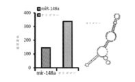
幾つかの実施形態では、本明細書において他に開示されるように、CDC-XOの分子カーゴの1つ以上の単離された成分を含む治療用組成物は、本明細書に開示される方法において使用される。幾つかの実施形態では、治療用組成物はCDC-XO RNAを含む。幾つかの実施形態では、本明細書において他に開示されるように、RNAは、CDC、CDC-XO及び/又はCDC-MVから単離され、再度組み合わされて(例えば、混合及び適合されて)治療方法における使用のための治療用混合物を提供する。幾つかの実施形態では、RNAの治療用混合物は、非コードRNAを含む、単一のRNA又は複数のRNA(例えば2、3、4、5、6、7、8、9、10以上のRNA)を含んでもよい。幾つかの実施形態では、非コードRNAは、とりわけtRNA、Y RNA、rTNA、mirRNA、IncRNA、piRNA、SnRNA、snoRNAを含み、更にそれらのフラグメントを含む。幾つかの実施形態では、治療用混合物は、マイクロRNA、miR-146a、miR-148a、miR-22、miR-24、miR-210、miR-150、miR-140-3p、miR-19a、miR-27b、miR-19b、miR-27a、miR-376c、miR-128、miR-320a、miR-143、miR-21、miR-130a、miR-9、miR-185、miR-23a、miR-215、miR-33a、miR204、miR-376c、miR4532、miR-4742、miR-582、miR-629、miR-223、miR-3125、miR-3677、miR-376b、miR-4449、miR-4773、miR-4787、miR-491、miR-495、miR-500a、miR-548ah、miR-550、miR-548ah、miR-550a、miR-551n、miR-5581、miR-616、若しくは図29で豊富であると示される任意のその他のマイクロRNA、及び/又は前述のもののいずれかに対して少なくとも約80%、85%、90%、95%、96%、97%、98%若しくは99%のパーセンテージ同一性を有するポリヌクレオチドからなる群から選択される1つ以上のマイクロRNAを含む。幾つかの実施形態では、治療用混合物は、miR-148a、miR-148-5p、miR-148-39、srDMD、及び/又は前述のものいずれかに対して少なくと約80%、85%、90%、95%、96%、97%、98%又は99%のパーセンテージ同一性を有するポリヌクレオチドの1つ以上を含む。幾つかの実施形態では、治療用混合物のマイクロRNAは、miR-148a-3p、及び/又は前述のものいずれかに対して少なくとも約80%、85%、90%、95%、96%、97%、98%若しくは99%のパーセンテージ同一性を有するポリヌクレオチドを含む。幾つかの実施形態では、マイクロRNAは、miR-148a-3p、及び/又は前述のもののいずれかに対して少なくとも約80%、85%、90%、95%、96%、97%、98%若しくは99%のパーセンテージ同一性を有するポリヌクレオチドを含む。様々な実施形態では、エキソソームは、DMDに由来する小さな非コードRNAであるsrDMD、及び/又は前述のいずれかに対して少なくとも約80%、85%、90%、95%、96%、97%、98%若しくは99%のパーセンテージ同一性を有するポリヌクレオチドを含む。 In some embodiments, therapeutic compositions comprising one or more isolated components of the molecular cargo of CDC-XO, as disclosed elsewhere herein, are disclosed herein used in a method. In some embodiments, therapeutic compositions comprise CDC-XO RNA. In some embodiments, RNA is isolated from CDC, CDC-XO and/or CDC-MV and recombined (e.g., mixed and matched), as disclosed elsewhere herein. ) provides a therapeutic mixture for use in a method of treatment. In some embodiments, the therapeutic mixture of RNAs comprises a single RNA or multiple RNAs (e.g., 2, 3, 4, 5, 6, 7, 8, 9, 10 or more RNAs), including non-coding RNAs. ) may be included. In some embodiments, non-coding RNAs include tRNAs, Y RNAs, rTNAs, mirRNAs, lncRNAs, piRNAs, SnRNAs, snoRNAs, among others, and also fragments thereof. In some embodiments, the therapeutic mixture comprises a microRNA, miR-146a, miR-148a, miR-22, miR-24, miR-210, miR-150, miR-140-3p, miR-19a, miR -27b, miR-19b, miR-27a, miR-376c, miR-128, miR-320a, miR-143, miR-21, miR-130a, miR-9, miR-185, miR-23a, miR-215 , miR-33a, miR-204, miR-376c, miR-4532, miR-4742, miR-582, miR-629, miR-223, miR-3125, miR-3677, miR-376b, miR-4449, miR-4773, miR -enriched in 4787, miR-491, miR-495, miR-500a, miR-548ah, miR-550, miR-548ah, miR-550a, miR-551n, miR-5581, miR-616, or Figure 29 and/or a percentage identity of at least about 80%, 85%, 90%, 95%, 96%, 97%, 98% or 99% to any of the foregoing comprising one or more microRNAs selected from the group consisting of polynucleotides having In some embodiments, the therapeutic mixture comprises miR-148a, miR-148-5p, miR-148-39, srDMD, and/or at least about 80%, 85% of any of the foregoing, Includes one or more polynucleotides having a percentage identity of 90%, 95%, 96%, 97%, 98% or 99%. In some embodiments, the microRNA of the therapeutic mixture is at least about 80%, 85%, 90%, 95%, 96%, 97% relative to miR-148a-3p and/or any of the foregoing. %, 98% or 99% percentage identity. In some embodiments, the microRNA is at least about 80%, 85%, 90%, 95%, 96%, 97%, 98% relative to miR-148a-3p and/or any of the foregoing or a polynucleotide with a percentage identity of 99%. In various embodiments, the exosome is at least about 80%, 85%, 90%, 95%, 96%, 97% relative to srDMD, a small non-coding RNA derived from DMD, and/or any of the foregoing , includes polynucleotides with a percentage identity of 98% or 99%.
幾つかの実施形態では、本明細書において他に開示される方法は、CDC-XOから単離された非コードRNAを使用して遂行され得る。特定の理論に拘束されないが、非コードRNAは、マイクロRNAであると確認され指定された非コードRNA遺伝子を含む、高度に特異的な核酸認識を必要とする調節性の役割に非常に適していると考えられている。幾つかの実施形態では、単離されたRNAポリヌクレオチドは、miR-148a、miR-148-5p、miR-148-39、srDMD、及び/又はmiR-148a、miR-148-5p、miR-148-39若しくはsrDMDに対して少なくとも約80%、85%、90%、95%、96%、97%、98%又は99%のパーセンテージ同一性を有するポリヌクレオチドの1つ以上から選択される。幾つかの実施形態では、miR-148aのヌクレオチド配列は以下の通りである:
5’GAGGCAAAGUUCUGAGACACUCCGACUCUGAGUAUGAUAGAAGUCAGUGCACUACAGAACUUUGUCUC3’(配列番号1);
miR-148-5pのヌクレオチド配列は以下の通りである:
5’AAAGUUCUGAGACACUCCGACU3’(配列番号2);
miR-148-3pのヌクレオチド配列は以下の通りである:
5’UCAGUGCACUACAGAACUUUGU3’(配列番号3);及び
srDMDのヌクレオチド配列は以下の通りである:
5’UGUACACAGAGGCUGAUCGAUUCUCCCUGAACAGCCUAUUACGGAGGCACUGCAGAUCAAGCCCGCCUGGAGAGGUGGAGUUUCAAGAGUCCCUUCCUGGUUCACCGUCUCCUUU3’(配列番号4)。
In some embodiments, methods disclosed elsewhere herein can be performed using non-coding RNA isolated from CDC-XO. Without being bound by any particular theory, non-coding RNAs are well suited for regulatory roles requiring highly specific nucleic acid recognition, including non-coding RNA genes identified and designated as microRNAs. It is believed that there are In some embodiments, the isolated RNA polynucleotide is miR-148a, miR-148-5p, miR-148-39, srDMD, and/or miR-148a, miR-148-5p, miR-148 -39 or one or more polynucleotides having at least about 80%, 85%, 90%, 95%, 96%, 97%, 98% or 99% percentage identity to srDMD. In some embodiments, the nucleotide sequence of miR-148a is as follows:
5′GAGGCAAAGUUCUGAGACACUCCCGACUCUGAGUAUGAUAGAAGUCAGUGCACUACAGAACUUUGUCUC3′ (SEQ ID NO: 1);
The nucleotide sequence of miR-148-5p is as follows:
5'AAAGUUCUGAGACACUCCCGACU3' (SEQ ID NO: 2);
The nucleotide sequence of miR-148-3p is as follows:
5′UCAGUGCACUACAGAACUUUGU3′ (SEQ ID NO: 3); and the nucleotide sequence of srDMD is as follows:
5′
幾つかの実施形態では、CDC、CDC-XO、及び/又はCDC-MVの分子カーゴの1つ以上の単離された成分は、ウイルス又は非ウイルスのベクターを使用して細胞に送達される。幾つかの実施形態では、ベクターはウイルスである。幾つかの実施形態では、ウイルスはアデノウイルス又はアデノ随伴ウイルスである。 In some embodiments, one or more isolated components of the CDC, CDC-XO, and/or CDC-MV molecular cargo are delivered to cells using viral or non-viral vectors. In some embodiments the vector is a virus. In some embodiments, the virus is adenovirus or adeno-associated virus.
或る実施形の態では、それを必要とする対象において、効果的に及び/又は安全にジストロフィン異常症を治療する前述の方法に従った、骨格筋ジストロフィー及び/又は異栄養性心筋症の治療における使用のためのmiR-148a、miR-148-5p、miR-148-39、srDMD、及び/又はmiR-148a、miR-148-5p、miR-148-39若しくはsrDMDに対して少なくとも約80%、85%、90%、95%、96%、97%、98%又は99%のパーセンテージ同一性を有するポリヌクレオチドを含む製剤又は組成物。幾つかの実施形態では、それを必要とする対象において、効果的に及び/又は安全にジストロフィン異常症を治療する前述の方法に従って、骨格筋ジストロフィー及び/又は異栄養性心筋症を治療するための上述の製剤及び/又は組成物の使用が提供される。 In one embodiment, in the treatment of skeletal muscular dystrophy and/or dystrophic cardiomyopathy according to the aforementioned method of effectively and/or safely treating dystrophinopathy in a subject in need thereof. at least about 80% relative to miR-148a, miR-148-5p, miR-148-39, srDMD, and/or miR-148a, miR-148-5p, miR-148-39 or srDMD for use; A formulation or composition comprising a polynucleotide having a percentage identity of 85%, 90%, 95%, 96%, 97%, 98% or 99%. In some embodiments, the above-described method for treating skeletal muscle dystrophy and/or dystrophic cardiomyopathy in a subject in need thereof, according to the above-described method of effectively and/or safely treating dystrophinopathy. Use of the formulations and/or compositions of
幾つかの実施形態では、外植片へと培養され、外植片由来細胞へと更に培養され、更に心筋球形成細胞として培養された後に心筋球細胞として培養され、そしてその後、XO及びEVが単離されるCDCとして培養された生検試料からCDCが生成される。幾つかの実施形態では、CDCはヒトのものである。様々な実施形態では、CDCはジストロフィン異常症に罹患した対象から得られた生検試料から生成される。幾つかの実施形態では、約24時間の期間に亘り低酸素条件下(例えば2%O2)でCDCを培養する。幾つかの実施形態では、無血清条件下でCDCを培養する。 In some embodiments, cultured into explants, further cultured into explant-derived cells, further cultured as cardiac sphere-forming cells, and then cultured as cardiac sphere cells, and then XO and EV CDCs are produced from a biopsy sample that has been cultured as isolated CDCs. In some embodiments, the CDC is human. In various embodiments, CDCs are generated from a biopsy sample obtained from a subject with dystrophinopathy. In some embodiments, CDCs are cultured under hypoxic conditions (eg, 2% O 2 ) for a period of about 24 hours. In some embodiments, CDCs are cultured under serum-free conditions.
発明の詳細な説明 Detailed description of the invention
本明細書に開示される幾つかの実施形態は、CDC、CDC-XO、CDC-EV、CDCの単離された分子カーゴ(例えば、CDC、CDC-XO、及び/又はCDC-EVに由来する個々の分子又は分子の組み合わせ);及び/又は前述の組み合わせを使用して、疾患、疾患状態、及び/又は疾患の症状を治療する方法に関する。幾つかの実施形態では、疾患はジストロフィン異常症である。幾つかの実施形態では、疾患状態は異栄養性障害である。幾つかの実施形態では、ジストロフィン異常症は、デュシェンヌ型筋ジストロフィー(DMD)及び/又はベッカー型筋ジストロフィーのうちの1つ以上を含む。幾つかの実施形態では、疾患状態はミオパチーである。幾つかの実施形態では、ミオパチーは骨格筋ミオパチーである。幾つかの実施形態では、この方法は、治療有効量のCDC、CDC-XO、CDC-EV、CDC-XO又はCDC-EVの分子カーゴ及び/又は前述の組み合わせを、疾患を患っている対象(例えば患者)に投与することにより、疾患及び/又はその症状を治療することを含む。本明細書で提供される方法及び組成物の幾つかの実施形態は、とりわけ、mdxマウスへのCDCの静脈内投与が、投与されたCDCの少なくとも一部が肺に蓄積するという知見にもかかわらず、本書に提示されている様々なデータが示すように、骨格筋ジストロフィーを患うヒト対象に治療有効量のCDCを投与することによって異栄養性骨格筋の機能改善が達成され、それにより骨格筋ジストロフィー、例えばデュシェンヌ型筋ジストロフィー(DMD)を患っているヒト対象の効果的な治療を可能にする、という驚くべき発見に基づく。 Some embodiments disclosed herein provide CDC, CDC-XO, CDC-EV, isolated molecular cargo of CDC (e.g., CDC, CDC-XO, and/or CDC-EV derived individual molecules or combinations of molecules); and/or methods of treating diseases, disease states, and/or symptoms of diseases using such combinations. In some embodiments, the disease is dystrophinopathy. In some embodiments, the disease state is a dystrophic disorder. In some embodiments, the dystrophinopathy comprises one or more of Duchenne muscular dystrophy (DMD) and/or Becker muscular dystrophy. In some embodiments, the disease state is myopathy. In some embodiments, the myopathy is skeletal muscle myopathy. In some embodiments, the method comprises administering a therapeutically effective amount of a molecular cargo of CDC, CDC-XO, CDC-EV, CDC-XO or CDC-EV and/or a combination of the foregoing to a subject afflicted with a disease ( (e.g., a patient) to treat the disease and/or its symptoms. Some embodiments of the methods and compositions provided herein are particularly useful despite the finding that intravenous administration of CDCs to mdx mice results in at least a portion of the administered CDCs accumulating in the lungs. However, as various data presented herein show, administration of a therapeutically effective amount of CDC to a human subject with skeletal muscular dystrophy can achieve improved dystrophic skeletal muscle function, thereby reducing skeletal muscular dystrophy, For example, it is based on the surprising discovery that it allows effective treatment of human subjects suffering from Duchenne Muscular Dystrophy (DMD).
本明細書で使用される「及び/又は」は、1つ以上の関連する列挙項目のあらゆる組み合わせと並んで、代替として(「又は」)解釈される場合の組み合わせの欠如を指す。 As used herein, "and/or" refers to any combination of one or more of the associated listed items, as well as the absence of combinations when interpreted alternatively ("or").
「治療する」又は「治療すること」又は「治療」とは、調節効果を与える任意の種類の作用を指し、これは、例えば、障害、疾患、又は病気に苦しんでいる対象にとって有益な効果である可能性があり、状態と関連する病状の発現の予防、対象の状態の改善(1つ以上の症状又は疾患等)、病状の進行の遅延若しくは減少、及び/又は臨床パラメーター、疾患若しくは病気の変化、病気の治癒等を含む。 "Treat" or "treating" or "treatment" refers to any kind of action that confers a modulating effect, e.g., a beneficial effect on a subject suffering from a disorder, disease, or illness. prevent the development of a medical condition associated with the condition, ameliorate the condition in a subject (such as one or more symptoms or diseases), slow or reduce the progression of the medical condition, and/or reduce the severity of clinical parameters, diseases or illnesses. Including change, cure of disease, etc.
本明細書で使用される「治療有効量」という用語は、障害、疾患又は病気に苦しんでいる対象に対して、例えば、有益な効果となり得る、対象の状態の改善(例えば、1つ以上の症状の調節)、状態の進行の遅延若しくは減少、障害の発症の予防若しくは遅延、及び/又は臨床パラメーター、疾患若しくは病気の変化等を含む、調節効果を与える治療薬(例えば、CDC-XO、CDC-EV、CDC、XO及びEVの分子カーゴ、又はそれらの組み合わせ)の量を指す。例えば、幾つかの実施形態では、有効量は、対象における状態を、少なくとも5%、例えば少なくとも10%、少なくとも15%、少なくとも20%、少なくとも25%、少なくとも30%、少なくとも35%、少なくとも40%、少なくとも45%、少なくとも50%、少なくとも55%、少なくとも60%、少なくとも65%、少なくとも70%、少なくとも75%、少なくとも80%、少なくとも85%、少なくとも90%、少なくとも95%、又は少なくとも100%改善する組成物、化合物、又は薬剤の量を指す場合がある。開示される主題の活性組成物中の有効成分及び薬剤の実際の投薬量レベルは、特定の対象及び/又は用途に対して所望の応答を達成するのに有効な量の活性剤(複数の場合もある)を投与するように変更されてもよい。選択される投薬量のレベルは、組成物の活性、製剤、投与経路、他の薬物又は治療との組み合わせ、治療されている状態の重症度、並びに治療されている対象の身体状態及び以前の病歴を含むが、これらに限定されない様々な要因に依存する。本明細書では、有効用量の決定及び調整と並んで、そのような調整を行う時期及び方法の評価が企図される。「治療有効量」という用語は、標的とされる異栄養性骨格筋において、ジストロフィンの再発現及び/又は骨格筋機能を永続的に(例えば、実質的に不可逆的に)回復するのに十分なCDC-XO、CDC-EV、CDC、及び/又は分子カーゴXO及びEVの量を意味する。 As used herein, the term "therapeutically effective amount" refers to an improvement in the subject's condition (e.g., one or more Therapeutic agents that provide a modulating effect (e.g., CDC-XO, CDC - EV, CDC, XO and EV molecular cargo, or combinations thereof). For example, in some embodiments, an effective amount reduces the condition in a subject by at least 5%, such as at least 10%, at least 15%, at least 20%, at least 25%, at least 30%, at least 35%, at least 40% , at least 45%, at least 50%, at least 55%, at least 60%, at least 65%, at least 70%, at least 75%, at least 80%, at least 85%, at least 90%, at least 95%, or at least 100% improvement It may refer to the amount of a composition, compound, or agent that The actual dosage level of the active ingredients and agents in the active compositions of the disclosed subject matter will be an amount of active agent(s) effective to achieve the desired response for a particular subject and/or use. may be modified to administer The selected dosage level will depend on the activity of the composition, the formulation, the route of administration, the combination with other drugs or treatments, the severity of the condition being treated, and the physical condition and previous medical history of the subject being treated. depends on a variety of factors, including but not limited to; Determination and adjustment of effective doses, as well as evaluation of when and how to make such adjustments, are contemplated herein. The term "therapeutically effective amount" refers to an amount sufficient to permanently (e.g., substantially irreversibly) restore dystrophin re-expression and/or skeletal muscle function in targeted dystrophic skeletal muscle. means the amount of CDC-XO, CDC-EV, CDC, and/or molecular cargo XO and EV.
本明細書で使用される「標的とされる異栄養性骨格筋」という用語は、異栄養性骨格筋の部位での或る量のCDC-XO、CDC-EV、CDC、分子カーゴCDC-XO及びCDC-EV、及び/又はそれらの組み合わせの送達である。幾つかの実施形態では、標的送達は、標的部位へのCDC-XO、CDC-EV、CDC、及び/又はCDC-XO及びCDC-EVの分子カーゴの偶然の、偶発的な、又は不注意による送達は含まれない。幾つかの実施形態では、標的送達には全身送達は含まれない。幾つかの実施形態では、標的送達には、異栄養性骨格筋の部位における、ジストロフィン異常症を治療するには不十分となり得る量のCDC-XO、CDC-EV、CDC、及び/又はCDC-XO及びCDC-EVの分子カーゴの偶然の、偶発的な、又は不注意による送達は含まれない。 As used herein, the term "targeted dystrophic skeletal muscle" refers to an amount of CDC-XO, CDC-EV, CDC, molecular cargo CDC-XO at the site of dystrophic skeletal muscle. and delivery of CDC-EV, and/or combinations thereof. In some embodiments, targeted delivery is by accidental, accidental, or inadvertent delivery of molecular cargo of CDC-XO, CDC-EV, CDC, and/or CDC-XO and CDC-EV to the target site. Does not include delivery. In some embodiments, targeted delivery does not include systemic delivery. In some embodiments, targeted delivery includes amounts of CDC-XO, CDC-EV, CDC, and/or CDC- Accidental, accidental, or inadvertent delivery of XO and CDC-EV molecular cargo is not included.
本明細書で使用される「異栄養性」という用語は、ジストロフィンの欠如又は欠乏である(例えば、骨格筋及び/又は心筋における)。 The term "dystrophy" as used herein is the absence or deficiency of dystrophin (eg, in skeletal and/or cardiac muscle).
細胞は、エキソソーム(XO)及び微小胞(MV)と称されるエンドソーム及び原形質膜を起源とする多様な種類の細胞外小胞(EV)を細胞外環境に放出する。EVは、細胞間コミュニケーションの重要な様式であり、細胞間及び細胞膜を通過する分子カーゴ(例えば、1つ以上の細胞質タンパク質、脂質、及びRNA)の移行の媒体としてはたらく。豊富な生物学的因子の環境を含む分泌脂質小胞であるXOは、強力なパラクリンシグナルを提供し、これによって、幹細胞が疾患又は損傷細胞を含む近隣の細胞に対する生物学的効果を増強する。タンパク質、生物活性脂質、及び核酸カーゴの封入及び移行により、これらの天然の送達装置は、再生プログラムの活性化につながるレシピエント細胞の顕著な表現型及び機能の変化を誘導することができる。国際公開第2016/05491号において、XOの投与は、mdxマウスの心不全を治療することが実証されており、その全体が参照されることより本明細書の一部をなす。 Cells release into the extracellular environment endosomes termed exosomes (XO) and microvesicles (MV) and diverse types of extracellular vesicles (EV) originating from the plasma membrane. EVs are an important mode of intercellular communication, serving as vehicles for the translocation of molecular cargo (eg, one or more of cytoplasmic proteins, lipids, and RNA) between cells and across cell membranes. XO, secreted lipid vesicles that contain a rich environment of biological agents, provide potent paracrine signals that enhance the biological effects of stem cells on neighboring cells, including diseased or injured cells. By encapsulation and translocation of proteins, bioactive lipids, and nucleic acid cargoes, these natural delivery devices can induce significant phenotypic and functional changes in recipient cells leading to activation of regenerative programs. In WO2016/05491, administration of XO was demonstrated to treat heart failure in mdx mice, which is incorporated herein by reference in its entirety.
本明細書に開示される幾つかの実施形態は、治療用途の方法におけるCDC及びCDC-XOの使用に関する。幾つかの実施形態では、本明細書に記載されるジストロフィン異常症の治療方法は、治療有効量のCDC及び/又はCDC-XOを、ジストロフィン異常症を患っている又はそれを有する対象に投与し、それによって対象を治療する工程を含む。幾つかの実施形態において、対象は、ジストロフィン異常症を患う小児対象である。幾つかの実施形態では、XOはCDCから単離される。幾つかの実施形態では、CDCは無血清培地で増殖される。幾つかの実施形態では、ジストロフィン異常症はデュシェンヌ型筋ジストロフィーである。他の実施形態では、ジストロフィン異常症はベッカー型筋ジストロフィーである。幾つかの実施形態では、CDC-XO、CDC-EV、CDC-XO又はCDC-EVの分子カーゴ、XO及びEVを産生するCDC、及び/又は上記の組み合わせは、ジストロフィンの再発現を達成する方法で使用される。幾つかの実施形態では、CDC-EVの全身心室内注射による、また同様にmdxマウスの骨格筋へのCDC-EVの直接筋肉内注射による、CDC-XO、CDC-EV、CDC-XO又はCDC-EVの分子カーゴ、XO及びEVを産生するCDC、及び/又は組み合わせによって、本発明者らは、本明細書に初めて記載された新規の治療方法、例えば、単回又は複数回の全身投与で治療有効量のCDC及び/又はCDC-EVSを投与することによる骨格DMDの治療方法を想到した。幾つかの実施形態では、疾患は筋ジストロフィーである。 Some embodiments disclosed herein relate to the use of CDCs and CDC-XOs in methods of therapeutic use. In some embodiments, the methods of treating dystrophinopathy described herein comprise administering a therapeutically effective amount of CDC and/or CDC-XO to a subject suffering from or having dystrophinopathy. , treating a subject thereby. In some embodiments, the subject is a pediatric subject with dystrophinopathy. In some embodiments, XO is isolated from CDC. In some embodiments, CDCs are grown in serum-free medium. In some embodiments, the dystrophinopathy is Duchenne muscular dystrophy. In another embodiment, the dystrophinopathy is Becker muscular dystrophy. In some embodiments, CDC-XO, CDC-EV, molecular cargo of CDC-XO or CDC-EV, CDCs producing XO and EV, and/or combinations of the above are methods of achieving re-expression of dystrophin used in In some embodiments, CDC-XO, CDC-EV, CDC-XO or CDC by systemic intraventricular injection of CDC-EV, as well as by direct intramuscular injection of CDC-EV into skeletal muscle of mdx mice. - Molecular cargo of EVs, CDCs producing XO and EVs, and/or combinations allow the inventors to discover novel therapeutic methods described herein for the first time, e.g., with single or multiple systemic administrations We have devised a method of treating skeletal DMD by administering a therapeutically effective amount of CDC and/or CDC-EVS. In some embodiments, the disease is muscular dystrophy.
本明細書に開示されるデータ及び実験は、ジストロフィン発現の誘導におけるCDC、CDC-XO、及び/又はCDC-EVの予想外の利点を実証している。本明細書で他に示されるように、mdxマウスの心臓へのCDCの注射は、心臓と骨格筋の両方において全長ジストロフィンタンパク質レベルをブーストし、心機能、歩行能力及び生存率を劇的かつ永続的に改善する。同様の結果は、ヒトのデュシェンヌ型心筋細胞でも実証されている。正の因子は、CDCによって生成される細胞XOに存在するようであり、CDCは、多小胞エンドソームが原形質膜と融合したときに細胞によって分泌される脂質二重層ナノ小胞である。 The data and experiments disclosed herein demonstrate an unexpected advantage of CDC, CDC-XO, and/or CDC-EV in inducing dystrophin expression. As shown elsewhere herein, injection of CDC into the heart of mdx mice boosted full-length dystrophin protein levels in both heart and skeletal muscle, dramatically and permanently improving cardiac function, ambulatory ability and survival. substantially improve. Similar results have been demonstrated in human Duchenne cardiomyocytes. A positive factor appears to reside in cellular XO generated by CDCs, lipid bilayer nanovesicles secreted by cells when multivesicular endosomes fuse with the plasma membrane.
幾つかの実施形態では、ヒトCDCによって分泌されるXO(及びEV)は、mdxマウス及びヒトデュシェンヌ型心筋細胞においてCDCの利点を再現することが実証されている。幾つかの実施形態では、CDC-XO(例えば、miR-148a)に見られる非コードRNA種の送達は、転写産物の長さやエクソン/イントロンの接合部に影響を与えることなく、CDC、CDC-XO、及び/又はCDC-EVがジストロフィンタンパク質レベルを増加させる能力を模倣する。幾つかの態様において、CDC-XOが媒介する非コードRNAの移行は、心臓及び骨格筋のジストロフィンを回復させることによりDMDを改善する。 In some embodiments, XO (and EVs) secreted by human CDCs have been demonstrated to reproduce the benefits of CDCs in mdx mouse and human Duchenne cardiomyocytes. In some embodiments, delivery of non-coding RNA species found in CDC-XO (eg, miR-148a) reduces CDC, CDC-XO (e.g., miR-148a), without affecting transcript length or exon/intron junctions. Mimics the ability of XO and/or CDC-EV to increase dystrophin protein levels. In some embodiments, CDC-XO-mediated non-coding RNA translocation ameliorates DMD by restoring cardiac and skeletal muscle dystrophin.
幾つかの実施形態では、本明細書に記載される結果は、ジストロフィン異常症の治療選択肢としてのCDC及びそれらのXO(及び/又はEV)を実証する。CDC及びそれらの分泌されたXO(及び/又はEV)は、心臓と骨格筋のジストロフィンレベルを確実に増加させる。幾つかの実施形態において、心臓及び骨格筋におけるジストロフィンレベルの増加は、身体へのCDC、CDC-XO、及び/又はCDC-EVの注射後(例えば、全身又は局所、骨格筋への局所を含む)の大きな持続性の全身的利益に結び付く。幾つかの実施形態では、本明細書に開示されるように、CDC、CDC-XO、及び/又はCDC-EVは、再生性であるだけでなく、抗炎症性及び抗線維性でもある。CDCは、血管新生を促進し、内因性の前駆細胞を動員し、生存する心臓細胞をうまく増殖させる拡散因子を分泌し;また、移植されたCDCは、不適応なリモデリング及びアポトーシスを抑制する。幾つかの実施形態では、CDCは、間接経路により(CDC-XO及び/又はCDC-EVを介して)動作する;そらは、マイクロRNA(分子カーゴの構成要素)を含む非コードRNAを含むCDC-XO及び/又はCDC-EVの分泌を介して間接的に作用する。幾つかの実施形態では、同種CDCは数週間以内に完全に排除されるが、それらの機能的及び構造的な利点は少なくとも6カ月持続する。これらの多様なメカニズムは、マイクロRNAを含む非コードRNAによるCDC-XO及び/又はCDC-EVの分泌を介して媒介される。 In some embodiments, the results described herein demonstrate CDCs and their XO (and/or EVs) as therapeutic options for dystrophinopathies. CDCs and their secreted XO (and/or EVs) reliably increase dystrophin levels in heart and skeletal muscle. In some embodiments, the increase in dystrophin levels in heart and skeletal muscle is after injection of CDC, CDC-XO, and/or CDC-EV into the body (e.g., systemically or locally, including local to skeletal muscle). ) are associated with large sustained systemic benefits. In some embodiments, as disclosed herein, CDCs, CDC-XOs, and/or CDC-EVs are not only regenerative, but also anti-inflammatory and anti-fibrotic. CDCs promote angiogenesis, recruit endogenous progenitor cells, and secrete spreading factors that successfully proliferate surviving cardiac cells; transplanted CDCs also suppress maladaptive remodeling and apoptosis. . In some embodiments, CDCs operate by an indirect pathway (via CDC-XO and/or CDC-EV); they include non-coding RNAs, including microRNAs (components of molecular cargo). - acting indirectly through the secretion of XO and/or CDC-EV. In some embodiments, allogeneic CDCs are completely eliminated within a few weeks, but their functional and structural benefits persist for at least 6 months. These diverse mechanisms are mediated through secretion of CDC-XO and/or CDC-EV by non-coding RNAs, including microRNAs.
特定の理論に拘束されることなく、上記のメカニズムは、ベッカー型筋ジストロフィー等の類似の筋ジストロフィーへの適用により、CDC、CDC-EV、又はCDC-XOにDMDを治療する能力もたらす。幾つかの実施形態において、CDC、CDC-XO、及び/又はCDC-EVは、再生細胞を動員し、線維化を逆転させ、炎症を標的とすることにより、ジストロフィンを置き換え、ジストロフィン欠失の病態生理学的結果を相殺する。幾つかの実施形態では、小児患者のDMDの中心的欠陥を逆転させ、本明細書の方法は、疾患の進行を未然に防ぐか又は予防することができ、それらの患者が治療的介入の選択肢を著しく制限する可能性のある併存疾患を回避できるようにする。 Without being bound by a particular theory, the above mechanisms, by application to similar muscular dystrophies such as Becker's muscular dystrophy, give CDC, CDC-EV, or CDC-XO the ability to treat DMD. In some embodiments, CDCs, CDC-XOs, and/or CDC-EVs replace dystrophin by recruiting regenerative cells, reversing fibrosis, and targeting inflammation to reduce pathogenesis of dystrophin deficiency. Counterbalance physiological consequences. In some embodiments, the central deficit of DMD in pediatric patients is reversed, and the methods herein can forestall or prevent disease progression, providing those patients with options for therapeutic intervention. to avoid comorbidities that can significantly limit
本明細書に開示される方法は、患者へのCDCの送達に関与するものを含むが、幾つかの実施形態では、細胞ではなくCDCにより分泌されるCDC-EV(例えばCDC-XO)を使用することにより、移植及び細胞自体の送達と比較した場合に有利な点を提供し得る。幾つかの実施形態では、CDCによって生成されるものを含むCDC-EV及びCDC-XOは、「無細胞」療法を開発するための強力で豊富なソースを提供することができる。CDC-XOベースの「無細胞」療法は、細胞療法とは対照的に、再生医療において以下の利点の1つ以上を提供する。幾つかの実施形態では、免疫原性又は腫瘍原性の可能性が低い又は存在しない生存不能な実体として、これらの特徴は特定の安全性の問題を有意に取り除く。幾つかの実施形態において、幹細胞由来のエキソソーム(及び/又はCDC-XO)は、MHC複合体分子を含む膜結合タンパク質の含有量が低いため、親細胞よりも免疫原性が低くなる可能性がある。幾つかの実施形態では、脂質小胞内の生物活性成分のCDC-XO封入により、in vivoでの分解から内容物を保護することができ、それによって、サイトカイン、成長因子、転写因子、RNA等の可溶性分子の送達としばしば関連する障害を潜在的に無効にすることができる。幾つかの実施形態では、CDC-XO及び/又はCDC-EVの投与(及び/又は保管)の容易さにより、最終的に患者への繰り返しかつ持続的な送達が可能となり、それによって疾患及び/又は機能不全組織の再生及び修復の可能性が最大化される。 Although the methods disclosed herein include those involving delivery of CDCs to patients, some embodiments use CDC-EVs (e.g., CDC-XO) secreted by CDCs rather than cells. This may offer advantages when compared to transplantation and delivery of the cells themselves. In some embodiments, CDC-EV and CDC-XO, including those produced by CDC, can provide a powerful and rich source for developing "cell-free" therapies. CDC-XO-based "cell-free" therapies offer one or more of the following advantages in regenerative medicine, as opposed to cell therapy. In some embodiments, as non-viable entities with low or no immunogenic or tumorigenic potential, these characteristics significantly obviate certain safety concerns. In some embodiments, stem cell-derived exosomes (and/or CDC-XO) may be less immunogenic than parental cells due to their lower content of membrane-bound proteins, including MHC complex molecules. be. In some embodiments, CDC-XO encapsulation of bioactive components within lipid vesicles can protect the contents from degradation in vivo, thereby protecting cytokines, growth factors, transcription factors, RNA, etc. can potentially abrogate the obstacles often associated with the delivery of soluble molecules of In some embodiments, the ease of administration (and/or storage) of CDC-XO and/or CDC-EV ultimately allows for repeated and sustained delivery to patients, thereby reducing disease and/or disease. Or the potential for regeneration and repair of dysfunctional tissue is maximized.
幾つかの実施形態では、CDC及び/又はCDC-XOを使用して、免疫調節プロセス、血管新生、及び内皮細胞の遊走を含む多数の細胞、組織及び生理学的プロセスを刺激することができる。本明細書に開示されるように、酸化及び/又はニトロソ化ストレスの増加、炎症の増加、アポトーシス促進及びリモデリング状態の環境を含む、DMD患者の病態生理に基づいて、細胞によって分泌されるCDC及び/又はCDC-XOを含む治療アプローチは、疾患の経過(及び前述の疾患状態及び/又は症状の1つ以上)を逆転させるのに大きな利点を提供する。幾つかの実施形態では、CDC、CDC-XO、及び/又はCDC-EVは、抗酸化、抗炎症、抗アポトーシス、抗リモデリング効果を促進する。幾つかの実施形態では、CDC、CDC-XO、及び/又はCDC-EVは、疾患細胞及び組織の再生能力を高める。幾つかの実施形態では、CDC、CDC-XO、及び/又はCDC-EVの投与は、DMDの遅延及び/又は逆転に有益であり、CDCに由来するエキソソーム集団は、これらの利益がもたらされることを可能にする。小児対象への早期治療介入は、心不全等の後期疾患の併存疾患を予防又は回避する永続的かつ全身的な利益を提供する。幾つかの実施形態において、それらの持続する利益は、以下に等しいか、少なくともおよそ:3カ月、6カ月、12カ月、又は前述の値を含む範囲及び/又は前述の値に及ぶ範囲である。 In some embodiments, CDCs and/or CDC-XOs can be used to stimulate numerous cell, tissue and physiological processes, including immunomodulatory processes, angiogenesis, and endothelial cell migration. As disclosed herein, CDC secreted by cells based on the pathophysiology of DMD patients, including increased oxidative and/or nitrosative stress, increased inflammation, pro-apoptotic and remodeling state milieu. and/or therapeutic approaches involving CDC-XO offer significant advantages in reversing the course of the disease (and one or more of the aforementioned disease states and/or symptoms). In some embodiments, CDC, CDC-XO, and/or CDC-EV promote antioxidant, anti-inflammatory, anti-apoptotic, anti-remodeling effects. In some embodiments, CDCs, CDC-XOs, and/or CDC-EVs enhance the regenerative capacity of diseased cells and tissues. In some embodiments, administration of CDCs, CDC-XOs, and/or CDC-EVs is beneficial in delaying and/or reversing DMD, and CDC-derived exosome populations provide these benefits. enable Early therapeutic intervention in pediatric subjects provides long-lasting systemic benefits that prevent or avoid late-stage disease comorbidities such as heart failure. In some embodiments, these sustained benefits are equal to or at least about: 3 months, 6 months, 12 months, or ranges inclusive of and/or spanning the aforementioned values.
エキソソームの幾つかの実施形態。XOは、サイトカイン、増殖因子、転写因子、脂質、コーディング核酸及び非コーディング核酸等の様々な生物学的因子に富む脂質二重層小胞である。XOは、血液、尿、羊水、間質腔及び細胞外空間に見られる。エンドソーム起源のこれらのエキソサイトーシス小胞は、40nm~100nmのサイズを含む、30nm~200nmの範囲のサイズであり、電子顕微鏡で明らかにされるように、カップ状の形態を有する。それらの初期形成は、細胞膜の内向きの出芽から始まり、エンドソームを形成し、その後、後期エンドソームの制限膜の陥入が続き、多胞体(MVB)を形成する。MVBと原形質膜の融合は、その後エキソソームとして知られる小胞の形成により、細胞外空間に内部小胞の放出をもたらす。幾つかの実施形態では、本明細書に記載されるXOは、エキソサイトーシスされた及び/又はエンドソーム起源の細胞外小胞である。幾つかの実施形態では、本明細書に記載のXOは、以下に等しいか、少なくともおよそ:30nm、50nm、100nm、150nm、200nm、又は前述の値を含む範囲及び/又は前述の値に及ぶ範囲の直径を有することができる。 Some embodiments of exosomes. XOs are lipid bilayer vesicles rich in various biological agents such as cytokines, growth factors, transcription factors, lipids, coding and non-coding nucleic acids. XO is found in blood, urine, amniotic fluid, interstitial space and extracellular space. These exocytotic vesicles of endosomal origin range in size from 30 nm to 200 nm, including sizes from 40 nm to 100 nm, and have a cup-shaped morphology as revealed by electron microscopy. Their initial formation begins with inward budding of the plasma membrane to form endosomes, followed by invagination of the limiting membrane of late endosomes to form multivesicular bodies (MVBs). Fusion of the MVB with the plasma membrane results in the release of internal vesicles into the extracellular space with subsequent formation of vesicles known as exosomes. In some embodiments, the XOs described herein are exocytosed and/or extracellular vesicles of endosomal origin. In some embodiments, XO as described herein is equal to or at least about: 30 nm, 50 nm, 100 nm, 150 nm, 200 nm, or ranges inclusive of and/or spanning the aforementioned values. can have a diameter of
本明細書に記載されるように、XOの「カーゴ」内容物は、形成時の親細胞の細胞調節状態を含む、親細胞起源に関連する生物学的因子の異なるサブセットを含むように親細胞起源を反映する。エキソソーム内のサイトカイン及び成長因子、脂質、コーディングRNA分子及び非コーディングRNA分子を含む様々なタンパク質の豊富な生物学的環境はいずれも、必然的に親細胞に由来する。細胞質誘導体の豊富なアレイを含むことに加えて、エキソソームは、膜の表面で膜結合受容体の細胞外ドメインを更に発現する。 As described herein, the 'cargo' content of XOs is the parent cell such that it contains distinct subsets of biological factors associated with parental cell origin, including the cellular regulatory state of the parent cell upon formation. Reflect origin. The diverse protein-rich biological milieu, including cytokines and growth factors, lipids, coding and non-coding RNA molecules, within exosomes all necessarily derive from the parent cell. In addition to containing a rich array of cytoplasmic derivatives, exosomes also express the extracellular domain of membrane-bound receptors on the surface of the membrane.
幾つかの実施形態では、記載されるカプセル化及び形成プロセスは、親細胞起源及び形成時の調節状態に基づいてXO組成物に不均一性を作り出す。それにもかかわらず、幾つかの実施形態では、一般的な出芽形成及び放出メカニズムは、エンドソーム関連タンパク質(Rab GTPase、SNARE、アネキシン、フロチリン等)原形質膜又はエンドソームでミクロドメインにクラスター化することが知られているタンパク質(4つの膜貫通ドメインであるテトラスパニン、例えばCD63、CD81、CD82、CD53、及びCD37)、脂質ラフト関連タンパク質(例、グリコシルホスファチジルイノシトールアンカー型タンパク質及びフロチリン)、コレステロール、スフィンゴミエリン、及びヘキソシルセラミド等のそれらの起源の結果として共通する一組の性質を確立する。 In some embodiments, the encapsulation and formation processes described create heterogeneity in the XO composition based on parental cell origin and regulatory state during formation. Nevertheless, in some embodiments, the general sprouting and release mechanism is based on endosome-associated proteins (Rab GTPases, SNAREs, annexins, flotillins, etc.) that can cluster into microdomains at the plasma membrane or endosomes. known proteins (4 transmembrane domain tetraspanins, e.g. CD63, CD81, CD82, CD53, and CD37), lipid raft-associated proteins (e.g. glycosylphosphatidylinositol-anchored protein and flotillin), cholesterol, sphingomyelin, and hexosylceramide establish a common set of properties as a result of their origin.
幾つかの実施形態では、小胞の起源を反映する成分に加えて、XOは、シグナル伝達プロセスに関連するmRNA及びマイクロRNAの両方を含有し、カーゴmRNAはレシピエント細胞で翻訳可能であるか、又はマイクロRNAはレシピエント細胞で標的mRNAを機能的に分解する。幾つかの実施形態では、遺伝子発現に影響を与えることが可能な他の非コードRNAもXOに存在し得る。mRNA又はマイクロRNAの集団のXOへの選択的取り込みを支配するプロセスは完全には理解されておらず、特定の理論に拘束されることはないが、他のエキソソーム及びそれらの起源細胞のRNAプロファイルと比較した場合、XOカーゴRNAの濃縮によって実証されるように、RNA分子はXOにランダムにではなく、選択的に取り込まれると考えられている。幾つかの実施形態では、理論に拘束されることなく、RNA分子が疾患の病因及び再生プロセスでの可能性のある役割を考慮すれば、XO中のRNA分子の存在及び標的レシピエント細胞に影響を及ぼす際の明らかな効力により、本明細書で他に開示されるように、XO及びそれらの分子カーゴが治療的に有効となる。 In some embodiments, in addition to components reflecting the origin of the vesicle, the XO contains both mRNAs and microRNAs associated with signaling processes, whether the cargo mRNA is translatable in the recipient cell. , or microRNAs functionally degrade target mRNAs in recipient cells. In some embodiments, other non-coding RNAs capable of affecting gene expression may also be present in XO. Although the processes governing the selective incorporation of populations of mRNAs or microRNAs into XO are not fully understood and without being bound by any particular theory, RNA profiles of other exosomes and their cells of origin RNA molecules are thought to be selectively, rather than randomly, incorporated into XO, as demonstrated by the enrichment of XO cargo RNA when compared to . In some embodiments, without being bound by theory, given the possible role of RNA molecules in disease pathogenesis and regenerative processes, the presence of RNA molecules in XO and their effect on target recipient cells The apparent potency in exerting , makes XOs and their molecular cargoes therapeutically effective, as disclosed elsewhere herein.
幾つかの実施形態では、エキソソームの天然の二重膜カプセル化は、カーゴ内容物が血流中又は組織内で分解することなく持続又は移動できるように保護され、制御された内部微小環境も提供する。幾つかの実施形態では、細胞外環境へのこのカーゴのその後の放出により、脂質リガンド受容体相互作用、エンドサイトーシス取り込みを介する内在化、又は小胞と細胞膜の直接融合によって媒介される、細胞表面への接着を介するレシピエント細胞との相互作用が可能になる。これらのプロセスにより、エキソソームカーゴ内容物が標的細胞に放出される。 In some embodiments, the natural double-membrane encapsulation of exosomes also provides a protected and controlled internal microenvironment such that cargo contents can persist or move in the bloodstream or within tissues without degradation. do. In some embodiments, subsequent release of this cargo into the extracellular environment is mediated by lipid-ligand-receptor interactions, internalization via endocytic uptake, or direct fusion of the vesicle with the cell membrane. Allows interaction with recipient cells via adhesion to surfaces. These processes release exosome cargo contents into target cells.
幾つかの実施形態では、XO細胞相互作用は、抗原提示、転写因子、サイトカイン、成長因子、mRNA及びマイクロRNA等の核酸の移行を含む、幾つかの異なる機構のいずれかによって誘導されるように、標的レシピエント細胞の遺伝的経路を調節することができる。 In some embodiments, XO cell interactions are induced by any of several different mechanisms, including antigen presentation, transcription factors, cytokines, growth factors, translocation of nucleic acids such as mRNA and microRNA. , can modulate the genetic pathways of target recipient cells.
エキソソームの単離及び調製。幾つかの実施形態では、XO単離は、分離及び分析のためのそれらの一般的な生化学的及び生物物理学的な特徴を使用して達成され得る。幾つかの実施形態では、分画超遠心法が、分泌されたXOを培養細胞の上清から単離する技術として使用され得る。幾つかの実施形態では、このアプローチは、比較的低い浮遊密度を活用することにより、非膜状粒子からのXOの分離を可能にする。幾つかの実施形態では、サイズ排除により、生化学的に類似しているが生物物理学的に異なるMVからの分離が可能になり、MVは最大1000nmのより大きな直径を持つ。幾つかの実施形態では、MVはまた、XOと共に治療混合物に含まれる(EVがXO及びMVの両方を含む場合)、及び/又はMVはXOから除去されない。他の実施形態では、XOが濃縮され、及び/又はMVが実質的に含まれないように、XOをMVから単離することができる。幾つかの実施形態では、浮揚速度の違いにより、異なるサイズのエキソソームの分離がさらに可能になる。幾つかの実施形態では、XOサイズは、40nm~100nmのサイズを含む30nm~200nmの範囲の直径を有する。幾つかの実施形態では、開示されるMV及びEVは、約:1000nm、750nm、500nm、400nm、300nm、250nm、200nm以上の、又は前述の値を含む範囲及び/又は前述の値に及ぶ範囲のサイズ(nm単位)を有する。 Isolation and preparation of exosomes. In some embodiments, XO isolation can be accomplished using their common biochemical and biophysical characteristics for separation and analysis. In some embodiments, differential ultracentrifugation can be used as a technique to isolate secreted XO from the supernatant of cultured cells. In some embodiments, this approach enables separation of XO from non-membrane particles by exploiting relatively low buoyant densities. In some embodiments, size exclusion allows separation from biochemically similar but biophysically distinct MVs, which have larger diameters up to 1000 nm. In some embodiments, MV is also included in the therapeutic mixture along with XO (where EV includes both XO and MV) and/or MV is not removed from XO. In other embodiments, XO can be isolated from MVs such that XO is enriched and/or is substantially free of MVs. In some embodiments, differences in levitation speed further enable separation of different sized exosomes. In some embodiments, the XO size has a diameter ranging from 30 nm to 200 nm, including sizes from 40 nm to 100 nm. In some embodiments, the MV and EV disclosed are about: size (in nm).
幾つかの実施形態では、目的の特定のエキソソームの具体的な特性に基づいて、XOの更なる精製を実施することができる。これには、例えば、目的のタンパク質を用いた免疫吸着の使用が含まれ、エキソプラズマ又は外向きの特定の小胞を選択する。 In some embodiments, further purification of XO can be performed based on the specific properties of the particular exosome of interest. This includes, for example, the use of immunoadsorption with the protein of interest to select for exoplasma or specific vesicles facing outward.
幾つかの実施形態では、示差遠心分離、不連続密度勾配、免疫親和性、限外濾過及び高速液体クロマトグラフィー(HPLC)のいずれか1つを使用してXO(及び/又はMV)を単離できるが、示差超遠心分離が使用される。幾つかの実施形態では、この手法は、2000×gから10000×gの遠心力の増加を利用して、100000×gでエキソソームペレットから中型及び大型の粒子及び細胞破片を分離する。遠心分離だけで馴化培地からのXOの顕著な分離/収集が可能になるが、幾つかの実施形態では、超遠心分離も、様々なタンパク質凝集体、遺伝物質、一般的な汚染物質である培地や細胞片に由来する粒子を除去し得る。幾つかの実施形態では、エキソソーム精製の特異性の増強は、エキソソーム調製物のより高い純度(浮選密度1.1g/ml~1.2g/ml)又は調製中に個別のシュガークッション(sugar cushion)の適用を提供するため、限外濾過と組み合わせた連続遠心分離、又はスクロース密度勾配における平衡密度勾配遠心分離を展開し得る。 In some embodiments, XO (and/or MVs) are isolated using any one of differential centrifugation, discontinuous density gradients, immunoaffinity, ultrafiltration and high performance liquid chromatography (HPLC). Differential ultracentrifugation is used, although it can. In some embodiments, this approach utilizes increasing centrifugal force from 2000×g to 10000×g to separate medium and large particles and cellular debris from the exosome pellet at 100000×g. Although centrifugation alone allows significant separation/collection of XO from conditioned media, in some embodiments, ultracentrifugation also removes various protein aggregates, genetic material, and media that are common contaminants. and particles derived from cell debris. In some embodiments, enhanced specificity of exosome purification is associated with higher purity of exosome preparations (flotation density 1.1 g/ml to 1.2 g/ml) or separate sugar cushions during preparation. ), continuous centrifugation combined with ultrafiltration, or equilibrium density gradient centrifugation in a sucrose gradient can be developed.
幾つかの実施形態では、限外濾過を使用して、生物活性を損なうことなくエキソソームを精製することができる。幾つかの実施形態では、膜は、約:200kDa、100kDa、75kDa、50kDa以下の、又は前述の値を含む範囲及び/又は前述の値に及ぶ範囲の分子量カットオフ(MWCO)等種々の細孔サイズを有する。幾つかの実施形態では、ゲル濾過は、小さい粒子を排除するため、代替的に又は追加して使用されてもよい。幾つかの実施形態において、メンブレン(例えば、透析、限外濾過等)及び/又はゲル濾過は、実質的な生理学的pHを使用して及び/又は実質的な生理学的塩濃度で行われる(例えば、非中性pH又は非生理学的塩濃度の使用を避ける)。幾つかの実施形態では、接線流濾過(TFF)システムが使用される。幾つかの実施形態では、TFFシステムはスケーラブル(10000L超まで)であり、XO画分を精製するだけでなく濃縮することもできる。幾つかの実施形態において、かかるアプローチは、有利には、示差遠心分離よりも時間がかからない。幾つかの実施形態では、HPLCを使用してXOを精製する。幾つかの実施形態では、HPLCを使用して、エキソソームを均一なサイズの粒子に精製し、調製物が生理学的pH及び塩濃度に維持されるようにそれらの生物活性を維持することもできる。 In some embodiments, ultrafiltration can be used to purify exosomes without loss of biological activity. In some embodiments, the membrane has various pores such as a molecular weight cutoff (MWCO) of about: 200 kDa, 100 kDa, 75 kDa, 50 kDa or less, or ranges inclusive of and/or spanning the aforementioned values. have a size. In some embodiments, gel filtration may alternatively or additionally be used to exclude small particles. In some embodiments, membrane (e.g., dialysis, ultrafiltration, etc.) and/or gel filtration is performed using a substantially physiological pH and/or at a substantially physiological salt concentration (e.g., , avoid using non-neutral pH or non-physiological salt concentrations). In some embodiments, a tangential flow filtration (TFF) system is used. In some embodiments, the TFF system is scalable (to over 10000 L) and can not only purify but also concentrate the XO fraction. In some embodiments, such an approach is advantageously less time consuming than differential centrifugation. In some embodiments, HPLC is used to purify XO. In some embodiments, HPLC can also be used to purify exosomes into uniformly sized particles and maintain their biological activity as the preparation is maintained at physiological pH and salt concentrations.
幾つかの実施形態では、化学的方法を使用してXOを単離する。幾つかの実施形態において、これらの化学的方法は、沈殿手法における異なる溶解度による分離を含む。幾つかの実施形態では、沈殿試薬をXOの溶液に加えてXOを精製する。幾つかの実施形態において、これらの化学的方法は、体積排除ポリマー(例えば、ポリエチレングリコール(PEG)等)への添加による分離を含む。幾つかの実施形態では、これらの化学的方法を、追加のラウンドの遠心分離又は濾過等と組み合わせることができる。幾つかの実施形態では、例えば、エキソソームの集団を即座に及び迅速に沈殿させるために、沈殿試薬、ExoQuick(登録商標)を馴化細胞培地に添加する。幾つかの実施形態では、フローフィールドフローフラクショネーション(F1FFF:flow field-flow fractionation)は、高分子(例えば、タンパク質)及びナノサイズからマイクロサイズの粒子(例えば、オルガネラ及び細胞)を分離及び特性評価するために使用される溶出に基づく手法であり、培養培地からのエキソソームの分画に適用される。 In some embodiments, chemical methods are used to isolate XO. In some embodiments, these chemical methods include separation by differential solubility in precipitation techniques. In some embodiments, a precipitation reagent is added to a solution of XO to purify the XO. In some embodiments, these chemical methods include separation by addition to volume excluding polymers such as polyethylene glycol (PEG). In some embodiments, these chemical methods can be combined, such as with additional rounds of centrifugation or filtration. In some embodiments, for example, a precipitating reagent, ExoQuick®, is added to the conditioned cell culture medium to precipitate a population of exosomes immediately and rapidly. In some embodiments, flow field-flow fractionation (F1FFF) separates and characterizes macromolecules (e.g., proteins) and nano- to micro-sized particles (e.g., organelles and cells). The elution-based technique used to evaluate and applied to the fractionation of exosomes from the culture medium.
幾つかの実施形態では、XOの生化学的及び生物物理学的な特徴に依存して、本明細書で他に開示される技術を超えて、焦点があてられる技術が目的の単離された特定のエキソソームに適用され得る。幾つかの実施形態では、抗体の免疫親和性を使用して、XO関連抗原を認識する。幾つかの態様において、XOは、親細胞の膜の表面で膜結合受容体の細胞外ドメインを発現する。幾つかの実施形態では、この発現は、共有される抗原プロファイルに基づいて、親細胞の起源に関連してXOを単離及び分離することを可能にする。幾つかの実施形態では、磁気ビーズへの複合化、クロマトグラフィーマトリクス、プレート若しくはマイクロ流体デバイス、及び/又はかかる技術と本明細書に開示される他の技術との組み合わせは、(例えば、目的の親細胞又は関連する細胞の調節状態に関連する可能性がある)目的の特定のXO又はMVの集団の単離を可能にする。他の親和性捕捉方法では、XO表面の特定の糖残基に結合するレクチンを使用する。 In some embodiments, the techniques focused over the techniques otherwise disclosed herein rely on the biochemical and biophysical characteristics of the XO to It can be applied to specific exosomes. In some embodiments, the immunoaffinity of antibodies is used to recognize XO-related antigens. In some embodiments, the XO expresses the extracellular domain of a membrane-bound receptor on the surface of the parent cell's membrane. In some embodiments, this expression allows the isolation and separation of XOs in relation to their parental cell origin based on their shared antigenic profile. In some embodiments, conjugation to magnetic beads, chromatography matrices, plates or microfluidic devices, and/or combinations of such techniques with other techniques disclosed herein (e.g., Allows isolation of specific XO or MV populations of interest (which may be related to the regulatory state of parental or related cells). Other affinity capture methods use lectins that bind to specific sugar residues on the XO surface.
エキソソームに基づく治療法。幾つかの実施形態では、本明細書で他に開示されるように、XOに基づく治療法は、潜在的な「無細胞」療法を有利に可能にする(例えば、CDC等がCDC-XO等から分離される)。「無細胞」療法の使用は、リスクを低減した細胞療法の潜在的な利点を保持し、及び/又は細胞療法が利用できない(及び/又は不可能な)状況で使用され得る。幾つかの実施形態では、本明細書で他に記載されるように、CDC等の細胞に基づく治療法の治療上の利点は、内因性の起源から生じる再生組織を伴う間接的な機構により生じ得る。幾つかの実施形態では、CDCによって産生される細胞XOは、疾患の進行を改善するだけでなく、疾患及び/又は機能不全の組織を修復及び再生する方法で、新たな治療アプローチのための増殖因子、転写因子、サイトカイン及び核酸の産生及び送達を可能にし得る。これに関して、CDC由来エキソソームは、相乗機構を動員して内因性幹細胞を心筋損傷部位に引き付け、細胞分化を促進し、デュシェンヌ型筋ジストロフィー等の慢性疾患の病態生理を逆転させることにより、主な満たされていない医学的ニーズに効果的に対処することができる。幾つかの実施形態において、CDCは、XO(及び/又はEV)の製造所として使用され得て、有利には、患者においてCDCの滞在期間を通してXOの持続的な供給源を提供する。 Exosome-based therapy. In some embodiments, as disclosed elsewhere herein, XO-based therapeutics advantageously enable potential "cell-free" therapies (e.g., CDC, etc. are CDC-XO, etc. separated from). The use of "cell-free" therapy retains the potential benefits of reduced-risk cell therapy and/or can be used in situations where cell therapy is not available (and/or impossible). In some embodiments, as described elsewhere herein, the therapeutic benefits of cell-based therapies such as CDC arise through indirect mechanisms involving regenerative tissue arising from endogenous sources. obtain. In some embodiments, cell XO produced by CDCs can be used for proliferation and novel therapeutic approaches in ways that not only ameliorate disease progression, but also repair and regenerate diseased and/or dysfunctional tissues. It may allow the production and delivery of factors, transcription factors, cytokines and nucleic acids. In this regard, CDC-derived exosomes have major implications by recruiting synergistic mechanisms to attract endogenous stem cells to sites of myocardial injury, promote cell differentiation, and reverse the pathophysiology of chronic diseases such as Duchenne muscular dystrophy. Able to effectively address unmet medical needs. In some embodiments, the CDC can be used as a source of XO (and/or EV), advantageously providing a continuous source of XO throughout the CDC stay in the patient.
幾つかの実施形態では、特に、DMD等の慢性状態の場合、XCを産生するCDC-XO又はCDCの患者への反復送達及び持続送達は、細胞に基づく治療法を使用する場合よりもより容易で、潜在的により安全となり得る方法で、疾患組織及び/又は機能不全組織の再生及び修復の可能性を高め得る。投薬レジメン及びスケジュールは、本明細書で他に更に詳細に開示される。 In some embodiments, especially for chronic conditions such as DMD, repeated and sustained delivery of XC-producing CDC-XO or CDCs to patients is easier than with cell-based therapies. and increase the likelihood of regeneration and repair of diseased and/or dysfunctional tissue in a potentially safer manner. Dosing regimens and schedules are disclosed in further detail elsewhere herein.
幾つかの実施形態では、本明細書の他の箇所で開示されるように、患者に提供されるXO及び/又はCDCの投与方法及び量は、治療用量を送達する様々な方法で提供され得る。幾つかの実施形態では、例えば、XOの投与のための組成物及び/又は溶液を投与は、単回投与で約1mg~約100mgのCDC-XOタンパク質を含む。幾つかの実施形態では、CDC-EV(例えば、CDC-XO)の用量は、以下に等しい又は少なくとも約:1mg、10mg、25mg、50mg、75mg、100mg、200mg、又は前述の値を含む及び/又はそれに及ぶ範囲のEV又はXO(mg単位)を含んでもよい。幾つかの実施形態では、投与方法は、対象への各単回用量の複数回投与を含む。幾つかの実施形態では、組成物(例えば、CDC-XO、CDC-EV、CDC、又はそれらの組み合わせを含む組成物)の投与には注射が含まれる。幾つかの実施形態では、注射は骨格筋注射を含む。幾つかの実施形態では、注射は腹腔内注射を含む。幾つかの実施形態では、組成物の投与には、動脈内又は静脈内の注入が含まれる。幾つかの実施形態では、対象の治療(例えば、CDC-XO及び/又はCDC-XOを放出するCDCの単回又は複数回用量の送達による)は、ジストロフィン発現の増加をもたらす。幾つかの実施形態では、ジストロフィン発現の増加は、四肢(例えば、腕又は脚のうちの1つ以上)の骨格筋で生じる。幾つかの実施形態では、横隔膜でジストロフィン発現の増加が起こる。幾つかの実施形態では、本明細書の他の箇所で開示される治療を受けている患者は、心筋症を患う小児患者である。幾つかの実施形態では、小児対象は心筋症と診断されている。幾つかの実施形態では、小児対象は心筋症に罹患しているが、心不全には罹患していない。幾つかの実施形態では、小児対象は3歳~11歳である。他の実施形態では、小児対象は12歳~18歳である。幾つかの実施形態では、ヒト対象は、約:3、6、11、12、15、18以下、又は前述の値を含む及び/又はそれに及ぶ範囲の年齢の小児対象である。 In some embodiments, the method of administration and amount of XO and/or CDC provided to the patient can be provided in a variety of ways to deliver a therapeutic dose, as disclosed elsewhere herein. . In some embodiments, for example, administering compositions and/or solutions for administration of XO comprise from about 1 mg to about 100 mg of CDC-XO protein in a single dose. and/or or a range of EV or XO (in mg). In some embodiments, the method of administration comprises multiple administrations of each single dose to the subject. In some embodiments, administering a composition (eg, a composition comprising CDC-XO, CDC-EV, CDC, or a combination thereof) includes injection. In some embodiments, the injection comprises a skeletal muscle injection. In some embodiments, the injection comprises an intraperitoneal injection. In some embodiments, administration of the composition includes intra-arterial or intravenous infusion. In some embodiments, treatment of a subject (eg, by delivery of single or multiple doses of CDC-XO and/or CDC releasing CDC-XO) results in increased dystrophin expression. In some embodiments, increased dystrophin expression occurs in skeletal muscle of the extremities (eg, one or more of the arms or legs). In some embodiments, increased dystrophin expression occurs in the diaphragm. In some embodiments, the patient undergoing treatments disclosed elsewhere herein is a pediatric patient with cardiomyopathy. In some embodiments, the pediatric subject has been diagnosed with cardiomyopathy. In some embodiments, the pediatric subject has cardiomyopathy but does not have heart failure. In some embodiments, the pediatric subject is between 3 and 11 years old. In other embodiments, the pediatric subject is 12-18 years old. In some embodiments, the human subject is a pediatric subject of age about: 3, 6, 11, 12, 15, 18 or less, or in a range including and/or spanning the aforementioned values.
幾つかの実施形態では、本明細書で他に開示されるように、組成物(例えば、CDC、CDC-XO、又はCDC-EVを含むもの)の投与には注射が含まれる。幾つかの実施形態では、注射は骨格筋注射を含む。幾つかの実施形態では、注射は腹腔内注射を含む。幾つかの実施形態では、組成物の投与は、動脈内又は静脈内注入を含む。幾つかの実施形態では、対象の治療は、ジストロフィン発現の増加をもたらす。幾つかの実施形態では、ジストロフィン発現の増加は、四肢の骨格筋で起こる。他の実施形態では、ジストロフィン発現の増加は横隔膜で起こる。他の実施形態において、対象は心筋症に罹患している。他の実施形態では、対象は心筋症と診断されている。他の実施形態では、対象は心筋症に罹患しているが、心不全には罹患していない。他の実施形態では、対象は3歳~11歳である。他の実施形態では、対象は12歳~18歳である。 In some embodiments, administering a composition (eg, one comprising CDC, CDC-XO, or CDC-EV) includes injection, as disclosed elsewhere herein. In some embodiments, the injection comprises a skeletal muscle injection. In some embodiments, the injection comprises an intraperitoneal injection. In some embodiments, administration of the composition comprises intra-arterial or intravenous infusion. In some embodiments, treating the subject results in increased dystrophin expression. In some embodiments, increased dystrophin expression occurs in skeletal muscle of the extremities. In other embodiments, increased dystrophin expression occurs in the diaphragm. In other embodiments, the subject has cardiomyopathy. In other embodiments, the subject has been diagnosed with cardiomyopathy. In other embodiments, the subject has cardiomyopathy but not heart failure. In other embodiments, the subject is 3-11 years old. In other embodiments, the subject is 12-18 years old.
本明細書に記載されるのは、CDC及びCDC-XOを介した損傷組織又は疾患組織の修復又は再生において顕著な利益を提供する組成物及び方法である。特定の支援技術は、例えば、米国特許出願第11/666,685号、同第12/622,143号、同第12/622,106号、同第14/421,355号、国際出願PCT/US2013/054732号、同PCT/US2015/053853号、同PCT/US2015/054301号、及び同PCT/US2016/035561に記載され、これらは参照することにより完全に本明細書の一部をなす。 Described herein are compositions and methods that provide significant benefits in CDC and CDC-XO mediated repair or regeneration of damaged or diseased tissue. Certain assisting technologies are described, for example, in U.S. Patent Application Serial Nos. 11/666,685, 12/622,143, 12/622,106, 14/421,355, International Application PCT/ US2013/054732, PCT/US2015/053853, PCT/US2015/054301, and PCT/US2016/035561, which are fully incorporated herein by reference.
幾つかの実施形態では、本明細書に記載されるのは、治療有効量のCDC及び/又はCDC-XOを対象に投与し、それにより該対象を治療することを含む、骨格筋疾患の治療方法である。本明細書に更に記載されるのは、CDC及び/又はCDC-XOを含む治療有効量の組成物を対象に投与し、それにより該対象を治療することを含む、骨格筋疾患の治療方法である。他の実施形態では、組成物は薬学的に許容可能な担体を含む。本明細書に更に記載されるのは、複数のCDC及び/又はCDC-XOを含む治療有効量の組成物を対象に投与し、それにより該対象を治療することを含む、慢性筋肉疾患の治療方法である。様々な実施形態において、複数のCDC及び/又はCDC-XOは、無血清培地で増殖されたCDCから単離される。様々な実施形態において、エキソソームは約90nm~約200nmの直径を有し、CD81+、CD63+、又はそれらの両方である。他の実施形態では、慢性筋疾患にはジストロフィン異常症が含まれる。様々な実施形態において、ジストロフィン異常症には、デュシェンヌ型筋ジストロフィーが含まれる。様々な実施形態において、ジストロフィン異常症にはベッカー型筋ジストロフィーが含まれる。様々な実施形態において、対象は18歳未満の小児患者である。様々な実施形態において、対象は13歳未満の思春期前の患者である。様々な実施形態において、対象は12歳未満の思春期前の患者である。様々な実施形態において、対象は11歳未満の思春期前の患者である。様々な実施形態において、対象は10歳未満の思春期前の患者である。様々な実施形態において、対象は3歳~11歳である。様々な実施形態において、対象は12歳~18歳である。 In some embodiments, described herein is treatment of skeletal muscle disease comprising administering a therapeutically effective amount of CDC and/or CDC-XO to a subject, thereby treating the subject. The method. Further described herein is a method of treating skeletal muscle disease comprising administering to a subject a therapeutically effective amount of a composition comprising CDC and/or CDC-XO, thereby treating said subject. be. In other embodiments, the composition includes a pharmaceutically acceptable carrier. Further described herein is treatment of chronic muscle disease comprising administering to a subject a therapeutically effective amount of a composition comprising a plurality of CDCs and/or CDC-XOs, thereby treating said subject. The method. In various embodiments, multiple CDCs and/or CDC-XOs are isolated from CDCs grown in serum-free medium. In various embodiments, the exosomes have a diameter of about 90 nm to about 200 nm and are CD81+, CD63+, or both. In other embodiments, chronic muscle disease includes dystrophinopathy. In various embodiments, dystrophinopathies include Duchenne muscular dystrophy. In various embodiments, the dystrophinopathies include Becker muscular dystrophy. In various embodiments, the subject is a pediatric patient under the age of 18. In various embodiments, the subject is a prepubertal patient under the age of 13. In various embodiments, the subject is a prepubertal patient under the age of 12. In various embodiments, the subject is a prepubertal patient under the age of 11 years. In various embodiments, the subject is a prepubertal patient less than 10 years old. In various embodiments, the subject is 3-11 years old. In various embodiments, the subject is 12-18 years old.
本明細書の幾つかの実施形態は、治療有効量のCDC-XO、CDC-EV、及び/又はXO放出CDCを対象に投与し、それにより該対象を治療することを含む、骨格筋疾患の治療方法である。幾つかの実施形態は、CDC-XO、CDC-EV、及び/又はXO放出CDCを含む治療有効量の組成物を対象に投与し、それにより該対象を治療することを含む、骨格筋疾患の治療方法に関する。幾つかの実施形態では、組成物は薬学的に許容される担体を含む。本明細書にさらに記載されるのは、CDC-XO、CDC-EV、及び/又はXO放出CDCを含む治療有効量の組成物を対象に投与し、それにより該対象を治療することを含む、慢性筋疾患の治療方法である。幾つかの実施形態では、慢性筋疾患にはジストロフィン異常症が含まれる。他の実施形態において、ジストロフィン異常症は、デュシェンヌ型筋ジストロフィーである。幾つかの実施形態では、ジストロフィン異常症には、ベッカー型筋ジストロフィーが含まれる。様々な実施形態において、対象は18歳未満の小児患者である。様々な実施形態において、対象は13歳未満の思春期前の患者である。様々な実施形態において、対象は12歳未満の思春期前の患者である。様々な実施形態において、対象は11歳未満の思春期前の患者である。様々な実施形態において、対象は10歳未満の思春期前の患者である。様々な実施形態において、対象は3歳~11歳である。様々な実施形態において、対象は12歳~18歳である。 Some embodiments herein include administering a therapeutically effective amount of CDC-XO, CDC-EV, and/or XO-releasing CDC to a subject, thereby treating the subject. It is a method of treatment. Some embodiments include administering to a subject a therapeutically effective amount of a composition comprising CDC-XO, CDC-EV, and/or XO-releasing CDC, thereby treating the subject. Regarding treatment methods. In some embodiments the composition comprises a pharmaceutically acceptable carrier. Further described herein is administering to a subject a therapeutically effective amount of a composition comprising CDC-XO, CDC-EV, and/or XO-releasing CDC, thereby treating said subject, It is a treatment method for chronic muscle disease. In some embodiments, chronic muscle disease includes dystrophinopathy. In other embodiments, the dystrophinopathy is Duchenne muscular dystrophy. In some embodiments, dystrophinopathies include Becker muscular dystrophy. In various embodiments, the subject is a pediatric patient under the age of 18. In various embodiments, the subject is a prepubertal patient under the age of 13. In various embodiments, the subject is a prepubertal patient under the age of 12. In various embodiments, the subject is a prepubertal patient under the age of 11 years. In various embodiments, the subject is a prepubertal patient less than 10 years old. In various embodiments, the subject is 3-11 years old. In various embodiments, the subject is 12-18 years old.
様々な実施形態において、対象は心筋症に罹患している。様々な実施形態において、対象は心筋症に罹患しているが、心不全には罹患していない。様々な実施形態において、対象は心筋症と診断されている。様々な実施形態において、対象は、心不全ではなく心筋症と診断されている。 In various embodiments, the subject has cardiomyopathy. In various embodiments, the subject has cardiomyopathy but does not have heart failure. In various embodiments, the subject has been diagnosed with cardiomyopathy. In various embodiments, the subject has been diagnosed with cardiomyopathy rather than heart failure.
様々な実施形態において、心筋症には、細胞膜分解、間質性炎症、脂肪置換、及び線維化のうちの1つ以上が含まれる。様々な実施形態において、心筋症には左心室後心底線維化;異常な房室結節伝導を伴うSVTを含む、心房内の伝導異常が含まれる。様々な実施形態において、心筋症は、心室拡大、呼吸困難、末梢性浮腫及び肝腫大の進行した段階が含まれる。様々な実施形態において、心不全(HF)は、心臓の構造及び機能が抑制される無症候性異常(ステージB)、顕性HF(ステージC)、そして進行性HF(ステージD)を含む。様々な実施形態において、対象は、GI及び尿路系の関与を更に含む、血管機能不全を含む平滑筋ミオパチーに罹患している。 In various embodiments, cardiomyopathy includes one or more of cell membrane degradation, interstitial inflammation, fatty replacement, and fibrosis. In various embodiments, the cardiomyopathy includes left ventricular posterior basal fibrosis; conduction abnormalities in the atrium, including SVT with abnormal atrioventricular nodal conduction. In various embodiments, the cardiomyopathy includes advanced stages of ventricular enlargement, dyspnea, peripheral edema, and hepatomegaly. In various embodiments, heart failure (HF) includes asymptomatic abnormalities that inhibit cardiac structure and function (Stage B), overt HF (Stage C), and progressive HF (Stage D). In various embodiments, the subject has smooth muscle myopathy, including vascular dysfunction, further involving GI and urinary system involvement.
幾つかの実施形態では、対象は、上記の年齢群の1つ等上記の1つ以上であり、心筋症及び/又は心不全に罹患及び/又は診断されている。これには、例えば、心不全ではなく、3歳~11歳で心筋症に罹患した及び/又は診断された対象が含まれる。 In some embodiments, the subject is one or more of the above, such as one of the age groups above, and is suffering from and/or diagnosed with cardiomyopathy and/or heart failure. This includes, for example, subjects suffering from and/or diagnosed with cardiomyopathy between the ages of 3 and 11, but not heart failure.
他の実施形態において、治療有効量の組成物を投与することは、単回用量で約1×10⁵個~約1×10⁸個以上のCDCを含む。別の例では、投与されるCDCの数としては、エキソソーム投薬量の別のベースラインとして、冠動脈当たり2500万個のCDC(すなわち、合計7500万個のCDC)が含まれる。様々な実施形態において、CDCの数は、エキソソーム投薬量の別のベースラインとして、単回用量に1×10⁵個、1×10⁶個、1×10⁷個、1×10⁸個、1×10⁹個のCDCを含む。特定の例では、これは体重に案分されてもよい(100000CDC/kg体重~1M CDC/kg体重の範囲の総CDC用量)。様々な実施形態において、投与は、2回、3回、4回、4回以上等の連続的に適用される用量の反復投薬であってもよい。 In other embodiments, administering a therapeutically effective amount of the composition comprises from about 1×10 5 to about 1×10 8 or more CDCs in a single dose. In another example, the number of CDCs administered includes 25 million CDCs per coronary artery (ie, 75 million total CDCs) as another baseline exosome dosage. In various embodiments, the number of CDCs is 1 x 10 5 , 1 x 10 6 , 1 x 10 7 , 1 x 10 8 , 1 x 10 8 in a single dose as another baseline exosome dosage. Contains x10 9 CDCs. In certain instances, this may be weight prorated (total CDC dose ranging from 100,000 CDC/kg body weight to 1 M CDC/kg body weight). In various embodiments, the administration may be repeated doses of 2, 3, 4, 4 or more, etc., consecutively applied doses.
他の実施形態では、治療有効量の組成物の投与には、動脈内及び静脈内の注入を含む注入が含まれる。他の実施形態では、注入は全身送達をもたらす。他の実施形態では、注入は、治療有効量のエキソソームを体内の1つ以上の場所に送達することができる。他の実施形態において、注入は、治療有効用量のエキソソームを平滑筋組織又は骨格筋組織に送達することができる。他の実施形態では、治療有効量の組成物の投与には注射が含まれる。他の実施形態では、注射は、心筋内注射、心臓腔及び心内腔、ならびにそれらに関連する血管を含む、心臓への注射を含む。他の実施形態では、心臓、心臓腔及び心内腔、それらに関連する血管への注射は、平滑筋又は骨格筋組織に治療的に有効な投薬量のエキソソームを送達することができる。他の実施形態では、注射は骨格筋注射を含む。他の実施形態では、注射は腹腔内注射を含む。他の実施形態では、注射は経皮注射を含む。 In other embodiments, administration of a therapeutically effective amount of the composition includes infusion, including intra-arterial and intravenous infusion. In other embodiments, injection results in systemic delivery. In other embodiments, injection can deliver a therapeutically effective amount of exosomes to one or more locations in the body. In other embodiments, the injection can deliver a therapeutically effective dose of exosomes to smooth or skeletal muscle tissue. In other embodiments, administering a therapeutically effective amount of the composition includes injection. In other embodiments, the injection comprises an intramyocardial injection, an injection into the heart, including the heart and intracardiac chambers and their associated blood vessels. In other embodiments, injection into the heart, heart and intracardiac cavities, and associated blood vessels can deliver therapeutically effective dosages of exosomes to smooth muscle or skeletal muscle tissue. In other embodiments, the injection comprises a skeletal muscle injection. In other embodiments, the injection comprises an intraperitoneal injection. In other embodiments, the injection comprises transdermal injection.
他の実施形態では、対象の治療は、ジストロフィン発現の増加をもたらす。他の実施形態では、ジストロフィン発現の増加は骨格筋で起こる。これには、ヒラメ筋等の四肢の骨格筋が含まれる。他の実施形態において、ジストロフィン発現の増加は横隔膜で生じる。他の実施形態において、対象の治療は、線維化の減少、炎症の減少、及び/又はミトコンドリア機能の増加をもたらす。他の実施形態では、線維化の減少には、コラーゲン蓄積の減少が含まれる。他の実施形態では、コラーゲンにはコラーゲンI及び/又はコラーゲンIIIが含まれる。他の実施形態において、炎症の減少には、細胞質核因子(赤血球由来2)様2(Nrf2)の増加、脂肪酸過酸化最終産物の減少、炎症細胞の数の減少、及び/又は抗酸化物質の発現亢進が含まれる。他の実施形態では、抗酸化剤には、ヘムオキシゲナーゼ-1(HO-1)、カタラーゼ、スーパーオキシドジスムターゼ-2(SOD-2)、及びグルタミン酸システインリガーゼ触媒(GCLC)サブユニットが含まれる。他の実施形態では、炎症細胞には、CD68+マクロファージ及びCD3+T細胞が含まれる。他の実施形態では、ミトコンドリア機能の増加には、ミトコンドリア超微細構造の増加及び/又はミトコンドリア生合成の増加が含まれる。他の実施形態では、ミトコンドリア機能の増加には、核PPAR-γコアクチベーター-1(PGC-1)発現の増加が含まれる。 In other embodiments, treating the subject results in increased dystrophin expression. In other embodiments, increased dystrophin expression occurs in skeletal muscle. This includes the skeletal muscles of the extremities such as the soleus muscle. In other embodiments, increased dystrophin expression occurs in the diaphragm. In other embodiments, treating a subject results in decreased fibrosis, decreased inflammation, and/or increased mitochondrial function. In other embodiments, reducing fibrosis includes reducing collagen accumulation. In other embodiments, collagen includes collagen I and/or collagen III. In other embodiments, the reduction in inflammation includes an increase in cytoplasmic nuclear factor (erythrocyte-derived 2)-like 2 (Nrf2), a decrease in fatty acid peroxidation end products, a decrease in the number of inflammatory cells, and/or Includes upregulation. In other embodiments, antioxidants include hemeoxygenase-1 (HO-1), catalase, superoxide dismutase-2 (SOD-2), and glutamic cysteine ligase catalytic (GCLC) subunits. In other embodiments, inflammatory cells include CD68 + macrophages and CD3 + T cells. In other embodiments, increasing mitochondrial function includes increasing mitochondrial ultrastructure and/or increasing mitochondrial biogenesis. In other embodiments, increasing mitochondrial function includes increasing nuclear PPAR-γ coactivator-1 (PGC-1) expression.
様々な実施形態において、CDCは、外植片へと培養された生検サンプルから生成され、外植片由来細胞に更に培養され、更に心筋球形成細胞として培養され、その後心筋球として培養され、続いて培養される。他の実施形態では、CDCはヒトのものである。様々な実施形態において、CDCは、ジストロフィン異常症に罹患した対象から得られた生検サンプルから生成される。 In various embodiments, CDCs are generated from a biopsy sample cultured into explants, further cultured into explant-derived cells, further cultured as cardiac sphere-forming cells, and then cultured as cardiac spheres, It is then cultured. In other embodiments, the CDC is human. In various embodiments, CDCs are generated from a biopsy sample obtained from a subject with dystrophinopathy.
他の実施形態では、対象の治療は、骨格筋組織の機能改善を含む、対象の機能改善を評価することを更に含む。様々な実施形態において、機能改善には、収縮強度の増加、歩行能力の改善、座位から立ち上がる能力の改善、臥位若しくは背臥位から座る能力の改善、並びにマウスのポインティング及び/又はクリック等の手先の器用さの改善の1つ以上が含まれる。他の実施形態では、対象の治療は、神経損傷の治療に応答する認知、肺損傷の治療に応答する血液酸素移行、及び損傷した免疫学的関連組織の治療に応答する免疫機能を評価することを更に含む。 In other embodiments, treating the subject further comprises evaluating functional improvement in the subject, including functional improvement in skeletal muscle tissue. In various embodiments, functional improvements include increased contraction strength, improved walking ability, improved ability to stand up from a sitting position, improved ability to sit from a supine or supine position, and mouse pointing and/or clicking, and the like. One or more of manual dexterity improvements are included. In other embodiments, treating the subject assesses cognition in response to treatment of nerve injury, blood oxygenation in response to treatment of lung injury, and immune function in response to treatment of injured immunologically relevant tissue. further includes
幾つかの実施形態では、本明細書に記載されるのは、対象から生検標本を単離し、外植片として生検標本を培養し、外植片由来細胞(EDC)を生成し、EDCを心筋球に培養し、心筋球由来細胞(CDC)の形成を誘導することを含む方法である。他の実施形態では、この方法は、CDCを対象に投与することを含む。他の実施形態では、本方法は、CDCからエキソソームを単離すること、及びCDC由来エキソソームを対象に投与することを含む。種々の実施形態において、外植片として生検標本を培養することは、生検標本を細かく刻み、フィブロネクチン被覆容器上で培養することを含む。様々な実施形態において、EDCの生成には、外植片から細胞を単離することが含まれる。様々な実施形態において、外植片から単離された細胞には、緩く付着した細胞及び/又は間質様細胞が含まれる。様々な実施形態において、EDCを心筋球に培養することは、ポリ-D-リジンプレート上でEDCを培養することを含む。種々の実施形態において、CDCの形成には、フィブロネクチン被覆容器上で剥離された心筋球を培養することが含まれる。CDC生成のための更なる例及び実施形態は、米国特許出願公開第6,217,415号に記載されており、参照することにより完全に本明細書の一部をなす。様々な実施形態において、CDC由来エキソソームを単離することは、本明細書に記載される技術のいずれかの使用を含む。様々な実施形態において、CDCを対象に投与することは、本明細書に記載される技術のいずれかの使用を含む。様々な実施形態において、CDC又はCDC由来エキソソームを対象に投与することは、本明細書に記載される技術のいずれかの使用を含む。様々な実施形態において、生検標本は、CDC又はCDC由来エキソソームを投与された同じ対象から単離される。様々な実施形態において、生検標本は、CDC由来エキソソームを投与される対象とは異なる対象から単離される。様々な実施形態において、対象は慢性筋肉疾患に罹患している。他の実施形態では、慢性筋疾患にはジストロフィン異常症が含まれる。他の実施形態において、ジストロフィン異常症は、デュシェンヌ型筋ジストロフィーである。他の実施形態において、ジストロフィン異常症は、ベッカー型筋ジストロフィーを含む。様々な実施形態において、慢性筋疾患に罹患している対象は、18歳未満の小児対象である。様々な実施形態において、対象は12歳未満の思春期前の対象である。 In some embodiments, described herein are isolating a biopsy specimen from a subject, culturing the biopsy specimen as an explant, producing explant-derived cells (EDC), into cardiomyocytes and inducing the formation of cardiomyocyte-derived cells (CDCs). In other embodiments, the method comprises administering CDC to the subject. In other embodiments, the method comprises isolating exosomes from CDCs and administering CDC-derived exosomes to a subject. In various embodiments, culturing the biopsy specimen as an explant comprises mincing the biopsy specimen and culturing on fibronectin-coated vessels. In various embodiments, EDC production includes isolating cells from the explant. In various embodiments, cells isolated from explants include loosely attached cells and/or stromal-like cells. In various embodiments, culturing EDC into cardiomyocytes comprises culturing EDC on poly-D-lysine plates. In various embodiments, forming CDCs includes culturing detached cardiomyocytes on fibronectin-coated vessels. Further examples and embodiments for CDC generation are described in US Patent Application Publication No. 6,217,415, which is fully incorporated herein by reference. In various embodiments, isolating CDC-derived exosomes comprises using any of the techniques described herein. In various embodiments, administering the CDC to the subject comprises using any of the techniques described herein. In various embodiments, administering CDCs or CDC-derived exosomes to a subject comprises using any of the techniques described herein. In various embodiments, the biopsy specimen is isolated from the same subject who received the CDC or CDC-derived exosomes. In various embodiments, the biopsy specimen is isolated from a subject different from the subject to whom the CDC-derived exosomes are administered. In various embodiments, the subject suffers from chronic muscle disease. In other embodiments, chronic muscle disease includes dystrophinopathy. In other embodiments, the dystrophinopathy is Duchenne muscular dystrophy. In other embodiments, the dystrophinopathy comprises Becker muscular dystrophy. In various embodiments, the subject suffering from chronic muscle disease is a pediatric subject under the age of 18. In various embodiments, the subject is a prepubescent subject under the age of 12.
幾つかの実施形態では、CDC由来エキソソーム(例えば、miR-148a-3p、又は以前は機能が不明の小さな115ヌクレオチドRNAであるsrDMD)に見られる非コードRNA種の送達は、CDC及びCDC由来エキソソームの能力を模倣して、転写産物の長さやエクソン/イントロン接合に影響を与えることなく、ジストロフィンのタンパク質レベルを増加させる。幾つかの実施形態では、これらの非コードRNAは、心臓及び骨格筋のジストロフィンを回復することにより、デュシェンヌ型筋ジストロフィーを改善する。幾つかの実施形態では、本明細書に開示されるのは、ジストロフィンを置き換えて、ジストロフィン欠失の病態生理学的結果を相殺することができる因子である。幾つかの実施形態では、該因子は、ジストロフィン発現に影響を与えるためにレシピエント細胞に移行されて、初めて、DMDの治療選択肢として核酸を確立するmiR-148a-3p及びsrDMDの1つ以上を含む。 In some embodiments, delivery of non-coding RNA species found in CDC-derived exosomes (e.g., miR-148a-3p, or srDMD, a small 115-nucleotide RNA of previously unknown function) can be delivered to CDC and CDC-derived exosomes. mimics the ability of dystrophin to increase protein levels of dystrophin without affecting transcript length or exon/intron junctions. In some embodiments, these non-coding RNAs ameliorate Duchenne muscular dystrophy by restoring cardiac and skeletal muscle dystrophin. In some embodiments, disclosed herein are agents that can replace dystrophin and counteract the pathophysiological consequences of dystrophin deficiency. In some embodiments, the factor is transferred to the recipient cell to affect dystrophin expression, and for the first time, one or more of miR-148a-3p and srDMD, establishing the nucleic acid as a therapeutic option for DMD. include.
幾つかの実施形態では、RNAポリヌクレオチドは、その配列が、本明細書で他に言及されるマイクロRNA又は短い非コードRNAに対して、少なくとも80%、85%、90%、95%、96%、97%、98%、99%又は100%同一であるものが使用される。本明細書で使用される場合、「同一」(すなわち、「配列同一性」)という用語は、2つのポリヌクレオチド配列が比較のウインドウに亘り(すなわち、ヌクレオチド毎に)同じであることを意味する。配列同一性のパーセンテージ(すなわち、「X%同一」、「同一性パーセント」である配列)を参照する場合、「同一」のパーセンテージは、比較のウインドウにわたって最適に配列された配列を含む2つの整列された配列を比較し、一致する位置の数を得るために、両方の配列で同一の核酸塩基(A、T、C、G、U、又はI)が生じる位置の数を決定し、一致した位置の数を比較ウインドウ内の位置の総数(すなわち、ウインドウサイズ)で割り、結果に100を掛けて、配列同一性の割合を求めることによって計算される。幾つかの実施形態では、少なくとも15ヌクレオチド位置、頻繁には、少なくとも15個~50個、50個~100個、又は100個以上のヌクレオチドのウインドウに亘る比較ウインドウであって、配列同一性のパーセンテージは、参照配列を目的のポリヌクレオチド配列と比較することにより計算される。幾つかの実施形態では、参照と目的のポリヌクレオチド配列の不連続セグメントを含む目的のポリヌクレオチド配列との間の1つ以上の比較ウインドウを組み合わせ、転座を説明する配列同一性のパーセンテージを計算してもよい。幾つかの実施形態では、目的のポリヌクレオチド配列は、比較のウインドウに亘って参照配列の合計20パーセント以下の欠失又は付加を含んでもよい。幾つかの実施形態において、本発明のマイクロRNAは、少なくとも、最大で、又は約1個、2個、3個、4個、5個、6個、7個、8個、9個、10個又はそれ以上の追加のヌクレオチドを、5’末端、3’末端、又は5’と3’の両方の末端に含んでもよい。これには、例えば、5’末端又は3’末端にGCGが追加されたGCG改変miR-148aの追加が含まれる。 In some embodiments, the RNA polynucleotide is at least 80%, 85%, 90%, 95%, 96% different in sequence from the microRNAs or short non-coding RNAs referred to elsewhere herein. %, 97%, 98%, 99% or 100% identical are used. As used herein, the term "identical" (i.e., "sequence identity") means that two polynucleotide sequences are the same (i.e., nucleotide-by-nucleotide) over the window of comparison. . When referring to a percentage of sequence identity (i.e., sequences that are "X% identical", "percent identity"), the percentage of "identical" refers to two alignments containing sequences that are optimally aligned over the window of comparison. To compare the generated sequences and obtain the number of matching positions, the number of positions where the same nucleobase (A, T, C, G, U, or I) occurs in both sequences is determined and matched. Calculated by dividing the number of positions by the total number of positions in the comparison window (ie, window size) and multiplying the result by 100 to obtain the percent sequence identity. In some embodiments, the comparison window spans a window of at least 15 nucleotide positions, frequently at least 15-50, 50-100, or 100 or more nucleotides, wherein the percentage of sequence identity is calculated by comparing a reference sequence to the polynucleotide sequence of interest. In some embodiments, one or more comparison windows between a reference and a polynucleotide sequence of interest, including discontinuous segments of the polynucleotide sequence of interest, are combined to calculate the percentage sequence identity that accounts for the translocation. You may In some embodiments, the polynucleotide sequence of interest may comprise deletions or additions totaling no more than 20 percent of the reference sequence over the window of comparison. In some embodiments, the microRNAs of the invention are at least, at most, or about 1, 2, 3, 4, 5, 6, 7, 8, 9, 10 or more additional nucleotides may be included at the 5' end, the 3' end, or both the 5' and 3' ends. This includes, for example, addition of GCG-modified miR-148a with additional GCG at the 5' or 3' end.
幾つかの実施形態では、1つ以上のポリヌクレオチドは、本明細書の他の箇所で開示される1つ以上のベクターにコードされる。幾つかの実施形態では、1つ以上のベクターは、遺伝子送達ビヒクルを介して細胞に導入される。幾つかの実施形態では、送達媒体は、アデノウイルスベクター等のウイルスベクター及び(例えば、アデノ随伴ウイルスベクター)を含む。幾つかの実施形態では、送達媒体は発現ベクター及び送達媒体を含む。幾つかの実施形態では、ポリヌクレオチドは放出因子又はリボソーム自体に作用することができる。幾つかの態様において、1つ以上のポリヌクレオチドは、ジストロフィン転写物のリードスルーを増強することができる。 In some embodiments, one or more polynucleotides are encoded in one or more vectors disclosed elsewhere herein. In some embodiments, one or more vectors are introduced into cells via gene delivery vehicles. In some embodiments, delivery vehicles include viral vectors such as adenoviral vectors and (eg, adeno-associated viral vectors). In some embodiments, a delivery vehicle comprises an expression vector and a delivery vehicle. In some embodiments, the polynucleotide can act on the release factor or the ribosome itself. In some embodiments, the one or more polynucleotides can enhance readthrough of dystrophin transcripts.
幾つかの実施形態では、治療用組成物はCDC、CDC-XO、及び/又はCDC-EVを含むことができるが、他の実施形態では、治療用組成物はCDC及び/又は小胞を欠いてもよく、代わりに有効量のRNAポリヌクレオチド又はRNAポリヌクレオチドをコードするベクターを含む組成物を含む。幾つかの実施形態では、RNA治療薬の有効量は、0.1mg/kg~20mg/kg、0.5mg/kg10mg/kgの範囲である。幾つかの実施形態では、治療有効量は単一の単位用量である。幾つかの実施形態では、有効量には、0.1nM~10Mの範囲の濃度が含まれる。幾つかの実施形態では、濃度は0.3nM~400nM、及び/又は1nM~200nMの範囲である。幾つかの実施形態では、有効量は、例えば、心筋及び骨格筋組織を含む1つ以上の組織におけるジストロフィン発現を増加させることができる量を含む。幾つかの実施形態では、短い非コードRNA及びマイクロRNAは、少なくとも、最大で、又は約6個、7個、8個、9個、10個、11個、12個、13個、14個、15個、16個、17個、18個、19個、20個、21個、22個、23個、24個、25個、50個、100個、150個、又は200個のヌクレオチド(その間で導かれ得る全ての整数又は範囲、並びに上述の値を含む及び/若しくはそれに及ぶ範囲を含む)の長さのRNAポリヌクレオチドを含む。幾つかの実施形態では、本明細書に開示される任意の投与と同様に、投薬レジメンにおける治療有効量の投与は、治療される対象に依存し、患者のサイズに基づいて外挿されてもよい(マウスモデルから外挿して、より大きな患者では増加され、より小さな患者では減少される、(マウスの用量に、少なくとも約1500、2000、2500、3000、又はそれに等しい、及び/又は上記の値を含む及び/又はそれに及ぶ範囲の係数を掛ける等)。幾つかの実施形態では、投薬レジメンにおける投与は、単回投薬であってもよく、又は10分、20分、30分、40分、50分、60分、及び/又は1時間、2時間、3時間、4時間、5時間、6時間、7時間、8時間、9時間、10時間、11時間、12時間、13時間、14時間、15時間、16時間、17時間、18時間、19時間、20時間、21時間、22時間、23時間、24時間以上、及び/又は1日、2日、3日、4日、5日、6日、7日以上の期間に亘る投薬の頻回投与であってもよい。幾つかの実施形態において、投与は、製剤及び/又は投与様式により実施される、時間放出又は持続放出機構によるものであってもよい。 In some embodiments, therapeutic compositions can comprise CDC, CDC-XO, and/or CDC-EV, while in other embodiments, therapeutic compositions lack CDC and/or vesicles. Alternatively, it includes a composition comprising an effective amount of an RNA polynucleotide or a vector encoding an RNA polynucleotide. In some embodiments, effective amounts of RNA therapeutics range from 0.1 mg/kg to 20 mg/kg, 0.5 mg/kg to 10 mg/kg. In some embodiments, the therapeutically effective amount is a single unit dose. In some embodiments, effective amounts include concentrations ranging from 0.1 nM to 10M. In some embodiments, concentrations range from 0.3 nM to 400 nM, and/or from 1 nM to 200 nM. In some embodiments, an effective amount includes an amount capable of increasing dystrophin expression in one or more tissues, including, for example, cardiac muscle and skeletal muscle tissue. In some embodiments, the short noncoding RNAs and microRNAs are at least, at most, or about 6, 7, 8, 9, 10, 11, 12, 13, 14, 15, 16, 17, 18, 19, 20, 21, 22, 23, 24, 25, 50, 100, 150, or 200 nucleotides in between (including all integers or ranges that can be derived, and ranges that include and/or extend over the above values). In some embodiments, as with any administration disclosed herein, administration of a therapeutically effective amount in a dosing regimen depends on the subject being treated and may be extrapolated based on patient size. Good (extrapolating from mouse models, increased in larger patients and decreased in smaller patients, (mouse dose at least about 1500, 2000, 2500, 3000, or equal to and/or above values multiplied by a factor of the range including and/or spanning, etc.) In some embodiments, administration in a dosing regimen may be a single dose, or 10 minutes, 20 minutes, 30 minutes, 40 minutes, 50 minutes, 60 minutes and/or 1 hour, 2 hours, 3 hours, 4 hours, 5 hours, 6 hours, 7 hours, 8 hours, 9 hours, 10 hours, 11 hours, 12 hours, 13 hours, 14 hours , 15 hours, 16 hours, 17 hours, 18 hours, 19 hours, 20 hours, 21 hours, 22 hours, 23 hours, 24 hours or more, and/or 1 day, 2 days, 3 days, 4 days, 5 days, There may be multiple administrations of medication over a period of 6 days, 7 days or more.In some embodiments, administration is by a time release or sustained release mechanism, carried out by formulation and/or mode of administration. may be
幾つかの実施形態では、1つ以上のRNAポリヌクレオチドは生物活性を持つ。幾つかの実施形態では、生物活性は、目的のペプチド又はタンパク質の翻訳のリードスルーの増強を含んでもよい。これには、例えば、異種発現システムでのペプチド又はタンパク質の発現を使用した生物活性の評価が含まれる。幾つかの実施形態では、ジストロフィン-eGFP等の異種発現融合タンパク質システムを使用して、生物活性を評価するための翻訳リードスルーの増強の尺度として野生型又は突然変異タンパク質を細胞にトランスフェクトしてもよい。幾つかの実施形態では、生物活性は、野生型又は突然変異タンパク質と比較した場合、ビヒクルのみに対して正規化された蛍光のパーセンテージとして評価され得る。幾つかの実施形態では、G418は陽性対照としての役割を果たす。幾つかの実施形態では、生物活性を評価するための蛍光のパーセンテージには、変異体と比較して蛍光シグナルの約10%~25%、25%~50%、50%~75%、75%~100%又は100%の増加が含まれる。幾つかの実施形態では、生物活性を評価するための蛍光のパーセンテージには、野生型ペプチド若しくはタンパク質発現、又はG418陽性対照の蛍光の0%~25%、25%~50%、50%~75%、75%~100%又は100%、が含まれる。 In some embodiments, one or more RNA polynucleotides are biologically active. In some embodiments, biological activity may include enhancement of translational readthrough of a peptide or protein of interest. This includes, for example, assessment of biological activity using expression of peptides or proteins in heterologous expression systems. In some embodiments, heterologous expression fusion protein systems such as dystrophin-eGFP are used to transfect cells with wild-type or mutant proteins as a measure of enhanced translational readthrough to assess biological activity. good too. In some embodiments, biological activity can be assessed as a percentage of fluorescence normalized to vehicle alone when compared to wild-type or mutant proteins. In some embodiments, G418 serves as a positive control. In some embodiments, the percentage of fluorescence to assess biological activity includes about 10%-25%, 25%-50%, 50%-75%, 75% of the fluorescence signal compared to the mutant. An increase of ˜100% or 100% is included. In some embodiments, the percentage of fluorescence to assess biological activity includes 0%-25%, 25%-50%, 50%-75% fluorescence of wild-type peptide or protein expression, or G418 positive control. %, 75% to 100% or 100%.
幾つかの実施形態では、RNAポリヌクレオチド組成物の投与には、動脈内、静脈内、及び心筋内の注入を含む注入が含まれる。幾つかの実施形態では、組成物の投与には注射が含まれる。幾つかの実施形態では、注入は、心筋内注入、心臓腔及び心内腔、それに関連する血管を含む、心臓への注入を含む。幾つかの実施形態では、注射は骨格筋注射を含む。幾つかの実施形態では、注射には腹腔内注射が含まれる。幾つかの実施形態では、注射には経皮注射が含まれる。幾つかの実施形態では、組成物の投与には吸入が含まれる。 In some embodiments, administration of the RNA polynucleotide composition includes injection, including intraarterial, intravenous, and intramyocardial injection. In some embodiments, administration of the composition includes injection. In some embodiments, the injection includes an intramyocardial injection, an injection into the heart, including intracardiac and intracardiac chambers, and associated blood vessels. In some embodiments, the injection comprises a skeletal muscle injection. In some embodiments, the injection includes an intraperitoneal injection. In some embodiments, injection includes transdermal injection. In some embodiments, administration of the composition includes inhalation.
幾つかの実施形態では、RNAポリヌクレオチド組成物による対象の治療は、ジストロフィン発現の増加をもたらす。幾つかの実施形態では、ジストロフィン発現の増加は骨格筋で起こる。幾つかの実施形態では、これには、ヒラメ筋等の四肢の骨格筋が含まれる。幾つかの実施形態では、ジストロフィン発現の増加は横隔膜で生じる。幾つかの実施形態では、対象の治療は、線維化の減少、炎症の減少、及び/又はミトコンドリア機能の増加をもたらす。幾つかの実施形態では、線維化の減少には、コラーゲン蓄積の減少が含まれる。幾つかの実施形態では、コラーゲンにはコラーゲンI及び/又はコラーゲンIIIが含まれる。幾つかの実施形態では、炎症の減少には、細胞質核因子(赤血球由来2)様2(Nrf2)の増加、脂肪酸過酸化最終産物の減少、炎症細胞の数の減少、及び/又は抗酸化物質の発現のアップレギュレーションが含まれる。幾つかの実施形態では、抗酸化剤には、ヘムオキシゲナーゼ-1(HO-1)、カタラーゼ、スーパーオキシドジスムターゼ-2(SOD-2)、及びグルタミン酸システインリガーゼ触媒(GCLC)サブユニットが含まれる。幾つかの実施形態において、炎症細胞には、CD68+マクロファージ及びCD3+T細胞が含まれる。幾つかの実施形態では、ミトコンドリア機能の増加には、ミトコンドリア超微細構造の増加及び/又はミトコンドリア生合成の増加が含まれる。幾つかの実施形態では、ミトコンドリア機能の増加には、核PPAR-γコアクチベーター-1(PGC-1)発現の増加が含まれる。 In some embodiments, treatment of a subject with an RNA polynucleotide composition results in increased dystrophin expression. In some embodiments, increased dystrophin expression occurs in skeletal muscle. In some embodiments, this includes skeletal muscles of the extremities, such as soleus. In some embodiments, increased dystrophin expression occurs in the diaphragm. In some embodiments, treating a subject results in decreased fibrosis, decreased inflammation, and/or increased mitochondrial function. In some embodiments, reducing fibrosis includes reducing collagen accumulation. In some embodiments, collagen includes collagen I and/or collagen III. In some embodiments, the reduction in inflammation includes an increase in cytoplasmic nuclear factor (erythrocyte-derived 2)-like 2 (Nrf2), a decrease in fatty acid peroxidation end products, a decrease in the number of inflammatory cells, and/or an antioxidant including upregulation of the expression of In some embodiments, antioxidants include heme oxygenase-1 (HO-1), catalase, superoxide dismutase-2 (SOD-2), and glutamic cysteine ligase catalytic (GCLC) subunits. In some embodiments, inflammatory cells include CD68 + macrophages and CD3 + T cells. In some embodiments, increasing mitochondrial function includes increasing mitochondrial ultrastructure and/or increasing mitochondrial biogenesis. In some embodiments, increasing mitochondrial function includes increasing nuclear PPAR-γ coactivator-1 (PGC-1) expression.
幾つかの実施形態では、RNAポリヌクレオチドによる対象の治療は、骨格筋組織の機能改善を含む、対象の機能改善を更に含む。幾つかの実施形態では、機能的改善には、収縮力の増加、歩行能力の改善、座位から立ち上がる能力の改善、臥位若しくは背臥位から座る能力の改善、並びにマウスのポインティング及び/又はクリック等の手先の器用さの改善等の1つ以上が含まれる。幾つかの実施形態では、対象の治療は、神経損傷の治療に応答する認知、肺損傷の治療に応答する血液酸素移行、及び損傷した免疫学的関連組織の治療に応答する免疫機能の改善を更に含む。 In some embodiments, treating a subject with an RNA polynucleotide further comprises improving the subject's function, including improving the function of skeletal muscle tissue. In some embodiments, functional improvements include increased contractile force, improved walking ability, improved ability to stand up from a sitting position, improved ability to sit from a supine or supine position, and mouse pointing and/or clicking. One or more of manual dexterity improvements such as In some embodiments, treating a subject improves cognition in response to treatment of nerve injury, blood oxygenation in response to treatment of lung injury, and immune function in response to treatment of injured immunologically relevant tissue. Including further.
幾つかの実施形態では、本明細書において他に開示されるように、本明細書に記載されるのは、2個、3個、4個、5個、6個、7個、8個、9個、10個以上のRNAポリヌクレオチド等の1つ以上のRNAポリヌクレオチドを含むRNAポリヌクレオチド組成物である。幾つかの態様において、組成物は、2個、3個、4個、5個、6個、7個、8個、9個、10個以上のRNAポリヌクレオチド等の1つ以上のRNAポリヌクレオチドを含む。幾つかの態様において、RNAには非コードRNAが含まれる。幾つかの態様において、非コードRNAには、とりわけ、それらの断片を更に含む、tRNA、yRNA、rTNA、mirRNA、IncRNA、piRNA、snRNA、snoRNAが含まれる。幾つかの態様において、1つ以上のRNAポリヌクレオチドはマイクロRNAである。幾つかの態様において、マイクロRNAは、miR-148a、miR-215、miR-33a、miR204、miR-376c、miR4532、miR-4742、miR-582、miR-629、miR-223、miR-3125、miR-3677、miR-376b、miR-4449、miR-4773、miR-4787、miR-491、miR-495、miR-500a、miR-548ah、miR-550、miR-548ah、miR-550a、miR-551n、miR-5581、miR-616、又は図29に濃縮されると示される他のマイクロRNAからなる群から選択される。幾つかの態様において、マイクロRNAは、マイクロRNA miR-146a、miR148a、miR-22、miR-24、miR-210、miR-150、miR-140-3p、miR-19a、miR-27b、miR-19b、miR-27a、miR-376c、miR-128、miR-320a、miR-143、miR-21、miR-130a、miR-9、miR-185、及びmiR-23aからなる群から選択される。幾つかの実施形態では、マイクロRNAはmiR-148a-3pを含む。幾つかの実施形態では、エキソソームは、DMDに由来する小さな非コードRNA、すなわちsrDMDを含む。幾つかの態様において、1つ以上のポリヌクレオチドは、対象のジストロフィン発現を増加させることができる。幾つかの実施形態において、1つ以上のポリヌクレオチド又は1つ以上のポリヌクレオチドを含むベクターは、薬学的に受容可能な担体を添加することにより、薬学的に活性な混合物又は組成物に組み込まれ得る。幾つかの実施形態では、医薬組成物は、1つ以上のポリヌクレオチド及び/又は1つ以上のポリヌクレオチドをコードするウイルスベースのベクターと、薬学的に許容可能な担体とを含む。幾つかの態様において、1つ以上のポリヌクレオチド及び/又は1つ以上のポリヌクレオチドをコードするベクターと、薬学的に許容される担体又は賦形剤とを含む医薬組成物は、1つ以上のポリヌクレオチドを送達する複合体、小胞及び/又はリポソームを形成可能な賦形剤、及び/又は細胞膜を通して小胞若しくはリポソームに複合化又は捕捉されたオリゴヌクレオチドを含む。幾つかの実施形態では、賦形剤は、ポリエチレンイミン及び誘導体、又はポリプロピレンイミン又はポリエチレンイミンコポリマー(PEC)及び誘導体を含む類似のカチオン性ポリマー、合成両親媒性物質、Lipofectin(商標)、かかる1つ以上のポリヌクレオチドを送達できる粒子への自己集合が可能である、DOTAP及び/又はウイルスキャプシドタンパク質の1つ以上を含む。 In some embodiments, as disclosed elsewhere herein, described herein are 2, 3, 4, 5, 6, 7, 8, RNA polynucleotide compositions comprising one or more RNA polynucleotides, such as 9, 10 or more RNA polynucleotides. In some embodiments, the composition comprises one or more RNA polynucleotides, such as 2, 3, 4, 5, 6, 7, 8, 9, 10 or more RNA polynucleotides. including. In some embodiments, RNA includes non-coding RNA. In some embodiments, non-coding RNAs include tRNAs, yRNAs, rTNAs, mirRNAs, IncRNAs, piRNAs, snRNAs, snoRNAs, among others, further comprising fragments thereof. In some embodiments, one or more RNA polynucleotides are microRNAs. In some embodiments, the microRNA is miR-148a, miR-215, miR-33a, miR204, miR-376c, miR4532, miR-4742, miR-582, miR-629, miR-223, miR-3125, miR-3677, miR-376b, miR-4449, miR-4773, miR-4787, miR-491, miR-495, miR-500a, miR-548ah, miR-550, miR-548ah, miR-550a, miR- 551n, miR-5581, miR-616, or other microRNAs shown to be enriched in FIG. In some embodiments, the microRNA is microRNA miR-146a, miR148a, miR-22, miR-24, miR-210, miR-150, miR-140-3p, miR-19a, miR-27b, miR- 19b, miR-27a, miR-376c, miR-128, miR-320a, miR-143, miR-21, miR-130a, miR-9, miR-185, and miR-23a. In some embodiments, the microRNA comprises miR-148a-3p. In some embodiments, exosomes comprise a small non-coding RNA derived from DMD, ie srDMD. In some aspects, the one or more polynucleotides can increase dystrophin expression in a subject. In some embodiments, one or more polynucleotides or vectors comprising one or more polynucleotides are incorporated into a pharmaceutically active mixture or composition by adding a pharmaceutically acceptable carrier. obtain. In some embodiments, pharmaceutical compositions comprise one or more polynucleotides and/or viral-based vectors encoding one or more polynucleotides and a pharmaceutically acceptable carrier. In some aspects, pharmaceutical compositions comprising one or more polynucleotides and/or vectors encoding one or more polynucleotides and a pharmaceutically acceptable carrier or excipient are provided with one or more Excipients capable of forming polynucleotide-delivery complexes, vesicles and/or liposomes, and/or oligonucleotides complexed or entrapped in vesicles or liposomes across cell membranes. In some embodiments, the excipient is polyethylenimine and derivatives, or similar cationic polymers including polypropyleneimine or polyethylenimine copolymers (PEC) and derivatives, synthetic amphiphiles, Lipofectin™, such as Contains one or more of DOTAP and/or viral capsid proteins that are capable of self-assembly into particles capable of delivering one or more polynucleotides.
幾つかの態様において、1つ以上のポリヌクレオチドの濃度は0.1nM~10Mの範囲である。様々な実施形態において、濃度は、0.3nM~400nM、1nM~200nMより多く及び/又はその間の範囲である。幾つかの態様において、1つ以上のポリヌクレオチドは、0.1mg/kg~20mg/kg、及び/又は0.5mg/kg~10mg/kgの範囲の用量で使用され得る。幾つかの実施形態では、1つ以上のポリヌクレオチドは、ポリヌクレオチドの総濃度又は添加された各ポリヌクレオチドの濃度を指す濃度を含む。 In some embodiments, the concentration of one or more polynucleotides ranges from 0.1 nM to 10M. In various embodiments, the concentration ranges from 0.3 nM to 400 nM, 1 nM to more than 200 nM and/or ranges therebetween. In some embodiments, one or more polynucleotides may be used at doses ranging from 0.1 mg/kg to 20 mg/kg, and/or from 0.5 mg/kg to 10 mg/kg. In some embodiments, one or more polynucleotides includes concentration, which refers to the total concentration of polynucleotides or the concentration of each added polynucleotide.
幾つかの実施形態では、本明細書において他に記載されるように、RNAポリヌクレオチドはマイクロRNA(及び/又はマイクロRNAの組み合わせ)である。幾つかの態様において、マイクロRNAにはmiR-148aが含まれる。幾つかの実施形態において、miR-148aマイクロRNAは、以下の配列5’GAGGCAAAGUUCUGAGAACACUGCGACUCUGAGUAUGAUAGAAGUCAGUGCACUACAGAACUUUGUCUC3’[配列番号1]を有する。幾つかの実施形態では、マイクロRNAを接尾辞「5P」又は「3P」によって表すことができ、「5P」は成熟マイクロRNAが前駆体の5’末端に由来することを示し、対応する「3P」は前駆体の3’末端に由来することを示す。幾つかの実施形態では、マイクロRNAはmiR-148-5pを含み、その配列は5’AAAGUUCUGAGACACUCCGACU3’[配列番号2]である。幾つかの実施形態では、マイクロRNAはmiR-148a-3pを含み、その配列は5’UCAGUGCACUACAGAACUUUGU3’[配列番号3]である。様々な実施形態において、マイクロRNAは、その配列がmiR-148a、miR-148-5p及び/又はmiR-148a-3p、及び/又は前述のいずれかの断片と少なくとも80%、85%、90%、95%、96%、97%、98%、99%又は100%同一であるRNAポリヌクレオチドを含む。幾つかの実施形態では、例えば、少なくとも15ヌクレオチド位置、頻繁には、少なくとも15個~50個、50個~100個、又は100個以上のヌクレオチドのウインドウに亘る比較ウインドウであって、配列同一性のパーセンテージは、参照配列を目的のポリヌクレオチド配列と比較することにより計算される。幾つかの実施形態では、参照と目的のポリヌクレオチド配列の不連続セグメントを含む目的のポリヌクレオチド配列との間の1つ以上の比較ウインドウを組み合わせ、転座を説明する配列同一性のパーセンテージを計算してもよい。幾つかの実施形態では、目的のポリヌクレオチド配列は、比較のウインドウに亘って参照配列の合計20パーセント以下の欠失又は付加を含んでもよい。幾つかの実施形態において、本発明のマイクロRNAは、少なくとも、最大で、又は約1個、2個、3個、4個、5個、6個、7個、8個、9個、10個又はそれ以上の追加のヌクレオチドを、5’末端、3’末端、又は5’と3’の両方の末端に含んでもよい。これには、例えば、5’末端又は3’末端にGCGが追加されたGCG改変miR-148aの追加が含まれる。
In some embodiments, the RNA polynucleotide is a microRNA (and/or a combination of microRNAs), as described elsewhere herein. In some embodiments, the microRNA includes miR-148a. In some embodiments, the miR-148a microRNA has the
幾つかの実施形態において、RNAポリヌクレオチドは、配列5’UGUACACAGAGGCUGAUCGAUUCUCCCUGAACAGCCUAUUACGGAGGCACUGCAGAUCAAGCCCGCCUGGAGAGGUGGAGUUUCAAGAGUCCCUUCCUGGUUCACCGUCUCCUUU3’[配列番号4]であるデュシェンヌ型ジストロフィー(dysrophy)(DMD)に由来する短い非コードRNA、すなわちsrDMDである。 In some embodiments, the RNA polynucleotide is a Duchenne dystrophy (d srDMD, a short non-coding RNA derived from the zysrophy) (DMD).
幾つかの実施形態では、短い非コードRNAは、その配列がsrDMD及び/又はその断片と少なくとも80%、85%、90%、95%、96%、97%、98%、99%又は100%同一であるRNAポリヌクレオチドを含む。これには、例えば、配列が5’UGUACACGGUGGAGUUUCAAGAGUCCCUUCCUGGUUCACCGUCUCCUUUAGAGGCUGAUCGAUUCUCCCUGAACAGCCUAUUACGGAGGCACUGCAGAUCAAGCCCGCCUGGA3’[配列番号5]であるsrDMDの113ヌクレオチド長バリアント(srDMDバリアント)が含まれる。別の例には、5’UCCCCACAGAGGCUGAUCGAUUCUCCCUGAACAGCCUCCUCCGGAGGCACUGCAGAUCAAGCCCGCCUGGAGAGGUGGAGUUUCAAGAGUCCCUUCCUGGUUCACCGUCUCCUUU3’[配列番号6]であるsrDMD変異体が含まれる。幾つかの実施形態において、マイクロRNAを含む短い非コードRNAは、少なくとも、又は最大で12個、13個、14個、15個、16個、17個、18個、19個、20個、21個、22個、23個、24個、25個、26個、27個、28個、29個、30個、31個、32、個33個、34個、35個、36個、37個、38個、39個、40個、41個、42個、43個、44個、45個、46個、47個、48個、49個、50個、51個、52個、53個、54個、55個、56個、57個、58個、59個、60個、61個、62個、63個、64個、65個、66個、67個、68個、69個、70個、71個、72個、73個、74個、75個、76個、77個、78個、79個、80個、81個、82個、83個、84個、85個、86個、87個、88個、89個、90個、91個、92個、93個、94個、95個、96個、97個、98個、99個、100個、101個、102個、103個、104個、105個、106個、107個、108個、109個、110個、111個、112個、113個、114個、115個、116個、117個、118個、119個、120個、121個、122個、123個、124個、125個、126個、127個、128個、129個、130個、140個、145個、150個、160個、170個、180個、190個、200個以上(その間で導かれ得る全ての整数又は範囲を含む)の残基長の長さを含む。幾つかの実施形態では、マイクロRNAを含む短い非コードRNAは、少なくとも、最大で、又は約6個、7個、8個、9個、10個、11個、12個、13個、14個、15個、16個、17個、18個、19個、20個、21個、22個、23個、24個、25個、50個、100個、150個、又は200個のヌクレオチド(その間で導かれ得る全ての整数又は範囲を含む)の長さのRNAポリヌクレオチドを指す。 In some embodiments, the short non-coding RNA has at least 80%, 85%, 90%, 95%, 96%, 97%, 98%, 99% or 100% of its sequence with srDMD and/or fragments thereof. It includes RNA polynucleotides that are identical. This includes, for example, a 113-nucleotide-long variant of srDMD (srDMD variants) are included. Another example includes an srDMD variant that is 5′UCCCCACAGAGGCUGAUCGAUUCUCCCCUGAACAGCCUCCUCCGGAGGCACUGCAGAUCAAGCCCGCCUGGAGAGGUGGAGUUUCAAGAGUCCCCUUCCUGGUUCACCGUCUCCUUU3′ [SEQ ID NO: 6]. In some embodiments, short non-coding RNAs, including microRNAs, are at least or at most 12, 13, 14, 15, 16, 17, 18, 19, 20, 21 22, 23, 24, 25, 26, 27, 28, 29, 30, 31, 32, 33, 34, 35, 36, 37, 38, 39, 40, 41, 42, 43, 44, 45, 46, 47, 48, 49, 50, 51, 52, 53, 54 , 55, 56, 57, 58, 59, 60, 61, 62, 63, 64, 65, 66, 67, 68, 69, 70, 71 72, 73, 74, 75, 76, 77, 78, 79, 80, 81, 82, 83, 84, 85, 86, 87, 88, 89, 90, 91, 92, 93, 94, 95, 96, 97, 98, 99, 100, 101, 102, 103, 104 , 105, 106, 107, 108, 109, 110, 111, 112, 113, 114, 115, 116, 117, 118, 119, 120, 121 122, 123, 124, 125, 126, 127, 128, 129, 130, 140, 145, 150, 160, 170, 180, 190, Includes lengths of 200 or more residues (including all integers or ranges that can be derived therebetween). In some embodiments, short non-coding RNAs, including microRNAs, are at least, at most, or about 6, 7, 8, 9, 10, 11, 12, 13, 14 , 15, 16, 17, 18, 19, 20, 21, 22, 23, 24, 25, 50, 100, 150, or 200 nucleotides between (including all integers or ranges that can be derived from the length of the RNA polynucleotide).
幾つかの実施形態では、RNAポリヌクレオチドは生物活性を有する。幾つかの実施形態では、生物活性は、目的のペプチド又はタンパク質の翻訳のリードスルーの増強を含んでもよい。これには、例えば、異種発現系でのペプチド又はタンパク質の発現を使用した生物活性の評価が含まれる。例えば、ジストロフィン-eGFP等の異種発現融合タンパク質システムを使用して、生物活性を評価するための翻訳リードスルーの増強手段として、野生型の又は変異体タンパク質を細胞にトランスフェクトすることができる。様々な実施形態において、生物活性は、野生型又は変異体タンパク質と比較した場合、ビヒクルのみに対して正規化された蛍光のパーセンテージとして評価されてもよい。様々な実施形態において、G418は陽性対照としての役割を果たす。様々な実施形態において、生物活性を評価するための蛍光のパーセンテージは、変異体と比較して蛍光シグナルの約10%~25%、25%~50%、50%~75%、75%~100%又は100%以上の増加を含む。様々な実施形態において、生物活性を評価するための蛍光のパーセンテージには、野生型ペプチド若しくはタンパク質発現、G418陽性対照の蛍光の0%~25%、25%~50%、50%~75%、75%~100%又は100%が含まれる。 In some embodiments, RNA polynucleotides have biological activity. In some embodiments, biological activity may include enhancement of translational readthrough of a peptide or protein of interest. This includes, for example, assessment of biological activity using expression of peptides or proteins in heterologous expression systems. For example, heterologous expression fusion protein systems such as dystrophin-eGFP can be used to transfect cells with wild-type or mutant proteins as a means of enhancing translational readthrough to assess biological activity. In various embodiments, biological activity may be assessed as a percentage of fluorescence normalized to vehicle only when compared to wild-type or mutant proteins. In various embodiments, G418 serves as a positive control. In various embodiments, the percentage of fluorescence to assess biological activity is about 10%-25%, 25%-50%, 50%-75%, 75%-100% of the fluorescence signal compared to the mutant. % or an increase of 100% or more. In various embodiments, the percentage of fluorescence to assess biological activity includes wild-type peptide or protein expression, 0%-25%, 25%-50%, 50%-75% of fluorescence of G418 positive control, 75% to 100% or 100% included.
幾つかの実施形態では、RNAポリヌクレオチドは合成のものである。例えば、核酸は、リン酸トリエステル、亜リン酸、又はホスホルアミダイト化学及び固相技術により合成され得る。様々な実施形態において、RNAポリヌクレオチドは組換え法で産生される。例えば、これにはベクター(ウイルス及び非ウイルス)、プラスミド、コスミド、及び(大量の所望のRNA分子を生成するため)宿主細胞等の細胞に核酸を送達するためのその他のビヒクルの使用が含れる。 In some embodiments, the RNA polynucleotide is synthetic. For example, nucleic acids can be synthesized by phosphotriester, phosphorous, or phosphoramidite chemistry and solid-phase techniques. In various embodiments, RNA polynucleotides are produced recombinantly. For example, this includes the use of vectors (viral and non-viral), plasmids, cosmids, and other vehicles to deliver nucleic acids to cells such as host cells (to produce large amounts of the desired RNA molecules). .
幾つかの実施形態では、RNAポリヌクレオチドをコードするベクターは、アデノウイルスベクター(例えば、アデノ随伴ウイルスベクター)等のウイルスベクターである。様々な実施形態において、ベクターは非ウイルス発現ベクターである。 In some embodiments, the vector encoding the RNA polynucleotide is a viral vector, such as an adenoviral vector (eg, an adeno-associated viral vector). In various embodiments, the vector is a non-viral expression vector.
幾つかの実施形態では、RNAの有効量は、0.1mg/kg~20mg/kg、及び/又は0.5mg/kg~10mg/kgの範囲である。幾つかの実施形態では、治療有効量は単一の単位用量である。幾つかの実施形態では、RNAの有効量には、0.1nM~10Mの間の範囲の濃度が含まれる。幾つかの態様において、RNAの濃度は0.3nM~400nM、又は1nM~200nMの範囲である。幾つかの実施形態では、RNAポリヌクレオチド又はベクターは、添加されるRNAポリヌクレオチド又はベクターの総濃度を指す濃度を含む。幾つかの実施形態では、RNAポリヌクレオチド又はRNAベクターの量は、特定の結果に対して有効な量であり、特定の細胞特性(複数の場合もある)を誘導する等、所望の目標を達成するために必要な量を指す。様々な実施形態において、RNAポリヌクレオチドの有効量には、例えば心筋及び骨格筋組織を含む1つ以上の組織におけるジストロフィン発現を増加させることができる量が含まれる。 In some embodiments, the effective amount of RNA ranges from 0.1 mg/kg to 20 mg/kg and/or from 0.5 mg/kg to 10 mg/kg. In some embodiments, the therapeutically effective amount is a single unit dose. In some embodiments, effective amounts of RNA include concentrations ranging between 0.1 nM to 10M. In some embodiments, the concentration of RNA ranges from 0.3 nM to 400 nM, or from 1 nM to 200 nM. In some embodiments, RNA polynucleotides or vectors include concentrations that refer to the total concentration of added RNA polynucleotides or vectors. In some embodiments, the amount of RNA polynucleotide or RNA vector is an amount effective for a particular result to achieve a desired goal, such as to induce a particular cellular property(s). refers to the amount needed to In various embodiments, effective amounts of RNA polynucleotides include amounts capable of increasing dystrophin expression in one or more tissues, including, for example, cardiac muscle and skeletal muscle tissue.
幾つかの実施形態において、RNAポリヌクレオチド又はRNAポリヌクレオチドをコードするベクターは、薬学的に許容可能な担体又は賦形剤を添加することにより、薬学的に活性な混合物又は組成物に組み込まれる。幾つかの実施形態では、医薬組成物は、RNAポリヌクレオチド及び/又はRNAポリヌクレオチドをコードするウイルスベースのベクター及び薬学的に許容可能な担体又は賦形剤を含む。幾つかの実施形態では、RNAポリヌクレオチド及び/又はRNAポリヌクレオチドをコードするベクター、及び薬学的に許容される担体又は賦形剤を含む医薬組成物は、RNAポリヌクレオチドを送達する複合体、小胞及び/又はリポソームを形成できる賦形剤、及び/又は細胞膜をにより小胞又はリポソームに複合化若しくは捕捉されたオリゴヌクレオチドを含む。これらの賦形剤の多くは当業者に知られており、ポリエチレンイミン及び誘導体、又はポリプロピレンイミン又はポリエチレンイミンコポリマー(PEC)及び誘導体を含む類似のカチオン性ポリマー、合成両親媒性物質、Lipofectin(商標)、かかるRNAポリヌクレオチドを送達できる粒子への自己集合が可能である、DOTAP及び/又はウイルスキャプシドタンパク質を含む。他の実施形態において、RNAポリヌクレオチドはエキソソーム内に含まれる。幾つかの態様において、エキソソーム内に含まれるRNAポリヌクレオチドは、細胞に由来するエキソソーム内のRNAポリヌクレオチドと比較して濃縮されている。幾つかの実施形態において、濃縮は、細胞に由来するエキソソーム内のRNAポリヌクレオチドと比較した場合、10%~100%、100%~200%、200%~400%、400%~1000%高いレベルのRNAポリヌクレオチドを含み得る。 In some embodiments, RNA polynucleotides or vectors encoding RNA polynucleotides are incorporated into pharmaceutically active mixtures or compositions by the addition of pharmaceutically acceptable carriers or excipients. In some embodiments, the pharmaceutical composition comprises an RNA polynucleotide and/or a viral-based vector encoding the RNA polynucleotide and a pharmaceutically acceptable carrier or excipient. In some embodiments, a pharmaceutical composition comprising an RNA polynucleotide and/or a vector encoding the RNA polynucleotide and a pharmaceutically acceptable carrier or excipient is a complex, small molecule that delivers the RNA polynucleotide. Excipients capable of forming vesicles and/or liposomes, and/or oligonucleotides complexed or entrapped in vesicles or liposomes by cell membranes. Many of these excipients are known to those skilled in the art and include polyethylenimine and derivatives, or similar cationic polymers including polypropyleneimine or polyethyleneimine copolymers (PEC) and derivatives, synthetic amphiphiles, Lipofectin™. ), including DOTAP and/or viral capsid proteins capable of self-assembly into particles capable of delivering such RNA polynucleotides. In other embodiments, the RNA polynucleotide is contained within an exosome. In some embodiments, the RNA polynucleotides contained within the exosomes are enriched relative to the RNA polynucleotides within the exosomes derived from the cell. In some embodiments, the enrichment is 10% to 100%, 100% to 200%, 200% to 400%, 400% to 1000% higher levels when compared to RNA polynucleotides in exosomes derived from cells. of RNA polynucleotides.
幾つかの実施形態では、RNAポリヌクレオチド又はRNAポリヌクレオチドをコードするベクターを投与することを含む、慢性筋疾患を治療する方法が提供される。幾つかの実施形態では、組成物の投与は、対象の慢性筋疾患を治療する。幾つかの実施形態では、慢性筋疾患はジストロフィン異常症である。幾つかの実施形態では、ジストロフィン異常症はデュシェンヌ型筋ジストロフィーである。幾つかの実施形態では、ジストロフィン異常症はベッカー型筋ジストロフィーである。幾つかの態様において、RNAポリヌクレオチドはマイクロRNAである。様々な実施形態において、マイクロRNAはmiR-148a[配列番号1]を含む。一部の実施形態では、マイクロRNAは、miR-148-5p[配列番号2]、及び/又はmiR-148a-3p[配列番号3]を含む。幾つかの実施形態では、マイクロRNAは、その配列が、miR-148a[配列番号1]及び/又はその断片(例えば、[配列番号2]、[配列番号3])に対して、少なくとも80%、85%、90%、95%、96%、97%、98%、99%又は100%同一(すなわちパーセント同一性)であるRNAポリヌクレオチドを含む。幾つかの実施形態では、マイクロRNAは、少なくとも、多くても、又は約1個、2個、3個、4個、5個、6個、7個、8個、9個、10個以上のヌクレオチドの5’、3’、又は5’と3’の両方の末端の追加のヌクレオチドを含んでもよい。これには、例えば、5’末端又は3’末端にGCGが付加されたGCG修飾miR-148aの追加が含まれる。幾つかの実施形態では、RNAポリヌクレオチドは、デュシェンヌ型筋ジストロフィー(dysrophy)(DMD)由来の短い非コードRNA、srDMDである。幾つかの態様において、短い非コードRNAは、その配列が、srDMD[配列番号4]及び/又はその断片に対して少なくとも80%、85%、90%、95%、96%、97%、98%、99%又は100%同一であるRNAポリヌクレオチドを含む。幾つかの実施形態では、短い非コードRNAには、srDMD変異体[配列番号5]及び/又はsrDMD変異体[配列番号6]が含まれる。 In some embodiments, methods of treating chronic muscle disease comprising administering an RNA polynucleotide or vector encoding an RNA polynucleotide are provided. In some embodiments, administration of the composition treats chronic muscle disease in a subject. In some embodiments, the chronic muscle disease is dystrophinopathy. In some embodiments, the dystrophinopathy is Duchenne muscular dystrophy. In some embodiments, the dystrophinopathy is Becker muscular dystrophy. In some aspects, the RNA polynucleotide is a microRNA. In various embodiments, the microRNA comprises miR-148a [SEQ ID NO: 1]. In some embodiments, the microRNA comprises miR-148-5p [SEQ ID NO:2], and/or miR-148a-3p [SEQ ID NO:3]. In some embodiments, the microRNA has at least 80% , 85%, 90%, 95%, 96%, 97%, 98%, 99% or 100% identical (ie, percent identity). In some embodiments, the microRNA has at least, at most, or about 1, 2, 3, 4, 5, 6, 7, 8, 9, 10 or more Additional nucleotides at the 5', 3', or both the 5' and 3' ends of the nucleotide may be included. This includes, for example, the addition of GCG-modified miR-148a with GCG appended at the 5' or 3' end. In some embodiments, the RNA polynucleotide is a short non-coding RNA from Duchenne muscular dystrophy (DMD), srDMD. In some embodiments, the short non-coding RNA has a sequence that is at least 80%, 85%, 90%, 95%, 96%, 97%, 98% relative to srDMD [SEQ ID NO:4] and/or fragments thereof. %, 99% or 100% identical RNA polynucleotides. In some embodiments, the short non-coding RNA comprises a srDMD variant [SEQ ID NO:5] and/or a srDMD variant [SEQ ID NO:6].
幾つかの実施形態では、組成物の投与には、有効量のRNAポリヌクレオチド又はRNAポリヌクレオチドをコードするベクターを含む組成物が含まれる。幾つかの実施形態では、RNAポリヌクレオチド又はRNAポリヌクレオチドをコードするベクターの有効量は、0.1mg/kg~20mg/kg、及び/又は0.5mg/kg~10mg/kgの範囲である。幾つかの実施形態では、治療有効量は単一の単位用量である。幾つかの実施形態では、有効量には、0.1nM~10Mの範囲の濃度が含まれる。幾つかの実施形態では、濃度は0.3nM~400nM、及び/又は1nM~200nMの範囲である。幾つかの実施形態では、有効量は、例えば、心筋及び骨格筋組織を含む1つ以上の組織におけるジストロフィン発現を増加させることができる量を含む。幾つかの実施形態では、短い非コードRNA及びマイクロRNAは、少なくとも、最大で、又は約6個、7個、8個、9個、10個、11個、12個、13個、14個、15個、16個、17個、18個、19個、20個、21個、22個、23個、24個、25個、50個、100個、150個、又は200個のヌクレオチド(その間で導かれ得る全ての整数又は範囲を含む)の長さのRNAポリヌクレオチドを含む。 In some embodiments, administering a composition includes a composition comprising an effective amount of an RNA polynucleotide or a vector encoding an RNA polynucleotide. In some embodiments, an effective amount of an RNA polynucleotide or vector encoding an RNA polynucleotide ranges from 0.1 mg/kg to 20 mg/kg, and/or from 0.5 mg/kg to 10 mg/kg. In some embodiments, the therapeutically effective amount is a single unit dose. In some embodiments, effective amounts include concentrations ranging from 0.1 nM to 10M. In some embodiments, concentrations range from 0.3 nM to 400 nM, and/or from 1 nM to 200 nM. In some embodiments, an effective amount includes an amount capable of increasing dystrophin expression in one or more tissues, including, for example, cardiac muscle and skeletal muscle tissue. In some embodiments, the short noncoding RNAs and microRNAs are at least, at most, or about 6, 7, 8, 9, 10, 11, 12, 13, 14, 15, 16, 17, 18, 19, 20, 21, 22, 23, 24, 25, 50, 100, 150, or 200 nucleotides in between (including all integers or ranges that can be derived).
幾つかの実施形態では、投薬レジメンにおける治療有効量の投与は、治療される対象に依存する。幾つかの実施形態では、投薬レジメンにおける投与は、単回投与、又は10分、20分、30分、40分、50分、60分、及び/又は1時間、2時間、3時間、4時間、5時間、6時間、7時間、8時間、9時間、10時間、11時間、12時間、13時間、14時間、15時間、16時間、17時間、18時間、19時間、20時間、21時間、22時間、23時間、24時間以上、及び/又は1日、2日、3日、4日、5日、6日、7日以上に及ぶ期間に亘る投与の複数回投与であってもよい。さらに、投与は、製剤及び/又は投与様式により実施される、時間放出又は持続放出メカニズムによるものであってもよい。 In some embodiments, administration of a therapeutically effective amount in a dosing regimen depends on the subject being treated. In some embodiments, administration in a dosing regimen is a single dose, or 10 minutes, 20 minutes, 30 minutes, 40 minutes, 50 minutes, 60 minutes, and/or 1 hour, 2 hours, 3 hours, 4 hours , 5 hours, 6 hours, 7 hours, 8 hours, 9 hours, 10 hours, 11 hours, 12 hours, 13 hours, 14 hours, 15 hours, 16 hours, 17 hours, 18 hours, 19 hours, 20 hours, 21 hours hours, 22 hours, 23 hours, 24 hours or more, and/or multiple doses of administration over a period of 1 day, 2 days, 3 days, 4 days, 5 days, 6 days, 7 days or more. good. Additionally, administration may be by a time-release or sustained release mechanism, dictated by the formulation and/or mode of administration.
幾つかの実施形態では、組成物の投与には、動脈内、静脈内、及び心筋内の注入を含む注入が含まれる。幾つかの実施形態では、組成物の投与には注射が含まれる。幾つかの実施形態では、注射は、心筋内注射、心臓腔及び心内腔、それに関連する血管を含む、心臓への注射を含む。幾つかの実施形態では、注射は骨格筋注射を含む。幾つかの実施形態では、注射には腹腔内注射が含まれる。幾つかの実施形態では、注射には経皮注射が含まれる。幾つかの実施形態では、組成物の投与には吸入が含まれる。 In some embodiments, administration of the composition includes injection, including intraarterial, intravenous, and intramyocardial injection. In some embodiments, administration of the composition includes injection. In some embodiments, the injection includes an intramyocardial injection, an injection into the heart, including the heart and intracardiac chambers and associated blood vessels. In some embodiments, the injection comprises a skeletal muscle injection. In some embodiments, the injection includes an intraperitoneal injection. In some embodiments, injection includes transdermal injection. In some embodiments, administration of the composition includes inhalation.
幾つかの実施形態において、対象の治療は、ジストロフィン発現の増加をもたらす。幾つかの実施形態において、ジストロフィン発現の増加は骨格筋で起こる。幾つかの実施形態では、これには、ヒラメ筋等の四肢の骨格筋が含まれる。他の実施形態において、ジストロフィン発現の増加は横隔膜で生じる。幾つかの実施形態では、対象の治療は、例えばジストロフィンを含むタンパク質のリードスルー翻訳の増強をもたらす。幾つかの実施形態では、対象の治療は、骨格筋組織の機能改善を含む、対象の機能改善を評価することを更に含む。幾つかの実施形態では、機能的改善には、収縮力の増加、歩行能力の改善、座位から立ち上がる能力の改善、臥位若しくは背臥位から座る能力の改善、並びにマウスのポインティング及び/又はクリック等の手先の器用さの改善等の1つ以上が含まれる。幾つかの実施形態では、対象の治療は、神経損傷の治療に応答する認知、肺損傷の治療に応答する血液酸素移行、及び損傷した免疫学的関連組織の治療に応答する免疫機能を評価することを更に含む。 In some embodiments, treating the subject results in increased dystrophin expression. In some embodiments, increased dystrophin expression occurs in skeletal muscle. In some embodiments, this includes skeletal muscles of the extremities, such as soleus. In other embodiments, increased dystrophin expression occurs in the diaphragm. In some embodiments, treating a subject results in enhanced read-through translation of proteins, including, for example, dystrophin. In some embodiments, treating the subject further comprises evaluating functional improvement in the subject, including functional improvement in skeletal muscle tissue. In some embodiments, functional improvements include increased contractile force, improved walking ability, improved ability to stand up from a sitting position, improved ability to sit from a supine or supine position, and mouse pointing and/or clicking. One or more of manual dexterity improvements such as In some embodiments, treatment of the subject assesses cognition in response to treatment of nerve injury, blood oxygenation in response to treatment of lung injury, and immune function in response to treatment of injured immunologically relevant tissue. further includes
本明細書の実施形態は、CDC及びCDC由来XOが、mdxマウスにおいて、デュシェンヌ型筋ジストロフィーの重要な病態生理学的特徴を回復させるため使用され得ることを実証する。ヒトCDCによって分泌されるエキソソームは、mdxマウスにおいてCDCの利益を再現し、ヒトデュシェンヌ型心筋細胞におけるカルシウムサイクリング及びミトコンドリア呼吸の異常を回復させる。CDC及びそれらのエキソソームはいずれも、mdxマウスにおいて心機能を改善する;CDCの単回注射は、最大運動能力を増加させ、生存率を改善するのに十分である。CDC由来エキソソーム(例えばmiR-148a)で見出された非コーディングRNA種の送達は、転写産物の長さ又はエクソン/イントロン接合に影響することなく、CDC及びCDC由来エキソソームの能力を模倣してジストロフィンタンパク質レベルを増加させる。よって、CDC及びCDC由来エキソソームは、シグナル伝達分子のエキソソーム媒介輸送を介してデュシェンヌ型筋ジストロフィーの特徴を改善する。
実施例1
動物研究
Embodiments herein demonstrate that CDCs and CDC-derived XOs can be used to restore key pathophysiological features of Duchenne muscular dystrophy in mdx mice. Exosomes secreted by human CDCs recapitulate CDC benefits in mdx mice and restore defects in calcium cycling and mitochondrial respiration in human Duchenne cardiomyocytes. Both CDCs and their exosomes improve cardiac function in mdx mice; a single injection of CDCs is sufficient to increase maximal exercise capacity and improve survival. Delivery of non-coding RNA species found in CDC-derived exosomes (eg, miR-148a) mimics the ability of CDC and CDC-derived exosomes to induce dystrophin expression without affecting transcript length or exon/intron junctions. Increase protein levels. Thus, CDCs and CDC-derived exosomes ameliorate the characteristics of Duchenne muscular dystrophy through exosome-mediated transport of signaling molecules.
Example 1
animal research
本発明者らは、10カ月齢からDMD(C57BL/10ScSn-Dmdmdx/J)のmdxマウスモデル、及び野生型系統適合マウス(C57BL/10ScSnJ野生型マウス心臓)(米国のJackson Laboratory)を研究した。CDC移植のプロセスを最適化するため、予備の用量応答実験を行ったところ、有効用量として1回目の注射に1×10⁵個の細胞、及び2回目の注射に1×10⁴個の細胞(1回目の注射後3カ月)を確認し、虚血及び非虚血のマウスモデルにおける先の用量決定実験と一致した。合計1×10⁵細胞/40μLリン酸緩衝生理食塩水(PBS:1回目の注射)、又は1×104細胞/40μL PBS(2回目の注射)、又はPBS単独を記載される通り4部位に等しく分けた左心室(LV)心筋に注射した。LVを3つの区域:心底部、中央部及び心尖部に視覚的に分け、心底部に1回の注射、中央部に2回の注射及び心尖部に1回の注射を行った。10カ月齢のCDC/mdx及びビヒクル/mdrマウスをCDC(Mdx+CDC、n=12)又はビヒクル[プラセボ:Mdx+ビヒクル(PBS)、n=12]で、それぞれ2回(3カ月間隔)注射した。開胸術の間に28-1/2ゲージ針により注射を行った。動物が全身麻酔(デクスメデトミジン(0.5mg/k)/ケタミン(75mg/kg);IP;手術前に1回)の状態にある間に全ての外科手術の手順を行った。CDC由来エキソソーム、NHDF由来エキソソーム(対照として)、miR-148a-3p(Sigma-Aldrich カタログ番号HMI0237)、マイクロRNA模倣対照(Sigma-Aldrichカタログ番号HMC0002)、srDMD、及び変異体srDMDの注射のため、同様のプロトコルを使用した。CDC由来エキソソーム[(10.32±3.28)×109/150μL PBS]又はPBS単独のLV腔への心室内単回注射を、28-1/2ゲージ針により開胸術の間に行った。首の頸動脈を経るPE-10カテーテル(ALZET;カリフォルニア州クパチーノ)を使用して、CDC(l×lO4細胞/40μL PBS)又はPBSの動脈内注射を実施した。31ゲージ針を備えた25μlハミルトンシリンジ(0.5μlの印付き)を使用して、筋肉の下1/3の単一の部位でヒラメ筋(SOL)の筋肉へのエキソソームの筋肉内注射を行った。針を筋肉の上部1/3まで進行させ、次いでエキソソーム[(20.64±2.12)×l07/3μL]を注射しながら、筋腹を通ってゆっくりと引き抜いた。
実施例2
CDC、CDC由来エキソソーム、NHDF由来エキソソーム、miR-148a-3p、miR模倣対照、srDMD及び変異体srDMD
We studied mdx mouse models of DMD (C57BL/10ScSn-Dmdmdx/J) from 10 months of age, and wild-type strain-matched mice (C57BL/10ScSnJ wild-type mouse hearts) (Jackson Laboratory, USA). To optimize the process of CDC engraftment, preliminary dose-response experiments were performed with an effective dose of 1×10 5 cells for the first injection and 1×10 4 cells for the second injection ( 3 months after the first injection), consistent with previous dose-finding experiments in ischemic and non-ischemic mouse models. A total of 1×10 5 cells/40 μL phosphate buffered saline (PBS: 1st injection), or 1×10 4 cells/40 μL PBS (2nd injection), or PBS alone at 4 sites as indicated. Injections were made into equally divided left ventricular (LV) myocardium. The LV was visually divided into three areas: basal, central and apical, and one injection was given in the basal, two in the central and one in the apical. Ten-month-old CDC/mdx and vehicle/mdr mice were each injected twice (3 months apart) with CDC (Mdx+CDC, n=12) or vehicle [placebo: Mdx+vehicle (PBS), n=12]. Injections were given through a 28-1/2 gauge needle during the thoracotomy. All surgical procedures were performed while the animals were under general anesthesia (dexmedetomidine (0.5 mg/k)/ketamine (75 mg/kg); IP; once before surgery). For injection of CDC-derived exosomes, NHDF-derived exosomes (as a control), miR-148a-3p (Sigma-Aldrich Cat#HMI0237), microRNA mimic control (Sigma-Aldrich Cat#HMC0002), srDMD, and mutant srDMD: A similar protocol was used. A single intraventricular injection of CDC-derived exosomes [(10.32±3.28)×10 9 /150 μL PBS] or PBS alone into the LV space was performed during thoracotomy with a 28-1/2 gauge needle. Ta. Intra-arterial injections of CDC (1× 10 4 cells/40 μL PBS) or PBS were performed using a PE-10 catheter (ALZET; Cupertino, Calif.) through the carotid artery of the neck. Intramuscular injections of exosomes into the soleus muscle (SOL) muscle were performed at a single site on the lower third of the muscle using a 25 μl Hamilton syringe (0.5 μl marked) with a 31 gauge needle. Ta. The needle was advanced to the upper third of the muscle and then slowly withdrawn through the muscle belly while injecting exosomes [(20.64±2.12)×10 7 /3 μL].
Example 2
CDC, CDC-derived exosomes, NHDF-derived exosomes, miR-148a-3p, miR mimic control, srDMD and mutant srDMD
記載される通り、野生型系統適合マウス心臓(C57BL/10ScSnJ野生型マウス心臓)からマウスCDCを増殖させた。簡潔に言えば、心室組織を約1mmの外植片に細かく刻み、部分的に酵素消化し、接着性(フィブロネクチンコーティング)培養皿に播種した。これらの外植片が自然に生じた増殖細胞(外植片由来細胞)をもたらし、それをコンフルエント後に採取し、懸濁培養液に播種(ポリ-D-リジンコーティングディッシュ上に細胞105個/mL)して、三次元心筋球の自己集合を可能にした。続いて、心筋球を接着性培養皿上に再度播種してCDCを得て、これを、全ての実験において1継代目で使用した。CDC由来エキソソーム:培養したヒトCDCによって一晩(24時間)順化した無血清培地からエキソソームを単離した。低酸素(2%O2;デフォルト条件)又は酸素正常状態(20%O2、単独でエキソソームのRNA含量を比較する研究のため)における(CDC由来エキソソーム)[又は対照としての正常ヒト皮膚線維芽細胞(NHDF)]。300g(10分間)及び10000g(30分間)の連続する遠心分離、並びに0.22ミクロンフィルタによる濾過後の順化培地からエキソソームを単離するため、超遠心分離(1時間に亘り100000g)を使用した。単離されたエキソソームをPBSに再懸濁し(in vivo及びin vitroの実験のため)、タンパク質に対するエキソソームの比を、Nanosight particle counter及びMicro BCA Protein Assay Kit(ニューヨーク州グランドアイランドのLife technologies)をそれぞれ使用して測定した。予備用量応用研究は、in vitro及びin vivo(心筋内CDC由来エキソソーム注射)実験に対して、それぞれ[(2.24±1.34)×l07]及び[6.19±3.68×10⁸]の低酸素CDC由来エキソソームを有効用量と確認した。
Mouse CDCs were propagated from wild-type strain-matched mouse hearts (C57BL/10ScSnJ wild-type mouse hearts) as described. Briefly, ventricular tissue was minced into approximately 1 mm explants, partially enzymatically digested and plated on adherent (fibronectin-coated) culture dishes. These explants give rise to naturally occurring proliferating cells (explant-derived cells), which are harvested after confluency and seeded in suspension culture (10 5 cells/cell on poly-D-lysine coated dishes). mL) to allow self-assembly of three-dimensional myocardial spheres. The cardiomyocytes were then replated on adherent culture dishes to obtain CDCs, which were used at
総容量40μlで室温にて30分間のRNAiMAXトランスフェクション試薬(ニューヨーク州グランドアイランドのlife technologies)と混合したmiR-148a-3p模倣対照及びmiR模倣対照(hsa-miR-148a-3p及びmiRNA陰性対照1;それぞれ2μg;ミズーリ州セントルイスのSigma-Aldrich)、短い非コードRNA、srDMD又はsrDMD変異体(それぞれ12μg;コロラド州ラファイエットのGE Dharmacon)を、上に記載される通り、1つの心臓当たり4つのポイントに注射した。
miR-148a-3p mimic control and miR mimic control (hsa-miR-148a-3p and miRNA negative control 1) mixed with RNAiMAX transfection reagent (life technologies, Grand Island, NY) in a total volume of 40 μl for 30 minutes at
srDMD変異体のヌクレオチド配列は、5’UCCCCACAGAGGCUGAUCGAUUCUCCCUGAACAGCCUCCUCCGGAGGCACUGCAGAUCAAGCCCGCCUGGAGAGGUGGAGUUUCAAGAGUCCCUUCCUGGUUCACCGUCUCCUUU3’(配列番号6)である。
実施例3
心エコー検査
The nucleotide sequence of the srDMD variant is 5′UCCCCACAGAGGCUGAUCGAUUCUCCCUGAACAGCCUCCUCCGGAGGCACUGCAGAUCAAGCCCGCCUGGAGAGGUGGAGUUUCAAGAGUCCCCUUCCUGGUUCACCGUCUCCUUU3′ (SEQ ID NO: 6).
Example 3
echocardiography
Vevo 770イメージングシステム(カナダ国トロントのVisualSonics)を使用して、1回目のCDC/CDC由来エキソソームまたはビヒクル注射の2日前(ベースライン)及び3週間後、2カ月後及び3カ月後に、並びに2回目のCDC/CDC-XO注射の3週間後、2カ月後及び3カ月後に、心エコー検査研究を実施した。同じイメージングシステムを使用して、ベースライン(2日前)及び選択したRNA(又は対照)の注射の3週間後に、心エコー検査研究を実施した。軽い全身麻酔の誘導後に、最大LV径のレベルで、心臓をイメージングした。Visual Sonicsバージョン1.3.8ソフトウェアを用いて、2次元長軸像から、LV駆出率(LVEF)を測定した。 Using a Vevo 770 Imaging System (VisualSonics, Toronto, Canada), 2 days before (baseline) and 3 weeks, 2 months and 3 months after the first CDC/CDC-derived exosomes or vehicle injection, and the second Echocardiographic studies were performed at 3 weeks, 2 months and 3 months after CDC/CDC-XO injection of . Echocardiography studies were performed at baseline (2 days before) and 3 weeks after injection of the selected RNA (or control) using the same imaging system. The heart was imaged at the level of maximum LV diameter after induction of light general anesthesia. LV ejection fraction (LVEF) was measured from two-dimensional long-axis views using Visual Sonics version 1.3.8 software.
CDC注射後の左心室(LV)拡張終期容積及び収縮終期容積の変化。1回目及び2回目のCDC移植は、プラセボと比べて、少なくとも6カ月間に亘るmdxマウスにおけるLV拡張終期容積(LV EDV)及び収縮終期容積(LV ESV)の持続的改善をもたらした。
実施例4
トレッドミル運動試験及び生存率分析
Changes in left ventricular (LV) end-diastolic and end-systolic volumes after CDC injection. First and second CDC transplantation resulted in sustained improvement in LV end-diastolic volume (LV EDV) and end-systolic volume (LV ESV) in mdx mice for at least 6 months compared to placebo.
Example 4
Treadmill exercise test and survival analysis
術前1週間及びCDC/ビヒクル注射3週間後からExer-3/6オープントレッドミル(オハイオ州コロンバスのColumbus Instruments)を用いて、運動能力を週1回評価した(術前1週間にmdxマウスのサブセットにおいて測定した運動能力は、術後3週間にMdx+ビヒクル群において測定したものと同等であった)。順応期間(10m/分で20分間)後、トレッドミル運動中は、マウスが疲労するまで、平均速度の段階的な増加(1m/分)を2分間毎に適用した(ショッカーにおいて10秒超を費やす;トレッドミル中は、マウスがトラックに留まるのを支援するために、軽く押し続けた)。続いて、マウスをケージに戻し、合計距離を記録した。トレッドミルプロトコールは、American Physiological Societyのガイドラインに準拠していた。週1回の運動を3カ月間行った後に、死亡率の評価のために、CDC/ビヒクルmdxマウスを、野生型年齢適合性マウスと一緒に追跡した(図1C)。
実施例5
骨格筋のin vitro等尺性収縮特性
Exercise performance was assessed weekly using an Exer-3/6 open treadmill (Columbus Instruments, Columbus, OH) starting 1 week preoperatively and 3 weeks after CDC/vehicle injection (mdx mice were evaluated 1 week preoperatively). Exercise capacity measured in the subset was comparable to that measured in the Mdx+
Example 5
In vitro isometric contractile properties of skeletal muscle
ケタミン/キシラジン(80mg/体重kg及び10mg/体重kgのIP)でマウスに深く麻酔をかけ、ヒラメ筋(SOL)及び/又は長指伸筋(EDL)及び/又は横隔膜(DIA)の筋肉を迅速に摘除し、動物を安楽死させた。簡潔に言えば、下腿側面の正中皮膚切開の後、SOL及び/又はEDLの筋肉を切開し、単離して、その起始腱及び停止腱を絹縫合糸(3-0)で締め、迅速に摘除した。SOL又はEDLの筋肉を、以下の組成:(mM単位)137 NaCl、5 KC1、2 CaCl2、1 MgSO4、1 NaH2PO4、24 NaHCO3、11 グルコースの哺乳動物リンガー溶液を含む組織浴に垂直に取り付けた。その溶液を、pHを7.35及び温度24℃に維持しながら95%O2及び5%CO2で絶えず通気した。横隔膜の研究のため、左肋弓下縁皮膚及び筋肉切開に続いて、肋弓下(midcostal)半横隔膜の切片を、冷リンガー液を含む予備Sylgar裏打ちディッシュに移し、線維を肋骨に付着させ、また絹縫合糸で締めて、組織浴に垂直に取り付けられた腱中心を無傷のままにして、3mm~4mmの幅の狭い横隔膜の条片を単離した。SOL、EDL又はDIAの一端をディッシュ底のクランプに固定し、もう一端を較正済みの力変換器(マサチューセッツ州ウォータータウンのCambridge Technology Model 300B)に取り付けた。システムに連結されたマイクロマニピュレーターを、筋長を調整するために使用した。筋肉の両側に置いた白金プレート電極を、過最大強度で送達される定電流の0.2ミリ秒持続時間単相矩形パルスを使用する直接筋肉刺激(マサチューセッツ州クインシーのGrass Model S88刺激装置)に使用した。最大等尺性単収縮応答が得られるまで筋長を調整した。等尺性収縮特性を最適な長さ(Lo)で決定した。ピーク単収縮力(Pt)を一連のシグナルパルスから決定した。力/頻度関係を1秒当たり5パルス~150パルス(PPS)の範囲の刺激頻度で測定した。刺激を、各刺激列の間を少なくとも1分隔てる間隔で、1秒の継続期間の刺激列で提示した。Pt及び最大強縮力(Po)を含む発生した筋力を、筋板の推定される生理学的断面積(CSA)に対して正規化し(CSA=筋肉重量/1.056×Lo;式中、1.056g/cm3が筋肉の密度を表す)、ニュートン(N)/cm2で表した。SOL及びEDLに関し、筋肉の比筋力を推定する際に、Loも筋線維長に対して正規化した(それぞれ、0.71及び0.44のLo)。SOL及びEDLによって発生した絶対筋力も報告する(mN)。
実施例6
iPSC由来心筋細胞
Mice were deeply anesthetized with ketamine/xylazine (80 mg/kg body weight and 10 mg/kg body weight IP) and the soleus (SOL) and/or extensor digitorum longus (EDL) and/or diaphragm (DIA) muscles were rapidly were excised and animals were euthanized. Briefly, after a midline skin incision on the lateral leg, the SOL and/or EDL muscles were dissected and isolated, their origin and stop tendons were tightened with silk sutures (3-0) and rapidly Removed. SOL or EDL muscles were placed in a tissue bath containing mammalian Ringer's solution of the following composition (in mM): 137 NaCl, 5 KC1, 2 CaCl2, 1 MgSO4 , 1 NaH2PO4, 24 NaHCO3, 11 glucose. mounted vertically. The solution was constantly aerated with 95% O2 and 5% CO2 while maintaining the pH at 7.35 and the temperature at 24°C. For diaphragmatic studies, following left infracostal skin and muscle incision, sections of the midcostal hemidiaphragm were transferred to pre-Sylgar-lined dishes containing cold Ringer's solution to allow the fibers to attach to the ribs. A narrow strip of diaphragm, 3-4 mm wide, was isolated, also tightened with a silk suture, leaving intact the center of the tendon, which was attached perpendicular to the tissue bath. One end of the SOL, EDL or DIA was secured to a dish bottom clamp and the other end was attached to a calibrated force transducer (Cambridge Technology Model 300B, Watertown, MA). A micromanipulator coupled to the system was used to adjust muscle length. Platinum plate electrodes placed on either side of the muscle were subjected to direct muscle stimulation (Grass Model S88 stimulator, Quincy, MA) using constant-current 0.2 ms duration monophasic rectangular pulses delivered at supramaximal intensity. used. Muscle length was adjusted until maximal isometric twitch response was obtained. Isometric shrinkage properties were determined at the optimal length (Lo). Peak twitch force (Pt) was determined from a series of signal pulses. Force/frequency relationships were measured at stimulation frequencies ranging from 5 to 150 pulses per second (PPS). Stimuli were presented in trains of 1 second duration with intervals of at least 1 minute between each train. Generated muscle forces, including Pt and maximal tetanic force (Po), were normalized to the estimated physiological cross-sectional area (CSA) of the muscle plate (CSA = muscle weight/1.056 x Lo; where 1 056 g/cm 3 represents muscle density), expressed in Newtons (N)/cm 2 . For SOL and EDL, Lo was also normalized to muscle fiber length when estimating muscle specific strength (Lo of 0.71 and 0.44, respectively). Absolute muscle forces generated by SOL and EDL are also reported (mN).
Example 6
iPSC-derived cardiomyocytes
尿由来細胞を50000細胞/ウェルでマトリゲル(カリフォルニア州サンホセのBD)被覆12ウェルプレートに蒔き、一晩付着させた(0日目)。2日目、3時間、8μg/mlポリブレンの存在下で、細胞を高力価OSKMウイルス上清により形質導入した。ウイルス上清を新鮮なUSC培地で置き換え、3日後にmTeSRl培地(ブリティッシュコロンビア州バンクーバーのStemCell Technology)で置き換え、毎日交換した。iPSC様コロニーが経時的に出現すると、それらを実態解剖顕微鏡(イリノイ州バッファローグローブのLeica M205C)下でガラス毛細管ピペットを使用して採取し、更なる増殖のため新たなマトリゲル被覆プレートに移した。修正を含む確立されたプロトコルに従って、尿由来iPSCを心筋細胞に分化させた。簡潔に言えば、iPSCコロニーをVersene(カリフォルニア州カールスバッドのLife technologies)と共に10分間インキュベートすることによって剥離させ、単一細胞浮遊液にトリチュレートして、TeSR1培地中250000細胞/cm2の密度でマトリゲル被覆プラスチックディッシュに蒔き、更に4日間培養した。次に、培地を、2%インスリン減少B27((Life Technologies)及び新鮮なL-グルタミンで補足したRPMI-1640培地に入れ替えて、分化を開始した。
実施例7
組織学
Urine-derived cells were plated at 50,000 cells/well in Matrigel (BD, San Jose, Calif.) coated 12-well plates and allowed to adhere overnight (Day 0). On
Example 7
Histology
1回目のCDC/CDC由来エキソソーム注射3週間後(CTL:n=4;Mdx+ビヒクル:n=6;Mdx+CDC/Mdx+CDC由来エキソソーム:それぞれn=6)又は3カ月後(CTL:n=4;Mdx+ビヒクル:n=6;Mdx+CDC/Mdx+CDC由来エキソソーム:n=6)及びmiR-148注射3週間後(n=6)に、マウスを屠殺した。組織学のため、各心臓の心尖部、中央部及び心底部由来のパラフィン包埋切片を使用した。線維化の評価のために、マッソントリクローム染色(HT15 Trichrome Stain[Masson]Kit;ミズーリ州セントルイスのSigma-Aldrich)を実施した。それぞれマウスCD3、CD20及びCD68に対する抗体による免疫染色によって、T細胞、B細胞及びマクロファージを評価し、各心臓の心尖領域(3個の切片;50μm間隔)、中央領域(4個の切片;50μm間隔)及び心底領域(3個の切片;50μm間隔)から無作為に選択した10個の各切片から10視野(倍率20倍)の細胞を数え、各心臓における平均細胞数を計算した。データを細胞/mm2視野の数として提示した。活性なサイクリング(Ki67+)及び増殖(Aurora B+)心筋細胞、並びにジストロフィンに対して陽性の心筋細胞を同様に数え、記載されているように、Ki67+、Aurora B+及びジストロフィン+心筋細胞の数を強拡大視野(HPF)当たりの心筋細胞の総数で割ったものとして、サイクリング画分及び増殖画分、並びにジストロフィン陽性心筋細胞をそれぞれ表した。各心臓について、測定結果を平均化した。 3 weeks after first CDC/CDC-derived exosome injection (CTL: n=4; Mdx+vehicle: n=6; Mdx+CDC/Mdx+CDC-derived exosomes: n=6 each) or 3 months (CTL: n=4; Mdx+vehicle) Mdx+CDC/Mdx+CDC-derived exosomes: n=6) and 3 weeks after miR-148 injection (n=6), mice were sacrificed. For histology, paraffin-embedded sections from the apical, mid- and basal regions of each heart were used. Masson's trichrome staining (HT15 Trichrome Stain [Masson] Kit; Sigma-Aldrich, St. Louis, Mo.) was performed for fibrosis assessment. T cells, B cells and macrophages were evaluated by immunostaining with antibodies against mouse CD3, CD20 and CD68, respectively, and the apical region (3 sections; 50 μm intervals), central region (4 sections; 50 μm intervals) of each heart. ) and 10 randomly selected sections from each of the basal regions (3 sections; 50 μm intervals) were counted in 10 fields (20x magnification) and the average cell number in each heart was calculated. Data were presented as number of cells/mm 2 field. Active cycling (Ki67 + ) and proliferating (Aurora B + ) cardiomyocytes, as well as cardiomyocytes positive for dystrophin, were counted similarly and Ki67 + , Aurora B + and dystrophin + cardiomyocytes were analyzed as described. Cycling and proliferating fractions and dystrophin-positive cardiomyocytes, respectively, were expressed as the number divided by the total number of cardiomyocytes per high-power field (HPF). Measurements were averaged for each heart.
免疫蛍光染色:低又は高pH緩衝液(カリフォルニア州カーピンテリアのDAKO)中で熱誘導性エピトープ回復を行い、続いて1%サポニン(ミズーリ州セントルイスのSigma;3%サポニンを含有するタンパク質ブロッキング溶液をKi67の免疫蛍光染色に適用した)を含有するタンパク質ブロッキング溶液で2時間透過処理/ブロッキングした。続いて、各心臓の心尖部、中央部及び心底部由来の5μm切片の免疫蛍光染色のため、一次抗体のタンパク質ブロッキング溶液を4℃で一晩適用した。PBSで3回洗浄(各10分間)した後に、Alexa Fluor二次抗体(ニューヨーク州グランドアイランドのLife Technologies)を検出に使用した。Leica TCS SP5 X共焦点顕微鏡システムによって、画像を撮影した。マウスジストロフィン(1μg/ml;カリフォルニア州フリーモントのThermo Fisher Scientific)、Ki-67(SP6;1:50;カリフォルニア州フリーモントのThermo Fisher Scientific)、WGA(コムギ胚芽凝集素;1:200;ニューヨーク州グランドアイランドのLife Technologies)、Nrf2(C20;1:50;カリフォルニア州サンタクルスのSanta Cruz Biotechnology)、aurora B(1:250;カリフォルニア州サンホセのBD Biosciences)に対する抗体を使用して、免疫蛍光染色を行った。 Immunofluorescence staining : Heat-induced epitope retrieval was performed in low or high pH buffer (DAKO, Carpinteria, CA) followed by protein blocking solution containing 1% saponin (Sigma, St. Louis, MO; 3% saponin). was applied for immunofluorescent staining of Ki67) for 2 h with protein blocking solution. Subsequently, a protein blocking solution of primary antibodies was applied overnight at 4° C. for immunofluorescence staining of 5 μm sections from the apex, middle and basal of each heart. After three washes (10 minutes each) with PBS, Alexa Fluor secondary antibody (Life Technologies, Grand Island, NY) was used for detection. Images were taken with a Leica TCS SP5 X confocal microscope system. Mouse dystrophin (1 μg/ml; Thermo Fisher Scientific, Fremont, CA), Ki-67 (SP6; 1:50; Thermo Fisher Scientific, Fremont, CA), WGA (wheat germ agglutinin; 1:200; NY) Life Technologies, Grand Island), Nrf2 (C20; 1:50; Santa Cruz Biotechnology, Santa Cruz, CA), aurora B (1:250; BD Biosciences, San Jose, CA). went.
免疫ペルオキシダーゼ染色:Ventana Medical System(アリゾナ州ツーソン;CD68)及びCell Marque(カリフォルニア州ロックリン;CD3、CD20)の予め希釈されているウサギモノクローナル抗体を使用して、CD3、CD20及びCD68の免疫組織化学的検出を5μm切片で実施した。高pH ER2緩衝液(イリノイ州バッファローグローブのLeica Biosystems)中でオンボード熱誘導性エピトープ回復法を使用して、Leica Bond-Max Ventana自動スライド染色機(イリノイ州シカゴ)によって、染色を行った。Dako Envision+ウサギ検出システム及びDako DAB(カリフォルニア州カーピンテリア)を使用して、染色を可視化した。続いて、メイヤーヘマトキシリンでスライドを1分間対比染色し、カバーガラスで覆った。 Immunoperoxidase Staining: Immunohistochemistry for CD3, CD20 and CD68 using pre-diluted rabbit monoclonal antibodies from the Ventana Medical System (Tucson, AZ; CD68) and Cell Marque (Rocklin, CA; CD3, CD20) Target detection was performed on 5 μm sections. Staining was performed with a Leica Bond-Max Ventana automatic slide stainer (Chicago, IL) using an on-board heat-induced epitope retrieval method in high pH ER2 buffer (Leica Biosystems, Buffalo Grove, IL). Staining was visualized using the Dako Envision + rabbit detection system and Dako DAB (Carpinteria, Calif.). Slides were then counterstained with Mayer's hematoxylin for 1 minute and covered with coverslips.
電子顕微鏡法:1mm3の立方体を2%グルタルアルデヒドに浸漬することによって、各心臓由来の後壁の心尖部(1つの立方体)、中央部(右側サブ部分、中央サブ部分及び左側サブ部分から3つの立方体)及び心底部(右側サブ部分、中央サブ部分及び左側サブ部分から3つの立方体)(CTL:n=3;Mdx+ビヒクル:n=3;Mdx+CDC:n=3)を固定し、オスミウムで後固定し、エポンに包埋した。切片を銀厚で切断し、酢酸ウラニル及びクエン酸塩で染色し、AMTデジタルカメラシステムを備えるJEOL 1010を用いて観察した。
実施例8
ウエスタンブロット
Electron microscopy: 3 from the apex (one cube), middle (right subsection, middle subsection and left subsection) of the posterior wall from each heart by immersing 1
Example 8
western blot
ウエスタンブロットを実施して、Nrf2シグナル伝達[Nrf2、リン酸化Nrf2(Nrf2-ps40)及びNrf2下流遺伝子産物:ヘムオキシゲナーゼ-1(HO-1)、カタラーゼ、スーパーオキシドジスムターゼ-2(SOD-2)、及びグルタミン酸-システインリガーゼ(GCLC)の触媒サブユニット]、Nrf2リン酸化[リン酸化Akt(Akt-p308)]、酸化的リン酸化[CI(NDUFB8サブユニット)、CII(SDHBサブユニット)、CIV(MTCOlサブユニット)、CIII(UQCRC2サブユニット)及びCV(ATPSAサブユニット)]、ミトコンドリア生合成(PGC-1)、マイトファジー(PINK1)、炎症(NF-κΒ及びMCP-1)及び線維化(コラーゲンIA1及びコラーゲンIIIA1)に寄与するジストロフィン及び標的タンパク質の心筋存在量を比較した。ウエスタンブロッティング(WB)によって、酸化ストレスのマーカーであるマロンジアルデヒドタンパク質付加物の心筋密度も測定した。各心臓の心尖部、中央部及び心底部由来のサンプル(各1mm厚の横断面)を混合し、均質化し、製造業者の指示(CelLytic NuCLEAR Extraction Kit、ミズーリ州セントルイスのSigma-Aldrich)に従って、核画分及び細胞質画分を抽出した。呼吸測定の欄に記載されているように、新鮮な全心臓(CTL:n=3;Mdx+ビヒクル:n=8;Mdx+CDC:n=8)からミトコンドリアを抽出した。WB分析のための細胞質抽出物、核抽出物及びミトコンドリア抽出物を-80℃で保存した。マイクロBCAタンパク質アッセイキット(ニューヨーク州グランドアイランドのLife technologies)によって、抽出物中のタンパク質濃度を決定した。以下の抗体を使用してウエスタンブロット分析によって、細胞質画分、核画分及びミトコンドリア画分の標的タンパク質を測定した:マウスNrf2、HO-1、カタラーゼ、SOD-2、GCLC、コラーゲンIA1及びコラーゲンIIIA1、並びにPGC-1に対する抗体を、Santa Cruz Biotechnology(カリフォルニア州サンタクルス)から購入し、リン酸化Nrf2(Nrf2-ps40;カリフォルニア州サンフランシスコのBiorbyt)、呼吸鎖サブユニット(全OXPHOS齧歯類WB抗体カクテル抗体)、マロンジアルデヒド、クエン酸シンターゼ及びTBP(Abeam、マサチューセッツ州ケンブリッジ)、Akt及びAkt-pT308、ΙκΒ-α、p-ΙκΒ-α(カリフォルニア州デンバーのCell Signaling Technology)、PINK1、MCP-1及びNF-κΒp65(ミズーリ州セントルイスのSigma-Aldrich)抗体を、引用されている供給業者から購入した。核(TBP)、細胞質ゾル及びミトコンドリア(クエン酸シンターゼ)標的タンパク質のハウスキーピングタンパク質の測定のために、TBP(TATA結合タンパク質)及びクエン酸シンターゼに対する抗体を使用した。 Western blots were performed to detect Nrf2 signaling [Nrf2, phosphorylated Nrf2 (Nrf2-p s40 ) and Nrf2 downstream gene products: heme oxygenase-1 (HO-1), catalase, superoxide dismutase-2 (SOD-2) , and the catalytic subunit of glutamate-cysteine ligase (GCLC)], Nrf2 phosphorylation [phosphorylated Akt (Akt-p 308 )], oxidative phosphorylation [CI (NDUFB8 subunit), CII (SDHB subunit), CIV (MTCOl subunit), CIII (UQCRC2 subunit) and CV (ATPSA subunit)], mitochondrial biogenesis (PGC-1), mitophagy (PINK1), inflammation (NF-κB and MCP-1) and fibrosis ( Myocardial abundance of dystrophin and target proteins contributing to collagen IA1 and collagen IIIA1) were compared. Myocardial density of malondialdehyde protein adducts, a marker of oxidative stress, was also measured by Western blotting (WB). Samples (1 mm thick cross sections each) from the apical, central, and basal regions of each heart were mixed, homogenized, and nucleated according to the manufacturer's instructions (CelLytic NuCLEAR Extraction Kit, Sigma-Aldrich, St. Louis, Mo.). Fractions and cytoplasmic fractions were extracted. Mitochondria were extracted from fresh whole hearts (CTL: n=3; Mdx+vehicle: n=8; Mdx+CDC: n=8) as described in the respiration measurements section. Cytoplasmic, nuclear and mitochondrial extracts for WB analysis were stored at -80°C. Protein concentrations in the extracts were determined by the Micro BCA Protein Assay Kit (Life technologies, Grand Island, NY). Target proteins in cytoplasmic, nuclear and mitochondrial fractions were measured by Western blot analysis using the following antibodies: mouse Nrf2, HO-1, catalase, SOD-2, GCLC, collagen IA1 and collagen IIIA1. , as well as antibodies against PGC-1 were purchased from Santa Cruz Biotechnology (Santa Cruz, CA), phosphorylated Nrf2 (Nrf2-p s40 ; Biorbyt, San Francisco, CA), respiratory chain subunits (whole OXPHOS rodent WB antibody cocktail antibody), malondialdehyde, citrate synthase and TBP (Abeam, Cambridge, MA), Akt and Akt-p T308 , ΙκB-α, p-ΙκB-α (Cell Signaling Technology, Denver, CA), PINK1, MCP -1 and NF-κB p65 (Sigma-Aldrich, St. Louis, Mo.) antibodies were purchased from the supplier cited. Antibodies against TBP (TATA binding protein) and citrate synthase were used for the determination of housekeeping proteins of nuclear (TBP), cytosolic and mitochondrial (citrate synthase) target proteins.
ウエスタンブロット法:簡潔に言えば、8%、10%及び4%~12%Bis-Trisゲル(ニューヨーク州グランドアイランドのLife technologies)上で、20μgのタンパク質を含有するアリコートを120Vで2時間分画し、PVDF膜(ニューヨーク州グランドアイランドのLife technologies)に転写した。ブロッキング緩衝液(1×TBS、0.05%Tween-20及び5%脱脂乳)中で膜を1時間インキュベートし、次いで、最適希釈で所定の抗体を含有する同じ緩衝液中で一晩インキュベートした。メンブレンを、セイヨウワサビペルオキシダーゼ結合抗ウサギIgG、抗マウスIgG(コロラド州デンバーのCell Signaling Technology)及び抗ヤギIgG(ミズーリ州セントルイスのSigma-Aldrich)を1:1000~3000希釈で含むバッファー(TBS、0.05%Tween-20及び3%脱脂乳)中で2時間インキュベートする前に、1×TBS、0.05%Tween-20中で5分間、3回洗浄した。メンブレンを1×TBS、0.05%Tween-20中で5分間、3回洗浄し、ECL化学発光物質(Super Signal West Pico Chemiluminescent Substrate;ニューヨーク州グランドアイランドのLife Technologies)を使用してオートルミノグラフィーにより発色させた。クエン酸シンターゼ及びTBPを、それに対して目的のタンパク質の発現が正規化されるハウスキーピングタンパク質として使用した。リン酸化されたAkt、Nrf2及びIκB-αを、Akt、Nrf2及びIκB-αの合計に対して正規化した。コラーゲンI及びコラーゲンIIIのウエスタンブロット解析を、非還元、非変性条件下で行った。
実施例9
統計分析
Western blotting: Briefly, aliquots containing 20 μg protein were fractionated at 120 V for 2 hours on 8%, 10% and 4%-12% Bis-Tris gels (Life technologies, Grand Island, NY). and transferred to PVDF membrane (Life technologies, Grand Island, NY). Membranes were incubated in blocking buffer (1×TBS, 0.05% Tween-20 and 5% non-fat milk) for 1 hour and then overnight in the same buffer containing the indicated antibody at optimal dilution. . The membrane was washed with horseradish peroxidase-conjugated anti-rabbit IgG, anti-mouse IgG (Cell Signaling Technology, Denver, Colo.) and anti-goat IgG (Sigma-Aldrich, St. Louis, Mo.) at 1:1000-3000 dilution in buffer (TBS, 0). Washed three times for 5 minutes in 1×TBS, 0.05% Tween-20 before incubating for 2 hours in 0.05% Tween-20 and 3% non-fat milk). Membranes were washed three times for 5 minutes in 1×TBS, 0.05% Tween-20 and autoluminographed using ECL chemiluminescent substrate (Super Signal West Pico Chemiluminescent Substrate; Life Technologies, Grand Island, NY). Color was developed by Citrate synthase and TBP were used as housekeeping proteins against which the expression of the protein of interest was normalized. Phosphorylated Akt, Nrf2 and IκB-α were normalized to the sum of Akt, Nrf2 and IκB-α. Western blot analysis of collagen I and collagen III was performed under non-reducing, non-denaturing conditions.
Example 9
statistical analysis
平均±SDとして提示される代替データに対する結果を除いて、全てのプールデータを平均±SEMとして提示する。それぞれコルモゴロフ・スミルノフ検定及びレーベン検定を使用して、データセットの正規性及び等分散性を試験した。両方とも確認された場合、t検定又は分散分析とそれに続くボンフェローニ事後検定を統計的有意性の決定に使用した;正規性又は等分散性のいずれかが保証されなかった場合、ノンパラメトリック検定(ウイルコクソン検定又はクラスカル・ワリス検定とそれに続くダン事後検定)を適用した(SPSS II、イリノイ州シカゴのSPSS Inc.)。検出力分析について、予備データは利用不可能であった。パイロットプロジェクトの結果から、本発明者らは、その後の研究を行うことができた。本研究は、記載されているように、臨床前報告基準に準拠していた。年齢適合マウスを、コンピューターに生成された無作為化スケジュールを使用して実験群に無作為に割り付けた。実験の実施、並びに結果及び転帰の解析を盲検法(割り付けの隠匿及び盲検評価)で行った。非盲検の前の解析後にマウス又はデータの事後の排除はなかった。 All pooled data are presented as mean ± SEM, except for results for alternate data, which are presented as mean ± SD. The normality and homoscedasticity of the data sets were tested using the Kolmogorov-Smirnov and Levene tests, respectively. A t-test or analysis of variance followed by a Bonferroni post hoc test was used to determine statistical significance when both were confirmed; Wilcoxon test or Kruskal-Wallis test followed by Dunn post hoc test) were applied (SPSS II, SPSS Inc., Chicago, IL). No preliminary data were available for the power analysis. The results of the pilot project allowed us to carry out further studies. The study complied with preclinical reporting criteria as described. Age-matched mice were randomized to experimental groups using a computer-generated randomization schedule. Experimental conduct and analysis of results and outcomes were performed in a blinded manner (allocation concealment and blinded assessment). There was no post hoc exclusion of mice or data after the open-label prior analysis.
駆出率データ:ベースライン、及びmdx及び対応する野生型マウス(C57BL/10ScSnJ)における細胞又はビヒクル対照による処理の3週間後の駆出率を測定する、1群当たり5匹の動物のパイロット研究から予備データを収集した。測定された治療効果は、およそ1単位の時間効果、3.5単位の群標準偏差を伴って、およそ4単位であった。本発明者らは、測定された分散の増加の可能性がある、より後の時間点について、群間のより大きな差を予想した。したがって、複合対称性共分散構造、経時的な動物の測定間の相関0.7、及び両側α005を仮定する(Power computed via PASS v. 11.0.)、経時的に動物1匹当たり6回の測定を含む研究計画で、各mdx群において1処理群当たり12匹の動物、及び7匹の野生型対照動物により、研究は、治療効果において4.5単位以上及び時間効果において1.4単位以上の違いを検出するための少なくとも80%の検定力を有していた。 Ejection Fraction Data: A pilot study of 5 animals per group measuring ejection fraction at baseline and after 3 weeks of treatment with cells or vehicle control in mdx and matched wild-type mice (C57BL/10ScSnJ). Preliminary data were collected from The measured treatment effect was approximately 4 units with a time effect of approximately 1 unit and a group standard deviation of 3.5 units. We expected greater differences between groups for later time points, which is likely an increase in the measured variance. Therefore, assuming a compound symmetric covariance structure, a correlation of 0.7 between animal measurements over time, and a two-sided α005 (Power computed via PASS v. 11.0.), 6 times per animal over time With 12 animals per treatment group and 7 wild-type control animals in each mdx group, the study measured >4.5 units in treatment effect and 1.4 units in time effect. It had a power of at least 80% to detect these differences.
トレッドミルデータ:ベースライン、及びmdx及び対応する野生型マウスにおける細胞又はビヒクル対照による処理の3週間後のトレッドミル距離(すなわち、以下に記載されるように、疲労する前に歩行した距離)を測定する、1群当たり5匹の動物のパイロット研究から予備データを収集した。測定された処理効果は、およそ150メートルであり、非処理群では経時的な観察の差が限定的であった。群標準偏差はおよそ75メートルであり、処理後に観察されたより大きな偏差を伴っていた。本発明者らは、測定された分散の増加の可能性があるより後の時間点について、群間のより大きな差を予想した。したがって、複合対称性共分散構造、経時的な動物の測定間の相関0.7、及び両側α005を仮定する(Power computed via PASS v.11.0.)、動物1匹当たり12回の経時的な測定を含む研究計画で、各トランスジェニック群において1処理群当たり11匹の動物、及び7匹の野生型対照動物により、研究は、治療効果において100メートル以上の違い、及び経時的に少なくとも30メートルの変化を検出するための少なくとも80%の検定力を有していた。
実施例10
リアルタイムポリメラーゼ連鎖反応によるCDCの生着の評価
Treadmill data: Treadmill distance (i.e., distance walked before fatigue, as described below) at baseline and 3 weeks after treatment with cells or vehicle control in mdx and corresponding wild-type mice. Preliminary data were collected from a pilot study of 5 animals per group measured. The measured treatment effect was approximately 150 meters, with limited differences observed over time in the untreated group. The group standard deviation was approximately 75 meters, with a larger deviation observed after treatment. We expected greater differences between groups for later time points, possibly increasing the measured variance. Therefore, assuming a compound symmetric covariance structure, a correlation of 0.7 between animal measurements over time, and a two-sided α005 (Power computed via PASS v.11.0.), 12 time-courses per animal With 11 animals per treatment group and 7 wild-type control animals in each transgenic group, the study demonstrated differences in treatment effects of >100 meters and at least 30 points over time. It had a power of at least 80% for detecting changes in meters.
Example 10
Assessment of CDC engraftment by real-time polymerase chain reaction
細胞生着を評価するため、CDC注射の1週間後、2週間後及び3週間後に定量的ポリメラーゼ連鎖反応(PCR)を行った。TaqManアッセイ(カリフォルニア州フォスターシティのApplied Biosystems)を使用して、生着のマーカーとしてY染色体上に位置するSRY遺伝子の検出を可能とするため、雄性CDCを雌性mdxマウスに注射した。マウス心臓全体を採取し、計量して、均質化した。注射したCDCから単離されたゲノムDNAの複数の希釈物を用いて標準曲線を作成した。全てのサンプルを、対照として注射していないマウス心臓に由来する等量のゲノムDNAでスパイクした。各反応について、50ngのゲノムDNAを使用した。リアルタイムPCRを3回反復して行った。 To assess cell engraftment, quantitative polymerase chain reaction (PCR) was performed 1, 2 and 3 weeks after CDC injection. Male CDCs were injected into female mdx mice to allow detection of the SRY gene located on the Y chromosome as a marker for engraftment using the TaqMan assay (Applied Biosystems, Foster City, Calif.). Whole mouse hearts were harvested, weighed and homogenized. A standard curve was generated using multiple dilutions of genomic DNA isolated from injected CDCs. All samples were spiked with an equal amount of genomic DNA from uninjected mouse hearts as a control. 50 ng of genomic DNA was used for each reaction. Real-time PCR was performed in triplicate.
標準曲線より生着を定量した。1週間目のCDCのパーセンテージ生着は約8%であり、2週間目には1%未満であった。3週間には生存するCDCを検出することはできなかった。
実施例11
呼吸測定
Engraftment was quantified from a standard curve. The percentage engraftment of CDCs at 1 week was approximately 8% and less than 1% at 2 weeks. No viable CDC could be detected at 3 weeks.
Example 11
respiration measurement
イソフルラン麻酔後に、頸部脱臼によってマウスを屠殺した。心臓を直ちに摘出し、PBSですすぎ、1mLの氷冷HES緩衝液(250mMスクロース、1mM EDTA、10mM HEPES、pH7.4)中でポリトロンによって均質化した。溶解物を4℃、1000gで5分間スピンダウンして、破砕されていない細胞及び大きな残屑を除去した。次いで、上清を4℃、7000gで10分間スピンダウンして、粗細胞質ゾルからミトコンドリアが豊富な画分を分離した。ペレットを1mLのHES緩衝液(WB用の溶解緩衝液の一部)に再懸濁した。タンパク質の定量を実施し、HES緩衝液で調整して、50μLの緩衝液中に10μgのタンパク質を含有するサンプルを得て、これを24ウェルSeahorse細胞培養プレートにロードし、これを4℃、2000gで20分間スピンダウンして、プレート表面へのミトコンドリア接着を可能とした。次いで、Seahorse XF24ミトコンドリアストレス試験の前に、450μL MAS緩衝液(70mMスクロース、220mMマンニトール、5mM KH2PO4、5mM MgCl2、1mM EGTA、0.2%脂肪酸不含BSA、pH7.4)を添加した。5mM/5mMピルビン酸/リンゴ酸及び0.25mM ADPを使用し、続いて、1μMオリゴマイシン、1μM FCCP、1μMアンチマイシン、500nMロテノンの混合物を使用して、ミトコンドリアの酸化的リン酸化を刺激した。サンプル溶解物中でクエン酸シンターゼ活性を測定して、試験のためにロードした実際のミトコンドリアの量について正規化した。記載されるように、Seahorse(商標)XF96 Extracellular Flux分析器を使用して、正常及びヒトデュシェンヌiPs細胞由来心筋細胞に対するSeahorse呼吸測定を実施した。
実施例12
蛍光標識付化CDC由来エキソソームの全身注射後のmdxマウス器官の生物発光撮像
Mice were sacrificed by cervical dislocation after isoflurane anesthesia. Hearts were immediately excised, rinsed with PBS, and homogenized by Polytron in 1 mL of ice-cold HES buffer (250 mM sucrose, 1 mM EDTA, 10 mM HEPES, pH 7.4). Lysates were spun down at 1000 g for 5 minutes at 4° C. to remove unbroken cells and large debris. The supernatant was then spun down at 7000 g for 10 min at 4° C. to separate the mitochondria-rich fraction from the crude cytosol. The pellet was resuspended in 1 mL HES buffer (part of lysis buffer for WB). Protein quantification was performed and adjusted with HES buffer to obtain a sample containing 10 μg protein in 50 μL buffer, which was loaded into a 24-well Seahorse cell culture plate, which was placed at 2000 g at 4°C. for 20 min to allow mitochondrial attachment to the plate surface. 450 μL MAS buffer (70 mM sucrose, 220 mM mannitol, 5 mM KH2PO4, 5 mM MgCl2, 1 mM EGTA, 0.2% fatty acid-free BSA, pH 7.4) was then added prior to the Seahorse XF24 mitochondrial stress test. 5 mM/5 mM pyruvate/malate and 0.25 mM ADP were used, followed by a mixture of 1 μM oligomycin, 1 μM FCCP, 1 μM antimycin, 500 nM rotenone to stimulate mitochondrial oxidative phosphorylation. Citrate synthase activity was measured in sample lysates and normalized for the amount of actual mitochondria loaded for the study. Seahorse respiration measurements were performed on normal and human Duchenne iPs cell-derived cardiomyocytes using the Seahorse™ XF96 Extracellular Flux Analyzer as described.
Example 12
Bioluminescence imaging of mdx mouse organs after systemic injection of fluorescently labeled CDC-derived exosomes
mdxマウス左心室腔への蛍光標識されたCDC由来エキソソームの全身的な注射の6時間後、マウスを屠殺し、器官を解剖して、IVIVS分子イメージングシステム(米国マサチューセッツ州ホプキントンのCaliper Life Sciences)を使用して撮像した。 Six hours after systemic injection of fluorescently labeled CDC-derived exosomes into the left ventricular cavity of mdx mice, mice were sacrificed and organs were dissected and analyzed with the IVIVS molecular imaging system (Caliper Life Sciences, Hopkinton, MA, USA). The image was taken using
細胞内Ca2 + の記録:5μMの蛍光カルシウム感受性色素Cal-520(カリフォルニア州サニーベールのAAT Bioquest)をiPS由来心筋細胞に30分間ロードし、チャンバー底部の両側に配置した2本の白金線(約1cm離間)を介して20Vの振幅と共に0.2ミリ秒二乗の電圧パルスを送達するIon-Optix Myopacer(IonOptix Corp)を使用して、周波数1Hzの電場刺激によってペーシングした。本発明者らは、Leica TCS-SP5-II(Leica Microsystems Inc.;ドイツ国ヴェツラー)のxytモード(2D)を使用して、細胞内Ca2+をイメージングした。488nmレーザーでCal520を励起し、視野サイズに応じて36ミリ秒/フレーム~7ミリ秒/フレームのスキャン速度で、10倍対物レンズ(Leica:N PLAN 10×/0.25)を用いて、その発光(505nm超)を収集した。Ca2+濃度に比例する蛍光強度(F)を、ベースライン蛍光F0(F/F0)に対して正規化した。ソフトウェアClampfit(ver.10.2、Molecular Devices,Inc.)を用いて、ピーク到達時間及びCa2+トランジェント振幅(F/F0)を分析した。各群における心拍間隔の交互脈を、1Hzペーシングの5秒~10秒間隔で計算した。ペーシング中に各細胞の各トランジェント振幅(各群でn=細胞10個)を測定し、平均及び標準偏差を計算し、群間で比較した。
Intracellular Ca2 + recordings: iPS-derived cardiomyocytes were loaded with 5 μM of the fluorescent calcium-sensitive dye Cal-520 (AAT Bioquest, Sunnyvale, Calif.) for 30 minutes and two platinum wires (approximately An Ion-Optix Myopacer (IonOptix Corp) delivering 0.2 ms squared voltage pulses with an amplitude of 20 V over 1 cm separations) was used to pace by electrical field stimulation at a frequency of 1 Hz. We imaged intracellular Ca 2+ using the xyt mode (2D) of a Leica TCS-SP5-II (Leica Microsystems Inc.; Wetzlar, Germany). We excited Cal520 with a 488 nm laser and measured it using a 10× objective (Leica:
RNAシーケンシング及び2次元階層的クラスタリング:ランダム及びポリ(T)プライミングの混合物を使用して二本鎖cDNAを作製するためNugen Ovation RNA-Seq System V2 kitを使用した。Kapa LTP library kit(マサチューセッツ州ウィルミントンのKapa Biosystems)を使用してシーケンシングライブラリを作製した。ワークフローは、本鎖cDNAの断片化、平滑末端を生成する末端修復、A-テーリング、アダプタライゲーション及びPCR増幅からなる。1つのレーンにおけるサンプルのマルチプレクシングには異なるアダプタを使用した。シーケンシングを、ペアリード100ランに対してIllumina HiSeq 2500で行った。データの質のチェックをIllumina SAV上で行った。デマルチプレクシングをIllumina CASAVA 1.8.2で行った。リードを、最初にBowtie2バージョン2.1.0を使用して最新のUCSCトランスクリプトセットに対してマッピングし、遺伝子発現レベルをRSEM vl.2.15を使用して推定した。TMM(M値のトリム平均)を、遺伝子発現を正規化するため使用した。差次的に発現された遺伝子を、edgeRプログラムを使用して同定した。p<0.05を伴う発現の変更及び2超の倍率変化を示す遺伝子を、差次的に発現されたとみなした。経路及びネットワークの解析を、Ingenuity(IPA)を使用して行った。IPAは、供給されたフォーカス遺伝子セットの適合度に従って各ネットワークのスコアを算定する。これらのスコアは、偶然得られたものに対して、ネットワークに属するフォーカス遺伝子の可能性を示す。2超のスコアは、フォーカス遺伝子ネットワークは単に偶然に生成されたのではないという約99%信頼を示す。IPAによって作製される正規の経路は、アップロードされたデータセットに最も有意である。FDRオプションを用いるフィッシャーの正確確率検定を、正規の経路の重要性を計算するために使用した。2次元階層的クラスタリングは、ビヒクル/CDC又はビヒクル/CDC由来エキソソーム(静脈内注射)mdx心臓、横隔膜、ヒラメ筋及びEDL筋の間で少なくとも2倍の倍率変化を有する遺伝子を使用した。各行はmdx分析された組織、各列は遺伝子を表す。プローブセットシグナル値をmdx分析された組織の平均に対して正規化した。遺伝子発現の相対レベルを、示されるスケールに従って、最低(緑)から最高(赤)で表す:目的の様々な経路に関与する遺伝子の転写産物の倍率変化の例をプロットする。 RNA sequencing and two-dimensional hierarchical clustering: Nugen Ovation RNA-Seq System V2 kit was used to generate double-stranded cDNA using a mixture of random and poly(T) priming. Sequencing libraries were generated using the Kapa LTP library kit (Kapa Biosystems, Wilmington, MA). The workflow consists of fragmentation of main strand cDNA, end repair to generate blunt ends, A-tailing, adapter ligation and PCR amplification. Different adapters were used for multiplexing samples in one lane. Sequencing was performed on an Illumina HiSeq 2500 for 100 runs of paired reads. Data quality checks were performed on Illumina SAV. Demultiplexing was performed with Illumina CASAVA 1.8.2. Reads were first mapped against the latest UCSC transcript set using Bowtie2 version 2.1.0 and gene expression levels were analyzed using RSEM vl. 2.15 was used to estimate. TMM (trimmed mean of M values) was used to normalize gene expression. Differentially expressed genes were identified using the edgeR program. Genes showing altered expression with p<0.05 and >2 fold changes were considered differentially expressed. Pathway and network analysis was performed using Ingenuity (IPA). IPA calculates a score for each network according to the fitness of the supplied focus gene set. These scores indicate the likelihood of the focus gene belonging to the network versus those obtained by chance. A score greater than 2 indicates approximately 99% confidence that the focused gene network was not generated simply by chance. The canonical pathways produced by IPA are the most significant in the uploaded dataset. Fisher's exact test with the FDR option was used to calculate the importance of normal pathways. Two-dimensional hierarchical clustering used genes with at least 2-fold fold change between vehicle/CDC or vehicle/CDC-derived exosomes (iv) mdx heart, diaphragm, soleus and EDL muscles. Each row represents an mdx-analyzed tissue and each column represents a gene. Probeset signal values were normalized to the mean of mdx-analyzed tissues. Relative levels of gene expression are represented from lowest (green) to highest (red) according to the scale shown: Examples of fold changes in transcripts for genes involved in various pathways of interest are plotted.
心筋内CDC注射後の心臓ミトコンドリア:各心臓の心尖部、中央部及び心底部に由来するTEM画像を、CTL(野生型)及びCDC/ビヒクルmdxマウス心臓におけるミトコンドリアの平均数の計算に使用した。全心臓組織から抽出されたDNA(QIAamp DNA Mini Kit、メリーランド州ジャーマンタウンのQIAGEN)を、製造業者の説明書によりPCRフォーマット(NovaQUANT(商標)Mouse Mitochondrial to Nuclear Ratioキット、マサチューセッツ州ビレリカのEMD Millipore)を使用して核DNAに対するミトコンドリア比を測定するため使用した。
実施例13
mdx心臓におけるCDC移植
Cardiac mitochondria after intramyocardial CDC injection: TEM images from the apical, central and basal regions of each heart were used to calculate the mean number of mitochondria in CTL (wild-type) and CDC/vehicle mdx mouse hearts. DNA extracted from whole heart tissue (QIAamp DNA Mini Kit, QIAGEN, Germantown, Md.) was placed in PCR format (NovaQUANT™ Mouse Mitochondrial to Nuclear Ratio Kit, EMD Millipore, Billerica, MA) according to the manufacturer's instructions. ) was used to measure the mitochondrial to nuclear DNA ratio.
Example 13
CDC transplantation in mdx hearts
CDCの心筋内注射に続いて、図1Aに示されるように心機能の改善、図1Bに示されるように運動能力の増加、及び図1Cに示されるように生存率の増加が観察された。酸化ストレス及び炎症もDMDにおける主な役者と確認された。CDC投与は、図1Dに示されるように炎症細胞浸潤の減少、及び図1E、図1F及び図1Gに示されるように酸化ストレスの減少をもたらした。 Following intramyocardial injection of CDC, improved cardiac function was observed as shown in FIG. 1A, increased exercise capacity as shown in FIG. 1B, and increased survival as shown in FIG. 1C. Oxidative stress and inflammation have also been identified as major players in DMD. CDC administration resulted in decreased inflammatory cell infiltration as shown in FIG. 1D and decreased oxidative stress as shown in FIGS. 1E, 1F and 1G.
これらの結果は、さらに、ミトコンドリア完全性の回復を含んだ。ミトコンドリア構造は、図2Aに示されるように組織的構造の明らかな回復を示し、図2Bに示されるようにサブユニット測定によって確認された。安定したコンピテントミトコンドリアによる再増殖が更に観察された。図3Aに示されるように、損傷したミトコンドリアの最初のターンオーバーの後、健全なミトコンドリアを伴う再増殖が続いた。図3Bに示されるように、群間で同じ数のミトコンドリアが存在した。 These results also included restoration of mitochondrial integrity. The mitochondrial structure showed a clear recovery of the organizational structure as shown in Figure 2A and confirmed by subunit measurements as shown in Figure 2B. Repopulation with stable competent mitochondria was also observed. As shown in Figure 3A, the initial turnover of damaged mitochondria was followed by repopulation with healthy mitochondria. As shown in Figure 3B, there were similar numbers of mitochondria between groups.
さらに、図4Aに示される顕微鏡画像に示されるように心臓コラーゲン含有量及び線維化の減少が観察され、図4Bに示されるようにコラーゲンタンパク質の検出が確認された。図5Aに示されるように、また図5BにおいてAurora B+及びki67+の染色により心筋形成における更なる改善が観察された。 In addition, a decrease in cardiac collagen content and fibrosis was observed as shown in the microscopic image shown in Figure 4A, confirming the detection of collagen protein as shown in Figure 4B. Further improvement in cardiomyogenesis was observed with Aurora B + and ki67 + staining as shown in FIG. 5A and in FIG. 5B.
この態様では、CDCは、骨格筋ミオパチー、筋細胞喪失をもたらす心筋症、線維化、酸化ストレス、炎症、ミトコンドリア無効/喪失、アポトーシス及び線維化を含む、DMDの主要な特徴を改善するのに効果的であると示される。 In this aspect, CDCs are effective in ameliorating key features of DMD, including skeletal muscle myopathy, cardiomyopathy leading to muscle cell loss, fibrosis, oxidative stress, inflammation, mitochondrial inefficiency/loss, apoptosis and fibrosis. shown to be
より具体的には、mdxマウスの心臓へのCDCの第1及び第2(より低用量)の用量の心筋内注射は、少なくとも6カ月に亘って、プラセボと比べた左心室機能(駆出率[EF]に現れる)及び容積を改善した。EFにおけるCDC誘導性の改善は、mdx心臓における生存CDCの検出ができなくなる時点を越えて持続した(CDC送達の3週間後)。EFの改善に加えて、CDC注射は歩行機能を増強した。10カ月齢の野生型マウス(CTL)及びmdxマウス(その他の実験で研究されたmdxマウスとは別の)を単回用量のCDC又はビヒクル投与の3週間後に開始する、週1回の高強度トレッドミル運動に供した。CDC処理mdxマウスは、運動能力を測定した3カ月間に亘って、ビヒクル処理mdxマウスと比べて、最大運動能力の実質的な増加を示した;生存率も2つの群で異なっていた。約23カ月齢までに、全てのビヒクル処理mdxマウスが死亡したのに対し、50%超のCDC処理mdxマウスが生存したままであった。機構の調査において、本発明者らは、最初に、CDCの抗酸化、抗炎症、抗線維化及び心筋形の効果を研究した。CDC注射は、酸化ストレス、炎症及びミトコンドリア完全性に関連する遺伝子の発現の大きな変化に結び付いた。Nrf2抗酸化経路が、CDC処理mdx心臓において活性化された。Nrf2は、通常Keap 1によって抑制されるが、酸化ストレス(同様に、Akt等のタンパク質キナーゼによるNrf2リン酸化)は、Nrf2-Keapl複合体の解離を引き起こし、Nrf2の核移行及び抗酸化酵素の転写活性化に達する。mdx心臓では、リン酸化Akt、全Nrf2及び各Nrf2のレベルが高い(予想通り、酸化ストレスに応答して);CDC処理は、それらのタンパク質及びそれらの下流遺伝子産物(ヘムオキシゲナーゼ-1[HO-1]、カタラーゼ、スーパーオキシドジスムターゼ-2[SOD-2]及びグルタメートシステインリガーゼ[GCLC]の触媒サブユニット)のレベルを更に増加させた。付随して、マロンジアルデヒド付加物の明らかな減少によって証明されるように、酸化ストレスが減弱された。組織学的分析は、ビヒクル処理mdx心臓における広範囲の線維化を明らかにしたが、CDC処理mdx心臓でははるかに少なかった(年齢適合野生型[WT]対照に匹敵する)。同様に、CDC処理は、処理の3週間後にmdx心臓組織におけるコラーゲンI及びIIIの蓄積を大きく回復した。CDCは、mdx心筋症に特徴的な炎症及びミトコンドリア機能不全を阻害した。炎症促進性のサイトカイン及びケモカインのマスター調節因子であるNFκBが、ビヒクルmdx心臓において活性化された。リン酸化IκB及び各p65の増加はMCP1(単球走化性タンパク質1)のアップレギュレーション、並びにCD68+マクロファージ及びCD3+T細胞の蓄積を伴っていた。CDC注射の3週間後では、CDC処理は、mdx心臓においてNFκBの活性化を逆転させ、炎症細胞数を減少させた。ミトコンドリアの構造及び機能は、筋ジストロフィー関連心不全において異常である。全トランスクリプトーム解析は、mdx心臓のミトコンドリア完全性に関連する遺伝子発現の大きな変化を明らかにした。この知見と一致して、CDCは、ミトコンドリア超微細構造を回復し、ミトコンドリアDNAコピー数を増加(ミトコンドリアの数ではない)させ、呼吸鎖サブユニットレベルを増大させ、単離されたmdxミトコンドリアの不完全な肺活量を正常化させた。注目すべきことに、mdxマウス心臓においてCDC処理3週間後に観察されたミトコンドリア完全性の改善及びミトコンドリアターンオーバーの減少は、抗酸化酵素のアップレギュレーション、並びに酸化ストレス及び炎症の減少に関連していた。また、本発明者らは、心筋形成に対するCDCの効果を探査した。ビヒクル処理mdx心臓は、恐らくは進行中の心筋細胞喪失の代償として、サイクリング(Ki67+)及び増殖(aurora B+)心筋細胞の数の数倍の増加を示した。CDCは、虚血モデル及び非虚血モデルにおける内因性心筋形成を増加させることが知られている。同様の効果がmdx心臓においてみられた:CDC処理は、Ki67+及びaurora B+心筋細胞の顕著な増加によって証明されるように、心筋形成を促進した。
More specifically, intramyocardial injection of first and second (lower dose) doses of CDC into the hearts of mdx mice improved left ventricular function (ejection fraction [EF]) and improved volume. CDC-induced improvement in EF persisted beyond the point at which viable CDC became undetectable in mdx hearts (3 weeks after CDC delivery). In addition to improving EF, CDC injection enhanced ambulatory function. 10-month-old wild-type (CTL) and mdx mice (separate from the mdx mice studied in other experiments) were treated with once-weekly high-intensity doses starting 3 weeks after administration of a single dose of CDC or vehicle. Subjected to treadmill exercise. CDC-treated mdx mice showed substantial increases in maximal exercise capacity compared to vehicle-treated mdx mice over the three-month period in which exercise capacity was measured; survival rates also differed between the two groups. By approximately 23 months of age, all vehicle-treated mdx mice died, whereas more than 50% of CDC-treated mdx mice remained alive. In a mechanistic investigation, we first investigated the antioxidant, anti-inflammatory, anti-fibrotic and myocardial effects of CDC. CDC injection was associated with profound changes in the expression of genes associated with oxidative stress, inflammation and mitochondrial integrity. The Nrf2 antioxidant pathway was activated in CDC-treated mdx hearts. Nrf2 is normally repressed by
興味深いことに、本発明者らは、CDC処理mdx心臓(19.8±2.7%ジストロフィン陽性心筋細胞)における免疫組織化学(IHC)による明らかなジストロフィン染色を見つけた。ウエスタンブロット(ジストロフィンのC末端に対する抗体を使用する)は、ビヒクル処理mdx心臓におけるジストロフィンの実質的な不在を明らかにしたが、CDC注射後でははるかに高いレベルであった。ジストロフィンの天然起源の全てのアイソフォームはCDCによって増大された;生理学的に関連する全長アイソフォームを、平均でウエスタンブロットデンシトメトリーによる対照レベルの20.1±0.8%まで回復した。IHC又はより定量的に信頼できる免疫ブロットのいずれかによって測定されたジストロフィン回復に関する値は、この範囲のジストロフィン発現のCRISPR/Cas9媒介回復として重要であり、より低い値であっても実質的に機能上の利益をもたらすのに十分である。心筋内CDC注射(LV4注射部位)は、図7Bに示されるように全ての測定されたアイソフォームに対するものを含めて、図7Aに示されるようにジストロフィン発現の増加をもたらした。
実施例14
mdx心臓におけるCDC由来エキソソーム移植
Interestingly, we found clear dystrophin staining by immunohistochemistry (IHC) in CDC-treated mdx hearts (19.8±2.7% dystrophin-positive cardiomyocytes). Western blots (using an antibody against the C-terminus of dystrophin) revealed a virtual absence of dystrophin in vehicle-treated mdx hearts, but at much higher levels after CDC injection. All naturally occurring isoforms of dystrophin were increased by CDC; physiologically relevant full-length isoforms were restored on average to 20.1±0.8% of control levels by Western blot densitometry. Values for dystrophin restoration measured by either IHC or more quantitatively reliable immunoblots are significant as CRISPR/Cas9-mediated restoration of dystrophin expression in this range, even lower values are substantially functional. enough to bring the above benefits. Intramyocardial CDC injection (LV4 injection site) resulted in increased dystrophin expression as shown in Figure 7A, including for all measured isoforms as shown in Figure 7B.
Example 14
CDC-derived exosome transplantation in mdx hearts
分泌された小胞エキソソームにを介してそれらの治療効果を媒介するCDCに関する報告と一致する、デュシェンヌ型筋ジストロフィーの遅延又は回復におけるCDC及びCDC由来エキソソームの役割の描写を図8に示す。CDCは、CDC由来エキソソームにより媒介されるように、筋細胞喪失を防止し、アポトーシス、線維化及び炎症を減少させる。興味深いことに、心筋内エキソソームはCDCの効果をリキャップする。心筋内CDC由来エキソソーム注射は、図9Aに示されるように、コラーゲンを野生型とほぼ同じレベルまで減少させる。さらに、心筋内エキソソームは、図9B及び図9Cに示されるように、CDCの効果をリキャップする。エキソソームの注射は、図9Dに示されるように、駆出率の段階的な減少を遅らせることができた。 A depiction of the role of CDCs and CDC-derived exosomes in delaying or reversing Duchenne muscular dystrophy is shown in FIG. 8, consistent with reports of CDCs mediating their therapeutic effects via secreted vesicular exosomes. CDCs prevent myocyte loss and reduce apoptosis, fibrosis and inflammation as mediated by CDC-derived exosomes. Interestingly, intramyocardial exosomes recap the effects of CDC. Intramyocardial CDC-derived exosome injection reduces collagen to approximately the same level as wild-type, as shown in FIG. 9A. Moreover, intramyocardial exosomes recap the effect of CDC, as shown in Figures 9B and 9C. Injection of exosomes was able to slow the gradual decline in ejection fraction, as shown in FIG. 9D.
CDC処理mdxマウスにおける心臓機能及び運動能力の不均衡な増加。これは、CDC自体、生着したCDCから分泌されるメディエータ(エキソソーム、ECV、タンパク質等)、調節された心臓セクレトーム、及び/又は改善された全身の血行力学に起因し得る。CDC処理されたmdxマウスにおける、図10Aに示される心臓機能及び図10Bに示される運動能力の不均衡な増加。 Disproportionate increases in cardiac function and exercise capacity in CDC-treated mdx mice. This may be due to CDCs themselves, mediators (exosomes, ECV, proteins, etc.) secreted from engrafted CDCs, a modulated cardiac secretome, and/or improved systemic hemodynamics. Disproportionate increases in cardiac function, shown in FIG. 10A, and exercise performance, shown in FIG. 10B, in CDC-treated mdx mice.
CDCによって分泌されたエキソソーム(すなわち、CDC由来エキソソーム)は、心筋梗塞の齧歯類モデルにおいてCDCの機能的及び構造的な利益を模倣する。mdxマウスでも同様に、CDCの利益は、低酸素CDCによって順化された培地から単離されたエキソソーム(直径約30nm~200nm)により再現された。ヒトCDC由来エキソソームの2回の反復投薬(3カ月間隔)は、ビヒクル注射と比べて、非免疫抑制mdxマウスにおいて最小であるが検出可能なホルモン応答を伴って、EFの持続的な改善に結び付いた。コラーゲンI及びIIIのレベルは減少したが、サイクリング(Ki67+)及び増殖(aurora B+)心筋細胞の数は、CDC由来エキソソーム注射mdxマウスにおいて増加した。CDC由来エキソソームの効果は、少なくとも部分的には、周辺の心筋によるクラスリン媒介取り込みを介して媒介されていた。親CDCと同様に、心筋内CDC由来エキソソーム注射は、mdx心臓においてジストロフィン発現を増加した。ジストロフィンタンパク質アップレギュレーションの程度は、CDC又はCDC由来エキソソームによる処理後と同等であった。
実施例15
全身的なCDC由来エキソソーム注射
CDC-secreted exosomes (ie, CDC-derived exosomes) mimic the functional and structural benefits of CDC in rodent models of myocardial infarction. Similarly in mdx mice, the benefits of CDC were reproduced by exosomes (approximately 30-200 nm in diameter) isolated from media conditioned by hypoxic CDC. Two repeated doses of human CDC-derived exosomes (3 months apart) were associated with sustained improvement in EF with minimal but detectable hormonal responses in non-immunosuppressed mdx mice compared to vehicle injection. Ta. Although collagen I and III levels were decreased, the number of cycling (Ki67 + ) and proliferating (aurora B + ) cardiomyocytes was increased in CDC-derived exosome-injected mdx mice. The effects of CDC-derived exosomes were mediated, at least in part, through clathrin-mediated uptake by peripheral myocardium. Similar to parental CDCs, intramyocardial CDC-derived exosome injection increased dystrophin expression in mdx hearts. The extent of dystrophin protein upregulation was comparable after treatment with CDC or CDC-derived exosomes.
Example 15
Systemic CDC-derived exosome injection
全身の利益を媒介するエキソソームの可能性を更に評価するため、本発明者らはmdx心臓の左心室腔にCDC由来エキソソームを注射した。 To further assess the potential of exosomes to mediate systemic benefit, we injected CDC-derived exosomes into the left ventricular cavity of mdx hearts.
CDC由来エキソソームの心室内注射は、図11Aに示されるように心臓において同様の有益な結果を実証した。CDC由来エキソソームは、図11Bに示されるようにCDC自体を反映する方法で、図11Cに示されるように高い相関度で遺伝子発現を調節することができた。さらに図11D及び図11Eにおいてそれぞれ示されるように、CDC由来エキソソーム注射により駆出率及び距離の両方が改善した。これらの結果は、図11F及び図11Gの遺伝子発現の結果について示されるように、横隔膜において更に観察された。図11Hに示されるように、CDC由来エキソソーム注射によって、単収縮力及び比筋力の両方が改善した。これらの結果は、図11I及び図11Jの遺伝子発現の結果について示されるように、ヒラメ筋において更に観察された。図11Kに示されるように、CDC由来エキソソーム注射によって、単収縮力及び比筋力の両方が改善した。心室内CDC由来エキソソーム注射後の生体内分布は、多くの組織型に亘り幅広い分布を示した。 Intraventricular injection of CDC-derived exosomes demonstrated similar beneficial results in the heart as shown in FIG. 11A. CDC-derived exosomes were able to modulate gene expression with a high degree of correlation as shown in FIG. 11C in a manner that mirrored the CDC itself as shown in FIG. 11B. Furthermore, CDC-derived exosome injection improved both ejection fraction and distance, as shown in FIGS. 11D and 11E, respectively. These results were further observed in the diaphragm, as shown for the gene expression results in FIGS. 11F and 11G. As shown in FIG. 11H, CDC-derived exosome injection improved both twitch force and specific muscle strength. These results were further observed in the soleus muscle, as shown for the gene expression results in Figures 11I and 11J. As shown in FIG. 11K, CDC-derived exosome injection improved both twitch force and specific muscle strength. Biodistribution following intraventricular CDC-derived exosome injection showed a broad distribution across many tissue types.
注射の6時間後、蛍光標識されたCDC由来エキソソームは、心臓及び骨格筋のみならず、脳、肝臓、肺、脾臓、腸及び腎臓でも明らかであった。心室内CDC由来エキソソーム注射の3週間後のmdx心臓、横隔膜及びヒラメ筋における変化は、心筋内CDC注射後のこれらの器官で見られた変更を模倣した。mdx心臓では、CDC由来エキソソームの注射の3週間後、本発明者らは、心筋内CDC注射後に見られる変化を反映する大きなトランスクリプトームの変化を見出した。その間に、心臓のジストロフィンレベルは増加し、EFが改善して及び運動能力が増大され、横隔膜は同様に広範囲なトランスクリプトーム変化を示し、これは、ジストロフィンレベルの増加と同様に、心筋内CDC注射後にmdx横隔膜で見られたものと十分に相関した。横隔膜の機能は、実質的には、心室内CDC由来エキソソーム注射の3週間後に正常化された。同様に、ヒラメ筋は、遺伝子発現の特徴的な変化、ジストロフィンのロバストな回復、及び筋肉機能の増強を示す。結果は、心筋内CDC注射のメディエータとしてCDC由来エキソソームを集合的に関係づける。
実施例16
mdx骨格筋へのCDC由来エキソソーム注射
Six hours after injection, fluorescently labeled CDC-derived exosomes were evident not only in heart and skeletal muscle, but also in brain, liver, lung, spleen, intestine and kidney. Changes in the mdx heart, diaphragm and
Example 16
CDC-derived exosome injection into mdx skeletal muscle
骨格筋に対する主な効果を調べるため、発明者らは、mdxマウスのヒラメ筋に直接CDC由来エキソソームを注射した。上の結果は、骨格筋組織の効果において観察された効果が、少なくとも部分的にはCDC由来エキソソームを介することを示した。ヒラメ筋への直接のCDC由来エキソソーム注射の結果を、図13A、図13B及び図13Cに示す。MyoD及びミオゲニンのレベルの更なる改善を図13Dに示す。図13F及び図13Gにおいて、IGF1R及びp-p65のレベルは野生型とほぼ同じに達する。図13Hに示されるヒラメ筋質量、並びに図131に示されるジストロフィン発現及び分布には目に見える改善が観察された。これらの改善は、図13Jに示されるように単収縮及び絶対筋力の改善において更に測定された。 To investigate the main effect on skeletal muscle, we injected CDC-derived exosomes directly into the soleus muscle of mdx mice. The above results indicated that the effects observed in skeletal muscle tissue effects were at least partially mediated by CDC-derived exosomes. The results of direct CDC-derived exosome injection into the soleus muscle are shown in Figures 13A, 13B and 13C. Further improvement in MyoD and myogenin levels is shown in Figure 13D. In Figures 13F and 13G, levels of IGF1R and p-p65 reach approximately the same as wild-type. Visible improvements were observed in soleus muscle mass, shown in FIG. 13H, and dystrophin expression and distribution, shown in FIG. These improvements were further measured in improvements in twitch and absolute strength as shown in Figure 13J.
mdxマウスにおけるCDCの動脈弓内注射では、CDC由来エキソソーム注射は、図14Aに示されるように横隔膜のトランスクリプトームを調節することができた。iPSC細胞に由来するヒトデュシェンヌ心筋細胞を評価する場合、図14B及び図14Cに示されるように、ジストロフィンタンパク質発現における同様の改善が観察された。 Intraarterial injection of CDCs in mdx mice, CDC-derived exosome injections were able to modulate the diaphragmatic transcriptome as shown in FIG. 14A. A similar improvement in dystrophin protein expression was observed when evaluating human Duchenne cardiomyocytes derived from iPSC cells, as shown in FIGS. 14B and 14C.
組織学的解析は、野生型と比べて、ビヒクル注射mdxヒラメ筋に生存する少数の筋線維を明らかにし、残ったものは肥大型であった。CDC由来エキソソームは筋線維の総数を著しく増加させて、サイズ分布をより小さな直径にシフトさせ、これは注射の3週間後の筋線維増殖の指標である。この解釈と一致して、筋芽細胞及び筋線維の分化を編成する主な転写因子である、MyoD及びミオゲニンの組織レベルの増加と共にMyoD+細胞の数はCDC由来エキソソーム注射後に増大された。生理学的な筋肉成長では、IGF-1は、上流のシグナルとして一般的に関係があるとされるが、mdxヒラメ筋に対するCDC由来エキソソームの効果はIGF-1受容体に依存しなかった。増強された筋肉再生と共に、ヒラメ筋内のCDC由来エキソソーム注射は、mdxヒラメ筋におけるジストロフィンタンパク質の発現を増加させながら(免疫組織化学及びウエスタンブロットの両方によって明らか)、炎症及び線維化を減少させた。正味の影響は、CDC由来エキソソームを注射したヒラメ筋にける収縮力の十分な回復であった。
実施例17
iPSC細胞由来ヒトデュシェンヌ心筋細胞におけるCDC由来エキソソーム
Histological analysis revealed fewer myofibers surviving in vehicle-injected mdx soleus muscle compared to wild-type, and the remaining were hypertrophic. CDC-derived exosomes significantly increased the total number of myofibers and shifted the size distribution to smaller diameters, indicative of
Example 17
CDC-derived exosomes in iPSC cell-derived human Duchenne cardiomyocytes
DMDの複数のモデルにおける有効性の証明は、CDC由来エキソソームが実行可能な処理の候補物質の可能性があるという概念を支持し得る。デュシェンヌヒトiPS由来心筋細胞(DMD CM)は、mdx心臓のミトコンドリアにおいて観察されたものを思い起こさせる酸素消費速度(OCR)の減少、及び異常なカルシウムサイクリングを含む、DMDに特徴的な多くの表現型の欠陥を示す。1週間前にCDCエキソソームでDMD CMをプライミングするとジストロフィン発現を増加し(ここでは、mdx心臓におけるよりも更に大きく、対照レベルの27.2±1.1%まで)、1Hzのバーストペーシング中の心拍間隔のカルシウムトランジェントの変化(催不整脈性の尺度)を抑制し、OCRを正常化した。2つDMDモデルにおける実験結果の一致は注目すべきである:mdxマウスはマウスジストロフィン遺伝子のエクソン23中にミスセンス突然変異を有しているのに対し、そのiPSC細胞が研究されたDMD患者はジストロフィン遺伝子に根本的に異なる遺伝病変を有している(フレームシフトによるエクソン50欠失)。したがって、CDC由来エキソソームの有効成分は、単一のジストロフィン突然変異、又は単一のクラスのジストロフィン突然変異に特異的ではない。
実施例18
無血清低酸素条件下で調製されたCD由来エキソソーム
Demonstration of efficacy in multiple models of DMD may support the notion that CDC-derived exosomes may be viable treatment candidates. Duchenne human iPS-derived cardiomyocytes (DMD CM) exhibit many phenotypes characteristic of DMD, including reduced oxygen consumption rate (OCR) and abnormal calcium cycling, reminiscent of those observed in mdx heart mitochondria. indicates a defect in Priming DMD CM with CDC exosomes one week prior increased dystrophin expression (here, even greater than in mdx hearts, to 27.2±1.1% of control levels) and increased heart rate during 1 Hz burst pacing. It suppressed changes in interval calcium transients (a measure of proarrhythmia) and normalized OCR. The concordance of the experimental results in the two DMD models is noteworthy: mdx mice have a missense mutation in
Example 18
CD-derived exosomes prepared under serum-free hypoxic conditions
図28に示されるように、低酸素培養CDCに由来するエキソソームのマイクロRNAは、酸素正常状態のもとで成長させたCDCに由来するエキソソームと比べて豊富である。-6倍~6倍のlog2倍率変化(230個のマイクロRNAでマイクロRNAを使用する2次元階層的クラスタリングを表す。低酸素エキソソームにおいて389個の検出されたマイクロRNA(無血清低酸素培地で24時間培養されたCDCに由来する)のうち、248個は、以前にミトコンドリア関連マイクロRNAと報告されていた。目的のエキソソームの更なる描写を図29に示す。この態様では、無血清低酸素エキソソームの元でのCDCの培養は、正常酸素条件等の代替の培養条件と比較した場合、そこから由来するエキソソームによる効力を高め、有益な利益を改善する可能性がある。
実施例19
異種発現系
As shown in Figure 28, exosome microRNAs derived from hypoxic cultured CDCs are abundant compared to exosomes derived from CDCs grown under normoxia. -6-fold to 6-fold log2 fold change (represents 2D hierarchical clustering using microRNAs in 230 microRNAs; 389 detected microRNAs in hypoxic exosomes (24 in serum-free hypoxic medium); 248 were previously reported mitochondria-associated microRNAs.A further depiction of the exosomes of interest is shown in Figure 29. In this embodiment, serum-free hypoxic exosomes Culturing CDCs under <RTIgt;C.</RTI> may enhance potency and improve beneficial benefits by exosomes derived therefrom when compared to alternative culture conditions, such as normoxic conditions.
Example 19
Heterologous expression system
HEK-293NT細胞を、MEM-NEAA及びl0mM L-グルタミンで補足されたDMEM(ピルビン酸ナトリウムを含まない)中の10%FBSを用いて成長させた。細胞を採取し、3継代目に6ウェル組織培養処理プレートの1ウェル当たり3.5×10⁵個の細胞密度で蒔いた。細胞を一晩接着させた後、次の日に製造業者のプロトコルに従ってRoche HP DNA Transfection Reagentを使用してトランスフェクトした。簡潔に言えば、全ての試薬を室温にした。次いで、各ウェルについて、1μgのプラスミドDNAを100μLのOpti-MEMに懸濁し、該溶液に4μLのトランスフェクション試薬を添加した。この反応物を室温で30分間インキュベートし、次いで100μLを各ウェルに滴下して添加した。細胞をトランスフェクション溶液と共に30℃で24時間インキュベートしてタンパク質の翻訳を刺激し、次いで、実験処理物を直接各ウェルに加えた。処理物は以下からなる:UltraPure蒸留水(DNアーゼ及びRNアーゼを含まない)中に再構成されたlmg G418硫酸塩(Gibco)、125ngのmiR-148a模倣物又は1.25μgのsrDMD。ビヒクル処理物は、上に列挙される各処理物に使用される容量に対応する等量のPBSからなる。24時間の処理期間に続いて、GFP蛍光及びルシフェラーゼ活性の解析のため細胞を採取した。簡潔には、6ウェルプレートを氷上に置き、各ウェルを氷冷PBSで2回洗浄した。次に1mLの氷冷非変性溶解バッファー(PBS中、20mM Tris HC1 pH8、137mM NaCl及び1%TritonX-100)を各ウェルに添加し、氷上で15分間インキュベートした。次に細胞溶解物を、細胞スクレーパーを使用して微小遠心管に移し、12000RPMで4℃にて1時間遠心分離した。上清を新しい予め冷やした微小遠心管に移し、氷上で維持した。各サンプルについて、200μLの細胞溶解物を、黒色/透明底96ウェルプレートの1つのウェルに移した。このプレートを使用して、SpectraMax M5プレートリーダー上でGFP蛍光を測定した。次いで、20μLの細胞溶解物をそれらの各ウェルから取り、黒色/透明底96ウェルプレートに移した。室温に平衡化したルシフェラーゼ基質(Sigma-Aldrich:FUC-1)を、製造業者のプロトコルに従って各ウェルに添加し、SpectraMax M5プレートリーダー(トップリード、1s積分時間)上で蛍光を測定した。ルシフェラーゼ基質の添加と蛍光の測定の間に20秒以上経過しないことを確実にするため、交代でルシフェラーゼ測定を行った。各実験について、トランスフェクトされていない対照の生GFP蛍光測定値(RFU単位)を、全てのトランスフェクトサンプルに関する蛍光測定値から引いた。次いで、これらの補正値を、対応するルシフェラーゼ活性測定値によって割り算した。最後に、正規化された値を、指数関数を使用して変換した。
HEK-293NT cells were grown with 10% FBS in DMEM (without sodium pyruvate) supplemented with MEM-NEAA and 10 mM L-glutamine. Cells were harvested and plated at a density of 3.5×10 5 cells per well in 6-well tissue culture treated plates at
生GFP蛍光測定値を、対応するルシフェラーゼ活性によって補正し、次いで指数関数(等式1)を使用して変換した。Y軸上、1はトランスフェクトされていないウェルの蛍光レベルである。生GFP蛍光測定を、対応するルシフェラーゼ活性によって補正し、次いで指数関数(等式1)を使用して形質転換した。Y軸上、1はトランスフェクトされていないウェルの蛍光レベルである。

野生型に対するPTC及びエクソン50Δ発現のベースレベル間の非直線の関係は、飽和度関数(saturating function)によって最も良く説明され、発現の増加(例えば小胞体ストレスに起因する)3に伴って全長融合タンパク質の分解が増加するという仮定と一致している。PTC:ビヒクル(n=15)、G418(n=7)、miR-148a-3p(n=4)及びsrDMD(n=4);エクソン50Δ:ビヒクル(n=8)、G418(n=3)、miR-148a-3p(n=4)及びsrDMD(n=3);*P<0.05対ビヒクル。野生型:CMVプロモータ+C-eGFPタグ+SC40ホタルluc(C-eGFPの前にネオマイシン及び停止コドンはない)を含む哺乳動物発現ベクターにおけるヒトDMDバリアントDp427m[BC111587.2]PTC:CMVプロモータ+C-eGFPタグ+SC40ホタルluc(C-eGFPの前にネオマイシン及び終始コドンはない)を含む哺乳動物発現ベクターにおけるヒトDMDバリアントDp427m[BC111587.2、UAA終始コドンを導入するためトランスフェクト配列に基づいて6863位にGをUに変異]エクソン50Δ:CMVプロモータ+C-eGFPタグ+SV40-ホタルluc(C-eGFPの前にネオマイシン及び終始コドンなし)を含む哺乳動物発現ベクターにおけるヒトDMDバリアントDp427m[BC111587.2,del exon 50]
実施例20
mdx心臓へのmiR-148a-3p及びsrDMDの移植
Raw GFP fluorescence measurements were corrected by the corresponding luciferase activity and then transformed using an exponential function (equation 1). On the Y-axis, 1 is the fluorescence level of untransfected wells. Raw GFP fluorescence measurements were corrected by the corresponding luciferase activity and then transformed using an exponential function (equation 1). On the Y-axis, 1 is the fluorescence level of untransfected wells.
The non-linear relationship between basal levels of PTC and exon 50Δ expression relative to wild-type is best explained by a saturating function, with increased expression (e.g. due to endoplasmic reticulum stress) associated with full-length fusions. Consistent with the assumption that protein degradation is increased. PTC: vehicle (n=15), G418 (n=7), miR-148a-3p (n=4) and srDMD (n=4); exon 50Δ: vehicle (n=8), G418 (n=3) , miR-148a-3p (n=4) and srDMD (n=3); * P<0.05 vs vehicle. Wild type: Human DMD variant Dp427m [BC111587.2] in mammalian expression vector containing CMV promoter + C-eGFP tag + SC40 firefly luc (no neomycin and stop codon before C-eGFP) PTC: CMV promoter + C-eGFP tag + Human DMD variant Dp427m in a mammalian expression vector containing SC40 firefly luc (no neomycin and stop codon before C-eGFP) [BC111587.2, G at position 6863 based on transfected sequence to introduce UAA stop codon to U] exon 50Δ: human DMD variant Dp427m in a mammalian expression vector containing CMV promoter + C-eGFP tag + SV40-firefly luc (no neomycin and stop codons before C-eGFP) [BC111587.2, del exon 50 ]
Example 20
Transplantation of miR-148a-3p and srDMD into mdx hearts
図35Aでは。srDMDの明らかな二次構造の描写と共に、正常酸素順化培地から単離されたCDC由来エキソソームと比較した場合の、低酸素順化培地(2%O2)から単離されたCDC由来エキソソームにおけるmiR-148a-3p及びsrDMDの差次的発現を観察した。図28及び図29に示される培養条件下での変化の更なる結果。図35Bに示される、ビヒクル、又はmiR-148a-3pの模倣物若しくはsrDMDの心筋内注射の3週間後のmdxマウス心臓における、ジストロフィンアイソフォーム:dp427、dp260、dpl40、dpi16、dp71、dp40のタンパク質存在量に関するウエスタンブロット及びプールデータ。さらに、図35Cでは、ビヒクル、又はmiR-148a-3p若しくはsrDMDの模倣物の心筋内注射の3週間後のmdxマウス心臓における、ジストロフィンアイソフォーム:dp427、dp260、dpl40、dpi16、dp71、dp40のタンパク質存在量、及びジストロフィン発現のレベルに関するウエスタンブロット及びプールデータ。
実施例21
エクソンスキッピング/選択的スプライシングの排除
In Figure 35A. In CDC-derived exosomes isolated from hypoxic conditioned medium (2% O 2 ) compared to CDC-derived exosomes isolated from normoxic conditioned medium with clear secondary structure delineation of srDMD Differential expression of miR-148a-3p and srDMD was observed. Further results of variation under culture conditions shown in Figures 28 and 29. Proteins of dystrophin isoforms: dp427, dp260, dpl40, dpi16, dp71, dp40 in
Example 21
Elimination of exon skipping/alternative splicing
図36では、miR-148a-3pはNF-κBp65及びリン酸化-Aktの両方のレベルの減少をもたらした。NF-κBp65及びAktはmiR-148a-3pの既知の標的である。図36Bでは、ジストロフィンのエクソン23に隣接するプライマーを使用するRT-PCR。上記RT-PCRをビヒクル、miR-148a-3p又はsrDMDで処理したマウス(n=4~6)に由来するmdx心臓において発現されたジストロフィンにおけるエクソン23の包含を評価するため使用した。ビヒクル、miR-148a-3p又はsrDMDで処理したマウス心臓に由来するジストロフィンに関するRNA-SeqデータのSashimiプロットは、エクソン23に亘るジャンクションリード(junction read)を表さない。全てのデータは平均±SEMである。‡P<0.002対miR-148a-3p及びsrDMD;†P<0.03対miR-148a-3p及びCTL(野生型)。
In FIG. 36, miR-148a-3p resulted in decreased levels of both NF-κBp65 and phosphorylated-Akt. NF-κBp65 and Akt are known targets of miR-148a-3p. FIG. 36B, RT-PCR using
図37Bにおける、ジストロフィン遺伝子(PTC)のエクソン23中の点突然変異又はジストロフィン遺伝子エクソン50の欠失(エクソン50Δ)を持つ、デュアルレポーターコンストラクトを用いてトランスフェクトされたHEK293 NT細胞におけるmiR-148a-3p又はsrDMDによる処理後のジストロフィン/eGFP発現のパーセンテージ増加[ビヒクル(PBS)と比較]。
実施例22
ジストロフィン発現及びその結果
miR-148a- in HEK293 NT cells transfected with a dual reporter construct with a point mutation in
Example 22
Dystrophin expression and its consequences
図38Aにおける、mdxマウスのベースライン、及びmiR-148a-3p又はマイクロRNA模倣対照[miRMimic(CTL)]の心筋内注射の3週間後の駆出率(EF)。参照のため野生型(WT)EF値も示す、1群当たりn=5。図38Bにおける、心筋内注射の3週間後の野生型(WT)マウス心臓、及びビヒクル(Veh.)、変異体srDMD及びsrDMD注射(srDMD)mdxマウス心臓のタンパク質存在量を表すウエスタンブロット。
実施例23
異種発現を使用する機構的研究
Ejection fraction (EF) of mdx mice at baseline and 3 weeks after intramyocardial injection of miR-148a-3p or microRNA mimic control [miRMimic(CTL)] in FIG. 38A. Wild-type (WT) EF values are also shown for reference, n=5 per group. Western blot representing protein abundance in wild type (WT) and vehicle (Veh.), mutant srDMD and srDMD injected (srDMD)
Example 23
Mechanistic studies using heterologous expression
図39Aに示されるように、全長ヒトジストロフィンを、野生型又は2つの変異体:エクソン23(PTC)中のUAA中途終止コドン、又はエクソン50欠失(エクソン50Δ)のうちの1つとしてORFにクローニングした。コンストラクトは、緑色蛍光をジストロフィン発現のレポーターとみなすことができるように、eGFPとインフレームで全長ジストロフィンの融合タンパク質を構築する。恒常的ルシフェラーゼ発現(SV40プロモータによって独立して駆動される)をトランスフェクション効率に対して正規化するため使用した。
As shown in Figure 39A, full-length human dystrophin was added to the ORF as wild-type or one of two mutants: the UAA premature stop codon in exon 23 (PTC), or an
図39Bに示されるように、全長(WT)、PTC又はエクソン50ΔのコンストラクトでトランスフェクトしたHEK-293NT細胞のジストロフィン/eGFP発現。96ウェル分光光度計においてウェル毎に全細胞溶解物の蛍光及び発光を定量した;等価な播種密度及び溶解体積で非トランスフェクト細胞を用いて各々ウェルにおける蛍光も定量した。応答は、アミノグリコシドG418に対するものを模倣し、両方の突然変異に対して性質上類似している。いかなる特定の理論にも拘束されないが、これらの知見は、短い非コードRNAが終結因子又はリボソーム自体に作用するという考えを支持する。
実施例24
ジストロフィン再発現のエフェクターとしてのmiR-148a-3p及びsrDMD
As shown in FIG. 39B, dystrophin/eGFP expression in HEK-293NT cells transfected with full-length (WT), PTC or exon 50Δ constructs. Fluorescence and luminescence of whole cell lysates were quantified per well in a 96-well spectrophotometer; fluorescence in each well was also quantified using untransfected cells at equivalent seeding densities and lysis volumes. The response mimics that to aminoglycoside G418 and is similar in nature to both mutations. Without being bound by any particular theory, these findings support the idea that short non-coding RNAs act on terminators or the ribosome itself.
Example 24
miR-148a-3p and srDMD as effectors of dystrophin re-expression
本発明者らは、機構を探索するため、新規なデュアルレポーターコンストラクト(eGFPに対してインフレームで融合された野生型及び突然変異型のジストロフィン、及び独立して発現されるルシフェラーゼ)の異種発現を利用した。応答は、アミノグリコシドG418に対するものを模倣し、両方の突然変異に対して性質上類似する。両方の種類の突然変異に対する有効性を考慮すれば、短い非コードRNAは、読み替え(recording)を増強するため、遊離因子又はリボソーム自体に作用することによって間接的にジストロフィン発現を増加させる可能性が最も高い。 To explore the mechanism, we performed heterologous expression of a novel dual reporter construct (wild-type and mutant dystrophin fused in-frame to eGFP and independently expressed luciferase). used. The response mimics that to aminoglycoside G418 and is similar in nature to both mutations. Given the efficacy against both types of mutations, short non-coding RNAs may increase dystrophin expression indirectly by acting on free factors or the ribosome itself, to enhance recording. highest.
より具体的には、PTCのリードスルー及びリボソームフレームシフトは、特定の遺伝子エラーの転写効率を高める、天然の「読み替え」プロセスである;いずれも、アミノグリコシド抗生物質(in vivoでは毒性となり得る濃度にもかかわらず)によって増強される。翻訳を定量するため、本発明者らは、e-GFPにインフレームで融合された全長ヒトジストロフィンを発現するデュアルレポータープラスミドを構築し、ルシフェラーゼはアッセイトランスフェクション効率とは独立して共発現させた。本発明者らは、野生型ジストロフィン又は2つの変異体:mdx突然変異を模倣するためにPTCを導入するエクソン23中の点突然変異、及びヒトDMD突然変異を複製するエクソン50の欠失を有する別の変異体の各々をコードするプラスミドでトランスフェクトされたHEK-293NT細胞において、ジストロフィン-eGFPが翻訳された場合にのみ見られる緑色蛍光を比較した。ビヒクルのみに対してパーセント増強として表される正規化された蛍光は、両方の変異体において陽性対照としてのアミノグリコシドG418による適切な増加を示した。miR-148a-3p模倣物又はsrDMDの適用は、同様に、両方の変異体においてジストロフィンeGFP発現を増強した。
More specifically, PTC readthrough and ribosomal frameshifting are natural “readback” processes that increase the efficiency of transcription of certain genetic errors; notwithstanding) is enhanced by To quantify translation, we constructed a dual reporter plasmid expressing full-length human dystrophin fused in-frame to e-GFP and luciferase co-expressed independently of the assay transfection efficiency. . We have wild-type dystrophin or two mutants: a point mutation in
エキソソームの効果は、それらのRNAペイロードに寄与するという確たる証拠がある。ジストロフィントランスクリプトは、RNA-seqにより不在であり、CDC由来エキソソームでは定量的PCRにより検出不可能であったため、ジストロフィン回復はそのRNAの細胞-細胞移入に起因しない。それにもかかわらず、調節性RNAは、不完全なエクソンを除去するためのスプライシング、又は未成熟終始コドンのリードスルーにより、ジストロフィン発現を直接又は間接に増加させるように作用し得る。本発明者らの条件下(24時間の無血清低酸素培地)で成長させたCDC由来エキソソームのRNA-seqは、酸素正常状態のCDC由来エキソソームと比較して、未知の機能のDMD(srDMD)試料に由来するmiR-148a-3p及び小型RNAの144倍及び337倍の増大をそれぞれ含む、大きな違いを明らかにした。エキソソームにおいて配列決定された小型RNA(25bp~200bp)のうち、miR-148a-3pが、その濃縮を考慮すると調査するにふさわしいように見える。この検討に加えて、srDMDがUAA(mdxマウスのジストロフィンのエクソン23中の未成熟終始コドン)を含む同族の配列を有し、ナンセンスサプレッサーRNAがリードスルーを促進するように機能し得ることを示唆することから、本発明者らの注意を引いた。miR-148a-3p又はsrDMDの心筋内注射は、投与の3週間後にmdx心臓におけるジストロフィンの発現を回復した。ジストロフィンタンパク質レベルに対するmiR-148a-3pの予想外の生体活性は、miR-148a-3pの既知の効果(NFκBp65及びリン酸化Aktの両方のレベルの減少)と並行して起こった。srDMDの効果は、これまでのところこの短いRNAが既知の機能を有していないことから際立っていた。同族UAA部位を変更するためのsrDMDの変異導入は、srDMDを無効にした。ナンセンス抑制活性と一致するものの、これらの知見はその機構を証明するには十分ではない。しかしながら、本発明者らは、有力因子としてエクソンスキッピングを除外した:miR-148a-3p又はsrDMD注射mdx心臓に由来する配列決定されたジストロフィンmRNAのジャンクションリード解析はエクソン23に及ぶリードがないことを明らかにした。選択的スプライシングに不利な証拠は、排除により、miR-148a-3p又はsrDMDの投与によってみられるジストロフィン発現の増加の根底にある可能性のある機構としてリードスルーの増強を残す。図28及び図29は、低酸素条件下で濃縮される様々なその他のRNAポリヌクレオチド及び処理剤の可能性がある候補物質を収載する。
There is compelling evidence that exosome effects contribute to their RNA payload. Dystrophin restoration is not due to cell-cell transfer of that RNA, as the dystrophin transcript was absent by RNA-seq and undetectable by quantitative PCR in CDC-derived exosomes. Nevertheless, regulatory RNAs can act to increase dystrophin expression directly or indirectly, either by splicing to remove incomplete exons or by read-through of premature stop codons. RNA-seq of CDC-derived exosomes grown under our conditions (serum-free hypoxic medium for 24 hours) compared to normoxic CDC-derived exosomes showed DMD of unknown function (srDMD) Significant differences were revealed, including 144- and 337-fold increases in miR-148a-3p and small RNAs from the samples, respectively. Among the small RNAs (25bp-200bp) sequenced in exosomes, miR-148a-3p appears worthy of investigation given its enrichment. In addition to this study, srDMD has a cognate sequence containing a UAA (a premature stop codon in
前述の作用を担う可能性のあるエキソソームの含有量を比較するため、srDMDと比較してmiR-148a-3pを測定し、それらはいずれも同様の活性レベル及びジストロフィン発現レベルを示した。 To compare the content of exosomes that may be responsible for the aforementioned effects, miR-148a-3p was measured relative to srDMD, both of which showed similar levels of activity and dystrophin expression.
本発明者らは、機構を探索するため、新規なデュアルレポーターコンストラクト(eGFPに対してインフレームで融合された野生型及び突然変異型のジストロフィン、及び独立して発現されるルシフェラーゼ)の異種発現を利用した。データは、それらの個々の構成要素であるmiR-148a-3p及びsrDMDが行うように、エキソソームがジストロフィン変異体の翻訳を増加させるという考えを支持する。応答は、アミノグリコシドG418に対するものを模倣し、両方の突然変異に対して性質上類似する。両方の種類の突然変異に対する有効性を考慮すれば、CDCエキソソームは、読み替えを増強するため、遊離因子又はリボソーム自体に作用することによって間接的にジストロフィン発現を増加させる可能性が最も高い。 To explore the mechanism, we performed heterologous expression of a novel dual reporter construct (wild-type and mutant dystrophin fused in-frame to eGFP and independently expressed luciferase). used. The data support the idea that exosomes increase translation of dystrophin mutants, as do their individual components miR-148a-3p and srDMD. The response mimics that to the aminoglycoside G418 and is similar in nature to both mutations. Given their efficacy against both types of mutations, CDC exosomes most likely increase dystrophin expression indirectly by acting on free factors or the ribosome itself, to enhance read-over.
より具体的には、PTCのリードスルー(read-through)及びリボソームフレームシフトは、特定の遺伝子エラーの転写効率を高める、天然の「読み替え」プロセスである;いずれも、アミノグリコシド抗生物質によって増強される(in vivoでは毒性となり得る濃度にもかかわらず)。翻訳を定量するため、本発明者らは、e-GFPにインフレームで融合された全長ヒトジストロフィンを発現するデュアルレポータープラスミドを構築し、ルシフェラーゼはアッセイトランスフェクション効率とは独立して共発現した。本発明者らは、野生型ジストロフィン又は2つの変異体:mdx突然変異を模倣するためにPTCを導入するエクソン23中の点突然変異、及びヒトDMD突然変異を複製するエクソン50の欠失を有する別の変異体の各々をコードするプラスミドでトランスフェクトされたHEK-293NT細胞において、ジストロフィン-eGFPが翻訳された場合にのみ見られる緑色蛍光を比較した。ビヒクルのみに対してパーセント増強として表される正規化された蛍光は、両方の変異体において陽性対照としてのアミノグリコシドG418による適切な増加を示した。CDC-エキソソーム(XO)、miR-148a-3p模倣物又はsrDMDの適用は、同様に、両方の変異体においてジストロフィンeGFP発現を増強した。両方の種類の突然変異に対する有効性を考慮すれば、CDCエキソソーム及びそれらの内容物は、遊離因子又はリボソーム自体に作用することにより間接的にジストロフィン発現を増加させる可能性が最も高い。対照的に、非異栄養性HEK-293NT細胞におけるジストロフィン-eGFP翻訳の観察された増大は、酸化ストレスの緩和による翻訳抑制解除と相反する。ここでは発現ベクターはイントロンを含まないジストロフィンに対するオープンリーディングフレームを使用し、有益な機構としてのスプライシングを更に排除した。データは、それらの構成要素であるmiR-148a-3p及びsrDMDが行うように、エキソソーム自体がジストロフィン変異体の翻訳有効性を増加させるという考えを支持する
実施例25
更なる目的の短い非コードのRNAの識別及び検証プラットフォーム
More specifically, PTC read-through and ribosomal frameshifting are natural 'read-through' processes that increase the efficiency of transcription of specific genetic errors; both are enhanced by aminoglycoside antibiotics. (despite concentrations that can be toxic in vivo). To quantify translation, we constructed a dual reporter plasmid expressing full-length human dystrophin fused in-frame to e-GFP and luciferase co-expressed independently of the assay transfection efficiency. We have wild-type dystrophin or two mutants: a point mutation in
Example 25
Further Purpose Short Noncoding RNA Identification and Validation Platform
eGFP及びルシフェラーゼを使用するデュアルレポーター系の構築は、翻訳効率を増強する生理活性を持つ可能性がある新たな短い非コードRNAを同定するためのロバストなプラットフォームを提供する。この態様では、治療活性を持つ細胞のRNAプロファイリングを、濃縮されたRNAを同定するため不活性な細胞と比較することができる。代替的には、治療活性を有する同じ細胞を、繰り返しになるが濃縮されたRNAを同定するため、処理活性を増強する又は縮小する変更可能な培養条件に対して比較することができる。これらのアプローチによって同定された短い非コードRNAを、次に、デュアルレポーター系を発現する細胞との接触によって検証することができる。具体的には、融合インフレームタンパク質(ジストロフィン-eGFP)が翻訳された場合に見られる緑色蛍光を測定することによる。対照としてのアミノグリコシドとの更なる比較により、翻訳を増強する短い非コードRNAの生体活性を同定することができる。
実施例26
mdx心臓におけるCDC移植の遠隔効果
Construction of a dual reporter system using eGFP and luciferase provides a robust platform for identifying new short non-coding RNAs that may have bioactivities that enhance translational efficiency. In this aspect, RNA profiling of therapeutically active cells can be compared to inactive cells to identify enriched RNA. Alternatively, the same cells with therapeutic activity can be compared against modifiable culture conditions that either enhance or reduce treatment activity, again to identify enriched RNA. Short non-coding RNAs identified by these approaches can then be validated by contact with cells expressing the dual reporter system. Specifically, by measuring the green fluorescence seen when the fusion in-frame protein (dystrophin-eGFP) is translated. Further comparison with aminoglycosides as controls allows identification of translation-enhancing bioactivities of short non-coding RNAs.
Example 26
Distant effects of CDC transplantation in mdx hearts
CDC及びそれらのエキソソームの心筋内注射は、mdxマウス心臓においてジストロフィンを増加させること及び主要な病態生理学的プロセスを回復させることにより、デュシェンヌ型心筋症を改善した。これらの変化は、運動能力の実質的な増加と関連し、これは、心機能におけるCDCと関連する改善に対して不相応に見える:EFは10%未満増加したのに対し、歩行能力は2倍になった。CDC処理mdxマウスにおける運動能力の増強機構を更に評価するため、本発明者らは、CDC又はビヒクルの心筋内注射の3週週間後に、3つの異なる骨格筋:横隔膜(DIA、重要な呼吸筋)、及び2つの四肢の筋肉(ヒラメ筋及び長指伸筋[EDL]、それぞれ遅筋及び速筋を代表する)を検査した。 Intramyocardial injection of CDCs and their exosomes ameliorated Duchenne cardiomyopathy by increasing dystrophin and restoring key pathophysiological processes in mdx mouse hearts. These changes were associated with substantial increases in exercise capacity, which appear disproportionate to the CDC-associated improvements in cardiac function: EF increased by less than 10%, while walking ability doubled. Became. To further assess the mechanisms of exercise performance enhancement in CDC-treated mdx mice, we performed immunohistochemistry in three different skeletal muscles: the diaphragm (DIA, a key respiratory muscle) 3 weeks after intramyocardial injection of CDC or vehicle. , and muscles of two extremities (soleus and extensor digitorum longus [EDL], representing slow and fast twitch muscles, respectively) were examined.
上の効果におけるCDC由来エキソソームの寄与を理解するため、本発明者らは、心筋内CDC注射の3週間後に骨格筋、横隔膜及びヒラメ筋を評価した。心筋内CDC注射後の横隔膜遺伝子発現に対する二次的効果はCa2+の違いを実証した。追加の結果を、炎症経路及び応答において観察した。心筋内CDC由来エキソソーム注射は、示される野生型とほぼ同じレベルまで酸化ストレスマーカーであるMDAの減少をもたらした。炎症マーカーであるp65及びlkBの更なる減少を観察した。炎症細胞の減少と同様に線維化の減少が観察された。横隔膜力発生及びヒラメ筋の改善を観察した。同様に、ヒラメ筋及びEDLは、トランスクリプトームと機能の両方のレベルにおいて注目すべき改善を示した:ヒラメ筋収縮力は完全に正常化された。遺伝子発現の変化は、横隔膜及びヒラメ筋において有意に相関した。
To understand the contribution of CDC-derived exosomes in the above effects, we assessed skeletal, diaphragm and
全身の利益を媒介するエキソソームの可能性を更に評価するため、本発明者らは、mdx心臓の左心室腔にCDC由来エキソソームを注射した。CDC由来エキソソームの心室内注射は、示されるように心臓において同様の有益な結果を実証した。CDC由来エキソソームは、CDC自体を反映する方法で遺伝子発現を調節することができた。さらに、駆出率及び距離の両方がCDC及びCDC由来エキソソーム注射によって改善した。これらの結果が横隔膜において更に観察され、CDC由来エキソソーム注射によって強縮力及び比筋力の両方が改善した。これらの結果は、遺伝子発現について示されるヒラメ筋で更に観察され、ここでもCDC由来エキソソーム注射によって強縮力及び比筋力の両方が改善した。
実施例27
動物及び注射
To further assess the potential of exosomes to mediate systemic benefit, we injected CDC-derived exosomes into the left ventricular cavity of mdx hearts. Intraventricular injection of CDC-derived exosomes demonstrated similar beneficial results in the heart as shown. CDC-derived exosomes were able to regulate gene expression in a manner that mirrored the CDC itself. Moreover, both ejection fraction and distance were improved by CDC and CDC-derived exosome injection. These results were further observed in the diaphragm, where CDC-derived exosome injection improved both tetanus and specific muscle strength. These results were further observed in the soleus muscle, which was shown for gene expression, where CDC-derived exosome injection also improved both tetanic and specific muscle strength.
Example 27
animals and injections
全て動物の処置が、シーダーズ-サイナイ医療センター動物実験委員会によって承認された。10カ月齢~12カ月齢のmdx(C57BL10/ScSn-DMDmdx/J)及び野生型の系統適合(C57BL10/ScSn/J)動物をこの研究に使用した。マウスを、12時間の光周期で温度管理された部屋において無菌条件下で飼育した。最大運動能力及びin vivo心機能のベースライン測定を注射の前に記録した。CDC(2.5×10⁵個)及びCDC-exos(2×10⁹個)を100μLのDPBSに懸濁し、mdxマウスの大腿静脈に注射した。ビヒクル処理mdxマウスは、大腿静脈に同じ容量のDPBS注射を受けた。マウスを、注射後3週間に最大運動能力及びin vivo心機能について再度評価し、次いで、組織を採取し、筋肉生理学実験、組織学及び免疫組織化学のため処理するか、又は液体窒素で凍結させて-80℃に保存した。
心筋球由来細胞培養物及びエキソソーム精製
All animal procedures were approved by the Cedars-Sinai Medical Center Animal Care and Use Committee. Ten to twelve month old mdx (C57BL10/ScSn-DMD mdx /J) and wild-type strain-matched (C57BL10/ScSn/J) animals were used in this study. Mice were housed under sterile conditions in a temperature-controlled room with a 12 hour photoperiod. Baseline measurements of maximal exercise capacity and in vivo cardiac function were recorded prior to injection. CDCs (2.5×10 5 ) and CDC-exos (2×10 9 ) were suspended in 100 μL of DPBS and injected into the femoral vein of mdx mice. Vehicle-treated mdx mice received the same volume of DPBS injection into the femoral vein. Mice were reassessed for maximal exercise capacity and in vivo
Cardiomyocyte-derived cell culture and exosome purification
マウスCDCを、8週齢の系統適合野生型ドナーから増殖させた。心室を1mm3未満の断片に切断し、洗浄して、部分的にトリプシン(0.05%;Gibco)で消化した。これらの断片を個別にフィブロネクチン(Corning)被覆培養ディッシュに蒔き、成長培地[イスコフ改変ダルベッコ培地(GIBCO)、20%ウシ胎児血清(Atlas Biologicals)、1%ペニシリン/ストレプトマイシン(GIBCO)、及びIX 2-メルカプトエタノール(GIBCO)]で培養した。変動可能な増殖期間の後、細胞の単層が外植片から出現し、高輝度(phase bright)細胞が増殖した。外植片を囲んで緩く接着している細胞(外植片由来細胞と称される)を、穏やかな酵素消化(TrypLE;GIBCO)を使用して採取し、ポリ-D-リジン被覆培養フラスコ(超低接着)に3日間播種した。懸濁培養において、外植片由来細胞は、心筋球と称される三次元クラスターを自然に形成し、これを採取し、フィブロネクチン被覆培養フラスコに播種した。接着培養では、本明細書において他にも開示されるように、心筋球はCDCと称される単層の細胞を形成する。CDCを3継代~5継代まで増殖させ、これを全ての実験に使用した。エキソソームの生合成を遮断するため、コンフルエントのCDCをDPBSで洗浄し、培地を無血清培地に取り換えた。in vivo実験に使用されるCDCを洗浄し、接着培養皿から酵素により解離させ、計数してDPBSに懸濁した。エキソソームを生成するため、ヒトCDCを5継代目にコンフルエントになるまで培養した。細胞をDPBSで洗浄し、培地を無血清培地に取り換えた。次いで、CDCを、生理学的に低酸素(2%O2)で24時間培養した。次いで、順化培地を収集し、0.45μmフィルタを使用して滅菌濾過し、その後使用するまで凍結した。その後、順化培地を解凍し、エキソソームを3kDaの遠心フィルタ(EMD Millipore)を使用する遠心分離により限外濾過によって精製し濃縮した。濾液のエキソソーム濃度を、ナノ粒子トラッキング解析(NanoSight NS300)によって測定した。次いで、エキソソームを使用する準備ができているチューブに等分し、凍結して、その後使用するまで-80℃で保管した。
トレッドミル運動試験
Mouse CDCs were expanded from 8-week-old strain-matched wild-type donors. The ventricles were cut into pieces less than 1 mm 3 , washed and partially digested with trypsin (0.05%; Gibco). These fragments were plated individually on fibronectin (Corning)-coated culture dishes and placed in growth media [Iscove's modified Dulbecco's medium (GIBCO), 20% fetal bovine serum (Atlas Biologicals), 1% penicillin/streptomycin (GIBCO), and IX 2- mercaptoethanol (GIBCO)]. After a variable growth period, a monolayer of cells emerged from the explants and phase bright cells proliferated. Loosely adherent cells surrounding the explant (termed explant-derived cells) were harvested using mild enzymatic digestion (TrypLE; GIBCO) and placed in poly-D-lysine-coated culture flasks (referred to as explant-derived cells). (ultra low attachment) for 3 days. In suspension culture, explant-derived cells spontaneously formed three-dimensional clusters called cardiomyocytes, which were harvested and seeded into fibronectin-coated culture flasks. In adherent culture, as disclosed elsewhere herein, cardiomyocytes form a monolayer of cells called CDCs. CDCs were grown to passages 3-5 and were used for all experiments. To block exosome biogenesis, confluent CDCs were washed with DPBS and the medium was changed to serum-free medium. CDCs used for in vivo experiments were washed, enzymatically dissociated from adherent culture dishes, counted and suspended in DPBS. To generate exosomes, human CDCs were cultured to confluence at
treadmill exercise test
ショックプレートを装備したExer-3/6齧歯類トレッドミル(Columbus Instruments)の内部にマウスを入れた。順化期間中、ショックプレートを非働化して、ベルト速度を10m/分に設定し、マウスを環境に順応させるため20分間干渉せずに置いた。順化期間の後、運動プロトコルに従事させた(0.15mAで1ショック/秒の頻度でショックプレートを稼働させた)。プロトコルは、毎分1m/分でベルト速度を加速することによって、意欲の消耗(volitional exhaustion)を誘導することを意図している。軽く押しながら、10秒超に亘ってショックプレート上で休息しているマウスを、最大運動能力に達したとみなし(累積移動距離を記録する)、運動試験を終了する。
in vitroでの単離された骨格筋生理学
Mice were placed inside an Exer-3/6 rodent treadmill (Columbus Instruments) equipped with a shock plate. During the acclimation period, the shock plate was deactivated, the belt speed was set at 10 m/min, and the mice were left undisturbed for 20 minutes to acclimate. After the acclimation period, they were engaged in an exercise protocol (the shock plate was activated at 0.15 mA and a frequency of 1 shock/sec). The protocol is intended to induce volitional exhaustion by accelerating the belt speed at 1 m/min per minute. Mice resting on the shock plate with a gentle push for more than 10 seconds are considered to have reached maximal exercise capacity (cumulative distance traveled is recorded) and the exercise test is terminated.
Isolated skeletal muscle physiology in vitro
マウスをイソフルラン吸入で深く麻酔し、ヒラメ筋又は横隔膜の筋肉を素早く摘出した。簡潔に言えば、下腿側面の正中皮膚切開の後、ヒラメ筋を切り開いて、単離して、その起始腱及び停止腱を絹縫合糸(3-0)で締め、迅速に摘出した。ヒラメ筋を、以下の組成:(mM単位)137 NaCl、5 KC1、2 CaCl2、1 MgSO4、1 NaH2PO4、24 NaHCO3、11 グルコースの哺乳動物リンガー溶液を含む組織浴に垂直に取り付けた。その溶液を、pHを7.35及び温度24℃に維持しながら95%O2及び5%CO2で絶えず通気した。左肋弓下縁皮膚及び筋肉の切開に続いて、肋弓下半横隔膜の切片を通気した冷リンガー液を含む予備Sylgar裏打ちディッシュに移し、線維を肋骨に付着させ、また絹縫合糸で締められ及び組織浴に垂直に取り付けられた腱中心を無傷のままにして、3mm~4mmの幅の狭い横隔膜の条片を単離した。ヒラメ筋の一端をディッシュ底のクランプに固定し、もう一端を較正済みの力変換器(マサチューセッツ州ウォータータウンのCambridge Technology Model 300B)に取り付けた。システムに連結されたマイクロマニピュレーターを、筋長を調整するために使用した。筋肉の両側に配置されたプラチナプレート電極を、過最大強度で送達される定電流の0.2ミリ秒持続時間単相矩形パルスを使用する直接の筋肉刺激(マサチューセッツ州クインシーのGrass Model S88刺激装置)に使用した。最大等尺性単収縮応答に達するまで筋肉前負荷を増加的に調整した。Loを、デジタルノギス(日本、Mitutoyo)を使用して0.1mm精度で測定した。次いで、ピーク単収縮力(Pt)、収縮時間(すなわちPtまでの時間)、及び半弛緩時間(すなわちPtが1/2最大値になるまでの時間)を一連のシグナルパルスから決定した。力/頻度関係を1秒当たり5パルス~180パルスの範囲の刺激頻度で測定した。刺激を、各刺激列の間を少なくとも1分隔てる間隔で、1秒の継続期間のパルス列で提示した。Pt及び最大強縮力(Po)を含む発生した筋力を、筋板の推定される生理学的横断面積(CSA)に対して正規化し(CSA=筋肉重量/1.056×Lo;式中、1.056g/cm3が筋肉の密度を表す)、ニュートン(N)/cm2で表した。ヒラメ筋に関し、筋肉の比筋力を推定する際に、Loも筋線維長に対して正規化した(0.71のLo)。発生する絶対筋力も報告する(mN)。 Mice were deeply anesthetized with isoflurane inhalation and the soleus or diaphragm muscles were quickly removed. Briefly, after a midline skin incision on the lateral leg, the soleus muscle was dissected open, isolated, its origin and stop tendons tightened with silk sutures (3-0), and rapidly extirpated. The soleus muscle was placed vertically in a tissue bath containing mammalian Ringer's solution of the following composition (in mM): 137 NaCl, 5 KCl, 2 CaCl 2 , 1 MgSO 4 , 1 NaH 2 PO 4 , 24 NaHCO 3 , 11 glucose. installed. The solution was constantly aerated with 95% O2 and 5% CO2 while maintaining the pH at 7.35 and temperature at 24°C. Following incision of the left subcostal margin skin and muscle, a section of the subcostal hemidiaphragm was transferred to a preparatory Sylgar-lined dish containing cold aerated Ringer's solution, the fibers attached to the ribs, and tightened with silk sutures. A narrow strip of diaphragm, 3-4 mm wide, was isolated, leaving the center of the tendon intact and attached perpendicular to the tissue bath. One end of the soleus muscle was fixed to a dish bottom clamp and the other end was attached to a calibrated force transducer (Cambridge Technology Model 300B, Watertown, MA). A micromanipulator coupled to the system was used to adjust muscle length. Direct muscle stimulation (Grass Model S88 stimulator, Quincy, MA) using constant-current 0.2 ms duration monophasic rectangular pulses delivered at supramaximal intensity to platinum plate electrodes placed on either side of the muscle. ). Muscle preload was adjusted incrementally until a maximal isometric twitch response was reached. Lo was measured with an accuracy of 0.1 mm using a digital vernier caliper (Mitutoyo, Japan). Peak twitch force (Pt), contraction time (ie, time to Pt), and half-relaxation time (ie, time to half-maximum Pt) were then determined from the series of signal pulses. Force/frequency relationships were measured at stimulation frequencies ranging from 5 to 180 pulses per second. Stimuli were presented in pulse trains of 1 second duration with intervals of at least 1 minute between each stimulus train. Generated muscle forces, including Pt and maximal tetanic force (Po), were normalized to the estimated physiological cross-sectional area (CSA) of the muscle plate (CSA = muscle weight/1.056 x Lo; where 1 .056 g/cm 3 represents muscle density), expressed in Newtons (N)/cm 2 . For soleus muscle, Lo was also normalized to muscle fiber length (Lo of 0.71) when estimating muscle specific strength. Absolute muscle force generated is also reported (mN).
図40は、mdxマウスにおける同系心筋球由来細胞(CDC)及びヒトCDC由来エキソソーム(CDC-XO)の単回静脈内投薬による心呼吸系に対する機能的改善を実証する。10カ月齢~12カ月齢の野生型(WT)及びmdxマウスを、心エコー検査によって最大運動能力及びin vivo心機能のベースライン評価に供した。この年齢では、mdxマウスは、左心室駆出率が損なわれたことにより運動に耐える能力が著しく減少していた(図40A)。CDC又はCDC-XOの単回静脈内投薬は、処理の3週間後のmdxマウスの最大運動能力を劇的に改善した。運動能力の改善に加えて、CDC及びCDC-XO処理は、ビヒクル処理mdxマウスと比べて、左心室の機能(駆出率によって証明される)を促進した(図40B)。心機能におけるロバストな改善は、間質性線維化の有意な減少を伴って(図40D)、mdx心臓の組織病理学における全体的な減少(図40C;ビヒクル:上段パネル、CDC:中段パネル、及びCDC-XOCDC-XO:下段パネル)により反映される。
実施例28
Figure 40 demonstrates functional improvement to the cardiorespiratory system by a single intravenous dose of syngeneic cardiomyocyte-derived cells (CDC) and human CDC-derived exosomes (CDC-XO) in mdx mice. Wild-type (WT) and mdx mice aged 10-12 months were subjected to baseline assessment of maximal exercise capacity and in vivo cardiac function by echocardiography. At this age, mdx mice had a significantly reduced ability to endure exercise due to impaired left ventricular ejection fraction (Fig. 40A). A single intravenous dose of CDC or CDC-XO dramatically improved the maximal exercise capacity of mdx mice after 3 weeks of treatment. In addition to improving exercise capacity, CDC and CDC-XO treatment enhanced left ventricular function (evidenced by ejection fraction) compared to vehicle-treated mdx mice (Fig. 40B). Robust improvement in cardiac function was accompanied by a significant reduction in interstitial fibrosis (Fig. 40D) and an overall reduction in histopathology of mdx hearts (Fig. 40C; vehicle: top panel; CDC: middle panel; and CDC-XOCDC-XO: bottom panel).
Example 28
実施例27に記載される通り動物を処理した。図41及び図42は、mdx心臓のトランスクリプトーム、炎症、酸化ストレス及び再生に対するCDC及びCDC-XOの効果を明らかにする。RNAシーケンシングデータの全トランスクリプトーム解析は、CDC及びCDC-XOが、mdx心臓のトランスクリプトームプロファイルを部分的に回復し、WT心臓へと遺伝子発現を歪めることを実証した(図41A)。722個の差次的に調節される遺伝子のKyoto Encyclopedia of Genes and Genomes (KEGG)濃縮分析は、サイトカイン-受容体相互作用、補体及び凝固カスケード、並びにNF-κB等の炎症に関与する幾つかの経路における著しいアップレギュレーションを示す(データは示していない)。したがって、炎症促進性遺伝子の宿主のマスター転写調節因子であるNF-κBの活性化(リン酸化)を探査した。mdx心臓において、NF-κBは強力に活性化される(図41B)。逆に、CDC及びCDC-XO処理は、リン酸化NF-κBのタンパク質レベルを減少させ(図41B)、炎症促進性シグナル伝達の減少を示した。NF-κBシグナル伝達の減少がmdx心臓における炎症に対して生理学的効果を有するかどうかを判断するため、ビヒクル(対照:標識化mdx)、CDC及びCDC-XO処理マウスに由来するmdx心臓の凍結切片を、活性化マクロファージマーカーであるCD68に対して免疫染色し、共焦点顕微鏡によって免疫蛍光を可視化した。ビヒクル処理mdx心臓と比較して、CDC及びCDC-XO処理mdx心臓は、有意に少ないCD68+マクロファージ(図41C及びD)を含み、mdx心臓において炎症を調節するCDC及びCDC-XOの直接的な効果を実証した。mdxマウス由来の心臓は以前にミトコンドリア機能不全を有すると記載されていることから、電子輸送及び酸化的リン酸化に関与する複合体のタンパク質発現に対するアッセイを行った。一貫して、中程度であるが有意な、ほとんどの電子伝達連鎖複合体及びATP合成酵素(複合体V)の減少が実証された(図42A)。対照的に、CDC及びCDC-XO処理は、電子伝達連鎖複合体及びATP合成酵素のタンパク質発現を回復した(図42A)。ミトコンドリア機能不全は、細胞の酸化ストレスの増加と関係している。重篤な酸化剤ストレスによって引き起こされるタンパク質への不可逆酸化修飾である、タンパク質-カルボニル付加物の形成を試験した。CDC及びCDC-XOによる処理はカルボニル化されたタンパク質蓄積をWT心臓と一致するレベルまで減少した(図42B)。最後に、静脈内で送達された場合、CDC又はCDC-XOが、心臓再生のマーカーである心筋細胞の増殖を誘導することができるかどうか判断するため試験を行った。CDC及びCDC-XO処理mdx心臓は、ビヒクル処理mdx心臓と比較して、Ki-67+心筋細胞(細胞分裂中に排他的に発現されるタンパク質)が2.5倍~3倍多かった(図42C及びD)。
実施例29
Animals were treated as described in Example 27. Figures 41 and 42 demonstrate the effects of CDC and CDC-XO on mdx heart transcriptome, inflammation, oxidative stress and regeneration. Whole-transcriptome analysis of RNA-sequencing data demonstrated that CDC and CDC-XO partially restored the transcriptome profile of mdx hearts and skewed gene expression toward WT hearts (FIG. 41A). The Kyoto Encyclopedia of Genes and Genomes (KEGG) enrichment analysis of 722 differentially regulated genes revealed cytokine-receptor interactions, the complement and coagulation cascades, and several involved in inflammation such as NF-κB. (data not shown). Therefore, the activation (phosphorylation) of NF-κB, the host's master transcriptional regulator of pro-inflammatory genes, was probed. NF-κB is strongly activated in mdx hearts (FIG. 41B). Conversely, CDC and CDC-XO treatment decreased protein levels of phosphorylated NF-κB (FIG. 41B), indicating decreased proinflammatory signaling. To determine whether reduced NF-κB signaling has a physiological effect on inflammation in mdx hearts, mdx hearts from vehicle (control: labeled mdx), CDC and CDC-XO treated mice were frozen. Sections were immunostained for CD68, an activated macrophage marker, and immunofluorescence was visualized by confocal microscopy. Compared to vehicle-treated mdx hearts, CDC- and CDC-XO-treated mdx hearts contained significantly fewer CD68 + macrophages (FIGS. 41C and D), suggesting that CDC and CDC-XO regulate inflammation in mdx hearts directly. demonstrated its effectiveness. Since hearts from mdx mice have been previously described as having mitochondrial dysfunction, we assayed for protein expression of complexes involved in electron transport and oxidative phosphorylation. Consistently, moderate but significant reductions in most electron transfer chain complexes and ATP synthase (complex V) were demonstrated (Fig. 42A). In contrast, CDC and CDC-XO treatment restored protein expression of the electron transfer chain complex and ATP synthase (Fig. 42A). Mitochondrial dysfunction is associated with increased cellular oxidative stress. We examined the formation of protein-carbonyl adducts, irreversible oxidative modifications to proteins caused by severe oxidant stress. Treatment with CDC and CDC-XO reduced carbonylated protein accumulation to levels consistent with WT hearts (Fig. 42B). Finally, studies were conducted to determine whether CDC or CDC-XO, when delivered intravenously, could induce cardiomyocyte proliferation, a marker of cardiac regeneration. CDC and CDC-XO-treated mdx hearts had 2.5- to 3-fold more Ki-67 + cardiomyocytes (a protein exclusively expressed during cell division) compared to vehicle-treated mdx hearts (Fig. 42C and D).
Example 29
実施例27に記載される通り動物を処理した。図43は、CDC及びCDC-XOの静脈内送達の治療上の利益は、mdx心臓に限定的ではなく、骨格筋機能の改善にも有効であることを示す。mdxマウスの骨格筋がmdx心臓と共通の病態生理学的プロセスを共有するならば、CDC及びCDC-XOの全身送達は、mdxマウスの骨格筋の利益となるかどうかを試験した。ビヒクル処理mdxマウスは、それぞれ主要な呼吸及び自発運動の筋肉である横隔膜(図43A~図43C)及びヒラメ筋(図43D~図43F)の等尺性の単収縮力及び強縮力の著しい減少を示した。CDC及びCDC-XOの静脈内送達は、横隔膜及びヒラメ筋によってもたらされる等尺性の力を強力に促進した(図43A~図43F)。mdx心臓のように、これらの改善は、組織病理学(図43G;ビヒクル:左パネル、CDC:中パネル、及びCDC-XO:右パネル)及び関連する線維化(図43H)の減少によって反映される。並行して、CDC及びCDC-XO処理は、mdxマウスのヒラメ筋を含む筋線維の数を増加した(図43I)。
実施例30
Animals were treated as described in Example 27. Figure 43 shows that the therapeutic benefits of intravenous delivery of CDC and CDC-XO are not restricted to the mdx heart, but are also effective in improving skeletal muscle function. Given that the skeletal muscle of mdx mice shares common pathophysiological processes with the mdx heart, we tested whether systemic delivery of CDC and CDC-XO would benefit the skeletal muscle of mdx mice. Vehicle-treated mdx mice showed marked reductions in isometric twitch and tetanic forces of the diaphragm (FIGS. 43A-43C) and soleus (FIGS. 43D-43F), the major respiratory and locomotor muscles, respectively. showed that. Intravenous delivery of CDC and CDC-XO strongly enhanced isometric forces exerted by the diaphragm and soleus muscles (FIGS. 43A-43F). As in mdx hearts, these improvements were reflected by a reduction in histopathology (Fig. 43G; vehicle: left panel, CDC: middle panel, and CDC-XO: right panel) and associated fibrosis (Fig. 43H). be. In parallel, CDC and CDC-XO treatment increased the number of myofibers, including the soleus muscle of mdx mice (Fig. 43I).
Example 30
実施例27に記載される通り動物を処理した。図44及び図45は、トランスクリプトームに対するCDC及びCDC-XOの効果、並びにmdxマウスのヒラメ筋の炎症を示す。mdxの心臓のように、全トランスクリプトーム分析は、CDC及びCDC-XOがmdxマウスのヒラメ筋のトランスクリプトームプロファイルを部分的に逆転させることを示す(図44A)。KEGG濃縮は、CDC(データは示していない)及びCDC-XO(図44B)で処理したmdxヒラメ筋の炎症に関与する経路の劇的なアップレギュレーションを示す。CDC及びCDC-XO処理による、TNF及びNF-κBシグナル伝達に関与する遺伝子の倍率変化(ビヒクル処理mdxヒラメ筋に対する)をそれぞれ図43C及び図43Dに示す。一貫して、ビヒクルで処理されたmdx心臓では、リン酸化NF-κBは、WTヒラメ筋よりもビヒクルで処理されたmdxヒラメ筋で有意に大きかった(図45A)。次に、ビヒクル、CDC及びCDC-XOで処理されたmdxヒラメ筋凍結切片のCD68免疫組織化学を探査した。ビヒクル処理されたmdx心臓のように、ヒラメ筋にもCD68+マクロファージが浸潤していた。しかしながら、CDC及びCDC-XOで処理されたmdx心臓とは異なり、これらの処理はヒラメ筋におけるCD68+マクロファージ蓄積を促進するようであり、これはRNAシーケンシングデータと一致する観察結果である(図45及び図45C)。これらの筋肉の束状配列(図45C中、α-サルコメリックアクチン[緑色]チャネル)を注意深く調べると、CDC及びCDC-XOの処理に起因するCD68+マクロファージの蓄積の増加は病的ではないようであることを明らかにする。実際、これらの処理は筋肉の収縮機能を高め(図43D~図43F)、タンパク質-カルボニル付加物を減衰させる(データは示していない)。 Animals were treated as described in Example 27. Figures 44 and 45 show the effects of CDC and CDC-XO on the transcriptome and soleus muscle inflammation in mdx mice. Like the mdx heart, whole transcriptome analysis shows that CDC and CDC-XO partially reverse the transcriptome profile of the soleus muscle of mdx mice (FIG. 44A). KEGG enrichment shows dramatic upregulation of pathways involved in inflammation in mdx soleus muscle treated with CDC (data not shown) and CDC-XO (Fig. 44B). Fold changes in genes involved in TNF and NF-κB signaling (relative to vehicle-treated mdx soleus muscle) by CDC and CDC-XO treatment are shown in FIGS. 43C and 43D, respectively. Consistently, in vehicle-treated mdx hearts, phosphorylated NF-κB was significantly greater in vehicle-treated than in WT soleus muscle (FIG. 45A). Next, we probed CD68 immunohistochemistry of mdx soleus muscle cryosections treated with vehicle, CDC and CDC-XO. Like vehicle-treated mdx hearts, soleus muscle was also infiltrated with CD68 + macrophages. However, unlike mdx hearts treated with CDC and CDC-XO, these treatments appeared to promote CD68 + macrophage accumulation in soleus muscle, an observation consistent with RNA sequencing data (Fig. 45 and FIG. 45C). Upon careful examination of the fascicular arrays of these muscles (α-sarcomeric actin [green] channels in FIG. 45C), the increased accumulation of CD68 + macrophages resulting from treatment with CDC and CDC-XO does not appear to be pathologic. make it clear that Indeed, these treatments enhance muscle contractile function (FIGS. 43D-43F) and attenuate protein-carbonyl adducts (data not shown).
図46は、単回投与の3週間後のヒラメ筋(図46A)及び横隔膜(図46B)における全長ジストロフィンアイソフォームのタンパク質発現を適度に増加させるCDC及びCDC-XO(静脈内送達時)の能力を示す。
更なる背景及び実施例
上で検討されるように、本明細書で提供される方法及び組成物の幾つかの実施形態は、mdxマウスへの心球由来細胞(CDC)の静脈内投与がそれらの肺におけるCDCの大部分の蓄積をもたらすという発見にもかかわらず、本書に提示される様々なデータが示すように、治療有効量のCDCを骨格筋ジストロフィーに苦しむヒト対象に投与することにより、異栄養性骨格筋の機能改善が達成され、それによって筋ジストロフィー、例えばDMDに苦しむヒト対象の効果的な治療を可能にするという驚くべき発見に基づく。
Figure 46. Ability of CDC and CDC-XO (when delivered intravenously) to modestly increase protein expression of full-length dystrophin isoforms in soleus muscle (Figure 46A) and diaphragm (Figure 46B) 3 weeks after single administration. indicate.
Further Background and Examples
As discussed above, some embodiments of the methods and compositions provided herein demonstrate that intravenous administration of cardiocyte-derived cells (CDCs) to mdx mice increases the size of CDCs in their lungs. Despite the finding that it results in partial accumulation, as the various data presented herein show, administration of a therapeutically effective amount of CDC to human subjects suffering from skeletal muscle dystrophy significantly improves dystrophic skeletal muscle function. Based on the surprising discovery that improvements have been achieved thereby allowing effective treatment of human subjects suffering from muscular dystrophy, such as DMD.
肺に蓄積されたCDCは、例えば脚等の異栄養性骨格筋との直接的な相互作用、又は間接的な機構(免疫調節反応や慢性炎症の軽減等)によりエキソソーム及び微小胞を含む細胞外小胞(EV)と並んでパラクリン因子を放出し、それを必要とする対象を治療する治療有効量に達した可能性がある。したがって、この文脈では、理論に拘束されることを望まないが、「治療有効量のCDC」とは、対象において骨格筋機能を増加及び/又は回復し、慢性炎症性免疫応答を免疫調節するために、対象の標的異栄養性骨格筋への十分な量のEVの送達をもたらす、対象に投与されるCDCを意味する。 Accumulated CDCs in the lungs are exposed to extracellular proteins, including exosomes and microvesicles, by direct interaction with dystrophic skeletal muscle, e.g., in the leg, or by indirect mechanisms (such as immunomodulatory responses and reduction of chronic inflammation). Paracrine factors may be released alongside vesicles (EVs) to reach therapeutically effective doses to treat subjects in need thereof. Thus, in this context, without wishing to be bound by theory, a “therapeutically effective amount of CDC” refers to a therapeutically effective amount of CDC for increasing and/or restoring skeletal muscle function and immunomodulating a chronic inflammatory immune response in a subject. , means a CDC administered to a subject that results in delivery of sufficient amounts of EVs to the subject's target dystrophic skeletal muscles.
したがって、幾つかの実施形態の一態様は、治療有効量の自己又は同種CDC及び/又はEV、例えばエキソソーム及び微小胞を対象に投与することを含む、それを必要とする対象において骨格筋ジストロフィーを安全に処理する方法を提供する。特に、CDC及び/又はEVの治療有効量は、対象の標的とされる異栄養性骨格筋を処理又は緩和するのに十分である。この文脈において「標的とされる異栄養性骨格筋」とは、異栄養性骨格筋の部位でジストロフィン異常症を治療するのに十分な量ではないかもしれないCDC及び/又はCDCから分泌され得るEVの偶発的又は不注意による送達ではなく、治療有効量のCDC及び/又はEVが、異栄養性骨格筋の部位で対象の特定の異栄養性骨格筋のジストロフィン異常症を治療又は軽減する、及び/又は骨格筋機能を回復するのに十分であることを意味する。 Accordingly, one aspect of some embodiments includes administering to a subject a therapeutically effective amount of autologous or allogeneic CDCs and/or EVs, such as exosomes and microvesicles, to safely treat skeletal muscular dystrophy in a subject in need thereof. provide a way to handle In particular, a therapeutically effective amount of CDCs and/or EVs is sufficient to treat or alleviate targeted dystrophic skeletal muscle in a subject. "Targeted dystrophic skeletal muscle" in this context may be secreted from CDC and/or CDCs that may not be in sufficient amounts to treat dystrophinopathy at sites of dystrophic skeletal muscle. A therapeutically effective amount of CDCs and/or EVs, rather than inadvertent or inadvertent delivery of EVs, treats or reduces a subject's specific dystrophic skeletal muscle dystrophinopathy at a site of dystrophic skeletal muscle; and/or sufficient to restore skeletal muscle function.
上記骨格筋ジストロフィーの非限定的な例として、DMD及びベッカー型筋ジストロフィーが挙げられ、ここで、例えば横隔膜、腕及び/又は脚の1つ以上の骨格筋がジストロフィーである。この文脈で治療有効量のCDC及び/又はEVを投与する手段の非限定的な例には、単回投与又は頻回投与による異栄養性骨格筋への直接の筋肉内注射又は注入、及び全身投与が含まれる。 Non-limiting examples of said skeletal muscular dystrophies include DMD and Becker's muscular dystrophy, where one or more skeletal muscles of eg the diaphragm, arms and/or legs are dystrophic. Non-limiting examples of means of administering therapeutically effective amounts of CDCs and/or EVs in this context include direct intramuscular injection or infusion into dystrophic skeletal muscle in single or multiple doses, and systemic Dosing included.
別の態様は、異栄養性心筋症を安全に治療する方法を提供し、該方法は、治療有効量のCDCを対象に全身投与することを含む。特に、CDCの治療有効量は、対象のジストロフィー心筋を治療又は緩和するのに十分である。上記異栄養性心筋症の非限定的な例としては、急性又は慢性の筋ジストロフィー、例えばDMD又はベッカー型筋ジストロフィーに続発する、又は関連する心不全が含まれる。 Another aspect provides a method of safely treating dystrophic cardiomyopathy, the method comprising systemically administering a therapeutically effective amount of CDC to a subject. In particular, a therapeutically effective amount of CDC is sufficient to treat or alleviate dystrophic myocardium in a subject. Non-limiting examples of such dystrophic cardiomyopathies include heart failure secondary to or associated with acute or chronic muscular dystrophy, such as DMD or Becker's muscular dystrophy.
上で検討されるように、異栄養性組織には、骨格筋及び/又は心筋におけるジストロフィンの欠如又は不足が含まれる。 As discussed above, dystrophic tissues include the absence or deficiency of dystrophin in skeletal and/or cardiac muscle.
幾つかの実施形態では、上記対象はヒト等の哺乳動物である。上記CDCの全身投与の非限定的な例としては、血管内投与(例えば、静脈内又は動脈内の注射又は注入)、大動脈内投与、心室内投与(例えば、右心室若しくは左心室又は心房への注射又は注入)、脊髄内投与、及び腹腔内投与が挙げられる。前記CDCの静脈内投与の非限定的な例には、頸静脈及び/又は大腿静脈の注射及び/又は注入が含まれる。頻回投与によるCDCの上記投与の非限定的な例には、1カ月~5カ月の間隔で2回~10回の投与、例えば、約3カ月の間隔で3回の投与、又は約1週間の間隔で5回の投与が含まれる。頻回投薬によるCDCの上記投与の非限定的な例には、0週、6週及び12週での3回の投与が含まれる。前記治療有効量のCDCの非限定的な例としては、少なくとも約75×106個~500×106個のCDC、例えば約75×106個のCDC、約150×106個のCDC、約300×106個のCDC、400×106個のCDC、及び500×106個のCDCが挙げられる。
In some embodiments, the subject is a mammal, such as a human. Non-limiting examples of systemic administration of the CDCs include intravascular administration (e.g., intravenous or intraarterial injection or infusion), intra-aortic administration, intraventricular administration (e.g., into the right or left ventricle or atrium). injection or infusion), intraspinal administration, and intraperitoneal administration. Non-limiting examples of intravenous administration of the CDC include jugular and/or femoral vein injection and/or infusion. Non-limiting examples of the above administration of CDC by frequent administration include 2 to 10 administrations at intervals of 1 to 5 months, such as 3 administrations at intervals of about 3 months, or about 1 week. 5 doses with an interval of . A non-limiting example of the above administration of CDC by multiple dosing includes three administrations at
幾つか実施形態は、前述の方法による骨格筋ジストロフィー及び/又はジストロフィー心筋症の治療に使用するためのCDCを含む製剤を提供する。 Some embodiments provide formulations comprising CDCs for use in treating skeletal muscular dystrophy and/or dystrophic cardiomyopathy according to the methods described above.
幾つかの実施形態は、前述の方法に従って骨格筋ジストロフィー及び/又は異栄養性心筋症を治療するため、前述の製剤を使用する。
心筋球
幾つかの実施形態では、心筋球は、それらの開示が参照により全体が本明細書の一部をなす、国際公開第2005/012510号、及びMessina et al.,“Isolation and Expansion of Adult Cardiac Stem Cells From Human and Murine Heart,”Circulation Research, 95:911-921(2004)に記載される、心臓組織に由来し、自己接着クラスターとして成長する未分化の心臓細胞を含む。
Some embodiments use the aforementioned formulations to treat skeletal muscular dystrophy and/or dystrophic cardiomyopathy according to the aforementioned methods.
myocardium
In some embodiments, the myocardium is described in WO 2005/012510, and Messina et al., the disclosures of which are incorporated herein by reference in their entirety. , "Isolation and Expansion of Adult Cardiac Stem Cells From Human and Murine Heart," Circulation Research, 95:911-921 (2004). heart cell including.
簡潔に言えば、手術又は心臓生検の間に患者から心臓組織を収集することができる。心臓組織は、左心室、右心室、中隔、左心房、右心房、分界稜、右心室内膜、中隔又は心室壁、心耳、又はそれらの組み合わせから採取され得る。生検は、例えば、特許出願公開第2009/012422号及び同第2012/0039857号に記載されるように経皮生検鉗子を使用することにより得ることができ、それらの開示は、参照によりその全体が本明細書の一部をなす。次に、組織を直接培養するか、あるいは心臓組織を凍結、解凍してから培養することができる。組織を、コラゲナーゼ、トリプシン等のプロテアーゼ酵素で消化することができる。心臓組織は、線維芽細胞様細胞及び心筋球形成細胞を含む細胞が外植片から成長するように、外植片として培養され得る。幾つかの例では、外植片は、細胞外マトリクスの1つ以上の成分(例えば、フィブロネクチン、ラミニン、コラーゲン、エラスチン、又は他の細胞外マトリクスタンパク質)で被覆された培養容器で培養される。組織外植片を、心筋球形成細胞を収集する前に、約1週間、2週間、3週間、4週間、又はそれ以上の間培養してもよい。線維芽細胞様細胞の層は、心筋球形成細胞が現れる外植片から成長し得る。心筋球形成細胞は、位相差顕微鏡下では小さく丸い位相の明るい(phase-bright)細胞として出現し得る。心筋球形成細胞を含む外植片を取り巻く細胞は、手作業の方法又は酵素消化によって収集され得る。収集された心筋球形成細胞は、心筋球の形成を促進する条件下で培養され得る。幾つかの態様では、細胞は、緩衝培地、アミノ酸、栄養素、血清又は血清代替物、EGF及びbFGFを含むがこれらに限定されない増殖因子、カルジオトロフィンを含むがこれらに限定されないサイトカイン、並びにトロンビン等の他の心筋球促進因子を含む心筋球成長培地で培養される。心筋球形成細胞は、約20000細胞/mL~100000細胞/mL等、心臓球形成に必要な適切な密度で播種され得る。細胞は、ポリ-D-リジン、又は細胞がディッシュの表面に付着するのを妨げる他の天然又は合成分子で被覆された滅菌ディッシュで培養され得る。心筋球は、心筋球形成細胞を播種した後、約2日~7日又はそれ以上で自然に現れる。
心筋球由来細胞(CDC)
Briefly, heart tissue can be collected from a patient during surgery or a heart biopsy. Heart tissue can be taken from the left ventricle, right ventricle, septum, left atrium, right atrium, crista terminalis, right ventricular lining, septum or ventricular wall, atrial appendage, or combinations thereof. A biopsy can be obtained, for example, by using percutaneous biopsy forceps as described in Patent Application Publication Nos. 2009/012422 and 2012/0039857, the disclosures of which are incorporated herein by reference. The entirety is incorporated herein by reference. The tissue can then be cultured directly or the heart tissue can be frozen and thawed prior to culture. Tissues can be digested with protease enzymes such as collagenase, trypsin, and the like. Cardiac tissue can be cultured as explants such that cells, including fibroblast-like cells and cardiac sphere-forming cells, grow from the explants. In some examples, explants are cultured in culture vessels coated with one or more components of an extracellular matrix (eg, fibronectin, laminin, collagen, elastin, or other extracellular matrix proteins). Tissue explants may be cultured for about 1 week, 2 weeks, 3 weeks, 4 weeks, or more prior to harvesting cardiomyocyte forming cells. A layer of fibroblast-like cells can grow from explants in which cardiomyocyte forming cells appear. Cardiomyocyte-forming cells can appear as small, round, phase-bright cells under a phase-contrast microscope. Cells surrounding the explant, including myocardial sphere-forming cells, can be harvested by manual methods or by enzymatic digestion. Collected cardiomyocyte-forming cells can be cultured under conditions that promote formation of cardiomyocytes. In some aspects, the cells are treated with buffered media, amino acids, nutrients, serum or serum replacement, growth factors including but not limited to EGF and bFGF, cytokines including but not limited to cardiotrophin, and thrombin. It is cultured in cardiomyocyte growth medium containing other cardiomyocyte-promoting factors such as. Cardiomyocyte forming cells can be seeded at a suitable density required for cardiosphere formation, such as about 20,000 cells/mL to 100,000 cells/mL. Cells can be cultured in sterile dishes coated with poly-D-lysine or other natural or synthetic molecules that prevent cells from adhering to the surface of the dish. Cardiomyocytes appear spontaneously about 2 to 7 days or more after seeding with cardiomyocyte-forming cells.
Cardiomyocyte-derived cells (CDC)
幾つか実施形態では、CDCは、例えば米国特許出願公開第2012/0315252号に記載される方法で心筋球を操作することにより生成された細胞の集団を含み、その開示は全体を参照することにより本明細書の一部をなす。例えば、CDCは、フィブロネクチン、ヒドロゲル、ポリマー、ラミニン、血清、コラーゲン又はゼラチン等の培養容器の固体表面への細胞の付着を促進する物質で被覆された固体表面に心筋球を播種すること、及び接着単層培養としてそれを増殖することによって生成され得る。CDCは、標準的な細胞培養方法に従って、繰り返し継代することができ、例えば、2回以上継代することができる。
細胞外小胞(EV)
In some embodiments, CDCs comprise populations of cells generated by manipulating cardiomyocytes, for example, in the methods described in U.S. Patent Application Publication No. 2012/0315252, the disclosure of which is incorporated by reference in its entirety. Form part of this specification. For example, CDC can be performed by seeding cardiomyocytes on a solid surface coated with a substance that promotes attachment of cells to the solid surface of a culture vessel, such as fibronectin, hydrogels, polymers, laminin, serum, collagen or gelatin, and adherence. It can be produced by growing it as a monolayer culture. CDCs can be repeatedly passaged, eg, passaged two or more times, according to standard cell culture methods.
extracellular vesicles (EV)
幾つかの実施形態において、エキソソーム及び微小小胞を含むEVは、細胞の原形質膜の多胞体又はエンドソーム関連領域を含む特定の細胞内経路を介して形成される小胞を含む。EVは、例えば直径が約20nm~150nmの範囲のサイズである。幾つかの場合には、EVは、約1.1g/mL~1.2g/mLの特徴的な浮力密度及び、特徴的な脂質組成を有する。それらの脂質膜はコレステロールが豊富で、スフィンゴミエリン、セラミド、脂質ラフト、及び露出したホスファチジルセリンを含む場合がある。EVは、インテグリンや細胞接着分子等の特定のマーカータンパク質を発現するが、一般的にはリソソーム、ミトコンドリア、又はカベオラのマーカーを欠いている。幾つかの実施形態では、EVは、タンパク質、DNA及びRNA(例えば、マイクロRNA及び非コード化RNA)等であるがこれらに限定されない細胞由来成分を含む。幾つかの実施形態では、EVを、エキソソームのレシピエントに対して同種、自己、異種、又は同系の供給源から得られた細胞から得ることができる。 In some embodiments, EVs comprising exosomes and microvesicles comprise vesicles formed via specific intracellular pathways involving multivesicular or endosome-associated regions of the cell's plasma membrane. EVs range in size, for example, from about 20 nm to 150 nm in diameter. In some cases, EVs have a characteristic buoyant density of about 1.1 g/mL to 1.2 g/mL and a characteristic lipid composition. Their lipid membranes are rich in cholesterol and may contain sphingomyelin, ceramides, lipid rafts, and exposed phosphatidylserine. EVs express specific marker proteins such as integrins and cell adhesion molecules, but generally lack lysosomal, mitochondrial, or caveolae markers. In some embodiments, an EV comprises cell-derived components such as, but not limited to, proteins, DNA and RNA (eg, microRNA and non-coding RNA). In some embodiments, EVs can be obtained from cells obtained from allogeneic, autologous, xenogeneic, or syngeneic sources to the exosome recipient.
幾つかの実施形態では、特定の種類のRNA、例えばマイクロRNA(miRNA)はEVによって運ばれる。miRNAは、多くの場合、標的メッセンジャーRNA転写産物(mRNA)上の相補配列に対する結合により転写後調節因子として機能し、それにより翻訳抑制、標的mRNA分解及び/又は遺伝子サイレンシングをもたらす。例えば、国際公開第2014/028493号に記載されるように、正常なヒト皮膚線維芽細胞から単離されたEVと比較して、miR146aはCDCにおいて250倍超の発現増加を示し、miR210はおよそ30倍アップレギュレートされる。 In some embodiments, specific types of RNA, such as microRNAs (miRNAs), are carried by EVs. miRNAs often function as post-transcriptional regulators by binding to complementary sequences on target messenger RNA transcripts (mRNAs), resulting in translational repression, target mRNA degradation and/or gene silencing. For example, as described in WO2014/028493, compared to EVs isolated from normal human dermal fibroblasts, miR146a exhibits >250-fold increased expression in CDC, and miR210 is approximately 30-fold upregulated.
心筋球及びCDCに由来するEVの例は、例えば、国際公開第2014/028493号に記載され、その開示は全体を参照することにより本明細書の一部をなす。EVの調製方法には、馴化培地で循環心筋球又はCDCを培養する工程、馴化培地から細胞を単離する工程、連続遠心分離等によりEVを精製する工程、及び任意に密度勾配、例えばスクロース密度勾配でEVを清澄化する工程が含まれる。幾つかの場合には、単離及び精製されたEVは、心筋球又はCDCの成分等の非エキソソーム成分を本質的に含まない。EVは、0.01%~1%のヒト血清アルブミンを含む滅菌PBSバッファー等のバッファーに再懸濁されてもよい。EVを将来使用するため凍結して保管することができる。
実施例31:マウスCDCの調製
Examples of cardiomyocytes and CDC-derived EVs are described, for example, in WO2014/028493, the disclosure of which is incorporated herein by reference in its entirety. Methods for preparing EVs include culturing circulating cardiomyocytes or CDCs in conditioned medium, isolating the cells from the conditioned medium, purifying the EVs, such as by continuous centrifugation, and optionally applying a density gradient, e.g. A step of clearing the EVs with a gradient is included. In some cases, the isolated and purified EVs are essentially free of non-exosomal components, such as components of cardiomyocytes or CDCs. EVs may be resuspended in a buffer such as sterile PBS buffer containing 0.01%-1% human serum albumin. EVs can be frozen and stored for future use.
Example 31: Preparation of mouse CDC
本明細書で提供される組成物及び方法の幾つかの実施形態には、マウス又はヒト等の哺乳動物から調製されたCDCが含まれる。マウスCDCを使用する例では、例えば、Smith,R.R.et al.,Regenerative potential of cardiosphere-derived cells expanded from percutaneous endomyocardial biopsy specimens,Circulation 115,896-908(2007)に記載される通り、野生型系統適合マウス心臓(C57BL/10ScSnJ野生型マウス心臓)からマウスCDCを増殖させた。簡潔に言えば、心室組織を約1mmの外植片に切り刻み、酵素で部分的に消化し、接着性(フィブロネクチン被覆)培養皿に播種した。これらの外植片は自発的に成長細胞(外植片由来細胞)を生成し、一旦コンフルエントで採取され、懸濁培養(ポリ-D-リジン被覆ディッシュに105細胞/mL)に播種して、3次元心筋球の自己組織化を可能とした。その後、接着培養皿に心筋球を再播種すると、3継代目、4継代目、又は5継代目で使用されるCDCが得られた。
実施例32:mdxマウスの運動能力
Some embodiments of the compositions and methods provided herein include CDCs prepared from mammals such as mice or humans. For examples using mouse CDC, see, for example, Smith, R.; R. et al. , Regenerative potential of cardiosphere-derived cells expanded from percutaneous endocardial biopsy specimens, Circulation 115, 896-908 (2007) Mouse CDCs were propagated from wild-type strain-matched mouse hearts (C57BL/10ScSnJ wild-type mouse hearts) as per let me Briefly, ventricular tissue was minced into approximately 1 mm explants, partially digested with enzymes, and seeded onto adherent (fibronectin-coated) culture dishes. These explants spontaneously generate growing cells (explant-derived cells) and, once harvested at confluency, are plated in suspension culture (10 5 cells/mL on poly-D-lysine-coated dishes). , enabled the self-organization of three-dimensional myocardial spheres. Cardiomyocytes were then replated on adherent culture dishes to yield CDCs that were used at
Example 32: Motor Performance of mdx Mice
図47に示すように、処理前に、8カ月齢~10カ月齢のmdxマウスにおける左心室駆出率のベースライン測定値を心エコー検査によって得て、トレッドミル運動を使用して運動能力を測定した。CDC処理又はビヒクル対照をt=0週に与えた。CDC処理は、75000細胞(頸静脈に静脈内注射)、150000細胞(特に指定のない限り頸静脈に静脈注射)、又は250000細胞(大腿静脈に静脈注射)の3種類のCDCの用量のいずれか1つを含んだ。ビヒクル対照はPBS(頸静脈に静脈内注射)を含んだ。左心室駆出率を、処理の3週間後に測定した。運動能力を、処理後6週間にわたり週1回測定した。研究の終わりに、マウスを屠殺し、分離した筋肉機能を各マウスのヒラメ筋及び横隔膜で測定し、マッソントリクローム染色により心臓組織を分析してコラーゲン沈着を測定した。図47に示される実験プロトコルを使用して、図48A、図48B、図49及び図50A~図54、並びに実施例32A~36Bに記載されるデータを生成した。 As shown in Figure 47, baseline measurements of left ventricular ejection fraction in mdx mice aged 8-10 months were obtained by echocardiography prior to treatment and exercise capacity was assessed using treadmill exercise. It was measured. CDC-treated or vehicle controls were given at t=0 weeks. CDC treatment was at one of three CDC doses: 75,000 cells (intravenous injection into the jugular vein), 150,000 cells (injected intravenously into the jugular vein unless otherwise specified), or 250,000 cells (injected intravenously into the femoral vein). included one. Vehicle controls included PBS (injected intravenously into the jugular vein). Left ventricular ejection fraction was measured after 3 weeks of treatment. Exercise capacity was measured weekly for 6 weeks after treatment. At the end of the study, mice were sacrificed, isolated muscle function was measured in the soleus muscle and diaphragm of each mouse, and cardiac tissue was analyzed by Masson's trichrome staining to measure collagen deposition. The experimental protocol shown in Figure 47 was used to generate the data described in Figures 48A, 48B, 49 and 50A-54, and Examples 32A-36B.
一実験では、投与経路が運動能力に影響を与えたかどうかを判断するため、mdxマウスの頸静脈又は大腿静脈のいずれかにCDCを静脈内投与した。マウスは、0週間で頸静脈(n=4)若しくは大腿静脈(n=4)により150000個のCDCで処理されるか、又はCDCを含まないPBSビヒクル(n=10)を投与された。運動能力を週1回評価し、図48Aに示す。Exer-3/6オープントレッドミル(オハイオ週コロンバスのColumbus Instruments)で運動能力を評価した。各マウスについて、順応期間(20分で10m/分)の後、マウスが疲労するまでトレッドミル運動中に2分ごとに平均速度の段階的な増加(2m/分)を適用した(ショッカーにおいて10秒超を費やすまで;トレッドミル中は、マウスがトラックに留まるのを支援するために、軽く押し続けた)。その後、マウスをケージに戻し、トレッドミル上を移動した合計距離を記録した。両方の処理経路(頸部及び大腿部)は、6週間の研究期間中に同様の運動能力の増加をもたらした。*=対照に対してp<0.05。したがって、幾つか実施形態では、全身投与によるCDC処理は、骨格筋のジストロフィン異常症を伴う、DMD又はベッカー型筋ジストロフィー等の筋ジストロフィーを有す対象の運動能力を改善する。治療上有効な投与には、頸静脈又は大腿静脈等の血管又は静脈への静脈内注射が含まれる。
In one experiment, CDC was administered intravenously into either the jugular or femoral vein of mdx mice to determine whether the route of administration affected exercise performance. Mice were treated with 150,000 CDCs via jugular (n=4) or femoral (n=4) veins at
運動能力、筋肉機能、体重、並びに心臓の線維化、構造及び機能に対する様々なCDC用量の効果を判断するため、一連の実験を行った(実施例32~実施例36Bに記載)。マウスを0週に75000個のCDC(n=8)、150000個のCDC(n=8)若しくは250000個のCDC(n=4)、又はPBSビヒクル(n=12)のIV投与で処理し、Exer-3/6オープントレッドミルで運動能力を毎週評価した。各マウスについて、順応期間(20分で10m/分)の後、マウスが疲労するまでトレッドミル運動中に2分ごとに平均速度の段階的な増加(2m/分)を適用した(ショッカーにおいて10秒超を費やすまで;トレッドミル中は、マウスがトラックに留まるのを支援するために、軽く押し続けた)。その後、マウスをケージに戻し、マウスが移動した合計距離を記録した。結果を図48Bにグラフで示す。処理後1週間~3週間で運動能力が最初に増加した後、75KのCDCで処理されたマウスの運動能力はPBS処理マウスの運動能力に戻った。150K及び250KのCDCで処理されたマウスは、75K又はPBSで処理されたマウスと比較して、6週間の研究の間、運動能力の増加を示し、用量応答を示した。*=対照に対してp<0.05。これらの結果はいずれも、一部の実施形態では、約75000個、100000個、125000個、150000個、200000個、250000個のCDC、又は約500000個若しくは1×106個のCDC等の用量が、骨格筋のジストロフィン異常症を伴う、DMD又はベッカー型筋ジストロフィー等の筋ジストロフィーを有する対象の運動能力を改善するのに治療上有効であることを示す。これらの結果はまた、幾つかの実施形態では、前述の数のCDCを含む及び/又はそれに及ぶ範囲が、骨格筋のジストロフィン異常症を伴う、DMD又はベッカー型筋ジストロフィー等の筋ジストロフィーを有する対象の運動能力を改善するのに治療上有効であることを示している。これらの結果はまた、幾つかの実施形態では、約150000個、200000個、250000個、500000個、又は1×106個のCDCの用量、又はより多くのCDCが75000個のCDCの用量よりも更に有効となり得ることを示す。したがって、幾つかの実施形態では、約75000個~約250000個のCDC、又は約150000個~約250000個のCDCの全身投与を使用して、ランニング能力を含む対象の運動能力を改善する可能性がある。
A series of experiments were conducted to determine the effects of various CDC doses on exercise capacity, muscle function, body weight, and cardiac fibrosis, structure and function (described in Examples 32-36B). Mice were treated with IV administration of 75,000 CDCs (n=8), 150,000 CDCs (n=8), or 250,000 CDCs (n=4), or PBS vehicle (n=12) at
本明細書及び他の実施例で例示される治療有効用量は、治療される対象のサイズ及び/又は体重に応じて増加又は調整されてもよい。例えば、約75000個~約250000個のCDCがマウスに治療上有効である場合、ヒトの治療有効用量も約75000個~約250000個のCDCであるが、平均的なヒトの体重に応じて調整して、約1.86×108個~約6.2×108個のCDC(一般的なマウスの体重25gから平均的な人間の体重62kgに調整するため)等の用量を含めてもよい。
実施例33:in vitroで単離された筋肉の機能
The therapeutically effective doses exemplified herein and in other examples may be increased or adjusted according to the size and/or weight of the subject being treated. For example, if about 75,000 to about 250,000 CDCs are therapeutically effective in mice, then a therapeutically effective dose for humans is also about 75,000 to about 250,000 CDCs, adjusted for average human body weight. and including doses such as about 1.86×10 8 to about 6.2×10 8 CDCs (to adjust for a typical mouse weight of 25 g to an average human weight of 62 kg). good.
Example 33: In vitro isolated muscle function
全身投与された様々な用量のCDCの筋肉機能に対する効果も判断した。図48Bに示すデータを生成するため使用したのと同じマウスを、ケタミン/キシラジン(80mg/kg体重及び10mg/kg体重の腹腔内投与)で深く麻酔した。各マウスについて、横隔膜の筋肉を素早く摘除し、動物を安楽死させた。左肋弓下縁皮膚及び筋肉の切開に続いて、肋弓下半横隔膜の切片を、冷リンガー液を含む予備Sylgar裏打ちディッシュに移し、線維を肋骨に付着させ、絹縫合糸で締めて組織浴に垂直に取り付けた腱中心を無傷のままにして、3mm幅~4mmの幅の狭い横隔膜の条片を単離した。横隔膜の一端をディッシュ底のクランプに固定し、もう一端を較正済みの力変換器(マサチューセッツ州ウォータータウンのCambridge Technology Model 300B)に取り付けた。システムに連結されたマイクロマニピュレーターを使用して、筋肉の長さを調整した。筋肉の両側に配置されたプラチナプレート電極を、最大強度で送達される定電流の0.2ミリ秒持続時間単相矩形パルスを使用する直接筋肉刺激(マサチューセッツ州クインシーのGrass Model S88刺激装置)に使用した。最大等尺性単収縮力応答の測定値が得られるまで、筋肉の長さを調整した。等尺性収縮特性を最適な長さ(Lo)で決定した。ピーク単収縮力(Pt)を、一連の単一パルスから決定した。力/周波数の関係を、5パルス/秒~150パルス/秒(pps)の範囲の刺激周波数で測定した。刺激を、各刺激トレインの間に少なくとも1分の間隔を置いて、1秒間のトレインで提示した。生成されたPt及び最大強縮力(Po)を含む筋肉力を、筋肉セグメントの推定生理学的断面積(CSA)に対して正規化し(CSA=筋肉重量/1.056×Fo;1.056g/cm3は筋肉の密度を表す)、ニュートン(N)/cm2で表す。図49に示すように、横隔膜筋機能は、PBSビヒクル(n=6)と比較して75K(n=8)、150K(n=8)で処理したマウスで増加する傾向があった。250K(n=4)CDCは、150K CDC及び75K CDCと比較して横隔膜筋機能により大きな影響を与え、用量応答を示した。250K用量のデータは、PBS対照処理と比較して統計学的に有意であった(p<0.05)。したがって、幾つか実施形態では、CDCの全身投与は、骨格筋のジストロフィン異常症を伴う、DMD又はベッカー型筋ジストロフィー等の筋ジストロフィーを有する対象において、骨格筋機能を含む筋肉機能を改善する。筋肉機能を改善するための治療有効用量としては、限定されないが、約75000個~約250000個のCDC、約150000個~約250000個のCDC、又は約250000個以上のCDCが挙げられる。
実施例34:Mdxマウスの体重
The effects of systemically administered CDC at various doses on muscle function were also determined. The same mice used to generate the data shown in Figure 48B were deeply anesthetized with ketamine/xylazine (80 mg/kg body weight and 10 mg/kg body weight ip). For each mouse, the diaphragm muscle was quickly removed and the animal was euthanized. Following an incision of the left subcostal margin skin and muscle, a section of the subcostal hemidiaphragm was transferred to a pre-Sylgar-lined dish containing cold Ringer's solution, the fibers attached to the ribs and tightened with silk sutures to remove the tissue bath. A narrow strip of the diaphragm, 3-4 mm wide, was isolated, leaving the tendon center attached perpendicular to the spine intact. One end of the diaphragm was fixed to a dish bottom clamp and the other end was attached to a calibrated force transducer (Cambridge Technology Model 300B, Watertown, MA). Muscle length was adjusted using a micromanipulator coupled to the system. Platinum-plate electrodes placed on either side of the muscle were subjected to direct muscle stimulation (Grass Model S88 stimulator, Quincy, MA) using constant-current 0.2 ms duration monophasic rectangular pulses delivered at maximum intensity. used. Muscle length was adjusted until a maximum isometric twitch force response measurement was obtained. Isometric shrinkage properties were determined at the optimal length (Lo). Peak twitch force (Pt) was determined from a series of single pulses. Force/frequency relationships were measured at stimulation frequencies ranging from 5 pulses/second to 150 pulses/second (pps). Stimuli were presented in 1-second trains with at least 1-minute intervals between each stimulus train. Muscle forces, including Pt and maximal tetanic force (Po) generated, were normalized to the estimated physiological cross-sectional area (CSA) of the muscle segment (CSA = muscle weight/1.056 x Fo; 1.056 g/ cm 3 represents muscle density), expressed in Newtons (N)/cm 2 . As shown in Figure 49, diaphragmatic muscle function tended to increase in mice treated with 75K (n=8), 150K (n=8) compared to PBS vehicle (n=6). 250K (n=4) CDC had a greater effect on diaphragmatic muscle function compared to 150K and 75K CDC, indicating a dose response. The 250K dose data were statistically significant (p<0.05) compared to the PBS control treatment. Thus, in some embodiments, systemic administration of CDC improves muscle function, including skeletal muscle function, in subjects with muscular dystrophy, such as DMD or Becker muscular dystrophy, with skeletal muscle dystrophinopathy. A therapeutically effective dose for improving muscle function includes, but is not limited to, about 75,000 to about 250,000 CDCs, about 150,000 to about 250,000 CDCs, or about 250,000 or more CDCs.
Example 34: Weight of Mdx mice
CDCで処理したマウスの体重を運動直後に毎週測定し、CDC処理が体重に何らかの影響を与えたかどうかを判定した。体重データを図50A~図50Bに示す。群間の体重の差は観察されなかった。したがって、幾つかの実施形態では、CDCの治療有効用量は、対象の体重又は体重に影響を与えることなく全身投与され得る。
実施例35:PBS又はCDC処理マウスに由来するmdxマウス心臓のマッソントリクローム染色
Body weights of CDC-treated mice were measured weekly immediately after exercise to determine whether CDC treatment had any effect on body weight. Body weight data are shown in Figures 50A-50B. No difference in body weight between groups was observed. Thus, in some embodiments, a therapeutically effective dose of CDC can be administered systemically without affecting the body weight or body weight of the subject.
Example 35: Masson's trichrome staining of mdx mouse hearts from PBS or CDC treated mice
実施例31に記載されるように、CDCで処理されたマウスを、処理の6週間後に屠殺した。各心臓のパラフィン包埋切片を組織学に使用して、心臓線維化に対するCDC処理の効果を識別した。マッソントリクローム染色(HT15 Trichrome Stain[Masson]Kit;ミズーリ州セントルイスのSigma-Aldrich)を線維化の評価のために実施した。図51に示す通り、PBS処理マウスの左心室心臓組織は、CDC処理マウス心臓切片の青色の減少によって示されるように、150KのCDCで処理したマウスと比較して、より多くの線維化及びコラーゲン沈着を示した。したがって、幾つかの実施形態では、CDCの全身投与は、骨格筋のジストロフィン異常症を伴う、DMD又はベッカー型筋ジストロフィー等の筋ジストロフィーを有する対象の心臓又は左心室の線維化を含む線維化を減少又は予防する。心臓線維化を軽減又は予防するための治療有効量には、少なくとも150000個のCDCが含まれる。 Mice treated with CDC, as described in Example 31, were sacrificed after 6 weeks of treatment. Paraffin-embedded sections of each heart were used for histology to identify the effect of CDC treatment on cardiac fibrosis. Masson's trichrome staining (HT15 Trichrome Stain [Masson] Kit; Sigma-Aldrich, St. Louis, Mo.) was performed for fibrosis assessment. As shown in Figure 51, the left ventricular heart tissue of PBS-treated mice had more fibrosis and collagen compared to mice treated with 150K CDC, as indicated by the decreased blue color of CDC-treated mouse heart sections. showed deposition. Thus, in some embodiments, systemic administration of a CDC reduces or reduces fibrosis, including cardiac or left ventricular fibrosis, in subjects with muscular dystrophy, such as DMD or Becker muscular dystrophy, with skeletal muscle dystrophinopathy. Prevent. A therapeutically effective amount for reducing or preventing cardiac fibrosis includes at least 150,000 CDCs.
図51の画像を生成するために使用された組織学スライドを再切断して、マッソントリクロームで再染色した。再切断及び再染色されたスライドの心臓全体の切片を図53に示す。図51に示す画像と同じように、図53に示す心臓全体の切片でも同様の結果が見られた。したがって、幾つか実施形態では、CDCの全身投与は、心臓全体を通して線維化を予防又は減少させる。心臓全体の線維化を予防又は減少するための治療有効量には、約75000個~約250000個のCDC、約150000~約250000個のCDC、又は約250000個のCDCが含まれる。さらに、CDCで処理したマウスの心臓では、全体的な心臓構造に対する悪影響は見られなかった。したがって、幾つかの実施形態では、治療有効量のCDCは、対象の心臓構造に悪影響を及ぼさない。
実施例36:mdxマウスの駆出率のベースラインから注射の3週間後までの変化
The histology slides used to generate the images in Figure 51 were recut and restained with Masson's Trichrome. Whole heart sections of recut and restained slides are shown in FIG. Similar results were seen in whole heart sections shown in FIG. 53, as well as in the images shown in FIG. Thus, in some embodiments, systemic administration of CDCs prevents or reduces fibrosis throughout the heart. A therapeutically effective amount for preventing or reducing fibrosis throughout the heart includes about 75,000 to about 250,000 CDCs, about 150,000 to about 250,000 CDCs, or about 250,000 CDCs. Furthermore, no adverse effects on overall cardiac architecture were seen in CDC-treated mouse hearts. Thus, in some embodiments, a therapeutically effective amount of CDC does not adversely affect cardiac structures in a subject.
Example 36: Changes in Ejection Fraction of mdx Mice from Baseline to 3 Weeks After Injection
図52A(また図47)に示すように、Vevo 3100 Imaging System(カナダ、トロントのVisualSonics)を使用して、処理の1日前~3日前、及び処理の3週間後に心エコー研究を行ってCDCの心機能に対する効果を判断した。軽い全身麻酔の導入後、心臓を最大左心室(LV)径のレベルで撮像した。LV駆出率(EF)を、2次元長軸断面からVisualSonicsバージョン3.0.0ソフトウェアで測定した。150000個のCDCによる処理は、駆出率を減少しなかった(図52B)。したがって、幾つかの実施形態では、治療有効量のCDCを、対象の心機能に悪影響を与えることなく骨格筋のジストロフィン異常症を伴うDMD又はベッカー型筋ジストロフィー等の筋ジストロフィーの対象に投与することができる。
実施例37:永続的なLAD結紮によるSCIDマウスの駆出率の変化
As shown in FIG. 52A (also in FIG. 47), echocardiographic studies were performed using a Vevo 3100 Imaging System (VisualSonics, Toronto, Canada) 1-3 days prior to treatment and 3 weeks after treatment to assess CDC. Effects on cardiac function were determined. After induction of light general anesthesia, the heart was imaged at the level of maximum left ventricular (LV) diameter. LV ejection fraction (EF) was measured with VisualSonics version 3.0.0 software from two-dimensional longitudinal sections. Treatment with 150,000 CDCs did not reduce ejection fraction (Fig. 52B). Thus, in some embodiments, a therapeutically effective amount of CDC can be administered to a subject with muscular dystrophy, such as DMD or Becker muscular dystrophy, with skeletal muscle dystrophinopathy without adversely affecting cardiac function of the subject. .
Example 37: Changes in ejection fraction of SCID mice with permanent LAD ligation
様々な投与経路が心臓機能に有益に影響を及ぼし得るかどうかを判断するため、ヒトCDCを筋肉内(IM)、大腿静脈、又は右心室の3つの別々の投与経路によりSCIDマウスに投与した。3つ全ての経路による投与は、左心室駆出率の肯定的な変化をもたらした(図54)。変化は、対照処理を受けたマウスと比較して、3つ全ての群で統計学的に有意であった。筋肉内及び静脈内の投与経路は同様に効果的であり、静脈内投与による有効性を示した。したがって、幾つか実施形態では、ヒトCDCによる処理は、SCIDを有する対象の心機能を改善する。処理用の投与経路としては、筋肉内注射、大腿静脈への全身静脈内注射、又は右心室注射等の心臓注射が挙げられる。
実施例38:ヒトAluシーケンスqPCR法を使用して評価された野生型マウスにおける頸静脈投与後のCDCの体内分布
To determine whether different routes of administration can beneficially affect cardiac function, human CDCs were administered to SCID mice by three separate routes of administration: intramuscular (IM), femoral vein, or right ventricle. Administration by all three routes resulted in positive changes in left ventricular ejection fraction (Figure 54). Changes were statistically significant in all three groups compared to mice receiving control treatments. Intramuscular and intravenous routes of administration were equally effective, demonstrating efficacy with intravenous administration. Thus, in some embodiments, treatment with human CDCs improves cardiac function in subjects with SCID. Routes of administration for treatment include intramuscular injection, systemic intravenous injection into the femoral vein, or cardiac injection such as right ventricular injection.
Example 38: Biodistribution of CDC after Jugular Vein Administration in Wild-type Mice Assessed Using Human Alu-Sequencing qPCR Methodology
この実施例における研究の1つの目的は、全身送達後のCDCの生体内分布を判断することであった。ヒトCDCを、頸静脈への静脈注射により野生型マウスに全身投与した。体内分布の決定には、ほとんどのヒトDNAに豊富で、一般にマウスDNAには存在しない転移因子であるヒトAlu配列を含むDNAの存在量の測定を含めた。CDC投与の10分後と24時間後に収集した組織でqPCRを使用して、ヒトAlu配列を含むDNAの存在量を決定した。
CDCの調製
One purpose of the study in this example was to determine the biodistribution of CDC after systemic delivery. Human CDCs were systemically administered to wild-type mice by intravenous injection into the jugular vein. Biodistribution determinations included measuring the abundance of DNA containing the human Alu sequence, a transposable element that is abundant in most human DNA and generally absent in mouse DNA. The abundance of DNA containing human Alu sequences was determined using qPCR on tissues collected 10 minutes and 24 hours after CDC administration.
Preparation of CDCs
ヒトCDCを、上記のマウスCDCと同様の方法で得た。細胞溶液中の培地の量に等しい量の培地でフラスコをすすいだ後、細胞溶液を1000rpm(197×g)で5分間遠心分離して、細胞溶液中の細胞をペレット化した。CDCをイスコフ改変ダルベッコ培地(IMDM)に再懸濁し、フェノールレッド及び追加の補充を行わずに、iNCYTO Cチップ使い捨て血球計を使用して計数した。フェノールレッド及び追加のサプリメントを添加せずに、IMDMでCDCを1.5×10⁶細胞/mLに希釈した。CDCを、注射前に氷上で保管し、又は組織スパイク研究のため細胞ペレットを-20℃で凍結した。
qPCR法の検証
Human CDCs were obtained in a manner similar to mouse CDCs described above. After rinsing the flask with a volume of medium equal to the amount of medium in the cell solution, the cell solution was centrifuged at 1000 rpm (197 xg) for 5 minutes to pellet the cells in the cell solution. CDCs were resuspended in Iscove's Modified Dulbecco's Medium (IMDM) and counted using an iNCYTO C-chip disposable hemocytometer without phenol red and additional supplementation. CDCs were diluted to 1.5×10 6 cells/mL in IMDM without the addition of phenol red and additional supplements. CDCs were stored on ice prior to injection or cell pellets were frozen at -20°C for tissue spiking studies.
Validation of the qPCR method
DNeasy Blood and Tissue Kit(Qiagen)を使用して、5継代目に1×10⁶個のCDCからゲノムDNAを単離した。CDC DNAの10倍連続希釈液を滅菌水で調製し、カスタムAluプライマーとカスタムAluプローブ(ThermoFisher製)を含むTaqman Fast Advanced Master Mix(ThermoFisher)を使用してqPCRを行った。プローブとプライマーのDNA配列は以下の通りである:
Genomic DNA was isolated from 1×10 6 CDCs at
フォワード:5’-GTCAGGAGATCGAGACCATCCT-3’、 Forward: 5'-GTCAGGAGATCGAGACCATCCT-3',
リバース:5’-AGTGGCGCAATCTCGGC-3’、 Reverse: 5'-AGTGGCGCAATCTCGGC-3',
プローブ:5’-6-FAM-AGCTACTCGGGAGGCTGAGGCAGGA-MGB-3’ Probe: 5'-6-FAM-AGCTACTCGGGAGGCTGAGGCAGGA-MGB-3'
qPCR反応を、QuantStudio 6 FlexリアルタイムPCR(RT-PCR)システム(ThermoFisher)で行った。Ct値を、各qPCRサンプルのCDC数の対数に対してプロットした。GraphPad Prism 5を使用して線形回帰分析を行い、直線の勾配を使用し、式:%効率=-l+10(”1/slope)×100を用いてqPCRの効率を計算した。
組織スパイク曲線
qPCR reactions were performed on a
tissue spike curve
ヒトCDCから単離されたDNAの希釈物を、生後8週齢~12週齢のナイーブC57BL/6Jマウス組織から単離されたDNAにスパイクし、qPCRを1μLのスパイクDNAを用いてQuantStudio 6 Flex RT-PCRシステムで3回実行した。qPCR反応にはTaqman Fast Advanced Master Mix、及びAlu配列の「qPCR法の検証」で上に記載されるものと同じプライマー/プローブが含まれ、マウスβ-アクチン(ThermoFisher、Mm00607939_sl)Taqmanプライマーを、Ct値を正規化するためのハウスキーピング遺伝子として使用した。ΔCtを、各サンプルによるAluのCt値からβ-アクチンのCt値を引くことで計算した。1μLのqPCRサンプルの既知の細胞数に対してΔCtをプロットした。GraphPad Prism 5を使用して、線形回帰分析を行った。
マウス注射と組織採取
Dilutions of DNA isolated from human CDCs were spiked into DNA isolated from 8- to 12-week-old naïve C57BL/6J mouse tissue and qPCR was performed using 1 μL of spiked DNA in a
Mouse injection and tissue collection
C57BL/6Jマウス(8週齢~12週齢、Jackson Laboratory)に、IMDM中100μLのヒトCDC(1.5×10⁶細胞/mL)を、吸入イソフルランによる麻酔下で頸静脈に注射した。利用されたこの用量のCDCは、頸静脈注射により投与された場合、mdxマウスに有効であった。10分後(n=8)又は24時間後(n=8)、頸椎脱臼によりマウスを屠殺した。顎下静脈から血液を採取した後、心臓、肺、脾臓、肝臓、横隔膜、及びヒラメ筋を摘除した。組織を、細胞注入を受けなかった2匹の対照マウスからも収集した。組織をPBSで洗浄した後、-80℃で凍結した。-80℃で凍結する前に、0.05Mの最終濃度のEDTAを抗凝固剤として血液に添加した。
Alu及びβ-アクチンのqPCR
C57BL/6J mice (8-12 weeks old, Jackson Laboratory) were injected with 100 μL of human CDC (1.5×10 6 cells/mL) in IMDM into the jugular vein under anesthesia with inhaled isoflurane. This dose of CDC utilized was effective in mdx mice when administered via jugular injection. Mice were sacrificed by cervical dislocation after 10 minutes (n=8) or 24 hours (n=8). After collecting blood from the submandibular vein, heart, lung, spleen, liver, diaphragm, and soleus muscles were removed. Tissues were also collected from two control mice that did not receive cell injections. After washing the tissue with PBS, it was frozen at -80°C. EDTA at a final concentration of 0.05 M was added to the blood as an anticoagulant before freezing at -80°C.
qPCR for Alu and β-actin
組織サンプルを解凍し、計量し、均質化するため小片に切断した。平均組織重量、均質化に使用される組織の量、及びDNA分離に使用される組織の量を表1に示す。DNeasy Kitの製造業者の指示に従って、DNeasy Blood and Tissue Kit(Qiagen)を使用してDNAを単離した。DNAを、100μLの溶出バッファーでDNeasyカラムから溶出した。組織スパイク曲線について上に記載されるようにqPCRを行った。
データ分析
ΔCtを、各サンプルのAluのCt値からβ-アクチンのCt値を引くことで計算した。標準曲線からの傾きとy切片を使用して、qPCRサンプルの細胞数の対数を計算した。組織1グラムあたりの細胞数は、DNA単離に使用した組織の量及び溶出したDNAの最終量を考慮して、qPCRサンプルの細胞数に組織に固有の係数を掛けて計算した(表1)。3回の実施を平均し、組織1グラム当たりの細胞を各時点の各器官についてグラフ化した。有意性をp≦0.05の片側スチューデントのT検定を使用して決定した。
Tissue samples were thawed, weighed, and cut into small pieces for homogenization. The average tissue weight, the amount of tissue used for homogenization, and the amount of tissue used for DNA isolation are shown in Table 1. DNA was isolated using the DNeasy Blood and Tissue Kit (Qiagen) according to the DNeasy Kit manufacturer's instructions. DNA was eluted from the DNeasy column with 100 μL of elution buffer. qPCR was performed as described above for tissue spike curves.
data analysis
ΔCt was calculated by subtracting the β-actin Ct value from the Alu Ct value for each sample. The slope and y-intercept from the standard curve were used to calculate the logarithm of the cell number of the qPCR samples. The number of cells per gram of tissue was calculated by multiplying the number of cells in the qPCR sample by a tissue-specific factor, taking into account the amount of tissue used for DNA isolation and the final amount of DNA eluted (Table 1). . Three runs were averaged and cells per gram of tissue were graphed for each organ at each time point. Significance was determined using a one-tailed Student's T-test with p<0.05.
表1を参照して、各組織の重量を測定し、組織重量に近似するように平均を取った。ほとんどの場合、他の組織よりも大きい肝臓を除いて、組織全体が均質化された。ホモジナイズされた組織から、10mg~25mgに相当する量をDNA分離に使用した。組織1グラムあたりのCDCの計算に使用される係数は、100μLの精製DNAのうちqPCRに使用される1μLと、DNAの分離に使用される組織の量とに基づく。

qPCRプライマーを検証し、アッセイの比例領域を評価するため、既知の数の細胞からヒトCDC DNAを単離した。連続希釈液を調製し、Aluプライマーを使用してqPCRを行った。図55Aに示すように、このアッセイは1つのqPCRサンプル当たり0.0025個~2500個の細胞の範囲で直線であり、全ての研究サンプルを網羅でき、1個未満の細胞からDNAを検出することができる。
組織スパイク曲線-結果
Human CDC DNA was isolated from a known number of cells to validate the qPCR primers and assess the proportional area of the assay. Serial dilutions were prepared and qPCR was performed using Alu primers. As shown in Figure 55A, the assay is linear from 0.0025 to 2500 cells per qPCR sample, can cover all study samples, and can detect DNA from less than 1 cell. can be done.
Tissue Spike Curve - Results
各組織のβ-アクチンレベルに起因するいかなる組織特異的な変動も除去するため、目的の各組織でスパイク研究を行った。図55Bに示すように、既知の量のCDC DNAをナイーブ組織DNAにスパイクすることにより、肺、肝臓、心臓、脾臓、横隔膜、血液、及びヒラメ筋の標準曲線を作成し、表2に、各ラインの傾き、切片、及びR2を要約する。これらの標準曲線を使用して、研究サンプルのqPCRにおけるCDC DNAの量を計算した。

C57BL/6Jマウスに、頸静脈投与により150000個のヒトCDCを注射した。注射後10分(n=8)又は24時間(n=8)に各マウスを安楽死させ、組織を摘除した(図56A)。組織をホモジナイズし、DNAを単離し、上に記載されるAlu配列とマウスβ-アクチンプライマーを使用してqPCRを行った。 C57BL/6J mice were injected with 150000 human CDCs via jugular injection. Ten minutes (n=8) or 24 hours (n=8) post-injection, each mouse was euthanized and the tissue excised (FIG. 56A). Tissues were homogenized, DNA was isolated, and qPCR was performed using the Alu sequences and mouse β-actin primers described above.
図56Bに示されるように、ヒトCDCの大部分が肺で見られた(10分間で155000±12500細胞/g組織)。注入されたCDCSの1%未満が他の全ての組織に分布しており、CDC注入の10分後に肝臓に120±47CDC/g組織が分布していた。血液、心臓、及びヒラメ筋にも、バックグラウンドレベル(それぞれ67±15細胞/g組織、19±4細胞/g組織、14±2細胞/g組織)を超えるCDCSが含まれていたが、肺ほど高くはなかった。投与の24時間後、肺にCDCの約23%が残っていた(36000±7900細胞/g組織)。低レベルのCDCが、肝臓、血液、心臓、脾臓、及びヒラメ筋にそれぞれ残っていた(それぞれ31±4細胞/g組織、15±6細胞/g組織、11±4細胞/g組織、17±5細胞/g組織、及び23±11細胞/g組織)。細胞の急速なクリアランスは、WTマウスによる免疫系のクリアランスに一部起因する可能性があるが、この仮説を確認するには更に研究が必要である。
As shown in Figure 56B, the majority of human CDCs were found in the lung (155,000 ± 12,500 cells/g tissue in 10 minutes). Less than 1% of the infused CDCS was distributed in all other tissues, with 120±47 CDC/g tissue distributed in the
頸静脈投与後、ヒトCDCはマウス肺に急速に捕捉され、注入された細胞の1%未満が残りの試験組織に残った。CDCは急速に排除され、24時間後には、10分後に肺、肝臓、又は血液中にあった細胞の22%~26%しか残っていなかった。より低いCDCクリアランス比が、心臓(投与の10分後に見られた細胞の58%が24時間後に残っている)及びヒラメで観察された。実際、ヒラメ筋において細胞送達後10分後よりも24時間後に、組織1グラム当たりにより多くの細胞が見られた。これらの結果は、幾つかの実施形態では、CDCの全身投与により大部分のCDCが肺に入るにもかかわらず、幾つかのCDCが心臓、並びにヒラメ筋及び横隔膜等の骨格筋に到達することを示している。したがって、幾つかの実施形態では、全身CDC投与の心臓及び骨格筋に対する少なくとも幾つかの治療上の効果は、心臓及び骨格筋に対するそれらの組織内での直接的な効果によるものである可能性がある。
実施例39:ヒトAluシーケンスqPCR法を使用して評価したSCIDマウスにおける頸静脈投与後のCDCの生体内分布とクリアランス
After jugular administration, human CDCs were rapidly captured in mouse lungs, with less than 1% of the infused cells remaining in the remaining test tissues. CDCs were rapidly cleared, with only 22%-26% of the cells remaining in the lung, liver, or blood after 10 minutes after 24 hours. Lower CDC clearance ratios were observed in heart (58% of cells seen 10 minutes post-dosing remain at 24 hours) and flounder. In fact, more cells per gram of tissue were seen 24 hours after cell delivery than 10 minutes after cell delivery in the soleus muscle. These results indicate that, in some embodiments, systemic administration of CDCs causes most of the CDCs to enter the lungs, while some CDCs reach the heart and skeletal muscles such as soleus and diaphragm. is shown. Thus, in some embodiments, at least some of the therapeutic effects of systemic CDC administration on heart and skeletal muscle may be due to direct effects on heart and skeletal muscle within those tissues. be.
Example 39: Biodistribution and clearance of CDC after jugular vein administration in SCID mice assessed using human Alu-sequencing qPCR method
この実施例における研究の1つの目的は、頸静脈への全身送達後の重症複合免疫不全(SCID)マウスにおけるCDCの生体内分布及びクリアランスを判断することであった。ヒトCDCの体内分布を、ヒトAlu配列を測定することにより決定した。CDCの生体内分布及びクリアランスを、SCIDマウスに頸静脈を投与してから24時間後、1週間後、及び3週間後に収集した組織でqPCRを使用して決定した(図57A)。免疫系の障害がヒトCDCに対する免疫反応を制限し得ることから、この研究にSCIDマウスが選択され、免疫応答がCDCをより早く体から除去すれば、より長い時間点を研究することが可能であった。 One purpose of the studies in this example was to determine the biodistribution and clearance of CDC in severe combined immunodeficient (SCID) mice after systemic delivery to the jugular vein. The biodistribution of human CDC was determined by measuring the human Alu sequence. Biodistribution and clearance of CDC was determined using qPCR in tissues collected 24 hours, 1 week, and 3 weeks after jugular vein administration in SCID mice (Fig. 57A). SCID mice were chosen for this study because impairment of the immune system can limit the immune response to human CDCs, and longer time points could be studied if the immune response clears CDCs from the body faster. there were.
この研究のためのCDC調製、qPCR法の検証、及び組織スパイク曲線に関する方法は、実施例39に記載されているものと同じ方法で行われた。
マウス注射と組織採取
Methods for CDC preparation, qPCR method validation, and tissue spike curves for this study were performed in the same manner as described in Example 39.
Mouse injection and tissue collection
8週齢~12週齢の雄性SCIDマウス(Jackson Laboratory)に、イソフルラン吸入による麻酔下で、IMDM中100μLのCDC(1.5×10⁶細胞/mL)を頸静脈注射した。24時間後(n=4)、1週間後(n=8)、又は3週間後(n=8)、顎下静脈から血液を収集し、続いて心臓、肺、脾臓、肝臓、横隔膜、ヒラメ筋及び精巣を摘除した。細胞注入を受けなかった2匹の対照マウスからも組織を収集した。-80℃で凍結する前に、組織をPBSで洗浄した。-80℃で凍結する前に、EDTAを抗凝固剤として最終濃度0.05Mまで血液に加えた。 Male SCID mice (Jackson Laboratory) aged 8-12 weeks were injected with 100 μL of CDC (1.5×10 6 cells/mL) in IMDM via the jugular vein under anesthesia by isoflurane inhalation. After 24 hours (n=4), 1 week (n=8), or 3 weeks (n=8), blood was collected from the submandibular vein, followed by heart, lung, spleen, liver, diaphragm, and flounder. Muscles and testes were removed. Tissues were also collected from two control mice that did not receive cell injections. Tissues were washed with PBS before freezing at -80°C. EDTA was added to the blood as an anticoagulant to a final concentration of 0.05M before freezing at -80°C.
Alu及びβ-アクチンqPCRに関連する方法、及びこの研究のためのデータ分析は、実施例38、図55A~55B、及び表2に記載されるのと同じ方法で行われた。表3は、精巣に関するデータが表3に含まれているが表1には含まれていないことを除いて、表1と同じである。

上に記載されるように、SCIDマウスに頸静脈投与により150000個のヒトCDCを注射した。注射の24時間後(n=4)、1週間後(n=8)、3週間後(n=8)にマウスを屠殺し、組織を摘除した。組織を均質化し、DNAを単離し、実施例38に記載されているAlu配列用のプライマー及びマウスβ-アクチンを使用してqPCRを実施した。 SCID mice were injected with 150,000 human CDCs by jugular injection as described above. Twenty-four hours (n=4), one week (n=8), and three weeks (n=8) after injection, mice were sacrificed and tissues excised. Tissues were homogenized, DNA was isolated and qPCR was performed using the primers for the Alu sequence described in Example 38 and mouse β-actin.
図57B及び図57Cでは、CDCの大部分が肺に見られた(24時間で217000±71000CDC/g組織)。注入されたCDCの1%未満が他の全ての組織に分布していることがわかった。肝臓及び血液には、CDC投与の24時間後に、バックグラウンドを超えるCDCが含まれていた(それぞれ50±15細胞/g組織、39±8細胞/g組織)。投与の1週間後、24時間に肺で見られたCDCの4%(8600±1900細胞/g組織)が留まった。興味深いことに、投与の1週間後には、心臓(1週間で7700±5000対24時間で110±58)、横隔膜(107±82対42±18)及び脾臓(170±80対107±37)において、投与後24時間でこれらの組織で見つかるよりも多くのCDCが見つかる傾向にあった。投与の1週間後にこれらの組織で見られるCDCの増加は、幾つかの実施形態では、細胞は肺から解放されて他の組織に再分布され、特に肺静脈を経て肺を出た後に出会う最初の器官である心臓に特に留まるようになる。試験した組織のいずれも、ビヒクルと比較して、CDC投与の3週間後に統計学的に有意な数の細胞を有しておらず、ほとんどのCDCは送達後3週間以内に排除されることを示唆した。
In Figures 57B and 57C, the majority of CDC was found in the lung (217,000 ± 71,000 CDC/g tissue at 24 hours). Less than 1% of infused CDCs were found to distribute to all other tissues. Liver and blood contained CDC above background (50±15 cells/g tissue, 39±8 cells/g tissue, respectively) 24 hours after CDC administration. One week after dosing, 4% (8600±1900 cells/g tissue) of the CDC seen in the lung at 24 hours remained. Interestingly, one week after dosing, in heart (7700±5000 at 1 week vs. 110±58 at 24 hours), diaphragm (107±82 vs. 42±18) and spleen (170±80 vs. 107±37) , there tended to be more CDC found in these
この研究は、CDCが肺に捕捉され、比較的少数のCDCが他の組織に分布した、WTマウスで実施された生体内分布研究(実施例38を参照されたい)の知見を確認する。WTマウスでの投与後24時間と比較して、SCIDマウスはCDCを迅速に排除せず(WTマウスで36000±7900細胞/g組織 対 SCIDマウスで218000±71000細胞/g組織が24時間で存在する)、おそらくSCIDマウスの免疫不全の性質によるものである。 This study confirms the findings of biodistribution studies performed in WT mice (see Example 38), in which CDC was trapped in the lung and relatively few CDCs distributed to other tissues. SCID mice did not clear CDCs as quickly as 24 hours after dosing in WT mice (36,000±7,900 cells/g tissue in WT vs. 218,000±71,000 cells/g tissue in SCID mice at 24 hours). ), probably due to the immunocompromised nature of SCID mice.
この研究は、頸静脈による細胞投与の24時間後にCDCが肺に捕捉されることを示している。同様のCDC生体内分布は、免疫不全SCIDマウス及びWT C57BL/6マウスにおいて24時間に観察され、免疫応答性マウスで観察されるより速いクリアランスの原因は免疫系にある可能性があるが、細胞分布に大きな影響は無いことが示唆された。
This study shows that CDCs are trapped in the
頸静脈投与の24時間後に肺に見られるCDCの約4%が1週間後に留まっていた。この研究では、細胞送達後1週間以内に再分布する可能性が示されており、24時間よりも1週間で心臓、脾臓、横隔膜において多くの細胞が見られた。したがって、結果は、幾つかの実施形態では、CDCの全身投与が肺へのCDCの一部の進入に結び付くにもかかわらず、幾つかの実施形態によると、いくらかのCDCが心臓及び骨格筋に到達することを示す(非限定的な例として、ヒラメ筋及び横隔膜)。したがって、幾つかの実施形態では、全身CDC投与の心臓及び骨格筋に対する少なくともいくらかの治療上の効果は、それらの組織内に局在するCDCによる心臓及び骨格筋に対する効果によるものである可能性がある。
実施例40:静脈内投与を使用した急性心筋梗塞(AMI)ブタモデルにおけるCDCの用量依存的な安全性及び有効性
About 4% of the CDC seen in the
Example 40: Dose-Dependent Safety and Efficacy of CDC in Acute Myocardial Infarction (AMI) Porcine Model Using Intravenous Administration
この研究の目的は、CDCの最大耐量(MTD:maximum tolerable dose)及びCDCの用量有効性応答(dose efficacy response)を調査することであった。この非限定的な例では、AMIで広く使用されているAMIブタモデルにおける静脈内投与を使用した。
CDCの調製
The purpose of this study was to investigate the maximum tolerable dose (MTD) of CDC and the dose efficacy response of CDC. In this non-limiting example, we used intravenous dosing in the widely used AMI pig model for AMI.
Preparation of CDCs
シンクレアミニブタCDC(pCDC)を、上に記載されるマウスCDC及びヒトCDC(hCDC)と同様の方法で産生して製剤化した。ブタの心臓を採取して解剖した。心房及び中隔の両方の断片を分離し、刻んで外植片とした。これらの外植片を、20%成長培地を含むcell bind 1-stackに播種した。3日~4日後、外植片由来細胞(EDC)は各外植片の周りで増殖し始めた。EDCを採取し、使用の準備が整うまで2mLのクライオバイアルにおいてCS10中で凍結した。EDCは、バイアルに少量の氷が残るまで37℃で解凍した。細胞溶液を少量の20%培地(約10mL)に滴下した。細胞を約280Gで5分間遠心分離して、残留CS10を除去した。細胞を再懸濁し、計数し、フィブロネクチン被覆Nuncトリプルフラスコに1つのフラスコ当たり約3×10⁶個~6×10⁶個の細胞で播種した。pCDCを、P5及びP6までのハイクロン(hyclone)血清で補足した20%培地で増殖させた。CryoStor CS 10で細胞を浮かせて凍結させた。バイアルに少量の氷が残るまで、凍結細胞を37℃で解凍した。細胞を以下の投与バッファーに再懸濁した:
・CryoStor(登録商標)CS10(22.5mL)、ヘパリン(2.5mL)、ニトログリセリン(250μL);及び
・5%ヒト血清アルブミン(103mL)、HypoThermosol(登録商標)(13.5mL)、CS10(13.5mL)(この濃度は、ヒト等価用量に比例する。IV注射について130mLの最適量が決定された後、量を変更した。)。
Sinclair minipig CDC (pCDC) was produced and formulated in a manner similar to mouse CDC and human CDC (hCDC) described above. Pig hearts were harvested and dissected. Both atrial and septal segments were separated and minced into explants. These explants were seeded into cell bind 1-stacks containing 20% growth medium. After 3-4 days, explant-derived cells (EDC) began to grow around each explant. EDC was harvested and frozen in CS10 in 2 mL cryovials until ready for use. The EDC was thawed at 37°C until a small amount of ice remained in the vial. The cell solution was added dropwise to a small volume of 20% medium (approximately 10 mL). Cells were centrifuged at approximately 280 G for 5 minutes to remove residual CS10. Cells were resuspended, counted and seeded into fibronectin-coated Nunc triple flasks at approximately 3×10 6 to 6×10 6 cells per flask. pCDC was grown in 20% medium supplemented with hyclone serum until P5 and P6. Cells were suspended and frozen in
CryoStor® CS10 (22.5 mL), Heparin (2.5 mL), Nitroglycerin (250 μL); and 5% Human Serum Albumin (103 mL), HypoThermosol® (13.5 mL), CS10 ( 13.5 mL) (This concentration is proportional to the human equivalent dose. The volume was modified after the optimal volume of 130 mL was determined for IV injection).
細胞濃度を比較的一定に保つため、ほとんどの用量については45分間に亘って細胞に適用した。また、130mLの容量で送達された細胞を45分かけて送達した。
動物モデル
Most doses were applied to the cells over a period of 45 minutes to keep the cell concentration relatively constant. Cells delivered in a volume of 130 mL were also delivered over 45 minutes.
animal model
心筋梗塞は、先に記載されるように(Kanazawa,Tseliou et al.2015)、血管形成バルーンを使用して左前下行(LAD)動脈を90分間閉塞した後、30分間再灌流することによりユカタンミニブタで誘発された。次いで、動物はベースラインの左心室図(LVグラム)を受けて、心機能の変化(駆出率の変化によって示される)が評価され、その後、ビヒクル(CryoStor(登録商標)CS10、72=10)又は同種CDC(n=18)を注入した。3種類のCDC用量:50×106(n=8)、100×106(n=3)、及び200×106(n=3)を連続して投与した。注入を、右室流出路(RVOT)に配置されたスワンガンツカテーテル(6フレンチ~8フレンチ)を使用して行った。追加の群(n=4)の動物に、大腿静脈注入を使用して200×106個のCDCを注射した。注入の2日後、動物はフォローアップLVグラムを受けた。心筋組織の損傷を特定するために、ベースライン及び48時間でAbbot Architect i2000SRを用いる化学発光によってトロポニンI(Tnl)レベルを評価した。簡潔に言えば、血液サンプルを遠心分離して血漿を収集し、一旦血漿を収集してAbbot Architect i2000SRで分析した。投与の24時間及び48時間後に動物は身体検査を受けた。1匹の動物に大腿静脈注入により200×106個のCDCを投与し、2週間後にフォローアップした。この動物は、CDCの長期的な影響を示すことを目的としている。
組織病理学分析
Myocardial infarction was assessed in Yucatan minipigs by 90 min occlusion of the left anterior descending (LAD) artery using an angioplasty balloon followed by 30 min reperfusion as previously described (Kanazawa, Tseliou et al. 2015). induced by Animals then underwent a baseline left ventricular diagram (LVgram) to assess changes in cardiac function (as indicated by changes in ejection fraction) followed by vehicle (CryoStor® CS10, 72=10 ) or allogeneic CDC (n=18). Three CDC doses were administered sequentially: 50x106 (n=8), 100x106 (n=3), and 200x106 (n=3). Infusions were performed using a Swan-Ganz catheter (6-8 French) placed in the right ventricular outflow tract (RVOT). An additional group (n=4) of animals was injected with 200×10 6 CDCs using femoral vein infusion. Two days after injection, animals received follow-up LV grams. To identify myocardial tissue damage, Troponin I (Tnl) levels were assessed by chemiluminescence using an Abbot Architect i2000SR at baseline and 48 hours. Briefly, blood samples were centrifuged to collect plasma, once plasma was collected and analyzed on an Abbot Architect i2000SR. Animals underwent a
Histopathological analysis
心臓及び肺組織を組織学的検査に供した。ゲンチアナバイオレット及びチオフラビンT色素を動物の屠殺の前に左心房に注入して、心臓のリスク領域(AAR:area at risk)及び微小血管閉塞(MVO:microvascular obstruction)を評価した。切除された心臓をスライスし、塩化トリフェニルテトラゾリウム(TTC)で染色して、梗塞サイズ(IS)を測定した。ビヒクル又は200MのCDCを注射した2匹のブタRVOTに由来する肺を収集し、4%パラホルムアルデヒドを注入した。図58に示すように、4%PFAにおいて48時間後、前部及び後部から組織サンプルを収集した)。各肺から15個のサンプルを収集し、パラフィン包埋した。スライド上の5μm組織切片をH&Eで染色した。
結果
Heart and lung tissue were subjected to histological examination. Gentian violet and Thioflavin T dyes were injected into the left atrium prior to animal sacrifice to assess cardiac area at risk (AAR) and microvascular obstruction (MVO). Excised hearts were sliced and stained with triphenyltetrazolium chloride (TTC) to measure infarct size (IS). Lungs from 2 porcine RVOT injected with vehicle or 200M CDC were harvested and infused with 4% paraformaldehyde. Tissue samples were collected from the anterior and posterior sections after 48 hours in 4% PFA, as shown in Figure 58). Fifteen samples were collected from each lung and embedded in paraffin. 5 μm tissue sections on slides were stained with H&E.
result
50×106用量を注入した最初の動物には、後の全ての動物に使用された45分とは対照的に、15分間に亘って注入を行った。このブタは、注入の間、酸素飽和度の持続的な減少を経験した(SpCbは100%から76%に減少した)。注入後24時間の身体検査の間、胸部聴診により正常な肺音が明らかになった。48時間のエンドポイントで、この動物はベースラインからのSpChの減少(85%)を示し続けた。その結果、後の全ての動物の注入時間を45分に増加させた。100×106の用量を注入した1匹の動物は、SpO2の一時的な減少(98%から81%)を示し、注入の20分後にベースラインに戻った。この動物は、フォローアップの身体検査では正常であった。200×106の用量を注入した1匹の動物は、SpO2のわずかな一時的減少(100%から94%)を示したが、SpO2の正常範囲内に留まった。心臓酵素(すなわちTnl)の増加は中程度で、ビヒクル及びCDCで処理した動物で同様であった。図59に示すように、Tnlの増加はCDC用量と統計学的に相関しなかった。代わりに、CDC処理はTnlを減少させる傾向があり、幾つかの実施形態では、全身CDC投与は心臓組織損傷を引き起こさず、心臓組織損傷を予防又は減少させる可能性があることを示した。図60~図62に示すように、AAR、NR/AAR、TTC/AARは群間で類似していた。これは、注入後48時間の心筋損傷の程度が、この研究のCDC処理によって悪影響を受けなかったという更なる証拠を提供する。図63に示すように、CDCを100×106及び200×106の用量で投与すると、EFは改善する傾向があった。しかしながら、200×106個の細胞の大腿静脈投与では、EFの減少が示された。全体として、これらの結果は、幾つかの実施形態では、例えばRVOT又は大腿静脈へのCDCの全身投与は、心臓組織の損傷又は機能不全を引き起こさず、心臓組織の損傷又は機能不全を予防又は減少させる可能性があることを示している。
The first animal infused with the 50×10 6 dose was infused over 15 minutes as opposed to the 45 minutes used for all subsequent animals. This pig experienced a sustained decrease in oxygen saturation during infusion (SpCb decreased from 100% to 76%). Chest auscultation revealed normal lung sounds during
ビヒクル又は200×106個のCDCをRVOTへと注射したブタの肺を、虚血/再灌流事象及び製品投与の48時間後に収集し、組織学的分析のためパラホルムアルデヒドで固定した。32個のサンプルを得て、パラフィン包埋し、スライスして、H&E染色後に分析した。肺表面と並んで切片表面には明らかな病変がないことがわかった。ビヒクル又は200×106個CDCを注射した2頭のRVOTからの68枚のスライドを、無関係の病理学者が分析した。ビヒクルを注射したブタ又はCDCを注射したブタから分析されたH&Eスライドは、明らかな組織学的異常を伴わず、正常な肺構造を示した。血液小胞又はその他の肺領域にはCDCは見つからなかった。したがって、幾つかの実施形態では、CDCの全身投与は肺組織に悪影響を及ぼさない。 Lungs from pigs injected with vehicle or 200×10 6 CDCs into the RVOT were harvested 48 hours after the ischemia/reperfusion event and product administration and fixed with paraformaldehyde for histological analysis. 32 samples were obtained, paraffin-embedded, sliced and analyzed after H&E staining. Alongside the lung surface, the section surface was found to be free of obvious lesions. Sixty-eight slides from 2 RVOTs injected with vehicle or 200×10 6 CDCs were analyzed by an independent pathologist. H&E slides analyzed from vehicle- or CDC-injected pigs showed normal lung architecture with no obvious histological abnormalities. No CDC was found in blood follicles or other lung areas. Thus, in some embodiments, systemic administration of CDCs does not adversely affect lung tissue.
投与された細胞の種々の用量のうち、CDCがRVOTによって送達されたときの最小有効用量は100×106であり、より高い用量(200×106)では、明らかな更なる利益が加わることはなかった。両方の用量は、AMIモデルの駆出率に同様の効果を示した。瘢痕の大きさは全ての条件で類似していた。ただし、200×106個のCDCを大腿静脈に注射したブタでは、EFの改善が限定的であった。これは、幾つかの実施形態では、異なる経路を介した投与が異なる有効性を有する可能性があることを示唆している。 Of the various doses of cells administered, the lowest effective dose when CDCs were delivered by RVOT was 100×10 6 , with a higher dose (200×10 6 ) adding a clear additional benefit. There was no. Both doses had similar effects on ejection fraction in the AMI model. Scar sizes were similar in all conditions. However, pigs injected with 200×10 6 CDCs into the femoral vein showed limited improvement in EF. This suggests that administration via different routes may have different efficacies in some embodiments.
肺サンプルの組織学的分析は、RVOTを介して又は大腿静脈経路を使用して投与された200×106個の細胞を有するブタにおいて組織損傷がないことを示した。RVOTを介した細胞の投与は、例えば大腿静脈よりも心臓から肺へのより直接的な経路であり、その経路によるCDCの投与は、幾つかの実施形態では、大腿静脈によるCDC投与により大きな影響を示すと予想される。 Histological analysis of lung samples showed no tissue damage in pigs with 200×10 6 cells administered via RVOT or using the femoral vein route. Administration of cells via RVOT is a more direct route from the heart to the lung than, for example, the femoral vein, and administration of CDCs by that route is, in some embodiments, more impactful than administration of CDCs via the femoral vein. expected to show
この研究の結果は、CDCの全身投与は、少なくとも400×106個(ブタの場合は200×106個の用量)のヒト等価用量までは、合理的に安全であることを示している(すなわち、一般的に、注入中の数回を超える軽度の一時的な有害事象と関連しない)。 The results of this study indicate that systemic administration of CDCs is reasonably safe up to human-equivalent doses of at least 400×10 6 (200×10 6 doses for pigs). ie, generally not associated with more than a few minor, transient adverse events during infusion).
この研究は、100×106個の細胞を全身送達することにより、AMIブタモデルにおけるEFの有効な改善を示すことを示した。100×106個又は200×106個の細胞を投与したブタ間で、有効性の点で有意な差は観察されなかった。したがって、幾つかの実施形態では、治療有効用量には、1×10⁵個、1×10⁶個、1×10⁷個、1×10⁸個、2×10⁸個、5×10⁸個、1×10⁹個、又は1×10¹⁰個のCDCが含まれ、心臓に傷害がある対象の心臓機能障害、並びに心臓及び/又は肺の組織損傷を予防又は減少させる。 This study showed that systemic delivery of 100×10 6 cells showed effective amelioration of EF in a pig model of AMI. No significant difference in efficacy was observed between pigs receiving 100x106 or 200x106 cells. Thus, in some embodiments, therapeutically effective doses include 1 x 10 5 , 1 x 10 6 , 1 x 10 7 , 1 x 10 8 , 2 x 10 8 , 5 x 10 8 , 1×10 9 , or 1×10 10 CDCs to prevent or reduce cardiac dysfunction and heart and/or lung tissue damage in subjects with cardiac injury.
この実施例の研究における証拠は、幾つかの実施形態では、最大有効用量が100×106個~200×106個のCDC(ヒト等価用量は200×106個~400×106個のCDC)であってもよいという結論を支持する。さらに、これらの投与量を投与しても、該投与量が投与された対象の肺組織に何らの毒物学的影響も生じなかった。
実施例41:CDCのヒトT細胞との相互作用
The evidence in the studies of this example indicates that, in some embodiments, a maximum effective dose of 100×10 6 to 200×10 6 CDCs (a human equivalent dose is 200×10 6 to 400×10 6 CDC). Furthermore, administration of these doses did not produce any toxicological effects on the lung tissue of the subjects to whom the doses were administered.
Example 41: CDC interaction with human T cells
この実施例における研究の1つの目的は、ヒト同種T細胞に連結されたCDCの免疫学的活性を決定することであった。この研究は、PBMCから分離された、HLA遺伝子型が同定されたヒト末梢血単核細胞(PBMC)(n=3)及びヒトCD3+T細胞(n=2)を用いて行われた。CDCを上記の方法で調製した。
CDC免疫表現型
One purpose of the studies in this example was to determine the immunological activity of CDCs linked to human allogeneic T cells. This study was performed using HLA-genotyped human peripheral blood mononuclear cells (PBMC) (n=3) and human CD3 + T cells (n=2) isolated from PBMC. CDC was prepared as described above.
CDC immunophenotype
定常状態のCDC(105個の細胞)を、免疫関連分子又はそれらの各アイソタイプ対照に特異的な抗体で染色した。細胞をCanto II BD FACSで取得し、FlowJoソフトウェアで解析した。全ての細胞が有意なレベルのHLAクラスI分子を発現したが、HLA II分子は陰性であった(図64)。 Steady-state CDCs (10 5 cells) were stained with antibodies specific for immune-related molecules or their respective isotype controls. Cells were acquired on a Canto II BD FACS and analyzed with FlowJo software. All cells expressed significant levels of HLA class I molecules, but were negative for HLA II molecules (Figure 64).
中程度の割合の細胞が、非古典的HLA I分子であるHLA-E及びHLA-Gのかすかな発現を示した。かなりの割合の細胞が中程度のレベルの共刺激CD86及び共刺激/調節CD274(PD-L1)分子を発現したが、共刺激CD80及び共刺激/調節CD275(ICOS-L)分子のわずかな発現を示した細胞はほとんどなかった。また、CDCの65%近くが、受容体NKG2Dリガンド(ULBP及びMIC-A/B)を活性化するNK細胞のかなりの発現を示した。したがって、幾つかの実施形態では、少なくとも幾つかの免疫刺激マーカー又は免疫調節マーカーがCDCに存在する可能性がある。
同種環境でT細胞を刺激するCDCの能力
A moderate proportion of cells showed faint expression of the non-classical HLA I molecules HLA-E and HLA-G. A significant proportion of the cells expressed moderate levels of co-stimulatory CD86 and co-stimulatory/regulatory CD274 (PD-L1) molecules, but faint expression of co-stimulatory CD80 and co-stimulatory/regulatory CD275 (ICOS-L) molecules. Few cells showed Nearly 65% of CDCs also showed significant expression of NK cells activating receptor NKG2D ligands (ULBP and MIC-A/B). Thus, in some embodiments, at least some immunostimulatory or immunomodulatory markers may be present in CDCs.
Ability of CDC to stimulate T cells in allogeneic environment
調整された一方向混合リンパ球培養物を使用して、CDCが同種T細胞を刺激する能力を調査した。簡潔に言えば、Ficoll-Hypaque密度勾配での遠心分離により、3名の異なる健康ドナーの血液サンプルからヒトHLA不適合PBMCを調製した。カルボキシフルオレセインスクシンイミジルエステル(CFSE)(10分間2.5μM)で標識された応答性未分画PBMC(1×10s)を、陽性対照として使用として使用されるHLA不適合マイトマイシンC処理刺激性PBMC(1×10⁵)(AlloPBMC)又はマイトマイシンC処理CDC(1×10⁴)のいずれかと共に、U底96ウェルプレートにおいてRPMI-10%FBS中で共培養した。 The ability of CDCs to stimulate allogeneic T cells was investigated using conditioned unidirectional mixed lymphocyte cultures. Briefly, human HLA-mismatched PBMC were prepared from blood samples of three different healthy donors by centrifugation on Ficoll-Hypaque density gradients. Responsive unfractionated PBMCs (1 × 10 s ) labeled with carboxyfluorescein succinimidyl ester (CFSE) (2.5 μM for 10 min) were used as positive controls HLA-mismatched mitomycin C-treated stimulated PBMCs used as (1×10 5 ) (AlloPBMC) or mitomycin C-treated CDCs (1×10 4 ) were co-cultured in RPMI-10% FBS in U-bottom 96-well plates.
5日間の共培養の終わりに、複合化された抗CD3、抗CD4、抗CD8-APC、抗CD69、抗HLA-DRを用いる染色により2つのT細胞活性化マーカー、CD69及びHLA-DRの発現;CFSE標識の喪失によるT細胞増殖;及び7AAD染色による細胞死をモニターした。図66の結果は、CDCのわずかな染色を示すが、AlloPBMC陽性対照ほど顕著ではない。したがって、幾つかの実施形態では、CDCは同種T細胞を刺激するわずかな能力を有する可能性がある。 Expression of two T-cell activation markers, CD69 and HLA-DR, by staining with combined anti-CD3, anti-CD4, anti-CD8-APC, anti-CD69, anti-HLA-DR at the end of 5 days of co-culture T cell proliferation by loss of CFSE labeling; and cell death by 7AAD staining were monitored. The results in Figure 66 show faint CDC staining, but not as pronounced as the AlloPBMC positive control. Therefore, in some embodiments, CDCs may have a modest ability to stimulate allogeneic T cells.
図66に見られるように、CD4+及びCD8+の両方のT細胞が、同種CDCに応答して2つの活性化マーカーの少なくとも1つを多少ともアップレギュレートした。HFA-DRのアップレギュレーションは、CD69よりも顕著であった。したがって、幾つかの実施形態では、CDCは未分画PBMC中のT細胞を活性化することができる。しかしながら、同種PBMC誘導性活性化(陽性対照)と比較して、これらのマーカーの観察されたアップレギュレーションは、ドナーAによるHFA-DRの発現に関しては概して非常に弱い。 As seen in Figure 66, both CD4 + and CD8 + T cells more or less upregulated at least one of the two activation markers in response to allogeneic CDC. Upregulation of HFA-DR was more pronounced than CD69. Thus, in some embodiments, CDCs can activate T cells in unfractionated PBMC. However, the observed upregulation of these markers is generally very weak for HFA-DR expression by donor A compared to allogeneic PBMC-induced activation (positive control).
次いで、この活性化がT細胞増殖をもたらすことができるかどうかを判断した。CFSEをモニターすることにより、CDCは弱いCD4+T細胞の増殖を誘発できる可能性があることが観察されたが、ドナー依存的であった。図66に示すように、観察された増殖は活性化マーカーの発現と一致しているようである。図67Bでは、未分画PBMCドナーA由来のT細胞のみがCD69及びHFA-DRの両方の有意な発現を示し、CDCに応答して増殖することが可能であった。未分画のPBMCドナーCのT細胞は、HFA-DRの有意な発現のみを示したのに対し、ドナーDはCD69もHFA-DRも発現せず、また、いずれもCDCへの応答による有意な増殖を示さなかった。図67A~図67Bに示すように、対照的に、CDCはCD8+T細胞において実質的な応答を誘発しなかった。まとめると、幾つかの実施形態では、CDCはCD4+T細胞において同種PMBC対照で観察されるよりもはるかに弱い応答を誘発するようである。図67Cに示すように、応答はドナーによってある程度異なるが、幾つかの実施形態によれば、治療有効量のCDCの全身投与は、CD4+若しくはCD8+のT細胞も、いかなる有意な免疫応答も活性化しない。 It was then determined whether this activation could lead to T cell proliferation. By monitoring CFSE, we observed that CDC could potentially induce proliferation of weak CD4 + T cells, but was donor dependent. As shown in Figure 66, the observed proliferation appears to be consistent with the expression of activation markers. In FIG. 67B, only T cells from unfractionated PBMC donor A showed significant expression of both CD69 and HFA-DR and were able to proliferate in response to CDC. T cells from unfractionated PBMC Donor C showed only significant expression of HFA-DR, whereas Donor D expressed neither CD69 nor HFA-DR and neither was significantly affected in response to CDC. showed no significant growth. In contrast, CDC did not induce substantial responses in CD8 + T cells, as shown in Figures 67A-B. Taken together, in some embodiments CDC appears to elicit much weaker responses in CD4 + T cells than observed in allogeneic PMBC controls. As shown in FIG. 67C, responses vary somewhat by donor, but according to some embodiments, systemic administration of a therapeutically effective amount of CDCs induces neither CD4 + or CD8 + T cells nor any significant immune response. not activate.
上記と同じ実験環境を使用して、精製CD3+T細胞(ドナーC及びドナーA)のCDC誘導性の活性化及び増殖を評価した。簡潔に言えば、CFSE(2.5μM、10分間)で標識された応答性同種T(1×10⁵)を、HLA不適合マイトマイシンC処理刺激PBMC(1×10⁵)(AlloPBMC)又はCDC(1×10⁴)のいずれかと共にU底96ウェルプレートにおいてRPMI-10%FBS中で共培養し、5日後、それらの活性化(CD69及びHLA-DRの発現)及び増殖(CFSE喪失)をモニターした。両方のドナーに由来するCD4+及びCD8+T細胞は、CD69ではなくHLA-DR活性化マーカーの発現を多かれ少なかれアップレギュレートした。しかしながら、図68に示すように、この活性化はCD4又はCD8のT細胞の有意な増殖をもたらさなかった。したがって、幾つかの実施形態では、CDCの投与は、同種CDCに再曝露されているにもかかわらず、驚くほど弱い免疫応答若しくは免疫応答の欠如をもたらすか、又は免疫応答を活性化しない。
CDCによる免疫調節
Using the same experimental setting as above, CDC-induced activation and proliferation of purified CD3 + T cells (Donor C and Donor A) was assessed. Briefly, CFSE (2.5 μM, 10 min)-labeled responsive allogeneic T (1×10 5 ) were treated with HLA-mismatched mitomycin C-treated stimulated PBMCs (1×10 5 ) (AlloPBMC) or CDCs (1×10 5 ). ×10 4 ) in RPMI-10% FBS in U-bottom 96-well plates and after 5 days their activation (CD69 and HLA-DR expression) and proliferation (CFSE loss) were monitored. . CD4 + and CD8 + T cells from both donors more or less upregulated the expression of HLA-DR activation markers but not CD69. However, as shown in Figure 68, this activation did not result in significant proliferation of CD4 or CD8 T cells. Thus, in some embodiments, administration of CDC results in a surprisingly weak or absent immune response, or does not activate an immune response despite re-exposure to allogeneic CDC.
Immunomodulation by CDC
次いで、免疫応答の活性化の欠如がCDC処理に応答した副作用の減少を伴って改善された安全性プロファイルを示す可能性があるため、CDCが同種環境で進行中の免疫応答を調節する能力を調べた。この目的で、CDCがU底96ウェルプレートでPHA刺激を強化し得るかどうかを確認するため、CDC(1×10⁴)の不在下又は存在下でHLA不適合未分画CLSE標識PBMC(1×10⁵)をPHA(1μg/ml)で刺激し、CFSEをモニターすることにより同種T細胞の増殖を評価した。図69に示すように、CDCはCD4+及びCD8+の両方のT細胞のPHA誘導性増殖を大幅にダウンレギュレートした。これらの結果は、幾つかの実施形態では、CDCが免疫刺激を驚くほど減少させ、それを強化しないことを示している。 The ability of CDCs to modulate ongoing immune responses in allogeneic settings was then explored, as the lack of immune response activation may indicate an improved safety profile with reduced side effects in response to CDC treatment. Examined. To this end, HLA-mismatched unfractionated CLSE-labeled PBMC ( 1x 10 5 ) were stimulated with PHA (1 μg/ml) and proliferation of allogeneic T cells was assessed by monitoring CFSE. As shown in Figure 69, CDC significantly downregulated PHA-induced proliferation of both CD4 + and CD8 + T cells. These results show that in some embodiments, CDC surprisingly reduces immune stimulation and does not enhance it.
したがって、同様の実験を、2名のドナー(ドナーC及びドナーA)に由来する精製CD3+T細胞を使用して実施した。これらのHLA不適合T細胞でのPHA誘導性CD69及びHLA-DRの発現の調節と並んで、それらのPHA誘導性増殖の調節を評価した。 Similar experiments were therefore performed using purified CD3 + T cells from two donors (donor C and donor A). Along with the regulation of PHA-induced CD69 and HLA-DR expression on these HLA-mismatched T cells, their regulation of PHA-induced proliferation was assessed.
PHA誘導性CD69を有意にダウンレギュレートする一方で、CDCはCD4+及びCD8+の両方のT細胞でHLA-DRの発現を有意に増加させた。活性化マーカーのこの調節は、CD4+及びCD8+の両方のT細胞増殖の強力な阻害をもたらした。CD4+T細胞増殖の阻害は、CD8+T細胞で観察された阻害よりも顕著であった。したがって、ドナー間の変動にもかかわらず、2名のドナーのみで得られた結果の限界内で、CDCは強力な免疫調節物質のようである。HLA-DRは、エフェクター制御性T細胞(Treg)のマーカーである。したがって、観察されたHLA-DR発現の増加は、CDCの存在によって誘導される調節性T細胞の最終的な増殖を示唆している可能性があり、これは、観察された進行中のT細胞増殖の強い阻害を説明する可能性がある。これらの結果は、図70に示すように、CDCによる同種T細胞の活性化及び増殖の誘導を分析する上記のデータと一致している。全体として、これらの結果は、幾つかの実施形態では、同種異系CDCの治療有効量が免疫応答を驚くほどダウンレギュレートできることを示す。
実施例42:CDC由来細胞外小胞(CDC-EV)とT細胞との相互作用
この実施例における研究の1つの目的は、T細胞の活性化及び調節につながるCDC-EVの免疫学的活性を判断することであった。
CDC-EV及び免疫表現型の特性評価
エキソソームの有益なマーカーの発現を、ウエスタンブロッティングを使用してCDC-EVで分析した。CDC-EV(20μl=10μg)をRIP Aバッファーを使用して溶解し、10%SDS-Pageゲルにロードし、ニトロセルロース膜に転写した。膜を5%BSAでブロックした後、HSP70、CD81、CD63、ALIX、HLA II及びβ-アクチンに対する特異的抗体とハイブリダイズさせた。CDC及び樹状細胞(DC)溶解物と並んで、エキソソーム不含上清(SN)を対照として使用した。CDC-EVは、予想されるエキソソームマーカーCD81、CD63及びALIXを発現したが、SNは完全に陰性であった(図71)。
While significantly down-regulating PHA-induced CD69, CDC significantly increased HLA-DR expression on both CD4 + and CD8 + T cells. This modulation of activation markers resulted in potent inhibition of both CD4 + and CD8 + T cell proliferation. Inhibition of CD4 + T cell proliferation was more pronounced than that observed with CD8 + T cells. Thus, despite donor-to-donor variability, CDC appears to be a potent immunomodulator, within the limits of results obtained with only two donors. HLA-DR is a marker for effector regulatory T cells (Treg). Therefore, the observed increase in HLA-DR expression may suggest a terminal expansion of regulatory T cells induced by the presence of CDCs, which is consistent with the observed ongoing T cell It may explain the strong inhibition of proliferation. These results are consistent with the above data analyzing induction of allogeneic T cell activation and proliferation by CDC, as shown in FIG. Collectively, these results demonstrate that, in some embodiments, therapeutically effective amounts of allogeneic CDC can surprisingly downregulate immune responses.
Example 42: Interaction of CDC-derived extracellular vesicles (CDC-EV) with T cells
One purpose of the studies in this example was to determine the immunological activity of CDC-EV leading to T cell activation and regulation.
CDC-EV and immunophenotypic characterization
Expression of informative exosome markers was analyzed in CDC-EV using Western blotting. CDC-EVs (20 μl=10 μg) were lysed using RIP A buffer, loaded on a 10% SDS-Page gel and transferred to a nitrocellulose membrane. Membranes were blocked with 5% BSA and then hybridized with specific antibodies against HSP70, CD81, CD63, ALIX, HLA II and β-actin. Exosome-free supernatants (SN) were used as controls alongside CDC and dendritic cell (DC) lysates. CDC-EV expressed the expected exosome markers CD81, CD63 and ALIX, but SN was completely negative (Fig. 71).
次に、免疫関連マーカーの表面発現についてCDC-EVを分析した。CDC-EV(30μl=15μg)を5μlラテックスビーズ(4μm)に結合させた。抗体との又はビーズとのこれらのEV/ビーズの最終的な非特異的結合をブロックするため、CDC-EV/ビーズを100mMグリシン及び2%BSA緩衝液で連続的に処理した。洗浄後、EV/ビーズを、Canto II BD Facsで取得した関連免疫分子に対する特異抗体で染色し、FlowJoソフトウェアで分析した。同量のそれぞれの抗体とインキュベートしたビーズを対照として使用した。 CDC-EVs were then analyzed for surface expression of immune-related markers. CDC-EV (30 μl=15 μg) was bound to 5 μl latex beads (4 μm). To block eventual non-specific binding of these EVs/beads to antibodies or to beads, CDC-EV/beads were treated sequentially with 100 mM glycine and 2% BSA buffer. After washing, EVs/beads were stained with specific antibodies against relevant immune molecules acquired on Canto II BD Facs and analyzed with FlowJo software. Beads incubated with the same amount of each antibody were used as controls.
ビーズ抗体対照と比較して、CDC-EVはHLAクラスI分子及びCD86を発現したが、HLA II分子及びCD80分子については陰性であった(図72)。CDC-EVは、共刺激PD-L1分子を発現しているようだが、ICOS-Lは発現していないようである。NK活性化受容体リガンドは、CDC-EVで顕著に発現された。EVマーカーCD81及びCD63の両方の有意な発現が検出された。したがって、幾つかの実施形態では、少なくとも幾つかの免疫刺激マーカー又は免疫調節マーカーがCDC-EVに存在し得る。
同種環境でT細胞を活性化するCDC及びCDC-EVの能力
Compared to bead antibody controls, CDC-EV expressed HLA class I molecules and CD86, but were negative for HLA II and CD80 molecules (Figure 72). CDC-EVs appear to express co-stimulatory PD-L1 molecules, but not ICOS-L. NK-activating receptor ligands were significantly expressed in CDC-EVs. Significant expression of both EV markers CD81 and CD63 was detected. Thus, in some embodiments, at least some immunostimulatory or immunomodulatory markers may be present in CDC-EV.
Ability of CDCs and CDC-EVs to activate T cells in allogeneic environment
PBMCを、Ficoll-Hypaque密度勾配での遠心分離により3名の異なる健康ドナーの血液サンプルから調製し、種々の実験で使用するため凍結保存した。CDC及びCDC-EVに応答したT細胞の活性化及び増殖を、2つのT細胞活性化マーカーCD69及びHLA-DRの発現、並びにCFSEレベルをそれぞれフローサイトメトリーによってモニターすることで調査した。簡潔に言えば、CFSE(2.5μM、10分間)で標識された応答しているPBMC(1x10⁵)を、図73Aに示すように、指定されるHLA不適合マイトマイシンC処理刺激性PBMC(1x10⁵)若しくはHLA不適合CDC(1×10⁴)、又は種々の用量のCDC-EVのいずれかと共にU底96ウェルプレートのRPMI-10%FBSで共培養した。複合化された抗CD3、抗CD4、抗CD8-APC、抗CD69、抗HLA-DR及び7AADとの5日間の共培養染色の終わりに、T細胞の活性化、増殖、及び細胞死をモニターした。結果は、幾つかのCDC-EVによる染色を示すが、AlloPBMC陽性対照ほどではない。したがって、幾つかの実施形態では、CDC-EVは同種T細胞を刺激できる可能性がある。 PBMC were prepared from blood samples of three different healthy donors by centrifugation on Ficoll-Hypaque density gradients and cryopreserved for use in various experiments. T cell activation and proliferation in response to CDC and CDC-EV were investigated by monitoring the expression of two T cell activation markers, CD69 and HLA-DR, and CFSE levels, respectively, by flow cytometry. Briefly, CFSE (2.5 μM, 10 min) labeled responding PBMC (1×10 5 ) were treated with designated HLA-mismatched mitomycin C-treated stimulatory PBMC (1×10 5 ) as shown in FIG. ) or HLA-mismatched CDCs (1×10 4 ), or various doses of CDC-EV were co-cultured in RPMI-10% FBS in U-bottom 96-well plates. At the end of 5 days of co-culture staining with conjugated anti-CD3, anti-CD4, anti-CD8-APC, anti-CD69, anti-HLA-DR and 7AAD, T cell activation, proliferation and cell death were monitored. . Results show some CDC-EV staining, but not as much as the AlloPBMC positive control. Therefore, in some embodiments, CDC-EV may be able to stimulate allogeneic T cells.
図74に示すように、対照と比較して、CDC及びCDC-EVは弱いCD4+T細胞増殖を誘発できるように見えるが、CD8+T細胞では実質的な応答を誘発しなかった。観察された応答は、同種異系のPMBC対照で観察された応答よりもはるかに弱かった。 As shown in Figure 74, compared to controls, CDC and CDC-EV appeared to be able to induce weak CD4 + T cell proliferation, but did not induce substantial responses in CD8 + T cells. The observed responses were much weaker than those observed in allogeneic PMBC controls.
したがって、精製されたCD3+T細胞(2名の異なるドナー)のCDC及びCDC-EV誘導性の活性化及び増殖を、上記と同じ実験環境を使用して評価した。簡潔に言えば、CFSE(2.5μM、10分間)で標識された応答性同種T(1×10⁵)を、U底96ウェルプレートのRPMI-10%FBSにおいて、指定される種々の用量のHLA不適合マイトマイシンC処理刺激性PBMC(1×10⁵)又はCDC(1×10⁴)又はCDC-EVのいずれかと共に共培養した。図75に示すように、CD4+T細胞の弱い活性化及び増殖はCDC-EVでのみ観察され、CDCでは観察されなかった。しかしながら、未分画PBMCを使用した場合に得られたものと比べて、活性化及び増殖のいずれの大きさもかなり低かった。したがって、幾つかの実施形態では、CDC及び/又はCDC-EVの投与は、驚くほど弱い免疫応答若しくは免疫応答の欠如をもたらすか、又は免疫応答を活性化しない。 Therefore, CDC and CDC-EV-induced activation and proliferation of purified CD3 + T cells (2 different donors) were evaluated using the same experimental setting as above. Briefly, CFSE (2.5 μM, 10 min)-labeled responsive allogeneic T (1×10 5 ) was injected in RPMI-10% FBS in U-bottom 96-well plates at various doses as indicated. Co-cultured with either HLA-mismatched mitomycin C-treated PBMC (1×10 5 ) or CDC (1×10 4 ) or CDC-EV. As shown in Figure 75, weak activation and proliferation of CD4 + T cells was observed only in CDC-EV, but not in CDC. However, both magnitudes of activation and proliferation were significantly lower than those obtained when unfractionated PBMCs were used. Thus, in some embodiments, administration of CDCs and/or CDC-EVs results in a surprisingly weak or absent immune response, or does not activate an immune response.
また、これらの結果を合わせると、幾つかの実施形態では、観察されるCDC-EV誘導性のT細胞の活性化及び増殖が主に単球/マクロファージ及び樹状細胞(DC)等の抗原提示細胞が関与し得る間接経路を介して起こることを示す。
同種EVに応答した間接的なT細胞の活性化及び増殖
Taken together, these results also indicate that, in some embodiments, the observed CDC-EV-induced T cell activation and proliferation is primarily due to antigen presentation of monocytes/macrophages and dendritic cells (DCs). It shows that it occurs through an indirect pathway in which cells may be involved.
Indirect T cell activation and proliferation in response to allogeneic EVs
2名の異なる健康ドナーから得られた血液サンプルから単球を単離した。次いで、単離された単球を、GM-CSF(20ng/ml)とIL4(20ng/ml)の組み合わせで6日間刺激し、樹状細胞(DC)と分化させた。GM-CSF+IL4による単球の分化は、HLAII、CD80、及びCD86の分子の中程度の発現、CD16の欠如(単球/マクロファージのマーカー)、及びTLR-2の低発現を特徴とする未成熟DC(iDC)を生成する。次いで、これらの単球由来iDCをHLA不適合CDC-EVと共に一晩インキュベートした。 Monocytes were isolated from blood samples obtained from two different healthy donors. Isolated monocytes were then stimulated with a combination of GM-CSF (20 ng/ml) and IL4 (20 ng/ml) for 6 days to differentiate into dendritic cells (DC). Monocyte differentiation by GM-CSF+IL4 immature DC characterized by moderate expression of HLAII, CD80, and CD86 molecules, lack of CD16 (monocyte/macrophage marker), and low expression of TLR-2 (iDC). These monocyte-derived iDCs were then incubated overnight with HLA-mismatched CDC-EVs.
EVで培養されたiDCは、成熟DC(mDC)の特徴を示した;図76に示すように、それらはHLAII、CD80及びCD86分子をアップレギュレートし、これはmDC特性と認められる。次いで、1×10⁴のiDC又はEVと接触していたiDC(iDC-EV)を、U底96ウェルプレートで更に6日間、自己T細胞(1×10⁵)と共培養させた。iDC又はiDC-EVと共培養された自己T細胞を、CD69及びHLA-DRの発現及びそれらの増殖について分析した。図77に示すように、2名の異なるドナーに由来するT細胞の応答は変動したが、iDC-EV全体は、iDC単独よりもT細胞の増殖を活性化及び誘導するのにより強力であった。直接的なCDC-EV誘導性T細胞増殖と比較して、間接的なCDC-EV誘導性T細胞増殖の大きさはかなり高かった。 iDCs cultured in EVs displayed characteristics of mature DCs (mDCs); as shown in Figure 76, they upregulated HLAII, CD80 and CD86 molecules, which are characteristic of mDCs. 1×10 4 iDCs or EV-contacted iDCs (iDC-EVs) were then co-cultured with autologous T cells (1×10 5 ) in U-bottom 96-well plates for an additional 6 days. Autologous T cells co-cultured with iDC or iDC-EV were analyzed for CD69 and HLA-DR expression and their proliferation. As shown in Figure 77, although T cell responses from two different donors varied, iDC-EV total was more potent in activating and inducing T cell proliferation than iDC alone. . The magnitude of indirect CDC-EV-induced T-cell proliferation was significantly higher compared to direct CDC-EV-induced T-cell proliferation.
成熟DC(mDC)により提示される場合のT細胞を刺激するCDC-EVの能力を評価した。mDCの食作用は非常に低いことを考慮に入れ、EVの適切な摂取を確保するため、アポトーシス体の食作用の以前の経験に基づいて、iDC及びiDC-EVを、DC成熟のインデューサーであると認識されているIFNγ(500IU/ml)で一晩処理することにより、DCの成熟を誘導した。iDCと比較して、これらのmDCは、mDCの特徴であるHLAII、CD80及びCD86のより高い発現、並びにTLR-2のより高い発現を示した。mDCへのiDC成熟中のCDC-EVの存在は、図78に示すように、HLAII、CD86、CD80、及びTLR-2分子を更にアップレギュレートし、CDC-EVがDC成熟を促進し得ることを示した。 The ability of CDC-EVs to stimulate T cells when presented by mature DCs (mDCs) was assessed. Taking into account the very low phagocytosis of mDCs and to ensure adequate uptake of EVs, iDCs and iDC-EVs were treated with inducers of DC maturation based on previous experience of phagocytosis of apoptotic bodies. DC maturation was induced by overnight treatment with recognized IFNγ (500 IU/ml). Compared to iDCs, these mDCs exhibited higher expression of HLAII, CD80 and CD86, and higher expression of TLR-2, which are hallmarks of mDCs. The presence of CDC-EV during iDC maturation to mDC further upregulated HLAII, CD86, CD80, and TLR-2 molecules, indicating that CDC-EV can promote DC maturation, as shown in FIG. showed that.
次いで、mDC又はmDC-EVを自己T細胞(1×10⁵)と共にU底96ウェルプレートで6日間共培養し、それらの活性化(CD69及びHLA-DRの発現)及び増殖を分析した。ここでも2名のドナーによる応答は変動したが、mDC-EV全体としては、mDC単独よりもT細胞の増殖を活性化及び誘導するのにより強力であった。iDC-EV誘発性応答と比較して、同じドナーのmDC-EV誘発性T細胞の活性化及び増殖は高かった。 mDCs or mDC-EVs were then co-cultured with autologous T cells (1×10 5 ) in U-bottom 96-well plates for 6 days and their activation (CD69 and HLA-DR expression) and proliferation were analyzed. Again, responses by the two donors varied, but overall mDC-EVs were more potent in activating and inducing T cell proliferation than mDCs alone. Compared to iDC-EV-induced responses, mDC-EV-induced T cell activation and proliferation of the same donors were higher.
T細胞の活性化及び増殖に関するこれらの結果全体が、直接経路の少なくともいくらかの活性化を排除することなく、CDC-EVが間接経路を通じてT細胞の増殖を活性化及び誘導できることを示唆する。したがって、幾つかの実施形態では、間接経路が活性化される。他の実施形態では、直接経路は部分的に活性化される。更に追加の実施形態では、直接経路と間接経路の組み合わせが活性化される。
CDC及びCDC-EVによる免疫調節
All of these results on T cell activation and proliferation suggest that CDC-EVs can activate and induce T cell proliferation through an indirect pathway, without precluding at least some activation of the direct pathway. Thus, in some embodiments the indirect pathway is activated. In other embodiments, the direct pathway is partially activated. In yet additional embodiments, a combination of direct and indirect pathways are activated.
Immunomodulation by CDC and CDC-EV
CDC又はCDC-EVは、DC成熟を増強する可能性があるが、これは、それらが対象内で必ずしも有害反応をもたらすことを意味するものではない。これを評価するため、CDC及びCDC-EVが同種の環境において進行中の免疫応答を調節する能力を調査した。図80の上段パネルに示すように、CDC(1×10⁴)又は様々な用量のCDC-EVの不在下又は存在下において、HLA不適合未分画CFSE標識PBMC(1×10⁵)をPHA(1μg/ml)で刺激した。実験をU底96ウェルプレートで実施し、同種T細胞増殖を、CFSEをモニタリングすることにより評価した。CDC及びCDC-EVはいずれも、PHA誘導性のCD4+及びCD8+T細胞増殖をダウンレギュレートすることができた。T細胞増殖のCDC-EV誘導性ダウンレギュレーションは用量依存的であり、最高使用量(20×109粒子)では、図80に示すように、CDC-EVは親細胞よりも進行中の応答のダウンレギュレーションにおいてより強力であった。 CDCs or CDC-EVs may enhance DC maturation, but this does not mean that they necessarily cause adverse reactions in subjects. To assess this, we investigated the ability of CDCs and CDC-EVs to modulate ongoing immune responses in allogeneic settings. As shown in the top panel of FIG. 80, HLA-mismatched unfractionated CFSE-labeled PBMC (1×10 5 ) were treated with PHA (1×10 4 ) in the absence or presence of CDC (1×10 4 ) or various doses of CDC-EV. 1 μg/ml). Experiments were performed in U-bottom 96-well plates and allogeneic T cell proliferation was assessed by monitoring CFSE. Both CDC and CDC-EV were able to downregulate PHA-induced CD4 + and CD8 + T cell proliferation. CDC-EV-induced downregulation of T cell proliferation was dose-dependent, and at the highest dose (20×10 9 particles), CDC-EVs were more active in the ongoing response than parental cells, as shown in FIG. more potent in downregulation.
CDC及びCDC-EVにより誘導される免疫調節は、同じ実験環境内でPBMCに代えて精製されたCD3+T細胞が使用された場合に同様の結果が得られたことから、直接効果による可能性が高い。実際、CDC及びCDC-EVは、図81に示すように、2名の異なるドナーから得られたT細胞上でCDC69及び/又はHLA-DRのPHA誘導性発現をダウンレギュレートすることができた。 Immunomodulation induced by CDCs and CDC-EVs may be due to a direct effect, as similar results were obtained when purified CD3 + T cells were used instead of PBMCs within the same experimental setting. is high. Indeed, CDC and CDC-EV were able to downregulate PHA-induced expression of CDC69 and/or HLA-DR on T cells obtained from two different donors, as shown in FIG. .
図82に示すように、CDC及びCDC-EVによるT細胞活性化マーカーのダウンモジュレーションは、PHA誘導性CD4+及びCD8+T増殖の顕著なダウンレギュレーションをもたらした。同じく図83に示すように、2名のみのドナーで得られたドナー間の変動にもかかわらず、結果の限界内で、CDC及びCDC-EVはいずれも強力な免疫調節因子である。 As shown in FIG. 82, downmodulation of T cell activation markers by CDC and CDC-EV resulted in marked downregulation of PHA-induced CD4 + and CD8 + T proliferation. As also shown in Figure 83, both CDC and CDC-EV are potent immunomodulators, within the limits of the results, despite inter-donor variability obtained with only two donors.
免疫調節を誘導するCDC-EVの能力のこれらの研究は、それらが強力な免疫調節物質であることを実証した。したがって、幾つかの実施形態では、CDC及び/又はCDC-EVの投与は、全体的に免疫応答を驚くほど抑制する。
実施例43:単回投与と比較した同種CDCの頻回投与による更なる改善
These studies of the ability of CDC-EVs to induce immunomodulation demonstrated that they are potent immunomodulators. Thus, in some embodiments, administration of CDCs and/or CDC-EVs surprisingly suppresses the overall immune response.
Example 43: Further Improvement with Multiple Dosing of Allogeneic CDC Compared to Single Dosing
幾つかの場合には、CDC及びCDC-EVは免疫応答を抑制し得ることから、免疫応答が治療上の効果を著しく弱めることなく、又は炎症応答を引き起こすことなく、対象に繰り返し投与され得る。この実施例の研究の目的の1つは、同種CDCを頻回全身投与すると、単回投与と比較して相加効果又は増強効果が得られるかどうかを評価することであった。例えば、mdxマウスでモデル化されているように、複数回の投与により、筋ジストロフィーの対象の筋肉活動及び運動能力の更なる改善が促進される可能性がある。したがって、目標は、同種CDCの頻回投与後の免疫応答を分析することであった。 In some cases, CDCs and CDC-EVs can suppress the immune response and can be administered repeatedly to a subject without the immune response significantly weakening the therapeutic effect or causing an inflammatory response. One of the objectives of the study in this example was to assess whether multiple systemic doses of allogeneic CDCs would have additive or potentiating effects compared to single doses. For example, as modeled in mdx mice, multiple administrations may promote further improvements in muscle activity and exercise performance in subjects with muscular dystrophy. The goal was therefore to analyze the immune response after multiple doses of allogeneic CDC.
心臓外植片に由来する細胞を超低接着性プレートで培養して心筋球を得た後、フィブロネクチン被覆プレートに播種してCDCを作製した。加齢mdxマウス(生後8カ月~10カ月)に3用量のCDCを6週間おきにIV投与し(0週目、6週目及び12週目に頸静脈を介して全身投与)、14週間に亘って運動能力を毎週分析した。この実験で使用した用量は、150000個の同系CDC(n=8)、150000個の同種CDC(n=6)、又は対照としてのPBS(n=6)であった。また、同種抗体の有無を分析して、CDCが引き起こした可能性がある免疫活性化の程度を判断した。
Cells derived from cardiac explants were cultured on ultra-low attachment plates to obtain cardiomyocytes and then plated on fibronectin-coated plates to generate CDCs. Aged mdx mice (8-10 months old) were administered 3 doses of CDC IV every 6 weeks (systemic administration via the jugular vein at
同系又は同種CDCの全身投与を繰り返した結果、図84A~図84Cに示されるように、mdxマウスへの各投与後の運動能力の増加がもたらされた。これらの結果は、幾つかの実施形態では、複数の全身投与で送達される治療有効用量が、単回投与と比較した場合に処理上の利益を高めることを示している。驚くべきことに、CDC投与は同種抗体の産生が弱い低免疫原性プロファイルをもたらし、幾つかの実施形態では、同系及び/又は同種のCDCの頻回投与は免疫応答を生じないことを示す。或る施形態では、単回投与と比較した頻回投与の増強された利点は、低い免疫原性プロファイル及び/又はアロ抗体の弱い産生が、より多くのCDCを体内に残すことができる、及び/又はCDCを体内により長く残すことができるため、CDCの数が多くなる又は期間が長くなると、有益な効果がより大きく発揮され得ることから、弱い免疫原性プロファイル及び/又は弱い同種抗体の産生をもたらす。例えば、免疫応答が、CDCが有益な効果を発揮する前に、2回目の投与でCDCを破壊した可能性があることから、強い免疫応答が2回目の投与による治療効果の向上を妨げた可能性がある。幾つかの実施形態では、CDCの数回の全身投与が行われ、追加の投与の少なくとも1回がCDCの最初の投与の利益を高める。 Repeated systemic administration of syngeneic or allogeneic CDCs resulted in increased motor performance after each administration to mdx mice, as shown in Figures 84A-84C. These results demonstrate that, in some embodiments, therapeutically effective doses delivered in multiple systemic administrations enhance treatment benefits when compared to single administrations. Surprisingly, CDC administration results in a low immunogenicity profile with weak alloantibody production, indicating that in some embodiments, frequent administration of syngeneic and/or allogeneic CDCs does not produce an immune response. In some embodiments, the enhanced advantages of multiple doses compared to single doses are that a lower immunogenicity profile and/or weaker production of alloantibodies can leave more CDC in the body, and A weaker immunogenicity profile and/or weaker alloantibody production, since higher numbers or longer duration of CDCs may exert a greater beneficial effect, as CDCs can remain in the body longer. bring. For example, the strong immune response may have prevented the second dose from enhancing the therapeutic effect, as the immune response may have destroyed the CDC at the second dose before the CDC exerted its beneficial effects. have a nature. In some embodiments, several systemic administrations of CDC are given and at least one additional administration enhances the benefit of the initial administration of CDC.
別の例では、同系CDC及び/又は同種CDCの頻回投与は、CDCに対する免疫応答の欠如又は比較的弱い免疫応答の結果として少なくとも部分的に、単回のCDC投与と比較して、心臓線維化、心臓組織損傷及び心臓リモデリングの減少、並びに心臓又は心室機能の改善等の心臓に対してより有益な効果を発揮する。 In another example, frequent administrations of syngeneic and/or allogeneic CDCs reduce cardiac fibrillation compared to a single administration of CDCs, at least in part as a result of a lack or relatively weak immune response to CDCs. It exerts more beneficial effects on the heart such as reduction of heart tissue damage and heart remodeling, and improvement of heart or ventricular function.
複数のCDC投与と免疫応答の相互作用を更に評価するため、免疫系と炎症を抑制する副腎皮質ステロイドの有無にかかわらずCDCを投与する別の実験を含めた。8カ月齢~10カ月齢のmdxマウスにPBSビヒクル(n=6)、PBSビヒクル+ステロイド(n=7)、CDC(n=7)、又はCDC+ステロイド(n=8)を投与した。CDCを、投与毎にマウス1匹当たり150000個のCDCの用量で投与した。ステロイド(プレドニゾン、1mg/kg/日)をCDC及び/又はPBS投与の各週に5日間投与した。ステロイド、CDC及び/又はビヒクルを0週に1回と6週に2回目の2回投与した。各投与には、頸静脈への全身静脈内注射を含めだ。図85Aに示すように、ステロイドの投与はCDCの有効性に影響を与えず、ステロイドの有無にかかわらず運動能力の同様の改善が観察された。血液中のCDCに対するアロ抗体をフローサイトメトリーによって測定した。図85B及び図85Cに示すように、CDCは、ステロイドの有無にかかわらず、免疫原性プロファイルが低かった(図86も参照されたい)。これらの結果は、免疫応答が1回以上のCDC投与の治療上の利益を妨げ又は減少させた場合、ステロイドは、その免疫応答を低下させることによりCDCの治療効果を高めると予想されていることから、全身CDC投与に対する免疫応答の欠如と一致する。したがって、幾つかの実施形態では、免疫応答の欠如又は弱い免疫応答により、追加のCDCを複数回投与することが可能になり、それによって、CDCに対する免疫反応が最小限又は存在しない状態で、単回投与と比較して増強された治療効果を発揮する。 To further evaluate the interaction of multiple CDC administrations and the immune response, we included separate experiments in which CDCs were administered with or without corticosteroids, which suppress immune system and inflammation. Eight- to ten-month-old mdx mice were administered PBS vehicle (n=6), PBS vehicle plus steroid (n=7), CDC (n=7), or CDC plus steroid (n=8). CDCs were administered at a dose of 150,000 CDCs per mouse per administration. Steroids (prednisone, 1 mg/kg/day) were administered for 5 days each week of CDC and/or PBS administration. Steroids, CDCs and/or vehicle were administered twice, once at 0 weeks and 2nd at 6 weeks. Each administration included a systemic intravenous injection into the jugular vein. As shown in Figure 85A, administration of steroids did not affect CDC efficacy, and similar improvements in exercise performance were observed with and without steroids. Alloantibodies to CDC in blood were measured by flow cytometry. As shown in Figures 85B and 85C, CDCs with and without steroids had a low immunogenicity profile (see also Figure 86). These results suggest that if an immune response prevents or diminishes the therapeutic benefit of one or more CDC administrations, steroids are expected to enhance the therapeutic efficacy of CDCs by reducing that immune response. , consistent with a lack of immune response to systemic CDC administration. Thus, in some embodiments, a lack or weak immune response allows for multiple doses of additional CDCs, thereby providing a single dose with minimal or no immune response to the CDCs. It exerts an enhanced therapeutic effect compared to single administration.
これらのデータは、CDCの反復投与がmdxマウスの更なる運動改善に効果的であることを示している。同種CDCは免疫系によって認識されるが、それらの免疫原性プロファイル及び免疫調節能力が低いことにより、同種CDCを複数回効果的に投与され得る。 These data indicate that repeated administration of CDC is effective in further improving locomotion in mdx mice. Although allogeneic CDCs are recognized by the immune system, their immunogenic profile and poor immunomodulatory capacity allow them to be effectively administered multiple times.
上記は、明確化及び理解の目的で、例示及び実施例によってある程度詳細に記載されているが、本開示の趣旨から逸脱することなく修正がなされてもよいことが当業者によって理解されるであろう。したがって、本明細書に開示される形態は例示にすぎず、本開示の範囲を限定するものではなく、むしろ本発明の実施形態の真の範囲及び趣旨に沿った全ての修正及び代替も網羅することが明確に理解されるべきである。 Although the foregoing has been described in some detail by way of illustration and example for purposes of clarity and understanding, it will be understood by those skilled in the art that modifications may be made without departing from the spirit of the disclosure. deaf. Accordingly, the forms disclosed herein are exemplary only and are not intended to limit the scope of the disclosure, but rather to cover all modifications and alterations commensurate with the true scope and spirit of the embodiments of the invention. should be clearly understood.
上に開示される実施形態の具体的な性質及び態様の様々な組み合わせ又はサブコンビネーションが作られてもよく、なおも本発明の1つ以上の範囲に含まれることが企図される。さらに、実施形態に関連する特定の性質、態様、方法、特性、特徴、品質、属性、要素等の本明細書における開示は、本明細書に記載される他の全ての実施形態で使用され得る。したがって、開示される発明の様々な様式を形成するため、開示される実施形態の様々な特徴及び態様を互いに組み合わせ、又は置換できることが理解されるべきである。よって、本明細書に開示される本発明の範囲は、上記の特定の開示される実施形態によって限定されるべきではないことが意図される。さらに、本発明は様々な修正及び代替の形態が可能であるが、その具体例は図面に示され、本明細書で詳細に説明される。しかしながら、本発明は、開示される特定の形態又は方法に限定されるものではないが、それとは対照的に、本発明は、記載される様々な実施形態及び添付の特許請求の範囲の趣旨及び範囲に含まれる全ての修正、同等物及び代替物を網羅するものであると理解されるべきである。開示されるどの方法も本明細書に列挙される順で行う必要はない。本明細書で開示される方法は、主治医によってとられる特定の行動を含むが、明示的又は暗示的にこれらの動作の任意の第三者の指示を含めることもできる。例えば、「抗原結合タンパク質の投与」等の動作には、「抗原結合タンパク質の投与の指示」が含まれる。さらに、本開示の特徴又は態様がマーカッシュグループに関して記載されている場合、当業者は、それによってマーカッシュグループの個々のメンバー又はメンバーのサブグループに関しても本開示が記載されていることを認識するであろう。 It is contemplated that various combinations or subcombinations of the specific features and aspects of the embodiments disclosed above may be made and still fall within the scope of one or more of the inventions. Moreover, disclosure herein of specific properties, aspects, methods, characteristics, features, qualities, attributes, elements, etc. related to the embodiments may be used in all other embodiments described herein. . It is therefore to be understood that various features and aspects of the disclosed embodiments can be combined or substituted with one another to form various aspects of the disclosed invention. Accordingly, it is intended that the scope of the inventions disclosed herein should not be limited by the specific disclosed embodiments above. Furthermore, while the invention is susceptible to various modifications and alternative forms, specific examples thereof have been shown in the drawings and will herein be described in detail. The invention, however, is not to be limited to the particular forms or methods disclosed, but rather the invention lies within the spirit and scope of the various embodiments described and the appended claims. It should be understood to cover all modifications, equivalents and alternatives falling within its scope. None of the disclosed methods need be performed in the order listed herein. The methods disclosed herein involve specific actions taken by the attending physician, but may also include any third party instruction of these actions, either explicitly or implicitly. For example, an action such as "administer antigen binding protein" includes "instructing administration of antigen binding protein". Furthermore, where features or aspects of the disclosure are described with respect to the Markush group, those skilled in the art will recognize that the disclosure is thereby also described with respect to individual members or subgroups of members of the Markush group. deaf.
また、本明細書に開示される範囲は、任意の及び全ての重複、部分範囲及びそれらの組み合わせを包含する。「最大、まで」、「少なくとも」、「より大きい」、「より小さい」、「間」等の言語には列挙される数字が含まれる。「約」や「およそ」等の用語が前に付く数字には、記載されている数字が含まれる。例えば、「約90%」には「90%」が含まれる。幾つかの実施形態では、少なくとも95%の相同性には、参照配列に対して96%、97%、98%、99%、及び100%の相同性が含まれる。さらに、配列がヌクレオチド配列又はアミノ酸配列を「含む」と開示される場合、かかる言及には、特に明記しない限り列挙される配列を「含む」、「からなる」又は「から本質的になる」ことも含まれる。 Also, the ranges disclosed herein encompass any and all overlaps, subranges and combinations thereof. Language such as "up to," "at least," "greater than," "less than," "between," etc. includes the numbers recited. Numbers preceded by terms such as "about" or "approximately" include the number being recited. For example, "about 90%" includes "90%." In some embodiments, at least 95% homology includes 96%, 97%, 98%, 99%, and 100% homology to the reference sequence. Further, when a sequence is disclosed as "comprising" a nucleotide or amino acid sequence, such reference does not include "comprising," "consisting of, or "consisting essentially of" the recited sequence unless otherwise specified. is also included.
特に明示的に述べられていない限り、本出願、特に添付の特許請求の範囲で使用される用語及び語句、並びにその変形、限定ではなく非限定であると解釈されるべきである。前述のものの例として、「含む」という用語は、「限定されずに含む」、「を含むが、これらに限定されない」等を意味すると読まれるべきである。 Unless explicitly stated otherwise, the terms and phrases used in this application, particularly in the appended claims, and variations thereof, are to be interpreted as non-limiting rather than limiting. By way of example of the foregoing, the term "including" should be read to mean "including without limitation," "including, but not limited to," and the like.
不定冠詞「1つの(a)」又は「1つの(an)」は複数を排除しない。例えば、分子量の値及び範囲を定義するための、本明細書で使用される「約」という用語は、表示の値及び/又は範囲の限界が±20%内、例えば±10%内で変動し得ることを意味する。数字の前の「約」の使用は、その数自体を含む。例えば、「約5」は、「5」の明示的な支持を提供する。重複する範囲及びその間の整数を含む範囲で提供される数字、例えば、1~4及び5~7の範囲は、例えば、1~7、1~6、1~5、2~5、2~7、4~7、1、2、3、4、5、6及び7を含む。
The indefinite article "a" or "an" does not exclude a plurality. For example, the term "about," as used herein, to define molecular weight values and ranges may vary within ±20%, such as ±10%, of the stated value and/or range limits. means to get The use of "about" before a number includes that number itself. For example, "about 5" provides explicit support for "5". Numerical values provided in ranges including overlapping ranges and integers there between, such as the
Claims (14)
14. The composition of any one of claims 1-13, configured to be administered with a steroid.
Priority Applications (1)
| Application Number | Priority Date | Filing Date | Title |
|---|---|---|---|
| JP2023130177A JP2023145784A (en) | 2017-04-19 | 2023-08-09 | Methods and compositions for treating skeletal muscular dystrophy |
Applications Claiming Priority (13)
| Application Number | Priority Date | Filing Date | Title |
|---|---|---|---|
| US201762487393P | 2017-04-19 | 2017-04-19 | |
| US201762487408P | 2017-04-19 | 2017-04-19 | |
| US201762487402P | 2017-04-19 | 2017-04-19 | |
| US62/487,393 | 2017-04-19 | ||
| US62/487,402 | 2017-04-19 | ||
| US62/487,408 | 2017-04-19 | ||
| US201762535672P | 2017-07-21 | 2017-07-21 | |
| US62/535,672 | 2017-07-21 | ||
| US201762569440P | 2017-10-06 | 2017-10-06 | |
| US62/569,440 | 2017-10-06 | ||
| US201862614753P | 2018-01-08 | 2018-01-08 | |
| US62/614,753 | 2018-01-08 | ||
| PCT/US2018/028184 WO2018195210A1 (en) | 2017-04-19 | 2018-04-18 | Methods and compositions for treating skeletal muscular dystrophy |
Related Child Applications (1)
| Application Number | Title | Priority Date | Filing Date |
|---|---|---|---|
| JP2023130177A Division JP2023145784A (en) | 2017-04-19 | 2023-08-09 | Methods and compositions for treating skeletal muscular dystrophy |
Publications (3)
| Publication Number | Publication Date |
|---|---|
| JP2020517601A JP2020517601A (en) | 2020-06-18 |
| JP2020517601A5 JP2020517601A5 (en) | 2021-06-10 |
| JP7336769B2 true JP7336769B2 (en) | 2023-09-01 |
Family
ID=63856114
Family Applications (2)
| Application Number | Title | Priority Date | Filing Date |
|---|---|---|---|
| JP2019556176A Active JP7336769B2 (en) | 2017-04-19 | 2018-04-18 | Methods and compositions for treating skeletal muscular dystrophy |
| JP2023130177A Pending JP2023145784A (en) | 2017-04-19 | 2023-08-09 | Methods and compositions for treating skeletal muscular dystrophy |
Family Applications After (1)
| Application Number | Title | Priority Date | Filing Date |
|---|---|---|---|
| JP2023130177A Pending JP2023145784A (en) | 2017-04-19 | 2023-08-09 | Methods and compositions for treating skeletal muscular dystrophy |
Country Status (6)
| Country | Link |
|---|---|
| US (2) | US11759482B2 (en) |
| EP (1) | EP3612191A4 (en) |
| JP (2) | JP7336769B2 (en) |
| AU (1) | AU2018255346A1 (en) |
| CA (1) | CA3059910A1 (en) |
| WO (1) | WO2018195210A1 (en) |
Families Citing this family (8)
| Publication number | Priority date | Publication date | Assignee | Title |
|---|---|---|---|---|
| US11660317B2 (en) | 2004-11-08 | 2023-05-30 | The Johns Hopkins University | Compositions comprising cardiosphere-derived cells for use in cell therapy |
| AU2013302799B2 (en) | 2012-08-13 | 2018-03-01 | Cedars-Sinai Medical Center | Exosomes and micro-ribonucleic acids for tissue regeneration |
| AU2015327812B2 (en) | 2014-10-03 | 2021-04-15 | Cedars-Sinai Medical Center | Cardiosphere-derived cells and exosomes secreted by such cells in the treatment of muscular dystrophy |
| EP3402543B1 (en) | 2016-01-11 | 2021-09-08 | Cedars-Sinai Medical Center | Cardiosphere-derived cells and exosomes secreted by such cells in the treatment of heart failure with preserved ejection fraction |
| US11351200B2 (en) | 2016-06-03 | 2022-06-07 | Cedars-Sinai Medical Center | CDC-derived exosomes for treatment of ventricular tachyarrythmias |
| EP3515459A4 (en) | 2016-09-20 | 2020-08-05 | Cedars-Sinai Medical Center | Cardiosphere-derived cells and their extracellular vesicles to retard or reverse aging and age-related disorders |
| US11660355B2 (en) | 2017-12-20 | 2023-05-30 | Cedars-Sinai Medical Center | Engineered extracellular vesicles for enhanced tissue delivery |
| EP4230196A1 (en) | 2022-02-21 | 2023-08-23 | Som Innovation Biotech, S.A. | Compounds for use in the treatment of dystrophinopathies |
Citations (3)
| Publication number | Priority date | Publication date | Assignee | Title |
|---|---|---|---|---|
| JP2003509374A (en) | 1999-09-14 | 2003-03-11 | チルドレンズ メディカル センター コーポレーション | Method for treating muscular dystrophy using bone marrow cells |
| WO2016054591A1 (en) | 2014-10-03 | 2016-04-07 | Cedars-Sinai Medical Center | Cardiosphere-derived cells and exosomes secreted by such cells in the treatment of muscular dystrophy |
| WO2016090183A1 (en) | 2014-12-03 | 2016-06-09 | Capricor Therapeutics, Inc. | Processes for producing stable exosome formulations |
Family Cites Families (361)
| Publication number | Priority date | Publication date | Assignee | Title |
|---|---|---|---|---|
| US3470876A (en) | 1966-09-28 | 1969-10-07 | John Barchilon | Dirigible catheter |
| US4106488A (en) | 1974-08-20 | 1978-08-15 | Robert Thomas Gordon | Cancer treatment method |
| US3964468A (en) | 1975-05-30 | 1976-06-22 | The Board Of Trustees Of Leland Stanford Junior University | Bioptome |
| US4659839A (en) | 1984-10-10 | 1987-04-21 | Mallinckrodt, Inc. | Coupling agents for radiolabeled antibody fragments |
| GB8711614D0 (en) | 1987-05-16 | 1987-06-24 | Medical Res Council | Proteins |
| US4960134A (en) | 1988-11-18 | 1990-10-02 | Webster Wilton W Jr | Steerable catheter |
| US5175004A (en) | 1988-12-27 | 1992-12-29 | Matsumura Kenneth N | Propagatable, new combinant cells for cellular replacement therapy |
| US4921482A (en) | 1989-01-09 | 1990-05-01 | Hammerslag Julius G | Steerable angioplasty device |
| US5052402A (en) | 1989-01-31 | 1991-10-01 | C.R. Bard, Inc. | Disposable biopsy forceps |
| US5104787A (en) | 1990-03-05 | 1992-04-14 | Lindstrom Richard L | Method for apparatus for a defined serumfree medical solution useful for corneal preservation |
| US6326198B1 (en) | 1990-06-14 | 2001-12-04 | Regents Of The University Of Michigan | Methods and compositions for the ex vivo replication of stem cells, for the optimization of hematopoietic progenitor cell cultures, and for increasing the metabolism, GM-CSF secretion and/or IL-6 secretion of human stromal cells |
| US5401629A (en) | 1990-08-07 | 1995-03-28 | The Salk Institute Biotechnology/Industrial Associates, Inc. | Assay methods and compositions useful for measuring the transduction of an intracellular signal |
| DE59009302D1 (en) | 1990-12-07 | 1995-07-27 | Ruesch Willy Ag | Medical instrument with steerable tip. |
| US5329923A (en) | 1991-02-15 | 1994-07-19 | Lundquist Ingemar H | Torquable catheter |
| US5228441A (en) | 1991-02-15 | 1993-07-20 | Lundquist Ingemar H | Torquable catheter and method |
| US5315996A (en) | 1991-02-15 | 1994-05-31 | Lundquist Ingemar H | Torquable catheter and method |
| AU660444B2 (en) | 1991-02-15 | 1995-06-29 | Ingemar H. Lundquist | Torquable catheter and method |
| US5454787A (en) | 1991-02-15 | 1995-10-03 | Lundquist; Ingemar H. | Torquable tubular assembly and torquable catheter utilizing the same |
| US5981165A (en) | 1991-07-08 | 1999-11-09 | Neurospheres Holdings Ltd. | In vitro induction of dopaminergic cells |
| US5287857A (en) | 1992-06-22 | 1994-02-22 | David Mann | Apparatus and method for obtaining an arterial biopsy |
| US5243167A (en) | 1992-09-16 | 1993-09-07 | Ingemar H. Lundquist | Apparatus and method for manufacturing a slotted torque tube |
| US5334145A (en) | 1992-09-16 | 1994-08-02 | Lundquist Ingemar H | Torquable catheter |
| US5383852A (en) | 1992-12-04 | 1995-01-24 | C. R. Bard, Inc. | Catheter with independent proximal and distal control |
| US5368564A (en) | 1992-12-23 | 1994-11-29 | Angeion Corporation | Steerable catheter |
| US5492825A (en) | 1993-08-06 | 1996-02-20 | The Regents Of The University Of California | Mammalian inward rectifier potassium channel cDNA, IRK1, corresponding vectors, and transformed cells |
| US5616568A (en) | 1993-11-30 | 1997-04-01 | The Research Foundation Of State University Of New York | Functionalized derivatives of hyaluronic acid |
| US5454827A (en) | 1994-05-24 | 1995-10-03 | Aust; Gilbert M. | Surgical instrument |
| US5840502A (en) | 1994-08-31 | 1998-11-24 | Activated Cell Therapy, Inc. | Methods for enriching specific cell-types by density gradient centrifugation |
| US5702905A (en) | 1994-09-28 | 1997-12-30 | Spectral Diagnostics | Monoclonal antibody to human ventricular myosin light chains |
| DE69621528T2 (en) | 1995-02-07 | 2003-01-16 | Shiseido Co Ltd | ANTI-INFLAMATIVE AGENTS |
| US5551427A (en) | 1995-02-13 | 1996-09-03 | Altman; Peter A. | Implantable device for the effective elimination of cardiac arrhythmogenic sites |
| US5715832A (en) | 1995-02-28 | 1998-02-10 | Boston Scientific Corporation | Deflectable biopsy catheter |
| US5925567A (en) | 1995-05-19 | 1999-07-20 | T. Breeders, Inc. | Selective expansion of target cell populations |
| US5702433A (en) | 1995-06-27 | 1997-12-30 | Arrow International Investment Corp. | Kink-resistant steerable catheter assembly for microwave ablation |
| CA2227786A1 (en) | 1995-07-28 | 1997-02-13 | Marie Curie Cancer Care | Transport proteins and their uses |
| FR2739621B1 (en) | 1995-10-05 | 1997-12-05 | Centre Nat Rech Scient | PEPTIDES FOR USE AS VECTORS FOR INTRACELLULAR ADDRESSING OF ACTIVE MOLECULES |
| EP0861894B1 (en) | 1995-11-17 | 2006-03-08 | Asahi Kasei Kabushiki Kaisha | Differentiation-suppressive polypeptide |
| US5762069A (en) | 1995-12-29 | 1998-06-09 | Akos Biomedical, Inc. | Multiple sample biopsy forceps |
| US5856155A (en) | 1996-02-23 | 1999-01-05 | The Johns Hopkins University School Of Medicine | Compounds and related methods for modulating potassium ion channels and assays for such compounds |
| US5824031A (en) | 1996-02-28 | 1998-10-20 | Cardio Source | Apparatus and method for deflecting a tip of a lead or catheter |
| US6132390A (en) | 1996-02-28 | 2000-10-17 | Eupalamus Llc | Handle for manipulation of a stylet used for deflecting a tip of a lead or catheter |
| US5782748A (en) | 1996-07-10 | 1998-07-21 | Symbiosis Corporation | Endoscopic surgical instruments having detachable proximal and distal portions |
| GB9615726D0 (en) | 1996-07-26 | 1996-09-04 | Medical Res Council | Anti-viral agent 11 |
| US5955275A (en) | 1997-02-14 | 1999-09-21 | Arcaris, Inc. | Methods for identifying nucleic acid sequences encoding agents that affect cellular phenotypes |
| US6017735A (en) | 1997-01-23 | 2000-01-25 | Marie Curie Cancer Care | Materials and methods for intracellular transport and their uses |
| US6547787B1 (en) | 1997-03-13 | 2003-04-15 | Biocardia, Inc. | Drug delivery catheters that attach to tissue and methods for their use |
| US6511477B2 (en) | 1997-03-13 | 2003-01-28 | Biocardia, Inc. | Method of drug delivery to interstitial regions of the myocardium |
| US6443949B2 (en) | 1997-03-13 | 2002-09-03 | Biocardia, Inc. | Method of drug delivery to interstitial regions of the myocardium |
| US6086582A (en) | 1997-03-13 | 2000-07-11 | Altman; Peter A. | Cardiac drug delivery system |
| US6416510B1 (en) | 1997-03-13 | 2002-07-09 | Biocardia, Inc. | Drug delivery catheters that attach to tissue and methods for their use |
| JP4656675B2 (en) | 1997-05-14 | 2011-03-23 | ユニバーシティー オブ ブリティッシュ コロンビア | High rate encapsulation of charged therapeutic agents in lipid vesicles |
| US6099832A (en) | 1997-05-28 | 2000-08-08 | Genzyme Corporation | Transplants for myocardial scars |
| US5899914A (en) | 1997-06-11 | 1999-05-04 | Endius Incorporated | Surgical instrument |
| US5851212A (en) | 1997-06-11 | 1998-12-22 | Endius Incorporated | Surgical instrument |
| CA2241615A1 (en) | 1997-06-26 | 1998-12-26 | An-Go-Gen Inc. | Catheters |
| US7514074B2 (en) | 1997-07-14 | 2009-04-07 | Osiris Therapeutics, Inc. | Cardiac muscle regeneration using mesenchymal stem cells |
| EP1007631B2 (en) | 1997-07-14 | 2009-02-18 | Osiris Therapeutics, Inc. | Cardiac muscle regeneration using mesenchymal stem cells |
| GB9718609D0 (en) | 1997-09-02 | 1997-11-05 | Imp College Innovations Ltd | Fusion protein |
| US6123699A (en) | 1997-09-05 | 2000-09-26 | Cordis Webster, Inc. | Omni-directional steerable catheter |
| US7037648B1 (en) | 1997-11-07 | 2006-05-02 | John Hopkins University | Somatic transfer of modified genes to predict drug effects |
| US5938603A (en) | 1997-12-01 | 1999-08-17 | Cordis Webster, Inc. | Steerable catheter with electromagnetic sensor |
| US6203487B1 (en) | 1997-12-31 | 2001-03-20 | Thomas Jefferson University | Use of magnetic particles in the focal delivery of cells |
| WO1999039624A1 (en) | 1998-02-05 | 1999-08-12 | Biosense Inc. | Intracardiac drug delivery |
| WO1999049015A2 (en) | 1998-03-23 | 1999-09-30 | Zymogenetics, Inc. | Cardiac-derived stem cells |
| US6296630B1 (en) | 1998-04-08 | 2001-10-02 | Biocardia, Inc. | Device and method to slow or stop the heart temporarily |
| US20010044619A1 (en) | 1998-04-08 | 2001-11-22 | Peter A. Altman | Cardiac drug delivery system and method for use |
| US6171610B1 (en) | 1998-04-24 | 2001-01-09 | University Of Massachusetts | Guided development and support of hydrogel-cell compositions |
| US6540725B1 (en) | 1998-06-04 | 2003-04-01 | Biosense Webster, Inc. | Injection catheter with controllably extendable injection needle |
| US6165164A (en) | 1999-03-29 | 2000-12-26 | Cordis Corporation | Catheter for injecting therapeutic and diagnostic agents |
| DE19833476B4 (en) | 1998-07-24 | 2005-08-25 | Huss, Ralf, Dr. | Genetically modified CD34 negatives, adherently growing hematopoietic stem cells and their use in gene therapy |
| US6102887A (en) | 1998-08-11 | 2000-08-15 | Biocardia, Inc. | Catheter drug delivery system and method for use |
| US6074408A (en) | 1998-10-13 | 2000-06-13 | Freeman; Kenneth V. | Modular medical instrument and method of using same |
| US6153582A (en) | 1998-11-05 | 2000-11-28 | Bausch & Lomb Surgical, Inc. | Defined serumfree medical solution for ophthalmology |
| US6572611B1 (en) | 1998-11-23 | 2003-06-03 | C. R. Bard, Inc. | Intracardiac grasp catheter |
| US6193763B1 (en) | 1998-12-17 | 2001-02-27 | Robert A. Mackin | Apparatus and method for contemporaneous treatment and fluoroscopic mapping of body tissue |
| AUPP785098A0 (en) | 1998-12-21 | 1999-01-21 | Victor Chang Cardiac Research Institute, The | Treatment of heart disease |
| US6783510B1 (en) | 1999-07-08 | 2004-08-31 | C.R. Bard, Inc. | Steerable catheter |
| US6488659B1 (en) | 1999-08-05 | 2002-12-03 | Biocardia, Inc. | System and method for delivering thermally sensitive and reverse-thermal gelation materials |
| US7015037B1 (en) | 1999-08-05 | 2006-03-21 | Regents Of The University Of Minnesota | Multiponent adult stem cells and methods for isolation |
| SE9903185D0 (en) | 1999-09-08 | 1999-09-08 | Europ I Of Science Ab | Therapeutic method and device based on magnetism |
| WO2001021767A2 (en) | 1999-09-24 | 2001-03-29 | Morphogen Pharmaceuticals, Inc. | Pluripotent embryonic-like stem cells, compositions, methods and uses thereof |
| US20030161817A1 (en) | 2001-03-28 | 2003-08-28 | Young Henry E. | Pluripotent embryonic-like stem cells, compositions, methods and uses thereof |
| US6716242B1 (en) | 1999-10-13 | 2004-04-06 | Peter A. Altman | Pulmonary vein stent and method for use |
| US6577895B1 (en) | 1999-10-13 | 2003-06-10 | Biocardia, Inc. | Pulmonary vein arrhythmia diagnostic device and method for use |
| US6224587B1 (en) | 1999-11-22 | 2001-05-01 | C.R. Bard, Inc. | Steerable catheter |
| WO2001037721A2 (en) | 1999-11-22 | 2001-05-31 | The Research Foundation Of State University Of New York | Magnetic nanoparticles for selective therapy |
| IL149933A0 (en) | 1999-12-06 | 2002-11-10 | Gen Hospital Corp | Pancreatic stem cells and their use in transplantation |
| EP1254952A4 (en) | 1999-12-28 | 2004-09-22 | Kyowa Hakko Kogyo Kk | Cells capable of differentiating into heart muscle cells |
| KR20030032912A (en) | 2000-01-14 | 2003-04-26 | 베쓰 이스라엘 디코니스 메디칼 센터 | Cardiac-cell specific enhancer elements and uses thereof |
| US6530944B2 (en) | 2000-02-08 | 2003-03-11 | Rice University | Optically-active nanoparticles for use in therapeutic and diagnostic methods |
| US6585716B2 (en) | 2000-04-05 | 2003-07-01 | Biocardia, Inc. | Method of treating the heart |
| US6478776B1 (en) | 2000-04-05 | 2002-11-12 | Biocardia, Inc. | Implant delivery catheter system and methods for its use |
| US20040087016A1 (en) | 2000-05-12 | 2004-05-06 | University Of Utah Research Foundation | Compositions and methods for cell dedifferentiation and tissue regeneration |
| DE60134982D1 (en) | 2000-07-13 | 2008-09-04 | Abbott Cardiovascular Systems | INTRODUCTION SYSTEM FOR MYOCARDIAL, CELLULAR MATERIAL |
| AU2001283088A1 (en) | 2000-07-31 | 2002-02-25 | New York Medical College | Methods and compositions for the repair and/or regeneration of damaged myocardium |
| US7547674B2 (en) | 2001-06-06 | 2009-06-16 | New York Medical College | Methods and compositions for the repair and/or regeneration of damaged myocardium |
| WO2002009650A2 (en) | 2000-07-31 | 2002-02-07 | New York Medical College | Methods and compositions for the repair and/or regeneration of damaged myocardium |
| US7862810B2 (en) | 2000-07-31 | 2011-01-04 | New York Medical College | Methods and compositions for the repair and/or regeneration of damaged myocardium |
| US20110091428A1 (en) | 2000-07-31 | 2011-04-21 | New York Medical College | Compositions of adult organ stem cells and uses thereof |
| IL154769A0 (en) | 2000-09-06 | 2003-10-31 | Univ Johns Hopkins | Cardiac arrhythmia treatment methods |
| US6569105B1 (en) | 2000-09-14 | 2003-05-27 | Syntheon, Llc | Rotatable and deflectable biopsy forceps |
| FR2814752A1 (en) | 2000-10-02 | 2002-04-05 | Chru Lille | METHOD FOR OBTAINING IN VITRO INSULIN-SECRETORY MAMMAL CELLS AND USES THEREOF |
| US6511471B2 (en) | 2000-12-22 | 2003-01-28 | Biocardia, Inc. | Drug delivery catheters that attach to tissue and methods for their use |
| IL157350A0 (en) | 2001-02-14 | 2004-02-19 | Anthrogenesis Corp | Post-partum mammalian placenta, its use and placental stem cells therefrom |
| WO2002074504A2 (en) | 2001-03-21 | 2002-09-26 | Vitrox Aps | Method and housing for performing operations on a material |
| AU2002306865A1 (en) | 2001-03-23 | 2003-03-03 | Warren J. Alilain | Generation of multipotent central nervous system stem cells |
| JP4279558B2 (en) | 2001-04-27 | 2009-06-17 | ジョンズ・ホプキンス・ユニバーシティ | Biological pacemaker |
| US6905827B2 (en) | 2001-06-08 | 2005-06-14 | Expression Diagnostics, Inc. | Methods and compositions for diagnosing or monitoring auto immune and chronic inflammatory diseases |
| US7026121B1 (en) | 2001-06-08 | 2006-04-11 | Expression Diagnostics, Inc. | Methods and compositions for diagnosing and monitoring transplant rejection |
| CA2351156A1 (en) | 2001-07-04 | 2003-01-04 | Peter W. Zandstra | A bioprocess for the generation of pluripotent cell derived cells and tissues |
| US6796963B2 (en) | 2001-07-10 | 2004-09-28 | Myocardial Therapeutics, Inc. | Flexible tissue injection catheters with controlled depth penetration |
| IL159580A0 (en) | 2001-07-12 | 2004-06-01 | Geron Corp | Cells of the cardiomyocyte lineage produced from human pluripotent stem cells |
| WO2003008535A2 (en) | 2001-07-20 | 2003-01-30 | Technion Research And Development Foundation Ltd. | Methods of generating human cardiac cells and tissues and uses thereof |
| US7074175B2 (en) | 2001-07-25 | 2006-07-11 | Erik Schroeder Handy | Thermotherapy via targeted delivery of nanoscale magnetic particles |
| US6997863B2 (en) | 2001-07-25 | 2006-02-14 | Triton Biosystems, Inc. | Thermotherapy via targeted delivery of nanoscale magnetic particles |
| US7731648B2 (en) | 2001-07-25 | 2010-06-08 | Aduro Biotech | Magnetic nanoscale particle compositions, and therapeutic methods related thereto |
| WO2003018780A1 (en) | 2001-08-27 | 2003-03-06 | Advanced Cell Technology, Inc. | De-differentiation and re-differentiation of somatic cells and production of cells for cell therapies |
| US7452532B2 (en) | 2001-09-30 | 2008-11-18 | Scicotec Gmbh | Transluminal application of adult stem cells for body organ tissue repair |
| US6805860B1 (en) | 2001-09-30 | 2004-10-19 | Eckhard Alt | Method of transluminal application of myogenic cells for repair or replacement of heart tissue |
| US20030082153A1 (en) | 2001-10-22 | 2003-05-01 | The Government Of The United States Of America | Stem cells that transform to beating cardiomyocytes |
| EP1451571B1 (en) | 2001-11-05 | 2013-08-21 | Medgenics, Inc. | Devices for dosing and administration of therapeutic micro-organs in living subjects |
| DE10162353A1 (en) | 2001-12-18 | 2003-07-03 | Roehm Gmbh | Extrusion nozzle for foils or plates with lateral melt feed |
| US20040014209A1 (en) | 2002-01-23 | 2004-01-22 | Lassar Andrew B. | Compositions and methods for modulating cell differentiation |
| GB0202149D0 (en) | 2002-01-30 | 2002-03-20 | Univ Edinburgh | Pluripotency determining factors and uses thereof |
| TWI288779B (en) | 2002-03-28 | 2007-10-21 | Blasticon Biotech Forschung | Dedifferentiated, programmable stem cells of monocytic origin, and their production and use |
| US6866117B2 (en) | 2002-04-05 | 2005-03-15 | Wing Enterprises, Inc. | Light weight ladder systems and methods |
| US7840261B2 (en) | 2002-06-05 | 2010-11-23 | Biocardia, Inc. | Catheter systems and methods for placing bi-ventricular pacing leads |
| AU2003299509A1 (en) | 2002-06-21 | 2004-05-13 | University Of Utah Research Foundation | Crosslinked compounds and methods of making and using thereof |
| WO2004009767A2 (en) | 2002-07-23 | 2004-01-29 | Boston Scientific Limited | Cell therapy for regeneration |
| EP1539930A4 (en) | 2002-07-29 | 2006-08-09 | Es Cell Int Pte Ltd | Multi-step method for the differentiation of insulin positive, glucose |
| WO2004019767A2 (en) | 2002-08-29 | 2004-03-11 | Baylor College Of Medicine | Heart derived cells for cardiac repair |
| WO2004044142A2 (en) | 2002-11-05 | 2004-05-27 | The Brigham And Women's Hospital, Inc. | Mesenchymal stem cells and methods of use thereof |
| US7794702B2 (en) | 2003-01-15 | 2010-09-14 | The Trustees Of Columbia University In The City Of New York | Mesenchymal stem cells as a vehicle for ion channel transfer in syncytial structures |
| WO2004070013A2 (en) | 2003-01-31 | 2004-08-19 | The Regents Of The University Of California | Use of islet 1 as a marker for isolating or generating stem cells |
| DE10331439B3 (en) | 2003-07-10 | 2005-02-03 | Micromod Partikeltechnologie Gmbh | Magnetic nanoparticles with improved magnetic properties |
| EP2290082A1 (en) | 2003-03-04 | 2011-03-02 | Intercell AG | Streptococcus pyogenes antigens |
| US7837631B2 (en) | 2003-03-14 | 2010-11-23 | Boston Scientific Scimed Inc. | Biopsy forceps with removable jaw segments |
| US20060041182A1 (en) | 2003-04-16 | 2006-02-23 | Forbes Zachary G | Magnetically-controllable delivery system for therapeutic agents |
| US20040214182A1 (en) | 2003-04-25 | 2004-10-28 | Vinod Sharma | Genetic modification of targeted regions of the cardiac conduction system |
| US7329638B2 (en) | 2003-04-30 | 2008-02-12 | The Regents Of The University Of Michigan | Drug delivery compositions |
| US20060025713A1 (en) | 2003-05-12 | 2006-02-02 | Alex Rosengart | Magnetic particle-based therapy |
| CA2431425A1 (en) | 2003-06-05 | 2004-12-05 | Angiogene, Inc. | Epas1 gene transfer to improve cell therapy |
| GB0313259D0 (en) | 2003-06-09 | 2003-07-16 | Consejo Superior Investigacion | Magnetic nanoparticles |
| ES2265199B1 (en) | 2003-06-12 | 2008-02-01 | Cellerix, S.L. | MOTHER CELLS MULTIPOTENT ADULTS FROM DEDIFFERENTIATED DRIVERS AND ITS APPLICATIONS. |
| US20070053839A1 (en) | 2003-06-12 | 2007-03-08 | Jianyi Zhang | Directing cells to target tissues organs |
| AU2004252567B2 (en) | 2003-06-27 | 2011-10-06 | Ethicon, Incorporated | Repair and regeneration of ocular tissue using postpartum-derived cells |
| CN101173250B (en) | 2003-06-30 | 2011-01-26 | 卫材R&D管理有限公司 | Magnetic cell and method of using the same |
| EP1649007A4 (en) | 2003-07-17 | 2008-05-14 | Gamida Cell Ltd | Methods for ex-vivo expanding stem/progenitor cells |
| ITRM20030376A1 (en) | 2003-07-31 | 2005-02-01 | Univ Roma | PROCEDURE FOR THE ISOLATION AND EXPANSION OF CARDIOC STAMIN CELLS FROM BIOPSIA. |
| US20080213812A1 (en) | 2003-09-29 | 2008-09-04 | Andrews William H | Methods and compositions for modulating telomerase reverse transcriptase (TERT) expression |
| JP2005110565A (en) | 2003-10-07 | 2005-04-28 | Nobuya Yamanaka | Agent for keeping differentiation/pluripotency |
| US7280863B2 (en) | 2003-10-20 | 2007-10-09 | Magnetecs, Inc. | System and method for radar-assisted catheter guidance and control |
| CN1537646A (en) | 2003-10-22 | 2004-10-20 | 高春平 | Tumour local comprehensive therepy method and device |
| US20050090732A1 (en) | 2003-10-28 | 2005-04-28 | Triton Biosystems, Inc. | Therapy via targeted delivery of nanoscale particles |
| ES2398239T3 (en) | 2003-11-10 | 2013-03-14 | The Scripps Research Institute | Compositions and procedures to induce cell dedifferentiation |
| GB0329310D0 (en) | 2003-12-18 | 2004-01-21 | Univ Keele | Method |
| US7625581B2 (en) | 2003-12-19 | 2009-12-01 | Ethicon, Inc. | Tissue scaffolds for use in muscoloskeletal repairs |
| US20070196281A1 (en) | 2003-12-31 | 2007-08-23 | Sungho Jin | Method and articles for remote magnetically induced treatment of cancer and other diseases, and method for operating such article |
| US20080274998A1 (en) | 2004-02-17 | 2008-11-06 | Yeda Research And Development Co. Ltd. | Disaccharide Molecules and Derivatives Thereof and Methods of Using Same |
| US7780873B2 (en) | 2004-02-23 | 2010-08-24 | Texas A&M University System | Bioactive complexes compositions and methods of use thereof |
| US20050260748A1 (en) | 2004-02-27 | 2005-11-24 | Michigan State University | Adult stem cells and uses thereof |
| US20050214938A1 (en) | 2004-03-26 | 2005-09-29 | Gold Joseph D | Cardiac bodies: clusters of spontaneously contracting cells for regenerating cardiac function |
| US20090074669A1 (en) | 2004-03-31 | 2009-03-19 | Ebert Steven N | PNMT as a novel marker for progenitor cells |
| WO2005110395A1 (en) | 2004-05-19 | 2005-11-24 | University Of South Carolina | System and device for magnetic drug targeting with magnetic drug carrier particles |
| WO2005113754A1 (en) | 2004-05-20 | 2005-12-01 | New York Medical College | Pluripotent adult stem cells |
| JP2006006125A (en) | 2004-06-22 | 2006-01-12 | Nipro Corp | Method for preparing marrow mononuclear cell and apparatus for preparing cell |
| US20060018897A1 (en) | 2004-06-28 | 2006-01-26 | Transtarget Inc. | Bispecific antibodies |
| WO2006007529A2 (en) | 2004-07-01 | 2006-01-19 | University Of Pittsburgh Of The Commonwealth System For Higher Education | Immunosuppressive exosomes |
| WO2006014156A1 (en) | 2004-07-02 | 2006-02-09 | Macropore Biosurgery, Inc. | Systems and methods for isolating and using clinically safe adipose derived regenerative cells |
| US20070196918A1 (en) | 2004-07-15 | 2007-08-23 | Sayre Chauncey B | Reprogramming of adult human testicular stem cells to pluripotent germ-line stem cells |
| WO2006017428A2 (en) | 2004-08-03 | 2006-02-16 | Becton, Dickinson And Company | Use of magnetic material to direct isolation of compounds and fractionation of multipart samples |
| ES2313805B1 (en) | 2004-10-04 | 2009-12-23 | Cellerix, S.L. | IDENTIFICATION AND ISOLATION OF MULTIPOTENT CELLS OF NON-OSTEOCONDRAL MESENQUIMAL FABRIC. |
| US8431397B2 (en) | 2004-09-14 | 2013-04-30 | The Trustees Of Columbia University In The City Of New York | Differentiation of human mesenchymal stem cells to cardiac progenitor cells that promote cardiac repair |
| US20060234375A1 (en) | 2004-09-30 | 2006-10-19 | Doronin Sergey V | Use of human stem cells and/or factors they produce to promote adult mammalian cardiac repair through cardiomyocyte cell division |
| CA2583473A1 (en) | 2004-10-05 | 2007-04-04 | University Of Georgia Research Foundation, Inc. | Neuronal progenitors from feeder-free human embryonic stem cell culture |
| FR2877571B1 (en) | 2004-11-05 | 2007-04-13 | Nanobiotix Sarl | NANOPARTICLES WITH INTRACELLULAR TARGETING ELEMENT, PREPARATION AND USES |
| AU2005304708B2 (en) | 2004-11-08 | 2012-01-19 | The Johns Hopkins University | Cardiac stem cells |
| US11660317B2 (en) | 2004-11-08 | 2023-05-30 | The Johns Hopkins University | Compositions comprising cardiosphere-derived cells for use in cell therapy |
| JP2008518731A (en) | 2004-11-08 | 2008-06-05 | ザ ジョンズ ホプキンス ユニバーシティー | Biopsy forceps |
| US7402151B2 (en) | 2004-12-17 | 2008-07-22 | Biocardia, Inc. | Steerable guide catheters and methods for their use |
| EP1674128A1 (en) | 2004-12-22 | 2006-06-28 | Steinbeis-Transferzentrum für Herz-Kreislaufforschung | Magnetic pole matrices useful for tissue engineering and treatment of disease |
| US7850960B2 (en) | 2004-12-30 | 2010-12-14 | University Of Washington | Methods for regulation of stem cells |
| DE602006017071D1 (en) | 2005-01-25 | 2010-11-04 | Five Prime Therapeutics Inc | COMPOSITIONS AND METHODS OF TREATING HEART DISEASES |
| EP2295541B1 (en) | 2005-03-04 | 2016-04-27 | Kyoto University | Pluripotent stem cell derived from cardiac tissue |
| CN101248171B (en) | 2005-04-12 | 2015-11-25 | 迈索布拉斯特股份有限公司 | By tissue non-specific alkaline phosphatase isolation of adult multipotential cells |
| DE102005016873A1 (en) | 2005-04-12 | 2006-10-19 | Magforce Nanotechnologies Ag | New nano-particle useful for production of composition to treatment and/or prophylaxis of proliferative illnesses, cancer and bacterial infections, where nano-particle is bonded therapeutic substance |
| US7846393B2 (en) | 2005-04-21 | 2010-12-07 | California Institute Of Technology | Membrane filter for capturing circulating tumor cells |
| GB0508110D0 (en) | 2005-04-22 | 2005-06-01 | Univ Keele | Gene delivery |
| US20060281791A1 (en) | 2005-04-29 | 2006-12-14 | Children's Medical Center Corporation | Methods of increasing proliferation of adult mammalian cardiomyocytes through p38 map kinase inhibition |
| US20070003528A1 (en) | 2005-06-29 | 2007-01-04 | Paul Consigny | Intracoronary device and method of use thereof |
| US20070014869A1 (en) | 2005-07-15 | 2007-01-18 | Cormatrix Cardiovascular, Inc. | Compositions for reconstruction, replacement or repair of intracardiac tissue |
| EP2302034B1 (en) | 2005-08-03 | 2020-10-14 | Astellas Institute for Regenerative Medicine | Improved methods of reprogramming animal somatic cells |
| US20070048383A1 (en) | 2005-08-25 | 2007-03-01 | Helmus Michael N | Self-assembled endovascular structures |
| US20070054397A1 (en) | 2005-08-26 | 2007-03-08 | Harald Ott | Adult cardiac uncommitted progenitor cells |
| JP2009507494A (en) | 2005-09-09 | 2009-02-26 | ザ・トラスティーズ・オブ・コロンビア・ユニバーシティ・イン・ザ・シティ・オブ・ニューヨーク | Chimeric HCN channel |
| ZA200803929B (en) | 2005-10-13 | 2009-08-26 | Anthrogenesis Corp | Production of oligodendrocytes from placenta-derived stem cells |
| US7736346B2 (en) | 2005-10-18 | 2010-06-15 | Biocardia, Inc. | Bio-interventional therapeutic treatments for cardiovascular diseases |
| AU2006305119B2 (en) | 2005-10-21 | 2012-12-20 | Chugai Seiyaku Kabushiki Kaisha | Agents for treating cardiopathy |
| CN100355454C (en) | 2005-10-25 | 2007-12-19 | 南京工业大学 | Nano-magnetic powder antihuman lirer cancer single anti HAb18 target medicine for magnetic heat therapy |
| CN100509059C (en) | 2005-10-25 | 2009-07-08 | 南京工业大学 | Nanometer manetic powder for magnetothermical therapy and anti-CEA antibody target medicine |
| JP5153639B2 (en) | 2005-10-25 | 2013-02-27 | ウィミンズ アンド チルドレンズ ヘルス リサーチ インスティテュート | Methods and compositions for modulating wound repair |
| ZA200804673B (en) | 2005-12-13 | 2009-11-25 | Univ Kyoto | Nuclear reprogramming factor |
| WO2007133812A2 (en) | 2005-12-30 | 2007-11-22 | Philadelphia Health & Education Corporation, D/B/A Drexel University College Of Medicine | Improved carriers for delivery of nucleic acid agents to cells and tissues |
| US7875451B2 (en) | 2006-01-19 | 2011-01-25 | The University Of Washington | Formulation to improve survival of transplanted cells |
| US7869854B2 (en) | 2006-02-23 | 2011-01-11 | Magnetecs, Inc. | Apparatus for magnetically deployable catheter with MOSFET sensor and method for mapping and ablation |
| AU2007232356A1 (en) | 2006-04-03 | 2007-10-11 | Keele University | Targeted therapy |
| WO2007147014A2 (en) | 2006-06-13 | 2007-12-21 | Fmc Biopolymer As | Method and systems for using biopolymer-based beads and hydrogels |
| US8568286B2 (en) | 2006-06-14 | 2013-10-29 | Cardiac Pacemakers, Inc. | Methods to position therapeutic agents using a magnetic field |
| US9149564B2 (en) | 2006-06-23 | 2015-10-06 | The Regents Of The University Of California | Articles comprising large-surface-area bio-compatible materials and methods for making and using them |
| WO2008008857A2 (en) | 2006-07-11 | 2008-01-17 | University Of Utah Research Foundation | Thiolated macromolecules and methods of making and using thereof |
| CA2662795A1 (en) | 2006-09-05 | 2008-03-13 | Columbus Nanoworks, Inc. | Magnetic particles and methods of making and using the same |
| WO2008036776A2 (en) | 2006-09-19 | 2008-03-27 | Asuragen, Inc. | Mir-15, mir-26, mir -31,mir -145, mir-147, mir-188, mir-215, mir-216 mir-331, mmu-mir-292-3p regulated genes and pathways as targets for therapeutic intervention |
| US20080089874A1 (en) | 2006-09-28 | 2008-04-17 | The Regents Of The University Of California | Directed differentiation and maturation of stem cell-derived cardiomyocytes |
| EP2208798B1 (en) | 2006-10-09 | 2013-08-07 | Medizinische Hochschule Hannover | MicroRNA (miRNA) for the diagnosis and treatment of heart diseases |
| CN101631852A (en) | 2006-10-23 | 2010-01-20 | 人类起源公司 | Methods and compositions for treatment of bone defects with placental cell populations |
| US7787949B2 (en) | 2006-10-30 | 2010-08-31 | Medtronic, Inc. | Biological pacemaker compositions and systems incorporating interstitial cells of Cajal |
| US8017389B2 (en) | 2006-11-07 | 2011-09-13 | Keck Graduate Institute | Enriched stem cell and progenitor cell populations, and methods of producing and using such populations |
| US20100093089A1 (en) | 2006-11-09 | 2010-04-15 | Eduardo Marban | Dedifferentiation of adult mammalian cardiomyocytes into cardiac stem cells |
| US8076305B2 (en) | 2006-11-30 | 2011-12-13 | Medtronic, Inc. | Biological pacemakers including mutated hyperpolarization-activated cyclic nucleotide-gated (HCN) channels |
| KR100830889B1 (en) | 2006-12-18 | 2008-05-21 | 주식회사 케이티 | Nanoparticle conjugated with cetuximab and manufacturing method thereof |
| DE102007008650B4 (en) | 2007-02-20 | 2012-06-06 | Charité - Universitätsmedizin Berlin | Cells for the therapy of the heart |
| EP2137296A2 (en) | 2007-03-23 | 2009-12-30 | Wisconsin Alumni Research Foundation | Somatic cell reprogramming |
| EP3878949A1 (en) | 2007-04-07 | 2021-09-15 | Whitehead Institute for Biomedical Research | Reprogramming of somatic cells |
| US8496926B2 (en) | 2007-04-16 | 2013-07-30 | Biocardia, Inc. | Treatment for chronic myocardial infarction |
| US9205112B2 (en) | 2007-04-23 | 2015-12-08 | Creative Medical Health, Inc. | Combination treatment of cardiovascular disease |
| US20080297287A1 (en) | 2007-05-30 | 2008-12-04 | Magnetecs, Inc. | Magnetic linear actuator for deployable catheter tools |
| FI20075417A0 (en) | 2007-06-05 | 2007-06-05 | Marjo-Riitta Suuronen | Formulations and methods for culturing embryonic stem cells |
| WO2009032456A2 (en) | 2007-08-01 | 2009-03-12 | Primegen Biotech Llc | Non-viral delivery of transcription factors that reprogram human somatic cells into a stem cell-like state |
| US20090136582A1 (en) | 2007-08-03 | 2009-05-28 | Albrecht Ralph M | Colloidal magnetic nanobioparticles for cytotoxicity and drug delivery |
| WO2009023510A1 (en) | 2007-08-09 | 2009-02-19 | Boston Scientific Scimed, Inc. | Catheter devices for myocardial injections or other uses |
| US20090081276A1 (en) | 2007-08-13 | 2009-03-26 | Eben Alsberg | Bioresorbable implant composition |
| US20090081170A1 (en) | 2007-09-13 | 2009-03-26 | Paul Riley | Cardiac progenitor cells |
| EP2203746B1 (en) | 2007-09-24 | 2013-03-06 | Technion Research & Development Foundation Ltd. | T cell subpopulations capable of treating cancer |
| CA2702173C (en) | 2007-10-10 | 2017-03-28 | Kyoto University | Preparation for treating heart disease used in cell therapy |
| CN101896188B (en) | 2007-10-15 | 2013-01-02 | 弗雷森纽斯医疗护理德国有限责任公司 | Use of microvesicles (MVS) for preparing a medicament having adjuvant activity on endothelial cell transplantation, particularly in the treatment of diabetes by pancreatic islet transplantation, and related method |
| WO2009058818A2 (en) | 2007-10-29 | 2009-05-07 | The Board Of Regents Of The University Of Texas System | Compositions comprising a micro-rna and methods of their use in regulating cardiac remodeling |
| DE102007052114B4 (en) | 2007-10-30 | 2011-01-05 | T2Cure Gmbh | Method for modulating the function, growth or differentiation of a cell |
| WO2009061382A2 (en) | 2007-11-02 | 2009-05-14 | Marban Eduardo T | Cardiac stem cell and myocyte secreted paracrine factors and uses thereof |
| EP2219656A2 (en) | 2007-11-09 | 2010-08-25 | New York Medical College | Methods for the repair and/or regeneration of damaged myocardium using variants of hepatocyte growth factor |
| EP2219653B1 (en) | 2007-11-09 | 2016-12-21 | The Board of Regents of The University of Texas System | Micro-rnas of the mir-15 family modulate cardiomyocyte survival and cardiac repair |
| WO2009064964A2 (en) | 2007-11-15 | 2009-05-22 | The University Of California | Switchable nano-vehicle delivery systems, and methods for making and using them |
| WO2009067644A1 (en) | 2007-11-21 | 2009-05-28 | University Of Miami | Compositions, systems and methods for obtaining and expanding insulin-producing cells |
| US9186317B2 (en) | 2007-11-26 | 2015-11-17 | Stc.Unm | Active nanoparticles and method of using |
| AU2008334036B2 (en) | 2007-11-30 | 2014-05-29 | New York Medical College | Methods of reducing transplant rejection and cardiac allograft vasculopathy by implanting autologous stem cells |
| AU2008334058B2 (en) | 2007-11-30 | 2014-07-10 | New York Medical College | Compositions comprising vascular and myocyte progenitor cells and methods of their use |
| US8512696B2 (en) | 2007-11-30 | 2013-08-20 | Autologous, Llc | Methods of isolating non-senescent cardiac stem cells and uses thereof |
| WO2009073518A1 (en) | 2007-11-30 | 2009-06-11 | New York Medical College | Methods of isolating non-senescent cardiac stem cells and uses thereof |
| CA2743701A1 (en) | 2007-11-30 | 2009-06-11 | New York Medical College | Compositions comprising hdac inhibitors and methods of their use in restoring stem cell function and preventing heart failure |
| US9090894B2 (en) | 2007-12-17 | 2015-07-28 | California Institute Of Technology | Modulating immune system development and function through microRNA MIR-146 |
| US20110110897A1 (en) | 2008-01-11 | 2011-05-12 | Schwarz Richard P | Adult Human Cardiac-Derived Progenitor Cells |
| US7750027B2 (en) | 2008-01-18 | 2010-07-06 | Oxagen Limited | Compounds having CRTH2 antagonist activity |
| US7999025B2 (en) | 2008-01-28 | 2011-08-16 | University Of Utah Research Foundation | Asymmetrically-functionalized nanoparticles organized on one-dimensional chains |
| EP2249800B1 (en) | 2008-02-04 | 2024-04-17 | Jeffrey L. Goldberg | Iron magnetic cells for localizing delivery and eye tissue repair |
| WO2009103818A1 (en) * | 2008-02-22 | 2009-08-27 | INSERM (Institut National de la Santé et de la Recherche Médicale) | Methods for obtaining progenitor cells and uses thereof in the treatment of tissue or organ damage |
| JP5718648B2 (en) | 2008-02-22 | 2015-05-13 | エイジェンシー・フォー・サイエンス,テクノロジー・アンド・リサーチ(エイ・スター) | Mesenchymal stem cell particles |
| WO2009120702A2 (en) | 2008-03-25 | 2009-10-01 | Emory University | Elemental iron nanoparticles |
| EP2294185A2 (en) | 2008-05-08 | 2011-03-16 | Coretherapix Slu | Multipotent adult stem cell population |
| US20110177054A1 (en) | 2008-06-06 | 2011-07-21 | Derrick Gibbings | Use of endo-lysosomal system and secreted vesicles (exosome-like) in treatments and diagnostics based on small rna and experimental study of small rna |
| AU2009257663B2 (en) | 2008-06-09 | 2014-06-26 | New York Medical College | Compositions comprising cardiac stem cells overexpressing specific microRNA and methods of their use in repairing damaged myocardium |
| WO2009149599A1 (en) | 2008-06-11 | 2009-12-17 | Liu Yanfang | Liposome medicament and preparation method and use thereof |
| US8481492B2 (en) | 2008-06-13 | 2013-07-09 | Life & Brain Gmbh | Fusion protein and use thereof |
| EP2300607A2 (en) | 2008-06-26 | 2011-03-30 | Spectrum Health Innovations, LLC | Method for treating and preventing radiation damage using genetically modified mesenchymal stem cells |
| US20130309304A1 (en) | 2008-08-05 | 2013-11-21 | Bernardo Nadal-Ginard | Compounds and methods |
| GB0814302D0 (en) | 2008-08-05 | 2008-10-01 | Coretherapix Slu | Compounds and methods |
| US20120034156A1 (en) | 2010-08-03 | 2012-02-09 | Searete Llc, A Limited Liability Corporation Of The State Of Delaware | Artificial cells |
| US8172831B2 (en) | 2008-09-02 | 2012-05-08 | Abbott Cardiovascular Systems Inc. | Catheter configured for incremental rotation |
| WO2010059806A2 (en) | 2008-11-20 | 2010-05-27 | Cedars-Sinai Medical Center | Generation of induced pluripotent stem cells without the use of viral vectors |
| US20120046242A1 (en) | 2008-12-24 | 2012-02-23 | Massachusetts Institute Of Technology | Molecular activators of the wnt/beta-catenin pathway |
| WO2010083466A1 (en) | 2009-01-16 | 2010-07-22 | Cedars-Sinai Medical Center | Methods and compositions for cardiac tissue regeneration |
| EP2228444A1 (en) | 2009-03-09 | 2010-09-15 | Julius-Maximilians-Universität Würzburg | microRNA for diagnostic and therapeutic purposes in cardiovascular diseases |
| US20120039857A1 (en) | 2009-04-06 | 2012-02-16 | Capricor, Inc. | Systems and methods for cardiac tissue repair |
| MX2011011556A (en) | 2009-04-29 | 2012-04-30 | Nutrition Physiology Company Llc | Inhibition of pathogenic growth on plant materials using lactic acid producing microorganisms. |
| EP2437750A4 (en) | 2009-05-20 | 2014-11-05 | Univ Texas | Identification of micro-rnas involved in post-myocardial infarction remodeling and heart failure |
| KR101314868B1 (en) | 2009-07-01 | 2013-10-08 | 주식회사이언메딕스 | Microvesicles derived from mammalian nucleated cell and use thereof |
| US20120258093A1 (en) * | 2009-08-20 | 2012-10-11 | Institut National De La Sante Et De La Recherche Medicale (Inserm) | Vla-4 as a biomarker for prognosis and target for therapy in duchenne muscular dystrophy |
| WO2011029092A1 (en) | 2009-09-04 | 2011-03-10 | University Of Miami | Klf family members regulate intrinsic axon regeneration ability |
| JP5902088B2 (en) | 2009-09-10 | 2016-04-13 | フレミング・ヴェリンFlemming VELIN | MicroRNA production method and therapeutic application thereof |
| EP2483407A2 (en) | 2009-09-30 | 2012-08-08 | President and Fellows of Harvard College | Methods for modulation of autophagy through the modulation of autophagy-enhancing gene products |
| US20120253102A1 (en) | 2009-10-27 | 2012-10-04 | Cedars-Sinai Medical Center | External magnetic force for targeted cell delivery with enhanced cell retention |
| US20150010640A1 (en) | 2009-10-27 | 2015-01-08 | Cedars-Sinai Medical Center | Bi-functional compositions for targeting cells to diseased tissues and methods of using same |
| WO2011053901A2 (en) | 2009-10-30 | 2011-05-05 | Northwestern University | Magnetic nanostructures as theranostic agents |
| WO2011057249A2 (en) | 2009-11-09 | 2011-05-12 | The Brigham And Women's Hospital, Inc. | Treatment of heart disease |
| US20120288481A1 (en) | 2009-11-09 | 2012-11-15 | The Brigham And Women's Hospital, Inc. | Treatment of heart disease |
| WO2011062244A1 (en) | 2009-11-18 | 2011-05-26 | Kuroda Masahiko | Carrier, method for producing same, and applications of same |
| EP2327781A1 (en) | 2009-11-27 | 2011-06-01 | RWTH Aachen | Micro-RNA and tissue repair |
| US20110135577A1 (en) | 2009-12-03 | 2011-06-09 | National Taiwan University | Superparamagnetic nanoparticles IN MEDICAL THERAPEUTICS and manufacturing method THEREOF |
| WO2011084460A1 (en) | 2009-12-15 | 2011-07-14 | Board Of Regents, The University Of Texas System | Micro-rna regulation in ischemia and ischemia-reperfusion injury |
| WO2011082038A2 (en) | 2009-12-31 | 2011-07-07 | Fate Therapeutics, Inc. | Improved reprogramming compositions |
| EP2371370A1 (en) | 2010-04-01 | 2011-10-05 | Johann Wolfgang Goethe-Universität Frankfurt am Main | Antagonists of miRNA-29 expression and their use in the prevention and treatment of aortic aneurysms and atherosclerotic plaque destabilization |
| US9821009B2 (en) | 2010-04-13 | 2017-11-21 | Jiangsu Mingma Biotech Co., Ltd. | Method for modulating microRNA content in living beings and the use thereof |
| WO2011139688A2 (en) | 2010-04-28 | 2011-11-10 | The J. David Gladstone Institutes | Methods for generating cardiomyocytes |
| US9249392B2 (en) | 2010-04-30 | 2016-02-02 | Cedars-Sinai Medical Center | Methods and compositions for maintaining genomic stability in cultured stem cells |
| US9845457B2 (en) | 2010-04-30 | 2017-12-19 | Cedars-Sinai Medical Center | Maintenance of genomic stability in cultured stem cells |
| EP2385120A1 (en) | 2010-05-04 | 2011-11-09 | Justus-Liebig- Universitat Giessen | Use of anti-miRNA antisense oligonucleotides for the treatment of pulmonary hypertension |
| KR101834830B1 (en) | 2010-05-12 | 2018-04-19 | 인리젠 | Bioactive renal cells |
| ES2718303T3 (en) | 2010-06-10 | 2019-07-01 | Midatech Ltd | Nanoparticles that carry peptides |
| US8980279B2 (en) | 2010-07-26 | 2015-03-17 | Qu Biologics | Personalized site-specific immunomodulation |
| WO2012019103A2 (en) | 2010-08-06 | 2012-02-09 | The General Hospital Corporation D/B/A | System and apparatus for cell treatment |
| CN103210089A (en) | 2010-08-13 | 2013-07-17 | 格拉斯哥大学大学行政评议会 | Therapeutic uses of microvesicles and related microRNAs |
| US20120093885A1 (en) | 2010-10-18 | 2012-04-19 | Northwestern University | Therapeutic vesicles |
| EP2446929A1 (en) | 2010-10-27 | 2012-05-02 | Johann Wolfgang Goethe-Universität Frankfurt am Main | Microvesicles derived from atheroprotective endothelial cells for the treatment and prevention of atherosclerotic diseases |
| US9248144B2 (en) | 2010-11-11 | 2016-02-02 | University Of Miami | Compositions, kits and methods for treatment of cardiovascular, immunological and inflammatory diseases |
| SG183579A1 (en) | 2011-02-11 | 2012-09-27 | Agency Science Tech & Res | Methods of detecting therapeutic exosomes |
| CN109432126B (en) | 2011-03-11 | 2022-06-14 | 儿童医学中心公司 | Methods and compositions related to mesenchymal stem cell exosomes |
| US8871731B2 (en) | 2011-03-16 | 2014-10-28 | Migagen Therapeutics, Inc. | Micro-RNA for the regulation of cardiac apoptosis and contractile function |
| CA2829804A1 (en) | 2011-03-29 | 2012-10-04 | Geron Corporation | Enriched populations of cardiomyocyte lineage cells from pluripotent stem cells |
| WO2012149557A1 (en) | 2011-04-28 | 2012-11-01 | New York University | miR-33 INHIBITORS AND USES THEREOF TO DECREASE INFLAMMATION |
| WO2012162741A1 (en) | 2011-06-01 | 2012-12-06 | Monash University | Enrichment of cardiomyocytes |
| EP2535412A1 (en) * | 2011-06-17 | 2012-12-19 | Universitat Pompeu-Fabra | New treatment for muscular dystrophies |
| EP2549399A1 (en) | 2011-07-19 | 2013-01-23 | Koninklijke Philips Electronics N.V. | Assessment of Wnt pathway activity using probabilistic modeling of target gene expression |
| US8802144B2 (en) | 2011-08-25 | 2014-08-12 | Wisconsin Alumni Research Foundation | 3-dimensional cardiac fibroblast derived extracellular matrix |
| WO2013048734A1 (en) | 2011-09-28 | 2013-04-04 | Tufts Medical Center, Inc. | Treatment and prevention of cardiovascular disease with cell derived lipid vesicles, microvesicles and exosomes |
| US20130280205A1 (en) | 2012-01-13 | 2013-10-24 | Georgia Regents University | Activators of SGK-1 for Use as Cardioprotective Agents |
| KR101389850B1 (en) | 2012-05-04 | 2014-04-29 | 이화여자대학교 산학협력단 | Method for Culture of cardiac progenitor cells and Uses Therefor |
| WO2013170170A2 (en) | 2012-05-10 | 2013-11-14 | Board Of Regents Of The University Of Nebraska | Compositions and methods for gene therapy |
| EP2861238A4 (en) | 2012-06-05 | 2016-03-16 | Capricor Inc | Optimized methods for generation of cardiac stem cells from cardiac tissue and their use in cardiac therapy |
| EP2687219A1 (en) | 2012-07-18 | 2014-01-22 | Universität Duisburg-Essen | Use of preparations comprising exosomes derived from mesenchymal stem cells (MSCs) in the prevention and therapy of inflammatory conditions |
| CA2879322A1 (en) | 2012-07-19 | 2014-01-23 | Reneuron Limited | Stem cell microparticles |
| AU2013302799B2 (en) | 2012-08-13 | 2018-03-01 | Cedars-Sinai Medical Center | Exosomes and micro-ribonucleic acids for tissue regeneration |
| US9492484B2 (en) | 2012-09-27 | 2016-11-15 | The Regents Of The University Of California | Cardiosphere derived cell population and methods of use |
| EP2914273B1 (en) | 2012-10-26 | 2018-07-04 | Cedars-Sinai Medical Center | Therapeutic cells depleted of specific subpopulations of cells for use in tissue repair of regeneration |
| US10526581B2 (en) | 2013-01-24 | 2020-01-07 | Bernardo Nadal-Ginard | Modulation of cardiac stem-progenitor cell differentiation, assays and uses thereof |
| JP2016512564A (en) | 2013-03-13 | 2016-04-28 | ユニバーシティ・オブ・シンシナティ | Treatment of diastolic cardiac dysfunction with TRPV2 receptor agonists |
| EP2971010B1 (en) | 2013-03-14 | 2020-06-10 | ModernaTX, Inc. | Formulation and delivery of modified nucleoside, nucleotide, and nucleic acid compositions |
| US20140275976A1 (en) | 2013-03-15 | 2014-09-18 | Adventist Health System/Sunbelt, Inc. | Global Ventricular Cardiac Diastolic Function Evaluation System and Associated Methods |
| EP3033418B1 (en) | 2013-08-14 | 2019-12-11 | Reneuron Limited | Stem cell microparticles and mirna |
| ES2724549T3 (en) | 2013-10-18 | 2019-09-12 | Inst Nat Sante Rech Med | Small RNAs derived from RNY as biomarkers for atherosclerosis-related disorders |
| US11377639B2 (en) | 2013-11-15 | 2022-07-05 | Wisconsin Alumni Research Foundation | Lineage reprogramming to induced cardiac progenitor cells (iCPC) by defined factors |
| US9921223B2 (en) | 2013-12-04 | 2018-03-20 | Board Of Regents, The University Of Texas System | Analysis of genomic DNA, RNA, and proteins in exosomes for diagnosis and theranosis |
| ES2837437T3 (en) | 2013-12-20 | 2021-06-30 | Univ Lausanne | Diagnostic, prognostic and therapeutic uses of long non-coding RNAs for heart disease and regenerative medicine |
| WO2015120150A1 (en) | 2014-02-05 | 2015-08-13 | Stc.Unm | Exosomes as a therapeutic for cancer |
| US20150328263A1 (en) | 2014-05-14 | 2015-11-19 | University Of Maryland, Baltimore | Cardiac stem cells for cardiac repair |
| EP3161482B1 (en) | 2014-06-27 | 2020-08-26 | XY Evergreen Technology Company | Method for enriching cns-derived exosomes |
| DE102014010254B4 (en) | 2014-07-11 | 2023-06-22 | Ottobock Se & Co. Kgaa | Orthopedic joint device |
| JP2017530977A (en) | 2014-10-03 | 2017-10-19 | シーダーズ−サイナイ・メディカル・センターCedars−Sinai Medical Center | Cardiomyocyte-derived cells (CDC) as therapeutic agents for pulmonary hypertension |
| EP3204117A4 (en) | 2014-10-06 | 2018-05-09 | Cedars-Sinai Medical Center | Polarization of macrophages to a healing phenotype by cardiosphere-derived cells and by the exosomes secreted by such cells |
| SG11201705907XA (en) | 2015-01-20 | 2017-08-30 | Miragen Therapeutics Inc | Mir-92 inhibitors and uses thereof |
| JP7292638B2 (en) | 2015-03-20 | 2023-06-19 | 国立大学法人三重大学 | Therapeutic drug for suppressing tumor growth and metastasis targeting cancer stromal-mesenchymal cells by exosomes releasing cytotoxic T cells |
| US10131878B2 (en) | 2015-04-06 | 2018-11-20 | Wisconsin Alumni Research Foundation | Methods for epicardial differentiation of human pluripotent stem cells |
| KR101818560B1 (en) | 2015-05-18 | 2018-01-16 | 가톨릭관동대학교기술지주 주식회사 | Pharmaceutical composition for preventing or treating arrhythmias containing microvesicles from stem cell |
| US10624849B2 (en) | 2015-09-28 | 2020-04-21 | Northwestern University | Targeted extracellular vesicles comprising membrane proteins with engineered glycosylation sites |
| EP3402543B1 (en) | 2016-01-11 | 2021-09-08 | Cedars-Sinai Medical Center | Cardiosphere-derived cells and exosomes secreted by such cells in the treatment of heart failure with preserved ejection fraction |
| WO2017136652A1 (en) | 2016-02-04 | 2017-08-10 | Tarveda Therapeutics, Inc. | Stapled peptide conjugates and particles |
| WO2017147594A1 (en) | 2016-02-26 | 2017-08-31 | Yale University | COMPOSITIONS AND METHODS OF USING piRNAS IN CANCER DIAGNOSTICS AND THERAPEUTICS |
| US11801268B2 (en) | 2016-03-14 | 2023-10-31 | Capricor, Inc. | Methods of treating ocular inflammation and chemical injuries of the eye with extracellular vesicles |
| WO2017173034A1 (en) | 2016-03-30 | 2017-10-05 | The University Of North Carolina At Chapel Hill | Biological agent-exosome compositions and uses thereof |
| WO2017193051A1 (en) | 2016-05-06 | 2017-11-09 | The Trustees Of Princeton University | Methods of monitoring rnase l activity |
| US11351200B2 (en) | 2016-06-03 | 2022-06-07 | Cedars-Sinai Medical Center | CDC-derived exosomes for treatment of ventricular tachyarrythmias |
| EP3515459A4 (en) | 2016-09-20 | 2020-08-05 | Cedars-Sinai Medical Center | Cardiosphere-derived cells and their extracellular vesicles to retard or reverse aging and age-related disorders |
| EP3431609B1 (en) | 2017-07-17 | 2020-02-12 | Masarykova Univerzita | Method of diagnosis of colorectal cancer |
| WO2019028223A1 (en) | 2017-08-04 | 2019-02-07 | Cedars-Sinai Medical Center | Cardiosphere-derived cells and their extracellular vesicles for treatment and prevention of cancer |
| WO2019050071A1 (en) | 2017-09-08 | 2019-03-14 | 고려대학교 산학협력단 | Composition for preventing or treating liver fibrosis, containing exosome or exosome-derived ribonucleic acid |
| US11660355B2 (en) | 2017-12-20 | 2023-05-30 | Cedars-Sinai Medical Center | Engineered extracellular vesicles for enhanced tissue delivery |
| US20210032598A1 (en) | 2018-01-30 | 2021-02-04 | Capricor, Inc. | Activation-induced tissue-effector cells suitable for cell therapy and extracelluar vesicles derived therefrom |
| US20210207145A1 (en) | 2018-02-05 | 2021-07-08 | Cedars-Sinai Medical Center | Methods for therapeutic use of exosomes and y-rnas |
| US20200199555A1 (en) | 2018-12-05 | 2020-06-25 | The Broad Institute, Inc. | Cas proteins with reduced immunogenicity and methods of screening thereof |
| WO2020131986A1 (en) | 2018-12-21 | 2020-06-25 | Pioneer Hi-Bred International, Inc. | Multiplex genome targeting |
| AU2020267561A1 (en) | 2019-05-08 | 2021-12-23 | Capricor, Inc. | Therapeutically active cells and exosomes |
| WO2021178514A1 (en) | 2020-03-04 | 2021-09-10 | Cedars-Sinai Medical Center | Cardiosphere-derived cells, exosomes derived therefrom, and methods of using same to treat volumetric muscle loss |
| US20230141499A1 (en) | 2020-03-20 | 2023-05-11 | Cedars-Sinai Medical Center | Cardiosphere-derived cell (cdc) therapy for the treatment of viral infections |
| AU2021275327A1 (en) | 2020-05-19 | 2022-12-08 | Cedars-Sinai Medical Center | Exosome-derived PIWI-interacting RNA and methods of use thereof |
-
2018
- 2018-04-18 EP EP18788380.6A patent/EP3612191A4/en active Pending
- 2018-04-18 CA CA3059910A patent/CA3059910A1/en active Pending
- 2018-04-18 US US16/605,489 patent/US11759482B2/en active Active
- 2018-04-18 WO PCT/US2018/028184 patent/WO2018195210A1/en unknown
- 2018-04-18 AU AU2018255346A patent/AU2018255346A1/en active Pending
- 2018-04-18 JP JP2019556176A patent/JP7336769B2/en active Active
-
2023
- 2023-06-02 US US18/205,313 patent/US20230381243A1/en active Pending
- 2023-08-09 JP JP2023130177A patent/JP2023145784A/en active Pending
Patent Citations (3)
| Publication number | Priority date | Publication date | Assignee | Title |
|---|---|---|---|---|
| JP2003509374A (en) | 1999-09-14 | 2003-03-11 | チルドレンズ メディカル センター コーポレーション | Method for treating muscular dystrophy using bone marrow cells |
| WO2016054591A1 (en) | 2014-10-03 | 2016-04-07 | Cedars-Sinai Medical Center | Cardiosphere-derived cells and exosomes secreted by such cells in the treatment of muscular dystrophy |
| WO2016090183A1 (en) | 2014-12-03 | 2016-06-09 | Capricor Therapeutics, Inc. | Processes for producing stable exosome formulations |
Non-Patent Citations (7)
| Title |
|---|
| Circulation Research,2015年,117(12) ,p.e125, 22919 |
| Circulation,2017年12月,Vol.136, No.24,pp. e463, 21682 |
| ClinicalTrials.gov Archive NCT02485938 on 2015_06_26,2015年06月26日,Retrieved from the Internet: URL,https://clinicaltrials.gov/ct2/history/NCT02485938?V_1=View#StudyPageTop |
| The FASEB Journal,2017年04月,Vol.31, Issue S1 p.877.10,https://faseb.onlinelibrary.wiley.com/doi/10.1096/fasebj.31.1_supplement.877.10 |
| The Journal of Cell Biology,2000年,Vol.150, No.5 ,pp.1085-1099 |
| 脳と発達,2011年,43 ,pp.24-29 |
| 難病と在宅ケア,2006年,Vol.11, No.11 ,pp.56-59 |
Also Published As
| Publication number | Publication date |
|---|---|
| JP2023145784A (en) | 2023-10-11 |
| US20230381243A1 (en) | 2023-11-30 |
| EP3612191A1 (en) | 2020-02-26 |
| JP2020517601A (en) | 2020-06-18 |
| US11759482B2 (en) | 2023-09-19 |
| CA3059910A1 (en) | 2018-10-25 |
| EP3612191A4 (en) | 2020-12-30 |
| AU2018255346A1 (en) | 2019-11-07 |
| US20200121727A1 (en) | 2020-04-23 |
| WO2018195210A1 (en) | 2018-10-25 |
Similar Documents
| Publication | Publication Date | Title |
|---|---|---|
| JP7336769B2 (en) | Methods and compositions for treating skeletal muscular dystrophy | |
| JP7275193B2 (en) | Cardiomyocyte-derived cells and exosomes secreted by such cells in the treatment of muscular dystrophy | |
| JP7395355B2 (en) | Compositions and uses thereof for promoting membrane fusion | |
| US20210046110A1 (en) | Modified monocytes/macrophages/dendritic cells expressing chimeric antigen receptors and uses in diseases and disorders associated with protein aggregates | |
| US20220218757A1 (en) | Therapeutically active cells and exosomes | |
| JP2022518207A (en) | Drug delivery system containing oxidized cholesterol | |
| JP7292638B2 (en) | Therapeutic drug for suppressing tumor growth and metastasis targeting cancer stromal-mesenchymal cells by exosomes releasing cytotoxic T cells | |
| US20230043964A1 (en) | Methods and compositions for treating atherosclerosis | |
| JP2020023502A (en) | Methods and compositions for stimulation of cell proliferation, and provision of biologically active mixtures of fgf2 isoforms | |
| US20230414660A1 (en) | Pd-1 decoy variants for immunotherapy | |
| JP2023503840A (en) | Compositions and methods for immunotherapy |
Legal Events
| Date | Code | Title | Description |
|---|---|---|---|
| A521 | Request for written amendment filed |
Free format text: JAPANESE INTERMEDIATE CODE: A523 Effective date: 20191213 |
|
| A521 | Request for written amendment filed |
Free format text: JAPANESE INTERMEDIATE CODE: A523 Effective date: 20200318 |
|
| A521 | Request for written amendment filed |
Free format text: JAPANESE INTERMEDIATE CODE: A523 Effective date: 20210415 |
|
| A621 | Written request for application examination |
Free format text: JAPANESE INTERMEDIATE CODE: A621 Effective date: 20210415 |
|
| A131 | Notification of reasons for refusal |
Free format text: JAPANESE INTERMEDIATE CODE: A131 Effective date: 20220517 |
|
| A521 | Request for written amendment filed |
Free format text: JAPANESE INTERMEDIATE CODE: A523 Effective date: 20220817 |
|
| A131 | Notification of reasons for refusal |
Free format text: JAPANESE INTERMEDIATE CODE: A131 Effective date: 20221101 |
|
| A601 | Written request for extension of time |
Free format text: JAPANESE INTERMEDIATE CODE: A601 Effective date: 20230131 |
|
| A521 | Request for written amendment filed |
Free format text: JAPANESE INTERMEDIATE CODE: A523 Effective date: 20230403 |
|
| A521 | Request for written amendment filed |
Free format text: JAPANESE INTERMEDIATE CODE: A821 Effective date: 20230404 |
|
| TRDD | Decision of grant or rejection written | ||
| A01 | Written decision to grant a patent or to grant a registration (utility model) |
Free format text: JAPANESE INTERMEDIATE CODE: A01 Effective date: 20230613 |
|
| A601 | Written request for extension of time |
Free format text: JAPANESE INTERMEDIATE CODE: A601 Effective date: 20230713 |
|
| A61 | First payment of annual fees (during grant procedure) |
Free format text: JAPANESE INTERMEDIATE CODE: A61 Effective date: 20230809 |
|
| R150 | Certificate of patent or registration of utility model |
Ref document number: 7336769 Country of ref document: JP Free format text: JAPANESE INTERMEDIATE CODE: R150 |