JP7299992B2 - 管腔内超音波血管境界の選択並びに関連するデバイス、システム、および方法 - Google Patents
管腔内超音波血管境界の選択並びに関連するデバイス、システム、および方法 Download PDFInfo
- Publication number
- JP7299992B2 JP7299992B2 JP2021547969A JP2021547969A JP7299992B2 JP 7299992 B2 JP7299992 B2 JP 7299992B2 JP 2021547969 A JP2021547969 A JP 2021547969A JP 2021547969 A JP2021547969 A JP 2021547969A JP 7299992 B2 JP7299992 B2 JP 7299992B2
- Authority
- JP
- Japan
- Prior art keywords
- contour
- option
- ultrasound imaging
- processor circuit
- imaging system
- Prior art date
- Legal status (The legal status is an assumption and is not a legal conclusion. Google has not performed a legal analysis and makes no representation as to the accuracy of the status listed.)
- Active
Links
Images


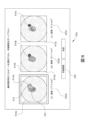


Classifications
-
- A—HUMAN NECESSITIES
- A61—MEDICAL OR VETERINARY SCIENCE; HYGIENE
- A61B—DIAGNOSIS; SURGERY; IDENTIFICATION
- A61B8/00—Diagnosis using ultrasonic, sonic or infrasonic waves
- A61B8/12—Diagnosis using ultrasonic, sonic or infrasonic waves in body cavities or body tracts, e.g. by using catheters
-
- A—HUMAN NECESSITIES
- A61—MEDICAL OR VETERINARY SCIENCE; HYGIENE
- A61B—DIAGNOSIS; SURGERY; IDENTIFICATION
- A61B8/00—Diagnosis using ultrasonic, sonic or infrasonic waves
- A61B8/08—Clinical applications
- A61B8/0833—Clinical applications involving detecting or locating foreign bodies or organic structures
- A61B8/085—Clinical applications involving detecting or locating foreign bodies or organic structures for locating body or organic structures, e.g. tumours, calculi, blood vessels, nodules
-
- A—HUMAN NECESSITIES
- A61—MEDICAL OR VETERINARY SCIENCE; HYGIENE
- A61B—DIAGNOSIS; SURGERY; IDENTIFICATION
- A61B8/00—Diagnosis using ultrasonic, sonic or infrasonic waves
- A61B8/08—Clinical applications
- A61B8/0891—Clinical applications for diagnosis of blood vessels
-
- A—HUMAN NECESSITIES
- A61—MEDICAL OR VETERINARY SCIENCE; HYGIENE
- A61B—DIAGNOSIS; SURGERY; IDENTIFICATION
- A61B8/00—Diagnosis using ultrasonic, sonic or infrasonic waves
- A61B8/46—Ultrasonic, sonic or infrasonic diagnostic devices with special arrangements for interfacing with the operator or the patient
- A61B8/461—Displaying means of special interest
-
- A—HUMAN NECESSITIES
- A61—MEDICAL OR VETERINARY SCIENCE; HYGIENE
- A61B—DIAGNOSIS; SURGERY; IDENTIFICATION
- A61B8/00—Diagnosis using ultrasonic, sonic or infrasonic waves
- A61B8/46—Ultrasonic, sonic or infrasonic diagnostic devices with special arrangements for interfacing with the operator or the patient
- A61B8/461—Displaying means of special interest
- A61B8/463—Displaying means of special interest characterised by displaying multiple images or images and diagnostic data on one display
-
- A—HUMAN NECESSITIES
- A61—MEDICAL OR VETERINARY SCIENCE; HYGIENE
- A61B—DIAGNOSIS; SURGERY; IDENTIFICATION
- A61B8/00—Diagnosis using ultrasonic, sonic or infrasonic waves
- A61B8/46—Ultrasonic, sonic or infrasonic diagnostic devices with special arrangements for interfacing with the operator or the patient
- A61B8/467—Ultrasonic, sonic or infrasonic diagnostic devices with special arrangements for interfacing with the operator or the patient characterised by special input means
- A61B8/469—Ultrasonic, sonic or infrasonic diagnostic devices with special arrangements for interfacing with the operator or the patient characterised by special input means for selection of a region of interest
-
- G—PHYSICS
- G06—COMPUTING OR CALCULATING; COUNTING
- G06N—COMPUTING ARRANGEMENTS BASED ON SPECIFIC COMPUTATIONAL MODELS
- G06N20/00—Machine learning
-
- G—PHYSICS
- G06—COMPUTING OR CALCULATING; COUNTING
- G06N—COMPUTING ARRANGEMENTS BASED ON SPECIFIC COMPUTATIONAL MODELS
- G06N3/00—Computing arrangements based on biological models
- G06N3/02—Neural networks
- G06N3/04—Architecture, e.g. interconnection topology
- G06N3/045—Combinations of networks
-
- G—PHYSICS
- G06—COMPUTING OR CALCULATING; COUNTING
- G06N—COMPUTING ARRANGEMENTS BASED ON SPECIFIC COMPUTATIONAL MODELS
- G06N3/00—Computing arrangements based on biological models
- G06N3/02—Neural networks
- G06N3/04—Architecture, e.g. interconnection topology
- G06N3/0464—Convolutional networks [CNN, ConvNet]
-
- G—PHYSICS
- G06—COMPUTING OR CALCULATING; COUNTING
- G06N—COMPUTING ARRANGEMENTS BASED ON SPECIFIC COMPUTATIONAL MODELS
- G06N3/00—Computing arrangements based on biological models
- G06N3/02—Neural networks
- G06N3/08—Learning methods
- G06N3/09—Supervised learning
-
- G—PHYSICS
- G06—COMPUTING OR CALCULATING; COUNTING
- G06T—IMAGE DATA PROCESSING OR GENERATION, IN GENERAL
- G06T7/00—Image analysis
- G06T7/0002—Inspection of images, e.g. flaw detection
- G06T7/0012—Biomedical image inspection
-
- G—PHYSICS
- G06—COMPUTING OR CALCULATING; COUNTING
- G06T—IMAGE DATA PROCESSING OR GENERATION, IN GENERAL
- G06T7/00—Image analysis
- G06T7/10—Segmentation; Edge detection
- G06T7/12—Edge-based segmentation
-
- G—PHYSICS
- G06—COMPUTING OR CALCULATING; COUNTING
- G06T—IMAGE DATA PROCESSING OR GENERATION, IN GENERAL
- G06T7/00—Image analysis
- G06T7/10—Segmentation; Edge detection
- G06T7/149—Segmentation; Edge detection involving deformable models, e.g. active contour models
-
- G—PHYSICS
- G06—COMPUTING OR CALCULATING; COUNTING
- G06T—IMAGE DATA PROCESSING OR GENERATION, IN GENERAL
- G06T7/00—Image analysis
- G06T7/10—Segmentation; Edge detection
- G06T7/174—Segmentation; Edge detection involving the use of two or more images
-
- G—PHYSICS
- G16—INFORMATION AND COMMUNICATION TECHNOLOGY [ICT] SPECIALLY ADAPTED FOR SPECIFIC APPLICATION FIELDS
- G16H—HEALTHCARE INFORMATICS, i.e. INFORMATION AND COMMUNICATION TECHNOLOGY [ICT] SPECIALLY ADAPTED FOR THE HANDLING OR PROCESSING OF MEDICAL OR HEALTHCARE DATA
- G16H30/00—ICT specially adapted for the handling or processing of medical images
- G16H30/40—ICT specially adapted for the handling or processing of medical images for processing medical images, e.g. editing
-
- G—PHYSICS
- G16—INFORMATION AND COMMUNICATION TECHNOLOGY [ICT] SPECIALLY ADAPTED FOR SPECIFIC APPLICATION FIELDS
- G16H—HEALTHCARE INFORMATICS, i.e. INFORMATION AND COMMUNICATION TECHNOLOGY [ICT] SPECIALLY ADAPTED FOR THE HANDLING OR PROCESSING OF MEDICAL OR HEALTHCARE DATA
- G16H40/00—ICT specially adapted for the management or administration of healthcare resources or facilities; ICT specially adapted for the management or operation of medical equipment or devices
- G16H40/60—ICT specially adapted for the management or administration of healthcare resources or facilities; ICT specially adapted for the management or operation of medical equipment or devices for the operation of medical equipment or devices
- G16H40/63—ICT specially adapted for the management or administration of healthcare resources or facilities; ICT specially adapted for the management or operation of medical equipment or devices for the operation of medical equipment or devices for local operation
-
- G—PHYSICS
- G06—COMPUTING OR CALCULATING; COUNTING
- G06T—IMAGE DATA PROCESSING OR GENERATION, IN GENERAL
- G06T2207/00—Indexing scheme for image analysis or image enhancement
- G06T2207/10—Image acquisition modality
- G06T2207/10132—Ultrasound image
-
- G—PHYSICS
- G06—COMPUTING OR CALCULATING; COUNTING
- G06T—IMAGE DATA PROCESSING OR GENERATION, IN GENERAL
- G06T2207/00—Indexing scheme for image analysis or image enhancement
- G06T2207/20—Special algorithmic details
- G06T2207/20081—Training; Learning
-
- G—PHYSICS
- G06—COMPUTING OR CALCULATING; COUNTING
- G06T—IMAGE DATA PROCESSING OR GENERATION, IN GENERAL
- G06T2207/00—Indexing scheme for image analysis or image enhancement
- G06T2207/20—Special algorithmic details
- G06T2207/20084—Artificial neural networks [ANN]
-
- G—PHYSICS
- G06—COMPUTING OR CALCULATING; COUNTING
- G06T—IMAGE DATA PROCESSING OR GENERATION, IN GENERAL
- G06T2207/00—Indexing scheme for image analysis or image enhancement
- G06T2207/20—Special algorithmic details
- G06T2207/20092—Interactive image processing based on input by user
- G06T2207/20096—Interactive definition of curve of interest
-
- G—PHYSICS
- G06—COMPUTING OR CALCULATING; COUNTING
- G06T—IMAGE DATA PROCESSING OR GENERATION, IN GENERAL
- G06T2207/00—Indexing scheme for image analysis or image enhancement
- G06T2207/30—Subject of image; Context of image processing
- G06T2207/30004—Biomedical image processing
- G06T2207/30101—Blood vessel; Artery; Vein; Vascular
Landscapes
- Engineering & Computer Science (AREA)
- Health & Medical Sciences (AREA)
- Life Sciences & Earth Sciences (AREA)
- Physics & Mathematics (AREA)
- General Health & Medical Sciences (AREA)
- Theoretical Computer Science (AREA)
- Biomedical Technology (AREA)
- Medical Informatics (AREA)
- Biophysics (AREA)
- Molecular Biology (AREA)
- Nuclear Medicine, Radiotherapy & Molecular Imaging (AREA)
- Radiology & Medical Imaging (AREA)
- Public Health (AREA)
- General Physics & Mathematics (AREA)
- Animal Behavior & Ethology (AREA)
- Pathology (AREA)
- Heart & Thoracic Surgery (AREA)
- Surgery (AREA)
- Veterinary Medicine (AREA)
- Software Systems (AREA)
- Computer Vision & Pattern Recognition (AREA)
- Artificial Intelligence (AREA)
- Mathematical Physics (AREA)
- General Engineering & Computer Science (AREA)
- Computing Systems (AREA)
- Evolutionary Computation (AREA)
- Data Mining & Analysis (AREA)
- Computational Linguistics (AREA)
- Vascular Medicine (AREA)
- Epidemiology (AREA)
- Primary Health Care (AREA)
- Quality & Reliability (AREA)
- Business, Economics & Management (AREA)
- General Business, Economics & Management (AREA)
- Ultra Sonic Daignosis Equipment (AREA)
Applications Claiming Priority (3)
| Application Number | Priority Date | Filing Date | Title |
|---|---|---|---|
| US201862750996P | 2018-10-26 | 2018-10-26 | |
| US62/750,996 | 2018-10-26 | ||
| PCT/IB2019/059166 WO2020084583A1 (en) | 2018-10-26 | 2019-10-25 | Intraluminal ultrasound vessel border selection and associated devices, systems, and methods |
Publications (3)
| Publication Number | Publication Date |
|---|---|
| JP2022509453A JP2022509453A (ja) | 2022-01-20 |
| JP2022509453A5 JP2022509453A5 (cg-RX-API-DMAC7.html) | 2022-10-28 |
| JP7299992B2 true JP7299992B2 (ja) | 2023-06-28 |
Family
ID=68393039
Family Applications (1)
| Application Number | Title | Priority Date | Filing Date |
|---|---|---|---|
| JP2021547969A Active JP7299992B2 (ja) | 2018-10-26 | 2019-10-25 | 管腔内超音波血管境界の選択並びに関連するデバイス、システム、および方法 |
Country Status (5)
| Country | Link |
|---|---|
| US (1) | US11596384B2 (cg-RX-API-DMAC7.html) |
| EP (1) | EP3870064A1 (cg-RX-API-DMAC7.html) |
| JP (1) | JP7299992B2 (cg-RX-API-DMAC7.html) |
| CN (1) | CN112912012B (cg-RX-API-DMAC7.html) |
| WO (1) | WO2020084583A1 (cg-RX-API-DMAC7.html) |
Cited By (11)
| Publication number | Priority date | Publication date | Assignee | Title |
|---|---|---|---|---|
| US11813485B2 (en) | 2020-01-28 | 2023-11-14 | The Regents Of The University Of Michigan | Systems and methods for histotripsy immunosensitization |
| US11813484B2 (en) | 2018-11-28 | 2023-11-14 | Histosonics, Inc. | Histotripsy systems and methods |
| WO2025090331A1 (en) * | 2023-10-27 | 2025-05-01 | Evident Vascular, Inc. | Systems and methods for enhanced ultrasound imaging |
| US12318636B2 (en) | 2022-10-28 | 2025-06-03 | Histosonics, Inc. | Histotripsy systems and methods |
| US12343568B2 (en) | 2020-08-27 | 2025-07-01 | The Regents Of The University Of Michigan | Ultrasound transducer with transmit-receive capability for histotripsy |
| US12446905B2 (en) | 2023-04-20 | 2025-10-21 | Histosonics, Inc. | Histotripsy systems and associated methods including user interfaces and workflows for treatment planning and therapy |
| US12527976B2 (en) | 2020-06-18 | 2026-01-20 | Histosonics, Inc. | Histotripsy acoustic and patient coupling systems and methods |
| US12582848B2 (en) | 2021-06-07 | 2026-03-24 | The Regents Of The University Of Michigan | Minimally invasive histotripsy systems and methods |
| US12599787B2 (en) | 2021-06-07 | 2026-04-14 | The Regents Of The University Of Michigan | All-in-one ultrasound systems and methods including histotripsy |
| US12642994B2 (en) | 2022-11-01 | 2026-06-02 | Histosonics, Inc. | Histotripsy systems and methods |
| US12642993B2 (en) | 2023-10-23 | 2026-06-02 | Histosonics, Inc. | High intensity therapeutic ultrasound (HITU) histotripsy systems, methods and treatment navigation software |
Families Citing this family (23)
| Publication number | Priority date | Publication date | Assignee | Title |
|---|---|---|---|---|
| WO2020190976A2 (en) * | 2019-03-17 | 2020-09-24 | Lightlab Imaging, Inc. | Arterial imaging and assessment systems and methods and related user interface based-workflows |
| EP4072427A1 (en) | 2019-12-10 | 2022-10-19 | Koninklijke Philips N.V. | Intraluminal image-based vessel diameter determination and associated devices, systems, and methods |
| WO2021117349A1 (ja) * | 2019-12-10 | 2021-06-17 | 富士フイルム株式会社 | 情報処理装置、情報処理システム、情報処理方法、及び情報処理プログラム |
| US20230045488A1 (en) * | 2020-01-06 | 2023-02-09 | Philips Image Guided Therapy Corporation | Intraluminal imaging based detection and visualization of intraluminal treatment anomalies |
| WO2021199967A1 (ja) * | 2020-03-30 | 2021-10-07 | テルモ株式会社 | プログラム、情報処理方法、学習モデルの生成方法、学習モデルの再学習方法、および、情報処理システム |
| JP7696356B2 (ja) * | 2020-09-29 | 2025-06-20 | テルモ株式会社 | 情報処理装置、情報処理方法及びプログラム |
| CN116568222B (zh) * | 2020-12-17 | 2026-04-21 | 富士胶片株式会社 | 超声波诊断装置及超声波诊断装置的控制方法 |
| WO2022238274A1 (en) | 2021-05-13 | 2022-11-17 | Koninklijke Philips N.V. | Automatic measurement of body lumen length between bookmarked intraluminal data based on coregistration of intraluminal data to extraluminal image |
| EP4337099A1 (en) | 2021-05-13 | 2024-03-20 | Koninklijke Philips N.V. | Pathway modification for coregistration of extraluminal image and intraluminal data |
| CN117295467A (zh) | 2021-05-13 | 2023-12-26 | 皇家飞利浦有限公司 | 来自先前的管腔外成像、管腔内数据和共配准的管腔内治疗指导 |
| US12702384B2 (en) | 2021-05-13 | 2026-08-11 | Philips Image Guided Therapy Corporation | Preview of intraluminal ultrasound image along longitudinal view of body lumen |
| WO2022238229A1 (en) | 2021-05-13 | 2022-11-17 | Koninklijke Philips N.V. | Coregistration reliability with extraluminal image and intraluminal data |
| EP4337096A1 (en) | 2021-05-13 | 2024-03-20 | Koninklijke Philips N.V. | Coregistration of intraluminal data to guidewire in extraluminal image obtained without contrast |
| ES3074928T3 (es) | 2021-08-17 | 2026-07-24 | Boston Scient Scimed Inc | Sistema de generación de imágenes intravasculares con análisis automatizado del calcio y orientación terapéutica |
| WO2023042861A1 (ja) * | 2021-09-17 | 2023-03-23 | テルモ株式会社 | 学習モデル生成方法、画像処理装置、情報処理装置、訓練データ生成方法および画像処理方法 |
| JP7680325B2 (ja) * | 2021-09-30 | 2025-05-20 | テルモ株式会社 | コンピュータプログラム、情報処理装置及び情報処理方法 |
| WO2023077267A1 (zh) * | 2021-11-02 | 2023-05-11 | 深圳迈瑞动物医疗科技股份有限公司 | 一种跟腱的检查方法和超声成像设备 |
| CN118541093A (zh) * | 2021-12-22 | 2024-08-23 | 皇家飞利浦有限公司 | 利用深呼吸确认参考图像帧和目标图像帧的管腔内成像 |
| US20250160786A1 (en) * | 2022-03-08 | 2025-05-22 | Philips Image Guided Therapy Corporation | Intravascular ultrasound imaging with contour generation and editing for circular and non-circular blood vessel borders |
| EP4525731A1 (en) * | 2022-05-27 | 2025-03-26 | Boston Scientific Scimed, Inc. | Systems and methods for intravascular visualization |
| WO2023247467A1 (en) * | 2022-06-24 | 2023-12-28 | Koninklijke Philips N.V. | Intraluminal ultrasound imaging with automatic detection of target and reference regions |
| US12646165B2 (en) | 2022-10-20 | 2026-06-02 | Canon U.S.A., Inc. | Contour editing tool for processing oct images |
| CN120826195A (zh) * | 2023-01-19 | 2025-10-21 | 波士顿科学国际有限公司 | 用于自动血管腔边界追踪的点击校正 |
Citations (6)
| Publication number | Priority date | Publication date | Assignee | Title |
|---|---|---|---|---|
| JP2003503141A (ja) | 1999-07-02 | 2003-01-28 | ザ・クリーブランド・クリニツク・フアンデーシヨン | 有効な輪郭線作成法及びシステムを使用する血管内の超音波分析 |
| US20040197015A1 (en) | 2003-04-02 | 2004-10-07 | Siemens Medical Solutions Usa, Inc. | Border detection for medical imaging |
| JP2009504329A (ja) | 2005-08-15 | 2009-02-05 | ボストン サイエンティフィック リミテッド | 医用画像解析 |
| WO2011125513A1 (ja) | 2010-03-31 | 2011-10-13 | 株式会社 日立メディコ | 医用画像診断装置及び、医用画像の計測値再入力方法 |
| JP2013005983A (ja) | 2011-06-27 | 2013-01-10 | Hitachi Medical Corp | 医用画像診断装置 |
| JP2015173820A (ja) | 2014-03-14 | 2015-10-05 | テルモ株式会社 | 画像処理装置、画像処理方法及びプログラム |
Family Cites Families (19)
| Publication number | Priority date | Publication date | Assignee | Title |
|---|---|---|---|---|
| US7226417B1 (en) | 1995-12-26 | 2007-06-05 | Volcano Corporation | High resolution intravascular ultrasound transducer assembly having a flexible substrate |
| US6200268B1 (en) | 1999-09-10 | 2001-03-13 | The Cleveland Clinic Foundation | Vascular plaque characterization |
| US7359554B2 (en) | 2002-08-26 | 2008-04-15 | Cleveland Clinic Foundation | System and method for identifying a vascular border |
| US7074188B2 (en) | 2002-08-26 | 2006-07-11 | The Cleveland Clinic Foundation | System and method of characterizing vascular tissue |
| US7175597B2 (en) | 2003-02-03 | 2007-02-13 | Cleveland Clinic Foundation | Non-invasive tissue characterization system and method |
| US7215802B2 (en) | 2004-03-04 | 2007-05-08 | The Cleveland Clinic Foundation | System and method for vascular border detection |
| JP4949264B2 (ja) * | 2004-11-19 | 2012-06-06 | コーニンクレッカ フィリップス エレクトロニクス エヌ ヴィ | 医療画像データ内の腫瘍境界を自動的に検出及び区分するシステム及び方法 |
| US20090177089A1 (en) * | 2008-01-04 | 2009-07-09 | Assaf Govari | Three-dimensional image reconstruction using doppler ultrasound |
| EP2637555B1 (en) * | 2010-11-08 | 2021-09-15 | Conavi Medical Inc. | Systems for improved visualization during minimally invasive procedures |
| EP2701603B1 (en) | 2011-04-28 | 2016-01-06 | Koninklijke Philips N.V. | Medical imaging device with separate button for selecting candidate segmentation images |
| US11272845B2 (en) * | 2012-10-05 | 2022-03-15 | Philips Image Guided Therapy Corporation | System and method for instant and automatic border detection |
| CN105190693B (zh) * | 2013-03-01 | 2018-04-06 | 波士顿科学国际有限公司 | 用于血管内超声序列中的管腔边界检测的系统和方法 |
| US20140276085A1 (en) * | 2013-03-13 | 2014-09-18 | Volcano Corporation | Coregistered intravascular and angiographic images |
| US9702762B2 (en) * | 2013-03-15 | 2017-07-11 | Lightlab Imaging, Inc. | Calibration and image processing devices, methods, and systems |
| JP6192846B2 (ja) * | 2013-09-11 | 2017-09-06 | ボストン サイエンティフィック サイムド,インコーポレイテッドBoston Scientific Scimed,Inc. | 血管内超音波撮像システムを使用して画像を選択し表示するためのシステム |
| US9747525B2 (en) * | 2014-06-16 | 2017-08-29 | Siemens Healthcare Gmbh | Method and system for improved hemodynamic computation in coronary arteries |
| US10474917B2 (en) | 2017-09-26 | 2019-11-12 | Siemens Healthcare Gmbh | Smart editing of image-processing results |
| WO2019174971A1 (en) | 2018-03-14 | 2019-09-19 | Koninklijke Philips N.V. | Alternative anatomical borders of blood vessels and associated devices, systems, and methods |
| EP4674371A3 (en) | 2018-03-15 | 2026-03-18 | Koninklijke Philips N.V. | Determination and visualization of anatomical landmarks for intraluminal lesion assessment and treatment planning |
-
2019
- 2019-10-23 US US16/661,360 patent/US11596384B2/en active Active
- 2019-10-25 CN CN201980070946.6A patent/CN112912012B/zh active Active
- 2019-10-25 WO PCT/IB2019/059166 patent/WO2020084583A1/en not_active Ceased
- 2019-10-25 EP EP19795670.9A patent/EP3870064A1/en not_active Withdrawn
- 2019-10-25 JP JP2021547969A patent/JP7299992B2/ja active Active
Patent Citations (6)
| Publication number | Priority date | Publication date | Assignee | Title |
|---|---|---|---|---|
| JP2003503141A (ja) | 1999-07-02 | 2003-01-28 | ザ・クリーブランド・クリニツク・フアンデーシヨン | 有効な輪郭線作成法及びシステムを使用する血管内の超音波分析 |
| US20040197015A1 (en) | 2003-04-02 | 2004-10-07 | Siemens Medical Solutions Usa, Inc. | Border detection for medical imaging |
| JP2009504329A (ja) | 2005-08-15 | 2009-02-05 | ボストン サイエンティフィック リミテッド | 医用画像解析 |
| WO2011125513A1 (ja) | 2010-03-31 | 2011-10-13 | 株式会社 日立メディコ | 医用画像診断装置及び、医用画像の計測値再入力方法 |
| JP2013005983A (ja) | 2011-06-27 | 2013-01-10 | Hitachi Medical Corp | 医用画像診断装置 |
| JP2015173820A (ja) | 2014-03-14 | 2015-10-05 | テルモ株式会社 | 画像処理装置、画像処理方法及びプログラム |
Cited By (18)
| Publication number | Priority date | Publication date | Assignee | Title |
|---|---|---|---|---|
| US11813484B2 (en) | 2018-11-28 | 2023-11-14 | Histosonics, Inc. | Histotripsy systems and methods |
| US11980778B2 (en) | 2018-11-28 | 2024-05-14 | Histosonics, Inc. | Histotripsy systems and methods |
| US12599784B2 (en) | 2018-11-28 | 2026-04-14 | Histosonics, Inc. | Histotripsy systems and methods |
| US12420118B2 (en) | 2018-11-28 | 2025-09-23 | Histosonics, Inc. | Histotripsy systems and methods |
| US12589261B2 (en) | 2018-11-28 | 2026-03-31 | Histosonics, Inc. | Histotripsy systems and methods |
| US12491382B2 (en) | 2018-11-28 | 2025-12-09 | Histosonics, Inc. | Histotripsy systems and methods |
| US12491384B2 (en) | 2018-11-28 | 2025-12-09 | Histosonics, Inc. | Histotripsy systems and methods |
| US11813485B2 (en) | 2020-01-28 | 2023-11-14 | The Regents Of The University Of Michigan | Systems and methods for histotripsy immunosensitization |
| US12527976B2 (en) | 2020-06-18 | 2026-01-20 | Histosonics, Inc. | Histotripsy acoustic and patient coupling systems and methods |
| US12343568B2 (en) | 2020-08-27 | 2025-07-01 | The Regents Of The University Of Michigan | Ultrasound transducer with transmit-receive capability for histotripsy |
| US12582848B2 (en) | 2021-06-07 | 2026-03-24 | The Regents Of The University Of Michigan | Minimally invasive histotripsy systems and methods |
| US12599787B2 (en) | 2021-06-07 | 2026-04-14 | The Regents Of The University Of Michigan | All-in-one ultrasound systems and methods including histotripsy |
| US12318636B2 (en) | 2022-10-28 | 2025-06-03 | Histosonics, Inc. | Histotripsy systems and methods |
| US12390665B1 (en) | 2022-10-28 | 2025-08-19 | Histosonics, Inc. | Histotripsy systems and methods |
| US12642994B2 (en) | 2022-11-01 | 2026-06-02 | Histosonics, Inc. | Histotripsy systems and methods |
| US12446905B2 (en) | 2023-04-20 | 2025-10-21 | Histosonics, Inc. | Histotripsy systems and associated methods including user interfaces and workflows for treatment planning and therapy |
| US12642993B2 (en) | 2023-10-23 | 2026-06-02 | Histosonics, Inc. | High intensity therapeutic ultrasound (HITU) histotripsy systems, methods and treatment navigation software |
| WO2025090331A1 (en) * | 2023-10-27 | 2025-05-01 | Evident Vascular, Inc. | Systems and methods for enhanced ultrasound imaging |
Also Published As
| Publication number | Publication date |
|---|---|
| WO2020084583A1 (en) | 2020-04-30 |
| EP3870064A1 (en) | 2021-09-01 |
| US11596384B2 (en) | 2023-03-07 |
| CN112912012A (zh) | 2021-06-04 |
| JP2022509453A (ja) | 2022-01-20 |
| US20200129147A1 (en) | 2020-04-30 |
| CN112912012B (zh) | 2024-05-31 |
Similar Documents
| Publication | Publication Date | Title |
|---|---|---|
| US11596384B2 (en) | Intraluminal ultrasound vessel border selection and associated devices, systems, and methods | |
| JP7542708B2 (ja) | 自動及び支援ラベル及びブックマークを用いた管腔内超音波イメージング | |
| US20260033809A1 (en) | Graphical longitudinal display for intraluminal ultrasound imaging and associated devices, systems, and methods | |
| US12514542B2 (en) | Speed determination for intraluminal ultrasound imaging and associated devices, systems, and methods | |
| CN113518588B (zh) | 管腔内超声方向引导和相关联的设备、系统和方法 | |
| US20250349023A1 (en) | Intraluminal image-based vessel diameter determination and associated devices, systems, and methods | |
| CN115003229A (zh) | 基于管腔内成像对管腔内处置异常的检测和可视化 | |
| US20230051383A1 (en) | Automatic intraluminal imaging-based target and reference image frame detection and associated devices, systems, and methods | |
| US20250160786A1 (en) | Intravascular ultrasound imaging with contour generation and editing for circular and non-circular blood vessel borders | |
| CN119486668A (zh) | 具有目标区域和参考区域的自动检测的管腔内超声成像 |
Legal Events
| Date | Code | Title | Description |
|---|---|---|---|
| A521 | Request for written amendment filed |
Free format text: JAPANESE INTERMEDIATE CODE: A523 Effective date: 20221020 |
|
| A621 | Written request for application examination |
Free format text: JAPANESE INTERMEDIATE CODE: A621 Effective date: 20221020 |
|
| A871 | Explanation of circumstances concerning accelerated examination |
Free format text: JAPANESE INTERMEDIATE CODE: A871 Effective date: 20221020 |
|
| A131 | Notification of reasons for refusal |
Free format text: JAPANESE INTERMEDIATE CODE: A131 Effective date: 20221121 |
|
| A521 | Request for written amendment filed |
Free format text: JAPANESE INTERMEDIATE CODE: A523 Effective date: 20230126 |
|
| A131 | Notification of reasons for refusal |
Free format text: JAPANESE INTERMEDIATE CODE: A131 Effective date: 20230201 |
|
| A521 | Request for written amendment filed |
Free format text: JAPANESE INTERMEDIATE CODE: A523 Effective date: 20230427 |
|
| TRDD | Decision of grant or rejection written | ||
| A01 | Written decision to grant a patent or to grant a registration (utility model) |
Free format text: JAPANESE INTERMEDIATE CODE: A01 Effective date: 20230519 |
|
| A61 | First payment of annual fees (during grant procedure) |
Free format text: JAPANESE INTERMEDIATE CODE: A61 Effective date: 20230616 |
|
| R150 | Certificate of patent or registration of utility model |
Ref document number: 7299992 Country of ref document: JP Free format text: JAPANESE INTERMEDIATE CODE: R150 |
|
| R250 | Receipt of annual fees |
Free format text: JAPANESE INTERMEDIATE CODE: R250 |