JP7258698B2 - Method for detecting particulate matter by immunochromatographic method and kit therefor - Google Patents
Method for detecting particulate matter by immunochromatographic method and kit therefor Download PDFInfo
- Publication number
- JP7258698B2 JP7258698B2 JP2019163068A JP2019163068A JP7258698B2 JP 7258698 B2 JP7258698 B2 JP 7258698B2 JP 2019163068 A JP2019163068 A JP 2019163068A JP 2019163068 A JP2019163068 A JP 2019163068A JP 7258698 B2 JP7258698 B2 JP 7258698B2
- Authority
- JP
- Japan
- Prior art keywords
- substance
- membrane
- binding
- binding target
- sample
- Prior art date
- Legal status (The legal status is an assumption and is not a legal conclusion. Google has not performed a legal analysis and makes no representation as to the accuracy of the status listed.)
- Active
Links
Images


Classifications
-
- G—PHYSICS
- G01—MEASURING; TESTING
- G01N—INVESTIGATING OR ANALYSING MATERIALS BY DETERMINING THEIR CHEMICAL OR PHYSICAL PROPERTIES
- G01N33/00—Investigating or analysing materials by specific methods not covered by groups G01N1/00 - G01N31/00
- G01N33/48—Biological material, e.g. blood, urine; Haemocytometers
- G01N33/50—Chemical analysis of biological material, e.g. blood, urine; Testing involving biospecific ligand binding methods; Immunological testing
- G01N33/53—Immunoassay; Biospecific binding assay; Materials therefor
- G01N33/574—Immunoassay; Biospecific binding assay; Materials therefor for cancer
- G01N33/57484—Immunoassay; Biospecific binding assay; Materials therefor for cancer involving compounds serving as markers for tumor, cancer, neoplasia, e.g. cellular determinants, receptors, heat shock/stress proteins, A-protein, oligosaccharides, metabolites
- G01N33/57488—Immunoassay; Biospecific binding assay; Materials therefor for cancer involving compounds serving as markers for tumor, cancer, neoplasia, e.g. cellular determinants, receptors, heat shock/stress proteins, A-protein, oligosaccharides, metabolites involving compounds identifable in body fluids
-
- G—PHYSICS
- G01—MEASURING; TESTING
- G01N—INVESTIGATING OR ANALYSING MATERIALS BY DETERMINING THEIR CHEMICAL OR PHYSICAL PROPERTIES
- G01N21/00—Investigating or analysing materials by the use of optical means, i.e. using sub-millimetre waves, infrared, visible or ultraviolet light
- G01N21/62—Systems in which the material investigated is excited whereby it emits light or causes a change in wavelength of the incident light
- G01N21/63—Systems in which the material investigated is excited whereby it emits light or causes a change in wavelength of the incident light optically excited
- G01N21/64—Fluorescence; Phosphorescence
- G01N21/6428—Measuring fluorescence of fluorescent products of reactions or of fluorochrome labelled reactive substances, e.g. measuring quenching effects, using measuring "optrodes"
-
- G—PHYSICS
- G01—MEASURING; TESTING
- G01N—INVESTIGATING OR ANALYSING MATERIALS BY DETERMINING THEIR CHEMICAL OR PHYSICAL PROPERTIES
- G01N21/00—Investigating or analysing materials by the use of optical means, i.e. using sub-millimetre waves, infrared, visible or ultraviolet light
- G01N21/75—Systems in which material is subjected to a chemical reaction, the progress or the result of the reaction being investigated
- G01N21/76—Chemiluminescence; Bioluminescence
-
- G—PHYSICS
- G01—MEASURING; TESTING
- G01N—INVESTIGATING OR ANALYSING MATERIALS BY DETERMINING THEIR CHEMICAL OR PHYSICAL PROPERTIES
- G01N33/00—Investigating or analysing materials by specific methods not covered by groups G01N1/00 - G01N31/00
- G01N33/48—Biological material, e.g. blood, urine; Haemocytometers
- G01N33/50—Chemical analysis of biological material, e.g. blood, urine; Testing involving biospecific ligand binding methods; Immunological testing
- G01N33/53—Immunoassay; Biospecific binding assay; Materials therefor
- G01N33/543—Immunoassay; Biospecific binding assay; Materials therefor with an insoluble carrier for immobilising immunochemicals
- G01N33/54366—Apparatus specially adapted for solid-phase testing
- G01N33/54386—Analytical elements
- G01N33/54387—Immunochromatographic test strips
- G01N33/54388—Immunochromatographic test strips based on lateral flow
-
- G—PHYSICS
- G01—MEASURING; TESTING
- G01N—INVESTIGATING OR ANALYSING MATERIALS BY DETERMINING THEIR CHEMICAL OR PHYSICAL PROPERTIES
- G01N21/00—Investigating or analysing materials by the use of optical means, i.e. using sub-millimetre waves, infrared, visible or ultraviolet light
- G01N21/62—Systems in which the material investigated is excited whereby it emits light or causes a change in wavelength of the incident light
- G01N21/63—Systems in which the material investigated is excited whereby it emits light or causes a change in wavelength of the incident light optically excited
- G01N21/64—Fluorescence; Phosphorescence
- G01N21/6428—Measuring fluorescence of fluorescent products of reactions or of fluorochrome labelled reactive substances, e.g. measuring quenching effects, using measuring "optrodes"
- G01N2021/6439—Measuring fluorescence of fluorescent products of reactions or of fluorochrome labelled reactive substances, e.g. measuring quenching effects, using measuring "optrodes" with indicators, stains, dyes, tags, labels, marks
-
- G—PHYSICS
- G01—MEASURING; TESTING
- G01N—INVESTIGATING OR ANALYSING MATERIALS BY DETERMINING THEIR CHEMICAL OR PHYSICAL PROPERTIES
- G01N21/00—Investigating or analysing materials by the use of optical means, i.e. using sub-millimetre waves, infrared, visible or ultraviolet light
- G01N21/75—Systems in which material is subjected to a chemical reaction, the progress or the result of the reaction being investigated
- G01N21/77—Systems in which material is subjected to a chemical reaction, the progress or the result of the reaction being investigated by observing the effect on a chemical indicator
- G01N2021/7756—Sensor type
- G01N2021/7759—Dipstick; Test strip
Description
本発明は、粒状物質をイムノクロマトグラフィー法によって検出する方法、及び、そのためのイムノクロマトグラフィー用テストストリップ及びキットに関している。 The present invention relates to a method for detecting particulate matter by immunochromatography, and an immunochromatographic test strip and kit therefor.
細胞外小胞やウイルスなどの粒状物質は、イムノクロマトグラフィー法によって検出され得る(特許文献1並びに非特許文献1及び2)。イムノクロマトグラフィー法は、ウェスタンブロッティングやELISAと比較して迅速な検出を可能とする試験方法であり、対象の粒状物質を単離しなくても検出が可能である点や、特定の非検出物質を表面上に備える粒状物質を他の粒状物質と区別して検出することが可能である点などでNanoSightなどによる計測よりも優れている。また、細胞外小胞のエクソソームは、血液や尿の他、母乳、唾液、涙などのあらゆる体液中に含まれている粒状物質であるが、これは細胞に固有のマイクロRNAを含んでいることが知られているため、体液を利用したがん診断におけるターゲット物質としても注目されている。特許文献1並びに非特許文献1及び2には、そのようなエクソソームを検出する方法として、当該エクソソームを標識抗体と反応させて複合体を形成させた後に、当該複合体をラテラルフロー用のメンブレン上で展開し、当該メンブレン上に固定されている捕捉抗体に結合させる方法が記載されている。
Particulate matter such as extracellular vesicles and viruses can be detected by immunochromatographic methods (
他方、特許文献2~5には、各種金属ナノ粒子などの標識物質が、被験物質を検出するために使用されることが記載されている。しかしながら、これらの文献には、細胞外小胞などの粒状物質を実際に検出することは記載されていない。
On the other hand,
従来のイムノクロマトグラフィー法では、メンブレン上で展開する前に、粒状物質の表面が標識抗体で覆われることになるため、捕捉抗体との反応性が低下し、検出感度が十分ではなかった。本発明は、高い感度で粒状物質を検出する方法を提供することを目的としている。 In the conventional immunochromatographic method, the surface of the particulate matter is covered with the labeled antibody before it develops on the membrane, so the reactivity with the capture antibody is lowered and the detection sensitivity is not sufficient. An object of the present invention is to provide a method for detecting particulate matter with high sensitivity.
本発明者らは、上記課題を解決すべく鋭意検討した結果、特定の順序でイムノクロマトグラフィー法を行うことにより、高い感度で粒状物質を検出することができることを見出し、本発明を完成させた。すなわち、本発明は、以下に示す粒状物質をイムノクロマトグラフィー法によって検出する方法、及び、そのためのイムノクロマトグラフィー用テストストリップ及びキットを提供するものである。
〔1〕互いに同じであっても異なってもよい第一の結合対象物質及び第二の結合対象物質を含む結合対象物質を表面上に複数備える粒状物質をイムノクロマトグラフィー法によって検出する方法であって、
(1)前記粒状物質を含む試料と、前記第一の結合対象物質に対する第一の特異的結合物質とを、メンブレン上で接触させ、前記粒状物質を前記第一の特異的結合物質により捕捉する工程、
(2)捕捉された前記粒状物質と、前記第二の結合対象物質に対する第二の特異的結合物質とを接触させ、前記粒状物質を標識する工程、及び、
(3)標識された前記粒状物質を検出する工程
を含み、前記第一の特異的結合物質が、前記メンブレン上に固定されており、前記第二の特異的結合物質が、標識物質に結合していることを特徴とする、方法。
〔2〕前記粒状物質が、細胞外小胞である、前記〔1〕に記載の方法。
〔3〕前記標識物質が、金属ナノ粒子、化学発光物質、又は、蛍光物質である、前記〔1〕又は〔2〕に記載の方法。
〔4〕前記金属ナノ粒子が、異方性金属ナノ粒子である、前記〔3〕に記載の方法。
〔5〕前記異方性金属ナノ粒子が、青色又は黒色であり、かつ前記試料が、血液を含む、前記〔4〕に記載の方法。
〔6〕前記試料に界面活性剤を添加する工程をさらに含む、前記〔1〕~〔5〕のいずれか一項に記載の方法。
〔7〕前記第一の結合対象物質が、前記第二の結合対象物質と同じである、前記〔1〕~〔6〕のいずれか一項に記載の方法。
〔8〕前記第一の結合対象物質における前記第一の特異的結合物質の結合部位が、前記第二の結合対象物質における前記第二の特異的結合物質の結合部位と同じである、前記〔7〕に記載の方法。
〔9〕前記検出工程が、前記粒状物質を定量する工程を含む、前記〔1〕~〔8〕のいずれか一項に記載の方法。
〔10〕前記定量工程が、質量分析装置、イムノクロマトリーダー、又は、画像解析装置で標識シグナルを測定する工程を含む、前記〔9〕に記載の方法。
〔11〕前記質量分析装置のイオン化方法が、誘導結合プラズマ(ICP)又はマトリックス支援レーザー脱離イオン化法(MALDI)である、前記〔10〕に記載の方法。
〔12〕前記〔1〕~〔11〕のいずれか一項に記載の方法に使用するための、イムノクロマトグラフィー用テストストリップ。
〔13〕互いに同じであっても異なってもよい第一の結合対象物質及び第二の結合対象物質を含む結合対象物質を表面上に複数備える粒状物質を、前記〔1〕~〔11〕のいずれか一項に記載の方法により検出するためのキットであって、
メンブレンを含むテストストリップと、
前記第一の結合対象物質に対する第一の特異的結合物質と、
前記第二の結合対象物質に対する第二の特異的結合物質と
を含み、前記第一の特異的結合物質が、前記メンブレン上に固定されており、前記第二の特異的結合物質が、標識物質に結合していることを特徴とする、キット。
〔14〕互いに同じであっても異なってもよい第一の結合対象物質及び第二の結合対象物質を含む結合対象物質を表面上に複数備える粒状物質を検出するためのイムノクロマトグラフィー用テストストリップであって、
前記第一の結合対象物質に対する第一の特異的結合物質が固定された検出部位を備えるメンブレンと、
試料流動方向について前記検出部位の上流側の位置で前記メンブレンと接する第一のサンプルパッドと
を含み、
前記第一のサンプルパッドから前記検出部位に至る途中にコンジュゲートパッドが含まれていないことを特徴とする、テストストリップ。
〔15〕前記試料流動方向について前記第一のサンプルパッドのさらに上流側の位置で前記メンブレンと接する第二のサンプルパッドをさらに含み、前記第二のサンプルパッドが、前記第一のサンプルパッドから離間している、前記〔14〕に記載のテストストリップ。
〔16〕前記第二のサンプルパッドが、前記第二の結合対象物質に対する第二の特異的結合物質を含むコンジュゲートパッドを含み、前記第二の特異的結合物質が、標識物質に結合している、前記〔15〕に記載のテストストリップ。
〔17〕前記第一のサンプルパッド及び前記第二のサンプルパッドが、第一のスペーサーを介して互いから離間している、前記〔15〕又は〔16〕に記載のテストストリップ。
〔18〕前記メンブレンが、前記第一のサンプルパッド側から前記第二のサンプルパッド側への前記粒状物質を含む試料の浸透を阻害するように構成されている、前記〔15〕~〔17〕のいずれか一項に記載のテストストリップ。
〔19〕前記粒状物質が、前記第一の結合対象物質又は前記第二の結合対象物質と同じであっても異なってもよい第三の結合対象物質をさらに備え、
前記メンブレンが、前記第三の結合対象物質に対する第三の特異的結合物質が固定された追加の検出部位と、前記試料流動方向について前記第一のサンプルパッドの下流側の位置で前記試料が流動する経路を分割するように配置された第二のスペーサーと、をさらに備え、
前記検出部位及び前記追加の検出部位が、前記第二のスペーサーによって分割された異なる経路に配置されている、前記〔15〕~〔18〕のいずれか一項に記載のテストストリップ。
〔20〕互いに同じであっても異なってもよい第一の結合対象物質及び第二の結合対象物質を含む結合対象物質を表面上に複数備える粒状物質を検出するためのイムノクロマトグラフィー用テストストリップであって、
前記第一の結合対象物質に対する第一の特異的結合物質が固定された検出部位を備えるメンブレンと、試料流動方向について前記検出部位の上流側の位置で前記メンブレンと接する第一のサンプルパッドとを含む第一の基板と、
第二のサンプルパッドを含む第二の基板と
を含み、
前記第一の基板及び前記第二の基板は、前記第二のサンプルパッドが前記試料流動方向について前記第一のサンプルパッドのさらに上流側の位置で前記メンブレンと接するように、接近可能であることを特徴とする、テストストリップ。
〔21〕前記第二のサンプルパッドが、前記第二の結合対象物質に対する第二の特異的結合物質を含むコンジュゲートパッドを含み、前記第二の特異的結合物質が、標識物質に結合している、前記〔20〕に記載のテストストリップ。
〔22〕前記第一の基板及び前記第二の基板が、伸縮構造を介して結合されている、前記〔20〕又は〔21〕に記載のテストストリップ。
〔23〕互いに同じであっても異なってもよい第一の結合対象物質及び第二の結合対象物質を含む結合対象物質を表面上に複数備える粒状物質を検出するためのイムノクロマトグラフィー用テストストリップであって、
前記第一の結合対象物質に対する第一の特異的結合物質が固定された検出部位を備えるメンブレンと、
第一の経路における試料流動方向について前記検出部位の上流側の位置で前記メンブレンと接する第一のサンプルパッドと、
前記第一の経路とは異なる第二の経路における試料流動方向について前記検出部位の上流側の位置で前記メンブレンと接する第二のサンプルパッドと
を含み、
前記第二のサンプルパッドが、前記第一のサンプルパッドから離間していることを特徴とする、テストストリップ。
〔24〕前記第二のサンプルパッドが、前記第二の結合対象物質に対する第二の特異的結合物質を含むコンジュゲートパッドを含み、前記第二の特異的結合物質が、標識物質に結合している、前記〔23〕に記載のテストストリップ。
〔25〕前記メンブレンが、短冊形、U字型、又は、V字型である、前記〔23〕又は〔24〕に記載のテストストリップ。
〔26〕前記粒状物質が、前記第一の結合対象物質又は前記第二の結合対象物質と同じであっても異なってもよい第三の結合対象物質をさらに備え、
前記メンブレンが、前記第三の結合対象物質に対する第三の特異的結合物質が固定された追加の検出部位と、前記第一の経路における試料流動方向について前記第一のサンプルパッドの下流側の位置で前記第一の経路を分割するように配置されたスペーサーと、をさらに備え、
前記検出部位及び前記追加の検出部位が、前記スペーサーで分割された異なる経路に配置されている、前記〔23〕又は〔24〕に記載のテストストリップ。
〔27〕前記第一の経路及び前記第二の経路とは異なる第三の経路における試料流動方向について前記追加の検出部位の上流側の位置で前記メンブレンと接する第三のサンプルパッドをさらに含む、前記〔26〕に記載のテストストリップ。
〔28〕前記第二のサンプルパッド及び/又は前記第三のサンプルパッドが、前記第二の特異的結合物質を含むコンジュゲートパッドを含んでいる、前記〔27〕に記載のテストストリップ。
〔29〕互いに同じであっても異なってもよい第一の結合対象物質及び第二の結合対象物質を含む結合対象物質を表面上に複数備える粒状物質を検出するためのイムノクロマトグラフィー用テストストリップであって、
前記第一の結合対象物質に対する第一の特異的結合物質が固定された検出部位を備える第一のメンブレンと、
第一の経路における試料流動方向について前記検出部位の上流側の位置で前記第一のメンブレンと接する第一のサンプルパッドと、
前記検出部位と前記第一のサンプルパッドの間で前記第一のメンブレンと接している第二のメンブレンと、
前記第一の経路とは異なる第二の経路における試料流動方向について前記検出部位の上流側の位置で前記第二のメンブレンと接する第二のサンプルパッドと
を含み、
前記第二のサンプルパッドが、前記第一のサンプルパッドから離間していることを特徴とする、テストストリップ。
〔30〕前記第一のサンプルパッド又は前記第二のサンプルパッドのいずれか一方が、前記第二の結合対象物質に対する第二の特異的結合物質を含むコンジュゲートパッドを含み、前記第二の特異的結合物質が、標識物質に結合している、前記〔29〕に記載のテストストリップ。
〔31〕前記メンブレンが、イムノクロマトグラフィー試験の成否を判定するコントロール部位をさらに備えている、前記〔14〕~〔30〕のいずれか一項に記載のテストストリップ。
As a result of intensive studies to solve the above problems, the present inventors have found that particulate matter can be detected with high sensitivity by performing immunochromatographic methods in a specific order, and have completed the present invention. That is, the present invention provides a method for detecting particulate matter by an immunochromatographic method, and an immunochromatographic test strip and kit therefor.
[1] A method for detecting, by an immunochromatographic method, a particulate substance having a plurality of binding target substances on its surface, including a first binding target substance and a second binding target substance, which may be the same or different. ,
(1) A sample containing the particulate matter and a first specific binding substance for the first binding target substance are brought into contact on a membrane, and the particulate matter is captured by the first specific binding matter. process,
(2) contacting the captured particulate matter with a second specific binding substance for the second binding target substance to label the particulate matter;
(3) detecting the labeled particulate matter, wherein the first specific binding substance is immobilized on the membrane, and the second specific binding substance binds to the labeled substance; A method, characterized in that:
[2] The method according to [1] above, wherein the particulate matter is an extracellular vesicle.
[3] The method according to [1] or [2] above, wherein the labeling substance is a metal nanoparticle, a chemiluminescent substance, or a fluorescent substance.
[4] The method according to [3] above, wherein the metal nanoparticles are anisotropic metal nanoparticles.
[5] The method according to [4] above, wherein the anisotropic metal nanoparticles are blue or black, and the sample contains blood.
[6] The method according to any one of [1] to [5] above, further comprising adding a surfactant to the sample.
[7] The method according to any one of [1] to [6] above, wherein the first binding target substance is the same as the second binding target substance.
[8] The above [ 7].
[9] The method according to any one of [1] to [8] above, wherein the detecting step includes a step of quantifying the particulate matter.
[10] The method according to [9] above, wherein the quantification step includes a step of measuring the labeled signal with a mass spectrometer, an immunochromatographic reader, or an image analyzer.
[11] The method according to [10] above, wherein the ionization method of the mass spectrometer is inductively coupled plasma (ICP) or matrix-assisted laser desorption ionization (MALDI).
[12] An immunochromatographic test strip for use in the method according to any one of [1] to [11] above.
[13] A particulate substance having a plurality of binding target substances including a first binding target substance and a second binding target substance, which may be the same or different, on the surface thereof, as described in [1] to [11] above. A kit for detection by the method of any one of the above,
a test strip containing a membrane;
a first specific binding substance for the first binding target substance;
a second specific binding substance for the second binding target substance, wherein the first specific binding substance is immobilized on the membrane, and the second specific binding substance is a labeling substance A kit, characterized in that it is coupled to
[14] An immunochromatographic test strip for detecting a particulate substance having a plurality of binding target substances on its surface, including a first binding target substance and a second binding target substance, which may be the same or different. There is
a membrane comprising a detection site on which a first specific binding substance for the first binding target substance is immobilized;
a first sample pad in contact with the membrane at a position on the upstream side of the detection site with respect to the sample flow direction;
A test strip, characterized in that it does not contain a conjugate pad en route from said first sample pad to said detection site.
[15] further comprising a second sample pad in contact with the membrane at a position further upstream of the first sample pad with respect to the sample flow direction, wherein the second sample pad is separated from the first sample pad; The test strip according to [14] above.
[16] The second sample pad includes a conjugate pad containing a second specific binding substance for the second binding target substance, and the second specific binding substance binds to a labeling substance The test strip according to [15] above.
[17] The test strip according to [15] or [16] above, wherein the first sample pad and the second sample pad are separated from each other via a first spacer.
[18] The above [15] to [17], wherein the membrane is configured to inhibit permeation of the sample containing the particulate matter from the first sample pad side to the second sample pad side. A test strip according to any one of .
[19] The particulate substance further comprises a third binding target substance which may be the same as or different from the first binding target substance or the second binding target substance,
The membrane includes an additional detection site on which a third specific binding substance for the third binding target substance is immobilized, and the sample flows at a position downstream of the first sample pad with respect to the sample flow direction. a second spacer arranged to divide the pathway to
The test strip according to any one of [15] to [18] above, wherein the detection site and the additional detection site are arranged in different paths separated by the second spacer.
[20] An immunochromatographic test strip for detecting a particulate substance having a plurality of binding target substances on its surface, including a first binding target substance and a second binding target substance, which may be the same or different. There is
A membrane comprising a detection site on which a first specific binding substance for said first binding target substance is immobilized, and a first sample pad in contact with said membrane at a position upstream of said detection site with respect to the sample flow direction. a first substrate comprising;
a second substrate including a second sample pad;
The first substrate and the second substrate are accessible such that the second sample pad contacts the membrane at a position further upstream of the first sample pad with respect to the sample flow direction. A test strip characterized by:
[21] The second sample pad includes a conjugate pad containing a second specific binding substance for the second binding target substance, and the second specific binding substance binds to a labeling substance The test strip according to [20] above.
[22] The test strip according to [20] or [21], wherein the first substrate and the second substrate are connected via an elastic structure.
[23] An immunochromatographic test strip for detecting a particulate substance having a plurality of binding target substances on its surface, including a first binding target substance and a second binding target substance, which may be the same or different. There is
a membrane comprising a detection site on which a first specific binding substance for the first binding target substance is immobilized;
a first sample pad in contact with the membrane at a position on the upstream side of the detection site with respect to the sample flow direction in the first path;
a second sample pad in contact with the membrane at a position on the upstream side of the detection site with respect to a sample flow direction in a second path different from the first path,
A test strip, wherein said second sample pad is spaced apart from said first sample pad.
[24] The second sample pad includes a conjugate pad containing a second specific binding substance for the second binding target substance, and the second specific binding substance binds to a labeling substance The test strip according to [23] above.
[25] The test strip of [23] or [24] above, wherein the membrane is rectangular, U-shaped, or V-shaped.
[26] The particulate substance further comprises a third binding target substance which may be the same as or different from the first binding target substance or the second binding target substance,
The membrane comprises an additional detection site on which a third specific binding substance for the third binding target substance is immobilized, and a position on the downstream side of the first sample pad with respect to the sample flow direction in the first path. a spacer positioned to divide the first pathway with
The test strip of [23] or [24] above, wherein the detection site and the additional detection site are arranged in different paths separated by the spacer.
[27] Further comprising a third sample pad in contact with the membrane at a position upstream of the additional detection site with respect to a sample flow direction in a third route different from the first route and the second route, The test strip described in [26] above.
[28] The test strip of [27] above, wherein the second sample pad and/or the third sample pad includes a conjugate pad containing the second specific binding substance.
[29] An immunochromatographic test strip for detecting a particulate substance having a plurality of binding target substances on its surface, including a first binding target substance and a second binding target substance, which may be the same or different from each other There is
a first membrane comprising a detection site on which a first specific binding substance for the first binding target substance is immobilized;
a first sample pad in contact with the first membrane at a position on the upstream side of the detection site with respect to the sample flow direction in the first path;
a second membrane in contact with the first membrane between the detection site and the first sample pad;
a second sample pad in contact with the second membrane at a position on the upstream side of the detection site with respect to a sample flow direction in a second path different from the first path,
A test strip, wherein said second sample pad is spaced apart from said first sample pad.
[30] Either the first sample pad or the second sample pad includes a conjugate pad containing a second specific binding substance for the second binding target substance, and the second specific The test strip of [29] above, wherein the target binding substance is bound to the labeling substance.
[31] The test strip of any one of [14] to [30] above, wherein the membrane further comprises a control portion for determining the success or failure of an immunochromatography test.
本発明に従えば、イムノクロマトグラフィー法において、粒状物質の検出感度を向上することができる。したがって、イムノクロマトグラフィー法によって、粒状物質を迅速かつ高い感度で検出することが可能となる。 According to the present invention, the detection sensitivity of particulate matter can be improved in immunochromatography. Therefore, the immunochromatographic method enables rapid and highly sensitive detection of particulate matter.
以下、本発明をさらに詳細に説明する。
本発明は、粒状物質をイムノクロマトグラフィー法によって検出する方法に関している。本明細書に記載の「イムノクロマトグラフィー法」とは、試料中の対象物質をそれに特異的に結合する物質を使用して検出する方法であって、当該試料をメンブレン上で移動させて対象物質を分離するラテラルフロー型の検出方法だけでなく、当該試料をメンブレンに垂直に移動させて対象物質を分離するフロースルー型(バーティカルフロー型)の検出方法のこともいう。本明細書に記載の「粒状物質」とは、前記イムノクロマトグラフィー法によって検出可能な大きさの粒状の物質のことをいい、前記粒状物質は、表面上に少なくとも1種の結合対象物質を複数備えている。前記結合対象物質は、互いに同じであっても異なってもよい第一の結合対象物質及び第二の結合対象物質を含む。前記粒状物質の粒径は、特に限定されないが、例えば、約10μm以下であってもよく、ある態様では、約5μm以下、約1μm以下、約500nm以下、又は約200nm以下であってもよい。具体的には、前記粒状物質は、例えば、エクソソーム、微小胞、アポトーシス小体、及びラージオンコソームなどの細胞外小胞であってもよく、インフルエンザウイルス、アデノウイルス、RSウイルス、ロタウイルス、ヒトパピローマウイルス、ヒト免疫不全ウイルス、B型肝炎ウイルス、ジカウイルス、及びデングウイルスなどのウイルスであってもよく、クラミジア、梅毒トレポネーマ、溶連菌、炭疽菌、黄色ブドウ球菌、赤痢菌、大腸菌、サルモネラ菌、ネズミチフス菌、パラチフス菌、緑膿菌、及び腸炎ビブリオ菌などの細菌であってもよい。
The present invention will now be described in more detail.
The present invention relates to a method for detecting particulate matter by immunochromatography. The "immunochromatographic method" described herein is a method of detecting a target substance in a sample using a substance that specifically binds to it, wherein the sample is moved on a membrane to detect the target substance. It refers not only to the lateral flow type detection method for separation, but also to the flow-through type (vertical flow type) detection method for separating the target substance by moving the sample perpendicularly to the membrane. The term "particulate matter" as used herein refers to a particulate matter of a size that can be detected by the immunochromatographic method, and the particulate matter has at least one binding target substance on its surface. ing. The binding target substance includes a first binding target substance and a second binding target substance, which may be the same or different. The particle size of the particulate matter is not particularly limited, but may be, for example, about 10 μm or less, and in certain embodiments, about 5 μm or less, about 1 μm or less, about 500 nm or less, or about 200 nm or less. Specifically, the particulate matter may be, for example, extracellular vesicles such as exosomes, microvesicles, apoptotic bodies, and large oncosomes, influenza virus, adenovirus, respiratory syncytial virus, rotavirus, human papilloma. viruses such as human immunodeficiency virus, hepatitis B virus, Zika virus, and dengue virus; Bacteria such as Paratyphi, Pseudomonas aeruginosa, and Vibrio parahaemolyticus are also possible.
前記粒状物質を含む試料は、イムノクロマトグラフィー法に供することができるものであれば、特に制限なく採用することができ、例えば、本発明の方法に適用する試料として、生体又は培養液などから採取した試料をそのまま採用してもいいし、ろ過や遠心分離などの前処理により精製、部分精製、又は濃縮された試料を採用してもよい。具体的には、前記試料は、例えば、血液(全血、血清、又は血漿)、脳髄液、涙、母乳、肺胞洗浄液、悪性胸膜滲出液、滑液、尿、羊水、腹水、精液、唾液、及びリンパ液などの体液、組織切片の保存液、又は、細胞培養上清などであってもよい。 The sample containing the particulate matter can be employed without particular limitation as long as it can be subjected to the immunochromatographic method. A sample may be used as it is, or a sample purified, partially purified, or concentrated by pretreatment such as filtration or centrifugation may be used. Specifically, the sample is, for example, blood (whole blood, serum, or plasma), cerebrospinal fluid, tears, breast milk, alveolar lavage, malignant pleural effusion, synovial fluid, urine, amniotic fluid, ascites, semen, saliva. , and body fluids such as lymph fluid, preservation fluids of tissue sections, cell culture supernatants, and the like.
本発明の方法は、
(1)前記粒状物質を含む試料と、前記第一の結合対象物質に対する第一の特異的結合物質とを、メンブレン上で接触させ、前記粒状物質を前記第一の特異的結合物質により捕捉する工程、
(2)捕捉された前記粒状物質と、前記第二の結合対象物質に対する第二の特異的結合物質とを接触させ、前記粒状物質を標識する工程、及び、
(3)標識された前記粒状物質を検出する工程
を含む。前記第一の結合対象物質と前記第二の結合対象物質が同じである場合、前記第一の結合対象物質における前記第一の特異的結合物質の結合部位は、前記第二の結合対象物質における前記第二の特異的結合物質の結合部位と同じであってもいいし、異なっていてもよい。両結合部位が同じであっても、前記粒状物質は、前記結合対象物質を表面上に複数備えているので、前記第一の特異的結合物質により捕捉した後にも、前記第二の特異的結合物質により標識することが可能となっている。
The method of the present invention comprises:
(1) A sample containing the particulate matter and a first specific binding substance for the first binding target substance are brought into contact on a membrane, and the particulate matter is captured by the first specific binding matter. process,
(2) contacting the captured particulate matter with a second specific binding substance for the second binding target substance to label the particulate matter;
(3) detecting the labeled particulate matter; When the first binding target substance and the second binding target substance are the same, the binding site of the first specific binding substance in the first binding target substance is It may be the same as or different from the binding site of the second specific binding substance. Even if both binding sites are the same, since the particulate substance has a plurality of the binding target substances on the surface, even after being captured by the first specific binding substance, the second specific binding It is possible to label with a substance.
前記第一の特異的結合物質は、前記メンブレン上に固定されているものであり、前記第一の結合対象物質を有している粒状物質をメンブレン上に捕捉することができる。前記メンブレンとしては、イムノクロマトグラフィー用テストストリップ(イムノクロマト試験紙)に用いられるもの(前記第一の特異的結合物質を固定化する性能を有し、かつ液体が所期の方向に通行することを妨げない性能を有するもの)を特に制限されることなく使用することができるが、例えば、毛細管作用を有し、液体及びそれに分散した成分を吸収により輸送可能な多孔性メンブレンであってもよい。前記メンブレンの材質は、特に限定されないが、例えば、セルロース、ニトロセルロース、セルロースアセテート、ポリビニリデンジフルオライド(PVDF)、ガラス繊維、ナイロン、又はポリケトンなどであってもよい。 The first specific binding substance is immobilized on the membrane, and particulate matter having the first binding target substance can be trapped on the membrane. As the membrane, those used in immunochromatographic test strips (immunochromatographic test paper) (having the ability to immobilize the first specific binding substance and preventing liquid from passing in the desired direction) However, for example, it may be a porous membrane that has capillary action and is capable of transporting liquid and components dispersed therein by absorption. The material of the membrane is not particularly limited, but may be, for example, cellulose, nitrocellulose, cellulose acetate, polyvinylidene difluoride (PVDF), glass fiber, nylon, or polyketone.
前記第一の特異的結合物質としては、前記メンブレン上に固定できるものであって、かつ、検出の対象である前記粒状物質を、前記第一の結合対象物質との複合体形成を介して前記メンブレン上に捕捉できるものであれば、特に制限なく採用することができる。前記第一の結合対象物質と前記第一の特異的結合物質との組合せの具体例としては、抗原とそれに結合する抗体、抗体とそれに結合する抗原、糖鎖又は複合糖質とそれに結合するレクチン、レクチンとそれに結合する糖鎖又は複合糖質、ホルモン又はサイトカインとそれに結合する受容体、受容体とそれに結合するホルモン又はサイトカイン、タンパク質とそれに結合する核酸アプタマー若しくはペプチドアプタマー、酵素とそれに結合する基質、基質とそれに結合する酵素、ビオチンとアビジン又はストレプトアビジン、アビジン又はストレプトアビジンとビオチン、IgGとプロテインA又はプロテインG、プロテインA又はプロテインGとIgG、T細胞免疫グロブリン・ムチンドメイン含有分子4(Tim4)とホスファチジルセリン(PS)、PSとTim4、あるいは、第1の核酸とそれに結合する(ハイブリダイズする)第2の核酸等が挙げられる。前記第2の核酸は、前記第1の核酸と相補的な配列を含む核酸であってもよい。 The first specific binding substance is one that can be immobilized on the membrane and binds the particulate substance to be detected through complex formation with the first binding target substance. As long as it can be captured on the membrane, it can be used without any particular limitation. Specific examples of the combination of the first binding target substance and the first specific binding substance include an antigen and an antibody that binds thereto, an antibody and an antigen that binds thereto, a sugar chain or glycoconjugate and a lectin that binds thereto. , a lectin and its binding sugar chain or glycoconjugate, a hormone or cytokine and its binding receptor, a receptor and its binding hormone or cytokine, a protein and its binding nucleic acid aptamer or peptide aptamer, an enzyme and its binding substrate , a substrate and an enzyme that binds to it, biotin and avidin or streptavidin, avidin or streptavidin and biotin, IgG and protein A or protein G, protein A or protein G and IgG, T cell immunoglobulin mucin domain-containing molecule 4 (Tim4 ) and phosphatidylserine (PS), PS and Tim4, or a first nucleic acid and a second nucleic acid that binds (hybridizes) thereto. The second nucleic acid may be a nucleic acid containing a sequence complementary to the first nucleic acid.
前記第一の結合対象物質が抗原である場合には、前記第一の特異的結合物質は抗体であってもよい。具体的には、前記粒状物質がエクソソームである場合には、前記第一の結合対象物質は、CD9、CD63、又はCD81であってもよく、前記第一の特異的結合物質は、それぞれ抗CD9抗体、抗CD63抗体、又は抗CD81抗体であってもよい。前記抗体は、前記抗原に対して特異的に結合するポリクローナル抗体、モノクローナル抗体、単鎖抗体又はそれらの断片であってもよく、該断片は、F(ab)フラグメント、F(ab’)フラグメント、F(ab’)2フラグメント又はF(v)フラグメントであってもよい。 When the first binding target substance is an antigen, the first specific binding substance may be an antibody. Specifically, when the particulate material is an exosome, the first binding target substance may be CD9, CD63, or CD81, and the first specific binding substance is anti-CD9, respectively. It may be an antibody, an anti-CD63 antibody, or an anti-CD81 antibody. The antibody may be a polyclonal antibody, a monoclonal antibody, a single-chain antibody, or a fragment thereof that specifically binds to the antigen, and the fragment is an F(ab) fragment, an F(ab') fragment, It may be an F(ab') 2 fragment or an F(v) fragment.
前記第二の特異的結合物質は、標識物質に結合しているものであり、前記第二の結合対象物質を有している粒状物質を標識することができる。前記第二の特異的結合物質と前記標識物質とは、共有結合若しくは非共有結合又は直接的若しくは間接的な結合などの様式にかかわらず、結合して複合体を形成していればよい。前記第二の特異的結合物質としては、検出の対象である前記粒状物質を、前記第二の結合対象物質との複合体形成により検出できるものであれば、特に制限なく採用することができる。前記第二の結合対象物質と前記第二の特異的結合物質との組合せの具体例としては、前述した前記第一の結合対象物質と前記第一の特異的結合物質との組合せの具体例と同じものが挙げられる。 The second specific binding substance is bound to a labeling substance, and can label particulate matter having the second binding target substance. The second specific binding substance and the labeling substance may be bound to form a complex regardless of whether they are covalently bound, non-covalently bound, or directly or indirectly bound. As the second specific binding substance, any substance can be employed without particular limitation as long as the particulate substance to be detected can be detected by forming a complex with the second binding target substance. Specific examples of the combination of the second binding target substance and the second specific binding substance include specific examples of the combination of the first binding target substance and the first specific binding substance described above The same can be mentioned.
本明細書に記載の「標識物質」とは、前記第二の特異的結合物質と前記第二の結合対象物質との結合を検出する際の目印を提供する物質のことをいう。前記標識物質は、特に制限されないが、例えば、金属ナノ粒子、化学発光物質、及び、蛍光物質などであってもよい。本明細書に記載の「金属ナノ粒子」とは、金属から製造された粒子であって、ナノメートル(nm)オーダーの大きさを持つ粒子のことをいう。前記金属は、特に限定されないが、金又は銀であってもよい。前記金属ナノ粒子は、特に限定されないが、異方性金属ナノ粒子であってもいいし、球状金属ナノ粒子であってもよい。本明細書に記載の「異方性金属ナノ粒子」とは、形状が異方性である金属ナノ粒子のこと、すなわち球状ではない金属ナノ粒子のことをいう。前記異方性金属ナノ粒子の形状は、特に限定されないが、例えば、多面体状、立方体状、双錘状、棒状(ロッド状)、又は板状(プレート状)であってもよく、当該形状が板状である場合には、その上面と下面の形状は、三角形、四角形、五角形、六角形などの多角形(角が丸みを帯びた形状を含む)又は円形などの板状構造を有している。 As used herein, the term "labeling substance" refers to a substance that provides a marker for detecting binding between the second specific binding substance and the second binding target substance. The labeling substance is not particularly limited, and may be, for example, metal nanoparticles, chemiluminescent substances, fluorescent substances, and the like. As used herein, the term "metal nanoparticles" refers to particles made of metal and having a size on the order of nanometers (nm). The metal is not particularly limited, but may be gold or silver. The metal nanoparticles are not particularly limited, but may be anisotropic metal nanoparticles or spherical metal nanoparticles. As used herein, "anisotropic metal nanoparticles" refer to metal nanoparticles that are anisotropic in shape, ie, metal nanoparticles that are not spherical. The shape of the anisotropic metal nanoparticles is not particularly limited. In the case of a plate-like shape, the upper and lower surfaces have a plate-like structure such as polygons (including shapes with rounded corners) such as triangles, quadrilaterals, pentagons, and hexagons, or circles. there is
前記金属ナノ粒子の表面は、別の金属によって被覆されていてもよい。前記金属と前記別の金属との組合せは、形成される被覆付き金属ナノ粒子が、前記第二の特異的結合物質を担持し、かつ標識としての使用が可能である限り特に限定されないが、例えば、前記金属が金である場合、前記別の金属はパラジウムであってもよく、前記金属が銀である場合、前記別の金属は金であってもよい。 The surface of the metal nanoparticles may be coated with another metal. The combination of the metal and the other metal is not particularly limited as long as the formed coated metal nanoparticles carry the second specific binding substance and can be used as a label, but for example , when said metal is gold, said another metal may be palladium; when said metal is silver, said another metal may be gold;
粒子の最大長径対最大長径に直交する幅の比(最大長径/最大長径に直交する幅;例えば、棒状粒子の場合は長軸/短軸、板状粒子の場合は平面最大長/厚さ)で定義される指数をアスペクト比といい、これは粒子の形状を表すために用いることができる。前記金属ナノ粒子の形状が変わると、その最大吸収波長の位置も変化するので、アスペクト比は、最大吸収波長の指標ともいえる。前記異方性金属ナノ粒子のアスペクト比は、1.00より大きい。アスペクト比が1より大きいことによって、前記異方性金属ナノ粒子は、球状の金属ナノ粒子では達成することのできない色調を示すことができる。例えば、前記異方性金属ナノ粒子は、赤色、マゼンタ、紫色、紺色、青色、シアン、又は、薄水色の色調を示すことができる。試料やメンブレンの色調と区別しやすい色調を示す金属ナノ粒子を使用すれば、前記粒状物質の検出に有利である。例えば、前記粒状物質を含む試料が血液を含む場合には、青色又は黒色の金属ナノ粒子を使用するのが好ましい。 The ratio of the maximum major axis of the particle to the width perpendicular to the maximum major axis (maximum major axis/width perpendicular to the maximum major axis; e.g., major axis/minor axis for rod-shaped particles, planar maximum length/thickness for plate-shaped particles) is called the aspect ratio, which can be used to describe the shape of the particles. Since the position of the maximum absorption wavelength also changes when the shape of the metal nanoparticles changes, the aspect ratio can also be said to be an index of the maximum absorption wavelength. The aspect ratio of the anisotropic metal nanoparticles is greater than 1.00. With an aspect ratio greater than 1, the anisotropic metal nanoparticles can exhibit color tones that cannot be achieved with spherical metal nanoparticles. For example, the anisotropic metal nanoparticles can exhibit red, magenta, purple, dark blue, blue, cyan, or light blue hues. The use of metal nanoparticles that exhibit a color tone that can be easily distinguished from the color tone of the sample or membrane is advantageous in detecting the particulate matter. For example, when the sample containing the particulate matter contains blood, it is preferable to use blue or black metal nanoparticles.
前記金属ナノ粒子としては、市販の金属ナノ粒子、又は、金属ナノ粒子の分野で通常採用されている公知の方法で製造した金属ナノ粒子を使用することができる(要すれば特許文献1~5並びに非特許文献1及び2などを参照)。
As the metal nanoparticles, commercially available metal nanoparticles or metal nanoparticles produced by a known method commonly used in the field of metal nanoparticles can be used (if necessary,
本明細書に記載の「化学発光物質」とは、光子を生成する化学反応に関わる物質のことをいい、例えば、当該化学反応を触媒する化学発光酵素などが含まれる。前記化学発光酵素は、特に限定されないが、例えば、西洋ワサビペルオキシダーゼ(HRP)などのペルオキシダーゼ、アルカリフォスファターゼ、又は、ルシフェラーゼなどであってもよい。前記ペルオキシダーゼは、ルミノール系の化合物の反応を触媒し、前記アルカリフォスファターゼは、ジオキセタン系の化合物の反応を触媒し、前記ルシフェラーゼは、ルシフェリン系の化合物の反応を触媒する。 As used herein, the term “chemiluminescent substance” refers to a substance involved in a chemical reaction that produces photons, and includes, for example, chemiluminescent enzymes that catalyze the chemical reaction. The chemiluminescent enzyme is not particularly limited, but may be, for example, a peroxidase such as horseradish peroxidase (HRP), alkaline phosphatase, or luciferase. The peroxidase catalyzes the reaction of luminol-based compounds, the alkaline phosphatase catalyzes the reaction of dioxetane-based compounds, and the luciferase catalyzes the reaction of luciferin-based compounds.
本明細書に記載の「蛍光物質」とは、励起光を吸収して蛍光を発する物質のことをいう。前記蛍光物質は、特に限定されないが、例えば、フルオレセインイソチオシアネート(FITC)、フィコエリスリン(PE)、アロフィコシアニン(APC)、又は、それらの誘導体などであってもよい。 As used herein, the term “fluorescent substance” refers to a substance that absorbs excitation light and emits fluorescence. The fluorescent substance is not particularly limited, but may be, for example, fluorescein isothiocyanate (FITC), phycoerythrin (PE), allophycocyanin (APC), or derivatives thereof.
前記第二の特異的結合物質と前記標識物質とを結合する方法としては、通常の結合方法を特に制限なく用いることができ、例えば、物理吸着、化学吸着(表面への共有結合)、化学結合(共有結合、配位結合、イオン結合又は金属結合)等を利用して、前記標識物質と前記第二の特異的結合物質とを直接的に結合させる方法や、前記標識物質の表面に水溶性高分子を結合させて、その水溶性高分子の末端又は主鎖若しくは側鎖に、直接的又は間接的に前記第二の特異的結合物質を結合させる方法などを採用することができる。例えば、前記第二の特異的結合物質が抗体であり、前記標識物質が金属ナノ粒子である場合には、当該金属ナノ粒子と抗体の溶液とを混合し、振盪し、遠心分離することで、抗体と結合した金属ナノ粒子(標識された検出抗体)を沈殿物として得ることができる。前記金属ナノ粒子と抗体とを静電吸着により結合させる場合、マイナス電荷を有するポリスチレンスルホン酸で前記金属ナノ粒子表面を被覆すると、当該抗体の結合効率が向上し得る。また、前記標識物質が化学発光酵素である場合には、当該酵素に活性化エステルを導入し、それを前記第二の特異的結合物質と反応させて、両者を結合することができる。 As a method for binding the second specific binding substance and the labeling substance, a conventional binding method can be used without particular limitation. (covalent bond, coordinate bond, ionic bond or metal bond) or the like, a method of directly binding the labeling substance and the second specific binding substance, or a water-soluble A method of binding a polymer and directly or indirectly binding the second specific binding substance to the end, main chain, or side chain of the water-soluble polymer can be employed. For example, when the second specific binding substance is an antibody and the labeling substance is a metal nanoparticle, the metal nanoparticles and the antibody solution are mixed, shaken, and centrifuged to obtain Antibody-bound metal nanoparticles (labeled detection antibody) can be obtained as a precipitate. When the metal nanoparticles and antibodies are bound by electrostatic adsorption, the binding efficiency of the antibodies can be improved by coating the surfaces of the metal nanoparticles with negatively charged polystyrene sulfonic acid. Also, when the labeling substance is a chemiluminescent enzyme, an activated ester can be introduced into the enzyme and reacted with the second specific binding substance to bind the two.
特定の理論に拘束されるものではないが、粒状物質を抗体で標識した後にそれをメンブレン上の抗体に捕捉させる従来の方法では、先に結合した標識用抗体が捕捉用抗体の結合を妨げ得るのに対して、本発明の方法では、上記(1)の捕捉工程の後に上記(2)の標識工程及び上記(3)の検出工程を備えていることにより、前記粒状物質を効率的にメンブレン上に捕捉することができ、それによって検出感度も上昇することができると考えられる。前記粒状物質は、前記結合対象物質を表面上に複数備えているので、そのメンブレン側で前記第一の特異的結合物質に結合部位を提供し、かつ他の側面で前記第二の特異的結合物質に結合部位を提供することが可能となっていると考えられる。 Without wishing to be bound by theory, in the conventional method of labeling particulate matter with antibodies and then capturing it with antibodies on the membrane, the previously bound labeling antibodies can interfere with the binding of the capturing antibodies. On the other hand, the method of the present invention includes the labeling step (2) and the detecting step (3) after the capturing step (1), thereby efficiently removing the particulate matter from the membrane. can be captured on the surface, thereby also increasing detection sensitivity. Since the particulate material has a plurality of the binding target substances on its surface, it provides a binding site for the first specific binding substance on its membrane side, and provides a binding site for the second specific binding substance on the other side. It is believed that it is possible to provide binding sites for substances.
ある態様では、本発明の方法は、前記試料に界面活性剤を添加する工程をさらに含み得る。前記界面活性剤は、特に限定されないが、例えば、(オクチルフェノキシ)ポリエトキシエタノール、4-ノニルフェニル-ポリエチレングリコール、ポリソルベート20(Tween(R)20)、ポリソルベート60(Tween(R)60)、ポリソルベート80(Tween(R)80)、ドデシル硫酸ナトリウム、又は、ドデシルベンゼンスルホン酸ナトリウムなどであってもよい。特定の理論に拘束されるものではないが、前記試料に界面活性剤を添加すると、前記粒状物質が互いに分離して、前記第一の特異的結合物質又は前記第二の特異的結合物質が結合しやすくなり、結果として、前記粒状物質の検出感度を高めることができると考えられる。 In some embodiments, the methods of the invention can further comprise adding a surfactant to the sample. The surfactant is not particularly limited, but for example, (octylphenoxy)polyethoxyethanol, 4-nonylphenyl-polyethylene glycol, polysorbate 20 (Tween (R) 20), polysorbate 60 (Tween (R) 60), polysorbate 80 (Tween (R) 80), sodium dodecyl sulfate, or sodium dodecylbenzenesulfonate. While not wishing to be bound by theory, addition of a surfactant to the sample causes the particulate material to separate from each other such that the first specific binding substance or the second specific binding substance binds. As a result, it is thought that the detection sensitivity of the particulate matter can be increased.
本発明の検出工程では、検出部位に集合している前記標識物質を、直接的に又は化学反応を起こしたり励起光を照射したりすることによって、目視で又は検出装置を用いて検出することができる。前記検出装置としては、特に限定されないが、例えば、質量分析装置、イムノクロマトリーダー、又は、CCDイメージャーやスキャナー及び画像処理ソフトウェアなどを含む画像解析装置などを使用してもよい。具体的には、前記標識物質がHRPである場合には、イムノクロマト試験後のイムノクロマト試験紙に、ルミノール、過酸化水素、及びエンハンサーなどを含む基質溶液を滴下し、生じた発光をX線フィルムやCCDイメージャーで検出することができる。 In the detection step of the present invention, the labeling substance gathered at the detection site can be detected directly or by causing a chemical reaction or irradiating with excitation light, visually or using a detection device. can. Although the detection device is not particularly limited, for example, a mass spectrometer, an immunochromatographic reader, or an image analysis device including a CCD imager, scanner, image processing software, and the like may be used. Specifically, when the labeling substance is HRP, a substrate solution containing luminol, hydrogen peroxide, an enhancer, etc. is added dropwise to the immunochromatography test paper after the immunochromatography test, and the resulting luminescence is observed on an X-ray film or the like. It can be detected with a CCD imager.
ある態様では、前記検出工程は、前記粒状物質を定量する工程を含んでもよい。例えば、NanoSightナノ粒子解析システムなどで計測して粒状物質の含有量が判明している標準試料について、スキャナー及び画像処理ソフトウェアなどで求めた輝度差、又は、イムノクロマトリーダーで測定した吸光度に基づいて検量線を作成し、未知の試料中の粒状物質の含有量を求めてもよい。あるいは、イムノクロマト試験後のイムノクロマト試験紙の判定部を裁断し、そこに標識として結合している金属を王水などで溶解して、溶液中の金属濃度を質量分析装置によって測定してもよい。前記質量分析装置のイオン化方法は、特に限定されないが、例えば、誘導結合プラズマ(ICP)又はマトリックス支援レーザー脱離イオン化法(MALDI)であってもよい。 In one aspect, the detecting step may comprise quantifying the particulate matter. For example, for a standard sample in which the content of particulate matter is known by measuring with a NanoSight nanoparticle analysis system, etc., calibration is performed based on the difference in brightness obtained with a scanner and image processing software, or the absorbance measured with an immunochromatographic reader. A line may be drawn to determine the content of particulate matter in an unknown sample. Alternatively, the judgment part of the immunochromatography test paper after the immunochromatography test may be cut, the metal bound there as a label may be dissolved with aqua regia, and the metal concentration in the solution may be measured by a mass spectrometer. The ionization method of the mass spectrometer is not particularly limited, but may be, for example, inductively coupled plasma (ICP) or matrix-assisted laser desorption ionization (MALDI).
本発明の方法は、その目的を損なわない限り、当技術分野で通常使用される任意の工程をさらに含んでもよい。例えば、本発明の方法は、前記粒状物質を単離又は精製する工程を含んでもよい。 The method of the present invention may further comprise any step commonly used in the art as long as it does not impair its purpose. For example, the method of the invention may comprise isolating or purifying said particulate matter.
また別の態様では、本発明は、互いに同じであっても異なってもよい第一の結合対象物質及び第二の結合対象物質を含む結合対象物質を表面上に複数備える粒状物質を、上述した方法により検出するためのキットにも関しており、メンブレンを含むテストストリップと、第一の結合対象物質に対する第一の特異的結合物質と、第二の結合対象物質に対する第二の特異的結合物質とを含み、前記第一の特異的結合物質が、前記メンブレン上に固定されており、前記第二の特異的結合物質が、標識物質に結合していることを特徴としている。 In another aspect, the present invention provides a particulate material having a plurality of binding target substances on its surface, including a first binding target substance and a second binding target substance, which may be the same or different. Also relates to a kit for detection by the method, a test strip comprising a membrane, a first specific binding substance for a first binding entity and a second specific binding substance for a second binding entity and wherein the first specific binding substance is immobilized on the membrane, and the second specific binding substance is bound to a labeling substance.
また別の態様では、本発明は、前記粒状物質をイムノクロマトグラフィー法によって検出する方法に使用するためのイムノクロマトグラフィー用テストストリップ(イムノクロマト試験紙)にも関している。前記テストストリップは、前記方法を実施することができる限り特に制限されず、ラテラルフロー型のイムノクロマトグラフィー法に使用されるテストストリップであってもいいし、フロースルー型(バーティカルフロー型)のイムノクロマトグラフィー法に使用されるテストストリップであってもよい。簡易なものとしては、例えば、バッキングシートに、メンブレン及び吸水パッドを固定することによって作製することができる。具体的には、バッキングシート(縦40mm、横300mm;ローマン社製、GL-57888)に、その下端に合わせてニトロセルロースメンブレン(縦25mm、横300mm;東洋濾紙株式会社製、IAB120)を貼り付け、さらに吸水パッド(縦20mm、横300mm;Ahlstrom社製、Grade0270)を、ニトロセルロースメンブレンの上端に5mm重なるように貼り付けて、最後に横方向に4mmの間隔で裁断することによって、短冊状のイムノクロマトグラフィー用テストストリップ(イムノクロマト試験紙)を作製することができる。前記メンブレンの中央付近には、常法により前記第一の特異的結合物質を固定する。このようにして作製したテストストリップは、吸水パッドが取り付けられていない側のメンブレンを展開液に浸すことによって使用することができる。
In another aspect, the present invention also relates to an immunochromatographic test strip (immunochromatographic test paper) for use in a method of detecting the particulate matter by an immunochromatographic method. The test strip is not particularly limited as long as the method can be carried out, and may be a test strip used for lateral flow immunochromatography or flow-through (vertical flow) immunochromatography. It may be a test strip used in the law. A simple one can be produced, for example, by fixing a membrane and a water-absorbent pad to a backing sheet. Specifically, a nitrocellulose membrane (
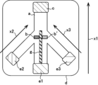
また別の態様では、本発明は、前記粒状物質をイムノクロマトグラフィー法によって検出する方法に使用するのに適した形状を有するイムノクロマトグラフィー用テストストリップ(イムノクロマト試験紙)、すなわち、互いに同じであっても異なってもよい第一の結合対象物質及び第二の結合対象物質を含む結合対象物質を表面上に複数備える粒状物質を検出するためのイムノクロマトグラフィー用テストストリップにも関している。具体的には、本発明のイムノクロマトグラフィー用テストストリップは、
前記第一の結合対象物質に対する第一の特異的結合物質が固定された検出部位(b)を備えるメンブレン(a)と、
試料流動方向(x)について前記検出部位(b)の上流側の位置で前記メンブレン(a)と接する第一のサンプルパッド(e1)と
を含み、
前記第一のサンプルパッド(e1)から前記検出部位(b)に至る途中にコンジュゲートパッドが含まれていないことを特徴としており、各構成要素は、基板(バッキングシート)(d)の上に配置され得る(図1)。前記テストストリップは、前記試料流動方向(x)について前記検出部位(b)の下流側の位置で吸水パッド(c)をさらに含んでいてもよい。また、前記テストストリップは、前記試料流動方向(x)について前記第一のサンプルパッド(e1)のさらに上流側の位置で前記メンブレン(a)と接する第二のサンプルパッド(e2)をさらに含んでいてもよく、ここで、前記第二のサンプルパッド(e2)は、前記第一のサンプルパッド(e1)から離間している(図2)。
In another aspect, the present invention provides an immunochromatographic test strip (immunochromatographic test paper) having a shape suitable for use in a method for detecting the particulate matter by an immunochromatographic method. It also relates to an immunochromatographic test strip for detecting a particulate material having a plurality of binding entities on its surface, including a first binding entity and a second binding entity, which may be different. Specifically, the immunochromatographic test strip of the present invention is
a membrane (a) comprising a detection site (b) to which a first specific binding substance for the first binding target substance is immobilized;
a first sample pad (e1) in contact with the membrane (a) at a position upstream of the detection site (b) with respect to the sample flow direction (x);
It is characterized in that no conjugate pad is included on the way from the first sample pad (e1) to the detection site (b), and each component is provided on a substrate (backing sheet) (d) can be placed (Fig. 1). Said test strip may further comprise an absorbent pad (c) at a position downstream of said detection site (b) with respect to said sample flow direction (x). In addition, the test strip further includes a second sample pad (e2) in contact with the membrane (a) at a position further upstream of the first sample pad (e1) with respect to the sample flow direction (x). wherein said second sample pad (e2) is spaced from said first sample pad (e1) (Fig. 2).
本明細書に記載の「サンプルパッド」とは、イムノクロマトグラフィー用の試料、検出試薬、又は、水及び緩衝液などの溶媒を受け入れてメンブレン(a)上での展開(流動)を開始させる部位のことをいう。前記サンプルパッドは、当技術分野で通常使用される任意の方法によって作製することができる。本発明のイムノクロマトグラフィー用テストストリップが、サンプルパッドを1つだけ含む場合、すなわち第一のサンプルパッド(e1)のみを含む場合には、この第一のサンプルパッド(e1)に前記粒状物質を含む試料を載せて、前記粒状物質が前記検出部位(b)において前記第一の特異的結合物質と反応した後に、同じ第一のサンプルパッド(e1)に前記第二の結合対象物質に対する第二の特異的結合物質(標識物質に結合)を含む検出試薬を載せて、前記検出部位(b)に捕捉されている前記粒状物質を標識する。本発明のイムノクロマトグラフィー用テストストリップが、第一のサンプルパッド(e1)及び第二のサンプルパッド(e2)を含む場合には、前記第一のサンプルパッド(e1)を前記粒状物質を含む試料を載せる部位として使用し、前記第二のサンプルパッド(e2)を前記第二の特異的結合物質を含む検出試薬を載せる部位として使用してもよい。 The term "sample pad" as used herein refers to a site that accepts a sample for immunochromatography, a detection reagent, or a solvent such as water and buffer to initiate development (flow) on the membrane (a). Say things. The sample pad can be made by any method commonly used in the art. When the immunochromatographic test strip of the present invention contains only one sample pad, that is, when it contains only the first sample pad (e1), the first sample pad (e1) contains the particulate material. After the sample is placed and the particulate substance reacts with the first specific binding substance at the detection site (b), the same first sample pad (e1) is subjected to a second binding substance for the second binding target substance. A detection reagent containing a specific binding substance (bound to a labeling substance) is applied to label the particulate matter captured at the detection site (b). When the immunochromatographic test strip of the present invention includes a first sample pad (e1) and a second sample pad (e2), the first sample pad (e1) is a sample containing the particulate matter. The second sample pad (e2) may be used as a site for loading the detection reagent containing the second specific binding substance.
ある態様では、前記第二のサンプルパッド(e2)は、前記第二の特異的結合物質を含むコンジュゲートパッド(f)を含んでいる(図3)。本明細書に記載の「コンジュゲートパッド」とは、前記第二の特異的結合物質が含侵している部位であり、ここへ水や緩衝液などの溶媒を添加すると、前記第二の特異的結合物質が溶出し、前記メンブレン(a)上での展開(流動)を開始するように構成されている。前記コンジュゲートパッド(f)は、当技術分野で通常使用される任意の方法によって作製することができる。前記コンジュゲートパッド(f)は、前記第二のサンプルパッド(e2)に少なくとも一部を覆われた態様又は前記第二のサンプルパッド(e2)と一体になった態様であってもいいし、併設された別部材のサンプルパッドと一緒になって第二のサンプルパッド(e2)として機能する領域を形成してもよい。 In one embodiment, said second sample pad (e2) comprises a conjugate pad (f) comprising said second specific binding substance (Fig. 3). As used herein, the term “conjugate pad” refers to a site impregnated with the second specific binding substance, and when a solvent such as water or a buffer is added to the site, the second specific binding substance It is configured such that the bound substances elute and begin to develop (flow) on said membrane (a). The conjugate pad (f) can be produced by any method commonly used in the art. The conjugate pad (f) may be at least partially covered with the second sample pad (e2) or integrated with the second sample pad (e2), A region functioning as a second sample pad (e2) may be formed together with a separate sample pad that is placed side by side.
ある態様では、前記第一のサンプルパッド(e1)及び前記第二のサンプルパッド(e2)は、第一のスペーサー(g1)を介して互いから離間している(図4及び図5)。また、ある態様では、前記メンブレンは、前記第一のサンプルパッド側から前記第二のサンプルパッド側への前記粒状物質を含む試料の浸透を阻害するように構成されている。そのような浸透阻害手段としては、当技術分野で通常使用される任意の手段を特に制限されることなく採用することができるが、例えば、前記メンブレン(a)中に水溶性高分子を含侵させた阻害領域(h)を設け、前記試料流動方向(x)とは逆向きへの流動速度を低下させるように構成してもよい(図6及び7)。 In one embodiment, said first sample pad (e1) and said second sample pad (e2) are separated from each other via a first spacer (g1) (Figs. 4 and 5). In one aspect, the membrane is configured to inhibit permeation of the sample containing the particulate matter from the first sample pad side to the second sample pad side. Any means commonly used in the art can be employed as such means for inhibiting permeation without particular limitation. It is also possible to provide an inhibited region (h) with a slanting direction to reduce the flow velocity in the direction opposite to the sample flow direction (x) (FIGS. 6 and 7).
ある態様では、前記粒状物質は、前記第一の結合対象物質又は前記第二の結合対象物質と同じであっても異なってもよい第三の結合対象物質をさらに備え、
前記メンブレン(a)は、前記第三の結合対象物質に対する第三の特異的結合物質が固定された追加の検出部位(b’)と、前記試料流動方向について前記第一のサンプルパッドの下流側の位置で前記試料が流動する経路を分割するように配置された第二のスペーサー(g2)と、をさらに備え、
前記検出部位(b)及び前記追加の検出部位(b’)は、前記第二のスペーサー(g2)によって分割された異なる経路に配置されている(図8)。前記第一の結合対象物質と前記第三の結合対象物質が異なる場合、前記検出部位(b)に捕捉された粒状物質の量と前記追加の検出部位(b’)に捕捉された粒状物質の量を比較することにより、前記試料中の粒状物質上の前記第一の結合対象物質と前記第三の結合対象物質の相対的な量を測定することができる。なお、前記検出部位(b)に捕捉された粒状物質と前記追加の検出部位(b’)に捕捉された粒状物質を異なる検出試薬で検出することも可能であり、その場合には、前記検出部位(b)の真上又は前記検出部位(b)と前記第一のサンプルパッド(e1)の間の第一の滴下領域(i1)、及び、前記追加の検出部位(b’)の真上又は前記追加の検出部位(b’)と前記第一のサンプルパッド(e1)の間の第二の滴下領域(i2)に、それぞれ異なる検出試薬を滴下することができる。
In one aspect, the particulate substance further comprises a third binding target substance that may be the same as or different from the first binding target substance or the second binding target substance,
The membrane (a) comprises an additional detection site (b') to which a third specific binding substance for the third binding target substance is immobilized, and a downstream side of the first sample pad with respect to the sample flow direction. a second spacer (g2) arranged to divide the path through which the sample flows at the position of
Said detection site (b) and said additional detection site (b') are arranged in different pathways separated by said second spacer (g2) (Fig. 8). When the first binding target substance and the third binding target substance are different, the amount of particulate matter captured at the detection site (b) and the amount of particulate matter captured at the additional detection site (b′) By comparing the amounts, the relative amounts of the first binding substance and the third binding substance on particulate matter in the sample can be determined. It is also possible to detect the particulate matter captured at the detection site (b) and the particulate matter captured at the additional detection site (b′) with different detection reagents. A first drop area (i1) directly above site (b) or between said detection site (b) and said first sample pad (e1) and directly above said additional detection site (b') Alternatively, different detection reagents can be dropped on the second dropping area (i2) between the additional detection site (b') and the first sample pad (e1).
別の具体例では、本発明のイムノクロマトグラフィー用テストストリップは、
前記第一の結合対象物質に対する第一の特異的結合物質が固定された検出部位(b)を備えるメンブレン(a)と、試料流動方向(x)について前記検出部位(b)の上流側の位置で前記メンブレン(a)と接する第一のサンプルパッド(e1)とを含む第一の基板(d1)と、
第二のサンプルパッド(e2)を含む第二の基板(d2)と
を含み、
前記第一の基板(d1)及び前記第二の基板(d2)は、前記第二のサンプルパッド(e2)が前記試料流動方向(x)について前記第一のサンプルパッド(e1)のさらに上流側の位置で前記メンブレン(a)と接するように、接近可能であることを特徴としている(図9)。前記第一のサンプルパッド(e1)から前記粒状物質を含む試料を流動させた後に、前記第二のサンプルパッド(e2)を前記メンブレン(a)と接触させて、当該第二のサンプルパッド(e2)から前記第二の結合対象物質に対する第二の特異的結合物質(標識物質に結合)を含む検出試薬を流動させることで、前記検出部位(b)に捕捉されている前記粒状物質を標識することができる。
In another embodiment, the immunochromatographic test strip of the present invention comprises
A membrane (a) comprising a detection site (b) on which a first specific binding substance for said first binding target substance is immobilized, and a position on the upstream side of said detection site (b) with respect to the sample flow direction (x) a first substrate (d1) comprising a first sample pad (e1) in contact with said membrane (a) at
a second substrate (d2) comprising a second sample pad (e2);
The first substrate (d1) and the second substrate (d2) are arranged such that the second sample pad (e2) is further upstream of the first sample pad (e1) in the sample flow direction (x). (Fig. 9). After flowing the sample containing the particulate matter from the first sample pad (e1), the second sample pad (e2) is brought into contact with the membrane (a), and the second sample pad (e2 ) to label the particulate substance captured in the detection site (b) by flowing a detection reagent containing a second specific binding substance (bound to the labeling substance) for the second binding target substance from be able to.
ある態様では、前記第二のサンプルパッド(e2)は、前記第二の特異的結合物質を含むコンジュゲートパッド(f)を含んでいる(図10)。また、ある態様では、前記第一の基板(d1)及び前記第二の基板(d2)は、伸縮構造(j)を介して結合されている(図11)。この場合には、前記伸縮構造(j)が伸長した状態(図11A)で前記第一のサンプルパッド(e1)から前記粒状物質を含む試料を流動させた後に、前記伸縮構造(j)を収縮させて前記コンジュゲートパッド(f)を含む前記第二のサンプルパッド(e2)を前記メンブレン(a)と接触させ(図11B)、そこで前記第二のサンプルパッド(e2)に水や緩衝液などの溶媒を添加すると、前記第二の特異的結合物質が溶出し、前記メンブレン(a)上での流動を開始する。 In one embodiment, said second sample pad (e2) comprises a conjugate pad (f) comprising said second specific binding substance (Figure 10). Also, in one aspect, said first substrate (d1) and said second substrate (d2) are coupled via a telescoping structure (j) (FIG. 11). In this case, after the sample containing the particulate matter is caused to flow from the first sample pad (e1) while the elastic structure (j) is stretched (FIG. 11A), the elastic structure (j) is contracted. and the second sample pad (e2) containing the conjugate pad (f) is brought into contact with the membrane (a) (FIG. 11B), whereupon the second sample pad (e2) is loaded with water, buffer solution or the like. Upon addition of solvent, the second specific binding substance elutes and begins to flow over the membrane (a).
別の具体例では、本発明のイムノクロマトグラフィー用テストストリップは、
前記第一の結合対象物質に対する第一の特異的結合物質が固定された検出部位(b)を備えるメンブレン(a)と、
第一の経路における試料流動方向(x1)について前記検出部位(b)の上流側の位置で前記メンブレン(a)と接する第一のサンプルパッド(e1)と、
前記第一の経路とは異なる第二の経路における試料流動方向(x2)について前記検出部位(b)の上流側の位置で前記メンブレンと接する第二のサンプルパッド(e2)と
を含み、
前記第二のサンプルパッド(e2)が、前記第一のサンプルパッド(e1)から離間していることを特徴としており(図12及び図13)、前記第一の経路及び前記第二の経路は、前記検出部位(b)の周辺を除き、必要な場合にはスペーサー(g)を介して互いから離間している。この具体例のテストストリップにおいては、前記粒状物質を含む試料と、前記第二の結合対象物質に対する第二の特異的結合物質(標識物質に結合)を含む検出試薬とを、別のサンプルパッドから別の経路で流動させることができるため、前記試料の流動が完全に終わる前に前記検出試薬の流動を開始しても、前記検出部位(b)の周辺以外では前記粒状物質と前記第二の特異的物質とが結合することはない。そうすると、前記検出試薬の流動を早くに開始できる分、イムノクロマトグラフィー法の試験に要する時間を短縮することが可能となる。
In another embodiment, the immunochromatographic test strip of the present invention comprises
a membrane (a) comprising a detection site (b) to which a first specific binding substance for the first binding target substance is immobilized;
a first sample pad (e1) in contact with the membrane (a) at a position on the upstream side of the detection site (b) with respect to the sample flow direction (x1) in the first path;
a second sample pad (e2) in contact with the membrane at a position on the upstream side of the detection site (b) with respect to the sample flow direction (x2) in a second path different from the first path,
characterized in that the second sample pad (e2) is spaced apart from the first sample pad (e1) (FIGS. 12 and 13), the first path and the second path are , are separated from each other via a spacer (g) if necessary, except around said detection site (b). In the test strip of this specific example, the sample containing the particulate matter and the detection reagent containing the second specific binding substance (bound to the labeled substance) for the second binding target substance are combined from separate sample pads. Even if the flow of the detection reagent is started before the flow of the sample is completely finished, the particulate matter and the second particle can be separated from each other except in the vicinity of the detection site (b). It does not bind to specific substances. As a result, the flow of the detection reagent can be started early, and the time required for the immunochromatographic test can be shortened.
ある態様では、前記第二のサンプルパッド(e2)は、前記第二の特異的結合物質を含むコンジュゲートパッド(f)を含んでいる(図14及び図15)。また、ある態様では、前記メンブレン(a)は、短冊形、U字型、又は、V字型である。 In one embodiment, said second sample pad (e2) comprises a conjugate pad (f) comprising said second specific binding substance (Figures 14 and 15). In one aspect, the membrane (a) is rectangular, U-shaped, or V-shaped.
ある態様では、前記粒状物質は、前記第一の結合対象物質又は前記第二の結合対象物質と同じであっても異なってもよい第三の結合対象物質をさらに備え、
前記メンブレン(a)は、前記第三の結合対象物質に対する第三の特異的結合物質が固定された追加の検出部位(b’)と、前記第一の経路における試料流動方向について前記第一のサンプルパッド(e1)の下流側の位置で前記第一の経路を分割するように配置されたスペーサー(g)と、をさらに備え、
前記検出部位(b)及び前記追加の検出部位(b’)は、前記スペーサー(g)で分割された異なる経路に配置されている(図16)。前記第一のサンプルパッド(e1)から流動されて前記検出部位(b)で捕捉された前記粒状物質は、前記第二のサンプルパッド(e2)から流動される前記第二の特異的結合物質を含む検出試薬により標識され得る。そして、前記追加の検出部位(b’)に捕捉された前記粒状物質は、その真上、又は、前記追加の検出部位(b’)と前記第一のサンプルパッド(e1)の間の滴下領域(i)に、前記検出試薬を滴下することで標識され得る。前記第一の結合対象物質と前記第三の結合対象物質が異なる場合、前記検出部位(b)に捕捉された粒状物質の量と前記追加の検出部位(b’)に捕捉された粒状物質の量を比較することにより、前記試料中の粒状物質上の前記第一の結合対象物質と前記第三の結合対象物質の相対的な量を測定することができる。
In one aspect, the particulate substance further comprises a third binding target substance that may be the same as or different from the first binding target substance or the second binding target substance,
The membrane (a) comprises an additional detection site (b') on which a third specific binding substance for the third binding target substance is immobilized, and the first a spacer (g) arranged to divide the first path at a position downstream of the sample pad (e1);
Said detection site (b) and said additional detection site (b') are arranged in different pathways separated by said spacer (g) (Fig. 16). The particulate material flowed from the first sample pad (e1) and captured at the detection site (b) binds the second specific binding substance flowed from the second sample pad (e2). It can be labeled with a detection reagent containing. Then, the particulate matter captured by the additional detection site (b') is directly above it or in a dropping area between the additional detection site (b') and the first sample pad (e1). (i) can be labeled by dropping said detection reagent. When the first binding target substance and the third binding target substance are different, the amount of particulate matter captured at the detection site (b) and the amount of particulate matter captured at the additional detection site (b′) By comparing the amounts, the relative amounts of the first binding substance and the third binding substance on particulate matter in the sample can be determined.
ある態様では、前記テストストリップは、前記第一の経路及び前記第二の経路とは異なる第三の経路における試料流動方向(x3)について前記追加の検出部位(b’)の上流側の位置で前記メンブレン(a)と接する第三のサンプルパッド(e3)をさらに含んでいる(図17)。また、ある態様では、前記第二のサンプルパッド(e2)及び/又は前記第三のサンプルパッド(e3)は、前記第二の特異的結合物質を含むコンジュゲートパッド(f1及びf2)を含んでいる(図18)。 In one aspect, the test strip is positioned upstream of the additional detection site (b') with respect to the sample flow direction (x3) in a third path different from the first path and the second path. It further comprises a third sample pad (e3) in contact with said membrane (a) (Fig. 17). Also, in one embodiment, said second sample pad (e2) and/or said third sample pad (e3) comprise conjugate pads (f1 and f2) comprising said second specific binding substance. (Fig. 18).
別の具体例では、本発明のイムノクロマトグラフィー用テストストリップは、
前記第一の結合対象物質に対する第一の特異的結合物質が固定された検出部位(b)を備える第一のメンブレン(a1)と、
第一の経路における試料流動方向(x1)について前記検出部位の上流側の位置で前記第一のメンブレン(a1)と接する第一のサンプルパッド(e1)と、
前記検出部位(b)と前記第一のサンプルパッド(e1)の間で前記第一のメンブレン(a1)と接している第二のメンブレン(a2)と、
前記第一の経路とは異なる第二の経路における試料流動方向(x2)について前記検出部位(b)の上流側の位置で前記第二のメンブレン(a2)と接する第二のサンプルパッド(e2)と
を含み、前記第二のサンプルパッド(e2)が、前記第一のサンプルパッド(e1)から離間していることを特徴としている(図19)。この具体例のテストストリップにおいては、前記第一のサンプルパッド(e1)又は前記第二のサンプルパッド(e2)のいずれか一方に前記粒状物質を含む試料を載せて、他方に前記第二の結合対象物質に対する第二の特異的結合物質(標識物質に結合)を含む検出試薬を載せて使用することができる。前記第二のサンプルパッド(e2)から流動を開始した試料又は検出試薬は、流動しながら前記第二のメンブレン(a2)と接している前記第一のメンブレン(a1)へと移行し、前記検出部位(b)に到達する。前記第二のメンブレン(a2)は、その末端領域で前記第一のメンブレン(a1)と接していてもよい。ある態様では、前記第一のサンプルパッド(e1)又は前記第二のサンプルパッド(e2)のいずれか一方が、前記第二の特異的結合物質を含むコンジュゲートパッド(f)を含んでいる(図20)。
In another embodiment, the immunochromatographic test strip of the present invention comprises
a first membrane (a1) comprising a detection site (b) to which a first specific binding substance for the first binding target substance is immobilized;
a first sample pad (e1) in contact with the first membrane (a1) at a position on the upstream side of the detection site with respect to the sample flow direction (x1) in the first path;
a second membrane (a2) in contact with the first membrane (a1) between the detection site (b) and the first sample pad (e1);
A second sample pad (e2) in contact with the second membrane (a2) at a position on the upstream side of the detection site (b) with respect to the sample flow direction (x2) in the second path different from the first path and wherein the second sample pad (e2) is spaced apart from the first sample pad (e1) (FIG. 19). In the test strip of this embodiment, either the first sample pad (e1) or the second sample pad (e2) is loaded with the sample containing the particulate material, and the other is loaded with the second bond. A detection reagent comprising a second specific binding substance for the substance of interest (bound to the labeled substance) can be applied. The sample or detection reagent that started to flow from the second sample pad (e2) moves to the first membrane (a1) in contact with the second membrane (a2) while flowing, and the detection It reaches site (b). Said second membrane (a2) may be in contact with said first membrane (a1) at its terminal region. In one embodiment, either said first sample pad (e1) or said second sample pad (e2) comprises a conjugate pad (f) comprising said second specific binding substance ( Figure 20).
本発明のイムノクロマトグラフィー用テストストリップは、当技術分野で通常使用される任意の構成を採用することができる。例えば、前記テストストリップにおいて、前記メンブレンは、イムノクロマトグラフィー試験の成否を判定するコントロール部位をさらに備えていてもよい。前記コントロール部位は、特に限定されないが、例えば、前記検出試薬に含まれている前記第二の特異的結合物質(標識物質に結合)を捕捉することのできる抗体などの物質を固定したコントロールライン(k)であってもよく(図21)、当該コントロールラインは、使用する検出試薬の種類に応じて複数設けてもよい。 The immunochromatographic test strip of the present invention can adopt any configuration commonly used in this technical field. For example, in the test strip, the membrane may further include a control portion for determining the success or failure of an immunochromatography test. The control site is not particularly limited, but for example, a control line ( k) (FIG. 21), and a plurality of control lines may be provided according to the type of detection reagent used.
以下、実施例により本発明を具体的に説明するが、本発明の範囲はこれら実施例に限定されるものではない。 EXAMPLES The present invention will be specifically described below with reference to Examples, but the scope of the present invention is not limited to these Examples.
1.エクソソーム溶液の調製方法
(1)細胞培養上清からの調製
10% FBS(名称:Fetal Bovine Serum、製造元:Life Technologies)、1% PSA(名称:ペニシリン-ストレプトマイシン-アムホテリシンB懸濁液(×100)(抗生物質-抗真菌剤溶液)、製造元:和光純薬工業株式会社)、及び2mM Glutamax(製造元:Life Technologies)含有RPMI1640培地を20mL使用し、150mmディッシュで乳がん細胞株MCF7をディッシュの底面積の80%まで培養した。培地を除去後、りん酸緩衝生理食塩水(PBS)20mLで2回洗浄し、2mM Glutamax含有Advanced RPMI 1640 Medium(製造元:Life Technologies)を20mL添加して、48時間培養した。150mmディッシュ10枚分の細胞培養上清(200mL)を2,000×g、4℃下で10分間遠心分離し、その上清を10,000×g、4℃下で30分間遠心分離して、最終的に得られた上清を孔径0.22μmのフィルターでろ過した。ろ液を175,000×g、4℃下で95分間遠心分離し、上清を除去して沈殿画分を得た。沈殿画分を13mLの1×PBSで分散し、210,000×g、4℃下で95分間遠心分離した。その上清を除去した後、沈殿物を0.2mLのPBSで再分散し、エクソソーム溶液(E溶液1)を調製した。
また、MCF7細胞に代えて、他の乳がん細胞株であるMDA-MB-231(略称:MM231)を使用し、上と同様の方法でエクソソーム溶液(E溶液2)を調製した。
1. Exosome solution preparation method (1) Preparation from cell culture supernatant 10% FBS (name: Fetal Bovine Serum, manufacturer: Life Technologies), 1% PSA (name: penicillin-streptomycin-amphotericin B suspension (x 100) (antibiotic-antimycotic agent solution), manufacturer: Wako Pure Chemical Industries, Ltd.) and 20 mL of RPMI1640 medium containing 2 mM Glutamax (manufacturer: Life Technologies), breast cancer cell line MCF7 was added to the bottom area of the dish in a 150 mm dish. Cultured to 80%. After removing the medium, the cells were washed twice with 20 mL of phosphate buffered saline (PBS), 20 mL of Advanced RPMI 1640 Medium containing 2 mM Glutamax (manufacturer: Life Technologies) was added, and cultured for 48 hours. Ten 150 mm dishes of cell culture supernatant (200 mL) was centrifuged at 2,000 x g for 10 minutes at 4°C, and the supernatant was centrifuged at 10,000 x g for 30 minutes at 4°C. , the finally obtained supernatant was filtered through a filter with a pore size of 0.22 μm. The filtrate was centrifuged at 175,000×g at 4° C. for 95 minutes, and the supernatant was removed to obtain a precipitate fraction. The precipitated fraction was dispersed with 13 mL of 1×PBS and centrifuged at 210,000×g at 4° C. for 95 minutes. After removing the supernatant, the precipitate was redispersed with 0.2 mL of PBS to prepare an exosome solution (E solution 1).
Further, instead of MCF7 cells, another breast cancer cell line MDA-MB-231 (abbreviation: MM231) was used to prepare an exosome solution (E solution 2) in the same manner as above.
(2)血清からの調製
4mLの血清(品番:12181201、販売元:コスモ・バイオ株式会社)を16,500×g、4℃下で20分間遠心分離し、その上清を孔径0.22μmのフィルターでろ過した。ろ液を210,000×g、4℃下で45分間遠心分離し、上清を除去して沈殿画分を得た。沈殿画分を4mLのPBSで分散し、再び210,000×g、4℃下で45分間遠心分離した。その上清を除去した後、沈殿物を0.1mLのPBSで再分散し、エクソソーム溶液(E溶液3)を調製した。
(2) Preparation from
2.細胞由来小胞溶液の評価
(1)粒径および粒子数濃度測定
各種エクソソーム溶液を希釈し、NanoSightナノ粒子解析システム(Malvern Panalytical製)によりエクソソームの粒径および粒子数濃度を測定した。結果を以下に示す。
[E溶液1]粒径:147nm、粒子数濃度:1.5×10¹²個/mL
[E溶液2]粒径:143nm、粒子数濃度:1.9×10¹²個/mL
[E溶液3]粒径:139nm、粒子数濃度:4.9×10¹⁰個/mL
2. Evaluation of cell-derived vesicle solution (1) particle size and particle number concentration measurement Various exosome solutions were diluted, and the particle size and particle number concentration of exosomes were measured by NanoSight nanoparticle analysis system (manufactured by Malvern Panalytical). The results are shown below.
[E solution 1] Particle size: 147 nm, particle number concentration: 1.5 × 10 12 /mL
[Solution E 2] Particle size: 143 nm, particle number concentration: 1.9×10 12 /mL
[Solution E 3] Particle size: 139 nm, particle number concentration: 4.9 × 10 10 /mL
(2)ウェスタンブロッティング
エクソソームを2.5×10⁹個含むように調製したE溶液1及び2、並びに、エクソソームを1.3×10⁹個含むように調製したE溶液3を、試料緩衝液(SDS-PAGE用、6倍濃縮、還元剤不含)(品番:09500-64、製造元:ナカライテスク製)を使用して、SDS-ポリアクリルアミドゲル電気泳動に供した。泳動後のSDSポリアクリルアミドゲルをPVDF膜(メルク製)に転写した。転写後のPVDF膜を2%のスキムミルクを含有したTBS-T(20mM Tris-HCl(pH 8.0)、150mM NaCl、0.05% Tween(R)20)中に浸してブロッキングした。ブロッキング終了後、PVDF膜を、0.2%スキムミルク含有TBS-Tで希釈したマウス抗CD9抗体(Hansa Bio Med製)、マウス抗CD63抗体(Hansa Bio Med製)、又はマウス抗CD81抗体(Hansa Bio Med製)と反応させた。そして、PVDF膜をTBS-Tで3回洗浄後、0.2%スキムミルク含有TBS-Tで希釈したHRP標識ヤギ抗マウスIgG抗体(Bio-Rad Laboratories製)と反応させた。最後に、このPVDF膜をTBS-Tで3回洗浄後、イムノスターLD(和光純薬工業製製)に浸し、直後にImageQuant LAS4000(GEヘルスケア製)で化学発光を検出して、得られた画像を目視又は輝度解析により評価した。結果を図22及び表1に示す。
(目視の評価基準)
++:明確にバンドの判別が可能
+ :バンドの判別が可能
- :バンドの判別が不可能
(2) Western
(Visual evaluation criteria)
++: Bands can be clearly distinguished +: Bands can be distinguished -: Bands cannot be distinguished

CD9、CD63、及びCD81は、いずれもエクソソームの表面に存在していることが知られているが、その量は由来の細胞によって異なる。今回のウェスタンブロッティングでは、E溶液1中のエクソソームではCD63の存在量が少ないことが示唆されたため、イムノクロマト試験において抗CD63抗体を使用すると、検出感度が低くなることが予想される。
CD9, CD63, and CD81 are all known to be present on the surface of exosomes, but their amounts vary depending on the cell of origin. Western blotting this time suggests that the exosomes in
3.金属ナノ粒子の調製
(1)金ナノプレート懸濁液
常法により、金ナノプレート(最大長さ45nm、厚さ23nmの青色板状ナノ粒子、略称:AuPL)を含む懸濁液A(最大吸収波長616nm;青色)を調製した(要すれば特許文献4などを参照)。
3. Preparation of metal nanoparticles (1) Gold nanoplate suspension Suspension A (maximum absorption wavelength 616 nm; blue) was prepared (see
(2)球状金コロイド懸濁液
市販の球状金コロイド粒子(BBI Solutions社製、粒子径40nm、略称:AuSP)を含む懸濁液B(最大吸収波長524nm;赤色)を、以降の実験で使用した。
(2) Spherical gold colloid suspension Suspension B (maximum absorption wavelength 524 nm; red) containing commercially available spherical gold colloid particles (BBI Solutions,
4.抗体担持金属ナノ粒子及び展開液の調製
最大吸収波長における吸光度が2.0になるように濃度を調整した1mLの懸濁液A又はBに、0.05mg/mLのマウス抗CD9抗体(品番:HBM-CD9-100、製造元:Hansa Bio Med)溶液を0.1mL添加して、1時間静置した。そして、0.5%のポリエチレングリコール(分子量20,000)水溶液を0.05mL、2%のBSA水溶液を0.1mL添加して、金属ナノ粒子表面をブロッキングした。その後、遠心分離機で金属ナノ粒子を沈殿させ、上清を除去し、150mMの塩化ナトリウム及び1%のBSAを含有する10mMのHEPES緩衝液1mLで再分散した。遠心分離操作を再度実施し、上清を除去後、金属ナノ粒子を同じ緩衝液で再度分散して、最大吸収波長における吸光度が1になるように濃度を調整した。これをイムノクロマト試験に使用する展開液A9及びB9とした。
4. Preparation of antibody-supported metal nanoparticles and developing solution 0.05 mg / mL mouse anti-CD9 antibody (product number: 0.1 mL of HBM-CD9-100 (manufacturer: Hansa Bio Med) solution was added and allowed to stand for 1 hour. Then, 0.05 mL of 0.5% polyethylene glycol (molecular weight 20,000) aqueous solution and 0.1 mL of 2% BSA aqueous solution were added to block the metal nanoparticle surfaces. After that, the metal nanoparticles were precipitated with a centrifuge, the supernatant was removed, and the particles were redispersed in 1 mL of 10 mM HEPES buffer containing 150 mM sodium chloride and 1% BSA. After centrifugation was performed again and the supernatant was removed, the metal nanoparticles were dispersed again in the same buffer to adjust the concentration so that the absorbance at the maximum absorption wavelength was 1. These were used as developing solutions A9 and B9 used in immunochromatography tests.
また、懸濁液Aについては、マウス抗CD9抗体に代えてマウス抗CD63抗体(品番:HBM-CD63-100、製造元:Hansa Bio Med)又はマウス抗CD81抗体(品番:HBM-CD81-100、製造元:Hansa Bio Med)を使用した以外は上述の方法と同様にして、抗体担持金属ナノ粒子を含む展開液A63及びA81を調製した。調製した展開液に含まれる金属ナノ粒子及び検出抗体の種類を表2に示す。 Further, for suspension A, instead of mouse anti-CD9 antibody, mouse anti-CD63 antibody (product number: HBM-CD63-100, manufacturer: Hansa Bio Med) or mouse anti-CD81 antibody (product number: HBM-CD81-100, manufacturer Developing solutions A63 and A81 containing antibody-supporting metal nanoparticles were prepared in the same manner as described above, except that Hansa Bio Med) was used. Table 2 shows the types of metal nanoparticles and detection antibodies contained in the prepared developing solution.

5.イムノクロマト試験
(1)イムノクロマト試験紙への捕捉抗体の固定
マウス抗CD9抗体(品番:HBM-CD9-100、製造元:Hansa Bio Med)を、濃度が0.25g/mLになるように10%スクロース含有PBSで希釈し、その希釈液の0.75μLを、吸水パッドが一方の端に取付けられている短冊形のニトロセルロースメンブレンを備えたイムノクロマト試験紙(株式会社フォーディクス製)の中央部へ滴下して、当該抗体を捕捉抗体としてメンブレン上に固定した。そして、3%のBSA含有PBSを10mL展開し、メンブレン全体をブロッキングした。
5. Immunochromatographic test (1) Immobilization of capture antibody on immunochromatographic test paper Mouse anti-CD9 antibody (product number: HBM-CD9-100, manufacturer: Hansa Bio Med) containing 10% sucrose at a concentration of 0.25 g/mL Diluted with PBS, 0.75 μL of the diluted solution was dropped onto the center of an immunochromatographic test paper (manufactured by Fordix Co., Ltd.) equipped with a strip-shaped nitrocellulose membrane with a water-absorbing pad attached to one end. The antibody was then immobilized on the membrane as a capture antibody. Then, 10 mL of 3% BSA-containing PBS was developed to block the entire membrane.
(2)本発明の方法
捕捉抗体を固定したイムノクロマト試験紙の吸水パッドが取り付けられていない方の端を、エクソソームを含む試験液に浸して、当該試験液を展開した。次に、上記表2に記載の展開液のいずれかを展開した。最後に、捕捉抗体を固定していた部分の発色を、後述する方法のいずれかにより評価した。
(2) Method of the present invention The end of the immunochromatography test paper immobilized with the capture antibody, to which the water absorption pad is not attached, is immersed in a test solution containing exosomes to develop the test solution. Next, one of the developing solutions described in Table 2 above was developed. Finally, the color development of the portion where the capture antibody had been immobilized was evaluated by one of the methods described below.
(3)従来の方法(比較法)
エクソソームを含む試験液を、上記表2に記載の展開液のいずれかと混合した。次に、その混合液を、捕捉抗体を固定したイムノクロマト試験紙に展開した。最後に、捕捉抗体を固定していた部分の発色を、後述する方法のいずれかにより評価した。
(3) Conventional method (comparative method)
A test solution containing exosomes was mixed with any of the developing solutions described in Table 2 above. Next, the mixed solution was developed on an immunochromatographic test paper on which a capture antibody was immobilized. Finally, the color development of the portion where the capture antibody had been immobilized was evaluated by one of the methods described below.
(4)界面活性剤の添加
エクソソーム溶液に界面活性剤を添加する場合には、エクソソーム溶液1μLに界面活性剤溶液を5μL添加し、全量が50μLになるように1%のウシ血清アルブミン(BSA)含有PBSを添加して、試験液を調製した。上記界面活性剤溶液としては、0.01%の(オクチルフェノキシ)ポリエトキシエタノール(名称:NonidetTM P-40,略称:NP40)水溶液又は0.1%のTween(R)20溶液を使用した。
(4) Addition of surfactant When adding a surfactant to the exosome solution, add 5 μL of surfactant solution to 1 μL of exosome solution, and 1% bovine serum albumin (BSA) so that the total amount is 50 μL. The PBS containing was added to prepare the test solution. As the surfactant solution, a 0.01% (octylphenoxy)polyethoxyethanol (name: Nonidet ™ P-40, abbreviation: NP40) aqueous solution or a 0.1
6.評価方法
(1)目視
イムノクロマト試験紙のメンブレン上の判定部分(マウス抗CD9抗体が固定化された部位)を目視で確認し、発色の程度を以下の基準で評価した。
++:明確に判別可能
+ :判別可能
+-:僅かに判別可能
- :判別不可能
6. Evaluation method (1) Visual observation The determination portion (the portion where the mouse anti-CD9 antibody was immobilized) on the membrane of the immunochromatographic test paper was visually confirmed, and the degree of color development was evaluated according to the following criteria.
++: Clearly identifiable +: Identifiable +-: Slightly identifiable -: Not identifiable
(2)輝度解析
検出抗体を含む展開液を展開した後のイムノクロマト試験紙をスキャナー(装置名:CanoScan LiDE500F、製造元:キヤノン株式会社)によりスキャニングし、判定部分の輝度と、判定部分以外の部分の最低輝度を、画像解析ソフト(Image-J)で測定することにより検出シグナルを数値化した。より具体的には、各部分をそれぞれ5回ずつ測定し、得られた数値の中央値の差を求めた。そして、エクソソームを含む試験液を展開したときの値から、エクソソームを含まない対照の溶液を展開したときの値を引いて、検出シグナルとしての輝度差を求めた。なお、Image-Jは、アメリカ国立衛生研究所でWayne Rasbandが開発したオープン・ソースで公有の画像処理ソフトウェアである(http://imagej.nih.gov/ij/)。
(2) Brightness analysis The immunochromatography test paper after developing the developing solution containing the detection antibody is scanned with a scanner (device name: CanoScan LiDE500F, manufacturer: Canon Inc.), and the brightness of the determination part and the brightness of the part other than the determination part The detected signal was quantified by measuring the minimum brightness with an image analysis software (Image-J). More specifically, each portion was measured five times, and the difference between the median values obtained was determined. Then, from the value when the test solution containing exosomes was developed, the value when the control solution containing no exosomes was developed was subtracted to obtain the luminance difference as a detection signal. Note that Image-J is an open source and publicly available image processing software developed by Wayne Rasband at the National Institutes of Health (http://imagej.nih.gov/ij/).
(3)イムノクロマトリーダーによる測定
検出抗体を含む展開液を展開した後のイムノクロマト試験紙をイムノクロマトリーダー(型番:C10066-10、製造元:浜松ホトニクス株式会社)で測定した。金ナノプレート(青色)を使用した際は青色系発色ライン測定モードで測定し、球状金コロイド(赤色)を使用した際は赤色系発色ライン測定モードで測定した。エクソソームを含む試験液を展開したときの値から、エクソソームを含まない対照の溶液を展開したときの値を引いて、検出シグナルとしての吸光度を求めた。
(3) Measurement by Immunochromatography Reader The immunochromatographic test paper after developing the developing solution containing the detection antibody was measured by an immunochromatography reader (model number: C10066-10, manufacturer: Hamamatsu Photonics K.K.). When gold nanoplates (blue) were used, measurement was performed in the blue color line measurement mode, and when spherical gold colloids (red) were used, the measurement was performed in the red color line measurement mode. The absorbance as a detection signal was obtained by subtracting the value when the control solution containing no exosomes was developed from the value when the test solution containing exosomes was developed.
7.イムノクロマト試験の展開方法の比較
エクソソームを含む試験液として血清(品番:12181201、販売元:コスモ・バイオ株式会社)を25μL使用し、検出抗体を含む展開液として展開液A9、A63、又はA81を60μL使用した。本発明の方法又は比較法によってイムノクロマト試験を行い、目視判定した結果を表3、輝度解析した結果を図23に示す。
7. Comparison of immunochromatographic

どの展開液(検出抗体)を使用した場合でも、本発明の方法に従ってイムノクロマト試験を行った場合には、比較法に対して検出感度が向上した。特に、展開液A63を使用した場合には、比較法では目視による検出が不可能であったにもかかわらず、本発明の方法では目視により発色を確認することができた。 Regardless of which developing solution (detection antibody) was used, the detection sensitivity was improved in the immunochromatography test according to the method of the present invention compared to the comparative method. In particular, when the developing solution A63 was used, color development could be visually confirmed in the method of the present invention, although visual detection was impossible in the comparative method.
8.各種試料におけるエクソソームの検出
(1)がん細胞株由来のエクソソーム
エクソソームを5.0×10⁹個含むE溶液1に、全量が50μLになるように1%のBSA含有PBSを添加した。これをイムノクロマト試験の試験液として使用し、検出抗体を含む展開液として展開液A9又はB9を60μL使用して、本発明の方法によりイムノクロマト試験を行った。輝度解析の結果を図24に示す。
8. Detection of exosomes in various samples (1) cancer cell line-derived
抗体を標識する金属ナノ粒子の種類にかかわらず、本発明の方法によって、良好なイムノクロマト試験結果を得ることができた。 Good immunochromatographic test results could be obtained by the method of the present invention regardless of the type of metal nanoparticles used to label the antibodies.
(2)全血中のエクソソーム
全血(販売元:株式会社ビジコムジャパン)をPBSで5倍に希釈した溶液をイムノクロマト試験の試験液として50μL使用し、検出抗体を含む展開液として展開液A9又はB9を60μL使用して、本発明の方法によりイムノクロマト試験を行った。輝度解析の結果を図25に示す。
(2) Exosomes in
エクソソームを単離又は精製していない試料を使用しても、本発明の方法によって、良好なイムノクロマト試験結果を得ることができた。また、抗体の標識として球状金ナノコロイドを使用するとわずかな輝度差しか測定できないような場合であっても、抗体の標識として異方性金ナノ粒子である金ナノプレートを使用すれば、検出感度をさらに高めることができた。 Good immunochromatographic test results could be obtained by the method of the present invention even when exosomes were not isolated or purified. In addition, even if the use of spherical gold nanocolloids as an antibody label can only measure a slight difference in brightness, the use of gold nanoplates, which are anisotropic gold nanoparticles, as an antibody label further enhances the detection sensitivity. I was able to raise it.
(3)培養上清中のエクソソーム
乳がん細胞株MCF7をディッシュ底面積の80%になるように培養し、その培地を無血清のRPMI1640培地に交換して、培養上清を経時的に採取した。採取した培養上清を試験液として50μL使用し、検出抗体を含む展開液として展開液A9を60μL使用して、本発明の方法によりイムノクロマト試験を行った。輝度解析した結果を図26に示す。
(3) Exosomes in the culture supernatant Breast cancer cell line MCF7 was cultured to 80% of the bottom area of the dish, the medium was replaced with serum-free RPMI1640 medium, and the culture supernatant was collected over time. Using 50 μL of the collected culture supernatant as the test solution and 60 μL of the developing solution A9 as the developing solution containing the detection antibody, an immunochromatographic test was performed according to the method of the present invention. FIG. 26 shows the result of luminance analysis.
本発明の方法に従ったイムノクロマト試験によれば、培養上清を直接使用した試験により、当該培養上清中のエクソソームが経時的に増加する様子を簡便に観察することができた。なお、このイムノクロマト試験の結果は、NanoSightナノ粒子解析システムによる計測結果とよく相関していた。 According to the immunochromatography test according to the method of the present invention, the test using the culture supernatant directly, it was possible to easily observe how exosomes in the culture supernatant increases over time. The results of this immunochromatographic test correlated well with the measurement results obtained by the NanoSight nanoparticle analysis system.
(4)NP40(界面活性剤)を使用して調製した試料中のエクソソーム
E溶液1(MCF7細胞由来エクソソーム溶液)及びE溶液2(MM231細胞由来エクソソーム溶液)にNP40溶液を添加して又は添加せずに50μLの試験液を調製した。検出抗体を含む展開液として展開液A9又はB9を60μL使用して、本発明の方法によりイムノクロマト試験を行った。輝度解析した結果を図27に示す。
(4) NP40 solution is added or added to exosome E solution 1 (MCF7 cell-derived exosome solution) and E solution 2 (MM231 cell-derived exosome solution) in the sample prepared using NP40 (surfactant) 50 μL of test solution was prepared without An immunochromatography test was performed according to the method of the present invention using 60 μL of developing solution A9 or B9 as the developing solution containing the detection antibody. FIG. 27 shows the result of luminance analysis.
NP40を試料中に添加することにより、いずれの細胞由来のエクソソームについても、検出感度を向上することができた。 By adding NP40 to the sample, it was possible to improve the detection sensitivity for exosomes derived from any cell.
(5)Tween(R)20(界面活性剤)を使用して調製した試料中のエクソソーム
E溶液1及びE溶液2にTween(R)20溶液を添加して又は添加せずに50μLの試験液を調製した。検出抗体を含む展開液として展開液A9を60μL使用して、本発明の方法によりイムノクロマト試験を行った。輝度解析した結果を図28に示す。
(5)
Tween(R)20を試料中に添加した場合でも、NP40を添加したときと同様に、エクソソームの検出感度を向上することができた。
Even when
9.イムノクロマト試験の定量性
NanoSightナノ粒子解析システムで計測した結果に基づき、E溶液1をエクソソームの数が9.0×10⁸個、1.8×10⁸個、又は0.9×10⁸個になるように1%のBSA含有PBSで希釈し、50μLの希釈液を調製した。この希釈液又はエクソソームを含まないBSA含有PBSを試験液として使用し、検出抗体を含む展開液として展開液A9を60μL使用して、本発明の方法によりイムノクロマト試験を行った。輝度解析した結果を図29、イムノクロマトリーダーで測定した結果を図30に示す。
9. Based on the results measured by the quantitative NanoSight nanoparticle analysis system of the immunochromatographic test , the number of exosomes in
輝度解析及びイムノクロマトリーダーによる測定のいずれにおいても、良好な検量線を引くことができるデータを取得することができた。したがって、本発明に従ったイムノクロマト試験を行えば、定量性のある試験を行うことができる。 In both the brightness analysis and the measurement by the immunochromatographic reader, we were able to obtain data that allowed us to draw a good calibration curve. Therefore, if the immunochromatography test according to the present invention is performed, a quantitative test can be performed.
10.ウェスタンブロッティングとの比較
NanoSightナノ粒子解析システムで計測した結果に基づき、E溶液1をエクソソームの数が50.0×10⁸個、5.0×10⁸個、又は0.5×10⁸個になるように1%のBSA含有PBSで希釈し、50μLの希釈液を調製した。この希釈液又はエクソソームを含まないBSA含有PBSを試験液として使用し、検出抗体を含む展開液として展開液A9を60μL使用して、本発明の方法によりイムノクロマト試験を行った。
10. Comparison with Western blotting Based on the results measured with the NanoSight nanoparticle analysis system, the number of exosomes in
また、イムノクロマト試験で使用したエクソソーム溶液と同じものをウェスタンブロッティングで解析した。一次抗体としてはマウス抗CD9抗体(Hansa Bio Med製)を使用し、二次抗体としてはHRP標識ヤギ抗マウスIgG抗体(Bio-Rad Laboratories製)を使用した。ウェスタンブロッティングの結果(写真)を図31に示し、イムノクロマト試験(IC)及びウェスタンブロッティング(WB)の目視判定の結果を表4に示す。 In addition, the same exosome solution used in the immunochromatographic test was analyzed by Western blotting. A mouse anti-CD9 antibody (manufactured by Hansa Bio Med) was used as the primary antibody, and an HRP-labeled goat anti-mouse IgG antibody (manufactured by Bio-Rad Laboratories) was used as the secondary antibody. The results (photographs) of Western blotting are shown in FIG. 31, and the visual evaluation results of immunochromatography (IC) and Western blotting (WB) are shown in Table 4.

ウェスタンブロッティングでは結果が出るまで約8時間かかり、操作も煩雑であったが、イムノクロマト試験では結果が出るまで約30分であり、操作も簡便であった。しかも、本発明の方法に従ったイムノクロマト試験の検出バンドは、ウェスタンブロッティングにより得られた検出バンドよりも目視で判別しやすいものだった。特に、0.5×10⁸個のエクソソームを使用した場合には、ウェスタンブロッティングでは目視による検出が不可能であったにもかかわらず、本発明の方法に従ったイムノクロマト試験では目視により発色を確認することができた。 Western blotting required about 8 hours to obtain results, and the operation was complicated, whereas immunochromatography test required about 30 minutes to obtain results, and the operation was simple. Moreover, the bands detected by the immunochromatographic test according to the method of the present invention were easier to visually distinguish than those obtained by Western blotting. In particular, when 0.5 × 10 8 exosomes were used, visual detection was not possible by Western blotting, but the immunochromatographic test according to the method of the present invention visually confirmed color development. We were able to.
11.化学発光による検出
Ab-10 Rapid Peroxidase Labeling Kit(株式会社同仁化学研究所製)を使用して、マウス抗CD9抗体(Hansa Bio Med製)をHRPで標識した。HRP標識マウス抗CD9抗体を150mMの塩化ナトリウム及び1%のBSAを含有する10mMのHEPES緩衝液で希釈し、抗体濃度0.2μg/mLの展開液を調製した。また、NanoSightナノ粒子解析システムで計測した結果に基づき、E溶液1をエクソソームの数が100×108個、20×108個、4×108個、又は0.8×108個になるように1%のBSA含有PBSで希釈し、50μLの希釈液を調製した。この希釈液又はエクソソームを含まないBSA含有PBSを試験液として使用し、HRP標識マウス抗CD9抗体を含む展開液を60μL使用して、本発明の方法(項目5(2))に従って展開操作を行った。展開液を展開した後、150mMの塩化ナトリウム及び1%のBSAを含有する10mMのHEPES緩衝液を50μL展開してイムノクロマト試験紙上の検出部位以外に残存するHRP標識マウス抗CD9抗体を洗浄してから、発色基質として50μLのイムノスターLD(富士フイルム和光純薬工業株式会社製)をイムノクロマト試験紙に滴下した。その滴下直後からCCDイメージャーImageQuant LAS 4000(GEヘルスケア製)で、化学発光を測定した。露光時間0.5秒で得られた像を輝度解析した結果を図32に示す。また、エクソソーム数が4×10⁸個の場合について、露光時間を変えて化学発光を測定した結果を図33に示す。
11. Chemiluminescence Detection Ab-10 Rapid Peroxidase Labeling Kit (Dojindo Laboratories) was used to label a mouse anti-CD9 antibody (Hansa Bio Med) with HRP. HRP-labeled mouse anti-CD9 antibody was diluted with 10 mM HEPES buffer containing 150 mM sodium chloride and 1% BSA to prepare a developing solution with an antibody concentration of 0.2 μg/mL. In addition, based on the results measured by the NanoSight nanoparticle analysis system, the number of exosomes in
化学発光による検出においても、良好な検量線を引くことができるデータを取得することができた。したがって、本発明に従ったイムノクロマト試験を行えば、定量性のある試験を行うことができる。また、露光時間を0.5秒から10秒に変更すると、輝度差は24倍に増大した。このように、化学発光による検出では、露光時間を長くすることで検出強度をさらに高めることができるので、微量の粒状物質をさらに高感度に検出することが可能である。 In the detection by chemiluminescence as well, we were able to obtain data with which a good calibration curve could be drawn. Therefore, if the immunochromatography test according to the present invention is performed, a quantitative test can be performed. Also, when the exposure time was changed from 0.5 seconds to 10 seconds, the luminance difference increased 24 times. In this way, in the detection by chemiluminescence, the detection intensity can be further increased by lengthening the exposure time, so it is possible to detect minute amounts of particulate matter with higher sensitivity.
以上より、特定の順序でイムノクロマトグラフィー法を行うことにより、高い感度で粒状物質を検出することができることがわかった。したがって、粒状物質を迅速かつ高い感度で検出することが可能となる。 From the above, it was found that a particulate matter can be detected with high sensitivity by performing immunochromatographic methods in a specific order. Therefore, particulate matter can be detected quickly and with high sensitivity.
a メンブレン
a1 第一のメンブレン
a2 第二のメンブレン
b 検出部位
b’ 追加の検出部位
c 吸水パッド
d 基板
d1 第一の基板
d2 第二の基板
e1 第一のサンプルパッド
e2 第二のサンプルパッド
f コンジュゲートパッド
f1 第一のコンジュゲートパッド
f2 第二のコンジュゲートパッド
g スペーサー
g1 第一のスペーサー
g2 第二のスペーサー
h 阻害領域
i 滴下領域
i1 第一の滴下領域
i2 第二の滴下領域
j 伸縮構造
k コントロールライン
x 試料流動方向
x1 第一の経路における試料流動方向
x2 第二の経路における試料流動方向
x3 第三の経路における試料流動方向
a membrane a1 first membrane a2 second membrane b detection site b′ additional detection site c water absorbing pad d substrate d1 first substrate d2 second substrate e1 first sample pad e2 second sample pad f conjugation Gate pad f1 First conjugate pad f2 Second conjugate pad g Spacer g1 First spacer g2 Second spacer h Inhibition region i Dropping region i1 First dropping region i2 Second dropping region j Stretchable structure k Control line x Sample flow direction x1 Sample flow direction x2 in the first path Sample flow direction x3 in the second path Sample flow direction in the third path
Claims (22)
(1)前記粒状物質を含む試料と、前記第一の結合対象物質に対する第一の特異的結合物質とを、メンブレン上で接触させ、前記粒状物質を前記第一の特異的結合物質により捕捉する工程、
(2)捕捉された前記粒状物質と、前記第二の結合対象物質に対する第二の特異的結合物質とを接触させ、前記粒状物質を標識する工程、及び、
(3)標識された前記粒状物質を検出する工程
を含み、前記第一の特異的結合物質が、前記メンブレン上に固定されており、前記第二の特異的結合物質が、標識物質に結合しており、
前記粒状物質が、細胞外小胞である、方法。 A method for detecting, by an immunochromatographic method, a particulate material having a plurality of binding target substances including a first binding target substance and a second binding target substance, which may be the same or different, on the surface thereof, the method comprising:
(1) A sample containing the particulate matter and a first specific binding substance for the first binding target substance are brought into contact on a membrane, and the particulate matter is captured by the first specific binding matter. process,
(2) contacting the captured particulate matter with a second specific binding substance for the second binding target substance to label the particulate matter;
(3) detecting the labeled particulate matter, wherein the first specific binding substance is immobilized on the membrane, and the second specific binding substance binds to the labeled substance; and
The method , wherein the particulate matter is extracellular vesicles .
(1)前記粒状物質を含む試料と、前記第一の結合対象物質に対する第一の特異的結合物質とを、メンブレン上で接触させ、前記粒状物質を前記第一の特異的結合物質により捕捉する工程、
(2)捕捉された前記粒状物質と、前記第二の結合対象物質に対する第二の特異的結合物質とを接触させ、前記粒状物質を標識する工程、及び、
(3)標識された前記粒状物質を検出する工程
を含み、前記第一の特異的結合物質が、前記メンブレン上に固定されており、前記第二の特異的結合物質が、標識物質に結合しており、
前記粒状物質が、細胞外小胞である、方法(ただし、前記試料を前記固定されている第一の特異的結合物質に向けて前記メンブレンに直接的に適用しクロマトグラフィー展開させて前記固定されている第一の特異的結合物質を通過せしめた後、前記試料に対して前記第二の特異的結合物質を含有する展開溶媒を前記試料のクロマトグラフィー展開方向とは逆方向に前記固定されている第一の特異的結合物質に向けて前記メンブレンにてクロマトグラフィー展開させる工程を含むものを除く)。 A method for detecting, by an immunochromatographic method, a particulate material having a plurality of binding target substances including a first binding target substance and a second binding target substance, which may be the same or different, on the surface thereof, the method comprising:
(1) A sample containing the particulate matter and a first specific binding substance for the first binding target substance are brought into contact on a membrane, and the particulate matter is captured by the first specific binding matter. process,
(2) contacting the captured particulate matter with a second specific binding substance for the second binding target substance to label the particulate matter;
(3) detecting the labeled particulate matter;
wherein the first specific binding substance is immobilized on the membrane, the second specific binding substance is bound to a labeled substance,
A method wherein said particulate matter is an extracellular vesicle, provided that said sample is applied directly to said membrane and chromatographically developed toward said immobilized first specific binding substance and said immobilized After allowing the first specific binding substance to pass through, a developing solvent containing the second specific binding substance is applied to the sample in a direction opposite to the chromatographic development direction of the sample. chromatographically developing on said membrane for a first specific binding substance present) .
メンブレンを含むテストストリップと、
前記第一の結合対象物質に対する第一の特異的結合物質と、
前記第二の結合対象物質に対する第二の特異的結合物質と
を含み、前記第一の特異的結合物質が、前記メンブレン上に固定されており、前記第二の特異的結合物質が、標識物質に結合していることを特徴とする、キット。 12. The granular material according to any one of claims 1 to 11, wherein a plurality of binding target substances including a first binding target substance and a second binding target substance, which may be the same or different, are provided on the surface thereof. A kit for detection by the method of
a test strip containing a membrane;
a first specific binding substance for the first binding target substance;
a second specific binding substance for the second binding target substance, wherein the first specific binding substance is immobilized on the membrane, and the second specific binding substance is a labeling substance A kit, characterized in that it is coupled to
互いに同じであっても異なってもよい第一の結合対象物質及び第二の結合対象物質を含む結合対象物質を表面上に複数備える粒状物質を検出するためのイムノクロマトグラフィー用テストストリップであって、
前記第一の結合対象物質に対する第一の特異的結合物質が固定された検出部位を備えるメンブレンと、
試料流動方向について前記検出部位の上流側の位置で前記メンブレンと接する第一のサンプルパッドと
を含み、
前記第一のサンプルパッドから前記検出部位に至る途中にコンジュゲートパッドが含まれていないことを特徴とする、テストストリップ
を用いて実施される、請求項1~11のいずれか一項に記載の方法。 said method comprising:
An immunochromatographic test strip for detecting a particulate substance having a plurality of binding target substances on its surface including a first binding target substance and a second binding target substance which may be the same or different,
a membrane comprising a detection site on which a first specific binding substance for the first binding target substance is immobilized;
a first sample pad in contact with the membrane at a position on the upstream side of the detection site with respect to the sample flow direction;
12. The method according to any one of claims 1 to 11, performed using a test strip, characterized in that no conjugate pad is included on the way from the first sample pad to the detection site. Method.
互いに同じであっても異なってもよい第一の結合対象物質及び第二の結合対象物質を含む結合対象物質を表面上に複数備える粒状物質を検出するためのイムノクロマトグラフィー用テストストリップであって、
前記第一の結合対象物質に対する第一の特異的結合物質が固定された検出部位を備えるメンブレンと、試料流動方向について前記検出部位の上流側の位置で前記メンブレンと接する第一のサンプルパッドとを含む第一の基板と、
第二のサンプルパッドを含む第二の基板と
を含み、
前記第一の基板及び前記第二の基板は、前記第二のサンプルパッドが前記試料流動方向について前記第一のサンプルパッドのさらに上流側の位置で前記メンブレンと接するように、接近可能であることを特徴とする、テストストリップ
を用いて実施される、請求項1~11のいずれか一項に記載の方法。 said method comprising:
An immunochromatographic test strip for detecting a particulate substance having a plurality of binding target substances on its surface including a first binding target substance and a second binding target substance which may be the same or different,
A membrane comprising a detection site on which a first specific binding substance for said first binding target substance is immobilized, and a first sample pad in contact with said membrane at a position upstream of said detection site with respect to the sample flow direction. a first substrate comprising;
a second substrate including a second sample pad;
The first substrate and the second substrate are accessible such that the second sample pad contacts the membrane at a position further upstream of the first sample pad with respect to the sample flow direction. A method according to any one of claims 1 to 11, carried out with a test strip, characterized in that
互いに同じであっても異なってもよい第一の結合対象物質及び第二の結合対象物質を含む結合対象物質を表面上に複数備える粒状物質を検出するためのイムノクロマトグラフィー用テストストリップであって、
前記第一の結合対象物質に対する第一の特異的結合物質が固定された検出部位を備えるメンブレンと、
第一の経路における試料流動方向について前記検出部位の上流側の位置で前記メンブレンと接する第一のサンプルパッドと、
前記第一の経路とは異なる第二の経路における試料流動方向について前記検出部位の上流側の位置で前記メンブレンと接する第二のサンプルパッドと
を含み、
前記第二のサンプルパッドが、前記第一のサンプルパッドから離間していることを特徴とする、テストストリップ
を用いて実施される、請求項1~11のいずれか一項に記載の方法。 said method comprising:
An immunochromatographic test strip for detecting a particulate substance having a plurality of binding target substances on its surface including a first binding target substance and a second binding target substance which may be the same or different,
a membrane comprising a detection site on which a first specific binding substance for the first binding target substance is immobilized;
a first sample pad in contact with the membrane at a position on the upstream side of the detection site with respect to the sample flow direction in the first path;
a second sample pad in contact with the membrane at a position on the upstream side of the detection site with respect to a sample flow direction in a second path different from the first path,
Method according to any one of claims 1 to 11, carried out with test strips, characterized in that the second sample pad is spaced apart from the first sample pad.
前記メンブレンが、前記第三の結合対象物質に対する第三の特異的結合物質が固定された追加の検出部位と、前記第一の経路における試料流動方向について前記第一のサンプルパッドの下流側の位置で前記第一の経路を分割するように配置されたスペーサーと、をさらに備え、
前記検出部位及び前記追加の検出部位が、前記スペーサーで分割された異なる経路に配置されている、請求項16又は17に記載の方法。 The particulate substance further comprises a third binding target substance that may be the same as or different from the first binding target substance or the second binding target substance,
The membrane comprises an additional detection site on which a third specific binding substance for the third binding target substance is immobilized, and a position on the downstream side of the first sample pad with respect to the sample flow direction in the first path. a spacer positioned to divide the first pathway with
18. A method according to claim 16 or 17, wherein said detection site and said additional detection site are arranged in different pathways separated by said spacer.
互いに同じであっても異なってもよい第一の結合対象物質及び第二の結合対象物質を含む結合対象物質を表面上に複数備える粒状物質を検出するためのイムノクロマトグラフィー用テストストリップであって、
前記第一の結合対象物質に対する第一の特異的結合物質が固定された検出部位を備える第一のメンブレンと、
第一の経路における試料流動方向について前記検出部位の上流側の位置で前記第一のメンブレンと接する第一のサンプルパッドと、
前記検出部位と前記第一のサンプルパッドの間で前記第一のメンブレンと接している第二のメンブレンと、
前記第一の経路とは異なる第二の経路における試料流動方向について前記検出部位の上流側の位置で前記第二のメンブレンと接する第二のサンプルパッドと
を含み、
前記第二のサンプルパッドが、前記第一のサンプルパッドから離間していることを特徴とする、テストストリップ
を用いて実施される、請求項1~11のいずれか一項に記載の方法。 said method comprising:
An immunochromatographic test strip for detecting a particulate substance having a plurality of binding target substances on its surface including a first binding target substance and a second binding target substance which may be the same or different,
a first membrane comprising a detection site on which a first specific binding substance for the first binding target substance is immobilized;
a first sample pad in contact with the first membrane at a position on the upstream side of the detection site with respect to the sample flow direction in the first path;
a second membrane in contact with the first membrane between the detection site and the first sample pad;
a second sample pad in contact with the second membrane at a position on the upstream side of the detection site with respect to a sample flow direction in a second path different from the first path,
Method according to any one of claims 1 to 11, carried out with test strips, characterized in that the second sample pad is spaced apart from the first sample pad.
Priority Applications (4)
| Application Number | Priority Date | Filing Date | Title |
|---|---|---|---|
| JP2019163068A JP7258698B2 (en) | 2019-09-06 | 2019-09-06 | Method for detecting particulate matter by immunochromatographic method and kit therefor |
| EP20860818.2A EP4027143A4 (en) | 2019-09-06 | 2020-09-07 | Method for detecting particulate substance using immunochromatography, and kit for same |
| US17/640,032 US20220317128A1 (en) | 2019-09-06 | 2020-09-07 | Method for detecting particulate substance using immunochromatography, and kit for same |
| PCT/JP2020/033823 WO2021045229A1 (en) | 2019-09-06 | 2020-09-07 | Method for detecting particulate substance using immunochromatography, and kit for same |
Applications Claiming Priority (1)
| Application Number | Priority Date | Filing Date | Title |
|---|---|---|---|
| JP2019163068A JP7258698B2 (en) | 2019-09-06 | 2019-09-06 | Method for detecting particulate matter by immunochromatographic method and kit therefor |
Publications (3)
| Publication Number | Publication Date |
|---|---|
| JP2021042983A JP2021042983A (en) | 2021-03-18 |
| JP2021042983A5 JP2021042983A5 (en) | 2022-07-13 |
| JP7258698B2 true JP7258698B2 (en) | 2023-04-17 |
Family
ID=74852364
Family Applications (1)
| Application Number | Title | Priority Date | Filing Date |
|---|---|---|---|
| JP2019163068A Active JP7258698B2 (en) | 2019-09-06 | 2019-09-06 | Method for detecting particulate matter by immunochromatographic method and kit therefor |
Country Status (4)
| Country | Link |
|---|---|
| US (1) | US20220317128A1 (en) |
| EP (1) | EP4027143A4 (en) |
| JP (1) | JP7258698B2 (en) |
| WO (1) | WO2021045229A1 (en) |
Families Citing this family (2)
| Publication number | Priority date | Publication date | Assignee | Title |
|---|---|---|---|---|
| WO2023176963A1 (en) * | 2022-03-17 | 2023-09-21 | 株式会社島津製作所 | Method for analyzing biomolecules present in vesicles |
| CN115754283B (en) * | 2022-12-06 | 2024-02-27 | 珠海市慧康生物科技有限公司 | Immune test strip based on quantum dot chemical luminescence method and preparation method thereof |
Citations (10)
| Publication number | Priority date | Publication date | Assignee | Title |
|---|---|---|---|---|
| JP2001337091A (en) | 2000-05-25 | 2001-12-07 | Nichirei Corp | Analyzer |
| JP2002267671A (en) | 2001-03-14 | 2002-09-18 | Nitto Denko Corp | Immunoassay |
| JP2008533472A (en) | 2005-03-11 | 2008-08-21 | ケムバイオ ダイアグノスティック システムズ,インコーポレイティド | Two-way immunoassay device |
| JP2009002822A (en) | 2007-06-22 | 2009-01-08 | Bl:Kk | Immuno-chromatography measuring method and kit |
| JP2012112042A (en) | 2010-11-05 | 2012-06-14 | Tanaka Kikinzoku Kogyo Kk | Blue-colored gold nanoparticle for immunological measurement, method for producing the same, and measurement method using the same |
| JP2017106915A (en) | 2015-12-07 | 2017-06-15 | 株式会社ビー・エム・エル | Method of monitoring t-cell activation from bodily fluid |
| CN108693347A (en) | 2018-05-14 | 2018-10-23 | 广州万孚生物技术股份有限公司 | Multichannel immunochromatography label pad and production method and detection reagent card |
| JP2019007984A (en) | 2018-10-09 | 2019-01-17 | 凸版印刷株式会社 | Detection method of analysis object and test strip for lateral flow |
| WO2019124532A1 (en) | 2017-12-22 | 2019-06-27 | 株式会社 三和化学研究所 | Immunochromatography device |
| JP2019113425A (en) | 2017-12-25 | 2019-07-11 | Blue Industries株式会社 | Inspection cartridge and inspection device for immunochromatography assay |
Family Cites Families (8)
| Publication number | Priority date | Publication date | Assignee | Title |
|---|---|---|---|---|
| US4960691A (en) * | 1986-09-29 | 1990-10-02 | Abbott Laboratories | Chromatographic test strip for determining ligands or receptors |
| US4855240A (en) * | 1987-05-13 | 1989-08-08 | Becton Dickinson And Company | Solid phase assay employing capillary flow |
| KR101157042B1 (en) * | 2005-04-18 | 2012-06-21 | 주식회사 엘지생명과학 | Improved Lateral Flow Immunoassay And Device Therefor |
| JP6417725B2 (en) | 2014-06-06 | 2018-11-07 | 凸版印刷株式会社 | Analyte detection method |
| JP2017095744A (en) | 2015-11-19 | 2017-06-01 | 大日本塗料株式会社 | Composition for detecting test substance containing gold nanorod and usage thereof |
| KR101998948B1 (en) * | 2017-05-29 | 2019-07-11 | 주식회사 포스코 | Method for amplification of signal in lateral flow assay by using water-soluble coating layer and lateral flow assay device using the method |
| CA3071328A1 (en) * | 2017-07-27 | 2019-01-31 | Verax Biomedical Incorporated | Sequential lateral flow device |
| JP6592136B2 (en) | 2018-04-25 | 2019-10-16 | 株式会社東芝 | Treatment apparatus, method of operating treatment apparatus, display control program, and particle beam treatment system |
-
2019
- 2019-09-06 JP JP2019163068A patent/JP7258698B2/en active Active
-
2020
- 2020-09-07 US US17/640,032 patent/US20220317128A1/en active Pending
- 2020-09-07 WO PCT/JP2020/033823 patent/WO2021045229A1/en unknown
- 2020-09-07 EP EP20860818.2A patent/EP4027143A4/en active Pending
Patent Citations (10)
| Publication number | Priority date | Publication date | Assignee | Title |
|---|---|---|---|---|
| JP2001337091A (en) | 2000-05-25 | 2001-12-07 | Nichirei Corp | Analyzer |
| JP2002267671A (en) | 2001-03-14 | 2002-09-18 | Nitto Denko Corp | Immunoassay |
| JP2008533472A (en) | 2005-03-11 | 2008-08-21 | ケムバイオ ダイアグノスティック システムズ,インコーポレイティド | Two-way immunoassay device |
| JP2009002822A (en) | 2007-06-22 | 2009-01-08 | Bl:Kk | Immuno-chromatography measuring method and kit |
| JP2012112042A (en) | 2010-11-05 | 2012-06-14 | Tanaka Kikinzoku Kogyo Kk | Blue-colored gold nanoparticle for immunological measurement, method for producing the same, and measurement method using the same |
| JP2017106915A (en) | 2015-12-07 | 2017-06-15 | 株式会社ビー・エム・エル | Method of monitoring t-cell activation from bodily fluid |
| WO2019124532A1 (en) | 2017-12-22 | 2019-06-27 | 株式会社 三和化学研究所 | Immunochromatography device |
| JP2019113425A (en) | 2017-12-25 | 2019-07-11 | Blue Industries株式会社 | Inspection cartridge and inspection device for immunochromatography assay |
| CN108693347A (en) | 2018-05-14 | 2018-10-23 | 广州万孚生物技术股份有限公司 | Multichannel immunochromatography label pad and production method and detection reagent card |
| JP2019007984A (en) | 2018-10-09 | 2019-01-17 | 凸版印刷株式会社 | Detection method of analysis object and test strip for lateral flow |
Non-Patent Citations (1)
| Title |
|---|
| OLIVEIRA-RODRIGUEZ, M. et al.,Point-of-care detection of extracellular vesicles: Sensitivity optimization and multiple-target detection,Biosensors and Bioelectronics,2016年08月02日,Vol.87,pp.38-45 |
Also Published As
| Publication number | Publication date |
|---|---|
| JP2021042983A (en) | 2021-03-18 |
| EP4027143A4 (en) | 2023-09-27 |
| EP4027143A1 (en) | 2022-07-13 |
| US20220317128A1 (en) | 2022-10-06 |
| WO2021045229A1 (en) | 2021-03-11 |
Similar Documents
| Publication | Publication Date | Title |
|---|---|---|
| US20200072822A1 (en) | Exosome analysis method, exosome analysis chip, and exosome analysis device | |
| WO2010001598A1 (en) | Porous solid phase for binding assay, and binding assay method using the same | |
| JP6008670B2 (en) | Membrane for immunochromatographic test strip, test strip and inspection method | |
| JP7258698B2 (en) | Method for detecting particulate matter by immunochromatographic method and kit therefor | |
| JP2002202307A (en) | Immunochromatography | |
| JP7141458B2 (en) | Chromatographic kits and chromatographic methods | |
| EP3486654B1 (en) | Detection method using immunochromatography | |
| EP3339862A1 (en) | Immunological detection method and test strip used therefor | |
| WO2019138898A1 (en) | Immunochromatography test piece, measurement kit, and measurement method | |
| US20140087365A1 (en) | Immunochromatographic detection method capable of determining sample without specimen as improperly operated sample and test strip used therewith | |
| JP2013181870A (en) | Test strip for immunochromatography | |
| JP2010156576A (en) | Chromatography vehicle | |
| CN105793707A (en) | Immunochromatography-assisted detection method | |
| JP2022075821A (en) | Lectin-based diagnostics of cancer | |
| JP2009162557A (en) | Immunochromatographic analysis method and immunochromatographic analysis kit | |
| US8110403B2 (en) | Immunoassay method | |
| KR20090116210A (en) | A method for dignosing cancer and a diagnostic kit using detection of glypican-3 | |
| JP2006038700A (en) | Analyzer and analysis method | |
| WO2020016571A1 (en) | Ferritin analysis via lateral flow immunoassay | |
| JP7451385B2 (en) | Method for detecting particulate matter by immunochromatography and kit therefor | |
| KR101726181B1 (en) | Immunochromatography Analysis Device | |
| JP6738888B2 (en) | Immunochromatographic analyzer for detecting gliadin, immunochromatographic analysis kit and immunochromatographic analysis method | |
| JP2001033453A (en) | Measuring method for ligand | |
| WO2022265105A1 (en) | Immunochromatographic test strip and immunochromatographic kit | |
| KR20220098633A (en) | Modified lateral flow immunoassay strip |
Legal Events
| Date | Code | Title | Description |
|---|---|---|---|
| A521 | Request for written amendment filed |
Free format text: JAPANESE INTERMEDIATE CODE: A523 Effective date: 20220705 |
|
| A621 | Written request for application examination |
Free format text: JAPANESE INTERMEDIATE CODE: A621 Effective date: 20220705 |
|
| A871 | Explanation of circumstances concerning accelerated examination |
Free format text: JAPANESE INTERMEDIATE CODE: A871 Effective date: 20220705 |
|
| A131 | Notification of reasons for refusal |
Free format text: JAPANESE INTERMEDIATE CODE: A131 Effective date: 20220808 |
|
| A601 | Written request for extension of time |
Free format text: JAPANESE INTERMEDIATE CODE: A601 Effective date: 20221006 |
|
| A521 | Request for written amendment filed |
Free format text: JAPANESE INTERMEDIATE CODE: A523 Effective date: 20221205 |
|
| TRDD | Decision of grant or rejection written | ||
| A01 | Written decision to grant a patent or to grant a registration (utility model) |
Free format text: JAPANESE INTERMEDIATE CODE: A01 Effective date: 20230306 |
|
| A61 | First payment of annual fees (during grant procedure) |
Free format text: JAPANESE INTERMEDIATE CODE: A61 Effective date: 20230405 |
|
| R150 | Certificate of patent or registration of utility model |
Ref document number: 7258698 Country of ref document: JP Free format text: JAPANESE INTERMEDIATE CODE: R150 |