JP7216638B2 - Compositions and methods for enhanced fluorescence - Google Patents
Compositions and methods for enhanced fluorescence Download PDFInfo
- Publication number
- JP7216638B2 JP7216638B2 JP2019512260A JP2019512260A JP7216638B2 JP 7216638 B2 JP7216638 B2 JP 7216638B2 JP 2019512260 A JP2019512260 A JP 2019512260A JP 2019512260 A JP2019512260 A JP 2019512260A JP 7216638 B2 JP7216638 B2 JP 7216638B2
- Authority
- JP
- Japan
- Prior art keywords
- antibody
- peg
- alexa fluor
- spacer
- dylight
- Prior art date
- Legal status (The legal status is an assumption and is not a legal conclusion. Google has not performed a legal analysis and makes no representation as to the accuracy of the status listed.)
- Active
Links
Images


Classifications
-
- A—HUMAN NECESSITIES
- A61—MEDICAL OR VETERINARY SCIENCE; HYGIENE
- A61K—PREPARATIONS FOR MEDICAL, DENTAL OR TOILETRY PURPOSES
- A61K49/00—Preparations for testing in vivo
- A61K49/001—Preparation for luminescence or biological staining
- A61K49/0013—Luminescence
- A61K49/0015—Phosphorescence
-
- G—PHYSICS
- G01—MEASURING; TESTING
- G01N—INVESTIGATING OR ANALYSING MATERIALS BY DETERMINING THEIR CHEMICAL OR PHYSICAL PROPERTIES
- G01N33/00—Investigating or analysing materials by specific methods not covered by groups G01N1/00 - G01N31/00
- G01N33/48—Biological material, e.g. blood, urine; Haemocytometers
- G01N33/50—Chemical analysis of biological material, e.g. blood, urine; Testing involving biospecific ligand binding methods; Immunological testing
- G01N33/58—Chemical analysis of biological material, e.g. blood, urine; Testing involving biospecific ligand binding methods; Immunological testing involving labelled substances
- G01N33/582—Chemical analysis of biological material, e.g. blood, urine; Testing involving biospecific ligand binding methods; Immunological testing involving labelled substances with fluorescent label
-
- A—HUMAN NECESSITIES
- A61—MEDICAL OR VETERINARY SCIENCE; HYGIENE
- A61K—PREPARATIONS FOR MEDICAL, DENTAL OR TOILETRY PURPOSES
- A61K47/00—Medicinal preparations characterised by the non-active ingredients used, e.g. carriers or inert additives; Targeting or modifying agents chemically bound to the active ingredient
- A61K47/06—Organic compounds, e.g. natural or synthetic hydrocarbons, polyolefins, mineral oil, petrolatum or ozokerite
- A61K47/08—Organic compounds, e.g. natural or synthetic hydrocarbons, polyolefins, mineral oil, petrolatum or ozokerite containing oxygen, e.g. ethers, acetals, ketones, quinones, aldehydes, peroxides
- A61K47/10—Alcohols; Phenols; Salts thereof, e.g. glycerol; Polyethylene glycols [PEG]; Poloxamers; PEG/POE alkyl ethers
-
- A—HUMAN NECESSITIES
- A61—MEDICAL OR VETERINARY SCIENCE; HYGIENE
- A61K—PREPARATIONS FOR MEDICAL, DENTAL OR TOILETRY PURPOSES
- A61K47/00—Medicinal preparations characterised by the non-active ingredients used, e.g. carriers or inert additives; Targeting or modifying agents chemically bound to the active ingredient
- A61K47/06—Organic compounds, e.g. natural or synthetic hydrocarbons, polyolefins, mineral oil, petrolatum or ozokerite
- A61K47/16—Organic compounds, e.g. natural or synthetic hydrocarbons, polyolefins, mineral oil, petrolatum or ozokerite containing nitrogen, e.g. nitro-, nitroso-, azo-compounds, nitriles, cyanates
- A61K47/18—Amines; Amides; Ureas; Quaternary ammonium compounds; Amino acids; Oligopeptides having up to five amino acids
-
- A—HUMAN NECESSITIES
- A61—MEDICAL OR VETERINARY SCIENCE; HYGIENE
- A61K—PREPARATIONS FOR MEDICAL, DENTAL OR TOILETRY PURPOSES
- A61K49/00—Preparations for testing in vivo
- A61K49/001—Preparation for luminescence or biological staining
- A61K49/0013—Luminescence
- A61K49/0017—Fluorescence in vivo
- A61K49/005—Fluorescence in vivo characterised by the carrier molecule carrying the fluorescent agent
- A61K49/0058—Antibodies
-
- G—PHYSICS
- G01—MEASURING; TESTING
- G01N—INVESTIGATING OR ANALYSING MATERIALS BY DETERMINING THEIR CHEMICAL OR PHYSICAL PROPERTIES
- G01N33/00—Investigating or analysing materials by specific methods not covered by groups G01N1/00 - G01N31/00
- G01N33/48—Biological material, e.g. blood, urine; Haemocytometers
- G01N33/50—Chemical analysis of biological material, e.g. blood, urine; Testing involving biospecific ligand binding methods; Immunological testing
- G01N33/53—Immunoassay; Biospecific binding assay; Materials therefor
- G01N33/531—Production of immunochemical test materials
- G01N33/532—Production of labelled immunochemicals
- G01N33/533—Production of labelled immunochemicals with fluorescent label
-
- G—PHYSICS
- G01—MEASURING; TESTING
- G01N—INVESTIGATING OR ANALYSING MATERIALS BY DETERMINING THEIR CHEMICAL OR PHYSICAL PROPERTIES
- G01N33/00—Investigating or analysing materials by specific methods not covered by groups G01N1/00 - G01N31/00
- G01N33/48—Biological material, e.g. blood, urine; Haemocytometers
- G01N33/50—Chemical analysis of biological material, e.g. blood, urine; Testing involving biospecific ligand binding methods; Immunological testing
- G01N33/68—Chemical analysis of biological material, e.g. blood, urine; Testing involving biospecific ligand binding methods; Immunological testing involving proteins, peptides or amino acids
Description
発明の分野
本開示は、蛍光色素の分野に関し、具体的には、蛍光シグナルを増加させ、そして蛍光消光を減少させるための組成物および方法に関する。
FIELD OF THE DISCLOSURE The present disclosure relates to the field of fluorescent dyes and, in particular, to compositions and methods for increasing fluorescence signal and decreasing fluorescence quenching.
背景
蛍光標識された生体分子は、定量アッセイおよび細胞イメージングに関連するものを含む様々な方法において広く使用されている。抗体、抗原、DNA、およびRNAなどの生体分子は蛍光標識されており、免疫蛍光(IFC)、フローサイトメトリー、蛍光活性化細胞選別(FACS)、免疫組織化学(IHC)、ウエスタンブロッティング、薬物結合試験、酵素反応速度論、HCAを含むイメージング(免疫細胞化学ICC)、インビボイメージングなどの用途において、および核酸ハイブリダイゼーションにおいて使用される。蛍光色素は、使用しやすく、特異的かつ高感度であるだけでなく、多重化の選択肢も提供するので、これらの用途に引き続き選ばれている。しかしながら、全ての用途が蛍光標識化に適しているわけではない。例えば、多くの用途において、増加した量の蛍光標識(高い色素対タンパク質比)が生体分子に付加されるにつれて、蛍光の強度は減少し得る(消光)。さらに、蛍光はまた、生体分子の蛍光標識化によって引き起こされる立体障害を通して減少し得る。増強されたシグナル強度を示す蛍光標識およびコンジュゲーション方法が必要とされている。
BACKGROUND Fluorescently labeled biomolecules are widely used in a variety of methods, including those associated with quantitative assays and cell imaging. Biomolecules such as antibodies, antigens, DNA, and RNA have been fluorescently labeled for immunofluorescence (IFC), flow cytometry, fluorescence-activated cell sorting (FACS), immunohistochemistry (IHC), western blotting, drug binding. It is used in applications such as testing, enzyme kinetics, imaging including HCA (immunocytochemistry ICC), in vivo imaging, and in nucleic acid hybridization. Fluorescent dyes continue to be the choice for these applications because they are not only easy to use, specific and sensitive, but also offer multiplexing options. However, not all applications are suitable for fluorescent labeling. For example, in many applications, the intensity of fluorescence can decrease (quenching) as increasing amounts of fluorescent label (high dye-to-protein ratio) are added to a biomolecule. Furthermore, fluorescence can also be reduced through steric hindrance caused by fluorescent labeling of biomolecules. There is a need for fluorescent labels and conjugation methods that exhibit enhanced signal intensity.
概要
本明細書に開示されているのは、例えば修飾リンカー、化学物質、および特定のプロトコールを介して生体分子にコンジュゲートした蛍光色素の強度を増加させるための組成物および方法である。したがって、本発明の一態様は、色素の性能を向上させることである。蛍光強度は、色素単独で作られた対応するコンジュゲートと比較して、生体分子が色素とスペーサー分子の両方で標識されているときに増加し得ることが見出された。これらのスペーサー分子を用いて作製したコンジュゲートは、ウエスタンブロッティング、ドットブロットアッセイ、プレートアッセイ、フローサイトメトリー、およびイムノアッセイ用途(例えば、免疫蛍光イメージング用途)において蛍光増強を示す。
Overview Disclosed herein are compositions and methods for increasing the intensity of fluorescent dyes conjugated to biomolecules via, for example, modified linkers, chemicals, and specific protocols. Therefore, one aspect of the present invention is to improve the performance of dyes. It was found that the fluorescence intensity can be increased when the biomolecule is labeled with both the dye and the spacer molecule compared to the corresponding conjugate made with the dye alone. Conjugates made with these spacer molecules exhibit fluorescence enhancement in Western blotting, dot blot assays, plate assays, flow cytometry, and immunoassay applications (eg, immunofluorescence imaging applications).
さらに、より少数の色素分子が(共有結合的または非共有結合的に)各生体分子と結合しているときにでも、増強された蛍光が見られる。この効果は、増強された蛍光を示す生体分子へのスペーサー基の結合に関連すると考えられている。理論に束縛されるものではないが、増強された蛍光は色素発光の消光の減少から生じると考えられる。いくつかの態様では、本発明は、部分的に、第1の生体分子(例えば第1の抗体)を含む組成物であって、2つ以上の蛍光標識および2つ以上のスペーサー分子が第1の生体分子に共有結合し、蛍光体とスペーサー分子は互いに共有結合していない組成物に関する。いくつかの場合では、第1の生体分子(例えば、第1の抗体)は、等量の蛍光標識を用いて調製されたがスペーサーを含まない第2の生体分子(例えば、第2の抗体)よりも高い蛍光発光レベルを示す。さらなる例では、第1の生体分子は第2の生体分子よりも高い蛍光発光レベルを示し、第1の生体分子および第2の生体分子はそれぞれ同数の共有結合した蛍光標識を有し、第2の生体分子は共有結合したスペーサーを有さない。特定の実施形態では、スペーサー分子は、スペーサーの非存在下での消光と比較して、蛍光標識の消光を減少させる。 Furthermore, enhanced fluorescence is seen even when fewer dye molecules are attached (covalently or non-covalently) to each biomolecule. This effect is believed to be related to the attachment of spacer groups to biomolecules that exhibit enhanced fluorescence. Without wishing to be bound by theory, it is believed that the enhanced fluorescence results from decreased quenching of dye emission. In some aspects, the invention is, in part, a composition comprising a first biomolecule (eg, a first antibody), wherein two or more fluorescent labels and two or more spacer molecules are the first and the fluorophore and spacer molecule are not covalently bonded to each other. In some cases, a first biomolecule (e.g., first antibody) is prepared with an equal amount of fluorescent label, but without a spacer, a second biomolecule (e.g., second antibody) shows a higher level of fluorescence emission than In a further example, the first biomolecule exhibits a higher fluorescence emission level than the second biomolecule, the first biomolecule and the second biomolecule each have the same number of covalently attached fluorescent labels, and the second biomolecules do not have covalently attached spacers. In certain embodiments, the spacer molecule reduces quenching of the fluorescent label compared to quenching in the absence of the spacer.
本発明のいくつかの態様において、スペーサー分子は反応性基を介して生体分子にコンジュゲートしている。そのような反応性基は、アミン反応性基(例えば、NHSエステル、1つまたは複数のアミン基はポリペプチドおよび/またはリジン側鎖のアミン末端にあり得る)、スルフヒドリル基、カルボン酸基などであり得る。スペーサー分子がコンジュゲートされ得るさらなる基としては、システイン残基、アスパラギン酸残基、グルタミン酸残基、および/またはポリペプチドのカルボキシ末端が含まれる。さらに、蛍光標識は、コンジュゲーションアームによって生体分子にコンジュゲートすることができる。これらの蛍光標識は、正に荷電していても、中性でも、および/または負に荷電していてもよい。 In some embodiments of the invention, spacer molecules are conjugated to biomolecules via reactive groups. Such reactive groups can be amine reactive groups (e.g., NHS esters, one or more amine groups can be at the amine terminus of the polypeptide and/or lysine side chains), sulfhydryl groups, carboxylic acid groups, and the like. could be. Additional groups to which the spacer molecule can be conjugated include cysteine residues, aspartic acid residues, glutamic acid residues, and/or the carboxy terminus of the polypeptide. Additionally, fluorescent labels can be conjugated to biomolecules via a conjugation arm. These fluorescent labels may be positively charged, neutral and/or negatively charged.
いくつかの実施形態では、本発明の実施(組成物と方法の両方)において使用される蛍光標識は、シアニン、ベンゾローダミン、ボディピー、フルオレセイン、ベンゾピリリウム誘導体であり得、さらにALEXA FLUOR(登録商標)色素および/またはDYLIGHT(商標)色素を含み得る。そのような色素は、ALEXA FLUOR(登録商標)350、ALEXA FLUOR(登録商標)405、ALEXA FLUOR(登録商標)430、ALEXA FLUOR(登録商標)488、ALEXA FLUOR(登録商標)500、ALEXA FLUOR(登録商標)514、ALEXA FLUOR(登録商標)532、ALEXA FLUOR(登録商標)546、ALEXA FLUOR(登録商標)555、ALEXA FLUOR(登録商標)568、ALEXA FLUOR(登録商標)594、ALEXA FLUOR(登録商標)610-X、ALEXA FLUOR(登録商標)633、ALEXA FLUOR(登録商標)647、ALEXA FLUOR(登録商標)660、ALEXA FLUOR(登録商標)680、ALEXA FLUOR(登録商標)700、ALEXA FLUOR(登録商標)750、ALEXA FLUOR(登録商標)790、AMCA-X、BODIPY(登録商標)630/650、BODIPY(登録商標)650/665、BODIPY(登録商標)FL、BODIPY(登録商標)TMR、BODIPY(登録商標)TR、BODIPY(登録商標)TR-X、CASCADE BLUE(登録商標)、ジニトロフェニル、フルオレセイン、HEX、JOE、MARINA BLUE(登録商標)、OREGON GREEN(登録商標)488、OREGON GREEN(登録商標)514、PACIFIC BLUE(商標)、PACIFIC ORANGE(商標)、RHODAMINE GREEN(商標)、QSY(登録商標)7、QSY(登録商標)9、QSY(登録商標)21、QSY(登録商標)35、ROX、RHODAMINE RED(商標)、TET、TAMRA、テトラメチルローダミン、FAM、TEXAS RED(登録商標)、7-ヒドロキシ-9H-(1,3-ジクロロ-9,9-ジメチルアクリジン-2-オン)スクシンイミジルエステル(DDAO-SE)、DYLIGHT(商標)350、DYLIGHT(商標)405、DYLIGHT(商標)488、DYLIGHT(商標)550、DYLIGHT(商標)594、DYLIGHT(商標)633、DYLIGHT(商標)650、DYLIGHT(商標)680、DYLIGHT(商標)755、およびDYLIGHT(商標)800からなる群から選択され得る。当然のことながら、他の色素、および上述の色素の修飾形態を本発明の実施において使用することができる。 In some embodiments, fluorescent labels used in the practice of this invention (both compositions and methods) can be cyanine, benzorhodamine, bodipy, fluorescein, benzopyrylium derivatives, and ALEXA FLUOR®. ) dyes and/or DYLIGHT™ dyes. Such dyes include ALEXA FLUOR® 350, ALEXA FLUOR® 405, ALEXA FLUOR® 430, ALEXA FLUOR® 488, ALEXA FLUOR® 500, ALEXA FLUOR® 514, ALEXA FLUOR® 532, ALEXA FLUOR® 546, ALEXA FLUOR® 555, ALEXA FLUOR® 568, ALEXA FLUOR® 594, ALEXA FLUOR® 610-X, ALEXA FLUOR® 633, ALEXA FLUOR® 647, ALEXA FLUOR® 660, ALEXA FLUOR® 680, ALEXA FLUOR® 700, ALEXA FLUOR® 750, ALEXA FLUOR® 790, AMCA-X, BODIPY® 630/650, BODIPY® 650/665, BODIPY® FL, BODIPY® TMR, BODIPY® ) TR, BODIPY® TR-X, CASCADE BLUE®, dinitrophenyl, fluorescein, HEX, JOE, MARINA BLUE®, OREGON GREEN® 488, OREGON GREEN® 514 , PACIFIC BLUE™, PACIFIC ORANGE™, RHODAMINE GREEN™, QSY® 7, QSY® 9, QSY® 21, QSY® 35, ROX, RHODAMINE RED™, TET, TAMRA, Tetramethylrhodamine, FAM, TEXAS RED®, 7-hydroxy-9H-(1,3-dichloro-9,9-dimethylacridin-2-one) succinimidyl ester (DDAO-SE), DYLIGHT™ 350, DYLIGHT™ 405, DYLIGHT™ 488, DYLIGHT™ 550, DYLIGHT™ 594, DYLIGHT™ 633, DYLIGHT™ 650, DYLIGHT ( trademark) 68 0, DYLIGHT™755, and DYLIGHT™800. Of course, other dyes and modified forms of the above dyes can be used in the practice of the invention.
本発明の実施において、スペーサーは負に荷電していても中性に荷電していてもよい。さらに、スペーサーは、例えば、アセテートおよびポリエチレングリコール(PEG)から選択され得る。スペーサーは、アセチル基を含み得、そしてアセテート分子(例えば、スルホ-NHS-アセテート)であり得る。さらに、スペーサーは、(PEG)nを含むかまたはそれからなることができ、式中、nは、1、2、3、4、5、6、7、8、9、10、11、12、13、14、または15から選択され、および/またはMS-(PEG)nを含むかまたはそれからなることができ、式中、nは1、2、3、4、5、6、7、8、9、10、11、12、13、14、または15から選択される。 In the practice of the invention, spacers may be negatively or neutrally charged. Additionally, spacers may be selected from, for example, acetate and polyethylene glycol (PEG). The spacer can contain an acetyl group and can be an acetate molecule (eg, sulfo-NHS-acetate). Further, the spacer can comprise or consist of (PEG)n, where n is 1, 2, 3, 4, 5, 6, 7, 8, 9, 10, 11, 12, 13 , 14, or 15, and/or may comprise or consist of MS-(PEG)n, where n is 1, 2, 3, 4, 5, 6, 7, 8, 9 , 10, 11, 12, 13, 14, or 15.
さらに、本発明の実施において使用されるスペーサーは、アルカノイル、アルケノイル、およびアルキノイル(-C(O)CnHm)から選択される1つ以上の基を含むか、またはそれらからなることができ、式中、nは1~20個の原子であり、m>nであり、炭素原子は、単結合、二重結合、および/または三重結合によって互いに結合することができる。アルキル、アルケニル、および/またはアルキニル基は、-(OCH2CH2O)x-(CH2)y-ORによってさらに置換されていてもよく、式中、xは1~20であり、yは1~6であり、そしてRはHまたはC1~6アルキルである。さらに、アルキル、アルケニル、および/またはアルキニル基は、アンモニウム(-NH3 +)、第4級アンモニウム(-NR3 +)基でさらに置換されていてもよく、ここで、RはC1~6アルキルである。 Additionally, spacers used in the practice of the present invention can comprise or consist of one or more groups selected from alkanoyl, alkenoyl, and alkinoyl (—C(O)C n H m ). , where n is 1 to 20 atoms, m>n, and the carbon atoms can be bonded to each other by single, double, and/or triple bonds. The alkyl, alkenyl, and/or alkynyl groups may be further substituted with —(OCH 2 CH 2 O) x —(CH 2 ) y —OR, where x is 1-20 and y is 1-6 and R is H or C 1-6 alkyl. Additionally, the alkyl, alkenyl, and/or alkynyl groups may be further substituted with ammonium (—NH 3 + ), quaternary ammonium (—NR 3 + ) groups, wherein R is C 1-6 is alkyl.
さらに、本発明の特定の実施形態では、蛍光色素は、1つ以上のALEXA FLUOR(登録商標)350、ALEXA FLUOR(登録商標)405、ALEXA FLUOR(登録商標)430、ALEXA FLUOR(登録商標)488、ALEXA FLUOR(登録商標)500、ALEXA FLUOR(登録商標)514、ALEXA FLUOR(登録商標)532、ALEXA FLUOR(登録商標)546、ALEXA FLUOR(登録商標)555、ALEXA FLUOR(登録商標)568、ALEXA FLUOR(登録商標)594、ALEXA FLUOR(登録商標)610-X、ALEXA FLUOR(登録商標)633、ALEXA FLUOR(登録商標)647、ALEXA FLUOR(登録商標)660、ALEXA FLUOR(登録商標)680、ALEXA FLUOR(登録商標)700、ALEXA FLUOR(登録商標)750、ALEXA FLUOR(登録商標)790、AMCA-X、BODIPY(登録商標)630/650、BODIPY(登録商標)650/665、BODIPY(登録商標)FL、BODIPY(登録商標)TMR、BODIPY(登録商標)TR、BODIPY(登録商標)TR-X、CASCADE BLUE(登録商標)ジニトロフェニル、フルオレセイン、HEX、JOE、MARINA BLUE(登録商標)OREGON GREEN(登録商標)488、OREGON GREEN(登録商標)514、PACIFIC BLUE(商標)、PACIFIC ORANGE(商標)、RHODAMINE GREEN(商標)、QSY(登録商標)7、QSY(登録商標)9、QSY(登録商標)21、QSY(登録商標)35、ROX、RHODAMINE RED(商標)、TET、TAMRA、tetramethyl rhodamine、FAM、TEXAS RED(登録商標)、または7-ヒドロキシ-9H-(1,3-ジクロロ-9,9-ジメチルアクリジン-2-オン)スクシンイミジルエステル(DDAO-SE)を含み得、スペーサーは、スルホ-NHS-アセテート;(PEG)n(式中、nが1、2、3、4、5、6、7、8、9、10、11、12、13、14、または15から選択される);MS-(PEG)n(式中、nが1、2、3、4、5、6、7、8、9、10、11、12、13、14、または15から選択される);アルカノイル、アルケノイル、またはアルキノイル(-C(O)CnHm)(式中、nが1~20個の原子であり、m>nであり、炭素原子が、単結合、二重結合、および/または三重結合で互いに結合し得る);あるいは-(OCH2CH2O)x-(CH2)y-OR(式中、xが1~20であり、yが1~6であり、RがHもしくはC1~6アルキルである)によってさらに置換されたアルキル、アルケニル、もしくはアルキニル基であって、またはアルキル、アルケニル、および/もしくはアルキニル基がアンモニウム(-NH3 +)、第4級アンモニウム((-NR3 +)基でさらに置換され、式中、RがC1~6アルキルである、アルキル、アルケニル、もしくはアルキニル基、のうちの1つ以上を含み得る。さらに、アルキル、アルケニル、および/またはアルキニル基は、Qがアリール、置換されたアリール、またはC1~6アルキルである、ホスホニウム基(-PQ3 +)でさらに置換されていてもよい。 Further, in certain embodiments of the invention, the fluorochromes are one or more of ALEXA FLUOR® 350, ALEXA FLUOR® 405, ALEXA FLUOR® 430, ALEXA FLUOR® 488 , ALEXA FLUOR® 500, ALEXA FLUOR® 514, ALEXA FLUOR® 532, ALEXA FLUOR® 546, ALEXA FLUOR® 555, ALEXA FLUOR® 568, ALEXA FLUOR® 594, ALEXA FLUOR® 610-X, ALEXA FLUOR® 633, ALEXA FLUOR® 647, ALEXA FLUOR® 660, ALEXA FLUOR® 680, ALEXA FLUOR® 700, ALEXA FLUOR® 750, ALEXA FLUOR® 790, AMCA-X, BODIPY® 630/650, BODIPY® 650/665, BODIPY® FL, BODIPY® TMR, BODIPY® TR, BODIPY® TR-X, CASCADE BLUE® dinitrophenyl, fluorescein, HEX, JOE, MARINA BLUE® OREGON GREEN® 488, OREGON GREEN® 514, PACIFIC BLUE®, PACIFIC ORANGE®, RHODAMINE GREEN®, QSY® 7, QSY® 9, QSY® 21 , QSY® 35, ROX, RHODAMINE RED®, TET, TAMRA, tetramethyl rhodamine, FAM, TEXAS RED®, or 7-hydroxy-9H-(1,3-dichloro-9,9- dimethylacridin-2-one)succinimidyl ester (DDAO-SE), where the spacer is sulfo-NHS-acetate; (PEG)n, where n is 1, 2, 3, 4, 5, 6 , 7, 8, 9, 10, 11, 12, 13, 14, or 15); MS-(PEG)n, where n is 1, 2, 3, 4, 5, 6, 7 , 8, 9, 10, 11, 12, 13, 14, or 15); alkanoyl, alkenoyl, or alkinoyl (—C(O)C n H m ), where n is 1 to 20 atoms; and m>n, and the carbon atoms may be bonded to each other with single, double, and/or triple bonds); or -(OCH 2 CH 2 O) x -(CH 2 ) y -OR ( wherein x is 1-20, y is 1-6, and R is H or C 1-6 alkyl, or an alkyl, alkenyl, or alkynyl group further substituted with an alkyl, alkenyl and/or alkynyl groups further substituted with ammonium (—NH 3 + ), quaternary ammonium ((—NR 3 + ) groups, wherein R is C 1-6 alkyl, alkyl, alkenyl, or alkynyl groups. Additionally, the alkyl, alkenyl, and/or alkynyl groups may be further substituted with a phosphonium group (-PQ 3 + ), where Q is aryl, substituted aryl, or C 1-6 alkyl.
本発明の組成物および方法は、蛍光標識された生体分子(例えば、抗体)を含むかまたは使用することができ、蛍光標識された生体分子に対する比は、1~50、5~30、または1~20である。本発明の組成物および方法は、蛍光標識された生体分子(例えば、抗体)を含むかまたは使用することができ、スペーサー剤の生体分子に対する比は、1~50、5~30、5~30、または1~20である。 The compositions and methods of the invention can include or use fluorescently labeled biomolecules (eg, antibodies), wherein the ratio to fluorescently labeled biomolecules is 1-50, 5-30, or 1 ~20. The compositions and methods of the invention can comprise or use fluorescently labeled biomolecules (eg, antibodies), where the ratio of spacer agent to biomolecule is 1-50, 5-30, 5-30 , or 1-20.
さらに、本発明の組成物および方法は、蛍光標識された生体分子(例えば、抗体)を含むかまたは使用することができ、ここでスペーサー剤は0.1~25倍、1~15倍、もしくは2.5~10倍の量で、複数の蛍光標識に対してモル過剰であるか;ここでスペーサー剤は2.5倍の量で、複数の蛍光標識に対してモル過剰であるか;ここでスペーサー剤は5倍の量で、複数の蛍光標識に対してモル過剰であるか;ここでスペーサー剤は7.5倍の量で、複数の蛍光標識に対してモル過剰であるか;または、ここでスペーサー剤は10倍の量で、複数の蛍光標識に対してモル過剰である。 Additionally, the compositions and methods of the invention can include or use fluorescently labeled biomolecules (eg, antibodies) where the spacer agent is 0.1-25 fold, 1-15 fold, or 2.5- to 10-fold amount in molar excess over the plurality of fluorescent labels; wherein the spacer agent is in 2.5-fold amount in molar excess over the plurality of fluorescent labels; where the spacer agent is in 5-fold amount and in molar excess over the plurality of fluorescent labels; where the spacer agent is in 7.5-fold amount and in molar excess over the plurality of fluorescent labels; or , where the spacer agent is in 10-fold amount and in molar excess over the multiple fluorescent labels.
さらに、本発明の組成物および方法は、蛍光標識された生体分子(例えば、抗体)を含むかまたは使用することができ、複数の蛍光標識によって占められる生体分子上の結合部位(例えば、アクセス可能なアミン基)の割合は1%~99%である。 Additionally, the compositions and methods of the invention can include or use fluorescently labeled biomolecules (e.g., antibodies), wherein binding sites (e.g., accessible amine groups) is between 1% and 99%.
いくつかの実施形態において、スペーサーの存在は、蛍光標識の検出可能な蛍光を、少なくとも20%、少なくとも30%、少なくとも40%、少なくとも50%、少なくとも60%、少なくとも70%、少なくとも80%、少なくとも90%、少なくとも100%、少なくとも125%、少なくとも150%、少なくとも200%、少なくとも300%、少なくとも400%、または少なくとも500%増加させる。 In some embodiments, the presence of the spacer reduces detectable fluorescence of the fluorescent label by at least 20%, at least 30%, at least 40%, at least 50%, at least 60%, at least 70%, at least 80%, at least Increase by 90%, at least 100%, at least 125%, at least 150%, at least 200%, at least 300%, at least 400%, or at least 500%.
本発明はまた、部分的には、蛍光標識された生体分子の蛍光を増大させる方法に関する。そのような方法には、(a)スペーサー分子を生体分子にコンジュゲートさせることと、(b)生体分子に蛍光標識をコンジュゲートさせることとを含むものが含まれ、ここでステップ(a)および(b)は同時にまたは任意の順序で実施することができ、ここでスペーサーと蛍光標識は互いにコンジュゲートしていない。さらに、スペーサー分子は、スペーサーの非存在下で生じる消光の量と比較して、蛍光標識の消光を減少させ得る。 The invention also relates, in part, to methods of increasing the fluorescence of fluorescently labeled biomolecules. Such methods include those comprising (a) conjugating a spacer molecule to a biomolecule and (b) conjugating a fluorescent label to the biomolecule, wherein steps (a) and (b) can be performed simultaneously or in any order, where the spacer and fluorescent label are not conjugated to each other. Additionally, a spacer molecule can reduce quenching of a fluorescent label relative to the amount of quenching that occurs in the absence of the spacer.
本発明はまた、部分的には、蛍光発光蛍光標識された生体分子を増強することができるスペーサー分子を同定するための方法にも関する。そのような方法は、(a)生体分子にコンジュゲートした複数の蛍光標識と独立して、スペーサー分子を生体分子にコンジュゲートすることと、(b)生体分子にコンジュゲートした複数の蛍光標識に加えてスペーサー剤の存在が複数の蛍光標識の検出可能な蛍光を増加させるかどうかを試験することと、(c)スペーサー剤を、生体分子にコンジュゲートした複数の蛍光標識に加えてスペーサー剤の存在が複数の蛍光標識の検出可能な蛍光を増加させるときにタンパク質にコンジュゲートした蛍光標識の消光を減少させるものとして同定することと、を含み得る。いくつかの場合において、スペーサー剤は、生体分子上に存在する最初のリジン側鎖で生体分子にコンジュゲートされる。さらに、生体分子は抗体または抗体断片である。また、複数の蛍光標識は、負および/または正に荷電していてもよい。さらに、スペーサー剤は、負および/または正に荷電していてもよい。 The present invention also relates, in part, to methods for identifying spacer molecules capable of enhancing fluorescence emission fluorescently labeled biomolecules. Such methods include (a) conjugating a spacer molecule to a biomolecule independently of the plurality of fluorescent labels conjugated to the biomolecule; in addition testing whether the presence of the spacer agent increases the detectable fluorescence of the multiple fluorescent labels; identifying as decreasing quenching of a protein-conjugated fluorescent label when its presence increases detectable fluorescence of the plurality of fluorescent labels. In some cases, the spacer agent is conjugated to the biomolecule at the first lysine side chain present on the biomolecule. Further, the biomolecule is an antibody or antibody fragment. Also, the multiple fluorescent labels may be negatively and/or positively charged. Additionally, the spacer agent may be negatively and/or positively charged.
本発明はさらに、部分的には、生体試料中の所望の標的の存在を決定するための方法に関する。そのような方法は、(a)生体試料を組成物の抗体と接触させることであって、ここで2つ以上の蛍光標識および2つ以上のスペーサー分子は抗体に共有結合し、蛍光分子およびスペーサー分子は互いに共有結合しない、接触させることと、(b)複数の蛍光標識が発光する蛍光を検出することと、(c)複数の蛍光標識によって発光された蛍光が検出されたときに、生体試料中の所望の標的の存在を決定することと、を含み得る。本発明の実施において使用される生体試料は、細胞溶解物、無傷の細胞(例えば、体液などの流体中の無傷の細胞)、単離されたタンパク質、および/または組換えタンパク質を含み得る。さらに、生体試料は、固体支持体上に固定化されてもよい。さらに、生体試料は哺乳動物のような生きている動物を含む。 The invention further relates, in part, to methods for determining the presence of desired targets in biological samples. Such methods include (a) contacting a biological sample with an antibody of the composition, wherein two or more fluorescent labels and two or more spacer molecules are covalently attached to the antibody and the fluorescent molecule and spacer molecule are (b) detecting fluorescence emitted by the plurality of fluorescent labels; (c) detecting fluorescence emitted by the plurality of fluorescent labels; and determining the presence of the desired target in the. Biological samples used in the practice of the present invention can include cell lysates, intact cells (eg, intact cells in fluids such as bodily fluids), isolated proteins, and/or recombinant proteins. Additionally, the biological sample may be immobilized on a solid support. Further, biological samples include living animals such as mammals.
本発明はまた、部分的に、第1の核酸分子を含む組成物であって、2つ以上の蛍光標識および2つ以上のスペーサー分子が第1の核酸分子に共有結合し、蛍光体とスペーサー分子は互いに共有結合していない組成物に関する。いくつかの実施形態では、第1の核酸分子は、等量の蛍光標識を用いて調製されたがスペーサーを含まない第2の核酸分子よりも高い蛍光発光レベルを示す。さらに、いくつかの実施形態では、第1の核酸分子は第2の核酸分子よりも高い蛍光発光レベルを示してもよく、第1の核酸分子および第2の核酸分子はそれぞれ同数の共有結合した蛍光標識を有し、第2の核酸分子は、共有結合したスペーサーを有さない。 The invention is also, in part, a composition comprising a first nucleic acid molecule, wherein two or more fluorescent labels and two or more spacer molecules are covalently attached to the first nucleic acid molecule, wherein the fluorophore and the spacer A molecule refers to a composition that is not covalently bound to each other. In some embodiments, the first nucleic acid molecule exhibits a higher fluorescence emission level than a second nucleic acid molecule prepared with an equal amount of fluorescent label but without a spacer. Further, in some embodiments, the first nucleic acid molecule may exhibit a higher fluorescence emission level than the second nucleic acid molecule, wherein the first nucleic acid molecule and the second nucleic acid molecule each have an equal number of covalently bound Having a fluorescent label, the second nucleic acid molecule does not have a covalently attached spacer.
本発明はまた、それぞれが複数の蛍光標識(例えば、約2~約30、約2~約20、約3~約30、約2~約15、約3~約15、約4~約30、約6~約20、約7~約30などの平均)とコンジュゲートした抗体を含むコンジュゲート抗体を含み、ここで、コンジュゲート抗体は、以下の特徴、
(a)蛍光標識1個あたりに基づいて0.5以上の蛍光比、
(b)少なくとも4つの蛍光標識を抗体にコンジュゲートすること、
(c)抗体の全蛍光が、非コンジュゲート蛍光分子の蛍光より少なくとも20パーセント大きい、および/または
(d)各抗体分子に結合した平均約3~約80個の蛍光標識、のうちの1つ以上を含む。
The present invention also provides a plurality of fluorescent labels each (eg, from about 2 to about 30, from about 2 to about 20, from about 3 to about 30, from about 2 to about 15, from about 3 to about 15, from about 4 to about 30, average of about 6 to about 20, about 7 to about 30, etc.), wherein the conjugated antibody has the following characteristics:
(a) a fluorescence ratio of 0.5 or greater on a per fluorescent label basis;
(b) conjugating at least four fluorescent labels to the antibody;
(c) the total fluorescence of the antibody is at least 20 percent greater than the fluorescence of the unconjugated fluorescent molecule, and/or (d) an average of from about 3 to about 80 fluorescent labels attached to each antibody molecule. Including above.
さらに、蛍光標識は、1つ以上(例えば、約1~約15、約2~約10、約2~約15、約2~約8、約3~約10、約3~約6など)のマルチアームポリマーによって生体分子(例えば、抗体)にコンジュゲートされ得る。さらに、マルチアームポリマーのアームは、(a)ポリエチレングリコール、(b)多糖類、および(c)ポリペプチド、ならびに他の物質からなる群から選択される種類の化学物質から構成されてもよい。さらに、蛍光標識間の平均ブラシ距離は、200~800オングストローム(例えば、約200~約700、約300~約800、約400~約800、約500~約800、約200~約600、約500~約800、約300~約700、約350~約800など)であり得る。また、抗体(または他の生体分子)にコンジュゲートした蛍光標識は、抗体(または他の生体分子)から少なくとも16(例えば、約16~約800、約25~約800、約40~約800、約60~約800、約100~約800、約200~約800、約250~約800、約150~約600など)の共有結合によって分離され得る。さらに、コンジュゲート抗体(または他の生体分子)は、ALEXA FLUOR(登録商標)350、ALEXA FLUOR(登録商標)405、ALEXA FLUOR(登録商標)430、ALEXA FLUOR(登録商標)488、ALEXA FLUOR(登録商標)500、ALEXA FLUOR(登録商標)514、ALEXA FLUOR(登録商標)532、ALEXA FLUOR(登録商標)546、ALEXA FLUOR(登録商標)555、ALEXA FLUOR(登録商標)568、ALEXA FLUOR(登録商標)594、ALEXA FLUOR(登録商標)610-X、ALEXA FLUOR(登録商標)633、ALEXA FLUOR(登録商標)647、ALEXA FLUOR(登録商標)660、ALEXA FLUOR(登録商標)680、ALEXA FLUOR(登録商標)700、ALEXA FLUOR(登録商標)750、ALEXA FLUOR(登録商標)790、AMCA-X、BODIPY(登録商標)630/650、BODIPY(登録商標)650/665、BODIPY(登録商標)FL、BODIPY(登録商標)TMR、BODIPY(登録商標)TR、BODIPY(登録商標)TR-X、CASCADE BLUE(登録商標)、ジニトロフェニル、フルオレセイン、HEX、JOE、MARINA BLUE(登録商標)、OREGON GREEN(登録商標)488、OREGON GREEN(登録商標)514、PACIFIC BLUE(商標)、PACIFIC ORANGE(商標)、RHODAMINE GREEN(商標)、QSY(登録商標)7、QSY(登録商標)9、QSY(登録商標)21、QSY(登録商標)35、ROX、RHODAMINE RED(商標)、TET、TAMRA、テトラメチルローダミン、FAM、TEXAS RED(登録商標)、または7-ヒドロキシ-9H-(1,3-ジクロロ-9,9-ジメチルアクリジン-2-オン)スクシンイミジルエステル(DDAO-SE)からなる群から選択される1つ以上の色素であり得る蛍光標識、ならびにDYLIGHT(商標)350、DYLIGHT(商標)405、DYLIGHT(商標)488、DYLIGHT(商標)550、DYLIGHT(商標)594、DYLIGHT(商標)633、DYLIGHT(商標)650、DYLIGHT(商標)680、DYLIGHT(商標)755、およびDYLIGHT(商標)800、およびPEG化DYLIGHT(商標)色素からなる群からの蛍光標識にコンジュゲートされ得る。 In addition, the fluorescent label may include one or more (eg, about 1 to about 15, about 2 to about 10, about 2 to about 15, about 2 to about 8, about 3 to about 10, about 3 to about 6, etc.) It can be conjugated to biomolecules (eg, antibodies) by multi-arm polymers. Additionally, the arms of the multi-arm polymer may be composed of a class of chemicals selected from the group consisting of (a) polyethylene glycol, (b) polysaccharides, and (c) polypeptides, and other substances. Further, the average brush distance between fluorescent labels is 200 to 800 Angstroms (eg, about 200 to about 700, about 300 to about 800, about 400 to about 800, about 500 to about 800, about 200 to about 600, about 500 to about 800, about 300 to about 700, about 350 to about 800, etc.). Also, the fluorescent label conjugated to the antibody (or other biomolecule) can be from the antibody (or other biomolecule) to at least 16 (eg, from about 16 to about 800, from about 25 to about 800, from about 40 to about 800, about 60 to about 800, about 100 to about 800, about 200 to about 800, about 250 to about 800, about 150 to about 600, etc.). Additionally, conjugated antibodies (or other biomolecules) include ALEXA FLUOR® 350, ALEXA FLUOR® 405, ALEXA FLUOR® 430, ALEXA FLUOR® 488, ALEXA FLUOR® 500, ALEXA FLUOR® 514, ALEXA FLUOR® 532, ALEXA FLUOR® 546, ALEXA FLUOR® 555, ALEXA FLUOR® 568, ALEXA FLUOR® 594, ALEXA FLUOR® 610-X, ALEXA FLUOR® 633, ALEXA FLUOR® 647, ALEXA FLUOR® 660, ALEXA FLUOR® 680, ALEXA FLUOR® 700, ALEXA FLUOR® 750, ALEXA FLUOR® 790, AMCA-X, BODIPY® 630/650, BODIPY® 650/665, BODIPY® FL, BODIPY® Trademarks) TMR, BODIPY® TR, BODIPY® TR-X, CASCADE BLUE®, dinitrophenyl, fluorescein, HEX, JOE, MARINA BLUE®, OREGON GREEN® 488 , OREGON GREEN® 514, PACIFIC BLUE®, PACIFIC ORANGE®, RHODAMINE GREEN®, QSY® 7, QSY® 9, QSY® 21, QSY ( 35, ROX, RHODAMINE RED™, TET, TAMRA, tetramethylrhodamine, FAM, TEXAS RED®, or 7-hydroxy-9H-(1,3-dichloro-9,9-dimethylacridine -2-one) succinimidyl ester (DDAO-SE), and a fluorescent label, which can be one or more dyes selected from the group consisting of DYLIGHT™ 350, DYLIGHT™ 405, DYLIGHT™ 488 , DYLIGHT™ 550, DYLIGHT T™ 594, DYLIGHT™ 633, DYLIGHT™ 650, DYLIGHT™ 680, DYLIGHT™ 755, and DYLIGHT™ 800, and PEGylated DYLIGHT™ dyes It can be conjugated to a fluorescent label.
本発明はまた、蛍光標識された生体分子を調製する方法であって、(a)反応性基および2つ以上の蛍光標識をスペーサー分子にコンジュゲートし、それによって蛍光標識されたスペーサー分子を形成することと、(b)蛍光標識されたスペーサー分子を生体分子にコンジュゲートし、それによって蛍光標識された生体分子を形成することと、を含み得、ここで、蛍光標識された生体分子の個々の蛍光標識が、蛍光標識1個あたりに基づいて0.5以上の蛍光比を有する、方法を含む。さらに、平均1~10(例えば、約1~約9、約2~約10、約3~約10、約4~約10、約5~約10、約2~約6、約3~約6、約3~約7など)の蛍光標識されたスペーサー分子を各生体分子にコンジュゲートさせることができる。さらに、蛍光標識されたスペーサー分子は、マルチアームポリマー(例えば、分岐鎖ポリエチレングリコール分子)であり得る。さらに、スペーサー分子(例えば、マルチアームポリマー)は、それぞれ平均4~20(例えば、約4~約10、約3~約8、約4~約8、約3~約9など)の蛍光標識にコンジュゲートされ得る。さらに、スペーサー分子(例えば、マルチアームポリマー)は、4,000~80,000ダルトン(例えば、約4,000~約70,000、約4,000~約60,000、約4,000~約50,000、約4,000~約40,000、約10,000~約70,000、約15,000~約60,000など)の分子量を有してもよい。 The invention also provides a method of preparing fluorescently labeled biomolecules, comprising: (a) conjugating a reactive group and two or more fluorescent labels to a spacer molecule, thereby forming a fluorescently labeled spacer molecule; and (b) conjugating a fluorescently labeled spacer molecule to the biomolecule, thereby forming a fluorescently labeled biomolecule, wherein each of the fluorescently labeled biomolecules of the fluorescent labels have a fluorescence ratio of 0.5 or greater on a per fluorescent label basis. Further, an average of 1 to 10 (eg, about 1 to about 9, about 2 to about 10, about 3 to about 10, about 4 to about 10, about 5 to about 10, about 2 to about 6, about 3 to about 6 , about 3 to about 7) can be conjugated to each biomolecule. Additionally, the fluorescently labeled spacer molecule can be a multi-arm polymer, such as a branched polyethylene glycol molecule. In addition, spacer molecules (eg, multi-arm polymers) each have an average of 4 to 20 (eg, about 4 to about 10, about 3 to about 8, about 4 to about 8, about 3 to about 9, etc.) fluorescent labels. can be conjugated. Additionally, the spacer molecule (eg, multi-arm polymer) has a molecular weight of 4,000 to 80,000 daltons (eg, from about 4,000 to about 70,000, from about 4,000 to about 60,000, from about 4,000 to about 50,000, about 4,000 to about 40,000, about 10,000 to about 70,000, about 15,000 to about 60,000, etc.).
本発明はさらに、蛍光標識された生体分子を検出する方法を含む。そのような方法は、(a)生体分子にコンジュゲートした蛍光標識を励起する光で蛍光標識された生体分子(例えば、抗体)を露光することと、(2)生体分子にコンジュゲートした蛍光標識によって生成される発光を検出することと、を含み得る。いくつかの場合において、蛍光標識された生体分子は、4つ以上の蛍光標識にコンジュゲートし得る。さらに、蛍光標識された生体分子の個々の蛍光標識は、蛍光標識1個あたりに基づいて0.7以上(例えば、約0.7~約1.0、約0.7~約0.95、約0.7~約0.9、約0.7~約0.85、約0.75~約0.95など)の蛍光比を有し得る。 The invention further includes methods of detecting fluorescently labeled biomolecules. Such methods include (a) exposing a fluorescently labeled biomolecule (e.g., an antibody) with light that excites a fluorescent label conjugated to the biomolecule; and detecting luminescence produced by. In some cases, a fluorescently labeled biomolecule may be conjugated to four or more fluorescent labels. Further, the individual fluorescent label of the fluorescently labeled biomolecule is 0.7 or more (eg, about 0.7 to about 1.0, about 0.7 to about 0.95, about 0.7 to about 0.9, about 0.7 to about 0.85, about 0.75 to about 0.95, etc.).
(図1)本発明の一実施形態の概略図を示す。この実施形態では、抗体を、50mMホウ酸緩衝液(pH8.5)中で2つの異なる色素モル過剰でNHS蛍光色素およびスルホNHS-アセテートスペーサーとコンジュゲートさせる。色素とスペーサーのコンジュゲーションは、感度の増強と消光の減少をもたらす。
(図2)本発明のいくつかの実施形態の概略図を示す。この実施形態では、抗体を、50mMホウ酸緩衝液(pH8.5)中でNHS蛍光色素およびメチル-PEG-NHS-エステルスペーサーとコンジュゲートさせる。この実施形態で使用されるスペーサーは、MS(PEG)4、MS(PEG)8およびMS(PEG)12である。
(図3)イメージング機器を用いて撮影したドットブロットのソフトウェア解析の結果を示す。ドットブロットを、NHSアセテートまたはMS(PEG)4スペーサーおよびDYLIGHT(商標)488蛍光色素(表1において略称「DYLIGHT(商標)488」)で共標識したGAM抗体を用いて試験した。NHSアセテートまたはMS(PEG)4を用いて作製されたDYLIGHT(商標)488-GAMコンジュゲートは、(スペーサーなしで作製された)基本コンジュゲートに対して1.2~1.8倍の範囲の蛍光強度の1.2~1.8倍の改善をもたらした。この図のレーンは次の通りである。
(表1)

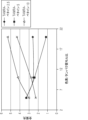
(図5)ドットブロット-DYLIGHT(商標)550-2×PEG-GARを用いたマウスIgGの検出。様々なモル過剰で、基本コンジュゲートに比べてNHSアセテート(2.5×、5×)またはMS(PEG)4(3.75×)からのシグナル/バックグランドの改善倍率。これは、イメージング機器を用いて撮影したドットブロットのソフトウェア解析の結果を示す。ドットブロットを、NHSアセテートまたはMS(PEG)4スペーサーおよびDYLIGHT(商標)550蛍光色素で共標識したDYLIGHT(商標)550-GAM抗体を用いて試験した。マウスIgGを1000ng/ドットから1:1で段階希釈した。全てのDYLIGHT(商標)550-2×PEG-GAR二次抗体を、1mg/mlストックの1/5000に希釈した。コンジュゲーション混合物に付加されたNHSアセテートまたはMS(PEG)4は、各々のそれぞれの色素のモル過剰での基本コンジュゲートと比較して、シグナル強度の改善(約1.6程度の増加)をもたらした。改善は1.2~1.6倍の範囲であり、特に、以下のGAM-DYLIGHT(商標)550-2×PEG:10×色素+5×アセテート、15×色素+3.75×MS(PEG)4、20×色素+5×アセテート、および20×+3.75×MS(PEG)4は、それぞれの基本コンジュゲートに対して1.3倍を超える改善を示した。
(図6)ドットブロット-マウスIgGのDyLight650-4×PEG-GAM検出、各々の色素のモル過剰での基本コンジュゲートに対するNHSアセテート(2.5×、5×、および10×)またはMS(PEG)4(3.75×、5×、10×)での改善倍率。この図は、イメージング機器を用いて撮影したドットブロットのソフトウェア解析の結果を示す。ドットブロットを、NHSアセテート(2.5×、5×、および10×)またはMS(PEG)4(3.75×)およびDYLIGHT(商標)650-4×PEG(表2において略称「DYLIGHT(商標)650」)(10×~20×)で共標識したGAM抗体を用いて試験した。マウスIgGを1000ng/ドットから1:1で段階希釈した。全てのDYLIGHT(商標)650-4×PEG-GAR二次抗体を、1mg/mlストックの1/10,000に希釈した。高度の色素の置換を有するコンジュゲートは、ドットブロットおよびウエスタンブロッティングなどの用途においてよりよく機能する傾向がある。NHSアセテートと(MS)PEG4の両方は、初期の基本コンジュゲートよりも感度およびシグナル/バックグラウンド(約2.2程度の増加)において顕著な改善をもたらす。2.5×モル過剰でGAM-DYLIGHT(商標)650-4×PEG-15×に付加されたNHSアセテートは、強度を1.7倍改善した。NHSアセテートによってもたらされた改善は、最高モル過剰(20×)で調製されたコンジュゲートを用いた場合よりも1.3倍優れた性能を示した。GAM-DYLIGHT(商標)650-4×PEG-15×コンジュゲーションに付加された全てのMS(PEG)4は、蛍光強度を1.8~2.2倍改善し、対応する最高基本コンジュゲートGAM-DYLIGHT(商標)650-4×PEG-20×よりも良好に機能した。この図のレーンは次の通りである。
(表2)

(表3)

(図9)ウエスタンブロットでのGAM-DYLIGHT(商標)650-4×PEG-GAR(7.5×モル過剰の色素で)に対するNHSアセテート(5×)またはMS(PEG)4(5×)の効果を示す。HeLa細胞溶解物を0.5μg/ウェルから4倍に希釈した。一次抗体マウス抗PDIを1mg/mlの1/5000に希釈した。全てのDYLIGHT(商標)二次抗体を、1mg/mlストックの1/5000に希釈した。5×モル過剰でGAM-DYLIGHT(商標)650-4×PEG-7.5×コンジュゲートに付加されたNHSアセテートは、強度を1.5倍改善した。3.75×モル過剰でGAM-DYLIGHT(商標)650-4×PEG-7.5×コンジュゲートに付加されたNHSアセテートは、強度を1.4倍改善した。
(図10)GAR-DYLIGHT(商標)800-4×PEG(表4において略称「DYLIGHT(商標)800」)の検出可能な蛍光レベルに対するNHSアセテート(2.5×、5×)またはMS(PEG)4の効果を試験するウエスタンブロットアッセイの結果を示す。A431細胞溶解物を1:1で段階希釈した。一次抗体ウサギ抗Hsp90と抗シクロフィリンBを両方とも1/5000に希釈した。全てのDYLIGHT(商標)二次抗体を、1mg/mlストックの1/20,000に希釈した。このウエスタンブロッティング用途で、MS(PEG)4(3.75×と5×)およびNHSアセテート(2.5~5×)の付加は、異なるモル過剰の色素で、基本DYLIGHT(商標)800-4×PEGコンジュゲートの蛍光強度と感度を20~100%顕著に増強した。この図のレーンは次の通りである。
(表4)

(図12)ウエスタンブロットおよびドットブロットアッセイでは、GAM-DYLIGHT(商標)680-4×PEG-GAR(10倍モル過剰での色素)に対するNHSアセテート(2.5×、5×)またはMS(PEG)4(5×)の効果を示す。ウエスタンブロットのために、HeLa細胞溶解物を0.5μg/ウェルから4倍に希釈し、抗PDI一次抗体を1mg/mlの1/5000に希釈した。ドットブロットのために、マウスIgGを1000ng/ドットから1:2に段階希釈した。全てのDYLIGHT(商標)680-4×PEG-GAR二次抗体を、1mg/mlストックの1/20000に希釈した。ウエスタンブロッティングとドットブロットアッセイの両方は、MS(PEG)4(5×)およびNHSアセテート(2.5×および5×)を付加すると、DYLIGHT(商標)680-4×PEGコンジュゲートの蛍光強度と感度が3~4倍、顕著に増強したことを示した。
(図13A)(DYLIGHT(商標)488-GAM4μg/mlによるIFC。様々なモル過剰で、基本コンジュゲートに比べてNHSアセテート(2.5×、5×)またはMS(PEG)4(3.75×)からのシグナル/バックグランドの改善倍率、そして図13B(IFC-DYLIGHT(商標)488-GAR4μg/mlによるPDIの検出。様々なモル過剰で、基本コンジュゲートに比べてNHSアセテート(2.5×、5×)またはMS(PEG)4(3.75×)の倍率でシグナル/バックグランドの向上)は、細胞イメージングアッセイでの7.5×~20×モル過剰の色素でのGAM-DYLIGHT(商標)488(13A)およびGAR-DYLIGHT(商標)488(13B)の蛍光に対するNHSアセテート(2.5×および5×)またはMS(PEG)4細胞(3.75×)の付加の効果を実証する。図13A:A549細胞を、1mg/mlストックの1/1000に希釈したpH2A×一次抗体で染色した。全てのDYLIGHT(商標)488二次抗体を1mg/mlストックの1/250に希釈した。NHSアセテート修飾コンジュゲートは、15×の色素モル過剰で基本コンジュゲートと比較してシグナル/バックグラウンドの1.4~1.5倍(GAM)および1.1~1.6倍(GAR)の範囲での改善をもたらした。GAMコンジュゲートについては、5×でのNHSアセテートおよび3.75×でのGARコンジュゲートについてのMS(PEG)4で最も顕著な改善が観察され、2.5×でのNHSアセテートおよび3.75×でのMS(PEG)4でより顕著な改善が観察された。
(図13B)図13Bは、A549細胞をpH2A×一次抗体で染色した同様の実験を示す。
(図14)DYLIGHT(商標)550-2×PEG(7.5×~20×)4μg/mlによるPDIのIFC検出。各々の色素モル過剰での基本コンジュゲートに対するNHSアセテート(2.5×、5×、および10×)またはMS(PEG)4(3.75×、5×、10×)の付加からの改善倍率。この図は、蛍光細胞イメージングアッセイでの、GAM-DYLIGHT(商標)550-2×PEG-GAM(7.5×~20×モル過剰の色素での)へのNHSアセテート(2.5×、5×、および10×)またはMS(PEG)4(3.75×、5×、および10×)の付加の効果を示す。U2OS細胞を、1mg/mlストックの1/100に希釈した抗PDI一次抗体で染色した。全てのDYLIGHT(商標)550-2×PEG-GAM二次抗体(表5において略称「DYLIGHT(商標)550」)を1mg/mlストックの1/250に希釈した。この細胞イメージング用途において、5×NHSアセテートの付加は、12.5×色素モル過剰のDYLIGHT(商標)550-2×PEG GAMコンジュゲートについての基本コンジュゲート(付加なしで作られた)と比較して約50%の改善をもたらし、3.75×MS(PEG)4の付加は、20倍の色素モル過剰で、基本コンジュゲートに対して約50%の改善をもたらした。この図のレーンは次の通りである。
(表5)

(表6)

(表7)

(図18)蛍光標識を含む分岐鎖PEG分子の生成および抗体分子への結合の概略図を示す。ステップ1では、反応性基および蛍光標識を分岐鎖PEG分子上のNH2基に結合させる。ステップ2において、付着部位が抗体分子に付加される。ステップ3では、蛍光標識された分岐鎖PEG分子を抗体分子に共有結合させる。
(図19)シングルアームコネクター(アミロースなど)とマルチアームコネクター(デキストラン)の各々の一例を示す図である。BMは生体分子を表し、APは結合点を表し、これは蛍光標識がその点に結合されることを意味する。
(図20)スターポリマーの種々の種類といくつかのスターポリマーの成分の例およびスターポリマーの製造方法の例である。図20Aは、コア(黒丸)および複数のアーム(黒線)を有するスターポリマーの図である。スターはアームに共有結合した蛍光標識を表す。いくつかのアームは蛍光標識されておらず、標識が完了しなかったことを示している。図20Bは、アームが2つの異なる種類(例えば、ポリエチレングリコールおよびポリビニルアルコール)であり、異なるアームの種類が実線および破線で表される、同様のスターポリマーの図である。図20Cの左側は、他の化学物質を結合するため、または重合のための開始剤として作用するために使用することができる反応性基(灰色のバー)を有するコア(黒い半円)の部分図を示す。コアは反応性基に結合したアダプター(黒い実線)を有して中心に表される。ポリマーアームは、アダプターに結合した右側(黒い破線)に示され、そして蛍光分子(スター)で標識されている。
(図21)本発明の実施において使用することができる種類の例示的なポリリジン分子を示す。R1およびR2基は標識なしとして示されている。これらの基は、蛍光標識のためのおよび生体分子(例えば、抗体)へのコンジュゲートのための結合点として使用することができる。
(図22)AF647分岐鎖PEGコンストラクトとコンジュゲートしたSK3マウス抗ヒトCD4抗体。SK3マウス抗ヒトCD4抗体(5.5mg/mL)を、抗体に対して10倍モル過剰エステルでのALEXA FLUOR(登録商標)647スクシンイミジルエステルで修飾した(破線)。抗体に対して10倍過剰のアジド-SEでタグ化されたSK3を、1mg/mLのアジド-SK3抗体で100μMのAF647-HG20K8 PEG-sDIBOに25℃で20時間クリックコンジュゲートし、5mMのNaN3でクエンチし、コンジュゲートをMillipore AMICON(商標)Ultra-2 100kDa遠心分離フィルターで精製した(点線)。抗体に対して20倍過剰のアジド-SEでタグ化されたSK3を、3mg/mLのアジド-SK3抗体で600μMのAF647-HG20K8 PEG-sDIBOに37℃で3時間クリックコンジュゲートし、5mMのNaN3でクエンチし、コンジュゲートをMillipore AMICON(商標)Ultra-2 100kDa遠心分離フィルターで精製した(実線)。96ウェルプレート中の100万個のFicollで単離されたPBMC/ウェルを、1μg~0.015μgの抗体の7点滴定を用いてSK3コンジュゲートで染色した。染色された細胞の分析は、ATTUNE(商標)NxTフローサイトメーターを用いて実施された。
(図23)AF647分岐鎖PEGコンストラクトとコンジュゲートしたSK3マウス抗ヒトCD4抗体。SK3マウス抗ヒトCD4抗体(5.5mg/mL)を、抗体に対して10倍モル過剰エステルでのALEXA FLUOR(登録商標)647スクシンイミジルエステルで修飾した(AF)。抗体に対して20倍過剰のアジド-SEでタグ化されたSK3を、3mg/mLのアジド-SK3抗体で600μMのAF647-HG20K8 PEG-sDIBOに37℃で3時間クリックコンジュゲートし、5mMのNaN3でクエンチし、コンジュゲートをMillipore AMICON(商標)Ultra-2 100kDa遠心分離フィルターで精製した(B1)。抗体に対して10倍過剰のアジド-SEでタグ化されたSK3を、1mg/mLのアジド-SK3抗体で100μMのAF647-HG20K8 PEG-sDIBOに25℃で20時間クリックコンジュゲートし、5mMのNaN3でクエンチし、コンジュゲートをMillipore AMICON(商標)Ultra-2 100kDa遠心分離フィルターで精製した(B2)。96ウェルプレート中の100万個のFicollで単離されたPBMC/ウェルを、1μg~0.015μgの抗体の7点滴定を用いてSK3コンジュゲートで染色した。染色された細胞の分析は、ATTUNE(商標)NxTフローサイトメーターを用いて実施し、アロフィコシアニン(APC)(Thermo Fisher Scientific、カタログ番号MHCD0405)と比較された。
1 shows a schematic diagram of one embodiment of the present invention; FIG. In this embodiment, the antibody is conjugated with an NHS fluorochrome and a sulfo-NHS-acetate spacer at two different dye molar excesses in 50 mM borate buffer (pH 8.5). Dye-spacer conjugation results in enhanced sensitivity and decreased quenching.
FIG. 2 shows a schematic diagram of some embodiments of the present invention. In this embodiment, the antibody is conjugated with an NHS fluorochrome and a methyl-PEG-NHS-ester spacer in 50 mM borate buffer (pH 8.5). Spacers used in this embodiment are MS(PEG) 4 , MS(PEG) 8 and MS(PEG) 12 .
(FIG. 3) Shows the results of software analysis of dot blots taken with an imaging instrument. Dot blots were tested with GAM antibodies co-labeled with NHS acetate or MS(PEG) 4 spacers and
(Table 1)
(FIG. 5) Dot Blot—Detection of mouse IgG using DYLIGHT™ 550-2×PEG-GAR. Fold improvement in signal/background from NHS acetate (2.5x, 5x) or MS(PEG)4 (3.75x) compared to base conjugate at various molar excesses. It shows the results of software analysis of dot blots taken with an imaging instrument. Dot blots were tested with DYLIGHT™ 550-GAM antibody co-labeled with NHS acetate or MS(PEG) 4 spacers and
(FIG. 6) Dot Blot—DyLight650-4×PEG-GAM detection of mouse IgG, NHS acetate (2.5×, 5×, and 10×) or MS (PEG) relative to base conjugate in molar excess of each dye. ) improvement factor at 4 (3.75×, 5×, 10×). This figure shows the results of software analysis of dot blots taken using an imaging instrument. Dot blots were analyzed with NHS acetate (2.5×, 5×, and 10×) or MS(PEG) 4 (3.75×) and DYLIGHT™ 650-4×PEG (abbreviated “DYLIGHT™ in Table 2). )650”) (10×-20×) with co-labeled GAM antibody. Mouse IgG was serially diluted 1:1 from 1000 ng/dot. All DYLIGHT™ 650-4x PEG-GAR secondary antibodies were diluted 1/10,000 from 1 mg/ml stock. Conjugates with a high degree of dye substitution tend to perform better in applications such as dot blots and western blotting. Both NHS Acetate and (MS)PEG 4 provide significant improvements in sensitivity and signal/background (an increase of about 2.2) over the initial base conjugate. NHS Acetate added to GAM-DYLIGHT™ 650-4xPEG-15x in 2.5x molar excess improved strength by 1.7x. The improvement provided by NHS acetate was 1.3-fold better than with the conjugate prepared at the highest molar excess (20x). All MS(PEG) 4 attached to GAM-DYLIGHT™ 650-4×PEG-15× conjugation improved fluorescence intensity by 1.8-2.2 fold, compared to the corresponding highest base conjugate GAM - Performed better than DYLIGHT™ 650-4x PEG-20x. The lanes in this figure are as follows:
(Table 2)
(Table 3)
(FIG. 9) Comparison of NHS Acetate (5×) or MS(PEG) 4 (5×) to GAM-DYLIGHT™ 650-4×PEG-GAR (at 7.5× molar excess of dye) in Western blots. Show effect. HeLa cell lysates were diluted 4-fold from 0.5 μg/well. Primary antibody mouse anti-PDI was diluted 1/5000 at 1 mg/ml. All DYLIGHT™ secondary antibodies were diluted 1/5000 of the 1 mg/ml stock. NHS acetate added to GAM-DYLIGHT™ 650-4xPEG-7.5x conjugate in 5x molar excess improved strength by 1.5 fold. NHS Acetate added to GAM-DYLIGHT™ 650-4xPEG-7.5x conjugate in 3.75x molar excess improved strength by 1.4x.
(FIG. 10) NHS acetate (2.5×, 5×) or MS (PEG ) shows the results of a Western blot assay testing the effect of 4 . A431 cell lysates were serially diluted 1:1. Both primary antibodies rabbit anti-Hsp90 and anti-cyclophilin B were diluted 1/5000. All DYLIGHT™ secondary antibodies were diluted 1/20,000 of the 1 mg/ml stock. In this Western blotting application, the addition of MS(PEG) 4 (3.75× and 5×) and NHS acetate (2.5-5×) were different molar excesses of the dyes, the base DYLIGHT™ 800-4. ×PEG conjugates significantly enhanced fluorescence intensity and sensitivity by 20-100%. The lanes in this figure are as follows:
(Table 4)
(FIG. 12) Western blot and dot blot assays showed NHS acetate (2.5×, 5×) or MS (PEG ) 4 (5×). For Western blotting, HeLa cell lysate was diluted 4-fold from 0.5 μg/well and anti-PDI primary antibody was diluted 1/5000 at 1 mg/ml. For dot blots, mouse IgG was serially diluted 1:2 from 1000 ng/dot. All DYLIGHT™ 680-4x PEG-GAR secondary antibodies were diluted 1/20000 of the 1 mg/ml stock. Both Western blotting and dot blot assays showed that the fluorescence intensity of DYLIGHT ™ 680-4x PEG conjugates and A significant 3-4 fold increase in sensitivity was demonstrated.
(FIG. 13A) (IFC with DYLIGHT™ 488-
(FIG. 13B) FIG. 13B shows a similar experiment in which A549 cells were stained with pH2A× primary antibody.
(FIG. 14) IFC detection of PDI with DYLIGHT™ 550-2×PEG (7.5×-20×) 4 μg/ml. Fold improvement from the addition of NHS acetate (2.5x, 5x, and 10x) or MS(PEG)4 (3.75x, 5x, 10x) to the base conjugate at each dye molar excess. . This figure shows NHS acetate (2.5×, 5 ×, and 10×) or MS(PEG) 4 (3.75×, 5×, and 10×). U2OS cells were stained with anti-PDI primary antibody diluted 1/100 of the 1 mg/ml stock. All DYLIGHT™ 550-2×PEG-GAM secondary antibodies (abbreviated “
(Table 5)
(Table 6)
(Table 7)
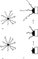
FIG. 18 shows a schematic of the generation of branched PEG molecules containing fluorescent labels and conjugation to antibody molecules. In
(FIG. 19) A diagram showing an example of each of a single-arm connector (amylose, etc.) and a multi-arm connector (dextran). BM stands for biomolecule and AP stands for attachment point, which means that the fluorescent label is attached to that point.
(FIG. 20) Various types of star polymers and examples of the components of some star polymers and examples of methods of making star polymers. FIG. 20A is a diagram of a star polymer with a core (filled circles) and multiple arms (filled lines). Stars represent fluorescent labels covalently attached to the arms. Some arms were not fluorescently labeled, indicating that the labeling was not completed. FIG. 20B is a diagram of a similar star polymer in which the arms are of two different types (eg, polyethylene glycol and polyvinyl alcohol), and the different arm types are represented by solid and dashed lines. The left side of FIG. 20C is a portion of the core (black semicircle) with reactive groups (grey bar) that can be used to bind other chemicals or act as an initiator for polymerization. Figure shows. The core is represented in the center with the adapter (solid black line) attached to the reactive group. The polymer arm is shown on the right (dashed black line) attached to an adapter and labeled with a fluorescent molecule (star).
(FIG. 21) shows exemplary polylysine molecules of the type that can be used in the practice of the present invention. The R1 and R2 groups are shown unlabeled. These groups can be used as attachment points for fluorescent labels and for conjugation to biomolecules (eg, antibodies).
(FIG. 22) SK3 mouse anti-human CD4 antibody conjugated with AF647 branched PEG construct. SK3 mouse anti-human CD4 antibody (5.5 mg/mL) was modified with ALEXA FLUOR® 647 succinimidyl ester in a 10-fold molar excess of ester over antibody (dashed line). Azide-SE tagged SK3 in 10-fold excess over antibody was click conjugated with 1 mg/mL azide-SK3 antibody to 100 μM AF647-HG20K8 PEG-sDIBO for 20 h at 25° C. and 5 mM NaN. After quenching with 3 , the conjugate was purified on a Millipore
(FIG. 23) SK3 mouse anti-human CD4 antibody conjugated with AF647 branched PEG construct. SK3 mouse anti-human CD4 antibody (5.5 mg/mL) was modified with ALEXA FLUOR® 647 succinimidyl ester in a 10-fold molar excess of ester over antibody (AF). Azide-SE tagged SK3 in 20-fold excess over antibody was click conjugated with 3 mg/mL azide-SK3 antibody to 600 μM AF647-HG20K8 PEG-sDIBO for 3 h at 37° C. and 5 mM NaN. 3 and the conjugate was purified with a Millipore
詳細な説明
蛍光標識は、タンパク質および核酸を含む生体分子の直接的、定量的、特異的、および高感度の検出をもたらすので、イメージングに広く用いられている。スルホン化および/またはPEG修飾されている修飾蛍光標識は、塩基性の未修飾色素と比較して感度が高い。しかしながら、これらの修飾蛍光標識でさえも、特定の色素対タンパク質(D/P)比で蛍光消光を示す。
DETAILED DESCRIPTION Fluorescent labels are widely used in imaging because they provide direct, quantitative, specific, and sensitive detection of biomolecules, including proteins and nucleic acids. Modified fluorescent labels that are sulfonated and/or PEG-modified are more sensitive than basic, unmodified dyes. However, even these modified fluorescent labels exhibit fluorescence quenching at certain dye-to-protein (D/P) ratios.
蛍光の増加は標識色素のモル過剰の増加と共に見られるが、タンパク質に対する最適色素の比(D/P)を超えるモル過剰は、典型的には特に抗原/抗体またはDNA/RNA相互作用の空間的立体配座が静的消光を引き起こし得る蛍光イメージング用途において生体分子の消光および/または沈殿をもたらす。 Although an increase in fluorescence is seen with increasing molar excess of labeling dye, molar excess over the optimal dye to protein ratio (D/P) is typically associated with the spatial distribution of antigen/antibody or DNA/RNA interactions, among others. Resulting in quenching and/or precipitation of biomolecules in fluorescence imaging applications where conformation can cause static quenching.
いくつかの実施形態において、本発明は、標準的なコンジュゲーションにおいて蛍光の減少をもたらす高度に標識されたコンジュゲート(例えば、高い色素対タンパク質比(D/P)を有する生体分子)を用いて消光を低減および/または蛍光シグナルを増加させる方法を含む。タンパク質、核酸および他の生体分子(例えば、オリゴ糖)に修飾を加えることができる。いくつかの実施形態において、本発明は、蛍光シグナルの増加および/または消光の減少を示す蛍光標識された生体分子を含む組成物を含み、組成物は生体分子、スペーサー、および蛍光標識を含み、スペーサーと蛍光標識は、互いに直接コンジュゲートしていない。 In some embodiments, the present invention uses highly labeled conjugates (e.g., biomolecules with high dye-to-protein ratios (D/P)) that result in reduced fluorescence upon standard conjugation. Methods of reducing quenching and/or increasing fluorescence signal are included. Modifications can be made to proteins, nucleic acids and other biomolecules (eg, oligosaccharides). In some embodiments, the invention comprises a composition comprising a fluorescently labeled biomolecule that exhibits increased fluorescence signal and/or decreased quenching, the composition comprising a biomolecule, a spacer, and a fluorescent label, Spacers and fluorescent labels are not directly conjugated to each other.
本発明はまた、蛍光標識1個あたりに基づいて増強された蛍光を示す組成物、ならびにそのような組成物を製造および使用する方法に関する。例として、生体分子に結合した単一の蛍光標識が100%の蛍光発光のベースラインを設定すると仮定する。さらに、2つの蛍光標識が同一の生体分子に結合しているとき、2つの蛍光標識のそれぞれがベースライン蛍光発光の平均80%を示すと仮定する。本発明は、部分的には、平均蛍光発光をベースラインの80%を超えて増加させるための組成物および方法に関する。 The invention also relates to compositions that exhibit enhanced fluorescence on a per fluorescent label basis, and methods of making and using such compositions. As an example, assume that a single fluorescent label attached to a biomolecule sets a baseline fluorescence emission of 100%. Further, assume that each of the two fluorescent labels exhibits an average of 80% of the baseline fluorescence emission when the two fluorescent labels are bound to the same biomolecule. The present invention relates, in part, to compositions and methods for increasing mean fluorescence emission above 80% of baseline.
いくつかの場合において、本発明の組成物、および本発明の方法で使用される組成物は、1つ以上の機能的特性によって定義され得る。そのような特性の例は、標識された分子(例えば、生体分子)に結合する蛍光標識の数、標識された分子上の蛍光標識間の平均距離(多数の異なる方法のいずれかで測定される)、および/または標識された分子上の蛍光標識の量子収率である。 In some cases, compositions of the invention, and compositions used in methods of the invention, may be defined by one or more functional properties. Examples of such properties are the number of fluorescent labels bound to a labeled molecule (e.g., biomolecule), the average distance between fluorescent labels on a labeled molecule (measured in any of a number of different ways). ), and/or the quantum yield of the fluorescent label on the labeled molecule.
蛍光強度を測定する1つの手段は、量子収率の測定によるものである。蛍光系についての量子収率(Φ)は、事実上、所与のフルオロフォアの発光効率であり、そして以下の式によって決定され得る。
![]()
![]()
以下の実施例8に示されるように、量子収率も消光効果を測定するために使用され得る。さらに、量子収率を測定するために使用され得るHamamatsu Absolute PL Quantum Yield Spectrometer(Hamamatsu Corp.,Bridgewater NJ 08807,C11347-11Quantaurus-QY Absolute PL Quantum Yield Spectrometer)のような装置は市販される。 Quantum yield can also be used to measure quenching effects, as shown in Example 8 below. Additionally, instruments such as the Hamamatsu Absolute PL Quantum Yield Spectrometer (Hamamatsu Corp., Bridgewater NJ 08807, C11347-11 Quantaurus-QY Absolute PL Quantum Yield Spectrometer) that can be used to measure quantum yield are commercially available.
実施例8および表25に示すように、蛍光標識された分子の量子収率は遊離の蛍光標識の量子収率と比較することができる。消光が実質的に起こらない条件下での単一ユニットの遊離の蛍光標識の量子収率を1と設定すれば、これは標識された分子に結合した各蛍光標識によって発生する蛍光の比較のためのベンチマークとして使用できる。多くの場合、本発明の組成物は、複数の蛍光標識で標識された蛍光標識された分子を含み、ここで蛍光発光の平均量は、蛍光標識1個あたりに基づいて、遊離の蛍光標識の蛍光発光の、少なくとも70%(0.7蛍光比)(例えば、約70%~約99%、約70%~約90%、約80%~約99%、約85%~約99%、約87%~約99%、約90%~約99%、約80%~約95%、約85%~約96%など)である。 As shown in Example 8 and Table 25, the quantum yield of fluorescently labeled molecules can be compared to that of free fluorescent label. Given that the quantum yield of a single unit of free fluorescent label under conditions in which substantially no quenching occurs is set at 1, this is useful for comparison of the fluorescence emitted by each fluorescent label bound to the labeled molecule. can be used as a benchmark for Often, the compositions of the invention comprise fluorescently-labeled molecules labeled with a plurality of fluorescent labels, wherein the average amount of fluorescence emission is the amount of free fluorescent label on a per fluorescent label basis. at least 70% (0.7 fluorescence ratio) of fluorescence emission (e.g., about 70% to about 99%, about 70% to about 90%, about 80% to about 99%, about 85% to about 99%, about 87% to about 99%, about 90% to about 99%, about 80% to about 95%, about 85% to about 96%, etc.).
実施例8および表25に示すように、蛍光強度は、遊離の標識の蛍光と比較した蛍光標識された分子の全蛍光の測定によって決定することができる。消光が実質的に起こらない条件下での単一ユニットの遊離の蛍光標識の蛍光を1と設定すれば、これは標識された分子に結合した各蛍光標識によって発生する蛍光の比較のためのベンチマークとして使用できる。多くの場合、本発明の組成物は、複数の蛍光標識で標識された蛍光標識された分子を含み、ここで蛍光発光の平均量は、蛍光標識1個あたりに基づいて、遊離の蛍光標識の蛍光発光の、少なくとも70%(0.7蛍光比)(例えば、約70%~約99%、約70%~約90%、約80%~約99%、約85%~約99%、約87%~約99%、約90%~約99%、約80%~約95%、約85%~約96%など)である。 As shown in Example 8 and Table 25, fluorescence intensity can be determined by measuring the total fluorescence of fluorescently labeled molecules compared to the fluorescence of the free label. Given that the fluorescence of a single unit of free fluorescent label under conditions in which substantially no quenching occurs is set at 1, this is a benchmark for comparison of the fluorescence emitted by each fluorescent label bound to the labeled molecule. can be used as In many cases, the compositions of the invention comprise fluorescently labeled molecules labeled with a plurality of fluorescent labels, wherein the average amount of fluorescence emission is the amount of free fluorescent label on a per fluorescent label basis. at least 70% (0.7 fluorescence ratio) of fluorescence emission (e.g., about 70% to about 99%, about 70% to about 90%, about 80% to about 99%, about 85% to about 99%, about 87% to about 99%, about 90% to about 99%, about 80% to about 95%, about 85% to about 96%, etc.).
表25に示されるように、遊離の蛍光標識と比較した蛍光標識された分子の明度を決定することができる。明度は、次の式で与えられるように、量子収量(Φ)、吸光係数(ε)、および1分子あたりの色素数(N)の積に比例する。
B=Φ×ε×N
したがって、遊離の蛍光標識の明度の標識された分子の明度に対する比を用いて、全蛍光増強を記載することができる。
As shown in Table 25, the brightness of fluorescently labeled molecules compared to free fluorescent label can be determined. Brightness is proportional to the product of quantum yield (Φ), extinction coefficient (ε), and number of dyes per molecule (N), as given by
B = Φ x ε x N
Therefore, the ratio of the brightness of the free fluorescent label to the brightness of the labeled molecule can be used to describe the total fluorescence enhancement.
例として、表25のデータは、脱イオン水中のALEXA FLUOR(登録商標)647のベンチマークとして設定されている。さらに、これは遊離の色素の100%量子収率および1.0の明度の比のベンチマークを設定する。試料のうちで、分子AF647-20K8は遊離の色素の73%の量子収率を有していたが、遊離の色素に対して5.8×の蛍光増強を示した。試料AF647-10K4が遊離の色素の最高量子収率(89%)を有するが、遊離の色素に対してわずか3.6×の蛍光増強を有することもまた示される。これらのデータは、これらの分子について見られる蛍光増強の程度がアームの長さと直接相関し得ることを示す。データはまた、アームの長さを一定に保ち、より多くの蛍光標識されたアームをポリマーに付加すると、蛍光増強が増大する傾向があることを示している。 As an example, the data in Table 25 are set as a benchmark for ALEXA FLUOR® 647 in deionized water. Furthermore, it sets a benchmark of 100% quantum yield of free dye and a brightness ratio of 1.0. Among the samples, the molecule AF647-20K8 had a quantum yield of 73% of the free dye, but exhibited a fluorescence enhancement of 5.8× over the free dye. It is also shown that sample AF647-10K4 has the highest quantum yield of the free dye (89%), but has only a 3.6× fluorescence enhancement relative to the free dye. These data indicate that the degree of fluorescence enhancement seen for these molecules can be directly correlated with arm length. The data also show that keeping arm length constant and adding more fluorescently labeled arms to the polymer tends to increase fluorescence enhancement.
したがって、本発明は、蛍光標識が蛍光シグナルを増強する様式で間隔を空けられるように、複数の蛍光標識を個々の分子(例えば、生体分子)に連結するための組成物および方法を含む。これは消光の減少によってなされ得る。蛍光シグナルを増強するための1つの方法は、試料中に存在する蛍光標識を空間的に分離することである。これは、複数の蛍光標識が検出されるべき同一の分子(例えば、生体分子)に結合しているときに特に有用である。 Accordingly, the present invention includes compositions and methods for linking multiple fluorescent labels to individual molecules (eg, biomolecules) such that the fluorescent labels are spaced in a manner that enhances the fluorescent signal. This can be done by reducing quenching. One method for enhancing the fluorescent signal is to spatially separate the fluorescent labels present in the sample. This is particularly useful when multiple fluorescent labels are attached to the same molecule (eg, biomolecule) to be detected.
いくつかの態様において、本発明は、スペーサーと蛍光標識とにコンジュゲートした抗体を産生する方法であって、ここでスペーサー剤を用いてスペーサーを抗体にコンジュゲートし、そしてスペーサーは蛍光標識にコンジュゲートしていない、方法を含む。スペーサー、抗体、および蛍光標識を含む組成物であって、ここでスペーサーが蛍光標識にコンジュゲートしていない組成物もまた包含される。 In some embodiments, the invention is a method of producing an antibody conjugated to a spacer and a fluorescent label, wherein the spacer is conjugated to the antibody using a spacer agent, and the spacer is conjugated to the fluorescent label. Not gated, including method. Also included are compositions comprising a spacer, an antibody, and a fluorescent label, wherein the spacer is not conjugated to the fluorescent label.
いくつかの実施形態において、スペーサーは、抗体にコンジュゲートした複数の蛍光標識の消光を低減することができる。 In some embodiments, the spacer can reduce quenching of multiple fluorescent labels conjugated to the antibody.
いくつかの実施形態では、スペーサー剤にコンジュゲートされた核酸を産生する方法であって、ここでスペーサー剤が蛍光標識に直接コンジュゲートされていない方法が包含される。そのようなスペーサー剤は、核酸にコンジュゲートした蛍光標識の消光を低減させ得る。 Some embodiments include methods of producing a nucleic acid conjugated to a spacer agent, wherein the spacer agent is not directly conjugated to a fluorescent label. Such spacer agents can reduce quenching of fluorescent labels conjugated to nucleic acids.
いくつかの実施形態において、本発明は、それらがコンジュゲートしている分子(例えば、生体分子)上の点からの空間的に分離した蛍光標識に関する組成物および方法を含む。多くの場合、これは1つ以上の蛍光標識をスペーサーに接続し、スペーサーを分子(例えば、生体分子)に接続することにより実施される。そのような組成物および方法の例を図18に示す。 In some embodiments, the present invention includes compositions and methods for spatially discrete fluorescent labeling from points on molecules (eg, biomolecules) to which they are conjugated. Often this is accomplished by attaching one or more fluorescent labels to a spacer and attaching the spacer to a molecule (eg, a biomolecule). Examples of such compositions and methods are shown in FIG.
定義
本明細書および例示的な実施形態は、限定的なものとして捉えられるべきではない。本明細書および添付の特許請求の範囲の目的において、別途示されない限り、数量、割合、または比率を表す全ての数、ならびに明細書および特許請求の範囲に使用される他の数値は、全ての場合において、それらが修正されすぎない程度まで「約」という用語によって修正されているものとして理解されるべきである。したがって、反対が示されない限り、以下の明細書および添付の特許請求の範囲に記載される数値パラメータは、得ようとする所望の特性によって変化し得る近似値である。最低でも、特許請求の範囲に対する均等の原則の適用を限定することを企図しないように、各数値パラメータは、少なくとも、報告された有効数字の桁数に照らしてかつ通常の四捨五入の技術を適用することによって解釈されるべきである。
DEFINITIONS The specification and example embodiments should not be taken as limiting. For the purposes of this specification and the appended claims, unless otherwise indicated, all numbers expressing quantities, percentages or proportions, and other numerical values used in the specification and claims, refer to all In some cases, the term "about" is to be understood as modifying to the extent that they are not overly modified. Accordingly, unless indicated to the contrary, the numerical parameters set forth in the following specification and attached claims are approximations that may vary depending upon the desired properties sought to be obtained. At the very least, each numerical parameter should be at least measured against the reported number of significant digits and apply conventional rounding techniques so as not to limit the application of the doctrine of equivalents to the claims. should be interpreted by
本明細書および添付の特許請求の範囲で使用されるとき、単数形「一つの(a)」、「一つの(an)」、および「その(the)」、ならびに任意の単語の任意の単数使用には、明確かつ疑いの余地なく1つの指示対象に限定されていない限り、複数の指示対象を含むことに留意されたい。本明細書で使用されるとき、「含む」という用語およびその文法的異形は、リスト中の項目の列挙が、列挙される項目と置き換えられるか、またはそこに追加され得る他の同様の項目を除外するものではないように、非限定的であることが意図される。 As used in this specification and the appended claims, the singular forms "a," "an," and "the," and any singular of any word It is noted that use includes plural referents unless clearly and unambiguously limited to one referent. As used herein, the term "comprising" and grammatical variants thereof means that the listing of items in a list includes other similar items that may replace or be added to the listed item. It is intended to be non-limiting, not exclusive.
本明細書中で使用されるとき、「生体分子」とは、合成または天然に存在する、タンパク質またはその断片、糖タンパク質、リポタンパク質、アミノ酸、ヌクレオシド、ヌクレオチド、核酸、オリゴヌクレオチド、DNA、RNA、炭水化物、糖、脂質、脂肪酸、ハプテン、抗体などを含むがこれらに限定されない。 As used herein, "biomolecule" refers to synthetic or naturally occurring proteins or fragments thereof, glycoproteins, lipoproteins, amino acids, nucleosides, nucleotides, nucleic acids, oligonucleotides, DNA, RNA, Including but not limited to carbohydrates, sugars, lipids, fatty acids, haptens, antibodies and the like.
「タンパク質」および「ポリペプチド」という用語は、本明細書では任意の長さのアミノ酸残基のポリマーを含むように一般的な意味で使用されている。本明細書で使用されるとき、「ペプチド」という用語は、モノマーがアミノ酸であり、アミド結合を介して互いに結合しているポリマーを指し、あるいはポリペプチドと呼ばれる。アミノ酸がα-アミノ酸であるとき、L-光学異性体またはD-光学異性体のいずれかを使用することができる。さらに、非天然アミノ酸、例えば、β-アラニン、フェニルグリシンおよびホモアルギニンもまた含まれる。遺伝子コードされていない一般的に遭遇するアミノ酸もまた本発明において使用され得る。本発明で使用される全てのアミノ酸は、D-またはL-異性体のいずれかであり得る。L-異性体が一般的に使用されている。さらに、他のペプチド模倣薬もまた本発明において有用である。一般的な概説については、Spatola,A.F.,単Chemistry and Biochemistry of Amino Acids,Peptides and Proteins,B.Weinstein,eds.,Marcel Dekker,New York,p.267(1983)を参照のこと。 The terms "protein" and "polypeptide" are used herein in a generic sense to include polymers of amino acid residues of any length. As used herein, the term "peptide" refers to a polymer whose monomers are amino acids and are linked together through amide bonds, otherwise referred to as a polypeptide. When the amino acids are α-amino acids, either the L-optical isomer or the D-optical isomer can be used. Additionally, unnatural amino acids such as β-alanine, phenylglycine and homoarginine are also included. Commonly encountered amino acids that are not genetically encoded can also be used in the present invention. All amino acids used in the present invention can be either the D- or L-isomer. The L-isomer is commonly used. Additionally, other peptidomimetics are also useful in the present invention. For a general review, see Spatola, A.; F. , Single Chemistry and Biochemistry of Amino Acids, Peptides and Proteins, B.; Weinstein, eds. , Marcel Dekker, New York, p. 267 (1983).
本明細書で使用されるとき、「抗体」という用語は、ハイブリドーマ細胞株によって、ポリクローナル抗体応答を誘発するための免疫化によって、化学合成によって、および抗体をコードする発現ベクターで形質転換された組換え宿主細胞によって、産生される抗体を含むがこれに限定されない抗体-抗原複合体を形成するために特定の物質(例えば、抗原および免疫原)に非共有結合的に結合する免疫グロブリン(Ig)スーパーファミリーのタンパク質を指す。ヒトにおいて、免疫グロブリン抗体は、IgA、IgD、IgE、IgG、およびIgMとして分類され、そして各クラスのメンバーは同一のアイソタイプを有すると言われる。ヒトIgAおよびIgGアイソタイプはさらにサブタイプIgA1およびIgA2、ならびにIgG1、IgG2、IgG3、およびIgG4にさらに細分される。マウスは概してヒトと同一のアイソタイプを有するが、IgGアイソタイプはIgG1、IgG2a、IgG2b、およびIgG3サブタイプに細分される。したがって、本明細書で使用されるとき、「抗体」という用語はその範囲内に、(a)免疫グロブリンの任意の様々なクラスまたはサブクラス(例えば、抗体を産生する任意の動物由来のIgA、IgD、IgG、IgM、およびIgE)、ならびに(b)マウス、キメラ、またはヒト化抗体などのポリクローナル抗体およびモノクローナル抗体を含むことが理解されよう。抗体分子は、抗原決定基として作用し得るアミノ酸配列の領域(例えば、Fc領域、カッパ軽鎖、ラムダ軽鎖、ヒンジ領域など)を有する。選択された領域に対して生成された抗体は抗-[領域](例えば、抗Fc、抗カッパ軽鎖、抗ラムダ軽鎖など)と呼ばれる。抗体は、典型的には、免疫グロブリンタンパク質を発現させるためにリンパ球活性化を開始するために高分子で生物を免疫することによって抗原に対して生成される。本明細書で使用されるとき、用語「抗体」はまた、非限定的に、単鎖Fv分子(scFv)を含む抗体結合ドメインであるか、またはそれと相同な結合ドメインを有する任意のポリペプチドまたはタンパク質を包含し、ここでVHドメインとVLドメインは、2つのドメインが会合して抗原結合部位を形成することを可能にするペプチドリンカーによって連結されている(Bird et al.,Science 242:423(1988)、およびHuston et al.,Proc.Natl.Acad.Sci.USA 85:5879(1988))。これらは天然源に由来することができ、あるいはそれらは部分的または全体的に合成的に産生されてもよい。 As used herein, the term "antibody" refers to a group transformed by a hybridoma cell line, by immunization to elicit a polyclonal antibody response, by chemical synthesis, and with an expression vector encoding the antibody. Immunoglobulins (Ig) that non-covalently bind to specific substances (e.g., antigens and immunogens) to form antibody-antigen complexes, including but not limited to antibodies produced by recombinant host cells Refers to the superfamily of proteins. In humans, immunoglobulin antibodies are classified as IgA, IgD, IgE, IgG, and IgM, and members of each class are said to have the same isotype. Human IgA and IgG isotypes are further subdivided into subtypes IgA 1 and IgA 2 , and IgG 1 , IgG 2 , IgG 3 and IgG 4 . Mice generally have the same isotypes as humans, but the IgG isotype is subdivided into IgG 1 , IgG 2a , IgG 2b , and IgG 3 subtypes. Thus, as used herein, the term "antibody" includes within its scope: (a) any of the various classes or subclasses of immunoglobulins (e.g., IgA, IgD, from any animal that produces antibodies); , IgG, IgM, and IgE), and (b) polyclonal and monoclonal antibodies such as murine, chimeric, or humanized antibodies. Antibody molecules have regions of amino acid sequence (eg, Fc region, kappa light chain, lambda light chain, hinge region, etc.) that can act as antigenic determinants. Antibodies raised against a selected region are called anti-[regions] (eg, anti-Fc, anti-kappa light chain, anti-lambda light chain, etc.). Antibodies are typically generated against an antigen by immunizing the organism with macromolecules to initiate lymphocyte activation to express immunoglobulin proteins. As used herein, the term "antibody" also includes, but is not limited to, an antibody binding domain, including single-chain Fv molecules (scFv), or any polypeptide having a binding domain homologous thereto or encompasses proteins, wherein VH and VL domains are linked by a peptide linker that allows the two domains to associate to form an antigen-binding site (Bird et al., Science 242:423 ( 1988), and Huston et al., Proc. Natl. Acad. Sci. USA 85:5879 (1988)). They can be derived from natural sources, or they may be partly or wholly synthetically produced.
さらに、VHH抗体は、抗原刺激細胞から得られたものとして、または遺伝子操作された抗原結合タンパク質としてのいずれかで使用され得る。 Additionally, VHH antibodies can be used either as derived from antigen-stimulated cells or as genetically engineered antigen binding proteins.
本明細書で使用されるとき、「抗体断片」という用語は、抗体全体の主要な選択的結合特性を保持する抗体の断片を指す。特定の断片、例えば様々なプロテアーゼでの消化により得られ、そして無傷の抗体のFc断片を欠くFab、Fab’、およびF(ab’)2、または無傷抗体中の重鎖成分を連結するジスルフィド結合の還元的切断により得られるいわゆる「半分子」断片は、当該技術分野において周知である。そのような断片には、軽鎖可変領域からなる単離された断片、重鎖および軽鎖の可変領域からなる「Fv」断片、ならびに軽鎖可変領域と重鎖可変領域がペプチドリンカーによって連結されている組換え単鎖ポリペプチド分子もまた含まれる。結合断片の他の例には、(i)VHドメインおよびCH1ドメインからなるFd断片;(ii)VHドメインからなるdAb断片(Ward et al.,Nature 341:544(1989))、(iii)単離されたCDR領域、ならびに(iv)上述の単鎖Fv分子(scFv)が含まれる。さらに、抗原認識特性を保持する組換え技術を用いて任意の断片を作製することができる。 As used herein, the term "antibody fragment" refers to a fragment of an antibody that retains the primary selective binding properties of whole antibody. Specific fragments, such as Fab, Fab′, and F(ab′) 2 , which are obtained by digestion with various proteases and lack the Fc fragment of intact antibodies, or disulfide bonds linking heavy chain components in intact antibodies So-called "half molecule" fragments obtained by reductive cleavage of are well known in the art. Such fragments include an isolated fragment consisting of the light chain variable region, an "Fv" fragment consisting of the heavy and light chain variable regions, and the light and heavy chain variable regions linked by a peptide linker. Also included are recombinant single-chain polypeptide molecules. Other examples of binding fragments include (i) an Fd fragment consisting of the VH and CH1 domains; (ii) a dAb fragment consisting of the VH domain (Ward et al., Nature 341:544 (1989)); Included are the separated CDR regions, as well as (iv) single chain Fv molecules (scFv) as described above. Moreover, any fragment can be produced using recombinant techniques that retain antigen recognition properties.
使用され得る例示的なVHH抗体は、単一のモノマーの可変抗体ドメインからなる抗体断片である単一ドメイン抗体である。そのような抗体断片は、典型的にはわずか12~25kDaの分子量を有し、したがって2本の重鎖タンパク質鎖および2本の軽鎖からなる他の多くの抗体(150~160kDa)よりも小さい。 An exemplary VHH antibody that may be used is a single domain antibody, which is an antibody fragment consisting of a single monomeric variable antibody domain. Such antibody fragments typically have a molecular weight of only 12-25 kDa and are therefore smaller than many other antibodies (150-160 kDa) which consist of two heavy protein chains and two light chains. .
本明細書中で使用されるとき、「抗原」とは、抗体の形成を誘導するか、または誘導することができる、または抗体が選択的に結合する、生物由来物質を含むがこれらに限定されない分子を指す。抗原はまた「免疫原」を指す。存在する他の物質との交差反応性または干渉が相対的に欠如するとき、抗体は抗原に選択的に結合する。 As used herein, "antigen" includes, but is not limited to, biological substances that induce or are capable of inducing the formation of antibodies, or to which antibodies selectively bind refers to molecules. Antigen also refers to "immunogen." An antibody selectively binds an antigen when there is a relative lack of cross-reactivity or interference with other substances present.
本明細書で使用されるとき、「反応性基」という用語は、別の化学基と反応して共有結合を形成することができる、すなわち適切な反応条件下で共有結合的に反応する基を指し、一般的に別の物質に対する結合点を表す。反応性基は一般に求核性、求電子性、および光活性化可能の基を含む。例示的な反応基としては、オレフィン、アセチレン、アルコール、フェノール、エーテル、酸化物、ハロゲン化物、アルデヒド、ケトン、カルボン酸、エステル、アミド、シアネート、イソシアネート、チオシアネート、イソチオシアネート、アミン、ヒドラジン、ヒドラゾン、ヒドラジド、ジアゾ、ジアゾニウム、ニトロ、ニトリル、メルカプタン、スルフィド、ジスルフィド、スルホキシド、スルホン、スルホン酸、スルフィン酸、アセタール、ケタール、無水物、硫酸、スルフェン酸イソニトリル、アミジン、イミド、イミダート、ニトロン、ヒドロキシルアミン、オキシム、ヒドロキサム酸、アルキン、およびアジドが挙げられるがこれらに限定されない。 As used herein, the term "reactive group" refers to a group that is capable of reacting with another chemical group to form a covalent bond, i.e., covalently reacts under suitable reaction conditions. refers to and generally represents a point of attachment to another substance. Reactive groups generally include nucleophilic, electrophilic, and photoactivatable groups. Exemplary reactive groups include olefins, acetylenes, alcohols, phenols, ethers, oxides, halides, aldehydes, ketones, carboxylic acids, esters, amides, cyanates, isocyanates, thiocyanates, isothiocyanates, amines, hydrazines, hydrazones, hydrazide, diazo, diazonium, nitro, nitrile, mercaptan, sulfide, disulfide, sulfoxide, sulfone, sulfonic acid, sulfinic acid, acetal, ketal, anhydride, sulfuric acid, isonitrile sulfenate, amidine, imide, imidate, nitrone, hydroxylamine, Examples include, but are not limited to, oximes, hydroxamic acids, alkynes, and azides.
本明細書中で使用されるとき、「スペーサー」、「スペーサー分子」、または「スペーサー剤」とは、生体分子に直接または間接的にコンジュゲートしたときに、生体分子から発光される蛍光を増強し得る化合物(例えば、有機化合物)をいう。これは蛍光標識の蛍光消光の低減から生じると考えられている。任意の数の化合物がスペーサーとして作用し得、例示的な化合物には、NHS-アセテートおよび様々な形態のポリエチレングリコール(PEG)が含まれる。本明細書で使用されるとき、用語「ポリエチレングリコール」または「PEG」は、エチレンオキシドのオリゴマーまたはポリマーを指す。PEGポリマー鎖長は大きく変動し得るが、10,000,000g/mol程度の高い分子量を有する傾向がある。PEGもまた、様々な形状で利用可能である。例えば、分岐鎖PEGは典型的には、中心コア基から生じる3~10個のPEG鎖を有する。スターPEGは、中心コア基から生じる3~100個のPEG鎖を有する。コームPEGは、通常ポリマー骨格にグラフトされた複数のPEG鎖を有する。ほとんどのPEGは、分子量分布を有する分子を含む(すなわち、それらは多分散性である)。サイズ分布は、その重量平均分子量およびその数平均分子量によって統計的に特徴付けることができ、その比は多分散性指数と呼ばれる。本発明の実施において使用され得る例示的なPEG化合物としては、MS(PEG)4、MS(PEG)8、およびMS(PEG)12(それぞれ、Thermo Fisher Scientific,Waltham,MA、カタログ番号22341、22509B、および22686)、ならびに(メチル-PEG12)3-PEG4-NHSエステル(Thermo Fisher Scientific,Waltham,MA、カタログ番号22421)などの分岐鎖PEG化合物が含まれる。 As used herein, a “spacer,” “spacer molecule,” or “spacer agent” enhances fluorescence emitted from a biomolecule when directly or indirectly conjugated to the biomolecule. A compound (for example, an organic compound) that can This is believed to result from reduced fluorescence quenching of the fluorescent label. Any number of compounds can act as spacers, exemplary compounds include NHS-acetate and various forms of polyethylene glycol (PEG). As used herein, the terms "polyethylene glycol" or "PEG" refer to oligomers or polymers of ethylene oxide. PEG polymer chain lengths can vary widely, but tend to have molecular weights as high as 10,000,000 g/mol. PEG is also available in various forms. For example, branched PEG typically has 3-10 PEG chains emanating from a central core group. Star PEGs have 3-100 PEG chains emanating from a central core group. Comb PEG usually has multiple PEG chains grafted onto the polymer backbone. Most PEGs contain molecules with a distribution of molecular weights (ie they are polydisperse). A size distribution can be statistically characterized by its weight average molecular weight and its number average molecular weight, the ratio of which is called the polydispersity index. Exemplary PEG compounds that can be used in the practice of the invention include MS(PEG) 4 , MS(PEG) 8 , and MS(PEG) 12 (Thermo Fisher Scientific, Waltham, MA, respectively, catalog numbers 22341, 22509B). , and 22686), and branched-chain PEG compounds such as (methyl-PEG 12 )3-PEG 4 -NHS ester (Thermo Fisher Scientific, Waltham, MA, catalog number 22421).
本明細書で使用されるとき、用語「直接スペーサー」は、それに結合した少なくとも1つの蛍光標識を有し、生体分子に直接結合する分子を指す。直接スペーサーは、(1)単一ポリマーまたは(2)コアに結合した複数のポリマーであり得る。直接スペーサーの例には、シングルアームポリマーおよびマルチアームポリマーが含まれる。 As used herein, the term "direct spacer" refers to a molecule that has at least one fluorescent label attached to it and directly binds to a biomolecule. A direct spacer can be (1) a single polymer or (2) multiple polymers attached to a core. Examples of direct spacers include single-arm and multi-arm polymers.
本明細書で使用されるとき、「ポリマー」は、繰り返しサブユニット(典型的には少なくとも4つの繰り返しサブユニット)からなる分子である。ポリマーは合成でも天然に存在するものでもよい。ポリマーの繰り返し単位は同一である必要はない。例えば、タンパク質は、異なるアミノ酸サブユニットからなるポリマーである。さらに、ポリマーは完全に直鎖状の分子である必要はなく、したがってデキストランのように分岐していてもよい。 As used herein, a "polymer" is a molecule composed of repeating subunits (typically at least four repeating subunits). Polymers may be synthetic or naturally occurring. The repeating units of the polymer need not be identical. For example, proteins are polymers composed of different amino acid subunits. Furthermore, the polymer need not be a perfectly linear molecule and thus may be branched like dextran.
本明細書中で使用されるとき、用語「シングルアームポリマー」とは、少なくとも1つの蛍光標識が結合し、非分岐鎖構造を有する、非分岐鎖分子を指す(図19を参照)。本発明の実施において使用され得るシングルアームポリマーの例は、「直鎖状」多糖類(例えば、アミロース)、ポリエチレングリコール、長鎖炭素分子(例えば、Ahx)、およびポリペプチドである。いくつかの場合では、非分枝鎖/直鎖多糖類は、α1,4結合によって互いに結合したモノマーからなる。 As used herein, the term "single arm polymer" refers to an unbranched molecule having at least one fluorescent label attached and having an unbranched structure (see Figure 19). Examples of single-arm polymers that can be used in the practice of the invention are "linear" polysaccharides (eg, amylose), polyethylene glycols, long-chain carbon molecules (eg, Ahx), and polypeptides. In some cases, the unbranched/linear polysaccharide consists of monomers linked together by α1,4 linkages.
本明細書中で使用されるとき、用語「マルチアームポリマー」とは、少なくとも1つの蛍光標識が結合し、そして非分岐鎖構造を有する分岐鎖分子を指す(図19参照)。本発明の実施において使用され得るマルチアームポリマーの例は、分岐鎖多糖類(例えば、デキストラン、グリコゲーゲン)、ポリエチレングリコール、分岐鎖状長鎖炭素分子(例えば、Ahx)、および分岐鎖ポリペプチドである。 As used herein, the term "multi-armed polymer" refers to a branched molecule having at least one fluorescent label attached and having an unbranched structure (see Figure 19). Examples of multi-arm polymers that can be used in the practice of the invention are branched polysaccharides (eg, dextran, glycogen), polyethylene glycols, branched long-chain carbon molecules (eg, Ahx), and branched polypeptides. .
本明細書中で使用される場合、用語「コンジュゲーション分子」または「コンジュゲーションアーム」とは、それを通して色素が分子(例えば、生体分子)に結合される(例えば、共有結合される)リンカーを指す。コンジュゲーション分子は、単一の色素分子または複数の色素分子(同一の色素または異なる色素)に結合してもよい。 As used herein, the term "conjugation molecule" or "conjugation arm" refers to a linker through which a dye is attached (e.g., covalently attached) to a molecule (e.g., a biomolecule). Point. A conjugation molecule may bind to a single dye molecule or multiple dye molecules (the same dye or different dyes).
本明細書で使用されるとき、用語「蛍光」は、分子が高エネルギー光子を吸収し、それを低エネルギー(長波長)光子として再発光する光学現象を指し、吸収された光子と発光された光子とのエネルギーの差分は分子振動または熱として終わる。 As used herein, the term "fluorescence" refers to the optical phenomenon in which a molecule absorbs a high energy photon and re-emits it as a lower energy (long wavelength) photon, the absorbed photon and the emitted photon The energy difference with the photon ends up as molecular vibrations or heat.
本明細書で使用されるとき、「蛍光標識」、「蛍光色素」、「フルオロフォア」、または「蛍光部分」という用語は、本質的に蛍光性である化合物、化学基、または組成物を指す。フルオロフォアは、フルオロフォアの溶解度、スペクトル特性、または物理的特性を改変する置換基を含み得る。多数のフルオロフォアが当業者に既知であり、クマリン、シアニン、ベンゾフラン、キノリン、キナゾリノン、インドール、フラン、ベンザゾール、ボラポリアザインダセン、ならびにフルオレセイン、ローダミンおよびロドールを含むキサンテン、ならびにRICHARD P.HAUGLAND,MOLECULAR PROBES HANDBOOK OF FLUORESCENT PROBES AND RESEARCH CHEMICALS(9th edition,CD-ROM,September 2002)に記載される他のフルオロフォアを含むがこれらに限定されない。N-ヒドロキシスクシンイミド(NHS)、マレイミド、およびヒドラジドなどの反応性化学ならびにクリック化学(例えば、SITECLICK(商標))が、現在、蛍光標識の生体分子へのコンジュゲーションに使用されている。 As used herein, the terms "fluorescent label," "fluorochrome," "fluorophore," or "fluorescent moiety" refer to a compound, chemical group, or composition that is fluorescent in nature. . Fluorophores may contain substituents that modify the solubility, spectral properties, or physical properties of the fluorophore. Numerous fluorophores are known to those of skill in the art and include coumarins, cyanines, benzofurans, quinolines, quinazolinones, indoles, furans, benzazoles, borapolyazaindacenes, and xanthenes, including fluoresceins, rhodamines and rhodols, and RICHARD P.S. Other fluorophores described in HAUGLAND, MOLECULAR PROBES HANDBOOK OF FLUORESCENT PROBES AND RESEARCH CHEMICALS (9th edition, CD-ROM, September 2002). Reactive chemistries such as N-hydroxysuccinimide (NHS), maleimide, and hydrazide as well as click chemistries (eg, SITECLICK™) are currently used for conjugation of fluorescent labels to biomolecules.
本明細書中で使用されるとき、用語「コンジュゲート」とは、共有結合または非共有結合のいずれかによって、直接的または間接的のいずれかで別の分子に結合している分子をさす。 As used herein, the term "conjugate" refers to a molecule that is attached, either directly or indirectly, to another molecule, either covalently or non-covalently.
「色素コンジュゲート」という用語は、抗体などの別の担体分子に共有結合または非共有結合で結合した色素分子を指し、多くの場合、色素は共有結合している。色素コンジュゲートは、単一の共有結合を介した直接結合、C、N、O、SおよびPからなる群から選択される1~20個の非水素原子を組み込んだ一連の安定な共有結合などのリンカーを介した架橋または結合によって結合され得、抗体または化学反応性基などの別の部分または生物学的および非生物学的構成要素へと蛍光色素を共有結合させる。コンジュゲーションまたはリンカーは、ビオチン/アビジンなどの受容体結合モチーフを含み得る。 The term "dye conjugate" refers to a dye molecule that is covalently or non-covalently attached to another carrier molecule, such as an antibody, often the dye is covalently attached. Dye conjugates include a direct bond via a single covalent bond, a series of stable covalent bonds incorporating 1-20 non-hydrogen atoms selected from the group consisting of C, N, O, S and P, etc. to covalently attach the fluorochrome to another moiety or biological and non-biological component such as an antibody or a chemically reactive group. A conjugation or linker may include a receptor binding motif such as biotin/avidin.
本明細書で使用されるとき、「近赤外色素」または「近赤外レポーター分子」または「NIR色素」または「NIRレポーター分子」という用語は、約580nm~約800nmの励起波長を有する色素またはレポーター分子を指す。多くの場合、NIR色素は約590nm~約860nmの範囲で発光する。多くの場合、NIR色素は約680~約790nmで励起される。多くの場合、色素には、ALEXA FLUOR(登録商標)660色素、ALEXA FLUOR(登録商標)680色素、ALEXA FLUOR(登録商標)700色素、ALEXA FLUOR(登録商標)750色素、およびALEXA FLUOR(登録商標)790色素が含まれる。NIR色素は、生体内に存在する内在性物質を励起することなく、選択的に可視化することができるので、インビボイメージングに特に有利である。いくつかのNIR色素は大きなストークスシフトを有し、そのため励起波長と発光波長は少なくとも20、30、40、50、60、70、または80nm離れている。 As used herein, the terms "near-infrared dye" or "near-infrared reporter molecule" or "NIR dye" or "NIR reporter molecule" refer to dyes or Refers to the reporter molecule. NIR dyes often emit in the range of about 590 nm to about 860 nm. NIR dyes are often excited at about 680 to about 790 nm. Often the dyes include ALEXA FLUOR® 660 dye, ALEXA FLUOR® 680 dye, ALEXA FLUOR® 700 dye, ALEXA FLUOR® 750 dye, and ALEXA FLUOR® ) 790 dyes. NIR dyes are particularly advantageous for in vivo imaging because they can selectively visualize endogenous substances present in vivo without exciting them. Some NIR dyes have large Stokes shifts, so that the excitation and emission wavelengths are at least 20, 30, 40, 50, 60, 70, or 80 nm apart.
「固体支持体」は、硬質または半硬質表面を有する基材材料を意味する。典型的には、基材の少なくとも1つの表面は実質的に平坦であるが、例えばウェル、隆起領域、エッチングされたトレンチ、または他のそのようなトポロジーによって特定の領域を物理的に分離することが望ましい場合がある。固体支持体物質はまた、球体(ミクロスフェアを含む)、棒状体(例えば光ファイバー)、ならびに加工品および不規則成形品を含む。 "Solid support" means a substrate material having a rigid or semi-rigid surface. Typically, at least one surface of the substrate is substantially planar, but physical separation of specific regions by, for example, wells, raised regions, etched trenches, or other such topologies may be desirable. Solid support materials also include spheres (including microspheres), rods (eg, optical fibers), and fabricated and irregularly shaped articles.
固体支持体物質は、非限定的に、ポリ(ビニリデンジフルオリド)(PVDF)、ポリスチレン、ポリカーボネート、ポリプロピレン、ナイロン、ガラス、デキストラン、キチン、砂、軽石、ポリテトラフルオロエチレン、アガロース、多糖類、デンドリマー、バッキーボール、ポリアクリルアミド、珪藻土-ポリアクリルアミド非共有結合複合体、ポリスチレン-ポリアクリルアミド共有結合複合体、ポリスチレン-PEG[ポリ(エチレングリコール)]複合体、シリコン、ゴム、固相合成、アフィニティー分離および精製、ハイブリダイゼーション反応、イムノアッセイおよび他のそのような用途のための支持体として使用される他の物質などの親和性マトリックスまたは化学的および生物学的分子合成および分析用支持体として使用される任意の材料を含む。固体支持体は粒状であってもよく、またはマイクロタイターディッシュもしくはウェル、スライドガラス、シリコンチップ、ニトロセルロースシート、ナイロンメッシュ、または他のそのような物質などの連続表面の形態であってもよい。 Solid support materials include, but are not limited to, poly(vinylidene difluoride) (PVDF), polystyrene, polycarbonate, polypropylene, nylon, glass, dextran, chitin, sand, pumice, polytetrafluoroethylene, agarose, polysaccharides, dendrimers. , buckyballs, polyacrylamide, diatomaceous earth-polyacrylamide non-covalent conjugates, polystyrene-polyacrylamide covalent conjugates, polystyrene-PEG [poly(ethylene glycol)] conjugates, silicon, rubber, solid phase synthesis, affinity separation and Any used as an affinity matrix or support for chemical and biological molecule synthesis and analysis such as other materials used as supports for purification, hybridization reactions, immunoassays and other such applications. including the material of A solid support may be particulate or in the form of a continuous surface such as a microtiter dish or well, glass slide, silicon chip, nitrocellulose sheet, nylon mesh, or other such material.
「キット」とは、関連する構成要素、典型的には1つ以上の化合物または組成物の包装されたセットを意味する。 "Kit" means a packaged set of related components, typically one or more compounds or compositions.
検出可能な生体分子
いくつかの実施形態において、スペーサー剤をも含む複数の蛍光標識で検出可能に標識されている生体分子が本明細書に開示されている。
Detectable Biomolecules Disclosed herein, in some embodiments, are biomolecules that are detectably labeled with multiple fluorescent labels that also include spacer agents.
a.スペーサー
スペーサーは、生体分子にコンジュゲートしたときに、独立して生体分子にコンジュゲートした複数の蛍光標識の励起から生じる蛍光発光を増強することができる任意の分子であり得る。これは蛍光標識の蛍光消光の低減によるものと考えられる。
a. Spacers Spacers can be any molecule that, when conjugated to a biomolecule, can enhance fluorescence emission resulting from excitation of multiple fluorescent labels independently conjugated to biomolecules. This is believed to be due to reduced fluorescence quenching of the fluorescent label.
いくつかの実施形態では、スペーサーはアセチル(-C(O)CH3)基を含む。いくつかの実施形態では、スペーサーはアセテート分子である。いくつかの実施形態では、アセテート分子はスルホ-NHS-アセテートである。 In some embodiments, the spacer comprises an acetyl (-C(O) CH3 ) group. In some embodiments the spacer is an acetate molecule. In some embodiments, the acetate molecule is sulfo-NHS-acetate.
いくつかの実施形態では、スペーサー剤はポリエチレングリコール(PEG)を含む。いくつかの実施形態では、スペーサー剤は(PEG)nを含み、式中、nが、1、2、3、4、5、6、7、8、9、10、11、12、13、14、または15から選択される。いくつかの実施形態では、スペーサー剤はMS-(PEG)nを含む。 In some embodiments, the spacer agent comprises polyethylene glycol (PEG). In some embodiments, the spacer agent comprises (PEG)n, where n is 1, 2, 3, 4, 5, 6, 7, 8, 9, 10, 11, 12, 13, 14 , or 15. In some embodiments, the spacer agent comprises MS-(PEG)n.
いくつかの実施形態では、スペーサーは、アルカノイル、アルケノイル、およびアルキノイル(-C(O)CnHmから選択され、式中、nは1~20個の原子であり、m>nであり、炭素原子が、単結合、二重結合、および/または三重結合によって互いに結合することができ、ならびにアルキル、アルケニル、および/またはアルキニル基はさらに置換されていてもよい。いくつかの実施形態では、具体的な置換は、-(OCH2CH2O)x-(CH2)y-ORなどのポリ(エチレン)グリコール部分を含み、式中、xは1~20であり、yは1~6であり、そしてRはHまたはC1~6アルキルである。いくつかの実施形態では、具体的な置換は、アンモニウム(-NH3 +)、第4級アンモニウム((-NR3 +)基(式中、RはC1~6アルキルである)、またはホスホニウム基(-PQ3 +)(式中、Qはアリール、置換アリール、またはC1~6アルキルである)を含む。 In some embodiments, the spacer is selected from alkanoyl, alkenoyl, and alkinoyl (—C(O)C n H m , where n is 1-20 atoms and m>n; The carbon atoms can be attached to each other by single, double, and/or triple bonds, and the alkyl, alkenyl, and/or alkynyl groups can be further substituted.In some embodiments, Specific substitutions include poly(ethylene) glycol moieties such as -(OCH 2 CH 2 O) x -(CH 2 ) y -OR, where x is 1-20 and y is 1-6 and R is H or C 1-6 alkyl.In some embodiments, specific substitutions include ammonium (—NH 3 + ), quaternary ammonium ((—NR 3 + ) groups ( wherein R is C 1-6 alkyl), or a phosphonium group (-PQ 3 + ) (where Q is aryl, substituted aryl, or C 1-6 alkyl).
いくつかの実施形態では、スペーサーは、アルキル、アルケニル、またはアルキニル基(-CnHmから選択され、式中、nは1~20個の原子であり、m>nであり、炭素原子が、単結合、二重結合、および/または三重結合によって互いに結合することができ、ならびにアルキル、アルケニル、および/またはアルキニル基はさらに置換されていてもよい。いくつかの実施形態では、具体的な置換は、負に荷電したスルホネート基(-OSO3-)、カルボキシレート基(-CO2-)、ホスフェート基(-OPO3-)、および/またはホスホネート基(-PO3-)を含む。いくつかの実施形態では、他の置換は、-(OCH2CH2O)x-(CH2)y-ORなどのポリ(エチレン)グリコール部分を含み、式中、xは1~20であり、yは1~6であり、そしてRはHまたはC1~6アルキルである。いくつかの実施形態では、他の具体的な置換は、アンモニウム(-NH3 +)、第4級アンモニウム((-NR3 +)基(式中、RはC1~6アルキルである)、またはホスホニウム基(-PQ3 +)(式中、Qはアリール、置換アリール、またはC1~6アルキルである)を含む。 In some embodiments, the spacer is selected from an alkyl, alkenyl, or alkynyl group (—C n H m , where n is 1-20 atoms, m>n, and the carbon atoms are , single, double, and/or triple bonds, and the alkyl, alkenyl, and/or alkynyl groups may be further substituted. Substitutions include negatively charged sulfonate groups (--OSO 3 --), carboxylate groups (--CO 2 --), phosphate groups (--OPO 3 --), and/or phosphonate groups (--PO 3 --). In some embodiments, other substitutions include poly(ethylene) glycol moieties such as —(OCH 2 CH 2 O) x —(CH 2 ) y —OR, where x is 1-20; y is 1-6 and R is H or C 1-6 alkyl. In some embodiments, other specific substitutions are ammonium (—NH 3 + ), quaternary ammonium (( —NR 3 + ) group, where R is C 1-6 alkyl, or phosphonium group (-PQ 3 + ), where Q is aryl, substituted aryl, or C 1-6 alkyl including.
いくつかの実施形態では、スペーサーは正に荷電している。いくつかの実施形態において、スペーサー剤はベタイン(すなわち、トリメチルグリシン)を含む。 In some embodiments the spacer is positively charged. In some embodiments, the spacer agent comprises betaine (ie, trimethylglycine).
いくつかの実施形態では、スペーサー剤は負に荷電している。 In some embodiments, spacer agents are negatively charged.
b.蛍光標識
本明細書に記載の蛍光色素は、アミン基またはチオール基を非限定的に含む、タンパク質上の官能基へのコンジュゲーションの結果として直接的または間接的に検出可能なシグナルを試料に与えるレポーター分子として機能する。これは、一般に試料の全タンパク質のサブセットの検出と組み合わせて、試料中の全タンパク質を検出する能力をもたらす。そのような場合、全タンパク質標識は、試料中の全タンパク質のサブセットを標識する色素とは区別して識別可能である。
b. Fluorescent Labels The fluorescent dyes described herein impart a detectable signal to a sample, either directly or indirectly as a result of conjugation to functional groups on proteins, including but not limited to amine or thiol groups. Functions as a reporter molecule. This provides the ability to detect all proteins in a sample, typically in combination with detection of a subset of all proteins in the sample. In such cases, the total protein label is distinguishable from dyes that label a subset of the total proteins in the sample.
典型的には、検出可能な応答とは、蛍光の変化、例えば、蛍光の強度、蛍光の励起または発光波長、蛍光の分布、蛍光寿命、蛍光偏光、またはそれらの組み合わせの変化である。 Typically, the detectable response is a change in fluorescence, such as a change in fluorescence intensity, fluorescence excitation or emission wavelength, fluorescence distribution, fluorescence lifetime, fluorescence polarization, or a combination thereof.
蛍光色素は、当業者に既知の任意のフルオロフォアであり得る。典型的には、色素は、ハロゲン、ニトロ、スルホ、シアノ、アルキル、ペルフルオロアルキル、アルコキシ、アルケニル、アルキニル、シクロアルキル、アリールアルキル、アシル、アリールもしくはヘテロアリール環系、ベンゾ、または当該技術分野で既知の発色団もしくはフルオロフォア上に典型的に存在する他の置換基を含む様々な置換基で1回以上任意選択で置換されている1つ以上の芳香環またはヘテロ芳香環を含むがこれらに限定されない。 A fluorescent dye can be any fluorophore known to those of skill in the art. Typically, dyes are halogen, nitro, sulfo, cyano, alkyl, perfluoroalkyl, alkoxy, alkenyl, alkynyl, cycloalkyl, arylalkyl, acyl, aryl or heteroaryl ring systems, benzo, or one or more aromatic or heteroaromatic rings optionally substituted one or more times with a variety of substituents, including other substituents typically present on chromophores or fluorophores of not.
本明細書中に記載されるような全タンパク質標識化に適切であり得る多種多様なフルオロフォアは、当該技術分野において既に知られている(RICHARD P.HAUGLAND,MOLECULAR PROBES HANDBOOK OF FLUORESCENT PROBES AND RESEARCH PRODUCTS(2002))。本明細書に記載の方法および組成物に使用される蛍光色素は、280nmを超える吸収極大を示す任意の化学部分である。そのような化学部分としては、ピレン、スルホン化ピレン、スルホン化クマリン、スルホン化カルボシアニン、スルホン化キサンテン、アントラセン、ナフタレン、アクリジン、スチルベン、インドール、イソインドール、インドリジン、ベンズインドール、オキサゾールまたはベンゾオキサゾール、チアゾールまたはベンゾチアゾール、4-アミノ-7-ニトロベンゾ-2-オキサ-1,3-ジアゾール(NBD)、カルボシアニン、カルボスチリル、ポルフィリン、サリチレート、アントラニレート、アズレン、ペリレン、ピリジン、キノリン、イソキノリン、クロメン、ボラポリアザインダセン、キサンテン、フルオレセイン、ロサミン、ローダミン、ローダミン、ベンゾ-またはジベンゾフルオレセイン、セミナフトフルオレセイン、ナフトフルオレセイン、ビマン、オキサジンまたはベンゾオキサジン、カルバジン、フェナレノン、クマリン、ベンゾフラン、ベンズフェナレノン)およびそれらの誘導体が挙げられるがこれらに限定されない。本明細書で使用されるとき、オキサジンは、レゾルフィン、アミノオキサジノン、ジアミノオキサジン、およびそれらのベンゾ置換類似体を含む。 A wide variety of fluorophores that may be suitable for whole protein labeling as described herein are already known in the art (RICHARD P. HAUGLAND, MOLECULAR PROBES HANDBOOK OF FLUORESCENT PROBES AND RESEARCH PRODUCTS). (2002)). A fluorescent dye used in the methods and compositions described herein is any chemical moiety that exhibits an absorption maximum above 280 nm. Such chemical moieties include pyrene, sulfonated pyrene, sulfonated coumarin, sulfonated carbocyanine, sulfonated xanthene, anthracene, naphthalene, acridine, stilbene, indole, isoindole, indolizine, benzindole, oxazole or benzoxazole. , thiazole or benzothiazole, 4-amino-7-nitrobenzo-2-oxa-1,3-diazole (NBD), carbocyanine, carbostyryl, porphyrin, salicylate, anthranilate, azulene, perylene, pyridine, quinoline, isoquinoline , chromene, borapolyazaindacene, xanthene, fluorescein, rosamine, rhodamine, rhodamine, benzo- or dibenzofluorescein, seminaphthofluorescein, naphthofluorescein, bimane, oxazine or benzoxazine, carbazine, phenalenone, coumarin, benzofuran, benzphenalenone) and derivatives thereof. As used herein, oxazines include resorufins, aminooxazinones, diaminooxazines, and their benzo-substituted analogs.
一態様では、蛍光色素は、ハロゲン、ニトロ、スルホ、シアノ、アルキル、ペルフルオロアルキル、アルコキシ、アルケニル、アルキニル、シクロアルキル、アリールアルキル、アシル、アリールもしくはヘテロアリール環系、ベンゾ、または当該技術分野で既知の発色団もしくはフルオロフォア上に典型的に存在する他の置換基を含む様々な置換基で1回以上任意選択で置換されている1つ以上の芳香環またはヘテロ芳香環を含む。一態様では、フルオロフォアは、1つ以上のジュロリジン環を含むキサンテンである。 In one aspect, the fluorochrome is halogen, nitro, sulfo, cyano, alkyl, perfluoroalkyl, alkoxy, alkenyl, alkynyl, cycloalkyl, arylalkyl, acyl, aryl or heteroaryl ring system, benzo, or known in the art. contains one or more aromatic or heteroaromatic rings that are optionally substituted one or more times with a variety of substituents, including other substituents typically present on chromophores or fluorophores of . In one aspect, the fluorophore is a xanthene containing one or more julolidine rings.
例示的な実施形態では、色素は、水素、ハロゲン、アミノ、置換アミノ、アルキル、置換アルキル、アリール、置換アリール、ヘテロアリール、置換ヘテロアリール、アルコキシ、スルホ、反応性基、固体支持体および担体分子からなる群から選択される置換基によって独立して置換される。別の実施形態では、本発明のキサンテン色素は、キサンテン系色素に典型的に見られる置換基、例えばフェニルおよび置換フェニル部分による、キサンテンの中心環の炭素原子上での置換および非置換の両方の化合物を含む。多くの場合、色素はローダミン、フルオレセイン、ボラポリアザインダセン、インドール、およびそれらの誘導体である。 In exemplary embodiments, the dye is hydrogen, halogen, amino, substituted amino, alkyl, substituted alkyl, aryl, substituted aryl, heteroaryl, substituted heteroaryl, alkoxy, sulfo, reactive groups, solid supports and carrier molecules. independently substituted with substituents selected from the group consisting of In another embodiment, the xanthene dyes of the present invention include both substituted and unsubstituted xanthene central ring carbon atoms with substituents typically found in xanthene-based dyes, such as phenyl and substituted phenyl moieties. Contains compounds. Often the dyes are rhodamines, fluoresceins, borapolyazaindacenes, indoles, and their derivatives.
コンジュゲートされるタンパク質に全タンパク質標識または発現タグ標識を結合させるために使用される反応基の選択は、典型的には、コンジュゲートさせる物質上の反応性または官能基および所望の共有結合の種類または長さに依存する。有機または無機の基材(生体分子または非生体分子)に典型的に存在する官能基の種類には、アミン、アミド、チオール、アルコール、フェノール、アルデヒド、ケトン、ホスホネート、イミダゾール、ヒドラジン、ヒドロキシルアミン、二置換アミン、ハロゲン化物、エポキシド、ハロゲン化シリル、カルボン酸エステル、スルホン酸エステル、プリン、ピリミジン、カルボン酸、オレフィン結合、またはこれらの基の組み合わせを含むがこれらに限定されない。タンパク質において、アミン、チオール、アルコール、およびフェノールを含むがこれらに限定されない様々な部位が生じ得る。 The choice of reactive group used to attach the whole protein label or expression tag label to the conjugated protein typically depends on the reactive or functional group on the material to be conjugated and the type of covalent bond desired. or length dependent. Classes of functional groups typically present on organic or inorganic substrates (biomolecules or non-biomolecules) include amines, amides, thiols, alcohols, phenols, aldehydes, ketones, phosphonates, imidazoles, hydrazines, hydroxylamines, Including, but not limited to, disubstituted amines, halides, epoxides, silyl halides, carboxylic acid esters, sulfonic acid esters, purines, pyrimidines, carboxylic acids, olefinic linkages, or combinations of these groups. A variety of sites can occur in proteins including, but not limited to, amines, thiols, alcohols, and phenols.
本明細書中に記載されるタンパク質標識化方法において使用され得るアミン反応性蛍光色素には、ALEXA FLUOR(登録商標)350、ALEXA FLUOR(登録商標)405、ALEXA FLUOR(登録商標)430、ALEXA FLUOR(登録商標)488、ALEXA FLUOR(登録商標)500、ALEXA FLUOR(登録商標)514、ALEXA FLUOR(登録商標)532、ALEXA FLUOR(登録商標)546、ALEXA FLUOR(登録商標)555、ALEXA FLUOR(登録商標)568、ALEXA FLUOR(登録商標)594、ALEXA FLUOR(登録商標)610-X、ALEXA FLUOR(登録商標)633、ALEXA FLUOR(登録商標)647、ALEXA FLUOR(登録商標)660、ALEXA FLUOR(登録商標)680、ALEXA FLUOR(登録商標)700、ALEXA FLUOR(登録商標)750、ALEXA FLUOR(登録商標)790、AMCA-X、BODIPY(登録商標)630/650、BODIPY(登録商標)650/665、BODIPY(登録商標)FL、BODIPY(登録商標)TMR、BODIPY(登録商標)TR、BODIPY(登録商標)TR-X、CASCADE BLUE(登録商標)、ジニトロフェニル、フルオレセイン、HEX、JOE、MARINA BLUE(登録商標)、OREGON GREEN(登録商標)488、OREGON GREEN(登録商標)514、PACIFIC BLUE(商標)、PACIFIC ORANGE(商標)、RHODAMINE GREEN(商標)、QSY(登録商標)7、QSY(登録商標)9、QSY(登録商標)21、QSY(登録商標)35、ROX、RHODAMINE RED(商標)、TET、TAMRA、テトラメチルローダミン、FAM、TEXAS RED(登録商標)、および7-ヒドロキシ-9H-(1,3-ジクロロ-9,9-ジメチルアクリジン-2-オン)スクシンイミジルエステル(DDAO-SE)が挙げられるがこれらに限定されない。 Amine-reactive fluorescent dyes that can be used in the protein labeling methods described herein include ALEXA FLUOR® 350, ALEXA FLUOR® 405, ALEXA FLUOR® 430, ALEXA FLUOR ® 488, ALEXA FLUOR® 500, ALEXA FLUOR® 514, ALEXA FLUOR® 532, ALEXA FLUOR® 546, ALEXA FLUOR® 555, ALEXA FLUOR® 568, ALEXA FLUOR® 594, ALEXA FLUOR® 610-X, ALEXA FLUOR® 633, ALEXA FLUOR® 647, ALEXA FLUOR® 660, ALEXA FLUOR® 680, ALEXA FLUOR® 700, ALEXA FLUOR® 750, ALEXA FLUOR® 790, AMCA-X, BODIPY® 630/650, BODIPY® 650/665, BODIPY® FL, BODIPY® TMR, BODIPY® TR, BODIPY® TR-X, CASCADE BLUE®, dinitrophenyl, fluorescein, HEX, JOE, MARINA BLUE® Trademark), OREGON GREEN® 488, OREGON GREEN® 514, PACIFIC BLUE®, PACIFIC ORANGE®, RHODAMINE GREEN®, QSY® 7, QSY® 9 , QSY® 21, QSY® 35, ROX, RHODAMINE RED®, TET, TAMRA, tetramethylrhodamine, FAM, TEXAS RED®, and 7-hydroxy-9H-(1, 3-dichloro-9,9-dimethylacridin-2-one)succinimidyl ester (DDAO-SE), but not limited to.
いくつかの実施形態では、本明細書に記載のタンパク質標識化方法で使用されるタンパク質に結合したタグに結合する蛍光発生試薬/色素は、ほんの一例としてFlAsH-EDT2(4’-5’-ビス(1,3,2-ジチオアルソラン-2-yl)フルオレセイン-(2,2-エタンジチオール)2)(LUMIO(商標)Green,Life Technologies Corp.,Carlsbad,CA)などのフルオレセインの二ヒ素誘導体を含む二ヒ素フルオロフォア、またはほんの一例としてReAsh-EDT2(LUMIO(商標)Red,Life Technologies Corp.,Carlsbad,CA)などのレゾルフィンの二ヒ素誘導体であり、あるいは、代替的に、ChoXAsH-EDT2もしくはHoXAsH-EDT2などの酸化誘導体であり得る。さらに、二ヒ素フルオロフォアは、Molecular Probes(Eugene,Oregon)から市販される、ほんの一例として、 ALEXA FLUOR(登録商標)350、ALEXA FLUOR(登録商標)430、ALEXA FLUOR(登録商標)488、ALEXA FLUOR(登録商標)532、ALEXA FLUOR(登録商標)546、ALEXA FLUOR(登録商標)568、ALEXA FLUOR(登録商標)594、ALEXA FLUOR(登録商標)663、およびALEXA FLUOR(登録商標)660などの本明細書に記載のALEXA FLUOR(登録商標)シリーズを含むがこれらに限定されない他の既知のフルオロフォアの二ヒ素誘導体であり得る。
In some embodiments, the fluorogenic reagent/dye that binds to the tag attached to the protein used in the protein labeling methods described herein is, by way of example only, FlAsH-EDT2 (4'-5'-bis Biarsenic derivatives of fluorescein such as (1,3,2-dithioarsolane-2-yl)fluorescein-(2,2-ethanedithiol) 2) (LUMIO™ Green, Life Technologies Corp., Carlsbad, Calif.) or a biarsenical derivative of resorufin such as, by way of example only, ReAsh-EDT2 (LUMIO™ Red, Life Technologies Corp., Carlsbad, Calif.), or alternatively, ChoXAsH-EDT2 or It can be an oxidized derivative such as HoXAsH-EDT2. Additionally, biarsenic fluorophores are commercially available from Molecular Probes (Eugene, Oregon), by way of example only, ALEXA FLUOR® 350, ALEXA FLUOR® 430,
いくつかの実施形態では、二ヒ素フルオロフォアは、少なくとも約1μM、2μM、3μM、4μM、5μM、10μM、15μM、20μM、30μM、40μM、50μM、100μM、またはそれ以上の濃度で、および約500μM、400μM、300μM、200μM、100μM、90μM、80μM、70μM、60μM、50μM、40μM、30μM、20μM、15μM、10μM、5μM、4μM、3μM、2μM、または1μM以下の濃度で存在することができる。 In some embodiments, the biarsenical fluorophore is at a concentration of at least about 1 μM, 2 μM, 3 μM, 4 μM, 5 μM, 10 μM, 15 μM, 20 μM, 30 μM, 40 μM, 50 μM, 100 μM, or more, and about 500 μM; It can be present at a concentration of 400 μM, 300 μM, 200 μM, 100 μM, 90 μM, 80 μM, 70 μM, 60 μM, 50 μM, 40 μM, 30 μM, 20 μM, 15 μM, 10 μM, 5 μM, 4 μM, 3 μM, 2 μM, or 1 μM or less.
いくつかの実施形態では、そのような蛍光色素が結合するタンパク質に結合したタグは、テトラシステインペプチドモチーフ、cys-cys-Xn-cys-cys(配列番号1)であり、式中各Xは任意の天然アミノ酸、非天然アミノ酸、またはそれらの組み合わせであり、そしてnは2~100の整数である。特定の実施形態では、nは2~90の整数であり、他の実施形態ではnは2~80の整数である。特定の実施形態では、nは2~70の整数であり、他の実施形態ではnは2~60の整数である。特定の実施形態において、nは、2~50の整数であり、他の実施形態において、nは、2~40の整数である。特定の実施形態では、nは2~30の整数であり、他の実施形態ではnは2~20の整数である。特定の実施形態では、nは2~10の整数であり、他の実施形態ではnは2~5の整数である。そのようなモチーフである場合、天然アミノ酸は、アラニン、アルギニン、アスパラギン、アスパラギン酸、システイン、グルタミン酸、グルタミン、グリシン、ヒスチジン、イソロイシン、ロイシン、リジン、メチオニン、フェニルアラニン、プロリン、セリン、トレオニン、トリプトファン、チロシン、およびバリンを含むがこれらに限定されない。ある特定の実施形態では、テトラシステインタグは配列CCPGCC(配列番号2)を有する。他の実施形態では、アミノ酸配列、AGGCCPGCCGGG(配列番号3)を含むがこれに限定されない、テトラシステインモチーフを含む12アミノ酸ペプチドが使用される。さらに、タンパク質は単一のテトラシステインタグで標識することができ、またはタンパク質は2、3、4、5、6、7、8、9、10、11、12、13、14、15、16、17、18、19または20個のテトラシステインタグを含むがこれらに限定されない複数のテトラシステインタグで標することができる。そのようなタグは、タンパク質の一次アミノ酸配列内で互いに分離されてもよく、またはコンカテマーとして直列に直接多量体化されていてもよい。 In some embodiments, such a fluorochrome-binding protein-linked tag is a tetracysteine peptide motif, cys-cys-Xn-cys-cys (SEQ ID NO: 1), where each X is an arbitrary natural amino acids, unnatural amino acids, or combinations thereof, and n is an integer from 2-100. In certain embodiments, n is an integer from 2-90, and in other embodiments n is an integer from 2-80. In certain embodiments, n is an integer from 2-70, and in other embodiments n is an integer from 2-60. In certain embodiments, n is an integer from 2-50; in other embodiments, n is an integer from 2-40. In certain embodiments, n is an integer from 2-30, and in other embodiments n is an integer from 2-20. In certain embodiments, n is an integer from 2-10, and in other embodiments n is an integer from 2-5. If such a motif, natural amino acids are alanine, arginine, asparagine, aspartic acid, cysteine, glutamic acid, glutamine, glycine, histidine, isoleucine, leucine, lysine, methionine, phenylalanine, proline, serine, threonine, tryptophan, tyrosine. , and valine. In certain embodiments, the tetracysteine tag has the sequence CCPGCC (SEQ ID NO:2). In other embodiments, a 12 amino acid peptide containing a tetracysteine motif is used, including but not limited to the amino acid sequence AGGCCPGCCGGG (SEQ ID NO:3). Additionally, proteins can be labeled with a single tetracysteine tag, or proteins can be It can be labeled with multiple tetracysteine tags, including but not limited to 17, 18, 19 or 20 tetracysteine tags. Such tags may be separated from each other within the primary amino acid sequence of the protein, or may be directly multimerized in tandem as concatemers.
ある特定の実施形態では、テトラシステインペプチドは、配列cys-cys-Xn-cys-X-cys-X(配列番号1)を有し、式中、各Xは、任意の天然アミノ酸、非天然アミノ酸、またはそれらの組み合わせであり、そしてnは2~100の整数である。特定の実施形態では、nは2~90の整数であり、他の実施形態ではnは2~80の整数である。特定の実施形態では、nは2~70の整数であり、他の実施形態ではnは2~60の整数である。特定の実施形態において、nは、2~50の整数であり、他の実施形態において、nは、2~40の整数である。特定の実施形態では、nは2~30の整数であり、他の実施形態ではnは2~20の整数である。特定の実施形態では、nは2~10の整数であり、他の実施形態ではnは2~5の整数である。そのようなモチーフである場合、天然アミノ酸は、アラニン、アルギニン、アスパラギン、アスパラギン酸、システイン、グルタミン酸、グルタミン、グリシン、ヒスチジン、イソロイシン、ロイシン、リジン、メチオニン、フェニルアラニン、プロリン、セリン、トレオニン、トリプトファン、チロシン、およびバリンを含むがこれらに限定されない。ある特定の実施形態では、テトラシステインタグは配列CCGGKGNGGCGC(配列番号4)を有する。 In certain embodiments, the tetracysteine peptide has the sequence cys-cys-Xn-cys-X-cys-X (SEQ ID NO: 1), where each X is any natural amino acid, non-natural amino acid , or a combination thereof, and n is an integer from 2-100. In certain embodiments, n is an integer from 2-90, and in other embodiments n is an integer from 2-80. In certain embodiments, n is an integer from 2-70, and in other embodiments n is an integer from 2-60. In certain embodiments, n is an integer from 2-50; in other embodiments, n is an integer from 2-40. In certain embodiments, n is an integer from 2-30, and in other embodiments n is an integer from 2-20. In certain embodiments, n is an integer from 2-10, and in other embodiments n is an integer from 2-5. If such a motif, natural amino acids are alanine, arginine, asparagine, aspartic acid, cysteine, glutamic acid, glutamine, glycine, histidine, isoleucine, leucine, lysine, methionine, phenylalanine, proline, serine, threonine, tryptophan, tyrosine. , and valine. In certain embodiments, the tetracysteine tag has the sequence CCGGKGNGGGCGC (SEQ ID NO:4).
テトラシステインペプチドタグ(単数または複数)は、N末端、C末端、またはタンパク質配列内のインフレームのいずれかで、標識されることが望まれるタンパク質に組換え的に融合され得、テトラシステイン融合組換えタンパク質を作製するための発現ベクターは、当業者に既知の技術を用いて容易に構築され得る。ある特定の実施形態では、テトラシステインタグ化タンパク質は、細菌宿主細胞、真菌宿主細胞、昆虫細胞、植物細胞、または哺乳動物細胞を含むがこれらに限定されない宿主細胞において組換え発現される。そのような細菌宿主細胞には、ほんの一例としてエシェリキア(Escherichia)属(例えば、大腸菌(E.coli))、クレブシエラ(Klebsiella)属、ストレプトマイセス(Streptomyces)属、ストレプトコッカス(Streptococcus)属、シゲラ(Shigella)属、スタフィロコッカス(Staphylococcus)属、エルウィニア(Erwinia)属、クレブシエラ(Klebsiella)属、バチルス(Bacillus)属(例えば、バチルス・セレウス(B.cereus)、枯草菌(B.subtilis)、およびバチルス・メガテリウム(B.megaterium))、セラチア(Serratia)属、シュードモナス(Pseudomonas)属(例えば、緑膿菌(P.aeruginosa)、およびシュードモナス・シリンゲ(P.syringae))、ならびにサルモネラ(Salmonella)属(例えば、サルモネラ・チフィ(S.typhi)、およびサルモネラ・チフィリウム(S.typhimurium))を含む、任意の属のグラム陰性およびグラム陽性細菌が含まれるがこれらに限定されない。本発明に適した好適な細菌株および血清型は、大腸菌血清型K、B、C、およびWを含み得る。典型的な細菌宿主は大腸菌株K-12である。真菌宿主細胞は、ほんの一例としてサッカロマイセス・セレビシエ(Saccharomyces cerevisiae)細胞を含み、一方で哺乳類細胞はほんの一例としてヒト細胞を含む。そのような実施形態では、対象のタンパク質を含むタンパク質試料は宿主細胞の溶解物であり、これは本明細書に記載の方法を用いた標識化および分析の前に、未精製であり得、部分的に精製され得、または実質的に精製され得る。 The tetracysteine peptide tag(s) can be recombinantly fused to the protein desired to be labeled, either at the N-terminus, C-terminus, or in-frame within the protein sequence, resulting in a tetracysteine fusion pair. Expression vectors for producing recombinant proteins can be readily constructed using techniques known to those of skill in the art. In certain embodiments, tetracysteine-tagged proteins are recombinantly expressed in host cells, including but not limited to bacterial, fungal, insect, plant, or mammalian cells. Such bacterial host cells include, by way of example only, Escherichia (e.g., E. coli), Klebsiella, Streptomyces, Streptococcus, Shigella ( Shigella, Staphylococcus, Erwinia, Klebsiella, Bacillus (e.g., B. cereus, B. subtilis, and Bacillus megaterium, Serratia, Pseudomonas (e.g., P. aeruginosa, and P. syringae), and Salmonella Gram-negative and Gram-positive bacteria of any genera, including, but not limited to, Salmonella typhi (S. typhi) and Salmonella typhimurium (S. typhimurium). Suitable bacterial strains and serotypes suitable for the present invention may include E. coli serotypes K, B, C, and W. A typical bacterial host is E. coli strain K-12. Fungal host cells include, by way of example only, Saccharomyces cerevisiae cells, while mammalian cells include, by way of example only, human cells. In such embodiments, the protein sample containing the protein of interest is a host cell lysate, which may be unpurified and partially purified prior to labeling and analysis using the methods described herein. It can be substantially purified, or can be substantially purified.
他の実施形態では、テトラシステインタグ化タンパク質はインビトロで発現され、対象のタンパク質を含むタンパク質試料は、翻訳(および場合によっては転写)が実施される無細胞抽出物、またはその部分的精製もしくは精製画分である。大腸菌に基づくEXPRESSWAY(商標)またはEXPRESSWAY(商標)Plusシステム(Life Technologies Corp.,Carlsbad,Ca)のような、抽出物が単一の無細胞抽出物中での共役した転写と翻訳を可能にする実施形態において、試料は、転写および翻訳が通常起こる無細胞抽出物、またはその画分である。 In other embodiments, the tetracysteine-tagged protein is expressed in vitro and the protein sample containing the protein of interest is a cell-free extract from which translation (and optionally transcription) is performed, or a partial purification or purification thereof. Fraction. The extract allows coupled transcription and translation in a single cell-free extract, such as the E. coli-based EXPRESSWAY™ or EXPRESSWAY™ Plus systems (Life Technologies Corp., Carlsbad, Ca) In embodiments, the sample is a cell-free extract, or fraction thereof, in which transcription and translation normally occur.
代替的に、GATEWAY(登録商標)Technology(Life technologies Corp.,Carlsbad,CA)は、対象の遺伝子を大腸菌内で発現させるために使用することができる普遍的なクローニング技術である。 Alternatively, GATEWAY® Technology (Life technologies Corp., Carlsbad, Calif.) is a universal cloning technology that can be used to express genes of interest in E. coli.
本明細書に記載の二ヒ素色素を用いて標識されるタンパク質は、テトラシステインモチーフを有する任意のタンパク質であり得る。テトラシステインタグ(単数または複数)が融合またはコンジュゲートされるタンパク質は、標識されることが望まれる任意のタンパク質(天然に存在するかまたは非天然に存在する)のいずれかであり得る。天然に存在するタンパク質は、既知の生物学的機能を有していてもいなくてもよく、そして発現することが知られていてもよいかまたはゲノム配列から予測されるだけであってもよい。タンパク質は、天然に存在するものである場合、完全長タンパク質またはその断片のみであり得る。したがって、テトラシステインタグ化タンパク質は、ヒトタンパク質または非ヒト哺乳動物タンパク質、真菌タンパク質、真正細菌タンパク質および古細菌タンパク質を含む細菌タンパク質、植物タンパク質、昆虫タンパク質、またはウイルスタンパク質などの動物タンパク質であり得る。 Proteins labeled with the biarsenic dyes described herein can be any protein with a tetracysteine motif. The protein to which the tetracysteine tag(s) is fused or conjugated can be any protein (naturally occurring or non-naturally occurring) desired to be labeled. A naturally occurring protein may or may not have a known biological function and may be known to be expressed or only predicted from a genomic sequence. A protein may only be a full-length protein or a fragment thereof if it occurs in nature. Thus, a tetracysteine-tagged protein can be a human protein or a non-human mammalian protein, a fungal protein, a bacterial protein including eubacterial proteins and an archaeal protein, a plant protein, an insect protein, or an animal protein such as a viral protein.
テトラシステインタグに加えて、他のタンパク質配列を標識することが望まれるタンパク質に組換え的に有用に付加することができる。そのような追加のタンパク質配列の中には、リンカーおよび/もしくは短いタグ、有用にはFLAGタグもしくはmycタグなどのエピトープタグ、またはポリヒスチジン(例えば、6xhis)タグなどの精製に有用な他の配列がある。代替的に、テトラシステインタグ(単数または複数)は、当該技術分野で慣用されているコンジュゲーション化学を用いて標識されるタンパク質に化学的にコンジュゲートすることができる。 In addition to the tetracysteine tag, other protein sequences can be usefully added recombinantly to the protein for which it is desired to label. Among such additional protein sequences are linkers and/or short tags, useful epitope tags such as FLAG or myc tags, or other sequences useful for purification such as polyhistidine (e.g. 6xhis) tags. There is Alternatively, the tetracysteine tag(s) can be chemically conjugated to the protein to be labeled using conjugation chemistries routine in the art.
いくつかの実施形態では、蛍光標識は正に荷電している。いくつかの実施形態では、蛍光標識は負に荷電している。 In some embodiments, the fluorescent label is positively charged. In some embodiments, the fluorescent label is negatively charged.
いくつかの実施形態では、蛍光標識の励起波長は350~850nmの間である。いくつかの実施形態では、蛍光標識の励起波長は遠赤色である。いくつかの実施形態では、蛍光標識の励起波長は近赤外である。いくつかの実施形態では、蛍光標識の励起波長は紫外(UV)である。 In some embodiments, the excitation wavelength of the fluorescent label is between 350-850 nm. In some embodiments, the excitation wavelength of the fluorescent label is far red. In some embodiments, the excitation wavelength for fluorescent labels is near infrared. In some embodiments, the excitation wavelength for fluorescent labels is ultraviolet (UV).
いくつかの実施形態では、蛍光標識はDYLIGHT(商標)(登録商標)蛍光体を含む。いくつかの実施形態では、DYLIGHT(商標)(登録商標)蛍光体は、DYLIGHT(商標)350、DYLIGHT(商標)405、DYLIGHT(商標)488、DYLIGHT(商標)550、DYLIGHT(商標)594、DYLIGHT(商標)633、DYLIGHT(商標)650、DYLIGHT(商標)680、DYLIGHT(商標)755、およびDYLIGHT(商標)800から選択される。いくつかの実施形態において、DYLIGHT(商標)蛍光体は、PEG分子(例えば、2×PEG、4×PEG、8×PEG、または12×PEG)にコンジュゲートしている。
In some embodiments, the fluorescent label comprises a DYLIGHT™ ® fluorophore. In some embodiments, the DYLIGHT™ phosphor is DYLIGHT™ 350, DYLIGHT™ 405,
いくつかの実施形態において、蛍光標識はALEXA FLUOR(登録商標)を含む。いくつかの実施形態において、ALEXA FLUOR(登録商標)は、ALEXA FLUOR(登録商標)350、ALEXA FLUOR(登録商標)405、ALEXA FLUOR(登録商標)430、ALEXA FLUOR(登録商標)488、ALEXA FLUOR(登録商標)532、ALEXA FLUOR(登録商標)546、ALEXA FLUOR(登録商標)555、ALEXA FLUOR(登録商標)568、ALEXA FLUOR(登録商標)594、ALEXA FLUOR(登録商標)610、ALEXA FLUOR(登録商標)633、ALEXA FLUOR(登録商標)635、ALEXA FLUOR(登録商標)647、ALEXA FLUOR(登録商標)660、ALEXA FLUOR(登録商標)680、ALEXA FLUOR(登録商標)700、ALEXA FLUOR(登録商標)750、およびALEXA FLUOR(登録商標)790から選択される。
In some embodiments, the fluorescent label comprises ALEXA FLUOR®. In some embodiments, ALEXA FLUOR® is ALEXA FLUOR® 350, ALEXA FLUOR® 405, ALEXA FLUOR® 430,
いくつかの実施形態では、蛍光標識はキサンテン、クマリン、シアニン、ピレン、オキサジン、ボラポリアザインダセン、ベンゾピリリウム、およびカルボピロニンから選択される部分を含む。 In some embodiments, the fluorescent label comprises a moiety selected from xanthene, coumarin, cyanine, pyrene, oxazine, borapolyazaindacene, benzopyrylium, and carbopyronine.
いくつかの実施形態では、蛍光標識はフルオレセイン(例えば、Cy2またはFITC)を含む。いくつかの実施形態では、蛍光標識はローダミン(例えば、TRITCまたはCy3)を含む。いくつかの実施形態では、蛍光標識は、MCA、クマリン、RHODAMINE RED、TEXAS RED、CASCADE BLUE、Cy5、Cy5.5、IR680、IR800、およびCy7を含む。 In some embodiments, the fluorescent label comprises fluorescein (eg, Cy2 or FITC). In some embodiments, the fluorescent label comprises rhodamine (eg, TRITC or Cy3). In some embodiments, fluorescent labels include MCA, Coumarin, RHODAMINE RED, TEXAS RED, CASCADE BLUE, Cy5, Cy5.5, IR680, IR800, and Cy7.
いくつかの実施形態では、蛍光標識は修飾蛍光標識である(例えば、蛍光標識はスルホン化されているかまたはPEGとコンジュゲートされている)。 In some embodiments, the fluorescent label is a modified fluorescent label (eg, the fluorescent label is sulfonated or conjugated with PEG).
いくつかの実施形態では、蛍光標識は蛍光タンパク質である。いくつかの実施形態では、蛍光タンパク質はフィコビリタンパク質である。本発明において有用なフィコビリタンパク質の例は、アロフィコシアニン、フィコシアニン、フィコエリスリン、アロフィコシアニンB、B-フィコエリスリン、フィコエリスロシアニン、およびb-フィコエリスリンである。フィコビリタンパク質の構造は研究されており、それらの蛍光スペクトル特性は既知である。A.N.Glazer,「Photosynthetic Accessory Proteins with Bilin Prosthetic Groups」,Biochemistry of Plants,Volume 8,M.D.Hatch and N.K.Boardman,EDS.,Academic Press,pp.51-96(1981)、およびA.N.Glazer,「Structure and Evolution of Photosynthetic Accessory Pigment Systems with Special Reference to Phycobiliproteins」,The Evolution of Protein Structure and Function,B.S.Sigman and M.A.Brazier,EDS.,Academic Press,pp.221-244(1980)を参照されたい。いくつかの実施形態では、蛍光タンパク質は、少なくとも約450nm、多くの場合少なくとも約500nmの吸収極大を有し、少なくとも15nm、多くの場合少なくとも約25nmのストークスシフトを有し、少なくとも約500nm、多くの場合少なくとも約550nmの最大蛍光発光を有する。
In some embodiments, the fluorescent label is a fluorescent protein. In some embodiments, the fluorescent protein is a phycobiliprotein. Examples of phycobiliproteins useful in the present invention are allophycocyanin, phycocyanin, phycoerythrin, allophycocyanin B, B-phycoerythrin, phycoerythrocyanin, and b-phycoerythrin. The structures of phycobiliproteins have been studied and their fluorescence spectral properties are known. A. N. Glazer, "Photosynthetic Accessory Proteins with Bilin Prosthetic Groups", Biochemistry of Plants,
いくつかの実施形態では、蛍光標識は、参照によりその全体が本明細書に組み込まれる、US2014/0349,893に開示されているような、ジピロメテンボロン二ふっ化色素である。 In some embodiments, the fluorescent label is a dipyrromethene boron difluoride dye, such as disclosed in US 2014/0349,893, which is incorporated herein by reference in its entirety.
本明細書に記載のタンパク質標識化方法で使用されるアミン反応性蛍光色素としては、アロイル-2-キノリン-カルボキシアルデヒド型の試薬が含まれるが、これらに限定されない。そのような試薬は、米国特許第5,459,272号および米国特許第5,631,374号に記載されており、これらのそれぞれは、それらの全体が参照により本明細書に組み込まれる。いくつかの実施形態において、使用されるアロイル-2-キノリン-カルボキシアルデヒド試薬は、3-(4-カルボキシベンゾイル)キノリン-2-カルボキシアルデヒドまたは3-(2-フロイル)キノリン-2-カルボキシアルデヒドである。ある特定の実施形態において、アミン反応性蛍光色素は3-(2-フロイル)キノリン-2-カルボキシアルデヒド)であり、一方、他の実施形態においてアミン反応性蛍光色素は3-(4カルボキシベンゾイル)-キノリン-2-カルボキシアルデヒドである。 Amine-reactive fluorescent dyes for use in the protein labeling methods described herein include, but are not limited to, aroyl-2-quinoline-carboxaldehyde type reagents. Such reagents are described in US Pat. Nos. 5,459,272 and 5,631,374, each of which is incorporated herein by reference in its entirety. In some embodiments, the aroyl-2-quinoline-carboxaldehyde reagent used is 3-(4-carboxybenzoyl)quinoline-2-carboxaldehyde or 3-(2-furoyl)quinoline-2-carboxaldehyde. be. In certain embodiments, the amine-reactive fluorochrome is 3-(2-furoyl)quinoline-2-carboxaldehyde), while in other embodiments the amine-reactive fluorochrome is 3-(4-carboxybenzoyl) - quinoline-2-carboxaldehyde.
c.生体分子
本明細書に開示される組成物および方法において使用され得る生体分子は、分子生物学的用途において有用である任意の生体分子を含む。
c. Biomolecules Biomolecules that can be used in the compositions and methods disclosed herein include any biomolecule that is useful in molecular biology applications.
いくつかの実施形態において、生体分子は抗体(例えば、一次抗体または二次抗体)である。いくつかの実施形態では、生体分子は抗体断片である。本発明の実施において使用される抗体は、ポリクローナル抗体、モノクローナル抗体、または遺伝子操作された抗体であり得、そして任意の供給源(例えば、サメ、トリ、またはラマ、ヒト、マウス、ウサギ、ヤギ、ラットなどの哺乳動物)からのものであり得る。さらに、ヒト化抗体を使用することができる。 In some embodiments, the biomolecule is an antibody (eg, primary or secondary antibody). In some embodiments, the biomolecule is an antibody fragment. Antibodies used in the practice of the present invention can be polyclonal, monoclonal, or genetically engineered antibodies and can be of any source, such as shark, bird, or llama, human, mouse, rabbit, goat, mammals such as rats). Additionally, humanized antibodies can be used.
いくつかの実施形態において、抗体はキメラである。 In some embodiments, the antibody is chimeric.
いくつかの実施形態では、生体分子はタンパク質またはポリペプチドである。いくつかの実施形態では、生体分子は組換えポリペプチドである。 In some embodiments, the biomolecule is a protein or polypeptide. In some embodiments, the biomolecule is a recombinant polypeptide.
いくつかの実施形態では、生体分子は核酸分子である。いくつかの実施形態では、核酸分子はオリゴヌクレオチド(例えば、15~50ヌクレオチド長)である。いくつかの実施形態では、核酸分子は、50ヌクレオチド長を超え、100ヌクレオチド長を超え、500ヌクレオチド長を超え、1kb長を超え、2kb長を超え、または5kb長を超える。 In some embodiments, the biomolecule is a nucleic acid molecule. In some embodiments, nucleic acid molecules are oligonucleotides (eg, 15-50 nucleotides in length). In some embodiments, the nucleic acid molecule is over 50 nucleotides in length, over 100 nucleotides in length, over 500 nucleotides in length, over 1 kb in length, over 2 kb in length, or over 5 kb in length.
d.蛍光色素の生体分子へのコンジュゲーション
典型的には励起波長が少なくとも580nmである場合、所望のスペクトル特性を有する適切な色素を選択した後、当該技術分野で周知の方法を用いて色素を標的担体分子にコンジュゲートさせることができる(Haugland,MOLECULAR PROBES HANDBOOK,上述(2002))。多くの場合、共有結合を形成するためのコンジュゲーションは、反応性化合物とコンジュゲートされるスペーサー分子の両方が可溶性である適切な溶媒中で本発明の反応性化合物を単に混合することからなる。多くの場合、反応は試薬を添加することなく、室温以下で自然に進行する。光活性化されているそれらの反応性化合物については、反応性化合物を活性化するための反応混合物の照射によってコンジュゲーションが促進される。所望の化合物-コンジュゲートを調製し得るような非水溶性物質の化学修飾は、多くの場合、ジメチルホルムアミド、ジメチルスルホキシド、アセトン、酢酸エチル、トルエン、またはクロロホルムなどの非プロトン性溶媒中で実施される。水溶性物質の同様の修飾は、本反応性化合物を使用してそれらを有機溶媒中により容易に溶解させることによって容易に達成される。
d. Conjugation of Fluorescent Dyes to Biomolecules After selecting a suitable dye with the desired spectral properties, typically where the excitation wavelength is at least 580 nm, the dye is attached to the target carrier using methods well known in the art. It can be conjugated to a molecule (Haugland, MOLECULAR PROBES HANDBOOK, supra (2002)). In many cases, conjugation to form a covalent bond simply consists of mixing the reactive compound of the invention in a suitable solvent in which both the reactive compound and the spacer molecule to be conjugated are soluble. In many cases, reactions proceed spontaneously at room temperature or below without the addition of reagents. For those reactive compounds that are photoactivated, conjugation is facilitated by irradiation of the reaction mixture to activate the reactive compound. Chemical modification of water-insoluble substances to prepare the desired compound-conjugates is often carried out in aprotic solvents such as dimethylformamide, dimethylsulfoxide, acetone, ethyl acetate, toluene, or chloroform. be. Similar modification of water-soluble substances is readily accomplished using the present reactive compounds to make them more readily soluble in organic solvents.
生体分子(例えば、タンパク質)コンジュゲートの調製は、典型的には、最初にコンジュゲートされる生体分子を水性緩衝液中に室温以下で約1~10mg/mLで溶解することを含む。例えば、重炭酸緩衝液(pH約8.3)、炭酸緩衝液およびホウ酸緩衝液(pH約9)は、スクシンイミジルエステルとの反応に特に好適であり、リン酸緩衝液(pH約7.2~8)は、チオール反応性官能基との反応に特に好適であり、炭酸緩衝液およびホウ酸緩衝液(pH約9)は、イソチオシアネートおよびジクロロトリアジンとの反応に特に好適である。次いで、適切な反応性化合物を、コンジュゲートされる生体分子の溶液に添加したときに適切な程度のコンジュゲーションを与えるのに十分な量で非ヒドロキシル溶媒(通常はDMSOまたはDMF)中に溶解させる。任意の生体分子(例えば、タンパク質)または他の成分に対する化合物の適切な量は、様々な量の化合物を生体分子に付加し、コンジュゲートをクロマトグラフィーで精製して非コンジュゲート化合物を分離し、化合物-生体分子コンジュゲートを所望の用途で試験する実験によって都合よく事前に決定される。 Preparation of biomolecule (eg, protein) conjugates typically first involves dissolving the biomolecule to be conjugated in an aqueous buffer at room temperature or below at about 1-10 mg/mL. For example, bicarbonate buffers (pH about 8.3), carbonate buffers and borate buffers (pH about 9) are particularly suitable for reactions with succinimidyl esters, and phosphate buffers (pH about 7). .2-8) are particularly suitable for reaction with thiol-reactive functional groups, and carbonate and borate buffers (pH about 9) are particularly suitable for reaction with isothiocyanates and dichlorotriazines. A suitable reactive compound is then dissolved in a non-hydroxyl solvent (usually DMSO or DMF) in an amount sufficient to give the appropriate degree of conjugation when added to a solution of the biomolecule to be conjugated. . An appropriate amount of compound for any given biomolecule (e.g., protein) or other component can be obtained by attaching various amounts of the compound to the biomolecule, chromatographically purifying the conjugate to separate the unconjugated compound, and It is conveniently predetermined by experiment testing the compound-biomolecule conjugate in the desired application.
任意の数の緩衝液が、コンジュゲーション反応のために、そして本明細書中に示される他のために使用され得る。説明の目的で実施例1および5を使用すると、リン酸緩衝生理食塩水およびホウ酸緩衝液がコンジュゲーション反応に使用され得、そして使用される。pH9.5の炭酸塩緩衝液を使用できるとも考えられる。そのため、より高いpHのコンジュゲーションはより低いモル過剰の色素およびスペーサー分子を必要とすると考えられている。したがって、本発明は、pHが約4.0~約10.0(例えば、約4.0~約10.0、約5.0~約10.0、約6.0~約10.0、約7.0~約10.0、約7.5~約10.0、約8.0~約10.0など)であるコンジュゲーション反応を実施するための組成物および方法を含む。コンジュゲーション反応は、様々なモル過剰での、蛍光色素または各蛍光色素の混合物と、NHS-アセテート、NHS-MS(PEG)4、NHS-MS(PEG)8、またはNHS-MS(PEG)12から選択されるスペーサーと共に、ホウ酸緩衝液50mM、pH8.5中で実施され得る。標識化反応は、室温(RT)で約1時間インキュベートすることができる。NHS活性化色素およびNHS活性化スペーサー剤は、両方の反応が同時に起こるように抗体に添加する前に組み合わせることができ、色素置換およびスペーサーのランダムな間隔を可能にする。
Any number of buffers can be used for the conjugation reaction and others shown herein. Using Examples 1 and 5 for illustrative purposes, phosphate buffered saline and borate buffers can and are used in the conjugation reaction. It is also envisioned that a pH 9.5 carbonate buffer could be used. Therefore, it is believed that higher pH conjugation requires a lower molar excess of dye and spacer molecules. Accordingly, the present invention provides pH of about 4.0 to about 10.0 (eg, about 4.0 to about 10.0, about 5.0 to about 10.0, about 6.0 to about 10.0, about 7.0 to about 10.0, about 7.5 to about 10.0, about 8.0 to about 10.0, etc.). The conjugation reaction was performed with the fluorochrome or mixtures of each fluorochrome in various molar excesses with NHS-acetate, NHS-MS(PEG) 4 , NHS-MS(PEG) 8 , or NHS-MS(PEG) 12 . can be performed in
本発明の実施において使用され得る緩衝剤としては、例として、2-(N-モルホリノ)エタンスルホン酸(MES)、リン酸塩、3-(N-モルホリノ)プロパンスルホン酸(MOPS)、トリス(ヒドロキシメチル)アミノメタン(TRIS)、ホウ酸塩、4-(2-ヒドロキシエチル)-1-ピペラジンエタンスルホン酸(HEPES)、および炭酸塩緩衝液が含まれる。 Buffering agents that may be used in the practice of the present invention include, by way of example, 2-(N-morpholino)ethanesulfonic acid (MES), phosphates, 3-(N-morpholino)propanesulfonic acid (MOPS), Tris ( hydroxymethyl)aminomethane (TRIS), borate, 4-(2-hydroxyethyl)-1-piperazineethanesulfonic acid (HEPES), and carbonate buffers.
本明細書に記載の生体分子(例えば、タンパク質)標識化方法において使用される蛍光試薬の濃度は、50nM~100mM(例えば、約50nM~約25mM、約50nM~約10mM、約50nM~約5mM、約100nM~約10mM、約100nM~約5mM、約200nM~約5mM、約50nM~約100μM、約1μM~約100μMなど)の範囲内である。ある特定の実施形態では、そのような濃度は、100nM~200mM(例えば、約10μM~約500μM、約1μM~約100μM、約100μM~約200μMなど)の範囲内の濃度を有する蛍光色素の原液の希釈によって得られる。ある特定の実施形態では、原液の濃度は100mMである。ある特定の実施形態では、原液の濃度は50mMである。ある特定の実施形態では、原液の濃度は20mMである。ある特定の実施形態では、原液の濃度は10mMである。ある特定の実施形態では、原液の濃度は1mMである。ある特定の実施形態では、原液の濃度は500μMである。ある特定の実施形態では、原液の濃度は10μMである。 The concentrations of fluorescent reagents used in the biomolecule (eg, protein) labeling methods described herein range from 50 nM to 100 mM (eg, from about 50 nM to about 25 mM, from about 50 nM to about 10 mM, from about 50 nM to about 5 mM, about 100 nM to about 10 mM, about 100 nM to about 5 mM, about 200 nM to about 5 mM, about 50 nM to about 100 μM, about 1 μM to about 100 μM, etc.). In certain embodiments, such concentration is of a stock solution of a fluorescent dye having a concentration within the range of 100 nM to 200 mM (eg, about 10 μM to about 500 μM, about 1 μM to about 100 μM, about 100 μM to about 200 μM, etc.). Obtained by dilution. In certain embodiments, the concentration of the stock solution is 100 mM. In certain embodiments, the concentration of the stock solution is 50 mM. In certain embodiments, the concentration of the stock solution is 20 mM. In certain embodiments, the concentration of the stock solution is 10 mM. In certain embodiments, the concentration of the stock solution is 1 mM. In certain embodiments, the concentration of the stock solution is 500 μM. In certain embodiments, the concentration of the stock solution is 10 μM.
いくつかの実施形態において、本明細書に記載の生体分子(例えば、タンパク質)の標識化方法において使用されるアミン反応性蛍光色素の濃度は、50nM~約100mM(例えば、約50nM~約50mM、約50nM~約25mM、約50nM~約10mM、約50nM~約5mM、約50nM~約5μM、約50nM~100μM、約100nM~約5mMなど)の範囲内である。ある特定の実施形態では、そのような濃度は、100nM~200mMの範囲内の濃度を有する蛍光色素の原液の希釈によって得られる。ある特定の実施形態では、原液の濃度は100mMである。ある特定の実施形態では、原液の濃度は50mMである。ある特定の実施形態では、原液の濃度は20mMである。ある特定の実施形態では、原液の濃度は10mMである。ある特定の実施形態では、原液の濃度は1mMである。ある特定の実施形態では、原液の濃度は500μMである。ある特定の実施形態では、原液の濃度は10μM~500μMである。ある特定の実施形態では、原液の濃度は1μM~100μMである。ある特定の実施形態では、原液の濃度は10μMである。ある特定の実施形態では、原液の濃度は100μM~200μMである。 In some embodiments, the concentration of the amine-reactive fluorescent dye used in the biomolecule (eg, protein) labeling methods described herein is from 50 nM to about 100 mM (eg, from about 50 nM to about 50 mM, about 50 nM to about 25 mM, about 50 nM to about 10 mM, about 50 nM to about 5 mM, about 50 nM to about 5 μM, about 50 nM to 100 μM, about 100 nM to about 5 mM, etc.). In certain embodiments, such concentrations are obtained by dilution of stock solutions of fluorescent dyes having concentrations in the range of 100 nM to 200 mM. In certain embodiments, the concentration of the stock solution is 100 mM. In certain embodiments, the concentration of the stock solution is 50 mM. In certain embodiments, the concentration of the stock solution is 20 mM. In certain embodiments, the concentration of the stock solution is 10 mM. In certain embodiments, the concentration of the stock solution is 1 mM. In certain embodiments, the concentration of the stock solution is 500 μM. In certain embodiments, the concentration of the stock solution is between 10 μM and 500 μM. In certain embodiments, the concentration of the stock solution is between 1 μM and 100 μM. In certain embodiments, the concentration of the stock solution is 10 μM. In certain embodiments, the concentration of the stock solution is between 100 μM and 200 μM.
いくつかの実施形態において、本明細書に記載の方法を用いて標識されたタンパク質またはタンパク質断片(例えば、抗体断片)の濃度は、0.01mg/mL~200mg/mLの範囲内(例えば、約0.1mg/mL~100mg/mL、約0.1mg/mL~約50mg/mL、約0.1mg/mL~約10mg/mL、約0.2mg/mL~約100mg/mL、約0.2mg/mL~約50mg/mL、約0.2mg/mL~約10mg/mL、約0.3mg/mL~10mg/mL、約0.4mg/mL~約10mg/mL、約0.5mg/mL~約10mg/mLなど)である。 In some embodiments, the concentration of protein or protein fragment (eg, antibody fragment) labeled using the methods described herein is in the range of 0.01 mg/mL to 200 mg/mL (eg, about 0.1 mg/mL to 100 mg/mL, about 0.1 mg/mL to about 50 mg/mL, about 0.1 mg/mL to about 10 mg/mL, about 0.2 mg/mL to about 100 mg/mL, about 0.2 mg /mL to about 50 mg/mL, from about 0.2 mg/mL to about 10 mg/mL, from about 0.3 mg/mL to about 10 mg/mL, from about 0.4 mg/mL to about 10 mg/mL, from about 0.5 mg/mL approximately 10 mg/mL).
いくつかの例では、生体分子上の各位置に1つを超える色素分子を結合させることができる。生体分子上の単一の位置に1つを超える色素分子を結合させる1つの方法は、1つを超える色素分子に結合するコンジュゲーション分子の使用によるものである。次いで、これらのコンジュゲーション分子は生体分子に結合し、それらと共に複数の色素分子を担持する。一例として、米国特許出願公開第2012/0256102号に記載されているものなどのポリマーデンドリマーを、これらの色素分子の生体分子への結合のために単一ポリマー主鎖またはコア(本明細書では「デンドリマー」と呼ぶ)にコンジュゲートした複数の蛍光色素分子を使用できる。これらのデンドリマーは、コンビナトリアル様式で、複数の色素分子、複数の色の色素、および/または複数の官能基の化学結合を可能にする規則的または不規則な、分岐鎖ポリマーネットワーク構造を有してもよい。 In some examples, more than one dye molecule can be attached to each position on the biomolecule. One method of attaching more than one dye molecule to a single location on a biomolecule is through the use of a conjugation molecule that binds to more than one dye molecule. These conjugation molecules then attach to biomolecules and carry multiple dye molecules with them. By way of example, polymeric dendrimers, such as those described in US Patent Application Publication No. 2012/0256102, may be combined with a single polymeric backbone or core (herein referred to as " Multiple fluorophores conjugated to a "dendrimer" can be used. These dendrimers have a regular or disordered, branched polymer network structure that allows chemical bonding of multiple dye molecules, multiple color dyes, and/or multiple functional groups in a combinatorial fashion. good too.
使用され得るコンジュゲーション分子のさらなる例は、スペーサーとして使用される同一分子の多くを含む。したがって、いくつかの例において、スペーサー分子およびコンジュゲーション分子は、コンジュゲーション分子がそれに結合した色素分子を有することを除いて同一の構造を有するであろう。そのため、様々な形態のPEG分子がコンジュゲーション分子として使用され得る。したがって、スペーサー分子は、色素分子(例えば、蛍光標識)を含み得る(図18参照)。 Further examples of conjugation molecules that can be used include many of the same molecules used as spacers. Thus, in some instances the spacer molecule and the conjugation molecule will have identical structures except that the conjugation molecule has a dye molecule attached to it. As such, various forms of PEG molecules can be used as conjugation molecules. Thus, the spacer molecule may contain a dye molecule (eg a fluorescent label) (see Figure 18).
したがって、本発明は、各々が平均で約2~約50個(例えば、約2~約45個、約2~約40個、約2~約35個、約2~約30、約2~約20、約5~約45、約10~約45など)の結合した色素分子を有するコンジュゲーション分子の使用を企図する。多くの場合、コンジュゲーション分子に結合した色素分子の平均数の標準偏差は10%、15%、および/または20%未満であろう。 Thus, the present invention provides an average of about 2 to about 50 (eg, about 2 to about 45, about 2 to about 40, about 2 to about 35, about 2 to about 30, about 2 to about 20, about 5 to about 45, about 10 to about 45, etc.) are contemplated. In many cases, the standard deviation of the average number of dye molecules attached to conjugation molecules will be less than 10%, 15%, and/or 20%.
標識度は、標識された生体分子について測定され得る。標識度は以下のように計算することができる。第一に、標識された生体分子のモル濃度は、例えば以下の式を用いて計算される。
![]()
A最大=色素分子についての最大波長(λmax)で測定された色素溶液の吸光度(A)
CF=補正係数、色素によって引き起こされる280nmの吸光度の量を調整する(表8参照)
希釈率=タンパク質:色素試料を吸光度測定のために希釈した程度(もし希釈していれば)
The degree of labeling can be measured for labeled biomolecules. The degree of labeling can be calculated as follows. First, the molar concentration of labeled biomolecule is calculated using, for example, the following formula.
![]()
A max = absorbance (A) of the dye solution measured at the maximum wavelength (λmax) for the dye molecule
CF = correction factor, adjusting the amount of absorbance at 280 nm caused by the dye (see Table 8)
Dilution ratio = protein: extent to which dye sample was diluted for absorbance measurement (if diluted)
次に、以下の式を使用して標識度を計算する。
![]()
![]()
(表8)例示的な色素の特性

本発明のいくつかの実施形態において、増強された蛍光は、スペーサーを有さない生体分子よりも低い標識度(DOL)を有するスペーサーを含む生体分子について観察される。一例として、スペーサーの有無にかかわらず、蛍光色素で標識された抗体があると仮定する。同等のDOL基準で、それに結合したスペーサーも有する色素で標識された抗体は、1.5~3.5倍の間の蛍光の増強を示し得、ここで、1は両方の抗体について同一のレベルの蛍光を示す。重要なのは、色素とスペーサーの両方に結合した生体分子について、色素分子1個あたりに基づく蛍光シグナル量が増加することである。 In some embodiments of the invention, enhanced fluorescence is observed for biomolecules containing spacers that have a lower degree of labeling (DOL) than biomolecules without spacers. As an example, suppose we have an antibody labeled with a fluorochrome, with or without a spacer. At equivalent DOL standards, a dye-labeled antibody that also has a spacer attached to it can show an enhancement in fluorescence between 1.5 and 3.5 fold, where 1 is the same level for both antibodies. of fluorescence. Importantly, for biomolecules attached to both dye and spacer, the amount of fluorescent signal on a per dye molecule basis is increased.
成分溶液への反応性化合物の添加に続いて、混合物を適切な期間(典型的には室温で約1時間から氷上で数時間)インキュベートし、過剰の化合物をゲル濾過、透析、HPLC、イオン交換もしくは疎水性ポリマーへの吸着、または他の適切な手段によって除去する。化合物-コンジュゲートは溶液中で使用されるかまたは凍結乾燥される。このようにして、適切なコンジュゲートは、抗体、抗体断片、および他の標的化担体分子から調製され得る。 Following addition of the reactive compound to the component solution, the mixture is incubated for a suitable period of time (typically about 1 hour at room temperature to several hours on ice) and excess compound is removed by gel filtration, dialysis, HPLC, ion exchange. Or removed by adsorption to a hydrophobic polymer, or other suitable means. The compound-conjugate is used in solution or lyophilized. Thus, suitable conjugates can be prepared from antibodies, antibody fragments, and other targeted carrier molecules.
本明細書に記載の方法で使用されるインキュベーション温度は、室温、周囲温度、またはほんの一例として少なくとも約26℃、27℃、28℃、29℃、30℃、40℃、50℃、60℃、70℃、80℃などの室温を超える温度、さらには90℃、95℃、96℃、97℃、98℃、99℃、または100℃までの高温であり得る。本明細書に記載の方法で使用される第1のインキュベーション温度および第2のインキュベーション温度は、同一でも異なっていてもよい。いくつかの実施形態では、第1のインキュベーション温度は、20℃~80℃の間、25℃~30℃の間、および/または周囲温度もしくは室温である。いくつかの実施形態において、第2のインキュベーション温度は、20℃~80℃の間、65℃~75℃の間、および/または約70℃である。他の態様において、第2のインキュベーション温度は周囲温度または室温である。 Incubation temperatures used in the methods described herein include room temperature, ambient temperature, or, by way of example only, at least about 26°C, 27°C, 28°C, 29°C, 30°C, 40°C, 50°C, 60°C, Temperatures above room temperature, such as 70°C, 80°C, and even elevated temperatures up to 90°C, 95°C, 96°C, 97°C, 98°C, 99°C, or 100°C. The first incubation temperature and the second incubation temperature used in the methods described herein can be the same or different. In some embodiments, the first incubation temperature is between 20°C and 80°C, between 25°C and 30°C, and/or ambient or room temperature. In some embodiments, the second incubation temperature is between 20°C and 80°C, between 65°C and 75°C, and/or about 70°C. In other embodiments, the second incubation temperature is ambient or room temperature.
本明細書に記載の方法で使用されるインキュベーション時間は、少なくとも30秒間、少なくとも1分間、少なくとも2分間、3分間、4分間、5分間、6分間、7分間、8分間、9分間、10分間、15分間、20分間、30分間、40分間、50分間、少なくとも1時間、または本明細書中の任意の範囲を含むがこれらに限定されない。本明細書に記載の方法で使用される第1のインキュベーション時間および第2のインキュベーション時間は、同一でも異なっていてもよい。一実施形態において、第1のインキュベーション時間は、0~60分、5~10分、および/または室温で5~10分である。いくつかの実施形態では、第2のインキュベーション時間は0~20分および/または約10分である。特定の例において、第2のインキュベーション時間は、約70℃で約10分である。他の実施形態において、第1のインキュベーション時間は25℃で1~3時間であり、第2のインキュベーション時間は25℃で一晩であり、そして第3のインキュベーション時間は37℃で2~3時間である。 Incubation times used in the methods described herein are at least 30 seconds, at least 1 minute, at least 2 minutes, 3 minutes, 4 minutes, 5 minutes, 6 minutes, 7 minutes, 8 minutes, 9 minutes, 10 minutes. , 15 minutes, 20 minutes, 30 minutes, 40 minutes, 50 minutes, at least 1 hour, or any range herein. The first and second incubation times used in the methods described herein can be the same or different. In one embodiment, the first incubation time is 0-60 minutes, 5-10 minutes, and/or 5-10 minutes at room temperature. In some embodiments, the second incubation time is 0-20 minutes and/or about 10 minutes. In certain examples, the second incubation time is about 70° C. for about 10 minutes. In other embodiments, the first incubation time is 1-3 hours at 25°C, the second incubation time is overnight at 25°C, and the third incubation time is 2-3 hours at 37°C. is.
バイオポリマーおよび他の高分子量ポリマーを含むポリマーのコンジュゲートは、典型的には当該技術分野でよく知られている手段によって調製される(例えば、Brinkley et al.,Bioconjugate Chem.,3:2(1992))。これらの実施形態では、多糖類に典型的なように単一種類の反応部位が利用可能であり得るか)、またはタンパク質に典型的なように複数種類の反応部位(例えば、アミン、チオール、アルコール、フェノール)が利用可能であり得る。標識化の選択性は適切な反応性色素の選択によって最もよく得られる。例えば、ハロアセトアミドまたはマレイミドなどのチオール選択的試薬によるチオールの修飾、または活性エステル、アシルアジド、イソチオシアネートまたは3,5-ジクロロ-2,4,6-トリアジンなどのアミン反応性試薬によるアミンの修飾。反応条件を注意深く制御することによって部分選択性もまた得ることができる。 Conjugates of polymers, including biopolymers and other high molecular weight polymers, are typically prepared by means well known in the art (e.g., Brinkley et al., Bioconjugate Chem., 3:2 ( 1992)). In these embodiments, a single type of reactive site may be available, as is typical for polysaccharides), or multiple types of reactive sites, as is typical for proteins (e.g., amines, thiols, alcohols). , phenol) may be available. Selectivity of labeling is best obtained by selection of an appropriate reactive dye. For example, modification of thiols with thiol-selective reagents such as haloacetamides or maleimides, or modification of amines with amine-reactive reagents such as active esters, acylazides, isothiocyanates or 3,5-dichloro-2,4,6-triazines. Partial selectivity can also be obtained by carefully controlling the reaction conditions.
ポリマーを化合物で修飾するとき、予想される化合物置換度に対して過剰の化合物が典型的に使用される。任意の残留する未反応の化合物、または化合物加水分解生成物は、通常、透析、クロマトグラフィー、または沈殿によって除去される。残留する非コンジュゲート色素の存在は、色素をそのコンジュゲートから溶出させる溶媒を用いた薄層クロマトグラフィーによって検出することができる。全ての場合において、適切な速度のコンジュゲーションを得るために、試薬は実用的な限り濃縮されているのが普通である。 When modifying a polymer with a compound, an excess of compound relative to the expected degree of compound substitution is typically used. Any remaining unreacted compound or compound hydrolysis products are typically removed by dialysis, chromatography, or precipitation. The presence of residual unconjugated dye can be detected by thin layer chromatography using a solvent that elutes the dye from its conjugate. In all cases, the reagents are usually as concentrated as practical in order to obtain adequate rates of conjugation.
本明細書に記載の方法のある特定の実施形態では、並行反応において、または標識されることが望まれるタンパク質から容易に分離可能な場合は、同一の反応に含めることによってかのいずれかで対照タンパク質またはタンパク質を標識して標識化の有効性をモニターし得る。 In certain embodiments of the methods described herein, the control is either in a parallel reaction or, if readily separable from the protein desired to be labeled, by inclusion in the same reaction. The protein or proteins can be labeled to monitor labeling effectiveness.
本明細書に記載の方法のある特定の実施形態では、標識された試料のタンパク質は一連の蛍光分子量標準と並行して有用に分離することができる。有用には、標準は、タンパク質を標識するために使用される少なくとも1つのフルオロフォアとスペクトル的に一致する。そのようなスペクトルマッチングは、例えば、タンパク質試料を標識するのに使用されるのと同一の二ヒ素フルオロフォアと並行して標識されるテトラシステインタグ化タンパク質標準を使用することによって、または試料タンパク質を標識するために使用される二ヒ素フルオロフォアもしくは他のフルオロフォアとスペクトル的に一致する蛍光部分を有する標準を使用することによって達成できる。本発明の実施に有用な標準の例には、BENCHMARK(商標)ファミリーのタンパク質標準(Life Technologies Corp.,Carlsbad,CA)およびMARKL2(商標)Unstained Standard(Life Technologies Corp.,Carlsbad,CA)が含まれる。 In certain embodiments of the methods described herein, labeled sample proteins can usefully be separated in parallel with a series of fluorescent molecular weight standards. Usefully, the standard spectrally matches at least one fluorophore used to label the protein. Such spectral matching can be achieved, for example, by using a tetracysteine-tagged protein standard that is labeled in parallel with the same biarsenic fluorophore used to label the protein sample, or by labeling the sample protein. This can be achieved by using standards with fluorescent moieties that spectrally match the biarsenic fluorophore or other fluorophore used to label. Examples of standards useful in the practice of the present invention include the BENCHMARK™ family of protein standards (Life Technologies Corp., Carlsbad, Calif.) and the MARKL2™ Unstained Standard (Life Technologies Corp., Carlsbad, Calif.). be
本明細書に記載の方法および組成物はまた、試料中に存在する蛍光標識されたタンパク質の量を定量化するためにも使用され得る。ある特定の実施形態において、本明細書に記載の方法は、二ヒ素フルオロフォアからの蛍光の量を定量化することをさらに含む。ある特定の実施形態において、本明細書に記載の方法は、アミン反応性蛍光色素からの蛍光の量を定量化することをさらに含む。ある特定の実施形態において、本明細書に記載の方法は、アミン反応性蛍光色素の蛍光部分からの蛍光の量を定量化することをさらに含む。タンパク質試料中に存在するタンパク質を分離せずに、またはPAGE、2D-PAGE、もしくはIEFなどの電気泳動またはクロマトグラフィーまたははそれらの組み合わせによって、タンパク質を部分的または完全に分離した後に、定量化を実施することができる。 The methods and compositions described herein can also be used to quantify the amount of fluorescently labeled protein present in a sample. In certain embodiments, the methods described herein further comprise quantifying the amount of fluorescence from the biarsenic fluorophore. In certain embodiments, the methods described herein further comprise quantifying the amount of fluorescence from the amine-reactive fluorochrome. In certain embodiments, the methods described herein further comprise quantifying the amount of fluorescence from the fluorescent moiety of the amine-reactive fluorochrome. Quantitation can be performed without separating the proteins present in the protein sample, or after partial or complete separation of the proteins by electrophoresis or chromatography such as PAGE, 2D-PAGE, or IEF, or a combination thereof. can be implemented.
e.スペーサー分子の生体分子へのコンジュゲーション
いくつかの実施形態において、スペーサー分子は、本明細書の実施例に記載されているNHS-エステル化学を用いて生体分子にコンジュゲートされ、他の化学、例えばマレイミド、ピリジルジスルフィド、およびヒドラジド、ならびにアジド/アルキンを含むSITECLICK(商標)技術もまた、このコンジュゲーション戦略のために使用され得る。
e. Conjugation of Spacer Molecules to Biomolecules In some embodiments, spacer molecules are conjugated to biomolecules using NHS-ester chemistry as described in the Examples herein, other chemistries such as SITECLICK™ technology, including maleimides, pyridyl disulfides, and hydrazides, and azides/alkynes can also be used for this conjugation strategy.
いくつかの実施形態において、スペーサー分子は、抗体などのタンパク質上に存在する最初のリジン側鎖で生体分子(例えば、抗体)にコンジュゲートされている。 In some embodiments, spacer molecules are conjugated to biomolecules (eg, antibodies) at the first lysine side chains present on proteins such as antibodies.
いくつかの実施形態において、本明細書に記載の方法を用いて標識されたタンパク質、タンパク質断片、または他の生体分子の濃度は、約0.01mg/mL~約200mg/mLの範囲内(例えば、約0.1mg/mL~約100mg/mL、約0.1mg/mL~約50mg/mL、約0.1mg/mL~約10mg/mL、約0.2mg/mL~約100mg/mL、約0.2mg/mL~約50mg/mL、約0.2mg/mL~約10mg/mL、約0.3mg/mL~約10mg/mL、約0.4mg/mL~約10mg/mL、約0.5mg/mL~約10mg/mLなど)である。 In some embodiments, the concentration of proteins, protein fragments, or other biomolecules labeled using the methods described herein is in the range of about 0.01 mg/mL to about 200 mg/mL (eg , about 0.1 mg/mL to about 100 mg/mL, about 0.1 mg/mL to about 50 mg/mL, about 0.1 mg/mL to about 10 mg/mL, about 0.2 mg/mL to about 100 mg/mL, about 0.2 mg/mL to about 50 mg/mL; about 0.2 mg/mL to about 10 mg/mL; about 0.3 mg/mL to about 10 mg/mL; about 0.4 mg/mL to about 10 mg/mL; 5 mg/mL to about 10 mg/mL, etc.).
いくつかの実施形態において、抗体への蛍光標識の、色素とタンパク質の比は1~50である。いくつかの実施形態において、抗体への蛍光標識の、色素とタンパク質の比は5~30である。いくつかの実施形態において、抗体への蛍光標識の、色素とタンパク質の比は1~20である。 In some embodiments, the dye to protein ratio of fluorescent labeling to the antibody is 1-50. In some embodiments, the dye to protein ratio of fluorescent labeling to the antibody is 5-30. In some embodiments, the dye to protein ratio of fluorescent labeling to the antibody is 1-20.
いくつかの実施形態において、スペーサーのタンパク質に対する比は1~50である。いくつかの実施形態において、スペーサー剤のタンパク質に対する比は5~30である。いくつかの実施形態において、スペーサー剤のタンパク質に対する比は1~20である。 In some embodiments, the spacer to protein ratio is 1-50. In some embodiments, the ratio of spacer agent to protein is 5-30. In some embodiments, the ratio of spacer agent to protein is 1-20.
いくつかの実施形態において、スペーサー剤が、0.1~25倍、1~15倍、または2.5~10倍の量で複数の蛍光標識に対してモル過剰で付加される。いくつかの実施形態において、スペーサー剤は、2.5倍の量で複数の蛍光標識に対してモル過剰である。いくつかの実施形態において、スペーサー剤は、5倍の量で複数の蛍光標識に対してモル過剰である。いくつかの実施形態において、スペーサー剤は、7.5倍の量で複数の蛍光標識に対してモル過剰である。いくつかの実施形態において、スペーサー剤は、10倍の量で複数の蛍光標識に対してモル過剰である。 In some embodiments, spacer agents are added in molar excess to multiple fluorescent labels in amounts of 0.1-25 fold, 1-15 fold, or 2.5-10 fold. In some embodiments, the spacer agent is in molar excess to the multiple fluorescent labels by a 2.5-fold amount. In some embodiments, the spacer agent is in molar excess to the multiple fluorescent labels by a 5-fold amount. In some embodiments, the spacer agent is in molar excess to the multiple fluorescent labels by a factor of 7.5. In some embodiments, the spacer agent is in molar excess to the multiple fluorescent labels by a 10-fold amount.
いくつかの実施形態において、スペーサー剤は核酸分子にコンジュゲートされている。部分(例えば、蛍光標識)を核酸にコンジュゲートするためのプロトコールは当該技術分野において記載されている(例えば、Rombouts et al.,Bioconjugate Chem.,27:280-207(2016)を参照)。核酸分子を標識化するための1つの方法は、ARES(商標)ALEXA FLUOR(登録商標)488 DNA Labeling Kit(Thermo Fisher、カタログ番号A21665)の使用によるものである。アミン修飾ヌクレオチドもまた、核酸分子の標識化を可能にするために使用され得る。例えば、5-アミノヘキシルアクリルアミド-dUTP(aha-dUTP)および5-アミノヘキシルアクリルアミド-dCTP(aha-dCTP)を用いて、逆転写、ニックトランスレーション、ランダムプライムラベリング、またはPCRなどの従来の酵素組み込み方法によってアミン修飾DNAを産生することができる。次いでアミン修飾DNAを任意のアミン反応性色素またはハプテンで標識することができる。この二段階技術は、他の方法では得ることが困難である、均一で高度のDNA標識化を一貫して作り出す。
In some embodiments, spacer agents are conjugated to nucleic acid molecules. Protocols for conjugating moieties (eg, fluorescent labels) to nucleic acids have been described in the art (see, eg, Rombouts et al., Bioconjugate Chem., 27:280-207 (2016)). One method for labeling nucleic acid molecules is through the use of the ARES™
空間的に分離された蛍光標識を用いて高いDOLを達成するための1つの方法が図18に示される。図18は、蛍光標識された分岐鎖PEG分子の調製および得られたPEG分子(直接スペーサー)の抗体への結合を示し、ここでPEG分子はCLICK-IT(商標)反応によって抗体に共有結合している(実施例7参照)。 One method for achieving high DOL using spatially separated fluorescent labels is shown in FIG. Figure 18 shows the preparation of fluorescently labeled branched PEG molecules and the conjugation of the resulting PEG molecules (direct spacers) to antibodies, where the PEG molecules are covalently attached to the antibody by the CLICK-IT™ reaction. (see Example 7).
図18に示すPEG分子は、それぞれ7つの蛍光標識に共有結合している。さらに、蛍光標識は、標識が標識された分子に対して特定の「ブラシ」長を有するように、個々のPEG分子に結合させることができる。「ブラシ長」は、蛍光標識を標識された分子に結合させる化学基の延長された長さを指す。一例として、PEG分子中の平均モノマー長が約3.5オングストロームであると仮定すると、図18においてn=1のとき、蛍光標識のブラシ長は約5~約10オングストロームの範囲であろう。多くの場合、図18のnは1にはならない。さらに、PEG分子は典型的にはサイズが異なり、平均分子量に基づいて記載されている。したがって、説明の目的で図18に示すPEG分子を使用すると、10,000の平均重量を有するPEG分子の集団は、約30のnを有するであろう。また、40,000の平均重量を有するPEG分子の集団は、約120のnを有するであろう。この分子を標識された分子に連結する分岐鎖PEG分子のアームおよび他のアームの両方が全て反復領域を有するという事実を考慮すると、ブラシ長は、約15オングストローム~約800オングストロームの範囲(例えば、約25~約800、約150~約800、約450~約800、約600~約800、約65~約800、約25~約700、約40~約700、約28~約600、約28~約500、約70~約700など)であり得る。したがって、本発明は、少なくとも1つの蛍光標識に共有結合している分子を産生および使用するための組成物および方法を含み、蛍光標識は約22オングストローム~約800オングストロームのブラシ長を有する。 The PEG molecules shown in Figure 18 are each covalently attached to seven fluorescent labels. Additionally, fluorescent labels can be attached to individual PEG molecules such that the labels have a specific "brush" length relative to the labeled molecule. "Brush length" refers to the extended length of the chemical group that attaches the fluorescent label to the labeled molecule. As an example, assuming an average monomer length in the PEG molecule of about 3.5 Angstroms, the fluorescent label brush length would range from about 5 to about 10 Angstroms when n=1 in FIG. In many cases, n in FIG. 18 is not 1. Additionally, PEG molecules typically vary in size and are described based on their average molecular weight. Thus, using the PEG molecules shown in FIG. 18 for illustrative purposes, a population of PEG molecules with an average weight of 10,000 would have an n of approximately 30. Also, a population of PEG molecules with an average weight of 40,000 would have an n of about 120. Given the fact that both the arm of the branched PEG molecule and the other arm linking this molecule to the labeled molecule all have repeat regions, the brush length ranges from about 15 Angstroms to about 800 Angstroms (e.g., about 25 to about 800, about 150 to about 800, about 450 to about 800, about 600 to about 800, about 65 to about 800, about 25 to about 700, about 40 to about 700, about 28 to about 600, about 28 to about 500, about 70 to about 700, etc.). Accordingly, the invention includes compositions and methods for producing and using molecules covalently attached to at least one fluorescent label, the fluorescent label having a brush length of about 22 Angstroms to about 800 Angstroms.
代替として、本発明の組成物は、分子とそれらが結合している蛍光標識との間の共有結合の数によって記載され得る。説明の目的で図18に示した分岐鎖PEG分子を再び使用すると、介在する共有結合の数は、蛍光標識が分岐鎖PEG分子に結合している場所に応じて、約24~約32となる。本発明は、少なくとも1つの蛍光標識に共有結合している分子を産生および使用するための組成物および方法を含み、ここで蛍光標識は約16~約800(例えば、約16~約700、約32~約800、約60~約800、約100~約800、約150~約800、約200~約800、約250~約800、約250~約700、約250~約650、約250~約600、約350~約800など)の介在する共有結合で分子に結合する。 Alternatively, compositions of the invention can be described by the number of covalent bonds between the molecules and the fluorescent labels to which they are attached. Again using the branched PEG molecule shown in FIG. 18 for illustrative purposes, the number of intervening covalent bonds will be from about 24 to about 32, depending on where the fluorescent label is attached to the branched PEG molecule. . The present invention includes compositions and methods for producing and using molecules covalently attached to at least one fluorescent label, wherein the fluorescent label is from about 16 to about 800 (eg, from about 16 to about 700, about 32 to about 800, about 60 to about 800, about 100 to about 800, about 150 to about 800, about 200 to about 800, about 250 to about 800, about 250 to about 700, about 250 to about 650, about 250 to about 600, about 350 to about 800, etc.) intervening covalent bonds to the molecule.
本発明はまた、部分的には、個々の標識された分子上の同一の位置に結合した複数の蛍光標識の間隔を空けることに関する。標識された分子からの蛍光標識の分離に関して上述したものと同様に、個々の標識された分子上の同一の位置に結合した複数の蛍光標識は、約15オングストローム~約800オングストローム(上述の範囲を含む)の距離で、および/または約16~約800(上述の範囲を含む)の介在する共有結合で、互いに分離することができる。 The invention also relates, in part, to spacing multiple fluorescent labels attached to the same location on individual labeled molecules. Similar to that described above with respect to separation of fluorescent labels from labeled molecules, multiple fluorescent labels attached to the same position on individual labeled molecules can range from about 15 Angstroms to about 800 Angstroms (with the above range ) and/or by from about 16 to about 800 intervening covalent bonds (including the range above).
本明細書で述べたように、本発明の態様は蛍光標識の間隔に関する。表9は、一連の分岐鎖PEG分子の推定された特性を示す。これらのPEG分子、ならびに他の分子は、異なる時点で複数のフォーマットで存在することを理解されたい。例えば、分岐鎖PEG分子のアームは、完全に伸長(ブラシ)または完全にコイル状(マッシュルーム)であり得、さらにその間の実質的に全ての立体配座もあり得る。さらに、各アームは、ある時点での他のアームとは独立して異なる伸長状態またはコイル状態にあり得る。従って、表9において、用語「ブラシ」は完全に伸長したPEGアームを指し、そして用語「マッシュルーム」は完全にコイル状のPEGアームを指す。マッシュルーム状態は、最小数を得るために80オングストロームの非常に大きいFlory半径(隣接するPEGアーム間の距離)をモデルにする。 As noted herein, aspects of the invention relate to the spacing of fluorescent labels. Table 9 shows the deduced properties of a series of branched PEG molecules. It is understood that these PEG molecules, as well as other molecules, exist in multiple formats at different times. For example, the arms of a branched PEG molecule can be fully extended (brush) or fully coiled (mushroom), as well as virtually all conformations in between. Furthermore, each arm can be in a different extended or coiled state independently of the other arms at a given time. Thus, in Table 9, the term "brush" refers to fully extended PEG arms and the term "mushroom" refers to fully coiled PEG arms. The mushroom condition models a very large Flory radius (distance between adjacent PEG arms) of 80 Angstroms to obtain a minimum number.
表9のF-F距離は、全てのフルオロフォアが互いに等距離にあると仮定した、異なるアームの末端上の2つのフルオロフォア間の距離である。最大距離はアームの長さの2倍である。さらに、最近傍の値は、4本のアームに対しては四面体配置、8本のアームに対しては球形/立方体を仮定する。当然のことながら、ある時点でのF-F距離の実際の値は、典型的にはブラシ距離とマッシュルーム距離の間のどこかとなるであろう。 The FF distance in Table 9 is the distance between two fluorophores on the ends of different arms, assuming all fluorophores are equidistant from each other. The maximum distance is twice the arm length. Furthermore, the nearest neighbor values assume a tetrahedral arrangement for 4 arms and a sphere/cube for 8 arms. Of course, the actual value of the FF distance at any given time will typically be somewhere between the brush distance and the mushroom distance.
(表9)分岐鎖PEGのアームの長さ(推定フルオロフォア~フルオロフォア(F-F)距離)

デキストランは、蛍光標識を分子に結合するための別の適切な物質である。デキストランは、それらの中程度から高分子量、良好な水溶性、および低い毒性を特徴とする親水性多糖類である。デキストランは、それらの卓越したポリ-(α-D-1,6-グルコース)結合のために生物学的に不活性である傾向がある。これらの結合はそれらを大部分の内在性細胞グリコシダーゼによる切断に対して耐性にする。それらはまた、通常は低い免疫原性を有し、一般に分岐鎖分子である。 Dextran is another suitable substance for attaching fluorescent labels to molecules. Dextrans are hydrophilic polysaccharides characterized by their moderate to high molecular weight, good water solubility, and low toxicity. Dextrans tend to be biologically inert due to their prominent poly-(α-D-1,6-glucose) linkages. These bonds render them resistant to cleavage by most endogenous cellular glycosidases. They also usually have low immunogenicity and are generally branched-chain molecules.
デキストランは、3000ダルトン~2,000,000ダルトンの間で変動する公称分子量(MW)で市販されている。本発明の実施における使用に適したデキストランは、3000、10,000、40,000、70,000、500,000、および2,000,000ダルトン(例えば、約4,000~約150,000、約6,000~約150,000、約8,000~約150,000、約15,000~約150,000、約10,000~約80,000、約12,000~約70,000など)を含む任意の数の異なる分子量のものであり得る。 Dextrans are commercially available with nominal molecular weights (MW) varying between 3000 Daltons and 2,000,000 Daltons. Dextrans suitable for use in the practice of the present invention include about 6,000 to about 150,000, about 8,000 to about 150,000, about 15,000 to about 150,000, about 10,000 to about 80,000, about 12,000 to about 70,000, etc. ) can be of any number of different molecular weights, including
デキストランは、通常、10,000MWの範囲のデキストランについて、デキストラン分子あたり0.2~2の色素分子の置換度(例えば、DOL)を有する。さらに、デキストランは典型的には3000MW範囲のデキストランあたり0.2~0.7の色素、10,000MW範囲のデキストランあたり0.4~2の色素、40,000MW範囲で1~4の色素、および70,000MW範囲で2~6の色素を含む。したがって、本発明の実施において使用されるデキストラン、ならびに他の標識されたポリマーは、1,000MWあたり約0.03~0.3(例えば、1,000MWあたり、約0.03~約0.25、約0.08~約0.3、約0.09~約0.3、約0.1~約0.3、約0.05~約0.2、約0.07~約0.25など)の蛍光標識の範囲のDOLを有し得る。 Dextrans typically have a degree of substitution (eg, DOL) of 0.2 to 2 dye molecules per dextran molecule for dextran in the 10,000 MW range. In addition, the dextran is typically 0.2-0.7 dyes per dextran in the 3000 MW range, 0.4-2 dyes per dextran in the 10,000 MW range, 1-4 dyes in the 40,000 MW range, and Contains 2 to 6 dyes in the 70,000 MW range. Thus, dextrans, as well as other labeled polymers, used in the practice of the present invention have a dosage of about 0.03 to 0.3 per 1,000 MW (eg, about 0.03 to about 0.25 per 1,000 MW). , about 0.08 to about 0.3, about 0.09 to about 0.3, about 0.1 to about 0.3, about 0.05 to about 0.2, about 0.07 to about 0.25 etc.).
デキストラン、ならびに他のポリマーは、いくつかの方法で標され得る。例えば、蛍光標識されたデキストランは、水溶性アミノデキストランとスクシンイミジルエステル基を有する蛍光標識との反応によって調製され得る。蛍光標識されたデキストランは、天然デキストランとFITCのような蛍光標識のイソチオシアネート誘導体との反応によってもまた調製され得る。適切な場合には、一旦蛍光標識が付加されると、デキストラン上の未反応アミンをキャップして中性または荷電デキストラン(すなわち、負または正に荷電)を得ることができる。さらに、キャッピングが実施されない場合でも、デキストランをアニオン性またはカチオン性にすることができる荷電蛍光標識を使用することができる。 Dextran, as well as other polymers, can be labeled in several ways. For example, a fluorescently labeled dextran can be prepared by reacting a water-soluble aminodextran with a fluorescent label having a succinimidyl ester group. Fluorescently labeled dextrans can also be prepared by reaction of native dextran with fluorescently labeled isothiocyanate derivatives such as FITC. Where appropriate, once the fluorescent label is attached, unreacted amines on the dextran can be capped to yield neutral or charged dextran (ie, negatively or positively charged). Additionally, charged fluorescent labels can be used that can render the dextrans anionic or cationic, even if capping is not performed.
本発明の実施において有用であり得る別の種類の多糖類はアミロースである。アミロースは、α-1,4-グリコシド結合によって連結されたα-D-グルコース単位からなる直鎖多糖類である。水素結合のために、アミロースは1ターンあたり6個のグルコース単位を含むらせん構造を形成する傾向がある。この種類の分子は、意識的に設計された方法で配置された蛍光標識を使用することができる構造規則性を提供する。したがって、本発明は、蛍光標識間の距離を均一に保つために使用することができるかなり静的な構造的特徴を有する組成物、ならびにそのような組成物を製造および使用する方法を含む。多くの場合、2つの蛍光標識をもたらすような様式および条件下でそのような分子と結合した2つの蛍光標識間の距離は、互いから30%を超えて(例えば約5%~約30%、約10%~約30%、約15%~約30%、約20%~約30%、約10%~約20%など)距離が変化しない。 Another type of polysaccharide that may be useful in the practice of the invention is amylose. Amylose is a linear polysaccharide consisting of α-D-glucose units linked by α-1,4-glycosidic bonds. Due to hydrogen bonding, amylose tends to form helical structures containing 6 glucose units per turn. This class of molecules provides structural regularity that allows the use of fluorescent labels arranged in consciously designed ways. Accordingly, the present invention includes compositions with fairly static structural features that can be used to maintain uniform distances between fluorescent labels, and methods of making and using such compositions. Often, the distance between two fluorescent labels attached to such molecules in a manner and under conditions that result in two fluorescent labels is more than 30% from each other (eg, about 5% to about 30%, about 10% to about 30%, about 15% to about 30%, about 20% to about 30%, about 10% to about 20%, etc.) does not change the distance.
ポリペプチドもまた本発明の実施において使用され得る。そのような分子の一例は図21に示される分岐鎖ポリリジン分子である。この分子は、米国特許出願公開第2010/0278750号に記載されている方法に従って作製することができる。蛍光標識を結合させることができる位置を表すいくつかの「R基」を図21に示す。そのような分子では、R基は同一でも異なって(例えば、R1およびR2)いてもよい。さらに、いくつかのR基が同一である場合、これらの基は、R基が完全に標識されていない蛍光標識された結合点として作用し得る。 Polypeptides may also be used in the practice of the invention. One example of such a molecule is the branched polylysine molecule shown in FIG. This molecule can be made according to the methods described in US Patent Application Publication No. 2010/0278750. Some "R groups" representing positions to which fluorescent labels can be attached are shown in FIG. In such molecules, the R groups can be the same or different (eg, R 1 and R 2 ). Additionally, when some R groups are identical, these groups can act as fluorescently labeled attachment points where the R groups are completely unlabeled.
例として、図21は、33個のR基、すなわち32個のR1基および1個のR2基を有するポリマーを示す。全てのR基が同一の種類であると仮定する。これらのR基は半ランダム方式で部分的に蛍光標識されてもよい。これは、ある割合のR基のみが蛍光標識を受けるように条件が提供され得ることを意味する。さらに、ポリマー中のそれらの位置のために、いくつかのR基が蛍光標識をより多く受ける傾向があるので、標識化は半ランダムである。したがって、本発明は、所望の蛍光効果のために、ポジショニングとDOLが調整されている蛍光標識されたポリマーの設計を含む。いくつかの場合において、標識を受容するポリマーあたりに基づいて利用可能な結合部位の平均数は、10%~90%の範囲内(例えば、約10%~約85%、約15%~約85%、約20%~約85%、約20%~約75%、約30%~約75%、約30%~約80%、約40%~約80%など)である。 By way of example, FIG. 21 shows a polymer with 33 R groups, ie 32 R 1 groups and 1 R 2 group. Assume all R groups are of the same type. These R groups may be partially fluorescently labeled in a semi-random fashion. This means that conditions can be provided such that only a proportion of the R groups receive fluorescent labeling. In addition, labeling is semi-random, as some R groups tend to receive more fluorescent labeling due to their position in the polymer. Accordingly, the present invention includes the design of fluorescently labeled polymers whose positioning and DOL are tailored for desired fluorescent effects. In some cases, the average number of binding sites available on a per label-accepting polymer is in the range of 10% to 90% (eg, about 10% to about 85%, about 15% to about 85 %, about 20% to about 85%, about 20% to about 75%, about 30% to about 75%, about 30% to about 80%, about 40% to about 80%, etc.).
上述のように、図21は、R1およびR2基を有するポリマーを示す。ポリマーの生体分子(例えば、抗体)への「指向性」結合が望まれる場合、R2基はR1基と異なってもよい。R基が同一である場合、ポリマー上の適切な位置にある任意のR基が生体分子へのコンジュゲーション点として作用することができるという意味で、結合しているのは「無指向性」であろう。当然ながら、生体分子に対する高レベルの生物活性(例えば、抗原結合)を維持しながら、高レベルの蛍光を達成するように条件を調整する。 As noted above, FIG. 21 shows a polymer with R 1 and R 2 groups. The R2 group may be different from the R1 group if "directed" attachment of the polymer to a biomolecule (eg, an antibody) is desired. When the R groups are identical, the attachment is "omnidirectional" in the sense that any R group in the appropriate position on the polymer can act as a point of conjugation to the biomolecule. be. Of course, conditions are adjusted to achieve high levels of fluorescence while maintaining high levels of biological activity (eg, antigen binding) to biomolecules.
生体分子蛍光の最大化は、対象の生体分子上の蛍光標識の数と部分的に無関係であり得る。例えば、第1の組の条件下で標識したときには特定の抗体上に7つの蛍光標識があり、第2の組の条件下で標識したときには同一の抗体上に10の蛍光標識があると仮定する。さらに、第1の組の条件下で標識された抗体の全蛍光量が、第2の組の条件下で標識された抗体の全蛍光量よりも大きいと仮定する。この場合、より少ない蛍光標識はより多くの蛍光をもたらした。したがって、他の要素(例えば、生体分子の機能的活性)が等しい場合、第1の条件セットは第2の条件セットよりも好ましいであろう。 Maximization of biomolecule fluorescence can be partially independent of the number of fluorescent labels on the biomolecule of interest. For example, suppose there are 7 fluorescent labels on a particular antibody when labeled under a first set of conditions and 10 fluorescent labels on the same antibody when labeled under a second set of conditions. . Further assume that the total fluorescence of antibodies labeled under the first set of conditions is greater than the total fluorescence of antibodies labeled under the second set of conditions. In this case, less fluorescent label resulted in more fluorescence. Therefore, the first set of conditions may be preferred over the second set of conditions when other factors (eg, the functional activity of the biomolecule) are equal.
蛍光標識を生体分子に結合させるために任意の数の連結基を使用することができる。これらの結合は、非共有結合または共有結合であり得る。さらに、非共有結合または共有結合は、(1)蛍光標識のポリマーへの結合、(2)コア(存在するとき)へのポリマーの結合、および/または(3)ポリマーまたはコア(存在するとき)の生体分子への結合の1つまたは全てを指すことができる。 Any number of linking groups can be used to attach the fluorescent label to the biomolecule. These bonds can be non-covalent or covalent. In addition, non-covalent or covalent attachment includes (1) attachment of the fluorescent label to the polymer, (2) attachment of the polymer to the core (if present), and/or (3) polymer or core (if present) to biomolecules.
多くの場合、ポリマー(コアを有するかまたは有さない)、ならびに本明細書に記載の他の種類のスペーサーは、生体分子に結合した蛍光標識の蛍光強度を増大させる機能を果たし得る。 In many cases, polymers (with or without cores), as well as other types of spacers described herein, can serve to increase the fluorescence intensity of fluorescent labels attached to biomolecules.
本発明の実施において有用である分子の1つのカテゴリーはスターポリマーと呼ばれる(Ren et al.,Star Polymers,Chemical Reviews,116:6743-6836(2016)を参照)。スターポリマーは、コアと一連の直鎖状ポリマー(「アーム」と呼ばれる)を含むマルチアーム分子である。これらのアームは通常、他の分子の結合を促進するために末端官能基を含む。スターポリマーは、ホモ-アーム(1種のみの組成のアームを含む)またはミクト-アーム(1種を超える組成、分子量、または末端官能基のアームを含む)として分類することができる。スターポリマーの合成は、典型的には、コアファースト、アームファースト、または接ぎ木のアプローチのいずれかを用いて行われる。 One category of molecules that are useful in the practice of the present invention are called star polymers (see Ren et al., Star Polymers, Chemical Reviews, 116:6743-6836 (2016)). A star polymer is a multi-armed molecule comprising a core and a series of linear polymers (called "arms"). These arms usually contain terminal functional groups to facilitate attachment of other molecules. Star polymers can be classified as homo-armed (containing arms of only one composition) or micto-armed (containing arms of more than one composition, molecular weight, or terminal functional group). Synthesis of star polymers is typically performed using either core-first, arm-first, or grafting approaches.
コアは、アームの分岐点として効果的に機能する。任意の数の分子が本発明の組成物のためのコアとして機能し得る。適切なコアの例には、オリゴグリセロール(例えばヘキサグリセロール)、オリゴエリスリトール(例えばペンタエリスリトール、ジペンタエリスリトール、トリペンタエリスリトール)、ソルビトール、トリメチロールプロパン、シラン(例えば1,2-ビス(メチルシリル)エタン)、アダマンタン、PAMAM(第1-、第2-、および第3世代(G-1-3)ポリ(アミドアミン)デンドリマー、ポリエチレンイミン(PEI)分岐鎖ポリマー、ペプチド(例えば、ポリリジン、ポリアスパラギン酸など)が含まれる。
The core effectively functions as a branching point for the arms. Any number of molecules can serve as the core for the compositions of the invention. Examples of suitable cores include oligoglycerols (eg hexaglycerol), oligoerythritols (eg pentaerythritol, dipentaerythritol, tripentaerythritol), sorbitol, trimethylolpropane, silanes (
コアは、重合の開始またはアーム上への結合のための官能基を既に有していてもよく、または図20に示すように、アームの結合を促進するために化学修飾を必要としてもよい。 The core may already have functional groups for initiation of polymerization or attachment onto the arms, or may require chemical modification to facilitate attachment of the arms, as shown in FIG.
例としてヘキサグリセロールを使用すると、この化合物は市販されており、スター型PEGポリマーのコアファースト合成に使用することができる。ヘキサグリセロールコアを有するスターPEGは、薬物の制御放出および創傷密閉のために使用され得る。ヘキサグリセロールコアを有するスターPEGは、ヘキサグリセロールコアからのエチレンオキシドの制御された重合によって産生することができる。この場合、コアはヘキサグリセロールであり、アームはポリエチレングリコールである。 Using hexaglycerol as an example, this compound is commercially available and can be used for core-first synthesis of star PEG polymers. Star PEG with a hexaglycerol core can be used for controlled release of drugs and wound sealing. Star PEG with a hexaglycerol core can be produced by controlled polymerization of ethylene oxide from the hexaglycerol core. In this case the core is hexaglycerol and the arms are polyethylene glycol.
アームは任意の数の直鎖状ポリマーからなり得、典型的には蛍光標識のための結合点として、およびこれらの標識を互いに間隔を開けさせるために、ならびに標識を、同一の標識された分子(例えば、生体分子)に結合した他の蛍光標識から間隔を開けさせるために機能するであろう。適切なアームの例には、ポリエチレングリコール、ポリ(ビニルピロリドン)、ポリグリセロール、およびポリビニルアルコール、双性イオンポリマー(例えば、ポリスフロベタイン)、ならびに水溶性ポリマーが含まれる。多くの場合、本発明での使用に適したポリマーは荷電していないであろう。 The arms can consist of any number of linear polymers, typically as attachment points for fluorescent labels and to space these labels from each other and to attach the labels to the same labeled molecule. It would function to space them from other fluorescent labels attached (eg biomolecules). Examples of suitable arms include polyethylene glycol, poly(vinylpyrrolidone), polyglycerol and polyvinylalcohol, zwitterionic polymers such as polysphlobetaine, and water soluble polymers. In many cases, polymers suitable for use in the present invention will not be charged.
存在すると、蛍光標識の結合に特に有用である抗体の一領域は、Fc(断片結晶化可能)領域である。Fc領域は、抗原結合部位に対して抗体の遠端にある。抗体のこの領域は、Fc受容体と呼ばれる細胞表面受容体および補体系のタンパク質と相互作用する。典型的には、Fc領域またはその近傍における化学物質の結合は、抗体による抗原結合にほとんど影響を与えない。しかしながら、抗原結合事象との干渉は、抗体に結合した化学物質のサイズに伴い増大する傾向がある。さらに、大きな結合した化学物質は、立体障害のために、生体分子に結合する能力に関して互いに干渉する傾向がある。従って、高レベルの蛍光と高レベルの機能的活性(例えば、抗原結合能)の両方を有する生体分子(例えば抗体)を生産するために一連の要素をバランスさせる必要がある場合がある。これらの要素のいくつかは以下の通りである。(1)生体分子のサイズ、(2)蛍光標識化のための生体分子上の結合点の位置、および(3)生体分子に結合した蛍光標識された分子のサイズおよび三次元構造。
抗体に関して、本発明は以下の特徴のうちの1つ以上を含む抗体を含む。
-抗体分子上の平均2~10個の異なる位置で結合(例えば、共有結合)した蛍光標識ポリマー。
-各抗体分子に結合した平均3~80個の蛍光標識。
-蛍光標識された抗体分子の抗原結合親和性(KD)は、標識されていない形態の抗体と比較して、2桁以下(例えば、約0.5~約2.0、約1.0~約2.0、約0.5~約1.5、約0.75~約1.5など)減少する。
-抗体分子に結合した蛍光標識あたりに基づいて、平均蛍光発光量は、遊離の蛍光標識の少なくとも60%(例えば、約60%~約98%、約70%~約98%、約80%~約98%、約85%~約98%、約80%~約93%など)である。
One region of an antibody that, if present, is particularly useful for attachment of a fluorescent label is the Fc (fragment crystallizable) region. The Fc region is at the distal end of the antibody to the antigen-binding site. This region of the antibody interacts with cell surface receptors called Fc receptors and proteins of the complement system. Typically, conjugation of chemical entities at or near the Fc region has little effect on antigen binding by an antibody. However, interference with antigen binding events tends to increase with the size of the chemical entity attached to the antibody. In addition, large conjugated chemicals tend to interfere with each other in their ability to bind biomolecules due to steric hindrance. Therefore, it may be necessary to balance a series of factors to produce a biomolecule (eg, antibody) that has both high levels of fluorescence and high levels of functional activity (eg, antigen binding capacity). Some of these elements are as follows. (1) the size of the biomolecule, (2) the location of the attachment points on the biomolecule for fluorescent labeling, and (3) the size and three-dimensional structure of the fluorescently labeled molecule bound to the biomolecule.
With respect to antibodies, the invention includes antibodies that include one or more of the following characteristics.
- Fluorescently labeled polymers attached (eg covalently attached) at an average of 2-10 different positions on the antibody molecule.
- An average of 3-80 fluorescent labels attached to each antibody molecule.
- the antigen binding affinity (K D ) of the fluorescently labeled antibody molecule compared to the unlabeled form of the antibody is no more than two orders of magnitude (e.g. about 0.5 to about 2.0, about 1.0 to about 2.0, about 0.5 to about 1.5, about 0.75 to about 1.5, etc.).
- the average fluorescence emission is at least 60% of the free fluorescent label (e.g., about 60% to about 98%, about 70% to about 98%, about 80% to about 98%, about 85% to about 98%, about 80% to about 93%, etc.).
f.生体分子/蛍光標識/スペーサーの組み合わせ
本発明は、部分的には、3つの成分:生体分子、蛍光標識、およびスペーサーの組み合わせに基づく。タンパク質(例えば、抗体)に関して、特にタンパク質が変性していないとき、蛍光標識およびスペーサーのための結合部位として使用され得る基は、常に結合のためにアクセス可能であるとは限らない。さらに、多くの場合、タンパク質を未変性形態に維持することが望ましいであろう。
f. Biomolecule/Fluorescent Label/Spacer Combinations The present invention is based, in part, on the combination of three components: a biomolecule, a fluorescent label, and a spacer. For proteins (eg, antibodies), groups that may be used as binding sites for fluorescent labels and spacers are not always accessible for conjugation, especially when the protein is not denatured. Furthermore, in many cases it will be desirable to maintain the protein in its native form.
蛍光発光の増強は、生体分子(例えば、抗体)を標識するために使用される蛍光標識およびスペーサーの様々な比に関係することが見出されている。さらに、各生体分子は、特定の増強された蛍光レベルを生じるためにコンジュゲーションプロセスにおいて異なる比の成分を必要とする可能性を有する。これは、抗体などの生体分子の異なる構造(例えば、一次、二次、三次および四次構造)、ならびに特定の蛍光標識および特定のスペーサーの特性に起因し得る。 Enhancement of fluorescence emission has been found to be related to different ratios of fluorescent labels and spacers used to label biomolecules (eg, antibodies). Furthermore, each biomolecule has the potential to require different ratios of components in the conjugation process to produce a particular enhanced level of fluorescence. This may be due to the different structures (eg, primary, secondary, tertiary and quaternary structures) of biomolecules such as antibodies, as well as the properties of specific fluorescent labels and specific spacers.
いくつかの例では、比は成分のそれぞれの重量に基づき得る。他の例では、比はモル比に基づき得る。本出願のいくつかの図および例は、モル比に関する。場合によっては(例えば、生体分子が大きく、多数のコンジュゲーション部位を有する場合)、成分重量の使用がより適切であり得る。 In some examples, ratios may be based on the weight of each of the components. In other examples, ratios can be based on molar ratios. Some figures and examples in this application relate to molar ratios. In some cases (eg, when the biomolecule is large and has a large number of conjugation sites), the use of component weights may be more appropriate.
抗体、および他の生体分子について、コンジュゲーションプロセスにおいて使用される以下の生体分子対蛍光標識対スペーサーの比は大きく変動し得るが、ほとんどの場合、生体分子の量は蛍光標識およびスペーサーの両方の量よりも少ないであろう。 For antibodies, and other biomolecules, the following ratio of biomolecule to fluorescent label to spacer used in the conjugation process can vary widely, but in most cases the amount of biomolecule is equal to both fluorescent label and spacer. less than the quantity.
さらに、生体分子上のコンジュゲーション部位の密度および/または間隔は、蛍光標識のスペーサーに対する最適な比率をしばしば決定する1つの要素である。これは、増強された蛍光が低減した消光によるものであると仮定すると、コンジュゲーション部位の全体または局所の密度が低いほど、消光量が少なくなると予想されるためである。いずれにしても、使用することができる生体分子対蛍光標識対スペーサーの比は、以下のように示される:B1:FL2~30:S2~20、Bは生体分子であり、FLは蛍光標識であり、Sはスペーサーである。本発明の実施において使用され得る比の具体的な範囲は、1:2~25:2~20(例えば、約1:2:2~約1:25:20、約1:5:2~約1:25:5、約1:10:2~約1:25:5、約1:5:2~約1:15:10、約1:10:5~約1:25:20、約1:10:5~約1:15:20、約1:10:5~約1:20:20など)に入る比を含み、ここで最初の数字は生体分子の量(例えば、モル)であり、2番目の数字は蛍光標識の量、3番目の数字はスペーサーの量である。 Furthermore, the density and/or spacing of conjugation sites on the biomolecule is one factor that often determines the optimal ratio of fluorescent label to spacer. This is because, assuming that the enhanced fluorescence is due to reduced quenching, a lower global or local density of conjugation sites would be expected to result in less quenching. In any event, the ratio of biomolecules to fluorescent labels to spacers that can be used is shown below: B 1 :FL 2-30 :S 2-20 , where B is the biomolecule and FL is Fluorescent label, S is a spacer. Specific ranges of ratios that can be used in the practice of the invention are 1:2 to 25:2 to 20 (eg, about 1:2:2 to about 1:25:20, about 1:5:2 to about 1:25:5, about 1:10:2 to about 1:25:5, about 1:5:2 to about 1:15:10, about 1:10:5 to about 1:25:20, about 1 :10:5 to about 1:15:20, about 1:10:5 to about 1:20:20, etc.), where the first number is the amount (e.g., moles) of the biomolecule. , the second number is the amount of fluorescent label and the third number is the amount of spacer.
いくつかの場合では、コンジュゲーションに使用される蛍光標識およびスペーサーの濃度は、生体分子上の利用可能な結合部位が効果的に飽和するようになる(例えば、利用可能な結合部位の少なくとも95%が蛍光標識またはスペーサーのいずれかに結合する)。そのような場合、蛍光標識とスペーサーの比は、蛍光増強のレベルにおける決定要素となり得る。多くの場合、蛍光標識のスペーサーに対する比は、10:1~10:50(例えば、約10:1~約10:25、約10:1~約10:10、約10:1~約10:5、約10:5~約10:50、約10:5~約10:20、約10:3~約10:30、約10:5~約10:30、約10:10~約10:25など)であろう。 In some cases, the concentration of fluorescent label and spacer used for conjugation is such that the available binding sites on the biomolecule are effectively saturated (e.g., at least 95% of the available binding sites attached to either the fluorescent label or the spacer). In such cases, the ratio of fluorescent label to spacer can be a determining factor in the level of fluorescence enhancement. Often, the ratio of fluorescent label to spacer is from 10:1 to 10:50 (eg, from about 10:1 to about 10:25, from about 10:1 to about 10:10, from about 10:1 to about 10:10: 5, about 10:5 to about 10:50, about 10:5 to about 10:20, about 10:3 to about 10:30, about 10:5 to about 10:30, about 10:10 to about 10: 25, etc.).
スペーサーおよび色素は、同時にまたは逐次的に、最初にスペーサーまたは色素のいずれかを生体分子にコンジュゲートさせて、生体分子にコンジュゲートさせることができる。多くの場合、スペーサーおよび色素が同一の場所で生体分子結合すると、それらは同時に生体分子にコンジュゲートする。しかしながら、結合部位が生体分子であり、飽和していない条件下で(例えば、スペーサーによる)第1のコンジュゲーション反応が実施される場合、したがって、結合部位を(例えば色素による)第2のコンジュゲーション反応のために利用可能とする場合に、逐次的なコンジュゲーションを使用することができる。 The spacer and dye can be conjugated to the biomolecule, either simultaneously or sequentially, by first conjugating either the spacer or the dye to the biomolecule. In many cases, when the spacer and dye bind to the biomolecule at the same location, they are simultaneously conjugated to the biomolecule. However, if the binding site is a biomolecule and the first conjugation reaction (e.g., with a spacer) is performed under non-saturating conditions, then the binding site can be subjected to a second conjugation (e.g., with a dye). Sequential conjugation can be used when available for reaction.
g.緩衝剤
いくつかの実施形態において、本発明の組成物は、緩衝液中に1つ以上のスペーサー、1つ以上の蛍光標識を含む。本明細書に開示されている蛍光分子およびスペーサー分子のいずれも、当該技術分野において既知の任意の緩衝剤と一緒に使用することができる。
g. Buffers In some embodiments, compositions of the invention comprise one or more spacers, one or more fluorescent labels in a buffer. Any of the fluorescent molecules and spacer molecules disclosed herein can be used with any buffer known in the art.
いくつかの実施形態において、本明細書に開示される組成物は、分子生物学的用途のために適切な任意の緩衝剤を含む。 In some embodiments, the compositions disclosed herein comprise any suitable buffering agent for molecular biology applications.
いくつかの実施形態では、緩衝剤は適切な保存緩衝剤(例えば、ホウ酸緩衝剤、リン酸緩衝剤、または炭酸塩緩衝剤)である。 In some embodiments, the buffer is a suitable storage buffer (eg, borate buffer, phosphate buffer, or carbonate buffer).
いくつかの実施形態において、緩衝剤は、検出アッセイにおける使用中に、本明細書に開示されるように検出可能な生体分子を緩衝するのに適している。 In some embodiments, the buffering agent is suitable for buffering biomolecules detectable as disclosed herein during use in detection assays.
方法
本発明は、基礎研究、ハイスループットスクリーニング、免疫組織化学、蛍光インサイチュハイブリダイゼーション(FISH)、マイクロアレイ技術、診断学、および医学療法において有用な用途を有する。本発明は、微生物学、免疫学、血液学および輸血、組織病理学、法医学的病理学、および獣医学的病理学の分野における診断用途のための様々なアッセイ形式で使用することができる。
Methods The invention has useful applications in basic research, high-throughput screening, immunohistochemistry, fluorescence in situ hybridization (FISH), microarray technology, diagnostics, and medical therapy. The present invention can be used in a variety of assay formats for diagnostic applications in the fields of microbiology, immunology, hematology and transfusion, histopathology, forensic pathology, and veterinary pathology.
いくつかの実施形態では、本明細書に記載の組成物は、蛍光標識された分子が検出される任意の分子生物学的用途に使用することができる。例えば、本明細書に開示されるように、検出可能な生体分子は、ウエスタンブロッティング、ELISA、フローサイトメトリー、フローサイトメトリー、およびFRETを含む用途において使用され得る。本明細書中に開示されるような検出可能な生体分子はまた、蛍光免疫組織化学(IHC)、蛍光免疫細胞化学(ICC)、およびインビボイメージング用途において使用され得る。 In some embodiments, the compositions described herein can be used in any molecular biology application where fluorescently labeled molecules are detected. For example, as disclosed herein, detectable biomolecules can be used in applications including western blotting, ELISA, flow cytometry, flow cytometry, and FRET. Detectable biomolecules as disclosed herein can also be used in fluorescence immunohistochemistry (IHC), fluorescence immunocytochemistry (ICC), and in vivo imaging applications.
いくつかの実施形態において、生体試料中の所望の標的の存在を決定するための方法が包含され、該方法は、a)生体試料を1つ以上の蛍光標識および1つ以上のスペーサー分子を含む組成物と接触させることであって、スペーサーおよび蛍光標識は生体分子にはコンジュゲートしているが互いにはコンジュゲートしていない、接触させることと、b)複数の蛍光標識によって発光される蛍光を検出することと、c)複数の蛍光標識によって発光された蛍光が検出されるときに、生体試料中の所望の標的の存在を決定することと、を含む。いくつかの実施形態では、生体試料は細胞溶解物を含む。いくつかの実施形態では、生体試料は無傷の細胞を含む。いくつかの実施形態では、生物学的試料は組織試料を含む。さらにそのような組織試料は固定されてもよい。さらに、本発明の組成物は免疫組織化学などの用途に使用することができる。いくつかの実施形態では、生体試料は単離されたタンパク質を含む。いくつかの実施形態では、生体試料は組換えタンパク質を含む。いくつかの実施形態では、生体試料は固体支持体上に固定化されている。いくつかの実施形態では、生体試料は流体中の無傷の細胞を含む。いくつかの実施形態では、生体試料は生きている動物である。いくつかの実施形態では、生きている動物は哺乳動物である。いくつかの例において、試料は肝臓、肺、筋肉、および皮膚などの組織を含む。 In some embodiments, a method for determining the presence of a desired target in a biological sample is included, the method comprising a) labeling the biological sample with one or more fluorescent labels and one or more spacer molecules contacting the composition, wherein the spacer and the fluorescent label are conjugated to the biomolecule but not to each other; and b) fluorescence emitted by the plurality of fluorescent labels. and c) determining the presence of the desired target in the biological sample when the fluorescence emitted by the plurality of fluorescent labels is detected. In some embodiments the biological sample comprises a cell lysate. In some embodiments, the biological sample comprises intact cells. In some embodiments the biological sample comprises a tissue sample. Additionally, such tissue samples may be fixed. Additionally, the compositions of the present invention can be used for applications such as immunohistochemistry. In some embodiments, the biological sample contains isolated proteins. In some embodiments, the biological sample contains recombinant proteins. In some embodiments the biological sample is immobilized on a solid support. In some embodiments, the biological sample comprises intact cells in fluid. In some embodiments the biological sample is a living animal. In some embodiments the living animal is a mammal. In some examples, the sample includes tissue such as liver, lung, muscle, and skin.
いくつかの実施形態では、生体内の標的抗原を画像化するための方法が本明細書に開示され、該方法は、a)本明細書に開示されているように、標的抗原に結合する複数の蛍光標識およびスペーサー剤に結合した抗体を提供することと、b)抗体を生体内に導入して接触体を形成することと、c)接触体を適切な波長で照射して照明体を形成することと、d)標的抗原が撮像された照明体を観察することと、を含む。いくつかの実施形態では、生体内の標的または抗原に特異的なこれらの抗体、または他の標的タンパク質またはペプチドは、インビボイメージングに適合する励起波長、典型的には約580nm~約800nmを有する蛍光色素とコンジュゲートしている。標的特異的色素コンジュゲートは、それらの優先的隔離が病的または傷害組織部位などの標的の病理学的または非病理的組織部位で起こるまで、循環血中を比較的自由に移動する。 In some embodiments, disclosed herein is a method for imaging a target antigen in vivo, the method comprising a) a plurality of b) introducing the antibody into the organism to form a contact body; and c) illuminating the contact body with an appropriate wavelength to form an illumination body. and d) observing an illuminating body in which the target antigen is imaged. In some embodiments, these antibodies, or other target proteins or peptides, specific for in vivo targets or antigens are fluorescent with excitation wavelengths compatible with in vivo imaging, typically from about 580 nm to about 800 nm. Conjugated with a dye. Target-specific dye conjugates move relatively freely in circulation until their preferential sequestration occurs at target pathological or non-pathological tissue sites, such as diseased or injured tissue sites.
h.キット
本発明の組成物は、様々なアッセイの実施を容易にするキットに組み込むことができる。キットは、乾燥形態の組成物または溶液中の組成物と共に包装されてもよい。キットは、アッセイを実施するための、典型的には水溶液として存在する1つ以上の緩衝剤、試料調製試薬、追加の検出試薬、有機溶媒、他の蛍光検出プローブ、標準物質、ミクロスフェア、特定の細胞株、抗体および/または取扱説明書をさらに含み得る。追加の任意の剤は、化合物と共に他の細胞機能を試験するための成分を含む。
h. Kits The compositions of the invention can be incorporated into kits that facilitate the performance of various assays. The kit may be packaged with the compositions in dry form or in solution. The kit includes one or more buffers, typically present as aqueous solutions, sample preparation reagents, additional detection reagents, organic solvents, other fluorescent detection probes, standards, microspheres, specific cell lines, antibodies and/or instructions. Additional optional agents include components for testing other cellular functions along with the compounds.
いくつかの実施形態において、キットは、本明細書に開示されるように、生体分子、スペーサー剤、および蛍光標識を含む。いくつかの実施形態において、キットは緩衝剤をさらに含む。いくつかの実施形態では、生体分子はすでにスペーサー剤および蛍光標識にコンジュゲートされている。いくつかの実施形態において、キットは、バイオコンジュゲーションキットとして、蛍光標識および反応基にコンジュゲートしたポリマーを含み得る。 In some embodiments, the kit includes a biomolecule, a spacer agent, and a fluorescent label as disclosed herein. In some embodiments, the kit further comprises a buffer. In some embodiments, the biomolecule is already conjugated to a spacer agent and fluorescent label. In some embodiments, the kit can include a polymer conjugated to a fluorescent label and a reactive group as a bioconjugation kit.
本発明のキットは、蛍光標識された生体分子(例えば、抗体)を調製するために使用される試薬をさらに含み得る。例示的な試薬は、以下の1つ以上を含む:蛍光色素、キットに提供される説明書に従って予め標識されているかまたは標識されているかのいずれかのスペーサー、ならびに(1)スペーサーを生体分子にコンジュゲートするためおよび/または(2)蛍光標識を生体分子にコンジュゲートするために使用され得る化合物。 Kits of the invention may further comprise reagents used to prepare fluorescently labeled biomolecules (eg, antibodies). Exemplary reagents include one or more of the following: a fluorescent dye, a spacer either pre-labeled or labeled according to instructions provided with the kit, and (1) attaching the spacer to a biomolecule. Compounds that can be used to conjugate and/or (2) conjugate fluorescent labels to biomolecules.
本明細書および例示的な実施形態は、限定的なものとして捉えられるべきではない。本明細書および添付の特許請求の範囲の目的において、別途示されない限り、数量、割合、または比率を表す全ての数、ならびに明細書および特許請求の範囲に使用される他の数値は、全ての場合において、それらが修正されすぎない程度まで「約」という用語によって修正されているものとして理解されるべきである。したがって、反対が示されない限り、以下の明細書および添付の特許請求の範囲に記載される数値パラメータは、得ようとする所望の特性によって変化し得る近似値である。最低でも、特許請求の範囲に対する均等の原則の適用を限定することを企図しないように、各数値パラメータは、少なくとも、報告された有効数字の桁数に照らしてかつ通常の四捨五入の技術を適用することによって解釈されるべきである。 The specification and example embodiments should not be taken as limiting. For the purposes of this specification and the appended claims, unless otherwise indicated, all numbers expressing quantities, percentages or proportions, and other numerical values used in the specification and claims, refer to all In some cases, the term "about" is to be understood as modifying to the extent that they are not overly modified. Accordingly, unless indicated to the contrary, the numerical parameters set forth in the following specification and attached claims are approximations that may vary depending upon the desired properties sought to be obtained. At the very least, each numerical parameter should be at least measured against the reported number of significant digits and apply conventional rounding techniques so as not to limit the application of the doctrine of equivalents to the claims. should be interpreted by
以下の実施例は、特定の開示された実施形態を説明するために提供されており、決して本開示の範囲を限定すると解釈されるべきではない。 The following examples are provided to illustrate certain disclosed embodiments and should not be construed as limiting the scope of the disclosure in any way.
実施例1:蛍光ウエスタンブロッティング法
a)NHS活性化蛍光色素およびスルホNHS-アセテート/NHS-アセテートおよびNHS-MS(PEG)4、NHS-MS(PEG)8およびNHS-MS(PEG)12を用いた抗体標識化。
DYLIGHT(商標)650-4×PEGなどのNHS活性化蛍光色素をジメチルホルムアミド(DMF)中に10mg /mlで再構成した。NHS-アセテートをジメチルホルムアミド(DMF)中に1mg/mlで新たに調製した。NHS-MS(PEG)4(カタログ番号22341(Thermo Fisher Scientific)、NHS-MS(PEG)8(カタログ番号22509(Thermo Fisher Scientific))、NHS-MS(PEG)12(カタログ番号22686(Thermo Fisher Scientific))をDMF中に100mg/mlで再構成した。使用直前に、PEG試薬をDMF中で1mg/mlにさらに希釈した。ホウ酸緩衝液50mM(カタログ番号28384(Thermo Fisher Scientific)、pH8.5)中7~10mg/mlの1mgのヤギ抗マウス(GAM)およびヤギ抗ウサギ(GAR)抗体を、様々なモル過剰での、蛍光色素または各蛍光色素の混合物と、NHS-アセテート、NHS-MS(PEG)4、NHS-MS(PEG)8、またはNHS-MS(PEG)12から選択されるスペーサーと共に、標識した。標識化反応は、室温(RT)で約1時間インキュベートした。NHS活性化色素およびNHS活性化スペーサー剤は、両方の反応が同時に起こるように抗体に添加する前に組み合わせ、色素置換およびスペーサーのランダムな間隔を可能にした。pH4.7の100mM MES緩衝液を各試料に添加して、pHを8.5から約7.2に下げた。この時点で、コンジュゲートの濃度を、保存緩衝液中の最終希釈物を収容するために約6mg/mlに調整した。Dye Removal Resin(Thermo Fisher Scientific、カタログ番号 22858)および5μmのHarvardカラム(Harvard Apparatus、カタログ番号74-3820)を用いて遊離の色素を除去した。タンパク質1mgあたり0.2mlの50%精製樹脂スラリーを使用した。コンジュゲートを0.1Mリン酸ナトリウム緩衝液、pH7.2(PBS)で1:50に希釈し、UV Cary分光光度計を使用してスキャンした。ODスキャン(252nm~900nm)を用いてコンジュゲートの濃度を決定し、色素モル/タンパク質モルの比(D/P)を計算した。最後に、長期保存のためにコンジュゲートをSTABILZYME(登録商標)NOBLE Storage Buffer(Surmodics、カタログ番号SZ04)で1mg/mlに希釈した。
Example 1: Fluorescent Western Blotting Methods a) Using NHS-activated fluorochromes and sulfoNHS-acetate/NHS-acetate and NHS-MS(PEG) 4 , NHS-MS(PEG) 8 and NHS-MS(PEG) 12 antibody labeling.
NHS-activated fluorochromes such as DYLIGHT™ 650-4×PEG were reconstituted at 10 mg/ml in dimethylformamide (DMF). NHS-acetate was freshly prepared at 1 mg/ml in dimethylformamide (DMF). NHS-MS(PEG)4 (catalog number 22341 (Thermo Fisher Scientific), NHS-MS(PEG) 8 (catalog number 22509 (Thermo Fisher Scientific)), NHS-MS(PEG) 12 (catalog number 22686 (Thermo Fisher Scientific) )) was reconstituted at 100 mg/ml in DMF.Just before use, the PEG reagent was further diluted to 1 mg/ml in
段階希釈した細胞溶解物(500ng~2ng)をSDS-PAGE試料バッファーと混合した。試料を95℃で5分間加熱し、Thermo Fisher Scientific Tris Glycine SDS-PAGEゲル(Novex Gels、4~20%、10ウェル、カタログ番号WT4202BX10)にロードした。製造者の指示に従ってゲルを電気泳動し、次いでセミドライ転写法を用いてニトロセルロース膜に転写した。膜をSEA BLOCKブロッキング緩衝液(Thermo Fisher Scientific、カタログ番号0037527)で30分間ブロッキングした。一次抗体を、Sea Block Blocking Buffer中で最終濃度0.1~2.5μg/mlに調製した。ブロットをSea Block Blocking Bufferで調製した抗体と共に室温(RT)で振盪しながら1時間インキュベートした。抗体溶液をデカントし、膜を10mM Tris、150mM NaCl、0.05%Tween-20、pH7.2(TBST)中で10分間2回洗浄した。蛍光標識された二次抗体コンジュゲートをSEA BLOCK Blocking Bufferで20~1000ng/mLの終濃度に希釈した。洗浄した膜を適切な二次抗体コンジュゲートと共に撹拌しながら30~60分間インキュベートした。緩衝液をデカントし、膜をTBSTで5分間6回洗浄した。適合性蛍光イメージャーを用いて膜を画像化した。 Serially diluted cell lysates (500 ng to 2 ng) were mixed with SDS-PAGE sample buffer. Samples were heated at 95° C. for 5 minutes and loaded onto a Thermo Fisher Scientific Tris Glycine SDS-PAGE gel (Novex Gels, 4-20%, 10 well, catalog number WT4202BX10). Gels were electrophoresed according to the manufacturer's instructions and then transferred to a nitrocellulose membrane using a semi-dry transfer method. The membrane was blocked for 30 minutes with SEA BLOCK blocking buffer (Thermo Fisher Scientific, catalog number 0037527). Primary antibodies were prepared in Sea Block Blocking Buffer to a final concentration of 0.1-2.5 μg/ml. Blots were incubated with antibody prepared in Sea Block Blocking Buffer for 1 hour at room temperature (RT) with shaking. The antibody solution was decanted and the membrane was washed twice for 10 minutes in 10 mM Tris, 150 mM NaCl, 0.05% Tween-20, pH 7.2 (TBST). Fluorescently labeled secondary antibody conjugates were diluted in SEA BLOCK Blocking Buffer to final concentrations of 20-1000 ng/mL. Washed membranes were incubated with the appropriate secondary antibody conjugate for 30-60 minutes with agitation. The buffer was decanted and the membrane was washed 6 times for 5 minutes with TBST. Membranes were imaged using a compatible fluorescence imager.
ウエスタンブロッティングアッセイにおいて、5×~20×モル過剰でDYLIGHT(商標)488にコンジュゲートしたGAM二次抗体に対するNHSアセテート(2.5×、5×および10×)ならびにMS(PEG)4(3.75×)スペーサーの付加の効果を試験する実験についての結果を図8に示す。A431細胞溶解物を1μg/ウェルから3倍に希釈した。使用した一次抗体ウサギは、1mg/mlから1/5000に希釈した抗Hsp90であった。全てのDYLIGHT(商標)二次抗体を、1mg/mlストックから1/5000に希釈した。ウエスタンブロット用途において、NHSアセテートまたはMS(PEG)4の付加とコンジュゲートしたDYLIGHT(商標)488-GARについて7.5×~20×までの各色素モル過剰において、基本のコンジュゲート(スペーサーを含まず作製)を超える蛍光強度の顕著な増加がある。 NHS acetate (2.5×, 5× and 10×) and MS(PEG) 4 (3. The results for an experiment testing the effect of adding a 75x) spacer are shown in FIG. A431 cell lysates were diluted 3-fold from 1 μg/well. The primary antibody rabbit used was anti-Hsp90 diluted 1/5000 from 1 mg/ml. All DYLIGHT™ secondary antibodies were diluted 1/5000 from 1 mg/ml stocks. In Western blot applications, basal conjugates (including spacers) were tested at each dye molar excess from 7.5× to 20× for DYLIGHT™ 488-GAR conjugated with the addition of NHS acetate or MS(PEG) 4 . There is a marked increase in fluorescence intensity over the first generation).
ウエスタンブロットアッセイにおける、DYLIGHT(商標)650-4xPEG(7.5×色素で)にコンジュゲートしたGAM二次抗体へのNHSアセテート(5X)またはMS(PEG)4(5x)スペーサーの付加の効果を実証する結果を図9に示す。HeLa細胞溶解物を0.5μg/ウェルから4倍に希釈した。一次抗体マウス抗PDIを1mg/mlの1/5000に希釈した。全てのDYLIGHT(商標)二次抗体を、1mg/mlストックの1/5000に希釈した。5×モル過剰でGAM-DYLIGHT(商標)650-4×PEG-7.5×コンジュゲートに付加されたNHSアセテートは、強度を1.5倍改善した。3.75×モル過剰でGAM-DYLIGHT(商標)650-4×PEG-7.5×コンジュゲートに付加されたNHSアセテートは、強度を1.4倍改善した。 The effect of addition of NHS acetate (5X) or MS(PEG) 4 (5x) spacers to GAM secondary antibody conjugated to DYLIGHT™ 650-4xPEG (7.5x with dye) in a Western blot assay was determined. The demonstrating results are shown in FIG. HeLa cell lysates were diluted 4-fold from 0.5 μg/well. Primary antibody mouse anti-PDI was diluted 1/5000 at 1 mg/ml. All DYLIGHT™ secondary antibodies were diluted 1/5000 of the 1 mg/ml stock. NHS acetate added to GAM-DYLIGHT™ 650-4xPEG-7.5x conjugate in 5x molar excess improved strength by 1.5 fold. NHS Acetate added to GAM-DYLIGHT™ 650-4xPEG-7.5x conjugate in 3.75x molar excess improved strength by 1.4x.
ウエスタンブロットアッセイにおける、GAR-DYLIGHT(商標)800-4xPEG二次抗体へのNHSアセテート(2.5×、5X)およびMS(PEG)4スペーサーの付加の効果を実証する結果を図10および表10に示す。A431細胞溶解物を1:1で段階希釈した。一次抗体ウサギ抗Hsp90と抗シクロフィリンBを1/5000に希釈した。全てのDYLIGHT(商標)二次抗体を、1mg/mlストックの1/20,000に希釈した。このウエスタンブロッティング用途で、MS(PEG)4(3.75×と5×)およびNHSアセテート(2.5~5×)の付加は、異なるモル過剰の色素で、基本のDYLIGHT(商標)800-4×PEGコンジュゲートの蛍光強度と感度を20~100%顕著に増強した。 Results demonstrating the effect of adding NHS acetate (2.5X, 5X) and MS(PEG) 4 spacers to GAR-DYLIGHT™ 800-4xPEG secondary antibody in a Western blot assay are shown in Figure 10 and Table 10. shown. A431 cell lysates were serially diluted 1:1. Primary antibodies rabbit anti-Hsp90 and anti-cyclophilin B were diluted 1/5000. All DYLIGHT™ secondary antibodies were diluted 1/20,000 of the 1 mg/ml stock. In this Western blotting application, the addition of MS(PEG) 4 (3.75× and 5×) and NHS acetate (2.5-5×) were different molar excesses of the dyes and the base DYLIGHT™ 800- 4×PEG conjugates significantly enhanced fluorescence intensity and sensitivity by 20-100%.
(表10)ウエスタンブロットアッセイにおけるGAR-DYLIGHT(商標)800-4xPEGに対する±NHSアセテート(2.5X、5X)またはMS(PEG)4(5X)の効果

ウエスタンブロットアッセイにおける、GAM-DYLIGHT(商標)550-2×PEGコンジュゲート(12.5×モル過剰の色素で)へのNHSアセテート(2.5×、5×、および10×)およびMS(PEG)4(5×)およびMS(PEG)8(5×)スペーサーの付加の効果を実証する結果を図11に示す。HeLa細胞溶解物を0.5μg/ウェルから4倍に希釈し、1mg/mlの1/5000に希釈した抗PDI一次抗体で染色した。全てのDYLIGHT(商標)二次抗体を、1mg/mlストックの1/5000に希釈した。この実験は、ウエスタンブロットアッセイにおいて、MS(PEG)4(5×)およびNHSアセテート(2.5×および5×)を付加すると、基本のDYLIGHT(商標)550-2×PEGコンジュゲートの蛍光強度と感度が少なくとも2倍、顕著に増強することを示した。長鎖MS(PEG)8を用いて調製したコンジュゲートは、この実験において基本のコンジュゲートを超える顕著な改善を示さなかった。 NHS acetate (2.5×, 5×, and 10×) and MS (PEG Results demonstrating the effect of addition of ) 4 (5x) and MS(PEG) 8 (5x) spacers are shown in FIG. HeLa cell lysates were diluted 4-fold from 0.5 μg/well and stained with 1 mg/ml anti-PDI primary antibody diluted 1/5000. All DYLIGHT™ secondary antibodies were diluted 1/5000 of the 1 mg/ml stock. This experiment demonstrates that the addition of MS(PEG) 4 (5x) and NHS acetate (2.5x and 5x) in a Western blot assay increases the fluorescence intensity of the basal DYLIGHT™ 550-2xPEG conjugate. showed a significant enhancement of sensitivity by at least two-fold. Conjugates prepared with long-chain MS(PEG) 8 did not show significant improvement over the base conjugate in this experiment.
GAM-DYLIGHT(商標)680-4×PEG-GAR(10×モル過剰の色素で)へのNHSアセテート(2.5×、5×)およびMS(PEG)4(5×)スペーサーの付加の効果を実証する結果を図12に示す。HeLa細胞溶解物を0.5μg/ウェルから4倍に希釈し、抗PDI一次抗体を1mg/mlの1/5000に希釈した。全てのDYLIGHT(商標)680-4×PEG-GAR二次抗体を、1mg/mlストックの1/20,000に希釈した。この実験は、ウエスタンブロッティングアッセイにおいて、MS(PEG)4(5×)およびNHSアセテート(2.5×および5×)を付加すると、基本のDYLIGHT(商標)680-4×PEGコンジュゲートの蛍光強度と感度が3~4倍、顕著に増強することを示す。 Effect of addition of NHS acetate (2.5×, 5×) and MS(PEG) 4 (5×) spacers to GAM-DYLIGHT™ 680-4×PEG-GAR (with 10× molar excess of dye) The results demonstrating are shown in FIG. HeLa cell lysates were diluted 4-fold from 0.5 μg/well and anti-PDI primary antibodies were diluted 1/5000 at 1 mg/ml. All DYLIGHT™ 680-4x PEG-GAR secondary antibodies were diluted 1/20,000 from 1 mg/ml stock. This experiment demonstrates that the addition of MS(PEG) 4 (5x) and NHS acetate (2.5x and 5x) in a Western blotting assay shows that the fluorescence intensity of the basal DYLIGHT™ 680-4x PEG conjugate is , the sensitivity is significantly increased by 3 to 4 times.
実施例2:ドットブロットアッセイ
段階希釈(1:1)マウスまたはウサギIgGを選択されたストック濃度から作製した。20μLの12チャンネルマルチピペットを用いて、希釈液を最高から最低まで96ウェルプレートに入れた。11の段階希釈液のうち1または2μLをニトロセルロースメンブレン上に慎重にスポットした。膜を一晩乾燥させ、次いでTBST中の2%BSAブロッキング緩衝液でブロックした。膜を室温で1時間撹拌しながらインキュベートした。ブロッキング溶液を容器からデカントした。二次抗体コンジュゲートをTBSまたはブロッキング緩衝液で希釈した。二次抗体コンジュゲート希釈物は、コンジュゲートされた標識に応じて変動した:1:5,000(DYLIGHT(商標)488および550-2×PEGコンジュゲート);1:10,000(DYLIGHT(商標)650-4×PEGコンジュゲート);ならびに1:20,000(DYLIGHT(商標)680-4×PEGおよびDYLIGHT(商標)800-4×PEGコンジュゲート)。膜を適切な二次抗体コンジュゲートと共に撹拌しながら30~60分間インキュベートした。膜をTBST緩衝液で5分間5回洗浄した。膜を適切な画像装置、例えばChemiDoc MP(488、550、650、680nm)およびLiCOR Odyssey CLx(650、680、800nm)で画像化した。
Example 2: Dot Blot Assay Serial dilutions (1:1) of mouse or rabbit IgG were made from selected stock concentrations. A 20 μL 12-channel multi-pipette was used to pipet the dilutions from top to bottom into a 96-well plate. 1 or 2 μL of the 11 serial dilutions were carefully spotted onto a nitrocellulose membrane. Membranes were dried overnight and then blocked with 2% BSA blocking buffer in TBST. The membrane was incubated at room temperature for 1 hour with agitation. The blocking solution was decanted from the container. Secondary antibody conjugates were diluted in TBS or blocking buffer. Secondary antibody conjugate dilutions varied depending on the label conjugated: 1:5,000 (
図3および表11に示した結果は、ドットブロットアッセイにおける、GAM-DYLIGHT(商標)488(5×~20×モル過剰の色素)に対するNHSアセテート(2.5×、5×および10×)およびMS(PEG)4(3.75×)スペーサーの効果を示す。ドットブロット用途において、NHSアセテートおよびMS(PEG)4を付加して作製したDYLIGHT(商標)488-GAMコンジュゲートは、種々の色素の7.5倍~20倍のモル過剰で、(スペーサーなしで作製した)基本のコンジュゲートに対して1.2~1.8倍の範囲の蛍光強度の明らかな改善をもたらした。 The results shown in FIG. 3 and Table 11 show that NHS acetate (2.5×, 5× and 10×) and Effect of MS(PEG) 4 (3.75×) spacer is shown. In dot blot applications, DYLIGHT™ 488-GAM conjugates made with NHS acetate and MS(PEG) 4 attachment were in 7.5- to 20-fold molar excess of the various dyes (without spacer produced a clear improvement in fluorescence intensity in the range of 1.2-1.8 fold over the base conjugate (made).
(表11)ドットブロットにおける5x~20xのモル過剰でのGAM-DYLIGHT(商標)488のコンジュゲーションにおけるNHSアセテート(2.5x、5x、および10x)またはMS(PEG)4(3.75x~10x)付加の効果

図4および表12に示した結果は、ドットブロットアッセイにおける、GAM-DYLIGHT(商標)488(7.5×~20×モル過剰の色素)に対するNHSアセテート(2.5×および5×)ならびにMS(PEG)4(3.75×)の付加の効果を実証する。この実験では、異なる二次抗体源を使用した。コンジュゲーション混合物にNHSアセテートおよびMS(PEG)4を付加すると、5×、15×および20×の色素モル過剰で、シグナル強度が、基本のコンジュゲーションと比較して1.2~2.6倍の範囲で顕著に改善された。 The results shown in Figure 4 and Table 12 show that NHS acetate (2.5x and 5x) and MS Demonstrating the effect of addition of (PEG) 4 (3.75×). A different source of secondary antibody was used in this experiment. Addition of NHS acetate and MS(PEG) 4 to the conjugation mixture increased the signal intensity by 1.2-2.6 fold compared to the basal conjugation at 5x, 15x and 20x dye molar excess. significantly improved in the range of
(表12)ドットブロットアッセイにおける5x~20xのモル過剰でのGAM-DYLIGHT(商標)488のコンジュゲーションにおけるNHSアセテート(2.5x、5x、および10x)またはMS(PEG)4(3.75x)付加の効果

図5および表13に示した結果は、ドットブロットアッセイにおける、GAM-DYLIGHT(商標)550-2×PEG-GAR(10×~20×モル過剰の色素)に対するNHSアセテート(2.5×、5×、および10×)ならびにMS(PEG)4(3.75×)の付加の効果を実証する。マウスIgGを1000ng/ドットから1:1で段階希釈した。全てのDYLIGHT(商標)550-2×PEG-GAR二次抗体を、1mg/mlストックの1/5000に希釈した。コンジュゲーション混合物に付加されたNHSアセテートまたはMS(PEG)4は、各々のそれぞれの色素のモル過剰での基本コンジュゲートと比較して、シグナル強度の改善をもたらした。改善は1.2~1.6倍の範囲であった。 The results shown in Figure 5 and Table 13 show that NHS acetate (2.5x, 5 ×, and 10×) and MS(PEG) 4 (3.75×). Mouse IgG was serially diluted 1:1 from 1000 ng/dot. All DYLIGHT™ 550-2x PEG-GAR secondary antibodies were diluted 1/5000 of the 1 mg/ml stock. NHS acetate or MS(PEG) 4 added to the conjugation mixture resulted in improved signal intensity compared to the base conjugate at a molar excess of each respective dye. Improvements ranged from 1.2 to 1.6 fold.
(表13)ドットブロットアッセイにおける10x~20xのモル過剰でのGAM-DYLIGHT(商標)550-2xPEG-GARのコンジュゲーションにおけるNHSアセテート(2.5x、5x、および10x)またはMS(PEG)4(3.75x)付加の効果

図6および表14に示す結果[SurbhiとMarie:図6および表14のデータは完全には一致していない。どちらが正しいのかわからない。これを整理されたい。これは大きな問題ではないが、正しく実行する必要がある。]は、ドットブロットアッセイにおける、GAM-DYLIGHT(商標)650-4×PEG-GAM(10×~20×モル過剰)に対するNHSアセテート(2.5×、5×、および10×)ならびにMS(PEG)4(3.75×)の付加の効果を実証する。マウスIgGを1000ng/ドットから1:1で段階希釈した。全てのDYLIGHT(商標)650-4×PEG-GAR二次抗体を、1mg/mlストックの1/10000に希釈した。NHSアセテートと(MS)PEG4の両方は、初期の基本コンジュゲートよりも感度およびシグナル/バックグラウンドにおいて顕著な改善をもたらした。2.5×モル過剰でGAM-DYLIGHT(商標)650-4×PEG-15×に付加されたNHSアセテートは、強度を1.7倍改善した。NHSアセテートによってもたらされた改善は、最高モル過剰の色素(20×)で調製されたコンジュゲートを用いた場合よりも1.3倍優れた性能を示した。GAM-DYLIGHT(商標)650-4×PEG-15×コンジュゲートに付加された全てのMS(PEG)4は、蛍光強度を1.8~2.2倍改善し、対応する最高基本コンジュゲートGAM-DYLIGHT(商標)650-4×PEG-20×よりも良好に機能した。 Results shown in Figure 6 and Table 14 [Surbhi and Marie: The data in Figure 6 and Table 14 are not in perfect agreement. I don't know which one is correct. Please sort this out. This is not a big deal, but it should be done correctly. ] show NHS acetate (2.5×, 5×, and 10×) and MS (PEG ) demonstrates the effect of the addition of 4 (3.75×). Mouse IgG was serially diluted 1:1 from 1000 ng/dot. All DYLIGHT™ 650-4xPEG-GAR secondary antibodies were diluted 1/10000 of the 1 mg/ml stock. Both NHS acetate and (MS)PEG 4 provided significant improvements in sensitivity and signal/background over the initial base conjugates. NHS Acetate added to GAM-DYLIGHT™ 650-4xPEG-15x in 2.5x molar excess improved strength by 1.7x. The improvement provided by NHS acetate was 1.3-fold better than with conjugates prepared with the highest molar excess of dye (20x). All MS(PEG) 4 attached to GAM-DYLIGHT™ 650-4×PEG-15× conjugates improved fluorescence intensity by 1.8-2.2 fold, compared to the corresponding highest base conjugate GAM - Performed better than DYLIGHT™ 650-4x PEG-20x.
(表14)ドットブロットアッセイにおける10x~20xのGAM-DYLIGHT(商標)650-4xPEG-GARのコンジュゲーションにおけるNHSアセテート(2.5x、5x、および10x)またはMS(PEG)4(3.75x)付加の効果

図7および表15に示した結果は、ドットブロットアッセイにおける、GAM-DYLIGHT(商標)800-4×PEG-に対するNHSアセテート(2.5×、5×、および10×)ならびにMS(PEG)4(3.75×、5×、10×)スペーサーの付加の効果を実証する。マウスIgGを1000ng/ドットから1:2に段階希釈した。全てのDYLIGHT(商標)800-4×PEG-GAR二次抗体を、1mg/mlストックの1/20,000に希釈した。この実験は、ドットブロット用途において、MS(PEG)4(3.75×および5×)およびNHSアセテート(5×)の付加が、基本のDYLIGHT(商標)800-4×PEGコンジュゲートの蛍光強度および感度を1.5~6倍で顕著に増強したことを示す。 The results shown in Figure 7 and Table 15 show that NHS acetate (2.5x, 5x, and 10x) and MS(PEG) 4 to GAM-DYLIGHT™ 800-4xPEG- in dot blot assays. (3.75×, 5×, 10×) demonstrate the effect of adding spacers. Mouse IgG was serially diluted 1:2 from 1000 ng/dot. All DYLIGHT™ 800-4x PEG-GAR secondary antibodies were diluted 1/20,000 from 1 mg/ml stock. This experiment demonstrated that the addition of MS(PEG) 4 (3.75× and 5×) and NHS acetate (5×) increased the fluorescence intensity of the base DYLIGHT™ 800-4×PEG conjugate in a dot blot application. and that the sensitivity was significantly enhanced by 1.5 to 6 times.
(表15)ドットブロットアッセイにおけるGAM-DYLIGHT(商標)800-4xPEGへのNHSアセテート(2.5x、5x、および10x)またはMS(PEG)4(3.75x、5x、および10x)付加の効果

図11に示した結果は、ドットブロットアッセイにおける、GAM-DYLIGHT(商標)550-2×PEG-GAR(12.5×モル過剰の色素)に対するNHSアセテート(2.5×、5×、および10×)ならびにMS(PEG)4(5×)およびMS(PEG)8(5×)スペーサー付加の効果を実証する。マウスIgGを0.5μg/ウェルから3倍に希釈した。全てのDYLIGHT(商標)二次抗体を、1mg/mlストックの1/5000に希釈した。これらのドットブロットアッセイは、MS(PEG)4(5×)およびNHSアセテート(2.5×および5×)を付加すると、基本DYLIGHT(商標)550-2×PEGコンジュゲートの蛍光強度と感度が少なくとも2倍、顕著に増強することを示した。長鎖MS(PEG)8によって調製したコンジュゲートは、基本コンジュゲートを超えて顕著な改善を示さなかった。 The results shown in FIG. 11 show that NHS acetate (2.5×, 5×, and 10×) versus GAM-DYLIGHT™ 550-2×PEG-GAR (12.5× molar excess of dye) in a dot blot assay. ×) and MS(PEG) 4 (5×) and MS(PEG) 8 (5×) to demonstrate the effect of spacer addition. Mouse IgG was diluted 3-fold from 0.5 μg/well. All DYLIGHT™ secondary antibodies were diluted 1/5000 of the 1 mg/ml stock. These dot blot assays demonstrate that the fluorescence intensity and sensitivity of the base DYLIGHT™ 550-2x PEG conjugate increase with the addition of MS(PEG) 4 (5x) and NHS acetate (2.5x and 5x). It showed a significant enhancement of at least 2-fold. Conjugates prepared with long-chain MS(PEG) 8 showed no significant improvement over the base conjugate.
図12に示した結果は、ドットブロットアッセイにおける、GAM-DYLIGHT(商標)680-4×PEG-GAR(10×モル過剰の色素)に対するNHSアセテート(2.5×、5×)およびMS(PEG)4(5×)スペーサーの付加の効果を実証する。マウスIgGを1000ng/ドットから1:2に段階希釈した。全てのDYLIGHT(商標)680-4×PEG-GAR二次抗体を、1mg/mlストックの1/20,000に希釈した。これらのドットブロットアッセイは、MS(PEG)4(5×)およびNHSアセテート(2.5×および5×)を付加すると、基本DYLIGHT(商標)680-4×PEGコンジュゲートの蛍光強度と感度が顕著に増強したことを示す。 The results shown in Figure 12 show that NHS acetate (2.5x, 5x) and MS (PEG ) 4 to demonstrate the effect of adding a (5x) spacer. Mouse IgG was serially diluted 1:2 from 1000 ng/dot. All DYLIGHT™ 680-4x PEG-GAR secondary antibodies were diluted 1/20,000 from 1 mg/ml stock. These dot blot assays demonstrate that the fluorescence intensity and sensitivity of the basic DYLIGHT™ 680-4x PEG conjugate increase with the addition of MS(PEG) 4 (5x) and NHS acetate (2.5x and 5x). It shows a marked enhancement.
実施例3:プレートアッセイ法
プレートを調製するために、10μg/mlから始まるマウスまたはウサギIgGの11の(1:1)段階希釈物を調製した。300μLの12チャンネルマルチピペットを使用して、100μLの各希釈液を96ウェルプレートの対応するウェルの最高から最低までの1~11の間で入れ、PBSを最後のカラムに加えた(#12;陰性対照)。これを列AからHまで繰り返した。プレートを一晩インキュベートした後、以下の通りに、ブロッキングし、SUPERBLOCK(商標)Blocking Buffer(Thermo Fisher、カタログ番号37515)でインキュベートした:200μLで5分間を2回、続いて200μLで10分間を1回。プレートを乾燥させ、次いで4℃で乾燥して保存した。
Example 3: Plate Assay Method To prepare plates, eleven (1:1) serial dilutions of mouse or rabbit IgG were prepared starting at 10 μg/ml. Using a 300 μL 12-channel multipipette, 100 μL of each dilution was dispensed into corresponding wells of a 96-well plate between highest and lowest 1-11 and PBS was added to the last column (#12; negative control). This was repeated for rows A through H. Plates were incubated overnight, then blocked and incubated with SUPERBLOCK™ Blocking Buffer (Thermo Fisher, Catalog #37515) as follows: 2 x 200 μL for 5 minutes, followed by 1 x 200 μL for 10 minutes. times. Plates were dried and then stored dry at 4°C.
マウスIgGまたはウサギIgGでコーティングしたプレートをPBST 20を200μLで2回洗浄し、次いでPBSで1回洗浄した。二次抗体コンジュゲートをTBSまたはPBSで希釈した。二次抗体コンジュゲートを1:100に希釈した(DYLIGHT(商標)488および550-2×PEG、DYLIGHT(商標)650-4×PEG、DYLIGHT(商標)680-4×PEGおよびDYLIGHT(商標)800-4×PEGコンジュゲート)。マウスIgGコートプレート中の100μLの適切なコンジュゲートGAM、ウサギIgGコートプレート中のGARをプレートウェルに添加した。各希釈物を、試験する各コンジュゲートについて異なる列に加えた。全ての比較は同じプレート上で行われた。プレートを60分間インキュベートした。プレートをTBSTまたはPBST緩衝液により200μLで3回洗浄した。100μLのPBSを各ウェルの各列に添加した。蛍光強度は、VariosKan装置を使用するか、またはChemiDoc MP(488、550、650、680nm)およびLiCOR Odyssey CLx(650、680、800nm)などの適切な撮像装置で蛍光シグナルを画像化することによって測定した。
Mouse IgG or rabbit IgG coated plates were washed twice with 200 μL of PBST 20 and then once with PBS. Secondary antibody conjugates were diluted in TBS or PBS. Secondary antibody conjugates were diluted 1:100 (
実施例4:免疫蛍光(IFC)法(すなわち、細胞イメージング法)
方法1:-80℃で保存した凍結U2OS細胞プレートを50℃で30分間解凍した。保存緩衝液(PBS)を除去し、細胞を1×PBS緩衝液中の0.1%Triton-X100で15分間(100μl/ウェル)透過処理した。プレートを2%BSA/PBS-0.1%Triton-X100ブロッカーで30分間ブロッキングした。2%BSA/PBS-0.1%のTriton-X100で希釈した一次抗体マウス抗PDIまたはウサギ抗HDAC2(10μg/ml)(カタログ番号PA1-861、Life Technologies Corp.,Carlsbad,CA)を、プレートに加え、室温で1時間インキュベートした。陰性対照は2%のBSA/PBS-0.1%Triton-X100ブロッカーのみを含む。インキュベーション後、一次抗体溶液をプレートから除去し、プレートを100μl/ウェルPBSTで3回および100μl/ウェルPBSで1回洗浄した。次に、種々のモル過剰の色素で標識されたGAMまたはGAR二次抗体をPBS中で4μg/mlに希釈し、そして室温で1時間インキュベートした。プレートを100μl/ウェルPBSTおよび1×100μl/ウェルPBSで3回洗浄し、Hoechst33342(カタログ番号62249、Thermo Scientific,Waltham,MA)(PBS中0.1μg/mlに希釈)を各ウェル(100μl/ウェル)に加えた。プレートをARRAYSCAN(商標)Plate Reader VTI3、20×対物レンズでスキャンした。
Example 4: Immunofluorescence (IFC) method (i.e., cell imaging method)
Method 1: Frozen U2OS cell plates stored at -80°C were thawed at 50°C for 30 minutes. Storage buffer (PBS) was removed and cells were permeabilized with 0.1% Triton-X100 in 1×PBS buffer for 15 minutes (100 μl/well). Plates were blocked with 2% BSA/PBS-0.1% Triton-X100 blocker for 30 minutes. Primary antibody mouse anti-PDI or rabbit anti-HDAC2 (10 μg/ml) diluted in 2% BSA/PBS-0.1% Triton-X100 (catalog number PA1-861 , Life Technologies Corp., Carlsbad, Calif.) was added to the plate. and incubated for 1 hour at room temperature. Negative controls contain 2% BSA/PBS-0.1% Triton-X100 blocker only. After incubation, the primary antibody solution was removed from the plates and the plates were washed 3 times with 100 μl/well PBST and once with 100 μl/well PBS. GAM or GAR secondary antibodies labeled with various molar excesses of dyes were then diluted to 4 μg/ml in PBS and incubated for 1 hour at room temperature. Plates were washed 3 times with 100 μl/well PBST and 1×100 μl/well PBS and Hoechst 33342 (catalog number 62249, Thermo Scientific, Waltham, Mass.) (diluted to 0.1 μg/ml in PBS) was added to each well (100 μl/well). ). Plates were scanned with an ARRAYSCAN™ Plate Reader VTI3, 20× objective.
方法2:384ウェルプレート中のA549細胞において特定の実験を行った。二次抗体の希釈率を変えつつ、一次抗体を同一の濃度(1μg/ml)で使用した。エトポシド(50μMで3時間、Tocris Bioscience、カタログ番号12-261-00)で処理した細胞中でのpH2AX測定を用いてBシグナル/ノイズ(S/N、シグナル対バックグラウンドとも呼ばれる)を測定し、異なる抗体を比較するために明度を比較した。二次抗体コンジュゲートを4つの異なる希釈(0.5、1、2、および4mg/ml)で試験した。標準的な手順を抗体染色に使用した:15分間の4%ホルムアルデヒド固定。透過処理は、0.5%Tritonx-100中で10分間行った。ブロッキングを3%BSAで30分間行った。一次抗体インキュベーションを室温で1時間行った。これに続いてPBSで3回洗浄した。二次抗体コンジュゲートを室温で1時間インキュベートした後、PBSで3回洗浄した。細胞をARRAYSCAN(商標)VTI(Thermo Fisher)で分析した。 Method 2: Certain experiments were performed on A549 cells in 384 well plates. The primary antibody was used at the same concentration (1 μg/ml) while varying the dilution of the secondary antibody. B signal/noise (S/N, also called signal to background) was measured using pH2AX measurements in cells treated with etoposide (50 μM for 3 hours, Tocris Bioscience, Cat. No. 12-261-00); Brightness was compared to compare different antibodies. Secondary antibody conjugates were tested at four different dilutions (0.5, 1, 2, and 4 mg/ml). A standard procedure was used for antibody staining: 4% formaldehyde fixation for 15 minutes. Permeabilization was performed in 0.5% Tritonx-100 for 10 minutes. Blocking was performed with 3% BSA for 30 minutes. Primary antibody incubation was performed for 1 hour at room temperature. This was followed by 3 washes with PBS. Secondary antibody conjugates were incubated for 1 hour at room temperature and then washed 3 times with PBS. Cells were analyzed on an ARRAYSCAN™ VTI (Thermo Fisher).
図13ならびに表16および17に示す結果は、細胞イメージング用途において、GAM-DYLIGHT(商標)488(13A)およびGAR-DYLIGHT(商標)488((13B)7.5×~20×モル過剰の色素で)へのNHSアセテート(2.5×および5×)およびMS(PEG)4(3.75×)スペーサー付加の効果を実証する。DYLIGHT(商標)488-GAMおよびDYLIGHT(商標)488-GAR。A549細胞を、1mg/mlストックの1/1000に希釈したpH2A×一次抗体で染色した。全てのDYLIGHT(商標)488二次抗体を1mg/mlストックの1/250に希釈した。コンジュゲーション混合物へと付加されたNHSアセテートは、15×の色素モル過剰で基本コンジュゲートと比較してシグナル/バックグラウンドの1.4~1.5倍(GAM)および1.1~1.6倍(GAR)の範囲での改善をもたらした。GAMコンジュゲートについては、5×でのNHSアセテートおよび3.75×でのGARコンジュゲートについてのMS(PEG)4で最も顕著な改善が観察され、2.5×でのNHSアセテートおよび3.75×でのMS(PEG)4でより顕著な改善が観察された。
The results shown in Figure 13 and Tables 16 and 17 demonstrate that GAM-DYLIGHT™ 488 (13A) and GAR-DYLIGHT™ 488 ((13B) 7.5x to 20x molar excess of the dyes in cell imaging applications demonstrate the effect of NHS acetate (2.5× and 5×) and MS(PEG) 4 (3.75×) spacer addition to ). DYLIGHT™ 488-GAM and DYLIGHT™ 488-GAR. A549 cells were stained with pH2Ax primary antibody diluted 1/1000 of a 1 mg/ml stock. All
(表16)細胞イメージング用途における5x~20xのモル過剰でのGAM-DYLIGHT(商標)488のコンジュゲーションにおけるNHSアセテート(2.5x、5x、および10x)またはMS(PEG)4(3.75x)付加の効果-DYLIGHT(商標)488-GAM

(表17)細胞イメージング用途における5x~20xのモル過剰でのGAR-DYLIGHT(商標)488のコンジュゲーションにおけるNHSアセテート(2.5x、5x、および10x)またはMS(PEG)4(3.75x)付加の効果-DYLIGHT(商標)488-GAR

図14および表18に示す結果は、細胞蛍光イメージング用途での、GAM-DYLIGHT(商標)550-2×PEG-GAM(7.5×~20×モル過剰の色素での)へのNHSアセテート(2.5×、5×、および10×)ならびにMS(PEG)4(3.75×、5×、および10×)の効果を実証する。U2OS細胞を、1mg/mlストックの1/100に希釈した抗PDI一次抗体で染色した。全てのDYLIGHT(商標)550-2×PEG-GAM二次抗体を、1mg/mlストックの1/250に希釈した。この細胞イメージング用途において、5×NHSアセテートの付加は、12.5×色素モル過剰のDYLIGHT(商標)550-2×PEG GAMコンジュゲートについての基本コンジュゲート(付加なしで作られた)と比較して約50%の改善をもたらし、3.75×MS(PEG)4の付加は、20倍の色素モル過剰で、基本コンジュゲートに対して約50%の改善をもたらした。 The results shown in Figure 14 and Table 18 demonstrate that NHS acetate (at 7.5x to 20x molar excess of dye) to GAM-DYLIGHT™ 550-2x PEG-GAM ( 2.5x, 5x, and 10x) and MS(PEG) 4 (3.75x, 5x, and 10x). U2OS cells were stained with anti-PDI primary antibody diluted 1/100 of the 1 mg/ml stock. All DYLIGHT™ 550-2xPEG-GAM secondary antibodies were diluted 1/250 of the 1 mg/ml stock. In this cell imaging application, the addition of 5x NHS acetate compared to the base conjugate (made without addition) for the DYLIGHT™ 550-2x PEG GAM conjugate with a 12.5x dye molar excess. and the addition of 3.75×MS(PEG) 4 , at a 20-fold dye molar excess, resulted in an improvement of about 50% over the base conjugate.
(表18)細胞イメージング用途におけるGAM-DYLIGHT(商標)550-2xPEGの蛍光強度に対する2.5x~10xのNHSアセテートおよびMS(PEG)4(3.75x~10x)の効果

細胞イメージング用途における、GAM-DYLIGHT(商標)650-4×PEG)に対するNHSアセテート(2.5×、5×、および10×)ならびにMS(PEG)4(3.75×、5×、10×)スペーサーの効果を試験する実験の結果を図15および表19に示す。U2OS細胞を、1mg/mlストックの1/100に希釈した抗PDI一次抗体で染色した。全てのDYLIGHT(商標)650-4×PEG-GAM二次抗体を、1mg/mlストックの1/250に希釈した。この細胞イメージング用途において、NHSアセテート-5×の付加は、20×モル過剰のDYLIGHT(商標)650-4×PEG GAMコンジュゲートについての基本コンジュゲート(付加なしで作られた)と比較して約70%の改善をもたらし、MS(PEG)4-3.75×の付加は、20倍のモル過剰で、基本コンジュゲートに対して約90%の改善を示した。 NHS Acetate (2.5x, 5x, and 10x) and MS(PEG)4 (3.75x, 5x, 10x) to GAM-DYLIGHT™ 650-4x PEG) in Cell Imaging Applications ) The results of experiments testing the effect of spacers are shown in FIG. 15 and Table 19. U2OS cells were stained with anti-PDI primary antibody diluted 1/100 of the 1 mg/ml stock. All DYLIGHT™ 650-4xPEG-GAM secondary antibodies were diluted 1/250 of the 1 mg/ml stock. In this cell imaging application, the addition of NHS Acetate-5x was approximately Resulting in a 70% improvement, the addition of MS(PEG) 4 -3.75×, at a 20-fold molar excess, showed approximately a 90% improvement over the base conjugate.
(表19)GAM-DYLIGHT(商標)650-4xPEGへのNHSアセテート(2.5x、5x、および10x)またはMS(PEG)4(3.75x、5x、10x)の付加の効果-細胞イメージング用途

細胞イメージング用途における、GAM-DYLIGHT(商標)680-4×PEG)の検出可能な蛍光レベルに対するNHSアセテート(2.5×、5×、および10×)ならびにMS(PEG)4(3.3.75×、5×、10×)スペーサーの付加の効果を試験する実験の結果を図16および表20に示す。U2OS細胞を、1mg/mlストックの1/100に希釈したマウス抗PDI一次抗体で染色した。全てのDYLIGHT(商標)680-4×PEG-GAM二次抗体を1mg/mlストックの1/250に希釈し、この細胞イメージング用途では、NHSアセテート-5×の付加は、DYLIGHT(商標)680-4×PEGの基本コンジュゲート(付加なしで作製)と比較して、7.5×と10×の両方で色素コンジュゲートの約70%の改善をもたらした。15×モル過剰のモル過剰でのGAMコンジュゲートとMS(PEG)4-3.75×は、15×モル過剰での基本コンジュゲートよりも約80%の改善を示した。 NHS Acetate (2.5×, 5×, and 10×) and MS(PEG) 4 (3.3. The results of experiments testing the effect of adding a 75x, 5x, 10x) spacer are shown in Figure 16 and Table 20. U2OS cells were stained with mouse anti-PDI primary antibody diluted 1/100 of 1 mg/ml stock. All DYLIGHT™ 680-4x PEG-GAM secondary antibodies were diluted 1/250 of the 1 mg/ml stock, and for this cell imaging application, the addition of NHS acetate-5x Both 7.5x and 10x resulted in approximately 70% improvement of the dye conjugate compared to the base conjugate of 4x PEG (made without addition). GAM conjugate at 15× molar excess and MS(PEG) 4 -3.75× showed approximately 80% improvement over base conjugate at 15× molar excess.
(表20)GAM-DYLIGHT(商標)680-4xPEGへのNHSアセテート(2.5x、5x、および10x)またはMS(PEG)4(3.3.75x、5x、10x)の付加の効果-細胞イメージング用途

結果
NHS-アセテート、NHS-MS(PEG)、およびNHS-ベタインなどのスペーサー剤の使用は、蛍光シグナル感度および強度を、典型的には消光をもたらす最適D/Pレベルを超えて増加させた。これは、ヤギ抗マウス(GAM)およびヤギ抗ウサギ(GAR)二次抗体を、NHS-アセテートならびにNHS-MS(PEG)4、NHS-MS(PEG)8およびNHS-MS(PEG)12を含むがこれらに限定されないスペーサー剤と組み合わせて、NHS-DYLIGHT(商標)488、NHS-DYLIGHT(商標)550 2×PEG、NHS-DYLIGHT(商標)650 4×PEG、NHS-DYLIGHT(商標)680 4×PEG、NHS-DYLIGHT(商標)800 4×PEGで標識化することによって実証された。色素コンジュゲーションおよび精製後のD/P値の我々による計算は、スペーサー剤の付加がD/P比に顕著な差を生じさせなかったことを実証したが、これは、これらの試薬およびフルオロフォアが抗体上の異なる第1級アミンを標識したことを示す(すなわち、それらは同一の第1級アミンについて競合しなかった)。
Results The use of spacer agents such as NHS-acetate, NHS-MS(PEG), and NHS-betaine increased fluorescence signal sensitivity and intensity beyond optimal D/P levels that typically result in quenching. This includes goat anti-mouse (GAM) and goat anti-rabbit (GAR) secondary antibodies, NHS-acetate and NHS-MS(PEG) 4 , NHS-MS(PEG) 8 and NHS-MS(PEG) 12 NHS-
異なる色素およびスペーサー剤を用いて作製したコンジュゲートを、IFC、ウエスタンブロッティング、ドットブロッティング、またはIgG結合プレートベースのアッセイを含む様々な用途において試験した。それぞれの場合において、色素に対するスペーサー剤の特定のモル過剰値における特定のスペーサー剤では、スペーサー剤を使用したときに、スペーサー剤を欠く対照と比較して、蛍光強度の増加が観察された。 Conjugates made with different dyes and spacer agents were tested in various applications including IFC, western blotting, dot blotting, or IgG binding plate-based assays. In each case, for a particular spacer agent at a particular molar excess of spacer agent to dye, an increase in fluorescence intensity was observed when the spacer agent was used compared to controls lacking the spacer agent.
上記の実験に加えて、NHS-ローダミンで標識し、様々なNHS-ベタイン濃度(ベタイン2.5、ベタイン5、ベタイン10モル比)とコンジュゲートした抗体は、抗体をスペーサー修飾試薬としてのベタインとコンジュゲートしたとき、全蛍光の増加を示した。以下の図17ならびに表21および22を参照のこと。異なるベタイン鎖長のうち、ベタイン10は、色素の全モル過剰でのTAMRAおよびD/P比が12を超えるALEXA FLUOR(登録商標)555に対して正の効果を示した(データ示さず)。
In addition to the experiments described above, antibodies labeled with NHS-rhodamine and conjugated with various NHS-betaine concentrations (betaine 2.5,
(表21)TAMRA―GAMコンジュゲートの蛍光にライする2.5x~10xベタインの効果

DOL=標識度
Table 21 Effect of 2.5x-10x betaine on fluorescence of TAMRA-GAM conjugates
実施例5:N,N,N-トリメチルグリシン-N-ヒドロキシスクシンイミドエステルブロミド(ベタイン-SE)有りおよびなしのヤギ抗マウスIgG(GAM)および5-(および-6)-カルボキシテトラメチルローダミン、スクシンイミジルエステル(5(6)-TAMRA-SE)の反応
TAMRA-SEを秤量して無水DMSO中10mg/mLの原液として調製し、ベタイン-SEを秤量して無水DMSO中4mg/mLのストックとして調製した。次いで、DMSO溶液を反応バイアルに移し、TAMRA-SE+/-ベタイン-SEを、IgGに対する5、10、または20倍の色素のIgGに対するモル比に基づいて測定してバイアルに入れ、ベタイン-SEのIgGに対するモル比0または10の当量もまたバイアルに添加した。
Example 5: Goat anti-mouse IgG (GAM) and 5-(and-6)-carboxytetramethylrhodamine, with and without N,N,N-trimethylglycine-N-hydroxysuccinimide ester bromide (betaine-SE). Reaction of Cynimidyl Ester (5(6)-TAMRA-SE) TAMRA-SE was weighed out and prepared as a 10 mg/mL stock solution in anhydrous DMSO and betaine-SE was weighed out as a 4 mg/mL stock solution in anhydrous DMSO. prepared. The DMSO solution was then transferred to a reaction vial and TAMRA-SE +/- betaine-SE was measured based on a molar ratio of 5, 10, or 20 times dye to IgG and placed in a vial, and betaine-SE An equivalent of 0 or 10 molar ratios to IgG was also added to the vial.
別個に、10mMリン酸カリウム、150mM塩化ナトリウム緩衝液(PBS)中のGAMの8.4mg/mL溶液の0.417mL(3.5mg)をプラスチックチューブに測定して入れ、pHを1M重炭酸ナトリウム、pH9.0の42μLで>8.0に上げた。0.5mgのGAM溶液をSEを含む反応バイアルに添加し、室温で1時間反応させた。色素-タンパク質コンジュゲートを、PBS中のBIORAD(商標)BIO-GEL(登録商標)P-30微粒子を充填した5~0.75×20cmカラムを用いるサイズ排除クロマトグラフィーによって遊離の色素およびベタインから分離し、同一のもので溶離した。各カラムからの最初のタンパク質含有バンドを回収した。 Separately, 0.417 mL (3.5 mg) of an 8.4 mg/mL solution of GAM in 10 mM potassium phosphate, 150 mM sodium chloride buffer (PBS) was measured into a plastic tube and the pH adjusted to 1 M sodium bicarbonate. , was raised to >8.0 with 42 μL of pH 9.0. 0.5 mg of GAM solution was added to the reaction vial containing SE and allowed to react for 1 hour at room temperature. The dye-protein conjugate is separated from free dye and betaine by size exclusion chromatography using a 5-0.75 x 20 cm column packed with BIORAD™ BIO-GEL® P-30 microparticles in PBS. and eluted with the same. The first protein-containing band from each column was collected.
吸光度スペクトルは、Perkin-Elmer Lambda 35 UV/Vis分光計で得られ、各試料について置換度(DOS)または色素モル/GAMモルを決定した。蛍光発光スペクトルは、Perkin Elmer LS 55蛍光分光計を用いて、545nmで一致した光学濃度を有し、545nmで励起された試料を用いて得た。発光データは550~750nmから収集した。相対量子収率(RQY)は、試料スペクトルの面積/試料標準スペクトルの面積として測定した。次いで、全蛍光をRQY*DOSの積として計算した。
Absorbance spectra were obtained on a Perkin-
(表22)TAMRA/GAMの異なるモル比の全蛍光出力

実施例6:1,3-プロパンスルトン(3-ヒドロキシ-1-プロパンスルホン酸γ-スルトン)有りおよびなしのヤギ抗マウスIgG(GAM)とAlexa Fluor(登録商標)488カルボン酸、スクシンイミジルエステル、ジリチウム塩(AF488-SE)との反応
AF488-SEを秤量して無水DMSO中10mg/mLの原液として調製し、プロパン-スルトンを秤量してE-PureH2O中1mg/mLのストックとして調製した。
Example 6: Goat anti-mouse IgG (GAM) with and without 1,3-propanesultone (3-hydroxy-1-propanesulfonic acid γ-sultone) and
10mMリン酸カリウム、150mM塩化ナトリウム緩衝液(PBS)中のGAMの11.2mg/mL溶液の0.357mL(4.0mg)をプラスチックチューブに測定して入れ、pHを1M重炭酸ナトリウム、pH9.0の36μLで>8.0に上げた。0.5mgのGAM溶液を反応バイアルに移し、0、2、5または10倍モル過剰のプロパンスルトンと2分間反応させ、次いでAF488ストックをGAMに対して8または15倍モル過剰で混合物に加え、室温で1時間反応させた。色素-タンパク質コンジュゲートを、PBS中のBIORAD(商標)BIO-GEL(登録商標)P-30微粒子を充填した5~0.75×20cmカラムを用いるサイズ排除クロマトグラフィーによって遊離の色素およびプロパンスルトンから分離し、同一のもので溶離した。各カラムからの最初のタンパク質含有バンドを回収した。 0.357 mL (4.0 mg) of a 11.2 mg/mL solution of GAM in 10 mM potassium phosphate, 150 mM sodium chloride buffer (PBS) was measured into a plastic tube and the pH adjusted to 1 M sodium bicarbonate, pH 9.0. 36 μL of 0 to >8.0. 0.5 mg of GAM solution was transferred to a reaction vial and reacted with 0, 2, 5 or 10-fold molar excess of propane sultone for 2 minutes, then AF488 stock was added to the mixture at 8- or 15-fold molar excess over GAM, The reaction was allowed to proceed for 1 hour at room temperature. The dye-protein conjugate was separated from free dye and propane sultone by size exclusion chromatography using a 5-0.75 x 20 cm column packed with BIORAD™ BIO-GEL™ P-30 microparticles in PBS. Separated and eluted with the same. The first protein-containing band from each column was collected.
吸光度スペクトルは、Perkin-Elmer Lambda 35 UV/Vis分光計で得られ、各試料について置換度(DOS)または色素モル/GAMモルを決定した。蛍光発光スペクトルは、Perkin Elmer LS 55蛍光分光計を用いて、475nmで一致した光学濃度を有し、475nmで励起された試料を用いて得た。発光データは480~800nmから収集した。相対量子収率(RQY)は、試料スペクトルの面積/試料標準スペクトルの面積として測定した。次いで、全蛍光をRQY*DOSの積として計算した。
Absorbance spectra were obtained on a Perkin-
(表23)AF488/GAMの異なるモル比の全蛍光出力

実施例7:ALEXA FLUOR(登録商標)647 NHSエステル、トリス(トリエチルアンモニウム塩)(AF647-SE)で修飾された20kDaの8アームPEGアミン(20K8PEG)を用いたSK3マウス抗ヒトCD4アジドの標識化
「AF647-SE」と略されるALEXA FLUOR(登録商標)647 NHS/スクシンイミジルエステル(Thermo Fisher Scientific、カタログ番号A37573)を秤量し、無水DMSO(Thermo Fisher Scientific、D12345)中32mMの原液として調製した。CLICK-IT(商標)SDPエステルsDIBOアルキン(sDIBO)(Thermo Fisher Scientific、カタログ番号C20025)を無水DMSO中9mg/mLの原液として調製した。以下の構造を有し、「20K8 PEG」と称される、8アームPEGアミン(ヘキサグリセロール)、HCl塩(JenKem,Plano,TX 75024,カタログ番号8ARM-NH2HCl)

プラスチックチューブに、300μLの20K8 PEG原液、176μLのsDIBO原液(2.4倍モル過剰/20K8 PEG)、および6μLのニートトリエチルアミン(TEA)を加え、25℃で3時間反応させた。反応後、300μLのAF647-SE原液(2倍モル過剰/PEG-アミン)をチューブに添加し、そして反応を25℃で一晩進行させた。AF647-20K8 PEG-sDIBOコンストラクトを、PBS中のBioRad BIO-GEL(登録商標)P-10Fを充填した1×30cmカラムを用いるサイズ排除クロマトグラフィーにより遊離の色素およびsDIBOから精製し、同一のもので溶離した。最初の色素含有画分を集めそしてEMD Millipore AMICON(商標)Ultra-4 10kDa遠心分離フィルターを用いて濃縮した。
To a plastic tube, 300 μL of 20K8 PEG stock solution, 176 μL of sDIBO stock solution (2.4-fold molar excess/20K8 PEG), and 6 μL of neat triethylamine (TEA) were added and allowed to react at 25° C. for 3 hours. After the reaction, 300 μL of AF647-SE stock solution (2-fold molar excess/PEG-amine) was added to the tube and the reaction was allowed to proceed overnight at 25°C. The AF647-20K8 PEG-sDIBO construct was purified from free dye and sDIBO by size exclusion chromatography using a 1 x 30 cm column packed with BioRad BIO-GEL® P-10F in PBS and purified from the same eluted. The first dye-containing fraction was collected and concentrated using an EMD Millipore
「Azide-SE」と略されるAzido(PEO)4プロピオン酸、スクシンイミジルエステル(Thermo Fisher Scientific、カタログ番号A10280)を秤量し、無水DMSO(Thermo Fisher Scientific、D12345)中10mMの原液として調製した。266μLのSK3マウス抗ヒトCD4抗体(2.5mg)および134μLのPBSをプラスチックチューブに添加し、50μLの1M重炭酸ナトリウム、pH9.0でpHを>8.0に上げた。8.3μLのアジド-SE原液(5倍モル過剰/抗体)および42μLのDMSOを抗体溶液に添加した。反応を25℃で2時間進行させた。アジド-SK3抗体を2mLのBioRad BIO-GEL(登録商標)P-30Mスピンカラムを用いて精製した。アジド-SK3抗体は、抗体に対して5倍~20倍過剰のアジド-SEで調製した。 Azido(PEO) 4 -propionic acid, succinimidyl ester, abbreviated as "Azide-SE" (Thermo Fisher Scientific, Catalog No. A10280) was weighed and prepared as a 10 mM stock solution in anhydrous DMSO (Thermo Fisher Scientific, D12345). . 266 μL of SK3 mouse anti-human CD4 antibody (2.5 mg) and 134 μL of PBS were added to a plastic tube and the pH was raised to >8.0 with 50 μL of 1M sodium bicarbonate, pH 9.0. 8.3 μL of azide-SE stock solution (5-fold molar excess/antibody) and 42 μL of DMSO were added to the antibody solution. The reaction was allowed to proceed for 2 hours at 25°C. Azide-SK3 antibody was purified using a 2 mL BioRad BIO-GEL® P-30M spin column. Azide-SK3 antibody was prepared with a 5- to 20-fold excess of azide-SE over antibody.
AF647-20K8 PEG-sDIBOの2mM溶液(DIBO濃度)の24.2μLおよび4.3mg/mLアジド-SK3の116μLをプラスチックチューブ中で混合した。360μLのPBSを添加して、溶液の最終濃度を100μMのAF647-20K8 PEG-sDIBO(DIBOの濃度)および1mg/mLのアジド-SK3抗体にした。クリック反応を37℃で2時間進行させ、続いて5mMのNaN3で室温で1時間クエンチした。AF647-20K8 PEG-SK3コンジュゲートをPBS中0.5mg/mLに希釈した。1~3mg/mLのアジド-SK3抗体を用いて、100~600μMの最終DIBO濃度で、25℃~37℃の反応温度で、2時間~20時間、コンジュゲーション反応を行った。実施した実験ごとの具体的な条件を表24に示す。 24.2 μL of a 2 mM solution of AF647-20K8 PEG-sDIBO (DIBO concentration) and 116 μL of 4.3 mg/mL azide-SK3 were mixed in a plastic tube. 360 μL of PBS was added to bring the final concentration of the solution to 100 μM AF647-20K8 PEG-sDIBO (concentration of DIBO) and 1 mg/mL azide-SK3 antibody. The click reaction was allowed to proceed for 2 hours at 37 °C, followed by quenching with 5 mM NaN3 for 1 hour at room temperature. AF647-20K8 PEG-SK3 conjugate was diluted to 0.5 mg/mL in PBS. Conjugation reactions were performed using 1-3 mg/mL of azide-SK3 antibody at final DIBO concentrations of 100-600 μM at reaction temperatures of 25-37° C. for 2-20 hours. Specific conditions for each experiment performed are shown in Table 24.
AF647-20K8 PEG-sDIBOの2mM溶液(DIBO濃度)の24.2μLおよび4.3mg/mLアジド-SK3の116μLをプラスチックチューブ中で混合した。360μLのPBSを添加して、溶液の最終濃度を100μMのAF647-20K8 PEG-sDIBO(DIBOの濃度)および1mg/mLのアジド-SK3抗体にした。クリック反応を37℃で2時間進行させ、続いて5mMのNaN3で室温で1時間クエンチした。コンジュゲートを精製し、EMD Millipore AMICON(商標)Ultra-4 100kDa遠心分離フィルターを用いて濃縮した。AF647-20K8 PEG-SK3コンジュゲートをPBS中0.5mg/mLに希釈した。1~3mg/mLのアジド-SK3抗体を用いて、100~600μMの最終DIBO濃度で、25℃~37℃の反応温度で、2時間~20時間、コンジュゲーション反応を行った。実施した実験ごとの具体的な条件を表24に示す。
24.2 μL of a 2 mM solution of AF647-20K8 PEG-sDIBO (DIBO concentration) and 116 μL of 4.3 mg/mL azide-SK3 were mixed in a plastic tube. 360 μL of PBS was added to bring the final concentration of the solution to 100 μM AF647-20K8 PEG-sDIBO (concentration of DIBO) and 1 mg/mL azide-SK3 antibody. The click reaction was allowed to proceed for 2 hours at 37 °C, followed by quenching with 5 mM NaN3 for 1 hour at room temperature. Conjugates were purified and concentrated using EMD Millipore
(表24)コンジュゲーション条件

実施例8:分岐鎖PEG AF647コンストラクトの量子収率の分析
量子収量測定用の試料を調製するために、AF647分岐鎖PEGコンストラクト(AF647-2K4、AF647-10K4、AF647-10K8、およびAF647-20K8)の溶液を脱イオン水中0.16μMの最終色素濃度に希釈した。量子収率(Φ)は、Hamamatsu Absolute PL Quantum Yield Spectrometerを用いて測定した。分岐鎖PEGコンストラクトについての量子収率を遊離色素のものと比較して、最終コンストラクトにおける消光度を決定した。さらに、分岐鎖PEGスペーサーを用いて達成される蛍光増強を決定するために明度を決定した。最小のコンストラクト、AF647-2K4(4つのアームを有する2,000分子量の分岐鎖PEG)は最大の消光(20%の遊離色素のQY、0.2の蛍光比)を示し、そして全蛍光における全体的な改善が最も低かった。89%までの蛍光量子収率が遊離色素(蛍光比0.9)で保持されている4つまたは8つのアーム(AF647-10K4およびAF647-20K8)のいずれかを有する高分子量コンストラクトについて最大の蛍光増強が見られ、そして5.8倍までの明度の改善が見られた。
Example 8: Quantum Yield Analysis of Branched PEG AF647 Constructs AF647 branched PEG constructs (AF647-2K4, AF647-10K4, AF647-10K8, and AF647-20K8) were used to prepare samples for quantum yield measurements. was diluted to a final dye concentration of 0.16 μM in deionized water. Quantum yield (Φ) was measured using a Hamamatsu Absolute PL Quantum Yield Spectrometer. Quantum yields for the branched PEG constructs were compared with those of the free dye to determine the degree of quenching in the final constructs. In addition, brightness was determined to determine the fluorescence enhancement achieved with branched PEG spacers. The smallest construct, AF647-2K4 (a 2,000 molecular weight branched PEG with four arms), exhibited the highest quenching (QY of free dye of 20%, fluorescence ratio of 0.2) and an overall improvement was lowest. Maximum fluorescence for high molecular weight constructs with either 4 or 8 arms (AF647-10K4 and AF647-20K8) with up to 89% fluorescence quantum yield retained with free dye (fluorescence ratio 0.9) An enhancement was seen and an improvement in brightness of up to 5.8 times.
(表25)ALEXA FLUOR(登録商標)647のパーセント量子収率(QY)および明度(B)に対する分岐鎖PEGスペーサーの効果

実施例9:AF647-20K8 PEG-SK3コンストラクトのフローサイトメトリー評価
新たに採取した抗凝固全血(ヒト)を、ACK溶解緩衝液を用いて室温で20分間溶解した。白血球を遠心分離(400×g、5分)により単離し、そして1%ウシ血清アルブミン/PBS(1%BSA/PBS)中で2回洗浄した。単離後、COUNTESS(登録商標)自動細胞計数器を用いて白血球の全数を決定し、次いで1mLあたり1000万細胞に希釈した。96ウェルプレート中の100万個の細胞/ウェルを、1μg~0.015μgの抗体の7点滴定を用いてAF647-20K8 PEG-SK3コンジュゲートで染色した。染色細胞を1%BSA/PBSで2回洗浄した。染色された細胞の分析は、ATTUNE(商標)NxTフローサイトメーターを用いて実施し、APC(Thermo Fisher Scientific、カタログ番号MHCD0405)、ALEXA FLUOR(登録商標)488(Invitrogen、カタログ番号MHCD0420)、FITC(Thermo Fisher Scientific、カタログ番号MA1-81103)およびBRILLIANT VIOLET(商標)605(BioLegend,San Diego、CA、カタログ番号300555)CD4コンジュゲートと比較された。
Example 9: Flow Cytometry Evaluation of AF647-20K8 PEG-SK3 Construct Freshly drawn anticoagulated whole blood (human) was lysed with ACK lysis buffer for 20 minutes at room temperature. Leukocytes were isolated by centrifugation (400×g, 5 min) and washed twice in 1% bovine serum albumin/PBS (1% BSA/PBS). After isolation, the total number of white blood cells was determined using a COUNTESS® automated cell counter and then diluted to 10 million cells per mL. One million cells/well in a 96-well plate were stained with AF647-20K8 PEG-SK3 conjugate using a 7-point titration of 1 μg to 0.015 μg of antibody. Stained cells were washed twice with 1% BSA/PBS. Analysis of stained cells was performed using an ATTUNE™ NxT flow cytometer using APC (Thermo Fisher Scientific, Cat#MHCD0405), ALEXA FLUOR® 488 (Invitrogen, Cat#MHCD0420), FITC ( Thermo Fisher Scientific, Catalog No. MA1-81103) and BRILLIANT VIOLET™ 605 (BioLegend, San Diego, CA, Catalog No. 300555) CD4 conjugates.
図22は、ATTUNE(商標)NxTフローサイトメーターのRL1チャンネルにおける蛍光強度の関数としてのCD4陽性リンパ球細胞のヒストグラムプロットを示す。CD4にコンジュゲートされたALEXA FLUOR(登録商標)647は破線として示され、そしてAF647-20K8 PEG-SK3コンジュゲートは点線または実線として示される。AF647コンジュゲート単独と比較して、スターPEGコンジュゲートは、明度において0.5logより大きい増加を示す。図23は、フローサイトメトリー実験においてコンジュゲート濃度の関数としてプロットしたシグナル対ノイズ比(S/N)および陽性パーセント(陽性%)を示す。スターPEGコンストラクト(ここではB1およびB2)は、試料中のいくつかのCD4陽性細胞数を正確に評価する能力を保持しながら、APC CD4ベンチマークコンジュゲートに対してS/Nにおいて2.5倍までの増加、およびAF647 CD4ベンチマークコンジュゲートに対してS/Nにおいて2倍までの増加を有する。 FIG. 22 shows a histogram plot of CD4 positive lymphocyte cells as a function of fluorescence intensity in the RL1 channel of the ATTUNE™ NxT flow cytometer. ALEXA FLUOR® 647 conjugated to CD4 is shown as a dashed line and AF647-20K8 PEG-SK3 conjugate is shown as a dotted or solid line. Compared to the AF647 conjugate alone, the Star PEG conjugate shows a greater than 0.5 log increase in brightness. Figure 23 shows the signal-to-noise ratio (S/N) and percent positive (% positive) plotted as a function of conjugate concentration in flow cytometry experiments. Star PEG constructs (here B1 and B2) increased S/N by up to 2.5-fold over APC CD4 benchmark conjugates while retaining the ability to accurately assess some CD4-positive cell numbers in samples. and up to a 2-fold increase in S/N over the AF647 CD4 benchmark conjugate.
実施例10:ALEXA FLUOR(登録商標)488のアミノデキストラン足場へのコンジュゲーション
70kDのアミノデキストランAF488足場の調製:10mgのアミノデキストラン(70,000MW、20個のアミノ基;Thermo Fisher Scientific、カタログ番号D1862)を1.0μlのDIEAを含む1.2mlの乾燥DMSOに溶解した。0.9mgのALEXA FLUOR(登録商標)488スクシンイミジルエステルリチウム塩(643MF; Thermo Fisher Scientific、カタログ番号A20000)を溶液に添加し、混合物を周囲温度で3.5時間撹拌した。溶液を12mLの酢酸エチルで希釈し、得られた懸濁液を遠心分離した。上清を廃棄し、そして固形物を10mLの新たな酢酸エチルと共に振盪し、そして遠心分離した。この洗浄を10mLの新たな酢酸エチルでさらに3回繰り返し、得られた沈殿物を真空中で乾燥させた。固体を0.5mlの水に再溶解し、そして溶液を両側から切り取った10cmのSpectra/Por Dialysis膜(Spectrum Labs、MWCO12-14,000、平坦、幅10mm)中に入れた。透析膜を1Lの水中で1週間ゆっくり撹拌した。水は1日に2回交換した。透析膜を一端から開け、溶液を凍結乾燥してアミノデキストランALEXA FLUOR(登録商標)足場を得た。測定されたDOLは9.7であり、相対QYは0.6である(ALEXA FLUOR(登録商標)488のQYを基準とする)。
Example 10: Conjugation of
70kDのアミノデキストランAF488足場へのチオールリンカーの結合:アミノデキストランAF488足場(4.5mg)を、0.055μLのN、N-ジイソプロピルエチルアミン(DIEA)を含む0.5mLのDMSOに溶解した。3-(2-ピリジルジチオ)プロピオン酸スクシンイミジル(SPDP)(20μg)を溶液に加え、混合物を一晩周囲温度に保ち、次いで酢酸スクシミジルエステル(1.0mg、3時間)でキャップした。溶液を10mLの酢酸エチルで希釈した。得られた懸濁液を遠心分離し、上清を廃棄した。固体を10mLの新たな酢酸エチルと共に振盪し、そして遠心分離した。洗浄をさらに5回繰り返した。得られた固体を真空乾燥した。測定されたDOLは0.74である。この物質を2mLの水に再溶解し、16mgのDTTを溶液に加えた。混合物を5分間撹拌し、G15 SEPHADEX(登録商標)カラムにロードし、生成物を緑色蛍光溶液としてDE水で溶離し、これをSMCC修飾ストレプトアビジンへのコンジュゲーションに使用した。決定された濃度は48μMであった(色素吸着による)。 Attachment of thiol linker to 70 kD aminodextran AF488 scaffold: Aminodextran AF488 scaffold (4.5 mg) was dissolved in 0.5 mL DMSO containing 0.055 μL N,N-diisopropylethylamine (DIEA). 3-(2-Pyridyldithio)succinimidyl propionate (SPDP) (20 μg) was added to the solution and the mixture was kept at ambient temperature overnight and then capped with succinimidyl acetate (1.0 mg, 3 hours). The solution was diluted with 10 mL of ethyl acetate. The resulting suspension was centrifuged and the supernatant discarded. The solid was shaken with 10 mL fresh ethyl acetate and centrifuged. The wash was repeated 5 more times. The solid obtained was dried in vacuum. The measured DOL is 0.74. This material was redissolved in 2 mL of water and 16 mg of DTT was added to the solution. The mixture was stirred for 5 minutes, loaded onto a G15 SEPHADEX® column and the product was eluted with DE water as a green fluorescent solution, which was used for conjugation to SMCC-modified streptavidin. The determined concentration was 48 μM (by dye adsorption).
チオールリンカーで修飾されたアミノデキストランAF488足場のSMCC修飾ストレプトアビジンへのコンジュゲーション:SMCC修飾ストレプトアビジン(35μL水溶液)を1、2、3および4当量のチオール修飾アミノデキストランAF488足場(48μM水溶液)で処理した。反応を周囲温度で3時間実施し、その後反応混合物を一晩4℃に保持した。コンジュゲートを10nMのPBS緩衝液を用いてP100サイズ排除カラムで精製する。 Conjugation of thiol linker-modified aminodextran AF488 scaffold to SMCC-modified streptavidin: SMCC-modified streptavidin (35 μL in water) was treated with 1, 2, 3 and 4 equivalents of thiol-modified aminodextran AF488 scaffold (48 μM in water). bottom. The reaction was carried out at ambient temperature for 3 hours, after which the reaction mixture was kept at 4° C. overnight. The conjugate is purified on a P100 size exclusion column using 10 nM PBS buffer.
(表26)70kDアミノデキストランAF488足場で標識されたストレプトアビジン(足場あたり平均9.7分子の色素)

(表27)AF488色素で標識されたストレプトアビジン

結果:表26および27に示されるように、足場から作製されたコンジュゲートは、単一のAF488色素から作製されたコンジュゲートと比較してより明るい。同様に、アミノデキストラン足場で標識化するためのほぼ一定のQYとは対照的に、AF488フルオロフォアのQYは、単一色素コンジュゲーションについて0.70から0.34に低下する。 Results: As shown in Tables 26 and 27, conjugates made from scaffolds are brighter compared to conjugates made from a single AF488 dye. Similarly, the QY of the AF488 fluorophore drops from 0.70 to 0.34 for single-dye conjugation, in contrast to the nearly constant QY for labeling with the aminodextran scaffold.
本発明はさらに以下の項によって提示される。 The invention is further presented by the following sections.
1.第1の抗体を含む組成物であって、2つ以上の蛍光標識および2つ以上のスペーサー分子が第1の抗体に共有結合し、蛍光標識とスペーサー分子は互いに共有結合していない組成物。 1. A composition comprising a first antibody, wherein two or more fluorescent labels and two or more spacer molecules are covalently attached to the first antibody, and the fluorescent labels and spacer molecules are not covalently attached to each other.
2.第1の抗体が、等量の蛍光標識を用いて調製されたがスペーサー分子を含まない第2の抗体よりも高い蛍光発光レベルを示す、項1に記載の組成物。
2. The composition of
3.第1の抗体が第2の抗体よりも高い蛍光発光レベルを示し、第1の抗体および第2の抗体がそれぞれ同数の共有結合した蛍光標識を有し、第2の抗体が共有結合したスペーサー分子を有さない、項1および2に記載の組成物。
3. the first antibody exhibits a higher fluorescence emission level than the second antibody, the first antibody and the second antibody each have the same number of covalently attached fluorescent labels, and the second antibody is covalently attached to the
4.スペーサー分子が、蛍光標識の消光を、スペーサー分子の非存在下での消光と比較して減少させる、項1~3に記載の組成物。 4. 4. The composition of paragraphs 1-3, wherein the spacer molecule reduces quenching of the fluorescent label compared to quenching in the absence of the spacer molecule.
5.スペーサー分子が、反応性基に対して抗体にコンジュゲートされている、項1~4に記載の組成物。 5. A composition according to paragraphs 1-4, wherein the spacer molecule is conjugated to the antibody to the reactive group.
6.反応性基がアミン基である、項5に記載の組成物。
6.
7.アミン基がリジン残基上にある、項6に記載の組成物。
7. 7. The composition of
8.蛍光標識がコンジュゲーション分子によって抗体にコンジュゲートされている、項1に記載の組成物。
8. 2. The composition of
9.蛍光標識が正に荷電している、項1~5に記載の組成物。 9. 6. The composition of paragraphs 1-5, wherein the fluorescent label is positively charged.
10.蛍光標識がALEXA FLUOR(登録商標)色素またはDYLIGHT(商標)色素である、項1~5に記載の組成物。 10. The composition of paragraphs 1-5, wherein the fluorescent label is an ALEXA FLUOR® dye or a DYLIGHT™ dye.
11.蛍光標識が、ALEXA FLUOR(登録商標)350、ALEXA FLUOR(登録商標)405、ALEXA FLUOR(登録商標)430、ALEXA FLUOR(登録商標)488、ALEXA FLUOR(登録商標)500、ALEXA FLUOR(登録商標)514、ALEXA FLUOR(登録商標)532、ALEXA FLUOR(登録商標)546、ALEXA FLUOR(登録商標)555、ALEXA FLUOR(登録商標)568、ALEXA FLUOR(登録商標)594、ALEXA FLUOR(登録商標)610-X、ALEXA FLUOR(登録商標)633、ALEXA FLUOR(登録商標)647、ALEXA FLUOR(登録商標)660、ALEXA FLUOR(登録商標)680、ALEXA FLUOR(登録商標)700、ALEXA FLUOR(登録商標)750、ALEXA FLUOR(登録商標)790、AMCA-X、BODIPY(登録商標)630/650、BODIPY(登録商標)650/665、BODIPY(登録商標)FL、BODIPY(登録商標)TMR、BODIPY(登録商標)TR、BODIPY(登録商標)TR-X、CASCADE BLUE(登録商標)、ジニトロフェニル、フルオレセイン、HEX、JOE、MARINA BLUE(登録商標)、OREGON GREEN(登録商標)488、OREGON GREEN(登録商標)514、PACIFIC BLUE(商標)、PACIFIC ORANGE(商標)、RHODAMINE GREEN(商標)、QSY(登録商標)7、QSY(登録商標)9、QSY(登録商標)21、QSY(登録商標)35、ROX、RHODAMINE RED(商標)、TET、TAMRA、テトラメチルローダミン、FAM、TEXAS RED(登録商標)、または7-ヒドロキシ-9H-(1,3-ジクロロ-9,9-ジメチルアクリジン-2-オン)スクシンイミジルエステル(DDAO-SE)からなる群から選択される色素である、項1~9に記載の組成物。 11. Fluorescent labels are ALEXA FLUOR® 350, ALEXA FLUOR® 405, ALEXA FLUOR® 430, ALEXA FLUOR® 488, ALEXA FLUOR® 500, ALEXA FLUOR® 514, ALEXA FLUOR® 532, ALEXA FLUOR® 546, ALEXA FLUOR® 555, ALEXA FLUOR® 568, ALEXA FLUOR® 594, ALEXA FLUOR® 610- X, ALEXA FLUOR® 633, ALEXA FLUOR® 647, ALEXA FLUOR® 660, ALEXA FLUOR® 680, ALEXA FLUOR® 700, ALEXA FLUOR® 750, ALEXA FLUOR® 790, AMCA-X, BODIPY® 630/650, BODIPY® 650/665, BODIPY® FL, BODIPY® TMR, BODIPY® TR , BODIPY® TR-X, CASCADE BLUE®, Dinitrophenyl, Fluorescein, HEX, JOE, MARINA BLUE®, OREGON GREEN® 488, OREGON GREEN® 514, PACIFIC BLUE(TM), PACIFIC ORANGE(TM), RHODAMINE GREEN(TM), QSY(R) 7, QSY(R) 9, QSY(R) 21, QSY(R) 35, ROX, RHODAMINE RED ( trademark), TET, TAMRA, tetramethylrhodamine, FAM, TEXAS RED®, or 7-hydroxy-9H-(1,3-dichloro-9,9-dimethylacridin-2-one) succinimidyl ester ( DDAO-SE).
12.蛍光標識が、DYLIGHT(商標)350、DYLIGHT(商標)405、DYLIGHT(商標)488、DYLIGHT(商標)550、DYLIGHT(商標)594、DYLIGHT(商標)633、DYLIGHT(商標)650、DYLIGHT(商標)680、DYLIGHT(商標)755、およびDYLIGHT(商標)800からなる群から選択される色素である、項10に記載の組成物。
12. The fluorescent labels are DYLIGHT™ 350, DYLIGHT™ 405,
13.スペーサー分子が負に荷電しているか中性である、項1~12に記載の組成物。 13. 13. The composition of paragraphs 1-12, wherein the spacer molecule is negatively charged or neutral.
14.スペーサー分子が、アセテートおよびポリエチレングリコール(PEG)から選択される、項1~13に記載の組成物。 14. 14. The composition of paragraphs 1-13, wherein the spacer molecule is selected from acetate and polyethylene glycol (PEG).
15.スペーサー分子がアセチル基を含む、項1~12に記載の組成物。 15. 13. The composition of paragraphs 1-12, wherein the spacer molecule comprises an acetyl group.
16.スペーサー分子がアセテート分子を含む、項1~14に記載の組成物。 16. 15. The composition of paragraphs 1-14, wherein the spacer molecule comprises an acetate molecule.
17.アセテート分子がスルホ-NHS-アセテートである、項16に記載の組成物。
17. 17. The composition of
18.スペーサー分子が(PEG)nを含むかまたはそれからなり、式中、nが、1、2、3、4、5、6、7、8、9、10、11、12、13、14、または15から選択される、項1~17に記載の組成物。
18. the spacer molecule comprises or consists of (PEG)n, wherein n is 1, 2, 3, 4, 5, 6, 7, 8, 9, 10, 11, 12, 13, 14, or 15 18. The composition according to
19.スペーサー分子がMS-(PEG)nを含み、式中、nが、1、2、3、4、5、6、7、8、9、10、11、12、13、14、または15から選択される、項1~17に記載の組成物。
19. the spacer molecule comprises MS-(PEG)n, wherein n is selected from 1, 2, 3, 4, 5, 6, 7, 8, 9, 10, 11, 12, 13, 14, or 15 The composition according to
20.スペーサー分子が、アルカノイル、アルケノイル、およびアルキノイル(-C(O)CnHmから選択される基を含むか、またはそれらからなり、式中、nが1~20個の原子であり、m>nであり、炭素原子が、単結合、二重結合、および/または三重結合によって互いに結合することができる、項1~19に記載の組成物。 20. The spacer molecule comprises or consists of a group selected from alkanoyl, alkenoyl, and alkinoyl (—C(O)C n H m , wherein n is 1-20 atoms and m> n and the carbon atoms can be bonded to each other by single, double and/or triple bonds.
21.アルキル、アルケニル、および/またはアルキニル基が、-(OCH2CH2O)x-(CH2)y-ORによってさらに置換され、式中、xが1~20であり、yが1~6であり、RがHまたはC1~6アルキルである、項20に記載の組成物。
21. The alkyl, alkenyl, and/or alkynyl groups are further substituted with —(OCH 2 CH 2 O) x —(CH 2 ) y —OR, where x is 1-20 and y is 1-6 21. The composition of
22.アルキル、アルケニル、および/またはアルキニル基が、アンモニウム(-NH3
+)、第4級アンモニウム(-NR3
+)基でさらに置換され、式中、RがC1~6アルキルである、項21に記載の組成物。
22.
23.蛍光色素が、1つ以上のALEXA FLUOR(登録商標)350、ALEXA FLUOR(登録商標)405、ALEXA FLUOR(登録商標)430、ALEXA FLUOR(登録商標)488、ALEXA FLUOR(登録商標)500、ALEXA FLUOR(登録商標)514、ALEXA FLUOR(登録商標)532、ALEXA FLUOR(登録商標)546、ALEXA FLUOR(登録商標)555、ALEXA FLUOR(登録商標)568、ALEXA FLUOR(登録商標)594、ALEXA FLUOR(登録商標)610-X、ALEXA FLUOR(登録商標)633、ALEXA FLUOR(登録商標)647、ALEXA FLUOR(登録商標)660、ALEXA FLUOR(登録商標)680、ALEXA FLUOR(登録商標)700、ALEXA FLUOR(登録商標)750、ALEXA FLUOR(登録商標)790、AMCA-X、BODIPY(登録商標)630/650、BODIPY(登録商標)650/665、BODIPY(登録商標)FL、BODIPY(登録商標)TMR、BODIPY(登録商標)TR、BODIPY(登録商標)TR-X、CASCADE BLUE(登録商標)ジニトロフェニル、フルオレセイン、HEX、JOE、MARINA BLUE(登録商標)OREGON GREEN(登録商標)488、OREGON GREEN(登録商標)514、PACIFIC BLUE(商標)、PACIFIC ORANGE(商標)、RHODAMINE GREEN(商標)、QSY(登録商標)7、QSY(登録商標)9、QSY(登録商標)21、QSY(登録商標)35、ROX、RHODAMINE RED(商標)、TET、TAMRA、tetramethyl rhodamine、FAM、TEXAS RED(登録商標)、または7-ヒドロキシ-9H-(1,3-ジクロロ-9,9-ジメチルアクリジン-2-オン)スクシンイミジルエステル(DDAO-SE)を含み、ならびに、DYLIGHT(商標)350、DYLIGHT(商標)405、DYLIGHT(商標)488、DYLIGHT(商標)550、DYLIGHT(商標)594、DYLIGHT(商標)633、DYLIGHT(商標)650、DYLIGHT(商標)680、DYLIGHT(商標)755、およびDYLIGHT(商標)80からなる群からのものであり、
スペーサー分子が、
スルホ-NHS-アセテート;
(PEG)n(式中、nが1、2、3、4、5、6、7、8、9、10、11、12、13、14、または15から選択される);
MS-(PEG)n(式中、nが1、2、3、4、5、6、7、8、9、10、11、12、13、14、または15から選択される);
アルカノイル、アルケノイル、またはアルキノイル(-C(O)CnHm)(式中、nが1~20個の原子であり、m>nであり、炭素原子が、単結合、二重結合、および/または三重結合で互いに結合し得る);あるいは
-(OCH2CH2O)x-(CH2)y-OR(式中、xが1~20であり、yが1~6であり、RがHもしくはC1~6アルキルである)によってさらに置換されたアルキル、アルケニル、もしくはアルキニル基であって、またはアルキル、アルケニル、および/もしくはアルキニル基がアンモニウム(-NH3
+)、第4級アンモニウム((-NR3
+)基でさらに置換され、式中、RがC1~6アルキルである、アルキル、アルケニル、もしくはアルキニル基
のうちの1つ以上を含む、項1~22に記載の組成物。
23. The fluorescent dye is one or more of ALEXA FLUOR® 350, ALEXA FLUOR® 405, ALEXA FLUOR® 430, ALEXA FLUOR® 488, ALEXA FLUOR® 500, ALEXA FLUOR 514, ALEXA FLUOR® 532, ALEXA FLUOR® 546, ALEXA FLUOR® 555, ALEXA FLUOR® 568, ALEXA FLUOR® 594, ALEXA FLUOR® 610-X, ALEXA FLUOR® 633, ALEXA FLUOR® 647, ALEXA FLUOR® 660, ALEXA FLUOR® 680, ALEXA FLUOR® 700, ALEXA FLUOR® 750, ALEXA FLUOR® 790, AMCA-X, BODIPY® 630/650, BODIPY® 650/665, BODIPY® FL, BODIPY® TMR, BODIPY ( TR, BODIPY® TR-X, CASCADE BLUE® Dinitrophenyl, Fluorescein, HEX, JOE, MARINA BLUE® OREGON GREEN® 488, OREGON GREEN® 514 , PACIFIC BLUE™, PACIFIC ORANGE™, RHODAMINE GREEN™, QSY® 7, QSY® 9, QSY® 21, QSY® 35, ROX, RHODAMINE RED™, TET, TAMRA, tetramethyl rhodamine, FAM, TEXAS RED®, or 7-hydroxy-9H-(1,3-dichloro-9,9-dimethylacridin-2-one) succinimidyl ester (DDAO-SE), and DYLIGHT™ 350, DYLIGHT™ 405, DYLIGHT™ 488, DYLIGHT™ 550, DYLIGHT™ 594, DYLIGHT™ 633, DYLIGHT™ 650, DYLIGHT™ 680, DYLIG is from the group consisting of HT™ 755, and DYLIGHT™ 80;
the spacer molecule
sulfo-NHS-acetate;
(PEG)n, wherein n is selected from 1, 2, 3, 4, 5, 6, 7, 8, 9, 10, 11, 12, 13, 14, or 15;
MS-(PEG)n, wherein n is selected from 1, 2, 3, 4, 5, 6, 7, 8, 9, 10, 11, 12, 13, 14, or 15;
Alkanoyl, alkenoyl, or alkinoyl (—C(O)C n H m ), where n is 1-20 atoms, m>n, and the carbon atoms are single, double, and or -(OCH 2 CH 2 O) x -(CH 2 ) y -OR, where x is 1-20, y is 1-6, and R is H or C 1-6 alkyl), or the alkyl, alkenyl, and/or alkynyl groups are ammonium (—NH 3 + ), quaternary ammonium The composition of paragraphs 1-22, comprising one or more of an alkyl, alkenyl, or alkynyl group further substituted with a (-NR 3 + ) group, wherein R is C 1-6 alkyl thing.
24.アルキル、アルケニル、および/またはアルキニル基が、ホスホニウム基(-PQ3
+)によってさらに置換され、式中、Qがアリール、置換されたアリール、またはC1~6アルキルである、項23に記載の組成物。
24. 24. According to
25.蛍光標識の抗体に対する比が1~50である、項1~24に記載の組成物。 25. 25. The composition of paragraphs 1-24, wherein the ratio of fluorescent label to antibody is 1-50.
26.蛍光標識の抗体に対する比が5~30である、項25に記載の組成物。
26. 26. The composition of
27.蛍光標識の抗体に対する比が1~20である、項25に記載の組成物。
27. 26. The composition of
28.スペーサー分子の抗体に対する比が1~50である、項1~27に記載の組成物。 28. 28. The composition of paragraphs 1-27, wherein the ratio of spacer molecule to antibody is 1-50.
29.スペーサー分子の抗体に対する比が5~30である、項28に記載の組成物。
29. 29. The composition of
30.スペーサー分子の抗体に対する比が5~30である、項28に記載の組成物。
30. 29. The composition of
31.スペーサー分子の抗体に対する比が1~20である、項28に記載の組成物。
31. 29. The composition of
32.スペーサー分子が、0.1~25倍、1~15倍、または2.5~10倍の量で複数の蛍光標識に対してモル過剰である、項1~31に記載の組成物。 32. 32. The composition of paragraphs 1-31, wherein the spacer molecule is in molar excess over the plurality of fluorescent labels by an amount of 0.1-25 fold, 1-15 fold, or 2.5-10 fold.
33.スペーサー分子が、2.5倍の量で複数の蛍光標識に対してモル過剰である、項32に記載の組成物。
33. 33. The composition of
34.スペーサー分子が、5倍の量で複数の蛍光標識に対してモル過剰である、項33に記載の組成物。
34. 34. The composition of
35.スペーサー分子が、7.5倍の量で複数の蛍光標識に対してモル過剰である、項33に記載の組成物。
35. 34. The composition of
36.スペーサー分子が、10倍の量で複数の蛍光標識に対してモル過剰である、項33に記載の組成物。
36. 34. The composition of
37.複数の蛍光標識によって占有されている抗体上の結合部位の割合が1%~99%である、項33に記載の組成物。
37. 34. The composition of
38.スペーサー分子の存在が、蛍光標識の検出可能な蛍光を、少なくとも20%、少なくとも30%、少なくとも40%、少なくとも50%、少なくとも60%、少なくとも70%、少なくとも80%、少なくとも90%、少なくとも100%、少なくとも125%、少なくとも150%、少なくとも200%、少なくとも300%、少なくとも400%、または少なくとも500%増加させる、項1~37に記載の組成物。 38. The presence of the spacer molecule reduces detectable fluorescence of the fluorescent label by at least 20%, at least 30%, at least 40%, at least 50%, at least 60%, at least 70%, at least 80%, at least 90%, at least 100%. , at least 125%, at least 150%, at least 200%, at least 300%, at least 400%, or at least 500%.
39.蛍光標識された生体分子の蛍光を増加させる方法であって、
(a)スペーサー分子を生体分子にコンジュゲートすることと、
(b)生体分子に蛍光標識をコンジュゲートすることと、を含み、
ステップ(a)および(b)は同時にまたは任意の順序で行うことができ、
スペーサーと蛍光標識は互いにコンジュゲートしていない、方法。
39. A method of increasing the fluorescence of a fluorescently labeled biomolecule comprising:
(a) conjugating a spacer molecule to a biomolecule;
(b) conjugating a fluorescent label to the biomolecule;
steps (a) and (b) can be performed simultaneously or in any order;
A method wherein the spacer and the fluorescent label are not conjugated to each other.
40.スペーサー分子が、蛍光標識の消光を、スペーサー分子の非存在下での消光と比較して減少させる、項39に記載の方法のための方法。 40. 40. A method for the method of paragraph 39, wherein the spacer molecule reduces quenching of the fluorescent label compared to quenching in the absence of the spacer molecule.
41.スペーサー分子が、反応性基に対して抗体にコンジュゲートされている、項39に記載の方法。 41. 40. The method of Paragraph 39, wherein the spacer molecule is conjugated to the antibody to the reactive group.
42.反応性基がアミン基である、項41に記載の方法。 42. 42. A method according to Item 41, wherein the reactive group is an amine group.
43.アミン基がリジン残基上にある、項42に記載の方法。 43. 43. The method of Paragraph 42, wherein the amine group is on a lysine residue.
44.蛍光標識が正に荷電している、項39~43に記載の方法。 44. 44. The method of paragraphs 39-43, wherein the fluorescent label is positively charged.
45.蛍光色素がALEXA FLUOR(登録商標)およびDYLIGHT(商標)から選択される、項39~43に記載の方法。 45. 44. The method of paragraphs 39-43, wherein the fluorescent dye is selected from ALEXA FLUOR® and DYLIGHT™.
46.蛍光標識が、ALEXA FLUOR(登録商標)350、ALEXA FLUOR(登録商標)405、ALEXA FLUOR(登録商標)430、ALEXA FLUOR(登録商標)488、ALEXA FLUOR(登録商標)500、ALEXA FLUOR(登録商標)514、ALEXA FLUOR(登録商標)532、ALEXA FLUOR(登録商標)546、ALEXA FLUOR(登録商標)555、ALEXA FLUOR(登録商標)568、ALEXA FLUOR(登録商標)594、ALEXA FLUOR(登録商標)610-X、ALEXA FLUOR(登録商標)633、ALEXA FLUOR(登録商標)647、ALEXA FLUOR(登録商標)660、ALEXA FLUOR(登録商標)680、ALEXA FLUOR(登録商標)700、ALEXA FLUOR(登録商標)750、ALEXA FLUOR(登録商標)790、AMCA-X、BODIPY(登録商標)630/650、BODIPY(登録商標)650/665、BODIPY(登録商標)FL、BODIPY(登録商標)TMR、BODIPY(登録商標)TR、BODIPY(登録商標)TR-X、CASCADE BLUE(登録商標)、ジニトロフェニル、フルオレセイン、HEX、JOE、MARINA BLUE(登録商標)、OREGON GREEN(登録商標)488、OREGON GREEN(登録商標)514、PACIFIC BLUE(商標)、PACIFIC ORANGE(商標)、RHODAMINE GREEN(商標)、QSY(登録商標)7、QSY(登録商標)9、QSY(登録商標)21、QSY(登録商標)35、ROX、RHODAMINE RED(商標)、TET、TAMRA、テトラメチルローダミン、FAM、TEXAS RED(登録商標)、および7-ヒドロキシ-9H-(l,3-ジクロロ-9,9-ジメチルアクリジン-2-オン)スクシンイミジルエステル(DDAO-SE)からなる群から選択される、項39~43に記載の方法。 46. Fluorescent labels are ALEXA FLUOR® 350, ALEXA FLUOR® 405, ALEXA FLUOR® 430, ALEXA FLUOR® 488, ALEXA FLUOR® 500, ALEXA FLUOR® 514, ALEXA FLUOR® 532, ALEXA FLUOR® 546, ALEXA FLUOR® 555, ALEXA FLUOR® 568, ALEXA FLUOR® 594, ALEXA FLUOR® 610- X, ALEXA FLUOR® 633, ALEXA FLUOR® 647, ALEXA FLUOR® 660, ALEXA FLUOR® 680, ALEXA FLUOR® 700, ALEXA FLUOR® 750, ALEXA FLUOR® 790, AMCA-X, BODIPY® 630/650, BODIPY® 650/665, BODIPY® FL, BODIPY® TMR, BODIPY® TR , BODIPY® TR-X, CASCADE BLUE®, Dinitrophenyl, Fluorescein, HEX, JOE, MARINA BLUE®, OREGON GREEN® 488, OREGON GREEN® 514, PACIFIC BLUE(TM), PACIFIC ORANGE(TM), RHODAMINE GREEN(TM), QSY(R) 7, QSY(R) 9, QSY(R) 21, QSY(R) 35, ROX, RHODAMINE RED ( trademark), TET, TAMRA, tetramethylrhodamine, FAM, TEXAS RED®, and 7-hydroxy-9H-(1,3-dichloro-9,9-dimethylacridin-2-one) succinimidyl ester ( DDAO-SE).
47.蛍光標識が、DYLIGHT(商標)350、DYLIGHT(商標)405、DYLIGHT(商標)488、DYLIGHT(商標)550、DYLIGHT(商標)594、DYLIGHT(商標)633、DYLIGHT(商標)650、DYLIGHT(商標)680、DYLIGHT(商標)755、およびDYLIGHT(商標)800からなる群から選択される、項39~43および45に記載の方法。
47. The fluorescent labels are DYLIGHT™ 350, DYLIGHT™ 405,
48.スペーサー分子が負に荷電しているか中性である、項39~43に記載の方法。 48. 44. The method of paragraphs 39-43, wherein the spacer molecule is negatively charged or neutral.
49.スペーサー分子が、アセテートおよびポリエチレングリコール(PEG)から選択される、項39~43に記載の方法。 49. 44. The method of paragraphs 39-43, wherein the spacer molecule is selected from acetate and polyethylene glycol (PEG).
50.スペーサー分子がアセチル基を含む、項39~48に記載の方法。 50. 49. The method of paragraphs 39-48, wherein the spacer molecule comprises an acetyl group.
51.スペーサー分子がアセテート分子を含む、項39~49に記載の方法。 51. 50. The method of paragraphs 39-49, wherein the spacer molecule comprises an acetate molecule.
52.アセテート分子がスルホ-NHS-アセテートである、項51に記載の方法。 52. 52. The method of Paragraph 51, wherein the acetate molecule is sulfo-NHS-acetate.
53.スペーサー分子が(PEG)nを含み、式中、nが、1、2、3、4、5、6、7、8、9、10、11、12、13、14、または15から選択される、項39~52に記載の方法。 53. the spacer molecule comprises (PEG)n, wherein n is selected from 1, 2, 3, 4, 5, 6, 7, 8, 9, 10, 11, 12, 13, 14, or 15 , Item 39-52.
54.スペーサー分子がMS-(PEG)nを含み、式中、nが、1、2、3、4、5、6、7、8、9、10、11、12、13、14、または15から選択される、項39~52に記載の方法。 54. the spacer molecule comprises MS-(PEG)n, wherein n is selected from 1, 2, 3, 4, 5, 6, 7, 8, 9, 10, 11, 12, 13, 14, or 15 53. The method of paragraphs 39-52, wherein
55.スペーサー分子が、アルカノイル、アルケノイル、およびアルキノイル(-C(O)CnHm)から選択される基を含み、式中、nが1~20個の原子であり、m>nであり、炭素原子が、単結合、二重結合、および/または三重結合によって互いに結合することができる、項39~48に記載の方法。 55. The spacer molecule comprises a group selected from alkanoyl, alkenoyl, and alkinoyl (—C(O)C n H m ), wherein n is 1-20 atoms, m>n, carbon 49. A method according to paragraphs 39-48, wherein the atoms can be bonded to each other by single, double and/or triple bonds.
56.アルキル、アルケニル、および/またはアルキニル基が、-(OCH2CH2O)x-(CH2)y-ORによってさらに置換され、式中、xが1~20であり、yが1~6であり、RがHまたはC1~6アルキルである、項39~48に記載の方法。 56. The alkyl, alkenyl, and/or alkynyl groups are further substituted with —(OCH 2 CH 2 O) x —(CH 2 ) y —OR, where x is 1-20 and y is 1-6 49. The method of paragraphs 39-48, wherein R is H or C 1-6 alkyl.
57.アルキル、アルケニル、および/またはアルキニル基が、アンモニウム(-NH3 +)、第4級アンモニウム(-NR3 +)基でさらに置換され、式中、RがC1~6アルキルである、項55に記載の方法。 57. the alkyl, alkenyl, and/or alkynyl groups are further substituted with ammonium (--NH 3 + ), quaternary ammonium (--NR 3 + ) groups, wherein R is C 1-6 alkyl, Term 55 The method described in .
58.アルキル、アルケニル、および/またはアルキニル基が、ホスホニウム基(-PQ3 +)によってさらに置換され、式中、Qがアリール、置換されたアリール、またはC1~6アルキルである、項55に記載の方法。 58. 56. according to clause 55, wherein the alkyl, alkenyl, and/or alkynyl group is further substituted with a phosphonium group (-PQ 3 + ), wherein Q is aryl, substituted aryl, or C 1-6 alkyl Method.
59.蛍光標識の抗体に対する比が1~50である、項39~58に記載の方法。 59. 59. The method of paragraphs 39-58, wherein the ratio of fluorescent label to antibody is 1-50.
60.蛍光標識の抗体に対する比が5~30である、項59に記載の方法。 60. 60. The method of Paragraph 59, wherein the ratio of fluorescent label to antibody is 5-30.
61.蛍光標識の抗体に対する比が1~20である、項59に記載の方法。 61. 60. The method of Paragraph 59, wherein the ratio of fluorescent label to antibody is 1-20.
62.スペーサー分子のタンパク質に対する比が1~50である、項39~61に記載の方法。 62. 62. A method according to paragraphs 39-61, wherein the ratio of spacer molecule to protein is 1-50.
63.スペーサー分子のタンパク質に対する比が5~30である、項62に記載の方法。 63. 63. The method of Paragraph 62, wherein the ratio of spacer molecules to protein is 5-30.
64.スペーサー分子のタンパク質に対する比が5~30である、項62に記載の方法。 64. 63. The method of Paragraph 62, wherein the ratio of spacer molecules to protein is 5-30.
65.スペーサー分子のタンパク質に対する比が1~20である、項62に記載の方法。 65. 63. The method of Paragraph 62, wherein the ratio of spacer molecule to protein is 1-20.
66.スペーサー分子が、0.1~25倍、1~15倍、または2.5~10倍の量で複数の蛍光標識に対してモル過剰である、項39~65に記載の方法。 66. 66. The method of paragraphs 39-65, wherein the spacer molecule is in molar excess to the plurality of fluorescent labels by an amount of 0.1-25 fold, 1-15 fold, or 2.5-10 fold.
67.スペーサー分子が、2.5倍の量で複数の蛍光標識に対してモル過剰である、項66に記載の方法。 67. 67. The method of Paragraph 66, wherein the spacer molecule is in molar excess to the plurality of fluorescent labels by a 2.5-fold amount.
68.スペーサー分子が、5倍の量で複数の蛍光標識に対してモル過剰である、項66に記載の方法。 68. 67. The method of Paragraph 66, wherein the spacer molecule is in molar excess to the plurality of fluorescent labels by a factor of 5.
69.スペーサー分子が、7.5倍の量で複数の蛍光標識に対してモル過剰である、項66に記載の方法。 69. 67. The method of Paragraph 66, wherein the spacer molecule is in molar excess to the plurality of fluorescent labels by a factor of 7.5.
70.スペーサー分子が、10倍の量で複数の蛍光標識に対してモル過剰である、項66に記載の方法。 70. 67. The method of Paragraph 66, wherein the spacer molecule is in molar excess to the plurality of fluorescent labels by a 10-fold amount.
71.複数の蛍光標識によって占有されている抗体上の結合部位の割合が1%~99%である、項39~70に記載の方法。 71. 71. The method of paragraphs 39-70, wherein the percentage of binding sites on the antibody occupied by multiple fluorescent labels is 1% to 99%.
72.スペーサー分子の存在が、蛍光標識の検出可能な蛍光を、少なくとも20%、少なくとも30%、少なくとも40%、少なくとも50%、少なくとも60%、少なくとも70%、少なくとも80%、少なくとも90%、少なくとも100%、少なくとも125%、少なくとも150%、少なくとも200%、少なくとも300%、少なくとも400%、または少なくとも500%増加させる、項39~71に記載の方法。 72. The presence of the spacer molecule reduces detectable fluorescence of the fluorescent label by at least 20%, at least 30%, at least 40%, at least 50%, at least 60%, at least 70%, at least 80%, at least 90%, at least 100%. , at least 125%, at least 150%, at least 200%, at least 300%, at least 400%, or at least 500%.
73.蛍光標識された生体分子の蛍光発光を増強することができるスペーサー分子を同定する方法であって、
(a)生体分子にコンジュゲートした複数の蛍光標識とは独立して、スペーサー分子を生体分子にコンジュゲートすることと、
(b)生体分子にコンジュゲートした複数の蛍光標識に加えてスペーサー分子の存在が複数の蛍光標識の検出可能な蛍光を増加させるかどうかを試験することと、
(c)スペーサー分子を、生体分子にコンジュゲートした複数の蛍光標識に加えてスペーサー分子の存在が複数の蛍光標識の検出可能な蛍光を増加させるときにタンパク質にコンジュゲートした蛍光標識の消光を減少させるものとして同定することと、を含む、方法。
73. A method of identifying a spacer molecule capable of enhancing fluorescence emission of a fluorescently labeled biomolecule comprising:
(a) conjugating a spacer molecule to the biomolecule independently of the plurality of fluorescent labels conjugated to the biomolecule;
(b) testing whether the presence of a spacer molecule in addition to the multiple fluorescent labels conjugated to the biomolecule increases the detectable fluorescence of the multiple fluorescent labels;
(c) reducing quenching of a fluorescent label conjugated to a protein when the presence of the spacer molecule in addition to the multiple fluorescent labels conjugated to the biomolecule increases the detectable fluorescence of the multiple fluorescent labels; and identifying as causing.
74.スペーサー分子が、生体分子上に存在する最初のリジン側鎖で生体分子にコンジュゲートされている、項73に記載の方法。 74. 74. The method of Paragraph 73, wherein the spacer molecule is conjugated to the biomolecule at the first lysine side chain present on the biomolecule.
75.生体分子が抗体または抗体断片である、項73~74に記載の方法。 75. 75. The method of paragraphs 73-74, wherein the biomolecule is an antibody or antibody fragment.
76.複数の蛍光標識が正に荷電している、項73~75に記載の方法。 76. 76. The method of paragraphs 73-75, wherein the plurality of fluorescent labels are positively charged.
77.スペーサー分子が正に荷電している、項73~76に記載の方法。 77. 77. A method according to paragraphs 73-76, wherein the spacer molecule is positively charged.
78.複数の蛍光標識が負に荷電している、項73~74に記載の方法。 78. 75. The method of paragraphs 73-74, wherein the plurality of fluorescent labels is negatively charged.
79.スペーサー分子が負に荷電している、項73~76に記載の方法。 79. 77. A method according to paragraphs 73-76, wherein the spacer molecule is negatively charged.
80.複数の蛍光標識がALEXA FLUOR(登録商標)およびDYLIGHT(商標)分子から選択される色素である、項73~79に記載の方法。 80. 80. The method of paragraphs 73-79, wherein the plurality of fluorescent labels are dyes selected from ALEXA FLUOR® and DYLIGHT™ molecules.
81.複数の蛍光標識が、ALEXA FLUOR(登録商標)350、ALEXA FLUOR(登録商標)405、ALEXA FLUOR(登録商標)430、ALEXA FLUOR(登録商標)488、ALEXA FLUOR(登録商標)500、ALEXA FLUOR(登録商標)514、ALEXA FLUOR(登録商標)532、ALEXA FLUOR(登録商標)546、ALEXA FLUOR(登録商標)555、ALEXA FLUOR(登録商標)568、ALEXA FLUOR(登録商標)594、ALEXA FLUOR(登録商標)610-X、ALEXA FLUOR(登録商標)633、ALEXA FLUOR(登録商標)647、ALEXA FLUOR(登録商標)660、ALEXA FLUOR(登録商標)680、ALEXA FLUOR(登録商標)700、ALEXA FLUOR(登録商標)750、ALEXA FLUOR(登録商標)790、AMCA-X、BODIPY(登録商標)630/650、BODIPY(登録商標)650/665、BODIPY(登録商標)FL、BODIPY(登録商標)TMR、BODIPY(登録商標)TR、BODIPY(登録商標)TR-X、CASCADE BLUE(登録商標)、ジニトロフェニル、フルオレセイン、HEX、JOE、MARINA BLUE(登録商標)、OREGON GREEN(登録商標)488、OREGON GREEN(登録商標)514、PACIFIC BLUE(商標)、PACIFIC ORANGE(商標)、RHODAMINE GREEN(商標)、QSY(登録商標)7、QSY(登録商標)9、QSY(登録商標)21、QSY(登録商標)35、ROX、RHODAMINE RED(商標)、TET、TAMRA、テトラメチルローダミン、FAM、TEXAS RED(登録商標)、および7-ヒドロキシ-9H-(1,3-ジクロロ-9,9-ジメチルアクリジン-2-オン)スクシンイミジルエステル(DDAO-SE)から選択される、項73~79に記載の方法。
81. Multiple fluorescent labels are ALEXA FLUOR® 350, ALEXA FLUOR® 405, ALEXA FLUOR® 430,
82.複数の蛍光標識が、DYLIGHT(商標)350、DYLIGHT(商標)405、DYLIGHT(商標)488、DYLIGHT(商標)550、DYLIGHT(商標)594、DYLIGHT(商標)633、DYLIGHT(商標)650、DYLIGHT(商標)680、DYLIGHT(商標)755、およびDYLIGHT(商標)800から選択される、項73~80に記載の方法。
82. The plurality of fluorescent labels includes DYLIGHT™ 350, DYLIGHT™ 405,
83.スペーサー分子が、アセテートおよびPEGから選択される、項73~82に記載の方法。 83. 83. The method of paragraphs 73-82, wherein the spacer molecule is selected from acetate and PEG.
84.複数の蛍光標識がコンジュゲーション分子によって抗体にコンジュゲートされている、項73~83に記載の方法。 84. 84. The method of paragraphs 73-83, wherein a plurality of fluorescent labels are conjugated to the antibody by a conjugation molecule.
85.複数の蛍光標識が最初のリジン側鎖で抗体にコンジュゲートされている、項73~84に記載の方法。 85. 85. The method of paragraphs 73-84, wherein multiple fluorescent labels are conjugated to the antibody at the first lysine side chain.
86.スペーサー分子が、最初のリジン側鎖で抗体にコンジュゲートされている、項85に記載の方法。 86. 86. The method of Paragraph 85, wherein the spacer molecule is conjugated to the antibody at the first lysine side chain.
87.抗体への複数の蛍光標識の、色素とタンパク質の比が1~50である、項73~86に記載の方法。 87. 87. The method of paragraphs 73-86, wherein the multiple fluorescent labeling to the antibody has a dye to protein ratio of 1-50.
88.抗体への複数の蛍光標識の、色素とタンパク質の比が5~30である、項87に記載の方法。 88. 88. The method of Paragraph 87, wherein the multiple fluorescent labeling to the antibody has a dye to protein ratio of 5-30.
89.抗体への複数の蛍光標識の、色素とタンパク質の比が1~20である、項87に記載の方法。 89. 88. The method of Paragraph 87, wherein the multiple fluorescent labeling to the antibody has a dye to protein ratio of 1-20.
90.スペーサー分子のタンパク質に対する比が1~50である、項73~88に記載の方法。 90. 89. A method according to paragraphs 73-88, wherein the ratio of spacer molecule to protein is 1-50.
91.スペーサー分子のタンパク質に対する比が5~30である、項90に記載の方法。
91. 91. The method of
92.スペーサー分子のタンパク質に対する比が5~30である、項90に記載の方法。
92. 91. The method of
93.スペーサー分子のタンパク質に対する比が1~20である、項90に記載の方法。
93. 91. The method of
94.スペーサー分子が、0.1~25倍、1~15倍、または2.5~10倍の量で複数の蛍光標識に対してモル過剰である、項73~89に記載の方法。 94. 89. The method of paragraphs 73-89, wherein the spacer molecule is in molar excess over the plurality of fluorescent labels by an amount of 0.1-25 fold, 1-15 fold, or 2.5-10 fold.
95.スペーサー分子が、2.5倍の量で複数の蛍光標識に対してモル過剰である、項94に記載の方法。 95. 95. The method of Paragraph 94, wherein the spacer molecule is in molar excess to the plurality of fluorescent labels by a 2.5-fold amount.
96.スペーサー分子が、5倍の量で複数の蛍光標識に対してモル過剰である、項94に記載の方法。 96. 95. The method of Paragraph 94, wherein the spacer molecule is in molar excess to the plurality of fluorescent labels by 5-fold amount.
97.スペーサー分子が、7.5倍の量で複数の蛍光標識に対してモル過剰である、項94に記載の方法。 97. 95. The method of Paragraph 94, wherein the spacer molecule is in molar excess to the plurality of fluorescent labels by a factor of 7.5.
98.スペーサー分子が、10倍の量で複数の蛍光標識に対してモル過剰である、項94に記載の方法。 98. 95. The method of Paragraph 94, wherein the spacer molecule is in molar excess over the plurality of fluorescent labels by a 10-fold amount.
99.複数の蛍光標識によって占有されている抗体上の結合部位の割合が1%~99%である、項73~98に記載の方法。 99. 99. The method of paragraphs 73-98, wherein the percentage of binding sites on the antibody occupied by multiple fluorescent labels is 1% to 99%.
100.スペーサー分子の存在が、蛍光標識の検出可能な蛍光を、少なくとも20%、少なくとも30%、少なくとも40%、少なくとも50%、少なくとも60%、少なくとも70%、少なくとも80%、少なくとも90%、少なくとも100%、少なくとも125%、少なくとも150%、少なくとも200%、少なくとも300%、少なくとも400%、または少なくとも500%増加させる、項99に記載の方法。 100. The presence of the spacer molecule reduces detectable fluorescence of the fluorescent label by at least 20%, at least 30%, at least 40%, at least 50%, at least 60%, at least 70%, at least 80%, at least 90%, at least 100%. , at least 125%, at least 150%, at least 200%, at least 300%, at least 400%, or at least 500%.
101.生体試料中の所望の標的の存在を決定する方法であって、
(a)生体試料を組成物、抗体と接触させることであって、2つ以上の蛍光標識と2つ以上のスペーサー分子が抗体と共有結合し、蛍光体とスペーサー分子が互いに共有結合していない、接触させることと、
(b)複数の蛍光標識によって発光される蛍光を検出することと、
(c)複数の蛍光標識によって発光された蛍光が検出されるときに、生体試料中の所望の標的の存在を決定することと、を含む、方法。
101. A method of determining the presence of a desired target in a biological sample, comprising:
(a) contacting the biological sample with the composition, the antibody, wherein the two or more fluorescent labels and the two or more spacer molecules are covalently attached to the antibody, and the fluorophores and spacer molecules are not covalently attached to each other; , contacting, and
(b) detecting fluorescence emitted by a plurality of fluorescent labels;
(c) determining the presence of the desired target in the biological sample when fluorescence emitted by the plurality of fluorescent labels is detected.
102.生体試料が細胞溶解物を含む、項101に記載の方法。 102. 102. The method of Paragraph 101, wherein the biological sample comprises a cell lysate.
103.生体試料が無傷の細胞を含む、項101に記載の方法。 103. 102. The method of Paragraph 101, wherein the biological sample comprises intact cells.
104.生体試料が単離されたタンパク質を含む、項101に記載の方法。 104. 102. The method of Paragraph 101, wherein the biological sample comprises the isolated protein.
105.生体試料が組換えタンパク質を含む、項101に記載の方法。 105. 102. The method of Paragraph 101, wherein the biological sample comprises recombinant protein.
106.生体試料が固体支持体上に固定化されている、項101~105に記載の方法。 106. 106. The method of paragraphs 101-105, wherein the biological sample is immobilized on a solid support.
107.生体試料が流体中の無傷の細胞を含む、項101に記載の方法。 107. 102. The method of Paragraph 101, wherein the biological sample comprises intact cells in a fluid.
108.生体試料が生きている動物である、項101に記載の方法。 108. 102. The method of Paragraph 101, wherein the biological sample is a living animal.
109.生きている動物が哺乳動物である、項108に記載の方法。 109. 109. The method of Paragraph 108, wherein the living animal is a mammal.
110.第1の核酸分子を含む組成物であって、2つ以上の蛍光標識および2つ以上のスペーサー分子が第1の核酸分子に共有結合し、蛍光体とスペーサー分子は互いに共有結合していない組成物。 110. A composition comprising a first nucleic acid molecule, wherein two or more fluorescent labels and two or more spacer molecules are covalently attached to the first nucleic acid molecule, and the fluorophore and the spacer molecule are not covalently attached to each other. thing.
111.第1の核酸分子が、等量の蛍光標識を用いて調製されたがスペーサー分子を含まない第2の核酸分子よりも高い蛍光発光レベルを示す、項110に記載の組成物。 111. 111. The composition of Paragraph 110, wherein the first nucleic acid molecule exhibits a higher fluorescence emission level than a second nucleic acid molecule prepared with an equal amount of fluorescent label but without a spacer molecule.
112.第1の核酸分子が第2の核酸分子よりも高い蛍光発光レベルを示し、第1の核酸分子および第2の核酸分子がそれぞれ同数の共有結合した蛍光標識を有し、第2の核酸分子が、共有結合したスペーサー分子を有さない、項110に記載の組成物。 112. the first nucleic acid molecule exhibits a higher level of fluorescence emission than the second nucleic acid molecule, the first nucleic acid molecule and the second nucleic acid molecule each have an equal number of covalently attached fluorescent labels, and the second nucleic acid molecule 111. The composition of Paragraph 110, which does not have a covalently attached spacer molecule.
113.複数の蛍光標識にコンジュゲートした抗体を含むコンジュゲート抗体であって、以下の特徴、
(a)蛍光標識1個あたりに基づいて0.5以上の蛍光比、
(b)少なくとも4つの蛍光標識を抗体にコンジュゲートすること、および/または
(c)抗体の全蛍光が、非コンジュゲート蛍光分子の蛍光より少なくとも20パーセント大きい、のうちの1つ以上を含む、コンジュゲート抗体。
113. A conjugated antibody comprising an antibody conjugated to a plurality of fluorescent labels, characterized by:
(a) a fluorescence ratio of 0.5 or greater on a per fluorescent label basis;
(b) conjugating at least four fluorescent labels to the antibody; and/or (c) the total fluorescence of the antibody is at least 20 percent greater than the fluorescence of the unconjugated fluorescent molecule. Conjugated antibody.
114.蛍光標識が1つ以上のマルチアームポリマーによって抗体にコンジュゲートされている、項113に記載のコンジュゲート抗体。 114. 114. The conjugated antibody of Paragraph 113, wherein the fluorescent label is conjugated to the antibody by one or more multi-arm polymers.
115.蛍光標識が1つのマルチアームポリマーによって抗体にコンジュゲートされている、項113~114に記載のコンジュゲート抗体。 115. Conjugated antibody according to paragraphs 113-114, wherein the fluorescent label is conjugated to the antibody by one multi-arm polymer.
116.蛍光標識が2~10のマルチアームポリマーによって抗体にコンジュゲートされている、項113および114に記載のコンジュゲート抗体。 116. Conjugated antibody according to paragraphs 113 and 114, wherein the fluorescent label is conjugated to the antibody by 2-10 multi-arm polymers.
117.2つ以上の蛍光標識が抗体にコンジュゲートしている、項116に記載のコンジュゲート抗体。 117. The conjugated antibody of Paragraph 116, wherein two or more fluorescent labels are conjugated to the antibody.
118.マルチアームポリマーのアームが、
(a)ポリエチレングリコール、
(b)多糖類、および
(c)ポリペプチド
からなる群から選択される種類の化学物質から構成される、項113~117に記載のコンジュゲート抗体。
118. The arms of the multi-arm polymer are
(a) polyethylene glycol,
118. The conjugated antibody of paragraphs 113-117, composed of a chemical entity selected from the group consisting of (b) polysaccharides, and (c) polypeptides.
119.蛍光標識間の平均ブラシ距離が200~800オングストロームである、項113~118に記載のコンジュゲート抗体。 119. The conjugated antibody of paragraphs 113-118, wherein the average brush distance between fluorescent labels is 200-800 angstroms.
120.蛍光標識が少なくとも16の共有結合によって抗体から離れている、項113~119に記載のコンジュゲート抗体。 120. A conjugated antibody according to paragraphs 113-119, wherein the fluorescent label is separated from the antibody by at least 16 covalent bonds.
121.蛍光標識が16~800の共有結合によって抗体から離れている、項120に記載のコンジュゲート抗体。 121. 121. The conjugated antibody of Paragraph 120, wherein the fluorescent label is separated from the antibody by 16-800 covalent bonds.
122.蛍光標識が、ALEXA FLUOR(登録商標)350、ALEXA FLUOR(登録商標)405、ALEXA FLUOR(登録商標)430、ALEXA FLUOR(登録商標)488、ALEXA FLUOR(登録商標)500、ALEXA FLUOR(登録商標)514、ALEXA FLUOR(登録商標)532、ALEXA FLUOR(登録商標)546、ALEXA FLUOR(登録商標)555、ALEXA FLUOR(登録商標)568、ALEXA FLUOR(登録商標)594、ALEXA FLUOR(登録商標)610-X、ALEXA FLUOR(登録商標)633、ALEXA FLUOR(登録商標)647、ALEXA FLUOR(登録商標)660、ALEXA FLUOR(登録商標)680、ALEXA FLUOR(登録商標)700、ALEXA FLUOR(登録商標)750、ALEXA FLUOR(登録商標)790、AMCA-X、BODIPY(登録商標)630/650、BODIPY(登録商標)650/665、BODIPY(登録商標)FL、BODIPY(登録商標)TMR、BODIPY(登録商標)TR、BODIPY(登録商標)TR-X、CASCADE BLUE(登録商標)、ジニトロフェニル、フルオレセイン、HEX、JOE、MARINA BLUE(登録商標)、OREGON GREEN(登録商標)488、OREGON GREEN(登録商標)514、PACIFIC BLUE(商標)、PACIFIC ORANGE(商標)、RHODAMINE GREEN(商標)、QSY(登録商標)7、QSY(登録商標)9、QSY(登録商標)21、QSY(登録商標)35、ROX、RHODAMINE RED(商標)、TET、TAMRA、テトラメチルローダミン、FAM、TEXAS RED(登録商標)、または7-ヒドロキシ-9H-(1,3-ジクロロ-9,9-ジメチルアクリジン-2-オン)スクシンイミジルエステル(DDAO-SE)からなる群から選択される色素、ならびにDYLIGHT(商標)350、DYLIGHT(商標)405、DYLIGHT(商標)488、DYLIGHT(商標)550、DYLIGHT(商標)594、DYLIGHT(商標)633、DYLIGHT(商標)650、DYLIGHT(商標)680、DYLIGHT(商標)755、およびDYLIGHT(商標)800、およびPEG化DYLIGHT(商標)色素からなる群からの蛍光標識である、項120に記載のコンジュゲート抗体。 122. Fluorescent labels are ALEXA FLUOR® 350, ALEXA FLUOR® 405, ALEXA FLUOR® 430, ALEXA FLUOR® 488, ALEXA FLUOR® 500, ALEXA FLUOR® 514, ALEXA FLUOR® 532, ALEXA FLUOR® 546, ALEXA FLUOR® 555, ALEXA FLUOR® 568, ALEXA FLUOR® 594, ALEXA FLUOR® 610- X, ALEXA FLUOR® 633, ALEXA FLUOR® 647, ALEXA FLUOR® 660, ALEXA FLUOR® 680, ALEXA FLUOR® 700, ALEXA FLUOR® 750, ALEXA FLUOR® 790, AMCA-X, BODIPY® 630/650, BODIPY® 650/665, BODIPY® FL, BODIPY® TMR, BODIPY® TR , BODIPY® TR-X, CASCADE BLUE®, Dinitrophenyl, Fluorescein, HEX, JOE, MARINA BLUE®, OREGON GREEN® 488, OREGON GREEN® 514, PACIFIC BLUE(TM), PACIFIC ORANGE(TM), RHODAMINE GREEN(TM), QSY(R) 7, QSY(R) 9, QSY(R) 21, QSY(R) 35, ROX, RHODAMINE RED ( trademark), TET, TAMRA, tetramethylrhodamine, FAM, TEXAS RED®, or 7-hydroxy-9H-(1,3-dichloro-9,9-dimethylacridin-2-one) succinimidyl ester ( DDAO-SE) and a dye selected from the group consisting of DYLIGHT™ 350, DYLIGHT™ 405, DYLIGHT™ 488, DYLIGHT™ 550, DYLIGHT™ 594, DYLIGHT™ 633, DYLIGHT (trademark) ) 650, DYLIGHT™ 680, DYLIGHT™ 755, and DYLIGHT™ 800, and PEGylated DYLIGHT™ dyes.
123.蛍光標識された生体分子を調製する方法であって、
(a)反応性基および2つ以上の蛍光標識をスペーサー分子にコンジュゲートし、それによって蛍光標識されたスペーサー分子を形成することと、
(b)蛍光標識されたスペーサー分子を生体分子にコンジュゲートし、それによって蛍光標識された生体分子を形成することと、を含み、
蛍光標識された生体分子の個々の蛍光標識が、蛍光標識1個あたりに基づいて0.5以上の蛍光比を有する、方法。
123. A method of preparing a fluorescently labeled biomolecule comprising:
(a) conjugating a reactive group and two or more fluorescent labels to a spacer molecule, thereby forming a fluorescently labeled spacer molecule;
(b) conjugating a fluorescently labeled spacer molecule to the biomolecule, thereby forming a fluorescently labeled biomolecule;
A method wherein the individual fluorescent labels of the fluorescently labeled biomolecules have a fluorescence ratio of 0.5 or greater on a per fluorescent label basis.
124.平均1~10個の蛍光標識されたスペーサー分子が各生体分子にコンジュゲートしている、項123に記載の方法。 124. 124. The method of Paragraph 123, wherein an average of 1-10 fluorescently labeled spacer molecules are conjugated to each biomolecule.
125.平均3~10個の蛍光標識されたスペーサー分子が各生体分子にコンジュゲートしている、項123に記載の方法。 125. 124. The method of Paragraph 123, wherein an average of 3-10 fluorescently labeled spacer molecules are conjugated to each biomolecule.
126.蛍光標識されたスペーサー分子がマルチアームポリマーである、項123~125に記載の方法。 126. 126. The method of paragraphs 123-125, wherein the fluorescently labeled spacer molecule is a multi-arm polymer.
127.マルチアームポリマーが分岐鎖ポリエチレングリコール分子である、項126に記載の方法。 127. 127. The method of Paragraph 126, wherein the multi-arm polymer is a branched polyethylene glycol molecule.
128.マルチアームポリマーが4~10個の蛍光標識にコンジュゲートしている、項126に記載の方法。 128. 127. The method of Paragraph 126, wherein the multi-arm polymer is conjugated to 4-10 fluorescent labels.
129.マルチアームポリマーが、4,000~80,000ダルトンの分子量を有する、項126に記載の方法。 129. 127. The method of Paragraph 126, wherein the multi-arm polymer has a molecular weight of 4,000-80,000 Daltons.
130.蛍光標識された生体分子を検出する方法であって、
(a)生体分子にコンジュゲートした蛍光標識を励起する光に蛍光標識された生体分子を曝露することと、
(b)生体分子にコンジュゲートした蛍光標識によって生成された発光を検出することと、を含み、
蛍光標識された生体分子が、4つ以上の蛍光標識にコンジュゲートし、
蛍光標識された生体分子の個々の蛍光標識が、蛍光標識1個あたりに基づいて0.7以上の蛍光比を有する、方法。
130. A method of detecting a fluorescently labeled biomolecule comprising:
(a) exposing the fluorescently labeled biomolecule to light that excites the fluorescent label conjugated to the biomolecule;
(b) detecting luminescence produced by a fluorescent label conjugated to the biomolecule;
a fluorescently labeled biomolecule conjugated to four or more fluorescent labels;
A method, wherein the individual fluorescent labels of the fluorescently labeled biomolecule have a fluorescence ratio of 0.7 or greater on a per fluorescent label basis.
131.蛍光標識された生体分子が抗体である、項130に記載の方法。
本発明のまた別の態様は、以下のとおりであってもよい。
〔1〕第1の抗体を含む組成物であって、
2つ以上の蛍光標識および2つ以上のスペーサー分子が前記第1の抗体に共有結合し、 前記蛍光標識およびスペーサー分子が互いに共有結合していない、組成物。
〔2〕前記第1の抗体が、等量の蛍光標識を用いて調製されたが前記スペーサー分子を含まない第2の抗体よりも高い蛍光発光レベルを示す、前記〔1〕に記載の組成物。
〔3〕前記第1の抗体が第2の抗体よりも高い蛍光発光レベルを示し、前記第1の抗体および前記第2の抗体がそれぞれ同数の共有結合した蛍光標識を有し、前記第2の抗体が共有結合したスペーサー分子を有さない、前記〔1〕に記載の組成物。
〔4〕前記スペーサー分子が、前記蛍光標識の消光を、前記スペーサー分子の非存在下での消光と比較して減少させる、前記〔1〕に記載の組成物。
〔5〕前記スペーサー分子が、反応性基に対して前記抗体にコンジュゲートされている、前記〔1〕に記載の組成物。
〔6〕前記反応性基がアミン基である、前記〔5〕に記載の組成物。
〔7〕前記アミン基がリジン残基上にある、前記〔6〕に記載の組成物。
〔8〕前記蛍光標識がコンジュゲーション分子によって前記抗体にコンジュゲートされている、前記〔1〕に記載の組成物。
〔9〕前記蛍光標識が正に荷電している、前記〔1〕に記載の組成物。
〔10〕前記蛍光標識がALEXA FLUOR(登録商標)色素またはDYLIGHT(商標)色素である、前記〔1〕に記載の組成物。
〔11〕前記蛍光標識が、ALEXA FLUOR(登録商標)350、ALEXA FLUOR(登録商標)405、ALEXA FLUOR(登録商標)430、ALEXA FLUOR(登録商標)488、ALEXA FLUOR(登録商標)500、ALEXA FLUOR(登録商標)514、ALEXA FLUOR(登録商標)532、ALEXA FLUOR(登録商標)546、ALEXA FLUOR(登録商標)555、ALEXA FLUOR(登録商標)568、ALEXA FLUOR(登録商標)594、ALEXA FLUOR(登録商標)610-X、ALEXA FLUOR(登録商標)633、ALEXA FLUOR(登録商標)647、ALEXA FLUOR(登録商標)660、ALEXA FLUOR(登録商標)680、ALEXA FLUOR(登録商標)700、ALEXA FLUOR(登録商標)750、ALEXA FLUOR(登録商標)790、AMCA-X、BODIPY(登録商標)630/650、BODIPY(登録商標)650/665、BODIPY(登録商標)FL、BODIPY(登録商標)TMR、BODIPY(登録商標)TR、BODIPY(登録商標)TR-X、CASCADE BLUE(登録商標)、ジニトロフェニル、フルオレセイン、HEX、JOE、MARINA BLUE(登録商標)、OREGON GREEN(登録商標)488、OREGON GREEN(登録商標)514、PACIFIC BLUE(商標)、PACIFIC ORANGE(商標)、RHODAMINE GREEN(商標)、QSY(登録商標)7、QSY(登録商標)9、QSY(登録商標)21、QSY(登録商標)35、ROX、RHODAMINE RED(商標)、TET、TAMRA、テトラメチルローダミン、FAM、TEXAS RED(登録商標)、または7-ヒドロキシ-9H-(1,3-ジクロロ-9,9-ジメチルアクリジン-2-オン)スクシンイミジルエステル(DDAO-SE)からなる群から選択される色素である、前記〔1〕に記載の組成物。
〔12〕前記蛍光標識が、DYLIGHT(商標)350、DYLIGHT(商標)405、DYLIGHT(商標)488、DYLIGHT(商標)550、DYLIGHT(商標)594、DYLIGHT(商標)633、DYLIGHT(商標)650、DYLIGHT(商標)680、DYLIGHT(商標)755、およびDYLIGHT(商標)800からなる群から選択される色素である、前記〔1〕に記載の組成物。
〔13〕前記スペーサー分子が負に荷電しているか中性である、前記〔1〕に記載の組成物。
〔14〕前記スペーサー分子が、アセテートおよびポリエチレングリコール(PEG)から選択される、前記〔1〕に記載の組成物。
〔15〕前記スペーサー分子がアセチル基を含む、前記〔1〕に記載の組成物。
〔16〕前記スペーサー分子がアセテート分子を含む、前記〔1〕に記載の組成物。
〔17〕前記アセテート分子がスルホ-NHS-アセテートである、前記〔1〕に記載の組成物。
〔18〕前記スペーサー分子が(PEG)nを含むかまたはそれからなり、式中、nが、1、2、3、4、5、6、7、8、9、10、11、12、13、14、または15から選択される、前記〔1〕に記載の組成物。
〔19〕前記スペーサー分子がMS-(PEG)nを含み、式中、nが、1、2、3、4、5、6、7、8、9、10、11、12、13、14、または15から選択される、前記〔1〕に記載の組成物。
〔20〕前記スペーサー分子が、アルカノイル、アルケノイル、およびアルキノイル(-C(O)C
n
H
m
)から選択される基を含むか、またはそれらからなり、式中、nが1~20個の原子であり、m>nであり、炭素原子が、単結合、二重結合、および/または三重結合によって互いに結合し得る、前記〔1〕に記載の組成物。
〔21〕前記アルキル、アルケニル、および/またはアルキニル基が、-(OCH
2
CH
2
O)
x
-(CH
2
)
y
-ORによってさらに置換され、式中、xが1~20であり、yが1~6であり、RがHまたはC
1~6
アルキルである、前記〔1〕に記載の組成物。
〔22〕前記アルキル、アルケニル、および/またはアルキニル基が、アンモニウム(-NH
3
+
)、第4級アンモニウム(-NR
3
+
)基でさらに置換され、式中、RがC
1~6
アルキルである、前記〔21〕に記載の組成物。
〔23〕前記蛍光色素が、1つ以上のALEXA FLUOR(登録商標)350、ALEXA FLUOR(登録商標)405、ALEXA FLUOR(登録商標)430、ALEXA FLUOR(登録商標)488、ALEXA FLUOR(登録商標)500、ALEXA FLUOR(登録商標)514、ALEXA FLUOR(登録商標)532、ALEXA FLUOR(登録商標)546、ALEXA FLUOR(登録商標)555、ALEXA FLUOR(登録商標)568、ALEXA FLUOR(登録商標)594、ALEXA FLUOR(登録商標)610-X、ALEXA FLUOR(登録商標)633、ALEXA FLUOR(登録商標)647、ALEXA FLUOR(登録商標)660、ALEXA FLUOR(登録商標)680、ALEXA FLUOR(登録商標)700、ALEXA FLUOR(登録商標)750、ALEXA FLUOR(登録商標)790、AMCA-X、BODIPY(登録商標)630/650、BODIPY(登録商標)650/665、BODIPY(登録商標)FL、BODIPY(登録商標)TMR、BODIPY(登録商標)TR、BODIPY(登録商標)TR-X、CASCADE BLUE(登録商標)、ジトロフェニル、フルオレセイン、HEX、JOE、MARINA BLUE(登録商標)、OREGON GREEN(登録商標)488、OREGON GREEN(登録商標)514、PACIFIC BLUE(商標)、PACIFIC ORANGE(商標)、RHODAMINE GREEN(商標)、QSY(登録商標)7、QSY(登録商標)9、QSY(登録商標)21、QSY(登録商標)35、ROX、RHODAMINE RED(商標)、TET、TAMRA、テトラメチルローダミン、FAM、TEXAS RED(登録商標)、または7-ヒドロキシ-9H-(1,3-ジクロロ-9,9-ジメチルアクリジン-2-オン)スクシンイミジルエステル(DDAO-SE)、ならびにDYLIGHT(商標)350、DYLIGHT(商標)405、DYLIGHT(商標)488、DYLIGHT(商標)550、DYLIGHT(商標)594、DYLIGHT(商標)633、DYLIGHT(商標)650、DYLIGHT(商標)680、DYLIGHT(商標)755、およびDYLIGHT(商標)80からなる群からのものを含み、
前記スペーサー分子が、
スルホ-NHS-アセテート;
(PEG)n(式中、nが1、2、3、4、5、6、7、8、9、10、11、12、13、14、または15から選択される);
MS-(PEG)n(式中、nが1、2、3、4、5、6、7、8、9、10、11、12、13、14、または15から選択される);
アルカノイル、アルケノイル、またはアルキノイル(-C(O)C
n
H
m
)(式中、nが1~20個の原子であり、m>nであり、炭素原子が、単結合、二重結合、および/または三重結合で互いに結合し得る);あるいは
-(OCH
2
CH
2
O)
x
-(CH
2
)
y
-OR(式中、xが1~20であり、yが1~6であり、RがHもしくはC
1~6
アルキルである)によってさらに置換されたアルキル、アルケニル、もしくはアルキニル基であって、または前記アルキル、アルケニル、および/もしくはアルキニル基がアンモニウム(-NH
3
+
)、第4級アンモニウム((-NR
3
+
)基でさらに置換され、式中、RがC
1~6
アルキルである、アルキル、アルケニル、もしくはアルキニル基
のうちの1つ以上を含む、前記〔1〕に記載の組成物。
〔24〕前記アルキル、アルケニル、および/またはアルキニル基が、ホスホニウム基(-PQ
3
+
)によってさらに置換され、式中、Qがアリール、置換されたアリール、またはC
1~6
アルキルである、前記〔23〕に記載の組成物。
〔25〕蛍光標識の抗体に対する比が1~50である、前記〔1〕に記載の組成物。
〔26〕蛍光標識の抗体に対する前記比が5~30である、前記〔25〕に記載の組成物。
〔27〕蛍光標識の抗体に対する前記比が1~20である、前記〔25〕に記載の組成物。
〔28〕前記スペーサー分子の抗体に対する比が1~50である、前記〔1〕に記載の組成物。
〔29〕前記スペーサー分子の抗体に対する前記比が5~30である、前記〔28〕に記載の組成物。
〔30〕前記スペーサー分子の抗体に対する前記比が5~30である、前記〔28〕に記載の組成物。
〔31〕前記スペーサー分子の抗体に対する前記比が1~20である、前記〔28〕に記載の組成物。
〔32〕前記スペーサー分子が、0.1~25倍、1~15倍、または2.5~10倍の量で複数の蛍光標識に対してモル過剰である、前記〔1〕に記載の組成物。
〔33〕前記スペーサー分子が、2.5倍の量で前記複数の蛍光標識に対してモル過剰である、前記〔32〕に記載の組成物。
〔34〕前記スペーサー分子が、5倍の量で前記複数の蛍光標識に対してモル過剰である、前記〔33〕に記載の組成物。
〔35〕前記スペーサー分子が、7.5倍の量で前記複数の蛍光標識に対してモル過剰である、前記〔33〕に記載の組成物。
〔36〕前記スペーサー分子が、10倍の量で前記複数の蛍光標識に対してモル過剰である、前記〔33〕に記載の組成物。
〔37〕前記複数の蛍光標識によって占有されている前記抗体上の結合部位の割合が1%~99%である、前記〔33〕に記載の組成物。
〔38〕前記スペーサー分子の存在が、前記蛍光標識の検出可能な蛍光を、少なくとも20%、少なくとも30%、少なくとも40%、少なくとも50%、少なくとも60%、少なくとも70%、少なくとも80%、少なくとも90%、少なくとも100%、少なくとも125%、少なくとも150%、少なくとも200%、少なくとも300%、少なくとも400%、または少なくとも500%増加させる、前記〔1〕に記載の組成物。
〔39〕蛍光標識された生体分子の蛍光を増加させる方法であって、
(a)スペーサー分子を生体分子にコンジュゲートすることと、
(b)前記生体分子に蛍光標識をコンジュゲートすることと、を含み、
ステップ(a)および(b)は同時にまたは任意の順序で行うことができ、
前記スペーサーと蛍光標識は互いにコンジュゲートされていない、方法。
〔40〕前記スペーサー分子が、前記蛍光標識の消光を、前記スペーサー分子の非存在下での消光と比較して減少させる、前記〔39〕に記載の方法。
〔41〕前記スペーサー分子が、反応性基に対して前記抗体にコンジュゲートされている、前記〔39〕に記載の方法。
〔42〕前記反応性基がアミン基である、前記〔41〕に記載の方法。
〔43〕前記アミン基がリジン残基上にある、前記〔42〕に記載の方法。
〔44〕前記蛍光標識が正に荷電している、前記〔39〕に記載の方法。
〔45〕前記蛍光色素がALEXA FLUOR(登録商標)およびDYLIGHT(商標)から選択される、前記〔39〕に記載の方法。
〔46〕前記蛍光標識が、ALEXA FLUOR(登録商標)350、ALEXA FLUOR(登録商標)405、ALEXA FLUOR(登録商標)430、ALEXA FLUOR(登録商標)488、ALEXA FLUOR(登録商標)500、ALEXA FLUOR(登録商標)514、ALEXA FLUOR(登録商標)532、ALEXA FLUOR(登録商標)546、ALEXA FLUOR(登録商標)555、ALEXA FLUOR(登録商標)568、ALEXA FLUOR(登録商標)594、ALEXA FLUOR(登録商標)610-X、ALEXA FLUOR(登録商標)633、ALEXA FLUOR(登録商標)647、ALEXA FLUOR(登録商標)660、ALEXA FLUOR(登録商標)680、ALEXA FLUOR(登録商標)700、ALEXA FLUOR(登録商標)750、ALEXA FLUOR(登録商標)790、AMCA-X、BODIPY(登録商標)630/650、BODIPY(登録商標)650/665、BODIPY(登録商標)FL、BODIPY(登録商標)TMR、BODIPY(登録商標)TR、BODIPY(登録商標)TR-X、CASCADE BLUE(登録商標)、ジニトロフェニル、フルオレセイン、HEX、JOE、MARINA BLUE(登録商標)、OREGON GREEN(登録商標)488、OREGON GREEN(登録商標)514、PACIFIC BLUE(商標)、PACIFIC ORANGE(商標)、RHODAMINE GREEN(商標)、QSY(登録商標)7、QSY(登録商標)9、QSY(登録商標)21、QSY(登録商標)35、ROX、RHODAMINE RED(商標)、TET、TAMRA、テトラメチルローダミン、FAM、TEXAS RED(登録商標)、および7-ヒドロキシ-9H-(l,3-ジクロロ-9,9-ジメチルアクリジン-2-オン)スクシンイミジルエステル(DDAO-SE)からなる群から選択される、前記〔39〕に記載の方法。
〔47〕前記蛍光標識が、DYLIGHT(商標)350、DYLIGHT(商標)405、DYLIGHT(商標)488、DYLIGHT(商標)550、DYLIGHT(商標)594、DYLIGHT(商標)633、DYLIGHT(商標)650、DYLIGHT(商標)680、DYLIGHT(商標)755、およびDYLIGHT(商標)800からなる群から選択される、前記〔39〕に記載の方法。
〔48〕前記スペーサー分子が負に荷電しているか中性である、前記〔39〕に記載の方法。
〔49〕前記スペーサー分子が、アセテートおよびポリエチレングリコール(PEG)から選択される、前記〔39〕に記載の方法。
〔50〕前記スペーサー分子がアセチル基を含む、前記〔39〕に記載の方法。
〔51〕前記スペーサー分子がアセテート分子を含む、前記〔39〕に記載の方法。
〔52〕前記アセテート分子がスルホ-NHS-アセテートである、前記〔39〕に記載の方法。
〔53〕前記スペーサー分子が(PEG)nを含み、式中、nが、1、2、3、4、5、6、7、8、9、10、11、12、13、14、または15から選択される、前記〔39〕に記載の方法。
〔54〕前記スペーサー分子がMS-(PEG)nを含み、式中、nが、1、2、3、4、5、6、7、8、9、10、11、12、13、14、または15から選択される、前記〔39〕に記載の方法。
〔55〕前記スペーサー分子が、アルカノイル、アルケノイル、およびアルキノイル(-C(O)C
n
H
m
から選択される基を含み、式中、nが1~20個の原子であり、m>nであり、炭素原子が、単結合、二重結合、および/または三重結合によって互いに結合し得る、前記〔39〕に記載の方法。
〔56〕前記アルキル、アルケニル、および/またはアルキニル基が、-(OCH
2
CH
2
O)
x
-(CH
2
)
y
-ORによってさらに置換され、式中、xが1~20であり、yが1~6であり、RがHまたはC
1~6
アルキルである、前記〔55〕に記載の方法。
〔57〕前記アルキル、アルケニル、および/またはアルキニル基が、アンモニウム((-NH
3
+
)、第4級アンモニウム(-NR
3
+
)基でさらに置換され、式中、RがC
1~6
アルキルである、前記〔55〕に記載の方法。
〔58〕前記アルキル、アルケニル、および/またはアルキニル基が、ホスホニウム基(-PQ
3
+
)によってさらに置換され、式中、Qがアリール、置換されたアリール、またはC
1~6
アルキルである、前記〔55〕に記載の方法。
〔59〕蛍光標識の抗体に対する比が1~50である、前記〔39〕に記載の方法。
〔60〕蛍光標識の抗体に対する前記比が5~30である、前記〔59〕に記載の方法。
〔61〕蛍光標識の抗体に対する前記比が1~20である、前記〔59〕に記載の方法。
〔62〕前記スペーサー分子のタンパク質に対する比が1~50である、前記〔39〕に記載の方法。
〔63〕前記スペーサー分子のタンパク質に対する前記比が5~30である、前記〔62〕に記載の方法。
〔64〕前記スペーサー分子のタンパク質に対する前記比が5~30である、前記〔62〕に記載の方法。
〔65〕前記スペーサー分子のタンパク質に対する前記比が1~20である、前記〔62〕に記載の方法。
〔66〕前記スペーサー分子が、0.1~25倍、1~15倍、または2.5~10倍の量で複数の蛍光標識に対してモル過剰である、前記〔39〕に記載の方法。
〔67〕前記スペーサー分子が、2.5倍の量で前記複数の蛍光標識に対してモル過剰である、前記〔66〕に記載の方法。
〔68〕前記スペーサー分子が、5倍の量で前記複数の蛍光標識に対してモル過剰である、前記〔66〕に記載の方法。
〔69〕前記スペーサー分子が、7.5倍の量で前記複数の蛍光標識に対してモル過剰である、前記〔66〕に記載の方法。
〔70〕前記スペーサー分子が、10倍の量で前記複数の蛍光標識に対してモル過剰である、前記〔66〕に記載の方法。
〔71〕前記複数の蛍光標識によって占有されている前記抗体上の結合部位の割合が1%~99%である、前記〔39〕に記載の方法。
〔72〕前記スペーサー分子の存在が、前記蛍光標識の検出可能な蛍光を、少なくとも20%、少なくとも30%、少なくとも40%、少なくとも50%、少なくとも60%、少なくとも70%、少なくとも80%、少なくとも90%、少なくとも100%、少なくとも125%、少なくとも150%、少なくとも200%、少なくとも300%、少なくとも400%、または少なくとも500%増加させる、前記〔39〕に記載の方法。
〔73〕蛍光標識された生体分子の蛍光発光を増強することができるスペーサー分子を同定する方法であって、
(a)生体分子にコンジュゲートされた複数の蛍光標識とは独立して、スペーサー分子を前記生体分子にコンジュゲートすることと、
(b)前記生体分子にコンジュゲートされた前記複数の蛍光標識に加えて前記スペーサー分子の存在が前記複数の蛍光標識の検出可能な蛍光を増加させるかどうかを試験することと、
(c)前記スペーサー分子を、前記生体分子にコンジュゲートされた前記複数の蛍光標識に加えて前記スペーサー分子の存在が前記複数の蛍光標識の検出可能な蛍光を増加させるときに、前記タンパク質にコンジュゲートされた前記蛍光標識の消光を減少させるものとして同定することと、を含む、方法。
〔74〕前記スペーサー分子が、前記生体分子上に存在する最初のリジン側鎖で前記生体分子にコンジュゲートされている、前記〔73〕に記載の方法。
〔75〕前記生体分子が抗体または抗体断片である、前記〔73〕に記載の方法。
〔76〕前記複数の蛍光標識が正に荷電している、前記〔73〕に記載の方法。
〔77〕前記スペーサー分子が正に荷電している、前記〔73〕に記載の方法。
〔78〕前記複数の蛍光標識が負に荷電している、前記〔73〕に記載の方法。
〔79〕前記スペーサー分子が負に荷電している、前記〔73〕に記載の方法。
〔80〕前記複数の蛍光標識がALEXA FLUOR(登録商標)およびDYLIGHT(商標)分子から選択される色素である、前記〔73〕に記載の方法。
〔81〕前記複数の蛍光標識が、ALEXA FLUOR(登録商標)350、ALEXA FLUOR(登録商標)405、ALEXA FLUOR(登録商標)430、ALEXA FLUOR(登録商標)488、ALEXA FLUOR(登録商標)500、ALEXA FLUOR(登録商標)514、ALEXA FLUOR(登録商標)532、ALEXA FLUOR(登録商標)546、ALEXA FLUOR(登録商標)555、ALEXA FLUOR(登録商標)568、ALEXA FLUOR(登録商標)594、ALEXA FLUOR(登録商標)610-X、ALEXA FLUOR(登録商標)633、ALEXA FLUOR(登録商標)647、ALEXA FLUOR(登録商標)660、ALEXA FLUOR(登録商標)680、ALEXA FLUOR(登録商標)700、ALEXA FLUOR(登録商標)750、ALEXA FLUOR(登録商標)790、AMCA-X、BODIPY(登録商標)630/650、BODIPY(登録商標)650/665、BODIPY(登録商標)FL、BODIPY(登録商標)TMR、BODIPY(登録商標)TR、BODIPY(登録商標)TR-X、CASCADE BLUE(登録商標)、ジニトロフェニル、フルオレセイン、HEX、JOE、MARINA BLUE(登録商標)、OREGON GREEN(登録商標)488、OREGON GREEN(登録商標)514、PACIFIC BLUE(商標)、PACIFIC ORANGE(商標)、RHODAMINE GREEN(商標)、QSY(登録商標)7、QSY(登録商標)9、QSY(登録商標)21、QSY(登録商標)35、ROX、RHODAMINE RED(商標)、TET、TAMRA、テトラメチルローダミン、FAM、TEXAS RED(登録商標)、および7-ヒドロキシ-9H-(1,3-ジクロロ-9,9-ジメチルアクリジン-2-オン)スクシンイミジルエステル(DDAO-SE)から選択される、前記〔73〕に記載の方法。
〔82〕前記複数の蛍光標識が、DYLIGHT(商標)350、DYLIGHT(商標)405、DYLIGHT(商標)488、DYLIGHT(商標)550、DYLIGHT(商標)594、DYLIGHT(商標)633、DYLIGHT(商標)650、DYLIGHT(商標)680、DYLIGHT(商標)755、およびDYLIGHT(商標)800から選択される、前記〔73〕に記載の方法。
〔83〕前記スペーサー分子が、アセテートおよびPEGから選択される、前記〔73〕に記載の方法。
〔84〕前記複数の蛍光標識がコンジュゲーション分子によって前記抗体にコンジュゲートされている、前記〔73〕に記載の方法。
〔85〕前記複数の蛍光標識が最初のリジン側鎖で前記抗体にコンジュゲートされている、前記〔73〕に記載の方法。
〔86〕前記スペーサー分子が、最初のリジン側鎖で前記抗体にコンジュゲートされている、前記〔73〕に記載の方法。
〔87〕前記抗体への前記複数の蛍光標識の、前記色素とタンパク質の比が1~50である、前記〔73〕に記載の方法。
〔88〕前記抗体への前記複数の蛍光標識の、前記色素とタンパク質の前記比が5~30である、前記〔87〕に記載の方法。
〔89〕前記抗体への複数の蛍光標識の、前記色素とタンパク質の前記比が1~20である、前記〔87〕に記載の方法。
〔90〕前記スペーサー分子のタンパク質に対する比が1~50である、前記〔73〕に記載の方法。
〔91〕前記スペーサー分子のタンパク質に対する前記比が5~30である、前記〔90〕に記載の方法。
〔92〕前記スペーサー分子のタンパク質に対する前記比が5~30である、前記〔90〕に記載の方法。
〔93〕前記スペーサー分子のタンパク質に対する前記比が1~20である、前記〔90〕に記載の方法。
〔94〕前記スペーサー分子が、0.1~25倍、1~15倍、または2.5~10倍の量で複数の蛍光標識に対してモル過剰である、前記〔73〕に記載の方法。
〔95〕前記スペーサー分子が、2.5倍の量で前記複数の蛍光標識に対してモル過剰である、前記〔94〕に記載の方法。
〔96〕前記スペーサー分子が、5倍の量で前記複数の蛍光標識に対してモル過剰である、前記〔94〕に記載の方法。
〔97〕前記スペーサー分子が、7.5倍の量で前記複数の蛍光標識に対してモル過剰である、前記〔94〕に記載の方法。
〔98〕前記スペーサー分子が、10倍の量で前記複数の蛍光標識に対してモル過剰である、前記〔94〕に記載の方法。
〔99〕前記複数の蛍光標識によって占有されている前記抗体上の結合部位の割合が1%~99%である、前記〔73〕に記載の方法。
〔100〕前記スペーサー分子の存在が、前記蛍光標識の検出可能な蛍光を、少なくとも20%、少なくとも30%、少なくとも40%、少なくとも50%、少なくとも60%、少なくとも70%、少なくとも80%、少なくとも90%、少なくとも100%、少なくとも125%、少なくとも150%、少なくとも200%、少なくとも300%、少なくとも400%、または少なくとも500%増加させる、前記〔99〕に記載の方法。
〔101〕生体試料中の所望の標的の存在を決定する方法であって、
(a)前記生体試料を組成物、抗体と接触させることであって、2つ以上の蛍光標識と2つ以上のスペーサー分子が前記抗体と共有結合し、前記蛍光体とスペーサー分子が互いに共有結合していない、接触させることと、
(b)前記複数の蛍光標識によって発光される蛍光を検出することと、
(c)前記複数の蛍光標識によって発光された蛍光が検出されるときに、前記生体試料中の前記所望の標的の存在を決定することと、を含む、方法。
〔102〕前記生体試料が細胞溶解物を含む、前記〔101〕に記載の方法。
〔103〕前記生体試料が無傷の細胞を含む、前記〔101〕に記載の方法。
〔104〕前記生体試料が単離されたタンパク質を含む、前記〔101〕に記載の方法。
〔105〕前記生体試料が組換えタンパク質を含む、前記〔101〕に記載の方法。
〔106〕前記生体試料が固体支持体上に固定化されている、前記〔101〕に記載の方法。
〔107〕前記生体試料が流体中の無傷の細胞を含む、前記〔101〕に記載の方法。
〔108〕前記生体試料が生きている動物である、前記〔101〕に記載の方法。
〔109〕前記生きている動物が哺乳動物である、前記〔108〕に記載の方法。
〔110〕第1の核酸分子を含む組成物であって、
2つ以上の蛍光標識および2つ以上のスペーサー分子が前記第1の核酸分子に共有結合し、
前記蛍光体およびスペーサー分子が互いに共有結合していない、組成物。
〔111〕前記第1の核酸分子が、等量の蛍光標識を用いて調製されたが前記スペーサー分子を含まない第2の核酸分子よりも高い蛍光発光レベルを示す、前記〔110〕に記載の組成物。
〔112〕前記第1の核酸分子が第2の核酸分子よりも高い蛍光発光レベルを示し、前記第1の核酸分子および前記第2の核酸分子がそれぞれ同数の共有結合した蛍光標識を有し、前記第2の核酸分子が、共有結合したスペーサー分子を有さない、前記〔110〕に記載の組成物。
〔113〕複数の蛍光標識にコンジュゲートされた抗体を含むコンジュゲート抗体であって、以下の特徴、
(a)蛍光標識1個あたりに基づいて0.5以上の蛍光比、
(b)少なくとも4つの蛍光標識を前記抗体にコンジュゲートすること、および/または
(c)前記抗体の全蛍光が、非コンジュゲート蛍光分子の蛍光より少なくとも20パーセント大きい、のうちの1つ以上を含む、コンジュゲート抗体。
〔114〕前記蛍光標識が1つ以上のマルチアームポリマーによって前記抗体にコンジュゲートされている、前記〔113〕に記載のコンジュゲート抗体。
〔115〕前記蛍光標識が1つのマルチアームポリマーによって前記抗体にコンジュゲートされている、前記〔114〕に記載のコンジュゲート抗体。
〔116〕前記蛍光標識が2~10のマルチアームポリマーによって前記抗体にコンジュゲートされている、前記〔114〕に記載のコンジュゲート抗体。
〔117〕2つ以上の蛍光標識が前記抗体にコンジュゲートされている、前記〔116〕に記載のコンジュゲート抗体。
〔118〕前記マルチアームポリマーのアームが、
(a)ポリエチレングリコール、
(b)多糖類、および
(c)ポリペプチド
からなる群から選択される種類の化学物質から構成される、前記〔113〕に記載のコンジュゲート抗体。
〔119〕前記蛍光標識間の平均ブラシ距離が200~800オングストロームである、前記〔113〕に記載のコンジュゲート抗体。
〔120〕前記蛍光標識が少なくとも16の共有結合によって前記抗体から離れている、前記〔113〕に記載のコンジュゲート抗体。
〔121〕前記蛍光標識が16~800の共有結合によって前記抗体から離れている、前記〔120〕に記載のコンジュゲート抗体。
〔122〕前記蛍光標識が、ALEXA FLUOR(登録商標)350、ALEXA FLUOR(登録商標)405、ALEXA FLUOR(登録商標)430、ALEXA FLUOR(登録商標)488、ALEXA FLUOR(登録商標)500、ALEXA FLUOR(登録商標)514、ALEXA FLUOR(登録商標)532、ALEXA FLUOR(登録商標)546、ALEXA FLUOR(登録商標)555、ALEXA FLUOR(登録商標)568、ALEXA FLUOR(登録商標)594、ALEXA FLUOR(登録商標)610-X、ALEXA FLUOR(登録商標)633、ALEXA FLUOR(登録商標)647、ALEXA FLUOR(登録商標)660、ALEXA FLUOR(登録商標)680、ALEXA FLUOR(登録商標)700、ALEXA FLUOR(登録商標)750、ALEXA FLUOR(登録商標)790、AMCA-X、BODIPY(登録商標)630/650、BODIPY(登録商標)650/665、BODIPY(登録商標)FL、BODIPY(登録商標)TMR、BODIPY(登録商標)TR、BODIPY(登録商標)TR-X、CASCADE BLUE(登録商標)、ジニトロフェニル、フルオレセイン、HEX、JOE、MARINA BLUE(登録商標)、OREGON GREEN(登録商標)488、OREGON GREEN(登録商標)514、PACIFIC BLUE(商標)、PACIFIC ORANGE(商標)、RHODAMINE GREEN(商標)、QSY(登録商標)7、QSY(登録商標)9、QSY(登録商標)21、QSY(登録商標)35、ROX、RHODAMINE RED(商標)、TET、TAMRA、テトラメチルローダミン、FAM、TEXAS RED(登録商標)、または7-ヒドロキシ-9H-(1,3-ジクロロ-9,9-ジメチルアクリジン-2-オン)スクシンイミジルエステル(DDAO-SE)からなる群から選択される色素、ならびにDYLIGHT(商標)350、DYLIGHT(商標)405、DYLIGHT(商標)488、DYLIGHT(商標)550、DYLIGHT(商標)594、DYLIGHT(商標)633、DYLIGHT(商標)650、DYLIGHT(商標)680、DYLIGHT(商標)755、およびDYLIGHT(商標)800、およびPEG化DYLIGHT(商標)色素からなる群からの蛍光標識である、前記〔120〕に記載のコンジュゲート抗体。
〔123〕蛍光標識された生体分子を調製する方法であって、
(a)反応性基および2つ以上の蛍光標識をスペーサー分子にコンジュゲートし、それによって蛍光標識されたスペーサー分子を形成することと、
(b)前記蛍光標識されたスペーサー分子を前記生体分子にコンジュゲートし、それによって前記蛍光標識された生体分子を形成することと、を含み、
前記蛍光標識された生体分子の個々の蛍光標識が、蛍光標識1個あたりに基づいて0.5以上の蛍光比を有する、方法。
〔124〕平均1~10個の蛍光標識されたスペーサー分子が各生体分子にコンジュゲートされている、前記〔123〕に記載の方法。
〔125〕平均3~10個の蛍光標識されたスペーサー分子が各生体分子にコンジュゲートされている、前記〔123〕に記載の方法。
〔126〕前記蛍光標識されたスペーサー分子がマルチアームポリマーである、前記〔123〕に記載の方法。
〔127〕マルチアームポリマーが分岐鎖ポリエチレングリコール分子である、前記〔126〕に記載の方法。
〔128〕前記マルチアームポリマーが4~10個の蛍光標識にコンジュゲートされている、前記〔126〕に記載の方法。
〔129〕前記マルチアームポリマーが、4,000~80,000ダルトンの分子量を有する、前記〔126〕に記載の方法。
〔130〕蛍光標識された生体分子を検出する方法であって、
(a)前記生体分子にコンジュゲートされた蛍光標識を励起する光に前記蛍光標識された生体分子を曝露することと、
(b)前記生体分子にコンジュゲートされた前記蛍光標識によって生成された発光を検出することと、を含み、
前記蛍光標識された生体分子が、4つ以上の蛍光標識にコンジュゲートされ、
前記蛍光標識された生体分子の個々の蛍光標識が、蛍光標識1個あたりに基づいて0.7以上の蛍光比を有する、方法。
〔131〕前記蛍光標識された生体分子が抗体である、前記〔130〕に記載の方法。
131. 131. The method of Paragraph 130, wherein the fluorescently labeled biomolecule is an antibody.
Another aspect of the present invention may be as follows.
[1] A composition comprising a first antibody,
A composition, wherein two or more fluorescent labels and two or more spacer molecules are covalently attached to said first antibody, and wherein said fluorescent labels and spacer molecules are not covalently attached to each other.
[2] The composition of [1] above, wherein the first antibody exhibits a higher fluorescence emission level than a second antibody prepared with an equal amount of fluorescent label but without the spacer molecule. .
[3] the first antibody exhibits a higher fluorescence emission level than the second antibody, the first antibody and the second antibody each have the same number of covalently bound fluorescent labels, and the second antibody The composition of [1] above, wherein the antibody does not have a covalently bound spacer molecule.
[4] The composition of [1] above, wherein the spacer molecule reduces quenching of the fluorescent label compared to quenching in the absence of the spacer molecule.
[5] The composition of [1] above, wherein the spacer molecule is conjugated to the antibody with respect to a reactive group.
[6] The composition according to [5] above, wherein the reactive group is an amine group.
[7] The composition according to [6] above, wherein the amine group is on a lysine residue.
[8] The composition of [1] above, wherein the fluorescent label is conjugated to the antibody by a conjugation molecule.
[9] The composition according to [1] above, wherein the fluorescent label is positively charged.
[10] The composition of [1] above, wherein the fluorescent label is ALEXA FLUOR (registered trademark) dye or DYLIGHT (trademark) dye.
[11] The fluorescent label is ALEXA FLUOR (registered trademark) 350, ALEXA FLUOR (registered trademark) 405, ALEXA FLUOR (registered trademark) 430, ALEXA FLUOR (registered trademark) 488, ALEXA FLUOR (registered trademark) 500, ALEXA FLUOR 514, ALEXA FLUOR® 532, ALEXA FLUOR® 546, ALEXA FLUOR® 555, ALEXA FLUOR® 568, ALEXA FLUOR® 594, ALEXA FLUOR® 610-X, ALEXA FLUOR® 633, ALEXA FLUOR® 647, ALEXA FLUOR® 660, ALEXA FLUOR® 680, ALEXA FLUOR® 700, ALEXA FLUOR® 750, ALEXA FLUOR® 790, AMCA-X, BODIPY® 630/650, BODIPY® 650/665, BODIPY® FL, BODIPY® TMR, BODIPY ( TR, BODIPY® TR-X, CASCADE BLUE®, Dinitrophenyl, Fluorescein, HEX, JOE, MARINA BLUE®, OREGON GREEN® 488, OREGON GREEN® ) 514, PACIFIC BLUE™, PACIFIC ORANGE™, RHODAMINE GREEN™, QSY® 7, QSY® 9, QSY® 21, QSY® 35, ROX , RHODAMINE RED™, TET, TAMRA, tetramethylrhodamine, FAM, TEXAS RED®, or 7-hydroxy-9H-(1,3-dichloro-9,9-dimethylacridin-2-one) The composition according to [1] above, which is a dye selected from the group consisting of synimidyl esters (DDAO-SE).
[12] the fluorescent label is DYLIGHT™ 350, DYLIGHT™ 405,
[13] The composition of [1] above, wherein the spacer molecule is negatively charged or neutral.
[14] The composition according to [1] above, wherein the spacer molecule is selected from acetate and polyethylene glycol (PEG).
[15] The composition according to [1] above, wherein the spacer molecule contains an acetyl group.
[16] The composition according to [1] above, wherein the spacer molecule contains an acetate molecule.
[17] The composition according to [1] above, wherein the acetate molecule is sulfo-NHS-acetate.
[18] the spacer molecule comprises or consists of (PEG)n, wherein n is 1, 2, 3, 4, 5, 6, 7, 8, 9, 10, 11, 12, 13, 14 or 15, the composition according to the above [1].
[19] the spacer molecule comprises MS-(PEG)n, wherein n is 1, 2, 3, 4, 5, 6, 7, 8, 9, 10, 11, 12, 13, 14; or 15, the composition according to the above [1].
[20] the spacer molecule comprises or consists of a group selected from alkanoyl, alkenoyl, and alkinoyl (—C(O)C n H m ), wherein n is 1 to 20 atoms; and m>n, and the carbon atoms can be bonded to each other through a single bond, a double bond, and/or a triple bond.
[21] said alkyl, alkenyl and/or alkynyl groups are further substituted with -(OCH 2 CH 2 O) x -(CH 2 ) y -OR, wherein x is 1-20 and y is 1 to 6, and R is H or C 1-6 alkyl.
[22] the alkyl, alkenyl, and/or alkynyl groups are further substituted with ammonium (--NH 3 + ), quaternary ammonium (--NR 3 + ) groups, wherein R is C 1-6 alkyl; The composition according to [21] above.
[23] The fluorescent dye is one or more of ALEXA FLUOR (registered trademark) 350, ALEXA FLUOR (registered trademark) 405, ALEXA FLUOR (registered trademark) 430, ALEXA FLUOR (registered trademark) 488, ALEXA FLUOR (registered trademark) 500, ALEXA FLUOR® 514, ALEXA FLUOR® 532, ALEXA FLUOR® 546, ALEXA FLUOR® 555, ALEXA FLUOR® 568, ALEXA FLUOR® 594, ALEXA FLUOR® 610-X, ALEXA FLUOR® 633, ALEXA FLUOR® 647, ALEXA FLUOR® 660, ALEXA FLUOR® 680, ALEXA FLUOR® 700, ALEXA FLUOR® 750, ALEXA FLUOR® 790, AMCA-X, BODIPY® 630/650, BODIPY® 650/665, BODIPY® FL, BODIPY® TMR, BODIPY® TR, BODIPY® TR-X, CASCADE BLUE®, Ditrophenyl, Fluorescein, HEX, JOE, MARINA BLUE®, OREGON GREEN® 488, OREGON GREEN ® 514, PACIFIC BLUE™, PACIFIC ORANGE™, RHODAMINE GREEN™, QSY® 7, QSY® 9, QSY® 21, QSY® 35, ROX, RHODAMINE RED™, TET, TAMRA, tetramethylrhodamine, FAM, TEXAS RED®, or 7-hydroxy-9H-(1,3-dichloro-9,9-dimethylacridine-2- on) succinimidyl ester (DDAO-SE) and DYLIGHT™ 350, DYLIGHT™ 405, DYLIGHT™ 488, DYLIGHT™ 550, DYLIGHT™ 594, DYLIGHT™ 633, DYLIGHT 650, DYLIGHT 680, DYLIGHT 7 55, and DYLIGHT™ 80;
The spacer molecule is
sulfo-NHS-acetate;
(PEG)n, wherein n is selected from 1, 2, 3, 4, 5, 6, 7, 8, 9, 10, 11, 12, 13, 14, or 15;
MS-(PEG)n, wherein n is selected from 1, 2, 3, 4, 5, 6, 7, 8, 9, 10, 11, 12, 13, 14, or 15;
Alkanoyl, alkenoyl, or alkinoyl (—C(O)C n H m ), where n is 1 to 20 atoms, m>n, and the carbon atoms are single bonds, double bonds, and / or may be attached to each other with a triple bond); or
by —(OCH 2 CH 2 O) x —(CH 2 ) y —OR where x is 1-20, y is 1-6 and R is H or C 1-6 alkyl further substituted alkyl, alkenyl, or alkynyl groups, or said alkyl, alkenyl, and/or alkynyl groups are further substituted with ammonium (—NH 3 + ), quaternary ammonium ((—NR 3 + ) groups. an alkyl, alkenyl, or alkynyl group in which R is C 1-6 alkyl
The composition according to [1] above, comprising one or more of
[24] the alkyl, alkenyl, and/or alkynyl group is further substituted with a phosphonium group (-PQ 3 + ), wherein Q is aryl, substituted aryl, or C 1-6 alkyl; The composition of [23].
[25] The composition of [1] above, wherein the ratio of the fluorescent label to the antibody is 1-50.
[26] The composition of [25] above, wherein the ratio of fluorescent label to antibody is 5-30.
[27] The composition of [25] above, wherein the ratio of fluorescent label to antibody is 1-20.
[28] The composition of [1] above, wherein the ratio of the spacer molecule to the antibody is 1-50.
[29] The composition of [28] above, wherein the ratio of the spacer molecule to the antibody is 5-30.
[30] The composition of [28] above, wherein the ratio of the spacer molecule to the antibody is 5-30.
[31] The composition of [28] above, wherein the ratio of the spacer molecule to the antibody is 1-20.
[32] The composition of [1] above, wherein the spacer molecule is in molar excess relative to the plurality of fluorescent labels by an amount of 0.1 to 25 times, 1 to 15 times, or 2.5 to 10 times. thing.
[33] The composition of [32] above, wherein the spacer molecule is in a molar excess of 2.5 times the amount of the plurality of fluorescent labels.
[34] The composition of [33] above, wherein the spacer molecule is in a 5-fold molar excess relative to the plurality of fluorescent labels.
[35] The composition of [33] above, wherein the spacer molecule is in a molar excess of 7.5 times the amount of the plurality of fluorescent labels.
[36] The composition of [33] above, wherein the spacer molecule is in a 10-fold molar excess relative to the plurality of fluorescent labels.
[37] The composition of [33] above, wherein the proportion of binding sites on the antibody occupied by the plurality of fluorescent labels is 1% to 99%.
[38] the presence of the spacer molecule reduces detectable fluorescence of the fluorescent label by at least 20%, at least 30%, at least 40%, at least 50%, at least 60%, at least 70%, at least 80%, at least 90%; %, at least 100%, at least 125%, at least 150%, at least 200%, at least 300%, at least 400%, or at least 500%.
[39] A method for increasing the fluorescence of a fluorescently labeled biomolecule, comprising:
(a) conjugating a spacer molecule to a biomolecule;
(b) conjugating a fluorescent label to the biomolecule;
steps (a) and (b) can be performed simultaneously or in any order;
The method, wherein said spacer and fluorescent label are not conjugated to each other.
[40] The method of [39] above, wherein the spacer molecule reduces quenching of the fluorescent label compared to quenching in the absence of the spacer molecule.
[41] The method of [39] above, wherein the spacer molecule is conjugated to the antibody with respect to a reactive group.
[42] The method according to [41] above, wherein the reactive group is an amine group.
[43] The method of [42] above, wherein the amine group is on a lysine residue.
[44] The method of [39] above, wherein the fluorescent label is positively charged.
[45] The method of [39] above, wherein the fluorescent dye is selected from ALEXA FLUOR (registered trademark) and DYLIGHT (trademark).
[46] the fluorescent label is ALEXA FLUOR (registered trademark) 350, ALEXA FLUOR (registered trademark) 405, ALEXA FLUOR (registered trademark) 430, ALEXA FLUOR (registered trademark) 488, ALEXA FLUOR (registered trademark) 500, ALEXA FLUOR 514, ALEXA FLUOR® 532, ALEXA FLUOR® 546, ALEXA FLUOR® 555, ALEXA FLUOR® 568, ALEXA FLUOR® 594, ALEXA FLUOR® 610-X, ALEXA FLUOR® 633, ALEXA FLUOR® 647, ALEXA FLUOR® 660, ALEXA FLUOR® 680, ALEXA FLUOR® 700, ALEXA FLUOR® 750, ALEXA FLUOR® 790, AMCA-X, BODIPY® 630/650, BODIPY® 650/665, BODIPY® FL, BODIPY® TMR, BODIPY ( TR, BODIPY® TR-X, CASCADE BLUE®, Dinitrophenyl, Fluorescein, HEX, JOE, MARINA BLUE®, OREGON GREEN® 488, OREGON GREEN® ) 514, PACIFIC BLUE™, PACIFIC ORANGE™, RHODAMINE GREEN™, QSY® 7, QSY® 9, QSY® 21, QSY® 35, ROX , RHODAMINE RED™, TET, TAMRA, tetramethylrhodamine, FAM, TEXAS RED®, and 7-hydroxy-9H-(1,3-dichloro-9,9-dimethylacridin-2-one) The method according to [39] above, which is selected from the group consisting of cinimidyl esters (DDAO-SE).
[47] the fluorescent label is DYLIGHT™ 350, DYLIGHT™ 405,
[48] The method of [39] above, wherein the spacer molecule is negatively charged or neutral.
[49] The method of [39] above, wherein the spacer molecule is selected from acetate and polyethylene glycol (PEG).
[50] The method of [39] above, wherein the spacer molecule contains an acetyl group.
[51] The method of [39] above, wherein the spacer molecule comprises an acetate molecule.
[52] The method of [39] above, wherein the acetate molecule is sulfo-NHS-acetate.
[53] the spacer molecule comprises (PEG)n, wherein n is 1, 2, 3, 4, 5, 6, 7, 8, 9, 10, 11, 12, 13, 14, or 15 The method according to [39] above, which is selected from
[54] the spacer molecule comprises MS-(PEG)n, wherein n is 1, 2, 3, 4, 5, 6, 7, 8, 9, 10, 11, 12, 13, 14; or 15, the method according to [39] above.
[55] said spacer molecule comprises a group selected from alkanoyl, alkenoyl, and alkinoyl (—C(O)C n H m , wherein n is 1 to 20 atoms, and m>n; The method of [39] above, wherein the carbon atoms can be bonded to each other by single, double and/or triple bonds.
[56] said alkyl, alkenyl and/or alkynyl groups are further substituted with -(OCH 2 CH 2 O) x -(CH 2 ) y -OR, wherein x is 1-20 and y is 1 to 6, and R is H or C 1-6 alkyl.
[57] said alkyl, alkenyl and/or alkynyl group is further substituted with an ammonium ((--NH 3 + ), quaternary ammonium (--NR 3 + ) group, wherein R is a C 1-6 alkyl The method according to [55] above.
[58] the alkyl, alkenyl, and/or alkynyl group is further substituted with a phosphonium group (-PQ 3 + ), wherein Q is aryl, substituted aryl, or C 1-6 alkyl; The method described in [55].
[59] The method of [39] above, wherein the ratio of the fluorescent label to the antibody is 1-50.
[60] The method of [59] above, wherein the ratio of fluorescent label to antibody is 5-30.
[61] The method of [59] above, wherein the ratio of fluorescent label to antibody is 1-20.
[62] The method of [39] above, wherein the ratio of the spacer molecule to the protein is 1-50.
[63] The method of [62] above, wherein the ratio of the spacer molecule to the protein is 5-30.
[64] The method of [62] above, wherein the ratio of the spacer molecule to the protein is 5-30.
[65] The method of [62] above, wherein the ratio of the spacer molecule to the protein is 1-20.
[66] The method of [39] above, wherein the spacer molecule is in molar excess relative to the plurality of fluorescent labels by an amount of 0.1-25 fold, 1-15 fold, or 2.5-10 fold. .
[67] The method of [66] above, wherein the spacer molecule is in a 2.5-fold molar excess relative to the plurality of fluorescent labels.
[68] The method of [66] above, wherein the spacer molecule is in a 5-fold molar excess relative to the plurality of fluorescent labels.
[69] The method of [66] above, wherein the spacer molecule is in a molar excess of 7.5 times the amount of the plurality of fluorescent labels.
[70] The method of [66] above, wherein the spacer molecule is in a 10-fold molar excess relative to the plurality of fluorescent labels.
[71] The method of [39] above, wherein the proportion of binding sites on the antibody occupied by the plurality of fluorescent labels is 1% to 99%.
[72] the presence of the spacer molecule reduces detectable fluorescence of the fluorescent label by at least 20%, at least 30%, at least 40%, at least 50%, at least 60%, at least 70%, at least 80%, at least 90%; %, at least 100%, at least 125%, at least 150%, at least 200%, at least 300%, at least 400%, or at least 500%.
[73] A method for identifying a spacer molecule capable of enhancing fluorescence emission of a fluorescently labeled biomolecule, comprising:
(a) independently of a plurality of fluorescent labels conjugated to said biomolecule, conjugating a spacer molecule to said biomolecule;
(b) testing whether the presence of the spacer molecule in addition to the plurality of fluorescent labels conjugated to the biomolecule increases the detectable fluorescence of the plurality of fluorescent labels;
(c) conjugating said spacer molecule to said protein when the presence of said spacer molecule in addition to said plurality of fluorescent labels conjugated to said biomolecule increases detectable fluorescence of said plurality of fluorescent labels; and identifying the gated fluorescent label as reducing quenching.
[74] The method of [73] above, wherein the spacer molecule is conjugated to the biomolecule at the first lysine side chain present on the biomolecule.
[75] The method of [73] above, wherein the biomolecule is an antibody or antibody fragment.
[76] The method of [73] above, wherein the plurality of fluorescent labels are positively charged.
[77] The method of [73] above, wherein the spacer molecule is positively charged.
[78] The method of [73] above, wherein the plurality of fluorescent labels are negatively charged.
[79] The method of [73] above, wherein the spacer molecule is negatively charged.
[80] The method of [73] above, wherein the plurality of fluorescent labels is a dye selected from ALEXA FLUOR® and DYLIGHT™ molecules.
[81] the plurality of fluorescent labels are ALEXA FLUOR® 350, ALEXA FLUOR® 405, ALEXA FLUOR® 430, ALEXA FLUOR® 488, ALEXA FLUOR® 500, ALEXA FLUOR® 514, ALEXA FLUOR® 532, ALEXA FLUOR® 546, ALEXA FLUOR® 555, ALEXA FLUOR® 568, ALEXA FLUOR® 594, ALEXA FLUOR ® 610-X, ALEXA FLUOR® 633, ALEXA FLUOR® 647, ALEXA FLUOR® 660, ALEXA FLUOR® 680, ALEXA FLUOR® 700, ALEXA FLUOR ® 750, ALEXA FLUOR ® 790, AMCA-X, BODIPY ® 630/650, BODIPY ® 650/665, BODIPY ® FL, BODIPY ® TMR, BODIPY® TR, BODIPY® TR-X, CASCADE BLUE®, dinitrophenyl, fluorescein, HEX, JOE, MARINA BLUE®, OREGON GREEN® 488, OREGON GREEN ( 514, PACIFIC BLUE™, PACIFIC ORANGE™, RHODAMINE GREEN™, QSY® 7, QSY® 9, QSY® 21, QSY® 35 , ROX, RHODAMINE RED™, TET, TAMRA, tetramethylrhodamine, FAM, TEXAS RED®, and 7-hydroxy-9H-(1,3-dichloro-9,9-dimethylacridin-2-one ) The method according to [73] above, which is selected from succinimidyl esters (DDAO-SE).
[82] the plurality of fluorescent labels is 650, DYLIGHT™680, DYLIGHT™755, and DYLIGHT™800.
[83] The method of [73] above, wherein the spacer molecule is selected from acetate and PEG.
[84] The method of [73] above, wherein the plurality of fluorescent labels are conjugated to the antibody by a conjugation molecule.
[85] The method of [73] above, wherein the plurality of fluorescent labels is conjugated to the antibody at the first lysine side chain.
[86] The method of [73] above, wherein the spacer molecule is conjugated to the antibody at the first lysine side chain.
[87] The method of [73] above, wherein the ratio of the dye to the protein of the plurality of fluorescent labels on the antibody is 1-50.
[88] The method of [87], wherein the ratio of the dye to the protein of the plurality of fluorescent labels on the antibody is 5-30.
[89] The method of [87] above, wherein the ratio of the dye to the protein of the plurality of fluorescent labels to the antibody is 1-20.
[90] The method of [73] above, wherein the ratio of the spacer molecule to the protein is 1-50.
[91] The method of [90], wherein the ratio of the spacer molecule to the protein is 5-30.
[92] The method of [90], wherein the ratio of the spacer molecule to the protein is 5-30.
[93] The method of [90] above, wherein the ratio of the spacer molecule to the protein is 1-20.
[94] The method of [73] above, wherein the spacer molecule is in molar excess relative to the plurality of fluorescent labels by an amount of 0.1-25 fold, 1-15 fold, or 2.5-10 fold. .
[95] The method of [94] above, wherein the spacer molecule is in a 2.5-fold molar excess relative to the plurality of fluorescent labels.
[96] The method of [94] above, wherein the spacer molecule is in a 5-fold molar excess relative to the plurality of fluorescent labels.
[97] The method of [94] above, wherein the spacer molecule is in a molar excess of 7.5 times the amount of the plurality of fluorescent labels.
[98] The method of [94] above, wherein the spacer molecule is in a 10-fold molar excess relative to the plurality of fluorescent labels.
[99] The method of [73] above, wherein the proportion of binding sites on the antibody occupied by the plurality of fluorescent labels is 1% to 99%.
[100] the presence of said spacer molecule reduces detectable fluorescence of said fluorescent label by at least 20%, at least 30%, at least 40%, at least 50%, at least 60%, at least 70%, at least 80%, at least 90%; %, at least 100%, at least 125%, at least 150%, at least 200%, at least 300%, at least 400%, or at least 500%.
[101] A method of determining the presence of a desired target in a biological sample, comprising:
(a) contacting said biological sample with a composition, an antibody, wherein two or more fluorescent labels and two or more spacer molecules are covalently bound to said antibody, and said fluorophores and spacer molecules are covalently bound to each other; not contacting and
(b) detecting fluorescence emitted by the plurality of fluorescent labels;
(c) determining the presence of the desired target in the biological sample when fluorescence emitted by the plurality of fluorescent labels is detected.
[102] The method of [101] above, wherein the biological sample comprises a cell lysate.
[103] The method of [101] above, wherein the biological sample contains intact cells.
[104] The method of [101] above, wherein the biological sample contains the isolated protein.
[105] The method of [101] above, wherein the biological sample contains a recombinant protein.
[106] The method of [101] above, wherein the biological sample is immobilized on a solid support.
[107] The method of [101] above, wherein the biological sample comprises intact cells in a fluid.
[108] The method of [101] above, wherein the biological sample is a living animal.
[109] The method of [108] above, wherein the living animal is a mammal.
[110] A composition comprising a first nucleic acid molecule,
two or more fluorescent labels and two or more spacer molecules covalently attached to said first nucleic acid molecule;
A composition, wherein said fluorophore and spacer molecule are not covalently bonded to each other.
[111] the above-described [110], wherein the first nucleic acid molecule exhibits a higher fluorescence emission level than a second nucleic acid molecule prepared with an equal amount of fluorescent label but without the spacer molecule; Composition.
[112] the first nucleic acid molecule exhibits a higher fluorescence emission level than the second nucleic acid molecule, and the first nucleic acid molecule and the second nucleic acid molecule each have an equal number of covalently bound fluorescent labels; The composition of [110] above, wherein the second nucleic acid molecule does not have a covalently bound spacer molecule.
[113] A conjugated antibody comprising an antibody conjugated to a plurality of fluorescent labels, characterized by:
(a) a fluorescence ratio of 0.5 or greater on a per fluorescent label basis;
(b) conjugating at least four fluorescent labels to said antibody, and/or
(c) a conjugated antibody comprising one or more of: total fluorescence of said antibody is at least 20 percent greater than fluorescence of an unconjugated fluorescent molecule;
[114] The conjugated antibody of [113] above, wherein the fluorescent label is conjugated to the antibody by one or more multi-arm polymers.
[115] The conjugated antibody of [114] above, wherein the fluorescent label is conjugated to the antibody by one multi-arm polymer.
[116] The conjugated antibody of [114] above, wherein said fluorescent label is conjugated to said antibody by 2 to 10 multi-arm polymers.
[117] The conjugated antibody of [116] above, wherein two or more fluorescent labels are conjugated to said antibody.
[118] The arms of the multi-arm polymer are
(a) polyethylene glycol,
(b) a polysaccharide, and
(c) a polypeptide
The conjugated antibody of [113] above, which is composed of a chemical substance selected from the group consisting of:
[119] The conjugated antibody of [113] above, wherein the average brush distance between the fluorescent labels is 200 to 800 angstroms.
[120] The conjugated antibody of [113] above, wherein said fluorescent label is separated from said antibody by at least 16 covalent bonds.
[121] The conjugated antibody of [120] above, wherein said fluorescent label is separated from said antibody by 16-800 covalent bonds.
[122] The fluorescent label is ALEXA FLUOR® 350, ALEXA FLUOR® 405, ALEXA FLUOR® 430, ALEXA FLUOR® 488, ALEXA FLUOR® 500, ALEXA FLUOR 514, ALEXA FLUOR® 532, ALEXA FLUOR® 546, ALEXA FLUOR® 555, ALEXA FLUOR® 568, ALEXA FLUOR® 594, ALEXA FLUOR® 610-X, ALEXA FLUOR® 633, ALEXA FLUOR® 647, ALEXA FLUOR® 660, ALEXA FLUOR® 680, ALEXA FLUOR® 700, ALEXA FLUOR® 750, ALEXA FLUOR® 790, AMCA-X, BODIPY® 630/650, BODIPY® 650/665, BODIPY® FL, BODIPY® TMR, BODIPY ( TR, BODIPY® TR-X, CASCADE BLUE®, Dinitrophenyl, Fluorescein, HEX, JOE, MARINA BLUE®, OREGON GREEN® 488, OREGON GREEN® ) 514, PACIFIC BLUE™, PACIFIC ORANGE™, RHODAMINE GREEN™, QSY® 7, QSY® 9, QSY® 21, QSY® 35, ROX , RHODAMINE RED™, TET, TAMRA, tetramethylrhodamine, FAM, TEXAS RED®, or 7-hydroxy-9H-(1,3-dichloro-9,9-dimethylacridin-2-one) dyes selected from the group consisting of synimidyl esters (DDAO-SE) and DYLIGHT™ 350, DYLIGHT™ 405, DYLIGHT™ 488, DYLIGHT™ 550, DYLIGHT™ 594, DYLIGHT ( 633, DYLIGHT™ 650, DYLIGHT™ 680, D The conjugated antibody of [120] above, which is a fluorescent label from the group consisting of YLIGHT™ 755, and DYLIGHT™ 800, and PEGylated DYLIGHT™ dyes.
[123] A method for preparing a fluorescently labeled biomolecule, comprising:
(a) conjugating a reactive group and two or more fluorescent labels to a spacer molecule, thereby forming a fluorescently labeled spacer molecule;
(b) conjugating the fluorescently labeled spacer molecule to the biomolecule, thereby forming the fluorescently labeled biomolecule;
A method, wherein the individual fluorescent labels of said fluorescently labeled biomolecules have a fluorescence ratio of 0.5 or greater on a per fluorescent label basis.
[124] The method of [123] above, wherein an average of 1 to 10 fluorescently labeled spacer molecules are conjugated to each biomolecule.
[125] The method of [123] above, wherein an average of 3 to 10 fluorescently labeled spacer molecules are conjugated to each biomolecule.
[126] The method of [123] above, wherein the fluorescently labeled spacer molecule is a multi-arm polymer.
[127] The method of [126] above, wherein the multi-arm polymer is a branched polyethylene glycol molecule.
[128] The method of [126] above, wherein the multi-arm polymer is conjugated to 4 to 10 fluorescent labels.
[129] The method of [126] above, wherein the multi-arm polymer has a molecular weight of 4,000 to 80,000 daltons.
[130] A method for detecting a fluorescently labeled biomolecule comprising:
(a) exposing the fluorescently labeled biomolecule to light that excites a fluorescent label conjugated to the biomolecule;
(b) detecting luminescence produced by the fluorescent label conjugated to the biomolecule;
the fluorescently labeled biomolecule is conjugated to four or more fluorescent labels;
A method, wherein the individual fluorescent labels of said fluorescently labeled biomolecules have a fluorescence ratio of 0.7 or greater on a per fluorescent label basis.
[131] The method of [130] above, wherein the fluorescently-labeled biomolecule is an antibody.
Claims (18)
2つ以上の蛍光標識および2つ以上のスペーサー分子が前記第1の抗体に共有結合し、 前記蛍光標識およびスペーサー分子が互いに共有結合しておらず、
前記第1の抗体は、前記蛍光標識を用いて検出され、
前記スペーサー分子が、
アルカノイル、アルケノイル、およびアルキノイル(-C(O)C n H m )(式中、nは1~20個の原子であり、m>nであり、前記アルカノイル、アルケノイル、およびアルキノイルの炭素原子は、単結合、二重結合、および/または三重結合によって互いに結合することができ、また、前記アルカノイル、アルケノイル、およびアルキノイルのアルキル、アルケニル、およびアルキニル基は、ポリ(エチレン)グリコール部分、アンモニウム(-NH 3 + )、第4級アンモニウム(-NR 3 + )基(式中、RはC 1~6 アルキルである)、またはホスホニウム基(-PQ 3 + )(式中、Qはアリール、置換アリール、またはC 1~6 アルキルである)でさらに置換されていてもよい);
アルキル、アルケニル、およびアルキニル基(-C n H m )(式中、nは1~20個の原子であり、m>nであり、前記アルキル、アルケニル、およびアルキニル基の炭素原子は、単結合、二重結合、および/または三重結合によって互いに結合することができ、また、前記アルキル、アルケニル、およびアルキニル基は、スルホネート基(-OSO 3 -)、カルボキシレート基(-CO 2 -)、ホスフェート基(-OPO 3 -)、ホスホネート基(-PO 3 -)、ポリ(エチレン)グリコール部分、アンモニウム(-NH 3 + )、第4級アンモニウム(-NR 3 + )基(式中、RはC 1~6 アルキルである)、またはホスホニウム基(-PQ 3 + )(式中、Qはアリール、置換アリール、またはC 1~6 アルキルである)でさらに置換されていてもよい);および
アセテート分子から選択されるか、または
前記スペーサー分子は、ポリエチレングリコール(PEG)、MS-PEG、またはベタインを含む、組成物。 A composition comprising a first antibody,
two or more fluorescent labels and two or more spacer molecules covalently attached to the first antibody, wherein the fluorescent labels and spacer molecules are not covalently attached to each other;
the first antibody is detected using the fluorescent label;
The spacer molecule is
alkanoyl, alkenoyl, and alkinoyl (—C(O)C n H m ), where n is 1 to 20 atoms and m>n, and the carbon atoms of said alkanoyl, alkenoyl, and alkinoyl are The alkyl, alkenyl, and alkynyl groups of said alkanoyl, alkenoyl, and alkinoyl may be attached to each other by single, double, and/or triple bonds, and the alkyl, alkenyl, and alkynyl groups of said alkanoyl, alkenoyl, and alkinoyl may be poly(ethylene) glycol moieties, ammonium (—NH 3 + ), quaternary ammonium (-NR 3 + ) groups (wherein R is C 1-6 alkyl), or phosphonium groups (-PQ 3 + ) (wherein Q is aryl, substituted aryl, or C 1-6 alkyl);
Alkyl, alkenyl, and alkynyl groups (—C n H m ), where n is from 1 to 20 atoms, m>n, and the carbon atoms of said alkyl, alkenyl, and alkynyl groups are connected to a single bond , double bonds, and/or triple bonds, and said alkyl, alkenyl, and alkynyl groups may be sulfonate groups (--OSO 3 --), carboxylate groups (--CO 2 --), phosphate group (--OPO 3 --), phosphonate group (--PO 3 --), poly(ethylene) glycol moiety, ammonium (--NH 3 + ), quaternary ammonium (--NR 3 + ) group (wherein R is C 1-6 alkyl), or optionally further substituted with a phosphonium group (-PQ 3 + ) (wherein Q is aryl, substituted aryl, or C 1-6 alkyl); and
selected from acetate molecules, or
The composition , wherein the spacer molecule comprises polyethylene glycol (PEG), MS-PEG, or betaine .
(a)スペーサー分子を生体分子にコンジュゲートすることと、
(b)前記生体分子に蛍光標識をコンジュゲートすることと、を含み、
ステップ(a)および(b)は同時にまたは任意の順序で行うことができ、
前記スペーサー分子と蛍光標識は互いにコンジュゲートされておらず、
前記生体分子は、前記蛍光標識を用いて検出される、方法。 A method of increasing the fluorescence of a fluorescently labeled biomolecule comprising:
(a) conjugating a spacer molecule to a biomolecule;
(b) conjugating a fluorescent label to the biomolecule;
steps (a) and (b) can be performed simultaneously or in any order;
said spacer molecule and fluorescent label are not conjugated to each other;
A method , wherein the biomolecule is detected using the fluorescent label .
アルカノイル、アルケノイル、およびアルキノイル(-C(O)CnHm)(式中、nは1~20個の原子であり、m>nであり、前記アルカノイル、アルケノイル、およびアルキノイルの炭素原子は、単結合、二重結合、および/または三重結合によって互いに結合することができ、また、前記アルカノイル、アルケノイル、およびアルキノイルのアルキル、アルケニル、およびアルキニル基は、ポリ(エチレン)グリコール部分、アンモニウム(-NH3 +)、第4級アンモニウム(-NR3 +)基(式中、RはC1~6アルキルである)、またはホスホニウム基(-PQ3 +)(式中、Qはアリール、置換アリール、またはC1~6アルキルである)でさらに置換されていてもよい);
アルキル、アルケニル、およびアルキニル基(-CnHm)(式中、nは1~20個の原子であり、m>nであり、前記アルキル、アルケニル、およびアルキニル基の炭素原子は、単結合、二重結合、および/または三重結合によって互いに結合することができ、また、前記アルキル、アルケニル、およびアルキニル基は、スルホネート基(-OSO3-)、カルボキシレート基(-CO2-)、ホスフェート基(-OPO3-)、ホスホネート基(-PO3-)、ポリ(エチレン)グリコール部分、アンモニウム(-NH3 +)、第4級アンモニウム(-NR3 +)基(式中、RはC1~6アルキルである)、またはホスホニウム基(-PQ3 +)(式中、Qはアリール、置換アリール、またはC1~6アルキルである)でさらに置換されていてもよい);および
アセテート分子から選択されるか、または
前記スペーサー分子は、ポリエチレングリコール(PEG)、MS-PEG、またはベタインを含む、請求項12~16のいずれか一項に記載の方法。 The spacer molecule is
alkanoyl, alkenoyl, and alkinoyl (—C(O)C n H m ), where n is 1 to 20 atoms and m>n, and the carbon atoms of said alkanoyl, alkenoyl, and alkinoyl are The alkyl, alkenyl, and alkynyl groups of said alkanoyl, alkenoyl, and alkinoyl may be attached to each other by single, double, and/or triple bonds, and the alkyl, alkenyl, and alkynyl groups of said alkanoyl, alkenoyl, and alkinoyl may be poly(ethylene) glycol moieties, ammonium (—NH 3 + ), quaternary ammonium (-NR 3 + ) groups (wherein R is C 1-6 alkyl), or phosphonium groups (-PQ 3 + ) (wherein Q is aryl, substituted aryl, or C 1-6 alkyl);
Alkyl, alkenyl, and alkynyl groups (—C n H m ), where n is from 1 to 20 atoms, m>n, and the carbon atoms of said alkyl, alkenyl, and alkynyl groups are connected to a single bond , double bonds, and/or triple bonds, and said alkyl, alkenyl, and alkynyl groups may be sulfonate groups (--OSO 3 --), carboxylate groups (--CO 2 --), phosphate group (--OPO 3 --), phosphonate group (--PO 3 --), poly(ethylene) glycol moiety, ammonium (--NH 3 + ), quaternary ammonium (--NR 3 + ) group (wherein R is C 1-6 alkyl), or optionally further substituted with a phosphonium group (-PQ 3 + ), where Q is aryl, substituted aryl, or C 1-6 alkyl); and an acetate molecule or the spacer molecule comprises polyethylene glycol (PEG), MS - PEG , or betaine .
Priority Applications (1)
| Application Number | Priority Date | Filing Date | Title |
|---|---|---|---|
| JP2022179386A JP2023025018A (en) | 2016-09-01 | 2022-11-09 | Composition and method for enhanced fluorescence |
Applications Claiming Priority (3)
| Application Number | Priority Date | Filing Date | Title |
|---|---|---|---|
| US201662382594P | 2016-09-01 | 2016-09-01 | |
| US62/382,594 | 2016-09-01 | ||
| PCT/US2017/049838 WO2018045278A1 (en) | 2016-09-01 | 2017-09-01 | Compositions and methods for enhanced fluorescence |
Related Child Applications (1)
| Application Number | Title | Priority Date | Filing Date |
|---|---|---|---|
| JP2022179386A Division JP2023025018A (en) | 2016-09-01 | 2022-11-09 | Composition and method for enhanced fluorescence |
Publications (3)
| Publication Number | Publication Date |
|---|---|
| JP2019531474A JP2019531474A (en) | 2019-10-31 |
| JP2019531474A5 JP2019531474A5 (en) | 2020-10-15 |
| JP7216638B2 true JP7216638B2 (en) | 2023-02-01 |
Family
ID=59846737
Family Applications (2)
| Application Number | Title | Priority Date | Filing Date |
|---|---|---|---|
| JP2019512260A Active JP7216638B2 (en) | 2016-09-01 | 2017-09-01 | Compositions and methods for enhanced fluorescence |
| JP2022179386A Pending JP2023025018A (en) | 2016-09-01 | 2022-11-09 | Composition and method for enhanced fluorescence |
Family Applications After (1)
| Application Number | Title | Priority Date | Filing Date |
|---|---|---|---|
| JP2022179386A Pending JP2023025018A (en) | 2016-09-01 | 2022-11-09 | Composition and method for enhanced fluorescence |
Country Status (5)
| Country | Link |
|---|---|
| US (4) | US20180092993A1 (en) |
| EP (1) | EP3507603B1 (en) |
| JP (2) | JP7216638B2 (en) |
| CN (1) | CN109844538A (en) |
| WO (1) | WO2018045278A1 (en) |
Families Citing this family (17)
| Publication number | Priority date | Publication date | Assignee | Title |
|---|---|---|---|---|
| EP3019559A4 (en) | 2013-08-22 | 2017-04-05 | Sony Corporation | Water soluble fluorescent or colored dyes and methods for their use |
| KR102526802B1 (en) | 2016-05-11 | 2023-05-02 | 소니그룹주식회사 | Ultra high brightness dimeric or polymeric dyes |
| US20180092993A1 (en) * | 2016-09-01 | 2018-04-05 | Life Technologies Corporation | Compositions and methods for enhanced fluorescence |
| WO2018231805A2 (en) | 2017-06-16 | 2018-12-20 | Duke University | Resonator networks for improved label detection, computation, analyte sensing, and tunable random number generation |
| WO2019099789A1 (en) | 2017-11-16 | 2019-05-23 | Sony Corporation | Programmable polymeric drugs |
| WO2019099999A1 (en) * | 2017-11-20 | 2019-05-23 | Vector Laboratories, Inc. | Methods and systems for species-on-species immunoassay detection |
| US11874280B2 (en) | 2018-03-19 | 2024-01-16 | Sony Group Corporation | Use of divalent metals for enhancement of fluorescent signals |
| JP2021519841A (en) | 2018-03-30 | 2021-08-12 | ベクトン・ディキンソン・アンド・カンパニーBecton, Dickinson And Company | Water-soluble polymer dye with pendant chromophore |
| CN109086566B (en) * | 2018-07-12 | 2021-06-18 | 浙江工业大学 | Group protein structure prediction method based on fragment resampling |
| CA3137333A1 (en) * | 2019-05-13 | 2020-11-19 | Wenbin Wang | A tag for labeling biomolecules |
| US20220229047A1 (en) * | 2019-06-20 | 2022-07-21 | Konica Minolta, Inc. | Linked body for fluorescent labeling agents |
| CN113383043A (en) | 2019-09-26 | 2021-09-10 | 索尼集团公司 | Polymer tandem dyes with linker groups |
| JPWO2021193682A1 (en) * | 2020-03-25 | 2021-09-30 | ||
| US11408825B2 (en) * | 2020-08-28 | 2022-08-09 | Hong Kong Applied Science and Technology Research Institute Company Limited | Forensic detector and the system thereof |
| CN117642413A (en) * | 2021-08-31 | 2024-03-01 | 富士胶片株式会社 | Compounds and labeled biological substances using the same |
| CN116148462A (en) * | 2021-11-22 | 2023-05-23 | 菲鹏生物股份有限公司 | Method for detecting antibody by capture method and application |
| CN115466960B (en) * | 2022-09-15 | 2023-05-26 | 河北工业大学 | Preparation method and application of benzothiazole group-containing modified polyaspartic acid fluorescent corrosion inhibitor |
Citations (5)
| Publication number | Priority date | Publication date | Assignee | Title |
|---|---|---|---|---|
| WO2007120762A2 (en) | 2006-04-14 | 2007-10-25 | Cambrios Technologies Corporation | Fluorescent particles bound to multifunctional scaffolds and their uses |
| JP2010529186A (en) | 2007-06-14 | 2010-08-26 | イノーバ・バイオサイエンシズ・リミテッド | Improved production of conjugates or production of conjugates |
| JP2012026879A (en) | 2010-07-23 | 2012-02-09 | Konica Minolta Holdings Inc | Fluorescent labeling agent, and conjugate and bioassay method using the same |
| US20140206099A1 (en) | 2012-09-18 | 2014-07-24 | Biotium, Inc. | Methods, compositions and kits for labeling of proteins |
| JP2015504943A (en) | 2011-12-23 | 2015-02-16 | ゼネラル・エレクトリック・カンパニイ | High density fluorescent dye cluster |
Family Cites Families (16)
| Publication number | Priority date | Publication date | Assignee | Title |
|---|---|---|---|---|
| US5459272A (en) | 1990-10-16 | 1995-10-17 | Research Corporation Technologies | Quinoline-2-(carboxaldehyde) reagents for detection of primary amines |
| AU2002306620A1 (en) | 2001-02-28 | 2002-09-12 | Stratagene | Fluorescent dye |
| US6972326B2 (en) | 2001-12-03 | 2005-12-06 | Molecular Probes, Inc. | Labeling of immobilized proteins using dipyrrometheneboron difluoride dyes |
| AU2003273530A1 (en) * | 2002-06-03 | 2003-12-19 | Pamgene B.V. | Biomolecular kinetics method using a flow-through microarray |
| CN1832762B (en) * | 2003-08-08 | 2012-09-05 | 弗雷泽纽斯卡比德国有限公司 | Conjugates of hydroxyalkyl starch and g-csf |
| WO2008017122A1 (en) | 2006-08-11 | 2008-02-14 | Starpharma Pty Ltd | Polylysine dendrimer contrast agent |
| JP2010528268A (en) * | 2007-05-18 | 2010-08-19 | ライフ テクノロジーズ コーポレーション | Rapid protein labeling and analysis |
| JP2011506673A (en) * | 2007-12-14 | 2011-03-03 | バイオティウム, インコーポレイテッド | Fluorescent compound |
| CA2786713C (en) * | 2010-01-19 | 2018-03-06 | Sirigen Group Limited | Novel reagents for directed biomarker signal amplification |
| US9448173B2 (en) | 2011-03-08 | 2016-09-20 | The Board Of Trustees Of The University Of Illinois | Dye-conjugated dendrimers |
| US20130052130A1 (en) | 2011-08-30 | 2013-02-28 | University Of Washington | Branched Discreet PEG Constructs |
| EP2822605A1 (en) | 2012-03-05 | 2015-01-14 | Bracco Imaging S.p.A | Dynamic contrast enhanced mri method and agents for the assessment of the macromolecular transport within pathologic tissues |
| CN102692508B (en) * | 2012-04-28 | 2014-12-17 | 广州鸿琪光学仪器科技有限公司 | Immunofluorescence test strip component for quickly quantitatively testing troponin-T and test card component manufactured by same and production process of immunofluorescence test strip |
| US9670318B2 (en) | 2015-05-28 | 2017-06-06 | Miltenyi Biotec Gmbh | Bright fluorochromes based on multimerization of fluorescent dyes |
| EP3506775A1 (en) | 2016-09-01 | 2019-07-10 | Nike Innovate C.V. | Vented garment |
| US20180092993A1 (en) * | 2016-09-01 | 2018-04-05 | Life Technologies Corporation | Compositions and methods for enhanced fluorescence |
-
2017
- 2017-09-01 US US15/693,772 patent/US20180092993A1/en not_active Abandoned
- 2017-09-01 JP JP2019512260A patent/JP7216638B2/en active Active
- 2017-09-01 EP EP17765041.3A patent/EP3507603B1/en active Active
- 2017-09-01 CN CN201780062491.4A patent/CN109844538A/en active Pending
- 2017-09-01 WO PCT/US2017/049838 patent/WO2018045278A1/en unknown
-
2020
- 2020-03-11 US US16/816,015 patent/US11857643B2/en active Active
- 2020-05-22 US US16/882,271 patent/US11865191B2/en active Active
-
2022
- 2022-11-09 JP JP2022179386A patent/JP2023025018A/en active Pending
-
2023
- 2023-10-16 US US18/380,388 patent/US20240042062A1/en active Pending
Patent Citations (5)
| Publication number | Priority date | Publication date | Assignee | Title |
|---|---|---|---|---|
| WO2007120762A2 (en) | 2006-04-14 | 2007-10-25 | Cambrios Technologies Corporation | Fluorescent particles bound to multifunctional scaffolds and their uses |
| JP2010529186A (en) | 2007-06-14 | 2010-08-26 | イノーバ・バイオサイエンシズ・リミテッド | Improved production of conjugates or production of conjugates |
| JP2012026879A (en) | 2010-07-23 | 2012-02-09 | Konica Minolta Holdings Inc | Fluorescent labeling agent, and conjugate and bioassay method using the same |
| JP2015504943A (en) | 2011-12-23 | 2015-02-16 | ゼネラル・エレクトリック・カンパニイ | High density fluorescent dye cluster |
| US20140206099A1 (en) | 2012-09-18 | 2014-07-24 | Biotium, Inc. | Methods, compositions and kits for labeling of proteins |
Non-Patent Citations (1)
| Title |
|---|
| KUNTSON, S. et al.,Development and Evaluation of a Fluorescent Antibody-Drug Conjugate for Molecular Imaging and Targeted Therapy of Pancreatic Cancer,PLOS ONE,2016年06月23日,Vol.11, No.6, e0157762,pp.1-25 |
Also Published As
| Publication number | Publication date |
|---|---|
| US20200261602A1 (en) | 2020-08-20 |
| EP3507603B1 (en) | 2024-03-06 |
| US20240042062A1 (en) | 2024-02-08 |
| US20200330610A1 (en) | 2020-10-22 |
| WO2018045278A1 (en) | 2018-03-08 |
| US11857643B2 (en) | 2024-01-02 |
| US20180092993A1 (en) | 2018-04-05 |
| CN109844538A (en) | 2019-06-04 |
| US11865191B2 (en) | 2024-01-09 |
| EP3507603A1 (en) | 2019-07-10 |
| JP2019531474A (en) | 2019-10-31 |
| JP2023025018A (en) | 2023-02-21 |
Similar Documents
| Publication | Publication Date | Title |
|---|---|---|
| JP7216638B2 (en) | Compositions and methods for enhanced fluorescence | |
| JP7097885B2 (en) | Water-soluble polymer dye | |
| EP2577309B1 (en) | Targeted probes of cellular physiology | |
| US10670612B2 (en) | Immunoassay standards and measurement of clinical biomarkers using intra-assay calibration standards | |
| ES2542337T3 (en) | Addressing and monitoring of antigens in living cells | |
| EP3481901A1 (en) | Fluorescent water-solvated conjugated polymers | |
| WO2018013389A1 (en) | Blue-excitable water-solvated polymeric dyes | |
| JP2019512573A (en) | Polyfluoreno [4,5-cde] oxepin conjugates and their use in analyte detection methods | |
| JP2017503796A (en) | MTG substrate for covalent conjugation of compounds | |
| WO2022216449A1 (en) | Water-soluble yellow green absorbing dyes | |
| US10768111B2 (en) | Fluorescent supramolecular biosensor and preparation method thereof | |
| US7662920B2 (en) | Fluorescent polypeptide complex | |
| CN111393872B (en) | Ultraviolet light excited fluorescent dye and application thereof | |
| US20050037402A1 (en) | Device and process for direct quantitative in vitro determination of a substance that is contained in a sample | |
| JP5828194B2 (en) | Bait chemicals in hydrogel particles for serum biomarker analysis | |
| CN107406515B (en) | Synthetic bi-epitopic Compounds | |
| EP4155349A1 (en) | Water-soluble yellow green absorbing dyes | |
| FR3084365A1 (en) | SINGLE DOMAIN ANTIBODIES THAT BIND PROTEIN G ALPHA | |
| EP4267679A1 (en) | Water-soluble yellow green absorbing dyes |
Legal Events
| Date | Code | Title | Description |
|---|---|---|---|
| A521 | Request for written amendment filed |
Free format text: JAPANESE INTERMEDIATE CODE: A523 Effective date: 20190304 |
|
| RD03 | Notification of appointment of power of attorney |
Free format text: JAPANESE INTERMEDIATE CODE: A7423 Effective date: 20200227 |
|
| RD04 | Notification of resignation of power of attorney |
Free format text: JAPANESE INTERMEDIATE CODE: A7424 Effective date: 20200227 |
|
| A521 | Request for written amendment filed |
Free format text: JAPANESE INTERMEDIATE CODE: A523 Effective date: 20200831 |
|
| A621 | Written request for application examination |
Free format text: JAPANESE INTERMEDIATE CODE: A621 Effective date: 20200831 |
|
| A977 | Report on retrieval |
Free format text: JAPANESE INTERMEDIATE CODE: A971007 Effective date: 20210625 |
|
| A131 | Notification of reasons for refusal |
Free format text: JAPANESE INTERMEDIATE CODE: A131 Effective date: 20210901 |
|
| A601 | Written request for extension of time |
Free format text: JAPANESE INTERMEDIATE CODE: A601 Effective date: 20211201 |
|
| A521 | Request for written amendment filed |
Free format text: JAPANESE INTERMEDIATE CODE: A523 Effective date: 20220301 |
|
| A02 | Decision of refusal |
Free format text: JAPANESE INTERMEDIATE CODE: A02 Effective date: 20220711 |
|
| A521 | Request for written amendment filed |
Free format text: JAPANESE INTERMEDIATE CODE: A523 Effective date: 20221109 |
|
| C60 | Trial request (containing other claim documents, opposition documents) |
Free format text: JAPANESE INTERMEDIATE CODE: C60 Effective date: 20221109 |
|
| C11 | Written invitation by the commissioner to file amendments |
Free format text: JAPANESE INTERMEDIATE CODE: C11 Effective date: 20221128 |
|
| A911 | Transfer to examiner for re-examination before appeal (zenchi) |
Free format text: JAPANESE INTERMEDIATE CODE: A911 Effective date: 20221222 |
|
| C21 | Notice of transfer of a case for reconsideration by examiners before appeal proceedings |
Free format text: JAPANESE INTERMEDIATE CODE: C21 Effective date: 20221226 |
|
| TRDD | Decision of grant or rejection written | ||
| A01 | Written decision to grant a patent or to grant a registration (utility model) |
Free format text: JAPANESE INTERMEDIATE CODE: A01 Effective date: 20230116 |
|
| A61 | First payment of annual fees (during grant procedure) |
Free format text: JAPANESE INTERMEDIATE CODE: A61 Effective date: 20230120 |
|
| R150 | Certificate of patent or registration of utility model |
Ref document number: 7216638 Country of ref document: JP Free format text: JAPANESE INTERMEDIATE CODE: R150 |