JP7214886B2 - Image processing device and its operating method - Google Patents
Image processing device and its operating method Download PDFInfo
- Publication number
- JP7214886B2 JP7214886B2 JP2021554199A JP2021554199A JP7214886B2 JP 7214886 B2 JP7214886 B2 JP 7214886B2 JP 2021554199 A JP2021554199 A JP 2021554199A JP 2021554199 A JP2021554199 A JP 2021554199A JP 7214886 B2 JP7214886 B2 JP 7214886B2
- Authority
- JP
- Japan
- Prior art keywords
- image
- medical image
- image processing
- remission
- frequency characteristic
- Prior art date
- Legal status (The legal status is an assumption and is not a legal conclusion. Google has not performed a legal analysis and makes no representation as to the accuracy of the status listed.)
- Active
Links
Images


Classifications
-
- A—HUMAN NECESSITIES
- A61—MEDICAL OR VETERINARY SCIENCE; HYGIENE
- A61B—DIAGNOSIS; SURGERY; IDENTIFICATION
- A61B1/00—Instruments for performing medical examinations of the interior of cavities or tubes of the body by visual or photographical inspection, e.g. endoscopes; Illuminating arrangements therefor
- A61B1/04—Instruments for performing medical examinations of the interior of cavities or tubes of the body by visual or photographical inspection, e.g. endoscopes; Illuminating arrangements therefor combined with photographic or television appliances
- A61B1/045—Control thereof
-
- A—HUMAN NECESSITIES
- A61—MEDICAL OR VETERINARY SCIENCE; HYGIENE
- A61B—DIAGNOSIS; SURGERY; IDENTIFICATION
- A61B5/00—Measuring for diagnostic purposes; Identification of persons
- A61B5/42—Detecting, measuring or recording for evaluating the gastrointestinal, the endocrine or the exocrine systems
- A61B5/4222—Evaluating particular parts, e.g. particular organs
- A61B5/4255—Intestines, colon or appendix
-
- A—HUMAN NECESSITIES
- A61—MEDICAL OR VETERINARY SCIENCE; HYGIENE
- A61B—DIAGNOSIS; SURGERY; IDENTIFICATION
- A61B1/00—Instruments for performing medical examinations of the interior of cavities or tubes of the body by visual or photographical inspection, e.g. endoscopes; Illuminating arrangements therefor
- A61B1/00002—Operational features of endoscopes
- A61B1/00004—Operational features of endoscopes characterised by electronic signal processing
- A61B1/00009—Operational features of endoscopes characterised by electronic signal processing of image signals during a use of endoscope
-
- A—HUMAN NECESSITIES
- A61—MEDICAL OR VETERINARY SCIENCE; HYGIENE
- A61B—DIAGNOSIS; SURGERY; IDENTIFICATION
- A61B1/00—Instruments for performing medical examinations of the interior of cavities or tubes of the body by visual or photographical inspection, e.g. endoscopes; Illuminating arrangements therefor
- A61B1/06—Instruments for performing medical examinations of the interior of cavities or tubes of the body by visual or photographical inspection, e.g. endoscopes; Illuminating arrangements therefor with illuminating arrangements
- A61B1/0661—Endoscope light sources
-
- A—HUMAN NECESSITIES
- A61—MEDICAL OR VETERINARY SCIENCE; HYGIENE
- A61B—DIAGNOSIS; SURGERY; IDENTIFICATION
- A61B5/00—Measuring for diagnostic purposes; Identification of persons
- A61B5/0033—Features or image-related aspects of imaging apparatus classified in A61B5/00, e.g. for MRI, optical tomography or impedance tomography apparatus; arrangements of imaging apparatus in a room
- A61B5/004—Features or image-related aspects of imaging apparatus classified in A61B5/00, e.g. for MRI, optical tomography or impedance tomography apparatus; arrangements of imaging apparatus in a room adapted for image acquisition of a particular organ or body part
-
- A—HUMAN NECESSITIES
- A61—MEDICAL OR VETERINARY SCIENCE; HYGIENE
- A61B—DIAGNOSIS; SURGERY; IDENTIFICATION
- A61B5/00—Measuring for diagnostic purposes; Identification of persons
- A61B5/0059—Measuring for diagnostic purposes; Identification of persons using light, e.g. diagnosis by transillumination, diascopy, fluorescence
- A61B5/0071—Measuring for diagnostic purposes; Identification of persons using light, e.g. diagnosis by transillumination, diascopy, fluorescence by measuring fluorescence emission
-
- A—HUMAN NECESSITIES
- A61—MEDICAL OR VETERINARY SCIENCE; HYGIENE
- A61B—DIAGNOSIS; SURGERY; IDENTIFICATION
- A61B5/00—Measuring for diagnostic purposes; Identification of persons
- A61B5/0059—Measuring for diagnostic purposes; Identification of persons using light, e.g. diagnosis by transillumination, diascopy, fluorescence
- A61B5/0082—Measuring for diagnostic purposes; Identification of persons using light, e.g. diagnosis by transillumination, diascopy, fluorescence adapted for particular medical purposes
- A61B5/0084—Measuring for diagnostic purposes; Identification of persons using light, e.g. diagnosis by transillumination, diascopy, fluorescence adapted for particular medical purposes for introduction into the body, e.g. by catheters
-
- A—HUMAN NECESSITIES
- A61—MEDICAL OR VETERINARY SCIENCE; HYGIENE
- A61B—DIAGNOSIS; SURGERY; IDENTIFICATION
- A61B5/00—Measuring for diagnostic purposes; Identification of persons
- A61B5/42—Detecting, measuring or recording for evaluating the gastrointestinal, the endocrine or the exocrine systems
- A61B5/4216—Diagnosing or evaluating gastrointestinal ulcers
-
- G—PHYSICS
- G06—COMPUTING; CALCULATING OR COUNTING
- G06T—IMAGE DATA PROCESSING OR GENERATION, IN GENERAL
- G06T7/00—Image analysis
- G06T7/0002—Inspection of images, e.g. flaw detection
- G06T7/0012—Biomedical image inspection
-
- A—HUMAN NECESSITIES
- A61—MEDICAL OR VETERINARY SCIENCE; HYGIENE
- A61B—DIAGNOSIS; SURGERY; IDENTIFICATION
- A61B2576/00—Medical imaging apparatus involving image processing or analysis
- A61B2576/02—Medical imaging apparatus involving image processing or analysis specially adapted for a particular organ or body part
-
- G—PHYSICS
- G06—COMPUTING; CALCULATING OR COUNTING
- G06T—IMAGE DATA PROCESSING OR GENERATION, IN GENERAL
- G06T2207/00—Indexing scheme for image analysis or image enhancement
- G06T2207/10—Image acquisition modality
- G06T2207/10016—Video; Image sequence
-
- G—PHYSICS
- G06—COMPUTING; CALCULATING OR COUNTING
- G06T—IMAGE DATA PROCESSING OR GENERATION, IN GENERAL
- G06T2207/00—Indexing scheme for image analysis or image enhancement
- G06T2207/10—Image acquisition modality
- G06T2207/10024—Color image
-
- G—PHYSICS
- G06—COMPUTING; CALCULATING OR COUNTING
- G06T—IMAGE DATA PROCESSING OR GENERATION, IN GENERAL
- G06T2207/00—Indexing scheme for image analysis or image enhancement
- G06T2207/10—Image acquisition modality
- G06T2207/10068—Endoscopic image
-
- G—PHYSICS
- G06—COMPUTING; CALCULATING OR COUNTING
- G06T—IMAGE DATA PROCESSING OR GENERATION, IN GENERAL
- G06T2207/00—Indexing scheme for image analysis or image enhancement
- G06T2207/30—Subject of image; Context of image processing
- G06T2207/30004—Biomedical image processing
- G06T2207/30028—Colon; Small intestine
-
- G—PHYSICS
- G06—COMPUTING; CALCULATING OR COUNTING
- G06T—IMAGE DATA PROCESSING OR GENERATION, IN GENERAL
- G06T2207/00—Indexing scheme for image analysis or image enhancement
- G06T2207/30—Subject of image; Context of image processing
- G06T2207/30004—Biomedical image processing
- G06T2207/30092—Stomach; Gastric
-
- G—PHYSICS
- G06—COMPUTING; CALCULATING OR COUNTING
- G06T—IMAGE DATA PROCESSING OR GENERATION, IN GENERAL
- G06T2207/00—Indexing scheme for image analysis or image enhancement
- G06T2207/30—Subject of image; Context of image processing
- G06T2207/30004—Biomedical image processing
- G06T2207/30101—Blood vessel; Artery; Vein; Vascular
Landscapes
- Health & Medical Sciences (AREA)
- Life Sciences & Earth Sciences (AREA)
- Engineering & Computer Science (AREA)
- Surgery (AREA)
- Physics & Mathematics (AREA)
- General Health & Medical Sciences (AREA)
- Medical Informatics (AREA)
- Molecular Biology (AREA)
- Biomedical Technology (AREA)
- Veterinary Medicine (AREA)
- Public Health (AREA)
- Animal Behavior & Ethology (AREA)
- Biophysics (AREA)
- Heart & Thoracic Surgery (AREA)
- Pathology (AREA)
- Radiology & Medical Imaging (AREA)
- Nuclear Medicine, Radiotherapy & Molecular Imaging (AREA)
- Optics & Photonics (AREA)
- Endocrinology (AREA)
- Gastroenterology & Hepatology (AREA)
- Physiology (AREA)
- Computer Vision & Pattern Recognition (AREA)
- Quality & Reliability (AREA)
- Theoretical Computer Science (AREA)
- General Physics & Mathematics (AREA)
- Signal Processing (AREA)
- Endoscopes (AREA)
Description
本発明は、疾患に関連する処理を行う画像処理装置及びその作動方法に関する。 The present invention relates to an image processing apparatus that performs disease-related processing and an operating method thereof.
医療分野においては、医療画像を用いて診断することが広く行われている。例えば、医療画像を用いる装置として、光源装置、内視鏡、及びプロセッサ装置を備える内視鏡システムがある。内視鏡システムでは、観察対象に対して照明光を照射し、照明光で照明された観察対象を撮像することにより、医療画像としての内視鏡画像を取得する。内視鏡画像は、モニタに表示され、診断に使用される。 In the medical field, diagnosis using medical images is widely performed. For example, as a device using medical images, there is an endoscope system that includes a light source device, an endoscope, and a processor device. An endoscope system acquires an endoscopic image as a medical image by irradiating an observation target with illumination light and capturing an image of the observation target illuminated by the illumination light. Endoscopic images are displayed on a monitor and used for diagnosis.
近年では、内視鏡画像に基づいて処理を行うことにより、病変部などの異常領域の判定などの診断を支援する情報をユーザーに提供することが行われつつある。また、内視鏡画像に適切な画像処理を施し、例えば、疾患のステージを判定するCAD(Computer-Aided Diagnosis)技術も開発されている。CAD技術において、ステージを高精度に判定するためには、高解像度の内視鏡画像を基に画像処理が施されることが望ましい。 In recent years, by performing processing based on endoscopic images, users are being provided with information that supports diagnosis, such as determination of abnormal regions such as lesions. Also, a CAD (Computer-Aided Diagnosis) technique has been developed that applies appropriate image processing to an endoscopic image and, for example, determines the stage of a disease. In CAD technology, it is desirable to perform image processing based on high-resolution endoscopic images in order to determine the stage with high accuracy.
例えば、特許文献1では、超拡大機能を有する内視鏡を用いて上皮の状態の解析を行う際に、画像に含まれるハレーション領域を用いて、解析の対象画像である超拡大画像であるかを判定する画像解析装置を開示している。
For example, in
ところで、大腸の疾患の一つとして、潰瘍性大腸炎(UC、Ulcerative Colitis)が知られている。内視鏡診断により潰瘍性大腸炎に関して判定する場合、内視鏡先端部から大腸の粘膜等の観察対象までの距離が遠い場合、得られる内視鏡画像の解像度は低く、ステージ判定のもととなる血管形状や出血の有無などの生体特徴を正確にとらえることは難しい。例えば、内視鏡画像を用いてステージ判定を精度良く行うために、内視鏡画像において観察対象までの距離が遠い場合には、医師が再度内視鏡検査を行い、観察対象までの距離が適切な内視鏡画像を改めて取得する必要があった。 By the way, ulcerative colitis (UC) is known as one of diseases of the large intestine. When judging ulcerative colitis by endoscopic diagnosis, if the distance from the tip of the endoscope to the observation target such as the mucous membrane of the large intestine is long, the resolution of the obtained endoscopic image is low, which is the basis for stage judgment. It is difficult to accurately capture biological characteristics such as the shape of blood vessels and the presence or absence of bleeding. For example, in order to accurately determine the stage using an endoscopic image, if the distance to the observation target is long in the endoscopic image, the doctor will perform an endoscopy again, and the distance to the observation target will be increased. Appropriate endoscopic images had to be acquired again.
本発明は、医療画像を用いた潰瘍性大腸炎に関する判定を精度良く行うことができる画像処理装置及びその作動方法を提供することを目的とする。 SUMMARY OF THE INVENTION An object of the present invention is to provide an image processing apparatus and an operating method thereof that can accurately determine ulcerative colitis using medical images.
本発明の画像処理装置は、プロセッサを備え、プロセッサは、観察対象を撮像して得られる医療画像を取得し、医療画像の撮影条件及び/又は医療画像を解析して得られる画像解析結果に基づき、医療画像に対して画像処理を行うか否かを決定し、画像処理を行うと決定した医療画像に対し、医療画像から得られる表層血管の密集、粘膜内出血、及び粘膜外出血に基づいて、潰瘍性大腸炎のステージに関する指標値を算出すること、潰瘍性大腸炎のステージを判定すること、及び潰瘍性大腸炎の寛解又は非寛解を判定することのうち少なくとも1つを行う。医療画像は、観察対象の計測を行うための計測補助光が照射された観察対象を撮像して得られ、画像解析結果は、医療画像において観察対象上に形成された計測補助光照射領域の位置である。また、プロセッサが潰瘍性大腸炎の寛解又は非寛解を判定する場合には、プロセッサは、医療画像から空間周波数成分分布を算出し、空間周波数成分分布に基づいて、第1周波数特性を有する第1周波数特性領域を抽出し、第1周波数特性よりも高い周波数を持つ第2周波数特性を有する第2周波数特性領域を抽出し、第2周波数特性よりも高い周波数を持つ第3周波数特性を有する第3周波数特性領域を抽出し、輝度値を用いる第1領域判別処理を施した第1周波数特性領域、輝度値を用いる第2領域判別処理を施した第2周波数特性領域、及び第3周波数特性領域に基づいて、表層血管の密集、粘膜内出血、及び粘膜外出血を検出し、表層血管の密集、粘膜内出血、及び粘膜外出血に基づいて、潰瘍性大腸炎の寛解又は非寛解を判定する。
また、本発明の画像処理装置は、プロセッサを備え、プロセッサは、観察対象を撮像して得られる医療画像を取得し、医療画像を解析して得られる画像解析結果に基づき、医療画像に対して画像処理を行うか否かを決定し、画像処理を行うと決定した医療画像に対し、医療画像から得られる表層血管の密集、粘膜内出血、及び粘膜外出血に基づいて、潰瘍性大腸炎のステージに関する指標値を算出すること、潰瘍性大腸炎のステージを判定すること、及び潰瘍性大腸炎の寛解又は非寛解を判定することのうち少なくとも1つを行い、画像解析結果は、医療画像から得られるハレーション分布、周波数特性、及び影分布のうち少なくとも1つである。
また、本発明の画像処理装置は、プロセッサを備え、プロセッサは、観察対象を内視鏡により撮像して得られる医療画像を取得し、医療画像の撮影条件及び/又は医療画像を解析して得られる画像解析結果に基づいて得られる医療画像を撮影した際の内視鏡と観察対象との距離の指標を用いて、医療画像に対して画像処理を行うか否かを決定し、画像処理を行うと決定した医療画像に対し、医療画像から得られる表層血管の密集、粘膜内出血、及び粘膜外出血に基づいて、潰瘍性大腸炎のステージに関する指標値を算出すること、潰瘍性大腸炎のステージを判定すること、及び潰瘍性大腸炎の寛解又は非寛解を判定することのうち少なくとも1つを行う。
The image processing apparatus of the present invention comprises a processor, and the processor acquires a medical image obtained by imaging an observation target, and based on the imaging conditions of the medical image and/or the image analysis result obtained by analyzing the medical image. , determining whether to perform image processing on the medical image, and for the medical image determined to perform image processing, based on the congestion of superficial blood vessels, intramucosal bleeding, and extramucosal bleeding obtained from the medical image, At least one of calculating an index value for the stage of ulcerative colitis, determining the stage of ulcerative colitis, and determining remission or non-remission of ulcerative colitis is performed. A medical image is obtained by imaging an observation target illuminated with a measurement auxiliary light for measuring the observation target, and the image analysis result is the position of the measurement auxiliary light irradiation area formed on the observation target in the medical image. is. Further, when the processor determines remission or non-remission of ulcerative colitis, the processor calculates a spatial frequency component distribution from the medical image, and based on the spatial frequency component distribution, a first frequency characteristic having a first frequency characteristic extracting a frequency characteristic region; extracting a second frequency characteristic region having a second frequency characteristic having a higher frequency than the first frequency characteristic; extracting a third frequency characteristic region having a third frequency characteristic having a frequency higher than the second frequency characteristic; A frequency characteristic area is extracted, and the first frequency characteristic area subjected to the first area discrimination process using the luminance value, the second frequency characteristic area subjected to the second area discrimination process using the luminance value, and the third frequency characteristic area Superficial vascular congestion, intramucosal hemorrhage, and extramucosal hemorrhage are detected, and remission or non-remission of ulcerative colitis is determined based on superficial vascular congestion, intramucosal hemorrhage, and extramucosal hemorrhage.
Further, the image processing apparatus of the present invention includes a processor, and the processor acquires a medical image obtained by imaging an observation target, and based on the image analysis result obtained by analyzing the medical image, The stage of ulcerative colitis is determined based on the density of superficial blood vessels, intramucosal hemorrhage, and extramucosal hemorrhage obtained from the medical images for which it is decided to perform image processing. at least one of calculating an index value for at least one of a halation distribution, a frequency characteristic, and a shadow distribution.
Further, the image processing apparatus of the present invention includes a processor, and the processor acquires a medical image obtained by imaging an observation target with an endoscope, and analyzes the imaging conditions of the medical image and/or the medical image. Using the index of the distance between the endoscope and the observation target when the medical image was taken based on the image analysis results obtained, it is decided whether or not to perform image processing on the medical image, and the image processing is performed. Calculating an index value related to the stage of ulcerative colitis based on the density of superficial blood vessels, intramucosal bleeding, and extramucosal bleeding obtained from the medical image for the medical image determined to be performed. and determining remission or non-remission of ulcerative colitis.
医療画像の撮影条件は、医療画像の拡大率指標であることが好ましい。 The imaging condition of the medical image is preferably a magnification index of the medical image.
画像解析結果は、医療画像から得られるハレーション分布、周波数特性、輝度値、及び影分布のうち少なくとも1つであることが好ましい。 The image analysis result is preferably at least one of halation distribution, frequency characteristics, luminance value, and shadow distribution obtained from the medical image.
医療画像は、観察対象の計測を行うための計測補助光が照射された観察対象を撮像して得られ、画像解析結果は、医療画像において観察対象上に形成された計測補助光照射領域の位置であることが好ましい。 A medical image is obtained by imaging an observation target illuminated with a measurement auxiliary light for measuring the observation target, and the image analysis result is the position of the measurement auxiliary light irradiation area formed on the observation target in the medical image. is preferably
プロセッサは、画像処理を行うと決定した医療画像に対し、医療画像の撮影条件及び/又は医療画像を解析して得られる画像解析結果に基づいて、潰瘍性大腸炎のステージに関する指標値を算出すること、潰瘍性大腸炎のステージを判定すること、及び潰瘍性大腸炎の寛解又は非寛解を判定することのうち少なくとも1つを行うことが好ましい。 The processor calculates an index value related to the stage of ulcerative colitis for the medical image determined to be subjected to image processing, based on the imaging conditions of the medical image and/or the image analysis result obtained by analyzing the medical image. determining the stage of ulcerative colitis; and determining remission or non-remission of ulcerative colitis.
プロセッサが、潰瘍性大腸炎の寛解又は非寛解を判定する場合には、医療画像から得られる周波数特性又は輝度値によって表層血管の密集、粘膜内出血、及び粘膜外出血を分類し、分類に従って潰瘍性大腸炎の寛解又は非寛解を判定することが好ましい。 When the processor determines remission or non-remission of ulcerative colitis, it classifies superficial blood vessel congestion, intramucosal hemorrhage, and extramucosal hemorrhage according to frequency characteristics or brightness values obtained from the medical image, and ulcerative colitis is classified according to the classification. It is preferred to determine remission or non-remission of colitis.
プロセッサが潰瘍性大腸炎の寛解又は非寛解を判定する場合には、医療画像から空間周波数成分分布を算出し、空間周波数成分分布に基づいて、第1周波数特性を有する第1周波数特性領域を抽出し、第1周波数特性よりも高い周波数を持つ第2周波数特性を有する第2周波数特性領域を抽出し、第2周波数特性よりも高い周波数を持つ第3周波数特性を有する第3周波数特性領域を抽出し、輝度値を用いる第1領域判別処理を施した第1周波数特性領域、輝度値を用いる第2領域判別処理を施した第2周波数特性領域、及び第3周波数特性領域に基づいて、表層血管の密集、粘膜内出血、及び粘膜外出血を検出し、表層血管の密集、粘膜内出血、及び粘膜外出血に基づいて、潰瘍性大腸炎の寛解又は非寛解を判定することが好ましい。 When the processor determines remission or non-remission of ulcerative colitis, a spatial frequency component distribution is calculated from the medical image, and a first frequency characteristic region having a first frequency characteristic is extracted based on the spatial frequency component distribution. and extracting a second frequency characteristic region having a second frequency characteristic having a frequency higher than the first frequency characteristic, and extracting a third frequency characteristic region having a third frequency characteristic having a frequency higher than the second frequency characteristic. Then, based on the first frequency characteristic area subjected to the first area determination process using the luminance value, the second frequency characteristic area subjected to the second area determination process using the luminance value, and the third frequency characteristic area, superficial blood vessels It is preferable to detect congestion, intramucosal hemorrhage, and extramucosal hemorrhage, and determine remission or non-remission of ulcerative colitis based on superficial vascular congestion, intramucosal hemorrhage, and extramucosal hemorrhage.
医療画像は、短波長の光を含む照明光によって照明された観察対象を撮像して得られることが好ましい。また、照明光は、中心波長又はピーク波長に410nmを含む紫色光であることが好ましい。 A medical image is preferably obtained by imaging an observation target illuminated with illumination light including short wavelength light. Also, the illumination light is preferably violet light having a center wavelength or peak wavelength of 410 nm.
また、本発明の画像処理装置の作動方法は、プロセッサを備える画像処理装置の作動方法であって、プロセッサが、観察対象を撮像して得られる医療画像を取得する画像取得ステップと、医療画像の撮影条件又は医療画像を解析して得られる画像解析結果に基づき、医療画像に対して画像処理を行うか否かを決定する画像処理決定ステップと、画像処理を行うと決定した医療画像に対し、医療画像から得られる表層血管の密集、粘膜内出血、及び粘膜外出血に基づいて、潰瘍性大腸炎のステージに関する指標値を算出すること、潰瘍性大腸炎のステージを判定すること、又は、潰瘍性大腸炎の寛解又は非寛解を判定することのうち少なくとも1つを行う判定ステップを備える。 Further, a method of operating an image processing apparatus of the present invention is a method of operating an image processing apparatus having a processor, wherein the processor acquires a medical image obtained by imaging an observation target; An image processing determination step of determining whether or not to perform image processing on the medical image based on the image analysis results obtained by analyzing the imaging conditions or the medical image; Based on superficial blood vessel congestion, intramucosal bleeding, and extramucosal bleeding obtained from medical images, calculating an index value for the stage of ulcerative colitis, determining the stage of ulcerative colitis, or Determining at least one of determining remission or non-remission of colitis.
医療画像は、観察対象の計測を行うための計測補助光が照射された観察対象を撮像して得られ、画像解析結果は、医療画像において観察対象上に形成された計測補助光照射領域の位置である。また、判定ステップにおいて潰瘍性大腸炎の寛解又は非寛解を判定する場合には、判定ステップでは、プロセッサが、医療画像から空間周波数成分分布を算出するステップと、空間周波数成分分布に基づいて、第1周波数特性を有する第1周波数特性領域を抽出し、第1周波数特性よりも高い周波数を持つ第2周波数特性を有する第2周波数特性領域を抽出し、第2周波数特性よりも高い周波数を持つ第3周波数特性を有する第3周波数特性領域を抽出するステップと、輝度値を用いる第1領域判別処理を施した第1周波数特性領域、輝度値を用いる第2領域判別処理を施した第2周波数特性領域、及び第3周波数特性領域に基づいて、表層血管の密集、粘膜内出血、及び粘膜外出血を検出するステップと、表層血管の密集、粘膜内出血、及び粘膜外出血に基づいて、潰瘍性大腸炎の寛解又は非寛解を判定するステップを有する。
また、本発明の画像処理装置の作動方法は、プロセッサを備える画像処理装置の作動方法であって、プロセッサが、観察対象を撮像して得られる医療画像を取得する画像取得ステップと、医療画像を解析して得られる画像解析結果に基づき、医療画像に対して画像処理を行うか否かを決定する画像処理決定ステップと、画像処理を行うと決定した医療画像に対し、医療画像から得られる表層血管の密集、粘膜内出血、及び粘膜外出血に基づいて、潰瘍性大腸炎のステージに関する指標値を算出すること、潰瘍性大腸炎のステージを判定すること、又は、潰瘍性大腸炎の寛解又は非寛解を判定することのうち少なくとも1つを行う判定ステップを備え、画像解析結果は、医療画像から得られるハレーション分布、周波数特性、及び影分布のうち少なくとも1つである。
また、本発明の画像処理装置の作動方法は、プロセッサを備える画像処理装置の作動方法であって、プロセッサが、観察対象を内視鏡により撮像して得られる医療画像を取得する画像取得ステップと、医療画像の撮影条件及び/又は医療画像を解析して得られる画像解析結果に基づいて得られる医療画像を撮影した際の内視鏡と観察対象との距離の指標を用いて、医療画像に対して画像処理を行うか否かを決定する画像処理決定ステップと、画像処理を行うと決定した医療画像に対し、医療画像から得られる表層血管の密集、粘膜内出血、及び粘膜外出血に基づいて、潰瘍性大腸炎のステージに関する指標値を算出すること、潰瘍性大腸炎のステージを判定すること、又は、潰瘍性大腸炎の寛解又は非寛解を判定することのうち少なくとも1つを行う判定ステップを備える。
また、医療画像は、中心波長又はピーク波長に410nmが含まれる紫色光である照明光によって照明された観察対象を撮像して得られることが好ましい。
A medical image is obtained by imaging an observation target illuminated with a measurement auxiliary light for measuring the observation target, and the image analysis result is the position of the measurement auxiliary light irradiation area formed on the observation target in the medical image. is. Further , when determining remission or non-remission of ulcerative colitis in the determination step, in the determination step, the processor calculates a spatial frequency component distribution from the medical image, and based on the spatial frequency component distribution, extracting a first frequency characteristic region having one frequency characteristic; extracting a second frequency characteristic region having a second frequency characteristic having a frequency higher than the first frequency characteristic; extracting a second frequency characteristic region having a frequency higher than the second frequency characteristic; A step of extracting a third frequency characteristic region having three frequency characteristics, a first frequency characteristic region subjected to a first region discrimination process using luminance values, and a second frequency characteristic region subjected to second region discrimination processing using luminance values detecting superficial vascular congestion, intramucosal hemorrhage, and extramucosal hemorrhage based on the region and the third frequency characteristic region; determining remission or non-remission of
Further, a method of operating an image processing apparatus of the present invention is a method of operating an image processing apparatus including a processor, wherein the processor obtains a medical image obtained by imaging an observation target; an image processing determination step of determining whether or not to perform image processing on the medical image based on the image analysis result obtained by the analysis; Calculating an index value for the stage of ulcerative colitis, determining the stage of ulcerative colitis, or remission or non-remission of ulcerative colitis based on vascular congestion, intramucosal hemorrhage, and extramucosal hemorrhage a determination step of performing at least one of determining remission, wherein the image analysis result is at least one of a halation distribution, a frequency characteristic, and a shadow distribution obtained from the medical image;
A method of operating an image processing apparatus according to the present invention is a method of operating an image processing apparatus including a processor, wherein the processor acquires a medical image obtained by imaging an observation target with an endoscope; , using the index of the distance between the endoscope and the observation object when the medical image was taken based on the imaging conditions of the medical image and / or the image analysis result obtained by analyzing the medical image, an image processing determination step of determining whether or not to perform image processing on the medical image; , calculating an index value for the stage of ulcerative colitis, determining the stage of ulcerative colitis, or determining remission or non-remission of ulcerative colitis. Prepare.
Moreover, it is preferable that the medical image is obtained by imaging an observation object illuminated with illumination light that is violet light having a center wavelength or peak wavelength of 410 nm.
本発明によれば、医療画像を用いた潰瘍性大腸炎に関する判定を精度良く行うことができる。 ADVANTAGE OF THE INVENTION According to this invention, the determination regarding ulcerative colitis using a medical image can be performed accurately.
[第1実施形態]
図1において、内視鏡システム10は、内視鏡12と、光源装置14と、プロセッサ装置16と、モニタ18と、コンソール19とを有する。内視鏡12は、光源装置14と光学的に接続され、且つ、プロセッサ装置16と電気的に接続される。内視鏡12は、観察対象の体内に挿入される挿入部12aと、挿入部12aの基端部分に設けられた操作部12bと、挿入部12aの先端側に設けられた湾曲部12c及び先端部12dとを有している。湾曲部12cは、操作部12bのアングルノブ12eを操作することにより湾曲動作する。先端部12dは、湾曲部12cの湾曲動作によって所望の方向に向けられる。[First embodiment]
In FIG. 1 , an
また、操作部12bには、アングルノブ12eの他、モードの切り替え操作に用いるモード切替SW(モード切替スイッチ)12fと、内視鏡画像の拡大・縮小を指示するズーム操作部12gとが設けられている。
In addition to the
なお、内視鏡システム10は、通常光モード、特殊光モード、及び疾患関連処理モードの3つのモードを有している。通常光モードでは、通常光を観察対象に照明して撮像することによって、自然な色合いの通常画像をモニタ18に表示する。特殊光モードでは、通常光と波長帯域が異なる特殊光を観察対象に照明して撮像することによって、特定の構造を強調した特殊画像をモニタ18に表示する。疾患関連処理モードでは、通常画像又は特殊画像に基づいて、疾患の一つである潰瘍性大腸炎の寛解又は非寛解を判定する。なお、疾患関連処理モードでは、潰瘍性大腸炎のステージに関する指標値を算出すること、又は、潰瘍性大腸炎のステージを判定することを行ってもよい。
The
なお、本実施形態では、疾患関連処理モードでは、特殊画像(内視鏡画像)を用いるが、通常画像を用いるようにしてもよい。また、疾患関連処理モードで用いる画像としては、医療画像の一つである内視鏡画像としての特殊画像の他、放射線撮影装置で得られる放射線画像、CT(Computed Tomography)で得られるCT画像、MRI(Magnetic Resonance Imaging)で得られるMRI画像などの医療画像を用いてもよい。また、内視鏡12が接続されたプロセッサ装置16内において、疾患関連処理モードを実行するが、その他の方法で疾患関連処理モードを実行するようにしてもよい。例えば、内視鏡システム10とは別の外部の画像処理装置に疾患関連処理部66(図2)の機能を設け、医療画像を外部の画像処理装置に入力して疾患関連処理モードを実行し、その実行結果を、外部の画像処理装置に接続された外部のモニタに表示するようにしてもよい。
In this embodiment, a special image (endoscopic image) is used in the disease-related processing mode, but a normal image may be used. The images used in the disease-related processing mode include special images as endoscopic images, which are one type of medical image, radiographic images obtained by radiographic equipment, CT images obtained by CT (Computed Tomography), Medical images such as MRI images obtained by MRI (Magnetic Resonance Imaging) may be used. Moreover, although the disease-related processing mode is executed within the
プロセッサ装置16は、モニタ18及びコンソール19と電気的に接続される。モニタ18は、観察対象の画像や、観察対象の画像に付帯する情報などを出力表示する。コンソール19は、機能設定などの入力操作を受け付けるユーザインタフェースとして機能する。なお、プロセッサ装置16には、画像や画像情報などを記録する外付けの記録部(図示省略)を接続してもよい。また、プロセッサ装置16は、本発明の画像処理装置に対応する。
図2において、光源装置14は、光源部20と、光源部20を制御する光源制御部21とを備えている。光源部20は、例えば、複数の半導体光源を有し、これらをそれぞれ点灯または消灯し、点灯する場合には各半導体光源の発光量を制御することにより、観察対象を照明する照明光を発する。本実施形態では、光源部20は、V-LED(Violet Light Emitting Diode)20a、B-LED(Blue Light Emitting Diode)20b、G-LED(Green Light Emitting Diode)20c、及びR-LED(Red Light Emitting Diode)20dの4色のLEDを有する。
In FIG. 2 , the
図3に示すように、V-LED20aは、中心波長405±10nm、波長範囲380~420nmの紫色光Vを発生する。B-LED20bは、中心波長460±10nm、波長範囲420~500nmの青色光Bを発生する。G-LED20cは、波長範囲が480~600nmに及ぶ緑色光Gを発生する。R-LED20dは、中心波長620~630nmで、波長範囲が600~650nmに及ぶ赤色光Rを発生する。なお、紫色光Vは、疾患関連処理モードにて用いる表層血管の密集、粘膜内出血、及び粘膜外出血を検出するために用いられる短波長の光であり、中心波長又はピーク波長に410nmを含めることが好ましい。
As shown in FIG. 3, the V-LED 20a generates violet light V with a central wavelength of 405±10 nm and a wavelength range of 380-420 nm. The B-
光源制御部21は、V-LED20a、B-LED20b、G-LED20c、及びR-LED20dを制御する。また、光源制御部21は、通常光モード時には、紫色光V、青色光B、緑色光G、及び赤色光R間の光強度比がVc:Bc:Gc:Rcとなる通常光を発光するように、各LED20a~20dを制御する。
The
また、光源制御部21は、特殊光モード又は疾患関連処理モード時には、短波長の光としての紫色光Vと、青色光B、緑色光G、及び赤色光Rとの光強度比がVs:Bs:Gs:Rsとなる特殊光を発光するように、各LED20a~20dを制御する。光強度比Vs:Bs:Gs:Rsとなる特殊光は、表層血管などを強調することが好ましい。そのため、特殊光は、紫色光Vの光強度を青色光Bの光強度よりも大きくすることが好ましい。例えば、図4に示すように、紫色光Vの光強度Vsと青色光Bの光強度Bsとの比率を「4:1」とする。また、図5に示すように、特殊光については、紫色光V、青色光B、緑色光G、及び赤色光R間の光強度比を1:0:0:0にして、短波長の光としての紫色光Vのみを発光するようにしてもよい。
Further, in the special light mode or the disease-related treatment mode, the light
なお、本明細書において、光強度比は、少なくとも1つの半導体光源の比率が0(ゼロ)の場合を含む。したがって、各半導体光源のいずれか1つまたは2つ以上が点灯しない場合を含む。例えば、紫色光V、青色光B、緑色光G、及び赤色光R間の光強度比が1:0:0:0の場合のように、半導体光源の1つのみを点灯し、他の3つは点灯しない場合も、光強度比を有するものとする。 In this specification, the light intensity ratio includes the case where the ratio of at least one semiconductor light source is 0 (zero). Therefore, it includes the case where one or more of the semiconductor light sources do not light up. For example, when the light intensity ratio between violet light V, blue light B, green light G, and red light R is 1:0:0:0, only one of the semiconductor light sources is turned on and the other three light sources are turned on. Even if one does not light up, it shall have a light intensity ratio.
各LED20a~20dが発する光は、ミラーやレンズなどで構成される光路結合部23を介して、ライトガイド25に入射される。ライトガイド25は、内視鏡12及びユニバーサルコード(内視鏡12と、光源装置14及びプロセッサ装置16を接続するコード)に内蔵されている。ライトガイド25は、光路結合部23からの光を、内視鏡12の先端部12dまで伝搬する。
Light emitted from each of the LEDs 20a to 20d is incident on a
内視鏡12の先端部12dには、照明光学系30aと撮像光学系30bが設けられている。照明光学系30aは照明レンズ32を有しており、ライトガイド25によって伝搬した照明光は照明レンズ32を介して観察対象に照射される。撮像光学系30bは、対物レンズ42、撮像センサ44を有している。照明光を照射したことによる観察対象からの光は、対物レンズ42を介して撮像センサ44に入射する。これにより、撮像センサ44に観察対象の像が結像される。
A
撮像センサ44としては、CCD(Charge Coupled Device)撮像センサやCMOS(Complementary Metal-Oxide Semiconductor)撮像センサを利用可能である。また、原色の撮像センサ44の代わりに、C(シアン)、M(マゼンタ)、Y(イエロー)及びG(グリーン)の補色フィルタを備えた補色撮像センサを用いても良い。補色撮像センサを用いる場合には、CMYGの4色の画像信号が出力されるので、補色-原色色変換によって、CMYGの4色の画像信号をRGBの3色の画像信号に変換することにより、撮像センサ44と同様のRGB各色の画像信号を得ることができる。
As the
撮像センサ44は、撮像制御部45によって駆動制御される。撮像制御部45における制御は、各モードによって異なっている。通常光モード又は疾患関連処理モードでは、撮像制御部45は、通常光で照明された観察対象を撮像するように、撮像センサ44を制御する。これにより、撮像センサ44のB画素からBc画像信号が出力され、G画素からGc画像信号が出力され、R画素からRc画像信号が出力される。
The
特殊光モード又は疾患関連処理モードでは、撮像制御部45は、特殊光で照明された観察対象を撮像するように、撮像センサ44を制御する。これにより、撮像センサ44のB画素からBs画像信号が出力され、G画素からGs画像信号が出力され、R画素からRs画像信号が出力される。
In the special light mode or the disease-related processing mode, the imaging control unit 45 controls the
CDS/AGC(Correlated Double Sampling/Automatic Gain Control)回路46は、撮像センサ44から得られるアナログの画像信号に相関二重サンプリング(CDS)や自動利得制御(AGC)を行う。CDS/AGC回路46を経た画像信号は、A/D(Analog/Digital)コンバータ48により、デジタルの画像信号に変換される。A/D変換後のデジタル画像信号がプロセッサ装置16に入力される。
A CDS/AGC (Correlated Double Sampling/Automatic Gain Control)
プロセッサ装置16は、画像取得部50と、DSP(Digital Signal Processor)52と、ノイズ低減部54と、画像処理切替部56と、画像処理部58と、映像信号生成部60とを備えている。画像処理部58は、通常画像生成部62と、特殊画像生成部64と、疾患関連処理部66とを備えている。
The
画像取得部50は、内視鏡12から入力される医療画像の一つである内視鏡画像の画像信号を取得する。取得した画像信号はDSP52に送信される。DSP52は、受信した画像信号に対して、欠陥補正処理、オフセット処理、ゲイン補正処理、リニアマトリクス処理、ガンマ変換処理、デモザイク処理、及びYC変換処理等の各種信号処理を行う。欠陥補正処理では、撮像センサ44の欠陥画素の信号が補正される。オフセット処理では、欠陥補正処理を施した画像信号から暗電流成分を除かれ、正確な零レベルを設定される。ゲイン補正処理は、オフセット処理後の各色の画像信号に特定のゲインを乗じることにより各画像信号の信号レベルを整える。ゲイン補正処理後の各色の画像信号には、色再現性を高めるリニアマトリクス処理が施される。
The
その後、ガンマ変換処理によって、各画像信号の明るさや彩度が整えられる。リニアマトリクス処理後の画像信号には、デモザイク処理(等方化処理,同時化処理とも言う)が施され、補間により各画素の欠落した色の信号を生成される。デモザイク処理によって、全画素がRGB各色の信号を有するようになる。DSP52は、デモザイク処理後の各画像信号にYC変換処理を施し、輝度信号Yと色差信号Cb及び色差信号Crをノイズ低減部54に出力する。
After that, gamma conversion processing adjusts the brightness and saturation of each image signal. The image signal after linear matrix processing is subjected to demosaic processing (also referred to as isotropic processing or synchronizing processing), and interpolated to generate missing color signals for each pixel. Demosaicing causes all pixels to have RGB signals. The
ノイズ低減部54は、DSP52でデモザイク処理等を施した画像信号に対して、例えば移動平均法やメディアンフィルタ法等によるノイズ低減処理を施す。ノイズを低減した画像信号は、画像処理切替部56に入力される。
The
画像処理切替部56は、設定されているモードによって、ノイズ低減部54からの画像信号の送信先を、通常画像生成部62と、特殊画像生成部64と、疾患関連処理部66のいずれかに切り替える。具体的には、例えば、通常光モードにセットされている場合には、ノイズ低減部54からの画像信号を通常画像生成部62に入力する。特殊光モードにセットされている場合には、ノイズ低減部54からの画像信号を特殊画像生成部64に入力する。疾患関連処理モードにセットされている場合には、ノイズ低減部54からの画像信号を疾患関連処理部66に入力する。
The image
通常画像生成部62は、入力した1フレーム分のRc画像信号、Gc画像信号、Bc画像信号に対して、通常画像用画像処理を施す。通常画像用画像処理には、3×3のマトリクス処理、階調変換処理、3次元LUT(Look Up Table)処理等の色変換処理、色彩強調処理、及び空間周波数強調等の構造強調処理が含まれる。通常画像用画像処理が施されたRc画像信号、Gc画像信号、及びBc画像信号は、通常画像として映像信号生成部60に入力される。
The normal
特殊画像生成部64は、入力した1フレーム分のRs画像信号、Gs画像信号、及びBs画像信号に対して、特殊画像用画像処理を施す。特殊画像用画像処理には、3×3のマトリクス処理、階調変換処理、3次元LUT(Look Up Table)処理等の色変換処理、色彩強調処理、空間周波数強調等の構造強調処理が含まれる。特殊画像用画像処理が施されたRs画像信号、Gs画像信号、及びBs画像信号は、特殊画像として映像信号生成部60に入力される。
The special
疾患関連処理部66は、特殊画像の撮影条件又は特殊画像を解析して得られる画像解析結果に基づき、特殊画像に対して画像処理を行うか否かを決定する。この際、疾患関連処理部66は、ズーム制御部57を介して、ズーム操作部12gの操作に基づくズームに関する情報を受け取る。そして、画像処理を行うと決定した特殊画像に対し、特殊画像から得られる表層血管の密集、粘膜内出血、及び粘膜外出血に基づいて、潰瘍性大腸炎のステージに関する指標値を算出すること、潰瘍性大腸炎のステージを判定すること、又は、潰瘍性大腸炎の寛解又は非寛解を判定することのうち少なくとも1つを行う。判定結果に関する情報は、映像信号生成部60に入力される。疾患関連処理部66の詳細については、後述する。なお、第1~第3実施形態では、疾患関連処理部66が潰瘍性大腸炎の寛解又は非寛解を判定する場合について説明を行う。
The disease-related
映像信号生成部60は、画像処理部58から出力される通常画像、特殊画像、又は判定結果に関する情報を、モニタ18においてフルカラーで表示可能にする映像信号に変換する。変換済みの映像信号はモニタ18に入力される。これにより、モニタ18には通常画像、特殊画像、又は判定結果に関する情報が表示される。
The
疾患関連処理部66の詳細について、以下説明する。図6に示すように、疾患関連処理部66は、画像処理決定部70と、空間周波数成分分布算出部71と、周波数特性領域抽出部72と、構造検出部74と、判定部76とを備える。画像処理決定部70は、特殊画像の撮影条件及び/又は特殊画像を解析して得られる画像解析結果に基づき、特殊画像に対して画像処理を行うか否かを決定する。画像処理決定部70が、画像処理を行うと決定した特殊画像について、画像処理を行う。空間周波数成分分布算出部71、周波数特性領域抽出部72、構造検出部74、及び判定部76は、画像処理を行う。画像処理に関しては、後述する。
Details of the disease-related
画像処理決定部70は、医療画像である特殊画像の撮影条件及び/又は特殊画像を解析して得られる画像解析結果に基づき、特殊画像に対して画像処理を行うか否かを決定する。図7に示すように、画像処理決定部70は、画像処理実施制御部81、拡大率指標解析部82、ハレーション分布解析部83、周波数特性解析部84、輝度値解析部85、影分布解析部86、及び計測補助光解析部87を備える。
The image
画像処理実施制御部81は、画像処理決定部70に関する制御を行う。画像処理決定部70に関する制御としては、特殊画像に対して画像処理を行うか否かの決定を、特殊画像の撮影条件及び/又は特殊画像を解析して得られる画像解析結果のうち、いずれに基づいて行うかといった制御、及び、複数の画像解析結果を得た場合、これらの解析結果を統合した上で特殊画像に対して画像処理を行うか否かを決定する制御が含まれる。特殊画像に対して画像処理を行うか否かの決定を、特殊画像の撮影条件及び/又は特殊画像を解析して得られる画像解析結果のうち、いずれに基づいて行うかは、予め設定する。例えば、撮影条件及び画像解析結果を全て得た後に、画像処理実施制御部81がこれらの解析結果を統合した上で画像処理を行うか否かを決定してもよいし、撮影条件のみを用いて決定してもよい。
The image processing
また、画像処理実施制御部81は、疾患関連処理部66に対し、特殊画像の撮影条件及び/又は特殊画像を解析して得られる画像解析結果を送る制御を行う。疾患関連処理部は、画像処理決定部が画像処理を行うと決定した特殊画像に対し、画像処理実施制御部81から送られた特殊画像の撮影条件及び/又は特殊画像を解析して得られる画像解析結果を用いて、潰瘍性大腸炎のステージに関する指標値を算出すること、潰瘍性大腸炎のステージを判定すること、及び潰瘍性大腸炎の寛解又は非寛解を判定することのうち少なくとも1つを行う。
Further, the image processing
特殊画像の撮影条件に基づく場合は、例えば、拡大率指標を用いる。撮影条件は、拡大率指標の他に、観察対象と内視鏡先端部との距離である撮影距離、又は照明光の光量等を用いてもよい。特殊画像を解析して得られる画像解析結果に基づく場合は、例えば、ハレーション分布、周波数特性、輝度値、影分布、又は計測補助光の位置を用いる。これらは、単独を用いても良いし、複数を用いてもよい。複数を用いる場合は、画像処理実施制御部81がこれらを統合し、予め設定した基準に従って画像処理を行うか否かを決定する。
If it is based on the shooting conditions of the special image, for example, a magnification index is used. As the photographing condition, in addition to the magnification index, the photographing distance, which is the distance between the object to be observed and the distal end of the endoscope, or the amount of illumination light may be used. When based on the image analysis result obtained by analyzing the special image, for example, halation distribution, frequency characteristics, brightness value, shadow distribution, or position of measurement auxiliary light is used. These may be used singly or in combination. When using a plurality of them, the image processing
撮影条件に基づく場合に用いる拡大率指標は、拡大率に関する指標であり、例えば、光学ズーム及び/又は電子ズームを用いた拡大率、焦点距離、又は被写界深度等の特殊画像取得時の撮影条件の他、ズーム操作部の操作履歴、ズームの種類と各ズームの割合等、拡大率に関する情報を用いることができる。また、特殊画像を画像解析することにより得た、拡大率に関する情報を用いてもよい。なお、拡大率とは、相対的な値であり、例えば、拡大又はズーム等の操作をせずに撮像して得られる画像が含む観察対象の像の大きさを基準とした場合に、拡大又はズーム等の操作後に撮像して得られる画像が含む観察対象の像の大きさの割合を示す値である。 The magnification index used when based on the shooting conditions is an index related to the magnification, for example, the magnification using optical zoom and/or electronic zoom, focal length, or depth of field. In addition to the conditions, it is possible to use information related to the enlargement ratio, such as the operation history of the zoom operation unit, the type of zoom and the ratio of each zoom. Also, information about the enlargement rate obtained by image analysis of the special image may be used. Note that the magnification rate is a relative value. This is a value indicating the ratio of the size of the image of the observation target included in the image obtained by imaging after an operation such as zooming.
本実施形態では、ズームの種類として、光学ズームと電子ズームとを備える。光学ズームでは、レンズを動かし、焦点距離を調節して撮像センサに結像する観察対象の像を拡大することにより、モニタに表示する観察対象を拡大する。また、電子ズームでは、観察対象を撮像して得られる画像信号の一部(例えば中央部)をトリミングし、トリミングした範囲を引き伸ばしてモニタに表示することによって観察対象を拡大する。本実施形態では、ズームの種類は、光学ズーム、電子ズーム、又は光学ズームと電子ズームとの両方のいずれであってもよい。 In this embodiment, as types of zoom, optical zoom and electronic zoom are provided. In the optical zoom, the object to be observed displayed on the monitor is enlarged by moving the lens and adjusting the focal length to magnify the image of the object to be observed formed on the imaging sensor. Further, in the electronic zoom, part of an image signal obtained by picking up an image of an observation target (for example, a central portion) is trimmed, and the trimmed range is stretched and displayed on a monitor to enlarge the observation target. In this embodiment, the type of zoom may be optical zoom, electronic zoom, or both optical zoom and electronic zoom.
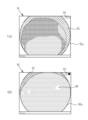
図8に示すように、観察対象の像をモニタに表示する場合、例えば、図8(A)に示すように、拡大等の操作を行わないモニタ画像91が表示する特殊画像91aでは、内視鏡先端部12dに取り付けたフード92が影を形成すること等により暗い部分が存在する。また、例えば、図8(B)に示すように、拡大の操作を行ったモニタ画像95が表示する特殊画像95aでは、照明光が透明なフード92を透過して観察対象に届き、また、ハレーション領域96が形成される。なお、図8(A)及び図8(B)において、また、図11、図12、図22、及び図23において、フード92は斜線で示す。また、ユーザーが拡大率を容易に把握できるよう、モニタ画像91及び95において、拡大率を示す拡大率表示93が示される。
As shown in FIG. 8, when the image of the observation target is displayed on the monitor, for example, as shown in FIG. A dark portion is present due to, for example, shadow formation by the
拡大率指標解析部82は、ズーム制御部57等により、拡大率指標に関する情報等を受け取り、これらの情報を解析することにより拡大率を算出する。拡大率は、特殊画像に対して画像処理を行うか否かを決定するために、画像処理実施制御部81に送られる。画像処理実施制御部81は、算出した拡大率が予め設定した閾値以上の場合に画像処理を行うと決定し、また、拡大率が予め設定した閾値より小さい場合は画像処理を行わないと決定する。
The magnification
次に、特殊画像を解析して得られる画像解析結果に基づく場合、画像解析結果として、例えば、ハレーション分布、周波数特性、輝度値、影分布、又は計測補助光の位置を用いることができる。ハレーション分布は、特殊画像が含むハレーション領域の分布を算出したものである。周波数特性は、特殊画像の空間周波数成分の分布を算出したものである。輝度値は、特殊画像の輝度値を算出したものである。影分布は、特殊画像が内視鏡先端部が備えるフードにより観察対象に形成される影を含む場合、この影の領域の分布を算出したものである。計測補助光の反射光位置は、観察対象の計測を行う場合に観察対象に照射される計測補助光が観察対象に形成する輝点の位置を算出したものである。 Next, when based on the image analysis result obtained by analyzing the special image, for example, halation distribution, frequency characteristics, brightness value, shadow distribution, or position of measurement auxiliary light can be used as the image analysis result. The halation distribution is obtained by calculating the distribution of halation regions included in the special image. The frequency characteristic is obtained by calculating the distribution of spatial frequency components of the special image. The luminance value is obtained by calculating the luminance value of the special image. The shadow distribution is obtained by calculating the distribution of the shadow area when the special image includes the shadow formed on the observation target by the hood provided at the distal end of the endoscope. The reflected light position of the measurement auxiliary light is a calculated position of a bright spot formed on the observation target by the measurement auxiliary light irradiated to the observation target when measuring the observation target.
ハレーション分布解析部83は、ハレーション分布に関する特殊画像の画像解析を行う。特殊画像において、内視鏡先端部12dと観察対象との距離によって、輝度値が周囲に比べて極端に高いハレーション領域が生じる場合がある。特殊画像がハレーション領域を含む場合は、内視鏡先端部12dと観察対象との距離が比較的近く、また、ハレーション領域を含まない場合は、内視鏡先端部12dと観察対象との距離が比較的遠い。したがって、特殊画像を解析してハレーション領域の分布を算出することにより、内視鏡先端部12dと観察対象との距離又は特殊画像の解像度の指標を得ることができる。
The halation distribution analysis unit 83 performs image analysis of the special image regarding halation distribution. In the special image, depending on the distance between the endoscope
例えば、図8(B)に示すように、特殊画像95aは、複数のハレーション領域96を含む。なお、図の煩雑さを避けるために、図8(B)において、一部のハレーション領域にのみ符号を付す。また、図8(B)は、拡大操作を行っている画像であるため、拡大率表示93により、この画像は拡大操作を行った特殊画像であることが示される。
For example, as shown in FIG. 8B, a
ハレーション分布解析部83は、予め設定した特定の輝度値以上の領域をハレーション領域とする。例えば、図9に示すように、特殊画像において、予め設定した特定の輝度値以上の領域を示すハレーション分布画像を作成する。特殊画像95aに基づき作成したハレーション分布画像95bには、図9(B)に示すように、ハレーション領域に基づく上記の特定の輝度値(S)以上の領域(Signal≧S)である高輝度領域97が形成される。また、特殊画像91aに基づき作成したハレーション分布画像91bには、図9(A)に示すように高輝度領域97が形成されない。ハレーション分布解析部83は、ハレーション分布画像91b及び95bに基づき、高輝度領域97の面積(Area(Signal≧S))をそれぞれ算出する。
The halation distribution analysis unit 83 determines a region having a predetermined luminance value or more as a halation region. For example, as shown in FIG. 9, in the special image, a halation distribution image is created that indicates a region having a luminance value equal to or higher than a predetermined specific luminance value. A
なお、図9を含む以下の図において、特殊画像等の図は、モニタに表示した場合の画像を模式的に記載するが、実際の特殊画像等の形状は問わず、場合によって適宜設定でき、円形又は矩形等であってよい。 In the following figures including FIG. 9, special images and the like are shown schematically as images displayed on a monitor. It may be circular, rectangular, or the like.
特殊画像91a及び95aに基づくハレーション分布画像91b及び95bにおけるそれぞれの高輝度領域97の面積は、特殊画像91a及び95aに対して画像処理を行うか否かを決定するために、画像処理実施制御部81に送られる。画像処理実施制御部81は、算出したそれぞれの高輝度領域97の面積が予め設定した閾値以上の場合に特殊画像に対して画像処理を行うと決定し、また、高輝度領域97の面積が予め設定した閾値より小さい特殊画像に対しては画像処理を行わないと決定する。
The areas of the
周波数特性解析部84は、周波数特性に関する特殊画像の画像解析を行う。解析により、特殊画像の空間周波数分布を算出する。例えば、内視鏡先端部12dと観察対象との距離が遠く、照明光が届かない管腔の奥深い部分は、空間周波数が周囲より低い低周波領域となる。また、例えば、内視鏡先端部12dと観察対象との距離が近く、観察対象の全てに照明光が届いている部分は、低周波領域を含まない。したがって、特殊画像について空間周波数分布を算出する解析を行うことにより、内視鏡先端部12dと観察対象との距離又は特殊画像の解像度の指標を得ることができる。
The frequency
図10(A)に示すように、特殊画像91aに基づき作成した空間周波数分布画像91cは、周囲より空間周波数が低い低周波領域(Frequency≦F)98を含む。また、図10(B)に示すように、特殊画像95aに基づき作成した空間周波数分布画像95cは、低周波領域98を含まない。周波数特性解析部84は、空間周波数分布画像91c及び95cに基づき、低周波領域98の面積(Area(Frequency≦F))をそれぞれ算出する。
As shown in FIG. 10A, a spatial
特殊画像91a及び95aに基づく空間周波数分布画像91c及び95cにおける低周波領域98の面積は、特殊画像91a及び95aに対して画像処理を行うか否かを決定するために、画像処理実施制御部81に送られる。画像処理実施制御部81は、算出した低周波領域98の面積が予め設定した閾値以下の場合に特殊画像に対して画像処理を行うと決定し、また、低周波領域98の面積が予め設定した閾値より大きい特殊画像に対しては画像処理を行わないと決定する。
The area of the low-
輝度値解析部85は、輝度値に関する特殊画像の画像解析を行う。解析により、特殊画像の輝度値分布を算出する。特殊画像に基づき算出した輝度値分布に基づき、例えば、特殊画像全体での平均輝度値を算出する。例えば、内視鏡先端部12dと観察対象との距離が遠く、照明光が届かない管腔の奥深い部分の面積が多い場合は、平均輝度値が小さくなる。また、例えば、内視鏡先端部12dと観察対象との距離が近く、観察対象の大部分に照明光が届いている部分は、平均輝度値が大きくなる。したがって、特殊画像について輝度値を算出する解析を行うことにより、内視鏡先端部12dと観察対象との距離又は特殊画像の解像度の指標を得ることができる。
The brightness
図11(A)に示すように、輝度値解析部85は、特殊画像91aに基づいて輝度値分布を算出し、輝度値分布から特殊画像91aの画像全体に対する平均輝度値BV1を算出する。また、図11(B)に示すように、特殊画像95aに基づいて輝度値分布を算出し、輝度値分布から特殊画像95aの画像全体に対する平均輝度値BV2を算出する。
As shown in FIG. 11A, the brightness
特殊画像91a及び95aのそれぞれにおける平均輝度値BV1及びBV2は、特殊画像91a及び95aに対して画像処理を行うか否かを決定するために、画像処理実施制御部81に送られる。画像処理実施制御部81は、算出した平均輝度値が予め設定した閾値以上の場合に特殊画像に対して画像処理を行うと決定し、また、平均輝度値が予め設定した閾値より小さい特殊画像に対しては画像処理を行わないと決定する。
The average brightness values BV1 and BV2 of the
影分布解析部86は、内視鏡先端部12dに取り付けたフードにより観察対象に生じる影(以下、フード影という)に関する特殊画像の画像解析を行う。解析により、特殊画像が含むフード影の領域の分布(以下、フード影領域という)を算出する。例えば、内視鏡先端部12dと観察対象との距離が遠い場合は、観察対象に対する照明光の角度が垂直から離れるため、フード影領域が大きくなる。また、例えば、内視鏡先端部12dと観察対象との距離が近い場合は、観察対象に対する照明光の角度が垂直に近くなるため、フード影領域が小さくなる。したがって、特殊画像についてフード影領域の分布を算出する解析を行うことにより、内視鏡先端部12dと観察対象との距離又は特殊画像の解像度の指標を得ることができる。
The shadow
図12(A)及び図12(B)に示すように、特殊画像91a及び特殊画像99は、それぞれフード92によるフード影領域92aを含む。影分布解析部86は、特殊画像91a及び特殊画像99に基づき、フード影領域92aの面積をそれぞれ算出する。なお、フード影領域92aの面積の算出は、例えば、画像解析により、輝度値が所定の範囲内の領域をフード影領域92aとして算出することができる。
As shown in FIGS. 12A and 12B, the
特殊画像91aに基づくフード影領域92aの面積SH1、及び特殊画像99に基づくフード影領域92aの面積SH2は、それぞれ特殊画像91a及び99に対して画像処理を行うか否かを決定するために、画像処理実施制御部81に送られる。画像処理実施制御部81は、算出したフード影領域92aの面積が予め設定した閾値より大きい場合に特殊画像に対して画像処理を行わないと決定し、また、フード影領域92aの面積が予め設定した閾値以下の特殊画像に対しては画像処理を行うと決定する。例えば、特殊画像91aに基づくフード影領域92aの面積SH1は、予め設定した閾値より大きいため、特殊画像91aに対しては画像処理を行わないと決定し、特殊画像99に基づくフード影領域92aの面積SH2は、予め設定した閾値以下であるため、特殊画像99に対しては画像処理を行うと決定する。
The area SH1 of the
計測補助光解析部87は、計測補助光の位置に関する特殊画像の画像解析を行う。計測補助光は、観察対象の計測に用いるものであり、照明光に加えて観察対象に照射される。計測補助光は、撮像センサ44の画素によって検出可能な色の光、例えば、600nm以上650nm以下の赤色光等の可視光であり、レーザー光源又はLED等の発光素子を用いる。
The measurement auxiliary
図13に示すように、計測補助光が観察対象に照射されることにより、観察対象上において、スポットSP等の計測補助光照射領域が形成される。計測補助光によるスポットSPが形成された観察対象を撮像して、計測補助光照射領域であるスポットSPを含む特殊画像100を得る。特殊画像100におけるスポットSPの位置を特定することにより、内視鏡先端部12dと観察対象との距離(以下、観察距離という)が求められる。
As shown in FIG. 13, by irradiating the observation object with the measurement auxiliary light, a measurement auxiliary light irradiation area such as a spot SP is formed on the observation object. A
特殊画像91aにおけるスポットSPの位置により、内視鏡先端部12dと観察対象との距離を求める方法について説明すると、図14に示すように、内視鏡先端部12dにおいて、計測補助光用レンズ30cが備えられ、計測補助光出射部30dから出射される計測補助光を観察対象に照射する。計測補助光の光軸Lmが撮像光学系の撮影画角(2つの実線L1で挟まれる領域内)に入る状態で、計測補助光を出射する。観察距離の範囲Rxにおいて観察可能であるとすると、範囲Rxの近端Px、中央付近Py、及び遠端Pzでは、各点での撮像範囲(矢印Qx、Qy、Qzで示す)における計測補助光によって観察対象上に形成されるスポットSPの位置(各矢印Qx、Qy、Qzが光軸Lmと交わる点)が異なることが分かる。内視鏡先端部12dの位置を位置P1とする。観察距離は、内視鏡先端部12dと観察対象との距離である。したがって、観察距離は、それぞれ、位置P1と、近端Px、中央付近Py、及び遠端Pzとの間の距離である。観察距離は、詳細には、内視鏡先端部12dにおける撮像光学系30bの光軸Axの始点から観察対象までの距離となる。軸Dvは観察距離を示す。
A method of obtaining the distance between the endoscope
計測補助光が照明された観察対象を撮像センサ44で撮像することによって、計測補助光照射領域であるスポットSPを含む特殊画像を得る。特殊画像では、スポットSPの位置は、撮像光学系30bの光軸Axと計測補助光の光軸Lmとの関係、及び観察距離に応じて異なる。したがって、予め、スポットSPの位置と観察距離とを対応付けた対応情報を取得しておくことにより、スポットSPの位置によって観察距離を得ることができる。なお、観察対象の長さ又は大きさを計測する際も、予め、観察距離と、例えば、所定の大きさのマーカとを対応付けた対応情報を取得しておくことにより、実寸を示すマーカを特殊画像上に重畳すること等により、観察対象の実寸を計測することができる。
A special image including the spot SP, which is the irradiation area of the measurement auxiliary light, is obtained by taking an image of the observation target illuminated with the measurement auxiliary light by the
計測補助光解析部87は、スポットSPを含む特殊画像の解析により、特殊画像におけるスポットSPの位置を特定する。例えば、内視鏡先端部12dと観察対象との距離が遠い場合は、スポットSPの位置が特殊画像の上部に位置する。また、例えば、内視鏡先端部12dと観察対象との距離が近い場合は、スポットSPの位置が特殊画像の中心部に位置する。したがって、特殊画像についてスポットSPの位置を特定する解析を行うことにより、内視鏡先端部12dと観察対象との距離又は特殊画像の解像度の指標を得ることができる。
The measurement auxiliary
図15(A)に示すように、特殊画像100aは、計測補助光により観察対象上に形成されたスポットSP1を含む。また、図15(B)に示すように、特殊画像100bは、計測補助光により観察対象上に形成されたスポットSP2を含む。計測補助光解析部87は、特殊画像100a及び100bのそれぞれにおいて、特殊画像100a及び100bにおけるスポットSP1及びスポットSP2の位置を、それぞれ特定する。計測補助光解析部87が予め取得する、スポットSPの位置と観察距離とを対応付けた対応情報により、計測補助光解析部87は、スポットSP1の位置から特殊画像100aにおける観察距離を、また、スポットSP2の位置から特殊画像100bにおける観察距離を算出する。なお、本実施形態では、スポットSP1の位置により特殊画像100aは観察距離がより遠いと算出され、スポットSP2の位置により特殊画像100bは観察距離がより近いと算出される。
As shown in FIG. 15A, the
算出された観察距離は、特殊画像91a及び95aに対して画像処理を行うか否かを決定するために、画像処理実施制御部81に送られる。画像処理実施制御部81は、算出した観察距離が予め設定した閾値以下の場合の特殊画像に対して画像処理を行うと決定し、また、観察距離が予め設定した閾値より大きい場合の特殊画像に対しては画像処理を行わないと決定する。
The calculated observation distance is sent to the image processing
画像処理実施制御部81が画像処理を行うと決定した特殊画像に対し、疾患関連処理部66は、特殊画像から得られる表層血管の密集、粘膜内出血、及び粘膜外出血に基づいて、潰瘍性大腸炎のステージに関する指標値を算出すること、潰瘍性大腸炎のステージを判定すること、及び潰瘍性大腸炎の寛解又は非寛解を判定することのうち少なくとも1つを行う。疾患関連処理部66にて判定対象とする潰瘍性大腸炎は、図16(A)~(E)に示すように、重症度が悪化する毎に、血管構造のパターンが変化することを発明者らが見出している。潰瘍性大腸炎が寛解、又は、潰瘍性大腸炎が発生していない場合には、表層血管のパターンが規則的であり(図16(A))、又は、表層血管のパターンの規則性に多少の乱れが生じている程度である(図16(B))。一方、潰瘍性大腸炎が非寛解であり、且つ、重症度が軽症である場合には、表層血管の密集が粗密である(図16(C))。また、潰瘍性大腸炎が非寛解であり、且つ、重症度が中等症である場合には、粘膜内出血が発生している(図16(D))。また、潰瘍性大腸炎が非寛解であり、且つ、重症度が中等症~重症である場合には、粘膜外出血が発生している(図16(E))。疾患関連処理部66では、上記の血管構造のパターン変化を利用して、医療画像の一つである特殊画像に基づいて、潰瘍性大腸炎の寛解又は非寛解を判定する。
For the special image for which the image processing
ここで、「表層血管の密集」とは、表層血管が蛇行し、集まる状態をいい、画像上の見た目では、腸腺嵩(クリプト)(図17参照)の周りを表層血管が何本も囲んでいることをいう。「粘膜内出血」とは、粘膜組織内(図17参照)の出血で内腔への出血との鑑別を要することをいう。「粘膜内出血」とは、画像上の見た目では、粘膜の中、且つ内腔(管腔、ひだの穴)ではない出血を指している。「粘膜外出血」とは、管腔内への少量の血液、管腔内を洗浄した後も内視鏡前方の管腔、又は粘膜からにじみ出て視認可能な血液、又は、出血性粘膜上でにじみを伴った管腔内の血液のことをいう。 Here, "concentration of superficial blood vessels" refers to a state in which superficial blood vessels are meandering and gathered together. From the appearance of the image, many superficial blood vessels surround the intestinal gland crypt (see FIG. 17). It means to be “Intramucosal hemorrhage” refers to bleeding within the mucosal tissue (see FIG. 17) that must be distinguished from bleeding into the lumen. "Intramucosal hemorrhage" refers to bleeding within the mucous membrane and not within the lumen (lumen, fold hole) as it appears on the image. “Extramucosal hemorrhage” is defined as a small amount of blood into the lumen, visible blood oozing out of the lumen or mucosa in front of the endoscope even after irrigation of the lumen, or blood on the bleeding mucosa. Refers to blood in the lumen with oozing.
疾患関連処理部66は、特殊画像から得られる周波数特性又は輝度値によって表層血管の密集、粘膜内出血、及び粘膜外出血を分類し、分類に従って、潰瘍性大腸炎の寛解又は非寛解を判定する。具体的には、表層血管の密集、粘膜内出血、及び粘膜外出血については、図18に示すように、分類される。表層血管の密集は、輝度値が低輝度で、且つ、周波数特性が高周波で表される。粘膜内出血は、輝度値が中輝度で、且つ、周波数特性が中周波で表される。粘膜外出血は、輝度値が低輝度で、且つ、周波数特性が低周波で表される。なお、特殊画像の各種構造を輝度値及び周波数特性で表した場合には、上記の3つの表層血管の密集、粘膜内出血、及び粘膜外出血の他に、特殊画像のボケ暗部又は内視鏡影(内視鏡の先端部12dを管腔に沿って動かす場合において内視鏡画像の中心部分にできる影)なども含まれる。本実施形態では、上記の分類を利用して、特殊画像から、潰瘍性大腸炎の寛解又は非寛解の判定に必要な表層血管の密集、粘膜内出血、及び粘膜外出血を抽出する。
The disease-related
図6に示すように、疾患関連処理部66は、空間周波数成分分布算出部71と、周波数特性領域抽出部72と、構造検出部74と、判定部76とを備えている。空間周波数成分分布算出部71は、特殊画像に対してラプラシアンフィルタをかけることにより、空間周波数成分分布を算出する。
As shown in FIG. 6 , the disease-related
周波数特性領域抽出部72は、空間周波数成分分布に基づいて、第1周波数特性(低周波)を有する第1周波数特性領域(低周波領域)を抽出し、第1周波数特性よりも高い周波数を持つ第2周波数特性(中周波)を有する第2周波数特性領域(中周波領域)を抽出し、第2周波数特性よりも高い周波数を持つ第3周波数特性(高周波)を有する第3周波数特性領域(高周波領域)を抽出する。
A frequency characteristic
具体的には、周波数特性領域抽出部72は、第1周波数特性領域抽出部72a、第3周波数特性領域抽出部72b、解析対象領域検出部72c、及び、第2周波数特性領域抽出部72dを備えており、図19に示す流れに沿って第1~第3周波数特性領域を抽出する。第1周波数特性領域抽出部72aは、空間周波数成分分布に基づいて、特定画素を含む近傍の9画素の周波数の標準偏差が一定値以下の場合に、特定画素を第1周波数特性に属する画素とする。この特定画素の検出を全画素分行うことによって、第1周波数特性領域を抽出する。第1周波数特性領域は、低周波の領域に相当する。第3周波数特性領域抽出部72bは、空間周波数成分分布に対するヘシアン解析によって、第3周波数特性領域を抽出する。第3周波数特性領域は、高周波の領域に相当する。なお、第1周波数特性領域抽出部72aでは、特定画素を含む近傍の9画素の周波数の標準偏差が一定値以下の場合に、特定画素を第1周波数特性に属する画素としているが、その他の統計量、例えば、近傍の9画素との周波数の最大値又は最小値、平均値が一定値以下の場合に、特定画素を第1周波数特性に属する画素としてもよい。
Specifically, the frequency
解析対象領域検出部72cは、特殊画像から第1周波数特性領域を除いた解析対象領域を検出する。第2周波数特性領域抽出部72dは、解析対象領域から、第3周波数特性領域を除くことにより、第2周波数特性領域を抽出する。第2周波数特性領域は、中周波の領域に相当する。
The analysis target
構造検出部74は、輝度値を用いる第1領域判別処理を施した第1周波数特性領域、輝度値を用いる第2領域判別処理を施した第2周波数特性領域、及び第3周波数特性領域に基づいて、表層血管の密集、粘膜内出血、及び粘膜外出血を検出する。具体的には、構造検出部74は、第1周波数特性領域に対して第1領域判別処理を施すことにより、粘膜外出血を検出し、第2周波数特性領域に対して第2領域判別処理を施すことにより、粘膜内出血を検出し、第3周波数特性領域を表層血管の密集として検出する。
The
低周波の第1周波数特性領域には、低輝度の粘膜外出血の他、中輝度のボケ暗部又は内視鏡影が含まれることから、これらを区別するために、第1領域判別処理が行われる。第1領域判別処理では、特殊画像の第1周波数特性領域のうち輝度値が輝度値用閾値以下の領域を、粘膜外出血の領域として検出する。中輝度の粘膜内出血を区別するために第2領域判別処理が行われる。第2領域判別処理では、特殊画像の第2周波数特性領域うち輝度値が輝度値用閾値以上の領域を、粘膜内出血の領域として検出する。なお、低輝度の表層血管の密集を区別するために第3領域判別処理を行うようにしてもよい。第3領域判別処理では、特殊画像の第3周波数特性領域のうち輝度値が輝度値用閾値以下の領域を、表層血管が密集する領域として検出する。 Since the low-frequency first frequency characteristic region includes low-brightness extramucosal hemorrhage as well as medium-brightness blurred dark areas or endoscopic shadows, first region discrimination processing is performed to distinguish between them. will be In the first area determination process, an area whose luminance value is equal to or less than the threshold value for luminance value among the first frequency characteristic areas of the special image is detected as an extramucosal bleeding area. A second area discrimination process is performed to distinguish intramucosal hemorrhage of medium brightness. In the second area determination process, an area having a luminance value equal to or greater than the threshold value for luminance value among the second frequency characteristic areas of the special image is detected as an intramucosal bleeding area. It should be noted that the third area determination process may be performed in order to distinguish the density of surface blood vessels with low brightness. In the third area determination process, an area whose luminance value is equal to or less than the threshold value for luminance value among the third frequency characteristic areas of the special image is detected as an area where superficial blood vessels are concentrated.
判定部76は、表層血管の密集があること、粘膜内出血の検出量が粘膜内出血用閾値以上であること、粘膜外出血の検出量が粘膜外出血用閾値以上であること、及び、粘膜内出血の検出量と粘膜外出血の検出量の和が粘膜内外出血用閾値以上であることのいずれかを満たす場合に、潰瘍性大腸炎が非寛解であると判定する。一方、判定部76は、表層血管の密集があること、粘膜内出血の検出量が粘膜内出血用閾値以上であること、粘膜外出血の検出量が粘膜外出血用閾値以上であること、及び、粘膜内出血の検出量と粘膜外出血の検出量の和が粘膜内外出血用閾値以上であることのいずれも満たさない場合に、潰瘍性大腸炎が寛解であると判定する。以上の判定部76での判定に関する情報は、モニタ18上に表示されて、ユーザーによる潰瘍性大腸炎の寛解又は非寛解の判定に用いられる。判定部76において潰瘍性大腸炎が寛解であると判定された場合には、図20に示すように、その旨のメッセージがモニタ18上に表示される。なお、判定に関する情報を表示する際には、判定部76での判定に用いた特殊画像を重畳表示することが好ましい。
The judging
なお、判定部76では、粘膜内出血の検出量について、特殊画像のうち第2周波数特性領域が占める割合に基づいて算出することが好ましい。また、判定部76では、粘膜外出血の検出量について、特殊画像のうち低輝度の第1周波数特性領域(第1領域判別処理後の第1周波数特性領域)が占める割合に基づいて算出することが好ましい。また、判定部76では、潰瘍性大腸炎の寛解又は非寛解の判定に加えて又は代えて、潰瘍性大腸炎の重症度を指標化した指標値を求め、指標値に従って潰瘍性大腸炎の寛解又は非寛解の判定を行い、また、指標値を判定結果としてモニタ18に表示するようにしてもよい。
It is preferable that the
なお、疾患関連処理部66において、例えば、周波数特性解析部84と空間周波数成分分布算出部71とのように、同様の機能を持つ場合はこれらを共通させてもよい。すなわち、一つの部分が、ある場合には周波数特性解析部84として機能し、また、別のある場合には空間周波数成分分布算出部71として機能するようにしてもよい。
In the disease-related
以上のように、医療画像を用いて潰瘍性大腸炎に関する判定を行うに際し、画像処理決定部70が、医療画像である特殊画像の撮影条件及び/又は特殊画像を解析して得られる画像解析結果に基づき、特殊画像に対して画像処理を行うか否かを決定し、画像処理決定部70が画像処理を行うと決定した特殊画像に対し、疾患関連処理部66が、特殊画像から得られる表層血管の密集、粘膜内出血、及び粘膜外出血に基づいて、潰瘍性大腸炎のステージに関する指標値を算出すること、潰瘍性大腸炎のステージを判定すること、及び潰瘍性大腸炎の寛解又は非寛解を判定することのうち少なくとも1つを行う画像処理装置により、画像処理の判定結果が精度良く得られる特殊画像に対して、画像処理が行われる。したがって、特殊画像を用いた潰瘍性大腸炎に関する判定を、精度良く行うことができる。
As described above, when making a determination on ulcerative colitis using a medical image, the image
また、場合により、予め設定された内容により、画像処理実施制御部81が特殊画像の撮影条件及び/又は特殊画像を解析して得られる画像解析結果に基づき、画像処理を行うか否かを決定するため、自動的に選択された特殊画像に対して、画像処理が行われる。したがって、特殊画像を用いた潰瘍性大腸炎に関する判定を、精度良く、しかも、簡便に行うことができる。
In some cases, the image processing
次に、疾患関連処理モードの一連の流れについて、図21に示すフローチャートに沿って説明を行う。疾患関連処理モードに切り替えられると、特殊光が観察対象に照射される。内視鏡12は、特殊光によって照明された観察対象を撮像することにより(ステップST110)、ある時点で内視鏡画像(医療画像)の一つである特殊画像を得る。画像取得部50は、内視鏡12からの特殊画像を取得する(ステップST120)。
Next, a series of flows in the disease-related processing mode will be described with reference to the flowchart shown in FIG. When switched to the disease-related processing mode, the observation target is illuminated with special light. The
特殊画像は、疾患関連処理部66に送られ、画像処理決定部70により、画像処理を行うか否か、つまり、画像処理の是非を決定する(ステップST130)。本実施形態では、周波数特性と、ハレーション分布との、2種類の解析を行い、画像処理実施制御部81が、これらの解析結果を統合して、画像処理の是非を決定する。
The special image is sent to the disease-related
周波数特性解析部84が、特殊画像の周波数特性を算出し、空間周波数分布画像を作成する。空間周波数分布画像は、空間周波数に応じて各色のフィルターをかけることにより作成できる。例えば、低周波領域98aを水色、高周波領域98bを赤色等とする。周波数特性解析部84は、例えば、空間周波数分布画像において、周波数がF以下の面積を算出し、予め設定された低周波領域の閾値との比較を行う。比較は、例えば、以下の式(1)により行う。ここで、周波数がF以下の面積を、Area(Frequency≦F)、低周波領域の閾値を、Threshold_Lowflequencyとする。以下の式(1)を満たす特殊画像は、観察対象が内視鏡先端部12dから遠いこと、または、管腔奥深くが存在することを示す。
A frequency
Area(Frequency≦F)>Threshold_Lowflequency (1) Area (Frequency≤F) > Threshold_Low frequency (1)
次に、ハレーション分布解析部83が、特殊画像のハレーション分布を算出し、ハレーション分布画像を作成する。ハレーション分布画像は、輝度値が特定値以上の領域を算出することにより作成できる。例えば、ハレーション分布画像は、輝度値が特定値に応じて、特定の輝度値以上の領域に各色のフィルターをかけることにより作成できる。例えば、ハレーション領域を白色等とする。ハレーション分布解析部83は、例えば、ハレーション分布画像において、輝度値がS以上の面積を算出し、予め設定されたハレーション領域の閾値との比較を行う。比較は、例えば、以下の式(2)により行う。ここで、輝度値がS以上の面積を、Area(Signal≧S)、ハレーション領域の閾値を、Threshold_Halarionとする。以下の式(2)を満たす特殊画像は、観察対象が内視鏡先端部12dから近いことを示すハレーション領域が存在しないことを示す。
Next, the halation distribution analysis unit 83 calculates the halation distribution of the special image and creates a halation distribution image. A halation distribution image can be created by calculating an area in which the luminance value is equal to or greater than a specific value. For example, a halation distribution image can be created by applying a filter of each color to an area having a luminance value equal to or higher than a specific luminance value according to the specific value. For example, the halation region is assumed to be white. For example, the halation distribution analysis unit 83 calculates an area having a luminance value of S or more in the halation distribution image, and compares it with a preset threshold for the halation region. The comparison is performed, for example, by the following formula (2). Here, let Area (Signal≧S) be an area where the luminance value is S or more, and Threshold_Halarion be the threshold value of the halation area. A special image that satisfies the following equation (2) indicates that there is no halation region indicating that the observation target is close to the
Area(Signal≧S)<Threshold_Halarion (2) Area (Signal≧S)<Threshold_Halarion (2)
画像処理実施制御部81は、周波数特性解析部84とハレーション分布解析部83とから、低周波領域98aの面積Area(Frequency≦F)、及びハレーション領域の面積Area(Signal≧S)を受け取り、上記式(1)及び(2)により、それぞれの閾値と比較する。さらに、上記式(1)かつ上記式(2)を満たす特殊画像は、内視鏡先端部12dから観察対象までの距離が遠く、解像度が低いため、画像処理によって例えば潰瘍性大腸炎のステージを判定するのに不都合であるから、画像処理を実施しないと決定する(ステップST140でNO)。例えば、図22に示すように、特殊画像91aに基づく空間周波数分布画像91cが式(1)を満たし、特殊画像91aに基づくハレーション分布画像91bが式(2)を満たすため、画像処理決定部70は、画像処理を行わないと決定する。画像処理決定部70が、画像処理を行わないと決定した場合、再度医療画像を取得する。
The image processing
また、上記式(1)を満たさず、かつ、上記式(2)を満たさない特殊画像は、内視鏡先端部12dから観察対象までの距離が近く、解像度が高いため、画像処理によって例えば潰瘍性大腸炎のステージを判定するのに好都合であるから、画像処理を実施すると決定する(ステップST140でYES)。例えば、図23に示すように、特殊画像95aは、特殊画像95aに基づく空間周波数分布画像95cが式(1)を満たさず、特殊画像95aに基づくハレーション分布画像95bが式(2)を満たさないため、画像処理決定部70は、画像処理を行なうと決定する。画像処理決定部70が、画像処理を行うと決定した場合、次のステップに進む。
A special image that does not satisfy the above formula (1) and does not satisfy the above formula (2) has a short distance from the
また、上記式(1)と上記式(2)のいずれか一方を満たす特殊画像も、内視鏡先端部12dから観察対象までの距離が近く、解像度が高いため、画像処理によって例えば潰瘍性大腸炎のステージを判定するのに好都合とまでは言えないが問題はないため、画像処理を実施すると決定する(ステップST140でYES)。
In addition, since the special image that satisfies either the above formula (1) or the above formula (2) also has a short distance from the endoscope
画像処理を実施すると決定された特殊画像に対して、画像処理を行う。空間周波数成分分布算出部71は、特殊画像から空間周波数成分分布を算出する(ステップST160)。第1周波数特性領域抽出部72aは、空間周波数成分分布に基づいて、低周波の第1周波数特性領域を抽出する(ステップST170)。また、第3周波数特性領域抽出部72bは、空間周波数成分分布に基づいて、高周波の第3周波数特性領域を抽出する(ステップST180)。解析対象領域検出部72cは、医療画像から第1周波数特性領域を除いた解析対象領域を検出する(ステップST190)。第2周波数特性領域抽出部72dは、解析対象領域から第3周波数特性領域を除くことにより、中周波の第2周波数特性領域を抽出する(ステップST200)。
Image processing is performed on the special image determined to be subjected to image processing. The spatial frequency component distribution calculator 71 calculates the spatial frequency component distribution from the special image (step ST160). The first frequency characteristic
構造検出部74において、低周波の第1周波数特性領域に対して、第1領域判別処理を行うことにより、粘膜外出血を検出する(ステップST210)。また、構造検出部74において、中周波の第2周波数特性領域に対して、第2領域判別処理を行うことにより、粘膜内出血を検出する(ステップST220)。また、構造検出部74では、第3周波数特性領域を、表層血管の密集として検出する。
The
判定部76は、構造検出部74によって検出された表層血管の密集、粘膜内出血、及び粘膜外出血に基づいて、潰瘍性大腸炎の寛解又は非寛解を判定する(ステップST230)。判定部76での判定に関する情報は、モニタ18に表示される(ステップST240)。
The
なお、疾患関連処理部66が、画像処理を行うと決定した特殊画像に対し、画像処理実施制御部81から送られた特殊画像の撮影条件及び/又は特殊画像を解析して得られる画像解析結果を用いる疾患関連処理モードの一連の流れについては、図24に示すフローチャートに沿って説明を行う。図24に示すフローチャートは、図21に示すフローチャートにおいて、ステップST150が追加されたものである。すなわち、画像処理を実施すると決定された特殊画像に対して画像処理を行う前に、画像処理実施制御部81から、特殊画像の撮影条件及び/又は特殊画像を解析して得られる画像解析結果である、画像処理のパラメータを取得する(ステップST150)。
For the special image for which the disease-related
画像処理のパラメータについては、例えば、炎症性疾患は血管が密集している領域が多いほど重症であるという知見に基づき、特殊画像が含む観察対象の拡大率が小さい場合は、表層血管の密集を評価するために表層血管の密集度を算出する場合、表層血管の密集度を算出するために用いるカーネルサイズも小さくする必要がある。上記式(1)及び上記式(2)の両方を満たす特殊画像は、拡大率が小さいと判断できる。したがって、画像処理実施制御部81は、上記式(1)及び上記式(2)の両方を満たす特殊画像であるとの画像処理結果を、疾患関連処理部66に送る。疾患関連処理部66は、画像処理実施制御部81から送られた、上記式(1)及び上記式(2)の両方を満たす特殊画像であるとの画像処理結果を用いて、表層血管の密集度を算出する場合のカーネルサイズとして、予め数種類準備しておいたカーネルサイズの中から、低倍率用カーネルを用いるとの画像処理パラメータを設定する。その後の流れは、図21に示すフローチャートと同様である。
Regarding image processing parameters, for example, based on the knowledge that inflammatory diseases are more severe when there are more areas with dense blood vessels, when the magnification of the observation target including the special image is small, the density of superficial blood vessels is reduced. When calculating the density of superficial blood vessels for evaluation, it is necessary to reduce the kernel size used for calculating the density of superficial blood vessels. A special image that satisfies both the above formula (1) and the above formula (2) can be determined to have a small enlargement ratio. Accordingly, the image processing
[第2実施形態]
第2実施形態では、上記第1実施形態で示した4色のLED20a~20dの代わりに、キセノンランプなどの広帯域光源と回転フィルタを用いて観察対象の照明を行う。また、カラーの撮像センサ44に代えて、モノクロの撮像センサで観察対象の撮像を行う。それ以外については、第1実施形態と同様である。
[Second embodiment]
In the second embodiment, instead of the four-color LEDs 20a to 20d shown in the first embodiment, a broadband light source such as a xenon lamp and a rotary filter are used to illuminate the observation target. Further, instead of the
図25に示すように、第2実施形態の内視鏡システム100では、光源装置14において、4色のLED20a~20dに代えて、広帯域光源102、回転フィルタ104、フィルタ切替部105が設けられている。また、撮像光学系30bには、カラーの撮像センサ44の代わりに、カラーフィルタが設けられていないモノクロの撮像センサ106が設けられている。
As shown in FIG. 25, in the
広帯域光源102はキセノンランプ、白色LEDなどであり、波長域が青色から赤色に及ぶ白色光を発する。回転フィルタ104には、内側から順に、通常光モード用フィルタ107と、特殊光モード及び疾患関連処理モード用フィルタ108とが設けられている(図26参照)。フィルタ切替部105は、回転フィルタ104を径方向に移動させるものであり、モード切替SW12fにより通常光モードにセットしたときに、通常光モード用フィルタ107を白色光の光路に挿入し、特殊光モード又は疾患関連処理モードにセットしたときに、特殊光モード及び疾患関連処理モード用フィルタ108を白色光の光路に挿入する。
The
図26に示すように、通常光モード用フィルタ107には、周方向に沿って、白色光のうち広帯域青色光Bを透過させるBフィルタ107a、白色光のうち広帯域緑色光Gを透過させるGフィルタ107b、及び、白色光のうち広帯域赤色光Rを透過させるRフィルタ107cが設けられている。したがって、通常光モード時には、回転フィルタ104が回転することで、通常光として、広帯域青色光B、広帯域緑色光G、広帯域赤色光Rが交互に観察対象に照射される。
As shown in FIG. 26, the normal
特殊光モード及び疾患関連処理モード用フィルタ108には、周方向に沿って、白色光のうち青色狭帯域光を透過させるBnフィルタ108a、及び、白色光のうち緑色狭帯域光を透過させるGnフィルタ108bが設けられている。したがって、特殊光モード又は疾患関連処理モード時には、回転フィルタ104が回転することで、特殊光として、短波長の光としての青色狭帯域光と緑色狭帯域光が交互に観察対象に照射される。なお、青色狭帯域光の波長帯域は400~450nmであり、緑色狭帯域光の波長帯域は540~560nmであることが好ましい。
In the special light mode and disease-related
内視鏡システム100では、通常光モード時には、広帯域青色光B、広帯域緑色光G、広帯域赤色光Rで観察対象が照明される毎にモノクロの撮像センサ106で観察対象を撮像する。これにより、Bc画像信号、Gc画像信号、Rc画像信号が得られる。そして、それら3色の画像信号に基づいて、上記第1実施形態と同様の方法で、通常画像が生成される。
In the
内視鏡システム100では、特殊光モード又は疾患関連処理モード時には、青色狭帯域光と緑色狭帯域光で観察対象が照明される毎にモノクロの撮像センサ106で観察対象を撮像する。これにより、Bs画像信号、Gs画像信号が得られる。そして、それら2色の画像信号に基づいて、上記第1実施形態と同様の方法で、特殊画像が生成される。
In the
[第3実施形態]
第3実施形態では、上記第1実施形態で示した4色のLED20a~20dの代わりに、レーザ光源と蛍光体を用いて観察対象の照明を行う。以下においては、第1実施形態と異なる部分のみ説明を行い、第1実施形態と略同様の部分については、説明を省略する。
[Third embodiment]
In the third embodiment, instead of the four-color LEDs 20a to 20d shown in the first embodiment, a laser light source and phosphors are used to illuminate the observation target. In the following, only parts different from the first embodiment will be explained, and explanations of parts that are substantially the same as the first embodiment will be omitted.
図27に示すように、第3実施形態の内視鏡システム200では、光源装置14の光源部20において、4色のLED20a~20dの代わりに、短波長の光に相当する中心波長405±10nmの紫色レーザ光を発する紫色レーザ光源部203(「405LD」と表記。LDは「Laser Diode」を表す)と、中心波長445±10nmの青色レーザ光を発する青色レーザ光源部(「445LD」と表記)204とが設けられている。これら各光源部203、204の半導体発光素子からの発光は、光源制御部208により個別に制御されている。
As shown in FIG. 27, in the
光源制御部208は、通常光モードの場合には、青色レーザ光源部204を点灯させる。これに対して、特殊光モード又は疾患関連処理モードの場合には、紫色レーザ光源部203と青色レーザ光源部204を同時点灯させる。
The light
なお、紫色レーザ光、青色レーザ光、又は、青緑色レーザ光の半値幅は±10nm程度にすることが好ましい。また、紫色レーザ光源部203及び青色レーザ光源部204は、ブロードエリア型のInGaN系レーザダイオードが利用でき、また、InGaNAs系レーザダイオードやGaNAs系レーザダイオードを用いることもできる。また、上記光源として、発光ダイオードなどの発光体を用いた構成としてもよい。
It is preferable that the half width of the violet laser light, the blue laser light, or the blue-green laser light is about ±10 nm. Further, the violet laser
照明光学系30aには、照明レンズ32の他に、ライトガイド25からの青色レーザ光又は青緑色レーザ光が入射する蛍光体210が設けられている。蛍光体210は、青色レーザ光によって励起され、蛍光を発する。したがって、青色レーザ光は励起光に相当する。また、青色レーザ光の一部は、蛍光体210を励起させることなく透過する。青緑色レーザ光は、蛍光体210を励起させることなく透過する。蛍光体210を出射した光は、照明レンズ32を介して、観察対象の体内を照明する。
In addition to the
ここで、通常光モードにおいては、主として青色レーザ光が蛍光体210に入射するため、図28に示すように、青色レーザ光、及び青色レーザ光により蛍光体210から励起発光する蛍光を合波した通常光が観察対象に照明される。この通常光で照明された観察対象を撮像センサ44で撮像することによって、Bc画像信号、Gc画像信号、Rc画像信号からなる通常画像が得られる。
Here, in the normal light mode, mainly the blue laser light is incident on the
また、特殊光モード又は疾患関連処理モードにおいては、紫色レーザ光及び青色レーザ光が蛍光体210に同時に入射することにより、図29に示すように、紫色レーザ光及び青色レーザ光に加えて、紫色レーザ光及び青色レーザ光によって蛍光体210から励起発光する蛍光を含む疑似白色光が、特殊光として発せられる。この特殊光で照明された観察対象を撮像センサ44で撮像することによって、Bs画像信号、Gs画像信号、Rs画像信号からなる特殊画像が得られる。なお、疑似白色光は、V-LED20a、B-LED20b、G-LED20c、及びR-LED20dから発せられる紫色光V、青色光B、緑色光G、及び赤色光を組み合わせた光としてもよい。
Further, in the special light mode or the disease-related treatment mode, the violet laser light and the blue laser light simultaneously enter the
なお、蛍光体210は、青色レーザ光の一部を吸収して、緑色~黄色に励起発光する複数種の蛍光体(例えばYKG系蛍光体、或いはBAM(BaMgAl10O17)などの蛍光体)を含んで構成されるものを使用することが好ましい。本構成例のように、半導体発光素子を蛍光体210の励起光源として用いれば、高い発光効率で高強度の白色光が得られ、白色光の強度を容易に調整できる上に、白色光の色温度、色度の変化を小さく抑えることができる。Note that the
なお、上記実施形態では、医療画像の一つである内視鏡画像の処理を行う内視鏡システムに対して、本発明の適用を行っているが、内視鏡画像以外の医療画像を処理する医療画像処理システムに対しても本発明の適用は可能である。また、医療画像を用いてユーザーに診断支援を行うための診断支援装置に対しても本発明の適用は可能である。また、医療画像を用いて、診断レポートなどの医療業務を支援するための医療業務支援装置に対しても本発明の適用は可能である。 In the above embodiment, the present invention is applied to an endoscope system that processes endoscopic images, which are one type of medical image. The present invention can also be applied to a medical image processing system that In addition, the present invention can also be applied to a diagnosis support device for providing diagnosis support to a user using medical images. The present invention can also be applied to a medical work support apparatus for supporting medical work such as making a diagnosis report using medical images.
例えば、図30に示すように、診断支援装置600は、医療画像処理システム602などのモダリティやPACS(Picture Archiving and Communication Systems)604を組み合わせて使用される。また、図31に示すように、医療業務支援装置610は、第1医療画像処理システム621、第2医療画像処理システム622、…、第N医療画像処理システム623等の各種検査装置と任意のネットワーク626を介して接続する。医療業務支援装置610は、第1~第N医療画像処理システム621、622・・・、623からの医療画像を受信し、受信した医療画像に基づいて、医療業務の支援を行う。
For example, as shown in FIG. 30, a
上記実施形態において、画像処理部58に含まれる通常画像生成部62、特殊画像生成部64、疾患関連処理部66、空間周波数成分分布算出部71、周波数特性領域抽出部72、第1周波数特性領域抽出部72a、第3周波数特性領域抽出部72b、解析対象領域検出部72c、第2周波数特性領域抽出部72d、構造検出部74、判定部76、画像処理実施制御部81、拡大率指標解析部82、ハレーション分布解析部83、周波数特性解析部84、輝度値解析部85、影分布解析部86、及び、計測補助光解析部87といった各種の処理を実行する処理部(processing unit)のハードウェア的な構造は、次に示すような各種のプロセッサ(processor)である。各種のプロセッサには、ソフトウエア(プログラム)を実行して各種の処理部として機能する汎用的なプロセッサであるCPU(Central Processing Unit)、FPGA (Field Programmable Gate Array) などの製造後に回路構成を変更可能なプロセッサであるプログラマブルロジックデバイス(Programmable Logic Device:PLD)、各種の処理を実行するために専用に設計された回路構成を有するプロセッサである専用電気回路などが含まれる。
In the above embodiment, the normal
1つの処理部は、これら各種のプロセッサのうちの1つで構成されてもよいし、同種または異種の2つ以上のプロセッサの組み合せ(例えば、複数のFPGAや、CPUとFPGAの組み合わせ)で構成されてもよい。また、複数の処理部を1つのプロセッサで構成してもよい。複数の処理部を1つのプロセッサで構成する例としては、第1に、クライアントやサーバなどのコンピュータに代表されるように、1つ以上のCPUとソフトウエアの組み合わせで1つのプロセッサを構成し、このプロセッサが複数の処理部として機能する形態がある。第2に、システムオンチップ(System On Chip:SoC)などに代表されるように、複数の処理部を含むシステム全体の機能を1つのIC(Integrated Circuit)チップで実現するプロセッサを使用する形態がある。このように、各種の処理部は、ハードウェア的な構造として、上記各種のプロセッサを1つ以上用いて構成される。 One processing unit may be composed of one of these various processors, or composed of a combination of two or more processors of the same type or different types (for example, a plurality of FPGAs or a combination of a CPU and an FPGA). may be Also, a plurality of processing units may be configured by one processor. As an example of configuring a plurality of processing units in one processor, first, as represented by computers such as clients and servers, one processor is configured by combining one or more CPUs and software, There is a form in which this processor functions as a plurality of processing units. Secondly, as typified by System On Chip (SoC), etc., there is a form of using a processor that realizes the functions of the entire system including multiple processing units with a single IC (Integrated Circuit) chip. be. In this way, the various processing units are configured using one or more of the above various processors as a hardware structure.
さらに、これらの各種のプロセッサのハードウェア的な構造は、より具体的には、半導体素子などの回路素子を組み合わせた形態の電気回路(circuitry)である。 Further, the hardware structure of these various processors is, more specifically, an electrical circuit in the form of a combination of circuit elements such as semiconductor elements.
本発明は、下記の別形態によっても実施可能である。
プロセッサ装置において、
画像取得部により、観察対象を撮像して得られる医療画像を取得し、
画像処理決定部により、前記医療画像の撮影条件及び/又は前記医療画像を解析して得られる画像解析結果に基づき、前記医療画像に対して画像処理を行うか否かを決定し、
疾患関連処理部により、前記画像処理決定部が前記画像処理を行うと決定した前記医療画像に対し、前記医療画像から得られる表層血管の密集、粘膜内出血、及び粘膜外出血に基づいて、潰瘍性大腸炎のステージに関する指標値を算出すること、前記潰瘍性大腸炎のステージを判定すること、及び前記潰瘍性大腸炎の寛解又は非寛解を判定することのうち少なくとも1つを行う、プロセッサ装置。The present invention can also be implemented in the following alternative forms.
in the processor device,
an image acquisition unit acquires a medical image obtained by capturing an image of an observation target;
determining whether or not to perform image processing on the medical image based on the imaging conditions of the medical image and/or an image analysis result obtained by analyzing the medical image, by an image processing determination unit;
The disease-related processing unit determines whether the medical image for which the image processing determination unit is to perform the image processing is ulcerative based on superficial blood vessel congestion, intramucosal hemorrhage, and extramucosal hemorrhage obtained from the medical image. A processor device that performs at least one of calculating an index value for a stage of colitis, determining the stage of the ulcerative colitis, and determining remission or non-remission of the ulcerative colitis.
10 内視鏡システム
12 内視鏡
12a 挿入部
12b 操作部
12c 湾曲部
12d 先端部
12e アングルノブ
12f モード切替スイッチ
12g ズーム操作部
14 光源装置
16 プロセッサ装置
18 モニタ
19 コンソール
20 光源部
20a V-LED
20b B-LED
20c G-LED
20d R-LED
21 光源制御部
23 光路結合部
25 ライトガイド
30a 照明光学系
30b 撮像光学系
30c 計測補助光用レンズ
30d 計測補助光出射部
32 照明レンズ
42 対物レンズ
44 撮像センサ
45 撮像制御部
46 CDS/AGC回路
48 A/Dコンバータ
50 画像取得部
52 DSP
54 ノイズ低減部
56 画像処理切替部
57 ズーム制御部
58 画像処理部
60 映像信号生成部
62 通常画像生成部
64 特殊画像生成部
66 疾患関連処理部
70 画像処理決定部
71 空間周波数成分分布算出部
72 周波数特性領域抽出部
72a 第1周波数特性領域抽出部
72b 第3周波数特性領域抽出部
72c 解析対象領域検出部
72d 第2周波数特性領域抽出部
74 構造検出部
76 判定部
81 画像処理実施制御部
82 拡大率指標解析部
83 ハレーション分布解析部
84 周波数特性解析部
85 輝度値解析部
86 影分布解析部
87 計測補助光解析部
91、95 モニタ画像
91a、95a、99、100、100a、100b 特殊画像
91b、95b ハレーション分布画像
91c、95c 空間周波数分布画像
92 フード
92a フード影領域
93 拡大率表示
96 ハレーション領域
97 高輝度領域
98 低周波領域
101 内視鏡システム
102 広帯域光源
104 回転フィルタ
105 フィルタ切替部
106 撮像センサ
107 通常光モード用フィルタ
107a Bフィルタ
107b Gフィルタ
107c Rフィルタ
108 特殊光モード及び疾患関連処理モード用フィルタ
108a Bnフィルタ
108b Gnフィルタ
200 内視鏡システム
203 紫色レーザ光源部
204 青色レーザ光源部
208 光源制御部
210 蛍光体
600 診断支援装置
602 医療画像処理システム
604 PACS
610 医療業務支援装置
621 第1医療画像処理システム
622 第2医療画像処理システム
623 第N医療画像処理システム
626 ネットワーク
SP、SP1、SP2 スポット
Lm 光軸
Rx 範囲
Px 近端
Py 中央付近
Pz 遠端
Qx、Qy、Qz 撮像範囲
P1 位置
Ax 光軸
Dv 観察距離10
20b B-LED
20c G-LED
20d R-LED
21 light
54 noise reduction unit 56 image processing switching unit 57 zoom control unit 58 image processing unit 60 video signal generation unit 62 normal image generation unit 64 special image generation unit 66 disease-related processing unit 70 image processing determination unit 71 spatial frequency component distribution calculation unit 72 Frequency characteristic area extractor 72a First frequency characteristic area extractor 72b Third frequency characteristic area extractor 72c Analysis target area detector 72d Second frequency characteristic area extractor 74 Structure detector 76 Judging unit 81 Image processing execution controller 82 Enlarge rate index analysis unit 83 halation distribution analysis unit 84 frequency characteristic analysis unit 85 luminance value analysis unit 86 shadow distribution analysis unit 87 measurement auxiliary light analysis units 91, 95 monitor images 91a, 95a, 99, 100, 100a, 100b special image 91b, 95b halation distribution images 91c and 95c spatial frequency distribution image 92 hood 92a hood shadow region 93 magnification display 96 halation region 97 high luminance region 98 low frequency region 101 endoscope system 102 broadband light source 104 rotating filter 105 filter switching unit 106 imaging sensor 107 Normal light mode filter 107a B filter 107b G filter 107c R filter 108 Special light mode and disease-related processing mode filter 108a Bn filter 108b Gn filter 200 Endoscope system 203 Violet laser light source unit 204 Blue laser light source unit 208 Light source control Unit 210 Phosphor 600 Diagnosis support device 602 Medical image processing system 604 PACS
610 Medical
Claims (17)
前記プロセッサは、
観察対象を撮像して得られる医療画像を取得し、
前記医療画像の撮影条件及び/又は前記医療画像を解析して得られる画像解析結果に基づき、前記医療画像に対して画像処理を行うか否かを決定し、
前記画像処理を行うと決定した前記医療画像に対し、前記医療画像から得られる表層血管の密集、粘膜内出血、及び粘膜外出血に基づいて、潰瘍性大腸炎のステージに関する指標値を算出すること、前記潰瘍性大腸炎のステージを判定すること、及び前記潰瘍性大腸炎の寛解又は非寛解を判定することのうち少なくとも1つを行い、
前記医療画像は、前記観察対象の計測を行うための計測補助光が照射された前記観察対象を撮像して得られ、
前記画像解析結果は、前記医療画像において前記観察対象上に形成された計測補助光照射領域の位置である画像処理装置。 with a processor
The processor
Acquiring a medical image obtained by imaging an observation target,
determining whether or not to perform image processing on the medical image based on imaging conditions of the medical image and/or image analysis results obtained by analyzing the medical image;
calculating an index value related to the stage of ulcerative colitis for the medical image determined to be subjected to the image processing, based on superficial blood vessel congestion, intramucosal bleeding, and extramucosal bleeding obtained from the medical image; at least one of determining the stage of the ulcerative colitis and determining remission or non-remission of the ulcerative colitis ;
The medical image is obtained by imaging the observation target irradiated with measurement auxiliary light for measuring the observation target,
The image processing apparatus , wherein the image analysis result is a position of a measurement auxiliary light irradiation area formed on the observation object in the medical image .
前記プロセッサは、
観察対象を撮像して得られる医療画像を取得し、
前記医療画像の撮影条件及び/又は前記医療画像を解析して得られる画像解析結果に基づき、前記医療画像に対して画像処理を行うか否かを決定し、
前記画像処理を行うと決定した前記医療画像に対し、前記医療画像から得られる表層血管の密集、粘膜内出血、及び粘膜外出血に基づいて、潰瘍性大腸炎のステージに関する指標値を算出すること、前記潰瘍性大腸炎のステージを判定すること、及び前記潰瘍性大腸炎の寛解又は非寛解を判定することのうち少なくとも1つを行い、
前記プロセッサが前記潰瘍性大腸炎の寛解又は非寛解を判定する場合には、
前記プロセッサは、
前記医療画像から空間周波数成分分布を算出し、
前記空間周波数成分分布に基づいて、第1周波数特性を有する第1周波数特性領域を抽出し、前記第1周波数特性よりも高い周波数を持つ第2周波数特性を有する第2周波数特性領域を抽出し、前記第2周波数特性よりも高い周波数を持つ第3周波数特性を有する第3周波数特性領域を抽出し、
輝度値を用いる第1領域判別処理を施した前記第1周波数特性領域、輝度値を用いる第2領域判別処理を施した前記第2周波数特性領域、及び前記第3周波数特性領域に基づいて、前記表層血管の密集、前記粘膜内出血、及び前記粘膜外出血を検出し、
前記表層血管の密集、前記粘膜内出血、及び前記粘膜外出血に基づいて、前記潰瘍性大腸炎の寛解又は非寛解を判定する画像処理装置。 with a processor
The processor
Acquiring a medical image obtained by imaging an observation target,
determining whether or not to perform image processing on the medical image based on imaging conditions of the medical image and/or image analysis results obtained by analyzing the medical image;
calculating an index value related to the stage of ulcerative colitis for the medical image determined to be subjected to the image processing, based on superficial blood vessel congestion, intramucosal bleeding, and extramucosal bleeding obtained from the medical image; at least one of determining the stage of the ulcerative colitis and determining remission or non-remission of the ulcerative colitis ;
If the processor determines remission or non-remission of the ulcerative colitis,
The processor
calculating a spatial frequency component distribution from the medical image;
Based on the spatial frequency component distribution, extracting a first frequency characteristic region having a first frequency characteristic, extracting a second frequency characteristic region having a second frequency characteristic having a frequency higher than the first frequency characteristic, extracting a third frequency characteristic region having a third frequency characteristic having a frequency higher than that of the second frequency characteristic;
Based on the first frequency characteristic area subjected to the first area determination process using the luminance value, the second frequency characteristic area subjected to the second area determination process using the luminance value, and the third frequency characteristic area, detecting superficial vascular congestion, intramucosal hemorrhage, and extramucosal hemorrhage;
An image processing apparatus for determining remission or non-remission of the ulcerative colitis based on the congestion of the superficial blood vessels, the intramucosal hemorrhage, and the extramucosal hemorrhage .
前記プロセッサは、
観察対象を撮像して得られる医療画像を取得し、
前記医療画像を解析して得られる画像解析結果に基づき、前記医療画像に対して画像処理を行うか否かを決定し、
前記画像処理を行うと決定した前記医療画像に対し、前記医療画像から得られる表層血管の密集、粘膜内出血、及び粘膜外出血に基づいて、潰瘍性大腸炎のステージに関する指標値を算出すること、前記潰瘍性大腸炎のステージを判定すること、及び前記潰瘍性大腸炎の寛解又は非寛解を判定することのうち少なくとも1つを行い、
前記画像解析結果は、前記医療画像から得られるハレーション分布、周波数特性、及び影分布のうち少なくとも1つである画像処理装置。 with a processor
The processor
Acquiring a medical image obtained by imaging an observation target,
determining whether or not to perform image processing on the medical image based on an image analysis result obtained by analyzing the medical image;
calculating an index value related to the stage of ulcerative colitis for the medical image determined to be subjected to the image processing, based on superficial blood vessel congestion, intramucosal bleeding, and extramucosal bleeding obtained from the medical image; at least one of determining the stage of the ulcerative colitis and determining remission or non-remission of the ulcerative colitis ;
The image processing apparatus , wherein the image analysis result is at least one of halation distribution, frequency characteristics, and shadow distribution obtained from the medical image .
前記プロセッサは、
観察対象を内視鏡により撮像して得られる医療画像を取得し、
前記医療画像の撮影条件及び/又は前記医療画像を解析して得られる画像解析結果に基づいて得られる前記医療画像を撮影した際の前記内視鏡と前記観察対象との距離の指標を用いて、前記医療画像に対して画像処理を行うか否かを決定し、
前記画像処理を行うと決定した前記医療画像に対し、前記医療画像から得られる表層血管の密集、粘膜内出血、及び粘膜外出血に基づいて、潰瘍性大腸炎のステージに関する指標値を算出すること、前記潰瘍性大腸炎のステージを判定すること、及び前記潰瘍性大腸炎の寛解又は非寛解を判定することのうち少なくとも1つを行う画像処理装置。 with a processor
The processor
Acquiring a medical image obtained by imaging an observation target with an endoscope ,
Using an index of the distance between the endoscope and the observation target when the medical image is captured based on the imaging conditions of the medical image and/or the image analysis result obtained by analyzing the medical image to determine whether or not to perform image processing on the medical image;
calculating an index value related to the stage of ulcerative colitis for the medical image determined to be subjected to the image processing, based on superficial blood vessel congestion, intramucosal bleeding, and extramucosal bleeding obtained from the medical image; An image processing apparatus that performs at least one of determining the stage of the ulcerative colitis and determining remission or non-remission of the ulcerative colitis.
前記画像解析結果は、前記医療画像において前記観察対象上に形成された計測補助光照射領域の位置である請求項4ないし6のいずれか1項に記載の画像処理装置。 The medical image is obtained by imaging the observation target irradiated with measurement auxiliary light for measuring the observation target,
7. The image processing apparatus according to any one of claims 4 to 6 , wherein said image analysis result is a position of a measurement auxiliary light irradiation area formed on said observation object in said medical image.
前記プロセッサは、
前記医療画像から空間周波数成分分布を算出し、
前記空間周波数成分分布に基づいて、第1周波数特性を有する第1周波数特性領域を抽出し、前記第1周波数特性よりも高い周波数を持つ第2周波数特性を有する第2周波数特性領域を抽出し、前記第2周波数特性よりも高い周波数を持つ第3周波数特性を有する第3周波数特性領域を抽出し、
輝度値を用いる第1領域判別処理を施した前記第1周波数特性領域、輝度値を用いる第2領域判別処理を施した前記第2周波数特性領域、及び前記第3周波数特性領域に基づいて、前記表層血管の密集、前記粘膜内出血、及び前記粘膜外出血を検出し、
前記表層血管の密集、前記粘膜内出血、及び前記粘膜外出血に基づいて、前記潰瘍性大腸炎の寛解又は非寛解を判定する請求項3ないし9のいずれか1項に記載の画像処理装置。 If the processor determines remission or non-remission of the ulcerative colitis,
The processor
calculating a spatial frequency component distribution from the medical image;
Based on the spatial frequency component distribution, extracting a first frequency characteristic region having a first frequency characteristic, extracting a second frequency characteristic region having a second frequency characteristic having a frequency higher than the first frequency characteristic, extracting a third frequency characteristic region having a third frequency characteristic having a frequency higher than that of the second frequency characteristic;
Based on the first frequency characteristic area subjected to the first area determination process using the luminance value, the second frequency characteristic area subjected to the second area determination process using the luminance value, and the third frequency characteristic area, detecting superficial vascular congestion, intramucosal hemorrhage, and extramucosal hemorrhage;
10. The image processing apparatus according to any one of claims 3 to 9 , wherein remission or non-remission of said ulcerative colitis is determined based on said superficial blood vessel congestion, said intramucosal bleeding, and said extramucosal bleeding.
前記プロセッサが、
観察対象を撮像して得られる医療画像を取得する画像取得ステップと、
前記医療画像の撮影条件及び/又は前記医療画像を解析して得られる画像解析結果に基づき、前記医療画像に対して画像処理を行うか否かを決定する画像処理決定ステップと、
前記画像処理を行うと決定した前記医療画像に対し、前記医療画像から得られる表層血管の密集、粘膜内出血、及び粘膜外出血に基づいて、潰瘍性大腸炎のステージに関する指標値を算出すること、前記潰瘍性大腸炎のステージを判定すること、又は、前記潰瘍性大腸炎の寛解又は非寛解を判定することのうち少なくとも1つを行う判定ステップを備え、
前記医療画像は、前記観察対象の計測を行うための計測補助光が照射された前記観察対象を撮像して得られ、
前記画像解析結果は、前記医療画像において前記観察対象上に形成された計測補助光照射領域の位置である画像処理装置の作動方法。 A method of operating an image processing device comprising a processor, comprising:
the processor
an image acquisition step of acquiring a medical image obtained by imaging an observation target;
an image processing determination step of determining whether or not to perform image processing on the medical image based on imaging conditions of the medical image and/or an image analysis result obtained by analyzing the medical image;
calculating an index value related to the stage of ulcerative colitis for the medical image determined to be subjected to the image processing, based on superficial blood vessel congestion, intramucosal bleeding, and extramucosal bleeding obtained from the medical image; determining at least one of determining the stage of the ulcerative colitis or determining remission or non-remission of the ulcerative colitis;
The medical image is obtained by imaging the observation target irradiated with measurement auxiliary light for measuring the observation target,
The operating method of the image processing apparatus, wherein the image analysis result is the position of the measurement auxiliary light irradiation area formed on the observation object in the medical image .
前記プロセッサが、
観察対象を撮像して得られる医療画像を取得する画像取得ステップと、
前記医療画像の撮影条件及び/又は前記医療画像を解析して得られる画像解析結果に基づき、前記医療画像に対して画像処理を行うか否かを決定する画像処理決定ステップと、
前記画像処理を行うと決定した前記医療画像に対し、前記医療画像から得られる表層血管の密集、粘膜内出血、及び粘膜外出血に基づいて、潰瘍性大腸炎のステージに関する指標値を算出すること、前記潰瘍性大腸炎のステージを判定すること、又は、前記潰瘍性大腸炎の寛解又は非寛解を判定することのうち少なくとも1つを行う判定ステップを備え、
前記判定ステップにおいて前記プロセッサが前記潰瘍性大腸炎の寛解又は非寛解を判定する場合には、
前記プロセッサが、
前記判定ステップでは、
前記医療画像から空間周波数成分分布を算出するステップと、
前記空間周波数成分分布に基づいて、第1周波数特性を有する第1周波数特性領域を抽出し、前記第1周波数特性よりも高い周波数を持つ第2周波数特性を有する第2周波数特性領域を抽出し、前記第2周波数特性よりも高い周波数を持つ第3周波数特性を有する第3周波数特性領域を抽出するステップと、
輝度値を用いる第1領域判別処理を施した前記第1周波数特性領域、輝度値を用いる第2領域判別処理を施した前記第2周波数特性領域、及び前記第3周波数特性領域に基づいて、前記表層血管の密集、前記粘膜内出血、及び前記粘膜外出血を検出するステップと、
前記表層血管の密集、前記粘膜内出血、及び前記粘膜外出血に基づいて、前記潰瘍性大腸炎の寛解又は非寛解を判定するステップを有する画像処理装置の作動方法。 A method of operating an image processing device comprising a processor, comprising:
the processor
an image acquisition step of acquiring a medical image obtained by imaging an observation target;
an image processing determination step of determining whether or not to perform image processing on the medical image based on imaging conditions of the medical image and/or an image analysis result obtained by analyzing the medical image;
calculating an index value related to the stage of ulcerative colitis for the medical image determined to be subjected to the image processing, based on superficial blood vessel congestion, intramucosal bleeding, and extramucosal bleeding obtained from the medical image; determining at least one of determining the stage of the ulcerative colitis or determining remission or non-remission of the ulcerative colitis ;
If the processor determines remission or non-remission of the ulcerative colitis in the determining step,
the processor
In the determination step,
calculating a spatial frequency component distribution from the medical image;
Based on the spatial frequency component distribution, extracting a first frequency characteristic region having a first frequency characteristic, extracting a second frequency characteristic region having a second frequency characteristic having a frequency higher than the first frequency characteristic, extracting a third frequency characteristic region having a third frequency characteristic having a higher frequency than the second frequency characteristic;
Based on the first frequency characteristic area subjected to the first area determination process using the luminance value, the second frequency characteristic area subjected to the second area determination process using the luminance value, and the third frequency characteristic area, detecting superficial vascular congestion, said intramucosal hemorrhage, and said extramucosal hemorrhage;
A method of operating an image processing apparatus, comprising the step of determining remission or non-remission of said ulcerative colitis based on said superficial blood vessel congestion, said intramucosal bleeding, and said extramucosal bleeding .
前記プロセッサが、
観察対象を撮像して得られる医療画像を取得する画像取得ステップと、
前記医療画像を解析して得られる画像解析結果に基づき、前記医療画像に対して画像処理を行うか否かを決定する画像処理決定ステップと、
前記画像処理を行うと決定した前記医療画像に対し、前記医療画像から得られる表層血管の密集、粘膜内出血、及び粘膜外出血に基づいて、潰瘍性大腸炎のステージに関する指標値を算出すること、前記潰瘍性大腸炎のステージを判定すること、又は、前記潰瘍性大腸炎の寛解又は非寛解を判定することのうち少なくとも1つを行う判定ステップを備え、
前記画像解析結果は、前記医療画像から得られるハレーション分布、周波数特性、及び影分布のうち少なくとも1つである画像処理装置の作動方法。 A method of operating an image processing device comprising a processor, comprising:
the processor
an image acquisition step of acquiring a medical image obtained by imaging an observation target;
an image processing determination step of determining whether or not to perform image processing on the medical image based on an image analysis result obtained by analyzing the medical image;
calculating an index value related to the stage of ulcerative colitis for the medical image determined to be subjected to the image processing, based on superficial blood vessel congestion, intramucosal bleeding, and extramucosal bleeding obtained from the medical image; determining at least one of determining the stage of the ulcerative colitis or determining remission or non-remission of the ulcerative colitis ;
The method of operating an image processing apparatus, wherein the image analysis result is at least one of halation distribution, frequency characteristics, and shadow distribution obtained from the medical image .
前記プロセッサが、
観察対象を内視鏡により撮像して得られる医療画像を取得する画像取得ステップと、
前記医療画像の撮影条件及び/又は前記医療画像を解析して得られる画像解析結果に基づいて得られる前記医療画像を撮影した際の前記内視鏡と前記観察対象との距離の指標を用いて、前記医療画像に対して画像処理を行うか否かを決定する画像処理決定ステップと、
前記画像処理を行うと決定した前記医療画像に対し、前記医療画像から得られる表層血管の密集、粘膜内出血、及び粘膜外出血に基づいて、潰瘍性大腸炎のステージに関する指標値を算出すること、前記潰瘍性大腸炎のステージを判定すること、又は、前記潰瘍性大腸炎の寛解又は非寛解を判定することのうち少なくとも1つを行う判定ステップを備える画像処理装置の作動方法。 A method of operating an image processing device comprising a processor, comprising:
the processor
an image acquisition step of acquiring a medical image obtained by imaging an observation target with an endoscope ;
Using an index of the distance between the endoscope and the observation target when the medical image is captured based on the imaging conditions of the medical image and/or the image analysis result obtained by analyzing the medical image an image processing determination step of determining whether or not to perform image processing on the medical image;
calculating an index value related to the stage of ulcerative colitis for the medical image determined to be subjected to the image processing, based on superficial blood vessel congestion, intramucosal bleeding, and extramucosal bleeding obtained from the medical image; A method of operating an image processing device comprising at least one of determining a stage of said ulcerative colitis or determining remission or non-remission of said ulcerative colitis.
Applications Claiming Priority (3)
| Application Number | Priority Date | Filing Date | Title |
|---|---|---|---|
| JP2019193042 | 2019-10-23 | ||
| JP2019193042 | 2019-10-23 | ||
| PCT/JP2020/036674 WO2021079691A1 (en) | 2019-10-23 | 2020-09-28 | Image processing device and operation method for same |
Publications (3)
| Publication Number | Publication Date |
|---|---|
| JPWO2021079691A1 JPWO2021079691A1 (en) | 2021-04-29 |
| JPWO2021079691A5 JPWO2021079691A5 (en) | 2022-06-10 |
| JP7214886B2 true JP7214886B2 (en) | 2023-01-30 |
Family
ID=75619957
Family Applications (1)
| Application Number | Title | Priority Date | Filing Date |
|---|---|---|---|
| JP2021554199A Active JP7214886B2 (en) | 2019-10-23 | 2020-09-28 | Image processing device and its operating method |
Country Status (4)
| Country | Link |
|---|---|
| US (1) | US12051201B2 (en) |
| JP (1) | JP7214886B2 (en) |
| CN (1) | CN114585291A (en) |
| WO (1) | WO2021079691A1 (en) |
Families Citing this family (1)
| Publication number | Priority date | Publication date | Assignee | Title |
|---|---|---|---|---|
| WO2024009631A1 (en) * | 2022-07-06 | 2024-01-11 | 富士フイルム株式会社 | Image processing device, and method for operating image processing device |
Citations (6)
| Publication number | Priority date | Publication date | Assignee | Title |
|---|---|---|---|---|
| WO2013140667A1 (en) | 2012-03-21 | 2013-09-26 | オリンパスメディカルシステムズ株式会社 | Image processing device |
| WO2018051680A1 (en) | 2016-09-15 | 2018-03-22 | 富士フイルム株式会社 | Endoscope system |
| WO2018159461A1 (en) | 2017-03-03 | 2018-09-07 | 富士フイルム株式会社 | Endoscope system, processor device, and method of operating endoscope system |
| JP2019042157A (en) | 2017-09-01 | 2019-03-22 | 富士フイルム株式会社 | Medical image processing device, endoscopic device, diagnostic support device and medical work support device |
| WO2019078204A1 (en) | 2017-10-17 | 2019-04-25 | 富士フイルム株式会社 | Medical image processing device and endoscope device |
| JP2019111040A (en) | 2017-12-22 | 2019-07-11 | サイバネットシステム株式会社 | Image analysis device and image analysis method |
Family Cites Families (1)
| Publication number | Priority date | Publication date | Assignee | Title |
|---|---|---|---|---|
| WO2020217883A1 (en) * | 2019-04-23 | 2020-10-29 | 富士フイルム株式会社 | Image processing device and method for operating same |
-
2020
- 2020-09-28 CN CN202080073953.4A patent/CN114585291A/en active Pending
- 2020-09-28 WO PCT/JP2020/036674 patent/WO2021079691A1/en active Application Filing
- 2020-09-28 JP JP2021554199A patent/JP7214886B2/en active Active
-
2022
- 2022-04-19 US US17/723,525 patent/US12051201B2/en active Active
Patent Citations (6)
| Publication number | Priority date | Publication date | Assignee | Title |
|---|---|---|---|---|
| WO2013140667A1 (en) | 2012-03-21 | 2013-09-26 | オリンパスメディカルシステムズ株式会社 | Image processing device |
| WO2018051680A1 (en) | 2016-09-15 | 2018-03-22 | 富士フイルム株式会社 | Endoscope system |
| WO2018159461A1 (en) | 2017-03-03 | 2018-09-07 | 富士フイルム株式会社 | Endoscope system, processor device, and method of operating endoscope system |
| JP2019042157A (en) | 2017-09-01 | 2019-03-22 | 富士フイルム株式会社 | Medical image processing device, endoscopic device, diagnostic support device and medical work support device |
| WO2019078204A1 (en) | 2017-10-17 | 2019-04-25 | 富士フイルム株式会社 | Medical image processing device and endoscope device |
| JP2019111040A (en) | 2017-12-22 | 2019-07-11 | サイバネットシステム株式会社 | Image analysis device and image analysis method |
Also Published As
| Publication number | Publication date |
|---|---|
| US20220237795A1 (en) | 2022-07-28 |
| JPWO2021079691A1 (en) | 2021-04-29 |
| US12051201B2 (en) | 2024-07-30 |
| WO2021079691A1 (en) | 2021-04-29 |
| CN114585291A (en) | 2022-06-03 |
Similar Documents
| Publication | Publication Date | Title |
|---|---|---|
| JP6785948B2 (en) | How to operate medical image processing equipment, endoscopic system, and medical image processing equipment | |
| JP6243364B2 (en) | Endoscope processor, operation method, and control program | |
| JP7335399B2 (en) | MEDICAL IMAGE PROCESSING APPARATUS, ENDOSCOPE SYSTEM, AND METHOD OF OPERATION OF MEDICAL IMAGE PROCESSING APPARATUS | |
| CN111107778B (en) | Medical image processing system, endoscope system, diagnosis support device, and medical service support device | |
| JP7051845B2 (en) | How to operate a medical image processing device, an endoscope system, and a medical image processing device | |
| JP6259747B2 (en) | Processor device, endoscope system, operating method of processor device, and program | |
| JP2020065685A (en) | Endoscope system | |
| JP6891294B2 (en) | Endoscopic system and how to operate it | |
| US20190246874A1 (en) | Processor device, endoscope system, and method of operating processor device | |
| JP7214886B2 (en) | Image processing device and its operating method | |
| US11957483B2 (en) | Image processing device and method of operating the same | |
| US20220117474A1 (en) | Image processing apparatus, endoscope system, and operation method of image processing apparatus | |
| WO2022230607A1 (en) | Medical image processing device, endoscope system, and operation method for medical image processing device | |
| JP7411515B2 (en) | Endoscope system and its operating method | |
| JP7556961B2 (en) | ENDOSCOPYRIGHT: 2014-01-13 ... | |
| WO2022210508A1 (en) | Processor device, medical image processing device, medical image processing system, and endoscopic system |
Legal Events
| Date | Code | Title | Description |
|---|---|---|---|
| A621 | Written request for application examination |
Free format text: JAPANESE INTERMEDIATE CODE: A621 Effective date: 20220330 |
|
| A521 | Request for written amendment filed |
Free format text: JAPANESE INTERMEDIATE CODE: A523 Effective date: 20220405 |
|
| A131 | Notification of reasons for refusal |
Free format text: JAPANESE INTERMEDIATE CODE: A131 Effective date: 20220816 |
|
| A601 | Written request for extension of time |
Free format text: JAPANESE INTERMEDIATE CODE: A601 Effective date: 20221004 |
|
| A521 | Request for written amendment filed |
Free format text: JAPANESE INTERMEDIATE CODE: A523 Effective date: 20221107 |
|
| TRDD | Decision of grant or rejection written | ||
| A01 | Written decision to grant a patent or to grant a registration (utility model) |
Free format text: JAPANESE INTERMEDIATE CODE: A01 Effective date: 20221220 |
|
| A61 | First payment of annual fees (during grant procedure) |
Free format text: JAPANESE INTERMEDIATE CODE: A61 Effective date: 20230118 |
|
| R150 | Certificate of patent or registration of utility model |
Ref document number: 7214886 Country of ref document: JP Free format text: JAPANESE INTERMEDIATE CODE: R150 |