JP2019042157A - Medical image processing device, endoscopic device, diagnostic support device and medical work support device - Google Patents
Medical image processing device, endoscopic device, diagnostic support device and medical work support device Download PDFInfo
- Publication number
- JP2019042157A JP2019042157A JP2017168754A JP2017168754A JP2019042157A JP 2019042157 A JP2019042157 A JP 2019042157A JP 2017168754 A JP2017168754 A JP 2017168754A JP 2017168754 A JP2017168754 A JP 2017168754A JP 2019042157 A JP2019042157 A JP 2019042157A
- Authority
- JP
- Japan
- Prior art keywords
- medical image
- red blood
- short wavelength
- image processing
- processing apparatus
- Prior art date
- Legal status (The legal status is an assumption and is not a legal conclusion. Google has not performed a legal analysis and makes no representation as to the accuracy of the status listed.)
- Granted
Links
- 238000012545 processing Methods 0.000 title claims abstract description 86
- 210000003743 erythrocyte Anatomy 0.000 claims abstract description 167
- 238000001514 detection method Methods 0.000 claims abstract description 40
- 238000004364 calculation method Methods 0.000 claims description 45
- 230000033001 locomotion Effects 0.000 claims description 30
- 210000004400 mucous membrane Anatomy 0.000 claims description 21
- 230000003902 lesion Effects 0.000 claims description 15
- 230000009087 cell motility Effects 0.000 claims description 13
- 238000003384 imaging method Methods 0.000 claims description 12
- 238000003745 diagnosis Methods 0.000 claims description 11
- 208000022559 Inflammatory bowel disease Diseases 0.000 claims description 6
- 230000001678 irradiating effect Effects 0.000 claims description 5
- 206010009900 Colitis ulcerative Diseases 0.000 description 16
- 201000006704 Ulcerative Colitis Diseases 0.000 description 16
- 238000004458 analytical method Methods 0.000 description 13
- 238000010191 image analysis Methods 0.000 description 11
- 210000004204 blood vessel Anatomy 0.000 description 10
- 230000004048 modification Effects 0.000 description 10
- 238000012986 modification Methods 0.000 description 10
- 238000012937 correction Methods 0.000 description 8
- 210000004369 blood Anatomy 0.000 description 7
- 239000008280 blood Substances 0.000 description 7
- 210000001035 gastrointestinal tract Anatomy 0.000 description 6
- 238000005286 illumination Methods 0.000 description 6
- 208000027866 inflammatory disease Diseases 0.000 description 5
- 208000032843 Hemorrhage Diseases 0.000 description 4
- 238000004820 blood count Methods 0.000 description 4
- 230000002596 correlated effect Effects 0.000 description 4
- 238000010586 diagram Methods 0.000 description 4
- 238000007689 inspection Methods 0.000 description 4
- 238000000034 method Methods 0.000 description 4
- 230000000875 corresponding effect Effects 0.000 description 3
- 208000011231 Crohn disease Diseases 0.000 description 2
- 208000025865 Ulcer Diseases 0.000 description 2
- 230000000740 bleeding effect Effects 0.000 description 2
- 239000002775 capsule Substances 0.000 description 2
- 230000008859 change Effects 0.000 description 2
- 201000010099 disease Diseases 0.000 description 2
- 208000037265 diseases, disorders, signs and symptoms Diseases 0.000 description 2
- 210000002429 large intestine Anatomy 0.000 description 2
- 230000008569 process Effects 0.000 description 2
- 231100000397 ulcer Toxicity 0.000 description 2
- 206010061818 Disease progression Diseases 0.000 description 1
- 206010062713 Haemorrhagic diathesis Diseases 0.000 description 1
- 206010030113 Oedema Diseases 0.000 description 1
- 230000009471 action Effects 0.000 description 1
- WLDHEUZGFKACJH-UHFFFAOYSA-K amaranth Chemical compound [Na+].[Na+].[Na+].C12=CC=C(S([O-])(=O)=O)C=C2C=C(S([O-])(=O)=O)C(O)=C1N=NC1=CC=C(S([O-])(=O)=O)C2=CC=CC=C12 WLDHEUZGFKACJH-UHFFFAOYSA-K 0.000 description 1
- 230000008901 benefit Effects 0.000 description 1
- 208000034158 bleeding Diseases 0.000 description 1
- 238000006243 chemical reaction Methods 0.000 description 1
- 238000004040 coloring Methods 0.000 description 1
- 238000004891 communication Methods 0.000 description 1
- 230000003247 decreasing effect Effects 0.000 description 1
- 230000008034 disappearance Effects 0.000 description 1
- 230000005750 disease progression Effects 0.000 description 1
- 238000009826 distribution Methods 0.000 description 1
- 230000000694 effects Effects 0.000 description 1
- 238000005516 engineering process Methods 0.000 description 1
- 210000003238 esophagus Anatomy 0.000 description 1
- 208000027686 extensive ulcer Diseases 0.000 description 1
- 208000031169 hemorrhagic disease Diseases 0.000 description 1
- 230000014759 maintenance of location Effects 0.000 description 1
- 238000004519 manufacturing process Methods 0.000 description 1
- 239000000463 material Substances 0.000 description 1
- 230000001575 pathological effect Effects 0.000 description 1
- 230000028327 secretion Effects 0.000 description 1
- 239000004065 semiconductor Substances 0.000 description 1
- 230000002269 spontaneous effect Effects 0.000 description 1
- 238000003860 storage Methods 0.000 description 1
- 238000001356 surgical procedure Methods 0.000 description 1
- 210000001519 tissue Anatomy 0.000 description 1
- 230000037303 wrinkles Effects 0.000 description 1
Images


Classifications
-
- A—HUMAN NECESSITIES
- A61—MEDICAL OR VETERINARY SCIENCE; HYGIENE
- A61B—DIAGNOSIS; SURGERY; IDENTIFICATION
- A61B1/00—Instruments for performing medical examinations of the interior of cavities or tubes of the body by visual or photographical inspection, e.g. endoscopes; Illuminating arrangements therefor
- A61B1/00002—Operational features of endoscopes
- A61B1/00004—Operational features of endoscopes characterised by electronic signal processing
- A61B1/00009—Operational features of endoscopes characterised by electronic signal processing of image signals during a use of endoscope
- A61B1/000094—Operational features of endoscopes characterised by electronic signal processing of image signals during a use of endoscope extracting biological structures
-
- A—HUMAN NECESSITIES
- A61—MEDICAL OR VETERINARY SCIENCE; HYGIENE
- A61B—DIAGNOSIS; SURGERY; IDENTIFICATION
- A61B1/00—Instruments for performing medical examinations of the interior of cavities or tubes of the body by visual or photographical inspection, e.g. endoscopes; Illuminating arrangements therefor
- A61B1/00002—Operational features of endoscopes
- A61B1/00043—Operational features of endoscopes provided with output arrangements
- A61B1/00045—Display arrangement
-
- A—HUMAN NECESSITIES
- A61—MEDICAL OR VETERINARY SCIENCE; HYGIENE
- A61B—DIAGNOSIS; SURGERY; IDENTIFICATION
- A61B1/00—Instruments for performing medical examinations of the interior of cavities or tubes of the body by visual or photographical inspection, e.g. endoscopes; Illuminating arrangements therefor
- A61B1/06—Instruments for performing medical examinations of the interior of cavities or tubes of the body by visual or photographical inspection, e.g. endoscopes; Illuminating arrangements therefor with illuminating arrangements
- A61B1/0638—Instruments for performing medical examinations of the interior of cavities or tubes of the body by visual or photographical inspection, e.g. endoscopes; Illuminating arrangements therefor with illuminating arrangements providing two or more wavelengths
-
- A—HUMAN NECESSITIES
- A61—MEDICAL OR VETERINARY SCIENCE; HYGIENE
- A61B—DIAGNOSIS; SURGERY; IDENTIFICATION
- A61B1/00—Instruments for performing medical examinations of the interior of cavities or tubes of the body by visual or photographical inspection, e.g. endoscopes; Illuminating arrangements therefor
- A61B1/31—Instruments for performing medical examinations of the interior of cavities or tubes of the body by visual or photographical inspection, e.g. endoscopes; Illuminating arrangements therefor for the rectum, e.g. proctoscopes, sigmoidoscopes, colonoscopes
-
- A—HUMAN NECESSITIES
- A61—MEDICAL OR VETERINARY SCIENCE; HYGIENE
- A61B—DIAGNOSIS; SURGERY; IDENTIFICATION
- A61B5/00—Measuring for diagnostic purposes; Identification of persons
- A61B5/0059—Measuring for diagnostic purposes; Identification of persons using light, e.g. diagnosis by transillumination, diascopy, fluorescence
- A61B5/0082—Measuring for diagnostic purposes; Identification of persons using light, e.g. diagnosis by transillumination, diascopy, fluorescence adapted for particular medical purposes
- A61B5/0084—Measuring for diagnostic purposes; Identification of persons using light, e.g. diagnosis by transillumination, diascopy, fluorescence adapted for particular medical purposes for introduction into the body, e.g. by catheters
-
- A—HUMAN NECESSITIES
- A61—MEDICAL OR VETERINARY SCIENCE; HYGIENE
- A61B—DIAGNOSIS; SURGERY; IDENTIFICATION
- A61B5/00—Measuring for diagnostic purposes; Identification of persons
- A61B5/02—Detecting, measuring or recording pulse, heart rate, blood pressure or blood flow; Combined pulse/heart-rate/blood pressure determination; Evaluating a cardiovascular condition not otherwise provided for, e.g. using combinations of techniques provided for in this group with electrocardiography or electroauscultation; Heart catheters for measuring blood pressure
- A61B5/026—Measuring blood flow
- A61B5/0261—Measuring blood flow using optical means, e.g. infrared light
-
- A—HUMAN NECESSITIES
- A61—MEDICAL OR VETERINARY SCIENCE; HYGIENE
- A61B—DIAGNOSIS; SURGERY; IDENTIFICATION
- A61B5/00—Measuring for diagnostic purposes; Identification of persons
- A61B5/145—Measuring characteristics of blood in vivo, e.g. gas concentration, pH value; Measuring characteristics of body fluids or tissues, e.g. interstitial fluid, cerebral tissue
- A61B5/14503—Measuring characteristics of blood in vivo, e.g. gas concentration, pH value; Measuring characteristics of body fluids or tissues, e.g. interstitial fluid, cerebral tissue invasive, e.g. introduced into the body by a catheter or needle or using implanted sensors
-
- A—HUMAN NECESSITIES
- A61—MEDICAL OR VETERINARY SCIENCE; HYGIENE
- A61B—DIAGNOSIS; SURGERY; IDENTIFICATION
- A61B5/00—Measuring for diagnostic purposes; Identification of persons
- A61B5/145—Measuring characteristics of blood in vivo, e.g. gas concentration, pH value; Measuring characteristics of body fluids or tissues, e.g. interstitial fluid, cerebral tissue
- A61B5/14535—Measuring characteristics of blood in vivo, e.g. gas concentration, pH value; Measuring characteristics of body fluids or tissues, e.g. interstitial fluid, cerebral tissue for measuring haematocrit
-
- A—HUMAN NECESSITIES
- A61—MEDICAL OR VETERINARY SCIENCE; HYGIENE
- A61B—DIAGNOSIS; SURGERY; IDENTIFICATION
- A61B5/00—Measuring for diagnostic purposes; Identification of persons
- A61B5/145—Measuring characteristics of blood in vivo, e.g. gas concentration, pH value; Measuring characteristics of body fluids or tissues, e.g. interstitial fluid, cerebral tissue
- A61B5/1455—Measuring characteristics of blood in vivo, e.g. gas concentration, pH value; Measuring characteristics of body fluids or tissues, e.g. interstitial fluid, cerebral tissue using optical sensors, e.g. spectral photometrical oximeters
- A61B5/1459—Measuring characteristics of blood in vivo, e.g. gas concentration, pH value; Measuring characteristics of body fluids or tissues, e.g. interstitial fluid, cerebral tissue using optical sensors, e.g. spectral photometrical oximeters invasive, e.g. introduced into the body by a catheter
-
- A—HUMAN NECESSITIES
- A61—MEDICAL OR VETERINARY SCIENCE; HYGIENE
- A61B—DIAGNOSIS; SURGERY; IDENTIFICATION
- A61B5/00—Measuring for diagnostic purposes; Identification of persons
- A61B5/48—Other medical applications
- A61B5/4842—Monitoring progression or stage of a disease
-
- A—HUMAN NECESSITIES
- A61—MEDICAL OR VETERINARY SCIENCE; HYGIENE
- A61B—DIAGNOSIS; SURGERY; IDENTIFICATION
- A61B5/00—Measuring for diagnostic purposes; Identification of persons
- A61B5/74—Details of notification to user or communication with user or patient ; user input means
- A61B5/742—Details of notification to user or communication with user or patient ; user input means using visual displays
- A61B5/743—Displaying an image simultaneously with additional graphical information, e.g. symbols, charts, function plots
-
- G—PHYSICS
- G06—COMPUTING; CALCULATING OR COUNTING
- G06T—IMAGE DATA PROCESSING OR GENERATION, IN GENERAL
- G06T7/00—Image analysis
- G06T7/0002—Inspection of images, e.g. flaw detection
- G06T7/0012—Biomedical image inspection
-
- G—PHYSICS
- G06—COMPUTING; CALCULATING OR COUNTING
- G06T—IMAGE DATA PROCESSING OR GENERATION, IN GENERAL
- G06T7/00—Image analysis
- G06T7/0002—Inspection of images, e.g. flaw detection
- G06T7/0012—Biomedical image inspection
- G06T7/0014—Biomedical image inspection using an image reference approach
- G06T7/0016—Biomedical image inspection using an image reference approach involving temporal comparison
-
- G—PHYSICS
- G06—COMPUTING; CALCULATING OR COUNTING
- G06T—IMAGE DATA PROCESSING OR GENERATION, IN GENERAL
- G06T7/00—Image analysis
- G06T7/20—Analysis of motion
- G06T7/246—Analysis of motion using feature-based methods, e.g. the tracking of corners or segments
-
- G—PHYSICS
- G06—COMPUTING; CALCULATING OR COUNTING
- G06V—IMAGE OR VIDEO RECOGNITION OR UNDERSTANDING
- G06V20/00—Scenes; Scene-specific elements
- G06V20/60—Type of objects
- G06V20/69—Microscopic objects, e.g. biological cells or cellular parts
- G06V20/693—Acquisition
-
- G—PHYSICS
- G06—COMPUTING; CALCULATING OR COUNTING
- G06T—IMAGE DATA PROCESSING OR GENERATION, IN GENERAL
- G06T2207/00—Indexing scheme for image analysis or image enhancement
- G06T2207/10—Image acquisition modality
- G06T2207/10016—Video; Image sequence
-
- G—PHYSICS
- G06—COMPUTING; CALCULATING OR COUNTING
- G06T—IMAGE DATA PROCESSING OR GENERATION, IN GENERAL
- G06T2207/00—Indexing scheme for image analysis or image enhancement
- G06T2207/10—Image acquisition modality
- G06T2207/10024—Color image
-
- G—PHYSICS
- G06—COMPUTING; CALCULATING OR COUNTING
- G06T—IMAGE DATA PROCESSING OR GENERATION, IN GENERAL
- G06T2207/00—Indexing scheme for image analysis or image enhancement
- G06T2207/10—Image acquisition modality
- G06T2207/10068—Endoscopic image
-
- G—PHYSICS
- G06—COMPUTING; CALCULATING OR COUNTING
- G06T—IMAGE DATA PROCESSING OR GENERATION, IN GENERAL
- G06T2207/00—Indexing scheme for image analysis or image enhancement
- G06T2207/30—Subject of image; Context of image processing
- G06T2207/30004—Biomedical image processing
- G06T2207/30028—Colon; Small intestine
-
- G—PHYSICS
- G06—COMPUTING; CALCULATING OR COUNTING
- G06T—IMAGE DATA PROCESSING OR GENERATION, IN GENERAL
- G06T2207/00—Indexing scheme for image analysis or image enhancement
- G06T2207/30—Subject of image; Context of image processing
- G06T2207/30004—Biomedical image processing
- G06T2207/30096—Tumor; Lesion
-
- G—PHYSICS
- G06—COMPUTING; CALCULATING OR COUNTING
- G06T—IMAGE DATA PROCESSING OR GENERATION, IN GENERAL
- G06T2207/00—Indexing scheme for image analysis or image enhancement
- G06T2207/30—Subject of image; Context of image processing
- G06T2207/30004—Biomedical image processing
- G06T2207/30101—Blood vessel; Artery; Vein; Vascular
- G06T2207/30104—Vascular flow; Blood flow; Perfusion
-
- G—PHYSICS
- G06—COMPUTING; CALCULATING OR COUNTING
- G06T—IMAGE DATA PROCESSING OR GENERATION, IN GENERAL
- G06T2207/00—Indexing scheme for image analysis or image enhancement
- G06T2207/30—Subject of image; Context of image processing
- G06T2207/30242—Counting objects in image
-
- G—PHYSICS
- G16—INFORMATION AND COMMUNICATION TECHNOLOGY [ICT] SPECIALLY ADAPTED FOR SPECIFIC APPLICATION FIELDS
- G16H—HEALTHCARE INFORMATICS, i.e. INFORMATION AND COMMUNICATION TECHNOLOGY [ICT] SPECIALLY ADAPTED FOR THE HANDLING OR PROCESSING OF MEDICAL OR HEALTHCARE DATA
- G16H30/00—ICT specially adapted for the handling or processing of medical images
- G16H30/20—ICT specially adapted for the handling or processing of medical images for handling medical images, e.g. DICOM, HL7 or PACS
-
- G—PHYSICS
- G16—INFORMATION AND COMMUNICATION TECHNOLOGY [ICT] SPECIALLY ADAPTED FOR SPECIFIC APPLICATION FIELDS
- G16H—HEALTHCARE INFORMATICS, i.e. INFORMATION AND COMMUNICATION TECHNOLOGY [ICT] SPECIALLY ADAPTED FOR THE HANDLING OR PROCESSING OF MEDICAL OR HEALTHCARE DATA
- G16H30/00—ICT specially adapted for the handling or processing of medical images
- G16H30/40—ICT specially adapted for the handling or processing of medical images for processing medical images, e.g. editing
-
- G—PHYSICS
- G16—INFORMATION AND COMMUNICATION TECHNOLOGY [ICT] SPECIALLY ADAPTED FOR SPECIFIC APPLICATION FIELDS
- G16H—HEALTHCARE INFORMATICS, i.e. INFORMATION AND COMMUNICATION TECHNOLOGY [ICT] SPECIALLY ADAPTED FOR THE HANDLING OR PROCESSING OF MEDICAL OR HEALTHCARE DATA
- G16H40/00—ICT specially adapted for the management or administration of healthcare resources or facilities; ICT specially adapted for the management or operation of medical equipment or devices
- G16H40/60—ICT specially adapted for the management or administration of healthcare resources or facilities; ICT specially adapted for the management or operation of medical equipment or devices for the operation of medical equipment or devices
- G16H40/63—ICT specially adapted for the management or administration of healthcare resources or facilities; ICT specially adapted for the management or operation of medical equipment or devices for the operation of medical equipment or devices for local operation
Abstract
Description
本発明は、医療画像の解析結果を用いる医療画像処理装置、内視鏡装置、診断支援装置、及び、医療業務支援装置に関する。 The present invention relates to a medical image processing apparatus that uses analysis results of medical images, an endoscope apparatus, a diagnosis support apparatus, and a medical service support apparatus.
従来、医療に係る装置(以下、医療装置という)のうち、被写体の画像(以下、医療画像という)を取得するものは、取得した医療画像を医師に提示する。そして、医師は、医療装置から得る医療画像を判断材料の1つとして使用し、診断等をする。当然ながら、診断の際に医療画像を用いてする被写体の状態等の鑑別は、医師の技量及び経験等に基づく。 Conventionally, among devices related to medical treatment (hereinafter referred to as medical devices), those that acquire an image of a subject (hereinafter referred to as medical images) present the acquired medical image to a doctor. Then, the doctor uses the medical image obtained from the medical device as one of the determination materials to make a diagnosis and the like. As a matter of course, the discrimination of the condition of the subject or the like using the medical image at the time of diagnosis is based on the skill and experience of the doctor.
近年においては、画像解析技術が進歩したので、医療画像を解析することで、医療画像から様々な客観的な情報を得ることができる。このため、医療画像の解析結果を医師等に提示することにより、鑑別及び診断等を支援する医療装置が増えてきている。また、内視鏡装置においては、血液の移動速度または血液の移動方向を特定し、血液の移動速度または血液の移動方向の分布を示すデータを出力する装置が知られている(特許文献1)。 In recent years, as image analysis technology has advanced, various objective information can be obtained from medical images by analyzing medical images. For this reason, medical devices that support discrimination, diagnosis, and the like are increasing by presenting analysis results of medical images to a doctor or the like. Further, in the endoscope apparatus, there is known a device which specifies the moving speed of blood or the moving direction of blood, and outputs data indicating the moving speed of blood or the distribution of the moving direction of blood (Patent Document 1) .
近年の医療装置においては、医療画像の解析結果を提示することが求められているが、病変の種類または進行度等によっては、病変の有無または進行度(いわゆるステージ)に関する解析結果を容易には得られない場合がある。具体的には、内視鏡画像を用いた診断においては、炎症性疾患について進行度を判別し得る正確な解析結果を得ることが難しい場合がある。 In recent medical devices, it is required to present analysis results of medical images, but depending on the type or progression of lesions, etc., analysis results on the presence or absence of lesions or the degree of progression (so-called stage) can be easily It may not be obtained. Specifically, in diagnosis using an endoscopic image, it may be difficult to obtain an accurate analysis result that can determine the degree of progression of an inflammatory disease.
ところで、血液の流れが、病変の有無または進行度等の判別に有用である場合がある。このため、内視鏡画像において、血液、特に、血液が含む赤血球を検出することができれば、炎症性疾患の進行度を従来よりも正確に判別し得る解析結果が得られる可能性がある。 By the way, the flow of blood may be useful for determining the presence or the progression of a lesion or the like. Therefore, if it is possible to detect blood, particularly red blood cells contained in blood, in an endoscopic image, it is possible to obtain an analysis result that can more accurately determine the progress of the inflammatory disease than in the past.
特許文献1に係る発明は赤血球の移動速度または移動方向を検出するが、使用する内視鏡画像は極めて限定的な被写体を写したものである。このため、特許文献1に係る発明が使用する方法で、消化管等を撮影する他の内視鏡で撮影した内視鏡画像から赤血球を検出することは困難である。
The invention according to
具体的には、特許文献1に係る発明が検出する赤血球は、糸球体という赤血球ほぼ1個分の太さしかない毛細血管を流れる赤血球である。さらに、特許文献1に係る発明は、少なくとも緑色波長帯域を含む広帯域な光を糸状体に照射し、かつ、緑色のカラーフィルタを介して撮影する。しかし、消化管等を撮影した内視鏡画像においては、緑色波長帯域を含む広帯域な光を照射する場合、緑色のカラーフィルタを介して被写体を撮影しても、赤血球を検出することは困難である。
Specifically, the red blood cell detected by the invention according to
本発明は、大腸等を撮影した内視鏡画像を用いて赤血球を検出し得る医療画像処理装置、内視鏡装置、診断支援装置、及び、医療業務支援装置を提供することを目的とする。 An object of the present invention is to provide a medical image processing apparatus, an endoscope apparatus, a diagnosis support apparatus, and a medical service support apparatus capable of detecting red blood cells using an endoscope image obtained by photographing a large intestine or the like.
本発明の医療画像処理装置は、被写体像を含む医療画像であって、緑色波長帯域よりも短波長帯域の光を照射した被写体を撮影して得る短波長医療画像を取得する医療画像取得部と、短波長医療画像を用いて赤血球を検出する赤血球検出部と、を備える。 A medical image processing apparatus according to the present invention is a medical image including a subject image, which is a medical image acquisition unit for acquiring a short wavelength medical image obtained by photographing a subject irradiated with light having a wavelength shorter than the green wavelength band. And a red blood cell detection unit that detects red blood cells using a short wavelength medical image.
赤血球検出部は、短波長医療画像を用いて、高周波であり、顆粒状であり、かつ、高濃度の領域を赤血球として検出することが好ましい。 It is preferable that the red blood cell detection unit detects a high frequency, granular, and high concentration region as red blood cells using a short wavelength medical image.
短波長医療画像において検出した赤血球の数量を算出する赤血球数量算出部と、短波長医療画像において検出した赤血球の移動量を算出する赤血球移動量算出部と、赤血球の数量及び移動量を用いて、病変の進行度を表す指標を算出する指標算出部と、を備えることが好ましい。 A red blood cell count calculation unit that calculates the number of red blood cells detected in the short wavelength medical image, a red blood cell movement calculation unit that calculates the movement amount of red blood cells detected in the short wavelength medical image, and the number and movement amount of red blood cells It is preferable to provide the parameter | index calculation part which calculates the parameter | index showing the progress degree of a lesion.
赤血球移動量算出部は、赤血球検出部が赤血球を検出した一連の短波長医療画像を用いて赤血球の移動量を算出することが好ましい。 The red blood cell movement amount calculation unit preferably calculates the movement amount of red blood cells using a series of short wavelength medical images in which the red blood cell detection unit detects red blood cells.
赤血球移動量算出部は、連続して撮影した2つの短波長医療画像、または、特定の間隔をおいて撮影した2つの前記短波長医療画像を用いて赤血球の移動量を算出することが好ましい。 It is preferable that the red blood cell movement amount calculation unit calculates the movement amount of red blood cells using two short wavelength medical images captured continuously or two short wavelength medical images captured at a specific interval.
指標算出部は、炎症性腸疾患の進行度と相関がある指標を算出することが好ましい。 The index calculation unit preferably calculates an index that is correlated with the degree of progression of inflammatory bowel disease.
指標算出部は、数量と移動量とを重み付け加算して指標を算出することが好ましい。 Preferably, the index calculation unit calculates the index by weighted addition of the quantity and the movement amount.
短波長医療画像を表示する表示部と、表示部に表示する短波長医療画像の表示色を調節する表示制御部と、を備えることが好ましい。 It is preferable to include a display unit that displays a short wavelength medical image, and a display control unit that adjusts the display color of the short wavelength medical image displayed on the display unit.
表示制御部は、粘膜の色を緑色の色相にすることが好ましい。 The display control unit preferably sets the color of the mucous membrane to a green hue.
病変の進行度と、指標と、を対応付けて表示する表示部を備えることが好ましい。 It is preferable to provide a display unit that displays the degree of progression of a lesion in association with an index.
短波長医療画像の画素の特徴量に基づいて、注目すべき領域である注目領域を検出する注目領域検出部を備え、赤血球検出部は、注目領域において赤血球を検出することが好ましい。 It is preferable to include an attention area detection unit that detects an attention area that is an area to be noticed based on feature amounts of pixels of the short wavelength medical image, and the red blood cell detection unit detects red blood cells in the attention area.
短波長医療画像は、特定の波長帯域の光を照射して得た画像であることが好ましい。 The short wavelength medical image is preferably an image obtained by irradiating light in a specific wavelength band.
特定の波長帯域は、可視域の青色帯域または紫色帯域であることが好ましい。 The specific wavelength band is preferably a blue band or a purple band in the visible range.
特定の波長帯域の光は、ピークが390nm以上450nm以下にあることが好ましい。 The light of the specific wavelength band preferably has a peak at 390 nm or more and 450 nm or less.
本発明の内視鏡装置は、上記画像処理装置と、短波長帯域の光を照射して画像を取得する内視鏡と、を含む。 An endoscope apparatus of the present invention includes the above-mentioned image processing apparatus, and an endoscope which emits light in a short wavelength band to acquire an image.
本発明の診断支援装置は、上記画像処理装置を含む。 The diagnosis support apparatus of the present invention includes the above image processing apparatus.
本発明の医療業務支援装置は、上記画像処理装置を含む医療業務支援装置。 A medical service support apparatus according to the present invention includes the above image processing apparatus.
本発明の医療画像処理装置、内視鏡装置、診断支援装置、及び、医療業務支援装置は、大腸等を撮影した内視鏡画像を用いて赤血球を検出できる。 The medical image processing apparatus, the endoscope apparatus, the diagnosis support apparatus, and the medical service support apparatus of the present invention can detect red blood cells using an endoscope image obtained by imaging a large intestine or the like.
図1に示すように、医療画像処理装置10は、医療画像取得部11、医療画像解析処理部12、表示部13、表示制御部15、入力受信部16、統括制御部17を備える。
As shown in FIG. 1, the medical
医療画像取得部11は、医療装置である内視鏡装置21から直接に、または、PACS(Picture Archiving and Communication System)22等の管理システムもしくはその他情報システムを介して、被写体像を含む医療画像である内視鏡画像(以下、医療画像という)を取得する。なお、医療画像は静止画像または動画である。医療画像が動画である場合、医療画像の表示には、動画を構成する1つの代表フレームの静止画を表示することのほか、動画を1または複数回、再生することを含む。
The medical image acquisition unit 11 is a medical image including a subject image either directly from the
医療画像取得部11は、複数種類の医療画像を取得し得る場合、これらの医療画像のうち少なくとも短波長医療画像61,62(図6及び図7参照)を取得する。短波長医療画像61,62とは、緑色波長帯域よりも短波長帯域の光を照射した被写体を撮影して得た医療画像である。「緑色波長帯域」とは概ね500nm以上570nm以下程度の波長帯域をいう。また、「緑色波長帯域よりも短波長帯域」とは約500nm未満の波長をいう。したがって、医療画像取得部11が取得する短波長医療画像61,62は、実質的に約500nm未満の波長帯域の成分を含む光を照明光として使用して撮影した医療画像である。但し、短波長医療画像61,62の撮影に使用する照明光は、約500nm未満の波長帯域の光を使用して撮影した場合における被写体像の特徴を失わない限度において、約500nm未満の波長帯域の成分の他に、約500nm以上の波長帯域の成分(緑色光)を含むことができる。医療画像取得部11は、例えば、ピークが390nm以上450nm以下にある波長帯域がごく狭い(例えば±10nm程度)の狭帯域な光を照明光として使用して撮影した短波長医療画像を取得し、医療画像解析処理部12においてはこの短波長医療画像61,62を使用する。
The medical image acquisition part 11 acquires at least the short wavelength
また、医療画像取得部11は、一連の短波長医療画像61,62を取得する。一連の短波長医療画像61,62とは、連続して撮影した2つの短波長医療画像61,62、または、特定の間隔をおいて撮影した2つの短波長医療画像61,62をいう。より具体的には、静止画である場合、一連の短波長医療画像61,62とは、1回の診察において同じ被写体を順次に撮影して得た2以上の医療画像である。また、動画である場合、一連の短波長医療画像61,62とは、その動画が含む2フレーム(コマ)以上の医療画像、または、1回の診察において同じ被写体を順次に撮影して得た複数の動画から任意に取得し得る2フレーム以上の医療画像である。また、医療画像取得部11は、静止画の短波長医療画像と、動画が含む1フレーム以上の短波長医療画像と、が1回の診察において同じ被写体を順次に撮影して得たものである場合、これらを一連の短波長医療画像として取得することができる。なお、一連の短波長医療画像が含む個々の短波長医療画像同士の撮影間隔は任意に設定または選択できる。本実施形態においては、医療画像取得部11は、撮影の時系列に沿って、最初に撮影した短波長医療画像61(図6参照)と、短波長医療画像61よりも後に撮影した短波長医療画像62(図7参照)と、を取得する。
Further, the medical image acquisition unit 11 acquires a series of short wavelength
なお、内視鏡装置21またはPACS22等に複数の医療画像がある場合、医療画像取得部11は、これら複数の医療画像の全部を、または、一部(例えば短波長医療画像だけ)を選択して取得することができる。内視鏡装置21またはPACS22等にある複数の医療画像から、一部の医療画像を選択して取得する場合、医師等のユーザ操作にしたがって手動で医療画像を選択することができる。また、医療画像取得部11は、撮影日時、撮影部位、または、その他の予め設定する条件(撮影に使用した照明光の種類等)にしたがって自動的に、取得する医療画像を選択することができる。
When there are a plurality of medical images in the
本実施形態においては、医療画像処理装置10は、内視鏡装置21と接続し、内視鏡装置21から医療画像を取得する。図2に示すように、本実施形態において医療画像処理装置10が接続する内視鏡装置21は、白色の波長帯域の光もしくは特定の波長帯域の光の少なくともいずれかを照射して被写体を撮影することにより画像を取得する内視鏡31、内視鏡31を介して被写体内に照明光を照射する光源装置32、プロセッサ装置33、及び、内視鏡31を用いて撮影した内視鏡画像等を表示するモニタ34を有する。内視鏡31が照明光に使用する特定の波長帯域の光は、例えば、緑色波長帯域よりも短波長帯域の光、特に可視域の青色帯域または紫色帯域の光である。プロセッサ装置33は、内視鏡画像を生成する画像生成部36を備える。医療画像処理装置10は上記プロセッサ装置33と接続する。そして、医療画像取得部11は、内視鏡装置21の画像生成部36から直接に短波長医療画像61,62を取得する。
In the present embodiment, the medical
医療画像解析処理部12は、医療画像取得部11が取得した短波長医療画像61,62を用いて解析処理をする。具体的には、図3に示すように、医療画像解析処理部12は、赤血球検出部41と、赤血球数量算出部42と、赤血球移動量算出部43と、指標算出部44と、を含む。
The medical image analysis processing unit 12 performs analysis processing using the short wavelength
赤血球検出部41は、短波長医療画像61,62を用いて赤血球71(図6等参照)を検出する。赤血球検出部41の検出対象は、1つの赤血球71または複数の赤血球71が集まった塊(集合体)である。具体的には、赤血球検出部41は、短波長医療画像61,62を用いて、高周波であり、顆粒状であり、かつ、高濃度の領域を赤血球71(または赤血球71の塊である。以下、同じ。)として検出する。
The red blood
高周波とは、短波長医療画像61,62をフーリエ変換した場合に、短波長医療画像61,62において被写体である粘膜72(図6等参照)が含む血管及びピットパターン等の構造(以下、組織等構造という)の空間周波数と比較して高い空間周波数を有することをいう。すなわち、赤血球検出部41は、組織等構造よりも微細なパターンを赤血球71の候補にする。なお、赤血球71のサイズはほぼ一定であるから、赤血球検出部41は、赤血球71の候補とするパターンのサイズを、短波長医療画像61,62における被写体の撮影倍率(拡大率)を用いて定める。
The high frequency refers to the structure of blood vessels and pit patterns included in the mucous membrane 72 (see FIG. 6 etc.) in the short wavelength
顆粒状とは、パターンの形状が微小な点(小面積の面を含む)であることをいう。すなわち、赤血球検出部41は、血管のような線状のパターン、及び、粘膜72の面積に比して比較的大きな面積を有するパターンを赤血球の候補から除く。そして、赤血球検出部41は、斑点のパターンを赤血球71の候補にする。
Granular means that the shape of the pattern is a minute point (including a small area surface). That is, the red blood
高濃度とは、短波長医療画像61,62において大部分を占める粘膜72に対して区別し得るコントラストを有することをいう。すなわち、赤血球検出部41は、粘膜72に対して所定以上のコントラストを有しているパターンを赤血球71の候補にする。このため、赤血球検出部41は、短波長医療画像61,62を反転した場合も赤血球71を検出できる。
High concentration means having a distinguishable contrast to the
より具体的には、赤血球検出部41は、円構造を用いたボトムハットフィルタ、または、Quoitフィルタ等を用いて、短波長医療画像61,62から赤血球71を検出する。また、赤血球は直径が7〜8μm、厚さが2μm程度であり、いくつかの赤血球が集まっていた場合でも非常に微細であることから被写体を極めて大きく拡大し、赤血球71を巨視的に視認できる特殊な撮影状況である場合を除けば、医療画像(内視鏡画像)を用いて赤血球の検出するのは難しい。また、医療画像(内視鏡画像)を用いて赤血球71を検出できるとしてもノイズとの区別が難しい。一方、赤血球検出部41が短波長医療画像61,62を用いてノイズと区別しつつ赤血球を検出できるのは、短波長医療画像61,62が緑色波長帯域よりも短波長帯域の光を照射した被写体を撮影して得た医療画像(内視鏡画像)であることによる。特に、本実施形態においては、緑色波長帯域よりも短波長の光であり、かつ、波長帯域がごく狭い狭帯域な光を照射した被写体を撮影して短波長医療画像61,62を得ているので、特殊な撮影状況でなくても、ある程度被写体を拡大して撮影していれば、赤血球71を検出できる。また、白色光等を被写体に照射し、緑色波長帯域の光を透過するフィルタを介して撮影する場合と比較すると、特にノイズが少ない状態で正確に赤血球71を検出できる。
More specifically, the red blood
赤血球数量算出部42は、赤血球検出部41の検出結果を用いて、短波長医療画像61,62において検出した赤血球71の数量を算出する。赤血球71の数量とは、赤血球71の個数または個数に相関がある量(面積等)である。
The red blood cell
赤血球移動量算出部43は、赤血球検出部41が赤血球71を検出した一連の短波長医療画像61,62を用いて、短波長医療画像61,62において検出した赤血球71の移動量を算出する。赤血球71の移動量とは、赤血球71の移動距離、または、赤血球71の移動距離に相関がある量(移動距離の合計または平均等)である。本実施形態においては、赤血球移動量算出部43は、医療画像取得部11が取得した一連の短波長医療画像のうち、少なくとも2つの短波長医療画像の差分を画素ごとに算出する。そして、赤血球移動量算出部43は、各画素における画素値の差分を、画像全体または画像内に定める一部領域において合計等(平均その他統計量を算出することを含む)し、その絶対値を、赤血球71の移動量とする。2つの短波長医療画像が、赤血球71の移動量を算出できる程度に短時間のうちに順次撮影したものであれば、粘膜72の大局的な動きは小さいので、差分の合計等は、移動が速い赤血球71の移動距離に相関がある。すなわち、2つの短波長医療画像の差分の合計等は、実質的に赤血球71の移動距離が大きいほど、値が大きくなる。
The red blood cell movement amount calculation unit 43 calculates the movement amount of the
指標算出部44は、赤血球数量算出部42が算出した赤血球71の数量、及び、赤血球移動量算出部43が算出した赤血球71の移動量を用いて、病変の進行度を表す指標を算出する。病変とは例えば炎症性疾患、より具体的には下部消化管において発症する潰瘍性大腸炎またはクローン病等の炎症性腸疾患である。このため、指標算出部44は、炎症性腸疾患の進行度と相関がある指標を算出する。
The
特に、本実施形態においては、指標算出部44は、潰瘍性大腸炎の進行度を表す指標を算出する。潰瘍性大腸炎は、内視鏡所見分類のMayo分類が知られている。Mayo分類にはMayo0、Mayo1、Mayo2、及び、Mayo3の4段階のグレードがある。Mayo0は、正常または非活動性(寛解期を含む)を表すグレードである。Mayo1は、軽症を表すグレードであり、一般に、発赤、血管像不明瞭、または、軽度の易出血性が認められる状態である。Mayo2は、中等症を表すグレードであり、一般に、顕著な発赤、血管像の消失、易出血性、膿性分泌物の付着、粘膜粗造、糜爛、または、部分的な潰瘍等が認められる状態である。Mayo3は重症(活動期)を表すグレードであり、一般に、明らかな自然出血、浮腫、潰瘍(広範な潰瘍を含む)等が認められる状態である。
In particular, in the present embodiment, the
潰瘍性大腸炎の進行度がMayo0程度の場合、短波長医療画像61,62においては、通常、赤血球71は血管内に密集して移動するので、赤血球検出部41における赤血球71の検出からは除外される。潰瘍性大腸炎の進行度がMayo1からMayo2程度の場合、粘膜72外への出血まで至らない場合でも、粘膜72内に赤血球71が漏出する。このため、短波長医療画像61,62においては、血管に沿わずに移動する赤血球71を検出することができる。潰瘍性大腸炎の進行度がMayo3の状態は、赤血球71の検出等をするまでもなく、病態が明らかである。したがって、本実施形態の医療画像処理装置10は、Mayo0、Mayo1、及び、Mayo2の範囲で、各グレードを判別し、または、どのグレードに近い状態にあるかを示す指標を算出する。
When the degree of progression of ulcerative colitis is about
指標算出部44は、病変の進行度を表す指標として、例えば、赤血球71の数量と、赤血球71の移動量と、を重み付け加算した値(以下、スコアという)を算出することができる。病変が潰瘍性大腸炎である場合に、赤血球71の数量が「P1」であり、赤血球71の移動量が「Q1」であり、赤血球71の数量Pに対する重み係数を「α」とし、かつ、赤血球71の移動量Qに対する重み係数を「β」とすると、潰瘍性大腸炎の進行度と相関があるスコアX1は、X1=α×P1+β×Q1により算出する。このスコアX1の値は、Mayo分類のグレードと相関があり(例えば値が大きくなるほどMayo2に近くなる)、重み係数α、βの値を調節することで、Mayo分類のグレードと相関をより強くすることができる。
The
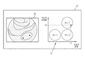
また、指標算出部44は、赤血球71の数量及び赤血球71の移動量と、病変の進行度と、を対応付けたマップ(数値テーブル等である場合を含む)を参照し、赤血球71の数量及び赤血球71の移動量から病変の進行度を求めることができる。例えば、図4に示すように、赤血球71の数量及び赤血球71の移動量と、潰瘍性大腸炎の進行度と、を対応付けたマップ51を参照すれば、潰瘍性大腸炎の進行度としてMayo分類のグレードを求めることができる。この場合、Mayo分類のグレードが指標算出部44が算出する指標である。例えば、赤血球71の数量が「P1」であり、赤血球71の移動量が「Q1」である場合、指標算出部44が算出する指標は「Mayo2」である。
In addition, the
上記指標の算出態様は任意に切り替えることができる。また、スコアX1の算出に使用する重み係数α及びβも任意に設定または変更できる。 The calculation mode of the above-mentioned index can be changed arbitrarily. In addition, weighting factors α and β used to calculate the score X1 can also be set or changed arbitrarily.
表示部13は、医療画像取得部11が取得した医療画像、及び、医療画像解析処理部12の解析結果を表示するディスプレイである。医療画像処理装置10が接続するデバイス等が含むモニタまたはディスプレイを共用し、医療画像処理装置10の表示部13として使用できる。表示制御部15は、表示部13における医療画像及び解析結果の表示態様を制御する。
The
入力受信部16は、医療画像処理装置10に付属するマウス、キーボード、その他操作デバイスからの入力を受け付ける。医療画像処理装置10の各部の動作はこれらの操作デバイスを用いて制御することができる。
The
統括制御部17は、医療画像処理装置10の各部の動作を統括的に制御する。入力受信部16が操作デバイスを用いた操作入力を受信した場合には、統括制御部17は、その操作入力にしたがって医療画像処理装置10の各部を制御する。
The
以下、医療画像処理装置10の動作の流れを説明する。図5に示すように、医療画像取得部11は、自動的にまたは手動選択により、一連の短波長医療画像61,62を取得する(ステップS10)。図6に示すように、一連の短波長医療画像61,62のうち、最初に撮影した短波長医療画像61は、粘膜72内に漏出した赤血球71を検出可能である。また、図7に示すように、一連の短波長医療画像61,62のうち、2番目に撮影した短波長医療画像62は、最初の短波長医療画像61と比較して、赤血球71の位置が移動している。
The flow of the operation of the medical
医療画像取得部11が一連の短波長医療画像61,62を取得すると、赤血球検出部41は、これら一連の短波長医療画像61,62の各々において赤血球71を検出する(ステップS11)。赤血球検出部41が一連の短波長医療画像61,62において赤血球71を検出すると、赤血球数量算出部42は赤血球71の数量を算出し(ステップS12)、かつ、赤血球数量算出部42は赤血球71の移動量を算出する(ステップS13)。その後、指標算出部44は、赤血球71の数量と、赤血球71の移動量と、を用いて、潰瘍性大腸炎の進行度と相関があるスコアX1、または、Mayo分類のグレードを指標として算出する(ステップS14)。
When the medical image acquisition unit 11 acquires a series of short wavelength
指標算出部44がスコアまたはMayo分類のグレードを指標として算出すると、表示制御部15は、一連の短波長医療画像61,62のうち少なくとも1つの短波長医療画像と、指標算出部44が算出した指標と、を表示部13に表示する。指標算出部44が潰瘍性大腸炎の進行度と相関があるスコアX1を算出する場合、図8に示すように、表示制御部15は、例えば短波長医療画像61と、スコアX1と、を表示部13に表示する。また、指標算出部44がMayo分類のグレードを算出する場合、図9に示すように、表示制御部15は、例えば短波長医療画像61と、Mayo分類のグレードと、を表示部13に表示する。
When the
上記のように、医療画像処理装置10は、緑色波長帯域よりも短波長帯域の光を照射した被写体を撮影して得た一連の短波長医療画像61,62を使用することで、通常は、精度良く検出することが困難である粘膜72内に漏出した赤血球71を検出することができる。その結果、医療画像処理装置10は、検出した赤血球71の数量及び移動量を用いて、潰瘍性大腸炎の進行度等、従来においては判別の難しかった病変の進行度等に係る解析結果を得ることができる。特に、潰瘍性大腸炎及びクローン病等の炎症性腸疾患においては、軽症から中等症程度の正確な判別が特に困難であったが、医療画像処理装置10は、これらを従来よりも正確に判別し得る指標を提供できる。上記実施形態においては、医療画像処理装置10が算出する指標を用いれば、潰瘍性大腸炎におけるMayo1とMayo2とを、Mayo0とMayo1とを、または、どの程度各グレードに近いか(Mayo2に近いMayo1なのか、Mayo0に近いMayo1なのか等)を、具体的かつ客観的に判別することができる。
As described above, the medical
上記実施形態においては、赤血球移動量算出部43は、2つの短波長医療画像61,62の差分の合計し、その絶対値を赤血球71の移動量としているが、赤血球移動量算出部43は、上記第1実施形態とは別の方法で赤血球71の移動量を算出することができる。例えば、最初に撮影した短波長医療画像61と、2番目に撮影した短波長医療画像62とのパターンマッチングを行い、対応する赤血球71を検出することにより、各赤血球71の移動量を算出しても良い。
In the above embodiment, the red blood cell movement amount calculation unit 43 sums the difference between the two short wavelength
上記実施形態においては、一連の短波長医療画像61,62の撮影倍率は任意であるが、一連の短波長医療画像61,62は、19インチの表示画面において被写体を60倍以上に拡大して撮影した短波長医療画像であることが好ましい。この場合、一連の短波長医療画像61,62を用いて赤血球71をより正確に検出することができる。なお、上記撮影倍率は、一般的なズーム機能を有する消化管用内視鏡において1段階ズームする程度の撮影倍率である。
In the above embodiment, the imaging magnification of the series of short wavelength
上記実施形態においては、表示制御部15は、短波長医療画像61と、スコアX1またはMayo分類のグレードと、を表示部13に表示しているが、これらの表示態様は一例であり、別の表示態様でこれらの情報を表示部13に表示することができる。例えば、表示制御部15は、スコアX1、または、Mayo分類のグレードのどちらを指標として算出する場合においても、図10に示すように、例えば短波長医療画像61と、赤血球71の数量及び移動量とMayo分類のグレードを対応付けるマップ51と、を、マップ51に赤血球71の具体的な数量「P1」移動量「Q1」及び該当するマップ51中の位置(星印)を示して表示部13に表示することができる。少なくとも図9及び図10に示す表示部13の表示形態は、病変の進行度を直接的に知得し得る表示形態であるという点において、病変の進行度と、指標と、を対応付けて表示する表示形態である。
In the above embodiment, the
上記実施形態においては、表示制御部15は、短波長医療画像61はいわゆる狭帯域光を用いて撮影した画像なのでほぼBチャンネル(青色チャンネル)にしか情報を持たないため、グレースケールで表示部13に表示するが、表示制御部15は、短波長医療画像61,62をカラーで表示部13に表示することができる。
In the above embodiment, the
短波長医療画像61,62をカラーで表示部13に表示する場合、表示制御部15は、表示部13に表示する短波長医療画像61,62の表示色を調節する表示色調整部として機能する。例えば、短波長医療画像61を表示部13に表示する場合、図11に示すように、表示制御部15は、表示部13に表示する短波長医療画像61を、表示用のBチャンネル(青色チャンネル)、Gチャンネル(緑色チャンネル)、及び、Rチャンネル(赤色チャンネル)に割り当てる。この際、表示制御部15は、少なくとも1つの色チャンネルについて、入力元であるグレースケールの短波長医療画像61に各色チャンネル用の補正をしてから各色チャンネルに出力する。すなわち、表示制御部15は、入力元の短波長医療画像61から、Bチャンネル用補正後のBチャンネル用画像76と、Gチャンネル用補正後のGチャンネル用画像77と、Rチャンネル用画像78を生成し、これらを用いて表示部13に疑似カラー化した短波長医療画像61を表示する。
When displaying the short wavelength
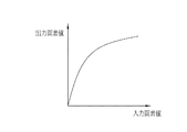
より具体的には、表示制御部15は、例えば、Bチャンネル及びRチャンネルについては、図12に示すように入力画素値と出力画素値を線形に対応付ける。このため、Bチャンネル及びRチャンネルには実質的に補正せずに入力元の短波長医療画像61をそのまま出力する。そして、Gチャンネルについては、例えば図13に示すように入力画素値と出力画素値を非線形に対応付ける補正をする。これらの結果、粘膜72の色は緑色の色相になる。一方、赤血球71は短波長帯域の光を良く吸収するので、入力元であるグレースケールの短波長医療画像61においてはほぼ黒色である。このため、上記補正を経た後も、赤血球71の色はほぼ黒色である。その結果、図14に示すように、粘膜72の色を緑色の色相にした表示用画像81は、元の短波長医療画像61と比較して、粘膜72に対する赤血球71のコントラストが高くなり、赤血球71を認識しやすくできる。これは、人間の視覚のバランスを考慮したLab色空間において、表示用画像81における粘膜72の緑色と赤血球71の黒色との色差が、グレースケールの短波長医療画像61における粘膜72の白色と赤血球71の黒色との色差よりも大きいことからも明らかである。なお、医師等は、血管等を強調した医療画像(いわゆる狭帯域観察画像等)を見慣れており、こうした医療画像は粘膜72の色が緑色系統の色相である場合が多い。このため、上記のように、表示用画像81において粘膜72を緑色の色相にすることで、擬似的にカラー化しつつも、他の一般的な表示形態の医療画像と比較しても違和感が少ない表示にできるという利点もある。
More specifically, for the B channel and R channel, for example, the
なお、非線形に対応付ける補正は上記の例に限られるものではなく、例えば、図15に示すように中間濃度の信号の視認性を上げるため、S字調のカーブを用いてもよい。この場合においても、図16に示すように、Rチャンネル、Gチャンネル及びBチャンネルの各チャンネルに別々の変換テーブルを割り当ててもよい。この他、入力元であるグレースケールの短波長医療画像61に各色チャンネル用の補正としてゲインをかけて調整してもよい。この場合、Gチャンネルに割り当てるゲインを高くし、かつ、Rチャンネル及びBチャンネルに割り当てるゲインを低くする。
Note that the correction associated with non-linearity is not limited to the above example. For example, as shown in FIG. 15, an S-shaped curve may be used to increase the visibility of the intermediate density signal. Also in this case, as shown in FIG. 16, separate conversion tables may be assigned to each of the R channel, G channel and B channel. In addition, it may be adjusted by applying gain as a correction for each color channel to the gray scale short wavelength
上記実施形態及び変形例においては、短波長医療画像61または短波長医療画像61を擬似カラー化した表示用画像81を表示部13に表示しているが、表示制御部15は、他の医療画像を表示部13に表示することができる。例えば、医療画像取得部11は、一連の短波長医療画像61,62の他に、表示部13への表示に用いる医療画像を取得する。表示部13への表示に用いる医療画像は、例えば、被写体に白色光を照射して撮影した医療画像であり、一連の短波長医療画像61,62のいずれかとほぼ同時に(粘膜72等に大きな変化がない程度の時間間隔で)撮影した内視鏡画像(以下、白色光画像という)である。この場合、表示制御部15は、赤血球71の検出等に使用する一連の短波長医療画像61,62の代わりに、白色光画像を表示部13に表示することができる。また、表示制御部15は、赤血球検出部41の検出結果を、白色光画像に重畳(オーバレイ)して表示部13に表示することで、白色光画像上において赤血球71の位置を強調表示することができる。
In the above embodiment and the modification, the
上記実施形態においては、医療画像処理装置10と内視鏡装置21は別個の装置であるが、内視鏡装置21は、医療画像処理装置10を含むことができる。この場合、図17に示すように、医療画像処理装置10を構成する各部220は、プロセッサ装置33に設ける。但し、表示部13は、内視鏡装置21のモニタ34を共用することができるので、プロセッサ装置33には表示部13以外の各部を設ければ足りる。また、上記実施形態及びその他変形例の医療画像処理装置10と、図2の内視鏡装置21と、の全体で新たな内視鏡装置を構成することができる。
In the above embodiment, the medical
内視鏡装置21は、基本的にリアルタイムに被写体を観察する装置であるから、上記のように、内視鏡装置21が医療画像処理装置10を含む場合、赤血球71の検出及び指標の算出は、自動的または手動設定により、任意のタイミングで行うことができる。
Since the
また、図18に示すように、内視鏡装置21その他モダリティと組み合わせて使用する診断支援装置610は、上記実施形態及びその他変形例の医療画像処理装置10を含むことができる。また、図19に示すように、例えば内視鏡装置21を含む、第1検査装置621、第2検査装置622、…、第N検査装置623等の各種検査装置と任意のネットワーク626を介して接続する医療業務支援装置630は、上記実施形態及びその他変形例の医療画像処理装置10を含むことができる。
In addition, as shown in FIG. 18, the
この他、医療画像処理装置10、及び、医療画像処理装置10を含む各種装置、及び、医療画像処理装置10の機能を内包する各種装置またはシステムは、以下の種々の変更等をして使用することができる。
In addition, the medical
短波長医療画像61,62の画素の特徴量に基づいて、注目すべき領域である注目領域を検出する注目領域検出部を備える場合、赤血球検出部41は、注目領域において赤血球71を検出することができる。
When the attention area detection unit for detecting the attention area which is the area to be noticed based on the feature amounts of the pixels of the short wavelength
内視鏡装置21については、内視鏡31はカプセル内視鏡を使用することができる。この場合、光源装置32と、プロセッサ装置33の一部と、はカプセル内視鏡に搭載することができる。
For the
また、指標算出部44は、上記実施形態及び変形例において示した指標以外の指標を算出することができる。例えば、指標算出部44は、一連の短波長医療画像61,62、または医療画像取得部11が取得する他の医療画像を用いて、血管の太さ、本数、または密度等の血管に関する情報(以下、血管情報という)を算出することができる。このように、指標算出部44が、血管情報等の上記実施形態及び変形例において示した指標以外の指標を算出する場合、表示制御部15は上記実施形態及び変形例において示した指標以外の指標を、上記実施形態及び変形例において示した指標とともに表示部13に表示することができる。また、上記実施形態及び変形例において示した指標以外の指標の表示または非表示はユーザの設定または操作によって任意に切り替えることができる。
Further, the
医療画像処理装置10が、医療画像解析処理部12の解析結果(赤血球71の位置、赤血球71の数量、赤血球71の移動量、及びこれらを用いて算出した指標)を、PACS22その他の任意のストレージに保存することができる。この場合、解析に使用した一連の短波長医療画像61,62と、解析結果と、を関連付けて保存することができる。
The medical
上記実施形態及び変形例において、医療画像取得部11、医療画像解析処理部12及び医療画像解析処理部12を構成する各部、表示制御部15、入力受信部16、統括制御部17、並びに、画像生成部36といった各種の処理を実行する処理部(processing unit)のハードウェア的な構造は、次に示すような各種のプロセッサ(processor)である。各種のプロセッサには、ソフトウエア(プログラム)を実行して各種の処理部として機能する汎用的なプロセッサであるCPU(Central Processing Unit)、FPGA (Field Programmable Gate Array)などの製造後に回路構成を変更可能なプロセッサであるプログラマブルロジックデバイス(Programmable Logic Device:PLD)、各種の処理を実行するために専用に設計された回路構成を有するプロセッサである専用電気回路などが含まれる。
In the above-described embodiment and modification, the medical image acquisition unit 11, the medical image analysis processing unit 12, and the units constituting the medical image analysis processing unit 12, the
1つの処理部は、これら各種のプロセッサのうちの1つで構成されてもよいし、同種または異種の2つ以上のプロセッサの組み合せ(例えば、複数のFPGAや、CPUとFPGAの組み合わせ)で構成されてもよい。また、複数の処理部を1つのプロセッサで構成してもよい。複数の処理部を1つのプロセッサで構成する例としては、第1に、クライアントやサーバなどのコンピュータに代表されるように、1つ以上のCPUとソフトウエアの組み合わせで1つのプロセッサを構成し、このプロセッサが複数の処理部として機能する形態がある。第2に、システムオンチップ(System On Chip:SoC)などに代表されるように、複数の処理部を含むシステム全体の機能を1つのIC(Integrated Circuit)チップで実現するプロセッサを使用する形態がある。このように、各種の処理部は、ハードウェア的な構造として、上記各種のプロセッサを1つ以上用いて構成される。 One processing unit may be configured of one of these various processors, or configured of a combination of two or more processors of the same type or different types (for example, a plurality of FPGAs or a combination of a CPU and an FPGA) It may be done. In addition, a plurality of processing units may be configured by one processor. As an example in which a plurality of processing units are configured by one processor, first, one processor is configured by a combination of one or more CPUs and software as represented by computers such as clients and servers; There is a form in which this processor functions as a plurality of processing units. Second, as typified by a system on chip (SoC) or the like, there is a form using a processor that realizes the functions of the entire system including a plurality of processing units in one integrated circuit (IC) chip. is there. Thus, the various processing units are configured using one or more of the above-described various processors as a hardware structure.
さらに、これらの各種のプロセッサのハードウェア的な構造は、より具体的には、半導体素子などの回路素子を組み合わせた形態の電気回路(circuitry)である。 Furthermore, the hardware-like structure of these various processors is, more specifically, an electrical circuit (circuitry) in the form in which circuit elements such as semiconductor elements are combined.
上記実施形態及び変形例の医療画像処理装置10は、炎症性疾患の進行度を判別する目的の他、手術後の縫合部からの出血の有無を確認する用途にも好適に使用できる。また、医療画像処理装置10は、下部消化管の炎症性腸疾患の他、食道等の上部消化管における炎症性疾患の進行度等を判別する際にも好適である。
The medical
上記図11から図14に示す医療画像の表示形態は、医療画像処理装置10の赤血球71の検出機能等とは別に利用できる。上記図11から図14に示す医療画像の表示形態は、グレースケールの内視鏡画像を表示部13等に表示する際に有用である。例えば、一般的な内視鏡装置21において、グレースケールの内視鏡画像をモニタ34に表示する場合に、上記図11から図14に示す医療画像の表示形態で疑似カラー化して表示することができる。この場合、赤血球71が写ってなくても、元のグレースケールの医療画像において概ね黒色である部分の視認性を向上することができる。また、上記実施形態等においては、入力元であるグレースケールの短波長医療画像61を、表示部13に表示する際に、表示制御部15が表示色の調節をするが、予め表示用画像81を生成することができる。すなわち、入力元であるグレースケールの画像を複数の色チャンネル(RGBまたはCMYK等)に割り当てる際に、各色チャンネルに対応する補正をする補正部と、補正後の画像を用いて、元のグレースケールの画像を疑似カラー化した新たな画像を生成する疑似カラー画像生成部と、を設ける。そして、疑似カラー画像生成部が生成した疑似カラー画像(表示用画像81)を表示制御部15が表示部13に表示しても良い。当然ながら、医療画像処理装置10においても、上記補正部及び疑似カラー画像生成部を設けることができる。
The display forms of medical images shown in FIGS. 11 to 14 can be used separately from the detection function of the
10 医療画像処理装置
11 医療画像取得部
12 医療画像解析処理部
13 表示部
15 表示制御部
16 入力受信部
17 統括制御部
21 内視鏡装置
22 PACS
31 内視鏡
32 光源装置
33 プロセッサ装置
34 モニタ
36 画像生成部
41 赤血球検出部
42 赤血球数量算出部
43 赤血球移動量算出部
44 指標算出部
51 マップ
61,62 短波長医療画像
71 赤血球
72 粘膜
76 Bチャンネル用画像
77 Gチャンネル用画像
78 Rチャンネル用画像
81 表示用画像
220 医療画像処理装置を構成する各部
610 診断支援装置
621 第1検査装置
622 第2検査装置
623 第N検査装置
626 ネットワーク
630 医療業務支援装置
P1 赤血球の数量
Q1 赤血球の移動量
X1 スコア
Reference Signs List 31 endoscope 32
Claims (17)
前記短波長医療画像を用いて赤血球を検出する赤血球検出部と、
を備える医療画像処理装置。 A medical image acquisition unit that acquires a short wavelength medical image obtained by imaging a subject that is a medical image including a subject image and that is irradiated with light having a wavelength shorter than the green wavelength band;
A red blood cell detection unit that detects red blood cells using the short wavelength medical image;
Medical image processing apparatus comprising:
前記短波長医療画像において検出した赤血球の移動量を算出する赤血球移動量算出部と、
赤血球の前記数量及び前記移動量を用いて、病変の進行度を表す指標を算出する指標算出部と、
を備える請求項1または2に記載の医療画像処理装置。 A red blood cell quantity calculation unit that calculates the quantity of red blood cells detected in the short wavelength medical image;
A red blood cell movement amount calculation unit that calculates the movement amount of red blood cells detected in the short wavelength medical image;
An index calculation unit that calculates an index representing the degree of progression of a lesion using the number of red blood cells and the movement amount;
The medical image processing apparatus according to claim 1, comprising:
前記表示部に表示する前記短波長医療画像の表示色を調節する表示制御部と、
を備える請求項1〜7のいずれか1項に記載の医療画像処理装置。 A display unit for displaying the short wavelength medical image;
A display control unit that adjusts a display color of the short wavelength medical image displayed on the display unit;
The medical image processing apparatus according to any one of claims 1 to 7, comprising:
前記赤血球検出部は、前記注目領域において赤血球を検出する請求項1〜10のいずれか1項に記載の医療画像処理装置。 And a region-of-interest detection unit configured to detect a region of interest, which is a region of interest, based on the feature amounts of the pixels of the short wavelength medical image.
The medical image processing apparatus according to any one of claims 1 to 10, wherein the red blood cell detection unit detects red blood cells in the region of interest.
前記短波長帯域の光を照射して画像を取得する内視鏡と、
を含む内視鏡装置。 A medical image processing apparatus according to any one of claims 1 to 14,
An endoscope which emits light in the short wavelength band to obtain an image;
Endoscope device including.
Priority Applications (3)
| Application Number | Priority Date | Filing Date | Title |
|---|---|---|---|
| JP2017168754A JP6850225B2 (en) | 2017-09-01 | 2017-09-01 | Medical image processing equipment, endoscopy equipment, diagnostic support equipment, and medical business support equipment |
| EP18189880.0A EP3449800B1 (en) | 2017-09-01 | 2018-08-21 | Medical image processing apparatus, endoscope apparatus, diagnostic support apparatus, and medical service support apparatus |
| US16/109,677 US11010891B2 (en) | 2017-09-01 | 2018-08-22 | Medical image processing apparatus, endoscope apparatus, diagnostic support apparatus, and medical service support apparatus |
Applications Claiming Priority (1)
| Application Number | Priority Date | Filing Date | Title |
|---|---|---|---|
| JP2017168754A JP6850225B2 (en) | 2017-09-01 | 2017-09-01 | Medical image processing equipment, endoscopy equipment, diagnostic support equipment, and medical business support equipment |
Publications (2)
| Publication Number | Publication Date |
|---|---|
| JP2019042157A true JP2019042157A (en) | 2019-03-22 |
| JP6850225B2 JP6850225B2 (en) | 2021-03-31 |
Family
ID=63350375
Family Applications (1)
| Application Number | Title | Priority Date | Filing Date |
|---|---|---|---|
| JP2017168754A Active JP6850225B2 (en) | 2017-09-01 | 2017-09-01 | Medical image processing equipment, endoscopy equipment, diagnostic support equipment, and medical business support equipment |
Country Status (3)
| Country | Link |
|---|---|
| US (1) | US11010891B2 (en) |
| EP (1) | EP3449800B1 (en) |
| JP (1) | JP6850225B2 (en) |
Cited By (4)
| Publication number | Priority date | Publication date | Assignee | Title |
|---|---|---|---|---|
| WO2020217883A1 (en) * | 2019-04-23 | 2020-10-29 | 富士フイルム株式会社 | Image processing device and method for operating same |
| JPWO2021006121A1 (en) * | 2019-07-08 | 2021-01-14 | ||
| JPWO2021079691A1 (en) * | 2019-10-23 | 2021-04-29 | ||
| WO2022071413A1 (en) * | 2020-10-02 | 2022-04-07 | 富士フイルム株式会社 | Image processing device, endoscope system, method for operating image processing device, and program for image processing device |
Families Citing this family (15)
| Publication number | Priority date | Publication date | Assignee | Title |
|---|---|---|---|---|
| EP3544482A4 (en) | 2016-11-28 | 2020-07-22 | Adaptivendo LLC | Endoscope with separable, disposable shaft |
| JP6858672B2 (en) * | 2017-08-29 | 2021-04-14 | 富士フイルム株式会社 | Medical image processing system and endoscopic system |
| EP3716207B1 (en) * | 2019-03-26 | 2023-11-29 | Active Medical B.V. | Method and apparatus for diagnostic analysis of the function and morphology of microcirculation alterations |
| USD1018844S1 (en) | 2020-01-09 | 2024-03-19 | Adaptivendo Llc | Endoscope handle |
| US11877792B2 (en) | 2020-10-02 | 2024-01-23 | Cilag Gmbh International | Smart energy combo control options |
| US11672534B2 (en) | 2020-10-02 | 2023-06-13 | Cilag Gmbh International | Communication capability of a smart stapler |
| US11830602B2 (en) | 2020-10-02 | 2023-11-28 | Cilag Gmbh International | Surgical hub having variable interconnectivity capabilities |
| US11883052B2 (en) | 2020-10-02 | 2024-01-30 | Cilag Gmbh International | End effector updates |
| US11883022B2 (en) | 2020-10-02 | 2024-01-30 | Cilag Gmbh International | Shared situational awareness of the device actuator activity to prioritize certain aspects of displayed information |
| US11877897B2 (en) | 2020-10-02 | 2024-01-23 | Cilag Gmbh International | Situational awareness of instruments location and individualization of users to control displays |
| US11748924B2 (en) | 2020-10-02 | 2023-09-05 | Cilag Gmbh International | Tiered system display control based on capacity and user operation |
| US11911030B2 (en) | 2020-10-02 | 2024-02-27 | Cilag Gmbh International | Communication capability of a surgical device with component |
| US20220104765A1 (en) * | 2020-10-02 | 2022-04-07 | Ethicon Llc | Surgical visualization and particle trend analysis system |
| US11963683B2 (en) | 2020-10-02 | 2024-04-23 | Cilag Gmbh International | Method for operating tiered operation modes in a surgical system |
| US20220104713A1 (en) * | 2020-10-02 | 2022-04-07 | Ethicon Llc | Tiered-access surgical visualization system |
Citations (12)
| Publication number | Priority date | Publication date | Assignee | Title |
|---|---|---|---|---|
| JPS5230493A (en) * | 1975-09-03 | 1977-03-08 | Hitachi Ltd | White blood-corpuscle classifying apparatus |
| JP2002517269A (en) * | 1998-06-08 | 2002-06-18 | グリンヴァルド,アミラム | Imaging and analyzing the movement of individual red blood cells in blood vessels |
| JP2005006768A (en) * | 2003-06-17 | 2005-01-13 | Olympus Corp | Electronic endoscope equipment and signal processor |
| US20050131284A1 (en) * | 2002-04-02 | 2005-06-16 | Yeda Research And Development Co. Ltd. | Characterization of moving objects in a stationary background |
| JP2006519032A (en) * | 2002-12-02 | 2006-08-24 | エダ リサーチ アンド ディベロップメント カンパニー リミティド | Characterization of arteriosclerosis by optical imaging |
| JP2008104628A (en) * | 2006-10-25 | 2008-05-08 | Tokyo Institute Of Technology | Conjunctiva and sclera imaging apparatus |
| JP2008302095A (en) * | 2007-06-11 | 2008-12-18 | Hitachi Ltd | Apparatus for measuring and evaluating blood flow |
| JP2010187925A (en) * | 2009-02-18 | 2010-09-02 | Nagoya Univ | Blood flow observation apparatus |
| JP2014166270A (en) * | 2013-02-28 | 2014-09-11 | Canon Inc | Image processing apparatus and image processing method |
| WO2015045576A1 (en) * | 2013-09-26 | 2015-04-02 | 富士フイルム株式会社 | Endoscope system, processor device for endoscope system, operation method for endoscope system, and operation method for processor device |
| JP2016154588A (en) * | 2015-02-23 | 2016-09-01 | Hoya株式会社 | Image processing device |
| WO2017122431A1 (en) * | 2016-01-15 | 2017-07-20 | オリンパス株式会社 | Image analysis device, image analysis system, and method for actuating image analysis device |
Family Cites Families (9)
| Publication number | Priority date | Publication date | Assignee | Title |
|---|---|---|---|---|
| AU738223B2 (en) * | 1995-10-23 | 2001-09-13 | Cytometrics, Inc. | Method and apparatus for reflected imaging analysis |
| US20080021331A1 (en) * | 2004-09-29 | 2008-01-24 | Yeda Research And Development Co. Ltd. | Characterization of moving objects in a stationary background |
| US9602777B2 (en) * | 2008-04-25 | 2017-03-21 | Roche Diagnostics Hematology, Inc. | Systems and methods for analyzing body fluids |
| JP6027803B2 (en) * | 2012-07-17 | 2016-11-16 | Hoya株式会社 | Image processing apparatus and endoscope apparatus |
| JP6198410B2 (en) * | 2013-02-28 | 2017-09-20 | キヤノン株式会社 | Image processing apparatus and image processing method |
| JP6097629B2 (en) * | 2013-04-26 | 2017-03-15 | Hoya株式会社 | Lesion evaluation information generator |
| JP6196922B2 (en) * | 2014-03-17 | 2017-09-13 | オリンパス株式会社 | Image processing apparatus, image processing method, and image processing program |
| JP2017067524A (en) * | 2015-09-29 | 2017-04-06 | 富士フイルム株式会社 | Method and device for screening for nucleated erythrocytes |
| TW201725314A (en) | 2016-01-05 | 2017-07-16 | 三宅圀博 | Fluid electricity generation device with dual-housing and rotor assembly |
-
2017
- 2017-09-01 JP JP2017168754A patent/JP6850225B2/en active Active
-
2018
- 2018-08-21 EP EP18189880.0A patent/EP3449800B1/en active Active
- 2018-08-22 US US16/109,677 patent/US11010891B2/en active Active
Patent Citations (13)
| Publication number | Priority date | Publication date | Assignee | Title |
|---|---|---|---|---|
| JPS5230493A (en) * | 1975-09-03 | 1977-03-08 | Hitachi Ltd | White blood-corpuscle classifying apparatus |
| JP2002517269A (en) * | 1998-06-08 | 2002-06-18 | グリンヴァルド,アミラム | Imaging and analyzing the movement of individual red blood cells in blood vessels |
| US20050131284A1 (en) * | 2002-04-02 | 2005-06-16 | Yeda Research And Development Co. Ltd. | Characterization of moving objects in a stationary background |
| JP2005521499A (en) * | 2002-04-02 | 2005-07-21 | エダ リサーチ アンド ディベロップメント カンパニー リミティド | Characterization of moving substances in a stationary background |
| JP2006519032A (en) * | 2002-12-02 | 2006-08-24 | エダ リサーチ アンド ディベロップメント カンパニー リミティド | Characterization of arteriosclerosis by optical imaging |
| JP2005006768A (en) * | 2003-06-17 | 2005-01-13 | Olympus Corp | Electronic endoscope equipment and signal processor |
| JP2008104628A (en) * | 2006-10-25 | 2008-05-08 | Tokyo Institute Of Technology | Conjunctiva and sclera imaging apparatus |
| JP2008302095A (en) * | 2007-06-11 | 2008-12-18 | Hitachi Ltd | Apparatus for measuring and evaluating blood flow |
| JP2010187925A (en) * | 2009-02-18 | 2010-09-02 | Nagoya Univ | Blood flow observation apparatus |
| JP2014166270A (en) * | 2013-02-28 | 2014-09-11 | Canon Inc | Image processing apparatus and image processing method |
| WO2015045576A1 (en) * | 2013-09-26 | 2015-04-02 | 富士フイルム株式会社 | Endoscope system, processor device for endoscope system, operation method for endoscope system, and operation method for processor device |
| JP2016154588A (en) * | 2015-02-23 | 2016-09-01 | Hoya株式会社 | Image processing device |
| WO2017122431A1 (en) * | 2016-01-15 | 2017-07-20 | オリンパス株式会社 | Image analysis device, image analysis system, and method for actuating image analysis device |
Cited By (12)
| Publication number | Priority date | Publication date | Assignee | Title |
|---|---|---|---|---|
| WO2020217883A1 (en) * | 2019-04-23 | 2020-10-29 | 富士フイルム株式会社 | Image processing device and method for operating same |
| JPWO2020217883A1 (en) * | 2019-04-23 | 2020-10-29 | ||
| CN113784654A (en) * | 2019-04-23 | 2021-12-10 | 富士胶片株式会社 | Image processing apparatus and method for operating the same |
| JP7116254B2 (en) | 2019-04-23 | 2022-08-09 | 富士フイルム株式会社 | Image processing device and its operating method |
| US11957483B2 (en) | 2019-04-23 | 2024-04-16 | Fujifilm Corporation | Image processing device and method of operating the same |
| JPWO2021006121A1 (en) * | 2019-07-08 | 2021-01-14 | ||
| WO2021006121A1 (en) * | 2019-07-08 | 2021-01-14 | 富士フイルム株式会社 | Image processing device, endoscope system, and operation method for image processing device |
| JP7217351B2 (en) | 2019-07-08 | 2023-02-02 | 富士フイルム株式会社 | Image processing device, endoscope system, and method of operating image processing device |
| JPWO2021079691A1 (en) * | 2019-10-23 | 2021-04-29 | ||
| WO2021079691A1 (en) * | 2019-10-23 | 2021-04-29 | 富士フイルム株式会社 | Image processing device and operation method for same |
| JP7214886B2 (en) | 2019-10-23 | 2023-01-30 | 富士フイルム株式会社 | Image processing device and its operating method |
| WO2022071413A1 (en) * | 2020-10-02 | 2022-04-07 | 富士フイルム株式会社 | Image processing device, endoscope system, method for operating image processing device, and program for image processing device |
Also Published As
| Publication number | Publication date |
|---|---|
| US20190073769A1 (en) | 2019-03-07 |
| EP3449800B1 (en) | 2024-01-17 |
| EP3449800A1 (en) | 2019-03-06 |
| JP6850225B2 (en) | 2021-03-31 |
| US11010891B2 (en) | 2021-05-18 |
Similar Documents
| Publication | Publication Date | Title |
|---|---|---|
| JP6850225B2 (en) | Medical image processing equipment, endoscopy equipment, diagnostic support equipment, and medical business support equipment | |
| US10776915B2 (en) | Medical image processing apparatus, endoscope apparatus, diagnostic support apparatus, and medical service support apparatus | |
| JP7296498B2 (en) | Medical image processing device and endoscope device | |
| JP7411618B2 (en) | medical image processing device | |
| US11950757B2 (en) | Medical image processing apparatus | |
| US11605363B2 (en) | Medical image processing apparatus for reducing examination load | |
| JP6785990B2 (en) | Medical image processing equipment and endoscopic equipment | |
| US11961228B2 (en) | Medical image processing system | |
| JP6834019B2 (en) | Medical image processing equipment and endoscopic equipment | |
| KR102393661B1 (en) | Endoscope system for multi image, image providing method of the system, and a recording medium having computer readable program for executing the method | |
| EP4183311A1 (en) | Image analysis processing device, endoscopy system, operation method for image analysis processing device, and program for image analysis processing device | |
| CN114585291A (en) | Image processing apparatus and method for operating the same | |
| JP6866497B2 (en) | Medical image processing equipment and endoscopic equipment | |
| WO2022009478A1 (en) | Image processing device, endoscope system, operation method for image processing device, and program for image processing device | |
| WO2022230607A1 (en) | Medical image processing device, endoscope system, and operation method for medical image processing device | |
| US20220245808A1 (en) | Image processing apparatus, image processing system, image processing method, and computer-readable recording medium | |
| JP2023011303A (en) | Medical image processing apparatus and operating method of the same | |
| JP2020058623A (en) | Medical image processing apparatus, medical image processing method, and medical image processing program |
Legal Events
| Date | Code | Title | Description |
|---|---|---|---|
| A621 | Written request for application examination |
Free format text: JAPANESE INTERMEDIATE CODE: A621 Effective date: 20190805 |
|
| A977 | Report on retrieval |
Free format text: JAPANESE INTERMEDIATE CODE: A971007 Effective date: 20200731 |
|
| A131 | Notification of reasons for refusal |
Free format text: JAPANESE INTERMEDIATE CODE: A131 Effective date: 20200825 |
|
| A521 | Request for written amendment filed |
Free format text: JAPANESE INTERMEDIATE CODE: A523 Effective date: 20201022 |
|
| TRDD | Decision of grant or rejection written | ||
| A01 | Written decision to grant a patent or to grant a registration (utility model) |
Free format text: JAPANESE INTERMEDIATE CODE: A01 Effective date: 20210216 |
|
| A61 | First payment of annual fees (during grant procedure) |
Free format text: JAPANESE INTERMEDIATE CODE: A61 Effective date: 20210305 |
|
| R150 | Certificate of patent or registration of utility model |
Ref document number: 6850225 Country of ref document: JP Free format text: JAPANESE INTERMEDIATE CODE: R150 |
|
| R250 | Receipt of annual fees |
Free format text: JAPANESE INTERMEDIATE CODE: R250 |