JP7208355B2 - Adaptive Compton camera for medical imaging - Google Patents
Adaptive Compton camera for medical imaging Download PDFInfo
- Publication number
- JP7208355B2 JP7208355B2 JP2021505881A JP2021505881A JP7208355B2 JP 7208355 B2 JP7208355 B2 JP 7208355B2 JP 2021505881 A JP2021505881 A JP 2021505881A JP 2021505881 A JP2021505881 A JP 2021505881A JP 7208355 B2 JP7208355 B2 JP 7208355B2
- Authority
- JP
- Japan
- Prior art keywords
- detector
- compton
- scatter
- patient
- capture
- Prior art date
- Legal status (The legal status is an assumption and is not a legal conclusion. Google has not performed a legal analysis and makes no representation as to the accuracy of the status listed.)
- Active
Links
Images


Classifications
-
- A—HUMAN NECESSITIES
- A61—MEDICAL OR VETERINARY SCIENCE; HYGIENE
- A61B—DIAGNOSIS; SURGERY; IDENTIFICATION
- A61B6/00—Apparatus for radiation diagnosis, e.g. combined with radiation therapy equipment
- A61B6/02—Devices for diagnosis sequentially in different planes; Stereoscopic radiation diagnosis
- A61B6/03—Computerised tomographs
- A61B6/037—Emission tomography
-
- A—HUMAN NECESSITIES
- A61—MEDICAL OR VETERINARY SCIENCE; HYGIENE
- A61B—DIAGNOSIS; SURGERY; IDENTIFICATION
- A61B6/00—Apparatus for radiation diagnosis, e.g. combined with radiation therapy equipment
- A61B6/04—Positioning of patients; Tiltable beds or the like
- A61B6/0407—Supports, e.g. tables or beds, for the body or parts of the body
-
- A—HUMAN NECESSITIES
- A61—MEDICAL OR VETERINARY SCIENCE; HYGIENE
- A61B—DIAGNOSIS; SURGERY; IDENTIFICATION
- A61B6/00—Apparatus for radiation diagnosis, e.g. combined with radiation therapy equipment
- A61B6/42—Apparatus for radiation diagnosis, e.g. combined with radiation therapy equipment with arrangements for detecting radiation specially adapted for radiation diagnosis
- A61B6/4208—Apparatus for radiation diagnosis, e.g. combined with radiation therapy equipment with arrangements for detecting radiation specially adapted for radiation diagnosis characterised by using a particular type of detector
- A61B6/4233—Apparatus for radiation diagnosis, e.g. combined with radiation therapy equipment with arrangements for detecting radiation specially adapted for radiation diagnosis characterised by using a particular type of detector using matrix detectors
-
- A—HUMAN NECESSITIES
- A61—MEDICAL OR VETERINARY SCIENCE; HYGIENE
- A61B—DIAGNOSIS; SURGERY; IDENTIFICATION
- A61B6/00—Apparatus for radiation diagnosis, e.g. combined with radiation therapy equipment
- A61B6/42—Apparatus for radiation diagnosis, e.g. combined with radiation therapy equipment with arrangements for detecting radiation specially adapted for radiation diagnosis
- A61B6/4208—Apparatus for radiation diagnosis, e.g. combined with radiation therapy equipment with arrangements for detecting radiation specially adapted for radiation diagnosis characterised by using a particular type of detector
- A61B6/4258—Apparatus for radiation diagnosis, e.g. combined with radiation therapy equipment with arrangements for detecting radiation specially adapted for radiation diagnosis characterised by using a particular type of detector for detecting non x-ray radiation, e.g. gamma radiation
-
- A—HUMAN NECESSITIES
- A61—MEDICAL OR VETERINARY SCIENCE; HYGIENE
- A61B—DIAGNOSIS; SURGERY; IDENTIFICATION
- A61B6/00—Apparatus for radiation diagnosis, e.g. combined with radiation therapy equipment
- A61B6/42—Apparatus for radiation diagnosis, e.g. combined with radiation therapy equipment with arrangements for detecting radiation specially adapted for radiation diagnosis
- A61B6/4266—Apparatus for radiation diagnosis, e.g. combined with radiation therapy equipment with arrangements for detecting radiation specially adapted for radiation diagnosis characterised by using a plurality of detector units
-
- A—HUMAN NECESSITIES
- A61—MEDICAL OR VETERINARY SCIENCE; HYGIENE
- A61B—DIAGNOSIS; SURGERY; IDENTIFICATION
- A61B6/00—Apparatus for radiation diagnosis, e.g. combined with radiation therapy equipment
- A61B6/42—Apparatus for radiation diagnosis, e.g. combined with radiation therapy equipment with arrangements for detecting radiation specially adapted for radiation diagnosis
- A61B6/4275—Apparatus for radiation diagnosis, e.g. combined with radiation therapy equipment with arrangements for detecting radiation specially adapted for radiation diagnosis using a detector unit almost surrounding the patient, e.g. more than 180°
-
- A—HUMAN NECESSITIES
- A61—MEDICAL OR VETERINARY SCIENCE; HYGIENE
- A61B—DIAGNOSIS; SURGERY; IDENTIFICATION
- A61B6/00—Apparatus for radiation diagnosis, e.g. combined with radiation therapy equipment
- A61B6/44—Constructional features of apparatus for radiation diagnosis
-
- A—HUMAN NECESSITIES
- A61—MEDICAL OR VETERINARY SCIENCE; HYGIENE
- A61B—DIAGNOSIS; SURGERY; IDENTIFICATION
- A61B6/00—Apparatus for radiation diagnosis, e.g. combined with radiation therapy equipment
- A61B6/44—Constructional features of apparatus for radiation diagnosis
- A61B6/4411—Constructional features of apparatus for radiation diagnosis the apparatus being modular
-
- A—HUMAN NECESSITIES
- A61—MEDICAL OR VETERINARY SCIENCE; HYGIENE
- A61B—DIAGNOSIS; SURGERY; IDENTIFICATION
- A61B6/00—Apparatus for radiation diagnosis, e.g. combined with radiation therapy equipment
- A61B6/44—Constructional features of apparatus for radiation diagnosis
- A61B6/4417—Constructional features of apparatus for radiation diagnosis related to combined acquisition of different diagnostic modalities
-
- A—HUMAN NECESSITIES
- A61—MEDICAL OR VETERINARY SCIENCE; HYGIENE
- A61B—DIAGNOSIS; SURGERY; IDENTIFICATION
- A61B6/00—Apparatus for radiation diagnosis, e.g. combined with radiation therapy equipment
- A61B6/44—Constructional features of apparatus for radiation diagnosis
- A61B6/4429—Constructional features of apparatus for radiation diagnosis related to the mounting of source units and detector units
-
- A—HUMAN NECESSITIES
- A61—MEDICAL OR VETERINARY SCIENCE; HYGIENE
- A61B—DIAGNOSIS; SURGERY; IDENTIFICATION
- A61B6/00—Apparatus for radiation diagnosis, e.g. combined with radiation therapy equipment
- A61B6/52—Devices using data or image processing specially adapted for radiation diagnosis
- A61B6/5258—Devices using data or image processing specially adapted for radiation diagnosis involving detection or reduction of artifacts or noise
- A61B6/5282—Devices using data or image processing specially adapted for radiation diagnosis involving detection or reduction of artifacts or noise due to scatter
-
- G—PHYSICS
- G01—MEASURING; TESTING
- G01T—MEASUREMENT OF NUCLEAR OR X-RADIATION
- G01T1/00—Measuring X-radiation, gamma radiation, corpuscular radiation, or cosmic radiation
- G01T1/16—Measuring radiation intensity
-
- G—PHYSICS
- G01—MEASURING; TESTING
- G01T—MEASUREMENT OF NUCLEAR OR X-RADIATION
- G01T1/00—Measuring X-radiation, gamma radiation, corpuscular radiation, or cosmic radiation
- G01T1/16—Measuring radiation intensity
- G01T1/1603—Measuring radiation intensity with a combination of at least two different types of detector
-
- G—PHYSICS
- G01—MEASURING; TESTING
- G01T—MEASUREMENT OF NUCLEAR OR X-RADIATION
- G01T1/00—Measuring X-radiation, gamma radiation, corpuscular radiation, or cosmic radiation
- G01T1/16—Measuring radiation intensity
- G01T1/1606—Measuring radiation intensity with other specified detectors not provided for in the other sub-groups of G01T1/16
-
- G—PHYSICS
- G01—MEASURING; TESTING
- G01T—MEASUREMENT OF NUCLEAR OR X-RADIATION
- G01T1/00—Measuring X-radiation, gamma radiation, corpuscular radiation, or cosmic radiation
- G01T1/16—Measuring radiation intensity
- G01T1/24—Measuring radiation intensity with semiconductor detectors
-
- G—PHYSICS
- G01—MEASURING; TESTING
- G01T—MEASUREMENT OF NUCLEAR OR X-RADIATION
- G01T1/00—Measuring X-radiation, gamma radiation, corpuscular radiation, or cosmic radiation
- G01T1/16—Measuring radiation intensity
- G01T1/24—Measuring radiation intensity with semiconductor detectors
- G01T1/242—Stacked detectors, e.g. for depth information
-
- G—PHYSICS
- G01—MEASURING; TESTING
- G01T—MEASUREMENT OF NUCLEAR OR X-RADIATION
- G01T1/00—Measuring X-radiation, gamma radiation, corpuscular radiation, or cosmic radiation
- G01T1/16—Measuring radiation intensity
- G01T1/24—Measuring radiation intensity with semiconductor detectors
- G01T1/243—Modular detectors, e.g. arrays formed from self contained units
-
- G—PHYSICS
- G01—MEASURING; TESTING
- G01T—MEASUREMENT OF NUCLEAR OR X-RADIATION
- G01T1/00—Measuring X-radiation, gamma radiation, corpuscular radiation, or cosmic radiation
- G01T1/16—Measuring radiation intensity
- G01T1/24—Measuring radiation intensity with semiconductor detectors
- G01T1/249—Measuring radiation intensity with semiconductor detectors specially adapted for use in SPECT or PET
-
- G—PHYSICS
- G01—MEASURING; TESTING
- G01T—MEASUREMENT OF NUCLEAR OR X-RADIATION
- G01T1/00—Measuring X-radiation, gamma radiation, corpuscular radiation, or cosmic radiation
- G01T1/29—Measurement performed on radiation beams, e.g. position or section of the beam; Measurement of spatial distribution of radiation
- G01T1/2907—Angle determination; Directional detectors; Telescopes
-
- G—PHYSICS
- G01—MEASURING; TESTING
- G01T—MEASUREMENT OF NUCLEAR OR X-RADIATION
- G01T1/00—Measuring X-radiation, gamma radiation, corpuscular radiation, or cosmic radiation
- G01T1/29—Measurement performed on radiation beams, e.g. position or section of the beam; Measurement of spatial distribution of radiation
- G01T1/2914—Measurement of spatial distribution of radiation
- G01T1/2921—Static instruments for imaging the distribution of radioactivity in one or two dimensions; Radio-isotope cameras
- G01T1/2928—Static instruments for imaging the distribution of radioactivity in one or two dimensions; Radio-isotope cameras using solid state detectors
-
- G—PHYSICS
- G01—MEASURING; TESTING
- G01T—MEASUREMENT OF NUCLEAR OR X-RADIATION
- G01T1/00—Measuring X-radiation, gamma radiation, corpuscular radiation, or cosmic radiation
- G01T1/29—Measurement performed on radiation beams, e.g. position or section of the beam; Measurement of spatial distribution of radiation
- G01T1/2914—Measurement of spatial distribution of radiation
- G01T1/2921—Static instruments for imaging the distribution of radioactivity in one or two dimensions; Radio-isotope cameras
- G01T1/2935—Static instruments for imaging the distribution of radioactivity in one or two dimensions; Radio-isotope cameras using ionisation detectors
-
- G—PHYSICS
- G01—MEASURING; TESTING
- G01T—MEASUREMENT OF NUCLEAR OR X-RADIATION
- G01T1/00—Measuring X-radiation, gamma radiation, corpuscular radiation, or cosmic radiation
- G01T1/29—Measurement performed on radiation beams, e.g. position or section of the beam; Measurement of spatial distribution of radiation
- G01T1/2914—Measurement of spatial distribution of radiation
- G01T1/2964—Scanners
- G01T1/2971—Scanners using solid state detectors
-
- G—PHYSICS
- G01—MEASURING; TESTING
- G01T—MEASUREMENT OF NUCLEAR OR X-RADIATION
- G01T1/00—Measuring X-radiation, gamma radiation, corpuscular radiation, or cosmic radiation
- G01T1/29—Measurement performed on radiation beams, e.g. position or section of the beam; Measurement of spatial distribution of radiation
- G01T1/2914—Measurement of spatial distribution of radiation
- G01T1/2985—In depth localisation, e.g. using positron emitters; Tomographic imaging (longitudinal and transverse section imaging; apparatus for radiation diagnosis sequentially in different planes, steroscopic radiation diagnosis)
Description
本実施形態は、コンプトン効果を用いた医療用画像に関するものである。コンプトン効果は、単一光子放出コンピュータ断層撮影(SPECT)に用いられるよりも高いエネルギーを撮像することを可能にする。コンプトン画像システムは、散乱層を組み立て、次いで大きなフレームワークに装着された捕獲層を組み立てるなど、テストプラットフォームとして構築される。エレクトロニクスは、ファントムの放射からコンプトンベースの事象を検出するために接続されている。 This embodiment relates to medical images using the Compton effect. The Compton effect allows imaging higher energies than those used in single photon emission computed tomography (SPECT). The Compton imaging system is built as a test platform, such as by assembling a scattering layer and then a capture layer attached to a large framework. Electronics are connected to detect Compton-based events from the phantom emissions.
コンプトン画像システムは、いかなる商業的臨床設定においても、実用的使用のための設計および制約要件に対処することに失敗している。現状の提案では、臨床で画像プラットフォームに統合する能力が欠けているか、または商業的ニーズおよび診断ニーズに対処するための設計および制約要件(すなわち、柔軟性および拡大性)が欠けている。 The Compton imaging system fails to address the design and constraint requirements for practical use in any commercial clinical setting. Current proposals either lack the ability to integrate clinically into imaging platforms or lack the design and constraint requirements (ie, flexibility and scalability) to address commercial and diagnostic needs.
コンプトンカメラは感度($)が低く、画質(IQ)が悪い場合がある。散乱層における散乱光子の絶対数は、幾何学的形状(例えば、線源-散乱立体角Ω<<4π)、光電効果に有利な材料(例えば、光電効果に有利な検出材料における低散乱分率)、および検出器の製造制限(例えば、Si検出器に対して最大~1mm、およびCZT検出器に対して2mm~10mmのように、散乱層および捕獲層の両方に対して製造され得る実用的な検出器の厚さは、有界である)のために、低い。捕獲層における捕捉散乱光子の数は、幾何学的形状(例えば、散乱捕獲立体角Ω<<4π)のために低い。ドップラー幅はコンプトンカメラの画質を劣化させる。コンプトン角の不確定性に対するドップラー幅の寄与は、入射光子エネルギーE0、散乱角θ、ターゲット原子に束縛された移動電子のエネルギーに依存する。検出器のエネルギー分解能が制限されると、コンプトン角の不確定性が追加される。散乱層と捕獲層の両方で検出器位置分解能が制限されると、追加のコンプトン円錐環オフセットが生じる。 Compton cameras have low sensitivity ($) and may have poor image quality (IQ). The absolute number of scattered photons in the scattering layer depends on the geometry (e.g. source-scattering solid angle Ω<<4π), the photoelectric effect favoring materials (e.g. the low scattering fraction in the detection material favoring the photoelectric effect ), and detector manufacturing limitations (e.g., ~1 mm maximum for Si detectors and 2 mm to 10 mm for CZT detectors, such that both scattering and trapping layers can be practically manufactured). The thickness of the effective detector is low because it is bounded). The number of trapped scattered photons in the trapping layer is low due to the geometry (eg, scattering trapping solid angle Ω<<4π). The Doppler width degrades the image quality of the Compton camera. The Doppler width contribution to the Compton angle uncertainty depends on the incident photon energy E 0 , the scattering angle θ, and the energy of the mobile electron bound to the target atom. The limited energy resolution of the detector adds the uncertainty of the Compton angle. Additional Compton conical ring offsets occur when the detector position resolution is limited in both the scattering and trapping layers.
序論として、以下に記載される好ましい実施形態は、医療用画像化のための方法およびシステムを含む。画質および/または感度を最適化するために、コンプトンカメラは適応可能である。散乱検出器および/または捕獲検出器は、患者および/またはお互いの近くおよび/または遠くに移動することができる。この適応により、幾何学的形状を変更することにより、画質と感度のバランスを保つことができる。 By way of introduction, the preferred embodiments described below include methods and systems for medical imaging. Compton cameras are adaptable to optimize image quality and/or sensitivity. Scatter detectors and/or capture detectors can be moved closer and/or farther from the patient and/or each other. This adaptation allows us to balance image quality and sensitivity by changing the geometry.
第1の態様では、医療用画像のためにコンプトンカメラが提供される。モータは、散乱検出器、捕獲検出器、または散乱検出器と捕獲検出器の両方に接続する。モータは、散乱検出器、捕獲検出器、または散乱検出器と捕獲検出器の両方を、患者ベッドからより近くまたはより遠くに移動するように構成されている。 In a first aspect, a Compton camera is provided for medical imaging. The motor connects to the scatter detector, the capture detector, or both the scatter detector and the capture detector. A motor is configured to move the scatter detector, the capture detector, or both the scatter detector and the capture detector closer or further from the patient bed.
第2の態様では、医療用画像システムが提供される。各々が散乱検出器および捕獲器検出器を有する固体検出器モジュール。制御プロセッサは、患者空間のアイソセンタに対する散乱検出器、捕獲検出器、又は散乱検出器と捕獲検出器の両方の位置を変更するように構成される。 In a second aspect, a medical imaging system is provided. Solid-state detector modules each having a scatter detector and a trap detector. The control processor is configured to change the position of the scatter detector, the capture detector, or both the scatter detector and the capture detector with respect to the isocenter of patient space.
第3の態様では、コンプトンカメラによる医療用画像撮影のための方法が提供される。モータがコンプトンカメラの検出器を動かす。移動した検出器は、患者からの放出物を検出する。検出された放出物からコンプトン画像が生成される。 In a third aspect, a method for medical imaging with a Compton camera is provided. A motor moves the detector of the Compton camera. A moved detector detects emissions from the patient. A Compton image is generated from the detected emissions.
本発明は、次の特許請求の範囲によって定義され、この欄の何れも、それらの特許請求の範囲を限定するものとして見なされるべきではない。本発明のそれ以上の観点および利点については、好ましい実施形態と関連して以下において説明し、これらを独立して又は組合せ状態でクレーム請求する場合がある。 The invention is defined by the following claims and nothing in this section should be taken as limiting those claims. Further aspects and advantages of the invention are described below in connection with the preferred embodiments and may be claimed independently or in combination.
構成要素および図面は、必ずしも縮尺通りにはなっておらず、それどころか、本発明の原理を説明する際に誇張がなされている。さらに、図面に関し、同一の参照符号は、互いに異なる図全体にわたり対応の部分を示している。 The components and drawings are not necessarily to scale, but rather are exaggerated in explaining the principles of the invention. Furthermore, with respect to the drawings, the same reference numerals indicate corresponding parts throughout the different figures.
図1~10は、マルチモダリティ対応コンプトンカメラを示している。様々な他の画像モダリティと共に使用するために、又は独立型コンプトンベースの医療用画像装置としてコンプトンカメラを形成するために、モジュラ設計が使用される。図11~18は、適応コンプトンカメラを示している。図1~10または別のコンプトンカメラのモジュール設計(例えば、モジュール式ではない)を使用して、画像の質、感度、および/または改善された診断のための他の特性を制御するために、1つまたは両方の散乱層および検出器層が、患者に対して移動可能である。適応コンプトンカメラの実施形態の要約の後、図1~図9のコンプトンカメラについて説明する。図1~図9のコンプトンカメラの特徴及び構成要素の多くは、図11~図18で後述する適応コンプトンカメラの実施形態で使用することができる。 1-10 show a multi-modality Compton camera. A modular design is used to form the Compton camera for use with various other imaging modalities or as a standalone Compton-based medical imaging device. Figures 11-18 show an adaptive Compton camera. 1-10 or another Compton camera modular design (e.g., non-modular) to control image quality, sensitivity, and/or other characteristics for improved diagnosis. One or both scattering and detector layers are movable relative to the patient. After a summary of adaptive Compton camera embodiments, the Compton cameras of FIGS. 1-9 are described. Many of the features and components of the Compton cameras of FIGS. 1-9 can be used in the adaptive Compton camera embodiments described below in FIGS. 11-18.
適応コンプトンカメラの場合には、散乱層及び/又は捕獲層は、撮像物体に向かって及びそれから離れるように移動可能である。散乱層は、撮像物体に可能な限り近い位置に配置することができ、一方、画質のための捕獲層は、散乱層から可能な限り離れた位置に配置することができる。どちらの考慮事項も、「適応型」コンプトンカメラ内で一緒に使用することができる。撮像物体境界を検知することにより、コンプトンカメラの構成を変化させた。例えば、各散乱層の撮像物体への距離、および各捕獲層の対応する各散乱層への距離を変化させて、与えられた利点図形(FOM)を最大化する。FOMは、ユーザによって定義される感度($)、画質(IQ)、および/または他のパラメータであり得る。感度($)は、散乱層を撮像物体に近づけることによって改善されてもよいし、一方、画質(IQ)は、撮像物体から捕獲層を遠ざけることによって改善されてもよい。捕獲層を散乱層に近づけると、感度が向上する可能性があるため、動きは診断目的で所望のFOMに基づいている。デジタルコリメータは、ある不確実性に対処するために、コンプトン角度が閾値を上回るコンプトン散乱事象をフィルターアウトするために使用されることがある。 In the case of adaptive Compton cameras, the scattering layer and/or the trapping layer are movable toward and away from the imaged object. The scattering layer can be placed as close as possible to the imaged object, while the capture layer for image quality can be placed as far away as possible from the scattering layer. Both considerations can be used together in an "adaptive" Compton camera. We changed the configuration of the Compton camera by detecting the imaged object boundaries. For example, the distance of each scattering layer to the imaged object and the distance of each trapping layer to each corresponding scattering layer are varied to maximize a given figure of merit (FOM). The FOM may be user-defined sensitivity ($), image quality (IQ), and/or other parameters. Sensitivity ($) may be improved by moving the scattering layer closer to the imaging object, while image quality (IQ) may be improved by moving the capture layer further away from the imaging object. Moving the trapping layer closer to the scattering layer may improve sensitivity, so the movement is based on the desired FOM for diagnostic purposes. A digital collimator may be used to filter out Compton scattering events with Compton angles above a threshold to address certain uncertainties.
図1~9を参照すると、医療用画像システムは、セグメント化された検出モジュールを有するマルチモダリティ互換性コンプトンカメラを含む。コンプトンカメラリングなどのコンプトンカメラは、検出ユニットを収容するモジュールにセグメント化されている。各モジュールは独立しており、リング状または部分リング状に組み立てると、モジュールは互いに連絡し合うことがある。モジュールは独立しているが、コンプトン散乱ベースの画像を生成するマルチモジュールユニットに組み立てることができる。円筒対称モジュール又は球面シェルセグメントモジュールを使用することができる。 Referring to FIGS. 1-9, a medical imaging system includes a multi-modality compatible Compton camera with a segmented detection module. A Compton camera, such as the Compton camera ring, is segmented into modules that house the detection units. Each module is independent and may communicate with each other when assembled in a ring or partial ring. The modules are independent, but can be assembled into a multi-module unit that produces Compton scatter-based images. Cylindrically symmetric modules or spherical shell segment modules can be used.
散乱-捕獲が対の、モジュラ構成は、効率的な製造を可能にし、現場でサービス可能であり、コスト及びエネルギー効率が良い。モジュールは、各半径方向検出ユニット、1モジュールの角スパン、および/または軸スパンについて、設計自由度を変更することを可能にする。散乱捕獲対モジュールは、マルチモダリティ適合性であり、そして/または臨床放出画像のためのモジュールリングコンプトンカメラを形成する。このデザインは柔軟性を可能にするため、既存のコンピュータ断層撮影(CT)、磁気共鳴(MR)、陽電子放出断層撮影(PET)または他の医療用画像プラットフォームに、軸方向に分離したシステムとして、または完全に統合されたシステムとしてコンプトンカメラを追加することができる。各モジュールは、放熱、データ収集、較正、及び/又は、サービスと同様に効率的な組立を可能にすることができる。 The modular construction of the scatter-trap pair allows for efficient manufacturing, is field serviceable, and is cost and energy efficient. The modules allow varying degrees of design freedom for each radial detection unit, the angular span of one module, and/or the axial span. The scatter capture pair module is multimodality compatible and/or forms a modular ring Compton camera for clinical emission imaging. Because this design allows for flexibility, it can be attached to an existing computed tomography (CT), magnetic resonance (MR), positron emission tomography (PET) or other medical imaging platform as an axially separated system. Or you can add a Compton camera as a fully integrated system. Each module can enable efficient assembly as well as heat dissipation, data collection, calibration, and/or servicing.
各散乱捕獲対モジュールは、商業的に好適な固体検出器モジュール(例えば、Si、CZT、CdTe、HPGeまたは同様のもの)から形成され、100~3000keVのエネルギー範囲を可能にする。コンプトン画像は、より広範囲の同位体エネルギー(>2MeV)を備えることができ、散乱・捕獲検出器の選択を通じて新しいトレーサ/マーカを可能にする。モジュール化により、個々のモジュールの削除または交換が可能となり、時間と費用効率の高いサービスが可能となる。モジュールは、独立して動作され、絶縁されてもよく、またはクロストークのためにリンクされてもよく、1つのモジュールの散乱検出器および別のモジュールの捕獲検出器を使用してコンプトン事象を検出する際の画質の向上およびより高い効率を可能にする。 Each scatter-trap pair module is formed from commercially suitable solid-state detector modules (eg, Si, CZT, CdTe, HPGe, or the like), enabling an energy range of 100-3000 keV. Compton images can have a wider range of isotopic energies (>2 MeV), enabling new tracers/markers through the choice of scatter-trap detectors. Modularization allows individual modules to be removed or replaced, allowing for time and cost efficient servicing. The modules may be operated independently, isolated, or linked for crosstalk, and detect Compton events using a scatter detector in one module and a capture detector in another module. Allows for improved image quality and greater efficiency when scanning.
モジュール性は、個々の要件に最適化された柔軟な設計幾何学を可能にする。例えば、CTシステムとの統合のための部分リング(例えば、X線源と検出器との間で接続される)、単一光子放射型コンピュータ断層撮影ガンマカメラ又は他の空間限定画像システムとの統合のために使用される少数のモジュール(例えば、タイリング)、又はフルリングを使用する。コンプトンで検出された事象に基づく機能的画像診断は、他の画像診断システム(例えば、CT、MR、またはPET)に追加されることがある。コンプトンカメラの軸方向のカバレッジをより大きくするために、複数のフルリングまたは部分リングを互いに隣接して配置することができる。専用または独立型コンプトンベースの画像システムが形成されてもよい。1つの実施形態では、モジュールは、低エネルギー用コリメータ(例えば、<300keV)を含み、マルチチャネルおよび多重画像化(例えば、コンプトン事象用の散乱・捕獲検出機を使用した高エネルギーおよびSPECTまたはPET画像用の検出器の一つを使用した低エネルギー)を提供する。モジュールは、静止していてもまたは高速回転(0.1rpm<<ω<<240rpm)であってもよい。次元、設置、サービス、および/またはコストの制約は、対となった散乱・捕獲モジュールによって対処される。 Modularity allows flexible design geometries optimized to individual requirements. For example, partial rings for integration with CT systems (e.g. connected between X-ray source and detector), single photon emission computed tomography gamma cameras or integration with other spatially limited imaging systems. Use a small number of modules (e.g., tilings), or a full ring used for Functional imaging based on Compton-detected events may be added to other imaging systems (eg, CT, MR, or PET). Multiple full or partial rings can be placed adjacent to each other for greater axial coverage of the Compton camera. A dedicated or stand-alone Compton-based imaging system may be formed. In one embodiment, the module includes a collimator for low energies (e.g. <300 keV) for multi-channel and multiplex imaging (e.g. high energy and SPECT or PET imaging using scatter-capture detectors for Compton events). low energy using one of the detectors for The module can be stationary or in high speed rotation (0.1 rpm<<ω<<240 rpm). Dimensional, installation, service, and/or cost constraints are addressed by paired scatter-trap modules.
図1は、コンプトンカメラのためのモジュール11の一実施形態を示す。4つのモジュール11が示されているが、追加のモジュールまたはそれ以下のモジュールを使用してもよい。コンプトンカメラは、コンプトンカメラの望ましいデザインに応じて、1つ以上のモジュールから形成される。
FIG. 1 shows one embodiment of
コンプトンカメラは医療用画像用である。モジュールに対する患者のための空間が、モジュールが患者から放出される光子を検出するために配置されるように提供される。患者内の放射性医薬品には放射性同位元素が含まれる。放射性同位元素からの崩壊により患者から光子が放出される。放射性同位元素からのエネルギーは、検出器の材料と構造に応じて、100~3000keVでもよい。様々な放射性同位元素のいずれも、患者を画像化するために使用することができる。 Compton cameras are for medical imaging. Space for the patient relative to the module is provided such that the module is positioned to detect photons emitted from the patient. The in-patient radiopharmaceutical contains a radioisotope. Photons are emitted from the patient by decay from the radioisotope. The energy from the radioisotope can be between 100 and 3000 keV, depending on the detector material and construction. Any of a variety of radioisotopes can be used to image the patient.
モジュール11の各々は、同一または多数の同一構成要素を含む。散乱検出器12、捕獲検出器13、回路基板14、およびバッフル15が、同じハウジング21内に設けられている。追加の、異なる、またはより少ない構成要素が提供されてもよい。例えば、散乱検出器12および捕獲検出器13は、他の構成要素を伴わないでハウジング21内に設けられている。別の例として、ファイバー光学データライン16は、モジュール11の全てまたはサブセットに提供される。
Each of the
モジュール11は、一緒に積み重ねられるように形作られている。モジュール11は、一致するくぼみと伸展、ラッチ、舌状溝、またはクリップを有するなど、互いに接合する。他の実施形態において、平らまたは他の表面は、互いにまたは分割器に対して静止するために提供される。モジュール11を任意の隣接するモジュール11に取り付けるためのラッチ、クリップ、ボルト、舌状溝または他の取り付け機構が設けられる。他の実施形態では、モジュール11は、任意の隣接モジュール11への直接接続の有無にかかわらず、ガントリまたは他のフレームワークに取り付ける。
他のモジュール11またはガントリへの接続または複数の接続は、解放可能であり得る。モジュール11は接続されており、外れることがある。接続は解除可能であってもよく、これにより、すべてのモジュール11を取り外すことなく、1つのモジュール11またはモジュール11の群を取り外すことができる。
The connection or connections to
複数のモジュール11からコンプトンカメラを形成するために、モジュール11のハウジング21および/または外形はくさび形である。モジュール11は、軸の周りに積み重ねられて、くさび形状によるリングまたは部分リングを形成することができる。軸に近い部分は、軸からさらに離れた部分の幅サイズよりも、軸に垂直な寸法に沿って狭い幅サイズを有する。図1のモジュール11において、ハウジング21は、軸から最も離れた部分を有する。他の実施形態では、最も広い部分は軸により近いが、軸に最も近い最も狭い部分から離れて配置されている。くさび形状では、散乱検出器12は、捕獲検出器13よりもくさび形状の狭い部分に近い。軸に垂直な平面に沿った断面におけるこのくさび形状は、軸の周りのリングの少なくとも一部を形成するように隣接する、および/または接続された当接位置におけるモジュール11の積み重ねを可能にする。
In order to form a Compton camera from a plurality of
くさびのテーパは、N個のモジュール11を提供して、軸の周りに完全なリングを形成する。N=10-30モジュールなど、任意の数Nを使用してもよい。数Nは、異なる数Nに対して異なるハウジング21を使用するような構成可能であってもよい。所与のコンプトンカメラに使用されるモジュール11の数は、コンプトンカメラの設計(例えば、部分的なリング)に応じて変化し得る。くさび形状は、軸に平行な断面においてくさび形状を有するなど、他の寸法に沿って設けられてもよい。
The taper of the wedge provides
積み重なったモジュール11は、医療用画像システムのガントリと接続されているように、円筒状に対称である。くさび状の断面の最も狭い端は医療画像システムの患者空間に最も近く、くさび状の断面の最も広い端は患者空間から最も遠いことがある。別の実施形態では、積み重なってリング状または一般的に湾曲した形状の積み重ねを可能にするくさび状以外の他の形状が提供されてもよい。
The
ハウジング21は、金属、プラスチック、ファイバーガラス、炭素(例えば、炭素繊維)、および/またはその他の材料である。1つの実施形態において、ハウジング21の異なる部分は異なる材料である。例えば、回路基板14の周囲のハウジングには錫が使用される。アルミニウムは、散乱検出器12および/または捕獲器検出器13を保持するために使用される。別の例では、ハウジング12は、アルミニウムなどの同じ物質である。
ハウジング21は、くさび形状を有する端板、回路基板14を収容する地面のシート、散乱検出器12および捕獲検出器13を保持する壁用の複数の構造などの異なる構造から形成されてよく、ここで、コンプトン事象から所望のエネルギーの光子が通過する可能性のある物質(例えば、アルミニウムまたは炭素繊維)から複数の構造が形成される。代替の実施形態では、モジュール11の散乱検出器12から別のモジュール11の捕獲検出器13に通過する光子の干渉を回避して、散乱検出器12および/または捕獲検出器13が配置される領域について、端板間のモジュール11に壁が設けられていない。検出器12、13を保持するためおよび/またはその両方によりハウジング21は、アルミニウムまたは炭素繊維などの低減衰材料でできている。
The
ハウジング21は、モジュールをシールするか、又は開口部を含むことができる。例えば、空気の流れのための開口部は、回路基板14におけるくさび形状の最も広い部分の頂部などに設けられる。ハウジング21は、穴、溝、舌状部、ラッチ、クリップ、スタンドオフ、バンパー、または取り付け、嵌合、および/または積み重ねのための他の構造を含んでもよい。
The
固体検出器モジュール11の各々は、コンプトンセンサの散乱検出器12および捕獲検出器13の両方を含む。各モジュールを積み重ねることにより、コンプトンセンサのサイズが大きくなる。所与のモジュール11自体は、散乱検出器12と捕獲検出器13の両方がモジュールに含まれるので、コンプトンセンサであってもよい。
Each solid-
モジュール11は、別々に取り外され、および/またはコンプトンセンサに追加され得る。所与のモジュール11について、散乱検出器12及び/又は捕獲検出器13は、モジュール11から取り外し可能であってもよい。例えば、モジュール11はサービスのために取り外される。故障した検出器12、13の一方または両方は、置換えのためにモジュール11から取り外される。一旦交換されると、修理されたモジュール11は、医療用画像システムに戻される。ボルト、クリップ、ラッチ、舌状溝、又は他の放出可能な接続部は、検出器12、13またはハウジング21の一部を、検出器12、13の残りのモジュール11に接続することができる。
散乱検出器12は、固体検出器である。Si、CZT、CdTe、HPGe、および/または他の材料などの任意の材料を使用してもよい。散乱検出器12は、任意の厚さ、例えば、CZTに対して約4mmのウエハ製造で作成される。約5×5cmのような任意のサイズを用いてもよい。図2は、散乱検出器12のための正方形の形状を示す。長方形などの、四角形以外の形状を用いることもある。図1のモジュール11については、散乱検出器12は、2つのくさび形の端板の間に延在する長方形であってもよい。
モジュール11において、散乱検出器12は任意の範囲を有する。例えば、散乱検出器12は、1つのくさび形の端壁から他のくさび形の端壁に延びる。モジュール11内のマウンティング間に延長するか、または1つもしくは両方の末端壁を越えて軸方向に伸長するように、より小さな、またはより大きな範囲が提供され得る。一実施形態では、散乱検出器12は、別端壁に延長されることなく、一端壁において、一端壁上に、または一端壁に存在する。
In
散乱検出器12は、センサのアレイを形成する。例えば、図2の5×5cm散乱検出器12は、約2.2mmの画素ピッチを有する21×21画素アレイである。他の多数の画素、画素ピッチ、および/またはアレイのサイズを使用してもよい。
散乱検出器12は、処理のためにフォーマットされた半導体を含む。例えば、散乱検出器12は、散乱検出器12内の電子との光子相互作用を感知するための特定用途向け集積回路(ASIC)を含む。ASICは、散乱検出器12の画素とコロケーションされる。ASICの厚さは様々である。複数のASIC、例えば、散乱検出器12の3×3グリッド内に9個のASICSを設けることができる。
散布検出器12は、例えば>100kcps/mmのような任意の計数率で動作することができる。相互作用による画素によって電気が発生する。この電気は、特定用途向け集積回路によって感知される。位置、時間、および/またはエネルギーが感知される。感知された信号は、増幅されるように条件付けされてもよく、回路基板14の1つ以上に送られる。フレキシブル回路、ワイヤ、または他の通信経路は、ASICからの信号を回路基板14に運ぶ。
コンプトンセンシングはコリメーションなしで動作する。代わりに、捕獲検出器13での光子相互作用に対する散乱検出器12での光子相互作用のエネルギー、位置、および角度の間の固定された関係が、散乱検出器12に入射する光子の角度を決定するために使用される。散乱検出器12および捕獲検出器13を用いてコンプトン処理が適用される。
Compton sensing works without collimation. Instead, a fixed relationship between the energy, position, and angle of the photon interaction at the
捕獲検出器13は固体検出器である。Si、CZT、CdTe、HPGe、および/または他の材料などの任意の材料を使用してもよい。捕獲検出器13は、任意の厚さ、例えば、CZTに対して約10mmのウエハ製造で作成される。約5×5cmのような任意のサイズを用いてもよい。サイズは、くさび形状と散乱検出器12と捕獲検出器13の離間した位置のために、散乱検出器12よりも少なくとも1つの寸法に沿って大きくすることができる。図3は、捕獲検出器13のための長方形形状を示すが、他の形状を使用してもよい。図1のモジュール11について、捕獲検出器13は、長さが同じであり、幅が散乱検出器12よりも大きい2つの端板の間に伸びる長方形であってもよい。
捕獲検出器12は、センサのアレイを形成する。例えば、図3の5×6cm捕獲検出器13は、約3.4mmの画素ピッチを有する14×18画素アレイである。画素サイズは、散乱検出器12の画素サイズよりも大きい。画素数は、散乱検出器12の画素数よりも少ない。他の画素数、画素ピッチ、および/またはアレイのサイズを使用してもよい。他の相対的画素サイズおよび/または画素数を使用することがある。
モジュール11では、捕獲検出器13は任意の範囲を有する。例えば、捕獲検出器13は、一方のくさび状端壁から他方のくさび状端壁まで延びている。モジュール11内のマウンティング間に延長するか、または1つもしくは両方の端壁を越えて軸方向に伸長するように、より小さな、またはより大きな範囲が提供され得る。1つの実施形態において、捕獲検出器13は、別の端壁に伸長することなく、一端壁において、一端壁上に、または一端壁に存在する。
In
捕獲検出器13は、処理のためにフォーマットされた半導体を含む。例えば、捕獲検出器13は、捕獲検出器13内の電子との光子相互作用を感知するためのASICを含む。ASICは、捕獲検出器13の画素とコロケーションされる。ASICの厚さは様々である。捕獲検出器13の2×3グリッド中の6個のASICSのような、複数のASICSを提供することができる。
捕獲検出機13は、例えば>100kcps/mmのような任意の計数率で動作することができる。相互作用による画素によって電気が発生する。この電気はASICによって感知される。位置、時間、および/またはエネルギーが感知される。感知された信号は、増幅されるように条件付けされてもよく、回路基板14の1つ以上に送られる。フレキシブル回路、ワイヤ、または他の通信経路は、ASICからの信号を回路基板14に運ぶ。
The
捕獲検出器13は、散乱検出器12から、軸または正常から、平行散乱検出器12および捕獲検出器13までの放射状線に沿った任意の距離だけ間隔されている。1つの実施形態において、分離は約20cmであるが、より大きいまたはより小さい分離を提供してもよい。捕獲検出器13と散乱検出器12との間の空間は、所望のエネルギーで光子のための減衰が低い空気、他のガス、および/または他の材料で満たされる。
The
回路基板14はプリント回路基板であるが、フレキシブル回路又は他の材料を使用してもよい。各モジュールのための任意の数の回路基板14を使用することができる。例えば、散乱検出器12には1つの回路基板14が設けられ、捕獲検出器13には別の回路基板14が設けられる。
回路基板14はハウジング21内にあるが、ハウジング21を越えて延在してもよい。ハウジング21は接地されてもよく、回路基板14の接地面として作用する。回路基板14は、互いに平行に設置されているか、またはくさびの形状に応じて離れて広がるなど非平行である。回路基板は、捕獲検出器13に略直交して配置される。一般的には、くさび形によるあらゆる広がりを考慮するために用いられる。ブラケット、ボルト、スクリュー、および/または相互からのスタンドオフ、および/またはハウジング21は、回路基板14を適所に保持するために使用される。
回路基板14は、フレキシブル回路又はワイヤを通して散乱検出器12および捕獲検出器13のASICSに接続する。ASIC は検出した信号を出力する。回路基板14は捕捉電子機器であり、これは検出された信号を処理してコンプトンプロセッサ19にパラメータを供給する。検出された信号の任意のパラメタリゼーションを使用することができる。一実施形態では、エネルギー、到着時間、および3次元における位置が出力される。他の収集処理が提供され得る。
回路基板14は、例えば、モジュール11内のガルバニック接続を通して、データブリッジ17へ、および/またはファイバー光学データリンク16へ、互いに出力する。ファイバデータリンク16は、電気信号を光信号に変換するための光ファイバインタフェースである。ファイバー光ケーブルまたは複数のケーブルは、散乱検出器12、および捕獲検出器13によって検出された事象の収集パラメータを、コンプトンプロセッサ19に提供する。
データブリッジ17は、モジュール11間の通信を可能にするための電気接続用の回路基板、ワイヤ、フレキシブル回路、および/または他の材料である。ハウジング又は保護プレートがデータブリッジ17を覆うことができる。データブリッジ17は、1つ以上のモジュール11に解放可能に接続する。例えば、データブリッジ17の栓または接合した接続部は、ハウジング21および/または回路基板14上の対応する栓または接合した接続部と接合する。ラッチ、クリップ、舌状溝、ネジ、および/またはボルト接続を使用して、データブリッジ17をモジュール11との所定位置に取り外し可能に保持することができる。
データブリッジ17はモジュール間の通信を可能にする。例えば、ファイバデータリンク16は、1つのモジュール11に提供され、他のモジュール11には提供されない。モジュール11ごとのファイバデータリンク16のコストは回避される。代わりに、他のモジュール11によって出力されるパラメータは、データブリッジ17を介して、ファイバデータリンク16を有するモジュール11に提供される。そのファイバデータリンク16を有するモジュール11の回路基板または複数の回路基板14は、ファイバデータリンク16を用いて、パラメータ出力をファイバデータリンク16に結び、複数のモジュール11から検出された事象を報告する。代替の実施形態では、各モジュール11はファイバデータリンク16を含むため、データブリッジ17は提供されないか、他の情報を伝達する。
A
データブリッジ17は、モジュール11間に他の信号を接続することができる。例えば、データブリッジ17は、発電用のコンダクターを含む。あるいは、別のブリッジがモジュール11にパワーを提供するか、またはモジュール11に個別にパワーを提供する。別の例として、クロック信号および/または同期信号は、データブリッジ17を用いてモジュール11間で通信される。
図1の実施形態において、別々のクロックおよび/または同期ブリッジ18が提供される。クロックおよび/または同期ブリッジ18は、モジュール11間のクロックおよび/または同期信号の通信を可能にするための、回路基板、ワイヤ、フレキシブル回路、および/または電気接続用の他の材料である。ハウジング又は保護プレートは、クロックおよび/または同期ブリッジ18を覆うことができる。クロックおよび/または同期ブリッジ18は、1つ以上のモジュール11に放出可能に接続する。例えば、クロックおよび/または同期ブリッジ18の栓または接合した接続部は、ハウジング21および/または回路基板14上の対応する栓または接合した接続部と接合する。ラッチ、クリップ、舌状溝、スクリュー、および/またはボルト接続を使用して、クロックおよび/または同期ブリッジ18をモジュール11とともに所定の位置に放出可能に保持することができる。
In the embodiment of Figure 1, separate clock and/or
クロックおよび/または同期ブリッジ18は、データブリッジ17と同じまたは複数のモジュール11の群と接続することができる。図1に図示の実施形態では、データブリッジ17は、モジュール11の対の間に接続し、クロックおよび/または同期ブリッジ18は、4つのモジュール11の群にわたって接続する。
The clock and/or
クロックおよび/または同期ブリッジ18は、モジュール11のクロックを同期させるための共通のクロック信号および/または同期信号を提供する。各モジュール11の回路基板14によって形成されるパラメータの1つは、事象の検出時である。コンプトンの検出は、散乱事象と捕獲事象の対に依存している。タイミングを用いて、複数の検出器12、13から事象を対にする。共通のクロックおよび/または同期は、複数のモジュール11において事象の対が検出されるところで、正確な対合を可能にする。別の実施形態では、同じモジュール11の中で検出される散乱事象および捕獲事象のみが使用されるので、クロックおよび/または同期ブリッジ18は提供されないことがある。
Clock and/or
複数のモジュール11間の他のリンクまたはブリッジが提供され得る。ブリッジ17、18は着脱可能であるため、個々のモジュール11は、ガントリ内に残存するモジュール11を残しながら、サービスのために除去され得る。
Other links or bridges between
各モジュール11は空冷される。モジュール11を通る強制空気のために孔(すなわち、入口孔および出口孔)を設けてもよい。モジュール11内の空気を誘導するために、1つ以上のバッフル15が用意されてもよい。水、伝音移送、および/または他の冷却が、代替的に、または追加的に提供され得る。
Each
1つの実施形態において、くさび形状モジュール11またはハウジング21の上部は開いている(すなわち、患者領域から最も遠い側のカバーがない)。1つ以上のバッフル15が、1つ以上の回路基板14および/またはハウジング21の中心部に沿って提供される。ファンおよび熱交換器20は、捕獲検出器13から離間した位置(例えば、モジュール11の上部)でモジュール11の1つの半分に沿うように、冷却されたまたは周囲温度の空気を各モジュール11に力流入させる。バフル15および/または回路基板14は、散乱検出器12と捕獲検出器13との間の気腔に少なくとも一部の空気を導く。次いで、空気は、モジュール11の別の部分(例えば、別の半分)上のバッフル15および/または回路基板14を通り、熱交換器20に出る。空気の他の経路が用意されてもよい。
In one embodiment, the top of the wedge-shaped
熱交換器およびファン20は、個々のモジュール11ごとに用意されているので、モジュール11内に全体または部分的に存在してもよい。他の実施形態では、ダクト、バッフル、または他の構造体が空気を複数のモジュール11に送る。例えば、4つのモジュール11の群は、モジュール11の群を冷却するためにガントリまたは他のフレームワークに設置される、共通の熱交換器およびファン20を共有する。
Heat exchangers and fans 20 are provided for each
コンプトンセンサを形成するために、1つ以上のモジュール11が使用される。例えば、患者からの光子放出を検出するために、2つ以上のモジュール11が、患者ベッドまたは画像空間に対して配置される。より多くの数のモジュール11の配列は、より多くの放出の検出を可能にし得る。くさび形状を使用することによって、モジュール11は、患者空間の周りにアークを形成するために、互いに反対、隣接、および/または接続されて配置され得る。弧はどの程度でもよい。モジュール11は、互いに直接接触するか、またはスペーサまたはガントリを介して、モジュール11間の小さな分離(例えば、10cm以下)で接触する。
One or
一実施形態では、4つのモジュール11が一緒に配置され、クロックおよび/または同期ブリッジ18、1つ以上のデータブリッジ17、および熱交換器とファン20を共有する。モジュール11の群に対して、1つ、2つ、または4つのファイバデータリンク16が提供される。モジュール11のこのような複数の群は、同一の患者空間に対して互いに離れて配置されてもよいし、互いに隣接して配置されてもよい。
In one embodiment, four
モジュール式アプローチのために、任意の数のモジュール11が使用され得る。製造は、モジュール11の他のものに使用されるよりも、異なる配置で任意のモジュール11を使用するにもかかわらず、同じ構成要素の複数を構築することにより、より効率的かつコストがかかる。
Any number of
モジュール11またはモジュール11の群のファイバデータリンク16は、コンプトンプロセッサ19に接続される。コンプトンプロセッサ19は、複数の事象のパラメータの値を受信する。エネルギーとタイミングパラメータを用いて、散乱事象と捕獲事象を対にした。各対に対して、一対の事象の空間的位置及びエネルギーは、散乱検出器12上の光子の入射角を見出すために使用される。事象対は、一実施形態における同一モジュール11における事象に限定される。別の実施形態では、同一または複数のモジュール11からの捕獲事象を、与えられたモジュール11からの散乱事象と対にすることができる。部分リング40の異なる部分からの対合事象のためのように、複数のコンプトンプロセッサ19が使用されてもよい。
A
一旦、一対の事象が連結されると、コンプトンプロセッサ19または他のプロセッサは、検出された放出物の2次元または3次元における分布を再構成するためにコンピュータ断層撮影を行うことができる。再構成では、各事象の発生角度または発生ラインを使用する。放出の再構成分布を用いてコンプトン画像を生成した。 Once the pair of events are linked, the Compton processor 19 or other processor can perform computed tomography to reconstruct the distribution of detected emissions in two or three dimensions. Reconstruction uses the angle of occurrence or line of occurrence of each event. A Compton image was generated using the reconstructed distribution of emissions.
ディスプレイ22は、CRT、LCD、プロジェクタ、プリンタ、または他のディスプレイである。ディスプレイ22は、コンプトン画像を表示するように構成されている。画像または複数の画像は、表示面緩衝液に保存され、表示部22に読み出される。画像は、SPECT画像と重ね合わせたまたは隣接するコンプトン画像を表示するなど、別々に表示されることもあれば、統合されることもある。 Display 22 is a CRT, LCD, projector, printer, or other display. The display 22 is configured to display Compton images. The image or images are stored in the display surface buffer and read out to the display 22 . The images may be displayed separately or merged, such as displaying the Compton image overlaid or adjacent to the SPECT image.
図4A~6は、モジュール11の一例の配置を示している。モジュール11は、患者空間を囲むリング40を形成する。図4Aは、軸方向に積み重ねられた4つのそのようなリング40を示す。図4Bは、リング40内のモジュール11の散乱検出器12および対応する捕獲検出器13を示す。図4Cは、リング40の一部の詳細を示している。3つのモジュール11は、対応する一対の散乱検出器12および捕獲検出器13を提供する。示されている以外の次元を用いてもよい。任意の数のモジュール11を用いてリング40を形成することができる。リング40は、患者空間を完全に囲んでいる。医療用画像システムのハウジング内で、リング40は、図5に示すようにガントリ50または別フレームワークと接続されている。リング40は、患者ベッド60が患者をリング40内に、および/またはリングを通じて移動させることができるように配置されてもよい。図6にこの立体配置の一つの例を示す。
4A-6 show an example arrangement of
リングは、患者からの放出物のコンプトンに基づく画像化のために使用され得る。図7は、同じタイプのモジュール11を複数の構成で使用する例を示す。部分リング40が形成される。リング40には1つ以上の隙間70が用意されている。これにより、他の構成要素が隙間内で使用され、および/またはより少ないモジュール11を使用することにより、より安価なシステムを作ることが可能となる可能性がある。
The ring can be used for Compton-based imaging of patient emissions. FIG. 7 shows an example of using the same type of
図8は、モジュール11の別の構成を示している。リング40はフルリングである。追加の部分リング80は、ベッド60または患者空間に対して軸方向に積み重ねられ、検出された放出の軸方向の範囲を拡張する。部分リング80は、図7の2つの隙間70部分リング40ではなく、N個のモジュール11(例えば、N=4)分布の1つおきまたはすべての群にある。追加のリングはフルリングであり得る。フルリング40は、部分的なリング80であってもよい。異なるリング40および/または部分リング80は、軸方向に積み重なり、全くまたはほとんど(例えば、モジュールの11軸方向の範囲が1/2未満)離れていない。複数のモジュールの11の軸方向の範囲の隙間を有するなど、より広い間隔を提供することができる。
FIG. 8 shows another configuration of
図9は、モジュール11のさらに別の構成を示している。一つのモジュール11、または単一モジュール11の群が、患者空間またはベッド60によって配置される。複数の間隔をあけた単一モジュール11または群(例えば、4つの群)は、ベッド60および/または患者空間に対して異なる位置に用意され得る。
FIG. 9 shows yet another configuration of
いずれの構成においても、モジュール11は、ガントリ、複数のガントリ、および/または他のフレームワークに付着することによって位置を保持されている。ボルトやスクリューを使用するなど、その保持が解放可能である。所望の数のモジュール11を用いて、所定の医療用画像システムのための所望の構成を組み立てる。集められたモジュール11は、患者空間を定義するか、または患者空間に対して、医療用画像システムに搭載される。結果は、患者を画像化するためのコンプトンセンサである。
In either configuration,
ベッド60は、患者を移動させて、異なる時間に患者の異なる部分をスキャンすることができる。代替的または追加的に、ガントリ50は、コンプトンセンサを形成するモジュール11を移動させる。ガントリ50は、患者空間に沿って軸方向に移行し、および/または患者空間の周りでコンプトンセンサを回転させる(すなわち、ベッド60および/または患者の長軸の周りを回転させる)。他の回転および/または移行を提供することができ、例えば、モジュール11をベッド60または患者の長軸に対して非平行に軸方向に回転させる。複数の移行および/または回転の組合せを提供してもよい。
The
コンプトンセンサを有する医療用画像システムは、独立した画像システムとして使用される。コンプトンセンシングは、患者における放射性医薬品の分布を測定するために使用される。例えば、フルリング40、部分リング40、および/または軸方向に積み重ねられたリング40、80は、コンプトンベースの画像システムとして使用される。
Medical imaging systems with Compton sensors are used as independent imaging systems. Compton sensing is used to measure the distribution of radiopharmaceuticals in patients. For example,
他の実施形態において、医療用画像システムは、マルチモダリティ画像システムである。モジュール11によって形成されるコンプトンセンサは、1つのモダリティであり、他のモダリティも提供される。例えば、他のモダリティは、単一光子放出コンピュータ断層撮影(SPECT)、PET、CT、またはMR撮像システムである。フルリング40、部分リング40、軸方向に積み重ねられたリング40,80、および/または単数のモジュール11またはモジュール11の群は、他の種類の医療用画像のためのセンサと組み合わされる。コンプトンセンサは、ベッド60の長軸に沿って位置決めされ、ベッドがコンプトンセンサ内の患者を一方向に、他方のモダリティ内に位置決めされるなど、他のモダリティとベッド60を共有することができる。
In other embodiments, the medical imaging system is a multimodality imaging system. The Compton sensor formed by the
コンプトンセンサは、外側のハウジングを他のモダリティと共有することができる。例えば、フルリング40、部分リング40、軸方向に積み重なったリング40、80、および/または単一モジュール11、またはモジュール11の群は、他のモダリティのセンサまたは複数のセンサのために収容されている同じ撮像システム内に配置される。ベッド60は、所望のセンサに対して、撮像システムハウジング内の患者を位置する。コンプトンセンサは、他のセンサに軸方向におよび/または同じ軸方向位置の隙間に隣接して配置することができる。1つの実施形態において、部分リング40はコンピュータ断層撮影システムにおいて使用される。X線源およびX線検出器を保持するガントリは、部分リング40のモジュール11も保持する。X線源は1つの隙間70内にあり、検出器は別の隙間70内にある。別の実施形態では、単一モジュール11またはモジュール11のスパース分布は、SPECTシステムのガントリと接続する。モジュール11はガンマカメラに隣接して配置されるので、ガンマカメラのガントリはモジュール11を移動させる。あるいは、コリメータは、モジュール11と患者との間、または散乱検出器12と捕獲検出器13との間に配置されてよく、モジュール11の散乱検出器12および/または捕獲検出器13が、コンプトン事象の検出の代わりに、またはそれに加えてSPECT画像のための光電事象検出のために使用されることを可能にする。
Compton sensors can share the outer housing with other modalities. For example,
コンプトンセンサのモジュールベースのセグメント化により、同じ設計のモジュール11を任意の複数の構成で使用することができる。したがって、異なる数のモジュール11、モジュール位置、および/またはモジュール11の構成が、異なる医療用画像システムのために使用されてもよい。例えば、一つの配置が一つのタイプのCTシステムでの使用のために提供され、異なるタイプのCTシステムのために異なる配置(例えば、モジュール11の数および/または位置)が使用される。
The module-based segmentation of the Compton sensor allows the same design of
コンプトンセンサのモジュールベースのセグメント化により、より効率的でコストのかかるサービスが可能になる。コンプトンセンサ全体を交換するのではなく、任意のモジュール11を切り離して固定または交換することができる。モジュール11は、個別に接続可能であり、互いにかつ/またはガントリ50と切り離せる。全てのブリッジは除去され、次いでモジュール11は、他のモジュール11が残っている間に医療用画像システムから取り出される。個々のモジュール11に取って代わる方が安価である。勤務時間が短縮される可能性がある。欠陥のあるモジュール11の個々の構成要素は、他方を残しながら散乱検出器12又は捕獲検出器13を交換する等、容易に交換することができる。モジュール11は、対応する検出器12、13を使用することによって、複数の放射性同位元素(すなわち、複数のエネルギー)で動作するように構成されてもよい。
Module-based segmentation of Compton sensors enables more efficient and cost-effective servicing. Any
図10はコンプトンカメラを形成、使用、および修復する方法のフローチャートの一実施形態を示す図である。コンプトンカメラはセグメント化されたアプローチで形成される。カメラ全体を所定の位置に手で組み立てるのではなく、散乱検出器と捕獲検出器の対が互いに相対的に配置されて、コンプトンカメラの所望の構成が形成される。このセグメント化されたアプローチは、同じ部品を使用する複数の構成、組立の容易さ、修理の容易さ、および/または他の撮像モダリティとの統合を可能にする可能性がある。 FIG. 10 illustrates one embodiment of a flow chart for a method of making, using, and repairing a Compton camera. Compton cameras are built with a segmented approach. Rather than manually assembling the entire camera in place, pairs of scatter and capture detectors are placed relative to each other to form the desired configuration of the Compton camera. This segmented approach may allow multiple configurations using the same parts, ease of assembly, ease of repair, and/or integration with other imaging modalities.
他の実施形態は、コンプトンカメラとSPECTカメラの組合せを形成する。図11のセグメント化されたモジュール11を使用する。図13~16のモジュールは、SPECTカメラを形成するために使用することができる。図11の検出器配置を使用することができる。
Other embodiments form a combination of Compton and SPECT cameras. Using the
本方法は、図1のシステムによって実施され、図4~9のいずれかに示されるようにコンプトンセンサを組み立てることができる。本方法は、図13~16のいずれかに示されるようにコンプトンセンサを組み立てるために、図11のシステムによって実施されてもよい。他のシステム、モジュール、および/または構成されたコンプトンセンサを使用してもよい。 The method can be performed by the system of FIG. 1 and the Compton sensor assembled as shown in any of FIGS. 4-9. The method may be performed by the system of FIG. 11 to assemble a Compton sensor as shown in any of FIGS. 13-16. Other systems, modules, and/or configurations of Compton sensors may be used.
動作は、示された順序で(頂部から底部へ、または数値で)または他の順位で行われる。例えば、動作108は、動作104の一部として実施されることがある。
Operations may be performed in the order shown (top to bottom or numerically) or in some other order. For example,
追加の動作、異なる動作、またはより少ない動作が提供されることがある。例えば、動作102および104は、動作106および108を実行せずにコンプトンカメラを組み立てるために設けられている。別の例として、動作106は、他の動作なしに行われる。
Additional, different, or fewer acts may be provided. For example,
動作102では、散乱検出器および捕獲検出器の対は、別個のハウジング内に収容される。モジュールは、各モジュールが散乱検出器と捕獲検出器の両方を含むところで組み立てられる。機械および(または)人がハウジングを製造する。
In
モジュールは、ハウジングの異なるものの散乱検出器および捕獲検出器の対が非平面であるところに当接するように成形される。例えば、くさび形状および/または位置決めは、図4Cに示されるような円弧から検出器対が形成されるように提供される。形状は、モジュールが別配置されたときに、円弧形状を可能にし、および/または強制する。 The module is shaped to abut where the scatter detector and capture detector pairs in different ones of the housings are non-planar. For example, wedge shapes and/or positioning are provided such that detector pairs are formed from arcs as shown in FIG. 4C. The shape allows and/or enforces an arcuate shape when the modules are separated.
動作104では、ハウジングは隣接している。人または機械は、ハウジングからコンプトンセンサを組み立てる。ハウジングをスペーサ、ガントリ、またはフレームワークを介して直接接触または接触させて互いに隣接するように積み重ねることによって、当接されたハウジングはアークを形成する。フルリングまたは部分リングが患者空間の周りに形成され、少なくとも部分的には患者空間を定義する。コンプトンカメラまたはコンプトンSPECTカメラの設計に基づいて、対応する散乱検出器と捕獲検出器の対を有する任意の数のハウジングを一緒に配置してカメラを形成する。
In
ハウジングは、マルチモダリティシステムの一部として、または単一の撮像システムを作成するために、隣接することがある。マルチモダリティシステムのために、ハウジングは、SPECT、PET、CT、またはMR撮像システムのような他のモダリティのためのセンサと同じ外部ハウジングに、および/または同じベッドに位置される。コンプトンカメラのハウジングおよび他のモダリティ用のセンサには、同一または複数のガントリまたは支持フレームワークを使用してもよい。他の実施形態については、モジュールは、コンプトンカメラおよびSPECT画像システムの両方を提供することによってマルチモダリティを提供する。 The housings may be adjacent as part of a multimodality system or to create a single imaging system. For multimodality systems, the housing is located in the same external housing and/or on the same bed as sensors for other modalities such as SPECT, PET, CT, or MR imaging systems. The same or multiple gantry or support frameworks may be used for the Compton camera housing and sensors for other modalities. For other embodiments, the module provides multimodality by providing both Compton cameras and SPECT imaging systems.
コンプトンカメラの構成または設計により、ハウジングの数および/または位置が定義される。いったん接地すると、1本以上のブリッジを介してなど、連絡のためにハウジングを接続することができる。ハウジングは、空気冷却システムおよび/またはコンプトンプロセッサなどの他の構成要素と接続することができる。 The configuration or design of the Compton camera defines the number and/or positions of the housings. Once grounded, the housings can be connected for communication, such as through one or more bridges. The housing can connect with other components such as an air cooling system and/or a Compton processor.
動作106では、組み立てられたコンプトンカメラが放出物を検出する。所与の放出光子は、散乱検出器と相互作用する。その結果、放出された光子の入射線から特定の角度における別の光子の散乱が生じる。この二次光子はエネルギーが小さい。二次光子は捕獲検出器で検出される。検出された散乱事象と捕獲事象の両方のエネルギーとタイミングに基づいて、事象を対にした。対になった事象の位置とエネルギーは、検出器間の線と散乱角を与える。その結果、放射光子の発生率の直線が決定される。
At
二次光子を検出する可能性を高めるために、あるハウジングからの捕獲事象を、別のハウジングの散乱事象と対にすることができる。角度のために、1つの散乱検出器からの散乱は、同じハウジング内の対になった捕獲検出器、または別のハウジング内の捕獲検出器に入っている可能性がある。検出器領域においてハウジングが開いていること、および/または低光子減衰材料を使用することによって、より多くのコンプトン事象が検出され得る。 Capture events from one housing can be paired with scattering events from another housing to increase the likelihood of detecting secondary photons. Because of the angle, scatter from one scatter detector can fall into a paired capture detector in the same housing or a capture detector in a separate housing. More Compton events can be detected by having the housing open and/or using low photon attenuation materials in the detector area.
検出された事象を計数または収集する。再構成では、複数のコンプトン事象が発生する応答またはラインの、ラインが使用される。患者からの放出物の3次元における分布は、コンプトン検知に基づいて再構成することができる。コンプトンセンシングが放射された光子の入射角を説明または提供するので、再構成にはコリメータは必要ない。 Count or collect detected events. Reconstruction uses lines of response or lines in which multiple Compton events occur. The distribution of patient emissions in three dimensions can be reconstructed based on Compton sensing. No collimator is required for reconstruction, as Compton sensing describes or provides the angle of incidence of the emitted photons.
検出された事象は、放射性同位元素の位置を再構成するために使用される。コンプトンおよび/または光電画像は、検出された事象および事象からの対応するライン情報から生成される。 The detected events are used to reconstruct the position of the radioisotope. Compton and/or photoelectric images are generated from detected events and corresponding line information from the events.
動作108では、人または機械(例えばロボット)が、ハウジングの1つを取り出す。ハウジングの検知器又は関連するエレクトロニクスのうちの1つが不合格となった場合には、又は異なるエネルギーで検知するために交換する場合には、ハウジングを撤去することができる。他のハウジングは医療用画像システムに残されている。これにより、コンプトンカメラ全体のより大きな解体および/または交換の費用なしに、ハウジングおよび/または検知器のより容易な修理および/または交換が可能となる。
At
図11~18は、適応コンプトンカメラを示している。図1~9または別のコンプトンカメラのモジュールを使用して、散乱層および/または捕獲層は、所与の画像状況(例えば、患者、検査のタイプ、適用、放射性同位元素放出のエネルギー、病変のサイズ、病変のタイプ(例えば、高温または低温)、放射能濃度)に対するメリットの図形(FOM)を最適化するための適応幾何学を有する。 Figures 11-18 show an adaptive Compton camera. Using FIGS. 1-9 or another Compton camera module, the scattering and/or capture layers can be adjusted in a given imaging situation (e.g., patient, type of examination, application, energy of radioisotope emission, lesion size). It has adaptive geometry to optimize the figure of merit (FOM) for size, lesion type (eg, hot or cold), radioactivity concentration).
図11は、医療用画像化のためのコンプトンカメラの一実施形態を示す。この医療用画像システムは、アイソセンタとは複数の距離など、2つ以上の構成を有し得る散乱層を含む。この医療用画像システムは、アイソセンタからの3つの異なる距離のような、2つ以上の構成を有し得る捕獲層を含む。散乱層および/または捕獲層の位置を選択することによって、適応構成を使用して、画質(IQ)および/または感度($)を最適化または改善することができる。「適応」散乱層および/または捕獲層は、ユーザの要求(例えば、FOM)および/または画像化された物体の輪郭に基づいて位置決めされる。 FIG. 11 shows one embodiment of a Compton camera for medical imaging. The medical imaging system includes a scattering layer that can have more than one configuration, such as multiple distances from the isocenter. This medical imaging system includes a capture layer that can have two or more configurations, such as three different distances from the isocenter. Adaptive configuration can be used to optimize or improve image quality (IQ) and/or sensitivity ($) by selecting the location of the scattering and/or trapping layers. The "adaptive" scattering and/or capture layers are positioned based on user requirements (eg, FOM) and/or contours of the imaged object.
医療用画像システムは、散乱検出器12、捕獲検出器13、患者ベッド60、センサ110、制御プロセッサ112、モータ114、コンプトンプロセッサ19、およびディスプレイ22を含む。追加の、異なる、またはより少ない構成要素が提供されてもよい。例えば、センサ110は設けられていない。別の例として、コンプトンプロセッサ19および/またはディスプレイ22は設けられていない。コンプトンプロセッサ19と制御プロセッサ112は、同一のプロセッサであってもよい。さらに別の例では、ユーザインタフェース(例えば、ユーザ入力装置)が、FOMのオペレータ選択または患者サイズの入力のために提供される。
The medical imaging system includes
患者ベッド60は、患者空間内で患者を支持する。ベッド60は、患者を医療用画像システム内外に移動させるためのロボットまたはローラーシステムなどの、移動可能なものであってもよい。医療用画像システムおよび/または散乱層の外側のハウジングは、患者ベッド60がその中に配置される内腔を作り出す。内腔は、患者を画像化するための患者空間を定義する。内腔は、縦軸に直交する断面平面内の任意の寸法、例えば、70cmであってもよい。
A
散乱層は、図1~図9のモジュール式システムを使用するなどして、複数の散乱検出器12から形成される。同様に、捕獲層13は、複数の捕獲検出器13から形成される。例えば、48個のモジュール11は、図11に示す48対の散乱検出器12及び捕獲検出器13を提供する。より多くの、又はより少ないモジュール11を使用することができる。モジュール11は、1つ以上の軸方向に間隔を置いたリングおよび/または部分リング、または1つ以上の疎に分布したモジュール11またはモジュール群などの任意の配置を有する。モジュール11は、マルチモダリティ撮像システムの一部であってもよいし、コンプトンカメラ専用システムであってもよい。散乱検出器12、捕獲検出器13(例えばモジュール11)は、患者ベッド60上の患者からの放出物を受け取るか、さもなければ患者空間内の放出物を受け取るように配置されている。
The scattering layer is formed from a plurality of scattering
センサ110は、患者空間内またはベッド60上の患者の外表面の位置を検出するための、奥行きカメラ、光学カメラ、赤外線センサ、LIDAR、または他のセンサである。センサ110は、測定値または計算された距離を制御プロセッサ112に送信するために、制御プロセッサ112と通信的に接続する。
Sensor 110 is a depth camera, optical camera, infrared sensor, LIDAR, or other sensor for detecting the position of the patient's outer surface in patient space or on
センサ110は、センサ110からの距離として外面の位置を直接測定することができ、かつ/又は位置を決定するために画像処理を適用することができる(例えば、投影グリッドの画像を処理する)。1つのセンサ110が示されている一方で、複数のセンサが、検出器12、13の軸位(すなわち、内腔または患者の長軸)における患者位置を測定するために使用されてもよい。患者の異なる部分は、アイソセンタおよび/または散乱検出器12から異なる範囲または距離を有する。1つの実施形態において、各モジュール11は、モジュール11の位置で患者のモジュール11および/または散乱検出器12からの距離を測定するための距離センサ110を含む。センサ110またはモジュール11より少ない単一のセンサ110は、センサまたは複数のセンサ110が、患者の複数の場所で患者の表面の位置を決定する場所で使用されることがある。
The sensor 110 can directly measure the position of the outer surface as a distance from the sensor 110 and/or can apply image processing (eg, process an image of a projection grid) to determine the position. While one sensor 110 is shown, multiple sensors may be used to measure the patient's position in the axial orientation of the
モータ114は、検出器12、13の1つ以上を移動させるためのサーボ、電気モータ、水圧モータ、空気圧モータ、または他のモータである。1つの実施形態において、各モジュール11に対して、および/または各検出器12、13に対して1つ以上のモータ114が提供される。モータ114は、モータ114の動作を制御して検出器12、13を移動させるために、制御プロセッサ112と電気的に接続する。モータ位置を決定するためおよび/または検出位置を決定するためのセンサなどの位置センサを提供することができる。
Motor 114 is a servo, electric motor, hydraulic motor, pneumatic motor, or other motor for moving one or more of
モータ114は、散乱検出器12、捕獲検出器13、又はその両方と接続する。接続は、チェーン、スクリュー駆動、ラックおよびピニオン(例えば、ギアイング)、またはモータ運動(例えば、シャフトのスピニング)を検出器12、13の患者空間へのまたは患者空間から離れた位置に並進するための他の物理的接続を介する。
Motor 114 connects to scatter
図12は、ガイド120の1つの実施形態を示している。ガイド120は、アイソセンタまたは患者空間を通って縦軸から垂直に伸びる放射状線に沿って検出器12、13の運動を制限または誘導するためのチャネル、バー、ピニオン、ラック、チェーンガイド、または他の構造である。代替の実施形態では、ガイド120は、半径方向からのオフセットなど、他のラインに沿っていてもよい。平行線として示されているが、ガイド120は、検出器12、13の運動を誘導するためのプレート、円筒、ボックス、導管、または他の形状であってもよい。図16Aおよび16Bは、2つのガイド平面160の一部としてのガイドを示す。ガイド面160内のガイド120は、モジュール11の移動を誘導する。
FIG. 12 shows one embodiment of
モータ114は、検出器12、13を、より近づけ、かつ/または患者ベッド60と患者空間からさらに遠ざけるように動かす。1つの実施形態において、ガイド120は、可能な位置の内外の範囲を定義する。例えば、検出器12、13は、ガイド120の両端まで配置されてもよい。位置を制限するためにブロックまたはモータ制御を使用することができる。別の実施形態では、ガイド120は、検出器12、13の1つ以上がガイド120の末端を越えて伸長することを可能にするテレスコーピング構成要素を含む。制御プロセッサ112又は物理的構造物を使用して、同時に延長された場合に散乱検出器12が衝突する可能性のある場所にどの散乱検出器12が近づくかを制限することができる。
Motor 114 moves
各検出器12、13は、XY平面(例えば、患者の縦軸および/または撮像システムのアイソセンタに直交するガイド平面160)内の各組のガイド120上をスライドする。各検出器12、13は、z軸に沿って(すなわち、アイソセンタに直交する半径方向に)配置される。散乱層および捕獲層は、他の実施形態において軸方向に並進され得る。
Each
ガイド120は、炭素または光子に一般的に透明な他の材料であってもよい。モータ114は、光子に干渉することを避けるために、患者空間に対して捕獲検出器13の後方に位置する。
図11は、散乱検出器12を、患者空間に対して2つの位置を有するものとして示す。モータ114は、散乱検出器12を2つの位置のうちの1つに移動させる。ガイド120は、位置を制限することができる。同様に、捕獲検出器13は、患者空間に対して3つの位置を有する。モータ114は、捕獲検出器13を3つの位置のうちの1つに移動させる。ガイド120は、位置を制限することができる。代替の実施形態では、追加の位置が提供されるか、ガイド120の範囲に沿った任意の位置が使用される。
FIG. 11 shows the
散乱光子の絶対数は、散乱検出器12と撮像物体との間の距離を減少させることによって増加され、したがって立体角Ωが増加される。より小さい撮像物体の場合、散乱検出器12はアイソセンタにより近い位置に配置されてもよい。より大きな撮像物体についても同じではない。また、適応コンプトンカメラの感度($)は、捕獲検出器13と散乱検出器12との間の距離を減少させることによって増加され、従って立体角Ωを増加させる。捕獲検出器13と散乱検出器12との間の距離を減少させることは、画質(IQ)を劣化させる。散乱検出器12と捕獲検出器13との間の距離を増加させると、画質(IQ)が向上し、一方、距離を減少させると感度($)が向上する。
The absolute number of scattered photons is increased by decreasing the distance between the
図13および図14は、適応コンプトンカメラの一実施形態を示す。モジュール11はそれぞれ、散乱検出器12および捕獲検出器13を含む。捕獲検出器13は、各モジュール11内の散乱検出器12から離れた固定距離にある。モータ114は、ガイド120に沿ってモジュール11と接続し、移動する。モジュール11は、患者空間の近くまたはさらに遠くに移動されるので、散乱検出器12と捕獲検出器13は一緒に移動する。ガントリ上のモジュール11のリング又は部分リングは、図13の内側の大部分の位置から外側リングによって表される外側の大部分の位置に移動させることができる。図13および14は、モジュール11すべてが患者空間から同じ距離に移動されたことを示すが、異なるモジュール11は、アイソセンタまたは患者空間に対して異なる量および/または異なる深さに配置されて移動されてもよい。あるいは、全てのモジュール11は、単一モータ114に接続されて、同じ距離を移動する。図14は、モジュール11をさらに内側または外側に移動させることができるかどうかを表すモジュール11の1つ上の矢印を示している。
Figures 13 and 14 illustrate one embodiment of an adaptive Compton camera.
他の実施形態では、散乱検出器12または捕獲検出器13は移動可能であるが、他の検出器(13、12)は移動しない(すなわち、z軸位置に固定される)。さらに他の実施形態では、散乱検出器12および捕獲検出器13は、互いに独立して移動する。散乱検出器12及び捕獲検出器13は、モジュール11内を移動することができ、及び/又はモジュール11は移動可能である。
In other embodiments, the
図15は、散乱検出器12および捕獲検出器13が、患者ベッド60または患者空間に対して独立に移動可能である一実施形態を示す。これらの検出器12、13は、互いに独立して移動可能でありながら、モジュールハウジング構造またはフレームワークを共有する検出器12、13の対などでモジュール11内にあってもよい。あるいは、モジュール11は、散乱検出器12のための1つのハウジングまたはフレームワークと、捕獲検出器13用の別のハウジングまたはフレームワークとに分離される。他の実施形態において、散乱検出器12および捕獲検出器13は、モジュール11の一部ではなく、別々に散乱層および捕獲層を拡張または収縮するために独立して移動可能である。
FIG. 15 shows an embodiment in which scatter
独立した移動のために提供することによって、散乱検出器12は、ガイド120および/または患者によって許容される最も内側の位置にあってもよく、捕獲検出器13は、ガイド120および/または患者によって許容される最も外側の位置にあってもよい。
By providing for independent movement,
散乱検出器12を動かして、センサ110の出力に基づいて散乱検出器12の患者からの距離を減少させる場合には、異なる散乱検出器12は、アイソセンタとは異なる距離にあってもよい。図11に示すように、アイソセンタからの距離は患者の複数の部位である。各散乱検出器12は、患者の表面に基づいて患者に対して位置決めされる。あるいは、全ての散乱検出器12は、患者の散乱検出器12までの距離を最小限にするアイソセンタからの同じ距離に位置される。
捕獲検出器13は、全てアイソセンタからの同じ距離、各々の散乱検出器からの同じ距離、または他の距離である。複数のFOMのために最適化するためのように、距離の組合せが、与えられた患者を画像化するために使用されることがある。
The
制御プロセッサ112は、プロセッサ、特定用途向け集積回路、フィールドプログラマブルゲートアレイ、プログラマブル論理コントローラ、デジタル回路、アナログ回路、またはそれらの組合せである。制御プロセッサ112は、モータ114の動作を制御する。制御プロセッサ112は、1つ以上の入力、例えば、センサ110からの患者位置情報、ユーザインタフェース(例えば、ユーザ入力装置)からの患者情報(例えば、体重および身長)、および/または運動または検出器位置を受信する。 Control processor 112 is a processor, application specific integrated circuit, field programmable gate array, programmable logic controller, digital circuitry, analog circuitry, or a combination thereof. Control processor 112 controls the operation of motor 114 . Control processor 112 receives one or more inputs, such as patient position information from sensor 110, patient information (eg, weight and height) from a user interface (eg, user input device), and/or motion or detector position. receive.
制御プロセッサ112は、モータ114を制御するために、ハードウエア、ファームウエア、および/またはソフトウェアによって構成されている。制御プロセッサ12は、散乱検出器12と捕獲検出器13との間の距離を設定するためにモータ114を制御する。患者、患者空間、またはアイソセンタからの散乱検出器12の距離が制御される。患者または患者空間からのモジュール11の距離を制御することができる。制御プロセッサ112は、モータ114に散乱検出器12、捕獲検出器13、及び/又はモジュール11を移動させる。
Control processor 112 is configured with hardware, firmware, and/or software to control motor 114 .
検出器12、13の位置は、与えられた検査に適応する。1人の患者について、位置を設定する。別の患者については、位置が変更されるか、または1人の患者に使用されたものとは異なる。制御プロセッサ112は、モータに検出器12、13の位置または複数の位置を変化させる。撮像アプリケーション、患者のサイズ、患者空間内の患者の位置、および/または他の情報に応じて、検出器12、13の位置が設定される。モータ114は、患者のコンプトン画像のために設定された位置または所望の位置に現在の位置を変化させる。
The position of
位置は、任意の基準にも基づいて設定される。例えば、制御プロセッサ112は、患者からの散乱検出器12の距離を減少させるために散乱検出器12を移動させるためにモータ114を制御し、散乱検出器12からの距離まで捕獲検出器13を移動させるためにモータ114を制御する。
The position is also set based on arbitrary criteria. For example, control processor 112 controls motor 114 to move
1つの実施形態において、制御コントローラ112は、FOMに基づいて位置を制御する。画像作業はFOMを示す。位置や複数の位置は、画質や感度など、さまざまな基準の相対的重要度によって異なる場合がある。ユーザがFOMを指定する。例えば、患者の身長、体重、肥満度指数、または他の情報は、与えられたFOMがより重要であることをもたらす。他の例として、画像技術者はFOMを入力する。さらに別の例では、画像アプリケーションまたは患者の特性に基づくデフォルトのFOMが使用される。 In one embodiment, controller 112 controls the position based on the FOM. Image work shows the FOM. A position or multiple positions may vary depending on the relative importance of various criteria such as image quality and sensitivity. User specifies FOM. For example, a patient's height, weight, body mass index, or other information may make a given FOM more important. As another example, an imaging technician enters a FOM. In yet another example, a default FOM based on imaging application or patient characteristics is used.
制御プロセッサ112は、撮像対象の輪郭および/または患者からの各散乱検出器の距離を決定する。それに応じてFOMが最大になる。散乱光子の絶対数は、散乱層と撮像対象との間の距離を減少させることによって増加し、したがって立体角Ωを増加させる。散乱検出器12は、患者の快適さのために所定の距離を維持するような、制約の有無にかかわらず、患者からの距離を最小限にするように配置されている。より小さな撮像対象の場合には、散乱層はアイソセンタに近い位置にあってもよい。より大きな撮像対象についても同じではない。同様に、複数のモジュール11の散乱検出器12は、アイソセンタからの距離は異なっているのがよいが、患者からは同じ距離に離れて配置されてもよい。
Control processor 112 determines the contour of the imaged object and/or the distance of each scatter detector from the patient. The FOM is maximized accordingly. The absolute number of scattered photons increases by decreasing the distance between the scattering layer and the imaged object, thus increasing the solid angle Ω. The
適応コンプトンカメラの感度($)は、捕獲層と散乱層の間の距離を減少させることにより増加し、従って立体角Ωを増加させる。捕獲層と散乱層の間の距離を減らすと、画質(IQ)が劣化する。散乱層と捕獲層の間の距離を増加させることにより、画質(IQ)は改善され、距離を減少させると感度($)は改善される。この「適応的」シナリオでは、指定されたFOMが捕獲検出器13の位置を決定するために使用される。例えば、FOMは感度であるので、捕獲検出器13は、例えば10cm以内のように、散乱検出器12に近接するように配置される。別の例として、FOMは画質であるので、捕獲検出器13は、10cm(例えば、20~70cm)を超えるような、散乱検出器12から離れた位置に配置される。このシステムは撮像対象の輪郭を感知し、それに従ってFOMを最大化するように適応する。
The sensitivity ($) of the adaptive Compton camera increases by decreasing the distance between the trapping and scattering layers, thus increasing the solid angle Ω. Reducing the distance between the trapping and scattering layers degrades the image quality (IQ). Increasing the distance between the scattering and trapping layers improves image quality (IQ) and decreasing the distance improves sensitivity ($). In this "adaptive" scenario, the specified FOM is used to determine the position of the
FOMは、重要な単一の基準の指標となりうる。あるいは、FOMは相対的重み付けである。散乱検出器12からの捕獲検出器13の中間位置決めは、画質に対する感度の相対的な重要性に基づいて使用されてもよい。他の実施形態において、複数のモジュール11は、同一患者の同一スキャンにおいて、モジュール11による検出器12、13の複数の相対位置に基づくコンプトン事象検出を提供するために、複数の相対的重み付けまたはFOMを使用する。さらに他の実施形態では、検出器12、13の互いに相対的な位置および/またはアイソセンタの相対的な位置が、同一スキャン中に経時的に変化し、異なる時間で異なるFOMを伴う事象を検出する結果となる。
FOM can be an important single criterion index. Alternatively, FOM is relative weighting. Intermediate positioning of
コンプトンプロセッサ19(例えば、画像プロセッサ)は、散乱検出器12及び捕獲検出器13から検出されたコンプトン事象からコンプトン画像を生成するように構成されている。モジュール11のエレクトロニクスまたは他のエレクトロニクスは、検出器12、13から検出された事象を出力する。事象の位置、エネルギー、および時間は、コンプトンプロセッサ19によって受信される。これらの事象は、位置、エネルギー、および/または時間を用いて対になる。対合、位置、およびエネルギーに基づいて、患者から散乱検出器12上への発光の発生角度を決定する。この角度は、発生率のコーンのように確率的に表されることがある。多くの検出されたコンプトン事象と発生率の角度からの再構成を用いて、患者の空間分布または放出物の物体空間を決定する。空間分布からコンプトン画像を描出する。
A Compton processor 19 (eg, an image processor) is configured to generate a Compton image from the Compton events detected from the
コンプトンプロセッサ19は、デジタル視準を行うように構成されている。事象が対になると、所定の事象に対する散乱検出器12からの散乱線の角度が決定される。エネルギーと角度の関係および対になった事象の位置は散乱光子の角度を示す。コンプトン事象は、1つ以上の散乱角閾値を適用するなど、角度に基づいて棄却されることがある。コンプトン画像は棄却されないコンプトン事象から生成される。他の実施形態では、デジタル視準は使用されない。
Compton processor 19 is configured to perform digital collimation. Once the events are paired, the angle of the scattered rays from
図17Aは、コンプトン角の関数としてコンプトン角の角度不確定性を示す。ある散乱角のコンプトン事象は、画質が悪くなる可能性がある。例えば、背面投影円錐のFWHMは、水平破線で表されるような所望のレベルになるようにする。所与のコンプトン事象に対するFWHMは、散乱角に基づいて所望のFWHMを上回るか下回る。例えば、40度から120度の間の角度は十分なFWHMで情報を提供する。図17Bは、散乱検出器に直交する発光が与えられた、異なる散乱角度を示す。散乱角度が小さい(例えば、40度未満)および/または大きい(例えば、120度を超える)コンプトン事象は、使用されない(すなわち、デジタル視準によって棄却される)。残りのコンプトン事象は、コンプトン画像を生成するために使用される。 FIG. 17A shows the angular uncertainty of the Compton angle as a function of the Compton angle. Compton events at certain scattering angles can result in poor image quality. For example, the FWHM of the back projection cone is brought to the desired level as represented by the horizontal dashed line. The FWHM for a given Compton event will be above or below the desired FWHM based on the scattering angle. For example, angles between 40 and 120 degrees provide information with sufficient FWHM. FIG. 17B shows different scattering angles given orthogonal emission to the scatter detector. Compton events with small (eg, less than 40 degrees) and/or large (eg, greater than 120 degrees) scattering angles are not used (ie, rejected by digital collimation). The remaining Compton events are used to generate the Compton image.
一例において、CZT散乱検出器12及びCZT捕獲検出器13は、70cmの内径を有する散乱層と捕獲層との間に30cmの距離を有する。FWHMが40.0mm未満のPSFは、コンプトン角が約40oを超える事象を棄却することによって生じる。その他の閾値を用いてもよい。
In one example,
再度、図11を参照して、ディスプレイ22はコンプトン画像を表示するように構成されている。棄却されなかった事象を用いて、感度と画質のバランスのとれたコンプトン画像を提供する。適応は、所与の患者および/または検査に対してより診断的に有用な画像をもたらす。 Referring again to FIG. 11, display 22 is configured to display Compton images. Non-rejected events are used to provide a Compton image that balances sensitivity and image quality. Adaptation results in more diagnostically useful images for a given patient and/or examination.
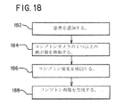
図18は、コンプトンカメラによる医療用画像形成方法の一実施形態のフローチャート図である。この方法は、図11~図16Bの適応コンプトンカメラを使用して実装される。コンプトンカメラで使用される1つ以上の検出器の位置を変更できる他の適応型コンプトンカメラを使用することができる。 FIG. 18 is a flow chart diagram of one embodiment of a medical imaging method with a Compton camera. This method is implemented using the adaptive Compton camera of FIGS. 11-16B. Other adaptive Compton cameras can be used that can change the position of one or more of the detectors used in the Compton camera.
動作は、示された順序か別の順番で行われる。追加の動作、異なる動作、またはより少ない動作が提供されることがある。例えば、患者は、動作182において感知されない。代わりに、捕獲検出器は、散乱検出器が患者の位置に基づいて移動されることなく、または散乱検出器が検知ではなく患者の体重に基づいて移動されることで、散乱検出器に対して移動される。別の例として、画像は、動作188において生成されない。画像は後で見るために保存される。
Operations may be performed in the order shown or in another order. Additional, different, or fewer acts may be provided. For example, the patient is not sensed in
動作182において、センサは患者を感知する。アイソセンタ、ベッド、および/または散乱検出器に対して、患者の外表面が感知される。患者からの閾値距離内での散乱検出器または複数の散乱検出器の位置決めを可能にするために、患者を感知する。
In
動作184では、コンプトンカメラの1つ以上の検出器が移動される。検出器または複数の検出器は、患者に向かって、または患者から遠ざけられる。FOM、検査タイプ、および/または他の情報に基づいて、検出器または複数の検出器が移動される。
At
1つの実施形態において、散乱検出器または複数の散乱検出器は、患者の検知の出力に基づいて、モータおよび制御プロセッサによって移動される。散乱検出器または複数の散乱検出器は、それぞれの散乱検出器に最も近い患者の外表面から閾値距離内になるように移動される。図11は、一部の散乱検出器が、患者の外表面に基づく他の散乱検出器よりもアイソセンタに近い例を示す。 In one embodiment, the scatter detector or detectors are moved by a motor and control processor based on the output of patient sensing. The scatter detector or scatter detectors are moved to be within a threshold distance from the outer surface of the patient closest to the respective scatter detector. FIG. 11 shows an example where some scatter detectors are closer to the isocenter than others based on the outer surface of the patient.
更に、又は代替的に、捕獲検出器又は複数の捕獲検出器は、モータ及び制御プロセッサによって移動される。検査タイプ、FOM、関与する放射性同位元素のエネルギー、および/または他の基準に基づいて、捕獲検出器または複数の捕獲検出器は、アイソセンタ、患者、および/または散乱検出器または複数の散乱検出器に対して移動される。例えば、散乱検出器は、患者から所定の距離になるように配置される。次いで、捕獲検出器は、距離がFOMまたは他の情報に基づく散乱検出器からの距離となるように配置される。 Additionally or alternatively, the capture detector or multiple capture detectors are moved by a motor and control processor. Based on study type, FOM, energies of radioisotopes involved, and/or other criteria, the capture detector or capture detectors may be isocenter, patient, and/or scatter detector or scatter detectors. is moved relative to For example, a scatter detector is placed at a predetermined distance from the patient. The capture detector is then positioned such that the distance is from the scatter detector based on the FOM or other information.
動作186では、散乱検出器及び捕獲検出器は事象を検出する。患者から放出されるガンマ線または光子は、散乱検出器と相互作用することがある。これらの散乱事象が検出される。結果として生じる散乱光子が放出され、捕獲検出器と相互作用する可能性がある。捕獲検出器内の相互作用が検出される。
At
検出されたコンプトン事象は対になっている。対になったコンプトン事象は、散乱検出器での患者からの放出の確率の錐体などの入射角を示すために使用される。コンプトン事象は、散乱角に基づいてデジタル的にコリメートされることがある。 The detected Compton events are paired. A paired Compton event is used to indicate the angle of incidence, such as a cone of probability of emission from the patient at the scatter detector. Compton events may be digitally collimated based on the scattering angle.
散乱検出器と捕獲検出器は、位置決めされた事象を検出する。患者のスキャン中、検出器は検出するために同じ位置に維持される。または、同一患者の同一スキャン中に1つ以上の検出器が移動される。 Scatter detectors and capture detectors detect positioned events. During scanning of the patient, the detector remains in the same position for detection. Alternatively, more than one detector is moved during the same scan of the same patient.
動作188では、任意のデジタル視準後に維持された対となったコンプトン事象が、患者からの放出物の空間的分布を再構成するために使用される。放出源は、コンプトン事象の入射角、場所、計数を用いて推計される。画像は、三次元レンダリングまたは断面平面画像のような空間分布から生成され得る。
In
本発明を種々の実施形態を参照して上述したが、本発明の特許請求の範囲から逸脱することなく、多くの変更及び改変がなされ得ることは理解されるべきである。したがって、上記詳細な説明は、本発明を限定するものではなく例示として解されるものであり、本発明の精神および範囲を定めるのは、全ての均等物を含めて特許請求の範囲の記載に基づくものであることは理解されるべきである。
Although the invention has been described above with reference to various embodiments, it should be understood that many variations and modifications can be made without departing from the scope of the claims of the invention. Accordingly, the foregoing detailed description is to be taken as illustrative rather than limiting, and it is intended that the following claims, including all equivalents, define the spirit and scope of the invention. It should be understood that the
Claims (18)
前記コンプトンカメラが、
患者ベッド(60)と、
それぞれが散乱検出器(12)と、捕獲検出器(13)と、を有する複数のモジュール(11)であって、前記患者ベッド(60)上の患者からの放出物を受け取るように配置された、複数のモジュール(11)と、
前記散乱検出器(12)、前記捕獲検出器(13)、前記散乱検出器(12)と前記捕獲検出器(13)の両方、または、前記複数のモジュールと連結するモータ(114)であって、
前記モータ(114)が、前記散乱検出器(12)、前記捕獲検出器(13)、または、前記散乱検出器(12)と前記捕獲検出器(13)の両方、を前記患者ベッド(60)に対して近づけるか、または、遠ざけるように構成されている、
モータ(114)と、
を含む、コンプトンカメラ。 A Compton camera for medical imaging, comprising:
The Compton camera is
a patient bed (60);
a plurality of modules (11) each having a scatter detector (12) and a capture detector (13) arranged to receive emissions from a patient on said patient bed (60) , a plurality of modules (11), and
a motor (114) coupled to the scatter detector (12), the capture detector (13) , both the scatter detector (12) and the capture detector (13), or the plurality of modules ; ,
The motor (114) drives the scatter detector (12), the capture detector (13), or both the scatter detector (12) and the capture detector (13) to the patient bed (60). configured to move toward or away from
a motor (114);
including Compton cameras.
前記モータ(114)が前記散乱検出器(12)と前記捕獲検出器(13)を一緒に移動させるように構成されている、請求項1に記載のコンプトンカメラ。 said scatter detector (12) is at a fixed distance from said capture detector (13);
The Compton camera of claim 1, wherein the motor (114) is configured to move the scatter detector (12) and the capture detector (13) together.
前記モータ(114)が前記捕獲検出器(13)を移動させることなく、前記散乱検出器(12)を移動させるように構成されている、請求項1に記載のコンプトンカメラ。 said scatter detector (12) is movable relative to said capture detector (13);
The Compton camera of claim 1, wherein the motor (114) is configured to move the scatter detector (12) without moving the capture detector (13).
前記モータ(114)が前記散乱検出器(12)を移動させることなく、前記捕獲検出器(13)を移動させるように構成されている、請求項1に記載のコンプトンカメラ。 said capture detector (13) is movable relative to said scatter detector (12);
The Compton camera of claim 1, wherein the motor (114) is configured to move the capture detector (13) without moving the scatter detector (12).
前記モータ(114)が、前記センサ(110)の出力に基づいて、前記散乱検出器(12)の前記患者からの距離を減少させるために、前記散乱検出器(12)を移動させるように構成されている、請求項1に記載のコンプトンカメラ。 further comprising a sensor (110) configured to sense a patient on said patient bed (60);
The motor (114) is configured to move the scatter detector (12) to decrease the distance of the scatter detector (12) from the patient based on the output of the sensor (110). 2. The Compton camera of claim 1, wherein:
前記モータ(114)を制御して、前記患者からの前記散乱検出器(12)の距離を減少させるように、前記散乱検出器(12)を移動させるとともに、
前記モータ(114)を制御して、前記性能指数に基づいて前記捕獲検出器(13)を前記散乱検出器(12)からの距離に移動させるように、
構成されている、請求項7に記載のコンプトンカメラ。 The control processor (112) is configured to:
controlling the motor (114) to move the scatter detector (12) to decrease the distance of the scatter detector (12) from the patient;
controlling the motor (114) to move the capture detector (13) a distance from the scatter detector (12) based on the figure of merit;
8. The Compton camera of claim 7 , configured.
前記コンプトン画像を表示するように構成されたディスプレイをさらに含む、
請求項1に記載のコンプトンカメラ。 further comprising an image processor (19) configured to generate a Compton image from Compton events formed from paired events at the scatter detector (12) and the capture detector (13);
further comprising a display configured to display said Compton image;
The Compton camera of Claim 1.
前記散乱検出器(12)、前記捕獲検出器(13)、前記散乱検出器(12)と前記捕獲検出器(13)の両方、または、前記複数の固体検出器モジュール(11)と連結されたモータ(114)と、
前記モータ(114)を制御して、前記散乱検出器(12)、前記捕獲検出器(13)、または、前記散乱検出器(12)および前記捕獲検出器(13)の両方、の位置を、患者空間のアイソセンタに対して変更するように構成された制御プロセッサ(112)と、
を含む、医療用画像システム。 a plurality of solid-state detector modules (11) each having a scatter detector (12) and a capture detector (13);
coupled with said scatter detector (12), said capture detector (13), both said scatter detector (12) and said capture detector (13), or said plurality of solid state detector modules (11) a motor (114);
controlling the motor (114) to position the scatter detector (12), the capture detector (13), or both the scatter detector (12) and the capture detector (13) by a control processor (112) configured to vary with respect to the isocenter of patient space;
medical imaging systems, including;
前記制御プロセッサ(112)が、前記センサ(110)からの前記患者の感知位置に基づいて、前記散乱検出器(12)の位置を変更するように構成されている、請求項12に記載の医療用画像システム。 further comprising a position sensor (110) configured to sense a patient within said patient space;
13. The medical treatment of Claim 12 , wherein the control processor (112) is configured to change the position of the scatter detector (12) based on the sensed position of the patient from the sensor (110). image system for.
前記方法が、
モータ(114)により、前記コンプトンカメラの検出器を移動させること(184)と、
移動する前記検出器によって、患者からの放出物を検出すること(186)と、
検出された前記放出物からコンプトン画像を生成すること(188)と、
を含む、医療用画像のための方法。 A method for medical imaging with a Compton camera according to any one of claims 1 to 11 , comprising:
the method comprising:
moving (184) a detector of the Compton camera with a motor (114);
detecting 186 patient emissions with the moving detector;
generating (188) a Compton image from the detected emissions;
A method for medical imaging, comprising:
前記移動させること(184)が、前記患者の前記検知の出力に基づいて移動させること(184)を含む、請求項16に記載の方法。 further comprising detecting (182) the patient;
17. The method of claim 16 , wherein moving (184) comprises moving (184) based on an output of the sensing of the patient.
moving the detector (184) moving a scatter detector (12), a capture detector (13), or both the scatter detector (12) and the capture detector (13) 17. The method of claim 16 , comprising (184).
Applications Claiming Priority (1)
| Application Number | Priority Date | Filing Date | Title |
|---|---|---|---|
| PCT/US2018/045469 WO2020032924A1 (en) | 2018-08-07 | 2018-08-07 | Adaptive compton camera for medical imaging |
Publications (2)
| Publication Number | Publication Date |
|---|---|
| JP2021533367A JP2021533367A (en) | 2021-12-02 |
| JP7208355B2 true JP7208355B2 (en) | 2023-01-18 |
Family
ID=63350611
Family Applications (1)
| Application Number | Title | Priority Date | Filing Date |
|---|---|---|---|
| JP2021505881A Active JP7208355B2 (en) | 2018-08-07 | 2018-08-07 | Adaptive Compton camera for medical imaging |
Country Status (6)
| Country | Link |
|---|---|
| US (1) | US11744528B2 (en) |
| EP (1) | EP3817663A1 (en) |
| JP (1) | JP7208355B2 (en) |
| CN (1) | CN112512427A (en) |
| IL (1) | IL280482A (en) |
| WO (1) | WO2020032924A1 (en) |
Families Citing this family (2)
| Publication number | Priority date | Publication date | Assignee | Title |
|---|---|---|---|---|
| FR3081231B1 (en) * | 2018-05-18 | 2020-06-12 | Damavan Imaging | GAMMA RADIATION DETECTION IMAGING SYSTEM AND METHOD |
| CN111329500B (en) * | 2018-12-19 | 2022-09-09 | 清华大学 | Gamma radiation imaging device and imaging method |
Citations (7)
| Publication number | Priority date | Publication date | Assignee | Title |
|---|---|---|---|---|
| US20040084624A1 (en) | 2002-10-31 | 2004-05-06 | Meng Ling Jian | Method and system for generating an image of the radiation density of a source of photons located in an object |
| US20040251419A1 (en) | 2003-06-16 | 2004-12-16 | Nelson Robert Sigurd | Device and system for enhanced SPECT, PET, and Compton scatter imaging in nuclear medicine |
| JP2008232971A (en) | 2007-03-23 | 2008-10-02 | Hitachi Medical Corp | Nuclear medicine diagnostic apparatus and photon measuring device |
| JP2013022041A (en) | 2011-07-15 | 2013-02-04 | Shimadzu Corp | Radiographic apparatus for breast examination |
| JP2014149308A (en) | 2008-06-13 | 2014-08-21 | Koninklijke Philips Nv | Reverse data reconstruction for optimal time sampling of count in physiological list mode nuclear medical imaging |
| JP2017026423A (en) | 2015-07-21 | 2017-02-02 | キヤノン株式会社 | Compton camera, and displacement detection method of the same |
| US20170285191A1 (en) | 2016-03-29 | 2017-10-05 | Kromek Goup, PLC | Sparse Acquisition Gamma Cameras |
Family Cites Families (87)
| Publication number | Priority date | Publication date | Assignee | Title |
|---|---|---|---|---|
| YU84586A (en) | 1986-05-21 | 1990-02-28 | Vladimir Bosnjakovic | Scintillation crystalline device and scintillation camera with that device |
| JPH0462492A (en) * | 1990-06-29 | 1992-02-27 | Toshiba Corp | Nuclear medical diagnostic device |
| US5097131A (en) * | 1990-11-21 | 1992-03-17 | Picker International, Inc. | Nuclear medicine camera gantry system with vertically stored collimators |
| US5093575A (en) * | 1990-11-21 | 1992-03-03 | Picker International, Inc. | Dual rotatable head gamma camera |
| US5097132A (en) * | 1990-11-21 | 1992-03-17 | Picker International, Inc. | Nuclear medicine camera system with improved gantry and patient table |
| US6184530B1 (en) * | 1991-05-23 | 2001-02-06 | Adac Laboratories | Adjustable dual-detector image data acquisition system |
| US5567944A (en) * | 1995-04-28 | 1996-10-22 | University Of Cincinnati | Compton camera for in vivo medical imaging of radiopharmaceuticals |
| US5821541A (en) * | 1996-02-02 | 1998-10-13 | Tuemer; Tuemay O. | Method and apparatus for radiation detection |
| US5757006A (en) * | 1997-01-30 | 1998-05-26 | Siemens Medical Systems, Inc. | Articulating detector array for a gamma camera |
| WO1998052069A1 (en) | 1997-05-16 | 1998-11-19 | University Of Michigan | Method for improving the spatial resolution of a compton camera |
| JP4169291B2 (en) | 1998-04-14 | 2008-10-22 | 株式会社東芝 | Nuclear medicine diagnostic equipment |
| US6346706B1 (en) | 1999-06-24 | 2002-02-12 | The Regents Of The University Of Michigan | High resolution photon detector |
| US6528795B2 (en) | 2000-04-27 | 2003-03-04 | The United States Of America As Represented By The Secretary Of The Navy | Compton scatter imaging instrument |
| US6791090B2 (en) * | 2000-05-17 | 2004-09-14 | Koninklijke Philips Electronics N.V. | Compton deconvolution camera |
| US6455856B1 (en) * | 2000-06-02 | 2002-09-24 | Koninklijke Philips Electronics N.V. | Gamma camera gantry and imaging method |
| US6583420B1 (en) * | 2000-06-07 | 2003-06-24 | Robert S. Nelson | Device and system for improved imaging in nuclear medicine and mammography |
| JP4659962B2 (en) | 2000-10-04 | 2011-03-30 | 株式会社東芝 | Nuclear medicine diagnostic equipment |
| US6603123B1 (en) | 2000-11-08 | 2003-08-05 | Koninklijke Philips Electronics, N.V. | Correction for depth-dependent sensitivity in rotating slat-collimated gamma camera |
| US6512232B2 (en) | 2000-12-20 | 2003-01-28 | Richard H. Pehl | Method and apparatus for improving the sensitivity of a gamma camera |
| WO2002065480A1 (en) * | 2001-02-01 | 2002-08-22 | Creatv Microtech, Inc. | tNTI-SCATTER GRIDS AND COLLIMATOR DESIGNS, AND THEIR MOTION, FABRICATION AND ASSEMBLY |
| US7922923B2 (en) | 2001-02-01 | 2011-04-12 | Creatv Microtech, Inc. | Anti-scatter grid and collimator designs, and their motion, fabrication and assembly |
| US7412022B2 (en) | 2002-02-28 | 2008-08-12 | Jupiter Clyde P | Non-invasive stationary system for three-dimensional imaging of density fields using periodic flux modulation of compton-scattered gammas |
| EP1521982A1 (en) | 2002-07-17 | 2005-04-13 | European Organization for Nuclear Research | Gamma ray detector for positron emission tomography (pet) and single photon emmission computed tomography (spect) |
| GB2401766B (en) | 2003-03-11 | 2006-03-15 | Symetrica Ltd | Improved gamma-ray camera system |
| US7015477B2 (en) | 2003-03-27 | 2006-03-21 | Donald Lee Gunter | Filtered backprojection algorithms for compton cameras in nuclear medicine |
| US7667203B2 (en) | 2003-07-03 | 2010-02-23 | Lockheed Martin Corporation | Gamma vector camera |
| JP2005090979A (en) * | 2003-09-12 | 2005-04-07 | National Cancer Center-Japan | Radiation detector for diagnosing nuclear medicine image and image diagnostic device |
| JP2007513667A (en) | 2003-12-12 | 2007-05-31 | ティシュオミクス リミテッド | Use of Compton scattering in characterization of body tissue for exemplary breast tissue, or use of a combination of XRF (X-ray fluorescence) and EDXRD (energy dispersive X-ray diffraction) |
| EP1548465A1 (en) | 2003-12-26 | 2005-06-29 | Riken | Gamma-ray detector and gamma-ray image pick-up apparatus |
| US7262417B2 (en) | 2004-03-26 | 2007-08-28 | Board Of Regents, The University Of Texas System | Method and system for improved image reconstruction and data collection for compton cameras |
| KR20070085387A (en) | 2004-12-01 | 2007-08-27 | 트라이엄프,오퍼레이팅애즈어조인트벤쳐바이더거버너스 오브더유니버시티오브알버타더유니버시티오브브리티시콜롬비아 칼레톤유니버시티시몬프레이저유니버시티더유니버시티 오브토론토앤드더유니버시티오브빅토리아 | System for selecting true coincidence events in positron emission tomography |
| US7304309B2 (en) | 2005-03-14 | 2007-12-04 | Avraham Suhami | Radiation detectors |
| US7635848B2 (en) * | 2005-04-01 | 2009-12-22 | San Diego State University Research Foundation | Edge-on SAR scintillator devices and systems for enhanced SPECT, PET, and compton gamma cameras |
| US7550738B1 (en) | 2005-04-28 | 2009-06-23 | Utah State University | Nuclear material identification and localization |
| US8217362B2 (en) | 2005-04-28 | 2012-07-10 | Utah State University | Identification and localization of radioisotopes using likelihood estimation |
| US7345283B2 (en) | 2005-10-04 | 2008-03-18 | Lawrence Livermore National Security, Llc | Filtered back-projection algorithm for Compton telescopes |
| US7732773B2 (en) | 2005-12-29 | 2010-06-08 | Lawrence Livermore National Security, Llc | Gamma-ray tracking method for pet systems |
| WO2007109227A2 (en) | 2006-03-17 | 2007-09-27 | The Trustees Of The University Of Pennsylvania | Slit-slat collimation |
| US7750308B2 (en) | 2006-06-14 | 2010-07-06 | Hitachi Medical Corporation | Compton camera device |
| FR2902527B1 (en) | 2006-06-20 | 2008-12-19 | Commissariat Energie Atomique | DEVICE FOR THE THREE-DIMENSIONAL LOCATION OF RADIATION SOURCES |
| US8063379B2 (en) * | 2006-06-21 | 2011-11-22 | Avraham Suhami | Radiation cameras |
| JP4486623B2 (en) | 2006-08-11 | 2010-06-23 | 独立行政法人理化学研究所 | Compton imaging camera |
| WO2008077011A2 (en) | 2006-12-19 | 2008-06-26 | Battelle Memorial Institute | Method and system for imaging a radiation source |
| US20120132814A1 (en) | 2007-02-26 | 2012-05-31 | Irving Weinberg | Radiation detection device, system and related methods |
| US7573039B2 (en) | 2007-03-13 | 2009-08-11 | Smith Bruce D | Compton camera configuration and imaging method |
| US8299437B2 (en) | 2007-05-15 | 2012-10-30 | National Institute Of Radiological Sciences | Gamma ray detector and gamma ray reconstruction method |
| US8153986B2 (en) | 2007-07-09 | 2012-04-10 | Lawrence Livermore National Security, Llc | Hybrid Compton camera/coded aperture imaging system |
| US8139713B2 (en) * | 2007-08-10 | 2012-03-20 | Koninklijke Philips Electronics N.V. | Combined nuclear-radiographic subject imaging |
| US8107589B2 (en) | 2007-12-21 | 2012-01-31 | Kabushiki Kaisha Toshiba | Radiotherapeutic system and radiotherapeutic dose distribution measuring method |
| FR2926893B1 (en) | 2008-01-25 | 2012-07-27 | Centre Nat Rech Scient | METHOD FOR LOCATING POSIONAL RADIONUCLEID, APPLICATIONS AND DEVICE FOR IMPLEMENTING THE SAME |
| JP5233029B2 (en) | 2008-08-22 | 2013-07-10 | 国立大学法人群馬大学 | Compton camera |
| WO2010034619A1 (en) * | 2008-09-23 | 2010-04-01 | Institut de Física D'Altes Energies | Device for detecting highly energetic photons |
| WO2010048363A2 (en) | 2008-10-24 | 2010-04-29 | University Of Washington | Line of response estimation for high-resolution pet detector |
| US8071949B2 (en) * | 2009-01-23 | 2011-12-06 | Jefferson Science Associates, Llc | High-resolution single photon planar and spect imaging of brain and neck employing a system of two co-registered opposed gamma imaging heads |
| US7863567B1 (en) | 2009-04-13 | 2011-01-04 | Raytheon Company | Multimodal radiation imager |
| WO2010141583A2 (en) | 2009-06-02 | 2010-12-09 | Mayo Foundation For Medical Education And Research | System and method for dose verification radiotherapy |
| US8476595B2 (en) | 2009-06-19 | 2013-07-02 | Yale University | Liquid xenon gamma ray imager |
| US8785864B2 (en) | 2009-09-22 | 2014-07-22 | Boss Physical Sciences Llc | Organic-scintillator compton gamma ray telescope |
| US20110303854A1 (en) | 2010-06-14 | 2011-12-15 | Utah State University | Gamma ray directionality probe |
| US20120043467A1 (en) | 2010-08-17 | 2012-02-23 | Andrey Gueorguiev | Single plane compton camera |
| CA2719381A1 (en) | 2010-10-29 | 2012-04-29 | Universite De Sherbrooke | Artificial intelligence method and apparatus for analysis of compton-scattered photons in radiation detection machines |
| WO2012077468A1 (en) | 2010-12-09 | 2012-06-14 | 独立行政法人理化学研究所 | Imaging device using gamma rays, image signal processor, and image processing method for gamma ray measurement data |
| US8519343B1 (en) | 2011-04-25 | 2013-08-27 | U.S. Department Of Energy | Multimode imaging device |
| JP5684670B2 (en) | 2011-08-03 | 2015-03-18 | 株式会社日立製作所 | Gamma camera system |
| EP3647822A3 (en) * | 2012-05-08 | 2020-08-12 | Spectrum Dynamics Medical Limited | Nuclear medicine tomography systems, detectors and methods |
| US10371834B2 (en) * | 2012-05-31 | 2019-08-06 | Minnesota Imaging And Engineering Llc | Detector systems for integrated radiation imaging |
| US10067239B2 (en) | 2012-05-31 | 2018-09-04 | Minnesota Imaging And Engineering Llc | Detector systems for radiation imaging |
| JP5840638B2 (en) * | 2013-03-21 | 2016-01-06 | 株式会社東芝 | Radiation detection apparatus and radiation detection method |
| US9057684B2 (en) * | 2013-04-05 | 2015-06-16 | The Arizona Board Of Regents On Behalf Of The University Of Arizona | Gamma ray imaging systems and methods |
| US9662079B2 (en) * | 2013-09-03 | 2017-05-30 | General Electric Company | Methods and apparatus for imaging with detectors having moving detector heads |
| US10575802B2 (en) * | 2013-09-27 | 2020-03-03 | General Electric Company | Systems and methods for controlling motion of detectors having moving detector heads |
| US9192346B2 (en) * | 2013-12-19 | 2015-11-24 | General Electric Company | Systems and methods for multi-modality imaging |
| US9029791B1 (en) * | 2013-12-20 | 2015-05-12 | General Electric Company | Imaging system using independently controllable detectors |
| JP6410127B2 (en) | 2014-03-11 | 2018-10-24 | 住友電気工業株式会社 | Electrolyte circulating battery, heat exchanger, and piping |
| US10188358B2 (en) * | 2014-05-15 | 2019-01-29 | General Electric Company | System and method for subject shape estimation |
| EP3190431B1 (en) * | 2014-09-05 | 2019-03-20 | Riken | Imaging device and method |
| US9763631B2 (en) * | 2014-09-17 | 2017-09-19 | General Electric Company | Systems and methods for imaging plural axial locations |
| WO2016077865A1 (en) * | 2014-11-21 | 2016-05-26 | Monash University | Single/limited angle gamma/x-ray tomography |
| US10143437B2 (en) * | 2015-06-30 | 2018-12-04 | General Electric Company | Systems and methods for dynamic scanning with multi-head camera |
| US9579072B1 (en) * | 2015-09-30 | 2017-02-28 | General Electric Company | Systems and methods for imaging with multi-head camera |
| ES2629092B1 (en) * | 2015-11-04 | 2018-07-04 | Consejo Superior De Investigaciones Científicas (Csic) | GAMMA RAY COMPTON CAMERA SYSTEM WITH FLIGHT TIME MEASUREMENT |
| US10884139B2 (en) * | 2016-03-09 | 2021-01-05 | University Of Maryland, Baltimore | Techniques for producing an image of radioactive emissions using a compton camera and compton lines |
| DE102016221658A1 (en) * | 2016-11-04 | 2018-05-09 | Siemens Healthcare Gmbh | Stray radiation compensation for an imaging medical device |
| US20210282725A1 (en) * | 2018-08-07 | 2021-09-16 | Siemens Medical Solutions Usa, Inc. | NEAR 2Pi COMPTON CAMERA FOR MEDICAL IMAGING |
| ES2941962T3 (en) * | 2018-08-07 | 2023-05-29 | Siemens Medical Solutions Usa Inc | Multimodal Compton and Single Photon Emission Computed Tomography Imaging System |
| CN112513680A (en) * | 2018-08-07 | 2021-03-16 | 美国西门子医疗系统股份有限公司 | Compton camera with segment detection module |
| US11647973B2 (en) * | 2021-05-04 | 2023-05-16 | Siemens Medical Solutions Usa, Inc. | Three-dimensional tileable gamma ray detector |
-
2018
- 2018-08-07 EP EP18759215.9A patent/EP3817663A1/en active Pending
- 2018-08-07 CN CN201880096375.9A patent/CN112512427A/en active Pending
- 2018-08-07 WO PCT/US2018/045469 patent/WO2020032924A1/en active Application Filing
- 2018-08-07 JP JP2021505881A patent/JP7208355B2/en active Active
- 2018-08-07 US US17/250,492 patent/US11744528B2/en active Active
-
2021
- 2021-01-28 IL IL280482A patent/IL280482A/en unknown
Patent Citations (7)
| Publication number | Priority date | Publication date | Assignee | Title |
|---|---|---|---|---|
| US20040084624A1 (en) | 2002-10-31 | 2004-05-06 | Meng Ling Jian | Method and system for generating an image of the radiation density of a source of photons located in an object |
| US20040251419A1 (en) | 2003-06-16 | 2004-12-16 | Nelson Robert Sigurd | Device and system for enhanced SPECT, PET, and Compton scatter imaging in nuclear medicine |
| JP2008232971A (en) | 2007-03-23 | 2008-10-02 | Hitachi Medical Corp | Nuclear medicine diagnostic apparatus and photon measuring device |
| JP2014149308A (en) | 2008-06-13 | 2014-08-21 | Koninklijke Philips Nv | Reverse data reconstruction for optimal time sampling of count in physiological list mode nuclear medical imaging |
| JP2013022041A (en) | 2011-07-15 | 2013-02-04 | Shimadzu Corp | Radiographic apparatus for breast examination |
| JP2017026423A (en) | 2015-07-21 | 2017-02-02 | キヤノン株式会社 | Compton camera, and displacement detection method of the same |
| US20170285191A1 (en) | 2016-03-29 | 2017-10-05 | Kromek Goup, PLC | Sparse Acquisition Gamma Cameras |
Also Published As
| Publication number | Publication date |
|---|---|
| US11744528B2 (en) | 2023-09-05 |
| EP3817663A1 (en) | 2021-05-12 |
| WO2020032924A1 (en) | 2020-02-13 |
| US20210290196A1 (en) | 2021-09-23 |
| JP2021533367A (en) | 2021-12-02 |
| IL280482A (en) | 2021-03-01 |
| CN112512427A (en) | 2021-03-16 |
Similar Documents
| Publication | Publication Date | Title |
|---|---|---|
| JP7204884B2 (en) | Compton and Single Photon Emission Tomography Multimodal Medical Imaging Systems | |
| JP7179958B2 (en) | Compton camera with segmented detection module | |
| EP3030154B1 (en) | Pet system with crystal and detector unit spacing | |
| CN101138502B (en) | Tileable multi-layer detector | |
| JP7293334B2 (en) | Nearly 2π Compton camera for medical imaging | |
| JP7208355B2 (en) | Adaptive Compton camera for medical imaging | |
| Dey | Improvement of performance of cardiac SPECT camera using curved detectors with pinholes | |
| US7375338B1 (en) | Swappable collimators method and system | |
| US8957384B2 (en) | Gamma ray detector linearity calibration | |
| JPWO2020032922A5 (en) | ||
| US20100027866A1 (en) | Positron ct apparatus | |
| WO2007106674A2 (en) | Nuclear medicine imaging system with high efficiency transmission measurement | |
| JP3604491B2 (en) | Gamma camera device | |
| Uribe et al. | Gantry design with accurate crystal positioning for a high-resolution transformable PET camera | |
| WO2018080288A1 (en) | Variable pet apparatus | |
| WO2016162962A1 (en) | Radiation imaging device | |
| JP2020153734A (en) | Medical diagnostic imaging device and method of generating image for inspection | |
| JP2015230194A (en) | Radiation imaging device using location identification type detector |
Legal Events
| Date | Code | Title | Description |
|---|---|---|---|
| A521 | Request for written amendment filed |
Free format text: JAPANESE INTERMEDIATE CODE: A523 Effective date: 20210608 |
|
| A621 | Written request for application examination |
Free format text: JAPANESE INTERMEDIATE CODE: A621 Effective date: 20210608 |
|
| RD03 | Notification of appointment of power of attorney |
Free format text: JAPANESE INTERMEDIATE CODE: A7423 Effective date: 20210608 |
|
| A977 | Report on retrieval |
Free format text: JAPANESE INTERMEDIATE CODE: A971007 Effective date: 20220420 |
|
| A131 | Notification of reasons for refusal |
Free format text: JAPANESE INTERMEDIATE CODE: A131 Effective date: 20220531 |
|
| A521 | Request for written amendment filed |
Free format text: JAPANESE INTERMEDIATE CODE: A523 Effective date: 20220808 |
|
| TRDD | Decision of grant or rejection written | ||
| A01 | Written decision to grant a patent or to grant a registration (utility model) |
Free format text: JAPANESE INTERMEDIATE CODE: A01 Effective date: 20221206 |
|
| A61 | First payment of annual fees (during grant procedure) |
Free format text: JAPANESE INTERMEDIATE CODE: A61 Effective date: 20230105 |
|
| R150 | Certificate of patent or registration of utility model |
Ref document number: 7208355 Country of ref document: JP Free format text: JAPANESE INTERMEDIATE CODE: R150 |