JP7197755B2 - Circulating Serum MicroRNA Biomarkers and Methods - Google Patents
Circulating Serum MicroRNA Biomarkers and Methods Download PDFInfo
- Publication number
- JP7197755B2 JP7197755B2 JP2018560445A JP2018560445A JP7197755B2 JP 7197755 B2 JP7197755 B2 JP 7197755B2 JP 2018560445 A JP2018560445 A JP 2018560445A JP 2018560445 A JP2018560445 A JP 2018560445A JP 7197755 B2 JP7197755 B2 JP 7197755B2
- Authority
- JP
- Japan
- Prior art keywords
- mir
- hsa
- mirnas
- disease
- mirna
- Prior art date
- Legal status (The legal status is an assumption and is not a legal conclusion. Google has not performed a legal analysis and makes no representation as to the accuracy of the status listed.)
- Active
Links
Images


Classifications
-
- C—CHEMISTRY; METALLURGY
- C12—BIOCHEMISTRY; BEER; SPIRITS; WINE; VINEGAR; MICROBIOLOGY; ENZYMOLOGY; MUTATION OR GENETIC ENGINEERING
- C12Q—MEASURING OR TESTING PROCESSES INVOLVING ENZYMES, NUCLEIC ACIDS OR MICROORGANISMS; COMPOSITIONS OR TEST PAPERS THEREFOR; PROCESSES OF PREPARING SUCH COMPOSITIONS; CONDITION-RESPONSIVE CONTROL IN MICROBIOLOGICAL OR ENZYMOLOGICAL PROCESSES
- C12Q1/00—Measuring or testing processes involving enzymes, nucleic acids or microorganisms; Compositions therefor; Processes of preparing such compositions
- C12Q1/68—Measuring or testing processes involving enzymes, nucleic acids or microorganisms; Compositions therefor; Processes of preparing such compositions involving nucleic acids
- C12Q1/6844—Nucleic acid amplification reactions
- C12Q1/686—Polymerase chain reaction [PCR]
-
- C—CHEMISTRY; METALLURGY
- C12—BIOCHEMISTRY; BEER; SPIRITS; WINE; VINEGAR; MICROBIOLOGY; ENZYMOLOGY; MUTATION OR GENETIC ENGINEERING
- C12Q—MEASURING OR TESTING PROCESSES INVOLVING ENZYMES, NUCLEIC ACIDS OR MICROORGANISMS; COMPOSITIONS OR TEST PAPERS THEREFOR; PROCESSES OF PREPARING SUCH COMPOSITIONS; CONDITION-RESPONSIVE CONTROL IN MICROBIOLOGICAL OR ENZYMOLOGICAL PROCESSES
- C12Q1/00—Measuring or testing processes involving enzymes, nucleic acids or microorganisms; Compositions therefor; Processes of preparing such compositions
- C12Q1/68—Measuring or testing processes involving enzymes, nucleic acids or microorganisms; Compositions therefor; Processes of preparing such compositions involving nucleic acids
- C12Q1/6876—Nucleic acid products used in the analysis of nucleic acids, e.g. primers or probes
- C12Q1/6883—Nucleic acid products used in the analysis of nucleic acids, e.g. primers or probes for diseases caused by alterations of genetic material
-
- C—CHEMISTRY; METALLURGY
- C12—BIOCHEMISTRY; BEER; SPIRITS; WINE; VINEGAR; MICROBIOLOGY; ENZYMOLOGY; MUTATION OR GENETIC ENGINEERING
- C12Q—MEASURING OR TESTING PROCESSES INVOLVING ENZYMES, NUCLEIC ACIDS OR MICROORGANISMS; COMPOSITIONS OR TEST PAPERS THEREFOR; PROCESSES OF PREPARING SUCH COMPOSITIONS; CONDITION-RESPONSIVE CONTROL IN MICROBIOLOGICAL OR ENZYMOLOGICAL PROCESSES
- C12Q2600/00—Oligonucleotides characterized by their use
- C12Q2600/158—Expression markers
-
- C—CHEMISTRY; METALLURGY
- C12—BIOCHEMISTRY; BEER; SPIRITS; WINE; VINEGAR; MICROBIOLOGY; ENZYMOLOGY; MUTATION OR GENETIC ENGINEERING
- C12Q—MEASURING OR TESTING PROCESSES INVOLVING ENZYMES, NUCLEIC ACIDS OR MICROORGANISMS; COMPOSITIONS OR TEST PAPERS THEREFOR; PROCESSES OF PREPARING SUCH COMPOSITIONS; CONDITION-RESPONSIVE CONTROL IN MICROBIOLOGICAL OR ENZYMOLOGICAL PROCESSES
- C12Q2600/00—Oligonucleotides characterized by their use
- C12Q2600/178—Oligonucleotides characterized by their use miRNA, siRNA or ncRNA
Description
本発明は、一般に、血清に基づくマイクロRNA、およびパーキンソン病に罹患している患者を識別するための方法、ならびに臨床医がこのような患者の処置プロトコルを決定するのを支援する方法に関する。 The present invention relates generally to serum-based microRNAs and methods for identifying patients with Parkinson's disease, as well as methods for assisting clinicians in determining treatment protocols for such patients.
パーキンソン病(PD)は、中脳の黒質のドーパミン含有細胞の非常に特異的な変性であり、線条体においてドーパミン欠乏を引き起こす。PDは現在、世界中で約1,000万人の人々が罹患している。初期の5~7年間はPD患者への効果的な処理が可能であるが、その後は、患者をしばしば衰弱させる一連の合併症(併せて後期運動変動(Late Motor Fluctuations)(LMF)と呼ばれる)が発生する。最も効果的な抗パーキンソン薬であるレボドーパ((-)-L-α-アミノ-β-(3,4-ジヒドロキシベンゼン)プロパン酸)またはL-ドーパでの治療は、LMFの出現を許容または促進させてしまう可能性があると考えられている。ドーパミンアゴニスト(ドーパミン作用薬)は治療選択肢として使用されるが、Lドーパと同じ程度の患者の症候緩和は得られない。 Parkinson's disease (PD) is a highly specific degeneration of dopamine-containing cells in the substantia nigra of the midbrain, causing dopamine deficiency in the striatum. PD currently affects approximately 10 million people worldwide. PD patients can be effectively treated for the first 5 to 7 years, but after that a series of often debilitating complications (collectively called late motor fluctuations (LMF)) occurs. Treatment with the most effective antiparkinsonian drugs, levodopa ((-)-L-α-amino-β-(3,4-dihydroxybenzene)propanoic acid) or L-dopa, tolerates or accelerates the appearance of LMF It is thought that there is a possibility that Dopamine agonists (dopamine agonists) are used as a treatment option, but do not provide the same degree of patient relief as L-dopa.
対症療法は、根底にある病状に影響を与えずに徴候および症状を改善する。レボドーパは、特にその末梢代謝が末梢デカルボキシラーゼ阻害剤(PDI)によって阻害される場合に、線条体におけるドーパミン濃度を増加させる。レボドーパ/PDI治療は、パーキンソン病の対症療法として広く用いられ、例えば、レボドーパ、カルビドーパ((-)-L-α-ヒドラジノ-α-メチル-β-(3,4-ジヒドロキシベンゼン)プロパン酸一水和物)、レボドーパと放出抑制カルビドーパ、レボドーパとベンゼラジド、又は、レボドーバと放出抑制ベンゼラジド(2-アミノ-3-ヒドロキシ-プロピオン酸N'-(2,3,4-トリヒドロキシ-ベンジル)-ヒドラジド)と併用される。 Symptomatic treatment ameliorates signs and symptoms without affecting the underlying medical condition. Levodopa increases dopamine concentrations in the striatum, especially when its peripheral metabolism is inhibited by peripheral decarboxylase inhibitors (PDIs). Levodopa/PDI therapy is widely used as a symptomatic treatment for Parkinson's disease, e.g. hydrate), levodopa and inhibited-release carbidopa, levodopa and benzelazide, or levodopa and inhibited-release benzelazide (N'-(2,3,4-trihydroxy-benzyl)-hydrazide 2-amino-3-hydroxy-propionate) used in conjunction with
カテコール-O-メチルトランスフェラーゼ(COMT)阻害剤は、レボドーパの代謝を抑制し、そのバイオアベイラビリティーを増強し、それにより、より多くの薬物がシナプス間隙でより長期間利用できるようになるので、レボドーパ治療を強化する。COMT阻害剤の例としては、トルカポン(3,4-ジヒドロキシ-4'-メチル-5-ニトロベンゾフェノン)およびエンタカポン((E)-2-シアノ-3-(3,4-ジヒドロキシ-5-ニトロフェニル)-N,N-ジエチル-2-プロペンアミド)が挙げられる。 Catechol-O-methyltransferase (COMT) inhibitors suppress the metabolism of levodopa and enhance its bioavailability, thereby making more drug available in the synaptic cleft for a longer period of time. intensify treatment. Examples of COMT inhibitors include tolcapone (3,4-dihydroxy-4'-methyl-5-nitrobenzophenone) and entacapone ((E)-2-cyano-3-(3,4-dihydroxy-5-nitrophenyl )-N,N-diethyl-2-propenamide).
ドーパミンアゴニストは、シナプス後線条体ドーパミン受容体を直接刺激することにより症状の改善をもたらす。ドーパミンアゴニストの例としては、ブロモクリプチン((5a)-2-ブロモ-12'-ヒドロキシ-2'-(1-メチルエチル)-5'-(2-メチルプロピル)エルゴタマン-3',6',18-トリオン)、ペルゴリド(8B-[(メチルチオ)メチル] -6-プロピルエルゴリン)、ロピニロール(4-[2-(ジプロピルアミノ)エチル]-1,3-ジヒドロ-2H-インドール-2-オン)、プラミペキソール((S)-4,5,6,7-テトラヒドロ-N6-プロピル-2,6-ベンゾチアゾールジアミン)、リスリド(N'-[(8a)-9,10-ジデヒドロ-6-メチルエルゴリン-8-イル]-N,N-ジメチル-尿素)、カベルゴリン((8β)-N-[3-(ジメチルアミノ)プロピル]-N-[(エチルアミノ)カルボニル]-6-(2-プロペニル)エルゴリン-8-カルボキサミド)、アポモルヒネ((6aR)-5,6,6a,7-テトラヒドロ-6-メチル-4H-ジベンゾ[de,g]キノリン-10,11-ジオール)、スマニロール(5-(メチルアミノ)-5,6-ジヒドロ-4H-イミダゾ{4,5,1-ij}キノリン-2(1H)-オン)、ロチゴチン((-)(S)-5,6,7,8-テトラヒドロ-6-[プロピル[2-(2-チエニル)エチル]アミノ]-1-ナフトール-)、タリペキソール(5,6,7,8-テトラヒドロ-6-(2-プロペニル)-4H-チアゾロ[4,5-d]アゼピン-2-アミン)、及びジヒドロエルゴクリプチン(エルゴタマン-3',6',18-トリオン,9,10-ジヒドロ-12'-ヒドロキシ-2'-メチル-5'-(フェニルメチル)(5'cc))が挙げられる。ドーパミンアゴニストは、パーキンソン病の早期における単剤療法として、および、より進行した段階でのレボドーパへの補助剤として有効である。レボドーパとは異なり、ドーパミンアゴニストは、シナプス後のドーパミン受容体を直接刺激する。これらは酸化的代謝を受けず、病気の進行を加速させないと考えられている。 Dopamine agonists ameliorate symptoms by directly stimulating postsynaptic striatal dopamine receptors. Examples of dopamine agonists include bromocriptine ((5a)-2-bromo-12'-hydroxy-2'-(1-methylethyl)-5'-(2-methylpropyl)ergotaman-3',6',18 -trione), pergolide (8B-[(methylthio)methyl]-6-propylergoline), ropinirole (4-[2-(dipropylamino)ethyl]-1,3-dihydro-2H-indol-2-one) ), pramipexole ((S)-4,5,6,7-tetrahydro-N6-propyl-2,6-benzothiazolediamine), lisuride (N'-[(8a)-9,10-didehydro- 6- methylergolin-8-yl]-N,N-dimethyl-urea), cabergoline ((8β)-N-[3-(dimethylamino)propyl]-N-[(ethylamino)carbonyl]-6-(2 -propenyl)ergoline-8-carboxamide), apomorphine ((6aR)-5,6,6a,7-tetrahydro-6-methyl-4H-dibenzo[de,g]quinoline-10,11-diol), sumanirole (5 -(methylamino)-5,6-dihydro-4H-imidazo{4,5,1-ij}quinolin-2(1H)-one), rotigotine ((-)(S)-5,6,7,8 -tetrahydro-6-[propyl[2-(2-thienyl)ethyl]amino]-1-naphthol-), talipexole (5,6,7,8-tetrahydro-6-(2-propenyl)-4H-thiazolo[ 4,5-d]azepin-2-amine), and dihydroergocryptine (ergotaman-3',6',18-trione, 9,10-dihydro-12'-hydroxy-2'-methyl-5'- (phenylmethyl)(5'cc)). Dopamine agonists are effective as monotherapy in early stages of Parkinson's disease and as adjuncts to levodopa in more advanced stages. Unlike levodopa, dopamine agonists directly stimulate postsynaptic dopamine receptors. They do not undergo oxidative metabolism and are believed not to accelerate disease progression.
アマンチジン(1-アミノトリシクロ(3,3,1,13,7)デカン)は、抗パーキンソン病活性を有することが偶然発見された抗ウイルス剤である。PDにおけるその作用メカニズムは立証されていないが、ドーパミン放出を増加させることによって作用すると考えられている。アマンチジンを単剤療法またはレボドーパと併用して投与された患者は、無動、硬直および振戦の改善を示す。 Amantidine (1-aminotricyclo( 3,3,1,13,7 )decane) is an antiviral agent that was serendipitously discovered to have antiparkinsonian activity. Its mechanism of action in PD has not been established, but it is believed to act by increasing dopamine release. Patients receiving amantidine as monotherapy or in combination with levodopa show improvement in akinesia, rigidity and tremor.
パーキンソン病の治療に用いられる他の薬剤としては、MAO-B阻害剤が挙げられる。 モノアミノオキシダーゼタイプB(MAO-B)の不活性化によるL-ドーパ代謝の抑制は、内因性残留ドーパミンおよびその前駆体であるL-ドーパ由来の外因性ドーパミンの両方の有効性を高める有効な手段である。セレギリン(メチル-(1-メチル-2-フェニル- エチル)-プロプ-2-イニル-アミン)はMAO-B阻害剤である。セレギリンによる治療が、ドーパミンの酸化的代謝に由来するフリーラジカルの形成を阻止することにより、PDにおける疾患の進行を遅らせる可能性があるという証拠がある。MAO-B阻害剤の他の例としては、ラザベミド(N-(2-アミノエチル)-5-クロロ-2-ピリジンカルボキサミド)、ラサジリン(N-プロパルギル-1-(R)アミノインダンおよびカロキサゾン(2-オキソ-2H-1,3-ベンゾキサジン-3(4H)-アセトアミド)が挙げられる。 Other agents used to treat Parkinson's disease include MAO-B inhibitors. Suppression of L-dopa metabolism by inactivation of monoaminooxidase type B (MAO-B) is an effective way to increase the availability of both endogenous residual dopamine and exogenous dopamine derived from its precursor, L-dopa. It is a means. Selegiline (methyl-(1-methyl-2-phenyl-ethyl)-prop-2-ynyl-amine) is an MAO-B inhibitor. There is evidence that treatment with selegiline may slow disease progression in PD by blocking the formation of free radicals derived from the oxidative metabolism of dopamine. Other examples of MAO-B inhibitors include lazabemide (N-(2-aminoethyl)-5-chloro-2-pyridinecarboxamide), rasagiline (N-propargyl-1-(R)aminoindan and caloxazone (2 -oxo-2H-1,3-benzoxazine-3(4H)-acetamide).
治療薬の有効性を高めるためには、PDに罹患した個人を早期に診断することが不可欠である。しかし、PDを診断するための客観的試験も確立されたバイオマーカーも存在しない。さらに、疾患の異種性、サブタイプおよび進行は、具体的な治療法候補の開発を困難にしている。 Early diagnosis of individuals with PD is essential to increase the effectiveness of therapeutic agents. However, there are no objective tests or established biomarkers for diagnosing PD. Furthermore, the heterogeneity, subtypes and progression of the disease make the development of specific therapeutic candidates difficult.
マイクロRNA(「miRNA」)は、遺伝子発現の調節において重要な役割を果たす非コードRNAのクラスである。miRNAは転写後レベルで作用し、哺乳類のタンパク質をコード化する遺伝子の最大30%までの発現を微調整する。成熟したmiRNAは長さ約22ヌクレオチドの短い一本鎖RNA分子である。miRNAは複数の遺伝子座でコード化されたものである場合があり、転写された複数のクラスターを縦列に組織したものである場合がある。miRNAの遺伝子はRNAポリメラーゼIIによって大きな一次転写物(pre-microRNA)として転写され、RNaseIII酵素Drosha、DGCR8および他の補因子を含むタンパク質複合体によって処理されて、約70ヌクレオチド前駆体マイクロRNA(pre-miRNA)を形成する(Cathew RW,Cell,2009; Kim VN,Nat Rev Mol Cel Biol,2009; Siomi H,Mol Cel,2010; Bartel DP,Cell,2004; Lee Y,Nature 2003; Han J,Genes Dev,2004)。
Pre-miRNAは、Exportin-5によって細胞質に輸送され、RNA誘導型サイレンシング複合体中のTRBP、PACTおよびAgo2とともに第2のRNaseIII酵素であるDICERによって処理され、2本鎖のmiRNAを生じる(Kim VN,Nat Rev Mol Cel Biol,2009; Gregory RI,Nature 2004; MAcRae IJ,PNAS,2008)。2本鎖のmiRNAのガイド鎖は、分離してAgo2と結合し、リボ核粒子へ取り込まれて遺伝子サイレンシングを媒介するRNA誘導型サイレンシング複合体RISCを形成する。miRNAのメカニズムは、mRNAの直接分解またはサイレンシングと翻訳の抑制から転写後の上方調節に及ぶ。(MacRae IJ,PNAS,2008.)
MicroRNAs (“miRNAs”) are a class of non-coding RNAs that play important roles in regulating gene expression. miRNAs act at the posttranscriptional level, fine-tuning the expression of up to 30% of mammalian protein-encoding genes. Mature miRNAs are short, single-stranded RNA molecules approximately 22 nucleotides in length. miRNAs may be encoded at multiple loci and organized in tandem in multiple transcribed clusters. Genes of miRNAs are transcribed as large primary transcripts (pre-microRNAs) by RNA polymerase II and processed by protein complexes containing the RNaseIII enzymes Drosha, DGCR8 and other cofactors to form ~70-nucleotide precursor microRNAs (pre Kim VN, Nat Rev Mol Cel Biol, 2009; Siomi H, Mol Cel, 2010; Bartel DP, Cell, 2004; Lee Y, Nature 2003; Han J, Genes Dev, 2004).
Pre-miRNAs are transported to the cytoplasm by Exportin-5 and processed by a second RNaseIII enzyme, DICER, along with TRBP, PACT and Ago2 in the RNA-induced silencing complex to generate double-stranded miRNAs (Kim VN, Nat Rev Mol Cel Biol, 2009; Gregory RI, Nature 2004; MACRae IJ, PNAS, 2008). The guide strand of the double-stranded miRNA segregates and binds to Ago2, forming the RNA-induced silencing complex RISC that is incorporated into ribonuclear particles and mediates gene silencing. The mechanisms of miRNAs range from direct degradation or silencing of mRNA and repression of translation to post-transcriptional upregulation. (MacRae IJ, PNAS, 2008.)
miRNAは、血液、脳脊髄液(CSF)、血漿、血清および唾液を含む体液中で検出可能なレベルで存在することが報告されている。miRNAの組織特異性は、様々な生理学的プロセスにおけるそれらの必要かつ不可欠な役割を示唆している。組織濃縮は、診断バイオマーカーおよび潜在的な治療標的として新規であるがあまり探索されていない役割を果たすことが期待される。
循環miRNAは、細胞溶解またはアポトーシス(細胞自然死)の結果としての損傷組織からの受動的漏出、エキソソームなどの微小胞を介した細胞からの能動輸送、またはRISCタンパク質複合体内での結合に由来すると理解される(Etheridgeら,2011)。小型RNA分子の脳およびCNSへのエキソソームおよび浸透圧ポンプ媒介送達は、miRNAに基づく療法の限界を克服する解決策を提供する(Alvarez-Ervitiら,2011; Kovalら,2013,Hum.Mol.Gen)。miRNAは、きわめて安定であることが実証されたので、潜在的バイオマーカーである有力な候補として存在する(Chenら,2008; Grasso,2014)。
Circulating miRNAs can be derived from passive leakage from damaged tissues as a result of cell lysis or apoptosis (apoptosis), active transport from cells via microvesicles such as exosomes, or binding within RISC protein complexes. understood (Etheridge et al., 2011). Exosome- and osmotic pump-mediated delivery of small RNA molecules to the brain and CNS offer a solution to overcome the limitations of miRNA-based therapies (Alvarez-Erviti et al., 2011; Koval et al., 2013, Hum. Mol. Gen ). miRNAs stand as strong candidates to be potential biomarkers as they have been demonstrated to be highly stable (Chen et al., 2008; Grasso, 2014).
本発明の1つの目的は、パーキンソン病に罹患している患者に関連するmiRNAを同定することである。
本発明の他の目的は、パーキンソン病に罹患している患者を判定する方法を提供することである。
これらの1つの目的および他の目的は、パーキンソン病に罹患している患者を判定するために、単独で、対で、または組み合わせて使用され得るmiRNAバイオマーカーを提供する本発明によって達成される。
One object of the present invention is to identify miRNAs associated with patients suffering from Parkinson's disease.
Another object of the present invention is to provide a method of determining a patient suffering from Parkinson's disease.
These one and other objectives are achieved by the present invention, which provides miRNA biomarkers that can be used singly, in pairs, or in combination to determine a patient suffering from Parkinson's disease.
任意抽出の集団の代表的な(population-representative)PDに罹患した患者のコホート(群)を確立するために、すべての可能な努力が行われた。試験参加時に血清を提供し、遅くとも追跡調査までに国立神経学的疾患および脳卒中研究所(http://www.ninds.nih.gov/disorders/parkinsons_disease/parkinsons_disease.htm)および英国脳バンク(http://www.ncbinlm.lih.gov/projects/gap/cgi-bin/GetPdf.cgi?id=phd000042)のPDの診断基準を満たしていた者は、患者に含めた。研究エントリーで二次パーキンソニズムを有する患者は、本研究から除外した。対照被験者は、友人、配偶者、および高齢者の公的機関を含む複数の出所から募集され、血清を提供していればこの研究に含まれていた。すべての患者および対照は白人であった。
PDのための可能なバイオマーカーについてのこの研究では、2段階手順を適用した。最初の発見段階では、16人の患者および8人の対照からの血清をランダムに選択した。残りの164人のPD患者およびこの研究に適格であった182人の対照が、確認のために選択された。
血清サンプルを臨床検査と同じ日に採取し、ドライアイスにより摂氏-70度で凍結保存し、ニューヨークの施設に輸送した。
All possible efforts were made to establish a population-representative cohort of patients with PD of the randomized population. Serum was provided at study entry and by follow-up at the latest at the National Institute of Neurological Diseases and Stroke (http://www.ninds.nih.gov/disorders/parkinsons_disease/parkinsons_disease.htm) and the British Brain Bank (http://www.ninds.nih.gov/disorders/parkinsons_disease/parkinsons_disease.htm). Patients who met the diagnostic criteria for PD of http://www.ncbinlm.lih.gov/projects/gap/cgi-bin/GetPdf.cgi?id=phd000042) were included. Patients with secondary parkinsonism at study entry were excluded from the study. Control subjects were recruited from multiple sources, including friends, spouses, and geriatric public agencies, and were included in the study if they provided serum. All patients and controls were Caucasian.
In this study of potential biomarkers for PD, a two-step procedure was applied. In the initial discovery phase, sera from 16 patients and 8 controls were randomly selected. The remaining 164 PD patients and 182 controls who were eligible for this study were selected for confirmation.
Serum samples were collected on the same day as the clinical examination, cryopreserved on dry ice at −70° C., and shipped to our facility in New York.
実施例1:差異的に発現したヒトmiRNAのqPCRによる分析
血清サンプルからのRNA単離およびQC
氷上で解凍した後、24個の血清サンプル(8個の対照、16個のPDサンプル)を3000xgで5分間スピンダウンして破片を除去した。上清を用い、miRCURY RNA Isolation Kit - Biofluids(Exiqon,MA)を使用して小さなRNAの単離を行った。RNA単離の前に、溶解緩衝液を0.267fmol/ulのスパイクインコントロールcel-miR-39-3p(Qiagen,CA)でスパイクした。RNA単離工程の残りの部分は、メーカーのプロトコルに従って実施し、単離したRNAをNanodrop 2000(Thermo Scientific,MA)で定量した。このRNAを用いて、Affymetrix v4マイクロRNAマイクロアレイチップを実施し、その後のcDNA合成およびqPCRも行った。上記のように434の血清サンプル(NYPUM研究の22の対照および42のPD、Park Westプロジェクトの190の対照および180のWD)由来のRNAを単離したが、これらはNanodropによって定量化されなかったが、これらの試料から得られたqPCRデータは、参照用小RNAであるscaRNA17により正規化された。
Example 1: Analysis of differentially expressed human miRNAs by qPCR RNA isolation and QC from serum samples
After thawing on ice, 24 serum samples (8 control, 16 PD samples) were spun down at 3000xg for 5 minutes to remove debris. The supernatant was used for small RNA isolation using the miRCURY RNA Isolation Kit - Biofluids (Exiqon, Mass.). Lysis buffer was spiked with 0.267 fmol/ul spike-in control cel-miR-39-3p (Qiagen, Calif.) prior to RNA isolation. The rest of the RNA isolation process was performed according to the manufacturer's protocol and the isolated RNA was quantified with a Nanodrop 2000 (Thermo Scientific, MA). Affymetrix v4 microRNA microarray chips were performed with this RNA, followed by cDNA synthesis and qPCR. RNA from 434 serum samples (22 controls and 42 PD from the NYPUM study, 190 controls and 180 WD from the Park West project) was isolated as described above, but these were not quantified by Nanodrop However, the qPCR data obtained from these samples were normalized by a small reference RNA, scaRNA17.
miRNAマイクロアレイとデータ解析
24の患者の血清サンプルから単離されたRNAを定量し、ゲイルム分析のためのYale Center(http://medicine.y3le.edu keci/ycgaindex.aspx)によってAffymetrix GeneChip(登録商標)miRNA 4.0アレイに供した。Affymetrix Expression Consoleソフトウェアから得られた正規化されたCELファイルは、Partek Genomics Suiteバージョン6.6 Copyright 2012(Partek,MO)にインポートされ、分析された。「マイクロRNA発現ワークフロー」を用い、ANOVAを用いて差異的に発現したmiRNAを検出し、対照との比較でPDコホートにおいて有意に(p <0.05)発現したmiRNAのリストを示した。検出されたmiRNAをさらなるqPCR検証に使用した。
miRNA microarray and data analysis
RNA isolated from 24 patient serum samples was quantified and processed into Affymetrix GeneChip® miRNA 4.0 arrays by the Yale Center for Gaylum analysis (http://medicine.y3le.edukeci/ycgaindex.aspx) provided. Normalized CEL files obtained from Affymetrix Expression Console software were imported into Partek Genomics Suite version 6.6 Copyright 2012 (Partek, MO) and analyzed. The 'microRNA expression workflow' was used to detect differentially expressed miRNAs using ANOVA and presented a list of miRNAs that were significantly (p < 0.05) expressed in the PD cohort compared to controls. Detected miRNAs were used for further qPCR validation.
定量的ポリメラーゼ連鎖反応
miRNA特異的qPCRのためのcDNAは、qScript(商標)microRNA cDNA Synthesisキット(Quanta Biosciences,MD)を用いてメーカーのプロトコルに従って合成し、次いで各qPCRを、miRNA特異的フォワードプライマー(表#)およびPerfeCTa(登録商標)ユニバーサルPCRプライマー(Quanta Biosciences,MD)を用いて行った。scaRNA17およびU6は、qPCR Cq値を正規化するための参照用小RNAとして使用したが、cel-miR-39-3pはスパイクインコントロールとして使用した。
PeriCTa(登録商標)SYBR(登録商標)GREEN SuperMix for IQ(商標)(Quanta Biosciences,MD)を、MyiQ(商標)単色リアルタイムPCR検出システム(Bio-Rad、CA)のすべてのqPCRに使用した。cel-miR-39-3pの標準曲線を、R2=0.97882およびPCR効率92.96%の条件でMS Excelで分析した。必要に応じて、テンプレート制御なし(NTC)が暗示された。
Quantitative polymerase chain reaction
cDNA for miRNA-specific qPCR was synthesized using the qScript™ microRNA cDNA Synthesis kit (Quanta Biosciences, MD) according to the manufacturer's protocol, then each qPCR was performed using a miRNA-specific forward primer (Table #) and PerfeCTa ® universal PCR primers (Quanta Biosciences, MD). scaRNA17 and U6 were used as reference small RNAs to normalize qPCR Cq values, while cel-miR-39-3p was used as a spike-in control.
PeriCTa® SYBR® GREEN SuperMix for IQ™ (Quanta Biosciences, MD) was used for all qPCR on the MyiQ™ Single Color Real-Time PCR Detection System (Bio-Rad, CA). A standard curve of cel-miR-39-3p was analyzed in MS Excel with R2=0.97882 and PCR efficiency of 92.96%. Where appropriate, no template control (NTC) was implied.
PDモデルに基づくデータ解析
PD診断に関する各miRNAの識別能力は、IBM SPSS Statistics、バージョン21を用いたROC分析から評価した。miRNAの組み合わせについて、試験変数は、結果としてのPD診断(yes/no)へのロジスティック回帰から予測された確率とした。適合(fit)に及ぼす異常値の影響を最小限に抑えるために、対数変換したmiRNA値を用いてロジスティック回帰を行った。
Data analysis based on PD model
The discriminatory power of each miRNA for PD diagnosis was assessed from ROC analysis using IBM SPSS Statistics, version 21. For miRNA combinations, the test variable was the predicted probability from logistic regression to the consequent PD diagnosis (yes/no). To minimize the effect of outliers on fit, logistic regression was performed using log-transformed miRNA values.
ノルウェーのPark West研究からのパーキンソン病患者血清試料における特異的に発現したヒトmiRNAを、miRNAマイクロアレイを用いて判定した。>1.2倍の差異発現を有するmiRNAとして、以下のものがある。 Differentially expressed human miRNAs in Parkinson's disease patient serum samples from the Norwegian Park West study were determined using miRNA microarrays. MiRNAs with >1.2-fold differential expression include:
85個の差異的に発現したヒトの未成熟及び成熟miRNA(比率>1.2倍)
hsa-miR-548ac, hsa-miR-335-5p, hsa-miR-548x-3p, hsa-miR-520g, hsa-miR-520h, hsa-miR-548ae, hsa-miR-3910-1, hsa-miR-4708-3p, hsa-miR-16-2-3p, hsa-miR-603, hsa-miR-3613-3p, hsa-miR-4797-5p, hsa-miR-548aj-3p, hsa-miR-450b-5p, hsa-miR-548ap-3p, hsa-miR-1184, hsa-miR-2277-5p, hsa-miR-1323, hsa-miR-548aa, hsa-miR-548t-3p, hsa-miR-221-5p, hsa-miR-190a-3p, hsa-miR-6873-5p, hsa-miR-155-3p, hsa-miR-510-5p, hsa-miR-4313, hsa-miR-3616, hsa-miR-8075, hsa-miR-4306, hsa-miR-6776, hsa-miR-6075, hsa-miR-8052, hsa-miR-532, hsa-miR-4791, hsa-miR-320b-1, hsa-miR-548y, hsa-miR-7973, hsa-miR-3136-5p, hsa-miR-606, hsa-miR-500a-3p, hsa-miR-4788, hsa-miR-4769-3p, hsa-miR-299-5p, hsa-miR-4431, hsa-miR-6749-5p, hsa-miR-138-2-3p, hsa-miR-1289-2, hsa-miR-548au, hsa-miR-6850, hsa-miR-561, hsa-miR-34b-5p, hsa-miR-3934-5p, hsa-miR-6739-5p, hsa-miR-4325, hsa-miR-4672, hsa-miR-215-5p, hsa-miR-4685-5p, hsa-miR-3160-1, hsa-miR-3160-2, hsa-miR-6793-5p, hsa-miR-8089, hsa-miR-6081, hsa-miR-892b, hsa-miR-936, hsa-miR-548ag, hsa-miR-345, hsa-miR-548k, hsa-miR-3188, hsa-miR-18lb-5p, hsa-let-7e, hsa-miR-4487, hsa-miR-509-3p, hsa-miR-3689a-3p, hsa-miR-4771, hsa-miR-520a-5p, hsa-miR-3150b, hsa-miR-6782-5p, hsa-miR-937-5p, hsa-miR-455-3p, hsa-miR-6865-3p, hsa-miR-4749-5p, hsa-miR-378b, hsa-miR-7706, hsa-miR-4445 及び hsa-miR-2355-5p
85 differentially expressed human immature and mature miRNAs (ratio >1.2 fold)
hsa-miR-548ac, hsa-miR-335-5p, hsa-miR-548x-3p, hsa-miR-520g, hsa-miR-520h, hsa-miR-548ae, hsa-miR-3910-1, hsa- miR-4708-3p, hsa-miR-16-2-3p, hsa-miR-603, hsa-miR-3613-3p, hsa-miR-4797-5p, hsa-miR-548aj-3p, hsa-miR- 450b-5p, hsa-miR-548ap-3p, hsa-miR-1184, hsa-miR-2277-5p, hsa-miR-1323, hsa-miR-548aa, hsa-miR-548t-3p, hsa-miR- 221-5p, hsa-miR-190a-3p, hsa-miR-6873-5p, hsa-miR-155-3p, hsa-miR-510-5p, hsa-miR-4313, hsa-miR-3616, hsa- miR-8075, hsa-miR-4306, hsa-miR-6776, hsa-miR-6075, hsa-miR-8052, hsa-miR-532, hsa-miR-4791, hsa-miR-320b-1, hsa- miR-548y, hsa-miR-7973, hsa-miR-3136-5p, hsa-miR-606, hsa-miR-500a-3p, hsa-miR-4788, hsa-miR-4769-3p, hsa-miR- 299-5p, hsa-miR-4431, hsa-miR-6749-5p, hsa-miR-138-2-3p, hsa-miR-1289-2, hsa-miR-548au, hsa-miR-6850, hsa- miR-561, hsa-miR-34b-5p, hsa-miR-3934-5p, hsa-miR-6739-5p, hsa-miR-4325, hsa-miR-4672, hsa-miR-215-5p, hsa- miR-4685-5p, hsa-miR-3160-1, hsa-miR-3160-2, hsa-miR-6793-5p, hsa-miR-8089, hsa-miR-6081, hsa-miR-892b, hsa- miR-936, hsa-miR-548ag, hsa-miR-345, hsa-miR-548k, hsa-miR-3188, hsa-miR-18lb-5p, hsa-let-7e, hsa-miR-4487, hsa-miR-509-3p, hsa-miR- 3689a-3p, hsa-miR-4771, hsa-miR-520a-5p, hsa-miR-3150b, hsa-miR-6782-5p, hsa-miR-937-5p, hsa-miR-455-3p, hsa- miR-6865-3p, hsa-miR-4749-5p, hsa-miR-378b, hsa-miR-7706, hsa-miR-4445 and hsa-miR-2355-5p
57個の差異的に発現した成熟miRNA(比率>1.2)
hsa-miR-548ac, hsa-miR-335-5p, hsa-miR-548x-3p, hsa-miR-548ae, hsa-miR-4708-3p, hsa-miR-16-2-3p, hsa-miR-603, hsa-miR-3613-3p, hsa-miR-4797-5p, hsa-miR-548aj-3p, hsa-miR-450b-5p, hsa-miR-548ap-3p, hsa-miR-1184, hsa-miR-2277-5p, hsa-miR-1323, hsa-miR-548aa, hsa-miR-548t-3p, hsa-miR-221-5p, hsa-miR-190a-3p, hsa-miR-6873-5p, hsa-miR-155-3p, hsa-miR-510-5p, hsa-miR-4313, hsa-miR-4306, hsa-miR-8052, hsa-miR-4791, hsa-miR-7973, hsa-miR-3136-5p, hsa-miR-606, hsa-miR-500a-3p, hsa-miR-4769-3p, hsa-miR-299-5p, hsa-miR-6749-5p, hsa-miR-138-2-3p, hsa-miR-34b-5p, hsa-miR-3934-5p, hsa-miR-6739-5p, hsa-miR-4325, hsa-miR-215-5p, hsa-miR-4685-5p, hsa-miR-6793-5p, hsa-miR-936, hsa-miR-548ag, hsa-miR-548k, hsa-miR-18lb-5p, hsa-let-7e, hsa-miR-509-3p, hsa-miR-3689a-3p, hsa-miR-4771, hsa-miR-520a-5p, hsa-miR-6782-5p, hsa-miR-937-5p, hsa-miR-455-3p, hsa-miR-6865-3p, hsa-miR-4749-5p, hsa-miR-378b 及び hsa-miR-2355-5p
57 differentially expressed mature miRNAs (ratio >1.2)
hsa-miR-548ac, hsa-miR-335-5p, hsa-miR-548x-3p, hsa-miR-548ae, hsa-miR-4708-3p, hsa-miR-16-2-3p, hsa-miR- 603, hsa-miR-3613-3p, hsa-miR-4797-5p, hsa-miR-548aj-3p, hsa-miR-450b-5p, hsa-miR-548ap-3p, hsa-miR-1184, hsa- miR-2277-5p, hsa-miR-1323, hsa-miR-548aa, hsa-miR-548t-3p, hsa-miR-221-5p, hsa-miR-190a-3p, hsa-miR-6873-5p, hsa-miR-155-3p, hsa-miR-510-5p, hsa-miR-4313, hsa-miR-4306, hsa-miR-8052, hsa-miR-4791, hsa-miR-7973, hsa-miR- 3136-5p, hsa-miR-606, hsa-miR-500a-3p, hsa-miR-4769-3p, hsa-miR-299-5p, hsa-miR-6749-5p, hsa-miR-138-2- 3p, hsa-miR-34b-5p, hsa-miR-3934-5p, hsa-miR-6739-5p, hsa-miR-4325, hsa-miR-215-5p, hsa-miR-4685-5p, hsa- miR-6793-5p, hsa-miR-936, hsa-miR-548ag, hsa-miR-548k, hsa-miR-18lb-5p, hsa-let-7e, hsa-miR-509-3p, hsa-miR- 3689a-3p, hsa-miR-4771, hsa-miR-520a-5p, hsa-miR-6782-5p, hsa-miR-937-5p, hsa-miR-455-3p, hsa-miR-6865-3p, hsa-miR-4749-5p, hsa-miR-378b and hsa-miR-2355-5p
28個の差異的に発現した未成熟miRNA(比率>1.2)
hsa-miR-520g, hsa-miR-520h, hsa-miR-3910-1, hsa-miR-3616, hsa-miR-8075, hsa-miR-6776, hsa-miR-6075, hsa-miR-532, hsa-miR-320b-1, hsa-miR-548y, hsa-miR-4788, hsa-miR-4431, hsa-miR-1289-2, hsa-miR-548au, hsa-miR-6850, hsa-miR-561, hsa-miR-4672, hsa-miR-3160-1, hsa-miR-3160-2, hsa-miR-8089, hsa-miR-6081, hsa-miR-892b, hsa-miR-345, hsa-miR-3188, hsa-miR-4487, hsa-miR-3150b, hsa-miR-7706 及び hsa-miR-4445
これらの差異的に発現した各miRNAの配列を、対照として用いた参照/ハウスキーピング小RNAであるcel-miR-39-3p U6およびScaRNA17とともに、以下の表1-1から1-5に示す。Cel-miR-39-3pはスパイクイン対照で、RNA試料の安定性を実証する。U6およびScaRNA17は、残りのmiRNA又は候補miRNAの読み取り値を正規化するための内部対照として用いられる。
28 differentially expressed immature miRNAs (ratio >1.2)
hsa-miR-520g, hsa-miR-520h, hsa-miR-3910-1, hsa-miR-3616, hsa-miR-8075, hsa-miR-6776, hsa-miR-6075, hsa-miR-532, hsa-miR-320b-1, hsa-miR-548y, hsa-miR-4788, hsa-miR-4431, hsa-miR-1289-2, hsa-miR-548au, hsa-miR-6850, hsa-miR- 561, hsa-miR-4672, hsa-miR-3160-1, hsa-miR-3160-2, hsa-miR-8089, hsa-miR-6081, hsa-miR-892b, hsa-miR-345, hsa- miR-3188, hsa-miR-4487, hsa-miR-3150b, hsa-miR-7706 and hsa-miR-4445
The sequences of each of these differentially expressed miRNAs are shown in Tables 1-1 to 1-5 below, along with the reference/housekeeping small RNAs cel-miR-39-3p U6 and ScaRNA17 used as controls. Cel-miR-39-3p is a spike-in control to demonstrate the stability of RNA samples. U6 and ScaRNA17 are used as internal controls to normalize readings of remaining or candidate miRNAs.


実施例2:16人の患者と8人の対照のサンプルコホートについての、qPCRによるヒトの成熟miRNAの検証
hsa-miR-335-5p、hsa-miR-3613-3pおよびhsa-miR-6865-3pの各PARKmiRについて、PD患者と健常対照との間の平均比率(mean fold change)を下記の表2に示し、図1に示す。

Table 2 below shows the mean fold change between PD patients and healthy controls for each of the PARKmiRs hsa-miR-335-5p, hsa-miR-3613-3p and hsa-miR-6865-3p. shown in FIG.
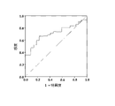
実施例3:ノルウェーのPark West調査からの346個体(182個の対照および164個のPD血清試料)のコホートについての、hsa-miR-335-5pおよびhsa-miR-6865-3pの分析
実施例2のqPCR技術を用いて潜在的な診断バイオマーカーを同定した。hsa-miR-335-5pとhsa-miR-6865-3pの組み合わせはPD診断の予測可能性が高いことが、判明した。hsa-miR-335-5pおよびhsa-miR-6865-3pを有するモデルの、アウトカム=PD(YES/NO)、n=(164の患者+182の対照)における結果を、以下の表3に示す。

真の疾患状態と比較した予測確率に基づくROC分析は、図2aに示すように、強い識別能力を示す。図2aの曲線の下の面積を上記の表3に示す。 A ROC analysis based on predicted probabilities compared to the true disease state shows strong discriminatory power, as shown in Fig. 2a. The area under the curve in Figure 2a is shown in Table 3 above.
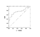
実施例4:346人のコホート(182の対照および164のPD血清サンプル)におけるhsa-miR-335-5pおよびhsa-miR-3613-3pの分析
実施例3のプロトコルに従って分析し、hsa-miR-335-5pおよびhsa-miR-3613-3pの組み合わせもPD診断の高い予測可能性を示すことが判明した。hsa-miR-335-5pおよびhsa-miR-3613-3pのモデルの、アウトカム=PD(YES/NO)、n=(164の患者+182の対照)における結果を、上記の表3に示す。
Example 4: Analysis of hsa-miR-335-5p and hsa-miR-3613-3p in a cohort of 346 individuals (182 control and 164 PD serum samples). The combination of 335-5p and hsa-miR-3613-3p was also found to be highly predictive of PD diagnosis. The results of the hsa-miR-335-5p and hsa-miR-3613-3p models with outcome=PD (YES/NO), n=(164 patients+182 controls) are shown in Table 3 above.
モデルから予測された確率に基づくROC分析は、強い識別能力を示すことが、図2bに示される。図2bの曲線下側の面積を上記の表3に示す。 It is shown in Figure 2b that the ROC analysis based on the probabilities predicted from the model shows strong discriminative power. The area under the curve in Figure 2b is shown in Table 3 above.
前述の実施例1~4から、同定された85のmiRNAのリストに由来する2つ以上のマイクロRNAの任意の組み合わせが、患者におけるPDの発生を診断するために使用され得ることが立証される。 Examples 1-4 above demonstrate that any combination of two or more microRNAs from the list of 85 miRNAs identified can be used to diagnose the development of PD in a patient. .
実施例5:hsa-miR-335-5p
上記の表3は、hsa-miR-335-5pが、アウトカム=PD(YES/NO)、n=(164の患者+182の対照)においてPD診断の高い予測可能性を示すことを示している。
Example 5: hsa-miR-335-5p
Table 3 above shows that hsa-miR-335-5p shows high predictability of PD diagnosis in Outcome = PD (YES/NO), n = (164 patients + 182 controls) .
モデルからの確率と真の疾患状態との比較に基づくROC分析は、強い識別能力を示すことが図3に示される。図3の曲線下側の面積を上記の表3に示す。 It is shown in FIG. 3 that the ROC analysis based on comparing the probabilities from the model with the true disease state shows strong discriminatory power. The area under the curve in FIG. 3 is shown in Table 3 above.
実施例6:has-miR-3613-3p
hsa-miR-3613-3pもまた、上記の表3に示すように、PD診断の高い予測可能性を示す。モデルからの確率と真の疾患状態との比較に基づくROC分析は、図4に示すように強い識別能力を示す。図4の曲線下側の面積を上記表3に示す。
Example 6: has-miR-3613-3p
hsa-miR-3613-3p also shows high predictability of PD diagnosis, as shown in Table 3 above. A ROC analysis based on comparing the probabilities from the model with the true disease state shows strong discriminatory power, as shown in FIG. The area under the curve in FIG. 4 is shown in Table 3 above.
実施例7:has-miR-6865-3p
同様に、hsa-miR-6865-3pはまた、上記の表3に示すように、PD診断の高い予測可能性を示す。モデルからの確率に基づくROC分析および強い識別能力を示す真の疾患状態との比較を図5に示す。図5の曲線下側の面積を表3に示す。
Example 7: has-miR-6865-3p
Similarly, hsa-miR-6865-3p also shows high predictability of PD diagnosis, as shown in Table 3 above. A probability-based ROC analysis from the model and comparison with the true disease state showing strong discriminatory power is shown in FIG. Table 3 shows the area under the curve in FIG.
上記の実施例5~7から、hsa-miR-335-5p、hsa-miR-3613-3pおよびhsa-miR-6865-3pが、PDの正確な診断のために個別に使用され得ることが立証される。 Examples 5-7 above demonstrate that hsa-miR-335-5p, hsa-miR-3613-3p and hsa-miR-6865-3p can be used individually for accurate diagnosis of PD. be done.
実施例8:スウェーデンのNYPUM研究からの64人のコホート(22の対照および42のPDの血清サンプル)におけるhsa-miR-335-5pおよびhsa-miR-6865-3pの分析
実施例2のqPCR技術を用いて、実施例2の診断バイオマーカーを検証した。hsa-miR-335-5pおよびhsa-miR-6865-3pの組み合わせは、PD診断の予測可能性が高いことが判明した。 hsa-miR-335-5pおよびhsa-miR-6865-3pのモデルについて、アウトカム= PD(YES/NO)、n=(42の患者+22の対照)の場合の結果を、以下の表4に示す。

真の疾患状態と比較した予測確率に基づくROC分析は、図7に示すように、高い識別能力を示す。図7の曲線の下側の面積を、上記の表4に示す。 ROC analysis based on predicted probabilities compared to true disease states shows high discriminatory power, as shown in FIG. The area under the curve in FIG. 7 is shown in Table 4 above.
実施例9:64人(22個の対照および42個のPD血清サンプル)のコホートにおけるhsa-miR-335-5pおよびhsa-miR-3613-3pの分析
実施例3のプロトコルに従って分析し、hsa-miR-335-5pとhsa-miR-3613-3pの組み合わせもPD診断の高い予測可能性を示すことが判明した。hsa-miR-335-5pおよびhsa-miR-3613-3pのモデルの、アウトカム=PD(YES/NO)、n=(42の患者+22の対照)における結果を表4に示す。
Example 9: Analysis of hsa-miR-335-5p and hsa-miR-3613-3p in a cohort of 64 individuals (22 control and 42 PD serum samples). The combination of miR-335-5p and hsa-miR-3613-3p was also found to be highly predictive of PD diagnosis. Results for the hsa-miR-335-5p and hsa-miR-3613-3p models are shown in Table 4, with outcome=PD (YES/NO), n=(42 patients+22 controls).
モデルから予測された確率に基づくROC分析は、図8に示すように、高い識別能力を示す。図8の曲線下側の面積を上記の表4に示す。 ROC analysis based on the probabilities predicted from the model shows high discriminatory power, as shown in FIG. The area under the curve in FIG. 8 is shown in Table 4 above.
実施例10:64人(22の対照および42のPD患者の血清サンプル)のコホートにおけるhsa-miR-3613-3pおよびhsa-miR-6865-5pの分析
実施例3のプロトコルに従って分析し、hsa-miR-3613-3pおよびhsa-miR-6865-5pの組み合わせもPD診断の高い予測可能性を示すことが判明した。hsa-miR-3613-3pおよびhsa-miR-6865-5pのモデルの、アウトカム=PD(YES/NO)、n=(42の患者+22の対照)における結果を表4に示す。
Example 10: Analysis of hsa-miR-3613-3p and hsa-miR-6865-5p in a cohort of 64 individuals (22 control and 42 PD patient serum samples). The combination of miR-3613-3p and hsa-miR-6865-5p was also found to be highly predictive of PD diagnosis. Results for the hsa-miR-3613-3p and hsa-miR-6865-5p models are shown in Table 4, with outcome = PD (YES/NO), n = (42 patients + 22 controls).
モデルから予測された確率に基づくROC分析は、図9に示すように、高い識別能力を示す。図9の曲線下側の面積を上記の表4に示す。 ROC analysis based on the probabilities predicted from the model shows high discriminatory power, as shown in FIG. The area under the curve in FIG. 9 is shown in Table 4 above.
実施例11:64人(22個の対照および42個のPD血清サンプル)のコホートにおけるhsa-miR-335-5p、hsa-miR-3613-3pおよびhsa-miR-6865-5pの分析
実施例3のプロトコルに従って分析し、hsa-miR-hsa-miR-335-5p、hsa-miR-3613-3pおよびhsa-miR-6865-5pの組合せもPD診断の高い予測可能性を示すことが判明した。hsa-miR-335-5p、hsa-miR-3613-3pおよびhsa-miR-6865-5pのモデルの、アウトカム=PD(YES/NO)、n=(42の患者+22の対照)における結果を、上記の表4に示す。
Example 11: Analysis of hsa-miR-335-5p, hsa-miR-3613-3p and hsa-miR-6865-5p in a cohort of 64 individuals (22 controls and 42 PD serum samples) and found that the combinations of hsa-miR-hsa-miR-335-5p, hsa-miR-3613-3p and hsa-miR-6865-5p also showed high predictive potential for PD diagnosis. Results for hsa-miR-335-5p, hsa-miR-3613-3p and hsa-miR-6865-5p models with outcome = PD (YES/NO), n = (42 patients + 22 controls) , as shown in Table 4 above.
モデルから予測された確率に基づくROC分析は、図10に示すように、高い識別能力を示す。図10の曲線下側の面積を上記の表4に示す。 ROC analysis based on the probabilities predicted from the model shows high discriminatory power, as shown in FIG. The area under the curve in FIG. 10 is shown in Table 4 above.
上記の実施例10から、同定された85のmiRNAのリストからの3つ以上のマイクロRNAの任意の組み合わせが、患者におけるPDの発生を診断するために使用できることが立証される。 Example 10 above demonstrates that any combination of three or more microRNAs from the list of 85 miRNAs identified can be used to diagnose the development of PD in a patient.
実施例12
複数の生物情報学ツールを用いたhsa-miR-335-5p、hsa-miR-3613-3pおよびhsa-miR-6865-3p標的の分析から、とりわけ、LRRK2およびParkinがhsa-miR-335-5pの予測標的であり、SNCAがhsa-miR-3613-3pの予測標的であることが示される。hsa-miR-335-5pレベルの調節の結果としてのSHSY-5Y細胞におけるLRRK2発現の調節は、ウェスタンブロット分析によって確認された。hsa-miR-335-5pを、神経芽腫細胞に導入(transfect)されたhsa-miR-335-5pの模倣物およびアンタゴミルを用いて過剰発現させ(図6A)、阻害した(図6A)。導入(transfection)の48時間後に細胞を溶解し、ウェスタンブロット分析に使用した。hsa-miR-335-5p模倣物はLRRK2の下方調節を示し、hsa-miR-335-5pアンタゴミアはLRRK2の上方調節を示した(図6B,C)。hsa-miR-3613-3pは、適度にSH-SY5Y細胞におけるSNCA発現を調節した。hsa-miR-335-5pと同様の実験的アプローチがhsa-miR-3613-3p(図6D)に適用され、その結果、タンパク質レベル(図6E,F)および転写レベル(図6G)において、hsa-miR-3613-3p模倣物で中程度のSNCA上方調節を示し、 miR-3613-3pアンタゴミルで中程度のSNCA下方調節を示した。
Example 12
Analysis of hsa-miR-335-5p, hsa-miR-3613-3p and hsa-miR-6865-3p targets using multiple bioinformatic tools revealed that, among others, LRRK2 and Parkin were associated with hsa-miR-335-5p It is shown that SNCA is a predicted target of hsa-miR-3613-3p. Regulation of LRRK2 expression in SHSY-5Y cells as a result of regulation of hsa-miR-335-5p levels was confirmed by Western blot analysis. hsa-miR-335-5p was overexpressed (FIG. 6A) and inhibited (FIG. 6A) using mimics of hsa-miR-335-5p and antagomir transfected into neuroblastoma cells. Cells were lysed 48 hours after transfection and used for Western blot analysis. hsa-miR-335-5p mimics showed downregulation of LRRK2 and hsa-miR-335-5p antagomir showed upregulation of LRRK2 (Fig. 6B,C). hsa-miR-3613-3p modestly regulated SNCA expression in SH-SY5Y cells. A similar experimental approach to hsa-miR-335-5p was applied to hsa-miR-3613-3p (Fig. 6D), resulting in hsa -miR-3613-3p mimic showed moderate SNCA upregulation and miR-3613-3p antagomir showed moderate SNCA downregulation.
LC-MSを用いた標的発見は、hsa-miR-335-5p、hsa-miR-3613-3pおよびhsa-miR-6865-3pの新規標的を見出すために行った。
a.hsa-miR-335-5p調節の結果としての異なる発現パターンを有するタンパク質としては、acadsb,slc4a7,lnp/kiaa1715,supt5h,sdhd,Wdr1,cmpk1,slc25a1,hmgcs1,twf2,ppp1r18,exoc8,tm9sf4,kif16b,dnajc2,selll,hectd1,gmppbなどがある。
b.hsa-miR-3613-3p調節の結果としての異なる発現パターンを有するタンパク質としては、wdr1,gmppb,hmbs,eml4,hebp1,apmap/c20orf3,sord,pcyt2,stat3,top2a,skiv212,cdc20,myo1e,ttll12,atad2,carm1,arfgap1,ppp4r1,nde1/nde11などがある。
c.hsa-miR-6865-3p調節の結果としての異なる発現パターンを有するタンパク質としては、wdr1,ppplr18,ppp4r1,ube2h,ube3c,stx16,ube4h,gtf2f1,map1b,ube2a,dusp3,arhgap1,nsun2,acox1,fkbp10,fam107b,pofut1,tomm22,hspb8,sbdsなどがある。
Target discovery using LC-MS was performed to find novel targets for hsa-miR-335-5p, hsa-miR-3613-3p and hsa-miR-6865-3p.
a. Proteins with differential expression patterns as a result of hsa-miR-335-5p regulation include acadsb, slc4a7, lnp/kiaa1715, supt5h, sdhd, Wdr1, cmpk1, slc25a1, hmgcs1, twf2, ppp1r18, exoc8, tm9sf4, kif16b , dnajc2, sellll, hectd1, gmppb, and so on.
b. Proteins with differential expression patterns as a result of hsa-miR-3613-3p regulation include wdr1, gmppb, hmbs, eml4, hebp1, apmap/c20orf3, sord, pcyt2, stat3, top2a, skiv212, cdc20, myo1e, ttll12 , atad2, carm1, arfgap1, ppp4r1, nde1/nde11, etc.
c. Proteins with differential expression patterns as a result of hsa-miR-6865-3p regulation include wdr1, ppplr18, ppp4r1, ube2h, ube3c, stx16, ube4h, gtf2f1, map1b, ube2a, dusp3, arhgap1, nsun2, acox1, fkbp10 , fam107b, pofut1, tomm22, hspb8, sbds, etc.
実施例13
患者からの血清中の2つ以上のmiRNAの組み合わせのレベルの測定は、潜在的なPD患者と健常者とを明確に区別するのを助けることができる。PDの疑いのある患者から採取した血液から血清サンプルを得る。血清は、全マイクロRNA単離および濃縮のために使用される。次いで、このRNAをqPCRを用いて試験して、実施例1に記載の85個のmiRNAのいずれか2個以上、または実施例5~7に記載の3個のmiRNAのいずれか1個のレベルを測定する。85種のmiRNAのうちの2種以上、または3種のmiRNAのいずれか1種が検出可能なレベルである場合、患者がPDを有することを確認する。必要に応じて、血漿、静脈または動脈血、または腰椎穿刺によって採取されたCSF試料を含む他の試料液を利用することができる。そのような血漿、血液またはCSF試料は上記のように処理される。2つ以上のmiRNAを組み合わせて、または試験マトリックスで使用される組み合わせのセットを測定することにより、望ましくはPD診断の精度が向上することが理解されよう。
Example 13
Measuring the levels of combinations of two or more miRNAs in serum from patients can help to clearly distinguish potential PD patients from healthy individuals. Serum samples are obtained from blood drawn from patients with suspected PD. Serum is used for total microRNA isolation and enrichment. This RNA is then tested using qPCR to determine the level of any two or more of the 85 miRNAs described in Example 1 or any one of the three miRNAs described in Examples 5-7. to measure. Detectable levels of 2 or more of the 85 miRNAs, or any one of the 3 miRNAs, confirm that the patient has PD. If desired, other sample fluids can be utilized, including plasma, venous or arterial blood, or CSF samples taken by lumbar puncture. Such plasma, blood or CSF samples are processed as described above. It will be appreciated that combining two or more miRNAs or measuring a set of combinations used in a test matrix desirably improves the accuracy of PD diagnosis.
実施例14
診断のためにmiRNAの組み合わせを使用することができるので、コホートに基づく変異を排除するために全ての候補を試験することが望ましい場合がある。関連miRNAのいずれかが検出可能な量存在する場合、PD病理を示すことが理解される。しかしながら、当業者であれば、診断のための人工的閾値を設定するために164v 182サンプルの値を使用することが臨床的に有用であり得ることを認識している。特異的miRNAレベルは、診断および臨床試験で臨床医が使用できる診断バイオマーカーキットを開発するために使用できる。この研究では、血清からのmiRNAの存在および定量は、RNAを増幅および定量するqRT-PCRによって判定された。当業者に公知の他の適切な技術、例えば標識されたアンチセンス配列および標識された抗体の使用など、を代替的に利用してもよい。適切な抗体は、2つの分子間の結合反応、典型的には測定条件下でバックグラウンド分子会合の10~100倍を超える、を参照して優先的に選択可能である。したがって、所定の免疫学的測定条件で、指定された抗体は特定のmiRNA配列に結合し、それによってその存在を同定する。そのような条件下での抗体への特異的結合は、特定のmiRNAに対するその特異性のために選択される抗体を必要とする。例えば、特定のmiRNAに対して産生された抗体は、他の分子と交差反応する抗体を差し引くことによって選択することができる。様々な免疫学的フォーマットを使用して、特定のmiRNAと特異的に免疫反応する抗体を選択することができ、例えば、固相ELISA免疫学的検定が用いられる(特定の免疫反応を判定するために用いられ得る免疫学的フォーマットと条件についての記載は、例えば、Harlow&Lane,Antibodies,A Laboratory Manual(1988)を参照のこと)。2つの分子が特異的に相互作用するかどうかを判定する方法がここに開示されており、結合親和性および特異性を判定する方法は、当技術分野で周知である(例えば、Harlow and Lane,Antibodies:A laboratory manual(Cold Spring Harbor Laboratory Press,1988);Friedelder,"Physical Biochemistry:Applications to biochemistry and molecular biology”(W.H.Freeman and co.1976)参照)。用語「抗体」は、本明細書では、天然に存在する抗体ならびに天然に存在しない抗体、例えば、一本鎖抗体、キメラ、二官能性及びヒト化抗体、ならびに、これらの抗原結合フラグメント(例えば、Fab'、F(ab')2、Fab、FvおよびrIgG)を含む。Pierceカタログおよびハンドブック、1994-1995(Pierce Chemical Co.,Rockford,IL)も参照されたい。また、Kuby,J.,Immunology,3rd Ed.,W.H.Freeman&Co.,New York(1998)も参照されたい。 このような天然に存在しない抗体は、固相ペプチド合成を用いて構築することができ、組換えにより産生することができ、または、例えばHuseら,Science,Vol.246(1989)1275-81に記載のように、様々な重鎖と様々な軽鎖からなる組み換えライブラリをスクリーニングすることによって得ることができる。例えば、キメラ、ヒト化、CDR移植、一本鎖および二官能性抗体を作製するためのこれらおよび他の方法は、当業者に周知である(Winter and Harris,Immunol.Today,Vol.14(1993)243-46;Wardら、Nature,Vol.341 (1989)544-46、HarlowおよびLane,supra,1988;Hilyardら,Protein Engineering:A practical approach(IRL Press 1992); Borrabeck,Antibody Engineering,第2版(Oxford University Press 1995))。同定されたRNA配列から単クローン性抗体および多クローン性抗体の両方を産生する方法は、当技術分野で周知である。
Example 14
Since combinations of miRNAs can be used for diagnosis, it may be desirable to test all candidates to rule out mutations based on the cohort. It is understood that the presence of detectable amounts of any of the relevant miRNAs is indicative of PD pathology. However, those skilled in the art recognize that using the values of the 164v 182 samples to set artificial thresholds for diagnosis can be clinically useful. Specific miRNA levels can be used to develop diagnostic biomarker kits for use by clinicians in diagnostics and clinical trials. In this study, the presence and quantification of miRNAs from serum was determined by qRT-PCR to amplify and quantify RNA. Other suitable techniques known to those skilled in the art, such as the use of labeled antisense sequences and labeled antibodies, may alternatively be utilized. Suitable antibodies can be preferentially selected by reference to a binding reaction between two molecules, typically 10-100 times greater than background molecular association under the conditions of measurement. Thus, under defined immunological assay conditions, the specified antibody will bind to a specific miRNA sequence, thereby identifying its presence. Specific binding to an antibody under such conditions requires an antibody selected for its specificity for a particular miRNA. For example, antibodies produced against a particular miRNA can be selected by subtracting out antibodies that cross-react with other molecules. A variety of immunological formats can be used to select antibodies specifically immunoreactive with a particular miRNA, for example solid phase ELISA immunoassays are used (to determine specific immune responses). See, for example, Harlow & Lane, Antibodies, A Laboratory Manual (1988)) for a description of immunological formats and conditions that can be used in immunoassays. Methods for determining whether two molecules specifically interact are disclosed herein, and methods for determining binding affinity and specificity are well known in the art (e.g., Harlow and Lane, Antibodies: A laboratory manual (Cold Spring Harbor Laboratory Press, 1988); see Friedelder, "Physical Biochemistry: Applications to biochemistry and molecular biology" (WH Freeman and co.1976). The term "antibody" is used herein to refer to naturally occurring and non-naturally occurring antibodies, including single chain antibodies, chimeric, bifunctional and humanized antibodies, and antigen-binding fragments thereof (e.g., Fab', F(ab')2, Fab, Fv and rIgG). See also Pierce Catalogs and Handbooks, 1994-1995 (Pierce Chemical Co., Rockford, Ill.). See also Kuby, J., Immunology, 3rd Ed. , WH Freeman & Co., New York (1998). Such non-naturally occurring antibodies can be constructed using solid-phase peptide synthesis, can be produced recombinantly, or can be described, for example, by Huse et al., Science, Vol. 246 (1989) 1275-81, by screening a recombinant library consisting of different heavy chains and different light chains. For example, these and other methods for making chimeric, humanized, CDR-grafted, single chain and bifunctional antibodies are well known to those skilled in the art (Winter and Harris, Immunol. Today, Vol. 14 (1993 ) 243-46; Ward et al., Nature, Vol. 341 (1989) 544-46, Harlow and Lane, supra, 1988; Hilyard et al., Protein Engineering: A practical approach (IRL Press 1992); Edition (Oxford University Press 1995)). Methods for producing both monoclonal and polyclonal antibodies from identified RNA sequences are well known in the art.
実施例15
多くの神経変性疾患は、症状および病理学的マーカーに関して、互いに密接に関連している。1つの神経変性疾患の循環診断マーカーは、他の疾患の診断に有用であり得る。レヴィ―小体認知症(DLB)、筋萎縮性側索硬化症(ALS)、アルツハイマー病(AD)、多系統萎縮症(MSA)、大脳皮質基底核変性症(CBD)、進行性核上麻痺(PSP)などの他の神経変性疾患を診断する方法も、上記の候補の類似のmiRNA測定を用いて開発することができる。疾患特異的キットは、[0037]に列挙されたものと同様に、[0019]に列挙されたmiRNAの様々な組み合わせを用いて開発することができる。
Example 15
Many neurodegenerative diseases are closely related to each other in terms of symptoms and pathological markers. Circulating diagnostic markers for one neurodegenerative disease may be useful in diagnosing other diseases. Dementia with Lewy bodies (DLB), amyotrophic lateral sclerosis (ALS), Alzheimer's disease (AD), multiple system atrophy (MSA), corticobasal degeneration (CBD), progressive supranuclear palsy Methods for diagnosing other neurodegenerative diseases such as (PSP) can also be developed using analogous miRNA measurements of the above candidates. Disease-specific kits can be developed using various combinations of the miRNAs listed in [0019], as well as those listed in [0037].
実施例16
1つまたは複数の組み合わせで検出されたmiRNAは、細胞内のいくつかのタンパク質を調節することができる。これらのマイクロRNAおよびその組み合わせを使用して、PDの新規タンパク質標的を見出すことができる。 PD病因におけるこれらのタンパク質の関与をさらに確証して、治療のためにそれらを標的とすることができる。
Example 16
One or more miRNAs detected in combination can regulate several proteins in the cell. These microRNAs and their combinations can be used to find novel protein targets for PD. With further confirmation of the involvement of these proteins in PD pathogenesis, they can be targeted for therapy.
実施例17
本発明者らは、ドーパミン作動性ニューロン細胞系におけるhsa-miR-335-5pによるLRRK2の予測される調節およびhsa-miR-3613-3p によるSNCAの予測される調節を、実験により確認した。言及された新規標的を調節するための治療的介入は、RNA干渉技術によって達成することができる。
Example 17
We experimentally confirmed the predicted regulation of LRRK2 by hsa-miR-335-5p and of SNCA by hsa-miR-3613-3p in dopaminergic neuronal cell lines. Therapeutic intervention to modulate the novel targets mentioned can be achieved by RNA interference technology.
実施例18
[0019]に記載されたmiRNAに由来する小さな核酸分子は、PD脳における遺伝子を特異的に標的化することによって治療的に介入して、完全または部分的治療を達成するように設計される。[0040]に示される効果は、脳細胞における正確な標的化のために達成されるであろう。
Example 18
[0019] The miRNA-derived small nucleic acid molecules described in [0019] are designed to intervene therapeutically by specifically targeting genes in PD brains to achieve full or partial therapy. The effects shown in [0040] will be achieved due to precise targeting in brain cells.
Claims (7)
前記サンプル中において配列番号2~86からなる群から選択される少なくとも3つのmiRNA の存在を示すレベルを判定するステップであって、前記3つのmiRNAのうち少なくとも2つは配列番号22,25および77からなる群から選択されるステップと、
前記少なくとも2つのmiRNAのうち少なくとも1つのmiRNAの存在を示す前記レベルが健常な対照と比較して1.2倍以上である場合にパーキンソン疾患のリスクが高いと判定するステップと、を含む、方法。 A method of aiding determination of Parkinson's disease in a sample of bodily fluid comprising:
determining a level indicative of the presence of at least three miRNAs selected from the group consisting of SEQ ID NOs: 2-86 in said sample, wherein at least two of said three miRNAs are SEQ ID NOs: 22, 25 and 77 a step selected from the group consisting of
and determining an increased risk for Parkinson's disease when said level indicative of the presence of at least one miRNA of said at least two miRNAs is 1.2-fold or greater compared to healthy controls. .
前記サンプル中において配列番号22,25および77からなる群から選択される少なくとも2つのmiRNAの存在を示すレベルを判定するステップと、
前記少なくとも2つのmiRNAのうち少なくとも1つのmiRNAの存在を示す前記レベルが健常な対照と比較して1.2倍以上である場合にパーキンソン疾患のリスクが高いと判定するステップと、を含む、方法。 A method of aiding determination of Parkinson's disease in a sample of bodily fluid comprising:
determining a level indicative of the presence of at least two miRNAs selected from the group consisting of SEQ ID NOs: 22, 25 and 77 in said sample;
and determining an increased risk for Parkinson's disease when said level indicative of the presence of at least one miRNA of said at least two miRNAs is 1.2-fold or greater compared to healthy controls. .
Applications Claiming Priority (3)
| Application Number | Priority Date | Filing Date | Title |
|---|---|---|---|
| US201662291619P | 2016-02-05 | 2016-02-05 | |
| US62/291,619 | 2016-02-05 | ||
| PCT/US2017/016412 WO2017136662A1 (en) | 2016-02-05 | 2017-02-03 | Circulating serum microrna biomarkers and methods for determining parkinson's disease |
Publications (3)
| Publication Number | Publication Date |
|---|---|
| JP2019506183A JP2019506183A (en) | 2019-03-07 |
| JP2019506183A5 JP2019506183A5 (en) | 2020-01-23 |
| JP7197755B2 true JP7197755B2 (en) | 2022-12-28 |
Family
ID=58094505
Family Applications (1)
| Application Number | Title | Priority Date | Filing Date |
|---|---|---|---|
| JP2018560445A Active JP7197755B2 (en) | 2016-02-05 | 2017-02-03 | Circulating Serum MicroRNA Biomarkers and Methods |
Country Status (5)
| Country | Link |
|---|---|
| US (2) | US11499184B2 (en) |
| EP (2) | EP3985130A1 (en) |
| JP (1) | JP7197755B2 (en) |
| ES (1) | ES2947634T3 (en) |
| WO (1) | WO2017136662A1 (en) |
Families Citing this family (7)
| Publication number | Priority date | Publication date | Assignee | Title |
|---|---|---|---|---|
| WO2018236589A1 (en) * | 2017-06-19 | 2018-12-27 | St. John's University | Circulating serum microrna biomarkers and methods for alzheimer's disease diagnosis |
| EP3850108A4 (en) * | 2018-10-18 | 2022-08-03 | Quadrant Biosciences Inc. | Molecular and functional characterization of early-stage parkinson's disease and treatments therein |
| EP3937986A4 (en) * | 2019-03-11 | 2023-04-12 | Ochsner Health System | Microrna regulatory network as biomarkers of seizure in patients with spontaneous intracerebral hemorrhage |
| WO2020213699A1 (en) * | 2019-04-17 | 2020-10-22 | 京都府公立大学法人 | Determination method of autoimmune disease and/or inflammatory disease, and preventive and/or therapeutic agent of inflammatory disease and/or autoimmune disease |
| CN111073973A (en) * | 2019-12-10 | 2020-04-28 | 石河子大学 | MicroRNA sequence for early diagnosis of type 2 diabetes and application thereof |
| CN115605608A (en) | 2020-05-14 | 2023-01-13 | 花王株式会社(Jp) | Method for detecting Parkinson's disease |
| WO2023050642A1 (en) * | 2021-09-29 | 2023-04-06 | 南京凡亦达生物科技有限公司 | Application of alpha-fetoprotein or carcinoembryonic antigen combined with gene marker in tumor diagnosis |
Citations (2)
| Publication number | Priority date | Publication date | Assignee | Title |
|---|---|---|---|---|
| WO2013036936A1 (en) | 2011-09-09 | 2013-03-14 | Van Andel Research Institute | Microrna biomarkers for diagnosing parkinson's disease |
| WO2014018650A1 (en) | 2012-07-25 | 2014-01-30 | Rush University Medical Center | Mirnas as novel therapeutic targets and diagnostic biomarkers for parkinson's disease |
Family Cites Families (2)
| Publication number | Priority date | Publication date | Assignee | Title |
|---|---|---|---|---|
| CN104903468B (en) | 2012-11-16 | 2019-04-23 | 萨尔大学 | New diagnosis MiRNA marker for Parkinson's disease |
| EP3760741A1 (en) * | 2013-12-19 | 2021-01-06 | Hummingbird Diagnostics GmbH | Mirnas as non-invasive biomarkers for parkinson's disease |
-
2017
- 2017-02-03 WO PCT/US2017/016412 patent/WO2017136662A1/en active Application Filing
- 2017-02-03 ES ES17706344T patent/ES2947634T3/en active Active
- 2017-02-03 EP EP21198200.4A patent/EP3985130A1/en active Pending
- 2017-02-03 JP JP2018560445A patent/JP7197755B2/en active Active
- 2017-02-03 EP EP17706344.3A patent/EP3411500B9/en active Active
- 2017-02-03 US US16/075,354 patent/US11499184B2/en active Active
-
2022
- 2022-10-07 US US18/045,065 patent/US20230323442A1/en active Pending
Patent Citations (2)
| Publication number | Priority date | Publication date | Assignee | Title |
|---|---|---|---|---|
| WO2013036936A1 (en) | 2011-09-09 | 2013-03-14 | Van Andel Research Institute | Microrna biomarkers for diagnosing parkinson's disease |
| WO2014018650A1 (en) | 2012-07-25 | 2014-01-30 | Rush University Medical Center | Mirnas as novel therapeutic targets and diagnostic biomarkers for parkinson's disease |
Non-Patent Citations (2)
| Title |
|---|
| AMENE SAGHAZADEH,MICRORNA MACHINERY IN PARKINSON'S DISEASE: A PLATFORM FOR NEURODEGENERATIVE DISEASES,EXPERT REVIEW OF NEUROTHERAPEUTICS,英国,2015年11月17日,PAGE(S): 1 - 27,https://www.ncbi.nlm.nih.gov/pubmed/26574782 |
| SAPANA SHINDE,BIOFLUID-BASED MICRORNA BIOMARKERS FOR PARKINSON'S DISEASE: AN OVERVIEW AND UPDATE,AIMS MEDICAL SCIENCE,2015年01月01日,VOL:2,NR:1,PAGE(S):15-25,http://scholarworks.gvsu.edu/cgi/viewcontent.cgi?article=1003&context=bms_articles |
Also Published As
| Publication number | Publication date |
|---|---|
| US20200354768A1 (en) | 2020-11-12 |
| EP3985130A1 (en) | 2022-04-20 |
| EP3411500C0 (en) | 2023-06-07 |
| JP2019506183A (en) | 2019-03-07 |
| US11499184B2 (en) | 2022-11-15 |
| EP3411500A1 (en) | 2018-12-12 |
| US20230323442A1 (en) | 2023-10-12 |
| EP3411500B1 (en) | 2023-06-07 |
| ES2947634T3 (en) | 2023-08-14 |
| EP3411500B9 (en) | 2023-10-04 |
| WO2017136662A1 (en) | 2017-08-10 |
| WO2017136662A4 (en) | 2017-08-31 |
Similar Documents
| Publication | Publication Date | Title |
|---|---|---|
| JP7197755B2 (en) | Circulating Serum MicroRNA Biomarkers and Methods | |
| Xie et al. | Serum miR-206 and miR-132 as potential circulating biomarkers for mild cognitive impairment | |
| Müller et al. | MicroRNAs in Alzheimer's disease: differential expression in hippocampus and cell-free cerebrospinal fluid | |
| ES2813699T3 (en) | Methods of using miRNA from body fluids for the detection and monitoring of Parkinson's disease (PD) | |
| US20240018595A1 (en) | Circulating serum microrna biomarkers and methods for parkinson's disease prognosis | |
| Yang et al. | MiRNA expression profiles in healthy OSAHS and OSAHS with arterial hypertension: potential diagnostic and early warning markers | |
| Ye et al. | MicroRNAs 99b-5p/100-5p regulated by endoplasmic reticulum stress are involved in abeta-induced pathologies | |
| Li et al. | Identification of miRNA-7 as a regulator of brain-derived neurotrophic factor/α-synuclein axis in atrazine-induced Parkinson’s disease by peripheral blood and brain microRNA profiling | |
| He et al. | Mitochondrial DNA content contributes to healthy aging in Chinese: a study from nonagenarians and centenarians | |
| US10724097B2 (en) | Methods and compositions for diagnosis and management of diabetes and metabolic syndrome | |
| WO2019005762A1 (en) | Treatment of non-small cell lung cancer | |
| US11634775B2 (en) | Circulating serum microRNA biomarkers and methods for Alzheimer's disease diagnosis | |
| Minutti-Zanella et al. | miRNAs in multiple sclerosis: A clinical approach | |
| Rong et al. | Regulatory roles of non-coding RNAs and m6A modification in trophoblast functions and the occurrence of its related adverse pregnancy outcomes | |
| Piscopo et al. | Identification of miRNAs regulating MAPT expression and their analysis in plasma of patients with dementia | |
| Vastrad et al. | Bioinformatics analysis of potential key genes and mechanisms in type 2 diabetes mellitus | |
| US10975436B2 (en) | Methods of using miRNA from bodily fluids for diagnosis and monitoring of neurodevelopmental disorders | |
| TW201805431A (en) | Method of diagnosing neurodegenerative diseases and primer pair thereof having a pair of primers designed to indirectly test miR-302 clustering expression | |
| Gonzalez-Latapi et al. | Characterization of baseline and longitudinal DNA Methylation in patients with sporadic Parkinson’s disease | |
| CN116356009A (en) | Application of miR-122-5p | |
| JP2023178332A (en) | Circulating serum cell-free DNA biomarkers and methods |
Legal Events
| Date | Code | Title | Description |
|---|---|---|---|
| A521 | Request for written amendment filed |
Free format text: JAPANESE INTERMEDIATE CODE: A523 Effective date: 20191204 |
|
| A621 | Written request for application examination |
Free format text: JAPANESE INTERMEDIATE CODE: A621 Effective date: 20191204 |
|
| A131 | Notification of reasons for refusal |
Free format text: JAPANESE INTERMEDIATE CODE: A131 Effective date: 20201218 |
|
| A521 | Request for written amendment filed |
Free format text: JAPANESE INTERMEDIATE CODE: A523 Effective date: 20210304 |
|
| A131 | Notification of reasons for refusal |
Free format text: JAPANESE INTERMEDIATE CODE: A131 Effective date: 20210826 |
|
| A521 | Request for written amendment filed |
Free format text: JAPANESE INTERMEDIATE CODE: A523 Effective date: 20211105 |
|
| A131 | Notification of reasons for refusal |
Free format text: JAPANESE INTERMEDIATE CODE: A131 Effective date: 20220228 |
|
| RD13 | Notification of appointment of power of sub attorney |
Free format text: JAPANESE INTERMEDIATE CODE: A7433 Effective date: 20220318 |
|
| A521 | Request for written amendment filed |
Free format text: JAPANESE INTERMEDIATE CODE: A821 Effective date: 20220318 |
|
| A521 | Request for written amendment filed |
Free format text: JAPANESE INTERMEDIATE CODE: A523 Effective date: 20220415 |
|
| A521 | Request for written amendment filed |
Free format text: JAPANESE INTERMEDIATE CODE: A523 Effective date: 20220427 Free format text: JAPANESE INTERMEDIATE CODE: A523 Effective date: 20220519 |
|
| A131 | Notification of reasons for refusal |
Free format text: JAPANESE INTERMEDIATE CODE: A131 Effective date: 20220819 |
|
| A521 | Request for written amendment filed |
Free format text: JAPANESE INTERMEDIATE CODE: A523 Effective date: 20221019 |
|
| TRDD | Decision of grant or rejection written | ||
| A01 | Written decision to grant a patent or to grant a registration (utility model) |
Free format text: JAPANESE INTERMEDIATE CODE: A01 Effective date: 20221109 |
|
| RD17 | Notification of extinguishment of power of sub attorney |
Free format text: JAPANESE INTERMEDIATE CODE: A7437 Effective date: 20221116 |
|
| A61 | First payment of annual fees (during grant procedure) |
Free format text: JAPANESE INTERMEDIATE CODE: A61 Effective date: 20221117 |
|
| R150 | Certificate of patent or registration of utility model |
Ref document number: 7197755 Country of ref document: JP Free format text: JAPANESE INTERMEDIATE CODE: R150 |