JP7183590B2 - Ophthalmic image processing device, OCT device, and ophthalmic image processing program - Google Patents
Ophthalmic image processing device, OCT device, and ophthalmic image processing program Download PDFInfo
- Publication number
- JP7183590B2 JP7183590B2 JP2018124973A JP2018124973A JP7183590B2 JP 7183590 B2 JP7183590 B2 JP 7183590B2 JP 2018124973 A JP2018124973 A JP 2018124973A JP 2018124973 A JP2018124973 A JP 2018124973A JP 7183590 B2 JP7183590 B2 JP 7183590B2
- Authority
- JP
- Japan
- Prior art keywords
- image
- resolution
- ophthalmologic
- images
- training
- Prior art date
- Legal status (The legal status is an assumption and is not a legal conclusion. Google has not performed a legal analysis and makes no representation as to the accuracy of the status listed.)
- Active
Links
Images


Description
本開示は、被検眼の眼科画像を処理する眼科画像処理装置、OCT装置、および、眼科画像処理装置において実行される眼科画像処理プログラムに関する。
The present disclosure relates to an ophthalmic image processing apparatus that processes an ophthalmic image of an eye to be examined, an OCT apparatus, and an ophthalmic image processing program executed in the ophthalmic image processing apparatus.
近年、機械学習アルゴリズムによって訓練された数学モデルを用いて、種々の医療情報を取得する技術が提案されている。例えば、特許文献1に記載の眼科装置では、機械学習アルゴリズムによって訓練された数学モデルに眼形状パラメータが入力されることで、被検眼のIOL関連情報(例えば、予想述語前房深度)が取得される。取得されたIOL関連情報に基づいて、IOL度数が算出される。 In recent years, techniques have been proposed for obtaining various medical information using mathematical models trained by machine learning algorithms. For example, in the ophthalmologic apparatus described in Patent Document 1, eye shape parameters are input to a mathematical model trained by a machine learning algorithm to obtain IOL-related information (for example, predicted predicate anterior chamber depth) of the eye to be examined. be. The IOL power is calculated based on the acquired IOL-related information.
また、機械学習アルゴリズムによって訓練された数学モデルを用いて、低解像度の画像から高解像度の画像を取得することも考えられる。この場合、低解像度の画像が数学モデルに入力されることで、高解像度の画像が出力される。 It is also conceivable to obtain high resolution images from low resolution images using mathematical models trained by machine learning algorithms. In this case, a low-resolution image is input to the mathematical model and a high-resolution image is output.
本開示における技術の1つの側面について説明する。機械学習アルゴリズムによって訓練された数学モデルを用いて、低解像度の画像から高解像度の画像を取得する場合には、同一の部位を撮影した低解像度の画像と高解像度の画像を用いて、数学モデルが予め訓練されている必要がある。つまり、数学モデルを構築する作業者は、同一の部位を撮影した低解像度の画像と高解像度の画像のセットを多数用意し、数学モデルを訓練させなければならない。画像のセットを多数用意するには、多大な労力を要する。 One aspect of the technology in the present disclosure will be described. When acquiring high-resolution images from low-resolution images using a mathematical model trained by a machine learning algorithm, using low-resolution images and high-resolution images of the same part, the mathematical model must be pre-trained. In other words, the operator who constructs the mathematical model must prepare a large number of sets of low-resolution images and high-resolution images of the same part to train the mathematical model. It takes a lot of effort to prepare a large set of images.
他の側面について説明する。数学モデルを適切に訓練させるためには、訓練に用いる低解像度の画像と高解像度の画像の対応関係が適切である必要がある。対応関係が不適切な画像のセットを用いて数学モデルが訓練されると、良好な高解像度の画像を数学モデルを用いて取得することは困難である。 Another aspect will be described. In order to properly train a mathematical model, it is necessary to have an appropriate correspondence between low-resolution images and high-resolution images used for training. It is difficult to obtain good high-resolution images using a mathematical model when the mathematical model is trained using a set of images with poor correspondence.
本開示は、上記複数の側面の少なくともいずれかを解決することを目的とする。つまり、本開示の典型的な目的は、低解像度の画像から高解像度の画像を適切に取得するための眼科画像処理装置、OCT装置、および眼科画像処理プログラムを提供することである。
An object of the present disclosure is to solve at least one of the above aspects. In other words, a typical object of the present disclosure is to provide an ophthalmic image processing apparatus, an OCT apparatus, and an ophthalmic image processing program for appropriately acquiring high-resolution images from low-resolution images.
本開示における典型的な実施形態が提供する眼科画像処理装置は、被検眼の組織の画像である眼科画像を処理する眼科画像処理装置であって、前記眼科画像処理装置の制御部は、眼科画像撮影装置によって撮影された眼科画像である基画像を取得し、機械学習アルゴリズムによって訓練された数学モデルに前記基画像を入力することで、画像解像度が前記基画像の画像解像度よりも高い目的画像を取得し、前記数学モデルは、同一の組織の部位を撮影対象とし、且つ、同一の眼科画像撮影装置によって撮影された複数の眼科画像のセットのうち、少なくとも1つの眼科画像である低解像度訓練画像を入力用訓練データとし、前記低解像度訓練画像よりも画像解像度が高い眼科画像である高解像度訓練画像を出力用訓練データとして訓練されており、複数の前記高解像度訓練画像の少なくともいずれかは、完全には重複しない複数の撮影領域の各々において撮影された複数の眼科画像を位置合わせすることで生成された、パノラマ画像である。
An ophthalmic image processing apparatus provided by a typical embodiment of the present disclosure is an ophthalmic image processing apparatus that processes an ophthalmic image that is an image of a tissue of an eye to be examined, wherein a control unit of the ophthalmic image processing apparatus includes: Obtaining a base image, which is an ophthalmologic image captured by an imaging device, and inputting the base image to a mathematical model trained by a machine learning algorithm to obtain a target image having a higher image resolution than the base image. a low-resolution training image, wherein the mathematical model is at least one ophthalmologic image from a set of a plurality of ophthalmologic images captured by the same ophthalmologic image capturing device, wherein the same tissue region is captured; is training data for input, high-resolution training images, which are ophthalmologic images having a higher image resolution than the low-resolution training images , are trained as training data for output, and at least one of the plurality of high-resolution training images is 1 is a panoramic image generated by aligning multiple ophthalmic images captured in each of multiple non-overlapping imaging regions.
本開示における典型的な実施形態が提供するOCT装置は、参照光と、被検眼の組織に照射された測定光の反射光とによるOCT信号を処理することで、前記組織の眼科画像を撮影するOCT装置であって、前記OCT装置の制御部は、撮影した眼科画像を基画像として取得し、機械学習アルゴリズムによって訓練された数学モデルに前記基画像を入力することで、画像解像度が前記基画像の画像解像度よりも高い目的画像を取得し、前記数学モデルは、同一の組織の部位を撮影対象とし、且つ、同一の眼科画像撮影装置によって撮影された複数の眼科画像のセットのうち、少なくとも1つの眼科画像である低解像度訓練画像を入力用訓練データとし、前記低解像度訓練画像よりも画像解像度が高い眼科画像である高解像度訓練画像を出力用訓練データとして訓練されており、複数の前記高解像度訓練画像の少なくともいずれかは、完全には重複しない複数の撮影領域の各々において撮影された複数の眼科画像を位置合わせすることで生成された、パノラマ画像である。
An OCT apparatus provided by a typical embodiment of the present disclosure processes an OCT signal of reference light and reflected light of measurement light applied to a tissue of an eye to be examined, thereby capturing an ophthalmologic image of the tissue. An OCT apparatus, wherein the control unit of the OCT apparatus obtains a photographed ophthalmologic image as a base image, and inputs the base image to a mathematical model trained by a machine learning algorithm so that the image resolution is the base image. and the mathematical model captures at least one of a plurality of sets of ophthalmologic images that target the same tissue region and are captured by the same ophthalmologic imaging device. Low-resolution training images, which are two ophthalmic images, are used as input training data, and high-resolution training images, which are ophthalmologic images having a higher image resolution than the low-resolution training images , are used as output training data. At least some of the resolution training images are panoramic images generated by registering multiple ophthalmic images taken in each of multiple non-overlapping fields of view.
本開示における典型的な実施形態が提供する眼科画像処理プログラムは、被検眼の組織の画像である眼科画像を処理する眼科画像処理装置によって実行される眼科画像処理プログラムであって、前記眼科画像処理プログラムが前記眼科画像処理装置の制御部によって実行されることで、眼科画像撮影装置によって撮影された眼科画像である基画像を取得する基画像取得ステップと、機械学習アルゴリズムによって訓練された数学モデルに前記基画像を入力することで、画像解像度が前記基画像の画像解像度よりも高い目的画像を取得する目的画像取得ステップと、を前記眼科画像処理装置に実行させ、前記数学モデルは、同一の組織の部位を撮影対象とし、且つ、同一の眼科画像撮影装置によって撮影された複数の眼科画像のセットのうち、少なくとも1つの眼科画像である低解像度訓練画像を入力用訓練データとし、前記低解像度訓練画像よりも画像解像度が高い眼科画像である高解像度訓練画像を出力用訓練データとして訓練されており、複数の前記高解像度訓練画像の少なくともいずれかは、完全には重複しない複数の撮影領域の各々において撮影された複数の眼科画像を位置合わせすることで生成された、パノラマ画像である。
An ophthalmic image processing program provided by a typical embodiment of the present disclosure is an ophthalmic image processing program executed by an ophthalmic image processing apparatus that processes an ophthalmic image that is an image of tissue of an eye to be examined, the ophthalmic image processing A program is executed by the control unit of the ophthalmologic image processing apparatus to acquire a base image, which is an ophthalmologic image captured by the ophthalmologic imaging apparatus, and a mathematical model trained by a machine learning algorithm. a target image obtaining step of obtaining a target image having a higher image resolution than the image resolution of the base image by inputting the base image, wherein of a set of a plurality of ophthalmologic images captured by the same ophthalmologic image capturing device, with a low-resolution training image that is at least one ophthalmologic image as input training data, and the low-resolution training High-resolution training images, which are ophthalmologic images having a higher image resolution than the images, are trained as output training data, and at least one of the plurality of high-resolution training images is each of a plurality of imaging regions that do not completely overlap. 1 is a panoramic image generated by aligning multiple ophthalmic images taken at .
本開示に係る眼科画像処理装置、OCT装置、および眼科画像処理プログラムによると、低解像度の画像から高解像度の画像が適切に取得される。 According to the ophthalmologic image processing apparatus, OCT apparatus, and ophthalmologic image processing program according to the present disclosure, high-resolution images are appropriately obtained from low-resolution images.
<概要>
本開示で例示する眼科画像処理装置の制御部は、眼科画像撮影装置によって撮影された眼科画像である基画像を取得する。制御部は、機械学習アルゴリズムによって訓練された数学モデルに基画像を入力することで、画像解像度が基画像の画像解像度よりも高い目的画像を取得する。数学モデルは、入力用訓練データと出力用訓練データのセットである訓練データセットによって訓練されている。訓練データセットは、同一の組織の部位を撮影対象とし、且つ、同一の眼科画像撮影装置によって撮影された複数の眼科画像を含む。入力用訓練データには、訓練データセットのうちの少なくとも1つの眼科画像である低解像度訓練画像が用いられている。また、出力用訓練データには、訓練データセットのうち、低解像度訓練画像よりも画像解像度が高い眼科画像である高解像度訓練画像が用いられている。
<Overview>
A control unit of an ophthalmologic image processing apparatus exemplified in the present disclosure acquires a base image, which is an ophthalmologic image captured by an ophthalmologic imaging apparatus. The control unit acquires a target image having an image resolution higher than that of the original image by inputting the original image into a mathematical model trained by a machine learning algorithm. A mathematical model is trained with a training data set, which is a set of input training data and output training data. The training data set includes a plurality of ophthalmologic images captured by the same ophthalmologic image capturing device and of the same tissue site. The input training data includes at least one ophthalmologic low-resolution training image of the training data set. High-resolution training images, which are ophthalmologic images with higher image resolution than low-resolution training images, are used as the output training data.
つまり、数学モデルを構築する数学モデル構築装置は、画像セット取得ステップと訓練ステップを実行する。画像セット取得ステップでは、被検眼の組織の同一の部位を撮影対象とし、且つ、同一の眼科画像撮影装置によって撮影された、複数の眼科画像のセットが取得される。訓練ステップでは、取得されたセットのうち、少なくとも1つの眼科画像である低解像度訓練画像を入力用訓練データとし、且つ、セットのうち、低解像度訓練画像よりも画像解像度が高い眼科画像である高解像度訓練画像を出力用訓練データとして、数学モデルが訓練される。 That is, a mathematical model builder that builds a mathematical model performs an image set acquisition step and a training step. In the image set acquisition step, a set of a plurality of ophthalmologic images captured by the same ophthalmologic image capturing device with the same site of the tissue of the subject's eye as the imaging target is obtained. In the training step, at least one low-resolution training image, which is an ophthalmologic image, in the obtained set is used as input training data, and a high-resolution ophthalmic image, which is an ophthalmologic image having a higher image resolution than the low-resolution training image, is used as training data for input. A mathematical model is trained using the resolution training images as training data for output.
この場合、訓練データセットに含まれる低解像度訓練画像と高解像度訓練画像が、共に同一の眼科画像撮影装置によって撮影される。従って、低解像度訓練画像と高解像度訓練画像が異なる眼科撮影装置によって撮影される場合に比べて、訓練データセットが効率よく取得される。また、低解像度訓練画像と高解像度訓練画像が異なる眼科撮影装置によって撮影される場合に比べて、訓練データセットに含まれる低解像度訓練画像と高解像度訓練画像の対応関係が適切になりやすい。(例えば、画像の色味、画像に含まれるノイズの量、アーチファクト、光学分解能、撮影画角、撮影時の組織の状態等が、低解像度訓練画像と高解像度訓練画像の間で近似しやすい。)よって、訓練された数学モデルが用いられることで、より良好な目的画像が取得される。 In this case, both the low-resolution training images and the high-resolution training images contained in the training data set are captured by the same ophthalmic imaging device. Therefore, a training data set is obtained more efficiently than if the low-resolution training images and the high-resolution training images were captured by different ophthalmic imaging devices. In addition, the correspondence relationship between the low-resolution training images and the high-resolution training images included in the training data set is more likely to be appropriate than when the low-resolution training images and the high-resolution training images are captured by different ophthalmologic imaging devices. (For example, the color tone of the image, the amount of noise contained in the image, the artifact, the optical resolution, the imaging angle of view, the state of the tissue at the time of imaging, etc. are likely to be similar between the low-resolution training image and the high-resolution training image. ), the trained mathematical model is used to obtain a better target image.
なお、本開示における「画像解像度」は、撮影対象を描写する細かさ(つまり、画像の精細さ)を意味する。換言すると、本開示における「画像解像度」は、撮影対象である組織上の単位面積に対応する、画像の画素数である。 It should be noted that “image resolution” in the present disclosure means the fineness of depicting an object to be photographed (that is, the fineness of an image). In other words, the “image resolution” in the present disclosure is the number of pixels of the image corresponding to the unit area on the tissue to be imaged.
また、訓練データセットに含まれる低解像度訓練画像および高解像度訓練画像は、同一の眼科画像撮影装置によって撮影された眼科画像そのもの(つまり、原画像)に限定されない。例えば、低解像度訓練画像および高解像度訓練画像の少なくとも一方は、眼科画像撮影装置によって撮影された原画像に基づいて生成された画像でもよい。つまり、低解像度訓練画像および高解像度訓練画像の少なくとも一方に加工等が施されている場合、低解像度訓練画像および高解像度訓練画像の基となった原画像が、同一の眼科画像撮影装置によって撮影されていればよい。 Also, the low-resolution training images and high-resolution training images included in the training data set are not limited to ophthalmologic images themselves (that is, original images) captured by the same ophthalmologic image capturing device. For example, at least one of the low resolution training images and the high resolution training images may be images generated based on original images captured by an ophthalmic imaging device. In other words, if at least one of the low-resolution training images and the high-resolution training images has been processed, the original images on which the low-resolution training images and the high-resolution training images are based are captured by the same ophthalmologic imaging device. It is good if it is.
基画像、低解像度訓練画像、および高解像度訓練画像は、OCT装置によって撮影された被検眼の眼底のOCTアンジオ画像であってもよい。OCTアンジオ画像には、被検眼の眼底血管が表れる。よって、OCTアンジオ画像が用いられることで、眼底血管が表れた高解像度の目的画像が、基画像に基づいて適切に取得される。 The base images, the low-resolution training images, and the high-resolution training images may be OCT angio images of the fundus of the subject's eye taken by an OCT device. The OCT angio image shows the fundus blood vessels of the eye to be examined. Therefore, by using the OCT angio image, a high-resolution target image showing the fundus blood vessels can be appropriately acquired based on the base image.
なお、OCTアンジオ画像は、眼底を正面(つまり、被検眼の視線方向)から見た二次元の正面画像であってもよい。OCTアンジオ画像は、例えば、同一位置に関して異なる時間に取得された少なくとも2つのOCT信号が処理されることで取得されるモーションコントラスト画像であってもよい。 Note that the OCT angio image may be a two-dimensional front image of the fundus viewed from the front (that is, the line-of-sight direction of the subject's eye). An OCT angio image may be, for example, a motion contrast image obtained by processing at least two OCT signals obtained at different times with respect to the same location.
ただし、訓練データセットとして用いられる眼科画像、および、数学モデルに入力される基画像として、眼底のOCTアンジオ画像以外の画像を用いることも可能である。例えば、OCT装置によって撮影された断層画像(二次元断層画像または三次元断層画像)、眼底カメラによって撮影された画像、レーザ走査型検眼装置(SLO)によって撮影された画像等の少なくともいずれかが、訓練データセットおよび基画像として用いられてもよい。また、訓練データセットおよび基画像は、被検眼の眼底以外の組織(例えば被検眼の前眼部等)を撮影した画像であってもよい。 However, it is possible to use images other than OCT angio images of the fundus as ophthalmologic images used as training data sets and as base images input to the mathematical model. For example, at least one of a tomographic image (two-dimensional tomographic image or three-dimensional tomographic image) captured by an OCT device, an image captured by a fundus camera, an image captured by a laser scanning optometric device (SLO), etc. It may be used as a training dataset and a base image. Also, the training data set and the base image may be images of tissues other than the fundus of the subject's eye (for example, the anterior segment of the subject's eye, etc.).
複数の訓練データセットの少なくともいずれかに用いられる高解像度訓練画像は、完全には重複しない複数の撮影領域の各々において撮影された複数の眼科画像を位置合わせすることで生成されたパノラマ画像であってもよい。つまり、眼科画像撮影装置は、完全には重複しない複数の撮影領域の各々について眼科画像(以下、「単位画像」という)を撮影してもよい。撮影された複数の単位画像が位置合わせされることで、パノラマ画像が生成されてもよい。数学モデル構築装置が実行する画像データ取得ステップは、生成されたパノラマ画像を高解像度訓練画像として取得するパノラマ画像取得ステップを含んでいてもよい。 The high-resolution training images used in at least one of the plurality of training data sets are panoramic images generated by registering ophthalmologic images taken in each of the plurality of non-overlapping imaging regions. may In other words, the ophthalmologic imaging apparatus may capture an ophthalmologic image (hereinafter referred to as a "unit image") for each of a plurality of imaging areas that do not completely overlap. A panoramic image may be generated by aligning a plurality of captured unit images. The image data acquisition step performed by the mathematical model construction device may include a panoramic image acquisition step of acquiring the generated panoramic images as high-resolution training images.
この場合、画像解像度が高い単位画像が撮影されることで、組織の広い範囲が撮影された高解像度の画像が、訓練データセットの高解像度訓練画像として用いられる。従って、訓練データセットの高解像度訓練画像がより適切に取得される。 In this case, by capturing unit images with high image resolution, high-resolution images capturing a wide range of tissues are used as high-resolution training images for the training data set. Therefore, high-resolution training images of the training dataset are better obtained.
複数の訓練データセットの少なくともいずれかに用いられる低解像度訓練画像は、高解像度訓練画像であるパノラマ画像の画像解像度を低下させることで生成された画像であってもよい。つまり、数学モデル構築装置が実行する画像データ取得ステップは、パノラマ画像の画像解像度を低下させることで低解像度訓練画像を生成する低解像度訓練画像生成ステップを含んでいてもよい。 The low-resolution training images used for at least one of the plurality of training data sets may be images generated by reducing the image resolution of panoramic images, which are high-resolution training images. That is, the image data acquisition step executed by the mathematical model construction device may include a low-resolution training image generation step of generating low-resolution training images by reducing the image resolution of the panoramic image.
この場合、少なくともいずれかの訓練データセットにおいて、低解像度訓練画像と高解像度訓練画像の撮影範囲、撮影位置、および撮影時の組織の状態が一致する。従って、訓練された数学モデルが用いられることで、より良好な目的画像が取得される。 In this case, in at least one of the training data sets, the low-resolution training image and the high-resolution training image match in imaging range, imaging position, and tissue state at the time of imaging. Therefore, a better target image is obtained by using a trained mathematical model.
なお、数学モデルを訓練する場合、複数の訓練データセットが訓練に用いられる。ここで、訓練データセットの高解像度訓練画像としてパノラマ画像が用いられる場合、複数の高解像度訓練画像の全てがパノラマ画像である必要は無く、複数の高解像度訓練画像の少なくとも一部がパノラマ画像であればよい。なお、複数の高解像度訓練画像に占めるパノラマ画像の割合が多い場合(望ましくは50%以上、より望ましくは70%以上の場合)には、高解像度訓練画像がより容易に取得される。同様に、パノラマ画像の画像解像度を低下させた画像(以下、「低解像度パノラマ画像」という)が訓練に用いられる場合、複数の低解像度訓練画像の全てが低解像度パノラマ画像である必要は無い。なお、複数の低解像度訓練画像に占める低解像度パノラマ画像の割合が多い場合(望ましくは50%以上、より望ましくは70%以上の場合)には、目的画像の品質がさらに向上する。 Note that when training a mathematical model, multiple training data sets are used for training. Here, when panoramic images are used as the high-resolution training images of the training dataset, not all of the multiple high-resolution training images need to be panoramic images, and at least some of the multiple high-resolution training images are panoramic images. I wish I had. Note that when the panoramic images account for a large proportion of the plurality of high-resolution training images (preferably 50% or more, more preferably 70% or more), the high-resolution training images are acquired more easily. Similarly, when images obtained by reducing the image resolution of panoramic images (hereinafter referred to as "low-resolution panoramic images") are used for training, not all of the plurality of low-resolution training images need to be low-resolution panoramic images. Note that when the low-resolution panoramic image accounts for a large proportion of the plurality of low-resolution training images (preferably 50% or more, more preferably 70% or more), the quality of the target image is further improved.
ただし、訓練データセットの低解像度訓練画像と高解像度訓練画像を生成するための具体的な方法を変更することも可能である。例えば、パノラマ画像でない眼科画像が、訓練データセットの高解像度訓練画像として用いられてもよい。また、パノラマ画像でない眼科画像の画像解像度を低下させることで、訓練データセットの低解像度訓練画像が生成されてもよい。また、同一の組織を異なる画像解像度で複数回撮影することで生成された複数の眼科画像が、訓練データセットの低解像度訓練画像および高解像度訓練画像として用いられてもよい。また、高解像度訓練画像としてパノラマ画像が用いられる場合に、パノラマ画像と同等の撮影範囲を低解像度で撮影した画像が、訓練データセットの低解像度訓練画像として用いられてもよい。 However, it is possible to vary the specific method for generating the low-resolution training images and the high-resolution training images of the training dataset. For example, non-panoramic ophthalmic images may be used as high-resolution training images for the training dataset. Low-resolution training images of the training dataset may also be generated by reducing the image resolution of non-panoramic ophthalmic images. Also, multiple ophthalmologic images generated by imaging the same tissue multiple times at different image resolutions may be used as the low-resolution training images and the high-resolution training images of the training data set. Further, when panoramic images are used as high-resolution training images, images obtained by photographing the same imaging range as the panoramic images at low resolution may be used as low-resolution training images for the training data set.
複数の訓練データセットの少なくともいずれかに用いられる低解像度訓練画像には、画像領域の少なくとも一部に対し、画質を劣化させる劣化処理が行われていてもよい。つまり、数学モデル構築装置は、低解像度訓練画像の画像領域の少なくとも一部に対し、画質を劣化させる劣化ステップを実行してもよい。 The low-resolution training images used for at least one of the plurality of training data sets may have undergone degradation processing that degrades image quality on at least part of the image region. That is, the mathematical model builder may perform a degradation step that degrades image quality for at least part of the image region of the low-resolution training images.
この場合、基画像の少なくとも一部の画質が良好でない場合でも、良好でない画質の影響が軽減された目的画像を出力するように、数学モデルが訓練される。よって、より適切な目的画像が取得される。 In this case, the mathematical model is trained so that even if at least a portion of the original image is of poor quality, it outputs a destination image that is less affected by the poor quality of the image. Therefore, a more suitable target image is acquired.
劣化処理の具体的な内容は、適宜選択できる。例えば、数学モデル構築装置の制御部は、ノイズを付与するノイズ付与処理、および、ぼかしを付与するぼかし付与処理の少なくともいずれかを行ってもよい。 The specific contents of the deterioration process can be selected as appropriate. For example, the control unit of the mathematical model construction device may perform at least one of a noise addition process of adding noise and a blurring process of adding blurring.
眼科画像処理装置の制御部は、同時表示処理および切替表示処理の少なくとも一方を実行してもよい。同時表示処理では、制御部は、基画像と目的画像を表示装置に同時に表示させる。切替表示処理では、制御部は、ユーザによって入力された指示に応じて、基画像と目的画像を切り替えて表示装置に表示させる。この場合、ユーザは、画像解像度が高い目的画像と、目的画像の基となった基画像(原画像)の両方を容易に確認することができる。よって、ユーザは、診断等をより適切に行うことができる。なお、基画像と目的画像は、同一の表示装置に表示されてもよいし、別々の表示装置に表示されてもよい。 The control unit of the ophthalmologic image processing apparatus may execute at least one of the simultaneous display process and the switching display process. In the simultaneous display process, the control unit causes the display device to display the base image and the target image at the same time. In the switching display process, the control unit causes the display device to switch between the base image and the target image in accordance with an instruction input by the user. In this case, the user can easily confirm both the target image with high image resolution and the base image (original image) on which the target image is based. Therefore, the user can more appropriately perform diagnosis and the like. Note that the base image and the target image may be displayed on the same display device, or may be displayed on separate display devices.
制御部は、眼科画像撮影装置によって連続して撮影される基画像を順次取得してもよい。制御部は、取得した基画像を数学モデルに順次入力することで、目的画像を連続して取得してもよい。制御部は、取得した複数の目的画像に基づいて、組織の動画データを生成してもよい。この場合、ユーザは、高い画像解像度で表示される組織の動画を確認することができる。よって、ユーザは、診断等をより適切に行うことができる。 The control unit may sequentially acquire base images that are continuously captured by the ophthalmologic imaging device. The control unit may successively acquire target images by sequentially inputting the acquired base images into the mathematical model. The control unit may generate moving image data of the tissue based on the plurality of acquired target images. In this case, the user can see the animation of the tissue displayed in high image resolution. Therefore, the user can more appropriately perform diagnosis and the like.
特に、高い解像度のパノラマ画像を生成する場合には、複数の単位画像からパノラマ画像が生成されるので、パノラマ画像を用いて組織の動画を表示させる場合には、フレームレートが著しく低下し易い。特に、撮影位置が時間の経過に伴って変化する場合等には、パノラマ画像を用いて良好な動画を生成することは困難である。これに対し、本開示の眼科画像処理装置は、フレームレートが低下することを抑制しつつ、高い解像度で広い範囲を撮影した動画を生成することが可能である。 In particular, when generating a high-resolution panoramic image, the panoramic image is generated from a plurality of unit images. Therefore, when a moving image of tissue is displayed using the panoramic image, the frame rate is likely to drop significantly. In particular, when the shooting position changes with the passage of time, it is difficult to generate a good moving image using a panoramic image. On the other hand, the ophthalmologic image processing apparatus of the present disclosure can generate a moving image in which a wide range is captured with high resolution while suppressing a decrease in frame rate.
なお、制御部は、眼科画像撮影装置によって撮影されている組織の高解像度の動画を、撮影と並行してリアルタイムに表示装置に表示させてもよい。また、制御部は、基画像の動画と目的画像の動画を表示装置に同時に表示させてもよい。また、制御部は、ユーザによって入力された指示に応じて、基画像の動画と目的画像の動画を切り替えて表示装置に表示させてもよい。この場合、ユーザは、目的画像の動画と基画像の動画の両方を容易に確認することができる。 Note that the control unit may cause the display device to display a high-resolution moving image of the tissue imaged by the ophthalmologic image capturing device in real time in parallel with the image capturing. Further, the control unit may display the moving image of the base image and the moving image of the target image at the same time on the display device. In addition, the control unit may switch between the moving image of the base image and the moving image of the target image and cause the display device to display the moving image according to the instruction input by the user. In this case, the user can easily confirm both the moving image of the target image and the moving image of the base image.
眼科画像処理装置の制御部は、注目領域画像取得ステップ、および合成ステップを実行してもよい。注目領域画像取得ステップでは、眼科画像撮影装置によって撮影された注目領域画像が取得される。注目領域画像は、基画像の撮影領域の注目領域が、基画像の画像解像度よりも高い画像解像度で撮影された画像である。合成ステップでは、目的画像における注目領域に、注目領域画像の原画像が合成される。この場合、合成された画像では、画像解像度が高い原画像によって注目領域が形成されると共に、注目領域以外の領域は目的画像によって形成される。よって、ユーザは、注目領域を原画像によって確認することができ、且つ、画像全体も高い解像度で確認することができる。 The control unit of the ophthalmologic image processing apparatus may execute the region-of-interest image acquiring step and the synthesizing step. In the region-of-interest image acquisition step, a region-of-interest image captured by an ophthalmologic imaging device is acquired. The attention area image is an image in which the attention area of the imaging area of the base image is captured with a higher image resolution than the image resolution of the base image. In the synthesizing step, the original image of the attention area image is synthesized with the attention area in the target image. In this case, in the synthesized image, the original image with high image resolution forms the attention area, and the areas other than the attention area are formed by the target image. Therefore, the user can confirm the attention area with the original image, and can also confirm the entire image with high resolution.
<実施形態>
(装置構成)
以下、本開示における典型的な実施形態の1つについて、図面を参照して説明する。図1に示すように、本実施形態では、数学モデル構築装置1、眼科画像処理装置21、および眼科画像撮影装置11A,11Bが用いられる。数学モデル構築装置1は、機械学習アルゴリズムによって数学モデルを訓練させることで、数学モデルを構築する。構築された数学モデルは、入力された基画像に基づいて、基画像よりも画像解像度が高い目的画像を出力する。眼科画像処理装置21は、数学モデルを用いて、基画像から目的画像を取得する。眼科画像撮影装置11A,11Bは、被検眼の組織の画像である眼科画像を撮影する。
<Embodiment>
(Device configuration)
One typical embodiment of the present disclosure will be described below with reference to the drawings. As shown in FIG. 1, in this embodiment, a mathematical model construction device 1, an ophthalmologic image processing device 21, and
一例として、本実施形態の数学モデル構築装置1にはパーソナルコンピュータ(以下、「PC」という)が用いられる。詳細は後述するが、数学モデル構築装置1は、眼科画像撮影装置11Aから取得した眼科画像を利用して数学モデルを訓練させることで、数学モデルを構築する。しかし、数学モデル構築装置1として機能できるデバイスは、PCに限定されない。例えば、眼科画像撮影装置11Aが数学モデル構築装置1として機能してもよい。また、複数のデバイスの制御部(例えば、PCのCPUと、眼科画像撮影装置11AのCPU13A)が、協働して数学モデルを構築してもよい。
As an example, a personal computer (hereinafter referred to as “PC”) is used for the mathematical model building device 1 of this embodiment. Although the details will be described later, the mathematical model building device 1 builds a mathematical model by training the mathematical model using the ophthalmologic image acquired from the
また、本実施形態の眼科画像処理装置21にはPCが用いられる。しかし、眼科画像処理装置21として機能できるデバイスも、PCに限定されない。例えば、眼科画像撮影装置11Bまたはサーバ等が、眼科画像処理装置21として機能してもよい。眼科画像撮影装置(本実施形態ではOCT装置)11Bが眼科画像処理装置21として機能する場合、眼科画像撮影装置11Bは、眼科画像を撮影しつつ、撮影した眼科画像よりも高画質の眼科画像を適切に取得することができる。タブレット端末またはスマートフォン等の携帯端末が、眼科画像処理装置21として機能してもよい。複数のデバイスの制御部(例えば、PCのCPUと、眼科画像撮影装置11BのCPU13B)が、協働して各種処理を行ってもよい。
A PC is used for the ophthalmologic image processing apparatus 21 of this embodiment. However, a device that can function as the ophthalmologic image processing apparatus 21 is not limited to a PC either. For example, the
また、本実施形態では、各種処理を行うコントローラの一例としてCPUが用いられる場合について例示する。しかし、各種デバイスの少なくとも一部に、CPU以外のコントローラが用いられてもよいことは言うまでもない。例えば、コントローラとしてGPUを採用することで、処理の高速化を図ってもよい。 Also, in this embodiment, a case where a CPU is used as an example of a controller that performs various processes will be described. However, it goes without saying that a controller other than the CPU may be used for at least some of the various devices. For example, a GPU may be employed as a controller to speed up processing.
数学モデル構築装置1について説明する。数学モデル構築装置1は、例えば、眼科画像処理装置21または眼科画像処理プログラムをユーザに提供するメーカー等に配置される。数学モデル構築装置1は、各種制御処理を行う制御ユニット2と、通信I/F5を備える。制御ユニット2は、制御を司るコントローラであるCPU3と、プログラムおよびデータ等を記憶することが可能な記憶装置4を備える。記憶装置4には、後述する数学モデル構築処理(図2参照)を実行するための数学モデル構築プログラムが記憶されている。また、通信I/F5は、数学モデル構築装置1を他のデバイス(例えば、眼科画像撮影装置11Aおよび眼科画像処理装置21等)と接続する。
The mathematical model construction device 1 will be described. The mathematical model building device 1 is located, for example, in a manufacturer or the like that provides the ophthalmic image processing device 21 or an ophthalmic image processing program to users. The mathematical model construction device 1 includes a control unit 2 that performs various control processes, and a communication I/F 5 . The control unit 2 includes a CPU 3, which is a controller for control, and a storage device 4 capable of storing programs, data, and the like. The storage device 4 stores a mathematical model building program for executing a later-described mathematical model building process (see FIG. 2). Also, the communication I/F 5 connects the mathematical model construction device 1 with other devices (for example, the
数学モデル構築装置1は、操作部7および表示装置8に接続されている。操作部7は、ユーザが各種指示を数学モデル構築装置1に入力するために、ユーザによって操作される。操作部7には、例えば、キーボード、マウス、タッチパネル等の少なくともいずれかを使用できる。なお、操作部7と共に、または操作部7に代えて、各種指示を入力するためのマイク等が使用されてもよい。表示装置8は、各種画像を表示する。表示装置8には、画像を表示可能な種々のデバイス(例えば、モニタ、ディスプレイ、プロジェクタ等の少なくともいずれか)を使用できる。なお、本開示における「画像」には、静止画像も動画像も共に含まれる。 The mathematical model construction device 1 is connected to an operation section 7 and a display device 8 . The operation unit 7 is operated by the user to input various instructions to the mathematical model construction device 1 . For example, at least one of a keyboard, a mouse, a touch panel, and the like can be used as the operation unit 7 . A microphone or the like for inputting various instructions may be used together with the operation unit 7 or instead of the operation unit 7 . The display device 8 displays various images. Various devices capable of displaying images (for example, at least one of a monitor, a display, a projector, etc.) can be used as the display device 8 . It should be noted that the “image” in the present disclosure includes both still images and moving images.
数学モデル構築装置1は、眼科画像撮影装置11Aから眼科画像のデータ(以下、単に「眼科画像」という場合もある)を取得することができる。数学モデル構築装置1は、例えば、有線通信、無線通信、着脱可能な記憶媒体(例えばUSBメモリ)等の少なくともいずれかによって、眼科画像撮影装置11Aから眼科画像のデータを取得してもよい。
The mathematical model construction device 1 can acquire ophthalmologic image data (hereinafter sometimes simply referred to as “ophthalmologic image”) from the
眼科画像処理装置21について説明する。眼科画像処理装置21は、例えば、被検者の診断または検査等を行う施設(例えば、病院または健康診断施設等)に配置される。眼科画像処理装置21は、各種制御処理を行う制御ユニット22と、通信I/F25を備える。制御ユニット22は、制御を司るコントローラであるCPU23と、プログラムおよびデータ等を記憶することが可能な記憶装置24を備える。記憶装置24には、後述する眼科画像処理(図5参照)を実行するための眼科画像処理プログラムが記憶されている。眼科画像処理プログラムには、数学モデル構築装置1によって構築された数学モデルを実現させるプログラムが含まれる。通信I/F25は、眼科画像処理装置21を他のデバイス(例えば、眼科画像撮影装置11Bおよび数学モデル構築装置1等)と接続する。
The ophthalmologic image processing apparatus 21 will be described. The ophthalmologic image processing apparatus 21 is installed, for example, in a facility (for example, a hospital, a health checkup facility, or the like) for diagnosing or examining a subject. The ophthalmologic image processing apparatus 21 includes a
眼科画像処理装置21は、操作部27および表示装置28に接続されている。操作部27および表示装置28には、前述した操作部7および表示装置8と同様に、種々のデバイスを使用することができる。
The ophthalmologic image processing apparatus 21 is connected to an
眼科画像処理装置21は、眼科画像撮影装置11Bから眼科画像を取得することができる。眼科画像処理装置21は、例えば、有線通信、無線通信、着脱可能な記憶媒体(例えばUSBメモリ)等の少なくともいずれかによって、眼科画像撮影装置11Bから眼科画像を取得してもよい。また、眼科画像処理装置21は、数学モデル構築装置1によって構築された数学モデルを実現させるプログラム等を、通信等を介して取得してもよい。
The ophthalmic image processing device 21 can acquire an ophthalmic image from the ophthalmic
眼科画像撮影装置11A,11Bについて説明する。一例として、本実施形態では、数学モデル構築装置1に眼科画像を提供する眼科画像撮影装置11Aと、眼科画像処理装置21に眼科画像を提供する眼科画像撮影装置11Bが使用される場合について説明する。しかし、使用される眼科画像撮影装置の数は2つに限定されない。例えば、数学モデル構築装置1および眼科画像処理装置21は、複数の眼科画像撮影装置から眼科画像を取得してもよい。また、数学モデル構築装置1および眼科画像処理装置21は、共通する1つの眼科画像撮影装置から眼科画像を取得してもよい。なお、本実施形態で例示する2つの眼科画像撮影装置11A,11Bは、同一の構成を備える。従って、以下では、2つの眼科画像撮影装置11A,11Bについて纏めて説明を行う。
The
また、本実施形態では、眼科画像撮影装置11(11A,11B)として、眼底のOCTアンジオ画像を撮影することが可能なOCT装置が用いられる。OCTアンジオ画像には、被検眼の眼底血管が表れる。よって、眼科画像処理装置21は、眼底血管が表れた高解像度の目的画像を取得することができる。なお、本実施形態のOCTアンジオ画像は、眼底を正面(つまり、被検眼の視線方向)から見た場合の二次元正面画像である。 Further, in this embodiment, an OCT apparatus capable of capturing an OCT angio image of the fundus is used as the ophthalmologic imaging apparatus 11 (11A, 11B). The OCT angio image shows the fundus blood vessels of the eye to be examined. Therefore, the ophthalmologic image processing apparatus 21 can acquire a high-resolution target image in which the fundus blood vessels appear. Note that the OCT angio image of the present embodiment is a two-dimensional front image when the fundus is viewed from the front (that is, the line-of-sight direction of the subject's eye).
眼科画像撮影装置11(11A,11B)は、各種制御処理を行う制御ユニット12(12A,12B)と、眼科画像撮影部16(16A,16B)を備える。制御ユニット12は、制御を司るコントローラであるCPU13(13A,13B)と、プログラムおよびデータ等を記憶することが可能な記憶装置14(14A,14B)を備える。 The ophthalmologic imaging apparatus 11 (11A, 11B) includes a control unit 12 (12A, 12B) that performs various control processes, and an ophthalmologic imaging section 16 (16A, 16B). The control unit 12 includes a CPU 13 (13A, 13B), which is a controller for control, and a storage device 14 (14A, 14B) capable of storing programs, data, and the like.
眼科画像撮影部16は、被検眼の眼科画像を撮影するために必要な各種構成を備える。本実施形態の眼科画像撮影部16には、OCT光源、OCT光源から出射されたOCT光を測定光と参照光に分岐する分岐光学素子、測定光を走査するための走査部、測定光を被検眼に照射するための光学系、被検眼の組織によって反射された光と参照光の合成光を受光する受光素子等が含まれる。 The ophthalmologic image capturing unit 16 has various configurations necessary for capturing an ophthalmologic image of the subject's eye. The ophthalmologic image capturing unit 16 of the present embodiment includes an OCT light source, a branching optical element that branches the OCT light emitted from the OCT light source into measurement light and reference light, a scanning unit for scanning the measurement light, and a scanning unit for scanning the measurement light. It includes an optical system for irradiating an eye to be examined, a light receiving element for receiving the combined light of the light reflected by the tissue of the eye to be examined and the reference light, and the like.
眼科画像撮影装置11は、OCTアンジオ画像の一種であるモーションコントラスト画像を撮影(生成)する。詳細には、CPU13は、組織における同一の位置にOCT光を複数回走査させることで、測定時間が異なる少なくとも2フレームのOCT信号(干渉信号)を取得する。CPU13は、取得したOCT信号に対して演算処理を行うことで、モーションコントラスト画像のデータを取得する。OCT信号を処理してモーションコントラストデータを取得する方法には、例えば、複素OCT信号の位相差を算出する方法、複素OCT信号のベクトル差分を算出する方法、脱相関、比、差分、分散等の振幅または強度の変化を算出する方法、および、複数の演算結果を掛け合わせる方法等の少なくともいずれかが採用されればよい。 The ophthalmologic image capturing device 11 captures (generates) a motion contrast image, which is a type of OCT angio image. Specifically, the CPU 13 acquires at least two frames of OCT signals (interference signals) with different measurement times by scanning the same position in the tissue with the OCT light multiple times. The CPU 13 acquires motion contrast image data by performing arithmetic processing on the acquired OCT signal. Methods for processing OCT signals to obtain motion contrast data include, for example, a method of calculating a phase difference of a complex OCT signal, a method of calculating a vector difference of a complex OCT signal, decorrelation, ratio, difference, variance, etc. At least one of a method of calculating changes in amplitude or intensity and a method of multiplying a plurality of calculation results may be adopted.
さらに、眼科画像撮影装置11は、組織(本実施形態では眼底)のパノラマ画像を撮影(生成)することができる。パノラマ画像は、完全には互いに重複しない複数の撮影領域の各々において撮影された複数の画像(以下、単位画像という)を位置合わせすることで生成される。それぞれの単位画像の撮影領域は、パノラマ画像全体の撮影領域よりも狭い。従って、眼科画像撮影装置11は、それぞれの単位画像を高い画像解像度で撮影することが可能である。よって、パノラマ画像は、組織の広い範囲が撮影された高解像度の画像となる。なお、OCTアンジオ画像のパノラマ画像を取得する方法の一例は、例えば、特開2017-47113号公報に記載されている。 Furthermore, the ophthalmologic image capturing apparatus 11 can capture (generate) a panoramic image of a tissue (fundus in this embodiment). A panoramic image is generated by aligning a plurality of images (hereinafter referred to as unit images) photographed in each of a plurality of photographing regions that do not completely overlap each other. The imaging area of each unit image is narrower than the imaging area of the entire panoramic image. Therefore, the ophthalmologic image capturing apparatus 11 can capture each unit image with high image resolution. Therefore, a panoramic image is a high-resolution image in which a wide range of tissue is captured. Note that an example of a method of acquiring a panoramic image of an OCT angio image is described, for example, in Japanese Unexamined Patent Application Publication No. 2017-47113.
ただし、OCT装置以外の眼科画像撮影装置を用いることも可能である。例えば、眼底カメラ、レーザ走査型検眼装置(SLO)、または角膜内皮細胞撮影装置(CEM)等が、眼科撮影装置として用いられてもよい。また、OCT装置は、二次元断層画像または三次元断層画像を、数学モデル構築装置1または眼科画像処理装置21に提供してもよい。 However, it is also possible to use an ophthalmologic imaging apparatus other than the OCT apparatus. For example, a fundus camera, a laser scanning optometric device (SLO), a corneal endothelial cell imaging device (CEM), or the like may be used as an ophthalmologic imaging device. Also, the OCT apparatus may provide a two-dimensional tomographic image or a three-dimensional tomographic image to the mathematical model building device 1 or the ophthalmic image processing device 21 .
(数学モデル構築処理)
図2~図4を参照して、数学モデル構築装置1が実行する数学モデル構築処理について説明する。数学モデル構築処理は、記憶装置4に記憶された数学モデル構築プログラムに従って、CPU3によって実行される。
(mathematical model construction processing)
The mathematical model building process executed by the mathematical model building device 1 will be described with reference to FIGS. 2 to 4. FIG. The mathematical model building process is executed by CPU 3 according to a mathematical model building program stored in storage device 4 .
数学モデル構築処理では、訓練データセットによって数学モデルが訓練されることで、基画像から目的画像を出力するための数学モデルが構築される。詳細は後述するが、訓練データセットには、入力用訓練データと出力用訓練データが含まれる。本実施形態では、入力用訓練データとして低解像度訓練画像のデータが使用される。また、出力用訓練データとして、低解像度訓練画像よりも画像解像度が高い高解像度訓練画像のデータが使用される。低解像度訓練画像と高解像度訓練画像は、同一の組織の部位を撮影対象としている。また、低解像度訓練画像と高解像度訓練画像は、同一の眼科画像撮影装置11によって撮影された画像に基づく画像である。 In the mathematical model construction process, the mathematical model is trained using the training data set to construct the mathematical model for outputting the target image from the original image. Although the details will be described later, the training data set includes input training data and output training data. In this embodiment, low-resolution training image data is used as input training data. Also, as training data for output, data of high-resolution training images whose image resolution is higher than that of low-resolution training images is used. The low-resolution training image and the high-resolution training image target the same tissue site. Also, the low-resolution training image and the high-resolution training image are images based on images captured by the same ophthalmologic imaging apparatus 11 .
図2に示すように、CPU3は、高解像度のパノラマ画像(本実施形態では、OCTアンジオ画像のパノラマ画像)を、高解像度訓練画像30として取得する(S1)。本実施形態では、CPU3は、眼科画像撮影装置11Aによって生成されたパノラマ画像を、通信等を介して取得する。ただし、CPU3は、パノラマ画像を生成する基となるデータ(例えば、モーションコントラストデータ)を取得し、取得したデータに基づいてパノラマ画像を生成することで、パノラマ画像を取得してもよい。
As shown in FIG. 2, the CPU 3 acquires high-resolution panoramic images (in this embodiment, panoramic images of OCT angio images) as high-resolution training images 30 (S1). In this embodiment, the CPU 3 acquires the panoramic image generated by the
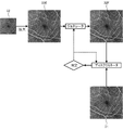
図3に、本実施形態で使用される高解像度訓練画像31の一例を示す。図3に示すように、本実施形態の高解像度訓練画像31は、OCTアンジオ画像のパノラマ画像であり、4つの単位画像311,312,313,314が位置合わせされることで生成されている。一例として、本実施形態では、4つの単位画像311,312,313,314の各々は、組織上における1辺3mmの正方形の領域を撮影した画像である。4つの単位画像311,312,313,314の各々の解像度は、256pt×256ptである。4つの単位画像311,312,313,314の各々の撮影領域は、格子状に隙間なく接している。よって、パノラマ画像である高解像度訓練画像31では、組織上における撮影領域は1辺6mmの正方形の領域となり、解像度は512pt×512ptとなる。
FIG. 3 shows an example of a high-
次いで、CPU3は、パノラマ画像である高解像度訓練画像31の画像解像度を低下させることで、低解像度訓練画像32を生成する(S2)。一例として、本実施形態のCPU3は、高解像度訓練画像31を構成する複数の画素を、規則的に(本実施形態では1個毎に)間引くことで、低解像度訓練画像32を生成する。その結果、低解像度訓練画像32では、組織上における撮影領域は、高解像度訓練画像31の撮影領域と同様に、1辺6mmの正方形の領域となる。一方で、低解像度訓練画像32の解像度は、256pt×256ptに低下する。その結果、低解像度訓練画像32の画像解像度(組織上の単位面積に対する画像の画素数)は、高解像度訓練画像31の画像解像度よりも低下する。
Next, the CPU 3 generates low-
以上のように、CPU3は、高解像度訓練画像31の画像解像度を低下させて低解像度訓練画像32を生成することで、高解像度訓練画像31と低解像度訓練画像32を含む訓練データセット30を取得する。この場合、高解像度訓練画像31と低解像度訓練画像32の撮影領域、撮影位置、および撮影時の組織の状態が一致する。よって、後述する数学モデルの訓練が、より適切に行われる。
As described above, the CPU 3 reduces the image resolution of the high-
また、数学モデルの訓練には、複数の訓練データセットが用いられる。本実施形態では、複数の訓練データセットのうち、パノラマ画像である高解像度訓練画像31と、高解像度訓練画像31の画像解像度を低下させることで生成された低解像度訓練画像32とを含む訓練データセット30の割合が、50%以上(望ましくは70%以上)となっている。その結果、数学モデルによって出力される目的画像の品質がさらに向上する。
Also, multiple training data sets are used to train the mathematical model. In this embodiment, training data including high-
なお、複数の訓練データセットのうち、前述した高解像度訓練画像31と低解像度訓練画像32を含まない訓練データセットを取得する方法は、適宜選択できる。例えば、CPU3は、パノラマ画像でない眼科画像を、高解像度訓練画像として取得してもよい。CPU3は、パノラマ画像でない高解像度訓練画像の画像解像度を低下させることで、低解像度訓練画像を生成してもよい。また、同一の組織を異なる画像解像度で複数回撮影することで生成された複数の眼科画像が、訓練データセットに用いられてもよい。なお、高解像度訓練画像の撮影領域と、低解像度訓練画像の撮影領域は、完全に一致していなくてもよい。例えば、高解像度訓練画像の撮影領域と、低解像度訓練画像の撮影領域の一部が重複していてもよい。
It should be noted that a method of obtaining a training data set that does not include the above-described high-
次いで、CPU3は、低解像度訓練画像32に、ランダムにノイズを付与する(S3)。詳細には、CPU3は、複数の低解像度訓練画像32のうち、ノイズを付与する低解像度訓練画像32をランダムに選択する。また、CPU3は、ノイズ付与処理を行う場合、付与するノイズの量、およびノイズを付与する領域等を、ランダムに決定する。
Next, the CPU 3 randomly adds noise to the low-resolution training image 32 (S3). Specifically, the CPU 3 randomly selects the low-
次いで、CPU3は、低解像度訓練画像32に、ランダムにぼかしを付与する(S4)。詳細には、CPU3は、複数の低解像度訓練画像32のうち、ぼかしを付与する低解像度訓練画像32をランダムに選択する。また、CPU3は、ぼかし付与処理を行う場合、ぼかしを付与する程度、およびぼかしを付与する領域等を、ランダムに決定する。なお、ノイズ付与処理(S3)およびぼかし付与処理(S4)は、低解像度訓練画像32の少なくとも一部の画像を劣化させる劣化処理の一例である。劣化処理の具体的な方法を変更することも可能である。
Next, the CPU 3 randomly blurs the low-resolution training images 32 (S4). Specifically, the CPU 3 randomly selects the low-
次いで、CPU3は、機械学習アルゴリズムによって、訓練データセットを用いた数学モデルの訓練を実行する(S5)。機械学習アルゴリズムとしては、例えば、ニューラルネットワーク、ランダムフォレスト、ブースティング、サポートベクターマシン(SVM)等が一般的に知られている。 CPU 3 then performs training of the mathematical model using the training data set by a machine learning algorithm (S5). As machine learning algorithms, for example, neural networks, random forests, boosting, support vector machines (SVM), etc. are generally known.
ニューラルネットワークは、生物の神経細胞ネットワークの挙動を模倣する手法である。ニューラルネットワークには、例えば、フィードフォワード(順伝播型)ニューラルネットワーク、RBFネットワーク(放射基底関数)、スパイキングニューラルネットワーク、畳み込みニューラルネットワーク、再帰型ニューラルネットワーク(リカレントニューラルネット、フィードバックニューラルネット等)、確率的ニューラルネット(ボルツマンマシン、ベイシアンネットワーク等)等がある。 A neural network is a technique that imitates the behavior of a neural network of living organisms. Neural networks include, for example, feedforward neural networks, RBF networks (radial basis functions), spiking neural networks, convolutional neural networks, recurrent neural networks (recurrent neural networks, feedback neural networks, etc.), probability neural networks (Boltzmann machine, Baysian network, etc.), etc.
ランダムフォレストは、ランダムサンプリングされた訓練データに基づいて学習を行って、多数の決定木を生成する方法である。ランダムフォレストを用いる場合、予め識別器として学習しておいた複数の決定木の分岐を辿り、各決定木から得られる結果の平均(あるいは多数決)を取る。 Random forest is a method of learning based on randomly sampled training data to generate a large number of decision trees. When a random forest is used, branches of a plurality of decision trees learned in advance as discriminators are traced, and the average (or majority vote) of the results obtained from each decision tree is taken.
ブースティングは、複数の弱識別器を組み合わせることで強識別器を生成する手法である。単純で弱い識別器を逐次的に学習させることで、強識別器を構築する。 Boosting is a method of generating a strong classifier by combining a plurality of weak classifiers. A strong classifier is constructed by sequentially learning simple and weak classifiers.
SVMは、線形入力素子を利用して2クラスのパターン識別器を構成する手法である。SVMは、例えば、訓練データから、各データ点との距離が最大となるマージン最大化超平面を求めるという基準(超平面分離定理)で、線形入力素子のパラメータを学習する。 SVM is a method of constructing a two-class pattern discriminator using linear input elements. The SVM learns the parameters of the linear input element, for example, based on the criterion of finding the margin-maximizing hyperplane that maximizes the distance to each data point from the training data (hyperplane separation theorem).
数学モデルは、例えば、入力データと出力データの関係を予測するためのデータ構造を指す。数学モデルは、訓練データセットを用いて訓練されることで構築される。訓練データセットは、入力用訓練データと出力用訓練データのセットである。数学モデルは、ある入力用訓練データが入力された時に、それに対応する出力用訓練データが出力されるように訓練される。例えば、訓練によって、各入力と出力の相関データ(例えば、重み)が更新される。 A mathematical model, for example, refers to a data structure for predicting the relationship between input data and output data. A mathematical model is built by being trained using a training data set. A training data set is a set of input training data and output training data. A mathematical model is trained such that when certain input training data is input, corresponding output training data is output. For example, training updates the correlation data (eg, weights) for each input and output.
一例として、本実施形態では、機械学習アルゴリズムとして、競合する2つのニューラルネットワークを利用する敵対的生成ネットワーク(Generative adversarial networks:GAN)が採用されている。ただし、他の機械学習アルゴリズムが用いられてもよい。例えば、多層型のニューラルネットワーク(一例として、畳み込みニューラルネットワーク(CNN)等)が、機械学習アルゴリズムとして用いられてもよい。 As an example, in this embodiment, as a machine learning algorithm, a generative adversarial network (GAN) utilizing two competing neural networks is employed. However, other machine learning algorithms may be used. For example, a multilayer neural network (eg, a convolutional neural network (CNN), etc.) may be used as the machine learning algorithm.
図4を参照して、本実施形態で採用されている機械学習アルゴリズム(GAN)による、数学モデルの訓練方法について説明する。前述したように、GANのネットワークは、互いに競合する2つのサブネットワーク(ジェネレータ(生成器)およびディスクリミネータ(識別器))を備える。本実施形態では、まず、入力用訓練データである低解像度訓練画像32を拡大した画像32Eが生成される。その結果、拡大された低解像度訓練画像32Eと、高解像度訓練画像31の大きさが一致する。なお、低解像度訓練画像32を拡大せず訓練用入力データとし、拡大する処理はジェネレータが行ってもよい。次いで、ジェネレータが、低解像度訓練画像32Eに基づいて、高解像度訓練画像31に極力一致する偽物画像32Fを出力する。ディスクリミネータは、本物画像である高解像度訓練画像31と、偽物画像32Fの正否の識別を試みる。判定結果は、ジェネレータおよびディスクリミネータの双方に反映される。この訓練が繰り返されることで、ジェネレータは、より高解像度訓練画像31に近い偽物画像32Fを生成しようとし、ディスクリミネータは、識別の正確性を向上させようとする。訓練された結果構築される数学モデル(ジェネレータモデル)は、画像解像度が低い眼科画像(例えば、後述する基画像)を入力することで、入力された画像よりも画像解像度が高い眼科画像(例えば、後述する目的画像)を生成することができる。
A method of training a mathematical model by the machine learning algorithm (GAN) employed in this embodiment will be described with reference to FIG. As mentioned above, the network of GANs comprises two sub-networks (generator and discriminator) that compete with each other. In this embodiment, first, an
なお、前述したように、本実施形態では、訓練データセットに含まれる高解像度画像と低解像度画像は、同一の眼科画像撮影装置11Aによって撮影された画像(詳細には、同一の眼科画像撮影装置11Aによって撮影された画像に基づく画像)である。従って、高解像度画像と低解像度画像の色味、画像に含まれるノイズの量、アーチファクト、光学分解能、撮影画角、撮影時の組織の状態等が近似しやすい。よって、数学モデルが適切に訓練される。
As described above, in the present embodiment, the high-resolution image and the low-resolution image included in the training data set are images captured by the same
また、本実施形態では、複数の訓練データセットの少なくともいずれかに用いられる低解像度訓練画像には、画像領域の少なくとも一部に対して劣化処理が行われている。従って、構築された数学モデルに入力される眼科画像(例えば、後述する基画像)の少なくとも一部の画質が良好でない場合でも、良好でない画質の影響が軽減された画像が出力される。 Further, in the present embodiment, degradation processing is performed on at least part of the image region of the low-resolution training images used for at least one of the plurality of training data sets. Therefore, even if the image quality of at least a part of the ophthalmologic image (for example, a base image described later) input to the constructed mathematical model is not good, an image in which the influence of the unfavorable image quality is reduced is output.
図2の説明に戻る。数学モデルの構築が完了するまで(S6:NO)、S1~S5の処理が繰り返される。数学モデルの構築が完了すると(S6:YES)、数学モデル構築処理は終了する。構築された数学モデルを実現させるプログラムおよびデータは、眼科画像処理装置21に組み込まれる。 Returning to the description of FIG. The processes of S1 to S5 are repeated until the construction of the mathematical model is completed (S6: NO). When construction of the mathematical model is completed (S6: YES), the mathematical model construction process ends. Programs and data for realizing the constructed mathematical model are installed in the ophthalmologic image processing apparatus 21 .
(眼科画像処理)
図5を参照して、眼科画像処理装置21が実行する眼科画像処理について説明する。眼科画像処理は、記憶装置24に記憶された眼科画像処理プログラムに従って、CPU23によって実行される。
(Ophthalmic image processing)
The ophthalmologic image processing executed by the ophthalmologic image processing apparatus 21 will be described with reference to FIG. The ophthalmologic image processing is executed by the
まず、CPU23は、眼科画像撮影装置11Bによって撮影された眼科画像である基画像を取得する(S11)。なお、本実施形態では、眼科画像撮影装置11Bは、基画像を連続して(つまり、動画で)撮影する。CPU23は、連続して撮影された基画像を、順次取得する。
First, the
本実施形態における基画像は、前述した高解像度訓練画像31および低解像度訓練画像32と同様に、被検眼の眼底のOCTアンジオ画像である。また、本実施形態における基画像は、前述した高解像度訓練画像31よりも画像解像度が低い画像である。一例として、本実施形態の眼科撮影装置11Bは、高解像度訓練画像31と同様の広さの撮影領域(例えば、6mm×6mmの撮影領域)を、高解像度訓練画像31の解像度よりも低い解像度(例えば、256pt×256pt)で撮影することで、基画像のデータを生成する。従って、基画像を撮影する際の撮影時間は、高解像度訓練画像31を撮影する際の撮影時間よりも短くなる。よって、基画像を撮影する際には、被検眼の動き(例えば、固視微動およびまばたき等)の影響、被検眼の動きに撮影領域を追従させるトラッキング動作の負担等が、高解像度訓練画像31を撮影する場合に比べて低下する。なお、目的画像が取得される場合、眼科画像撮影装置11Bは、基画像の撮影領域を、高解像度訓練画像31と同等の撮影領域に自動的に設定する。その結果、基画像および目的画像がより円滑に取得される。
The base image in this embodiment is an OCT angio image of the fundus of the subject's eye, similar to the high-
次いで、CPU23は、前述した数学モデルに基画像を入力することで、基画像よりも画像解像度が高い目的画像を取得する(S12)。なお、本実施形態では、CPU23は、連続して取得される基画像を数学モデルに順次入力することで、目的画像を連続して取得する。
Next, the
次いで、CPU23は、表示装置28に目的画像を表示させる指示が入力されているか否かを判断する(S14)。本実施形態では、ユーザは、目的画像、基画像、および、目的画像と基画像の両方のいずれを表示装置28に表示させるかの指示を、操作部27を操作することで眼科画像処理装置21に入力することができる。目的画像を表示させる指示が入力されていなければ(S14:NO)、処理はそのままS17へ移行する。目的画像を表示させる指示が入力されている場合(S14:YES)、CPU23は、目的画像を表示装置28に表示させる(S15)。詳細には、本実施形態のS15では、S12で連続して取得される複数の目的画像が表示装置28に順次表示されることで、組織の動画が表示される。つまり、眼科画像撮影装置11Bによってその時点で撮影されている基画像(原画像)の動画に基づいて、撮影中の組織の高解像度の動画がリアルタイムに表示装置28に表示される。なお、CPU23は、S12で連続して取得される複数の目的画像に基づいて、目的画像の動画のデータを生成し、記憶装置に記憶させてもよい。
Next, the
なお、眼科画像処理装置21は、目的画像、基画像、および、目的画像と基画像の両方のいずれを表示装置28に表示させるかの指示を、種々の方法で入力することができる。例えば、CPU23は、目的画像の表示指示を入力するアイコン、基画像の表示指示を入力するアイコン、および、両方の画像の表示指示を入力するアイコンを、表示装置28に表示させてもよい。ユーザは、希望するアイコンを選択することで、表示指示を眼科画像処理装置21に入力してもよい。
The ophthalmologic image processing apparatus 21 can use various methods to input instructions as to which of the target image, the base image, and both the target image and the base image should be displayed on the
次いで、CPU23は、表示装置28に基画像を表示させる指示が入力されているか否かを判断する(S17)。入力されていなければ(S17:NO)、処理はそのままS20へ移行する。基画像を表示させる指示が入力されている場合(S17:YES)、CPU23は、基画像を表示装置28に表示させる(S18)。詳細には、本実施形態のS18では、S11で連続して取得される複数の基画像が表示装置28に順次表示されることで、組織の動画が表示される。以上のように、CPU23は、ユーザによって入力された指示に応じて、目的画像と基画像を切り替えて表示装置28に表示させることができる。よって、ユーザは、目的画像と基画像の両方を容易に確認することができる。
Next, the
次いで、CPU23は、目的画像と基画像を同時に表示装置28に表示させる指示が入力されているか否かを判断する(S20)。入力されていなければ(S20:NO)、処理はそのままS23へ移行する。同時表示指示が入力されていれる場合(S20:YES)、CPU23は、目的画像と基画像を同時に表示装置28に表示させる。詳細には、本実施形態のS20では、目的画像の動画と基画像の動画が共に表示装置28に表示される。
Next, the
なお、S15,S18,S21では、動画でなく静止画が表示されてもよい。CPU23は、ユーザによって入力される指示に応じて、動画の表示と静止画の表示を切り替えてもよい。また、CPU23は、動画および静止画を表示装置28に表示させずに、動画および静止画の少なくとも一方のデータを記憶装置24に保存させてもよい。
Note that in S15, S18, and S21, a still image may be displayed instead of the moving image. The
次いで、CPU23は、表示を終了させる指示が入力されたか否かを判断する(S23)。入力されていなければ(S23:NO)、処理はS11へ戻り、S11~S23の処理が繰り返される。表示を終了させる指示が入力されると(S23:YES)、CPU23は、目的画像(本実施形態では、連続して取得される複数の目的画像の少なくともいずれか)に対する各種解析処理を実行する(S24)。前述したように、目的画像の画像解像度は、基画像の画像解像度よりも高い。従って、目的画像に対する解析処理を実行することで、基画像に対する解析処理を実行する場合に比べて解析精度が向上する。なお、解析処理の具体的な内容は、適宜選択できる。一例として、本実施形態のS24では、眼底の血管密度(例えば、中心窩無灌流領域周囲の血管密度等)が解析される。
Next, the
また、目的画像を表示させるタイミングには、例えば、眼科画像撮影装置11Bによる撮影中にリアルタイムで表示させるタイミング、撮影された画像が良好であるか否かをユーザに確認させるタイミング、撮影後に解析結果またはレポートと共に表示させるタイミング等、複数のタイミングがある。CPU23は、目的画像を表示させることが可能な複数のタイミングのそれぞれに対し、目的画像、基画像、および、目的画像と基画像の両方のいずれを表示装置28に表示させるかの指示を、別々に入力することができる。つまり、ユーザは、目的画像を表示させることが可能なタイミングに応じて、目的画像、基画像、および、目的画像と基画像の両方のいずれを表示装置28に表示させるかを、予め設定しておくことができる。その結果、ユーザの利便性がさらに向上する。
The timing of displaying the target image includes, for example, the timing of displaying the target image in real time during imaging by the
(合成画像生成処理)
図6を参照して、眼科画像処理装置21のCPU23が実行する合成画像生成処理について説明する。図6に示すように、合成画像40には、目的画像42と注目領域画像44が含まれる。目的画像は、図5のS12で説明したように、数学モデルに基画像を入力することで取得される、高解像度の眼科画像である。注目領域画像44は、眼科画像撮影装置11Bによって、基画像の撮影領域の一部の注目領域が撮影された画像である。注目領域画像44は、基画像の画像解像度よりも高い画像解像度で撮影されている。また、注目領域画像44は、眼科画像撮影装置11Bによって撮影された画像そのもの(つまり原画像)である。基画像の撮影領域に対する注目領域の位置、大きさ、および形状等の設定方法は、適宜選択できる。例えば、眼科画像撮影装置11Bは、ユーザによって入力される指示に応じて、基画像の撮影領域内に注目領域を設定してもよい。また、眼科画像撮影装置11Bは、基画像に対して画像処理等を行うことで、基画像における特徴部位(例えば、黄斑、視神経乳頭、および眼底血管等の少なくともいずれか)を検出し、特徴部位を基準として注目領域を自動で設定してもよい。
(Synthetic image generation processing)
Synthetic image generation processing executed by the
眼科画像処理装置21のCPU23は、基画像および注目領域画像44を取得する。CPU23は、基画像を数学モデルに入力することで、基画像よりも画像解像度が高い目的画像42を取得する。CPU23は、目的画像42における注目領域に、注目領域画像44を合成することで、合成画像40を生成する。合成画像40によると、ユーザは、注目領域を原画像によって確認することができ、且つ、画像全体も高い解像度で確認することができる。
The
上記実施形態で開示された技術は一例に過ぎない。従って、上記実施形態で例示された技術を変更することも可能である。まず、上記実施形態で例示された複数の技術のうちの一部のみを実行することも可能である。例えば、高解像度訓練画像としてパノラマ画像を用いる技術を採用せずに、低解像度訓練画像に対してノイズ付与処理およびぼかし付与処理の少なくとも一方を実行する技術を採用してもよい。 The technology disclosed in the above embodiment is merely an example. Therefore, it is also possible to modify the techniques exemplified in the above embodiments. First, it is also possible to implement only some of the techniques exemplified in the above embodiments. For example, instead of using panoramic images as high-resolution training images, a technique of performing at least one of noise addition processing and blurring processing on low-resolution training images may be adopted.
なお、図5のS11で基画像を取得する処理は、「基画像取得ステップ」の一例である。図5のS12で目的画像を取得する処理は、「目的画像取得ステップ」の一例である。図5のS21で目的画像と基画像を同時に表示させる処理は、「同時表示処理」および「同時表示ステップ」の一例である。図5のS14~S18で基画像と目的画像を切り替えて表示させる処理は、「切替表示処理」および「切替表示ステップ」の一例である。図2のS1,S2で訓練データセットを取得する処理は、「画像セット取得ステップ」の一例である。図2のS5で数学モデルを構築する処理は、「訓練ステップ」の一例である。図2のS1でパノラマ画像を取得する処理は、「パノラマ画像取得ステップ」の一例である。図2のS2で低解像度訓練画像32を生成する処理は、「低解像度訓練画像生成ステップ」の一例である。図2のS3で低解像度訓練画像32にノイズを付与する処理、および、図2のS4で低解像度訓練画像32にぼかしを付与する処理は、「劣化処理」および「劣化ステップ」の一例である。
The process of acquiring the base image in S11 of FIG. 5 is an example of the "base image obtaining step". The process of acquiring the target image in S12 of FIG. 5 is an example of the "target image acquisition step". The process of simultaneously displaying the target image and the base image in S21 of FIG. 5 is an example of the "simultaneous display process" and the "simultaneous display step." The process of switching and displaying the base image and the target image in S14 to S18 of FIG. 5 is an example of the "switching display process" and the "switching display step". The process of acquiring the training data set in S1 and S2 of FIG. 2 is an example of the "image set acquisition step". The process of building a mathematical model in S5 of FIG. 2 is an example of a "training step." The process of acquiring the panorama image in S1 of FIG. 2 is an example of the "panorama image acquisition step". The process of generating the low-
1 数学モデル構築装置
3 CPU
4 記憶装置
11 眼科画像撮影装置
21 眼科画像処理装置
23 CPU
24 記憶装置
27 操作部
28 表示装置
30 訓練データセット
31 高解像度訓練画像
32 低解像度訓練画像
40 合成画像
42 目的画像
44 注目領域画像
1 mathematical model building device 3 CPU
4 storage device 11 ophthalmic imaging device 21 ophthalmic
24
Claims (4)
前記眼科画像処理装置の制御部は、
眼科画像撮影装置によって撮影された眼科画像である基画像を取得し、
機械学習アルゴリズムによって訓練された数学モデルに前記基画像を入力することで、画像解像度が前記基画像の画像解像度よりも高い目的画像を取得し、
前記数学モデルは、同一の組織の部位を撮影対象とし、且つ、同一の眼科画像撮影装置によって撮影された複数の眼科画像のセットのうち、少なくとも1つの眼科画像である低解像度訓練画像を入力用訓練データとし、前記低解像度訓練画像よりも画像解像度が高い眼科画像である高解像度訓練画像を出力用訓練データとして訓練されており、
複数の前記高解像度訓練画像の少なくともいずれかは、完全には重複しない複数の撮影領域の各々において撮影された複数の眼科画像を位置合わせすることで生成された、パノラマ画像であることを特徴とする眼科画像処理装置。 An ophthalmic image processing apparatus for processing an ophthalmic image that is an image of tissue of an eye to be examined,
The control unit of the ophthalmologic image processing apparatus includes:
Acquiring a base image, which is an ophthalmologic image captured by an ophthalmic imaging device,
obtaining a target image whose image resolution is higher than that of the base image by inputting the base image into a mathematical model trained by a machine learning algorithm;
The mathematical model uses, as an input, a low-resolution training image that is at least one ophthalmologic image out of a set of a plurality of ophthalmologic images captured by the same ophthalmologic image capturing device, with the same tissue site as an imaging target. Training is performed using a high-resolution training image, which is an ophthalmologic image having a higher image resolution than the low-resolution training image, as training data for output ,
At least one of the plurality of high-resolution training images is a panoramic image generated by aligning a plurality of ophthalmologic images captured in each of a plurality of imaging regions that do not completely overlap. ophthalmic image processing device.
前記低解像度訓練画像の少なくともいずれかは、前記パノラマ画像の画像解像度を低下させることで生成された画像であることを特徴とする眼科画像処理装置。 The ophthalmic image processing apparatus according to claim 1 ,
An ophthalmologic image processing apparatus, wherein at least one of the low-resolution training images is an image generated by lowering the image resolution of the panoramic image.
前記OCT装置の制御部は、
撮影した眼科画像を基画像として取得し、
機械学習アルゴリズムによって訓練された数学モデルに前記基画像を入力することで、画像解像度が前記基画像の画像解像度よりも高い目的画像を取得し、
前記数学モデルは、同一の組織の部位を撮影対象とし、且つ、同一の眼科画像撮影装置によって撮影された複数の眼科画像のセットのうち、少なくとも1つの眼科画像である低解像度訓練画像を入力用訓練データとし、前記低解像度訓練画像よりも画像解像度が高い眼科画像である高解像度訓練画像を出力用訓練データとして訓練されており、
複数の前記高解像度訓練画像の少なくともいずれかは、完全には重複しない複数の撮影領域の各々において撮影された複数の眼科画像を位置合わせすることで生成された、パノラマ画像であることを特徴とするOCT装置。 An OCT apparatus that captures an ophthalmologic image of a tissue of an eye to be inspected by processing an OCT signal from reference light and reflected light of measurement light applied to a tissue of an eye to be inspected,
The control unit of the OCT device
Acquire the captured ophthalmic image as a base image,
obtaining a target image whose image resolution is higher than that of the base image by inputting the base image into a mathematical model trained by a machine learning algorithm;
The mathematical model uses, as an input, a low-resolution training image that is at least one ophthalmologic image out of a set of a plurality of ophthalmologic images captured by the same ophthalmologic image capturing device, with the same tissue site as an imaging target. Training is performed using a high-resolution training image, which is an ophthalmologic image having a higher image resolution than the low-resolution training image, as training data for output ,
At least one of the plurality of high-resolution training images is a panoramic image generated by aligning a plurality of ophthalmologic images captured in each of a plurality of imaging regions that do not completely overlap. OCT device to do.
前記眼科画像処理プログラムが前記眼科画像処理装置の制御部によって実行されることで、
眼科画像撮影装置によって撮影された眼科画像である基画像を取得する基画像取得ステップと、
機械学習アルゴリズムによって訓練された数学モデルに前記基画像を入力することで、画像解像度が前記基画像の画像解像度よりも高い目的画像を取得する目的画像取得ステップと、
を前記眼科画像処理装置に実行させ、
前記数学モデルは、同一の組織の部位を撮影対象とし、且つ、同一の眼科画像撮影装置によって撮影された複数の眼科画像のセットのうち、少なくとも1つの眼科画像である低解像度訓練画像を入力用訓練データとし、前記低解像度訓練画像よりも画像解像度が高い眼科画像である高解像度訓練画像を出力用訓練データとして訓練されており、
複数の前記高解像度訓練画像の少なくともいずれかは、完全には重複しない複数の撮影領域の各々において撮影された複数の眼科画像を位置合わせすることで生成された、パノラマ画像であることを特徴とする眼科画像処理プログラム。
An ophthalmic image processing program executed by an ophthalmic image processing device that processes an ophthalmic image that is an image of tissue of an eye to be examined,
By executing the ophthalmic image processing program by the control unit of the ophthalmic image processing apparatus,
a base image obtaining step of obtaining a base image, which is an ophthalmologic image captured by an ophthalmic imaging device;
a target image obtaining step of obtaining a target image having an image resolution higher than that of the base image by inputting the base image into a mathematical model trained by a machine learning algorithm;
causes the ophthalmic image processing apparatus to execute
The mathematical model uses, as an input, a low-resolution training image that is at least one ophthalmologic image out of a set of a plurality of ophthalmologic images captured by the same ophthalmologic image capturing device, with the same tissue site as an imaging target. Training is performed using a high-resolution training image, which is an ophthalmologic image having a higher image resolution than the low-resolution training image, as training data for output ,
At least one of the plurality of high-resolution training images is a panoramic image generated by aligning a plurality of ophthalmologic images captured in each of a plurality of imaging regions that do not completely overlap. ophthalmic image processing program.
Priority Applications (2)
| Application Number | Priority Date | Filing Date | Title |
|---|---|---|---|
| JP2018124973A JP7183590B2 (en) | 2018-06-29 | 2018-06-29 | Ophthalmic image processing device, OCT device, and ophthalmic image processing program |
| JP2022196733A JP7388525B2 (en) | 2018-06-29 | 2022-12-09 | Ophthalmology image processing device and ophthalmology image processing program |
Applications Claiming Priority (1)
| Application Number | Priority Date | Filing Date | Title |
|---|---|---|---|
| JP2018124973A JP7183590B2 (en) | 2018-06-29 | 2018-06-29 | Ophthalmic image processing device, OCT device, and ophthalmic image processing program |
Related Child Applications (1)
| Application Number | Title | Priority Date | Filing Date |
|---|---|---|---|
| JP2022196733A Division JP7388525B2 (en) | 2018-06-29 | 2022-12-09 | Ophthalmology image processing device and ophthalmology image processing program |
Publications (3)
| Publication Number | Publication Date |
|---|---|
| JP2020000678A JP2020000678A (en) | 2020-01-09 |
| JP2020000678A5 JP2020000678A5 (en) | 2021-07-26 |
| JP7183590B2 true JP7183590B2 (en) | 2022-12-06 |
Family
ID=69097728
Family Applications (2)
| Application Number | Title | Priority Date | Filing Date |
|---|---|---|---|
| JP2018124973A Active JP7183590B2 (en) | 2018-06-29 | 2018-06-29 | Ophthalmic image processing device, OCT device, and ophthalmic image processing program |
| JP2022196733A Active JP7388525B2 (en) | 2018-06-29 | 2022-12-09 | Ophthalmology image processing device and ophthalmology image processing program |
Family Applications After (1)
| Application Number | Title | Priority Date | Filing Date |
|---|---|---|---|
| JP2022196733A Active JP7388525B2 (en) | 2018-06-29 | 2022-12-09 | Ophthalmology image processing device and ophthalmology image processing program |
Country Status (1)
| Country | Link |
|---|---|
| JP (2) | JP7183590B2 (en) |
Families Citing this family (4)
| Publication number | Priority date | Publication date | Assignee | Title |
|---|---|---|---|---|
| JP7435067B2 (en) * | 2020-03-12 | 2024-02-21 | 株式会社ニデック | Ophthalmology image processing device and ophthalmology image processing program |
| JP2022068892A (en) * | 2020-10-23 | 2022-05-11 | 株式会社トプコン | Ophthalmological observation device, ophthalmological image processing device, ophthalmological image processing method, program and recording medium |
| CN112957005A (en) * | 2021-02-01 | 2021-06-15 | 山西省眼科医院(山西省红十字防盲流动眼科医院、山西省眼科研究所) | Automatic identification and laser photocoagulation region recommendation algorithm for fundus contrast image non-perfusion region |
| WO2023248968A1 (en) * | 2022-06-21 | 2023-12-28 | パナソニック インテレクチュアル プロパティ コーポレーション オブ アメリカ | Image processing method, image processing device, and image processing program |
Citations (5)
| Publication number | Priority date | Publication date | Assignee | Title |
|---|---|---|---|---|
| JP2017500651A (en) | 2014-03-20 | 2017-01-05 | 三菱電機株式会社 | A method for processing an input low resolution (LR) image into an output high resolution (HR) image |
| JP2017047113A (en) | 2015-09-04 | 2017-03-09 | 株式会社ニデック | Ophthalmic imaging apparatus and ophthalmic imaging program |
| JP2017213062A (en) | 2016-05-30 | 2017-12-07 | 株式会社トプコン | Ophthalmologic imaging apparatus |
| JP2018005841A (en) | 2016-07-08 | 2018-01-11 | 株式会社トプコン | Medical image processing method and medical image processing device |
| JP2019216848A (en) | 2018-06-15 | 2019-12-26 | キヤノン株式会社 | Medical image processing apparatus, medical image processing method, and program |
Family Cites Families (3)
| Publication number | Priority date | Publication date | Assignee | Title |
|---|---|---|---|---|
| JP5055166B2 (en) * | 2008-02-29 | 2012-10-24 | キヤノン株式会社 | Eye open / closed degree determination device, method and program, and imaging device |
| JP5827024B2 (en) * | 2011-03-31 | 2015-12-02 | 株式会社吉田製作所 | Control device, control method, and control program for optical coherence tomographic image generation apparatus |
| JP6491471B2 (en) * | 2014-12-24 | 2019-03-27 | キヤノン株式会社 | Image processing apparatus, image processing method, and program |
-
2018
- 2018-06-29 JP JP2018124973A patent/JP7183590B2/en active Active
-
2022
- 2022-12-09 JP JP2022196733A patent/JP7388525B2/en active Active
Patent Citations (5)
| Publication number | Priority date | Publication date | Assignee | Title |
|---|---|---|---|---|
| JP2017500651A (en) | 2014-03-20 | 2017-01-05 | 三菱電機株式会社 | A method for processing an input low resolution (LR) image into an output high resolution (HR) image |
| JP2017047113A (en) | 2015-09-04 | 2017-03-09 | 株式会社ニデック | Ophthalmic imaging apparatus and ophthalmic imaging program |
| JP2017213062A (en) | 2016-05-30 | 2017-12-07 | 株式会社トプコン | Ophthalmologic imaging apparatus |
| JP2018005841A (en) | 2016-07-08 | 2018-01-11 | 株式会社トプコン | Medical image processing method and medical image processing device |
| JP2019216848A (en) | 2018-06-15 | 2019-12-26 | キヤノン株式会社 | Medical image processing apparatus, medical image processing method, and program |
Also Published As
| Publication number | Publication date |
|---|---|
| JP7388525B2 (en) | 2023-11-29 |
| JP2020000678A (en) | 2020-01-09 |
| JP2023077425A (en) | 2023-06-05 |
Similar Documents
| Publication | Publication Date | Title |
|---|---|---|
| JP7388525B2 (en) | Ophthalmology image processing device and ophthalmology image processing program | |
| US11633096B2 (en) | Ophthalmologic image processing device and non-transitory computer-readable storage medium storing computer-readable instructions | |
| JP7114358B2 (en) | MEDICAL IMAGE PROCESSING APPARATUS, MEDICAL IMAGE PROCESSING METHOD AND PROGRAM | |
| JP2011120655A (en) | Tomographic imaging apparatus, image processing apparatus and image processing method | |
| JP2020166814A (en) | Medical image processing device, medical image processing method, and program | |
| JP2024040372A (en) | Ophthalmology image processing program and ophthalmology image processing device | |
| JP7332463B2 (en) | Control device, optical coherence tomography device, control method for optical coherence tomography device, and program | |
| JP2024045441A (en) | Ophthalmic image processing device and ophthalmic image processing program | |
| JP6703319B1 (en) | Ophthalmic image processing device and OCT device | |
| CN112533526A (en) | Ophthalmologic image processing apparatus, OCT apparatus, and ophthalmologic image processing program | |
| JP7147888B2 (en) | Ophthalmic image processing device, OCT device, and ophthalmic image processing program | |
| JP2019208851A (en) | Fundus image processing device and fundus image processing program | |
| JP6747617B2 (en) | Ophthalmic image processing device and OCT device | |
| WO2022209574A1 (en) | Medical image processing device, medical image processing program, and medical image processing method | |
| WO2021020419A1 (en) | Medical image processing device and medical image processing program | |
| JP7435067B2 (en) | Ophthalmology image processing device and ophthalmology image processing program | |
| WO2020241794A1 (en) | Ophthalmic image processing device, ophthalmic image processing program, and ophthalmic image processing system | |
| JP2021074095A (en) | Ophthalmologic image processing device and ophthalmologic image processing program | |
| WO2023281965A1 (en) | Medical image processing device and medical image processing program | |
| WO2021045019A1 (en) | Ophthalmic image processing program and ophthalmic image processing device | |
| JP7180187B2 (en) | Ophthalmic image processing device, OCT device, and ophthalmic image processing program | |
| JP7210927B2 (en) | Ophthalmic image processing device, OCT device, and ophthalmic image processing program | |
| JP2012176291A (en) | Tomographic imaging apparatus, image processing apparatus, image processing system, and method and program for controlling image processing apparatus | |
| JP7302184B2 (en) | Ophthalmic image processing device and ophthalmic image processing program | |
| JP7233792B2 (en) | Diagnostic imaging device, diagnostic imaging method, program, and method for generating training data for machine learning |
Legal Events
| Date | Code | Title | Description |
|---|---|---|---|
| A521 | Request for written amendment filed |
Free format text: JAPANESE INTERMEDIATE CODE: A523 Effective date: 20210601 |
|
| A621 | Written request for application examination |
Free format text: JAPANESE INTERMEDIATE CODE: A621 Effective date: 20210601 |
|
| A977 | Report on retrieval |
Free format text: JAPANESE INTERMEDIATE CODE: A971007 Effective date: 20220414 |
|
| A131 | Notification of reasons for refusal |
Free format text: JAPANESE INTERMEDIATE CODE: A131 Effective date: 20220510 |
|
| A521 | Request for written amendment filed |
Free format text: JAPANESE INTERMEDIATE CODE: A523 Effective date: 20220707 |
|
| TRDD | Decision of grant or rejection written | ||
| A01 | Written decision to grant a patent or to grant a registration (utility model) |
Free format text: JAPANESE INTERMEDIATE CODE: A01 Effective date: 20221025 |
|
| A61 | First payment of annual fees (during grant procedure) |
Free format text: JAPANESE INTERMEDIATE CODE: A61 Effective date: 20221107 |
|
| R150 | Certificate of patent or registration of utility model |
Ref document number: 7183590 Country of ref document: JP Free format text: JAPANESE INTERMEDIATE CODE: R150 |