JP7181219B2 - antigen-specific immune effector cells - Google Patents
antigen-specific immune effector cells Download PDFInfo
- Publication number
- JP7181219B2 JP7181219B2 JP2019556588A JP2019556588A JP7181219B2 JP 7181219 B2 JP7181219 B2 JP 7181219B2 JP 2019556588 A JP2019556588 A JP 2019556588A JP 2019556588 A JP2019556588 A JP 2019556588A JP 7181219 B2 JP7181219 B2 JP 7181219B2
- Authority
- JP
- Japan
- Prior art keywords
- cells
- cell
- antigen
- hla
- car
- Prior art date
- Legal status (The legal status is an assumption and is not a legal conclusion. Google has not performed a legal analysis and makes no representation as to the accuracy of the status listed.)
- Active
Links
- 239000000427 antigen Substances 0.000 title claims description 172
- 108091007433 antigens Proteins 0.000 title claims description 172
- 102000036639 antigens Human genes 0.000 title claims description 172
- 239000012642 immune effector Substances 0.000 title description 9
- 229940121354 immunomodulator Drugs 0.000 title description 9
- 210000004027 cell Anatomy 0.000 claims description 457
- 210000001744 T-lymphocyte Anatomy 0.000 claims description 276
- 238000000034 method Methods 0.000 claims description 184
- 230000004069 differentiation Effects 0.000 claims description 153
- 210000003958 hematopoietic stem cell Anatomy 0.000 claims description 153
- 108090000623 proteins and genes Proteins 0.000 claims description 108
- 210000000822 natural killer cell Anatomy 0.000 claims description 101
- 108010019670 Chimeric Antigen Receptors Proteins 0.000 claims description 98
- 102100031573 Hematopoietic progenitor cell antigen CD34 Human genes 0.000 claims description 88
- 101000777663 Homo sapiens Hematopoietic progenitor cell antigen CD34 Proteins 0.000 claims description 88
- 210000001778 pluripotent stem cell Anatomy 0.000 claims description 87
- 241000282414 Homo sapiens Species 0.000 claims description 51
- -1 cAMP Proteins 0.000 claims description 36
- 101000608935 Homo sapiens Leukosialin Proteins 0.000 claims description 31
- 102100039564 Leukosialin Human genes 0.000 claims description 31
- 230000027455 binding Effects 0.000 claims description 31
- 210000004263 induced pluripotent stem cell Anatomy 0.000 claims description 31
- 102000005789 Vascular Endothelial Growth Factors Human genes 0.000 claims description 30
- 108010019530 Vascular Endothelial Growth Factors Proteins 0.000 claims description 30
- 239000003112 inhibitor Substances 0.000 claims description 27
- 108020004414 DNA Proteins 0.000 claims description 26
- 238000012258 culturing Methods 0.000 claims description 26
- 230000006698 induction Effects 0.000 claims description 25
- 102000003974 Fibroblast growth factor 2 Human genes 0.000 claims description 23
- 108090000379 Fibroblast growth factor 2 Proteins 0.000 claims description 23
- CIWBSHSKHKDKBQ-JLAZNSOCSA-N Ascorbic acid Chemical compound OC[C@H](O)[C@H]1OC(=O)C(O)=C1O CIWBSHSKHKDKBQ-JLAZNSOCSA-N 0.000 claims description 22
- 102100039064 Interleukin-3 Human genes 0.000 claims description 22
- 102000002254 Glycogen Synthase Kinase 3 Human genes 0.000 claims description 21
- 108010014905 Glycogen Synthase Kinase 3 Proteins 0.000 claims description 21
- 210000002242 embryoid body Anatomy 0.000 claims description 20
- 206010021143 Hypoxia Diseases 0.000 claims description 19
- 108010002350 Interleukin-2 Proteins 0.000 claims description 19
- 108700014844 flt3 ligand Proteins 0.000 claims description 19
- 108010070047 Notch Receptors Proteins 0.000 claims description 18
- 210000003162 effector t lymphocyte Anatomy 0.000 claims description 18
- 230000001146 hypoxic effect Effects 0.000 claims description 17
- 210000001671 embryonic stem cell Anatomy 0.000 claims description 14
- 230000001472 cytotoxic effect Effects 0.000 claims description 13
- 239000004615 ingredient Substances 0.000 claims description 13
- 102100028972 HLA class I histocompatibility antigen, A alpha chain Human genes 0.000 claims description 12
- 102100028976 HLA class I histocompatibility antigen, B alpha chain Human genes 0.000 claims description 12
- 108010075704 HLA-A Antigens Proteins 0.000 claims description 12
- 108010058607 HLA-B Antigens Proteins 0.000 claims description 12
- DFPAKSUCGFBDDF-UHFFFAOYSA-N Nicotinamide Chemical compound NC(=O)C1=CC=CN=C1 DFPAKSUCGFBDDF-UHFFFAOYSA-N 0.000 claims description 12
- 235000010323 ascorbic acid Nutrition 0.000 claims description 12
- 210000004881 tumor cell Anatomy 0.000 claims description 12
- 229960005070 ascorbic acid Drugs 0.000 claims description 11
- 239000011668 ascorbic acid Substances 0.000 claims description 11
- 108010056030 retronectin Proteins 0.000 claims description 11
- 230000001939 inductive effect Effects 0.000 claims description 10
- 238000002826 magnetic-activated cell sorting Methods 0.000 claims description 10
- 101000914514 Homo sapiens T-cell-specific surface glycoprotein CD28 Proteins 0.000 claims description 9
- 102100027213 T-cell-specific surface glycoprotein CD28 Human genes 0.000 claims description 9
- AQGNHMOJWBZFQQ-UHFFFAOYSA-N CT 99021 Chemical group CC1=CNC(C=2C(=NC(NCCNC=3N=CC(=CC=3)C#N)=NC=2)C=2C(=CC(Cl)=CC=2)Cl)=N1 AQGNHMOJWBZFQQ-UHFFFAOYSA-N 0.000 claims description 8
- 102000003812 Interleukin-15 Human genes 0.000 claims description 8
- 108090000172 Interleukin-15 Proteins 0.000 claims description 8
- 108010002586 Interleukin-7 Proteins 0.000 claims description 8
- LZAXPYOBKSJSEX-UHFFFAOYSA-N blebbistatin Chemical compound C1CC2(O)C(=O)C3=CC(C)=CC=C3N=C2N1C1=CC=CC=C1 LZAXPYOBKSJSEX-UHFFFAOYSA-N 0.000 claims description 8
- 230000004068 intracellular signaling Effects 0.000 claims description 8
- 108700028369 Alleles Proteins 0.000 claims description 7
- 102100031785 Endothelial transcription factor GATA-2 Human genes 0.000 claims description 7
- 102100028971 HLA class I histocompatibility antigen, C alpha chain Human genes 0.000 claims description 7
- 108010052199 HLA-C Antigens Proteins 0.000 claims description 7
- 101000813735 Homo sapiens ETS translocation variant 2 Proteins 0.000 claims description 7
- 101001066265 Homo sapiens Endothelial transcription factor GATA-2 Proteins 0.000 claims description 7
- 102100030704 Interleukin-21 Human genes 0.000 claims description 7
- 210000001151 cytotoxic T lymphocyte Anatomy 0.000 claims description 7
- 239000012636 effector Substances 0.000 claims description 7
- 108010027263 homeobox protein HOXA9 Proteins 0.000 claims description 7
- 108010074108 interleukin-21 Proteins 0.000 claims description 7
- 102100039579 ETS translocation variant 2 Human genes 0.000 claims description 6
- 102000006354 HLA-DR Antigens Human genes 0.000 claims description 6
- 108010058597 HLA-DR Antigens Proteins 0.000 claims description 6
- 102100021090 Homeobox protein Hox-A9 Human genes 0.000 claims description 6
- 108700012912 MYCN Proteins 0.000 claims description 6
- 101150022024 MYCN gene Proteins 0.000 claims description 6
- 108700026495 N-Myc Proto-Oncogene Proteins 0.000 claims description 6
- 102100030124 N-myc proto-oncogene protein Human genes 0.000 claims description 6
- 210000003716 mesoderm Anatomy 0.000 claims description 6
- 235000005152 nicotinamide Nutrition 0.000 claims description 6
- 239000011570 nicotinamide Substances 0.000 claims description 6
- 229960003966 nicotinamide Drugs 0.000 claims description 6
- YDRYQBCOLJPFFX-REOHCLBHSA-N (2r)-2-amino-3-(1,1,2,2-tetrafluoroethylsulfanyl)propanoic acid Chemical compound OC(=O)[C@@H](N)CSC(F)(F)C(F)F YDRYQBCOLJPFFX-REOHCLBHSA-N 0.000 claims description 5
- 102000015789 HLA-DP Antigens Human genes 0.000 claims description 5
- 108010010378 HLA-DP Antigens Proteins 0.000 claims description 5
- 108010062347 HLA-DQ Antigens Proteins 0.000 claims description 5
- 108700039143 HMGA2 Proteins 0.000 claims description 5
- 102100028999 High mobility group protein HMGI-C Human genes 0.000 claims description 5
- 101150073387 Hmga2 gene Proteins 0.000 claims description 5
- 102100025116 Homeobox protein Hox-A4 Human genes 0.000 claims description 5
- 101001077578 Homo sapiens Homeobox protein Hox-A4 Proteins 0.000 claims description 5
- 101001109698 Homo sapiens Nuclear receptor subfamily 4 group A member 2 Proteins 0.000 claims description 5
- 101000837837 Homo sapiens Transcription factor EC Proteins 0.000 claims description 5
- 101000652324 Homo sapiens Transcription factor SOX-17 Proteins 0.000 claims description 5
- 102000003815 Interleukin-11 Human genes 0.000 claims description 5
- 108090000177 Interleukin-11 Proteins 0.000 claims description 5
- 101150029107 MEIS1 gene Proteins 0.000 claims description 5
- 108700041619 Myeloid Ecotropic Viral Integration Site 1 Proteins 0.000 claims description 5
- 102000047831 Myeloid Ecotropic Viral Integration Site 1 Human genes 0.000 claims description 5
- 102100022676 Nuclear receptor subfamily 4 group A member 2 Human genes 0.000 claims description 5
- 102100028503 Transcription factor EC Human genes 0.000 claims description 5
- 102100030243 Transcription factor SOX-17 Human genes 0.000 claims description 5
- 210000000581 natural killer T-cell Anatomy 0.000 claims description 4
- 102100024505 Bone morphogenetic protein 4 Human genes 0.000 claims description 3
- 108010065805 Interleukin-12 Proteins 0.000 claims description 3
- 102000013462 Interleukin-12 Human genes 0.000 claims description 3
- 108700019146 Transgenes Proteins 0.000 claims description 3
- 210000003071 memory t lymphocyte Anatomy 0.000 claims description 3
- 210000003289 regulatory T cell Anatomy 0.000 claims description 3
- 102100024222 B-lymphocyte antigen CD19 Human genes 0.000 claims description 2
- 108010049955 Bone Morphogenetic Protein 4 Proteins 0.000 claims description 2
- 210000001266 CD8-positive T-lymphocyte Anatomy 0.000 claims 1
- 101000980825 Homo sapiens B-lymphocyte antigen CD19 Proteins 0.000 claims 1
- 101000762379 Homo sapiens Bone morphogenetic protein 4 Proteins 0.000 claims 1
- 239000002609 medium Substances 0.000 description 98
- 230000014509 gene expression Effects 0.000 description 86
- 206010028980 Neoplasm Diseases 0.000 description 75
- 108091008874 T cell receptors Proteins 0.000 description 73
- 239000013598 vector Substances 0.000 description 65
- 102000016266 T-Cell Antigen Receptors Human genes 0.000 description 58
- 150000007523 nucleic acids Chemical class 0.000 description 56
- 230000003394 haemopoietic effect Effects 0.000 description 41
- 102000017420 CD3 protein, epsilon/gamma/delta subunit Human genes 0.000 description 39
- 108050005493 CD3 protein, epsilon/gamma/delta subunit Proteins 0.000 description 39
- 102000039446 nucleic acids Human genes 0.000 description 39
- 108020004707 nucleic acids Proteins 0.000 description 39
- 229920002153 Hydroxypropyl cellulose Polymers 0.000 description 37
- 108090000765 processed proteins & peptides Proteins 0.000 description 35
- 102000004169 proteins and genes Human genes 0.000 description 32
- 210000002865 immune cell Anatomy 0.000 description 31
- 210000000130 stem cell Anatomy 0.000 description 31
- 102000004127 Cytokines Human genes 0.000 description 29
- 108090000695 Cytokines Proteins 0.000 description 29
- 235000018102 proteins Nutrition 0.000 description 29
- 230000008672 reprogramming Effects 0.000 description 29
- 241000699666 Mus <mouse, genus> Species 0.000 description 27
- 108010073929 Vascular Endothelial Growth Factor A Proteins 0.000 description 26
- 201000011510 cancer Diseases 0.000 description 26
- 210000001519 tissue Anatomy 0.000 description 25
- 108700018351 Major Histocompatibility Complex Proteins 0.000 description 24
- 102000036693 Thrombopoietin Human genes 0.000 description 24
- 108010041111 Thrombopoietin Proteins 0.000 description 24
- 238000001727 in vivo Methods 0.000 description 24
- 230000020382 suppression by virus of host antigen processing and presentation of peptide antigen via MHC class I Effects 0.000 description 24
- 238000000684 flow cytometry Methods 0.000 description 23
- 102000004196 processed proteins & peptides Human genes 0.000 description 23
- 239000003623 enhancer Substances 0.000 description 22
- 210000001082 somatic cell Anatomy 0.000 description 22
- 239000003795 chemical substances by application Substances 0.000 description 21
- 239000013612 plasmid Substances 0.000 description 21
- 101000946843 Homo sapiens T-cell surface glycoprotein CD8 alpha chain Proteins 0.000 description 20
- 108010002386 Interleukin-3 Proteins 0.000 description 20
- 102100034922 T-cell surface glycoprotein CD8 alpha chain Human genes 0.000 description 20
- 230000024245 cell differentiation Effects 0.000 description 20
- 208000037265 diseases, disorders, signs and symptoms Diseases 0.000 description 19
- 230000000694 effects Effects 0.000 description 18
- 229940076264 interleukin-3 Drugs 0.000 description 18
- 239000003446 ligand Substances 0.000 description 18
- 239000000047 product Substances 0.000 description 18
- 101000581981 Homo sapiens Neural cell adhesion molecule 1 Proteins 0.000 description 17
- 102100027347 Neural cell adhesion molecule 1 Human genes 0.000 description 17
- 102000005650 Notch Receptors Human genes 0.000 description 17
- 238000000338 in vitro Methods 0.000 description 17
- 102000000588 Interleukin-2 Human genes 0.000 description 16
- 230000010261 cell growth Effects 0.000 description 16
- 239000000203 mixture Substances 0.000 description 16
- 108060003951 Immunoglobulin Proteins 0.000 description 15
- 102000018358 immunoglobulin Human genes 0.000 description 15
- 239000003550 marker Substances 0.000 description 15
- 229920001184 polypeptide Polymers 0.000 description 15
- 230000010076 replication Effects 0.000 description 15
- 230000009258 tissue cross reactivity Effects 0.000 description 15
- 102100033553 Delta-like protein 4 Human genes 0.000 description 14
- 101000872077 Homo sapiens Delta-like protein 4 Proteins 0.000 description 14
- 241000699670 Mus sp. Species 0.000 description 14
- 201000010099 disease Diseases 0.000 description 14
- 230000006870 function Effects 0.000 description 14
- 230000002062 proliferating effect Effects 0.000 description 14
- 101000738771 Homo sapiens Receptor-type tyrosine-protein phosphatase C Proteins 0.000 description 13
- 241000701044 Human gammaherpesvirus 4 Species 0.000 description 13
- 102000004889 Interleukin-6 Human genes 0.000 description 13
- 108090001005 Interleukin-6 Proteins 0.000 description 13
- 108091028043 Nucleic acid sequence Proteins 0.000 description 13
- 102100037422 Receptor-type tyrosine-protein phosphatase C Human genes 0.000 description 13
- 210000003719 b-lymphocyte Anatomy 0.000 description 13
- 239000012634 fragment Substances 0.000 description 13
- 210000004698 lymphocyte Anatomy 0.000 description 13
- 230000001105 regulatory effect Effects 0.000 description 13
- 230000035897 transcription Effects 0.000 description 13
- 238000013518 transcription Methods 0.000 description 13
- 102000006306 Antigen Receptors Human genes 0.000 description 12
- 108010083359 Antigen Receptors Proteins 0.000 description 12
- 108091026890 Coding region Proteins 0.000 description 12
- 101000914496 Homo sapiens T-cell antigen CD7 Proteins 0.000 description 12
- 101000716102 Homo sapiens T-cell surface glycoprotein CD4 Proteins 0.000 description 12
- 102100027208 T-cell antigen CD7 Human genes 0.000 description 12
- 102100036011 T-cell surface glycoprotein CD4 Human genes 0.000 description 12
- 150000001875 compounds Chemical class 0.000 description 12
- 230000002708 enhancing effect Effects 0.000 description 12
- 229940100601 interleukin-6 Drugs 0.000 description 12
- 230000008569 process Effects 0.000 description 12
- 102100029761 Cadherin-5 Human genes 0.000 description 11
- 101000794587 Homo sapiens Cadherin-5 Proteins 0.000 description 11
- 239000007760 Iscove's Modified Dulbecco's Medium Substances 0.000 description 11
- 239000008280 blood Substances 0.000 description 11
- 239000011159 matrix material Substances 0.000 description 11
- 102000005962 receptors Human genes 0.000 description 11
- 108020003175 receptors Proteins 0.000 description 11
- 238000011282 treatment Methods 0.000 description 11
- 108020004684 Internal Ribosome Entry Sites Proteins 0.000 description 10
- 241001465754 Metazoa Species 0.000 description 10
- 210000004369 blood Anatomy 0.000 description 10
- 102000054766 genetic haplotypes Human genes 0.000 description 10
- 210000005260 human cell Anatomy 0.000 description 10
- 238000002347 injection Methods 0.000 description 10
- 239000007924 injection Substances 0.000 description 10
- 229910052760 oxygen Inorganic materials 0.000 description 10
- 210000003819 peripheral blood mononuclear cell Anatomy 0.000 description 10
- 102000040430 polynucleotide Human genes 0.000 description 10
- 108091033319 polynucleotide Proteins 0.000 description 10
- 239000002157 polynucleotide Substances 0.000 description 10
- 230000001177 retroviral effect Effects 0.000 description 10
- 239000013603 viral vector Substances 0.000 description 10
- 102000043129 MHC class I family Human genes 0.000 description 9
- 108091054437 MHC class I family Proteins 0.000 description 9
- 108010072388 Methyl-CpG-Binding Protein 2 Proteins 0.000 description 9
- 208000015914 Non-Hodgkin lymphomas Diseases 0.000 description 9
- 238000010586 diagram Methods 0.000 description 9
- 238000012360 testing method Methods 0.000 description 9
- 102100020715 Fms-related tyrosine kinase 3 ligand protein Human genes 0.000 description 8
- 101710162577 Fms-related tyrosine kinase 3 ligand protein Proteins 0.000 description 8
- HTTJABKRGRZYRN-UHFFFAOYSA-N Heparin Chemical compound OC1C(NC(=O)C)C(O)OC(COS(O)(=O)=O)C1OC1C(OS(O)(=O)=O)C(O)C(OC2C(C(OS(O)(=O)=O)C(OC3C(C(O)C(O)C(O3)C(O)=O)OS(O)(=O)=O)C(CO)O2)NS(O)(=O)=O)C(C(O)=O)O1 HTTJABKRGRZYRN-UHFFFAOYSA-N 0.000 description 8
- 102000004058 Leukemia inhibitory factor Human genes 0.000 description 8
- 108090000581 Leukemia inhibitory factor Proteins 0.000 description 8
- 241000829100 Macaca mulatta polyomavirus 1 Species 0.000 description 8
- 102000006890 Methyl-CpG-Binding Protein 2 Human genes 0.000 description 8
- 229910019142 PO4 Inorganic materials 0.000 description 8
- 102000008579 Transposases Human genes 0.000 description 8
- 108010020764 Transposases Proteins 0.000 description 8
- 241000700605 Viruses Species 0.000 description 8
- 230000004913 activation Effects 0.000 description 8
- 230000015572 biosynthetic process Effects 0.000 description 8
- 230000008859 change Effects 0.000 description 8
- 229960002897 heparin Drugs 0.000 description 8
- 229920000669 heparin Polymers 0.000 description 8
- 230000000977 initiatory effect Effects 0.000 description 8
- 230000007246 mechanism Effects 0.000 description 8
- 201000001441 melanoma Diseases 0.000 description 8
- 238000002054 transplantation Methods 0.000 description 8
- 241001529453 unidentified herpesvirus Species 0.000 description 8
- 108091032973 (ribonucleotides)n+m Proteins 0.000 description 7
- 101001077417 Gallus gallus Potassium voltage-gated channel subfamily H member 6 Proteins 0.000 description 7
- 101001010792 Homo sapiens Transcriptional regulator ERG Proteins 0.000 description 7
- 102100029983 Transcriptional regulator ERG Human genes 0.000 description 7
- 235000001014 amino acid Nutrition 0.000 description 7
- 150000001413 amino acids Chemical class 0.000 description 7
- 210000000612 antigen-presenting cell Anatomy 0.000 description 7
- QVGXLLKOCUKJST-UHFFFAOYSA-N atomic oxygen Chemical compound [O] QVGXLLKOCUKJST-UHFFFAOYSA-N 0.000 description 7
- 238000005516 engineering process Methods 0.000 description 7
- 210000002950 fibroblast Anatomy 0.000 description 7
- 210000000987 immune system Anatomy 0.000 description 7
- 230000003211 malignant effect Effects 0.000 description 7
- 230000001404 mediated effect Effects 0.000 description 7
- 230000035772 mutation Effects 0.000 description 7
- 239000001301 oxygen Substances 0.000 description 7
- 239000008194 pharmaceutical composition Substances 0.000 description 7
- 239000010452 phosphate Substances 0.000 description 7
- 238000012216 screening Methods 0.000 description 7
- 238000000926 separation method Methods 0.000 description 7
- 210000002966 serum Anatomy 0.000 description 7
- 230000004083 survival effect Effects 0.000 description 7
- 238000012546 transfer Methods 0.000 description 7
- 238000013519 translation Methods 0.000 description 7
- 230000003612 virological effect Effects 0.000 description 7
- 108010047041 Complementarity Determining Regions Proteins 0.000 description 6
- 108010017080 Granulocyte Colony-Stimulating Factor Proteins 0.000 description 6
- 102100039619 Granulocyte colony-stimulating factor Human genes 0.000 description 6
- 108700026244 Open Reading Frames Proteins 0.000 description 6
- 208000009956 adenocarcinoma Diseases 0.000 description 6
- 239000007640 basal medium Substances 0.000 description 6
- 210000000601 blood cell Anatomy 0.000 description 6
- 230000037396 body weight Effects 0.000 description 6
- 210000002798 bone marrow cell Anatomy 0.000 description 6
- 239000002299 complementary DNA Substances 0.000 description 6
- 230000006378 damage Effects 0.000 description 6
- 239000013604 expression vector Substances 0.000 description 6
- 230000004927 fusion Effects 0.000 description 6
- 230000002068 genetic effect Effects 0.000 description 6
- ZDXPYRJPNDTMRX-UHFFFAOYSA-N glutamine Natural products OC(=O)C(N)CCC(N)=O ZDXPYRJPNDTMRX-UHFFFAOYSA-N 0.000 description 6
- 238000003384 imaging method Methods 0.000 description 6
- 210000003738 lymphoid progenitor cell Anatomy 0.000 description 6
- 229940074358 magnesium ascorbate Drugs 0.000 description 6
- AIOKQVJVNPDJKA-ZZMNMWMASA-L magnesium;(2r)-2-[(1s)-1,2-dihydroxyethyl]-4-hydroxy-5-oxo-2h-furan-3-olate Chemical compound [Mg+2].OC[C@H](O)[C@H]1OC(=O)C(O)=C1[O-].OC[C@H](O)[C@H]1OC(=O)C(O)=C1[O-] AIOKQVJVNPDJKA-ZZMNMWMASA-L 0.000 description 6
- 108020004999 messenger RNA Proteins 0.000 description 6
- 210000001616 monocyte Anatomy 0.000 description 6
- PJUIMOJAAPLTRJ-UHFFFAOYSA-N monothioglycerol Chemical compound OCC(O)CS PJUIMOJAAPLTRJ-UHFFFAOYSA-N 0.000 description 6
- 210000000056 organ Anatomy 0.000 description 6
- 235000021317 phosphate Nutrition 0.000 description 6
- 241000701822 Bovine papillomavirus Species 0.000 description 5
- 201000009030 Carcinoma Diseases 0.000 description 5
- AOJJSUZBOXZQNB-TZSSRYMLSA-N Doxorubicin Chemical compound O([C@H]1C[C@@](O)(CC=2C(O)=C3C(=O)C=4C=CC=C(C=4C(=O)C3=C(O)C=21)OC)C(=O)CO)[C@H]1C[C@H](N)[C@H](O)[C@H](C)O1 AOJJSUZBOXZQNB-TZSSRYMLSA-N 0.000 description 5
- 102100039620 Granulocyte-macrophage colony-stimulating factor Human genes 0.000 description 5
- 206010035226 Plasma cell myeloma Diseases 0.000 description 5
- 102100024616 Platelet endothelial cell adhesion molecule Human genes 0.000 description 5
- 102000003425 Tyrosinase Human genes 0.000 description 5
- 108060008724 Tyrosinase Proteins 0.000 description 5
- 238000010171 animal model Methods 0.000 description 5
- 238000002659 cell therapy Methods 0.000 description 5
- 108091008034 costimulatory receptors Proteins 0.000 description 5
- 230000001086 cytosolic effect Effects 0.000 description 5
- 231100000433 cytotoxic Toxicity 0.000 description 5
- 230000007423 decrease Effects 0.000 description 5
- 210000004443 dendritic cell Anatomy 0.000 description 5
- 210000003743 erythrocyte Anatomy 0.000 description 5
- 238000001476 gene delivery Methods 0.000 description 5
- 239000005090 green fluorescent protein Substances 0.000 description 5
- 230000012010 growth Effects 0.000 description 5
- 239000003102 growth factor Substances 0.000 description 5
- 230000028993 immune response Effects 0.000 description 5
- 229940072221 immunoglobulins Drugs 0.000 description 5
- 238000009169 immunotherapy Methods 0.000 description 5
- 238000011534 incubation Methods 0.000 description 5
- 208000015181 infectious disease Diseases 0.000 description 5
- 238000003780 insertion Methods 0.000 description 5
- 230000037431 insertion Effects 0.000 description 5
- 238000002955 isolation Methods 0.000 description 5
- 208000032839 leukemia Diseases 0.000 description 5
- 210000002540 macrophage Anatomy 0.000 description 5
- 238000004519 manufacturing process Methods 0.000 description 5
- 239000012528 membrane Substances 0.000 description 5
- 230000004048 modification Effects 0.000 description 5
- 238000012986 modification Methods 0.000 description 5
- 210000003643 myeloid progenitor cell Anatomy 0.000 description 5
- 238000004806 packaging method and process Methods 0.000 description 5
- NBIIXXVUZAFLBC-UHFFFAOYSA-K phosphate Chemical compound [O-]P([O-])([O-])=O NBIIXXVUZAFLBC-UHFFFAOYSA-K 0.000 description 5
- 238000003752 polymerase chain reaction Methods 0.000 description 5
- 210000004986 primary T-cell Anatomy 0.000 description 5
- 230000003362 replicative effect Effects 0.000 description 5
- 239000000243 solution Substances 0.000 description 5
- 230000005030 transcription termination Effects 0.000 description 5
- 230000004614 tumor growth Effects 0.000 description 5
- 238000011144 upstream manufacturing Methods 0.000 description 5
- HZAXFHJVJLSVMW-UHFFFAOYSA-N 2-Aminoethan-1-ol Chemical compound NCCO HZAXFHJVJLSVMW-UHFFFAOYSA-N 0.000 description 4
- JKMHFZQWWAIEOD-UHFFFAOYSA-N 2-[4-(2-hydroxyethyl)piperazin-1-yl]ethanesulfonic acid Chemical compound OCC[NH+]1CCN(CCS([O-])(=O)=O)CC1 JKMHFZQWWAIEOD-UHFFFAOYSA-N 0.000 description 4
- 108091003079 Bovine Serum Albumin Proteins 0.000 description 4
- 102100025475 Carcinoembryonic antigen-related cell adhesion molecule 5 Human genes 0.000 description 4
- 239000006144 Dulbecco’s modified Eagle's medium Substances 0.000 description 4
- 108010031111 EBV-encoded nuclear antigen 1 Proteins 0.000 description 4
- 102000018233 Fibroblast Growth Factor Human genes 0.000 description 4
- 108050007372 Fibroblast Growth Factor Proteins 0.000 description 4
- 108700028146 Genetic Enhancer Elements Proteins 0.000 description 4
- 102000003886 Glycoproteins Human genes 0.000 description 4
- 108090000288 Glycoproteins Proteins 0.000 description 4
- 108060001084 Luciferase Proteins 0.000 description 4
- 239000005089 Luciferase Substances 0.000 description 4
- 206010025323 Lymphomas Diseases 0.000 description 4
- 102100028389 Melanoma antigen recognized by T-cells 1 Human genes 0.000 description 4
- 241000699660 Mus musculus Species 0.000 description 4
- 102100038895 Myc proto-oncogene protein Human genes 0.000 description 4
- 101710135898 Myc proto-oncogene protein Proteins 0.000 description 4
- 239000012580 N-2 Supplement Substances 0.000 description 4
- 206010039491 Sarcoma Diseases 0.000 description 4
- 241000700584 Simplexvirus Species 0.000 description 4
- 108091081024 Start codon Proteins 0.000 description 4
- 108010039445 Stem Cell Factor Proteins 0.000 description 4
- 102000040945 Transcription factor Human genes 0.000 description 4
- 108091023040 Transcription factor Proteins 0.000 description 4
- 101710150448 Transcriptional regulator Myc Proteins 0.000 description 4
- 108010031318 Vitronectin Proteins 0.000 description 4
- 102100035140 Vitronectin Human genes 0.000 description 4
- 230000003213 activating effect Effects 0.000 description 4
- 239000000654 additive Substances 0.000 description 4
- 238000011467 adoptive cell therapy Methods 0.000 description 4
- SHGAZHPCJJPHSC-YCNIQYBTSA-N all-trans-retinoic acid Chemical compound OC(=O)\C=C(/C)\C=C\C=C(/C)\C=C\C1=C(C)CCCC1(C)C SHGAZHPCJJPHSC-YCNIQYBTSA-N 0.000 description 4
- 230000008901 benefit Effects 0.000 description 4
- 230000029918 bioluminescence Effects 0.000 description 4
- 238000005415 bioluminescence Methods 0.000 description 4
- 210000002459 blastocyst Anatomy 0.000 description 4
- 210000001185 bone marrow Anatomy 0.000 description 4
- 230000032823 cell division Effects 0.000 description 4
- 238000010367 cloning Methods 0.000 description 4
- 238000003501 co-culture Methods 0.000 description 4
- 230000003247 decreasing effect Effects 0.000 description 4
- 230000002950 deficient Effects 0.000 description 4
- 208000035475 disorder Diseases 0.000 description 4
- 239000003814 drug Substances 0.000 description 4
- 238000004520 electroporation Methods 0.000 description 4
- 210000002308 embryonic cell Anatomy 0.000 description 4
- 239000012091 fetal bovine serum Substances 0.000 description 4
- 229940126864 fibroblast growth factor Drugs 0.000 description 4
- 238000009472 formulation Methods 0.000 description 4
- 230000007774 longterm Effects 0.000 description 4
- 210000004962 mammalian cell Anatomy 0.000 description 4
- 210000001161 mammalian embryo Anatomy 0.000 description 4
- 201000000050 myeloid neoplasm Diseases 0.000 description 4
- 210000000440 neutrophil Anatomy 0.000 description 4
- 239000002773 nucleotide Substances 0.000 description 4
- 125000003729 nucleotide group Chemical group 0.000 description 4
- 230000010412 perfusion Effects 0.000 description 4
- 210000005259 peripheral blood Anatomy 0.000 description 4
- 239000011886 peripheral blood Substances 0.000 description 4
- 229930002330 retinoic acid Natural products 0.000 description 4
- 230000011664 signaling Effects 0.000 description 4
- 241000894007 species Species 0.000 description 4
- 238000010186 staining Methods 0.000 description 4
- 239000000725 suspension Substances 0.000 description 4
- 230000001225 therapeutic effect Effects 0.000 description 4
- 238000002560 therapeutic procedure Methods 0.000 description 4
- 230000002103 transcriptional effect Effects 0.000 description 4
- 238000001890 transfection Methods 0.000 description 4
- 229960001727 tretinoin Drugs 0.000 description 4
- NFGXHKASABOEEW-UHFFFAOYSA-N 1-methylethyl 11-methoxy-3,7,11-trimethyl-2,4-dodecadienoate Chemical compound COC(C)(C)CCCC(C)CC=CC(C)=CC(=O)OC(C)C NFGXHKASABOEEW-UHFFFAOYSA-N 0.000 description 3
- 102100034540 Adenomatous polyposis coli protein Human genes 0.000 description 3
- 102100023635 Alpha-fetoprotein Human genes 0.000 description 3
- WVDDGKGOMKODPV-UHFFFAOYSA-N Benzyl alcohol Chemical compound OCC1=CC=CC=C1 WVDDGKGOMKODPV-UHFFFAOYSA-N 0.000 description 3
- 206010006187 Breast cancer Diseases 0.000 description 3
- 102100027207 CD27 antigen Human genes 0.000 description 3
- 208000005243 Chondrosarcoma Diseases 0.000 description 3
- 108010035532 Collagen Proteins 0.000 description 3
- 102000008186 Collagen Human genes 0.000 description 3
- 206010009944 Colon cancer Diseases 0.000 description 3
- 208000035473 Communicable disease Diseases 0.000 description 3
- KCXVZYZYPLLWCC-UHFFFAOYSA-N EDTA Chemical compound OC(=O)CN(CC(O)=O)CCN(CC(O)=O)CC(O)=O KCXVZYZYPLLWCC-UHFFFAOYSA-N 0.000 description 3
- 102000001301 EGF receptor Human genes 0.000 description 3
- 108060006698 EGF receptor Proteins 0.000 description 3
- 108010066687 Epithelial Cell Adhesion Molecule Proteins 0.000 description 3
- 102100041003 Glutamate carboxypeptidase 2 Human genes 0.000 description 3
- 108010017213 Granulocyte-Macrophage Colony-Stimulating Factor Proteins 0.000 description 3
- 101000924577 Homo sapiens Adenomatous polyposis coli protein Proteins 0.000 description 3
- 101000914511 Homo sapiens CD27 antigen Proteins 0.000 description 3
- 101000578784 Homo sapiens Melanoma antigen recognized by T-cells 1 Proteins 0.000 description 3
- 108091006905 Human Serum Albumin Proteins 0.000 description 3
- 102000008100 Human Serum Albumin Human genes 0.000 description 3
- 241000725303 Human immunodeficiency virus Species 0.000 description 3
- 108091092195 Intron Proteins 0.000 description 3
- 208000008839 Kidney Neoplasms Diseases 0.000 description 3
- 108700021430 Kruppel-Like Factor 4 Proteins 0.000 description 3
- 206010058467 Lung neoplasm malignant Diseases 0.000 description 3
- 108700005092 MHC Class II Genes Proteins 0.000 description 3
- 108091054438 MHC class II family Proteins 0.000 description 3
- 241000124008 Mammalia Species 0.000 description 3
- 108091061960 Naked DNA Proteins 0.000 description 3
- 239000002202 Polyethylene glycol Substances 0.000 description 3
- 206010060862 Prostate cancer Diseases 0.000 description 3
- 108010072866 Prostate-Specific Antigen Proteins 0.000 description 3
- 102100038358 Prostate-specific antigen Human genes 0.000 description 3
- 208000000236 Prostatic Neoplasms Diseases 0.000 description 3
- 101100247004 Rattus norvegicus Qsox1 gene Proteins 0.000 description 3
- 206010038389 Renal cancer Diseases 0.000 description 3
- 108091027981 Response element Proteins 0.000 description 3
- 241000714474 Rous sarcoma virus Species 0.000 description 3
- 101150086694 SLC22A3 gene Proteins 0.000 description 3
- FAPWRFPIFSIZLT-UHFFFAOYSA-M Sodium chloride Chemical compound [Na+].[Cl-] FAPWRFPIFSIZLT-UHFFFAOYSA-M 0.000 description 3
- 230000006044 T cell activation Effects 0.000 description 3
- 108700026226 TATA Box Proteins 0.000 description 3
- 108700005077 Viral Genes Proteins 0.000 description 3
- 208000027418 Wounds and injury Diseases 0.000 description 3
- 239000002253 acid Substances 0.000 description 3
- 239000012190 activator Substances 0.000 description 3
- 239000008186 active pharmaceutical agent Substances 0.000 description 3
- 238000013019 agitation Methods 0.000 description 3
- 150000001412 amines Chemical class 0.000 description 3
- 230000003321 amplification Effects 0.000 description 3
- 230000000890 antigenic effect Effects 0.000 description 3
- 238000002617 apheresis Methods 0.000 description 3
- 230000001580 bacterial effect Effects 0.000 description 3
- 239000011324 bead Substances 0.000 description 3
- 238000004113 cell culture Methods 0.000 description 3
- 210000000170 cell membrane Anatomy 0.000 description 3
- 238000005119 centrifugation Methods 0.000 description 3
- 229920001436 collagen Polymers 0.000 description 3
- 230000003013 cytotoxicity Effects 0.000 description 3
- 231100000135 cytotoxicity Toxicity 0.000 description 3
- 230000037430 deletion Effects 0.000 description 3
- 238000012217 deletion Methods 0.000 description 3
- 230000001419 dependent effect Effects 0.000 description 3
- 229940079593 drug Drugs 0.000 description 3
- 239000003937 drug carrier Substances 0.000 description 3
- 235000013601 eggs Nutrition 0.000 description 3
- 230000001605 fetal effect Effects 0.000 description 3
- 230000036541 health Effects 0.000 description 3
- 210000002443 helper t lymphocyte Anatomy 0.000 description 3
- 210000003630 histaminocyte Anatomy 0.000 description 3
- 238000010348 incorporation Methods 0.000 description 3
- 208000014674 injury Diseases 0.000 description 3
- 230000010354 integration Effects 0.000 description 3
- 230000003834 intracellular effect Effects 0.000 description 3
- 210000003734 kidney Anatomy 0.000 description 3
- 201000010982 kidney cancer Diseases 0.000 description 3
- 230000021633 leukocyte mediated immunity Effects 0.000 description 3
- 201000005202 lung cancer Diseases 0.000 description 3
- 208000020816 lung neoplasm Diseases 0.000 description 3
- 108010026228 mRNA guanylyltransferase Proteins 0.000 description 3
- 125000002496 methyl group Chemical group [H]C([H])([H])* 0.000 description 3
- 238000010369 molecular cloning Methods 0.000 description 3
- 238000003199 nucleic acid amplification method Methods 0.000 description 3
- 210000003463 organelle Anatomy 0.000 description 3
- 230000001717 pathogenic effect Effects 0.000 description 3
- 229920001223 polyethylene glycol Polymers 0.000 description 3
- 239000002243 precursor Substances 0.000 description 3
- 230000035755 proliferation Effects 0.000 description 3
- 230000008707 rearrangement Effects 0.000 description 3
- 230000002829 reductive effect Effects 0.000 description 3
- 238000011160 research Methods 0.000 description 3
- 238000010361 transduction Methods 0.000 description 3
- 230000026683 transduction Effects 0.000 description 3
- 230000002476 tumorcidal effect Effects 0.000 description 3
- 241001430294 unidentified retrovirus Species 0.000 description 3
- OYHQOLUKZRVURQ-NTGFUMLPSA-N (9Z,12Z)-9,10,12,13-tetratritiooctadeca-9,12-dienoic acid Chemical compound C(CCCCCCC\C(=C(/C\C(=C(/CCCCC)\[3H])\[3H])\[3H])\[3H])(=O)O OYHQOLUKZRVURQ-NTGFUMLPSA-N 0.000 description 2
- LKKMLIBUAXYLOY-UHFFFAOYSA-N 3-Amino-1-methyl-5H-pyrido[4,3-b]indole Chemical compound N1C2=CC=CC=C2C2=C1C=C(N)N=C2C LKKMLIBUAXYLOY-UHFFFAOYSA-N 0.000 description 2
- 102100031585 ADP-ribosyl cyclase/cyclic ADP-ribose hydrolase 1 Human genes 0.000 description 2
- 208000031261 Acute myeloid leukaemia Diseases 0.000 description 2
- HJCMDXDYPOUFDY-WHFBIAKZSA-N Ala-Gln Chemical compound C[C@H](N)C(=O)N[C@H](C(O)=O)CCC(N)=O HJCMDXDYPOUFDY-WHFBIAKZSA-N 0.000 description 2
- 108020002663 Aldehyde Dehydrogenase Proteins 0.000 description 2
- 102000002260 Alkaline Phosphatase Human genes 0.000 description 2
- 108020004774 Alkaline Phosphatase Proteins 0.000 description 2
- 201000003076 Angiosarcoma Diseases 0.000 description 2
- 102100023003 Ankyrin repeat domain-containing protein 30A Human genes 0.000 description 2
- 208000023275 Autoimmune disease Diseases 0.000 description 2
- 102100035526 B melanoma antigen 1 Human genes 0.000 description 2
- 239000012583 B-27 Supplement Substances 0.000 description 2
- 208000003950 B-cell lymphoma Diseases 0.000 description 2
- 102100022005 B-lymphocyte antigen CD20 Human genes 0.000 description 2
- 241000894006 Bacteria Species 0.000 description 2
- 206010005003 Bladder cancer Diseases 0.000 description 2
- 108010007726 Bone Morphogenetic Proteins Proteins 0.000 description 2
- 102000007350 Bone Morphogenetic Proteins Human genes 0.000 description 2
- 241000283690 Bos taurus Species 0.000 description 2
- 208000026310 Breast neoplasm Diseases 0.000 description 2
- 102100032937 CD40 ligand Human genes 0.000 description 2
- 102100025570 Cancer/testis antigen 1 Human genes 0.000 description 2
- CURLTUGMZLYLDI-UHFFFAOYSA-N Carbon dioxide Chemical compound O=C=O CURLTUGMZLYLDI-UHFFFAOYSA-N 0.000 description 2
- 108010033547 Carbonic Anhydrase I Proteins 0.000 description 2
- 102100025518 Carbonic anhydrase 1 Human genes 0.000 description 2
- 108010022366 Carcinoembryonic Antigen Proteins 0.000 description 2
- 102000019034 Chemokines Human genes 0.000 description 2
- 108010012236 Chemokines Proteins 0.000 description 2
- 108010035563 Chloramphenicol O-acetyltransferase Proteins 0.000 description 2
- 108010077544 Chromatin Proteins 0.000 description 2
- 108020004705 Codon Proteins 0.000 description 2
- 208000001333 Colorectal Neoplasms Diseases 0.000 description 2
- 108010062580 Concanavalin A Proteins 0.000 description 2
- 241000701022 Cytomegalovirus Species 0.000 description 2
- IGXWBGJHJZYPQS-SSDOTTSWSA-N D-Luciferin Chemical compound OC(=O)[C@H]1CSC(C=2SC3=CC=C(O)C=C3N=2)=N1 IGXWBGJHJZYPQS-SSDOTTSWSA-N 0.000 description 2
- 108090000626 DNA-directed RNA polymerases Proteins 0.000 description 2
- 102000004163 DNA-directed RNA polymerases Human genes 0.000 description 2
- CYCGRDQQIOGCKX-UHFFFAOYSA-N Dehydro-luciferin Natural products OC(=O)C1=CSC(C=2SC3=CC(O)=CC=C3N=2)=N1 CYCGRDQQIOGCKX-UHFFFAOYSA-N 0.000 description 2
- MYMOFIZGZYHOMD-UHFFFAOYSA-N Dioxygen Chemical compound O=O MYMOFIZGZYHOMD-UHFFFAOYSA-N 0.000 description 2
- 101150029707 ERBB2 gene Proteins 0.000 description 2
- 241000196324 Embryophyta Species 0.000 description 2
- 102100031940 Epithelial cell adhesion molecule Human genes 0.000 description 2
- 108700024394 Exon Proteins 0.000 description 2
- 102000007665 Extracellular Signal-Regulated MAP Kinases Human genes 0.000 description 2
- 108010007457 Extracellular Signal-Regulated MAP Kinases Proteins 0.000 description 2
- 102100037362 Fibronectin Human genes 0.000 description 2
- 108010067306 Fibronectins Proteins 0.000 description 2
- BJGNCJDXODQBOB-UHFFFAOYSA-N Fivefly Luciferin Natural products OC(=O)C1CSC(C=2SC3=CC(O)=CC=C3N=2)=N1 BJGNCJDXODQBOB-UHFFFAOYSA-N 0.000 description 2
- 241000701047 Gallid alphaherpesvirus 2 Species 0.000 description 2
- 108010010803 Gelatin Proteins 0.000 description 2
- WQZGKKKJIJFFOK-GASJEMHNSA-N Glucose Natural products OC[C@H]1OC(O)[C@H](O)[C@@H](O)[C@@H]1O WQZGKKKJIJFFOK-GASJEMHNSA-N 0.000 description 2
- 102100029360 Hematopoietic cell signal transducer Human genes 0.000 description 2
- 102100026122 High affinity immunoglobulin gamma Fc receptor I Human genes 0.000 description 2
- 208000017604 Hodgkin disease Diseases 0.000 description 2
- 208000010747 Hodgkins lymphoma Diseases 0.000 description 2
- 101000777636 Homo sapiens ADP-ribosyl cyclase/cyclic ADP-ribose hydrolase 1 Proteins 0.000 description 2
- 101000757191 Homo sapiens Ankyrin repeat domain-containing protein 30A Proteins 0.000 description 2
- 101000874316 Homo sapiens B melanoma antigen 1 Proteins 0.000 description 2
- 101000897405 Homo sapiens B-lymphocyte antigen CD20 Proteins 0.000 description 2
- 101100383038 Homo sapiens CD19 gene Proteins 0.000 description 2
- 101000868215 Homo sapiens CD40 ligand Proteins 0.000 description 2
- 101000914324 Homo sapiens Carcinoembryonic antigen-related cell adhesion molecule 5 Proteins 0.000 description 2
- 101000914321 Homo sapiens Carcinoembryonic antigen-related cell adhesion molecule 7 Proteins 0.000 description 2
- 101000892862 Homo sapiens Glutamate carboxypeptidase 2 Proteins 0.000 description 2
- 101000746373 Homo sapiens Granulocyte-macrophage colony-stimulating factor Proteins 0.000 description 2
- 101000990188 Homo sapiens Hematopoietic cell signal transducer Proteins 0.000 description 2
- 101000913074 Homo sapiens High affinity immunoglobulin gamma Fc receptor I Proteins 0.000 description 2
- 101000599951 Homo sapiens Insulin-like growth factor I Proteins 0.000 description 2
- 101000603882 Homo sapiens Nuclear receptor subfamily 1 group I member 3 Proteins 0.000 description 2
- 101000946860 Homo sapiens T-cell surface glycoprotein CD3 epsilon chain Proteins 0.000 description 2
- 101000934341 Homo sapiens T-cell surface glycoprotein CD5 Proteins 0.000 description 2
- 101000813738 Homo sapiens Transcription factor ETV6 Proteins 0.000 description 2
- 101000801234 Homo sapiens Tumor necrosis factor receptor superfamily member 18 Proteins 0.000 description 2
- 102000002265 Human Growth Hormone Human genes 0.000 description 2
- 108010000521 Human Growth Hormone Proteins 0.000 description 2
- 239000000854 Human Growth Hormone Substances 0.000 description 2
- 206010061218 Inflammation Diseases 0.000 description 2
- 102100037852 Insulin-like growth factor I Human genes 0.000 description 2
- 102100020944 Integrin-linked protein kinase Human genes 0.000 description 2
- 102100030126 Interferon regulatory factor 4 Human genes 0.000 description 2
- 102000003814 Interleukin-10 Human genes 0.000 description 2
- 108090000174 Interleukin-10 Proteins 0.000 description 2
- 102000004388 Interleukin-4 Human genes 0.000 description 2
- 108090000978 Interleukin-4 Proteins 0.000 description 2
- 108010002335 Interleukin-9 Proteins 0.000 description 2
- 102000000585 Interleukin-9 Human genes 0.000 description 2
- 208000007766 Kaposi sarcoma Diseases 0.000 description 2
- 102100020880 Kit ligand Human genes 0.000 description 2
- MIJPAVRNWPDMOR-ZAFYKAAXSA-N L-ascorbic acid 2-phosphate Chemical compound OC[C@H](O)[C@H]1OC(=O)C(OP(O)(O)=O)=C1O MIJPAVRNWPDMOR-ZAFYKAAXSA-N 0.000 description 2
- 102100031413 L-dopachrome tautomerase Human genes 0.000 description 2
- 101710093778 L-dopachrome tautomerase Proteins 0.000 description 2
- FBOZXECLQNJBKD-ZDUSSCGKSA-N L-methotrexate Chemical compound C=1N=C2N=C(N)N=C(N)C2=NC=1CN(C)C1=CC=C(C(=O)N[C@@H](CCC(O)=O)C(O)=O)C=C1 FBOZXECLQNJBKD-ZDUSSCGKSA-N 0.000 description 2
- QIVBCDIJIAJPQS-VIFPVBQESA-N L-tryptophane Chemical compound C1=CC=C2C(C[C@H](N)C(O)=O)=CNC2=C1 QIVBCDIJIAJPQS-VIFPVBQESA-N 0.000 description 2
- KZSNJWFQEVHDMF-BYPYZUCNSA-N L-valine Chemical compound CC(C)[C@H](N)C(O)=O KZSNJWFQEVHDMF-BYPYZUCNSA-N 0.000 description 2
- 241000713666 Lentivirus Species 0.000 description 2
- DDWFXDSYGUXRAY-UHFFFAOYSA-N Luciferin Natural products CCc1c(C)c(CC2NC(=O)C(=C2C=C)C)[nH]c1Cc3[nH]c4C(=C5/NC(CC(=O)O)C(C)C5CC(=O)O)CC(=O)c4c3C DDWFXDSYGUXRAY-UHFFFAOYSA-N 0.000 description 2
- 102000043131 MHC class II family Human genes 0.000 description 2
- 241000282560 Macaca mulatta Species 0.000 description 2
- 102000018697 Membrane Proteins Human genes 0.000 description 2
- 108010052285 Membrane Proteins Proteins 0.000 description 2
- 102000003735 Mesothelin Human genes 0.000 description 2
- 108090000015 Mesothelin Proteins 0.000 description 2
- 241000713869 Moloney murine leukemia virus Species 0.000 description 2
- 102100034256 Mucin-1 Human genes 0.000 description 2
- 241001529936 Murinae Species 0.000 description 2
- 101100335081 Mus musculus Flt3 gene Proteins 0.000 description 2
- 102000005640 Myosin Type II Human genes 0.000 description 2
- 108010045128 Myosin Type II Proteins 0.000 description 2
- NWIBSHFKIJFRCO-WUDYKRTCSA-N Mytomycin Chemical compound C1N2C(C(C(C)=C(N)C3=O)=O)=C3[C@@H](COC(N)=O)[C@@]2(OC)[C@@H]2[C@H]1N2 NWIBSHFKIJFRCO-WUDYKRTCSA-N 0.000 description 2
- 108010057466 NF-kappa B Proteins 0.000 description 2
- 102000003945 NF-kappa B Human genes 0.000 description 2
- PVNIIMVLHYAWGP-UHFFFAOYSA-N Niacin Chemical compound OC(=O)C1=CC=CN=C1 PVNIIMVLHYAWGP-UHFFFAOYSA-N 0.000 description 2
- 241001028048 Nicola Species 0.000 description 2
- 108020005187 Oligonucleotide Probes Proteins 0.000 description 2
- 206010033128 Ovarian cancer Diseases 0.000 description 2
- 206010061535 Ovarian neoplasm Diseases 0.000 description 2
- 240000007019 Oxalis corniculata Species 0.000 description 2
- 108091008606 PDGF receptors Proteins 0.000 description 2
- 108091007960 PI3Ks Proteins 0.000 description 2
- 108060006580 PRAME Proteins 0.000 description 2
- 102000036673 PRAME Human genes 0.000 description 2
- 208000002193 Pain Diseases 0.000 description 2
- 206010061902 Pancreatic neoplasm Diseases 0.000 description 2
- ISWSIDIOOBJBQZ-UHFFFAOYSA-N Phenol Natural products OC1=CC=CC=C1 ISWSIDIOOBJBQZ-UHFFFAOYSA-N 0.000 description 2
- 108090000430 Phosphatidylinositol 3-kinases Proteins 0.000 description 2
- 102000003993 Phosphatidylinositol 3-kinases Human genes 0.000 description 2
- 102000011653 Platelet-Derived Growth Factor Receptors Human genes 0.000 description 2
- 239000004372 Polyvinyl alcohol Substances 0.000 description 2
- 241000288906 Primates Species 0.000 description 2
- 241000700157 Rattus norvegicus Species 0.000 description 2
- 108020004511 Recombinant DNA Proteins 0.000 description 2
- 108700008625 Reporter Genes Proteins 0.000 description 2
- 241000283984 Rodentia Species 0.000 description 2
- 240000004808 Saccharomyces cerevisiae Species 0.000 description 2
- 241000701062 Saimiriine gammaherpesvirus 2 Species 0.000 description 2
- 101710173694 Short transient receptor potential channel 2 Proteins 0.000 description 2
- 108091027967 Small hairpin RNA Proteins 0.000 description 2
- 108020004459 Small interfering RNA Proteins 0.000 description 2
- 102100035748 Squamous cell carcinoma antigen recognized by T-cells 3 Human genes 0.000 description 2
- 208000005718 Stomach Neoplasms Diseases 0.000 description 2
- 102100035794 T-cell surface glycoprotein CD3 epsilon chain Human genes 0.000 description 2
- 102100025244 T-cell surface glycoprotein CD5 Human genes 0.000 description 2
- 108010065917 TOR Serine-Threonine Kinases Proteins 0.000 description 2
- 102000013530 TOR Serine-Threonine Kinases Human genes 0.000 description 2
- 102100039580 Transcription factor ETV6 Human genes 0.000 description 2
- QIVBCDIJIAJPQS-UHFFFAOYSA-N Tryptophan Natural products C1=CC=C2C(CC(N)C(O)=O)=CNC2=C1 QIVBCDIJIAJPQS-UHFFFAOYSA-N 0.000 description 2
- 108060008682 Tumor Necrosis Factor Proteins 0.000 description 2
- 102000000852 Tumor Necrosis Factor-alpha Human genes 0.000 description 2
- 102100033728 Tumor necrosis factor receptor superfamily member 18 Human genes 0.000 description 2
- 208000007097 Urinary Bladder Neoplasms Diseases 0.000 description 2
- 108091008605 VEGF receptors Proteins 0.000 description 2
- KZSNJWFQEVHDMF-UHFFFAOYSA-N Valine Natural products CC(C)C(N)C(O)=O KZSNJWFQEVHDMF-UHFFFAOYSA-N 0.000 description 2
- 102000016549 Vascular Endothelial Growth Factor Receptor-2 Human genes 0.000 description 2
- 108010053099 Vascular Endothelial Growth Factor Receptor-2 Proteins 0.000 description 2
- 102000009484 Vascular Endothelial Growth Factor Receptors Human genes 0.000 description 2
- 208000036142 Viral infection Diseases 0.000 description 2
- 208000008383 Wilms tumor Diseases 0.000 description 2
- 102000013814 Wnt Human genes 0.000 description 2
- 108050003627 Wnt Proteins 0.000 description 2
- 230000000996 additive effect Effects 0.000 description 2
- 230000001464 adherent effect Effects 0.000 description 2
- 230000000735 allogeneic effect Effects 0.000 description 2
- 108010026331 alpha-Fetoproteins Proteins 0.000 description 2
- 235000005550 amino acid supplement Nutrition 0.000 description 2
- 238000004458 analytical method Methods 0.000 description 2
- 239000003242 anti bacterial agent Substances 0.000 description 2
- 229940088710 antibiotic agent Drugs 0.000 description 2
- 239000002246 antineoplastic agent Substances 0.000 description 2
- 238000013459 approach Methods 0.000 description 2
- 238000003556 assay Methods 0.000 description 2
- 239000012298 atmosphere Substances 0.000 description 2
- 210000003651 basophil Anatomy 0.000 description 2
- 102000015736 beta 2-Microglobulin Human genes 0.000 description 2
- 108010081355 beta 2-Microglobulin Proteins 0.000 description 2
- 210000000988 bone and bone Anatomy 0.000 description 2
- 239000000872 buffer Substances 0.000 description 2
- 239000001506 calcium phosphate Substances 0.000 description 2
- 229910000389 calcium phosphate Inorganic materials 0.000 description 2
- 235000011010 calcium phosphates Nutrition 0.000 description 2
- 150000001720 carbohydrates Chemical class 0.000 description 2
- 235000014633 carbohydrates Nutrition 0.000 description 2
- YCIMNLLNPGFGHC-UHFFFAOYSA-N catechol Chemical compound OC1=CC=CC=C1O YCIMNLLNPGFGHC-UHFFFAOYSA-N 0.000 description 2
- 230000003833 cell viability Effects 0.000 description 2
- 230000001413 cellular effect Effects 0.000 description 2
- 108700010039 chimeric receptor Proteins 0.000 description 2
- 210000003483 chromatin Anatomy 0.000 description 2
- 239000013611 chromosomal DNA Substances 0.000 description 2
- 238000003776 cleavage reaction Methods 0.000 description 2
- 239000011248 coating agent Substances 0.000 description 2
- 238000000576 coating method Methods 0.000 description 2
- 238000010276 construction Methods 0.000 description 2
- 238000011109 contamination Methods 0.000 description 2
- XUJNEKJLAYXESH-UHFFFAOYSA-N cysteine Natural products SCC(N)C(O)=O XUJNEKJLAYXESH-UHFFFAOYSA-N 0.000 description 2
- 235000018417 cysteine Nutrition 0.000 description 2
- 125000000151 cysteine group Chemical group N[C@@H](CS)C(=O)* 0.000 description 2
- 230000016396 cytokine production Effects 0.000 description 2
- 108010057085 cytokine receptors Proteins 0.000 description 2
- 102000003675 cytokine receptors Human genes 0.000 description 2
- 230000001461 cytolytic effect Effects 0.000 description 2
- 210000005220 cytoplasmic tail Anatomy 0.000 description 2
- 238000002784 cytotoxicity assay Methods 0.000 description 2
- 231100000263 cytotoxicity test Toxicity 0.000 description 2
- 230000032459 dedifferentiation Effects 0.000 description 2
- 229960003957 dexamethasone Drugs 0.000 description 2
- UREBDLICKHMUKA-CXSFZGCWSA-N dexamethasone Chemical compound C1CC2=CC(=O)C=C[C@]2(C)[C@]2(F)[C@@H]1[C@@H]1C[C@@H](C)[C@@](C(=O)CO)(O)[C@@]1(C)C[C@@H]2O UREBDLICKHMUKA-CXSFZGCWSA-N 0.000 description 2
- 230000029087 digestion Effects 0.000 description 2
- 229910001882 dioxygen Inorganic materials 0.000 description 2
- 229960004679 doxorubicin Drugs 0.000 description 2
- 210000001900 endoderm Anatomy 0.000 description 2
- 210000002889 endothelial cell Anatomy 0.000 description 2
- 230000003511 endothelial effect Effects 0.000 description 2
- 239000000147 enterotoxin Substances 0.000 description 2
- 231100000655 enterotoxin Toxicity 0.000 description 2
- 210000003979 eosinophil Anatomy 0.000 description 2
- 210000003013 erythroid precursor cell Anatomy 0.000 description 2
- VJJPUSNTGOMMGY-MRVIYFEKSA-N etoposide Chemical compound COC1=C(O)C(OC)=CC([C@@H]2C3=CC=4OCOC=4C=C3[C@@H](O[C@H]3[C@@H]([C@@H](O)[C@@H]4O[C@H](C)OC[C@H]4O3)O)[C@@H]3[C@@H]2C(OC3)=O)=C1 VJJPUSNTGOMMGY-MRVIYFEKSA-N 0.000 description 2
- 229960005420 etoposide Drugs 0.000 description 2
- 210000003527 eukaryotic cell Anatomy 0.000 description 2
- 238000000855 fermentation Methods 0.000 description 2
- 230000004151 fermentation Effects 0.000 description 2
- 238000001914 filtration Methods 0.000 description 2
- 229960000390 fludarabine Drugs 0.000 description 2
- GIUYCYHIANZCFB-FJFJXFQQSA-N fludarabine phosphate Chemical compound C1=NC=2C(N)=NC(F)=NC=2N1[C@@H]1O[C@H](COP(O)(O)=O)[C@@H](O)[C@@H]1O GIUYCYHIANZCFB-FJFJXFQQSA-N 0.000 description 2
- 239000012737 fresh medium Substances 0.000 description 2
- 108020001507 fusion proteins Proteins 0.000 description 2
- 102000037865 fusion proteins Human genes 0.000 description 2
- 206010017758 gastric cancer Diseases 0.000 description 2
- 230000002496 gastric effect Effects 0.000 description 2
- 229920000159 gelatin Polymers 0.000 description 2
- 239000008273 gelatin Substances 0.000 description 2
- 235000019322 gelatine Nutrition 0.000 description 2
- 235000011852 gelatine desserts Nutrition 0.000 description 2
- 238000010362 genome editing Methods 0.000 description 2
- 210000004602 germ cell Anatomy 0.000 description 2
- 210000001654 germ layer Anatomy 0.000 description 2
- 239000003862 glucocorticoid Substances 0.000 description 2
- 239000008103 glucose Substances 0.000 description 2
- 210000003714 granulocyte Anatomy 0.000 description 2
- 238000003306 harvesting Methods 0.000 description 2
- 208000014951 hematologic disease Diseases 0.000 description 2
- 230000002489 hematologic effect Effects 0.000 description 2
- 230000011132 hemopoiesis Effects 0.000 description 2
- 239000000833 heterodimer Substances 0.000 description 2
- 238000002744 homologous recombination Methods 0.000 description 2
- 230000006801 homologous recombination Effects 0.000 description 2
- 229940088597 hormone Drugs 0.000 description 2
- 239000005556 hormone Substances 0.000 description 2
- 238000009396 hybridization Methods 0.000 description 2
- 210000004408 hybridoma Anatomy 0.000 description 2
- JYGXADMDTFJGBT-VWUMJDOOSA-N hydrocortisone Chemical compound O=C1CC[C@]2(C)[C@H]3[C@@H](O)C[C@](C)([C@@](CC4)(O)C(=O)CO)[C@@H]4[C@@H]3CCC2=C1 JYGXADMDTFJGBT-VWUMJDOOSA-N 0.000 description 2
- 230000007954 hypoxia Effects 0.000 description 2
- 230000001900 immune effect Effects 0.000 description 2
- 229960003444 immunosuppressant agent Drugs 0.000 description 2
- 239000003018 immunosuppressive agent Substances 0.000 description 2
- 230000001976 improved effect Effects 0.000 description 2
- 230000002779 inactivation Effects 0.000 description 2
- 230000004054 inflammatory process Effects 0.000 description 2
- 230000005764 inhibitory process Effects 0.000 description 2
- NOESYZHRGYRDHS-UHFFFAOYSA-N insulin Chemical compound N1C(=O)C(NC(=O)C(CCC(N)=O)NC(=O)C(CCC(O)=O)NC(=O)C(C(C)C)NC(=O)C(NC(=O)CN)C(C)CC)CSSCC(C(NC(CO)C(=O)NC(CC(C)C)C(=O)NC(CC=2C=CC(O)=CC=2)C(=O)NC(CCC(N)=O)C(=O)NC(CC(C)C)C(=O)NC(CCC(O)=O)C(=O)NC(CC(N)=O)C(=O)NC(CC=2C=CC(O)=CC=2)C(=O)NC(CSSCC(NC(=O)C(C(C)C)NC(=O)C(CC(C)C)NC(=O)C(CC=2C=CC(O)=CC=2)NC(=O)C(CC(C)C)NC(=O)C(C)NC(=O)C(CCC(O)=O)NC(=O)C(C(C)C)NC(=O)C(CC(C)C)NC(=O)C(CC=2NC=NC=2)NC(=O)C(CO)NC(=O)CNC2=O)C(=O)NCC(=O)NC(CCC(O)=O)C(=O)NC(CCCNC(N)=N)C(=O)NCC(=O)NC(CC=3C=CC=CC=3)C(=O)NC(CC=3C=CC=CC=3)C(=O)NC(CC=3C=CC(O)=CC=3)C(=O)NC(C(C)O)C(=O)N3C(CCC3)C(=O)NC(CCCCN)C(=O)NC(C)C(O)=O)C(=O)NC(CC(N)=O)C(O)=O)=O)NC(=O)C(C(C)CC)NC(=O)C(CO)NC(=O)C(C(C)O)NC(=O)C1CSSCC2NC(=O)C(CC(C)C)NC(=O)C(NC(=O)C(CCC(N)=O)NC(=O)C(CC(N)=O)NC(=O)C(NC(=O)C(N)CC=1C=CC=CC=1)C(C)C)CC1=CN=CN1 NOESYZHRGYRDHS-UHFFFAOYSA-N 0.000 description 2
- 108010059517 integrin-linked kinase Proteins 0.000 description 2
- 230000003993 interaction Effects 0.000 description 2
- 108010051920 interferon regulatory factor-4 Proteins 0.000 description 2
- 229940118526 interleukin-9 Drugs 0.000 description 2
- 238000005304 joining Methods 0.000 description 2
- 238000011813 knockout mouse model Methods 0.000 description 2
- 230000000670 limiting effect Effects 0.000 description 2
- 101150111214 lin-28 gene Proteins 0.000 description 2
- 150000002632 lipids Chemical class 0.000 description 2
- 239000002502 liposome Substances 0.000 description 2
- 210000004072 lung Anatomy 0.000 description 2
- 210000001165 lymph node Anatomy 0.000 description 2
- RLSSMJSEOOYNOY-UHFFFAOYSA-N m-cresol Chemical compound CC1=CC=CC(O)=C1 RLSSMJSEOOYNOY-UHFFFAOYSA-N 0.000 description 2
- 238000012423 maintenance Methods 0.000 description 2
- 208000015486 malignant pancreatic neoplasm Diseases 0.000 description 2
- 230000035800 maturation Effects 0.000 description 2
- 210000003593 megakaryocyte Anatomy 0.000 description 2
- 229960000485 methotrexate Drugs 0.000 description 2
- 244000005700 microbiome Species 0.000 description 2
- 239000003226 mitogen Substances 0.000 description 2
- 239000003607 modifier Substances 0.000 description 2
- 208000010492 mucinous cystadenocarcinoma Diseases 0.000 description 2
- 210000000066 myeloid cell Anatomy 0.000 description 2
- 201000008026 nephroblastoma Diseases 0.000 description 2
- 239000002751 oligonucleotide probe Substances 0.000 description 2
- 201000008968 osteosarcoma Diseases 0.000 description 2
- 201000002528 pancreatic cancer Diseases 0.000 description 2
- 208000008443 pancreatic carcinoma Diseases 0.000 description 2
- 238000007911 parenteral administration Methods 0.000 description 2
- 239000002245 particle Substances 0.000 description 2
- 108010089193 pattern recognition receptors Proteins 0.000 description 2
- 102000007863 pattern recognition receptors Human genes 0.000 description 2
- AQIXEPGDORPWBJ-UHFFFAOYSA-N pentan-3-ol Chemical compound CCC(O)CC AQIXEPGDORPWBJ-UHFFFAOYSA-N 0.000 description 2
- 239000000813 peptide hormone Substances 0.000 description 2
- 230000002688 persistence Effects 0.000 description 2
- 238000007747 plating Methods 0.000 description 2
- 230000008488 polyadenylation Effects 0.000 description 2
- 229920002451 polyvinyl alcohol Polymers 0.000 description 2
- 238000001556 precipitation Methods 0.000 description 2
- 229960004618 prednisone Drugs 0.000 description 2
- XOFYZVNMUHMLCC-ZPOLXVRWSA-N prednisone Chemical compound O=C1C=C[C@]2(C)[C@H]3C(=O)C[C@](C)([C@@](CC4)(O)C(=O)CO)[C@@H]4[C@@H]3CCC2=C1 XOFYZVNMUHMLCC-ZPOLXVRWSA-N 0.000 description 2
- 238000002360 preparation method Methods 0.000 description 2
- 125000002924 primary amino group Chemical group [H]N([H])* 0.000 description 2
- 238000012545 processing Methods 0.000 description 2
- 230000001737 promoting effect Effects 0.000 description 2
- QELSKZZBTMNZEB-UHFFFAOYSA-N propylparaben Chemical compound CCCOC(=O)C1=CC=C(O)C=C1 QELSKZZBTMNZEB-UHFFFAOYSA-N 0.000 description 2
- 210000002307 prostate Anatomy 0.000 description 2
- RXWNCPJZOCPEPQ-NVWDDTSBSA-N puromycin Chemical compound C1=CC(OC)=CC=C1C[C@H](N)C(=O)N[C@H]1[C@@H](O)[C@H](N2C3=NC=NC(=C3N=C2)N(C)C)O[C@@H]1CO RXWNCPJZOCPEPQ-NVWDDTSBSA-N 0.000 description 2
- 238000010188 recombinant method Methods 0.000 description 2
- 238000011084 recovery Methods 0.000 description 2
- GHMLBKRAJCXXBS-UHFFFAOYSA-N resorcinol Chemical compound OC1=CC=CC(O)=C1 GHMLBKRAJCXXBS-UHFFFAOYSA-N 0.000 description 2
- 230000004044 response Effects 0.000 description 2
- 230000002441 reversible effect Effects 0.000 description 2
- 201000009410 rhabdomyosarcoma Diseases 0.000 description 2
- 239000000523 sample Substances 0.000 description 2
- 230000007017 scission Effects 0.000 description 2
- 230000028327 secretion Effects 0.000 description 2
- 239000012679 serum free medium Substances 0.000 description 2
- 239000004055 small Interfering RNA Substances 0.000 description 2
- DAEPDZWVDSPTHF-UHFFFAOYSA-M sodium pyruvate Chemical compound [Na+].CC(=O)C([O-])=O DAEPDZWVDSPTHF-UHFFFAOYSA-M 0.000 description 2
- 230000009870 specific binding Effects 0.000 description 2
- 230000000638 stimulation Effects 0.000 description 2
- 210000002784 stomach Anatomy 0.000 description 2
- 201000011549 stomach cancer Diseases 0.000 description 2
- 239000013589 supplement Substances 0.000 description 2
- 208000024891 symptom Diseases 0.000 description 2
- 229940037128 systemic glucocorticoids Drugs 0.000 description 2
- 206010043554 thrombocytopenia Diseases 0.000 description 2
- 239000011573 trace mineral Substances 0.000 description 2
- 235000013619 trace mineral Nutrition 0.000 description 2
- 230000005026 transcription initiation Effects 0.000 description 2
- 230000009466 transformation Effects 0.000 description 2
- 230000009261 transgenic effect Effects 0.000 description 2
- 230000001052 transient effect Effects 0.000 description 2
- 230000005945 translocation Effects 0.000 description 2
- 230000017105 transposition Effects 0.000 description 2
- 238000011269 treatment regimen Methods 0.000 description 2
- QORWJWZARLRLPR-UHFFFAOYSA-H tricalcium bis(phosphate) Chemical compound [Ca+2].[Ca+2].[Ca+2].[O-]P([O-])([O-])=O.[O-]P([O-])([O-])=O QORWJWZARLRLPR-UHFFFAOYSA-H 0.000 description 2
- 241000701161 unidentified adenovirus Species 0.000 description 2
- 201000005112 urinary bladder cancer Diseases 0.000 description 2
- 239000004474 valine Substances 0.000 description 2
- 229940124676 vascular endothelial growth factor receptor Drugs 0.000 description 2
- OGWKCGZFUXNPDA-XQKSVPLYSA-N vincristine Chemical compound C([N@]1C[C@@H](C[C@]2(C(=O)OC)C=3C(=CC4=C([C@]56[C@H]([C@@]([C@H](OC(C)=O)[C@]7(CC)C=CCN([C@H]67)CC5)(O)C(=O)OC)N4C=O)C=3)OC)C[C@@](C1)(O)CC)CC1=C2NC2=CC=CC=C12 OGWKCGZFUXNPDA-XQKSVPLYSA-N 0.000 description 2
- 229960004528 vincristine Drugs 0.000 description 2
- OGWKCGZFUXNPDA-UHFFFAOYSA-N vincristine Natural products C1C(CC)(O)CC(CC2(C(=O)OC)C=3C(=CC4=C(C56C(C(C(OC(C)=O)C7(CC)C=CCN(C67)CC5)(O)C(=O)OC)N4C=O)C=3)OC)CN1CCC1=C2NC2=CC=CC=C12 OGWKCGZFUXNPDA-UHFFFAOYSA-N 0.000 description 2
- 230000009385 viral infection Effects 0.000 description 2
- 238000005406 washing Methods 0.000 description 2
- HDTRYLNUVZCQOY-UHFFFAOYSA-N α-D-glucopyranosyl-α-D-glucopyranoside Natural products OC1C(O)C(O)C(CO)OC1OC1C(O)C(O)C(O)C(CO)O1 HDTRYLNUVZCQOY-UHFFFAOYSA-N 0.000 description 1
- DIGQNXIGRZPYDK-WKSCXVIASA-N (2R)-6-amino-2-[[2-[[(2S)-2-[[2-[[(2R)-2-[[(2S)-2-[[(2R,3S)-2-[[2-[[(2S)-2-[[2-[[(2S)-2-[[(2S)-2-[[(2R)-2-[[(2S,3S)-2-[[(2R)-2-[[(2S)-2-[[(2S)-2-[[(2S)-2-[[2-[[(2S)-2-[[(2R)-2-[[2-[[2-[[2-[(2-amino-1-hydroxyethylidene)amino]-3-carboxy-1-hydroxypropylidene]amino]-1-hydroxy-3-sulfanylpropylidene]amino]-1-hydroxyethylidene]amino]-1-hydroxy-3-sulfanylpropylidene]amino]-1,3-dihydroxypropylidene]amino]-1-hydroxyethylidene]amino]-1-hydroxypropylidene]amino]-1,3-dihydroxypropylidene]amino]-1,3-dihydroxypropylidene]amino]-1-hydroxy-3-sulfanylpropylidene]amino]-1,3-dihydroxybutylidene]amino]-1-hydroxy-3-sulfanylpropylidene]amino]-1-hydroxypropylidene]amino]-1,3-dihydroxypropylidene]amino]-1-hydroxyethylidene]amino]-1,5-dihydroxy-5-iminopentylidene]amino]-1-hydroxy-3-sulfanylpropylidene]amino]-1,3-dihydroxybutylidene]amino]-1-hydroxy-3-sulfanylpropylidene]amino]-1,3-dihydroxypropylidene]amino]-1-hydroxyethylidene]amino]-1-hydroxy-3-sulfanylpropylidene]amino]-1-hydroxyethylidene]amino]hexanoic acid Chemical compound C[C@@H]([C@@H](C(=N[C@@H](CS)C(=N[C@@H](C)C(=N[C@@H](CO)C(=NCC(=N[C@@H](CCC(=N)O)C(=NC(CS)C(=N[C@H]([C@H](C)O)C(=N[C@H](CS)C(=N[C@H](CO)C(=NCC(=N[C@H](CS)C(=NCC(=N[C@H](CCCCN)C(=O)O)O)O)O)O)O)O)O)O)O)O)O)O)O)N=C([C@H](CS)N=C([C@H](CO)N=C([C@H](CO)N=C([C@H](C)N=C(CN=C([C@H](CO)N=C([C@H](CS)N=C(CN=C(C(CS)N=C(C(CC(=O)O)N=C(CN)O)O)O)O)O)O)O)O)O)O)O)O DIGQNXIGRZPYDK-WKSCXVIASA-N 0.000 description 1
- LAQPKDLYOBZWBT-NYLDSJSYSA-N (2s,4s,5r,6r)-5-acetamido-2-{[(2s,3r,4s,5s,6r)-2-{[(2r,3r,4r,5r)-5-acetamido-1,2-dihydroxy-6-oxo-4-{[(2s,3s,4r,5s,6s)-3,4,5-trihydroxy-6-methyloxan-2-yl]oxy}hexan-3-yl]oxy}-3,5-dihydroxy-6-(hydroxymethyl)oxan-4-yl]oxy}-4-hydroxy-6-[(1r,2r)-1,2,3-trihydrox Chemical compound O[C@H]1[C@H](O)[C@H](O)[C@H](C)O[C@H]1O[C@H]([C@@H](NC(C)=O)C=O)[C@@H]([C@H](O)CO)O[C@H]1[C@H](O)[C@@H](O[C@]2(O[C@H]([C@H](NC(C)=O)[C@@H](O)C2)[C@H](O)[C@H](O)CO)C(O)=O)[C@@H](O)[C@@H](CO)O1 LAQPKDLYOBZWBT-NYLDSJSYSA-N 0.000 description 1
- SSOORFWOBGFTHL-OTEJMHTDSA-N (4S)-5-[[(2S)-1-[[(2S)-1-[[(2S)-1-[[(2S)-1-[[(2S)-1-[[(2S)-1-[[(2S)-1-[[(2S)-6-amino-1-[[(2S)-1-[[(2S)-1-[[(2S)-1-[[(2S)-1-[[2-[(2S)-2-[[(2S)-1-[[(2S)-1-[[(2S)-1-[[(2S)-1-[[(2S)-1-[[(2S)-1-[[(2S)-6-amino-1-[[(2S)-1-[[(2S)-1-[[(2S,3S)-1-[[(2S)-1-[[(2S)-1-[[(2S)-6-amino-1-[[(2S)-1-[[(2S)-1-[[(2S)-1-[[(2S)-1-[[(2S)-1-[[(2S)-5-amino-1-[[(2S)-1-[[(2S)-1-[[(2S)-6-amino-1-[[(2S)-6-amino-1-[[(2S)-1-[[(2S)-1-[[(2S)-5-amino-1-[[(2S)-5-carbamimidamido-1-[[(2S)-5-carbamimidamido-1-[[(1S)-4-carbamimidamido-1-carboxybutyl]amino]-1-oxopentan-2-yl]amino]-1-oxopentan-2-yl]amino]-1,5-dioxopentan-2-yl]amino]-5-carbamimidamido-1-oxopentan-2-yl]amino]-5-carbamimidamido-1-oxopentan-2-yl]amino]-1-oxohexan-2-yl]amino]-1-oxohexan-2-yl]amino]-5-carbamimidamido-1-oxopentan-2-yl]amino]-4-methyl-1-oxopentan-2-yl]amino]-1,5-dioxopentan-2-yl]amino]-4-methyl-1-oxopentan-2-yl]amino]-3-hydroxy-1-oxopropan-2-yl]amino]-3-hydroxy-1-oxopropan-2-yl]amino]-3-hydroxy-1-oxopropan-2-yl]amino]-1-oxopropan-2-yl]amino]-1-oxohexan-2-yl]amino]-3-hydroxy-1-oxopropan-2-yl]amino]-1-oxo-3-phenylpropan-2-yl]amino]-3-methyl-1-oxopentan-2-yl]amino]-3-methyl-1-oxobutan-2-yl]amino]-5-carbamimidamido-1-oxopentan-2-yl]amino]-1-oxohexan-2-yl]amino]-3-methyl-1-oxobutan-2-yl]amino]-5-carbamimidamido-1-oxopentan-2-yl]amino]-3-methyl-1-oxobutan-2-yl]amino]-4-methyl-1-oxopentan-2-yl]amino]-1-oxopropan-2-yl]amino]-5-carbamimidamido-1-oxopentan-2-yl]carbamoyl]pyrrolidin-1-yl]-2-oxoethyl]amino]-3-(1H-indol-3-yl)-1-oxopropan-2-yl]amino]-4-methyl-1-oxopentan-2-yl]amino]-1-oxo-3-phenylpropan-2-yl]amino]-5-carbamimidamido-1-oxopentan-2-yl]amino]-1-oxohexan-2-yl]amino]-3-methyl-1-oxobutan-2-yl]amino]-5-carbamimidamido-1-oxopentan-2-yl]amino]-4-methyl-1-oxopentan-2-yl]amino]-1-oxo-3-phenylpropan-2-yl]amino]-3-(1H-imidazol-4-yl)-1-oxopropan-2-yl]amino]-3-methyl-1-oxobutan-2-yl]amino]-4-methyl-1-oxopentan-2-yl]amino]-4-[[(2S)-2-[[(2S)-2-[[(2S)-2,6-diaminohexanoyl]amino]-3-methylbutanoyl]amino]propanoyl]amino]-5-oxopentanoic acid Chemical compound CC[C@H](C)[C@H](NC(=O)[C@@H](NC(=O)[C@H](CCCNC(N)=N)NC(=O)[C@H](CCCCN)NC(=O)[C@@H](NC(=O)[C@H](CCCNC(N)=N)NC(=O)[C@@H](NC(=O)[C@H](CC(C)C)NC(=O)[C@H](C)NC(=O)[C@H](CCCNC(N)=N)NC(=O)[C@@H]1CCCN1C(=O)CNC(=O)[C@H](Cc1c[nH]c2ccccc12)NC(=O)[C@H](CC(C)C)NC(=O)[C@H](Cc1ccccc1)NC(=O)[C@H](CCCNC(N)=N)NC(=O)[C@H](CCCCN)NC(=O)[C@@H](NC(=O)[C@H](CCCNC(N)=N)NC(=O)[C@H](CC(C)C)NC(=O)[C@H](Cc1ccccc1)NC(=O)[C@H](Cc1c[nH]cn1)NC(=O)[C@@H](NC(=O)[C@H](CC(C)C)NC(=O)[C@H](CCC(O)=O)NC(=O)[C@H](C)NC(=O)[C@@H](NC(=O)[C@@H](N)CCCCN)C(C)C)C(C)C)C(C)C)C(C)C)C(C)C)C(C)C)C(=O)N[C@@H](Cc1ccccc1)C(=O)N[C@@H](CO)C(=O)N[C@@H](CCCCN)C(=O)N[C@@H](C)C(=O)N[C@@H](CO)C(=O)N[C@@H](CO)C(=O)N[C@@H](CO)C(=O)N[C@@H](CC(C)C)C(=O)N[C@@H](CCC(N)=O)C(=O)N[C@@H](CC(C)C)C(=O)N[C@@H](CCCNC(N)=N)C(=O)N[C@@H](CCCCN)C(=O)N[C@@H](CCCCN)C(=O)N[C@@H](CCCNC(N)=N)C(=O)N[C@@H](CCCNC(N)=N)C(=O)N[C@@H](CCC(N)=O)C(=O)N[C@@H](CCCNC(N)=N)C(=O)N[C@@H](CCCNC(N)=N)C(=O)N[C@@H](CCCNC(N)=N)C(O)=O SSOORFWOBGFTHL-OTEJMHTDSA-N 0.000 description 1
- MBQMFCDFSXXNTI-UHFFFAOYSA-N 1-(2-chloropropan-2-yl)-2-octadecylbenzene Chemical compound CCCCCCCCCCCCCCCCCCC1=CC=CC=C1C(C)(C)Cl MBQMFCDFSXXNTI-UHFFFAOYSA-N 0.000 description 1
- VVJYUAYZJAKGRQ-BGZDPUMWSA-N 1-[(2r,4r,5s,6r)-4,5-dihydroxy-6-(hydroxymethyl)oxan-2-yl]-5-methylpyrimidine-2,4-dione Chemical compound O=C1NC(=O)C(C)=CN1[C@@H]1O[C@H](CO)[C@@H](O)[C@H](O)C1 VVJYUAYZJAKGRQ-BGZDPUMWSA-N 0.000 description 1
- KKVYYGGCHJGEFJ-UHFFFAOYSA-N 1-n-(4-chlorophenyl)-6-methyl-5-n-[3-(7h-purin-6-yl)pyridin-2-yl]isoquinoline-1,5-diamine Chemical compound N=1C=CC2=C(NC=3C(=CC=CN=3)C=3C=4N=CNC=4N=CN=3)C(C)=CC=C2C=1NC1=CC=C(Cl)C=C1 KKVYYGGCHJGEFJ-UHFFFAOYSA-N 0.000 description 1
- FSPQCTGGIANIJZ-UHFFFAOYSA-N 2-[[(3,4-dimethoxyphenyl)-oxomethyl]amino]-4,5,6,7-tetrahydro-1-benzothiophene-3-carboxamide Chemical compound C1=C(OC)C(OC)=CC=C1C(=O)NC1=C(C(N)=O)C(CCCC2)=C2S1 FSPQCTGGIANIJZ-UHFFFAOYSA-N 0.000 description 1
- WEVYNIUIFUYDGI-UHFFFAOYSA-N 3-[6-[4-(trifluoromethoxy)anilino]-4-pyrimidinyl]benzamide Chemical compound NC(=O)C1=CC=CC(C=2N=CN=C(NC=3C=CC(OC(F)(F)F)=CC=3)C=2)=C1 WEVYNIUIFUYDGI-UHFFFAOYSA-N 0.000 description 1
- 102100030310 5,6-dihydroxyindole-2-carboxylic acid oxidase Human genes 0.000 description 1
- 101710163881 5,6-dihydroxyindole-2-carboxylic acid oxidase Proteins 0.000 description 1
- BSYNRYMUTXBXSQ-FOQJRBATSA-N 59096-14-9 Chemical compound CC(=O)OC1=CC=CC=C1[14C](O)=O BSYNRYMUTXBXSQ-FOQJRBATSA-N 0.000 description 1
- 102100040079 A-kinase anchor protein 4 Human genes 0.000 description 1
- 101710109924 A-kinase anchor protein 4 Proteins 0.000 description 1
- 208000030507 AIDS Diseases 0.000 description 1
- 208000002008 AIDS-Related Lymphoma Diseases 0.000 description 1
- 102100033793 ALK tyrosine kinase receptor Human genes 0.000 description 1
- 101150019464 ARAF gene Proteins 0.000 description 1
- 102100022498 Actin-like protein 8 Human genes 0.000 description 1
- 102000007469 Actins Human genes 0.000 description 1
- 108010085238 Actins Proteins 0.000 description 1
- 208000024893 Acute lymphoblastic leukemia Diseases 0.000 description 1
- 102100021305 Acyl-CoA:lysophosphatidylglycerol acyltransferase 1 Human genes 0.000 description 1
- 208000004804 Adenomatous Polyps Diseases 0.000 description 1
- 201000010000 Agranulocytosis Diseases 0.000 description 1
- 241000589158 Agrobacterium Species 0.000 description 1
- 102000005369 Aldehyde Dehydrogenase Human genes 0.000 description 1
- 102100037982 Alpha-1,6-mannosylglycoprotein 6-beta-N-acetylglucosaminyltransferase A Human genes 0.000 description 1
- GUBGYTABKSRVRQ-XLOQQCSPSA-N Alpha-Lactose Chemical compound O[C@@H]1[C@@H](O)[C@@H](O)[C@@H](CO)O[C@H]1O[C@@H]1[C@@H](CO)O[C@H](O)[C@H](O)[C@H]1O GUBGYTABKSRVRQ-XLOQQCSPSA-N 0.000 description 1
- 102000052587 Anaphase-Promoting Complex-Cyclosome Apc3 Subunit Human genes 0.000 description 1
- 108700004606 Anaphase-Promoting Complex-Cyclosome Apc3 Subunit Proteins 0.000 description 1
- 102100032187 Androgen receptor Human genes 0.000 description 1
- 102100022014 Angiopoietin-1 receptor Human genes 0.000 description 1
- 102000004149 Annexin A2 Human genes 0.000 description 1
- 108090000668 Annexin A2 Proteins 0.000 description 1
- 101710145634 Antigen 1 Proteins 0.000 description 1
- 102100021569 Apoptosis regulator Bcl-2 Human genes 0.000 description 1
- 101100065701 Arabidopsis thaliana ETC2 gene Proteins 0.000 description 1
- 101000719121 Arabidopsis thaliana Protein MEI2-like 1 Proteins 0.000 description 1
- IYMAXBFPHPZYIK-BQBZGAKWSA-N Arg-Gly-Asp Chemical class NC(N)=NCCC[C@H](N)C(=O)NCC(=O)N[C@@H](CC(O)=O)C(O)=O IYMAXBFPHPZYIK-BQBZGAKWSA-N 0.000 description 1
- 102000030431 Asparaginyl endopeptidase Human genes 0.000 description 1
- 208000010839 B-cell chronic lymphocytic leukemia Diseases 0.000 description 1
- 102100038080 B-cell receptor CD22 Human genes 0.000 description 1
- 108091012583 BCL2 Proteins 0.000 description 1
- 208000035143 Bacterial infection Diseases 0.000 description 1
- 102100021663 Baculoviral IAP repeat-containing protein 5 Human genes 0.000 description 1
- 102100027522 Baculoviral IAP repeat-containing protein 7 Human genes 0.000 description 1
- 206010004146 Basal cell carcinoma Diseases 0.000 description 1
- 101000653197 Beet necrotic yellow vein virus (isolate Japan/S) Movement protein TGB3 Proteins 0.000 description 1
- 102100026189 Beta-galactosidase Human genes 0.000 description 1
- COVZYZSDYWQREU-UHFFFAOYSA-N Busulfan Chemical compound CS(=O)(=O)OCCCCOS(C)(=O)=O COVZYZSDYWQREU-UHFFFAOYSA-N 0.000 description 1
- 102100040840 C-type lectin domain family 7 member A Human genes 0.000 description 1
- 108700012434 CCL3 Proteins 0.000 description 1
- 102100038078 CD276 antigen Human genes 0.000 description 1
- 108010041397 CD4 Antigens Proteins 0.000 description 1
- 101150013553 CD40 gene Proteins 0.000 description 1
- 108010084313 CD58 Antigens Proteins 0.000 description 1
- 102100037904 CD9 antigen Human genes 0.000 description 1
- 101150108242 CDC27 gene Proteins 0.000 description 1
- 108091007914 CDKs Proteins 0.000 description 1
- 108091058556 CTAG1B Proteins 0.000 description 1
- 102000000905 Cadherin Human genes 0.000 description 1
- 108050007957 Cadherin Proteins 0.000 description 1
- 101100161935 Caenorhabditis elegans act-4 gene Proteins 0.000 description 1
- 101100005789 Caenorhabditis elegans cdk-4 gene Proteins 0.000 description 1
- OYPRJOBELJOOCE-UHFFFAOYSA-N Calcium Chemical compound [Ca] OYPRJOBELJOOCE-UHFFFAOYSA-N 0.000 description 1
- 241000282836 Camelus dromedarius Species 0.000 description 1
- 102100039510 Cancer/testis antigen 2 Human genes 0.000 description 1
- 241000282465 Canis Species 0.000 description 1
- 241000282472 Canis lupus familiaris Species 0.000 description 1
- 241000283707 Capra Species 0.000 description 1
- 201000000274 Carcinosarcoma Diseases 0.000 description 1
- 102100026548 Caspase-8 Human genes 0.000 description 1
- 108090000538 Caspase-8 Proteins 0.000 description 1
- 102000053642 Catalytic RNA Human genes 0.000 description 1
- 108090000994 Catalytic RNA Proteins 0.000 description 1
- 101000870191 Catharanthus roseus Peptidyl-prolyl cis-trans isomerase Proteins 0.000 description 1
- 102000016289 Cell Adhesion Molecules Human genes 0.000 description 1
- 108010067225 Cell Adhesion Molecules Proteins 0.000 description 1
- 206010008342 Cervix carcinoma Diseases 0.000 description 1
- 102000000013 Chemokine CCL3 Human genes 0.000 description 1
- 102000009410 Chemokine receptor Human genes 0.000 description 1
- 108050000299 Chemokine receptor Proteins 0.000 description 1
- 108091007741 Chimeric antigen receptor T cells Proteins 0.000 description 1
- 108010023736 Chondroitinases and Chondroitin Lyases Proteins 0.000 description 1
- 201000009047 Chordoma Diseases 0.000 description 1
- 208000006332 Choriocarcinoma Diseases 0.000 description 1
- 102100032920 Chromobox protein homolog 2 Human genes 0.000 description 1
- KRKNYBCHXYNGOX-UHFFFAOYSA-K Citrate Chemical compound [O-]C(=O)CC(O)(CC([O-])=O)C([O-])=O KRKNYBCHXYNGOX-UHFFFAOYSA-K 0.000 description 1
- 102100038449 Claudin-6 Human genes 0.000 description 1
- 108700010070 Codon Usage Proteins 0.000 description 1
- 102100035167 Coiled-coil domain-containing protein 54 Human genes 0.000 description 1
- 108020004635 Complementary DNA Proteins 0.000 description 1
- 102100025278 Coxsackievirus and adenovirus receptor Human genes 0.000 description 1
- IVOMOUWHDPKRLL-KQYNXXCUSA-N Cyclic adenosine monophosphate Chemical compound C([C@H]1O2)OP(O)(=O)O[C@H]1[C@@H](O)[C@@H]2N1C(N=CN=C2N)=C2N=C1 IVOMOUWHDPKRLL-KQYNXXCUSA-N 0.000 description 1
- 108010060385 Cyclin B1 Proteins 0.000 description 1
- 108010009392 Cyclin-Dependent Kinase Inhibitor p16 Proteins 0.000 description 1
- 102100024462 Cyclin-dependent kinase 4 inhibitor B Human genes 0.000 description 1
- 102000003903 Cyclin-dependent kinases Human genes 0.000 description 1
- 108090000266 Cyclin-dependent kinases Proteins 0.000 description 1
- PMATZTZNYRCHOR-CGLBZJNRSA-N Cyclosporin A Chemical compound CC[C@@H]1NC(=O)[C@H]([C@H](O)[C@H](C)C\C=C\C)N(C)C(=O)[C@H](C(C)C)N(C)C(=O)[C@H](CC(C)C)N(C)C(=O)[C@H](CC(C)C)N(C)C(=O)[C@@H](C)NC(=O)[C@H](C)NC(=O)[C@H](CC(C)C)N(C)C(=O)[C@H](C(C)C)NC(=O)[C@H](CC(C)C)N(C)C(=O)CN(C)C1=O PMATZTZNYRCHOR-CGLBZJNRSA-N 0.000 description 1
- 229930105110 Cyclosporin A Natural products 0.000 description 1
- 108010036949 Cyclosporine Proteins 0.000 description 1
- 102000012466 Cytochrome P450 1B1 Human genes 0.000 description 1
- 108050002014 Cytochrome P450 1B1 Proteins 0.000 description 1
- FBPFZTCFMRRESA-FSIIMWSLSA-N D-Glucitol Natural products OC[C@H](O)[C@H](O)[C@@H](O)[C@H](O)CO FBPFZTCFMRRESA-FSIIMWSLSA-N 0.000 description 1
- FBPFZTCFMRRESA-KVTDHHQDSA-N D-Mannitol Chemical compound OC[C@@H](O)[C@@H](O)[C@H](O)[C@H](O)CO FBPFZTCFMRRESA-KVTDHHQDSA-N 0.000 description 1
- FBPFZTCFMRRESA-JGWLITMVSA-N D-glucitol Chemical compound OC[C@H](O)[C@@H](O)[C@H](O)[C@H](O)CO FBPFZTCFMRRESA-JGWLITMVSA-N 0.000 description 1
- WQZGKKKJIJFFOK-QTVWNMPRSA-N D-mannopyranose Chemical compound OC[C@H]1OC(O)[C@@H](O)[C@@H](O)[C@@H]1O WQZGKKKJIJFFOK-QTVWNMPRSA-N 0.000 description 1
- 102000053602 DNA Human genes 0.000 description 1
- 108010008286 DNA nucleotidylexotransferase Proteins 0.000 description 1
- 230000006820 DNA synthesis Effects 0.000 description 1
- 230000004568 DNA-binding Effects 0.000 description 1
- 101100193633 Danio rerio rag2 gene Proteins 0.000 description 1
- 206010011968 Decreased immune responsiveness Diseases 0.000 description 1
- 241000702421 Dependoparvovirus Species 0.000 description 1
- 102100040606 Dermatan-sulfate epimerase Human genes 0.000 description 1
- 101710127030 Dermatan-sulfate epimerase Proteins 0.000 description 1
- 108700029231 Developmental Genes Proteins 0.000 description 1
- 229920002307 Dextran Polymers 0.000 description 1
- 239000004375 Dextrin Substances 0.000 description 1
- 229920001353 Dextrin Polymers 0.000 description 1
- 101100216227 Dictyostelium discoideum anapc3 gene Proteins 0.000 description 1
- 102100024746 Dihydrofolate reductase Human genes 0.000 description 1
- 102100020743 Dipeptidase 1 Human genes 0.000 description 1
- 108090000204 Dipeptidase 1 Proteins 0.000 description 1
- 206010061818 Disease progression Diseases 0.000 description 1
- 206010061819 Disease recurrence Diseases 0.000 description 1
- 101100095895 Drosophila melanogaster sle gene Proteins 0.000 description 1
- 206010059866 Drug resistance Diseases 0.000 description 1
- 108050002772 E3 ubiquitin-protein ligase Mdm2 Proteins 0.000 description 1
- 102000012199 E3 ubiquitin-protein ligase Mdm2 Human genes 0.000 description 1
- 101150049307 EEF1A2 gene Proteins 0.000 description 1
- 238000002965 ELISA Methods 0.000 description 1
- 101150025421 ETS gene Proteins 0.000 description 1
- 206010014733 Endometrial cancer Diseases 0.000 description 1
- 206010014759 Endometrial neoplasm Diseases 0.000 description 1
- 208000005431 Endometrioid Carcinoma Diseases 0.000 description 1
- 102100031780 Endonuclease Human genes 0.000 description 1
- 108010041308 Endothelial Growth Factors Proteins 0.000 description 1
- 101710146739 Enterotoxin Proteins 0.000 description 1
- 102000004190 Enzymes Human genes 0.000 description 1
- 108090000790 Enzymes Proteins 0.000 description 1
- 108010092408 Eosinophil Peroxidase Proteins 0.000 description 1
- 102100028471 Eosinophil peroxidase Human genes 0.000 description 1
- 206010014967 Ependymoma Diseases 0.000 description 1
- 108010055196 EphA2 Receptor Proteins 0.000 description 1
- 102100030340 Ephrin type-A receptor 2 Human genes 0.000 description 1
- YQYJSBFKSSDGFO-UHFFFAOYSA-N Epihygromycin Natural products OC1C(O)C(C(=O)C)OC1OC(C(=C1)O)=CC=C1C=C(C)C(=O)NC1C(O)C(O)C2OCOC2C1O YQYJSBFKSSDGFO-UHFFFAOYSA-N 0.000 description 1
- 102000018651 Epithelial Cell Adhesion Molecule Human genes 0.000 description 1
- 241000283073 Equus caballus Species 0.000 description 1
- 208000031637 Erythroblastic Acute Leukemia Diseases 0.000 description 1
- 208000036566 Erythroleukaemia Diseases 0.000 description 1
- 241000588724 Escherichia coli Species 0.000 description 1
- 108700039887 Essential Genes Proteins 0.000 description 1
- 108091092566 Extrachromosomal DNA Proteins 0.000 description 1
- 241000282324 Felis Species 0.000 description 1
- 241000282326 Felis catus Species 0.000 description 1
- 102100028043 Fibroblast growth factor 3 Human genes 0.000 description 1
- 108090000382 Fibroblast growth factor 6 Proteins 0.000 description 1
- 102100028075 Fibroblast growth factor 6 Human genes 0.000 description 1
- 201000008808 Fibrosarcoma Diseases 0.000 description 1
- 229920001917 Ficoll Polymers 0.000 description 1
- 238000012413 Fluorescence activated cell sorting analysis Methods 0.000 description 1
- GHASVSINZRGABV-UHFFFAOYSA-N Fluorouracil Chemical compound FC1=CNC(=O)NC1=O GHASVSINZRGABV-UHFFFAOYSA-N 0.000 description 1
- 241000710198 Foot-and-mouth disease virus Species 0.000 description 1
- 241000233866 Fungi Species 0.000 description 1
- 102100039717 G antigen 1 Human genes 0.000 description 1
- 102100032340 G2/mitotic-specific cyclin-B1 Human genes 0.000 description 1
- 101150000448 GAT2 gene Proteins 0.000 description 1
- 102000038624 GSKs Human genes 0.000 description 1
- 108091007911 GSKs Proteins 0.000 description 1
- 206010017993 Gastrointestinal neoplasms Diseases 0.000 description 1
- 208000031852 Gastrointestinal stromal cancer Diseases 0.000 description 1
- 108700039691 Genetic Promoter Regions Proteins 0.000 description 1
- 208000008999 Giant Cell Carcinoma Diseases 0.000 description 1
- 208000013607 Glanzmann thrombasthenia Diseases 0.000 description 1
- 206010018338 Glioma Diseases 0.000 description 1
- 102100035716 Glycophorin-A Human genes 0.000 description 1
- 206010018498 Goitre Diseases 0.000 description 1
- 102000006771 Gonadotropins Human genes 0.000 description 1
- 108010086677 Gonadotropins Proteins 0.000 description 1
- 102000001398 Granzyme Human genes 0.000 description 1
- 108060005986 Granzyme Proteins 0.000 description 1
- 108010043121 Green Fluorescent Proteins Proteins 0.000 description 1
- 102000004144 Green Fluorescent Proteins Human genes 0.000 description 1
- 208000031886 HIV Infections Diseases 0.000 description 1
- 101150008172 HOXA9 gene Proteins 0.000 description 1
- 208000001258 Hemangiosarcoma Diseases 0.000 description 1
- 208000002250 Hematologic Neoplasms Diseases 0.000 description 1
- 102100028721 Hermansky-Pudlak syndrome 5 protein Human genes 0.000 description 1
- 102100035108 High affinity nerve growth factor receptor Human genes 0.000 description 1
- 102000018713 Histocompatibility Antigens Class II Human genes 0.000 description 1
- 102100032742 Histone-lysine N-methyltransferase SETD2 Human genes 0.000 description 1
- 208000021519 Hodgkin lymphoma Diseases 0.000 description 1
- 101100118545 Holotrichia diomphalia EGF-like gene Proteins 0.000 description 1
- 108700005087 Homeobox Genes Proteins 0.000 description 1
- 102100028092 Homeobox protein Nkx-3.1 Human genes 0.000 description 1
- 241000282412 Homo Species 0.000 description 1
- 101000779641 Homo sapiens ALK tyrosine kinase receptor Proteins 0.000 description 1
- 101000678435 Homo sapiens Actin-like protein 8 Proteins 0.000 description 1
- 101001042227 Homo sapiens Acyl-CoA:lysophosphatidylglycerol acyltransferase 1 Proteins 0.000 description 1
- 101000753291 Homo sapiens Angiopoietin-1 receptor Proteins 0.000 description 1
- 101000884305 Homo sapiens B-cell receptor CD22 Proteins 0.000 description 1
- 101000936083 Homo sapiens Baculoviral IAP repeat-containing protein 7 Proteins 0.000 description 1
- 101000884279 Homo sapiens CD276 antigen Proteins 0.000 description 1
- 101000738354 Homo sapiens CD9 antigen Proteins 0.000 description 1
- 101000856237 Homo sapiens Cancer/testis antigen 1 Proteins 0.000 description 1
- 101000889345 Homo sapiens Cancer/testis antigen 2 Proteins 0.000 description 1
- 101000797586 Homo sapiens Chromobox protein homolog 2 Proteins 0.000 description 1
- 101000882898 Homo sapiens Claudin-6 Proteins 0.000 description 1
- 101000737052 Homo sapiens Coiled-coil domain-containing protein 54 Proteins 0.000 description 1
- 101000858031 Homo sapiens Coxsackievirus and adenovirus receptor Proteins 0.000 description 1
- 101000980919 Homo sapiens Cyclin-dependent kinase 4 inhibitor B Proteins 0.000 description 1
- 101000954709 Homo sapiens Doublecortin domain-containing protein 2 Proteins 0.000 description 1
- 101000886137 Homo sapiens G antigen 1 Proteins 0.000 description 1
- 101001074244 Homo sapiens Glycophorin-A Proteins 0.000 description 1
- 101000985516 Homo sapiens Hermansky-Pudlak syndrome 5 protein Proteins 0.000 description 1
- 101000654725 Homo sapiens Histone-lysine N-methyltransferase SETD2 Proteins 0.000 description 1
- 101000578249 Homo sapiens Homeobox protein Nkx-3.1 Proteins 0.000 description 1
- 101001041117 Homo sapiens Hyaluronidase PH-20 Proteins 0.000 description 1
- 101001033279 Homo sapiens Interleukin-3 Proteins 0.000 description 1
- 101000614481 Homo sapiens Kidney-associated antigen 1 Proteins 0.000 description 1
- 101001139134 Homo sapiens Krueppel-like factor 4 Proteins 0.000 description 1
- 101000941892 Homo sapiens Leucine-rich repeat and calponin homology domain-containing protein 4 Proteins 0.000 description 1
- 101000941871 Homo sapiens Leucine-rich repeat neuronal protein 1 Proteins 0.000 description 1
- 101000777628 Homo sapiens Leukocyte antigen CD37 Proteins 0.000 description 1
- 101000917858 Homo sapiens Low affinity immunoglobulin gamma Fc region receptor III-A Proteins 0.000 description 1
- 101000917839 Homo sapiens Low affinity immunoglobulin gamma Fc region receptor III-B Proteins 0.000 description 1
- 101001051093 Homo sapiens Low-density lipoprotein receptor Proteins 0.000 description 1
- 101001134060 Homo sapiens Melanocyte-stimulating hormone receptor Proteins 0.000 description 1
- 101001005728 Homo sapiens Melanoma-associated antigen 1 Proteins 0.000 description 1
- 101001005720 Homo sapiens Melanoma-associated antigen 4 Proteins 0.000 description 1
- 101001057131 Homo sapiens Melanoma-associated antigen D4 Proteins 0.000 description 1
- 101000957259 Homo sapiens Mitotic spindle assembly checkpoint protein MAD2A Proteins 0.000 description 1
- 101000946889 Homo sapiens Monocyte differentiation antigen CD14 Proteins 0.000 description 1
- 101001133056 Homo sapiens Mucin-1 Proteins 0.000 description 1
- 101000623901 Homo sapiens Mucin-16 Proteins 0.000 description 1
- 101001133081 Homo sapiens Mucin-2 Proteins 0.000 description 1
- 101000934338 Homo sapiens Myeloid cell surface antigen CD33 Proteins 0.000 description 1
- 101001109501 Homo sapiens NKG2-D type II integral membrane protein Proteins 0.000 description 1
- 101000721712 Homo sapiens NTF2-related export protein 1 Proteins 0.000 description 1
- 101000589301 Homo sapiens Natural cytotoxicity triggering receptor 1 Proteins 0.000 description 1
- 101000971513 Homo sapiens Natural killer cells antigen CD94 Proteins 0.000 description 1
- 101000613490 Homo sapiens Paired box protein Pax-3 Proteins 0.000 description 1
- 101000601724 Homo sapiens Paired box protein Pax-5 Proteins 0.000 description 1
- 101000691463 Homo sapiens Placenta-specific protein 1 Proteins 0.000 description 1
- 101000617725 Homo sapiens Pregnancy-specific beta-1-glycoprotein 2 Proteins 0.000 description 1
- 101001136592 Homo sapiens Prostate stem cell antigen Proteins 0.000 description 1
- 101001136981 Homo sapiens Proteasome subunit beta type-9 Proteins 0.000 description 1
- 101000880770 Homo sapiens Protein SSX2 Proteins 0.000 description 1
- 101000679365 Homo sapiens Putative tyrosine-protein phosphatase TPTE Proteins 0.000 description 1
- 101001100176 Homo sapiens RB-associated KRAB zinc finger protein Proteins 0.000 description 1
- 101001109419 Homo sapiens RNA-binding protein NOB1 Proteins 0.000 description 1
- 101001012157 Homo sapiens Receptor tyrosine-protein kinase erbB-2 Proteins 0.000 description 1
- 101000857677 Homo sapiens Runt-related transcription factor 1 Proteins 0.000 description 1
- 101000706563 Homo sapiens SUN domain-containing protein 3 Proteins 0.000 description 1
- 101000665137 Homo sapiens Scm-like with four MBT domains protein 1 Proteins 0.000 description 1
- 101001059454 Homo sapiens Serine/threonine-protein kinase MARK2 Proteins 0.000 description 1
- 101000825253 Homo sapiens Sperm protein associated with the nucleus on the X chromosome A Proteins 0.000 description 1
- 101000824971 Homo sapiens Sperm surface protein Sp17 Proteins 0.000 description 1
- 101000873927 Homo sapiens Squamous cell carcinoma antigen recognized by T-cells 3 Proteins 0.000 description 1
- 101000934376 Homo sapiens T-cell differentiation antigen CD6 Proteins 0.000 description 1
- 101000914484 Homo sapiens T-lymphocyte activation antigen CD80 Proteins 0.000 description 1
- 101000648075 Homo sapiens Trafficking protein particle complex subunit 1 Proteins 0.000 description 1
- 101000687905 Homo sapiens Transcription factor SOX-2 Proteins 0.000 description 1
- 101000638154 Homo sapiens Transmembrane protease serine 2 Proteins 0.000 description 1
- 101000814512 Homo sapiens X antigen family member 1 Proteins 0.000 description 1
- 101000976574 Homo sapiens Zinc finger protein 131 Proteins 0.000 description 1
- 101000788752 Homo sapiens Zinc finger protein 350 Proteins 0.000 description 1
- 101000976617 Homo sapiens Zinc finger protein 414 Proteins 0.000 description 1
- 101000976622 Homo sapiens Zinc finger protein 42 homolog Proteins 0.000 description 1
- 241000713772 Human immunodeficiency virus 1 Species 0.000 description 1
- 241000713340 Human immunodeficiency virus 2 Species 0.000 description 1
- 241000701806 Human papillomavirus Species 0.000 description 1
- 108010003272 Hyaluronate lyase Proteins 0.000 description 1
- 102000001974 Hyaluronidases Human genes 0.000 description 1
- 206010020843 Hyperthermia Diseases 0.000 description 1
- HEFNNWSXXWATRW-UHFFFAOYSA-N Ibuprofen Chemical compound CC(C)CC1=CC=C(C(C)C(O)=O)C=C1 HEFNNWSXXWATRW-UHFFFAOYSA-N 0.000 description 1
- 108010021625 Immunoglobulin Fragments Proteins 0.000 description 1
- 102000008394 Immunoglobulin Fragments Human genes 0.000 description 1
- 108700005091 Immunoglobulin Genes Proteins 0.000 description 1
- 108020005350 Initiator Codon Proteins 0.000 description 1
- 102000004877 Insulin Human genes 0.000 description 1
- 108090001061 Insulin Proteins 0.000 description 1
- 108050002021 Integrator complex subunit 2 Proteins 0.000 description 1
- 108010064593 Intercellular Adhesion Molecule-1 Proteins 0.000 description 1
- 102100037877 Intercellular adhesion molecule 1 Human genes 0.000 description 1
- 108090000176 Interleukin-13 Proteins 0.000 description 1
- 102000003816 Interleukin-13 Human genes 0.000 description 1
- 102000015696 Interleukins Human genes 0.000 description 1
- 108010063738 Interleukins Proteins 0.000 description 1
- 108010041872 Islet Amyloid Polypeptide Proteins 0.000 description 1
- 102100027670 Islet amyloid polypeptide Human genes 0.000 description 1
- 102000042838 JAK family Human genes 0.000 description 1
- 102100020677 Krueppel-like factor 4 Human genes 0.000 description 1
- ZQISRDCJNBUVMM-UHFFFAOYSA-N L-Histidinol Natural products OCC(N)CC1=CN=CN1 ZQISRDCJNBUVMM-UHFFFAOYSA-N 0.000 description 1
- 150000000994 L-ascorbates Chemical class 0.000 description 1
- ZDXPYRJPNDTMRX-VKHMYHEASA-N L-glutamine Chemical compound OC(=O)[C@@H](N)CCC(N)=O ZDXPYRJPNDTMRX-VKHMYHEASA-N 0.000 description 1
- ZQISRDCJNBUVMM-YFKPBYRVSA-N L-histidinol Chemical compound OC[C@@H](N)CC1=CNC=N1 ZQISRDCJNBUVMM-YFKPBYRVSA-N 0.000 description 1
- ROHFNLRQFUQHCH-YFKPBYRVSA-N L-leucine Chemical compound CC(C)C[C@H](N)C(O)=O ROHFNLRQFUQHCH-YFKPBYRVSA-N 0.000 description 1
- FFEARJCKVFRZRR-BYPYZUCNSA-N L-methionine Chemical compound CSCC[C@H](N)C(O)=O FFEARJCKVFRZRR-BYPYZUCNSA-N 0.000 description 1
- COLNVLDHVKWLRT-QMMMGPOBSA-N L-phenylalanine Chemical compound OC(=O)[C@@H](N)CC1=CC=CC=C1 COLNVLDHVKWLRT-QMMMGPOBSA-N 0.000 description 1
- OUYCCCASQSFEME-QMMMGPOBSA-N L-tyrosine Chemical compound OC(=O)[C@@H](N)CC1=CC=C(O)C=C1 OUYCCCASQSFEME-QMMMGPOBSA-N 0.000 description 1
- GUBGYTABKSRVRQ-QKKXKWKRSA-N Lactose Natural products OC[C@H]1O[C@@H](O[C@H]2[C@H](O)[C@@H](O)C(O)O[C@@H]2CO)[C@H](O)[C@@H](O)[C@H]1O GUBGYTABKSRVRQ-QKKXKWKRSA-N 0.000 description 1
- 108090001090 Lectins Proteins 0.000 description 1
- 102000004856 Lectins Human genes 0.000 description 1
- 208000018142 Leiomyosarcoma Diseases 0.000 description 1
- ROHFNLRQFUQHCH-UHFFFAOYSA-N Leucine Natural products CC(C)CC(N)C(O)=O ROHFNLRQFUQHCH-UHFFFAOYSA-N 0.000 description 1
- 102100032655 Leucine-rich repeat neuronal protein 1 Human genes 0.000 description 1
- 102100031586 Leukocyte antigen CD37 Human genes 0.000 description 1
- 108090001030 Lipoproteins Proteins 0.000 description 1
- 102000004895 Lipoproteins Human genes 0.000 description 1
- 208000000265 Lobular Carcinoma Diseases 0.000 description 1
- 102100029185 Low affinity immunoglobulin gamma Fc region receptor III-B Human genes 0.000 description 1
- 102100024640 Low-density lipoprotein receptor Human genes 0.000 description 1
- 208000028018 Lymphocytic leukaemia Diseases 0.000 description 1
- 206010025312 Lymphoma AIDS related Diseases 0.000 description 1
- 108010010995 MART-1 Antigen Proteins 0.000 description 1
- FYYHWMGAXLPEAU-UHFFFAOYSA-N Magnesium Chemical compound [Mg] FYYHWMGAXLPEAU-UHFFFAOYSA-N 0.000 description 1
- 208000006644 Malignant Fibrous Histiocytoma Diseases 0.000 description 1
- 208000007369 Malignant Mixed Tumor Diseases 0.000 description 1
- 229930195725 Mannitol Natural products 0.000 description 1
- 208000025205 Mantle-Cell Lymphoma Diseases 0.000 description 1
- 210000002361 Megakaryocyte Progenitor Cell Anatomy 0.000 description 1
- 102100034216 Melanocyte-stimulating hormone receptor Human genes 0.000 description 1
- 102000000440 Melanoma-associated antigen Human genes 0.000 description 1
- 108050008953 Melanoma-associated antigen Proteins 0.000 description 1
- 102100025050 Melanoma-associated antigen 1 Human genes 0.000 description 1
- 102100025077 Melanoma-associated antigen 4 Human genes 0.000 description 1
- 201000009574 Mesenchymal Chondrosarcoma Diseases 0.000 description 1
- 206010027406 Mesothelioma Diseases 0.000 description 1
- 102000003792 Metallothionein Human genes 0.000 description 1
- 108090000157 Metallothionein Proteins 0.000 description 1
- 102100038792 Mitotic spindle assembly checkpoint protein MAD2A Human genes 0.000 description 1
- 102100035877 Monocyte differentiation antigen CD14 Human genes 0.000 description 1
- 241000713333 Mouse mammary tumor virus Species 0.000 description 1
- 108010008707 Mucin-1 Proteins 0.000 description 1
- 102100023123 Mucin-16 Human genes 0.000 description 1
- 102100034263 Mucin-2 Human genes 0.000 description 1
- 208000034578 Multiple myelomas Diseases 0.000 description 1
- 101100381978 Mus musculus Braf gene Proteins 0.000 description 1
- 101001062862 Mus musculus Fatty acid-binding protein, adipocyte Proteins 0.000 description 1
- 101100193635 Mus musculus Rag2 gene Proteins 0.000 description 1
- 101100369076 Mus musculus Tdgf1 gene Proteins 0.000 description 1
- 102100034681 Myeloblastin Human genes 0.000 description 1
- 108090000973 Myeloblastin Proteins 0.000 description 1
- 201000003793 Myelodysplastic syndrome Diseases 0.000 description 1
- 208000033776 Myeloid Acute Leukemia Diseases 0.000 description 1
- 102100025243 Myeloid cell surface antigen CD33 Human genes 0.000 description 1
- 102000003505 Myosin Human genes 0.000 description 1
- 108060008487 Myosin Proteins 0.000 description 1
- 102100031688 N-acetylgalactosamine-6-sulfatase Human genes 0.000 description 1
- 102100022680 NKG2-D type II integral membrane protein Human genes 0.000 description 1
- 102100032870 Natural cytotoxicity triggering receptor 1 Human genes 0.000 description 1
- 102100021462 Natural killer cells antigen CD94 Human genes 0.000 description 1
- 241001045988 Neogene Species 0.000 description 1
- 229930193140 Neomycin Natural products 0.000 description 1
- 108090000028 Neprilysin Proteins 0.000 description 1
- 102000003729 Neprilysin Human genes 0.000 description 1
- 206010029260 Neuroblastoma Diseases 0.000 description 1
- 102100023181 Neurogenic locus notch homolog protein 1 Human genes 0.000 description 1
- 108700037638 Neurogenic locus notch homolog protein 1 Proteins 0.000 description 1
- 102100025246 Neurogenic locus notch homolog protein 2 Human genes 0.000 description 1
- 108700037064 Neurogenic locus notch homolog protein 2 Proteins 0.000 description 1
- 102100025247 Neurogenic locus notch homolog protein 3 Human genes 0.000 description 1
- 102100025254 Neurogenic locus notch homolog protein 4 Human genes 0.000 description 1
- 206010029488 Nodular melanoma Diseases 0.000 description 1
- 108010029756 Notch3 Receptor Proteins 0.000 description 1
- 108010029741 Notch4 Receptor Proteins 0.000 description 1
- 101710163270 Nuclease Proteins 0.000 description 1
- 102000002584 Octamer Transcription Factor-3 Human genes 0.000 description 1
- 108010068425 Octamer Transcription Factor-3 Proteins 0.000 description 1
- 206010030113 Oedema Diseases 0.000 description 1
- 239000012124 Opti-MEM Substances 0.000 description 1
- 238000012408 PCR amplification Methods 0.000 description 1
- 102100034640 PWWP domain-containing DNA repair factor 3A Human genes 0.000 description 1
- 108050007154 PWWP domain-containing DNA repair factor 3A Proteins 0.000 description 1
- 229930012538 Paclitaxel Natural products 0.000 description 1
- 208000025618 Paget disease of nipple Diseases 0.000 description 1
- 208000024024 Paget disease of the nipple Diseases 0.000 description 1
- 102100040891 Paired box protein Pax-3 Human genes 0.000 description 1
- 102100037504 Paired box protein Pax-5 Human genes 0.000 description 1
- 101000621505 Peanut clump virus (isolate 87/TGTA2) Suppressor of RNA silencing Proteins 0.000 description 1
- 241001494479 Pecora Species 0.000 description 1
- 108010087702 Penicillinase Proteins 0.000 description 1
- 102000035195 Peptidases Human genes 0.000 description 1
- 108091005804 Peptidases Proteins 0.000 description 1
- 241000709664 Picornaviridae Species 0.000 description 1
- 208000007641 Pinealoma Diseases 0.000 description 1
- 102100026181 Placenta-specific protein 1 Human genes 0.000 description 1
- 208000000474 Poliomyelitis Diseases 0.000 description 1
- 108010039918 Polylysine Proteins 0.000 description 1
- ONIBWKKTOPOVIA-UHFFFAOYSA-N Proline Natural products OC(=O)C1CCCN1 ONIBWKKTOPOVIA-UHFFFAOYSA-N 0.000 description 1
- 102100036735 Prostate stem cell antigen Human genes 0.000 description 1
- 102100035764 Proteasome subunit beta type-9 Human genes 0.000 description 1
- 108010029485 Protein Isoforms Proteins 0.000 description 1
- 102000001708 Protein Isoforms Human genes 0.000 description 1
- 102100037686 Protein SSX2 Human genes 0.000 description 1
- 102000004022 Protein-Tyrosine Kinases Human genes 0.000 description 1
- 108090000412 Protein-Tyrosine Kinases Proteins 0.000 description 1
- 102100022578 Putative tyrosine-protein phosphatase TPTE Human genes 0.000 description 1
- 102100038422 RB-associated KRAB zinc finger protein Human genes 0.000 description 1
- 230000006819 RNA synthesis Effects 0.000 description 1
- 102100022491 RNA-binding protein NOB1 Human genes 0.000 description 1
- 108010092799 RNA-directed DNA polymerase Proteins 0.000 description 1
- 108091030071 RNAI Proteins 0.000 description 1
- 239000012980 RPMI-1640 medium Substances 0.000 description 1
- 238000010240 RT-PCR analysis Methods 0.000 description 1
- 241000700159 Rattus Species 0.000 description 1
- 102100030086 Receptor tyrosine-protein kinase erbB-2 Human genes 0.000 description 1
- 102100020718 Receptor-type tyrosine-protein kinase FLT3 Human genes 0.000 description 1
- 101710151245 Receptor-type tyrosine-protein kinase FLT3 Proteins 0.000 description 1
- 108010008281 Recombinant Fusion Proteins Proteins 0.000 description 1
- 102000007056 Recombinant Fusion Proteins Human genes 0.000 description 1
- 208000006265 Renal cell carcinoma Diseases 0.000 description 1
- 241001068295 Replication defective viruses Species 0.000 description 1
- 208000007660 Residual Neoplasm Diseases 0.000 description 1
- 102100025373 Runt-related transcription factor 1 Human genes 0.000 description 1
- 108050003189 SH2B adapter protein 1 Proteins 0.000 description 1
- 108010017324 STAT3 Transcription Factor Proteins 0.000 description 1
- 102100031129 SUN domain-containing protein 3 Human genes 0.000 description 1
- 208000004337 Salivary Gland Neoplasms Diseases 0.000 description 1
- 206010061934 Salivary gland cancer Diseases 0.000 description 1
- 102100038689 Scm-like with four MBT domains protein 1 Human genes 0.000 description 1
- 102000003800 Selectins Human genes 0.000 description 1
- 108090000184 Selectins Proteins 0.000 description 1
- 108091081021 Sense strand Proteins 0.000 description 1
- MTCFGRXMJLQNBG-UHFFFAOYSA-N Serine Natural products OCC(N)C(O)=O MTCFGRXMJLQNBG-UHFFFAOYSA-N 0.000 description 1
- 102100028904 Serine/threonine-protein kinase MARK2 Human genes 0.000 description 1
- 208000003274 Sertoli cell tumor Diseases 0.000 description 1
- 108010071390 Serum Albumin Proteins 0.000 description 1
- 102000007562 Serum Albumin Human genes 0.000 description 1
- 108700025832 Serum Response Element Proteins 0.000 description 1
- 101710173693 Short transient receptor potential channel 1 Proteins 0.000 description 1
- 102100024040 Signal transducer and activator of transcription 3 Human genes 0.000 description 1
- 206010041067 Small cell lung cancer Diseases 0.000 description 1
- 102100035242 Sodium- and chloride-dependent GABA transporter 2 Human genes 0.000 description 1
- 102100022327 Sperm protein associated with the nucleus on the X chromosome A Human genes 0.000 description 1
- 241000713880 Spleen focus-forming virus Species 0.000 description 1
- 101710185775 Squamous cell carcinoma antigen recognized by T-cells 3 Proteins 0.000 description 1
- 238000003120 Steady-Glo Luciferase Assay System Methods 0.000 description 1
- 229930006000 Sucrose Natural products 0.000 description 1
- CZMRCDWAGMRECN-UGDNZRGBSA-N Sucrose Chemical compound O[C@H]1[C@H](O)[C@@H](CO)O[C@@]1(CO)O[C@@H]1[C@H](O)[C@@H](O)[C@H](O)[C@@H](CO)O1 CZMRCDWAGMRECN-UGDNZRGBSA-N 0.000 description 1
- 241000282887 Suidae Species 0.000 description 1
- 206010042553 Superficial spreading melanoma stage unspecified Diseases 0.000 description 1
- 108010002687 Survivin Proteins 0.000 description 1
- 102100025131 T-cell differentiation antigen CD6 Human genes 0.000 description 1
- 102100027222 T-lymphocyte activation antigen CD80 Human genes 0.000 description 1
- 210000000662 T-lymphocyte subset Anatomy 0.000 description 1
- 108700012457 TACSTD2 Proteins 0.000 description 1
- QJJXYPPXXYFBGM-LFZNUXCKSA-N Tacrolimus Chemical compound C1C[C@@H](O)[C@H](OC)C[C@@H]1\C=C(/C)[C@@H]1[C@H](C)[C@@H](O)CC(=O)[C@H](CC=C)/C=C(C)/C[C@H](C)C[C@H](OC)[C@H]([C@H](C[C@H]2C)OC)O[C@@]2(O)C(=O)C(=O)N2CCCC[C@H]2C(=O)O1 QJJXYPPXXYFBGM-LFZNUXCKSA-N 0.000 description 1
- 108010017842 Telomerase Proteins 0.000 description 1
- 241001648840 Thosea asigna virus Species 0.000 description 1
- 108060008245 Thrombospondin Proteins 0.000 description 1
- 102000002938 Thrombospondin Human genes 0.000 description 1
- 102000006601 Thymidine Kinase Human genes 0.000 description 1
- 108020004440 Thymidine kinase Proteins 0.000 description 1
- 208000024770 Thyroid neoplasm Diseases 0.000 description 1
- 102100025256 Trafficking protein particle complex subunit 1 Human genes 0.000 description 1
- 108700009124 Transcription Initiation Site Proteins 0.000 description 1
- 102100024270 Transcription factor SOX-2 Human genes 0.000 description 1
- 108700029229 Transcriptional Regulatory Elements Proteins 0.000 description 1
- 102000004338 Transferrin Human genes 0.000 description 1
- 108090000901 Transferrin Proteins 0.000 description 1
- 102000004887 Transforming Growth Factor beta Human genes 0.000 description 1
- 108090001012 Transforming Growth Factor beta Proteins 0.000 description 1
- 108090000333 Transgelin Proteins 0.000 description 1
- 102000003932 Transgelin Human genes 0.000 description 1
- 102100031989 Transmembrane protease serine 2 Human genes 0.000 description 1
- 206010052779 Transplant rejections Diseases 0.000 description 1
- HDTRYLNUVZCQOY-WSWWMNSNSA-N Trehalose Natural products O[C@@H]1[C@@H](O)[C@@H](O)[C@@H](CO)O[C@@H]1O[C@@H]1[C@H](O)[C@@H](O)[C@@H](O)[C@@H](CO)O1 HDTRYLNUVZCQOY-WSWWMNSNSA-N 0.000 description 1
- YCPOZVAOBBQLRI-WDSKDSINSA-N Treosulfan Chemical compound CS(=O)(=O)OC[C@H](O)[C@@H](O)COS(C)(=O)=O YCPOZVAOBBQLRI-WDSKDSINSA-N 0.000 description 1
- LVTKHGUGBGNBPL-UHFFFAOYSA-N Trp-P-1 Chemical compound N1C2=CC=CC=C2C2=C1C(C)=C(N)N=C2C LVTKHGUGBGNBPL-UHFFFAOYSA-N 0.000 description 1
- 108090000631 Trypsin Proteins 0.000 description 1
- 102000004142 Trypsin Human genes 0.000 description 1
- 102100022153 Tumor necrosis factor receptor superfamily member 4 Human genes 0.000 description 1
- 101710165473 Tumor necrosis factor receptor superfamily member 4 Proteins 0.000 description 1
- 102100040245 Tumor necrosis factor receptor superfamily member 5 Human genes 0.000 description 1
- 102100033254 Tumor suppressor ARF Human genes 0.000 description 1
- 102100027212 Tumor-associated calcium signal transducer 2 Human genes 0.000 description 1
- 102100027244 U4/U6.U5 tri-snRNP-associated protein 1 Human genes 0.000 description 1
- 101710155955 U4/U6.U5 tri-snRNP-associated protein 1 Proteins 0.000 description 1
- IVOMOUWHDPKRLL-UHFFFAOYSA-N UNPD107823 Natural products O1C2COP(O)(=O)OC2C(O)C1N1C(N=CN=C2N)=C2N=C1 IVOMOUWHDPKRLL-UHFFFAOYSA-N 0.000 description 1
- 208000015778 Undifferentiated pleomorphic sarcoma Diseases 0.000 description 1
- 208000006105 Uterine Cervical Neoplasms Diseases 0.000 description 1
- 208000002495 Uterine Neoplasms Diseases 0.000 description 1
- 241000700618 Vaccinia virus Species 0.000 description 1
- 108010067390 Viral Proteins Proteins 0.000 description 1
- 206010047741 Vulval cancer Diseases 0.000 description 1
- 208000004354 Vulvar Neoplasms Diseases 0.000 description 1
- 102100039490 X antigen family member 1 Human genes 0.000 description 1
- 108010084455 Zeocin Proteins 0.000 description 1
- 102100023571 Zinc finger protein 131 Human genes 0.000 description 1
- 102100025434 Zinc finger protein 350 Human genes 0.000 description 1
- 102100023548 Zinc finger protein 414 Human genes 0.000 description 1
- 102100023550 Zinc finger protein 42 homolog Human genes 0.000 description 1
- 230000005856 abnormality Effects 0.000 description 1
- 208000006336 acinar cell carcinoma Diseases 0.000 description 1
- 206010000583 acral lentiginous melanoma Diseases 0.000 description 1
- 230000006786 activation induced cell death Effects 0.000 description 1
- 239000004480 active ingredient Substances 0.000 description 1
- 208000021841 acute erythroid leukemia Diseases 0.000 description 1
- 208000002517 adenoid cystic carcinoma Diseases 0.000 description 1
- 201000000452 adenoid squamous cell carcinoma Diseases 0.000 description 1
- 201000008395 adenosquamous carcinoma Diseases 0.000 description 1
- 208000020990 adrenal cortex carcinoma Diseases 0.000 description 1
- 208000007128 adrenocortical carcinoma Diseases 0.000 description 1
- 230000002411 adverse Effects 0.000 description 1
- 230000002776 aggregation Effects 0.000 description 1
- 238000004220 aggregation Methods 0.000 description 1
- HDTRYLNUVZCQOY-LIZSDCNHSA-N alpha,alpha-trehalose Chemical compound O[C@@H]1[C@@H](O)[C@H](O)[C@@H](CO)O[C@@H]1O[C@@H]1[C@H](O)[C@@H](O)[C@H](O)[C@@H](CO)O1 HDTRYLNUVZCQOY-LIZSDCNHSA-N 0.000 description 1
- 108010034034 alpha-1,6-mannosylglycoprotein beta 1,6-N-acetylglucosaminyltransferase Proteins 0.000 description 1
- 239000012080 ambient air Substances 0.000 description 1
- 108010080146 androgen receptors Proteins 0.000 description 1
- 208000007502 anemia Diseases 0.000 description 1
- 229940121363 anti-inflammatory agent Drugs 0.000 description 1
- 239000002260 anti-inflammatory agent Substances 0.000 description 1
- 230000000692 anti-sense effect Effects 0.000 description 1
- 229940121375 antifungal agent Drugs 0.000 description 1
- 239000003429 antifungal agent Substances 0.000 description 1
- 239000004599 antimicrobial Substances 0.000 description 1
- 239000003963 antioxidant agent Substances 0.000 description 1
- 235000006708 antioxidants Nutrition 0.000 description 1
- 239000003443 antiviral agent Substances 0.000 description 1
- 239000007864 aqueous solution Substances 0.000 description 1
- 108010072041 arginyl-glycyl-aspartic acid Proteins 0.000 description 1
- 210000004507 artificial chromosome Anatomy 0.000 description 1
- 108010055066 asparaginylendopeptidase Proteins 0.000 description 1
- 230000001363 autoimmune Effects 0.000 description 1
- 230000005784 autoimmunity Effects 0.000 description 1
- 229960002170 azathioprine Drugs 0.000 description 1
- LMEKQMALGUDUQG-UHFFFAOYSA-N azathioprine Chemical compound CN1C=NC([N+]([O-])=O)=C1SC1=NC=NC2=C1NC=N2 LMEKQMALGUDUQG-UHFFFAOYSA-N 0.000 description 1
- 208000022362 bacterial infectious disease Diseases 0.000 description 1
- 229960000686 benzalkonium chloride Drugs 0.000 description 1
- 229960001950 benzethonium chloride Drugs 0.000 description 1
- UREZNYTWGJKWBI-UHFFFAOYSA-M benzethonium chloride Chemical compound [Cl-].C1=CC(C(C)(C)CC(C)(C)C)=CC=C1OCCOCC[N+](C)(C)CC1=CC=CC=C1 UREZNYTWGJKWBI-UHFFFAOYSA-M 0.000 description 1
- 235000019445 benzyl alcohol Nutrition 0.000 description 1
- CADWTSSKOVRVJC-UHFFFAOYSA-N benzyl(dimethyl)azanium;chloride Chemical compound [Cl-].C[NH+](C)CC1=CC=CC=C1 CADWTSSKOVRVJC-UHFFFAOYSA-N 0.000 description 1
- WQZGKKKJIJFFOK-VFUOTHLCSA-N beta-D-glucose Chemical compound OC[C@H]1O[C@@H](O)[C@H](O)[C@@H](O)[C@@H]1O WQZGKKKJIJFFOK-VFUOTHLCSA-N 0.000 description 1
- 108010005774 beta-Galactosidase Proteins 0.000 description 1
- 230000002457 bidirectional effect Effects 0.000 description 1
- 230000000975 bioactive effect Effects 0.000 description 1
- 230000003115 biocidal effect Effects 0.000 description 1
- 238000001574 biopsy Methods 0.000 description 1
- HUTDDBSSHVOYJR-UHFFFAOYSA-H bis[(2-oxo-1,3,2$l^{5},4$l^{2}-dioxaphosphaplumbetan-2-yl)oxy]lead Chemical compound [Pb+2].[Pb+2].[Pb+2].[O-]P([O-])([O-])=O.[O-]P([O-])([O-])=O HUTDDBSSHVOYJR-UHFFFAOYSA-H 0.000 description 1
- 210000004703 blastocyst inner cell mass Anatomy 0.000 description 1
- 201000000053 blastoma Diseases 0.000 description 1
- 229940112869 bone morphogenetic protein Drugs 0.000 description 1
- 210000004556 brain Anatomy 0.000 description 1
- 210000000481 breast Anatomy 0.000 description 1
- 201000003714 breast lobular carcinoma Diseases 0.000 description 1
- 229960002092 busulfan Drugs 0.000 description 1
- LRHPLDYGYMQRHN-UHFFFAOYSA-N butyl alcohol Substances CCCCO LRHPLDYGYMQRHN-UHFFFAOYSA-N 0.000 description 1
- 125000000484 butyl group Chemical group [H]C([*])([H])C([H])([H])C([H])([H])C([H])([H])[H] 0.000 description 1
- 210000004899 c-terminal region Anatomy 0.000 description 1
- 229940046731 calcineurin inhibitors Drugs 0.000 description 1
- 229910052791 calcium Inorganic materials 0.000 description 1
- 239000011575 calcium Substances 0.000 description 1
- 239000002775 capsule Substances 0.000 description 1
- 239000001569 carbon dioxide Substances 0.000 description 1
- 229910002092 carbon dioxide Inorganic materials 0.000 description 1
- 125000003178 carboxy group Chemical group [H]OC(*)=O 0.000 description 1
- 231100000357 carcinogen Toxicity 0.000 description 1
- 239000003183 carcinogenic agent Substances 0.000 description 1
- 210000004534 cecum Anatomy 0.000 description 1
- 230000022131 cell cycle Effects 0.000 description 1
- 230000003915 cell function Effects 0.000 description 1
- 230000007910 cell fusion Effects 0.000 description 1
- 239000002771 cell marker Substances 0.000 description 1
- 230000011748 cell maturation Effects 0.000 description 1
- 239000006285 cell suspension Substances 0.000 description 1
- 230000030570 cellular localization Effects 0.000 description 1
- 230000033077 cellular process Effects 0.000 description 1
- 230000002490 cerebral effect Effects 0.000 description 1
- 201000010881 cervical cancer Diseases 0.000 description 1
- 238000012512 characterization method Methods 0.000 description 1
- 239000002738 chelating agent Substances 0.000 description 1
- 150000005829 chemical entities Chemical class 0.000 description 1
- 238000006243 chemical reaction Methods 0.000 description 1
- 239000003153 chemical reaction reagent Substances 0.000 description 1
- 210000003763 chloroplast Anatomy 0.000 description 1
- 201000010240 chromophobe renal cell carcinoma Diseases 0.000 description 1
- 230000002759 chromosomal effect Effects 0.000 description 1
- 210000000349 chromosome Anatomy 0.000 description 1
- 229960001265 ciclosporin Drugs 0.000 description 1
- 230000004186 co-expression Effects 0.000 description 1
- 210000001072 colon Anatomy 0.000 description 1
- 208000029742 colonic neoplasm Diseases 0.000 description 1
- 238000002648 combination therapy Methods 0.000 description 1
- 208000011588 combined hepatocellular carcinoma and cholangiocarcinoma Diseases 0.000 description 1
- 230000000295 complement effect Effects 0.000 description 1
- 238000004590 computer program Methods 0.000 description 1
- 230000001143 conditioned effect Effects 0.000 description 1
- 239000000356 contaminant Substances 0.000 description 1
- 238000004132 cross linking Methods 0.000 description 1
- 239000012228 culture supernatant Substances 0.000 description 1
- 210000004748 cultured cell Anatomy 0.000 description 1
- 229940095074 cyclic amp Drugs 0.000 description 1
- 229930182912 cyclosporin Natural products 0.000 description 1
- 208000002445 cystadenocarcinoma Diseases 0.000 description 1
- 210000000805 cytoplasm Anatomy 0.000 description 1
- 229940127089 cytotoxic agent Drugs 0.000 description 1
- 108010025838 dectin 1 Proteins 0.000 description 1
- 108700041286 delta Proteins 0.000 description 1
- 238000009795 derivation Methods 0.000 description 1
- 238000013461 design Methods 0.000 description 1
- 238000001514 detection method Methods 0.000 description 1
- 235000019425 dextrin Nutrition 0.000 description 1
- 238000003745 diagnosis Methods 0.000 description 1
- 108020001096 dihydrofolate reductase Proteins 0.000 description 1
- 238000006471 dimerization reaction Methods 0.000 description 1
- 150000002016 disaccharides Chemical class 0.000 description 1
- 230000005750 disease progression Effects 0.000 description 1
- 208000037765 diseases and disorders Diseases 0.000 description 1
- BVTBRVFYZUCAKH-UHFFFAOYSA-L disodium selenite Chemical compound [Na+].[Na+].[O-][Se]([O-])=O BVTBRVFYZUCAKH-UHFFFAOYSA-L 0.000 description 1
- 239000002270 dispersing agent Substances 0.000 description 1
- 231100000673 dose–response relationship Toxicity 0.000 description 1
- 238000007878 drug screening assay Methods 0.000 description 1
- 230000008406 drug-drug interaction Effects 0.000 description 1
- 230000009977 dual effect Effects 0.000 description 1
- 210000003981 ectoderm Anatomy 0.000 description 1
- 230000013020 embryo development Effects 0.000 description 1
- 201000008184 embryoma Diseases 0.000 description 1
- 210000002257 embryonic structure Anatomy 0.000 description 1
- 239000000839 emulsion Substances 0.000 description 1
- 201000003914 endometrial carcinoma Diseases 0.000 description 1
- 230000002357 endometrial effect Effects 0.000 description 1
- 208000028730 endometrioid adenocarcinoma Diseases 0.000 description 1
- 230000009762 endothelial cell differentiation Effects 0.000 description 1
- 108700004025 env Genes Proteins 0.000 description 1
- 230000007613 environmental effect Effects 0.000 description 1
- 230000002255 enzymatic effect Effects 0.000 description 1
- 229940088598 enzyme Drugs 0.000 description 1
- 230000002327 eosinophilic effect Effects 0.000 description 1
- 108010087914 epidermal growth factor receptor VIII Proteins 0.000 description 1
- 108700010919 epstein-barr-virus proteins Proteins 0.000 description 1
- 239000003797 essential amino acid Substances 0.000 description 1
- 235000020776 essential amino acid Nutrition 0.000 description 1
- 229940011871 estrogen Drugs 0.000 description 1
- 239000000262 estrogen Substances 0.000 description 1
- 238000011156 evaluation Methods 0.000 description 1
- 230000001747 exhibiting effect Effects 0.000 description 1
- 230000002349 favourable effect Effects 0.000 description 1
- 239000000835 fiber Substances 0.000 description 1
- 239000000706 filtrate Substances 0.000 description 1
- 239000012847 fine chemical Substances 0.000 description 1
- 238000007667 floating Methods 0.000 description 1
- 239000012530 fluid Substances 0.000 description 1
- 238000001943 fluorescence-activated cell sorting Methods 0.000 description 1
- 229960002949 fluorouracil Drugs 0.000 description 1
- 230000003325 follicular Effects 0.000 description 1
- 201000003444 follicular lymphoma Diseases 0.000 description 1
- 230000037433 frameshift Effects 0.000 description 1
- 125000002446 fucosyl group Chemical group C1([C@@H](O)[C@H](O)[C@H](O)[C@@H](O1)C)* 0.000 description 1
- 108700004026 gag Genes Proteins 0.000 description 1
- 210000001035 gastrointestinal tract Anatomy 0.000 description 1
- 238000003197 gene knockdown Methods 0.000 description 1
- 238000003209 gene knockout Methods 0.000 description 1
- 230000030279 gene silencing Effects 0.000 description 1
- 230000009368 gene silencing by RNA Effects 0.000 description 1
- 238000012226 gene silencing method Methods 0.000 description 1
- 238000010363 gene targeting Methods 0.000 description 1
- 238000001415 gene therapy Methods 0.000 description 1
- 102000034356 gene-regulatory proteins Human genes 0.000 description 1
- 108091006104 gene-regulatory proteins Proteins 0.000 description 1
- 238000012252 genetic analysis Methods 0.000 description 1
- 238000010353 genetic engineering Methods 0.000 description 1
- 238000012248 genetic selection Methods 0.000 description 1
- 201000003872 goiter Diseases 0.000 description 1
- 239000002622 gonadotropin Substances 0.000 description 1
- 239000001963 growth medium Substances 0.000 description 1
- 229940093915 gynecological organic acid Drugs 0.000 description 1
- 201000009277 hairy cell leukemia Diseases 0.000 description 1
- 210000003128 head Anatomy 0.000 description 1
- 201000010536 head and neck cancer Diseases 0.000 description 1
- 208000014829 head and neck neoplasm Diseases 0.000 description 1
- 201000005787 hematologic cancer Diseases 0.000 description 1
- 208000034737 hemoglobinopathy Diseases 0.000 description 1
- 230000023597 hemostasis Effects 0.000 description 1
- 208000006359 hepatoblastoma Diseases 0.000 description 1
- 210000003494 hepatocyte Anatomy 0.000 description 1
- 208000029824 high grade glioma Diseases 0.000 description 1
- 238000012615 high-resolution technique Methods 0.000 description 1
- 239000000710 homodimer Substances 0.000 description 1
- 229960002773 hyaluronidase Drugs 0.000 description 1
- 229960000890 hydrocortisone Drugs 0.000 description 1
- 229920001477 hydrophilic polymer Polymers 0.000 description 1
- 125000001165 hydrophobic group Chemical group 0.000 description 1
- 229940044700 hylenex Drugs 0.000 description 1
- 230000003463 hyperproliferative effect Effects 0.000 description 1
- 230000036031 hyperthermia Effects 0.000 description 1
- 229960001680 ibuprofen Drugs 0.000 description 1
- 102000027596 immune receptors Human genes 0.000 description 1
- 108091008915 immune receptors Proteins 0.000 description 1
- 208000026278 immune system disease Diseases 0.000 description 1
- 230000002163 immunogen Effects 0.000 description 1
- 238000003364 immunohistochemistry Methods 0.000 description 1
- 230000001506 immunosuppresive effect Effects 0.000 description 1
- 238000002650 immunosuppressive therapy Methods 0.000 description 1
- 238000010874 in vitro model Methods 0.000 description 1
- 239000000411 inducer Substances 0.000 description 1
- 239000012678 infectious agent Substances 0.000 description 1
- 230000002757 inflammatory effect Effects 0.000 description 1
- 238000001802 infusion Methods 0.000 description 1
- 208000018337 inherited hemoglobinopathy Diseases 0.000 description 1
- 230000002401 inhibitory effect Effects 0.000 description 1
- 229940125396 insulin Drugs 0.000 description 1
- 102000006495 integrins Human genes 0.000 description 1
- 108010044426 integrins Proteins 0.000 description 1
- 229940076144 interleukin-10 Drugs 0.000 description 1
- 108090000237 interleukin-24 Proteins 0.000 description 1
- 102000003898 interleukin-24 Human genes 0.000 description 1
- 229940047122 interleukins Drugs 0.000 description 1
- 230000037041 intracellular level Effects 0.000 description 1
- 238000007918 intramuscular administration Methods 0.000 description 1
- 238000007912 intraperitoneal administration Methods 0.000 description 1
- 238000001990 intravenous administration Methods 0.000 description 1
- 238000010253 intravenous injection Methods 0.000 description 1
- 206010073096 invasive lobular breast carcinoma Diseases 0.000 description 1
- 238000011835 investigation Methods 0.000 description 1
- 230000009191 jumping Effects 0.000 description 1
- 230000000366 juvenile effect Effects 0.000 description 1
- 229960000318 kanamycin Drugs 0.000 description 1
- 229930027917 kanamycin Natural products 0.000 description 1
- SBUJHOSQTJFQJX-NOAMYHISSA-N kanamycin Chemical compound O[C@@H]1[C@@H](O)[C@H](O)[C@@H](CN)O[C@@H]1O[C@H]1[C@H](O)[C@@H](O[C@@H]2[C@@H]([C@@H](N)[C@H](O)[C@@H](CO)O2)O)[C@H](N)C[C@@H]1N SBUJHOSQTJFQJX-NOAMYHISSA-N 0.000 description 1
- 229930182823 kanamycin A Natural products 0.000 description 1
- 210000002510 keratinocyte Anatomy 0.000 description 1
- 208000022013 kidney Wilms tumor Diseases 0.000 description 1
- 229940043355 kinase inhibitor Drugs 0.000 description 1
- 239000008101 lactose Substances 0.000 description 1
- 210000001821 langerhans cell Anatomy 0.000 description 1
- 238000011031 large-scale manufacturing process Methods 0.000 description 1
- 210000000867 larynx Anatomy 0.000 description 1
- 239000002523 lectin Substances 0.000 description 1
- 206010024217 lentigo Diseases 0.000 description 1
- 210000000265 leukocyte Anatomy 0.000 description 1
- 206010024627 liposarcoma Diseases 0.000 description 1
- 201000007270 liver cancer Diseases 0.000 description 1
- 208000014018 liver neoplasm Diseases 0.000 description 1
- 201000005249 lung adenocarcinoma Diseases 0.000 description 1
- 201000000014 lung giant cell carcinoma Diseases 0.000 description 1
- 201000003866 lung sarcoma Diseases 0.000 description 1
- 201000005243 lung squamous cell carcinoma Diseases 0.000 description 1
- 230000000527 lymphocytic effect Effects 0.000 description 1
- 201000010953 lymphoepithelioma-like carcinoma Diseases 0.000 description 1
- 230000000998 lymphohematopoietic effect Effects 0.000 description 1
- 208000003747 lymphoid leukemia Diseases 0.000 description 1
- 230000001589 lymphoproliferative effect Effects 0.000 description 1
- 239000012931 lyophilized formulation Substances 0.000 description 1
- 239000006166 lysate Substances 0.000 description 1
- 229940124302 mTOR inhibitor Drugs 0.000 description 1
- 229920002521 macromolecule Polymers 0.000 description 1
- 229910052749 magnesium Inorganic materials 0.000 description 1
- 239000011777 magnesium Substances 0.000 description 1
- 239000004137 magnesium phosphate Substances 0.000 description 1
- 229910000157 magnesium phosphate Inorganic materials 0.000 description 1
- 229960002261 magnesium phosphate Drugs 0.000 description 1
- 230000036210 malignancy Effects 0.000 description 1
- 201000007055 malignant Leydig cell tumor Diseases 0.000 description 1
- 201000007054 malignant Sertoli cell tumor Diseases 0.000 description 1
- 201000011614 malignant glioma Diseases 0.000 description 1
- 206010061526 malignant mesenchymoma Diseases 0.000 description 1
- 208000021810 malignant mixed neoplasm Diseases 0.000 description 1
- 201000009020 malignant peripheral nerve sheath tumor Diseases 0.000 description 1
- 239000003628 mammalian target of rapamycin inhibitor Substances 0.000 description 1
- 208000027202 mammary Paget disease Diseases 0.000 description 1
- 239000000594 mannitol Substances 0.000 description 1
- 235000010355 mannitol Nutrition 0.000 description 1
- 241001515942 marmosets Species 0.000 description 1
- 208000000516 mast-cell leukemia Diseases 0.000 description 1
- 201000008749 mast-cell sarcoma Diseases 0.000 description 1
- 239000000463 material Substances 0.000 description 1
- 210000002901 mesenchymal stem cell Anatomy 0.000 description 1
- 208000010569 mesonephric adenocarcinoma Diseases 0.000 description 1
- 229910052751 metal Inorganic materials 0.000 description 1
- 239000002184 metal Substances 0.000 description 1
- 229930182817 methionine Natural products 0.000 description 1
- 239000004292 methyl p-hydroxybenzoate Substances 0.000 description 1
- 235000010270 methyl p-hydroxybenzoate Nutrition 0.000 description 1
- 229960002216 methylparaben Drugs 0.000 description 1
- 238000012737 microarray-based gene expression Methods 0.000 description 1
- 239000011325 microbead Substances 0.000 description 1
- 230000000813 microbial effect Effects 0.000 description 1
- 244000000010 microbial pathogen Species 0.000 description 1
- 238000000520 microinjection Methods 0.000 description 1
- 230000003278 mimic effect Effects 0.000 description 1
- 210000003470 mitochondria Anatomy 0.000 description 1
- 229960004857 mitomycin Drugs 0.000 description 1
- 230000011278 mitosis Effects 0.000 description 1
- 230000000394 mitotic effect Effects 0.000 description 1
- 201000010225 mixed cell type cancer Diseases 0.000 description 1
- 208000029638 mixed neoplasm Diseases 0.000 description 1
- 238000012544 monitoring process Methods 0.000 description 1
- 150000002772 monosaccharides Chemical class 0.000 description 1
- 230000000877 morphologic effect Effects 0.000 description 1
- 238000010172 mouse model Methods 0.000 description 1
- 210000002894 multi-fate stem cell Anatomy 0.000 description 1
- 238000012243 multiplex automated genomic engineering Methods 0.000 description 1
- 210000003205 muscle Anatomy 0.000 description 1
- 239000003471 mutagenic agent Substances 0.000 description 1
- 231100000707 mutagenic chemical Toxicity 0.000 description 1
- 230000003505 mutagenic effect Effects 0.000 description 1
- RTGDFNSFWBGLEC-SYZQJQIISA-N mycophenolate mofetil Chemical compound COC1=C(C)C=2COC(=O)C=2C(O)=C1C\C=C(/C)CCC(=O)OCCN1CCOCC1 RTGDFNSFWBGLEC-SYZQJQIISA-N 0.000 description 1
- 229960004866 mycophenolate mofetil Drugs 0.000 description 1
- 206010028537 myelofibrosis Diseases 0.000 description 1
- 208000025113 myeloid leukemia Diseases 0.000 description 1
- 208000001611 myxosarcoma Diseases 0.000 description 1
- 229960003940 naproxen sodium Drugs 0.000 description 1
- CDBRNDSHEYLDJV-FVGYRXGTSA-M naproxen sodium Chemical compound [Na+].C1=C([C@H](C)C([O-])=O)C=CC2=CC(OC)=CC=C21 CDBRNDSHEYLDJV-FVGYRXGTSA-M 0.000 description 1
- 208000014761 nasopharyngeal type undifferentiated carcinoma Diseases 0.000 description 1
- 210000003739 neck Anatomy 0.000 description 1
- 101150091879 neo gene Proteins 0.000 description 1
- 229960004927 neomycin Drugs 0.000 description 1
- 230000001613 neoplastic effect Effects 0.000 description 1
- 210000000653 nervous system Anatomy 0.000 description 1
- 208000029974 neurofibrosarcoma Diseases 0.000 description 1
- 230000007935 neutral effect Effects 0.000 description 1
- 208000004235 neutropenia Diseases 0.000 description 1
- 235000001968 nicotinic acid Nutrition 0.000 description 1
- 229960003512 nicotinic acid Drugs 0.000 description 1
- 239000011664 nicotinic acid Substances 0.000 description 1
- 201000000032 nodular malignant melanoma Diseases 0.000 description 1
- 208000002154 non-small cell lung carcinoma Diseases 0.000 description 1
- 239000000041 non-steroidal anti-inflammatory agent Substances 0.000 description 1
- 229940021182 non-steroidal anti-inflammatory drug Drugs 0.000 description 1
- 201000009804 nonencapsulated sclerosing carcinoma Diseases 0.000 description 1
- 239000002736 nonionic surfactant Substances 0.000 description 1
- 231100000252 nontoxic Toxicity 0.000 description 1
- 230000003000 nontoxic effect Effects 0.000 description 1
- 238000010899 nucleation Methods 0.000 description 1
- 210000004940 nucleus Anatomy 0.000 description 1
- 230000030648 nucleus localization Effects 0.000 description 1
- 235000015097 nutrients Nutrition 0.000 description 1
- 238000011275 oncology therapy Methods 0.000 description 1
- 230000000174 oncolytic effect Effects 0.000 description 1
- 150000007524 organic acids Chemical class 0.000 description 1
- 235000005985 organic acids Nutrition 0.000 description 1
- 210000002997 osteoclast Anatomy 0.000 description 1
- 230000001151 other effect Effects 0.000 description 1
- 230000002611 ovarian Effects 0.000 description 1
- 201000003733 ovarian melanoma Diseases 0.000 description 1
- 210000001672 ovary Anatomy 0.000 description 1
- 238000012261 overproduction Methods 0.000 description 1
- LXCFILQKKLGQFO-UHFFFAOYSA-N p-hydroxybenzoic acid methyl ester Natural products COC(=O)C1=CC=C(O)C=C1 LXCFILQKKLGQFO-UHFFFAOYSA-N 0.000 description 1
- 229960001592 paclitaxel Drugs 0.000 description 1
- 210000000496 pancreas Anatomy 0.000 description 1
- 208000004019 papillary adenocarcinoma Diseases 0.000 description 1
- 201000010210 papillary cystadenocarcinoma Diseases 0.000 description 1
- 208000024641 papillary serous cystadenocarcinoma Diseases 0.000 description 1
- 230000005298 paramagnetic effect Effects 0.000 description 1
- 244000045947 parasite Species 0.000 description 1
- 230000008186 parthenogenesis Effects 0.000 description 1
- 244000052769 pathogen Species 0.000 description 1
- 230000001575 pathological effect Effects 0.000 description 1
- 230000007170 pathology Effects 0.000 description 1
- 229950009506 penicillinase Drugs 0.000 description 1
- 210000004976 peripheral blood cell Anatomy 0.000 description 1
- 201000002628 peritoneum cancer Diseases 0.000 description 1
- 230000002085 persistent effect Effects 0.000 description 1
- 238000002823 phage display Methods 0.000 description 1
- 239000008177 pharmaceutical agent Substances 0.000 description 1
- 230000000144 pharmacologic effect Effects 0.000 description 1
- COLNVLDHVKWLRT-UHFFFAOYSA-N phenylalanine Natural products OC(=O)C(N)CC1=CC=CC=C1 COLNVLDHVKWLRT-UHFFFAOYSA-N 0.000 description 1
- CWCMIVBLVUHDHK-ZSNHEYEWSA-N phleomycin D1 Chemical compound N([C@H](C(=O)N[C@H](C)[C@@H](O)[C@H](C)C(=O)N[C@@H]([C@H](O)C)C(=O)NCCC=1SC[C@@H](N=1)C=1SC=C(N=1)C(=O)NCCCCNC(N)=N)[C@@H](O[C@H]1[C@H]([C@@H](O)[C@H](O)[C@H](CO)O1)O[C@@H]1[C@H]([C@@H](OC(N)=O)[C@H](O)[C@@H](CO)O1)O)C=1N=CNC=1)C(=O)C1=NC([C@H](CC(N)=O)NC[C@H](N)C(N)=O)=NC(N)=C1C CWCMIVBLVUHDHK-ZSNHEYEWSA-N 0.000 description 1
- PHEDXBVPIONUQT-RGYGYFBISA-N phorbol 13-acetate 12-myristate Chemical compound C([C@]1(O)C(=O)C(C)=C[C@H]1[C@@]1(O)[C@H](C)[C@H]2OC(=O)CCCCCCCCCCCCC)C(CO)=C[C@H]1[C@H]1[C@]2(OC(C)=O)C1(C)C PHEDXBVPIONUQT-RGYGYFBISA-N 0.000 description 1
- 239000002644 phorbol ester Substances 0.000 description 1
- 150000003013 phosphoric acid derivatives Chemical class 0.000 description 1
- 239000003757 phosphotransferase inhibitor Substances 0.000 description 1
- 230000035790 physiological processes and functions Effects 0.000 description 1
- 208000024724 pineal body neoplasm Diseases 0.000 description 1
- 201000004123 pineal gland cancer Diseases 0.000 description 1
- 208000031223 plasma cell leukemia Diseases 0.000 description 1
- 239000013600 plasmid vector Substances 0.000 description 1
- 229920003023 plastic Polymers 0.000 description 1
- 239000004033 plastic Substances 0.000 description 1
- 210000004623 platelet-rich plasma Anatomy 0.000 description 1
- 108700004029 pol Genes Proteins 0.000 description 1
- 229920000656 polylysine Polymers 0.000 description 1
- 229920000036 polyvinylpyrrolidone Polymers 0.000 description 1
- 239000001267 polyvinylpyrrolidone Substances 0.000 description 1
- 235000013855 polyvinylpyrrolidone Nutrition 0.000 description 1
- 230000029279 positive regulation of transcription, DNA-dependent Effects 0.000 description 1
- 238000002600 positron emission tomography Methods 0.000 description 1
- 230000001124 posttranscriptional effect Effects 0.000 description 1
- 230000003389 potentiating effect Effects 0.000 description 1
- 239000003755 preservative agent Substances 0.000 description 1
- 230000037452 priming Effects 0.000 description 1
- 229940002612 prodrug Drugs 0.000 description 1
- 239000000651 prodrug Substances 0.000 description 1
- 238000004393 prognosis Methods 0.000 description 1
- 210000001236 prokaryotic cell Anatomy 0.000 description 1
- 125000001500 prolyl group Chemical group [H]N1C([H])(C(=O)[*])C([H])([H])C([H])([H])C1([H])[H] 0.000 description 1
- 230000001902 propagating effect Effects 0.000 description 1
- 235000010232 propyl p-hydroxybenzoate Nutrition 0.000 description 1
- 239000004405 propyl p-hydroxybenzoate Substances 0.000 description 1
- 229960003415 propylparaben Drugs 0.000 description 1
- 235000019833 protease Nutrition 0.000 description 1
- 208000013368 pseudoglandular squamous cell carcinoma Diseases 0.000 description 1
- 230000005180 public health Effects 0.000 description 1
- 238000000746 purification Methods 0.000 description 1
- 229950010131 puromycin Drugs 0.000 description 1
- JFINOWIINSTUNY-UHFFFAOYSA-N pyrrolidin-3-ylmethanesulfonamide Chemical compound NS(=O)(=O)CC1CCNC1 JFINOWIINSTUNY-UHFFFAOYSA-N 0.000 description 1
- 230000005855 radiation Effects 0.000 description 1
- ZAHRKKWIAAJSAO-UHFFFAOYSA-N rapamycin Natural products COCC(O)C(=C/C(C)C(=O)CC(OC(=O)C1CCCCN1C(=O)C(=O)C2(O)OC(CC(OC)C(=CC=CC=CC(C)CC(C)C(=O)C)C)CCC2C)C(C)CC3CCC(O)C(C3)OC)C ZAHRKKWIAAJSAO-UHFFFAOYSA-N 0.000 description 1
- 108091008598 receptor tyrosine kinases Proteins 0.000 description 1
- 102000027426 receptor tyrosine kinases Human genes 0.000 description 1
- 230000010837 receptor-mediated endocytosis Effects 0.000 description 1
- 230000009467 reduction Effects 0.000 description 1
- 230000022532 regulation of transcription, DNA-dependent Effects 0.000 description 1
- 239000003488 releasing hormone Substances 0.000 description 1
- 230000001850 reproductive effect Effects 0.000 description 1
- 230000000284 resting effect Effects 0.000 description 1
- 238000012552 review Methods 0.000 description 1
- 108091092562 ribozyme Proteins 0.000 description 1
- 239000011435 rock Substances 0.000 description 1
- 238000011808 rodent model Methods 0.000 description 1
- 108010038196 saccharide-binding proteins Proteins 0.000 description 1
- 235000002020 sage Nutrition 0.000 description 1
- 201000007416 salivary gland adenoid cystic carcinoma Diseases 0.000 description 1
- 208000014212 sarcomatoid carcinoma Diseases 0.000 description 1
- 230000003248 secreting effect Effects 0.000 description 1
- 238000004062 sedimentation Methods 0.000 description 1
- 230000000405 serological effect Effects 0.000 description 1
- 208000007056 sickle cell anemia Diseases 0.000 description 1
- 230000019491 signal transduction Effects 0.000 description 1
- HBMJWWWQQXIZIP-UHFFFAOYSA-N silicon carbide Chemical compound [Si+]#[C-] HBMJWWWQQXIZIP-UHFFFAOYSA-N 0.000 description 1
- 229910010271 silicon carbide Inorganic materials 0.000 description 1
- 229960002930 sirolimus Drugs 0.000 description 1
- QFJCIRLUMZQUOT-HPLJOQBZSA-N sirolimus Chemical compound C1C[C@@H](O)[C@H](OC)C[C@@H]1C[C@@H](C)[C@H]1OC(=O)[C@@H]2CCCCN2C(=O)C(=O)[C@](O)(O2)[C@H](C)CC[C@H]2C[C@H](OC)/C(C)=C/C=C/C=C/[C@@H](C)C[C@@H](C)C(=O)[C@H](OC)[C@H](O)/C(C)=C/[C@@H](C)C(=O)C1 QFJCIRLUMZQUOT-HPLJOQBZSA-N 0.000 description 1
- 238000004513 sizing Methods 0.000 description 1
- 210000004927 skin cell Anatomy 0.000 description 1
- 208000000649 small cell carcinoma Diseases 0.000 description 1
- 208000000587 small cell lung carcinoma Diseases 0.000 description 1
- 229940126586 small molecule drug Drugs 0.000 description 1
- 150000003384 small molecules Chemical class 0.000 description 1
- 239000011780 sodium chloride Substances 0.000 description 1
- 229940054269 sodium pyruvate Drugs 0.000 description 1
- 239000011781 sodium selenite Substances 0.000 description 1
- 229960001471 sodium selenite Drugs 0.000 description 1
- 235000015921 sodium selenite Nutrition 0.000 description 1
- 239000007787 solid Substances 0.000 description 1
- 239000002904 solvent Substances 0.000 description 1
- 238000000527 sonication Methods 0.000 description 1
- 239000000600 sorbitol Substances 0.000 description 1
- 238000001179 sorption measurement Methods 0.000 description 1
- 210000000952 spleen Anatomy 0.000 description 1
- 210000004989 spleen cell Anatomy 0.000 description 1
- 230000002269 spontaneous effect Effects 0.000 description 1
- 206010041823 squamous cell carcinoma Diseases 0.000 description 1
- 230000010473 stable expression Effects 0.000 description 1
- 230000004936 stimulating effect Effects 0.000 description 1
- 101150050955 stn gene Proteins 0.000 description 1
- 238000006467 substitution reaction Methods 0.000 description 1
- 239000005720 sucrose Substances 0.000 description 1
- 235000000346 sugar Nutrition 0.000 description 1
- 150000008163 sugars Chemical class 0.000 description 1
- 231100000617 superantigen Toxicity 0.000 description 1
- 208000030457 superficial spreading melanoma Diseases 0.000 description 1
- 239000006228 supernatant Substances 0.000 description 1
- 230000008093 supporting effect Effects 0.000 description 1
- 238000004114 suspension culture Methods 0.000 description 1
- 208000011580 syndromic disease Diseases 0.000 description 1
- 230000009885 systemic effect Effects 0.000 description 1
- 229960001967 tacrolimus Drugs 0.000 description 1
- QJJXYPPXXYFBGM-SHYZHZOCSA-N tacrolimus Natural products CO[C@H]1C[C@H](CC[C@@H]1O)C=C(C)[C@H]2OC(=O)[C@H]3CCCCN3C(=O)C(=O)[C@@]4(O)O[C@@H]([C@H](C[C@H]4C)OC)[C@@H](C[C@H](C)CC(=C[C@@H](CC=C)C(=O)C[C@H](O)[C@H]2C)C)OC QJJXYPPXXYFBGM-SHYZHZOCSA-N 0.000 description 1
- 238000002626 targeted therapy Methods 0.000 description 1
- 230000008685 targeting Effects 0.000 description 1
- RCINICONZNJXQF-MZXODVADSA-N taxol Chemical compound O([C@@H]1[C@@]2(C[C@@H](C(C)=C(C2(C)C)[C@H](C([C@]2(C)[C@@H](O)C[C@H]3OC[C@]3([C@H]21)OC(C)=O)=O)OC(=O)C)OC(=O)[C@H](O)[C@@H](NC(=O)C=1C=CC=CC=1)C=1C=CC=CC=1)O)C(=O)C1=CC=CC=C1 RCINICONZNJXQF-MZXODVADSA-N 0.000 description 1
- ZRKFYGHZFMAOKI-QMGMOQQFSA-N tgfbeta Chemical compound C([C@H](NC(=O)[C@H](C(C)C)NC(=O)CNC(=O)[C@H](CCC(O)=O)NC(=O)[C@H](CCCNC(N)=N)NC(=O)[C@H](CC(N)=O)NC(=O)[C@H](CC(C)C)NC(=O)[C@H]([C@@H](C)O)NC(=O)[C@H](CCC(O)=O)NC(=O)[C@H]([C@@H](C)O)NC(=O)[C@H](CC(C)C)NC(=O)CNC(=O)[C@H](C)NC(=O)[C@H](CO)NC(=O)[C@H](CCC(N)=O)NC(=O)[C@@H](NC(=O)[C@H](C)NC(=O)[C@H](C)NC(=O)[C@@H](NC(=O)[C@H](CC(C)C)NC(=O)[C@@H](N)CCSC)C(C)C)[C@@H](C)CC)C(=O)N[C@@H]([C@@H](C)O)C(=O)N[C@@H](C(C)C)C(=O)N[C@@H](CC=1C=CC=CC=1)C(=O)N[C@@H](C)C(=O)N1[C@@H](CCC1)C(=O)N[C@@H]([C@@H](C)O)C(=O)N[C@@H](CC(N)=O)C(=O)N[C@@H](CCC(O)=O)C(=O)N[C@@H](C)C(=O)N[C@@H](CC=1C=CC=CC=1)C(=O)N[C@@H](CCCNC(N)=N)C(=O)N[C@@H](C)C(=O)N[C@@H](CC(C)C)C(=O)N1[C@@H](CCC1)C(=O)N1[C@@H](CCC1)C(=O)N[C@@H](CCCNC(N)=N)C(=O)N[C@@H](CCC(O)=O)C(=O)N[C@@H](CCCNC(N)=N)C(=O)N[C@@H](CO)C(=O)N[C@@H](CCCNC(N)=N)C(=O)N[C@@H](CC(C)C)C(=O)N[C@@H](CC(C)C)C(O)=O)C1=CC=C(O)C=C1 ZRKFYGHZFMAOKI-QMGMOQQFSA-N 0.000 description 1
- 229940124597 therapeutic agent Drugs 0.000 description 1
- 229940035024 thioglycerol Drugs 0.000 description 1
- 201000002510 thyroid cancer Diseases 0.000 description 1
- 208000026308 thyroid gland diffuse sclerosing papillary carcinoma Diseases 0.000 description 1
- 208000015191 thyroid gland papillary and follicular carcinoma Diseases 0.000 description 1
- 210000002303 tibia Anatomy 0.000 description 1
- 230000003614 tolerogenic effect Effects 0.000 description 1
- 238000002723 toxicity assay Methods 0.000 description 1
- 239000012581 transferrin Substances 0.000 description 1
- 238000011830 transgenic mouse model Methods 0.000 description 1
- 238000003146 transient transfection Methods 0.000 description 1
- 230000007704 transition Effects 0.000 description 1
- 206010044412 transitional cell carcinoma Diseases 0.000 description 1
- 102000035160 transmembrane proteins Human genes 0.000 description 1
- 108091005703 transmembrane proteins Proteins 0.000 description 1
- 229960003181 treosulfan Drugs 0.000 description 1
- 210000000143 trophectoderm cell Anatomy 0.000 description 1
- 239000012588 trypsin Substances 0.000 description 1
- 239000000717 tumor promoter Substances 0.000 description 1
- 208000029729 tumor suppressor gene on chromosome 11 Diseases 0.000 description 1
- 210000003171 tumor-infiltrating lymphocyte Anatomy 0.000 description 1
- 230000000381 tumorigenic effect Effects 0.000 description 1
- OUYCCCASQSFEME-UHFFFAOYSA-N tyrosine Natural products OC(=O)C(N)CC1=CC=C(O)C=C1 OUYCCCASQSFEME-UHFFFAOYSA-N 0.000 description 1
- 208000010576 undifferentiated carcinoma Diseases 0.000 description 1
- 241001515965 unidentified phage Species 0.000 description 1
- 210000000689 upper leg Anatomy 0.000 description 1
- 206010046766 uterine cancer Diseases 0.000 description 1
- 229960005486 vaccine Drugs 0.000 description 1
- 239000003981 vehicle Substances 0.000 description 1
- 230000035899 viability Effects 0.000 description 1
- 230000007484 viral process Effects 0.000 description 1
- 210000002845 virion Anatomy 0.000 description 1
- 229940088594 vitamin Drugs 0.000 description 1
- 229930003231 vitamin Natural products 0.000 description 1
- 235000013343 vitamin Nutrition 0.000 description 1
- 239000011782 vitamin Substances 0.000 description 1
- 201000005102 vulva cancer Diseases 0.000 description 1
Images


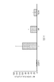


Classifications
-
- C—CHEMISTRY; METALLURGY
- C12—BIOCHEMISTRY; BEER; SPIRITS; WINE; VINEGAR; MICROBIOLOGY; ENZYMOLOGY; MUTATION OR GENETIC ENGINEERING
- C12N—MICROORGANISMS OR ENZYMES; COMPOSITIONS THEREOF; PROPAGATING, PRESERVING, OR MAINTAINING MICROORGANISMS; MUTATION OR GENETIC ENGINEERING; CULTURE MEDIA
- C12N5/00—Undifferentiated human, animal or plant cells, e.g. cell lines; Tissues; Cultivation or maintenance thereof; Culture media therefor
- C12N5/06—Animal cells or tissues; Human cells or tissues
- C12N5/0602—Vertebrate cells
- C12N5/0634—Cells from the blood or the immune system
- C12N5/0636—T lymphocytes
-
- A—HUMAN NECESSITIES
- A61—MEDICAL OR VETERINARY SCIENCE; HYGIENE
- A61K—PREPARATIONS FOR MEDICAL, DENTAL OR TOILETRY PURPOSES
- A61K35/00—Medicinal preparations containing materials or reaction products thereof with undetermined constitution
- A61K35/12—Materials from mammals; Compositions comprising non-specified tissues or cells; Compositions comprising non-embryonic stem cells; Genetically modified cells
- A61K35/14—Blood; Artificial blood
- A61K35/17—Lymphocytes; B-cells; T-cells; Natural killer cells; Interferon-activated or cytokine-activated lymphocytes
-
- A—HUMAN NECESSITIES
- A61—MEDICAL OR VETERINARY SCIENCE; HYGIENE
- A61K—PREPARATIONS FOR MEDICAL, DENTAL OR TOILETRY PURPOSES
- A61K35/00—Medicinal preparations containing materials or reaction products thereof with undetermined constitution
- A61K35/12—Materials from mammals; Compositions comprising non-specified tissues or cells; Compositions comprising non-embryonic stem cells; Genetically modified cells
- A61K35/48—Reproductive organs
- A61K35/54—Ovaries; Ova; Ovules; Embryos; Foetal cells; Germ cells
- A61K35/545—Embryonic stem cells; Pluripotent stem cells; Induced pluripotent stem cells; Uncharacterised stem cells
-
- A—HUMAN NECESSITIES
- A61—MEDICAL OR VETERINARY SCIENCE; HYGIENE
- A61K—PREPARATIONS FOR MEDICAL, DENTAL OR TOILETRY PURPOSES
- A61K39/00—Medicinal preparations containing antigens or antibodies
- A61K39/46—Cellular immunotherapy
- A61K39/461—Cellular immunotherapy characterised by the cell type used
- A61K39/4611—T-cells, e.g. tumor infiltrating lymphocytes [TIL], lymphokine-activated killer cells [LAK] or regulatory T cells [Treg]
-
- A—HUMAN NECESSITIES
- A61—MEDICAL OR VETERINARY SCIENCE; HYGIENE
- A61K—PREPARATIONS FOR MEDICAL, DENTAL OR TOILETRY PURPOSES
- A61K39/00—Medicinal preparations containing antigens or antibodies
- A61K39/46—Cellular immunotherapy
- A61K39/461—Cellular immunotherapy characterised by the cell type used
- A61K39/4613—Natural-killer cells [NK or NK-T]
-
- A—HUMAN NECESSITIES
- A61—MEDICAL OR VETERINARY SCIENCE; HYGIENE
- A61K—PREPARATIONS FOR MEDICAL, DENTAL OR TOILETRY PURPOSES
- A61K39/00—Medicinal preparations containing antigens or antibodies
- A61K39/46—Cellular immunotherapy
- A61K39/463—Cellular immunotherapy characterised by recombinant expression
- A61K39/4631—Chimeric Antigen Receptors [CAR]
-
- A—HUMAN NECESSITIES
- A61—MEDICAL OR VETERINARY SCIENCE; HYGIENE
- A61K—PREPARATIONS FOR MEDICAL, DENTAL OR TOILETRY PURPOSES
- A61K39/00—Medicinal preparations containing antigens or antibodies
- A61K39/46—Cellular immunotherapy
- A61K39/464—Cellular immunotherapy characterised by the antigen targeted or presented
- A61K39/4643—Vertebrate antigens
- A61K39/4644—Cancer antigens
- A61K39/464402—Receptors, cell surface antigens or cell surface determinants
- A61K39/464411—Immunoglobulin superfamily
- A61K39/464412—CD19 or B4
-
- A—HUMAN NECESSITIES
- A61—MEDICAL OR VETERINARY SCIENCE; HYGIENE
- A61P—SPECIFIC THERAPEUTIC ACTIVITY OF CHEMICAL COMPOUNDS OR MEDICINAL PREPARATIONS
- A61P35/00—Antineoplastic agents
-
- C—CHEMISTRY; METALLURGY
- C07—ORGANIC CHEMISTRY
- C07K—PEPTIDES
- C07K14/00—Peptides having more than 20 amino acids; Gastrins; Somatostatins; Melanotropins; Derivatives thereof
- C07K14/435—Peptides having more than 20 amino acids; Gastrins; Somatostatins; Melanotropins; Derivatives thereof from animals; from humans
- C07K14/705—Receptors; Cell surface antigens; Cell surface determinants
- C07K14/70503—Immunoglobulin superfamily
- C07K14/7051—T-cell receptor (TcR)-CD3 complex
-
- C—CHEMISTRY; METALLURGY
- C07—ORGANIC CHEMISTRY
- C07K—PEPTIDES
- C07K16/00—Immunoglobulins [IGs], e.g. monoclonal or polyclonal antibodies
- C07K16/18—Immunoglobulins [IGs], e.g. monoclonal or polyclonal antibodies against material from animals or humans
- C07K16/28—Immunoglobulins [IGs], e.g. monoclonal or polyclonal antibodies against material from animals or humans against receptors, cell surface antigens or cell surface determinants
- C07K16/2803—Immunoglobulins [IGs], e.g. monoclonal or polyclonal antibodies against material from animals or humans against receptors, cell surface antigens or cell surface determinants against the immunoglobulin superfamily
-
- C—CHEMISTRY; METALLURGY
- C12—BIOCHEMISTRY; BEER; SPIRITS; WINE; VINEGAR; MICROBIOLOGY; ENZYMOLOGY; MUTATION OR GENETIC ENGINEERING
- C12N—MICROORGANISMS OR ENZYMES; COMPOSITIONS THEREOF; PROPAGATING, PRESERVING, OR MAINTAINING MICROORGANISMS; MUTATION OR GENETIC ENGINEERING; CULTURE MEDIA
- C12N5/00—Undifferentiated human, animal or plant cells, e.g. cell lines; Tissues; Cultivation or maintenance thereof; Culture media therefor
- C12N5/06—Animal cells or tissues; Human cells or tissues
- C12N5/0602—Vertebrate cells
- C12N5/0634—Cells from the blood or the immune system
- C12N5/0646—Natural killers cells [NK], NKT cells
-
- C—CHEMISTRY; METALLURGY
- C12—BIOCHEMISTRY; BEER; SPIRITS; WINE; VINEGAR; MICROBIOLOGY; ENZYMOLOGY; MUTATION OR GENETIC ENGINEERING
- C12N—MICROORGANISMS OR ENZYMES; COMPOSITIONS THEREOF; PROPAGATING, PRESERVING, OR MAINTAINING MICROORGANISMS; MUTATION OR GENETIC ENGINEERING; CULTURE MEDIA
- C12N5/00—Undifferentiated human, animal or plant cells, e.g. cell lines; Tissues; Cultivation or maintenance thereof; Culture media therefor
- C12N5/06—Animal cells or tissues; Human cells or tissues
- C12N5/0602—Vertebrate cells
- C12N5/0696—Artificially induced pluripotent stem cells, e.g. iPS
-
- A—HUMAN NECESSITIES
- A61—MEDICAL OR VETERINARY SCIENCE; HYGIENE
- A61K—PREPARATIONS FOR MEDICAL, DENTAL OR TOILETRY PURPOSES
- A61K2239/00—Indexing codes associated with cellular immunotherapy of group A61K39/46
- A61K2239/31—Indexing codes associated with cellular immunotherapy of group A61K39/46 characterized by the route of administration
-
- A—HUMAN NECESSITIES
- A61—MEDICAL OR VETERINARY SCIENCE; HYGIENE
- A61K—PREPARATIONS FOR MEDICAL, DENTAL OR TOILETRY PURPOSES
- A61K2239/00—Indexing codes associated with cellular immunotherapy of group A61K39/46
- A61K2239/38—Indexing codes associated with cellular immunotherapy of group A61K39/46 characterised by the dose, timing or administration schedule
-
- A—HUMAN NECESSITIES
- A61—MEDICAL OR VETERINARY SCIENCE; HYGIENE
- A61K—PREPARATIONS FOR MEDICAL, DENTAL OR TOILETRY PURPOSES
- A61K2239/00—Indexing codes associated with cellular immunotherapy of group A61K39/46
- A61K2239/46—Indexing codes associated with cellular immunotherapy of group A61K39/46 characterised by the cancer treated
- A61K2239/48—Blood cells, e.g. leukemia or lymphoma
-
- C—CHEMISTRY; METALLURGY
- C07—ORGANIC CHEMISTRY
- C07K—PEPTIDES
- C07K2317/00—Immunoglobulins specific features
- C07K2317/60—Immunoglobulins specific features characterized by non-natural combinations of immunoglobulin fragments
- C07K2317/62—Immunoglobulins specific features characterized by non-natural combinations of immunoglobulin fragments comprising only variable region components
- C07K2317/622—Single chain antibody (scFv)
-
- C—CHEMISTRY; METALLURGY
- C07—ORGANIC CHEMISTRY
- C07K—PEPTIDES
- C07K2319/00—Fusion polypeptide
- C07K2319/01—Fusion polypeptide containing a localisation/targetting motif
- C07K2319/03—Fusion polypeptide containing a localisation/targetting motif containing a transmembrane segment
-
- C—CHEMISTRY; METALLURGY
- C07—ORGANIC CHEMISTRY
- C07K—PEPTIDES
- C07K2319/00—Fusion polypeptide
- C07K2319/33—Fusion polypeptide fusions for targeting to specific cell types, e.g. tissue specific targeting, targeting of a bacterial subspecies
-
- C—CHEMISTRY; METALLURGY
- C12—BIOCHEMISTRY; BEER; SPIRITS; WINE; VINEGAR; MICROBIOLOGY; ENZYMOLOGY; MUTATION OR GENETIC ENGINEERING
- C12N—MICROORGANISMS OR ENZYMES; COMPOSITIONS THEREOF; PROPAGATING, PRESERVING, OR MAINTAINING MICROORGANISMS; MUTATION OR GENETIC ENGINEERING; CULTURE MEDIA
- C12N2501/00—Active agents used in cell culture processes, e.g. differentation
- C12N2501/01—Modulators of cAMP or cGMP, e.g. non-hydrolysable analogs, phosphodiesterase inhibitors, cholera toxin
-
- C—CHEMISTRY; METALLURGY
- C12—BIOCHEMISTRY; BEER; SPIRITS; WINE; VINEGAR; MICROBIOLOGY; ENZYMOLOGY; MUTATION OR GENETIC ENGINEERING
- C12N—MICROORGANISMS OR ENZYMES; COMPOSITIONS THEREOF; PROPAGATING, PRESERVING, OR MAINTAINING MICROORGANISMS; MUTATION OR GENETIC ENGINEERING; CULTURE MEDIA
- C12N2501/00—Active agents used in cell culture processes, e.g. differentation
- C12N2501/10—Growth factors
- C12N2501/115—Basic fibroblast growth factor (bFGF, FGF-2)
-
- C—CHEMISTRY; METALLURGY
- C12—BIOCHEMISTRY; BEER; SPIRITS; WINE; VINEGAR; MICROBIOLOGY; ENZYMOLOGY; MUTATION OR GENETIC ENGINEERING
- C12N—MICROORGANISMS OR ENZYMES; COMPOSITIONS THEREOF; PROPAGATING, PRESERVING, OR MAINTAINING MICROORGANISMS; MUTATION OR GENETIC ENGINEERING; CULTURE MEDIA
- C12N2501/00—Active agents used in cell culture processes, e.g. differentation
- C12N2501/10—Growth factors
- C12N2501/125—Stem cell factor [SCF], c-kit ligand [KL]
-
- C—CHEMISTRY; METALLURGY
- C12—BIOCHEMISTRY; BEER; SPIRITS; WINE; VINEGAR; MICROBIOLOGY; ENZYMOLOGY; MUTATION OR GENETIC ENGINEERING
- C12N—MICROORGANISMS OR ENZYMES; COMPOSITIONS THEREOF; PROPAGATING, PRESERVING, OR MAINTAINING MICROORGANISMS; MUTATION OR GENETIC ENGINEERING; CULTURE MEDIA
- C12N2501/00—Active agents used in cell culture processes, e.g. differentation
- C12N2501/10—Growth factors
- C12N2501/145—Thrombopoietin [TPO]
-
- C—CHEMISTRY; METALLURGY
- C12—BIOCHEMISTRY; BEER; SPIRITS; WINE; VINEGAR; MICROBIOLOGY; ENZYMOLOGY; MUTATION OR GENETIC ENGINEERING
- C12N—MICROORGANISMS OR ENZYMES; COMPOSITIONS THEREOF; PROPAGATING, PRESERVING, OR MAINTAINING MICROORGANISMS; MUTATION OR GENETIC ENGINEERING; CULTURE MEDIA
- C12N2501/00—Active agents used in cell culture processes, e.g. differentation
- C12N2501/10—Growth factors
- C12N2501/155—Bone morphogenic proteins [BMP]; Osteogenins; Osteogenic factor; Bone inducing factor
-
- C—CHEMISTRY; METALLURGY
- C12—BIOCHEMISTRY; BEER; SPIRITS; WINE; VINEGAR; MICROBIOLOGY; ENZYMOLOGY; MUTATION OR GENETIC ENGINEERING
- C12N—MICROORGANISMS OR ENZYMES; COMPOSITIONS THEREOF; PROPAGATING, PRESERVING, OR MAINTAINING MICROORGANISMS; MUTATION OR GENETIC ENGINEERING; CULTURE MEDIA
- C12N2501/00—Active agents used in cell culture processes, e.g. differentation
- C12N2501/10—Growth factors
- C12N2501/165—Vascular endothelial growth factor [VEGF]
-
- C—CHEMISTRY; METALLURGY
- C12—BIOCHEMISTRY; BEER; SPIRITS; WINE; VINEGAR; MICROBIOLOGY; ENZYMOLOGY; MUTATION OR GENETIC ENGINEERING
- C12N—MICROORGANISMS OR ENZYMES; COMPOSITIONS THEREOF; PROPAGATING, PRESERVING, OR MAINTAINING MICROORGANISMS; MUTATION OR GENETIC ENGINEERING; CULTURE MEDIA
- C12N2501/00—Active agents used in cell culture processes, e.g. differentation
- C12N2501/20—Cytokines; Chemokines
- C12N2501/23—Interleukins [IL]
- C12N2501/2303—Interleukin-3 (IL-3)
-
- C—CHEMISTRY; METALLURGY
- C12—BIOCHEMISTRY; BEER; SPIRITS; WINE; VINEGAR; MICROBIOLOGY; ENZYMOLOGY; MUTATION OR GENETIC ENGINEERING
- C12N—MICROORGANISMS OR ENZYMES; COMPOSITIONS THEREOF; PROPAGATING, PRESERVING, OR MAINTAINING MICROORGANISMS; MUTATION OR GENETIC ENGINEERING; CULTURE MEDIA
- C12N2501/00—Active agents used in cell culture processes, e.g. differentation
- C12N2501/20—Cytokines; Chemokines
- C12N2501/23—Interleukins [IL]
- C12N2501/2306—Interleukin-6 (IL-6)
-
- C—CHEMISTRY; METALLURGY
- C12—BIOCHEMISTRY; BEER; SPIRITS; WINE; VINEGAR; MICROBIOLOGY; ENZYMOLOGY; MUTATION OR GENETIC ENGINEERING
- C12N—MICROORGANISMS OR ENZYMES; COMPOSITIONS THEREOF; PROPAGATING, PRESERVING, OR MAINTAINING MICROORGANISMS; MUTATION OR GENETIC ENGINEERING; CULTURE MEDIA
- C12N2501/00—Active agents used in cell culture processes, e.g. differentation
- C12N2501/20—Cytokines; Chemokines
- C12N2501/23—Interleukins [IL]
- C12N2501/2311—Interleukin-11 (IL-11)
-
- C—CHEMISTRY; METALLURGY
- C12—BIOCHEMISTRY; BEER; SPIRITS; WINE; VINEGAR; MICROBIOLOGY; ENZYMOLOGY; MUTATION OR GENETIC ENGINEERING
- C12N—MICROORGANISMS OR ENZYMES; COMPOSITIONS THEREOF; PROPAGATING, PRESERVING, OR MAINTAINING MICROORGANISMS; MUTATION OR GENETIC ENGINEERING; CULTURE MEDIA
- C12N2501/00—Active agents used in cell culture processes, e.g. differentation
- C12N2501/20—Cytokines; Chemokines
- C12N2501/26—Flt-3 ligand (CD135L, flk-2 ligand)
-
- C—CHEMISTRY; METALLURGY
- C12—BIOCHEMISTRY; BEER; SPIRITS; WINE; VINEGAR; MICROBIOLOGY; ENZYMOLOGY; MUTATION OR GENETIC ENGINEERING
- C12N—MICROORGANISMS OR ENZYMES; COMPOSITIONS THEREOF; PROPAGATING, PRESERVING, OR MAINTAINING MICROORGANISMS; MUTATION OR GENETIC ENGINEERING; CULTURE MEDIA
- C12N2501/00—Active agents used in cell culture processes, e.g. differentation
- C12N2501/70—Enzymes
- C12N2501/72—Transferases (EC 2.)
- C12N2501/727—Kinases (EC 2.7.)
-
- C—CHEMISTRY; METALLURGY
- C12—BIOCHEMISTRY; BEER; SPIRITS; WINE; VINEGAR; MICROBIOLOGY; ENZYMOLOGY; MUTATION OR GENETIC ENGINEERING
- C12N—MICROORGANISMS OR ENZYMES; COMPOSITIONS THEREOF; PROPAGATING, PRESERVING, OR MAINTAINING MICROORGANISMS; MUTATION OR GENETIC ENGINEERING; CULTURE MEDIA
- C12N2501/00—Active agents used in cell culture processes, e.g. differentation
- C12N2501/90—Polysaccharides
- C12N2501/91—Heparin
-
- C—CHEMISTRY; METALLURGY
- C12—BIOCHEMISTRY; BEER; SPIRITS; WINE; VINEGAR; MICROBIOLOGY; ENZYMOLOGY; MUTATION OR GENETIC ENGINEERING
- C12N—MICROORGANISMS OR ENZYMES; COMPOSITIONS THEREOF; PROPAGATING, PRESERVING, OR MAINTAINING MICROORGANISMS; MUTATION OR GENETIC ENGINEERING; CULTURE MEDIA
- C12N2506/00—Differentiation of animal cells from one lineage to another; Differentiation of pluripotent cells
- C12N2506/11—Differentiation of animal cells from one lineage to another; Differentiation of pluripotent cells from blood or immune system cells
-
- C—CHEMISTRY; METALLURGY
- C12—BIOCHEMISTRY; BEER; SPIRITS; WINE; VINEGAR; MICROBIOLOGY; ENZYMOLOGY; MUTATION OR GENETIC ENGINEERING
- C12N—MICROORGANISMS OR ENZYMES; COMPOSITIONS THEREOF; PROPAGATING, PRESERVING, OR MAINTAINING MICROORGANISMS; MUTATION OR GENETIC ENGINEERING; CULTURE MEDIA
- C12N2510/00—Genetically modified cells
Description
本願は、2017年4月18日に出願された米国仮特許出願第62/486,875号明細書の優先権の利益を主張し、その内容全体はここで参照により援用される。 This application claims the priority benefit of US Provisional Patent Application No. 62/486,875, filed April 18, 2017, the entire contents of which are hereby incorporated by reference.
1.分野
本発明は、一般に分子生物学の分野に関する。より詳細には、それは、抗原特異的免疫エフェクター細胞、例えばT細胞及びNK細胞に関する方法及び組成物に関する。
1. FIELD The present invention relates generally to the field of molecular biology. More particularly, it relates to methods and compositions relating to antigen-specific immune effector cells, such as T cells and NK cells.
2.関連技術の説明
がんと診断された患者にとって利用可能な診断及び治療オプションにおける技術的進歩にもかかわらず、依然として予後不良のままであることが多く、多くの患者が治癒不能である。免疫療法は、正常組織を損傷させることなく悪性腫瘍細胞を根絶する可能性とともに、様々な腫瘍を有すると診断された患者に対して強力であるが標的化された治療をもたらす可能性を秘めている。理論上、免疫系のT細胞は、腫瘍細胞に特異的なタンパク質パターンを認識する能力があり、種々のエフェクター機構を通じてのそれらの破壊を媒介することになる。養子T細胞療法は、患者自身のT細胞の腫瘍根絶能力を利用・増幅し、次にこれらのエフェクターを、それらが残留腫瘍を有効に除去しても健常組織を損傷しないような状態で患者に戻すという試みである。この手法は腫瘍免疫学の分野にとって新規性はないが、養子T細胞療法の臨床使用における多くの欠点により、がん治療におけるこの手法の十分な利用が損なわれる。
2. Description of the Related Art Despite technological advances in diagnostic and treatment options available for patients diagnosed with cancer, the prognosis remains often poor and many patients are incurable. Immunotherapy has the potential to eradicate malignant tumor cells without damaging normal tissue, and offers the potential to provide potent yet targeted therapy for patients diagnosed with a variety of tumors. there is In theory, T cells of the immune system are capable of recognizing protein patterns specific to tumor cells and mediate their destruction through various effector mechanisms. Adoptive T-cell therapy harnesses and amplifies the tumor eradicating capacity of the patient's own T cells and then introduces these effectors into the patient in such a way that they effectively clear residual tumor but do not damage healthy tissue. An attempt to get it back. Although this approach is not new to the field of tumor immunology, many shortcomings in the clinical use of adoptive T cell therapy impair the full utilization of this approach in cancer therapy.
現行の養子T細胞療法は、患者や腫瘍特異的T細胞の不足、例えば、体内にそれらが希少であること、それらがいくつかの腫瘍免疫系の回避機構を克服できないこと、及びそれらの寿命が短いことにより、制限を受ける。所望される抗原に対して反応性を示す、典型的には少数のT細胞を単離及び拡大することは困難である。したがって、免疫療法における有効利用を意図した治療的に十分且つ機能的な抗原特異的免疫細胞に対する需要は満たされていない。 Current adoptive T-cell therapy suffers from a shortage of patient- and tumor-specific T cells, such as their rarity in the body, their inability to overcome several tumor immune system evasion mechanisms, and their longevity. Limited by being short. It is difficult to isolate and expand the typically small number of T cells that are reactive to the desired antigen. Thus, there is an unmet need for therapeutically sufficient and functional antigen-specific immune cells intended for effective use in immunotherapy.
第1の実施形態では、本開示は、抗原特異的エフェクターT細胞及び/又はNK細胞を生成する方法であって、多能性幹細胞(PSC)を改変し、キメラ抗原受容体(CAR)を発現し、それによりCAR-PSCを生成することと;CAR-PSCをCD34+造血前駆細胞(HPC)に分化させるか又はフォワードにリプログラミングすることと;CD34+HPCをT細胞及び/又はNK細胞にさらに分化させることと;T細胞及び/又はNK細胞を拡大させることと、を含む、方法を提供する。いくつかの態様では、拡大させることは、抗原特異的標的細胞と共培養し、それにより抗原特異的エフェクターT細胞及び/又はNK細胞を生成することを含む。 In a first embodiment, the present disclosure provides a method of generating antigen-specific effector T cells and/or NK cells comprising modifying pluripotent stem cells (PSCs) to express chimeric antigen receptors (CARs) differentiating or forward reprogramming the CAR-PSCs into CD34 + hematopoietic progenitor cells (HPCs); and converting the CD34 + HPCs into T cells and/or NK cells. further differentiating; and expanding the T cells and/or NK cells. In some aspects, expanding comprises co-culturing with antigen-specific target cells, thereby generating antigen-specific effector T cells and/or NK cells.
特定の態様では、CARを発現するように改変されたPSCは、人工多能性幹細胞(iPSC)又は胚性幹細胞(ESC)である。特定の態様では、iPSCは、体細胞、例えばT細胞からリプログラミングされる。 In certain aspects, the PSCs modified to express CAR are induced pluripotent stem cells (iPSCs) or embryonic stem cells (ESCs). In certain aspects, iPSCs are reprogrammed from somatic cells, such as T cells.
いくつかの態様では、CAR-PSCをCD34+HPCに分化させるステップは、指向された分化誘導(directed differentiation)を実施することを含む。特定の態様では、指向された分化誘導は、胚様体(EB)をブレビスタチン、GSK-3阻害剤、FGF2、及びVEGFの存在下で生成すること;中胚葉誘導を誘導するため、EBをBMP4、VEGF、及びFGF2と接触させることと;EBをFlt-3リガンド、IL-3、SCF、及びTPOの存在下で分化させ、それによりHPCを生成することと、を含む。いくつかの態様では、分化は、BMP4が本質的に含まないか又は含まない培地中でなされる。他の態様では、分化は、BMP4の存在下でなされる。特定の態様では、GSK-3阻害剤は、CHIR99021である。いくつかの態様では、分化は、IL-11、cAMP、及び/又はVEGFの存在をさらに含む。 In some aspects, differentiating the CAR-PSCs into CD34 + HPCs comprises performing directed differentiation. In certain aspects, the directed induction of differentiation includes generating embryoid bodies (EBs) in the presence of blebbistatin, a GSK-3 inhibitor, FGF2, and VEGF; contacting with BMP4, VEGF, and FGF2; and differentiating EBs in the presence of Flt-3 ligand, IL-3, SCF, and TPO, thereby generating HPCs. In some aspects, differentiation occurs in a medium that is essentially free or free of BMP4. In another aspect, the differentiation is in the presence of BMP4. In a particular aspect, the GSK-3 inhibitor is CHIR99021. In some aspects, differentiation further comprises the presence of IL-11, cAMP, and/or VEGF.
いくつかの態様では、指向された分化誘導は、ブレビスタチン、BMP4、VEGF、及びbFGFの存在下で、アミンでコーティングされた表面上で個別化されたPSCを培養することと;PSCをBMP4、VEGF、及びFGF2と接触させることにより分化を開始させることと;PSCをFlt-3リガンド、IL-3、IL-6、SCF、TPO、及びヘパリンの存在下でさらに分化させ、それによりHPCを生成することと、を含み、方法はEBの形成を含まない。 In some aspects, directed differentiation is culturing the individualized PSCs on an amine-coated surface in the presence of blebbistatin, BMP4, VEGF, and bFGF; initiating differentiation by contact with VEGF and FGF2; and further differentiating PSCs in the presence of Flt-3 ligand, IL-3, IL-6, SCF, TPO, and heparin, thereby generating HPCs. and and the method does not include formation of EBs.
特定の態様では、方法は、方法全体の持続時間など、限定されたフィーダーフリー条件下で該細胞を培養することを含む。いくつかの態様では、PSCは、本質的には導入遺伝子を含まないか又は導入遺伝子を含まない。特定の態様では、PSCは、ヒトである。特定の態様では、T細胞は、CD4+T細胞、CD8+T細胞、細胞傷害性T細胞、制御性T細胞、ナチュラルキラーT細胞、ナイーブT細胞、メモリーT細胞、及び/又はγδT細胞である。 In certain aspects, the method includes culturing the cells under limited feeder-free conditions, such as the duration of the entire method. In some aspects, the PSCs are essentially transgene-free or transgene-free. In certain aspects, the PSC is human. In particular aspects, the T cells are CD4 + T cells, CD8 + T cells, cytotoxic T cells, regulatory T cells, natural killer T cells, naive T cells, memory T cells, and/or γδ T cells. .
特定の態様では、CARを発現するように改変されたPSCは、ERG/ETV2、GATA2、及びHOXA9を、例えば単一の誘導性プロモーターの制御下で発現するようにさらに改変される。いくつかの態様では、方法は、HMGA2、MYCN、NR4A2、SOX17、TFEC、MEIS1、及び/又はHOXA4を発現するようにPSCを改変することをさらに含む。したがって、いくつかの態様では、CAR-PSCをCD34+HPCに分化させるステップは、ERG/ETV2、GATA2、及びHOXA9の発現を、HPCを生成するのに十分な期間にわたり誘導することと、HPCをT細胞及び/又はNK細胞にさらに分化させる前に発現の誘導を終了させることと、を含む。 In certain aspects, PSCs modified to express CAR are further modified to express ERG/ETV2, GATA2, and HOXA9, eg, under the control of a single inducible promoter. In some aspects, the method further comprises modifying the PSC to express HMGA2, MYCN, NR4A2, SOX17, TFEC, MEIS1, and/or HOXA4. Thus, in some aspects, differentiating CAR-PSCs into CD34 + HPCs comprises inducing expression of ERG/ETV2, GATA2, and HOXA9 for a period of time sufficient to generate HPCs; terminating the induction of expression prior to further differentiation into T cells and/or NK cells.
具体的な態様では、CAR-PSCをCD34+HPCに分化させるステップは、抗原特異的T細胞及び/又はNK細胞に分化させる前に、CD34及び/又はCD43を発現する細胞について選択することをさらに含む。特定の態様では、選択することは、磁気活性化細胞選別(MACS)を実施することを含む。いくつかの態様では、CD34及び/又はCD43を発現する細胞は、全細胞集団の少なくとも25、30、35、40、45、50、60、65、70、75、80、85、90、又はより高いパーセントを含む。いくつかの態様では、CD34及び/又はCD43陽性細胞について選択するためにMACsを実施するステップは実施されない。 In a specific aspect, differentiating CAR-PSCs into CD34 + HPCs further comprises selecting for cells expressing CD34 and/or CD43 prior to differentiating into antigen-specific T cells and/or NK cells. include. In certain aspects, selecting comprises performing magnetic activated cell sorting (MACS). In some aspects, the cells expressing CD34 and/or CD43 are at least 25, 30, 35, 40, 45, 50, 60, 65, 70, 75, 80, 85, 90, or more of the total cell population. Contains a high percentage. In some aspects, performing MACs to select for CD34 and/or CD43 positive cells is not performed.
特定の態様では、少なくとも2、3、4、5、10、15、20、又はより高いパーセントのHPCが、CARを発現する。いくつかの態様では、少なくとも3、4、5、6、7、8、9、10、11、12、13、14、15、20、25、30、35、40、45又はより高いパーセントのHPCが、CARを発現する。 In certain aspects, at least 2, 3, 4, 5, 10, 15, 20, or a higher percentage of HPCs express CAR. In some aspects, at least 3, 4, 5, 6, 7, 8, 9, 10, 11, 12, 13, 14, 15, 20, 25, 30, 35, 40, 45 or higher percent HPC express CAR.
いくつかの態様では、HPCをT細胞及び/又はNK細胞に分化させることは、低酸素条件下、アスコルビン酸及びニコチンアミドの存在下で、HPCをレトロネクチン及びノッチDLL-4でコーティングされた表面上で培養することを含む。いくつかの態様では、培養物は、SCF、FLT-3リガンド、TPO、及びIL-7をさらに含み、また任意選択的には、GSK阻害剤(例えばCHIR99021)、IL-2、及び/又はIL-12を含む。特定の態様では、拡大するステップは、抗原特異的T細胞を抗CD3抗体及びIL-2の存在下で培養することをさらに含む。さらなる態様では、拡大するステップは、抗原特異的T細胞を抗CD3抗体、IL-2、IL-15、及びIL-21の存在下で培養することをさらに含む。いくつかの態様では、拡大するステップは、抗原特異的T細胞を、抗CD3抗体、FLT3リガンド、IL-7、IL-2、及び/又はIL-15の存在下で培養することを含み、また任意選択的にはSCF、TPO、及び/又はIL-21をさらに含む。特定の態様では、分化されたCD34+HPCの少なくとも1、1.5、2、2.5、3、4、5、6、7、8、9、10、15、20、25、30、35、40、45、50又はより高いパーセントが、CD3+CAR+T細胞である。いくつかの態様では、分化されたCD34+HPCの少なくとも1、2、3、4、5、6、7、8、9、10、15、20、25、30、35、40、50、又はより高いパーセントが、CD3-CAR+NK細胞である。いくつかの態様では、拡大されたCD34+HPCの少なくとも2パーセントが、CD3+CAR+T細胞である。特定の態様では、拡大されたCD34+HPCの少なくとも10パーセントが、CD3-CAR+NK細胞である。 In some aspects, differentiating HPCs into T cells and/or NK cells includes exposing HPCs to retronectin and Notch DLL-4 coated surfaces in the presence of ascorbic acid and nicotinamide under hypoxic conditions. including culturing in In some aspects, the culture further comprises SCF, FLT-3 ligand, TPO, and IL-7, and optionally a GSK inhibitor (eg, CHIR99021), IL-2, and/or IL-7. -12 included. In certain aspects, the expanding step further comprises culturing the antigen-specific T cells in the presence of anti-CD3 antibody and IL-2. In a further aspect, the expanding step further comprises culturing the antigen-specific T cells in the presence of anti-CD3 antibody, IL-2, IL-15 and IL-21. In some aspects, the expanding step comprises culturing the antigen-specific T cells in the presence of an anti-CD3 antibody, FLT3 ligand, IL-7, IL-2, and/or IL-15, and Optionally further comprising SCF, TPO and/or IL-21. In particular aspects, at least 1, 1.5, 2, 2.5, 3, 4, 5, 6, 7, 8, 9, 10, 15, 20, 25, 30, 35 of the differentiated CD34 + HPC , 40, 45, 50 or higher percent are CD3 + CAR + T cells. In some aspects, at least 1, 2, 3, 4, 5, 6, 7, 8, 9, 10, 15, 20, 25, 30, 35, 40, 50, or more of the differentiated CD34 + HPCs A high percentage are CD3-CAR+ NK cells. In some aspects, at least 2 percent of the expanded CD34 + HPCs are CD3 + CAR + T cells. In certain aspects, at least 10 percent of the expanded CD34 + HPCs are CD3 − CAR + NK cells.
特定の態様では、CAR及び抗原特異的標的細胞は、同じ抗原に特異的である。ある具体的な態様では、抗原はCD19である。特定の態様では、抗原特異的標的細胞は、腫瘍細胞である。いくつかの態様では、抗原特異的標的細胞は、ヒトである。特定の態様では、抗原特異的標的細胞は、HLAクラスI陰性である。いくつかの態様では、抗原特異的エフェクターT細胞の少なくとも5、例えば、6、7、8、9、10、15、20、25、30、35、40、50、又はより高いパーセントが、標的細胞に対する細胞傷害性活性を呈する。特定の態様では、抗原特異的エフェクターNK細胞の少なくとも10、例えば、15、20、25、30、35、40、45、50、又はより高いパーセントが、標的細胞に対する細胞傷害性活性を呈する。 In certain aspects, the CAR and the antigen-specific target cell are specific for the same antigen. In one specific aspect, the antigen is CD19. In certain aspects, the antigen-specific target cells are tumor cells. In some aspects, the antigen-specific target cell is human. In certain aspects, the antigen-specific target cells are HLA class I negative. In some aspects, at least 5, e.g., 6, 7, 8, 9, 10, 15, 20, 25, 30, 35, 40, 50, or a higher percentage of antigen-specific effector T cells are target cells exhibits cytotoxic activity against In certain aspects, at least 10, eg, 15, 20, 25, 30, 35, 40, 45, 50, or higher percentages of antigen-specific effector NK cells exhibit cytotoxic activity against target cells.
いくつかの態様では、CARは、PSCのゲノムに組み込まれたDNAによってコードされる。特定の態様では、CARは、細胞内シグナル伝達ドメイン、膜貫通ドメイン、及び抗原結合領域を含む細胞外ドメインを含む。具体的な態様では、細胞内シグナル伝達ドメインは、CD3ζ及びCD28を含む。いくつかの態様では、抗原結合領域は、F(ab’)2、Fab’、Fab、Fv、又はscFvである。 In some aspects, the CAR is encoded by DNA integrated into the genome of the PSC. In certain aspects, the CAR comprises an extracellular domain comprising an intracellular signaling domain, a transmembrane domain, and an antigen binding region. In a specific aspect, the intracellular signaling domain comprises CD3ζ and CD28. In some aspects, the antigen binding region is F(ab')2, Fab', Fab, Fv, or scFv.
特定の態様では、PSCは、HLAホモ接合性である。いくつかの態様では、HLAホモ接合性PSCは、対立遺伝子HLA-A、HLA-B、HLA-C、HLA-DR、HLA-DP又はHLA-DQ遺伝子座の1つ以上においてホモ接合性である。特定の態様では、HLAホモ接合性PSCは、対立遺伝子HLA-A、HLA-B、HLA-C、HLA-DR、HLA-DP又はHLA-DQ遺伝子座の2つにおいてホモ接合性である。特定の態様では、HLAホモ接合性PSCは、HLA-A及びHLA-Bにおいてホモ接合性である。特定の態様では、HLAホモ接合性PSCは、HLA-A、HLA-B、及びHLA-Cにおいてホモ接合性である。 In certain aspects, the PSC is HLA homozygous. In some aspects, the HLA homozygous PSC is homozygous at one or more of the allelic HLA-A, HLA-B, HLA-C, HLA-DR, HLA-DP or HLA-DQ loci . In certain aspects, an HLA homozygous PSC is homozygous at two of the allelic HLA-A, HLA-B, HLA-C, HLA-DR, HLA-DP or HLA-DQ loci. In certain aspects, HLA homozygous PSCs are homozygous for HLA-A and HLA-B. In certain aspects, HLA homozygous PSCs are homozygous in HLA-A, HLA-B, and HLA-C.
別の実施形態は、抗原特異的エフェクターT細胞及び/又はNK細胞を生成する方法であって、CARを発現するようにPSCを改変し、それによりCAR-PSCを生成することと;CAR-PSCをブレビスタチン、GSK-3阻害剤、FGF2、及びVEGFの存在下で培養し、それによりEBを生成することと;中胚葉誘導を誘導するため、EBをBMP4、VEGF、及びFGF2と接触させることと;EBをFlt-3リガンド、BMP4、IL-3、SCF、及びTPOの存在下で分化させ、それによりCD34+HPCを生成することと;CD34+HPCをT細胞及び/又はNK細胞までさらに分化させることと;T細胞及び/又はNK細胞を拡大させることであって、拡大させることが、抗原特異的標的細胞と共培養し、それにより抗原特異的エフェクターT細胞及び/又はNK細胞を生成することを含む、ことと、を含む、方法を提供する。特定の態様では、GSK-3阻害剤は、CHIR99021である。 Another embodiment is a method of generating antigen-specific effector T cells and/or NK cells, comprising modifying a PSC to express CAR, thereby generating a CAR-PSC; in the presence of blebbistatin, a GSK-3 inhibitor, FGF2, and VEGF, thereby generating EBs; and contacting the EBs with BMP4, VEGF, and FGF2 to induce mesoderm induction. and; differentiating EBs in the presence of Flt-3 ligand, BMP4, IL-3, SCF, and TPO, thereby generating CD34 + HPC; and further converting CD34 + HPC to T cells and/or NK cells. differentiating; expanding T cells and/or NK cells, the expanding being co-cultured with antigen-specific target cells, thereby generating antigen-specific effector T cells and/or NK cells A method is provided comprising: In a particular aspect, the GSK-3 inhibitor is CHIR99021.
さらに別の実施形態では、抗原特異的エフェクターT細胞及び/又はNK細胞を生成する方法であって、CARを発現するようにPSCを改変し、それによりCAR-PSCを生成することと;ブレビスタチン、BMP4、VEGFの存在下で、アミンでコーティングされた表面上で個別化されたCAR-PSCを培養し、且つCAR-PSCをBMP4、VEGF、及びFGF2と接触させることにより分化を開始させることと;CAR-PSCをFlt-3リガンド、IL-3、IL-6、SCF、TPO、及びヘパリンの存在下でさらに分化させ、それによりCD34+HPCを生成することと;CD34+HPCをT細胞及び/又はNK細胞まで分化させることと;T細胞及び/又はNK細胞を拡大させることであって、拡大させることが、抗原特異的標的細胞と共培養し、それにより抗原特異的エフェクターT細胞及び/又はNK細胞を生成することを含む、ことと、を含む、方法が提供され、方法はEBの形成を含まない。 In yet another embodiment, a method of generating antigen-specific effector T cells and/or NK cells comprising: modifying PSCs to express CAR, thereby generating CAR-PSCs; culturing individualized CAR-PSCs on an amine-coated surface in the presence of BMP4, VEGF, and initiating differentiation by contacting the CAR-PSCs with BMP4, VEGF, and FGF2; further differentiating CAR-PSCs in the presence of Flt-3 ligand, IL-3, IL-6, SCF, TPO, and heparin, thereby generating CD34 + HPCs; and converting CD34 + HPCs into T cells and / or differentiating to NK cells; expanding the T cells and / or NK cells, the expansion being co-cultured with antigen-specific target cells, thereby generating antigen-specific effector T cells and / or or generating NK cells, wherein the method does not involve formation of EBs.
さらなる実施形態は、抗原特異的エフェクターT細胞及び/又はNK細胞を生成する方法であって、CARを発現するようにPSCを改変し、それによりCAR-PSCを生成することと;CAR-PSCをCD34+HPCに分化させることと;CD34+CD43+HPCについて選択することと;CD34+CD43+HPCをT細胞及び/又はNK細胞までさらに分化させることと;T細胞及び/又はNK細胞を拡大させることであって、拡大させることが、抗原特異的標的細胞と共培養し、それにより抗原特異的エフェクターT細胞及び/又はNK細胞を生成することを含む、ことと、を含む、方法を提供する。 A further embodiment is a method of generating antigen-specific effector T cells and/or NK cells, comprising modifying PSCs to express CAR, thereby generating CAR-PSCs; differentiating into CD34 + HPCs; selecting for CD34 + CD43 + HPCs; further differentiating CD34 + CD43 + HPCs into T cells and/or NK cells; expanding T cells and/or NK cells. wherein the expanding comprises co-culturing with antigen-specific target cells, thereby generating antigen-specific effector T cells and/or NK cells. .
別の実施形態では、上記の実施形態に従って生成される抗原特異的エフェクターT細胞及び/又はNK細胞の集団が提供される。本明細書に記載の実施形態に従って生成される抗原特異的エフェクターT細胞及び/又はNK細胞を含む医薬組成物もまた本明細書に提供される。さらに、対象におけるがんの治療のための実施形態に従って生成される抗原特異的エフェクターT細胞及び/又はNK細胞を含む組成物が本明細書に提供される。がんの治療のための実施形態に従って生成される抗原特異的エフェクターT細胞及び/又はNK細胞の使用についても本明細書に提供される。 In another embodiment, there is provided a population of antigen-specific effector T cells and/or NK cells generated according to the above embodiments. Also provided herein are pharmaceutical compositions comprising antigen-specific effector T cells and/or NK cells generated according to embodiments described herein. Further provided herein are compositions comprising antigen-specific effector T cells and/or NK cells generated according to embodiments for the treatment of cancer in a subject. Also provided herein is the use of antigen-specific effector T cells and/or NK cells generated according to embodiments for the treatment of cancer.
さらに別の実施形態では、対象におけるがんを治療する方法であって、本明細書に記載の実施形態に従って生成される抗原特異的エフェクターT細胞及び/又はNK細胞を有効量で対象に投与することを含む、方法が提供される。いくつかの態様では、がんは、腫瘍抗原を発現し、抗原特異的エフェクターT細胞及び/又はNK細胞は、前記腫瘍抗原に特異的である。特定の態様では、対象はヒトである。 In yet another embodiment, a method of treating cancer in a subject, comprising administering to the subject an effective amount of antigen-specific effector T cells and/or NK cells generated according to embodiments described herein A method is provided comprising: In some aspects, the cancer expresses a tumor antigen and the antigen-specific effector T cells and/or NK cells are specific for said tumor antigen. In certain embodiments, the subject is human.
本発明の他の目的、特徴及び利点は、以下の詳細な説明から明白となる。しかし、詳細な説明及び具体例が、本発明の好ましい実施形態を示す一方で、あくまで例示として示されることは理解されるべきである、というのは、本発明の精神及び範囲の範囲内での様々な変更及び修飾がこの詳細な説明から当業者にとって明白となるからである。 Other objects, features and advantages of the present invention will become apparent from the detailed description below. It is to be understood, however, that the detailed description and specific examples, while indicating preferred embodiments of the invention, are given by way of illustration only, without departing from the spirit and scope of the invention. Various changes and modifications will become apparent to those skilled in the art from this detailed description.
以下の図面は、本明細書の一部を形成し、本発明の特定の態様をさらに明らかにするように含められる。本発明は、これらの図面の1つ以上を、本明細書に提示される具体的な実施形態の詳細な説明と組み合わせて参照することにより、より十分に理解されることがある。 The following drawings form part of this specification and are included to further clarify certain aspects of the present invention. The invention may be more fully understood by reference to one or more of these drawings in conjunction with the detailed description of specific embodiments presented herein.
特定の実施形態では、本開示は、抗原受容体、例えば本明細書中でCAR-PSCと称されるキメラ抗原受容体(CAR)を発現するように改変されているヒト多能性幹細胞(PSC)から抗原特異的免疫エフェクター細胞を生成するための高度に効率的な方法を提供する。現行の方法によって生成される免疫エフェクター細胞が、限定はされないが、T細胞、NK細胞、及びiNKT細胞を含みうる。 In certain embodiments, the present disclosure provides human pluripotent stem cells (PSCs) that have been modified to express an antigen receptor, such as a chimeric antigen receptor (CAR), referred to herein as CAR-PSC. ) to generate antigen-specific immune effector cells. Immune effector cells generated by current methods can include, but are not limited to, T cells, NK cells, and iNKT cells.
PSCは、T細胞の開始集団を(例えば、レトロウイルス又はエピソーム法により)リプログラミングし、T細胞由来のiPSC(TiPSC)を生成することにより得てもよい。T細胞は、様々な供給源、例えば血液サンプルから単離されてもよい。T細胞の開始集団は、それらの特徴的なT細胞受容体(TCR)遺伝子再構成を保持してもよく、HLAホモ接合性細胞(即ちMHCクラスI及びII遺伝子においてホモ接合性である)であってもよい。したがって、iPSCは、本明細書中でHLAスーパードナーと称されるHLAホモ接合性対象から単離された細胞から生成されうる。 PSCs may be obtained by reprogramming a starting population of T cells (eg, by retroviral or episomal methods) to generate T cell-derived iPSCs (TiPSCs). T cells may be isolated from a variety of sources, such as blood samples. The starting population of T cells may retain their characteristic T cell receptor (TCR) gene rearrangement and are HLA homozygous cells (i.e. homozygous in MHC class I and II genes). There may be. Thus, iPSCs can be generated from cells isolated from HLA homozygous subjects, referred to herein as HLA superdonors.
次に、CAR-PSCは、CD34+造血前駆細胞(HPC)を生成するように分化又はプログラムされてもよい。これは、サイトカイン(例えば、SCF、TPO、FLT-3、IL-6、IL-3、及びヘパリン)の組み合わせを用いた指向された分化誘導を通じて達成されてもよい(例えば、国際出願PCT/US2016/057899号明細書中に記載、その全体が参照により本明細書中に援用される)。代替的方法では、CAR-PSCは、ETV2/ERG、GATA2、及びHOXA9(即ちEGH)をコードする発現構築物によるフォワードプログラミングを用いて、CD34+HPCに分化されてもよい(例えば、国際出願PCT/US2016/057893号明細書中に記載、その全体が参照により本明細書中に援用される)。これらのEGH-PSCは、長期生着能(engraftability)のため、HMGA2、MYCN、NR4A2、SOX17、TFEC、MEIS1、及び/又はHOXA4を発現するようにさらに改変されてもよい。 CAR-PSCs may then be differentiated or programmed to generate CD34 + hematopoietic progenitor cells (HPCs). This may be achieved through directed differentiation induction using a combination of cytokines (e.g. SCF, TPO, FLT-3, IL-6, IL-3, and heparin) (e.g. International Application PCT/US2016 057899, incorporated herein by reference in its entirety). In an alternative method, CAR-PSCs may be differentiated into CD34 + HPCs using forward programming with expression constructs encoding ETV2/ERG, GATA2, and HOXA9 (ie, EGH) (see, eg, International Application PCT/ US 2016/057893, incorporated herein by reference in its entirety). These EGH-PSCs may be further modified to express HMGA2, MYCN, NR4A2, SOX17, TFEC, MEIS1, and/or HOXA4 for long-term engraftability.
最終的には、CD34+CAR-HPCは、CD3+T細胞又はCD56+CD3-NK細胞に分化されてもよい。リンパ系能力についてHPC分化中(例えば7~11日)の最適な時間枠は、CD34及びCD43の発現によって同定された7~11日であってもよい。例えば、増強されたリンパ系能力を有するHPCは、マーカーCD144、CD34、CD45、及びCD7の2つ以上について陽性である細胞の画分を選別することにより単離されてもよい。 Ultimately, CD34 + CAR-HPCs may be differentiated into CD3 + T cells or CD56 + CD3 − NK cells. An optimal time frame during HPC differentiation (eg, days 7-11) for lymphoid potential may be days 7-11 identified by the expression of CD34 and CD43. For example, HPCs with enhanced lymphoid potential may be isolated by sorting a fraction of cells that are positive for two or more of the markers CD144, CD34, CD45, and CD7.
T細胞分化のための例示的方法は、レトロネクチン及びDLL-4のフィーダーフリーマトリックスとしての使用を含む。T細胞分化は、効率及び成熟を増強するためのアスコルビン酸の使用により、並びに低酸素条件下で培養することにより、さらに増強されてもよい。 An exemplary method for T cell differentiation involves the use of retronectin and DLL-4 as feeder-free matrices. T cell differentiation may be further enhanced through the use of ascorbic acid to enhance efficiency and maturation, and by culturing under hypoxic conditions.
さらに、T細胞及び/又はNK細胞は、分化プロセス中に抗原特異的標的細胞(例えば腫瘍細胞)と共培養することにより増強されてもよい。この方法は、標的細胞に対するT細胞及びNK細胞の細胞傷害性活性を増強することが見出され、それは具体的には、腫瘍増殖における減少及び腫瘍細胞が注射されたマウスの生存における増加により認められた。 Additionally, T cells and/or NK cells may be enhanced by co-culturing with antigen-specific target cells (eg, tumor cells) during the differentiation process. This method was found to enhance the cytotoxic activity of T cells and NK cells against target cells, specifically by a decrease in tumor growth and an increase in survival of mice injected with tumor cells. was taken.
したがって、本開示の方法は、インビボでの安定な移植、インビトロでの化合物のスクリーニング、及び血液学的疾患及び損傷の機構の解明などの広範囲の用途に、無数の抗原特異的免疫エフェクター細胞、例えばT細胞及びNK細胞を提供することができる。 Thus, the methods of the present disclosure are useful for a wide range of applications, such as in vivo stable transplantation, in vitro compound screening, and elucidation of mechanisms of hematologic disease and injury, including myriad antigen-specific immune effector cells, e.g. T cells and NK cells can be provided.
I.定義
本明細書で用いられるとき、「本質的に含まない」は、特定成分の観点で、組成物に意図的に配合されている、且つ/又はあくまで汚染物質として又は微量で存在するような特定成分がないことを意味するように本明細書で用いられる。したがって、組成物の任意の意図しない汚染に起因する特定成分の総量は、0.05%を十分に下回り、好ましくは0.01%未満である。標準的分析法を用いて特定成分が全く検出できないような組成物が最も好ましい。
I. DEFINITIONS As used herein, "essentially free", in terms of specified ingredients, is intentionally included in the composition and/or is present only as a contaminant or in trace amounts. Used herein to mean no ingredients. Therefore, the total amount of specified ingredients resulting from any unintentional contamination of the composition is well below 0.05%, preferably below 0.01%. Compositions in which no specific component is detectable using standard analytical methods are most preferred.
本明細書で用いられるとき、「a」又は「an」は、1以上を意味してもよい。特許請求の範囲で用いられ、用語「含む(comprising)」と併用されるとき、用語「a」又は「an」は、1又は2以上を意味してもよい。 As used herein, "a" or "an" may mean one or more. When used in the claims and in conjunction with the term "comprising," the term "a" or "an" may mean one or more.
特許請求の範囲の中での用語「又は」の使用は、あくまで代替物を指すことが明示されない限り「及び/又は」を意味するように用いられる、或いはその代替物は、相互に排他的であるが、本開示は、あくまで代替物と「及び/又は」を指すような定義を支持する。本明細書で用いられるとき、「別の」は、少なくとも第2又はそれ以上を意味してもよい。 The use of the term "or" in the claims is used to mean "and/or" unless explicitly indicated to the contrary, or the alternatives are mutually exclusive. However, this disclosure supports definitions that refer only to alternatives and "and/or." As used herein, "another" may mean at least a second or more.
本願全体を通じて、用語「約」は、ある値が、その値を決定するために利用されているデバイス、方法における固有の誤差の変動、又は試験対象の中に存在する変動を含むことを示すように用いられる。 Throughout this application, the term "about" is used to indicate that a value includes variations in error inherent in the device, method, or test subject used to determine that value. used for
用語「外因性」は、細胞又は生物におけるタンパク質、遺伝子、核酸、又はポリヌクレオチドに関連して用いられるとき、人工的又は天然手段により細胞又は生物に導入されているタンパク質、遺伝子、核酸、又はポリヌクレオチドを指し;又は細胞に関連して用いられるとき、該用語は、単離され、その後、人工的又は天然手段により他の細胞に又は生物に導入された細胞を指す。外因性核酸は、異なる生物又は細胞に由来してもよい、又はそれは、生物又は細胞内部に天然に存在する核酸の1つ以上の追加的なコピーであってもよい。外因性細胞は、異なる生物に由来してもよい、又はそれは同じ生物に由来してもよい。非限定例によると、外因性核酸は、天然細胞内に存在するような場合と異なる染色体位置に存在するものであり、又はそれ以外では、天然に見出される核酸配列と異なる核酸配列によって隣接される。 The term "exogenous," when used in reference to a protein, gene, nucleic acid, or polynucleotide in a cell or organism, means a protein, gene, nucleic acid, or polynucleotide that has been introduced into the cell or organism by artificial or natural means. refers to a nucleotide; or when used in reference to a cell, the term refers to a cell that has been isolated and then introduced into other cells or into an organism by artificial or natural means. Exogenous nucleic acid may be from a different organism or cell, or it may be one or more additional copies of nucleic acid naturally occurring within an organism or cell. An exogenous cell may be from a different organism or it may be from the same organism. By way of non-limiting example, an exogenous nucleic acid is one that is present in a different chromosomal location than it is in the natural cell, or is otherwise flanked by nucleic acid sequences that differ from the nucleic acid sequences found in nature. .
「発現構築物」又は「発現カセット」は、転写を誘導する能力がある核酸分子を意味する。発現構築物は、最低限、1つ以上の所望される細胞型、組織又は臓器における遺伝子発現を誘導する、1つ以上の転写調節エレメント(例えば、プロモーター、エンハンサー又はそれと機能的に等価な構造)を含む。追加的なエレメント、例えば転写終結シグナルも含められてもよい。 An "expression construct" or "expression cassette" refers to a nucleic acid molecule capable of directing transcription. Expression constructs minimally contain one or more transcriptional regulatory elements (e.g., promoters, enhancers, or functionally equivalent structures thereof) that direct gene expression in one or more desired cell types, tissues, or organs. include. Additional elements such as transcription termination signals may also be included.
「ベクター」又は「構築物」(時として、遺伝子送達系又は遺伝子導入「媒体」と称される)は、インビトロ又はインビボのいずれかで宿主細胞に送達されるべきポリヌクレオチドを含む高分子又は分子の複合体を指す。 A "vector" or "construct" (sometimes referred to as a gene delivery system or gene transfer "vehicle") is a macromolecule or molecule containing a polynucleotide to be delivered to a host cell either in vitro or in vivo. Refers to complex.
ベクターの一般的タイプである「プラスミド」は、染色体DNAを独立して複製する能力がある、染色体DNAとは別の染色体外DNA分子である。特定の場合、それは環状であり、且つ二本鎖である。 A "plasmid", a common type of vector, is an extrachromosomal DNA molecule, separate from the chromosomal DNA, capable of replicating independently of the chromosomal DNA. In certain cases it is circular and double-stranded.
「複製起点」(「ori」)又は「複製起点」は、細胞内のプラスミド中に存在するとき、プラスミド中、及び/又はDNA合成が開始する部位又はその近傍で連結された配列を維持する能力がある、例えばリンパ増殖性ヘルペスウイルス中のDNA配列である。例として、EBV(エプスタイン・バーウイルス)におけるoriは、FR配列(30bpのリピートの20の不完全なコピー)、好ましくはDS配列を含むが、EBV中の他の部位がEBNA-1に結合し、例えばRep*配列が複製起点としてDSを代替しうる(Kirshmaier and Sugden,1998)。したがって、EBVの複製起点は、FR、DS又はRep*配列又は任意の機能的に等価な配列を、核酸修飾又はそれらから得られる合成的組み合わせを通じて含む。例えば、本開示ではまた、Lindner et al.,2008に詳述された通り、例えば個別のエレメントの挿入又は突然変異による、EBVの遺伝子操作された複製起点が用いられてもよい。 "Origin of replication"("ori") or "origin of replication" refers to the ability, when present in a plasmid in a cell, to maintain a linked sequence in the plasmid and/or at or near the site where DNA synthesis is initiated. are, for example, DNA sequences in lymphotrophic herpesviruses. As an example, the ori in EBV (Epstein-Barr virus) contains an FR sequence (20 defective copies of a 30 bp repeat), preferably a DS sequence, while other sites in EBV bind EBNA-1. For example, the Rep * sequence can replace the DS as the origin of replication (Kirshmaier and Sugden, 1998). Thus, the EBV origin of replication includes the FR, DS or Rep * sequences or any functionally equivalent sequences through nucleic acid modifications or synthetic combinations derived therefrom. For example, this disclosure also describes Lindner et al. , 2008, genetically engineered origins of replication of EBV may be used, for example by insertion or mutation of individual elements.
特定のタンパク質を「コードする」、「遺伝子」、「ポリヌクレオチド」、「コード領域」、「配列」、「セグメント」、「断片」又は「導入遺伝子」は、適切な制御配列の制御下に置かれるとき、インビトロ又はインビボで、転写される、また任意選択的には遺伝子産物、例えばポリペプチドに翻訳される核酸分子である。コード領域は、cDNA、ゲノムDNA、又はRNAのいずれかの形態で存在してもよい。核酸分子は、DNA形態で存在するとき、一本鎖(即ちセンス鎖)又は二本鎖であってもよい。コード領域の境界は、5’(アミノ)末端での開始コドン及び3’(カルボキシ)末端での翻訳停止コドンにより決定される。遺伝子は、限定はされないが、原核生物又は真核生物mRNAからのcDNA、原核生物又は真核生物DNAからのゲノムDNA配列、及び合成DNA配列を含みうる。転写終結配列は通常、遺伝子配列の3’側に位置することになる。 A "encoding", "gene", "polynucleotide", "coding region", "sequence", "segment", "fragment" or "transgene" for a particular protein is placed under the control of appropriate regulatory sequences. A nucleic acid molecule that is transcribed and, optionally, translated into a gene product, eg, a polypeptide, when produced in vitro or in vivo. A coding region may be in the form of cDNA, genomic DNA, or RNA. A nucleic acid molecule, when present in DNA form, may be single-stranded (ie, the sense strand) or double-stranded. The boundaries of the coding region are determined by a start codon at the 5' (amino) terminus and a translation stop codon at the 3' (carboxy) terminus. A gene can include, but is not limited to, cDNA from prokaryotic or eukaryotic mRNA, genomic DNA sequences from prokaryotic or eukaryotic DNA, and synthetic DNA sequences. A transcription termination sequence will usually be located 3' to the gene sequence.
用語「調節エレメント」は、集合的に、プロモーター領域、ポリアデニル化シグナル、転写終結配列、上流調節ドメイン、複製起点、内部リボソーム進入部位(IRES)、エンハンサー、スプライスジャンクションなどを指し、それらは集合的に、レシピエント細胞内のコード配列の複製、転写、転写後プロセシング、及び翻訳をもたらす。選択されたコード配列が適切な宿主細胞内で複製、転写、及び翻訳されうる限り、これらの調節エレメントのすべてが存在する必要はない。 The term "regulatory element" refers collectively to promoter regions, polyadenylation signals, transcription termination sequences, upstream regulatory domains, origins of replication, internal ribosome entry sites (IRES), enhancers, splice junctions, etc., which collectively , effect replication, transcription, post-transcriptional processing and translation of the coding sequence in the recipient cell. All of these regulatory elements need not be present so long as the selected coding sequence can be replicated, transcribed and translated in the appropriate host cell.
用語「プロモーター」は、その通常の意味で、DNA制御配列を含むヌクレオチド領域を指すように本明細書で用いられ、ここで該制御配列は、RNAポリメラーゼに結合し、コード配列下流(3’方向)の転写を開始する能力がある遺伝子に由来する。それは、核酸配列の特異的転写を開始させるため、調節タンパク質及び分子が結合してもよい遺伝要素、例えばRNAポリメラーゼ及び他の転写因子を含有してもよい。語句「作動可能に位置づけられた」、「作動可能に連結された」、「制御下で」及び「転写調節下で」は、プロモーターが、核酸配列の転写開始及び/又は発現を制御するため、核酸配列に関連した正確な機能的位置及び/又は配向で存在することを意味する。 The term "promoter" is used herein in its ordinary sense to refer to a nucleotide region containing a DNA regulatory sequence, wherein the regulatory sequence binds RNA polymerase and directs a coding sequence downstream (3' direction). ) from genes capable of initiating the transcription of It may contain genetic elements to which regulatory proteins and molecules may bind, such as RNA polymerase and other transcription factors, to initiate the specific transcription of nucleic acid sequences. The phrases "operably positioned," "operably linked," "under control," and "under transcriptional control" mean that the promoter controls transcription initiation and/or expression of a nucleic acid sequence; It means present in the correct functional position and/or orientation relative to the nucleic acid sequence.
「エンハンサー」は、プロモーターに近接して位置づけられるとき、エンハンサードメインの不在下でのプロモーターから生じる転写活性と比べて増強された転写活性を付与する核酸配列を意味する。 By "enhancer" is meant a nucleic acid sequence that, when placed in close proximity to a promoter, confers enhanced transcriptional activity relative to that resulting from the promoter in the absence of the enhancer domain.
「作動可能に連結された」又は「同時発現された」は、核酸分子に関して、2以上の核酸分子(例えば、転写対象の核酸分子、プロモーター、及びエンハンサーエレメント)が核酸分子の転写を可能にするような方法で接続されることを意味する。「作動可能に連結された」又は「同時発現された」は、ペプチド及び/又はポリペプチド分子に関して、2以上のペプチド及び/又はポリペプチド分子が、単一のポリペプチド鎖、即ち融合ポリペプチドであって、その融合体の各ペプチド及び/又はポリペプチド成分の少なくとも1つの特性を有するものをもたらすような方法で接続されることを意味する。融合ポリペプチドは、好ましくは、キメラ、即ち異種分子からなるものである。 "Operably linked" or "co-expressed" refers to nucleic acid molecules in which two or more nucleic acid molecules (e.g., nucleic acid molecules to be transcribed, promoters, and enhancer elements) permit transcription of the nucleic acid molecule. It is meant to be connected in such a way that "Operably linked" or "co-expressed" refers to peptide and/or polypeptide molecules when two or more peptide and/or polypeptide molecules are combined in a single polypeptide chain, i.e., a fusion polypeptide. and connected in such a way as to result in one having at least one characteristic of each peptide and/or polypeptide component of the fusion. Fusion polypeptides are preferably chimeric, ie, composed of heterologous molecules.
「相同性」は、2つのポリヌクレオチド間又は2つのポリペプチド間の同一性のパーセントを指す。ある配列ともう1つの配列との間の対応は、当該技術分野で公知の技術により決定されうる。例えば、相同性は、配列情報を整列し、容易に利用可能なコンピュータプログラムを用いることによる、2つのポリペプチド分子間の配列情報の直接比較により決定されうる。或いは、相同性は、相同性領域間での安定な二本鎖の形成、次いで一本鎖に特異的なヌクレアーゼでの消化と、消化された断片のサイズ決定を容易にする条件下での、ポリヌクレオチドのハイブリダイゼーションにより決定されうる。2つのDNA、又は2つのポリペプチドの配列は、上記方法を用いた判定として、ヌクレオチド、又はアミノ酸の少なくとも約80%、好ましくは少なくとも約90%、また最も好ましくは少なくとも約95%が各々、規定された分子の長さにわたり一致するとき、互いに「実質的に相同である」。 "Homology" refers to the percent identity between two polynucleotides or between two polypeptides. Correspondence between one sequence and another can be determined by techniques known in the art. For example, homology can be determined by direct comparison of the sequence information between two polypeptide molecules by aligning the sequence information and using readily available computer programs. Alternatively, the homology can be obtained by forming stable duplexes between regions of homology, followed by digestion with a single-strand-specific nuclease, and under conditions that facilitate sizing of the digested fragments. It can be determined by hybridization of polynucleotides. The sequences of the two DNAs or the two polypeptides are defined by at least about 80%, preferably at least about 90%, and most preferably at least about 95% of the nucleotides or amino acids, respectively, as determined using the methods described above. are "substantially homologous" to each other when they match over the length of the molecules identified.
用語「細胞」は、本明細書では当該技術分野でその最も広い意味で用いられ、多細胞生物の組織の構成単位である生体を指し、細胞はそれを外部から隔てる膜構造に覆われ、自己複製能を有し、且つ遺伝子情報及びそれを発現するための機構を有する。本明細書で用いられる細胞は、天然に存在する細胞又は人工的に修飾された細胞(例えば、融合細胞、遺伝子組換え細胞など)であってもよい。 The term "cell" is used herein in its broadest sense in the art to refer to a living organism, the structural unit of a multicellular organism, covered by a membrane structure that separates it from the outside and self It is replicable and has genetic information and a mechanism for expressing it. Cells used herein may be naturally occurring cells or artificially modified cells (eg, fusion cells, genetically modified cells, etc.).
用語「幹細胞」は、本明細書で、好適な条件下で多様な範囲の特殊化細胞型に分化する能力がある一方で、他の好適な条件下で自己再生し、本質的に未分化な多能性状態で残存する能力がある細胞を指す。用語「幹細胞」はまた、多能性細胞、複能性細胞、前駆細胞(precursor cell)及び前駆細胞(progenitor cell)を包含する。例示的なヒト幹細胞は、骨髄組織から得られる造血又は間葉系幹細胞、胚組織から得られる胚性幹細胞、又は胎児の生殖組織から得られる胚性生殖細胞から得られてもよい。例示的な多能性幹細胞はまた、多能性に関連した特定の転写因子の発現により、それらを多能性状態にリプログラミングすることにより体細胞から生成可能であり;これらの細胞は、「人工多能性幹細胞」又は「iPSC又はiPS細胞」と称される。 The term "stem cell" as used herein means a cell capable of differentiating into a diverse range of specialized cell types under suitable conditions, while self-renewing and essentially undifferentiated under other suitable conditions. Refers to cells that are capable of remaining in a pluripotent state. The term "stem cell" also includes pluripotent cells, multipotent cells, precursor cells and progenitor cells. Exemplary human stem cells may be derived from hematopoietic or mesenchymal stem cells obtained from bone marrow tissue, embryonic stem cells obtained from embryonic tissue, or embryonic germ cells obtained from fetal reproductive tissue. Exemplary pluripotent stem cells can also be generated from somatic cells by reprogramming them to a pluripotent state through the expression of specific transcription factors associated with pluripotency; induced pluripotent stem cells" or "iPSCs or iPS cells".
「胚性幹(ES)細胞」は、初期における胚、例えば胚盤胞期の内部細胞塊から得られる、又は人工的手段(例えば核移植)により生成される未分化多能性細胞であり、胚細胞(例えば精子及び卵)を含む、胚又は成体において任意の分化細胞型を生じさせうる。 An "embryonic stem (ES) cell" is an undifferentiated pluripotent cell obtained from an early embryo, e.g., the inner cell mass of the blastocyst stage, or produced by artificial means (e.g., nuclear transfer), Any differentiated cell type can be generated in the embryo or adult, including embryonic cells (eg, sperm and eggs).
「人工多能性幹細胞(iPSC又はiPS細胞)」は、因子(本明細書でリプログラミング因子と称される)の組み合わせを発現する、又はその発現を誘導することによる体細胞のリプログラミングにより作製された細胞である。iPS細胞は、胎児、出生後、新生児、若年、又は成人体細胞を用いて作製されうる。特定の実施形態では、体細胞を多能性幹細胞にリプログラミングするために使用可能な因子として、例えば、Oct4(時としてOct3/4と称される)、Sox2、c-Myc、Klf4、Nanog、及びLin28が挙げられる。いくつかの実施形態では、体細胞は、体細胞を多能性幹細胞にリプログラミングするための、少なくとも2つのリプログラミング因子、少なくとも3つのリプログラミング因子、少なくとも4つのリプログラミング因子、少なくとも5つのリプログラミング因子、少なくとも6つのリプログラミング因子、又は少なくとも7つのリプログラミング因子を発現させることにより、リプログラミングされる。 "Induced pluripotent stem cells (iPSCs or iPS cells)" are produced by reprogramming somatic cells by expressing or inducing the expression of a combination of factors (referred to herein as reprogramming factors) It is a cell that has been treated. iPS cells can be generated using fetal, postnatal, neonatal, juvenile, or adult somatic cells. In certain embodiments, factors that can be used to reprogram somatic cells into pluripotent stem cells include, for example, Oct4 (sometimes referred to as Oct3/4), Sox2, c-Myc, Klf4, Nanog, and Lin28. In some embodiments, the somatic cell comprises at least 2 reprogramming factors, at least 3 reprogramming factors, at least 4 reprogramming factors, at least 5 reprogramming factors, to reprogram the somatic cell into a pluripotent stem cell. It is reprogrammed by expressing a programming factor, at least 6 reprogramming factors, or at least 7 reprogramming factors.
「造血前駆細胞(progenitor cells)」又は「造血前駆細胞(precursor cells)」は、造血系列に関与するが、さらなる造血分化の能力があり、造血幹細胞、多能性造血幹細胞、共通骨髄前駆細胞、巨核球前駆細胞、赤血球前駆細胞、及びリンパ球系前駆細胞を含む細胞を指す。造血幹細胞(HSC)は、骨髄系列(単球及びマクロファージ、顆粒球(好中球、好塩基球、好酸球、及びマスト細胞)、赤血球、巨核球/血小板、樹状細胞)、及びリンパ球系列(T細胞、B細胞、NK細胞)を含むあらゆる血球型を生じさせる多能性幹細胞である(例えば、Doulatov et al.,2012;Notta et al.,2015を参照)。「マルチリンフォイドプロジェニター(multilymphoid progenitor)」(MLP)は、あらゆるリンパ球系列(B、T、及びNK細胞)を生じさせるが、その他の(骨髄)能力を有していても有してしなくてもよく、且つCD45RA+/CD10+/CD7-である、任意の前駆細胞を説明するために定義される(Doulatov et al.,2010)。任意のB、T、及びNK前駆細胞は、MLPと称されうる。「共通骨髄前駆細胞」(CMP)は、顆粒球、単球、巨核球及び赤血球を生じうるCD45+/CD31+/CD43+/CD34-細胞の発現によって規定される共通の骨髄前駆細胞を指す。造血前駆細胞は、CD34を発現することがある。造血前駆細胞は、CD133を同時発現することがあり、CD38の発現について陰性であってもよい。造血前駆細胞は、CD34+/CD45+造血前駆細胞及びCD34+/CD45+/CD43+造血前駆細胞を含む。CD34+/CD43+/CD45+造血前駆細胞は、骨髄前駆細胞を高度に富化することがある。造血細胞はまた、未分化造血細胞の様々なサブセット:CD34-/CD133+/CD38-(未分化造血前駆細胞)、CD43(+)CD235a(+)CD41a(+/-)(赤血球-巨核球系細胞)、lin(-)CD34(+)CD43(+)CD45(-)(多能性)、及びlin(-)CD34(+)CD43(+)CD45(+)(骨髄偏向)細胞、CD133+/ALDH+(アルデヒドデヒドロゲナーゼ)を含む。これらの未分化造血細胞型又は造血前駆細胞はいずれも、本明細書に記載のようなiPS細胞に変換されることがあると予想される。いくつかの態様では、該細胞は、肥満細胞、ランゲルハンス細胞、破骨細胞、NK細胞、T細胞、CIKT細胞、又はT細胞、NK細胞、及びB細胞の他のサブタイプを含んでもよい。 "Hematopoietic progenitor cells" or "hematopoietic precursor cells" are committed to the hematopoietic lineage but are capable of further hematopoietic differentiation, including hematopoietic stem cells, pluripotent hematopoietic stem cells, common myeloid progenitor cells, Refers to cells including megakaryocyte progenitor cells, erythroid progenitor cells, and lymphoid progenitor cells. Hematopoietic stem cells (HSC) are derived from the myeloid lineage (monocytes and macrophages, granulocytes (neutrophils, basophils, eosinophils, and mast cells), erythrocytes, megakaryocytes/platelets, dendritic cells), and lymphocytes. It is a pluripotent stem cell that gives rise to all blood cell types, including lineages (T cells, B cells, NK cells) (see, eg, Doulatov et al., 2012; Notta et al., 2015). "Multilymphoid progenitors" (MLPs) give rise to all lymphoid lineages (B, T, and NK cells), with or without other (myeloid) capabilities. defined to describe any progenitor cell that may not be and is CD45RA + /CD10 + /CD7 − (Doulatov et al., 2010). Any B, T, and NK progenitor cell can be referred to as an MLP. "Common myeloid progenitor" (CMP) refers to the common myeloid progenitor defined by the expression of CD45 + /CD31 + /CD43 + /CD34 - cells that can give rise to granulocytes, monocytes, megakaryocytes and erythrocytes. Hematopoietic progenitor cells may express CD34. Hematopoietic progenitor cells may co-express CD133 and may be negative for expression of CD38. Hematopoietic progenitor cells include CD34 + /CD45 + hematopoietic progenitor cells and CD34 + /CD45 + /CD43 + hematopoietic progenitor cells. CD34 + /CD43 + /CD45 + hematopoietic progenitor cells can be highly enriched for myeloid progenitor cells. Hematopoietic cells also form various subsets of undifferentiated hematopoietic cells: CD34 − /CD133 + /CD38 − (undifferentiated hematopoietic progenitor cells), CD43 (+) CD235a (+) CD41a (+/-) (erythroid-megakaryocyte lineage cells), lin(−)CD34(+)CD43(+)CD45(−) (pluripotent), and lin(−)CD34(+)CD43(+)CD45(+) (bone marrow-biased) cells, CD133 + /ALDH + (aldehyde dehydrogenase). Any of these undifferentiated hematopoietic cell types or hematopoietic progenitor cells are expected to be converted into iPS cells as described herein. In some aspects, the cells may comprise mast cells, Langerhans cells, osteoclasts, NK cells, T cells, CIKT cells, or other subtypes of T cells, NK cells, and B cells.
本明細書で用いられるとき、用語「免疫細胞」は、限定はされないが、T細胞、NK細胞、T/NK細胞、樹状細胞、マクロファージ、B細胞、好中球、赤血球、単球、好塩基球、好中球、マスト細胞、好酸球、及びそれらの任意の組み合わせを含む、免疫系の細胞を指す。 As used herein, the term "immune cells" includes, but is not limited to, T cells, NK cells, T/NK cells, dendritic cells, macrophages, B cells, neutrophils, erythrocytes, monocytes, neutrophils, erythrocytes, monocytes, Refers to cells of the immune system, including basophils, neutrophils, mast cells, eosinophils, and any combination thereof.
T細胞の「アクチベーター」又はT細胞を活性化することになる条件は、T細胞を活性化する刺激を指し、抗原提示細胞上又は他の表面上に提示されることがある抗原;特異性と無関係に多数のT細胞受容体(TCR)複合体に結合するポリクローナルアクチベーターを含み、且つコンカナバリンA(Con-A)やフィトヘマグルチニン(PHA)などのレクチン、及びTCR又はCD3タンパク質上の不変フレームワークエピトープに特異的に結合する抗体などの作用物質;並びに相当数のT細胞を刺激するスーパー抗原を含み、且つ例えばブドウ球菌(Staphyloccal)エンテロトキシンなどのエンテロトキシンを含む。 "Activators" of T cells or conditions that will activate T cells refer to stimuli that activate T cells, antigens that may be presented on antigen-presenting cells or on other surfaces; specificity containing polyclonal activators that bind to multiple T-cell receptor (TCR) complexes independently of and lectins such as concanavalin A (Con-A) and phytohaemagglutinin (PHA), and invariant frames on the TCR or CD3 proteins agents such as antibodies that specifically bind to work epitopes; as well as superantigens that stimulate a significant number of T cells and enterotoxins such as Staphyloccal enterotoxin.
用語「Tリンパ球」及び「T細胞」は、交換可能に用いられ、1つ以上のMHC分子又は1つ以上の非古典的MHC分子と共に抗原提示細胞の表面又はマトリックスに提示されたときに抗原を認識する能力があるTCRを発現する細胞を指す。 The terms "T lymphocyte" and "T cell" are used interchangeably to identify antigens when presented on the surface or matrix of antigen-presenting cells in conjunction with one or more MHC molecules or one or more non-classical MHC molecules. refers to a cell that expresses a TCR that is capable of recognizing
用語「T細胞」は、当該技術分野で定義されるようなTリンパ球を指し、胸腺細胞、未熟Tリンパ球、成熟Tリンパ球、休止Tリンパ球、又は活性化Tリンパ球を含むことが意図される。T細胞は、CD4+T細胞、CD8+T細胞、CD4+CD8+T細胞、又はCD4-CD8-細胞でありうる。T細胞はまた、Tヘルパー細胞、例えば、Tヘルパー1(TH1)細胞、又はTヘルパー2(TH2)細胞、又はTH17細胞、並びに細胞傷害性T細胞、制御性T細胞、ナチュラルキラーT細胞、ナイーブT細胞、メモリーT細胞、又はγδT細胞でありうる(Wilson et al.,2009;Wynn,2005、Ladi et al.,2006)。少なくとも1つのマーカー、例えばCD4だけ互いに異なるT細胞は、本明細書中でT細胞の「サブセット」と称される。 The term "T cells" refers to T lymphocytes as defined in the art and can include thymocytes, immature T lymphocytes, mature T lymphocytes, resting T lymphocytes, or activated T lymphocytes. intended. The T cells can be CD4 + T cells, CD8 + T cells, CD4 + CD8 + T cells, or CD4 − CD8 − cells. T cells can also be T helper cells such as T helper 1 (TH1) cells, or T helper 2 (TH2) cells, or TH17 cells, as well as cytotoxic T cells, regulatory T cells, natural killer T cells, naive They can be T cells, memory T cells, or γδ T cells (Wilson et al., 2009; Wynn, 2005, Ladi et al., 2006). T cells that differ from each other by at least one marker, such as CD4, are referred to herein as "subsets" of T cells.
「CD4+T細胞」とは、表面上にCD4を発現して細胞媒介性免疫応答に関与するT細胞のサブセットを指す。それらは刺激後の分泌特性によって特徴づけられ、IFNγ、TNFα、IL-2、IL-4及びIL-10などのサイトカインの分泌を含んでもよい。「CD4」は、Tリンパ球上の分化抗原として最初に定義されたものだけでなく、単球/マクロファージを含む他の細胞上に見出される55kD糖タンパク質である。CD4抗原は、免疫グロブリンスーパー遺伝子ファミリーのメンバーであり、会合認識エレメントとしてMHC(主要組織適合複合体)クラスII制限免疫応答に関与する。Tリンパ球については、ヘルパー/インデューサーサブセットが定義される。 "CD4 + T cells" refer to a subset of T cells that express CD4 on their surface and are involved in cell-mediated immune responses. They are characterized by secretory properties after stimulation and may involve secretion of cytokines such as IFNγ, TNFα, IL-2, IL-4 and IL-10. "CD4" is a 55 kD glycoprotein found on other cells, including monocytes/macrophages, as well as originally defined as a differentiation antigen on T lymphocytes. The CD4 antigen is a member of the immunoglobulin supergene family and participates in the MHC (major histocompatibility complex) class II restricted immune response as an association recognition element. For T lymphocytes, helper/inducer subsets are defined.
「CD8+T細胞」は、表面上にCD8を発現し、MHCクラスI制限であり、且つ細胞傷害性T細胞として機能するT細胞のサブセットを指す。「CD8」分子は、胸腺細胞上並びに細胞傷害性Tリンパ球上及びサプレッサーTリンパ球上に見出される分化抗原である。CD8抗原は、免疫グロブリンスーパー遺伝子ファミリーのメンバーであり、主要組織適合複合体クラスI制限相互作用における会合認識エレメントである。 "CD8 + T cells" refer to a subset of T cells that express CD8 on their surface, are MHC class I restricted, and function as cytotoxic T cells. The "CD8" molecule is a differentiation antigen found on thymocytes and on cytotoxic and suppressor T lymphocytes. The CD8 antigen is a member of the immunoglobulin supergene family and the associated recognition element in major histocompatibility complex class I restriction interactions.
「多能性幹細胞」は、1つ以上の組織若しくは臓器、又は好ましくは3つの胚葉:内胚葉(胃内壁、胃腸管、肺)、中胚葉(筋肉、骨、血液、泌尿生殖器)、若しくは外胚葉(表皮組織及び神経系)のいずれかを構成するすべての細胞に分化する潜在能力を有する幹細胞を指す。 A "pluripotent stem cell" may be a cell derived from one or more tissues or organs, or preferably from three germ layers: endoderm (stomach lining, gastrointestinal tract, lung), mesoderm (muscle, bone, blood, genitourinary), or exoblast. Refers to stem cells that have the potential to differentiate into all cells that make up any of the germ layers (epidermal tissue and nervous system).
本明細書で用いられるとき、用語「体細胞」は、卵子や精子などの生殖細胞以外の任意の細胞であり、そのDNAを次世代に直接的に伝達しないものを指す。典型的には、体細胞は、限られた多能性を有するか又は多能性を全く有しない。本明細書で用いられる体細胞は、天然に存在するか又は遺伝子改変されることがある。 As used herein, the term "somatic cell" refers to any cell other than germ cells, such as an egg or sperm, that does not directly transmit its DNA to the next generation. Typically, somatic cells have limited or no pluripotency. As used herein, somatic cells may be naturally occurring or genetically modified.
「プログラミング」は、細胞により生成可能な後代の型を改変するプロセスである。例えば、プログラミングを用いずに同じ条件下で形成できている型と比べて、少なくとも1つの新しい細胞型の後代を培養下又はインビボのいずれかで形成可能になるように改変されているとき、細胞はプログラムされている。このことは、かかる後代をプログラミング前に本質的に全く形成できない場合、十分な増殖後、新しい細胞型の表現型特性を有する後代が測定可能な割合で観測されることを意味し、代替的には、新しい細胞型の特性を有する割合は、プログラミング前よりも多く測定されうる。このプロセスは、分化、脱分化、及び分化転換を含む。 "Programming" is the process of modifying the type of progeny that can be produced by a cell. For example, a cell when it has been modified to be capable of forming, either in culture or in vivo, at least one new cell type progeny compared to the types that could have formed under the same conditions without programming. is programmed. This means that after sufficient growth, a measurable proportion of progeny with the phenotypic characteristics of the new cell type is observed, if essentially none of such progeny can be formed prior to programming; , the proportion with the characteristics of the new cell type can be measured more than before programming. This process includes differentiation, dedifferentiation, and transdifferentiation.
「分化」は、大して特殊化されていない細胞がより特殊化された細胞型になるプロセスである。「脱分化」とは、部分的又は最終的に分化された細胞がより初期の発生段階、例えば多能性又は複能性の段階に戻る細胞プロセスである。「分化転換」は、ある分化された細胞型を他の分化された細胞型に変換するプロセスである。典型的には、プログラミングによる分化転換は、細胞が中間的多能性段階を経由することなく行われる。即ち、細胞は、ある分化された細胞型から他の分化された細胞型に直接的にプログラムされる。特定の条件下では、新しい細胞型の特性を有する後代の割合は、優先度が高い順に少なくとも約1%、5%、25%、又はそれ以上であってもよい。 "Differentiation" is the process by which a less specialized cell becomes a more specialized cell type. "Dedifferentiation" is a cellular process in which a partially or terminally differentiated cell reverts to an earlier developmental stage, such as the pluripotent or multipotent stage. "Transdifferentiation" is the process of converting one differentiated cell type into another differentiated cell type. Typically, programmed transdifferentiation occurs without cells going through an intermediate pluripotent stage. That is, cells are directly programmed from one differentiated cell type to another differentiated cell type. Under certain conditions, the percentage of progeny with the characteristics of the new cell type may be at least about 1%, 5%, 25%, or more in order of decreasing preference.
「リプログラミング」は、リプログラミングを用いずに同じ条件下で有すると思われるよりも少なくとも1つの新しい細胞型の後代を形成する測定可能に向上した能力を培養下又はインビボのいずれかで細胞に付与するプロセスである。より具体的には、リプログラミングは、多能性の潜在能力を体細胞に付与するプロセスである。このことは、かかる後代をリプログラミング前に本質的に全く生成できない場合、十分な増殖後、新しい細胞型の表現型特性を有する後代が測定可能な割合になることを意味し、さもなければ、新しい細胞型の特性を有する割合は、リプログラミング前よりも多く測定されうる。特定の条件下では、新しい細胞型の特性を有する後代の割合は、優先度が高い順に少なくとも約1%、5%、25%、又はそれ以上であってもよい。 "Reprogramming" means that cells, either in culture or in vivo, have a measurably enhanced ability to form progeny of at least one new cell type than they would have under the same conditions without reprogramming. It is the process of giving. More specifically, reprogramming is the process of endowing somatic cells with pluripotent potential. This means that after sufficient growth, there will be a measurable proportion of progeny with the phenotypic characteristics of the new cell type, if essentially none of such progeny can be generated prior to reprogramming; The proportion with the characteristics of the new cell type can be measured higher than before reprogramming. Under certain conditions, the percentage of progeny with the characteristics of the new cell type may be at least about 1%, 5%, 25%, or more in order of decreasing preference.
用語「フォワードプログラミング」は、1つ以上の特定の系列決定遺伝子又は遺伝子産物を複能性細胞又は多能性細胞に提供することにより多能性を全く有していない分化された体細胞とは対照的な複能性細胞又は多能性細胞をプログラムすることを指す。例えば、フォワードプログラミングは、造血前駆細胞若しくは他の前駆細胞、又は造血細胞若しくは他の分化された体細胞になるように、ESC又はiPSCをプログラムするプロセスを示してもよい。 The term "forward programming" refers to differentiated somatic cells that have no pluripotency by providing multipotent or pluripotent cells with one or more specific lineage-determining genes or gene products. Refers to programming contrasting multipotent or pluripotent cells. For example, forward programming may refer to the process of programming an ESC or iPSC to become a hematopoietic or other progenitor cell, or a hematopoietic or other differentiated somatic cell.
本明細書で用いられるとき、用語「対象」又は「それを必要とする対象」は、哺乳動物、好ましくは、細胞又は組織の移植を必要とする任意の年齢の男性又は女性のヒトを指す。典型的には、対象(本明細書ではレシピエントとも称される)は、細胞又は組織の移植による治療に適合しうる障害又は病理学的若しくは望ましくない病態、状態、若しくは症候群又は物理的、形態学的、若しくは生理学的な異常が原因で細胞又は組織の移植を必要とする。 As used herein, the term "subject" or "subject in need thereof" refers to a mammal, preferably a male or female human of any age in need of cell or tissue transplantation. Typically, a subject (also referred to herein as a recipient) has a disorder or a pathological or undesirable condition, condition, or syndrome amenable to treatment by cell or tissue transplantation or physical, morphological A medical or physiological abnormality requiring cell or tissue transplantation.
本明細書で用いられるとき、遺伝子の「破壊」は、破壊の不在下における遺伝子産物の発現レベルと比べて、細胞において対象の遺伝子によりコードされる1つ以上の遺伝子産物の発現が排除又は低減されることを意味する。例示的な遺伝子産物として、遺伝子によってコードされるmRNA及びタンパク質産物が挙げられる。破壊は、いくつかの場合には一過性また可逆的であり、他の場合には永久的である。破壊は、いくつかの場合には、機能性又は全長のタンパク質又はmRNAの破壊であり、切断型又は非機能性産物が生成されることがあるという事実は関係しない。本明細書のいくつかの実施形態では、遺伝子の活性又は機能は、発現とは対照的に破壊される。遺伝子破壊は、人工的方法により、即ち、化合物、分子、複合体、若しくは組成物の添加若しくは導入により、且つ/又は例えばDNAレベルで遺伝子の核酸の破壊若しくは遺伝子との会合により、一般に誘導される。遺伝子破壊の例示的方法として、遺伝子サイレンシング、ノックダウン、ノックアウト、及び/又は遺伝子破壊技術、例えば遺伝子編集が挙げられる。例として、アンチセンス技術、例えば、一般に発現の一過的低減をもたらすRNAi、siRNA、shRNA、及び/又はリボザイム、並びに標的遺伝子の不活性化又は破壊をもたらす遺伝子編集技術、例えば、破壊及び/又は相同組換えの誘導によるものが挙げられる。例として、挿入、突然変異、及び欠失が挙げられる。破壊は、典型的には、遺伝子によりコードされる正常産物又は「野生型」産物の発現の抑制及び/又は完全欠如をもたらす。かかる遺伝子破壊の例として、全遺伝子の欠失を含めて、遺伝子又は遺伝子の一部の挿入、フレームシフト突然変異及びミスセンス突然変異、欠失、ノックイン、並びにノックアウトが挙げられる。かかる破壊は、コード領域、例えば1つ以上のエクソンで行うことが可能であり、結果として、停止コドンの挿入などにより全長産物、機能性産物、又はいずれかの産物を生成できなくなる。かかる破壊はまた、遺伝子の転写を阻止するように、プロモーター又はエンハンサー又は転写の活性化に影響を及ぼす他の領域における破壊により行ってもよい。遺伝子破壊は、相同組換えによる標的遺伝子の不活性化を含む、遺伝子ターゲッティングを含む。 As used herein, "disruption" of a gene eliminates or reduces the expression of one or more gene products encoded by the gene of interest in a cell compared to the level of expression of the gene product in the absence of the disruption. means to be Exemplary gene products include mRNA and protein products encoded by the gene. Destruction is transient or reversible in some cases, and permanent in others. Disruption is in some cases disruption of a functional or full-length protein or mRNA, regardless of the fact that truncated or non-functional products may be produced. In some embodiments herein, gene activity or function is disrupted as opposed to expression. Gene disruption is generally induced by artificial means, i.e. by addition or introduction of a compound, molecule, complex or composition and/or by disruption of the nucleic acid of the gene or association with the gene, e.g. at the DNA level . Exemplary methods of gene disruption include gene silencing, knockdown, knockout, and/or gene disruption techniques such as gene editing. Examples include antisense technologies, such as RNAi, siRNA, shRNA, and/or ribozymes, which generally result in a transient reduction in expression, and gene editing technologies, which result in inactivation or disruption of target genes, such as disruption and/or Examples include those by induction of homologous recombination. Examples include insertions, mutations, and deletions. Disruption typically results in repression and/or complete lack of expression of the normal or "wild-type" product encoded by the gene. Examples of such gene disruptions include insertions, frameshift and missense mutations, deletions, knock-ins and knock-outs of genes or portions of genes, including deletions of the entire gene. Such disruptions can be made in a coding region, eg, one or more exons, and result in the inability to produce full-length, functional, or both products, such as by insertion of stop codons. Such disruptions may also be effected by disruptions in promoters or enhancers or other regions that affect activation of transcription so as to prevent transcription of the gene. Gene disruption includes gene targeting, including target gene inactivation by homologous recombination.
「ノッチリガンド」は、造血幹細胞などのいくつかの異なる哺乳動物細胞の膜に存在するノッチ受容体ポリペプチドに結合可能なタンパク質である。ヒト細胞において同定されているノッチ受容体として、Notch-1、Notch-2、Notch-3、及びNotch-4が挙げられる。ノッチリガンドは、典型的には、アミノ末端に20~22のアミノ酸を含むDSLドメイン(D-Delta、S-Serrate、及びL-Lag2)及び細胞外表面上に3~8の間のEGF様リピートを有する(Furie and Furie,1988、Knust et al.,1987、Suzuki et al.,1987)。 A "Notch ligand" is a protein capable of binding to Notch receptor polypeptides present on the membranes of several different mammalian cells, including hematopoietic stem cells. Notch receptors that have been identified in human cells include Notch-1, Notch-2, Notch-3, and Notch-4. Notch ligands typically have a DSL domain containing 20-22 amino acids at the amino terminus (D-Delta, S-Serrate, and L-Lag2) and between 3-8 EGF-like repeats on the extracellular surface. (Furie and Furie, 1988, Knust et al., 1987, Suzuki et al., 1987).
「スーパードナー」は、特定のMHCクラスI及びII遺伝子がホモ接合性である個体として本明細書で言及される。これらのホモ接合性個体は、スーパードナーとして機能しうるとともに、その細胞は、その細胞を含む組織及び他の物質を含めて、ハプロタイプがホモ接合性又はヘテロ接合性のいずれの個体に移植可能である。スーパードナーは、HLA-A、HLA-B、HLA-C、HLA-DR、HLA-DP又はHLA-DQ遺伝子座/遺伝子座対立遺伝子が各々、ホモ接合性でありうる。 A "superdonor" is referred to herein as an individual who is homozygous for specific MHC class I and II genes. These homozygous individuals can serve as superdonors and the cells, including tissue and other material containing the cells, can be transplanted into individuals who are either homozygous or heterozygous for the haplotype. be. A superdonor can be homozygous for each HLA-A, HLA-B, HLA-C, HLA-DR, HLA-DP or HLA-DQ locus/locus allele.
用語「キメラ抗原受容体(CAR)」は、本明細書で用いられるとき、例えば、人工T細胞受容体、キメラT細胞受容体、又はキメラ免疫受容体を指し、特定の免疫エフェクター細胞に人工的な特異性を移植する改変された受容体を包含してもよい。CARは、例えば養子細胞療法における使用を意図して、モノクローナル抗体の特異性をT細胞に付与し、それにより多数の特異的T細胞の作製を可能にするように用いられてもよい。具体的な実施形態では、CARは、例えば細胞の腫瘍関連抗原に対する特異性を誘導する。いくつかの実施形態では、CARは、細胞内活性化ドメイン、膜貫通ドメイン、及び腫瘍関連抗原結合領域を含む細胞外ドメインを含む。特定の態様では、CARは、CD3ζ膜貫通ドメイン及びエンドドメインに融合された、モノクローナル抗体由来の一本鎖可変断片(scFv)の融合体を含む。他のCAR設計の特異性は、受容体のリガンド(例えばペプチド)又はパターン認識受容体、例えばデクチンに由来してもよい。特定の場合、抗原認識ドメインのスペーシングは、活性化誘導細胞死を低減するように修飾されうる。特定の場合、CARは、追加的な同時刺激シグナル伝達のためのドメイン、例えば、CD3ζ、FcR、CD27、CD28、CD137、DAP10、及び/又はOX40を含む。場合によっては、同時刺激分子、イメージング用(例えば陽電子放射断層撮影用)レポーター遺伝子、プロドラッグ、ホーミング受容体、ケモカイン、ケモカイン受容体、サイトカイン、及びサイトカイン受容体の添加時、T細胞を条件的に除去する遺伝子産物を含む分子が、CARと同時発現されうる。 The term "chimeric antigen receptor (CAR)," as used herein, refers to, for example, an artificial T-cell receptor, a chimeric T-cell receptor, or a chimeric immune receptor, in which specific immune effector cells are artificially It may also include modified receptors that incorporate specificity. CARs may be used to confer monoclonal antibody specificity to T cells, thereby allowing generation of large numbers of specific T cells, eg, intended for use in adoptive cell therapy. In a specific embodiment, the CAR induces, for example, specificity of cells for tumor-associated antigens. In some embodiments, the CAR comprises an extracellular domain comprising an intracellular activation domain, a transmembrane domain, and a tumor-associated antigen binding region. In a particular aspect, the CAR comprises a fusion of a single-chain variable fragment (scFv) derived from a monoclonal antibody fused to the CD3ζ transmembrane domain and endodomain. Other CAR design specificities may be derived from receptor ligands (eg, peptides) or pattern recognition receptors, such as dectins. In certain cases, the spacing of antigen recognition domains can be modified to reduce activation-induced cell death. In certain cases, the CAR includes domains for additional co-stimulatory signaling, such as CD3ζ, FcR, CD27, CD28, CD137, DAP10, and/or OX40. Optionally, upon addition of co-stimulatory molecules, reporter genes for imaging (e.g., positron emission tomography), prodrugs, homing receptors, chemokines, chemokine receptors, cytokines, and cytokine receptors, T cells are conditionally A molecule containing the gene product to remove can be co-expressed with the CAR.
用語「抗原提示細胞(APC)」は、1つ以上の抗原を、免疫系の特異的なエフェクター細胞によって認識可能なペプチド-MHC複合体の形態で提示し、それによりその抗原又は提示されている抗原に対して有効な細胞性免疫応答を誘導することが可能な細胞のクラスを指す。APCは、インタクトな全細胞、例えば、マクロファージ、B細胞、内皮細胞、活性化T細胞、及び樹状細胞;又は天然に存在するか若しくは合成の、β2ミクログロブリンと複合化された精製されたMHCクラスI分子などの他の分子でありうる。多くの細胞型がT細胞認識のため、その細胞表面上に抗原を提示する能力があってもよい一方で、樹状細胞のみは、細胞傷害性Tリンパ球(CTL)応答においてナイーブT細胞を活性化するのに効率的な量で抗原を提示する能力を有する。 The term "antigen-presenting cell (APC)" presents one or more antigens in the form of peptide-MHC complexes recognizable by specific effector cells of the immune system, whereby the antigen or Refers to a class of cells capable of inducing an effective cell-mediated immune response to an antigen. APC can be derived from intact whole cells such as macrophages, B cells, endothelial cells, activated T cells, and dendritic cells; or naturally occurring or synthetic purified MHC complexed with β2 microglobulin. It can be other molecules such as class I molecules. While many cell types may be capable of presenting antigen on their cell surface for T-cell recognition, only dendritic cells elicit naive T cells in a cytotoxic T lymphocyte (CTL) response. It has the ability to present antigen in amounts that are effective for activation.
II.多能性幹細胞
特定の実施形態では、多能性幹細胞は、抗原受容体、例えばCARを発現するように改変される。多能性幹細胞は、限定はされないが、人工多能性幹細胞及び胚性幹細胞を含む幹細胞であってもよい。特定の態様では、本明細書で用いられる多能性幹細胞は、本開示の造血前駆細胞を含む、インビトロで長期増殖の能力がある一方で、身体のあらゆる細胞型に分化する潜在能力を保持する、ヒト胚性幹細胞(ESC)又は人工多能性幹細胞(iPSC)である。
II. Pluripotent Stem Cells In certain embodiments, pluripotent stem cells are engineered to express antigen receptors, such as CAR. Pluripotent stem cells may be stem cells, including but not limited to induced pluripotent stem cells and embryonic stem cells. In certain aspects, the pluripotent stem cells used herein are capable of long-term proliferation in vitro, including the hematopoietic progenitor cells of the present disclosure, while retaining the potential to differentiate into any cell type in the body. , human embryonic stem cells (ESC) or induced pluripotent stem cells (iPSC).
A.胚性幹細胞
特定の態様では、ESCとしての多能性幹細胞。ES細胞は、胚盤胞の内部細胞塊に由来し、高いインビトロ分化能を有する。ES細胞は、発生中の胚の外部栄養外胚葉層を除去し、次いで非増殖細胞の支持細胞層上で内部細胞塊を培養することにより単離されうる。再播種された細胞は、増殖し続け、除去、解離、さらなる再播種、及び増殖放置が可能であるES細胞の新しいコロニーを生成することができる。未分化ES細胞を「継代培養する」このプロセスは、数回反復することで、未分化ES細胞を含有する細胞株を生成可能である(米国特許第5,843,780号明細書;米国特許第6,200,806号明細書;米国特許第7,029,913号明細書)。ES細胞は、増殖する一方でその多能性を維持するような潜在能力を有する。例えば、ES細胞は、細胞に関する、また細胞分化を制御する遺伝子に関する研究において有用である。遺伝子操作及び選択と組み合わせてのES細胞の多能性は、トランスジェニック、キメラ、及びノックアウトマウスの作製を通じたインビボでの遺伝子分析試験において用いることができる。
A. Embryonic Stem Cells In certain aspects, pluripotent stem cells as ESCs. ES cells are derived from the inner cell mass of blastocysts and have high in vitro differentiation potential. ES cells can be isolated by removing the ectotrophectoderm layer of the developing embryo and then culturing the inner cell mass on a feeder layer of non-proliferating cells. The replated cells can continue to proliferate and generate new colonies of ES cells that can be removed, dissociated, replated further, and left to grow. This process of "passaging" undifferentiated ES cells can be repeated several times to generate cell lines containing undifferentiated ES cells (U.S. Pat. No. 5,843,780; U.S. Pat. U.S. Patent No. 6,200,806; U.S. Patent No. 7,029,913). ES cells have the potential to proliferate while maintaining their pluripotency. For example, ES cells are useful in research on cells and genes that control cell differentiation. ES cell pluripotency in combination with genetic engineering and selection can be used in in vivo genetic analysis testing through the generation of transgenic, chimeric, and knockout mice.
マウスES細胞を作製するための方法は周知である。一方法では、マウス129株からの着床前胚盤胞がマウス抗血清で処理され、栄養外胚葉が除去され、内部細胞塊が、ウシ胎仔血清を含有する培地中の化学的に不活化されたマウス胚性線維芽細胞の支持細胞層上で培養される。発生する未分化ES細胞のコロニーは、ウシ胎仔血清の存在下、マウス胚性線維芽細胞支持細胞層上で継代され、ES細胞の集団が作製される。いくつかの方法では、マウスES細胞は、サイトカイン白血病阻害因子(LIF)を血清含有培地に添加することにより、支持細胞層の不在下で増殖されうる。他の方法では、マウスES細胞は、骨形成タンパク質及びLIFの存在下で、無血清培地中で増殖されうる。 Methods for generating mouse ES cells are well known. In one method, preimplantation blastocysts from mouse strain 129 were treated with mouse antiserum, the trophectoderm was removed, and the inner cell mass was chemically inactivated in medium containing fetal bovine serum. cultured on a feeder layer of mouse embryonic fibroblasts. Colonies of undifferentiated ES cells that develop are passaged on feeder layers of mouse embryonic fibroblasts in the presence of fetal bovine serum to generate populations of ES cells. In some methods, mouse ES cells can be grown in the absence of a feeder layer by adding the cytokine leukemia inhibitory factor (LIF) to serum-containing medium. Alternatively, mouse ES cells can be grown in serum-free medium in the presence of bone morphogenetic protein and LIF.
ヒトES細胞は、前述の方法(Thomson and Marshall,1998年;Reubinoff et al.,2000)により胚細胞を生成するため、精子及び卵細胞の融合、核移植、単為生殖、又はクロマチンのリプログラミングとそれに続くリプログラミングされたクロマチンの原形質膜内への取り込みによって生成される接合体又は胚盤胞段階の哺乳動物胚から作製又は誘導されうる。一方法では、ヒト胚盤胞が抗ヒト血清に曝露され、栄養外胚葉細胞が溶解され、マウス胚性線維芽細胞の支持細胞層上で培養されている内部細胞塊から除去される。さらに、内部細胞塊に由来する細胞の凝集塊が、化学的又は機械的に解離され、再播種され、未分化形態を有するコロニーが、マイクロピペットにより選択され、解離され、再播種される。いくつかの方法では、ヒトES細胞は、塩基性線維芽細胞増殖因子の存在下、線維芽細胞の支持細胞層上でES細胞を培養することにより、血清を伴わずに増殖可能である。他の方法では、ヒトES細胞は、塩基性線維芽細胞増殖因子を含有する「条件づけ」培地の存在下、MATRIGEL(商標)又はラミニンなどのタンパク質マトリックス上で該細胞を培養することにより、支持細胞層を伴わずに増殖可能である(Xu et al.,2001)。 Human ES cells are used to generate embryonic cells by methods previously described (Thomson and Marshall, 1998; Reubinoff et al., 2000), sperm and egg cell fusion, nuclear transfer, parthenogenesis, or chromatin reprogramming. It may be produced or derived from a zygote or blastocyst stage mammalian embryo produced by subsequent incorporation of reprogrammed chromatin into the plasma membrane. In one method, human blastocysts are exposed to anti-human serum and the trophectoderm cells are lysed and removed from the inner cell mass, which is cultured on a feeder layer of mouse embryonic fibroblasts. Additionally, clumps of cells derived from the inner cell mass are chemically or mechanically dissociated and replated, and colonies with undifferentiated morphology are selected by micropipette, dissociated and replated. In some methods, human ES cells can be grown without serum by culturing the ES cells on a feeder layer of fibroblasts in the presence of basic fibroblast growth factor. In other methods, human ES cells are supported by culturing the cells on a protein matrix such as MATRIGEL™ or laminin in the presence of "conditioned" medium containing basic fibroblast growth factor. It can grow without a cell layer (Xu et al., 2001).
ES細胞はまた、前述の方法により、アカゲザル及びマーモセットを含む他の生物から(Thomson and Marshall,1998;Thomson et al.,1995年;Thomson and Odorico,2000;米国特許第5,843,780号明細書)、さらには樹立されたマウス及びヒト細胞株から誘導されてもよい。例えば、樹立されたヒトES細胞株は、MAOI、MA09、ACT-4、HI、H7、H9、H13、H14及びACT30を含む。さらなる例として、樹立されているマウスES細胞株は、マウス株129胚の内部細胞塊から樹立されたCGR8細胞株を含み、CGR8細胞の培養物は、支持細胞層を伴わずにLIFの存在下で増殖可能である。 ES cells have also been obtained from other organisms, including rhesus monkeys and marmosets, by methods previously described (Thomson and Marshall, 1998; Thomson et al., 1995; Thomson and Odorico, 2000; US Pat. No. 5,843,780). book), and may also be derived from established murine and human cell lines. For example, established human ES cell lines include MAOI, MA09, ACT-4, HI, H7, H9, H13, H14 and ACT30. As a further example, mouse ES cell lines that have been established include the CGR8 cell line, which was established from the inner cell mass of mouse strain 129 embryos, wherein cultures of CGR8 cells were grown in the presence of LIF without a feeder layer. can grow in
ES幹細胞は、転写因子Oct4、アルカリホスファターゼ(AP)、段階特異的な胚性抗原SSEA-1、段階特異的な胚性抗原SSEA-3、段階特異的な胚性抗原SSEA-4、転写因子NANOG、腫瘍拒絶抗原1-60(TRA-1-60)、腫瘍拒絶抗原1-81(TRA-1-81)、SOX2、又はREX1を含むタンパク質マーカーによって検出可能である。 ES stem cells express transcription factor Oct4, alkaline phosphatase (AP), stage-specific embryonic antigen SSEA-1, stage-specific embryonic antigen SSEA-3, stage-specific embryonic antigen SSEA-4, transcription factor NANOG , tumor rejection antigen 1-60 (TRA-1-60), tumor rejection antigen 1-81 (TRA-1-81), SOX2, or REX1.
B.人工多能性幹細胞
他の態様では、本明細書で用いられる多能性幹細胞は、一般にiPS細胞又はiPSCと略記される、人工多能性幹(iPS)細胞である。多能性の誘導は当初、2006年にマウス細胞を用いて(Yamanaka et al.2006)、また2007年にヒト細胞を用いて(Yu et al.2007;Takahashi et al.2007)、多能性に関連した転写因子の導入を介した体細胞のリプログラミングにより達成された。iPSCの使用により、ES細胞の大規模臨床使用に関連した倫理的且つ実用的問題の大部分が回避され、またiPSC由来の自家移植片を有する患者は、移植片拒絶を阻止するための生涯にわたる免疫抑制治療を必要としなくてもよい。
B. Induced Pluripotent Stem Cells In another aspect, pluripotent stem cells as used herein are induced pluripotent stem (iPS) cells, commonly abbreviated as iPS cells or iPSCs. Induction of pluripotency was initially performed using mouse cells in 2006 (Yamanaka et al. 2006) and in 2007 using human cells (Yu et al. 2007; Takahashi et al. 2007). This was achieved by the reprogramming of somatic cells through the introduction of transcription factors associated with The use of iPSCs circumvents most of the ethical and practical issues associated with the large-scale clinical use of ES cells, and patients with iPSC-derived autografts can undergo life-long clinical trials to prevent graft rejection. May not require immunosuppressive therapy.
胚細胞を例外として、iPSCにおける開始点として任意の細胞を用いることができる。例えば、細胞型であれば、ケラチノサイト、線維芽細胞、造血細胞、間葉細胞、肝細胞、又は胃細胞を挙げることができる。T細胞もまた、リプログラミング用の体細胞の供給源として用いてもよい(米国特許第8,741,648号明細書)。細胞分化の度合い又は細胞が収集される動物の年齢について制限はなく;本明細書に開示される方法では、未分化前駆細胞(体性幹細胞を含む)及び最終分化成熟細胞であっても、体細胞の供給源として用いることができる。 With the exception of embryonic cells, any cell can be used as a starting point in iPSCs. For example, cell types can include keratinocytes, fibroblasts, hematopoietic cells, mesenchymal cells, hepatocytes, or gastric cells. T cells may also be used as a source of somatic cells for reprogramming (US Pat. No. 8,741,648). There is no limit to the degree of cell differentiation or the age of the animal from which the cells are collected; It can be used as a source of cells.
体細胞は、当業者に公知の方法を用いて、iPSCを生成するようにリプログラミング可能である。当業者は、人工多能性幹細胞を容易に作製することができ、例えば、公開された米国特許出願公開第20090246875号明細書、公開された米国特許出願公開第2010/0210014号明細書;公開された米国特許出願公開第20120276636号明細書;米国特許第8,058,065号明細書;米国特許第8,129,187号明細書;米国特許第8,268,620号明細書;PCT公開の国際公開第2007/069666A1号パンフレット、及び米国特許第8,268,620号明細書(参照により本明細書中に援用される)を参照のこと。一般に、核リプログラミング因子は、体細胞から多能性幹細胞を生成するため、用いられる。いくつかの実施形態では、Klf4、c-Myc、Oct3/4、Sox2、Nanog、及びLin28の少なくとも3つ、又は少なくとも4つが利用される。他の実施形態では、Oct3/4、Sox2、c-Myc及びKlf4が利用される。 Somatic cells can be reprogrammed to generate iPSCs using methods known to those skilled in the art. One skilled in the art can readily generate induced pluripotent stem cells, e.g., published U.S. Patent Application Publication No. 20090246875, published U.S. Patent Application Publication No. 2010/0210014; U.S. Patent Application Publication No. 20120276636; U.S. Patent No. 8,058,065; U.S. Patent No. 8,129,187; U.S. Patent No. 8,268,620; See WO2007/069666A1 and US Pat. No. 8,268,620, incorporated herein by reference. Generally, nuclear reprogramming factors are used to generate pluripotent stem cells from somatic cells. In some embodiments, at least three, or at least four of Klf4, c-Myc, Oct3/4, Sox2, Nanog, and Lin28 are utilized. In other embodiments Oct3/4, Sox2, c-Myc and Klf4 are utilized.
これらの核リプログラミング物質のマウス及びヒトcDNA配列は、国際公開第2007/069666号パンフレット及び米国特許第8,183,038号明細書(参照により本明細書中に援用される)に記載のNCBI受入番号に関連して利用可能である。1つ以上のリプログラミング物質、又はこれらのリプログラミング物質をコードする核酸を導入するための方法は、当該技術分野で公知であり、例えば、米国特許第8,268,620号明細書、米国特許第8,691,574号明細書、米国特許第8,741,648号明細書、米国特許第8,546,140号明細書や、公開された米国特許第8,900,871号明細書及び米国特許第8,071,369号明細書(いずれも参照により本明細書中に援用される)に開示されている。 Mouse and human cDNA sequences for these nuclear reprogramming agents are available from NCBI as described in WO2007/069666 and US Pat. No. 8,183,038, incorporated herein by reference. Available in conjunction with an accession number. Methods for introducing one or more reprogramming agents, or nucleic acids encoding these reprogramming agents, are known in the art, for example, US Pat. No. 8,268,620, US Pat. 8,691,574, U.S. Pat. No. 8,741,648, U.S. Pat. No. 8,546,140, published U.S. Pat. No. 8,900,871 and US Pat. No. 8,071,369, both of which are incorporated herein by reference.
iPSCは、一旦得られると、多能性を維持するのに十分な培地中で培養されうる。iPSCは、米国特許第7,442,548号明細書及び米国特許出願公開第2003/0211603号明細書に記載の通り、多能性幹細胞、より詳細には胚性幹細胞を培養するために開発された様々な培地及び技術とともに用いられてもよい。マウス細胞の場合、培養は、分化抑制因子としての白血病阻害因子(LIF)の通常の培地への添加とともに実施される。ヒト細胞の場合、塩基性線維芽細胞増殖因子(bFGF)がLIFの代わりに添加されることが望ましい。当業者に公知と思われる、iPSCを培養及び維持するための他の方法が、本方法とともに用いられてもよい。 Once obtained, iPSCs can be cultured in sufficient medium to maintain pluripotency. iPSCs were developed to culture pluripotent stem cells, more particularly embryonic stem cells, as described in US Pat. No. 7,442,548 and US Patent Application Publication No. 2003/0211603. may be used with a variety of media and techniques. For mouse cells, culture is performed with the addition of leukemia inhibitory factor (LIF) as a differentiation suppressor to the normal medium. For human cells, basic fibroblast growth factor (bFGF) is desirably added instead of LIF. Other methods for culturing and maintaining iPSCs that would be known to those skilled in the art may be used with the present methods.
特定の実施形態では、未決定の条件が用いられてもよく;例えば、多能性細胞は、幹細胞を未分化状態で維持するため、線維芽細胞支持細胞上又は線維芽細胞支持細胞に曝露されている培地上で培養されてもよい。いくつかの実施形態では、該細胞は、支持細胞としての、細胞分裂を完了させるために放射線又は抗生物質で処理されたマウス胚性線維芽細胞の共存下で培養される。或いは、多能性細胞は、規定されたフィーダー非依存性培養系、例えばTESR(商標)培地(Ludwig et al.,2006a;Ludwig et al.,2006b)又はE8(商標)/Essential 8(商標)培地(Chen et al.,2011)を用いて、本質的に未分化な状態で培養及び維持されてもよい。
In certain embodiments, undetermined conditions may be used; It may be cultured on a medium containing In some embodiments, the cells are cultured in the presence of mouse embryonic fibroblasts as feeder cells that have been treated with radiation or antibiotics to complete cell division. Alternatively, pluripotent cells may be cultured in defined feeder-independent culture systems, such as TESR™ medium (Ludwig et al., 2006a; Ludwig et al., 2006b) or E8™/
プラスミドは、例えば、調節された高コピー数を達成することや細菌におけるプラスミドの不安定性の潜在的要因を回避すること、またヒト細胞を含む哺乳動物細胞における使用に適合するプラスミド選択のための手段を提供すること、といったいくつかの目標を考慮して設計されている。ヒト細胞における使用に対するプラスミドの二重の要件については、特別な関心が払われている。第1に、プラスミドは大量のDNAが生成及び精製可能であるように、大腸菌(E.Coli)における維持及び発酵に適する。第2に、プラスミドは、ヒト患者及び動物における使用において安全且つ好適である。第1の要件では、細菌発酵中、比較的容易に選択され、安定に維持されうる、高コピー数のプラスミドが求められる。第2の要件では、選択可能マーカーや他のコード配列などの要素を重視することが求められる。いくつかの実施形態では、マーカーをコードするプラスミドは、(1)高コピー数の複製起点、(2)選択可能マーカー、例えば限定はされないが、カナマイシン抗生物質選択用のneo遺伝子、(3)チロシナーゼエンハンサーを含む転写終結配列、及び(4)様々な核酸カセットの組み込みのためのマルチクローニング部位;及び(5)チロシナーゼプロモーターに作動可能に連結されたマーカーをコードする核酸配列、から構成される。タンパク質をコードする核酸を誘導するための、当該技術分野で公知のプラスミドベクターが極めて多く存在する。これらは、限定はされないが、米国特許第6,103,470号明細書;米国特許第7,598,364号明細書;米国特許第7,989,425号明細書;及び米国特許第6,416,998号明細書(参照により本明細書中に援用される)に開示されたベクターを含む。 Plasmids are, for example, a means for selecting plasmids that are compatible with use in mammalian cells, including human cells, to achieve regulated high copy number and to avoid potential sources of plasmid instability in bacteria. It is designed with several goals in mind, such as providing Of particular interest is the dual requirement of plasmids for use in human cells. First, plasmids are suitable for maintenance and fermentation in E. coli so that large amounts of DNA can be produced and purified. Second, plasmids are safe and suitable for use in human patients and animals. The first requirement calls for a high copy number plasmid that can be relatively easily selected and stably maintained during bacterial fermentation. The second requirement calls for emphasis on elements such as selectable markers and other coding sequences. In some embodiments, a plasmid encoding a marker includes (1) a high copy number origin of replication, (2) a selectable marker, such as, but not limited to, the neo gene for kanamycin antibiotic selection, (3) tyrosinase It consists of a transcription termination sequence containing an enhancer, and (4) a multiple cloning site for incorporation of various nucleic acid cassettes; and (5) a nucleic acid sequence encoding a marker operably linked to a tyrosinase promoter. There are numerous plasmid vectors known in the art for deriving protein-encoding nucleic acids. These include, but are not limited to, U.S. Patent No. 6,103,470; U.S. Patent No. 7,598,364; U.S. Patent No. 7,989,425; 416,998, incorporated herein by reference.
エピソーム遺伝子送達系は、プラスミド、エプスタイン・バーウイルス(EBV)に基づくエピソームベクター(米国特許第8,546,140号明細書)、酵母に基づくベクター、アデノウイルスに基づくベクター、サルウイルス40(SV40)に基づくエピソームベクター、ウシパピローマウイルス(BPV)に基づくベクター、又はレンチウイルスベクターでありうる。ウイルス遺伝子送達系は、RNAに基づくウイルスベクター又はDNAに基づくウイルスベクターでありうる。 Episomal gene delivery systems include plasmids, Epstein-Barr virus (EBV)-based episomal vectors (U.S. Pat. No. 8,546,140), yeast-based vectors, adenovirus-based vectors, simian virus 40 (SV40). , bovine papilloma virus (BPV)-based vectors, or lentiviral vectors. Viral gene delivery systems can be RNA-based viral vectors or DNA-based viral vectors.
1.iPSCを生成するための細胞
本開示の特定の実施形態は、iPSCにリプログラミングされる体細胞(例えば、血球又は皮膚細胞)の開始集団に関する。血球の集団は、末梢血単核球(PBMC)、混合集団を含有する全血又はその画分、脾臓細胞、骨髄細胞、腫瘍浸潤リンパ球、白血球除去によって得られた細胞、生検組織、及びリンパ節、例えば腫瘍からのリンパ節流入を含みうる。好適なドナーは、免疫ドナー、非免疫(ナイーブ)ドナー、処置又は未処置ドナーを含む。「処置」ドナーは、1つ以上の生物学的修飾因子に曝露されているものである。「未処置」ドナーは、1つ以上の生物学的修飾因子に曝露されていない。
1. Cells for Generating iPSCs Certain embodiments of the present disclosure relate to starting populations of somatic cells (eg, blood or skin cells) that are reprogrammed into iPSCs. Blood cell populations include peripheral blood mononuclear cells (PBMC), whole blood containing mixed populations or fractions thereof, spleen cells, bone marrow cells, tumor-infiltrating lymphocytes, cells obtained by leukapheresis, biopsy tissue, and Lymph nodes, such as lymph node drains from tumors, may be included. Suitable donors include immune donors, non-immune (naive) donors, treated or untreated donors. A "treated" donor is one that has been exposed to one or more biological modifiers. A "naïve" donor has not been exposed to one or more biological modifiers.
いくつかの態様では、血球の集団はT細胞を含む。T細胞は、T細胞の精製された集団でありうる、又は代替的には、T細胞は、異なるタイプの細胞、例えばB細胞及び/又は他の末梢血液細胞を有する集団内に存在しうる。T細胞は、T細胞のサブセット、例えばCD4+T細胞の精製された集団でありうる、又はそれらは、T細胞の異なるサブセットを含むT細胞の集団でありうる。別の実施形態では、T細胞は、長期間にわたり培養下で維持されているT細胞クローンである。T細胞クローンは、様々な程度、形質転換されうる。具体的な実施形態では、T細胞は、培養下で無制限に増殖するT細胞クローンである。 In some aspects, the blood cell population comprises T cells. T cells may be a purified population of T cells, or alternatively, T cells may be present in populations having different types of cells, such as B cells and/or other peripheral blood cells. The T cells can be a subset of T cells, such as a purified population of CD4 + T cells, or they can be a population of T cells comprising different subsets of T cells. In another embodiment, the T cell is a T cell clone that has been maintained in culture for an extended period of time. T cell clones can be transformed to varying degrees. In a specific embodiment, the T cells are T cell clones that grow indefinitely in culture.
いくつかの態様では、T細胞は、初代T細胞である。用語「初代T細胞」は、長期間にわたり培養下で維持されているT細胞に対して、個体から得られるT細胞を含むことが意図される。したがって、初代T細胞は、特に、対象から得られる末梢血T細胞である。初代T細胞の集団は、多くはT細胞の1つのサブセットから構成されうる。或いは、初代T細胞の集団は、T細胞の異なるサブセットから構成されうる。 In some aspects, the T cells are primary T cells. The term "primary T cells" is intended to include T cells obtained from an individual as opposed to T cells maintained in culture for an extended period of time. Primary T cells are thus in particular peripheral blood T cells obtained from a subject. A population of primary T cells can often consist of a single subset of T cells. Alternatively, the primary T cell population may be composed of different subsets of T cells.
T細胞は、健常者からの、又は代替的にはある病態に罹患した個体からの、以前に貯蔵された血液サンプルでありうる。該病態は、感染性疾患、例えば、ウイルス感染、細菌感染若しくは任意の他の微生物による感染に起因する病態、又は高増殖性疾患、例えばメラノーマのようながんでありうる。具体的な実施形態では、T細胞は、ヒト免疫不全ウイルス(human immunodeficiency virus)(HIV)に感染した個体から得られる。さらに別の実施形態では、T細胞は、自己免疫性疾患若しくはT細胞病理を患うか又はそれに罹りやすい対象から得られる。T細胞は、ヒト起源、マウス起源又は任意の他の哺乳動物種でありうる。 The T cells can be previously pooled blood samples from healthy individuals, or alternatively from individuals suffering from certain disease states. The condition may be an infectious disease, eg, a condition resulting from viral infection, bacterial infection or infection by any other microorganism, or a hyperproliferative disease, eg cancer, such as melanoma. In a specific embodiment, T cells are obtained from an individual infected with human immunodeficiency virus (HIV). In yet another embodiment, T cells are obtained from a subject suffering from or susceptible to an autoimmune disease or T cell pathology. T cells can be of human origin, murine origin or any other mammalian species.
T細胞を含む細胞の集団を得る方法は、当該技術分野で周知である。例えば、末梢血単核球(PBMC)は、当該技術分野で公知の方法に記載のように入手可能である。かかる方法の例が、実施例に示され、Kimら(1992);Biswasら(1990);Biswasら(1991)によって考察されている。 Methods for obtaining populations of cells, including T cells, are well known in the art. For example, peripheral blood mononuclear cells (PBMC) can be obtained as described in methods known in the art. Examples of such methods are provided in the Examples and discussed by Kim et al. (1992); Biswas et al. (1990); Biswas et al. (1991).
いくつかの態様では、血球の開始集団は、造血幹細胞(HSC)を含む。HSCは通常、骨髄中に存在するが、血液中に押し出される可能性があり、それは末梢血中の多数のHSCを収集するため、臨床的に用いられる動員というプロセスである。選択される1つの動員物質が、顆粒球コロニー刺激因子(G-CSF)である。末梢血中を循環するCD34+造血幹細胞又は前駆細胞は、アフェレーシス技術により、非擾乱状態下、又はG-CSFのような造血増殖因子の外部投与後の動員後のいずれかに収集されうる。動員後に収集された幹細胞又は前駆細胞の数は、非擾乱状態下でアフェレーシス後に得られる数よりも多い。いくつかの態様では、細胞集団の供給源は、造血幹細胞又は前駆細胞を濃縮する必要がないことから、外来的に適用される因子により動員されていない細胞を有する対象である。 In some aspects, the starting population of blood cells comprises hematopoietic stem cells (HSCs). HSCs normally reside in the bone marrow, but can be extruded into the blood, a process called mobilization used clinically to harvest large numbers of HSCs in peripheral blood. One recruiter of choice is granulocyte colony stimulating factor (G-CSF). CD34 + hematopoietic stem or progenitor cells circulating in peripheral blood can be collected by apheresis techniques either under undisturbed conditions or after mobilization after exogenous administration of hematopoietic growth factors such as G-CSF. The numbers of stem or progenitor cells collected after mobilization are higher than those obtained after apheresis under undisturbed conditions. In some aspects, the source of the cell population is a subject having cells that have not been recruited by exogenously applied factors, as there is no need to enrich for hematopoietic stem or progenitor cells.
細胞の集団から造血前駆細胞を得る方法もまた、当該技術分野で周知である。造血前駆細胞は、様々なサイトカイン、例えば、hSCF、hFLT3、及び/若しくはIL-3(Akkina et al.,1996)を用いて拡大されてもよい、又はCD34+細胞は、MACS又はFACSを用いて濃縮されてもよい。上記の通り、CD34+細胞を濃縮するため、ネガティブ選択技術もまた、用いてもよい。 Methods of obtaining hematopoietic progenitor cells from populations of cells are also well known in the art. Hematopoietic progenitor cells may be expanded using various cytokines such as hSCF, hFLT3, and/or IL-3 (Akkina et al., 1996), or CD34 + cells may be expanded using MACS or FACS. It may be concentrated. Negative selection techniques may also be used to enrich for CD34 + cells, as described above.
本明細書に記載の方法における使用を意図した細胞の集団は、哺乳動物細胞、例えば、ヒト細胞、非ヒト霊長類細胞、齧歯類細胞(例えば、マウス又はラット)、ウシ細胞、ヒツジ細胞、ブタ細胞、ウマ細胞、ヒツジ細胞、イヌ細胞、及びネコ細胞又はそれらの混合物であってもよい。非ヒト霊長類細胞は、アカゲザル細胞を含む。該細胞は、動物、例えばヒト患者から得てもよい、又は細胞株から得てもよい。該細胞が動物から得られる場合、それらは、例えば非分離細胞(即ち混合集団)のようなものとして用いられてもよい;それらは、まず例えば形質転換により培養下で樹立されていてもよい;又はそれらは、予備的な精製方法が施されていてもよい。例えば、細胞集団は、細胞表面マーカーの発現に基づくポジティブ若しくはネガティブ選択により操作されてもよい;インビトロ若しくはインビボで1つ以上の抗原で刺激されてもよい;インビトロ若しくはインビボで1つ以上の生物学的修飾因子で処置されてもよい;又はこれらのいずれか若しくはすべての組み合わせであってもよい。例示的実施形態では、細胞集団に、非T細胞及び/又は特定のT細胞サブセットの枯渇についてのネガティブ選択が施される。ネガティブ選択は、CD19、及びCD20などのB細胞マーカー;単球マーカーCD14;NK細胞マーカーCD56を含む、種々の分子の細胞表面発現に基づいて実施されうる。代替的には、細胞集団に、非CD34+造血細胞及び/又は特定の造血細胞サブセットの枯渇についてのネガティブ選択が施されてもよい。ネガティブ選択は、(例えば巨核球系統の細胞に対する)種々の分子、例えば、抗体のカクテル(例えば、CD2、CD3、CD11b、CD14、CD15、CD16、CD19、CD56、CD123、CD235a、及びCD41)の細胞表面発現に基づいて実施可能であり、例えばMACS又はカラム分離による他の細胞型の分離用に用いられてもよい Populations of cells contemplated for use in the methods described herein include mammalian cells, e.g., human cells, non-human primate cells, rodent cells (e.g., mouse or rat), bovine cells, ovine cells, Porcine cells, equine cells, ovine cells, canine cells, and feline cells or mixtures thereof. Non-human primate cells include rhesus monkey cells. The cells may be obtained from an animal, such as a human patient, or may be obtained from a cell line. Where the cells are obtained from an animal, they may be used, for example, as non-dissociated cells (i.e. mixed populations); they may have first been established in culture, for example by transformation; Alternatively, they may have undergone a preliminary purification method. For example, cell populations may be manipulated by positive or negative selection based on expression of cell surface markers; stimulated with one or more antigens in vitro or in vivo; or a combination of any or all of these. In an exemplary embodiment, the cell population is negatively selected for depletion of non-T cells and/or specific T cell subsets. Negative selection can be performed based on cell surface expression of a variety of molecules, including B cell markers such as CD19 and CD20; monocyte marker CD14; NK cell marker CD56. Alternatively, the cell population may be negatively selected for depletion of non-CD34 + hematopoietic cells and/or specific hematopoietic cell subsets. Negative selection is a cocktail of different molecules, e.g. Can be performed based on surface expression and may be used for separation of other cell types, e.g. by MACS or column separation
細胞サンプルを対象から得て、次に所望される細胞型のため、それを濃縮することも可能である。例えば、PBMC及び/又はCD34+造血細胞は、本明細書に記載のように血液から単離可能である。PBMCからT細胞を濃縮するため、カウンターフロー遠心分離(水簸)が使用可能である。細胞はまた、種々の技術、例えば所望される細胞型の細胞表面上のエピトープに結合する抗体による単離及び/又は活性化、例えば、該細胞を活性化し、次いで同じビーズによるカラム分離を可能にする抗体コンュゲートビーズを用いるいくつかのT細胞単離キットを用いて、他の細胞から単離されうる。使用可能な別の方法は、特定の細胞型を、受容体会合により該細胞を活性化することなく選択的に濃縮するため、細胞表面マーカーに対する抗体を用いるネガティブ選択を含む。 It is also possible to obtain a cell sample from the subject and then enrich it for the desired cell type. For example, PBMC and/or CD34 + hematopoietic cells can be isolated from blood as described herein. Counter-flow centrifugation (elutriation) can be used to enrich T cells from PBMC. Cells can also be isolated and/or activated by various techniques, e.g. antibodies that bind to epitopes on the cell surface of the desired cell type, e.g. activating the cells and then column separation with the same beads. can be isolated from other cells using some T cell isolation kits that use antibody-conjugated beads that Another method that can be used involves negative selection using antibodies against cell surface markers to selectively enrich for specific cell types without activating the cells by receptor engagement.
骨髄細胞は、腸骨稜、大腿骨、脛骨、脊椎、肋骨又は他の髄空間から得てもよい。骨髄は、患者から採取され、様々な分離及び洗浄手順を通じて単離されてもよい。骨髄細胞を単離するための公知の手順は、以下のステップ:a)3つの画分中の骨髄懸濁液の遠心分離及び中間画分、又はバフィーコートの回収;b)ステップ(a)からのバフィーコート画分が、分離流体、一般にはFicoll(Pharmacia Fine Chemicals ABの商標)でもう1回遠心分離され、また骨髄細胞を含有する中間画分が収集される;並びにc)再注入可能な(re-transfusable)骨髄細胞の回収のための、ステップ(b)からの収集された画分の洗浄、を含む。 Bone marrow cells may be obtained from the iliac crest, femur, tibia, spine, rib or other medullary spaces. Bone marrow may be harvested from a patient and isolated through various separation and washing procedures. Known procedures for isolating bone marrow cells include the following steps: a) centrifugation of the bone marrow suspension in three fractions and recovery of the intermediate fraction, or buffy coat; b) from step (a) The buffy coat fraction of is centrifuged once more in a separation fluid, generally Ficoll (trademark of Pharmacia Fine Chemicals AB), and an intermediate fraction containing bone marrow cells is collected; and c) a reinjectable (re-transfusable) washing the collected fraction from step (b) for recovery of bone marrow cells.
T細胞中に濃縮された細胞の集団の使用が所望される場合、細胞のかかる集団は、白血球除去及び連続流細胞分離装置を用いる機械的アフェレーシスにより、細胞の混合集団から入手可能である。例えば、T細胞は、Ficoll-Hypaque(商標)勾配上の分離、Percoll勾配上の分離、又は水簸を含む任意の公知の方法により、バフィーコートから単離されうる。 If it is desired to use a population of cells enriched in T cells, such a population of cells can be obtained from a mixed population of cells by leukapheresis and mechanical apheresis using a continuous flow cell separator. For example, T cells can be isolated from the buffy coat by any known method, including separation on Ficoll-Hypaque™ gradients, separation on Percoll gradients, or elutriation.
特定の態様では、T細胞は、T細胞活性化におけるシグナルカスケードを誘導するためにT細胞受容体に結合する作用剤によって活性化される。例えば、CD3抗体が用いられてもよい。リプログラミングを意図してT細胞を有意な数の増殖状態に拡大するため、サイトカイン、例えばIL-2もまた用いてもよい。特定の態様では、抗CD3及び抗CD28の双方が、同時刺激が関与する場合のT細胞活性化として用いられてもよい。代替的態様では、例えばプレートに結合された抗CD3などの抗CD3の架橋が適用されてもよい。可溶性抗CD3がPBMC中のT細胞を活性化するために用いられる場合、可溶性抗CD3抗体がPBMC中のAPCに結合することがあり、次に該抗体をT細胞に提示する。可溶性抗CD3抗体単独が精製されたT細胞の集団内で用いられる場合、上述した理由のため、アネルギーが生じることになる。特定の実施形態は、T細胞を抗CD3(OKT3)及びIL2の存在下で培養することを含み、それは高価で扱いにくいビーズ又はプレートに結合した抗体を用いる必要がないことから有利且つ便利であり;OKT3及びIL2の添加後であれば、PBMCの細胞環境はT細胞の活性化に役立つことになる。次に、T細胞は、優先的拡大に起因し、PBMC培養下で他の細胞型を混雑させる。 In certain aspects, T cells are activated by agents that bind to T cell receptors to induce a signal cascade in T cell activation. For example, a CD3 antibody may be used. Cytokines such as IL-2 may also be used to expand T cells into a significant number of proliferative states with the intention of reprogramming. In certain aspects, both anti-CD3 and anti-CD28 may be used for T cell activation when co-stimulation is involved. In an alternative embodiment, cross-linking of anti-CD3, such as plate-bound anti-CD3, may be applied. When soluble anti-CD3 is used to activate T cells in PBMCs, soluble anti-CD3 antibodies may bind to APCs in PBMCs and then present the antibodies to the T cells. When soluble anti-CD3 antibodies alone are used in purified T cell populations, anergy will occur for the reasons discussed above. Certain embodiments involve culturing T cells in the presence of anti-CD3 (OKT3) and IL2, which is advantageous and convenient because it does not require the use of expensive and cumbersome bead- or plate-bound antibodies. after addition of OKT3 and IL2, the cellular environment of PBMCs will be conducive to T cell activation. T cells then crowd other cell types in PBMC cultures due to preferential expansion.
特定の態様では、血球の開始集団は、例えばリンパ芽球様細胞株(LCL)からのリンパ芽球様細胞を含む。LCLの作製は、当該技術分野で公知であり、例えばB細胞のエプスタイン・バーウイルス(Epstein-Barr virus)(EBV)による感染を介する(Frisan et al.,2001)。 In certain aspects, the starting population of blood cells comprises lymphoblastoid cells, eg, from a lymphoblastoid cell line (LCL). Generation of LCLs is known in the art, eg, via infection of B cells with the Epstein-Barr virus (EBV) (Frisan et al., 2001).
2.体細胞のリプログラミング
いくつかの実施形態では、細胞(例えばT細胞)の開始集団が、例えば米国特許出願公開第2014/0315304号明細書(その全体が参照により本明細書中に援用される)に記載の方法により、iPSCにリプログラミングされる。本開示の特定の態様では、リプログラミング因子が、1つ以上のベクター、例えば組み込みベクター又はエピソームベクターに含まれる発現カセットから発現される。さらなる態様では、リプログラミングタンパク質は、タンパク質形質導入によれば、体細胞に直接的に導入可能である。
2. Reprogramming of Somatic Cells In some embodiments, the starting population of cells (e.g., T cells) is iPSC is reprogrammed by the method described in . In certain aspects of the present disclosure, reprogramming factors are expressed from expression cassettes contained in one or more vectors, such as integrating vectors or episomal vectors. In a further aspect, reprogramming proteins can be introduced directly into somatic cells by protein transduction.
当業者は、標準の組換え技術を通じてベクターを構築することを十分に理解するであろう(例えば、Sambrook et al.,2001年及びAusubel et al.,1996年を参照、いずれも参照により本明細書中に援用される)。ベクターとして、限定はされないが、プラスミド、コスミド、ウイルス(バクテリオファージ、動物ウイルス、及び植物ウイルス)、及び人工染色体(例えばYACs)、例えばレトロウイルスベクター(例えば、モロニーマウス白血病ウイルスベクター(MoMLV)、MSCV、SFFV、MPSV、SNVなどに由来)、レンチウイルスベクター(例えば、HIV-1、HIV-2、SIV、BIV、FIVなどに由来)、アデノウイルス(Ad)ベクター(その複製可能型、複製欠損型及び無活力(gutless)型を含む)、アデノ随伴ウイルス(AAV)ベクター、サルウイルス40(SV-40)ベクター、ウシパピローマウイルスベクター、エプスタイン・バーウイルスベクター、ヘルペスウイルスベクター、ワクシニアウイルスベクター、ハーベイマウス肉腫ウイルスベクター、マウス乳腺腫瘍ウイルスベクター、ラウス肉腫ウイルスベクターが挙げられる。 Those skilled in the art will appreciate constructing vectors through standard recombinant techniques (see, e.g., Sambrook et al., 2001 and Ausubel et al., 1996, both incorporated herein by reference). incorporated in the book). Vectors include, but are not limited to, plasmids, cosmids, viruses (bacteriophages, animal viruses, and plant viruses), and artificial chromosomes (e.g., YACs), such as retroviral vectors (e.g., Moloney murine leukemia virus vector (MoMLV), MSCV). , SFFV, MPSV, SNV, etc.), lentiviral vectors (e.g., derived from HIV-1, HIV-2, SIV, BIV, FIV, etc.), adenoviral (Ad) vectors (replication competent, replication defective, etc.). and gutless forms), adeno-associated virus (AAV) vectors, simian virus 40 (SV-40) vectors, bovine papilloma virus vectors, Epstein-Barr virus vectors, herpes virus vectors, vaccinia virus vectors, Harvey mouse Examples include sarcoma virus vectors, mouse mammary tumor virus vectors, and Rous sarcoma virus vectors.
a.ウイルスベクター
本開示の特定の態様では、ウイルスベクターが提供されてもよい。組換えウイルスベクターの作製においては、非必須遺伝子が、典型的には異種(又は非天然)タンパク質における遺伝子又はコード配列で置換される。ウイルスベクターは、核酸、おそらくはタンパク質を細胞に導入するためにウイルス配列を利用する発現構築物の一種である。特定のウイルスが、細胞に感染する、又は受容体媒介エンドサイトーシスを介して細胞に侵入する、そして宿主細胞ゲノムに組み込まれ、ウイルス遺伝子を安定且つ効率的に発現させる能力は、それらを外来核酸の細胞(例えば哺乳動物細胞)への導入における魅力的な候補にしている。本開示の特定の態様の核酸を送達するために用いられてもよいウイルスベクターの非限定例は、下記の通りである。
a. Viral Vectors In certain aspects of the present disclosure, viral vectors may be provided. In making recombinant viral vectors, non-essential genes are typically replaced with genes or coding sequences for heterologous (or non-naturally occurring) proteins. Viral vectors are a type of expression construct that utilizes viral sequences to introduce nucleic acids, possibly proteins, into cells. The ability of certain viruses to infect cells, or to enter cells via receptor-mediated endocytosis, and to integrate into the host cell genome and stably and efficiently express viral genes renders them foreign nucleic acids. making it an attractive candidate for introduction into cells (eg, mammalian cells). Non-limiting examples of viral vectors that may be used to deliver nucleic acids of certain aspects of the disclosure are as follows.
レトロウイルスは、その遺伝子を宿主ゲノムに組み込み、大量の外来遺伝子材料を導入し、広範囲の種及び細胞型に感染し、且つ特定の細胞株中にパッケージングされるその能力故に、遺伝子送達ベクターとして見込みがある(Miller,1992)。 Retroviruses are used as gene delivery vectors because of their ability to integrate their genes into the host genome, introduce large amounts of foreign genetic material, infect a wide range of species and cell types, and be packaged into specific cell lines. Promising (Miller, 1992).
レトロウイルスベクターを構築するため、核酸が特定のウイルス配列の代わりにウイルスゲノムに挿入され、複製欠損のウイルスが生成される。ビリオンを作製するため、gag、pol、及びenv遺伝子を有する(が、LTR及びパッケージング成分を有しない)パッケージング細胞株が構築される(Mann et al.,1983)。cDNAを有する組換えプラスミドが、レトロウイルスLTR及びパッケージング配列と一緒に、(例えばリン酸カルシウム沈殿により)特定の細胞株に導入されるとき、パッケージング配列は、組換えプラスミドのRNA転写物のウイルス粒子へのパッケージングを可能にし、次に培地中に分泌される(Nicolas and Rubenstein,1988;Temin,1986;Mann et al.,1983)。次に、組換えレトロウイルスを含有する培地は収集され、任意選択的には濃縮され、遺伝子導入用に用いられる。レトロウイルスベクターは、多種多様な細胞型に感染することができる。しかし、組込み及び安定な発現には、宿主細胞の分裂が要求される(Paskind et al.,1975)。 To construct a retroviral vector, a nucleic acid is inserted into the viral genome in place of specific viral sequences to produce a replication-defective virus. To make virions, a packaging cell line with the gag, pol, and env genes (but without the LTR and packaging components) is constructed (Mann et al., 1983). When a recombinant plasmid carrying the cDNA is introduced into a particular cell line (for example, by calcium phosphate precipitation), along with the retroviral LTR and packaging sequences, the packaging sequence becomes the viral particle of the recombinant plasmid's RNA transcript. , and then secreted into the medium (Nicolas and Rubenstein, 1988; Temin, 1986; Mann et al., 1983). The medium containing the recombinant retrovirus is then collected, optionally concentrated, and used for gene transfer. Retroviral vectors are capable of infecting a wide variety of cell types. However, integration and stable expression require host cell division (Paskind et al., 1975).
レンチウイルスは、共通のレトロウイルス遺伝子gag、pol、及びenvに加えて、制御性又は構造的機能を伴う他の遺伝子を有する複合レトロウイルスである。レンチウイルスベクターは、当該技術分野で周知である(例えば、Naldini et al.,1996;Zufferey et al.,1997;Blomer et al.,1997;米国特許第6,013,516号明細書及び米国特許第5,994,136号明細書を参照)。 Lentiviruses are complex retroviruses that have the common retroviral genes gag, pol, and env, plus other genes with regulatory or structural functions. Lentiviral vectors are well known in the art (e.g., Naldini et al., 1996; Zufferey et al., 1997; Blomer et al., 1997; US Pat. No. 6,013,516 and US Pat. See US Pat. No. 5,994,136).
組換えレンチウイルスベクターは、非分裂細胞に感染する能力があり、生体内及び生体外遺伝子導入と核酸配列の発現の双方に使用可能である。例えば、非分裂細胞に感染する能力がある組換えレンチウイルス(ここで好適な宿主細胞には、パッケージング機能を保有する2つ以上のベクター、即ち、gag、pol及びenv、並びにrev及びtatがトランスフェクトされる)は、米国特許第5,994,136号明細書(参照により本明細書中に援用される)に記載されている。 Recombinant lentiviral vectors are capable of infecting non-dividing cells and can be used for both in vivo and ex vivo gene transfer and expression of nucleic acid sequences. For example, a recombinant lentivirus capable of infecting non-dividing cells (where suitable host cells include two or more vectors carrying packaging functions, namely gag, pol and env, and rev and tat). transfected) are described in US Pat. No. 5,994,136, incorporated herein by reference.
b.エピソームベクター
本開示の特定の態様では、プラスミド又はリポソームに基づく染色体外(即ちエピソーム)ベクターの使用もまた提供されてもよい。かかるエピソームベクターは、例えば、oriPに基づくベクター、及び/又はEBNA-1の誘導体をコードするベクターを含んでもよい。これらのベクターは、DNAの大きい断片の細胞への導入及び染色体外での維持、細胞周期あたり1回の複製、娘細胞への効率的分配を可能にし、且つ実質的に免疫応答を全く誘発しなくてもよい。
b. Episomal Vectors Certain aspects of the present disclosure may also provide for the use of extrachromosomal (ie, episomal) vectors based on plasmids or liposomes. Such episomal vectors may include, for example, oriP-based vectors and/or vectors encoding derivatives of EBNA-1. These vectors allow large pieces of DNA to be introduced into cells and maintained extrachromosomally, replicated once per cell cycle, efficiently distributed to daughter cells, and elicit virtually no immune response. It doesn't have to be.
特に、oriPに基づく発現ベクターの複製に必要とされる唯一のウイルスタンパク質であるEBNA-1は、その抗原のMHCクラスI分子上への提示に要求されるプロセシングをバイパスするための効率的機構を発現していることから、細胞性免疫応答を誘発しない(Levitskaya et al.,1997)。さらに、EBNA-1は、トランスに作用し、クローン化遺伝子の発現を増強し、いくつかの細胞株においてクローン化遺伝子の発現を最大で100倍誘導しうる(Langle-Rouault et al.,1998年;Evans et al.,1997)。最終的には、かかるoriPに基づく発現ベクターの作製は安価である。 In particular, EBNA-1, the only viral protein required for replication of oriP-based expression vectors, provides an efficient mechanism to bypass the processing required for presentation of its antigens on MHC class I molecules. Being expressed, it does not elicit a cell-mediated immune response (Levitskaya et al., 1997). In addition, EBNA-1 acts in trans, enhancing expression of cloned genes and can induce expression of cloned genes up to 100-fold in some cell lines (Langle-Rouault et al., 1998). Evans et al., 1997). Finally, the construction of such oriP-based expression vectors is inexpensive.
他の染色体外ベクターは、他のリンパ増殖性ヘルペスウイルスに基づくベクターを含む。リンパ増殖性ヘルペスウイルスは、リンパ芽球(例えばヒトBリンパ芽球)において複製するヘルペスウイルスであり、その自然生活環の一部としてプラスミドになる。単純ヘルペスウイルス(HSV)は、「リンパ増殖性」ヘルペスウイルスでない。例示的なリンパ増殖性ヘルペスウイルスとして、限定はされないが、EBV、カポジ肉腫ヘルペスウイルス(Kaposi’s sarcoma herpes viruses)(KSHV);ヘルペスウイルス・サイミリ(Herpes virus saimiri)(HS)及びマレック病ウイルス(Marek’s disease virus)(MDV)が挙げられる。エピソームベースベクターの他の供給源もまた検討され、例えば、酵母ARS、アデノウイルス、SV40、又はBPVが挙げられる。 Other extrachromosomal vectors include vectors based on other lymphoproliferative herpesviruses. Lymphotrophic herpesviruses are herpesviruses that replicate in lymphoblasts (eg, human B lymphoblasts) and become plasmids as part of their natural life cycle. Herpes simplex virus (HSV) is not a "lymphotrophic" herpes virus. Exemplary lymphotrophic herpesviruses include, but are not limited to, EBV, Kaposi's sarcoma herpes viruses (KSHV); Herpes virus saimiri (HS) and Marek's disease virus ( Marek's disease virus) (MDV). Other sources of episomal-based vectors are also contemplated, including yeast ARS, adenovirus, SV40, or BPV.
当業者は、標準の組換え技術を通じてベクターを構築することを十分に理解するであろう(例えば、Maniatis et al.,1988及びAusubel et al.,1994を参照、いずれも参照により本明細書中に援用される)。 Those of skill in the art will appreciate the construction of vectors through standard recombinant techniques (see, e.g., Maniatis et al., 1988 and Ausubel et al., 1994, both incorporated herein by reference). ).
ベクターはまた、遺伝子送達及び/又は遺伝子発現をさらに調節する、又はそれ以外では標的化細胞に有利な特性をもたらす、他の成分又は機能性を含みうる。かかる他の成分として、例えば、細胞に対する結合又は標的化に影響を及ぼす成分(細胞型又は組織に特異的な結合を媒介する成分を含む);細胞によるベクターの核酸の取り込みに影響を及ぼす成分;取り込み後の細胞内でのポリヌクレオチドの局在化に影響を及ぼす成分(核局在化を媒介する作用物質など);及びポリヌクレオチドの発現に影響を及ぼす成分が挙げられる。 Vectors may also contain other components or functionalities that further modulate gene delivery and/or gene expression, or otherwise confer advantageous properties to the targeted cells. Such other moieties include, for example, moieties that affect binding or targeting to cells (including moieties that mediate cell-type or tissue-specific binding); moieties that affect uptake of vector nucleic acids by cells; Components that affect polynucleotide localization within cells after uptake (such as agents that mediate nuclear localization); and components that affect polynucleotide expression.
かかる成分はまた、マーカー、例えば、取り込み済みであり且つベクターによって送達された核酸を発現中である細胞について検出又は選択するために使用可能な検出可能マーカー及び/又は選択マーカーを含んでもよい。かかる成分は、(結合及び取り込みを媒介する成分又は機能性を有する特定のウイルスベクターの使用などの)ベクターの天然特性として提供されうる、又はベクターは、かかる機能性を提供するように修飾されうる。多種多様なかかるベクターは、当該技術分野で公知であり、一般に利用可能である。ベクターが宿主細胞内で維持されるとき、ベクターは、自律的構造として有糸分裂中の細胞により安定に複製されうる、宿主細胞のゲノム中に組み込まれうる、又は宿主細胞の核又は細胞質において維持されうる、のいずれかである。 Such components may also include markers, eg, detectable and/or selectable markers that can be used to detect or select for cells expressing the nucleic acid that has been incorporated and is delivered by the vector. Such components may be provided as natural properties of the vector (such as the use of specific viral vectors with components or functionality that mediate binding and uptake) or the vector may be modified to provide such functionality. . A wide variety of such vectors are known in the art and are generally available. When the vector is maintained within the host cell, it can be stably replicated by the mitotic cell as an autonomous structure, integrated into the genome of the host cell, or maintained in the nucleus or cytoplasm of the host cell. Either
c.トランスポゾンに基づく系
特定の態様では、プログラミング因子の送達では、トランスポゾン-トランスポザーゼシステムを用いることができる。例えば、トランスポゾン-トランスポザーゼシステムは、周知のSleeping Beauty、Frog Princeトランスポゾン-トランスポザーゼシステム(後者の説明として、例えば欧州特許第1507865号明細書を参照)、又はTTAAに特異的なトランスポゾンPiggyBacシステムでありうる。
c. Transposon-Based Systems In certain aspects, programming factor delivery can employ a transposon-transposase system. For example, the transposon-transposase system can be the well-known Sleeping Beauty, Frog Prince transposon-transposase system (for a description of the latter see eg EP 1507865), or the TTAA-specific transposon PiggyBac system.
トランスポゾンは、転位と称されるプロセスである、単細胞のゲノム内部の異なる位置に移動することができるDNAの配列である。該プロセスでは、それらは突然変異を引き起こし、ゲノムにおけるDNAの量を変化させうる。トランスポゾンはまた、以前はジャンピング遺伝子と称されたが、移動性遺伝要素の例である。 Transposons are sequences of DNA that can move to different locations within the genome of a single cell, a process called transposition. In the process they can cause mutations and change the amount of DNA in the genome. Transposons, formerly called jumping genes, are also examples of mobile genetic elements.
種々の移動性遺伝要素が存在し、且つそれらはそれらの転位機構に基づいて分類されうる。クラスIの移動性遺伝要素、又はレトロトランスポゾンは、最初にRNAに転写され、次いで逆転写酵素によりDNAに逆転写され、次いでゲノム内の別の位置に挿入されることにより、自らを複製する。クラスII移動性遺伝要素は、ゲノム内部でそれらを「カットアンドペーストする」ため、トランスポザーゼを用いて、ある位置から別の位置に直接的に移動する。 A variety of mobile genetic elements exist and they can be classified based on their transposition mechanism. Class I mobile genetic elements, or retrotransposons, replicate themselves by being first transcribed into RNA, then reverse transcribed into DNA by reverse transcriptase, and then inserted at another location in the genome. Class II mobile genetic elements move directly from one location to another using transposases to "cut and paste" them within the genome.
特定の実施形態では、本開示で提供される構築物(例えば、多系統構築物)では、PiggyBac発現系が用いられる。PiggyBac(PB)DNAトランスポゾンは、「カットアンドペースト」機構を介して結集し、それにより、トランスポゾン自体によってコードされるトランスポザーゼ酵素(PBトランスポザーゼ)は、ゲノム内部の他の部位でトランスポゾンを切断し、再び組み込む。PBトランスポザーゼは、トランスポゾンに隣接するPBインバーテッド末端リピート(ITR)を特異的に認識し;これらの配列に結合し、トランスポゾンの切除を触媒する。次に、PBは、比較的ランダムな様式で、ゲノム全体を通じてTTAA部位で組み込む。ジーントラップ突然変異(即ちトランスジェニック動物の作製に適応した)の作出においては、トランスポザーゼは、1つのプラスミド上にトランスに供され、ドナートランスポゾン、トランスポザーゼに対する結合部位によって隣接されるジーントラップ(ITR)を含む組換えトランスポゾンを有するプラスミドが同時トランスフェクトされる。トランスポザーゼは、トランスポゾンのプラスミドからの切除とその後のゲノムへの組込みを触媒することになる。コード領域内部の組込みは、ジーントラップ発現に必要な要素を捕捉することになる。PBは、いくつかの理想的な特性を有する:(1)それは遺伝子内部で優先的に挿入する(挿入の50~67%がヒット遺伝子)(2)それは局所的ホッピングを全く示さない(広範なゲノムカバー率(genome coverage))(3)それはトランスポザーゼのレベル上昇が転位減少を引き起こすような過剰生成阻害に対する感受性がない(4)それは、Sleeping Beautyと異なり、ドナー部位から明確に切断し、「フットプリント」を全く残さない。 In certain embodiments, constructs provided in this disclosure (eg, multilineage constructs) employ the PiggyBac expression system. PiggyBac(PB) DNA transposons assemble via a "cut-and-paste" mechanism whereby the transposase enzyme (PB transposase) encoded by the transposon itself cleaves the transposon at other sites within the genome, again Incorporate. PB transposases specifically recognize PB inverted terminal repeats (ITRs) flanking transposons; they bind to these sequences and catalyze excision of transposons. PB then integrates at TTAA sites throughout the genome in a relatively random fashion. In the creation of gene trap mutations (i.e., adapted for the production of transgenic animals), the transposase is placed in trans on one plasmid and flanked by the donor transposon, the binding site for the transposase, the gene trap (ITR). A plasmid with a recombinant transposon containing is co-transfected. The transposase will catalyze the excision of the transposon from the plasmid and its subsequent integration into the genome. Integration within the coding region will capture elements necessary for genetrap expression. PB has several ideal properties: (1) it preferentially inserts within genes (50-67% of insertions are hit genes) (2) it shows no local hopping (extensive (3) it is insensitive to overproduction inhibition, such that elevated levels of transposase cause decreased translocation (4) it clearly cleaves from the donor site, unlike Sleeping Beauty, and "foot Leaves no prints.
d.調節エレメント
本開示において有用なリプログラミングベクターに含まれる発現カセットは、好ましくは、タンパク質コード配列、介在配列を含むスプライスシグナル、及び転写終結/ポリアデニル化配列に作動可能に連結された真核生物転写プロモーターを(5’→3’方向に)有する。
d. Regulatory Elements Expression cassettes contained in reprogramming vectors useful in the present disclosure preferably comprise a eukaryotic transcription promoter operably linked to a protein coding sequence, splice signals including intervening sequences, and transcription termination/polyadenylation sequences. (in the 5′→3′ direction).
(i)プロモーター/エンハンサー
本明細書に提供される発現構築物は、プログラミング遺伝子の発現を駆動するためのプロモーターを含む。プロモーターは、一般にRNA合成用の開始部位を位置づけるように機能する配列を含む。この最良の公知例は、TATAボックスであるが、TATAボックスが欠けているいくつかのプロモーター、例えば、哺乳動物の末端デオキシヌクレオチジルトランスフェラーゼ遺伝子用のプロモーターや、SV40後期遺伝子用のプロモーターなどでは、開始部位に重なる別々のエレメントが、自ら開始場所を固定することに役立つ。追加的なプロモーターエレメントが、転写開始の頻度を調節する。典型的には、これらは開始部位の30~110bp上流領域に位置するが、同様にいくつかのプロモーターが開始部位の下流に機能的エレメントを有することが示されている。コード配列をプロモーター「の制御下に」導くため、転写リーディングフレームの転写開始部位の5’末端が、選択されたプロモーターの「下流」(即ち、その3’側)に位置づけられる。「上流」プロモーターは、DNAの転写を刺激し、コードRNAの発現を促進する。
(i) Promoter/Enhancer The expression constructs provided herein contain a promoter to drive expression of the programming gene. A promoter generally includes a sequence that functions to position the initiation site for RNA synthesis. The best known example of this is the TATA box, but some promoters lacking a TATA box, such as the promoter for the mammalian terminal deoxynucleotidyl transferase gene and the promoter for the SV40 late gene, initiate A separate element that overlaps the site serves to self-fix the starting location. Additional promoter elements regulate the frequency of transcription initiation. Typically these are located 30-110 bp upstream of the initiation site, although several promoters have been shown to have functional elements downstream of the initiation site as well. To bring a coding sequence "under the control of" a promoter, the 5' end of the transcription initiation site of the transcriptional reading frame is positioned "downstream" of (ie, 3' to) the promoter of choice. An "upstream" promoter stimulates transcription of DNA and promotes expression of coding RNA.
プロモーターエレメント間のスペーシングは柔軟であることが多いことから、プロモーター機能は、エレメントが相互に対して反転又は移動するとき、保存される。tkプロモーターでは、プロモーターエレメント間のスペーシングは、活性が低下し始める前に50bpの間隔に増加されうる。プロモーターに依存し、各エレメントは、転写を活性化するのに協調的又は独立的のいずれかで機能しうると思われる。プロモーターは、核酸配列の転写活性化に関与するシス作用性制御配列を指す、「エンハンサー」と併用されてもされなくてもよい。 Because the spacing between promoter elements is often flexible, promoter function is preserved when elements are flipped or moved relative to each other. In the tk promoter, the spacing between promoter elements can be increased to 50 bp intervals before activity begins to decline. Depending on the promoter, each element could function either cooperatively or independently in activating transcription. A promoter may or may not be used in conjunction with an "enhancer," which refers to a cis-acting regulatory sequence involved in the transcriptional activation of a nucleic acid sequence.
プロモーターは、核酸配列に天然に会合されるものであってもよく、コードセグメント及び/又はエクソンの上流に位置する5’非コード配列を単離することによって得られてもよい。かかるプロモーターは、「内因性」と称することができる。同様に、エンハンサーは、核酸配列に天然に会合されるものであり、その配列の下流又は上流のいずれかに位置してもよい。或いは、コード核酸セグメントを、通常はその天然環境下で核酸配列に会合されないプロモーターを指す、組換え又は異種プロモーターの制御下に位置づけることにより、特定の利点が得られることになる。組換え又は異種エンハンサーもまた、通常はその天然環境下で核酸配列に会合されないエンハンサーを指す。かかるプロモーター又はエンハンサーは、他の遺伝子のプロモーター又はエンハンサー、及び任意の他のウイルス、又は原核生物若しくは真核細胞から単離されるプロモーター又はエンハンサー、及び「天然に存在する」ことがない、即ち異なる転写調節領域の異なるエレメント、及び/若しくは発現を改変する突然変異を有するプロモーター又はエンハンサーを含んでもよい。例えば、組換えDNA構成物において最も汎用されるプロモーターは、βラクタマーゼ(ペニシリナーゼ)、ラクトース及びトリプトファン(trp)プロモーターシステムを含む。プロモーター及びエンハンサーの核酸配列を合成的に生成することに加えて、配列は、本明細書に開示される組成物と関連させて、PCR(商標)を含む、組換えクローニング及び/又は核酸増幅技術を用いて生成されてもよい(米国特許第4,683,202号明細書及び米国特許第5,928,906号明細書を参照、各々は参照により本明細書中に援用される)。さらに、ミトコンドリア、クロロプラストなどの非核細胞小器官内部の配列の転写及び/又は発現を誘導する制御配列も同様に利用可能であることは検討される。 Promoters may be those naturally associated with the nucleic acid sequence, or may be obtained by isolating the 5' non-coding sequences located upstream of the coding segment and/or exons. Such promoters can be referred to as "endogenous." Similarly, enhancers are those naturally associated with a nucleic acid sequence and may be located either downstream or upstream of that sequence. Alternatively, placing an encoding nucleic acid segment under the control of a recombinant or heterologous promoter, which refers to a promoter not normally associated with the nucleic acid sequence in its natural environment, will provide particular advantages. A recombinant or heterologous enhancer also refers to an enhancer that is not normally associated with a nucleic acid sequence in its natural environment. Such promoters or enhancers include promoters or enhancers of other genes, and promoters or enhancers isolated from any other virus, or prokaryotic or eukaryotic cell, and are not "naturally occurring", i.e., different transcriptional promoters or enhancers. Promoters or enhancers with different elements of the regulatory region and/or mutations that alter expression may be included. For example, the most commonly used promoters in recombinant DNA constructs include the beta-lactamase (penicillinase), lactose and tryptophan (trp) promoter systems. In addition to producing promoter and enhancer nucleic acid sequences synthetically, the sequences may be subjected to recombinant cloning and/or nucleic acid amplification techniques, including PCR™, in conjunction with the compositions disclosed herein. (see US Pat. Nos. 4,683,202 and 5,928,906, each incorporated herein by reference). Additionally, it is contemplated that control sequences that direct transcription and/or expression of sequences within non-nuclear organelles such as mitochondria, chloroplasts, etc., are available as well.
天然に、発現用に選択される、細胞小器官、細胞型、組織、臓器、又は生物におけるDNAセグメントの発現を有効に誘導するプロモーター及び/又はエンハンサーを用いることは重要となる。分子生物学の当業者は一般に、タンパク質発現用のプロモーター、エンハンサー、及び細胞型の組み合わせの使用について理解している(例えば、Sambrook et al.,1989、参照により本明細書中に援用される)。用いられるプロモーターは、組換えタンパク質及び/又はペプチドの大規模生成において有利であるような、導入されたDNAセグメントの高レベルの発現を誘導するのに適した条件下で、構成的、組織特異的、誘導可能、且つ/又は有用であってもよい。プロモーターは、異種であっても又は内因性であってもよい。 It is important to use promoters and/or enhancers that effectively direct expression of the DNA segment in the organelle, cell type, tissue, organ, or organism that is naturally selected for expression. Those skilled in the art of molecular biology generally understand the use of promoters, enhancers, and cell type combinations for protein expression (e.g., Sambrook et al., 1989, incorporated herein by reference). . Promoters used are constitutive, tissue-specific, under conditions suitable to induce high levels of expression of the introduced DNA segment, which is advantageous in large-scale production of recombinant proteins and/or peptides. , inducible and/or useful. Promoters may be heterologous or endogenous.
加えて、発現を誘導するには、任意のプロモーター/エンハンサーの組み合わせ(例えば、真核生物プロモーターデータベースEPDBによる)も利用可能である。T3、T7又はSP6細胞質発現系の使用は、もう一つの考えられる実施形態である。真核細胞は、適切な細菌ポリメラーゼが、送達複合体の一部、又は追加的な遺伝子発現構築物のいずれかとして提供される場合、特定の細菌プロモーターからの細胞質転写を支持しうる。 Additionally, any promoter/enhancer combination (eg, according to the Eukaryotic Promoter Database EPDB) can be used to drive expression. Use of T3, T7 or SP6 cytoplasmic expression systems is another possible embodiment. Eukaryotic cells can support cytoplasmic transcription from certain bacterial promoters when the appropriate bacterial polymerase is provided, either as part of the delivery complex or as an additional gene expression construct.
プロモーターの非限定例として、初期又は後期ウイルスプロモーター、例えば、SV40初期又は後期プロモーター、サイトメガロウイルス(CMV)最初期プロモーター、ラウス肉腫ウイルス(RSV)初期プロモーター;真核細胞プロモーター、例えば、βアクチンプロモーター(Ng,1989;Quitsche et al.,1989)、GADPHプロモーター(Alexander et al.,1988,Ercolani et al.,1988)、メタロチオネインプロモーター(Karin et al.,1989年;Richards et al.,1984);及び連鎖状応答エレメントプロモーター、例えば、環状AMP応答エレメントプロモーター(cre)、血清応答エレメントプロモーター(sre)、ホルボールエステルプロモーター(TPA)及び最小TATAボックス近傍の応答エレメントプロモーター(tre)が挙げられる。ヒト成長ホルモンプロモーター配列(例えば、ジェンバンク受入番号X05244、ヌクレオチド283-341に掲載のヒト成長ホルモン最小プロモーター)又はマウス乳腺腫瘍プロモーター(ATCC、カタログ番号ATCC45007から入手可能)を用いることも可能である。 Non-limiting examples of promoters include early or late viral promoters such as the SV40 early or late promoter, cytomegalovirus (CMV) immediate early promoter, Rous sarcoma virus (RSV) early promoter; eukaryotic promoters such as the beta actin promoter. (Ng, 1989; Quitsche et al., 1989), GADPH promoter (Alexander et al., 1988, Ercolani et al., 1988), metallothionein promoter (Karin et al., 1989; Richards et al., 1984); and concatenated response element promoters such as the cyclic AMP response element promoter (cre), the serum response element promoter (sre), the phorbol ester promoter (TPA) and the response element promoter near the minimal TATA box (tre). A human growth hormone promoter sequence (eg, the human growth hormone minimal promoter as set forth in GenBank Accession No. X05244, nucleotides 283-341) or mouse mammary tumor promoter (ATCC, available from Cat. No. ATCC45007) may also be used.
組織特異的な導入遺伝子発現、特にプログラミングから誘導される造血細胞及び造血細胞の前駆体におけるレポーター遺伝子発現については、誘導された造血細胞及び前駆体を同定するための方法として望ましいことがある。特異性及び活性の双方を増強するため、シス作用性調節エレメントの使用が検討されている。例えば、造血細胞に特異的なプロモーターが用いられてもよい。多数のかかる造血細胞に特異的なプロモーターは、当該技術分野で公知であり、表1に提示される造血遺伝子のプロモーターが挙げられる。 Tissue-specific transgene expression, particularly reporter gene expression in programming-induced hematopoietic cells and progenitors of hematopoietic cells, may be desirable as a method for identifying induced hematopoietic cells and progenitors. The use of cis-acting regulatory elements has been investigated to enhance both specificity and activity. For example, promoters specific for hematopoietic cells may be used. A number of such hematopoietic cell-specific promoters are known in the art and include the promoters of the hematopoietic genes presented in Table 1.
特定の態様では、本開示の方法はまた、エンハンサー配列、即ち、プロモーターの活性を増強し、且つシスに作用する潜在性を有し、たとえ(標的プロモーターから最大で数キロベース離れた)比較的長い距離にわたってもその配向性と無関係である、核酸配列に関する。しかし、エンハンサー機能は、それが所与のプロモーターの近隣で機能することもあることから、必ずしもかかる長い距離に制限されるとは限らない。 In certain aspects, the methods of the present disclosure also enhance the activity of enhancer sequences, i.e., promoters, and have the potential to act in cis, even if relatively It relates to nucleic acid sequences that are independent of their orientation over long distances. However, enhancer function is not necessarily restricted to such long distances, as it may function in close proximity to a given promoter.
多数の造血細胞プロモーター及びエンハンサー配列が同定されており、本方法において有用なことがある。例えば、米国特許第5,556,954号明細書;米国特許出願公開第20020055144号明細書;米国特許出願公開第20090148425号明細書を参照のこと。 A number of hematopoietic promoter and enhancer sequences have been identified and may be useful in the present methods. See, for example, US Patent No. 5,556,954; US Patent Application Publication No. 20020055144; US Patent Application Publication No. 20090148425.
(ii)開始シグナル及び関連した発現
コード配列の効率的翻訳のため、特定の開始シグナルもまた、本開示で提供される発現構築物において用いられてもよい。これらのシグナルは、ATG開始コドン又は隣接配列を含む。ATG開始コドンを含む外因性翻訳制御シグナルが、提供される必要があるかもしれない。当業者であれば、このことを決定し、必要なシグナルを提供することが容易にできるであろう。全インサートの翻訳を保証するため、開始コドンが所望されるコード配列のリーディングフレームと「インフレームで」なければならないことは周知である。外因性翻訳制御シグナル及び開始コドンは、天然又は合成のいずれかでありうる。発現の効率は、適切な転写エンハンサーエレメントの封入により増強されてもよい。
(ii) Initiation Signals and Associated Expression For efficient translation of coding sequences, specific initiation signals may also be used in the expression constructs provided in this disclosure. These signals include the ATG initiation codon or adjacent sequences. Exogenous translational control signals, including the ATG initiation codon may need to be provided. A person skilled in the art would be readily able to determine this and provide the necessary signal. It is well known that the initiation codon must be "in-frame" with the reading frame of the desired coding sequence to ensure translation of the entire insert. Exogenous translational control signals and initiation codons can be either natural or synthetic. The efficiency of expression may be enhanced by inclusion of appropriate transcriptional enhancer elements.
特定の実施形態では、多重遺伝子、即ちポリシストロニックメッセージを作出するため、内部リボソーム進入部位(IRES)エレメントが用いられる。IRESエレメントは、5’メチル化キャップ依存性翻訳のリボソーム走査モデルをバイパスし、内部部位で翻訳を開始することができる(Pelletier and Sonenberg,1988)。ピコルナウイルスファミリーの2メンバー(ポリオ及び脳心筋炎)からのIRESエレメント(Pelletier and Sonenberg,1988)、並びに哺乳動物メッセージからのIRES(Macejak and Sarnow,1991)については記載がある。IRESエレメントは、異種オープンリーディングフレームに連結されうる。複数のオープンリーディングフレームが一緒に転写され、各々がIRESによって分離されることで、ポリシストロニックメッセージを作出しうる。IRESエレメントのおかげで、各オープンリーディングフレームは、効率的翻訳のため、リボソームにアクセス可能である。複数の遺伝子は、単一のメッセージを転写するため、単一のプロモーター/エンハンサーを用いて効率的に発現されうる(米国特許第5,925,565号明細書及び米国特許第5,935,819号明細書を参照、各々は参照により本明細書中に援用される)。 In certain embodiments, internal ribosome entry site (IRES) elements are used to create multigene or polycistronic messages. IRES elements can bypass the ribosome scanning model of 5'methylated cap-dependent translation and initiate translation at internal sites (Pelletier and Sonenberg, 1988). IRES elements from two members of the picornavirus family (polio and encephalomyocarditis) (Pelletier and Sonenberg, 1988) and an IRES from mammalian messages (Macejak and Sarnow, 1991) have been described. IRES elements can be linked to heterologous open reading frames. Multiple open reading frames can be transcribed together, each separated by an IRES, creating polycistronic messages. Thanks to the IRES element, each open reading frame is ribosome accessible for efficient translation. Multiple genes can be efficiently expressed using a single promoter/enhancer to transcribe a single message (U.S. Pat. Nos. 5,925,565 and 5,935,819 Nos. 2003/0130002, each incorporated herein by reference).
加えて、本開示に提供される構築物においてプログラミング遺伝子の連結又は同時発現をもたらすのであれば、特定の2A配列要素が使用可能である。例えば、オープンリーディングフレームを連結し、単一のシストロンを形成することにより遺伝子を同時発現するのであれば、切断配列が使用可能である。例示的な切断配列が、F2A(口蹄疫ウイルス2A)又は「2A様」配列(例えば、ゾセア・アシグナ(Thosea asigna)ウイルス2A;T2A)である。特定の実施形態では、多系統構築物における遺伝子の発現を連結するため、F2A切断ペプチドが用いられる。 In addition, certain 2A sequence elements can be used provided they effect the joining or co-expression of programming genes in the constructs provided in this disclosure. For example, truncation sequences can be used if genes are co-expressed by joining the open reading frames to form a single cistron. Exemplary cleavage sequences are F2A (foot and mouth disease virus 2A) or "2A-like" sequences (eg, Thosea asigna virus 2A; T2A). In certain embodiments, F2A cleavage peptides are used to link expression of genes in multilineage constructs.
e.複製起点
宿主細胞内でベクターを増殖させるため、それは、1つ以上の複製起点部位(「ori」と称されることが多い)、例えば、上記のようなEBVのoriPに対応する核酸配列、又はプログラミングの際に類似の若しくは増強された機能を伴う遺伝子操作されたoriPに対応する核酸配列(複製が開始される特定の核酸配列である)を含有してもよい。或いは、上記のような他の染色体外複製ウイルスの複製起点又は自己複製配列(ARS)が利用可能である。
e. Origin of replication In order to propagate the vector in a host cell, it contains one or more origin of replication sites (often referred to as "ori"), e.g. Nucleic acid sequences corresponding to genetically engineered oriPs with similar or enhanced function upon programming (which are specific nucleic acid sequences from which replication is initiated) may be included. Alternatively, other extrachromosomally replicating viral origins of replication or autonomously replicating sequences (ARS) as described above can be used.
f.選択及びスクリーニング可能マーカー
特定の実施形態では、核酸構築物を含有する細胞が、マーカーを発現ベクター内に含めることにより、インビトロ又はインビボで同定されてもよい。かかるマーカーであれば、細胞に同定可能な変化を与え、発現ベクターを含有する細胞の容易な同定が可能になる。一般に、選択マーカーは、選択を可能にする特性を与えるものである。ポジティブ選択マーカーは、該マーカーの存在がその選択を可能にするようなものである一方、ネガティブ選択マーカーは、その存在がその選択を阻止するようなものである。ポジティブ選択マーカーの例が、薬剤耐性マーカーである。
f. Selectable and Screenable Markers In certain embodiments, cells containing a nucleic acid construct may be identified in vitro or in vivo by including a marker within the expression vector. Such markers would confer an identifiable change to the cell and allow easy identification of cells containing the expression vector. Generally, a selectable marker is one that confers a property that allows for selection. A positive selectable marker is one whose presence allows its selection, while a negative selectable marker is one whose presence prevents its selection. An example of a positive selectable marker is a drug resistance marker.
通常、薬剤選択マーカーの組み入れは、形質転換体のクローニング及び同定に役立ち、例えば、ネオマイシン、ピューロマイシン、ハイグロマイシン、DHFR、GPT、ゼオシン及びヒスチジノールに対する耐性を付与する遺伝子は、有用な選択マーカーである。条件の実行に基づく形質転換体の識別を可能にする表現型を付与するマーカーに加えて、比色分析を基本とするGFPなどのスクリーニング可能マーカーを含むマーカーの他のタイプについても検討される。或いは、単純ヘルペスウイルスチミジンキナーゼ(tk)又はクロラムフェニコールアセチルトランスフェラーゼ(CAT)などのネガティブ選択マーカーとしてスクリーニング可能な酵素が利用されてもよい。当業者であれば、おそらくはFACS分析と併せて、免疫学的マーカーを利用する仕方についても理解するであろう。用いられるマーカーは、遺伝子産物をコードする核酸と同時に発現可能である限り、重要であると考えられない。選択及びスクリーニング可能マーカーのさらなる例は、当業者に周知である。 Incorporation of a drug selectable marker usually aids in the cloning and identification of transformants, for example genes conferring resistance to neomycin, puromycin, hygromycin, DHFR, GPT, zeocin and histidinol are useful selectable markers. . In addition to phenotypic-conferring markers that allow for the identification of transformants based on the performance of the conditions, other types of markers are also contemplated, including screenable markers such as GFP, which are colorimetric-based. Alternatively, enzymes that can be screened as negative selectable markers such as herpes simplex virus thymidine kinase (tk) or chloramphenicol acetyltransferase (CAT) may be utilized. Those skilled in the art will also understand how to utilize immunological markers, possibly in conjunction with FACS analysis. The marker used is not considered critical so long as it can be co-expressed with the nucleic acid encoding the gene product. Further examples of selectable and screenable markers are well known to those of skill in the art.
本開示による、核酸、例えばDNA又はRNAの、造血前駆細胞にプログラムされるべき多能性幹細胞への導入では、本明細書に記載の通り、又は当業者に公知と思われる通り、細胞の形質転換のための核酸送達に適した任意の方法を用いてもよい。かかる方法として、限定はされないが、例えば、生体外トランスフェクションによる(Wilson et al.,1989,Nabel et al,1989)、注射(米国特許第5,994,624号明細書、米国特許第5,981,274号明細書、米国特許第5,945,100号明細書、米国特許第5,780,448号明細書、米国特許第5,736,524号明細書、米国特許第5,702,932号明細書、米国特許第5,656,610号明細書、米国特許第5,589,466号明細書及び米国特許第5,580,859号明細書、各々は参照により本明細書中に援用される)、例えばマイクロインジェクション(Harland and Weintraub,1985;米国特許第5,789,215号明細書、参照により本明細書中に援用される)による;エレクトロポレーションによる(米国特許第5,384,253号明細書、参照により本明細書中に援用される;Tur-Kaspa et al.,1986;Potter et al.,1984);リン酸カルシウム沈殿による(Graham and Van Der Eb,1973;Chen and Okayama,1987;Rippe et al.,1990);DEAE-デキストラン、次いでポリエチレングリコールを用いることによる(Gopal,1985);直接音波負荷による(Fechheimer et al.,1987);リポソーム媒介トランスフェクション(Nicolau and Sene,1982;Fraley et al.,1979;Nicolau et al.,1987;Wong et al.,1980;Kaneda et al.,1989;Kato et al.,1991)及び受容体媒介トランスフェクション(Wu and Wu,1987;Wu and Wu,1988)による;微粒子銃による(PCT出願の国際公開第94/09699号パンフレット及び国際公開第95/06128号パンフレット;米国特許第5,610,042号明細書;米国特許第5,322,783号明細書、米国特許第5,563,055号明細書、米国特許第5,550,318号明細書、米国特許第5,538,877号明細書及び米国特許第5,538,880号明細書、各々は参照により本明細書中に援用される);シリコンカーバイド繊維を用いた撹拌による(Kaeppler et al.,1990;米国特許第5,302,523号明細書及び米国特許第5,464,765号明細書、各々は参照により本明細書中に援用される);アグロバクテリウム媒介形質転換による(米国特許第5,591,616号明細書及び米国特許第5,563,055号明細書、各々は参照により本明細書中に援用される);乾燥/阻害媒介DNA取り込みによる(Potrykus et al.,1985)DNAの直接送達、及びかかる方法の任意の組み合わせが挙げられる。これらなどの技術の適用を通じて、細胞小器官、細胞、組織又は生物は、安定に又は一過性に形質転換されてもよい。 Introduction of nucleic acids, e.g., DNA or RNA, according to the present disclosure, into pluripotent stem cells to be programmed into hematopoietic progenitor cells may involve transfection of the cells, as described herein or as would be known to those skilled in the art. Any method suitable for nucleic acid delivery for conversion may be used. Such methods include, but are not limited to, for example, by in vitro transfection (Wilson et al., 1989, Nabel et al., 1989), injection (U.S. Pat. 981,274, U.S. Patent No. 5,945,100, U.S. Patent No. 5,780,448, U.S. Patent No. 5,736,524, U.S. Patent No. 5,702, 932, U.S. Pat. No. 5,656,610, U.S. Pat. No. 5,589,466 and U.S. Pat. No. 5,580,859, each incorporated herein by reference. by microinjection (Harland and Weintraub, 1985; U.S. Pat. No. 5,789,215, incorporated herein by reference); by electroporation (U.S. Pat. 384,253, incorporated herein by reference; Tur-Kaspa et al., 1986; Potter et al., 1984); by calcium phosphate precipitation (Graham and Van Der Eb, 1973; Chen and Okayama by using DEAE-dextran followed by polyethylene glycol (Gopal, 1985); by direct sonication (Fechheimer et al., 1987); liposome-mediated transfection (Nicolau and Sene, 1987); 1982; Fraley et al., 1979; Nicolau et al., 1987; Wong et al., 1980; Kaneda et al., 1989; Kato et al., 1991) and receptor-mediated transfection (Wu and Wu, 1987; Wu and Wu, 1988); by particle bombardment (PCT Applications WO 94/09699 and WO 95/06128; U.S. Patent No. 5,610,042; U.S. Patent No. 5, 322,783, U.S. Pat. No. 5,563,055, U.S. Pat. No. 5,550,318, U.S. Pat. No. 5,538,877 and U.S. Pat. 880 specification, each of which is incorporated herein by reference); by agitation with silicon carbide fibers (Kaeppler et al. , 1990; U.S. Pat. Nos. 5,302,523 and 5,464,765, each incorporated herein by reference); by Agrobacterium-mediated transformation (U.S. U.S. Pat. No. 5,591,616 and U.S. Pat. No. 5,563,055, each incorporated herein by reference); by desiccation/inhibition-mediated DNA uptake (Potrykus et al., 1985) direct delivery of DNA, and any combination of such methods. Through the application of techniques such as these, organelles, cells, tissues or organisms may be stably or transiently transformed.
C.MHCハプロタイプマッチング
主要組織適合複合体(MHC)は、同種臓器移植片の免疫拒絶の主因である。3つの主要なMHCクラスIハプロタイプ(A、B、及びC)と3つの主要なMHCクラスIIハプロタイプ(DR、DP、及びDQ)とが存在する。HLA遺伝子座は、高度に多形性であり、染色体6上の4Mbにわたり分布される。領域内部のHLA遺伝子をハプロタイプ化する能力は、この領域が自己免疫及び感染症に関連し、ドナーとレシピエントとの間のHLAハプロタイプの適合性が移植の臨床成績に影響しうることから、臨床的に重要である。MHCクラスIに対応するHLAは、細胞内部からペプチドを提示し、且つMHCクラスIIに対応するHLAは、細胞の外部からTリンパ球に抗原を提示する。移植片と宿主との間でのMHCハプロタイプの不適合性は、移植片に対する免疫応答を惹起し、その拒絶をもたらす。したがって、患者は、拒絶を予防するため、免疫抑制剤で治療されうる。HLA適合幹細胞株は、免疫拒絶のリスクを克服することがある。
C. MHC Haplotype Matching The major histocompatibility complex (MHC) is a major cause of immune rejection of allogeneic organ grafts. There are three major MHC class I haplotypes (A, B, and C) and three major MHC class II haplotypes (DR, DP, and DQ). The HLA loci are highly polymorphic and distributed over 4 Mb on
移植におけるHLAの重要性を理由に、好ましいドナー-レシピエント対を同定するため、HLA遺伝子座が通常は血清学及びPCRにより分類される。HLAクラスI及びII抗原の血清学的検出は、精製されたT又はBリンパ球を伴う補体媒介性リンパ球毒性試験を用いて達成されうる。この手順は、主にHLA-A及びHLA-B遺伝子座と適合するように用いられる。分子に基づく組織タイピングは、時として血清学的検査よりも正確でありうる。PCR産物が一連のオリゴヌクレオチドプローブに対して試験される、SSOP(配列特異的オリゴヌクレオチドプローブ)法などの低分解能分子法は、HLA抗原を同定するために用いることができ、これらの方法は現在、クラスII-HLAタイピング用に用いられる最も一般的な方法である。PCR増幅用に対立遺伝子特異的プライマーを用いる、SSP(配列特異的プライマー)法などの高分解能技術は、特異的なMHC対立遺伝子を同定しうる。 Because of the importance of HLA in transplantation, HLA loci are usually sorted by serology and PCR to identify favorable donor-recipient pairs. Serological detection of HLA class I and II antigens can be accomplished using a complement-mediated lymphotoxicity test involving purified T or B lymphocytes. This procedure is used primarily to match HLA-A and HLA-B loci. Molecular-based tissue typing can sometimes be more accurate than serology. Low-resolution molecular methods such as SSOP (sequence-specific oligonucleotide probes) methods, in which PCR products are tested against a series of oligonucleotide probes, can be used to identify HLA antigens, and these methods are currently , is the most common method used for class II-HLA typing. High-resolution techniques such as the SSP (sequence-specific primers) method, which uses allele-specific primers for PCR amplification, can identify specific MHC alleles.
ドナーとレシピエントとの間のMHC適合性は、ドナー細胞がHLAホモ接合性である、即ち各抗原提示タンパク質における同一の対立遺伝子を有する場合、有意に増加する。大部分の個体は、MHCクラスI及びII遺伝子に対してヘテロ接合性であるが、特定の個体は、これらの遺伝子においてホモ接合性である。これらのホモ接合性個体は、スーパードナーとして役立つことができ、それら細胞から作製された移植片は、そのハプロタイプにおいてホモ接合性又はヘテロ接合性のいずれかであるすべての個体に移植されうる。さらに、ホモ接合性ドナー細胞が、ある集団内に高頻度で見出されるハプロタイプを有する場合、これらの細胞は、多数の個体に対する移植療法における用途を有してもよい。 MHC compatibility between donor and recipient is significantly increased when the donor cells are HLA homozygous, ie, have identical alleles in each antigen-presenting protein. Most individuals are heterozygous for MHC class I and II genes, but certain individuals are homozygous for these genes. These homozygous individuals can serve as superdonors, and grafts made from those cells can be transplanted into all individuals who are either homozygous or heterozygous for that haplotype. Additionally, where homozygous donor cells have haplotypes that are frequently found within a population, these cells may have application in transplantation therapy for large numbers of individuals.
したがって、いくつかの実施形態では、本方法のPSCは、治療されるべき対象、又は該患者の場合と同じ若しくは実質的に同じHLAタイプを有する別の対象の体細胞から生成されうる。ある場合、ドナーの主要なHLA(例えば、HLA-A、HLA-B及びHLA-DRの3つの主要な遺伝子座)は、レシピエントの主要なHLAと同一である。場合によっては、体細胞ドナーはスーパードナーであってもよく;したがって、HPCや、それに続く免疫細胞、例えばT細胞を生成するため、MHCホモ接合性スーパードナー由来のPSCが用いられてもよい。したがって、スーパードナーに由来する免疫エフェクター細胞は、そのハプロタイプにおいてホモ接合性又はヘテロ接合性のいずれかである対象に移植されてもよい。例えば、免疫細胞は、HLA-A及びHLA-Bなどの2つのHLA対立遺伝子にホモ接合性でありうる。そのようなものとして、スーパードナーから得られる免疫細胞は、本明細書に開示される方法において用いられ、潜在的には多数の有望なレシピエントに「マッチする」可能性がある免疫細胞が作製可能である。 Thus, in some embodiments, the PSCs of the present methods may be generated from somatic cells of the subject to be treated, or another subject having the same or substantially the same HLA type as that of the patient. In some cases, the donor's major HLA (eg, the three major loci, HLA-A, HLA-B and HLA-DR) are identical to the recipient's major HLA. In some cases, the somatic cell donor may be a superdonor; thus, PSCs from MHC homozygous superdonors may be used to generate HPCs and subsequent immune cells, such as T cells. Thus, immune effector cells derived from superdonors may be transplanted into subjects who are either homozygous or heterozygous for that haplotype. For example, immune cells can be homozygous for two HLA alleles, such as HLA-A and HLA-B. As such, immune cells obtained from superdonors are used in the methods disclosed herein to generate immune cells that can potentially be "matched" to a large number of potential recipients. It is possible.
D.遺伝子操作された抗原受容体
PSCは、改変されたTCR又はCARなどの抗原受容体を発現するように遺伝子操作されうる。例えば、PSC(例えば自家又は同種)は、がん抗原に対する抗原特異性を有するTCR又はCARを発現するように修飾される。
D. Genetically Engineered Antigen Receptors PSCs can be genetically engineered to express antigen receptors such as altered TCRs or CARs. For example, PSCs (eg, autologous or allogeneic) are modified to express TCRs or CARs with antigen specificity for cancer antigens.
修飾の好適な方法は、当該技術分野で公知である。例えば、Sambrook and Ausubel,上記を参照のこと。例えば、該細胞は、がん抗原に対する抗原特異性を有するTCRを発現するため、Heemskerk et al.Hum Gene Ther.19:496-510(2008)及びJohnson et al.Blood 114:535-46(2009)に記載の形質導入技術を用いて、形質導入されてもよい。 Suitable methods of modification are known in the art. See, eg, Sambrook and Ausubel, supra. For example, since the cells express TCRs with antigen specificity for cancer antigens, Heemskerk et al. Hum Gene Ther. 19:496-510 (2008) and Johnson et al. They may also be transduced using the transduction techniques described in Blood 114:535-46 (2009).
完全長TCRα及びβ(又はγ及びδ)鎖をコードするRNAのエレクトロポレーションは、レトロウイルス性に形質導入された、内因性のTCR鎖の対合によって引き起こされる自己反応性で長期的問題を克服するための選択肢として用いることができる。たとえかかる代替的な対合が一過性トランスフェクション法において生じるとしても、作製される見込みのある自己反応性T細胞は、導入されたTCRα及びβ鎖が専ら一過性に発現されることから、後になってこの自己反応性を失うことになる。導入されたTCRα及びβ鎖の発現が減少するとき、正常な自己T細胞のみが残される。これは、完全長TCR鎖が、導入されたTCR鎖を決して失うことがなく、患者において常在する自己反応性を引き起こす、安定なレトロウイルス形質導入により導入されるような状況ではない。 Electroporation of RNA encoding full-length TCR α and β (or γ and δ) chains eliminates long-term problems with autoreactivity caused by pairing of retrovirally transduced, endogenous TCR chains. It can be used as an option to overcome. Even if such alternative pairings occur in transient transfection methods, the prospective autoreactive T cells generated are likely to be generated because the introduced TCR α and β chains are exclusively transiently expressed. , later loses this self-reactivity. When the expression of the introduced TCR α and β chains is reduced, only normal autologous T cells are left. This is not the situation in which full-length TCR chains are introduced by stable retroviral transduction, which never loses the introduced TCR chain and causes persistent autoreactivity in patients.
いくつかの実施形態では、該細胞は、1つ以上の抗原受容体をコードする、遺伝子工学により導入される1つ以上の核酸、及びかかる核酸の遺伝子操作産物を含む。いくつかの実施形態では、該核酸は、異種である、即ち通常は細胞内又は細胞から得られるサンプル中に存在せず、例えば改変されている細胞及び/又はかかる細胞の由来元である生物において通常は見出されない、別の生物又は細胞から得られるものなどが挙げられる。いくつかの実施形態では、該核酸は、天然に存在せず、天然に見出されない核酸(例えばキメラ)が挙げられる。 In some embodiments, the cell comprises one or more genetically engineered nucleic acids encoding one or more antigen receptors, and genetically engineered products of such nucleic acids. In some embodiments, the nucleic acid is heterologous, i.e. not normally present in a cell or in a sample obtained from a cell, e.g. Examples include those obtained from other organisms or cells that are not normally found. In some embodiments, the nucleic acid is non-naturally occurring, including nucleic acids not found in nature (eg, chimeras).
いくつかの実施形態では、該CARは、抗原に特異的に結合する細胞外抗原認識ドメインを有する。いくつかの実施形態では、該抗原は、細胞の表面上に発現されるタンパク質である。いくつかの実施形態では、該CARはTCR様CARであり、且つ該抗原は、プロセシングされたペプチド抗原、例えば、TCRのように、主要組織適合複合体(MHC)分子との関連で、細胞表面上で認識される、細胞内タンパク質のペプチド抗原である。 In some embodiments, the CAR has an extracellular antigen recognition domain that specifically binds antigen. In some embodiments, the antigen is a protein expressed on the surface of cells. In some embodiments, the CAR is a TCR-like CAR and the antigen is a processed peptide antigen, e.g. As recognized above, it is a peptide antigen of an intracellular protein.
例示的な抗原受容体、例えばCAR及び組換えTCR、並びに該受容体を改変し、細胞に導入するための方法は、例えば、国際特許出願公開番号としての国際公開第200014257号パンフレット、国際公開第2013126726号パンフレット、国際公開第2012/129514号パンフレット、国際公開第2014031687号パンフレット、国際公開第2013/166321号パンフレット、国際公開第2013/071154号パンフレット、国際公開第2013/123061号パンフレット、米国特許出願公開番号としての米国特許第2002131960号明細書、米国特許第2013287748号明細書、米国特許第20130149337号明細書、米国特許第6,451,995号明細書、米国特許第7,446,190号明細書、米国特許第8,252,592号明細書、米国特許第8,339,645号明細書、米国特許第8,398,282号明細書、米国特許第7,446,179号明細書、米国特許第6,410,319号明細書、米国特許第7,070,995号明細書、米国特許第7,265,209号明細書、米国特許第7,354,762号明細書、米国特許第7,446,191号明細書、米国特許第8,324,353号明細書、及び米国特許第8,479,118号明細書、並びに欧州特許出願番号としての欧州特許第2537416号明細書、並びに/又はSadelain et al.,Cancer Discov.2013 April;3(4):388-398;Davila et al.(2013) PLoS ONE 8(4):e61338;Turtle et al.,Curr.Opin.Immunol.,2012 October;24(5):633-39;Wu et al.,Cancer,2012 March 18(2):160-75に記載のものを含む。いくつかの態様では、遺伝子操作された抗原受容体は、米国特許第7,446,190号明細書に記載のようなCAR、及び国際特許出願公開番号としての国際公開第2014055668Al号パンフレットに記載のものを含む。 Exemplary antigen receptors, such as CAR and recombinant TCR, and methods for modifying and introducing the receptors into cells are described, for example, in International Patent Application Publication No. WO200014257, International Publication No. WO200014257; 2013126726, WO2012/129514, WO2014031687, WO2013/166321, WO2013/071154, WO2013/123061, US patent application US2002131960, US2013287748, US20130149337, US6,451,995, US7,446,190 as publication numbers U.S. Patent No. 8,252,592, U.S. Patent No. 8,339,645, U.S. Patent No. 8,398,282, U.S. Patent No. 7,446,179, US Pat. No. 6,410,319, US Pat. No. 7,070,995, US Pat. No. 7,265,209, US Pat. No. 7,354,762, US Pat. 7,446,191, U.S. Pat. No. 8,324,353 and U.S. Pat. No. 8,479,118 and EP 2537416 as European Patent Application No. and/or Sadelain et al. , Cancer Discov. 2013 April;3(4):388-398; Davila et al. (2013) PLoS ONE 8(4):e61338; Turtle et al. , Curr. Opin. Immunol. , 2012 October; 24(5):633-39; Wu et al. , Cancer, 2012 March 18(2):160-75. In some aspects, the genetically engineered antigen receptor is a CAR as described in U.S. Pat. No. 7,446,190 and a Including things.
1.キメラ抗原受容体
いくつかの実施形態では、CARは、a)細胞内シグナル伝達ドメイン、b)膜貫通ドメイン、及びc)抗原結合領域を含む細胞外ドメインを含む。
1. Chimeric Antigen Receptors In some embodiments, the CAR comprises an extracellular domain comprising a) an intracellular signaling domain, b) a transmembrane domain, and c) an antigen binding region.
いくつかの実施形態では、改変された抗原受容体は、CAR、例えば、活性化若しくは刺激性CAR、同時刺激性CAR(国際公開第2014/055668号パンフレットを参照)、及び/又は阻害性CAR(iCAR,Fedorov et al.,2013を参照)を含む。CARは、一般に、いくつかの態様ではリンカー及び/又は膜貫通ドメインを介して、1つ以上の細胞内シグナル伝達成分に連結された細胞外抗原(又はリガンド)結合ドメインを含む。かかる分子は、典型的には、天然抗原受容体を通じてシグナルを、同時刺激受容体と組み合わせたかかる受容体を通じてシグナルを、及び/又は同時刺激受容体単独を通じてシグナルを模倣又は近似する。 In some embodiments, the modified antigen receptor is a CAR, e.g., activating or stimulatory CAR, co-stimulatory CAR (see WO2014/055668), and/or inhibitory CAR ( iCAR, see Fedorov et al., 2013). A CAR generally comprises an extracellular antigen (or ligand) binding domain linked to one or more intracellular signaling components, in some aspects via a linker and/or a transmembrane domain. Such molecules typically mimic or approximate signals through native antigen receptors, signals through such receptors in combination with costimulatory receptors, and/or signals through costimulatory receptors alone.
本開示の特定の実施形態は、抗原特異的CARポリペプチド、例えば、細胞内シグナル伝達ドメイン、膜貫通ドメイン、及び1つ以上のシグナル伝達モチーフを含む細胞外ドメインを含む、免疫原性(hCAR)を低減するためにヒト化されているCARをコードする核酸を含む核酸の使用に関する。特定の実施形態では、該CARは、1つ以上の抗原間で共有された空間を含むエピトープを認識してもよい。特定の実施形態では、該結合領域は、モノクローナル抗体の相補性決定領域、モノクローナル抗体の可変領域、及び/又はその抗原結合断片を含みうる。別の実施形態では、その特異性は、受容体に結合するペプチド(例えばサイトカイン)から誘導される。 Certain embodiments of the present disclosure are immunogenic (hCAR) polypeptides comprising antigen-specific CAR polypeptides, e.g. Use of nucleic acids, including nucleic acids encoding CARs, that have been humanized to reduce In certain embodiments, the CAR may recognize epitopes that include spaces shared between one or more antigens. In certain embodiments, the binding regions may comprise complementarity determining regions of monoclonal antibodies, variable regions of monoclonal antibodies, and/or antigen-binding fragments thereof. In another embodiment, the specificity is derived from a peptide (eg, cytokine) that binds to a receptor.
ヒトCAR核酸がヒト患者用の細胞免疫療法を増強するために用いられるヒト遺伝子であってもよいことが検討されている。具体的な実施形態では、本発明は、完全長CARcDNA又はコード領域を含む。抗原結合領域又はドメインは、特定のヒトモノクローナル抗体に由来する一本鎖可変断片(scFv)のVH及びVL鎖の断片、例えば米国特許第7,109,304号明細書(参照により本明細書中に援用される)に記載のものを含みうる。該断片はまた、ヒト抗原特異的抗体のいくつもの異なる抗原結合ドメインでありうる。より具体的な実施形態では、該断片は、ヒト細胞における発現のためのヒトコドン使用用に最適化された配列によってコードされる抗原特異的scFvである。 It is contemplated that the human CAR nucleic acid may be a human gene used to enhance cellular immunotherapy for human patients. In specific embodiments, the invention includes full-length CAR cDNAs or coding regions. Antigen-binding regions or domains are fragments of the VH and VL chains of a single-chain variable fragment (scFv) derived from a particular human monoclonal antibody, e.g. can include those described in ). The fragment can also be any number of different antigen binding domains of a human antigen-specific antibody. In a more specific embodiment, the fragment is an antigen-specific scFv encoded by sequences optimized for human codon usage for expression in human cells.
該配列の場合、多量体、例えば二重特異性抗体又は多量体でありうる。多量体は、軽鎖及び重鎖の可変部分の二重特異性抗体への交差対合(cross pairing)により形成される可能性が最も高い。該構築物のヒンジ部分は、完全に欠失されているから、最初のシステインが維持されているまで、セリンではなくプロリンの置換まで、最大で最初のシステインまで切断されているまで、複数の代替物を有しうる。Fc部分は欠失されうる。安定で且つ/又は二量体化する任意のタンパク質は、この目的を果たしうる。ヒト免疫グロブリンから、Fcドメインの1つに限り、例えばCH2又はCH3ドメインのいずれかであれば使用可能である。さらに、二量体化を改善するために修飾されているヒト免疫グロブリンのヒンジ、CH2及びCH3領域であれば使用可能である。さらに、免疫グロブリンのヒンジ部に限れば使用可能である。さらに、CD8αの一部であれば使用可能である。 Such sequences may be multimers, eg, bispecific antibodies or multimers. Multimers are most likely formed by cross-pairing of the variable portions of the light and heavy chains to the bispecific antibody. The hinge portion of the construct has multiple alternatives, from being completely deleted, to retaining the first cysteine, to substituting proline instead of serine, to truncating up to the first cysteine. can have The Fc portion can be deleted. Any protein that is stable and/or dimerizes can serve this purpose. From human immunoglobulins, only one of the Fc domains can be used, eg either the CH2 or CH3 domain. In addition, human immunoglobulin hinge, CH2 and CH3 regions that have been modified to improve dimerization can be used. Furthermore, it is possible to use only the hinge portion of an immunoglobulin. Furthermore, any part of CD8α can be used.
いくつかの実施形態では、CAR核酸は、他の同時刺激受容体、例えば膜貫通ドメイン及び修飾されたCD28細胞内シグナル伝達ドメインをコードする配列を含む。他の同時刺激受容体として、限定はされないが、CD28、CD27、OX-40(CD134)、DAP10、及び4-1BB(CD137)の1つ以上が挙げられる。CD3ζによって開始される一次シグナルに加えて、ヒトCARに挿入されたヒト同時刺激受容体によって提供される追加的なシグナルは、NK細胞の完全な活性化にとって重要であり、インビボでの持続性及び養子免疫療法の治療的成功を改善することに役立ちうる。 In some embodiments, the CAR nucleic acid comprises sequences encoding other co-stimulatory receptors, such as transmembrane domains and modified CD28 intracellular signaling domains. Other co-stimulatory receptors include, but are not limited to, one or more of CD28, CD27, OX-40 (CD134), DAP10, and 4-1BB (CD137). In addition to the primary signal initiated by CD3ζ, additional signals provided by human co-stimulatory receptors inserted into the human CAR are critical for full activation of NK cells, persistence and persistence in vivo. It may help improve the therapeutic success of adoptive immunotherapy.
いくつかの実施形態では、CARは、特定の抗原(又はマーカー若しくはリガンド)、例えば養子療法により標的にされるべき特定の細胞型において発現される抗原、例えばがんマーカー、及び/又は減衰応答を誘導することが意図された抗原、例えば正常若しくは非罹患細胞型上に発現される抗原に対する特異性を用いて構築される。したがって、CARは、典型的には、その細胞外部分に、1つ以上の抗原結合分子、例えば、1つ以上の抗原結合断片、ドメイン、若しくは部分、又は1つ以上の抗体可変ドメイン、及び/又は抗体分子を含む。いくつかの実施形態では、CARは、抗原結合部分又は抗体分子の一部、例えばモノクローナル抗体(mAb)の可変重(VH)鎖及び可変軽(VL)鎖由来の一本鎖抗体断片(scFv)を含む。 In some embodiments, the CAR targets specific antigens (or markers or ligands), e.g., antigens expressed in specific cell types to be targeted by adoptive therapy, e.g., cancer markers, and/or attenuates responses. It is constructed with specificity for the antigen it is intended to induce, eg, an antigen expressed on normal or undiseased cell types. Thus, a CAR typically has, in its extracellular portion, one or more antigen-binding molecules, e.g., one or more antigen-binding fragments, domains, or portions, or one or more antibody variable domains, and/or or containing an antibody molecule. In some embodiments, the CAR is an antigen-binding portion or part of an antibody molecule, such as a single-chain antibody fragment (scFv) derived from the variable heavy (VH) and variable light (VL) chains of a monoclonal antibody (mAb). including.
キメラ抗原受容体の特定の実施形態では、(抗原結合領域を含む細胞外ドメインと称されてもよい)受容体の抗原特異的部分は、腫瘍関連抗原又は病原体に特異的な抗原結合ドメインを含む。抗原は、パターン認識受容体、例えばデクチン1によって認識される炭水化物抗原を含む。腫瘍関連抗原は、それが腫瘍細胞の細胞表面上に発現される限り、任意の種類であってもよい。腫瘍関連抗原の例示的実施形態として、CD19、CD20、がん胎児性抗原、アルファフェトプロテイン、CA-125、MUC-1、CD56、EGFR、c-Met、AKT、Her2、Her3、上皮腫瘍抗原、メラノーマ関連抗原、突然変異p53、突然変異rasなどが挙げられる。 In certain embodiments of chimeric antigen receptors, the antigen-specific portion of the receptor (which may be referred to as the extracellular domain comprising the antigen-binding region) comprises an antigen-binding domain specific for a tumor-associated antigen or pathogen. . Antigens include carbohydrate antigens recognized by pattern recognition receptors such as Dectin-1. A tumor-associated antigen may be of any type, as long as it is expressed on the cell surface of tumor cells. Exemplary embodiments of tumor-associated antigens include CD19, CD20, carcinoembryonic antigen, alpha-fetoprotein, CA-125, MUC-1, CD56, EGFR, c-Met, AKT, Her2, Her3, epithelial tumor antigen, melanoma Related antigens, mutated p53, mutated ras, and the like.
キメラ受容体をコードするオープンリーディングフレームの配列は、ゲノムDNA源、cDNA源から入手されうる、又は(例えばPCRを介して)合成されうる、又はそれらの組み合わせでありうる。ゲノムDNAのサイズ及びイントロンの数に依存して、イントロンがmRNAを安定化することが見出されることから、cDNA又はその組み合わせを用いることが望ましいことがある。加えて、mRNAを安定化するため、内因性又は外因性非コード領域を用いることがさらに有利なことがある。 The sequences of open reading frames encoding chimeric receptors may be obtained from genomic DNA sources, cDNA sources, or synthesized (eg, via PCR), or a combination thereof. Depending on the size of the genomic DNA and the number of introns, it may be desirable to use cDNA or a combination thereof, as introns have been found to stabilize mRNA. Additionally, it may be advantageous to use endogenous or exogenous non-coding regions to stabilize mRNA.
キメラ構築物がネイキッドDNAとして又は好適なベクター内で免疫細胞に導入可能であることが検討される。ネイキッドDNAを用いてエレクトロポレーションにより細胞を安定にトランスフェクトする方法は、当該技術分野で公知である。例えば、米国特許第6,410,319号明細書を参照のこと。ネイキッドDNAは、一般に発現にとって適切な配向性でプラスミド発現ベクター内に含有されるキメラ受容体をコードするDNAを指す。 It is contemplated that the chimeric construct can be introduced into immune cells as naked DNA or in a suitable vector. Methods for stably transfecting cells by electroporation with naked DNA are known in the art. See, for example, US Pat. No. 6,410,319. Naked DNA generally refers to DNA encoding a chimeric receptor contained within a plasmid expression vector in proper orientation for expression.
或いは、ウイルスベクター(例えば、レトロウイルスベクター、アデノウイルスベクター、アデノ随伴ウイルスベクター、又はレンチウイルスベクター)は、キメラ構築物を免疫細胞に導入するため、使用可能である。本開示の方法に従う使用に適したベクターは、免疫細胞において非複製性である。細胞において維持されるウイルスのコピー数が細胞の生存度を維持するのに十分に低い場合には、ウイルスに基づく多数のベクターが知られており、例えば、HIV、SV40、EBV、HSV、又はBPVに基づくベクターなどが挙げられる。 Alternatively, viral vectors (eg, retroviral vectors, adenoviral vectors, adeno-associated viral vectors, or lentiviral vectors) can be used to introduce the chimeric construct into immune cells. Vectors suitable for use in accordance with the disclosed methods are non-replicating in immune cells. A number of viral-based vectors are known, for example, HIV, SV40, EBV, HSV, or BPV, where the copy number of the virus maintained in the cell is sufficiently low to maintain viability of the cell. and vectors based on.
いくつかの態様では、抗原特異的結合、又は認識成分は、1つ以上の膜貫通及び細胞内シグナル伝達ドメインに連結される。いくつかの実施形態では、CARは、CARの細胞外ドメインに融合された膜貫通ドメインを含む。一実施形態では、天然にCARにおけるドメインの1つに会合される膜貫通ドメインが用いられる。場合によっては、膜貫通ドメインは、受容体複合体の他のメンバーとの相互作用を最小化するため、かかるドメインの同じ又は異なる表面膜タンパク質の膜貫通ドメインへの結合を回避するようなアミノ酸置換により選択又は修飾される。 In some aspects, the antigen-specific binding or recognition moiety is linked to one or more transmembrane and intracellular signaling domains. In some embodiments, the CAR comprises a transmembrane domain fused to the extracellular domain of the CAR. In one embodiment, a transmembrane domain that is naturally associated with one of the domains in the CAR is used. Optionally, the transmembrane domain has amino acid substitutions that avoid binding of such domain to the transmembrane domain of the same or a different surface membrane protein in order to minimize interactions with other members of the receptor complex. selected or modified by
いくつかの実施形態では、膜貫通ドメインは、天然又は合成供給源のいずれかに由来する。供給源が天然である場合、ドメインは、いくつかの態様では任意の膜結合又は膜貫通タンパク質に由来する。膜貫通領域は、T細胞受容体のα、β又はζ鎖、CD28、CD3ζ、CD3ε、CD3γ、CD3δ、CD45、CD4、CD5、CD8、CD9、CD16、CD22、CD33、CD37、CD64、CD80、CD86、CD134、CD137、CD154、ICOS/CD278、GITR/CD357、NKG2D、及びDAP分子に由来する(即ち、それらの少なくとも膜貫通領域を含む)ものを含む。或いは、膜貫通ドメインは、いくつかの実施形態では合成である。いくつかの態様では、合成膜貫通ドメインは、主にロイシン及びバリンなどの疎水性残基を含む。いくつかの態様では、フェニルアラニン、トリプトファン及びバリンの三つ組が、合成膜貫通ドメインの各末端に見出されることになる。 In some embodiments, the transmembrane domain is derived from either natural or synthetic sources. Where the source is natural, the domains are in some aspects derived from any membrane-bound or transmembrane protein. The transmembrane domain is the α, β or ζ chain of the T cell receptor, CD28, CD3ζ, CD3ε, CD3γ, CD3δ, CD45, CD4, CD5, CD8, CD9, CD16, CD22, CD33, CD37, CD64, CD80, CD86 , CD134, CD137, CD154, ICOS/CD278, GITR/CD357, NKG2D, and DAP molecules (ie, including at least their transmembrane regions). Alternatively, the transmembrane domain is synthetic in some embodiments. In some aspects, the synthetic transmembrane domain comprises predominantly hydrophobic residues such as leucine and valine. In some embodiments, a phenylalanine, tryptophan and valine triad will be found at each end of the synthetic transmembrane domain.
特定の実施形態では、免疫細胞、例えばNK細胞を遺伝的に修飾するための本明細書に開示されるプラットフォーム技術は、(i)エレクトロポレーション装置(例えばnucleofector)を用いる非ウイルス遺伝子導入、(ii)エンドドメイン(例えば、CD28/CD3ζ、CD137/CD3ζ、又は他の組み合わせ)を通じてシグナル伝達するCAR、(iii)抗原認識ドメインを細胞表面に接続する可変長の細胞外ドメインを有するCAR、及び場合によっては、(iv)CAR+免疫細胞を強力且つ数値的に拡大することができる、K562由来の人工的抗原提示細胞(aAPC)を含む(Singh et al.,2008;Singh et al.,2011)。 In certain embodiments, the platform technology disclosed herein for genetically modifying immune cells, e.g., NK cells, comprises (i) non-viral gene transfer using an electroporation device (e.g., nucleofector), ( ii) CARs that signal through endodomains (e.g., CD28/CD3ζ, CD137/CD3ζ, or other combinations), (iii) CARs with variable-length extracellular domains that connect the antigen-recognition domains to the cell surface, and cases Some include (iv) K562-derived artificial antigen-presenting cells (aAPCs), which can potently and numerically expand CAR + immune cells (Singh et al., 2008; Singh et al., 2011). .
2.T細胞受容体(TCR)
いくつかの実施形態では、遺伝子操作された抗原受容体は、組換えTCR及び/又は天然に存在するT細胞からクローン化されたTCRを含む。「T細胞受容体」又は「TCR」は、可変a及びβ鎖(各々、TCRα及びTCRβとしても公知)又は可変γ及びδ鎖(各々、TCRγ及びTCRδとしても公知)を有し、且つMHC受容体に結合された抗原ペプチドに特異的に結合する能力がある分子を指す。いくつかの実施形態では、TCRは、αβ形態である。
2. T cell receptor (TCR)
In some embodiments, genetically engineered antigen receptors comprise recombinant TCRs and/or TCRs cloned from naturally occurring T cells. A “T cell receptor” or “TCR” has variable a and β chains (also known as TCRα and TCRβ, respectively) or variable γ and δ chains (also known as TCRγ and TCRδ, respectively) and has MHC receptor Refers to a molecule capable of specifically binding to a body-bound antigenic peptide. In some embodiments, the TCR is in the αβ form.
典型的には、αβ及びγδ形態で存在するTCRは、一般に構造的に類似するが、それらを発現するT細胞は、明らかな解剖学的位置又は機能を有してもよい。TCRは、細胞の表面上に又は可溶性形態で見出されうる。一般に、TCRは、一般に主要組織適合複合体(MHC)分子に結合された抗原を認識することに関与する場合、T細胞(又はTリンパ球)の表面上に見出される。いくつかの実施形態では、TCRはまた、定常ドメイン、膜貫通ドメイン及び/又は短い細胞質側末端を有しうる(例えば、Janeway et al,Immunobiology:The Immune System in Health and Disease,3rd Ed.,Current Biology Publications,p.433,1997を参照)。例えば、いくつかの態様では、TCRの各鎖は、1つのN末端免疫グロブリン可変ドメイン、1つの免疫グロブリン定常ドメイン、膜貫通領域、及びC末端に短い細胞質側末端を有しうる。いくつかの実施形態では、TCRは、シグナル伝達を媒介することに関与したCD3複合体の不変タンパク質に関連する。特に指定のない限り、用語「TCR」は、その機能的TCR断片を包含するように理解されるべきである。該用語はまた、αβ形態又はγδ形態でのTCRを含む、インタクトなTCR又は完全長TCRを包含する。 TCRs, which typically exist in αβ and γδ forms, are generally structurally similar, although the T cells expressing them may have distinct anatomical locations or functions. TCRs can be found on the surface of cells or in soluble form. Generally, TCRs are found on the surface of T cells (or T lymphocytes) where they are involved in recognizing antigens that are commonly bound to major histocompatibility complex (MHC) molecules. In some embodiments, a TCR may also have a constant domain, a transmembrane domain and/or a short cytoplasmic tail (see, eg, Janeway et al, Immunobiology: The Immune System in Health and Disease, 3rd Ed., See Current Biology Publications, p.433, 1997). For example, in some aspects, each chain of a TCR can have one N-terminal immunoglobulin variable domain, one immunoglobulin constant domain, a transmembrane region, and a short cytoplasmic tail at the C-terminus. In some embodiments, the TCR is associated with the invariant protein of the CD3 complex involved in mediating signal transduction. Unless otherwise specified, the term "TCR" should be understood to encompass functional TCR fragments thereof. The term also includes intact TCRs or full-length TCRs, including TCRs in the αβ or γδ forms.
したがって、本明細書における目的として、TCRに対する参照には、任意のTCR又は機能断片、例えば、MHC分子において結合された特異的抗原ペプチド、即ちMHC-ペプチド複合体に結合するTCRの抗原結合部分が含まれる。交換可能に使用可能なTCRの「抗原結合部分」又は「抗原結合断片」は、TCRの構造ドメインの一部を有するが、完全なTCRが結合する対象の抗原(例えばMHC-ペプチド複合体)に結合する分子を指す。場合によっては、抗原結合部分は、特定のMHC-ペプチド複合体に結合するための結合部位を形成するのに十分である、TCRの可変ドメイン、例えばTCRの可変a鎖及び可変β鎖(一般には各鎖が3つの相補性決定領域を有する場合など)を有する。 Thus, for purposes herein, reference to a TCR includes any TCR or functional fragment, e.g., the antigen-binding portion of a TCR that binds to a specific antigenic peptide bound in an MHC molecule, i.e., the MHC-peptide complex. included. An "antigen-binding portion" or "antigen-binding fragment" of a TCR, which can be used interchangeably, has a portion of the structural domain of the TCR, but does not bind to the antigen of interest (e.g., MHC-peptide complex) to which the intact TCR binds. It refers to the molecule that binds. Optionally, the antigen-binding portion is a variable domain of a TCR, such as the variable a-chain and variable β-chain (generally each strand has three complementarity determining regions).
いくつかの実施形態では、TCR鎖の可変ドメインは、ループ、又は相補性決定領域(CDR)(免疫グロブリンに類似する)を形成するように会合し、TCR分子の結合部位を形成することにより抗原認識を付与し、ペプチド特異性を決定することで、ペプチド特異性を決定する。典型的には、免疫グロブリンのように、CDRは、フレームワーク領域(FRs)によって分離される(例えば、Jores et al.,1990;Chothia et al.,1988;Lefranc et al.,2003を参照)。いくつかの実施形態では、CDR3は、プロセシングされた抗原を認識することに関与する主要なCDRであるが、α鎖のCDR1もまた、抗原ペプチドのN末端部分と相互作用することが示されている一方で、β鎖のCDR1は、該ペプチドのC末端部分と相互作用する。CDR2は、MHC分子を認識すると考えられる。いくつかの実施形態では、β鎖の可変領域は、さらなる超可変(HV4)領域を有しうる。 In some embodiments, the variable domains of the TCR chains associate to form loops, or complementarity determining regions (CDRs) (similar to immunoglobulins), which form the binding site for the TCR molecule, thereby binding the antigen. Peptide specificity is determined by conferring recognition and determining peptide specificity. Typically, as in immunoglobulins, the CDRs are separated by framework regions (FRs) (see, e.g., Jores et al., 1990; Chothia et al., 1988; Lefranc et al., 2003). . In some embodiments, CDR3 is the primary CDR involved in recognizing processed antigen, although CDR1 of the α chain has also been shown to interact with the N-terminal portion of antigen peptides. while the CDR1 of the β chain interacts with the C-terminal portion of the peptide. CDR2 is believed to recognize MHC molecules. In some embodiments, the β chain variable region may have an additional hypervariable (HV4) region.
いくつかの実施形態では、TCR鎖は、定常ドメインを有する。例えば、免疫グロブリンのように、TCR鎖の細胞外部分(例えば、a鎖、β鎖)は、2つの免疫グロブリンドメイン、N末端に可変ドメイン(例えば、Va又はVp;典型的にはKabat付番(Kabat et al.,「Sequences of Proteins of Immunological Interest,US Dept.Health and Human Services,Public Health Service National Institutes of Health,1991,5th ed.)に基づくアミノ酸1~116)と、細胞膜に隣接した1つの定常ドメイン(例えば、a鎖定常ドメイン又はCa、典型的にはKabatに基づくアミノ酸117~259、β鎖定常ドメイン又はCp、典型的にはKabatに基づくアミノ酸117~295)とを有しうる。例えば、場合によっては、2つの鎖によって形成されたTCRの細胞外部分は、2つの膜から近位の定常ドメインと、CDRを有する2つの膜から遠位の可変ドメインを有する。TCRドメインの定常ドメインは、システイン残基がジスルフィド結合を形成し、2つの鎖間を連結させる、短い接続配列を有する。いくつかの実施形態では、TCRは、TCRが定常ドメインにおいて2つのジスルフィド結合を有するように、α及びβ鎖の各々において追加的なシステイン残基を有してもよい。 In some embodiments, the TCR chains have constant domains. For example, like immunoglobulins, the extracellular portion of a TCR chain (e.g., a chain, β chain) consists of two immunoglobulin domains, an N-terminal variable domain (e.g., Va or Vp; typically with Kabat (Kabat et al., "Sequences of Proteins of Immunological Interest, US Dept. Health and Human Services, Public Health Service National Institutes of Health, 1991, 5th ed. 1) and adjacent amino acids 1-1). (e.g., the a-chain constant domain or C a , typically amino acids 117-259 according to Kabat, the β-chain constant domain or C p , typically amino acids 117-295 according to Kabat) For example, in some cases, the extracellular portion of a TCR formed by two chains has two membrane-proximal constant domains and two membrane-distal variable domains with CDRs. The constant domain of the TCR domain has a short connecting sequence at which a cysteine residue forms a disulfide bond, linking the two chains.In some embodiments, the TCR has two disulfide bonds in the constant domain. may have additional cysteine residues in each of the α and β chains so as to have
いくつかの実施形態では、TCR鎖は、膜貫通ドメインを有しうる。いくつかの実施形態では、膜貫通ドメインは、正に荷電される。場合によっては、TCR鎖は、細胞質側末端を有する。場合によっては、該構造は、TCRがCD3のような他の分子と会合することを可能にする。例えば、膜貫通領域を伴う定常ドメインを有するTCRは、細胞膜内のタンパク質に固定され、CD3シグナル伝達装置又は複合体の不変サブユニットと会合しうる。 In some embodiments, the TCR chain may have a transmembrane domain. In some embodiments, the transmembrane domain is positively charged. Optionally, the TCR chain has a cytoplasmic terminus. In some cases, the structure allows the TCR to associate with other molecules such as CD3. For example, a TCR having a constant domain with a transmembrane region may be anchored to a protein within the cell membrane and associated with the CD3 signaling apparatus or invariant subunit of the complex.
一般に、CD3は、哺乳類における3つの異なる鎖(γ、δ、及びε)及びζ鎖を有しうる多タンパク質複合体である。例えば、哺乳類では、該複合体は、CD3γ鎖、CD3δ鎖、2つのCD3ε鎖、及びCD3ζ鎖のホモ二量体を有しうる。CD3γ鎖、CD3δ鎖、及びCD3ε鎖は、単一の免疫グロブリンドメインを有する免疫グロブリンスーパーファミリーの高度に関連した細胞表面タンパク質である。CD3γ鎖、CD3δ鎖、及びCD3ε鎖の膜貫通領域は、負に荷電され、それはこれらの鎖の正荷電T細胞受容体鎖との会合を可能にするという特徴である。CD3γ鎖、CD3δ鎖、及びCD3ε鎖の細胞内末端が各々、免疫受容体チロシンに基づく活性化モチーフ又はITAMとして公知の単一の保存モチーフを有する一方で、各CD3ζ鎖は3つを有する。一般に、ITAMは、TCR複合体のシグナル伝達能力に関与する。これらのアクセサリー分子は、負に荷電した膜貫通領域を有し、TCRから該細胞にシグナルを増殖する役割を担う。CD3鎖及びζ鎖は、TCRと一緒に、T細胞受容体複合体として公知のものを形成する。 In general, CD3 is a multiprotein complex that can have three different chains (γ, δ, and ε) and a ζ chain in mammals. For example, in mammals, the complex can have a homodimer of a CD3 gamma chain, a CD3 delta chain, two CD3 epsilon chains, and a CD3 zeta chain. CD3 gamma, CD3 delta, and CD3 epsilon chains are highly related cell surface proteins of the immunoglobulin superfamily with a single immunoglobulin domain. The transmembrane regions of the CD3 gamma, CD3 delta, and CD3 epsilon chains are negatively charged, a feature that allows these chains to associate with positively charged T-cell receptor chains. While the intracellular tails of the CD3γ, CD3δ, and CD3ε chains each have a single conserved motif known as the immunoreceptor tyrosine-based activation motif or ITAM, each CD3ζ chain has three. In general, ITAMs are involved in the signaling capabilities of the TCR complex. These accessory molecules have negatively charged transmembrane regions and are responsible for propagating signals from the TCR to the cell. The CD3 and zeta chains together with the TCR form what is known as the T cell receptor complex.
いくつかの実施形態では、TCRは2つの鎖α及びβ(又は任意選択的にはγ及びδ)のヘテロ二量体であってもよく、又は一本鎖TCR構築物であってもよい。いくつかの実施形態では、TCRは、例えば1又は複数のジスルフィド結合によって連結された2本の別々の鎖(α鎖及びβ鎖又はγ鎖及びδ鎖)を有するヘテロ二量体である。いくつかの実施形態では、標的抗原(例えばがん抗原)に対するTCRが同定され、細胞に導入される。いくつかの実施形態では、TCRをコードする核酸は、例えば公的に利用可能なTCR DNA配列のポリメラーゼ連鎖反応(PCR)増幅により、種々の供給源から得ることができる。いくつかの実施形態では、TCRは、生物学的供給源から、例えば細胞から、例えばT細胞(例えば細胞傷害性T細胞)、T細胞ハイブリドーマ又は他の公的に利用可能な供給源から得られる。いくつかの実施形態では、T細胞は、インビボで単離された細胞から得ることができる。いくつかの実施形態では、高親和性T細胞クローンは、患者から単離され、TCRが単離されうる。いくつかの実施形態では、T細胞は、培養されたT細胞ハイブリドーマ又はクローンでありうる。いくつかの実施形態では、標的抗原に対するTCRクローンは、ヒト免疫系遺伝子(例えば、ヒト白血球抗原系、又はHLA)で改変されたトランスジェニックマウスにおいて作製されている。いくつかの実施形態では、標的抗原に対するTCRを単離するため、ファージディスプレイが用いられる。いくつかの実施形態では、TCR又はその抗原結合部分は、TCRの配列に関する知識から合成的に作製されうる。 In some embodiments, the TCR may be a heterodimer of two chains α and β (or optionally γ and δ) or may be a single chain TCR construct. In some embodiments, the TCR is a heterodimer having two separate chains (α and β or γ and δ chains) linked by, for example, one or more disulfide bonds. In some embodiments, TCRs against target antigens (eg, cancer antigens) are identified and introduced into cells. In some embodiments, nucleic acids encoding TCRs can be obtained from a variety of sources, eg, by polymerase chain reaction (PCR) amplification of publicly available TCR DNA sequences. In some embodiments, the TCR is obtained from a biological source, such as from a cell, such as a T cell (eg, a cytotoxic T cell), a T cell hybridoma, or other publicly available sources. . In some embodiments, T cells can be obtained from cells isolated in vivo. In some embodiments, high-affinity T cell clones can be isolated from patients and TCRs isolated. In some embodiments, T cells can be cultured T cell hybridomas or clones. In some embodiments, TCR clones against target antigens are generated in transgenic mice that have been engineered with human immune system genes (eg, human leukocyte antigen system, or HLA). In some embodiments, phage display is used to isolate TCRs against target antigens. In some embodiments, a TCR or antigen-binding portion thereof may be synthetically generated from knowledge of the sequence of the TCR.
3.抗原提示細胞
マクロファージ、Bリンパ球、及び樹状細胞を含むAPCは、特定のMHC分子のそれらの発現により分化される。APCは、抗原を内部移行させ、それらの外部細胞膜上のMHC分子と一緒に、その抗原の一部を再発現する。MHCは、複数の遺伝子座を有する大きい遺伝子複合体である。MHC遺伝子座は、クラスI及びクラスII MHCと称される、MHC膜分子の2つの主要クラスをコードする。Tヘルパーリンパ球は、一般にMHCクラスII分子に関連する抗原を認識し、且つT細胞傷害性リンパ球は、MHCクラスI分子に関連する抗原を認識する。MHCは、ヒトではHLA複合体と称され、マウスではH-2複合体と称される。
3. Antigen Presenting Cells APCs, including macrophages, B lymphocytes, and dendritic cells, are differentiated by their expression of specific MHC molecules. APCs internalize antigens and re-express some of them together with MHC molecules on their outer cell membrane. MHC is a large gene complex with multiple loci. MHC loci encode two major classes of MHC membrane molecules, termed class I and class II MHC. T helper lymphocytes generally recognize antigens associated with MHC class II molecules, and T cytotoxic lymphocytes recognize antigens associated with MHC class I molecules. MHC is called the HLA complex in humans and the H-2 complex in mice.
場合によっては、aAPCが、実施形態の治療組成物及び細胞療法生成物を調製するのに有用である。抗原提示系の調製及び使用に関する一般的ガイダンスについては、例えば、米国特許第6,225,042号明細書、米国特許第6,355,479号明細書、米国特許第6,362,001号明細書及び米国特許第6,790,662号明細書を参照のこと。 In some cases, aAPCs are useful in preparing therapeutic compositions and cell therapy products of the embodiments. For general guidance on the preparation and use of antigen presenting systems, see, eg, US Pat. No. 6,225,042, US Pat. No. 6,355,479, US Pat. No. 6,362,001 and U.S. Pat. No. 6,790,662.
aAPC系は、少なくとも1つの外因性補助分子を含んでもよい。補助分子の任意の好適な数及び組み合わせが利用されてもよい。補助分子は、同時刺激分子及び接着分子などの補助分子から選択されてもよい。例示的な同時刺激分子として、CD86、CD64(FcγRI)、41BBリガンド、及びIL-21が挙げられる。接着分子は、セレクチンなどの炭水化物結合糖タンパク質、インテグリンなどの膜貫通結合糖タンパク質、カドヘリンなどのカルシウム依存性タンパク質、及び1回膜貫通型免疫グロブリン(Ig)スーパーファミリータンパク質、例えば細胞間接着分子(ICAM)(例えば、細胞対細胞又は細胞対マトリックス接触を促進する)を含んでもよい。例示的な接着分子として、LFA-3及びICAM、例えばICAM-1が挙げられる。例示的な補助分子、例えば同時刺激分子及び接着分子の選択、クローニング、調製、及び発現にとって有用な技術、方法、及び試薬が、例えば、米国特許第6,225,042号明細書、米国特許第6,355,479号明細書、及び米国特許第6,362,001号明細書に例示されている。 The aAPC system may contain at least one exogenous auxiliary molecule. Any suitable number and combination of assisting molecules may be utilized. Auxiliary molecules may be selected from auxiliary molecules such as co-stimulatory molecules and adhesion molecules. Exemplary co-stimulatory molecules include CD86, CD64 (FcγRI), 41BB ligand, and IL-21. Adhesion molecules include carbohydrate-binding glycoproteins such as selectins, transmembrane-binding glycoproteins such as integrins, calcium-dependent proteins such as cadherins, and single-pass transmembrane immunoglobulin (Ig) superfamily proteins such as intercellular adhesion molecules ( ICAM) (eg, to promote cell-to-cell or cell-to-matrix contact). Exemplary adhesion molecules include LFA-3 and ICAMs such as ICAM-1. Techniques, methods, and reagents useful for the selection, cloning, preparation, and expression of exemplary accessory molecules, such as co-stimulatory and adhesion molecules, are described, for example, in US Pat. No. 6,225,042, US Pat. 6,355,479 and US Pat. No. 6,362,001.
4.抗原
遺伝子操作された抗原受容体によって標的にされる抗原の中に、養子細胞療法により標的にされるべき疾患、状態、又は細胞型との関連で発現されるものが挙げられる。疾患及び状態の中で、増殖性、腫瘍性、及び悪性疾患及び障害、例えばがん及び腫瘍、例えば、血液がん、免疫系のがん、例えばリンパ腫、白血病、及び/若しくは骨髄腫、例えばB.T.及び骨髄性白血病、リンパ腫、及び多発性骨髄腫が挙げられる。いくつかの実施形態では、抗原は、疾患又は状態の細胞上、例えば腫瘍又は病原性細胞上で、正常又は非標的化細胞若しくは組織と比べて、選択的に発現又は過剰発現される。他の実施形態では、抗原は、正常細胞上で発現され、且つ/又は改変細胞上で発現される。
4. Antigens Among the antigens targeted by genetically engineered antigen receptors are those expressed in the context of the disease, condition, or cell type to be targeted by adoptive cell therapy. Among diseases and conditions, proliferative, neoplastic, and malignant diseases and disorders, such as cancers and tumors, such as hematological cancers, cancers of the immune system, such as lymphoma, leukemia, and/or myeloma, such as B . T. and myeloid leukemia, lymphoma, and multiple myeloma. In some embodiments, the antigen is selectively expressed or overexpressed on cells of a disease or condition, eg, tumor or pathogenic cells, compared to normal or non-targeted cells or tissues. In other embodiments, the antigen is expressed on normal cells and/or expressed on modified cells.
任意の好適な抗原は、本方法において用途が見出されてもよい。例示的な抗原として、限定はされないが、感染病原体からの抗原分子、オート/自己抗原、腫瘍/がん関連抗原、及び腫瘍ネオ抗原が挙げられる(Linnemann et al.,2015)。 Any suitable antigen may find use in the present methods. Exemplary antigens include, but are not limited to, antigenic molecules from infectious agents, auto/self antigens, tumor/cancer associated antigens, and tumor neoantigens (Linnemann et al., 2015).
腫瘍関連抗原は、前立腺がん、乳がん、結腸直腸がん、肺がん、膵がん、腎がん、中皮腫、卵巣がん、又はメラノーマがんに由来してもよい。例示的な腫瘍関連抗原又は腫瘍細胞由来抗原として、MAGE1、3、及びMAGE4(又は他のMAGE抗原);PRAME;BAGE;RAGE、Lage(NY-ESO-1としても公知);SAGE;及びHAGE又はGAGEが挙げられる。腫瘍抗原のこれらの非限定例は、メラノーマ、肺がん、肉腫、及び膀胱がんなどの広範囲の腫瘍タイプにおいて発現される。例えば、米国特許第6,544,518号明細書を参照のこと。前立腺がん腫瘍関連抗原は、例えば、前立腺特異膜抗原(PSMA)、前立腺特異抗原(PSA)、前立腺酸性ホスフェート(prostatic acid phosphates)、NKX3.1、及び前立腺の6回膜貫通上皮抗原(STEAP)を含む。 Tumor-associated antigens may be derived from prostate cancer, breast cancer, colorectal cancer, lung cancer, pancreatic cancer, renal cancer, mesothelioma, ovarian cancer, or melanoma cancer. Exemplary tumor-associated or tumor cell-derived antigens include MAGE1, 3, and MAGE4 (or other MAGE antigens); PRAME; BAGE; RAGE, Lage (also known as NY-ESO-1); SAGE; GAGE is mentioned. These non-limiting examples of tumor antigens are expressed in a wide range of tumor types including melanoma, lung cancer, sarcoma, and bladder cancer. See, for example, US Pat. No. 6,544,518. Prostate cancer tumor-associated antigens include, for example, prostate-specific membrane antigen (PSMA), prostate-specific antigen (PSA), prostatic acid phosphates, NKX3.1, and prostate six transmembrane epithelial antigen (STEAP). including.
他の腫瘍関連抗原として、Plu-1、HASH-1、HasH-2、Cripto及びCriptinが挙げられる。加えて、腫瘍抗原は、自己ペプチドホルモン、例えば、多くのがんの治療に有用な、短い10アミノ酸長ペプチドである、全長ゴナドトロピンホルモン放出ホルモン(GnRH)、であってもよい。 Other tumor-associated antigens include Plu-1, HASH-1, HasH-2, Cripto and Criptin. Additionally, the tumor antigen may be a self-peptide hormone, such as full-length gonadotropin hormone-releasing hormone (GnRH), a short 10 amino acid long peptide useful in the treatment of many cancers.
腫瘍抗原は、腫瘍関連抗原の発現、例えばHER-2/neuの発現によって特徴づけられるがんに由来する腫瘍抗原を含む。目的の腫瘍関連抗原は、系列特異的腫瘍抗原、例えばメラニン細胞-メラノーマ系列抗原MART-1/Melan-A、gp100、gp75、mda-7、チロシナーゼ及びチロシナーゼ関連タンパク質を含む。例示的な腫瘍関連抗原として、限定はされないが、p53、Ras、c-Myc、細胞質セリン/トレオニンキナーゼ(例えば、A-Raf、B-Raf、及びC-Raf、サイクリン依存性キナーゼ)、MAGE-A1、MAGE-A2、MAGE-A3、MAGE-A4、MAGE-A6、MAGE-A10、MAGE-A12、MART-1、BAGE、DAM-6、-10、GAGE-1、-2、-8、GAGE-3、-4、-5、-6、-7B、NA88-A、MART-1、MC1R、Gp100、PSA、PSM、チロシナーゼ、TRP-1、TRP-2、ART-4、CAMEL、CEA、Cyp-B、hTERT、hTRT、iCE、MUC1、MUC2、ホスホイノシチド3-キナーゼ(PI3K)、TRK受容体、PRAME、P15、RU1、RU2、SART-1、SART-3、ウィルムス腫瘍抗原(WT1)、AFP、-カテニン/m、カスパーゼ-8/m、CEA、CDK-4/m、ELF2M、GnT-V、G250、HSP70-2M、HST-2、KIAA0205、MUM-1、MUM-2、MUM-3、ミオシン/m、RAGE、SART-2、TRP-2/INT2、707-AP、アネキシンII、CDC27/m、TPI/mbcr-abl、BCR-ABL、インターフェロン制御因子4(IRF4)、ETV6/AML、LDLR/FUT、Pml/RAR、腫瘍関連カルシウムシグナルトランスデューサー1(TACSTD1)、TACSTD2、受容体チロシンキナーゼ(例えば、上皮成長因子受容体(EGFR)(特にEGFRvIII)、血小板由来増殖因子受容体(PDGFR)、血管内皮増殖因子受容体(VEGFR))、細胞質チロシンキナーゼ(例えば、src-ファミリー、syk-ZAP70ファミリー)、インテグリン結合キナーゼ(ILK)、転写のシグナルトランスデューサー及びアクチベーターSTAT3、STATS、及びSTATE、低酸素症誘導因子(例えば、HIF-1及びHIF-2)、核内因子-κB(NF-B)、ノッチ受容体(例えば、Notch1~4)、c-Met、ラパマイシンの哺乳動物標的(mTOR)、WNT、細胞外シグナル制御キナーゼ(ERK)、及びそれらの調節サブユニット、PMSA、PR-3、MDM2、メソセリン、腎細胞がん-5T4、SM22-α、炭酸脱水酵素I(CAI)及びIX(CAIX)(G250としても公知)、STEAD、TEL/AML1、GD2、プロテイナーゼ3、hTERT、肉腫の転座限界点、EphA2、ML-IAP、EpCAM、ERG(TMPRSS2 ETS融合遺伝子)、NA17、PAX3、ALK、アンドロゲン受容体、サイクリンB1、ポリシアル酸、MYCN、RhoC、GD3、フコシルGM1、mesothelian、PSCA、sLe、PLAC1、GM3、BORIS、Tn、GLoboH、NY-BR-1、RGsS、SART3、STn、PAX5、OY-TES1、精子タンパク質17、LCK、HMWMAA、AKAP-4、SSX2、XAGE1、B7H3、レグマイン、TIE2、Page4、MAD-CT-1、FAP、MAD-CT-2、fos関連抗原1、CBX2、CLDN6、SPANX、TPTE、ACTL8、ANKRD30A、CDKN2A、MAD2L1、CTAG1B、SUNC1、LRRN1及びイディオタイプの任意の1つ以上に由来する、又はそれらを含む腫瘍抗原が挙げられる。 Tumor antigens include those derived from cancers characterized by the expression of tumor-associated antigens, such as HER-2/neu. Tumor-associated antigens of interest include lineage-specific tumor antigens such as the melanocyte-melanoma lineage antigens MART-1/Melan-A, gp100, gp75, mda-7, tyrosinase and tyrosinase-related proteins. Exemplary tumor-associated antigens include, but are not limited to, p53, Ras, c-Myc, cytoplasmic serine/threonine kinases (eg, A-Raf, B-Raf, and C-Raf, cyclin-dependent kinases), MAGE- A1, MAGE-A2, MAGE-A3, MAGE-A4, MAGE-A6, MAGE-A10, MAGE-A12, MART-1, BAGE, DAM-6, -10, GAGE-1, -2, -8, GAGE -3, -4, -5, -6, -7B, NA88-A, MART-1, MC1R, Gp100, PSA, PSM, Tyrosinase, TRP-1, TRP-2, ART-4, CAMEL, CEA, Cyp -B, hTERT, hTRT, iCE, MUC1, MUC2, phosphoinositide 3-kinase (PI3K), TRK receptor, PRAME, P15, RU1, RU2, SART-1, SART-3, Wilms tumor antigen (WT1), AFP, -catenin/m, caspase-8/m, CEA, CDK-4/m, ELF2M, GnT-V, G250, HSP70-2M, HST-2, KIAA0205, MUM-1, MUM-2, MUM-3, myosin /m, RAGE, SART-2, TRP-2/INT2, 707-AP, annexin II, CDC27/m, TPI/mbcr-abl, BCR-ABL, interferon regulatory factor 4 (IRF4), ETV6/AML, LDLR/ FUT, Pml/RAR, tumor-associated calcium signal transducer 1 (TACSTD1), TACSTD2, receptor tyrosine kinases (e.g., epidermal growth factor receptor (EGFR) (particularly EGFRvIII), platelet-derived growth factor receptor (PDGFR), vascular endothelial growth factor receptor (VEGFR)), cytoplasmic tyrosine kinases (e.g. src-family, syk-ZAP70 family), integrin-linked kinases (ILK), signal transducers and activators of transcription STAT3, STATS, and STATE, hypoxia disease-inducing factors (e.g. HIF-1 and HIF-2), nuclear factor-κB (NF-B), Notch receptors (e.g. Notch1-4), c-Met, mammalian target of rapamycin (mTOR), WNT, extracellular signal-regulated kinase (ERK) and their regulatory subunits, PMSA, PR-3, MDM2, mesothelin, renal cell carcinoma-5T4, SM22-α, carbonic anhydrase I (CAI) and IX (CAIX) (also known as G250), STEAD, TEL/AML1, GD2, Proteinase 3, hTERT, sarcoma translocation breaking point, EphA2, ML-IAP, EpCAM, ERG (TMPRSS2 ETS fusion gene), NA17, PAX3 , ALK, androgen receptor, cyclin B1, polysialic acid, MYCN, RhoC, GD3, fucosyl GM1, mesothelian, PSCA, sLe, PLAC1, GM3, BORIS, Tn, GLoboH, NY-BR-1, RGsS, SART3, STn, PAX5, OY-TES1, sperm protein 17, LCK, HMWMAA, AKAP-4, SSX2, XAGE1, B7H3, legumain, TIE2, Page4, MAD-CT-1, FAP, MAD-CT-2, fos-associated antigen 1, CBX2 , CLDN6, SPANX, TPTE, ACTL8, ANKRD30A, CDKN2A, MAD2L1, CTAG1B, SUNC1, LRRN1 and any one or more of the idiotypes.
抗原は、正常細胞と比べて腫瘍細胞内で突然変異した遺伝子又は腫瘍細胞内で異なるレベルで転写された遺伝子に由来するエピトープ領域又はエピトープペプチドを含んでもよく、例えば、テロメラーゼ酵素、サバイビン、メソセリン、突然変異ras、bcr/abl再配列、Her2/neu、突然変異型又は野生型p53、チトクロムP450 1B1、及び異常に発現されたイントロン配列、例えばN-アセチルグルコサミン転移酵素V;骨髄腫及びB細胞リンパ腫における特有のイディオタイプを生成する免疫グロブリン遺伝子のクローン再配列;腫瘍ウイルスプロセスに由来するエピトープ領域又はエピトープペプチドを含む腫瘍抗原、例えばヒトパピローマウイルスタンパク質E6及びE7;エプスタイン・バーウイルスタンパク質LMP2;腫瘍選択的発現を伴う非突然変異がん胎児性タンパク質、例えばがん胎児性抗原及びαフェトプロテインが挙げられる。 Antigens may include epitope regions or epitope peptides derived from genes that are mutated or transcribed at different levels in tumor cells compared to normal cells, such as the telomerase enzyme, survivin, mesothelin, Mutant ras, bcr/abl rearrangement, Her2/neu, mutant or wild-type p53, cytochrome P450 1B1, and aberrantly expressed intronic sequences such as N-acetylglucosaminyltransferase V; myeloma and B-cell lymphoma clonal rearrangements of immunoglobulin genes that generate unique idiotypes in the tumor antigens, including epitopic regions or peptides derived from tumor viral processes, such as human papillomavirus proteins E6 and E7; Epstein-Barr virus protein LMP2; Non-mutated oncofetal proteins with expression include carcinoembryonic antigen and alpha-fetoprotein.
他の実施形態では、抗原は、病原性微生物又は日和見病原性微生物(本明細書で感染症微生物とも称される)、例えば、ウイルス、真菌、寄生虫、及び細菌から得られる、又はそれに由来する。特定の実施形態では、かかる微生物に由来する抗原は、完全長タンパク質を含む。 In other embodiments, antigens are obtained or derived from pathogenic or opportunistic pathogenic microorganisms (also referred to herein as infectious disease microorganisms), such as viruses, fungi, parasites, and bacteria. . In certain embodiments, such microbial-derived antigens comprise full-length proteins.
III.免疫エフェクター細胞
A.造血前駆細胞
抗原受容体、例えばCARを発現するように改変された本開示のPSCは、当該技術分野で公知の方法により、HPCに分化されてもよい。一方法では、CAR-PSCは、指向された分化誘導を通じてCD34+HPCに分化される。もう一つの方法では、CAR-PSCは、フォワードプログラミングを通じてCD34+HPCに分化される。
III. Immune effector cells A. Hematopoietic Progenitor Cells PSCs of the present disclosure modified to express antigen receptors, such as CAR, may be differentiated into HPCs by methods known in the art. In one method, CAR-PSCs are differentiated into CD34 + HPCs through directed differentiation induction. In another method, CAR-PSCs are differentiated into CD34 + HPCs through forward programming.
1.指向された分化誘導
本開示の特定の実施形態は、CAR-PSCのHPCへの分化に関する。CAR-PSCは、例えば米国特許第8,372,642号明細書(参照により本明細書中に援用される)に記載される当該技術分野で公知の方法によりHPCに分化されうる。一方法では、造血分化を促進するため、BMP4、VEGF、Flt3リガンド、IL-3、及びGM-CSFの組み合わせが用いられてもよい。特定の実施形態では、細胞培養物の、分化用のPSCを調製するための第1の培地、BMP4、VEGF、及びFGFを含む第2の培地、次いでFlt3リガンド、SCF、TPO、IL-3、及びIL-6を含む第3の培地中の培養物への逐次的曝露により、多能性細胞がHPC及び造血細胞に分化されうる。第2の限定培地はまた、ヘパリンを含みうる。さらに、BMP4及びVEGFを含有する培地へのFGF-2(50ng/ml)の封入は、多能性細胞からの造血前駆細胞の生成の効率を増強しうる。さらに、第1の限定培地へのグリコーゲンシンターゼキナーゼ3(GSK3)阻害剤(例えば、CHIR99021、BIO、及びSB-216763)の封入は、HPCの生成をさらに増強しうる。
1. Directed Differentiation Certain embodiments of the present disclosure relate to differentiation of CAR-PSCs into HPCs. CAR-PSCs can be differentiated into HPCs by methods known in the art, for example, as described in US Pat. No. 8,372,642, incorporated herein by reference. In one method, a combination of BMP4, VEGF, Flt3 ligand, IL-3, and GM-CSF may be used to promote hematopoietic differentiation. In certain embodiments, the cell culture comprises a first medium for preparing PSCs for differentiation, a second medium comprising BMP4, VEGF, and FGF, followed by Flt3 ligand, SCF, TPO, IL-3, and IL-6, pluripotent cells can be differentiated into HPCs and hematopoietic cells. The second defined medium can also contain heparin. Furthermore, inclusion of FGF-2 (50 ng/ml) in media containing BMP4 and VEGF can enhance the efficiency of hematopoietic progenitor cell generation from pluripotent cells. Additionally, inclusion of glycogen synthase kinase 3 (GSK3) inhibitors (eg, CHIR99021, BIO, and SB-216763) in the first defined medium can further enhance HPC production.
一般に、多能性細胞の造血前駆細胞への分化は、定義された条件又は定義されていない条件を用いて実施されてもよい。定義された条件が、得られる細胞がヒト対象への投与が意図される場合の実施形態では一般に好ましいことは理解されるであろう。造血幹細胞は、定義された条件下で(例えばTeSR培地を用いて)多能性幹細胞から誘導されてもよく、また造血細胞は、多能性細胞由来の胚様体から作製されてもよい。他の実施形態では、多能性細胞は、OP9細胞又はマウス胚性線維芽細胞上で共培養され、次いで分化されてもよい。 In general, differentiation of pluripotent cells into hematopoietic progenitor cells may be performed using defined or undefined conditions. It will be appreciated that the defined conditions are generally preferred in embodiments where the resulting cells are intended for administration to a human subject. Hematopoietic stem cells may be derived from pluripotent stem cells under defined conditions (eg, using TeSR medium), and hematopoietic cells may be generated from embryoid bodies derived from pluripotent cells. In other embodiments, pluripotent cells may be co-cultured on OP9 cells or mouse embryonic fibroblasts and then differentiated.
多能性細胞は、分化プロセスの一部として胚様体又は凝集体を形成することが可能であってもよい。分化を誘導するための「胚様体」(EB)、又は増殖細胞群の形成は、一般にはヒト多能性幹細胞のEBへのインビトロ凝集を含み、ヒト多能性幹細胞の内胚葉、外胚葉、及び中胚葉起源を表す複数の組織型への自発的及びランダム分化を可能にする。したがって、三次元のEBは、いくつかの造血細胞及び内皮細胞画分を生成するため、使用可能である。 Pluripotent cells may be capable of forming embryoid bodies or aggregates as part of the differentiation process. Formation of “embryoid bodies” (EBs), or proliferating cell populations, to induce differentiation generally involves the in vitro aggregation of human pluripotent stem cells into EBs, endoderm, ectoderm , and allows spontaneous and random differentiation into multiple tissue types representing mesodermal origin. Therefore, three-dimensional EBs can be used to generate several hematopoietic and endothelial cell fractions.
EBは、以下のプロトコルを用いて形成されてもよい。MATRIGEL(商標)でコーティングされたプレート上でのフィーダーフリーでの増殖に適応した未分化iPSCは、室温で約8~10分間の0.5MEDTA処理を用いた培養密度で収集されてもよい。EDTAは、インキュベーション後に吸引され、EBは、ROCK阻害剤又はブレビスタチンを含有するSFD培地中の該細胞を収集することにより形成されてもよい。該培地は翌日、異なるサイトカイン製剤を含有するEB1分化培地に変更されてもよい。該細胞は、凝集体形成を促進するため、25~50万個の細胞/mlの密度で蒔かれる。 An EB may be formed using the following protocol. Undifferentiated iPSCs adapted for feeder-free growth on MATRIGEL™-coated plates may be harvested at confluency using 0.5 M EDTA treatment for about 8-10 minutes at room temperature. EDTA may be aspirated after incubation and EBs may be formed by harvesting the cells in SFD medium containing ROCK inhibitors or blebbistatin. The medium may be changed the next day to EB1 differentiation medium containing a different cytokine formulation. The cells are plated at a density of 250,000-500,000 cells/ml to promote aggregate formation.
凝集体形成を促進するため、該細胞は、75%IMDM(Gibco)、RA添加物を有しない0.05%N2及びB-27が添加された25%ハム改変F12(Cellgro)、200mMの1-グルタミン、0.05mg/mlのアスコルビン酸-2-リン酸マグネシウム塩(Asc2-P)(WAKO)、及び4.5×10-4のMTGからなる無血清分化(SFD)培地中での一晩インキュベーションのため、低吸着プレートに移されてもよい。翌日、細胞は、各ウェルから収集され、遠心分離されてもよい。次に、該細胞は、分化から最初の4日間、約50ng/mlの骨形成因子(BMP4)、約50ng/mlの血管内皮増殖因子(VEGF)、及び50ng/mlのzb FGFが添加されたSFD基礎培地からなる「EB分化培地」に再懸濁されてもよい。該細胞は、48時間ごとに半分供給される。分化から5日目、培地は、50ng/mlの幹細胞因子(SCF)、約50ng/mlのFlt-3リガンド(Flt-3L)、50ng/mlのインターロイキン-6(IL-6)、50ng/mlのインターロイキン-3(IL-3)、50ng/mlのトロンボポエチン(TPO)が添加されたSFD培地からなる第2の培地と交換される。該細胞は、新しい分化培地が48時間ごとに半分供給される。培地変更は、分化培養物を300gで5分間沈降させ、分化培養物から体積の半分を吸引し、それに新しい培地を補充することにより実施される。特定の実施形態では、EB分化培地は、概ねBMP4(例えば約50ng/ml)、VEGF(例えば約50ng/ml)、また任意選択的にはFGF-2(例えば約25~75ng/ml又は約50ng/ml)を含んでもよい。上清は、吸引され、新しい分化培地と交換されてもよい。或いは、該細胞は、新しい培地に2日ごとに半分供給されてもよい。該細胞は、分化プロセス中の異なる時点で収集されてもよい。
To promote aggregate formation, the cells were fermented with 75% IMDM (Gibco), 0.05% N2 without RA and 25% Ham's modified F12 (Cellgro) supplemented with B-27, 200 mM 1 - glutamine, magnesium ascorbic acid-2-phosphate (Asc2-P) (WAKO) at 0.05 mg/ml, and 4.5 x 10 -4 MTG in serum-free differentiation (SFD) medium May be transferred to low adsorption plates for overnight incubation. The next day, cells may be harvested from each well and centrifuged. The cells were then supplemented with about 50 ng/ml bone morphogenetic factor (BMP4), about 50 ng/ml vascular endothelial growth factor (VEGF), and 50 ng/ml zb FGF for the first 4 days after differentiation. They may be resuspended in "EB differentiation medium" consisting of SFD basal medium. The cells are fed half every 48 hours. On
HPCは、限定培地を用いて多能性幹細胞から培養されてもよい。限定培地を用いての多能性細胞の造血CD34+幹細胞への分化のための方法は、例えば、米国特許出願第12/715,136号明細書(その全体が参照により援用される)に記載されている。これらの方法は、本開示で用いられてもよいことが期待される。 HPCs may be cultured from pluripotent stem cells using defined media. Methods for differentiating pluripotent cells into hematopoietic CD34 + stem cells using defined media are described, for example, in US patent application Ser. No. 12/715,136, which is incorporated by reference in its entirety. It is It is expected that these methods may be used in the present disclosure.
例えば、造血CD34+分化を誘導するため、限定培地が用いられてもよい。限定培地は、増殖因子BMP4、VEGF、Flt3リガンド、IL-3及び/又はGMCSFを含有してもよい。多能性細胞は、BMP4、VEGF、また任意選択的にはFGF-2を含む第1の限定培地中で培養され、次いで、(Flt3リガンド、IL-3、及びGMCSF)又は(Flt3リガンド、IL-3、IL-6、及びTPO)のいずれかを含む第2の培地中で培養されてもよい。第1及び第2の培地はまた、SCF、IL-6、G-CSF、EPO、FGF-2、及び/又はTPOの1つ以上を含んでもよい。実質的な低酸素条件(例えば20%未満のO2)が、造血又は内皮分化をさらに促進してもよい。 For example, defined media may be used to induce hematopoietic CD34 + differentiation. Defined media may contain growth factors BMP4, VEGF, Flt3 ligand, IL-3 and/or GMCSF. Pluripotent cells are cultured in a first defined medium containing BMP4, VEGF, and optionally FGF-2, followed by (Flt3 ligand, IL-3, and GMCSF) or (Flt3 ligand, IL -3, IL-6, and TPO). The first and second media may also contain one or more of SCF, IL-6, G-CSF, EPO, FGF-2, and/or TPO. Substantially hypoxic conditions (eg, less than 20% O 2 ) may further promote hematopoiesis or endothelial differentiation.
細胞は、機械的又は酵素的手段を介して(例えばトリプシン又はTrypLE(商標)を用いて)実質的に個別化されてもよい。ROCK阻害剤(例えばH1152又はY-27632)も、該培地中に含まれてもよい。これらの手法は、例えばロボティックオートメーションを用いて自動化されてよいことが期待される。 Cells may be substantially individualized through mechanical or enzymatic means (eg, using trypsin or TrypLE™). A ROCK inhibitor (eg H1152 or Y-27632) may also be included in the medium. It is expected that these techniques may be automated using, for example, robotic automation.
特定の実施形態では、多能性細胞の造血前駆細胞への分化を促進するため、実質的な低酸素条件が用いられてもよい。当業者によって理解されるであろうが、約20.8%未満の大気酸素含量は低酸素性と考えられることになる。培養下でのヒト細胞は、外気と比べて低下した酸素含量を有する大気条件下で増殖しうる。この相対的低酸素は、培地に曝露された大気酸素を低減することにより、達成されてもよい。胚細胞は、典型的には、一般に二酸化炭素が環境レベルで大気酸素が約1%~約6%の間での低下した酸素条件下で、インビボで発生する。理論によって拘束されることを望まないが、低酸素条件が特定の胚発生条件の態様を再現することがあると期待される。下の例に示されるように、特定の実施形態では、多能性細胞、例えばiPSC又はhESCの、より分化した細胞型、例えばHPCへのさらなる分化を促進するため、低酸素条件を用いることができる。 In certain embodiments, substantially hypoxic conditions may be used to promote differentiation of pluripotent cells into hematopoietic progenitor cells. As will be understood by those skilled in the art, an atmospheric oxygen content of less than about 20.8% would be considered hypoxic. Human cells in culture can grow under atmospheric conditions with reduced oxygen content compared to ambient air. This relative hypoxia may be achieved by reducing the atmospheric oxygen exposed to the medium. Embryonic cells typically develop in vivo under reduced oxygen conditions, generally between ambient levels of carbon dioxide and about 1% to about 6% atmospheric oxygen. While not wishing to be bound by theory, it is expected that hypoxic conditions may reproduce aspects of certain embryonic development conditions. As shown in the examples below, in certain embodiments, hypoxic conditions can be used to promote further differentiation of pluripotent cells, such as iPSCs or hESCs, into more differentiated cell types, such as HPCs. can.
多能性細胞の造血前駆細胞への分化を促進するため、以下の低酸素条件が用いられてもよい。特定の実施形態では、造血前駆細胞への分化を促進するため、約20%未満、約19%未満、約18%未満、約17%未満、約16%未満、未満約15%、約14%未満、約13%未満、約12%未満、約11%未満、未満約10%、約9%未満、約8%未満、約7%未満、約6%未満、約5%未満、約5%、約4%、約3%、約2%、又は約1%の大気酸素含量が用いられてもよい。特定の実施形態では、低酸素大気は、約5%の酸素ガスを含む。 To promote differentiation of pluripotent cells into hematopoietic progenitor cells, the following hypoxic conditions may be used. In certain embodiments, less than about 20%, less than about 19%, less than about 18%, less than about 17%, less than about 16%, less than about 15%, about 14%, to promote differentiation into hematopoietic progenitor cells less than about 13%, less than about 12%, less than about 11%, less than about 10%, less than about 9%, less than about 8%, less than about 7%, less than about 6%, less than about 5%, about 5% , about 4%, about 3%, about 2%, or about 1% atmospheric oxygen content may be used. In certain embodiments, the hypoxic atmosphere contains about 5% oxygen gas.
任意の所与の造血前駆細胞の拡大において特定の培地が用いられることに無関係に、用いられる培地は、好ましくは、少なくとも1つのサイトカインが約0.1ng/mL~約500ngmL、より通常的には10ng/mL~100ng/mLの濃度で添加される。好適なサイトカインとして、限定はされないが、c-キットリガンド(KL)(造血幹細胞因子(StI)、マスト細胞増殖因子(MGF)、及び幹細胞因子(SCF)とも称される)、IL-6、G-CSF、IL-3、GM-CSF、IL-1α、IL-11 MIP-1α、LIF、c-mplリガンド/TPO、及びflk2/flk3リガンド(Flt2L又はFlt3L)が挙げられる。特に、培養物は、SCF、Flt3L及びTPOの少なくとも1つを含むことになる。より詳細には、培養物は、SCF、Flt3L及びTPOを含むことになる。 Regardless of the particular medium used in any given hematopoietic progenitor cell expansion, the medium used preferably contains at least one cytokine from about 0.1 ng/mL to about 500 ngmL, more usually It is added at a concentration of 10 ng/mL to 100 ng/mL. Suitable cytokines include, but are not limited to, c-kit ligand (KL) (also called hematopoietic stem cell factor (StI), mast cell growth factor (MGF), and stem cell factor (SCF)), IL-6, G -CSF, IL-3, GM-CSF, IL-1α, IL-11 MIP-1α, LIF, c-mpl ligand/TPO, and flk2/flk3 ligand (Flt2L or Flt3L). In particular, the culture will comprise at least one of SCF, Flt3L and TPO. More specifically, the culture will contain SCF, Flt3L and TPO.
一実施形態では、サイトカインは、培地に含有され、培地灌流により補充される。或いは、バイオリアクターシステムを用いるとき、サイトカインは、別々の入口を通じて、濃縮溶液として、培地灌流なしに、別々に添加されてもよい。サイトカインが灌流なしに添加されるとき、それらは典型的には、バイオリアクター内の容積の1/10~1/100に等しい量で10倍~100倍溶液として添加され、新しいサイトカインは約2~4日ごとに添加されることになる。さらに、新しい濃縮されたサイトカインはまた、灌流培地中のサイトカインに、さらに別々に添加されうる。 In one embodiment, cytokines are contained in the medium and supplemented by medium perfusion. Alternatively, when using a bioreactor system, cytokines may be added separately through separate inlets, as concentrated solutions, without medium perfusion. When cytokines are added without perfusion, they are typically added as a 10-100X solution in an amount equal to 1/10-1/100 of the volume in the bioreactor, with fresh cytokines being added at about 2-100X. It will be added every 4 days. Additionally, the new concentrated cytokines can also be added separately to the cytokines in the perfusion medium.
いくつかの実施形態では、HPCは、破壊されたメチル-CpG結合タンパク質2(MeCP2)を示し、骨髄分化又はリンパ系分化を促進するための条件下で培養される。いくつかの態様では、HPCは、メチル化DNAへの結合性を本質的に有しない非機能的MeCP2を発現する。特定の態様では、HPCは、MeCP2 DNA結合活性をもたらすのに十分なレベルでMeCP2を発現しない。特定の態様では、MeCP2は、MeCP2遺伝子における切断又は突然変異の理由で非機能的である。いくつかの態様では、破壊されたMeCP2を示すHPCを得ることは、HPCをsiRNA、shRNA又はMeCP2の小分子阻害剤と接触させることを含む。 In some embodiments, the HPCs exhibit disrupted methyl-CpG binding protein 2 (MeCP2) and are cultured under conditions to promote myeloid or lymphoid differentiation. In some aspects, the HPCs express non-functional MeCP2 that essentially has no binding to methylated DNA. In certain aspects, the HPCs do not express MeCP2 at a sufficient level to confer MeCP2 DNA binding activity. In certain aspects, MeCP2 is non-functional due to truncations or mutations in the MeCP2 gene. In some aspects, obtaining HPCs exhibiting disrupted MeCP2 comprises contacting HPCs with siRNA, shRNA or small molecule inhibitors of MeCP2.
(i)例示的な三次元分化法
HPCへのPSC分化のための例示的方法は、フィーダーフリー条件下、例えば、Essential 8(E8)培地中、MATRIGEL(商標)又はビトロネクチンでコーティングされたプレート上での維持を含む。凝集体は、50ng/mlのFGF2、50ng/mlのVEGF、2μMのCHIR99021(GSK-3阻害剤)、及びブレビスタチン(ミオシンII阻害剤)(例えば2~10μM、例えば10μM)が添加されたEssential 3(E3)培地(例えば、E8培地の8中3の成分のみを含有する:DMEM/F12基礎培地、アスコルビン酸(例えば100~500μM)、2-リン酸マグネシウム及び亜セレン酸ナトリウム)中、特にサブコンフルエント、0.5~1百万細胞/mlの密度で例えば80%未満のコンフルエンスでのPSCから作られる。凝集体形成、及びその後のステップは、連続撹拌下、超低付着(ULA)フラスコ内での24時間の培養中、実施される。
(i) Exemplary Three-Dimensional Differentiation Method An exemplary method for PSC differentiation into HPC is on MATRIGEL™ or vitronectin-coated plates under feeder-free conditions, e.g., in Essential 8 (E8) medium. including maintenance at Aggregates were treated with Essential to which 50 ng/ml FGF2, 50 ng/ml VEGF, 2 μM CHIR99021 (GSK-3 inhibitor), and blebbistatin (myosin II inhibitor) (eg, 2-10 μM, such as 10 μM) were added. 3 (E3) medium (eg containing only 3 out of 8 components of E8 medium: DMEM/F12 basal medium, ascorbic acid (eg 100-500 μM), 2-magnesium phosphate and sodium selenite), in particular Subconfluent, made from PSCs at a density of 0.5-1 million cells/ml, eg, at less than 80% confluence. Aggregate formation and subsequent steps are performed during 24 hours of culture in ultra-low attachment (ULA) flasks under continuous agitation.
形成された細胞凝集体(即ちEB)は、サイトカイン(25ng/mlのBMP4、50mg/mlのVEGF及び50ng/mlのFGF2)を誘導する造血中胚葉が添加された無血清分化培地(例えば、50%IMDM、50%Hams F12培地、100μg/mlのポリビニルアルコール、100μg/mlの組換えヒト血清アルブミン、1×非必須アミノ酸添加物(Invitrogen)、0.1×既知組成の脂質添加物(Invitrogen)、125μMのアスコルビン酸2-リン酸マグネシウム、0.25μMのリノール酸、微量元素添加物A(0.3×)、B(0.2×)及びC(0.1×)(Corning)、5mMの塩化ナトリウム、100μMのモノチオグリセロール、20μMのエタノールアミン、100ng/mlのヘパリン、及び10ng/mlのIGF1)にさらに移される。培養は、2日目の完全培地変更を伴い、例えば4日間継続される。
Formed cell aggregates (i.e., EBs) are treated with serum-free differentiation medium (e.g., 50 ng/ml) supplemented with hematopoietic mesoderm that induces cytokines (25 ng/ml BMP4, 50 mg/ml VEGF and 50 ng/ml FGF2). % IMDM, 50% Hams F12 medium, 100 μg/ml polyvinyl alcohol, 100 μg/ml recombinant human serum albumin, 1× nonessential amino acid supplement (Invitrogen), 0.1× chemically defined lipid supplement (Invitrogen) , 125 μM magnesium ascorbate 2-phosphate, 0.25 μM linoleic acid, trace element additives A (0.3×), B (0.2×) and C (0.1×) (Corning), 5 mM sodium chloride, 100 μM monothioglycerol, 20 μM ethanolamine, 100 ng/ml heparin, and 10 ng/ml IGF1). Cultivation is continued for, for example, 4 days with a complete medium change on
造血CD34+前駆細胞の分化及び拡大を支持するため、細胞凝集体が、造血支持サイトカイン、例えば、50ng/mlのSCF、20mg/mlのTPO、10ng/mlのFLT3L、20ng/mlのIL-3、及び25ng/mlのBMP4が添加された無血清分化培地(上記)にさらに移される。培養は、2日目の完全培地変更を伴い、例えば4日間継続される。
To support the differentiation and expansion of hematopoietic CD34 + progenitor cells, cell aggregates are treated with hematopoietic-supportive cytokines such as 50 ng/ml SCF, 20 mg/ml TPO, 10 ng/ml FLT3L, 20 ng/ml IL-3. , and 25 ng/ml BMP4 added to serum-free differentiation medium (described above). Cultivation is continued for, for example, 4 days with a complete medium change on
培養物は、分化プロセス後、例えば9日後、収集される。単一の細胞懸濁液が、例えばアクターゼ中に、分化細胞凝集体の消化を通じて得られる。次に、例えばMACSによって単離された単離CD34+細胞がT/NK分化培養物に蒔かれる、又は単離後1時間以内の後の使用のため、凍結保存される。 Cultures are harvested after the differentiation process, eg after 9 days. Single cell suspensions are obtained through digestion of differentiated cell aggregates, eg in actase. Isolated CD34 + cells, eg, by MACS, are then plated in T/NK differentiation cultures or cryopreserved for later use within 1 hour after isolation.
(ii)例示的な二次元分化法
代替的な例示的方法では、PSCは、HPCの生成用の二次元分化プロトコルを受ける。まず、PSCが、Essential 8(E8)培地中、例えばMATRIGEL(商標)又はビトロネクチンでコーティングされて、フィーダーフリー条件下で、例えば5~10継代にわたり、低酸素条件に気候順応される。PSCは、個別化され、5uMのブレビスタチン(例えば2~10μM、例えば10μM)が添加された無血清限定(SFD)培地の存在下、25000/cm2の密度で、PureCoatアミンでコーティングされた6ウェルプレート(Corning Inc.)上に蒔かれる。SFD基礎培地は、75%IMDM(Invitrogen 12200-069)(グルタミン及び25mMヘペス+P/Sを有する)、25%Hams F12(Mediatech 10-080-CV)、0.5%N2添加物(Invitrogen 17502-048)、レチノイン酸を有しない1%B27添加物(Invitrogen 12587-010)、0.05%BSA、50ug/mlのアスコルビン酸、及び4.5×10-4Mのモノチオグリセロールで、50ng/mlのBMP-4、VEGF、及びbFGFが添加されたものを含有してもよい。
(ii) Exemplary Two-Dimensional Differentiation Method In an alternative exemplary method, PSCs undergo a two-dimensional differentiation protocol for generation of HPCs. First, PSCs are coated in Essential 8 (E8) medium, eg, with MATRIGEL™ or vitronectin, and acclimated to hypoxic conditions under feeder-free conditions, eg, for 5-10 passages. PSCs were individualized and coated with PureCoat amine at a density of 25000/cm 2 in the presence of serum-free defined (SFD) medium supplemented with 5 uM blebbistatin (eg 2-10 μM, eg 10 μM). Plated on well plates (Corning Inc.). SFD basal medium is 75% IMDM (Invitrogen 12200-069) (with glutamine and 25 mM Hepes+P/S), 25% Hams F12 (Mediatech 10-080-CV), 0.5% N2 supplement (Invitrogen 17502- 048), 1% B27 additive without retinoic acid (Invitrogen 12587-010), 0.05% BSA, 50 ug/ml ascorbic acid, and 4.5 x 10 M monothioglycerol at 50 ng/ml of BMP-4, VEGF, and bFGF added.
造血分化の誘導は、1日目、例えば、75%IMDM(Invitrogen 12200-069)(グルタミン及び25mMヘペス+P/Sを有する)、25%Hams F12(Mediatech 10-080-CV)、0.5%N2添加物(Invitrogen 17502-048)、レチノイン酸を有しない1%B27添加物(Invitrogen 12587-010)、0.05%BSA、50ug/mlのアスコルビン酸、及び4.5×10-4Mのモノチオグリセロールで、50ng/mlのBMP4、VEGF、及びbFGFが添加されたものを含有するSFD基礎培地中で培養することにより開始される。2日目、培地は吸引され、細胞は、さらに48時間、新しいEB1培地(例えば、75%IMDM(Invitrogen 12200-069)(グルタミン及び25mMヘペス+P/Sを有する)、25%Hams F12(Mediatech 10-080-CV)、0.5%N2添加物(Invitrogen 17502-048)、レチノイン酸を有しない1%B27添加物(Invitrogen 12587-010)、0.05%BSA、50ug/mlのアスコルビン酸、及び4.5×10-4Mのモノチオグリセロールで、50ng/mlのBMP4、VEGF、及びbFGFが添加されたものを含有するSFD基礎培地)に配置された。
Induction of hematopoietic differentiation is performed on
5~10日目、培地は吸引され、細胞は、次の48時間、EB2培地に配置された。EB2培地は、75%IMDM(Invitrogen 12200-069)(グルタミン及び25mMヘペス+P/Sを有する)、25%Hams F12(Mediatech 10-080-CV)、0.5%N2添加物(Invitrogen 17502-048)、レチノイン酸を有しない1%B27添加物(Invitrogen 12587-010)、0.05%BSA、50ug/mlのアスコルビン酸、及び4.5×10-4Mのモノチオグリセロールで、50ng/mlのFlt-3リガンド、IL3、IL6、SCF、及びTPO(各々、50ng/ml及び5000U/mlのヘパリン)が添加されたものを含有する新しいSFD基礎培地を含んでもよい。該細胞は、分化の7、8、9、10日目にTrypLEを用いて収集され、HPCマーカー及びリンパ系前駆細胞の存在について染色される。
On days 5-10, the medium was aspirated and cells were placed in EB2 medium for the next 48 hours. EB2 medium is 75% IMDM (Invitrogen 12200-069) (with glutamine and 25 mM Hepes+P/S), 25% Hams F12 (Mediatech 10-080-CV), 0.5% N2 supplement (Invitrogen 17502-048 ), 1% B27 additive without retinoic acid (Invitrogen 12587-010), 0.05% BSA, 50 ug/ml ascorbic acid, and 4.5 x 10 M monothioglycerol at 50 ng/ml Fresh SFD basal medium containing supplemented with Flt-3 ligand, IL3, IL6, SCF, and TPO (50 ng/ml and 5000 U/ml heparin, respectively) may be included. The cells are harvested with TrypLE on
2.フォワードプログラミング
本開示の特定の実施形態は、造血細胞の分化/機能にとって重要なプログラミング遺伝子の組み合わせの発現を介したCAR-PSCのフォワードプログラミングにより、HPCを提供する。一方法では、PSCは、例えば国際出願PCT/US2016/057893号明細書(その全体が参照により本明細書中に援用される)に記載の、ETS遺伝子(例えば、ETC2又はERG)、造血発生遺伝子(例えば、GATA2)、及びホメオボックス遺伝子(例えば、HOXA9)などの少なくとも3つの造血前駆体プログラミング遺伝子を発現するように修飾される。特定の態様では、ETV2/ERG、GATA2、及びHOXA9遺伝子は、1つのベクター、例えば誘導性PiggyBacベクターにより、CAR-PSCにトランスフェクトされた双方向Tightプロモーターを用いて、同時発現される。
2. Forward Programming Certain embodiments of the present disclosure provide HPCs by forward programming of CAR-PSCs through expression of combinations of programming genes important for hematopoietic cell differentiation/function. In one method, the PSC is an ETS gene (e.g. ETC2 or ERG), a hematopoietic developmental gene, e.g. (eg, GATA2), and homeobox genes (eg, HOXA9) to express at least three hematopoietic progenitor programming genes. In certain aspects, the ETV2/ERG, GATA2, and HOXA9 genes are co-expressed using a bidirectional Tight promoter transfected into CAR-PSCs by one vector, eg, an inducible PiggyBac vector.
さらに、EGH-CAR-PSCは、長期生着能のための追加的な遺伝子を発現するようにさらに修飾されてもよい。例示的な遺伝子として、HMGA2、MYCN、NR4A2、SOX17、TFEC、MEIS1、HOXA4、ZNF414、KLF4、ZNF131、BCL2、ETV6、ZNF350、及び/又はRBAKが挙げられる。例えば、PSCは、HMGA2、MYCN、NR4A2、SOX17、TFEC、MEIS1、及びHOXA4を発現するための1つ以上のベクターがトランスフェクトされてもよい。 Additionally, EGH-CAR-PSCs may be further modified to express additional genes for long-term engraftment potential. Exemplary genes include HMGA2, MYCN, NR4A2, SOX17, TFEC, MEIS1, HOXA4, ZNF414, KLF4, ZNF131, BCL2, ETV6, ZNF350, and/or RBAK. For example, PSCs may be transfected with one or more vectors to express HMGA2, MYCN, NR4A2, SOX17, TFEC, MEIS1, and HOXA4.
好ましくは、ETV2/GAT2/HOXA9遺伝子は、PSCを造血前駆細胞にフォワードプログラムするのに十分な期間に限って発現される。したがって、造血前駆体プログラミング遺伝子は、誘導性プロモーターの制御下でありうる。したがって、造血前駆体プログラミング遺伝子の発現は、多系統造血前駆細胞にフォワードプログラムするのに十分な期間、PSCにおいて誘導されうる。その期間は、約1日~約20日、例えば、約3、4、5、6、7、8、9、又は10日でありうる。或いは、造血前駆体プログラミング遺伝子は、エピソームベクターによりPSCに導入されうる。したがって、造血前駆体プログラミング遺伝子の場合、PSCにおいて一過性に発現されうる。 Preferably, the ETV2/GAT2/HOXA9 gene is expressed only for a period of time sufficient to forward-program PSCs into hematopoietic progenitor cells. Thus, hematopoietic progenitor programming genes can be under the control of inducible promoters. Thus, expression of hematopoietic progenitor programming genes can be induced in PSCs for a period of time sufficient to forward program multilineage hematopoietic progenitor cells. The period can be from about 1 day to about 20 days, such as about 3, 4, 5, 6, 7, 8, 9, or 10 days. Alternatively, hematopoietic progenitor programming genes can be introduced into PSCs by episomal vectors. Thus, hematopoietic progenitor programming genes may be transiently expressed in PSCs.
B.リンパ系細胞分化
次に、HPCは、T細胞、NK細胞、及びT/NK細胞を含むリンパ系細胞にさらに分化されうる。いくつかの態様では、分化中のHPCは、リンパ系細胞への分化のため、7~12日目、例えば8~11日目、単離される。この段階でのHPCは、CD34及びCD43の発現により同定されてもよい。さらに、リンパ系能力を有するHPCは、CD144、DLL4、CD7及びCD235を、11日目に低下する低いレベルで発現しうるが、これらのマーカーの発現の特定の閾値レベルが、細胞をDLL4の存在下でリンパ系分化に向けて予備刺激するのに必要であることを意味する。
B. Lymphoid Cell Differentiation HPCs can then be further differentiated into lymphoid cells, including T cells, NK cells, and T/NK cells. In some aspects, the differentiating HPCs are isolated on days 7-12, such as days 8-11, for differentiation into lymphoid cells. HPCs at this stage may be identified by expression of CD34 and CD43. In addition, although HPCs with lymphoid potential may express CD144, DLL4, CD7 and CD235 at low levels that decline at day 11, certain threshold levels of expression of these markers may render cells It is necessary to prestimulate towards lymphoid differentiation under the circumstance.
いくつかの態様では、分化プロセスの7~11日目、例えば、7日目、8日目、9日目、10日目又は11日目に単離したHPCは、T及びNK細胞などのリンパ系細胞に分化されうる。いくつかの態様では、リンパ系前駆細胞における起源のタイミングは、HPC分化中の血液内皮前駆細胞の減少及び赤血球前駆細胞の出現に一致する。特定の態様では、9日目のHPCは、T細胞生成時の効率上昇を有してもよい。リンパ系分化の能力があるHPCは、特定マーカーの発現により、単離及び/又は同定されうる。例えば、CD34及び/又はCD43の表面発現、特にCD34及びCD43の双方の発現を伴う細胞は、リンパ系分化について、例えばMACS選別により選択されてもよい。 In some aspects, HPCs isolated on days 7-11 of the differentiation process, e.g. can be differentiated into lineage cells. In some aspects, the timing of origin in lymphoid progenitor cells coincides with the decline of blood endothelial progenitor cells and the emergence of erythroid progenitor cells during HPC differentiation. In certain aspects, day 9 HPCs may have increased efficiency in generating T cells. HPCs capable of lymphoid differentiation can be isolated and/or identified by expression of specific markers. For example, cells with surface expression of CD34 and/or CD43, particularly cells with expression of both CD34 and CD43, may be selected for lymphoid differentiation, eg, by MACS sorting.
リンパ系前駆細胞を検出するための追加的なマーカーとして、DLL4、CD144、CD31、CD34、CD43lo、CD45lo/-、CD235、CD7、Flk-1、APNLRが挙げられる。特定の態様では、CD34/CD7、CD235/CD7、DLL4/CD34、DLL4/CD31、DLL4/CD144、又はCD34/CD43lo二重陽性集団の存在が、リンパ系前駆細胞を同定するため、用いられる。HPC上でのCD144の発現は、CD31、CD34及びDLL4と共染色される。HPC上でのCD7の発現では、CD235、CD34及びCD43との共染色がなされる。それ故、CD144及びCD7を同時発現するHPCは、リンパ系能力により、膜結合ノッチリガンド(DLL4)を血液内皮マーカーと共に発現する前駆体が捕捉され、インビトロで最終的な造血発生の系列を生成する能力があるリンパ系前駆細胞を出現させるための表現型シグネチャーが作出されることを示す。特定の態様では、HPCは、CD31、CD34、CD144、CD43、CD45、CD6、CD335、Flk-1、及びDLL4を含む表面マーカーの選別により、増強されたリンパ系能力を有する細胞にさらに選別されてもよい。いくつかの態様では、HPCのCD114/CD34、CD144/CD45、CD144/CD7、及びCD144/CD34/CD45/CD7の陽性画分は、リンパ系細胞に分化される。特定の態様では、HPCのCD144/CD7陽性画分は、リンパ系細胞に分化される。 Additional markers for detecting lymphoid progenitor cells include DLL4, CD144, CD31, CD34, CD43 lo , CD45 lo/− , CD235, CD7, Flk-1, APNLR. In certain aspects, the presence of CD34/CD7, CD235/CD7, DLL4/CD34, DLL4/CD31, DLL4/CD144, or CD34/CD43 lo double positive populations is used to identify lymphoid progenitor cells. CD144 expression on HPC co-stains with CD31, CD34 and DLL4. CD7 expression on HPCs co-stains with CD235, CD34 and CD43. Therefore, HPCs co-expressing CD144 and CD7 are sequestered by progenitors expressing membrane-bound Notch ligand (DLL4) along with blood endothelial markers with lymphoid potential to generate the definitive hematopoietic lineage in vitro. We show that a phenotypic signature is generated for emergence of competent lymphoid progenitor cells. In certain aspects, the HPCs are further sorted into cells with enhanced lymphoid potential by sorting for surface markers including CD31, CD34, CD144, CD43, CD45, CD6, CD335, Flk-1, and DLL4. good too. In some aspects, the CD114/CD34, CD144/CD45, CD144/CD7, and CD144/CD34/CD45/CD7 positive fraction of HPCs are differentiated into lymphoid cells. In certain aspects, the CD144/CD7 positive fraction of HPCs are differentiated into lymphoid cells.
HPCは、リンパ系分化のための規定されたフィーダーフリー条件下で培養されてもよい。培地は、1つ以上のマトリックス成分、例えば、レトロネクチン、フィブロネクチン又はRGDペプチドを含有してもよい。いずれの理論にも拘束されたくないが、マトリックス成分は、胚性幹細胞の増殖用の固体支持体を提供することがある。特定の実施形態では、マトリックス成分は、培養表面に適用され、細胞を培地に播種する前に、培地と接触されてもよい。例えば、細胞は、細胞を凝集塊に機械的に分離するか又は細胞を個別化し、且つ造血前駆細胞に分化誘導する前、フィブロネクチン又はMATRIGEL(商標)でコーティングされたプレート上の限定培地(例えばTeSR培地)中で培養されてもよい。 HPCs may be cultured under defined feeder-free conditions for lymphoid differentiation. The medium may contain one or more matrix components such as retronectin, fibronectin or RGD peptides. While not wishing to be bound by any theory, the matrix component may provide a solid support for the growth of embryonic stem cells. In certain embodiments, the matrix component may be applied to the culture surface and contacted with the medium prior to seeding the medium with cells. For example, cells may be grown on fibronectin- or MATRIGEL™-coated plates in defined media (e.g., TeSR culture medium).
様々なマトリックス成分が、多能性細胞を培養するため、用いられてもよく、例えば、コラーゲン(例えば、コラーゲンIV)、ラミニン、ビトロネクチン、MATRIGEL(商標)、ゼラチン、ポリリジン、トロンボスポンジン(例えば、TSP-1、-2、-3、-4及び/又は-5)、及び/又はPRONECTIN-F(商標)が挙げられる。特定の実施形態では、TeSRを用いて予め培養された細胞でのMATRIGEL(商標)、コラーゲンIV、又はラミニンのみの使用は、細胞生存度に対して生じうる有害作用が理由で回避されてもよいが、これらの組成物は、有利には他のマトリックス成分と併用されてもよい。これらマトリックス成分の組み合わせは、細胞増殖及び細胞生存度を促進するためのさらなる利益をもたらすことがある。特定の実施形態では、細胞を例えば造血分化前に培養するため、上記マトリックス成分の1、2、3、4、5、6、又はそれ以上が用いられてもよい。 Various matrix components may be used to culture pluripotent cells, such as collagen (e.g., collagen IV), laminin, vitronectin, MATRIGEL™, gelatin, polylysine, thrombospondin (e.g., TSP-1, -2, -3, -4 and/or -5), and/or PRONETIN-F™. In certain embodiments, the use of MATRIGEL™, Collagen IV, or Laminin alone in cells pre-cultured with TeSR may be avoided due to possible adverse effects on cell viability. However, these compositions may advantageously be used in combination with other matrix components. Combinations of these matrix components may provide additional benefits for promoting cell growth and cell viability. In certain embodiments, 1, 2, 3, 4, 5, 6, or more of the above matrix components may be used to culture cells, eg, prior to hematopoietic differentiation.
いくつかの実施形態では、リンパ系分化を増強するため、アスコルビン酸が用いられてもよい。限定培地は、約10μM~約1mM、例えば100~500μM、例えば約50μM~約100μM、例えば約95μMのアスコルビン酸が添加されてもよい。アスコルビン酸は、様々なアスコルビン酸塩、例えばリン酸アスコルビン酸マグネシウムから選択されてもよい。いくつかの実施形態では、リンパ系分化を増強するため、ニコチンアミド(例えばニコチン酸)が、例えば約0.1mM~約5mMの濃度で用いられてもよい。 In some embodiments, ascorbic acid may be used to enhance lymphoid differentiation. The defined medium may be supplemented with ascorbic acid from about 10 μM to about 1 mM, such as from 100 to 500 μM, such as from about 50 μM to about 100 μM, such as about 95 μM. Ascorbic acid may be selected from various ascorbates, such as magnesium ascorbate phosphate. In some embodiments, nicotinamide (eg, nicotinic acid) may be used, eg, at a concentration of about 0.1 mM to about 5 mM, to enhance lymphoid differentiation.
いくつかの態様では、HPCは、対象におけるノッチリガンドの生成を増強する物質を投与することによってノッチリガンドの内因性活性を改変することにより、リンパ系細胞、例えばT細胞に分化される。方法はまた、ノッチリガンド並びにIL-7、IL-15、SCF、Flt-3及びIL-3からなる群から選択される1つ以上のサイトカインを有効量で含む培地中で該細胞を培養することを含む。いくつかの特定の実施形態では、該培地は、IL-6をさらに含みうる。いくつかの実施形態では、ノッチリガンドは、δ4ノッチリガンド(DLL4)、例えばDLL4:Fcキメラである。 In some aspects, HPCs are differentiated into lymphoid cells, eg, T cells, by altering the endogenous activity of Notch ligand by administering an agent that enhances Notch ligand production in the subject. The method also includes culturing the cells in a medium containing an effective amount of Notch ligand and one or more cytokines selected from the group consisting of IL-7, IL-15, SCF, Flt-3 and IL-3. including. In some specific embodiments, the medium can further comprise IL-6. In some embodiments, the Notch ligand is a δ4 Notch ligand (DLL4), eg, a DLL4:Fc chimera.
T細胞系列の細胞の分化及び増殖を促進及び維持するノッチリガンドが選択される。ノッチリガンドは、起源がヒトであってもよい、又は齧歯類、イヌ、ネコ、ブタ、ヒツジ、雌ウシ、ヤギ、及び霊長類などの哺乳動物種を含む他種に由来してもよい。ノッチリガンドの特定例として、デルタファミリーが挙げられる。デルタファミリーは、δ-1(ジェンバンク受入番号AF003522、ヒト(Homo sapiens))、δ-3(ジェンバンク受入番号AF084576、ラット(Rattus norvegicus))、δ様1(ジェンバンク受入番号NM_005618及びNP_005609、ヒト(Homo sapiens);ジェンバンク受入番号X80903、I48324、ハツカネズミ(M.musculus))、δ様3(ジェンバンク受入番号NM_053666、N_446118、ラット(Rattus norvegicus))、δ-4(ジェンバンク受入番号AF273454、BAB18580、ハツカネズミ(Mus musculus);ジェンバンク受入番号AF279305、AAF81912、ヒト(Homo sapiens))、及びδ様4(ジェンバンク受入番号Q9NR61、AAF76427、AF253468、NM_019074、ヒト(Homo sapiens);ジェンバンク受入番号NM_019454、ハツカネズミ(mus musculus))を含む。ノッチリガンドは、市販されている、又は組換えDNA技術により生成され、様々な程度に精製されうる。 Notch ligands are selected that promote and maintain differentiation and proliferation of cells of the T cell lineage. Notch ligands may be human in origin or derived from other species, including mammalian species such as rodents, dogs, cats, pigs, sheep, cows, goats, and primates. Particular examples of Notch ligands include the Delta family. The Delta family includes delta-1 (GenBank Accession No. AF003522, Homo sapiens), delta-3 (GenBank Accession No. AF084576, Rattus norvegicus), delta-like 1 (GenBank Accession Nos. NM_005618 and NP_005609, Homo sapiens; Genbank accession numbers X80903, I48324, M. musculus), delta-like 3 (Genbank accession numbers NM_053666, N_446118, Rattus norvegicus), delta-4 (Genbank accession numbers AF273454) , BAB18580, Mus musculus; GenBank accession numbers AF279305, AAF81912, Homo sapiens), and delta-like 4 (GenBank accession numbers Q9NR61, AAF76427, AF253468, NM_019074, Homo sapiens); No. NM_019454, including mus musculus. Notch ligands are commercially available or produced by recombinant DNA technology and can be purified to varying degrees.
方法は、上記の培養下で分化されたNK細胞を生成するのに十分な持続時間にわたりHPC細胞を維持するステップをさらに含む。いくつかの実施形態では、分化されたNK細胞は、T細胞を伴う培養下で出現するが、NK細胞は、6週間後、増殖を停止することがある。一般に、NK細胞の数における増加及び/又はそれらの分化状態についての判定は、当業者に公知の通常の方法を用いて評価される。例えば、該培養細胞は、NK細胞の発現について、該細胞を抗CD56及び抗CD3抗体で染色することにより、フローサイトメトリーによって監視されてもよい。抗CD3抗体、例えばOKT3は、0.5~2μgの濃度で存在してもよい。CD56+/CD3-である細胞であれば、分化されたNK細胞を示すことになる。 The method further comprises maintaining the HPC cells in culture as described above for a duration sufficient to generate differentiated NK cells. In some embodiments, differentiated NK cells appear in culture with T cells, but NK cells may stop proliferating after 6 weeks. Generally, an increase in the number of NK cells and/or a determination of their differentiation state is assessed using routine methods known to those skilled in the art. For example, the cultured cells may be monitored by flow cytometry for expression of NK cells by staining the cells with anti-CD56 and anti-CD3 antibodies. Anti-CD3 antibodies, eg OKT3, may be present at a concentration of 0.5-2 μg. A cell that is CD56 + /CD3 − would indicate a differentiated NK cell.
特定の態様では、CD3+T細胞又はCD56+CD3-NK細胞へのリンパ系分化は、レトロネクチン及びDLL4でコーティングされたプレート上での二次元低酸素培養により実施される。特定の態様では、非組織培養処理プレートは、HPCのリンパ系分化における使用を意図して、DLL4:Fcキメラタンパク質及びRetroNectin(フィブロネクチン断片CH-296;宝酒造株式会社、日本)でコーティングされてもよい。該分化は、T/NK細胞分化の第1の期間、それに続くT又はNK細胞拡大の第2の期間を含んでもよい。分化の第1の期間は約1週間~約2週間であってもよく、また拡大の第2の期間もまた約1週間~約2週間であってもよい。したがって、リンパ系分化及び拡大の全期間は、約2~4週間であってもよい。 In a particular aspect, lymphoid differentiation into CD3 + T cells or CD56 + CD3 − NK cells is performed by two-dimensional hypoxic culture on retronectin and DLL4 coated plates. In certain aspects, non-tissue culture treated plates may be coated with DLL4:Fc chimeric protein and RetroNectin (fibronectin fragment CH-296; Takara Shuzo Co., Ltd., Japan) intended for use in lymphoid differentiation of HPCs. . Said differentiation may comprise a first period of T/NK cell differentiation followed by a second period of T or NK cell expansion. The first period of differentiation may be from about 1 week to about 2 weeks, and the second period of expansion may also be from about 1 week to about 2 weeks. Thus, the total duration of lymphoid differentiation and expansion may be about 2-4 weeks.
いくつかの実施形態では、リンパ系分化プロセスは、T細胞及びNK細胞の細胞傷害性活性を増強するため、抗原特異的標的細胞、例えば抗原特異的腫瘍細胞との共培養を含んでもよい。特定の態様では、リンパ系分化の拡大期間は、抗原特異的標的細胞との共培養を含んでもよい。例えば、CD19-CAR-T細胞又はNK細胞は、拡大期間中、例えば1~3週間、特に2週間、CD19+腫瘍細胞(例えば、CD19+Daudi細胞)と共培養されてもよい。いくつかの態様では、拡大期間は、拡大を改善するため、サイトカイン、例えばIL2、IL15、及び/又はIL21、特にIL2の添加をさらに含んでもよい。該サイトカインの濃度は、例えば10~50μMに最適化されてもよい。 In some embodiments, the lymphoid differentiation process may involve co-culturing with antigen-specific target cells, such as antigen-specific tumor cells, to enhance the cytotoxic activity of T cells and NK cells. In certain aspects, the period of expansion of lymphoid differentiation may include co-culturing with antigen-specific target cells. For example, CD19 − CAR-T cells or NK cells may be co-cultured with CD19 + tumor cells (eg CD19 + Daudi cells) for a period of expansion, eg 1-3 weeks, especially 2 weeks. In some aspects, the expansion period may further comprise addition of cytokines, such as IL2, IL15, and/or IL21, particularly IL2, to improve expansion. The cytokine concentration may be optimized, eg, between 10 and 50 μM.
C.細胞培養
特定の実施形態では、HPCの骨髄又はリンパ系への分化を促進するため、実質的な低酸素条件が用いられてもよい。特定の実施形態では、造血前駆細胞への分化を促進するため、約20%未満、約19%未満、約18%未満、約17%未満、約16%未満、約15%未満、約14%未満、約13%未満、約12%未満、約11%未満、約10%未満、約9%未満、約8%未満、約7%未満、約6%未満、約5%未満、約5%、約4%、約3%、約2%、又は約1%の大気酸素含量が用いられてもよい。特定の実施形態では、低酸素大気は、約5%の酸素ガスを含む。
C. Cell Culture In certain embodiments, substantially hypoxic conditions may be used to promote myeloid or lymphoid differentiation of HPCs. In certain embodiments, less than about 20%, less than about 19%, less than about 18%, less than about 17%, less than about 16%, less than about 15%, about 14%, to promote differentiation into hematopoietic progenitor cells less than about 13%, less than about 12%, less than about 11%, less than about 10%, less than about 9%, less than about 8%, less than about 7%, less than about 6%, less than about 5%, about 5% , about 4%, about 3%, about 2%, or about 1% atmospheric oxygen content may be used. In certain embodiments, the hypoxic atmosphere contains about 5% oxygen gas.
本明細書に記載の通り、HPCの骨髄及びリンパ系への分化を促進するため、有利には1つ以上の限定培地が用いられてもよく;特に、血清及びマウス支持細胞層などの動物産物の除去により、細胞の動物産物への曝露に伴うリスクが低減され、ヒト対象により安全に投与可能と思われる細胞の作製が可能になる。伝統的な幹細胞の培養発現が、幹細胞を種々の細胞型に分化させるための血清産物及びマウス支持細胞層に依存していることから、これらの伝統的方法は、分化が実施可能な規模を制限し、生物学的可変性及び潜在的汚染を増加させ、且つ通常であれば有用性が明らかであるかもしれないトランスレーショナル療法(translational therapies)におけるES細胞の使用を妨害している。 As described herein, one or more defined media may advantageously be used to promote myeloid and lymphoid differentiation of HPCs; particularly animal products such as serum and mouse feeder layers. Removal of the reduces the risks associated with exposing cells to animal products and allows for the creation of cells that may be administered more safely to human subjects. Because traditional stem cell culture expression relies on serum products and mouse feeder layers to differentiate stem cells into various cell types, these traditional methods limit the scale at which differentiation can be performed. However, it increases biological variability and potential contamination, and hampers the use of ES cells in translational therapies where utility might otherwise be apparent.
一般に、本開示の細胞は、細胞増殖を持続可能な栄養素に富んだ緩衝液である培地中で培養される。本明細書に記載の方法に従う、多能性幹細胞の単離、拡大並びに造血前駆細胞及び造血細胞への分化に適した培地として、限定はされないが、高グルコースダルベッコ改変イーグル培地(DMEM)、DMEM/F-15、RPMI1640、イスコフ改変ダルベッコ培地(IMDM)、及びOpti-MEM SFM(Invitrogen Inc.)が挙げられる。合成培地は、ヒト血清アルブミン、ヒトExCyteリポタンパク質、トランスフェリン、インスリン、ビタミン、必須及び非必須アミノ酸、ピルビン酸ナトリウム、グルタミン及びマイトジェンが添加された、イスコフ改変ダルベッコ培地(IMDM)(Gibco)などの最小必須培地を含むだけでなく、好適である。本明細書で用いられるとき、マイトジェンは、細胞の分裂を刺激する作用剤を指す。作用剤は、化学物質でありえ、通常は細胞が細胞分裂を開始することを促進し、有糸分裂を誘発するタンパク質の一部の形態でありうる。一実施形態では、米国特許出願第08/464,599号明細書及び国際公開第96/39487号パンフレットに記載の場合などの無血清培地、及び米国特許第5,486,359号明細書に記載のような「完全培地」が、本明細書に記載の方法での使用を意図して検討される。いくつかの実施形態では、該培地は、10%ウシ胎仔血清(FBS)、ヒト自家血清、ヒトAB血清又は血小板リッチ血漿(ヘパリン(2U/ml)が添加される)が添加される。 Generally, cells of the present disclosure are cultured in media that are nutrient-rich buffers that are sustainable for cell growth. Suitable media for pluripotent stem cell isolation, expansion and differentiation into hematopoietic progenitor and hematopoietic cells according to the methods described herein include, but are not limited to, high glucose Dulbecco's Modified Eagle Medium (DMEM), DMEM /F-15, RPMI 1640, Iscove's Modified Dulbecco's Medium (IMDM), and Opti-MEM SFM (Invitrogen Inc.). Defined media are minimal, such as Iscove's Modified Dulbecco's Medium (IMDM) (Gibco) supplemented with human serum albumin, human ExCyte lipoproteins, transferrin, insulin, vitamins, essential and non-essential amino acids, sodium pyruvate, glutamine and mitogens. It is preferred not only to contain the essential medium. As used herein, a mitogen refers to an agent that stimulates cell division. The agent can be a chemical entity and can be in the form of part of a protein that normally encourages cells to initiate cell division and induce mitosis. In one embodiment, a serum-free medium such as that described in US patent application Ser. No. 08/464,599 and WO 96/39487 and described in US Pat. A "complete medium" such as is contemplated for use in the methods described herein. In some embodiments, the medium is supplemented with 10% fetal bovine serum (FBS), autologous human serum, human AB serum or platelet-rich plasma supplemented with heparin (2U/ml).
免疫細胞は、多能性幹細胞又は造血前駆細胞を、該細胞の骨髄又はリンパ系への分化を促進するのに十分な因子の細胞内レベルを増加させる条件下の培地中で培養することにより作製されうる。該培地はまた、増殖因子の様々な種類のような1つ以上の造血細胞分化及び成熟化剤を含有してもよい。これらの作用剤は、細胞のより成熟した表現型への関与の誘導を補助する(又は成熟細胞の生存を優先的に促進する)、又はこれの効果のいずれの組み合わせも有することがある。分化剤及び成熟化剤は、造血細胞系列の細胞の増殖を促進する能力がある可溶性増殖因子(ペプチドホルモン、サイトカイン、リガンド-受容体複合体、及び他の化合物)を含んでもよい。かかる作用剤の非限定例として、限定はされないが、造血又は内皮増殖因子、例えば、線維芽細胞増殖因子(FGF)、血管内皮増殖因子(VEGF)、幹細胞因子(SCF)、トロンボポエチン(TPO)、FLT-3リガンド(FLT3L)、インターロイキン-3(IL-3)、インターロイキン-6(IL-6)、インターロイキン-9(IL-9)、又は顆粒球コロニー刺激因子(G-CSF)、又はそのアイソフォーム若しくは変異体が挙げられる。 Immune cells are produced by culturing pluripotent stem cells or hematopoietic progenitor cells in medium under conditions that increase intracellular levels of factors sufficient to promote differentiation of the cells into myeloid or lymphoid lineages. can be The medium may also contain one or more hematopoietic cell differentiation and maturation agents such as various types of growth factors. These agents may help induce the cells to commit to a more mature phenotype (or preferentially promote the survival of mature cells), or have any combination of these effects. Differentiation and maturation agents may include soluble growth factors (peptide hormones, cytokines, ligand-receptor complexes, and other compounds) capable of promoting proliferation of cells of the hematopoietic lineage. Non-limiting examples of such agents include, but are not limited to, hematopoietic or endothelial growth factors such as fibroblast growth factor (FGF), vascular endothelial growth factor (VEGF), stem cell factor (SCF), thrombopoietin (TPO), FLT-3 ligand (FLT3L), interleukin-3 (IL-3), interleukin-6 (IL-6), interleukin-9 (IL-9), or granulocyte colony stimulating factor (G-CSF), or isoforms or variants thereof.
IV.抗原特異的免疫細胞の使用
特定の態様の方法及び組成物によって提供される抗原特異的免疫エフェクター細胞は、種々の用途において使用可能である。これらは、限定はされないが、ほんの数例を挙げれば、インビボでの細胞の移植又は注入;細胞傷害性化合物、発がん物質、突然変異原の増殖/制御因子、医薬化合物などのインビトロでのスクリーニング;血液学的疾患及び損傷の機構の解明;作用剤及び/又は増殖因子が機能する機構の研究;患者におけるがんの診断及び監視;遺伝子治療;並びに生物活性生成物の生成を含む。
IV. Uses of Antigen-Specific Immune Cells The antigen-specific immune effector cells provided by the methods and compositions of certain embodiments can be used in a variety of applications. These include, but are not limited to, in vivo cell transplantation or injection; in vitro screening of cytotoxic compounds, carcinogens, mutagen growth/regulators, pharmaceutical compounds, etc.; investigation of mechanisms by which agents and/or growth factors function; diagnosis and monitoring of cancer in patients; gene therapy; and production of bioactive products.
A.試験化合物のスクリーニング
本開示の免疫細胞は、本明細書に提供されるリンパ系細胞の特徴に影響を及ぼす因子(溶媒、小分子薬、ペプチド、及びポリヌクレオチドなど)又は環境条件(培養条件又は操作など)をスクリーニングため、使用可能である。
A. Screening of Test Compounds Immune cells of the present disclosure can be tested by factors (such as solvents, small molecule drugs, peptides, and polynucleotides) or environmental conditions (such as culture conditions or manipulations) that affect the characteristics of the lymphoid cells provided herein. etc.) can be used for screening.
本開示の特定のスクリーニング用途は、薬剤研究における医薬化合物の試験に関する。読者は、一般には、標準教科書のIn vitro Methods in Pharmaceutical Research,Academic Press,1997、及び米国特許第5,030,015号明細書を参照する。特定の態様では、骨髄及びリンパ系細胞は、短期間培養下、造血細胞及び前駆体で以前に実施されている通り、標準的薬剤スクリーニング及び毒性アッセイにおける試験用細胞の役割を担う。候補医薬化合物の活性の評価は、一般に、特定の態様に提供される造血細胞又は前駆体を候補化合物と組み合わせること、形態における任意の変化、マーカー表現型、又は(不活性化合物で処置された1又は複数の未処置細胞と比べての)該化合物に起因し得る細胞の代謝活性を判定すること、次に該化合物の効果を観察された変化と相関させることを含む。該スクリーニングは、該化合物が造血細胞又は前駆体に対する薬理学的効果を有するように設計される、又は他の効果を有するように設計された化合物が造血細胞又は前駆体に対して意図されない効果を有してもよい、のいずれかの理由で実施されてもよい。2以上の薬剤は、考えられる薬剤-薬剤相互作用効果を検出するため、(同時的又は経時的のいずれかにしても該細胞と組み合わせることにより)組み合わせて試験されうる。 A particular screening application of the present disclosure relates to the testing of pharmaceutical compounds in pharmaceutical research. The reader is generally referred to the standard textbooks In vitro Methods in Pharmaceutical Research, Academic Press, 1997, and US Pat. No. 5,030,015. In certain aspects, myeloid and lymphoid cells serve as test cells in standard drug screening and toxicity assays, as has been previously done with hematopoietic cells and progenitors in short-term culture. Evaluation of the activity of a candidate pharmaceutical compound generally involves combining hematopoietic cells or progenitors provided in certain embodiments with the candidate compound, any change in morphology, marker phenotype, or (1 or a plurality of untreated cells), and then correlating the effect of the compound with the observed changes. The screening may be performed by screening the compounds designed to have a pharmacological effect on hematopoietic cells or progenitors, or compounds designed to have other effects that have unintended effects on hematopoietic cells or progenitors. may be implemented for any reason. Two or more drugs can be tested in combination (either simultaneously or in combination with the cells over time) to detect possible drug-drug interaction effects.
B.養子細胞療法
いくつかの実施形態では、本開示は、免疫療法のための方法であって、本開示の免疫細胞を有効量で投与することを含む、方法を提供する。一実施形態では、内科疾患又は障害は、免疫応答を誘発する免疫細胞集団の導入により治療される。本開示の特定の実施形態では、がん又は感染は、免疫応答を誘発する免疫細胞集団の導入により治療される。本明細書では、個体におけるがんの進行を治療する、又は遅延させるための方法であって、を含む、抗原特異的細胞療法剤を有効量で個体に投与することを含む、方法が提供される。本方法は、免疫異常、固形がん、血液がん、及びウイルス感染の治療に適用されてもよい。
B. Adoptive Cell Therapy In some embodiments, the disclosure provides methods for immunotherapy comprising administering an effective amount of immune cells of the disclosure. In one embodiment, a medical disease or disorder is treated by introduction of immune cell populations that elicit an immune response. In certain embodiments of the disclosure, cancer or infection is treated by the introduction of immune cell populations that induce an immune response. Provided herein are methods for treating or delaying the progression of cancer in an individual comprising administering to the individual an effective amount of an antigen-specific cell therapy agent comprising be. The method may be applied to treat immune disorders, solid tumors, hematological cancers, and viral infections.
本治療方法が有用である場合の腫瘍は、任意の悪性細胞タイプ、例えば固形腫瘍又は血液腫瘍に見出されるものを含む。例示的な固形腫瘍として、限定はされないが、膵臓、結腸、盲腸、胃、脳、頭、首、卵巣、腎臓、喉頭、肉腫、肺、膀胱、メラノーマ、前立腺、及び乳房からなる群から選択される臓器の腫瘍を挙げることができる。例示的な血液腫瘍として、骨髄の腫瘍、T又はB細胞悪性腫瘍、白血病、リンパ腫、芽細胞腫、骨髄腫などが挙げられる。本明細書に提供される方法を用いて治療されてもよいがんのさらなる例として、限定はされないが、肺がん(小細胞肺がん、非小細胞肺がん、肺の腺がん、及び肺の扁平上皮がんを含む)、腹膜のがん、胃(gastric)又は胃(stomach)がん(胃腸がん及び胃腸間質がんを含む)、膵がん、子宮頸がん、卵巣がん、肝がん、膀胱がん、乳がん、大腸がん、結腸直腸がん、子宮内膜又は子宮がん、唾液腺がん、腎(kidney)又は腎(renal)がん、前立腺がん、外陰がん、甲状腺がん、様々なタイプの頭頚部がん、及びメラノーマが挙げられる。 Tumors for which this method of treatment is useful include those found in any malignant cell type, such as solid tumors or hematologic tumors. Exemplary solid tumors selected from, but not limited to, the group consisting of pancreas, colon, cecum, stomach, brain, head, neck, ovary, kidney, larynx, sarcoma, lung, bladder, melanoma, prostate, and breast. organ tumors that Exemplary hematologic malignancies include tumors of the bone marrow, T or B cell malignancies, leukemia, lymphoma, blastoma, myeloma, and the like. Additional examples of cancers that may be treated using the methods provided herein include, but are not limited to, lung cancer (small cell lung cancer, non-small cell lung cancer, adenocarcinoma of the lung, and squamous cell lung cancer). cancer), peritoneal cancer, gastric or stomach cancer (including gastrointestinal cancer and gastrointestinal stromal cancer), pancreatic cancer, cervical cancer, ovarian cancer, liver cancer, bladder cancer, breast cancer, colon cancer, colorectal cancer, endometrial or uterine cancer, salivary gland cancer, kidney or renal cancer, prostate cancer, vulvar cancer, Thyroid cancer, various types of head and neck cancer, and melanoma.
がんは、詳細には、限定はされないが、以下の組織学的タイプ:悪性新生物;がん腫;未分化がん腫;巨細胞がん及び紡錘細胞がん;小細胞がん;乳頭がん;扁平上皮がん;リンパ上皮がん;基底細胞がん;毛母がん;移行上皮がん;乳頭状移行上皮がん;腺がん;悪性ガストリン産生腫瘍;胆管がん;肝細胞がん;肝細胞がん及び胆管がんの合併;索状腺がん;腺様嚢胞がん;腺腫性ポリープにおける腺がん;腺がん、家族性結腸ポリポーシス;固形がん;悪性カルチノイド腫瘍;細気管支肺胞腺がん;乳頭状腺がん;色素嫌性がん;好酸性がん;好酸性腺がん;好塩基性がん;明細胞腺がん;顆粒細胞がん;濾胞腺がん;乳頭腺がん及び濾胞腺がん;非被包性硬化性がん;副腎皮質がん;類内膜がん;皮膚付属器がん;アポクリン腺がん;皮脂腺がん;耳垢腺がん;粘膜表皮がん;嚢胞腺がん;乳頭嚢胞腺がん;乳頭漿液性嚢胞腺がん;粘液性嚢胞腺がん;粘液性腺がん;印環細胞がん;浸潤性導管がん;髄様がん;小葉がん;炎症性がん;乳房のパジェット病;腺房細胞がん;腺扁平上皮がん;扁平上皮化生を伴う腺がん;悪性胸腺腫;悪性の卵巣間質腫;悪性莢膜細胞腫;悪性顆粒膜細胞腫;悪性アンドロブラストーマ;セルトリ細胞腫;悪性ライディッヒ細胞腫;悪性脂質細胞腫瘍;悪性傍神経節腫;悪性乳房外傍神経節腫;褐色細胞腫;血管球血管肉腫;悪性黒色腫;無色素性黒色腫;表在拡大型黒色腫;悪性黒子由来黒色腫;末端黒子型黒色腫;結節性黒色腫;巨大色素性母斑における悪性黒色腫;類上皮細胞黒色腫;悪性青色母斑;肉腫;線維肉腫;悪性線維性組織球腫;粘液肉腫;脂肪肉腫;平滑筋肉腫;横紋筋肉腫;胎児性横紋筋肉腫;胞巣状横紋筋肉腫;間質肉腫;悪性混合腫瘍;ミュラー管混合腫瘍;腎芽腫;肝芽腫;がん肉腫;悪性間葉腫;悪性ブレンナー腫瘍;悪性葉状腫瘍;滑膜肉腫;悪性中皮腫;未分化胚細胞腫;胎児性がん;悪性奇形腫;悪性卵巣甲状腺腫;絨毛がん;悪性中腎腫;血管肉腫;悪性血管内皮腫;カポジ肉腫;悪性血管外皮腫;リンパ管肉腫;骨肉腫;傍骨性骨肉腫;軟骨肉腫;悪性軟骨芽細胞腫;間葉性軟骨肉腫;骨巨細胞腫;ユーイング肉腫;悪性歯原性腫瘍;エナメル芽細胞歯牙肉腫;悪性エナメル上皮腫;エナメル芽細胞線維肉腫;悪性松果体腫;脊索腫;悪性神経膠腫;上衣腫;星状細胞腫;原形質性星状細胞腫;線維性星細胞腫;星状芽細胞腫;神経膠芽腫;乏突起細胞腫;乏突起膠芽細胞腫;原始神経外胚葉性;小脳肉腫;神経節芽細胞腫;神経芽細胞腫;網膜芽細胞腫;嗅神経原性腫瘍;悪性髄膜腫;神経線維肉腫;悪性神経鞘腫;悪性顆粒細胞腫;悪性リンパ腫;ホジキン病;ホジキンリンパ腫;側肉芽腫;小リンパ球性悪性リンパ腫;びまん性大細胞悪性リンパ腫;濾胞性悪性リンパ腫;菌状息肉腫;他の特定された非ホジキンリンパ腫;B細胞リンパ腫;低グレード/濾胞性非ホジキンリンパ腫(NHL);小リンパ球性(SL)NHL;中間グレード/濾胞性NHL;中間グレードびまん性NHL;高グレード免疫芽球性NHL;高グレードリンパ芽球性NHL;高グレード小非切断細胞NHL;バルキー病NHL;マントル細胞リンパ腫;エイズ関連リンパ腫;ワルデンシュトレームマクログロブリン血症;悪性組織球増殖症;多発性骨髄腫;肥満細胞肉腫;免疫増殖性小腸疾患;白血病;リンパ性白血病;形質細胞白血病;赤白血病;リンパ肉腫細胞性白血病;骨髄性白血病;好塩基球性白血病;好酸球性白血病;単球性白血病;肥満細胞性白血病;巨核芽球性白血病;骨髄肉腫;ヘアリー細胞白血病;慢性リンパ性白血病(CLL);急性リンパ芽球性白血病(ALL);急性骨髄性白血病(AML);並びに慢性骨髄芽球性白血病であってもよい。 Cancer is specifically, but not limited to, the following histological types: malignant neoplasm; carcinoma; anaplastic carcinoma; giant and spindle cell carcinoma; small cell carcinoma; Cancer; squamous cell carcinoma; lymphoepithelial carcinoma; basal cell carcinoma; hair matrix carcinoma; transitional cell carcinoma; Cancer; combined hepatocellular carcinoma and cholangiocarcinoma; cord adenocarcinoma; adenoid cystic carcinoma; adenocarcinoma in adenomatous polyps; bronchioalveolar adenocarcinoma; papillary adenocarcinoma; chromophobe carcinoma; eosinophilic carcinoma; adenocarcinoma; papillary and follicular adenocarcinoma; non-encapsulated sclerosing carcinoma; adrenocortical carcinoma; endometrioid carcinoma; cystadenocarcinoma; papillary cystadenocarcinoma; papillary serous cystadenocarcinoma; mucinous cystadenocarcinoma; mucinous cystadenocarcinoma; lobular carcinoma; inflammatory cancer; Paget disease of the breast; acinic cell carcinoma; adenosquamous carcinoma; adenocarcinoma with squamous metaplasia; Stromal tumor; Malignant capsiocytoma; Malignant granulosacytoma; Malignant androblastoma; Sertoli cell tumor; Malignant Leydig cell tumor; Apigmented melanoma; superficial spreading melanoma; lentigo-derived melanoma; acral lentiginous melanoma; nodular melanoma; malignant fibrous histiocytoma; myxosarcoma; liposarcoma; leiomyosarcoma; rhabdomyosarcoma; fetal rhabdomyosarcoma; Malignant mixed tumor; Müllerian mixed tumor; nephroblastoma; hepatoblastoma; carcinosarcoma; malignant mesenchymoma; malignant ovarian goiter; choriocarcinoma; malignant mesonephroma; angiosarcoma; malignant hemangioendothelioma; Kaposi's sarcoma; Osteosarcoma; parabone osteosarcoma; chondrosarcoma; malignant chondrosarcoma; mesenchymal chondrosarcoma; Blast fibrosarcoma; malignant pinealoma; chordoma; malignant glioma; ependymoma; Tumor oligodendrocytoma; oligodendroglioblastoma; primitive neuroectodermal; cerebellar sarcoma; ganglioblastoma; neuroblastoma; Malignant schwannoma; Malignant granulocytoma; Malignant lymphoma; Hodgkin's disease; Hodgkin's lymphoma; B-cell lymphoma; low-grade/follicular non-Hodgkin's lymphoma (NHL); small lymphocytic (SL) NHL; intermediate-grade/follicular NHL; intermediate-grade diffuse NHL; high-grade immune Blastic NHL; High-grade lymphoblastic NHL; High-grade small uncut cell NHL; Bulky's disease NHL; Mantle cell lymphoma; AIDS-related lymphoma; Myeloma; Mast Cell Sarcoma; Immunoproliferative Small Bowel Disease; Leukemia; Lymphocytic Leukemia; Plasma Cell Leukemia; Erythroleukemia; mast cell leukemia; megakaryoblastic leukemia; myelosarcoma; hairy cell leukemia; chronic lymphocytic leukemia (CLL); acute lymphoblastic leukemia (ALL); acute myeloid leukemia (AML); It may be blastic leukemia.
治療用途として本明細書に提供される細胞の適合性を判定するため、該細胞はまず、好適な動物モデルにおいて試験されうる。1つのレベルで、細胞は、インビボで生存し、それらの表現型を維持するそれらの能力について評価される。本明細書に提供される細胞は、免疫不全動物(例えばNOGマウス、又は化学的に若しくは放射線照射により免疫不全にされた動物)のさらなら観察が許容される部位、例えば腎臓カプセルの下、脾臓内、肝小葉内、又は骨髄内に投与される。組織は、数日から数週又はそれ以上の期間後、収集され、赤血球などの開始細胞型がさらに存在するか否かについて評価される。これは、投与された細胞に検出可能な標識(例えば緑色蛍光タンパク質、又はβ-ガラクトシダーゼ)を提供することにより;又は投与されたヒト細胞に特異的な構成的マーカーを測定することにより、実施されうる。本明細書に提供される細胞が齧歯類モデルにおいて試験中である時、投与された細胞の存在及び表現型は、ヒト特異抗体を用いる免疫組織化学又はELISAにより、又はヒトポリヌクレオチド配列に特異的であるような増幅を引き起こすプライマー及びハイブリダイゼーション条件を用いるRT-PCR分析により、評価可能である。mRNA又はタンパク質レベルでの遺伝子発現を評価するのに適したマーカーが、本開示における他の箇所に提供される。 To determine the suitability of the cells provided herein for therapeutic use, the cells can first be tested in suitable animal models. At one level, cells are assessed for their ability to survive and maintain their phenotype in vivo. Cells provided herein may be used in immunodeficient animals (e.g., NOG mice, or animals that have been rendered immunodeficient chemically or by irradiation) at sites where further observation is permissible, e.g., under the kidney capsule, spleen. It is administered intraperitoneally, intrahepatic lobularly, or intraosseously. The tissue is harvested after a period of days to weeks or longer and evaluated for the presence of additional starting cell types such as red blood cells. This is done by providing the administered cells with a detectable label (eg, green fluorescent protein, or β-galactosidase); or by measuring a specific constitutive marker on the administered human cells. sell. When the cells provided herein are being tested in rodent models, the presence and phenotype of the administered cells can be determined by immunohistochemistry or ELISA using human-specific antibodies or by specific human polynucleotide sequences. can be assessed by RT-PCR analysis using primers and hybridization conditions that induce amplification such that they are targeted. Markers suitable for assessing gene expression at the mRNA or protein level are provided elsewhere in this disclosure.
本開示の方法によって提供される免疫細胞は、様々な動物モデルにおいて、血液学的障害及び損傷を治療するそれらの能力について試験されてもよい。例えば、鎌状赤血球貧血マウスモデル又はT/B細胞欠損Rag-2ノックアウトマウスが、本明細書に開示される骨髄及びリンパ系細胞を試験するための特に有用な動物モデルであってもよい。 Immune cells provided by the methods of the present disclosure may be tested for their ability to treat hematological disorders and injuries in various animal models. For example, the sickle cell anemia mouse model or the T/B cell deficient Rag-2 knockout mouse may be particularly useful animal models for testing the myeloid and lymphoid cells disclosed herein.
動物モデルにおける望ましい機能特性又は有効性を示す、本開示の特定の態様に提供される免疫細胞はまた、それを必要とするヒト対象への直接投与に適してもよい。止血を目的として、該細胞は、循環に対して十分な接近性を有する任意の部位に投与されうる。造血細胞又はその前駆体はまた、損傷又は疾患の部位に送達されてもよい。 Immune cells provided in certain aspects of the present disclosure that exhibit desirable functional properties or efficacy in animal models may also be suitable for direct administration to a human subject in need thereof. For the purpose of hemostasis, the cells can be administered to any site with sufficient accessibility to the circulation. Hematopoietic cells or their precursors may also be delivered to the site of injury or disease.
本開示の特定の態様に提供される細胞は、それを必要とする任意の対象の治療に使用可能である。かかる治療に適することがあるヒト状態は、様々な貧血及び異常ヘモグロビン症、並びに造血細胞数の減少によって特徴づけられる疾患(例えば、骨髄異形成症候群、骨髄線維症、好中球減少症、無顆粒球症、グランツマン血小板無力症、血小板減少症、及び後天性免疫不全症候群など)を含む。ヒト治療においては、用量は、一般に、対象の体重、苦痛の性質及び重症度、並びに投与された細胞の複製能について調節して、約109~1012個の細胞、典型的には約5×10⁹~5×10¹⁰個の細胞である。治療方法及び適切な用量を決定するための究極的責任は、監督臨床医(managing clinician)とともにある。 Cells provided in certain aspects of the disclosure can be used to treat any subject in need thereof. Human conditions that may be suitable for such treatment include various anemias and hemoglobinopathies, as well as diseases characterized by a decreased number of hematopoietic cells (e.g., myelodysplastic syndrome, myelofibrosis, neutropenia, agranulocytosis). thrombocytopenia, Glanzmann's thrombasthenia, thrombocytopenia, and acquired immunodeficiency syndrome). In human therapy, doses are generally adjusted for the body weight of the subject, the nature and severity of the affliction, and the replicative capacity of the administered cells, to about 10 9 to 10 12 cells, typically about 5 cells. ×10 9 to 5×10 10 cells. Ultimate responsibility for determining treatment regimens and appropriate dosages rests with the managing clinician.
治療有効量の免疫細胞は、非経口投与、例えば、静脈内、腹腔内、筋肉内、胸骨内、若しくは関節内注射、又は注入を含むいくつかの経路により投与されうる。 A therapeutically effective amount of immune cells can be administered by several routes, including parenteral administration, eg, intravenous, intraperitoneal, intramuscular, intrasternal, or intraarticular injection, or infusion.
養子細胞療法における使用を意図した治療有効量の免疫細胞は、治療中の対象において所望される効果を達成する量である。例えば、これは、進行を阻害する、若しくは自己免疫若しくは同種免疫疾患の退行を引き起こすのに必要である、又は自己免疫性疾患、例えば疼痛及び炎症によって引き起こされる症状を軽減する能力がある免疫細胞の量でありうる。それは、炎症、例えば、疼痛、浮腫及び体温上昇に関連する症状を軽減するのに必要な量でありうる。それはまた、移植された臓器の拒絶を低減又は予防するのに必要な量でありうる。 A therapeutically effective amount of immune cells intended for use in adoptive cell therapy is that amount that achieves the desired effect in the subject being treated. For example, it may be necessary to inhibit progression or cause regression of autoimmune or alloimmune diseases, or to reduce symptoms caused by autoimmune diseases such as pain and inflammation. can be a quantity. It may be the amount necessary to relieve symptoms associated with inflammation, such as pain, edema and hyperthermia. It can also be the amount necessary to reduce or prevent rejection of the transplanted organ.
免疫細胞集団は、疾患に合致する治療計画、例えば、病態を寛解するための1日~数日にわたる単回若しくは数回用量、又は疾患の進行を阻害し、疾患の再発を予防するための延長期間にわたる定期的用量で投与されうる。製剤中に用いられるべき正確な用量はまた、投与経路や、疾患又は障害の重症度に依存することになり、また施術者の判断及び各患者の環境に応じて決定されるべきである。免疫細胞の治療有効量は、治療中の対象、苦痛の重症度及びタイプ、並びに投与の様式に依存することになる。いくつかの実施形態では、ヒト対象の治療において使用可能な用量は、少なくとも3.8×10⁴から、少なくとも3.8×10⁵、少なくとも3.8×10⁶、少なくとも3.8×10⁷、少なくとも3.8×10⁸、少なくとも3.8×10⁹、又は少なくとも3.8×10¹⁰の免疫細胞/m2の範囲である。特定の実施形態では、ヒト対象の治療において用いられる用量は、約3.8×109~約3.8×1010の免疫細胞/m2の範囲である。さらなる実施形態では、免疫細胞の治療有効量は、約5×106細胞/kg体重~約7.5×108細胞/kg体重、例えば約2×107細胞~約5×108細胞/kg体重、又は約5×107細胞~約2×108細胞/kg体重で変動しうる。免疫細胞の正確な量は、対象の年齢、体重、性別、及び生理学的状態に基づいて当業者により容易に測定される。有効用量は、インビトロ又は動物モデル試験システムから得られる用量反応曲線から外挿されうる。 Immune cell populations are targeted to disease-matched treatment regimens, e.g., single or multiple doses over one to several days to ameliorate the condition, or extended doses to inhibit disease progression and prevent disease recurrence. It can be administered in regular doses over a period of time. The precise dose to be employed in the formulation will also depend on the route of administration, as well as the severity of the disease or disorder, and should be decided according to the judgment of the practitioner and each patient's circumstances. A therapeutically effective amount of immune cells will depend on the subject being treated, the severity and type of affliction, and the mode of administration. In some embodiments, doses useful in treating human subjects range from at least 3.8 x 10 4 to at least 3.8 x 10 5 , at least 3.8 x 10 6 , at least 3.8 x 10 7 , at least 3.8×10 8 , at least 3.8×10 9 , or at least 3.8×10 10 immune cells/m 2 . In certain embodiments, doses employed in treating human subjects range from about 3.8×10 9 to about 3.8×10 10 immune cells/m 2 . In further embodiments, the therapeutically effective amount of immune cells is from about 5×10 6 cells/kg body weight to about 7.5×10 8 cells/kg body weight, such as from about 2×10 7 cells to about 5×10 8 cells/kg body weight. kg body weight, or may vary from about 5×10 7 cells to about 2×10 8 cells/kg body weight. The exact amount of immune cells is readily determined by those skilled in the art based on the age, weight, sex, and physiological state of the subject. Effective doses may be extrapolated from dose-response curves derived from in vitro or animal model test systems.
免疫細胞は、免疫介在性障害の治療用の1以上の他の治療薬と組み合わせて投与されてもよい。併用療法剤は、限定はされないが、1つ以上の抗微生物剤(例えば、抗生物質、抗ウイルス剤及び抗真菌剤)、抗腫瘍剤(例えば、フルオロウラシル、メトトレキサート、パクリタキセル、フルダラビン、エトポシド、ドキソルビシン、又はビンクリスチン)、免疫枯渇剤(immune-depleting agents)(例えば、フルダラビン、エトポシド、ドキソルビシン、又はビンクリスチン)、免疫抑制剤(例えば、アザチオプリン、又はグルココルチコイド、例えばデキサメタゾン若しくはプレドニゾン)、抗炎症剤(例えば、グルココルチコイド、例えば、ヒドロコルチゾン、デキサメタゾン若しくはプレドニゾン、又は非ステロイド性抗炎症剤、例えば、アセチルサリチル酸、イブプロフェン若しくはナプロキセンナトリウム)、サイトカイン(例えば、インターロイキン-10又はトランスフォーミング増殖因子β)、ホルモン(例えば、エストロゲン)、又はワクチンを含みうる。さらに、限定はされないが、カルシニューリン阻害剤(例えば、シクロスポリン及びタクロリムス);mTOR阻害剤(例えばラパマイシン);ミコフェノール酸モフェチル、(例えば、CD3、CD4、CD40、CD154、CD45、IVIG、又はB細胞を認識する)抗体;化学療法剤(例えば、メトトレキサート、トレオスルファン、ブスルファン);放射線照射;又はケモカイン、インターロイキン若しくはそれらの阻害剤(例えば、BAFF、IL-2、抗IL-2R、IL-4、JAKキナーゼ阻害剤)を含む免疫抑制剤又は免疫寛容誘発剤(tolerogenic agents)が投与可能である。かかる追加的な医薬品は、所望される効果に応じて、免疫細胞の投与の前、間、又は後に投与可能である。細胞及び薬剤のこの投与は、同じ経路により、又は異なる経路により、また同じ部位又は異なる部位のいずれかに可能である。 Immune cells may be administered in combination with one or more other therapeutic agents for the treatment of immune-mediated disorders. Combination therapy agents include, but are not limited to, one or more antimicrobial agents (e.g., antibiotics, antiviral agents, and antifungal agents), antitumor agents (e.g., fluorouracil, methotrexate, paclitaxel, fludarabine, etoposide, doxorubicin, or vincristine), immune-depleting agents (e.g. fludarabine, etoposide, doxorubicin, or vincristine), immunosuppressants (e.g. azathioprine, or glucocorticoids such as dexamethasone or prednisone), anti-inflammatory agents (e.g. Glucocorticoids such as hydrocortisone, dexamethasone or prednisone, or non-steroidal anti-inflammatory agents such as acetylsalicylic acid, ibuprofen or naproxen sodium), cytokines (eg interleukin-10 or transforming growth factor beta), hormones (eg estrogen), or vaccines. Additionally, without limitation, calcineurin inhibitors (e.g., cyclosporin and tacrolimus); mTOR inhibitors (e.g., rapamycin); mycophenolate mofetil, (e.g., CD3, CD4, CD40, CD154, CD45, IVIG, or B cells); chemotherapeutic agents (e.g., methotrexate, treosulfan, busulfan); irradiation; or chemokines, interleukins or inhibitors thereof (e.g., BAFF, IL-2, anti-IL-2R, IL-4); Immunosuppressive or tolerogenic agents, including JAK kinase inhibitors) can be administered. Such additional pharmaceutical agents can be administered before, during, or after administration of the immune cells, depending on the desired effect. This administration of cells and agents can be either by the same route or by different routes and to the same or different sites.
C.医薬組成物
さらに、本明細書では、免疫細胞(例えばT細胞又はNK細胞)及び薬学的に許容できる担体を含む医薬組成物及び製剤が提供される。投与は、自家又は非自家でありうる。例えば、T細胞及び/又はNK細胞並びにそれらを含む組成物は、1つの対象から入手され、且つ同じ対象又は異なる適合する対象に投与されうる。本開示の免疫細胞は、カテーテル投与、全身注射、局所注射、静脈内注射、又は非経口投与を含む局所注射を介して投与されうる。本開示の治療組成物を投与するとき、それは一般に、単位用量の注射剤型(例えば、溶液、懸濁液、乳濁液)で配合されることになる。
C. Pharmaceutical Compositions Further provided herein are pharmaceutical compositions and formulations comprising immune cells (eg, T cells or NK cells) and a pharmaceutically acceptable carrier. Administration can be autologous or non-autologous. For example, T cells and/or NK cells and compositions comprising them can be obtained from one subject and administered to the same subject or different matched subjects. The immune cells of the present disclosure can be administered via catheter administration, systemic injection, local injection, intravenous injection, or local injection, including parenteral administration. When a therapeutic composition of the present disclosure is administered, it will generally be formulated in a unit dose injectable form (eg, solution, suspension, emulsion).
本明細書に記載のような医薬組成物及び製剤は、所望される純度を有する活性成分(抗体又はポリペプチドなど)を、凍結乾燥製剤又は水溶液の形態で、1つ以上の任意選択的な薬学的に許容できる担体と混合することにより調製されうる(Remington’s Pharmaceutical Sciences 22nd edition,2012)。薬学的に許容できる担体は、一般に、用いられる用量及び濃度でレシピエントに対して非毒性であり、限定はされないが、リン酸、クエン酸、及び他の有機酸などの緩衝液;アスコルビン酸及びメチオニンを含む抗酸化剤;保存剤(塩化オクタデシルジメチルベンジル;塩化ヘキサメトニウム;塩化ベンザルコニウム;塩化ベンゼトニウム;フェノール、ブチル又はベンジルアルコール;メチル又はプロピルパラベンなどのアルキルパラベン;カテコール;レソルシノール;シクロヘキサノール;3-ペンタノール;及びm-クレゾールなど);低分子量(約10残基未満)ポリペプチド;血清アルブミン、ゼラチン、又は免疫グロブリンなどのタンパク質;ポリビニルピロリドンなどの親水性ポリマー;グリシン、グルタミン、アスパラギン、ヒスチジン、アルギニン、又はリジンなどのアミノ酸;単糖、二糖、及び他の炭水化物、例えば、グルコース、マンノース、又はデキストリン;EDTAなどのキレート剤;スクロース、マンニトール、トレハロース又はソルビトールなどの糖;ナトリウムなどの塩形成対イオン;金属錯体(例えば、Zn-タンパク質錯体);及び/又はポリエチレングリコール(PEG)などの非イオン性界面活性剤、を含む。本明細書中の例示的な薬学的に許容できる担体は、間質薬物分散剤、例えば可溶性中性活性ヒアルロニダーゼ糖タンパク質(sHASEGP)、例えばヒト可溶性PH-20ヒアルロニダーゼ糖タンパク質、例えばrHuPH20(HYLENEX(登録商標),Baxter International,Inc.)をさらに含む。特定の例示的なrHuPH20を含むsHASEGP及び使用方法は、米国特許出願公開第2005/0260186号明細書及び米国特許出願公開第2006/0104968号明細書に記載されている。一態様では、sHASEGPは、コンドロイチナーゼなど、1つ以上の追加的なグリコサミノグリカナーゼと組み合わせられる。 Pharmaceutical compositions and formulations as described herein contain an active ingredient (such as an antibody or polypeptide) of desired purity, in the form of a lyophilized formulation or an aqueous solution, in one or more optional pharmaceutical formulations. (Remington's Pharmaceutical Sciences 22nd edition, 2012). Pharmaceutically acceptable carriers are generally nontoxic to recipients at the dosages and concentrations employed and include buffers such as, but not limited to, phosphate, citrate, and other organic acids; Antioxidants, including methionine; preservatives (octadecyldimethylbenzyl chloride; hexamethonium chloride; benzalkonium chloride; benzethonium chloride; phenol, butyl or benzyl alcohol; alkylparabens such as methyl or propylparaben; catechol; resorcinol; 3-pentanol; and m-cresol, etc.); low molecular weight (less than about 10 residues) polypeptides; proteins such as serum albumin, gelatin, or immunoglobulins; hydrophilic polymers such as polyvinylpyrrolidone; monosaccharides, disaccharides and other carbohydrates such as glucose, mannose, or dextrins; chelating agents such as EDTA; sugars such as sucrose, mannitol, trehalose or sorbitol; metal complexes (eg, Zn-protein complexes); and/or nonionic surfactants such as polyethylene glycol (PEG). Exemplary pharmaceutically acceptable carriers herein are interstitial drug dispersing agents, such as soluble neutral active hyaluronidase glycoprotein (sHASEGP), such as human soluble PH-20 hyaluronidase glycoprotein, such as rHuPH20 (HYLENEX®). trademark), Baxter International, Inc.). Certain exemplary sHASEGPs, including rHuPH20, and methods of use are described in US Patent Application Publication No. 2005/0260186 and US Patent Application Publication No. 2006/0104968. In one aspect, sHASEGP is combined with one or more additional glycosaminoglycanase, such as a chondroitinase.
V.実施例
以下の実施例は、本発明の好ましい実施形態を実証するため、含められる。以下の実施例中に開示される技術が、本発明の実施において十分に機能するような発明者によって発見された技術を表し、ひいてはその実施のための好ましい方法を構成すると考えることができることは、当業者によって理解されるべきである。しかし、当業者は、本開示に照らして、開示される具体的な実施形態において多くの変更を設けられることを理解するだけでなく、本発明の精神及び範囲から逸脱することなく同様又は類似の結果を得る必要がある。
V. Examples The following examples are included to demonstrate preferred embodiments of the invention. It is noted that the techniques disclosed in the examples which follow represent techniques discovered by the inventors to function well in the practice of the invention, and thus can be considered to constitute preferred methods for its practice, should be understood by those skilled in the art. However, those skilled in the art will appreciate, in light of the present disclosure, that many changes can be made in the specific embodiments disclosed, as well as similar or similar modifications without departing from the spirit and scope of the invention. I need to get results.
実施例1-抗CD19 CARを発現するPSC由来T/NK細胞の誘導
キメラ抗原受容体(CAR)を発現する多能性幹細胞を作製するため、2つの別々の方法を用いた。一方法では、導入遺伝子を含まないPSCを、レトロウイルスベクターを用いて1C細胞を生成し(米国特許出願公開第20160257939号明細書;その全体が参照により本明細書中に援用される)又はエピソームベクターを用いてE11細胞を生成することで、T細胞から誘導した。第2の方法では、PSCに造血プログラミング遺伝子(hematopoietic programming genes)ETV2/ERG、GATA2、及びHOXA9をコードするPiggyBac発現ベクターをトランスフェクトし(H1 ESCをDOX誘導性ETV2-GATA2-HOXA9(EGH)造血プログラミング遺伝子の導入により改変し)、A16細胞を生成した。次に、両方法からのPSCを遺伝子組換えし、FMC63 mAb由来のヒトCD19に結合するscFvドメイン、CD28同時刺激ドメイン及びCD3ζシグナル伝達ドメインからなる第2世代の抗ヒトCD19キメラ抗原受容体(CAR)を構成的に発現した。
Example 1 - Induction of PSC-Derived T/NK Cells Expressing Anti-CD19 CAR Two separate methods were used to generate pluripotent stem cells expressing chimeric antigen receptors (CARs). In one method, transgene-free PSCs are generated in 1C cells using retroviral vectors (U.S. Patent Application Publication No. 20160257939; incorporated herein by reference in its entirety) or episomal The vectors were derived from T cells by generating E11 cells. In a second method, PSCs were transfected with a PiggyBac expression vector encoding the hematopoietic programming genes ETV2/ERG, GATA2, and HOXA9 (H1 ESCs were transformed into DOX-induced ETV2-GATA2-HOXA9 (EGH) hematopoiesis). modified by introduction of programming genes) to generate A16 cells. PSCs from both methods were then genetically modified to form a second-generation anti-human CD19 chimeric antigen receptor (CAR) consisting of an scFv domain that binds human CD19, a CD28 co-stimulatory domain and a CD3zeta signaling domain from the FMC63 mAb. ) was constitutively expressed.
非修飾及びCAR修飾PSCを、サイトカイン分化誘導(1C、E11)又はEGH誘導プログラミング(A16)を通じて、CD34+T/NK前駆細胞に分化させた(図1A~B)。単離したCD34+前駆細胞を、リン酸アスコルビン酸マグネシウム(0.25mM)、ニコチンアミド(2mM)及びサイトカイン(SCF、TPO、FLT3L、IL7、IL2)を添加したStemSpan SFEM(Stem Cell Technologies)中、DLL4/レトロネクチンでコーティングしたプレート上での4週の低酸素培養物を用いて、CD3+T(1C、E11)及びCD3-CD56+NK(A16)細胞にさらに分化させた。並行して、T/NK培養物に、マイトマイシンCで処理したDaudi細胞(β2ミクログロブリン欠損HLAクラスI陰性CD19+Bリンパ芽球様細胞株)を、抗原(CD19)に特異的なCAR活性化の供給源として、分化の最後2週の間に添加した。作製したT/NK細胞を次のように名づけた:1C又はE11由来T細胞-T、CAR-T、CAR-T/Ag(Daudi共培養);A16由来NK細胞-NK、CAR-NK、CAR-NK/Ag(Daudi共培養)。 Unmodified and CAR modified PSCs were differentiated into CD34 + T/NK progenitor cells through cytokine differentiation induction (1C, E11) or EGH induction programming (A16) (FIG. 1A-B). Isolated CD34 + progenitor cells were cultured in StemSpan SFEM (Stem Cell Technologies) supplemented with magnesium ascorbate phosphate (0.25 mM), nicotinamide (2 mM) and cytokines (SCF, TPO, FLT3L, IL7, IL2). Four weeks of hypoxic culture on DLL4/retronectin-coated plates was used to further differentiate into CD3 + T (1C, E11) and CD3 − CD56 + NK (A16) cells. In parallel, mitomycin C-treated Daudi cells (β2 microglobulin-deficient HLA class I-negative CD19 + B lymphoblastoid cell line) were added to T/NK cultures for antigen (CD19)-specific CAR activation. was added during the last two weeks of differentiation as a source of Generated T/NK cells were named as follows: 1C or E11-derived T cells--T, CAR-T, CAR-T/Ag (Daudi co-culture); A16-derived NK cells--NK, CAR-NK, CAR - NK/Ag (Daudi co-culture).
分化段階全体を通じたCARの発現を、プロテインL染色を用いてフローサイトメトリーにより評価した。CD3+T細胞を、λ鎖マウス抗ヒトCD3 mAb(クローンSP34-2)と共染色した。T/NK細胞への分化後、CARの発現が有意に抑制されたが、CAR陽性T及びNK細胞は、CARに特異的な抗原との活性化培養(Ag;CD19+Daudi細胞と共培養)後、選択的に拡大可能であった。 CAR expression throughout differentiation stages was assessed by flow cytometry using protein L staining. CD3 + T cells were co-stained with lambda chain mouse anti-human CD3 mAb (clone SP34-2). After differentiation into T/NK cells, expression of CAR was significantly suppressed, but CAR-positive T and NK cells were suppressed by activation culture with CAR-specific antigen (Ag; co-culture with CD19 + Daudi cells). Later, it could be selectively expanded.
PSC由来T(1C)及びNK(A16)細胞の細胞傷害性機能を、ルシフェラーゼを発現するCD19+Daudi及びRaji標的細胞を用いるインビトロ細胞傷害性アッセイにより評価した。標的を1:1の比でのT/NKエフェクターとともに24時間インキュベートし、ルシフェラーゼ活性を、Steady-Gloルシフェラーゼアッセイシステム(Promega)により、培養ライセート中で定量化した。式:(1-(ET/T))×100(式中、ET-エフェクター+標的培養物、T-標的のみ)により、パーセント細胞傷害性を算出した。 The cytotoxic function of PSC-derived T(1C) and NK(A16) cells was assessed by an in vitro cytotoxicity assay using luciferase-expressing CD19 + Daudi and Raji target cells. Targets were incubated with T/NK effectors at a 1:1 ratio for 24 hours and luciferase activity was quantified in culture lysates by the Steady-Glo luciferase assay system (Promega). Percent cytotoxicity was calculated by the formula: (1−(ET/T))×100, where ET-effector+target cultures, T-target only.
非修飾T/NK細胞が何らかの検出可能な活性が欠如していた一方で、両方のCARを発現するT(1C)及びNK(A16)細胞は、CD19+標的に対する細胞傷害性を示した。CAR依存性細胞傷害性活性は、CD19+標的(Daudi)細胞との2週のT/NK共培養(CAR/Ag変異体)後、有意に増強された(図3A)。 Both CAR-expressing T(1C) and NK(A16) cells exhibited cytotoxicity against CD19 + targets, while unmodified T/NK cells lacked any detectable activity. CAR-dependent cytotoxic activity was significantly enhanced after 2 weeks of T/NK co-culture (CAR/Ag mutant) with CD19 + target (Daudi) cells (Fig. 3A).
CAR-T細胞の細胞溶解能を、Incucyte S3生細胞分析システム(Essen Bioscience)を用いるリアルタイム標的細胞計数により確認した。GFPを発現するCD19+Raji標的細胞を、非修飾及びCARトランスフェクトPSC(E11 TiPSC)由来T細胞とともに1:1比でインキュベートした。GFP+Raji細胞のタイムラプスイメージング及び計数を、9時間にわたり実施した。生存可能(GFP+)Raji細胞の有意な減少が、CAR-T細胞のみとの培養下で検出された(図3B)。 The cytolytic capacity of CAR-T cells was confirmed by real-time target cell counting using the Incucyte S3 live cell assay system (Essen Bioscience). GFP-expressing CD19 + Raji target cells were incubated with unmodified and CAR-transfected PSC (E11 TiPSC)-derived T cells at a 1:1 ratio. Time-lapse imaging and counting of GFP + Raji cells was performed over 9 hours. A significant decrease in viable (GFP+) Raji cells was detected in cultures with CAR-T cells alone (Fig. 3B).
2週のT/NK分化とそれに続くPSC(E11 TiPSC)由来CD34+HPCからの1週の抗CD3誘導T細胞の拡大培養により作製したCAR-T細胞を、ヒトCD19抗原をトランスフェクトしたマウスP815マスト細胞腫細胞(CD19+P815)及び非トランスフェクトP815とともに24時間インキュベートし、CAR誘導性及び構成的サイトカイン産生を各々評価した。LEGENDplexフローサイトメトリー多重サイトカインアッセイ(BioLegend)を用いて、培養上清中のサイトカインを測定した(図4)。CAR-T細胞は、IFNγ、TNFα、IL2、IL13及びGM-CSFの特異的なCAR誘導性産生を示した。PSC由来CAR-T細胞の細胞傷害性特性についても、構成的なグランザイムB分泌により示された。 CAR-T cells generated by 2 weeks of T/NK differentiation followed by 1 week of expansion of anti-CD3-induced T cells from PSC (E11 TiPSC)-derived CD34 + HPCs were transfected with mouse P815 cells transfected with human CD19 antigen. CAR-induced and constitutive cytokine production was assessed for 24 hours incubation with mastcytoma cells (CD19 + P815) and non-transfected P815, respectively. Cytokines were measured in culture supernatants using the LEGENDplex flow cytometric multiplex cytokine assay (BioLegend) (Figure 4). CAR-T cells showed specific CAR-induced production of IFNγ, TNFα, IL2, IL13 and GM-CSF. The cytotoxic properties of PSC-derived CAR-T cells were also demonstrated by constitutive granzyme B secretion.
インビボでCAR-T細胞の有効性を試験するため、8週齢NSGマウスに、5×10⁴のルシフェラーゼを発現するRaji細胞を腹膜内(ip)注射した。翌日(1日目)、T(1C由来)及びNK(A16由来)細胞を、107/マウスの用量で注射した(ip)。3日目及び5日目、T/NK注射を反復した。T/NK注射の間(1~5日目)、すべてのマウスに、IL2及びIL15サイトカインの組み合わせを(双方とも500ng/マウスで)追加注射した(ip)。マウスにおける腫瘍増殖を、2週ごとにインビボ生物発光イメージングにより監視した(図5A)。150mg/kgのInVivo-Gloルシフェリン(Promega)を注射した(ip)麻酔マウスを、ルシフェリン注射後15分以内に、Pearl Trilogyインビボイメージャー(LiCor)を用いて分析した。PSC由来T(1C)又はNK(A16)細胞のいずれかで処置した異なるマウス群における生存曲線を図5Bに示す。
To test the efficacy of CAR-T cells in vivo, 8-week-old NSG mice were injected intraperitoneally (ip) with 5×10 4 luciferase-expressing Raji cells. The next day (day 1), T (1C-derived) and NK (A16-derived) cells were injected (ip) at a dose of 107 /mouse. On
2週目に検出された腫瘍は有意に抑制され、平均マウス生存は、CARを発現するPSC由来T又はNK細胞を用いた1回の処置期間(2日間隔で3回注射)で約2倍延長された。インビボでの腫瘍溶解能は、CARを発現するPSC由来T又はNK細胞のインビトロでの抗原陽性腫瘍細胞との先行的な共培養により、有意に改善されうる。
Tumors detected at 2 weeks were significantly suppressed and mean mouse survival approximately doubled during one treatment period (3
実施例2-指向された分化誘導の方法
CD34+リンパ造血前駆細胞へのPCS分化:実施例1のT細胞由来PSC(レトロウイルス及びエピソームリプログラミングの各々により末梢血T細胞から誘導された1C及びE11 TiPSC)は、凝集体懸濁培養を通じて、CD34+造血前駆細胞に分化した。PSCは、Essential 8(E8)培地中、Matrigel(商標)又はビトロネクチンでコーティングされた6ウェルプレート上、フィーダーフリー条件下で維持した。2μMのCHIR99021(GSK-3阻害剤)及び5μMのブレビスタチン(ミオシン-II阻害剤)を添加したE8培地中、50万細胞/mlの密度でのサブコンフルエントなPSC(<80%のコンフルエンス)から凝集体が作られた。(あらゆるその後の培養ステップを含む)15~20rpmで、ロッカープラットフォーム上、連続撹拌下、超低付着(ULA)フラスコ内での6時間培養中、凝集体の形成を実施した。
Example 2 - Methods of Directed Differentiation Induction PCS Differentiation into CD34 + Lymphohematopoietic Progenitor Cells: T cell-derived PSCs of Example 1 (1C and 1C derived from peripheral blood T cells by retroviral and episomal reprogramming, respectively) E11 TiPSCs) differentiated into CD34 + hematopoietic progenitor cells through aggregate suspension culture. PSCs were maintained under feeder-free conditions on Matrigel™- or vitronectin-coated 6-well plates in Essential 8 (E8) medium. From subconfluent PSCs (<80% confluence) at a density of 0.5 million cells/ml in E8 medium supplemented with 2 μM CHIR99021 (GSK-3 inhibitor) and 5 μM blebbistatin (myosin-II inhibitor) Agglomerates were formed. Aggregate formation was performed during 6 hours incubation in ultra-low attachment (ULA) flasks on a rocker platform at 15-20 rpm (including any subsequent incubation steps) under continuous agitation.
細胞凝集体形成を伴う培養物を、6及び24時間後、2μMのCHIR99021、50ng/mlのVEGF及び50ng/mlのFGF2を等しい培地容量の添加により添加した無血清造血分化培地(HDM:50%IMDM、50%Hams F12培地、100μg/mlのポリビニルアルコール、100μg/mlの組換えヒト血清アルブミン、1×非必須アミノ酸添加物(Invitrogen)、0.1×既知組成脂質添加物(Invitrogen)、125μMのアスコルビン酸2-リン酸マグネシウム、0.25μMのリノール酸、微量元素添加物A(0.3×)、B(0.2×)及びC(0.1×)(Corning)、5mMの塩化ナトリウム、100μMのモノチオグリセロール、20μMのエタノールアミン、100ng/mlのヘパリン、及び10ng/mlのIGF1)に徐々に移した。2日目、細胞凝集体が15分間の沈降により沈殿し、培地を吸引し、造血中胚葉誘導性サイトカイン(25ng/mlのBMP4、50mg/mlのVEGF及び50ng/mlのFGF2)を添加したHDMに培養物を移した。完全培地を毎日変更しながら、培養を3日間継続した。
After 6 and 24 hours, cultures with cell aggregate formation were treated with serum-free hematopoietic differentiation medium (HDM: 50% IMDM, 50% Hams F12 medium, 100 μg/ml polyvinyl alcohol, 100 μg/ml recombinant human serum albumin, 1× nonessential amino acid supplement (Invitrogen), 0.1× chemically defined lipid supplement (Invitrogen), 125 μM magnesium ascorbate 2-phosphate, 0.25 μM linoleic acid, trace element additives A (0.3×), B (0.2×) and C (0.1×) (Corning), 5 mM chloride sodium, 100 μM monothioglycerol, 20 μM ethanolamine, 100 ng/ml heparin, and 10 ng/ml IGF1). On
造血CD34+前駆細胞の分化及び拡大を支持するため、細胞凝集体を、造血支持サイトカイン(50ng/mlのSCF、50ng/mlのTPO、20ng/mlのFLT3L及び20ng/mlのIL-3)を添加したHDMにさらに移した。完全培地を毎日変更しながら、培養を3~5日間継続した。造血移行プロセスを改善するため、以下の増強成分:IL11(5~20ng/ml)、8Br-cAMP(100~300μM)、及び/又はVEGF(20~50ng/ml)が添加可能である。 To support the differentiation and expansion of hematopoietic CD34 + progenitor cells, cell aggregates were treated with hematopoietic supporting cytokines (50 ng/ml SCF, 50 ng/ml TPO, 20 ng/ml FLT3L and 20 ng/ml IL-3). Further transferred to added HDM. The culture was continued for 3-5 days with daily change of complete medium. To improve the hematopoietic transition process, the following enhancing components can be added: IL11 (5-20 ng/ml), 8Br-cAMP (100-300 μM), and/or VEGF (20-50 ng/ml).
浮遊する個別の細胞又は小集団として懸濁液中で同定されたHPCを、2+3+3~5日の分化プロセス(全体で8~10日)後、収集した。HPCを、70μm及び30μmのセルストレーナー(Corning)による全分化培養物の濾過により単離した。遠心分離により濾液から収集したHPCを、HDMで1回洗浄し、TCDM(T細胞分化培地)に再懸濁した。T/NKの能力を有するHPCを濃縮するため、CD34+HPC画分の単離は、製造業者からの推奨に従い、直接CD34ミクロビーズ(Myltenyi Biotec)を用いる磁気活性化細胞選別(MACS)により実施可能である。HPCをT/NK分化培養物に蒔くか、又は後の使用のため、単離から1時間以内に凍結保存した。 HPCs, identified in suspension as floating individual cells or small clusters, were harvested after a 2+3+3-5 day differentiation process (8-10 days in total). HPCs were isolated by filtration of whole differentiated cultures through 70 μm and 30 μm cell strainers (Corning). HPCs collected from the filtrate by centrifugation were washed once with HDM and resuspended in TCDM (T cell differentiation medium). To enrich for HPCs with T/NK potential, isolation of the CD34 + HPC fraction was performed by magnetic activated cell sorting (MACS) using direct CD34 microbeads (Myltenyi Biotec) following the manufacturer's recommendations. It is possible. HPCs were plated in T/NK differentiation cultures or cryopreserved within 1 hour of isolation for later use.
T/NK分化培養物:T/NK分化のため、非組織培養処理したプラスチックプレートを、ノッチリガンドhDLL4-Fcキメラタンパク質及びレトロネクチンのPBSで希釈したもの(各々、0.5μg/cm2)でコーティングした。細胞プレーティング前、コーティング溶液を吸引し、プレートを、PBSで1回洗浄し、StemSpan SFEM(Stem Cell Technologies)、GlutaMax(1/100)、リン酸アスコルビン酸マグネシウム(250μM)、ニコチンアミド(2mM)、並びにサイトカインSCF、TPO、FLT3L及びIL7(各々、50ng/ml)からなる0.25ml/cm2のT細胞分化培地(TCDM)を充填した。PSC由来HPCを5000細胞/cm2の密度で蒔き、低酸素(5%O2)CO2インキュベーター内で2週間培養し、3日目に等しいTCDM培養容量を添加し、半分の培養容量を翌々日ごとに交換した。穏やかな再懸濁と、非接着性細胞の収集と、それに続くPBS-EDTA(0.5mM)による5~10分処理による緩く付着した細胞の収集とにより、全分化細胞を収集した。分化プロセスを継続し、T/NK細胞の収量を改善するため、T細胞分化培養は、T/NK細胞の所望される収量が達成されるまで、収集した細胞を新規に調製したDLL4/レトロネクチンプレートに10000細胞/cm2の密度で再び蒔くことにより反復可能である。T/NK分化の効率もまた、以下の増強成分:CHIR99021(1~3μM)、IL2(2~10ng/ml)、IL12(10~50ng/ml)のTCDMへの添加により改善することができる。
T/NK differentiation cultures: For T/NK differentiation, non-tissue culture treated plastic plates were coated with Notch ligand hDLL4-Fc chimeric protein and Retronectin diluted in PBS (0.5 μg/cm 2 each). did. Before cell plating, the coating solution was aspirated and the plates were washed once with PBS, StemSpan SFEM (Stem Cell Technologies), GlutaMax (1/100), magnesium ascorbate phosphate (250 μM), nicotinamide (2 mM). , and the cytokines SCF, TPO, FLT3L and IL7 (50 ng/ml each) with 0.25 ml/cm 2 of T cell differentiation medium (TCDM). PSC-derived HPCs were plated at a density of 5000 cells/cm 2 and cultured in a hypoxic (5% O 2 ) CO 2 incubator for 2 weeks, adding an equal TCDM culture volume on
T細胞拡大培養物:T細胞拡大のため、非組織培養処理プレートを、抗CD3 mAb(クローンOKT3)、hDLL4-Fc及びレトロネクチンをPBSで希釈したもの(各々、0.5μg/cm2)でコーティングした。細胞プレーティング前、コーティング溶液を吸引し、プレートを、PBSで1回洗浄し、StemSpan SFEM(Stem Cell Technologies)、Glutamax(1/100)、リン酸アスコルビン酸マグネシウム(250μM)、ニコチンアミド(2mM)、並びにサイトカインSCF、TPO、FLT3L、IL7(各々、50ng/ml)及びIL2、IL15(各々、10ng/ml)からなる0.25ml/cm2のT細胞拡大培地(TCEM)で充填した。拡大を改善するため、IL21(5~20ng/ml)も添加することができた。T/NK分化培養物から収集した細胞を、10000細胞/cm2の密度で蒔き、低酸素(5%O2)CO2インキュベーター内で最大2週間培養し、3日目に等しいTCEM培養容量を添加し、半分の培養容量を翌々日ごとに交換した。拡大したT細胞を、穏やかな再懸濁と非接着性細胞の収集により収集した。
T cell expansion cultures: For T cell expansion, coat non-tissue culture treated plates with anti-CD3 mAb (clone OKT3), hDLL4-Fc and retronectin diluted in PBS (0.5 μg/cm 2 each). did. Before cell plating, the coating solution was aspirated and the plates were washed once with PBS, StemSpan SFEM (Stem Cell Technologies), Glutamax (1/100), magnesium ascorbate phosphate (250 μM), nicotinamide (2 mM). , and cytokines SCF, TPO, FLT3L, IL7 (50 ng/ml each) and IL2, IL15 (10 ng/ml each ). IL21 (5-20 ng/ml) could also be added to improve expansion. Cells harvested from T/NK differentiation cultures were plated at a density of 10000 cells/cm 2 and cultured in a hypoxic (5% O 2 ) CO 2 incubator for up to 2 weeks and an equal TCEM culture volume was added on
実施例3-指向された分化誘導方法の特徴づけ
まずPSCを、懸濁細胞凝集体の培養下、8~10日間、WNT誘導性の分化プライミング、中胚葉誘導及びHPC分化の連続ステップを通じて、CD34+HPCに分化させた(図1B)。HPC画分を濾過により単離した。CD34+HPCのMACs選別は、実施しなかったが、任意選択的には直接CD34常磁性ビーズ(Myltenyi Biotec)を用いて実施してもよい。次に、HPCを、T/NK分化のため、2~4週間、ヒトDLL4-Fc+レトロネクチンでコーティングしたプレートに移した。T細胞は、抗CD3 mAb(OKT3クローン)でコーティングしたプレート上での培養下で1~2週間、さらに拡大することができた。
Example 3 - Characterization of Directed Differentiation Induction Methods PSCs were first transduced through successive steps of WNT-induced differentiation priming, mesoderm induction and HPC differentiation for 8-10 days in culture of suspension cell aggregates to CD34 cells. + HPCs were differentiated (Fig. 1B). The HPC fraction was isolated by filtration. MACs sorting of CD34 + HPC was not performed, but may optionally be performed directly using CD34 paramagnetic beads (Myltenyi Biotec). HPCs were then transferred to human DLL4-Fc + retronectin-coated plates for T/NK differentiation for 2-4 weeks. T cells were able to expand further for 1-2 weeks in culture on plates coated with anti-CD3 mAb (OKT3 clone).
PSC(1C TiPSC)由来CD34+細胞は、T/NK分化条件下で2週後、低FSC/SSCパラメータで規定される典型的なリンパ系細胞集団を発現した(図1D、左側のドットプロット)。このリンパ系集団は、大部分のCD3+T及びCD56+CD3-NK細胞を含有した(図1D、中間のドットプロット)。T細胞集団は、CD4+及びCD8+の単一及び二重陽性細胞、並びに有意な割合の二重陰性細胞を含んだ(図1D、右側のドットプロット)。 PSC (1C TiPSC)-derived CD34 + cells expressed a typical lymphoid cell population defined by low FSC/SSC parameters after 2 weeks under T/NK differentiation conditions (Fig. 1D, left dot plot). . This lymphoid population contained the majority of CD3 + T and CD56 + CD3 − NK cells (Fig. 1D, middle dot plot). The T cell population contained CD4 + and CD8 + single- and double-positive cells, as well as a significant proportion of double-negative cells (Fig. 1D, right dot plot).
各個別の細胞型の収量を、細胞誘導の各段階での絶対細胞数のインプットに対するアウトプットの比として算出した。例えば、1.5CD34+HPCの収量は、平均で1.5(アウトプット)CD34+HPCが1(インプット)PSCから誘導可能であることを示す。したがって、102T細胞の収量は、102(アウトプット)T細胞が1(インプット)CD34+HPCから誘導可能であることを示す(図1E)。 The yield of each individual cell type was calculated as the ratio of output to input in absolute cell number at each stage of cell derivation. For example, a yield of 1.5 CD34 + HPC indicates that on average 1.5 (output) CD34 + HPC can be derived from 1 (input) PSC. A yield of 102 T cells therefore indicates that 102 (output) T cells can be derived from 1 (input) CD34 + HPC (FIG. 1E).
PSC(1C TiPSC)由来T細胞(CD3+)は、4週間かけて分化及び拡大し、α/βTCR(γ/δでない、又は不変Vα24 NKT TCR)並びに典型的なT細胞マーカーCD5、CD27、及びCD7を発現した(図1F)。それらはまた、細胞傷害性T/NK関連(CD161、CD94)マーカー及び活性化(CD69)マーカーを発現した。 PSC (1C TiPSC)-derived T cells (CD3 + ) differentiated and expanded over 4 weeks, α/β TCR (non-γ/δ or invariant Vα24 NKT TCR) and typical T cell markers CD5, CD27, and CD7 was expressed (Fig. 1F). They also expressed cytotoxic T/NK-associated (CD161, CD94) and activation (CD69) markers.
固定化された抗CD3抗体(iCD3)は、PSC由来T細胞の拡大を達成するため、最低限必要であり、十分であった(図1G、棒グラフ)。CD3及びCD28 mAb(sCD3、sCD28)での可溶性刺激は、単独又は組み合わせ(sCD3+sCD28)のいずれか、又はiCD3に添加するとき(iCD3+sCD28)、有効でなかった。拡大培養下で増殖するT細胞は、不規則な形状のリンパ芽球の特徴的形態を得た(図1G、写真)。比較的異質なインプット細胞集団と対照的に、2週間のT細胞拡大から収集した細胞は、本質的に純粋なCD3+T細胞であり、それはまた、CD56を発現し、CD8の発現を得た(図1G、フローサイトメトリーのドットプロット)。したがって、この指向された分化誘導方法は、T細胞を効率的に生成した。
***
Immobilized anti-CD3 antibody (iCD3) was minimally necessary and sufficient to achieve expansion of PSC-derived T cells (Fig. 1G, bar graph). Soluble stimulation with CD3 and CD28 mAbs (sCD3, sCD28) was ineffective either alone or in combination (sCD3 + sCD28) or when added to iCD3 (iCD3 + sCD28). T cells growing in expanded culture acquired the characteristic morphology of irregularly shaped lymphoblasts (Fig. 1G, photograph). In contrast to the relatively heterogeneous input cell population, the cells harvested from the 2-week T cell expansion were essentially pure CD3 + T cells, which also expressed CD56 and gained expression of CD8. (Fig. 1G, flow cytometry dot plot). Thus, this directed differentiation method efficiently generated T cells.
***
本明細書に開示及び主張される方法のすべては、本開示に照らして過度の実験を行うことなく作成し、実施することができる。本発明の組成物及び方法が好ましい実施形態に関して記載されている一方、バリエーションが、方法に、また本明細書に記載の方法のステップ又は一連のステップにおいて、本発明の概念、精神及び範囲から逸脱することなく適用されてもよいことは当業者に明らかになるであろう。より詳細には、化学的に且つ生理的に関連がある特定の薬剤が本明細書に記載の薬剤と置き換えられてもよい一方で、同じ又は類似の結果が得られることになることは明らかになるであろう。当業者にとって明白なすべてのかかる類似の代替及び修飾は、添付の特許請求の範囲によって定義されるような本発明の精神、範囲、及び概念の範囲内に含まれると思われる。
参考文献
以下の参考文献は、本明細書に示されるものを補足する例示的な手順又はその他の詳細を提供する程度に、参照により本明細書中に具体的に援用される。
Akkina et al.,J.Virol.,70:2581-2585,1996.
Alexander et al.,Proc.Nat.Acad.Sci.USA,85:5092-5096,1988.
Ausubel et al.,Current Protocols in Molecular Biology,Greene Publ.Assoc.Inc.&John Wiley&Sons,Inc.,MA,1996.
Biswas et al.,Annals NY Acad.Sci.,590:582-583,1990.
Biswas,et al.,J.Clin.Microbiol.,29:2228-2233,1991.
Blomer et al.,J.Virol.,71(9):6641-6649,1997.
Chen and Okayama,Mol.Cell Biol.,7(8):2745-2752,1987.
Chen et al.,Nature Methods 8:424-429,2011.
Chothia et al.,EMBO J.7:3745,1988.
Doulatov et al.,Cell Stem Cell.10:120-36,2012.
Ercolani et al.,J.Biol.Chem.,263:15335-15341,1988.
Evans,et al.,In:Cancer Principles and Practice of Oncology,Devita et al.(Eds.),Lippincot-Raven,NY,1054-1087,1997.
Fechheimer et al.,Proc Natl.Acad.Sci.USA,84:8463-8467,1987.
Fraley et al.,Proc.Natl.Acad.Sci.USA,76:3348-3352,1979.
Frisan et al.,Epstein-Barr Virus Protocols,Part III,125-127,2001
Furie and Furie,Cell 53:505-518,1988.
Graham and Van Der Eb,Virology,52:456-467,1973.
国際公開番号、国際公開第2007/069666号パンフレット
国際公開番号、国際出願PCT/US2016/057893号明細書
国際公開番号、国際出願PCT/US2016/057899号明細書
国際公開番号、国際公開第94/09699号パンフレット
国際公開番号、国際公開第95/06128号パンフレット
国際公開番号、国際公開第96/39487号パンフレット
Jores et al.,PNAS U.S.A.87:9138,1990.
Kaeppler et al.,Plant Cell Reports 9:415-418,1990.
Kaneda et al.,Science,243:375-378,1989.
Karin et al.Cell,36:371-379,1989.
Kato et al,J.Biol.Chem.,266:3361-3364,1991.
Kim et al.,J.Virol.,66:3879-3882,1992.
Knust et al,EMBO J.761-766,1987.
Ladi et al.,Nature Immunology,7:338-343,2006.
Langle-Rouault et al.,J.Virol.,72(7):6181-6185,1998.
Lefranc et al.,Dev.Comp.Immunol.27:55,2003.
Levitskaya et al.,Proc.Natl.Acad.Sci.USA,94(23):12616-12621,1997.
Linnemann,C. et al.Nat Med 21,81-85,2015.
Ludwig et al.,Nat.Biotechnol.,24(2):185-187,2006b.
Ludwig et al.,Nat.Methods,3(8):637-46,2006a.
Macejak and Sarnow,Nature,353:90-94,1991.
Maniatis,et al.,Molecular Cloning,A Laboratory Manual,Cold Spring Harbor Press,Cold Spring Harbor,N.Y.,1988.
Mann et al.,Cell,33:153-159,1983.
Nabel et al.,Science,244(4910):1342-1344,1989.
Naldini et al.,Science,272(5259):263-267,1996.Zufferey et al.,Nat.Biotechnol.,15(9):871-875,
Ng,Nuc.Acid Res.,17:601-615,1989.
Nicola,et al.,Blood,54:614-627,1979.
Nicolas and Rubenstein,In:Vectors:A survey of molecular cloning vectors and their uses,Lab.Press,2001.
Rodriguez and Denhardt,eds.,Stoneham:Butterworth,pp.494-513,1988.
Nicolau and Sene,Biochim.Biophys.Acta,721:185-190,1982.
Nicolau et al.,Methods Enzymol.,149:157-176,1987.
Notta et al.,Science,218-221,2011.
Paskind et al.,Virology,67:242-248,1975.
特許公開番号、欧州特許1507865号明細書
Pelletier and Sonenberg,Nature,334(6180):320-325,1988.
Potrykus et al.,Mol.Gen.Genet.,199(2):169-177,1985.
Potter et al.,Proc.Natl.Acad.Sci.USA,81:7161-7165,1984.
Quitsche et al.,J.Biol.Chem.,264:9539-9545,1989.
Remington’s Pharmaceutical Sciences 22nd edition,2012.
Reubinoff et al.,Nat.Biotechnol.,18:399-404,2000.
Richards et al.,Cell,37:263-272,1984.
Rippe,et al.,Mol.Cell Biol.,10:689-695,1990.
Sambrook and Russell,Molecular Cloning:A Laboratory Manual,3rd Ed.Cold Spring Harbor 1997.
Suzuki et al,EMBO J.6:1891-1897,1987.
Takahashi et al.,Cell,131:861-872,2007.
Temin,In:Gene Transfer,Kucherlapati(Ed.),NY,Plenum Press,149-188,1986.
Thomson and Marshall,Curr.Top.Dev.Biol.,38:133-165,1998.
Thomson and Odorico,J.Trends.Biotechnol.,18:53-57,2000.
Thomson et al.Proc.Natl.Acad.Scie.USA,92:7844-7848,1995.
Tur-Kaspa et al.,Mol.Cell Biol.,6:716-718,1986.
米国特許出願第08/464,599号明細書
米国特許出願第12/715,136号明細書
米国特許出願第61/088,054号明細書
米国特許第4,683,202号明細書
米国特許第5,030,015号明細書
米国特許第5,302,523号明細書
米国特許第5,322,783号明細書
米国特許第5,384,253号明細書
米国特許第5,464,765号明細書
米国特許第5,486,359号明細書
米国特許第5,538,877号明細書
米国特許第5,538,880号明細書
米国特許第5,550,318号明細書
米国特許第5,556,954号明細書
米国特許第5,563,055号明細書
米国特許第5,580,859号明細書
米国特許第5,589,466号明細書
米国特許第5,591,616号明細書
米国特許第5,610,042号明細書
米国特許第5,656,610号明細書
米国特許第5,702,932号明細書
米国特許第5,736,524号明細書
米国特許第5,780,448号明細書
米国特許第5,789,215号明細書
米国特許第5,843,780号明細書
米国特許第5,843,780号明細書
米国特許第5,928,906号明細書
米国特許第5,945,100号明細書
米国特許第5,981,274号明細書
米国特許第5,994,136号明細書
米国特許第5,994,624号明細書
米国特許第6,013,516号明細書
米国特許第6,200,806号明細書
米国特許第6,225,042号明細書
米国特許第6,355,479号明細書
米国特許第6,362,001号明細書
米国特許第6,544,518号明細書
米国特許第6,790,662号明細書
米国特許第7,029,913号明細書
米国特許第8,058,065号明細書
米国特許第8,129,187号明細書
米国特許第8,268,620号明細書
米国特許第8,268,620号明細書
米国特許第8,372,642号明細書
米国特許第8,741,648号明細書
米国特許出願公開第20020055144号明細書
米国特許出願公開第20050260186号明細書
米国特許出願公開第20060104968号明細書
米国特許出願公開第20090148425号明細書
米国特許出願公開第20090246875号明細書
米国特許出願公開第2010/0210014号明細書
米国特許出願公開第20120276636号明細書
米国特許出願公開第2014/0315304号明細書
米国特許出願公開第20160257939号明細書
Wilson et al.,Nature Reviews Immunology,9:91-105,2009.
Wilson et al.,Science,244:1344-1346,1989.
Wong et al.,Gene,10:87-94,1980.
Wynn,Nature Immunology,6:1069-1070,2005.
Xu et al.,Nat.Biotechnol.,19:971-974,2001.
Yamanaka et al.,Cell,131(5):861-72,2007.
All of the methods disclosed and claimed herein can be made and executed without undue experimentation in light of the present disclosure. While the compositions and methods of this invention have been described with respect to preferred embodiments, variations may occur in the methods, in any step or sequence of steps described herein, that depart from the concept, spirit and scope of the invention. It will be clear to those skilled in the art that it may be applied without More specifically, it should be apparent that certain agents that are chemically and physiologically related may be substituted for the agents described herein while still obtaining the same or similar results. will be. All such similar substitutes and modifications apparent to those skilled in the art are deemed to be within the spirit, scope and concept of the invention as defined by the appended claims.
REFERENCES The following references, to the extent that they provide exemplary procedural or other details supplementary to those set forth herein, are specifically incorporated herein by reference.
Akkina et al. , J. Virol. , 70:2581-2585, 1996.
Alexander et al. , Proc. Nat. Acad. Sci. USA, 85:5092-5096, 1988.
Ausubel et al. , Current Protocols in Molecular Biology, Greene Publ. Assoc. Inc. & John Wiley & Sons, Inc. , MA, 1996.
Biswas et al. , Annals NY Acad. Sci. , 590:582-583, 1990.
Biswas, et al. , J. Clin. Microbiol. , 29:2228-2233, 1991.
Blomer et al. , J. Virol. , 71(9):6641-6649, 1997.
Chen and Okayama, Mol. Cell Biol. , 7(8):2745-2752, 1987.
Chen et al. , Nature Methods 8:424-429, 2011.
Chothia et al. , EMBOJ. 7:3745, 1988.
Doulatov et al. , Cell Stem Cell. 10:120-36, 2012.
Ercolani et al. , J. Biol. Chem. , 263:15335-15341, 1988.
Evans, et al. , In: Cancer Principles and Practice of Oncology, Devita et al. (Eds.), Lippincot-Raven, NY, 1054-1087, 1997.
Fechheimer et al. , Proc Natl. Acad. Sci. USA, 84:8463-8467, 1987.
Fraley et al. , Proc. Natl. Acad. Sci. USA, 76:3348-3352, 1979.
Frisan et al. , Epstein-Barr Virus Protocols, Part III, 125-127, 2001
Furie and Furie, Cell 53:505-518, 1988.
Graham and Van Der Eb, Virology, 52:456-467, 1973.
International Publication No., International Publication No. 2007/069666 Pamphlet International Publication No., International Application PCT/US2016/057893 International Publication No., International Application PCT/US2016/057899 International Publication No., International Publication No. 94/09699 WO 95/06128, WO 96/39487, Jores et al. , PNAS U.S.A. S. A. 87:9138, 1990.
Kaeppler et al. , Plant Cell Reports 9:415-418, 1990.
Kaneda et al. , Science, 243:375-378, 1989.
Karin et al. Cell, 36:371-379, 1989.
Kato et al,J. Biol. Chem. , 266:3361-3364, 1991.
Kim et al. , J. Virol. , 66:3879-3882, 1992.
Knust et al, EMBOJ. 761-766, 1987.
Ladi et al. , Nature Immunology, 7:338-343, 2006.
Langle-Rouault et al. , J. Virol. , 72(7):6181-6185, 1998.
Lefranc et al. , Dev. Comp. Immunol. 27:55, 2003.
Levitskaya et al. , Proc. Natl. Acad. Sci. USA, 94(23): 12616-12621, 1997.
Linnemann, C.; et al. Nat Med 21, 81-85, 2015.
Ludwig et al. , Nat. Biotechnol. , 24(2): 185-187, 2006b.
Ludwig et al. , Nat. Methods, 3(8):637-46, 2006a.
Macejak and Sarnow, Nature, 353:90-94, 1991.
Maniatis, et al. , Molecular Cloning, A Laboratory Manual, Cold Spring Harbor Press, Cold Spring Harbor, N.W. Y. , 1988.
Mann et al. , Cell, 33:153-159, 1983.
Nabel et al. , Science, 244(4910): 1342-1344, 1989.
Naldini et al. , Science, 272(5259):263-267, 1996. Zufferey et al. , Nat. Biotechnol. , 15(9):871-875,
Ng, Nuc. Acid Res. , 17:601-615, 1989.
Nicola, et al. , Blood, 54:614-627, 1979.
Nicolas and Rubenstein, In: Vectors: A survey of molecular cloning vectors and their uses, Lab. Press, 2001.
Rodriguez and Denhardt, eds. , Stoneham: Butterworth, pp. 494-513, 1988.
Nicolau and Sene, Biochim. Biophys. Acta, 721:185-190, 1982.
Nicolau et al. , Methods Enzymol. , 149:157-176, 1987.
Notta et al. , Science, 218-221, 2011.
Paskind et al. , Virology, 67:242-248, 1975.
Patent Publication No. EP 1507865 Pelletier and Sonenberg, Nature, 334(6180):320-325, 1988.
Potrykus et al. , Mol. Gen. Genet. , 199(2): 169-177, 1985.
Potter et al. , Proc. Natl. Acad. Sci. USA, 81:7161-7165, 1984.
Quitsche et al. , J. Biol. Chem. , 264:9539-9545, 1989.
Remington's Pharmaceutical Sciences 22nd edition, 2012.
Reubinoff et al. , Nat. Biotechnol. , 18:399-404, 2000.
Richards et al. , Cell, 37:263-272, 1984.
Rippe, et al. , Mol. Cell Biol. , 10:689-695, 1990.
Sambrook and Russell, Molecular Cloning: A Laboratory Manual, 3rd Ed. Cold Spring Harbor 1997.
Suzuki et al, EMBOJ. 6:1891-1897, 1987.
Takahashi et al. , Cell, 131:861-872, 2007.
Temin, In: Gene Transfer, Kucherlapati (Ed.), NY, Plenum Press, 149-188, 1986.
Thomson and Marshall, Curr. Top. Dev. Biol. , 38:133-165, 1998.
Thomson and Odorico, J.; Trends. Biotechnol. , 18:53-57, 2000.
Thomson et al. Proc. Natl. Acad. Scie. USA, 92:7844-7848, 1995.
Tur-Kaspa et al. , Mol. Cell Biol. , 6:716-718, 1986.
U.S. Patent Application No. 08/464,599 U.S. Patent Application No. 12/715,136 U.S. Patent Application No. 61/088,054 U.S. Pat. 5,030,015 US Patent 5,302,523 US Patent 5,322,783 US Patent 5,384,253 US Patent 5,464,765 Specifications U.S. Patent No. 5,486,359 U.S. Patent No. 5,538,877 U.S. Patent No. 5,538,880 U.S. Patent No. 5,550,318 U.S. Patent No. 5 , 556,954 U.S. Patent No. 5,563,055 U.S. Patent No. 5,580,859 U.S. Patent No. 5,589,466 U.S. Patent No. 5,591,616 US Patent No. 5,610,042 US Patent No. 5,656,610 US Patent No. 5,702,932 US Patent No. 5,736,524 US Patent No. 5, 780,448 U.S. Patent No. 5,789,215 U.S. Patent No. 5,843,780 U.S. Patent No. 5,843,780 U.S. Patent No. 5,928,906 U.S. Patent No. 5,945,100 U.S. Patent No. 5,981,274 U.S. Patent No. 5,994,136 U.S. Patent No. 5,994,624 U.S. Patent No. 6,013 ,516 U.S. Pat. No. 6,200,806 U.S. Pat. No. 6,225,042 U.S. Pat. No. 6,355,479 U.S. Pat. No. 6,362,001 U.S. Patent No. 6,544,518 U.S. Patent No. 6,790,662 U.S. Patent No. 7,029,913 U.S. Patent No. 8,058,065 U.S. Patent No. 8,129, 187 U.S. Patent No. 8,268,620 U.S. Patent No. 8,268,620 U.S. Patent No. 8,372,642 U.S. Patent No. 8,741,648 U.S. Patent US Patent Application Publication No. 20050260186 US Patent Application Publication No. 20060104968 US Patent Application Publication No. 20090148425 US Patent Application Publication No. 20090246875 US Patent Application Publication No. 2010/ 0210014 US Patent Application Publication No. 20120276636 US Patent Application Publication No. 201 4/0315304 US Patent Application Publication No. 20160257939 Wilson et al. , Nature Reviews Immunology, 9:91-105, 2009.
Wilson et al. , Science, 244:1344-1346, 1989.
Wong et al. , Gene, 10:87-94, 1980.
Wynn, Nature Immunology, 6:1069-1070, 2005.
Xu et al. , Nat. Biotechnol. , 19:971-974, 2001.
Yamanaka et al. , Cell, 131(5):861-72, 2007.
Claims (32)
(a)多能性幹細胞(PSC)を改変し、キメラ抗原受容体(CAR)を発現し、それによりCAR-PSCを生成することと;
(b)前記CAR-PSCをCD34+造血前駆細胞(HPC)に分化させることと、
(c)前記CD34+HPCをT細胞及び/又はNK細胞にさらに分化させることと;
(d)前記T細胞及び/又はNK細胞を拡大させることであって、拡大させることが、抗原を発現する細胞と共培養し、それにより抗原特異的エフェクターT細胞及び/又はN
K細胞を生成することを含むことと
を含み、
ステップ(b)が、
(ba)ブレビスタチン、GSK-3阻害剤、FGF2、及びVEGFの存在下で胚様体(EB)を生成することと;
(bb)中胚葉誘導を誘導させるため、前記EBをBMP-4、VEGF、及びFGF2と接触させることと;
(bc)Flt-3リガンド、IL3、SCF、及びTPOの存在下で前記EBを分化させ、それによりHPCを生成することと、
を含む指向された分化誘導を実施することを含む、方法。 A method of generating antigen-specific effector T cells and/or NK cells, comprising:
(a) modifying a pluripotent stem cell (PSC) to express a chimeric antigen receptor (CAR), thereby generating a CAR-PSC;
(b) differentiating said CAR-PSCs into CD34+ hematopoietic progenitor cells (HPCs);
(c) further differentiating said CD34 + HPCs into T cells and/or NK cells;
(d) expanding said T cells and/or NK cells, wherein expanding is co-cultured with antigen-expressing cells , thereby antigen-specific effector T cells and/or N
generating K cells;
step (b) is
(ba) generating embryoid bodies (EBs) in the presence of blebbistatin, a GSK-3 inhibitor, FGF2, and VEGF;
(bb) contacting the EBs with BMP-4, VEGF, and FGF2 to induce mesoderm induction;
(bc) differentiating said EBs in the presence of Flt-3 ligand, IL3, SCF, and TPO, thereby generating HPCs;
A method comprising performing directed differentiation induction comprising
ステップ(d)の拡大させることが、前記抗原特異的T細胞を抗CD3抗体、IL-2、IL-15、及びIL-21の存在下で培養することをさらに含むか、又は
ステップ(d)の拡大させることが、前記抗原特異的T細胞を抗CD3抗体、FLT3リガンド、IL-7、IL-2、IL-15、及び/又はIL-21の存在下で培養することをさらに含むか、又は
ステップ(d)の拡大させることが、SCF及びTPOからなる群から選択される1つ又は2つの追加的な成分をさらに含む、請求項22に記載の方法。 At least 2 percent of said expanded CD34 + HPCs are CD3 + CAR + NK cells, or expanding in step (d) includes treating said antigen-specific T cells with -15, and IL-21, or expanding in step (d) wherein the antigen-specific T cells are anti-CD3 antibody, FLT3 ligand, IL-7, IL- 2, further comprising culturing in the presence of IL-15 and/or IL-21, or expanding in step (d) one or two selected from the group consisting of SCF and TPO 23. The method of claim 22, further comprising additional ingredients.
前記抗原を発現する細胞がヒト由来である、請求項24に記載の方法。 25. The method of claim 24, wherein the antigen is CD19, or the cells expressing the antigen are tumor cells, or the cells expressing the antigen are of human origin .
Applications Claiming Priority (3)
| Application Number | Priority Date | Filing Date | Title |
|---|---|---|---|
| US201762486875P | 2017-04-18 | 2017-04-18 | |
| US62/486,875 | 2017-04-18 | ||
| PCT/US2018/028133 WO2018195175A1 (en) | 2017-04-18 | 2018-04-18 | Antigen-specific immune effector cells |
Publications (3)
| Publication Number | Publication Date |
|---|---|
| JP2020517237A JP2020517237A (en) | 2020-06-18 |
| JP2020517237A5 JP2020517237A5 (en) | 2021-05-27 |
| JP7181219B2 true JP7181219B2 (en) | 2022-11-30 |
Family
ID=62165624
Family Applications (1)
| Application Number | Title | Priority Date | Filing Date |
|---|---|---|---|
| JP2019556588A Active JP7181219B2 (en) | 2017-04-18 | 2018-04-18 | antigen-specific immune effector cells |
Country Status (15)
| Country | Link |
|---|---|
| US (1) | US20200123501A1 (en) |
| EP (2) | EP4083063A3 (en) |
| JP (1) | JP7181219B2 (en) |
| KR (1) | KR20190141190A (en) |
| CN (1) | CN110891967A (en) |
| AU (1) | AU2018254442B2 (en) |
| BR (1) | BR112019021745A2 (en) |
| CA (1) | CA3058779A1 (en) |
| DK (1) | DK3612557T3 (en) |
| EA (1) | EA201992469A1 (en) |
| ES (1) | ES2912383T3 (en) |
| IL (1) | IL269716A (en) |
| MX (1) | MX2019011897A (en) |
| SG (1) | SG11201909331UA (en) |
| WO (1) | WO2018195175A1 (en) |
Families Citing this family (19)
| Publication number | Priority date | Publication date | Assignee | Title |
|---|---|---|---|---|
| CN109337870B (en) * | 2018-12-24 | 2020-07-03 | 广东暨德康民生物科技有限责任公司 | Human V gamma 9V delta 2T cell amplification method and culture medium |
| CN113874493A (en) * | 2019-03-28 | 2021-12-31 | 韩国生命工学研究院 | Method for producing NK cell into which CAR gene is introduced and use thereof |
| EP4054598A1 (en) * | 2019-11-05 | 2022-09-14 | City of Hope | Generation of chimeric antigen receptor modified t cells from stem cells and therapeutic uses thereof |
| EP4065137A1 (en) * | 2019-11-28 | 2022-10-05 | Glycostem Therapeutics B.V. | Method for obtaining car-nk cells |
| EP4189068A1 (en) * | 2020-07-27 | 2023-06-07 | Stemcell Technologies Canada Inc. | Systems and methods for differentiating hematopoietic cells |
| CN112293349A (en) * | 2020-11-06 | 2021-02-02 | 中国医学科学院血液病医院(中国医学科学院血液学研究所) | Aplastic anemia mouse model induced by combination of pan-T lymphocytes and X-ray irradiation and construction method |
| CN112063585A (en) * | 2020-11-16 | 2020-12-11 | 广州杜德生物科技有限公司 | Culture method of cord blood NK-like T lymphocytes through multi-factor stimulation |
| US11591381B2 (en) | 2020-11-30 | 2023-02-28 | Crispr Therapeutics Ag | Gene-edited natural killer cells |
| US11473060B2 (en) | 2020-12-30 | 2022-10-18 | Crispr Therapeutics Ag | Compositions and methods for differentiating stem cells into NK cells |
| JP2024513906A (en) | 2021-04-07 | 2024-03-27 | センチュリー セラピューティクス,インコーポレイテッド | Compositions and methods for generating alpha-beta T cells from induced pluripotent stem cells |
| BR112023018844A2 (en) | 2021-04-07 | 2023-10-10 | Century Therapeutics Inc | COMPOSITIONS AND METHODS FOR GENERATION OF GAMMA-DELTA T CELLS FROM INDUCED PLURIPOTENT STEM CELLS |
| EP4355861A1 (en) * | 2021-06-15 | 2024-04-24 | Takeda Pharmaceutical Company Limited | Method for producing natural killer cells from pluripotent stem cells |
| CN113684180B (en) * | 2021-08-31 | 2023-05-26 | 山东大学第二医院 | NK cell preparation method for improving myeloma killing activity |
| WO2023097311A1 (en) * | 2021-11-26 | 2023-06-01 | Vita Therapeutics, Inc. | Preparation of engineered myeloid cells |
| CN114958771B (en) * | 2022-06-27 | 2023-12-05 | 广东康盾创新产业集团股份公司 | Preparation method of IPSC-CAR-NK cells |
| CN116410336B (en) * | 2023-06-02 | 2023-09-22 | 云南赛元生物技术有限公司 | Chimeric antigen receptor encoding nucleotide, CAR-NK cell, construction method and application thereof |
| CN117050941B (en) * | 2023-10-11 | 2024-01-26 | 苏州艾凯利元生物科技有限公司 | Method for preparing natural killer cells |
| CN117050940B (en) * | 2023-10-11 | 2024-01-26 | 苏州艾凯利元生物科技有限公司 | Method for preparing natural killer cells |
| CN117487009B (en) * | 2024-01-03 | 2024-04-12 | 北京市农林科学院 | Anti-chicken PML monoclonal antibody and application thereof |
Citations (2)
| Publication number | Priority date | Publication date | Assignee | Title |
|---|---|---|---|---|
| JP2013532492A (en) | 2010-08-05 | 2013-08-19 | ウイスコンシン アラムニ リサーチ ファンデーション | Simple basic medium for human pluripotent cell culture |
| WO2014165707A2 (en) | 2013-04-03 | 2014-10-09 | Memorial Sloan-Kettering Cancer Center | Effective generation of tumor-targeted t-cells derived from pluripotent stem cells |
Family Cites Families (88)
| Publication number | Priority date | Publication date | Assignee | Title |
|---|---|---|---|---|
| US4683202A (en) | 1985-03-28 | 1987-07-28 | Cetus Corporation | Process for amplifying nucleic acid sequences |
| GB2197915B (en) | 1986-11-19 | 1990-11-14 | Rolls Royce Plc | Improvements in or relating to fluid bearings |
| US5703055A (en) | 1989-03-21 | 1997-12-30 | Wisconsin Alumni Research Foundation | Generation of antibodies through lipid mediated DNA delivery |
| US5302523A (en) | 1989-06-21 | 1994-04-12 | Zeneca Limited | Transformation of plant cells |
| US7705215B1 (en) | 1990-04-17 | 2010-04-27 | Dekalb Genetics Corporation | Methods and compositions for the production of stably transformed, fertile monocot plants and cells thereof |
| US5550318A (en) | 1990-04-17 | 1996-08-27 | Dekalb Genetics Corporation | Methods and compositions for the production of stably transformed, fertile monocot plants and cells thereof |
| US5322783A (en) | 1989-10-17 | 1994-06-21 | Pioneer Hi-Bred International, Inc. | Soybean transformation by microparticle bombardment |
| US5484956A (en) | 1990-01-22 | 1996-01-16 | Dekalb Genetics Corporation | Fertile transgenic Zea mays plant comprising heterologous DNA encoding Bacillus thuringiensis endotoxin |
| US5486359A (en) | 1990-11-16 | 1996-01-23 | Osiris Therapeutics, Inc. | Human mesenchymal stem cells |
| US5384253A (en) | 1990-12-28 | 1995-01-24 | Dekalb Genetics Corporation | Genetic transformation of maize cells by electroporation of cells pretreated with pectin degrading enzymes |
| WO1993004169A1 (en) | 1991-08-20 | 1993-03-04 | Genpharm International, Inc. | Gene targeting in animal cells using isogenic dna constructs |
| US5610042A (en) | 1991-10-07 | 1997-03-11 | Ciba-Geigy Corporation | Methods for stable transformation of wheat |
| US5556954A (en) | 1992-02-13 | 1996-09-17 | Beth Israel Hospital Boston Association | Hematopoietic stem cell specific gene expression |
| US6416998B1 (en) | 1992-09-02 | 2002-07-09 | Baylor College Of Medicine | Plasmid encoding a modified steroid hormone |
| US5591616A (en) | 1992-07-07 | 1997-01-07 | Japan Tobacco, Inc. | Method for transforming monocotyledons |
| US5702932A (en) | 1992-07-20 | 1997-12-30 | University Of Florida | Microinjection methods to transform arthropods with exogenous DNA |
| WO1994002620A2 (en) | 1992-07-27 | 1994-02-03 | Pioneer Hi-Bred International, Inc. | An improved method of agrobacterium-mediated transformation of cultured soybean cells |
| DE4228457A1 (en) | 1992-08-27 | 1994-04-28 | Beiersdorf Ag | Production of heterodimeric PDGF-AB using a bicistronic vector system in mammalian cells |
| GB9222888D0 (en) | 1992-10-30 | 1992-12-16 | British Tech Group | Tomography |
| US5656610A (en) | 1994-06-21 | 1997-08-12 | University Of Southern California | Producing a protein in a mammal by injection of a DNA-sequence into the tongue |
| FR2722208B1 (en) | 1994-07-05 | 1996-10-04 | Inst Nat Sante Rech Med | NEW INTERNAL RIBOSOME ENTRY SITE, VECTOR CONTAINING SAME AND THERAPEUTIC USE |
| US5736524A (en) | 1994-11-14 | 1998-04-07 | Merck & Co.,. Inc. | Polynucleotide tuberculosis vaccine |
| US5843780A (en) | 1995-01-20 | 1998-12-01 | Wisconsin Alumni Research Foundation | Primate embryonic stem cells |
| AU720201B2 (en) | 1995-03-08 | 2000-05-25 | Scripps Research Institute, The | Antigen presenting system and methods for activation of T-cells |
| US5908782A (en) | 1995-06-05 | 1999-06-01 | Osiris Therapeutics, Inc. | Chemically defined medium for human mesenchymal stem cells |
| US5763270A (en) | 1995-06-07 | 1998-06-09 | Genemedicine, Inc. | Plasmid for delivery of nucleic acids to cells and methods of use |
| US6013516A (en) | 1995-10-06 | 2000-01-11 | The Salk Institute For Biological Studies | Vector and method of use for nucleic acid delivery to non-dividing cells |
| US5780448A (en) | 1995-11-07 | 1998-07-14 | Ottawa Civic Hospital Loeb Research | DNA-based vaccination of fish |
| US6451995B1 (en) | 1996-03-20 | 2002-09-17 | Sloan-Kettering Institute For Cancer Research | Single chain FV polynucleotide or peptide constructs of anti-ganglioside GD2 antibodies, cells expressing same and related methods |
| AU5389096A (en) | 1996-04-11 | 1997-10-29 | Human Genome Sciences, Inc. | Human hematopoietic-specific protein |
| US5928906A (en) | 1996-05-09 | 1999-07-27 | Sequenom, Inc. | Process for direct sequencing during template amplification |
| PT969865E (en) | 1996-05-23 | 2007-02-28 | Scripps Research Inst | Mhc class ii antigen-presenting systems and methods for activating cd4?+ t cells |
| US5945100A (en) | 1996-07-31 | 1999-08-31 | Fbp Corporation | Tumor delivery vehicles |
| US5981274A (en) | 1996-09-18 | 1999-11-09 | Tyrrell; D. Lorne J. | Recombinant hepatitis virus vectors |
| US5994624A (en) | 1997-10-20 | 1999-11-30 | Cotton Incorporated | In planta method for the production of transgenic plants |
| US5994136A (en) | 1997-12-12 | 1999-11-30 | Cell Genesys, Inc. | Method and means for producing high titer, safe, recombinant lentivirus vectors |
| JP2002524081A (en) | 1998-09-04 | 2002-08-06 | スローン − ケッタリング インスティチュート フォー キャンサー リサーチ | Fusion receptor specific for prostate-specific membrane antigen and uses thereof |
| US6410319B1 (en) | 1998-10-20 | 2002-06-25 | City Of Hope | CD20-specific redirected T cells and their use in cellular immunotherapy of CD20+ malignancies |
| US6790662B1 (en) | 1999-03-12 | 2004-09-14 | Ortho-Mcneil Pharmaceutical, Inc. | Method of isolating CD8+ cells, and related hybridoma cells antibodies and polypeptides |
| PL203951B1 (en) | 1999-04-19 | 2009-11-30 | Smithkline Beecham Biolog | Vaccines |
| AU2001265346A1 (en) | 2000-06-02 | 2001-12-17 | Memorial Sloan-Kettering Cancer Center | Artificial antigen presenting cells and methods of use thereof |
| ATE338124T1 (en) | 2000-11-07 | 2006-09-15 | Hope City | CD19-SPECIFIC TARGETED IMMUNE CELLS |
| US7070995B2 (en) | 2001-04-11 | 2006-07-04 | City Of Hope | CE7-specific redirected immune cells |
| US20090257994A1 (en) | 2001-04-30 | 2009-10-15 | City Of Hope | Chimeric immunoreceptor useful in treating human cancers |
| US20030211603A1 (en) | 2001-08-14 | 2003-11-13 | Earp David J. | Reprogramming cells for enhanced differentiation capacity using pluripotent stem cells |
| US7446190B2 (en) | 2002-05-28 | 2008-11-04 | Sloan-Kettering Institute For Cancer Research | Nucleic acids encoding chimeric T cell receptors |
| DE10224242A1 (en) | 2002-05-29 | 2003-12-11 | Max Delbrueck Centrum | Frog Prince, a transposon vector for vertebrate gene transfer |
| AU2003263653A1 (en) | 2002-09-27 | 2004-04-19 | Daewoong Co., Ltd. | A vaccine enhancing the protective immunity to hepatitis c virus using plasmid dna and recombinant adenovirus |
| US20060104968A1 (en) | 2003-03-05 | 2006-05-18 | Halozyme, Inc. | Soluble glycosaminoglycanases and methods of preparing and using soluble glycosaminogly ycanases |
| US7871607B2 (en) | 2003-03-05 | 2011-01-18 | Halozyme, Inc. | Soluble glycosaminoglycanases and methods of preparing and using soluble glycosaminoglycanases |
| US20050129671A1 (en) | 2003-03-11 | 2005-06-16 | City Of Hope | Mammalian antigen-presenting T cells and bi-specific T cells |
| EP2216342B1 (en) | 2003-07-31 | 2015-04-22 | Immunomedics, Inc. | Anti-CD19 antibodies |
| US7682828B2 (en) | 2003-11-26 | 2010-03-23 | Whitehead Institute For Biomedical Research | Methods for reprogramming somatic cells |
| WO2006029198A2 (en) | 2004-09-08 | 2006-03-16 | Wisconsin Alumni Research Foundation | Culturing human embryonic stem cells |
| US7606580B2 (en) | 2005-05-11 | 2009-10-20 | Aol Llc | Personalized location information for mobile devices |
| CA2627478A1 (en) | 2005-10-28 | 2007-05-03 | Dnavec Corporation | Therapeutic method for blood coagulation disorder |
| EP1951751B1 (en) | 2005-11-14 | 2017-11-01 | Merial, Inc. | Gene therapy for renal failure |
| US8129187B2 (en) | 2005-12-13 | 2012-03-06 | Kyoto University | Somatic cell reprogramming by retroviral vectors encoding Oct3/4. Klf4, c-Myc and Sox2 |
| US8278104B2 (en) | 2005-12-13 | 2012-10-02 | Kyoto University | Induced pluripotent stem cells produced with Oct3/4, Klf4 and Sox2 |
| EP2206724A1 (en) | 2005-12-13 | 2010-07-14 | Kyoto University | Nuclear reprogramming factor |
| SG193652A1 (en) | 2007-03-23 | 2013-10-30 | Wisconsin Alumni Res Found | Somatic cell reprogramming |
| US8389282B2 (en) | 2007-03-30 | 2013-03-05 | Memorial Sloan-Kettering Cancer Center | Constitutive expression of costimulatory ligands on adoptively transferred T lymphocytes |
| US9683232B2 (en) | 2007-12-10 | 2017-06-20 | Kyoto University | Efficient method for nuclear reprogramming |
| US8479118B2 (en) | 2007-12-10 | 2013-07-02 | Microsoft Corporation | Switching search providers within a browser search box |
| JP5173594B2 (en) | 2008-05-27 | 2013-04-03 | キヤノン株式会社 | Management apparatus, image forming apparatus, and processing method thereof |
| DK2297307T3 (en) | 2008-06-04 | 2016-07-25 | Cellular Dynamics Int Inc | PROCEDURES FOR THE MANUFACTURE OF IPS CELLS USING NON-VIRAL METHODS |
| EP2356221B1 (en) | 2008-10-24 | 2018-11-21 | Wisconsin Alumni Research Foundation | Pluripotent stem cells obtained by non-viral reprogramming |
| US8372642B2 (en) | 2009-02-27 | 2013-02-12 | Cellular Dynamics International, Inc. | Differentiation of pluripotent cells |
| DK2548950T3 (en) | 2009-06-05 | 2017-12-11 | Cellular Dynamics Int Inc | Reprogramming of T cells and hematopoietic cells |
| CN102625837B (en) | 2009-08-07 | 2015-01-14 | 国立大学法人京都大学 | Method of efficiently establishing induced pluripotent stem cells |
| WO2011090221A1 (en) | 2010-01-22 | 2011-07-28 | Kyoto University | Method for improving induced pluripotent stem cell generation efficiency |
| ES2670842T3 (en) | 2010-06-15 | 2018-06-01 | Cellular Dynamics International, Inc. | Generation of induced pluripotent stem cells from small volumes of peripheral blood |
| NZ612512A (en) | 2010-12-09 | 2015-03-27 | Univ Pennsylvania | Use of chimeric antigen receptor-modified t cells to treat cancer |
| RU2688185C2 (en) | 2011-03-23 | 2019-05-21 | Фред Хатчинсон Кэнсер Рисерч Сентер | Method and compositions for cellular immunotherapy |
| US8398282B2 (en) | 2011-05-12 | 2013-03-19 | Delphi Technologies, Inc. | Vehicle front lighting assembly and systems having a variable tint electrowetting element |
| JP2014520551A (en) * | 2011-07-11 | 2014-08-25 | セルラー ダイナミクス インターナショナル, インコーポレイテッド | Cell reprogramming method and genome modification method |
| US10208086B2 (en) | 2011-11-11 | 2019-02-19 | Fred Hutchinson Cancer Research Center | Cyclin A1-targeted T-cell immunotherapy for cancer |
| EP3594245A1 (en) | 2012-02-13 | 2020-01-15 | Seattle Children's Hospital d/b/a Seattle Children's Research Institute | Bispecific chimeric antigen receptors and therapeutic uses thereof |
| WO2013126726A1 (en) | 2012-02-22 | 2013-08-29 | The Trustees Of The University Of Pennsylvania | Double transgenic t cells comprising a car and a tcr and their methods of use |
| BR112014027374B1 (en) | 2012-05-03 | 2022-05-31 | Fred Hutchinson Cancer Research Center | Method for generating a t-cell receptor (tcr) with increased affinity |
| CA3177394A1 (en) | 2012-08-20 | 2014-02-27 | Fred Hutchinson Cancer Center | Method and compositions for cellular immunotherapy |
| RU2020124583A (en) | 2012-10-02 | 2020-08-03 | Мемориал Слоан-Кеттеринг Кэнсер Сентер | COMPOSITIONS AND METHODS FOR IMMUNOTHERAPY |
| WO2014165663A1 (en) * | 2013-04-03 | 2014-10-09 | Cellular Dynamics International, Inc. | Methods and compositions for culturing endoderm progenitor cells in suspension |
| WO2014186469A2 (en) * | 2013-05-14 | 2014-11-20 | Board Of Regents, The University Of Texas System | Human application of engineered chimeric antigen receptor (car) t-cells |
| AU2015249371B2 (en) * | 2014-04-24 | 2020-04-30 | Board Of Regents, The University Of Texas System | Application of induced pluripotent stem cells to generate adoptive cell therapy products |
| KR20180066262A (en) * | 2015-11-04 | 2018-06-18 | 페이트 세러퓨틱스, 인코포레이티드 | Genetic manipulation of pluripotent cells |
| ES2862907T3 (en) * | 2015-11-27 | 2021-10-08 | Cartherics Pty Ltd | Genetically modified cells and their uses |
| US20180362927A1 (en) * | 2015-12-08 | 2018-12-20 | Bryce R. BKAZAR | Human t cell derived from t cell-derived induced pluripotent stem cell and methods of making and using |
-
2018
- 2018-04-18 BR BR112019021745A patent/BR112019021745A2/en unknown
- 2018-04-18 JP JP2019556588A patent/JP7181219B2/en active Active
- 2018-04-18 EP EP22151825.1A patent/EP4083063A3/en active Pending
- 2018-04-18 CA CA3058779A patent/CA3058779A1/en active Pending
- 2018-04-18 KR KR1020197033869A patent/KR20190141190A/en active IP Right Grant
- 2018-04-18 CN CN201880031627.XA patent/CN110891967A/en active Pending
- 2018-04-18 EA EA201992469A patent/EA201992469A1/en unknown
- 2018-04-18 WO PCT/US2018/028133 patent/WO2018195175A1/en unknown
- 2018-04-18 EP EP18724646.7A patent/EP3612557B1/en active Active
- 2018-04-18 US US16/606,120 patent/US20200123501A1/en active Pending
- 2018-04-18 ES ES18724646T patent/ES2912383T3/en active Active
- 2018-04-18 SG SG11201909331U patent/SG11201909331UA/en unknown
- 2018-04-18 DK DK18724646.7T patent/DK3612557T3/en active
- 2018-04-18 AU AU2018254442A patent/AU2018254442B2/en active Active
- 2018-04-18 MX MX2019011897A patent/MX2019011897A/en unknown
-
2019
- 2019-09-26 IL IL26971619A patent/IL269716A/en unknown
Patent Citations (2)
| Publication number | Priority date | Publication date | Assignee | Title |
|---|---|---|---|---|
| JP2013532492A (en) | 2010-08-05 | 2013-08-19 | ウイスコンシン アラムニ リサーチ ファンデーション | Simple basic medium for human pluripotent cell culture |
| WO2014165707A2 (en) | 2013-04-03 | 2014-10-09 | Memorial Sloan-Kettering Cancer Center | Effective generation of tumor-targeted t-cells derived from pluripotent stem cells |
Non-Patent Citations (3)
| Title |
|---|
| Shin JW. et al.,Contractile forces sustain and polarize hematopoiesis from stem and progenitor cells,Cell Stem Cell,2014年,Vol. 14(1),pp. 81-93 |
| Themeli M. et al.,Generation of tumor-targeted human T lymphocytes from induced pluripotent stem cells for cancer therapy,Nat. Biotechnol.,2013年,Vol. 31(10),pp. 928-933 |
| Themeli M. et al.,New cell sources for T cell engineering and adoptive immunotherapy,Cell Stem Cell,2015年,Vol. 16(4),pp. 357-366 |
Also Published As
| Publication number | Publication date |
|---|---|
| EP3612557A1 (en) | 2020-02-26 |
| SG11201909331UA (en) | 2019-11-28 |
| ES2912383T3 (en) | 2022-05-25 |
| EP4083063A3 (en) | 2023-01-04 |
| AU2018254442A1 (en) | 2019-10-17 |
| AU2018254442B2 (en) | 2024-03-28 |
| BR112019021745A2 (en) | 2020-05-05 |
| JP2020517237A (en) | 2020-06-18 |
| EP4083063A2 (en) | 2022-11-02 |
| US20200123501A1 (en) | 2020-04-23 |
| CN110891967A (en) | 2020-03-17 |
| IL269716A (en) | 2019-11-28 |
| DK3612557T3 (en) | 2022-04-19 |
| MX2019011897A (en) | 2019-11-28 |
| WO2018195175A1 (en) | 2018-10-25 |
| EA201992469A1 (en) | 2020-05-27 |
| CA3058779A1 (en) | 2018-10-25 |
| KR20190141190A (en) | 2019-12-23 |
| EP3612557B1 (en) | 2022-01-19 |
Similar Documents
| Publication | Publication Date | Title |
|---|---|---|
| JP7181219B2 (en) | antigen-specific immune effector cells | |
| AU2019229342B2 (en) | Effective generation of tumor-targeted T-cells derived from pluripotent stem cells | |
| KR102598351B1 (en) | Method for direct differentiation of pluripotent stem cells into HLA homozygous immune cells | |
| TWI812584B (en) | Methods of generating t-cells from stem cells and immunotherapeutic methods using the t-cells | |
| JP2022027790A (en) | Method of generating natural killer cells and dendritic cells from human embryonic stem cell-derived hemangioblasts | |
| JP5652783B2 (en) | NKT cell-derived iPS cells and NKT cells derived therefrom | |
| Zhu et al. | Concise review: human pluripotent stem cells to produce cell-based cancer immunotherapy | |
| KR20180063333A (en) | Specified differentiation of pluripotent stem cells into immune cells | |
| KR20180066262A (en) | Genetic manipulation of pluripotent cells | |
| JP2014005294A (en) | Use of mapc or progeny therefrom to populate lymphohematopoietic tissues | |
| US20220275332A1 (en) | T cell production from rag inactivated ipscs | |
| CN117480249A (en) | Stem cells comprising unrearranged T Cell Receptor (TCR) loci and methods of use thereof | |
| IL302291A (en) | Generation of cd4 effector and regulatory t cells from human pluripotent stem cells | |
| EA046022B1 (en) | ANTIGENSPECIFIC IMMUNE EFFECTOR CELLS | |
| WO2024077159A1 (en) | B cell lineages derived from pluripotent cells | |
| O'Brien | Induced Human Pluripotent Stem Cell–derived NK Cells as an Alternative Source of Lymphocytes for Anti-Cancer Immunotherapy | |
| JP2009509911A (en) | Use of MAPCs or their progeny to establish lymphatic hematopoietic tissue | |
| Du | Co-Expansion of Gamma Delta T Cells and Cytokine Induced Killer Cells for Adoptive Immune Cell Therapy |
Legal Events
| Date | Code | Title | Description |
|---|---|---|---|
| A521 | Request for written amendment filed |
Free format text: JAPANESE INTERMEDIATE CODE: A523 Effective date: 20210415 |
|
| A621 | Written request for application examination |
Free format text: JAPANESE INTERMEDIATE CODE: A621 Effective date: 20210415 |
|
| A131 | Notification of reasons for refusal |
Free format text: JAPANESE INTERMEDIATE CODE: A131 Effective date: 20220524 |
|
| A521 | Request for written amendment filed |
Free format text: JAPANESE INTERMEDIATE CODE: A523 Effective date: 20220817 |
|
| TRDD | Decision of grant or rejection written | ||
| A01 | Written decision to grant a patent or to grant a registration (utility model) |
Free format text: JAPANESE INTERMEDIATE CODE: A01 Effective date: 20221108 |
|
| A61 | First payment of annual fees (during grant procedure) |
Free format text: JAPANESE INTERMEDIATE CODE: A61 Effective date: 20221117 |
|
| R150 | Certificate of patent or registration of utility model |
Ref document number: 7181219 Country of ref document: JP Free format text: JAPANESE INTERMEDIATE CODE: R150 |