JP7173964B2 - Anti-p53 antibody - Google Patents
Anti-p53 antibody Download PDFInfo
- Publication number
- JP7173964B2 JP7173964B2 JP2019520556A JP2019520556A JP7173964B2 JP 7173964 B2 JP7173964 B2 JP 7173964B2 JP 2019520556 A JP2019520556 A JP 2019520556A JP 2019520556 A JP2019520556 A JP 2019520556A JP 7173964 B2 JP7173964 B2 JP 7173964B2
- Authority
- JP
- Japan
- Prior art keywords
- polypeptide
- antibody
- mutant
- sequence
- seq
- Prior art date
- Legal status (The legal status is an assumption and is not a legal conclusion. Google has not performed a legal analysis and makes no representation as to the accuracy of the status listed.)
- Active
Links
- 108090000765 processed proteins & peptides Proteins 0.000 claims description 748
- 102000004196 processed proteins & peptides Human genes 0.000 claims description 616
- 229920001184 polypeptide Polymers 0.000 claims description 550
- 102100025064 Cellular tumor antigen p53 Human genes 0.000 claims description 523
- 101000721661 Homo sapiens Cellular tumor antigen p53 Proteins 0.000 claims description 523
- 239000000427 antigen Substances 0.000 claims description 229
- 108091007433 antigens Proteins 0.000 claims description 228
- 102000036639 antigens Human genes 0.000 claims description 228
- 230000035772 mutation Effects 0.000 claims description 188
- 125000003275 alpha amino acid group Chemical group 0.000 claims description 182
- 238000000034 method Methods 0.000 claims description 132
- 206010028980 Neoplasm Diseases 0.000 claims description 110
- 150000001413 amino acids Chemical class 0.000 claims description 81
- 230000002163 immunogen Effects 0.000 claims description 81
- 201000011510 cancer Diseases 0.000 claims description 70
- 102200104166 rs11540652 Human genes 0.000 claims description 70
- 102200104161 rs121912651 Human genes 0.000 claims description 41
- 102200106275 rs28934575 Human genes 0.000 claims description 40
- 102200106084 rs1057519991 Human genes 0.000 claims description 39
- 102200106274 rs121912656 Human genes 0.000 claims description 39
- 102200104847 rs28934574 Human genes 0.000 claims description 39
- 102200106083 rs587780070 Human genes 0.000 claims description 39
- 102200102859 rs786202962 Human genes 0.000 claims description 39
- 102200069225 rs121434640 Human genes 0.000 claims description 34
- 102200068968 rs200188353 Human genes 0.000 claims description 34
- 239000002904 solvent Substances 0.000 claims description 34
- 102200101620 rs104894442 Human genes 0.000 claims description 33
- 102200057536 rs202160435 Human genes 0.000 claims description 33
- 102200059506 rs281875236 Human genes 0.000 claims description 33
- 102200016737 rs72552294 Human genes 0.000 claims description 33
- 239000003814 drug Substances 0.000 claims description 31
- 238000004519 manufacturing process Methods 0.000 claims description 21
- 102000002933 Thioredoxin Human genes 0.000 claims description 16
- 108060008226 thioredoxin Proteins 0.000 claims description 16
- 108020004707 nucleic acids Proteins 0.000 claims description 14
- 102000039446 nucleic acids Human genes 0.000 claims description 14
- 150000007523 nucleic acids Chemical class 0.000 claims description 14
- 229940094937 thioredoxin Drugs 0.000 claims description 14
- 238000002255 vaccination Methods 0.000 claims description 13
- 230000004568 DNA-binding Effects 0.000 claims description 11
- 230000000890 antigenic effect Effects 0.000 claims description 9
- 238000009566 cancer vaccine Methods 0.000 claims description 4
- 229940022399 cancer vaccine Drugs 0.000 claims description 4
- 230000027455 binding Effects 0.000 description 290
- 239000012634 fragment Substances 0.000 description 174
- 210000004027 cell Anatomy 0.000 description 151
- 108010019670 Chimeric Antigen Receptors Proteins 0.000 description 110
- 235000001014 amino acid Nutrition 0.000 description 90
- 108090000623 proteins and genes Proteins 0.000 description 74
- 102000004169 proteins and genes Human genes 0.000 description 67
- 102200102887 rs28934578 Human genes 0.000 description 64
- 235000018102 proteins Nutrition 0.000 description 62
- 239000000562 conjugate Substances 0.000 description 54
- 102100035360 Cerebellar degeneration-related antigen 1 Human genes 0.000 description 51
- 241001465754 Metazoa Species 0.000 description 42
- 102200104041 rs28934576 Human genes 0.000 description 41
- 210000004408 hybridoma Anatomy 0.000 description 38
- 238000002965 ELISA Methods 0.000 description 37
- 241000699670 Mus sp. Species 0.000 description 35
- 238000011282 treatment Methods 0.000 description 35
- 241000699666 Mus <mouse, genus> Species 0.000 description 32
- 230000014509 gene expression Effects 0.000 description 27
- 238000004113 cell culture Methods 0.000 description 26
- -1 diabody (Db) Chemical class 0.000 description 26
- 239000012228 culture supernatant Substances 0.000 description 24
- 108010021625 Immunoglobulin Fragments Proteins 0.000 description 21
- 102000008394 Immunoglobulin Fragments Human genes 0.000 description 21
- 239000002773 nucleotide Substances 0.000 description 21
- 125000003729 nucleotide group Chemical group 0.000 description 21
- 238000001514 detection method Methods 0.000 description 20
- 238000000338 in vitro Methods 0.000 description 20
- 238000004458 analytical method Methods 0.000 description 19
- 239000000523 sample Substances 0.000 description 18
- 238000003119 immunoblot Methods 0.000 description 17
- 208000037265 diseases, disorders, signs and symptoms Diseases 0.000 description 16
- 230000003053 immunization Effects 0.000 description 16
- 239000013598 vector Substances 0.000 description 16
- 201000010099 disease Diseases 0.000 description 15
- 229910052727 yttrium Inorganic materials 0.000 description 15
- 229940079593 drug Drugs 0.000 description 14
- DHMQDGOQFOQNFH-UHFFFAOYSA-N Glycine Natural products NCC(O)=O DHMQDGOQFOQNFH-UHFFFAOYSA-N 0.000 description 13
- 239000003795 chemical substances by application Substances 0.000 description 13
- 238000002649 immunization Methods 0.000 description 13
- 210000001744 T-lymphocyte Anatomy 0.000 description 12
- 230000005875 antibody response Effects 0.000 description 12
- 210000004369 blood Anatomy 0.000 description 12
- 239000008280 blood Substances 0.000 description 12
- 238000010185 immunofluorescence analysis Methods 0.000 description 12
- 238000001727 in vivo Methods 0.000 description 12
- 241000894007 species Species 0.000 description 12
- 238000002474 experimental method Methods 0.000 description 11
- 238000001114 immunoprecipitation Methods 0.000 description 11
- 229910052757 nitrogen Inorganic materials 0.000 description 11
- 239000008194 pharmaceutical composition Substances 0.000 description 11
- AOJJSUZBOXZQNB-TZSSRYMLSA-N Doxorubicin Chemical compound O([C@H]1C[C@@](O)(CC=2C(O)=C3C(=O)C=4C=CC=C(C=4C(=O)C3=C(O)C=21)OC)C(=O)CO)[C@H]1C[C@H](N)[C@H](O)[C@H](C)O1 AOJJSUZBOXZQNB-TZSSRYMLSA-N 0.000 description 10
- 230000009870 specific binding Effects 0.000 description 10
- 238000011144 upstream manufacturing Methods 0.000 description 10
- 229960005486 vaccine Drugs 0.000 description 10
- 230000002072 anti-mutant effect Effects 0.000 description 9
- 238000003556 assay Methods 0.000 description 9
- 230000006870 function Effects 0.000 description 9
- 239000007789 gas Substances 0.000 description 9
- 238000003364 immunohistochemistry Methods 0.000 description 9
- 210000001519 tissue Anatomy 0.000 description 9
- 210000004881 tumor cell Anatomy 0.000 description 9
- YBJHBAHKTGYVGT-ZKWXMUAHSA-N (+)-Biotin Chemical compound N1C(=O)N[C@@H]2[C@H](CCCCC(=O)O)SC[C@@H]21 YBJHBAHKTGYVGT-ZKWXMUAHSA-N 0.000 description 8
- 125000000539 amino acid group Chemical group 0.000 description 8
- 210000003719 b-lymphocyte Anatomy 0.000 description 8
- 238000002512 chemotherapy Methods 0.000 description 8
- 230000009260 cross reactivity Effects 0.000 description 8
- 229910052731 fluorine Inorganic materials 0.000 description 8
- 230000003993 interaction Effects 0.000 description 8
- 102000005962 receptors Human genes 0.000 description 8
- 108020003175 receptors Proteins 0.000 description 8
- 238000002864 sequence alignment Methods 0.000 description 8
- 125000003607 serino group Chemical group [H]N([H])[C@]([H])(C(=O)[*])C(O[H])([H])[H] 0.000 description 8
- 238000010186 staining Methods 0.000 description 8
- 108020004414 DNA Proteins 0.000 description 7
- 239000004471 Glycine Substances 0.000 description 7
- 241000283984 Rodentia Species 0.000 description 7
- RJURFGZVJUQBHK-UHFFFAOYSA-N actinomycin D Natural products CC1OC(=O)C(C(C)C)N(C)C(=O)CN(C)C(=O)C2CCCN2C(=O)C(C(C)C)NC(=O)C1NC(=O)C1=C(N)C(=O)C(C)=C2OC(C(C)=CC=C3C(=O)NC4C(=O)NC(C(N5CCCC5C(=O)N(C)CC(=O)N(C)C(C(C)C)C(=O)OC4C)=O)C(C)C)=C3N=C21 RJURFGZVJUQBHK-UHFFFAOYSA-N 0.000 description 7
- 230000000694 effects Effects 0.000 description 7
- 239000000203 mixture Substances 0.000 description 7
- 238000012216 screening Methods 0.000 description 7
- 238000002198 surface plasmon resonance spectroscopy Methods 0.000 description 7
- 229940124597 therapeutic agent Drugs 0.000 description 7
- WYWHKKSPHMUBEB-UHFFFAOYSA-N tioguanine Chemical compound N1C(N)=NC(=S)C2=C1N=CN2 WYWHKKSPHMUBEB-UHFFFAOYSA-N 0.000 description 7
- 108010024976 Asparaginase Proteins 0.000 description 6
- 241000283690 Bos taurus Species 0.000 description 6
- 108091035707 Consensus sequence Proteins 0.000 description 6
- 108060001084 Luciferase Proteins 0.000 description 6
- 241001529936 Murinae Species 0.000 description 6
- 206010035226 Plasma cell myeloma Diseases 0.000 description 6
- 102100036856 Tumor necrosis factor receptor superfamily member 9 Human genes 0.000 description 6
- 238000012575 bio-layer interferometry Methods 0.000 description 6
- 238000011161 development Methods 0.000 description 6
- 230000018109 developmental process Effects 0.000 description 6
- UREBDLICKHMUKA-CXSFZGCWSA-N dexamethasone Chemical compound C1CC2=CC(=O)C=C[C@]2(C)[C@]2(F)[C@@H]1[C@@H]1C[C@@H](C)[C@@](C(=O)CO)(O)[C@@]1(C)C[C@@H]2O UREBDLICKHMUKA-CXSFZGCWSA-N 0.000 description 6
- VJJPUSNTGOMMGY-MRVIYFEKSA-N etoposide Chemical compound COC1=C(O)C(OC)=CC([C@@H]2C3=CC=4OCOC=4C=C3[C@@H](O[C@H]3[C@@H]([C@@H](O)[C@@H]4O[C@H](C)OC[C@H]4O3)O)[C@@H]3[C@@H]2C(OC3)=O)=C1 VJJPUSNTGOMMGY-MRVIYFEKSA-N 0.000 description 6
- 210000003527 eukaryotic cell Anatomy 0.000 description 6
- 238000003384 imaging method Methods 0.000 description 6
- 238000002347 injection Methods 0.000 description 6
- 239000007924 injection Substances 0.000 description 6
- 201000000050 myeloid neoplasm Diseases 0.000 description 6
- 238000002823 phage display Methods 0.000 description 6
- 239000013612 plasmid Substances 0.000 description 6
- 230000004044 response Effects 0.000 description 6
- 102200140684 rs121912664 Human genes 0.000 description 6
- 102200106583 rs121912666 Human genes 0.000 description 6
- 102200103765 rs121913343 Human genes 0.000 description 6
- 210000002966 serum Anatomy 0.000 description 6
- 210000000952 spleen Anatomy 0.000 description 6
- 230000008685 targeting Effects 0.000 description 6
- 230000001225 therapeutic effect Effects 0.000 description 6
- OGWKCGZFUXNPDA-XQKSVPLYSA-N vincristine Chemical compound C([N@]1C[C@@H](C[C@]2(C(=O)OC)C=3C(=CC4=C([C@]56[C@H]([C@@]([C@H](OC(C)=O)[C@]7(CC)C=CCN([C@H]67)CC5)(O)C(=O)OC)N4C=O)C=3)OC)C[C@@](C1)(O)CC)CC1=C2NC2=CC=CC=C12 OGWKCGZFUXNPDA-XQKSVPLYSA-N 0.000 description 6
- DLGOEMSEDOSKAD-UHFFFAOYSA-N Carmustine Chemical compound ClCCNC(=O)N(N=O)CCCl DLGOEMSEDOSKAD-UHFFFAOYSA-N 0.000 description 5
- 241000588724 Escherichia coli Species 0.000 description 5
- 101000851370 Homo sapiens Tumor necrosis factor receptor superfamily member 9 Proteins 0.000 description 5
- FBOZXECLQNJBKD-ZDUSSCGKSA-N L-methotrexate Chemical compound C=1N=C2N=C(N)N=C(N)C2=NC=1CN(C)C1=CC=C(C(=O)N[C@@H](CCC(O)=O)C(O)=O)C=C1 FBOZXECLQNJBKD-ZDUSSCGKSA-N 0.000 description 5
- NWIBSHFKIJFRCO-WUDYKRTCSA-N Mytomycin Chemical compound C1N2C(C(C(C)=C(N)C3=O)=O)=C3[C@@H](COC(N)=O)[C@@]2(OC)[C@@H]2[C@H]1N2 NWIBSHFKIJFRCO-WUDYKRTCSA-N 0.000 description 5
- 102000007079 Peptide Fragments Human genes 0.000 description 5
- 108010033276 Peptide Fragments Proteins 0.000 description 5
- FOCVUCIESVLUNU-UHFFFAOYSA-N Thiotepa Chemical compound C1CN1P(N1CC1)(=S)N1CC1 FOCVUCIESVLUNU-UHFFFAOYSA-N 0.000 description 5
- 230000033289 adaptive immune response Effects 0.000 description 5
- 238000012867 alanine scanning Methods 0.000 description 5
- 239000002246 antineoplastic agent Substances 0.000 description 5
- 238000012258 culturing Methods 0.000 description 5
- 230000001086 cytosolic effect Effects 0.000 description 5
- 229940127089 cytotoxic agent Drugs 0.000 description 5
- 239000001963 growth medium Substances 0.000 description 5
- GURKHSYORGJETM-WAQYZQTGSA-N irinotecan hydrochloride (anhydrous) Chemical compound Cl.C1=C2C(CC)=C3CN(C(C4=C([C@@](C(=O)OC4)(O)CC)C=4)=O)C=4C3=NC2=CC=C1OC(=O)N(CC1)CCC1N1CCCCC1 GURKHSYORGJETM-WAQYZQTGSA-N 0.000 description 5
- HAWPXGHAZFHHAD-UHFFFAOYSA-N mechlorethamine Chemical compound ClCCN(C)CCCl HAWPXGHAZFHHAD-UHFFFAOYSA-N 0.000 description 5
- SGDBTWWWUNNDEQ-LBPRGKRZSA-N melphalan Chemical compound OC(=O)[C@@H](N)CC1=CC=C(N(CCCl)CCCl)C=C1 SGDBTWWWUNNDEQ-LBPRGKRZSA-N 0.000 description 5
- GLVAUDGFNGKCSF-UHFFFAOYSA-N mercaptopurine Chemical compound S=C1NC=NC2=C1NC=N2 GLVAUDGFNGKCSF-UHFFFAOYSA-N 0.000 description 5
- XOFYZVNMUHMLCC-ZPOLXVRWSA-N prednisone Chemical compound O=C1C=C[C@]2(C)[C@H]3C(=O)C[C@](C)([C@@](CC4)(O)C(=O)CO)[C@@H]4[C@@H]3CCC2=C1 XOFYZVNMUHMLCC-ZPOLXVRWSA-N 0.000 description 5
- 238000000746 purification Methods 0.000 description 5
- 102200104154 rs28934571 Human genes 0.000 description 5
- RCINICONZNJXQF-MZXODVADSA-N taxol Chemical compound O([C@@H]1[C@@]2(C[C@@H](C(C)=C(C2(C)C)[C@H](C([C@]2(C)[C@@H](O)C[C@H]3OC[C@]3([C@H]21)OC(C)=O)=O)OC(=O)C)OC(=O)[C@H](O)[C@@H](NC(=O)C=1C=CC=CC=1)C=1C=CC=CC=1)O)C(=O)C1=CC=CC=C1 RCINICONZNJXQF-MZXODVADSA-N 0.000 description 5
- NRUKOCRGYNPUPR-QBPJDGROSA-N teniposide Chemical compound COC1=C(O)C(OC)=CC([C@@H]2C3=CC=4OCOC=4C=C3[C@@H](O[C@H]3[C@@H]([C@@H](O)[C@@H]4O[C@@H](OC[C@H]4O3)C=3SC=CC=3)O)[C@@H]3[C@@H]2C(OC3)=O)=C1 NRUKOCRGYNPUPR-QBPJDGROSA-N 0.000 description 5
- 230000004614 tumor growth Effects 0.000 description 5
- 238000001262 western blot Methods 0.000 description 5
- UAIUNKRWKOVEES-UHFFFAOYSA-N 3,3',5,5'-tetramethylbenzidine Chemical compound CC1=C(N)C(C)=CC(C=2C=C(C)C(N)=C(C)C=2)=C1 UAIUNKRWKOVEES-UHFFFAOYSA-N 0.000 description 4
- 108700028369 Alleles Proteins 0.000 description 4
- 102000015790 Asparaginase Human genes 0.000 description 4
- CMSMOCZEIVJLDB-UHFFFAOYSA-N Cyclophosphamide Chemical compound ClCCN(CCCl)P1(=O)NCCCO1 CMSMOCZEIVJLDB-UHFFFAOYSA-N 0.000 description 4
- UHDGCWIWMRVCDJ-CCXZUQQUSA-N Cytarabine Chemical compound O=C1N=C(N)C=CN1[C@H]1[C@@H](O)[C@H](O)[C@@H](CO)O1 UHDGCWIWMRVCDJ-CCXZUQQUSA-N 0.000 description 4
- 108010092160 Dactinomycin Proteins 0.000 description 4
- 241000283086 Equidae Species 0.000 description 4
- 101000914514 Homo sapiens T-cell-specific surface glycoprotein CD28 Proteins 0.000 description 4
- 108010047761 Interferon-alpha Proteins 0.000 description 4
- 102000006992 Interferon-alpha Human genes 0.000 description 4
- 241000283973 Oryctolagus cuniculus Species 0.000 description 4
- 239000002202 Polyethylene glycol Substances 0.000 description 4
- 241000700159 Rattus Species 0.000 description 4
- 102100027213 T-cell-specific surface glycoprotein CD28 Human genes 0.000 description 4
- JXLYSJRDGCGARV-WWYNWVTFSA-N Vinblastine Natural products O=C(O[C@H]1[C@](O)(C(=O)OC)[C@@H]2N(C)c3c(cc(c(OC)c3)[C@]3(C(=O)OC)c4[nH]c5c(c4CCN4C[C@](O)(CC)C[C@H](C3)C4)cccc5)[C@@]32[C@H]2[C@@]1(CC)C=CCN2CC3)C JXLYSJRDGCGARV-WWYNWVTFSA-N 0.000 description 4
- 238000002835 absorbance Methods 0.000 description 4
- RJURFGZVJUQBHK-IIXSONLDSA-N actinomycin D Chemical compound C[C@H]1OC(=O)[C@H](C(C)C)N(C)C(=O)CN(C)C(=O)[C@@H]2CCCN2C(=O)[C@@H](C(C)C)NC(=O)[C@H]1NC(=O)C1=C(N)C(=O)C(C)=C2OC(C(C)=CC=C3C(=O)N[C@@H]4C(=O)N[C@@H](C(N5CCC[C@H]5C(=O)N(C)CC(=O)N(C)[C@@H](C(C)C)C(=O)O[C@@H]4C)=O)C(C)C)=C3N=C21 RJURFGZVJUQBHK-IIXSONLDSA-N 0.000 description 4
- 239000002671 adjuvant Substances 0.000 description 4
- 229960002685 biotin Drugs 0.000 description 4
- 235000020958 biotin Nutrition 0.000 description 4
- 239000011616 biotin Substances 0.000 description 4
- 238000005119 centrifugation Methods 0.000 description 4
- 210000004748 cultured cell Anatomy 0.000 description 4
- STQGQHZAVUOBTE-VGBVRHCVSA-N daunorubicin Chemical compound O([C@H]1C[C@@](O)(CC=2C(O)=C3C(=O)C=4C=CC=C(C=4C(=O)C3=C(O)C=21)OC)C(C)=O)[C@H]1C[C@H](N)[C@H](O)[C@H](C)O1 STQGQHZAVUOBTE-VGBVRHCVSA-N 0.000 description 4
- 239000003085 diluting agent Substances 0.000 description 4
- 238000005516 engineering process Methods 0.000 description 4
- 229960005420 etoposide Drugs 0.000 description 4
- 238000011156 evaluation Methods 0.000 description 4
- 239000013604 expression vector Substances 0.000 description 4
- 239000000284 extract Substances 0.000 description 4
- 238000000855 fermentation Methods 0.000 description 4
- 230000004151 fermentation Effects 0.000 description 4
- 238000000684 flow cytometry Methods 0.000 description 4
- 239000012530 fluid Substances 0.000 description 4
- 125000003630 glycyl group Chemical group [H]N([H])C([H])([H])C(*)=O 0.000 description 4
- 210000005260 human cell Anatomy 0.000 description 4
- JYGXADMDTFJGBT-VWUMJDOOSA-N hydrocortisone Chemical compound O=C1CC[C@]2(C)[C@H]3[C@@H](O)C[C@](C)([C@@](CC4)(O)C(=O)CO)[C@@H]4[C@@H]3CCC2=C1 JYGXADMDTFJGBT-VWUMJDOOSA-N 0.000 description 4
- 238000010166 immunofluorescence Methods 0.000 description 4
- 230000016784 immunoglobulin production Effects 0.000 description 4
- 238000002991 immunohistochemical analysis Methods 0.000 description 4
- 238000011534 incubation Methods 0.000 description 4
- 238000001802 infusion Methods 0.000 description 4
- 239000000463 material Substances 0.000 description 4
- 229960004961 mechlorethamine Drugs 0.000 description 4
- 239000013642 negative control Substances 0.000 description 4
- 230000003287 optical effect Effects 0.000 description 4
- 239000013610 patient sample Substances 0.000 description 4
- 239000000546 pharmaceutical excipient Substances 0.000 description 4
- 210000002381 plasma Anatomy 0.000 description 4
- 229920001223 polyethylene glycol Polymers 0.000 description 4
- 108091033319 polynucleotide Proteins 0.000 description 4
- 239000002157 polynucleotide Substances 0.000 description 4
- 102000040430 polynucleotide Human genes 0.000 description 4
- 230000035755 proliferation Effects 0.000 description 4
- 239000003531 protein hydrolysate Substances 0.000 description 4
- 230000001105 regulatory effect Effects 0.000 description 4
- 238000011160 research Methods 0.000 description 4
- 230000011664 signaling Effects 0.000 description 4
- 230000004936 stimulating effect Effects 0.000 description 4
- 239000000126 substance Substances 0.000 description 4
- 238000006467 substitution reaction Methods 0.000 description 4
- 229910052717 sulfur Inorganic materials 0.000 description 4
- 238000011269 treatment regimen Methods 0.000 description 4
- JXLYSJRDGCGARV-CFWMRBGOSA-N vinblastine Chemical compound C([C@H](C[C@]1(C(=O)OC)C=2C(=CC3=C([C@]45[C@H]([C@@]([C@H](OC(C)=O)[C@]6(CC)C=CCN([C@H]56)CC4)(O)C(=O)OC)N3C)C=2)OC)C[C@@](C2)(O)CC)N2CCC2=C1NC1=CC=CC=C21 JXLYSJRDGCGARV-CFWMRBGOSA-N 0.000 description 4
- OGWKCGZFUXNPDA-UHFFFAOYSA-N vincristine Natural products C1C(CC)(O)CC(CC2(C(=O)OC)C=3C(=CC4=C(C56C(C(C(OC(C)=O)C7(CC)C=CCN(C67)CC5)(O)C(=O)OC)N4C=O)C=3)OC)CN1CCC1=C2NC2=CC=CC=C12 OGWKCGZFUXNPDA-UHFFFAOYSA-N 0.000 description 4
- DEQANNDTNATYII-OULOTJBUSA-N (4r,7s,10s,13r,16s,19r)-10-(4-aminobutyl)-19-[[(2r)-2-amino-3-phenylpropanoyl]amino]-16-benzyl-n-[(2r,3r)-1,3-dihydroxybutan-2-yl]-7-[(1r)-1-hydroxyethyl]-13-(1h-indol-3-ylmethyl)-6,9,12,15,18-pentaoxo-1,2-dithia-5,8,11,14,17-pentazacycloicosane-4-carboxa Chemical compound C([C@@H](N)C(=O)N[C@H]1CSSC[C@H](NC(=O)[C@H]([C@@H](C)O)NC(=O)[C@H](CCCCN)NC(=O)[C@@H](CC=2C3=CC=CC=C3NC=2)NC(=O)[C@H](CC=2C=CC=CC=2)NC1=O)C(=O)N[C@H](CO)[C@H](O)C)C1=CC=CC=C1 DEQANNDTNATYII-OULOTJBUSA-N 0.000 description 3
- FDKXTQMXEQVLRF-ZHACJKMWSA-N (E)-dacarbazine Chemical compound CN(C)\N=N\c1[nH]cnc1C(N)=O FDKXTQMXEQVLRF-ZHACJKMWSA-N 0.000 description 3
- VHRSUDSXCMQTMA-PJHHCJLFSA-N 6alpha-methylprednisolone Chemical compound C([C@@]12C)=CC(=O)C=C1[C@@H](C)C[C@@H]1[C@@H]2[C@@H](O)C[C@]2(C)[C@@](O)(C(=O)CO)CC[C@H]21 VHRSUDSXCMQTMA-PJHHCJLFSA-N 0.000 description 3
- STQGQHZAVUOBTE-UHFFFAOYSA-N 7-Cyan-hept-2t-en-4,6-diinsaeure Natural products C1=2C(O)=C3C(=O)C=4C(OC)=CC=CC=4C(=O)C3=C(O)C=2CC(O)(C(C)=O)CC1OC1CC(N)C(O)C(C)O1 STQGQHZAVUOBTE-UHFFFAOYSA-N 0.000 description 3
- 206010003445 Ascites Diseases 0.000 description 3
- 241000894006 Bacteria Species 0.000 description 3
- 108010006654 Bleomycin Proteins 0.000 description 3
- COVZYZSDYWQREU-UHFFFAOYSA-N Busulfan Chemical compound CS(=O)(=O)OCCCCOS(C)(=O)=O COVZYZSDYWQREU-UHFFFAOYSA-N 0.000 description 3
- 206010009944 Colon cancer Diseases 0.000 description 3
- GHASVSINZRGABV-UHFFFAOYSA-N Fluorouracil Chemical compound FC1=CNC(=O)NC1=O GHASVSINZRGABV-UHFFFAOYSA-N 0.000 description 3
- 108060003951 Immunoglobulin Proteins 0.000 description 3
- SHGAZHPCJJPHSC-NUEINMDLSA-N Isotretinoin Chemical compound OC(=O)C=C(C)/C=C/C=C(C)C=CC1=C(C)CCCC1(C)C SHGAZHPCJJPHSC-NUEINMDLSA-N 0.000 description 3
- 239000005551 L01XE03 - Erlotinib Substances 0.000 description 3
- GQYIWUVLTXOXAJ-UHFFFAOYSA-N Lomustine Chemical compound ClCCN(N=O)C(=O)NC1CCCCC1 GQYIWUVLTXOXAJ-UHFFFAOYSA-N 0.000 description 3
- 239000005089 Luciferase Substances 0.000 description 3
- ZDZOTLJHXYCWBA-VCVYQWHSSA-N N-debenzoyl-N-(tert-butoxycarbonyl)-10-deacetyltaxol Chemical compound O([C@H]1[C@H]2[C@@](C([C@H](O)C3=C(C)[C@@H](OC(=O)[C@H](O)[C@@H](NC(=O)OC(C)(C)C)C=4C=CC=CC=4)C[C@]1(O)C3(C)C)=O)(C)[C@@H](O)C[C@H]1OC[C@]12OC(=O)C)C(=O)C1=CC=CC=C1 ZDZOTLJHXYCWBA-VCVYQWHSSA-N 0.000 description 3
- 108010016076 Octreotide Proteins 0.000 description 3
- 229930012538 Paclitaxel Natural products 0.000 description 3
- 102220497176 Small vasohibin-binding protein_T47D_mutation Human genes 0.000 description 3
- NKANXQFJJICGDU-QPLCGJKRSA-N Tamoxifen Chemical compound C=1C=CC=CC=1C(/CC)=C(C=1C=CC(OCCN(C)C)=CC=1)/C1=CC=CC=C1 NKANXQFJJICGDU-QPLCGJKRSA-N 0.000 description 3
- CBPNZQVSJQDFBE-FUXHJELOSA-N Temsirolimus Chemical compound C1C[C@@H](OC(=O)C(C)(CO)CO)[C@H](OC)C[C@@H]1C[C@@H](C)[C@H]1OC(=O)[C@@H]2CCCCN2C(=O)C(=O)[C@](O)(O2)[C@H](C)CC[C@H]2C[C@H](OC)/C(C)=C/C=C/C=C/[C@@H](C)C[C@@H](C)C(=O)[C@H](OC)[C@H](O)/C(C)=C/[C@@H](C)C(=O)C1 CBPNZQVSJQDFBE-FUXHJELOSA-N 0.000 description 3
- SHGAZHPCJJPHSC-YCNIQYBTSA-N all-trans-retinoic acid Chemical compound OC(=O)\C=C(/C)\C=C\C=C(/C)\C=C\C1=C(C)CCCC1(C)C SHGAZHPCJJPHSC-YCNIQYBTSA-N 0.000 description 3
- 239000003242 anti bacterial agent Substances 0.000 description 3
- 229940088710 antibiotic agent Drugs 0.000 description 3
- 238000003491 array Methods 0.000 description 3
- 238000013528 artificial neural network Methods 0.000 description 3
- 229940120638 avastin Drugs 0.000 description 3
- DVQHYTBCTGYNNN-UHFFFAOYSA-N azane;cyclobutane-1,1-dicarboxylic acid;platinum Chemical compound N.N.[Pt].OC(=O)C1(C(O)=O)CCC1 DVQHYTBCTGYNNN-UHFFFAOYSA-N 0.000 description 3
- 230000001580 bacterial effect Effects 0.000 description 3
- 229960000397 bevacizumab Drugs 0.000 description 3
- 230000015572 biosynthetic process Effects 0.000 description 3
- 210000004899 c-terminal region Anatomy 0.000 description 3
- KVUAALJSMIVURS-ZEDZUCNESA-L calcium folinate Chemical compound [Ca+2].C1NC=2NC(N)=NC(=O)C=2N(C=O)C1CNC1=CC=C(C(=O)N[C@@H](CCC([O-])=O)C([O-])=O)C=C1 KVUAALJSMIVURS-ZEDZUCNESA-L 0.000 description 3
- 229960005395 cetuximab Drugs 0.000 description 3
- JCKYGMPEJWAADB-UHFFFAOYSA-N chlorambucil Chemical compound OC(=O)CCCC1=CC=C(N(CCCl)CCCl)C=C1 JCKYGMPEJWAADB-UHFFFAOYSA-N 0.000 description 3
- 229960000640 dactinomycin Drugs 0.000 description 3
- 238000003745 diagnosis Methods 0.000 description 3
- 238000010586 diagram Methods 0.000 description 3
- 238000010494 dissociation reaction Methods 0.000 description 3
- 230000005593 dissociations Effects 0.000 description 3
- 229960004679 doxorubicin Drugs 0.000 description 3
- 239000003937 drug carrier Substances 0.000 description 3
- 239000003623 enhancer Substances 0.000 description 3
- AAKJLRGGTJKAMG-UHFFFAOYSA-N erlotinib Chemical compound C=12C=C(OCCOC)C(OCCOC)=CC2=NC=NC=1NC1=CC=CC(C#C)=C1 AAKJLRGGTJKAMG-UHFFFAOYSA-N 0.000 description 3
- LIQODXNTTZAGID-OCBXBXKTSA-N etoposide phosphate Chemical compound COC1=C(OP(O)(O)=O)C(OC)=CC([C@@H]2C3=CC=4OCOC=4C=C3[C@@H](O[C@H]3[C@@H]([C@@H](O)[C@@H]4O[C@H](C)OC[C@H]4O3)O)[C@@H]3[C@@H]2C(OC3)=O)=C1 LIQODXNTTZAGID-OCBXBXKTSA-N 0.000 description 3
- 229960000752 etoposide phosphate Drugs 0.000 description 3
- 238000009472 formulation Methods 0.000 description 3
- 108020001507 fusion proteins Proteins 0.000 description 3
- 102000037865 fusion proteins Human genes 0.000 description 3
- XGALLCVXEZPNRQ-UHFFFAOYSA-N gefitinib Chemical compound C=12C=C(OCCCN3CCOCC3)C(OC)=CC2=NC=NC=1NC1=CC=C(F)C(Cl)=C1 XGALLCVXEZPNRQ-UHFFFAOYSA-N 0.000 description 3
- 230000012010 growth Effects 0.000 description 3
- UUVWYPNAQBNQJQ-UHFFFAOYSA-N hexamethylmelamine Chemical compound CN(C)C1=NC(N(C)C)=NC(N(C)C)=N1 UUVWYPNAQBNQJQ-UHFFFAOYSA-N 0.000 description 3
- HOMGKSMUEGBAAB-UHFFFAOYSA-N ifosfamide Chemical compound ClCCNP1(=O)OCCCN1CCCl HOMGKSMUEGBAAB-UHFFFAOYSA-N 0.000 description 3
- 238000003018 immunoassay Methods 0.000 description 3
- 229940127121 immunoconjugate Drugs 0.000 description 3
- 102000018358 immunoglobulin Human genes 0.000 description 3
- 230000005764 inhibitory process Effects 0.000 description 3
- 229960005280 isotretinoin Drugs 0.000 description 3
- 238000002372 labelling Methods 0.000 description 3
- GOTYRUGSSMKFNF-UHFFFAOYSA-N lenalidomide Chemical compound C1C=2C(N)=CC=CC=2C(=O)N1C1CCC(=O)NC1=O GOTYRUGSSMKFNF-UHFFFAOYSA-N 0.000 description 3
- RGLRXNKKBLIBQS-XNHQSDQCSA-N leuprolide acetate Chemical compound CC(O)=O.CCNC(=O)[C@@H]1CCCN1C(=O)[C@H](CCCNC(N)=N)NC(=O)[C@H](CC(C)C)NC(=O)[C@@H](CC(C)C)NC(=O)[C@@H](NC(=O)[C@H](CO)NC(=O)[C@H](CC=1C2=CC=CC=C2NC=1)NC(=O)[C@H](CC=1N=CNC=1)NC(=O)[C@H]1NC(=O)CC1)CC1=CC=C(O)C=C1 RGLRXNKKBLIBQS-XNHQSDQCSA-N 0.000 description 3
- 239000000891 luminescent agent Substances 0.000 description 3
- 210000004698 lymphocyte Anatomy 0.000 description 3
- 238000004949 mass spectrometry Methods 0.000 description 3
- 230000001404 mediated effect Effects 0.000 description 3
- 239000002609 medium Substances 0.000 description 3
- RQZAXGRLVPAYTJ-GQFGMJRRSA-N megestrol acetate Chemical compound C1=C(C)C2=CC(=O)CC[C@]2(C)[C@@H]2[C@@H]1[C@@H]1CC[C@@](C(C)=O)(OC(=O)C)[C@@]1(C)CC2 RQZAXGRLVPAYTJ-GQFGMJRRSA-N 0.000 description 3
- 229960001428 mercaptopurine Drugs 0.000 description 3
- 229960004857 mitomycin Drugs 0.000 description 3
- KKZJGLLVHKMTCM-UHFFFAOYSA-N mitoxantrone Chemical compound O=C1C2=C(O)C=CC(O)=C2C(=O)C2=C1C(NCCNCCO)=CC=C2NCCNCCO KKZJGLLVHKMTCM-UHFFFAOYSA-N 0.000 description 3
- 238000010369 molecular cloning Methods 0.000 description 3
- XWXYUMMDTVBTOU-UHFFFAOYSA-N nilutamide Chemical compound O=C1C(C)(C)NC(=O)N1C1=CC=C([N+]([O-])=O)C(C(F)(F)F)=C1 XWXYUMMDTVBTOU-UHFFFAOYSA-N 0.000 description 3
- 229960001592 paclitaxel Drugs 0.000 description 3
- 229960001972 panitumumab Drugs 0.000 description 3
- 238000001556 precipitation Methods 0.000 description 3
- OIGNJSKKLXVSLS-VWUMJDOOSA-N prednisolone Chemical compound O=C1C=C[C@]2(C)[C@H]3[C@@H](O)C[C@](C)([C@@](CC4)(O)C(=O)CO)[C@@H]4[C@@H]3CCC2=C1 OIGNJSKKLXVSLS-VWUMJDOOSA-N 0.000 description 3
- 230000008569 process Effects 0.000 description 3
- 238000003127 radioimmunoassay Methods 0.000 description 3
- 238000001959 radiotherapy Methods 0.000 description 3
- 230000009467 reduction Effects 0.000 description 3
- 229960004641 rituximab Drugs 0.000 description 3
- 125000006850 spacer group Chemical group 0.000 description 3
- 239000006228 supernatant Substances 0.000 description 3
- 230000004083 survival effect Effects 0.000 description 3
- 229960001278 teniposide Drugs 0.000 description 3
- 238000002560 therapeutic procedure Methods 0.000 description 3
- 229960003087 tioguanine Drugs 0.000 description 3
- UCFGDBYHRUNTLO-QHCPKHFHSA-N topotecan Chemical compound C1=C(O)C(CN(C)C)=C2C=C(CN3C4=CC5=C(C3=O)COC(=O)[C@]5(O)CC)C4=NC2=C1 UCFGDBYHRUNTLO-QHCPKHFHSA-N 0.000 description 3
- 229960004528 vincristine Drugs 0.000 description 3
- BMKDZUISNHGIBY-ZETCQYMHSA-N (+)-dexrazoxane Chemical compound C([C@H](C)N1CC(=O)NC(=O)C1)N1CC(=O)NC(=O)C1 BMKDZUISNHGIBY-ZETCQYMHSA-N 0.000 description 2
- JWDFQMWEFLOOED-UHFFFAOYSA-N (2,5-dioxopyrrolidin-1-yl) 3-(pyridin-2-yldisulfanyl)propanoate Chemical compound O=C1CCC(=O)N1OC(=O)CCSSC1=CC=CC=N1 JWDFQMWEFLOOED-UHFFFAOYSA-N 0.000 description 2
- 108091032973 (ribonucleotides)n+m Proteins 0.000 description 2
- 102100025573 1-alkyl-2-acetylglycerophosphocholine esterase Human genes 0.000 description 2
- UEJJHQNACJXSKW-UHFFFAOYSA-N 2-(2,6-dioxopiperidin-3-yl)-1H-isoindole-1,3(2H)-dione Chemical compound O=C1C2=CC=CC=C2C(=O)N1C1CCC(=O)NC1=O UEJJHQNACJXSKW-UHFFFAOYSA-N 0.000 description 2
- RTQWWZBSTRGEAV-PKHIMPSTSA-N 2-[[(2s)-2-[bis(carboxymethyl)amino]-3-[4-(methylcarbamoylamino)phenyl]propyl]-[2-[bis(carboxymethyl)amino]propyl]amino]acetic acid Chemical compound CNC(=O)NC1=CC=C(C[C@@H](CN(CC(C)N(CC(O)=O)CC(O)=O)CC(O)=O)N(CC(O)=O)CC(O)=O)C=C1 RTQWWZBSTRGEAV-PKHIMPSTSA-N 0.000 description 2
- AOJJSUZBOXZQNB-VTZDEGQISA-N 4'-epidoxorubicin Chemical compound O([C@H]1C[C@@](O)(CC=2C(O)=C3C(=O)C=4C=CC=C(C=4C(=O)C3=C(O)C=21)OC)C(=O)CO)[C@H]1C[C@H](N)[C@@H](O)[C@H](C)O1 AOJJSUZBOXZQNB-VTZDEGQISA-N 0.000 description 2
- HSHNITRMYYLLCV-UHFFFAOYSA-N 4-methylumbelliferone Chemical compound C1=C(O)C=CC2=C1OC(=O)C=C2C HSHNITRMYYLLCV-UHFFFAOYSA-N 0.000 description 2
- XAUDJQYHKZQPEU-KVQBGUIXSA-N 5-aza-2'-deoxycytidine Chemical compound O=C1N=C(N)N=CN1[C@@H]1O[C@H](CO)[C@@H](O)C1 XAUDJQYHKZQPEU-KVQBGUIXSA-N 0.000 description 2
- NMUSYJAQQFHJEW-KVTDHHQDSA-N 5-azacytidine Chemical compound O=C1N=C(N)N=CN1[C@H]1[C@H](O)[C@H](O)[C@@H](CO)O1 NMUSYJAQQFHJEW-KVTDHHQDSA-N 0.000 description 2
- VVIAGPKUTFNRDU-UHFFFAOYSA-N 6S-folinic acid Natural products C1NC=2NC(N)=NC(=O)C=2N(C=O)C1CNC1=CC=C(C(=O)NC(CCC(O)=O)C(O)=O)C=C1 VVIAGPKUTFNRDU-UHFFFAOYSA-N 0.000 description 2
- KDCGOANMDULRCW-UHFFFAOYSA-N 7H-purine Chemical compound N1=CNC2=NC=NC2=C1 KDCGOANMDULRCW-UHFFFAOYSA-N 0.000 description 2
- 208000031261 Acute myeloid leukaemia Diseases 0.000 description 2
- BFYIZQONLCFLEV-DAELLWKTSA-N Aromasine Chemical compound O=C1C=C[C@]2(C)[C@H]3CC[C@](C)(C(CC4)=O)[C@@H]4[C@@H]3CC(=C)C2=C1 BFYIZQONLCFLEV-DAELLWKTSA-N 0.000 description 2
- MLDQJTXFUGDVEO-UHFFFAOYSA-N BAY-43-9006 Chemical compound C1=NC(C(=O)NC)=CC(OC=2C=CC(NC(=O)NC=3C=C(C(Cl)=CC=3)C(F)(F)F)=CC=2)=C1 MLDQJTXFUGDVEO-UHFFFAOYSA-N 0.000 description 2
- 241000283725 Bos Species 0.000 description 2
- 206010006187 Breast cancer Diseases 0.000 description 2
- 208000026310 Breast neoplasm Diseases 0.000 description 2
- GAGWJHPBXLXJQN-UORFTKCHSA-N Capecitabine Chemical compound C1=C(F)C(NC(=O)OCCCCC)=NC(=O)N1[C@H]1[C@H](O)[C@H](O)[C@@H](C)O1 GAGWJHPBXLXJQN-UORFTKCHSA-N 0.000 description 2
- 241000283707 Capra Species 0.000 description 2
- 241000700199 Cavia porcellus Species 0.000 description 2
- 108091007741 Chimeric antigen receptor T cells Proteins 0.000 description 2
- PTOAARAWEBMLNO-KVQBGUIXSA-N Cladribine Chemical compound C1=NC=2C(N)=NC(Cl)=NC=2N1[C@H]1C[C@H](O)[C@@H](CO)O1 PTOAARAWEBMLNO-KVQBGUIXSA-N 0.000 description 2
- 108020004705 Codon Proteins 0.000 description 2
- 241000701022 Cytomegalovirus Species 0.000 description 2
- 238000000116 DAPI staining Methods 0.000 description 2
- 108010019673 Darbepoetin alfa Proteins 0.000 description 2
- 102000004190 Enzymes Human genes 0.000 description 2
- 108090000790 Enzymes Proteins 0.000 description 2
- HTIJFSOGRVMCQR-UHFFFAOYSA-N Epirubicin Natural products COc1cccc2C(=O)c3c(O)c4CC(O)(CC(OC5CC(N)C(=O)C(C)O5)c4c(O)c3C(=O)c12)C(=O)CO HTIJFSOGRVMCQR-UHFFFAOYSA-N 0.000 description 2
- 108010074604 Epoetin Alfa Proteins 0.000 description 2
- 241000283074 Equus asinus Species 0.000 description 2
- 241001198387 Escherichia coli BL21(DE3) Species 0.000 description 2
- HKVAMNSJSFKALM-GKUWKFKPSA-N Everolimus Chemical compound C1C[C@@H](OCCO)[C@H](OC)C[C@@H]1C[C@@H](C)[C@H]1OC(=O)[C@@H]2CCCCN2C(=O)C(=O)[C@](O)(O2)[C@H](C)CC[C@H]2C[C@H](OC)/C(C)=C/C=C/C=C/[C@@H](C)C[C@@H](C)C(=O)[C@H](OC)[C@H](O)/C(C)=C/[C@@H](C)C(=O)C1 HKVAMNSJSFKALM-GKUWKFKPSA-N 0.000 description 2
- 241000282326 Felis catus Species 0.000 description 2
- 108010029961 Filgrastim Proteins 0.000 description 2
- VWUXBMIQPBEWFH-WCCTWKNTSA-N Fulvestrant Chemical compound OC1=CC=C2[C@H]3CC[C@](C)([C@H](CC4)O)[C@@H]4[C@@H]3[C@H](CCCCCCCCCS(=O)CCCC(F)(F)C(F)(F)F)CC2=C1 VWUXBMIQPBEWFH-WCCTWKNTSA-N 0.000 description 2
- 108010069236 Goserelin Proteins 0.000 description 2
- BLCLNMBMMGCOAS-URPVMXJPSA-N Goserelin Chemical compound C([C@@H](C(=O)N[C@H](COC(C)(C)C)C(=O)N[C@@H](CC(C)C)C(=O)N[C@@H](CCCN=C(N)N)C(=O)N1[C@@H](CCC1)C(=O)NNC(N)=O)NC(=O)[C@H](CO)NC(=O)[C@H](CC=1C2=CC=CC=C2NC=1)NC(=O)[C@H](CC=1NC=NC=1)NC(=O)[C@H]1NC(=O)CC1)C1=CC=C(O)C=C1 BLCLNMBMMGCOAS-URPVMXJPSA-N 0.000 description 2
- 108010043121 Green Fluorescent Proteins Proteins 0.000 description 2
- 102000004144 Green Fluorescent Proteins Human genes 0.000 description 2
- VSNHCAURESNICA-UHFFFAOYSA-N Hydroxyurea Chemical compound NC(=O)NO VSNHCAURESNICA-UHFFFAOYSA-N 0.000 description 2
- XDXDZDZNSLXDNA-TZNDIEGXSA-N Idarubicin Chemical compound C1[C@H](N)[C@H](O)[C@H](C)O[C@H]1O[C@@H]1C2=C(O)C(C(=O)C3=CC=CC=C3C3=O)=C3C(O)=C2C[C@@](O)(C(C)=O)C1 XDXDZDZNSLXDNA-TZNDIEGXSA-N 0.000 description 2
- 108010054477 Immunoglobulin Fab Fragments Proteins 0.000 description 2
- 102000001706 Immunoglobulin Fab Fragments Human genes 0.000 description 2
- 102100040018 Interferon alpha-2 Human genes 0.000 description 2
- 108010079944 Interferon-alpha2b Proteins 0.000 description 2
- 102000003815 Interleukin-11 Human genes 0.000 description 2
- 108090000177 Interleukin-11 Proteins 0.000 description 2
- 108010002350 Interleukin-2 Proteins 0.000 description 2
- 102000000588 Interleukin-2 Human genes 0.000 description 2
- 239000005411 L01XE02 - Gefitinib Substances 0.000 description 2
- 239000002147 L01XE04 - Sunitinib Substances 0.000 description 2
- 239000005511 L01XE05 - Sorafenib Substances 0.000 description 2
- 239000002136 L01XE07 - Lapatinib Substances 0.000 description 2
- 108010000817 Leuprolide Proteins 0.000 description 2
- 241000282560 Macaca mulatta Species 0.000 description 2
- 241000124008 Mammalia Species 0.000 description 2
- XOGTZOOQQBDUSI-UHFFFAOYSA-M Mesna Chemical compound [Na+].[O-]S(=O)(=O)CCS XOGTZOOQQBDUSI-UHFFFAOYSA-M 0.000 description 2
- 101100462520 Mus musculus Tp53 gene Proteins 0.000 description 2
- 108010021466 Mutant Proteins Proteins 0.000 description 2
- 102000008300 Mutant Proteins Human genes 0.000 description 2
- 201000003793 Myelodysplastic syndrome Diseases 0.000 description 2
- 208000033776 Myeloid Acute Leukemia Diseases 0.000 description 2
- 208000015914 Non-Hodgkin lymphomas Diseases 0.000 description 2
- 108010051791 Nuclear Antigens Proteins 0.000 description 2
- 102000019040 Nuclear Antigens Human genes 0.000 description 2
- 108091028043 Nucleic acid sequence Proteins 0.000 description 2
- 108091034117 Oligonucleotide Proteins 0.000 description 2
- SHGAZHPCJJPHSC-UHFFFAOYSA-N Panrexin Chemical compound OC(=O)C=C(C)C=CC=C(C)C=CC1=C(C)CCCC1(C)C SHGAZHPCJJPHSC-UHFFFAOYSA-N 0.000 description 2
- 241001494479 Pecora Species 0.000 description 2
- 241000009328 Perro Species 0.000 description 2
- 238000011529 RT qPCR Methods 0.000 description 2
- 108010090804 Streptavidin Proteins 0.000 description 2
- 241000282898 Sus scrofa Species 0.000 description 2
- 208000031673 T-Cell Cutaneous Lymphoma Diseases 0.000 description 2
- NAVMQTYZDKMPEU-UHFFFAOYSA-N Targretin Chemical compound CC1=CC(C(CCC2(C)C)(C)C)=C2C=C1C(=C)C1=CC=C(C(O)=O)C=C1 NAVMQTYZDKMPEU-UHFFFAOYSA-N 0.000 description 2
- BPEGJWRSRHCHSN-UHFFFAOYSA-N Temozolomide Chemical compound O=C1N(C)N=NC2=C(C(N)=O)N=CN21 BPEGJWRSRHCHSN-UHFFFAOYSA-N 0.000 description 2
- 229910052775 Thulium Inorganic materials 0.000 description 2
- 208000003721 Triple Negative Breast Neoplasms Diseases 0.000 description 2
- 108700025316 aldesleukin Proteins 0.000 description 2
- 229960000548 alemtuzumab Drugs 0.000 description 2
- JKOQGQFVAUAYPM-UHFFFAOYSA-N amifostine Chemical compound NCCCNCCSP(O)(O)=O JKOQGQFVAUAYPM-UHFFFAOYSA-N 0.000 description 2
- ROBVIMPUHSLWNV-UHFFFAOYSA-N aminoglutethimide Chemical compound C=1C=C(N)C=CC=1C1(CC)CCC(=O)NC1=O ROBVIMPUHSLWNV-UHFFFAOYSA-N 0.000 description 2
- 229960003437 aminoglutethimide Drugs 0.000 description 2
- BFNBIHQBYMNNAN-UHFFFAOYSA-N ammonium sulfate Chemical compound N.N.OS(O)(=O)=O BFNBIHQBYMNNAN-UHFFFAOYSA-N 0.000 description 2
- 229910052921 ammonium sulfate Inorganic materials 0.000 description 2
- 235000011130 ammonium sulphate Nutrition 0.000 description 2
- OTBXOEAOVRKTNQ-UHFFFAOYSA-N anagrelide Chemical compound N1=C2NC(=O)CN2CC2=C(Cl)C(Cl)=CC=C21 OTBXOEAOVRKTNQ-UHFFFAOYSA-N 0.000 description 2
- YBBLVLTVTVSKRW-UHFFFAOYSA-N anastrozole Chemical compound N#CC(C)(C)C1=CC(C(C)(C#N)C)=CC(CN2N=CN=C2)=C1 YBBLVLTVTVSKRW-UHFFFAOYSA-N 0.000 description 2
- 230000001093 anti-cancer Effects 0.000 description 2
- 230000000259 anti-tumor effect Effects 0.000 description 2
- 230000010056 antibody-dependent cellular cytotoxicity Effects 0.000 description 2
- 238000013459 approach Methods 0.000 description 2
- GOLCXWYRSKYTSP-UHFFFAOYSA-N arsenic trioxide Inorganic materials O1[As]2O[As]1O2 GOLCXWYRSKYTSP-UHFFFAOYSA-N 0.000 description 2
- 210000003567 ascitic fluid Anatomy 0.000 description 2
- 229960003272 asparaginase Drugs 0.000 description 2
- QVGXLLKOCUKJST-UHFFFAOYSA-N atomic oxygen Chemical compound [O] QVGXLLKOCUKJST-UHFFFAOYSA-N 0.000 description 2
- 230000008901 benefit Effects 0.000 description 2
- 239000003124 biologic agent Substances 0.000 description 2
- 238000001574 biopsy Methods 0.000 description 2
- 229960001561 bleomycin Drugs 0.000 description 2
- OYVAGSVQBOHSSS-UAPAGMARSA-O bleomycin A2 Chemical compound N([C@H](C(=O)N[C@H](C)[C@@H](O)[C@H](C)C(=O)N[C@@H]([C@H](O)C)C(=O)NCCC=1SC=C(N=1)C=1SC=C(N=1)C(=O)NCCC[S+](C)C)[C@@H](O[C@H]1[C@H]([C@@H](O)[C@H](O)[C@H](CO)O1)O[C@@H]1[C@H]([C@@H](OC(N)=O)[C@H](O)[C@@H](CO)O1)O)C=1N=CNC=1)C(=O)C1=NC([C@H](CC(N)=O)NC[C@H](N)C(N)=O)=NC(N)=C1C OYVAGSVQBOHSSS-UAPAGMARSA-O 0.000 description 2
- GXJABQQUPOEUTA-RDJZCZTQSA-N bortezomib Chemical compound C([C@@H](C(=O)N[C@@H](CC(C)C)B(O)O)NC(=O)C=1N=CC=NC=1)C1=CC=CC=C1 GXJABQQUPOEUTA-RDJZCZTQSA-N 0.000 description 2
- 210000004556 brain Anatomy 0.000 description 2
- 208000035269 cancer or benign tumor Diseases 0.000 description 2
- 229960004562 carboplatin Drugs 0.000 description 2
- 229960005243 carmustine Drugs 0.000 description 2
- 210000000170 cell membrane Anatomy 0.000 description 2
- 230000004663 cell proliferation Effects 0.000 description 2
- 210000003169 central nervous system Anatomy 0.000 description 2
- 238000006243 chemical reaction Methods 0.000 description 2
- 229960004630 chlorambucil Drugs 0.000 description 2
- 229960004316 cisplatin Drugs 0.000 description 2
- DQLATGHUWYMOKM-UHFFFAOYSA-L cisplatin Chemical compound N[Pt](N)(Cl)Cl DQLATGHUWYMOKM-UHFFFAOYSA-L 0.000 description 2
- 208000029742 colonic neoplasm Diseases 0.000 description 2
- 230000004540 complement-dependent cytotoxicity Effects 0.000 description 2
- BGSOJVFOEQLVMH-VWUMJDOOSA-N cortisol phosphate Chemical compound O=C1CC[C@]2(C)[C@H]3[C@@H](O)C[C@](C)([C@@](CC4)(O)C(=O)COP(O)(O)=O)[C@@H]4[C@@H]3CCC2=C1 BGSOJVFOEQLVMH-VWUMJDOOSA-N 0.000 description 2
- 238000011262 co‐therapy Methods 0.000 description 2
- 201000007241 cutaneous T cell lymphoma Diseases 0.000 description 2
- 229960004397 cyclophosphamide Drugs 0.000 description 2
- 235000013365 dairy product Nutrition 0.000 description 2
- 230000001419 dependent effect Effects 0.000 description 2
- 229960000605 dexrazoxane Drugs 0.000 description 2
- 238000000502 dialysis Methods 0.000 description 2
- 239000003534 dna topoisomerase inhibitor Substances 0.000 description 2
- 229960003668 docetaxel Drugs 0.000 description 2
- 229940088598 enzyme Drugs 0.000 description 2
- 229960001904 epirubicin Drugs 0.000 description 2
- 229960001433 erlotinib Drugs 0.000 description 2
- 230000001605 fetal effect Effects 0.000 description 2
- 210000002950 fibroblast Anatomy 0.000 description 2
- ODKNJVUHOIMIIZ-RRKCRQDMSA-N floxuridine Chemical compound C1[C@H](O)[C@@H](CO)O[C@H]1N1C(=O)NC(=O)C(F)=C1 ODKNJVUHOIMIIZ-RRKCRQDMSA-N 0.000 description 2
- GIUYCYHIANZCFB-FJFJXFQQSA-N fludarabine phosphate Chemical compound C1=NC=2C(N)=NC(F)=NC=2N1[C@@H]1O[C@H](COP(O)(O)=O)[C@@H](O)[C@@H]1O GIUYCYHIANZCFB-FJFJXFQQSA-N 0.000 description 2
- 238000012921 fluorescence analysis Methods 0.000 description 2
- 238000002866 fluorescence resonance energy transfer Methods 0.000 description 2
- 239000007850 fluorescent dye Substances 0.000 description 2
- MKXKFYHWDHIYRV-UHFFFAOYSA-N flutamide Chemical compound CC(C)C(=O)NC1=CC=C([N+]([O-])=O)C(C(F)(F)F)=C1 MKXKFYHWDHIYRV-UHFFFAOYSA-N 0.000 description 2
- 235000008191 folinic acid Nutrition 0.000 description 2
- 239000011672 folinic acid Substances 0.000 description 2
- 229960002584 gefitinib Drugs 0.000 description 2
- 229960003297 gemtuzumab ozogamicin Drugs 0.000 description 2
- 239000005090 green fluorescent protein Substances 0.000 description 2
- 230000002209 hydrophobic effect Effects 0.000 description 2
- 229960001001 ibritumomab tiuxetan Drugs 0.000 description 2
- 229960001101 ifosfamide Drugs 0.000 description 2
- YLMAHDNUQAMNNX-UHFFFAOYSA-N imatinib methanesulfonate Chemical compound CS(O)(=O)=O.C1CN(C)CCN1CC1=CC=C(C(=O)NC=2C=C(NC=3N=C(C=CN=3)C=3C=NC=CC=3)C(C)=CC=2)C=C1 YLMAHDNUQAMNNX-UHFFFAOYSA-N 0.000 description 2
- 230000028993 immune response Effects 0.000 description 2
- 210000000987 immune system Anatomy 0.000 description 2
- 230000036039 immunity Effects 0.000 description 2
- 230000002779 inactivation Effects 0.000 description 2
- 230000001965 increasing effect Effects 0.000 description 2
- 238000003780 insertion Methods 0.000 description 2
- 230000037431 insertion Effects 0.000 description 2
- 230000000968 intestinal effect Effects 0.000 description 2
- 230000003834 intracellular effect Effects 0.000 description 2
- 238000007912 intraperitoneal administration Methods 0.000 description 2
- 230000002601 intratumoral effect Effects 0.000 description 2
- 238000004255 ion exchange chromatography Methods 0.000 description 2
- 229960004768 irinotecan Drugs 0.000 description 2
- 230000000155 isotopic effect Effects 0.000 description 2
- 210000003734 kidney Anatomy 0.000 description 2
- 238000011813 knockout mouse model Methods 0.000 description 2
- BCFGMOOMADDAQU-UHFFFAOYSA-N lapatinib Chemical compound O1C(CNCCS(=O)(=O)C)=CC=C1C1=CC=C(N=CN=C2NC=3C=C(Cl)C(OCC=4C=C(F)C=CC=4)=CC=3)C2=C1 BCFGMOOMADDAQU-UHFFFAOYSA-N 0.000 description 2
- HPJKCIUCZWXJDR-UHFFFAOYSA-N letrozole Chemical compound C1=CC(C#N)=CC=C1C(N1N=CN=C1)C1=CC=C(C#N)C=C1 HPJKCIUCZWXJDR-UHFFFAOYSA-N 0.000 description 2
- 229960001691 leucovorin Drugs 0.000 description 2
- 229960004338 leuprorelin Drugs 0.000 description 2
- 239000003446 ligand Substances 0.000 description 2
- 230000000670 limiting effect Effects 0.000 description 2
- 208000020816 lung neoplasm Diseases 0.000 description 2
- 230000003211 malignant effect Effects 0.000 description 2
- 238000001819 mass spectrum Methods 0.000 description 2
- 229960001924 melphalan Drugs 0.000 description 2
- 229960000485 methotrexate Drugs 0.000 description 2
- 229960004584 methylprednisolone Drugs 0.000 description 2
- 238000002493 microarray Methods 0.000 description 2
- 238000002156 mixing Methods 0.000 description 2
- 230000000869 mutational effect Effects 0.000 description 2
- 201000005962 mycosis fungoides Diseases 0.000 description 2
- IXOXBSCIXZEQEQ-UHTZMRCNSA-N nelarabine Chemical compound C1=NC=2C(OC)=NC(N)=NC=2N1[C@@H]1O[C@H](CO)[C@@H](O)[C@@H]1O IXOXBSCIXZEQEQ-UHTZMRCNSA-N 0.000 description 2
- 206010061311 nervous system neoplasm Diseases 0.000 description 2
- 229960002653 nilutamide Drugs 0.000 description 2
- 210000004940 nucleus Anatomy 0.000 description 2
- 230000002246 oncogenic effect Effects 0.000 description 2
- 210000001672 ovary Anatomy 0.000 description 2
- 239000001301 oxygen Substances 0.000 description 2
- 229910052760 oxygen Inorganic materials 0.000 description 2
- WRUUGTRCQOWXEG-UHFFFAOYSA-N pamidronate Chemical compound NCCC(O)(P(O)(O)=O)P(O)(O)=O WRUUGTRCQOWXEG-UHFFFAOYSA-N 0.000 description 2
- 108010044644 pegfilgrastim Proteins 0.000 description 2
- WBXPDJSOTKVWSJ-ZDUSSCGKSA-N pemetrexed Chemical compound C=1NC=2NC(N)=NC(=O)C=2C=1CCC1=CC=C(C(=O)N[C@@H](CCC(O)=O)C(O)=O)C=C1 WBXPDJSOTKVWSJ-ZDUSSCGKSA-N 0.000 description 2
- FPVKHBSQESCIEP-JQCXWYLXSA-N pentostatin Chemical compound C1[C@H](O)[C@@H](CO)O[C@H]1N1C(N=CNC[C@H]2O)=C2N=C1 FPVKHBSQESCIEP-JQCXWYLXSA-N 0.000 description 2
- 210000001428 peripheral nervous system Anatomy 0.000 description 2
- 230000004481 post-translational protein modification Effects 0.000 description 2
- VJZLQIPZNBPASX-OJJGEMKLSA-L prednisolone sodium phosphate Chemical compound [Na+].[Na+].O=C1C=C[C@]2(C)[C@H]3[C@@H](O)C[C@](C)([C@@](CC4)(O)C(=O)COP([O-])([O-])=O)[C@@H]4[C@@H]3CCC2=C1 VJZLQIPZNBPASX-OJJGEMKLSA-L 0.000 description 2
- 208000025638 primary cutaneous T-cell non-Hodgkin lymphoma Diseases 0.000 description 2
- CPTBDICYNRMXFX-UHFFFAOYSA-N procarbazine Chemical compound CNNCC1=CC=C(C(=O)NC(C)C)C=C1 CPTBDICYNRMXFX-UHFFFAOYSA-N 0.000 description 2
- 238000012545 processing Methods 0.000 description 2
- 238000004393 prognosis Methods 0.000 description 2
- 210000001236 prokaryotic cell Anatomy 0.000 description 2
- 125000001500 prolyl group Chemical group [H]N1C([H])(C(=O)[*])C([H])([H])C([H])([H])C1([H])[H] 0.000 description 2
- 230000001902 propagating effect Effects 0.000 description 2
- 238000011002 quantification Methods 0.000 description 2
- 230000002285 radioactive effect Effects 0.000 description 2
- GZUITABIAKMVPG-UHFFFAOYSA-N raloxifene Chemical compound C1=CC(O)=CC=C1C1=C(C(=O)C=2C=CC(OCCN3CCCCC3)=CC=2)C2=CC=C(O)C=C2S1 GZUITABIAKMVPG-UHFFFAOYSA-N 0.000 description 2
- 238000003757 reverse transcription PCR Methods 0.000 description 2
- 230000002441 reversible effect Effects 0.000 description 2
- 238000012552 review Methods 0.000 description 2
- 108010038379 sargramostim Proteins 0.000 description 2
- 238000000926 separation method Methods 0.000 description 2
- 238000002415 sodium dodecyl sulfate polyacrylamide gel electrophoresis Methods 0.000 description 2
- ZSJLQEPLLKMAKR-GKHCUFPYSA-N streptozocin Chemical compound O=NN(C)C(=O)N[C@H]1[C@@H](O)O[C@H](CO)[C@@H](O)[C@@H]1O ZSJLQEPLLKMAKR-GKHCUFPYSA-N 0.000 description 2
- 238000007920 subcutaneous administration Methods 0.000 description 2
- 239000000758 substrate Substances 0.000 description 2
- WINHZLLDWRZWRT-ATVHPVEESA-N sunitinib Chemical compound CCN(CC)CCNC(=O)C1=C(C)NC(\C=C/2C3=CC(F)=CC=C3NC\2=O)=C1C WINHZLLDWRZWRT-ATVHPVEESA-N 0.000 description 2
- 238000012706 support-vector machine Methods 0.000 description 2
- 230000002459 sustained effect Effects 0.000 description 2
- 238000003786 synthesis reaction Methods 0.000 description 2
- 229960000235 temsirolimus Drugs 0.000 description 2
- 229960001196 thiotepa Drugs 0.000 description 2
- FRNOGLGSGLTDKL-UHFFFAOYSA-N thulium atom Chemical compound [Tm] FRNOGLGSGLTDKL-UHFFFAOYSA-N 0.000 description 2
- 229940044693 topoisomerase inhibitor Drugs 0.000 description 2
- 229960000303 topotecan Drugs 0.000 description 2
- 229960005267 tositumomab Drugs 0.000 description 2
- 238000010361 transduction Methods 0.000 description 2
- 230000026683 transduction Effects 0.000 description 2
- 238000012546 transfer Methods 0.000 description 2
- 229960000575 trastuzumab Drugs 0.000 description 2
- 208000022679 triple-negative breast carcinoma Diseases 0.000 description 2
- 238000009281 ultraviolet germicidal irradiation Methods 0.000 description 2
- 229960003048 vinblastine Drugs 0.000 description 2
- KDQAABAKXDWYSZ-PNYVAJAMSA-N vinblastine sulfate Chemical compound OS(O)(=O)=O.C([C@H](C[C@]1(C(=O)OC)C=2C(=CC3=C([C@]45[C@H]([C@@]([C@H](OC(C)=O)[C@]6(CC)C=CCN([C@H]56)CC4)(O)C(=O)OC)N3C)C=2)OC)C[C@@](C2)(O)CC)N2CCC2=C1NC1=CC=CC=C21 KDQAABAKXDWYSZ-PNYVAJAMSA-N 0.000 description 2
- GBABOYUKABKIAF-IELIFDKJSA-N vinorelbine Chemical compound C1N(CC=2C3=CC=CC=C3NC=22)CC(CC)=C[C@H]1C[C@]2(C(=O)OC)C1=CC([C@]23[C@H]([C@@]([C@H](OC(C)=O)[C@]4(CC)C=CCN([C@H]34)CC2)(O)C(=O)OC)N2C)=C2C=C1OC GBABOYUKABKIAF-IELIFDKJSA-N 0.000 description 2
- 229960002066 vinorelbine Drugs 0.000 description 2
- WAEXFXRVDQXREF-UHFFFAOYSA-N vorinostat Chemical compound ONC(=O)CCCCCCC(=O)NC1=CC=CC=C1 WAEXFXRVDQXREF-UHFFFAOYSA-N 0.000 description 2
- XRASPMIURGNCCH-UHFFFAOYSA-N zoledronic acid Chemical compound OP(=O)(O)C(P(O)(O)=O)(O)CN1C=CN=C1 XRASPMIURGNCCH-UHFFFAOYSA-N 0.000 description 2
- NAALWFYYHHJEFQ-ZASNTINBSA-N (2s,5r,6r)-6-[[(2r)-2-[[6-[4-[bis(2-hydroxyethyl)sulfamoyl]phenyl]-2-oxo-1h-pyridine-3-carbonyl]amino]-2-(4-hydroxyphenyl)acetyl]amino]-3,3-dimethyl-7-oxo-4-thia-1-azabicyclo[3.2.0]heptane-2-carboxylic acid Chemical compound N([C@@H](C(=O)N[C@H]1[C@H]2SC([C@@H](N2C1=O)C(O)=O)(C)C)C=1C=CC(O)=CC=1)C(=O)C(C(N1)=O)=CC=C1C1=CC=C(S(=O)(=O)N(CCO)CCO)C=C1 NAALWFYYHHJEFQ-ZASNTINBSA-N 0.000 description 1
- VNTHYLVDGVBPOU-QQYBVWGSSA-N (7s,9s)-9-acetyl-7-[(2r,4s,5s,6s)-4-amino-5-hydroxy-6-methyloxan-2-yl]oxy-6,9,11-trihydroxy-4-methoxy-8,10-dihydro-7h-tetracene-5,12-dione;2-hydroxypropane-1,2,3-tricarboxylic acid Chemical compound OC(=O)CC(O)(C(O)=O)CC(O)=O.O([C@H]1C[C@@](O)(CC=2C(O)=C3C(=O)C=4C=CC=C(C=4C(=O)C3=C(O)C=21)OC)C(C)=O)[C@H]1C[C@H](N)[C@H](O)[C@H](C)O1 VNTHYLVDGVBPOU-QQYBVWGSSA-N 0.000 description 1
- FPVKHBSQESCIEP-UHFFFAOYSA-N (8S)-3-(2-deoxy-beta-D-erythro-pentofuranosyl)-3,6,7,8-tetrahydroimidazo[4,5-d][1,3]diazepin-8-ol Natural products C1C(O)C(CO)OC1N1C(NC=NCC2O)=C2N=C1 FPVKHBSQESCIEP-UHFFFAOYSA-N 0.000 description 1
- LKJPYSCBVHEWIU-KRWDZBQOSA-N (R)-bicalutamide Chemical compound C([C@@](O)(C)C(=O)NC=1C=C(C(C#N)=CC=1)C(F)(F)F)S(=O)(=O)C1=CC=C(F)C=C1 LKJPYSCBVHEWIU-KRWDZBQOSA-N 0.000 description 1
- HJTAZXHBEBIQQX-UHFFFAOYSA-N 1,5-bis(chloromethyl)naphthalene Chemical compound C1=CC=C2C(CCl)=CC=CC2=C1CCl HJTAZXHBEBIQQX-UHFFFAOYSA-N 0.000 description 1
- VSNHCAURESNICA-NJFSPNSNSA-N 1-oxidanylurea Chemical compound N[14C](=O)NO VSNHCAURESNICA-NJFSPNSNSA-N 0.000 description 1
- PNDPGZBMCMUPRI-HVTJNCQCSA-N 10043-66-0 Chemical compound [131I][131I] PNDPGZBMCMUPRI-HVTJNCQCSA-N 0.000 description 1
- VHRSUDSXCMQTMA-UHFFFAOYSA-N 11,17-dihydroxy-17-(2-hydroxyacetyl)-6,10,13-trimethyl-7,8,9,11,12,14,15,16-octahydro-6h-cyclopenta[a]phenanthren-3-one Chemical compound CC12C=CC(=O)C=C1C(C)CC1C2C(O)CC2(C)C(O)(C(=O)CO)CCC21 VHRSUDSXCMQTMA-UHFFFAOYSA-N 0.000 description 1
- FUFLCEKSBBHCMO-UHFFFAOYSA-N 11-dehydrocorticosterone Natural products O=C1CCC2(C)C3C(=O)CC(C)(C(CC4)C(=O)CO)C4C3CCC2=C1 FUFLCEKSBBHCMO-UHFFFAOYSA-N 0.000 description 1
- NMIZONYLXCOHEF-UHFFFAOYSA-N 1h-imidazole-2-carboxamide Chemical compound NC(=O)C1=NC=CN1 NMIZONYLXCOHEF-UHFFFAOYSA-N 0.000 description 1
- QXLQZLBNPTZMRK-UHFFFAOYSA-N 2-[(dimethylamino)methyl]-1-(2,4-dimethylphenyl)prop-2-en-1-one Chemical compound CN(C)CC(=C)C(=O)C1=CC=C(C)C=C1C QXLQZLBNPTZMRK-UHFFFAOYSA-N 0.000 description 1
- GOJUJUVQIVIZAV-UHFFFAOYSA-N 2-amino-4,6-dichloropyrimidine-5-carbaldehyde Chemical group NC1=NC(Cl)=C(C=O)C(Cl)=N1 GOJUJUVQIVIZAV-UHFFFAOYSA-N 0.000 description 1
- NDMPLJNOPCLANR-UHFFFAOYSA-N 3,4-dihydroxy-15-(4-hydroxy-18-methoxycarbonyl-5,18-seco-ibogamin-18-yl)-16-methoxy-1-methyl-6,7-didehydro-aspidospermidine-3-carboxylic acid methyl ester Natural products C1C(CC)(O)CC(CC2(C(=O)OC)C=3C(=CC4=C(C56C(C(C(O)C7(CC)C=CCN(C67)CC5)(O)C(=O)OC)N4C)C=3)OC)CN1CCC1=C2NC2=CC=CC=C12 NDMPLJNOPCLANR-UHFFFAOYSA-N 0.000 description 1
- WUIABRMSWOKTOF-OYALTWQYSA-O 3-[[2-[2-[2-[[(2s,3r)-2-[[(2s,3s,4r)-4-[[(2s,3r)-2-[[6-amino-2-[(1s)-3-amino-1-[[(2s)-2,3-diamino-3-oxopropyl]amino]-3-oxopropyl]-5-methylpyrimidine-4-carbonyl]amino]-3-[(2r,3s,4s,5s,6s)-3-[(2r,3s,4s,5r,6r)-4-carbamoyloxy-3,5-dihydroxy-6-(hydroxymethyl)ox Chemical compound OS(O)(=O)=O.N([C@H](C(=O)N[C@H](C)[C@@H](O)[C@H](C)C(=O)N[C@@H]([C@H](O)C)C(=O)NCCC=1SC=C(N=1)C=1SC=C(N=1)C(=O)NCCC[S+](C)C)[C@@H](O[C@H]1[C@H]([C@@H](O)[C@H](O)[C@H](CO)O1)O[C@@H]1[C@H]([C@@H](OC(N)=O)[C@H](O)[C@@H](CO)O1)O)C=1N=CNC=1)C(=O)C1=NC([C@H](CC(N)=O)NC[C@H](N)C(N)=O)=NC(N)=C1C WUIABRMSWOKTOF-OYALTWQYSA-O 0.000 description 1
- ZHSKUOZOLHMKEA-UHFFFAOYSA-N 4-[5-[bis(2-chloroethyl)amino]-1-methylbenzimidazol-2-yl]butanoic acid;hydron;chloride Chemical compound Cl.ClCCN(CCCl)C1=CC=C2N(C)C(CCCC(O)=O)=NC2=C1 ZHSKUOZOLHMKEA-UHFFFAOYSA-N 0.000 description 1
- HUDPLKWXRLNSPC-UHFFFAOYSA-N 4-aminophthalhydrazide Chemical compound O=C1NNC(=O)C=2C1=CC(N)=CC=2 HUDPLKWXRLNSPC-UHFFFAOYSA-N 0.000 description 1
- QTQGHKVYLQBJLO-UHFFFAOYSA-N 4-methylbenzenesulfonate;(4-methyl-1-oxo-1-phenylmethoxypentan-2-yl)azanium Chemical compound CC1=CC=C(S(O)(=O)=O)C=C1.CC(C)CC(N)C(=O)OCC1=CC=CC=C1 QTQGHKVYLQBJLO-UHFFFAOYSA-N 0.000 description 1
- SHGAZHPCJJPHSC-ZVCIMWCZSA-N 9-cis-retinoic acid Chemical compound OC(=O)/C=C(\C)/C=C/C=C(/C)\C=C\C1=C(C)CCCC1(C)C SHGAZHPCJJPHSC-ZVCIMWCZSA-N 0.000 description 1
- QTBSBXVTEAMEQO-UHFFFAOYSA-M Acetate Chemical compound CC([O-])=O QTBSBXVTEAMEQO-UHFFFAOYSA-M 0.000 description 1
- 108010012934 Albumin-Bound Paclitaxel Proteins 0.000 description 1
- 102000002260 Alkaline Phosphatase Human genes 0.000 description 1
- 108020004774 Alkaline Phosphatase Proteins 0.000 description 1
- 108010083359 Antigen Receptors Proteins 0.000 description 1
- 102000006306 Antigen Receptors Human genes 0.000 description 1
- 101100452478 Arabidopsis thaliana DHAD gene Proteins 0.000 description 1
- 239000004475 Arginine Substances 0.000 description 1
- 206010003571 Astrocytoma Diseases 0.000 description 1
- 108090001008 Avidin Proteins 0.000 description 1
- 208000010839 B-cell chronic lymphocytic leukemia Diseases 0.000 description 1
- 208000032791 BCR-ABL1 positive chronic myelogenous leukemia Diseases 0.000 description 1
- 102100026189 Beta-galactosidase Human genes 0.000 description 1
- 241000219495 Betulaceae Species 0.000 description 1
- WKBOTKDWSSQWDR-OIOBTWANSA-N Bromine-77 Chemical compound [77Br] WKBOTKDWSSQWDR-OIOBTWANSA-N 0.000 description 1
- 229940045513 CTLA4 antagonist Drugs 0.000 description 1
- KLWPJMFMVPTNCC-UHFFFAOYSA-N Camptothecin Natural products CCC1(O)C(=O)OCC2=C1C=C3C4Nc5ccccc5C=C4CN3C2=O KLWPJMFMVPTNCC-UHFFFAOYSA-N 0.000 description 1
- GAGWJHPBXLXJQN-UHFFFAOYSA-N Capecitabine Natural products C1=C(F)C(NC(=O)OCCCCC)=NC(=O)N1C1C(O)C(O)C(C)O1 GAGWJHPBXLXJQN-UHFFFAOYSA-N 0.000 description 1
- 208000005623 Carcinogenesis Diseases 0.000 description 1
- 241000282693 Cercopithecidae Species 0.000 description 1
- 208000001333 Colorectal Neoplasms Diseases 0.000 description 1
- MFYSYFVPBJMHGN-ZPOLXVRWSA-N Cortisone Chemical compound O=C1CC[C@]2(C)[C@H]3C(=O)C[C@](C)([C@@](CC4)(O)C(=O)CO)[C@@H]4[C@@H]3CCC2=C1 MFYSYFVPBJMHGN-ZPOLXVRWSA-N 0.000 description 1
- MFYSYFVPBJMHGN-UHFFFAOYSA-N Cortisone Natural products O=C1CCC2(C)C3C(=O)CC(C)(C(CC4)(O)C(=O)CO)C4C3CCC2=C1 MFYSYFVPBJMHGN-UHFFFAOYSA-N 0.000 description 1
- 241000699802 Cricetulus griseus Species 0.000 description 1
- 108050006400 Cyclin Proteins 0.000 description 1
- 102000016736 Cyclin Human genes 0.000 description 1
- 102100021906 Cyclin-O Human genes 0.000 description 1
- 102000004127 Cytokines Human genes 0.000 description 1
- 108090000695 Cytokines Proteins 0.000 description 1
- 102100025698 Cytosolic carboxypeptidase 4 Human genes 0.000 description 1
- 230000005778 DNA damage Effects 0.000 description 1
- 231100000277 DNA damage Toxicity 0.000 description 1
- 239000003155 DNA primer Substances 0.000 description 1
- 230000033616 DNA repair Effects 0.000 description 1
- 238000001712 DNA sequencing Methods 0.000 description 1
- ZBNZXTGUTAYRHI-UHFFFAOYSA-N Dasatinib Chemical compound C=1C(N2CCN(CCO)CC2)=NC(C)=NC=1NC(S1)=NC=C1C(=O)NC1=C(C)C=CC=C1Cl ZBNZXTGUTAYRHI-UHFFFAOYSA-N 0.000 description 1
- WEAHRLBPCANXCN-UHFFFAOYSA-N Daunomycin Natural products CCC1(O)CC(OC2CC(N)C(O)C(C)O2)c3cc4C(=O)c5c(OC)cccc5C(=O)c4c(O)c3C1 WEAHRLBPCANXCN-UHFFFAOYSA-N 0.000 description 1
- GUGHGUXZJWAIAS-QQYBVWGSSA-N Daunorubicin hydrochloride Chemical compound Cl.O([C@H]1C[C@@](O)(CC=2C(O)=C3C(=O)C=4C=CC=C(C=4C(=O)C3=C(O)C=21)OC)C(C)=O)[C@H]1C[C@H](N)[C@H](O)[C@H](C)O1 GUGHGUXZJWAIAS-QQYBVWGSSA-N 0.000 description 1
- SHIBSTMRCDJXLN-UHFFFAOYSA-N Digoxigenin Natural products C1CC(C2C(C3(C)CCC(O)CC3CC2)CC2O)(O)C2(C)C1C1=CC(=O)OC1 SHIBSTMRCDJXLN-UHFFFAOYSA-N 0.000 description 1
- BWGNESOTFCXPMA-UHFFFAOYSA-N Dihydrogen disulfide Chemical compound SS BWGNESOTFCXPMA-UHFFFAOYSA-N 0.000 description 1
- 241000196324 Embryophyta Species 0.000 description 1
- 241000588921 Enterobacteriaceae Species 0.000 description 1
- 206010014967 Ependymoma Diseases 0.000 description 1
- 241000588698 Erwinia Species 0.000 description 1
- 108010008165 Etanercept Proteins 0.000 description 1
- 229910052693 Europium Inorganic materials 0.000 description 1
- GZOQXGCPGSHJTK-MVNLRXSJSA-N FC1=CNC(=O)NC1=O.O=C1N=C(N)N=CN1[C@H]1[C@H](O)[C@H](O)[C@@H](CO)O1 Chemical compound FC1=CNC(=O)NC1=O.O=C1N=C(N)N=CN1[C@H]1[C@H](O)[C@H](O)[C@@H](CO)O1 GZOQXGCPGSHJTK-MVNLRXSJSA-N 0.000 description 1
- 102000009123 Fibrin Human genes 0.000 description 1
- 108010073385 Fibrin Proteins 0.000 description 1
- BWGVNKXGVNDBDI-UHFFFAOYSA-N Fibrin monomer Chemical compound CNC(=O)CNC(=O)CN BWGVNKXGVNDBDI-UHFFFAOYSA-N 0.000 description 1
- MPJKWIXIYCLVCU-UHFFFAOYSA-N Folinic acid Natural products NC1=NC2=C(N(C=O)C(CNc3ccc(cc3)C(=O)NC(CCC(=O)O)CC(=O)O)CN2)C(=O)N1 MPJKWIXIYCLVCU-UHFFFAOYSA-N 0.000 description 1
- GYHNNYVSQQEPJS-OIOBTWANSA-N Gallium-67 Chemical compound [67Ga] GYHNNYVSQQEPJS-OIOBTWANSA-N 0.000 description 1
- GYHNNYVSQQEPJS-YPZZEJLDSA-N Gallium-68 Chemical compound [68Ga] GYHNNYVSQQEPJS-YPZZEJLDSA-N 0.000 description 1
- 208000032612 Glial tumor Diseases 0.000 description 1
- 206010018338 Glioma Diseases 0.000 description 1
- 239000004366 Glucose oxidase Substances 0.000 description 1
- 108010015776 Glucose oxidase Proteins 0.000 description 1
- 108010017080 Granulocyte Colony-Stimulating Factor Proteins 0.000 description 1
- 102000004269 Granulocyte Colony-Stimulating Factor Human genes 0.000 description 1
- 102100039619 Granulocyte colony-stimulating factor Human genes 0.000 description 1
- 102100039620 Granulocyte-macrophage colony-stimulating factor Human genes 0.000 description 1
- 241000238631 Hexapoda Species 0.000 description 1
- 208000017604 Hodgkin disease Diseases 0.000 description 1
- 208000021519 Hodgkin lymphoma Diseases 0.000 description 1
- 208000010747 Hodgkins lymphoma Diseases 0.000 description 1
- 241000282412 Homo Species 0.000 description 1
- 101000897441 Homo sapiens Cyclin-O Proteins 0.000 description 1
- 101000932590 Homo sapiens Cytosolic carboxypeptidase 4 Proteins 0.000 description 1
- XDXDZDZNSLXDNA-UHFFFAOYSA-N Idarubicin Natural products C1C(N)C(O)C(C)OC1OC1C2=C(O)C(C(=O)C3=CC=CC=C3C3=O)=C3C(O)=C2CC(O)(C(C)=O)C1 XDXDZDZNSLXDNA-UHFFFAOYSA-N 0.000 description 1
- 108010067060 Immunoglobulin Variable Region Proteins 0.000 description 1
- 102000017727 Immunoglobulin Variable Region Human genes 0.000 description 1
- 108010078049 Interferon alpha-2 Proteins 0.000 description 1
- 108010050904 Interferons Proteins 0.000 description 1
- 102000014150 Interferons Human genes 0.000 description 1
- ZCYVEMRRCGMTRW-AHCXROLUSA-N Iodine-123 Chemical compound [123I] ZCYVEMRRCGMTRW-AHCXROLUSA-N 0.000 description 1
- 208000008839 Kidney Neoplasms Diseases 0.000 description 1
- 239000002067 L01XE06 - Dasatinib Substances 0.000 description 1
- 206010058467 Lung neoplasm malignant Diseases 0.000 description 1
- 206010025323 Lymphomas Diseases 0.000 description 1
- 241001508691 Martes zibellina Species 0.000 description 1
- 208000000172 Medulloblastoma Diseases 0.000 description 1
- 102000018697 Membrane Proteins Human genes 0.000 description 1
- 108010052285 Membrane Proteins Proteins 0.000 description 1
- 206010027406 Mesothelioma Diseases 0.000 description 1
- 206010027476 Metastases Diseases 0.000 description 1
- FQISKWAFAHGMGT-SGJOWKDISA-M Methylprednisolone sodium succinate Chemical compound [Na+].C([C@@]12C)=CC(=O)C=C1[C@@H](C)C[C@@H]1[C@@H]2[C@@H](O)C[C@]2(C)[C@@](O)(C(=O)COC(=O)CCC([O-])=O)CC[C@H]21 FQISKWAFAHGMGT-SGJOWKDISA-M 0.000 description 1
- 229930192392 Mitomycin Natural products 0.000 description 1
- 241000699660 Mus musculus Species 0.000 description 1
- 101001033003 Mus musculus Granzyme F Proteins 0.000 description 1
- LKJPYSCBVHEWIU-UHFFFAOYSA-N N-[4-cyano-3-(trifluoromethyl)phenyl]-3-[(4-fluorophenyl)sulfonyl]-2-hydroxy-2-methylpropanamide Chemical compound C=1C=C(C#N)C(C(F)(F)F)=CC=1NC(=O)C(O)(C)CS(=O)(=O)C1=CC=C(F)C=C1 LKJPYSCBVHEWIU-UHFFFAOYSA-N 0.000 description 1
- 201000004404 Neurofibroma Diseases 0.000 description 1
- 201000010133 Oligodendroglioma Diseases 0.000 description 1
- 108010038807 Oligopeptides Proteins 0.000 description 1
- 102000015636 Oligopeptides Human genes 0.000 description 1
- 206010033128 Ovarian cancer Diseases 0.000 description 1
- 206010061535 Ovarian neoplasm Diseases 0.000 description 1
- 206010061902 Pancreatic neoplasm Diseases 0.000 description 1
- 229930040373 Paraformaldehyde Natural products 0.000 description 1
- 208000037273 Pathologic Processes Diseases 0.000 description 1
- 102000035195 Peptidases Human genes 0.000 description 1
- 108091005804 Peptidases Proteins 0.000 description 1
- 108010079855 Peptide Aptamers Proteins 0.000 description 1
- 102000003992 Peroxidases Human genes 0.000 description 1
- 206010060862 Prostate cancer Diseases 0.000 description 1
- 208000000236 Prostatic Neoplasms Diseases 0.000 description 1
- 239000004365 Protease Substances 0.000 description 1
- CZPWVGJYEJSRLH-UHFFFAOYSA-N Pyrimidine Chemical compound C1=CN=CN=C1 CZPWVGJYEJSRLH-UHFFFAOYSA-N 0.000 description 1
- 241000700157 Rattus norvegicus Species 0.000 description 1
- 229910052772 Samarium Inorganic materials 0.000 description 1
- 206010039491 Sarcoma Diseases 0.000 description 1
- 238000012300 Sequence Analysis Methods 0.000 description 1
- MTCFGRXMJLQNBG-UHFFFAOYSA-N Serine Natural products OCC(N)C(O)=O MTCFGRXMJLQNBG-UHFFFAOYSA-N 0.000 description 1
- 108010003723 Single-Domain Antibodies Proteins 0.000 description 1
- FAPWRFPIFSIZLT-UHFFFAOYSA-M Sodium chloride Chemical compound [Na+].[Cl-] FAPWRFPIFSIZLT-UHFFFAOYSA-M 0.000 description 1
- 230000006044 T cell activation Effects 0.000 description 1
- 229940126547 T-cell immunoglobulin mucin-3 Drugs 0.000 description 1
- 101150080074 TP53 gene Proteins 0.000 description 1
- 229940123237 Taxane Drugs 0.000 description 1
- GKLVYJBZJHMRIY-OUBTZVSYSA-N Technetium-99 Chemical compound [99Tc] GKLVYJBZJHMRIY-OUBTZVSYSA-N 0.000 description 1
- 229910052771 Terbium Inorganic materials 0.000 description 1
- 208000000728 Thymus Neoplasms Diseases 0.000 description 1
- 101710120037 Toxin CcdB Proteins 0.000 description 1
- 108060008682 Tumor Necrosis Factor Proteins 0.000 description 1
- 102000000852 Tumor Necrosis Factor-alpha Human genes 0.000 description 1
- 108010078814 Tumor Suppressor Protein p53 Proteins 0.000 description 1
- 102000015098 Tumor Suppressor Protein p53 Human genes 0.000 description 1
- 102000001742 Tumor Suppressor Proteins Human genes 0.000 description 1
- 108010040002 Tumor Suppressor Proteins Proteins 0.000 description 1
- 101710165473 Tumor necrosis factor receptor superfamily member 4 Proteins 0.000 description 1
- 102100022153 Tumor necrosis factor receptor superfamily member 4 Human genes 0.000 description 1
- 101710165434 Tumor necrosis factor receptor superfamily member 9 Proteins 0.000 description 1
- 238000005411 Van der Waals force Methods 0.000 description 1
- 241000251539 Vertebrata <Metazoa> Species 0.000 description 1
- 229940122803 Vinca alkaloid Drugs 0.000 description 1
- 241000700605 Viruses Species 0.000 description 1
- VWQVUPCCIRVNHF-OUBTZVSYSA-N Yttrium-90 Chemical compound [90Y] VWQVUPCCIRVNHF-OUBTZVSYSA-N 0.000 description 1
- FPVRUILUEYSIMD-RPRRAYFGSA-N [(8s,9r,10s,11s,13s,14s,16r,17r)-9-fluoro-11-hydroxy-17-(2-hydroxyacetyl)-10,13,16-trimethyl-3-oxo-6,7,8,11,12,14,15,16-octahydrocyclopenta[a]phenanthren-17-yl] acetate Chemical compound C1CC2=CC(=O)C=C[C@]2(C)[C@]2(F)[C@@H]1[C@@H]1C[C@@H](C)[C@@](C(=O)CO)(OC(C)=O)[C@@]1(C)C[C@@H]2O FPVRUILUEYSIMD-RPRRAYFGSA-N 0.000 description 1
- 229960000446 abciximab Drugs 0.000 description 1
- 230000002159 abnormal effect Effects 0.000 description 1
- 230000005856 abnormality Effects 0.000 description 1
- 229940028652 abraxane Drugs 0.000 description 1
- XQEJFZYLWPSJOV-XJQYZYIXSA-N acetic acid;(4r,7s,10s,13r,16s,19r)-10-(4-aminobutyl)-19-[[(2r)-2-amino-3-phenylpropanoyl]amino]-16-benzyl-n-[(2r,3r)-1,3-dihydroxybutan-2-yl]-7-[(1r)-1-hydroxyethyl]-13-(1h-indol-3-ylmethyl)-6,9,12,15,18-pentaoxo-1,2-dithia-5,8,11,14,17-pentazacycloicosa Chemical compound CC(O)=O.C([C@@H](N)C(=O)N[C@H]1CSSC[C@H](NC(=O)[C@H]([C@@H](C)O)NC(=O)[C@H](CCCCN)NC(=O)[C@@H](CC=2C3=CC=CC=C3NC=2)NC(=O)[C@H](CC=2C=CC=CC=2)NC1=O)C(=O)N[C@H](CO)[C@H](O)C)C1=CC=CC=C1 XQEJFZYLWPSJOV-XJQYZYIXSA-N 0.000 description 1
- 230000021736 acetylation Effects 0.000 description 1
- 238000006640 acetylation reaction Methods 0.000 description 1
- DZBUGLKDJFMEHC-UHFFFAOYSA-O acridine;hydron Chemical class C1=CC=CC2=CC3=CC=CC=C3[NH+]=C21 DZBUGLKDJFMEHC-UHFFFAOYSA-O 0.000 description 1
- 230000004913 activation Effects 0.000 description 1
- 239000013543 active substance Substances 0.000 description 1
- 229960002964 adalimumab Drugs 0.000 description 1
- 210000004100 adrenal gland Anatomy 0.000 description 1
- 210000001943 adrenal medulla Anatomy 0.000 description 1
- 229940009456 adriamycin Drugs 0.000 description 1
- 229940064305 adrucil Drugs 0.000 description 1
- 230000009824 affinity maturation Effects 0.000 description 1
- 238000001261 affinity purification Methods 0.000 description 1
- 229940042992 afinitor Drugs 0.000 description 1
- 238000013019 agitation Methods 0.000 description 1
- 229940060238 agrylin Drugs 0.000 description 1
- 229940060236 ala-cort Drugs 0.000 description 1
- 229960005310 aldesleukin Drugs 0.000 description 1
- 229940110282 alimta Drugs 0.000 description 1
- 229960001445 alitretinoin Drugs 0.000 description 1
- 229930013930 alkaloid Natural products 0.000 description 1
- 229940098174 alkeran Drugs 0.000 description 1
- 229940100198 alkylating agent Drugs 0.000 description 1
- 239000002168 alkylating agent Substances 0.000 description 1
- 229960000473 altretamine Drugs 0.000 description 1
- 229960001097 amifostine Drugs 0.000 description 1
- 230000003321 amplification Effects 0.000 description 1
- 229960001220 amsacrine Drugs 0.000 description 1
- XCPGHVQEEXUHNC-UHFFFAOYSA-N amsacrine Chemical compound COC1=CC(NS(C)(=O)=O)=CC=C1NC1=C(C=CC=C2)C2=NC2=CC=CC=C12 XCPGHVQEEXUHNC-UHFFFAOYSA-N 0.000 description 1
- 229960001694 anagrelide Drugs 0.000 description 1
- 229960002932 anastrozole Drugs 0.000 description 1
- 239000004037 angiogenesis inhibitor Substances 0.000 description 1
- 238000010171 animal model Methods 0.000 description 1
- 230000000340 anti-metabolite Effects 0.000 description 1
- 239000000611 antibody drug conjugate Substances 0.000 description 1
- 238000009175 antibody therapy Methods 0.000 description 1
- 229940049595 antibody-drug conjugate Drugs 0.000 description 1
- 229940100197 antimetabolite Drugs 0.000 description 1
- 239000002256 antimetabolite Substances 0.000 description 1
- 229910052787 antimony Inorganic materials 0.000 description 1
- WATWJIUSRGPENY-UHFFFAOYSA-N antimony atom Chemical compound [Sb] WATWJIUSRGPENY-UHFFFAOYSA-N 0.000 description 1
- 210000000436 anus Anatomy 0.000 description 1
- 230000006907 apoptotic process Effects 0.000 description 1
- 229940115115 aranesp Drugs 0.000 description 1
- ODKSFYDXXFIFQN-UHFFFAOYSA-N arginine Natural products OC(=O)C(N)CCCNC(N)=N ODKSFYDXXFIFQN-UHFFFAOYSA-N 0.000 description 1
- 229940078010 arimidex Drugs 0.000 description 1
- 229940087620 aromasin Drugs 0.000 description 1
- 229940014583 arranon Drugs 0.000 description 1
- 210000001367 artery Anatomy 0.000 description 1
- DCXYFEDJOCDNAF-UHFFFAOYSA-M asparaginate Chemical compound [O-]C(=O)C(N)CC(N)=O DCXYFEDJOCDNAF-UHFFFAOYSA-M 0.000 description 1
- 229960002756 azacitidine Drugs 0.000 description 1
- 229960002170 azathioprine Drugs 0.000 description 1
- LMEKQMALGUDUQG-UHFFFAOYSA-N azathioprine Chemical compound CN1C=NC([N+]([O-])=O)=C1SC1=NC=NC2=C1NC=N2 LMEKQMALGUDUQG-UHFFFAOYSA-N 0.000 description 1
- 230000004888 barrier function Effects 0.000 description 1
- 229960004669 basiliximab Drugs 0.000 description 1
- 229960002707 bendamustine Drugs 0.000 description 1
- YTKUWDBFDASYHO-UHFFFAOYSA-N bendamustine Chemical compound ClCCN(CCCl)C1=CC=C2N(C)C(CCCC(O)=O)=NC2=C1 YTKUWDBFDASYHO-UHFFFAOYSA-N 0.000 description 1
- 230000009286 beneficial effect Effects 0.000 description 1
- 108010005774 beta-Galactosidase Proteins 0.000 description 1
- 229960002938 bexarotene Drugs 0.000 description 1
- 229960000997 bicalutamide Drugs 0.000 description 1
- 229940108502 bicnu Drugs 0.000 description 1
- 239000011230 binding agent Substances 0.000 description 1
- 239000012472 biological sample Substances 0.000 description 1
- 210000000601 blood cell Anatomy 0.000 description 1
- 210000004204 blood vessel Anatomy 0.000 description 1
- 210000000988 bone and bone Anatomy 0.000 description 1
- 210000001185 bone marrow Anatomy 0.000 description 1
- 229960001467 bortezomib Drugs 0.000 description 1
- 229960002092 busulfan Drugs 0.000 description 1
- 229940112133 busulfex Drugs 0.000 description 1
- 235000008207 calcium folinate Nutrition 0.000 description 1
- 239000011687 calcium folinate Substances 0.000 description 1
- 229940112129 campath Drugs 0.000 description 1
- 229940088954 camptosar Drugs 0.000 description 1
- VSJKWCGYPAHWDS-FQEVSTJZSA-N camptothecin Chemical compound C1=CC=C2C=C(CN3C4=CC5=C(C3=O)COC(=O)[C@]5(O)CC)C4=NC2=C1 VSJKWCGYPAHWDS-FQEVSTJZSA-N 0.000 description 1
- 229940127093 camptothecin Drugs 0.000 description 1
- 230000036952 cancer formation Effects 0.000 description 1
- 125000001314 canonical amino-acid group Chemical group 0.000 description 1
- 229960004117 capecitabine Drugs 0.000 description 1
- 231100000504 carcinogenesis Toxicity 0.000 description 1
- 239000000969 carrier Substances 0.000 description 1
- 229940097647 casodex Drugs 0.000 description 1
- 230000001364 causal effect Effects 0.000 description 1
- 210000004534 cecum Anatomy 0.000 description 1
- 239000006143 cell culture medium Substances 0.000 description 1
- 230000025084 cell cycle arrest Effects 0.000 description 1
- 230000030833 cell death Effects 0.000 description 1
- 238000002659 cell therapy Methods 0.000 description 1
- 230000036755 cellular response Effects 0.000 description 1
- 210000001638 cerebellum Anatomy 0.000 description 1
- 210000003679 cervix uteri Anatomy 0.000 description 1
- 230000008859 change Effects 0.000 description 1
- 239000002738 chelating agent Substances 0.000 description 1
- 238000010382 chemical cross-linking Methods 0.000 description 1
- 150000005829 chemical entities Chemical class 0.000 description 1
- 230000000973 chemotherapeutic effect Effects 0.000 description 1
- 210000004978 chinese hamster ovary cell Anatomy 0.000 description 1
- 238000004587 chromatography analysis Methods 0.000 description 1
- 229960002436 cladribine Drugs 0.000 description 1
- 238000010367 cloning Methods 0.000 description 1
- 210000001072 colon Anatomy 0.000 description 1
- 230000000052 comparative effect Effects 0.000 description 1
- 230000002860 competitive effect Effects 0.000 description 1
- 238000010668 complexation reaction Methods 0.000 description 1
- 238000012790 confirmation Methods 0.000 description 1
- RYGMFSIKBFXOCR-AKLPVKDBSA-N copper-67 Chemical compound [67Cu] RYGMFSIKBFXOCR-AKLPVKDBSA-N 0.000 description 1
- ALEXXDVDDISNDU-JZYPGELDSA-N cortisol 21-acetate Chemical compound C1CC2=CC(=O)CC[C@]2(C)[C@@H]2[C@@H]1[C@@H]1CC[C@@](C(=O)COC(=O)C)(O)[C@@]1(C)C[C@@H]2O ALEXXDVDDISNDU-JZYPGELDSA-N 0.000 description 1
- 229960004544 cortisone Drugs 0.000 description 1
- 229940088547 cosmegen Drugs 0.000 description 1
- 230000000139 costimulatory effect Effects 0.000 description 1
- 108091008034 costimulatory receptors Proteins 0.000 description 1
- GLNDAGDHSLMOKX-UHFFFAOYSA-N coumarin 120 Chemical compound C1=C(N)C=CC2=C1OC(=O)C=C2C GLNDAGDHSLMOKX-UHFFFAOYSA-N 0.000 description 1
- 229960000684 cytarabine Drugs 0.000 description 1
- 230000009089 cytolysis Effects 0.000 description 1
- 210000000805 cytoplasm Anatomy 0.000 description 1
- 239000002254 cytotoxic agent Substances 0.000 description 1
- 231100000599 cytotoxic agent Toxicity 0.000 description 1
- 229960002806 daclizumab Drugs 0.000 description 1
- 229940059359 dacogen Drugs 0.000 description 1
- 229960005029 darbepoetin alfa Drugs 0.000 description 1
- 229960002448 dasatinib Drugs 0.000 description 1
- 229960000975 daunorubicin Drugs 0.000 description 1
- 229960003109 daunorubicin hydrochloride Drugs 0.000 description 1
- 229940041983 daunorubicin liposomal Drugs 0.000 description 1
- 229940107841 daunoxome Drugs 0.000 description 1
- 229940026692 decadron Drugs 0.000 description 1
- 229960003603 decitabine Drugs 0.000 description 1
- 238000012217 deletion Methods 0.000 description 1
- 230000037430 deletion Effects 0.000 description 1
- 229940027008 deltasone Drugs 0.000 description 1
- 108010017271 denileukin diftitox Proteins 0.000 description 1
- 238000013461 design Methods 0.000 description 1
- 229910052805 deuterium Inorganic materials 0.000 description 1
- 229960003957 dexamethasone Drugs 0.000 description 1
- 229960003657 dexamethasone acetate Drugs 0.000 description 1
- 229960002344 dexamethasone sodium phosphate Drugs 0.000 description 1
- PLCQGRYPOISRTQ-FCJDYXGNSA-L dexamethasone sodium phosphate Chemical compound [Na+].[Na+].C1CC2=CC(=O)C=C[C@]2(C)[C@]2(F)[C@@H]1[C@@H]1C[C@@H](C)[C@@](C(=O)COP([O-])([O-])=O)(O)[C@@]1(C)C[C@@H]2O PLCQGRYPOISRTQ-FCJDYXGNSA-L 0.000 description 1
- 229940087410 dexasone Drugs 0.000 description 1
- 239000000032 diagnostic agent Substances 0.000 description 1
- 229940039227 diagnostic agent Drugs 0.000 description 1
- 238000002405 diagnostic procedure Methods 0.000 description 1
- 238000002050 diffraction method Methods 0.000 description 1
- 230000029087 digestion Effects 0.000 description 1
- QONQRTHLHBTMGP-UHFFFAOYSA-N digitoxigenin Natural products CC12CCC(C3(CCC(O)CC3CC3)C)C3C11OC1CC2C1=CC(=O)OC1 QONQRTHLHBTMGP-UHFFFAOYSA-N 0.000 description 1
- SHIBSTMRCDJXLN-KCZCNTNESA-N digoxigenin Chemical compound C1([C@@H]2[C@@]3([C@@](CC2)(O)[C@H]2[C@@H]([C@@]4(C)CC[C@H](O)C[C@H]4CC2)C[C@H]3O)C)=CC(=O)OC1 SHIBSTMRCDJXLN-KCZCNTNESA-N 0.000 description 1
- 238000010790 dilution Methods 0.000 description 1
- 239000012895 dilution Substances 0.000 description 1
- 239000000539 dimer Substances 0.000 description 1
- 238000006471 dimerization reaction Methods 0.000 description 1
- 238000000375 direct analysis in real time Methods 0.000 description 1
- 208000035475 disorder Diseases 0.000 description 1
- VSJKWCGYPAHWDS-UHFFFAOYSA-N dl-camptothecin Natural products C1=CC=C2C=C(CN3C4=CC5=C(C3=O)COC(=O)C5(O)CC)C4=NC2=C1 VSJKWCGYPAHWDS-UHFFFAOYSA-N 0.000 description 1
- 229940115080 doxil Drugs 0.000 description 1
- 238000007876 drug discovery Methods 0.000 description 1
- 238000012063 dual-affinity re-targeting Methods 0.000 description 1
- 210000001198 duodenum Anatomy 0.000 description 1
- GVGYEFKIHJTNQZ-RFQIPJPRSA-N ecgonine benzoate Chemical compound O([C@@H]1[C@@H]([C@H]2CC[C@@H](C1)N2C)C(O)=O)C(=O)C1=CC=CC=C1 GVGYEFKIHJTNQZ-RFQIPJPRSA-N 0.000 description 1
- 229940121647 egfr inhibitor Drugs 0.000 description 1
- 230000009881 electrostatic interaction Effects 0.000 description 1
- 229940120655 eloxatin Drugs 0.000 description 1
- 229940073038 elspar Drugs 0.000 description 1
- 229940000733 emcyt Drugs 0.000 description 1
- 239000000839 emulsion Substances 0.000 description 1
- 210000004696 endometrium Anatomy 0.000 description 1
- 230000002708 enhancing effect Effects 0.000 description 1
- 230000007613 environmental effect Effects 0.000 description 1
- 230000002255 enzymatic effect Effects 0.000 description 1
- YQGOJNYOYNNSMM-UHFFFAOYSA-N eosin Chemical compound [Na+].OC(=O)C1=CC=CC=C1C1=C2C=C(Br)C(=O)C(Br)=C2OC2=C(Br)C(O)=C(Br)C=C21 YQGOJNYOYNNSMM-UHFFFAOYSA-N 0.000 description 1
- 102000052116 epidermal growth factor receptor activity proteins Human genes 0.000 description 1
- 108700015053 epidermal growth factor receptor activity proteins Proteins 0.000 description 1
- 210000002919 epithelial cell Anatomy 0.000 description 1
- 210000000981 epithelium Anatomy 0.000 description 1
- 229960003388 epoetin alfa Drugs 0.000 description 1
- 229940082789 erbitux Drugs 0.000 description 1
- 210000003238 esophagus Anatomy 0.000 description 1
- 229960001842 estramustine Drugs 0.000 description 1
- FRPJXPJMRWBBIH-RBRWEJTLSA-N estramustine Chemical compound ClCCN(CCCl)C(=O)OC1=CC=C2[C@H]3CC[C@](C)([C@H](CC4)O)[C@@H]4[C@@H]3CCC2=C1 FRPJXPJMRWBBIH-RBRWEJTLSA-N 0.000 description 1
- 229960000403 etanercept Drugs 0.000 description 1
- 125000001495 ethyl group Chemical group [H]C([H])([H])C([H])([H])* 0.000 description 1
- 229940098617 ethyol Drugs 0.000 description 1
- OGPBJKLSAFTDLK-UHFFFAOYSA-N europium atom Chemical compound [Eu] OGPBJKLSAFTDLK-UHFFFAOYSA-N 0.000 description 1
- 229960005167 everolimus Drugs 0.000 description 1
- 229940085363 evista Drugs 0.000 description 1
- 230000007717 exclusion Effects 0.000 description 1
- 229960000255 exemestane Drugs 0.000 description 1
- 230000001747 exhibiting effect Effects 0.000 description 1
- 229940087861 faslodex Drugs 0.000 description 1
- 229940087476 femara Drugs 0.000 description 1
- 229950003499 fibrin Drugs 0.000 description 1
- 229960004177 filgrastim Drugs 0.000 description 1
- 229960000961 floxuridine Drugs 0.000 description 1
- 229960000390 fludarabine Drugs 0.000 description 1
- 229960005304 fludarabine phosphate Drugs 0.000 description 1
- GNBHRKFJIUUOQI-UHFFFAOYSA-N fluorescein Chemical compound O1C(=O)C2=CC=CC=C2C21C1=CC=C(O)C=C1OC1=CC(O)=CC=C21 GNBHRKFJIUUOQI-UHFFFAOYSA-N 0.000 description 1
- 108091006047 fluorescent proteins Proteins 0.000 description 1
- 102000034287 fluorescent proteins Human genes 0.000 description 1
- YCKRFDGAMUMZLT-BJUDXGSMSA-N fluorine-18 atom Chemical compound [18F] YCKRFDGAMUMZLT-BJUDXGSMSA-N 0.000 description 1
- 229940064300 fluoroplex Drugs 0.000 description 1
- 229960002949 fluorouracil Drugs 0.000 description 1
- YLRFCQOZQXIBAB-RBZZARIASA-N fluoxymesterone Chemical compound C1CC2=CC(=O)CC[C@]2(C)[C@]2(F)[C@@H]1[C@@H]1CC[C@](C)(O)[C@@]1(C)C[C@@H]2O YLRFCQOZQXIBAB-RBZZARIASA-N 0.000 description 1
- 229960001751 fluoxymesterone Drugs 0.000 description 1
- 229960002074 flutamide Drugs 0.000 description 1
- 238000004108 freeze drying Methods 0.000 description 1
- 229960002258 fulvestrant Drugs 0.000 description 1
- 230000004927 fusion Effects 0.000 description 1
- 210000000232 gallbladder Anatomy 0.000 description 1
- 238000001502 gel electrophoresis Methods 0.000 description 1
- 229960005277 gemcitabine Drugs 0.000 description 1
- SDUQYLNIPVEERB-QPPQHZFASA-N gemcitabine Chemical compound O=C1N=C(N)C=CN1[C@H]1C(F)(F)[C@H](O)[C@@H](CO)O1 SDUQYLNIPVEERB-QPPQHZFASA-N 0.000 description 1
- 229960000578 gemtuzumab Drugs 0.000 description 1
- 230000004077 genetic alteration Effects 0.000 description 1
- 231100000118 genetic alteration Toxicity 0.000 description 1
- 229940084910 gliadel Drugs 0.000 description 1
- 229940116332 glucose oxidase Drugs 0.000 description 1
- 235000019420 glucose oxidase Nutrition 0.000 description 1
- 229960002913 goserelin Drugs 0.000 description 1
- 210000003714 granulocyte Anatomy 0.000 description 1
- 239000003102 growth factor Substances 0.000 description 1
- 210000003128 head Anatomy 0.000 description 1
- 230000036541 health Effects 0.000 description 1
- 210000002216 heart Anatomy 0.000 description 1
- 230000002489 hematologic effect Effects 0.000 description 1
- 229940022353 herceptin Drugs 0.000 description 1
- 229940003183 hexalen Drugs 0.000 description 1
- VKYKSIONXSXAKP-UHFFFAOYSA-N hexamethylenetetramine Chemical compound C1N(C2)CN3CN1CN2C3 VKYKSIONXSXAKP-UHFFFAOYSA-N 0.000 description 1
- HNDVDQJCIGZPNO-UHFFFAOYSA-N histidine Natural products OC(=O)C(N)CC1=CN=CN1 HNDVDQJCIGZPNO-UHFFFAOYSA-N 0.000 description 1
- 125000000487 histidyl group Chemical group [H]N([H])C(C(=O)O*)C([H])([H])C1=C([H])N([H])C([H])=N1 0.000 description 1
- 229940088013 hycamtin Drugs 0.000 description 1
- 229940096120 hydrea Drugs 0.000 description 1
- 229960000890 hydrocortisone Drugs 0.000 description 1
- 229950000785 hydrocortisone phosphate Drugs 0.000 description 1
- 229960004204 hydrocortisone sodium phosphate Drugs 0.000 description 1
- 229960001401 hydrocortisone sodium succinate Drugs 0.000 description 1
- VWQWXZAWFPZJDA-CGVGKPPMSA-N hydrocortisone succinate Chemical compound O=C1CC[C@]2(C)[C@H]3[C@@H](O)C[C@](C)([C@@](CC4)(O)C(=O)COC(=O)CCC(O)=O)[C@@H]4[C@@H]3CCC2=C1 VWQWXZAWFPZJDA-CGVGKPPMSA-N 0.000 description 1
- 229910052739 hydrogen Inorganic materials 0.000 description 1
- 239000001257 hydrogen Substances 0.000 description 1
- 229940099279 idamycin Drugs 0.000 description 1
- 229960000908 idarubicin Drugs 0.000 description 1
- 229940090411 ifex Drugs 0.000 description 1
- 210000003405 ileum Anatomy 0.000 description 1
- 229960002411 imatinib Drugs 0.000 description 1
- 229960003685 imatinib mesylate Drugs 0.000 description 1
- 238000003365 immunocytochemistry Methods 0.000 description 1
- 230000002055 immunohistochemical effect Effects 0.000 description 1
- 239000007943 implant Substances 0.000 description 1
- 230000001976 improved effect Effects 0.000 description 1
- 238000012606 in vitro cell culture Methods 0.000 description 1
- 238000011503 in vivo imaging Methods 0.000 description 1
- 229910052738 indium Inorganic materials 0.000 description 1
- APFVFJFRJDLVQX-UHFFFAOYSA-N indium atom Chemical compound [In] APFVFJFRJDLVQX-UHFFFAOYSA-N 0.000 description 1
- APFVFJFRJDLVQX-AHCXROLUSA-N indium-111 Chemical compound [111In] APFVFJFRJDLVQX-AHCXROLUSA-N 0.000 description 1
- 229960000598 infliximab Drugs 0.000 description 1
- 230000002401 inhibitory effect Effects 0.000 description 1
- 230000000977 initiatory effect Effects 0.000 description 1
- 230000010354 integration Effects 0.000 description 1
- 239000000138 intercalating agent Substances 0.000 description 1
- 229940079322 interferon Drugs 0.000 description 1
- 229940074383 interleukin-11 Drugs 0.000 description 1
- 238000001990 intravenous administration Methods 0.000 description 1
- 229940065638 intron a Drugs 0.000 description 1
- 230000009545 invasion Effects 0.000 description 1
- XMBWDFGMSWQBCA-BJUDXGSMSA-N iodane Chemical compound [126IH] XMBWDFGMSWQBCA-BJUDXGSMSA-N 0.000 description 1
- XMBWDFGMSWQBCA-LZFNBGRKSA-N iodane Chemical compound [133IH] XMBWDFGMSWQBCA-LZFNBGRKSA-N 0.000 description 1
- ZCYVEMRRCGMTRW-YPZZEJLDSA-N iodine-125 Chemical compound [125I] ZCYVEMRRCGMTRW-YPZZEJLDSA-N 0.000 description 1
- 230000005865 ionizing radiation Effects 0.000 description 1
- 229940084651 iressa Drugs 0.000 description 1
- 229960002014 ixabepilone Drugs 0.000 description 1
- FABUFPQFXZVHFB-CFWQTKTJSA-N ixabepilone Chemical compound C/C([C@@H]1C[C@@H]2O[C@]2(C)CCC[C@@H]([C@@H]([C@H](C)C(=O)C(C)(C)[C@H](O)CC(=O)N1)O)C)=C\C1=CSC(C)=N1 FABUFPQFXZVHFB-CFWQTKTJSA-N 0.000 description 1
- 210000001847 jaw Anatomy 0.000 description 1
- 210000001630 jejunum Anatomy 0.000 description 1
- 229940043355 kinase inhibitor Drugs 0.000 description 1
- 210000004561 lacrimal apparatus Anatomy 0.000 description 1
- 229960004891 lapatinib Drugs 0.000 description 1
- 210000000867 larynx Anatomy 0.000 description 1
- 230000002045 lasting effect Effects 0.000 description 1
- 229960004942 lenalidomide Drugs 0.000 description 1
- 229960003881 letrozole Drugs 0.000 description 1
- 229960002293 leucovorin calcium Drugs 0.000 description 1
- 208000032839 leukemia Diseases 0.000 description 1
- 229940063725 leukeran Drugs 0.000 description 1
- 229940087875 leukine Drugs 0.000 description 1
- 210000000265 leukocyte Anatomy 0.000 description 1
- GFIJNRVAKGFPGQ-LIJARHBVSA-N leuprolide Chemical compound CCNC(=O)[C@@H]1CCCN1C(=O)[C@H](CCCNC(N)=N)NC(=O)[C@H](CC(C)C)NC(=O)[C@@H](CC(C)C)NC(=O)[C@@H](NC(=O)[C@H](CO)NC(=O)[C@H](CC=1C2=CC=CC=C2NC=1)NC(=O)[C@H](CC=1N=CNC=1)NC(=O)[C@H]1NC(=O)CC1)CC1=CC=C(O)C=C1 GFIJNRVAKGFPGQ-LIJARHBVSA-N 0.000 description 1
- 239000007791 liquid phase Substances 0.000 description 1
- 210000004185 liver Anatomy 0.000 description 1
- 201000007270 liver cancer Diseases 0.000 description 1
- 208000014018 liver neoplasm Diseases 0.000 description 1
- 230000033001 locomotion Effects 0.000 description 1
- 229960002247 lomustine Drugs 0.000 description 1
- 230000007774 longterm Effects 0.000 description 1
- HWYHZTIRURJOHG-UHFFFAOYSA-N luminol Chemical compound O=C1NNC(=O)C2=C1C(N)=CC=C2 HWYHZTIRURJOHG-UHFFFAOYSA-N 0.000 description 1
- 210000004072 lung Anatomy 0.000 description 1
- 201000005202 lung cancer Diseases 0.000 description 1
- 108010078259 luprolide acetate gel depot Proteins 0.000 description 1
- 229940087857 lupron Drugs 0.000 description 1
- 210000002751 lymph Anatomy 0.000 description 1
- 210000001165 lymph node Anatomy 0.000 description 1
- 210000002540 macrophage Anatomy 0.000 description 1
- 208000015486 malignant pancreatic neoplasm Diseases 0.000 description 1
- 201000009020 malignant peripheral nerve sheath tumor Diseases 0.000 description 1
- 210000004962 mammalian cell Anatomy 0.000 description 1
- 210000005075 mammary gland Anatomy 0.000 description 1
- 238000013507 mapping Methods 0.000 description 1
- 239000011159 matrix material Substances 0.000 description 1
- 229940087732 matulane Drugs 0.000 description 1
- 229940087412 maxidex Drugs 0.000 description 1
- 230000007246 mechanism Effects 0.000 description 1
- QZIQJVCYUQZDIR-UHFFFAOYSA-N mechlorethamine hydrochloride Chemical compound Cl.ClCCN(C)CCCl QZIQJVCYUQZDIR-UHFFFAOYSA-N 0.000 description 1
- 229960002868 mechlorethamine hydrochloride Drugs 0.000 description 1
- 210000001370 mediastinum Anatomy 0.000 description 1
- 229940064748 medrol Drugs 0.000 description 1
- 229940090004 megace Drugs 0.000 description 1
- 229960001786 megestrol Drugs 0.000 description 1
- 229960004296 megestrol acetate Drugs 0.000 description 1
- 201000001441 melanoma Diseases 0.000 description 1
- 206010027191 meningioma Diseases 0.000 description 1
- QSHDDOUJBYECFT-UHFFFAOYSA-N mercury Chemical compound [Hg] QSHDDOUJBYECFT-UHFFFAOYSA-N 0.000 description 1
- 229910052753 mercury Inorganic materials 0.000 description 1
- QSHDDOUJBYECFT-NJFSPNSNSA-N mercury-203 Chemical compound [203Hg] QSHDDOUJBYECFT-NJFSPNSNSA-N 0.000 description 1
- 210000000713 mesentery Anatomy 0.000 description 1
- 229960004635 mesna Drugs 0.000 description 1
- 229940101533 mesnex Drugs 0.000 description 1
- 108020004999 messenger RNA Proteins 0.000 description 1
- 230000009401 metastasis Effects 0.000 description 1
- 230000001394 metastastic effect Effects 0.000 description 1
- 206010061289 metastatic neoplasm Diseases 0.000 description 1
- DASQOOZCTWOQPA-GXKRWWSZSA-L methotrexate disodium Chemical compound [Na+].[Na+].C=1N=C2N=C(N)N=C(N)C2=NC=1CN(C)C1=CC=C(C(=O)N[C@@H](CCC([O-])=O)C([O-])=O)C=C1 DASQOOZCTWOQPA-GXKRWWSZSA-L 0.000 description 1
- 229960003058 methotrexate sodium Drugs 0.000 description 1
- 238000010208 microarray analysis Methods 0.000 description 1
- 229960001156 mitoxantrone Drugs 0.000 description 1
- 239000000178 monomer Substances 0.000 description 1
- 238000010172 mouse model Methods 0.000 description 1
- 210000000214 mouth Anatomy 0.000 description 1
- 229940087004 mustargen Drugs 0.000 description 1
- 238000002703 mutagenesis Methods 0.000 description 1
- 231100000350 mutagenesis Toxicity 0.000 description 1
- 229940090009 myleran Drugs 0.000 description 1
- 210000000754 myometrium Anatomy 0.000 description 1
- LBWFXVZLPYTWQI-IPOVEDGCSA-N n-[2-(diethylamino)ethyl]-5-[(z)-(5-fluoro-2-oxo-1h-indol-3-ylidene)methyl]-2,4-dimethyl-1h-pyrrole-3-carboxamide;(2s)-2-hydroxybutanedioic acid Chemical compound OC(=O)[C@@H](O)CC(O)=O.CCN(CC)CCNC(=O)C1=C(C)NC(\C=C/2C3=CC(F)=CC=C3NC\2=O)=C1C LBWFXVZLPYTWQI-IPOVEDGCSA-N 0.000 description 1
- YOHYSYJDKVYCJI-UHFFFAOYSA-N n-[3-[[6-[3-(trifluoromethyl)anilino]pyrimidin-4-yl]amino]phenyl]cyclopropanecarboxamide Chemical compound FC(F)(F)C1=CC=CC(NC=2N=CN=C(NC=3C=C(NC(=O)C4CC4)C=CC=3)C=2)=C1 YOHYSYJDKVYCJI-UHFFFAOYSA-N 0.000 description 1
- OHDXDNUPVVYWOV-UHFFFAOYSA-N n-methyl-1-(2-naphthalen-1-ylsulfanylphenyl)methanamine Chemical compound CNCC1=CC=CC=C1SC1=CC=CC2=CC=CC=C12 OHDXDNUPVVYWOV-UHFFFAOYSA-N 0.000 description 1
- 210000001989 nasopharynx Anatomy 0.000 description 1
- 229940086322 navelbine Drugs 0.000 description 1
- 229960000801 nelarabine Drugs 0.000 description 1
- 229940071846 neulasta Drugs 0.000 description 1
- 229940082926 neumega Drugs 0.000 description 1
- 229940029345 neupogen Drugs 0.000 description 1
- 230000001537 neural effect Effects 0.000 description 1
- 208000007538 neurilemmoma Diseases 0.000 description 1
- 208000029974 neurofibrosarcoma Diseases 0.000 description 1
- 210000004498 neuroglial cell Anatomy 0.000 description 1
- 230000000926 neurological effect Effects 0.000 description 1
- 229940080607 nexavar Drugs 0.000 description 1
- 229940099637 nilandron Drugs 0.000 description 1
- 229940109551 nipent Drugs 0.000 description 1
- 208000002154 non-small cell lung carcinoma Diseases 0.000 description 1
- 230000009871 nonspecific binding Effects 0.000 description 1
- 238000003199 nucleic acid amplification method Methods 0.000 description 1
- 235000015097 nutrients Nutrition 0.000 description 1
- 229960002700 octreotide Drugs 0.000 description 1
- 229960001494 octreotide acetate Drugs 0.000 description 1
- 238000006384 oligomerization reaction Methods 0.000 description 1
- 231100000590 oncogenic Toxicity 0.000 description 1
- 229940100027 ontak Drugs 0.000 description 1
- 108010046821 oprelvekin Proteins 0.000 description 1
- 229940003515 orapred Drugs 0.000 description 1
- DWAFYCQODLXJNR-BNTLRKBRSA-L oxaliplatin Chemical compound O1C(=O)C(=O)O[Pt]11N[C@@H]2CCCC[C@H]2N1 DWAFYCQODLXJNR-BNTLRKBRSA-L 0.000 description 1
- 229960001756 oxaliplatin Drugs 0.000 description 1
- 108700025694 p53 Genes Proteins 0.000 description 1
- 210000002741 palatine tonsil Anatomy 0.000 description 1
- 229960000402 palivizumab Drugs 0.000 description 1
- KDLHZDBZIXYQEI-VENIDDJXSA-N palladium-100 Chemical compound [100Pd] KDLHZDBZIXYQEI-VENIDDJXSA-N 0.000 description 1
- 229940046231 pamidronate Drugs 0.000 description 1
- 210000000496 pancreas Anatomy 0.000 description 1
- 201000002528 pancreatic cancer Diseases 0.000 description 1
- 208000008443 pancreatic carcinoma Diseases 0.000 description 1
- 229940096763 panretin Drugs 0.000 description 1
- 239000012188 paraffin wax Substances 0.000 description 1
- 229920002866 paraformaldehyde Polymers 0.000 description 1
- 210000003681 parotid gland Anatomy 0.000 description 1
- 230000009054 pathological process Effects 0.000 description 1
- 230000007170 pathology Effects 0.000 description 1
- 229940097097 pediapred Drugs 0.000 description 1
- HQQSBEDKMRHYME-UHFFFAOYSA-N pefloxacin mesylate Chemical compound [H+].CS([O-])(=O)=O.C1=C2N(CC)C=C(C(O)=O)C(=O)C2=CC(F)=C1N1CCN(C)CC1 HQQSBEDKMRHYME-UHFFFAOYSA-N 0.000 description 1
- 108010001564 pegaspargase Proteins 0.000 description 1
- 229960001744 pegaspargase Drugs 0.000 description 1
- 229960001373 pegfilgrastim Drugs 0.000 description 1
- 239000008188 pellet Substances 0.000 description 1
- 229960005079 pemetrexed Drugs 0.000 description 1
- 229960002340 pentostatin Drugs 0.000 description 1
- 210000003819 peripheral blood mononuclear cell Anatomy 0.000 description 1
- 210000004303 peritoneum Anatomy 0.000 description 1
- 108040007629 peroxidase activity proteins Proteins 0.000 description 1
- 230000002688 persistence Effects 0.000 description 1
- 239000008177 pharmaceutical agent Substances 0.000 description 1
- 230000026731 phosphorylation Effects 0.000 description 1
- 238000006366 phosphorylation reaction Methods 0.000 description 1
- 239000003757 phosphotransferase inhibitor Substances 0.000 description 1
- 230000035790 physiological processes and functions Effects 0.000 description 1
- 239000000049 pigment Substances 0.000 description 1
- 229940063179 platinol Drugs 0.000 description 1
- 210000004224 pleura Anatomy 0.000 description 1
- 229920000642 polymer Polymers 0.000 description 1
- 239000013641 positive control Substances 0.000 description 1
- 230000023603 positive regulation of transcription initiation, DNA-dependent Effects 0.000 description 1
- 230000003389 potentiating effect Effects 0.000 description 1
- 230000001376 precipitating effect Effects 0.000 description 1
- 229960005205 prednisolone Drugs 0.000 description 1
- 229960004618 prednisone Drugs 0.000 description 1
- 229940096111 prelone Drugs 0.000 description 1
- 238000002360 preparation method Methods 0.000 description 1
- 230000002265 prevention Effects 0.000 description 1
- 230000003449 preventive effect Effects 0.000 description 1
- 125000002924 primary amino group Chemical group [H]N([H])* 0.000 description 1
- 230000001686 pro-survival effect Effects 0.000 description 1
- 229960000624 procarbazine Drugs 0.000 description 1
- 229940029359 procrit Drugs 0.000 description 1
- 229940087463 proleukin Drugs 0.000 description 1
- 230000000644 propagated effect Effects 0.000 description 1
- 230000000069 prophylactic effect Effects 0.000 description 1
- 210000002307 prostate Anatomy 0.000 description 1
- 238000000159 protein binding assay Methods 0.000 description 1
- 235000004252 protein component Nutrition 0.000 description 1
- 229940121649 protein inhibitor Drugs 0.000 description 1
- 239000012268 protein inhibitor Substances 0.000 description 1
- 230000002797 proteolythic effect Effects 0.000 description 1
- 229940034080 provenge Drugs 0.000 description 1
- 229940117820 purinethol Drugs 0.000 description 1
- 230000005855 radiation Effects 0.000 description 1
- 238000005395 radioluminescence Methods 0.000 description 1
- 229960004622 raloxifene Drugs 0.000 description 1
- ZAHRKKWIAAJSAO-UHFFFAOYSA-N rapamycin Natural products COCC(O)C(=C/C(C)C(=O)CC(OC(=O)C1CCCCN1C(=O)C(=O)C2(O)OC(CC(OC)C(=CC=CC=CC(C)CC(C)C(=O)C)C)CCC2C)C(C)CC3CCC(O)C(C3)OC)C ZAHRKKWIAAJSAO-UHFFFAOYSA-N 0.000 description 1
- 229910052761 rare earth metal Inorganic materials 0.000 description 1
- 150000002910 rare earth metals Chemical class 0.000 description 1
- 239000000376 reactant Substances 0.000 description 1
- 230000009257 reactivity Effects 0.000 description 1
- 230000008707 rearrangement Effects 0.000 description 1
- 238000003259 recombinant expression Methods 0.000 description 1
- 230000000306 recurrent effect Effects 0.000 description 1
- 230000002829 reductive effect Effects 0.000 description 1
- 229930002330 retinoic acid Natural products 0.000 description 1
- 108700004030 rev Genes Proteins 0.000 description 1
- 101150098213 rev gene Proteins 0.000 description 1
- 229940120975 revlimid Drugs 0.000 description 1
- 229910052702 rhenium Inorganic materials 0.000 description 1
- WUAPFZMCVAUBPE-UHFFFAOYSA-N rhenium atom Chemical compound [Re] WUAPFZMCVAUBPE-UHFFFAOYSA-N 0.000 description 1
- 229940061969 rheumatrex Drugs 0.000 description 1
- PYWVYCXTNDRMGF-UHFFFAOYSA-N rhodamine B Chemical compound [Cl-].C=12C=CC(=[N+](CC)CC)C=C2OC2=CC(N(CC)CC)=CC=C2C=1C1=CC=CC=C1C(O)=O PYWVYCXTNDRMGF-UHFFFAOYSA-N 0.000 description 1
- 238000005096 rolling process Methods 0.000 description 1
- 102200141515 rs28933395 Human genes 0.000 description 1
- KJTLSVCANCCWHF-NJFSPNSNSA-N ruthenium-103 Chemical compound [103Ru] KJTLSVCANCCWHF-NJFSPNSNSA-N 0.000 description 1
- KJTLSVCANCCWHF-RNFDNDRNSA-N ruthenium-105 Chemical compound [105Ru] KJTLSVCANCCWHF-RNFDNDRNSA-N 0.000 description 1
- KJTLSVCANCCWHF-AHCXROLUSA-N ruthenium-97 Chemical compound [97Ru] KJTLSVCANCCWHF-AHCXROLUSA-N 0.000 description 1
- 210000003079 salivary gland Anatomy 0.000 description 1
- KZUNJOHGWZRPMI-UHFFFAOYSA-N samarium atom Chemical compound [Sm] KZUNJOHGWZRPMI-UHFFFAOYSA-N 0.000 description 1
- 229940072272 sandostatin Drugs 0.000 description 1
- 108700014314 sandostatinLAR Proteins 0.000 description 1
- 229960002530 sargramostim Drugs 0.000 description 1
- SIXSYDAISGFNSX-NJFSPNSNSA-N scandium-47 Chemical compound [47Sc] SIXSYDAISGFNSX-NJFSPNSNSA-N 0.000 description 1
- 206010039667 schwannoma Diseases 0.000 description 1
- 230000003248 secreting effect Effects 0.000 description 1
- 230000028327 secretion Effects 0.000 description 1
- 239000006152 selective media Substances 0.000 description 1
- 238000012163 sequencing technique Methods 0.000 description 1
- 210000001599 sigmoid colon Anatomy 0.000 description 1
- 230000019491 signal transduction Effects 0.000 description 1
- 229960000714 sipuleucel-t Drugs 0.000 description 1
- 229960002930 sirolimus Drugs 0.000 description 1
- QFJCIRLUMZQUOT-HPLJOQBZSA-N sirolimus Chemical compound C1C[C@@H](O)[C@H](OC)C[C@@H]1C[C@@H](C)[C@H]1OC(=O)[C@@H]2CCCCN2C(=O)C(=O)[C@](O)(O2)[C@H](C)CC[C@H]2C[C@H](OC)/C(C)=C/C=C/C=C/[C@@H](C)C[C@@H](C)C(=O)[C@H](OC)[C@H](O)/C(C)=C/[C@@H](C)C(=O)C1 QFJCIRLUMZQUOT-HPLJOQBZSA-N 0.000 description 1
- 210000003491 skin Anatomy 0.000 description 1
- 210000000813 small intestine Anatomy 0.000 description 1
- 229940126586 small molecule drug Drugs 0.000 description 1
- 150000003384 small molecules Chemical class 0.000 description 1
- 239000011780 sodium chloride Substances 0.000 description 1
- 210000004872 soft tissue Anatomy 0.000 description 1
- 239000007787 solid Substances 0.000 description 1
- 238000010532 solid phase synthesis reaction Methods 0.000 description 1
- 229940088542 solu-cortef Drugs 0.000 description 1
- 229940087854 solu-medrol Drugs 0.000 description 1
- 238000000638 solvent extraction Methods 0.000 description 1
- 238000000527 sonication Methods 0.000 description 1
- 229960003787 sorafenib Drugs 0.000 description 1
- 206010041823 squamous cell carcinoma Diseases 0.000 description 1
- 238000010561 standard procedure Methods 0.000 description 1
- 210000002784 stomach Anatomy 0.000 description 1
- 229960001052 streptozocin Drugs 0.000 description 1
- 229960001796 sunitinib Drugs 0.000 description 1
- 238000001356 surgical procedure Methods 0.000 description 1
- 229940034785 sutent Drugs 0.000 description 1
- 208000024891 symptom Diseases 0.000 description 1
- 230000002194 synthesizing effect Effects 0.000 description 1
- 229940095374 tabloid Drugs 0.000 description 1
- 229960001603 tamoxifen Drugs 0.000 description 1
- 229940120982 tarceva Drugs 0.000 description 1
- 229940099419 targretin Drugs 0.000 description 1
- RCINICONZNJXQF-XAZOAEDWSA-N taxol® Chemical compound O([C@@H]1[C@@]2(CC(C(C)=C(C2(C)C)[C@H](C([C@]2(C)[C@@H](O)C[C@H]3OC[C@]3(C21)OC(C)=O)=O)OC(=O)C)OC(=O)[C@H](O)[C@@H](NC(=O)C=1C=CC=CC=1)C=1C=CC=CC=1)O)C(=O)C1=CC=CC=C1 RCINICONZNJXQF-XAZOAEDWSA-N 0.000 description 1
- 229940063683 taxotere Drugs 0.000 description 1
- 229940061353 temodar Drugs 0.000 description 1
- 229960004964 temozolomide Drugs 0.000 description 1
- QFJCIRLUMZQUOT-UHFFFAOYSA-N temsirolimus Natural products C1CC(O)C(OC)CC1CC(C)C1OC(=O)C2CCCCN2C(=O)C(=O)C(O)(O2)C(C)CCC2CC(OC)C(C)=CC=CC=CC(C)CC(C)C(=O)C(OC)C(O)C(C)=CC(C)C(=O)C1 QFJCIRLUMZQUOT-UHFFFAOYSA-N 0.000 description 1
- GZCRRIHWUXGPOV-UHFFFAOYSA-N terbium atom Chemical compound [Tb] GZCRRIHWUXGPOV-UHFFFAOYSA-N 0.000 description 1
- 150000003505 terpenes Chemical class 0.000 description 1
- 238000012360 testing method Methods 0.000 description 1
- 210000001550 testis Anatomy 0.000 description 1
- WGTODYJZXSJIAG-UHFFFAOYSA-N tetramethylrhodamine chloride Chemical compound [Cl-].C=12C=CC(N(C)C)=CC2=[O+]C2=CC(N(C)C)=CC=C2C=1C1=CC=CC=C1C(O)=O WGTODYJZXSJIAG-UHFFFAOYSA-N 0.000 description 1
- MPLHNVLQVRSVEE-UHFFFAOYSA-N texas red Chemical compound [O-]S(=O)(=O)C1=CC(S(Cl)(=O)=O)=CC=C1C(C1=CC=2CCCN3CCCC(C=23)=C1O1)=C2C1=C(CCC1)C3=[N+]1CCCC3=C2 MPLHNVLQVRSVEE-UHFFFAOYSA-N 0.000 description 1
- 229960003433 thalidomide Drugs 0.000 description 1
- 229940034915 thalomid Drugs 0.000 description 1
- 229940110675 theracys Drugs 0.000 description 1
- FRNOGLGSGLTDKL-YPZZEJLDSA-N thulium-167 Chemical compound [167Tm] FRNOGLGSGLTDKL-YPZZEJLDSA-N 0.000 description 1
- 229940104230 thymidine Drugs 0.000 description 1
- 210000001541 thymus gland Anatomy 0.000 description 1
- 201000002341 thymus lymphoma Diseases 0.000 description 1
- 210000001685 thyroid gland Anatomy 0.000 description 1
- 210000002105 tongue Anatomy 0.000 description 1
- 229940035307 toposar Drugs 0.000 description 1
- XFCLJVABOIYOMF-QPLCGJKRSA-N toremifene Chemical compound C1=CC(OCCN(C)C)=CC=C1C(\C=1C=CC=CC=1)=C(\CCCl)C1=CC=CC=C1 XFCLJVABOIYOMF-QPLCGJKRSA-N 0.000 description 1
- 229960005026 toremifene Drugs 0.000 description 1
- 229940100411 torisel Drugs 0.000 description 1
- 239000003053 toxin Substances 0.000 description 1
- 210000003437 trachea Anatomy 0.000 description 1
- 238000013518 transcription Methods 0.000 description 1
- 230000035897 transcription Effects 0.000 description 1
- 230000032258 transport Effects 0.000 description 1
- 229940066958 treanda Drugs 0.000 description 1
- 229960001727 tretinoin Drugs 0.000 description 1
- 229940111528 trexall Drugs 0.000 description 1
- 229940086984 trisenox Drugs 0.000 description 1
- 230000005760 tumorsuppression Effects 0.000 description 1
- 229940094060 tykerb Drugs 0.000 description 1
- 238000010798 ubiquitination Methods 0.000 description 1
- 230000034512 ubiquitination Effects 0.000 description 1
- 238000000108 ultra-filtration Methods 0.000 description 1
- 210000003932 urinary bladder Anatomy 0.000 description 1
- 210000004291 uterus Anatomy 0.000 description 1
- 239000003981 vehicle Substances 0.000 description 1
- 210000003462 vein Anatomy 0.000 description 1
- 229940099039 velcade Drugs 0.000 description 1
- 229940065658 vidaza Drugs 0.000 description 1
- 229960004982 vinblastine sulfate Drugs 0.000 description 1
- AQTQHPDCURKLKT-JKDPCDLQSA-N vincristine sulfate Chemical compound OS(O)(=O)=O.C([C@@H](C[C@]1(C(=O)OC)C=2C(=CC3=C([C@]45[C@H]([C@@]([C@H](OC(C)=O)[C@]6(CC)C=CCN([C@H]56)CC4)(O)C(=O)OC)N3C=O)C=2)OC)C[C@@](C2)(O)CC)N2CCC2=C1NC1=CC=CC=C21 AQTQHPDCURKLKT-JKDPCDLQSA-N 0.000 description 1
- 229960004355 vindesine Drugs 0.000 description 1
- UGGWPQSBPIFKDZ-KOTLKJBCSA-N vindesine Chemical compound C([C@@H](C[C@]1(C(=O)OC)C=2C(=CC3=C([C@]45[C@H]([C@@]([C@H](O)[C@]6(CC)C=CCN([C@H]56)CC4)(O)C(N)=O)N3C)C=2)OC)C[C@@](C2)(O)CC)N2CCC2=C1N=C1[C]2C=CC=C1 UGGWPQSBPIFKDZ-KOTLKJBCSA-N 0.000 description 1
- CILBMBUYJCWATM-PYGJLNRPSA-N vinorelbine ditartrate Chemical compound OC(=O)[C@H](O)[C@@H](O)C(O)=O.OC(=O)[C@H](O)[C@@H](O)C(O)=O.C1N(CC=2C3=CC=CC=C3NC=22)CC(CC)=C[C@H]1C[C@]2(C(=O)OC)C1=CC([C@]23[C@H]([C@@]([C@H](OC(C)=O)[C@]4(CC)C=CCN([C@H]34)CC2)(O)C(=O)OC)N2C)=C2C=C1OC CILBMBUYJCWATM-PYGJLNRPSA-N 0.000 description 1
- 229960002166 vinorelbine tartrate Drugs 0.000 description 1
- GBABOYUKABKIAF-IWWDSPBFSA-N vinorelbinetartrate Chemical compound C1N(CC=2C3=CC=CC=C3NC=22)CC(CC)=C[C@H]1C[C@]2(C(=O)OC)C1=CC(C23[C@H]([C@@]([C@H](OC(C)=O)[C@]4(CC)C=CCN([C@H]34)CC2)(O)C(=O)OC)N2C)=C2C=C1OC GBABOYUKABKIAF-IWWDSPBFSA-N 0.000 description 1
- 229960000237 vorinostat Drugs 0.000 description 1
- 210000003905 vulva Anatomy 0.000 description 1
- 235000012431 wafers Nutrition 0.000 description 1
- 229940053867 xeloda Drugs 0.000 description 1
- 210000005253 yeast cell Anatomy 0.000 description 1
- 229940053890 zanosar Drugs 0.000 description 1
- 229940033942 zoladex Drugs 0.000 description 1
- 229960004276 zoledronic acid Drugs 0.000 description 1
- 229940061261 zolinza Drugs 0.000 description 1
- 229940002005 zometa Drugs 0.000 description 1
Images


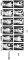

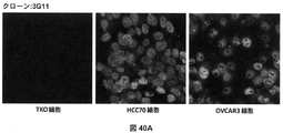


Classifications
-
- C—CHEMISTRY; METALLURGY
- C07—ORGANIC CHEMISTRY
- C07K—PEPTIDES
- C07K16/00—Immunoglobulins [IGs], e.g. monoclonal or polyclonal antibodies
- C07K16/18—Immunoglobulins [IGs], e.g. monoclonal or polyclonal antibodies against material from animals or humans
-
- A—HUMAN NECESSITIES
- A61—MEDICAL OR VETERINARY SCIENCE; HYGIENE
- A61K—PREPARATIONS FOR MEDICAL, DENTAL OR TOILETRY PURPOSES
- A61K39/00—Medicinal preparations containing antigens or antibodies
-
- A—HUMAN NECESSITIES
- A61—MEDICAL OR VETERINARY SCIENCE; HYGIENE
- A61K—PREPARATIONS FOR MEDICAL, DENTAL OR TOILETRY PURPOSES
- A61K49/00—Preparations for testing in vivo
- A61K49/001—Preparation for luminescence or biological staining
- A61K49/0013—Luminescence
- A61K49/0017—Fluorescence in vivo
- A61K49/005—Fluorescence in vivo characterised by the carrier molecule carrying the fluorescent agent
- A61K49/0058—Antibodies
-
- A—HUMAN NECESSITIES
- A61—MEDICAL OR VETERINARY SCIENCE; HYGIENE
- A61P—SPECIFIC THERAPEUTIC ACTIVITY OF CHEMICAL COMPOUNDS OR MEDICINAL PREPARATIONS
- A61P35/00—Antineoplastic agents
-
- A—HUMAN NECESSITIES
- A61—MEDICAL OR VETERINARY SCIENCE; HYGIENE
- A61K—PREPARATIONS FOR MEDICAL, DENTAL OR TOILETRY PURPOSES
- A61K39/00—Medicinal preparations containing antigens or antibodies
- A61K2039/505—Medicinal preparations containing antigens or antibodies comprising antibodies
-
- A—HUMAN NECESSITIES
- A61—MEDICAL OR VETERINARY SCIENCE; HYGIENE
- A61K—PREPARATIONS FOR MEDICAL, DENTAL OR TOILETRY PURPOSES
- A61K39/00—Medicinal preparations containing antigens or antibodies
- A61K2039/55—Medicinal preparations containing antigens or antibodies characterised by the host/recipient, e.g. newborn with maternal antibodies
- A61K2039/552—Veterinary vaccine
-
- A—HUMAN NECESSITIES
- A61—MEDICAL OR VETERINARY SCIENCE; HYGIENE
- A61K—PREPARATIONS FOR MEDICAL, DENTAL OR TOILETRY PURPOSES
- A61K39/00—Medicinal preparations containing antigens or antibodies
- A61K2039/57—Medicinal preparations containing antigens or antibodies characterised by the type of response, e.g. Th1, Th2
- A61K2039/575—Medicinal preparations containing antigens or antibodies characterised by the type of response, e.g. Th1, Th2 humoral response
-
- A—HUMAN NECESSITIES
- A61—MEDICAL OR VETERINARY SCIENCE; HYGIENE
- A61K—PREPARATIONS FOR MEDICAL, DENTAL OR TOILETRY PURPOSES
- A61K39/00—Medicinal preparations containing antigens or antibodies
- A61K2039/80—Vaccine for a specifically defined cancer
-
- C—CHEMISTRY; METALLURGY
- C07—ORGANIC CHEMISTRY
- C07K—PEPTIDES
- C07K2317/00—Immunoglobulins specific features
- C07K2317/20—Immunoglobulins specific features characterized by taxonomic origin
- C07K2317/24—Immunoglobulins specific features characterized by taxonomic origin containing regions, domains or residues from different species, e.g. chimeric, humanized or veneered
-
- C—CHEMISTRY; METALLURGY
- C07—ORGANIC CHEMISTRY
- C07K—PEPTIDES
- C07K2317/00—Immunoglobulins specific features
- C07K2317/30—Immunoglobulins specific features characterized by aspects of specificity or valency
- C07K2317/34—Identification of a linear epitope shorter than 20 amino acid residues or of a conformational epitope defined by amino acid residues
Landscapes
- Health & Medical Sciences (AREA)
- Chemical & Material Sciences (AREA)
- Life Sciences & Earth Sciences (AREA)
- General Health & Medical Sciences (AREA)
- Organic Chemistry (AREA)
- Medicinal Chemistry (AREA)
- Immunology (AREA)
- Veterinary Medicine (AREA)
- Public Health (AREA)
- Animal Behavior & Ethology (AREA)
- Pharmacology & Pharmacy (AREA)
- Epidemiology (AREA)
- Genetics & Genomics (AREA)
- Biochemistry (AREA)
- Nuclear Medicine, Radiotherapy & Molecular Imaging (AREA)
- Chemical Kinetics & Catalysis (AREA)
- Proteomics, Peptides & Aminoacids (AREA)
- Molecular Biology (AREA)
- Biophysics (AREA)
- General Chemical & Material Sciences (AREA)
- Microbiology (AREA)
- Mycology (AREA)
- Biomedical Technology (AREA)
- Engineering & Computer Science (AREA)
- Peptides Or Proteins (AREA)
- Medicines Containing Antibodies Or Antigens For Use As Internal Diagnostic Agents (AREA)
- Measuring Or Testing Involving Enzymes Or Micro-Organisms (AREA)
- Preparation Of Compounds By Using Micro-Organisms (AREA)
- Micro-Organisms Or Cultivation Processes Thereof (AREA)
- Medicines Containing Material From Animals Or Micro-Organisms (AREA)
Description
本発明は、突然変異体p53ポリペプチドを対象とする抗体、およびこの抗体を作製するための方法に関する。 The present invention relates to antibodies directed against mutant p53 polypeptides and methods for making such antibodies.
免疫応答は、克服されうる高度に特異的な認識イベントが、ほぼ全ての種類の外因性抗原を回避するように闘うことを目的として、進化の過程によって完全無欠なものとなった(FlajnikおよびKasahara、Nat Rev Gene(2010)、11(1):47~59)。脊椎動物では、Tリンパ球およびBリンパ球のいずれも、抗原に誘導される選択的な再配列を経る受容体を発達させて、類似する種に由来する高度に類縁の抗原性タンパク質内で生じうる可変性のうちで最も微細な可変性をも検出し(CooperおよびAlder、Cell(2006)、124(4):815~822)、これにより、外来からの侵入に対する障壁を確立した。この生得的能力は、わずかな構造的変動を伴う、高度に類似するタンパク質に対して多数の抗体を作出するのに利用されている(Loら、2014、Microbiol.Spectr.、2、AID-0007-2012)。さらにまた、標的アミノ酸残基上の、リン酸化、ユビキチン化、およびアセチル化など、わずかな翻訳後修飾を検出する抗体も作製され、これは、多種多様な生理学的過程および病理学的過程の機構的理解をもたらした。しかし、がんゲノム内のミスセンス突然変異のために注目されることが多い単一のアミノ酸変化の検出は、親タンパク質におけるフランキング領域の高度な相同性のために、選択的抗体の作出にとっての大きな難題となっている。突然変異していない天然タンパク質と交差反応しない突然変異特異的抗体を作製する試みがなされているが、これまでのところ、大半の取組みは、成功していない(Bondgaardら、Modern Pathology(2014)、27:1590~1598)。 The immune response has been perfected by evolution to combat the highly specific recognition events that can be overcome to evade nearly all kinds of exogenous antigens (Flajnik and Kasahara , Nat Rev Gene (2010), 11(1):47-59). In vertebrates, both T and B lymphocytes develop receptors that undergo selective antigen-induced rearrangements to occur within highly related antigenic proteins from similar species. We detected even the finest variability possible (Cooper and Alder, Cell (2006), 124(4):815-822), thereby establishing a barrier to foreign invasion. This innate ability has been exploited to generate large numbers of antibodies against highly similar proteins with minor structural variations (Lo et al., 2014, Microbiol. Spectr., 2, AID-0007 -2012). Furthermore, antibodies have also been generated that detect subtle post-translational modifications, such as phosphorylation, ubiquitination, and acetylation, on target amino acid residues, which are mechanisms of a wide variety of physiological and pathological processes. brought understanding. However, the detection of single amino acid changes, which are often focused on due to missense mutations within cancer genomes, may be of interest for the generation of selective antibodies due to the high degree of homology of the flanking regions in the parental proteins. is a big challenge. Attempts have been made to generate mutation-specific antibodies that do not cross-react with the unmutated native protein, but so far most efforts have been unsuccessful (Bondgaard et al., Modern Pathology (2014). 27:1590-1598).
EGFR、PD-1などの表面抗原に対するmAbは、臨床状況で、処置のために使用され成功を収めているが(Martinelliら、2009 Clinical and Experimental Immunology、158(1):1~9;PhilipsおよびAtkins、2014、Int Immunol、27(1):39~46)、核抗原に対するmAbは一般に、有効となるために細胞膜を通過する上で難関に直面する。しかし、近年の研究は、核抗原に対する抗体が、実際に、治療的に有効であることを示しており(HongおよびZeng、2012、Cancer Res、72(11):3715)、この手法が、将来的に成功しうることを約束している。 Although mAbs against surface antigens such as EGFR, PD-1 have been used successfully for treatment in the clinical setting (Martinelli et al., 2009 Clinical and Experimental Immunology, 158(1):1-9; Philips and Atkins, 2014, Int Immunol, 27(1):39-46), mAbs against nuclear antigens generally face challenges in crossing the cell membrane to be effective. However, recent studies have shown that antibodies against nuclear antigens are indeed therapeutically effective (Hong and Zeng, 2012, Cancer Res, 72(11):3715), suggesting that this approach may be of interest in the future. promises to be successful.
p53は、全てのがん型にわたり、最も突然変異する遺伝子であり、全ての腫瘍のうちの50%を超える腫瘍が、これにおける遺伝子変更の何らかの形態を呈する(Forbesら、2015、Nucleic Acids Res、43、D805~D811)。p53内の大半の突然変異は、約200アミノ酸にわたる、中央のDNA結合性ドメイン(DBD)内に見出され、同定された全てのp53突然変異のうちの、約90%を占める。ほぼ全てのDBD突然変異は、ゲノムの保護におけるp53の多機能的な役割のために、腫瘍の抑制および治療に対する効果的な応答に不可欠な、野生型p53の機能の喪失をもたらす(VousdenおよびLu、Nat Rev Cancer(2002)、2(8):594~604;Freed-PastorおよびPrives、Genes Dev(2012)、26(12):1268~1286)。野生型機能の喪失の他に、p53内の突然変異はまた、がんの発症を促進し、また治療に対する効果的な応答も阻害する2つの主要な現象ももたらす。1つは、p53の突然変異体対立遺伝子が、野生型対立遺伝子と共存する場合に生じ、これにより、ヘテロオリゴマーを形成し、この結果として、残りの野生型p53機能の減弱化をもたらすドミナントネガティブ(DN)効果である(Kernら、Science(1992)、256:827~830)。加えて、とりわけ、野生型p53対立遺伝子が、ヘテロ接合性喪失のために失われる、がんの後期段階では、突然変異体p53対立遺伝子が単独で存在する結果として、がん細胞は、生存および浸潤特性を促進する、突然変異体p53による新規発がん機能の獲得(GOF)に起因して、生存のために、突然変異体p53タンパク質に依存するようになる(Sabapathy、Front Oncol(2015)、5:276)。これらの過程における突然変異体p53の因果的役割は、その発現の低減、またはその機能の不活化により裏付けられており、この発現の低減または機能の不活化においては、DN機能およびGOF機能のいずれもがレスキューされえ、これは、がん細胞が治療的処置に対してより感受性となることを可能とする(Leeら、Cancer Cell(2012)、22:751~764)。これらのデータは、突然変異体p53をターゲティングする興味深い可能性を開くが、突然変異体p53の不活化を成功させるには、いくつかの難関が存在する。最も重要なこととして、p53内の全ての突然変異は、その野生型活性の喪失、および大半の場合に、DN効果を明白にもたらすが、GOF特性は、全てではなく、一部の突然変異体に選択的なものであるようである(Leeら、Cancer Cell(2012)、22:751~764;Hanelら、Cell Death Diff(2013)、20:898~909)ことに注目すべきであり、突然変異体p53のターゲティングは、各個別のp53突然変異体を選択的に検出し、不活化させるために、より洗練された手法を必要とすることが強調される。 p53 is the most mutated gene across all cancer types, with over 50% of all tumors exhibiting some form of genetic alteration in it (Forbes et al., 2015, Nucleic Acids Res. 43, D805-D811). Most mutations within p53 are found within the central DNA-binding domain (DBD), which spans approximately 200 amino acids and accounts for approximately 90% of all identified p53 mutations. Almost all DBD mutations result in loss of function of wild-type p53, which is essential for tumor suppression and effective response to therapy, due to the multifunctional role of p53 in protecting the genome (Vousden and Lu , Nat Rev Cancer (2002), 2(8):594-604; Freed-Pastor and Prives, Genes Dev (2012), 26(12):1268-1286). Besides loss of wild-type function, mutations within p53 also result in two major events that both promote cancer development and inhibit effective response to therapy. One occurs when mutant alleles of p53 coexist with wild-type alleles, thereby forming hetero-oligomers and resulting in attenuation of the remaining wild-type p53 function. (DN) effect (Kern et al., Science (1992), 256:827-830). In addition, particularly in later stages of cancer, when the wild-type p53 allele is lost due to loss of heterozygosity, the presence of the mutant p53 allele alone results in cancer cell survival and Due to the gain of de novo oncogenic function (GOF) by mutant p53, which promotes invasive properties, it becomes dependent on the mutant p53 protein for survival (Sabapathy, Front Oncol (2015), 5 : 276). A causal role of mutant p53 in these processes is supported by its reduced expression, or inactivation of its function, in which both DN and GOF function can be rescued, allowing cancer cells to become more susceptible to therapeutic treatment (Lee et al., Cancer Cell (2012), 22:751-764). These data open up interesting possibilities for targeting mutant p53, but several challenges exist for successful inactivation of mutant p53. Most importantly, although all mutations within p53 clearly result in loss of its wild-type activity and, in most cases, DN effects, the GOF property is mutated in some, but not all, mutants. (Lee et al., Cancer Cell (2012), 22:751-764; Hanel et al., Cell Death Diff (2013), 20:898-909). It is emphasized that targeting mutant p53 requires more sophisticated approaches to selectively detect and inactivate each individual p53 mutant.
突然変異体p53の生物学を理解することは、突然変異体p53の、発がんおよび治療への応答における役割の解明にとっての基礎となっている。突然変異体p53が、どのようにして、その生存促進機能を果たすのかについての知見に関して、著しい進歩があったが、限界も存在する。例えば、全ての突然変異が、機能的に同様であるわけではなく(Sabapathy、2015、Front.Oncol.5:276)、p53のトランス活性化ドメインおよびオリゴマー化ドメインにおいて見出される突然変異(これは、ある特定の集団では、極めて一般的である(Achatzら、2007、Cancer Lett.、245:96~102))は、さらなる評価を必要とする。 Understanding the biology of mutant p53 is fundamental to the elucidation of its role in carcinogenesis and response to therapy. Although significant progress has been made in understanding how mutant p53 performs its pro-survival functions, limitations also exist. For example, not all mutations are functionally similar (Sabapathy, 2015, Front. Oncol. 5:276), and mutations found in the transactivation and oligomerization domains of p53, which Its extreme prevalence in certain populations (Achatz et al., 2007, Cancer Lett., 245:96-102) requires further evaluation.
現行の技術は、ノックインマウスモデル、または野生型タンパク質を伴わずに、突然変異体タンパク質だけを発現するヒト腫瘍細胞系を用いているが、これらは、野生型タンパク質との共存における突然変異体タンパク質の役割を十全には反映しない場合がある。 Current technology uses knock-in mouse models, or human tumor cell lines that express only the mutant protein without the wild-type protein, but these do not support the mutant protein in coexistence with the wild-type protein. may not fully reflect the role of
数十年にわたり、突然変異体p53に対する抗体を作出しようとする試みがなされており、p53の、突然変異体コンフォメーションに対するモノクローナル抗体(mAb)(PAb240)、または野生型コンフォメーションに対するmAb(PAb246)が利用可能である(Gannonら、EMBO J(1990)、9(5):1595~1602)。しかし、これらの抗体は、十分に特異的ではなく、交差反応性を呈し、適正なコンフォメーションの検出に、極めて選択的な条件を必要とするので、それらの使用は限定されている。 For decades, attempts have been made to generate antibodies against mutant p53, monoclonal antibodies (mAbs) against the mutant conformation (PAb240) or against the wild-type conformation of p53 (PAb246). are available (Gannon et al., EMBO J (1990), 9(5):1595-1602). However, their use is limited because these antibodies are not sufficiently specific, exhibit cross-reactivity, and require highly selective conditions for detection of proper conformation.
VojtesekおよびLane、J Cell Sci(1993)、105(3):607~612は、DO-1と称する、ヒトp53のアミノ末端領域に対する抗体について記載しており、この抗体は、マウスp53との交差反応性を伴わずに、ヒトp53に対して高レベルの特異性を示す。 Vojtesek and Lane, J Cell Sci (1993), 105(3):607-612, describe an antibody directed against the amino-terminal region of human p53, termed DO-1, which cross-reacts with mouse p53. It shows a high level of specificity for human p53 without reactivity.
第1の態様では、本発明は、野生型p53ポリペプチドより、突然変異体p53ポリペプチドに特異的な抗体を作製するための方法であって、免疫原として、(i)突然変異と、突然変異に直接隣接する少なくとも1つのアミノ酸とを含む、突然変異体p53ポリペプチドのアミノ酸配列を含む抗原配列、および(ii)溶媒に接触可能な立体配置にある抗原配列をもたらすための足場配列を含むペプチドまたはポリペプチドを使用するステップを含む方法を提供する。 In a first aspect, the present invention provides a method for generating antibodies specific to mutant p53 polypeptides over wild-type p53 polypeptides, wherein as immunogens, (i) a mutation and a mutation and (ii) a scaffolding sequence to bring the antigen sequence in a solvent accessible configuration. A method is provided comprising using a peptide or polypeptide.
一部の実施形態では、足場配列は、溶媒に接触可能な配列を含むペプチドまたはポリペプチドに由来し、抗原配列は、ペプチドもしくはポリペプチドの溶媒に接触可能な配列に挿入されるか、またはその全部もしくは一部を置換する。一部の実施形態では、溶媒に接触可能な配列を含むペプチドまたはポリペプチドは、チオレドキシンであり、溶媒に接触可能な配列は、チオレドキシンの活性部位配列である。一部の実施形態では、免疫原として使用されるペプチドまたはポリペプチドは、さらに、抗原配列と足場配列との間に、1または複数のリンカー配列を含む。一部の実施形態では、免疫原として使用されるペプチドまたはポリペプチドは、突然変異体p53ポリペプチドの少なくとも2つのアミノ酸配列を含む。一部の実施形態では、免疫原として使用されるペプチドまたはポリペプチドは、さらに、突然変異体p53ポリペプチドの少なくとも2つのアミノ酸配列の間に、リンカー配列を含む。一部の実施形態では、突然変異体p53ポリペプチドの少なくとも2つのアミノ酸配列は、非同一である。一部の実施形態では、突然変異体p53ポリペプチドのアミノ酸配列は、少なくとも5つのアミノ酸を含む。一部の実施形態では、突然変異体p53ポリペプチドは、DNA結合性ドメイン(DBD)内に、突然変異を含む。一部の実施形態では、突然変異体p53ポリペプチドは、R175H、R248Q、R273H、R248W、G245S、R273C、R282W、R249S、G245D、C176F、H179Y、H179R、Y220C、およびR337Hのうちの1つから選択される突然変異を含む。一部の実施形態では、突然変異体p53ポリペプチドは、配列番号3~16のうちの1つのアミノ酸配列を含むか、またはこれからなる。 In some embodiments, the scaffolding sequence is derived from a peptide or polypeptide comprising a solvent accessible sequence and the antigen sequence is inserted into or into the solvent accessible sequence of the peptide or polypeptide. Replace in whole or in part. In some embodiments, the peptide or polypeptide comprising the solvent accessible sequence is a thioredoxin and the solvent accessible sequence is a thioredoxin active site sequence. In some embodiments, peptides or polypeptides used as immunogens further comprise one or more linker sequences between the antigen sequence and the scaffold sequence. In some embodiments, peptides or polypeptides used as immunogens comprise at least two amino acid sequences of a mutant p53 polypeptide. In some embodiments, peptides or polypeptides used as immunogens further comprise a linker sequence between at least two amino acid sequences of the mutant p53 polypeptide. In some embodiments, at least two amino acid sequences of the mutant p53 polypeptide are non-identical. In some embodiments, the amino acid sequence of the mutant p53 polypeptide comprises at least 5 amino acids. In some embodiments, the mutant p53 polypeptide comprises a mutation within the DNA binding domain (DBD). In some embodiments, the mutant p53 polypeptide is selected from one of R175H, R248Q, R273H, R248W, G245S, R273C, R282W, R249S, G245D, C176F, H179Y, H179R, Y220C, and R337H contains mutations that In some embodiments, the mutant p53 polypeptide comprises or consists of an amino acid sequence of one of SEQ ID NOS:3-16.
別の態様では、本発明は、本発明に従う抗体を作製するための方法により得られるか、または得ることが可能な抗体または抗原結合性断片を提供する。
別の態様では、本発明は、野生型p53ポリペプチドより、突然変異体p53ポリペプチドに特異的である抗体または抗原結合性断片を提供する。
In another aspect, the invention provides an antibody or antigen-binding fragment obtained or obtainable by a method for producing an antibody according to the invention.
In another aspect, the invention provides antibodies or antigen-binding fragments that are more specific for mutant p53 polypeptides than wild-type p53 polypeptides.
一部の実施形態では、突然変異体p53ポリペプチドは、DNA結合性ドメイン(DBD)内に、突然変異を含む。一部の実施形態では、突然変異体p53ポリペプチドは、R175H、R248Q、R273H、R248W、G245S、R273C、R282W、R249S、G245D、C176F、H179Y、H179R、Y220C、およびR337Hのうちの1つから選択される突然変異を含む。一部の実施形態では、突然変異体p53ポリペプチドは、配列番号3~16のうちの1つのアミノ酸配列を含むか、またはこれからなる。 In some embodiments, the mutant p53 polypeptide comprises a mutation within the DNA binding domain (DBD). In some embodiments, the mutant p53 polypeptide is selected from one of R175H, R248Q, R273H, R248W, G245S, R273C, R282W, R249S, G245D, C176F, H179Y, H179R, Y220C, and R337H contains mutations that In some embodiments, the mutant p53 polypeptide comprises or consists of an amino acid sequence of one of SEQ ID NOS:3-16.
別の態様では、本発明は、R175H p53ポリペプチドに結合することが可能であり、任意に単離されており、アミノ酸配列i)~vi):
i) LC-CDR1:QSLLNSGNQKSX1 (配列番号31);
ii) LC-CDR2:GAS (配列番号20);
iii) LC-CDR3:QNDHSYPLT (配列番号21);
iv) HC-CDR1:GX2TFTEYT (配列番号32);
v) HC-CDR2:IX3PX4X5GX6T (配列番号33);
vi) HC-CDR3:ARWGGDYV (配列番号27)
を有する抗体もしくは抗原結合性断片、または配列i)~vi)のうちの1もしくは複数における、1つもしくは2つもしくは3つのアミノ酸が、別のアミノ酸で置きかえられた、そのバリアント[配列中、X1=YまたはN;X2=FまたはY;X3=DまたはN;X4=NまたはY;X5=NまたはS;およびX6=VまたはG]を提供する。
In another aspect, the invention is capable of binding to a R175H p53 polypeptide, optionally isolated, having the amino acid sequences i)-vi):
i) LC-CDR1: QSLLNSGNQKSX 1 (SEQ ID NO:31);
ii) LC-CDR2: GAS (SEQ ID NO:20);
iii) LC-CDR3: QNDHSYPLT (SEQ ID NO:21);
iv) HC-CDR1:GX 2 TFTEYT (SEQ ID NO:32);
v) HC-CDR2: IX 3 PX 4 X 5 GX 6 T (SEQ ID NO: 33);
vi) HC-CDR3: ARWGGDYV (SEQ ID NO: 27)
or a variant thereof in which one or two or three amino acids in one or more of sequences i)-vi) are replaced with another amino acid [in the sequence X X2 = F or Y; X3 = D or N ; X4 = N or Y; X5 = N or S; and X6 = V or G].
一部の実施形態では、LC-CDR1は、QSLLNSGNQKSY(配列番号19)またはQSLLNSGNQKSN(配列番号23)のうちの1つである。一部の実施形態では、HC-CDR1は、GFTFTEYT(配列番号25)またはGYTFTEYT(配列番号29)のうちの1つである。一部の実施形態では、HC-CDR2は、IDPNNGVT(配列番号26)またはINPYSGGT(配列番号30)のうちの1つである。 In some embodiments, LC-CDR1 is one of QSLLNSGNQKSY (SEQ ID NO: 19) or QSLLNSGNQKSN (SEQ ID NO: 23). In some embodiments, HC-CDR1 is one of GFTFTEYT (SEQ ID NO:25) or GYTFFTEYT (SEQ ID NO:29). In some embodiments, the HC-CDR2 is one of IDPNNGVT (SEQ ID NO:26) or INPYSGGT (SEQ ID NO:30).
一部の実施形態では、抗体または抗原結合性断片は、以下のCDR:
LC-CDR1:QSLLNSGNQKSX1 (配列番号31);
LC-CDR2:GAS (配列番号20);
LC-CDR3:QNDHSYPLT (配列番号21)
[配列中、X1=YまたはN]が組み込まれた少なくとも1つの軽鎖可変領域を有する。
In some embodiments, the antibody or antigen-binding fragment has the following CDRs:
LC-CDR1: QSLLNSGNQKSX 1 (SEQ ID NO: 31);
LC-CDR2: GAS (SEQ ID NO: 20);
LC-CDR3: QNDHSYPLT (SEQ ID NO:21)
It has at least one light chain variable region that incorporates [where X 1 =Y or N].
一部の実施形態では、抗体または抗原結合性断片は、以下のCDR:
LC-CDR1:QSLLNSGNQKSY (配列番号19);
LC-CDR2:GAS (配列番号20);
LC-CDR3:QNDHSYPLT (配列番号21)
が組み込まれた少なくとも1つの軽鎖可変領域を有する。
In some embodiments, the antibody or antigen-binding fragment has the following CDRs:
LC-CDR1: QSLLNSGNQKSY (SEQ ID NO: 19);
LC-CDR2: GAS (SEQ ID NO:20);
LC-CDR3: QNDHSYPLT (SEQ ID NO:21)
has at least one light chain variable region that incorporates
一部の実施形態では、抗体または抗原結合性断片は、以下のCDR:
LC-CDR1:QSLLNSGNQKSN (配列番号23);
LC-CDR2:GAS (配列番号20);
LC-CDR3:QNDHSYPLT (配列番号21)
が組み込まれた少なくとも1つの軽鎖可変領域を有する。
In some embodiments, the antibody or antigen-binding fragment has the following CDRs:
LC-CDR1: QSLLNSGNQKSN (SEQ ID NO:23);
LC-CDR2: GAS (SEQ ID NO:20);
LC-CDR3: QNDHSYPLT (SEQ ID NO:21)
has at least one light chain variable region that incorporates
一部の実施形態では、抗体または抗原結合性断片は、以下のCDR:
HC-CDR1:GX2TFTEYT (配列番号32);
HC-CDR2:IX3PX4X5GX6T (配列番号33);
HC-CDR3:ARWGGDYV (配列番号27)
[配列中、X2=FまたはY;X3=DまたはN;X4=NまたはY;X5=NまたはS;およびX6=VまたはG]が組み込まれた少なくとも1つの重鎖可変領域を有する。
In some embodiments, the antibody or antigen-binding fragment has the following CDRs:
HC-CDR1: GX 2 TFTEYT (SEQ ID NO: 32);
HC-CDR2: IX 3 PX 4 X 5 GX 6 T (SEQ ID NO: 33);
HC-CDR3: ARWGGDYV (SEQ ID NO: 27)
at least one heavy chain variable that incorporates [in sequence X2 = F or Y; X3 = D or N; X4 = N or Y; X5 = N or S; and X6 = V or G] have an area.
一部の実施形態では、抗体または抗原結合性断片は、以下のCDR:
HC-CDR1:GFTFTEYT (配列番号25);
HC-CDR2:IDPNNGVT (配列番号26);
HC-CDR3:ARWGGDYV (配列番号27)
が組み込まれた少なくとも1つの重鎖可変領域を有する。
In some embodiments, the antibody or antigen-binding fragment has the following CDRs:
HC-CDR1: GFTFTEYT (SEQ ID NO: 25);
HC-CDR2: IDPNNGVT (SEQ ID NO: 26);
HC-CDR3: ARWGGDYV (SEQ ID NO: 27)
has at least one heavy chain variable region that incorporates
一部の実施形態では、抗体または抗原結合性断片は、以下のCDR:
HC-CDR1:GYTFTEYT (配列番号29);
HC-CDR2:INPYSGGT (配列番号30);
HC-CDR3:ARWGGDYV (配列番号27)
が組み込まれた少なくとも1つの重鎖可変領域を有する。
In some embodiments, the antibody or antigen-binding fragment has the following CDRs:
HC-CDR1: GYTFTEYT (SEQ ID NO: 29);
HC-CDR2: INPYSGGT (SEQ ID NO: 30);
HC-CDR3: ARWGGDYV (SEQ ID NO: 27)
has at least one heavy chain variable region that incorporates
別の態様では、本発明は、以下のCDR:
LC-CDR1:QSLLNSGNQKSX1 (配列番号31);
LC-CDR2:GAS (配列番号20);
LC-CDR3:QNDHSYPLT (配列番号21)
[配列中、X1=YまたはN]を含む、単離された、軽鎖可変領域のポリペプチドを提供する。
In another aspect, the invention provides the following CDRs:
LC-CDR1: QSLLNSGNQKSX 1 (SEQ ID NO: 31);
LC-CDR2: GAS (SEQ ID NO: 20);
LC-CDR3: QNDHSYPLT (SEQ ID NO:21)
An isolated light chain variable region polypeptide is provided comprising [wherein the sequence is X 1 =Y or N].
一部の実施形態では、LC-CDR1は、QSLLNSGNQKSY(配列番号19)またはQSLLNSGNQKSN(配列番号23)のうちの1つである。
別の態様では、本発明は、以下のCDR:
HC-CDR1:GX2TFTEYT (配列番号32);
HC-CDR2:IX3PX4X5GX6T (配列番号33);
HC-CDR3:ARWGGDYV (配列番号27)
[配列中、X2=FまたはY;X3=DまたはN;X4=NまたはY;X5=NまたはS;およびX6=VまたはG]を含む、単離された、重鎖可変領域のポリペプチドを提供する。
In some embodiments, LC-CDR1 is one of QSLLNSGNQKSY (SEQ ID NO: 19) or QSLLNSGNQKSN (SEQ ID NO: 23).
In another aspect, the invention provides the following CDRs:
HC-CDR1: GX 2 TFTEYT (SEQ ID NO: 32);
HC-CDR2: IX 3 PX 4 X 5 GX 6 T (SEQ ID NO: 33);
HC-CDR3: ARWGGDYV (SEQ ID NO: 27)
X2 = F or Y; X3 = D or N; X4 = N or Y; X5 = N or S; and X6 = V or G]. A variable region polypeptide is provided.
一部の実施形態では、HC-CDR1は、GFTFTEYT(配列番号25)またはGYTFTEYT(配列番号29)のうちの1つである。一部の実施形態では、HC-CDR2は、IDPNNGVT(配列番号26)またはINPYSGGT(配列番号30)のうちの1つである。 In some embodiments, HC-CDR1 is one of GFTFTEYT (SEQ ID NO:25) or GYTFFTEYT (SEQ ID NO:29). In some embodiments, the HC-CDR2 is one of IDPNNGVT (SEQ ID NO:26) or INPYSGGT (SEQ ID NO:30).
別の態様では、本発明は、本明細書で記載される、重鎖可変領域のポリペプチドと組み合わされた、本明細書で記載される、単離された、軽鎖可変領域のポリペプチドを提供する。 In another aspect, the invention provides an isolated light chain variable region polypeptide as described herein in combination with a heavy chain variable region polypeptide as described herein. offer.
別の態様では、本発明は、R175H p53ポリペプチドに結合することが可能であり、軽鎖可変領域および重鎖可変領域の配列を含み、
軽鎖が、LC-CDR1:QSLLNSGNQKSX1(配列番号31)、QSLLNSGNQKSY(配列番号19)、またはQSLLNSGNQKSN(配列番号23)のうちの1つ;LC-CDR2:GAS(配列番号20);LC-CDR3:QNDHSYPLT(配列番号21)に対する、少なくとも85%の、全体的な配列同一性を有するLC-CDR1、LC-CDR2、LC-CDR3を含み;そして
重鎖が、HC-CDR1:GX2TFTEYT(配列番号32)、GFTFTEYT(配列番号25)またはGYTFTEYT(配列番号29)のうちの1つ;HC-CDR2:IX3PX4X5GX6T(配列番号33)、IDPNNGVT(配列番号26)、またはINPYSGGT(配列番号30)のうちの1つ;HC-CDR3:ARWGGDYV(配列番号27)[配列中、X1=YまたはN;X2=FまたはY;X3=DまたはN;X4=NまたはY;X5=NまたはS;およびX6=VまたはG]に対する、少なくとも85%の、全体的な配列同一性を有するHC-CDR1、HC-CDR2、HC-CDR3を含む、
抗体または抗原結合性断片を提供する。
In another aspect, the invention is capable of binding to a R175H p53 polypeptide and comprises light and heavy chain variable region sequences,
the light chain is one of LC-CDR1: QSLLNSGNQKSX 1 (SEQ ID NO:31), QSLLNSGNQKSY (SEQ ID NO:19), or QSLLNSGNQKSN (SEQ ID NO:23); LC-CDR2: GAS (SEQ ID NO:20); LC-CDR3 : contains LC-CDR1, LC-CDR2, LC-CDR3 with an overall sequence identity of at least 85% to QNDHSYPLT (SEQ ID NO: 21); and the heavy chain comprises HC-CDR1: GX 2 TFTEYT (sequence number 32), one of GFTFTEYT (SEQ ID NO: 25 ) or GYTFFTEYT (SEQ ID NO : 29); one of INPYSGGT (SEQ ID NO: 30); HC-CDR3: ARWGGDYV (SEQ ID NO: 27) [where X 1 =Y or N; X 2 =F or Y; X 3 =D or N; N or Y; X = N or S; and X = V or G]
Antibodies or antigen-binding fragments are provided.
別の態様では、本発明は、R175H p53ポリペプチドに結合することが可能であり、軽鎖可変領域および重鎖可変領域の配列を含み、
軽鎖配列が、配列番号18もしくは22(図24)、または配列番号237もしくは94(図49)の軽鎖配列に対する、少なくとも85%の配列同一性を有し;そして
重鎖配列が、配列番号24もしくは28(図25)、または配列番号239、240、もしくは241(図50)の重鎖配列に対する、少なくとも85%の配列同一性を有する、
抗体または抗原結合性断片を提供する。
In another aspect, the invention is capable of binding to a R175H p53 polypeptide and comprises light and heavy chain variable region sequences,
the light chain sequence has at least 85% sequence identity to the light chain sequence of SEQ ID NO: 18 or 22 (Figure 24), or SEQ ID NO: 237 or 94 (Figure 49); and the heavy chain sequence has SEQ ID NO: have at least 85% sequence identity to the heavy chain sequence of 24 or 28 (Figure 25), or SEQ ID NOs: 239, 240, or 241 (Figure 50);
Antibodies or antigen-binding fragments are provided.
別の態様では、本発明は、R175H p53ポリペプチドに結合することが可能であり、(i)本明細書で記載される抗原結合性断片またはポリペプチド、および(ii)R175H p53ポリペプチド以外のポリペプチドに結合することが可能な抗原結合性断片を含む二特異性抗体または二特異性抗原結合性断片である、任意に単離された抗体または抗原結合性断片を提供する。 In another aspect, the present invention is capable of binding to R175H p53 polypeptides and (i) antigen-binding fragments or polypeptides described herein and (ii) other than R175H p53 polypeptides. An optionally isolated antibody or antigen-binding fragment is provided that is a bispecific antibody or bispecific antigen-binding fragment comprising an antigen-binding fragment capable of binding a polypeptide.
別の態様では、本発明は、R175H p53ポリペプチドまたはこの断片に結合した、本明細書で記載される抗体、抗原結合性断片、またはポリペプチドを含む、任意に単離された、in vitroにおける複合体を提供する。 In another aspect, the invention provides any isolated, in vitro Provide a complex.
別の態様では、本発明は、R248Q p53ポリペプチドに結合することが可能であり、任意に単離されており、アミノ酸配列i)~vi):
i) LC-CDR1:QSLLYSDGKTY (配列番号41);
ii) LC-CDR2:LVS (配列番号42);
iii) LC-CDR3:WQGTHFPLT (配列番号43);
iv) HC-CDR1:GYTFTDYY (配列番号46);
v) HC-CDR2:IX7PKNGGT (配列番号52);
vi) HC-CDR3:AKX8GGX9DDY (配列番号53)
を有する抗体もしくは抗原結合性断片、または配列i)~vi)のうちの1もしくは複数における、1つもしくは2つもしくは3つのアミノ酸が、別のアミノ酸で置きかえられた、そのバリアント[配列中、X7=HまたはD;X8=MまたはQ;およびX9=YまたはF]を提供する。
In another aspect, the invention is capable of binding to a R248Q p53 polypeptide, optionally isolated, having the amino acid sequences i)-vi):
i) LC-CDR1: QSLLYSDGKTY (SEQ ID NO: 41);
ii) LC-CDR2:LVS (SEQ ID NO:42);
iii) LC-CDR3: WQGTHFPLT (SEQ ID NO: 43);
iv) HC-CDR1:GYTFTDYY (SEQ ID NO:46);
v) HC-CDR2: IX 7 PKNGGT (SEQ ID NO: 52);
vi) HC-CDR3: AKX 8 GGX 9 DDY (SEQ ID NO: 53)
or a variant thereof in which one or two or three amino acids in one or more of sequences i)-vi) are replaced with another amino acid [in the sequence X 7 = H or D; X 8 = M or Q; and X 9 = Y or F].
一部の実施形態では、HC-CDR2は、IHPKNGGT(配列番号47)またはIDPKNGGT(配列番号50)のうちの1つである。一部の実施形態では、HC-CDR3は、AKMGGYDDY(配列番号48)またはAKQGGFDDY(配列番号51)のうちの1つである。 In some embodiments, the HC-CDR2 is one of IHPKNGGT (SEQ ID NO:47) or IDPKNGGT (SEQ ID NO:50). In some embodiments, the HC-CDR3 is one of AKMGGYDDY (SEQ ID NO:48) or AKQGGFDDY (SEQ ID NO:51).
一部の実施形態では、抗体または抗原結合性断片は、以下のCDR:
LC-CDR1:QSLLYSDGKTY (配列番号41)
LC-CDR2:LVS (配列番号42)
LC-CDR3:WQGTHFPLT (配列番号43)
が組み込まれた少なくとも1つの軽鎖可変領域を有する。
In some embodiments, the antibody or antigen-binding fragment has the following CDRs:
LC-CDR1: QSLLYSDGKTY (SEQ ID NO: 41)
LC-CDR2: LVS (SEQ ID NO:42)
LC-CDR3: WQGTHFPLT (SEQ ID NO:43)
has at least one light chain variable region that incorporates
一部の実施形態では、抗体または抗原結合性断片は、以下のCDR:
HC-CDR1:GYTFTDYY (配列番号46);
HC-CDR2:IX7PKNGGT (配列番号52);
HC-CDR3:AKX8GGX9DDY (配列番号53)
[配列中、X7=HまたはD;X8=MまたはQ;およびX9=YまたはF]が組み込まれた少なくとも1つの重鎖可変領域を有する。
In some embodiments, the antibody or antigen-binding fragment has the following CDRs:
HC-CDR1: GYTFTDYY (SEQ ID NO: 46);
HC-CDR2: IX 7 PKNGGT (SEQ ID NO: 52);
HC-CDR3: AKX 8 GGX 9 DDY (SEQ ID NO: 53)
It has at least one heavy chain variable region that incorporates [in sequence X 7 =H or D; X 8 =M or Q; and X 9 =Y or F].
一部の実施形態では、抗体または抗原結合性断片は、以下のCDR:
HC-CDR1:GYTFTDYY (配列番号46);
HC-CDR2:IHPKNGGT (配列番号47);
HC-CDR3:AKMGGYDDY (配列番号48)
が組み込まれた少なくとも1つの重鎖可変領域を有する。
In some embodiments, the antibody or antigen-binding fragment has the following CDRs:
HC-CDR1: GYTFTDYY (SEQ ID NO: 46);
HC-CDR2: IHPKNGGT (SEQ ID NO: 47);
HC-CDR3: AKMGGYDDY (SEQ ID NO:48)
has at least one heavy chain variable region that incorporates
一部の実施形態では、抗体または抗原結合性断片は、以下のCDR:
HC-CDR1:GYTFTDYY (配列番号46);
HC-CDR2:IDPKNGGT (配列番号50);
HC-CDR3:AKQGGFDDY (配列番号51)
が組み込まれた少なくとも1つの重鎖可変領域を有する。
In some embodiments, the antibody or antigen-binding fragment has the following CDRs:
HC-CDR1: GYTFTDYY (SEQ ID NO: 46);
HC-CDR2: IDPKNGGT (SEQ ID NO: 50);
HC-CDR3: AKQGGFDDY (SEQ ID NO: 51)
has at least one heavy chain variable region that incorporates
別の態様では、本発明は、以下のCDR:
LC-CDR1:QSLLYSDGKTY (配列番号41)
LC-CDR2:LVS (配列番号42)
LC-CDR3:WQGTHFPLT (配列番号43)
を含む、単離された、軽鎖可変領域のポリペプチドを提供する。
In another aspect, the invention provides the following CDRs:
LC-CDR1: QSLLYSDGKTY (SEQ ID NO: 41)
LC-CDR2: LVS (SEQ ID NO:42)
LC-CDR3: WQGTHFPLT (SEQ ID NO:43)
There is provided an isolated light chain variable region polypeptide comprising:
別の態様では、本発明は、以下のCDR:
HC-CDR1:GYTFTDYY (配列番号46);
HC-CDR2:IX7PKNGGT (配列番号52);
HC-CDR3:AKX8GGX9DDY (配列番号53)
[配列中、X7=HまたはD;X8=MまたはQ;およびX9=YまたはF]を含む、単離された、重鎖可変領域のポリペプチドを提供する。
In another aspect, the invention provides the following CDRs:
HC-CDR1: GYTFTDYY (SEQ ID NO: 46);
HC-CDR2: IX 7 PKNGGT (SEQ ID NO: 52);
HC-CDR3: AKX 8 GGX 9 DDY (SEQ ID NO: 53)
An isolated heavy chain variable region polypeptide is provided comprising [wherein the sequence X 7 =H or D; X 8 =M or Q; and X 9 =Y or F].
一部の実施形態では、HC-CDR2は、IHPKNGGT(配列番号47)またはIDPKNGGT(配列番号50)のうちの1つである。一部の実施形態では、HC-CDR3は、AKMGGYDDY(配列番号48)またはAKQGGFDDY(配列番号51)のうちの1つである。 In some embodiments, the HC-CDR2 is one of IHPKNGGT (SEQ ID NO:47) or IDPKNGGT (SEQ ID NO:50). In some embodiments, the HC-CDR3 is one of AKMGGYDDY (SEQ ID NO:48) or AKQGGFDDY (SEQ ID NO:51).
別の態様では、本発明は、本明細書で記載される、重鎖可変領域のポリペプチドと組み合わされた、本明細書で記載される、単離された、軽鎖可変領域のポリペプチドを提供する。 In another aspect, the invention provides an isolated light chain variable region polypeptide as described herein in combination with a heavy chain variable region polypeptide as described herein. offer.
別の態様では、本発明は、R248Q p53ポリペプチドに結合することが可能であり、軽鎖可変領域および重鎖可変領域の配列を含み、
軽鎖が、LC-CDR1:QSLLYSDGKTY(配列番号41);LC-CDR2:LVS(配列番号42);LC-CDR3:WQGTHFPLT(配列番号43)に対する、少なくとも85%の、全体的な配列同一性を有するLC-CDR1、LC-CDR2、LC-CDR3を含み;そして
重鎖が、HC-CDR1:GYTFTDYY(配列番号46);HC-CDR2:IX7PKNGGT(配列番号52)、IHPKNGGT(配列番号47)またはIDPKNGGT(配列番号50)のうちの1つ;HC-CDR3:AKX8GGX9DDY(配列番号53)、AKMGGYDDY(配列番号48)またはAKQGGFDDY(配列番号51)[配列中、X7=HまたはD;X8=MまたはQ;およびX9=YまたはF]のうちの1つに対する、少なくとも85%の、全体的な配列同一性を有するHC-CDR1、HC-CDR2、HC-CDR3を含む、
抗体または抗原結合性断片を提供する。
In another aspect, the invention is capable of binding to a R248Q p53 polypeptide and comprises light and heavy chain variable region sequences,
the light chain has at least 85% overall sequence identity to LC-CDR1: QSLLYSDGKTY (SEQ ID NO:41); LC-CDR2: LVS (SEQ ID NO:42); LC-CDR3: WQGTHFPLT (SEQ ID NO:43); and the heavy chain is HC-CDR1: GYTFTDYY (SEQ ID NO: 46); HC-CDR2: IX 7 PKNGGT (SEQ ID NO: 52), IHPKNGGT (SEQ ID NO: 47) or one of IDPKNGGT ( SEQ ID NO: 50) ; D; X 8 =M or Q; and X 9 =Y or F] ,
Antibodies or antigen-binding fragments are provided.
別の態様では、本発明は、R248Q p53ポリペプチドに結合することが可能であり、軽鎖可変領域および重鎖可変領域の配列を含み、
軽鎖配列が、配列番号40もしくは44(図28)、または配列番号251もしくは252(図57)の軽鎖配列に対する、少なくとも85%の配列同一性を有し;そして
重鎖配列が、配列番号45もしくは49(図29)、または配列番号253もしくは254(図58)の重鎖配列に対する、少なくとも85%の配列同一性を有する、
抗体または抗原結合性断片を提供する。
In another aspect, the invention is capable of binding to a R248Q p53 polypeptide and comprises light and heavy chain variable region sequences,
the light chain sequence has at least 85% sequence identity to the light chain sequence of SEQ ID NO: 40 or 44 (Figure 28), or SEQ ID NO: 251 or 252 (Figure 57); and the heavy chain sequence has SEQ ID NO: have at least 85% sequence identity to the heavy chain sequence of 45 or 49 (Figure 29), or SEQ ID NO: 253 or 254 (Figure 58);
Antibodies or antigen-binding fragments are provided.
別の態様では、本発明は、R248Q p53ポリペプチドに結合することが可能であり、(i)本明細書で記載される抗原結合性断片またはポリペプチド、および(ii)R248Q p53ポリペプチド以外のポリペプチドに結合することが可能な抗原結合性断片を含む二特異性抗体または二特異性抗原結合性断片である、任意に単離された抗体または抗原結合性断片を提供する。 In another aspect, the present invention is capable of binding to an R248Q p53 polypeptide and comprising (i) an antigen-binding fragment or polypeptide described herein and (ii) a non-R248Q p53 polypeptide. An optionally isolated antibody or antigen-binding fragment is provided that is a bispecific antibody or bispecific antigen-binding fragment comprising an antigen-binding fragment capable of binding a polypeptide.
別の態様では、本発明は、R248Q p53ポリペプチドまたはこの断片に結合した、本明細書で記載される抗体、抗原結合性断片、またはポリペプチドを含む、任意に単離された、in vitroにおける複合体を提供する。 In another aspect, the invention provides any isolated, in vitro Provide a complex.
別の態様では、本発明は、R273H p53ポリペプチドに結合することが可能であり、任意に単離されており、アミノ酸配列i)~vi):
i) LC-CDR1:QSIVHNNGDTY (配列番号59);
ii) LC-CDR2:KVS (配列番号60);
iii) LC-CDR3:FQGSHLPLT (配列番号61);
iv) HC-CDR1:GFSFSDYY (配列番号63);
v) HC-CDR2:ISVGGTYT (配列番号64);
vi) HC-CDR3:VRDGNDGKFLG (配列番号65)
を有する抗体もしくは抗原結合性断片、または配列i)~vi)のうちの1もしくは複数における、1つもしくは2つもしくは3つのアミノ酸が、別のアミノ酸で置きかえられた、そのバリアントを提供する。
In another aspect, the invention is capable of binding to a R273H p53 polypeptide, optionally isolated, having the amino acid sequences i)-vi):
i) LC-CDR1: QSIVHNNGDTY (SEQ ID NO: 59);
ii) LC-CDR2:KVS (SEQ ID NO: 60);
iii) LC-CDR3: FQGSHLPLT (SEQ ID NO: 61);
iv) HC-CDR1: GFSFSDYY (SEQ ID NO: 63);
v) HC-CDR2: ISVGGTYT (SEQ ID NO: 64);
vi) HC-CDR3: VRDGNDGKFLG (SEQ ID NO: 65)
or variants thereof in which one or two or three amino acids in one or more of sequences i)-vi) are replaced with another amino acid.
一部の実施形態では、抗体または抗原結合性断片は、以下のCDR:
LC-CDR1:QSIVHNNGDTY (配列番号59)
LC-CDR2:KVS (配列番号60)
LC-CDR3:FQGSHLPLT (配列番号61)
が組み込まれた少なくとも1つの軽鎖可変領域を有する。
In some embodiments, the antibody or antigen-binding fragment has the following CDRs:
LC-CDR1: QSIVHNNGDTY (SEQ ID NO:59)
LC-CDR2: KVS (SEQ ID NO: 60)
LC-CDR3: FQGSHLPLT (SEQ ID NO: 61)
has at least one light chain variable region that incorporates
一部の実施形態では、抗体または抗原結合性断片は、以下のCDR:
HC-CDR1:GFSFSDYY (配列番号63)
HC-CDR2:ISVGGTYT (配列番号64)
HC-CDR3:VRDGNDGKFLG (配列番号65)
が組み込まれた少なくとも1つの重鎖可変領域を有する。
In some embodiments, the antibody or antigen-binding fragment has the following CDRs:
HC-CDR1: GFSFSDYY (SEQ ID NO: 63)
HC-CDR2: ISVGGTYT (SEQ ID NO: 64)
HC-CDR3: VRDGNDGKFLG (SEQ ID NO: 65)
has at least one heavy chain variable region that incorporates
別の態様では、本発明は、以下のCDR:
LC-CDR1:QSIVHNNGDTY (配列番号59)
LC-CDR2:KVS (配列番号60)
LC-CDR3:FQGSHLPLT (配列番号61)
を含む、単離された、軽鎖可変領域のポリペプチドを提供する。
In another aspect, the invention provides the following CDRs:
LC-CDR1: QSIVHNNGDTY (SEQ ID NO:59)
LC-CDR2: KVS (SEQ ID NO: 60)
LC-CDR3: FQGSHLPLT (SEQ ID NO: 61)
There is provided an isolated light chain variable region polypeptide comprising:
別の態様では、本発明は、以下のCDR:
HC-CDR1:GFSFSDYY (配列番号63)
HC-CDR2:ISVGGTYT (配列番号64)
HC-CDR3:VRDGNDGKFLG (配列番号65)
を含む、単離された、重鎖可変領域のポリペプチドを提供する。
In another aspect, the invention provides the following CDRs:
HC-CDR1: GFSFSDYY (SEQ ID NO: 63)
HC-CDR2: ISVGGTYT (SEQ ID NO: 64)
HC-CDR3: VRDGNDGKFLG (SEQ ID NO: 65)
There is provided an isolated heavy chain variable region polypeptide comprising:
別の態様では、本発明は、本明細書で記載される、重鎖可変領域のポリペプチドと組み合わされた、本明細書で記載される、単離された、軽鎖可変領域のポリペプチドを提供する。 In another aspect, the invention provides an isolated light chain variable region polypeptide as described herein in combination with a heavy chain variable region polypeptide as described herein. offer.
別の態様では、本発明は、R273H p53ポリペプチドに結合することが可能であり、軽鎖可変領域および重鎖可変領域の配列を含み、
軽鎖が、LC-CDR1:QSIVHNNGDTY(配列番号59);LC-CDR2:KVS(配列番号60);LC-CDR3:FQGSHLPLT(配列番号61)に対する、少なくとも85%の、全体的な配列同一性を有するLC-CDR1、LC-CDR2、LC-CDR3を含み;そして
重鎖が、HC-CDR1:GFSFSDYY(配列番号63);HC-CDR2:ISVGGTYT(配列番号64);HC-CDR3:VRDGNDGKFLG(配列番号65)に対する、少なくとも85%の、全体的な配列同一性を有するHC-CDR1、HC-CDR2、HC-CDR3を含む、
抗体または抗原結合性断片を提供する。
In another aspect, the invention is capable of binding to a R273H p53 polypeptide and comprises light and heavy chain variable region sequences,
the light chain has at least 85% overall sequence identity to LC-CDR1: QSIVHNNGDTY (SEQ ID NO: 59); LC-CDR2: KVS (SEQ ID NO: 60); LC-CDR3: FQGSHLPLT (SEQ ID NO: 61) HC-CDR1: GFSFSDYY (SEQ ID NO: 63); HC-CDR2: ISVGGTYT (SEQ ID NO: 64); HC-CDR3: VRDGNDGKFLG (SEQ ID NO: 64); 65) with at least 85% overall sequence identity to
Antibodies or antigen-binding fragments are provided.
別の態様では、本発明は、R273H p53ポリペプチドに結合することが可能であり、軽鎖可変領域および重鎖可変領域の配列を含み、
軽鎖配列が、配列番号58(図32)または配列番号247(図53)の軽鎖配列に対する、少なくとも85%の配列同一性を有し;そして
重鎖配列が、配列番号62(図33)または配列番号248(図54)の重鎖配列に対する、少なくとも85%の配列同一性を有する、
抗体または抗原結合性断片を提供する。
In another aspect, the invention is capable of binding to a R273H p53 polypeptide and comprises light and heavy chain variable region sequences,
the light chain sequence has at least 85% sequence identity to the light chain sequence of SEQ ID NO:58 (Figure 32) or SEQ ID NO:247 (Figure 53); and the heavy chain sequence is SEQ ID NO:62 (Figure 33) or having at least 85% sequence identity to the heavy chain sequence of SEQ ID NO: 248 (Figure 54);
Antibodies or antigen-binding fragments are provided.
別の態様では、本発明は、R273H p53ポリペプチドに結合することが可能であり、(i)本明細書で記載される抗原結合性断片またはポリペプチド、および(ii)R273H p53ポリペプチド以外のポリペプチドに結合することが可能な抗原結合性断片を含む二特異性抗体または二特異性抗原結合性断片である、任意に単離された抗体または抗原結合性断片を提供する。 In another aspect, the invention is capable of binding to an R273H p53 polypeptide and (i) an antigen-binding fragment or polypeptide described herein and (ii) a non-R273H p53 polypeptide. An optionally isolated antibody or antigen-binding fragment is provided that is a bispecific antibody or bispecific antigen-binding fragment comprising an antigen-binding fragment capable of binding a polypeptide.
別の態様では、本発明は、R273H p53ポリペプチドまたはこの断片に結合した、本明細書で記載される抗体、抗原結合性断片、またはポリペプチドを含む、任意に単離された、in vitroにおける複合体を提供する。 In another aspect, the invention provides any isolated, in vitro Provide a complex.
別の態様では、本発明は、薬物部分または検出用部分へとコンジュゲートさせた、本明細書で記載される抗体、抗原結合性断片、またはポリペプチドを提供する。
別の態様では、本発明は、本明細書で記載される抗原結合性断片またはポリペプチドを含むキメラ抗原受容体(CAR)を提供する。
In another aspect, the invention provides an antibody, antigen-binding fragment, or polypeptide described herein conjugated to a drug or detectable moiety.
In another aspect, the invention provides a chimeric antigen receptor (CAR) comprising an antigen-binding fragment or polypeptide described herein.
別の態様では、本発明は、本明細書で記載されるキメラ抗原受容体(CAR)を含む細胞を提供する。
別の態様では、本発明は、本明細書で記載される抗体、抗原結合性断片、ポリペプチド、コンジュゲート、キメラ抗原受容体(CAR)、または細胞と、少なくとも1つの薬学的に許容される担体とを含む組成物を提供する。
In another aspect, the invention provides a cell comprising the chimeric antigen receptor (CAR) described herein.
In another aspect, the invention provides an antibody, antigen-binding fragment, polypeptide, conjugate, chimeric antigen receptor (CAR), or cell described herein and at least one pharmaceutically acceptable and a carrier.
別の態様では、本発明は、本明細書で記載される抗体、抗原結合性断片、ポリペプチド、コンジュゲート、またはキメラ抗原受容体(CAR)をコードする、単離核酸を提供する。 In another aspect, the invention provides an isolated nucleic acid encoding an antibody, antigen-binding fragment, polypeptide, conjugate, or chimeric antigen receptor (CAR) described herein.
別の態様では、本発明は、本明細書で記載される核酸を含むベクターを提供する。
別の態様では、本発明は、本明細書で記載されるベクターを含む宿主細胞を提供する。
別の態様では、本発明は、本明細書で記載される抗体、抗原結合性断片、ポリペプチド、コンジュゲート、またはキメラ抗原受容体(CAR)を作製するための方法であって、抗体、抗原結合性断片、ポリペプチド、またはコンジュゲートをコードするベクターの発現に適する条件下で、宿主細胞を培養するステップと、抗体、抗原結合性断片、ポリペプチド、またはコンジュゲートを回収するステップとを含む方法を提供する。
In another aspect, the invention provides vectors comprising the nucleic acids described herein.
In another aspect, the invention provides host cells containing the vectors described herein.
In another aspect, the invention provides a method for making an antibody, antigen-binding fragment, polypeptide, conjugate, or chimeric antigen receptor (CAR) described herein, comprising: culturing the host cell under conditions suitable for expression of the vector encoding the binding fragment, polypeptide or conjugate; and recovering the antibody, antigen binding fragment, polypeptide or conjugate. provide a way.
別の態様では、本発明は、治療、または医学的処置の方法における使用のための、本明細書で記載される抗体、抗原結合性断片、ポリペプチド、コンジュゲート、キメラ抗原受容体(CAR)、または細胞を提供する。 In another aspect, the invention provides an antibody, antigen-binding fragment, polypeptide, conjugate, chimeric antigen receptor (CAR) as described herein for use in a therapeutic or method of medical treatment. , or providing cells.
別の態様では、本発明は、本明細書で記載される抗体、抗原結合性断片、ポリペプチド、コンジュゲート、キメラ抗原受容体(CAR)、または細胞の、医学的処置の方法における使用のための医薬の製造における使用を提供する。 In another aspect, the invention provides antibodies, antigen-binding fragments, polypeptides, conjugates, chimeric antigen receptors (CARs), or cells described herein for use in methods of medical treatment. in the manufacture of a medicament.
別の態様では、本発明は、疾患を処置する方法であって、本明細書で記載される抗体、抗原結合性断片、ポリペプチド、コンジュゲート、キメラ抗原受容体(CAR)、または細胞を、疾患を患う患者へと投与するステップを含む方法を提供する。 In another aspect, the invention provides a method of treating a disease, comprising an antibody, antigen-binding fragment, polypeptide, conjugate, chimeric antigen receptor (CAR), or cell described herein, A method is provided comprising administering to a patient suffering from a disease.
別の態様では、本発明は、がんの処置または防止における使用のための、本明細書で記載される抗体、抗原結合性断片、ポリペプチド、コンジュゲート、キメラ抗原受容体(CAR)、または細胞を提供する。 In another aspect, the invention provides an antibody, antigen-binding fragment, polypeptide, conjugate, chimeric antigen receptor (CAR), or antibody described herein for use in treating or preventing cancer. Provide cells.
別の態様では、本発明は、本明細書で記載される抗体、抗原結合性断片、ポリペプチド、コンジュゲート、キメラ抗原受容体(CAR)、または細胞の、がんの処置または防止における使用のための医薬の製造における使用を提供する。 In another aspect, the invention provides the use of the antibodies, antigen-binding fragments, polypeptides, conjugates, chimeric antigen receptors (CARs), or cells described herein in the treatment or prevention of cancer. use in the manufacture of a medicament for
別の態様では、本発明は、がんを処置する方法であって、本明細書で記載される抗体、抗原結合性断片、ポリペプチド、コンジュゲート、キメラ抗原受容体(CAR)、または細胞を、がんを患う患者へと投与するステップを含む方法を提供する。 In another aspect, the invention provides a method of treating cancer, comprising an antibody, antigen-binding fragment, polypeptide, conjugate, chimeric antigen receptor (CAR), or cell described herein. provides a method comprising the step of administering to a patient suffering from cancer.
別の態様では、本発明は、がんを防止するか、またはがんの再発を防止する方法であって、本明細書で記載される抗体、抗原結合性断片、ポリペプチド、コンジュゲート、キメラ抗原受容体(CAR)、または細胞を、がんの危険性があるか、またはがんの再発の危険性があると考えられる患者へと投与するステップを含む方法を提供する。 In another aspect, the invention provides a method of preventing cancer or preventing cancer recurrence, comprising the antibodies, antigen-binding fragments, polypeptides, conjugates, chimeras described herein. Methods are provided comprising administering an antigen receptor (CAR), or cell, to a patient at risk of cancer or believed to be at risk of cancer recurrence.
別の態様では、本発明は、突然変異体p53ポリペプチドを含有するか、またはこれを含有することが疑われる試料を、本明細書で記載される抗体、抗原結合性断片、ポリペプチド、コンジュゲート、キメラ抗原受容体(CAR)、または細胞と接触させるステップと、抗体、抗原結合性断片、ポリペプチド、コンジュゲート、CAR、または細胞の、突然変異体p53ポリペプチドとの複合体の形成を検出するステップとを含む方法、任意に、in vitroにおける方法を提供する。 In another aspect, the invention provides a sample containing or suspected of containing a mutant p53 polypeptide, a sample containing an antibody, antigen-binding fragment, polypeptide, conjugate as described herein. contacting with a gate, chimeric antigen receptor (CAR), or cell and forming a complex of the antibody, antigen-binding fragment, polypeptide, conjugate, CAR, or cell with the mutant p53 polypeptide; detecting, optionally in vitro.
別の態様では、本発明は、対象における疾患または状態を診断する方法であって、in vitroにおいて、対象に由来する試料を、本明細書で記載される抗体、抗原結合性断片、ポリペプチド、コンジュゲート、キメラ抗原受容体(CAR)、または細胞と接触させるステップと、抗体、抗原結合性断片、ポリペプチド、コンジュゲート、CAR、または細胞の、突然変異体p53ポリペプチドとの複合体の形成を検出するステップとを含む方法を提供する。 In another aspect, the invention provides a method of diagnosing a disease or condition in a subject, wherein a sample from the subject is treated in vitro with an antibody, antigen-binding fragment, polypeptide, Contacting with a conjugate, chimeric antigen receptor (CAR), or cell to form a complex of the antibody, antigen-binding fragment, polypeptide, conjugate, CAR, or cell with the mutant p53 polypeptide and detecting.
別の態様では、本発明は、突然変異体p53ポリペプチドターゲティング剤による処置のために、対象を選択または層別化する方法であって、in vitroにおいて、対象に由来する試料を、本明細書で記載される抗体、抗原結合性断片、ポリペプチド、コンジュゲート、キメラ抗原受容体(CAR)、または細胞と接触させるステップと、抗体、抗原結合性断片、ポリペプチド、コンジュゲート、CAR、または細胞の、突然変異体p53ポリペプチドとの複合体の形成を検出するステップとを含む方法を提供する。 In another aspect, the invention provides a method of selecting or stratifying a subject for treatment with a mutant p53 polypeptide targeting agent, wherein in vitro a sample from the subject is described herein contacting with an antibody, antigen-binding fragment, polypeptide, conjugate, chimeric antigen receptor (CAR), or cell as described in and the antibody, antigen-binding fragment, polypeptide, conjugate, CAR, or cell and detecting formation of a complex with a mutant p53 polypeptide.
別の態様では、本発明は、本明細書で記載される抗体、抗原結合性断片、ポリペプチド、コンジュゲート、キメラ抗原受容体(CAR)、または細胞の、in vitroまたはin vivoにおける、突然変異体p53ポリペプチドの検出のための使用を提供する。 In another aspect, the invention provides the in vitro or in vivo mutation of an antibody, antigen-binding fragment, polypeptide, conjugate, chimeric antigen receptor (CAR), or cell described herein. A use is provided for the detection of p53 polypeptides.
別の態様では、本発明は、本明細書で記載される抗体、抗原結合性断片、ポリペプチド、コンジュゲート、キメラ抗原受容体(CAR)、または細胞の、in vitroまたはin vivoにおける、診断剤または予後診断剤としての使用を提供する。 In another aspect, the invention provides an antibody, antigen-binding fragment, polypeptide, conjugate, chimeric antigen receptor (CAR), or cell described herein, in vitro or in vivo, as a diagnostic agent. or for use as a prognostic agent.
別の態様では、本発明は、本明細書で記載される抗体、抗原結合性断片、ポリペプチド、コンジュゲート、キメラ抗原受容体(CAR)、または細胞の、in vivoにおいて、がんを検出、位置特定、またはイメージングするための方法における使用を提供する。 In another aspect, the invention provides an antibody, antigen-binding fragment, polypeptide, conjugate, chimeric antigen receptor (CAR), or cell described herein to detect cancer in vivo, Use in a method for localizing or imaging is provided.
別の態様では、本発明は、in vivoにおいて、がんを検出、位置特定、またはイメージングするための方法であって、本明細書で記載される抗体、抗原結合性断片、ポリペプチド、コンジュゲート、キメラ抗原受容体(CAR)、または細胞を、対象へと投与するステップと、抗体、抗原結合性断片、ポリペプチド、コンジュゲート、キメラ抗原受容体(CAR)、または細胞を検出するステップとを含む方法を提供する。 In another aspect, the invention provides a method for detecting, localizing, or imaging cancer in vivo, comprising the antibodies, antigen-binding fragments, polypeptides, conjugates described herein , chimeric antigen receptor (CAR), or cells to a subject; and detecting the antibody, antigen-binding fragment, polypeptide, conjugate, chimeric antigen receptor (CAR), or cells. Provide a method that includes
別の態様では、本発明は、本明細書で記載される免疫原を含むワクチン、任意に、がんワクチンを提供する。
別の態様では、本発明は、任意に、がんに対する、ワクチン接種の方法における使用のための、本明細書で記載される免疫原を提供する。
In another aspect, the invention provides vaccines, optionally cancer vaccines, comprising the immunogens described herein.
In another aspect, the invention provides an immunogen as described herein for use in a method of vaccination, optionally against cancer.
別の態様では、本発明は、本明細書で記載される免疫原の、任意に、がんに対する、対象のワクチン接種における使用のための医薬の製造における使用を提供する。
別の態様では、本発明は、任意に、がんに対して、対象にワクチン接種するための方法であって、対象へと、本明細書で記載される免疫原を投与するステップを含み、これにより、対象に、がんに対してワクチン接種する方法を提供する。
In another aspect, the invention provides use of an immunogen as described herein in the manufacture of a medicament for use in vaccination of a subject, optionally against cancer.
In another aspect, the invention optionally provides a method for vaccinating a subject against cancer, comprising administering to the subject an immunogen as described herein, This provides a method of vaccinating a subject against cancer.
本発明の多様な態様に従う、一部の実施形態では、がんは、突然変異体p53ポリペプチドを発現するか、または突然変異体p53ポリペプチドをコードする核酸を含む、1または複数の細胞を含むがんである。突然変異体p53ポリペプチドは、選択された抗体、抗原結合性断片、ポリペプチド、コンジュゲート、キメラ抗原受容体(CAR)、細胞、または免疫原に対応しうる。 In some embodiments, according to various aspects of the invention, the cancer comprises one or more cells that express a mutant p53 polypeptide or contain a nucleic acid that encodes a mutant p53 polypeptide. cancer, including A mutant p53 polypeptide can correspond to a selected antibody, antigen-binding fragment, polypeptide, conjugate, chimeric antigen receptor (CAR), cell, or immunogen.
突然変異体p53ポリペプチドおよび野生型p53ポリペプチド
p53腫瘍抑制因子は、ヒトでは、TP53遺伝子によりコードされるタンパク質である。p53は、DNA損傷および他のゲノム異常に対する細胞の応答において、最重要の役割を果たす。p53の活性化は、細胞周期の停止、DNAの修復、またはアポトーシスをもたらす。p53をコードする遺伝子の突然変異は、様々ながんの発生および進行に関与している(MullerおよびVousden、Cancer Cell(2014)、25:304~317)。
Mutant p53 Polypeptides and Wild-Type p53 Polypeptides The p53 tumor suppressor is the protein encoded by the TP53 gene in humans. p53 plays a pivotal role in the cellular response to DNA damage and other genomic abnormalities. Activation of p53 leads to cell cycle arrest, DNA repair, or apoptosis. Mutations in the gene encoding p53 have been implicated in the development and progression of various cancers (Muller and Vousden, Cancer Cell (2014), 25:304-317).
ホットスポット突然変異(R175H、R248W、およびR273H)を含む、ある特定のp53突然変異は、p53依存性腫瘍抑制因子活性の喪失を結果としてもたらすだけでなく、発がん活性の獲得も結果としてもたらす。 Certain p53 mutations, including hotspot mutations (R175H, R248W, and R273H), not only result in loss of p53-dependent tumor suppressor activity, but also result in gain of oncogenic activity.
本明細書で言及される野生型p53ポリペプチドは、所与の動物についての、p53の基準アミノ酸配列を含みうるか、またはこれからなりうる。p53の基準アミノ酸配列は、当業者に公知のデータベースから、検索することができる。このようなデータベースは、GenBank、EMBL、DDBJ、UniProt、Swiss-Prot、TrEMBL、Protein Information Resource、Protein Data Bank、Ensembl、およびInterProを含む。 A wild-type p53 polypeptide referred to herein can comprise or consist of the canonical amino acid sequence of p53 for a given animal. The reference amino acid sequence of p53 can be searched from databases known to those skilled in the art. Such databases include GenBank, EMBL, DDBJ, UniProt, Swiss-Prot, TrEMBL, Protein Information Resource, Protein Data Bank, Ensembl, and InterPro.
動物は、哺乳動物でありうる。一部の実施形態では、動物は、ヒトでありうる。一部の実施形態では、動物は、非ヒト哺乳動物(例えば、ウサギ、モルモット、ラット、マウスまたは他の齧歯動物(Rodentia目の任意の動物を含む)、ネコ、イヌ、ブタ、ヒツジ、ヤギ、ウシ(雌牛、例えば、乳牛、またはBos目の任意の動物を含む)、ウマ(Equidae目の任意の動物を含む)、ロバ、および非ヒト霊長動物)でありうる。 Animals can be mammals. In some embodiments, the animal can be human. In some embodiments, the animal is a non-human mammal such as a rabbit, guinea pig, rat, mouse or other rodent (including any animal of the order Rodentia), cat, dog, pig, sheep, goat. , cattle (including cows, eg, dairy cows, or any animal of the order Bos), horses (including any animal of the order Equidae), donkeys, and non-human primates).
一部の実施形態では、野生型p53ポリペプチドは、UniProtKB-P04637(P53_HUMAN)のアミノ酸配列: In some embodiments, the wild-type p53 polypeptide has the amino acid sequence of UniProtKB-P04637 (P53_HUMAN):

を含みうるか、またはこれからなりうる。
突然変異体p53ポリペプチドは、例えば、野生型p53ポリペプチドのアミノ酸配列の、1または複数のアミノ酸の、挿入、欠失、または置換の結果として、野生型p53ポリペプチドのアミノ酸配列と異なるアミノ酸配列を有するポリペプチドである。
may comprise or consist of
A mutant p53 polypeptide has an amino acid sequence that differs from that of a wild-type p53 polypeptide, e.g., as a result of an insertion, deletion, or substitution of one or more amino acids in the amino acid sequence of a wild-type p53 polypeptide. is a polypeptide having
突然変異体p53ポリペプチドは、p53ポリペプチドをコードするDNAの遺伝子突然変異であって、ポリペプチドのアミノ酸配列の変化を引き起こす突然変異の結果として生じうる。p53の突然変異は当技術分野では公知であり、例えば、参照によりその全体において本明細書に組み込まれる、MullerおよびVousden、Cancer Cell(2014)、25:304~317において記載されており、例えば、305~308頁にわたる表1を参照されたい。 A mutant p53 polypeptide can result from a genetic mutation in the DNA encoding the p53 polypeptide that results in a change in the amino acid sequence of the polypeptide. Mutations of p53 are known in the art and described, for example, in Muller and Vousden, Cancer Cell (2014), 25:304-317, which is incorporated herein by reference in its entirety; See Table 1 on pages 305-308.
本発明は、特に、野生型p53ポリペプチドと、極めてわずかだけ異なり、野生型p53ポリペプチドへの著しい結合を呈さない、突然変異体p53ポリペプチドに結合することが可能な抗体に関する。 The present invention particularly relates to antibodies capable of binding mutant p53 polypeptides that differ only very slightly from wild-type p53 polypeptides and do not exhibit significant binding to wild-type p53 polypeptides.
一部の実施形態では、抗体は、野生型p53ポリペプチドと、10アミノ酸またはこれより少数、例えば、5、4、3、もしくは2アミノ酸またはこれより少数だけ異なる突然変異体p53ポリペプチドに結合することが可能である。一部の実施形態では、抗体は、野生型p53ポリペプチドと、1アミノ酸だけ異なる突然変異体p53ポリペプチドに結合することが可能である。 In some embodiments, the antibody binds to a mutant p53 polypeptide that differs from a wild-type p53 polypeptide by 10 amino acids or fewer, such as 5, 4, 3, or 2 amino acids or fewer. It is possible. In some embodiments, the antibody is capable of binding a mutant p53 polypeptide that differs from a wild-type p53 polypeptide by one amino acid.
すなわち、本発明は、p53の、単一アミノ酸突然変異バリアントを認識することが可能な抗体を提供する。
一部の実施形態では、野生型p53のアミノ酸配列と異なる、突然変異体p53ポリペプチドのアミノ酸は、DNA結合性ドメインの配列内にある。
Thus, the present invention provides antibodies capable of recognizing single amino acid mutation variants of p53.
In some embodiments, the amino acid of the mutant p53 polypeptide that differs from the amino acid sequence of wild-type p53 is within the sequence of the DNA binding domain.
p53のDNA結合性ドメインは、進化において保存されており、当業者は、例えば、ヒト野生型p53ポリペプチドの、DNA結合性ドメイン配列を参照することにより、所与の野生型p53ポリペプチドのDNA結合性ドメインを同定することが可能である。ヒト野生型p53ポリペプチドの、DNA結合性ドメインは、以下の配列: The DNA-binding domain of p53 is conserved in evolution, and one skilled in the art can determine the DNA binding domain of a given wild-type p53 polypeptide by reference to, for example, the DNA-binding domain sequences of human wild-type p53 polypeptides. It is possible to identify binding domains. The DNA-binding domain of the human wild-type p53 polypeptide has the following sequence:

を有する、配列番号1(例えば、Freed-PastorおよびPrives、Genes and Development(2012)、26:1268~1286を参照されたい)の101~306位に対応する。 corresponding to positions 101-306 of SEQ ID NO: 1 (see, eg, Freed-Pastor and Prives, Genes and Development (2012), 26:1268-1286).
p53のDNA結合性ドメイン内の、p53の突然変異の、9つのホットスポットであって、配列番号1と比べて番号付けされた、248、273、175、176、179、220、245、249、282位に対応する、9つのホットスポットが同定されており、さらなるホットスポットが、337位において同定されている。一部の実施形態では、本発明に従う抗体が特異的に結合する、突然変異体p53ポリペプチドは、248、273、175、176、179、220、245、249、282、および337位のうちの1または複数において、野生型p53の配列と比べた、アミノ酸の差違を含む。一部の実施形態では、突然変異体p53ポリペプチドは、248、273、175、176、179、220、245、249、282、または337位のうちの1つにおいて、野生型p53の配列と比べた、アミノ酸の差違を含む。
nine hotspots of p53 mutations within the DNA-binding domain of p53, numbered relative to SEQ ID NO: 1, 248, 273, 175, 176, 179, 220, 245, 249, Nine hotspots have been identified, corresponding to position 282, and an additional hotspot at position 337. In some embodiments, the mutant p53 polypeptide to which an antibody in accordance with the invention specifically binds is In one or more, it contains amino acid differences compared to the wild-type p53 sequence. In some embodiments, the mutant p53 polypeptide has at one of
一部の実施形態では、本発明に従う抗体は、特異的な、公知の突然変異体p53ポリペプチドに結合することが可能である。一部の実施形態では、本発明に従う抗体は、配列番号1と比べて番号付けされた、以下のアミノ酸置換:R175H、R248Q、R273H、R248W、G245S、R273C、R282W、R249S、G245D、C176F、H179Y、H179R、Y220C、R337Hのうちの1つを含むp53ポリペプチドに結合することが可能でありうる。 In some embodiments, antibodies according to the invention are capable of binding to specific, known mutant p53 polypeptides. In some embodiments, an antibody according to the invention has the following amino acid substitutions numbered relative to SEQ ID NO: 1: R175H, R248Q, R273H, R248W, G245S, R273C, R282W, R249S, G245D, C176F, H179Y , H179R, Y220C, R337H.
一部の実施形態では、本発明に従う抗体は、以下の配列のうちの1つを含むか、またはこれらのうちの1つからなる、突然変異体p53ポリペプチドに結合することが可能でありうる。 In some embodiments, an antibody according to the invention may be capable of binding a mutant p53 polypeptide comprising or consisting of one of the following sequences: .


当業者は、例えば、配列アライメントにより、他の種のp53ポリペプチドのアミノ酸配列内の、p53ポリペプチドの、同等な突然変異を、たやすく決定することが可能である。 One skilled in the art can readily determine equivalent mutations of p53 polypeptides within the amino acid sequences of p53 polypeptides of other species, eg, by sequence alignment.
ヒト野生型p53ポリペプチドのアミノ酸配列(配列番号1)に従い番号付けされた、所与のp53突然変異に対する言及は、他の種、例えば、マウスまたはラットにおける、p53と相同なタンパク質内の同等な突然変異を含む。 References to a given p53 mutation, numbered according to the amino acid sequence of the human wild-type p53 polypeptide (SEQ ID NO: 1), refer to equivalents within proteins homologous to p53 in other species, e.g., mouse or rat. Contains mutations.
例えば、本明細書における、「R175H」に対する言及は、p53のマウス相同体のアミノ酸配列の172位における、アルギニンから、ヒスチジンへの、同等な突然変異(すなわち、R172H)を包含し、例えば、参照によりその全体において本明細書に組み込まれる、Oliveら、2004、Cell、119、847~860を参照されたい。同様に、本明細書における、「R175H p53ポリペプチド」に対する言及は、p53のマウス相同体の、同等なR172H突然変異体ポリペプチドを含むポリペプチドを包含する。
免疫原
本発明に従う抗体は、本明細書で記載される方法に従い作製され、免疫原を使用して惹起される。
For example, reference herein to "R175H" includes the equivalent mutation (i.e., R172H) from arginine to histidine at position 172 of the amino acid sequence of the murine homologue of p53, see e.g. See Olive et al., 2004, Cell, 119, 847-860, which is incorporated herein in its entirety. Similarly, reference herein to "R175H p53 polypeptide" includes polypeptides that include the equivalent R172H mutant polypeptide of the murine homologue of p53.
Immunogen Antibodies according to the invention are generated according to the methods described herein and raised using an immunogen.
本発明では、免疫原とは、ペプチド分子またはポリペプチド分子である。本明細書で使用される「ペプチド」とは、ペプチド結合により連結された、2つまたはこれを超えるアミノ酸モノマーの鎖である。ペプチドは、典型的に、約2~50アミノ酸の範囲の長さを有する。「ポリペプチド」とは、2つまたはこれを超えるペプチドによるポリマー鎖である。ポリペプチドは、典型的に、約50アミノ酸を超える長さを有する。 In the present invention, an immunogen is a peptide or polypeptide molecule. As used herein, a "peptide" is a chain of two or more amino acid monomers linked by peptide bonds. Peptides typically have lengths ranging from about 2 to 50 amino acids. A "polypeptide" is a polymer chain made up of two or more peptides. Polypeptides typically have lengths greater than about 50 amino acids.
本発明の方法では、免疫原として、(i)突然変異と、突然変異のいずれかの側の少なくとも1つのアミノ酸とを含む、突然変異体p53ポリペプチドのアミノ酸配列を含む抗原配列、および(ii)溶媒に接触可能な立体配置にある抗原配列をもたらすための足場配列を含むペプチドまたはポリペプチドを使用する方法により、抗体を惹起する。
抗原配列
抗原配列は、方法により作製された抗体が、結合することが可能な、突然変異体p53ポリペプチドのアミノ酸配列を含むか、またはこれからなる。
In the methods of the invention, as immunogens, (i) an antigen sequence comprising an amino acid sequence of a mutant p53 polypeptide comprising a mutation and at least one amino acid on either side of the mutation, and (ii) 4.) Antibodies are raised by methods that use peptides or polypeptides containing scaffolding sequences to bring the antigenic sequence into a solvent accessible configuration.
Antigen Sequence An antigen sequence comprises or consists of an amino acid sequence of a mutant p53 polypeptide to which an antibody generated by the method is capable of binding.
抗原配列の、突然変異体p53ポリペプチドのアミノ酸配列は、突然変異を含む、すなわち、抗原配列は、野生型p53ポリペプチドのアミノ酸配列と比べて突然変異した、突然変異体p53ポリペプチドの位置を含む。 The amino acid sequence of the mutant p53 polypeptide of the antigen sequence contains a mutation, i.e., the antigen sequence positions the mutant p53 polypeptide mutated relative to the amino acid sequence of the wild-type p53 polypeptide. include.
抗原配列の、突然変異体p53ポリペプチドのアミノ酸配列は、さらに、突然変異に直接隣接する少なくとも1つのアミノ酸を含む。すなわち、配列は、突然変異したp53ポリペプチドのアミノ酸配列内の、突然変異した位置のすぐ上流(すなわち、これに対するN末端)またはすぐ下流(すなわち、これに対するC末端)に、少なくとも1つのアミノ酸残基を含む。こうして、突然変異は、それが、突然変異体p53ポリペプチド内で生じるアミノ酸のすぐの配列の文脈で提示される。 The amino acid sequence of the mutant p53 polypeptide of the antigen sequence further includes at least one amino acid immediately adjacent to the mutation. That is, the sequence has at least one amino acid residue immediately upstream (ie, N-terminal to it) or immediately downstream (ie, C-terminally to it) of the mutated position within the amino acid sequence of the mutated p53 polypeptide. including groups. A mutation is thus presented in the context of the immediate sequence of amino acids in which it occurs within a mutant p53 polypeptide.
例示を目的として、上記の配列番号1により表された、R175H突然変異体p53に言及すると、突然変異体p53ポリペプチドのアミノ酸配列は、少なくとも「RH」または「HC」を含む。 For purposes of illustration, referring to the R175H mutant p53 represented by SEQ ID NO: 1 above, the amino acid sequence of the mutant p53 polypeptide includes at least " RH " or " HC ".
一部の実施形態では、抗原配列の、突然変異体p53ポリペプチドのアミノ酸配列は、突然変異した位置のすぐ上流に、少なくとも1つ、2つ、3つ、4つ、5つ、6つ、7つ、8つ、9つ、または10のアミノ酸のうちの1つを含みうる。一部の実施形態では、抗原配列の、突然変異体p53ポリペプチドのアミノ酸配列は、突然変異した位置のすぐ下流に、少なくとも1つ、2つ、3つ、4つ、5つ、6つ、7つ、8つ、9つ、または10のアミノ酸のうちの1つを含みうる。一部の実施形態では、抗原配列の、突然変異体p53ポリペプチドのアミノ酸配列は、突然変異した位置のすぐ上流に、少なくとも1つ、2つ、3つ、4つ、5つ、6つ、7つ、8つ、9つ、または10のアミノ酸のうちの1つを含むことが可能であり、かつ、突然変異した位置のすぐ下流に、少なくとも1つ、2つ、3つ、4つ、5つ、6つ、7つ、8つ、9つ、または10のアミノ酸のうちの1つを含みうる。 In some embodiments, the amino acid sequence of the mutant p53 polypeptide of the antigen sequence immediately upstream of the mutated position is at least 1, 2, 3, 4, 5, 6, It may contain one of 7, 8, 9, or 10 amino acids. In some embodiments, the amino acid sequence of the mutant p53 polypeptide of the antigen sequence immediately downstream of the mutated position is at least 1, 2, 3, 4, 5, 6, It may contain one of 7, 8, 9, or 10 amino acids. In some embodiments, the amino acid sequence of the mutant p53 polypeptide of the antigen sequence immediately upstream of the mutated position is at least 1, 2, 3, 4, 5, 6, can include one of 7, 8, 9, or 10 amino acids, and immediately downstream of the mutated position, at least 1, 2, 3, 4, It may contain one of 5, 6, 7, 8, 9, or 10 amino acids.
一部の実施形態では、抗原配列の、突然変異体p53ポリペプチドのアミノ酸配列は、突然変異した位置のすぐ上流に、25、20、19、18、17、16、15、14、13、12、11、12、11、または10以下のアミノ酸のうちの1つを含みうるか、またはこれからなりうる。一部の実施形態では、抗原配列の、突然変異体p53ポリペプチドのアミノ酸配列は、突然変異した位置のすぐ下流に、25、20、19、18、17、16、15、14、13、12、11、12、11、または10以下のアミノ酸のうちの1つを含みうるか、またはこれからなりうる。一部の実施形態では、抗原配列の、突然変異体p53ポリペプチドのアミノ酸配列は、突然変異した位置のすぐ上流に、25、20、19、18、17、16、15、14、13、12、11、12、11、または10以下のアミノ酸のうちの1つを含みうるか、またはこれからなることが可能であり、かつ、突然変異した位置のすぐ下流に25、20、19、18、17、16、15、14、13、12、11、12、11、または10以下のアミノ酸のうちの1つを含みうるか、またはこれからなりうる。
In some embodiments, the amino acid sequence of the mutant p53 polypeptide of the antigen sequence is 25, 20, 19, 18, 17, 16, 15, 14, 13, 12 immediately upstream of the mutated position. , 11, 12, 11, or 10 or fewer amino acids. In some embodiments, the amino acid sequence of the mutant p53 polypeptide of the antigen sequence is 25, 20, 19, 18, 17, 16, 15, 14, 13, 12 immediately downstream of the mutated position. , 11, 12, 11, or 10 or fewer amino acids. In some embodiments, the amino acid sequence of the mutant p53 polypeptide of the antigen sequence is 25, 20, 19, 18, 17, 16, 15, 14, 13, 12 immediately upstream of the mutated position. , 11, 12, 11, or 10 or fewer amino acids, and immediately downstream of the mutated
一部の実施形態では、抗原配列の、突然変異体p53ポリペプチドのアミノ酸配列は、突然変異した位置のすぐ上流に、1つ~2つ、1つ~3つ、1つ~4つ、1つ~5つ、1つ~6つ、1つ~7つ、1つ~8つ、1つ~9つ、1つ~10、1つ~11、1つ~12、1つ~13、1つ~14、1つ~15、または1つ~20のアミノ酸のうちの1つを含みうるか、またはこれからなりうる。一部の実施形態では、抗原配列の、突然変異体p53ポリペプチドのアミノ酸配列は、突然変異した位置のすぐ下流に、1つ~2つ、1つ~3つ、1つ~4つ、1つ~5つ、1つ~6つ、1つ~7つ、1つ~8つ、1つ~9つ、1つ~10、1つ~11、1つ~12、1つ~13、1つ~14、1つ~15、または1つ~20のアミノ酸のうちの1つを含みうるか、またはこれからなりうる。一部の実施形態では、抗原配列の、突然変異体p53ポリペプチドのアミノ酸配列は、突然変異した位置のすぐ上流に、1つ~2つ、1つ~3つ、1つ~4つ、1つ~5つ、1つ~6つ、1つ~7つ、1つ~8つ、1つ~9つ、1つ~10、1つ~11、1つ~12、1つ~13、1つ~14、1つ~15、または1つ~20のアミノ酸のうちの1つを含みうるか、またはこれからなることが可能であり、かつ、突然変異した位置のすぐ下流に、1つ~2つ、1つ~3つ、1つ~4つ、1つ~5つ、1つ~6つ、1つ~7つ、1つ~8つ、1つ~9つ、1つ~10、1つ~11、1つ~12、1つ~13、1つ~14、1つ~15、または1つ~20のアミノ酸のうちの1つを含みうるか、またはこれからなりうる。 In some embodiments, the amino acid sequence of the mutant p53 polypeptide of the antigen sequence immediately upstream of the mutated position is 1-2, 1-3, 1-4, 1 1 to 5, 1 to 6, 1 to 7, 1 to 8, 1 to 9, 1 to 10, 1 to 11, 1 to 12, 1 to 13, 1 It can comprise or consist of one of 1-14, 1-15, or 1-20 amino acids. In some embodiments, the amino acid sequence of the mutant p53 polypeptide of the antigen sequence immediately downstream of the mutated position is 1-2, 1-3, 1-4, 1 1 to 5, 1 to 6, 1 to 7, 1 to 8, 1 to 9, 1 to 10, 1 to 11, 1 to 12, 1 to 13, 1 It can comprise or consist of one of 1-14, 1-15, or 1-20 amino acids. In some embodiments, the amino acid sequence of the mutant p53 polypeptide of the antigen sequence immediately upstream of the mutated position is 1-2, 1-3, 1-4, 1 1 to 5, 1 to 6, 1 to 7, 1 to 8, 1 to 9, 1 to 10, 1 to 11, 1 to 12, 1 to 13, 1 can comprise or consist of one of 1-14, 1-15, or 1-20 amino acids, and immediately downstream of the mutated position, 1-2 , 1-3, 1-4, 1-5, 1-6, 1-7, 1-8, 1-9, 1-10, 1 It can comprise or consist of one of ˜11, 1-12, 1-13, 1-14, 1-15, or 1-20 amino acids.
一部の実施形態では、抗原配列の、突然変異体p53ポリペプチドのアミノ酸配列は、突然変異した位置のすぐ上流および下流に、7つ~11、3つ~7つ、または2つ~5つのアミノ酸のうちの1つを含みうるか、またはこれからなりうる。 In some embodiments, the amino acid sequence of the mutant p53 polypeptide of the antigen sequence has 7-11, 3-7, or 2-5 amino acids immediately upstream and downstream of the mutated position. It may comprise or consist of one of the amino acids.
突然変異体p53ポリペプチドのアミノ酸配列(突然変異と、上流/下流のアミノ酸とを含む)は、少なくとも2つ、3つ、4つ、5つ、6つ、7つ、8つ、9つ、10、11、12、13、14、15、16、17、18、19、20、25、または30のアミノ酸のうちの1つを含みうるか、またはこれからなりうる。一部の実施形態では、突然変異体p53ポリペプチドのアミノ酸配列は、50、45、40、35、30、20、19、18、17、16、15、14、13、12、11、12、11、または10以下のアミノ酸を含みうるか、またはこれらからなりうる。一部の実施形態では、突然変異体p53ポリペプチドのアミノ酸配列は、2~50、5~40、5~30、5~25、5~20、5~15、8~30、8~25、8~20、8~15、10~30、10~25、10~20、または10~15アミノ酸の長さでありうる。一部の好ましい実施形態では、突然変異体p53ポリペプチドのアミノ酸配列は、5~20または8~20アミノ酸の長さでありうる。 Amino acid sequences of mutant p53 polypeptides (including mutations and upstream/downstream amino acids) are at least 2, 3, 4, 5, 6, 7, 8, 9, It may comprise or consist of one of 10, 11, 12, 13, 14, 15, 16, 17, 18, 19, 20, 25, or 30 amino acids. In some embodiments, the amino acid sequence of the mutant p53 polypeptide is It may comprise or consist of 11, or 10 or fewer amino acids. In some embodiments, the amino acid sequence of the mutant p53 polypeptide is 2-50, 5-40, 5-30, 5-25, 5-20, 5-15, 8-30, 8-25, It can be 8-20, 8-15, 10-30, 10-25, 10-20, or 10-15 amino acids in length. In some preferred embodiments, the amino acid sequence of the mutant p53 polypeptide can be 5-20 or 8-20 amino acids in length.
一部の実施形態では、免疫原の抗原配列は、単一コピーを超える突然変異を含む。すなわち、一部の実施形態では、抗原配列は、突然変異体p53ポリペプチドの、2つまたはこれを超えるアミノ酸配列を含む。抗原配列が、突然変異体p53ポリペプチドの、複数のアミノ酸配列を含む実施形態では、突然変異体p53ポリペプチドの、各アミノ酸配列は、本明細書で記載される実施形態に従い、独立に規定されうる。 In some embodiments, the antigen sequence of the immunogen contains more than a single copy mutation. Thus, in some embodiments, an antigen sequence comprises two or more amino acid sequences of a mutant p53 polypeptide. In embodiments in which the antigen sequence comprises multiple amino acid sequences of mutant p53 polypeptides, each amino acid sequence of mutant p53 polypeptides is independently defined according to embodiments described herein. sell.
例示を目的として、上記の配列番号1により表された、R175H突然変異体p53に言及すると、R175H突然変異体p53ポリペプチドの、1つを超えるアミノ酸配列を含む抗原配列は、配列「QHMTEVVRHCPHHERCSDgsgTEVVRHCPHHER」(配列番号90)(小文字は、可撓性のセリン/グリシンリンカー配列を指し示す)を含みうる。この配列は、突然変異体p53ポリペプチドの、2つのアミノ酸配列である、(1)QHMTEVVRHCPHHERCSDと、(2)TEVVRHCPHHERとを含むことが明らかであろう。 For purposes of illustration, referring to the R175H mutant p53 represented by SEQ ID NO: 1 above, the antigen sequence comprising more than one amino acid sequence of the R175H mutant p53 polypeptide is the sequence "QHMTEVVR H CPHHERCSDgsgTEVVR H CPHHER" (SEQ ID NO: 90) (lowercase letters indicate flexible serine/glycine linker sequences). This sequence will appear to include two amino acid sequences of the mutant p53 polypeptide: (1) QHMTEVVR H CPHHERCSD and (2) TEVVR H CPHHER.
一部の実施形態では、抗原配列は、突然変異体p53ポリペプチドの、1つ、2つ、3つ、4つ、5つ、または6つのアミノ酸配列のうちの1つを含む。一部の実施形態では、抗原配列は、突然変異体p53ポリペプチドの少なくとも2つ、3つ、または4つのアミノ酸配列を含む。一部の実施形態では、抗原配列は、突然変異体p53ポリペプチドの、6つ、5つ、4つ、または3つ以下のアミノ酸配列を含む。一部の実施形態では、抗原配列は、突然変異体p53ポリペプチドの、2つまたは3つのアミノ酸配列を含むか、またはこれらからなる。 In some embodiments, the antigen sequence comprises one of 1, 2, 3, 4, 5, or 6 amino acid sequences of a mutant p53 polypeptide. In some embodiments, the antigen sequence comprises at least 2, 3, or 4 amino acid sequences of a mutant p53 polypeptide. In some embodiments, the antigen sequence comprises no more than 6, 5, 4, or 3 amino acid sequences of a mutant p53 polypeptide. In some embodiments, the antigen sequence comprises or consists of two or three amino acid sequences of a mutant p53 polypeptide.
抗原配列が、突然変異体p53ポリペプチドの、1つを超えるアミノ酸配列を含む実施形態では、突然変異体p53ポリペプチドの、各アミノ酸配列は、同一でありうる。一部の実施形態では、抗原配列は、突然変異体p53ポリペプチドの、非同一のアミノ酸配列を含みうる。一部の実施形態では、突然変異体p53ポリペプチドの、各アミノ酸配列は、非同一でありうる。 In embodiments in which the antigen sequence comprises more than one amino acid sequence of the mutant p53 polypeptide, each amino acid sequence of the mutant p53 polypeptide can be identical. In some embodiments, an antigen sequence may comprise non-identical amino acid sequences of mutant p53 polypeptides. In some embodiments, each amino acid sequence of a mutant p53 polypeptide can be non-identical.
抗原配列が、突然変異体p53ポリペプチドの、1つを超えるアミノ酸配列を含む、一部の実施形態では、抗原配列は、さらに、突然変異体p53ポリペプチドのアミノ酸配列の間に、リンカー配列を含む。リンカー配列については、本明細書の下記で記載される。
足場配列
免疫原はまた、溶媒に接触可能な立体配置にある抗原配列をもたらすための足場配列も含む。
In some embodiments, wherein the antigen sequence comprises more than one amino acid sequence of a mutant p53 polypeptide, the antigen sequence further comprises a linker sequence between the amino acid sequences of the mutant p53 polypeptide. include. Linker sequences are described herein below.
Scaffolding Sequence The immunogen also includes a scaffolding sequence for bringing the antigenic sequence into a solvent accessible configuration.
溶媒に接触可能な立体配置にある抗原配列をもたらすことにより、足場配列は、免疫原の抗原配列に対して方向付けられた抗体を惹起する機会をもたらす。とりわけ、このような立体配置は、抗体が、抗原配列に、物理的に接触することを可能とする。 By providing the antigenic sequences in a solvent accessible configuration, the scaffolding sequences provide the opportunity to elicit antibodies directed against the antigenic sequences of the immunogen. Among other things, such configurations allow the antibody to make physical contacts with the antigen sequence.
足場配列が、溶媒に接触可能な立体配置にある抗原配列をもたらすのかどうかは、免疫原のアミノ酸配列に基づき、決定または予測されうる。
溶媒に接触可能なアミノ酸もしくはアミノ酸の配列、および/または溶媒に接触不可能なアミノ酸もしくはアミノ酸の配列は、アミノ酸の配列に基づき、予測される場合もあり、アミノ酸配列の三次元構造に基づき、予測される場合もある。溶媒への接触可能性は、例えば、参照によりその全体において本明細書に組み込まれる、Aliら、Current Protein and Peptide Science、2014、15(3)において総説されている通り、接触可能表面積(ASA)を計算することにより、予測されうる。ASAは、例えば、シュレーク-ラプリー(ローリングプローブ)アルゴリズム、Zレイヤー積分、交差部、LCPO(linear combination of pairwise overlap)、または出力線図を使用して計算されうる。アミノ酸配列のASAを予測するためのツールは、人工ニューラルネットワーク(ANN)、サポートベクトルマシン(SVM)、およびマルコフ連鎖モデル(MCM)を含む。溶媒への接触可能性を予測するのに使用されうるソフトウェアは、ASAview、ACCpro、PDBePISA、CCP4、GETAREA、DSSP、ProtSA、NACCESS、ACCESS、POPS-R、SERF、NetSurfP、ASAP、SANN、およびSABLEを含む。ACCPro5.1は、1D再帰型ニューラルネットワークアルゴリズムを実装する。
Whether a scaffold sequence results in an antigen sequence in a solvent accessible configuration can be determined or predicted based on the amino acid sequence of the immunogen.
The solvent accessible amino acid or sequence of amino acids and/or the solvent inaccessible amino acid or amino acid sequence may be predicted based on the sequence of the amino acids, and may be predicted based on the three-dimensional structure of the amino acid sequence. Sometimes it is. Accessibility to solvents is, for example, accessible surface area (ASA), as reviewed in Ali et al., Current Protein and Peptide Science, 2014, 15(3), which is incorporated herein by reference in its entirety. can be predicted by calculating ASA can be computed using, for example, the Schleke-Rapley (rolling probe) algorithm, Z-layer integration, intersection, linear combination of pairwise overlap (LCPO), or power plot. Tools for predicting ASA of amino acid sequences include artificial neural networks (ANN), support vector machines (SVM), and Markov chain models (MCM). Software that can be used to predict solvent accessibility include ASAview, ACCpro, PDBePISA, CCP4, GETAREA, DSSP, ProtSA, NACCESS, ACCESS, POPS-R, SERF, NetSurfP, ASAP, SANN, and SABLE. include. ACCPro5.1 implements a 1D recurrent neural network algorithm.
一部の実施形態では、足場配列は、溶媒に接触可能な配列を含むペプチドまたはポリペプチドに由来する。
一部の実施形態では、抗原配列が、ペプチドもしくはポリペプチドの溶媒に接触可能な配列に挿入されるか、またはその全部もしくは一部を置換することにより、溶媒に接触可能な立体配置にある抗原配列がもたらされる。このような実施形態では、足場配列が、抗原配列により中断されることが理解されるであろう。
In some embodiments, the scaffolding sequence is derived from a peptide or polypeptide that includes a solvent accessible sequence.
In some embodiments, the antigen sequence is inserted into, or replacing all or part of, the solvent accessible sequence of the peptide or polypeptide so that the antigen is in a solvent accessible configuration. An array is provided. It will be appreciated that in such embodiments, the scaffolding sequences are interrupted by antigenic sequences.
一部の実施形態では、抗原配列は、ペプチドまたはポリペプチドの溶媒に接触可能な配列であって、足場配列が由来する配列内の2つのアミノ酸の間に挿入されうる。一部の実施形態では、抗原配列は、溶媒に接触可能な配列の、1または複数の残基に代えて挿入されうる。 In some embodiments, the antigen sequence is a solvent accessible sequence of the peptide or polypeptide that can be inserted between two amino acids within the sequence from which the scaffold sequence is derived. In some embodiments, an antigen sequence may be inserted in place of one or more residues of the solvent accessible sequence.
一部のペプチド/ポリペプチドについては、溶媒に接触可能なアミノ酸もしくはアミノ酸の配列、および/または溶媒に接触不可能なアミノ酸もしくはアミノ酸の配列は、公知であるか、または予測され、当業者に公知のデータベースであって、ASAviewを含むデータベースを参照することにより、当業者により同定されうる。溶媒に接触可能なアミノ酸もしくはアミノ酸の配列、および/または溶媒に接触不可能なアミノ酸もしくはアミノ酸の配列はまた、ペプチド/ポリペプチドのアミノ酸の配列に基づいて予測される場合もあり、本明細書の上記で記載されたツールを使用して、ペプチド/ポリペプチドのアミノ酸配列の三次元構造に基づいて予測される場合もある。 For some peptides/polypeptides, the solvent accessible amino acids or sequences of amino acids and/or the solvent inaccessible amino acids or sequences of amino acids are known or predicted and known to those of skill in the art. can be identified by those skilled in the art by referring to databases including ASAview. Solvent accessible amino acids or sequences of amino acids and/or solvent inaccessible amino acids or sequences of amino acids may also be predicted based on the sequence of amino acids of the peptide/polypeptide, as described herein. It may also be predicted based on the three-dimensional structure of the amino acid sequence of the peptide/polypeptide using the tools described above.
本発明に従う免疫原では、任意の適切な足場配列が使用されうる。一部の実施形態では、足場配列は、
a)足場配列内に挿入された抗原配列が、フォールディングした足場配列の表面上に提示され;かつ/または
b)足場配列のフォールディングおよび/もしくは三次元構造が、抗原配列により、実質的に変化しないか、もしくは破壊されず;かつ/または
c)足場配列が、免疫原の高度な発現および/もしくは容易な精製をもたらし;かつ/または
d)足場配列が、免疫原性である
ように選択されうる。
Any suitable scaffolding sequence may be used in an immunogen according to the invention. In some embodiments, the scaffolding sequence is
a) the antigen sequence inserted within the scaffold sequence is displayed on the surface of the folded scaffold sequence; and/or b) the folding and/or three-dimensional structure of the scaffold sequence is not substantially altered by the antigen sequence. and/or c) the scaffold sequence provides for high expression and/or easy purification of the immunogen; and/or d) the scaffold sequence can be selected to be immunogenic. .
当業者は、例えば、アミノ酸配列/タンパク質構造、データベースの参照、または経験的解析による、アルゴリズムベースの予測を使用する、足場/免疫原配列の解析により、上記で列挙された特性を保有する足場配列を、たやすく同定することが可能である。 One skilled in the art will be able to identify scaffold sequences possessing the properties listed above by analysis of scaffold/immunogen sequences using algorithm-based predictions, e.g., by amino acid sequence/protein structure, database lookup, or empirical analysis. can be readily identified.
一部の実施形態では、足場配列が由来する、溶媒に接触可能な配列を含むペプチド/ポリペプチドは、チオレドキシンである。チオレドキシンの構造および機能については、参照によりその全体において本明細書に組み込まれる、Collet and Messens、Antioxid Redox Signal(2010)、13:1205~1216において記載されている。Barrellら、Protein Expr Purif(2004)、33(1):153~159(参照により全体において本明細書に組み込まれる)は、挿入されたアミノ酸配列に特異的な抗体を作製するための、細菌チオレドキシンの活性部位への、アミノ酸配列の挿入について記載する。 In some embodiments, the peptide/polypeptide comprising a solvent accessible sequence from which the scaffolding sequence is derived is a thioredoxin. The structure and function of thioredoxins are described in Collet and Messens, Antioxid Redox Signal (2010), 13:1205-1216, which is incorporated herein by reference in its entirety. Barrell et al., Protein Expr Purif (2004), 33(1):153-159 (incorporated herein by reference in its entirety) describes the use of bacterial thioredoxin to generate antibodies specific for inserted amino acid sequences. describes the insertion of an amino acid sequence into the active site of
一部の実施形態では、チオレドキシンは、配列番号17に従うアミノ酸配列、またはこの断片であって、少なくとも20のアミノ酸(任意に、少なくとも30、40、50、60、70、80、90、または100のアミノ酸のうちの1つ)を有し、これに対する、少なくとも70%、75%、80%、85%、90%、91%、92%、93%、94%、95%、96%、97%、98%、または99%の配列同一性(例えば、アライメントに従う)を有し、活性部位のモチーフである、Cys-Gly-Pro-Cys(配列番号91)を有する、断片を含む:
MSDKIIHLTDDSFDTDVLKADGAILVDFWAEWCGPCKMIAPILDEIADEYQGKLTVAKLNIDQNPGTAPKYGIRGIPTLLLFKNGEVAATKVGALSKGQLKEFLDANLA (配列番号17)
一部の実施形態では、チオレドキシンの溶媒に接触可能な配列は、活性部位配列である。チオレドキシンの活性部位配列は、多様な由来のチオレドキシン中に保存されたモチーフである、Cys-Gly-Pro-Cysに対応する。一部の実施形態では、抗原配列が、この配列に挿入されるか、またはその全部または一部を置換する。一部の実施形態では、抗原配列は、活性部位配列のN末端Cys残基の後方、またはGly残基の後方、またはPro残基の後方に挿入される。一部の実施形態では、抗原配列は、チオレドキシンのアミノ酸配列内の活性部位配列の、N末端のCys残基のすぐ上流に挿入される。一部の実施形態では、抗原配列は、チオレドキシンのアミノ酸配列内の活性部位配列の、C末端のCys残基のすぐ下流に挿入される。
In some embodiments, the thioredoxin is an amino acid sequence according to SEQ ID NO: 17, or a fragment thereof, of at least 20 amino acids (optionally at least 30, 40, 50, 60, 70, 80, 90, or 100 amino acids) for which at least 70%, 75%, 80%, 85%, 90%, 91%, 92%, 93%, 94%, 95%, 96%, 97% , 98%, or 99% sequence identity (e.g., according to an alignment) and having the active site motif, Cys-Gly-Pro-Cys (SEQ ID NO: 91):
MSDKIIHLTDDSFDTDVLKADGAILVDFWAEWCGPCKMIAPILDEIADEYQGKLTVAKLNIDQNPGTAPKYGIRGIPTLLLFKNGEVAATKVGALSKGQLKEFLDANLA (SEQ ID NO: 17)
In some embodiments, the solvent accessible sequence of thioredoxin is an active site sequence. The active site sequence of thioredoxin corresponds to Cys-Gly-Pro-Cys, a motif conserved among thioredoxins of diverse origin. In some embodiments, the antigen sequence is inserted into, or replaces in whole or in part, this sequence. In some embodiments, the antigen sequence is inserted after the N-terminal Cys residue, or after the Gly residue, or after the Pro residue of the active site sequence. In some embodiments, the antigen sequence is inserted immediately upstream of the N-terminal Cys residue of the active site sequence within the thioredoxin amino acid sequence. In some embodiments, the antigen sequence is inserted immediately downstream of the C-terminal Cys residue of the active site sequence within the thioredoxin amino acid sequence.
一部の実施形態では、足場配列は、配列番号17を含むか、またはこれからなる。一部の実施形態では、足場配列は、配列番号17、または少なくとも20のアミノ酸(任意に、少なくとも30、40、50、60、70、80、90、または100のアミノ酸のうちの1つ)を有する、この断片に対する、少なくとも70%、75%、80%、85%、90%、91%、92%、93%、94%、95%、96%、97%、98%、または99%の配列同一性を有するアミノ酸配列を含むか、またはこれからなる。 In some embodiments, the scaffold sequence comprises or consists of SEQ ID NO:17. In some embodiments, the scaffold sequence comprises SEQ ID NO: 17, or at least 20 amino acids (optionally at least one of 30, 40, 50, 60, 70, 80, 90, or 100 amino acids). at least 70%, 75%, 80%, 85%, 90%, 91%, 92%, 93%, 94%, 95%, 96%, 97%, 98%, or 99% of this fragment It comprises or consists of an amino acid sequence with sequence identity.
一部の実施形態では、足場配列は、免疫原の抗原配列の表面における配向および/または溶媒への露出を容易とするように、1または複数の、さらなるアミノ酸を含みうる。一部の実施形態では、足場は、免疫原の配列中に提供される抗原配列の表面における配向および/または溶媒への露出を容易とするように、抗原配列の各末端のうちの1つにおいて、アミノ酸残基を含みうる。 In some embodiments, the scaffolding sequence may comprise one or more additional amino acids to facilitate orientation of the immunogen on the surface of the antigen sequence and/or exposure to solvent. In some embodiments, a scaffold is provided at one of each end of the antigen sequence to facilitate orientation on the surface and/or solvent exposure of the antigen sequence provided in the immunogen sequence. , may include amino acid residues.
例示を目的として、図1A(すなわち、配列番号80)に示された、R175H免疫原に言及すると、免疫原の足場(配列番号80の1~35および91~165位)は、抗原配列が、免疫原の配列内で、プロリン残基(配列番号80の35および91位)により挟まれるように、抗原配列のインサートに隣接する、さらなるプロリン残基を含む。
リンカー配列
免疫原は、アミノ酸配列の間に、1または複数のリンカー配列を含みうる。上記で説明した通り、免疫原は、抗原配列内の、突然変異体p53ポリペプチドのアミノ酸配列の間に、リンカー配列を含みうる。
For purposes of illustration, referring to the R175H immunogen shown in FIG. 1A (ie, SEQ ID NO:80), the scaffold of the immunogen (positions 1-35 and 91-165 of SEQ ID NO:80) is such that the antigen sequence is Within the sequence of the immunogen, additional proline residues are included that flank the insert of the antigen sequence as flanked by the proline residues (
Linker Sequences The immunogen may contain one or more linker sequences between the amino acid sequences. As explained above, the immunogen may contain a linker sequence between the amino acid sequences of the mutant p53 polypeptide within the antigen sequence.
一部の実施形態では、免疫原は、さらに、抗原配列と足場配列との間に、1または複数のリンカー配列を含みうる。抗原配列が、足場配列に挿入されるか、またはその全部もしくは一部を置換する実施形態では、リンカー配列は、抗原配列の、一方または両方の「末端」に提供されうる。例示を目的として述べると、免疫原は、以下の構造:
[足場配列]-{リンカー}-[抗原配列]-{リンカー}-[足場配列]
を含みうる。
In some embodiments, an immunogen can further comprise one or more linker sequences between the antigen sequence and the scaffold sequence. In embodiments where the antigen sequence is inserted into, or replaces in whole or in part, the scaffold sequence, linker sequences may be provided at one or both "ends" of the antigen sequence. By way of example, an immunogen has the following structure:
[scaffold sequence]-{linker}-[antigen sequence]-{linker}-[scaffold sequence]
can include
当業者には、リンカー配列が公知であり、例えば、参照によりその全体において本明細書に組み込まれる、Chenら、Adv Drug Deliv Rev(2013)、65(10):1357~1369において記載されている。 Linker sequences are known to those of skill in the art and are described, for example, in Chen et al., Adv Drug Deliv Rev (2013), 65(10):1357-1369, which is incorporated herein by reference in its entirety. .
一部の実施形態では、リンカー配列は、可撓性のリンカー配列でありうる。可撓性のリンカー配列は、リンカー配列により連結された、アミノ酸配列の相対的運動を可能とすることから、配列の、抗原結合性分子への接触可能性を増強すると有利である。当業者には、可撓性のリンカーが公知であり、いくつかは、上記で、参照により本明細書に組み込まれた、Chenら、Adv Drug Deliv Rev(2013)、65(10):1357~1369において同定される。可撓性のリンカー配列は、高比率のグリシン残基および/またはセリン残基を含むことが多い。 In some embodiments, the linker sequence can be a flexible linker sequence. Flexible linker sequences allow relative movement of the amino acid sequences linked by the linker sequence, thus advantageously enhancing the accessibility of the sequences to the antigen-binding molecule. Flexible linkers are known to those of skill in the art, some are described in Chen et al., Adv Drug Deliv Rev (2013), 65(10):1357-, supra, incorporated herein by reference. 1369. Flexible linker sequences often contain a high proportion of glycine and/or serine residues.
一部の実施形態では、リンカー配列は、少なくとも1つのグリシン残基および/または少なくとも1つのセリン残基を含む。一部の実施形態では、リンカー配列は、グリシン残基およびセリン残基からなる。一部の実施形態では、リンカー配列は、交互に現れるグリシン残基およびセリン残基(すなわち、「GSGS」(配列番号92)など)を含むか、またはこれらからなる。一部の実施形態では、リンカー配列は、1~2、1~3、1~4、1~5、1~6、1~7、1~8、1~9、1~10、1~15、または1~20アミノ酸の長さを有する。 In some embodiments, the linker sequence comprises at least one glycine residue and/or at least one serine residue. In some embodiments, the linker sequence consists of glycine and serine residues. In some embodiments, the linker sequence comprises or consists of alternating glycine and serine residues (ie, "GSGS" (SEQ ID NO:92), etc.). In some embodiments, the linker sequence is 1-2, 1-3, 1-4, 1-5, 1-6, 1-7, 1-8, 1-9, 1-10, 1-15 , or have a length of 1-20 amino acids.
本発明に従う抗原配列内の、突然変異体p53ポリペプチドの複数の配列の間に提供される可撓性のリンカー配列は、突然変異体p53ポリペプチドの配列の柔軟な相対的配置を可能とする。一部の実施形態では、突然変異体p53ポリペプチドの配列の間のリンカー配列は、グリシン残基およびセリン残基を含むか、またはこれらからなる。一部の実施形態では、リンカー配列は、交互に現れるグリシン残基およびセリン残基を含むか、またはこれらからなる。一部の実施形態では、リンカー配列は、1~2、1~3、1~4、または1~5アミノ酸の長さの、交互に現れるグリシン残基およびセリン残基を含むか、またはこれらからなる。一部の実施形態では、突然変異体p53ポリペプチドの配列の間のリンカー配列は、配列GSG(配列番号89)を含むか、またはこれからなる。 Flexible linker sequences provided between multiple sequences of mutant p53 polypeptides within an antigen sequence according to the invention allow flexible relative positioning of sequences of mutant p53 polypeptides. . In some embodiments, the linker sequence between sequences of mutant p53 polypeptides comprises or consists of glycine and serine residues. In some embodiments, the linker sequence comprises or consists of alternating glycine and serine residues. In some embodiments, the linker sequence comprises or consists of alternating glycine and serine residues 1-2, 1-3, 1-4, or 1-5 amino acids in length. Become. In some embodiments, the linker sequence between the sequences of the mutant p53 polypeptide comprises or consists of the sequence GSG (SEQ ID NO:89).
免疫原は、抗原配列、足場配列、および任意のリンカー配列に加えて、さらなるアミノ酸またはアミノ酸配列を含みうる。例えば、免疫原は、免疫原の、発現、フォールディング、トラフィッキング、プロセシング、または精製を容易とするアミノ酸配列を含みうる。例えば、免疫原は、任意に、N末端またはC末端において、His(例えば、6×His)、Myc GST、MBP、FLAG、HA、E、またはビオチンタグをコードする配列を含みうる。
免疫原についての例示的な実施形態
本発明は、(i)R175H突然変異を含むR175H突然変異体p53ポリペプチドのアミノ酸配列と、突然変異のいずれかの側の少なくとも1つのアミノ酸とを含む抗原配列、および(ii)溶媒に接触可能な立体配置にある抗原配列をもたらすための足場配列を含む免疫原を提供する。一部の実施形態では、免疫原は、1つを超える、R175H突然変異体p53ポリペプチドの配列を含み、一部の実施形態では、免疫原は、例えば、2つ、3つ、4つ、または5つのこのような配列を含む。一部の実施形態では、免疫原は、異なる長さの、R175H突然変異体p53ポリペプチド(R175H突然変異を含む)の配列を含む抗原配列を含む。一部の実施形態では、抗原配列は、5~22、7~20、または9~18アミノ酸の長さの、R175H突然変異体p53ポリペプチドの配列を含む。一部の実施形態では、免疫原は、QHMTEVVRHCPHHERCSD(配列番号77)、TEVVRHCPHHER(配列番号78)、およびVRHCPHHER(配列番号79)のうちの1または複数を含む抗原配列を含む。一部の実施形態では、免疫原は、配列番号77、78、および79を含む抗原配列を含む。一部の実施形態では、免疫原は、配列番号77、78、および79を含み、かつ、配列番号77、78、および79の間に、本明細書の上記で記載されたリンカー配列を含む抗原配列を含む。一部の実施形態では、免疫原は、配列番号77、78、および79を含み、抗原配列と足場配列との間にリンカー配列を含む抗原配列を含む。一部の実施形態では、免疫原は、以下の配列:
Immunogens may contain additional amino acids or amino acid sequences in addition to the antigen sequence, scaffold sequence and optional linker sequence. For example, an immunogen can include amino acid sequences that facilitate the expression, folding, trafficking, processing, or purification of the immunogen. For example, an immunogen can optionally include a sequence encoding a His (eg, 6xHis), Myc GST, MBP, FLAG, HA, E, or biotin tag at the N-terminus or C-terminus.
Exemplary Embodiments for Immunogens The invention provides an antigen sequence comprising (i) an amino acid sequence of an R175H mutant p53 polypeptide comprising the R175H mutation and at least one amino acid on either side of the mutation; and (ii) an immunogen comprising a scaffold sequence for bringing the antigen sequence into a solvent accessible configuration. In some embodiments, the immunogen comprises more than one R175H mutant p53 polypeptide sequence; in some embodiments, the immunogen comprises, for example, two, three, four, or 5 such sequences. In some embodiments, the immunogen comprises antigen sequences comprising sequences of R175H mutant p53 polypeptides (including R175H mutations) of different lengths. In some embodiments, the antigen sequence comprises the sequence of an R175H mutant p53 polypeptide that is 5-22, 7-20, or 9-18 amino acids in length. In some embodiments, the immunogen comprises an antigen sequence comprising one or more of QHMTEVVRHCPHHERCSD (SEQ ID NO:77), TEVVRHCPHHER (SEQ ID NO:78), and VRHCPHHER (SEQ ID NO:79). In some embodiments, the immunogen comprises antigen sequences comprising SEQ ID NOs:77, 78, and 79. In some embodiments, the immunogen comprises an antigen comprising SEQ ID NOs: 77, 78, and 79 and a linker sequence described herein above between SEQ ID NOs: 77, 78, and 79 Contains arrays. In some embodiments, the immunogen comprises an antigen sequence comprising SEQ ID NOS: 77, 78, and 79, comprising a linker sequence between the antigen sequence and the scaffold sequence. In some embodiments, the immunogen has the following sequence:

を含むか、またはこれからなる。
本発明は、(i)R248Q突然変異を含むR248Q突然変異体p53ポリペプチドのアミノ酸配列と、突然変異のいずれかの側の少なくとも1つのアミノ酸とを含む抗原配列、および(ii)溶媒に接触可能な立体配置にある抗原配列をもたらすための足場配列を含む免疫原を提供する。一部の実施形態では、免疫原は、1つを超える、R248Q突然変異体p53ポリペプチドの配列を含み、一部の実施形態では、免疫原は、例えば、2つ、3つ、4つ、または5つのこのような配列を含む。一部の実施形態では、免疫原は、異なる長さの、R248Q突然変異体p53ポリペプチド(R248Q突然変異を含む)の配列を含む抗原配列を含む。一部の実施形態では、抗原配列は、10~22、12~20、または15~18アミノ酸の長さの、R248Q突然変異体p53ポリペプチドの配列を含む。一部の実施形態では、免疫原は、SCMGGMNQRPILTIITLED(配列番号81)、MGGMNQRPILTIITLED(配列番号82)、およびNSSCMGGMNQRPILT(配列番号83)のうちの1または複数を含む抗原配列を含む。一部の実施形態では、免疫原は、配列番号81、82、および83を含む抗原配列を含む。一部の実施形態では、免疫原は、配列番号81、82、および83を含み、かつ、配列番号81、82、および83の間に、本明細書の上記で記載されたリンカー配列を含む抗原配列を含む。一部の実施形態では、免疫原は、配列番号81、82、および83を含み、抗原配列と足場配列との間にリンカー配列を含む抗原配列を含む。一部の実施形態では、免疫原は、以下の配列:
contains or consists of
The present invention provides (i) an antigen sequence comprising the amino acid sequence of an R248Q mutant p53 polypeptide comprising the R248Q mutation and at least one amino acid on either side of the mutation, and (ii) solvent accessible An immunogen is provided that includes a scaffolding sequence to bring the antigen sequence into a suitable conformation. In some embodiments, the immunogen comprises more than one R248Q mutant p53 polypeptide sequence; in some embodiments, the immunogen comprises, for example, two, three, four, or 5 such sequences. In some embodiments, the immunogen comprises antigen sequences comprising sequences of R248Q mutant p53 polypeptides (including the R248Q mutation) of different lengths. In some embodiments, the antigen sequence comprises the sequence of an R248Q mutant p53 polypeptide that is 10-22, 12-20, or 15-18 amino acids in length. In some embodiments, the immunogen comprises an antigen sequence comprising one or more of SCMGGMNQRPILTIITLED (SEQ ID NO:81), MGGMNQRPILTIITLED (SEQ ID NO:82), and NSSCMGGMNQRPILT (SEQ ID NO:83). In some embodiments, the immunogen comprises antigen sequences comprising SEQ ID NOs:81, 82, and 83. In some embodiments, the immunogen comprises an antigen comprising SEQ ID NOS:81, 82, and 83 and a linker sequence described herein above between SEQ ID NOS:81, 82, and 83 Contains arrays. In some embodiments, the immunogen comprises antigen sequences comprising SEQ ID NOS:81, 82, and 83, comprising a linker sequence between the antigen sequence and the scaffold sequence. In some embodiments, the immunogen has the following sequence:

を含むか、またはこれからなる。
本発明は、(i)R273H突然変異を含むR273H突然変異体p53ポリペプチドのアミノ酸配列と、突然変異のいずれかの側の少なくとも1つのアミノ酸とを含む抗原配列、および(ii)溶媒に接触可能な立体配置にある抗原配列をもたらすための足場配列を含む免疫原を提供する。一部の実施形態では、免疫原は、1つを超える、R273H突然変異体p53ポリペプチドの配列を含み、一部の実施形態では、免疫原は、例えば、2つ、3つ、4つ、または5つのこのような配列を含む。一部の実施形態では、免疫原は、異なる長さの、R273H突然変異体p53ポリペプチド(R273H突然変異を含む)の配列を含む抗原配列を含む。一部の実施形態では、抗原配列は、5~20、6~18、または10~15アミノ酸の長さの、R273H突然変異体p53ポリペプチドの配列を含む。一部の実施形態では、免疫原は、RNSFEVHVCA(配列番号85)、NLLGRNSFEVHVCAC(配列番号86)、およびGRNSFEVHVCACP(配列番号87)のうちの1または複数を含む抗原配列を含む。一部の実施形態では、免疫原は、配列番号85、86、および87を含む抗原配列を含む。一部の実施形態では、免疫原は、配列番号85、86、および87を含み、かつ、配列番号85、86、および87の間に、本明細書の上記で記載されたリンカー配列を含む抗原配列を含む。一部の実施形態では、免疫原は、配列番号85、86、および87を含み、抗原配列と足場配列との間にリンカー配列を含む抗原配列を含む。一部の実施形態では、免疫原は、以下の配列:
contains or consists of
The present invention provides (i) an antigen sequence comprising the amino acid sequence of an R273H mutant p53 polypeptide comprising the R273H mutation and at least one amino acid on either side of the mutation, and (ii) solvent accessible An immunogen is provided that includes a scaffolding sequence to bring the antigen sequence into a suitable conformation. In some embodiments, the immunogen comprises more than one R273H mutant p53 polypeptide sequence; in some embodiments, the immunogen comprises, for example, two, three, four, or 5 such sequences. In some embodiments, the immunogen comprises antigen sequences comprising sequences of R273H mutant p53 polypeptides (including R273H mutations) of different lengths. In some embodiments, the antigen sequence comprises the sequence of an R273H mutant p53 polypeptide that is 5-20, 6-18, or 10-15 amino acids in length. In some embodiments, the immunogen comprises an antigen sequence comprising one or more of RNSFEVHVCA (SEQ ID NO:85), NLLGRNSFEVHVCAC (SEQ ID NO:86), and GRNSFEVHVCACP (SEQ ID NO:87). In some embodiments, the immunogen comprises antigen sequences comprising SEQ ID NOs:85, 86, and 87. In some embodiments, the immunogen comprises an antigen comprising SEQ ID NOs:85, 86, and 87 and a linker sequence described herein above between SEQ ID NOs:85, 86, and 87 Contains arrays. In some embodiments, the immunogen comprises an antigen sequence comprising SEQ ID NOs:85, 86, and 87 and comprising a linker sequence between the antigen sequence and the scaffold sequence. In some embodiments, the immunogen has the following sequence:

を含むか、またはこれからなる。
抗体を作製するための方法
本発明は、抗体を作製するための方法を提供する。方法は、典型的に、動物の免疫系により、外来(すなわち、非宿主)であると認識される素材を、動物へと導入する結果として、動物による、素材に結合することが可能な抗体の選択的産生をもたらすステップを伴う。
contains or consists of
Methods for Making Antibodies The present invention provides methods for making antibodies. The method typically involves introducing into the animal material that is recognized as foreign (i.e., non-host) by the animal's immune system, resulting in the production of antibodies capable of binding to the material by the animal. It involves steps that result in selective production.
本明細書では、免疫応答を引き起こすのに使用される素材は、免疫原と称される。免疫原は、抗原を含むか、またはこれからなる場合もあり、これへとプロセシングされる場合もある。本明細書で使用される「抗原」とは、獲得免疫応答、特に、Bリンパ球媒介性(体液性)獲得免疫応答を刺激することにより、抗体の産生を刺激することが可能な分子を指す。本明細書で使用される「抗原性」という用語は、獲得免疫応答、特に、Bリンパ球媒介性獲得免疫応答を刺激することにより、抗体の産生を刺激する、分子の能力を指す。 Materials used to provoke an immune response are referred to herein as immunogens. An immunogen may comprise or consist of, or be processed into, an antigen. As used herein, "antigen" refers to a molecule capable of stimulating the production of antibodies by stimulating an adaptive immune response, particularly a B-lymphocyte-mediated (humoral) adaptive immune response. . As used herein, the term "antigenicity" refers to the ability of a molecule to stimulate the production of antibodies by stimulating an adaptive immune response, particularly a B-lymphocyte-mediated adaptive immune response.
本発明では、免疫原は、ペプチド分子またはポリペプチド分子でありうる。
抗原に特異的に結合することが可能な抗体を産生する、動物免疫系の能力は、多様な調査研究、診断、および治療への適用において、目的の分子を検出するための抗体を作出するのに使用されうる。
In the present invention, an immunogen can be a peptide molecule or a polypeptide molecule.
The ability of the animal immune system to produce antibodies capable of specifically binding to antigens is of great value in producing antibodies to detect molecules of interest in a variety of research, diagnostic, and therapeutic applications. can be used for
当技術分野では、動物の免疫化により、抗体を作製するための方法が周知であり、例えば、参照によりその全体において本明細書に組み込まれる、Antibodies:A Laboratory Manual、第2版、2014;Edward A.Greenfield、Cold Spring Harbor Laboratory Pressにおいて記載されている。特に、第6章は、抗体の作製のための、動物の免疫化についての詳細な記載を提示する。
Methods for generating antibodies by immunization of animals are well known in the art, e.g., Antibodies: A Laboratory Manual, 2nd Edition, 2014, which is incorporated herein by reference in its entirety; A. Greenfield, Cold Spring Harbor Laboratory Press. In particular,
抗体の作製は、動物に、免疫原を投与して、抗原特異的抗体の産生を刺激するステップを伴い、次いで、抗原特異的抗体は、動物から回収されうる。
本明細書で使用される抗体を「作製するステップ」とは、抗体を創出する工程であって、例えば、免疫化のための免疫原を調製するステップ、動物を免疫化するステップ、ハイブリドーマの形成、抗体を回収するステップ、標的への結合についてスクリーニングするステップ、抗体をアイソタイピングするステップ、抗体を精製するステップ、または抗体を標識化するステップのうちの1または複数を含みうる工程を指す。
Antibody production involves administering an immunogen to an animal to stimulate the production of antigen-specific antibodies, which can then be recovered from the animal.
As used herein, "producing" an antibody is the process of creating an antibody, e.g., preparing an immunogen for immunization, immunizing an animal, forming a hybridoma , screening for binding to a target, isotyping the antibody, purifying the antibody, or labeling the antibody.
本発明の方法では、抗体は、動物を、免疫原で免疫化することにより作製される。免疫原による免疫化に応答して、抗体を産生することが可能な、任意の動物が、本方法に従い、有用である。本発明の方法は、疾患を診断または処置するための方法でも、手術の方法でもないことが理解されるであろう。一部の実施形態では、動物は、非ヒト哺乳動物(例えば、ウサギ、モルモット、ラット、マウスまたは他の齧歯動物(Rodentia目の任意の動物を含む)、ネコ、イヌ、ブタ、ヒツジ、ヤギ、ウシ(雌牛、例えば、乳牛、またはBos目の任意の動物を含む)、ウマ(Equidae目の任意の動物を含む)、ロバ、および非ヒト霊長動物)でありうる。特に、動物は、マウス(例えば、Mus musculus)、ラット(例えば、Rattus norvegicus)、またはアカゲザル(Macaca mulatta)でありうる。特定の実施形態では、動物は、マウスである。本方法では、免疫原は、免疫化を目的として、動物へと投与される。 In the methods of the invention, antibodies are produced by immunizing an animal with an immunogen. Any animal capable of producing antibodies in response to immunization with an immunogen is useful according to the present methods. It will be appreciated that the methods of the present invention are neither methods for diagnosing or treating disease nor methods of surgery. In some embodiments, the animal is a non-human mammal such as a rabbit, guinea pig, rat, mouse or other rodent (including any animal of the order Rodentia), cat, dog, pig, sheep, goat. , cattle (including cows, eg, dairy cows, or any animal of the order Bos), horses (including any animal of the order Equidae), donkeys, and non-human primates). In particular, the animal can be a mouse (eg Mus musculus), a rat (eg Rattus norvegicus), or a rhesus monkey (Macaca mulatta). In certain embodiments, the animal is a mouse. In this method, an immunogen is administered to an animal for purposes of immunization.
本発明に従う免疫原による、動物の免疫化は、例えば、Antibodies:A Laboratory Manual、第2版、2014;Edward A.Greenfield、Cold Spring Harbor Laboratory Press(参照により全体において本明細書に組み込まれる)、特に、第6章に記載された手段など、任意の適切な手段によりなされうる。免疫化のための素材は、必要に応じて製剤化されうる。例えば、免疫原ペプチド/ポリペプチドは、滅菌生理食塩液中で希釈される場合があり、安定的なエマルジョンを形成するように、アジュバント(例えば、完全フロイントアジュバントもしくは不完全フロイントアジュバントおよび/またはCpG)と組み合わされうる。個々の免疫化のための、免疫原の適量は、例えば、Antibodies:A Laboratory Manual、第2版、2014;Edward A.Greenfield、Cold Spring Harbor Laboratory Press(上記で、参照により本明細書に組み込まれた)を参照することにより、当業者により、たやすく決定されうる。免疫化に適切な容量および濃度もまた、当業者により、たやすく決定されうる。
Immunization of animals with immunogens according to the invention is described, for example, in Antibodies: A Laboratory Manual, 2nd Edition, 2014; Greenfield, Cold Spring Harbor Laboratory Press (incorporated herein by reference in its entirety), particularly those described in
本発明に従う抗体を作製するための方法は、抗体の産生のためのスクリーニングのステップを含む場合があり、例えば、試料(例えば、血液試料)は、投与後の適切な時間に、動物から得られる場合があり、抗体の産生について解析されうる。当業者には、抗体の産生の検出および定量化のためのイムノアッセイが周知であり、例えば、Antibodies:A Laboratory Manual、第2版、2014;Edward A.Greenfield、Cold Spring Harbor Laboratory Press、2014(上記で、参照により本明細書に組み込まれた)、特に、第15章において記載されている。例えば、血液試料、血漿試料、血清試料、または腹水試料は、動物から回収される場合があり、例えば、ELISAまたはフローサイトメトリーにより解析されうる。
Methods for making antibodies according to the invention may include a screening step for antibody production, e.g., a sample (e.g., blood sample) is obtained from the animal at a suitable time after administration. In some cases, it can be analyzed for antibody production. Immunoassays for the detection and quantification of antibody production are well known to those skilled in the art, see, for example, Antibodies: A Laboratory Manual, 2nd Edition, 2014; Greenfield, Cold Spring Harbor Laboratory Press, 2014, supra, incorporated herein by reference, particularly in
本発明に従う方法により作製される抗体は、当業者に公知の方法により、突然変異体p53ポリペプチドおよび/または野生型p53ポリペプチドへの結合について解析されうる。例えば、解析は、ELISA、免疫ブロット(例えば、ウェスタンブロット)、フローサイトメトリー、免疫組織化学、免疫沈降、表面プラズモン共鳴(BIAcore)、BLI(biolayer interferometry)により実施されうる。 Antibodies produced by methods according to the invention can be analyzed for binding to mutant and/or wild-type p53 polypeptides by methods known to those of skill in the art. For example, analysis can be performed by ELISA, immunoblot (eg, Western blot), flow cytometry, immunohistochemistry, immunoprecipitation, surface plasmon resonance (BIAcore), biolayer interferometry (BLI).
解析はまた、方法により作製される抗体についてのエピトープの解析も含むことが可能であり、抗体の同一性(例えば、配列)を決定する解析を含みうる。このような解析は、例えば、質量分析による、抗体のシーケンシングを含みうる。 Analysis can also include epitope analysis for antibodies produced by the methods, and can include analysis to determine the identity (eg, sequence) of the antibody. Such analysis can include sequencing of antibodies, eg, by mass spectrometry.
一部の実施形態では、抗体を作製するための方法は、突然変異体p53ポリペプチドに結合することが可能な抗体を単離するステップを含みうる。一部の実施形態では、1または複数の抗体は、動物から単離される。抗体は、例えば、動物の血液、血漿、血清、または腹水から回収されうる。一部の実施形態では、抗体は、本発明に従う免疫原で免疫化された動物から得られた細胞から単離されうる。一部の実施形態では、細胞は、Bリンパ球である。一部の実施形態では、抗体は、in vitroで培養されたBリンパ球に由来する細胞培養物上清から単離されうる。 In some embodiments, methods for making antibodies may comprise isolating antibodies capable of binding to a mutant p53 polypeptide. In some embodiments, one or more antibodies are isolated from an animal. Antibodies can be recovered, for example, from the animal's blood, plasma, serum, or ascitic fluid. In some embodiments, antibodies can be isolated from cells obtained from an animal that has been immunized with an immunogen according to the invention. In some embodiments, the cells are B lymphocytes. In some embodiments, antibodies can be isolated from cell culture supernatants derived from in vitro cultured B lymphocytes.
ポリクローナル抗体は、本発明に従う投与後、適切な時点において、動物から得られた血液または血清から、直接回収されうる。
一部の実施形態では、抗体は、突然変異体p53ポリペプチドに結合することが可能な、1または複数の抗体を産生するハイブリドーマから得られる。一部の実施形態では、抗体は、ハイブリドーマの培養物の細胞培養物上清から得られる。一部の実施形態では、抗体は、目的の突然変異体p53ポリペプチドに結合することが可能な、1または複数の抗体を産生するハイブリドーマで免疫化された動物の、血液、血漿、血清、または腹水から得られる。
Polyclonal antibodies can be recovered directly from blood or serum obtained from the animal at a suitable time after administration according to the invention.
In some embodiments, antibodies are obtained from hybridomas that produce one or more antibodies capable of binding a mutant p53 polypeptide. In some embodiments, antibodies are obtained from cell culture supernatants of hybridoma cultures. In some embodiments, the antibody is the blood, plasma, serum, or Obtained from ascites.
当業者には、抗体を、抗体を含有する試料(例えば、細胞、細胞抽出物、細胞培養培地、血液、血漿、血清、腹水)から単離する(すなわち、精製する)ための方法が周知であり、例えば、Antibodies:A Laboratory Manual、第2版、2014;Edward A.Greenfield、Cold Spring Harbor Laboratory Press(上記で、参照により本明細書に組み込まれた)、特に、第10章において詳細に記載されている。方法は、例えば、イオン交換クロマトグラフィー、プロテインAまたはプロテインGベースの精製、ゲル電気泳動、透析、および標的への結合に基づくアフィニティー精製を含む。
Those skilled in the art are familiar with methods for isolating (i.e., purifying) antibodies from antibody-containing samples (e.g., cells, cell extracts, cell culture media, blood, plasma, serum, ascites). Yes, see, for example, Antibodies: A Laboratory Manual, 2nd Edition, 2014; Greenfield, Cold Spring Harbor Laboratory Press (incorporated herein by reference above), particularly in
一部の実施形態では、本発明の方法は、目的の突然変異体p53ポリペプチドに結合することが可能な、1または複数の抗体を産生するハイブリドーマを作製するステップを含む。方法は、動物から単離された、目的の突然変異体p53ポリペプチドに結合することが可能な、1または複数の抗体を産生することが可能な細胞を、骨髄腫細胞と融合させて、ハイブリドーマを作製するステップを含む。 In some embodiments, the methods of the invention comprise producing hybridomas that produce one or more antibodies capable of binding a mutant p53 polypeptide of interest. The method comprises fusing cells isolated from an animal and capable of producing one or more antibodies capable of binding to a mutant p53 polypeptide of interest with myeloma cells to form hybridomas. comprising the step of making
特に、本発明の方法は、突然変異体p53ポリペプチドに結合することが可能なモノクローナル抗体を作製するための方法である。これは、動物を、本発明に従う免疫原により免疫化し、その後、動物から単離された細胞から、モノクローナルハイブリドーマを作出し、ハイブリドーマが、突然変異体p53ポリペプチドに結合することが可能な、単一種類(すなわち、単一特異性)の抗体を作製することにより達成される。 In particular, the methods of the invention are methods for generating monoclonal antibodies capable of binding mutant p53 polypeptides. This involves immunizing an animal with an immunogen according to the invention and then generating monoclonal hybridomas from cells isolated from the animal, the hybridomas being capable of binding mutant p53 polypeptides. This is accomplished by generating antibodies of one type (ie, monospecific).
当業者には、ハイブリドーマを形成するための方法が周知であり、例えば、Antibodies:A Laboratory Manual、第2版、2014;Edward A.Greenfield、Cold Spring Harbor Laboratory Press(上記で、参照により本明細書に組み込まれた)、特に、第7章において記載されている。略述すると、本発明に従い、動物を免疫化し、獲得免疫応答を刺激し、Bリンパ球を、動物から単離し、ハイブリドーマを作製するのに適する骨髄腫細胞系と融合させる。Bリンパ球と、骨髄腫細胞とは、当業者に公知の方法を使用して、融合されうる。例えば、細胞は、ポリエチレングリコール(PEG)中の共遠心分離により、融合されうる。融合後、細胞を、限界希釈により、組織培養物プレートに、ウェル1つ当たり、およそ単一の細胞へと播種し、in vitroで培養する。ハイブリドーマは、当技術分野で公知の選択用培地(例えば、ヒポキサンチン-アミノプテリン-チミジン(HAT)を含有する培地)であって、融合しなかったリンパ球および骨髄腫細胞は、増殖または存続することが不可能な培地を使用する培養により選択されうる。
Methods for forming hybridomas are well known to those skilled in the art, see, for example, Antibodies: A Laboratory Manual, 2nd Edition, 2014; Greenfield, Cold Spring Harbor Laboratory Press (above, incorporated herein by reference), particularly in
本発明の方法により作製される抗体は、当業者に公知の方法を使用して、大スケールで作製されうる。ハイブリドーマは、細胞培養の標準的な方法を使用するin vitro培養物中で繁殖する場合もあり、in vivoにおいて、例えば、宿主動物の腹水として繁殖する場合もある。一部の実施形態では、本発明の方法は、ハイブリドーマを、in vitroの細胞培養物によって繁殖させるステップを含む。一部の実施形態では、方法は、宿主動物に、ハイブリドーマを注射することにより、ハイブリドーマを、in vivoにおいて繁殖させるステップを含む。 Antibodies produced by the methods of the invention can be produced on a large scale using methods known to those of skill in the art. Hybridomas may be propagated in in vitro culture using standard methods of cell culture, or in vivo, for example, as ascites in the host animal. In some embodiments, the methods of the invention comprise propagating hybridomas by in vitro cell culture. In some embodiments, the method comprises propagating the hybridomas in vivo by injecting the hybridomas into a host animal.
本発明に従う抗体はまた、本明細書の下記で記載される通り、組換えによっても作製されうる。例えば、モノクローナル抗体をコードするポリヌクレオチドは、例えば、抗体の重鎖および軽鎖をコードする遺伝子を特異的に増幅する、オリゴヌクレオチドプライマーを使用する逆転写PCR(RT-PCR)により、抗体を産生するB細胞またはハイブリドーマ細胞から単離される場合があり、ポリヌクレオチドの配列が決定されうる。重鎖および軽鎖をコードする、単離されたポリヌクレオチドは、E.coli、サルCOS細胞、チャイニーズハムスター卵巣(CHO)細胞などの宿主細胞、または他の形では免疫グロブリンタンパク質を産生しない骨髄腫細胞へとトランスフェクトされると、モノクローナル抗体を産生する、適切な発現ベクターへとクローニングされうる。
抗体
本発明の抗体は、野生型p53ポリペプチドより、突然変異体p53ポリペプチドに特異的である。
Antibodies according to the invention may also be produced recombinantly, as described herein below. For example, a polynucleotide encoding a monoclonal antibody can be used to generate antibodies by reverse transcription PCR (RT-PCR) using oligonucleotide primers that specifically amplify the genes encoding the heavy and light chains of the antibody. The polynucleotide may be isolated from a B cell or hybridoma cell that has been isolated from the genus and the sequence of the polynucleotide can be determined. Isolated polynucleotides encoding heavy and light chains were obtained from E. Suitable expression vectors that produce monoclonal antibodies when transfected into host cells such as E. coli, monkey COS cells, Chinese hamster ovary (CHO) cells, or myeloma cells that do not otherwise produce immunoglobulin proteins. can be cloned into
Antibodies Antibodies of the invention are more specific to mutant p53 polypeptides than to wild-type p53 polypeptides.
抗体は、特定の突然変異体p53ポリペプチドに結合することが可能でありうるが、野生型p53ポリペプチドに結合することが可能でないことがありうる。「結合することが可能でない」とは、抗体が、ペプチド/ポリペプチドへの結合を、実質的に呈さないか、または例えば、非特異的結合レベルを上回る、ペプチド/ポリペプチドへの著しい結合を呈さないことを意味する。 An antibody may be capable of binding a particular mutant p53 polypeptide, but may not be capable of binding a wild-type p53 polypeptide. "Not capable of binding" means that the antibody exhibits substantially no binding to the peptide/polypeptide or exhibits significant binding to the peptide/polypeptide, e.g., above non-specific binding levels. means not to show
本明細書で使用された、所与のポリペプチド/ペプチド「に特異的」な抗体は、ポリペプチド/ペプチドに対する特異的結合を提示する。「特異的結合」とは、非特異的ではない相互作用である。特異的結合は、ファンデルワールス力、静電相互作用、水素結合、および疎水性相互作用などの非共有結合的相互作用により媒介される。本発明の抗体は、好ましくは、抗体が、野生型p53および/またはp53の他の異なる突然変異体より特異的な、突然変異体p53ポリペプチドに対する特異的結合を提示する。 As used herein, an antibody "specific for" a given polypeptide/peptide displays specific binding to the polypeptide/peptide. "Specific binding" is an interaction that is not non-specific. Specific binding is mediated by non-covalent interactions such as van der Waals forces, electrostatic interactions, hydrogen bonding, and hydrophobic interactions. Antibodies of the invention preferably exhibit specific binding to mutant p53 polypeptides, wherein the antibodies are more specific than wild-type p53 and/or other different mutants of p53.
別の基準ペプチド/ポリペプチドより、所与のペプチド/ポリペプチドに特異的な抗体は、基準ペプチド/ポリペプチドに対する、抗体の結合のアフィニティーより大きなアフィニティーで、所与のペプチド/ポリペプチドに結合しうる。所与のペプチド/ポリペプチドに対して「より大きなアフィニティー」を提示する抗体は、このペプチド/ポリペプチドに、基準ペプチド/ポリペプチドへの結合の強度と比較して、より大きな強度で結合する。本発明の抗体は、抗体が特異的な突然変異体p53ポリペプチドに、抗体による、野生型p53および/またはp53の他の異なる突然変異体への結合のアフィニティーと比較して、より大きなアフィニティーで結合しうる。 An antibody specific for a given peptide/polypeptide over another reference peptide/polypeptide binds the given peptide/polypeptide with an affinity greater than the binding affinity of the antibody for the reference peptide/polypeptide. sell. An antibody displaying "greater affinity" for a given peptide/polypeptide will bind to this peptide/polypeptide with greater strength relative to the strength of binding to a reference peptide/polypeptide. Antibodies of the present invention may exhibit a greater affinity for the mutant p53 polypeptide to which the antibody is specific than the affinity of binding by the antibody to wild-type p53 and/or other different mutants of p53. can combine
抗体の、その抗原に対するアフィニティーとは、抗体が抗原に結合する強度を指す。抗体のアフィニティーは、当業者に周知の方法により決定されうる。このような方法は、例えば、精製された抗原を伴うSPRアッセイによるin vitro解析を含む。結合は、以下の用語:抗体/抗原複合体が、どのくらい迅速に形成されるのかを表す定数である、「オン速度」のkon;抗体/抗原複合体が、どのくらい迅速に解離するのかを表す定数である、「オフ速度」のkoff;および式KD=(Koff/Kon)に従い計算される平衡解離定数のKDにより表されうる。 The affinity of an antibody for its antigen refers to the strength with which the antibody binds to the antigen. Antibody affinity can be determined by methods well known to those of skill in the art. Such methods include, for example, in vitro analysis by SPR assays involving purified antigen. Binding is defined in the following terms: kon for "on-rate", a constant that describes how quickly an antibody/antigen complex forms; describes how quickly an antibody/antigen complex dissociates. and the equilibrium dissociation constant K D , calculated according to the formula K D =(K off /K on ), which is a constant.
抗体の、ペプチド/ポリペプチドに対する結合のアフィニティーは、定量的に決定されうる。一部の実施形態では、所与のペプチド/ポリペプチドに対して、基準ペプチド/ポリペプチドと比較して、より大きなアフィニティーを提示する抗体は、所与のペプチド/ポリペプチドに、抗体の、基準ペプチド/ポリペプチドへの結合についての値未満のKD値またはEC50値で結合する。 The binding affinity of an antibody for a peptide/polypeptide can be determined quantitatively. In some embodiments, an antibody that displays greater affinity for a given peptide/polypeptide as compared to a reference peptide/polypeptide is a reference It binds with a KD or EC50 value that is less than the value for binding to the peptide/polypeptide.
「交差反応性」とは、1つを超えるペプチド/ポリペプチドに結合する、抗体の能力を指す。すなわち、2つまたはこれを超えるペプチド/ポリペプチドへの結合を提示する抗体は、これらのペプチド/ポリペプチドに対して、「交差反応性」であるという。本発明に従う抗体は、好ましくは野生型p53、および/または抗体が特異的な突然変異体p53ポリペプチド以外の、p53の突然変異体に対して、小さな交差反応性を提示するか、または交差反応性を提示しない。「交差反応性ではない」とは、抗体が、それぞれのペプチド/ポリペプチドに対して、小さな交差反応性を呈するか、交差反応性を呈さないか、または実質的に結合しないことを意味する。 "Cross-reactivity" refers to the ability of an antibody to bind to more than one peptide/polypeptide. Thus, an antibody that exhibits binding to two or more peptides/polypeptides is said to be "cross-reactive" to those peptides/polypeptides. Antibodies according to the invention preferably display little cross-reactivity or cross-reactivity to mutants of p53 other than wild-type p53 and/or the mutant p53 polypeptide to which the antibody is directed. do not reveal gender. "No cross-reactivity" means that the antibody exhibits little cross-reactivity, no cross-reactivity, or substantially no binding to the respective peptide/polypeptide.
本発明に従う抗体は、所与の突然変異体p53ポリペプチドに対して、野生型p53ポリペプチドに対するアフィニティーと比較して大きなアフィニティーを提示する。一部の実施形態では、抗体は、ヒトp53ポリペプチドの、所与の突然変異体に対して、野生型ヒトp53ポリペプチドに対するアフィニティーと比較して大きなアフィニティーを提示する。一部の実施形態では、抗体は、R175H、R248Q、R273H、R248W、G245S、R273C、R282W、R249S、G245D、C176F、H179Y、H179R、Y220C、またはR337Hのうちの1つに対応する突然変異を含む、p53ペプチド/ポリペプチドに対して、前記突然変異を含まないp53ペプチド/ポリペプチドに対するアフィニティーと比較して大きなアフィニティーを提示する。一部の実施形態では、抗体は、突然変異を含む、配列番号3、4、5、6、7、8、9、10、11、12、13、14、15、もしくは16のうちの1つのアミノ酸配列、またはこの断片を含むペプチド/ポリペプチドに対して、配列番号1のアミノ酸配列、またはこの断片を含むペプチド/ポリペプチドに対するアフィニティーと比較して大きなアフィニティーを提示しうる。 Antibodies according to the invention display greater affinity for a given mutant p53 polypeptide compared to their affinity for wild-type p53 polypeptides. In some embodiments, the antibody displays greater affinity for a given mutant of a human p53 polypeptide compared to its affinity for a wild-type human p53 polypeptide. In some embodiments, the antibody comprises a mutation corresponding to one of R175H, R248Q, R273H, R248W, G245S, R273C, R282W, R249S, G245D, C176F, H179Y, H179R, Y220C, or R337H , displays greater affinity for the p53 peptide/polypeptide compared to the affinity for the p53 peptide/polypeptide without said mutation. In some embodiments, the antibody comprises a mutation of one of SEQ ID NOs: 3, 4, 5, 6, 7, 8, 9, 10, 11, 12, 13, 14, 15, or 16. Peptides/polypeptides comprising the amino acid sequence, or fragments thereof, may display greater affinity compared to affinity for peptides/polypeptides comprising the amino acid sequence of SEQ ID NO: 1, or fragments thereof.
本発明に従う抗体は、所与の突然変異体p53ポリペプチドに対して、他の突然変異体p53ポリペプチドと比較して大きなアフィニティーを提示しうる。一部の実施形態では、抗体は、ヒトp53ポリペプチドの、所与の突然変異体に対して、ヒトp53ポリペプチドの、他の突然変異体に対するアフィニティーと比較して大きなアフィニティーを提示する。 Antibodies according to the invention may display greater affinity for a given mutant p53 polypeptide compared to other mutant p53 polypeptides. In some embodiments, the antibody displays greater affinity for a given mutant of a human p53 polypeptide compared to its affinity for other mutants of the human p53 polypeptide.
一部の実施形態では、抗体は、R175Hに対応する突然変異を含む、p53ペプチド/ポリペプチドに対して、前記突然変異を含まないか、またはR248Q、R273H、R248W、G245S、R273C、R282W、R249S、G245D、C176F、H179Y、H179R、Y220C、もしくはR337Hのうちの1もしくは複数に対応する突然変異を含む、p53ペプチド/ポリペプチドに対するアフィニティーと比較して大きなアフィニティーを提示する。 In some embodiments, the antibody is directed against a p53 peptide/polypeptide comprising a mutation corresponding to R175H, without said mutation, or R248Q, R273H, R248W, G245S, R273C, R282W, R249S. , G245D, C176F, H179Y, H179R, Y220C, or R337H.
一部の実施形態では、抗体は、R175H突然変異を含む、配列番号3のアミノ酸配列、またはこの断片を含むか、またはこれからなるポリペプチド/ペプチドに対して、配列番号1、4、5、6、7、8、9、10、11、12、13、14、15、または16のうちの1つのアミノ酸配列を含むか、またはこれからなるポリペプチド/ペプチドと比較して大きなアフィニティーを提示しうる。 In some embodiments, the antibody is directed against a polypeptide/peptide comprising or consisting of the amino acid sequence of SEQ ID NO:3, or a fragment thereof, comprising the R175H mutation. , 7, 8, 9, 10, 11, 12, 13, 14, 15, or 16.
一部の実施形態では、抗体は、R248Qに対応する突然変異を含む、p53ペプチド/ポリペプチドに対して、前記突然変異を含まないか、またはR175H、R273H、R248W、G245S、R273C、R282W、R249S、G245D、C176F、H179Y、H179R、Y220C、もしくはR337Hのうちの1もしくは複数に対応する突然変異を含む、p53ペプチド/ポリペプチドに対するアフィニティーと比較して大きなアフィニティーを提示する。 In some embodiments, the antibody is directed against a p53 peptide/polypeptide comprising a mutation corresponding to R248Q, without said mutation, or R175H, R273H, R248W, G245S, R273C, R282W, R249S. , G245D, C176F, H179Y, H179R, Y220C, or R337H.
一部の実施形態では、抗体は、R248Q突然変異を含む、配列番号4のアミノ酸配列、またはこの断片を含むか、またはこれからなるポリペプチド/ペプチドに対して、配列番号1、3、5、6、7、8、9、10、11、12、13、14、15、または16のうちの1つのアミノ酸配列を含むか、またはこれからなるポリペプチド/ペプチドと比較して大きなアフィニティーを提示しうる。 In some embodiments, the antibody is directed against a polypeptide/peptide comprising or consisting of the amino acid sequence of SEQ ID NO:4, or a fragment thereof, comprising the R248Q mutation. , 7, 8, 9, 10, 11, 12, 13, 14, 15, or 16.
一部の実施形態では、抗体は、R273Hに対応する突然変異を含む、p53ペプチド/ポリペプチドに対して、前記突然変異を含まないか、またはR175H、R248Q、R248W、G245S、R273C、R282W、R249S、G245D、C176F、H179Y、H179R、Y220C、もしくはR337Hのうちの1もしくは複数に対応する突然変異を含む、p53ペプチド/ポリペプチドに対するアフィニティーと比較して大きなアフィニティーを提示する。 In some embodiments, the antibody is directed against a p53 peptide/polypeptide comprising a mutation corresponding to R273H, without said mutation, or R175H, R248Q, R248W, G245S, R273C, R282W, R249S. , G245D, C176F, H179Y, H179R, Y220C, or R337H.
一部の実施形態では、抗体は、R273H突然変異を含む、配列番号5のアミノ酸配列、またはこの断片を含むか、またはこれからなるポリペプチド/ペプチドに対して、配列番号1、3、4、6、7、8、9、10、11、12、13、14、15、または16のうちの1つのアミノ酸配列を含むか、またはこれからなるポリペプチド/ペプチドと比較して大きなアフィニティーを提示しうる。 In some embodiments, the antibody is directed against a polypeptide/peptide comprising or consisting of the amino acid sequence of SEQ ID NO:5, or a fragment thereof, comprising the R273H mutation. , 7, 8, 9, 10, 11, 12, 13, 14, 15, or 16.
一部の実施形態では、抗体は、R248Wに対応する突然変異を含む、p53ペプチド/ポリペプチドに対して、前記突然変異を含まないか、またはR175H、R248Q、R273H、G245S、R273C、R282W、R249S、G245D、C176F、H179Y、H179R、Y220C、もしくはR337Hのうちの1もしくは複数に対応する突然変異を含む、p53ペプチド/ポリペプチドに対するアフィニティーと比較して大きなアフィニティーを提示する。 In some embodiments, the antibody is directed against a p53 peptide/polypeptide comprising a mutation corresponding to R248W, without said mutation, or R175H, R248Q, R273H, G245S, R273C, R282W, R249S. , G245D, C176F, H179Y, H179R, Y220C, or R337H.
一部の実施形態では、抗体は、R248W突然変異を含む、配列番号6のアミノ酸配列、またはこの断片を含むか、またはこれからなるポリペプチド/ペプチドに対して、配列番号1、3、4、5、7、8、9、10、11、12、13、14、15、または16のうちの1つのアミノ酸配列を含むか、またはこれからなるポリペプチド/ペプチドと比較して大きなアフィニティーを提示しうる。 In some embodiments, the antibody is directed against a polypeptide/peptide comprising or consisting of the amino acid sequence of SEQ ID NO: 6, or a fragment thereof, comprising the R248W mutation. , 7, 8, 9, 10, 11, 12, 13, 14, 15, or 16.
一部の実施形態では、抗体は、G245Sに対応する突然変異を含む、p53ペプチド/ポリペプチドに対して、前記突然変異を含まないか、またはR175H、R248Q、R273H、R248W、R273C、R282W、R249S、G245D、C176F、H179Y、H179R、Y220C、もしくはR337Hのうちの1もしくは複数に対応する突然変異を含む、p53ペプチド/ポリペプチドに対するアフィニティーと比較して大きなアフィニティーを提示する。 In some embodiments, the antibody is directed against a p53 peptide/polypeptide comprising a mutation corresponding to G245S, without said mutation, or R175H, R248Q, R273H, R248W, R273C, R282W, R249S. , G245D, C176F, H179Y, H179R, Y220C, or R337H.
一部の実施形態では、抗体は、G245S突然変異を含む、配列番号7のアミノ酸配列、またはこの断片を含むか、またはこれからなるポリペプチド/ペプチドに対して、配列番号1、3、4、5、6、8、9、10、11、12、13、14、15、または16のうちの1つのアミノ酸配列を含むか、またはこれからなるポリペプチド/ペプチドと比較して大きなアフィニティーを提示しうる。 In some embodiments, the antibody is directed against a polypeptide/peptide comprising or consisting of the amino acid sequence of SEQ ID NO:7, or a fragment thereof, comprising the G245S mutation, SEQ ID NO:1, 3, 4, 5 , 6, 8, 9, 10, 11, 12, 13, 14, 15, or 16 amino acid sequences.
一部の実施形態では、抗体は、R273Cに対応する突然変異を含む、p53ペプチド/ポリペプチドに対して、前記突然変異を含まないか、またはR175H、R248Q、R273H、R248W、G245S、R282W、R249S、G245D、C176F、H179Y、H179R、Y220C、もしくはR337Hのうちの1もしくは複数に対応する突然変異を含む、p53ペプチド/ポリペプチドに対するアフィニティーと比較して大きなアフィニティーを提示する。 In some embodiments, the antibody is directed against a p53 peptide/polypeptide comprising a mutation corresponding to R273C, without said mutation, or R175H, R248Q, R273H, R248W, G245S, R282W, R249S. , G245D, C176F, H179Y, H179R, Y220C, or R337H.
一部の実施形態では、抗体は、R273C突然変異を含む、配列番号8のアミノ酸配列、またはこの断片を含むか、またはこれからなるポリペプチド/ペプチドに対して、配列番号1、3、4、5、6、7、9、10、11、12、13、14、15、または16のうちの1つのアミノ酸配列を含むか、またはこれからなるポリペプチド/ペプチドと比較して大きなアフィニティーを提示しうる。 In some embodiments, the antibody is directed against a polypeptide/peptide comprising or consisting of the amino acid sequence of SEQ ID NO: 8, or a fragment thereof, comprising the R273C mutation. , 6, 7, 9, 10, 11, 12, 13, 14, 15, or 16 amino acid sequences.
一部の実施形態では、抗体は、R282Wに対応する突然変異を含む、p53ペプチド/ポリペプチドに対して、前記突然変異を含まないか、またはR175H、R248Q、R273H、R248W、G245S、R273C、R249S、G245D、C176F、H179Y、H179R、Y220C、もしくはR337Hのうちの1もしくは複数に対応する突然変異を含む、p53ペプチド/ポリペプチドに対するアフィニティーと比較して大きなアフィニティーを提示する。 In some embodiments, the antibody is directed against a p53 peptide/polypeptide comprising a mutation corresponding to R282W, without said mutation, or R175H, R248Q, R273H, R248W, G245S, R273C, R249S. , G245D, C176F, H179Y, H179R, Y220C, or R337H.
一部の実施形態では、抗体は、R282W突然変異を含む、配列番号9のアミノ酸配列、またはこの断片を含むか、またはこれからなるポリペプチド/ペプチドに対して、配列番号1、3、4、5、6、7、8、10、11、12、13、14、15、または16のうちの1つのアミノ酸配列を含むか、またはこれからなるポリペプチド/ペプチドと比較して大きなアフィニティーを提示しうる。 In some embodiments, the antibody is directed against a polypeptide/peptide comprising or consisting of the amino acid sequence of SEQ ID NO: 9, or a fragment thereof, comprising the R282W mutation. , 6, 7, 8, 10, 11, 12, 13, 14, 15, or 16 amino acid sequences.
一部の実施形態では、抗体は、R249Sに対応する突然変異を含む、p53ペプチド/ポリペプチドに対して、前記突然変異を含まないか、またはR175H、R248Q、R273H、R248W、G245S、R273C、R282W、G245D、C176F、H179Y、H179R、Y220C、もしくはR337Hのうちの1もしくは複数に対応する突然変異を含む、p53ペプチド/ポリペプチドに対するアフィニティーと比較して大きなアフィニティーを提示する。 In some embodiments, the antibody is directed against a p53 peptide/polypeptide comprising a mutation corresponding to R249S, without said mutation, or R175H, R248Q, R273H, R248W, G245S, R273C, R282W. , G245D, C176F, H179Y, H179R, Y220C, or R337H.
一部の実施形態では、抗体は、R249S突然変異を含む、配列番号10のアミノ酸配列、またはこの断片を含むか、またはこれからなるポリペプチド/ペプチドに対して、配列番号1、3、4、5、6、7、8、9、11、12、13、14、15、または16のうちの1つのアミノ酸配列を含むか、またはこれからなるポリペプチド/ペプチドと比較して大きなアフィニティーを提示しうる。 In some embodiments, the antibody is directed against a polypeptide/peptide comprising or consisting of the amino acid sequence of SEQ ID NO: 10, or a fragment thereof, comprising the R249S mutation. , 6, 7, 8, 9, 11, 12, 13, 14, 15, or 16 amino acid sequences.
一部の実施形態では、抗体は、G245Dに対応する突然変異を含む、p53ペプチド/ポリペプチドに対して、前記突然変異を含まないか、またはR175H、R248Q、R273H、R248W、G245S、R273C、R282W、R249S、C176F、H179Y、H179R、Y220C、もしくはR337Hのうちの1もしくは複数に対応する突然変異を含む、p53ペプチド/ポリペプチドに対するアフィニティーと比較して大きなアフィニティーを提示する。 In some embodiments, the antibody is directed against a p53 peptide/polypeptide comprising a mutation corresponding to G245D, does not contain said mutation, or does not contain said mutation or R175H, R248Q, R273H, R248W, G245S, R273C, R282W , R249S, C176F, H179Y, H179R, Y220C, or R337H.
一部の実施形態では、抗体は、G245D突然変異を含む、配列番号11のアミノ酸配列、またはこの断片を含むか、またはこれからなるポリペプチド/ペプチドに対して、配列番号1、3、4、5、6、7、8、9、10、12、13、14、15、または16のうちの1つのアミノ酸配列を含むか、またはこれからなるポリペプチド/ペプチドと比較して大きなアフィニティーを提示しうる。 In some embodiments, the antibody is directed against a polypeptide/peptide comprising or consisting of the amino acid sequence of SEQ ID NO: 11, or a fragment thereof, comprising the G245D mutation. , 6, 7, 8, 9, 10, 12, 13, 14, 15, or 16 amino acid sequences.
一部の実施形態では、抗体は、C176Fに対応する突然変異を含む、p53ペプチド/ポリペプチドに対して、前記突然変異を含まないか、またはR175H、R248Q、R273H、R248W、G245S、R273C、R282W、R249S、G245D、H179Y、H179R、Y220C、もしくはR337Hのうちの1もしくは複数に対応する突然変異を含む、p53ペプチド/ポリペプチドに対するアフィニティーと比較して大きなアフィニティーを提示する。 In some embodiments, the antibody is directed against a p53 peptide/polypeptide comprising a mutation corresponding to C176F, without said mutation, or R175H, R248Q, R273H, R248W, G245S, R273C, R282W. , R249S, G245D, H179Y, H179R, Y220C, or R337H.
一部の実施形態では、抗体は、C176F突然変異を含む、配列番号12のアミノ酸配列、またはこの断片を含むか、またはこれからなるポリペプチド/ペプチドに対して、配列番号1、3、4、5、6、7、8、9、10、11、13、14、15、または16のうちの1つのアミノ酸配列を含むか、またはこれからなるポリペプチド/ペプチドと比較して大きなアフィニティーを提示しうる。 In some embodiments, the antibody is directed against a polypeptide/peptide comprising or consisting of the amino acid sequence of SEQ ID NO: 12, or a fragment thereof, comprising the C176F mutation. , 6, 7, 8, 9, 10, 11, 13, 14, 15, or 16 amino acid sequences.
一部の実施形態では、抗体は、H179Yに対応する突然変異を含む、p53ペプチド/ポリペプチドに対して、前記突然変異を含まないか、またはR175H、R248Q、R273H、R248W、G245S、R273C、R282W、R249S、G245D、C176F、H179R、Y220C、もしくはR337Hのうちの1もしくは複数に対応する突然変異を含む、p53ペプチド/ポリペプチドに対するアフィニティーと比較して大きなアフィニティーを提示する。 In some embodiments, the antibody is directed against a p53 peptide/polypeptide comprising a mutation corresponding to H179Y, without said mutation, or R175H, R248Q, R273H, R248W, G245S, R273C, R282W. , R249S, G245D, C176F, H179R, Y220C, or R337H.
一部の実施形態では、抗体は、H179Y突然変異を含む、配列番号13のアミノ酸配列、またはこの断片を含むか、またはこれからなるポリペプチド/ペプチドに対して、配列番号1、3、4、5、6、7、8、9、10、11、12、14、15、または16のうちの1つのアミノ酸配列を含むか、またはこれからなるポリペプチド/ペプチドと比較して大きなアフィニティーを提示しうる。 In some embodiments, the antibody is directed against a polypeptide/peptide comprising or consisting of the amino acid sequence of SEQ ID NO: 13, or a fragment thereof, comprising the H179Y mutation. , 6, 7, 8, 9, 10, 11, 12, 14, 15, or 16 amino acid sequences.
一部の実施形態では、抗体は、H179Rに対応する突然変異を含む、p53ペプチド/ポリペプチドに対して、前記突然変異を含まないか、またはR175H、R248Q、R273H、R248W、G245S、R273C、R282W、R249S、G245D、C176F、H179Y、Y220C、もしくはR337Hのうちの1もしくは複数に対応する突然変異を含む、p53ペプチド/ポリペプチドに対するアフィニティーと比較して大きなアフィニティーを提示する。 In some embodiments, the antibody is directed against a p53 peptide/polypeptide comprising a mutation corresponding to H179R, without said mutation, or R175H, R248Q, R273H, R248W, G245S, R273C, R282W. , R249S, G245D, C176F, H179Y, Y220C, or R337H.
一部の実施形態では、抗体は、H179R突然変異を含む、配列番号14のアミノ酸配列、またはこの断片を含むか、またはこれからなるポリペプチド/ペプチドに対して、配列番号1、3、4、5、6、7、8、9、10、11、12、13、15、または16のうちの1つのアミノ酸配列を含むか、またはこれからなるポリペプチド/ペプチドと比較して大きなアフィニティーを提示しうる。 In some embodiments, the antibody is directed against a polypeptide/peptide comprising or consisting of the amino acid sequence of SEQ ID NO: 14, or a fragment thereof, comprising the H179R mutation, SEQ ID NO: 1, 3, 4, 5 , 6, 7, 8, 9, 10, 11, 12, 13, 15, or 16 amino acid sequences.
一部の実施形態では、抗体は、Y220Cに対応する突然変異を含む、p53ペプチド/ポリペプチドに対して、前記突然変異を含まないか、またはR175H、R248Q、R273H、R248W、G245S、R273C、R282W、R249S、G245D、C176F、H179Y、H179R、もしくはR337Hのうちの1もしくは複数に対応する突然変異を含む、p53ペプチド/ポリペプチドに対するアフィニティーと比較して大きなアフィニティーを提示する。 In some embodiments, the antibody is directed against a p53 peptide/polypeptide comprising a mutation corresponding to Y220C, without said mutation, or R175H, R248Q, R273H, R248W, G245S, R273C, R282W. , R249S, G245D, C176F, H179Y, H179R, or R337H.
一部の実施形態では、抗体は、Y220C突然変異を含む、配列番号15のアミノ酸配列、またはこの断片を含むか、またはこれからなるポリペプチド/ペプチドに対して、配列番号1、3、4、5、6、7、8、9、10、11、12、13、14、または16のうちの1つのアミノ酸配列を含むか、またはこれからなるポリペプチド/ペプチドと比較して大きなアフィニティーを提示しうる。 In some embodiments, the antibody is directed against a polypeptide/peptide comprising or consisting of the amino acid sequence of SEQ ID NO: 15, or a fragment thereof, comprising the Y220C mutation. , 6, 7, 8, 9, 10, 11, 12, 13, 14, or 16 amino acid sequences.
一部の実施形態では、抗体は、R337Hに対応する突然変異を含む、p53ペプチド/ポリペプチドに対して、前記突然変異を含まないか、またはR175H、R248Q、R273H、R248W、G245S、R273C、R282W、R249S、G245D、C176F、H179Y、H179R、もしくはY220Cのうちの1または複数に対応する突然変異を含む、p53ペプチド/ポリペプチドに対するアフィニティーと比較して大きなアフィニティーを提示する。 In some embodiments, the antibody is directed against a p53 peptide/polypeptide comprising a mutation corresponding to R337H, does not contain said mutation or R175H, R248Q, R273H, R248W, G245S, R273C, R282W. , R249S, G245D, C176F, H179Y, H179R, or Y220C.
一部の実施形態では、抗体は、R337H突然変異を含む、配列番号16のアミノ酸配列、またはこの断片を含むか、またはこれからなるポリペプチド/ペプチドに対して、配列番号1、3、4、5、6、7、8、9、10、11、12、13、14、または15のうちの1つのアミノ酸配列を含むか、またはこれからなるポリペプチド/ペプチドと比較して大きなアフィニティーを提示しうる。 In some embodiments, the antibody is directed against a polypeptide/peptide comprising or consisting of the amino acid sequence of SEQ ID NO: 16, or a fragment thereof, comprising the R337H mutation. , 6, 7, 8, 9, 10, 11, 12, 13, 14, or 15 amino acid sequences.
本発明は、特に、突然変異体p53ポリペプチドに結合することが可能であるが、野生型p53ポリペプチドに結合することが可能ではない抗体に関する。すなわち、本発明は、突然変異体p53ポリペプチドに特異的に結合するが、野生型p53ポリペプチドと交差反応性ではない(すなわち、野生型p53ポリペプチドに結合することもない)抗体に関する。 The invention particularly relates to antibodies capable of binding mutant p53 polypeptides, but not wild-type p53 polypeptides. Thus, the present invention relates to antibodies that specifically bind mutant p53 polypeptides but are not cross-reactive with wild-type p53 polypeptides (ie, do not bind wild-type p53 polypeptides).
本発明はまた、所与の特定のp53突然変異体に結合することが可能であるが、p53の、他の異なる突然変異体に結合することが可能ではない抗体にも関する。すなわち、本発明は、所与の特定のp53突然変異体に特異的に結合するが、p53の、他の異なる突然変異体と交差反応性ではない(すなわち、p53の、他の異なる突然変異体に結合することもない)抗体に関する。 The invention also relates to antibodies capable of binding to a given specific p53 mutant, but not to other different mutants of p53. That is, the present invention specifically binds to a given particular p53 mutant, but is not cross-reactive with other different mutants of p53 (i.e., other different mutants of p53 or bind to) antibodies.
一部の実施形態では、抗体は、所与の突然変異体p53ポリペプチドに結合することが可能でありうるが、別の突然変異体p53ポリペプチドに結合することが可能でないことがありうる。 In some embodiments, an antibody may be capable of binding a given mutant p53 polypeptide, but may not be capable of binding another mutant p53 polypeptide.
抗体が、所与の分子に結合することが可能であるのかどうかは、当業者に周知の方法により決定されうる。例えば、方法は、酵素免疫測定アッセイ(ELISA)、免疫沈降、ウェスタンブロット、免疫蛍光法などのイムノアッセイ、ならびに表面プラズモン共鳴(SPR)アッセイおよび蛍光共鳴エネルギー移動(FRET)など、他の方法による解析を含む。 Whether an antibody is capable of binding a given molecule can be determined by methods well known to those skilled in the art. For example, methods include immunoassays such as enzyme-linked immunosorbent assay (ELISA), immunoprecipitation, Western blot, immunofluorescence, and analysis by other methods such as surface plasmon resonance (SPR) assay and fluorescence resonance energy transfer (FRET). include.
所与のペプチドまたはポリペプチドに結合する、抗体の能力は、抗体と、対照ペプチド/ポリペプチドとの非特異的相互作用より、アフィニティーが大きな、抗体とペプチド/ポリペプチドとの相互作用の検出により決定されうる。対照ペプチド/ポリペプチドは、抗体が特異的に結合しないことが公知であるか、または抗体が特異的に結合しないことが決定されているペプチドまたはポリペプチドでありうる。 The ability of an antibody to bind a given peptide or polypeptide is determined by detecting interactions between the antibody and the peptide/polypeptide that have greater affinity than non-specific interactions between the antibody and a control peptide/polypeptide. can be determined. A control peptide/polypeptide can be a peptide or polypeptide that is known to not specifically bind to an antibody or that has been determined to not specifically bind to an antibody.
一部の実施形態では、所与のペプチドまたはタンパク質に結合する、抗体の能力は、抗体と、ペプチド/ポリペプチドとの相互作用が、抗体と、陰性対照ペプチド/ポリペプチドとの非特異的相互作用より強力であるか、より安定的であるか、より長く存続するか、またはより小さな平衡解離定数(KD)を有するという観察により決定されうる。 In some embodiments, the ability of an antibody to bind a given peptide or protein is determined by the interaction of the antibody with the peptide/polypeptide compared to the non-specific interaction of the antibody with a negative control peptide/polypeptide. It can be determined by observation that it is more potent, more stable, longer lasting, or has a smaller equilibrium dissociation constant (K D ).
一部の実施形態では、抗体の、非類縁の標的への結合の程度は、例えば、ELISA、SPR、BLI(biolayer interferometry)、またはRIAにより測定される、抗体の、標的への結合の、約10%未満である。代替的に、結合特異性は、結合アフィニティーとして反映される場合もあり、この場合、本発明に従う抗体は、そのコグネイトの突然変異体p53ポリペプチドに、抗体の、別の標的分子(例えば、他の突然変異体p53ポリペプチドおよび/または野生型p53ポリペプチド)に対するKDの、10の累乗倍の、少なくとも0.1(すなわち、0.1×10n[ここで、nは、桁数を表す整数である])倍のKDで結合する。これは、任意に、少なくとも0.2、0.3、0.4、0.5、0.6、0.7、0.8、0.9、1.0、1.5、または2.0のうちの1つでありうる。 In some embodiments, the degree of binding of the antibody to an unrelated target is about less than 10%. Alternatively, binding specificity may be reflected as binding affinity, in which case an antibody according to the invention will bind its cognate mutant p53 polypeptide to another target molecule of the antibody (e.g., other of at least 0.1 (i.e., 0.1 x 10 n [where n is an order of magnitude is an integer representing]) times KD . It is optionally at least 0.2, 0.3, 0.4, 0.5, 0.6, 0.7, 0.8, 0.9, 1.0, 1.5, or 2. It can be one of 0.
本発明の一部の実施形態では、方法は、突然変異体p53ポリペプチドの、全長アミノ酸配列の文脈で、抗原配列に結合することが可能な抗体を作製する。すなわち、一部の実施形態では、方法は、抗原配列への結合を介して、突然変異体p53ポリペプチドに結合しうる抗体を作製する。 In some embodiments of the invention, the methods generate antibodies capable of binding antigen sequences in the context of the full-length amino acid sequence of the mutant p53 polypeptide. Thus, in some embodiments, the methods generate antibodies capable of binding mutant p53 polypeptides through binding to antigenic sequences.
ペプチド/ポリペプチド内の部位であって、抗体が結合する部位は、当技術分野で周知の、多様な方法であって、抗体-抗原複合体についてのX線共結晶構造解析、ペプチド走査、突然変異誘発マッピング、質量分析による水素-重水素交換解析、ファージディスプレイ、競合ELISA、およびタンパク質分解ベースの「保護」法を含む方法を使用して、当業者により決定されうる。このような方法については、例えば、参照によりその全体において本明細書に組み込まれる、Gershoniら、BioDrugs、2007、21(3):145~156において記載されている。 Sites within the peptide/polypeptide to which the antibody binds can be determined by a variety of methods well known in the art, including X-ray co-crystallography of antibody-antigen complexes, peptide scanning, immunohistochemistry. It can be determined by one skilled in the art using methods including mutagenesis mapping, hydrogen-deuterium exchange analysis by mass spectrometry, phage display, competitive ELISA, and proteolytic-based "protection" methods. Such methods are described, for example, in Gershoni et al., BioDrugs, 2007, 21(3):145-156, which is incorporated herein by reference in its entirety.
好ましくは、抗体は、それらが特異的な突然変異体p53ポリペプチドに、1μMまたはこれ未満、好ましくは、≦100nM、≦75nM、≦50nM、≦40nM、≦30nM、≦20nM、≦15nM、≦12.5nM、≦10nM、≦9nM、≦8nM、≦7nM、≦6nM、≦5nM、≦4nM、≦3nM、≦2nM、≦1nM、≦500pMのうちの1つのKDで結合する。抗体の、その標的に対する結合アフィニティーは、その解離定数(KD)として記載されることが多い。結合アフィニティーは、ELISA、表面プラズモン共鳴(SPR;例えば、Heartyら、Methods Mol Biol(2012)、907:411~442を参照されたい)、BLI(biolayer interferometry)(例えば、Ladら(2015)、J Biomol Screen、20(4):498~507を参照されたい)、または抗体のFab形および抗原分子を伴って実施される放射性標識化抗原結合性アッセイ(RIA)など、当技術分野で公知の方法により測定されうる。 Preferably, the antibodies are directed against the mutant p53 polypeptide to which they are specific at 1 μM or less, preferably ≦100 nM, ≦75 nM, ≦50 nM, ≦40 nM, ≦30 nM, ≦20 nM, ≦15 nM, ≦12 binds with a KD of one of .5 nM, ≤10 nM, ≤9 nM, ≤8 nM, ≤7 nM, ≤6 nM, ≤5 nM, ≤4 nM, ≤3 nM, ≤2 nM, ≤1 nM, ≤500 pM. The binding affinity of an antibody for its target is often described as its dissociation constant (K D ). Binding affinity can be determined by ELISA, surface plasmon resonance (SPR; see e.g. Hearty et al., Methods Mol Biol (2012), 907:411-442), BLI (biolayer interferometry) (e.g. Lad et al. (2015), J. Biomol Screen, 20(4):498-507) or radiolabeled antigen binding assays (RIA) performed with Fab forms of the antibody and the antigen molecule. can be measured by
本発明に従う抗体は、単離形態および/または精製形態で提供されうる。
「抗体」により、この断片もしくは誘導体、または合成抗体もしくは合成抗体断片を含める。
Antibodies according to the invention may be provided in isolated and/or purified form.
By "antibody" we include fragments or derivatives thereof, or synthetic antibodies or synthetic antibody fragments.
モノクローナル抗体技術と関連する最新の技法を念頭に置くと、大半の抗原に対する抗体が調製されうる。抗原結合性部分は、抗体(例えば、Fab断片)または合成抗体断片(例えば、単鎖Fv断片[ScFv])の一部でありうる。選択された抗原に適するモノクローナル抗体は、公知の技法、例えば、「Monoclonal Antibodies:A manual of techniques」、H Zola(CRC Press、1988)および「Monoclonal Hybridoma Antibodies:Techniques and Applications」、J G R Hurrell(CRC Press、1982)において開示されている技法により調製されうる。キメラ抗体については、Neubergerら(1988、8th International Biotechnology Symposium Part 2、792~799)により論じられている。
Bearing in mind the state of the art associated with monoclonal antibody technology, antibodies can be prepared to most antigens. The antigen-binding portion can be part of an antibody (eg, a Fab fragment) or a synthetic antibody fragment (eg, a single-chain Fv fragment [ScFv]). Monoclonal antibodies suitable for a selected antigen can be prepared using known techniques, such as those described in "Monoclonal Antibodies: A manual of techniques", H Zola (CRC Press, 1988) and "Monoclonal Hybridoma Antibodies: Techniques and Research Applications" (July R Applications). CRC Press, 1982). Chimeric antibodies are discussed by Neuberger et al. (1988, 8th International
モノクローナル抗体(mAb)は、本発明の方法において、有用であり、抗原上の、単一のエピトープを特異的にターゲティングする、抗体の均質な集団である。
ポリクローナル抗体は、本発明の方法において有用である。単一特異的ポリクローナル抗体が好ましい。適切なポリクローナル抗体は、当技術分野で周知の方法を使用して調製されうる。
Monoclonal antibodies (mAbs) are a homogeneous population of antibodies that are useful in the methods of the invention and specifically target a single epitope on an antigen.
Polyclonal antibodies are useful in the methods of the invention. Monospecific polyclonal antibodies are preferred. Suitable polyclonal antibodies can be prepared using methods well known in the art.
抗体および抗体断片が遺伝子操作されうるように、Fab断片およびFab2断片など、抗体の抗原結合性断片もまた、使用/提供されうる。早期のプロテアーゼ消化実験により、まず認識された事実であるが、抗体/断片の可変重鎖(VH)ドメインおよび可変軽鎖(VL)ドメインは、抗原認識に関与する。さらなる確認は、齧歯動物抗体の「ヒト化」により見出された。結果として得られる抗体が、齧歯動物親抗体の抗原特異性を保持するように、齧歯動物由来の可変ドメインは、ヒト由来の定常ドメインへと融合されうる(Morrisonら(1984)、Proc.Natl.Acad.Sd.USA、81、6851~6855)。 Antigen-binding fragments of antibodies may also be used/provided, such as Fab and Fab 2 fragments, such that antibodies and antibody fragments may be genetically engineered. Early protease digestion experiments first recognized the fact that the variable heavy (V H ) and variable light (V L ) domains of antibodies/fragments are involved in antigen recognition. Further confirmation was found by "humanization" of rodent antibodies. Rodent-derived variable domains can be fused to human-derived constant domains such that the resulting antibody retains the antigen specificity of the rodent parent antibody (Morrison et al. (1984) Proc. Natl. Acad. Sd. USA, 81, 6851-6855).
抗原特異性が、可変ドメインにより付与され、定常ドメインに依存しないことは、全てが、1または複数の可変ドメインを含有する抗体断片の、細菌による発現を伴う実験から公知である。これらの分子は、Fab様分子(Betterら(1988)、Science、240、1041);Fv分子(Skerraら(1988)、Science、240、1038);VHおよびVLのパートナードメインが、可撓性のオリゴペプチドを介して連結された、単鎖Fv(ScFv)分子(Birdら(1988)、Science、242、423;Hustonら(1988)、Proc.Natl.Acad.Sd.USA、85、5879)、および単離Vドメインを含む単一ドメイン抗体(dAb)(Wardら(1989)、Nature、341、544)を含む。それらの特異的結合性部位を保持する、抗体断片の合成に関与する技法についての一般的な総説は、WinterおよびMilstein(1991)、Nature、349、293~299において見出されるであろう。 It is known from experiments involving bacterial expression of antibody fragments containing one or more variable domains that antigen specificity is conferred by the variable domain and is independent of the constant domain. These molecules are Fab-like molecules (Better et al. (1988) Science, 240, 1041); Fv molecules (Skerra et al. (1988) Science, 240, 1038 ); single-chain Fv (ScFv) molecules (Bird et al. (1988) Science 242, 423; Huston et al. (1988) Proc. Natl. Acad. Sd. USA, 85, 5879) ), and single domain antibodies (dAbs) comprising isolated V domains (Ward et al. (1989) Nature 341, 544). A general review of techniques involved in synthesizing antibody fragments that retain their specific binding sites may be found in Winter and Milstein (1991) Nature 349:293-299.
「ScFv分子」とは、VHおよびVLのパートナードメインが、例えば、可撓性のオリゴペプチドにより、共有結合的に連結された分子を意味する。
Fab、Fv、ScFv、およびdAbの抗体断片は全て、E.coliにおいて発現され、E.coliから分泌されうるので、大量の前記断片の容易な作製が可能となる。
By "ScFv molecule" is meant a molecule in which the VH and VL partner domains are covalently linked, eg, by a flexible oligopeptide.
Fab, Fv, ScFv, and dAb antibody fragments were all isolated from E. expressed in E. coli and E. coli. Since it can be secreted from E. coli, it allows easy production of large amounts of the fragment.
全抗体およびF(ab’)2断片は、「二価」である。「二価」とは、前記抗体およびF(ab’)2断片が、2つの抗原結合性部位を有することを意味する。これに対し、Fab断片、Fv断片、ScFv断片、およびdAb断片は、一価であり、抗原結合性部位を1つだけ有する。当技術分野で周知の通り、ファージディスプレイ技術使用して、合成抗体もまた作製されうる。 Whole antibodies and F(ab') 2 fragments are "bivalent". By "bivalent" is meant that the antibodies and F(ab') 2 fragments have two antigen-binding sites. In contrast, Fab, Fv, ScFv and dAb fragments are monovalent and have only one antigen binding site. Synthetic antibodies can also be produced using phage display technology, as is well known in the art.
本出願はまた、任意に単離され、所与の突然変異体p53ポリペプチドに結合することが可能であり、(i)本発明に従う、抗原結合性断片またはポリペプチド、および(ii)所与の突然変異体p53ポリペプチド以外のポリペプチドに結合することが可能な抗原結合性断片を含む二特異性抗体/抗原結合性断片である抗体または抗原結合性断片も提供する。 The present application also provides an arbitrarily isolated and capable of binding to a given mutant p53 polypeptide, (i) an antigen-binding fragment or polypeptide according to the invention, and (ii) a given mutant p53 polypeptide. Also provided are antibodies or antigen-binding fragments that are bispecific antibodies/antigen-binding fragments, including antigen-binding fragments that are capable of binding to polypeptides other than mutant p53 polypeptides of .
一部の実施形態では、所与の突然変異体p53ポリペプチド以外のポリペプチドに結合することが可能な抗原結合性断片は、別の、異なる突然変異体p53ポリペプチドに結合することが可能でありうる。 In some embodiments, an antigen-binding fragment capable of binding a polypeptide other than a given mutant p53 polypeptide is capable of binding another, different mutant p53 polypeptide. Possible.
本発明に従う二特異性抗体または二特異性抗原結合性断片の抗原結合性断片は、抗原に結合することが可能な、ポリペプチドの任意の断片でありうる。一部の実施形態では、抗原結合性断片は、併せて、抗体または抗原結合性断片の抗原結合性領域を規定する、少なくとも3つの軽鎖CDR(すなわち、LC-CDR1、LC-CDR2、およびLC-CDR3)と、3つの重鎖CDR(すなわち、HC-CDR1、HC-CDR2、およびHC-CDR3)とを含む。一部の実施形態では、抗原結合性断片は、抗体または抗原結合性断片の、軽鎖可変ドメインおよび重鎖可変ドメインを含みうる。一部の実施形態では、抗原結合性断片は、抗体または抗原結合性断片の、軽鎖ポリペプチドおよび重鎖ポリペプチドを含みうる。 An antigen-binding fragment of a bispecific antibody or bispecific antigen-binding fragment according to the invention can be any fragment of a polypeptide capable of binding to an antigen. In some embodiments, the antigen-binding fragment comprises at least three light chain CDRs that together define the antigen-binding region of the antibody or antigen-binding fragment (i.e., LC-CDR1, LC-CDR2, and LC-CDR2). -CDR3) and three heavy chain CDRs (ie, HC-CDR1, HC-CDR2, and HC-CDR3). In some embodiments, an antigen-binding fragment can comprise the light and heavy chain variable domains of an antibody or antigen-binding fragment. In some embodiments, an antigen-binding fragment can comprise light and heavy chain polypeptides of an antibody or antigen-binding fragment.
本発明に従う二特異性抗体および二特異性抗原結合性断片は、参照によりその全体において本明細書に組み込まれる、Kontermann、MAbs、2012、4(2):182~197において記載されたフォーマットなど、任意の適切なフォーマットで提供されうる。例えば、二特異性抗体または二特異性抗原結合性断片は、二特異性抗体コンジュゲート(例えば、IgG2、F(ab’)2、またはCovX-Body)、二特異性IgGまたはIgG様分子(例えば、IgG、scFv4-Ig、IgG-scFv、scFv-IgG、DVD-Ig、IgG-sVD、sVD-IgG、ツーインワンIgG、mAb2、またはTandemab(一般軽鎖))、非対称性の二特異性IgGまたは二特異性IgG様分子(例えば、kih IgG、kih IgG(一般軽鎖)、CrossMab、kih IgG-scFab、mAb-Fv、チャージペア、またはSEED-body)、二特異性抗体低分子(例えば、ダイアボディー(Db)、dsDb、DART、scDb、tandAb、タンデムscFv(taFv)、タンデムdAb/VHH、トリプルボディー、トリプルヘッド、Fab-scFv、またはF(ab’)2-scFv2)、二特異性FcとCH3との融合タンパク質(例えば、taFv-Fc、ジダイアボディー、scDb-CH3、scFv-Fc-scFv、HCAb-VHH、scFv-kih-Fc、またはscFv-kih-CH3)、または二特異性融合タンパク質(例えば、scFv2-アルブミン、scDb-アルブミン、taFv-毒素、DNL-Fab3、DNL-Fab4-IgG、DNL-Fab4-IgG-サイトカイン2)でありうる。特に、Kontermann、MAbs、2012、4(2):182~19の図2を参照されたい。 Bispecific antibodies and bispecific antigen-binding fragments according to the invention are in the format described in Kontermann, MAbs, 2012, 4(2):182-197, which is incorporated herein by reference in its entirety; It can be provided in any suitable format. For example, bispecific antibodies or bispecific antigen-binding fragments can be bispecific antibody conjugates (e.g., IgG2, F(ab') 2 , or CovX-Body), bispecific IgG or IgG-like molecules (e.g. , IgG, scFv 4 -Ig, IgG-scFv, scFv-IgG, DVD-Ig, IgG-sVD, sVD-IgG, two-in-one IgG, mAb 2 , or Tandemab (generic light chain)), asymmetric bispecific IgG or bispecific IgG-like molecules (e.g., kih IgG, kih IgG (general light chain), CrossMab, kih IgG-scFab, mAb-Fv, charge pair, or SEED-body), bispecific antibody small molecules (e.g., diabody (Db), dsDb, DART, scDb, tandAb, tandem scFv (taFv), tandem dAb/VHH, triple body, triple head, Fab-scFv, or F(ab′) 2 -scFv 2 ), bispecific Fc and C H 3 fusion proteins (e.g., taFv-Fc, didiabody, scDb-C H 3, scFv-Fc-scFv, HCAb-VHH, scFv-kih-Fc, or scFv-kih-C H 3 ), or bispecific fusion proteins (eg, scFv 2 -albumin, scDb-albumin, taFv-toxin, DNL-Fab 3 , DNL-Fab 4 -IgG, DNL-Fab 4 -IgG-cytokine 2 ). See in particular Figure 2 of Kontermann, MAbs, 2012, 4(2):182-19.
当業者は、本発明に従う二特異性抗体および二特異性抗原結合性断片をデザインおよび調製することが可能である。二特異性抗体を作製するための方法は、例えば、参照によりその全体において本明細書に組み込まれる、SegalおよびBast、2001、Production of Bispecific Antibodies、Current Protocols in Immunology、14:IV:2.13:2.13.1~2.13.16に記載された、抗体または抗体断片の化学的架橋、例えば、還元性のジスルフィド結合または非還元性のチオエーテル結合を含む。例えば、N-スクシンイミジル-3-(-2-ピリジルジチオ)-プロピオネート(SPDP)は、例えば、ヒンジ領域のSH基を介して、Fab断片を化学的に架橋して、ジスルフィド連結された二特異性F(ab)2ヘテロ二量体を創出するのに使用されうる。二特異性抗体を作製するための他の方法は、例えば、D.M.およびBast,B.J.、2001、Production of Bispecific Antibodies、Current Protocols in Immunology、14:IV:2.13:2.13.1~2.13.16において記載されている通り、抗体を産生するハイブリドーマを、例えば、ポリエチレングリコールと融合させて、二特異性抗体を分泌することが可能なクァドローマ細胞を作製するステップを含む。 A person skilled in the art is capable of designing and preparing bispecific antibodies and bispecific antigen-binding fragments according to the invention. Methods for making bispecific antibodies are described, for example, in Segal and Bast, 2001, Production of Bispecific Antibodies, Current Protocols in Immunology, 14:IV:2.13, which is incorporated herein by reference in its entirety. Including chemical cross-linking of antibodies or antibody fragments as described in 2.13.1-2.13.16, eg reducible disulfide bonds or non-reducible thioether bonds. For example, N-succinimidyl-3-(-2-pyridyldithio)-propionate (SPDP) chemically cross-links the Fab fragment, eg, via SH groups in the hinge region, to form a disulfide-linked bispecific It can be used to create F(ab) 2 heterodimers. Other methods for making bispecific antibodies are described, for example, in D. et al. M. and Bast, B.; J. , 2001, Production of Bispecific Antibodies, Current Protocols in Immunology, 14: IV: 2.13: 2.13.1-2.13.16. to create a quadroma cell capable of secreting the bispecific antibody.
本発明に従う二特異性抗体および二特異性抗原結合性断片はまた、例えば、いずれの全内容も、参照により本明細書に組み込まれる、Antibody Engineering:Methods and Protocols、第2版(Humana Press、2012)、第40章:Production of Bispecific Antibodies:Diabodies and Tandem scFv(HornigおよびFarber-Schwarz)、またはFrench、How to make bispecific antibodies、Methods Mol.Med.、2000;40:333~339において記載される通り、例えば、抗原結合性分子のためにポリペプチドをコードする核酸構築物の発現を介して、組換えによっても作製されうる。例えば、2つの抗原結合性断片のための軽鎖可変ドメインおよび重鎖可変ドメイン(すなわち、突然変異体p53ポリペプチドに結合することが可能な抗原結合性断片のための軽鎖可変ドメインおよび重鎖可変ドメイン、ならびに別の標的タンパク質に結合することが可能な抗原結合性断片のための軽鎖可変ドメインおよび重鎖可変ドメイン)をコードし、適切なリンカーまたは抗原結合性断片の間の二量体化ドメインをコードする配列を含むDNA構築物は、分子クローニング法により調製されうる。その後、組換え二特異性抗体は、適切な宿主細胞(例えば、哺乳動物の宿主細胞)内の構築物の発現(例えば、in vitroにおける)により作製される場合があり、次いで、発現した組換え二特異性抗体は、任意に、精製されうる。 Bispecific antibodies and bispecific antigen-binding fragments according to the invention are also described, for example, in Antibody Engineering: Methods and Protocols, 2nd Edition (Humana Press, 2012), the entire contents of which are incorporated herein by reference. ), Chapter 40: Production of Bispecific Antibodies: Diabodies and Tandem scFv (Hornig and Farber-Schwarz), or French, How to make bispecific antibodies, Methods Mol. Med. , 2000;40:333-339, for example, through expression of a nucleic acid construct encoding a polypeptide for an antigen binding molecule. For example, a light chain variable domain and a heavy chain variable domain for two antigen-binding fragments (i.e., a light chain variable domain and a heavy chain for an antigen-binding fragment capable of binding a mutant p53 polypeptide). a variable domain, and a light chain variable domain and a heavy chain variable domain for an antigen-binding fragment capable of binding to another target protein) and a dimer between suitable linkers or antigen-binding fragments DNA constructs containing sequences encoding the cloning domain can be prepared by molecular cloning methods. A recombinant bispecific antibody may then be produced by expression (e.g., in vitro) of the construct in a suitable host cell (e.g., a mammalian host cell), followed by expression of the expressed recombinant bispecific antibody. A specific antibody can optionally be purified.
抗体は、抗原に対する抗体のアフィニティーが、非修飾親抗体と比較して改善された、修飾抗体が作出されるアフィニティー成熟の工程により作製されうる。アフィニティー成熟抗体は、当技術分野で公知の手順、例えば、Marksら、Rio/Technology、10:779~783(1992);Barbasら、ProcNat.Acad.Sci.USA、91:3809~3813(1994);Schierら、Gene、169:147~155(1995);Yeltonら、J.Immunol.、155:1994~2004(1995);Jacksonら、J.Immunol.、154(7):3310~159(1995);およびHawkinsら、J.Mol.Biol.、226:889~896(1992)により作製されうる。 Antibodies may be produced by a process of affinity maturation in which a modified antibody is produced that has improved affinity for the antibody as compared to the unmodified parent antibody. Affinity-matured antibodies can be prepared using procedures known in the art, eg, Marks et al., Rio/Technology, 10:779-783 (1992); Barbas et al., ProcNat. Acad. Sci. USA, 91:3809-3813 (1994); Schier et al., Gene, 169:147-155 (1995); Yelton et al., J. Am. Immunol. , 155:1994-2004 (1995); Jackson et al., J. Am. Immunol. , 154(7):3310-159 (1995); and Hawkins et al., J. Am. Mol. Biol. , 226:889-896 (1992).
本発明に従う抗体は、好ましくは、所与の突然変異体p53ポリペプチドに対する特異的結合を呈する。標的分子に特異的に結合する抗体は、好ましくは、標的に、他の標的に結合するより大きなアフィニティー、および/またはより長い持続時間で結合する。本抗体は、所与の突然変異体p53ポリペプチドに、他の突然変異体p53ポリペプチドおよび野生型p53ポリペプチドより大きなアフィニティーで結合しうる。 Antibodies according to the invention preferably exhibit specific binding to a given mutant p53 polypeptide. An antibody that specifically binds to a target molecule preferably binds to the target with greater affinity and/or longer duration of binding to other targets. The antibody may bind a given mutant p53 polypeptide with greater affinity than other mutant p53 polypeptides and wild-type p53 polypeptides.
抗体、断片、またはポリペプチドは、野生型p53ポリペプチド、例えば、ヒトp53ポリペプチドへの結合を実質的に提示しない。これは、野生型p53ポリペプチドのアミノ酸配列と、単一のアミノ酸残基だけ異なりうる、本明細書で記載される、突然変異体p53ポリペプチドに対して方向付けられた抗体について、予測外の特徴である。すなわち、ポリペプチドに結合することが可能な抗体であれば、単一のアミノ酸だけ異なる、別のポリペプチドに対する交差反応性を提示することが予測されるであろう。p53の単一アミノ酸の突然変異体に特異的な本抗体は、突然変異体p53を、野生型p53から区別するのに有用であるので、調査研究への適用、治療適用、および診断適用など、広範にわたり使用される。 The antibody, fragment, or polypeptide displays substantially no binding to wild-type p53 polypeptide, eg, human p53 polypeptide. This is unexpected for antibodies directed against mutant p53 polypeptides described herein, which may differ from the wild-type p53 polypeptide amino acid sequence by a single amino acid residue. It is a feature. Thus, an antibody capable of binding a polypeptide would be expected to display cross-reactivity to another polypeptide that differs by a single amino acid. The antibodies, which are specific for single amino acid mutants of p53, are useful for distinguishing mutant p53 from wild-type p53 and are therefore useful in research, therapeutic, and diagnostic applications, such as. Used extensively.
本明細書で使用される「実質的に結合しないこと」とは、陰性対照抗体(例えば、野生型p53ポリペプチドと類縁でない標的に対して方向付けられた抗体、または野生型p53ポリペプチドに結合しないことが公知の抗体)による結合のレベルを、著しく超えない結合を指す。一部の実施形態では、本発明に従う抗体は、野生型p53ポリペプチド(例えば、ヒト野生型p53ポリペプチド)への結合であって、所与のアッセイまたは所与の濃度において、陰性対照抗体(例えば、野生型p53ポリペプチドと類縁でない標的に対して方向付けられた抗体、または野生型p53ポリペプチドに結合しないことが公知の抗体)により提示される、野生型p53ポリペプチドへの結合の、≦500%、≦400%、≦300%、≦250%、≦200%、≦150%、または≦100%である結合を呈しうる。結合は、ELISA、SPR、BLI(biolayer interferometry)、フローサイトメトリー、またはラジオイムノアッセイ(RIA)を含む、当業者に周知の技法により測定されうる。 As used herein, "substantially no binding" refers to a negative control antibody (e.g., an antibody directed against a target unrelated to the wild-type p53 polypeptide, or an antibody that binds to the wild-type p53 polypeptide). It refers to binding that does not significantly exceed the level of binding by antibodies known not to do so. In some embodiments, an antibody according to the invention binds to a wild-type p53 polypeptide (e.g., a human wild-type p53 polypeptide) and in a given assay or at a given concentration is a negative control antibody ( binding to a wild-type p53 polypeptide, e.g., presented by an antibody directed against a target unrelated to the wild-type p53 polypeptide, or an antibody known not to bind to the wild-type p53 polypeptide; Binding can be ≤500%, ≤400%, ≤300%, ≤250%, ≤200%, ≤150%, or ≤100%. Binding may be measured by techniques well known to those of skill in the art, including ELISA, SPR, biolayer interferometry (BLI), flow cytometry, or radioimmunoassay (RIA).
一部の実施形態では、本発明に従う抗体は、腫瘍の増殖またはがんの進行を阻害することが可能でありうる。一部の実施形態では、本発明に従う抗体は、抗がん活性を提示しうる。一部の実施形態では、腫瘍の増殖またはがんの進行の阻害は、in vivoにおける阻害でありうる。「阻害」は、腫瘍の増殖の低減もしくは制御、またはがん細胞の数の低減もしくは制御でありうる。腫瘍の増殖またはがんの進行の阻害は、in vivoにおいて、例えば、本明細書で記載されるがんの動物モデルにおいて評価されうる。 In some embodiments, antibodies according to the invention may be capable of inhibiting tumor growth or cancer progression. In some embodiments, antibodies according to the invention may exhibit anti-cancer activity. In some embodiments, the inhibition of tumor growth or cancer progression can be in vivo. "Inhibition" can be reduction or control of tumor growth, or reduction or control of cancer cell numbers. Inhibition of tumor growth or cancer progression can be assessed in vivo, eg, in an animal model of cancer as described herein.
抗がん活性は、本発明に従う抗体/断片による処置後における、腫瘍細胞数および/または腫瘍体積の、処置の非存在下、または陰性対照抗体による処置後における腫瘍細胞数および/または腫瘍体積と比較した低減の検出により決定されうる。 Anti-cancer activity is determined by comparing tumor cell number and/or tumor volume after treatment with an antibody/fragment according to the invention to tumor cell number and/or tumor volume in the absence of treatment or after treatment with a negative control antibody. It can be determined by detecting a comparative reduction.
一部の実施形態では、本発明に従う抗体は、例えば、配列番号3に示される、R175H p53ポリペプチド、またはR175H突然変異を含むこの断片に結合することが可能である。一部の実施形態では、本発明に従う抗体は、例えば、配列番号4に示される、R248Q p53ポリペプチド、またはR248Q突然変異を含むこの断片に結合することが可能である。一部の実施形態では、本発明に従う抗体は、例えば、配列番号5に示される、R273H p53ポリペプチド、またはR273H突然変異を含むこの断片に結合することが可能である。一部の実施形態では、本発明に従う抗体は、例えば、配列番号6に示される、R248W p53ポリペプチド、またはR248W突然変異を含むこの断片に結合することが可能である。一部の実施形態では、本発明に従う抗体は、例えば、配列番号7に示される、G245S p53ポリペプチド、またはG245S突然変異を含むこの断片に結合することが可能である。一部の実施形態では、本発明に従う抗体は、例えば、配列番号8に示される、R273C p53ポリペプチド、またはR273C突然変異を含むこの断片に結合することが可能である。一部の実施形態では、本発明に従う抗体は、例えば、配列番号9に示される、R282W p53ポリペプチド、またはR282W突然変異を含むこの断片に結合することが可能である。一部の実施形態では、本発明に従う抗体は、例えば、配列番号10に示される、R249S p53ポリペプチド、またはR249S突然変異を含むこの断片に結合することが可能である。一部の実施形態では、本発明に従う抗体は、例えば、配列番号11に示される、G245D p53ポリペプチド、またはG245D突然変異を含むこの断片に結合することが可能である。一部の実施形態では、本発明に従う抗体は、例えば、配列番号12に示される、C176F p53ポリペプチド、またはC176F突然変異を含むこの断片に結合することが可能である。一部の実施形態では、本発明に従う抗体は、例えば、配列番号13に示される、H179Y p53ポリペプチド、またはH179Y突然変異を含むこの断片に結合することが可能である。一部の実施形態では、本発明に従う抗体は、例えば、配列番号14に示される、H179R p53ポリペプチド、またはH179R突然変異を含むこの断片に結合することが可能である。一部の実施形態では、本発明に従う抗体は、例えば、配列番号15に示される、Y220C p53ポリペプチド、またはY220C突然変異を含むこの断片に結合することが可能である。一部の実施形態では、本発明に従う抗体は、例えば、配列番号16に示される、R337H p53ポリペプチド、またはR337H突然変異を含むこの断片に結合することが可能である。 In some embodiments, an antibody according to the invention is capable of binding to the R175H p53 polypeptide, eg, set forth in SEQ ID NO:3, or a fragment thereof comprising the R175H mutation. In some embodiments, an antibody according to the invention is capable of binding to the R248Q p53 polypeptide, eg, set forth in SEQ ID NO:4, or a fragment thereof comprising the R248Q mutation. In some embodiments, an antibody according to the invention is capable of binding to the R273H p53 polypeptide, eg, set forth in SEQ ID NO:5, or a fragment thereof comprising the R273H mutation. In some embodiments, an antibody according to the invention is capable of binding to the R248W p53 polypeptide, eg, set forth in SEQ ID NO:6, or a fragment thereof comprising the R248W mutation. In some embodiments, an antibody according to the invention is capable of binding the G245S p53 polypeptide, eg, set forth in SEQ ID NO:7, or a fragment thereof comprising the G245S mutation. In some embodiments, an antibody according to the invention is capable of binding to the R273C p53 polypeptide, eg, set forth in SEQ ID NO:8, or a fragment thereof comprising the R273C mutation. In some embodiments, an antibody according to the invention is capable of binding to the R282W p53 polypeptide, eg, set forth in SEQ ID NO:9, or a fragment thereof comprising the R282W mutation. In some embodiments, an antibody according to the invention is capable of binding to the R249S p53 polypeptide, eg, set forth in SEQ ID NO: 10, or a fragment thereof comprising the R249S mutation. In some embodiments, an antibody according to the invention is capable of binding to the G245D p53 polypeptide, eg, set forth in SEQ ID NO: 11, or a fragment thereof comprising the G245D mutation. In some embodiments, an antibody according to the invention is capable of binding to the C176F p53 polypeptide, eg, set forth in SEQ ID NO: 12, or a fragment thereof comprising the C176F mutation. In some embodiments, an antibody according to the invention is capable of binding to the H179Y p53 polypeptide, eg, set forth in SEQ ID NO: 13, or a fragment thereof comprising the H179Y mutation. In some embodiments, an antibody according to the invention is capable of binding to the H179R p53 polypeptide, eg, set forth in SEQ ID NO: 14, or a fragment thereof comprising the H179R mutation. In some embodiments, an antibody according to the invention is capable of binding the Y220C p53 polypeptide, eg, set forth in SEQ ID NO: 15, or a fragment thereof comprising the Y220C mutation. In some embodiments, an antibody according to the invention is capable of binding to the R337H p53 polypeptide, eg, set forth in SEQ ID NO:16, or a fragment thereof comprising the R337H mutation.
一部の実施形態では、本発明に従う抗体は、特定の突然変異体p53ポリペプチドに特異的である。すなわち、一部の実施形態では、抗体は、特定の突然変異体p53ポリペプチドだけに結合することが可能であるが、野生型p53ポリペプチドまたは他の突然変異体p53ポリペプチドなど、前記突然変異体p53ポリペプチド以外のp53ポリペプチドに結合することが可能ではない。 In some embodiments, antibodies according to the invention are specific for a particular mutant p53 polypeptide. That is, in some embodiments, an antibody is capable of binding only to a particular mutant p53 polypeptide, but not to said mutant p53 polypeptide, such as a wild-type p53 polypeptide or other mutant p53 polypeptides. It is not capable of binding to p53 polypeptides other than the body p53 polypeptide.
一部の実施形態では、本発明の抗体は、ヒトp53ポリペプチドの、所与の突然変異体に結合することが可能であるが、野生型ヒトp53ポリペプチドに結合することが可能ではない。一部の実施形態では、抗体は、R175H、R248Q、R273H、R248W、G245S、R273C、R282W、R249S、G245D、C176F、H179Y、H179R、Y220C、またはR337Hのうちの1つに対応する突然変異を含む、p53ペプチド/ポリペプチドに結合することが可能であるが、前記突然変異を含まないp53ペプチド/ポリペプチドに結合することが可能ではない。一部の実施形態では、抗体は、突然変異を含む、配列番号3、4、5、6、7、8、9、10、11、12、13、14、15、もしくは16のうちの1つのアミノ酸配列、またはこの断片を含むペプチド/ポリペプチドに結合することが可能でありうるが、配列番号1のアミノ酸配列、またはこの断片を含むペプチド/ポリペプチドに結合することが可能でないことがありうる。 In some embodiments, the antibodies of the invention are capable of binding to a given mutant form of human p53 polypeptide, but are not capable of binding to wild-type human p53 polypeptide. In some embodiments, the antibody comprises a mutation corresponding to one of R175H, R248Q, R273H, R248W, G245S, R273C, R282W, R249S, G245D, C176F, H179Y, H179R, Y220C, or R337H , but not a p53 peptide/polypeptide that does not contain said mutation. In some embodiments, the antibody comprises a mutation of one of SEQ ID NOs: 3, 4, 5, 6, 7, 8, 9, 10, 11, 12, 13, 14, 15, or 16. It may be possible to bind to a peptide/polypeptide comprising the amino acid sequence, or a fragment thereof, but it may not be possible to bind to a peptide/polypeptide comprising the amino acid sequence of SEQ ID NO: 1, or a fragment thereof. .
本発明に従う抗体は、所与の突然変異体p53ポリペプチドに結合することが可能でありうるが、他の突然変異体p53ポリペプチドに結合することが可能でないことがありうる。一部の実施形態では、抗体は、ヒトp53ポリペプチドの、所与の突然変異体に結合することが可能でありうるが、ヒトp53ポリペプチドの、他の突然変異体に結合することが可能でないことがありうる。 An antibody according to the invention may be capable of binding a given mutant p53 polypeptide, but may not be capable of binding other mutant p53 polypeptides. In some embodiments, the antibody may be capable of binding a given mutant of the human p53 polypeptide, but may bind other mutants of the human p53 polypeptide. It may not be.
一部の実施形態では、抗体は、R175Hに対応する突然変異を含む、p53ペプチド/ポリペプチドに結合することが可能でありうるが、前記突然変異を含まないか、またはR248Q、R273H、R248W、G245S、R273C、R282W、R249S、G245D、C176F、H179Y、H179R、Y220C、もしくはR337Hのうちの1もしくは複数に対応する突然変異を含む、p53ペプチド/ポリペプチドに結合することが可能でないことがありうる。一部の実施形態では、抗体は、突然変異を含む、配列番号3のアミノ酸配列、またはこの断片を含むか、またはこれからなるポリペプチド/ペプチドに結合することが可能でありうるが、配列番号1、4、5、6、7、8、9、10、11、12、13、14、15、または16のうちの1つのアミノ酸配列を含むか、またはこれからなるポリペプチド/ペプチドに結合することが可能でないことがありうる。 In some embodiments, the antibody may be capable of binding a p53 peptide/polypeptide comprising a mutation corresponding to R175H, but not said mutation, or R248Q, R273H, R248W, It may not be possible to bind p53 peptides/polypeptides containing mutations corresponding to one or more of G245S, R273C, R282W, R249S, G245D, C176F, H179Y, H179R, Y220C, or R337H. . In some embodiments, the antibody may be capable of binding a polypeptide/peptide comprising or consisting of the amino acid sequence of SEQ ID NO:3, or a fragment thereof, including mutations, whereas SEQ ID NO:1 , 4, 5, 6, 7, 8, 9, 10, 11, 12, 13, 14, 15, or 16. It may not be possible.
一部の実施形態では、抗体は、R248Qに対応する突然変異を含む、p53ペプチド/ポリペプチドに結合することが可能でありうるが、前記突然変異を含まないか、またはR175H、R273H、R248W、G245S、R273C、R282W、R249S、G245D、C176F、H179Y、H179R、Y220C、もしくはR337Hのうちの1もしくは複数に対応する突然変異を含む、p53ペプチド/ポリペプチドに結合することが可能でないことがありうる。一部の実施形態では、抗体は、突然変異を含む、配列番号4のアミノ酸配列、またはこの断片を含むか、またはこれからなるポリペプチド/ペプチドに結合することが可能でありうるが、配列番号1、3、5、6、7、8、9、10、11、12、13、14、15、または16のうちの1つのアミノ酸配列を含むか、またはこれからなるポリペプチド/ペプチドに結合することが可能でないことがありうる。 In some embodiments, the antibody may be capable of binding a p53 peptide/polypeptide comprising a mutation corresponding to R248Q, but not said mutation, or R175H, R273H, R248W, It may not be possible to bind p53 peptides/polypeptides containing mutations corresponding to one or more of G245S, R273C, R282W, R249S, G245D, C176F, H179Y, H179R, Y220C, or R337H. . In some embodiments, the antibody may be capable of binding a polypeptide/peptide comprising or consisting of the amino acid sequence of SEQ ID NO: 4, or a fragment thereof, including mutations, whereas SEQ ID NO: 1 , 3, 5, 6, 7, 8, 9, 10, 11, 12, 13, 14, 15, or 16. It may not be possible.
一部の実施形態では、抗体は、R273Hに対応する突然変異を含む、p53ペプチド/ポリペプチドに結合することが可能でありうるが、前記突然変異を含まないか、またはR175H、R248Q、R248W、G245S、R273C、R282W、R249S、G245D、C176F、H179Y、H179R、Y220C、もしくはR337Hのうちの1もしくは複数に対応する突然変異を含む、p53ペプチド/ポリペプチドに結合することが可能でないことがありうる。一部の実施形態では、抗体は、突然変異を含む、配列番号5のアミノ酸配列、またはこの断片を含むか、またはこれからなるポリペプチド/ペプチドに結合することが可能でありうるが、配列番号1、3、4、6、7、8、9、10、11、12、13、14、15、または16のうちの1つのアミノ酸配列を含むか、またはこれからなるポリペプチド/ペプチドに結合することが可能でないことがありうる。 In some embodiments, the antibody may be capable of binding a p53 peptide/polypeptide comprising a mutation corresponding to R273H, but not said mutation, or R175H, R248Q, R248W, It may not be possible to bind p53 peptides/polypeptides containing mutations corresponding to one or more of G245S, R273C, R282W, R249S, G245D, C176F, H179Y, H179R, Y220C, or R337H. . In some embodiments, the antibody may be capable of binding to a polypeptide/peptide comprising or consisting of the amino acid sequence of SEQ ID NO:5, or a fragment thereof, including mutations, whereas SEQ ID NO:1 , 3, 4, 6, 7, 8, 9, 10, 11, 12, 13, 14, 15, or 16. It may not be possible.
一部の実施形態では、抗体は、R248Wに対応する突然変異を含む、p53ペプチド/ポリペプチドに結合することが可能でありうるが、前記突然変異を含まないか、またはR175H、R248Q、R273H、G245S、R273C、R282W、R249S、G245D、C176F、H179Y、H179R、Y220C、もしくはR337Hのうちの1もしくは複数に対応する突然変異を含む、p53ペプチド/ポリペプチドに結合することが可能でないことがありうる。一部の実施形態では、抗体は、突然変異を含む、配列番号6のアミノ酸配列、またはこの断片を含むか、またはこれからなるポリペプチド/ペプチドに結合することが可能でありうるが、配列番号1、3、4、5、7、8、9、10、11、12、13、14、15、または16のうちの1つのアミノ酸配列を含むか、またはこれからなるポリペプチド/ペプチドに結合することが可能でないことがありうる。 In some embodiments, the antibody may be capable of binding a p53 peptide/polypeptide comprising a mutation corresponding to R248W, but not said mutation, or R175H, R248Q, R273H, It may not be possible to bind p53 peptides/polypeptides containing mutations corresponding to one or more of G245S, R273C, R282W, R249S, G245D, C176F, H179Y, H179R, Y220C, or R337H. . In some embodiments, the antibody may be capable of binding a polypeptide/peptide comprising or consisting of the amino acid sequence of SEQ ID NO: 6, or a fragment thereof, including mutations, whereas SEQ ID NO: 1 , 3, 4, 5, 7, 8, 9, 10, 11, 12, 13, 14, 15, or 16. It may not be possible.
一部の実施形態では、抗体は、G245Sに対応する突然変異を含む、p53ペプチド/ポリペプチドに結合することが可能でありうるが、前記突然変異を含まないか、またはR175H、R248Q、R273H、R248W、R273C、R282W、R249S、G245D、C176F、H179Y、H179R、Y220C、もしくはR337Hのうちの1もしくは複数に対応する突然変異を含む、p53ペプチド/ポリペプチドに結合することが可能でないことがありうる。一部の実施形態では、抗体は、突然変異を含む、配列番号7のアミノ酸配列、またはこの断片を含むか、またはこれからなるポリペプチド/ペプチドに結合することが可能でありうるが、配列番号1、3、4、5、6、8、9、10、11、12、13、14、15、または16のうちの1つのアミノ酸配列を含むか、またはこれからなるポリペプチド/ペプチドに結合することが可能でないことがありうる。 In some embodiments, the antibody may be capable of binding a p53 peptide/polypeptide comprising a mutation corresponding to G245S, but not said mutation, or R175H, R248Q, R273H, It may not be possible to bind p53 peptides/polypeptides containing mutations corresponding to one or more of R248W, R273C, R282W, R249S, G245D, C176F, H179Y, H179R, Y220C, or R337H. . In some embodiments, the antibody may be capable of binding a polypeptide/peptide comprising or consisting of the amino acid sequence of SEQ ID NO: 7, or a fragment thereof, including mutations, whereas SEQ ID NO: 1 , 3, 4, 5, 6, 8, 9, 10, 11, 12, 13, 14, 15, or 16. It may not be possible.
一部の実施形態では、抗体は、R273Cに対応する突然変異を含む、p53ペプチド/ポリペプチドに結合することが可能でありうるが、前記突然変異を含まないか、またはR175H、R248Q、R273H、R248W、G245S、R282W、R249S、G245D、C176F、H179Y、H179R、Y220C、もしくはR337Hのうちの1もしくは複数に対応する突然変異を含む、p53ペプチド/ポリペプチドに結合することが可能でないことがありうる。一部の実施形態では、抗体は、突然変異を含む、配列番号8のアミノ酸配列、またはこの断片を含むか、またはこれからなるポリペプチド/ペプチドに結合することが可能でありうるが、配列番号1、3、4、5、6、7、9、10、11、12、13、14、15、または16のうちの1つのアミノ酸配列を含むか、またはこれからなるポリペプチド/ペプチドに結合することが可能でないことがありうる。 In some embodiments, the antibody may be capable of binding a p53 peptide/polypeptide comprising a mutation corresponding to R273C, but not said mutation, or R175H, R248Q, R273H, It may not be possible to bind p53 peptides/polypeptides containing mutations corresponding to one or more of R248W, G245S, R282W, R249S, G245D, C176F, H179Y, H179R, Y220C, or R337H. . In some embodiments, the antibody may be capable of binding to a polypeptide/peptide comprising or consisting of the amino acid sequence of SEQ ID NO:8, or a fragment thereof, including mutations, whereas SEQ ID NO:1 , 3, 4, 5, 6, 7, 9, 10, 11, 12, 13, 14, 15, or 16. It may not be possible.
一部の実施形態では、抗体は、R282Wに対応する突然変異を含む、p53ペプチド/ポリペプチドに結合することが可能でありうるが、前記突然変異を含まないか、またはR175H、R248Q、R273H、R248W、G245S、R273C、R249S、G245D、C176F、H179Y、H179R、Y220C、もしくはR337Hのうちの1もしくは複数に対応する突然変異を含む、p53ペプチド/ポリペプチドに結合することが可能でないことがありうる。一部の実施形態では、抗体は、突然変異を含む、配列番号9のアミノ酸配列、またはこの断片を含むか、またはこれからなるポリペプチド/ペプチドに結合することが可能でありうるが、配列番号1、3、4、5、6、7、8、10、11、12、13、14、15、または16のうちの1つのアミノ酸配列を含むか、またはこれからなるポリペプチド/ペプチドに結合することが可能でないことがありうる。 In some embodiments, the antibody may be capable of binding a p53 peptide/polypeptide comprising a mutation corresponding to R282W, but not said mutation, or R175H, R248Q, R273H, It may not be possible to bind p53 peptides/polypeptides containing mutations corresponding to one or more of R248W, G245S, R273C, R249S, G245D, C176F, H179Y, H179R, Y220C, or R337H. . In some embodiments, the antibody may be capable of binding a polypeptide/peptide comprising or consisting of the amino acid sequence of SEQ ID NO: 9, or a fragment thereof, including mutations, whereas SEQ ID NO: 1 , 3, 4, 5, 6, 7, 8, 10, 11, 12, 13, 14, 15, or 16. It may not be possible.
一部の実施形態では、抗体は、G245Sに対応する突然変異を含む、p53ペプチド/ポリペプチドに結合することが可能でありうるが、前記突然変異を含まないか、またはR175H、R248Q、R273H、R248W、G245S、R273C、R282W、G245D、C176F、H179Y、H179R、Y220C、もしくはR337Hのうちの1もしくは複数に対応する突然変異を含む、p53ペプチド/ポリペプチドに結合することが可能でないことがありうる。一部の実施形態では、抗体は、突然変異を含む、配列番号10のアミノ酸配列、またはこの断片を含むか、またはこれからなるポリペプチド/ペプチドに結合することが可能でありうるが、配列番号1、3、4、5、6、7、8、9、11、12、13、14、15、または16のうちの1つのアミノ酸配列を含むか、またはこれからなるポリペプチド/ペプチドに結合することが可能でないことがありうる。 In some embodiments, the antibody may be capable of binding a p53 peptide/polypeptide comprising a mutation corresponding to G245S, but not said mutation, or R175H, R248Q, R273H, It may not be possible to bind p53 peptides/polypeptides containing mutations corresponding to one or more of R248W, G245S, R273C, R282W, G245D, C176F, H179Y, H179R, Y220C, or R337H. . In some embodiments, the antibody may be capable of binding a polypeptide/peptide comprising or consisting of the amino acid sequence of SEQ ID NO: 10, or a fragment thereof, including mutations, whereas SEQ ID NO: 1 , 3, 4, 5, 6, 7, 8, 9, 11, 12, 13, 14, 15, or 16. It may not be possible.
一部の実施形態では、抗体は、G245Dに対応する突然変異を含む、p53ペプチド/ポリペプチドに結合することが可能でありうるが、前記突然変異を含まないか、またはR175H、R248Q、R273H、R248W、G245S、R273C、R282W、R249S、C176F、H179Y、H179R、Y220C、もしくはR337Hのうちの1もしくは複数に対応する突然変異を含む、p53ペプチド/ポリペプチドに結合することが可能でないことがありうる。一部の実施形態では、抗体は、突然変異を含む、配列番号11のアミノ酸配列、またはこの断片を含むか、またはこれからなるポリペプチド/ペプチドに結合することが可能でありうるが、配列番号1、3、4、5、6、7、8、9、10、12、13、14、15、または16のうちの1つのアミノ酸配列を含むか、またはこれからなるポリペプチド/ペプチドに結合することが可能でないことがありうる。 In some embodiments, the antibody may be capable of binding a p53 peptide/polypeptide comprising a mutation corresponding to G245D, but not said mutation, or R175H, R248Q, R273H, It may not be possible to bind p53 peptides/polypeptides containing mutations corresponding to one or more of R248W, G245S, R273C, R282W, R249S, C176F, H179Y, H179R, Y220C, or R337H. . In some embodiments, the antibody may be capable of binding a polypeptide/peptide comprising or consisting of the amino acid sequence of SEQ ID NO: 11, or a fragment thereof, including mutations, whereas SEQ ID NO: 1 , 3, 4, 5, 6, 7, 8, 9, 10, 12, 13, 14, 15, or 16. It may not be possible.
一部の実施形態では、抗体は、C176Fに対応する突然変異を含む、p53ペプチド/ポリペプチドに結合することが可能でありうるが、前記突然変異を含まないか、またはR175H、R248Q、R273H、R248W、G245S、R273C、R282W、R249S、G245D、H179Y、H179R、Y220C、もしくはR337Hのうちの1もしくは複数に対応する突然変異を含む、p53ペプチド/ポリペプチドに結合することが可能でないことがありうる。一部の実施形態では、抗体は、突然変異を含む、配列番号12のアミノ酸配列、またはこの断片を含むか、またはこれからなるポリペプチド/ペプチドに結合することが可能でありうるが、配列番号1、3、4、5、6、7、8、9、10、11、13、14、15、または16のうちの1つのアミノ酸配列を含むか、またはこれからなるポリペプチド/ペプチドに結合することが可能でないことがありうる。 In some embodiments, the antibody may be capable of binding a p53 peptide/polypeptide comprising a mutation corresponding to C176F, but not said mutation, or R175H, R248Q, R273H, It may not be possible to bind p53 peptides/polypeptides containing mutations corresponding to one or more of R248W, G245S, R273C, R282W, R249S, G245D, H179Y, H179R, Y220C, or R337H. . In some embodiments, the antibody may be capable of binding a polypeptide/peptide comprising or consisting of the amino acid sequence of SEQ ID NO: 12, or a fragment thereof, including mutations, whereas SEQ ID NO: 1 , 3, 4, 5, 6, 7, 8, 9, 10, 11, 13, 14, 15, or 16. It may not be possible.
一部の実施形態では、抗体は、H179Yに対応する突然変異を含む、p53ペプチド/ポリペプチドに結合することが可能でありうるが、前記突然変異を含まないか、またはR175H、R248Q、R273H、R248W、G245S、R273C、R282W、R249S、G245D、C176F、H179R、Y220C、もしくはR337Hのうちの1もしくは複数に対応する突然変異を含む、p53ペプチド/ポリペプチドに結合することが可能でないことがありうる。一部の実施形態では、抗体は、突然変異を含む、配列番号13のアミノ酸配列、またはこの断片を含むか、またはこれからなるポリペプチド/ペプチドに結合することが可能でありうるが、配列番号1、3、4、5、6、7、8、9、10、11、12、14、15、または16のうちの1つのアミノ酸配列を含むか、またはこれからなるポリペプチド/ペプチドに結合することが可能でないことがありうる。 In some embodiments, the antibody may be capable of binding a p53 peptide/polypeptide comprising a mutation corresponding to H179Y, but not said mutation, or R175H, R248Q, R273H, It may not be possible to bind p53 peptides/polypeptides containing mutations corresponding to one or more of R248W, G245S, R273C, R282W, R249S, G245D, C176F, H179R, Y220C, or R337H. . In some embodiments, the antibody may be capable of binding a polypeptide/peptide comprising or consisting of the amino acid sequence of SEQ ID NO: 13, or a fragment thereof, including mutations, whereas SEQ ID NO: 1 , 3, 4, 5, 6, 7, 8, 9, 10, 11, 12, 14, 15, or 16. It may not be possible.
一部の実施形態では、抗体は、H179Rに対応する突然変異を含む、p53ペプチド/ポリペプチドに結合することが可能でありうるが、前記突然変異を含まないか、またはR175H、R248Q、R273H、R248W、G245S、R273C、R282W、R249S、G245D、C176F、H179Y、Y220C、もしくはR337Hのうちの1もしくは複数に対応する突然変異を含む、p53ペプチド/ポリペプチドに結合することが可能でないことがありうる。一部の実施形態では、抗体は、突然変異を含む、配列番号14のアミノ酸配列、またはこの断片を含むか、またはこれからなるポリペプチド/ペプチドに結合することが可能でありうるが、配列番号1、3、4、5、6、7、8、9、10、11、12、13、15、または16のうちの1つのアミノ酸配列を含むか、またはこれからなるポリペプチド/ペプチドに結合することが可能でないことがありうる。 In some embodiments, the antibody may be capable of binding a p53 peptide/polypeptide comprising a mutation corresponding to H179R, but not said mutation, or R175H, R248Q, R273H, It may not be possible to bind p53 peptides/polypeptides containing mutations corresponding to one or more of R248W, G245S, R273C, R282W, R249S, G245D, C176F, H179Y, Y220C, or R337H. . In some embodiments, the antibody may be capable of binding a polypeptide/peptide comprising or consisting of the amino acid sequence of SEQ ID NO: 14, or a fragment thereof, including mutations, whereas SEQ ID NO: 1 , 3, 4, 5, 6, 7, 8, 9, 10, 11, 12, 13, 15, or 16. It may not be possible.
一部の実施形態では、抗体は、Y220Cに対応する突然変異を含む、p53ペプチド/ポリペプチドに結合することが可能でありうるが、前記突然変異を含まないか、またはR175H、R248Q、R273H、R248W、G245S、R273C、R282W、R249S、G245D、C176F、H179Y、H179R、もしくはR337Hのうちの1もしくは複数に対応する突然変異を含む、p53ペプチド/ポリペプチドに結合することが可能でないことがありうる。一部の実施形態では、抗体は、突然変異を含む、配列番号15のアミノ酸配列、またはこの断片を含むか、またはこれからなるポリペプチド/ペプチドに結合することが可能でありうるが、配列番号1、3、4、5、6、7、8、9、10、11、12、13、14、または16のうちの1つのアミノ酸配列を含むか、またはこれからなるポリペプチド/ペプチドに結合することが可能でないことがありうる。 In some embodiments, the antibody may be capable of binding a p53 peptide/polypeptide comprising a mutation corresponding to Y220C, but not said mutation, or R175H, R248Q, R273H, It may not be possible to bind p53 peptides/polypeptides containing mutations corresponding to one or more of R248W, G245S, R273C, R282W, R249S, G245D, C176F, H179Y, H179R, or R337H. . In some embodiments, the antibody may be capable of binding a polypeptide/peptide comprising or consisting of the amino acid sequence of SEQ ID NO: 15, or a fragment thereof, including mutations, whereas SEQ ID NO: 1 , 3, 4, 5, 6, 7, 8, 9, 10, 11, 12, 13, 14, or 16. It may not be possible.
一部の実施形態では、抗体は、R337Hに対応する突然変異を含む、p53ペプチド/ポリペプチドに結合することが可能でありうるが、前記突然変異を含まないか、またはR175H、R248Q、R273H、R248W、G245S、R273C、R282W、R249S、G245D、C176F、H179Y、H179R、もしくはY220Cのうちの1もしくは複数に対応する突然変異を含む、p53ペプチド/ポリペプチドに結合することが可能でないことがありうる。一部の実施形態では、抗体は、突然変異を含む、配列番号16のアミノ酸配列、またはこの断片を含むか、またはこれからなるポリペプチド/ペプチドに結合することが可能でありうるが、配列番号1、3、4、5、6、7、8、9、10、11、12、13、14、または15のうちの1つのアミノ酸配列を含むか、またはこれからなるポリペプチド/ペプチドに結合することが可能でないことがありうる。 In some embodiments, the antibody may be capable of binding a p53 peptide/polypeptide comprising a mutation corresponding to R337H, but not said mutation, or R175H, R248Q, R273H, It may not be possible to bind p53 peptides/polypeptides containing mutations corresponding to one or more of R248W, G245S, R273C, R282W, R249S, G245D, C176F, H179Y, H179R, or Y220C. . In some embodiments, the antibody may be capable of binding a polypeptide/peptide comprising or consisting of the amino acid sequence of SEQ ID NO: 16, or a fragment thereof, including mutations, whereas SEQ ID NO: 1 , 3, 4, 5, 6, 7, 8, 9, 10, 11, 12, 13, 14, or 15. It may not be possible.
本発明に従う抗体が結合することが可能な、本発明の突然変異体p53ポリペプチドの断片は、突然変異を含む。一部の実施形態では、本発明に従う抗体が結合することが可能な、本発明の突然変異体p53ポリペプチドの断片は、少なくとも3つ、4つ、5つ、6つ、7つ、8つ、9つ、10、11、12、13、14、15、16、17、18、19、20、25、30、35、40、または50のアミノ酸を含みうる。一部の実施形態では、断片は、突然変異体p53ポリペプチドのアミノ酸配列のうちの、5つ~300、5つ~250、5つ~200、5つ~150、5つ~100、5つ~75、5つ~50、5つ~40、5つ~30、5つ~20、または5つ~10のアミノ酸からなりうる。 Fragments of mutant p53 polypeptides of the invention that can be bound by antibodies according to the invention include mutations. In some embodiments, the fragments of the mutant p53 polypeptides of the invention that can be bound by an antibody according to the invention are at least 3, 4, 5, 6, 7, 8 , 9, 10, 11, 12, 13, 14, 15, 16, 17, 18, 19, 20, 25, 30, 35, 40, or 50 amino acids. In some embodiments, the fragment is 5-300, 5-250, 5-200, 5-150, 5-100, 5 of the amino acid sequence of the mutant p53 polypeptide. It can consist of ˜75, 5-50, 5-40, 5-30, 5-20, or 5-10 amino acids.
一部の実施形態では、本発明に従う抗体は、抗体が結合する、突然変異体p53ポリペプチドのエピトープを参照することにより規定されうる。一部の実施形態では、抗体は、連続アミノ酸配列(すなわち、アミノ酸一次配列)からなる直鎖状エピトープに結合しうる。一部の実施形態では、抗体は、アミノ酸配列のアミノ酸による不連続配列からなる、コンフォメーションエピトープに結合しうる。アミノ酸の不連続配列のアミノ酸は、突然変異体p53ポリペプチドの異なる領域内に配置される可能性があり、配列が、例えば、そのネイティブ構造へとフォールディングした場合に、近接して位置しうる。 In some embodiments, an antibody according to the invention can be defined by reference to the epitope of the mutant p53 polypeptide to which the antibody binds. In some embodiments, an antibody may bind a linear epitope consisting of a contiguous amino acid sequence (ie, the primary amino acid sequence). In some embodiments, an antibody may bind a conformational epitope consisting of discontinuous sequences of amino acids of an amino acid sequence. The amino acids of the discontinuous sequence of amino acids may be located within different regions of the mutant p53 polypeptide and may be located in close proximity when the sequence is folded into its native structure, for example.
一部の実施形態では、R175H p53ポリペプチドに結合することが可能な抗体は、配列番号3の100~250位、110~240位、120~230位、130~220位、140~210位、150~200位、160~190位、または170~180位のうちの1つに対応するアミノ酸配列を含むか、またはこれからなるポリペプチドのエピトープに結合しうる。 In some embodiments, the antibody capable of binding to the R175H p53 polypeptide comprises positions 100-250, 110-240, 120-230, 130-220, 140-210 of SEQ ID NO:3, It may bind to an epitope of a polypeptide comprising or consisting of an amino acid sequence corresponding to one of positions 150-200, 160-190, or 170-180.
一部の実施形態では、R248Q p53ポリペプチドに結合することが可能な抗体は、配列番号4の173~323位、183~313位、193~303位、203~293位、213~283位、223~273位、233~263位、または243~253位のうちの1つに対応するアミノ酸配列を含むか、またはこれからなるポリペプチドのエピトープに結合しうる。 In some embodiments, the antibody capable of binding to the R248Q p53 polypeptide comprises positions 173-323, 183-313, 193-303, 203-293, 213-283 of SEQ ID NO:4, It may bind to an epitope of a polypeptide comprising or consisting of an amino acid sequence corresponding to one of positions 223-273, 233-263, or 243-253.
一部の実施形態では、R273H p53ポリペプチドに結合することが可能な抗体は、配列番号5の198~348位、208~338位、218~328位、228~318位、238~308位、248~298位、258~288位、または268~278位のうちの1つに対応するアミノ酸配列を含むか、またはこれからなるポリペプチドのエピトープに結合しうる。 In some embodiments, the antibody capable of binding to the R273H p53 polypeptide comprises positions 198-348, 208-338, 218-328, 228-318, 238-308 of SEQ ID NO:5, It may bind to an epitope of a polypeptide comprising or consisting of an amino acid sequence corresponding to one of positions 248-298, 258-288, or 268-278.
一部の実施形態では、R248W p53ポリペプチドに結合することが可能な抗体は、配列番号6の173~323位、183~313位、193~303位、203~293位、213~283位、223~273位、233~263位、または243~253位のうちの1つに対応するアミノ酸配列を含むか、またはこれからなるポリペプチドのエピトープに結合しうる。 In some embodiments, the antibody capable of binding to the R248W p53 polypeptide comprises positions 173-323, 183-313, 193-303, 203-293, 213-283 of SEQ ID NO:6, It may bind to an epitope of a polypeptide comprising or consisting of an amino acid sequence corresponding to one of positions 223-273, 233-263, or 243-253.
一部の実施形態では、G245S p53ポリペプチドに結合することが可能な抗体は、配列番号7の170~320位、180~310位、190~300位、200~290位、210~280位、220~270位、230~260位、または240~250位のうちの1つに対応するアミノ酸配列を含むか、またはこれからなるポリペプチドのエピトープに結合しうる。 In some embodiments, the antibody capable of binding to a G245S p53 polypeptide comprises positions 170-320, 180-310, 190-300, 200-290, 210-280 of SEQ ID NO:7, It may bind to an epitope of a polypeptide comprising or consisting of an amino acid sequence corresponding to one of positions 220-270, 230-260, or 240-250.
一部の実施形態では、R273C p53ポリペプチドに結合することが可能な抗体は、配列番号8の198~348位、208~338位、218~328位、228~318位、238~308位、248~298位、258~288位、または268~278位のうちの1つに対応するアミノ酸配列を含むか、またはこれからなるポリペプチドのエピトープに結合しうる。 In some embodiments, the antibody capable of binding to the R273C p53 polypeptide comprises positions 198-348, 208-338, 218-328, 228-318, 238-308 of SEQ ID NO:8, It may bind to an epitope of a polypeptide comprising or consisting of an amino acid sequence corresponding to one of positions 248-298, 258-288, or 268-278.
一部の実施形態では、R282W p53ポリペプチドに結合することが可能な抗体は、配列番号9の207~357位、217~347位、227~337位、237~327位、247~317位、257~301位、267~297位、または277~287位のうちの1つに対応するアミノ酸配列を含むか、またはこれからなるポリペプチドのエピトープに結合しうる。 In some embodiments, the antibody capable of binding to the R282W p53 polypeptide comprises positions 207-357, 217-347, 227-337, 237-327, 247-317 of SEQ ID NO:9, It may bind to an epitope of a polypeptide comprising or consisting of an amino acid sequence corresponding to one of positions 257-301, 267-297, or 277-287.
一部の実施形態では、R249S p53ポリペプチドに結合することが可能な抗体は、配列番号10の174~324位、184~314位、194~304位、204~294位、214~284位、224~274位、234~264位、または244~254位のうちの1つに対応するアミノ酸配列を含むか、またはこれからなるポリペプチドのエピトープに結合しうる。 In some embodiments, the antibody capable of binding to the R249S p53 polypeptide comprises positions 174-324, 184-314, 194-304, 204-294, 214-284 of SEQ ID NO:10, It may bind to an epitope of a polypeptide comprising or consisting of an amino acid sequence corresponding to one of positions 224-274, 234-264, or 244-254.
一部の実施形態では、G245D p53ポリペプチドに結合することが可能な抗体は、配列番号11の170~320位、180~310位、190~300位、200~290位、210~280位、220~270位、230~260位、または240~250位のうちの1つに対応するアミノ酸配列を含むか、またはこれからなるポリペプチドのエピトープに結合しうる。 In some embodiments, the antibody capable of binding to a G245D p53 polypeptide comprises positions 170-320, 180-310, 190-300, 200-290, 210-280 of SEQ ID NO:11, It may bind to an epitope of a polypeptide comprising or consisting of an amino acid sequence corresponding to one of positions 220-270, 230-260, or 240-250.
一部の実施形態では、C176F p53ポリペプチドに結合することが可能な抗体は、配列番号12の101~251位、111~241位、121~231位、131~221位、141~211位、151~201位、161~191位、または171~181位のうちの1つに対応するアミノ酸配列を含むか、またはこれからなるポリペプチドのエピトープに結合しうる。 In some embodiments, the antibody capable of binding to a C176F p53 polypeptide comprises positions 101-251, 111-241, 121-231, 131-221, 141-211 of SEQ ID NO:12, It may bind to an epitope of a polypeptide comprising or consisting of an amino acid sequence corresponding to one of positions 151-201, 161-191, or 171-181.
一部の実施形態では、H179Y p53ポリペプチドに結合することが可能な抗体は、配列番号13の104~254位、114~244位、124~234位、134~224位、144~214位、154~204位、164~194位、または174~184位のうちの1つに対応するアミノ酸配列を含むか、またはこれからなるポリペプチドのエピトープに結合しうる。 In some embodiments, the antibody capable of binding to the H179Y p53 polypeptide comprises positions 104-254, 114-244, 124-234, 134-224, 144-214 of SEQ ID NO: 13, It may bind to an epitope of a polypeptide comprising or consisting of an amino acid sequence corresponding to one of positions 154-204, 164-194, or 174-184.
一部の実施形態では、H179R p53ポリペプチドに結合することが可能な抗体は、配列番号14の104~254位、114~244位、124~234位、134~224位、144~214位、154~204位、164~194位、または174~184位のうちの1つに対応するアミノ酸配列を含むか、またはこれからなるポリペプチドのエピトープに結合しうる。 In some embodiments, the antibody capable of binding to the H179R p53 polypeptide comprises positions 104-254, 114-244, 124-234, 134-224, 144-214 of SEQ ID NO:14, It may bind to an epitope of a polypeptide comprising or consisting of an amino acid sequence corresponding to one of positions 154-204, 164-194, or 174-184.
一部の実施形態では、Y220C p53ポリペプチドに結合することが可能な抗体は、配列番号15の145~295位、155~285位、165~275位、175~265位、185~255位、195~245位、205~235位、または215~225位のうちの1つに対応するアミノ酸配列を含むか、またはこれからなるポリペプチドのエピトープに結合しうる。 In some embodiments, the antibody capable of binding to a Y220C p53 polypeptide comprises positions 145-295, 155-285, 165-275, 175-265, 185-255 of SEQ ID NO:15, It may bind to an epitope of a polypeptide comprising or consisting of an amino acid sequence corresponding to one of positions 195-245, 205-235, or 215-225.
一部の実施形態では、R337H p53ポリペプチドに結合することが可能な抗体は、配列番号16の262~412位、272~402位、282~392位、292~382位、302~372位、312~362位、322~352位、または332~342位のうちの1つに対応するアミノ酸配列を含むか、またはこれからなるポリペプチドのエピトープに結合しうる。 In some embodiments, the antibody capable of binding to the R337H p53 polypeptide comprises positions 262-412, 272-402, 282-392, 292-382, 302-372 of SEQ ID NO:16, It may bind to an epitope of a polypeptide comprising or consisting of an amino acid sequence corresponding to one of positions 312-362, 322-352, or 332-342.
一部の実施形態では、R175H p53ポリペプチドに結合することが可能な抗体は、配列番号3の175~179位に対応するアミノ酸位置、すなわち、HCPHH(配列番号68)、または配列番号3の175位(すなわち、H)および177~179位(すなわち、PHH(配列番号69))に対応するアミノ酸位置、または配列番号3の175位(すなわち、H)および178~179位(すなわち、HH(配列番号70))に対応するアミノ酸位置を含むか、またはこれらからなるポリペプチドのエピトープに結合しうる。すなわち、一部の実施形態では、R175H p53ポリペプチドに結合することが可能な抗体は、以下: In some embodiments, the antibody capable of binding to the R175H p53 polypeptide is at amino acid positions corresponding to positions 175-179 of SEQ ID NO:3, ie, HCPHH (SEQ ID NO:68), or 175 of SEQ ID NO:3. Amino acid positions corresponding to positions (ie, H) and positions 177-179 (ie, PHH (SEQ ID NO:69)) or positions 175 (ie, H) and positions 178-179 (ie, HH (SEQ ID NO:69)) of SEQ ID NO:3. No. 70)) can bind to an epitope of a polypeptide comprising or consisting of amino acid positions corresponding to number 70)). Thus, in some embodiments, an antibody capable of binding an R175H p53 polypeptide is:

の通りのエピトープに結合しうる。
一部の実施形態では、R248Q p53ポリペプチドに結合することが可能な抗体は、配列番号4の247~249位に対応するアミノ酸位置、すなわち、NQR(配列番号71)、または配列番号4の215~216位(すなわち、SV(配列番号72))および233~234位(すなわち、HY(配列番号73))に対応するアミノ酸位置、または配列番号4の249~250位に対応するアミノ酸位置、すなわち、RP(配列番号74)を含むか、またはこれらからなるポリペプチドのエピトープに結合しうる。すなわち、一部の実施形態では、R248Q p53ポリペプチドに結合することが可能な抗体は、以下:
can bind to epitopes as follows.
In some embodiments, the antibody capable of binding to the R248Q p53 polypeptide is at amino acid positions corresponding to positions 247-249 of SEQ ID NO:4, ie, NQR (SEQ ID NO:71), or 215 of SEQ ID NO:4. -216 (i.e. SV (SEQ ID NO:72)) and 233-234 (i.e. HY (SEQ ID NO:73)) or amino acid positions corresponding to positions 249-250 of SEQ ID NO:4, i.e. , RP (SEQ ID NO: 74). Thus, in some embodiments, an antibody capable of binding to an R248Q p53 polypeptide is:

の通りのエピトープに結合しうる。
一部の実施形態では、R273H p53ポリペプチドに結合することが可能な抗体は、配列番号5の272~273位に対応するアミノ酸位置、すなわち、VH(配列番号75)を含むか、またはこれらからなるポリペプチドのエピトープに結合しうる。
It can bind to epitopes as follows.
In some embodiments, an antibody capable of binding a R273H p53 polypeptide comprises or consists of amino acid positions corresponding to positions 272-273 of SEQ ID NO:5, ie, VH (SEQ ID NO:75). can bind to an epitope of any polypeptide.
一部の態様では、抗体は、抗R175H p53抗体クローンである4H5またはMH 4H5である。抗R175H p53抗体クローンである4H5およびMH 4H5は、以下のCDR配列:
軽鎖:
LC-CDR1:QSLLNSGNQKSY (配列番号19)
LC-CDR2:GAS (配列番号20)
LC-CDR3:QNDHSYPLT (配列番号21)
重鎖:
HC-CDR1:GFTFTEYT (配列番号25)
HC-CDR2:IDPNNGVT (配列番号26)
HC-CDR3:ARWGGDYV (配列番号27)
を含む。
In some aspects, the antibody is the anti-R175H p53 antibody clone 4H5 or MH 4H5. Anti-R175H p53 antibody clones 4H5 and MH 4H5 have the following CDR sequences:
Light chain:
LC-CDR1: QSLLNSGNQKSY (SEQ ID NO: 19)
LC-CDR2: GAS (SEQ ID NO:20)
LC-CDR3: QNDHSYPLT (SEQ ID NO:21)
Heavy chain:
HC-CDR1: GFTFTEYT (SEQ ID NO: 25)
HC-CDR2: IDPNNGVT (SEQ ID NO: 26)
HC-CDR3: ARWGGDYV (SEQ ID NO: 27)
including.
本明細書で記載される抗体の全てについて、CDRは、VBASE2 CDR予測ツール(参照によりその全体において本明細書に組み込まれる、Retterら、Nucleic Acids Research(2005)33(データベース特集):D671~674)に従い、規定される。 For all of the antibodies described herein, the CDRs were determined using the VBASE2 CDR prediction tool (Retter et al., Nucleic Acids Research (2005) 33 (Database Special): D671-674, which is incorporated herein by reference in its entirety). ).
一部の態様では、抗体は、抗R175H p53抗体クローンである7B9またはMH 7B9である。抗R175H p53抗体クローンである7B9およびMH 7B9は、以下のCDR配列:
軽鎖:
LC-CDR1:QSLLNSGNQKSN (配列番号23)
LC-CDR2:GAS (配列番号20)
LC-CDR3:QNDHSYPLT (配列番号21)
重鎖:
HC-CDR1:GYTFTEYT (配列番号29)
HC-CDR2:INPYSGGT (配列番号30)
HC-CDR3:ARWGGDYV (配列番号27)
を含む。
In some aspects, the antibody is the anti-R175H p53 antibody clone 7B9 or MH 7B9. Anti-R175H p53 antibody clones 7B9 and MH 7B9 have the following CDR sequences:
Light chain:
LC-CDR1: QSLLNSGNQKSN (SEQ ID NO:23)
LC-CDR2: GAS (SEQ ID NO:20)
LC-CDR3: QNDHSYPLT (SEQ ID NO:21)
Heavy chain:
HC-CDR1: GYTFTEYT (SEQ ID NO: 29)
HC-CDR2: INPYSGGT (SEQ ID NO:30)
HC-CDR3: ARWGGDYV (SEQ ID NO: 27)
including.
一部の態様では、抗体は、抗R175H p53抗体クローンである10C8またはMH 10C8である。抗R175H p53抗体クローンである10C8およびMH 10C8は、以下のCDR配列:
軽鎖:
LC-CDR1:QSLLNSGNQKSN (配列番号23)
LC-CDR2:GAS (配列番号20)
LC-CDR3:QNDHSYPLT (配列番号21)
重鎖:
HC-CDR1:GYTFTEYT (配列番号29)
HC-CDR2:INPYSGGT (配列番号30)
HC-CDR3:ARWGGDYV (配列番号27)
を含む。
In some aspects, the antibody is the anti-R175H p53 antibody clone 10C8 or MH 10C8. Anti-R175H p53 antibody clones 10C8 and MH 10C8 have the following CDR sequences:
Light chain:
LC-CDR1: QSLLNSGNQKSN (SEQ ID NO:23)
LC-CDR2: GAS (SEQ ID NO:20)
LC-CDR3: QNDHSYPLT (SEQ ID NO:21)
Heavy chain:
HC-CDR1: GYTFTEYT (SEQ ID NO: 29)
HC-CDR2: INPYSGGT (SEQ ID NO:30)
HC-CDR3: ARWGGDYV (SEQ ID NO: 27)
including.
本発明の多様な態様に従う、HC-CDR2が、INPYSGGT(配列番号30)である、一部の実施形態では、この配列は、配列INPYSGGTV(配列番号76)に含まれうる。一部の実施形態では、HC-CDR2は、INPYSGGTV(配列番号76)である。 In some embodiments in which the HC-CDR2 is INPYSGGT (SEQ ID NO:30), according to various aspects of the invention, this sequence may be comprised in the sequence INPYSGGTV (SEQ ID NO:76). In some embodiments, the HC-CDR2 is INPYSGGTV (SEQ ID NO:76).
本発明に従う抗R175H p53抗体は、クローン4H5、7B9、もしくは10C8、または配列番号18もしくは22;および24もしくは28のうちの1つのCDRを含みうる。 An anti-R175H p53 antibody according to the invention can comprise one CDR of clone 4H5, 7B9, or 10C8, or SEQ ID NOs: 18 or 22;
本発明に従う抗R175H p53抗体は、クローンMH 4H5、MH 7B9、もしくはMH 10C8、または配列番号237もしくは238;および239、240、もしくは241のうちの1つのCDRを含みうる。 An anti-R175H p53 antibody according to the invention may comprise one CDR of clone MH 4H5, MH 7B9, or MH 10C8, or SEQ ID NOs: 237 or 238;
抗R175H p53抗体クローンの、VL鎖およびVH鎖のアミノ酸配列は、図24、25、49、および50に示される。コードするヌクレオチド配列は、図27、51、および52に示される。 The amino acid sequences of the V L and V H chains of the anti-R175H p53 antibody clone are shown in FIGS. The encoding nucleotide sequence is shown in Figures 27, 51 and 52.
抗R175H p53抗体は、配列番号18、22、237、および238;または24、28、239、240、もしくは241のVLおよび/またはVHのアミノ酸配列のうちの1または複数、あるいは図24、25、49、および50に示されるVLおよび/またはVHのアミノ酸配列のうちの1つに対して、高百分率の配列同一性を有するアミノ酸配列を含むVL鎖および/またはVH鎖を有しうる。例えば、本発明に従う抗体は、R175H p53ポリペプチドに結合し、配列番号18、22、237、238、24、28、239、240、もしくは241のうちの1つのVL鎖もしくはVH鎖のアミノ酸配列、または図24、25、49、および50に示されるVL鎖もしくはVH鎖のアミノ酸配列のうちの1つに対する、少なくとも70%、より好ましくは、少なくとも75%、80%、85%、86%、87%、88%、89%、90%、91%、92%、93%、94%、95%、96%、97%、98%、99%、または100%のうちの1つの配列同一性を有するアミノ酸配列を含むVL鎖またはVH鎖を有する抗体を含む。 The anti-R175H p53 antibody comprises one or more of the VL and/or VH amino acid sequences of SEQ ID NOS: 18, 22, 237, and 238; or 24, 28, 239, 240, or 241; VL and/or VH chains comprising an amino acid sequence having a high percentage sequence identity to one of the VL and/or VH amino acid sequences shown in 25, 49, and 50. can have For example, an antibody in accordance with the invention binds to the R175H p53 polypeptide and comprises a VL or VH chain amino acid of one of SEQ ID NOs: 18, 22, 237, 238 , 24, 28, 239, 240, or 241. at least 70%, more preferably at least 75%, 80%, 85%, relative to the sequence or one of the amino acid sequences of the V L or V H chains shown in Figures 24, 25, 49 and 50; one of 86%, 87%, 88%, 89%, 90%, 91%, 92%, 93%, 94%, 95%, 96%, 97%, 98%, 99%, or 100% Antibodies with V L or V H chains that contain amino acid sequences with sequence identity are included.
一部の態様では、抗体は、抗R248Q p53抗体クローンである3G11またはMH 3G11である。抗R248Q p53抗体クローンである3G11およびMH 3G11は、以下のCDR配列:
軽鎖:
LC-CDR1:QSLLYSDGKTY (配列番号41)
LC-CDR2:LVS (配列番号42)
LC-CDR3:WQGTHFPLT (配列番号43)
重鎖:
HC-CDR1:GYTFTDYY (配列番号46)
HC-CDR2:IHPKNGGT (配列番号47)
HC-CDR3:AKMGGYDDY (配列番号48)
を含む。
In some aspects, the antibody is the anti-R248Q p53 antibody clone 3G11 or MH 3G11. Anti-R248Q p53 antibody clones 3G11 and MH 3G11 have the following CDR sequences:
Light chain:
LC-CDR1: QSLLYSDGKTY (SEQ ID NO: 41)
LC-CDR2: LVS (SEQ ID NO:42)
LC-CDR3: WQGTHFPLT (SEQ ID NO:43)
Heavy chain:
HC-CDR1: GYTFTDYY (SEQ ID NO: 46)
HC-CDR2: IHPKNGGT (SEQ ID NO:47)
HC-CDR3: AKMGGYDDY (SEQ ID NO:48)
including.
一部の態様では、抗体は、抗R248Q p53抗体クローンである4H2またはMH 4H2である。抗R248Q p53抗体クローンである4H2およびMH 4H2は、以下のCDR配列:
軽鎖:
LC-CDR1:QSLLYSDGKTY (配列番号41)
LC-CDR2:LVS (配列番号42)
LC-CDR3:WQGTHFPLT (配列番号43)
重鎖:
HC-CDR1:GYTFTDYY (配列番号46)
HC-CDR2:IDPKNGGT (配列番号50)
HC-CDR3:AKQGGFDDY (配列番号51)
を含む。
In some aspects, the antibody is the anti-R248Q p53 antibody clone 4H2 or MH 4H2. Anti-R248Q p53 antibody clones 4H2 and MH 4H2 have the following CDR sequences:
Light chain:
LC-CDR1: QSLLYSDGKTY (SEQ ID NO: 41)
LC-CDR2: LVS (SEQ ID NO:42)
LC-CDR3: WQGTHFPLT (SEQ ID NO:43)
Heavy chain:
HC-CDR1: GYTFTDYY (SEQ ID NO: 46)
HC-CDR2: IDPKNGGT (SEQ ID NO: 50)
HC-CDR3: AKQGGFDDY (SEQ ID NO: 51)
including.
本発明に従う抗R248Q p53抗体は、クローン3G11もしくは4H2、または配列番号40もしくは44;および45もしくは49のうちの1つのCDRを含みうる。
本発明に従う抗R248Q p53抗体は、クローンMH 3G11もしくはMH 4H2、または配列番号251もしくは252;および253もしくは254のうちの1つのCDRを含みうる。
An anti-R248Q p53 antibody according to the invention may comprise one CDR of clone 3G11 or 4H2, or SEQ ID NOS: 40 or 44;
An anti-R248Q p53 antibody according to the invention may comprise the CDRs of one of clone MH 3G11 or MH 4H2, or SEQ ID NOS: 251 or 252;
抗R248Q p53抗体クローンの、VL鎖およびVH鎖のアミノ酸配列は、図28、29、57、および58に示される。コードするヌクレオチド配列は、図31、59、および60に示される。 The amino acid sequences of the V L and V H chains of the anti-R248Q p53 antibody clone are shown in FIGS. Encoding nucleotide sequences are shown in FIGS.
抗R248Q p53抗体は、配列番号40、44、251、および252;または45、49、253、もしくは254のVLおよび/またはVHのアミノ酸配列のうちの1または複数、あるいは図28、29、57、および58に示されるVLおよび/またはVHのアミノ酸配列のうちの1つに対して、高百分率の配列同一性を有するアミノ酸配列を含むVL鎖および/またはVH鎖を有しうる。例えば、本発明に従う抗体は、R248Q p53ポリペプチドに結合し、配列番号40、44、251、252、45、49、253、もしくは254のうちの1つのVL鎖もしくはVH鎖のアミノ酸配列、または図28、29、57、および58に示されるVL鎖もしくはVH鎖のアミノ酸配列のうちの1つに対する、少なくとも70%、より好ましくは、少なくとも75%、80%、85%、86%、87%、88%、89%、90%、91%、92%、93%、94%、95%、96%、97%、98%、99%、または100%のうちの1つの配列同一性を有するアミノ酸配列を含むVL鎖またはVH鎖を有する抗体を含む。 The anti-R248Q p53 antibody comprises one or more of the VL and/or VH amino acid sequences of SEQ ID NOS: 40, 44, 251, and 252; or 45, 49, 253, or 254; 57 , and 58 to one of the VL and/or VH amino acid sequences shown in 58. sell. For example, an antibody in accordance with the invention binds to the R248Q p53 polypeptide and has a VL or VH chain amino acid sequence of one of SEQ ID NOS: 40 , 44, 251, 252, 45, 49, 253, or 254, or at least 70%, more preferably at least 75%, 80%, 85%, 86%, relative to one of the V L or V H chain amino acid sequences shown in Figures 28, 29, 57 and 58 , 87%, 88%, 89%, 90%, 91%, 92%, 93%, 94%, 95%, 96%, 97%, 98%, 99%, or 100% sequence identity Antibodies with V L or V H chains that contain amino acid sequences with specific properties.
一部の態様では、抗体は、抗R273H p53抗体クローンである13E4またはMH 13E4である。抗R273H p53抗体クローンである13E4およびMH 13E4は、以下のCDR配列:
軽鎖:
LC-CDR1:QSIVHNNGDTY (配列番号59)
LC-CDR2:KVS (配列番号60)
LC-CDR3:FQGSHLPLT (配列番号61)
重鎖:
HC-CDR1:GFSFSDYY (配列番号63)
HC-CDR2:ISVGGTYT (配列番号64)
HC-CDR3:VRDGNDGKFLG (配列番号65)
を含む。
In some aspects, the antibody is the anti-R273H p53 antibody clone 13E4 or MH 13E4. Anti-R273H p53 antibody clones 13E4 and MH 13E4 have the following CDR sequences:
Light chain:
LC-CDR1: QSIVHNNGDTY (SEQ ID NO:59)
LC-CDR2: KVS (SEQ ID NO: 60)
LC-CDR3: FQGSHLPLT (SEQ ID NO: 61)
Heavy chain:
HC-CDR1: GFSFSDYY (SEQ ID NO: 63)
HC-CDR2: ISVGGTYT (SEQ ID NO: 64)
HC-CDR3: VRDGNDGKFLG (SEQ ID NO: 65)
including.
本発明に従う抗R273H p53抗体は、クローン13E4、または配列番号58および62のうちの1つのCDRを含みうる。
本発明に従う抗R273H p53抗体は、クローンMH 13E4、または配列番号247および248のうちの1つのCDRを含みうる。
An anti-R273H p53 antibody according to the invention may comprise clone 13E4, or the CDRs of one of SEQ ID NOS:58 and 62.
An anti-R273H p53 antibody according to the invention may comprise clone MH 13E4, or the CDRs of one of SEQ ID NOs:247 and 248.
抗R273H p53抗体クローンの、VL鎖およびVH鎖のアミノ酸配列は、図32、33、53、および54に示される。コードするヌクレオチド配列は、図34、55、および56に示される。 The amino acid sequences of the V L and V H chains of the anti-R273H p53 antibody clone are shown in FIGS. The encoding nucleotide sequence is shown in Figures 34, 55 and 56.
抗R273H p53抗体は、配列番号58、247、62、および248のVL鎖もしくはVH鎖のアミノ酸配列のうちの1もしくは複数、または図32、33、53、および54に示されるVL鎖もしくはVH鎖のアミノ酸配列のうちの1つに対して、高百分率の配列同一性を有するアミノ酸配列を含むVL鎖および/またはVH鎖を有しうる。例えば、本発明に従う抗体は、R273H p53ポリペプチドに結合し、配列番号58、247、62、および248のうちの1つのVL鎖もしくはVH鎖のアミノ酸配列、または図32、33、53、および54に示されるVL鎖もしくはVH鎖のアミノ酸配列のうちの1つに対する、少なくとも70%、より好ましくは、少なくとも75%、80%、85%、86%、87%、88%、89%、90%、91%、92%、93%、94%、95%、96%、97%、98%、99%、または100%のうちの1つの配列同一性を有するアミノ酸配列を含むVL鎖またはVH鎖を有する抗体を含む。 The anti-R273H p53 antibody comprises one or more of the V L chain or V H chain amino acid sequences of SEQ ID NOs: 58, 247, 62 and 248, or the V L chains shown in FIGS. Alternatively, one may have a V L chain and/or a V H chain comprising amino acid sequences having a high percentage of sequence identity to one of the amino acid sequences of the V H chains. For example, an antibody according to the invention binds to the R273H p53 polypeptide and has a V L or V H chain amino acid sequence of one of SEQ ID NOS: 58, 247, 62, and 248, or Figures 32, 33, 53, and at least 70%, more preferably at least 75%, 80%, 85%, 86%, 87%, 88%, 89% to one of the V L or V H chain amino acid sequences shown in %, 90%, 91%, 92%, 93%, 94%, 95%, 96%, 97%, 98%, 99%, or 100% sequence identity of V Includes antibodies with light chains or VH chains.
本発明に従う抗体では、6つのCDR配列のうちの、1つまたは2つまたは3つまたは4つは、変動しうる。バリアントは、6つのCDR配列のうちの1つまたは2つにおいて、1つまたは2つのアミノ酸置換を有しうる。 One or two or three or four of the six CDR sequences may be varied in an antibody according to the invention. A variant may have one or two amino acid substitutions in one or two of the six CDR sequences.
軽鎖および重鎖のCDRはまた、特に、多数の異なるフレームワーク領域と併用すると、有用でありうる。したがって、LC-CDR1~3またはHC-CDR1~3を有する軽鎖および/または重鎖は、代替的なフレームワーク領域を有しうる。当技術分野では、適切なフレームワーク領域が周知であり、例えば、参照により本明細書に組み込まれる、M.LefrancおよびG.Lefranc(2001)、「The Immunoglobulin FactsBook」、Academic Pressにおいて記載されている。 Light and heavy chain CDRs may also be useful, particularly when used in conjunction with a number of different framework regions. Thus, light and/or heavy chains with LC-CDR1-3 or HC-CDR1-3 may have alternative framework regions. Suitable framework regions are well known in the art, eg, M. et al., incorporated herein by reference. Lefranc and G.; Lefranc (2001), "The Immunoglobulin Facts Book", Academic Press.
本発明に従う抗体は、検出可能に標識化される場合もあり、少なくとも、検出が可能でありうる。例えば、抗体は、放射性原子または有色分子または蛍光分子で標識化される場合もあり、他の任意の方法でたやすく検出されうる分子で標識化される場合もある。適切な検出用分子は、蛍光タンパク質、ルシフェラーゼ、酵素基質、および放射性標識を含む。結合性部分は、検出用標識で、直接的に標識化される場合もあり、間接的に標識化される場合もある。例えば、結合性部分は、それ自体標識化された、別の抗体により検出されうる、非標識化抗体でありうる。代替的に、二次抗体が、ビオチンに結合しており、一次抗体を間接的に標識するのに、標識化されたストレプトアビジンの、ビオチンへの結合が使用される場合もある。
キメラ抗原受容体
本発明は、突然変異体p53ポリペプチドに結合することが可能な、キメラ抗原受容体(CAR)を提供する。CARは、本発明に従う、抗原結合性断片またはポリペプチドを含む。
Antibodies according to the invention may be detectably labeled or at least detectable. For example, an antibody may be labeled with a radioactive atom or a colored or fluorescent molecule, or with any other molecule that is readily detectable. Suitable detection molecules include fluorescent proteins, luciferases, enzyme substrates, and radiolabels. The binding moiety may be directly or indirectly labeled with a detectable label. For example, a binding moiety can be an unlabeled antibody that can be detected by another antibody that is itself labeled. Alternatively, the secondary antibody may be conjugated to biotin and the binding of labeled streptavidin to biotin may be used to indirectly label the primary antibody.
Chimeric Antigen Receptors The present invention provides chimeric antigen receptors (CARs) capable of binding mutant p53 polypeptides. CARs include antigen-binding fragments or polypeptides according to the invention.
キメラ抗原受容体(CAR)とは、抗原結合性およびT細胞活性化機能の両方をもたらす、組換え受容体である。CARの構造および操作については、例えば、参照によりその全体において本明細書に組み込まれる、Dottiら、Immunol Rev(2014)、257(1)において総説されている。 A chimeric antigen receptor (CAR) is a recombinant receptor that provides both antigen-binding and T-cell activation functions. The structure and operation of CARs are reviewed, for example, in Dotti et al., Immunol Rev (2014), 257(1), which is incorporated herein by reference in its entirety.
本明細書では、本発明に従う抗原結合性断片は、キメラ抗原受容体(CAR)の抗原結合性ドメインとして提供される。一部の実施形態では、CARは、本明細書で記載される抗体、抗原結合性断片、またはポリペプチドの、任意の実施形態に従う、VLドメインおよびVHドメインを含む。したがって、本発明に従うCARに結合させる抗原は、本明細書で記載される、突然変異体p53ポリペプチドである。 As used herein, the antigen-binding fragment according to the invention is provided as the antigen-binding domain of a chimeric antigen receptor (CAR). In some embodiments, a CAR comprises a VL domain and a VH domain according to any embodiment of an antibody, antigen-binding fragment, or polypeptide described herein. Accordingly, the antigen bound to a CAR according to the invention is a mutant p53 polypeptide as described herein.
CARは、T細胞の効力、特異性、および安全性を、さらに増強するように、共刺激リガンド、キメラ共刺激受容体、またはサイトカインと組み合わされうる(参照により本明細書に具体的に組み込まれる、Sadelainら、The basic principles of chimeric antigen receptor(CAR)design、Cancer Discov.、2013年4月;3(4):388~398.doi:10.1158/2159-8290.CD-12-0548)。 CARs can be combined with co-stimulatory ligands, chimeric co-stimulatory receptors, or cytokines to further enhance T cell efficacy, specificity, and safety (specifically incorporated herein by reference). , Sadelain et al., The basic principles of chimeric antibody receptor (CAR) design, Cancer Discov., 2013 Apr;3(4):388-398.doi:10.1158/2159-8290.CD-12-0548). .
本発明はまた、本発明に従うCARを含む細胞も提供する。本発明に従うCARは、腫瘍ターゲティングT細胞、例えば、CARが特異的である突然変異体p53ポリペプチドを発現する腫瘍細胞へとターゲティングされたT細胞を作出するのに使用されうる。 The invention also provides cells comprising a CAR according to the invention. A CAR according to the invention can be used to generate tumor-targeting T cells, eg, T cells targeted to tumor cells expressing a mutant p53 polypeptide for which the CAR is specific.
CARの、T細胞への操作は、養子T細胞療法のためのT細胞の拡大時になされるなど、形質導入および拡大のために、in vitroにおける培養時に実施されうる。形質導入は、様々な方法を用いうるが、クローン的に拡大および存続するT細胞における、持続的なCAR発現を可能とするために、安定的な遺伝子移入が必要とされる。 Manipulation of CAR into T cells can be performed during in vitro culture for transduction and expansion, such as during expansion of T cells for adoptive T cell therapy. Transduction can use a variety of methods, but stable gene transfer is required to allow sustained CAR expression in clonally expanded and persisted T cells.
本明細書で記載される、突然変異体p53ポリペプチドの特異性を決定する要素に加えて、CAR分子は、腫瘍細胞に遭遇したときに、T細胞の増殖および存続を促進するよう、CD28および腫瘍壊死因子受容体スーパーファミリーメンバー9(TNFRSF9;4-1BB)に由来する共刺激エンドドメインなどの共刺激エンドドメインを発現するようにさらに操作されうる(NishioおよびDotti、OncoImmunology、4:2、e988098;2015年2月)。 In addition to the elements that determine the specificity of the mutant p53 polypeptides described herein, the CAR molecule, when encountering tumor cells, promotes proliferation and survival of T cells such as CD28 and It can be further engineered to express costimulatory endodomains, such as those derived from tumor necrosis factor receptor superfamily member 9 (TNFRSF9; 4-1BB) (Nishio and Dotti, OncoImmunology, 4:2, e988098 ; February 2015).
CARは、典型的に、抗原結合性ドメインを、CD3ゼータ鎖タンパク質またはFcγRIタンパク質の細胞内ドメインと、単一のキメラタンパク質内で組み合わせる。CARの構造的特徴は、Sjoukeら(The pharmacology of second-generation chimeric antigen receptors、Nature Reviews Drug Discovery、14、499509(2015)doi:10.1038/nrd4597)により記載されている。CARは、典型的に、膜貫通ドメインおよびエンドドメインへと連結された、細胞外抗原結合性ドメインを有する。任意選択のヒンジドメインまたはスペーサードメインは、結合性部分と、膜貫通ドメインとの分離をもたらすことが可能であり、可撓性のリンカーとして作用しうる。 A CAR typically combines an antigen-binding domain with the intracellular domain of a CD3 zeta chain protein or an FcγRI protein in a single chimeric protein. The structural characteristics of CAR have been described by Sjouke et al. (The pharmacology of second-generation chimeric antigen receptors, Nature Reviews Drug Discovery, 14, 499509 (2015) doi: 10.1038/nrd4597). CARs typically have an extracellular antigen-binding domain linked to a transmembrane domain and an endodomain. An optional hinge or spacer domain can provide separation between the binding moiety and the transmembrane domain and can act as a flexible linker.
本発明に従い、CARの抗原認識ドメインは、本明細書で記載される、突然変異体p53ポリペプチドに結合することが可能な抗体、抗原結合性断片、またはポリペプチドであるか、またはこれに由来する。 According to the present invention, the antigen-recognition domain of the CAR is or is derived from an antibody, antigen-binding fragment, or polypeptide capable of binding to the mutant p53 polypeptides described herein. do.
ヒンジ領域またはスペーサー領域は、結合性部分が、異なる方向に向くことを可能とする可撓性のドメインでありうる。ヒンジ領域またはスペーサー領域は、IgG1に由来する場合もあり、免疫グロブリンのCH2CH3領域に由来する場合もある。 The hinge or spacer region can be a flexible domain that allows the binding moieties to face in different directions. The hinge or spacer region may be derived from IgG1 or from the CH2CH3 region of an immunoglobulin.
膜貫通ドメインは、細胞膜にわたる、疎水性アルファヘリックスでありうる。エンドドメインと会合する膜貫通ドメインが、一般に、使用される。
エンドドメインは、抗原の結合の後における受容体のクラスタリング/二量体化、および細胞へのシグナル伝達の開始の一因となる。一般に使用される、1つの膜貫通ドメインは、CD3ゼータの膜貫通ドメインおよびエンドドメインである。CD28、4-1BB、OX40、ICOSなど、1または複数の共刺激タンパク質受容体に由来する細胞内ドメインは、抗腫瘍活性に関して有益でありうる、さらなる共刺激シグナル伝達をもたらすように、CARの細胞質テールへと、任意に、組み込まれうる。
A transmembrane domain can be a hydrophobic alpha helix that spans the cell membrane. A transmembrane domain associated with an endodomain is commonly used.
The endodomain contributes to clustering/dimerization of the receptor following antigen binding and initiation of signal transduction into the cell. One commonly used transmembrane domain is the transmembrane and endodomain of CD3 zeta. Intracellular domains derived from one or more co-stimulatory protein receptors, such as CD28, 4-1BB, OX40, ICOS, are incorporated into the cytoplasm of the CAR to provide additional co-stimulatory signaling that may be beneficial with respect to anti-tumor activity. It can optionally be incorporated into the tail.
一実施形態では、CARは、抗原認識ドメインを有する細胞外ドメイン、膜貫通ドメイン、および細胞質ドメインを含む。CAR内のドメインの1つと天然で会合する膜貫通ドメインが使用される場合もあり、このようなドメインの、表面膜タンパク質が同じでまたは異なる膜貫通ドメインへの結合を回避して、受容体複合体の他のメンバーとの相互作用を最小化するように、膜貫通ドメインが、アミノ酸置換により、選択または修飾される場合もある。細胞質ドメインは、CD28および/または4-1BBのシグナル伝達ドメインを、それら自体で、または他の任意の所望の細胞質ドメインと組み合わされて含むようにデザインされうる。細胞質ドメインは、CD3ゼータのシグナル伝達ドメインを含むように、さらにデザインされうる。例えば、CARの細胞質ドメインは、CD3ゼータ、4-1BB、およびCD28のシグナル伝達モジュール、ならびにこれらの組合せを含みうるがこれらに限定されない。 In one embodiment, the CAR comprises an extracellular domain with an antigen recognition domain, a transmembrane domain, and a cytoplasmic domain. A transmembrane domain that is naturally associated with one of the domains in the CAR may also be used, avoiding the binding of such a domain to a transmembrane domain with the same or different surface membrane protein to facilitate receptor complexation. Transmembrane domains may also be selected or modified by amino acid substitutions to minimize interactions with other members of the body. The cytoplasmic domains may be designed to include the CD28 and/or 4-1BB signaling domains by themselves or in combination with any other desired cytoplasmic domains. The cytoplasmic domain can be further designed to include the signaling domain of CD3zeta. For example, the CAR cytoplasmic domain can include, but is not limited to, the CD3-zeta, 4-1BB, and CD28 signaling modules, and combinations thereof.
本発明はまた、CARとして、本発明に従う突然変異体p53ポリペプチドに結合することが可能な抗原結合性断片を含むCAR T細胞も提供する。
本発明のCAR T細胞は、所望のCAR、例えば、抗突然変異体p53、CD8aヒンジドメインおよび膜貫通ドメイン、ならびにヒト4-1BBおよびCD3ゼータのシグナル伝達ドメインを含むCARを含むレンチウイルスベクターを、in vitroにおいて、細胞へと導入することにより作出されうる。本発明のCAR T細胞は、in vivoにおいて、複製されることが可能である結果として、長期にわたる存続をもたらし、これにより、持続的な腫瘍制御をもたらしうる。
The present invention also provides, as a CAR, a CAR T cell comprising an antigen-binding fragment capable of binding a mutant p53 polypeptide according to the invention.
The CAR T cells of the invention are lentiviral vectors containing the desired CAR, e.g. It can be produced by introducing into cells in vitro. The CAR T cells of the present invention may be able to replicate in vivo resulting in long-term persistence and thereby in sustained tumor control.
一実施形態では、本発明は、リンパ球の輸注を使用して、がんを有するか、またはがんを有する危険性がある患者を処置するために、突然変異体p53ポリペプチドに結合することが可能なCARを発現する、遺伝子改変されたT細胞を投与することに関する。好ましくは、処置では、自家リンパ球の輸注が使用される。自家PBMCを、処置を必要とする患者から回収し、本明細書で記載され、当技術分野でも公知の方法を使用して、T細胞を活性化させ、拡大させ、次いで、患者へと輸注し戻す。
突然変異体p53ポリペプチドを検出するための方法
本明細書で記載される抗体、抗体断片、ポリペプチド、コンジュゲート、CAR、または細胞は、抗体または抗原結合性断片の、突然変異体p53ポリペプチドへの結合を伴う方法において使用されうる。このような方法は、抗体、抗体断片、ポリペプチド、コンジュゲート、CAR、または細胞と、突然変異体p53ポリペプチドとの、結合複合体の検出を伴いうる。したがって、一実施形態では、突然変異体p53ポリペプチドを含有するか、またはこれを含有することが疑われる試料を、本明細書で記載される、抗体、抗体断片、ポリペプチド、コンジュゲート、CAR、または細胞と接触させるステップと、抗体、抗体断片、ポリペプチド、コンジュゲート、CAR、または細胞と、突然変異体p53ポリペプチドとの複合体の形成を検出するステップとを含む方法が提供される。
In one embodiment, the present invention uses lymphocyte infusion to bind mutant p53 polypeptides to treat patients with cancer or at risk of having cancer. administering genetically modified T cells expressing a CAR capable of Preferably, the treatment uses infusion of autologous lymphocytes. Autologous PBMC are harvested from a patient in need of treatment, activated and expanded for T cells using methods described herein and known in the art, and then infused into the patient. return.
Methods for Detecting Mutant p53 Polypeptides The antibodies, antibody fragments, polypeptides, conjugates, CARs, or cells described herein can be used to detect mutant p53 polypeptides of antibodies or antigen-binding fragments. can be used in methods involving binding to Such methods may involve detection of binding complexes between antibodies, antibody fragments, polypeptides, conjugates, CARs, or cells and mutant p53 polypeptides. Thus, in one embodiment, a sample containing or suspected of containing a mutant p53 polypeptide is treated with an antibody, antibody fragment, polypeptide, conjugate, CAR as described herein. or a cell, and detecting formation of a complex between the antibody, antibody fragment, polypeptide, conjugate, CAR, or cell and the mutant p53 polypeptide. .
本発明の態様では、抗体、抗体断片、ポリペプチド、コンジュゲート、CAR、または細胞が特異的である、突然変異体p53ポリペプチド(または突然変異を含むこの断片)に結合した(すなわち、これと複合した)、本発明に従う抗体、抗体断片、ポリペプチド、コンジュゲート、CAR、または細胞を含む、in vitroにおける複合体が提供される。 In aspects of the invention, the antibody, antibody fragment, polypeptide, conjugate, CAR, or cell bound to (i.e. conjugated), an in vitro conjugate comprising an antibody, antibody fragment, polypeptide, conjugate, CAR, or cell according to the invention is provided.
当技術分野では、サンドイッチアッセイ、例えば、ELISAなどのイムノアッセイを含む、適切な方法フォーマットが周知である。方法は、抗体、抗体断片、ポリペプチド、コンジュゲート、CAR、もしくは細胞、または突然変異体p53ポリペプチド、あるいはこれらの両方を、検出用標識、例えば、蛍光標識、発光標識、または放射性標識で標識化するステップを伴いうる。突然変異体p53ポリペプチドの発現は、例えば、生検により得られた組織試料についての免疫組織化学(IHC)により測定されうる。 Suitable method formats are well known in the art, including sandwich assays, eg, immunoassays such as ELISA. The method includes labeling an antibody, antibody fragment, polypeptide, conjugate, CAR, or cell, or mutant p53 polypeptide, or both, with a detectable label, such as a fluorescent label, a luminescent label, or a radioactive label. may involve the step of converting Expression of mutant p53 polypeptides can be measured, for example, by immunohistochemistry (IHC) on tissue samples obtained by biopsy.
特に、本発明の抗突然変異体p53抗体は、本明細書で例示される通り、免疫ブロット、免疫蛍光、免疫沈降、免疫組織化学、およびin vivoイメージングによる解析において、有用である。 In particular, the anti-mutant p53 antibodies of the invention are useful in analyzes by immunoblot, immunofluorescence, immunoprecipitation, immunohistochemistry, and in vivo imaging, as exemplified herein.
この種の方法は、突然変異体p53ポリペプチドの検出および/または定量化を必要とする疾患または状態を、診断または予後診断的に評価する方法の基盤をもたらしうる。このような方法は、患者試料に対して、in vitroで実施される場合もあり、患者試料を加工した後で実施される場合もある。試料が回収されたら、患者は、診断または予後診断的評価のin vitro法が実施されるのに立ち会うことが必要とされないので、方法は、ヒトまたは動物の身体に対して実施されることがない方法でありうる。 Such methods can provide the basis for diagnostic or prognostic evaluation of diseases or conditions requiring detection and/or quantification of mutant p53 polypeptides. Such methods may be performed on patient samples in vitro or after processing the patient samples. Since the patient is not required to be present when the in vitro method of diagnostic or prognostic evaluation is performed once the sample is collected, the method is not performed on the human or animal body. can be a method.
方法は、患者試料中の、突然変異体p53ポリペプチドの存在を検出するステップを伴いうる。
方法は、患者試料中に存在する、突然変異体p53ポリペプチドの量を決定するステップを伴いうる。一部の実施形態では、方法は、診断に到達する過程の一部として、決定された量を、標準値または基準値に照らして比較するステップをさらに含みうる。診断もしくは予後診断の精度を増強するか、または本明細書で記載される検査を使用することにより得られた結果を確認するように、他の診断検査も、本明細書で記載される診断検査と共に使用されうる。
A method may involve detecting the presence of a mutant p53 polypeptide in a patient sample.
The method may involve determining the amount of mutant p53 polypeptide present in the patient sample. In some embodiments, the method may further comprise comparing the determined amount against a standard or reference value as part of reaching a diagnosis. Other diagnostic tests may also be used as described herein to enhance diagnostic or prognostic accuracy or to confirm results obtained by using the tests described herein. can be used with
試料中の、突然変異体p53ポリペプチドの検出は、患者におけるがん性状態の診断、がん性状態への素因の診断、またはがん性状態の予後診断をもたらすこと(予後診断すること)を目的として使用されうる。診断または予後診断は、良性または悪性でありうる、既存の(既に診断された)がん性状態に関する場合もあり、疑われたがん性状態に関する場合もあり、患者におけるがん性状態(かつて、診断されていない可能性がある)についてのスクリーニングに関する場合もある。 Detection of a mutant p53 polypeptide in a sample results in a diagnosis of a cancerous condition, a predisposition to a cancerous condition, or a prognosis of a cancerous condition in a patient (prognosing) can be used for the purpose of Diagnosis or prognosis may relate to an existing (already diagnosed) cancerous condition, which may be benign or malignant, or to a suspected cancerous condition; , may be undiagnosed).
試料中の、突然変異体p53ポリペプチドの検出は、患者が、抗突然変異体p53抗体、断片、ポリペプチド、コンジュゲート、CAR、または細胞による処置に応答しうることを指し示しうる。試料中の、所与の突然変異体p53ポリペプチドの存在は、所与の抗突然変異体p53抗体、断片、ポリペプチド、コンジュゲート、CAR、または細胞による処置のために、患者を選択するのに使用されうる。したがって、本発明の抗体、抗体断片、ポリペプチド、コンジュゲート、CAR、または細胞は、抗突然変異体p53抗体治療による処置のために、患者を選択するのに使用されうる。 Detection of a mutant p53 polypeptide in a sample can indicate that a patient can respond to treatment with an anti-mutant p53 antibody, fragment, polypeptide, conjugate, CAR, or cells. The presence of a given mutant p53 polypeptide in a sample selects a patient for treatment with a given anti-mutant p53 antibody, fragment, polypeptide, conjugate, CAR, or cell. can be used for Accordingly, the antibodies, antibody fragments, polypeptides, conjugates, CARs, or cells of the invention can be used to select patients for treatment with anti-mutant p53 antibody therapy.
試料は、任意の組織または体液から採取されうる。試料は、ある量の血液;個体の血液に由来する、ある量の血清であって、フィブリンによる凝血および血液細胞を除去した後に得られる、血液の流体部分を含みうる血清;組織試料もしくは生検;または前記個体から単離された細胞を含むか、またはこれに由来しうる。 A sample can be taken from any tissue or fluid. A sample is a quantity of blood; a quantity of serum derived from the blood of an individual, which may comprise the fluid portion of the blood obtained after removal of fibrin clots and blood cells; tissue samples or biopsies or may comprise or be derived from cells isolated from said individual.
本発明に従う方法は、好ましくは、in vitroにおいて実施される。「in vitro」という用語が、培養物中の細胞を伴う実験を包含することが意図されるのに対し、「in vivo」という用語は、無傷の多細胞性生物を伴う実験を包含することが意図される。 The method according to the invention is preferably performed in vitro. The term "in vitro" is intended to include experiments involving cells in culture, while the term "in vivo" may include experiments involving intact multicellular organisms. intended.
本発明はまた、例えば、in vivoにおいて、がん(例えば、腫瘍)を検出、位置特定、またはイメージングするための方法における使用のための、本発明に従う抗体、抗体断片、ポリペプチド、コンジュゲート、CAR、または細胞も提供する。 The invention also provides antibodies, antibody fragments, polypeptides, conjugates, according to the invention for use in methods for detecting, localizing, or imaging cancer (e.g., tumors), e.g., in vivo. CARs, or cells are also provided.
本発明に従う抗体、断片、ポリペプチド、コンジュゲート、CAR、または細胞は、検出のために、放射性同位体または非同位体性実体などの検出用標識(例えば、シグナル発生標識)で、適切な形で、直接的または間接的に標識されうる。放射性同位体は、ヨウ素123、ヨウ素125、ヨウ素126、ヨウ素131、ヨウ素133、臭素77、テクネシウム99m、インジウム111、インジウム113m、ガリウム67、ガリウム68、ルテニウム95、ルテニウム97、ルテニウム103、ルテニウム105、水銀207、水銀203、レニウム99m、レニウム101、レニウム105、スカンジウム47、テルリウム121m、テルリウム122m、テルリウム125m、ツリウム165、ツリウム167、ツリウム168、銅67、フッ素18、イットリウム90、パラジウム100、ビスマス217、およびアンチモニー211を含む。非同位体性実体は、酵素(例えば、ペルオキシダーゼ、アルカリホスファターゼ、グルコースオキシダーゼ、ベータ-ガラクトシダーゼ、ルシフェラーゼ)、色素、ハプテン、放射性発光、化学発光(例えば、アクリジニウムエステル、ルミノール、イソルミノール)、生物発光剤、蛍光剤(例えば、フルオレセイン、ローダミン、エオシン、およびNDB、緑色蛍光タンパク質(GFP)、ユーロピウム(Eu)、テルビウム(Tb)、およびサマリウム(Sm)など、希土類のキレート剤、テトラメチルローダミン、Texas Red、4-メチルウンベリフェロン、7-アミノ-4-メチルクマリン、Cy3、Cy5)、またはリン光発光剤などの発光剤、抗体、受容体、およびビオチン、アビジン、ストレプトアビジン、またはジゴキシゲニンなどのリガンドから選択されうる。 Antibodies, fragments, polypeptides, conjugates, CARs, or cells according to the invention may be labeled with a detectable label (e.g., a signal-producing label) such as a radioisotope or non-isotopic entity for detection, in an appropriate form. can be directly or indirectly labeled with The radioisotopes are iodine -123 , iodine- 125 , iodine-126, iodine- 131 , iodine -133 , bromine- 77 , technetium- 99m , indium - 111 , indium -113m , gallium- 67 , gallium- 68, ruthenium-95 , ruthenium- 97 , ruthenium- 103 , ruthenium- 105 , Mercury -207 , Mercury- 203 , Rhenium- 99m , Rhenium -101 , Rhenium-105, Scandium -47 , Tellurium-121m, Tellurium- 122m , Tellurium- 125m , Thulium -165, Thulium-167 , Thulium - 168 , Copper- 67 , Fluorine -18 , Yttrium- 90 , Palladium- 100 , Bismuth- 217 , and antimony 211 . Non-isotopic entities include enzymes (e.g. peroxidase, alkaline phosphatase, glucose oxidase, beta-galactosidase, luciferase), pigments, haptens, radioluminescence, chemiluminescence (e.g. acridinium esters, luminol, isoluminol), biological Luminescent agents, fluorescent agents such as fluorescein, rhodamine, eosin, and NDB, green fluorescent protein (GFP), europium (Eu), terbium (Tb), and samarium (Sm), rare earth chelators, tetramethylrhodamine, luminescent agents such as Texas Red, 4-methylumbelliferone, 7-amino-4-methylcoumarin, Cy3, Cy5), or phosphorescent luminescent agents, antibodies, receptors, and biotin, avidin, streptavidin, or digoxigenin, etc. can be selected from ligands of
当業者には、検出法が周知であり、標識化剤に対応するように選択されうる。適切な技法は、PCRによるオリゴヌクレオチドタグの増幅、質量分析、例えば、基質の酵素的転換時における、レポータータンパク質による、蛍光もしくは色彩の検出、または放射能の検出を含む。 Detection methods are well known to those skilled in the art and can be selected to correspond to the labeling agent. Suitable techniques include amplification of oligonucleotide tags by PCR, mass spectroscopy, detection of fluorescence or color, or detection of radioactivity, eg by a reporter protein upon enzymatic conversion of the substrate.
一部の実施形態では、方法は、抗体、抗体断片、ポリペプチド、コンジュゲート、CAR、または細胞を、対象、例えば、がんを伴うと診断されたか、またはこれを有することが疑われる患者へと投与するステップと、抗体、抗体断片、ポリペプチド、コンジュゲート、CAR、または細胞を検出するステップとを含む。一部の実施形態では、方法は、抗体、抗体断片、ポリペプチド、コンジュゲート、CAR、または細胞からのシグナルを検出するステップを含む。一部の実施形態では、方法は、シグナルの、画像への変換を含む。 In some embodiments, a method includes administering an antibody, antibody fragment, polypeptide, conjugate, CAR, or cell to a subject, e.g., a patient diagnosed with or suspected of having cancer. and detecting the antibody, antibody fragment, polypeptide, conjugate, CAR, or cell. In some embodiments, the method comprises detecting a signal from an antibody, antibody fragment, polypeptide, conjugate, CAR, or cell. In some embodiments, the method includes converting the signal into an image.
本発明はまた、本発明に従う抗体、断片、ポリペプチド、コンジュゲート、CAR、または細胞を使用して 突然変異体p53ポリペプチドターゲティング剤による処置のための対象を選択/層別化する方法も提供する。一部の実施形態では、対象を、本発明に従う処置のために選択するか、または、例えば、個体から得られた試料中の、突然変異体p53ポリペプチド、または突然変異体p53ポリペプチドをコードする核酸の存在の検出に基づき、このような処置から利益を得る対象として同定する。
治療適用
本発明に従う、抗体、抗原結合性断片、ポリペプチド、コンジュゲート、CAR、および細胞、ならびにこのような薬剤を含む組成物は、医学的処置の方法における使用のために提供されうる。処置は、処置を必要とする疾患または状態を有する対象に対して施されうる。疾患または状態は、がんでありうる。
The invention also provides methods of selecting/stratifying subjects for treatment with a mutant p53 polypeptide targeting agent using the antibodies, fragments, polypeptides, conjugates, CARs or cells according to the invention. do. In some embodiments, a subject is selected for treatment in accordance with the present invention or has a mutant p53 polypeptide, or encodes a mutant p53 polypeptide, e.g., in a sample obtained from the individual. Subjects who would benefit from such treatment are identified based on the detection of the presence of nucleic acids that do.
Therapeutic Applications Antibodies, antigen-binding fragments, polypeptides, conjugates, CARs, and cells according to the invention, and compositions comprising such agents, can be provided for use in methods of medical treatment. Treatment can be administered to a subject having a disease or condition in need of treatment. The disease or condition can be cancer.
一部の実施形態では、がんは、突然変異体p53ポリペプチドを発現する細胞を含みうる。一部の実施形態では、がんは、本発明に従う、抗突然変異体p53抗体、断片、またはポリペプチドが特異的である突然変異体p53ポリペプチドを発現する細胞を含みうる。 In some embodiments, cancers may comprise cells that express mutant p53 polypeptides. In some embodiments, cancers may comprise cells expressing mutant p53 polypeptides for which an anti-mutant p53 antibody, fragment, or polypeptide in accordance with the present invention is specific.
例えば、突然変異体p53ポリペプチドを発現する、このような腫瘍細胞は、本発明に従う抗体を伴う処置により、抗体依存性細胞媒介性細胞傷害作用(ADCC)、相補体依存性細胞傷害作用(CDC)により、または抗体-薬物コンジュゲートを使用して、直接殺滅されうる。 For example, such tumor cells, which express mutant p53 polypeptides, are induced by antibody-dependent cell-mediated cytotoxicity (ADCC), complement-dependent cytotoxicity (CDC) by treatment with antibodies according to the invention. ) or using an antibody-drug conjugate.
処置は、がんの発症または進行の防止を目的としうる。したがって、抗体、抗原結合性断片、ポリペプチド、またはコンジュゲートは、医薬組成物または医薬を製剤化するのに使用される場合があり、対象は、疾患状態の発症に対して、予防的に処置されうる。これは、疾患状態の症状の発生の前になされる場合もあり、および/またはがんの発症または進行の危険性が大きいと考えられる対象に対して施される場合もある。 Treatment may be aimed at preventing the development or progression of cancer. Accordingly, the antibodies, antigen-binding fragments, polypeptides, or conjugates may be used to formulate a pharmaceutical composition or medicament, wherein a subject is treated prophylactically against the development of a disease state. can be This may be done prior to the onset of symptoms of the disease state and/or in subjects considered at high risk of developing or developing cancer.
抗体、抗原結合性断片、またはポリペプチドの投与は、好ましくは、「治療有効量」であり、これは、個体に対して有益性を示すのに十分な量である。投与される実際の量、ならびに投与の速度および時間経過は、処置される疾患の性質および重症度に依存するであろう。処置の処方、例えば、投与量についての決定などは、一般的な従事者および他の医師の責任の範囲内にあり、典型的に、処置される障害、個々の患者の状態、送達部位、投与法、および従事者に公知の他の因子を考慮に入れる。上記で言及された技法およびプロトコールの例は、Remington’s Pharmaceutical Sciences、第20版、2000、Lippincott,Williams & Wilkins刊において見出されうる。
抗体コンジュゲート
本発明はまた、化学的部分へとコンジュゲートさせた、本発明に従う、抗体、抗原結合性断片、またはポリペプチドを含む、抗体コンジュゲートも提供する。
Administration of an antibody, antigen-binding fragment, or polypeptide is preferably in a "therapeutically effective amount," which is an amount sufficient to show benefit to an individual. The actual amount administered, and rate and time-course of administration, will depend on the nature and severity of the disease being treated. Prescription of treatment, e.g., decisions about dosage, etc., is within the responsibility of general practitioners and other physicians and typically depends on the disorder being treated, individual patient condition, site of delivery, administration, and the like. method, and other factors known to the practitioner. Examples of the techniques and protocols referred to above can be found in Remington's Pharmaceutical Sciences, 20th Edition, 2000, published by Lippincott, Williams & Wilkins.
Antibody Conjugates The invention also provides antibody conjugates comprising an antibody, antigen-binding fragment, or polypeptide according to the invention conjugated to a chemical moiety.
一部の実施形態では、化学的部分は、治療効果をもたらすための部分でありうる。一部の実施形態では、化学的部分は、薬物部分(例えば、細胞傷害剤)でありうる。一部の実施形態では、薬物部分は、本明細書の下記で記載される化学療法薬でありうる。 In some embodiments, the chemical moiety can be a moiety that provides a therapeutic effect. In some embodiments, the chemical moiety can be a drug moiety (eg, cytotoxic agent). In some embodiments, the drug moiety can be a chemotherapeutic agent as described herein below.
一部の実施形態では、化学的部分は、検出用部分でありうる。当業者には、それらの検出に適する部分および手段が周知であり、本明細書の上記で記載された、放射性同位体または非同位体性実体を含む。
薬学的に有用な組成物および医薬の製剤化
本発明に従う、抗体、抗原結合性断片、ポリペプチド、コンジュゲート、CAR、および細胞は、臨床使用のための医薬組成物として製剤化される場合があり、薬学的に許容される担体、希釈剤、賦形剤、またはアジュバントを含みうる。
In some embodiments, the chemical moiety can be a detectable moiety. Suitable moieties and means for their detection are well known to those skilled in the art and include radioisotopes or non-isotopic entities as described herein above.
Pharmaceutically Useful Compositions and Pharmaceutical Formulations Antibodies, antigen-binding fragments, polypeptides, conjugates, CARs, and cells according to the present invention may be formulated as pharmaceutical compositions for clinical use. Yes, and may include a pharmaceutically acceptable carrier, diluent, excipient, or adjuvant.
本発明に従い、また、薬学的に有用な組成物の作製法であって、本明細書で記載される抗体、抗原結合性断片、ポリペプチド、CAR、もしくは細胞を単離するステップ;および/または単離された本明細書で記載される抗体、抗原結合性断片、ポリペプチド、CAR、もしくは細胞を、薬学的に許容される担体、アジュバント、賦形剤、もしくは希釈剤と混合するステップから選択される、1または複数のステップを含みうる作製法も提供される。 According to the present invention, also a method of making a pharmaceutically useful composition, comprising isolating an antibody, antigen-binding fragment, polypeptide, CAR, or cell described herein; and/or mixing an isolated antibody, antigen-binding fragment, polypeptide, CAR, or cell described herein with a pharmaceutically acceptable carrier, adjuvant, excipient, or diluent; Also provided is a fabrication method that can include one or more steps of:
例えば、本発明のさらなる態様は、がんの処置における使用のための、医薬または医薬組成物を製剤化または作製する方法であって、本明細書で記載される抗体、抗原結合性断片、ポリペプチド、CAR、または細胞を、薬学的に許容される担体、アジュバント、賦形剤、または希釈剤と混合することにより、医薬組成物または医薬を製剤化するステップを含む方法に関する。 For example, a further aspect of the invention is a method of formulating or making a medicament or pharmaceutical composition for use in treating cancer, comprising the antibodies, antigen-binding fragments, poly(s) described herein. It relates to a method comprising formulating a pharmaceutical composition or medicament by mixing a peptide, CAR or cell with a pharmaceutically acceptable carrier, adjuvant, excipient or diluent.
組成物は、単独で投与される場合もあり、他の処置と組み合わされて、処置される状態に応じて同時にまたは逐次的に投与される場合もある。
本明細書では、本発明の抗体、抗原結合性断片、ポリペプチド、CAR、または細胞と、化学療法剤とは、同時に投与される場合もあり、逐次的に投与される場合もある。
Compositions may be administered alone or in combination with other treatments, administered simultaneously or sequentially depending on the condition being treated.
As used herein, the antibody, antigen-binding fragment, polypeptide, CAR, or cell of the invention and the chemotherapeutic agent may be administered simultaneously or sequentially.
一部の実施形態では、本発明の抗体、抗原結合性断片、ポリペプチド、CAR、または細胞による処置は、化学療法を伴いうる。
同時的投与とは、抗体、抗原結合性断片、ポリペプチド、CAR、または細胞と、治療剤との、例えば、両方の薬剤を含有する医薬組成物(組み合わされた調製物)として併せた投与、または互いの直後に、任意に、同じ投与経路を介する投与、例えば、同じ動脈、静脈、もしくは他の血管への投与を指す。
In some embodiments, treatment with antibodies, antigen-binding fragments, polypeptides, CARs, or cells of the invention may be accompanied by chemotherapy.
Simultaneous administration is administration of an antibody, antigen-binding fragment, polypeptide, CAR, or cell and a therapeutic agent together, e.g., as a pharmaceutical composition (combined preparation) containing both agents; or immediately after each other, optionally referring to administration via the same route of administration, eg, administration into the same artery, vein, or other blood vessel.
逐次的投与とは、抗体、抗原結合性断片、ポリペプチド、CAR、もしくは細胞、または治療剤のうちの1つの投与であって、所与の時間間隔の後で、他の薬剤の別個の投与に後続される投与を指す。2つの薬剤が、同じ経路により投与されることは必要とされないが、一部の実施形態では、2つの薬剤が、同じ経路により投与される。時間間隔は、任意の時間間隔でありうる。
がん
がんは、任意の、望ましくない細胞増殖(または望ましくない細胞増殖により顕示される、任意の疾患)、新生物、または腫瘍の場合もあり、望ましくない細胞増殖、新生物、または腫瘍の危険性、またはこれらに対する素因の増大の場合もある。がんは、良性の場合もあり、悪性の場合もあり、原発性の場合もあり、続発性(転移性)の場合もある。新生物または腫瘍は、細胞の、任意の異常な増殖または増殖であることがあり、任意の組織内に位置しうる。組織の例は、副腎、副腎髄質、肛門、虫垂、膀胱、血液、骨、骨髄、脳、乳腺、盲腸、中枢神経系(脳を含むか、または除外する)、小脳、子宮頸部、結腸、十二指腸、子宮内膜、上皮細胞(例えば、腎臓上皮)、胆嚢、食道、神経膠細胞、心臓、回腸、空腸、腎臓、涙腺、喉頭、肝臓、肺、リンパ、リンパ節、リンパ芽球、顎、縦隔、腸間膜、子宮筋層、鼻咽頭、綱、口腔、卵巣、膵臓、耳下腺、末梢神経系、腹膜、胸膜、前立腺、唾液腺、S字結腸、皮膚、小腸、軟部組織、脾臓、胃、精巣、胸腺、甲状腺、舌、扁桃腺、気管、子宮、外陰、白血球を含みうる。
Sequential administration is administration of one of the antibodies, antigen-binding fragments, polypeptides, CARs, or cells, or therapeutic agents, followed by separate administration of the other agent after a given time interval. refers to administration followed by Although it is not required that the two agents be administered by the same route, in some embodiments the two agents are administered by the same route. The time interval can be any time interval.
Cancer Cancer can be any unwanted cell proliferation (or any disease manifested by unwanted cell proliferation), neoplasm, or tumor. It may also be a risk, or an increased predisposition to these. Cancer can be benign, malignant, primary, or secondary (metastatic). A neoplasm or tumor can be any abnormal growth or proliferation of cells and can be located in any tissue. Examples of tissues are adrenal gland, adrenal medulla, anus, appendix, bladder, blood, bone, bone marrow, brain, mammary gland, cecum, central nervous system (including or excluding brain), cerebellum, cervix, colon, duodenum, endometrium, epithelial cells (e.g. kidney epithelium), gallbladder, esophagus, glial cells, heart, ileum, jejunum, kidney, lacrimal gland, larynx, liver, lung, lymph, lymph nodes, lymphoblasts, jaw, Mediastinum, mesentery, myometrium, nasopharynx, class, oral cavity, ovary, pancreas, parotid gland, peripheral nervous system, peritoneum, pleura, prostate, salivary gland, sigmoid colon, skin, small intestine, soft tissue, spleen , stomach, testis, thymus, thyroid, tongue, tonsils, trachea, uterus, vulva, white blood cells.
処置される腫瘍は、神経系腫瘍の場合もあり、非神経系腫瘍の場合もある。神経系腫瘍、例えば、神経膠腫、髄芽腫、髄膜腫、神経線維腫、上衣腫、シュワン細胞腫、神経線維肉腫、星状細胞腫、および希突起神経膠腫は、中枢神経系に由来する場合もあり、末梢神経系に由来する場合もある。非神経系がん/腫瘍は、他の任意の非神経組織に由来する場合があり、例は、黒色腫、中皮腫、リンパ腫、骨髄腫、白血病、非ホジキンリンパ腫(NHL)、ホジキンリンパ腫、慢性骨髄性白血病(CML)、急性骨髄性白血病(AML)、骨髄異形成症候群(MDS)、皮膚T細胞リンパ腫(CTCL)、慢性リンパ性白血病(CLL)、肝がん、類表皮癌、前立腺癌、乳がん、肺がん、結腸がん、卵巣がん、膵臓がん、胸腺癌、NSCLC、血液がん、および肉腫を含む。一部の実施形態では、がんは、乳がんでありうる。 Tumors to be treated may be nervous system tumors or non-neurological tumors. Nervous system tumors such as glioma, medulloblastoma, meningioma, neurofibroma, ependymoma, schwannoma, neurofibrosarcoma, astrocytoma, and oligodendroglioma affect the central nervous system. It may originate from the peripheral nervous system. Non-neural cancers/tumors may be derived from any other non-neural tissue, examples include melanoma, mesothelioma, lymphoma, myeloma, leukemia, non-Hodgkin's lymphoma (NHL), Hodgkin's lymphoma, Chronic myelogenous leukemia (CML), acute myelogenous leukemia (AML), myelodysplastic syndrome (MDS), cutaneous T-cell lymphoma (CTCL), chronic lymphocytic leukemia (CLL), liver cancer, epidermoid carcinoma, prostate cancer , breast cancer, lung cancer, colon cancer, ovarian cancer, pancreatic cancer, thymic cancer, NSCLC, hematological cancers, and sarcoma. In some embodiments, the cancer can be breast cancer.
一部の実施形態では、がんは、突然変異体p53のペプチドまたはポリペプチドをコードするか、またはこれを発現するがんである。一部の実施形態では、がんは、DNA結合性ドメイン(DBD)内に、突然変異を含む、突然変異体p53ペプチド/ポリペプチドをコードするか、またはこれを発現するがんである。 In some embodiments, the cancer is a cancer that encodes or expresses a mutant p53 peptide or polypeptide. In some embodiments, the cancer is a cancer that encodes or expresses a mutant p53 peptide/polypeptide comprising a mutation within the DNA binding domain (DBD).
一部の実施形態では、がんは、248、273、175、176、179、220、245、249、282、および337位のうちの1または複数において、野生型p53の配列と比べた、アミノ酸の差違を含む、突然変異体p53ペプチド/ポリペプチドをコードするか、またはこれを発現するがんである。
In some embodiments, the cancer has amino acids at one or more of
一部の実施形態では、がんは、以下の突然変異:R175H、R248Q、R273H、R248W、G245S、R273C、R282W、R249S、G245D、C176F、H179Y、H179R、Y220C、およびR337Hのうちの1または複数を含む、突然変異体p53ペプチド/ポリペプチドをコードするか、またはこれを発現するがんである。 In some embodiments, the cancer is characterized by one or more of the following mutations: R175H, R248Q, R273H, R248W, G245S, R273C, R282W, R249S, G245D, C176F, H179Y, H179R, Y220C, and R337H cancers that encode or express mutant p53 peptides/polypeptides, including
がんは、当業者に周知である、任意の適する手段により、例えば、生物学的試料の解析に基づき、突然変異体p53ペプチド/ポリペプチドをコードするか、またはこれを発現することが決定されうる。 The cancer is determined to encode or express a mutant p53 peptide/polypeptide by any suitable means well known to those of skill in the art, e.g., based on analysis of biological samples. sell.
突然変異体p53ポリペプチドをコードするがんは、例えば、DNAシーケンシングなどにより、突然変異をコードする核酸の検出に基づき同定されうる。突然変異体p53ポリペプチドを発現する細胞を含むがんは、突然変異体p53ペプチド/ポリペプチドの発現の検出により同定されうる。発現は、遺伝子の発現の場合もあり、タンパク質の発現の場合もある。遺伝子の発現は、例えば、定量的リアルタイムPCR(qRT-PCR)を介する、例えば、突然変異体p53ペプチド/ポリペプチドをコードするmRNAの検出により決定されうる。タンパク質の発現は、例えば、抗体ベースの方法、例えば、ウェスタンブロット、免疫組織化学、免疫細胞化学、フローサイトメトリー、ELISAを介する、例えば、突然変異体p53ペプチド/ポリペプチドの検出により決定されうる。 Cancers that encode mutant p53 polypeptides can be identified based on detection of nucleic acid encoding the mutation, such as by, for example, DNA sequencing. Cancers containing cells that express mutant p53 polypeptides can be identified by detection of expression of mutant p53 peptides/polypeptides. Expression may be gene expression or protein expression. Gene expression can be determined, eg, by detection of mRNA encoding the mutant p53 peptide/polypeptide, eg, via quantitative real-time PCR (qRT-PCR). Protein expression can be determined, eg, by detection of mutant p53 peptides/polypeptides, eg, via antibody-based methods, eg, Western blot, immunohistochemistry, immunocytochemistry, flow cytometry, ELISA.
本明細書における、「突然変異体p53ペプチド/ポリペプチドをコードするか、またはこれを発現するがん」は、突然変異体p53ペプチド/ポリペプチドをコードするか、またはこれを発現する任意の細胞を含む。一部の実施形態では、細胞は、腫瘍細胞でありうる。
化学療法/放射線療法
化学療法および放射線療法は、それぞれ、薬物またはイオン化放射線(例えば、X線またはγ線を使用する放射線療法)による、がんの処置を指す。
As used herein, a "cancer encoding or expressing a mutant p53 peptide/polypeptide" refers to any cell that encodes or expresses a mutant p53 peptide/polypeptide. including. In some embodiments, the cells can be tumor cells.
Chemotherapy/Radiotherapy Chemotherapy and radiotherapy refer to the treatment of cancer with drugs or ionizing radiation (eg, radiotherapy using X-rays or gamma rays), respectively.
薬物は、化学的実体、例えば、低分子医薬、抗生剤、DNA挿入剤、タンパク質阻害剤(例えば、キナーゼ阻害剤)、または生物学的薬剤、例えば、抗体、抗体断片、核酸またはペプチドアプタマー、核酸(例えば、DNA、RNA)、ペプチド、ポリペプチド、またはタンパク質でありうる。薬物は、医薬組成物または医薬として製剤化されうる。製剤は、1または複数の薬物(例えば、1または複数の活性の薬剤)を、1または複数の薬学的に許容される希釈剤、賦形剤、または担体と共に含みうる。 Drugs are chemical entities such as small molecule drugs, antibiotics, DNA intercalating agents, protein inhibitors (e.g. kinase inhibitors), or biological agents such as antibodies, antibody fragments, nucleic acids or peptide aptamers, nucleic acids (eg, DNA, RNA), peptides, polypeptides, or proteins. A drug may be formulated as a pharmaceutical composition or medicament. A formulation may include one or more drugs (eg, one or more active agents) together with one or more pharmaceutically acceptable diluents, excipients, or carriers.
処置は、1つを超える薬物の投与を伴いうる。薬物は、単独で投与される場合もあり、他の処置と組み合わされて、処置される状態に応じて同時にまたは逐次的に投与される場合もある。例えば、化学療法は、1または複数が、がんを処置するように意図されうる、2つの薬物の投与を伴う共治療でありうる。 Treatment may involve administration of more than one drug. Drugs may be administered alone or in combination with other treatments, administered simultaneously or sequentially depending on the condition being treated. For example, chemotherapy can be co-therapy involving the administration of two drugs, one or more of which can be intended to treat cancer.
化学療法は、1または複数の投与経路、例えば、非経口経路、静脈内注射、経口経路、皮下経路、皮内経路、または腫瘍内経路により投与されうる。
化学療法は、処置レジメに従い投与されうる。処置レジメは、医師または医療従事者により作成されうる、化学療法投与の、所定の時程表、計画、スキーム、またはスケジュールであってもよく、処置を必要とする患者に適するように、調整されうる。
Chemotherapy can be administered by one or more routes of administration, such as parenteral, intravenous, oral, subcutaneous, intradermal, or intratumoral routes.
Chemotherapy can be administered according to the treatment regimen. A treatment regimen may be a predetermined timeline, plan, scheme, or schedule of chemotherapy administration, which may be developed by a physician or medical practitioner, and is tailored to suit the patient in need of treatment. sell.
処置レジメは、患者へと投与される化学療法の種類;各薬物または放射線の用量または線量;投与間の時間間隔;各処置の長さ;存在する場合、任意の休薬期間の回数および性質などのうちの1または複数を指し示しうる。共治療のために、各薬物が、どのように投与されるべきであるのかを指し示す、単一の処置レジメが用意されうる。 The treatment regimen includes the type of chemotherapy administered to the patient; the dose or dose of each drug or radiation; the time interval between administrations; the length of each treatment; may refer to one or more of For co-therapy, a single treatment regimen can be provided that indicates how each drug should be administered.
化学療法薬および生物学的薬剤は、シスプラチン、カルボプラチン、メクロレタミン、シクロホスファミド、クロランブシル、イホスファミドなどのアルキル化剤;アザチオプリンまたはメルカプトプリンなど、プリンまたはピリミジンによる代謝拮抗剤;ビンカアルカロイド(例えば、ビンクリスチン、ビンブラスチン、ビノレルビン、ビンデシン)、ポドフィロキシン、エトポシド、テニポシドなどのアルカロイドおよびテルペノイド、パクリタキセル(Taxol(商標))、ドセタキセルなどのタキサン;I型トポイソメラーゼ阻害剤である、カンプトテシン、イリノテカン、およびトポテカン、またはII型トポイソメラーゼ阻害剤である、アムサクリン、エトポシド、エトポシドリン酸塩、テニポシドなどのトポイソメラーゼ阻害剤;ダクチノマイシン、ドキソルビシン(Adriamycin(商標))、エピルビシン、ブレオマイシン、ラパマイシンなどの抗腫瘍性抗生剤(例えば、アントラサイクリン系抗生剤);抗PD-1抗体、抗PD-L1抗体、抗TIM-3抗体、抗CTLA-4、抗4-1BB、抗GITR、抗CD27、抗BLTA、抗OX43、抗VEGF、抗TNFα、抗IL-2、抗GpIIb/IIIa、抗CD-52、抗CD20、抗RSV、抗HER2/neu(erbB2)、抗TNF受容体、抗EGFR抗体、モノクローナル抗体、または抗体断片など、抗体ベースの薬剤であって、例が、セツキシマブ、パニツムマブ、インフリキシマブ、バシリキシマブ、ベバシズマブ(Avastin(登録商標))、アブシキシマブ、ダクリズマブ、ゲムツズマブ、アレムツズマブ、リツキシマブ(Mabthera(登録商標))、パリビズマブ、トラスツズマブ、エタネルセプト、アダリムマブ、ニモツズマブを含む薬剤;EGFR阻害剤など、エルロチニブ、セツキシマブ、およびゲフィチニブ;ベバシズマブ(Avastin(登録商標))などの抗血管新生剤;シプリューセルT(Provenge(登録商標))などのがんワクチンから選択されうる。 Chemotherapeutic and biological agents include alkylating agents such as cisplatin, carboplatin, mechlorethamine, cyclophosphamide, chlorambucil, ifosfamide; purine or pyrimidine antimetabolites such as azathioprine or mercaptopurine; vinca alkaloids (e.g., vincristine); , vinblastine, vinorelbine, vindesine), alkaloids and terpenoids such as podophylloxine, etoposide, teniposide, taxanes such as paclitaxel (Taxol™), docetaxel; type I topoisomerase inhibitors camptothecin, irinotecan, and topotecan, or type II topoisomerase inhibitors such as amsacrine, etoposide, etoposide phosphate, teniposide; antitumor antibiotics such as dactinomycin, doxorubicin (Adriamycin™), epirubicin, bleomycin, rapamycin (e.g. Cyclin antibiotics); anti-PD-1 antibody, anti-PD-L1 antibody, anti-TIM-3 antibody, anti-CTLA-4, anti-4-1BB, anti-GITR, anti-CD27, anti-BLTA, anti-OX43, anti-VEGF, anti Antibody-based, such as TNFα, anti-IL-2, anti-GpIIb/IIIa, anti-CD-52, anti-CD20, anti-RSV, anti-HER2/neu (erbB2), anti-TNF receptor, anti-EGFR antibody, monoclonal antibody, or antibody fragment Examples include cetuximab, panitumumab, infliximab, basiliximab, bevacizumab (Avastin®), abciximab, daclizumab, gemtuzumab, alemtuzumab, rituximab (Mabthera®), palivizumab, trastuzumab, etanercept, adalimumab EGFR inhibitors such as erlotinib, cetuximab, and gefitinib; anti-angiogenic agents such as bevacizumab (Avastin®); cancer vaccines such as Sipuleucel-T (Provenge®); sell.
さらなる化学療法薬は、13-cis-レチノイン酸、2-クロロデオキシアデノシン、5-アザシチジン5-フルオロウラシル、6-メルカプトプリン、6-チオグアニン、Abraxane、Accutane(登録商標)、アクチノマイシン-D、Adriamycin(登録商標)、Adrucil(登録商標)、Afinitor(登録商標)、Agrylin(登録商標)、Ala-Cort(登録商標)、アルデスロイキン、アレムツズマブ、ALIMTA、アリトレチノイン、Alkaban-AQ(登録商標)、Alkeran(登録商標)、all-transレチノイン酸、アルファインターフェロン、アルトレタミン、アメトプテリン、アミホスチン、アミノグルテチミド、アナグレリド、Anandron(登録商標)、アナストロゾール、アラビノシルシトシン、Aranesp(登録商標)、Aredia(登録商標)、Arimidex(登録商標)、Aromasin(登録商標)、Arranon(登録商標)、三酸化ヒ素、アスパラギナーゼ、ATRA、Avastin(登録商標)、アザシチジン、BCG、BCNU、ベンダムスチン、ベバシズマブ、ベキサロテン、BEXXAR(登録商標)、ビカルタミド、BiCNU、Blenoxane(登録商標)、ブレオマイシン、ボルテゾミブ、ブスルファン、Busulfex(登録商標)、ロイコボリンカルシウム、Campath(登録商標)、Camptosar(登録商標)、カンプトテシン-11、カペシタビン、Carac(商標)、カルボプラチン、カルムスチン、Casodex(登録商標)、CC-5013、CCI-779、CCNU、CDDP、CeeNU、Cerubidine(登録商標)、セツキシマブ、クロランブシル、シスプラチン、シトロボラム因子、クラドリビン、コルチゾン、Cosmegen(登録商標)、CPT-11、シクロホスファミド、Cytadren(登録商標)、シタラビン Cytosar-U(登録商標)、Cytoxan(登録商標)、Dacogen、ダクチノマイシン、ダルベポエチンアルファ、Dasatinib、ダウノマイシン、ダウノルビシン、ダウノルビシン塩酸塩、ダウノルビシンリポソーム剤、DaunoXome(登録商標)、Decadron、デシタビン、Delta-Cortef(登録商標)、Deltasone(登録商標)、デニロイキン・ディフティトックス、DepoCyt(商標)、デキサメタゾン、デキサメタゾン酢酸塩、デキサメタゾンリン酸エステルナトリウム、Dexasone、デキスラゾキサン、DHAD、DIC、Diodex、ドセタキセル、Doxil(登録商標)、ドキソルビシン、ドキソルビシンリポソーム剤、Droxia(商標)、DTIC、DTIC-Dome(登録商標)、Duralone(登録商標)、Eligard(商標)、Ellence(商標)、Eloxatin(商標)、Elspar(登録商標)、Emcyt(登録商標)、Epirubicin、エポエチンアルファ、Erbitux、エルロチニブ、エルウィニアL-アスパラギナーゼ、エストラムスチン、Ethyol、Etopophos(登録商標)、エトポシド、エトポシドリン酸塩、Eulexin(登録商標)、エベロリムス、Evista(登録商標)、エキセメスタン、Faslodex(登録商標)、Femara(登録商標)、フィルグラスチム、フロクスウリジン、Fludara(登録商標)、フルダラビン、Fluoroplex(登録商標)、フルオロウラシル、フルオキシメステロン、フルタミド、フォリン酸、FUDR(登録商標)、フルベストラント、ゲフィチニブ、ゲムシタビン、ゲムツズマブオゾガマイシン、Gleevec(商標)、Gliadel(登録商標)ウェハー剤、ゴセレリン、顆粒球コロニー刺激因子、顆粒球マクロファージ刺激因子、Herceptin(登録商標)、Hexadrol、Hexalen(登録商標)、ヘキサメチラミン、HMM、Hycamtin(登録商標)、Hydrea(登録商標)、Hydrocort Acetate(登録商標)、ヒドロコルチゾン、ヒドロコルチゾンリン酸エステルナトリウム、ヒドロコルチゾンコハク酸エステルナトリウム、Hydrocortone Phosphate、ヒドロキシウレア、イブリツモマブ、イブリツモマブチウキセタン、Idamycin(登録商標)、イダルビシン、Ifex(登録商標)、IFN-アルファ、イホスファミド、IL-11、IL-2、イマチニブメシル酸塩、イミダゾールカルボキサミド、インターフェロンアルファ、インターフェロンアルファ2b(PEGコンジュゲート剤)、インターロイキン2、インターロイキン11、Intron A(登録商標)(インターフェロンアルファ2b)、Iressa(登録商標)、イリノテカン、イソトレチノイン、イキサベピロン、Ixempra(商標)、Kidrolase、Lanacort(登録商標)、ラパチニブ、L-アスパラギナーゼ、LCR、レナリドミド、レトロゾール、ロイコボリン、Leukeran、Leukine(商標)、ロイプロリド、Leurocristine、Leustatin(商標)、Ara-Cリポソーム剤、Liquid Pred(登録商標)、ロムスチン、L-PAM、L-サルコリシン、Lupron(登録商標)、Lupron Depot(登録商標)、Matulane(登録商標)、Maxidex、メクロレタミン、メクロレタミン塩酸塩、Medralone(登録商標)、Medrol(登録商標)、Megace(登録商標)、メゲストロール、メゲストロール酢酸塩、メルファラン、メルカプトプリン、メスナ、Mesnex(商標)、メトトレキサート、メトトレキサートナトリウム、メチルプレドニゾロン、Meticorten(登録商標)、マイトマイシン、マイトマイシン-C、ミトキサントロン、M-Prednisol(登録商標)、MTC、MTX、Mustargen(登録商標)、ムスチン、Mutamycin(登録商標)、Myleran(登録商標)、Mylocel(商標)、Mylotarg(登録商標)、ナベルビン(登録商標)、ネララビン、Neosar(登録商標)、Neulasta(商標)、Neumega(登録商標)、Neupogen(登録商標)、Nexavar(登録商標)、Nilandron(登録商標)、ニルタミド、Nipent(登録商標)、窒素マスタード、Novaldex(登録商標)、Novantrone(登録商標)、オクトレオチド、オクトレオチド酢酸塩、Oncospar(登録商標)、Oncovin(登録商標)、Ontak(登録商標)、Onxal(商標)、オプレベルキン、Orapred(登録商標)、Orasone(登録商標)、オキサリプラチン、パクリタキセル、パクリタキセルタンパク質結合剤、パミドロネート、パニツムマブ、Panretin(登録商標)、Paraplatin(登録商標)、Pediapred(登録商標)、PEGインターフェロン、ペガスパルガーゼ、ペグフィルグラスチム、PEG-INTRON(商標)、PEG-L-アスパラギナーゼ、PEMETREXED、ペントスタチン、フェニルアラニンマスタード、Platinol(登録商標)、Platinol-AQ(登録商標)、プレドニゾロン、Prednisone、Prelone(登録商標)、プロカルバジン、PROCRIT(登録商標)、Proleukin(登録商標)、カルムスチンインプラント剤を伴うプロリフェプロサン20、Purinethol(登録商標)、ラロキシフェン、Revlimid(登録商標)、Rheumatrex(登録商標)、Rituxan(登録商標)、リツキシマブ、Roferon-A(登録商標)(インターフェロンアルファ2a)、Rubex(登録商標)、ルビドマイシン塩酸塩、Sandostatin(登録商標)、Sandostatin LAR(登録商標)、サルグラモスチム、Solu-Cortef(登録商標)、Solu-Medrol(登録商標)、ソラフェニブ、SPRYCEL(商標)、STI-571、ストレプトゾシン、SU11248、スニチニブ、Sutent(登録商標)、タモキシフェン、Tarceva(登録商標)、Targretin(登録商標)、Taxol(登録商標)、Taxotere(登録商標)、Temodar(登録商標)、テモゾロミド、テムシロリムス、テニポシド、TESPA、サリドマイド、Thalomid(登録商標)、TheraCys(登録商標)、チオグアニン、Thioguanine Tabloid(登録商標)、チオホスファミド、Thioplex(登録商標)、チオテパ、TICE(登録商標)、Toposar(登録商標)、トポテカン、トレミフェン、Torisel(登録商標)、トシツモマブ、トラスツズマブ、Treanda(登録商標)、トレチノイン、Trexall(商標)、Trisenox(登録商標)、TSPA、TYKERB(登録商標)、VCR、Vectibix(商標)、Velban(登録商標)、Velcade(登録商標)、VePesid(登録商標)、Vesanoid(登録商標)、Viadur(商標)、Vidaza(登録商標)、ビンブラスチン、ビンブラスチン硫酸塩、Vincasar Pfs(登録商標)、ビンクリスチン、ビノレルビン、ビノレルビン酒石酸塩、VLB、VM-26、ボリノスタット、VP-16、Vumon(登録商標)、ゼローダ(登録商標)、Zanosar(登録商標)、Zevalin(商標)、Zinecard(登録商標)、Zoladex(登録商標)、ゾレドロン酸、Zolinza、Zometa(登録商標)から選択されうる。
投与経路
本発明の態様に従う、抗体、抗原結合性断片、ポリペプチド、コンジュゲート、CAR、細胞、ならびに他の治療剤、医薬、および医薬組成物は、非経口、静脈内、動脈内、筋内、皮下、皮内、腫瘍内、および経口を含むがこれらに限定されない、多数の経路による投与のために製剤化されうる。抗体、抗原結合性断片、ポリペプチド、コンジュゲート、CAR、細胞、および治療剤は、流体形態で製剤化される場合もあり、固体形態で製剤化される場合もある。流体製剤は、ヒトまたは動物の身体の、選択された領域への注射による投与のために製剤化されうる。
投与レジメ
抗体、抗原結合性断片、ポリペプチド、コンジュゲート、CAR、または細胞の複数回投与を行うことができる。投与のうちの1回もしくは複数回、または各回は、別の治療剤の同時的投与または逐次的投与を伴いうる。
Additional chemotherapeutic agents include 13-cis-retinoic acid, 2-chlorodeoxyadenosine, 5-azacytidine 5-fluorouracil, 6-mercaptopurine, 6-thioguanine, Abraxane, Accutane®, actinomycin-D, Adriamycin ( ®, Adrucil®, Afinitor®, Agrylin®, Ala-Cort®, Aldesleukin, Alemtuzumab, ALIMTA, Alitretinoin, Alkaban-AQ®, Alkeran ( ), all-trans retinoic acid, alpha interferon, altretamine, amethopterin, amifostine, aminoglutethimide, anagrelide, Anandron®, anastrozole, arabinosylcytosine, Aranesp®, Aredia® Arimidex®, Aromasin®, Arranon®, Arsenic Trioxide, Asparaginase, ATRA, Avastin®, Azacytidine, BCG, BCNU, Bendamustine, Bevacizumab, Bexarotene, BEXXAR® Trademark), Bicalutamide, BiCNU, Blenoxane®, Bleomycin, Bortezomib, Busulfan, Busulfex®, Leucovorin Calcium, Campath®, Camptosar®, Camptothecin-11, Capecitabine, Carac™ , carboplatin, carmustine, Casodex®, CC-5013, CCI-779, CCNU, CDDP, CeeNU, Cerubidine®, cetuximab, chlorambucil, cisplatin, citrovoram factor, cladribine, cortisone, Cosmegen®, CPT-11, Cyclophosphamide, Cytadren®, Cytarabine Cytosar-U®, Cytoxan®, Dacogen, Dactinomycin, Darbepoetin alfa, Dasatinib, Daunomycin, Daunorubicin, Daunorubicin hydrochloride, Daunorubicin Liposomal agents, DaunoXome®, Decadron, Decitabine, Delta-Cortef®, Deltasone®, Denileukin Diffitox, DepoCyt™, Dexamethasone, Dexamethasone Acetate, Dexamethasone Sodium Phosphate, Dexasone, Dexrazoxane, DHAD, DIC, Diodex, Docetaxel, Doxil®, Doxorubicin, Doxorubicin Liposomal, Droxia™ ), DTIC, DTIC-Dome®, Duralone®, Eligard®, Elence®, Eloxatin®, Elspar®, Emcyt®, Epirubicin, Epoetin Alfa, Erbitux, Erlotinib, Erwinia L-Asparaginase, Estramustine, Ethyol, Etopophos®, Etoposide, Etoposide Phosphate, Eulexin®, Everolimus, Evista®, Exemestane, Faslodex®, Femara®, Filgrastim, Floxuridine, Fludara®, Fludarabine, Fluoroplex®, Fluorouracil, Fluoxymesterone, Flutamide, Folinic Acid, FUDR®, Fulvestrant, Gefitinib, Gemcitabine, Gemtuzumab Ozogamicin, Gleevec™, Gliadel® Wafers, Goserelin, Granulocyte Colony Stimulating Factor, Granulocyte Macrophage Stimulating Factor, Herceptin®, Hexadrol, Hexalen® Trademarks), Hexamethylamine, HMM, Hycamtin®, Hydrea®, Hydrocort Acetate®, Hydrocortisone, Hydrocortisone Sodium Phosphate, Hydrocortisone Sodium Succinate, Hydrocortone Phosphate, Hydroxyurea, Ibritumomab, Ibritumomab Tiuxetan, Idamycin®, Idarubicin, Ifex®, IFN-Alpha, Ifosfamide, IL-11, IL-2, Imatinib Mesylate, Imidazole Carboxamide, Interferon-Alpha, Interferon-Alpha 2b (PEG conjugate agent), interleukin 2, interleukin 11, Intron A® (inter Feron alpha 2b), Iressa®, Irinotecan, Isotretinoin, Ixabepilone, Ixempra™, Kidrolase, Lanacort®, Lapatinib, L-Asparaginase, LCR, Lenalidomide, Letrozole, Leucovorin, Leukeran, Leukine ( Trademark), Leuprolide, Leurocristine, Leustatin™, Ara-C Liposomal Agent, Liquid Pred®, Lomustine, L-PAM, L-Sarcolysin, Lupron®, Lupron Depot®, Matulane ( ), Maxidex, mechlorethamine, mechlorethamine hydrochloride, Medralone®, Medrol®, Megace®, megestrol, megestrol acetate, melphalan, mercaptopurine, mesna, Mesnex ( Trademark), Methotrexate, Methotrexate Sodium, Methylprednisolone, Meticorten®, Mitomycin, Mitomycin-C, Mitoxantrone, M-Prednisol®, MTC, MTX, Mustargen®, Mustine, Mutamycin® Trademarks), Myleran®, Mylocel®, Mylotarg®, Navelbine®, Nelarabine, Neosar®, Neulasta®, Neumega®, Neupogen® , Nexavar®, Nilandron®, Nilutamide, Nipent®, Nitrogen Mustard, Novaldex®, Novantrone®, Octreotide, Octreotide Acetate, Oncospar®, Oncovin ( ), Ontak ® , Onxal ™ , oprequin, Orapred ® , Orasone ® , oxaliplatin, paclitaxel, paclitaxel protein binders, pamidronate, panitumumab, Panretin ® , Paraplatin ®, Pediapred®, PEG Interferon, Pegaspargase, Pegfilgrastim, PEG-INTRON™, PEG-L- Asparaginase, PEMETREXED, Pentostatin, Phenylalanine Mustard, Platinol®, Platinol-AQ®, Prednisolone, Prednisone, Prelone®, Procarbazine, PROCRIT®, Proleukin®, Carmustine Prolifeprosan 20 with Implant, Purinethol®, Raloxifene, Revlimid®, Rheumatrex®, Rituxan®, Rituximab, Roferon-A® (interferon alpha 2a) , Rubex®, Rubidomycin Hydrochloride, Sandostatin®, Sandostatin LAR®, Sargramostim, Solu-Cortef®, Solu-Medrol®, Sorafenib, SPRYCEL™, STI -571, Streptozocin, SU11248, Sunitinib, Sutent®, Tamoxifen, Tarceva®, Targretin®, Taxol®, Taxotere®, Temodar®, Temozolomide, Temsirolimus, Teniposide, TESPA, Thalidomide, Thalomid®, TheraCys®, Thioguanine, Thioguanine Tabloid®, Thiophosphamide, Thioplex®, Thiotepa, TICE®, Toposar® , Topotecan, Toremifene, Torisel®, Tositumomab, Trastuzumab, Treanda®, Tretinoin, Trexall®, Trisenox®, TSPA, TYKERB®, VCR, Vectibix®, Velban ®, Velcade®, VePesid®, Vesanoid®, Viadur™, Vidaza®, Vinblastine, Vinblastine Sulfate, Vincasar Pfs®, Vincristine, Vinorelbine , vinorelbine tartrate, VLB, VM-26, vorinostat, VP-16, Vumon®, Xeloda® , Zanosar®, Zevalin®, Zinecard®, Zoladex®, zoledronic acid, Zolinza, Zometa®.
Routes of Administration Antibodies, antigen-binding fragments, polypeptides, conjugates, CARs, cells, and other therapeutic agents, medicaments, and pharmaceutical compositions according to aspects of the invention may be administered parenterally, intravenously, intraarterially, intramuscularly. It can be formulated for administration by a number of routes including, but not limited to, subcutaneous, intradermal, intratumoral, and oral. Antibodies, antigen-binding fragments, polypeptides, conjugates, CARs, cells, and therapeutic agents may be formulated in fluid or solid form. Fluid formulations may be formulated for administration by injection to a selected region of the human or animal body.
Dosing Regimens Multiple doses of antibodies, antigen-binding fragments, polypeptides, conjugates, CARs, or cells can be administered. One or more, or each, of the administrations can be accompanied by simultaneous or sequential administration of another therapeutic agent.
複数回投与は、1、2、3、4、5、6、7、8、9、10、11、12、13、14、15、16、17、18、19、20、21、22、23、24、25、26、27、28、29、30、もしくは31日間、または1、2、3、4、5、もしくは6カ月間のうちの1つに選択されうる、所定の時間間隔だけ隔てることができる。例を目的として述べると、投与は、7、14、21、または28日間(±3、2、または1日間)ごとに1回施されうる。
キット
本発明の一部の態様では、キットオブパーツが提供される。一部の実施形態では、キットは、所定量の、本発明に従う、抗体、抗原結合性断片、ポリペプチド、コンジュゲート、CAR、または細胞を有する、少なくとも1つの容器を有しうる。キットは、抗体、抗原結合性断片、ポリペプチド、コンジュゲート、CAR、または細胞を、医薬または医薬組成物の形態で提供することが可能であり、指定された疾患または状態を処置するための、患者への投与のための指示書と共に提供されうる。抗体、抗原結合性断片、またはポリペプチドは、腫瘍または血液への注射または注入に適するように製剤化されうる。
Multiple doses are , 24, 25, 26, 27, 28, 29, 30, or 31 days, or 1, 2, 3, 4, 5, or 6 months apart. be able to. By way of example, administration can be given once every 7, 14, 21, or 28 days (±3, 2, or 1 day).
Kits In some aspects of the invention, kits of parts are provided. In some embodiments, a kit can have at least one container with a predetermined amount of an antibody, antigen-binding fragment, polypeptide, conjugate, CAR, or cells according to the invention. Kits can provide antibodies, antigen-binding fragments, polypeptides, conjugates, CARs, or cells in the form of pharmaceuticals or pharmaceutical compositions for treating the disease or condition indicated, It may be provided with instructions for administration to the patient. Antibodies, antigen-binding fragments, or polypeptides can be formulated for injection or infusion into tumors or blood.
一部の実施形態では、キットは、所定量の別の治療剤(例えば、化学療法剤)を有する、少なくとも1つの容器をさらに含みうる。このような実施形態では、キットはまた、2つの医薬または医薬組成物が、それらが、特異的疾患または特異的状態のために組み合わされた処置をもたらすよう、同時に投与される場合もあり、個別に投与される場合もあるように、第2の医薬または医薬組成物も含みうる。治療剤はまた、腫瘍または血液への注射または注入に適するようにも製剤化されうる。
対象
本発明に従う、抗体、抗原結合性断片、ポリペプチド、コンジュゲート、CAR、または細胞により処置される対象は、任意の動物またはヒトでありうる。対象は、好ましくは、哺乳動物であり、より好ましくは、ヒトである。対象は、非ヒト哺乳動物でありうるが、より好ましくは、ヒトである。対象は、男性の場合もあり、女性の場合もある。対象は、患者でありうる。対象は、処置を必要とする疾患もしくは状態を伴うと診断されているか、またはこのような疾患もしくは状態を有することが疑われる。
配列同一性
アミノ酸配列またはヌクレオチド配列の同一性の決定を目的とするアライメントは、当業者に公知の、多様な方法で、例えば、ClustalW 1.82ソフトウェア、T-coffeeソフトウェア、またはMegalign(DNASTAR)ソフトウェアなど、公開されているコンピュータソフトウェアを使用して達成されうる。このようなソフトウェアを使用する場合、例えば、ギャップペナルティーおよび拡張ペナルティーについては、デフォルトパラメータが使用されることが好ましい。ClustalW 1.82のデフォルトパラメータは、タンパク質ギャップ開始ペナルティー=10.0、タンパク質ギャップ拡張ペナルティー=0.2、タンパク質行列=Gonnet、タンパク質/DNA ENDGAP=-1、タンパク質/DNA GAPDIST=4である。
組換え体の作製
本発明に従う方法において使用される免疫原、ならびに本発明に従う抗体、断片、ポリペプチド、コンジュゲート、およびCARは、当業者に公知である、組換え体の作製のための方法に従い調製されうる。
In some embodiments, the kit can further comprise at least one container with a predetermined amount of another therapeutic agent (eg, a chemotherapeutic agent). In such embodiments, the kit also includes two pharmaceutical agents or pharmaceutical compositions that may be administered simultaneously such that they provide combined treatment for a specific disease or condition; A second pharmaceutical or pharmaceutical composition may also be included, such that it may be administered to the Therapeutic agents may also be formulated suitable for injection or infusion into a tumor or blood.
Subjects Subjects to be treated with antibodies, antigen-binding fragments, polypeptides, conjugates, CARs, or cells according to the present invention can be any animal or human. The subject is preferably a mammal, more preferably a human. A subject can be a non-human mammal, but is more preferably a human. The subject may be male or female. A subject can be a patient. The subject has been diagnosed with a disease or condition requiring treatment or is suspected of having such a disease or condition.
Sequence Identity Alignment for the purpose of determining identity of amino acid or nucleotide sequences can be performed in a variety of ways known to those skilled in the art, such as ClustalW 1.82 software, T-coffee software, or Megalign (DNASTAR) software. etc., can be accomplished using publicly available computer software. When using such software, it is preferred that the default parameters be used, eg, for gap and extension penalties. The default parameters for ClustalW 1.82 are protein gap opening penalty=10.0, protein gap extension penalty=0.2, protein matrix=Gonnet, protein/DNA ENDGAP=-1, protein/DNA GAPDIST=4.
Recombinant Production Immunogens used in the methods according to the invention and antibodies, fragments, polypeptides, conjugates and CARs according to the invention are known to those skilled in the art, methods for recombinant production. can be prepared according to
目的のペプチド/ポリペプチドは、化学合成、例えば、液相合成または固相合成により調製されうる。例えば、ペプチド/ポリペプチドは、例えば、参照によりその全体において本明細書に組み込まれる、Chandruduら、Molecules(2013)、18:4373~4388において記載された方法を使用して合成されうる。 A peptide/polypeptide of interest may be prepared by chemical synthesis, eg, liquid phase synthesis or solid phase synthesis. For example, peptides/polypeptides can be synthesized, eg, using the methods described in Chandrudu et al., Molecules (2013), 18:4373-4388, which is incorporated herein by reference in its entirety.
本発明に従う免疫原、抗体、断片、ポリペプチド、コンジュゲート、およびCARは、組換え発現により作製されうる。当技術分野では、参照によりその全体において本明細書に組み込まれる、GreenおよびSambrook、Molecular Cloning:A Laboratory Manual(第4版)、Cold Spring Harbor Press、2012において明示された分子生物学法など、組換え体の作製に適する分子生物学法が周知である。 Immunogens, antibodies, fragments, polypeptides, conjugates and CARs according to the invention may be produced by recombinant expression. The art is familiar with molecular biology methods such as those defined in Green and Sambrook, Molecular Cloning: A Laboratory Manual (4th ed.), Cold Spring Harbor Press, 2012, which is incorporated herein by reference in its entirety. Molecular biology methods suitable for making recombinants are well known.
発現は、ヌクレオチド配列からの発現でありうる。ヌクレオチド配列は、ベクター内に含有されうる。本明細書で使用される「ベクター」とは、外来の遺伝子素材を、細胞へと移入するための媒体として使用されるオリゴヌクレオチド分子(DNAまたはRNA)である。ベクターは、細胞内の、外来の遺伝子素材の発現のための発現ベクターでありうる。このようなベクターは、発現させる配列をコードするヌクレオチド配列に作動可能に連結された、プロモーター配列を含みうる。ベクターはまた、終止コドンおよび発現エンハンサーも含みうる。当技術分野で公知の、任意の適切なベクター、プロモーター、エンハンサー、および終止コドンが、ペプチドまたはポリペプチドを、本発明に従うベクターから発現させるのに使用されうる。一部の実施形態では、ベクターは、プラスミド、MAC、ウイルスなどでありうる。一部の実施形態では、ベクターは、真核発現ベクター、例えば、真核細胞内の、ベクターからのタンパク質の発現に必要な要素を含むベクターでありうる。一部の実施形態では、ベクターは、例えば、タンパク質の発現を駆動するように、サイトメガロウイルス(CMV)またはSV40のプロモーターを含む、哺乳動物発現ベクターでありうる。 Expression can be from a nucleotide sequence. A nucleotide sequence may be contained within a vector. As used herein, a "vector" is an oligonucleotide molecule (DNA or RNA) that is used as a vehicle to transfer foreign genetic material into a cell. A vector can be an expression vector for the expression of foreign genetic material within a cell. Such vectors may contain a promoter sequence operably linked to the nucleotide sequence encoding the sequence to be expressed. Vectors may also contain stop codons and expression enhancers. Any suitable vector, promoter, enhancer and stop codon known in the art can be used to express a peptide or polypeptide from a vector according to the invention. In some embodiments, vectors can be plasmids, MACs, viruses, and the like. In some embodiments, the vector can be a eukaryotic expression vector, eg, a vector that contains the elements necessary for expression of a protein from the vector in a eukaryotic cell. In some embodiments, the vector can be a mammalian expression vector comprising, for example, a cytomegalovirus (CMV) or SV40 promoter to drive expression of the protein.
「作動可能に連結された」という用語は、選択されたヌクレオチド配列と、調節的ヌクレオチド配列(例えば、プロモーターおよび/またはエンハンサー)とが、ヌクレオチド配列の発現を、調節配列の影響下または制御下に置くような形で、共有結合的に連結された(これにより、発現カセットを形成する)状況を含みうる。したがって、調節配列が、ヌクレオチド配列の転写を行うことが可能である場合、調節配列は、選択されたヌクレオチド配列に作動可能に連結されている。次いで、結果として得られる転写物は、所望のペプチドまたはポリペプチドへと翻訳されうる。 The term "operably linked" means that a selected nucleotide sequence and a regulatory nucleotide sequence (e.g., promoter and/or enhancer) permit expression of the nucleotide sequence to be under the influence or control of the regulatory sequence. It can include the situation that they are covalently linked (thereby forming an expression cassette) in such a way. Thus, a regulatory sequence is operably linked to a nucleotide sequence of choice if the regulatory sequence is capable of effecting transcription of the nucleotide sequence. The resulting transcript can then be translated into the desired peptide or polypeptide.
本発明に従う組換え体の作製のために、ポリペプチドの発現に適する、任意の細胞が使用されうる。細胞は、原核細胞の場合もあり、真核細胞の場合もある。一部の実施形態では、細胞は、古細菌または細菌の細胞など、原核細胞である。一部の実施形態では、細菌は、Enterobacteriaceae科の細菌、例えば、Escherichia coliなどのグラム陰性菌でありうる。 Any cell suitable for expression of a polypeptide may be used for recombinant production according to the present invention. Cells may be prokaryotic or eukaryotic. In some embodiments, the cells are prokaryotic, such as archaeal or bacterial cells. In some embodiments, the bacterium can be a Gram-negative bacterium, such as a bacterium of the family Enterobacteriaceae, eg, Escherichia coli.
一部の実施形態では、細胞は、真核細胞など、酵母細胞、植物細胞、昆虫細胞、または哺乳動物細胞、例えば、CHO細胞、HEK細胞、HeLa細胞、またはCOS細胞である。 In some embodiments, the cells are yeast cells, plant cells, insect cells, or mammalian cells, such as eukaryotic cells, eg, CHO cells, HEK cells, HeLa cells, or COS cells.
場合によって、一部の原核細胞は、真核細胞と同じフォールディングまたは翻訳後修飾を可能としないため、細胞は、原核細胞ではない。加えて、真核細胞では、極めて高度な発現レベルが可能であり、適切なタグを使用すると、タンパク質は、真核細胞から精製することが、より容易でありうる。タンパク質の、培地への分泌を増強する、特異的なプラスミドもまた、用いられうる。 In some cases, the cell is not a prokaryotic cell because some prokaryotic cells do not allow the same folding or post-translational modifications as eukaryotic cells. In addition, eukaryotic cells are capable of very high expression levels, and with appropriate tags, proteins may be easier to purify from eukaryotic cells. Specific plasmids can also be used that enhance secretion of proteins into the medium.
作製は、ペプチドまたはポリペプチドを発現するように修飾された真核細胞の培養または発酵を伴いうる。培養または発酵は、栄養物、空気/酸素および/または増殖因子の適切な供給がなされたバイオリアクター内で実施されうる。分泌されたタンパク質は、培養培地/発酵培養液を、細胞から分割し、タンパク質内容物を抽出し、個々のタンパク質を分離して、分泌されたペプチドまたはポリペプチドを単離することにより回収されうる。当業者には、培養法、発酵法、および分離法が周知であり、例えば、GreenおよびSambrook、Molecular Cloning:A Laboratory Manual(第4版;上記で、参照により本明細書に組み込まれた)において記載されている。 Production may involve culturing or fermentation of eukaryotic cells that have been modified to express the peptide or polypeptide. Cultivation or fermentation can be carried out in bioreactors with appropriate supplies of nutrients, air/oxygen and/or growth factors. Secreted proteins can be recovered by partitioning the culture medium/fermentation broth from the cells, extracting the protein content, separating the individual proteins and isolating the secreted peptides or polypeptides. . Cultivation, fermentation, and separation methods are well known to those of skill in the art, see, for example, Green and Sambrook, Molecular Cloning: A Laboratory Manual (4th ed.; supra, incorporated herein by reference). Have been described.
バイオリアクターは、細胞が培養されうる、1または複数の反応槽を含む。バイオリアクター内の培養は、反応物の、リアクターへの、持続的な流入と、培養された細胞の、リアクターからの、持続的な流出とにより、持続的に生じうる。代替的に、培養は、バッチ内で生じうる。バイオリアクターは、培養される細胞に、最適の条件がもたらされるように、環境条件、例えば、pH、酸素、反応槽への流入および反応槽からの流出の流量、ならびに反応槽内の攪拌をモニタリングし、制御する。 A bioreactor comprises one or more reaction vessels in which cells can be cultured. Cultivation within a bioreactor can occur continuously with a constant inflow of reactants into the reactor and a constant outflow of cultured cells from the reactor. Alternatively, culturing can occur in batches. The bioreactor monitors environmental conditions such as pH, oxygen, flow rates into and out of the reactor, and agitation within the reactor to provide optimal conditions for the cells being cultured. and control.
免疫原、抗体、断片、ポリペプチド、コンジュゲート、またはCARを発現する細胞の培養の後、目的のペプチド/ポリペプチドが単離されることが好ましい。当技術分野で公知の、細胞培養物から、タンパク質を分離するための、任意の適切な方法が使用されうる。ペプチド/ポリペプチドを、培養物から単離するために、まず、培養された細胞を、目的のペプチド/ポリペプチドを含有する培地から分離することが必要でありうる。目的のペプチド/ポリペプチドが、細胞から分泌される場合、細胞は、分泌された目的のペプチド/ポリペプチドを含有する培養培地から、遠心分離により分離されうる。目的のペプチド/ポリペプチドが、細胞内に集まる場合、遠心分離の前に、例えば、超音波処理、急速な凍結-融解、または浸透圧溶解を使用して、細胞を破壊することが必要となるであろう。遠心分離は、培養された細胞、または培養された細胞の細胞破砕物を含有するペレットと、培養培地および目的のペプチド/ポリペプチドを含有する上清とをもたらすであろう。 After culturing the immunogen, antibody, fragment, polypeptide, conjugate, or cells expressing the CAR, the peptide/polypeptide of interest is preferably isolated. Any suitable method for isolating proteins from cell cultures known in the art can be used. In order to isolate a peptide/polypeptide from culture, it may first be necessary to separate the cultured cells from the medium containing the peptide/polypeptide of interest. If the peptide/polypeptide of interest is secreted from the cells, the cells can be separated from the culture medium containing the secreted peptide/polypeptide of interest by centrifugation. If the peptide/polypeptide of interest is concentrated intracellularly, it may be necessary to disrupt the cells using, for example, sonication, rapid freeze-thaw, or osmotic lysis prior to centrifugation. Will. Centrifugation will result in a pellet containing the cultured cells, or cell debris from the cultured cells, and a supernatant containing the culture medium and the peptide/polypeptide of interest.
次いで、目的のペプチド/ポリペプチドを、他のタンパク質および非タンパク質成分を含有しうる、上清または培養物培地から単離することが所望されうる。タンパク質成分を、上清または培養培地から分離するための、一般的な手法は、沈殿である。可溶性が異なるタンパク質は、硫酸アンモニウムなど、異なる濃度の沈殿剤で沈殿する。例えば、低濃度の沈殿剤では、水溶性のタンパク質が抽出される。したがって、増大させる、異なる濃度の沈殿剤を添加することにより、可溶性の異なるタンパク質が、識別されうる。その後、分離されたタンパク質から、硫酸アンモニウムを除去するのに、透析が使用されうる。 It may then be desirable to isolate the peptide/polypeptide of interest from the supernatant or culture medium, which may contain other proteinaceous and non-proteinaceous components. A common technique for separating protein components from the supernatant or culture medium is precipitation. Differently soluble proteins are precipitated with different concentrations of precipitating agents, such as ammonium sulfate. For example, at low concentrations of precipitant, water-soluble proteins are extracted. Thus, by adding different increasing concentrations of precipitant, different proteins with solubles can be distinguished. Dialysis can then be used to remove the ammonium sulfate from the separated proteins.
当技術分野では、異なるタンパク質を識別するための他の方法、例えば、イオン交換クロマトグラフィーおよびサイズクロマトグラフィーが公知である。これらは、沈殿に対する代替法として使用される場合もあり、沈殿に後続して実施される場合もある。 Other methods for distinguishing different proteins are known in the art, such as ion exchange chromatography and size chromatography. These may be used as an alternative to precipitation or may be performed subsequent to precipitation.
目的のペプチド/ポリペプチドは、培養物から単離されたら、ペプチドまたはポリペプチドを濃縮することが所望または必要でありうる。当技術分野では、限外濾過または凍結乾燥など、タンパク質を濃縮するための多数の方法が公知である。
ワクチン接種
本発明の方法において、突然変異体p53ポリペプチドに対する抗体を惹起するのに使用される免疫原はまた、ワクチンとしても有用である。免疫原は、p53の突然変異と関連するがんに対する免疫を発生させるのに使用されうる。
Once a peptide/polypeptide of interest has been isolated from culture, it may be desirable or necessary to enrich the peptide or polypeptide. Numerous methods for concentrating proteins are known in the art, such as ultrafiltration or lyophilization.
Vaccination Immunogens used to raise antibodies to mutant p53 polypeptides in the methods of the invention are also useful as vaccines. Immunogens can be used to generate immunity against cancers associated with p53 mutations.
したがって、本発明は、本明細書で記載される、任意の実施形態に従う免疫原を使用する、ワクチン接種のための方法と、そのワクチンとしての使用とを含むワクチンを提供する。 Accordingly, the present invention provides a vaccine, including a method for vaccination and its use as a vaccine, using an immunogen according to any of the embodiments described herein.
ワクチンの免疫原/ワクチン接種において使用される免疫原は、同じ突然変異を含むがんに対する免疫を発生させる、p53の突然変異を含みうる。例えば、R175Hを含む抗原配列を含む免疫原は、R175H p53を発現する、1もしくは複数の細胞を含むか、またはR175H p53をコードする核酸を含むがんに対するワクチン接種のための方法において有用でありうる。 Immunogens of vaccines/immunogens used in vaccination may contain mutations of p53 that generate immunity against cancers containing the same mutations. For example, an immunogen comprising an antigenic sequence comprising R175H is useful in methods for vaccination against cancers comprising one or more cells expressing R175H p53 or comprising a nucleic acid encoding R175H p53. sell.
一部の実施形態では、本発明に従うワクチンまたはワクチン接種において、複数の、異なる免疫原が使用されうる。一部の実施形態では、ワクチン/ワクチン接種は、異なるp53の突然変異を含む免疫原を使用することが可能であり、したがって、1つを超えるp53突然変異を含むがん、および/または異なるp53突然変異を含むがんに対するワクチン接種に、有用でありうる。 In some embodiments, multiple, different immunogens may be used in a vaccine or vaccination according to the invention. In some embodiments, the vaccine/vaccination can use immunogens containing different p53 mutations, thus cancers containing more than one p53 mutation and/or different p53 It may be useful for vaccination against cancers containing mutations.
当業者は、例えば、参照によりその全体において本明細書に組み込まれる、Vaccines(第6版)Plotkinら、2012、Elsevier Saundersを参照することにより、本発明に従う、ワクチンに適する製剤、およびワクチン接種のためのスケジュールを、たやすく決定することが可能である。 Those of ordinary skill in the art will be familiar with formulations suitable for vaccines, and methods of vaccination according to the present invention, for example, by reference to Vaccines (6th ed.) Plotkin et al., 2012, Elsevier Saunders, which is incorporated herein by reference in its entirety. It is possible to easily determine a schedule for
ワクチン接種の方法および関連する使用は、予防的処置および/または防止的処置を含む。例えば、がんを発症する危険性があるか、または処置後において再発(recurrenceまたはrelapse)の危険性があると考えられる対象には、本明細書で記載されるワクチンが投与されうる。このような投与は、対象が、がんを伴わないと考えられる場合にもなされうる。危険性状態およびがん状態についての評価は、相応の資格を有する医療従事者により、実施されうる。このような方法は、がんの発症、増殖、転移、または再発の可能性を低減しうる。 Methods of vaccination and related uses include prophylactic and/or preventive treatment. For example, a subject considered at risk of developing cancer or at risk of recurrence or relapse following treatment may be administered a vaccine as described herein. Such administration can also occur when the subject is believed to be cancer-free. Evaluations for risk status and cancer status may be performed by appropriately qualified health care professionals. Such methods may reduce the likelihood of cancer development, growth, metastasis, or recurrence.
本発明は、このような組合せが、明らかに許容されないか、または明示的に回避される場合を除き、記載される態様と好ましい特徴との組合せを含む。
本明細書で使用される節の小見出しは、構成上の目的だけのものであり、記載される対象物を限定するものと見なすべきではない。
The present invention includes any combination of the aspects and preferred features described, except where such combination is clearly disallowed or expressly avoided.
The section subheadings used herein are for organizational purposes only and are not to be construed as limiting the subject matter described.
ここで、付属の図面を参照しながら、例を目的として、本発明の態様および実施形態を例示する。当業者には、さらなる態様および実施形態は、明らかであろう。本文で言及される全ての文献は、参照により本明細書に組み込まれる。 Aspects and embodiments of the invention will now be illustrated, by way of example, with reference to the accompanying drawings. Further aspects and embodiments will be apparent to those skilled in the art. All documents mentioned in this text are incorporated herein by reference.
本明細書および後続の特許請求の範囲を通して、文脈によりそうでないことが要求されない限りにおいて、「~を含む(comprise)」という語、ならびに「~を含む(comprises)」および「~を含むこと」などの変化形は、言明された整数またはステップまたは整数もしくはステップの群の包含を含意し、他の任意の整数またはステップまたは整数もしくはステップの群の除外は含意しないものと理解される。 Throughout this specification and the claims that follow, the word "comprises" and "comprises" and "including" unless the context requires otherwise are understood to imply the inclusion of the stated integers or steps or groups of integers or steps and not the exclusion of any other integers or steps or groups of integers or steps.
そうでないことが文脈により明確に指示されない限りにおいて、本明細書および付属の特許請求の範囲において使用される単数形の「ある(a)」、「ある(an)」、および「その」は、複数の指示対象を含むことに注意されたい。本明細書では、範囲は、「約」1つの特定の値からの範囲、および/または「約」別の特定の値までの範囲として表される。このような範囲が表される場合、別の実施形態は、1つの特定の値からの範囲、および/または他の特定の値までの範囲を含む。同様に、値が、先行詞である「約」の使用により、近似値として表される場合、特定の値が、別の実施形態を形成することが理解されるであろう。 Unless the context clearly dictates otherwise, as used in this specification and the appended claims, the singular forms "a," "an," and "the" are Note the inclusion of multiple referents. Ranges can be expressed herein as from "about" one particular value, and/or to "about" another particular value. When such a range is expressed, another embodiment includes the range from the one particular value and/or to the other particular value. Similarly, when values are expressed as approximations by use of the antecedent "about," it will be understood that the particular value forms another embodiment.
ここで、付属の図面を参照しながら、本発明の原理を例示する実施形態および実験について論じる。 Embodiments and experiments illustrating the principles of the present invention will now be discussed with reference to the accompanying drawings.
本発明者らは、以下の実施例において、p53の、DBD領域における点突然変異体に対するモノクローナル抗体の作出および特徴付けについて記載する。抗体は、個々のp53ホットスポット突然変異について、高度に特異的であることが示され、様々な生化学アッセイおよび組織学アッセイにおける抗体の有用性が裏付けられる。 We describe the generation and characterization of monoclonal antibodies against point mutations in the DBD region of p53 in the following examples. The antibodies were shown to be highly specific for individual p53 hotspot mutations, supporting their utility in various biochemical and histological assays.
実施例1
免疫原のデザインおよび作製
一連のプロトコールを使用して、特異的p53突然変異体に対する抗体を作出しようとする試みは、おそらく、突然変異体エピトープの効率的な発現の欠如、この結果としてもたらされる突然変異体p53ポリペプチドに対する特異性の欠如のために、完全には成功していない。
Example 1
Design and production of immunogens Attempts to generate antibodies to specific p53 mutants using a series of protocols have probably failed due to the lack of efficient expression of mutant epitopes, the consequent sudden Completely unsuccessful due to lack of specificity for mutant p53 polypeptides.
したがって、本発明者らは、突然変異を挟むアミノ酸配列の長さが可変的な、突然変異体p53突然変異(すなわち、R175H、R248QまたはR273H)の3つのコピーが配置された突出部を伴うTrxAタンパク質を用いた。突然変異体p53ポリペプチドのアミノ酸配列を、抗原配列を挟み抗原配列とTrxA配列とを隔てる可撓性のGly-Ser-Gly-Ser-Gly(配列番号236)リンカーと共に、TrxAの活性部位へと挿入した。より短い、Gly-Ser-Glyリンカーもまた、各突然変異体p53の配列の間に挿入した。 We therefore found that TrxA with three-copy-placed overhangs of mutant p53 mutations (i.e., R175H, R248Q or R273H) with variable lengths of amino acid sequences flanking the mutations. protein was used. The amino acid sequence of the mutant p53 polypeptide was ligated into the active site of TrxA with flexible Gly-Ser-Gly-Ser-Gly (SEQ ID NO:236) linkers flanking the antigen sequence and separating the antigen and TrxA sequences. inserted. A shorter, Gly-Ser-Gly linker was also inserted between the sequences of each mutant p53.
TrxAは、タンパク質の発現レベル、可溶性、および熱安定性を増強するために、Escherichia coli発現系内で広く使用され、活性部位(Cys33-Gly34-Pro35-Cys36)(配列番号91)がタンパク質本体から溶液へと突出する融合パートナーである(LaVallieら、2000、Methods Enzymol、326:322~340;Youngら、2012 Biotechnol.J.、7:620~634)。この活性部位をコードするDNA配列上の制限(RsrII)部位の存在は、TrxAの表面上に提示されうる内部ペプチド融合体のための挿入点をもたらし、これは、TrxA足場上の、溶媒に接触可能なループ内への抗原の挿入による抗体の産生のために利用され成功を収めている(Barrellら、2004、Protein Expr.Purif.、33:153~159)。 TrxA is widely used within Escherichia coli expression systems to enhance protein expression levels, solubility, and thermostability, where the active site (Cys33-Gly34-Pro35-Cys36) (SEQ ID NO: 91) is separated from the protein body. It is a fusion partner that projects into solution (LaVallie et al., 2000, Methods Enzymol, 326:322-340; Young et al., 2012 Biotechnol. J., 7:620-634). The presence of a restriction (RsrII) site on the DNA sequence encoding this active site provides an insertion point for an internal peptide fusion that can be displayed on the surface of TrxA, which is solvent accessible on the TrxA scaffold. It has been used successfully for the production of antibodies by inserting the antigen into possible loops (Barrell et al., 2004, Protein Expr. Purif., 33:153-159).
pJexpress404ベクターへとクローニングされた、突然変異体p53(R175H、R273H、およびR248Q)のトリペプチド配列を擁するTrxA足場は、DNA2.0(Menlo Park、CA、USA)から得た。コード配列は、固定化金属アフィニティークロマトグラフィー(IMAC)によるタンパク質の精製を容易とするように、C末端His6タグを伴ってデザインし、E.coliに好ましいコドンへと、カスタムで最適化した。合成コード断片の真正性は、DNAシーケンシングにより検証した。 The TrxA scaffold harboring the tripeptide sequence of mutant p53 (R175H, R273H and R248Q) cloned into the pJexpress404 vector was obtained from DNA2.0 (Menlo Park, Calif., USA). The coding sequence was designed with a C-terminal His6 tag to facilitate purification of the protein by immobilized metal affinity chromatography (IMAC); Custom optimized for E. coli preferred codons. The authenticity of the synthetic coding fragment was verified by DNA sequencing.
抗体を惹起するのに使用された免疫原のアミノ酸配列は、図1A~1Cに示される。
相同性のモデル化は、突然変異体p53抗原配列の、TrxA足場の活性部位への挿入が、TrxAタンパク質から突然変異体p53抗原配列が伸長し、溶媒に接触可能なループ内の突然変異残基が露出した形で、ペプチド配列の免疫原性を増大させる、実行可能な提示戦略をもたらすことを予測した。Swiss Modelにより予測される、TrxR175H免疫原、TrxR248Q免疫原、およびTrxR273H免疫原の3D構造を示す(図2A~2C)が、これにより、露出した、溶媒接触可能な突然変異体p53の抗原性領域が明らかになる。
The amino acid sequences of the immunogens used to raise the antibodies are shown in Figures 1A-1C.
Homology modeling suggests that the insertion of the mutant p53 antigen sequence into the active site of the TrxA scaffold results in an extension of the mutant p53 antigen sequence from the TrxA protein and the mutated residues in the solvent accessible loop. was expected to provide a viable display strategy that increases the immunogenicity of peptide sequences in exposed form. The 3D structures of the TrxR175H, TrxR248Q and TrxR273H immunogens predicted by the Swiss Model are shown (Figs. 2A-2C), which reveal exposed, solvent-accessible antigenic regions of the mutant p53. becomes clear.
E.coli宿主株であるBL21(DE3)trxB(Novagen、Merck Millipore、Darmstadt、Germany)を、組換えTrxp53突然変異体構築物の発現のために使用した。タンパク質の可溶性を決定するミニスケールの発現研究を、Liewら、2014、Biochimie、89、21~29において記載される通りに実施した。大スケールの精製には、Liewら、2014において記載されている通りに実施される可溶性ペプチドおよび不溶性ペプチドを、それぞれ、リン酸緩衝液中への統合型オンカラムリフォールディングを伴うネイティブIMACおよび変成IMACにより回収した。BCAアッセイ(Pierce、Rockford、IL、USA)を使用して、タンパク質の定量化を実施し、SDS-PAGE解析により純度を評価した。 E. E. coli host strain BL21(DE3) trxB (Novagen, Merck Millipore, Darmstadt, Germany) was used for expression of recombinant Trxp53 mutant constructs. Mini-scale expression studies to determine protein solubility were performed as described in Liew et al., 2014, Biochimie, 89, 21-29. For large-scale purification, soluble and insoluble peptides were purified by native and modified IMAC with integrated on-column refolding into phosphate buffer, respectively, performed as described in Liew et al., 2014. Recovered. Protein quantification was performed using the BCA assay (Pierce, Rockford, Ill., USA) and purity was assessed by SDS-PAGE analysis.
TrxR175H免疫原の発現を、E.coliにおいて誘導し、可溶性形態および不溶性形態のいずれにおいても発現する(図3A)が、不溶性画分への分配率が大きいTrxR175Hについて、約18.8kDaであると予測した。精製TrxR175Hについての、質量分析による解析は、実際の分子量が、約18,700Daであり、第1のメチオニン残基の喪失と符合する(計算による質量:約18,707Da)ことを明らかにした(図4A)。かつての報告は、そのアミノ酸配列の2位にSer残基を含有するTrxA融合体は、おそらく、E.coliにおける内因性のメチオニンアミノペプチダーゼにより、第1のメチオニン(131Da)の効率的な切断を可能とすることを示した(Liewら、2007)。同様に、TrxR273Hも、主に、不溶性画分として封入体内に、約18,200Daの予測サイズで発現した(図3C)。TrxR248Qは、不溶性タンパク質画分へと分配されることが見出され、SDS-PAGE上では、予測外に、15kDaの低分子量位置に泳動し(図3B)、質量分析による解析は、17,352Daの質量を明らかにした(図4B)。構築物のコード配列は、DNAシーケンシングにより検証し、発現させるタンパク質は、固定化金属アフィニティークロマトグラフィー(IMAC)により、精製することができ、これは、TrxR248Wのカルボキシル末端に配置されたHis6タグが無傷であり、全長タンパク質が翻訳されることを指し示し、低分子量は、N末端のAla20の下流において、裏付けられた通り、E.coliプロテアーゼによる、予測外のタンパク質分解性切断の結果である可能性があり(Carrioら、1999、Biochim.Biophys.Acta、1434、170~176;Corcheroら、1997、Biochem.Biophys.Res.Commun.、237、325~330)、166残基のタンパク質部分、およびそのナトリウム形態(17,324/17,347Da)に対応することを示唆した。いずれにせよ、E.coliプロテアーゼによる、予測外のタンパク質分解は、抗原配列を、融合タンパク質の末端領域ではなく、TrxA内で入れ子にする、顕著な利点を強調する。
Expression of the TrxR175H immunogen was determined by E. It was predicted to be approximately 18.8 kDa for TrxR175H, which is induced in E. coli and expressed in both soluble and insoluble forms (Fig. 3A), but which partitions heavily into the insoluble fraction. Mass spectrometric analysis of purified TrxR175H revealed that the actual molecular weight was approximately 18,700 Da, consistent with the loss of the first methionine residue (calculated mass: approximately 18,707 Da). Figure 4A). A previous report suggested that a TrxA fusion containing a Ser residue at
実施例2
特異的p53突然変異体に対するp53モノクローナル抗体を同定する、初期スクリーニング
8週齢のBalb/c雌マウス(Biological Resource Center、Singapore)5匹ずつの3群に、Trxp53突然変異体ペプチドを接種した。初回の免疫化を、Sigma Adjuvant System(Sigma)により、腹腔内で実施するのに続き、5回にわたる腹腔内注射および皮下注射を、3週間隔で行った。4回目の免疫化の1週間後、ランセット(MEDIpoint International Inc.)を使用する、頬からの採血を介して、血液を、各マウスから採取した。血液試料を、1600rpmで、10分間にわたり遠心分離し、血清を吸引し、全長の、R175H、R273H、およびR248Qの突然変異体p53タンパク質に対する、後続の酵素免疫測定アッセイ(ELISA)解析のために、4℃で保管した。
Example 2
Initial Screening to Identify p53 Monoclonal Antibodies Against Specific p53 Mutants Three groups of five 8-week-old Balb/c female mice (Biological Resource Center, Singapore) were inoculated with Trxp53 mutant peptides. The initial immunization was performed intraperitoneally by the Sigma Adjuvant System (Sigma) followed by five intraperitoneal and subcutaneous injections at 3-week intervals. One week after the fourth immunization, blood was collected from each mouse via buccal bleed using a lancet (MEDIpoint International Inc.). Blood samples were centrifuged at 1600 rpm for 10 minutes, serum was aspirated and used for subsequent enzyme-linked immunosorbent assay (ELISA) analysis for full-length, R175H, R273H, and R248Q mutant p53 proteins. Stored at 4°C.
血清中の抗体力価が最高であるマウスを、融合のための脾臓ドナーとして選択した。選択されたマウス(各p53突然変異について、1匹ずつ)に、アジュバントを伴わない、Trxp53突然変異体ペプチドの静脈内注射により、最終回の追加投与を施した。マウス骨髄腫SP2/0細胞系を、融合パートナーとして使用した。融合の1週間前に、細胞を、それらが、対数期において、>70%のコンフルエンシーに達するまで、RPMI(Gibco)および10%のFBS中で培養した。免疫マウスの脾臓細胞は、滅菌条件下で採取した。ハイブリドーマ細胞の作出、選択、およびクローニングは、製造元のプロトコールに従い、ClonaCell-HY Hybrodoma Cloningキットを使用して実施した。 Mice with the highest antibody titers in serum were selected as spleen donors for fusion. Selected mice (one for each p53 mutation) were given a final boost by intravenous injection of the Trxp53 mutant peptide without adjuvant. Mouse myeloma SP2/0 cell line was used as a fusion partner. One week prior to fusion, cells were cultured in RPMI (Gibco) and 10% FBS until they reached >70% confluency in log phase. Spleen cells from immunized mice were collected under sterile conditions. Generation, selection and cloning of hybridoma cells were performed using the CloneCell-HY Hybridoma Cloning kit according to the manufacturer's protocol.
抗突然変異体p53 mAbを分泌するハイブリドーマクローンを、それぞれ、R175H、R273H、およびR248Qの突然変異を擁する組換え全長p53タンパク質でコーティングされた、96ウェルプレートを伴うELISAにより選択した。チオレドキシンペプチドを、陰性対照として使用した。個々のハイブリドーマウェルから回収された上清を、ELISAプレート上で調べた。10%のウシ胎仔血清(FCS)を、ブロッキングおよび抗体の希釈のために使用した。0.05%のTween 20を伴うPBSを、洗浄のために使用した。洗浄の後、10%FCSを伴うPBST中に、1:5000の、HRP(Biorad)へとコンジュゲートさせたヤギ抗マウスIgGを使用して、IgGを検出した。プレートは、1倍濃度のTMB ELISA基質溶液(Sigma)で現像した。吸光度は、EnVision Plate Reader(Perkin Elmer)により、650nmで測定した。
Hybridoma clones secreting anti-mutant p53 mAbs were selected by ELISA with 96-well plates coated with recombinant full-length p53 protein harboring the R175H, R273H, and R248Q mutations, respectively. Thioredoxin peptide was used as a negative control. Supernatants collected from individual hybridoma wells were tested on ELISA plates. 10% fetal calf serum (FCS) was used for blocking and antibody dilution. PBS with 0.05
各突然変異について、少なくとも3匹ずつの、独立のマウスからのデータを提示する。突然変異体もしくは野生型のp53タンパク質、または突然変異体p53ペプチド断片を使用する初期ELISAスクリーニングは、突然変異体p53を認識する抗体を産生するハイブリドーマの各々について、抗体は、それぞれの突然変異について特異的であり、野生型p53タンパク質と交差反応しないことを明らかにした(図5A~5C)。抗体は、突然変異体p53全タンパク質に対して、ペプチド断片と比較して、より感受性であった。 Data from at least three independent mice are presented for each mutation. Initial ELISA screening using mutant or wild-type p53 protein, or mutant p53 peptide fragments, revealed that for each of the hybridomas producing antibodies that recognize mutant p53, the antibodies were specific for each mutation. , and demonstrated no cross-reactivity with the wild-type p53 protein (FIGS. 5A-5C). Antibodies were more sensitive to mutant p53 whole protein compared to peptide fragments.
特異性をさらに確認するのに、全てのペプチドおよび突然変異体タンパク質を使用する、さらなる解析はまた、R175H突然変異体p53に対するモノクローナル抗体を産生する3つのハイブリドーマクローンが、高度に特異的であり(クローン7B9、10C8、および4H5)、R248Q突然変異体またはR273H突然変異体のタンパク質およびペプチドと交差反応しないことも示した(図6A)。同様の結果は、R248Qに対するクローン(クローン4H2および3G11)およびR273Hに対するクローン(クローン13E4)についても得られた(図6Bおよび6C)。 Further analysis using all peptides and mutant proteins to further confirm the specificity also showed that three hybridoma clones producing monoclonal antibodies to the R175H mutant p53 were highly specific ( It also showed no cross-reactivity with proteins and peptides of clones 7B9, 10C8, and 4H5), R248Q or R273H mutants (Fig. 6A). Similar results were obtained with clones directed against R248Q (clones 4H2 and 3G11) and R273H (clone 13E4) (Figs. 6B and 6C).
個々のハイブリドーマクローンによりターゲティングされるエピトープを決定するために、3つのp53突然変異体に対する抗体を使用して、ペプチドファージディスプレイ解析を実施した。 To determine the epitopes targeted by individual hybridoma clones, peptide phage display analysis was performed using antibodies against the three p53 mutants.
pIIIコートタンパク質のNH2末端において、ランダムな12マーのペプチド(2.7×10⁹の配列)をコードするM13ファージライブラリー(New England Biolabs)を使用した。96ウェルMaxiSorpプレート(Nunc)を、50nMの精製抗体でコーティングした。ウェルを、ブロッキング緩衝液(PBS、0.5%のTween 20、2%のBSA)と共に、室温で、1時間にわたりインキュベートし、洗浄緩衝液(PBS、1%のTween20、2%のBSA)で洗浄し、洗浄緩衝液中、室温で、4×10¹⁰のファージと共にインキュベートした。結合したファージを、0.2Mのグリシン(pH2.2)で溶出させ、1Mのトリス(pH9.1)で中和した。製造元の指示書に従い、溶出させたファージを増幅した。
An M13 phage library (New England Biolabs) was used that encodes random 12-mer peptides (2.7×10 9 sequences) at the NH2-terminus of the pIII coat protein. 96-well MaxiSorp plates (Nunc) were coated with 50 nM of purified antibody. Wells were incubated with blocking buffer (PBS, 0.5
選択工程は、3つのサイクルにわたり繰り返した。最終ラウンドからのファージプラークを選択し、製造元により記載される通りに増幅し、シーケンシングした。選択されたファージ上に提示されたペプチドは、DNAシーケンシングからの結果についての解析から推定した。個々の抗体によりターゲティングされるエピトープは、Clustal Omega多重配列アライメントツールを使用する、ペプチド配列のアライメントからの、コンセンサス配列の決定により得た。 The selection process was repeated for 3 cycles. Phage plaques from the final round were selected, amplified and sequenced as described by the manufacturer. Peptides displayed on selected phage were deduced from analysis of results from DNA sequencing. Epitopes targeted by individual antibodies were obtained by consensus sequence determination from peptide sequence alignments using the Clustal Omega multiple sequence alignment tool.
R175H突然変異体p53に対するハイブリドーマでは、コンセンサス配列は、第1のヒスチジンが、野生型p53内のアルギニン残基を置きかえる突然変異である「HCPHH」(図7A)であった。R175H突然変異体p53に対する全てのハイブリドーマに由来する、ほぼ全てのクローンは、この配列を捕捉した(図8A)。R248Q抗体クローンのコンセンサス配列は、クローン3G11については、「SV・・・HY」(215~216および233~234位;図8A)であり、クローン4H2については、「RP」(249~250位;図7B)であった。抗R273Hクローン13E4のコンセンサス配列は、「VH」(272~273位;図7C)であった。 Among hybridomas to the R175H mutant p53, the consensus sequence was 'HCPHH' (FIG. 7A), a mutation in which the first histidine replaces an arginine residue in wild-type p53. Almost all clones derived from all hybridomas to the R175H mutant p53 captured this sequence (Fig. 8A). The consensus sequences of the R248Q antibody clones are "SV...HY" (positions 215-216 and 233-234; Figure 8A) for clone 3G11 and "RP" (positions 249-250; FIG. 7B). The consensus sequence for the anti-R273H clone 13E4 was 'VH' (positions 272-273; Figure 7C).
エピトープの最重要のアミノ酸を決定するために、R175H突然変異体p53タンパク質の、Met169~Arg181(MTEVVRHCPHHER)、およびR273H突然変異体p53タンパク質の、Arg267~Gly 279(RNSFEVHVCACP)に対応する、個々にアラニン置換された13アミノ酸のペプチドの2つのセットは、化学合成され、Bio Basic Inc.から得られた。ペプチドを、N末端のビオチンとコンジュゲートさせ、96ウェルの、ストレプトアビジンでコーティングされたELISAプレート(Pierce、Thermo Scientific)上、10ug/mlで、1時間にわたり、個々にインキュベートした。3ラウンドにわたる洗浄の後、プレートを、抗R175Hである、10C8、4H5、および7B9、ならびに抗R273Hである13E4と共に、それぞれ、1ug/ml、4℃で、一晩にわたりインキュベートした。洗浄の後、プレートを、二次抗マウスIgG-HRPと共に、37℃で、1時間にわたりインキュベートした。インキュベーションの後、プレートを、3回にわたり洗浄してから、可溶性HRP基質を、5分間にわたり適用し、Envisionプレートリーダー(Perkin Elmer)により、650nmにおける吸光度を決定した。 To determine the most important amino acids of the epitope, individual alanines corresponding to Met169-Arg181 (MTEVVRHCPHHER) of the R175H mutant p53 protein and Arg267-Gly279 (RNSFEVHVCACP) of the R273H mutant p53 protein Two sets of substituted 13 amino acid peptides were chemically synthesized and obtained from Bio Basic Inc. obtained from Peptides were conjugated with N-terminal biotin and incubated individually at 10 ug/ml for 1 hour on 96-well streptavidin-coated ELISA plates (Pierce, Thermo Scientific). After 3 rounds of washing, the plates were incubated with anti-R175H, 10C8, 4H5, and 7B9, and anti-R273H, 13E4, 1 ug/ml each at 4° C. overnight. After washing, plates were incubated with secondary anti-mouse IgG-HRP for 1 hour at 37°C. After incubation, plates were washed 3 times before soluble HRP substrate was applied for 5 minutes and absorbance at 650 nm was determined with an Envision plate reader (Perkin Elmer).
R175H抗体クローンである、4H5、7B9、および10C8についてのアラニン走査の結果は、図9A~9Cに示され、R273H抗体クローン13E4についての結果は、図9Dに示される。p53ペプチド配列内の突然変異は、被験mAbの反応性を支援しなくとも、他の抗体の反応性を支援すれば、mAbエピトープにとって、極めて重要であると同定した。 Alanine scanning results for R175H antibody clones 4H5, 7B9, and 10C8 are shown in Figures 9A-9C and results for R273H antibody clone 13E4 are shown in Figure 9D. Mutations within the p53 peptide sequence were identified as critical to the mAb epitope if they did not support reactivity of the tested mAb, but did support reactivity of other antibodies.
BALB/cマウスに、0.25mLの、フロイントの不完全アジュバント(Sigma Chemical Co.)の、単回腹腔内(IP)注射を施した。14日後、マウスに、0.5mLの容量中のハイブリドーマ細胞4×10⁵個の、単回IP注射を行い、その後、マウスを、毎日、腹部の膨張により決定される、腹水の発症について検討した。ハイブリドーマ細胞注射の7~10日後、マウスに麻酔をかけ、麻酔をかけたマウスから、18~22ゲージの注射針を伴う、腹部穿刺により、重力流を介して、腹水を、滅菌遠心管へと、無菌的に回収した。腹部へと、静かに指圧を加え、腹水の採取を容易とする必要に応じて、マウスの体位を変更した。マウスの各個別のケージについて、腹水をプールした。製造元の指示書に従い、マウスmAbアイソタイピングキット(Roche)を使用して、ハイブリドーマ上清から、抗体クローンのアイソタイプを決定した。腹水を、PBSにより、1:10の比で希釈し、IgGを、プロテインGカラムクロマトグラフィー(GE Healthcare)により精製した。抗体は、0.2Mのトリス-グリシン、pH2.7を含有する、5mlの溶出緩衝液により、カラムから溶出させた。溶出させた画分は、0.05mMのPBS、pH7.4に対して透析した。精製された抗体の確認は、還元条件下の、SDS-PAGEにより実施した。 BALB/c mice were given a single intraperitoneal (IP) injection of 0.25 mL of incomplete Freund's adjuvant (Sigma Chemical Co.). After 14 days, mice were given a single IP injection of 4×10 5 hybridoma cells in a volume of 0.5 mL, after which mice were examined daily for the development of ascites as determined by abdominal swelling. . 7-10 days after hybridoma cell injection, mice were anesthetized and ascites fluid was drawn from the anesthetized mice via gravity flow into sterile centrifuge tubes by abdominal puncture with an 18-22 gauge needle. , collected aseptically. Gently finger pressure was applied to the abdomen and the mouse was repositioned as necessary to facilitate collection of the ascites fluid. Ascitic fluid was pooled for each individual cage of mice. Antibody clones were isotyped from hybridoma supernatants using a mouse mAb isotyping kit (Roche) according to the manufacturer's instructions. Ascites was diluted with PBS at a ratio of 1:10 and IgG was purified by protein G column chromatography (GE Healthcare). Antibody was eluted from the column with 5 ml of elution buffer containing 0.2 M Tris-glycine, pH 2.7. Eluted fractions were dialyzed against 0.05 mM PBS, pH 7.4. Confirmation of purified antibody was performed by SDS-PAGE under reducing conditions.
実施例3
生化学法による、特異的p53ホットスポット突然変異体に対するmAbの特異性についての評価
p53ホットスポット突然変異体特異的抗体の特異性を決定する、第1の試みは、免疫ブロットアッセイにおいて、それらの有効性を評価することにより行った。6つの一般的なホットスポットp53突然変異(R175H、R245S、R248QおよびR248W、R249S、R273H、ならびにR282W)を安定的に発現するか、またはこれらを一過性にトランスフェクトされたp53ヌルH1299細胞を、初期解析のために使用した。
Example 3
Evaluation of mAb specificity for specific p53 hotspot mutants by biochemical methods. It was done by evaluating efficacy. p53-null H1299 cells stably expressing or transiently transfected with six common hotspot p53 mutations (R175H, R245S, R248Q and R248W, R249S, R273H, and R282W). , used for the initial analysis.
50μgの細胞溶解物を、各ウェルの、4~12%のビス-トリスSDSポリアクリルアミドプレキャストゲル(Invitrogen)へとロードした。使用したタンパク質マーカーは、Precision Plus Protein(商標)Standards Dual Colour(Bio-Rad、Hercules、CA)であった。SDS-PAGEゲルは、タンパク質バンドが、スタッキングゲルを越えるまで、60ボルト(V)の定電圧で泳動させ、その後、色素の前線が、底部に達するまで、ゲルは、100Vで、持続的に泳動させた。免疫ブロット法のために、iBlot(商標)Drying Blottingシステム(Invitrogen)上、20~25Vで、10分間にわたり、ニトロセルロース膜へのタンパク質の転写を実行した。膜を、各回、PBST(0.05%のTween20(Bio-Rad、Hercules、CA)を含有するリン酸緩衝生理食塩液(PBS))で、10分間ずつ、3回にわたり洗浄し、非特異的結合は、PBST緩衝液中に4%の脱脂粉乳を使用して、静かに攪拌しながら、1時間にわたりブロッキングした。その後、膜を、各回、PBSTで、10分間ずつ、3回にわたり洗浄した。洗浄ステップの後、過剰量のPBSTを除去してから、ハイブリドーマ上清を添加した。一次抗体を、静かな攪拌下、4℃で、一晩にわたりインキュベートした。膜を、各回、PBSTで、10分間ずつ、3回にわたり洗浄して、結合しなかった一次抗体を除去した。10%FCSを伴うPBST中に、1:5000の、HRP(Biorad)へとコンジュゲートさせたヤギ抗マウスIgGを、検出のために使用した。二次抗体を、静かな攪拌下、室温で、1時間にわたりインキュベートした。結合しなかった二次抗体は、上記で言及した方法で洗い落としてから、ClarityウェスタンブロットECL基質(Biorad)を使用して視覚化した。Odessey Fc(Licor)を使用して、密度解析を実施した。 50 μg of cell lysate was loaded in each well onto a 4-12% Bis-Tris SDS polyacrylamide precast gel (Invitrogen). The protein marker used was Precision Plus Protein™ Standards Dual Color (Bio-Rad, Hercules, Calif.). The SDS-PAGE gel was run at a constant voltage of 60 volts (V) until the protein band crossed the stacking gel, then the gel was run continuously at 100 V until the dye front reached the bottom. let me For immunoblotting, protein transfer to nitrocellulose membranes was performed on the iBlot™ Drying Blotting System (Invitrogen) at 20-25 V for 10 minutes. Membranes were washed three times for 10 minutes each time with PBST (phosphate-buffered saline (PBS) containing 0.05% Tween 20 (Bio-Rad, Hercules, Calif.)) to remove non-specific Binding was blocked using 4% non-fat dry milk in PBST buffer for 1 hour with gentle agitation. The membrane was then washed 3 times with PBST for 10 minutes each time. After washing steps, excess PBST was removed before adding hybridoma supernatant. Primary antibodies were incubated overnight at 4° C. under gentle agitation. The membrane was washed three times with PBST for 10 minutes each time to remove unbound primary antibody. Goat anti-mouse IgG conjugated to HRP (Biorad) at 1:5000 in PBST with 10% FCS was used for detection. Secondary antibodies were incubated for 1 hour at room temperature under gentle agitation. Unbound secondary antibody was washed off as described above and visualized using Clarity Western Blot ECL Substrate (Biorad). Density analysis was performed using Odessey Fc (Licor).
3つの突然変異体特異的抗体の全ては、汎p53抗体であるDO1を使用して決定される通り、夥多に発現する、他の突然変異体p53または野生型p53を検出せずに、H1299細胞内で発現する、それらの対応する突然変異体p53タンパク質を検出することが可能であった(図11)。 All three mutant-specific antibodies responded to H1299 without detecting the abundantly expressed mutant p53 or wild-type p53, as determined using the pan-p53 antibody, DO1. It was possible to detect their corresponding mutant p53 proteins expressed intracellularly (Fig. 11).
抗R248Q抗体は、同じ残基において異なる突然変異を含む近縁のR248W突然変異体を検出することが不可能であったことから、抗体の極めて高度な特異性が強調された知見は、特に、重要であった。 The anti-R248Q antibody was unable to detect closely related R248W mutants containing different mutations at the same residue, a finding highlighting the extremely high specificity of the antibody, especially was important.
p53突然変異体特異的抗体を使用して、免疫沈降解析もまた実施するのに続き、抗p53ウサギ抗体であるCM1による検出を行ったところ、ここでもまた、抗体は、それぞれの突然変異体タンパク質だけを沈降させることにおいて、特異的であることが明らかにされた(図12)。CM1抗体による逆免疫沈降に続き、突然変異体特異的抗体による免疫ブロット法もまた、極めて大きな特異性を伴う、同様の結果をもたらした(図12)。 Immunoprecipitation analysis was also performed using a p53 mutant-specific antibody followed by detection with CM1, an anti-p53 rabbit antibody, again showing that the antibody was specific for each mutant protein. It was found to be specific in precipitating only . Reverse immunoprecipitation with CM1 antibody followed by immunoblotting with mutant-specific antibody also gave similar results with extremely high specificity (Fig. 12).
次に、本発明者らは、野生型タンパク質または多様なホットスポット突然変異体を発現する、多数のヒト腫瘍細胞系内で、内因性p53を認識する、これらのmAbの能力を決定した(図13)。全ての細胞系は、UV照射により誘導され、DO1抗体により検出される、多様な突然変異体p53、または野生型p53を、大量に発現したが、突然変異体特異的mAbによる直接的免疫ブロット法は、腫瘍細胞系内で、それぞれの突然変異体p53タンパク質だけを検出することが可能であった(図14)ことから、抗体の特異性が確認された。 We next determined the ability of these mAbs to recognize endogenous p53 in a number of human tumor cell lines expressing wild-type protein or various hotspot mutants (Fig. 13). All cell lines abundantly expressed various mutant p53, or wild-type p53, induced by UV irradiation and detected by DO1 antibody, but not by direct immunoblotting with mutant-specific mAbs. was able to detect only the respective mutant p53 protein in tumor cell lines (Fig. 14), confirming the specificity of the antibodies.
内因性タンパク質の免疫沈降は、R175H特異的抗体についても、同じ傾向を指し示した(図15、左パネル)。R248Q抗体およびR273H抗体のいずれもまた、汎p53CM1抗体による一次免疫沈降の後に、免疫ブロットの検出に使用したところ、それらのそれぞれの突然変異体タンパク質だけを、特異的に検出することが可能であった(図15、中パネルおよび右パネル)。しかし、免疫沈降に直接使用するのに続き、CM1による免疫検出にかけたところ、これらの2つ抗体は、おそらく、被験条件下におけ、非特異的結合のために、他の一部のp53突然変異体も検出した。それにもかかわらず、直接的免疫ブロット法および汎p53抗体による免疫沈降における高レベルの特異性は、これらの抗体の、それらのそれぞれの突然変異体p53抗原に対する特異性を強調する。 Immunoprecipitation of endogenous proteins indicated the same trend for the R175H-specific antibody (Fig. 15, left panel). Both the R248Q and R273H antibodies were also able to specifically detect only their respective mutant proteins when used for immunoblot detection after primary immunoprecipitation with a pan-p53CM1 antibody. (Fig. 15, middle and right panels). However, when used directly for immunoprecipitation, followed by immunodetection with CM1, these two antibodies showed some other p53 mutations, presumably due to non-specific binding under the conditions tested. Mutants were also detected. Nevertheless, the high level of specificity in direct immunoblotting and immunoprecipitation with pan-p53 antibodies underscores the specificity of these antibodies for their respective mutant p53 antigens.
実施例4
免疫蛍光解析における、特異的p53ホットスポット突然変異体に対するmAbの特異性
免疫蛍光染色により、突然変異体特異的抗体の特異性について調べるために、本発明者らは、再度、野生型p53もしくは多様なp53突然変異体を過剰発現するH1299細胞、または内因性の突然変異体p53タンパク質を発現する腫瘍細胞系を用いた。
Example 4
Specificity of mAbs for Specific p53 Hotspot Mutants in Immunofluorescent Analysis To examine the specificity of mutant-specific antibodies by immunofluorescent staining, we again H1299 cells overexpressing the p53 mutant, or tumor cell lines expressing endogenous mutant p53 protein were used.
96ウェルプレート上に固定されたトランスフェクト細胞を、20分間にわたり、0.4%のTriton X-100による透過処理にかけた。PBSですすいだ後、細胞を、PBSTritonX(PBSTX)中に5%のBSAで、20分間にわたりブロッキングするのに続き、ハイブリドーマ上清中、4℃で、一晩にわたるインキュベーションを行った。1%のBSAを伴うPBSTX中に1:1000のgoat Alexaflor 488 Donkey anti-mouse IgG conjugated(Life technologies)を使用して、IgGを検出した。次いで、細胞を、DAPIで対比染色し、Incell Analyzer(GE Healthcare)で精査した。 Transfected cells fixed on 96-well plates were permeabilized with 0.4% Triton X-100 for 20 minutes. After rinsing with PBS, cells were blocked with 5% BSA in PBSTritonX (PBSTX) for 20 minutes, followed by overnight incubation at 4°C in hybridoma supernatants. IgG was detected using 1:1000 goat Alexaflor 488 Donkey anti-mouse IgG conjugated (Life technologies) in PBSTX with 1% BSA. Cells were then counterstained with DAPI and probed with an Incell Analyzer (GE Healthcare).
全ての細胞は、DO1抗体またはCM1抗体を伴う染色により決定される存在度で、多様なp53形態を発現したが、過剰発現させた場合(図16)、または腫瘍細胞系内の内因性状態(図17Aおよび17B)において、それらのそれぞれの突然変異体タンパク質だけを検出する突然変異特異的抗体を用いると、顕著に異なる核染色パターンが観察された。 All cells expressed various forms of p53, with abundance determined by staining with DO1 or CM1 antibodies, but when overexpressed (Fig. 16) or endogenous within the tumor cell line (Fig. 16). In Figures 17A and 17B), a markedly different nuclear staining pattern was observed using mutation-specific antibodies that detect only their respective mutant proteins.
免疫ブロットおよび免疫沈降を介する解析により観察される通り、R248Q突然変異体に対する抗体は、特異性が極めて大きく、近縁のR248W突然変異体タンパク質を検出することが可能ではなかった(図17B)。 As observed by analysis via immunoblotting and immunoprecipitation, the antibody against the R248Q mutant was extremely specific and was not able to detect the closely related R248W mutant protein (Fig. 17B).
実施例5
免疫組織化学による、特異的p53ホットスポット突然変異体に対するmAbを使用する、ヒト腫瘍試料についての解析
パラフィン包埋組織内の、p53突然変異体特異的抗体の有効性を決定するために、本発明者らは、免疫組織化学(IHC)解析により、多数のヒト腫瘍試料について解析した。まず、突然変異体特異的抗体を使用して、DNAシーケンシングにより、公知のp53突然変異を伴うことが確認された、一連の腫瘍試料(図18A)について検討した。
Example 5
Analysis on Human Tumor Samples Using mAbs Against Specific p53 Hotspot Mutants by Immunohistochemistry We analyzed a number of human tumor samples by immunohistochemical (IHC) analysis. We first examined a series of tumor samples (FIG. 18A) that were confirmed by DNA sequencing to carry known p53 mutations using mutant-specific antibodies.
p53 R273Hの遺伝子型を伴うHT29異種移植マウスモデルからのマウス腫瘍切片と、p53 R172H突然変異体細胞系から発生させた腫瘍切片は、Advanced Molecular Pathology Laboratory(AMPL)、Institute of Molecular and Cell Biologyにより、パラフィンブロックへと加工された。次いで、5μmの蝋切片を、ガラススライド(Leica Biosystems)へと包埋し、50℃のホットプレート上で、1時間にわたり乾燥させた。切片を、キシレン(ChemTech Trading)中で、脱パラフィン化し、百分率を低下させるエタノール(ChemTech Trading)を介して、水へと戻した。組織切片を、抗原露出のための標的賦活化溶液、pH9(Dako)と共に加熱し、次いで、PBS中ですすいだ。内因性ペルオキシダーゼを、PBS中に2%(v/v)の過酸化水素(Merck)で、30分間にわたりブロッキングし、PBSですすいだ。切片を、PBS中に10%(v/v)のヤギ血清(Dako)で、1時間にわたりブロッキングし、次いで、ビオチニル化一次抗体クローンと共に、4℃で、一晩にわたりインキュベートした。切片を、水で洗浄し、次いで、PBS中ですすいでから、ストレプトアビジン-HRP抗体(BioLegend)による検出を行った。次いで、3,3-ジアミノベンジジンを、基質として使用して、抗原-抗体間相互作用を可視化させ、切片を、ヘマトキシリンにより、薄色で対比染色してから、脱水し、Cytoseal 60合成樹脂(Richard-Allan Scientific(商標)、Fisher Scientific)にマウントした。AxioImager(Zeiss)光学顕微鏡を使用して、スライドを、明視野下でイメージングし、AxioVision Rel 4.8ソフトウェア(Carl Zeiss AG)により解析した。
Mouse tumor sections from the HT29 xenograft mouse model with the p53 R273H genotype and tumor sections developed from the p53 R172H mutant cell line were obtained by the Advanced Molecular Pathology Laboratory (AMPL), Institute of Molecular and Cell Biology. Processed into paraffin blocks. 5 μm wax sections were then embedded into glass slides (Leica Biosystems) and dried on a hotplate at 50° C. for 1 hour. Sections were deparaffinized in xylene (ChemTech Trading) and rehydrated via decreasing percentage ethanol (ChemTech Trading). Tissue sections were heated with Target Retrieval Solution, pH 9 (Dako) for antigen exposure, then rinsed in PBS. Endogenous peroxidase was blocked with 2% (v/v) hydrogen peroxide (Merck) in PBS for 30 minutes and rinsed with PBS. Sections were blocked with 10% (v/v) goat serum (Dako) in PBS for 1 hour and then incubated with biotinylated primary antibody clones overnight at 4°C. Sections were washed with water and then rinsed in PBS before detection with a streptavidin-HRP antibody (BioLegend). 3,3-diaminobenzidine is then used as a substrate to visualize antigen-antibody interactions, and sections are counterstained with hematoxylin in light tint before being dehydrated and coated with
免疫蛍光解析の場合と同様に、3つの突然変異体特異的抗体は、p53内にそれぞれの突然変異を伴う試料を染色したが、野生型p53または他の突然変異を伴う試料は染色しなかった(図18B)ことから、IHC状況下における特異性が裏付けられた。 As with immunofluorescence analysis, the three mutant-specific antibodies stained samples with respective mutations in p53, but not samples with wild-type p53 or other mutations. (FIG. 18B) confirming the specificity in the IHC setting.
本発明者らは、結腸、乳腺(トリプルネガティブ)、肺、前立腺、および腎臓の腫瘍に由来する、いくつかの腫瘍マイクロアレイについて、これらの抗体を伴う染色により、さらに評価した。IHC染色に適する汎p53抗体(DO7)およびR175H特異的抗体による染色からの代表的結果が示される(図19)。3群:DO7およびR175H mAbの両方により染色された群;DO7だけについて陽性の群;ならびに両方の抗体について陰性である最後の群の試料が現出した(図21)。DO7染色のレベルは、トリプルネガティブ乳がん群、肺がん群、および結腸がん群について、最高であったことから、これらのがんは、p53について、突然変異率が高く(Olivierら、2004、IARC Sci Publ、157:247~270)、したがって、抗p53抗体による染色について陽性であるというかつてのデータが確認される。R175H-mAbによる染色は、第1の群における、DO7による染色を反映したことから、いずれの抗体も、この群内の同じ細胞を認識していたことが示唆された。数例の試料をシーケンシングして、p53突然変異の状態を決定し、いずれの抗体によっても染色される全ての試料が、R175H突然変異を保有することを見出した。対照的に、DO7だけにより染色される試料は、p53の他の残基における突然変異を有するが、R175における突然変異は有さず、いずれの抗体についても陰性である試料は、p53内の突然変異を有さなかった。 We further evaluated several tumor microarrays from colon, mammary (triple negative), lung, prostate, and kidney tumors by staining with these antibodies. Representative results from staining with a pan-p53 antibody (DO7) suitable for IHC staining and an R175H-specific antibody are shown (Figure 19). Group 3: a group that stained with both DO7 and R175H mAb; a group that was positive for DO7 only; and a final group of samples that were negative for both antibodies emerged (Figure 21). Levels of DO7 staining were highest for triple-negative, lung, and colon cancer groups, indicating that these cancers have a high mutation rate for p53 (Olivier et al., 2004, IARC Sci. Publ, 157:247-270), thus confirming previous data of positive staining with anti-p53 antibodies. Staining with R175H-mAb mirrored staining with DO7 in the first group, suggesting that both antibodies recognized the same cells within this group. Several samples were sequenced to determine p53 mutation status and all samples stained with either antibody were found to carry the R175H mutation. In contrast, samples that stain with DO7 alone have mutations in other residues of p53 but no mutations in R175, and samples that are negative for either antibody show mutations in p53. had no mutations.
ヒトトリプルネガティブ乳がん試料である、B41、B89、B98、B27、およびB52を、免疫組織化学による解析のために、抗p53抗体である、D07、11D1、または抗R175H抗体で染色した。R175H突然変異を含む腫瘍試料である、B41、B89、およびB98(CAG:下線が付される)は、3つの抗体全てによる染色について、陽性であった(図20A)。R175H突然変異を含まない腫瘍試料であるB27(CGC:下線が付される)は、抗p53抗体である、DO7および11D1だけにより染色され(かつ、抗R175H抗体により染色されず)、試料B52は、抗p53抗体のうちのいずれによっても染色されなかった(図20B)。 Human triple-negative breast cancer samples, B41, B89, B98, B27, and B52, were stained with anti-p53 antibodies, D07, 11D1, or anti-R175H antibody for analysis by immunohistochemistry. Tumor samples containing the R175H mutation, B41, B89, and B98 (CAG; underlined) were positive for staining with all three antibodies (Figure 20A). B27 (CGC: underlined), a tumor sample that does not contain the R175H mutation, was stained only by the anti-p53 antibodies, DO7 and 11D1 (and not by the anti-R175H antibody), and sample B52 , were not stained by any of the anti-p53 antibodies (Fig. 20B).
これらのデータは、まとめて、パラフィン包埋臨床試料中の、これらの突然変異体特異的抗体の特異性を指し示す。
実施例6
ヒトp53突然変異体特異的抗体の有効性についての、同等なマウス突然変異体との比較
最後に、本発明者らは、抗体が、対応するマウス突然変異を検出することが可能であるのかどうかについて調べた。本実施例で研究される3つの突然変異に対応するヒト配列は、マウスp53において、高度に相同であり(図22)、R175残基の近傍の配列は、同等なマウスR172残基と同様である(Oliveら、2004、Cell、119、847~860)。よって、本発明者らは、ヒトにおけるR175Hと同等の突然変異を含有する(Langら、2004、Cell、119、861~872)、R172Hマウスに由来する、マウス胎仔線維芽細胞(MEF)、またはヒトR4249Sと同等である、非類縁の突然変異であるR246Sを伴うMEF(Leeら、2012、Cancer Cell、22、751~764)を用いた。
Collectively, these data indicate the specificity of these mutant-specific antibodies in paraffin-embedded clinical samples.
Example 6
Efficacy of human p53 mutant-specific antibodies compared to comparable mouse mutants Finally, we investigated whether the antibodies were able to detect the corresponding mouse mutations. investigated. The human sequences corresponding to the three mutations studied in this example are highly homologous in mouse p53 (Figure 22), and the sequences near the R175 residue are similar to the equivalent mouse R172 residue. (Olive et al., 2004, Cell, 119, 847-860). Thus, we found mouse embryonic fibroblasts (MEFs) derived from R172H mice, which contain the equivalent mutation to R175H in humans (Lang et al., 2004, Cell, 119, 861-872), or MEFs with an unrelated mutation, R246S, which is equivalent to human R4249S (Lee et al., 2012, Cancer Cell, 22, 751-764) were used.
直接的な免疫ブロット法は、抗R175H抗体が、R172H MEFからの突然変異体p53だけを検出することが可能であり、R246S MEFまたは野生型MEFは、汎p53抗体であるCM5により決定される通り、著しい量のp53を発現するが、これらからのp53は検出しえないことを指し示した(図23A)。R175H特異的抗体による免疫沈降に続き、免疫ブロット法もまた、抗体は、実際、特異的であり、マウスR172H突然変異体タンパク質を検出しうることを裏付けた(図23B)。本発明者らはまた、R175H特異的抗体の能力を、免疫蛍光解析(図23C)およびIHC解析(図23D)によっても調べたが、これらにより、その特異性が確認されたことから、異なる種における、これらの抗体の有用性が強調された。 Direct immunoblotting showed that the anti-R175H antibody was able to detect only mutant p53 from R172H MEFs, R246S MEFs or wild-type MEFs, as determined by CM5, a pan-p53 antibody. , expressed significant amounts of p53, although p53 from these was not detectable (Fig. 23A). Immunoprecipitation with an R175H-specific antibody followed by immunoblotting also confirmed that the antibody was indeed specific and able to detect the mouse R172H mutant protein (Fig. 23B). We also examined the potency of the R175H-specific antibody by immunofluorescence analysis (Fig. 23C) and IHC analysis (Fig. 23D), which confirmed its specificity and therefore demonstrated The usefulness of these antibodies was emphasized in
ホルマリン固定パラフィン包埋(FFPE)の5μm切片に対して、IHC解析のための免疫染色を実施した。Dako製のトリス/EDTA標的賦活化溶液、pH9を使用して、抗原賦活化を実施した。ブロッキングは、DAKO製の10%ヤギ血清により実施した。二次抗体は、Dako Envision(商標)+/HRPであった。DAKO liquid DAB+により現像した。画像は、40倍の対物レンズを使用する、Zeiss AxioImager正立顕微鏡により捕捉した。
Immunostaining for IHC analysis was performed on 5 μm sections of formalin-fixed paraffin-embedded (FFPE). Antigen retrieval was performed using Tris/EDTA target retrieval solution,
実施例7
抗突然変異体p53モノクローナル抗体
7.1 R175H突然変異体p53抗体クローンである、4H5、7B9、および10C8
実施例1(図1Aを参照されたい)で記載された通りに、TrxAの活性部位配列に挿入した、R175H p53突然変異の3つのコピーを含む免疫原で、マウスを免疫化することにより、抗R175H p53マウスモノクローナル抗体を惹起した。
Example 7
Anti-mutant p53 monoclonal antibody 7.1 R175H mutant p53 antibody clones 4H5, 7B9 and 10C8
By immunizing mice with an immunogen containing three copies of the R175H p53 mutation inserted into the active site sequence of TrxA as described in Example 1 (see Figure 1A), anti- R175H p53 mouse monoclonal antibody was raised.
抗R175H p53マウスモノクローナル抗体を作製するハイブリドーマクローンを得た。軽鎖および重鎖の可変ドメイン配列のアミノ酸配列を決定し、これらは、図24および25に示される。抗体の、軽鎖および重鎖の可変ドメイン配列をコードするDNA配列は、図27に示される。 A hybridoma clone was obtained that produced an anti-R175H p53 mouse monoclonal antibody. The amino acid sequences of the light and heavy chain variable domain sequences were determined and are shown in FIGS. The DNA sequences encoding the antibody light and heavy chain variable domain sequences are shown in FIG.
CDRは、VBASE2(上記で、参照により本明細書に組み込まれた、Retterら、Nucleic Acids Research(2005)33(データベース特集):D671~674)を使用して予測した。
7.2 R248Q突然変異体p53抗体クローン 3G11および4H2
実施例1(図1Bを参照されたい)で記載された通りに、TrxAの活性部位配列に挿入した、R248Q p53突然変異の3つのコピーを含む免疫原で、マウスを免疫化することにより、抗R248Q p53マウスモノクローナル抗体を惹起した。
CDRs were predicted using VBASE2 (Retter et al., Nucleic Acids Research (2005) 33 (Database Special): D671-674, supra, incorporated herein by reference).
7.2 R248Q mutant p53 antibody clones 3G11 and 4H2
By immunizing mice with an immunogen containing three copies of the R248Q p53 mutation inserted into the active site sequence of TrxA as described in Example 1 (see Figure 1B), anti- R248Q p53 mouse monoclonal antibody was raised.
抗R248Q p53マウスモノクローナル抗体を作製するハイブリドーマクローンを得た。軽鎖および重鎖の可変ドメイン配列のアミノ酸配列を決定し、これらは、図28および29に示される。抗体の、軽鎖および重鎖の可変ドメイン配列をコードするDNA配列は、図31に示される。 Hybridoma clones producing anti-R248Q p53 mouse monoclonal antibodies were obtained. The amino acid sequences of the light and heavy chain variable domain sequences were determined and are shown in FIGS. The DNA sequences encoding the light and heavy chain variable domain sequences of the antibody are shown in FIG.
CDRは、上記の通り、VBASE2を使用して予測した。
7.3 R273H突然変異体p53抗体クローンである13E4
実施例1(図1Cを参照されたい)で記載された通りに、TrxAの活性部位配列に挿入した、R273H p53突然変異の3つのコピーを含む免疫原で、マウスを免疫化することにより、抗R273H p53マウスモノクローナル抗体を惹起した。
CDRs were predicted using VBASE2 as described above.
7.3 13E4, the R273H Mutant p53 Antibody Clone
By immunizing mice with an immunogen containing three copies of the R273H p53 mutation inserted into the active site sequence of TrxA as described in Example 1 (see Figure 1C), anti- R273H p53 mouse monoclonal antibody was raised.
抗R273H p53マウスモノクローナル抗体を作製するハイブリドーマクローンを得た。軽鎖および重鎖の可変ドメイン配列のアミノ酸配列を決定し、これらは、図32および33に示される。抗体クローンである13E4の、軽鎖および重鎖の可変ドメイン配列をコードするDNA配列は、図34に示される。 A hybridoma clone was obtained that produced an anti-R273H p53 mouse monoclonal antibody. The amino acid sequences of the light and heavy chain variable domain sequences were determined and are shown in FIGS. The DNA sequences encoding the light and heavy chain variable domain sequences of antibody clone 13E4 are shown in FIG.
CDRは、上記の通り、VBASE2を使用して予測した。
実施例8
抗突然変異体p53モノクローナル抗体の特徴付け
8.1 R175H突然変異体p53抗体クローンである、4H5、7B9、および10C8
ハイブリドーマクローンである、4H5、7B9、および10C8の細胞培養物上清を使用して、R172Hマウス胸腺リンパ腫細胞系細胞である、T47D細胞、WiDr細胞、およびDKO細胞から得られた細胞抽出物に対して、ウェスタンブロット解析を実施した。結果は、図35に示される。各クローンに由来する抗体は、R175H突然変異体p53に特異的であった。
CDRs were predicted using VBASE2 as described above.
Example 8
Characterization of anti-mutant p53 monoclonal antibodies 8.1 R175H mutant p53 antibody clones 4H5, 7B9 and 10C8
Cell culture supernatants of hybridoma clones 4H5, 7B9, and 10C8 were used against cell extracts obtained from R172H mouse thymic lymphoma cell lines T47D, WiDr, and DKO cells. Western blot analysis was performed. Results are shown in FIG. Antibodies derived from each clone were specific for the R175H mutant p53.
免疫蛍光解析もまた、ハイブリドーマクローンである、4H5、7B9、および10C8の細胞培養物上清を使用し、R175H突然変異体p53を発現させる構築物をトランスフェクトしたR172Hマウス胸腺リンパ腫細胞系細胞である、TKO細胞、C6細胞、H1299細胞、T47D細胞、およびH1299細胞を使用して実施した。結果は、図36A~36Cに示される。各クローンに由来する抗体は、マウスR172H突然変異体p53に特異的であった。 Immunofluorescence analysis was also performed using cell culture supernatants of hybridoma clones 4H5, 7B9, and 10C8, R172H mouse thymic lymphoma cell line cells transfected with a construct expressing the R175H mutant p53. It was performed using TKO, C6, H1299, T47D and H1299 cells. The results are shown in Figures 36A-36C. Antibodies derived from each clone were specific for mouse R172H mutant p53.
抗体である、4H5、7B9、および10C8により認識されたエピトープは、ヒトR175H p53との関連で、図10に示される。
ハイブリドーマクローンである、4H5、7B9、および10C8の細胞培養物上清を使用する、p53ノックアウトマウス、または照射された、R172H p53陽性マウスから得られた、マウス腸組織切片の免疫組織化学(IHC)解析により、抗体クローンを、R175H p53を認識するそれらの能力について、さらに調べた。結果は、図37に示される。抗体は、IHCにより、マウス腸組織切片内の、R172H突然変異体p53を検出することが可能であった。
The epitopes recognized by antibodies 4H5, 7B9 and 10C8 are shown in FIG. 10 in the context of human R175H p53.
Immunohistochemistry (IHC) of mouse intestinal tissue sections obtained from p53 knockout mice or irradiated R172H p53 positive mice using cell culture supernatants of hybridoma clones 4H5, 7B9, and 10C8. Analysis further examined the antibody clones for their ability to recognize R175H p53. Results are shown in FIG. The antibody was able to detect R172H mutant p53 in mouse intestinal tissue sections by IHC.
抗体クローンである4H5を、in vivoにおいて、R175H陽性がんを視覚化し、モニタリングする能力について、さらに解析した。P53R172H/R172Hマウスに由来するマウス腫瘍細胞に、ルシフェラーゼ遺伝子をトランスフェクトし、腫瘍モデルを確立するのに使用した。マウスに、100μgの、XenoLight CF750で標識化された、抗R175H突然変異体p53抗体クローンである4H5を、IV注射し、注射の、6、24、72時間後、および7日後に、IVIS解析によりイメージングした。実験の結果は、図38に示される。抗体は、in vivoにおいて、腫瘍を視覚化し、モニタリングするのに適することが裏付けられた。
8.2 R248Q突然変異体p53抗体クローンである、3G11および4H2
ハイブリドーマクローンである、3G11および4H2の細胞培養物上清を使用して、TKO細胞およびHCC70細胞(R248Q突然変異を有する)から得られた細胞抽出物に対して、ウェスタンブロット解析を実施した。結果は、図39に示される。抗体は、R248Q突然変異体p53に特異的であった。
Antibody clone 4H5 was further analyzed for its ability to visualize and monitor R175H-positive cancers in vivo. Mouse tumor cells derived from P53R 172H/R172H mice were transfected with the luciferase gene and used to establish a tumor model. Mice were injected IV with 100 μg of XenoLight CF750-labeled anti-R175H mutant p53 antibody clone 4H5 and analyzed by IVIS analysis at 6, 24, 72 hours and 7 days after injection. imaged. The results of the experiment are shown in FIG. Antibodies have been demonstrated to be suitable for visualizing and monitoring tumors in vivo.
8.2 R248Q mutant p53 antibody clones, 3G11 and 4H2
Western blot analysis was performed on cell extracts obtained from TKO and HCC70 cells (with R248Q mutation) using cell culture supernatants of hybridoma clones 3G11 and 4H2. Results are shown in FIG. Antibodies were specific for the R248Q mutant p53.
免疫蛍光解析もまた、ハイブリドーマクローンである、3G11および4H2の細胞培養物上清を使用し、TKO細胞、HCC70細胞、およびOVCAR3細胞(R248Q突然変異を有する)を使用して実施した。結果は、図40Aおよび40Bに示される。各クローンに由来する抗体は、R248Q突然変異体p53に特異的であった。 Immunofluorescence analysis was also performed using cell culture supernatants of hybridoma clones, 3G11 and 4H2, using TKO cells, HCC70 cells, and OVCAR3 cells (with R248Q mutation). The results are shown in Figures 40A and 40B. Antibodies derived from each clone were specific for the R248Q mutant p53.
抗体クローン3G11および4H2により認識されたエピトープを、ペプチドファージディスプレイ解析により解析した(図8Bおよび7B)。抗体により認識されたエピトープは、ヒトR248Q p53との関連で、図10に示される。
8.3 R273H突然変異体p53抗体クローンである、13E4
ハイブリドーマクローンである13E4の細胞培養物上清を使用して、T47D細胞およびMB468細胞(R273H突然変異を有する)から得られた細胞抽出物に対して、ウェスタンブロット解析を実施した。結果は、図41に示される。クローン13E4に由来する抗体は、R273H突然変異体p53に特異的であった。
The epitopes recognized by antibody clones 3G11 and 4H2 were analyzed by peptide phage display analysis (Figures 8B and 7B). The epitope recognized by the antibody is shown in Figure 10 in the context of human R248Q p53.
8.3 13E4, an R273H Mutant p53 Antibody Clone
Western blot analysis was performed on cell extracts obtained from T47D and MB468 cells (with R273H mutation) using cell culture supernatants of hybridoma clone 13E4. Results are shown in FIG. Antibodies derived from clone 13E4 were specific for the R273H mutant p53.
免疫蛍光解析もまた、ハイブリドーマクローンである13E4の細胞培養物上清を使用し、R273H突然変異体p53を発現する、T47D細胞およびWiDr細胞を使用して実施した。結果は、図42に示される。クローン13E4に由来する抗体は、R273H突然変異体p53に特異的であった。 Immunofluorescence analysis was also performed using cell culture supernatants of hybridoma clone 13E4 and using T47D and WiDr cells expressing the R273H mutant p53. Results are shown in FIG. Antibodies derived from clone 13E4 were specific for the R273H mutant p53.
13E4抗体により認識されたエピトープは、ヒトR273H p53との関連で、図10に示される。
抗体クローンである13E4を、in vivoにおいて、R273H陽性がんを視覚化し、モニタリングする能力について、さらに解析した。p53-R273H突然変異体の細胞である、HT29腫瘍細胞系に、ルシフェラーゼ遺伝子をトランスフェクトし、腫瘍モデルを確立するのに使用した。マウスに、100μgの、XenoLight CF750で標識化された、抗R273H突然変異体p53抗体クローンである13E4を、IV注射し、注射の72時間後に、IVIS解析によりイメージングした。実験の結果は、図43に示される。抗体は、in vivoにおいて、腫瘍を視覚化し、モニタリングするのに適することが裏付けられた。
Epitopes recognized by the 13E4 antibody are shown in FIG. 10 in the context of human R273H p53.
Antibody clone 13E4 was further analyzed for its ability to visualize and monitor R273H-positive cancers in vivo. The HT29 tumor cell line, p53-R273H mutant cells, were transfected with the luciferase gene and used to establish a tumor model. Mice were injected IV with 100 μg of XenoLight CF750-labeled anti-R273H mutant p53 antibody clone 13E4 and imaged by
実施例9
マウスFv-ヒトIgG1 Fcキメラ抗突然変異体p53抗体
抗突然変異体p53抗体の、マウスFv-ヒトIgG1 Fcキメラ形を調製した。
Example 9
Mouse Fv-Human IgG1 Fc Chimeric Anti-Mutant p53 Antibody A mouse Fv-human IgG1 Fc chimeric form of anti-mutant p53 antibody was prepared.
重鎖および軽鎖の可変領域を、マウスモノクローナル抗体の親クローンである、4H5、7B9、10C8、3G11、4H2、および13E4から、各々がヒトIgG1定常領域を含有するpTT5ベクターへとクローニングした。 The heavy and light chain variable regions were cloned from parental clones of mouse monoclonal antibodies, 4H5, 7B9, 10C8, 3G11, 4H2, and 13E4, into the pTT5 vector, each containing a human IgG1 constant region.
マウス-ヒトキメラの重鎖および軽鎖のプラスミドを、プラスミド1μg当たり、2μLの293-FectInトランスフェクション試薬を使用して、細胞100万個当たりの総プラスミド1μgで、HEK293-6e細胞へと共トランスフェクトした。トランスフェクションの4~6日後、分泌されたキメラ抗体を含有する培養物上清を採取し、プロテインGアガロースビーズを使用して、精製した。 Mouse-human chimeric heavy and light chain plasmids were co-transfected into HEK293-6e cells at 1 μg total plasmid per million cells using 2 μL of 293-FectIn transfection reagent per μg plasmid. did. Culture supernatants containing secreted chimeric antibodies were harvested 4-6 days after transfection and purified using protein G agarose beads.
キメラ抗体は、1Mのトリス(pH9.0)により中和され、PBSへと透析された、0.1Mのグリシン-HCl(pH2.7)を使用して、ビーズから溶出させた。
キメラ抗体は、ELISA(被験濃度:1ng/μL)により、それぞれの突然変異体p53を認識することが可能であると決定された。
Chimeric antibodies were eluted from the beads using 0.1 M glycine-HCl (pH 2.7) neutralized with 1 M Tris (pH 9.0) and dialyzed into PBS.
The chimeric antibodies were determined to be capable of recognizing the respective mutant p53 by ELISA (concentration tested: 1 ng/μL).
照射された、R172H p53陽性マウスから得られた、マウス腸組織切片のIHC解析により、抗R175H p53抗体クローンである、4H5、7B9、および10C8の、マウスFv-ヒトIgG1 Fcキメラ形を、R175H p53を認識する能力についてもまた調べた。結果は、図44に示される。抗体のキメラ形は、IHCにより、マウス腸組織切片内の、R172H突然変異体p53を検出することが可能であった。 IHC analysis of mouse intestinal tissue sections obtained from irradiated, R172H p53-positive mice revealed mouse Fv-human IgG1 Fc chimeric forms of the anti-R175H p53 antibody clones 4H5, 7B9, and 10C8 to the R175H p53 We also examined the ability to recognize Results are shown in FIG. A chimeric form of the antibody was able to detect R172H mutant p53 in mouse intestinal tissue sections by IHC.
実施例10
in vivoにおける、抗p53突然変異体抗体が、がんを処置する能力についての解析
本発明者らは、in vivoにおいて、抗突然変異体p53モノクローナル抗体が、がんを処置する能力について調べた。
Example 10
Analysis of the ability of anti-p53 mutant antibodies to treat cancer in vivo We investigated the ability of anti-mutant p53 monoclonal antibodies to treat cancer in vivo.
略述すると、マウスに、ルシフェラーゼ遺伝子を保有するSKBR3細胞(ヒト乳がん細胞)5×10⁶個を注射した。SKBR3細胞は、p53内に、R175H突然変異を保有する(例えば、図13を参照されたい)。ルシフェラーゼ遺伝子は、in vivoにおける、ルシフェラーゼ活性の検出による、腫瘍の増殖のモニタリングを可能とした。4日後、マウスに、SKBR-3細胞を注射し、マウスを、100μlの抗R175H抗体または100μlのIgG抗体対照で処置した。注射は、4日ごとに(図45における矢印により指し示される)繰り返した。腫瘍の増殖は、ルシフェラーゼの発光を測定することにより、実験を通してモニタリングした。結果は、図45に示されるが、これは、ルシフェラーゼ活性の、対照処置群と比較したレベルの低減により証拠立てられる通り、抗R175H抗体で処置されたマウスにおける抗がん効果を裏付ける。 Briefly, mice were injected with 5×10 6 SKBR3 cells (human breast cancer cells) carrying the luciferase gene. SKBR3 cells carry the R175H mutation in p53 (see, eg, Figure 13). The luciferase gene allowed monitoring of tumor growth by detecting luciferase activity in vivo. Four days later, mice were injected with SKBR-3 cells and mice were treated with 100 μl of anti-R175H antibody or 100 μl of IgG antibody control. Injections were repeated every 4 days (indicated by arrows in Figure 45). Tumor growth was monitored throughout the experiment by measuring luciferase luminescence. The results, shown in Figure 45, support an anti-cancer effect in mice treated with anti-R175H antibody, as evidenced by reduced levels of luciferase activity compared to control-treated groups.
実施例11
使用される免疫原が、突然変異体p53ポリペプチドに対する免疫応答を発生させる能力についての解析
本発明者らはまた、p53突然変異体ポリペプチドに対する免疫応答を刺激するために、ワクチンとして使用される、実施例1で記載された免疫原の能力についても調べた。
Example 11
Analysis of the Ability of the Immunogens Used to Generate an Immune Response Against Mutant p53 Polypeptides , the potency of the immunogens described in Example 1 were also investigated.
マウスの群を、免疫原で免疫化し、ELISAにより、ポリクローナル抗体応答を解析して、免疫原が、抗体応答を誘発するワクチンとして使用されうるのかどうかを決定した。 Groups of mice were immunized with the immunogen and polyclonal antibody responses were analyzed by ELISA to determine whether the immunogen could be used as a vaccine to elicit an antibody response.
結果は、図46A~46Fに示される。異なる突然変異体p53の免疫原による免疫化は、それぞれのp53突然変異体に対応するペプチドに対して、高度に特異的であり(図46A、46C、および46E)、野生型ヒトp53との、最小の相互作用を提示する(図46B、46D、および46F)抗体の産生を誘導することが示される。これらの結果は、突然変異体p53免疫原が、抗体応答を誘発することが可能であり、この免疫が、それぞれのp53突然変異体に特異的であることを裏付ける。 The results are shown in Figures 46A-46F. Immunization with different mutant p53 immunogens was highly specific for the peptides corresponding to each p53 mutant (FIGS. 46A, 46C, and 46E), with wild-type human p53 It is shown to induce the production of antibodies that display minimal interaction (Figures 46B, 46D, and 46F). These results confirm that mutant p53 immunogens are capable of eliciting antibody responses and that this immunity is specific for each p53 mutant.
本発明者らは、次に、免疫原による免疫化に応答して発生した抗体が、突然変異体p53ポリペプチドを認識することが可能であるのかどうかについて調べた。血清を、実施例1で記載された、R175H、R248Q、またはR273Hの免疫原を注射されたマウスから得、免疫蛍光解析により、突然変異体p53ポリペプチドを認識する能力について解析した。結果は、図47A~47Cに示されるが、これは、免疫原による免疫化に応答して、マウスにおいて発生した抗体が、対応する突然変異体p53ポリペプチドを認識することが可能であったことを裏付ける。 We next investigated whether antibodies generated in response to immunization with an immunogen were capable of recognizing mutant p53 polypeptides. Sera were obtained from mice injected with the R175H, R248Q, or R273H immunogens described in Example 1 and analyzed for their ability to recognize mutant p53 polypeptides by immunofluorescence analysis. The results, shown in Figures 47A-47C, demonstrate that antibodies generated in mice in response to immunization with the immunogen were able to recognize the corresponding mutant p53 polypeptides. backs up.
実施例12
結論
本発明者らは、DBD領域内で一般に生じる、3つのp53ホットスポット突然変異(R175H、R248Q、およびR273H)(Vikhanskayaら、Nucleic Acids Res(2007)、35:2093~2104)に対するp53突然変異体特異的抗体を作出することに成功した。抗体が特徴付けられ、in vivoにおいて、がんを処置するそれらの有用性が裏付けられるのと同様に、様々な生化学アッセイおよび組織学アッセイにおいても、それらの有用性が裏付けられている。
Example 12
Conclusion We identified p53 mutations for three p53 hotspot mutations (R175H, R248Q, and R273H) that commonly occur within the DBD region (Vikhanskaya et al., Nucleic Acids Res (2007), 35:2093-2104). We succeeded in producing body-specific antibodies. Just as antibodies have been characterized and their utility in treating cancer in vivo has also been documented in various biochemical and histological assays.
本発明者らは、初めて、p53の単一点突然変異体に特異的に結合することが可能であるが、野生型p53とは交差反応しない抗体であって、TrxAを融合パートナーとして使用して、タンパク質本体から溶液への突出により提示される突然変異を含有する領域の複数のコピーを用意することにより、抗原の発現を増強する免疫原を使用して惹起される抗体を作出することが可能であった。この手法は、いくつかのp53突然変異体に対する、特異性および選択性が大きなmAbクローンの作出を一貫してもたらした。 For the first time, we present an antibody capable of specifically binding to a single point mutant of p53 but not cross-reacting with wild-type p53, using TrxA as a fusion partner, By providing multiple copies of regions containing mutations that are presented by protrusions from the protein body into solution, it is possible to generate antibodies that are elicited using immunogens that enhance antigen expression. there were. This approach consistently resulted in the generation of mAb clones with great specificity and selectivity against several p53 mutants.
本実施例は、p53内で見出される、最も一般的なホットスポット突然変異のうちの3つ、すなわち、R175H、R248Q、およびR273Hに対する抗体の作出を裏付ける。これらの突然変異体に対して作出されたmAbは、免疫ブロット法、免疫沈降、免疫蛍光、および免疫組織化学にわたる様々な技法において、意図された抗原と、他の突然変異または野生型p53タンパク質とを区別する、それらの能力において特異的であった。in vivoの、ヒト異種移植がんマウスモデルでは、本発明者らはさらに、対応するp53突然変異を含む腫瘍細胞の増殖を阻害する能力も裏付けた。 This example demonstrates the generation of antibodies to three of the most common hotspot mutations found within p53: R175H, R248Q, and R273H. mAbs generated against these mutants can be tested against the intended antigen and other mutant or wild-type p53 proteins in a variety of techniques ranging from immunoblotting, immunoprecipitation, immunofluorescence, and immunohistochemistry. were specific in their ability to distinguish between In vivo, in a human xenograft cancer mouse model, we also demonstrated the ability to inhibit the growth of tumor cells containing the corresponding p53 mutations.
さらに、抗体は、R175H突然変異について裏付けられる通り、マウスなど、他の種において、対応する突然変異を検出することが可能であり、したがって、種を越えた配列の保存(例えば、図22を参照されたい)を踏まえれば、他のマウス突然変異体へも適用可能なはずであり、これにより、基礎的な調査研究のための貴重なツールであることを実証する。 Furthermore, the antibody was able to detect the corresponding mutation in other species, such as mouse, as evidenced for the R175H mutation, thus demonstrating sequence conservation across species (see, e.g., Figure 22). ), it should be applicable to other mouse mutants, thereby demonstrating a valuable tool for basic research studies.
突然変異体特異的抗体は、潜在的には単一細胞レベルにすらおいても、またがん細胞のクローン的進化において、同じ細胞内の野生型および突然変異体両方のp53タンパク質の個々の役割および組み合わされた役割を腑分けするのに有用なツールである。 Mutant-specific antibodies potentially even at the single-cell level and the individual roles of both wild-type and mutant p53 proteins within the same cell in the clonal evolution of cancer cells. and a useful tool for sorting out combined roles.
ヒト腫瘍試料のIHC解析のための、突然変異体特異的mAbの有用性から、これらのmAbが、p53状態を決定するときの病理学的解析において、極めて有用なツールであることが強調され、このツールは、実現が容易であり、DNAシーケンシング技術と比較して、費用効果が著しく高い。 The utility of mutant-specific mAbs for IHC analysis of human tumor samples highlights that these mAbs are extremely useful tools in pathological analysis when determining p53 status; This tool is easy to implement and significantly more cost effective compared to DNA sequencing technology.
本実施例で用いられたTrxA提示系は、臨床的に有用でありうる、p53内の他の突然変異に対するmAbを作出するためにも有用であり、また、腫瘍抑制因子およびがん遺伝子において見出される、他の突然変異に対するmAbを作出するためにも有用であろう。単一のアミノ酸だけ異なるタンパク質を区別することが可能なモノクローナル抗体であれば、診断剤および治療剤として、臨床的に有用でありうる。 The TrxA presentation system used in this example is also useful for generating mAbs against other mutations within p53 that may be clinically useful, and is also found in tumor suppressors and oncogenes. It would also be useful to generate mAbs against other mutations that are known to exist. Monoclonal antibodies capable of distinguishing between proteins that differ by a single amino acid would be clinically useful as diagnostic and therapeutic agents.
さらに、本発明者らが、ワクチン接種戦略のための免疫原の有用性を裏付けたことから、p53の突然変異体を認識することが可能な抗体応答を誘導する能力が裏付けられる。
治療の文脈では、このような突然変異体特異的抗体は、突然変異を保有しない、患者の正常細胞内では、いかなる副作用も及ぼさないので、極めて安全である可能性が高い。このような抗体であれば、疾患において、過剰発現するか、または調節異常となるタンパク質に対する、現在利用可能な、一般的な抗体に優ることが予測される。
Furthermore, the inventors have demonstrated the usefulness of the immunogen for vaccination strategies, demonstrating its ability to induce antibody responses capable of recognizing mutants of p53.
In the therapeutic context, such mutant-specific antibodies are likely to be extremely safe, as they do not exert any side effects in normal cells of patients that do not carry the mutation. Such antibodies are expected to outperform currently available generic antibodies against proteins that are overexpressed or dysregulated in disease.
実施例13
in vivoにおける、突然変異体特異的抗p53抗体の、腫瘍増殖に対する効果
本発明者らは、次に、in vivoにおける、p53突然変異体特異的モノクローナル抗体の投与の、がん細胞の増殖に対する効果について調べた。
Example 13
Effects of Mutant-Specific Anti-p53 Antibodies on Tumor Growth in Vivo investigated.
略述すると、SCIDマウスの脇腹に、ルシフェラーゼ標識化SKBR細胞5×10⁶個(p53内に、R175H突然変異を保有する(例えば、図13を参照されたい))を、皮下注射した。4日目から、マウスに、4日ごとに、100μlの、R175H突然変異体p53に特異的なモノクローナル抗体(a175)、またはアイソタイプ対照抗体(IgG)を、静脈内注射した。ルシフェラーゼ活性を測定することにより、腫瘍の増殖をモニタリングした。
Briefly, 5×10 6 luciferase-labeled SKBR cells (carrying the R175H mutation in p53 (see, eg, FIG. 13)) were injected subcutaneously into the flank of SCID mice. Beginning on
実験の結果は、図48に示される。R175H突然変異体p53に特異的なモノクローナル抗体で処置されたマウスでは、強力な抗がん効果が観察された。
実施例14
マウス-ヒトキメラ抗p53突然変異体抗体の調製
本発明者らは、抗R175H抗体クローンである、4H5、7B9、および10C8、抗R273H抗体クローンである13E4、ならびに抗R248Q抗体クローンである、3G11および4H2の、マウス-ヒトキメラ形を調製した。
The results of the experiment are shown in FIG. A strong anti-cancer effect was observed in mice treated with a monoclonal antibody specific for the R175H mutant p53.
Example 14
Preparation of mouse-human chimeric anti-p53 mutant antibodies. A mouse-human chimeric form of was prepared.
マウスモノクローナル親抗体の、可変重鎖および可変軽鎖をコードするDNAを、マウスモノクローナル親抗体から、各々がヒト定常領域を含有する、個別のpTT5ベクターへとクローニングした。マウス-ヒトキメラの重鎖および軽鎖のプラスミドを、プラスミド1マイクログラム当たり、2μlの293-FectInトランスフェクション試薬を使用して、細胞1×10⁶個当たりの総プラスミド1μgで、HEK293-6e細胞へと共トランスフェクトした。 The DNA encoding the variable heavy and variable light chains of the mouse monoclonal parental antibody was cloned from the mouse monoclonal parental antibody into separate pTT5 vectors, each containing a human constant region. Mouse-human chimeric heavy and light chain plasmids were transfected into HEK293-6e cells at 1 μg total plasmid per 1×10 6 cells using 2 μl of 293-FectIn transfection reagent per microgram of plasmid. was co-transfected with
トランスフェクションの4~6日後、分泌された、マウス-ヒトキメラ抗体を含有する細胞培養物上清を採取し、プロテインGアガロースビーズを使用して、抗体を精製した。マウス-ヒトキメラ抗体は、1Mのトリス(pH9.0)により中和され、PBSへと透析された、0.1Mのグリシン-HCl(pH2.7)を使用して、ビーズから溶出させた。マウス-ヒトキメラ抗体は、マウスFvおよびヒトFcを含む。 Cell culture supernatants containing secreted mouse-human chimeric antibodies were harvested 4-6 days after transfection and the antibodies were purified using protein G agarose beads. Mouse-human chimeric antibodies were eluted from the beads using 0.1 M glycine-HCl (pH 2.7) neutralized with 1 M Tris (pH 9.0) and dialyzed into PBS. Mouse-human chimeric antibodies include mouse Fv and human Fc.
マウス-ヒトキメラ抗p53突然変異体R175H抗体のクローンのVL配列は、図49および51に示され、VH配列は、図50および52に示される。
マウス-ヒトキメラ抗p53突然変異体R273H抗体のクローンである13E4のVL配列は、図53および55に示され、VH配列は、図54および56に示される。
The VL sequence of the mouse-human chimeric anti-p53 mutant R175H antibody clone is shown in FIGS. 49 and 51 and the VH sequence is shown in FIGS.
The VL sequence of mouse-human chimeric anti-p53 mutant R273H antibody clone 13E4 is shown in FIGS. 53 and 55 and the VH sequence is shown in FIGS.
マウス-ヒトキメラ抗p53突然変異体R248Q抗体のクローンのVL配列は、図57および59に示され、VH配列は、図58および60に示される。
実施例15
マウス-ヒトキメラ抗p53突然変異体抗体の特徴付け
マウス-ヒトキメラ抗p53 R175H抗体
マウスヒトキメラ抗p53 R175H抗体を、ELISAにより、R175H突然変異体p53への結合について解析した。
The VL sequences of mouse-human chimeric anti-p53 mutant R248Q antibody clones are shown in FIGS. 57 and 59 and the VH sequences are shown in FIGS.
Example 15
Characterization of Mouse-Human Chimeric Anti-p53 Mutant Antibodies Mouse-Human Chimeric Anti-p53 R175H Antibody Mouse-human chimeric anti-p53 R175H antibody was analyzed for binding to R175H mutant p53 by ELISA.
結果は、図61に示される。キメラ抗p53 R175H抗体(MH 4H5、MH 7B9、MH 10C8)抗体は、0.1~1ng/μlの間の濃度で、陽性シグナルをもたらし、ヒトp53 R175H全長タンパク質だけに、特異的に結合し、ヒトp53 R273H全長タンパク質、ヒトp53野生型全長タンパク質、またはマウスp53野生型全長タンパク質には結合しなかった。市販の抗p53抗体(1C12)を、陽性対照抗体として含めた。 Results are shown in FIG. Chimeric anti-p53 R175H antibodies (MH 4H5, MH 7B9, MH 10C8) antibodies gave a positive signal at concentrations between 0.1-1 ng/μl and specifically bound only to human p53 R175H full-length protein, It did not bind to human p53 R273H full-length protein, human p53 wild-type full-length protein, or mouse p53 wild-type full-length protein. A commercially available anti-p53 antibody (1C12) was included as a positive control antibody.
結合はまた、ウェスタンブロットによっても解析され、結果は、図62に示される。ウェスタンブロットを介する、ヒト細胞系内の、変成させた、内因性p53の検出(抗体を、1ng/μlの濃度で使用した)は、キメラ抗p53 R175H抗体(MH 4H5、MH 7B9、MH 10C8)の特異性を確認した。抗体は、p53内に、R175H突然変異を擁するSKBR3細胞系に由来するp53だけを検出し、p53ヌルH1299細胞系に由来するp53も、野生型p53細胞系であるMCF7およびA549に由来するp53も、R273H細胞系であるA431およびSW480に由来するp53も検出しなかった。 Binding was also analyzed by Western blot and the results are shown in FIG. Detection of denatured, endogenous p53 in human cell lines via Western blot (antibodies were used at a concentration of 1 ng/μl) using chimeric anti-p53 R175H antibodies (MH 4H5, MH 7B9, MH 10C8) confirmed the specificity of The antibody detected only p53 from the SKBR3 cell line harboring the R175H mutation in p53, neither p53 from the p53 null H1299 cell line nor from the wild-type p53 cell lines MCF7 and A549. , also did not detect p53 from the R273H cell lines A431 and SW480.
抗体を、4%のパラホルムアルデヒド中に固定され、パラフィン中に包埋された、異なるがん細胞系への結合についての免疫組織化学解析により、R175H突然変異体p53に特異的に結合するそれらの能力についてさらに解析した。マウスヒトキメラ抗R175H抗体は、SKBR3細胞だけを染色することが示された。MH 7B9を使用する解析による代表的画像は、図63に示される。
マウス-ヒトキメラ抗p53 R273H抗体
マウスヒトキメラ抗p53 R273H抗体である、13E4を、ELISAにより、R273H突然変異体p53への結合について解析した。
Antibodies were fixed in 4% paraformaldehyde and embedded in paraffin and immunohistochemical analysis for binding to different cancer cell lines revealed those that specifically bind to the R175H mutant p53. A further analysis was performed on the ability. A mouse-human chimeric anti-R175H antibody was shown to stain only SKBR3 cells. Representative images from analysis using MH 7B9 are shown in FIG.
Mouse-Human Chimeric Anti-p53 R273H Antibody The mouse-human chimeric anti-p53 R273H antibody, 13E4, was analyzed for binding to R273H mutant p53 by ELISA.
結果は、図64に示される。キメラ抗p53 R175H抗体であるMH 13E4は、ヒトp53 R273H全長タンパク質に、チオレドキシン、ヒトp53 R175H全長タンパク質、およびヒトp53野生型全長タンパク質に結合するより、はるかに強く結合した。 Results are shown in FIG. The chimeric anti-p53 R175H antibody, MH 13E4, bound human p53 R273H full-length protein much more strongly than it bound thioredoxin, human p53 R175H full-length protein, and human p53 wild-type full-length protein.
結合はまた、ウェスタンブロットによっても解析され、結果は、図65に示される。ウェスタンブロットを介する、ヒト細胞系内の、変成させた、内因性p53の検出(抗体を、1ng/μlの濃度で使用した)は、p53内に、R273H突然変異を擁するA431細胞系およびSW480細胞系に由来するp53だけを検出し、p53ヌルH1299細胞系に由来するp53も、野生型p53細胞系であるMCF7およびA549に由来するp53も、R175H細胞系であるSKBR3に由来するp53も検出しない、MH 13E4の特異性を確認した。 Binding was also analyzed by Western blot and the results are shown in FIG. Detection of denatured, endogenous p53 in human cell lines via Western blot (antibodies were used at a concentration of 1 ng/μl) detected the A431 cell line and SW480 cells harboring the R273H mutation in p53. Detects only p53 from p53-null H1299 cell lines, wild-type p53 cell lines MCF7 and A549, and R175H cell line SKBR3. , confirmed the specificity of MH 13E4.
抗体を、4%のパラホルムアルデヒド中に固定され、パラフィン中に包埋された、異なるがん細胞系への結合についての免疫組織化学解析により、R273H突然変異体p53に特異的に結合する能力についてさらに解析した。MH 13E4は、R273H突然変異を擁する細胞(すなわち、A431細胞)だけを染色することが見出された(図66を参照されたい)。
マウス-ヒトキメラ抗p53 R248Q抗体
マウスヒトキメラ抗p53 R248Q抗体を、ELISAにより、R248Q突然変異体p53への結合について解析した。抗体を、実験において、1ng/μlの最終濃度で使用した。
The antibody was fixed in 4% paraformaldehyde and embedded in paraffin for its ability to specifically bind to the R273H mutant p53 by immunohistochemical analysis for binding to different cancer cell lines. further analyzed. MH 13E4 was found to stain only cells harboring the R273H mutation (ie A431 cells) (see Figure 66).
Mouse-Human Chimeric Anti-p53 R248Q Antibody Mouse-human chimeric anti-p53 R248Q antibody was analyzed for binding to R248Q mutant p53 by ELISA. Antibodies were used in experiments at a final concentration of 1 ng/μl.
結果は、図67に示される。キメラ抗p53 R248Q(MH 3G11およびMH 4H2)抗体は、全長R248Q p53に結合するが、全長R175H p53もしくは全長R273H p53、または全長野生型ヒトp53もしくは全長野生型マウスp53には結合しないことが見出された。市販の抗p53抗体(1C12)を、陽性対照抗体として含めた。 Results are shown in FIG. Chimeric anti-p53 R248Q (MH 3G11 and MH 4H2) antibodies were found to bind full-length R248Q p53, but not full-length R175H p53 or full-length R273H p53, or full-length wild-type human p53 or full-length wild-type mouse p53. was done. A commercially available anti-p53 antibody (1C12) was included as a positive control antibody.
実施例16
抗p53突然変異体抗体の、in vivoにおける腫瘍イメージングのための診断用抗体としての評価
本発明者らは、次に、抗p53突然変異体抗体が、in vivoにおける腫瘍イメージングに有用であるのかどうかについて調べた。
Example 16
Evaluation of anti-p53 mutant antibodies as diagnostic antibodies for in vivo tumor imaging We next investigated whether anti-p53 mutant antibodies would be useful for in vivo tumor imaging. investigated.
略述すると、100ugの蛍光標識R273H特異的mAbであるMH 13E4(または蛍光標識化IgG対照)を、HT29腫瘍(p53内にR273H突然変異を擁する)を保有するマウスへと、静脈内注射した。マウスを、抗体注射後72時間において、IVIS Sprectrum in vivoイメージングシステムを使用して、mAbのトラフィッキングについてイメージングした。
Briefly, 100 ug of fluorescently labeled R273H-specific mAb MH 13E4 (or fluorescently labeled IgG control) was injected intravenously into mice bearing HT29 tumors (carrying the R273H mutation within p53). Mice were imaged for mAb trafficking using the IVIS Spectrum in
結果は、図68に示される。MH 13E4は、ヌードマウスにおいて、R273H突然変異体p53陽性HT29異種移植腫瘍を特異的に検出した。
別個の実験では、100ugの蛍光標識R175H特異的mAbであるMH 4H5またはMH 7B9を、R175H突然変異体p53陽性クローン32腫瘍を保有するマウスへと、i.v.注射した。マウスを、抗体注射後6時間および24時間、ならびに2、3、および7日目に、IVIS Sprectrum in vivoイメージングシステムを使用して、mAbのトラフィッキングについてイメージングした。クローン32細胞は、ルシフェラーゼを発現するので、腫瘍細胞の位置を、ルシフェラーゼ活性の検出により解析することができた。
Results are shown in FIG. MH 13E4 specifically detected R273H mutant p53-positive HT29 xenograft tumors in nude mice.
In a separate experiment, 100 ug of fluorescently labeled R175H-specific mAbs MH 4H5 or MH 7B9 were injected i.p. v. injected. Mice were imaged for mAb trafficking using the IVIS Spectrum in vivo imaging system at 6 and 24 hours and 2, 3, and 7 days after antibody injection. Clone 32 cells express luciferase, so tumor cell location could be analyzed by detection of luciferase activity.
結果は、図69に示される。MH 4H5抗体およびMH 7B9抗体は、R175H突然変異体p53を検出し、最長で、7日間にわたり、腫瘍内に保持された。腫瘍細胞に対する抗体の特異性は、抗体と同じ位置における、ルシフェラーゼの検出により裏付けられる。 Results are shown in FIG. The MH 4H5 and MH 7B9 antibodies detected R175H mutant p53 and was retained in tumors for up to 7 days. Antibody specificity for tumor cells is supported by the detection of luciferase at the same location as the antibody.
さらなる実験では、抗p53突然変異体R175H抗体が、自然発生のR175H p53腫瘍を検出しうるのかどうかについて調べた。100ugの蛍光標識R175H特異的mAbであるMH 4H5を、マウスp53内の、マウスR172H突然変異を有するマウスへと、i.v.注射した。突然変異体p53R172Hマウスは、マウスp53内に、R172H突然変異を擁する腫瘍の自然発生に対して、高度に感受性である。マウスを、抗体注射後2および3日目に、IVIS Sprectrum in vivoイメージングシステムを使用して、mAbのトラフィッキングについてイメージングした。
Further experiments investigated whether the anti-p53 mutant R175H antibody could detect spontaneous R175H p53 tumors. MH 4H5, a fluorescently labeled R175H-specific mAb, was injected ip into mice with the mouse R172H mutation in mouse p53. v. injected. Mutant p53 R172H mice are highly susceptible to spontaneous development of tumors harboring the R172H mutation in mouse p53. Mice were imaged for mAb trafficking using the IVIS Spectrum in
結果は、図70に示される。MH 4H5は、自然発生の突然変異体マウスp53 R172H腫瘍を検出することが可能であった。
実施例17
in vivoにおける、抗p53突然変異体抗体が、がんを処置する治療有用性の評価
本発明者らは、次に、in vivoにおいて、抗p53突然変異体抗体が、がんの処置に有用であるのかどうかについて調べた。
抗p53突然変異体R175H抗体
第1の実験では、モノクローナル抗体である、13E4の投与の治療効果を、HT29-ルシフェラーゼ異種移植腫瘍モデルにおいて解析した。略述すると、0日目、ヌードBalb/cマウス(n=3)に、HT29-luc細胞5×106個を、皮下接種するのに続き、3、7、11、14、18、および21日目に、15mg/kgの対照IgGまたは13E4 mAbを、i.v.注射した。マウスを、28日目に解析した。腫瘍の増殖は、平均光子強度を測定することにより決定した。
Results are shown in FIG. MH 4H5 was able to detect spontaneous mutant mouse p53 R172H tumors.
Example 17
Evaluation of therapeutic utility of anti-p53 mutant antibodies to treat cancer in vivo I asked if there was.
Anti-p53 Mutant R175H Antibody In the first experiment, the therapeutic effect of administration of 13E4, a monoclonal antibody, was analyzed in an HT29-luciferase xenograft tumor model. Briefly, on
処置スケジュールの概略表示は、図71Aに示される。実験の結果は、図71Bに示される。13E4 mAbで処置されたマウスは、IgG対照抗体で処置されたマウスと比較して、81.62%の腫瘍サイズの阻害を示した。 A schematic representation of the treatment schedule is shown in FIG. 71A. The results of the experiment are shown in Figure 71B. Mice treated with 13E4 mAb showed 81.62% inhibition of tumor size compared to mice treated with IgG control antibody.
第2の実験でも、モノクローナル抗体である、13E4の投与の治療効果を、HT29-ルシフェラーゼ異種移植腫瘍モデルにおいて解析した。略述すると、0日目、ヌードBalb/cマウス(n=3)の各脇腹に、HT29-luc細胞5×106個を、皮下接種するのに続き、4、7、11、14、18、21、25、28、32、および35日目に、10mg/kgの対照IgGまたは13E4 mAbを、i.v.注射した。マウスを、7、14、21、28、および35日目に解析した。腫瘍体積は、接種後、毎週、ルシフェラーゼを発現するHT29腫瘍細胞の発光イメージングにより測定した。実験の終了時において、腫瘍をマウスから切り出し、腫瘍量を記録した。
In a second experiment, the therapeutic effect of administration of 13E4, a monoclonal antibody, was also analyzed in an HT29-luciferase xenograft tumor model. Briefly, on
処置スケジュールの概略表示は、図72Aに示され、実験の結果は、図72B、72C、および72Dに示される。13E4 mAbで処置されたマウスは、IgG対照抗体で処置されたマウスと比較して、87.3%の腫瘍サイズの阻害を示し、腫瘍量は、対照IgGで処置されたマウスから得られた腫瘍の重量の、約2.5分の1であった。図72Dは、時間経過にわたる、13E4による腫瘍増殖の阻害を示す。 A schematic representation of the treatment schedule is shown in Figure 72A and the results of the experiment are shown in Figures 72B, 72C, and 72D. Mice treated with 13E4 mAb showed 87.3% inhibition of tumor size compared to mice treated with IgG control antibody, and tumor burden was compared to tumors obtained from mice treated with control IgG. was about 1/2.5 of the weight of the FIG. 72D shows inhibition of tumor growth by 13E4 over time.
さらなる実験では、抗p53突然変異体R175H抗体である4H5を、自然発生のマウスp53突然変異体R172H陽性がんの増殖を阻害する、その能力について解析した。略述すると、マウス腫瘍細胞系である、クローン32を、p53R172H突然変異体マウスから作出した。0日目に、クローン32細胞3×10⁶個を、同系B6マウスへと注射し、3、6、9、12、15、18、21、および24日目に、100ugの4H5、7B9(R175Hに特異的なmAb)、13E4(R273Hに特異的である)、および11D10(ヒトp53およびマウスp53のいずれにも反応性である)を、マウスへと、i.v.注射した。解析および腫瘍の測定のために、25日目に、マウスを屠殺した。
In further experiments, the anti-p53 mutant R175H antibody, 4H5, was analyzed for its ability to inhibit the growth of spontaneous mouse p53 mutant R172H-positive cancers. Briefly, a mouse tumor cell line, clone 32, was generated from p53 R172H mutant mice. On
処置スケジュールの概略表示は、図73Aに示され、実験の結果は、図73Bに示される。4H5(R175H mAb)で処置されたマウスにおける腫瘍重量の平均は、対照IgG処置群における腫瘍の重量より75%小さかった。7B9(R175H mAb)、13E4(R273H mAb)、11D10(p53 mAb)は、同系マウス腫瘍の重量に対して、著しい効果を示さなかった。 A schematic representation of the treatment schedule is shown in Figure 73A and the results of the experiment are shown in Figure 73B. The average tumor weight in mice treated with 4H5 (R175H mAb) was 75% smaller than the tumor weight in the control IgG-treated group. 7B9 (R175H mAb), 13E4 (R273H mAb), 11D10 (p53 mAb) had no significant effect on syngeneic mouse tumor weight.
実施例18
p53突然変異体抗体を惹起するのに使用された免疫原の、ワクチン候補物質としての評価
本発明者らは、次に、本発明のp53突然変異体特異的抗体を惹起するのに使用された免疫原が、p53突然変異体がんの発症に対して、対象にワクチン接種することが可能であるのかどうかについて調べた。
Example 18
Evaluation of Immunogens Used to Raise p53 Mutant Antibodies as Vaccine Candidates We next used to raise p53 mutant specific antibodies of the invention It was investigated whether the immunogen could vaccinate subjects against the development of p53 mutant cancers.
0、21、42、63、および84日目に、野生型のBALB/CマウスまたはB6マウスおよび突然変異体p53R172Hマウスに、TrxR175Hタンパク質(実施例1を参照されたい)を注射した。各注射の7日後に、血清を回収し、ELISA、細胞染色、およびウェスタンブロットにより解析した。ELISA解析のために使用された抗原は、チオレドキシンタンパク質(Trx)、Trx-R175Hタンパク質、またはR175H突然変異体p53の全長タンパク質であった。87日目に、p53 R175H陽性腫瘍細胞を、マウスへと注射した。
Wild-type BALB/C or B6 mice and mutant p53 R172H mice were injected with TrxR175H protein (see Example 1) on
実験手順についての概略表示は、図74Aに示される。ELISA解析の結果を、図74Bに示すが、これは、表示の時点(1回目および2回目の免疫化の後)で得られた血清が、TrxR175Hタンパク質およびR175H突然変異体p53の全長タンパク質と反応したことを示す。 A schematic representation of the experimental procedure is shown in Figure 74A. The results of the ELISA analysis are shown in Figure 74B, demonstrating that sera obtained at the indicated time points (after the first and second immunizations) reacted with the TrxR175H protein and the full-length protein of the R175H mutant p53. indicate that
図74Cおよび74Dは、10匹の異なるマウス(M1~M10)からの、1回目および2回目の採血から得られた血清による、SKBR3細胞(R175H突然変異を擁する)およびTKO細胞(p53ヌル)の染色の結果を示す。TrxR175H免疫化マウスの血清中の抗体は、p53R175Hを発現するSKBR3細胞では、陽性染色を示したが、p53ノックアウトTKO細胞では、これを示さなかった。 Figures 74C and 74D show SKBR3 cells (carrying the R175H mutation) and TKO cells (p53 null) by sera obtained from the first and second bleeds from 10 different mice (M1-M10). Shows staining results. Antibodies in the sera of TrxR175H-immunized mice showed positive staining in SKBR3 cells expressing p53 R175H , but not in p53 knockout TKO cells.
図74Eは、TrxR175Hで免疫化されたマウスの血清中の抗p53R175H抗体の、SKBR3細胞に由来する細胞溶解物に対する反応性についてのウェスタンブロット解析の結果を示す。血清は、1:1000の希釈率で使用した。レーンA~Eは、5匹の異なるマウスに由来する血清を含有し、レーンFは、抗体であるD01を含有する陽性対照である。結果は、2回目の注射の後、解析されたマウスの全てが、R175H突然変異体p53の全長タンパク質に特異的な抗体を含有したことを示す。 Figure 74E shows the results of Western blot analysis of the reactivity of anti-p53 R175H antibodies in the sera of mice immunized with TrxR175H to cell lysates derived from SKBR3 cells. Serum was used at a dilution of 1:1000. Lanes AE contain sera from 5 different mice and lane F is a positive control containing antibody D01. The results show that after the second injection, all mice analyzed contained antibodies specific for the full-length protein of the R175H mutant p53.
図74Fは、Trx、TrxR175H、またはPBSによる免疫化の後における、異なる免疫細胞サブセットの細胞のレベルについての解析の結果を示す。5回目の注射の3日後、マウスを屠殺し、脾臓細胞を、フローサイトメトリーにより解析した。T細胞の百分率は、p53 R172H/R172H突然変異体マウスにおいて、TrxR175HまたはTrxによる免疫化により、PBSと比較して増大することが見出された。 FIG. 74F shows the results of analysis of cellular levels of different immune cell subsets after immunization with Trx, TrxR175H, or PBS. Three days after the fifth injection, mice were sacrificed and splenocytes were analyzed by flow cytometry. The percentage of T cells was found to be increased by immunization with TrxR175H or Trx compared to PBS in p53 R172H/R172H mutant mice.
まとめると、ELISA、細胞染色、ウェスタンブロット、およびフローサイトメトリーのデータは、TrxR175Hが、突然変異体R175H p53に対するT細胞およびB細胞両方の応答を、効果的に誘発しうることを裏付ける。 Taken together, the ELISA, cell staining, Western blot, and flow cytometry data confirm that TrxR175H can effectively induce both T and B cell responses to mutant R175H p53.
まとめると、ELISA、細胞染色、ウェスタンブロット、およびフローサイトメトリーのデータは、TrxR175Hが、突然変異体R175H p53に対するT細胞およびB細胞両方の応答を、効果的に誘発しうることを裏付ける。
非限定的に、本発明は以下の態様を含む。
[態様1]
野生型p53ポリペプチドより、突然変異体p53ポリペプチドに特異的な抗体を作製するための方法であって、免疫原として、(i)突然変異と、突然変異に直接隣接する少なくとも1つのアミノ酸とを含む、突然変異体p53ポリペプチドのアミノ酸配列を含む抗原配列、および(ii)溶媒に接触可能な立体配置にある抗原配列をもたらすための足場配列を含むペプチドまたはポリペプチドを使用するステップを含む方法。
[態様2]
足場配列が、溶媒に接触可能な配列を含むペプチドまたはポリペプチドに由来し、抗原配列が、ペプチドもしくはポリペプチドの溶媒に接触可能な配列に挿入されるか、またはその全部もしくは一部を置換する、態様1に記載の方法。
[態様3]
溶媒に接触可能な配列を含むペプチドまたはポリペプチドが、チオレドキシンであり、溶媒に接触可能な配列が、チオレドキシンの活性部位配列である、態様2に記載の方法。
[態様4]
免疫原として使用されるペプチドまたはポリペプチドが、さらに、抗原配列と足場配列との間に、1または複数のリンカー配列を含む、態様1から3のいずれか一項に記載の方法。
[態様5]
免疫原として使用されるペプチドまたはポリペプチドが、突然変異体p53ポリペプチドの少なくとも2つのアミノ酸配列を含む、態様1から4のいずれか一項に記載の方法。
[態様6]
免疫原として使用されるペプチドまたはポリペプチドが、さらに、突然変異体p53ポリペプチドの少なくとも2つのアミノ酸配列の間に、リンカー配列を含む、態様5に記載の方法。
[態様7]
突然変異体p53ポリペプチドの少なくとも2つのアミノ酸配列が、非同一である、態様5または6に記載の方法。
[態様8]
突然変異体p53ポリペプチドのアミノ酸配列が、少なくとも5つのアミノ酸を含む、態様1から7のいずれか一項に記載の方法。
[態様9]
突然変異体p53ポリペプチドが、DNA結合性ドメイン(DBD)内に、突然変異を含む、態様1から8のいずれか一項に記載の方法。
[態様10]
突然変異体p53ポリペプチドが、R175H、R248Q、R273H、R248W、G245S、R273C、R282W、R249S、G245D、C176F、H179Y、H179R、Y220C、およびR337Hのうちの1つから選択される突然変異を含む、態様1から9のいずれか一項に記載の方法。
[態様11]
突然変異体p53ポリペプチドが、配列番号3~16のうちの1つのアミノ酸配列を含むか、またはこれからなる、態様1から10のいずれか一項に記載の方法。
[態様12]
態様1から11のいずれか一項に記載の方法により得られるか、または得ることが可能な抗体または抗原結合性断片。
[態様13]
野生型p53ポリペプチドより、突然変異体p53ポリペプチドに特異的である抗体または抗原結合性断片。
[態様14]
突然変異体p53ポリペプチドが、DNA結合性ドメイン(DBD)内に、突然変異を含む、態様13に記載の抗体または抗原結合性断片。
[態様15]
突然変異体p53ポリペプチドが、R175H、R248Q、R273H、R248W、G245S、R273C、R282W、R249S、G245D、C176F、H179Y、H179R、Y220C、およびR337Hのうちの1つから選択される突然変異を含む、態様13または14に記載の抗体または抗原結合性断片。
[態様16]
突然変異体p53ポリペプチドが、配列番号3~16のうちの1つのアミノ酸配列を含むか、またはこれからなる、態様13から15のいずれか一項に記載の抗体または抗原結合性断片。
[態様17]
R175H p53ポリペプチドに結合することが可能であり、任意に単離されており、アミノ酸配列i)~vi):
i) LC-CDR1:QSLLNSGNQKSX1 (配列番号31);
ii) LC-CDR2:GAS (配列番号20);
iii) LC-CDR3:QNDHSYPLT (配列番号21);
iv) HC-CDR1:GX2TFTEYT (配列番号32);
v) HC-CDR2:IX3PX4X5GX6T (配列番号33);
vi) HC-CDR3:ARWGGDYV (配列番号27)
を有する抗体もしくは抗原結合性断片、または配列i)~vi)のうちの1もしくは複数における、1つもしくは2つもしくは3つのアミノ酸が、別のアミノ酸で置きかえられた、そのバリアント[配列中、X1=YまたはN;X2=FまたはY;X3=DまたはN;X4=NまたはY;X5=NまたはS;およびX6=VまたはG]。
[態様18]
LC-CDR1が、QSLLNSGNQKSY(配列番号19)またはQSLLNSGNQKSN(配列番号23)のうちの1つである、態様17に記載の抗体または抗原結合性断片。
[態様19]
HC-CDR1が、GFTFTEYT(配列番号25)またはGYTFTEYT(配列番号29)のうちの1つである、態様17または18に記載の抗体または抗原結合性断片。
[態様20]
HC-CDR2が、IDPNNGVT(配列番号26)またはINPYSGGT(配列番号30)のうちの1つである、態様17から19のいずれか一項に記載の抗体または抗原結合性断片。
[態様21]
以下のCDR:
LC-CDR1:QSLLNSGNQKSX1 (配列番号31);
LC-CDR2:GAS (配列番号20);
LC-CDR3:QNDHSYPLT (配列番号21)
[配列中、X1=YまたはN]が組み込まれた少なくとも1つの軽鎖可変領域を有する、態様17から20のいずれか一項に記載の抗体または抗原結合性断片。
[態様22]
以下のCDR:
LC-CDR1:QSLLNSGNQKSY (配列番号19);
LC-CDR2:GAS (配列番号20);
LC-CDR3:QNDHSYPLT (配列番号21)
が組み込まれた少なくとも1つの軽鎖可変領域を有する、態様21に記載の抗体または抗原結合性断片。
[態様23]
以下のCDR:
LC-CDR1:QSLLNSGNQKSN (配列番号23);
LC-CDR2:GAS (配列番号20);
LC-CDR3:QNDHSYPLT (配列番号21)
が組み込まれた少なくとも1つの軽鎖可変領域を有する、態様21に記載の抗体または抗原結合性断片。
[態様24]
以下のCDR:
HC-CDR1:GX2TFTEYT (配列番号32);
HC-CDR2:IX3PX4X5GX6T (配列番号33);
HC-CDR3:ARWGGDYV (配列番号27)
[配列中、X2=FまたはY;X3=DまたはN;X4=NまたはY;X5=NまたはS;およびX6=VまたはG]が組み込まれた少なくとも1つの重鎖可変領域を有する、態様17から23のいずれか一項に記載の抗体または抗原結合性断片。
[態様25]
以下のCDR:
HC-CDR1:GFTFTEYT (配列番号25);
HC-CDR2:IDPNNGVT (配列番号26);
HC-CDR3:ARWGGDYV (配列番号27)
が組み込まれた少なくとも1つの重鎖可変領域を有する、態様24に記載の抗体または抗原結合性断片。
[態様26]
以下のCDR:
HC-CDR1:GYTFTEYT (配列番号29);
HC-CDR2:INPYSGGT (配列番号30);
HC-CDR3:ARWGGDYV (配列番号27)
が組み込まれた少なくとも1つの重鎖可変領域を有する、態様24に記載の抗体または抗原結合性断片。
[態様27]
以下のCDR:
LC-CDR1:QSLLNSGNQKSX1 (配列番号31);
LC-CDR2:GAS (配列番号20);
LC-CDR3:QNDHSYPLT (配列番号21)
[配列中、X1=YまたはN]を含む、単離された、軽鎖可変領域のポリペプチド。
[態様28]
LC-CDR1が、QSLLNSGNQKSY(配列番号19)またはQSLLNSGNQKSN(配列番号23)のうちの1つである、態様27に記載の、単離された、軽鎖可変領域のポリペプチド。
[態様29]
以下のCDR:
HC-CDR1:GX2TFTEYT (配列番号32);
HC-CDR2:IX3PX4X5GX6T (配列番号33);
HC-CDR3:ARWGGDYV (配列番号27)
[配列中、X2=FまたはY;X3=DまたはN;X4=NまたはY;X5=NまたはS;およびX6=VまたはG]を含む、単離された、重鎖可変領域のポリペプチド。
[態様30]
HC-CDR1が、GFTFTEYT(配列番号25)またはGYTFTEYT(配列番号29)のうちの1つである、態様29に記載の、単離された、重鎖可変領域のポリペプチド。
[態様31]
HC-CDR2が、IDPNNGVT(配列番号26)またはINPYSGGT(配列番号30)のうちの1つである、態様29または30に記載の、単離された、重鎖可変領域のポリペプチド。
[態様32]
態様29から31のいずれか一項に記載の、重鎖可変領域のポリペプチドと組み合わされた、態様27または28に記載の、単離された、軽鎖可変領域のポリペプチド。
[態様33]
R175H p53ポリペプチドに結合することが可能であり、軽鎖可変領域および重鎖可変領域の配列を含み、
軽鎖が、LC-CDR1:QSLLNSGNQKSX1(配列番号31)、QSLLNSGNQKSY(配列番号19)、またはQSLLNSGNQKSN(配列番号23)のうちの1つ;LC-CDR2:GAS(配列番号20);LC-CDR3:QNDHSYPLT(配列番号21)に対する、少なくとも85%の、全体的な配列同一性を有するLC-CDR1、LC-CDR2、LC-CDR3を含み;そして
重鎖が、HC-CDR1:GX2TFTEYT(配列番号32)、GFTFTEYT(配列番号25)またはGYTFTEYT(配列番号29)のうちの1つ;HC-CDR2:IX3PX4X5GX6T(配列番号33)、IDPNNGVT(配列番号26)、またはINPYSGGT(配列番号30)のうちの1つ;HC-CDR3:ARWGGDYV(配列番号27)[配列中、X1=YまたはN;X2=FまたはY;X3=DまたはN;X4=NまたはY;X5=NまたはS;およびX6=VまたはG]に対する、少なくとも85%の、全体的な配列同一性を有するHC-CDR1、HC-CDR2、HC-CDR3を含む、
抗体または抗原結合性断片。
[態様34]
R175H p53ポリペプチドに結合することが可能であり、軽鎖可変領域および重鎖可変領域の配列を含み、
軽鎖配列が、配列番号18もしくは22(図24)、または配列番号237もしくは94(図49)の軽鎖配列に対する、少なくとも85%の配列同一性を有し;そして
重鎖配列が、配列番号24もしくは28(図25)、または配列番号239、240、
もしくは241(図50)の重鎖配列に対する、少なくとも85%の配列同一性を有する、
抗体または抗原結合性断片。
[態様35]
R175H p53ポリペプチドに結合することが可能であり、(i)態様17から34のいずれか一項に記載の抗原結合性断片またはポリペプチド、および(ii)R175H p53ポリペプチド以外のポリペプチドに結合することが可能な抗原結合性断片を含む二特異性抗体または二特異性抗原結合性断片である、任意に単離された抗体または抗原結合性断片。
[態様36]
R175H p53ポリペプチドまたはこの断片に結合した、態様17から35のいずれか一項に記載の抗体、抗原結合性断片、またはポリペプチドを含む、任意に単離された、in vitroにおける複合体。
[態様37]
R248Q p53ポリペプチドに結合することが可能であり、任意に単離されており、アミノ酸配列i)~vi):
i) LC-CDR1:QSLLYSDGKTY (配列番号41);
ii) LC-CDR2:LVS (配列番号42);
iii) LC-CDR3:WQGTHFPLT (配列番号43);
iv) HC-CDR1:GYTFTDYY (配列番号46);
v) HC-CDR2:IX7PKNGGT (配列番号52);
vi) HC-CDR3:AKX8GGX9DDY (配列番号53)
を有する抗体もしくは抗原結合性断片、または配列i)~vi)のうちの1もしくは複数における、1つもしくは2つもしくは3つのアミノ酸が、別のアミノ酸で置きかえられた、そのバリアント[配列中、X7=HまたはD;X8=MまたはQ;およびX9=YまたはF]。
[態様38]
HC-CDR2が、IHPKNGGT(配列番号47)またはIDPKNGGT(配列番号50)のうちの1つである、態様37に記載の抗体または抗原結合性断片。
[態様39]
HC-CDR3が、AKMGGYDDY(配列番号48)またはAKQGGFDDY(配列番号51)のうちの1つである、態様37または38に記載の抗体または抗原結合性断片。
[態様40]
以下のCDR:
LC-CDR1:QSLLYSDGKTY (配列番号41)
LC-CDR2:LVS (配列番号42)
LC-CDR3:WQGTHFPLT (配列番号43)
が組み込まれた少なくとも1つの軽鎖可変領域を有する、態様37から39のいずれか一項に記載の抗体または抗原結合性断片。
[態様41]
以下のCDR:
HC-CDR1:GYTFTDYY (配列番号46);
HC-CDR2:IX7PKNGGT (配列番号52);
HC-CDR3:AKX8GGX9DDY (配列番号53)
[配列中、X7=HまたはD;X8=MまたはQ;およびX9=YまたはF]が組み込まれた少なくとも1つの重鎖可変領域を有する、態様37から40のいずれか一項に記載の抗体または抗原結合性断片。
[態様42]
以下のCDR:
HC-CDR1:GYTFTDYY (配列番号46);
HC-CDR2:IHPKNGGT (配列番号47);
HC-CDR3:AKMGGYDDY (配列番号48)
が組み込まれた少なくとも1つの重鎖可変領域を有する、態様41に記載の抗体または抗原結合性断片。
[態様43]
以下のCDR:
HC-CDR1:GYTFTDYY (配列番号46);
HC-CDR2:IDPKNGGT (配列番号50);
HC-CDR3:AKQGGFDDY (配列番号51)
が組み込まれた少なくとも1つの重鎖可変領域を有する、態様41に記載の抗体または抗原結合性断片。
[態様44]
以下のCDR:
LC-CDR1:QSLLYSDGKTY (配列番号41)
LC-CDR2:LVS (配列番号42)
LC-CDR3:WQGTHFPLT (配列番号43)
を含む、単離された、軽鎖可変領域のポリペプチド。
[態様45]
以下のCDR:
HC-CDR1:GYTFTDYY (配列番号46);
HC-CDR2:IX7PKNGGT (配列番号52);
HC-CDR3:AKX8GGX9DDY (配列番号53)
[配列中、X7=HまたはD;X8=MまたはQ;およびX9=YまたはF]を含む、単離された、重鎖可変領域のポリペプチド。
[態様46]
HC-CDR2が、IHPKNGGT(配列番号47)またはIDPKNGGT(配列番号50)のうちの1つである、態様45に記載の、単離された、重鎖可変領域のポリペプチド。
[態様47]
HC-CDR3が、AKMGGYDDY(配列番号48)またはAKQGGFDDY(配列番号51)のうちの1つである、態様45または46に記載の、単離された、重鎖可変領域のポリペプチド。
[態様48]
態様45から47のいずれか一項に記載の、重鎖可変領域のポリペプチドと組み合わされた、態様44に記載の、単離された、軽鎖可変領域のポリペプチド。
[態様49]
R248Q p53ポリペプチドに結合することが可能であり、軽鎖可変領域および重鎖可変領域の配列を含み、
軽鎖が、LC-CDR1:QSLLYSDGKTY(配列番号41);LC-CDR2:LVS(配列番号42);LC-CDR3:WQGTHFPLT(配列番号43)に対する、少なくとも85%の、全体的な配列同一性を有するLC-CDR1、LC-CDR2、LC-CDR3を含み;そして
重鎖が、HC-CDR1:GYTFTDYY(配列番号46);HC-CDR2:IX7PKNGGT(配列番号52)、IHPKNGGT(配列番号47)またはIDPKNGGT(配列番号50)のうちの1つ;HC-CDR3:AKX8GGX9DDY(配列番号53)、AKMGGYDDY(配列番号48)またはAKQGGFDDY(配列番号51)[配列中、X7=HまたはD;X8=MまたはQ;およびX9=YまたはF]のうちの1つに対する、少なくとも85%の、全体的な配列同一性を有するHC-CDR1、HC-CDR2、HC-CDR3を含む、
抗体または抗原結合性断片。
[態様50]
R248Q p53ポリペプチドに結合することが可能であり、軽鎖可変領域および重鎖可変領域の配列を含み、
軽鎖配列が、配列番号40もしくは44(図28)、または配列番号251もしくは252(図57)の軽鎖配列に対する、少なくとも85%の配列同一性を有し;そして
重鎖配列が、配列番号45もしくは49(図29)、または配列番号253もしくは254(図58)の重鎖配列に対する、少なくとも85%の配列同一性を有する、
抗体または抗原結合性断片。
[態様51]
R248Q p53ポリペプチドに結合することが可能であり、(i)態様37から50のいずれか一項に記載の抗原結合性断片またはポリペプチド、および(ii)R248Q p53ポリペプチド以外のポリペプチドに結合することが可能な抗原結合性断片を含む二特異性抗体または二特異性抗原結合性断片である、任意に単離された抗体または抗原結合性断片。
[態様52]
R248Q p53ポリペプチドまたはこの断片に結合した、態様37から51のいずれか一項に記載の抗体、抗原結合性断片、またはポリペプチドを含む、任意に単離された、in vitroにおける複合体。
[態様53]
R273H p53ポリペプチドに結合することが可能であり、任意に単離されており、アミノ酸配列i)~vi):
i) LC-CDR1:QSIVHNNGDTY (配列番号59);
ii) LC-CDR2:KVS (配列番号60);
iii) LC-CDR3:FQGSHLPLT (配列番号61);
iv) HC-CDR1:GFSFSDYY (配列番号63);
v) HC-CDR2:ISVGGTYT (配列番号64);
vi) HC-CDR3:VRDGNDGKFLG (配列番号65)
を有する抗体もしくは抗原結合性断片、または配列i)~vi)のうちの1もしくは複数
における、1つもしくは2つもしくは3つのアミノ酸が、別のアミノ酸で置きかえられた
、そのバリアント。
[態様54]
以下のCDR:
LC-CDR1:QSIVHNNGDTY (配列番号59)
LC-CDR2:KVS (配列番号60)
LC-CDR3:FQGSHLPLT (配列番号61)
が組み込まれた少なくとも1つの軽鎖可変領域を有する、態様53に記載の抗体または抗
原結合性断片。
[態様55]
以下のCDR:
HC-CDR1:GFSFSDYY (配列番号63)
HC-CDR2:ISVGGTYT (配列番号64)
HC-CDR3:VRDGNDGKFLG (配列番号65)
が組み込まれた少なくとも1つの重鎖可変領域を有する、態様53または54に記載の抗
体または抗原結合性断片。
[態様56]
以下のCDR:
LC-CDR1:QSIVHNNGDTY (配列番号59)
LC-CDR2:KVS (配列番号60)
LC-CDR3:FQGSHLPLT (配列番号61)
を含む、単離された、軽鎖可変領域のポリペプチド。
[態様57]
以下のCDR:
HC-CDR1:GFSFSDYY (配列番号63)
HC-CDR2:ISVGGTYT (配列番号64)
HC-CDR3:VRDGNDGKFLG (配列番号65)
を含む、単離された、重鎖可変領域のポリペプチド。
[態様58]
態様57に記載の、重鎖可変領域のポリペプチドと組み合わされた、態様56に記載の、単離された、軽鎖可変領域のポリペプチド。
[態様59]
R273H p53ポリペプチドに結合することが可能であり、軽鎖可変領域および重鎖可変領域の配列を含み、
軽鎖が、LC-CDR1:QSIVHNNGDTY(配列番号59);LC-CDR2:KVS(配列番号60);LC-CDR3:FQGSHLPLT(配列番号61)に対する、少なくとも85%の、全体的な配列同一性を有するLC-CDR1、LC-CDR2、LC-CDR3を含み;そして
重鎖が、HC-CDR1:GFSFSDYY(配列番号63);HC-CDR2:ISVGGTYT(配列番号64);HC-CDR3:VRDGNDGKFLG(配列番号65)に対する、少なくとも85%の、全体的な配列同一性を有するHC-CDR1、HC-CDR2、HC-CDR3を含む、抗体または抗原結合性断片。
[態様60]
R273H p53ポリペプチドに結合することが可能であり、軽鎖可変領域および重鎖可変領域の配列を含み、
軽鎖配列が、配列番号58(図32)または配列番号247(図53)の軽鎖配列に対する、少なくとも85%の配列同一性を有し;そして
重鎖配列が、配列番号62(図33)または配列番号248(図54)の重鎖配列に対する、少なくとも85%の配列同一性を有する、
抗体または抗原結合性断片。
[態様61]
R273H p53ポリペプチドに結合することが可能であり、(i)態様53から60のいずれか一項に記載の抗原結合性断片またはポリペプチド、および(ii)R273H p53ポリペプチド以外のポリペプチドに結合することが可能な抗原結合性断片を含む二特異性抗体または二特異性抗原結合性断片である、任意に単離された抗体または抗原結合性断片。
[態様62]
R273H p53ポリペプチドまたはこの断片に結合した、態様53から61のいずれか一項に記載の抗体、抗原結合性断片、またはポリペプチドを含む、任意に単離された、in vitroにおける複合体。
[態様63]
薬物部分または検出用部分へとコンジュゲートさせた、態様17から35、態様37から51、または態様53から61のいずれか一項に記載の抗体、抗原結合性断片、またはポリペプチド。
[態様64]
態様17から35、態様37から51、または態様53から61のいずれか一項に記載の抗原結合性断片またはポリペプチドを含むキメラ抗原受容体(CAR)。
[態様65]
態様64に記載のキメラ抗原受容体(CAR)を含む細胞。
[態様66]
態様17から35、態様37から51、態様53から61、または態様63から65のいずれか一項に記載の抗体、抗原結合性断片、ポリペプチド、コンジュゲート、キメラ抗原受容体(CAR)、または細胞と、少なくとも1つの薬学的に許容される担体とを含む組成物。
[態様67]
態様17から35、態様37から51、態様53から61、態様63または64のいずれか一項に記載の抗体、抗原結合性断片、ポリペプチド、コンジュゲート、またはキメラ抗原受容体(CAR)をコードする単離核酸。
[態様68]
態様67に記載の核酸を含むベクター。
[態様69]
態様68に記載のベクターを含む宿主細胞。
[態様70]
態様17から35、態様37から51、態様53から61、態様63または64のいずれか一項に記載の抗体、抗原結合性断片、ポリペプチド、コンジュゲート、またはキメラ抗原受容体(CAR)を作製するための方法であって、抗体、抗原結合性断片、ポリペプチド、またはコンジュゲートをコードするベクターの発現に適する条件下で、態様69に記載の宿主細胞を培養するステップと、抗体、抗原結合性断片、ポリペプチド、またはコンジュゲートを回収するステップとを含む方法。
[態様71]
治療、または医学的処置の方法における使用のための、態様17から35、態様37から51、態様53から61、または態様63から65のいずれか一項に記載の抗体、抗原結合性断片、ポリペプチド、コンジュゲート、キメラ抗原受容体(CAR)、または細胞。
[態様72]
がんの処置における使用のための、態様17から35、態様37から51、態様53から61、または態様63から65のいずれか一項に記載の抗体、抗原結合性断片、ポリペプチド、コンジュゲート、キメラ抗原受容体(CAR)、または細胞。
[態様73]
態様17から35、態様37から51、態様53から61、または態様63から65のいずれか一項に記載の抗体、抗原結合性断片、ポリペプチド、コンジュゲート、キメラ抗原受容体(CAR)、または細胞の、がんの処置における使用のための医薬の製造における使用。
[態様74]
がんを処置する方法であって、態様17から35、態様37から51、態様53から61、または態様63から65のいずれか一項に記載の抗体、抗原結合性断片、ポリペプチド、コンジュゲート、キメラ抗原受容体(CAR)、または細胞を、がんを患う患者へと投与するステップを含む方法。
[態様75]
突然変異体p53ポリペプチドを含有するか、またはこれを含有することが疑われる試料を、態様17から35、態様37から51、態様53から61、または態様63から65のいずれか一項に記載の抗体、抗原結合性断片、ポリペプチド、コンジュゲート、キメラ抗原受容体(CAR)、または細胞と接触させるステップと、抗体、抗原結合性断片、ポリペプチド、コンジュゲート、CAR、または細胞の、突然変異体p53ポリペプチドとの複合体の形成を検出するステップとを含む方法。
[態様76]
対象における疾患または状態を診断する方法であって、in vitroにおいて、対象に由来する試料を、態様17から35、態様37から51、態様53から61、または態様63から65のいずれか一項に記載の抗体、抗原結合性断片、ポリペプチド、コンジュゲート、キメラ抗原受容体(CAR)、または細胞と接触させるステップと、抗体、抗原結合性断片、ポリペプチド、コンジュゲート、CAR、または細胞の、突然変異体p53ポリペプチドとの複合体の形成を検出するステップとを含む方法。
[態様77]
突然変異体p53ポリペプチドターゲティング剤による処置のために、対象を選択または層別化する方法であって、in vitroにおいて、対象に由来する試料を、態様17から35、態様37から51、態様53から61、または態様63から65のいずれか一項に記載の抗体、抗原結合性断片、ポリペプチド、コンジュゲート、キメラ抗原受容体(CAR)、または細胞と接触させるステップと、抗体、抗原結合性断片、ポリペプチド、コンジュゲート、CAR、または細胞の、突然変異体p53ポリペプチドとの複合体の形成を検出するステップとを含む方法。
[態様78]
態様17から35、態様37から51、態様53から61、または態様63から65のいずれか一項に記載の抗体、抗原結合性断片、ポリペプチド、コンジュゲート、キメラ抗原受容体(CAR)、または細胞の、in vitroまたはin vivoにおける、突然変異体p53ポリペプチドの検出のための使用。
[態様79]
態様17から35、態様37から51、態様53から61、または態様63から65のいずれか一項に記載の抗体、抗原結合性断片、ポリペプチド、コンジュゲート、キメラ抗原受容体(CAR)、または細胞の、in vitroまたはin vivoにおける、診断剤または予後診断剤としての使用。
[態様80]
態様17から35、態様37から51、態様53から61、または態様63から65のいずれか一項に記載の抗体、抗原結合性断片、ポリペプチド、コンジュゲート、キメラ抗原受容体(CAR)、または細胞の、in vivoにおいて、がんを検出、位置特定、またはイメージングするための方法における使用。
[態様81]
in vivoにおいて、がんを検出、位置特定、またはイメージングするための方法であって、態様17から35、態様37から51、態様53から61、または態様63から65のいずれか一項に記載の抗体、抗原結合性断片、ポリペプチド、コンジュゲート、キメラ抗原受容体(CAR)、または細胞を、対象へと投与するステップと、抗体、抗原結合性断片、ポリペプチド、コンジュゲート、キメラ抗原受容体(CAR)、または細胞を検出するステップとを含む方法。
[態様82]
態様1から11のいずれか一項に記載の免疫原を含むワクチン。
[態様83]
がんに対するワクチン接種の方法における使用のための、態様1から11のいずれか一項に記載の免疫原。
[態様84]
態様1から11のいずれか一項に記載の免疫原の、がんに対するワクチン接種における使用のための医薬の製造における使用。
[態様85]
がんに対して、対象にワクチン接種するための方法であって、対象へと、態様1から11のいずれか一項に記載の免疫原を投与するステップを含み、これにより、対象に、がんに対してワクチン接種する方法。
[態様86]
がんが、突然変異体p53ポリペプチドを発現するか、または突然変異体p53ポリペプチドをコードする核酸を含む、1または複数の細胞を含むがんである、態様86から85のいずれか一項に記載のがんワクチン、使用のための免疫原、使用、または方法。Taken together, the ELISA, cell staining, Western blot, and flow cytometry data confirm that TrxR175H can effectively induce both T and B cell responses to mutant R175H p53.
Non-limiting, the present invention includes the following aspects.
[Aspect 1]
A method for generating antibodies specific to a mutant p53 polypeptide from a wild-type p53 polypeptide, comprising as immunogens: (i) the mutation and at least one amino acid immediately adjacent to the mutation; and (ii) a peptide or polypeptide comprising a scaffolding sequence to bring the antigen sequence in a solvent accessible configuration. Method.
[Aspect 2]
The scaffold sequence is derived from a peptide or polypeptide comprising a solvent accessible sequence, and the antigen sequence is inserted into, or replaces all or part of the solvent accessible sequence of the peptide or polypeptide A method according to
[Aspect 3]
3. The method of
[Aspect 4]
A method according to any one of
[Aspect 5]
5. The method of any one of aspects 1-4, wherein the peptide or polypeptide used as an immunogen comprises at least two amino acid sequences of a mutant p53 polypeptide.
[Aspect 6]
A method according to
[Aspect 7]
7. A method according to
[Aspect 8]
8. The method of any one of aspects 1-7, wherein the amino acid sequence of the mutant p53 polypeptide comprises at least 5 amino acids.
[Aspect 9]
9. The method of any one of aspects 1-8, wherein the mutant p53 polypeptide comprises a mutation within the DNA binding domain (DBD).
[Aspect 10]
the mutant p53 polypeptide comprises a mutation selected from one of R175H, R248Q, R273H, R248W, G245S, R273C, R282W, R249S, G245D, C176F, H179Y, H179R, Y220C, and R337H; 10. The method of any one of aspects 1-9.
[Aspect 11]
11. The method of any one of aspects 1-10, wherein the mutant p53 polypeptide comprises or consists of an amino acid sequence of one of SEQ ID NOS:3-16.
[Aspect 12]
An antibody or antigen-binding fragment obtained or obtainable by a method according to any one of aspects 1-11.
[Aspect 13]
An antibody or antigen-binding fragment that is more specific to a mutant p53 polypeptide than to a wild-type p53 polypeptide.
[Aspect 14]
14. An antibody or antigen-binding fragment according to aspect 13, wherein the mutant p53 polypeptide comprises a mutation within the DNA binding domain (DBD).
[Aspect 15]
the mutant p53 polypeptide comprises a mutation selected from one of R175H, R248Q, R273H, R248W, G245S, R273C, R282W, R249S, G245D, C176F, H179Y, H179R, Y220C, and R337H; 15. The antibody or antigen-binding fragment of
[Aspect 16]
16. An antibody or antigen-binding fragment according to any one of aspects 13-15, wherein the mutant p53 polypeptide comprises or consists of an amino acid sequence of one of SEQ ID NOS:3-16.
[Aspect 17]
A R175H p53 polypeptide, optionally isolated, capable of binding to the amino acid sequences i) to vi):
i) LC-CDR1: QSLLNSGNQKSX1 (SEQ ID NO:31);
ii) LC-CDR2: GAS (SEQ ID NO:20);
iii) LC-CDR3: QNDHSYPLT (SEQ ID NO:21);
iv) HC-CDR1:GX2TFTEYT (SEQ ID NO:32);
v) HC-CDR2: IX3PX4X5GX6T (SEQ ID NO:33);
vi) HC-CDR3: ARWGGDYV (SEQ ID NO: 27)
or a variant thereof in which one or two or three amino acids in one or more of sequences i)-vi) are replaced with another amino acid [X1 in the sequence X2=F or Y; X3=D or N; X4=N or Y; X5=N or S; and X6=V or G].
[Aspect 18]
18. An antibody or antigen-binding fragment according to aspect 17, wherein LC-CDR1 is one of QSLLNSGNQKSY (SEQ ID NO: 19) or QSLLNSGNQKSN (SEQ ID NO: 23).
[Aspect 19]
19. The antibody or antigen-binding fragment of
[Aspect 20]
20. The antibody or antigen-binding fragment according to any one of aspects 17-19, wherein HC-CDR2 is one of IDPNNGVT (SEQ ID NO:26) or INPYSGGT (SEQ ID NO:30).
[Aspect 21]
The following CDRs:
LC-CDR1: QSLLNSGNQKSX1 (SEQ ID NO:31);
LC-CDR2: GAS (SEQ ID NO: 20);
LC-CDR3: QNDHSYPLT (SEQ ID NO:21)
21. The antibody or antigen-binding fragment of any one of aspects 17-20, which has at least one light chain variable region that incorporates [X1=Y or N in the sequence].
[Aspect 22]
The following CDRs:
LC-CDR1: QSLLNSGNQKSY (SEQ ID NO: 19);
LC-CDR2: GAS (SEQ ID NO: 20);
LC-CDR3: QNDHSYPLT (SEQ ID NO:21)
22. The antibody or antigen-binding fragment of
[Aspect 23]
The following CDRs:
LC-CDR1: QSLLNSGNQKSN (SEQ ID NO:23);
LC-CDR2: GAS (SEQ ID NO: 20);
LC-CDR3: QNDHSYPLT (SEQ ID NO:21)
22. The antibody or antigen-binding fragment of
[Aspect 24]
The following CDRs:
HC-CDR1: GX2TFTEYT (SEQ ID NO:32);
HC-CDR2: IX3PX4X5GX6T (SEQ ID NO:33);
HC-CDR3: ARWGGDYV (SEQ ID NO: 27)
X2 = F or Y; X3 = D or N; X4 = N or Y; X5 = N or S; and X6 = V or G]. 24. The antibody or antigen-binding fragment of any one of 17-23.
[Aspect 25]
The following CDRs:
HC-CDR1: GFTFTEYT (SEQ ID NO: 25);
HC-CDR2: IDPNNGVT (SEQ ID NO: 26);
HC-CDR3: ARWGGDYV (SEQ ID NO: 27)
25. The antibody or antigen-binding fragment of aspect 24, which has at least one heavy chain variable region incorporated with
[Aspect 26]
The following CDRs:
HC-CDR1: GYTFTEYT (SEQ ID NO: 29);
HC-CDR2: INPYSGGT (SEQ ID NO: 30);
HC-CDR3: ARWGGDYV (SEQ ID NO: 27)
25. The antibody or antigen-binding fragment of aspect 24, which has at least one heavy chain variable region incorporated with
[Aspect 27]
The following CDRs:
LC-CDR1: QSLLNSGNQKSX1 (SEQ ID NO:31);
LC-CDR2: GAS (SEQ ID NO: 20);
LC-CDR3: QNDHSYPLT (SEQ ID NO:21)
An isolated light chain variable region polypeptide comprising [wherein X1=Y or N].
[Aspect 28]
28. An isolated light chain variable region polypeptide according to
[Aspect 29]
The following CDRs:
HC-CDR1: GX2TFTEYT (SEQ ID NO:32);
HC-CDR2: IX3PX4X5GX6T (SEQ ID NO:33);
HC-CDR3: ARWGGDYV (SEQ ID NO: 27)
X2 = F or Y; X3 = D or N; X4 = N or Y; X5 = N or S; and X6 = V or G]. .
[Aspect 30]
30. An isolated heavy chain variable region polypeptide according to
[Aspect 31]
31. An isolated heavy chain variable region polypeptide according to
[Aspect 32]
An isolated light chain variable region polypeptide according to
[Aspect 33]
capable of binding to a R175H p53 polypeptide and comprising light and heavy chain variable region sequences;
the light chain is one of LC-CDR1: QSLLNSGNQKSX1 (SEQ ID NO:31), QSLLNSGNQKSY (SEQ ID NO:19), or QSLLNSGNQKSN (SEQ ID NO:23); LC-CDR2: GAS (SEQ ID NO:20); LC-CDR3: comprising LC-CDR1, LC-CDR2, LC-CDR3 with at least 85% overall sequence identity to QNDHSYPLT (SEQ ID NO:21); and heavy chain comprising HC-CDR1: GX2TFTEYT (SEQ ID NO:32) , GFTFTEYT (SEQ ID NO: 25) or GYTFTEYT (SEQ ID NO: 29); HC-CDR3: ARWGGDYV (SEQ ID NO: 27) [where X1 = Y or N; X2 = F or Y; X3 = D or N; X4 = N or Y; X5 = N or S; HC-CDR1, HC-CDR2, HC-CDR3 with at least 85% overall sequence identity to
Antibodies or antigen-binding fragments.
[Aspect 34]
capable of binding to a R175H p53 polypeptide and comprising light and heavy chain variable region sequences;
the light chain sequence has at least 85% sequence identity to the light chain sequence of SEQ ID NO: 18 or 22 (Figure 24), or SEQ ID NO: 237 or 94 (Figure 49); and the heavy chain sequence has SEQ ID NO: 24 or 28 (Figure 25), or SEQ ID NOS: 239, 240,
or having at least 85% sequence identity to the heavy chain sequence of 241 (Figure 50);
Antibodies or antigen-binding fragments.
[Aspect 35]
capable of binding to an R175H p53 polypeptide and (i) an antigen-binding fragment or polypeptide according to any one of aspects 17 to 34, and (ii) binding to a polypeptide other than the R175H p53 polypeptide Any isolated antibody or antigen-binding fragment that is a bispecific antibody or bispecific antigen-binding fragment, including an antigen-binding fragment capable of
[Aspect 36]
An optionally isolated, in vitro complex comprising the antibody, antigen-binding fragment, or polypeptide of any one of aspects 17-35, bound to a R175H p53 polypeptide or fragment thereof.
[Aspect 37]
A R248Q p53 polypeptide, optionally isolated, capable of binding to the amino acid sequences i)-vi):
i) LC-CDR1: QSLLYSDGKTY (SEQ ID NO: 41);
ii) LC-CDR2:LVS (SEQ ID NO:42);
iii) LC-CDR3: WQGTHFPLT (SEQ ID NO: 43);
iv) HC-CDR1:GYTFTDYY (SEQ ID NO:46);
v) HC-CDR2: IX7PKNGGT (SEQ ID NO: 52);
vi) HC-CDR3: AKX8GGX9DDY (SEQ ID NO: 53)
or variants thereof in which one or two or three amino acids in one or more of sequences i)-vi) are replaced with another amino acid [X7 in the sequence =H or D; X8 = M or Q; and X9 = Y or F].
[Aspect 38]
38. An antibody or antigen-binding fragment according to
[Aspect 39]
39. An antibody or antigen-binding fragment according to
[Aspect 40]
The following CDRs:
LC-CDR1: QSLLYSDGKTY (SEQ ID NO: 41)
LC-CDR2: LVS (SEQ ID NO:42)
LC-CDR3: WQGTHFPLT (SEQ ID NO:43)
40. The antibody or antigen-binding fragment of any one of aspects 37-39, which has at least one light chain variable region incorporated with
[Aspect 41]
The following CDRs:
HC-CDR1: GYTFTDYY (SEQ ID NO: 46);
HC-CDR2: IX7PKNGGT (SEQ ID NO: 52);
HC-CDR3: AKX8GGX9DDY (SEQ ID NO:53)
The antibody of any one of aspects 37-40, having at least one heavy chain variable region that incorporates [wherein sequence X7=H or D; X8=M or Q; and X9=Y or F]. or an antigen-binding fragment.
[Aspect 42]
The following CDRs:
HC-CDR1: GYTFTDYY (SEQ ID NO: 46);
HC-CDR2: IHPKNGGT (SEQ ID NO: 47);
HC-CDR3: AKMGGYDDY (SEQ ID NO:48)
42. The antibody or antigen-binding fragment of aspect 41, which has at least one heavy chain variable region incorporated with
[Aspect 43]
The following CDRs:
HC-CDR1: GYTFTDYY (SEQ ID NO: 46);
HC-CDR2: IDPKNGGT (SEQ ID NO: 50);
HC-CDR3: AKQGGFDDY (SEQ ID NO: 51)
42. The antibody or antigen-binding fragment of aspect 41, which has at least one heavy chain variable region incorporated with
[Aspect 44]
The following CDRs:
LC-CDR1: QSLLYSDGKTY (SEQ ID NO: 41)
LC-CDR2: LVS (SEQ ID NO:42)
LC-CDR3: WQGTHFPLT (SEQ ID NO:43)
An isolated light chain variable region polypeptide comprising:
[Aspect 45]
The following CDRs:
HC-CDR1: GYTFTDYY (SEQ ID NO: 46);
HC-CDR2: IX7PKNGGT (SEQ ID NO: 52);
HC-CDR3: AKX8GGX9DDY (SEQ ID NO:53)
An isolated heavy chain variable region polypeptide comprising [wherein sequence X7=H or D; X8=M or Q; and X9=Y or F].
[Aspect 46]
46. An isolated heavy chain variable region polypeptide according to aspect 45, wherein HC-CDR2 is one of IHPKNGGT (SEQ ID NO: 47) or IDPKNGGT (SEQ ID NO: 50).
[Aspect 47]
47. An isolated heavy chain variable region polypeptide according to
[Aspect 48]
An isolated light chain variable region polypeptide according to aspect 44, in combination with a heavy chain variable region polypeptide according to any one of aspects 45-47.
[Aspect 49]
capable of binding to an R248Q p53 polypeptide, comprising light and heavy chain variable region sequences;
the light chain has at least 85% overall sequence identity to LC-CDR1: QSLLYSDGKTY (SEQ ID NO:41); LC-CDR2: LVS (SEQ ID NO:42); LC-CDR3: WQGTHFPLT (SEQ ID NO:43); and the heavy chain has HC-CDR1: GYTFTDYY (SEQ ID NO: 46); HC-CDR2: IX7PKNGGT (SEQ ID NO: 52), IHPKNGGT (SEQ ID NO: 47) or IDPKNGGT (SEQ ID NO: 50); HC-CDR3: AKX8GGX9DDY (SEQ ID NO: 53), AKMGGYDDY (SEQ ID NO: 48) or AKQGGFDDY (SEQ ID NO: 51) [where X7 = H or D; X8 = M or Q and X9=Y or F] with an overall sequence identity of at least 85%.
Antibodies or antigen-binding fragments.
[Aspect 50]
capable of binding to an R248Q p53 polypeptide, comprising light and heavy chain variable region sequences;
the light chain sequence has at least 85% sequence identity to the light chain sequence of SEQ ID NO: 40 or 44 (Figure 28), or SEQ ID NO: 251 or 252 (Figure 57); and the heavy chain sequence has SEQ ID NO: have at least 85% sequence identity to the heavy chain sequence of 45 or 49 (Figure 29), or SEQ ID NO: 253 or 254 (Figure 58);
Antibodies or antigen-binding fragments.
[Aspect 51]
capable of binding to an R248Q p53 polypeptide and (i) an antigen-binding fragment or polypeptide according to any one of
[Aspect 52]
An optionally isolated, in vitro complex comprising the antibody, antigen-binding fragment, or polypeptide of any one of aspects 37-51, bound to a R248Q p53 polypeptide or fragment thereof.
[Aspect 53]
A R273H p53 polypeptide, optionally isolated, capable of binding to the amino acid sequences i) to vi):
i) LC-CDR1: QSIVHNNGDTY (SEQ ID NO: 59);
ii) LC-CDR2:KVS (SEQ ID NO: 60);
iii) LC-CDR3: FQGSHLPLT (SEQ ID NO: 61);
iv) HC-CDR1: GFSFSDYY (SEQ ID NO: 63);
v) HC-CDR2: ISVGGTYT (SEQ ID NO: 64);
vi) HC-CDR3: VRDGNDGKFLG (SEQ ID NO: 65)
or variants thereof in which one or two or three amino acids in one or more of sequences i)-vi) are replaced with another amino acid.
[Aspect 54]
The following CDRs:
LC-CDR1: QSIVHNNGDTY (SEQ ID NO:59)
LC-CDR2: KVS (SEQ ID NO: 60)
LC-CDR3: FQGSHLPLT (SEQ ID NO: 61)
54. An antibody or antigen-binding fragment according to aspect 53, which has at least one light chain variable region incorporated with
[Aspect 55]
The following CDRs:
HC-CDR1: GFSFSDYY (SEQ ID NO: 63)
HC-CDR2: ISVGGTYT (SEQ ID NO: 64)
HC-CDR3: VRDGNDGKFLG (SEQ ID NO: 65)
55. An antibody or antigen-binding fragment according to aspects 53 or 54, which has at least one heavy chain variable region incorporated with
[Aspect 56]
The following CDRs:
LC-CDR1: QSIVHNNGDTY (SEQ ID NO:59)
LC-CDR2: KVS (SEQ ID NO: 60)
LC-CDR3: FQGSHLPLT (SEQ ID NO: 61)
An isolated light chain variable region polypeptide comprising:
[Aspect 57]
The following CDRs:
HC-CDR1: GFSFSDYY (SEQ ID NO: 63)
HC-CDR2: ISVGGTYT (SEQ ID NO: 64)
HC-CDR3: VRDGNDGKFLG (SEQ ID NO: 65)
An isolated heavy chain variable region polypeptide comprising:
[Aspect 58]
57. An isolated light chain variable region polypeptide according to
[Aspect 59]
capable of binding to a R273H p53 polypeptide and comprising light and heavy chain variable region sequences;
the light chain has at least 85% overall sequence identity to LC-CDR1: QSIVHNNGDTY (SEQ ID NO: 59); LC-CDR2: KVS (SEQ ID NO: 60); LC-CDR3: FQGSHLPLT (SEQ ID NO: 61) HC-CDR1: GFSFSDYY (SEQ ID NO: 63); HC-CDR2: ISVGGTYT (SEQ ID NO: 64); HC-CDR3: VRDGNDGKFLG (SEQ ID NO: 64); 65) comprising HC-CDR1, HC-CDR2, HC-CDR3 with at least 85% overall sequence identity to 65).
[Aspect 60]
capable of binding to a R273H p53 polypeptide and comprising light and heavy chain variable region sequences;
the light chain sequence has at least 85% sequence identity to the light chain sequence of SEQ ID NO:58 (Figure 32) or SEQ ID NO:247 (Figure 53); and the heavy chain sequence is SEQ ID NO:62 (Figure 33) or having at least 85% sequence identity to the heavy chain sequence of SEQ ID NO: 248 (Figure 54);
Antibodies or antigen-binding fragments.
[Aspect 61]
capable of binding to an R273H p53 polypeptide and (i) an antigen-binding fragment or polypeptide according to any one of aspects 53 to 60, and (ii) binding to a polypeptide other than an R273H p53 polypeptide Any isolated antibody or antigen-binding fragment that is a bispecific antibody or bispecific antigen-binding fragment, including an antigen-binding fragment capable of
[Aspect 62]
An optionally isolated, in vitro complex comprising the antibody, antigen-binding fragment, or polypeptide of any one of aspects 53-61, bound to a R273H p53 polypeptide or fragment thereof.
[Aspect 63]
The antibody, antigen-binding fragment, or polypeptide of any one of aspects 17-35, aspects 37-51, or aspects 53-61, conjugated to a drug moiety or a detectable moiety.
[Aspect 64]
A chimeric antigen receptor (CAR) comprising the antigen-binding fragment or polypeptide of any one of aspects 17-35, aspects 37-51, or aspects 53-61.
[Aspect 65]
A cell comprising a chimeric antigen receptor (CAR) according to
[Aspect 66]
an antibody, antigen-binding fragment, polypeptide, conjugate, chimeric antigen receptor (CAR), or according to any one of aspects 17-35, 37-51, 53-61, or 63-65, or A composition comprising cells and at least one pharmaceutically acceptable carrier.
[Aspect 67]
encoding an antibody, antigen-binding fragment, polypeptide, conjugate, or chimeric antigen receptor (CAR) according to any one of aspects 17-35, 37-51, 53-61, 63 or 64 an isolated nucleic acid that
[Aspect 68]
A vector comprising a nucleic acid according to
[Aspect 69]
A host cell comprising a vector according to
[Aspect 70]
producing an antibody, antigen-binding fragment, polypeptide, conjugate, or chimeric antigen receptor (CAR) according to any one of aspects 17-35, 37-51, 53-61, 63 or 64 comprising culturing a host cell according to aspect 69 under conditions suitable for expression of a vector encoding an antibody, antigen-binding fragment, polypeptide, or conjugate; and recovering the sex fragment, polypeptide, or conjugate.
[Aspect 71]
The antibody, antigen-binding fragment, poly(polyethylene) of any one of aspects 17-35, aspects 37-51, aspects 53-61, or aspects 63-65 for use in therapy or a method of medical treatment. Peptides, conjugates, chimeric antigen receptors (CAR), or cells.
[Aspect 72]
An antibody, antigen-binding fragment, polypeptide, conjugate according to any one of aspects 17-35, aspects 37-51, aspects 53-61, or aspects 63-65 for use in treating cancer , chimeric antigen receptor (CAR), or cells.
[Aspect 73]
an antibody, antigen-binding fragment, polypeptide, conjugate, chimeric antigen receptor (CAR), or according to any one of aspects 17-35, 37-51, 53-61, or 63-65, or Use of the cells in the manufacture of a medicament for use in treating cancer.
[Aspect 74]
A method of treating cancer, wherein the antibody, antigen-binding fragment, polypeptide, conjugate of any one of aspects 17-35, aspects 37-51, aspects 53-61, or aspects 63-65 , a chimeric antigen receptor (CAR), or a cell to a patient suffering from cancer.
[Aspect 75]
A sample containing or suspected of containing a mutant p53 polypeptide is according to any one of aspects 17-35, aspects 37-51, aspects 53-61, or aspects 63-65. with an antibody, antigen-binding fragment, polypeptide, conjugate, chimeric antigen receptor (CAR), or cell of the antibody, antigen-binding fragment, polypeptide, conjugate, chimeric antigen receptor (CAR), or cell; detecting formation of a complex with the mutant p53 polypeptide.
[Aspect 76]
A method of diagnosing a disease or condition in a subject, wherein in vitro a sample from the subject is treated according to any one of aspects 17-35, aspects 37-51, aspects 53-61, or aspects 63-65. contacting with an antibody, antigen-binding fragment, polypeptide, conjugate, chimeric antigen receptor (CAR), or cell as described; detecting formation of a complex with the mutant p53 polypeptide.
[Aspect 77]
A method of selecting or stratifying a subject for treatment with a mutant p53 polypeptide targeting agent, wherein in vitro a sample from the subject is subjected to any of aspects 17-35, aspects 37-51, aspect 53 61 or any one of aspects 63-65, the antibody, antigen-binding fragment, polypeptide, conjugate, chimeric antigen receptor (CAR), or cell of detecting formation of a complex with the fragment, polypeptide, conjugate, CAR, or cell with the mutant p53 polypeptide.
[Aspect 78]
an antibody, antigen-binding fragment, polypeptide, conjugate, chimeric antigen receptor (CAR), or according to any one of aspects 17-35, 37-51, 53-61, or 63-65, or Use for the detection of mutant p53 polypeptides in cells, in vitro or in vivo.
[Aspect 79]
an antibody, antigen-binding fragment, polypeptide, conjugate, chimeric antigen receptor (CAR), or according to any one of aspects 17-35, 37-51, 53-61, or 63-65, or Use of cells as diagnostic or prognostic agents in vitro or in vivo.
[Aspect 80]
an antibody, antigen-binding fragment, polypeptide, conjugate, chimeric antigen receptor (CAR), or according to any one of aspects 17-35, 37-51, 53-61, or 63-65, or Use of cells in methods for detecting, localizing or imaging cancer in vivo.
[Aspect 81]
A method for detecting, localizing, or imaging cancer in vivo, according to any one of aspects 17-35, aspects 37-51, aspects 53-61, or aspects 63-65. administering an antibody, antigen-binding fragment, polypeptide, conjugate, chimeric antigen receptor (CAR), or cell to a subject; (CAR), or detecting cells.
[Aspect 82]
A vaccine comprising an immunogen according to any one of aspects 1-11.
[Aspect 83]
12. An immunogen according to any one of aspects 1-11 for use in a method of vaccination against cancer.
[Aspect 84]
Use of an immunogen according to any one of aspects 1-11 in the manufacture of a medicament for use in vaccination against cancer.
[Aspect 85]
12. A method for vaccinating a subject against cancer comprising administering to the subject an immunogen according to any one of
[Aspect 86]
86. According to any one of aspects 86-85, wherein the cancer is a cancer comprising one or more cells that express a mutant p53 polypeptide or contain a nucleic acid that encodes a mutant p53 polypeptide. Cancer vaccine, immunogen for use, use or method as described.
Claims (12)
免疫原として
(i)突然変異を含む、前記突然変異体p53ポリペプチドの少なくとの2つの非同一のアミノ酸配列を含む抗原配列、ここにおいて、前記突然変異体p53ポリペプチドが、5-20のアミノ酸の長さであり、そして、抗原配列はさらに、前記突然変異体p53ポリペプチドのアミノ酸配列の間にリンカー配列を含む、
および
(ii)溶媒に接触可能な立体配置にある抗原配列をもたらすための足場配列、
を含むペプチドまたはポリペプチド、を使用するステップを含む、
ここにおいて、免疫原が、さらに、抗原配列と足場配列との間に、1または複数のリンカー配列を含む。
前記方法。 A method for generating antibodies specific to a mutant p53 polypeptide from a wild-type p53 polypeptide, comprising:
As an immunogen : (i) an antigen sequence comprising at least two non-identical amino acid sequences of said mutant p53 polypeptide containing a mutation, wherein said mutant p53 polypeptide comprises 5-20 is amino acids in length, and the antigen sequence further comprises a linker sequence between the amino acid sequences of said mutant p53 polypeptide;
and (ii) a scaffolding sequence to bring the antigenic sequence into a solvent accessible configuration,
using a peptide or polypeptide comprising
Here, the immunogen further comprises one or more linker sequences between the antigen sequence and the scaffold sequence.
the aforementioned method.
Applications Claiming Priority (3)
| Application Number | Priority Date | Filing Date | Title |
|---|---|---|---|
| GB1617564.8 | 2016-10-17 | ||
| GBGB1617564.8A GB201617564D0 (en) | 2016-10-17 | 2016-10-17 | Anti-p53 antibodies |
| PCT/SG2017/050522 WO2018074978A1 (en) | 2016-10-17 | 2017-10-17 | Anti-p53 antibodies |
Publications (3)
| Publication Number | Publication Date |
|---|---|
| JP2019535241A JP2019535241A (en) | 2019-12-12 |
| JP2019535241A5 JP2019535241A5 (en) | 2020-11-26 |
| JP7173964B2 true JP7173964B2 (en) | 2022-11-16 |
Family
ID=57680660
Family Applications (1)
| Application Number | Title | Priority Date | Filing Date |
|---|---|---|---|
| JP2019520556A Active JP7173964B2 (en) | 2016-10-17 | 2017-10-17 | Anti-p53 antibody |
Country Status (7)
| Country | Link |
|---|---|
| US (2) | US11613568B2 (en) |
| EP (1) | EP3526251A4 (en) |
| JP (1) | JP7173964B2 (en) |
| CN (1) | CN110770252A (en) |
| GB (1) | GB201617564D0 (en) |
| SG (1) | SG11201903411YA (en) |
| WO (1) | WO2018074978A1 (en) |
Families Citing this family (6)
| Publication number | Priority date | Publication date | Assignee | Title |
|---|---|---|---|---|
| CA3040675C (en) * | 2016-12-27 | 2024-02-20 | Hill's Pet Nutrition, Inc. | Pet food compositions |
| GB202008688D0 (en) | 2020-06-09 | 2020-07-22 | Cancer Research Tech Ltd | Chimeric antigen receptor cell |
| CN113403286B (en) * | 2021-06-24 | 2024-01-16 | 新乡学院 | Targeting three-display phage and preparation method and application thereof |
| US20230108717A1 (en) * | 2021-10-05 | 2023-04-06 | The Penn State Research Foundation | Novel methods of creating a protein map and using said map to identify therapeutic targets |
| WO2023114430A1 (en) * | 2021-12-16 | 2023-06-22 | The Johns Hopkins University | Manabodies targeting p53 tumor antigens and methods of using |
| CN114574524A (en) * | 2022-03-14 | 2022-06-03 | 深圳市体内生物医药科技有限公司 | Method for screening liver cancer suppressor gene from whole genome and application thereof |
Citations (1)
| Publication number | Priority date | Publication date | Assignee | Title |
|---|---|---|---|---|
| JP2008533986A (en) | 2005-03-25 | 2008-08-28 | ラモト アット テル アヴィヴ ユニヴァーシティ リミテッド | Human synthetic single chain antibody against common epitope of mutant p53 and use thereof |
Family Cites Families (10)
| Publication number | Priority date | Publication date | Assignee | Title |
|---|---|---|---|---|
| US6083709A (en) | 1985-08-21 | 2000-07-04 | Osi Pharmaceuticals, Inc. | Immunoassay for detection of mutant P53 polypeptide in serum |
| DK1013666T3 (en) | 1990-06-27 | 2006-08-14 | Univ Princeton | Protein complex p53 / p90 |
| EP1006364A1 (en) | 1991-02-01 | 2000-06-07 | OSI Pharmaceuticals, Inc. | Immunoassay for detection of mutant P53 polypeptide in biological fluids |
| US5646016A (en) | 1991-02-06 | 1997-07-08 | Genetics Institute, Inc. | Peptide and protein fusions to thioredoxin, thioredoxin-like molecules, and modified thioredoxin-like molecules |
| DE69333448D1 (en) * | 1992-07-22 | 2004-04-15 | Univ Princeton | P53 impfstoff |
| US20030086935A1 (en) * | 1992-07-22 | 2003-05-08 | Levine Arnold J. | p53 vaccine |
| GB9224784D0 (en) * | 1992-11-26 | 1993-01-13 | Univ Dundee | Cellular protein |
| US6630584B1 (en) | 2000-03-16 | 2003-10-07 | Ramot At Tel-Aviv University Ltd. | Single chain antibody against mutant p53 |
| US8728479B2 (en) * | 2009-03-31 | 2014-05-20 | The Trustees Of The University Of Pennsylvania | Antigen-binding proteins comprising recombinant protein scaffolds |
| US9517252B2 (en) | 2011-09-09 | 2016-12-13 | Agency For Science, Technology And Research | p53 activating peptides |
-
2016
- 2016-10-17 GB GBGB1617564.8A patent/GB201617564D0/en not_active Ceased
-
2017
- 2017-10-17 WO PCT/SG2017/050522 patent/WO2018074978A1/en unknown
- 2017-10-17 JP JP2019520556A patent/JP7173964B2/en active Active
- 2017-10-17 CN CN201780078161.4A patent/CN110770252A/en active Pending
- 2017-10-17 SG SG11201903411YA patent/SG11201903411YA/en unknown
- 2017-10-17 US US16/342,763 patent/US11613568B2/en active Active
- 2017-10-17 EP EP17861395.6A patent/EP3526251A4/en active Pending
-
2023
- 2023-02-22 US US18/172,773 patent/US20230257457A1/en active Pending
Patent Citations (1)
| Publication number | Priority date | Publication date | Assignee | Title |
|---|---|---|---|---|
| JP2008533986A (en) | 2005-03-25 | 2008-08-28 | ラモト アット テル アヴィヴ ユニヴァーシティ リミテッド | Human synthetic single chain antibody against common epitope of mutant p53 and use thereof |
Non-Patent Citations (3)
| Title |
|---|
| Elena Canali et al.,SCIENTIFIC REPORTS,2014年04月22日,4 : 4729,p. 1-11 |
| Jonas Bethuyne et al.,Nucleic Acids Research,2014年,Vol. 42, no. 20,p. 12928-12938 |
| Philippa J. Barrell et al.,Protein Expression and Purification,2004年,Vol. 33,p. 153-159 |
Also Published As
| Publication number | Publication date |
|---|---|
| EP3526251A4 (en) | 2020-06-10 |
| CN110770252A (en) | 2020-02-07 |
| US20230257457A1 (en) | 2023-08-17 |
| JP2019535241A (en) | 2019-12-12 |
| WO2018074978A1 (en) | 2018-04-26 |
| US11613568B2 (en) | 2023-03-28 |
| GB201617564D0 (en) | 2016-11-30 |
| EP3526251A1 (en) | 2019-08-21 |
| SG11201903411YA (en) | 2019-05-30 |
| US20190248879A1 (en) | 2019-08-15 |
Similar Documents
| Publication | Publication Date | Title |
|---|---|---|
| US11072659B2 (en) | Anti-PD-1 antibodies | |
| JP7173964B2 (en) | Anti-p53 antibody | |
| US11142574B2 (en) | Anti-TIM-3 antibodies | |
| JP6670935B2 (en) | Anti-PD-1 nanoantibodies and uses thereof | |
| JP2019202994A (en) | Anti-vegf/dll4 double variable domain immunoglobulins and use thereof | |
| US11739157B2 (en) | IL2Rbeta/common gamma chain antibodies | |
| KR20170070241A (en) | Anti-TIM-3 Antibodies | |
| JP7460551B2 (en) | Anti-HER2 antibody | |
| WO2020094834A1 (en) | Il2rbeta/common gamma chain antibodies | |
| EP3778646A1 (en) | T cell receptor like antibodies that bind to p53-mhc class i complex | |
| JP2013177317A (en) | Anti-pdgf receptor antibody | |
| KR102451588B1 (en) | PRL3 antibody |
Legal Events
| Date | Code | Title | Description |
|---|---|---|---|
| A521 | Request for written amendment filed |
Free format text: JAPANESE INTERMEDIATE CODE: A523 Effective date: 20201016 |
|
| A621 | Written request for application examination |
Free format text: JAPANESE INTERMEDIATE CODE: A621 Effective date: 20201016 |
|
| A977 | Report on retrieval |
Free format text: JAPANESE INTERMEDIATE CODE: A971007 Effective date: 20210826 |
|
| A131 | Notification of reasons for refusal |
Free format text: JAPANESE INTERMEDIATE CODE: A131 Effective date: 20210901 |
|
| A521 | Request for written amendment filed |
Free format text: JAPANESE INTERMEDIATE CODE: A523 Effective date: 20211201 |
|
| A131 | Notification of reasons for refusal |
Free format text: JAPANESE INTERMEDIATE CODE: A131 Effective date: 20220412 |
|
| A521 | Request for written amendment filed |
Free format text: JAPANESE INTERMEDIATE CODE: A523 Effective date: 20220712 |
|
| TRDD | Decision of grant or rejection written | ||
| A01 | Written decision to grant a patent or to grant a registration (utility model) |
Free format text: JAPANESE INTERMEDIATE CODE: A01 Effective date: 20221007 |
|
| A61 | First payment of annual fees (during grant procedure) |
Free format text: JAPANESE INTERMEDIATE CODE: A61 Effective date: 20221104 |
|
| R150 | Certificate of patent or registration of utility model |
Ref document number: 7173964 Country of ref document: JP Free format text: JAPANESE INTERMEDIATE CODE: R150 |