JP7148552B2 - LIVER ORGANOID COMPOSITIONS AND METHODS OF MAKING AND USING SAME - Google Patents
LIVER ORGANOID COMPOSITIONS AND METHODS OF MAKING AND USING SAME Download PDFInfo
- Publication number
- JP7148552B2 JP7148552B2 JP2019564915A JP2019564915A JP7148552B2 JP 7148552 B2 JP7148552 B2 JP 7148552B2 JP 2019564915 A JP2019564915 A JP 2019564915A JP 2019564915 A JP2019564915 A JP 2019564915A JP 7148552 B2 JP7148552 B2 JP 7148552B2
- Authority
- JP
- Japan
- Prior art keywords
- liver
- organoids
- cells
- organoid
- drug
- Prior art date
- Legal status (The legal status is an assumption and is not a legal conclusion. Google has not performed a legal analysis and makes no representation as to the accuracy of the status listed.)
- Active
Links
Images


Classifications
-
- C—CHEMISTRY; METALLURGY
- C12—BIOCHEMISTRY; BEER; SPIRITS; WINE; VINEGAR; MICROBIOLOGY; ENZYMOLOGY; MUTATION OR GENETIC ENGINEERING
- C12N—MICROORGANISMS OR ENZYMES; COMPOSITIONS THEREOF; PROPAGATING, PRESERVING, OR MAINTAINING MICROORGANISMS; MUTATION OR GENETIC ENGINEERING; CULTURE MEDIA
- C12N5/00—Undifferentiated human, animal or plant cells, e.g. cell lines; Tissues; Cultivation or maintenance thereof; Culture media therefor
- C12N5/06—Animal cells or tissues; Human cells or tissues
- C12N5/0602—Vertebrate cells
- C12N5/067—Hepatocytes
- C12N5/0671—Three-dimensional culture, tissue culture or organ culture; Encapsulated cells
-
- A—HUMAN NECESSITIES
- A61—MEDICAL OR VETERINARY SCIENCE; HYGIENE
- A61K—PREPARATIONS FOR MEDICAL, DENTAL OR TOILETRY PURPOSES
- A61K35/00—Medicinal preparations containing materials or reaction products thereof with undetermined constitution
- A61K35/12—Materials from mammals; Compositions comprising non-specified tissues or cells; Compositions comprising non-embryonic stem cells; Genetically modified cells
- A61K35/37—Digestive system
- A61K35/407—Liver; Hepatocytes
-
- A—HUMAN NECESSITIES
- A61—MEDICAL OR VETERINARY SCIENCE; HYGIENE
- A61P—SPECIFIC THERAPEUTIC ACTIVITY OF CHEMICAL COMPOUNDS OR MEDICINAL PREPARATIONS
- A61P1/00—Drugs for disorders of the alimentary tract or the digestive system
- A61P1/16—Drugs for disorders of the alimentary tract or the digestive system for liver or gallbladder disorders, e.g. hepatoprotective agents, cholagogues, litholytics
-
- G—PHYSICS
- G01—MEASURING; TESTING
- G01N—INVESTIGATING OR ANALYSING MATERIALS BY DETERMINING THEIR CHEMICAL OR PHYSICAL PROPERTIES
- G01N33/00—Investigating or analysing materials by specific methods not covered by groups G01N1/00 - G01N31/00
- G01N33/48—Biological material, e.g. blood, urine; Haemocytometers
- G01N33/50—Chemical analysis of biological material, e.g. blood, urine; Testing involving biospecific ligand binding methods; Immunological testing
- G01N33/5005—Chemical analysis of biological material, e.g. blood, urine; Testing involving biospecific ligand binding methods; Immunological testing involving human or animal cells
- G01N33/5008—Chemical analysis of biological material, e.g. blood, urine; Testing involving biospecific ligand binding methods; Immunological testing involving human or animal cells for testing or evaluating the effect of chemical or biological compounds, e.g. drugs, cosmetics
- G01N33/5082—Supracellular entities, e.g. tissue, organisms
-
- C—CHEMISTRY; METALLURGY
- C12—BIOCHEMISTRY; BEER; SPIRITS; WINE; VINEGAR; MICROBIOLOGY; ENZYMOLOGY; MUTATION OR GENETIC ENGINEERING
- C12N—MICROORGANISMS OR ENZYMES; COMPOSITIONS THEREOF; PROPAGATING, PRESERVING, OR MAINTAINING MICROORGANISMS; MUTATION OR GENETIC ENGINEERING; CULTURE MEDIA
- C12N2500/00—Specific components of cell culture medium
- C12N2500/30—Organic components
- C12N2500/38—Vitamins
-
- C—CHEMISTRY; METALLURGY
- C12—BIOCHEMISTRY; BEER; SPIRITS; WINE; VINEGAR; MICROBIOLOGY; ENZYMOLOGY; MUTATION OR GENETIC ENGINEERING
- C12N—MICROORGANISMS OR ENZYMES; COMPOSITIONS THEREOF; PROPAGATING, PRESERVING, OR MAINTAINING MICROORGANISMS; MUTATION OR GENETIC ENGINEERING; CULTURE MEDIA
- C12N2501/00—Active agents used in cell culture processes, e.g. differentation
- C12N2501/10—Growth factors
- C12N2501/119—Other fibroblast growth factors, e.g. FGF-4, FGF-8, FGF-10
-
- C—CHEMISTRY; METALLURGY
- C12—BIOCHEMISTRY; BEER; SPIRITS; WINE; VINEGAR; MICROBIOLOGY; ENZYMOLOGY; MUTATION OR GENETIC ENGINEERING
- C12N—MICROORGANISMS OR ENZYMES; COMPOSITIONS THEREOF; PROPAGATING, PRESERVING, OR MAINTAINING MICROORGANISMS; MUTATION OR GENETIC ENGINEERING; CULTURE MEDIA
- C12N2501/00—Active agents used in cell culture processes, e.g. differentation
- C12N2501/10—Growth factors
- C12N2501/12—Hepatocyte growth factor [HGF]
-
- C—CHEMISTRY; METALLURGY
- C12—BIOCHEMISTRY; BEER; SPIRITS; WINE; VINEGAR; MICROBIOLOGY; ENZYMOLOGY; MUTATION OR GENETIC ENGINEERING
- C12N—MICROORGANISMS OR ENZYMES; COMPOSITIONS THEREOF; PROPAGATING, PRESERVING, OR MAINTAINING MICROORGANISMS; MUTATION OR GENETIC ENGINEERING; CULTURE MEDIA
- C12N2501/00—Active agents used in cell culture processes, e.g. differentation
- C12N2501/10—Growth factors
- C12N2501/155—Bone morphogenic proteins [BMP]; Osteogenins; Osteogenic factor; Bone inducing factor
-
- C—CHEMISTRY; METALLURGY
- C12—BIOCHEMISTRY; BEER; SPIRITS; WINE; VINEGAR; MICROBIOLOGY; ENZYMOLOGY; MUTATION OR GENETIC ENGINEERING
- C12N—MICROORGANISMS OR ENZYMES; COMPOSITIONS THEREOF; PROPAGATING, PRESERVING, OR MAINTAINING MICROORGANISMS; MUTATION OR GENETIC ENGINEERING; CULTURE MEDIA
- C12N2501/00—Active agents used in cell culture processes, e.g. differentation
- C12N2501/10—Growth factors
- C12N2501/16—Activin; Inhibin; Mullerian inhibiting substance
-
- C—CHEMISTRY; METALLURGY
- C12—BIOCHEMISTRY; BEER; SPIRITS; WINE; VINEGAR; MICROBIOLOGY; ENZYMOLOGY; MUTATION OR GENETIC ENGINEERING
- C12N—MICROORGANISMS OR ENZYMES; COMPOSITIONS THEREOF; PROPAGATING, PRESERVING, OR MAINTAINING MICROORGANISMS; MUTATION OR GENETIC ENGINEERING; CULTURE MEDIA
- C12N2501/00—Active agents used in cell culture processes, e.g. differentation
- C12N2501/20—Cytokines; Chemokines
- C12N2501/237—Oncostatin M [OSM]
-
- C—CHEMISTRY; METALLURGY
- C12—BIOCHEMISTRY; BEER; SPIRITS; WINE; VINEGAR; MICROBIOLOGY; ENZYMOLOGY; MUTATION OR GENETIC ENGINEERING
- C12N—MICROORGANISMS OR ENZYMES; COMPOSITIONS THEREOF; PROPAGATING, PRESERVING, OR MAINTAINING MICROORGANISMS; MUTATION OR GENETIC ENGINEERING; CULTURE MEDIA
- C12N2501/00—Active agents used in cell culture processes, e.g. differentation
- C12N2501/30—Hormones
- C12N2501/38—Hormones with nuclear receptors
- C12N2501/385—Hormones with nuclear receptors of the family of the retinoic acid recptor, e.g. RAR, RXR; Peroxisome proliferator-activated receptor [PPAR]
-
- C—CHEMISTRY; METALLURGY
- C12—BIOCHEMISTRY; BEER; SPIRITS; WINE; VINEGAR; MICROBIOLOGY; ENZYMOLOGY; MUTATION OR GENETIC ENGINEERING
- C12N—MICROORGANISMS OR ENZYMES; COMPOSITIONS THEREOF; PROPAGATING, PRESERVING, OR MAINTAINING MICROORGANISMS; MUTATION OR GENETIC ENGINEERING; CULTURE MEDIA
- C12N2501/00—Active agents used in cell culture processes, e.g. differentation
- C12N2501/30—Hormones
- C12N2501/38—Hormones with nuclear receptors
- C12N2501/39—Steroid hormones
-
- C—CHEMISTRY; METALLURGY
- C12—BIOCHEMISTRY; BEER; SPIRITS; WINE; VINEGAR; MICROBIOLOGY; ENZYMOLOGY; MUTATION OR GENETIC ENGINEERING
- C12N—MICROORGANISMS OR ENZYMES; COMPOSITIONS THEREOF; PROPAGATING, PRESERVING, OR MAINTAINING MICROORGANISMS; MUTATION OR GENETIC ENGINEERING; CULTURE MEDIA
- C12N2501/00—Active agents used in cell culture processes, e.g. differentation
- C12N2501/40—Regulators of development
- C12N2501/415—Wnt; Frizzeled
-
- C—CHEMISTRY; METALLURGY
- C12—BIOCHEMISTRY; BEER; SPIRITS; WINE; VINEGAR; MICROBIOLOGY; ENZYMOLOGY; MUTATION OR GENETIC ENGINEERING
- C12N—MICROORGANISMS OR ENZYMES; COMPOSITIONS THEREOF; PROPAGATING, PRESERVING, OR MAINTAINING MICROORGANISMS; MUTATION OR GENETIC ENGINEERING; CULTURE MEDIA
- C12N2501/00—Active agents used in cell culture processes, e.g. differentation
- C12N2501/70—Enzymes
- C12N2501/72—Transferases [EC 2.]
- C12N2501/727—Kinases (EC 2.7.)
-
- C—CHEMISTRY; METALLURGY
- C12—BIOCHEMISTRY; BEER; SPIRITS; WINE; VINEGAR; MICROBIOLOGY; ENZYMOLOGY; MUTATION OR GENETIC ENGINEERING
- C12N—MICROORGANISMS OR ENZYMES; COMPOSITIONS THEREOF; PROPAGATING, PRESERVING, OR MAINTAINING MICROORGANISMS; MUTATION OR GENETIC ENGINEERING; CULTURE MEDIA
- C12N2501/00—Active agents used in cell culture processes, e.g. differentation
- C12N2501/999—Small molecules not provided for elsewhere
-
- C—CHEMISTRY; METALLURGY
- C12—BIOCHEMISTRY; BEER; SPIRITS; WINE; VINEGAR; MICROBIOLOGY; ENZYMOLOGY; MUTATION OR GENETIC ENGINEERING
- C12N—MICROORGANISMS OR ENZYMES; COMPOSITIONS THEREOF; PROPAGATING, PRESERVING, OR MAINTAINING MICROORGANISMS; MUTATION OR GENETIC ENGINEERING; CULTURE MEDIA
- C12N2503/00—Use of cells in diagnostics
- C12N2503/02—Drug screening
-
- C—CHEMISTRY; METALLURGY
- C12—BIOCHEMISTRY; BEER; SPIRITS; WINE; VINEGAR; MICROBIOLOGY; ENZYMOLOGY; MUTATION OR GENETIC ENGINEERING
- C12N—MICROORGANISMS OR ENZYMES; COMPOSITIONS THEREOF; PROPAGATING, PRESERVING, OR MAINTAINING MICROORGANISMS; MUTATION OR GENETIC ENGINEERING; CULTURE MEDIA
- C12N2506/00—Differentiation of animal cells from one lineage to another; Differentiation of pluripotent cells
- C12N2506/45—Differentiation of animal cells from one lineage to another; Differentiation of pluripotent cells from artificially induced pluripotent stem cells
-
- C—CHEMISTRY; METALLURGY
- C12—BIOCHEMISTRY; BEER; SPIRITS; WINE; VINEGAR; MICROBIOLOGY; ENZYMOLOGY; MUTATION OR GENETIC ENGINEERING
- C12N—MICROORGANISMS OR ENZYMES; COMPOSITIONS THEREOF; PROPAGATING, PRESERVING, OR MAINTAINING MICROORGANISMS; MUTATION OR GENETIC ENGINEERING; CULTURE MEDIA
- C12N2533/00—Supports or coatings for cell culture, characterised by material
- C12N2533/90—Substrates of biological origin, e.g. extracellular matrix, decellularised tissue
Landscapes
- Health & Medical Sciences (AREA)
- Engineering & Computer Science (AREA)
- Life Sciences & Earth Sciences (AREA)
- Biomedical Technology (AREA)
- Biotechnology (AREA)
- Chemical & Material Sciences (AREA)
- Bioinformatics & Cheminformatics (AREA)
- Zoology (AREA)
- Organic Chemistry (AREA)
- General Health & Medical Sciences (AREA)
- Cell Biology (AREA)
- Genetics & Genomics (AREA)
- Wood Science & Technology (AREA)
- Immunology (AREA)
- Microbiology (AREA)
- Biochemistry (AREA)
- Gastroenterology & Hepatology (AREA)
- Medicinal Chemistry (AREA)
- General Engineering & Computer Science (AREA)
- Urology & Nephrology (AREA)
- Hematology (AREA)
- Molecular Biology (AREA)
- Veterinary Medicine (AREA)
- Public Health (AREA)
- Animal Behavior & Ethology (AREA)
- Pharmacology & Pharmacy (AREA)
- Virology (AREA)
- Toxicology (AREA)
- Pathology (AREA)
- Nutrition Science (AREA)
- Physiology (AREA)
- Developmental Biology & Embryology (AREA)
- Analytical Chemistry (AREA)
- Physics & Mathematics (AREA)
- Epidemiology (AREA)
- General Physics & Mathematics (AREA)
- Food Science & Technology (AREA)
- Tropical Medicine & Parasitology (AREA)
- Chemical Kinetics & Catalysis (AREA)
- General Chemical & Material Sciences (AREA)
Description
関連出願の相互参照
本出願は、2017年6月9日に出願された米国仮特許出願第62/517,414号の優先権および利益を主張するものであり、各内容は全ての目的のために参照によりその全てが本明細書に組み込まれる。
CROSS-REFERENCE TO RELATED APPLICATIONS This application claims priority to and the benefit of U.S. Provisional Patent Application No. 62/517,414, filed June 9, 2017, the contents of which are hereby incorporated by reference for all purposes. is incorporated herein by reference in its entirety.
肝臓は、外因性化合物の解毒および凝固、ならびに脂質、タンパク質、アンモニウム、および胆汁の生成など、生命に不可欠な多くの代謝機能を提供する重要な器官である。患者の肝臓のインビトロ再構成は、再生療法、創薬および薬物毒性研究を含む用途を提供し得る。肝細胞を用いた既存の方法論は、必須の解剖学的構造の欠如に主に起因しているが、非常に貧弱な機能性を示し、それは製薬産業におけるそれらの実際的使用を制限している。 The liver is a vital organ that provides many metabolic functions essential to life, such as detoxification and coagulation of exogenous compounds, and production of lipids, proteins, ammonium, and bile. In vitro reconstitution of a patient's liver may offer uses including regenerative therapy, drug discovery and drug toxicity studies. Existing methodologies using hepatocytes, largely due to the lack of essential anatomy, exhibit very poor functionality, which limits their practical use in the pharmaceutical industry. .
製薬産業では、初期スクリーニングで同定された候補薬の失敗により、医薬品開発から毎年数十億ドルが失われ、そのような失敗のために3分の1近くの薬が市場から回収される(TakebeおよびTaniguchi,2014)。薬物候補の失敗は、患者の治療機会の甚大なる喪失をもたらす。前臨床試験は一般的に、「ヒット」化合物を同定するための主要な有効性スクリーニングとしてのin vitro評価と、それに続く代謝および毒性の機序を評価するためのin vitroおよびin vivoでの安全性試験と、で構成される。この非効率性は、ヒトにおける薬物誘発性肝障害(DILI)を評価するのに高いスループットを伴う生理学的に関連する前臨床モデルが実質的に欠如していることから説明することができ、したがって、絶え間なく増大する化合物ライブラリの莫大な数を評価するためのin vitroヒトスクリーニングモデルを早急に開発する必要がある。 In the pharmaceutical industry, billions of dollars are lost each year from drug development due to failure of drug candidates identified in initial screening, and nearly one-third of drugs are withdrawn from the market because of such failures (Takebe et al. and Taniguchi, 2014). Failure of a drug candidate results in catastrophic loss of patient treatment opportunities. Preclinical studies generally consist of in vitro evaluation as a primary efficacy screen to identify “hit” compounds, followed by in vitro and in vivo safety studies to assess metabolic and toxic mechanisms. sex test, and This inefficiency can be explained by the substantial lack of physiologically relevant preclinical models with high throughput to assess drug-induced liver injury (DILI) in humans and thus There is an urgent need to develop in vitro human screening models for evaluating the vast number of ever-growing compound libraries.
初代肝細胞は、高度に極性のある代謝細胞型であり、微絨毛線チャネルを有する毛細胆管構造を形成し、末梢循環を胆汁酸分泌経路から分離する。DILIの最も上流の局面は、肝細胞による薬物(またはそれらの反応性代謝産物)解毒および多剤耐性関連タンパク質(MRP)輸送体などの輸送体を介した毛細胆管への排泄を含む。これは、DILI病理学を予測するために肝細胞のin vivoでの重要な特性としてこれらの独自に組織化された構造を再構築する必要性を示唆している。しかしながら、トログリタゾン、ネファゾドンおよびトルカポンの場合においてのように(https://livertox.nlm.nih.gov/index.html)、単離された初代ヒト肝細胞または肝細胞株の使用を伴う現在の単純化された培養モデルとin vivo生理学との間には、薬物毒性プロファイルにかなりの違いがある。したがって、毒性学的特性の決定は主に、薬物開発のための必須のステップとして動物に依存しているが、ヒトと動物との間の生理学には顕著な違いがあるために、ヒトの結果に対する忠実度は著しく欠如している(Leslie et al.,2007;Yang et al.,2014)。さらに、特異体質性DILI(IDILI)の発症は非常にまれであるが、それにもかかわらず、米国の急性肝不全の約10~15%に関与しており(Reuben et al.,2010)、予測はほとんど不可能である(Kullak-Ublick et al.,2017)。まとめると、提案された薬物の解毒および排泄を試験する化合物をスクリーニングするために効果的なヒト細胞モデルが必要とされている。 Primary hepatocytes are a highly polar metabolic cell type that form bile canaliculi structures with microvillus channels to separate the peripheral circulation from the bile acid secretion pathway. The most upstream aspects of DILI involve drug (or their reactive metabolites) detoxification by hepatocytes and excretion into the bile canaliculi via transporters such as the multidrug resistance-related protein (MRP) transporter. This suggests the need to reconstruct these uniquely organized structures as important properties of hepatocytes in vivo to predict DILI pathology. However, as in the case of troglitazone, nefazodone and tolcapone (https://livertox.nlm.nih.gov/index.html), current simple methods involve the use of isolated primary human hepatocytes or hepatocyte lines. There are considerable differences in drug toxicity profiles between simulated culture models and in vivo physiology. Determination of toxicological properties therefore largely relies on animals as an essential step for drug development, but due to the marked differences in physiology between humans and animals, human outcomes may be difficult. There is a marked lack of fidelity to . Furthermore, the occurrence of idiosyncratic DILI (IDILI) is extremely rare, yet it is responsible for approximately 10-15% of acute liver failure in the United States (Reuben et al., 2010), predicting is almost impossible (Kullak-Ublick et al., 2017). Taken together, there is a need for effective human cell models for screening compounds that test the detoxification and excretion of proposed drugs.
多能性幹細胞(PSC)からのヒト肝細胞分化方法の斬新的進歩にもかかわらず、ヒト幹細胞を用いたディッシュでの臨床試験は依然として「誇大宣伝」である。有効性および/または毒性についての薬物スクリーニングのみならず、例えば、移植用のブリッジとして、また精密(個別化医療)用のブリッジとして、生体人工肝臓装置に使用されるための肝細胞モデルが必要とされている。本開示は、当該技術分野における前述の必要性のうちの1つ以上に対処しようと努めたものである。
この出願の発明に関連する先行技術文献情報としては、以下のものがある(国際出願日以降国際段階で引用された文献及び他国に国内移行した際に引用された文献を含む)。
(先行技術文献)
(特許文献)
(特許文献1) 国際公開第2014/151921号
(特許文献2) 国際公開第2016/141137号
Despite novel advances in human hepatocyte differentiation methods from pluripotent stem cells (PSCs), in-dish clinical trials with human stem cells remain "hype". There is a need for hepatocyte models not only for drug screening for efficacy and/or toxicity, but also for use in bioartificial liver devices, e.g., as a bridge for transplantation and as a bridge for precision (personalized medicine). It is The present disclosure seeks to address one or more of the aforementioned needs in the art.
Prior art document information related to the invention of this application includes the following (including documents cited in the international phase after the international filing date and documents cited when entering the national phase in other countries).
(Prior art document)
(Patent document)
(Patent Document 1) International Publication No. 2014/151921
(Patent Document 2) International Publication No. 2016/141137
iPSC細胞などの前駆細胞から肝臓オルガノイドの形成を誘導する方法が開示される。開示された肝臓オルガノイドは、肝不全および/または薬物誘発性肝障害(DILI)、および/または薬物毒性などの重篤な有害事象(SAE)についてのスクリーニングに使用することができる。開示された肝臓オルガノイドは、肝障害を有する個体を治療するために、または好ましい治療薬を同定するためにも使用することができる。 Disclosed are methods of inducing the formation of liver organoids from progenitor cells, such as iPSC cells. The disclosed liver organoids can be used to screen for liver failure and/or drug-induced liver injury (DILI), and/or serious adverse events (SAEs) such as drug toxicity. The disclosed liver organoids can also be used to treat individuals with liver disorders or to identify preferred therapeutic agents.
当業者は、以下に記載される図面が例示目的のみのためであることを理解するだろう。図面は、決して本教示の範囲を限定することを意図しない。 Those skilled in the art will appreciate that the drawings, described below, are for illustration purposes only. The drawings are not intended to limit the scope of the present teachings in any way.
特許または出願ファイルは、カラーで作成された少なくとも1つの図面を含む。カラー図面(複数可)を伴うこの特許または特許出願公開のコピーは、請求および手数料の支払いにより、官庁により提供されるであろう。 The patent or application file contains at least one drawing executed in color. Copies of this patent or patent application publication with color drawing(s) will be provided by the Office upon request and payment of a fee.
特に明記しない限り、用語は当業者による従来の使用法に従って理解されるべきである。 Unless otherwise specified, terms are to be understood according to conventional usage by those of ordinary skill in the art.
「約」もしくは「およそ」という用語は、当業者による決定に従って、例えば、測定システムの制限の、その値がどのように測定され、または、決定されるかに依存する、特定の値に対して許容できる誤差範囲内にあることを意味する。例えば、「約」は、当該技術分野における実務に従って、1以上の標準偏差内であることを意味し得る。あるいは、「約」は、与えられた値の20%まで、または10%まで、または5%まで、または1%までの範囲であることを意味し得る。あるいは、特に生物系または生物学的プロセスに関して、この用語は、ある値の10倍以内、好ましくは5倍以内、より好ましくは2倍以内であることを意味し得る。特定の値が本出願および特許請求の範囲に記載されている場合、特に明記しない限り、特定の値に対する許容可能な誤差範囲内を意味する「約」という用語を想定すべきである。 The terms "about" or "approximately" are used to a particular value depending on how that value is measured or determined, e.g., the limits of the measurement system, as determined by one skilled in the art. It means that it is within an acceptable margin of error. For example, "about" can mean within 1 or more standard deviations, per practice in the art. Alternatively, "about" can mean ranging up to 20%, or up to 10%, or up to 5%, or up to 1% of the given value. Alternatively, particularly with respect to a biological system or process, the term can mean within 10-fold, preferably within 5-fold, more preferably within 2-fold of a value. Where specific values are referred to in this application and claims, the term “about” should be assumed to mean within an acceptable error range for the specific value, unless otherwise stated.
本明細書中で使用されるとき、用語「全能性(totipotent)幹細胞」(全能性(omnipotent)幹細胞としても知られる)は、胚性細胞型および胚体外細胞型に分化することのできる幹細胞である。そのような細胞は完全で生存可能な生物を構築することができる。これらの細胞は卵細胞と精子細胞の融合から産生される。受精卵の最初の数回の分裂によって産生された細胞も全能性である。 As used herein, the term "totipotent stem cells" (also known as omnipotent stem cells) are stem cells that are capable of differentiating into embryonic and extra-embryonic cell types. be. Such cells are capable of building up whole, viable organisms. These cells are produced from the fusion of an egg cell and a sperm cell. Cells produced by the first few divisions of a fertilized egg are also totipotent.
本明細書で使用されるとき、用語「多能性幹細胞(PSC)」は、体のほぼ全ての細胞型、すなわち、内胚葉(胃内胃壁、消化管、肺)、中胚葉(筋肉、骨、血液、尿生殖器)、および外胚葉(表皮組織および神経系))を含む3つの胚葉(胚上皮)のいずれかに由来する細胞に分化できる任意の細胞を包含する。PSCは、着床前の胚盤胞の内細胞塊細胞の子孫であり得るか、または特定の遺伝子の発現を強制することによって、成体体細胞などの非多能性細胞の誘導により得ることができる。多能性幹細胞は、任意の適切な供給源に由来し得る。多能性幹細胞の供給源の例には、ヒト、げっ歯類、ブタ、およびウシを含む哺乳動物の供給源が含まれる。 As used herein, the term "pluripotent stem cells (PSCs)" refers to almost all cell types of the body, i.e. endoderm (gastric lining, gut, lung), mesoderm (muscle, bone). , blood, genitourinary), and ectoderm (epidermal tissue and nervous system)), and any cell capable of differentiating into cells derived from any of the three germ layers (germinal epithelium). PSCs can be the progeny of pre-implantation blastocyst inner cell mass cells or can be obtained by derivation of non-pluripotent cells such as adult somatic cells by forcing the expression of specific genes. can. Pluripotent stem cells can be derived from any suitable source. Examples of sources of pluripotent stem cells include mammalian sources, including humans, rodents, pigs, and bovines.
本明細書で使用されるとき、用語「人工多能性幹細胞(iPSC)」は、一般にiPS細胞とも略され、特定の遺伝子の「強制的な」発現を誘導することによって、成体体細胞などの通常は非多能性細胞から人工的に誘導される多能性幹細胞の一種を指す。hiPSCはヒトiPSCを指す。 As used herein, the term "induced pluripotent stem cells (iPSCs)", also commonly abbreviated as iPS cells, is used to induce the "forced" expression of specific genes to induce Usually refers to a type of pluripotent stem cells that are artificially induced from non-pluripotent cells. hiPSC refers to human iPSC.
本明細書中で使用されるとき、用語「胚性幹細胞(ESC)」はまた一般にES細胞とも略され、多能性であり、かつ初期胚である胚盤胞の内部細胞塊に由来する細胞を指す。本発明の目的のために、用語「ESC」は、胚性生殖細胞も場合により包含するように広く使用される。 As used herein, the term "embryonic stem cell (ESC)", also commonly abbreviated as ES cell, is a cell that is pluripotent and derived from the inner cell mass of the early embryonic blastocyst. point to For the purposes of the present invention, the term "ESC" is used broadly to optionally include embryonic germ cells.
本明細書中で使用されるとき、用語「前駆細胞」は、1つ以上の前駆細胞がそれ自体を再生する能力または1つ以上の特殊化細胞型に分化する能力を獲得することになる、本明細書に記載の方法において使用され得る任意の細胞を包含する。いくつかの実施形態では、前駆細胞は、多能性であるか、または多能性になる能力を有する。いくつかの実施形態では、前駆細胞は、多能性を獲得するために外部因子(例えば増殖因子)の処理に供される。いくつかの実施形態では、前駆細胞は、全能性(totipotentまたはomnipotent)幹細胞;多能性幹細胞(誘導型または非誘導型);多分化能性幹細胞;オリゴ分化能性幹細胞および単分化能性幹細胞であり得る。いくつかの実施形態では、前駆細胞は、胚、幼児、子供、または成人由来であり得る。いくつかの実施形態では、前駆細胞は、多能性が遺伝子操作またはタンパク質/ペプチド処置を介して付与されるような処置を受ける体細胞であり得る。 As used herein, the term "progenitor cell" means that one or more progenitor cells acquire the ability to regenerate themselves or differentiate into one or more specialized cell types. It includes any cell that can be used in the methods described herein. In some embodiments, the progenitor cells are pluripotent or have the potential to become pluripotent. In some embodiments, progenitor cells are subjected to treatment with external factors (eg, growth factors) to acquire pluripotency. In some embodiments, progenitor cells are totipotent or omnipotent stem cells; pluripotent stem cells (induced or uninduced); pluripotent stem cells; oligopotent stem cells and unipotent stem cells can be In some embodiments, progenitor cells may be of embryonic, infant, child, or adult origin. In some embodiments, progenitor cells may be somatic cells that undergo treatment to confer pluripotency through genetic engineering or protein/peptide treatment.
発生生物学において、細胞分化は、それほど特殊化されていない細胞がより特殊化された細胞型になる過程である。本明細書中で使用されるとき、用語「指向性分化」は、それほど特殊化されていない細胞が特定の特殊化された標的細胞型になるプロセスを指す。特殊化された標的細胞型の特異性は、初期細胞の運命を定義または変更するために使用できる任意の適用可能な方法によって決定することができる。例示的な方法としては、遺伝子操作、化学的処理、タンパク質処理、および核酸処理が挙げられるが、これらに限定されない。 In developmental biology, cell differentiation is the process by which less specialized cells become more specialized cell types. As used herein, the term "directed differentiation" refers to the process by which a less specialized cell becomes a particular specialized target cell type. The specificity of a specialized target cell type can be determined by any applicable method that can be used to define or alter early cell fate. Exemplary methods include, but are not limited to, genetic engineering, chemical treatments, protein treatments, and nucleic acid treatments.
胚性細胞由来の多能性幹細胞
いくつかの実施形態において、1つのステップは、多能性であるかまたは多能性になるように誘導され得る幹細胞を得ることである。いくつかの実施形態では、多能性幹細胞は胚性幹細胞に由来し、また、この胚性幹細胞は哺乳動物初期胚の全能性細胞に由来し、in vitroで無限の未分化増殖が可能である。胚性幹細胞は、初期段階の胚である胚盤胞の内部細胞塊に由来する多能性幹細胞である。胚盤胞から胚性幹細胞を誘導するための方法は当技術分野において周知である。ヒト胚性幹細胞H9(H9-hESC)は、本明細書に記載されている例示的な実施形態において使用されるが、本明細書に記載されている方法およびシステムは任意の幹細胞に適用可能であることは当業者には理解されよう。
Embryonic Cell-Derived Pluripotent Stem Cells In some embodiments, one step is obtaining stem cells that are or can be induced to become pluripotent. In some embodiments, the pluripotent stem cells are derived from embryonic stem cells, which are also derived from totipotent cells of early mammalian embryos, and are capable of unlimited undifferentiated growth in vitro. . Embryonic stem cells are pluripotent stem cells derived from the inner cell mass of the early stage embryo, the blastocyst. Methods for deriving embryonic stem cells from blastocysts are well known in the art. Although human embryonic stem cell H9 (H9-hESC) is used in exemplary embodiments described herein, the methods and systems described herein are applicable to any stem cell. One of ordinary skill in the art will understand.
本発明に従う実施形態において使用され得るさらなる幹細胞は、National Stem Cell Bank(NSCB)、Human Embryonic Stem Cell Research Center at the University of California、San Francisco(UCSF);WISC cell Bank at the Wi Cell Research Institute;the University of Wisconsin Stem Cell and Regenerative Medicine Center(UW-SCRMC);Novocell、Inc.(San Diego、Calif.);Cellartis AB(Goteborg、Sweden);ES Cell International Pte Ltd(Singapore);Technion at the Israel Institute of Technology(Haifa、Israel);ならびにPrinceton Universityおよびthe University of Pennsylvaniaが保有するthe Stem Cell Database;によって提供されるものか、それらが保有するデータベースに記載されているものを含むが、これらに限定されない。本発明に従う実施形態において使用され得る例示的な胚性幹細胞は、SA01(SA001);SA02(SA002);ES01(HES-1);ES02(HES-2);ES03(HES-3);ES04(HES-4);ES05(HES-5);ES06(HES-6);BG01(BGN-01);BG02(BGN-02);BG03(BGN-03);TE03(13);TE04(14);TE06(16);UC01(HSF1);UC06(HSF6);WA01(H1);WA07(H7);WA09(H9);WA13(H13);WA14(H14)を含むが、これらに限定されない。 Additional stem cells that may be used in embodiments according to the present invention are available at the National Stem Cell Bank (NSCB), the Human Embryonic Stem Cell Research Center at the University of California, San Francisco (UCSF); University of Wisconsin Stem Cell and Regenerative Medicine Center (UW-SCRMC); Novocell, Inc.; (San Diego、Calif.);Cellartis AB(Goteborg、Sweden);ES Cell International Pte Ltd(Singapore);Technion at the Israel Institute of Technology(Haifa、Israel);ならびにPrinceton Universityおよびthe University of Pennsylvaniaが保有するthe including, but not limited to, those provided by or described in databases they maintain. Exemplary embryonic stem cells that may be used in embodiments according to the present invention are SA01 (SA001); SA02 (SA002); ES01 (HES-1); ES02 (HES-2); ES05 (HES-5); ES06 (HES-6); BG01 (BGN-01); BG02 (BGN-02); BG03 (BGN-03); TE03 (13); WA01 (H1); WA07 (H7); WA09 (H9); WA13 (H13); WA14 (H14).
胚性幹細胞についてのさらなる詳細は、例えば、Thomson et al.,1998,"Embryonic Stem Cell Lines Derived from Human Blastocysts,"Science 282(5391):1145-1147;Andrews et al.,2005,"Embryonic stem(ES)cells and embryonal carcinoma(EC)cells:opposite sides of the same coin,"Biochem Soc Trans 33:1526-1530;Martin 1980,"Teratocarcinomas and mammalian embryogenesis,"Science 209(4458):768-776;Evans and Kaufman,1981,"Establishment in culture of pluripotent cells from mouse embryos,"Nature 292(5819):154-156;Klimanskaya et al.,2005,"Human embryonic stem cells derived without feeder cells,"Lancet 365(9471):1636-1641、において見出され得るが、それらの各記載は、その全体が参照により本明細書に組み込まれる。 Further details about embryonic stem cells can be found, for example, in Thomson et al. , 1998, "Embryonic Stem Cell Lines Derived from Human Blastocysts," Science 282(5391):1145-1147; Andrews et al. ,2005,"Embryonic stem(ES)cells and embryonal carcinoma(EC)cells:opposite sides of the same coin,"Biochem Soc Trans 33:1526-1530;Martin 1980,"Teratocarcinomas and mammalian embryogenesis,"Science 209(4458) : 768-776; Evans and Kaufman, 1981, "Establishment in culture of pluripotent cells from mouse embryos," Nature 292(5819): 154-156; Klimanskaya et al. , 2005, "Human embryonic stem cells derived without feeder cells," Lancet 365(9471):1636-1641, each of which is incorporated herein by reference in its entirety.
人工多能性幹細胞(iPSC)
いくつかの実施形態では、iPSCは、成体線維芽細胞などの非多能性細胞への特定の幹細胞関連遺伝子のトランスフェクションによって誘導される。トランスフェクションは典型的には、レトロウイルスのようなウイルスベクターを通して達成される。トランスフェクトされた遺伝子はマスター転写調節因子Oct-3/4(Pouf51)およびSox2を含むが、他の遺伝子が誘導の効率を高めることが示唆されている。3~4週間後、少数のトランスフェクトされた細胞が多能性幹細胞と形態学的および生化学的に類似するようになり、通常は形態学的選択、倍加時間、またはレポーター遺伝子および抗生物質選択を通じて単離される。本明細書中で使用されるとき、iPSCには、第一世代iPSC、マウスにおける第二世代iPSC、およびヒト人工多能性幹細胞が含まれるが、これらに限定されない。いくつかの実施形態では、レトロウイルス系を用いて、4つの中心遺伝子:Oct3/4、Sox2、Klf4、およびc-Mycを用いて、ヒト線維芽細胞を多能性幹細胞に形質転換する。別の実施形態では、レンチウイルス系を用いて体細胞をOCT4、SOX2、NANOG、およびLIN28で形質転換する。発現がiPSCにおいて誘導される遺伝子には、Oct-3/4(例えば、Pou5f1);Sox遺伝子ファミリーの特定のメンバー(例えば、Sox1、Sox2、Sox3、およびSox15);Klfファミリーの特定のメンバー(例えば、Klf1、Klf2、Klf4、およびKlf5)、Mycファミリーの特定のメンバー(例えば、C-myc、L-myc、およびN-myc)、Nanog、およびLIN28が含まれるが、これらに限定されない。
Induced Pluripotent Stem Cell (iPSC)
In some embodiments, iPSCs are induced by transfection of specific stem cell-related genes into non-pluripotent cells such as adult fibroblasts. Transfection is typically accomplished through a viral vector such as a retrovirus. Transfected genes include the master transcription factors Oct-3/4 (Pouf51) and Sox2, although other genes have been suggested to enhance the efficiency of induction. After 3-4 weeks, a small number of transfected cells become morphologically and biochemically similar to pluripotent stem cells and are usually subjected to morphological selection, doubling time, or reporter gene and antibiotic selection. isolated through As used herein, iPSCs include, but are not limited to, first generation iPSCs, second generation iPSCs in mice, and human induced pluripotent stem cells. In some embodiments, a retroviral system is used to transform human fibroblasts into pluripotent stem cells with four central genes: Oct3/4, Sox2, Klf4, and c-Myc. In another embodiment, somatic cells are transformed with OCT4, SOX2, NANOG, and LIN28 using a lentiviral system. Genes whose expression is induced in iPSCs include Oct-3/4 (eg, Pou5f1); certain members of the Sox gene family (eg, Sox1, Sox2, Sox3, and Sox15); certain members of the Klf family (eg, , Klf1, Klf2, Klf4, and Klf5), certain members of the Myc family (eg, C-myc, L-myc, and N-myc), Nanog, and LIN28.
いくつかの実施形態において、iPSCを作製するために非ウイルス系技術が使用される。いくつかの実施形態では、アデノウイルスを使用して、必要な4つの遺伝子をマウスの皮膚および肝臓細胞のDNAに輸送し、その結果、胚性幹細胞と同一の細胞を得ることができる。アデノウイルスはそれ自身の遺伝子のいずれも標的宿主に組み込まないので、腫瘍を作り出す危険性が排除される。いくつかの実施形態では、リプログラミングは、非常に低い効率ではあるが、ウイルストランスフェクション系を全く用いることなく、プラスミドを介して達成することができる。他の実施形態では、タンパク質の直接送達を使用してiPSCを作製し、したがってウイルスまたは遺伝子改変の必要性を排除する。いくつかの実施形態において、マウスiPSC細胞の作製は、類似の方法論を使用して可能である:ポリアルギニンアンカーを介して細胞内に導かれる特定のタンパク質による細胞の反復処理は、多能性を誘導するのに十分であった。いくつかの実施形態では、多能性誘導遺伝子の発現はまた、低酸素条件下で体細胞をFGF2で処理することによっても増加させることができる。 In some embodiments, non-viral techniques are used to generate iPSCs. In some embodiments, adenovirus can be used to transfer the required four genes into the DNA of mouse skin and liver cells, resulting in cells identical to embryonic stem cells. Since adenovirus does not integrate any of its own genes into the target host, the risk of creating tumors is eliminated. In some embodiments, reprogramming can be accomplished via plasmids without any viral transfection system, albeit with very low efficiency. In other embodiments, direct delivery of proteins is used to generate iPSCs, thus eliminating the need for viruses or genetic modification. In some embodiments, generation of murine iPSC cells is possible using a similar methodology: repeated treatment of cells with specific proteins directed into the cells via polyarginine anchors induces pluripotency. enough to induce. In some embodiments, expression of pluripotency-inducing genes can also be increased by treating somatic cells with FGF2 under hypoxic conditions.
胚性幹細胞に関するさらなる詳細は、Kaji et al.,2009,"Virus free induction of pluripotency and subsequent excision of reprogramming factors,"Nature 458:771-775;Woltjen et al.,2009,"piggyBac transposition reprograms fibroblasts to induced pluripotent stem cells,"Nature 458:766-770;Okita et al.,2008,"Generation of Mouse Induced Pluripotent Stem Cells Without Viral Vectors,"Science 322(5903):949-953;Stadtfeld et al.,2008,"Induced Pluripotent Stem Cells Generated without Viral Integration,"Science 322(5903):945-949;およびZhou et al.,2009,"Generation of Induced Pluripotent Stem Cells Using Recombinant Proteins,"Cell Stem Cell 4(5):381-384;において見出されることができ、それらの各記載は、その全体が参照により本明細書に組み込まれる。 Further details regarding embryonic stem cells can be found in Kaji et al. , 2009, "Virus free induction of pluripotency and subsequent excision of reprogramming factors," Nature 458:771-775; Woltjen et al. , 2009, "piggyBac transposition reprograms fibroblasts to induced pluripotent stem cells," Nature 458:766-770; Okita et al. , 2008, "Generation of Mouse Induced Pluripotent Stem Cells Without Viral Vectors," Science 322(5903):949-953; Stadtfeld et al. , 2008, "Induced Pluripotent Stem Cells Generated without Viral Integration," Science 322(5903):945-949; and Zhou et al. , 2009, "Generation of Induced Pluripotent Stem Cells Using Recombinant Proteins," Cell Stem Cell 4(5):381-384; each of which is incorporated herein by reference in its entirety. be
いくつかの実施形態において、例示的なiPS細胞株はiPS-DF19-9;iPS-DF19-9;iPS-DF4-3;iPS-DF6-9;iPS(包皮);iPS(IMR90);およびiPS(IMR90)を含むがこれらに限定されない。 In some embodiments, exemplary iPS cell lines are iPS-DF19-9; iPS-DF19-9; iPS-DF4-3; iPS-DF6-9; (IMR90), including but not limited to.
DE発生に関連するシグナル伝達経路の機能に関するさらなる詳細は、例えば、ZornおよびWells,2009,"Vertebrate endoderm development and organ formation,"Annu Rev Cell Dev Biol 25:221-251;Dessimoz et al.,2006,"FGF signaling is necessary for establishing gut tube domains along the anterior-posterior axis in vivo,"Mech Dev 123:42-55;McLin et al.,2007,"Repression of Wnt/β-カテニン signaling in the anterior endoderm is essential for liver and pancreas development.Development,"134:2207-2217;WellsおよびMelton,2000,Development 127:1563-1572;de Santa Barbara et al.,2003,"Development and differentiation of the intestinal epithelium,"Cell Mol Life Sci 60(7):1322-1332;において見出されることができ、それらの各記載は、その全体が参照により本明細書に組み込まれる。 Further details regarding the function of signaling pathways associated with DE development can be found, for example, in Zorn and Wells, 2009, "Vertebrate endoderm development and organ formation," Annu Rev Cell Dev Biol 25:221-251; Dessimoz et al. , 2006, "FGF signaling necessity for establishing gut tube domains along the anterior-posterior axis in vivo," Mech Dev 123:42-55; McLin et al. ,2007,"Repression of Wnt/β-カテニン signaling in the anterior endoderm is essential for liver and pancreas development.Development,"134:2207-2217;WellsおよびMelton,2000,Development 127:1563-1572;de Santa Barbara et al. , 2003, "Development and differentiation of the intestinal epithelium," Cell Mol Life Sci 60(7):1322-1332; each of which is incorporated herein by reference in its entirety. .
多能性細胞(例えば、iPSCまたはESC)から胚体内胚葉を作製するための任意の方法が、本明細書に記載の方法に適用可能である。多能性細胞(例えば、iPSCまたはESC)から胚体内胚葉を作製するための任意の方法が、本明細書に記載の方法に適用可能である。例示的な方法は、例えば、US97/19068B2(Wells et al.),"Methods and systems for converting precursor cells into intestinal tissues through directed differentiation,"およびUS2017/0240866A1(Wells et al.),"Methods and systems for converting precursor cells into gastric tissues through directed differentiation」に記載されている。いくつかの実施形態では、多能性細胞は桑実胚に由来する。いくつかの実施形態では、多能性幹細胞は幹細胞である。これらの方法で使用される幹細胞は、胚性幹細胞を含み得るが、これに限定されない。胚性幹細胞は、胚の内部細胞塊または胚の生殖巣堤に由来し得る。胚性幹細胞または生殖細胞は、ヒトを含む種々の哺乳動物種を含むがこれらに限定されない種々の動物種に由来し得る。いくつかの実施形態において、ヒト胚性幹細胞は胚体内胚葉を産生するために使用される。いくつかの実施形態において、ヒト胚性生殖細胞は、胚体内胚葉を産生するために使用される。いくつかの実施形態では、iPSCは胚体内胚葉を産生するために使用される。本発明において使用することができるDE細胞を取得または作製するためのさらなる方法としては、米国特許第.7,510,876号(D'Amour et al.);米国特許第7,326,572号(Fisk et al.);Kubo1 et al.,2004,"Development of definitive endoderm from embryonic stem cells in culture,"Development 131:1651-1662;D'Amour et al.,2005,"Efficient differentiation of human embryonic stem cells to definitive endoderm,"Nature Biotechnology 23:1534-1541;およびAng et al.,1993,"The formation and maintenance of the definitive endoderm lineage in the mouse:involvement of HNF3/forkhead proteins,"Development 119:1301-1315に記載されたものを含むが、これらに限定されない。 Any method for generating definitive endoderm from pluripotent cells (eg, iPSCs or ESCs) is applicable to the methods described herein. Any method for generating definitive endoderm from pluripotent cells (eg, iPSCs or ESCs) is applicable to the methods described herein.例示的な方法は、例えば、US97/19068B2(Wells et al.),"Methods and systems for converting precursor cells into intestinal tissues through directed differentiation,"およびUS2017/0240866A1(Wells et al.),"Methods and systems for "converting precursor cells into gastric tissue through directed differentiation". In some embodiments, the pluripotent cells are derived from morula. In some embodiments, pluripotent stem cells are stem cells. Stem cells used in these methods can include, but are not limited to, embryonic stem cells. Embryonic stem cells may be derived from the inner cell mass of the embryo or the gonadal crest of the embryo. Embryonic stem or germ cells can be derived from a variety of animal species including, but not limited to, various mammalian species, including humans. In some embodiments, human embryonic stem cells are used to produce definitive endoderm. In some embodiments, human embryonic germ cells are used to produce definitive endoderm. In some embodiments, iPSCs are used to produce definitive endoderm. Additional methods for obtaining or making DE cells that can be used in the present invention are described in US Pat. 7,510,876 (D'Amour et al.); US Pat. No. 7,326,572 (Fisk et al.); Kubo1 et al. , 2004, "Development of definitive endoderm from embryonic stem cells in culture," Development 131: 1651-1662; D'Amour et al. , 2005, "Efficient differentiation of human embryonic stem cells to definitive endoderm," Nature Biotechnology 23:1534-1541; and Ang et al. , 1993, "The formation and maintenance of the definitive endoderm lineage in the mouse: evolution of HNF3/forkhead proteins," Development 119:1301-1315.
出願人は、ヒトiPSCを用いて3D肝臓構造を作製する方法を見出した。この構造は、極性肝上皮、星細胞、および小管構造を含む微小肝臓構造を含む。開示された組成物は、既存のモデルと比較して、肝機能、胆汁輸送活性、および耐久性において改善を示す。3D構造モデルは、薬物スクリーニング試験および/または薬物毒性スクリーニング、移植、血清タンパク質産物の産生、ならびに個別化治療の開発のための新規かつ頑強なモデルとして使用され得る。1つの特定の用途において、組成物および方法は、肝臓毒性について薬物化合物をスクリーニングするために使用され得る。 Applicants have discovered a method to generate 3D liver structures using human iPSCs. This structure includes microhepatic structures including polarized hepatic epithelium, stellate cells, and canalicular structures. The disclosed compositions show improvements in liver function, bile transport activity, and durability compared to existing models. 3D structural models can be used as novel and robust models for drug and/or drug toxicity screening, transplantation, production of serum protein products, and development of personalized therapies. In one particular application, the compositions and methods can be used to screen drug compounds for liver toxicity.
3D凝集肝細胞が報告されているが、開示された組成物はアルブミン産生(iPSC由来肝細胞を使用する従来の最高標準モデルと比較して最大10倍の増加)などの非常に高い機能活性を有し、内部管腔構造に起因して改善された酸素および/または栄養供給が可能となり、そのために、はるかにより長い培養(少なくとも60日以上)と薬物検査に有用な長期検査プラットフォームとが可能になる。開示された組成物は、低アルブミン血症の治療のための凝固因子生成物であるアルブミンのような血漿生成物の産生に有用であり、またヒトiPSC由来のミニチュア肝臓を移植してin vivoで障害を治療できる治療的移植にも有用であり得る。最後に、開示された組成物は、個別化医療(治療の個別化)に使用することができる。 Although 3D aggregated hepatocytes have been reported, the disclosed compositions exhibit very high functional activities such as albumin production (up to 10-fold increase compared to the previous gold standard model using iPSC-derived hepatocytes). and allows for improved oxygen and/or nutrient supply due to the internal luminal structure, thereby enabling much longer cultures (at least 60 days or longer) and long-term testing platforms useful for drug testing. Become. The disclosed compositions are useful for the production of plasma products such as albumin, a coagulation factor product, for the treatment of hypoalbuminemia, and can be demonstrated in vivo by transplanting human iPSC-derived miniature livers. It may also be useful for therapeutic transplants that can treat disorders. Finally, the disclosed compositions can be used for personalized medicine (personalization of treatment).
一態様では、iPSC細胞から肝臓オルガノイドの形成を誘導する方法が開示される。方法は、以下の In one aspect, a method of inducing formation of liver organoids from iPSC cells is disclosed. The method is below
a)後部前腸スフェロイドを形成するのに十分な期間、好ましくは約1~約3日間、iPSC細胞由来の胚体内胚葉(DE)をFGF経路活性化剤およびWntシグナル伝達経路の活性化剤(GSK3阻害剤によって活性化され得る)と接触させるステップと、b)肝臓オルガノイドを形成するのに十分な期間、好ましくは約1~約5日間、好ましくは約4日間、レチノイン酸(RA)の存在下で、ステップaで得られた後部前腸スフェロイドをインキュベートするステップと、を含み得る。 a) iPSC cell-derived definitive endoderm (DE) is treated with an FGF pathway activator and an activator of the Wnt signaling pathway ( and b) the presence of retinoic acid (RA) for a period of time sufficient to form liver organoids, preferably from about 1 to about 5 days, preferably about 4 days. below, incubating the posterior foregut spheroids obtained in step a.
線維芽細胞増殖因子(FGF)は、血管新生、創傷治癒、および胚発生に関与する増殖因子のファミリーである。FGFはヘパリン結合タンパク質であり、細胞表面関連ヘパラン硫酸プロテオグリカンとの相互作用はFGFシグナル伝達に必須であることが示されている。適切なFGF経路活性化剤は、当業者には容易に理解されよう。例示的なFGF経路活性化剤としては、FGF1、FGF2、FGF3、FGF4、FGF8、FGF9、FGF10、FGF11、FGF12、FGF13、FGF14、FGF15、FGF16、FGF17、FGF18、FGF19、FGF20、FGF21、FGF22、およびFGF23からなる群から選択される1つ以上の分子が挙げられるが、これらに限定されない。いくつかの態様において、FGFシグナル伝達経路に関連する細胞成分を標的とするsiRNAおよび/またはshRNAを用いてこれらの経路を活性化してもよい。 Fibroblast growth factors (FGFs) are a family of growth factors involved in angiogenesis, wound healing, and embryonic development. FGF is a heparin-binding protein and its interaction with cell surface-associated heparan sulfate proteoglycans has been shown to be essential for FGF signaling. Suitable FGF pathway activators will be readily appreciated by those skilled in the art. Exemplary FGF pathway activators include FGF1, FGF2, FGF3, FGF4, FGF8, FGF9, FGF10, FGF11, FGF12, FGF13, FGF14, FGF15, FGF16, FGF17, FGF18, FGF19, FGF20, FGF21, FGF22, and One or more molecules selected from the group consisting of FGF23 include, but are not limited to. In some embodiments, siRNAs and/or shRNAs that target cellular components associated with FGF signaling pathways may be used to activate these pathways.
いくつかの実施形態では、DE培養物は、10ng/mlまたはそれ以上;20ng/mlまたはそれ以上;50ng/mlまたはそれ以上;75ng/mlまたはそれ以上;100ng/mlまたはそれ以上;120ng/mlまたはそれ以上;150ng/mlまたはそれ以上;200ng/mlまたはそれ以上;500ng/mlまたはそれ以上;1,000ng/mlまたはそれ以上;1,200ng/mlまたはそれ以上;1,500ng/mlまたはそれ以上;2,000ng/mlまたはそれ以上;5,000ng/mlまたはそれ以上;7,000ng/mlまたはそれ以上;10,000ng/mlまたはそれ以上;あるいは15,000ng/mlまたはそれ以上の濃度の本明細書に記載のFGFシグナル伝達経路の1つ以上の分子で処置される。いくつかの実施形態では、シグナル伝達分子の濃度は処置の間、一定に維持される。他の実施形態では、シグナル伝達経路の分子の濃度は、処置の過程で変化する。いくつかの実施形態において、本発明によるシグナル伝達分子は、DMEMおよびウシ胎仔血清(FBS)を含む培地に懸濁される。FBSは、2%以上、5%以上、10%以上、15%以上、20%以上、30%以上、または50%以上の濃度であり得る。当業者は、本明細書に記載されるレジメンが、限定されるものではないがFGFシグナル伝達経路における任意の分子を含む、本明細書に記載されるシグナル伝達経路の任意の既知の分子に単独でまたは組み合わせて適用可能であることを理解するであろう。 20 ng/ml or more; 50 ng/ml or more; 75 ng/ml or more; 100 ng/ml or more; 150 ng/ml or more; 200 ng/ml or more; 500 ng/ml or more; 1,000 ng/ml or more; 1,200 ng/ml or more; 2,000 ng/ml or more; 5,000 ng/ml or more; 7,000 ng/ml or more; 10,000 ng/ml or more; Treated with one or more molecules of the FGF signaling pathway described herein. In some embodiments, the concentration of signaling molecule is kept constant during treatment. In other embodiments, the concentration of the signaling pathway molecule is altered over the course of treatment. In some embodiments, signaling molecules according to the invention are suspended in medium containing DMEM and fetal bovine serum (FBS). FBS can be at a concentration of 2% or more, 5% or more, 10% or more, 15% or more, 20% or more, 30% or more, or 50% or more. One skilled in the art will appreciate that the regimens described herein can be used alone with any known molecule of the signaling pathway described herein, including but not limited to any molecule in the FGF signaling pathway. or in combination.
適切なFGF経路活性化剤は、当業者には容易に理解されよう。一態様では、FGFシグナル伝達経路活性化剤は、小分子もしくはタンパク質FGFシグナル伝達経路活性化、FGF1、FGF2、FGF3、FGF4、FGF8、FGF9、FGF10、FGF11、FGF12、FGF13、FGF14、FGF15、FGF16、FGF17、FGF18、FGF19、FGF20、FGF21、FGF22、FGF23、またはそれらの組み合わせから選択されてもよい。WNTシグナル伝達経路活性化剤は、小分子またはタンパク質Wntシグナル伝達経路活性化剤、例えば、塩化リチウム;2-アミノ-4,6-二置換ピリミジン(ヘテロ)アリールピリミジン;IQ1;QS11;NSC668036;DCAベータ-カテニン;2-アミノ-4-[3,4-(メチレンジオキシ)-ベンジル-アミノ]-6-(3-メトキシフェニル)ピリミジン、Wnt1、Wnt2、Wnt2b、Wnt3、Wnt3a、Wnt4、Wnt5a、Wnt5b、Wnt6、Wnt7a、Wnt7b、Wnt8a、Wnt8b、Wnt9a、Wnt9b、Wnt10a、Wnt10b、Wnt11、Wnt16、GSK3阻害剤、好ましくは、CHIRON、R-スポンジン、またはそれらの組み合わせから選択されてもよい。一態様では、BMP活性化剤は、BMP2、BMP4、BMP7、BMP9、BMP経路を活性化する小分子、BMP経路を活性化するタンパク質から選択されてもよく、ノギン、ドルソモルフィン、LDN189、DMH-1、ベントロモフィン(ventromophin)、およびそれらの組み合わせを含んでもよい。適切なGSK3阻害剤は、当業者には容易に理解されよう。例示的なGSK3阻害剤としては、限定されないが、例えば、GSK3βを阻害するChiron/CHIR99021が挙げられる。当業者は、開示された方法を実施するのに適したGSK3阻害剤を認識するであろう。GSK3阻害剤は、約1μM~約100μM、または約2μM~約50μM、または約3μM~約25μMの量で投与されてもよい。当業者は、適切な量および期間を容易に認識するであろう。いくつかの態様において、FGFシグナル伝達経路に関連する細胞成分を標的とするsiRNAおよび/またはshRNAを用いてこれらの経路を活性化してもよい。 Suitable FGF pathway activators will be readily appreciated by those skilled in the art. In one aspect, the FGF signaling pathway activator is a small molecule or protein FGF signaling pathway activation, FGF1, FGF2, FGF3, FGF4, FGF8, FGF9, FGF10, FGF11, FGF12, FGF13, FGF14, FGF15, FGF16, It may be selected from FGF17, FGF18, FGF19, FGF20, FGF21, FGF22, FGF23, or combinations thereof. WNT signaling pathway activators are small molecule or protein Wnt signaling pathway activators such as lithium chloride; 2-amino-4,6-disubstituted pyrimidine (hetero)arylpyrimidines; IQ1; QS11; NSC668036; beta-catenin; 2-amino-4-[3,4-(methylenedioxy)-benzyl-amino]-6-(3-methoxyphenyl)pyrimidine, Wnt1, Wnt2, Wnt2b, Wnt3, Wnt3a, Wnt4, Wnt5a, Wnt5b, Wnt6, Wnt7a, Wnt7b, Wnt8a, Wnt8b, Wnt9a, Wnt9b, Wnt10a, Wnt10b, Wnt11, Wnt16, GSK3 inhibitors, preferably CHIRON, R-spondin, or combinations thereof. In one aspect, the BMP activator may be selected from BMP2, BMP4, BMP7, BMP9, small molecules that activate the BMP pathway, proteins that activate the BMP pathway, Noggin, Dorsomorphin, LDN189, DMH- 1, ventromophins, and combinations thereof. Suitable GSK3 inhibitors will be readily appreciated by those skilled in the art. Exemplary GSK3 inhibitors include, but are not limited to, Chiron/CHIR99021, which inhibits GSK3β. One skilled in the art will recognize suitable GSK3 inhibitors for practicing the disclosed methods. GSK3 inhibitors may be administered in an amount of about 1 μM to about 100 μM, or about 2 μM to about 50 μM, or about 3 μM to about 25 μM. Those skilled in the art will readily recognize appropriate amounts and durations. In some embodiments, siRNAs and/or shRNAs that target cellular components associated with FGF signaling pathways may be used to activate these pathways.
一態様では、幹細胞は、哺乳動物、またはヒトのiPSCであり得る。 In one aspect, the stem cells can be mammalian or human iPSCs.
一態様において、前腸スフェロイドは、例えば、商標名Matrigelとして販売されている市販の基底膜マトリックスなどの基底膜マトリックス中に埋め込むことができる。 In one aspect, the foregut spheroids can be embedded in a basement membrane matrix, such as, for example, the commercially available basement membrane matrix sold under the trade name Matrigel.
一態様では、肝臓オルガノイドは、アルファ-フェトプロテイン(AFP)、アルブミン(ALB)、レチノール結合タンパク質(RBP4)、サイトケラチン19(CK19)、肝細胞核因子6(HNF6)、シトクロムP450 3A4(CYP3A4)、HNF4a、E-カドヘリン、DAPI、およびEpcamを発現し得ることを特徴とし得る。そのような発現は、例えば、40日目から50日目に起こり得る。発現レベルは、ヒト肝細胞において観察されるもの、例えば、成体肝細胞のものと類似していてもよい。
In one aspect, the liver organoid comprises alpha-fetoprotein (AFP), albumin (ALB), retinol binding protein (RBP4), cytokeratin 19 (CK19), hepatocyte nuclear factor 6 (HNF6), cytochrome P450 3A4 (CYP3A4), HNF4a , E-cadherin, DAPI, and Epcam. Such expression can occur, for example, from
一態様では、肝臓オルガノイドは、胆汁輸送活性を有することを特徴とし得る。 In one aspect, liver organoids can be characterized as having bile transport activity.
一態様では、肝臓オルガノイドは幹細胞に由来してもよく、内在化微絨毛細胞および間葉系細胞をさらに含む管腔構造を含んでもよい。管腔構造は、極性肝細胞および基底膜によって囲まれていてもよい。肝臓オルガノイドは、機能的星細胞および機能的クッパー細胞を含み得る。 In one aspect, liver organoids may be derived from stem cells and may comprise luminal structures that further comprise internalizing microvillus cells and mesenchymal cells. The luminal structure may be surrounded by polarized hepatocytes and a basement membrane. Liver organoids may contain functional astrocytes and functional Kupffer cells.
ある態様では、肝臓オルガノイドは、以下:胆汁産生能、胆汁輸送活性、少なくとも50ng/mL/1xe6細胞/24時間の補体因子H発現、少なくとも40ng/mL/1xe6細胞/24時間の補体因子B、少なくとも1000ng/mL/1xe6細胞/24時間のC3発現、少なくとも1000ng/mL/1xe6細胞/24時間のC4発現、少なくとも1000ng/mL/1xe6細胞/24時間のフィブリノゲン産生、および少なくとも1000ng/mL/1xe6細胞/24時間のアルブミン産生、のうちの1つ以上を有することを特徴とし得る。一態様では、肝臓オルガノイドは、少なくとも10,000ng/mL 1xe6細胞/24時間の総肝臓タンパク質発現を有することを特徴とし得る。肝臓オルガノイドは、PROX1、RBP4、CYP2C9、CYP3A4、ABCC11、CFH、C3、C5、ALB、FBG、MRP2、ALCAM、CD68、CD34、CD31から選択される1つのまたは複数の遺伝子を発現し得ることを特徴とし得る。一態様では、肝臓オルガノイドは、例えば、CY2C9*2変異体などの薬物代謝シトクロム変異体を含む細胞を含み得る。肝臓オルガノイドは、US 2016/0177270号に記載されているもののような血管系を含み得る。 In certain aspects, the liver organoid has the following: bile production capacity, bile transport activity, complement factor H expression of at least 50 ng/mL/1xe 6 cells/24 hours, complement factor H expression of at least 40 ng/mL/1xe 6 cells/24 hours Factor B, at least 1000 ng/mL/1xe 6 cells/24 hours C3 expression, at least 1000 ng/mL/1xe 6 cells/24 hours C4 expression, at least 1000 ng/mL/1xe 6 cells/24 hours fibrinogen production, and at least 1000 ng/mL/1xe 6 cells/24 hours albumin production. In one aspect, a liver organoid can be characterized as having a total liver protein expression of at least 10,000 ng/mL 1xe 6 cells/24 hours. The liver organoid is characterized in that it can express one or more genes selected from PROX1, RBP4, CYP2C9, CYP3A4, ABCC11, CFH, C3, C5, ALB, FBG, MRP2, ALCAM, CD68, CD34, CD31. can be In one aspect, liver organoids can comprise cells comprising drug-metabolizing cytochrome mutants, such as, for example, CY2C9*2 mutants. Liver organoids may include vasculature such as those described in US 2016/0177270.
一態様では、肝臓オルガノイドは、肝臓オルガノイドが炎症細胞、例えば、T細胞または他の炎症性分泌タンパク質を含まないことを特徴とし得る。 In one aspect, liver organoids can be characterized in that they do not contain inflammatory cells, such as T cells or other inflammatory secretory proteins.
一態様では、重篤な有害事象(SAE)についてスクリーニングする方法が開示される。SAEは、肝不全および/または薬物誘発性肝障害(DILI)であり得る。この方法は、毒性が目的とされる対象となる薬物を本明細書に記載の肝臓オルガノイドと接触させるステップを含み得る。一態様では、方法は、フルオレセインジアセテート(FD)の摂取および/または排出を測定するステップを含んでもよく、ここで、排出障害は、薬物が重篤な有害事象を誘発する可能性があることを示す。対象となる薬物の毒性は、ミトコンドリア膜電位、ROSの測定、肝臓ミトコンドリアの膨潤、およびそれらの組み合わせから選択されるパラメータの測定によって決定されてもよく、ここで、ミトコンドリアに対する損傷は、薬物が重篤な有害事象を誘発する可能性があることを示す。一態様では、方法は、オルガノイド生存率を分析するステップを含み、ここで、オルガノイド生存率の障害または低下は、対象となる薬物が重篤な有害事象を誘発する可能性があることを示す。 In one aspect, a method of screening for serious adverse events (SAEs) is disclosed. SAE can be liver failure and/or drug-induced liver injury (DILI). The method may comprise contacting a drug whose toxicity is targeted with the liver organoids described herein. In one aspect, the method may comprise measuring uptake and/or excretion of fluorescein diacetate (FD), wherein impaired excretion indicates that the drug may induce serious adverse events. indicates Toxicity of a drug of interest may be determined by measuring a parameter selected from mitochondrial membrane potential, measurement of ROS, swelling of liver mitochondria, and combinations thereof, wherein damage to mitochondria is determined by the severity of the drug. Indicates the possibility of inducing serious adverse events. In one aspect, the method includes analyzing organoid viability, wherein impaired or decreased organoid viability indicates that the drug of interest may induce serious adverse events.
一態様では、肝障害を有する個体を治療する方法が開示され、この方法は、本明細書に記載の肝臓オルガノイドをそれを必要とする個体に移植するステップを含み得る。肝障害は、例えば、代謝性肝疾患、末期肝疾患、またはそれらの組み合わせを含み得る。 In one aspect, a method of treating an individual with liver damage is disclosed, which method can include transplanting a liver organoid described herein into an individual in need thereof. Liver disorders can include, for example, metabolic liver disease, end-stage liver disease, or a combination thereof.
一態様では、個体にとって好ましい治療薬を同定する方法が開示される。この態様では、方法は、対象となるiPSCに由来する肝臓オルガノイドを候補化合物と接触させるステップを含むことができ、ここで、例えば、対象となるiPSCは、かかる個体において見出される1つ以上の突然変異を含むか、または例えば、対象となるiPSCは、かかる個体と同じ倫理的背景に由来するか、またはさらに、対象となるiPSCは、かかる個体に由来するものである。 In one aspect, a method of identifying a preferred therapeutic agent for an individual is disclosed. In this aspect, the method can include contacting liver organoids derived from the iPSCs of interest with a candidate compound, wherein, for example, the iPSCs of interest are one or more mutations found in such an individual. For example, the iPSCs of interest may be from the same ethical background as such individuals, or additionally, the iPSCs of interest may be derived from such individuals.
本研究において、出願人は、毛細胆管膜を横切ってMRP2によって毛細胆管ネットワークに排出されたフルオレセインジアセテートを使用して胆汁輸送活性を試験した(Tian et al.,2004)。トログリタゾンおよびシクロスポリンがMRP2を阻害することは以前に報告されている(Chang et al.,2013;Lechner et al.,2010)。さらに、排出トランスポーターMRP2は、ボセンタンの輸送を仲介する(Fahrmayr et al.,2013)。ネファゾドンによるMRP2の阻害は報告されていないが、MRP2は肝細胞における胆汁酸の毛細胆管排泄のためのATP依存性胆汁酸塩トランスポーターであるため、ネファゾドンによるミトコンドリアストレスは、胆汁輸送活性、フルオレセインジアセテートの排出の減少に関連し得る。 In the present study, Applicants tested bile transport activity using fluorescein diacetate that was excreted into the bile canaliculus network by MRP2 across the bile canaliculi membrane (Tian et al., 2004). It has been previously reported that troglitazone and cyclosporine inhibit MRP2 (Chang et al., 2013; Lechner et al., 2010). Furthermore, the efflux transporter MRP2 mediates the transport of bosentan (Fahrmayr et al., 2013). Although inhibition of MRP2 by nefazodone has not been reported, since MRP2 is an ATP-dependent bile salt transporter for bile canaliculary excretion of bile acids in hepatocytes, mitochondrial stress by nefazodone may reduce bile transport activity, fluorescein dilation, and biliary transport activity. It may be related to the reduction of acetate excretion.
薬物誘発性肝障害(DILI)のリスク化合物の前臨床的検出は、依然として薬物開発における重要な課題であり、予測的なヒトシステムの必要性が強調されている。ここで、出願人は、オルガノイド解像度で臨床DILI病理を分析するためのヒト肝臓オルガノイド(HLO)モデルを開発した。ヒトiPSCからの分化型HLOは、毛細胆管様構造によって裏打ちされた内腔を有する極性肝細胞を含み、一方向性の胆汁酸輸送経路を確立している。出願人は、LoT(肝臓オルガノイドに基づく毒性スクリーニング)と呼ばれる肝臓オルガノイドイメージングを使用してDILIをモデル化することによってオルガノイドの構造的特徴を活用した。LoTは、胆汁うっ滞性および/またはミトコンドリア毒性に基づいて、10種の市販薬と5体の異なるドナーで機能的に検証される。ボセンタン誘発胆汁うっ滞は、CYP2C9低代謝ドナー由来HLOに特異的である。興味深いことに、脂肪症のオルガノイドは診療所で示唆されたようにロシグリタゾン毒性に対して脆弱であり、大量のオルガノドの死からの化学的救助が続いた。したがって、LoTは、薬物の安全性を分析するために使用することができる高い忠実度のオルガノイドモデルであり、さらに費用対効果の高いプラットフォームであり、化合物の最適化を容易にし、機構的研究を提供し、そして個別化医療および抗DILI治療スクリーニング用途をもたらす。 Preclinical detection of drug-induced liver injury (DILI) risk compounds remains a major challenge in drug development, highlighting the need for predictive human systems. Here, Applicants have developed a human liver organoid (HLO) model for analyzing clinical DILI pathology at organoid resolution. Differentiated HLO from human iPSCs contain polarized hepatocytes with lumens lined by bile canaliculi-like structures, establishing a unidirectional bile acid transport pathway. Applicants exploited the structural features of organoids by modeling DILI using liver organoid imaging called LoT (liver organoid-based toxicity screening). LoT is functionally validated in 10 marketed drugs and 5 different donors based on cholestatic and/or mitochondrial toxicity. Bosentan-induced cholestasis is specific for HLO from CYP2C9 poor metabolizer donors. Interestingly, adipose organoids were vulnerable to rosiglitazone toxicity as suggested in the clinic, followed by chemical rescue from mass organoid death. LoT is therefore a high-fidelity organoid model that can be used to analyze drug safety and is also a cost-effective platform, facilitating compound optimization and mechanistic studies. and lead to personalized medicine and anti-DILI therapeutic screening applications.
製薬産業では、初期スクリーニングで同定された候補薬の失敗により、医薬品開発から毎年数十億ドルが失われ、そのような失敗のために多くの(3分の1の)薬が市場から回収される(TakebeおよびTaniguchi,2014)。有望な有効性にも関わらず、薬物候補の失敗は、患者の治療機会の甚大なる喪失をもたらす。前臨床試験は一般的に、「ヒット」化合物を同定するための主要な有効性スクリーニングとしてのin vitro評価と、それに続く代謝および毒性の機序を評価するためのin vitroおよびin vivoでの安全性試験と、で構成される。この非効率性は、ヒトにおける薬物誘発性肝障害(DILI)を評価するのに生理学的に関連する前臨床モデルが実質的に欠如していることから説明することができ、したがって、絶え間なく増大する化合物ライブラリの莫大な数を評価するためのin vitroヒトスクリーニングモデルを早急に開発する必要がある。 In the pharmaceutical industry, billions of dollars are lost each year from drug development due to failure of drug candidates identified in initial screening, and many (one-third) of drugs are withdrawn from the market due to such failures. (Takebe and Taniguchi, 2014). Despite promising efficacy, failure of drug candidates results in catastrophic loss of patient treatment opportunities. Preclinical studies generally consist of in vitro evaluation as a primary efficacy screen to identify “hit” compounds, followed by in vitro and in vivo safety studies to assess metabolic and toxic mechanisms. sex test, and This inefficiency can be explained by the substantial lack of physiologically relevant preclinical models to assess drug-induced liver injury (DILI) in humans, thus the ever-increasing There is an urgent need to develop an in vitro human screening model for evaluating the vast number of chemical compound libraries available.
初代肝細胞は、高度に極性のある代謝細胞型であり、微絨毛線チャネルを有する毛細胆管構造を形成し、末梢循環を胆汁酸分泌経路から分離する。DILIの最も上流の局面は、肝細胞による薬物(またはそれらの反応性代謝産物)解毒および多剤耐性関連タンパク質(MRP)輸送体などの輸送体を介した毛細胆管への排泄を含む。これは、DILI病理学を予測するために肝細胞のin vivoでの重要な特性としてこれらの独自に組織化された構造を再構築する必要性を示唆している。しかしながら、トログリタゾン、ネファゾドンおよびトルカポンの場合においてのように(https://livertox.nlm.nih.gov/index.html)、単離された初代ヒト肝細胞または肝細胞株の使用を伴う現在の単純化された培養モデルとin vivo生理学との間には、薬物毒性プロファイルにかなりの違いがある。したがって、毒性学的特性の決定は主に、薬物開発のための必須のステップとして動物に依存しているが、ヒトと動物との間の生理学には顕著な違いがあるために、ヒトの結果に対する忠実度は著しく欠如している(Leslie et al.,2007;Yang et al.,2014)。さらに、特異体質性DILI(IDILI)の発症は非常にまれであるが、それにもかかわらず、米国の急性肝不全の約10~15%に関与しており(Reuben et al.,2010)、予測はほとんど不可能である(Kullak-Ublick et al.,2017)。まとめると、提案された薬物の解毒および排泄を試験する化合物をスクリーニングするために効果的なヒト細胞モデルが非常に期待されている。 Primary hepatocytes are a highly polar metabolic cell type that form bile canaliculi structures with microvillus channels to separate the peripheral circulation from the bile acid secretion pathway. The most upstream aspects of DILI involve drug (or their reactive metabolites) detoxification by hepatocytes and excretion into the bile canaliculi via transporters such as the multidrug resistance-related protein (MRP) transporter. This suggests the need to reconstruct these uniquely organized structures as important properties of hepatocytes in vivo to predict DILI pathology. However, as in the case of troglitazone, nefazodone and tolcapone (https://livertox.nlm.nih.gov/index.html), current simple methods involve the use of isolated primary human hepatocytes or hepatocyte lines. There are considerable differences in drug toxicity profiles between simulated culture models and in vivo physiology. Determination of toxicological properties therefore largely relies on animals as an essential step for drug development, but due to the marked differences in physiology between humans and animals, human outcomes may be difficult. There is a marked lack of fidelity to . Furthermore, the occurrence of idiosyncratic DILI (IDILI) is extremely rare, yet it is responsible for approximately 10-15% of acute liver failure in the United States (Reuben et al., 2010), predicting is almost impossible (Kullak-Ublick et al., 2017). Taken together, an effective human cell model for screening compounds testing the detoxification and excretion of proposed drugs is highly anticipated.
多能性幹細胞(PSC)からのヒト肝細胞の分化方法の革新的進歩にもかかわらず、ヒト幹細胞を用いたディッシュでの臨床試験は「誇大宣伝」のままである。ある程度まで、これは、(1)ロット差の克服、(2)実験的バッチ差の最小化、(3)アッセイスループットの向上、および(4)臨床試験データとの関連性における改善、を含む、以前の細胞に基づくアプローチにおける課題によって説明できる。本出願人は、安定的に拡張可能なヒト幹細胞、すなわちiPSCを使用して比較的単純かつ頑強なオルガノイドに基づく試験プラットフォームを開発することによってこれらの問題に対処する。出願人はまず、ヒトPSCを後部前腸オルガノイドに指向させ、定義された因子およびマトリックスを用いた極性培養を通して漸進的な肝細胞分化を続けた。生成されたヒト肝臓オルガノイドは、極性肝細胞に囲まれた管腔内構造を有し、タンパク質および胆汁酸産生ならびに輸送機能を含む重要なヒト肝細胞機能を果たすことができることが示されている。興味深いことに、本出願人は、ライブ画像に基づく蛍光ジアセテートの取り込みおよび排泄の動的検出が、高レベルの再現性を伴って、胆汁排泄の阻害剤として特徴付けられる一連のDILI薬によって誘発される胆汁うっ滞を正確にモデル化することを見出した。別に、ミトコンドリア膜電位評価は、臨床試験によって確立されたDILI薬の従来の分類を反映して、各化合物について独立したリスク評価を可能にした。さらに、本出願人は、脂肪毒性ストレスによって誘発されるモデル条件へのアプローチを拡張し、活性酸素種(ROS)産生によるDILIの可能性の増強を確認した。オルガノイドに基づく生存率評価により、N-アセチルシステインによるDILIの逆転が確認され、抗DILI薬物スクリーニングに対する我々のアプローチの可能性が強調された。総合すると、肝臓オルガノイドに基づく毒性スクリーニング(LoT)と呼ばれるこの頑強なアッセイは、ヒト肝臓オルガノイドにおいて開発された最初の機能的読み出しであると考えられ、診断、機能研究、薬物開発および個別化医療を容易にする。 Despite breakthroughs in methods of differentiating human hepatocytes from pluripotent stem cells (PSCs), in-dish clinical trials with human stem cells remain "hyped". To some extent, this includes (1) overcoming lot differences, (2) minimizing experimental batch differences, (3) increasing assay throughput, and (4) improving relevance to clinical trial data. It can be explained by challenges in previous cell-based approaches. Applicants address these issues by developing a relatively simple and robust organoid-based testing platform using stably expandable human stem cells, iPSCs. Applicants first directed human PSCs to posterior foregut organoids and continued progressive hepatocyte differentiation through polar culture with defined factors and matrices. It has been shown that the generated human liver organoids have intraluminal structures surrounded by polarized hepatocytes and can perform important human hepatocyte functions, including protein and bile acid production and transport functions. Interestingly, we demonstrate that live image-based dynamic detection of fluorescent diacetate uptake and excretion, with a high level of reproducibility, was induced by a series of DILI drugs characterized as inhibitors of biliary excretion. have been found to accurately model cholestasis. Separately, mitochondrial membrane potential assessment allowed independent risk assessment for each compound, reflecting the traditional classification of DILI drugs established by clinical trials. Furthermore, we have extended our approach to model conditions induced by lipotoxic stress and confirmed the enhanced potential of DILI through reactive oxygen species (ROS) production. Organoid-based viability assessment confirmed the reversal of DILI by N-acetylcysteine, highlighting the potential of our approach for anti-DILI drug screening. Taken together, this robust assay, termed liver organoid-based toxicity screening (LoT), is believed to be the first functional readout developed in human liver organoids, and is expected to drive diagnostics, functional studies, drug development and personalized medicine. make it easier.
結果
複数のヒトiPSCからの極性肝臓オルガノイドの生成と特徴付け
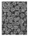
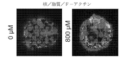
出願人は、まず、ヒトiPSC由来の前腸スフェロイドを使用することによって新規肝臓オルガノイド分化方法を確立した(Spence et al.,2011)(図1A)。第一ステップとして、出願人は、以前に記載されたように(D'Amour et al.,2005)、胚体内胚葉への分化を促進するためにBMPおよびアクチビンAを使用した。さらに、FGF4およびGSK3阻害剤(CHIR99021)を使用して前腸スフェロイドを誘導し、出芽スフェロイドを観察した。穏やかなピペッティングによりディッシュ上に播種した間葉系細胞を剥離した後、オルガノイドをMatrigel中に埋め込んだ。レチノイン酸(RA)は、毛細胆管および小管周囲鞘のサイズおよび複雑さの増大によって示されるように細胞極性を増強することが報告されている(Falasca et al.,1998)。胆汁輸送モデリングに適した極性オルガノイドを生成するために、オルガノイドをRAで処置した。オルガノイド生成方法を最適化するために、出願人は最初にRA処置の期間を変化させた。オルガノイドのアルブミン分泌レベルは、1、2、3、4、および5日間のRA処置について、それぞれD25で1160、1054、3092、4709、および3865ng/mLであり、4日間のRA処置プロトコルは最高レベルに達する傾向があった(図8)。したがって、アルブミン分泌のレベルに基づいて、RAの期間を4日間に設定した。形態学的には、RA処置後約10日で、上皮細胞で覆われた300を超えるオルガノイドが首尾よく生成され、管腔化構造を有するオルガノイドの比率は71%(216/305)であった(図1、パネルBおよび図9)。免疫組織化学分析は、アルブミンがオルガノイドの上皮細胞において陽性であることを明らかにし、そして興味深いことに、IV型コラーゲンは外表面に局在化し、ZO-1(接着帯閉塞)は管腔内層を染色し、これは、これらのオルガノイドが極性特性を有することを示唆する(図1、パネルC)。
Results Generation and Characterization of Polarized Liver Organoids from Multiple Human iPSCs Applicants first established a novel liver organoid differentiation method by using foregut spheroids derived from human iPSCs (Spence et al., 2011) ( Figure 1A). As a first step, Applicants used BMP and Activin A to promote differentiation to definitive endoderm as previously described (D'Amour et al., 2005). In addition, an FGF4 and GSK3 inhibitor (CHIR99021) was used to induce foregut spheroids and budding spheroids were observed. After detaching the mesenchymal cells seeded on the dish by gentle pipetting, the organoids were embedded in Matrigel. Retinoic acid (RA) has been reported to enhance cell polarity as indicated by increased size and complexity of bile canaliculi and peritubular sheaths (Falasca et al., 1998). Organoids were treated with RA to generate polar organoids suitable for bile transport modeling. To optimize the organoid generation method, Applicants first varied the duration of RA treatment. Organoid albumin secretion levels were 1160, 1054, 3092, 4709, and 3865 ng/mL at D25 for 1, 2, 3, 4, and 5 days of RA treatment, respectively, with the 4-day RA treatment protocol having the highest levels. (Fig. 8). Therefore, the duration of RA was set at 4 days based on the level of albumin secretion. Morphologically, approximately 10 days after RA treatment, over 300 organoids covered with epithelial cells were successfully generated, with a proportion of 71% (216/305) of organoids with tubular structures. (Fig. 1, panel B and Fig. 9). Immunohistochemical analysis revealed that albumin was positive in the epithelial cells of the organoids and, interestingly, type IV collagen was localized to the outer surface and ZO-1 (adhesin occlusion) to the luminal lining. staining, suggesting that these organoids have polar properties (Fig. 1, panel C).
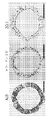
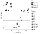
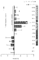
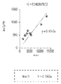
定量的ポリメラーゼ連鎖反応(qPCR)分析により、オルガノイド中の細胞は、アルファ-フェトプロテイン(AFP)、アルブミン(ALB)、レチノール結合タンパク質4(RBP4)、サイトケラチン19(CK19)、胆管細胞分化を制御する肝細胞核因子6(HNF6)、および分化中のシトクロムP450 3A4(CYP3A4)などの肝マーカー遺伝子の発現が有意に増加したことが明らかになった(図1、パネルD)。しかしながら、バルクオルガノイド由来RNAから抽出された最も肝臓的な遺伝子の発現レベルは、初代肝細胞よりもオルガノイドにおいて低かった。理論によって制限されることを意図しないが、これらの異なるmRNAプロファイルは、間質細胞マーカーによって同定された細胞の約30%が非実質細胞であるため、間質系列の存在に一部起因すると考えられ、これにより、オルガノイドは初代肝細胞よりもin vivoの肝臓組織により近いものとなる。出願人はさらに、RNA配列(RNA-seq)を用いた包括的な遺伝子発現分析によってオルガノイドをプロファイリングした。主成分分析は、オルガノイドにおける遺伝子発現が、iPSC由来胆管細胞および正常ヒト胆管細胞と類似していないことを示した(図1、パネルE)。さらに、培養上清中、ALB、フィブリノゲン(Fbg)および補体因子などの肝細胞特異的タンパク質がELISAによって確認された(図1、パネルF~G)。オルガノイドの肝機能性を定量するために、出願人は細胞数によって正規化されたアルブミン分泌レベルを調べた(図10)。アルブミン分泌レベルは、2133ng/日/106細胞であり(図1、パネルF)、公表されたiPSC由来肝細胞(Miki et al.,2011;Song et al.,2015;Song et al.,2009;Vosough et al.,2013)に関連するhPSCのHLCへの2Dおよび3D分化における他の実験(150~1000ng/日/106細胞)よりも高く、一方で、初代肝細胞は、3D足場において30~40μg/日/106細胞を産生する(Davidson et al.,2016;Dvir-Ginzberg et al.,2003)。これらの結果は、公表された文献において、肝臓オルガノイドが幹細胞由来の肝細胞と比較して妥当なアルブミン分泌活性を有する肝細胞を含んでいることを示した。重要なことに、このオルガノイド生成方法は再現性があり、したがって、管腔内オルガノイドがアルブミン分泌能を有する317D6および1383D6のiPS細胞株の両方から生成されたので、他のPSC株に適用可能である(図11)。まとめると、出願人は、肝細胞の特徴を有する多数の極性肝臓オルガノイドを生成するためのプロトコルを確立した。 Quantitative polymerase chain reaction (qPCR) analysis revealed that cells in organoids regulate alpha-fetoprotein (AFP), albumin (ALB), retinol binding protein 4 (RBP4), cytokeratin 19 (CK19), cholangiocyte differentiation Significantly increased expression of hepatic marker genes such as hepatocyte nuclear factor 6 (HNF6) and differentiating cytochrome P450 3A4 (CYP3A4) was revealed (Fig. 1, panel D). However, the expression levels of most hepatic genes extracted from bulk organoid-derived RNA were lower in organoids than in primary hepatocytes. Without intending to be bound by theory, it is believed that these distinct mRNA profiles are due in part to the presence of a stromal lineage, as approximately 30% of the cells identified by stromal cell markers are non-parenchymal cells. This makes the organoids more like liver tissue in vivo than primary hepatocytes. Applicants have further profiled the organoids by comprehensive gene expression analysis using RNA sequencing (RNA-seq). Principal component analysis showed that gene expression in organoids was dissimilar to iPSC-derived cholangiocytes and normal human cholangiocytes (Fig. 1, panel E). Furthermore, hepatocyte-specific proteins such as ALB, fibrinogen (Fbg) and complement factors were confirmed in the culture supernatant by ELISA (Fig. 1, panels FG). To quantify hepatic functionality of the organoids, Applicants examined albumin secretion levels normalized by cell number (Fig. 10). The albumin secretion level was 2133 ng/day/10 6 cells (Fig. 1, panel F), and compared with published iPSC-derived hepatocytes (Miki et al., 2011; Song et al., 2015; Song et al., 2009 ; Vosough et al., 2013) in 2D and 3D differentiation of hPSCs into HLCs (150-1000 ng/day/ 10 cells), whereas primary hepatocytes in 3D scaffolds It produces 30-40 μg/day/10 6 cells (Davidson et al., 2016; Dvir-Ginzberg et al., 2003). These results indicated that liver organoids contained hepatocytes with reasonable albumin secretory activity compared to stem cell-derived hepatocytes in the published literature. Importantly, this organoid generation method was reproducible and therefore applicable to other PSC lines, as intraluminal organoids were generated from both 317D6 and 1383D6 iPS cell lines with albumin secretion capacity. There is (Fig. 11). Taken together, Applicants have established a protocol for generating a large number of polarized liver organoids with hepatocyte characteristics.
胆汁酸産生ヒトiPSC-肝臓オルガノイドのミクロ解剖学的特徴付け
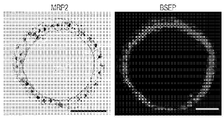
次に、肝臓オルガノイドが胆汁輸送活性を有するかどうかを試験するために、出願人は最初に胆汁合成および排泄機能に関与する主要なタンパク質を染色することによってオルガノイドを特徴付けた。BSEPおよびMRP2の免疫蛍光染色は、これらのタンパク質が管腔内領域に優先的に局在することを実証した(図2、パネルA)。胆管は最小の肝内分泌チャネルであり、毛細胆管腔は隣接する肝細胞の対向する原形質膜の修飾された先端領域によって形成された空間からなる(Cutrin et al.,1996;Tsukada et al.,1995)。さらに、それは密着帯複合体によって区切られ、微絨毛は毛細胆管内腔の内側に位置する(Tsukada et al.,1995)。ZO-1染色は肝臓の毛細胆管領域を染色することが知られており、図1、パネルCは密着帯が肝臓オルガノイドの内側に位置することを示唆した。透過型電子顕微鏡検査は、オルガノイドが内腔に指向した微絨毛を含有することを明らかにした(図2、パネルB)。これらの解剖学的特徴と一致して、qRT-PCR分析は、オルガノイドがABCB11およびNa+-タウロコール酸共輸送ポリペプチド(NTCP)の遺伝子発現を有することを明らかにしたが、そのレベルは初代肝細胞よりもオルガノイドにおいて低かった(図2、パネルC)。したがって、オルガノイドは、接着帯によって内腔から分離された極性ヒト肝細胞を含み、これはin vivo肝毛細胆管を模倣する独特のミクロ解剖学的構造を反映する。
Microanatomical characterization of bile acid-producing human iPSC-liver organoids Next, to test whether liver organoids have bile transport activity, applicants first investigated the major proteins involved in bile synthesis and excretion functions. The organoids were characterized by staining. Immunofluorescence staining of BSEP and MRP2 demonstrated that these proteins preferentially localized to the intraluminal region (Fig. 2, panel A). The bile duct is the smallest hepatic endocrine channel, and the bile canalicular lumen consists of the space formed by the modified apical regions of the opposing plasma membranes of adjacent hepatocytes (Cutrin et al., 1996; Tsukada et al., 1995). In addition, it is delimited by tight junction complexes and microvilli are located inside the bile canaliculus lumen (Tsukada et al., 1995). ZO-1 staining is known to stain the bile canaliculi region of the liver and Figure 1, panel C suggested that tight junctions are located inside the liver organoids. Transmission electron microscopy revealed that the organoids contained lumen-oriented microvilli (Fig. 2, panel B). Consistent with these anatomical features, qRT-PCR analysis revealed that the organoids possessed gene expression of ABCB11 and Na+-taurocholate cotransporting polypeptide (NTCP), although the levels were lower than those of primary hepatocytes. in organoids (Fig. 2, panel C). Thus, the organoids contain polarized human hepatocytes separated from the lumen by adherends, reflecting a unique micro-anatomical structure that mimics the in vivo hepatic bile canaliculi.
次に、胆汁酸(BA)産生能力を決定するために、出願人は、オルガノイド培養物から収集した管腔内液にELISAを実施した。管腔内液の総BAプールのレベルは、26.7μg/日/106細胞(直径200μmのオルガノイド中で約125μmol/L)であり(図2、パネルD)、そして驚くべきことに、BA濃度は、以前の報告(Ni et al.,2016)におけるサンドイッチ培養由来の初代肝細胞のもの(約40μg/日/106細胞、培養上清中10μmol/L)に匹敵した。このように、オルガノイドは毛細胆管様の形態を有するだけでなく、胆汁酸産生および分泌活性も有し、これは、胆汁酸輸送経路が正しく構築されていることを示唆している。 Next, to determine bile acid (BA) production capacity, Applicants performed an ELISA on intraluminal fluid collected from the organoid cultures. The level of total BA pool in the luminal fluid was 26.7 μg/day/10 6 cells (approximately 125 μmol/L in 200 μm diameter organoids) ( FIG. 2 , panel D), and surprisingly, BA The concentration was comparable to that of primary hepatocytes derived from sandwich culture in a previous report (Ni et al., 2016) (approximately 40 μg/day/10 6 cells, 10 μmol/L in culture supernatant). Thus, the organoids not only have bile canaliculi-like morphology, but also have bile acid production and secretion activity, suggesting that the bile acid transport pathway is correctly constructed.
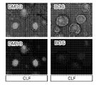
ヒト肝臓オルガノイドにおける胆汁酸摂取と排泄の動的可視化
胆汁酸排泄は、胆汁流の主要な決定要因であり、それ故に、この系における欠陥は様々な肝疾患病理に関連した胆汁分泌障害(胆汁うっ滞)をもたらす可能性がある(Nishida et al.,1991)。肝細胞の頂端(毛細胆管)膜に位置する排出輸送タンパク質は、薬物および代謝物を含む多くの内因性および外因性化合物の肝臓除去において重要な役割を果たしている(KockおよびBrouwer,2012)。BSEPおよびMRP2は、ヒトにおいて毛細胆管胆汁酸塩輸送を媒介する。胆汁輸送のための主要なタンパク質の発現陽性を実証した後、出願人は次に、オルガノイドが胆汁酸をその内腔に活発に輸送することができるかどうか考えた。第一に、オルガノイドへの胆汁酸の摂取を調べるために、出願人は、胆汁酸塩アナログ(Mork et al.,2012)であるコリルグリチルアミド-フルオレセイン(CGamF)を用いてオルガノイドに挑戦した。外部からのCGamFの処理後、オルガノイドの管腔内へのCGamFの蓄積は首尾よく確認された(図2、パネルE)。同様に、蛍光胆汁酸コリル-リシル-フルオレセイン(CLF)は再現性よく排泄され、複数のヒトiPSC株からオルガノイド内に蓄積されることが見出された(図2、パネルF)。このアッセイの特異性を決定するために、出願人は、CRISPR-Cas9に基づく遺伝子編集アプローチを用いてBSEP非官能化対立遺伝子を有するiPSC株を開発した。BSEPは胆汁輸送に関与し、これと一致して、BSEP-KO iPSC-オルガノイドは、親対照オルガノイドと比較して蛍光胆汁酸を蓄積することができなかった。まとめると、これらのデータは、オルガノイドが胆汁酸を外側から摂取し、それらをオルガノイドの内側に排出する能力を有することを示唆している。
Dynamic visualization of bile acid uptake and excretion in human liver organoids. (Nishida et al., 1991). Efflux transport proteins located in the apical (bile canaliculi) membrane of hepatocytes play an important role in the hepatic elimination of many endogenous and exogenous compounds, including drugs and metabolites (Kock and Brouwer, 2012). BSEP and MRP2 mediate canalicular bile salt transport in humans. After demonstrating positive expression of key proteins for bile transport, Applicants next asked whether organoids could actively transport bile acids into their lumen. First, to investigate bile acid uptake into organoids, Applicants challenged organoids with a bile salt analog (Mork et al., 2012), cholylglycylamido-fluorescein (CGamF). . After exogenous treatment of CGamF, accumulation of CGamF within the lumen of organoids was successfully confirmed (Fig. 2, panel E). Similarly, the fluorescent bile acid cholyl-lysyl-fluorescein (CLF) was reproducibly excreted and found to accumulate within organoids from multiple human iPSC lines (Fig. 2, panel F). To determine the specificity of this assay, Applicants developed an iPSC line with a BSEP non-functionalized allele using a CRISPR-Cas9-based gene editing approach. BSEP is involved in bile transport, and consistent with this, BSEP-KO iPSC-organoids failed to accumulate fluorescent bile acids compared to parental control organoids. Taken together, these data suggest that organoids have the ability to take up bile acids from the outside and excrete them inside the organoid.
CYP2C9*2 iPSC-肝臓オルガノイドに特異的なボセンタン誘発胆汁うっ滞
オルガノイドに基づく胆汁うっ滞表現型決定法の臨床的関連性を試験するために、出願人は、忠実度の問題に対処するために、出願者のシステムに薬理ゲノム学的洞察を採用した。具体的には、周知の感受性遺伝子変異体(すなわち、例えば、ボセンタンについては、Clin Pharmacol Ther.2013 Dec;94(6):678-86.doi:10.1038/clpt.2013.143.Epub 2013 Jul 17.「Association of CYP2C9*2 with bosentan-induced liver injury」に記載されているCYP2C9*2)を保有する複数のiPSC株が収集され(図3、パネルA)、ボセンタンの存在下でそれらの胆汁うっ滞性の可能性を比較した(図3、パネルB)。興味深いことに、オルガノイドへのCLF排泄は、CY2C9*2保有オルガノイドでは著しく損なわれたが、非保有オルガノイドでは損なわれなかった。これは、図3、パネルCに示されるように、CYP2C9*2の不在下で3つの異なるiPSC由来オルガノイドにおいて示される、ボセンタンにより誘導される胆汁うっ滞に対する臨床的傾向と一致する。対照的に、イリノテカンに基づく胆汁うっ滞は、CYP2C9*2 iPSC株に特異的ではなかった。これらの結果は、オルガノイドに基づく胆汁うっ滞アッセイがヒトの多様性のいくつかの局面を予測することを示した。
CYP2C9*2 iPSC-Liver Organoid-Specific Bosentan-Induced Cholestasis In order to test the clinical relevance of the organoid-based cholestasis phenotyping method, applicants sought , which employed pharmacogenomic insights into Applicants' system. Specifically, known susceptibility gene variants (i.e., for bosentan, for example, Clin Pharmacol Ther. 2013 Dec;94(6):678-86.doi:10.1038/clpt.2013.143.Epub 2013). Jul 17. Multiple iPSC lines harboring CYP2C9*2) described in “Association of CYP2C9*2 with bosentan-induced liver injury” were harvested (Fig. 3, panel A) and their The cholestatic potential was compared (Fig. 3, panel B). Interestingly, CLF excretion into organoids was significantly impaired in CY2C9*2-bearing organoids, but not in non-bearing organoids. This is consistent with the clinical propensity for bosentan-induced cholestasis shown in three different iPSC-derived organoids in the absence of CYP2C9*2, as shown in FIG. 3, panel C. In contrast, irinotecan-based cholestasis was not specific for the CYP2C9*2 iPSC line. These results indicated that organoid-based cholestasis assays are predictive of several aspects of human diversity.
オルガノイドにおけるハイスループット薬物誘発性胆汁うっ滞評価
薬物によって誘発されたDILIにおける胆汁うっ滞の重要な役割を考慮して、出願人は次に、このオルガノイドモデルが特定の化合物の存在下でのDILIの病理学を反映するかどうかを考えた。多数の化合物を試験する前に、出願人は、CLFおよびCGamFの両方がいくつかの問題:1.バックグラウンドが強く、手動での洗浄プロセスが必要となる;2.シグナル強度が弱いため、慎重な集録設定が必要となる、に起因して高速イメージングに適用できないので、まずハイスループット蛍光ベースアッセイを開発しようとした。あるいは、肝細胞における排出輸送の有用なマーカーであると報告されているフルオレセインジアセテート(FD)の使用が提案されている(BarthおよびSchwarz,1982;Bravo et al.,1998)。極性蛍光代謝物フルオレセインは、細胞から毛細胆管腔内に活発に輸送されるまで細胞内に捕捉される(Malinen et al.,2014)。培地交換や曝露量の調整をせずにFDを輸送能力の生の評価に使用できるかどうかを判断するために、経時的肝胆道輸送活性を低速度撮影イメージングを用いてさらに調べた。オルガノイドをフルオレセインジアセテートと共に45分間インキュベートし、そして管腔内蓄積が処理後20分でオルガノイドの内部に観察された(図4、パネルA、B)。この輸送の流れの反対方向は、オルガノイドへのFDの微量注入によって決定された。管腔内へのジアセテートの微量注入後、フルオレセインは内部に留まり、オルガノイドの外部には観察されなかった(図4、パネルC)。要約すると、このFDに基づく評価モデルは、単純な蛍光ライブイメージング分析によって肝臓オルガノイドにおける一方向性の排出胆汁輸送を評価するためのハイスループットの可能性を有する。
High Throughput Drug-Induced Cholestasis Assessment in Organoids Given the important role of cholestasis in drug-induced DILI, Applicants next proposed that this organoid model could be used to improve DILI in the presence of certain compounds. We considered whether it reflects the pathology of Before testing a large number of compounds, Applicants noted that both CLF and CGamF had several problems:1. 1. Strong background, requiring a manual wash process; We first attempted to develop a high-throughput fluorescence-based assay as it is not applicable for high-speed imaging due to the weak signal intensity requiring careful acquisition settings. Alternatively, the use of fluorescein diacetate (FD), reported to be a useful marker of efflux transport in hepatocytes, has been proposed (Barth and Schwarz, 1982; Bravo et al., 1998). The polar fluorescent metabolite fluorescein is trapped intracellularly until it is actively transported from the cell into the bile canalicular lumen (Malinen et al., 2014). To determine whether FD can be used for live assessment of transport capacity without medium changes or adjustment of exposure, hepatobiliary transport activity over time was further examined using time-lapse imaging. Organoids were incubated with fluorescein diacetate for 45 minutes, and intraluminal accumulation was observed inside the
次に、出願人は、FDAが承認した10種類の薬物の実行可能な投与量を評価することによってFDに基づくアッセイの忠実度を検証し、細胞損傷による二次的な障害を測定した。出願人は、許容可能な生存率を有する9つの化合物について最適用量を首尾よく見出した。対照的に、アミオダロン(AMIO)は試験された範囲内でオルガノイドに対して有意に有毒であり、それ故、AMIOはさらなる潜在的なDILI評価試験から排除された(図12)。出願人は、DILI機序に基づいて3つのタイプ:胆汁うっ滞を伴わないDILI化合物(クラスA)、胆汁うっ滞を伴うDILI化合物(クラスB)、およびDILI化合物として報告されていない化合物(クラスC)(Oorts et al.,2016)のうちの1つとして分類された9つのトレーニング化合物(TC)と共にFDを使用して、オルガノイドにおける胆汁うっ滞の可能性を調査した。FD排泄の阻害可能性を定量化するために、出願人は、画像Jによってオルガノイドの外側と内側との間の蛍光強度比を決定することによって、単純だが頑健な定量化方法を開発した(図4、パネルB)。検証研究として、出願人は最初にシクロスポリンA(CSA)を用いて阻害率を評価する能力を確認した。FD処置後5分で、対照(DMSO)と比較して、24時間CSAで処置した群において有意な減少(対照と比較して0.4)が観察された(図4、パネルB)。次に、出願人は、このアプローチの忠実度を評価するために9つのTCを複数の濃度でスクリーニングした。興味深いことに、このスクリーニングシステムでは、TCの処置後24時間で、臨床的観察と同様に、FDの排出はクラスB化合物、ボセンタン、CSA、トログリタゾンおよびネファゾドンにおいて有意に減少した(p<0.01または0.05)が、一方で、クラスAおよびクラスCの化合物では阻害効果は観察されなかった(図4、パネルDの上の画像および図4、パネルE)。これらの結果は、肝臓オルガノイドモデルが、ヒト表現型との関連性が高い薬物開発における候補化合物についての胆汁輸送阻害効力を分類するのに有用であることを示唆した。 Applicants then validated the fidelity of the FD-based assay by evaluating viable dosages of ten FDA-approved drugs to measure damage secondary to cell damage. Applicants have successfully found optimal doses for nine compounds with acceptable survival rates. In contrast, amiodarone (AMIO) was significantly toxic to organoids within the range tested and therefore AMIO was excluded from further potential DILI evaluation studies (Fig. 12). Applicants have identified three types based on the DILI mechanism: DILI compounds without cholestasis (Class A), DILI compounds with cholestasis (Class B), and compounds not reported as DILI compounds (Class C) FD was used with nine training compounds (TC) classified as one of (Oorts et al., 2016) to investigate the cholestasis potential in organoids. To quantify the potential inhibition of FD excretion, Applicants developed a simple but robust quantification method by determining the fluorescence intensity ratio between the outside and inside of the organoid by Image J (Fig. 4, panel B). As a validation study, Applicants first confirmed the ability to assess percent inhibition using cyclosporin A (CSA). At 5 minutes after FD treatment, a significant reduction (0.4 compared to control) was observed in the group treated with CSA for 24 hours compared to control (DMSO) (Fig. 4, panel B). Applicants then screened nine TCs at multiple concentrations to assess the fidelity of this approach. Interestingly, in this screening system, 24 hours after TC treatment, FD excretion was significantly reduced for class B compounds, bosentan, CSA, troglitazone and nefazodone, similar to clinical observations (p<0.01 or 0.05), whereas no inhibitory effect was observed for class A and class C compounds (Fig. 4, panel D top image and Fig. 4, panel E). These results suggested that the liver organoid model would be useful for classifying the bile transport inhibition potency for candidate compounds in drug development that are highly relevant to the human phenotype.
オルガノイドにおけるミトコンドリア過負荷の評価
さらに、ミトコンドリアの毒性は、DILIの発症に関連する複数の機序においてDILIにおける中心的な役割を果たすので(Pessayre et al.,2012)、出願人はミトコンドリアの健康評価を調査した。この研究では、オルガノイド中のミトコンドリアの健康度を調べるために、ミトコンドリアの健康度を直接読み取ることにより、ミトコンドリア膜電位(MMP)の指標を用いて無傷細胞のMMPをモニターした(Li et al.,2014)。24時間のTCの処置後、MMPの用量依存的な増加がトルカポン(2~8倍の変化、p<0.01)、ジクロフェナク(7~13倍の変化、p<0.05または0.01)、CSA(3~7倍の変化、p<0.01)およびネファゾドン(4~42倍の変化、p<0.01)の処置で観察された(図5、パネルAの下の画像およびグラフ)。さらに、用量依存性は観察されなかったが、トログリタゾンはオルガノイド中のMMPも増加させた(3~5倍の変化、p<0.05)。一方、ボセンタン、エンタカポンおよびピオグリタゾンの処置後、MMPの増加は複数回投与においてさえ明確に観察されなかった。これらの結果は、肝臓オルガノイドに基づく毒性スクリーニング(LoT)と命名されたこのライブ画像に基づくアッセイが、ミトコンドリア毒性のあるなしにかかわらず化合物を区別することを実証した。
Assessment of mitochondrial overload in organoids Furthermore, since mitochondrial toxicity plays a central role in DILI in multiple mechanisms associated with the development of DILI (Pessayre et al., 2012), Applicants evaluated mitochondrial health. investigated. In this study, to examine mitochondrial health in organoids, we monitored MMPs in intact cells using an index of mitochondrial membrane potential (MMP), a direct readout of mitochondrial health (Li et al., 2014). After treatment with TC for 24 hours, dose-dependent increases in MMPs were observed for tolcapone (2- to 8-fold change, p<0.01), diclofenac (7- to 13-fold change, p<0.05 or 0.01). ), observed with CSA (3-7 fold change, p<0.01) and nefazodone (4-42 fold change, p<0.01) treatments (Fig. 5, panel A lower images and graph). In addition, troglitazone also increased MMPs in organoids (3- to 5-fold change, p<0.05), although no dose-dependency was observed. On the other hand, after treatment with bosentan, entacapone and pioglitazone no increase in MMPs was clearly observed even at multiple doses. These results demonstrated that this live image-based assay, termed liver organoid-based toxicity screening (LoT), discriminates compounds with and without mitochondrial toxicity.
LoTシステムによるDILI化合物の機構的分類の再検討
ヒトDILIの重度の症状発現は多因子性であり、ミトコンドリアおよびBSEP阻害などのDILIの既知の機序に特に関連する薬効の組み合わせと高度に関連している(Aleo et al.,2014)。しかし、現在のin vitro機能モデルはそのような多因子寄与を評価することは困難である。LoTシステムにおける多重化された生の機能的読み出しの利点を考慮して、出願人は、生存、胆汁うっ滞およびミトコンドリアストレスの間の関係を分析することを試みた。注目すべきことに、CSA、TROおよびNEFAなどの24時間で二重作用(胆汁うっ滞およびミトコンドリアストレス)を有する薬物は、TOL、DICLOおよびBOSと比べて72時間で細胞生存率を有意に低下させた。これらのデータは、二重毒性がDILIの重症度と非常に関連していることを示す臨床データと同等であり、以前の報告(Aleo et al.,2014)と一致する(図5、パネルB、C)。さらに、本出願人はまた、130μMでのエンタカポン処置はオルガノイド生存率を減少させた(24時間で85%から72時間で64%まで)ことを指摘した。エンタカポンは、DILIを誘導するために血漿タンパク質、主にアルブミンへの広範な結合を必要とする(Fisher et al.,2002)。しかしながら、利用可能な方法に基づいて、エンタカポンがどのように肝臓に対して有毒であるかは依然として分かっていない(Oorts et al.,2016)。まとめると、LoTシステムはDILIの主要な機序分類のための有利な人体モデルシステムであり、未知の複雑な機序をさらに詳しく説明するための有用な試験プラットフォームである。
Reexamination of the Mechanistic Classification of DILI Compounds by the LoT System The severe manifestations of human DILI are multifactorial and highly associated with a combination of pharmacological effects specifically related to known mechanisms of DILI such as mitochondrial and BSEP inhibition. (Aleo et al., 2014). However, current in vitro functional models have difficulty assessing such multifactorial contributions. Given the advantages of multiplexed raw functional readouts in the LoT system, Applicants sought to analyze the relationship between survival, cholestasis and mitochondrial stress. Of note, drugs with dual actions (cholestasis and mitochondrial stress) at 24 hours such as CSA, TRO and NEFA significantly reduced cell viability at 72 hours compared to TOL, DICLO and BOS. let me These data are comparable with clinical data showing that dual toxicity is highly associated with DILI severity, and are consistent with previous reports (Aleo et al., 2014) (Fig. 5, panel B). , C). In addition, applicants also noted that entacapone treatment at 130 μM decreased organoid viability (from 85% at 24 hours to 64% at 72 hours). Entacapone requires extensive binding to plasma proteins, primarily albumin, to induce DILI (Fisher et al., 2002). However, based on available methods, it remains unknown how entacapone is toxic to the liver (Oorts et al., 2016). Taken together, the LoT system is an advantageous human model system for the primary mechanistic classification of DILI and a useful testing platform for further delineation of unknown complex mechanisms.
ヒト肝臓オルガノイドにおけるDILIに対する脆弱性の評価
DILIの発生率は多数の宿主因子によってしばしば混乱することが知られている。確かに、アセトアミノフェンのようないくつかの薬物からの肝毒性の危険性が、げっ歯類とヒトの両方において、肥満およびNAFLDに起因して、非常に増加するという証拠が増えている(APAP)(Fromenty,2013;Michaut et al.,2016)。したがって、無症状の段階であっても、患者に対してそのような「脆弱な」状態にあるDILI可能性を予測することは重要である。本研究では、出願人は、不飽和脂肪酸、オレイン酸への同時曝露によって脂肪毒性オルガノイドモデルを確立した(図6、パネルA)。オルガノイドに対するオレイン酸処置の3日後、オルガノイド中の脂質蓄積は激しくなった(図6、パネルB)。脂肪酸の酸化は活性酸素種(ROS)の重要な供給源であり、それはATPおよびニコチンアミドジヌクレオチドの枯渇をもたらし、脂肪肝におけるDNA損傷を誘発する(BrowningおよびHorton,2004)。これと一致して、ROS産生は脂質処置オルガノイドにおいて観察された(図6、パネルCおよび図13、パネルA)。さらに、脂肪酸は、公表されている表現型と同様に肝臓ミトコンドリアの大量の腫脹を誘発した(図6、パネルDおよび図13、パネルB)(ZborowskiおよびWojtczak,1963)。ラットモデルにおいて肝ミトコンドリア機能障害がNAFLDの発症に先行するので(Rector et al.,2010)、これらの結果は、肝毒性オルガノイドがある程度in vivo脂肪肝モデルをモデル化することを示している。
Evaluation of Vulnerability to DILI in Human Liver Organoids It is known that the incidence of DILI is often confounded by a number of host factors. Indeed, there is increasing evidence that the risk of hepatotoxicity from some drugs, such as acetaminophen, is greatly increased due to obesity and NAFLD in both rodents and humans ( APAP) (Fromenty, 2013; Michaut et al., 2016). Therefore, it is important to predict DILI probabilities in such a "vulnerable" state for patients, even in asymptomatic stages. In this study, Applicants established a lipotoxic organoid model by co-exposure to unsaturated fatty acids, oleic acid (Fig. 6, panel A). After 3 days of oleic acid treatment on organoids, lipid accumulation in organoids became severe (Fig. 6, panel B). Oxidation of fatty acids is an important source of reactive oxygen species (ROS), which leads to depletion of ATP and nicotinamide dinucleotides and induces DNA damage in fatty liver (Browning and Horton, 2004). Consistent with this, ROS production was observed in lipid-treated organoids (Figure 6, panel C and Figure 13, panel A). Furthermore, fatty acids induced massive swelling of liver mitochondria (Fig. 6, panel D and Fig. 13, panel B), similar to the published phenotype (Zborowski and Wojtczak, 1963). Since hepatic mitochondrial dysfunction precedes the development of NAFLD in rat models (Rector et al., 2010), these results indicate that hepatotoxic organoids model the in vivo fatty liver model to some extent.
この脂肪毒性オルガノイドモデルを強化されたROS産生を伴う脆弱な状態として認識して、トログリタゾン(0~50μM)を24時間処置し、オルガノイドにおける細胞生存率を評価した。50μMのトログリタゾン単独の処置により、細胞生存率は24時間で85%であり、一方、72時間で67%に減少した。しかしながら、脂肪毒性状態に対するトログリタゾンの処置後、オルガノイド死のためにオルガノイドの大量の断片化が観察された。その後の細胞生存率分析によりこの結果が確認された(対照と比較して約40%、p<0.05)(図6、パネルEおよび6、パネルF)。 Recognizing this lipotoxic organoid model as a vulnerable state with enhanced ROS production, troglitazone (0-50 μM) was treated for 24 hours to assess cell viability in organoids. Treatment with 50 μM troglitazone alone resulted in cell viability of 85% at 24 hours, whereas it decreased to 67% at 72 hours. However, after troglitazone treatment for lipotoxic conditions, massive fragmentation of organoids was observed due to organoid death. Subsequent cell viability analysis confirmed this result (approximately 40% compared to control, p<0.05) (FIGS. 6, panels E and 6, panel F).
次に、出願人は、潜在的な治療用化合物によってオルガノイドをDILI様状態から回復できるかどうかを調べた。静脈内NACはアセトアミノフェンに関連しない急性肝不全患者の生存率を改善し(Lee et al.,2009)、トログリタゾン誘発性細胞毒性を減少させた(Rachek et al.,2009)ので、出願人は、ROS産生を阻害するためにN-アセチルシステイン(NAC)、抗酸化剤を用いた。予想通り、細胞生存率はNACによって有意に改善され、これはNACが脆弱な条件下でさえオルガノイドにおける細胞死を救済したことを示唆している(図6、パネルEおよび6、パネルF)。ほとんどのDILIの場合、唯一の介入は、原因薬物が同定できればそれを除去することである(PolsonおよびLee,2005)(Bohan et al.,2001;NavarroおよびSenior,2006)。このLoTシステムは、DILIを治療するための多剤レジメンおよび創薬に関連する原因薬物を同定するための有用なツールとなり得る。 Applicants next investigated whether potential therapeutic compounds could restore organoids from a DILI-like state. Intravenous NAC improved survival in patients with acute liver failure not associated with acetaminophen (Lee et al., 2009) and reduced troglitazone-induced cytotoxicity (Rachek et al., 2009). used N-acetylcysteine (NAC), an antioxidant, to inhibit ROS production. As expected, cell viability was significantly improved by NAC, suggesting that NAC rescued cell death in organoids even under vulnerable conditions (Fig. 6, panels E and 6, panel F). For most DILI, the only intervention is to remove the causative drug if it can be identified (Polson and Lee, 2005) (Bohan et al., 2001; Navarro and Senior, 2006). This LoT system can be a useful tool for identifying causative drugs relevant to multi-drug regimens and drug discovery to treat DILI.
肝不全を含む重篤な有害事象(SAE)は、臨床開発中の薬物の減少または市販医薬品の中止の主な原因である。特に、DILIは薬物開発における重大な課題であり、ここで、トランスポーター活性の阻害によって誘導される薬物誘発性胆汁うっ滞は1つの主要な原因である。ヒト初代肝細胞を用いるサンドイッチ培養は、医薬品における現在の最良の選択である。最近の報告は、ヒト線維芽細胞からの分化転換細胞を用いた肝細胞に基づく胆汁うっ滞モデルの有望性を示したが(Ni et al.,2016)、ヒト肝細胞供給源が多様かつ制限されているため、ならびに複雑な定量化アルゴリズムが必要であるために、これらのアッセイプラットフォームは依然として再現性の課題ならびにスループットの問題を有する。さらに、ヒト肝細胞癌細胞株であるHepaRG細胞も胆汁うっ滞の特徴を評価するのに有用であるが、それらの低いBSEP(胆汁酸塩排出ポンプ、またはABCB11、胆汁酸排出のための重要な輸送体、ならびに胆汁うっ滞剤の主な標的)活性および時間のかかる分化手順のために、その使用が制限される(Le Vee et al.,2013)。さらに重要なことに、本質的な解剖学的構造の欠如はそれらの製薬産業への実用化を制限する。あるいは、記載された方法は、試験化合物の存在下でライブ蛍光イメージングにより胆汁輸送活性を測定するための簡単で、頑強な、かつハイスループットなシステムを可能にする。LoTアッセイの主な利点は以下:1.費用対効果(50オルガノイドあたり12.35ドル、384ウェルあたり94.85ドル)、2.アッセイスループット(単一オルガノイドで測定可能)、3.ミトコンドリアストレスなどの他の因子間の相互作用を分析するための多重読み出し、を含む。特に、上述のように、遡及的研究は、細胞生存率が二重読み出し;ミトコンドリアおよび胆汁うっ滞ストレスに依存して減少したため、複数の細胞ストレス可能性がDILI(Aleo et al.,2014)の発生率と関連しており、LoTアッセイはこの研究を用いた結果と同等であることを明らかにした。酸化ストレスは細胞死に重要な役割を果たしており、胆汁うっ滞性肝障害の発症と関連付けられた(Serviddio et al.,2004)。疎水性胆汁酸は、胆汁うっ滞の間に細胞内に蓄積し、正常なミトコンドリアの電子伝達を妨害し、呼吸複合体IおよびIIIの活性を阻害し、その結果アデノシン三リン酸合成を減少させ(Krahenbuhl et al.,1994)、ミトコンドリア機能不全誘導アポトーシスをもたらす(Bernardi,1996)。これらの知見と一致して、出願人のこれらの二重読み出しの相関分析は、図5に見られるように、胆汁うっ滞ストレスがミトコンドリアストレスと比較して肝障害についてより支配的な因子であることを示した。したがって、LoTシステムはDILI機序を調査するためのモデルシステムとして使用することができる。 Serious adverse events (SAEs), including liver failure, are the leading cause of drug decline in clinical development or discontinuation of marketed drugs. In particular, DILI is a significant challenge in drug development, where drug-induced cholestasis induced by inhibition of transporter activity is one major cause. Sandwich cultures with human primary hepatocytes are the current best choice in medicine. A recent report showed the promise of a hepatocyte-based cholestasis model using transdifferentiated cells from human fibroblasts (Ni et al., 2016), but human hepatocyte sources are diverse and limited. These assay platforms still have reproducibility challenges as well as throughput problems because of the complexity of the quantification algorithms and the need for complex quantification algorithms. In addition, HepaRG cells, a human hepatocellular carcinoma cell line, are also useful for assessing cholestasis characteristics, although their low BSEP (bile salt efflux pump, or ABCB11, critical for bile acid efflux) transporter, and major target of cholestatic agents) activity and time-consuming differentiation procedures limit its use (Le Vee et al., 2013). More importantly, their lack of essential anatomy limits their practical application to the pharmaceutical industry. Alternatively, the methods described enable a simple, robust, and high-throughput system for measuring bile transport activity by live fluorescence imaging in the presence of test compounds. The main advantages of the LoT assay are:1. Cost-effectiveness ($12.35 per 50 organoids, $94.85 per 384 wells);2. assay throughput (measurable on a single organoid);3. Including multiplex readouts to analyze interactions between other factors, such as mitochondrial stress. In particular, as mentioned above, retrospective studies have shown that multiple cellular stress possibilities were identified in DILI (Aleo et al., 2014), as cell viability decreased in a dual readout; It was associated with incidence and the LoT assay revealed comparable results with this study. Oxidative stress plays an important role in cell death and has been associated with the development of cholestatic liver injury (Serviddio et al., 2004). Hydrophobic bile acids accumulate intracellularly during cholestasis, interfere with normal mitochondrial electron transport, and inhibit the activity of respiratory complexes I and III, resulting in decreased adenosine triphosphate synthesis. (Krahenbuhl et al., 1994), leading to mitochondrial dysfunction-induced apoptosis (Bernardi, 1996). Consistent with these findings, Applicants' correlation analysis of these dual readouts indicates that cholestatic stress is the more dominant factor for liver injury compared to mitochondrial stress, as seen in FIG. showed that Therefore, the LoT system can be used as a model system to investigate DILI mechanisms.
さらに、最近のiPSCパネル集団の確立を考慮すると、個人における異なる感受性についての潜在的評価もまた有望である(Inoue et al.,2014)。従来のin vitroアッセイシステムにおけるSAEの予測は、一般的に個体差に焦点を合わせていないが、SAEは多くの場合、SAEが発生しやすい小さな準患者群で起こる(StevensおよびBaker,2009)。LoTシステムを多様な集団iPSCパネルに適用することにより、以前は到達できないものであったSAEに対する異なる感受性について提供できるようになる。DILIの極めてまれな性質を考慮すると、特定のゲノムまたは民族的要因を有する患者の細胞の使用は、現在知られていないDILIの特異体質的な機序を解明するのに役立つだろう。したがって、LoTは、DILIの可能性を最小にするための本質的な洞察を提供することによって、製薬産業にとってゲームチェンジング的戦略として役立ち得る(図7)。 Furthermore, given the recent establishment of iPSC panel populations, the potential evaluation of differential susceptibility in individuals is also promising (Inoue et al., 2014). Prediction of SAEs in conventional in vitro assay systems generally does not focus on individual variability, although SAEs often occur in small sub-patient populations prone to SAEs (Stevens and Baker, 2009). Application of the LoT system to diverse population iPSC panels will provide different susceptibility to SAEs that were previously unattainable. Given the extremely rare nature of DILI, the use of patient cells with specific genomic or ethnic factors will help elucidate currently unknown idiosyncratic mechanisms of DILI. Therefore, LoT may serve as a game-changing strategy for the pharmaceutical industry by providing essential insights to minimize the likelihood of DILI (Fig. 7).
このオルガノイドモデルにおける一つの制限は免疫学的反応の欠如である。過敏性反応から生じる免疫学的効果は、特異体質性DILIについての1つの可能な機序である。薬物による過敏性を評価するためのin vitroモデルは限られているが、トログリタゾン誘導性細胞傷害性による感受性は、肝細胞株、Huh7細胞およびTHP-1細胞を用いたin vitro共培養モデルを用いて高められた(Edling et al.,2009)。したがって、免疫系統に焦点を当てることによってLoTプラットフォームを進めることは、肝細胞炎症を評価するために興味深いものとなる。それにもかかわらず、LoT試験プラットフォームは、複数のFDA承認薬物による胆汁排出機能の阻害がこのアッセイにおいて再現可能に観察されるので、個々のオルガノイドから再現可能かつ大量のデータセットを生成することにおいて優れているように思われる。胆汁うっ滞が薬物誘発性、脂肪毒性、感染性および先天性の状態を含む広範囲の肝疾患によって誘発されることを考慮すると(Chatterjee et al.,2014)、オルガノイドに基づくLoTアッセイは、機序的研究ならびにDILIを超えた薬物スクリーニング用途の可能性を伴って、様々な状況における肝内胆汁うっ滞の分析に有用である。 One limitation in this organoid model is the lack of immunological response. Immunological effects resulting from hypersensitivity reactions are one possible mechanism for idiosyncratic DILI. Although in vitro models for assessing drug hypersensitivity are limited, susceptibility by troglitazone-induced cytotoxicity was demonstrated using an in vitro co-culture model using hepatic cell lines, Huh7 cells and THP-1 cells. (Edling et al., 2009). Therefore, advancing the LoT platform by focusing on the immune system will be of interest to assess hepatocyte inflammation. Nonetheless, the LoT testing platform excels at generating reproducible and large datasets from individual organoids, as inhibition of biliary excretion function by multiple FDA-approved drugs is reproducibly observed in this assay. It seems that Considering that cholestasis is induced by a wide range of liver diseases, including drug-induced, lipotoxic, infectious and congenital conditions (Chatterjee et al., 2014), organoid-based LoT assays suggest that the mechanism It is useful for the analysis of intrahepatic cholestasis in a variety of settings, with potential drug screening applications beyond DILI as well as clinical studies.
LoTアッセイによる脆弱なヒト肝状態の研究
肥満などの宿主因子は、DILIの発症に大きな影響を与えることが知られている(Heidari et al.,2014)が、その複雑な性質のために臨床現場では多くの場合理解されていない。肥満または脂肪肝の存在は、生体異物および非毒性化学物質(例えば、薬物)によって引き起こされる肝障害に対して患者を脆弱にする可能性があり、これらは危険因子の存在下でより低用量で肝毒性になる可能性がある(Fromenty,2013)。それにもかかわらず、現在の臨床試験システムは、一握りのバイオマーカー(ALT、AST)レベルで脆弱な肝臓条件下で志願者を層別化するためには設計されていない。脂肪症を有する患者の数は無症状であり、投薬前にバイオマーカーで検出不可能であるため、臨床段階に入る前にこの脆弱条件における結果を予測することが重要である。
Study of Vulnerable Human Liver Condition by LoT Assay Host factors such as obesity are known to have a significant impact on the development of DILI (Heidari et al., 2014), but due to their complex nature, clinical practice is often not understood. The presence of obesity or fatty liver can make patients vulnerable to liver damage caused by xenobiotics and non-toxic chemicals (e.g. drugs), which can be tolerated at lower doses in the presence of risk factors. It can be hepatotoxic (Frommenty, 2013). Nonetheless, current clinical trial systems are not designed to stratify volunteers under vulnerable hepatic conditions at a handful of biomarker (ALT, AST) levels. Because the number of patients with steatosis is asymptomatic and undetectable with biomarkers prior to dosing, it is important to predict outcome in this vulnerable condition before entering the clinical stage.
リード化合物の生成/最適化などの初期薬物スクリーニング段階におけるこれらの脆弱条件における毒性を評価するために、LoTシステムを発展させる努力を重ね、出願人は、肝臓オルガノイドに脂肪毒性ストレスを適用し、DILIに対する高糖尿病薬物トログリタゾンの多大な相乗効果を実証した。実際、オルガノイド系は、オルガノイド中の肝細胞へのトリグリセリドの蓄積によって促進される大量の肝細胞死を示すことによって、この特徴をうまく反映している。肥満におけるDILIの機序の1つは、グルタチオン(GSH)レベルの低下を説明することができる(Michaut et al.,2016)。薬物誘発性酸化ストレスには、いくつかの原因があり得るが、特にGSH枯渇およびミトコンドリア呼吸鎖の阻害による(Begriche et al.,2011;Pessayre et al.,2010)。脆弱モデルは、細胞内GSHレベルの低下、およびNACを提供することによる改善されるミトコンドリア機能障害を介するトログリタゾン誘導酸化ストレスの悪化、を反映し得る。非アルコール性脂肪性肝炎(NASH)の罹患率の劇的な上昇を考慮すると、既存のNAFLDを悪化させるかまたはより頻繁に急性肝炎を誘発するための薬物の最小限のリストがまだあることは注目に値する。さらに、in vitro還元系は、単離された宿主因子がオルガノイドに効果的に展開され得るので、以前試験されなかった宿主因子を研究するための以前には予期されなかったウィンドウを提供する。 In an effort to develop a LoT system to assess toxicity in these vulnerable conditions in early drug screening stages such as lead compound generation/optimization, Applicants applied lipotoxic stress to liver organoids and applied DILI demonstrated a significant synergistic effect of the hyperdiabetic drug troglitazone on Indeed, the organoid system successfully reflects this characteristic by exhibiting massive hepatocyte death driven by the accumulation of triglycerides in hepatocytes in the organoid. One of the mechanisms of DILI in obesity may explain decreased glutathione (GSH) levels (Michaut et al., 2016). Drug-induced oxidative stress can have several causes, notably GSH depletion and inhibition of the mitochondrial respiratory chain (Begriche et al., 2011; Pessayre et al., 2010). The fragility model may reflect exacerbation of troglitazone-induced oxidative stress through reduced intracellular GSH levels and mitochondrial dysfunction ameliorated by providing NAC. Given the dramatic rise in the prevalence of non-alcoholic steatohepatitis (NASH), it is unlikely that there is still a minimal list of drugs to exacerbate existing NAFLD or more frequently induce acute hepatitis. Noteworthy. Furthermore, the in vitro reduction system provides a previously unexpected window to study previously untested host factors, as isolated host factors can be effectively expanded into organoids.
LoTに基づく精密医療
個別化医療の観点から、LoTを用いた最適な薬物療法の選択は、臨床において主要な関心事となるであろう。例えば、抗精神病薬の選択において考慮される戦略は、精神科病処置集団における無視できない肝障害の発生率に従って肝耐性を考慮しなければならない;考えられるDILI薬の16%が神経精神薬である(Dumortier et al.,2002)。NASHが多くの場合、鬱病などの心理的障害を伴うことを考慮すると、抗鬱剤(三環系薬剤またはSSRI)、気分安定剤、および神経遮断薬のより安全な組合せ選択が必要である(Dumortier et al.,2002)。また、加齢に伴う慢性状態の増加のために、複数剤併用(すなわち、多剤併用)は高齢者に医療を提供することの一般的な結果であり(MarcumおよびGellad,2012)、DILIが疑われる場合に、原因となる薬を特定することが非常に困難となる。患者由来のiPSC-オルガノイドは無限かつ再現性のある供給源を提供するので、LoTは患者におけるDILIの可能性を層別化するためのパネルとして役立ち、個別化の観点からより安全な薬物を選択するための情報を提供することができる。
LoT-Based Precision Medicine From a personalized medicine perspective, the selection of optimal pharmacotherapy using LoT will be a major clinical concern. For example, strategies considered in antipsychotic drug selection must consider hepatic tolerance according to the non-negligible incidence of liver injury in psychiatric treatment populations; 16% of potential DILI drugs are neuropsychiatric drugs. (Dumortier et al., 2002). Given that NASH is often associated with psychological disorders such as depression, there is a need for a safer combination selection of antidepressants (tricyclics or SSRIs), mood stabilizers, and neuroleptics (Dumortier et al., 2002). Also, due to the increase in chronic conditions with age, polypharmacy (i.e., polypharmacy) is a common consequence of providing health care to the elderly (Marcum and Gelad, 2012), and DILI When suspected, it becomes very difficult to identify the causative drug. As patient-derived iPSC-organoids provide an infinite and reproducible source, LoT can serve as a panel to stratify the likelihood of DILI in patients and select safer drugs from an individualization perspective can provide information to
DILIに対するLoTに基づく創薬
同様に重要なのは、LoT系を用いた抗DILI治療用化合物スクリーニングの潜在的用途である。多くの薬は肝臓とDILIに有害な影響を及ぼしており、それは臨床的に大きな問題である。実際には、アセトアミノフェンは、米国におけるDILIの症例の約半分を占める(Russo et al.,2004)。世界の他の地域、例えば発展途上国では、抗結核薬などの他の薬がDILIの主な原因である可能性がある(BellおよびChalasani,2009)。しかし、利用可能な対症療法は数えるほどしかない。ここで、概念実証実験として、出願人は、トログリタゾンによって証明されるように、DILIの毒性機序に抵抗する化合物の治療効果を評価するためのオルガノイド生存実験を確立した。NACがパラセタモール過量投与の主な治療選択である(Makin et al.,1995;VermaおよびKaplowitz,2009)が、最近では、研究の焦点は非パラセタモール性DILIにおけるNACの使用の調査へと変化している(Chughlay et al.,2016)。LoTシステムは、非パラセタモール薬によるDILIに対するNACの有効性を評価するのに有用である。さらに、このさらなるハイスループットアプローチは、DILI様症状をin vitroで回復する大規模化合物ライブラリをスクリーニングするための強力なツールとして役立つ。本明細書に記載の方法を組み合わせて、臨床DILI表現型に関連する細胞内因性および外因性因子を同定および研究するために使用することができ、リード化合物最適化、機序研究、および精密化医療、ならびに抗DILI療法スクリーニング用途が容易となる。
LoT-Based Drug Discovery Against DILI Also important is the potential application of anti-DILI therapeutic compound screening using the LoT system. Many drugs have detrimental effects on the liver and DILI, which is a major clinical problem. In fact, acetaminophen accounts for about half of DILI cases in the United States (Russo et al., 2004). In other parts of the world, such as developing countries, other drugs, such as anti-tuberculosis drugs, may be the major cause of DILI (Bell and Chalasani, 2009). However, there are only a few symptomatic treatments available. Here, as a proof-of-concept experiment, Applicants established an organoid survival experiment to evaluate the therapeutic efficacy of compounds that resist the toxic mechanism of DILI, as evidenced by troglitazone. NAC is the main therapeutic choice for paracetamol overdose (Makin et al., 1995; Verma and Kaplowitz, 2009), but recently the focus of research has shifted to investigating the use of NAC in non-paracetamol-induced DILI. (Chughlay et al., 2016). The LoT system is useful for evaluating the efficacy of NAC against DILI with non-paracetamol drugs. Moreover, this additional high-throughput approach serves as a powerful tool for screening large compound libraries that ameliorate DILI-like symptoms in vitro. The methods described herein can be combined and used to identify and study cell-intrinsic and extrinsic factors associated with the clinical DILI phenotype, leading to lead compound optimization, mechanistic studies, and refinement. Medical as well as anti-DILI therapy screening applications are facilitated.
方法
PSCの維持
この研究で使用されたCYP2C9*2変異体ヒトiPSCクローンを有するTkDA3は、K.EtoおよびH.Nakauchiの好意により提供された。他の適切な株には、京都大学から寄贈されたヒトiPSC株およびCoriell Biorepositoryから購入した株が含まれ、以前に記載されたように維持された(Takahashi et al.,2007)。未分化hiPSCは、mTeSR1培地中でフィーダー不含条件下で維持された(StemCell technologies、Vancouver、Canada)。他の適切な培地は、LonzaからのE8、またはAijinomoto Co.からのStemFitを含む。プレートを5%CO2/95%空気を伴うインキュベーター中、37℃で、1/30希釈のMatrigel(Corning Inc.、New York、NY、USA)でコーティングした。hPSC維持。Matrigelの代わりに、Mippi CoまたはBiolamina CoのLaminin 511、Laminin 411を使用することができる。
Methods Maintenance of PSCs TkDA3 with CYP2C9*2 mutant human iPSC clones used in this study were obtained from K. et al. Eto and H. Courtesy of Nakauchi. Other suitable lines included human iPSC lines donated by Kyoto University and lines purchased from the Coriell Biorepository and maintained as previously described (Takahashi et al., 2007). Undifferentiated hiPSCs were maintained under feeder-free conditions in mTeSR1 medium (StemCell technologies, Vancouver, Canada). Other suitable media are E8 from Lonza, or Aijinomoto Co. Includes StemFit from. Plates were coated with a 1/30 dilution of Matrigel (Corning Inc., New York, NY, USA) at 37° C. in an incubator with 5% CO 2 /95% air. hPSC maintenance. Laminin 511, Laminin 411 from Mippi Co or Biolamina Co can be used instead of Matrigel.
肝臓オルガノイド(HLO)の産生
胚体内胚葉へのhiPSCの分化は、以前に記載された方法にいくつかの修飾を加えて使用して、誘導された(Spence et al.,2011)。簡単に説明すると、hiPSCのコロニーをAccutase(Thermo Fisher Scientific Inc.、Waltham、MA、USA)中で単離し、150000~300000細胞をMatrigelまたはラミニンコート組織培養24ウェルプレート(VWR Scientific Products、West Chester、PA)に蒔いた。細胞が高密度になったとき(90%超の細胞がウェルを覆う)、1日目には、100ng/mLのアクチビンA(R&D Systems、Minnepolis、MN)および50ng/mLの骨形成タンパク質4(BMP4;R&D Systems)を含むRPMI 1640培地(Life Technologies、Carlsbad、CA)に、2日目には、100ng/mLのアクチビンAおよび0.2%ウシ胎仔血清(FCS;Thermo Fisher Scientific Inc.)を含むRPMI 1640培地に、および3日目には、100ng/mLのアクチビンAおよび2%FCSを含むRPMI 1640培地に、培地を交換した。4~6日目に、細胞を、500ng/mlの線維芽細胞増殖因子(FGF4;R&D Systems)および3μMのCHIR99021(Stemgent、Cambridge、MA、USA)を含有するB27(Life Technologies)およびN2(Gibco、Rockville、MD)を伴うアドバンストDMEM/F12(Thermo Fisher Scientific Inc.)中で培養した。細胞分化のための培養物を5%CO2/95%空気の雰囲気中で37℃で維持し、培地は毎日交換した。分化した胚体内胚葉は、7日目にプレート上で発芽を示した。スフェロイドがMatrigel中に包埋するのに十分でない場合は、4~6日目の培地を再び添加し、37℃で一晩インキュベートする。
Production of Liver Organoids (HLO) Differentiation of hiPSCs to definitive endoderm was induced using a previously described method with some modifications (Spence et al., 2011). Briefly, hiPSC colonies were isolated in Accutase (Thermo Fisher Scientific Inc., Waltham, Mass., USA) and 150,000-300,000 cells were plated on Matrigel or laminin-coated tissue culture 24-well plates (VWR Scientific Products, West Chester, USA). PA). When the cells reached a high density (>90% cells covered the well), on
肝臓オルガノイドへの分化3つの方法:「Matrigelドロップ法」、「Matrigelサンドイッチ法」、および「Matrigelフリー法」を使用して、DEを肝臓オルガノイドに分化させてもよく、各方法については以下に記載される。 Differentiation into Liver Organoids Three methods may be used to differentiate DEs into liver organoids: the “Matrigel drop method,” the “Matrigel sandwich method,” and the “Matrigel-free method,” each method is described below. be done.
Matrigelドロップ法:7~8日目に、プレーティングした細胞を有する胚体内胚葉オルガノイドを静かにピペッティングして、ディッシュから剥離した。単離したスフェロイドを800rpmで3分間遠心分離し、上清を除去した後、ディッシュ上の100%Matrigelドロップに包埋した。プレートを5%CO2/95%空気の雰囲気中、37℃で5~15分間置いた。Matrigelが固化した後、アドバンストDMEM/F12に、B27、N2およびレチノイン酸(RA;Sigma、St.Louis、MO)2μMを1~5日間添加した。培地を一日おきに交換した。RA処理後、Matrigelドロップ中に包埋されたオルガノイドを、10ng/mLの肝細胞増殖因子(HGF;PeproTech、Rocky Hill、NJ)、0.1μMのデキサメタゾン(Dex;Sigma)および20ng/mLのオンコスタチンM(OSM;R&D Systems)を伴う肝細胞培養培地(HCM Lonza、Walkersville、MD)中で培養した。細胞分化のための培養物を5%CO2/95%空気の雰囲気中で37℃に維持しそして培地を3日毎に交換した。20~30日目頃に、Matrigelドロップ中に包埋されたオルガノイドを、あらゆる分析のために、スクラッチおよび穏やかなピペッティングによって単離した。 Matrigel drop method: On days 7-8, the definitive endoderm organoids with plated cells were gently pipetted off the dish. The isolated spheroids were centrifuged at 800 rpm for 3 minutes, the supernatant was removed and then embedded in 100% Matrigel drops on the dish. Plates were placed at 37° C. for 5-15 minutes in an atmosphere of 5% CO 2 /95% air. After Matrigel solidified, Advanced DMEM/F12 was supplemented with B27, N2 and 2 μM retinoic acid (RA; Sigma, St. Louis, Mo.) for 1-5 days. Medium was changed every other day. After RA treatment, organoids embedded in Matrigel drops were treated with 10 ng/mL hepatocyte growth factor (HGF; PeproTech, Rocky Hill, NJ), 0.1 μM dexamethasone (Dex; Sigma) and 20 ng/mL oncoal. Cultured in hepatocyte culture medium (HCM Lonza, Walkersville, Md.) with statin M (OSM; R&D Systems). Cultures for cell differentiation were maintained at 37° C. in an atmosphere of 5% CO 2 /95% air and medium was changed every 3 days. Organoids embedded in Matrigel drops were isolated by scratching and gentle pipetting for any analysis around day 20-30.
Matrigelサンドイッチ法:7~8日目に、プレーティングした細胞を有する胚体内胚葉オルガノイドを静かにピペッティングして、ディッシュから剥離した。単離したスフェロイドを800rpmで3分間遠心分離し、上清を除去した後、それらを100%Matrigelと混合した。同時に、全てのサプリメントを含む肝細胞培養培地を同容量の100%Matrigelと混合した。HCMおよびMatrigel混合物をディッシュの底にプレーティングして、プレート上に厚いコーティング(0.3~0.5cm)を作り、次いで、5%CO2/95%空気の雰囲気中に37℃で15~30分間置いた。Matrigelを固化させた後、Matrigelと混合したスフェロイドを、Matrigelで厚くコーティングされたプレート上に播種した。プレートを、5%CO2/95%空気の雰囲気中に、37℃で5分間置いた。アドバンストDMEM/F12に、B27、N2、およびレチノイン酸(RA;Sigma、St.Louis、MO)2μMを1~5日間添加した。培地を一日おきに交換した。RA処理後、Matrigelドロップ中に包埋されたオルガノイドを、10ng/mLの肝細胞増殖因子(HGF;PeproTech、Rocky Hill、NJ)、0.1μMのデキサメタゾン(Dex;Sigma)および20ng/mLのオンコスタチンM(OSM;R&D Systems)を伴う肝細胞培養培地(HCM Lonza、Walkersville、MD)中で培養した。細胞分化のための培養物を5%CO2/95%空気の雰囲気中で37℃に維持しそして培地を3日毎に交換した。20~30日目頃に、Matrigelドロップ中に包埋されたオルガノイドを、あらゆる分析のために、スクラッチおよび穏やかなピペッティングによって単離した。 Matrigel Sandwich Method: On days 7-8, the definitive endoderm organoids with plated cells were gently pipetted off the dish. After centrifuging the isolated spheroids at 800 rpm for 3 minutes and removing the supernatant, they were mixed with 100% Matrigel. At the same time, hepatocyte culture medium containing all supplements was mixed with an equal volume of 100% Matrigel. The HCM and Matrigel mixture was plated on the bottom of the dish to create a thick coating (0.3-0.5 cm) on the plate and then dried for 15-15°C at 37°C in an atmosphere of 5% CO2 /95% air. Let sit for 30 minutes. After solidifying the Matrigel, the spheroids mixed with Matrigel were seeded onto plates heavily coated with Matrigel. Plates were placed in an atmosphere of 5% CO2/95% air for 5 minutes at 37°C. Advanced DMEM/F12 was supplemented with B27, N2, and 2 μM retinoic acid (RA; Sigma, St. Louis, Mo.) for 1-5 days. Medium was changed every other day. After RA treatment, organoids embedded in Matrigel drops were treated with 10 ng/mL hepatocyte growth factor (HGF; PeproTech, Rocky Hill, NJ), 0.1 μM dexamethasone (Dex; Sigma) and 20 ng/mL oncoal. Cultured in hepatocyte culture medium (HCM Lonza, Walkersville, Md.) with statin M (OSM; R&D Systems). Cultures for cell differentiation were maintained at 37° C. in an atmosphere of 5% CO 2 /95% air and medium was changed every 3 days. Organoids embedded in Matrigel drops were isolated by scratching and gentle pipetting for any analysis around day 20-30.
Matrigelフリー法:7~8日目に、プレーティングした細胞を有する胚体内胚葉オルガノイドを、B27(Life Technologies)およびN2(Gibco、Rockville、MD)レチノイン酸(RA;Sigma、St.Louis、MO)2μMを含むアドバンストDMEM/F12(Thermo Fisher Scientific Inc.)中で平面培養を4日間続けた。培地を一日おきに交換した。4日間の平面培養の後、オルガノイドは出芽し始めるが、一方で、2D細胞は肝細胞に分化する。オルガノイドおよび肝細胞の両方は、10ng/mLの肝細胞増殖因子(HGF;PeproTech、Rocky Hill、NJ)、0.1μMのデキサメタゾン(Dex;Sigma)、および20ng/mLのオンコスタチンM(OSM;R&D Systems)10日間を伴う肝細胞培養培地(HCM Lonza、Walkersville、MD)下で60日間にわたって維持することができる。オルガノイドアッセイのために、浮遊オルガノイドは、超低接着マルチウェルプレート6ウェルプレートに収集することができ、必要に応じて、その後のアッセイに使用することができる。細胞分化のための培養物を5%CO2/95%空気の雰囲気中で37℃に維持しそして培地を3日毎に交換した。 Matrigel-free method: On days 7-8, definitive endoderm organoids with plated cells were treated with B27 (Life Technologies) and N2 (Gibco, Rockville, Md.) retinoic acid (RA; Sigma, St. Louis, Mo.). Plate culture was continued for 4 days in Advanced DMEM/F12 (Thermo Fisher Scientific Inc.) containing 2 μM. Medium was changed every other day. After 4 days of plate culture, organoids begin to sprout, while 2D cells differentiate into hepatocytes. Both organoids and hepatocytes were treated with 10 ng/mL hepatocyte growth factor (HGF; PeproTech, Rocky Hill, NJ), 0.1 μM dexamethasone (Dex; Sigma), and 20 ng/mL Oncostatin M (OSM; R&D Systems) for 60 days in hepatocyte culture medium (HCM Lonza, Walkersville, Md.) with 10 days. For organoid assays, floating organoids can be collected in ultra-low attachment multiwell 6-well plates and used for subsequent assays, if desired. Cultures for cell differentiation were maintained at 37° C. in an atmosphere of 5% CO 2 /95% air and medium was changed every 3 days.
H&E染色および免疫組織化学
肝臓オルガノイドをMatrigelから収集し、4%パラホルムアルデヒド中で固定し、次いで、パラフィン中に包埋した。切片をH&E染色および免疫組織化学染色にかけた。以下の一次抗体を使用した:抗ヒトアルブミン抗体(1:200希釈 abcam、Cambridge、UK)、抗IV型コラーゲン抗体(1:200希釈 eBioscience、San Diego、CA、USA)、抗ZO-1抗体(1:200希釈 BD Transduction Laboratories(San Jose、CA、USA)および抗MRP2抗体(1:200希釈 Novus Biologicals、Littleton、CO)。色素結合二次抗体、Alexa Fluor 568結合ロバ抗ウサギ免疫グロブリン(IgG;1:1000;Invitrogen、A10042)を室温で2時間、オルガノイドに適用した。核を10μg/mLのHoechst 33342(Sigma)を用いて室温で10分間染色し、その後オルガノイドを洗浄用緩衝液で3回再度洗浄した。標本を蛍光顕微鏡または明視野下で観察した。全組織標本免疫組織化学染色のために、肝臓オルガノイドを4%パラホルムアルデヒドで30分間固定し、2.5%Tween20(Sigma)で室温で透過処理した後、オルガノイドを、PBSで希釈した以下の一次抗体:ポリクローナル抗BSEP抗体(1:200 Sigma)と共に4℃で一晩インキュベートした。蛍光色素結合二次抗体、Alexa Fluor 568結合ロバ抗ウサギ免疫グロブリン(IgG;1:500;Invitrogen、A10042)を室温で2時間、オルガノイドに適用した。反応後、細胞を洗浄用緩衝液(0.5%Triton-X 100[Sigma]および0.5%ウシ血清アルブミン[BSA;Sigma]を含むPBS)で3回洗浄した。核を10μg/mLのHoechst 33342(Sigma)を用いて室温で10分間染色し、その後オルガノイドを洗浄用緩衝液で3回再度洗浄した。標本をNikon A1Rsi倒立共焦点顕微鏡で実施した共焦点イメージングの下で観察した。
H&E Staining and Immunohistochemistry Liver organoids were harvested from Matrigel, fixed in 4% paraformaldehyde and then embedded in paraffin. Sections were subjected to H&E staining and immunohistochemical staining. The following primary antibodies were used: anti-human albumin antibody (1:200 dilution abcam, Cambridge, UK), anti-type IV collagen antibody (1:200 dilution eBioscience, San Diego, Calif., USA), anti-ZO-1 antibody ( 1:200 dilution BD Transduction Laboratories (San Jose, Calif., USA) and anti-MRP2 antibody (1:200 dilution Novus Biologicals, Littleton, Colo.) Dye-conjugated secondary antibody, Alexa Fluor 568-conjugated donkey anti-rabbit immunoglobulin (IgG; 1:1000; Invitrogen, A10042) was applied to the organoids for 2 hours at room temperature.The nuclei were stained with 10 μg/mL Hoechst 33342 (Sigma) for 10 minutes at room temperature, after which the organoids were washed 3 times with washing buffer. Samples were observed under a fluorescence microscope or brightfield.For immunohistochemical staining of whole tissue specimens, liver organoids were fixed with 4% paraformaldehyde for 30 minutes and incubated with 2.5% Tween 20 (Sigma) at room temperature. After permeabilization with , organoids were incubated overnight at 4° C. with the following primary antibodies diluted in PBS: polyclonal anti-BSEP antibody (1:200 Sigma): fluorochrome-conjugated secondary antibody, Alexa Fluor 568-conjugated donkey anti Rabbit immunoglobulin (IgG; 1:500; Invitrogen, A10042) was applied to the organoids for 2 hours at room temperature After the reaction, cells were washed with washing buffer (0.5% Triton-X 100 [Sigma] and 0.5% Triton-X 100 [Sigma] and PBS with % bovine serum albumin [BSA; Sigma]) Nuclei were stained with 10 μg/mL Hoechst 33342 (Sigma) for 10 minutes at room temperature, after which organoids were washed three times with washing buffer. Washed again.The specimens were viewed under confocal imaging performed on a Nikon A1Rsi inverted confocal microscope.
RNA単離、RT-qPCR
RNeasyミニキット(Qiagen、Hilden、Germany)を用いてRNAを単離した。製造元のプロトコルに従ってRT-PCRのためのSuperScriptIII First-Strand Sysnthesis Systen(Invitrogen、CA、USA)を用いて逆転写を行った。qPCRは、QuantStudio 3 Real-Time PCR System(Thermo)上でTaqMan遺伝子発現マスターミックス(Applied Biosystmes)を使用して実施した。各標的遺伝子についての全てのプライマーおよびプローブ情報は、Universal ProbeLibrary Assay Design Center(https://qpcr.probefinder.com/organism.jsp)から入手した。
RNA isolation, RT-qPCR
RNA was isolated using the RNeasy mini kit (Qiagen, Hilden, Germany). Reverse transcription was performed using the SuperScript III First-Strand Synthesis System for RT-PCR (Invitrogen, CA, USA) according to the manufacturer's protocol. qPCR was performed using TaqMan Gene Expression Master Mix (Applied Biosystems) on a
RNA配列データの主成分分析
RNA単離、cDNA合成、Illumina HiSeq 2500での配列決定は、以前に記載されている(Asai et al.,2017)。TopHat(バージョン2.0.13)を用いてRNA-Seqリードをヒトゲノム(GRCh37/hg19)に整列させた。TophatからのアラインメントデータをアセンブラーCufflinks(バージョン2.2.1)に供給して、アラインメントされたRNA-Seqリードを転写物に組み立てた。注釈付き転写物は、UCSCゲノムブラウザ(http://genome.ucsc.edu)およびEnsemblデータベースから得た。転写物存在量は、マッピングされた百万フラグメント当たりのエキソンの1キロベース当たりのフラグメント数(FPKM)で測定された。
Principal Component Analysis of RNA Sequence Data RNA isolation, cDNA synthesis, and sequencing on an Illumina HiSeq 2500 were described previously (Asai et al., 2017). RNA-Seq reads were aligned to the human genome (GRCh37/hg19) using TopHat (version 2.0.13). The alignment data from Tophat was fed into the assembler Cufflinks (version 2.2.1) to assemble the aligned RNA-Seq reads into transcripts. Annotated transcripts were obtained from the UCSC genome browser (http://genome.ucsc.edu) and the Ensembl database. Transcript abundance was measured in fragments per kilobase of exon per million fragments mapped (FPKM).
pHLOの系統を比較するために、出願人は、以下のようにインハウスのRNA配列データ(pFGおよびオルガノイド)を前処理した公開データと組み合わせた:iPSC、DE、HS、HP、iDHおよびNHCの転写物存在量は、GSE86007から得た(Jalan-Sakrikar et al.,2016);小児肝臓組織、成体肝臓組織、成体右葉組織、胎児肝臓組織、および初代肝細胞のものが、エンコード(ENCODE)(ENCFF418BVF、ENCFF804QWF、ENCFF965IQH、ENCFF918SJO、ENCFF367FJJ、ENCFF029IUF、ENCFF280YNO、ENCFF347TXW、ENCFF724CQI、ENCFF624LQL、ENCFF962SOD、ENCFF170AEC)(Consortium,2012;Sloan et al.,2016)およびGSE85223(Asai et al.,2017)から得られた。可能なデータ前処理の後に全てのデータセットが同一の遺伝子記号を有する場合には遺伝子を使用した。出願人は、log2空間でFPKM+1およびRPKM+1データの四分位数正規化を行い、続いて中央値発現レベルの上位10000以内の遺伝子を選択した。RパッケージFactoMineR(バージョン1.35)を使用することによってスケール化された遺伝子発現レベルを使用することによって、主成分分析を実施した(Sebastien Le,2008)。 To compare pHLO lineages, Applicants combined in-house RNA-seq data (pFG and organoids) with preprocessed published data as follows: iPSC, DE, HS, HP, iDH and NHC. Transcript abundance was obtained from GSE86007 (Jalan-Sakrikar et al., 2016); pediatric liver tissue, adult liver tissue, adult right lobe tissue, fetal liver tissue, and primary hepatocytes encoded (ENCODE) (ENCFF418BVF、ENCFF804QWF、ENCFF965IQH、ENCFF918SJO、ENCFF367FJJ、ENCFF029IUF、ENCFF280YNO、ENCFF347TXW、ENCFF724CQI、ENCFF624LQL、ENCFF962SOD、ENCFF170AEC)(Consortium,2012;Sloan et al.,2016)およびGSE85223(Asai et al.,2017)から得られrice field. Genes were used if all datasets had the same gene symbol after possible data preprocessing. Applicants performed quartile normalization of FPKM+1 and RPKM+1 data in log2 space, followed by selection of genes within the top 10000 median expression levels. Principal component analysis was performed by using gene expression levels scaled by using the R package FactoMineR (version 1.35) (Sebastien Le, 2008).
タンパク質分泌分析
オルガノイドのアルブミン、フィブリノゲンおよび補体因子の分泌レベルを測定するために、超低接着96ウェルプレート(Corning)上のオルガノイドの培養上清200μLを収集した。培養上清を回収し、使用時まで-80℃で保存した。ヒトアルブミンELISA定量セット(Bethyl Laboratories、Inc.、TX、USA)およびフィブリノゲン(Thermo Fisher Scientific)を製造元の指示に従って用いて、上清をアッセイした。補体因子を分析するために、Luminexシステム(Luminex Corporation、Austin、TX)を製造業者の指示に従って用いて、上清を測定した。細胞数当たりのアルブミン産生量を算出するために、細胞数のオルガノイド直径による線形回帰式を用いた。管腔内オルガノイドの総胆汁酸分泌レベルを測定するために、マイクロインジェクションNanoject II(Drummond Scientific、Broomall、PA、USA)を用いて、オルガノイド内部の液体を吸収した。吸収した液体をPBSで希釈し、次いで、総胆汁酸ELISAキット(Antibodies-online、Inc.、GA、USA)を用いてアッセイした。総胆汁酸の容量を算出するために、オルガノイド中の細胞の数を、アルブミン産生についてと同じ方法で線形回帰式を用いて算出し、また、コール酸の分子量を算出のために使用し、以前の報告における容量と比較した。
Protein Secretion Assays To measure organoid albumin, fibrinogen and complement factor secretion levels, 200 μL of organoid culture supernatants on ultra-low attachment 96-well plates (Corning) were collected. Culture supernatants were collected and stored at -80°C until use. Supernatants were assayed using a human albumin ELISA quantitation set (Bethyl Laboratories, Inc., TX, USA) and fibrinogen (Thermo Fisher Scientific) according to the manufacturer's instructions. To analyze complement factors, supernatants were measured using the Luminex system (Luminex Corporation, Austin, Tex.) according to the manufacturer's instructions. To calculate the amount of albumin produced per cell number, a linear regression equation with cell number and organoid diameter was used. To measure total bile acid secretion levels of intraluminal organoids, a microinjection Nanoject II (Drummond Scientific, Broomall, PA, USA) was used to absorb liquid inside the organoids. The absorbed fluid was diluted with PBS and then assayed using a total bile acid ELISA kit (Antibodies-online, Inc., GA, USA). To calculate the total bile acid content, the number of cells in the organoids was calculated using a linear regression equation in the same way as for albumin production, and the molecular weight of cholic acid was used for calculation, previously described. compared with the capacity in the report of
透過電子顕微鏡法
透過型電子顕微鏡法のために、簡単には、オルガノイドを4℃で一晩3%グルタルアルデヒド中で固定し、0.1Mカコジル酸ナトリウム緩衝液中で洗浄し、次いで、4%四酸化オスミウム中で1時間インキュベートした。続いてそれらを洗浄し、次いでエタノール系列中で脱水し、そして最後にプロピレンオキシド/LX112中に包埋した。次に組織を切片化し、2%酢酸ウラニル、続いてクエン酸鉛で染色した。画像はHitachi透過型電子顕微鏡で視覚化した。
Transmission Electron Microscopy For transmission electron microscopy, organoids were briefly fixed in 3% glutaraldehyde at 4° C. overnight, washed in 0.1 M sodium cacodylate buffer, and then 4% Incubated in osmium tetroxide for 1 hour. They were subsequently washed, then dehydrated in an ethanol series and finally embedded in propylene oxide/LX112. Tissues were then sectioned and stained with 2% uranyl acetate followed by lead citrate. Images were visualized on a Hitachi transmission electron microscope.
CGamFアッセイ
簡単に説明すると、オルガノイドを、輸送用緩衝液(118mM NaCl、23.8mM NaHCO 3、4.83mM KCl、0.96mM KH2PO4、1.20mM MgSO4、12.5mM HEPES、5mMグルコース、1.53mM CaCl2、pH7.4に調整)と共に30分間プレインキュベートした。次に、オルガノイドを10μMの蛍光標識胆汁酸(CGamF;Dr Hofmannから寄贈)で1時間処理し、その後、オルガノイドをPBSで3回洗浄した。画像を蛍光顕微鏡BZ-X710(Keyence、Osaka、Japan)上で得た。
CGamF Assay Briefly, organoids were mixed with transport buffer (118 mM NaCl, 23.8 mM NaHCO3, 4.83 mM KCl, 0.96 mM KH2PO4, 1.20 mM MgSO4, 12.5 mM HEPES, 5 mM glucose, 1.53 mM CaCl2, adjusted to pH 7.4) for 30 minutes. The organoids were then treated with 10 μM fluorescently labeled bile acids (CGamF; gift of Dr Hofmann) for 1 hour, after which the organoids were washed three times with PBS. Images were obtained on a fluorescence microscope BZ-X710 (Keyence, Osaka, Japan).
胆汁輸送阻害の評価
フルオレセインジアセテートをオルガノイド中の胆汁輸送活性を評価するために使用した。25日目頃に、オルガノイドをPBSですすぎ、フルオレセインジアセテートを培地中でオルガノイドに処理した。さらに、輸送の方向を調べるために、Nanoject III(Drummond Scientific)を用いてフルオレセインジアセテートをオルガノイドに注入した。フルオレセインジアセテートの処理または注射の後、画像を蛍光顕微鏡BZ-X710(Keyence)で捕捉した。次に、試験システムの実現可能性を確認するために、HCM中の10mg/mLフルオレセインジアセテート(Sigma)に20μMシクロスポリンA(CSA;Sigma)を45分間添加し、蛍光顕微鏡法BZ-9000(Keyence)を使用して画像を連続的に捕捉した。胆汁輸送阻害の評価のために、ジメチルスルホキシド(DMSO;Sigma)、陰性対照としてストレプトマイシン(STP;Sigma)、トルカポン(Tol;Sigma)、ジクロフェナク(Diclo;Sigma)、ボセンタン(BOS;Sigma)、CSA、トログリタゾン(Tro;Sigma)、ネファドゾン(Nefa;Sigma)、エンタカポン(Enta;Sigma)およびピオグリタゾン(PIO、Sigma)での処理後、HCM中の10mg/mLフルオレセインジアセテートを加えた。5分間インキュベートした後、オルガノイドをPBSで3回すすぎ、そして蛍光顕微鏡BZ-X710を用いて画像を連続的に捕捉した。Imagej 1.48kソフトウェア(Wayne Rasband、NIHR、USA、http://imagej.nih.gov/ij)を使用して、オルガノイドの外側と内側の強度の比を計算することによって、分析を行った。処理中の明度またはコントラストの変化は、画像全体にわたって等しく適用された。
Assessment of Bile Transport Inhibition Fluorescein diacetate was used to assess bile transport activity in organoids. Around
ミトコンドリア毒性可能性の評価
各培養条件下の超低接着マルチウェルプレート6ウェルプレートにて培養した後、オルガノイドを拾い上げ、Microslide 8ウェルガラスボトム(Ibidi、WI、USA)に播種した。ミトコンドリア膜電位(MMP)の評価のために、ジメチルスルホキシド(DMSO;Sigma)、陰性対照としてストレプトマイシン(STP;Sigma)、トルカポン(Tol;Sigma)、ジクロフェナク(Diclo;Sigma)、ボセンタン(BOS;Sigma)、シクロスポリンA(CSA;Sigma)、トログリタゾン(Tro;Sigma)、ネファドゾン(Nefa;Sigma)、エンタカポン(Enta;Sigma)およびピオグリタゾン(PIO、Sigma)での24時間の処理後、250nM テトラメチルローダミン、メチルエステル、過塩素酸塩(TMRM;Thermo Fisher Scientific)を加えた。30分間インキュベートした後、オルガノイドをPBSで3回すすぎ、60倍の水浸対物レンズを用いてNikon A1倒立共焦点顕微鏡(日本)上で画像をスキャンした。TMRMのアリアス(Arias)および強度は、IMARIS 8(Bitplane AG、Switzerland)によりMMPとして計算された。胆汁うっ滞性ストレスおよびミトコンドリア性ストレスを評価するために、薬物処置後24時間目にオルガノイドごとにCellTiter-Glo(登録商標)発光細胞生存率アッセイ(Promega、Mannheim、Germany)を用いることにより細胞生存率を測定し、細胞死に至る細胞損傷に起因する二次的変化を回避するために、各用量にて生存率が低下しないことが確認された。
Evaluation of Mitochondrial Toxicity Potential After culturing in ultra-low attachment multi-well plates 6-well plates under each culture condition, organoids were picked and seeded in Microslide 8-well glass bottom (Ibidi, WI, USA). For assessment of mitochondrial membrane potential (MMP), dimethyl sulfoxide (DMSO; Sigma), streptomycin (STP; Sigma) as a negative control, tolcapone (Tol; Sigma), diclofenac (Diclo; Sigma), bosentan (BOS; Sigma) , 250 nM tetramethylrhodamine, 250 nM tetramethylrhodamine, methyl Ester, perchlorate (TMRM; Thermo Fisher Scientific) was added. After 30 minutes of incubation, the organoids were rinsed three times with PBS and images were scanned on a Nikon A1 inverted confocal microscope (Japan) using a 60x water immersion objective. Arias and intensity of TMRM were calculated as MMP by IMARIS 8 (Bitplane AG, Switzerland). To assess cholestatic and mitochondrial stress, cell survival was measured by using the CellTiter-Glo® Luminescent Cell Viability Assay (Promega, Mannheim, Germany) per organoid 24 hours after drug treatment. Viability was measured and no decrease in viability was confirmed at each dose to avoid secondary changes due to cell damage leading to cell death.
ミトコンドリアおよび胆汁うっ滞性ストレスとオルガノイドにおける細胞生存率との関係の分析
細胞生存率とミトコンドリアおよび胆汁うっ滞性ストレスとの関係を実証するために、まず、ミトコンドリアおよび胆汁うっ滞性ストレスアッセイから得られた値に基づいて、以下の式:「指数=-(サンプル値-対照値)×100」を用いて、指数を設定した。ミトコンドリアおよび胆汁うっ滞性ストレスに関連する細胞損傷を分析するために、薬物処置後72時間目に、オルガノイド当たりのATP含量を、CellTiter-Glo(登録商標)発光細胞生存率アッセイ(Promega)を使用して測定した。これらのデータは、Infogr.am(http://infogr.am):無料のウェブベースのツールを使用して図4、パネルBとして示された。
Analysis of the relationship between mitochondrial and cholestatic stress and cell viability in organoids To demonstrate the relationship between cell viability and mitochondrial and cholestatic stress, Based on the values obtained, the index was set using the following formula: "Index = - (sample value - control value) x 100". To analyze cellular damage associated with mitochondria and cholestatic stress, ATP content per organoid was measured 72 hours after drug treatment using the CellTiter-Glo® Luminescent Cell Viability Assay (Promega). and measured. These data are published in Infogr. am (http://infogr.am): shown as Figure 4, panel B using a free web-based tool.
脆弱条件におけるオルガノイドの生存率の評価
実験は図5Aに示すように実施した。Matrigelから排除され洗浄された後、オルガノイドは、超低接着マルチウェルプレート6ウェルプレート(Corning)上で3日間800μMオレイン酸で処理された。次に、50μMのトログリタゾンを50μMのNACの存在下または非存在下で24時間処理した。細胞生存率は、CellTiter-Glo(登録商標)発光細胞生存率アッセイ(Promega)を用いて実施した。蛍光顕微鏡BZ-9000を用いて画像を連続的に得た。
Evaluation of Organoid Viability in Fragile Conditions Experiments were performed as shown in FIG. 5A. After exclusion from Matrigel and washing, organoids were treated with 800 μM oleic acid for 3 days on ultra-low attachment multi-well plates 6-well plates (Corning). They were then treated with 50 μM troglitazone in the presence or absence of 50 μM NAC for 24 hours. Cell viability was performed using the CellTiter-Glo® Luminescent Cell Viability Assay (Promega). Images were obtained serially using a fluorescence microscope BZ-9000.
脂質誘発ミトコンドリアストレス評価
各培養条件下の超低接着マルチウェルプレート6ウェルプレートにて培養した後、20個のオルガノイドを拾い上げ、Microslide 8ウェルガラスボトム(Ibidi、WI、USA)に播種し、生細胞染色に付した。以下の試薬またはキットを使用した:脂質用のBODIPY(登録商標)493/503(Thermo Fisher Scientific)、および細胞骨格用のSiRアクチンキット(USA Scientific、FL、USA)、ROS用のCellROX(登録商標)緑色試薬(Fisher Scientific)、ミトコンドリア用のTMRM(ThermoFisher Scientific)。オルガノイドを可視化し、60倍の水浸対物レンズを用いてNikon A1倒立共焦点顕微鏡(日本)でスキャンした。ROS産生、ミトコンドリアのサイズおよび数は、IMARIS8によって分析された。
Lipid-Induced Mitochondrial Stress Assessment After culturing in ultra-low attachment multi-well plates 6-well plates under each culture condition, 20 organoids were picked and seeded in Microslide 8-well glass bottom (Ibidi, WI, USA) and live cells Stained. The following reagents or kits were used: BODIPY® 493/503 (Thermo Fisher Scientific) for lipids and SiR actin kit (USA Scientific, FL, USA) for cytoskeleton, CellROX® for ROS ) green reagent (Fisher Scientific), TMRM (ThermoFisher Scientific) for mitochondria. Organoids were visualized and scanned with a Nikon A1 inverted confocal microscope (Japan) using a 60x water immersion objective. ROS production, mitochondrial size and number were analyzed by IMARIS8.
統計学
統計的有意性は、対応のないスチューデントのt検定または一元配置ANOVAとダネットの多重比較事後検定とを用いて決定した。P<0.05を有意とみなした。
Statistics Statistical significance was determined using unpaired Student's t-test or one-way ANOVA with Dunnett's multiple comparisons post hoc test. P<0.05 was considered significant.
参考文献
Aleo,M.D.,Luo,Y.,Swiss,R.,Bonin,P.D.,Potter,D.M.,and Will,Y.(2014).Human drug-induced liver injury severity is highly associated with dual inhibition of liver mitochondrial function and bile salt export pump.Hepatology 60,1015-1022.
Asai,A.,Aihara,E.,Watson,C.,Mourya,R.,Mizuochi,T.,Shivakumar,P.,Phelan,K.,Mayhew,C.,Helmrath,M.,Takebe,T.,et al.(2017).Paracrine signals regulate human liver organoid maturation from induced pluripotent stem cells.Development 144,1056-1064.
Barth,C.A.,and Schwarz,L.R.(1982).Transcellular transport of fluorescein in hepatocyte monolayers:evidence for functional polarity of cells in culture.Proc Natl Acad Sci U S A 79,4985-4987.
Begriche,K.,Massart,J.,Robin,M.A.,Borgne-Sanchez,A.,and Fromenty,B.(2011).Drug-induced toxicity on mitochondria and lipid metabolism:mechanistic diversity and deleterious consequences for the liver.J Hepatol 54,773-794.
Bell,L.N.,and Chalasani,N.(2009).Epidemiology of idiosyncratic drug-induced liver injury.Semin Liver Dis 29,337-347.
Bernardi,P.(1996).The permeability transition pore.Control points of a cyclosporin A-sensitive mitochondrial channel involved in cell death.Biochim Biophys Acta 1275,5-9.
Bohan,T.P.,Helton,E.,McDonald,I.,Konig,S.,Gazitt,S.,Sugimoto,T.,Scheffner,D.,Cusmano,L.,Li,S.,and Koch,G.(2001).Effect of L-carnitine treatment for valproate-induced hepatotoxicity.Neurology 56,1405-1409.
Bravo,P.,Bender,V.,and Cassio,D.(1998).Efficient in vitro vectorial transport of a fluorescent conjugated bile acid analogue by polarized hepatic hybrid WIF-B and WIF-B9 cells.Hepatology 27,576-583.
Browning,J.D.,and Horton,J.D.(2004).Molecular mediators of hepatic steatosis and liver injury.J Clin Invest 114,147-152.
Chang,J.H.,Plise,E.,Cheong,J.,Ho,Q.,and Lin,M.(2013).Evaluating the in vitro inhibition of UGT1A1,OATP1B1,OATP1B3,MRP2,and BSEP in predicting drug-induced hyperbilirubinemia.Mol Pharm 10,3067-3075.
Chatterjee,S.,Richert,L.,Augustijns,P.,and Annaert,P.(2014).Hepatocyte-based in vitro model for assessment of drug-induced cholestasis.Toxicol Appl Pharmacol 274,124-136.
Chughlay,M.F.,Kramer,N.,Spearman,C.W.,Werfalli,M.,and Cohen,K.(2016).N-acetylcysteine for non-paracetamol drug-induced liver injury:a systematic review.Br J Clin Pharmacol 81,1021-1029.
Consortium,E.P.(2012).An integrated encyclopedia of DNA elements in the human genome.Nature 489,57-74.
Cutrin,J.C.,Cantino,D.,Biasi,F.,Chiarpotto,E.,Salizzoni,M.,Andorno,E.,Massano,G.,Lanfranco,G.,Rizzetto,M.,Boveris,A.,et al.(1996).Reperfusion damage to the bile canaliculi in transplanted human liver.Hepatology 24,1053-1057.
D'Amour,K.A.,Agulnick,A.D.,Eliazer,S.,Kelly,O.G.,Kroon,E.,and Baetge,E.E.(2005).Efficient differentiation of human embryonic stem cells to definitive endoderm.Nat Biotechnol 23,1534-1541.
Davidson,M.D.,Ballinger,K.R.,and Khetani,S.R.(2016).Long-term exposure to abnormal glucose levels alters drug metabolism pathways and insulin sensitivity in primary human hepatocytes.Sci Rep 6,28178.
Dumortier,G.,Cabaret,W.,Stamatiadis,L.,Saba,G.,Benadhira,R.,Rocamora,J.F.,Aubriot-Delmas,B.,Glikman,J.,and Januel,D.(2002).[Hepatic tolerance of atypical antipsychotic drugs].Encephale 28,542-551.
Dvir-Ginzberg,M.,Gamlieli-Bonshtein,I.,Agbaria,R.,and Cohen,S.(2003).Liver tissue engineering within alginate scaffolds:effects of cell-seeding density on hepatocyte viability,morphology,and function.Tissue Eng 9,757-766.
Edling,Y.,Sivertsson,L.K.,Butura,A.,Ingelman-Sundberg,M.,and Ek,M.(2009).Increased sensitivity for troglitazone-induced cytotoxicity using a human in vitro co-culture model.Toxicol In Vitro 23,1387-1395.
Fahrmayr,C.,Konig,J.,Auge,D.,Mieth,M.,Munch,K.,Segrestaa,J.,Pfeifer,T.,Treiber,A.,and Fromm,M.(2013).Phase I and II metabolism and MRP2-mediated export of bosentan in a MDCKII-OATP1B1-CYP3A4-UGT1A1-MRP2 quadruple-transfected cell line.Br J Pharmacol 169,21-33.
Falasca,L.,Favale,A.,Serafino,A.,Ara,C.,and Conti Devirgiliis,L.(1998).The effect of retinoic acid on the re-establishment of differentiated hepatocyte phenotype in primary culture.Cell Tissue Res 293,337-347.
Fisher,A.,Croft-Baker,J.,Davis,M.,Purcell,P.,and McLean,A.J.(2002).Entacapone-induced hepatotoxicity and hepatic dysfunction.Mov Disord 17,1362-1365;discussion 1397-1400.
Fromenty,B.(2013).Drug-induced liver injury in obesity.J Hepatol 58,824-826.
Heidari,R.,Niknahad,H.,Jamshidzadeh,A.,and Abdoli,N.(2014).Factors affecting drug-induced liver injury:antithyroid drugs as instances.Clin Mol Hepatol 20,237-248.
Inoue,H.,Nagata,N.,Kurokawa,H.,and Yamanaka,S.(2014).iPS cells:a game changer for future medicine.EMBO J 33,409-417.
Jalan-Sakrikar,N.,De Assuncao,T.M.,Lu,J.,Almada,L.L.,Lomberk,G.,Fernandez-Zapico,M.E.,Urrutia,R.,and Huebert,R.C.(2016).Hedgehog Signaling Overcomes an EZH2-Dependent Epigenetic Barrier to Promote Cholangiocyte Expansion.PLoS One 11,e0168266.
Kock,K.,and Brouwer,K.L.(2012).A perspective on efflux transport proteins in the liver.Clin Pharmacol Ther 92,599-612.
Krahenbuhl,S.,Talos,C.,Fischer,S.,and Reichen,J.(1994).Toxicity of bile acids on the electron transport chain of isolated rat liver mitochondria.Hepatology 19,471-479.
Le Vee,M.,Noel,G.,Jouan,E.,Stieger,B.,and Fardel,O.(2013).Polarized expression of drug transporters in differentiated human hepatoma HepaRG cells.Toxicol In Vitro 27,1979-1986.
Lechner,C.,Reichel,V.,Moenning,U.,Reichel,A.,and Fricker,G.(2010).Development of a fluorescence-based assay for drug interactions with human Multidrug Resistance Related Protein(MRP2;ABCC2) in MDCKII-MRP2 membrane vesicles.Eur J Pharm Biopharm 75,284-290.
Lee,W.M.,Hynan,L.S.,Rossaro,L.,Fontana,R.J.,Stravitz,R.T.,Larson,A.M.,Davern,T.J.,2nd,Murray,N.G.,McCashland,T.,Reisch,J.S.,et al.(2009).Intravenous N-acetylcysteine improves transplant-free survival in early stage non-acetaminophen acute liver failure.Gastroenterology 137,856-864,864 e851.
Leslie,E.M.,Watkins,P.B.,Kim,R.B.,and Brouwer,K.L.(2007).Differential inhibition of rat and human Na+-dependent taurocholate cotransporting polypeptide(NTCP/SLC10A1)by bosentan:a mechanism for species differences in hepatotoxicity.J Pharmacol Exp Ther 321,1170-1178.
Li,N.,Oquendo,E.,Capaldi,R.A.,Robinson,J.P.,He,Y.D.,Hamadeh,H.K.,Afshari,C.A.,Lightfoot-Dunn,R.,and Narayanan,P.K.(2014).A systematic assessment of mitochondrial function identified novel signatures for drug-induced mitochondrial disruption in cells.Toxicol Sci 142,261-273.
Makin,A.J.,Wendon,J.,and Williams,R.(1995).A 7-year experience of severe acetaminophen-induced hepatotoxicity(1987-1993).Gastroenterology 109,1907-1916.
Malinen,M.M.,Kanninen,L.K.,Corlu,A.,Isoniemi,H.M.,Lou,Y.R.,Yliperttula,M.L.,and Urtti,A.O.(2014).Differentiation of liver progenitor cell line to functional organotypic cultures in 3D nanofibrillar cellulose and hyaluronan-gelatin hydrogels.Biomaterials 35,5110-5121.
Marcum,Z.A.,and Gellad,W.F.(2012).Medication adherence to multidrug regimens.Clin Geriatr Med 28,287-300.
Michaut,A.,Le Guillou,D.,Moreau,C.,Bucher,S.,McGill,M.R.,Martinais,S.,Gicquel,T.,Morel,I.,Robin,M.A.,Jaeschke,H.,et al.(2016).A cellular model to study drug-induced liver injury in nonalcoholic fatty liver disease:Application to acetaminophen.Toxicol Appl Pharmacol 292,40-55.
Miki,T.,Ring,A.,and Gerlach,J.(2011).Hepatic differentiation of human embryonic stem cells is promoted by three-dimensional dynamic perfusion culture conditions.Tissue Eng Part C Methods 17,557-568.
Mork,L.M.,Isaksson,B.,Boran,N.,Ericzon,B.G.,Strom,S.,Fischler,B.,and Ellis,E.(2012).Comparison of culture media for bile Acid transport studies in primary human hepatocytes.J Clin Exp Hepatol 2,315-322.
Navarro,V.J.,and Senior,J.R.(2006).Drug-related hepatotoxicity.N Engl J Med 354,731-739.
Ni,X.,Gao,Y.,Wu,Z.,Ma,L.,Chen,C.,Wang,L.,Lin,Y.,Hui,L.,and Pan,G.(2016).Functional human induced hepatocytes(hiHeps) with bile acid synthesis and transport capacities:A novel in vitro cholestatic model.Sci Rep 6,38694.
Nishida,T.,Gatmaitan,Z.,Che,M.,and Arias,I.M.(1991).Rat liver canalicular membrane vesicles contain an ATP-dependent bile acid transport system.Proc Natl Acad Sci U S A 88,6590-6594.
Oorts,M.,Baze,A.,Bachellier,P.,Heyd,B.,Zacharias,T.,Annaert,P.,and Richert,L.(2016).Drug-induced cholestasis risk assessment in sandwich-cultured human hepatocytes.Toxicol In Vitro 34,179-186.
Pessayre,D.,Fromenty,B.,Berson,A.,Robin,M.A.,Letteron,P.,Moreau,R.,and Mansouri,A.(2012).Central role of mitochondria in drug-induced liver injury.Drug Metab Rev 44,34-87.
Pessayre,D.,Mansouri,A.,Berson,A.,and Fromenty,B.(2010).Mitochondrial involvement in drug-induced liver injury.Handb Exp Pharmacol,311-365.
Polson,J.,and Lee,W.M.(2005).AASLD position paper:the management of acute liver failure.Hepatology 41,1179-1197.
Rachek,L.I.,Yuzefovych,L.V.,Ledoux,S.P.,Julie,N.L.,and Wilson,G.L.(2009).Troglitazone,but not rosiglitazone,damages mitochondrial DNA and induces mitochondrial dysfunction and cell death in human hepatocytes.Toxicol Appl Pharmacol 240,348-354.
Rector,R.S.,Thyfault,J.P.,Uptergrove,G.M.,Morris,E.M.,Naples,S.P.,Borengasser,S.J.,Mikus,C.R.,Laye,M.J.,Laughlin,M.H.,Booth,F.W.,et al.(2010).Mitochondrial dysfunction precedes insulin resistance and hepatic steatosis and contributes to the natural history of non-alcoholic fatty liver disease in an obese rodent model.J Hepatol 52,727-736.
Russo,M.W.,Galanko,J.A.,Shrestha,R.,Fried,M.W.,and Watkins,P.(2004).Liver transplantation for acute liver failure from drug induced liver injury in the United States.Liver Transpl 10,1018-1023.
Sebastien Le,J.J.,Francois Husson(2008).FactoMineR:An R Package for Multivariate Analysis.Journal of Statistical Software 25.
Serviddio,G.,Pereda,J.,Pallardo,F.V.,Carretero,J.,Borras,C.,Cutrin,J.,Vendemiale,G.,Poli,G.,Vina,J.,and Sastre,J.(2004).Ursodeoxycholic acid protects against secondary biliary cirrhosis in rats by preventing mitochondrial oxidative stress.Hepatology 39,711-720.
Sloan,C.A.,Chan,E.T.,Davidson,J.M.,Malladi,V.S.,Strattan,J.S.,Hitz,B.C.,Gabdank,I.,Narayanan,A.K.,Ho,M.,Lee,B.T.,et al.(2016).ENCODE data at the ENCODE portal.Nucleic Acids Res 44,D726-732.
Song,W.,Lu,Y.C.,Frankel,A.S.,An,D.,Schwartz,R.E.,and Ma,M.(2015).Engraftment of human induced pluripotent stem cell-derived hepatocytes in immunocompetent mice via 3D co-aggregation and encapsulation.Sci Rep 5,16884.
Song,Z.,Cai,J.,Liu,Y.,Zhao,D.,Yong,J.,Duo,S.,Song,X.,Guo,Y.,Zhao,Y.,Qin,H.,et al.(2009).Efficient generation of hepatocyte-like cells from human induced pluripotent stem cells.Cell Res 19,1233-1242.
Spence,J.R.,Mayhew,C.N.,Rankin,S.A.,Kuhar,M.F.,Vallance,J.E.,Tolle,K.,Hoskins,E.E.,Kalinichenko,V.V.,Wells,S.I.,Zorn,A.M.,et al.(2011).Directed differentiation of human pluripotent stem cells into intestinal tissue in vitro.Nature 470,105-109.
Stevens,J.L.,and Baker,T.K.(2009).The future of drug safety testing:expanding the view and narrowing the focus.Drug Discov Today 14,162-167.
Takahashi,K.,Tanabe,K.,Ohnuki,M.,Narita,M.,Ichisaka,T.,Tomoda,K.,and Yamanaka,S.(2007).Induction of pluripotent stem cells from adult human fibroblasts by defined factors.Cell 131,861-872.
Takebe,T.,and Taniguchi,H.(2014).Human iPSC-derived miniature organs:a tool for drug studies.Clin Pharmacol Ther 96,310-313.
Tian,X.,Zamek-Gliszczynski,M.J.,Zhang,P.,and Brouwer,K.L.(2004).Modulation of multidrug resistance-associated protein 2(Mrp2) and Mrp3 expression and function with small interfering RNA in sandwich-cultured rat hepatocytes.Mol Pharmacol 66,1004-1010.
Tsukada,N.,Ackerley,C.A.,and Phillips,M.J.(1995).The structure and organization of the bile canalicular cytoskeleton with special reference to actin and actin-binding proteins.Hepatology 21,1106-1113.
Verma,S.,and Kaplowitz,N.(2009).Diagnosis,management and prevention of drug-induced liver injury.Gut 58,1555-1564.
Vosough,M.,Omidinia,E.,Kadivar,M.,Shokrgozar,M.A.,Pournasr,B.,Aghdami,N.,and Baharvand,H.(2013).Generation of functional hepatocyte-like cells from human pluripotent stem cells in a scalable suspension culture.Stem Cells Dev 22,2693-2705.
Yang,K.,Woodhead,J.L.,Watkins,P.B.,Howell,B.A.,and Brouwer,K.L.(2014).Systems pharmacology modeling predicts delayed presentation and species differences in bile acid-mediated troglitazone hepatotoxicity.Clin Pharmacol Ther 96,589-598.
Zborowski,J.,and Wojtczak,L.(1963).Induction of Swelling of Liver Mitochondria by Fatty Acids of Various Chain Length.Biochim Biophys Acta 70,596-598.
References Aleo, M.; D. , Luo, Y.; , Swiss, R. , Bonin, P.; D. , Potter, D.; M. , and Will, Y.; (2014). Human drug-induced liver injury severity is highly associated with dual inhibition of liver mitochondrial function and bile salt export pump.
Asai, A.; , Aihara, E.; , Watson, C.; , Mourya, R.; , Mizuochi, T.; , Shivakumar, P.; , Phelan, K.; , Mayhew, C.; , Helmrath, M.; , Takebe, T.; , et al. (2017). Paracrine signals regulate human liver organoid maturation from induced pluripotent stem cells. Development 144, 1056-1064.
Barth, C.; A. , and Schwarz, L.; R. (1982). Transcellular transport of fluorescein in hepatocyte monolayers: evidence for functional polarity of cells in culture. Proc Natl Acad Sci USA 79, 4985-4987.
Begriche, K.; , Massart, J.; , Robin, M.; A. , Borgne-Sanchez, A.; , and Fromenty, B.; (2011). Drug-induced toxicity on mitochondria and lipid metabolism: mechanical diversity and deleterious consequences for the liver. J Hepatol 54, 773-794.
Bell, L.; N. , and Chalasani, N.L. (2009). Epidemiology of idiosynchronous drug-induced liver injury. Semin Liver Dis 29, 337-347.
Bernardi, P.; (1996). The permeability transition pore. Control points of a cyclosporin A-sensitive mitochondrial channel involved in cell death. Biochim Biophys Acta 1275, 5-9.
Bohan, T.; P. , Helton, E. , McDonald, I.M. , Konig, S.; , Gazitt, S.; , Sugimoto, T.; , Scheffner, D.; , Cusmano, L.; , Li, S.; , and Koch, G.; (2001). Effect of L-carnitine treatment for valproate-induced hepatotoxicity. Neurology 56, 1405-1409.
Bravo, P.; , Bender, V.; , and Cassio, D.; (1998). Efficient in vitro vector transport of a fluorescent conjugated bile acid analogue by polarized hepatic hybrid WIF-B and WIF-B9 cells. Hepatology 27, 576-583.
Browning, J.; D. , and Horton, J.; D. (2004). Molecular mediators of hepatic steatosis and liver injury. J Clin Invest 114, 147-152.
Chang, J.; H. , Plice, E. , Cheong, J.; , Ho, Q. , and Lin, M.; (2013). Evaluating the in vitro inhibition of UGT1A1, OATP1B1, OATP1B3, MRP2, and BSEP in predicting drug-induced hyperbilirubinemia.
Chatterjee, S.; , Richert, L.; , Augustijns, P.; , and Annaert, P.; (2014). Hepatocyte-based in vitro model for assessment of drug-induced cholestasis. Toxicol Appl Pharmacol 274, 124-136.
Chughlay, M.; F. , Kramer, N.; , Spearman, C.; W. , Werfalli, M.; , and Cohen, K.; (2016). N-acetylcysteine for non-paracetamol drug-induced liver injury: a systematic review. Br J Clin Pharmacol 81, 1021-1029.
Consortium, E.M. P. (2012). An integrated encyclopedia of DNA elements in the human genome. Nature 489, 57-74.
Cutrin, J.; C. , Cantino, D.; , Biasi, F.; , Chiarpotto, E.M. , Salizzoni, M.; , Andorno, E.; , Massano, G.; , Lanfranco, G.; , Rizzetto, M.; , Boveris, A.; , et al. (1996). Reperfusion damage to the bill canaliculi in transplanted human liver. Hepatology 24, 1053-1057.
D'Amour, K.; A. , Agulnick, A.; D. , Eliazer, S.; , Kelly, O.; G. , Kroon, E.; , and Baetge, E.; E. (2005). Efficient differentiation of human embryonic stem cells to definitive endoderm. Nat Biotechnol 23, 1534-1541.
Davidson, M.; D. , Ballinger, K.; R. , and Khetani, S.; R. (2016). Long-term exposure to abnormal glucose levels alters drug metabolism pathways and insulin sensitivity in primary human hepatocytes.
Dumortier, G.; , Cabaret, W.; , Stamatiadis, L.; , Saba, G.; , Benadhira, R.; , Rocamora, J.; F. , Aubriot-Delmas, B.; , Glikman, J.; , and Januel, D.; (2002). [Hepatic tolerance of atypical antipsychotic drugs]. Encephale 28, 542-551.
Dvir-Ginzberg, M.; , Gamlieli-Bonshtein, I.M. , Agbaria, R.; , and Cohen, S.; (2003). Liver tissue engineering within alginate scaffolds: effects of cell-seeding density on hepatocyte viability, morphology, and function.
Edling, Y.; , Sivertsson, L.; K. , Butura, A.; , Ingelman-Sundberg, M.; , and Ek, M.; (2009). Increased sensitivity for troglitazone-induced cytotoxicity using a human in vitro co-culture model. Toxicol In Vitro 23, 1387-1395.
Fahrmayr, C.; , Konig, J.; , Auge, D.; , Mieth, M.; , Munch, K.; , Segrestaa, J.; , Pfeifer, T.; , Treiber, A.; , and Fromm, M.; (2013). Phase I and II metabolism and MRP2-mediated export of bosentan in a MDCKII-OATP1B1-CYP3A4-UGT1A1-MRP2 quadruple-transfected cell line. Br J Pharmacol 169, 21-33.
Falasca, L.; , Favale, A.; , Serafino, A.; , Ara, C. , and Conti Devilgiliis, L.; (1998). The effect of retinoic acid on the re-establishment of differentiated hepatocyte phenotype in primary culture. Cell Tissue Res 293, 337-347.
Fisher, A.; , Croft-Baker, J.; , Davis, M.; , Purcell, P.; , and McLean, A.M. J. (2002). Entcapone-induced hepatotoxicity and hepatic dysfunction. Mov Disord 17, 1362-1365; discussion 1397-1400.
Fromenty, B.; (2013). Drug-induced liver injury in obesity. J Hepatol 58, 824-826.
Heidari, R.; , Niknahad, H.; , Jamshidzadeh, A.; , and Abdoli, N.L. (2014). Factors affecting drug-induced liver injury: antihyroid drugs as instances.
Inoue, H.; , Nagata, N.; , Kurokawa, H.; , and Yamanaka, S.; (2014). iPS cells: a game changer for future medicine. EMBO J 33, 409-417.
Jalan-Sakrikar, N.G. , De Assuncao, T.; M. , Lu, J.; , Almada, L.; L. , Lomberk, G.; , Fernandez-Zapico, M.; E. , Urrutia, R. , and Huebert, R.; C. (2016). Hedgehog Signaling Overcomes an EZH2-Dependent Epigenetic Barrier to Promote Cholangiocyte Expansion.
Kock, K. , and Brouwer, K.; L. (2012). A perspective on efflux transport proteins in the liver. Clin Pharmacol Ther 92, 599-612.
Krahenbuhl, S.; , Talos, C.; , Fischer, S.; , and Reichen, J.; (1994). Toxicity of bile acids on the electron transport chain of isolated rat liver mitochondria. Hepatology 19, 471-479.
Le Vee, M.; , Noel, G.; , Jouan, E. , Stieger, B.; , and Fardel, O.; (2013). Polarized expression of drug transporters in differentiated human hepatoma HepaRG cells. Toxicol In Vitro 27, 1979-1986.
Lechner, C.; , Reichel, V.; , Moonning, U.S.A.; , Reichel, A.; , and Fricker, G.; (2010). Development of a fluorescence-based assay for drug interactions with human Multidrug Resistance Related Protein (MRP2; ABCC2) in MDCKII-MRP2 membrane vesicles. Eur J Pharm Biopharm 75, 284-290.
Lee, W. M. , Hynan, L.; S. , Rossaro, L.; , Fontana, R. J. , Stravitz, R. T. , Larson, A.; M. , Davern, T.; J. , 2nd, Murray, N.L. G. , McCashland, T.; , Reisch, J.; S. , et al. (2009). Intravenous N-acetylcysteine improves transplant-free survival in early stage non-acetaminophen acute liver failure. Gastroenterology 137, 856-864, 864 e851.
Leslie, E. M. , Watkins, P.; B. , Kim, R. B. , and Brouwer, K.; L. (2007). Differential inhibition of rat and human Na+-dependent taurocholate cotransporting polypeptide (NTCP/SLC10A1) by bosentan: a mechanism for species differences in hepatito. J Pharmacol Exp Ther 321, 1170-1178.
Li, N. , Oquendo, E.; , Capaldi, R.; A. , Robinson, J.; P. , He, Y.; D. , Hamadeh, H.; K. , Afshari, C.; A. , Lightfoot-Dunn, R. , and Narayanan, P.M. K. (2014). A systematic assessment of mitochondrial function identified novel signatures for drug-induced mitochondrial disruption in cells. Toxicol Sci 142, 261-273.
Makin, A.; J. , Wendon, J.; , and Williams, R.I. (1995). A 7-year experience of severe acetaminophen-induced hepatotoxicity (1987-1993). Gastroenterology 109, 1907-1916.
Malinen, M.; M. , Kanninen, L.; K. , Corlu, A.; , Isoniemi, H.; M. , Lou, Y.; R. , Yliperttula, M.; L. , and Urtti, A.; O. (2014). Differentiation of liver progenitor cell line to functional organotypic cultures in 3D nanofibrillar cellulose and hyaluronan-gelatin hydrogels.
Marcum, Z.; A. , and Gelad, W.; F. (2012). Medication adherence to multidrug regimens. Clin Geriatr Med 28, 287-300.
Michaut, A.; , Le Guillou, D. , Moreau, C.; , Bucher, S.; , McGill, M.; R. , Martinais, S.; , Gicquel, T.; , Morel, I. , Robin, M.; A. , Jaeschke, H.; , et al. (2016). A cellular model to study drug-induced liver injury in nonalcoholic fatty liver disease: Application to acetaminophen. Toxicol Appl Pharmacol 292, 40-55.
Miki, T. , Ring, A.; , and Gerlach, J.; (2011). Hepatic differentiation of human embryonic stem cells is promoted by three-dimensional dynamic perfusion culture conditions. Tissue Eng Part C Methods 17, 557-568.
Mork, L.; M. , Isaksson, B.; , Boran, N.; , Ericzon, B.; G. , Strom, S.; , Fischler, B.; , and Ellis, E.M. (2012). Comparison of culture media for bile Acid transport studies in primary human hepatocytes. J
Navarro, V.; J. , and Senior,J. R. (2006). Drug-related hepatotoxicity. N Engl J Med 354, 731-739.
Ni, X. , Gao, Y.; , Wu, Z.; , Ma, L. , Chen, C.; , Wang, L.; , Lin, Y.; , Hui, L.; , and Pan, G.; (2016). Functional human induced hepatocytes (hiHeps) with bile acid synthesis and transport capacities: A novel in vitro cholestatic model.
Nishida, T.; , Gatmaitan, Z.; , Che, M.; , and Arias, I.M. M. (1991). Rat liver canal membrane vesicles contain an ATP-dependent bile acid transport system. Proc Natl Acad Sci USA 88, 6590-6594.
Oorts, M.; , Baze, A.; , Bachellier, P.; , Heyd, B.; , Zacharias, T.; , Annaert, P.; , and Richert, L.; (2016). Drug-induced cholestasis risk assessment in sandwich-cultured human hepatocytes. Toxicol In Vitro 34, 179-186.
Pessayre, D.; , Fromenty, B.; , Berson, A.; , Robin, M.; A. , Letteron, P.; , Moreau, R.; , and Mansouri, A.; (2012). Central role of mitochondria in drug-induced liver injury. Drug Metab Rev 44, 34-87.
Pessayre, D.; , Mansouri, A.; , Berson, A.; , and Fromenty, B.; (2010). Mitochondrial involvement in drug-induced liver injury. Handb Exp Pharmacol, 311-365.
Polson, J.; , and Lee, W.; M. (2005). AASLD position paper: the management of cute liver failure. Hepatology 41, 1179-1197.
Rachek, L.; I. , Yuzefovych, L.; V. , Ledoux, S.; P. , Julie, N.; L. , and Wilson, G.; L. (2009). Troglitazone, but not rosiglitazone, damages mitochondrial DNA and induces mitochondrial dysfunction and cell death in human hepatocytes. Toxicol Appl Pharmacol 240, 348-354.
Rector, R. S. , Thyfault, J.; P. , Uppergrove, G.; M. , Morris, E.; M. , Naples, S.; P. , Borengasser, S.; J. , Mikus, C.; R. , Laye, M.; J. , Laughlin, M.; H. , Booth, F.; W. , et al. (2010). Mitochondrial dysfunction precedes insulin resistance and hepatic steatosis and contributes to the natural history of non-alcoholic fat liver disease in an obese rodent. J Hepatol 52, 727-736.
Russo, M.; W. , Galanko, J.; A. , Shrestha, R.; , Fried, M.; W. , and Watkins, P.M. (2004). Liver transplantation for cute liver failure from drug induced liver injury in the United States.
Sebastien Le, J.; J. , Francois Husson (2008). FactoMineR: An R Package for Multivariate Analysis. Journal of
Serviddio, G.; , Pereda, J.; , Pallardo, F.; V. , Carretero, J.; , Borras, C.; , Cutrin, J.; , Vendemiale, G.; , Poli, G.; , Vina, J.; , and Sastre, J.; (2004). Ursodeoxycholic acid protects against secondary secondary circulation circulation in rats by preventing mitochondrial oxidative stress. Hepatology 39, 711-720.
Sloan, C. A. , Chan, E. T. , Davidson, J.; M. , Malladi, V.; S. , Strattan, J.; S. , Hitz, B.; C. , Gabdank, I.M. , Narayanan, A.; K. , Ho, M.; , Lee, B.; T. , et al. (2016). ENCODE data at the ENCODE portal. Nucleic Acids Res 44, D726-732.
Song, W. , Lu, Y.; C. , Frankel, A.; S. , An, D. , Schwartz, R. E. , and Ma, M.; (2015). Engraftment of human induced pluripotent stem cell-derived hepatocytes in immunocompetent mice via 3D co-aggregation and encapsulation.
Song, Z. , Cai, J.; , Liu, Y.; , Zhao, D.; , Yong, J.; , Duo, S.; , Song, X. , Guo, Y.; , Zhao, Y.; , Qin, H.; , et al. (2009). Efficient generation of hepatocyte-like cells from human induced pluripotent stem cells. Cell Res 19, 1233-1242.
Spence, J.; R. , Mayhew, C.; N. , Rankin, S.; A. , Kuhar, M.; F. , Vallance, J.; E. , Tolle, K.; , Hoskins, E.; E. , Kalinichenko, V.; V. , Wells, S.; I. , Zorn, A.; M. , et al. (2011). Directed differentiation of human pluripotent stem cells into intestinal tissue in vitro. Nature 470, 105-109.
Stevens, J.; L. , and Baker, T.; K. (2009). The future of drug safety testing: expanding the view and narrowing the focus. Drug Discov Today 14, 162-167.
Takahashi, K.; , Tanabe, K.; , Ohnuki, M.; , Narita, M.; , Ichisaka, T.; , Tomoda, K.; , and Yamanaka, S.; (2007). Induction of pluripotent stem cells from adult human fibroblasts by defined factors. Cell 131, 861-872.
Takebe, T.; , and Taniguchi, H.; (2014). Human iPSC-derived miniature organs: a tool for drug studies. Clin Pharmacol Ther 96, 310-313.
Tian, X.; , Zamek-Gliszczynski, M.; J. , Zhang, P.; , and Brouwer, K.; L. (2004). Modulation of multidrug resistance-associated protein 2 (Mrp2) and Mrp3 expression and function with small interfering RNA in sandwich-cultured rat hepatocytes. Mol Pharmacol 66, 1004-1010.
Tsukada, N.; , Ackerley, C.; A. , and Phillips, M.; J. (1995). The structure and organization of the bill canal cytoskeleton with special reference to actin-binding proteins. Hepatology 21, 1106-1113.
Verma, S.; , and Kaplowitz, N.L. (2009). Diagnosis, management and prevention of drug-induced liver injury. Gut 58, 1555-1564.
Vosough, M.; , Omidinia, E.; , Kadivar, M.; , Shokrgozar, M.; A. , Pournasr, B.; , Aghdami, N.; , and Baharvand, H.; (2013). Generation of functional hepatocyte-like cells from human pluripotent stem cells in a scalable suspension culture. Stem Cells Dev 22, 2693-2705.
Yang, K. , Woodhead, J.; L. , Watkins, P.; B. , Howell, B.; A. , and Brouwer, K.; L. (2014). Systems Pharmacology Modeling Predicts Delayed Presentation and Species Differences in Bile Acid-Mediated Troglitazone Hepatotoxicity. Clin Pharmacol Ther 96, 589-598.
Zborowski, J.; , and Wojtczak, L.; (1963). Induction of Swelling of Liver Mitochondria by Fatty Acids of Various Chain Length. Biochim Biophys Acta 70, 596-598.
他に指示がない限り、パーセンテージおよび割合は全て重量で計算される。 All percentages and ratios are calculated by weight unless otherwise indicated.
全てのパーセンテージおよび割合は、特に指示がない限り、組成物全体を基準にして計算される。 All percentages and ratios are calculated based on the total composition unless otherwise indicated.
本明細書全体を通じて記載されているあらゆる最大数値限定には、それより小さいあらゆる数値限定が、そのようなより小さい数値限定が本明細書に明確に記載されているかのように含まれることを理解すべきである。本明細書全体を通じて記載されるあらゆる最小数値限定は、それよりも大きいあらゆる数値限定を、あたかもこうしたそれよりも大きい数値限定が本明細書に明確に記載されているかのように含む。本明細書全体を通じて記載されるあらゆる数値範囲は、こうしたより広い数値範囲内に入る、それよりも狭いあらゆる数値範囲を、あたかもこうしたそれよりも狭い数値範囲が全て本明細書に明確に記載されているかのように含む。 It is understood that every maximum numerical limitation given throughout this specification will include every lower numerical limitation, as if such lower numerical limitations were expressly written herein. Should. Every minimum numerical limitation given throughout this specification will include every higher numerical limitation, as if such higher numerical limitations were expressly written herein. Every numerical range given throughout this specification will include every narrower numerical range that falls within such broader numerical range, as if such narrower numerical ranges were all expressly recited herein. Contain as if.
本明細書に開示した寸法および値は、列挙された正確な数値に厳密に限定されるものと理解されるべきではない。むしろ、特に指定されないかぎり、そのような各寸法は、列挙された値とその値の周辺の機能的に同等の範囲の両方を意味することが意図されている。例えば、「20mm」として開示される寸法は、「約20mm」を意味するものとする。 The dimensions and values disclosed herein should not be understood to be strictly limited to the exact numerical values recited. Rather, unless otherwise specified, each such dimension is intended to mean both the recited value and a functionally equivalent range surrounding that value. For example, a dimension disclosed as "20 mm" shall mean "about 20 mm."
相互参照されるまたは関連特許もしくは出願のいずれも含めた、本明細書に引用されているすべての文書は、明示的に除外される、または特に限定されないかぎり、その全体が参照により本明細書に組み込まれる。いかなる文献の引用も、本明細書中で開示または特許請求される任意の発明に対する先行技術であるとはみなされず、あるいはそれを単独でまたは他の任意の参考文献(単数または複数)と組み合わせたときに、そのような発明すべてを教示、示唆、または開示するとはみなされない。さらに、本文書における用語の任意の意味または定義が、参照により組み込まれた文書内の同じ用語の意味または定義と矛盾する場合、本文書におけるその用語に割り当てられた意味または定義が適用されるものとする。 All documents cited herein, including any cross-referenced or related patents or applications, are hereby incorporated by reference in their entirety unless expressly excluded or otherwise limited. incorporated. Citation of any document is not considered prior art to any invention disclosed or claimed herein, or taken alone or in combination with any other reference(s) It is not considered to teach, suggest or disclose all such inventions at times. Further, if any meaning or definition of a term in this document conflicts with the meaning or definition of the same term in a document incorporated by reference, the meaning or definition assigned to that term in this document shall govern. and
本発明の特定の実施形態を例示し説明してきたが、本発明の趣旨および範囲から逸脱することなく他の様々な変更および修正を加えることができることは当業者には明らかであろう。したがって、本発明の範囲内にあるそのような全ての変更および修正を添付の特許請求の範囲で網羅することを意図している。 While specific embodiments of the invention have been illustrated and described, it will be apparent to those skilled in the art that various other changes and modifications can be made without departing from the spirit and scope of the invention. It is therefore intended to cover in the appended claims all such changes and modifications that are within the scope of this invention.
Claims (28)
a)後部前腸スフェロイドを形成するために1~3日間、胚体内胚葉細胞をFGF4およびWntシグナル伝達経路活性化剤と接触させる工程と、
b)前記肝臓オルガノイドを形成するために1~5日間、レチノイン酸(RA)の存在下で工程a)の前記後部前腸スフェロイドをインキュベートする工程と、
c)前記肝臓オルガノイドを肝細胞増殖因子、デキサメタゾン、およびオンコスタチンMとともに少なくとも10日間インキュベートする工程と、を含む、方法。 1. A method of inducing the formation of liver organoids from definitive endoderm (DE) cells, comprising:
a) contacting definitive endoderm cells with FGF4 and a Wnt signaling pathway activator for 1-3 days to form posterior foregut spheroids ;
b) incubating the posterior foregut spheroids of step a ) in the presence of retinoic acid (RA) for 1-5 days to form the liver organoids;
c) incubating said liver organoids with hepatocyte growth factor, dexamethasone, and oncostatin M for at least 10 days .
前記肝臓オルガノイドは、対象となるiPSC由来である、方法。 A method of identifying a preferred therapeutic agent for an individual comprising contacting a liver organoid of any one of claims 11-17 with a candidate compound,
The method, wherein said liver organoids are derived from iPSCs of interest.
Priority Applications (1)
| Application Number | Priority Date | Filing Date | Title |
|---|---|---|---|
| JP2022150773A JP7749290B2 (en) | 2017-06-09 | 2022-09-22 | Liver organoid compositions and methods of making and using same |
Applications Claiming Priority (3)
| Application Number | Priority Date | Filing Date | Title |
|---|---|---|---|
| US201762517414P | 2017-06-09 | 2017-06-09 | |
| US62/517,414 | 2017-06-09 | ||
| PCT/US2018/018585 WO2018226267A1 (en) | 2017-06-09 | 2018-02-19 | Liver organoid compositions and methods of making and using same |
Related Child Applications (1)
| Application Number | Title | Priority Date | Filing Date |
|---|---|---|---|
| JP2022150773A Division JP7749290B2 (en) | 2017-06-09 | 2022-09-22 | Liver organoid compositions and methods of making and using same |
Publications (3)
| Publication Number | Publication Date |
|---|---|
| JP2020523000A JP2020523000A (en) | 2020-08-06 |
| JP2020523000A5 JP2020523000A5 (en) | 2021-04-01 |
| JP7148552B2 true JP7148552B2 (en) | 2022-10-05 |
Family
ID=64566329
Family Applications (2)
| Application Number | Title | Priority Date | Filing Date |
|---|---|---|---|
| JP2019564915A Active JP7148552B2 (en) | 2017-06-09 | 2018-02-19 | LIVER ORGANOID COMPOSITIONS AND METHODS OF MAKING AND USING SAME |
| JP2022150773A Active JP7749290B2 (en) | 2017-06-09 | 2022-09-22 | Liver organoid compositions and methods of making and using same |
Family Applications After (1)
| Application Number | Title | Priority Date | Filing Date |
|---|---|---|---|
| JP2022150773A Active JP7749290B2 (en) | 2017-06-09 | 2022-09-22 | Liver organoid compositions and methods of making and using same |
Country Status (10)
| Country | Link |
|---|---|
| US (1) | US20200199537A1 (en) |
| EP (1) | EP3635095A4 (en) |
| JP (2) | JP7148552B2 (en) |
| KR (2) | KR102781633B1 (en) |
| CN (1) | CN110914408A (en) |
| AU (2) | AU2018279790B2 (en) |
| CA (1) | CA3065759A1 (en) |
| IL (1) | IL270714B2 (en) |
| NZ (1) | NZ759164A (en) |
| WO (1) | WO2018226267A1 (en) |
Cited By (12)
| Publication number | Priority date | Publication date | Assignee | Title |
|---|---|---|---|---|
| JP2022119862A (en) * | 2016-11-04 | 2022-08-17 | チルドレンズ ホスピタル メディカル センター | LIVER ORGANOID COMPOSITIONS AND METHODS OF MAKING AND USING SAME |
| JP2022191263A (en) * | 2017-06-09 | 2022-12-27 | チルドレンズ ホスピタル メディカル センター | LIVER ORGANOID COMPOSITIONS AND METHODS OF MAKING AND USING SAME |
| US12241090B2 (en) | 2014-05-28 | 2025-03-04 | Children's Hospital Medical Center | Methods and systems for converting precursor cells into gastric tissues through directed differentiation |
| US12258584B2 (en) | 2010-05-06 | 2025-03-25 | Children's Hospital Medical Center | Methods and systems for converting precursor cells into intestinal tissues through directed differentiation |
| US12281334B2 (en) | 2017-04-14 | 2025-04-22 | Children's Hospital Medical Center | Multi donor stem cell compositions and methods of making same |
| US12297457B2 (en) | 2017-10-10 | 2025-05-13 | Children's Hospital Medical Center | Esophageal tissue and/or organoid compositions and methods of making same |
| US12379372B2 (en) | 2017-12-21 | 2025-08-05 | Children's Hospital Medical Center | Digitalized human organoids and methods of using same |
| US12421500B2 (en) | 2018-07-26 | 2025-09-23 | Children's Hospital Medical Center | Hepato-biliary-pancreatic tissues and methods of making same |
| US12428622B2 (en) | 2018-09-12 | 2025-09-30 | Children's Hospital Medical Center | Organoid compositions for the production of hematopoietic stem cells and derivatives thereof |
| US12497597B2 (en) | 2019-05-31 | 2025-12-16 | Children's Hospital Medical Center | Methods of generating and expanding hematopoietic stem cells |
| US12534709B2 (en) | 2019-05-31 | 2026-01-27 | Children's Hospital Medical Center | Shaped organoid compositions and methods of making same |
| US12600943B2 (en) | 2019-02-01 | 2026-04-14 | The University Of Hong Kong | Innervated organoid compositions and methods of making same |
Families Citing this family (18)
| Publication number | Priority date | Publication date | Assignee | Title |
|---|---|---|---|---|
| AU2015331848B2 (en) | 2014-10-17 | 2022-03-03 | Children's Hospital Medical Center, D/B/A Cincinnati Children's Hospital Medical Center | In vivo model of human small intestine using pluripotent stem cells and methods of making and using same |
| EP3548507A4 (en) | 2016-12-05 | 2020-07-15 | Children's Hospital Medical Center | COLON ORGANOIDS AND METHOD FOR THE PRODUCTION AND USE THEREOF |
| US20230039816A1 (en) * | 2019-09-27 | 2023-02-09 | Public University Corporation Yokohama City University | Method for inducing hepatocyte plasticity |
| CA3175071A1 (en) | 2020-03-11 | 2021-09-16 | Bit Bio Limited | Method of generating hepatic cells |
| US20230399623A1 (en) * | 2020-11-13 | 2023-12-14 | Oslo Universitetssykehus Hf | Artifical liver organoids and methods of their production |
| CN112553339B (en) * | 2020-12-29 | 2024-08-09 | 广东南芯医疗科技有限公司 | Guiding method and kit for irinotecan personalized medicine genes |
| JP2024522642A (en) * | 2021-06-11 | 2024-06-21 | チルドレンズ ホスピタル メディカル センター | Liver organoid model for hyperbilirubinemia and methods for producing and using same |
| JP2023020221A (en) * | 2021-07-30 | 2023-02-09 | ウシオ電機株式会社 | Drug evaluation method |
| CN114317439B (en) * | 2021-12-23 | 2024-04-16 | 北京基石生命科技有限公司 | Method for culturing tumor organoids |
| CN114891831A (en) * | 2022-01-14 | 2022-08-12 | 北京清华长庚医院 | Endothelial cell line overexpressing WNT2 gene and its construction method and application |
| CN115386535B (en) * | 2022-10-26 | 2023-02-03 | 天津外泌体科技有限公司 | Multispectral liver organoid model and drug hepatotoxicity evaluation method based on same |
| WO2024206911A2 (en) | 2023-03-30 | 2024-10-03 | Children's Hospital Medical Center | Clinical-grade organoids |
| WO2024238977A2 (en) | 2023-05-18 | 2024-11-21 | Children's Hospital Medical Center | Liver organoids with intrahepatic sympathetic nerves, and methods of use thereof |
| EP4731750A2 (en) | 2023-06-23 | 2026-04-29 | Children's Hospital Medical Center | Methods of matrix-free suspension culture |
| WO2025072803A1 (en) | 2023-09-29 | 2025-04-03 | Children's Hospital Medical Center | Ntrk2 signaling-mediated alveolar capillary injury and repair |
| CN117701492B (en) * | 2023-12-14 | 2025-05-06 | 首都医科大学 | Construction method and application of liver organ |
| WO2025212920A1 (en) | 2024-04-03 | 2025-10-09 | Children's Hospital Medical Center | Multi-zonal liver organoids |
| WO2025217202A1 (en) | 2024-04-08 | 2025-10-16 | Children's Hospital Medical Center | Bile duct organoid |
Citations (10)
| Publication number | Priority date | Publication date | Assignee | Title |
|---|---|---|---|---|
| JP2013511969A (en) | 2009-11-25 | 2013-04-11 | アンスティチュ ナショナル ドゥ ラ サンテ エ ドゥ ラ ルシェルシュ メディカル | Methods for hepatic differentiation of definitive endoderm cells |
| JP2013521810A (en) | 2010-03-22 | 2013-06-13 | セルアーティス アーベー | Differentiation induction and maturation from pluripotent cells to hepatocyte-like cells by regulating Wnt signaling pathway |
| JP2013535201A (en) | 2010-07-29 | 2013-09-12 | コーニンクレッカ ネザーランド アカデミー ヴァン ウェテンシャッペン | Liver organoids, uses thereof, and culture methods for obtaining the same |
| WO2015183920A2 (en) | 2014-05-28 | 2015-12-03 | Children's Hospital Medical Center | Methods and systems for converting precursor cells into gastric tissues through directed differentiation |
| WO2016056999A1 (en) | 2014-10-08 | 2016-04-14 | Agency For Science, Technology And Research | Methods of differentiating stem cells into liver cell lineages |
| JP2016514968A (en) | 2013-03-15 | 2016-05-26 | オルガノボ,インク. | Engineered liver tissue, array thereof, and method of manufacturing the same |
| WO2016141137A1 (en) | 2015-03-03 | 2016-09-09 | President And Fellows Of Harvard College | Methods of generating functional human tissue |
| WO2017048193A1 (en) | 2015-09-15 | 2017-03-23 | Agency For Science, Technology And Research (A*Star) | Derivation of liver organoids from human pluripotent stem cells |
| WO2017153992A1 (en) | 2016-03-08 | 2017-09-14 | Yissum Research Development Company Of The Hebrew University Of Jerusalem Ltd | Method and system for continuous biosensing |
| WO2018085615A1 (en) | 2016-11-04 | 2018-05-11 | Children's Hospital Medical Center | Liver organoid compositions and methods of making and using same |
Family Cites Families (4)
| Publication number | Priority date | Publication date | Assignee | Title |
|---|---|---|---|---|
| TR201104076A1 (en) * | 2011-04-26 | 2012-11-21 | Kordsa Global End�Str�Yel �Pl�K Ve Kord Bez� San. Ve T�C. A.�. | Production method of the tire belt upper belt. |
| US20150329821A1 (en) * | 2012-10-19 | 2015-11-19 | Agency For Science, Technology And Research | Methods of differentiating stem cells into one or more cell lineages |
| US9324528B1 (en) * | 2014-11-17 | 2016-04-26 | General Electric Company | Magnetic trip mechanism for circuit breaker |
| WO2018226267A1 (en) * | 2017-06-09 | 2018-12-13 | Children's Hospital Medical Center | Liver organoid compositions and methods of making and using same |
-
2018
- 2018-02-19 WO PCT/US2018/018585 patent/WO2018226267A1/en not_active Ceased
- 2018-02-19 KR KR1020247000992A patent/KR102781633B1/en active Active
- 2018-02-19 CA CA3065759A patent/CA3065759A1/en active Pending
- 2018-02-19 KR KR1020197034531A patent/KR102625361B1/en active Active
- 2018-02-19 US US16/611,998 patent/US20200199537A1/en active Pending
- 2018-02-19 AU AU2018279790A patent/AU2018279790B2/en active Active
- 2018-02-19 NZ NZ759164A patent/NZ759164A/en not_active IP Right Cessation
- 2018-02-19 CN CN201880033510.5A patent/CN110914408A/en active Pending
- 2018-02-19 JP JP2019564915A patent/JP7148552B2/en active Active
- 2018-02-19 EP EP18813104.9A patent/EP3635095A4/en not_active Withdrawn
- 2018-02-19 IL IL270714A patent/IL270714B2/en unknown
-
2022
- 2022-07-05 AU AU2022204804A patent/AU2022204804A1/en active Pending
- 2022-09-22 JP JP2022150773A patent/JP7749290B2/en active Active
Patent Citations (10)
| Publication number | Priority date | Publication date | Assignee | Title |
|---|---|---|---|---|
| JP2013511969A (en) | 2009-11-25 | 2013-04-11 | アンスティチュ ナショナル ドゥ ラ サンテ エ ドゥ ラ ルシェルシュ メディカル | Methods for hepatic differentiation of definitive endoderm cells |
| JP2013521810A (en) | 2010-03-22 | 2013-06-13 | セルアーティス アーベー | Differentiation induction and maturation from pluripotent cells to hepatocyte-like cells by regulating Wnt signaling pathway |
| JP2013535201A (en) | 2010-07-29 | 2013-09-12 | コーニンクレッカ ネザーランド アカデミー ヴァン ウェテンシャッペン | Liver organoids, uses thereof, and culture methods for obtaining the same |
| JP2016514968A (en) | 2013-03-15 | 2016-05-26 | オルガノボ,インク. | Engineered liver tissue, array thereof, and method of manufacturing the same |
| WO2015183920A2 (en) | 2014-05-28 | 2015-12-03 | Children's Hospital Medical Center | Methods and systems for converting precursor cells into gastric tissues through directed differentiation |
| WO2016056999A1 (en) | 2014-10-08 | 2016-04-14 | Agency For Science, Technology And Research | Methods of differentiating stem cells into liver cell lineages |
| WO2016141137A1 (en) | 2015-03-03 | 2016-09-09 | President And Fellows Of Harvard College | Methods of generating functional human tissue |
| WO2017048193A1 (en) | 2015-09-15 | 2017-03-23 | Agency For Science, Technology And Research (A*Star) | Derivation of liver organoids from human pluripotent stem cells |
| WO2017153992A1 (en) | 2016-03-08 | 2017-09-14 | Yissum Research Development Company Of The Hebrew University Of Jerusalem Ltd | Method and system for continuous biosensing |
| WO2018085615A1 (en) | 2016-11-04 | 2018-05-11 | Children's Hospital Medical Center | Liver organoid compositions and methods of making and using same |
Non-Patent Citations (1)
| Title |
|---|
| Kyle W. McCracken et al.,Modelling human development and disease in pluripotent stem-cell-derived gastric organoids,NATURE,2014年,Vol.516,pp.400-404 |
Cited By (15)
| Publication number | Priority date | Publication date | Assignee | Title |
|---|---|---|---|---|
| US12258584B2 (en) | 2010-05-06 | 2025-03-25 | Children's Hospital Medical Center | Methods and systems for converting precursor cells into intestinal tissues through directed differentiation |
| US12241090B2 (en) | 2014-05-28 | 2025-03-04 | Children's Hospital Medical Center | Methods and systems for converting precursor cells into gastric tissues through directed differentiation |
| US12414967B2 (en) | 2016-11-04 | 2025-09-16 | Children's Hospital Medical Center | Compositions and methods of treating liver disease |
| JP7545186B2 (en) | 2016-11-04 | 2024-09-04 | チルドレンズ ホスピタル メディカル センター | Liver organoid compositions and methods of making and using same |
| JP2022119862A (en) * | 2016-11-04 | 2022-08-17 | チルドレンズ ホスピタル メディカル センター | LIVER ORGANOID COMPOSITIONS AND METHODS OF MAKING AND USING SAME |
| US12281334B2 (en) | 2017-04-14 | 2025-04-22 | Children's Hospital Medical Center | Multi donor stem cell compositions and methods of making same |
| JP2022191263A (en) * | 2017-06-09 | 2022-12-27 | チルドレンズ ホスピタル メディカル センター | LIVER ORGANOID COMPOSITIONS AND METHODS OF MAKING AND USING SAME |
| JP7749290B2 (en) | 2017-06-09 | 2025-10-06 | チルドレンズ ホスピタル メディカル センター | Liver organoid compositions and methods of making and using same |
| US12297457B2 (en) | 2017-10-10 | 2025-05-13 | Children's Hospital Medical Center | Esophageal tissue and/or organoid compositions and methods of making same |
| US12379372B2 (en) | 2017-12-21 | 2025-08-05 | Children's Hospital Medical Center | Digitalized human organoids and methods of using same |
| US12421500B2 (en) | 2018-07-26 | 2025-09-23 | Children's Hospital Medical Center | Hepato-biliary-pancreatic tissues and methods of making same |
| US12428622B2 (en) | 2018-09-12 | 2025-09-30 | Children's Hospital Medical Center | Organoid compositions for the production of hematopoietic stem cells and derivatives thereof |
| US12600943B2 (en) | 2019-02-01 | 2026-04-14 | The University Of Hong Kong | Innervated organoid compositions and methods of making same |
| US12497597B2 (en) | 2019-05-31 | 2025-12-16 | Children's Hospital Medical Center | Methods of generating and expanding hematopoietic stem cells |
| US12534709B2 (en) | 2019-05-31 | 2026-01-27 | Children's Hospital Medical Center | Shaped organoid compositions and methods of making same |
Also Published As
| Publication number | Publication date |
|---|---|
| JP2022191263A (en) | 2022-12-27 |
| KR102625361B1 (en) | 2024-01-18 |
| CA3065759A1 (en) | 2018-12-13 |
| AU2022204804A1 (en) | 2022-07-28 |
| CN110914408A (en) | 2020-03-24 |
| IL270714B1 (en) | 2025-09-01 |
| JP2020523000A (en) | 2020-08-06 |
| KR102781633B1 (en) | 2025-03-18 |
| KR20200015898A (en) | 2020-02-13 |
| US20200199537A1 (en) | 2020-06-25 |
| NZ759164A (en) | 2023-02-24 |
| IL270714B2 (en) | 2026-01-01 |
| JP7749290B2 (en) | 2025-10-06 |
| AU2018279790A1 (en) | 2019-12-05 |
| EP3635095A4 (en) | 2021-12-01 |
| KR20240010095A (en) | 2024-01-23 |
| EP3635095A1 (en) | 2020-04-15 |
| IL270714A (en) | 2020-01-30 |
| WO2018226267A1 (en) | 2018-12-13 |
| AU2018279790B2 (en) | 2022-04-07 |
Similar Documents
| Publication | Publication Date | Title |
|---|---|---|
| JP7749290B2 (en) | Liver organoid compositions and methods of making and using same | |
| JP7545186B2 (en) | Liver organoid compositions and methods of making and using same | |
| US20250223567A1 (en) | Compositions and methods for obtaining vascularized human intestinal organoid tissue, and related uses thereof | |
| Wu et al. | Modeling drug-induced liver injury and screening for anti-hepatofibrotic compounds using human PSC-derived organoids | |
| EP4678727A2 (en) | Hepato-biliary-pancreatic tissues and methods of making same | |
| CN103180436A (en) | Vitro hepatic differentiation | |
| Shiraki et al. | Efficient differentiation of embryonic stem cells into hepatic cells in vitro using a feeder-free basement membrane substratum | |
| JP2024174893A (en) | Liver organoid compositions and methods of making and using same | |
| HK40067008A (en) | Methods for generating hepatocytes and cholangiocytes from pluripotent stem cells | |
| Harrison | Liver cell types derived from pluripotent stem cells |
Legal Events
| Date | Code | Title | Description |
|---|---|---|---|
| A711 | Notification of change in applicant |
Free format text: JAPANESE INTERMEDIATE CODE: A711 Effective date: 20200825 |
|
| A521 | Request for written amendment filed |
Free format text: JAPANESE INTERMEDIATE CODE: A821 Effective date: 20200825 |
|
| A521 | Request for written amendment filed |
Free format text: JAPANESE INTERMEDIATE CODE: A523 Effective date: 20210218 |
|
| A621 | Written request for application examination |
Free format text: JAPANESE INTERMEDIATE CODE: A621 Effective date: 20210218 |
|
| A131 | Notification of reasons for refusal |
Free format text: JAPANESE INTERMEDIATE CODE: A131 Effective date: 20220111 |
|
| A601 | Written request for extension of time |
Free format text: JAPANESE INTERMEDIATE CODE: A601 Effective date: 20220406 |
|
| A521 | Request for written amendment filed |
Free format text: JAPANESE INTERMEDIATE CODE: A523 Effective date: 20220427 |
|
| TRDD | Decision of grant or rejection written | ||
| A01 | Written decision to grant a patent or to grant a registration (utility model) |
Free format text: JAPANESE INTERMEDIATE CODE: A01 Effective date: 20220823 |
|
| A61 | First payment of annual fees (during grant procedure) |
Free format text: JAPANESE INTERMEDIATE CODE: A61 Effective date: 20220922 |
|
| R150 | Certificate of patent or registration of utility model |
Ref document number: 7148552 Country of ref document: JP Free format text: JAPANESE INTERMEDIATE CODE: R150 |