JP7128399B2 - Methods of Using PD-L1 Expression in Treatment Decisions for Cancer Treatment - Google Patents
Methods of Using PD-L1 Expression in Treatment Decisions for Cancer Treatment Download PDFInfo
- Publication number
- JP7128399B2 JP7128399B2 JP2019505328A JP2019505328A JP7128399B2 JP 7128399 B2 JP7128399 B2 JP 7128399B2 JP 2019505328 A JP2019505328 A JP 2019505328A JP 2019505328 A JP2019505328 A JP 2019505328A JP 7128399 B2 JP7128399 B2 JP 7128399B2
- Authority
- JP
- Japan
- Prior art keywords
- cancer
- expression
- immune checkpoint
- subject
- cells
- Prior art date
- Legal status (The legal status is an assumption and is not a legal conclusion. Google has not performed a legal analysis and makes no representation as to the accuracy of the status listed.)
- Active
Links
Images


Classifications
-
- G—PHYSICS
- G01—MEASURING; TESTING
- G01N—INVESTIGATING OR ANALYSING MATERIALS BY DETERMINING THEIR CHEMICAL OR PHYSICAL PROPERTIES
- G01N33/00—Investigating or analysing materials by specific methods not covered by groups G01N1/00 - G01N31/00
- G01N33/48—Biological material, e.g. blood, urine; Haemocytometers
- G01N33/50—Chemical analysis of biological material, e.g. blood, urine; Testing involving biospecific ligand binding methods; Immunological testing
- G01N33/53—Immunoassay; Biospecific binding assay; Materials therefor
- G01N33/575—Immunoassay; Biospecific binding assay; Materials therefor for cancer
- G01N33/5758—Immunoassay; Biospecific binding assay; Materials therefor for cancer involving compounds serving as markers for tumours, cancers or neoplasias, e.g. cellular determinants, receptors, heat shock/stress proteins, A-protein, oligosaccharides or metabolites
- G01N33/5759—Immunoassay; Biospecific binding assay; Materials therefor for cancer involving compounds serving as markers for tumours, cancers or neoplasias, e.g. cellular determinants, receptors, heat shock/stress proteins, A-protein, oligosaccharides or metabolites involving compounds localised on the membrane of tumour or cancer cells
-
- A—HUMAN NECESSITIES
- A61—MEDICAL OR VETERINARY SCIENCE; HYGIENE
- A61K—PREPARATIONS FOR MEDICAL, DENTAL OR TOILETRY PURPOSES
- A61K39/00—Medicinal preparations containing antigens or antibodies
- A61K39/395—Antibodies; Immunoglobulins; Immune serum, e.g. antilymphocytic serum
- A61K39/39533—Antibodies; Immunoglobulins; Immune serum, e.g. antilymphocytic serum against materials from animals
- A61K39/3955—Antibodies; Immunoglobulins; Immune serum, e.g. antilymphocytic serum against materials from animals against proteinaceous materials, e.g. enzymes, hormones, lymphokines
-
- A—HUMAN NECESSITIES
- A61—MEDICAL OR VETERINARY SCIENCE; HYGIENE
- A61K—PREPARATIONS FOR MEDICAL, DENTAL OR TOILETRY PURPOSES
- A61K39/00—Medicinal preparations containing antigens or antibodies
- A61K39/395—Antibodies; Immunoglobulins; Immune serum, e.g. antilymphocytic serum
- A61K39/39533—Antibodies; Immunoglobulins; Immune serum, e.g. antilymphocytic serum against materials from animals
- A61K39/39558—Antibodies; Immunoglobulins; Immune serum, e.g. antilymphocytic serum against materials from animals against tumor tissues, cells, antigens
-
- A—HUMAN NECESSITIES
- A61—MEDICAL OR VETERINARY SCIENCE; HYGIENE
- A61K—PREPARATIONS FOR MEDICAL, DENTAL OR TOILETRY PURPOSES
- A61K45/00—Medicinal preparations containing active ingredients not provided for in groups A61K31/00 - A61K41/00
- A61K45/06—Mixtures of active ingredients without chemical characterisation, e.g. antiphlogistics and cardiaca
-
- A—HUMAN NECESSITIES
- A61—MEDICAL OR VETERINARY SCIENCE; HYGIENE
- A61P—SPECIFIC THERAPEUTIC ACTIVITY OF CHEMICAL COMPOUNDS OR MEDICINAL PREPARATIONS
- A61P35/00—Antineoplastic agents
-
- A—HUMAN NECESSITIES
- A61—MEDICAL OR VETERINARY SCIENCE; HYGIENE
- A61P—SPECIFIC THERAPEUTIC ACTIVITY OF CHEMICAL COMPOUNDS OR MEDICINAL PREPARATIONS
- A61P43/00—Drugs for specific purposes, not provided for in groups A61P1/00-A61P41/00
-
- C—CHEMISTRY; METALLURGY
- C07—ORGANIC CHEMISTRY
- C07K—PEPTIDES
- C07K16/00—Immunoglobulins [IG], e.g. monoclonal or polyclonal antibodies
- C07K16/18—Immunoglobulins [IG], e.g. monoclonal or polyclonal antibodies against material from animals or humans
- C07K16/28—Immunoglobulins [IG], e.g. monoclonal or polyclonal antibodies against material from animals or humans against receptors, cell surface antigens or cell surface determinants
- C07K16/2803—Immunoglobulins [IG], e.g. monoclonal or polyclonal antibodies against material from animals or humans against receptors, cell surface antigens or cell surface determinants against the immunoglobulin superfamily
- C07K16/2827—Immunoglobulins [IG], e.g. monoclonal or polyclonal antibodies against material from animals or humans against receptors, cell surface antigens or cell surface determinants against the immunoglobulin superfamily against B7 molecules, e.g. CD80, CD86
-
- G—PHYSICS
- G01—MEASURING; TESTING
- G01N—INVESTIGATING OR ANALYSING MATERIALS BY DETERMINING THEIR CHEMICAL OR PHYSICAL PROPERTIES
- G01N33/00—Investigating or analysing materials by specific methods not covered by groups G01N1/00 - G01N31/00
- G01N33/48—Biological material, e.g. blood, urine; Haemocytometers
- G01N33/50—Chemical analysis of biological material, e.g. blood, urine; Testing involving biospecific ligand binding methods; Immunological testing
- G01N33/5005—Chemical analysis of biological material, e.g. blood, urine; Testing involving biospecific ligand binding methods; Immunological testing involving human or animal cells
- G01N33/5008—Chemical analysis of biological material, e.g. blood, urine; Testing involving biospecific ligand binding methods; Immunological testing involving human or animal cells for testing or evaluating the effect of chemical or biological compounds, e.g. drugs, cosmetics
- G01N33/5011—Chemical analysis of biological material, e.g. blood, urine; Testing involving biospecific ligand binding methods; Immunological testing involving human or animal cells for testing or evaluating the effect of chemical or biological compounds, e.g. drugs, cosmetics for testing antineoplastic activity
-
- C—CHEMISTRY; METALLURGY
- C07—ORGANIC CHEMISTRY
- C07K—PEPTIDES
- C07K2317/00—Immunoglobulins specific features
- C07K2317/70—Immunoglobulins specific features characterized by effect upon binding to a cell or to an antigen
- C07K2317/76—Antagonist effect on antigen, e.g. neutralization or inhibition of binding
-
- G—PHYSICS
- G01—MEASURING; TESTING
- G01N—INVESTIGATING OR ANALYSING MATERIALS BY DETERMINING THEIR CHEMICAL OR PHYSICAL PROPERTIES
- G01N2333/00—Assays involving biological materials from specific organisms or of a specific nature
- G01N2333/435—Assays involving biological materials from specific organisms or of a specific nature from animals; from humans
- G01N2333/705—Assays involving receptors, cell surface antigens or cell surface determinants
- G01N2333/70503—Immunoglobulin superfamily, e.g. VCAMs, PECAM, LFA-3
- G01N2333/70532—B7 molecules, e.g. CD80, CD86
-
- G—PHYSICS
- G01—MEASURING; TESTING
- G01N—INVESTIGATING OR ANALYSING MATERIALS BY DETERMINING THEIR CHEMICAL OR PHYSICAL PROPERTIES
- G01N2500/00—Screening for compounds of potential therapeutic value
- G01N2500/02—Screening involving studying the effect of compounds C on the interaction between interacting molecules A and B (e.g. A = enzyme and B = substrate for A, or A = receptor and B = ligand for the receptor)
-
- G—PHYSICS
- G01—MEASURING; TESTING
- G01N—INVESTIGATING OR ANALYSING MATERIALS BY DETERMINING THEIR CHEMICAL OR PHYSICAL PROPERTIES
- G01N2800/00—Detection or diagnosis of diseases
- G01N2800/52—Predicting or monitoring the response to treatment, e.g. for selection of therapy based on assay results in personalised medicine; Prognosis
Landscapes
- Health & Medical Sciences (AREA)
- Life Sciences & Earth Sciences (AREA)
- Chemical & Material Sciences (AREA)
- Immunology (AREA)
- Engineering & Computer Science (AREA)
- Medicinal Chemistry (AREA)
- General Health & Medical Sciences (AREA)
- Biomedical Technology (AREA)
- Molecular Biology (AREA)
- Urology & Nephrology (AREA)
- Hematology (AREA)
- Public Health (AREA)
- Animal Behavior & Ethology (AREA)
- Pharmacology & Pharmacy (AREA)
- Veterinary Medicine (AREA)
- Microbiology (AREA)
- Organic Chemistry (AREA)
- Biochemistry (AREA)
- Epidemiology (AREA)
- Cell Biology (AREA)
- Bioinformatics & Cheminformatics (AREA)
- Pathology (AREA)
- Food Science & Technology (AREA)
- Physics & Mathematics (AREA)
- Analytical Chemistry (AREA)
- General Physics & Mathematics (AREA)
- Biotechnology (AREA)
- Chemical Kinetics & Catalysis (AREA)
- General Chemical & Material Sciences (AREA)
- Nuclear Medicine, Radiotherapy & Molecular Imaging (AREA)
- Mycology (AREA)
- Oncology (AREA)
- Biophysics (AREA)
- Genetics & Genomics (AREA)
- Proteomics, Peptides & Aminoacids (AREA)
- Toxicology (AREA)
- Tropical Medicine & Parasitology (AREA)
- Endocrinology (AREA)
- Medicines That Contain Protein Lipid Enzymes And Other Medicines (AREA)
- Measuring Or Testing Involving Enzymes Or Micro-Organisms (AREA)
Description
癌は、米国における死因の第二位であり、男性の42%、女性の38%が生涯のうちで癌を発症する[51-54]。免疫療法は、起原にかかわらず、患者の免疫系を利用して癌を攻撃する治療法である。免疫系は、細菌やウイルスなど、体内に侵入した異物を攻撃するために進化したチェックとバランスのネットワークによって調整されている。しかしながら、癌は、例えば、PD-L1及びPD-L2等のタンパク質を発現することによって免疫系を回避でき、これらによって免疫系が癌細胞を攻撃することを阻害する。 Cancer is the second leading cause of death in the United States, with 42% of men and 38% of women developing cancer during their lifetime [51-54] . Immunotherapy is a treatment that uses the patient's immune system to attack cancer, regardless of origin. The immune system is coordinated by a network of checks and balances that has evolved to attack foreign invaders, such as bacteria and viruses, that enter the body. However, cancers can evade the immune system by expressing proteins such as PD-L1 and PD-L2, which prevent the immune system from attacking cancer cells.
具体的には、腫瘍細胞とT細胞との間の相互作用は、腫瘍細胞上の主要組織適合複合体(MHC)とT細胞上のT細胞受容体(TCR)とが接触することを含む[54]。MHCとT細胞受容体とが接触すると、T細胞が活性化され、腫瘍細胞が破壊される。 Specifically, the interaction between tumor cells and T cells involves contact between the major histocompatibility complex (MHC) on tumor cells and the T cell receptor (TCR) on T cells [ 54] . Contact between MHC and T cell receptors activates T cells and destroys tumor cells.
腫瘍細胞は、表面に免疫チェックポイントタンパク質PD-L1を発現している場合、T細胞免疫監視を免れてしまうことがある。PD-L1は、存在する場合、T細胞によって発現されるPD-1に結合し、これにより、T細胞の活性化が阻止され、T細胞免疫監視が抑制される。 Tumor cells can escape T cell immune surveillance if they express the immune checkpoint protein PD-L1 on their surface. PD-L1, when present, binds to PD-1 expressed by T cells, thereby blocking T cell activation and suppressing T cell immune surveillance.
PD-L1/PD-1相互作用を遮断できる免疫チェックポイント阻害剤が開発されている。このような薬物は、T細胞免疫監視機構が再び正常に機能することを可能にし、従って、対象における正常な免疫応答によって腫瘍細胞を破壊できる。 Immune checkpoint inhibitors have been developed that can block the PD-L1/PD-1 interaction. Such drugs allow the T-cell immune surveillance to function normally again, thus allowing the normal immune response in the subject to destroy tumor cells.
T細胞上のCTLA-4の遮断も同様の効果を有する可能性がある。FDAは、メラノーマについてのCTLA-4に基づく最初の免疫療法剤を2011年に承認した。FDA免疫療法承認のペースは、2014年に高まり、2016年末までに、メラノーマ、非小細胞肺癌(NSCLC)、腎細胞癌(RCC)、頭頸部癌、膀胱癌及びホジキンリンパ腫に対する18件の免疫療法が承認されている[55-59]。現在、複数の腫瘍にわたる広範な有効性の可能性を示す100以上のオープン免疫療法臨床試験が存在する。 Blocking CTLA-4 on T cells may have similar effects. The FDA approved the first CTLA-4-based immunotherapy for melanoma in 2011. The pace of FDA immunotherapy approvals increased in 2014, with 18 immunotherapies for melanoma, non-small cell lung cancer (NSCLC), renal cell carcinoma (RCC), head and neck cancer, bladder cancer and Hodgkin's lymphoma by the end of 2016 has been approved [55-59] . There are currently over 100 open immunotherapy clinical trials demonstrating the potential for widespread efficacy across multiple tumors.
免疫チェックポイント阻害剤の有効な使用の鍵は、癌を有する特定の対象が薬物に応答するか否かを判定することである。PD-L1又はPD-1に結合し、免疫チェックポイント阻害剤として働く抗体を、腫瘍細胞がPD-L1を発現しない患者に投与しても、治療の効果はない。このような抗体ベースの治療は非常に高価であるため、患者がその治療に応答する少なくとも幾つかの徴候を確認することが重要である。 A key to the effective use of immune checkpoint inhibitors is determining whether a particular subject with cancer will respond to the drug. Administration of PD-L1 or antibodies that bind to PD-1 and act as immune checkpoint inhibitors to patients whose tumor cells do not express PD-L1 has no therapeutic effect. Because such antibody-based therapies are very expensive, it is important to establish at least some indication that the patient will respond to the therapy.
PD-L1発現を検査するために腫瘍生検によって癌細胞を取得することは、患者に痛みや不快感を与えること、単離された腫瘍の領域以外を検査できないこと、腫瘍微小環境においてタンパク質発現プロファイルの変化が経時的に起こる可能性があることを含む重大な欠点を有する。 Obtaining cancer cells by tumor biopsy to examine PD-L1 expression causes pain and discomfort to the patient, cannot examine areas other than isolated tumor regions, and does not detect protein expression in the tumor microenvironment. It has significant drawbacks, including that profile changes can occur over time.
血液ベースの生検は、腫瘍によって放出されるか、さもなくば分離する細胞のリアルタイムシーケンシャルトラッキングを提供できる点で優れた組織生検である。血液サンプルは、患者からより容易に及びより頻繁に取得できる。更に、タンパク質発現プロファイルの変化を経時的にモニタリングできる。循環腫瘍細胞(CTC)は、末梢血から容易に単離でき、組織生検から得られる腫瘍細胞の代替物として使用できる癌関連細胞型の1つである[1-4]。CTCは、固形腫瘍から離れて血流中に流入する腫瘍細胞である。CTCは、癌腫、肉腫、神経芽細胞腫、及びメラノーマ患者の血液中に見出される。 Blood-based biopsies are superior tissue biopsies in that they can provide real-time sequential tracking of cells released or otherwise segregated by a tumor. Blood samples can be obtained more easily and more frequently from patients. Additionally, changes in protein expression profiles can be monitored over time. Circulating tumor cells (CTCs) are one cancer-associated cell type that can be easily isolated from peripheral blood and used as a surrogate for tumor cells obtained from tissue biopsies [1-4] . CTCs are tumor cells that leave a solid tumor and enter the bloodstream. CTCs are found in the blood of carcinoma, sarcoma, neuroblastoma, and melanoma patients.
血液ベースの生検から得ることができる更なる細胞型の同定は、この技術を更に発展させて、免疫チェックポイント阻害剤による治療が有効な癌患者を特定するために重要である。 Identification of additional cell types that can be obtained from blood-based biopsies is important for further developing this technology to identify cancer patients who would benefit from treatment with immune checkpoint inhibitors.
本発明は、癌を有する対象にとって、免疫チェックポイント阻害剤での治療が有益であるかを判定するための有効な手段を提供すること、並びに他の重要な目的を含む。 The present invention includes the provision of effective means for determining whether a subject with cancer will benefit from treatment with an immune checkpoint inhibitor, as well as other important objectives.
本発明は、その発現が対象における癌のスクリーニング、モニタリング、及び診断に使用される末梢ベースのバイオマーカー及び細胞に関する。また、本発明は、バイオマーカーの発現の有無又は発現の変化に基づいて癌を治療する方法に関する。本明細書で定義する方法により、腫瘍医は、従来の細胞傷害性療法と免疫療法のより良好な組合せ及び順序を選択でき、並びに免疫療法に対する耐性を有する応答を示す可能性が高い患者を識別しうる。 The present invention relates to peripheral-based biomarkers and cells whose expression is used for screening, monitoring, and diagnosing cancer in a subject. The present invention also relates to methods of treating cancer based on the presence, absence, or altered expression of biomarkers. The methods defined herein allow oncologists to select better combinations and sequences of conventional cytotoxic therapy and immunotherapy, as well as identify patients likely to have a resistant response to immunotherapy. I can.
第1の実施形態では、本発明は、免疫チェックポイント阻害剤に対する感受性について、癌を有する対象をスクリーニングする方法を提供する。この方法は、PD-L1発現について、癌を有する対象から単離された循環腫瘍細胞(circulating tumor cell:CTC)、上皮間葉転換CTC(epithelial to mesenchymal transition CTC cell:EMTCTC)、癌関連マクロファージ様細胞(cancer associated macrophage-like cell:CAML)、及び癌関連血管内皮細胞(cancer associated vascular endothelial cell:CAVE)のうちの1つ以上をアッセイすることを含み、PD-L1発現が検出された場合、対象は、免疫チェックポイント阻害剤が有効であるとみなす。 In a first embodiment, the invention provides methods of screening subjects with cancer for susceptibility to immune checkpoint inhibitors. This method investigated PD-L1 expression in circulating tumor cells (CTCs), epithelial to mesenchymal transition CTCs (EMTCTCs), cancer-associated macrophage-like cells isolated from subjects with cancer. assaying one or more of cancer associated macrophage-like cells (CAML) and cancer associated vascular endothelial cells (CAVE), if PD-L1 expression is detected; The subject considers the immune checkpoint inhibitor to be effective.
第2の実施形態では、本発明は、免疫チェックポイント阻害剤による治療に対する、癌を有する対象の応答性を予測する方法を提供する。この方法は、PD-L1発現について、癌を有する対象から単離されたCTC、EMTCTC、CAML、及びCAVEのうちの1つ以上をアッセイすることを含み、PD-L1発現が検出された場合、対象は、免疫チェックポイント阻害剤による処置に応答性があると予測する。 In a second embodiment, the invention provides a method of predicting the responsiveness of a subject with cancer to treatment with an immune checkpoint inhibitor. The method comprises assaying one or more of CTCs, EMTCTCs, CAMLs, and CAVEs isolated from a subject with cancer for PD-L1 expression, wherein if PD-L1 expression is detected, The subject is expected to be responsive to treatment with an immune checkpoint inhibitor.
第3の実施形態では、本発明は、癌を有する対象の治療を選択する方法を提供する。この方法は、PD-L1発現について、癌を有する対象から単離されたCTC、EMTCTC、CAML、及びCAVEのうちの1つ以上をアッセイすることを含み、PD-L1発現が検出された場合、対象の治療として対象への治療有効量の免疫チェックポイント阻害剤の投与を選択する。 In a third embodiment, the invention provides a method of selecting treatment for a subject with cancer. The method comprises assaying one or more of CTCs, EMTCTCs, CAMLs, and CAVEs isolated from a subject with cancer for PD-L1 expression, wherein if PD-L1 expression is detected, Administering a therapeutically effective amount of an immune checkpoint inhibitor to a subject is selected as treatment of the subject.
第4の実施形態では、本発明は、免疫チェックポイント阻害剤治療を受ける癌を有する対象を同定するためのアッセイの方法を提供する。この方法は、癌を有する対象から単離されたCTC、EMTCTC、CAML、及びCAVEのうちの1つ以上をアッセイすることを含み、PD-L1発現が検出された場合、対象は、免疫チェックポイント阻害剤治療を受ける対象として同定される。 In a fourth embodiment, the present invention provides assay methods for identifying subjects with cancer who receive immune checkpoint inhibitor therapy. The method comprises assaying one or more of CTCs, EMTCTCs, CAMLs, and CAVEs isolated from a subject with cancer, wherein if PD-L1 expression is detected, the subject undergoes immune checkpoint Identified as a subject to receive inhibitor therapy.
第5の実施形態では、本発明は、癌を有する対象を治療する方法を提供する。この方法は、(a)PD-L1発現について、癌を有する対象から単離されたCTC、EMTCTC、CAML、及びCAVEのうちの1つ以上をアッセイすることと、(b)PD-L1発現が検出された場合、対象に治療有効量の免疫チェックポイント阻害剤を投与することとを含む。 In a fifth embodiment, the invention provides a method of treating a subject with cancer. The method comprises (a) assaying one or more of CTCs, EMTCTCs, CAMLs, and CAVEs isolated from a subject with cancer for PD-L1 expression; If detected, administering to the subject a therapeutically effective amount of an immune checkpoint inhibitor.
第6の実施形態では、本発明は、癌を有する対象を治療する方法を提供する。この方法は、癌を有する対象に治療有効量の免疫チェックポイント阻害剤を投与することを含み、免疫チェックポイント阻害剤は、癌を有する対象から単離されたCTC、EMTCTC、CAML、及びCAVEのうちの1つ以上において、PD-L1発現が検出された後に投与される。 In a sixth embodiment, the invention provides a method of treating a subject with cancer. The method comprises administering to a subject with cancer a therapeutically effective amount of an immune checkpoint inhibitor, wherein the immune checkpoint inhibitor comprises CTCs, EMTCTCs, CAMLs, and CAVEs isolated from the subject with cancer. One or more of which are administered after PD-L1 expression is detected.
第7の実施形態では、本発明は、癌を有する対象におけるPD-L1発現をモニタリングする方法を提供する。この方法は、(a)PD-L1発現について、癌を有する対象から第1の時点で単離されたCTC、EMTCTC、CAML、及びCAVEのうちの1つ以上をアッセイすることと、(b)PD-L1発現について、癌を有する対象から第2の時点で単離されたCTC、EMTCTC、CAML、及びCAVEのうちの1つ以上をアッセイすることと、(c)第1及び第2の時点で単離された細胞においてアッセイされたPD-L1発現を比較することとを含む。この実施形態の特定の側面では、対象は、癌の治療を受けている。 In a seventh embodiment, the invention provides methods of monitoring PD-L1 expression in a subject with cancer. The method comprises (a) assaying one or more of CTCs, EMTCTCs, CAMLs, and CAVEs isolated from a subject with cancer at a first time point for PD-L1 expression; and (b) assaying one or more of CTCs, EMTCTCs, CAMLs, and CAVEs isolated from a subject with cancer at a second time point for PD-L1 expression; and (c) the first and second time points. and comparing PD-L1 expression assayed in cells isolated in . In certain aspects of this embodiment, the subject is being treated for cancer.
第8の実施形態では、本発明は、癌を有する対象における治療をモニタリングする方法を提供する。この方法は、(a)PD-L1発現について、癌の治療を受けている対象から第1の時点で単離されたCTC、EMTCTC、CAML、及びCAVEのうちの1つ以上をアッセイすることと、(b)PD-L1発現について、癌の治療を受けている対象から第2の時点で単離されたCTC、EMTCTC、CAML、及びCAVEの1つ以上をアッセイすることと、(c)第1及び第2の時点で単離された細胞においてアッセイされたPD-L1発現を比較することにより、癌を有する対象における治療をモニタリングすることとを含む。この実施形態の特定の側面では、対象は、免疫チェックポイント阻害剤を用いて治療されている。 In an eighth embodiment, the invention provides a method of monitoring therapy in a subject with cancer. The method comprises (a) assaying for PD-L1 expression one or more of CTCs, EMTCTCs, CAMLs, and CAVEs isolated at a first time point from a subject undergoing treatment for cancer; (b) assaying one or more of CTCs, EMTCTCs, CAMLs, and CAVEs isolated at a second time point from a subject undergoing treatment for cancer for PD-L1 expression; monitoring treatment in a subject with cancer by comparing PD-L1 expression assayed in cells isolated at one and a second time point. In certain aspects of this embodiment, the subject has been treated with an immune checkpoint inhibitor.
関連する実施形態及び側面において、免疫チェックポイント阻害剤は、PD-L1アンタゴニスト、PD-1アンタゴニスト、及びCTLA-4アンタゴニストのうちの1つ以上である。 In related embodiments and aspects, the immune checkpoint inhibitor is one or more of a PD-L1 antagonist, a PD-1 antagonist, and a CTLA-4 antagonist.
上述のように定められる、所定の関連実施形態及び側面において、免疫チェックポイント阻害剤は、(i)PD-L1とPD-1との間の結合、(ii)PD-L1のその結合パートナーへの結合、(iii)PD-1のその結合パートナーへの結合、及び(iv)CTLA-4のその結合パートナーへの結合のうちの1つ以上を阻害する。 In certain related embodiments and aspects defined above, the immune checkpoint inhibitor is capable of: (i) binding between PD-L1 and PD-1; (ii) PD-L1 to its binding partner; (iii) binding of PD-1 to its binding partner; and (iv) binding of CTLA-4 to its binding partner.
上述のように定められる、所定の関連実施形態及び側面において、免疫チェックポイント阻害剤は、抗体、例えば、モノクローナル抗体である。特定の側面において、免疫チェックポイント阻害剤は、ヒト抗体、ヒト化抗体、又はキメラ抗体である。 In certain related embodiments and aspects defined above, the immune checkpoint inhibitor is an antibody, eg, a monoclonal antibody. In certain aspects, the immune checkpoint inhibitor is a human, humanized, or chimeric antibody.
具体的な免疫チェックポイント阻害剤の例は、以下に限定されるものではないが、ニボルマブ(Nivolumab)(オプジーボ:Opdivo)、イピリムマブ(Ipilimumab)(ヤーボイ:Yervoy)、ペムブロリズマブ(Pembrolizumab)(キイトルーダ:Keytruda)、アテゾリズマブ(Atezolizumab)(テセントリク:Tecentriq)、トレメリムマブ(Tremelimumab)、及びデュルバルマブ(Durvalumab)(MED14736)のうちの1つ以上を含む。
上述のように定められる、所定の関連関連する実施形態及び側面において、方法は、1つ以上の治療有効量の追加の抗癌剤を対象に投与することを更に含む。追加の抗癌剤は、以下に限定されるものではないが、免疫療法剤、化学療法剤、放射線治療薬、既存の癌剤、CCR5及びCXCR4等を含む。
Examples of specific immune checkpoint inhibitors include, but are not limited to, Nivolumab (Opdivo), Ipilimumab (Yervoy), Pembrolizumab (Keytruda) ), Atezolizumab (Tecentriq), Tremelimumab, and Durvalumab (MED 14736).
In certain related related embodiments and aspects defined above, the method further comprises administering to the subject a therapeutically effective amount of one or more additional anti-cancer agents. Additional anti-cancer agents include, but are not limited to, immunotherapeutic agents, chemotherapeutic agents, radiotherapeutic agents, existing cancer agents, CCR5 and CXCR4, and the like.
具体的な抗癌剤の例は、以下に限定されるものではないが、T-VEC、AM-0010、CXCR4アンタゴニスト、TGF-βキナーゼ阻害剤ガルニセルチブ(galunisertib)、抗CSF-1Rモノクローナル抗体、アベマシクリブ(Abemaciclib)、ファスロデックス(Faslodex)、ネシツムマブ(necitumumab)、AZD9291、サイラムザ(Cyramza)(ラムシルマブ(ramucirumab))、TPIV200、ガルニセルチブ(Galunisertib)、癌ワクチン、サイトカイン、細胞ベースの療法、二重及び多重特異性抗体、腫瘍ターゲティングmAbs、リツキシマブ(Rituximab)、腫瘍溶解性ウイルス、レオウイルス、ブリナツモマブ(Blinatumomab)、シプリューセル-T(Sipuleucel-T)、T-Vec、IL-2、IFN-α、トラスツズマブ(Trastuzumab)、セルキシマブ(Celuximab)、ベバシズマブ(bevacizumab)、Tim-3、BTLA、抗IL-10、GM-CSF、抗血管新生治療、VEGF遮断薬、HMGB1、Nrp1、TAM受容体チロシンキナーゼ、Axl、MerTK、ALT-803、IL-15、免疫抑制リガンドホスファチジルセリン(Ligand Phosphatidylserine:PS)、バビツキシマブ(bavituximab)、ベバシズマブ(bevacizumab)(抗VEGF)、コブルメチニブ(coblmetinib)(MEK阻害剤)、ベムラフェニブ(vemurafenib)(BRAF阻害剤)、エルロチニブ(erlotinib)(EGFR)、アレクチニブ(alectinib)(ALK阻害剤)、ベバシズマブ(bevacizumab)(抗VEGF)、パゾパニブ(pazopanib)(チロシンキナーゼ阻害剤)、ダブラフェニブ(dabrafenib)(BRAF阻害剤)、トラメチニブ(trametinib)(MEK阻害剤)、デュルバルマブ(durvalumab)(抗PD-L1)、スニチニブ(sunitinib)(RTK阻害剤)、パゾパニブ(pazopanib)(RTK阻害剤)、サルグラモスチム(sargramostim)、VISTA、TIM-3、LAG-3、PRS-343、CD137(4-1BB)/HER2二重特異性抗体、USP7、抗HER2、SEMA4D、CTLA-4、PD-1、PD-L1、及びPD-L2のうちの1つ以上を含むことができる。 Examples of specific anticancer agents include, but are not limited to, T-VEC, AM-0010, CXCR4 antagonists, TGF-β kinase inhibitor galunisertib, anti-CSF-1R monoclonal antibody, Abemaciclib ), Faslodex, necitumumab, AZD9291, Cyramza (ramucirumab), TPIV200, Galunisertib, cancer vaccines, cytokines, cell-based therapies, bi- and multispecific Antibodies, Tumor-Targeting mAbs, Rituximab, Oncolytic Viruses, Reoviruses, Blinatumomab, Sipuleucel-T, T-Vec, IL-2, IFN-α, Trastuzumab, Celuximab, bevacizumab, Tim-3, BTLA, anti-IL-10, GM-CSF, antiangiogenic therapy, VEGF blockers, HMGB1, Nrp1, TAM receptor tyrosine kinase, Axl, MerTK, ALT- 803, IL-15, Immunosuppressive Ligand Phosphatidylserine (PS), bavituximab, bevacizumab (anti-VEGF), coblmetinib (MEK inhibitor), vemurafenib (BRAF inhibitor) ), erlotinib (EGFR), alectinib (ALK inhibitor), bevacizumab (anti-VEGF), pazopanib (tyrosine kinase inhibitor), dabrafenib (BRAF inhibitor), trametinib (MEK inhibitor), durvalumab (anti-PD-L1), sunitinib (RTK inhibitor), pazopanib (RTK inhibitor), sargramostim, VISTA, TIM- 3, LAG-3, PRS-343, CD137(4-1BB)/HER2 duplex It can include one or more of specific antibodies, USP7, anti-HER2, SEMA4D, CTLA-4, PD-1, PD-L1, and PD-L2.
上述のように定められる、所定の関連する実施形態及び側面において、PD-L1発現のアッセイは、PD-L1タンパク質発現を検出すること及びPD-L1mRNA産生を検出することの1つ以上によって行ってもよい。PD-L1タンパク質発現は、例えば、免疫組織化学(immunohistochemistry:IHC)によって検出してもよい。IHCは、膜染色、細胞質染色、又はこれらの組み合わせによって行ってもよい。IHCは、抗PD-L1抗体、すなわち、PD-L1に対する結合特異性を有する抗体を用いて行ってもよい。PD-L1タンパク質発現は、IHCによる弱い染色強度、中程度染色強度、又は強い染色強度として検出してもよい。また、PD-L1タンパク質発現は、IHCによる低染色強度、中程度染色強度、又は高染色強度として検出してもよい。また、PD-L1タンパク質発現の検出は、低染色強度から高染色強度への変化から導いてもよく、低染色強度から中程度染色強度への変化から導いてもよく、中程度染色強度から高染色強度への変化から導いてもよい。PD-L1タンパク質発現は、単離された細胞の染色として検出してもよい。 In certain related embodiments and aspects defined above, assaying for PD-L1 expression is performed by one or more of detecting PD-L1 protein expression and detecting PD-L1 mRNA production. good too. PD-L1 protein expression may be detected, for example, by immunohistochemistry (IHC). IHC may be performed by membrane staining, cytoplasmic staining, or a combination thereof. IHC may be performed using an anti-PD-L1 antibody, ie, an antibody with binding specificity for PD-L1. PD-L1 protein expression may be detected as weak, moderate, or strong staining by IHC. PD-L1 protein expression may also be detected as low, moderate, or high staining intensity by IHC. Detection of PD-L1 protein expression may also be derived from a change from low to high staining intensity, from a low to moderate staining intensity, or from a moderate to high staining intensity. It may be derived from changes in staining intensity. PD-L1 protein expression may be detected as staining of isolated cells.
ある側面において、IHCは、PD-L1に対する結合特異性を有する1つ以上の抗体を利用する免疫蛍光(IF)染色を用いて行われる。PD-L1への抗PD-L1抗体の結合は、抗PD-L1抗体にコンジュゲートされた蛍光化合物を介して検出してもよく、抗PD-L1抗体に対する結合特異性を有する標識コンジュゲート二次抗体を介して検出してもよい。適切な検出可能な標識としては、フルオロフォア(fluorophore)が挙げられる。 In one aspect, IHC is performed using immunofluorescence (IF) staining utilizing one or more antibodies with binding specificity for PD-L1. Binding of an anti-PD-L1 antibody to PD-L1 may be detected via a fluorescent compound conjugated to the anti-PD-L1 antibody, a labeled conjugate having binding specificity for the anti-PD-L1 antibody. Detection may be via the following antibodies. Suitable detectable labels include fluorophores.
上述のように定められる、所定の関連する実施形態及び側面では、PD-L1発現レベルが、癌に罹患していない同じ種の対象由来の間質細胞の集団のPD-L1発現よりも大きい場合、PD-L1発現が検出される。 In certain related embodiments and aspects defined above, if the PD-L1 expression level is greater than PD-L1 expression in a population of stromal cells from a subject of the same species not suffering from cancer , PD-L1 expression is detected.
上述のように定められる、所定の関連する実施形態及び側面では、CTC、EMTCTC、CAML、及びCAVEは、癌を有する対象から得た血液から単離される。特定の側面では、血液は末梢血である。 In certain related embodiments and aspects defined above, CTCs, EMTCTCs, CAMLs and CAVEs are isolated from blood obtained from a subject with cancer. In a particular aspect, the blood is peripheral blood.
上述のように定められる、所定の関連する実施形態及び側面では、癌を有する対象は、標的薬剤、化学療法、又は放射線療法の1つ以上を用いて治療を受けていてもよい。 In certain related embodiments and aspects defined above, a subject with cancer may be treated with one or more of targeted agents, chemotherapy, or radiation therapy.
上述のように定められる、所定の関連する実施形態及び側面では、癌は、肺癌、乳癌、前立腺癌、膵臓癌、メラノーマ、膀胱癌、腎臓癌、頭頸部癌、大腸癌、肝臓癌、卵巣癌、神経芽細胞腫、肉腫、骨肉腫、食道癌、脳及びその他神経系癌、喉頭癌、気管支癌、口腔及び咽頭癌、胃癌、精巣癌、甲状腺癌、子宮頸癌、又は子宮体癌である。癌は、固形腫瘍であってもよく、例えば、ステージI、ステージII、ステージIII、又はステージIVの固形腫瘍等の癌であってもよい。固形腫瘍は、癌腫、肉腫、神経芽細胞腫、又はメラノーマであってもよい。肺癌の例には、非小細胞肺癌(NSCLC)が含まれるが、これに限定されない。 In certain related embodiments and aspects defined above, the cancer is lung cancer, breast cancer, prostate cancer, pancreatic cancer, melanoma, bladder cancer, kidney cancer, head and neck cancer, colon cancer, liver cancer, ovarian cancer , neuroblastoma, sarcoma, osteosarcoma, esophageal cancer, brain and other nervous system cancer, laryngeal cancer, bronchial cancer, oral and pharyngeal cancer, gastric cancer, testicular cancer, thyroid cancer, cervical cancer, or endometrial cancer . The cancer may be a solid tumor, eg, a cancer such as a Stage I, Stage II, Stage III, or Stage IV solid tumor. A solid tumor may be a carcinoma, sarcoma, neuroblastoma, or melanoma. Examples of lung cancer include, but are not limited to, non-small cell lung cancer (NSCLC).
上で定義した特定の関連する実施形態及び側面において、少なくとも1つのCTC、EMTCTC、CAML、又はCAVEは、少なくとも1つのRAD50巣(RAD50 foci)を示す。 In certain related embodiments and aspects defined above, at least one CTC, EMTCTC, CAML or CAVE exhibits at least one RAD50 foci.
以上の記述は、以下の本発明の詳細な説明をより理解しうるように、本発明の特徴及び技術的利点を包括的に概説したものである。以下では、本発明の特許請求の範囲の主題を構成する本発明の更なる特徴及び利点について記述する。本明細書に開示する任意の概念及び特定の実施形態は、本発明と同じ目的を達成するための他の構造を変更又は設計するための基礎として利用できることは、当業者によって理解されよう。このような均等な構成は、特許請求の範囲に記載されている本発明の思想及び範囲から逸脱するものではないことは、当業者によって理解されよう。その構成と運用方法の両方に関して本発明の特徴であると考えられる新規な特徴は、更なる目的及び利点と共に、添付図面を参照する以下の説明から一層理解される。なお、任意の説明、図、実施例等は、例示及び説明のためにのみ提供され、本発明の範囲を限定するものではない。 The foregoing has outlined rather broadly the features and technical advantages of the present invention in order that the detailed description of the invention that follows may be better understood. Additional features and advantages of the invention will be described hereinafter which form the subject of the claims of the invention. It will be appreciated by those skilled in the art that any of the concepts and specific embodiments disclosed herein may be utilized as a basis for modifying or designing other structures for carrying out the same purposes of the present invention. It should be understood by those skilled in the art that such equivalent constructions do not depart from the spirit and scope of the invention as set forth in the claims. The novel features which are believed to be characteristic of the invention, both as to its organization and method of operation, together with further objects and advantages, will be better understood from the following description, which refers to the accompanying drawings. It should be noted that any descriptions, figures, examples, etc. are provided for purposes of illustration and description only and are not intended to limit the scope of the invention.
I.定義
本明細書で使用する不定冠詞「a」又は「an」は、一以上を意味することがある。本明細書において、用語「comprising(含む、有する、備える)」等と関連して使用される場合、不定冠詞「a」又は「an」は、一又は複数を意味することがある。本明細書で使用する「他の(another)」は、少なくとも第2の又はこれ以上を意味することがある。更に、文脈による要求がない限り、単数形の用語は複数を含み、複数形の用語は単数を含む。
I. DEFINITIONS As used herein, the indefinite articles "a" or "an" may mean one or more. As used herein, the indefinite articles "a" or "an" may mean one or more when used in connection with the term "comprising," or the like. As used herein, "another" may mean at least a second or more. Further, unless otherwise required by context, singular terms shall include pluralities and plural terms shall include the singular.
本明細書で使用する「約」は、明示の有無にかかわらず、数値、例えば、整数、分数、及び百分率について言及する。「約」という用語は、包括的に、指示された値と等価である(例えば、同じ機能又は結果を有する)と当業者がみなすことができる数値の範囲(例えば、指示された値の+/-5~10%)を意味する。場合によっては、用語「約」は、最も近い有効数字に丸められた数値を含むことができる。 As used herein, "about," whether or not indicated, refers to numerical values, such as whole numbers, fractions, and percentages. The term “about” is used inclusively to include a range of numbers (e.g., +/ -5 to 10%). In some cases, the term "about" can include numbers rounded to the nearest significant figure.
II.本発明
液体生検は、末梢血中に見出される循環腫瘍細胞(CTC)のリアルタイムで連続的な追跡を提供し、このようなアッセイは組織生検の代替物として使用できる[1-4]。末梢血中の循環腫瘍細胞(CTC)を評価することによって、特定の治療を受ける特定の対象における癌診断、並びに腫瘍のスクリーニング、モニタリング治療、及び感受性判定のための手段として、上皮間葉転換が進行しているCTC亜型(EMTCTC)[2,3,5-9]及び予後に関連する病理学的に定義可能なCTC(PDCTC)[6-10]を含むCTCの異種集団を検査できる。
II. The Invention Liquid biopsies provide real-time, continuous tracking of circulating tumor cells (CTCs) found in peripheral blood, and such assays can be used as an alternative to tissue biopsies [1-4] . By assessing circulating tumor cells (CTCs) in peripheral blood, epithelial-mesenchymal transition is used as a tool for cancer diagnosis in certain subjects undergoing certain treatments, as well as tumor screening, therapeutic monitoring, and susceptibility determination. A heterogeneous population of CTCs can be examined, including advanced CTC subtypes (EMTCTC) [2,3,5-9] and pathologically definable CTCs associated with prognosis (PDCTC) [6-10] .
近年、癌患者の末梢血において、癌に関連する他の循環細胞が同定されており、本明細書に開示する方法により、これをアッセイしうる。この癌間質細胞亜型は、癌関連マクロファージ様細胞、略してCAMLと呼ばれている。CAMLは、CTCとCAMLの両方を捕捉する非親和性マイクロフィルトレーションベースの方法を用いて血液中で同定されており、これにより、これらの癌特異的循環細胞亜型の特異的又は並行分析が可能である[1,6-16]。CAMLは、侵襲性悪性腫瘍及び様々な固形悪性腫瘍(例えば、乳癌、前立腺癌、非小細胞肺癌(NSCLC)及び膵臓癌)の全てのステージで見出される、近年定義された循環骨髄由来間質細胞である[11,13,14,17]。CAMLは、固形腫瘍の全てのステージにおける血液中の特殊化された骨髄性倍数体細胞である。これらは、大きなサイズ(25μmより大)、倍数体核、及び形態、すなわち、丸型、棒状、1つ又は2つの尾部が180度離れていることによって容易に同定できる。CAMLは、通常、CD31、CD14、CD45及びサイトケラチンを発現し、EpCAM、CD146、CD11c及びtie2を発現することもある[11,13,14,17]。CAMLは、癌特異的であり、悪性臓器の臓器部位から播種すると考えられるが、これらが実際に原発腫瘍部位に存在するか、又はこれらの臨床的有用性は、不明のままである。 Other circulating cells associated with cancer have recently been identified in the peripheral blood of cancer patients and can be assayed by the methods disclosed herein. This cancer stromal cell subtype is called cancer-associated macrophage-like cells, CAMLs for short. CAMLs have been identified in blood using a non-affinity microfiltration-based method that captures both CTCs and CAMLs, allowing for specific or parallel analysis of these cancer-specific circulating cell subtypes. is possible [1,6-16] . CAMLs are recently defined circulating bone marrow-derived stromal cells found in all stages of aggressive and various solid malignancies, such as breast, prostate, non-small cell lung cancer (NSCLC) and pancreatic cancer. is [11,13,14,17] . CAMLs are specialized myeloid polyploid cells in the blood in all stages of solid tumors. They are readily identifiable by their large size (greater than 25 μm), polyploid nucleus, and morphology: round, rod-like, one or two tails 180 degrees apart. CAMLs normally express CD31, CD14, CD45 and cytokeratin, and may also express EpCAM, CD146, CD11c and tie2 [11,13,14,17] . CAMLs are cancer-specific and are thought to disseminate from organ sites of malignant organs, but whether they actually reside at the primary tumor site or their clinical utility remains unclear.
CTCの異なるサブグループは、腫瘍の増悪、腫瘍の拡散、及び腫瘍治療に応答して、表現型をアップレギュレート及び/又はダウンレギュレートする。上皮間葉転換のように状態を転換させる個々の癌細胞の能力によって、本明細書で定義される方法においてアッセイ可能な更なる循環癌細胞亜型、すなわち、上皮間葉転換CTC(EMTCTC)が生じる。EMTは、段階的形態形成過程であり、EMTCTCは、様々な遷移段階の細胞を包含する[6]。EMTCTCは、通常、上皮タンパク質、例えば、EpCAM及びCKのダウンレギュレーション、及び間葉系幹細胞タンパク質、例えば、ビメンチン及びCD34のアップレギュレーションによって表される[13]。EMTCTC亜型分類は、通常、非プロテオーム法、すなわち、mRNA発現又はDNA分析を用いて行われる[13]。 Different subgroups of CTCs upregulate and/or downregulate phenotypes in response to tumor progression, tumor spread, and tumor therapy. An additional circulating cancer cell subtype that can be assayed in the methods defined herein is the ability of individual cancer cells to switch states, such as epithelial-mesenchymal transition, namely epithelial-mesenchymal transition CTC (EMTCTC). occur. EMT is a stepwise morphogenetic process and EMTCTC encompass cells at various transition stages [6] . EMTCTC are usually represented by downregulation of epithelial proteins such as EpCAM and CK, and upregulation of mesenchymal stem cell proteins such as vimentin and CD34 [13] . EMTCTC subtyping is usually performed using non-proteomic methods, ie mRNA expression or DNA analysis [13] .
本明細書で定義される方法においてアッセイ可能な癌に関連する更なる循環細胞型は、癌関連血管内皮細胞、略してCAVEである。CAVEは、循環内皮細胞の亜型である。腫瘍は、腫瘍血管内皮細胞によって提供される血液供給を必要とする。CAVEは、腫瘍部位から離脱して血流中に流入する腫瘍血管内皮細胞である。CAVEは、クラスター内に見出されることが多い。CAVEは、サイトケラチン及びCD31、CD146、CD144、CD105等の様々な亜型の内皮細胞マーカーを発現するが、CD14又はCD45を発現しない[50]。 A further cancer-associated circulating cell type that can be assayed in the methods defined herein is cancer-associated vascular endothelial cells, CAVE for short. CAVE is a subtype of circulating endothelial cells. Tumors require a blood supply provided by tumor vascular endothelial cells. CAVE are tumor vascular endothelial cells that leave the tumor site and enter the bloodstream. CAVEs are often found within clusters. CAVE expresses cytokeratin and various subtypes of endothelial cell markers such as CD31, CD146, CD144, CD105, but not CD14 or CD45 [50] .
このような循環細胞の利用は、液体生検では十分に研究されておらず、本発明は、異なる癌、特に、関連するCTC、CAML、CAVE、及びEMTCTCがその表面にPD-L1を発現する癌のスクリーニング、モニタリング、診断、及び治療において、CTC、CAML、CAVE、及びEMTCTCを使用する方法を提供する。 Such utilization of circulating cells has not been well studied in liquid biopsies and the present invention suggests that different cancers, in particular the related CTC, CAML, CAVE and EMTCTC, express PD-L1 on their surface. Methods of using CTCs, CAMLs, CAVEs, and EMTCTCs in cancer screening, monitoring, diagnosis, and therapy are provided.
免疫チェックポイント阻害剤に対する感受性のスクリーニング方法
上述のように、本発明は、免疫チェックポイント阻害剤に対する感受性について癌を有する対象をスクリーニングする方法に関する。この方法は、PD-L1発現について、癌を有する対象から単離された、循環腫瘍細胞(CTC)、上皮間葉転換CTC(EMTCTC)、癌関連マクロファージ様細胞(CAML)、及び癌関連血管内皮細胞(CAVE)のうちの1つ以上をアッセイすることを含み、PD-L1発現が検出された場合、対象は免疫チェックポイント阻害剤の感受性を有するとみなす。
Methods of Screening for Susceptibility to Immune Checkpoint Inhibitors As noted above, the present invention relates to methods of screening subjects with cancer for susceptibility to immune checkpoint inhibitors. This method measures PD-L1 expression in circulating tumor cells (CTCs), epithelial-mesenchymal transition CTCs (EMTCTCs), cancer-associated macrophage-like cells (CAMLs), and cancer-associated vascular endothelial cells isolated from subjects with cancer. A subject is considered susceptible to an immune checkpoint inhibitor if PD-L1 expression is detected, including assaying one or more of the cells (CAVE).
免疫チェックポイント阻害剤への応答性を予測する方法
また、本発明は、免疫チェックポイント阻害剤による治療に対する癌を有する対象の応答性を予測する方法に関する。この方法は、PD-L1発現について、癌を有する対象から単離された、CTC、EMTCTC、CAML、及びCAVEのうちの1つ以上をアッセイすることを含み、PD-L1発現が検出された場合、対象は、免疫チェックポイント阻害剤による処置に応答性を有すると予測する。
Methods of Predicting Responsiveness to Immune Checkpoint Inhibitors The present invention also relates to methods of predicting the responsiveness of a subject with cancer to treatment with an immune checkpoint inhibitor. The method comprises assaying one or more of CTCs, EMTCTCs, CAMLs, and CAVEs isolated from a subject with cancer for PD-L1 expression, wherein if PD-L1 expression is detected , the subject is predicted to be responsive to treatment with an immune checkpoint inhibitor.
免疫チェックポイント阻害剤治療を選択する方法
更に、本発明は、癌を有する対象の治療法を選択するための方法に関する。この方法は、PD-L1発現について、癌を有する対象から単離された、CTC、EMTCTC、CAML、及びCAVEのうちの1つ以上をアッセイすることを含み、PD-L1発現が検出された場合、対象の治療として、治療有効量の免疫チェックポイント阻害剤の対象への投与を選択する。
Methods for Selecting Immune Checkpoint Inhibitor Therapy The present invention further relates to methods for selecting therapy for a subject with cancer. The method comprises assaying one or more of CTCs, EMTCTCs, CAMLs, and CAVEs isolated from a subject with cancer for PD-L1 expression, wherein if PD-L1 expression is detected , administering a therapeutically effective amount of an immune checkpoint inhibitor to the subject as treatment of the subject.
免疫チェックポイント阻害剤治療のための対象を特定するためのアッセイ
更に、本発明は、免疫チェックポイント阻害剤治療を受ける癌を有する対象を特定するためのアッセイに関する。この方法は、PD-L1発現について、癌を有する対象から単離された、CTC、EMTCTC、CAML、及びCAVEのうちの1つ以上をアッセイすることを含み、PD-L1発現が検出された場合、対象は、免疫チェックポイント阻害剤処置を受ける対象として特定される。
Assays for Identifying Subjects for Immune Checkpoint Inhibitor Therapy The present invention further relates to assays for identifying subjects with cancer who receive immune checkpoint inhibitor therapy. The method comprises assaying one or more of CTCs, EMTCTCs, CAMLs, and CAVEs isolated from a subject with cancer for PD-L1 expression, wherein if PD-L1 expression is detected , the subject is identified as a subject to receive immune checkpoint inhibitor treatment.
免疫チェックポイント阻害剤を用いた治療方法
また、本発明は、癌を有する対象を治療する方法に関する。この方法は、(a)PD-L1発現について、癌を有する対象から単離されたCTC、EMTCTC、CAML、及びCAVEのうちの1つ以上をアッセイすることと、(b)PD-L1の発現が検出された場合、治療有効量の免疫チェックポイント阻害剤を対象に投与することとを含む。免疫チェックポイント阻害剤は、免疫チェックポイント阻害剤及び薬学的に許容される担体を含む医薬製剤として投与できる。
Methods of Treatment Using Immune Checkpoint Inhibitors The present invention also relates to methods of treating a subject with cancer. The method comprises (a) assaying one or more of CTCs, EMTCTCs, CAMLs, and CAVEs isolated from a subject with cancer for PD-L1 expression; and (b) expression of PD-L1. is detected, administering to the subject a therapeutically effective amount of an immune checkpoint inhibitor. The immune checkpoint inhibitor can be administered as a pharmaceutical formulation containing the immune checkpoint inhibitor and a pharmaceutically acceptable carrier.
関連する実施形態において、本発明は、癌を有する対象を治療する方法に関する。この方法は、治療有効量の免疫チェックポイント阻害剤を癌を有する対象に投与することを含み、免疫チェックポイント阻害剤は、癌を有する対象から単離された、CTC、EMTCTC、CAML、及びCAVEのうちの1つ以上においてPD-L1発現が検出された後に投与される。免疫チェックポイント阻害剤は、免疫チェックポイント阻害剤及び薬学的に許容される担体を含む医薬製剤として投与できる。 In related embodiments, the invention relates to methods of treating a subject with cancer. The method comprises administering to a subject with cancer a therapeutically effective amount of an immune checkpoint inhibitor, wherein the immune checkpoint inhibitor is CTC, EMTCTC, CAML, and CAVE isolated from the subject with cancer. are administered after PD-L1 expression is detected in one or more of the The immune checkpoint inhibitor can be administered as a pharmaceutical formulation containing the immune checkpoint inhibitor and a pharmaceutically acceptable carrier.
治療方法に関する本発明の各実施形態及び側面において、この方法は、単独の免疫チェックポイント阻害剤を用いて実施してもよく、対象の癌を治療及び阻害するための追加の手段(例えば、本明細書で定義する追加の抗癌剤)を用いて実施してもよい。このような追加の手段は、当業者に周知であり、以下に限定されるものではないが、抗癌化学療法剤及び放射線療法、並びに腫瘍の外科的除去を含む。 In each of the embodiments and aspects of the invention relating to therapeutic methods, the methods may be practiced with the immune checkpoint inhibitor alone, and additional means for treating and inhibiting cancer in a subject (e.g., the present additional anti-cancer agents as defined herein). Such additional means are well known to those of skill in the art and include, but are not limited to, anti-cancer chemotherapeutic agents and radiation therapy, and surgical removal of the tumor.
本明細書で使用する、用語「治療する(treat, treating)」及び「治療(treatment)」は、通常の及び慣習的な意味を有し、対象から腫瘍又は癌を完全に又は部分的に除去すること、対象の腫瘍のサイズを縮小すること、対象の腫瘍又は癌の細胞を死滅させること、対象の癌又は腫瘍の症状を改善することの1つ以上を含む。治療は、免疫チェックポイント阻害剤が投与されていない対象と対比して、約1%~約100%の除去、縮小、死滅、又は改善を意味する。除去、縮小、死滅又は改善は、好ましくは、約100%、約99%、約98%、約97%、約96%、約95%、約90%、約80%、約70%、約60%、約50%、約40%、約30%、約20%、約10%、約5%又は約1%である。治療の結果は、永久的であってもよく、数日間(例えば、1、2、3、4、5、6、又は7日間)、数週間(例えば、1、2、3、又は4週間)、数カ月間(1、2、3、4、5、又は6カ月間以上、又は数年間(例えば、1、2、3、4、5、又は6年間以上)継続するものであってもよい。 As used herein, the terms "treat, treating" and "treatment" have their ordinary and customary meanings and include the complete or partial removal of a tumor or cancer from a subject. reducing the size of a tumor in a subject; killing cells of a tumor or cancer in a subject; ameliorating symptoms of a cancer or tumor in a subject. Treatment means about 1% to about 100% elimination, reduction, death, or amelioration relative to a subject not receiving an immune checkpoint inhibitor. Removal, reduction, killing or improvement is preferably about 100%, about 99%, about 98%, about 97%, about 96%, about 95%, about 90%, about 80%, about 70%, about 60% %, about 50%, about 40%, about 30%, about 20%, about 10%, about 5%, or about 1%. Results of treatment may be permanent and may last for days (eg, 1, 2, 3, 4, 5, 6, or 7 days), weeks (eg, 1, 2, 3, or 4 weeks). , may last for several months (1, 2, 3, 4, 5, or 6 months or longer), or several years (eg, 1, 2, 3, 4, 5, or 6 years or longer).
本明細書で使用する場合、用語「阻害する(inhibit, inhibiting)」及び「阻害(inhibition)」は、通常の及び慣習的な意味を有し、癌又は腫瘍の形成、癌又は腫瘍の発症、癌又は腫瘍の成長及び転移を遅らせること(hindering)、妨げること(impeding)、遮ること(obstructing)、防ぐこと(deterring)、又は抑制すること(restraining)の1つ以上を意味する。阻害は、免疫チェックポイント阻害剤が投与されていない対象と対比して、約1%~約100%の抑制を意味する。この抑制は、好ましくは、約100%、約99%、約98%、約97%、約96%、約95%、約90%、約80%、約70%、約60%、約50%、40%、約30%、約20%、約10%、約5%又は約1%である。阻害の方法は、癌又は腫瘍の臨床症状の発症前、発症時、又は発症後に対象において実施しうる。したがって、対象は、癌又は腫瘍を有する者であってもよく、単に癌又は腫瘍を発症する傾向がある者であってもよい。阻害の結果は、永久的であってもよく、数日間(例えば、1、2、3、4、5、6、又は7日間)、数週間(例えば、1、2、3、又は4週間)、数カ月間(1、2、3、4、5、又は6カ月間以上、又は数年間(例えば、1、2、3、4、5、又は6年間以上)継続するものであってもよい。
As used herein, the terms "inhibit, inhibiting" and "inhibition" have their usual and customary meanings and include cancer or tumor formation, cancer or tumor development, It means one or more of hindering, impeding, obstructing, deterring or restraining cancer or tumor growth and metastasis. By inhibition is meant a suppression of from about 1% to about 100% relative to a subject not administered an immune checkpoint inhibitor. This inhibition is preferably about 100%, about 99%, about 98%, about 97%, about 96%, about 95%, about 90%, about 80%, about 70%, about 60%, about 50% , 40%, about 30%, about 20%, about 10%, about 5%, or about 1%. A method of inhibition may be performed in a subject before, at, or after clinical symptoms of a cancer or tumor. Thus, a subject may be a person with cancer or a tumor or simply a person predisposed to developing cancer or a tumor. The result of inhibition may be permanent and may be for days (
考慮すべき複数の要因の僅かな例として、この方法の特定の目的又は目標、対象の年齢及び体型、対象の包括的な健康状態に応じて、免疫チェックポイント阻害剤及び免疫チェックポイント阻害剤を含む薬学的製剤を異なるスケジュールで対象に投与しうる。包括的に言えば、免疫チェックポイント阻害剤及び医薬製剤は、治療又は阻害の過程に亘って、1回又は2回、3回、4回、5回、6回又はこれ以上の回数投与しうる。投薬スケジュールにおける各投薬のタイミングは、日、週、月、又は年を単位とすることができ、例えば、1、2、3、4、5、6、7、8、9、10、11、12、13、14、15、16、17、18、19、20、21、22、23、24、25、26、27、28、29、30週毎、又はこれ以上の週毎に行ってもよい。投薬スケジュールにおいて、同量の免疫チェックポイント阻害剤を投与してもよく、投与毎に投与量を変化させてもよい。また、免疫チェックポイント阻害剤の同一性は、投薬スケジュールにおける投与毎に変化させてもよく、同じであってもよい。 Immune checkpoint inhibitors and immune checkpoint inhibitors, depending on the specific purpose or goal of the method, the age and body type of the subject, and the general health of the subject, are just a few examples of the multiple factors to consider. The containing pharmaceutical formulation can be administered to the subject on different schedules. In general, immune checkpoint inhibitors and pharmaceutical formulations may be administered 1 or 2, 3, 4, 5, 6 or more times over the course of treatment or inhibition. . The timing of each dose in the dosing schedule can be by day, week, month or year, e.g. , every 13, 14, 15, 16, 17, 18, 19, 20, 21, 22, 23, 24, 25, 26, 27, 28, 29, 30 or more weeks . In the dosing schedule, the same amount of immune checkpoint inhibitor may be administered, or the dose may vary from administration to administration. Also, the identity of the immune checkpoint inhibitor may vary or be the same from administration to administration in the dosing schedule.
本発明の各方法において、「治療有効量」の免疫チェックポイント阻害剤又は免疫チェックポイント阻害剤を含む医薬製剤が対象に投与される。治療有効量は、対象によって異なる。なお、治療有効量は、阻害又は治療のいずれかにおいて、その方法の目的又は目標を達成するために十分な量を意味する。例えば、本発明の方法において使用される免疫チェックポイント阻害剤の治療有効量は、通常、ペプチドが投与される対象の体重1kgあたり、約0.1μg~約10,000μgの免疫チェックポイント阻害剤の量である。また、治療有効量は、対象の体重1kgあたり、約0.5μg~約5000μg、約1μg~約500μg、約10μg~約200μg、約1μg~約800μg、約10μg~約1000μg、約50μg~約5000μg、約50μg~約500μg、約100μg~約1000μg、約250μg~約2500μg、約500μg~約2000μg、約10μg~約800μg、約10μg~約1000μg、約1μg~約300μg、及び約10μg~約300μgの免疫チェックポイント阻害剤であってもよい。 In each method of the invention, a "therapeutically effective amount" of an immune checkpoint inhibitor or a pharmaceutical formulation comprising an immune checkpoint inhibitor is administered to a subject. A therapeutically effective amount will vary from subject to subject. As used herein, a therapeutically effective amount means an amount sufficient to achieve the purpose or goal of the method, either inhibition or treatment. For example, a therapeutically effective amount of an immune checkpoint inhibitor used in the methods of the present invention typically ranges from about 0.1 μg to about 10,000 μg of immune checkpoint inhibitor per kg body weight of the subject to whom the peptide is administered. quantity. Also, the therapeutically effective amount is about 0.5 μg to about 5000 μg, about 1 μg to about 500 μg, about 10 μg to about 200 μg, about 1 μg to about 800 μg, about 10 μg to about 1000 μg, about 50 μg to about 5000 μg per kg body weight of the subject. , about 50 μg to about 500 μg, about 100 μg to about 1000 μg, about 250 μg to about 2500 μg, about 500 μg to about 2000 μg, about 10 μg to about 800 μg, about 10 μg to about 1000 μg, about 1 μg to about 300 μg, and about 10 μg to about 300 μg It may be an immune checkpoint inhibitor.
当業者は、必要以上の実験を行うことなく、周知の技術によって、適切な用量及び投与スケジュールを容易に決定できる。このような決定は、部分的に、特定の用量の忍容性及び有効性に基づいて行われる。 Appropriate doses and dosing schedules can be readily determined by those of ordinary skill in the art using well known techniques without undue experimentation. Such decisions are based, in part, on the tolerability and efficacy of a particular dose.
免疫チェックポイント阻害剤又は医薬製剤の投与は、ペプチド送達の分野で一般的に知られている手段のいずれかを用いて行いうる。このような経路には、静脈内、腹腔内、筋肉内、皮下及び皮内の経路、並びに鼻腔内投与が含まれ、これらは、吸入、眼科的手法、経口的手法、直腸的手法、経膣的手法、又は免疫チェックポイント阻害剤又は医薬製剤を粘膜組織に接触させる他の任意の手法を含む。 Administration of immune checkpoint inhibitors or pharmaceutical agents may be by any of the means commonly known in the peptide delivery art. Such routes include intravenous, intraperitoneal, intramuscular, subcutaneous and intradermal routes, and intranasal administration, which include inhalation, ophthalmic, oral, rectal, vaginal. or any other means of contacting mucosal tissue with an immune checkpoint inhibitor or pharmaceutical agent.
本発明の医薬製剤は、1つ以上の免疫チェックポイント阻害剤及び薬学的に許容される担体を含む。担体の適切な例は、当業者に周知であり、水、注射用水、生理食塩水、緩衝生理食塩水、デキストロース、グリセロール、エタノール、プロピレングリコール、ポリソルベート80(Tween-80(商標))、ポリ(エチレン)グリコール300及び400(PEG300及び400)、PEG化ヒマシ油(例えば、クレモフォールEL)、ポロキサマー407及び188、親水性及び疎水性担体、及びこれらの組み合わせ等が含まれる。疎水性担体は、例えば、脂肪乳剤、脂質、PEG化リン脂質、ポリマーマトリックス、生体適合性ポリマー、リポスフィア、小胞、粒子、及びリポソームを含む。これらの用語は、特に、細胞培養培地を除外する。製剤は、安定化剤、緩衝剤、抗酸化剤及び防腐剤、等張化剤、増量剤、乳化剤、懸濁剤又は粘度剤、不活性希釈剤、充填剤、及びこれらの組み合わせを更に含んでいてもよい。
Pharmaceutical formulations of the invention comprise one or more immune checkpoint inhibitors and a pharmaceutically acceptable carrier. Suitable examples of carriers are well known to those skilled in the art and include water, water for injection, saline, buffered saline, dextrose, glycerol, ethanol, propylene glycol, polysorbate 80 (Tween-80™), poly( ethylene) glycols 300 and 400 (
本明細書に開示する方法を実施するために必要な成分を含むキットも本発明の範囲内である。キットは、1つ以上の免疫チェックポイント阻害剤及び使用説明書を含む。幾つかの側面では、1つ以上の免疫チェックポイント阻害剤は、免疫チェックポイント阻害剤及び薬学的に許容される担体を含む医薬製剤中に存在する。 Also within the scope of the invention are kits containing the necessary components to carry out the methods disclosed herein. Kits include one or more immune checkpoint inhibitors and instructions for use. In some aspects, one or more immune checkpoint inhibitors are present in a pharmaceutical formulation comprising an immune checkpoint inhibitor and a pharmaceutically acceptable carrier.
PD-L1発現をモニタリングする方法
また、本発明は、癌を有する対象におけるPD-L1発現をモニタリングする方法も包含する。この方法は、(a)PD-L1発現について、第1の時点で癌を有する対象から単離されたCTC、EMTCTC、CAML、及びCAVEのうちの1つ以上をアッセイすることと、(b)PD-L1発現について、第2の時点で癌を有する対象から単離されたCTC、EMTCTC、CAML、及びCAVEのうちの1つ以上をアッセイすることと、(c)第1の時点及び第2の時点で単離された細胞においてアッセイされたPD-L1発現を比較することとを含む。この実施形態の特定の側面では、対象は、癌の治療を受けている。
Methods of Monitoring PD-L1 Expression The invention also encompasses methods of monitoring PD-L1 expression in a subject with cancer. The method comprises (a) assaying for PD-L1 expression one or more of CTCs, EMTCTCs, CAMLs, and CAVEs isolated from a subject with cancer at a first time point; and (b) (c) assaying one or more of CTCs, EMTCTCs, CAMLs, and CAVEs isolated from a subject with cancer at a second time point for PD-L1 expression; and comparing PD-L1 expression assayed in cells isolated at time points. In certain aspects of this embodiment, the subject is being treated for cancer.
治療をモニタリングする方法
また、本発明は、癌を有する対象における治療をモニタリングする方法を更に包含する。本発明は、(a)PD-L1発現について、第1の時点で癌治療中対象から単離されたCTC、EMTCTC、CAML、及びCAVEのうちの1つ以上をアッセイすることと、(b)PD-L1発現について、第2の時点で癌治療中対象から単離されたCTC、EMTCTC、CAML、及びCAVEのうちの1つ以上をアッセイすることと、(c)第1の時点及び第2の時点で単離された細胞においてアッセイされたPD-L1発現を比較することにより、癌を有する対象における治療をモニタリングすることを含む。この実施形態の特定の側面では、対象は、免疫チェックポイント阻害剤を用いて治療されている。
Methods of Monitoring Treatment The present invention also further encompasses methods of monitoring treatment in a subject with cancer. The present invention provides for (a) assaying for PD-L1 expression one or more of CTCs, EMTCTCs, CAMLs, and CAVEs isolated from a subject undergoing cancer treatment at a first time point; and (b) (c) assaying one or more of CTCs, EMTCTCs, CAMLs, and CAVEs isolated from a subject on cancer treatment at a second time point for PD-L1 expression; monitoring treatment in a subject with cancer by comparing PD-L1 expression assayed in cells isolated at time points. In certain aspects of this embodiment, the subject has been treated with an immune checkpoint inhibitor.
免疫チェックポイント阻害剤
本明細書で使用する「免疫チェックポイント阻害剤」という用語は、例えば、T細胞等の免疫系の細胞及び幾つかのタイプの癌細胞によって発現されるタンパク質を阻害又は遮断する薬物(抗体を含む)を指す。これらのタンパク質は、免疫応答を阻害し、T細胞が癌細胞を死滅させることを阻止することがある。これらのタンパク質がブロックされると、免疫系の阻害が克服され、T細胞が癌細胞を死滅させることができる。T細胞又は癌細胞について見出されるチェックポイントタンパク質の例としては、PD-1/PD-L1及びCTLA-4/B7-1/B7-2が挙げられる。このように、免疫チェックポイント阻害剤は、免疫系攻撃に対する癌の主な防御(すなわち、T細胞)の1つを克服しようとするものである。
Immune Checkpoint Inhibitor As used herein, the term "immune checkpoint inhibitor" inhibits or blocks proteins expressed by cells of the immune system, such as T cells and some types of cancer cells. Refers to drugs (including antibodies). These proteins can inhibit the immune response and prevent T cells from killing cancer cells. When these proteins are blocked, immune system inhibition is overcome and T cells are able to kill cancer cells. Examples of checkpoint proteins found on T cells or cancer cells include PD-1/PD-L1 and CTLA-4/B7-1/B7-2. Immune checkpoint inhibitors thus seek to overcome one of cancer's main defenses against immune system attack (ie, T cells).
本発明の免疫チェックポイント阻害剤は、以下に限定されるものではないが、PD-L1アンタゴニスト、PD-1アンタゴニスト、及びCTLA-4アンタゴニストが挙げられる。 Immune checkpoint inhibitors of the present invention include, but are not limited to, PD-L1 antagonists, PD-1 antagonists, and CTLA-4 antagonists.
また、本発明の免疫チェックポイント阻害剤は、以下に限定されるものではないが、(i)PD-L1とPD-1との間の結合、(ii)PD-L1のその結合パートナーへの結合、(iii)PD-1のその結合パートナーへの結合、及び(iv)CTLA-4のその結合パートナーへの結合のうちの1つ以上の阻害剤が挙げられる。 The immune checkpoint inhibitors of the present invention also include, but are not limited to: (i) binding between PD-L1 and PD-1; (ii) PD-L1 binding to its binding partner; binding, (iii) binding of PD-1 to its binding partner, and (iv) binding of CTLA-4 to its binding partner.
更に、本発明の免疫チェックポイント阻害剤は、以下に限定されるものではないが、モノクローナル抗体等の抗体が挙げられる。特定の側面において、免疫チェックポイント阻害剤は、ヒト抗体、ヒト化抗体、又はキメラ抗体である。また、免疫チェックポイント阻害剤としては、その阻害活性を保持する抗体断片も挙げられる。このような抗体断片は、以下に限定されるものではないが、Fab断片、F(ab’)2断片、及び一本鎖Fv(scFv)が挙げられる。一側面では、免疫チェックポイント阻害剤は、PD-L1、PD-1、又はCTLA-4に対する結合特異性を有する抗体又はその抗体断片である。 Furthermore, immune checkpoint inhibitors of the present invention include, but are not limited to, antibodies such as monoclonal antibodies. In certain aspects, the immune checkpoint inhibitor is a human, humanized, or chimeric antibody. Immune checkpoint inhibitors also include antibody fragments that retain their inhibitory activity. Such antibody fragments include, but are not limited to, Fab fragments, F(ab')2 fragments, and single chain Fv (scFv). In one aspect, the immune checkpoint inhibitor is an antibody or antibody fragment thereof that has binding specificity for PD-L1, PD-1, or CTLA-4.
具体的な免疫チェックポイント阻害剤の例は、以下に限定されるものではないが、ニボルマブ(Nivolumab)(オプジーボ:Opdivo)、イピリムマブ(Ipilimumab)(ヤーボイ:Yervoy)、ペムブロリズマブ(Pembrolizumab)(キイトルーダ:Keytruda)、アテゾリズマブ(Atezolizumab)(テセントリク:Tecentriq)、トレメリムマブ(Tremelimumab)、及びデュルバルマブ(Durvalumab)(MED14736)のうちの1つ以上が挙げられる。 Examples of specific immune checkpoint inhibitors include, but are not limited to, Nivolumab (Opdivo), Ipilimumab (Yervoy), Pembrolizumab (Keytruda) ), Atezolizumab (Tecentriq), Tremelimumab, and Durvalumab (MED 14736).
抗癌剤
癌の治療に関する本発明の実施形態及び側面において、方法には、免疫チェックポイント阻害剤に加えて治療有効量の1つ以上の抗癌剤を対象に投与することが含まれうる。
Anti-Cancer Agents In embodiments and aspects of the invention directed to the treatment of cancer, the method can include administering to the subject a therapeutically effective amount of one or more anti-cancer agents in addition to the immune checkpoint inhibitor.
抗癌剤は、同じ対象に投与される免疫チェックポイント阻害剤と適合性を有するという点においてのみ限定される。 Anti-cancer agents are limited only in that they are compatible with immune checkpoint inhibitors administered to the same subject.
追加の抗癌剤としては、以下に限定されるものではないが、免疫療法剤、化学療法剤、放射線治療薬、既存の癌剤、CCR5及びCXCR4が挙げられる。具体的な抗癌剤の例には、以下に限定されるものではないが、T-VEC、AM-0010、CXCR4アンタゴニスト、TGF-βキナーゼ阻害剤ガルニセルチブ(galunisertib)、抗CSF-1Rモノクローナル抗体、アベマシクリブ(Abemaciclib)、ファスロデックス(Faslodex)、ネシツムマブ(necitumumab)、AZD9291、サイラムザ(Cyramza、ラムシルマブ(ramucirumab))、TPIV200、ガルニセルチブ(Galunisertib)、癌ワクチン、サイトカイン、細胞ベースの療法、二重及び多重特異性抗体、腫瘍ターゲティングmAbs、リツキシマブ(Rituximab)、腫瘍溶解性ウイルス、レオウイルス、ブリナツモマブ(Blinatumomab)、シプリューセル-T(Sipuleucel-T)、T-Vec、IL-2、IFN-α、トラスツズマブ(Trastuzumab)、セルキシマブ(Celuximab)、ベバシズマブ(bevacizumab)、Tim-3、BTLA、抗IL-10、GM-CSF、抗血管新生治療、VEGF遮断薬、HMGB1、Nrp1、TAM受容体チロシンキナーゼ、Axl、MerTK、ALT-803、IL-15、免疫抑制リガンドホスファチジルセリン(Ligand Phosphatidylserine:PS)、バビツキシマブ(bavituximab)、ベバシズマブ(bevacizumab、抗VEGF)、コブルメチニブ(coblmetinib、MEK阻害剤)、ベムラフェニブ(vemurafenib、BRAF阻害剤)、エルロチニブ(erlotinib、EGFR)、アレクチニブ(alectinib、ALK阻害剤)、ベバシズマブ(bevacizumab、抗VEGF)、パゾパニブ(pazopanib、チロシンキナーゼ阻害剤)、ダブラフェニブ(dabrafenib、BRAF阻害剤)、トラメチニブ(trametinib、MEK阻害剤)、デュルバルマブ(durvalumab、抗PD-L1)、スニチニブ(sunitinib、RTK阻害剤)、パゾパニブ(pazopanib、RTK阻害剤)、サルグラモスチム(sargramostim)、VISTA、TIM-3、LAG-3、PRS-343、CD137(4-1BB)/HER2二重特異性抗体、USP7、抗HER2、SEMA4D、CTLA-4、PD-1、PD-L1、及びPD-L2のうちの1つ以上が含まれる。 Additional anti-cancer agents include, but are not limited to, immunotherapeutic agents, chemotherapeutic agents, radiotherapeutic agents, existing cancer agents, CCR5 and CXCR4. Examples of specific anticancer agents include, but are not limited to, T-VEC, AM-0010, CXCR4 antagonists, TGF-β kinase inhibitor galunisertib, anti-CSF-1R monoclonal antibody, abemaciclib ( Abemaciclib, Faslodex, necitumumab, AZD9291, Cyramza, ramucirumab, TPIV200, Galunisertib, cancer vaccines, cytokines, cell-based therapies, bi- and multispecific Antibodies, Tumor-Targeting mAbs, Rituximab, Oncolytic Viruses, Reoviruses, Blinatumomab, Sipuleucel-T, T-Vec, IL-2, IFN-α, Trastuzumab, Celuximab, bevacizumab, Tim-3, BTLA, anti-IL-10, GM-CSF, antiangiogenic therapy, VEGF blockers, HMGB1, Nrp1, TAM receptor tyrosine kinase, Axl, MerTK, ALT- 803, IL-15, immunosuppressive ligand phosphatidylserine (PS), bavituximab, bevacizumab (anti-VEGF), coblmetinib (MEK inhibitor), vemurafenib (BRAF inhibitor), erlotinib (erlotinib, EGFR), alectinib (ALK inhibitor), bevacizumab (anti-VEGF), pazopanib (tyrosine kinase inhibitor), dabrafenib (BRAF inhibitor), trametinib (MEK inhibitor) , durvalumab (anti-PD-L1), sunitinib (RTK inhibitor), pazopanib (RTK inhibitor), sargramostim, VISTA, TIM-3, LAG-3, PRS-343, CD137 ( 4-1BB)/HER2 bispecific antibody, USP7, anti One or more of HER2, SEMA4D, CTLA-4, PD-1, PD-L1, and PD-L2 are included.
PD-L1発現をアッセイするための手段
本説明から明らかなように、本発明の方法は、細胞におけるPD-L1発現のアッセイ、すなわち、検出及び/又は測定に基づいている。本発明の一側面では、単に、選択された細胞がPD-L1を発現しているか否かを判定することで、本明細書で定義する方法のそれぞれを使用できる。したがって、これらの方法は、細胞内のPD-L1発現の量を定量化する必要なく実施できる。なお、本発明の別の側面では、本明細書で定義する方法のそれぞれは、細胞によるPD-L1発現の相対量又は特定量を判定することによって使用できる。相対量は、例えば、細胞が他の細胞又は標準よりも多くのPD-L1を発現しているか否かを判定することによって判定してもよい。特定量は、例えば、細胞中のPD-L1発現のレベルを定量化することによって判定してもよい。
Means for Assaying PD-L1 Expression As will be apparent from the present description, the methods of the invention are based on assaying, ie detecting and/or measuring, PD-L1 expression in cells. In one aspect of the invention, each of the methods defined herein can be used simply by determining whether a selected cell expresses PD-L1. Therefore, these methods can be performed without the need to quantify the amount of PD-L1 expression in cells. Yet, in another aspect of the invention, each of the methods defined herein can be used by determining relative or specific amounts of PD-L1 expression by cells. Relative amounts may be determined, for example, by determining whether a cell expresses more PD-L1 than other cells or a standard. A specific amount may be determined, for example, by quantifying the level of PD-L1 expression in a cell.
PD-L1発現は、PD-L1タンパク質発現を検出/測定すること、及びPD-L1mRNA産生を検出/測定することの1つ以上によってアッセイしうる。PD-L1タンパク質発現は、例えば、免疫組織化学(immunohistochemistry:IHC)を介して検出/測定しうる。IHCは、膜染色、細胞質染色、又はこれらの組み合わせによって行いうる。IHCは、抗PD-L1抗体、例えば、以下に限定されるものではないが、E1L3N、SP142.2、28-8、22C3、EPR19759、MIH2、MIH5、MIH6、ABM4E54、130021、EPR20529、10F.9G2、及びCD274を用いて行いうる。PD-L1タンパク質発現は、弱い染色強度、中程度染色強度、又は強い染色強度として検出/測定しうる。また、PD-L1タンパク質発現は、低染色強度、中程度染色強度、又は高染色強度として検出しうる。また、PD-L1タンパク質発現の検出は、低染色強度から高染色強度へ経時的に誘導可能であるとして検出してもよく、低染色強度から中程度染色強度へ経時的に誘導可能であるとして検出してもよく、中程度染色強度から高染色強度へ経時的に誘導可能であるとして推定してもよい。また、PD-L1タンパク質発現は、単に単離された細胞のいずれかの染色、例えば、バックグラウンド上の任意の量の染色として検出/測定してもよい。 PD-L1 expression can be assayed by one or more of detecting/measuring PD-L1 protein expression and detecting/measuring PD-L1 mRNA production. PD-L1 protein expression can be detected/measured via, for example, immunohistochemistry (IHC). IHC may be performed by membrane staining, cytoplasmic staining, or a combination thereof. IHC includes anti-PD-L1 antibodies such as, but not limited to, E1L3N, SP142.2, 28-8, 22C3, EPR19759, MIH2, MIH5, MIH6, ABM4E54, 130021, EPR20529, 10F. 9G2, and CD274. PD-L1 protein expression may be detected/measured as weak staining intensity, moderate staining intensity, or strong staining intensity. PD-L1 protein expression can also be detected as low intensity, moderate intensity, or high intensity staining. Further, the detection of PD-L1 protein expression may be detected as inducible over time from low staining intensity to high staining intensity, or as inducible over time from low staining intensity to moderate staining intensity. It may be detected or presumed to be induced over time from moderate to high staining intensity. PD-L1 protein expression may also be detected/measured simply as any staining of isolated cells, eg, any amount of staining above background.
特定の側面において、IHCは、免疫蛍光(immunofluorescence:IF)染色を用いて実施される。PD-L1に対する結合特異性を有する1つ以上の抗体を利用してPD-L1タンパク質発現を検出してもよい。PD-L1に対する抗PD-L1抗体の結合は、抗PD-L1抗体にコンジュゲートされた蛍光化合物又は他の検出可能な標識を介して検出してもよく、抗PD-L1抗体に対する結合特異性を有する、二次抗体にコンジュゲートされたフルオロフォア又は他の検出可能な標識を介して検出してもよい。 In certain aspects, IHC is performed using immunofluorescence (IF) staining. One or more antibodies with binding specificity for PD-L1 may be utilized to detect PD-L1 protein expression. Binding of an anti-PD-L1 antibody to PD-L1 may be detected via a fluorescent compound or other detectable label conjugated to the anti-PD-L1 antibody to determine binding specificity for the anti-PD-L1 antibody. may be detected via a fluorophore or other detectable label conjugated to the secondary antibody, which has a
上で定義した特定の関連する実施形態及び側面では、PD-L1発現レベルが癌に罹患していない同じ種の対象由来の間質細胞の集団のPD-L1発現よりも大きい場合に、PD-L1発現が検出されたと判定する。 In certain related embodiments and aspects defined above, PD- Determine that L1 expression is detected.
細胞のソース
本発明の方法で使用される細胞は、CTC、EMTCTC、CAML、及びCAVEのうちの1つ以上を含む。したがって、これらの方法は、これらのタイプの循環細胞の1つ、2つ、3つ、又は4つ全てを使用して実施しうる。
Sources of Cells Cells used in the methods of the invention comprise one or more of CTC, EMTCTC, CAML, and CAVE. Thus, these methods may be practiced using one, two, three, or all four of these types of circulating cells.
細胞は、末梢血等の血液を含む、細胞を見出すことができる任意の体液から取得しうる。例えば、セルセーブ保存チューブ(登録商標:CellSave preservative tubes)内に血液サンプルを採取し、例えば、低圧真空システムを使用するセルシーブ(CellSieve:商標)マイクロフィルトレーションアッセイ(CellSieve Microfiltration Assay)によって血液を処理してもよい。 Cells may be obtained from any bodily fluid in which cells can be found, including blood such as peripheral blood. For example, blood samples are collected in CellSave preservative tubes, and the blood is processed by, for example, the CellSieve™ Microfiltration Assay using a low pressure vacuum system. may
対象(Subject(s))
本発明の方法において言及される対象は、ヒト、非ヒト霊長類、トリ、ウマ、ウシ、ヤギ、ヒツジ、並びにイヌ、ネコ、齧歯類等のペット動物、又は他の哺乳類が挙げられる。
Subject(s)
Subjects referred to in the methods of the present invention include humans, non-human primates, birds, horses, cattle, goats, sheep, as well as companion animals such as dogs, cats, rodents, or other mammals.
癌を有する対象は、癌の治療を受けている者であってもよい。このような治療には、以下に限定されるものではないが、標的薬剤投与、化学療法、及び放射線療法が挙げられる。癌は、肺癌、乳癌、前立腺癌、膵臓癌、メラノーマ、膀胱癌、腎臓癌、頭頸部癌、大腸癌、肝臓癌、卵巣癌、神経芽細胞腫、肉腫、骨肉腫、食道癌、脳及びその他神経系癌、喉頭癌、気管支癌、口腔及び咽頭癌、胃癌、精巣癌、甲状腺癌、子宮頸癌、又は子宮体癌の1つ以上でありうる。癌は、固形腫瘍、例えば、ステージI、ステージII、ステージIII又はステージIVの固形腫瘍であってもよい。固形腫瘍は、以下に限定されるものではないが、癌腫、肉腫、神経芽細胞腫又はメラノーマであってもよい。肺癌の例には、以下に限定されるものではないが、非小細胞肺癌(non-small cell lung carcinoma:NSCLC)でありうる。 A subject with cancer may be one undergoing treatment for cancer. Such treatments include, but are not limited to, targeted drug administration, chemotherapy, and radiation therapy. Cancers include lung cancer, breast cancer, prostate cancer, pancreatic cancer, melanoma, bladder cancer, kidney cancer, head and neck cancer, colon cancer, liver cancer, ovarian cancer, neuroblastoma, sarcoma, osteosarcoma, esophageal cancer, brain and others. It can be one or more of nervous system cancer, laryngeal cancer, bronchial cancer, oral and pharyngeal cancer, gastric cancer, testicular cancer, thyroid cancer, cervical cancer, or endometrial cancer. The cancer may be a solid tumor, such as a stage I, stage II, stage III or stage IV solid tumor. A solid tumor may be, but is not limited to, carcinoma, sarcoma, neuroblastoma or melanoma. An example of lung cancer can be, but is not limited to, non-small cell lung carcinoma (NSCLC).
RAD50巣(RAD50 Foci)
上で定義した特定の関連する実施形態及び側面において、少なくとも1つのCTC、EMTCTC、CAML、又はCAVEは、少なくとも1つのRAD50巣を示す。
RAD50 Foci
In certain related embodiments and aspects defined above, at least one CTC, EMTCTC, CAML or CAVE exhibits at least one RAD50 foci.
部位指向性放射線を受ける腫瘍に由来する細胞は、電離放射線誘発性DNA損傷によってマーキングされ、腫瘍及び間質細胞を含む[18-24]。したがって、放射線治療における腫瘍部位に由来する循環細胞は、電離放射線誘発病巣(IRIF)等のDNA損傷の証拠を有するはずであり、これは、RAD50で視覚化できる[18-24]。RAD50は、タンパク質NBS1及びMRE11と複合体を形成するタンパク質であり、放射線及び/又は化学薬品による治療に続くDNA二本鎖修復プロセスにおいて重要である。正常な哺乳類細胞では、RAD50は、細胞質と核の両方に分布する。DNAの二本鎖切断に続いて、RAD50/NBS/MRE11複合体は、破壊された部位に急速に転位し、破壊が修復されるまで、凝集した核病巣、例えば、IRIFを形成する[18,20,23,25]。したがって、RAD50は、腫瘍塊を標的とした放射線に直接暴露された患者からの細胞の生物学的タグとして機能する、高レベルの放射線に曝された細胞の特定の識別子として使用できる[18-24,26-28]。 Tumor-derived cells that receive site-directed radiation are marked by ionizing radiation-induced DNA damage and include tumor and stromal cells [18-24] . Therefore, circulating cells from tumor sites in radiotherapy should have evidence of DNA damage, such as ionizing radiation-induced foci (IRIF), which can be visualized with RAD50 [18-24] . RAD50 is a protein that forms a complex with proteins NBS1 and MRE11 and is important in the DNA double-strand repair process following radiation and/or chemical treatment. In normal mammalian cells, RAD50 is distributed in both the cytoplasm and nucleus. Following DNA double-strand breaks, the RAD50/NBS/MRE11 complex rapidly translocates to the site of the break, forming a cohesive nuclear foci, e.g., IRIF, until the break is repaired [18, 20,23,25] . Therefore, RAD50 can be used as a specific identifier for cells exposed to high levels of radiation, acting as a biological tag for cells from patients directly exposed to tumor mass-targeted radiation [18-24]. , 26-28] .
III.実施例
実施例1
血液サンプル採取
ステージI~IV肺癌を有する41人の患者について、前向きパイロット研究を行った(表1)。書面によるインフォームドコンセント及び現地IRBの承認に従って、匿名化された末梢血サンプルを採取した。2013年7月から2014年5月の期間、原発性肺癌の放射線療法を開始する前の患者を募集した。4例の患者はステージI疾患のための体幹部定位放射線治療(Stereotactic Body Radiation Therapy:SBRT)を受け、37例の患者は、ステージII-IV疾患のための、プロトン療法(n=16)又は強度変調放射線療法(Intensity-modulated radiation therapy:IMRT)(n=21)を用いた化学放射線療法を受けた。匿名の血液サンプル(7.5mL)を採取し、MDアンダーソン癌センター(MD Anderson Cancer Center:MDA)においてオンサイトで処理した。スライドを匿名化し、クリーティービーマイクロテック社(CreatvMicroTech, Inc.)の臨床試験室に搬送し、ここで分析を行った。製造業者のプロトコール(ダコ(DAKO))に従って、原発腫瘍由来の匿名化生検サンプルをMDAにおいて処理した。この施設からの結果は、研究の完了までは、共有されず、伝達されなかった。
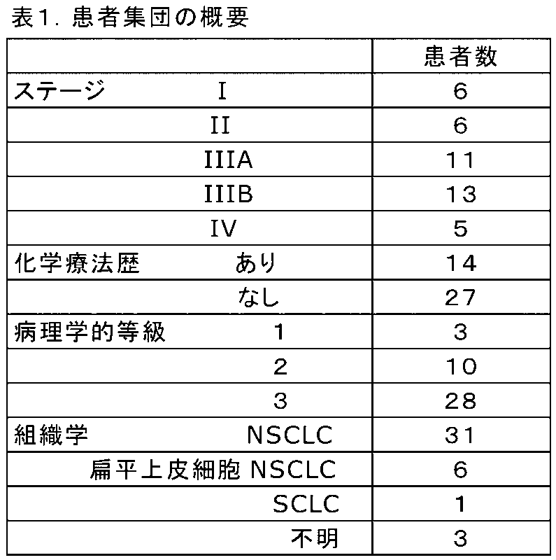
Blood Sampling A prospective pilot study was conducted on 41 patients with stage I-IV lung cancer (Table 1). Anonymized peripheral blood samples were collected following written informed consent and local IRB approval. Patients were recruited from July 2013 to May 2014 prior to starting radiotherapy for primary lung cancer. Four patients received stereotactic body radiation therapy (SBRT) for stage I disease and 37 patients received proton therapy (n=16) or Chemoradiation therapy with Intensity-modulated radiotherapy (IMRT) (n=21) was received. Anonymous blood samples (7.5 mL) were collected and processed on-site at MD Anderson Cancer Center (MDA). The slides were anonymized and shipped to the CreatvMicroTech, Inc. clinical laboratory, where they were analyzed. Anonymized biopsy samples from primary tumors were processed at MDA according to the manufacturer's protocol (DAKO). Results from this facility were not shared or communicated until completion of the study.
セルシーブ(CellSieve:商標)低フローマイクロフィルトレーション手順
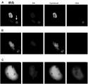
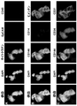
低圧真空システムを用いたセルシーブ(商標)マイクロフィルトレーションアッセイ(CellSieve Microfiltration Assay)によって、セルセーブ保存チューブ(CellSave preservative tubes:商標)に採取した血液サンプル(7.5mL)を処理した[1,12]。セルシーブ(商標)マイクロフィルトレーションアッセイは、>7ミクロンのサイズ排除に基づいて循環細胞を分離する。形態学的特徴及びCD45、EpCAM、サイトケラチン8,18,19及びDAPIの表現型発現に基づいて、事前に知られている細胞学的特徴[6,11,14]を用いて、熟練した細胞学者が予後に病理学的に定義可能なCTC(PDCTC)、EMTCTC、及びCAMLを同定した(図1及び図2)[1,6,12]。イメージングの全てにおいて、オリンパスBX54WI蛍光顕微鏡と共にカールツァイス(CarlZeiss)AxioCam及びZen2011Blue(カールツァイス)を使用した。
CellSieve™ Low Flow Microfiltration Procedure Blood samples collected in CellSave preservative tubes™ by CellSieve™ Microfiltration Assay using a low pressure vacuum system (7.5 mL) were treated [1,12] . The CellSieve™ microfiltration assay separates circulating cells based on >7 micron size exclusion. Based on morphological characteristics and phenotypic expression of CD45, EpCAM,
PDCTC/EMTCTC亜型及びCAMLの列挙
癌患者に見出される2つの最も一般的なCTC亜型(PDCTCS及びEMTCTC)の定義的な特徴及びCAML同定のための特徴は、既に開示されている[1,6,10~14]。本研究では、無傷のPDCTC、EMTCTC、及びCAMLのみを特徴付けた(図2及び図3)[1,6,10~14]。PDCTCは、CD45陰性であり、線維性サイトケラチン陽性であり、悪性病理学的基準を有するDAPI陽性核を有し、CTCのセルサーチ(CellSearch:登録商標)亜型として分類される[1,6,10~14]。EMTCTCは、これまでに定義されているように、CD45陰性であり、拡散性サイトケラチンシグナル及び異常な基準を有するDAPI陽性核を有する[1,6,9,12,13]。CAMLは、拡大された(>30μm)、拡散した細胞質サイトケラチン染色を伴う多核細胞、及び/又はCD45+/CD14+として記述される[6,11,14,17,22,43]。3つの細胞型については、全て、熟練したCTC細胞学者が同定し、画像化し、病理学者が確認した。上述のような細胞学的分類ができなかったアポトーシス性CTC及びCTCは、含まれなかった。同定の後、細胞を画像化し、将来の分析のために各セルのx-y軸をマークした。サンプルは、4℃で1~3年間保存した。
Enumeration of PDCTC/EMTCTC Subtypes and CAMLs The defining characteristics of the two most common CTC subtypes (PDCTCS and EMTCTC) found in cancer patients and the characteristics for CAML identification have been previously disclosed [1, 6,10-14] . In the present study, only intact PDCTCs, EMTCTCs and CAMLs were characterized (Figs. 2 and 3) [1,6,10-14] . PDCTCs are CD45-negative, fibrocytic cytokeratin-positive, have DAPI-positive nuclei with malignant pathological criteria, and are classified as the CellSearch® subtype of CTCs [1,6 ,10-14] . EMTCTCs, as previously defined, are CD45-negative and have DAPI-positive nuclei with diffuse cytokeratin signals and abnormal criteria [1,6,9,12,13] . CAMLs are described as enlarged (>30 μm), multinucleated cells with diffuse cytoplasmic cytokeratin staining, and/or CD45+/CD14+ [6,11,14,17,22,43] . All three cell types were identified by a trained CTC cytologist, imaged, and confirmed by a pathologist. Apoptotic CTCs and CTCs that could not be cytologically classified as described above were not included. After identification, cells were imaged and the xy axis of each cell was marked for future analysis. Samples were stored at 4°C for 1-3 years.
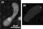
PD-L1及びRAD50のQUAS-Rクエンチング及び再染色
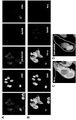
PDCTC、EMTCTC、及びCAMLの最初の同定及び定量化の後、蛍光をクエンチし、RAD50-DyLight550(Pierce Thermo)、PD-L1-AlexaFluor488(R&D systems社)、及びDAPI核染色(図1)によってサンプルを再染色した。既に開示された、QUAS-R(クエンチ、非誘導体化、アミンストリップ及び再染色(Quench, Underivatize, Amine-Strip and Restain)技術を使用した[13]。簡単に説明すると、サンプルを撮像し、マークした後のフィルターに、クエンチング溶液、トリス、及び洗浄ステップの順次的化学処理を施した。化学的クエンチングの後、フィルターをPBSで洗浄し、1XPBS/20%FBSでインキュベートし、次に、RAD50-AlexaFluor550及びPD-L1-AlexaFluor488に対する抗体と共に、室温で1時間インキュベートした。抗体インキュベーションの後、フィルターを1xPBSTで洗浄し、Fluoromount-G/DAPI(Southern Biotech社)によってスライドマウントした。サンプルをx/y軸に沿って配向させ、Zen2011Blue(カールツァイス)マーク及び発見ソフトウェアを用いて先に画像化した細胞を再配置した。Zen2011Blue(Carl Zeiss社)を用いて画像を処理した。
QUAS-R Quenching and Restaining of PD-L1 and RAD50 After initial identification and quantification of PDCTC, EMTCTC, and CAML, fluorescence was quenched, RAD50-DyLight550 (Pierce Thermo), PD-L1-AlexaFluor488 (R&D systems), and DAPI nuclear staining (Fig. 1). We used the previously disclosed QUAS-R (Quench, Underivatize, Amine-Strip and Restain) technique [13] . After quenching, the filters were subjected to sequential chemical treatments of quenching solution, Tris, and washing steps.After chemical quenching, the filters were washed with PBS, incubated with 1XPBS/20% FBS, and then Incubated for 1 hour at room temperature with antibodies against RAD50-AlexaFluor 550 and PD-L1-AlexaFluor 488. After antibody incubation, filters were washed with 1x PBST and slide-mounted with Fluoromount-G/DAPI (Southern Biotech). Oriented along the /y-axis and relocated the previously imaged cells using Zen2011Blue (Carl Zeiss) marks and discovery software Images were processed using Zen2011Blue (Carl Zeiss).
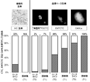
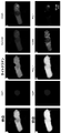
原発腫瘍生検におけるPD-L1の定量化
ダコpharmDxクローン22c3及びダコpharmDxクローン28-8の両方を用いて、製造業者のガイドラインに従い、利用可能な全ての原発腫瘍生検からのPD-L1発現を分析した(図4)。この研究の8人の患者については、両方のクローンをスクリーニングするために十分な腫瘍サンプルが保存されており、1つのサンプルは、クローン22c3に対する単一のIHC試験のために十分な腫瘍を保存していた。両方のクローンを、既に開示されている標準操作手順[29-31,38]に従って染色した。
Quantification of PD-L1 in Primary Tumor Biopsies Both DakopharmDx clone 22c3 and DakopharmDx clone 28-8 were used to quantify PD-L1 expression from all available primary tumor biopsies according to the manufacturer's guidelines. analyzed (Fig. 4). For eight patients in this study, sufficient tumor samples were saved to screen both clones, and one sample had enough tumor saved for a single IHC test against clone 22c3. was Both clones were stained according to previously published standard operating procedures [29-31,38] .
循環細胞におけるRAD50及びPD-L1の定量化
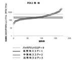
各細胞の核局在化RAD50遺伝子座を列挙することによってRAD50遺伝子座の形成を判定した(図1及び図5)[23]。細胞全体の面積を用いて、ZenBlueソフトウェアにより各細胞のPD-L1ピクセル強度を測定した。各画像の局所的バックグラウンドの平均ピクセル強度から各セルの平均ピクセル強度を減算した(図1C)。細胞の平均ピクセル強度を0-負(ピクセル平均0~150)、1-低(ピクセル平均151-300)、2-中(ピクセル平均301-750)、及び3-高(ピクセル平均751+)の4つのIHC群に四分化した(図5)。IHCスコアリングのためのPD-L1強度のIHC範囲閾値は、150ピクセル強度を、局所化されたバックグラウンドシグナルの標準偏差とし、300ピクセル強度を、局所化されたバックグラウンドの標準偏差の2倍とし、750を、局所化されたバックグラウンドの強度の2倍とすることによって定義した(図5)。
Quantification of RAD50 and PD-L1 in Circulating Cells The formation of RAD50 loci was determined by enumerating the nuclear-localized RAD50 loci of each cell (Figs. 1 and 5) [23] . The area of the whole cell was used to measure the PD-L1 pixel intensity of each cell by ZenBlue software. The average pixel intensity of each cell was subtracted from the local background average pixel intensity of each image (Fig. 1C). The average pixel intensity of the cells was 0-negative (pixel average 0-150), 1-low (pixel average 151-300), 2-medium (pixel average 301-750), and 3-high (pixel average 751+). There were four IHC groups (Fig. 5). The IHC range threshold for PD-L1 intensity for IHC scoring was 150 pixel intensity as the standard deviation of the localized background signal and 300 pixel intensity as twice the standard deviation of the localized background. and 750 was defined by doubling the intensity of the localized background (Fig. 5).
統計的方法
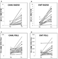
MATLAB R2013Aにおいて、全ての亜型及び既知の患者集団からのカウントを用いて解析を行った。無増悪生存期間(progression free survival)分析のために、増悪までの時間を、T0血液サンプルを取得したときから、増悪の日までと定義し、24ヶ月のエンドポイントまで全ての患者について研究を行い、すなわち、研究を打ち切った患者はなかった。RAD50巣形成及びPD-L1発現の平均変化の有意性は、スチューデントのT検定によって判定した。個々の測定値について、ピアソン係数を使用して、RAD50巣とPD-L1発現との間の相関を判定した。カプランマイヤープロットの有意性は、ログランク分析によって判定した。
Statistical Methods Analyzes were performed in MATLAB R2013A using counts from all subtypes and known patient populations. For progression free survival analysis, time to progression was defined as from the time the T0 blood sample was obtained to the date of progression, and all patients were studied to the 24-month endpoint. ie, no patients were discontinued from the study. The significance of mean changes in RAD50 foci formation and PD-L1 expression was determined by Student's T-test. For individual measurements, the Pearson's coefficient was used to determine the correlation between RAD50 foci and PD-L1 expression. The significance of Kaplan-Meier plots was determined by log-rank analysis.
結果
LC患者におけるPDCTC、EMTCTC、及びCAML
セルサーチ(CellSearch:登録商標)プラットフォームを使用するNSCLC患者におけるCTC亜集団は、通常、非転移性症例のうち、0~5%にしか見出されないことが報告されている。これに対し、EMTCTC集団は、通常、非転移性患者集団の約80%に見出され、CAMLについては、NSCLCにおける広範に亘る評価は行われていない[3-6,8,15,17,43-45]。放射線療法の開始前に採取された最初のベースライン血液サンプル(T0)では、41サンプル中35サンプル(85%)において、少なくとも1つのサイトケラチン陽性細胞(すなわち、PDCTC、EMTCTC、又はCAML)を同定できた(図1及び図3)。その後、放射線療法開始から2~3週間後、又はSBRT患者については最後の分画の後に、患者の第2のフォローアップサンプル(T1)を採取した。T1については、41サンプル全て(100%)に少なくとも1つのサイトケラチン陽性細胞(すなわち、PDCTC、EMTCTC、又はCAML)が見出された。具体的には、EMTCTCは、T0サンプルの49%及びT1サンプルの66%に見出された。CAMLは、T0サンプルの81%及びT1サンプルの100%に見出された(図3)。PDCTCは、T0において1サンプル(2%)のみ、T1において3サンプルのみ(7%)に見出された(図3)。PDCTCは、セルサーチ(CellSearch:登録商標)CTCシステムによって単離された同じCTC集団であることが示されており、これらの数値は、これまでの報告[7-9,15]に合致する。セルサーチシステムは、ステージIIIのNSCLCでは0~5%の陽性、ステージIVでは21~32%の範囲で、NSCLC患者のCTCを単離した[7-9,15]。患者のうちの35人がステージI-IIIとして病期分類されたので、2~7%は、通例的なCTC集団の範囲内である(表1)[7-9,15]。通例的なPDCTC集団の発生率が低いことは、サンプルの85%(T0)及び100%(T1)に存在するEMTCTC及びCAMLとは対照的である(図3)。EMTCTC単独では、NSCLCにおける液体生検に対する感度が幾らか向上する可能性があると推論されてきたが[7-9,16]、これらの結果は、EMTCTC及びCAMLの両方の組み合わせによって、腫瘍由来細胞を分析する血液ベースの診断の感度が向上することを示唆している。
Results PDCTC, EMTCTC, and CAML in LC Patients
It has been reported that the CTC subpopulation in NSCLC patients using the CellSearch® platform is typically found in only 0-5% of non-metastatic cases. In contrast, the EMTCTC population is usually found in approximately 80% of the nonmetastatic patient population, and CAMLs have not been extensively evaluated in NSCLC [3-6,8,15,17, 43-45] . At least one cytokeratin-positive cell (i.e., PDCTC, EMTCTC, or CAML) was identified in 35 of 41 samples (85%) in the first baseline blood sample (T0) taken prior to initiation of radiotherapy It was possible (Fig. 1 and Fig. 3). A second follow-up sample (T1) of the patient was then taken 2-3 weeks after the start of radiation therapy, or after the final fractionation for SBRT patients. For T1, at least one cytokeratin-positive cell (ie, PDCTC, EMTCTC, or CAML) was found in all 41 samples (100%). Specifically, EMTCTC was found in 49% of T0 samples and 66% of T1 samples. CAMLs were found in 81% of T0 samples and 100% of T1 samples (Fig. 3). PDCTC was found in only 1 sample (2%) at T0 and only 3 samples (7%) at T1 (Fig. 3). PDCTCs have been shown to be the same CTC population isolated by the CellSearch® CTC system, and these numbers are consistent with previous reports [7-9,15] . The CellSearch system isolated CTCs from NSCLC patients ranging from 0-5% positive for stage III NSCLC and 21-32% for stage IV [7-9,15] . Thirty-five of the patients were staged as stages I-III, so 2-7% fall within the customary CTC population (Table 1) [7-9,15] . The low incidence of the customary PDCTC population contrasts with EMTCTC and CAML, which are present in 85% (T0) and 100% (T1) of the samples (Fig. 3). Although it has been speculated that EMTCTC alone may provide some increased sensitivity to liquid biopsies in NSCLC [7-9,16] , these results suggest that both EMTCTC and CAML in combination may provide tumor-derived It suggests that the sensitivity of blood-based diagnostics that analyze cells would be improved.
放射線照射された細胞の生物学的トラッカーとしてのRAD50
これまで、原発腫瘍の部位からの末梢血への播種としてCTC及びCAMLが記述されてきたが、これらの細胞が循環に入る前に存在する正確な位置を確認した研究は未だなされていない。このように起原が不明であることの主な理由は、患者の腫瘍/間質細胞を標識し、これらの播種を追跡することが困難であり、このような実験が患者に危険をもたらす可能性があるためである。哺乳類細胞におけるIRIF形成物内のRAD50巣が、細胞への直接放射線曝露の生物学的トラッカーとして示されることはあったが、これは、放射線治療を受けている患者の循環腫瘍又は間質細胞においては、未だ評価されていない。T0ベースラインにおける非照射LC患者において、EMTCTC細胞内のRAD50巣の核当たりの巣数は、0~4の範囲であり、平均は0.59±0.97であり、CAMLでは、巣数は、0~5の範囲であり、平均0.38±1.07であった(図6及び図7)。RAD50巣は、通常、少数の未治療細胞で同定される正常な生物学的修復メカニズムであるため、細胞内に幾つかのRAD50巣が存在することは、驚くべきことではない[21,23,24]。T1において患者を腫瘍指向性放射線療法に曝露した後、RAD50巣数は、EMTCTCにおいて、核当たり0~9、平均4.27±2.63及びCAMLにおいて、核当たり0~20、平均3.9±3.93に有意に増加した(図6)。この増加は、T0及びT1の両方の時点(n=35)で検出可能な細胞を有する全ての患者において観察され、正常なCD45+白血球のバックグラウンドでは殆ど見いだされなかった(図1B)。このように、EMTCTC及びCAMLの両方において、RAD50は、T0における平均0.48からT1における平均4.05(p<0.0001)に増加した(図6)。これらの結果は、RAD50を使用して、腫瘍部位に由来する照射された細胞を標識及び追跡でき、したがって、腫瘍動態を追跡できることを示唆している。
RAD50 as a biological tracker of irradiated cells
Previously, CTCs and CAMLs have been described as dissemination into the peripheral blood from the site of the primary tumor, but no studies have yet confirmed the exact location of these cells before they enter the circulation. The main reason for this unknown origin is the difficulty in labeling tumor/stromal cells in patients and tracking their dissemination, and such experiments can pose risks to patients. This is because of the nature of Although RAD50 foci within IRIF formations in mammalian cells have been shown to serve as biological trackers of direct radiation exposure to cells, this is not the case in circulating tumor or stromal cells of patients undergoing radiotherapy. has not yet been evaluated. In non-irradiated LC patients at TO baseline, the number of foci per nucleus of RAD50 foci in EMTCTC cells ranged from 0 to 4 with a mean of 0.59±0.97, and in CAMLs, the number of foci was , ranged from 0 to 5, with an average of 0.38±1.07 (FIGS. 6 and 7). The presence of several RAD50 foci in cells is not surprising, as RAD50 foci are a normal biological repair mechanism usually identified in a minority of untreated cells [21,23, 24] . After exposing the patient to tumor-directed radiation therapy at T1, the number of RAD50 foci was 0-9 per nucleus, mean 4.27±2.63 in EMTCTC and 0-20 per nucleus, mean 3.9 in CAML. It increased significantly to ±3.93 (Fig. 6). This increase was observed in all patients with detectable cells at both T0 and T1 time points (n=35) and was rarely found in a background of normal CD45+ leukocytes (Fig. 1B). Thus, in both EMTCTC and CAML, RAD50 increased from a mean of 0.48 at T0 to a mean of 4.05 at T1 (p<0.0001) (Fig. 6). These results suggest that RAD50 can be used to label and track irradiated cells originating from the tumor site and thus tumor dynamics.
循環細胞におけるPD-L1の動的発現
放射線を含む様々な細胞傷害性療法によって、腫瘍にPD-L1が誘発され得ることが示唆されている[29-34,36-40,42]。BBBを使用してこれを視認できるかを判定するために、T0及びT1の時点でPD-L1染色を評価した。従来の0-3IHC組織生検スコアリングと同様の方法で、標準化された比較スコアリングシステムを開発した(図5)。染色及び画像化の後、LC患者において見出された全ての373細胞のPD-L1発現及び局所バックグラウンドを測定した。各画像の局所的バックグラウンドのピクセル強度を375±150に平均化した(図5)。局所化されたバックグラウンド効果を考慮するために、測定された各細胞から各画像のバックグラウンドを減算し、PD-L1ピクセル強度範囲を17-3090に補正した(図5)。次に、0~150ピクセルのバックグラウンドの標準偏差をスコア0(細胞の26%)とし、標準偏差の2倍(151-300ピクセル)をスコア1(低発現、細胞の42%)として、補正したピクセル強度で細胞を分類した。中程度の発現、すなわちスコア2(細胞の22%)は、平均バックグラウンドシグナル(301-750ピクセル)の2倍として判定し、高発現、すなわちスコア3(細胞の10%)を平均バックグラウンドシグナルの>2倍(>750ピクセル)に設定した(図5)。
Dynamic Expression of PD-L1 in Circulating Cells It has been suggested that PD-L1 can be induced in tumors by various cytotoxic therapies, including radiation [29-34,36-40,42] . PD-L1 staining was assessed at T0 and T1 to determine if this was visible using the BBB. A standardized comparative scoring system was developed in a manner similar to conventional 0-3 IHC tissue biopsy scoring ( Fig. 5) . After staining and imaging, PD-L1 expression and local background of all 373 cells found in LC patients were determined. The local background pixel intensity of each image was averaged to 375±150 (FIG. 5). To account for localized background effects, the background of each image was subtracted from each cell measured and the PD-L1 pixel intensity range was corrected to 17-3090 (Fig. 5). The standard deviation of the background from 0-150 pixels was then scored 0 (26% of cells) and twice the standard deviation (151-300 pixels) was scored 1 (low expression, 42% of cells). Cells were classified according to their pixel intensities. Moderate expression, i.e. score 2 (22% of cells), was determined as twice the average background signal (301-750 pixels), high expression, i.e. score 3 (10% of cells), was determined as average background signal. (>750 pixels) (Fig. 5).
PD-L1のピクセル強度は、EMTCTCでは、T0で平均384±484、T1で672±669(p=0.021)であり、CAMLでは、T0で平均182±89、T1で282±169(p=0.004)であった(不図示)。回帰分析により、T0からT1にかけて、RAD50とPD-L1との間に弱いが有意な正の相関が見出された(ピアソンR2=0.079、p<0.0001、n=373)。患者間で、RAD50は、T0からT1にかけて確実に誘発されたが、個々の患者におけるPD-L1発現の変化は、遥かに多様であった(不図示)。T0およびT1の両時点で評価可能であった35人の患者において、T0とT1との間にPD-L1発現の3つの異なるパターンを見出した。18人の患者(51%)は、両方の時点でPD-L1の発現がなく/低く、6人の患者(17%)は、両方の時点で持続的に中/高PD-L1を有し、11人の患者(32%)は、低い0/1スコアから2/3スコアに増加した(不図示)。 PD-L1 pixel intensity averaged 384±484 at T0 and 672±669 at T1 (p=0.021) for EMTCTC and averaged 182±89 at T0 and 282±169 at T1 (p=0.021) for CAML. = 0.004) ( not shown ). Regression analysis found a weak but significant positive correlation between RAD50 and PD-L1 from T0 to T1 (Pearson R2=0.079, p<0.0001, n=373). Between patients, RAD50 was robustly induced from T0 to T1, but changes in PD-L1 expression in individual patients were much more variable ( not shown ). In 35 patients who were evaluable at both T0 and T1, we found three distinct patterns of PD-L1 expression between T0 and T1. Eighteen patients (51%) had no/low PD-L1 expression at both time points and 6 patients (17%) had persistent moderate/high PD-L1 at both time points. , 11 patients (32%) increased from a low 0/1 score to a 2/3 score ( not shown ).
原発組織、CTC及びCAMLにおけるPD-L1レベルの比較
クローン22c3及び28-8(ダコ(DAKO))を使用する市販の2つのCLIA認定試験を使用して、IHCによる元の診断生検から利用可能な組織を染色した。病理学的アーカイブから有用な組織又は細胞ブロックを回収できたのは、41人の患者のうち9人のみであり、1回のIHC検査に十分な組織を有していたのは、これらの9人の患者のうち1人のみであった(図4)。これは、腫瘍壊死又は小結節の結果、PD-L1 IHC試験を行うために十分な集団が得られなかったためである。9個のアーカイブサンプルのうち、2個のみがPD-L1染色陽性であり、2個の試験間で発現スコア及びパーセンテージの変動があった(図4C)。これに対し、PD-L1発現は、BBBを使用して、T0患者サンプルの85%(n=35/41)及びT1患者サンプルの100%(n=41/41)で定量可能であった。具体的には、T0において、EMTCTC及びCAMLは、21人の患者(60%)において低/負(スコア0/1)のPD-L1発現を示し、9人の患者(26%)において中等度(スコア2)の発現を示し、5人の患者(14%)において高(スコア3)の発現を示した(図4C)。
Comparison of PD-L1 levels in primary tissues, CTCs and CAMLs Available from original diagnostic biopsies by IHC using two commercially available CLIA accredited tests using clones 22c3 and 28-8 (DAKO) tissues were stained. Only 9 of 41 patients were able to retrieve useful tissue or cell blocks from the pathological archives, and only 9 of these 9 had enough tissue for a single IHC test. There was only 1 out of 1 patient (Fig. 4). This was because tumor necrosis or nodules did not result in a sufficient population for PD-L1 IHC testing. Of the 9 archival samples, only 2 were positive for PD-L1 staining and there was variation in expression scores and percentages between the 2 studies (Fig. 4C). In contrast, PD-L1 expression was quantifiable in 85% (n=35/41) of T0 patient samples and 100% (n=41/41) of T1 patient samples using BBB. Specifically, at TO, EMTCTC and CAML showed low/negative (
T0において、循環細胞におけるPD-L1の発現は、28-8IHCクローン結果を用いて評価した2つのIHC陽性染色サンプルについてのIHC生検結果との整合性が高かった(図4C)。3人の患者は、IHCによる陰性のPD-L1組織に一致し及び循環細胞での低い発現(0/1)を示したが、3人の患者は、陰性組織IHC PD-L1では不一致の結果を示し、循環細胞では2/3のスコアを示し、1人の患者は、T0サンプルにおいて循環細胞を欠いていた(図4)。サンプル数が限られているため、適切な統計解析は不可能であった。しかしながら、これらの結果は、一次生検では、PD-L1発現をアッセイするために組織が不十分であるが、血液ベースの生検(Blood Based Biopsy:BBB)法によって、内因性レベルを測定し、原発性肺腫瘍に見出される細胞集団に由来する循環細胞中に存在するPD-L1発現の変化をモニタリングできることを示唆している。 At T0, PD-L1 expression in circulating cells was highly consistent with IHC biopsy results for two IHC-positive staining samples evaluated using the 28-8 IHC clone results (FIG. 4C). Three patients showed concordant negative PD-L1 tissue by IHC and low expression in circulating cells (0/1), while three patients had discordant results on negative tissue IHC PD-L1. and a score of 2/3 in circulating cells, with one patient lacking circulating cells in the T0 sample (Fig. 4). Adequate statistical analysis was not possible due to the limited number of samples. However, these results indicate that primary biopsies have insufficient tissue to assay PD-L1 expression, whereas the Blood Based Biopsy (BBB) method measures endogenous levels. , suggest that changes in PD-L1 expression present in circulating cells from cell populations found in primary lung tumors can be monitored.
潜在的な予後マーカーとしての循環細胞中のPD-L1及びRAD50
組織生検では、バイオマーカーPD-L1の発現のみでは、肺癌における生存の予後指標にならず、一方、RAD50の巣形成は、生存と正の相関を有することが示されている[14,18,20,24,29,30,32,35,39,40,42]。T0とT1の両方の時点でのPD-L1の発現又はRAD50の平均数に基づいて患者の臨床転帰を分析した(図8)。PD-L1の発現を用いてPFSを比較するために、カットオフ基準として中程度の発現、すなわち、2つのコホートについて、<2対≧2を使用した。T0においてPD-L1が低い患者は、1.8の僅かに悪いハザード比(hazard ratio:HR)を有し、これは、有意ではなかった(p=0.305)。T1では、PD-L1が低い患者は、全体的PFSがやや良好(HR=0.7)であったが、これも有意ではなかった(p=0.581)。このデータは、T0又はT1におけるPD-L1レベルの発現に基づく全体的PFSとの相関が限定的又は相関が認められないことを示唆している。PFS中央値を使用することにより、T0においてより高いPD-L1を有する細胞において、PFS中央値へのより良好な傾向(16ヶ月対>24ヶ月間)を見出したが、これを確認するには、遥かに大きなサンプルサイズが必要である。
PD-L1 and RAD50 in Circulating Cells as Potential Prognostic Markers
Tissue biopsies have shown that expression of the biomarker PD-L1 alone is not prognostic of survival in lung cancer, whereas RAD50 foci formation is positively correlated with survival [14,18]. ,20,24,29,30,32,35,39,40,42] . Patients' clinical outcomes were analyzed based on the mean number of PD-L1 expression or RAD50 at both T0 and T1 time points (FIG. 8 ). To compare PFS using PD-L1 expression, moderate expression was used as a cut-off criterion, ie <2 vs. ≧2 for the two cohorts. Patients with low PD-L1 at T0 had a slightly worse hazard ratio (HR) of 1.8, which was not significant (p=0.305). At T1, patients with low PD-L1 had slightly better overall PFS (HR=0.7), which was also not significant (p=0.581). This data suggests limited or no correlation with overall PFS based on expression of PD-L1 levels at T0 or T1. By using median PFS, we found a better trend towards median PFS (16 months vs. >24 months) in cells with higher PD-L1 at T0, but to confirm this , requires a much larger sample size.
組織生検におけるRAD50巣の形成は、生存と正の相関を有することが示されているため[18,20,24,26]、循環細胞におけるその予後値を評価した(図8)。T0におけるEMTCTC及びCAMLのRAD50巣数は、全体的PFSにおいて臨床的に差がなかった(HR=1.0、p=0.775)。一方、T1においてRAD50巣が多い患者では、全体的PFSが改善される有意ではない傾向があった(HR2.3、p=0.27)(図8)。このように、全体的PFSは、有意差がなかったが、RAD50巣/細胞が≦1の患者と比較して、RAD50巣/細胞が>1の患者では、PFS中央値が1.9倍長かった(それぞれ、9.8ヶ月対18.5ヶ月)。このデータは、放射線療法後の循環細胞におけるRAD50の増加は予後値を有する可能性があり、更なる検証とより大きなサンプルサイズの観察が必要であることを示唆している。 Since the formation of RAD50 foci in tissue biopsies has been shown to be positively correlated with survival [18,20,24,26] , its prognostic value in circulating cells was assessed (Fig. 8 ). EMTCTC and CAML RAD50 foci numbers at T0 were not clinically different in overall PFS (HR=1.0, p=0.775). In contrast, patients with more RAD50 foci at T1 showed a non-significant trend toward improved overall PFS (HR 2.3, p=0.27) (FIG. 8 ). Thus, although overall PFS was not significantly different, median PFS was 1.9 times longer in patients with >1 RAD50 foci/cell compared to patients with ≤1 RAD50 foci/cell. (9.8 vs. 18.5 months, respectively). This data suggests that increased RAD50 in circulating cells after radiotherapy may have prognostic value and requires further validation and observation of larger sample sizes.
前段に示した結果の通り、(化学療法)放射線療法を受けている41人の肺癌患者からの3つの循環血液細胞亜型PDCTC、EMTCTC、及びCAMLにおけるPD-L1レベル及びRAD50巣を前向きに及び逐次的に追跡した。放射線誘導RAD50巣形成に基づいて、循環細胞を表現型分類し、原発性肺腫瘍塊に現れる細胞の明確な生物学的変化を定量的に追跡した。更に、これらの動的変化の追跡を用いて、放射線療法に対してより感受性が高まった腫瘍を有する患者を区別できる可能性があるが、より大規模な研究が必要である。 As shown in the previous section, we prospectively investigated PD-L1 levels and RAD50 foci in three circulating blood cell subtypes PDCTC, EMTCTC, and CAML from 41 lung cancer patients undergoing (chemo)radiation. followed sequentially. Based on radiation-induced RAD50 foci formation, circulating cells were phenotyped to quantitatively track distinct biological changes in cells that appear in primary lung tumor masses. Furthermore, tracking these dynamic changes may be used to distinguish patients with tumors that are more sensitive to radiation therapy, but larger studies are needed.
多くのグループが、放射線損傷細胞の核におけるRAD50巣形成によって、IRIFの形成が観察されることを立証し(図9)[18,20,23]、DNA損傷ストレッサーによる事前の感作によるIRIF形成の阻害が、多くの癌(すなわち、NSCLC、乳癌、扁平上皮癌等)において、臨床転帰と正の相関を有することが示された[18,20,23,24,26]。まず、放射線療法前の未治療のNSCLC患者は、RAD50巣の数が少なく、放射線療法の導入に直接的に並行して、RAD50巣が大幅に増加することを観察した。このRAD50の増加は、放射線治療導入によって誘発されるDNA損傷の結果である可能性が高く、循環細胞におけるRAD50巣形成は、部位特異的放射線療法を受けている癌患者の非侵襲的トレーサーとして機能すると考えられる。RAD50は、循環細胞の原発部位の器官を確認するために液体生検分析と同様に使用でき、これは、患者の原発肺塊からEMTCTC及びCAMLの両方が播種していることを示唆していると考えられる。 A number of groups have demonstrated that IRIF formation is observed by RAD50 foci formation in the nuclei of radiation-damaged cells (Fig. 9 ) [18,20,23] , and IRIF formation by prior sensitization with DNA damage stressors. was shown to be positively correlated with clinical outcome in many cancers (ie NSCLC, breast cancer, squamous cell carcinoma, etc.) [18,20,23,24,26] . First, we observed that treatment-naïve NSCLC patients prior to radiotherapy had fewer RAD50 foci, and a large increase in RAD50 foci directly paralleling the introduction of radiotherapy. This increase in RAD50 is likely the result of DNA damage induced by radiotherapy induction, and RAD50 foci formation in circulating cells serves as a noninvasive tracer in cancer patients undergoing site-specific radiotherapy. It is thought that RAD50 can be used similarly to liquid biopsy analysis to confirm the organ of origin of circulating cells, suggesting that both EMTCTCs and CAMLs disseminate from the patient's primary lung mass. it is conceivable that.
PD-L1発現に関する現在承認されている生検組織のIHC試験は、非常に高レベルのPD-L1発現を伴う患者における応答の予測因子に過ぎないものの、やがて多くのPD-L1陰性患者も利益を得るであろう[29,30,33,39-42,46]。この不一致は、免疫調節発現の動的性質及び/又は間質細胞成分を分析できないことに起因する可能性がある。免疫チェックポイントタンパク質発現は、動的であり、複数の微小環境、炎症、及び治療因子の影響を受けるため、血液ベースの分析が患者における現在のPD-L1発現のより正確な表現を提供し得るとの仮説が立てられてきた[29,30,39,42,43,47,48]。興味深い点として、我々は、患者の循環細胞において持続的に低、持続的に高、又は低から高へ誘導可能の3つのクラスのPD-L1応答を見出し、これは、患者の約3分の1(32%)で生じた。これは、患者の約半分(49%)において、本質的に高レベル又は誘導性のPD-L1レベルが免疫療法応答を予測できることを示唆しており、これは、免疫療法と放射線療法を組み合わせた臨床試験において、将来の検証が必要な仮説である。 Although currently approved biopsy tissue IHC testing for PD-L1 expression is only a predictor of response in patients with very high levels of PD-L1 expression, over time many PD-L1-negative patients will also benefit. [29,30,33,39-42,46] . This discrepancy may be due to the dynamic nature of immunomodulatory expression and/or the inability to analyze the stromal cell component. Because immune checkpoint protein expression is dynamic and influenced by multiple microenvironmental, inflammatory, and therapeutic factors, blood-based analysis may provide a more accurate representation of current PD-L1 expression in patients. has been hypothesized [29,30,39,42,43,47,48] . Interestingly, we found three classes of PD-L1 responses in patients' circulating cells: persistently low, persistently high, or low-to-high inducible, which was observed in about a third of patients. 1 (32%) occurred. This suggests that in about half (49%) of patients, intrinsically high or inducible PD-L1 levels can predict immunotherapy response, which is associated with combined immunotherapy and radiotherapy. It is a hypothesis that requires future verification in clinical trials.
実施例2
腫瘍血管内皮細胞(tumor endothelial cell:TEC)[60-62]は、血管新生及び新生血管形成のための重要な構造を形成することによって、腫瘍の開始、生存及び成長に必要とされる間質細胞の集団である。TECは、全ての腫瘍部位において必須成分であり、腫瘍脈管構造に必要とされ、転移性ニッチのプライミングを補助し、腫瘍の分子不安定性に寄与する。循環において、共通のTEC集団が同定されており、これらは、これらの大きなサイズ、多細胞クラスター化、及び周知のECマーカーCD31及びビメンチン(Vimentin)[60]に基づいて、癌関連血管内皮細胞(CAVE)として定義されている。
Example 2
Tumor endothelial cells (TECs) [60-62] are essential stroma cells required for tumor initiation, survival and growth by forming key structures for angiogenesis and neovascularization. A group of cells. TECs are essential components in all tumor sites, are required for tumor vasculature, help prime the metastatic niche, and contribute to tumor molecular instability. In the circulation, common TEC populations have been identified that, based on their large size, multicellular clustering, and well-known EC markers CD31 and Vimentin [60] , are cancer-associated vascular endothelial cells ( CAVE).
サイズ排除は、これらの表面マーカー発現にかかわらず、末梢患者の血液から大きな細胞を単離し、循環腫瘍ECの多くの亜型の捕捉を可能にする技術である。セルシーブ(CellSieve:商標)マイクロフィルターは、全血からCAVE、CAML、及びCTCを迅速かつ効率的に分離できるサイズ排除膜であり、これにより、悪性疾患と共に、及びこれと関連して、全ての細胞タイプを研究することが可能である[13,60-62]。更に、セルシーブ(CellSieve:商標)マイクロフィルターを使用して、単離された細胞上の亜型のバイオマーカーの質量スクリーニングを可能にする多重表現型技術(multi-phenotyping technique)が開発されている Size exclusion is a technique that isolates large cells from peripheral patient blood and allows capture of many subtypes of circulating tumor ECs, regardless of their surface marker expression. CellSieve™ microfilters are size-exclusion membranes that can rapidly and efficiently separate CAVEs, CAMLs, and CTCs from whole blood, thereby allowing all cells with and associated with malignancies to It is possible to study types [13,60-62] . In addition, a multi-phenotyping technique has been developed that allows mass screening of subtypic biomarkers on isolated cells using CellSieve™ microfilters.
この手法を実証するために、2012~2014年に亘って、116人の癌患者(ステージI~IV)の胸部(n=42)、肺(n=39)、及び前立腺(n=35)を含む末梢血サンプル並びに34人の健常対照群から血液を採取した。確立されたフィルタリング法、すなわち、セルシーブ(CellSieve:商標)マイクロフィルトレーション技術(クリエイティブマイクロテック社:Creatv MicroTech)によって血液を処理し、サイズ排除によって血液を濾過し、CK8、18、19、EpCAM及びCD45について細胞を染色した(図10A)。同定及び画像化の後、QUAS-R(クエンチ、非誘導体化、アミンストリップ及び再染色(Quench, Underivatize, Amine-Strip and Restain)技術を使用して、蛍光シグナルを除去し、CD146、CD14、ビメンチン、及びDAPIについて全ての細胞を再染色した(図10B)。再イメージングの後、再びQUAS-Rを用いて蛍光を除去し、CD144、CD34(又はCD105)、CD31、及びDAPIについて細胞を再染色した(図10C)。CAMLで観察されたCD14+及び倍数体核構造を用いて、CAVEの多核クラスターが癌関連マクロファージ様細胞と区別された。 To demonstrate this approach, breast (n=42), lung (n=39), and prostate (n=35) of 116 cancer patients (stages I-IV) were examined over 2012-2014. Blood was collected from a peripheral blood sample containing as well as 34 healthy controls. Blood was processed by established filtering methods: CellSieve™ microfiltration technology (Creatv MicroTech), filtered by size exclusion, CK8, 18, 19, EpCAM and Cells were stained for CD45 (Fig. 10A ). After identification and imaging, the QUAS-R (Quench, Underivatize, Amine-Strip and Restain) technique was used to remove the fluorescent signal and detect CD146, CD14, vimentin. , and DAPI (FIG. 10B) After re - imaging, fluorescence was again removed using QUAS-R and cells were re-stained for CD144, CD34 (or CD105), CD31, and DAPI. (Fig. 10C ) Multinucleated clusters of CAVE were distinguished from cancer-associated macrophage-like cells using the CD14+ and polyploid nuclear structures observed in CAMLs.
CAVEは、CD31、CD144又はCD146の陽性に基づいて、116人中63人(54%)で同定されたが、健常対照群では検出されなかった。CAVEは、ステージI患者の43%、ステージII患者の66%、ステージIII患者の74%、及びステージIV患者の82%において見出された(図11)。CAVEは、胸部の69%、肺の60%、及び前立腺の77%のサンプルで見出された。CAVEは、CD14及びCD45について全て陰性であった。CD31がCAVEの96%で見出された最も多く存在するマーカーであり、これに、CD144(85%)、サイトケラチン(68%)、CD34(64%)、CD146(45%)、EpCAM(23%)及びCD105(4%)が続いた(図12)。 CAVE was identified in 63 of 116 (54%) based on positivity for CD31, CD144 or CD146, but was not detected in healthy controls. CAVE was found in 43% of stage I patients, 66% of stage II patients, 74% of stage III patients, and 82% of stage IV patients ( Fig. 11 ). CAVE was found in 69% of breast, 60% of lung and 77% of prostate samples. CAVE was all negative for CD14 and CD45. CD31 was the most prevalent marker found in 96% of CAVE, including CD144 (85%), cytokeratin (68%), CD34 (64%), CD146 (45%), EpCAM (23%), %) and CD105 (4%) followed ( Fig. 12 ).
この結果は、マイクロフィルトレーションによって単離されたCECは、サイトケラチン陽性であり、CD45陰性であり、これは、固形腫瘍を有する患者の循環系に共通に現れるが、健常対照群には現れないことを示している。複数の表現型の亜型によって、複数の固形腫瘍タイプの癌患者のCECを適切に識別し、亜型化できる。このデータは、CECのサブセット、例えば、CAVEがCK+/CD45-として循環細胞に見出され、癌特異的循環細胞の異種集団として存在することを示唆している。 The results show that CECs isolated by microfiltration are cytokeratin-positive and CD45-negative, which commonly appear in the circulation of patients with solid tumors, but not healthy controls. indicates that there is no Multiple phenotypic subtypes allow for the proper identification and subtyping of CEC in cancer patients with multiple solid tumor types. This data suggests that a subset of CECs, such as CAVE, are found in circulating cells as CK+/CD45- and exist as a heterogeneous population of cancer-specific circulating cells.
実施例3
図13は、細胞のクラスター中に出現するCAVEを検証するための染色を示している。癌腫患者から捕捉された全ての細胞の最初の染色において、DAPI、サイトケラチン8、18及び19、EpCAM及びCD45についてサンプルを染色した。図13の一番上の列は、サイトケラチン発現、及びEpCAM、CD45発現なしのクラスターを示している。図13の二番目の列は、DAPI、PD-L1、CD14及びCD31について細胞が再染色されたことを示している。CD31+膜染色及び陰性CD45及びCD14染色は、これがCAVEクラスターであることを示している。このCAVEのクラスターは、PD-L1の高発現を有する。図13の三番目の列は、CD144、CXCR4及びビメンチンについて細胞が再染色されたことを示している。CXCR4は、他の薬剤標的である。再染色のためにQUAS-Rを使用し[13]、この再染色は、蛍光色素をクエンチングし、続いて再染色することからなる。この再染色は、併用免疫療法を分析する方法を示している。この例では、PD-L1とCXCR4の両方の発現が高く、この併用療法は、この患者にとって有効である可能性があることを示している。
Example 3
FIG. 13 shows staining to verify CAVE appearing in clusters of cells. Samples were stained for DAPI,
図14は、DAPI、サイトケラチン8、18及び19、EpCAM及びCD45について初期的に染色されたCAML細胞を示している(一番上の列)。図14の二番目の列は、DAPI、PD-L1、CCR5及びPD-1について細胞が再染色されたことを示している。この例では、PD-L1とCCR5の両方の発現が高く、この併用免疫療法がこの患者にとって有効である可能性があることを示している。
Figure 14 shows CAML cells initially stained for DAPI,
図15は、DAPI、サイトケラチン8、18及び19、EpCAM及びCD45について初期的に染色されたCAML細胞を示している(一番上の列)。図15の二番目の列は、DAPI、PD-L1、CCR5及びPD-1について細胞が再染色されたことを示している。この例では、CCR5が存在するがPD-L1の発現はなく、この併用免疫療法がこの患者にとって有効である可能性があることを示している。
Figure 15 shows CAML cells initially stained for DAPI,
実施例4
現在の免疫療法コンパニオン診断は、組織生検におけるPD-L1タンパク質発現に基づいている。しかしながら、IHC組織生検法は、生検を繰り返すことの臨床的実現可能性及びコスト、固有の腫瘍異質性、及びPD-L1 IHC陰性患者が依然として免疫療法に応答する可能性があるとの知識のために制限されている。
Example 4
Current immunotherapy companion diagnostics are based on PD-L1 protein expression in tissue biopsies. However, the IHC tissue biopsy method is limited by the clinical feasibility and cost of repeat biopsies, the inherent tumor heterogeneity, and the knowledge that PD-L1 IHC-negative patients may still respond to immunotherapy. is restricted for
図16A及び図16Bに示すように、PD-L1の発現は、CAML及びCTC上で検出できる。末梢循環細胞からPD-L1発現をリアルタイムでモニタリングできるか否かを判定するために、抗PD1免疫療法であるペムブロリズマブ(pembrolizumab)の開始前及び開始後のRCCについて、患者からの連続サンプルを評価した。PD-L1発現を定量的に測定し、(シグナル-バックグラウンド)/バックグラウンド、又は(S-N)/Nとして、蛍光シグナル強度に基づいて、これをスコアリングした。CAMLを用いて、治療中のPD-L1タンパク質発現の動的変化をモニタリングできることを実証した。 As shown in FIGS. 16A and 16B, PD-L1 expression can be detected on CAMLs and CTCs. Serial samples from patients were evaluated for RCC before and after initiation of the anti-PD1 immunotherapy pembrolizumab to determine whether PD-L1 expression could be monitored in real time from peripheral circulating cells. . PD-L1 expression was quantitatively measured and scored based on fluorescence signal intensity as (signal-background)/background, or (SN)/N. We have demonstrated that CAML can be used to monitor dynamic changes in PD-L1 protein expression during therapy.
1人の患者のペムブロリズマブによる最初の治療の前に、この発現は、CAML上で約6であった(図16A)。ペムブロリズマブ投与の1ヶ月後、CAML上のPD-L1発現は、バックグラウンドのレベルまで低下した(図16B)。図16Bは、(S-N)/N<0.5である長さ74μmの棒状CAMLであり、PD-L1蛍光チャネルでは、殆ど不可視である。 Before the first treatment with pembrolizumab in one patient, this expression was approximately 6 on CAMLs (FIG. 16A). One month after pembrolizumab administration, PD-L1 expression on CAMLs decreased to background levels (FIG. 16B). FIG. 16B is a 74 μm long rod-like CAML with (SN)/N<0.5, almost invisible in the PD-L1 fluorescence channel.
図17は、細胞の視覚化を可能にする他の染色を含む。この時点では、患者は依然として1本の血液管から26個のCAMLを有していたが、これらすべてにおいて、PD-L1は、ノイズレベルであった。ペムブロリズマブ治療を続けたが、患者は、数カ月後に死亡した。PD-L1発現の喪失は、異なるRCCサブクローンの存在又はペムブロリズマブに応答するPD-L1低発現サブクローンの選択を示している可能性がある。 Figure 17 includes other stains that allow visualization of the cells. At this time, the patient still had 26 CAMLs from one blood vessel, but PD-L1 was at the noise level in all of them. Despite continued pembrolizumab treatment, the patient died several months later. Loss of PD-L1 expression may indicate the presence of different RCC subclones or selection of PD-L1 low expressing subclones in response to pembrolizumab.
本発明について、特定の実施形態を参照して説明したが、本発明の思想及び範囲から逸脱することなく、様々な変更を行うことができることは、当業者にとって明らかである。特許請求の範囲は、ここに記述した特定の実施形態に限定されない。
<付記事項>
[付記事項1]
免疫チェックポイント阻害剤に対する感受性について、癌を有する対象をスクリーニングする方法であって、
PD-L1発現について、癌を有する対象から単離された、循環腫瘍細胞(CTC)、上皮間葉転換CTC(EMTCTC)、癌関連マクロファージ様細胞(CAML)、及び癌関連血管内皮細胞(CAVE)のうちの1つ以上をアッセイすることを含み、
PD-L1発現が検出された場合、前記対象は、免疫チェックポイント阻害剤が有効であるとみなす、前記方法。
[付記事項2]
免疫チェックポイント阻害剤による治療に対する、癌を有する対象の応答性を予測する方法であって、
PD-L1発現について、癌を有する対象から単離された、CTC、EMTCTC、CAML、及びCAVEのうちの1つ以上をアッセイすることを含み、
PD-L1発現が検出された場合、前記対象は、免疫チェックポイント阻害剤による処置に応答性があると予測する、前記方法。
[付記事項3]
癌を有する対象の治療を選択する方法であって、
PD-L1発現について、癌を有する対象から単離された、CTC、EMTCTC、CAML、及びCAVEの1つ以上をアッセイすることを含み、
PD-L1発現が検出された場合、前記対象の治療として前記対象への治療有効量の免疫チェックポイント阻害剤の投与を選択する、前記方法。
[付記事項4]
免疫チェックポイント阻害剤治療を受ける癌を有する対象を同定するためのアッセイの方法であって、
PD-L1発現について、癌を有する対象から単離された、CTC、EMTCTC、CAML、及びCAVEのうちの1つ以上をアッセイすることを含み、
PD-L1発現が検出された場合、前記対象は、免疫チェックポイント阻害剤治療を受ける対象として同定される、前記方法。
[付記事項5]
癌を有する対象を治療する方法であって、
(a)PD-L1発現について、癌を有する対象から単離された、CTC、EMTCTC、CAML、及びCAVEのうちの1つ以上をアッセイすることと、
(b)PD-L1発現が検出された場合、前記対象に治療有効量の免疫チェックポイント阻害剤を投与することと、
を含む方法。
[付記事項6]
癌を有する対象を治療する方法であって、
癌を有する対象に治療有効量の免疫チェックポイント阻害剤を投与することを含み、前記免疫チェックポイント阻害剤は、前記癌を有する対象から単離されたCTC、EMTCTC、CAML、及びCAVEのうちの1つ以上において、PD-L1発現が検出された後に投与される、前記方法。
[付記事項7]
癌を有する対象におけるPD-L1発現をモニタリングする方法であって、
(a)PD-L1発現について、癌を有する対象から第1の時点で単離された、CTC、EMTCTC、CAML、及びCAVEのうちの1つ以上をアッセイすることと、
(b)PD-L1発現について、前記癌を有する対象から第2の時点で単離されたCTC、EMTCTC、CAML、及びCAVEのうちの1つ以上をアッセイすることと、
(c)前記第1及び第2の時点で単離された細胞においてアッセイされたPD-L1発現を比較することと、
を含む方法。
[付記事項8]
前記対象は、癌の治療を受けている、付記事項7に記載の方法。
[付記事項9]
癌を有する対象における治療をモニタリングする方法であって、
(a)PD-L1発現について、癌の治療を受けている対象から第1の時点で単離されたCTC、EMTCTC、CAML、及びCAVEのうちの1つ以上をアッセイすることと、
(b)PD-L1発現について、前記癌の治療を受けている対象から第2の時点で単離されたCTC、EMTCTC、CAML、及びCAVEのうちの1つ以上をアッセイすることと、
(c)前記第1及び第2の時点で単離された細胞においてアッセイされたPD-L1発現を比較することにより、前記癌を有する対象における治療をモニタリングすることと、
を含む、前記方法。
[付記事項10]
前記対象は免疫チェックポイント阻害剤を用いて治療されている、付記事項9に記載の方法。
[付記事項11]
前記免疫チェックポイント阻害剤は、PD-L1アンタゴニスト、PD-1アンタゴニスト、及びCTLA-4アンタゴニストのうちの1つ以上である、付記事項1~6及び10のいずれか一項に記載の方法。
[付記事項12]
前記免疫チェックポイント阻害剤は、PD-L1とPD-1との間の結合を阻害する、付記事項1~6及び10のいずれか一項に記載の方法。
[付記事項13]
前記免疫チェックポイント阻害剤は、PD-L1のその結合パートナーへの結合を阻害する、付記事項1~6及び10のいずれか一項に記載の方法。
[付記事項14]
前記免疫チェックポイント阻害剤は、PD-1のその結合パートナーへの結合を阻害する、付記事項1~6及び10のいずれか一項に記載の方法。
[付記事項15]
前記免疫チェックポイント阻害剤は、CTLA-4のその結合パートナーへの結合を阻害する、付記事項1~6及び10のいずれか一項に記載の方法。
[付記事項16]
前記免疫チェックポイント阻害剤は、抗体である、付記事項1~6及び10のいずれか一項に記載の方法。
[付記事項17]
前記免疫チェックポイント阻害剤は、モノクローナル抗体である、付記事項1~6及び10のいずれか一項に記載の方法。
[付記事項18]
前記免疫チェックポイント阻害剤は、ヒト抗体、ヒト化抗体、又はキメラ抗体である、付記事項1~6及び10のいずれか一項に記載の方法。
[付記事項19]
前記免疫チェックポイント阻害剤は、ニボルマブ(オプジーボ)、イピリムマブ(ヤーボイ)、ペムブロリズマブ(キイトルーダ)、アテゾリズマブ(テセントリク)、トレメリムマブ、及びデュルバルマブ(MED14736)のうちの1つ以上である、付記事項1~6及び10のいずれか一項に記載の方法。
[付記事項20]
1つ以上の追加の抗癌剤の治療有効量を対象に投与することを更に含む、付記事項5、6、8、及び10のいずれか一項に記載の方法。
[付記事項21]
前記1つ以上の追加の抗癌剤は、免疫療法剤、化学療法剤、放射線療法剤、既存の癌剤、CCR5及びCXCR4からなる群から選択される、付記事項20に記載の方法。
[付記事項22]
前記1つ以上の追加の抗癌剤は、T-VEC、AM-0010、CXCR4アンタゴニスト、TGF-βキナーゼ阻害剤ガルニセルチブ、抗CSF-1Rモノクローナル抗体、アベマシクリブ、ファスロデックス、ネシツムマブ、AZD9291、サイラムザ(ラムシルマブ)、TPIV200、ガルニセルチブ、癌ワクチン、サイトカイン、細胞ベースの療法、二重及び多重特異性抗体、腫瘍ターゲティングmAbs、リツキシマブ、腫瘍溶解性ウイルス、レオウイルス、ブリナツモマブ、シプリューセル-T、T-Vec、IL-2、IFN-α、トラスツズマブ、セルキシマブ、ベバシズマブ、Tim-3、BTLA、抗IL-10、GM-CSF、抗血管新生治療、VEGF遮断薬、HMGB1、Nrp1、TAM受容体チロシンキナーゼ、Axl、MerTK、ALT-803、IL-15、免疫抑制リガンドホスファチジルセリン(PS)、バビツキシマブ、ベバシズマブ(抗VEGF)、コブルメチニブ(MEK阻害剤)、ベムラフェニブ(BRAF阻害剤)、エルロチニブ(EGFR)、アレクチニブ(ALK阻害剤)、ベバシズマブ(抗VEGF)、パゾパニブ(チロシンキナーゼ阻害剤)、ダブラフェニブ(BRAF阻害剤)、トラメチニブ(MEK阻害剤)、デュルバルマブ(抗PD-L1)、スニチニブ(RTK阻害剤)、パゾパニブ(RTK阻害剤)、サルグラモスチム、VISTA、TIM-3、LAG-3、PRS-343、CD137(4-1BB)/HER2二重特異性抗体、USP7、抗HER2、SEMA4D、CTLA-4、PD-1、PD-L1、及びPD-L2からなる群から選択される、付記事項20に記載の方法。
[付記事項23]
前記PD-L1発現のアッセイは、PD-L1タンパク質発現を検出すること及びPD-L1mRNA産生を検出することのうちの1つ以上によって行われる、付記事項1~10のいずれか一項に記載の方法。
[付記事項24]
前記PD-L1タンパク質発現は、免疫組織化学(IHC)によって検出される、付記事項23に記載の方法。
[付記事項25]
前記IHCは、膜染色、細胞質染色、又はこれらの組み合わせによって行われる、付記事項24に記載の方法。
[付記事項26]
前記IHCは、抗PD-L1抗体を用いて行われる、付記事項24又は25に記載の方法。
[付記事項27]
PD-L1タンパク質発現は、IHCによって低染色強度として検出される、付記事項24~26のいずれか一項に記載の方法。
[付記事項28]
PD-L1タンパク質発現は、IHCによって高染色強度として検出される、付記事項24~26のいずれか一項に記載の方法。
[付記事項29]
PD-L1タンパク質発現は、IHCによって誘導可能であるとして検出される、付記事項24~26のいずれか一項に記載の方法。
[付記事項30]
PD-L1タンパク質発現は、単離された細胞の染色として検出される、付記事項24~26のいずれか一項に記載の方法。
[付記事項31]
IHCは、PD-L1に対する結合特異性を有する1つ以上の抗体を利用する免疫蛍光(IF)染色を用いて行われる、付記事項24に記載の方法。
[付記事項32]
PD-L1への抗PD-L1抗体の結合は、前記抗PD-L1抗体にコンジュゲートされた蛍光化合物を介して検出される、付記事項31に記載の方法。
[付記事項33]
PD-L1への抗PD-L1抗体の結合は、抗PD-L1抗体に対する結合特異性を有するフルオロフォアコンジュゲート二次抗体を介して検出される、付記事項31に記載の方法。
[付記事項34]
PD-L1発現レベルが、癌に罹患していない同じ種の対象由来の間質細胞の集団のPD-L1発現よりも大きい場合、PD-L1発現が検出される、付記事項1~6のいずれか一項に記載の方法。
[付記事項35]
CTC、EMTCTC、CAML、及びCAVEは、前記癌を有する対象から得た血液から単離される、付記事項1~6のいずれか一項に記載の方法。
[付記事項36]
前記血液は末梢血である、付記事項35に記載の方法。
[付記事項37]
前記癌を有する対象は、標的薬剤、化学療法、又は放射線療法のうちの1つ以上を用いて治療を受けている、付記事項1~10のいずれか一項に記載の方法。
[付記事項38]
前記癌は、固形腫瘍である、付記事項1~10のいずれか一項に記載の方法。
[付記事項39]
前記固形腫瘍は、ステージI、ステージII、ステージIII、又はステージIVの癌である、付記事項38に記載の方法。
[付記事項40]
前記固形腫瘍は、癌腫、肉腫、神経芽細胞腫、又はメラノーマである、付記事項40に記載の方法。
[付記事項41]
前記癌は、肺癌、乳癌、前立腺癌、膵臓癌、メラノーマ、膀胱癌、腎臓癌、頭頸部癌、大腸癌、肝臓癌、卵巣癌、神経芽細胞腫、肉腫、骨肉腫、食道癌、脳及びその他神経系癌、喉頭癌、気管支癌、口腔及び咽頭癌、胃癌、精巣癌、甲状腺癌、子宮頸癌、又は子宮体癌の1つ以上である、付記事項1~10のいずれか一項に記載の方法。
[付記事項42]
前記肺癌は、非小細胞肺癌(NSCLC)である、付記事項41に記載の方法。
[付記事項43]
少なくとも1つのCTC、EMTセル、CAML、又はCAVEは、少なくとも1つのRAD50巣を示す、付記事項1~10のいずれか一項に記載の方法。
Although the present invention has been described with reference to specific embodiments, it will be apparent to those skilled in the art that various modifications can be made without departing from the spirit and scope of the invention. The claims are not limited to the particular embodiments described herein.
<Additional notes>
[Appendix 1]
A method of screening a subject with cancer for susceptibility to an immune checkpoint inhibitor, comprising:
Circulating tumor cells (CTC), epithelial-mesenchymal transition CTC (EMTCTC), cancer-associated macrophage-like cells (CAML), and cancer-associated vascular endothelial cells (CAVE) isolated from subjects with cancer for PD-L1 expression comprising assaying one or more of
The method, wherein the subject considers the immune checkpoint inhibitor to be effective if PD-L1 expression is detected.
[Appendix 2]
A method of predicting the responsiveness of a subject with cancer to treatment with an immune checkpoint inhibitor, comprising:
assaying one or more of CTCs, EMTCTCs, CAMLs, and CAVEs isolated from a subject with cancer for PD-L1 expression;
The method, wherein the subject is predicted to be responsive to treatment with an immune checkpoint inhibitor if PD-L1 expression is detected.
[Appendix 3]
A method of selecting treatment for a subject with cancer, comprising:
assaying one or more of CTCs, EMTCTCs, CAMLs, and CAVEs isolated from a subject with cancer for PD-L1 expression;
The above method, wherein if PD-L1 expression is detected, administration of a therapeutically effective amount of an immune checkpoint inhibitor to the subject is selected as treatment of the subject.
[Appendix 4]
1. A method of assay for identifying a subject with cancer who receives immune checkpoint inhibitor therapy, comprising:
assaying one or more of CTCs, EMTCTCs, CAMLs, and CAVEs isolated from a subject with cancer for PD-L1 expression;
The above method, wherein the subject is identified as a subject for immune checkpoint inhibitor therapy if PD-L1 expression is detected.
[Appendix 5]
A method of treating a subject with cancer, comprising:
(a) assaying one or more of CTCs, EMTCTCs, CAMLs, and CAVEs isolated from a subject with cancer for PD-L1 expression;
(b) if PD-L1 expression is detected, administering to said subject a therapeutically effective amount of an immune checkpoint inhibitor;
method including.
[Appendix 6]
A method of treating a subject with cancer, comprising:
administering to a subject with cancer a therapeutically effective amount of an immune checkpoint inhibitor, wherein the immune checkpoint inhibitor is one of CTCs, EMTCTCs, CAMLs, and CAVEs isolated from the subject with cancer. In one or more of the above methods, administered after PD-L1 expression is detected.
[Appendix 7]
A method of monitoring PD-L1 expression in a subject with cancer, comprising:
(a) assaying one or more of CTCs, EMTCTCs, CAMLs, and CAVEs isolated at a first time point from a subject with cancer for PD-L1 expression;
(b) assaying one or more of CTCs, EMTCTCs, CAMLs, and CAVEs isolated at a second time point from a subject with said cancer for PD-L1 expression;
(c) comparing PD-L1 expression assayed in cells isolated at said first and second time points;
method including.
[Appendix 8]
8. The method of
[Appendix 9]
A method of monitoring therapy in a subject with cancer, comprising:
(a) assaying one or more of CTCs, EMTCTCs, CAMLs, and CAVEs isolated at a first time point from a subject undergoing treatment for cancer for PD-L1 expression;
(b) assaying one or more of CTCs, EMTCTCs, CAMLs, and CAVEs isolated at a second time point from a subject undergoing treatment for said cancer for PD-L1 expression;
(c) monitoring treatment in a subject with said cancer by comparing PD-L1 expression assayed in cells isolated at said first and second time points;
The above method, comprising
[Appendix 10]
9. The method of
[Appendix 11]
11. The method of any one of clauses 1-6 and 10, wherein said immune checkpoint inhibitor is one or more of a PD-L1 antagonist, a PD-1 antagonist and a CTLA-4 antagonist.
[Appendix 12]
11. The method of any one of clauses 1-6 and 10, wherein said immune checkpoint inhibitor inhibits binding between PD-L1 and PD-1.
[Appendix 13]
11. The method of any one of clauses 1-6 and 10, wherein said immune checkpoint inhibitor inhibits binding of PD-L1 to its binding partner.
[Appendix 14]
11. The method of any one of clauses 1-6 and 10, wherein said immune checkpoint inhibitor inhibits binding of PD-1 to its binding partner.
[Appendix 15]
11. The method of any one of clauses 1-6 and 10, wherein said immune checkpoint inhibitor inhibits binding of CTLA-4 to its binding partner.
[Appendix 16]
11. The method of any one of clauses 1-6 and 10, wherein said immune checkpoint inhibitor is an antibody.
[Appendix 17]
11. The method of any one of clauses 1-6 and 10, wherein said immune checkpoint inhibitor is a monoclonal antibody.
[Appendix 18]
11. The method of any one of clauses 1-6 and 10, wherein said immune checkpoint inhibitor is a human, humanized, or chimeric antibody.
[Appendix 19]
wherein said immune checkpoint inhibitor is one or more of nivolumab (Opdivo), ipilimumab (Yervoy), pembrolizumab (Keytruda), atezolizumab (Tecentriq), tremelimumab, and durvalumab (MED14736), Appendices 1-6 and 11. The method of any one of 10.
[Appendix 20]
11. The method of any one of
[Appendix 21]
21. The method of
[Appendix 22]
The one or more additional anti-cancer agents are T-VEC, AM-0010, CXCR4 antagonist, TGF-beta kinase inhibitor garnisertib, anti-CSF-1R monoclonal antibody, abemaciclib, faslodex, necitumumab, AZD9291, cyramza (ramucirumab) , TPIV200, garnisertib, cancer vaccines, cytokines, cell-based therapies, bi- and multispecific antibodies, tumor-targeting mAbs, rituximab, oncolytic virus, reovirus, blinatumomab, sipuleucel-T, T-Vec, IL-2 , IFN-α, Trastuzumab, Serximab, Bevacizumab, Tim-3, BTLA, Anti-IL-10, GM-CSF, Antiangiogenic therapy, VEGF blockers, HMGB1, Nrp1, TAM receptor tyrosine kinase, Axl, MerTK, ALT -803, IL-15, immunosuppressive ligand phosphatidylserine (PS), bavituximab, bevacizumab (anti-VEGF), covlumetinib (MEK inhibitor), vemurafenib (BRAF inhibitor), erlotinib (EGFR), alectinib (ALK inhibitor), bevacizumab (anti-VEGF), pazopanib (tyrosine kinase inhibitor), dabrafenib (BRAF inhibitor), trametinib (MEK inhibitor), durvalumab (anti-PD-L1), sunitinib (RTK inhibitor), pazopanib (RTK inhibitor), sargramostim, VISTA, TIM-3, LAG-3, PRS-343, CD137(4-1BB)/HER2 bispecific antibody, USP7, anti-HER2, SEMA4D, CTLA-4, PD-1, PD-L1, and 21. The method of
[Appendix 23]
11. The method of any one of clauses 1-10, wherein assaying for PD-L1 expression is performed by one or more of detecting PD-L1 protein expression and detecting PD-L1 mRNA production. Method.
[Appendix 24]
24. The method of
[Appendix 25]
25. The method of
[Appendix 26]
26. The method of
[Appendix 27]
27. The method of any one of clauses 24-26, wherein PD-L1 protein expression is detected as low staining intensity by IHC.
[Appendix 28]
27. The method of any one of clauses 24-26, wherein PD-L1 protein expression is detected as high staining intensity by IHC.
[Appendix 29]
27. The method of any one of clauses 24-26, wherein PD-L1 protein expression is detected as inducible by IHC.
[Appendix 30]
27. The method of any one of clauses 24-26, wherein PD-L1 protein expression is detected as staining of isolated cells.
[Appendix 31]
25. The method of
[Appendix 32]
32. The method of
[Appendix 33]
32. The method of
[Appendix 34]
Any of Supplements 1-6, wherein PD-L1 expression is detected when the PD-L1 expression level is greater than PD-L1 expression in a population of stromal cells from a subject of the same species not suffering from cancer. or the method described in
[Appendix 35]
7. The method of any one of clauses 1-6, wherein CTCs, EMTCTCs, CAMLs and CAVEs are isolated from blood obtained from a subject with said cancer.
[Appendix 36]
36. The method of
[Appendix 37]
11. The method of any one of clauses 1-10, wherein the subject with cancer is being treated with one or more of targeted agents, chemotherapy, or radiation therapy.
[Appendix 38]
11. The method of any one of clauses 1-10, wherein said cancer is a solid tumor.
[Appendix 39]
39. The method of
[Appendix 40]
41. The method of
[Appendix 41]
Said cancers include lung cancer, breast cancer, prostate cancer, pancreatic cancer, melanoma, bladder cancer, kidney cancer, head and neck cancer, colon cancer, liver cancer, ovarian cancer, neuroblastoma, sarcoma, osteosarcoma, esophageal cancer, brain and Any one or more of nervous system cancer, laryngeal cancer, bronchial cancer, oral and pharyngeal cancer, gastric cancer, testicular cancer, thyroid cancer, cervical cancer, or endometrial cancer, according to any one of
[Appendix 42]
42. The method of
[Appendix 43]
11. The method of any one of clauses 1-10, wherein at least one CTC, EMT cell, CAML or CAVE exhibits at least one RAD50 foci.
参考文献
本明細書で言及した全ての特許文献及び刊行物は、本発明が関係する当業者の技術水準を示すものである。引用された特許文献及び刊行物のそれぞれは、引用によってその全体が本明細書に援用される。以下の参考文献の全てが本出願に引用されている。
1. Adams DL, Zhu P, Makarova OV, Martin SS, Charpentier M, Chumsri S, et al. The systematic study of circulating tumor cell isolation using lithographic microfilters. RSC Advances. 2014;4:4334-42.
2. Lianidou ES, Markou A. Circulating tumor cells in breast cancer: detection systems, molecular characterization, and future challenges. Clinical chemistry. 2011;57:1242-55.
3. Pantel K, Brakenhoff RH, Brandt B. Detection, clinical relevance and specific biological properties of disseminating tumour cells. Nature reviews Cancer. 2008;8:329-40.
4. Paterlini-Brechot P, Benali NL. Circulating tumor cells (CTC) detection: clinical impact and future directions. Cancer letters. 2007;253:180-204.
5. Adams D, Tsai S, Makarova OV, Zhu P, Li S, Amstutz PT, et al. Low cytokeratin-and low EpCAM-expressing circulating tumor cells in pancreatic cancer. ASCO Annual Meeting Proceedings; 2013. p. 11046.
6. Adams DL, Stefansson S, Haudenschild C, Martin SS, Charpentier M, Chumsri S, et al. Cytometric characterization of circulating tumor cells captured by microfiltration and their correlation to the cellsearch((R)) CTC test. Cytometry Part A : the journal of the International Society for Analytical Cytology. 2015;87:137-144.
7. Krebs MG, Hou JM, Sloane R, Lancashire L, Priest L, Nonaka D, et al. Analysis of circulating tumor cells in patients with non-small cell lung cancer using epithelial marker-dependent and -independent approaches. J Thorac Oncol. 2012;7:306-15.
8. Farace F, Massard C, Vimond N, Drusch F, Jacques N, Billiot F, et al. A direct comparison of CellSearch and ISET for circulating tumour-cell detection in patients with metastatic carcinomas. British journal of cancer. 2011;105:847-53.
9. Lecharpentier A, Vielh P, Perez-Moreno P, Planchard D, Soria JC, Farace F. Detection of circulating tumour cells with a hybrid (epithelial/mesenchymal) phenotype in patients with metastatic non-small cell lung cancer. British journal of cancer. 2011;105:1338-41.
10. Adams DL, Adams DK, Stefansson S, Haudenschild C, Martin SS, Charpentier M, et al. Mitosis in circulating tumor cells stratifies highly aggressive breast carcinomas. Breast cancer research : BCR. 2016;18:44.
11. Adams DL, Adams DK, Alpaugh RK, Cristofanilli M, Martin SS, Chumsri S, et al. Circulating Cancer-Associated Macrophage-Like Cells Differentiate Malignant Breast Cancer and Benign Breast Conditions. Cancer Epidemiol Biomarkers Prev. 2016;25:1037-42.
12. Adams DL, Alpaugh RK, Martin SS, Charpentier M, Chumsri S, Cristofanilli M, et al. Precision microfilters as an all in one system for multiplex analysis of circulating tumor cells. RSC Advances. 2016;6:6405-14.
13. Adams DL, Alpaugh RK, Tsai S, Tang CM, Stefansson S. Multi-Phenotypic subtyping of circulating tumor cells using sequential fluorescent quenching and restaining. Sci Rep. 2016;6:33488.
14. Adams DL, Martin SS, Alpaugh RK, Charpentier M, Tsai S, Bergan RC, et al. Circulating giant macrophages as a potential biomarker of solid tumors. Proceedings of the National Academy of Sciences of the United States of America. 2014;111:3514-9.
15. Allard WJ, Matera J, Miller MC, Repollet M, Connelly MC, Rao C, et al. Tumor cells circulate in the peripheral blood of all major carcinomas but not in healthy subjects or patients with nonmalignant diseases. Clin Cancer Res. 2004;10:6897-904.
16. Anantharaman A, Friedlander T, Lu D, Krupa R, Premasekharan G, Hough J, et al. Programmed death-ligand 1 (PD-L1) characterization of circulating tumor cells (CTCs) in muscle invasive and metastatic bladder cancer patients. BMC Cancer. 2016;16:744.
17. Mu Z, Benali-Furet N, Uzan G, Znaty A, Ye Z, Paolillo C, et al. Detection and Characterization of Circulating Tumor Associated Cells in Metastatic Breast Cancer. Int J Mol Sci. 2016;17.
18. Abuzeid WM, Jiang X, Shi G, Wang H, Paulson D, Araki K, et al. Molecular disruption of RAD50 sensitizes human tumor cells to cisplatin-based chemotherapy. J Clin Invest. 2009;119:1974-85.
19. Adams DL, Edelman MJ, Fang P, Jiang W, He J, Xu T, et al. Sequential tracking of PD-L1 expression and RAD50 induction in CTCs and circulating stromal cells of lung cancer patients during treatment with radiotherapy. Cancer Research. 2016;76:4990-.
20. Flores-Perez A, Rafaelli LE, Ramirez-Torres N, Arechaga-Ocampo E, Frias S, Sanchez S, et al. RAD50 targeting impairs DNA damage response and sensitizes human breast cancer cells to cisplatin therapy. Cancer Biol Ther. 2014;15:777-88.
21. Garcia-Villa A, Balasubramanian P, Miller BL, Lustberg MB, Ramaswamy B, Chalmers JJ. Assessment of gamma-H2AX levels in circulating tumor cells from patients receiving chemotherapy. Front Oncol. 2012;2:128.
22. Lin SH, He J, Edelman M, Xu T, Gao H, Reuben J, et al. Sequential Assessment of DNA Damage Response and PD-L1 Expression in Circulating Tumor Cells of Lung Cancer Patients during Radiotherapy. JOURNAL OF THORACIC ONCOLOGY; 2015: ELSEVIER SCIENCE INC 360 PARK AVE SOUTH, NEW YORK, NY 10010-1710 USA. p. S266-S7.
23. Maser RS, Monsen KJ, Nelms BE, Petrini JH. hMre11 and hRad50 nuclear foci are induced during the normal cellular response to DNA double-strand breaks. Mol Cell Biol. 1997;17:6087-96.
24. Wang LH, Pfister TD, Parchment RE, Kummar S, Rubinstein L, Evrard YA, et al. Monitoring drug-induced gammaH2AX as a pharmacodynamic biomarker in individual circulating tumor cells. Clin Cancer Res. 2010;16:1073-84.
25. Gatei M, Jakob B, Chen P, Kijas AW, Becherel OJ, Gueven N, et al. ATM protein-dependent phosphorylation of Rad50 protein regulates DNA repair and cell cycle control. J Biol Chem. 2011;286:31542-56.
26. Teng SC, Wu KJ, Tseng SF, Wong CW, Kao L. Importin KPNA2, NBS1, DNA repair and tumorigenesis. J Mol Histol. 2006;37:293-9.
27. Demaria S, Golden EB, Formenti SC. Role of Local Radiation Therapy in Cancer Immunotherapy. JAMA Oncol. 2015;1:1325-32.
28. Derer A, Deloch L, Rubner Y, Fietkau R, Frey B, Gaipl US. Radio-Immunotherapy-Induced Immunogenic Cancer Cells as Basis for Induction of Systemic Anti-Tumor Immune Responses - Pre-Clinical Evidence and Ongoing Clinical Applications. Front Immunol. 2015;6:505.
29. Brahmer J, Reckamp KL, Baas P, Crino L, Eberhardt WE, Poddubskaya E, et al. Nivolumab versus Docetaxel in Advanced Squamous-Cell Non-Small-Cell Lung Cancer. N Engl J Med. 2015;373:123-35.
30. Brahmer JR, Tykodi SS, Chow LQ, Hwu WJ, Topalian SL, Hwu P, et al. Safety and activity of anti-PD-L1 antibody in patients with advanced cancer. N Engl J Med. 2012;366:2455-65.
31. Garon EB, Rizvi NA, Hui R, Leighl N, Balmanoukian AS, Eder JP, et al. Pembrolizumab for the treatment of non-small-cell lung cancer. N Engl J Med. 2015;372:2018-28.
32. Gettinger SN, Horn L, Gandhi L, Spigel DR, Antonia SJ, Rizvi NA, et al. Overall Survival and Long-Term Safety of Nivolumab (Anti-Programmed Death 1 Antibody, BMS-936558, ONO-4538) in Patients With Previously Treated Advanced Non-Small-Cell Lung Cancer. J Clin Oncol. 2015;33:2004-12.
33. Ilie M, Long-Mira E, Bence C, Butori C, Lassalle S, Bouhlel L, et al. Comparative study of the PD-L1 status between surgically resected specimens and matched biopsies of NSCLC patients reveal major discordances: a potential issue for anti-PD-L1 therapeutic strategies. Annals of oncology : official journal of the European Society for Medical Oncology / ESMO. 2016;27:147-53.
34. Reck M, Rodriguez-Abreu D, Robinson AG, Hui R, Csoszi T, Fulop A, et al. Pembrolizumab versus Chemotherapy for PD-L1-Positive Non-Small-Cell Lung Cancer. N Engl J Med. 2016.
35. Sundar R, Cho BC, Brahmer JR, Soo RA. Nivolumab in NSCLC: latest evidence and clinical potential. Therapeutic advances in medical oncology. 2015;7:85-96.
36. Topalian SL, Hodi FS, Brahmer JR, Gettinger SN, Smith DC, McDermott DF, et al. Safety, activity, and immune correlates of anti-PD-1 antibody in cancer. N Engl J Med. 2012;366:2443-54.
37. Antonia S, Goldberg SB, Balmanoukian A, Chaft JE, Sanborn RE, Gupta A, et al. Safety and antitumour activity of durvalumab plus tremelimumab in non-small cell lung cancer: a multicentre, phase 1b study. The lancet oncology. 2016;17:299-308.
38. Borghaei H, Paz-Ares L, Horn L, Spigel DR, Steins M, Ready NE, et al. Nivolumab versus Docetaxel in Advanced Nonsquamous Non-Small-Cell Lung Cancer. N Engl J Med. 2015;373:1627-39.
39. Ma W, Gilligan BM, Yuan J, Li T. Current status and perspectives in translational biomarker research for PD-1/PD-L1 immune checkpoint blockade therapy. J Hematol Oncol. 2016;9:47.
40. Taube JM, Klein A, Brahmer JR, Xu H, Pan X, Kim JH, et al. Association of PD-1, PD-1 ligands, and other features of the tumor immune microenvironment with response to anti-PD-1 therapy. Clin Cancer Res. 2014;20:5064-74.
41. Herbst RS, Soria JC, Kowanetz M, Fine GD, Hamid O, Gordon MS, et al. Predictive correlates of response to the anti-PD-L1 antibody MPDL3280A in cancer patients. Nature. 2014;515:563-7.
42. Ung C, Kockx MM. Challenges & Perspectives of Immunotherapy Biomarkers & The HistoOncoImmuneTM Methodology. Expert Review of Precision Medicine and Drug Development. 2016;1:9-24.
43. Zhu P, Stanton ML, Castle EP, Joseph RW, Adams DL, Li S, et al. Detection of tumor-associated cells in cryopreserved peripheral blood mononuclear cell samples for retrospective analysis. J Transl Med. 2016;14:198.
44. Alix-Panabieres C, Pantel K. Challenges in circulating tumour cell research. Nature reviews Cancer. 2014;14:623-31.
45. Marrinucci D, Bethel K, Kolatkar A, Luttgen MS, Malchiodi M, Baehring F, et al. Fluid biopsy in patients with metastatic prostate, pancreatic and breast cancers. Phys Biol. 2012;9:016003.
46. Callahan MK, Ott PA, Odunsi K, Bertolini SV, Pan LS, Venhaus RR, et al. A phase 1 study to evaluate the safety and tolerability of MEDI4736, an anti-PD-L1 antibody, in combination with tremelimumab in patients with advanced solid tumors. ASCO Annual Meeting Proceedings; 2014. p. TPS3120.
47. Mazel M, Jacot W, Pantel K, Bartkowiak K, Topart D, Cayrefourcq L, et al. Frequent expression of PD-L1 on circulating breast cancer cells. Mol Oncol. 2015;9:1773-82.
48. Satelli A, Batth IS, Brownlee Z, Rojas C, Meng QH, Kopetz S, et al. Potential role of nuclear PD-L1 expression in cell-surface vimentin positive circulating tumor cells as a prognostic marker in cancer patients. Sci Rep. 2016;6:28910.
49. Nicolazzo C, Raimondi C, Mancini M, Caponnetto S, Gradilone A, Gandini O, et al. Monitoring PD-L1 positive circulating tumor cells in non-small cell lung cancer patients treated with the PD-1 inhibitor Nivolumab. Sci Rep. 2016;6:31726.
50. Daniel L. Adams, R. Katherine Alpaugh, Steven H. Lin, Jeffrey R. Marks, Raymond Bergan, Stuart S. Martin, Saranya Chumsri, Massimo Cristofanilli, Cha-Mei Tang, Steingrimur Stefansson, "Multiplex phenotyping of circulating cancer associated macrophage-like cells in patients with solid tumors", Proceedings of AACR, Vol. 58, April 2017. Abstract #778.
51. American Cancer Society. http://www.cancer.org/cancer/cancerbasics/lifetime-probability-of-developing-or-dying-from-cancer
52. SEER. https://seer.cancer.gov/
53. CDC. https://www.cdc.gov/nchs/fastats/leading-causes-of-death.htm
54. Burstein HJ, Krilov L, Aragon-Ching JB, Baxter† NN, Chiorean EG, Chow WN, De Groot JF, Devine SM, and more, Clinical Cancer Advances 2017: Annual Report on Progress Against Cancer From the American Society of Clinical Oncology. J. of Clinical Oncology, 2017, February 1, 35. DOI: 10.1200/JCO.2016.71.5292. http://ascopubs.org/doi/abs/10.1200/JCO.2016.71.5292
55. Yervoy (ipilimumab) FDA approval history. https://www.drugs.com/history/yervoy.html
56. Opdivo (nivolumab) FDA approval history. https://www.drugs.com/history/opdivo.html
57. Keytruda (pembrolizumab) FDA approval history. https://www.drugs.com/history/keytruda.html
58. Tecentriq (atezolizumab) FDA approval history. https://www.drugs.com/history/tecentriq.html
59. Yervoy and Opdivo combination FDA approval history. http://www.fda.gov/Drugs/InformationOnDrugs/ApprovedDrugs/ucm465274.htm
60. Adams DL "Detection and monitoring of circulating immune cells in solid tumors: shifting priorities In liquid biopsies in solid tumors" (Chapter 5), Springer, 2017.
61. El-Heliebi A, et al "Are morphological criteria sufficient for the identification of circulating tumor cells in renal cancer?" J. Trans Med. 11:214, 201362. Cima I, et al "Tumor-derived circulating endothelial cell clusters in colorectal cancer." Sci Trans Med 8(345):345ra389 2016.
REFERENCES All patents and publications mentioned in the specification are indicative of the level of skill of those skilled in the art to which the invention pertains. Each of the cited patent documents and publications is hereby incorporated by reference in its entirety. All of the following references are cited in this application.
1. Adams DL, Zhu P, Makarova OV, Martin SS, Charpentier M, Chumsri S, et al. The systematic study of circulating tumor cell isolation using lithographic microfilters. RSC Advances. 2014;4:4334-42.
2. Lianidou ES, Markou A. Circulating tumor cells in breast cancer: detection systems, molecular characterization, and future challenges. Clinical chemistry. 2011;57:1242-55.
3. Pantel K, Brakenhoff RH, Brandt B. Detection, clinical relevance and specific biological properties of disseminating tumor cells. Nature reviews Cancer. 2008;8:329-40.
4. Paterlini-Brechot P, Benali NL. Circulating tumor cells (CTC) detection: clinical impact and future directions. Cancer letters. 2007;253:180-204.
5. Adams D, Tsai S, Makarova OV, Zhu P, Li S, Amstutz PT, et al. Low cytokeratin-and low EpCAM-expressing circulating tumor cells in pancreatic cancer. ASCO Annual Meeting Proceedings; 2013. p. 11046.
6. Adams DL, Stefansson S, Haudenschild C, Martin SS, Charpentier M, Chumsri S, et al. Cytometric characterization of circulating tumor cells captured by microfiltration and their correlation to the cellsearch((R)) CTC test. Cytometry Part A: 2015;87:137-144.
7. Krebs MG, Hou JM, Sloane R, Lancashire L, Priest L, Nonaka D, et al. Analysis of circulating tumor cells in patients with non-small cell lung cancer using epithelial marker-dependent and -independent approaches. J Thorac Oncol 2012;7:306-15.
8. Farace F, Massard C, Vimond N, Drusch F, Jacques N, Billiot F, et al. A direct comparison of CellSearch and ISET for circulating tumour-cell detection in patients with metastatic carcinomas. British journal of cancer. 2011;105 :847-53.
9. Lecharpentier A, Vielh P, Perez-Moreno P, Planchard D, Soria JC, Farace F. Detection of circulating tumor cells with a hybrid (epithelial/mesenchymal) phenotype in patients with metastatic non-small cell lung cancer. British journal of 2011;105:1338-41.
10. Adams DL, Adams DK, Stefansson S, Haudenschild C, Martin SS, Charpentier M, et al. Mitosis in circulating tumor cells stratifies highly aggressive breast carcinomas. Breast cancer research : BCR. 2016;18:44.
11. Adams DL, Adams DK, Alpaugh RK, Cristofanilli M, Martin SS, Chumsri S, et al. Circulating Cancer-Associated Macrophage-Like Cells Differentiate Malignant Breast Cancer and Benign Breast Conditions. Cancer Epidemiol Biomarkers Prev. 2016;25:1037 -42.
12. Adams DL, Alpaugh RK, Martin SS, Charpentier M, Chumsri S, Cristofanilli M, et al. Precision microfilters as an all in one system for multiplex analysis of circulating tumor cells. RSC Advances. 2016;6:6405-14.
13. Adams DL, Alpaugh RK, Tsai S, Tang CM, Stefansson S. Multi-Phenotypic subtyping of circulating tumor cells using sequential fluorescent quenching and retaining. Sci Rep. 2016;6:33488.
14. Adams DL, Martin SS, Alpaugh RK, Charpentier M, Tsai S, Bergan RC, et al. Circulating giant macrophages as a potential biomarker of solid tumors. Proceedings of the National Academy of Sciences of the United States of America. 2014; 111:3514-9.
15. Allard WJ, Matera J, Miller MC, Repollet M, Connelly MC, Rao C, et al. Tumor cells circulate in the peripheral blood of all major carcinomas but not in healthy subjects or patients with nonmalignant diseases. Clin Cancer Res. 2004 ; 10:6897-904.
16. Anantharaman A, Friedlander T, Lu D, Krupa R, Premasekharan G, Hough J, et al. Programmed death-ligand 1 (PD-L1) characterization of circulating tumor cells (CTCs) in muscle invasive and metastatic bladder cancer patients. BMC Cancer. 2016;16:744.
17. Mu Z, Benali-Furet N, Uzan G, Znaty A, Ye Z, Paolillo C, et al. Detection and Characterization of Circulating Tumor Associated Cells in Metastatic Breast Cancer. Int J Mol Sci. 2016;17.
18. Abuzeid WM, Jiang X, Shi G, Wang H, Paulson D, Araki K, et al. Molecular disruption of RAD50 sensitizes human tumor cells to cisplatin-based chemotherapy. J Clin Invest. 2009;119:1974-85.
19. Adams DL, Edelman MJ, Fang P, Jiang W, He J, Xu T, et al. Sequential tracking of PD-L1 expression and RAD50 induction in CTCs and circulating stromal cells of lung cancer patients during treatment with radiotherapy. Cancer Research 2016;76:4990-.
20. Flores-Perez A, Rafaelli LE, Ramirez-Torres N, Arechaga-Ocampo E, Frias S, Sanchez S, et al. RAD50 targeting impairs DNA damage response and sensitizes human breast cancer cells to cisplatin therapy. Cancer Biol Ther. 2014 ;15:777-88.
21. Garcia-Villa A, Balasubramanian P, Miller BL, Lustberg MB, Ramaswamy B, Chalmers JJ. Assessment of gamma-H2AX levels in circulating tumor cells from patients receiving chemotherapy. Front Oncol. 2012;2:128.
22. Lin SH, He J, Edelman M, Xu T, Gao H, Reuben J, et al. Sequential Assessment of DNA Damage Response and PD-L1 Expression in Circulating Tumor Cells of Lung Cancer Patients during Radiotherapy. JOURNAL OF THORACIC ONCOLOGY; 2015: ELSEVIER SCIENCE INC 360 PARK AVE SOUTH, NEW YORK, NY 10010-1710 USA. p. S266-S7.
23. Maser RS, Monsen KJ, Nelms BE, Petrini JH. hMre11 and hRad50 nuclear foci are induced during the normal cellular response to DNA double-strand breaks. Mol Cell Biol. 1997;17:6087-96.
24. Wang LH, Pfister TD, Parchment RE, Kummar S, Rubinstein L, Evrard YA, et al. Monitoring drug-induced gammaH2AX as a pharmacodynamic biomarker in individual circulating tumor cells. Clin Cancer Res. 2010;16:1073-84.
25. Gatei M, Jakob B, Chen P, Kijas AW, Becherel OJ, Gueven N, et al. ATM protein-dependent phosphorylation of Rad50 protein regulates DNA repair and cell cycle control. J Biol Chem. 2011;286:31542-56 .
26. Teng SC, Wu KJ, Tseng SF, Wong CW, Kao L. Importin KPNA2, NBS1, DNA repair and tumorigenesis. J Mol Histol. 2006;37:293-9.
27. Demaria S, Golden EB, Forenti SC. Role of Local Radiation Therapy in Cancer Immunotherapy. JAMA Oncol. 2015;1:1325-32.
28. Derer A, Deloch L, Rubner Y, Fietkau R, Frey B, Gaipl US. Radio-Immunotherapy-Induced Immunogenic Cancer Cells as Basis for Induction of Systemic Anti-Tumor Immune Responses - Pre-Clinical Evidence and Ongoing Clinical Applications. Front Immunol. 2015;6:505.
29. Brahmer J, Reckamp KL, Baas P, Crino L, Eberhardt WE, Poddubskaya E, et al. Nivolumab versus Docetaxel in Advanced Squamous-Cell Non-Small-Cell Lung Cancer. N Engl J Med. 2015;373:123- 35.
30. Brahmer JR, Tykodi SS, Chow LQ, Hwu WJ, Topalian SL, Hwu P, et al. Safety and activity of anti-PD-L1 antibody in patients with advanced cancer. N Engl J Med. 2012;366:2455- 65.
31. Garon EB, Rizvi NA, Hui R, Leighl N, Balmanoukian AS, Eder JP, et al. Pembrolizumab for the treatment of non-small-cell lung cancer. N Engl J Med. 2015;372:2018-28.
32. Gettinger SN, Horn L, Gandhi L, Spigel DR, Antonia SJ, Rizvi NA, et al. Overall Survival and Long-Term Safety of Nivolumab (
33. Ilie M, Long-Mira E, Bence C, Butori C, Lassalle S, Bouhlel L, et al. Comparative study of the PD-L1 status between surgically resected specimens and matched biopsies of NSCLC patients reveal major discordances: a potential issue. for anti-PD-L1 therapeutic strategies. Annals of oncology : official journal of the European Society for Medical Oncology / ESMO. 2016;27:147-53.
34. Reck M, Rodriguez-Abreu D, Robinson AG, Hui R, Csoszi T, Fulop A, et al. Pembrolizumab versus Chemotherapy for PD-L1-Positive Non-Small-Cell Lung Cancer. N Engl J Med. 2016.
35. Sundar R, Cho BC, Brahmer JR, Soo RA. Nivolumab in NSCLC: latest evidence and clinical potential. Therapeutic advances in medical oncology. 2015;7:85-96.
36. Topalian SL, Hodi FS, Brahmer JR, Gettinger SN, Smith DC, McDermott DF, et al. Safety, activity, and immune correlates of anti-PD-1 antibody in cancer. N Engl J Med. 2012;366:2443 -54.
37. Antonia S, Goldberg SB, Balmanoukian A, Chaft JE, Sanborn RE, Gupta A, et al. Safety and antitumour activity of durvalumab plus tremelimumab in non-small cell lung cancer: a multicentre, phase 1b study. The lancet oncology. 2016;17:299-308.
38. Borghaei H, Paz-Ares L, Horn L, Spigel DR, Steins M, Ready NE, et al. Nivolumab versus Docetaxel in Advanced Nonsquamous Non-Small-Cell Lung Cancer. N Engl J Med. 2015;373:1627- 39.
39. Ma W, Gilligan BM, Yuan J, Li T. Current status and perspectives in translational biomarker research for PD-1/PD-L1 immune checkpoint blockade therapy. J Hematol Oncol. 2016;9:47.
40. Taube JM, Klein A, Brahmer JR, Xu H, Pan X, Kim JH, et al. Association of PD-1, PD-1 ligands, and other features of the tumor immune microenvironment with response to anti-PD-1 therapy. Clin Cancer Res. 2014;20:5064-74.
41. Herbst RS, Soria JC, Kowanetz M, Fine GD, Hamid O, Gordon MS, et al. Predictive correlates of response to the anti-PD-L1 antibody MPDL3280A in cancer patients. Nature. 2014;515:563-7.
42. Ung C, Kockx MM. Challenges & Perspectives of Immunotherapy Biomarkers & The HistoOncoImmune TM Methodology. Expert Review of Precision Medicine and Drug Development. 2016;1:9-24.
43. Zhu P, Stanton ML, Castle EP, Joseph RW, Adams DL, Li S, et al. Detection of tumor-associated cells in cryopreserved peripheral blood mononuclear cell samples for retrospective analysis. J Transl Med. 2016;14:198.
44. Alix-Panabieres C, Pantel K. Challenges in circulating tumor cell research. Nature reviews Cancer. 2014;14:623-31.
45. Marrinucci D, Bethel K, Kolatkar A, Luttgen MS, Malchiodi M, Baehring F, et al. Fluid biopsy in patients with metastatic prostate, pancreatic and breast cancers. Phys Biol. 2012;9:016003.
46. Callahan MK, Ott PA, Odunsi K, Bertolini SV, Pan LS, Venhaus RR, et al. A
47. Mazel M, Jacot W, Pantel K, Bartkowiak K, Topart D, Cayrefourcq L, et al. Frequent expression of PD-L1 on circulating breast cancer cells. Mol Oncol. 2015;9:1773-82.
48. Satelli A, Batth IS, Brownlee Z, Rojas C, Meng QH, Kopetz S, et al. Potential role of nuclear PD-L1 expression in cell-surface vimentin positive circulating tumor cells as a prognostic marker in cancer patients. 2016;6:28910.
49. Nicolazzo C, Raimondi C, Mancini M, Caponnetto S, Gradilone A, Gandini O, et al. Monitoring PD-L1 positive circulating tumor cells in non-small cell lung cancer patients treated with the PD-1 inhibitor Nivolumab. 2016;6:31726.
50. Daniel L. Adams, R. Katherine Alpaugh, Steven H. Lin, Jeffrey R. Marks, Raymond Bergan, Stuart S. Martin, Saranya Chumsri, Massimo Cristofanilli, Cha-Mei Tang, Steingrimur Stefansson, "Multiplex phenotyping of circulating cancer." associated macrophage-like cells in patients with solid tumors", Proceedings of AACR, Vol. 58, April 2017. Abstract #778.
51. American Cancer Society. http://www.cancer.org/cancer/cancerbasics/lifetime-probability-of-developing-or-dying-from-cancer
52. SEER. https://seer.cancer.gov/
53. CDC. https://www.cdc.gov/nchs/fastats/leading-causes-of-death.htm
54. Burstein HJ, Krilov L, Aragon-Ching JB, Baxter† NN, Chiorean EG, Chow WN, De Groot JF, Devine SM, and more, Clinical Cancer Advances 2017: Annual Report on Progress Against Cancer From the American Society of Clinical Oncology. J. of Clinical Oncology, 2017, February 1, 35. DOI: 10.1200/JCO.2016.71.5292. http://ascopubs.org/doi/abs/10.1200/JCO.2016.71.5292
55. Yervoy (ipilimumab) FDA approval history. https://www.drugs.com/history/yervoy.html
56. Opdivo (nivolumab) FDA approval history. https://www.drugs.com/history/opdivo.html
57. Keytruda (pembrolizumab) FDA approval history. https://www.drugs.com/history/keytruda.html
58. Tecentriq (atezolizumab) FDA approval history. https://www.drugs.com/history/tecentriq.html
59. Yervoy and Opdivo combination FDA approval history. http://www.fda.gov/Drugs/InformationOnDrugs/ApprovedDrugs/ucm465274.htm
60. Adams DL "Detection and monitoring of circulating immune cells in solid tumors: shifting priorities In liquid biopsies in solid tumors" (Chapter 5), Springer, 2017.
61. El-Heliebi A, et al "Are morphological criteria sufficient for the identification of circulating tumor cells in renal cancer?" J. Trans Med. 11:214, 201362. Cima I, et al "Tumor-derived circulating endothelial cell clusters in colorectal cancer." Sci Trans Med 8(345):345ra389 2016.
Claims (34)
PD-L1発現について、癌を有する対象から単離された、癌関連マクロファージ様細胞(CAML)及び上皮間葉転換CTC(EMTCTC)をアッセイすることを含み、
CAML及びEMTCTCにPD-L1発現が検出された場合、免疫チェックポイント阻害剤に対する感受性を有すると判定する、前記方法。 A method of determining susceptibility to an immune checkpoint inhibitor, comprising:
assaying cancer -associated macrophage-like cells (CAMLs) and epithelial-mesenchymal transition CTCs (EMTCTCs) isolated from a subject with cancer for PD-L1 expression ;
The above method, wherein susceptibility to an immune checkpoint inhibitor is determined if PD-L1 expression is detected in CAMLs and EMTCTCs .
PD-L1発現について、癌を有する対象から単離された、CAML及びEMTCTCをアッセイすることを含み、
CAML及びEMTCTCにPD-L1発現が検出された場合、免疫チェックポイント阻害剤に対する応答性があると予測する、前記方法。 A method for predicting responsiveness to an immune checkpoint inhibitor , comprising:
assaying CAMLs and EMTCTCs isolated from a subject with cancer for PD-L1 expression ;
The above method, wherein detection of PD-L1 expression in CAMLs and EMTCTCs predicts responsiveness to immune checkpoint inhibitors.
PD-L1発現について、癌を有する対象から単離された、CAML及びEMTCTCをアッセイすること含み、
CAML及びEMTCTCにPD-L1発現が検出された場合、前記対象の治療として前記対象への治療有効量の免疫チェックポイント阻害剤の投与を選択する、前記方法。 A method of selecting treatment for a subject with cancer, comprising:
assaying CAMLs and EMTCTCs isolated from a subject with cancer for PD-L1 expression ;
The above method, wherein administration of a therapeutically effective amount of an immune checkpoint inhibitor to the subject is selected as treatment of the subject if PD-L1 expression is detected in CAMLs and EMTCTCs .
PD-L1発現について、癌を有する対象から単離された、CAML及びEMTCTCをアッセイすること含み、
CAML及びEMTCTCにPD-L1発現が検出された場合、前記対象は、免疫チェックポイント阻害剤治療を受ける対象として同定される、前記方法。 1. A method of assay for identifying a subject with cancer who receives immune checkpoint inhibitor therapy, comprising:
assaying CAMLs and EMTCTCs isolated from a subject with cancer for PD-L1 expression ;
The above method, wherein the subject is identified for immune checkpoint inhibitor therapy if PD-L1 expression is detected in CAMLs and EMTCTCs .
Applications Claiming Priority (3)
| Application Number | Priority Date | Filing Date | Title |
|---|---|---|---|
| US201662322570P | 2016-04-14 | 2016-04-14 | |
| US62/322,570 | 2016-04-14 | ||
| PCT/US2017/027714 WO2017181073A1 (en) | 2016-04-14 | 2017-04-14 | Methods of using pd-l1 expression in treatment decisions for cancer therapy |
Publications (3)
| Publication Number | Publication Date |
|---|---|
| JP2019514028A JP2019514028A (en) | 2019-05-30 |
| JP2019514028A5 JP2019514028A5 (en) | 2020-05-21 |
| JP7128399B2 true JP7128399B2 (en) | 2022-08-31 |
Family
ID=60042267
Family Applications (1)
| Application Number | Title | Priority Date | Filing Date |
|---|---|---|---|
| JP2019505328A Active JP7128399B2 (en) | 2016-04-14 | 2017-04-14 | Methods of Using PD-L1 Expression in Treatment Decisions for Cancer Treatment |
Country Status (9)
| Country | Link |
|---|---|
| US (3) | US11353459B2 (en) |
| EP (1) | EP3443013B1 (en) |
| JP (1) | JP7128399B2 (en) |
| KR (2) | KR102436984B1 (en) |
| CN (1) | CN109311989A (en) |
| AU (1) | AU2017250284C1 (en) |
| CA (1) | CA3020854A1 (en) |
| ES (1) | ES3014978T3 (en) |
| WO (1) | WO2017181073A1 (en) |
Families Citing this family (31)
| Publication number | Priority date | Publication date | Assignee | Title |
|---|---|---|---|---|
| WO2016033103A1 (en) * | 2014-08-25 | 2016-03-03 | Creatv Microtech, Inc. | Use of circulating cell biomarkers in the blood for detection and diagnosis of diseases and methods of isolating them |
| CA3008279A1 (en) | 2015-12-14 | 2017-06-22 | X4 Pharmaceuticals, Inc. | Methods for treating cancer |
| CN109069426B (en) | 2015-12-14 | 2021-10-29 | X4 制药有限公司 | Methods of treating cancer |
| WO2017112894A1 (en) | 2015-12-22 | 2017-06-29 | X4 Pharmaceuticals, Inc. | Methods for treating immunodeficiency disease |
| CA3019394A1 (en) | 2016-04-08 | 2017-10-12 | X4 Pharmaceuticals, Inc. | Methods for treating cancer |
| JP7128399B2 (en) * | 2016-04-14 | 2022-08-31 | クリエイティブ マイクロテック インコーポレーテッド | Methods of Using PD-L1 Expression in Treatment Decisions for Cancer Treatment |
| ES2870920T3 (en) | 2016-06-21 | 2021-10-28 | X4 Pharmaceuticals Inc | CXCR4 inhibitors and their uses |
| CN109640988A (en) | 2016-06-21 | 2019-04-16 | X4 制药有限公司 | CXCR4 inhibitors and their uses |
| EP3472129A4 (en) | 2016-06-21 | 2019-12-04 | X4 Pharmaceuticals, Inc. | CXCR4 INHIBITORS AND USES THEREOF |
| US11865176B2 (en) | 2016-11-08 | 2024-01-09 | Dana-Farber Cancer Institute, Inc. | Compositions and methods of modulating anti-tumor immunity |
| ES2928773T3 (en) | 2017-01-17 | 2022-11-22 | Heparegenix Gmbh | Protein kinase inhibitors to promote liver regeneration or reduce or prevent hepatocyte death |
| JP2021502071A (en) * | 2017-11-07 | 2021-01-28 | エックス4 ファーマシューティカルズ, インコーポレイテッド | Cancer biomarkers and how to use them |
| JP7449640B2 (en) * | 2018-03-13 | 2024-03-14 | クレアティブ マイクロテック インコーポレイテッド | Method of monitoring therapeutic response and disease progression in a subject using circulating cells |
| WO2019213509A1 (en) * | 2018-05-03 | 2019-11-07 | Rutgers, The State University Of New Jersey | Compositions and methods for treating cancer |
| US10548889B1 (en) | 2018-08-31 | 2020-02-04 | X4 Pharmaceuticals, Inc. | Compositions of CXCR4 inhibitors and methods of preparation and use |
| CN112789684B (en) * | 2018-10-01 | 2025-05-09 | 文塔纳医疗系统公司 | Methods and systems for predicting response to PD-1 axis-directed therapy |
| WO2020246846A1 (en) * | 2019-06-05 | 2020-12-10 | 연세대학교 산학협력단 | Tox-based method for predicting treatment response to cancer immunotherapy |
| CN110687281B (en) * | 2019-08-26 | 2023-05-23 | 中国医学科学院肿瘤医院 | Application of PD-L1 autoantibodies in tumor prognosis assessment |
| KR102325962B1 (en) * | 2019-10-28 | 2021-11-12 | 경북대학교 산학협력단 | Use for inhibiting exosome secretion or PD-L1 expression by calcium channel blockers |
| JP7593584B2 (en) * | 2020-03-10 | 2024-12-03 | 学校法人杏林学園 | Tumor cell markers and methods for detecting or harvesting tumor cells - Patents.com |
| CA3171250A1 (en) | 2020-03-10 | 2021-09-16 | E. Lynne KELLEY | Methods for treating neutropenia |
| CN111521792A (en) * | 2020-04-21 | 2020-08-11 | 山东第一医科大学(山东省医学科学院) | Immunofluorescence kit and method for detecting non-small cell lung cancer patient peripheral blood circulating tumor cell PD-L1 gene mutation |
| CN111686111B (en) * | 2020-06-09 | 2023-06-13 | 南方医科大学 | Application of MALT1 protease inhibitor in preparation of non-small cell lung cancer therapeutic drug |
| CN112014564B (en) * | 2020-09-07 | 2023-03-21 | 中南大学湘雅医院 | Application of p62/SQSTM1 in preparation of PD-L1/PD-1 monoclonal antibody tumor immunotherapy medicine |
| CN112194723B (en) * | 2020-09-25 | 2021-09-21 | 广州百吉生物制药有限公司 | Application of immune cells in treating cancer |
| EP4321868A4 (en) * | 2021-04-09 | 2025-03-26 | Cytogen, Inc. | METHOD OF TREATING CANCER USING AN IMMUNE CHECKPOINT INHIBITOR |
| AU2022314642A1 (en) * | 2021-07-21 | 2024-02-01 | Creatv Microtech Inc. | Methods for predicting and/or monitoring cancer treatment response using changes in circulating cancer associated macrophage-like cells (camls) |
| KR20230125672A (en) * | 2022-02-21 | 2023-08-29 | 사회복지법인 삼성생명공익재단 | A composition for predicting the reactivity of immune checkpoint inhibitors in cancer patients |
| JP2025513724A (en) * | 2022-03-31 | 2025-04-30 | ヴェンタナ メディカル システムズ, インク. | Methods and systems for predicting response to pd-1 axis directed therapeutics in mismatch repair deficient colorectal tumors |
| KR20230151916A (en) * | 2022-04-25 | 2023-11-02 | 웰마커바이오 주식회사 | Biomarker composition for predicting susceptibility of cancer patients to anti-igsf1 antibody and method using the same |
| EP4720669A1 (en) * | 2023-06-02 | 2026-04-08 | Creatv Microtech, Inc. | Methods for predicting progression free survival and overall survival in subjects having cancer using tumor macrophage hybrid cells |
Citations (3)
| Publication number | Priority date | Publication date | Assignee | Title |
|---|---|---|---|---|
| WO2016033103A1 (en) | 2014-08-25 | 2016-03-03 | Creatv Microtech, Inc. | Use of circulating cell biomarkers in the blood for detection and diagnosis of diseases and methods of isolating them |
| WO2016061064A1 (en) | 2014-10-15 | 2016-04-21 | Epic Sciences, Inc. | Circulating tumor cell diagnostics for therapy targeting pd-l1 |
| WO2016121574A1 (en) | 2015-01-29 | 2016-08-04 | コニカミノルタ株式会社 | Method for simultaneous detection of blood cells having interacting molecules |
Family Cites Families (10)
| Publication number | Priority date | Publication date | Assignee | Title |
|---|---|---|---|---|
| EP1212422B1 (en) | 1999-08-24 | 2007-02-21 | Medarex, Inc. | Human ctla-4 antibodies and their uses |
| US8552154B2 (en) * | 2008-09-26 | 2013-10-08 | Emory University | Anti-PD-L1 antibodies and uses therefor |
| KR101764096B1 (en) * | 2011-11-28 | 2017-08-02 | 메르크 파텐트 게엠베하 | Anti-pd-l1 antibodies and uses thereof |
| HK1203971A1 (en) * | 2012-05-15 | 2015-11-06 | Bristol-Myers Squibb Company | Cancer immunotherapy by disrupting pd-1/pd-l1 signaling |
| AU2013267253B2 (en) * | 2012-06-01 | 2018-03-29 | Creatv Microtech, Inc. | Capture, identification and use of a new biomarker of solid tumors in body fluids |
| US20150071910A1 (en) * | 2013-03-15 | 2015-03-12 | Genentech, Inc. | Biomarkers and methods of treating pd-1 and pd-l1 related conditions |
| US20160084839A1 (en) * | 2013-04-02 | 2016-03-24 | Marisa Dolled-Filhart | Immunohistochemical assay for detecting expression of programmed death ligand 1 (pd-l1) in tumor tissue |
| PE20170255A1 (en) * | 2014-01-24 | 2017-03-22 | Dana Farber Cancer Inst Inc | ANTIBODY MOLECULES BINDING AND USES OF PD-1 |
| US20180318347A1 (en) * | 2015-04-22 | 2018-11-08 | Agenus Inc. | Methods for treating cancer |
| JP7128399B2 (en) * | 2016-04-14 | 2022-08-31 | クリエイティブ マイクロテック インコーポレーテッド | Methods of Using PD-L1 Expression in Treatment Decisions for Cancer Treatment |
-
2017
- 2017-04-14 JP JP2019505328A patent/JP7128399B2/en active Active
- 2017-04-14 AU AU2017250284A patent/AU2017250284C1/en active Active
- 2017-04-14 ES ES17783284T patent/ES3014978T3/en active Active
- 2017-04-14 CN CN201780034556.4A patent/CN109311989A/en active Pending
- 2017-04-14 KR KR1020187032222A patent/KR102436984B1/en active Active
- 2017-04-14 KR KR1020227029136A patent/KR102548385B1/en active Active
- 2017-04-14 US US16/092,998 patent/US11353459B2/en active Active
- 2017-04-14 EP EP17783284.7A patent/EP3443013B1/en active Active
- 2017-04-14 WO PCT/US2017/027714 patent/WO2017181073A1/en not_active Ceased
- 2017-04-14 CA CA3020854A patent/CA3020854A1/en active Pending
-
2022
- 2022-04-26 US US17/729,208 patent/US12480952B2/en active Active
-
2025
- 2025-11-06 US US19/381,554 patent/US20260063639A1/en active Pending
Patent Citations (3)
| Publication number | Priority date | Publication date | Assignee | Title |
|---|---|---|---|---|
| WO2016033103A1 (en) | 2014-08-25 | 2016-03-03 | Creatv Microtech, Inc. | Use of circulating cell biomarkers in the blood for detection and diagnosis of diseases and methods of isolating them |
| WO2016061064A1 (en) | 2014-10-15 | 2016-04-21 | Epic Sciences, Inc. | Circulating tumor cell diagnostics for therapy targeting pd-l1 |
| WO2016121574A1 (en) | 2015-01-29 | 2016-08-04 | コニカミノルタ株式会社 | Method for simultaneous detection of blood cells having interacting molecules |
Non-Patent Citations (3)
| Title |
|---|
| Chiara Nicolazzo et al.,Scientific Rreports,Vol.6:31726,2016年08月,pp.1-7,doi: 10.1038/srep31726 |
| Martine Mazela et al.,Molecular Oncology,2015年,Vol.9,pp.1773-1782 |
| Rachel Krupa et al.,Journal of Thoracic Oncology,2016年02月,Vol.11, No.2 Supplement,S52 |
Also Published As
| Publication number | Publication date |
|---|---|
| CA3020854A1 (en) | 2017-10-19 |
| US20220252605A1 (en) | 2022-08-11 |
| US20260063639A1 (en) | 2026-03-05 |
| US11353459B2 (en) | 2022-06-07 |
| KR20220123475A (en) | 2022-09-06 |
| WO2017181073A1 (en) | 2017-10-19 |
| ES3014978T3 (en) | 2025-04-28 |
| AU2017250284B2 (en) | 2023-12-21 |
| EP3443013A1 (en) | 2019-02-20 |
| CN109311989A (en) | 2019-02-05 |
| KR102548385B1 (en) | 2023-06-28 |
| EP3443013B1 (en) | 2025-01-22 |
| EP3443013C0 (en) | 2025-01-22 |
| US12480952B2 (en) | 2025-11-25 |
| JP2019514028A (en) | 2019-05-30 |
| US20190128892A1 (en) | 2019-05-02 |
| AU2017250284C1 (en) | 2024-06-20 |
| KR102436984B1 (en) | 2022-08-26 |
| AU2017250284A1 (en) | 2018-11-08 |
| KR20180133465A (en) | 2018-12-14 |
| EP3443013A4 (en) | 2019-11-20 |
Similar Documents
| Publication | Publication Date | Title |
|---|---|---|
| US12480952B2 (en) | Methods of using PD-L1 expression in treatment decisions for cancer therapy | |
| JP2019514028A5 (en) | ||
| US12049503B2 (en) | Treatment of LAG-3 positive tumors | |
| JP7495886B2 (en) | Treating multiple myeloma and using the biomarker 4-(4-(4-(((2-(2,6-dioxopiperidin-3-yl)-1-oxoisoindolin-4-yl)oxy)methyl)benzyl)piperazin-1-yl)-3-fluorobenzonitrile | |
| US10241115B2 (en) | Immunohistochemical proximity assay for PD-1 positive cells and PD-ligand positive cells in tumor tissue | |
| US20170261507A1 (en) | Methods for Predicting and Monitoring Cancer Patients' Response to Teatment by Measuring Myeloid Derived Suppressor Cells (MDSCs) | |
| ES2837155T3 (en) | Use of PD-1 and Tim-3 as a measure of CD8 + cells to predict and treat renal cell carcinoma | |
| Patel et al. | Progress in personalized immunotherapy for patients with brain metastasis | |
| CN119291200A (en) | A detection kit for detecting CXCR4+CD8+T cells for predicting tumor immunotherapy | |
| KR20250053141A (en) | Use of extracellular vesicles and micronuclei of circulating stromal cells as pan-cancer biomarkers for predicting clinical outcome | |
| JP2024529385A (en) | Methods for predicting and/or monitoring cancer treatment response using changes in circulating cancer-associated macrophage-like cells (CAML) | |
| JP2021038270A (en) | Methods and kits for determining glioma prognosis, distant recurrence risk and infiltration, and pharmaceutical compositions for treating gliomas | |
| JP6195716B2 (en) | Anticancer drug resistance diagnostic marker | |
| WO2024249993A1 (en) | Methods of using beta-2 adrenergic receptor expression in treatment decisions for cancer therapy | |
| WO2024211413A2 (en) | Trogocytosis in cancer cells and methods for treating cancer related thereto |
Legal Events
| Date | Code | Title | Description |
|---|---|---|---|
| A521 | Request for written amendment filed |
Free format text: JAPANESE INTERMEDIATE CODE: A523 Effective date: 20200410 |
|
| A621 | Written request for application examination |
Free format text: JAPANESE INTERMEDIATE CODE: A621 Effective date: 20200410 |
|
| A977 | Report on retrieval |
Free format text: JAPANESE INTERMEDIATE CODE: A971007 Effective date: 20210322 |
|
| A131 | Notification of reasons for refusal |
Free format text: JAPANESE INTERMEDIATE CODE: A131 Effective date: 20210330 |
|
| A601 | Written request for extension of time |
Free format text: JAPANESE INTERMEDIATE CODE: A601 Effective date: 20210630 |
|
| A521 | Request for written amendment filed |
Free format text: JAPANESE INTERMEDIATE CODE: A523 Effective date: 20210716 |
|
| A131 | Notification of reasons for refusal |
Free format text: JAPANESE INTERMEDIATE CODE: A131 Effective date: 20211130 |
|
| A521 | Request for written amendment filed |
Free format text: JAPANESE INTERMEDIATE CODE: A523 Effective date: 20220228 |
|
| TRDD | Decision of grant or rejection written | ||
| A01 | Written decision to grant a patent or to grant a registration (utility model) |
Free format text: JAPANESE INTERMEDIATE CODE: A01 Effective date: 20220628 |
|
| A61 | First payment of annual fees (during grant procedure) |
Free format text: JAPANESE INTERMEDIATE CODE: A61 Effective date: 20220726 |
|
| R150 | Certificate of patent or registration of utility model |
Ref document number: 7128399 Country of ref document: JP Free format text: JAPANESE INTERMEDIATE CODE: R150 |
|
| R250 | Receipt of annual fees |
Free format text: JAPANESE INTERMEDIATE CODE: R250 |