JP7122002B2 - Method for producing a disease model non-human animal, disease model non-human animal, drug screening method using the animal, and disease risk determination method - Google Patents
Method for producing a disease model non-human animal, disease model non-human animal, drug screening method using the animal, and disease risk determination method Download PDFInfo
- Publication number
- JP7122002B2 JP7122002B2 JP2019503129A JP2019503129A JP7122002B2 JP 7122002 B2 JP7122002 B2 JP 7122002B2 JP 2019503129 A JP2019503129 A JP 2019503129A JP 2019503129 A JP2019503129 A JP 2019503129A JP 7122002 B2 JP7122002 B2 JP 7122002B2
- Authority
- JP
- Japan
- Prior art keywords
- positive
- cells
- eae
- human animal
- disease
- Prior art date
- Legal status (The legal status is an assumption and is not a legal conclusion. Google has not performed a legal analysis and makes no representation as to the accuracy of the status listed.)
- Active
Links
Images


Classifications
-
- G—PHYSICS
- G01—MEASURING; TESTING
- G01N—INVESTIGATING OR ANALYSING MATERIALS BY DETERMINING THEIR CHEMICAL OR PHYSICAL PROPERTIES
- G01N33/00—Investigating or analysing materials by specific methods not covered by groups G01N1/00 - G01N31/00
- G01N33/48—Biological material, e.g. blood, urine; Haemocytometers
- G01N33/50—Chemical analysis of biological material, e.g. blood, urine; Testing involving biospecific ligand binding methods; Immunological testing
- G01N33/5005—Chemical analysis of biological material, e.g. blood, urine; Testing involving biospecific ligand binding methods; Immunological testing involving human or animal cells
- G01N33/5008—Chemical analysis of biological material, e.g. blood, urine; Testing involving biospecific ligand binding methods; Immunological testing involving human or animal cells for testing or evaluating the effect of chemical or biological compounds, e.g. drugs, cosmetics
-
- A—HUMAN NECESSITIES
- A01—AGRICULTURE; FORESTRY; ANIMAL HUSBANDRY; HUNTING; TRAPPING; FISHING
- A01K—ANIMAL HUSBANDRY; AVICULTURE; APICULTURE; PISCICULTURE; FISHING; REARING OR BREEDING ANIMALS, NOT OTHERWISE PROVIDED FOR; NEW BREEDS OF ANIMALS
- A01K67/00—Rearing or breeding animals, not otherwise provided for; New or modified breeds of animals
- A01K67/027—New or modified breeds of vertebrates
-
- A—HUMAN NECESSITIES
- A61—MEDICAL OR VETERINARY SCIENCE; HYGIENE
- A61K—PREPARATIONS FOR MEDICAL, DENTAL OR TOILETRY PURPOSES
- A61K45/00—Medicinal preparations containing active ingredients not provided for in groups A61K31/00 - A61K41/00
-
- A—HUMAN NECESSITIES
- A61—MEDICAL OR VETERINARY SCIENCE; HYGIENE
- A61P—SPECIFIC THERAPEUTIC ACTIVITY OF CHEMICAL COMPOUNDS OR MEDICINAL PREPARATIONS
- A61P1/00—Drugs for disorders of the alimentary tract or the digestive system
- A61P1/04—Drugs for disorders of the alimentary tract or the digestive system for ulcers, gastritis or reflux esophagitis, e.g. antacids, inhibitors of acid secretion, mucosal protectants
-
- A—HUMAN NECESSITIES
- A61—MEDICAL OR VETERINARY SCIENCE; HYGIENE
- A61P—SPECIFIC THERAPEUTIC ACTIVITY OF CHEMICAL COMPOUNDS OR MEDICINAL PREPARATIONS
- A61P25/00—Drugs for disorders of the nervous system
-
- A—HUMAN NECESSITIES
- A61—MEDICAL OR VETERINARY SCIENCE; HYGIENE
- A61P—SPECIFIC THERAPEUTIC ACTIVITY OF CHEMICAL COMPOUNDS OR MEDICINAL PREPARATIONS
- A61P9/00—Drugs for disorders of the cardiovascular system
- A61P9/04—Inotropic agents, i.e. stimulants of cardiac contraction; Drugs for heart failure
-
- G—PHYSICS
- G01—MEASURING; TESTING
- G01N—INVESTIGATING OR ANALYSING MATERIALS BY DETERMINING THEIR CHEMICAL OR PHYSICAL PROPERTIES
- G01N33/00—Investigating or analysing materials by specific methods not covered by groups G01N1/00 - G01N31/00
- G01N33/15—Medicinal preparations ; Physical properties thereof, e.g. dissolubility
-
- G—PHYSICS
- G01—MEASURING; TESTING
- G01N—INVESTIGATING OR ANALYSING MATERIALS BY DETERMINING THEIR CHEMICAL OR PHYSICAL PROPERTIES
- G01N33/00—Investigating or analysing materials by specific methods not covered by groups G01N1/00 - G01N31/00
- G01N33/48—Biological material, e.g. blood, urine; Haemocytometers
- G01N33/50—Chemical analysis of biological material, e.g. blood, urine; Testing involving biospecific ligand binding methods; Immunological testing
- G01N33/5005—Chemical analysis of biological material, e.g. blood, urine; Testing involving biospecific ligand binding methods; Immunological testing involving human or animal cells
- G01N33/5008—Chemical analysis of biological material, e.g. blood, urine; Testing involving biospecific ligand binding methods; Immunological testing involving human or animal cells for testing or evaluating the effect of chemical or biological compounds, e.g. drugs, cosmetics
- G01N33/5082—Supracellular entities, e.g. tissue, organisms
- G01N33/5088—Supracellular entities, e.g. tissue, organisms of vertebrates
-
- A—HUMAN NECESSITIES
- A01—AGRICULTURE; FORESTRY; ANIMAL HUSBANDRY; HUNTING; TRAPPING; FISHING
- A01K—ANIMAL HUSBANDRY; AVICULTURE; APICULTURE; PISCICULTURE; FISHING; REARING OR BREEDING ANIMALS, NOT OTHERWISE PROVIDED FOR; NEW BREEDS OF ANIMALS
- A01K2207/00—Modified animals
- A01K2207/35—Animals modified by environmental factors, e.g. temperature, O2
-
- A—HUMAN NECESSITIES
- A01—AGRICULTURE; FORESTRY; ANIMAL HUSBANDRY; HUNTING; TRAPPING; FISHING
- A01K—ANIMAL HUSBANDRY; AVICULTURE; APICULTURE; PISCICULTURE; FISHING; REARING OR BREEDING ANIMALS, NOT OTHERWISE PROVIDED FOR; NEW BREEDS OF ANIMALS
- A01K2227/00—Animals characterised by species
- A01K2227/10—Mammal
- A01K2227/105—Murine
-
- A—HUMAN NECESSITIES
- A01—AGRICULTURE; FORESTRY; ANIMAL HUSBANDRY; HUNTING; TRAPPING; FISHING
- A01K—ANIMAL HUSBANDRY; AVICULTURE; APICULTURE; PISCICULTURE; FISHING; REARING OR BREEDING ANIMALS, NOT OTHERWISE PROVIDED FOR; NEW BREEDS OF ANIMALS
- A01K2267/00—Animals characterised by purpose
- A01K2267/03—Animal model, e.g. for test or diseases
- A01K2267/0306—Animal model for genetic diseases
- A01K2267/0318—Animal model for neurodegenerative disease, e.g. non- Alzheimer's
-
- A—HUMAN NECESSITIES
- A01—AGRICULTURE; FORESTRY; ANIMAL HUSBANDRY; HUNTING; TRAPPING; FISHING
- A01K—ANIMAL HUSBANDRY; AVICULTURE; APICULTURE; PISCICULTURE; FISHING; REARING OR BREEDING ANIMALS, NOT OTHERWISE PROVIDED FOR; NEW BREEDS OF ANIMALS
- A01K2267/00—Animals characterised by purpose
- A01K2267/03—Animal model, e.g. for test or diseases
- A01K2267/035—Animal model for multifactorial diseases
- A01K2267/0356—Animal model for processes and diseases of the central nervous system, e.g. stress, learning, schizophrenia, pain, epilepsy
-
- A—HUMAN NECESSITIES
- A01—AGRICULTURE; FORESTRY; ANIMAL HUSBANDRY; HUNTING; TRAPPING; FISHING
- A01K—ANIMAL HUSBANDRY; AVICULTURE; APICULTURE; PISCICULTURE; FISHING; REARING OR BREEDING ANIMALS, NOT OTHERWISE PROVIDED FOR; NEW BREEDS OF ANIMALS
- A01K2267/00—Animals characterised by purpose
- A01K2267/03—Animal model, e.g. for test or diseases
- A01K2267/035—Animal model for multifactorial diseases
- A01K2267/0368—Animal model for inflammation
-
- A—HUMAN NECESSITIES
- A01—AGRICULTURE; FORESTRY; ANIMAL HUSBANDRY; HUNTING; TRAPPING; FISHING
- A01K—ANIMAL HUSBANDRY; AVICULTURE; APICULTURE; PISCICULTURE; FISHING; REARING OR BREEDING ANIMALS, NOT OTHERWISE PROVIDED FOR; NEW BREEDS OF ANIMALS
- A01K2267/00—Animals characterised by purpose
- A01K2267/03—Animal model, e.g. for test or diseases
- A01K2267/035—Animal model for multifactorial diseases
- A01K2267/0375—Animal model for cardiovascular diseases
Landscapes
- Health & Medical Sciences (AREA)
- Life Sciences & Earth Sciences (AREA)
- Engineering & Computer Science (AREA)
- Chemical & Material Sciences (AREA)
- Biomedical Technology (AREA)
- Immunology (AREA)
- General Health & Medical Sciences (AREA)
- Medicinal Chemistry (AREA)
- Bioinformatics & Cheminformatics (AREA)
- Animal Behavior & Ethology (AREA)
- Molecular Biology (AREA)
- Pharmacology & Pharmacy (AREA)
- Environmental Sciences (AREA)
- Urology & Nephrology (AREA)
- Hematology (AREA)
- Public Health (AREA)
- Veterinary Medicine (AREA)
- Food Science & Technology (AREA)
- Physics & Mathematics (AREA)
- Analytical Chemistry (AREA)
- Biochemistry (AREA)
- General Physics & Mathematics (AREA)
- Pathology (AREA)
- Cell Biology (AREA)
- Organic Chemistry (AREA)
- General Chemical & Material Sciences (AREA)
- Nuclear Medicine, Radiotherapy & Molecular Imaging (AREA)
- Chemical Kinetics & Catalysis (AREA)
- Animal Husbandry (AREA)
- Biodiversity & Conservation Biology (AREA)
- Microbiology (AREA)
- Toxicology (AREA)
- Tropical Medicine & Parasitology (AREA)
- Biotechnology (AREA)
- Zoology (AREA)
- Cardiology (AREA)
- Neurology (AREA)
- Epidemiology (AREA)
- Neurosurgery (AREA)
- Biophysics (AREA)
Description
本発明は、脳内血管に炎症を有する疾患モデル非ヒト動物の製造方法、当該モデル非ヒト動物、当該モデル非ヒト動物を用いて行う薬剤のスクリーニング方法、脳内血管の炎症の有無を指標とする疾患リスクの判定方法、並びにGABA受容体アゴニストその他を有効成分とする進行性多発性硬化症、胃腸炎、心筋障害又は突然死を予防及び/又は治療するための医薬に関する。 The present invention provides a method for producing a disease model non-human animal having inflammation in brain blood vessels, the model non-human animal, a drug screening method using the model non-human animal, and the presence or absence of brain blood vessel inflammation as an index. and a drug for preventing and/or treating progressive multiple sclerosis, gastroenteritis, myocardial injury or sudden death containing a GABA receptor agonist or the like as an active ingredient.
騒音、寒冷、薬物、細菌感染、さらには人間関係や業務上の責任等の様々な外的な因子は一般にストレスと呼ばれ、非特異的な変調を人体に引き起こすことが、経験的にも知られている。例えば、ストレスは心身の恒常性維持機能を損なわせ、喘息、円形脱毛症、頻尿、耳鳴り、めまい等の比較的軽度の疾患又は症状をもたらし得る。 Various external factors, such as noise, cold, drugs, bacterial infections, and even human relationships and work responsibilities, are commonly referred to as stress, and empirically known to cause non-specific changes in the human body. It is For example, stress can impair homeostatic functions of the mind and body, resulting in relatively mild diseases or symptoms such as asthma, alopecia areata, frequent urination, tinnitus, dizziness, and the like.
またストレスは、さらにうつ病、パニック障害、不安障害等の神経症、消化管の潰瘍、過敏性腸症候群、虚血性心疾患等の重度な疾患又は症状を、ときに生命を直接的に脅かす症状、例えば突然死を誘発することもあり得る。突然死は、発症から24時間以内に死亡する自然死であり、心疾患に起因する心臓突然死がその代表例である。 Stress can also lead to serious diseases or symptoms such as depression, panic disorder, neuroses such as anxiety disorders, gastrointestinal ulcers, irritable bowel syndrome, ischemic heart disease, and sometimes life-threatening symptoms. can, for example, induce sudden death. Sudden death is a natural death that occurs within 24 hours after onset, and a typical example is sudden cardiac death caused by heart disease.
ストレスに起因する各種疾患に対する有効な予防法又は治療法を探るべく、ストレスと各種疾患又は症状との関連性に関する医学的研究が進められている。例えば、モデル動物を利用した研究によって、神経構成要素間の相互作用、例えば、自律神経系、中枢神経系や、視床下部-脳下垂体-副腎系及び副腎皮質刺激ホルモン放出因子系等のストレス系、並びに腸管バリア、管腔の微生物叢及び腸管免疫応答等の腸管因子に、脳腸相関が関与することが報告されている(非特許文献1)。 Medical research on the relationship between stress and various diseases or symptoms is underway in order to find effective preventive or therapeutic methods for various diseases caused by stress. For example, studies using model animals have revealed interactions between neural components, such as the autonomic nervous system, the central nervous system, and stress systems such as the hypothalamus-pituitary-adrenal system and the adrenocorticotropic hormone-releasing factor system. , and intestinal factors such as the intestinal barrier, luminal microbiota, and intestinal immune response have been reported to be involved in the brain-gut interaction (Non-Patent Document 1).
また、ストレスと各種疾患又は症状との関連性に関する分子生物学的な研究により、ストレスに対する生体反応に、コルチコトロピン放出ホルモン(CRH)を代表とするストレスホルモン、ノルアドレナリン、セロトニン、ドーパミン等の神経伝達物質、その他様々な神経ペプチド等が関与していることも解明されつつある。 In addition, through molecular biological research on the relationship between stress and various diseases or symptoms, stress hormones such as corticotropin-releasing hormone (CRH), neurotransmitters such as noradrenaline, serotonin, and dopamine have been found to be associated with biological responses to stress. , and various other neuropeptides are also being elucidated.
本発明者らは、多発性硬化症の動物モデル(実験的自己免疫性脳脊髄炎、EAEモデル)を使用して、「痛み」というストレス負荷から多発性硬化症の症状発症に至るまでの過程を研究した結果、痛みによる感覚神経の活性化、交感神経の活性化、第5腰髄腹側血管への免疫細胞の浸潤、及び当該浸潤による炎症回路の活性化というステップ(ゲートウェイ反射)を経て多発性硬化症の症状が発症することを見出した(非特許文献2)。この研究成果は、痛みを起点とする神経ネットワークの抑制、例えば鎮痛薬の投与が、痛みの除去に留まらず多発性硬化症の再発を防ぐ新たな手段になる、という可能性を提唱するものである。 The present inventors used an animal model of multiple sclerosis (experimental autoimmune encephalomyelitis, EAE model) to investigate the process from the stress load of "pain" to the onset of symptoms of multiple sclerosis. As a result of research, through the steps of activation of sensory nerves by pain, activation of sympathetic nerves, infiltration of immune cells into ventral vessels of the fifth lumbar spinal cord, and activation of inflammatory circuits by the infiltration (gateway reflex) It was found that symptoms of multiple sclerosis develop (Non-Patent Document 2). The results of this research suggest the possibility that suppression of the nerve network that originates from pain, such as the administration of analgesics, may be a new means of not only eliminating pain but also preventing the recurrence of multiple sclerosis. be.
このように、ストレス負荷から特定の疾患又は症状の罹患又は発症に至るまでの過程の解明は、当該疾患又は症状に対する予防又は治療等に関する新たなアプローチの提案、新たな創薬ターゲットの発見等に至る可能性を有する。 In this way, elucidation of the process from stress load to morbidity or onset of a specific disease or symptom will lead to the proposal of new approaches for prevention or treatment of the disease or symptom, discovery of new drug discovery targets, etc. have the potential to reach
本発明は、ストレス、特に慢性的なストレスを受けた動物に特定の疾患又は症状を罹患又は発症させる方法、そのストレスと疾患又は症状の罹患又は発症に至る過程の解明を通じて、当該疾患又は症状に対する予防又は治療等の研究開発に有用なツールを提供することを目的とする。 The present invention provides a method for causing or developing a specific disease or symptom in an animal under stress, particularly chronic stress, and elucidation of the process leading to the stress and disease or symptom affliction or development. The purpose is to provide useful tools for research and development such as prevention and treatment.
本発明者らは、ストレス状態にある非ヒト動物の体内に中枢神経組織由来抗原反応性のCD4陽性T細胞を存在させると、当該動物は脳内血管に炎症を生じ、進行性多発性硬化症、胃腸炎、心筋障害又は突然死といった様々な疾患又は症状を呈することを見いだし、以下の発明を完成させた。 The present inventors found that when CD4-positive T cells reactive with central nervous tissue-derived antigens are present in the body of a non-human animal under stress, the animal develops inflammation in the blood vessels in the brain, leading to progressive multiple sclerosis. , gastroenteritis, myocardial injury, or sudden death, and have completed the following invention.
(1)ストレス状態にある非ヒト動物の体内に中枢神経組織由来抗原反応性のCD4陽性T細胞を存在させる工程を含む、脳内血管に炎症を有する疾患モデル非ヒト動物の製造方法。
(2)脳内血管が第三脳室と視床と歯状回との境界領域の血管である、(1)に記載の製造方法。
(3)疾患モデル非ヒト動物が進行性多発性硬化症、胃腸炎、心筋障害及び突然死よりなる群から選択される少なくとも1の疾患又は症状を有する動物である、(1)又は(2)に記載の製造方法。
(4)ストレス状態にある非ヒト動物の体内に中枢神経組織由来抗原反応性のCD4陽性T細胞を存在させる工程が、ストレス負荷後の非ヒト動物に中枢神経組織由来抗原反応性のCD4陽性T細胞を移入する工程である、(1)~(3)のいずれか一項に記載の製造方法。
(5)第三脳室と視床と歯状回との境界領域の血管に炎症があり、進行性多発性硬化症、胃腸炎、心筋障害及び突然死よりなる群から選択される少なくとも1の疾患又は症状を有する、疾患モデル非ヒト動物。
(6)(i)ストレス状態にある非ヒト動物の体内に中枢神経組織由来抗原反応性のCD4陽性T細胞を存在させる工程、
(ii)工程(i)の開始前から終了後のいずれかの時点で非ヒト動物に被験物質を投与する工程、並びに
(iii)被験物質を投与した非ヒト動物において、脳内血管の炎症、進行性多発性硬化症、胃腸炎、心筋障害及び突然死よりなる群から選択される少なくとも1の疾患又は症状の発症、進行又は発生を観察し、被験物質を投与しない非ヒト動物のそれと比較する工程
を含む、進行性多発性硬化症、胃腸炎、心筋障害及び突然死よりなる群から選択される少なくとも1の疾患又は症状を予防及び/又は治療するための薬剤のスクリーニング方法。
(7)(a)第三脳室と視床と歯状回との境界領域の血管における炎症の有無を検出する工程、並びに
(b)炎症が検出された場合に進行性多発性硬化症、胃腸炎、心筋障害及び突然死よりなる群から選択される少なくとも1の疾患又は症状を罹患又は発症するリスクが高いと判定する工程
を含む、進行性多発性硬化症、胃腸炎、心筋障害及び突然死よりなる群から選択される少なくとも1の疾患又は症状を罹患又は発症するリスクを判定する方法。
(8)CCケモカインリガンド5(CCL5)に対する抗体を有効成分とする、進行性多発性硬化症、胃腸炎、心筋障害及び突然死よりなる群から選択される少なくとも1の疾患又は症状を予防及び/又は治療するための医薬。
(9)GABA受容体アゴニストを有効成分とする、進行性多発性硬化症、胃腸炎、心筋障害及び突然死よりなる群から選択される少なくとも1の疾患又は症状を予防及び/又は治療するための医薬。
(10)ATP受容体アンタゴニストを有効成分とする、進行性多発性硬化症、胃腸炎、心筋障害及び突然死よりなる群から選択される少なくとも1の疾患又は症状を予防及び/又は治療するための医薬。
(11)プロトンポンプ阻害剤を有効成分とする、進行性多発性硬化症、心筋障害及び突然死よりなる群から選択される少なくとも1の疾患又は症状を予防及び/又は治療するための医薬。
(12)lymphocyte antigen 6 family member G5C(LY6G6C)に対する抗体を有効成分とする、進行性多発性硬化症、胃腸炎、心筋障害及び突然死よりなる群から選択される少なくとも1の疾患又は症状を予防及び/又は治療するための医薬。
(13)α2Cアドレナリン受容体に対する抗体を有効成分とする、進行性多発性硬化症、胃腸炎、心筋障害及び突然死よりなる群から選択される少なくとも1の疾患又は症状を予防及び/又は治療するための医薬。
(14)疾患又は症状が、ストレス負荷に起因する疾患又は症状である、(8)から(13)のいずれか一項に記載の医薬。(1) A method for producing a disease model non-human animal having cerebral blood vessel inflammation, which comprises the step of allowing central nervous tissue-derived antigen-reactive CD4-positive T cells to exist in the body of a non-human animal under stress.
(2) The production method according to (1), wherein the intracerebral blood vessel is the blood vessel in the boundary region between the third ventricle, the thalamus and the dentate gyrus.
(3) The disease model non-human animal is an animal having at least one disease or symptom selected from the group consisting of progressive multiple sclerosis, gastroenteritis, myocardial injury and sudden death, (1) or (2) The manufacturing method described in .
(4) The step of causing central nervous tissue-derived antigen-reactive CD4-positive T cells to exist in the body of a non-human animal in a stressed state includes: The production method according to any one of (1) to (3), which is a step of transferring cells.
(5) At least one disease selected from the group consisting of progressive multiple sclerosis, gastroenteritis, myocardial injury and sudden death, with inflammation in the blood vessels in the boundary region between the third ventricle, the thalamus and the dentate gyrus. or disease model non-human animals having symptoms.
(6) (i) a step of allowing central nervous tissue-derived antigen-reactive CD4-positive T cells to exist in the body of a non-human animal under stress;
(ii) administering a test substance to a non-human animal at any time from before the start of step (i) to after the end; Observe the onset, progression or occurrence of at least one disease or symptom selected from the group consisting of progressive multiple sclerosis, gastroenteritis, myocardial damage and sudden death, and compare with that in non-human animals to which the test substance is not administered. A method of screening for drugs for preventing and/or treating at least one disease or condition selected from the group consisting of progressive multiple sclerosis, gastroenteritis, myocardial damage and sudden death, comprising steps.
(7) (a) the step of detecting the presence or absence of inflammation in blood vessels in the boundary region between the third ventricle, the thalamus and the dentate gyrus; Progressive multiple sclerosis, gastroenteritis, myocardial injury and sudden death, comprising determining that the patient is at high risk of having or developing at least one disease or condition selected from the group consisting of inflammation, myocardial injury and sudden death. A method of determining the risk of having or developing at least one disease or condition selected from the group consisting of:
(8) prevention and/or prevention of at least one disease or symptom selected from the group consisting of progressive multiple sclerosis, gastroenteritis, myocardial injury and sudden death, using an antibody against CC chemokine ligand 5 (CCL5) as an active ingredient or medicine for treatment.
(9) for preventing and/or treating at least one disease or symptom selected from the group consisting of progressive multiple sclerosis, gastroenteritis, myocardial injury and sudden death, using a GABA receptor agonist as an active ingredient Medicine.
(10) for preventing and/or treating at least one disease or symptom selected from the group consisting of progressive multiple sclerosis, gastroenteritis, myocardial injury and sudden death, using an ATP receptor antagonist as an active ingredient Medicine.
(11) A medicament for preventing and/or treating at least one disease or symptom selected from the group consisting of progressive multiple sclerosis, myocardial damage and sudden death, containing a proton pump inhibitor as an active ingredient.
(12) preventing at least one disease or symptom selected from the group consisting of progressive multiple sclerosis, gastroenteritis, myocardial injury and sudden death, using an antibody against
(13) Preventing and/or treating at least one disease or symptom selected from the group consisting of progressive multiple sclerosis, gastroenteritis, myocardial injury and sudden death using an antibody against α2C adrenergic receptor as an active ingredient medicine for.
(14) The medicament according to any one of (8) to (13), wherein the disease or symptom is a stress-induced disease or symptom.
本発明の疾患モデル動物の製造方法により作製された非ヒト動物は、進行性多発性硬化症、胃腸炎、心筋障害及び突然死の病態を反映しており、これらの疾患又は症状の予防又は治療のための医薬の開発や、発症機序解明のための研究に有用である。また、本発明によると、進行性多発性硬化症、胃腸炎、心筋障害及び突然死の罹患又は発症リスクを判定することもでき、これにより、これらの疾患又は症状の罹患又は発症リスクの高い対象に対して、罹患又は発症の前に予防的な処置を行うことが可能になる。さらに、本発明の医薬によると、進行性多発性硬化症、胃腸炎、心筋障害及び突然死よりなる群から選択される少なくとも1の疾患又は症状を予防及び/又は治療することができる。 Non-human animals produced by the method for producing a disease model animal of the present invention reflect the pathology of progressive multiple sclerosis, gastroenteritis, myocardial injury and sudden death, and prevention or treatment of these diseases or symptoms It is useful for the development of drugs for and research for elucidating the pathogenic mechanism. In addition, according to the present invention, it is also possible to determine the morbidity or risk of developing progressive multiple sclerosis, gastroenteritis, myocardial damage, and sudden death, thereby determining the risk of morbidity or development of these diseases or symptoms. preventive treatment before the onset of disease or onset. Furthermore, according to the medicament of the present invention, at least one disease or symptom selected from the group consisting of progressive multiple sclerosis, gastroenteritis, myocardial damage and sudden death can be prevented and/or treated.
疾患モデル動物の製造方法及び疾患モデル動物
本発明の第一の態様は、ストレス状態にある非ヒト動物の体内に中枢神経組織由来抗原反応性のCD4陽性T細胞を存在させる工程を含む、脳内血管に炎症を有する疾患モデル非ヒト動物の製造方法に関する。 Method for Producing Disease Model Animal and Disease Model Animal The first aspect of the present invention comprises the step of allowing CD4-positive T cells reactive to central nervous tissue-derived antigens to exist in the body of a non-human animal under stress, wherein The present invention relates to a method for producing a disease model non-human animal having inflammation in blood vessels.
医学用語としての「ストレス」は、体外から加えられた有害因子(ストレス作因、ストレッサー)とそれによって生じた防御反応の両方を指す語であるが(南山堂医学大辞典第19版)、本明細書においては前者の有害因子をストレスと、有害因子に曝露されることをストレスを受ける又はストレス負荷を受けると呼び、また後者の防御反応をストレス反応と呼ぶ。いわゆるストレス学説によると、ストレス反応は、生体がストレスから自身を防御するために起こす適応症候群と呼ばれる一連の反応であり、ストレスを受けてからストレスに対する適応反応を起こすまでの警告反応期、適応反応によりストレスに対する抵抗力を発揮する抵抗期、及び長期にわたるストレス負荷により抵抗力を失っていく疲憊期の3期に分けられる。本明細書においては、かかるストレス反応のいずれかの期にある状態を、ストレス状態にあるという。 "Stress" as a medical term refers to both harmful factors (stress agents, stressors) applied from outside the body and protective reactions caused by them (Nanzando Medical Encyclopedia 19th Edition). In the specification, the former harmful factor is referred to as stress, exposure to the harmful factor is referred to as being stressed or stressed, and the latter protective reaction is referred to as stress reaction. According to the so-called stress theory, the stress reaction is a series of reactions called the adaptive syndrome that the body takes to protect itself from stress. It is divided into three periods: a resistance period in which resistance to stress is exerted due to stress, and an exhaustion period in which resistance is lost due to long-term stress load. As used herein, the state of being in any phase of such a stress response is referred to as being in a stress state.
本発明において用いられる非ヒト動物は、実験動物として用いることができるヒト以外の動物であるかぎり制限はない。非ヒト動物は、好ましくは、中枢神経組織由来抗原の投与又は同抗原反応性CD4陽性T細胞の投与により実験的自己免疫性脳脊髄炎(EAE)を発症することが知られている非ヒト動物であり、例えば、マウス、ラット、モルモット、ウサギ、ニワトリ、霊長類等である。 Non-human animals used in the present invention are not limited as long as they are animals other than humans that can be used as experimental animals. Non-human animals are preferably non-human animals known to develop experimental autoimmune encephalomyelitis (EAE) by administration of central nervous tissue-derived antigens or administration of antigen-reactive CD4-positive T cells and, for example, mice, rats, guinea pigs, rabbits, chickens, primates, and the like.
本発明において用いられるストレス状態にある非ヒト動物は、非ヒト動物にストレスを負荷することにより作製することができる。かかるストレスは、非ヒト動物をストレス状態にする、すなわち非ヒト動物にストレス反応を生じさせるものであれば制限はなく、ストレス反応が長期にわたって持続される慢性ストレスが好適に利用される。慢性ストレスとしては、例えば、睡眠障害を引き起こすPAWW(Perpetual Avoidance from Water on a Wheel)ストレス(Miyazaki,K. et al.,PloS one,2013,8,e55452)、湿潤床敷、社会的挫折ストレス、母子分離ストレス等を挙げることができる。 The stressed non-human animal used in the present invention can be produced by subjecting the non-human animal to stress. Such stress is not limited as long as it puts the non-human animal into a stressed state, that is, induces a stress reaction in the non-human animal, and chronic stress that sustains the stress reaction over a long period of time is preferably used. Examples of chronic stress include PAWW (Perpetual Avoidance from Water on a Wheel) stress (Miyazaki, K. et al., PloSone, 2013, 8, e55452) that causes sleep disorders, wet bedding, social frustration stress, Mother-child separation stress etc. can be mentioned.
非ヒト動物におけるストレス状態は、ストレス反応の有無を検出することにより、例えばストレスに応答して分泌が亢進することが知られているコルチゾール、アルドステロン、アンドロゲン等の副腎皮質ホルモンの血中濃度を測定することにより確認することができる。後述の実施例に示すように、本発明者らは、ストレスが第三脳室と視床と歯状回との境界領域に存在する血管周辺組織におけるCCL5の発現を増加させ、また室傍核(PVN)における交感神経の活性化を引き起こすことを明らかにした。したがって、前記血管周辺組織におけるCCL5の発現量の増加、PVNにおける交感神経活性化に伴うcfosやCREBのリン酸化の亢進もまた、ストレス状態にあることを確認するための指標として利用することができる。 The stress state in non-human animals is determined by detecting the presence or absence of stress reactions, for example, by measuring blood levels of cortisol, aldosterone, androgen, which are known to increase in secretion in response to stress. can be confirmed by As shown in the examples below, the present inventors have found that stress increases the expression of CCL5 in the perivascular tissue present in the boundary region between the third ventricle, the thalamus, and the dentate gyrus, and also increases the expression of CCL5 in the paraventricular nucleus ( induced sympathetic activation in PVN). Therefore, an increase in the expression level of CCL5 in the perivascular tissue and an increase in phosphorylation of cfos and CREB associated with sympathetic nerve activation in the PVN can also be used as indicators for confirming a stress state. .
中枢神経組織由来抗原は、多発性硬化症の病態を模した自己免疫モデルであるEAEを誘導する際に用いられる抗原である。本発明において利用される中枢神経組織由来抗原は、EAE誘導能を有するものであればよく、抗原を投与される動物個体と同種又は異種の他の個体から調製した中枢神経組織の破砕物をそのまま用いることも可能であるが、好ましくは、髄鞘(ミエリン)に含まれるタンパク質、特にプロテオリピドタンパク質(PLP)、ミエリンオリゴデンドロサイト糖タンパク質(MOG)又はミエリン塩基性タンパク質(MBP)のインタクトなタンパク質又はその部分ペプチドが用いられる。かかるタンパク質又はその部分ペプチドの例は、EAEに関する公知文献、例えばMiller,S. et al.,Curr.Protoc.Immunol.,2010,Unit 15.1等に記載されている。上記文献は参照により本明細書中に組み込まれる。 A central nervous tissue-derived antigen is an antigen used for inducing EAE, which is an autoimmune model simulating the pathology of multiple sclerosis. The central nervous tissue-derived antigen to be used in the present invention may be any one having an EAE-inducing ability, and a crushed central nervous tissue prepared from another individual of the same or different species as the animal to which the antigen is administered may be used as is. Although it is also possible to use, preferably proteins contained in myelin, in particular proteolipid protein (PLP), myelin oligodendrocyte glycoprotein (MOG) or intact protein of myelin basic protein (MBP) Or its partial peptide is used. Examples of such proteins or partial peptides thereof are described in known literature on EAE, eg Miller, S.; et al. , Curr. Protoc. Immunol. , 2010, Unit 15.1. The above documents are incorporated herein by reference.
本発明における中枢神経組織由来抗原反応性のCD4陽性T細胞は、中枢神経組織由来抗原に応答した免疫反応を起こす能力を持つCD4陽性T細胞である。この細胞は、生体への中枢神経組織由来抗原の投与により体内で誘導されてEAEを引き起こすことが知られており、EAE病原性CD4陽性T細胞とも呼ばれる。動物の体内に中枢神経組織由来抗原反応性CD4陽性T細胞を存在させる手段としては、動物に中枢神経組織由来抗原を投与することで内因性の同抗原反応性CD4陽性T細胞を生じさせる方法、又は中枢神経組織由来抗原を投与した他の動物から同抗原反応性CD4陽性T細胞を回収し、これを非ヒト動物に移入する方法が知られている。前者により誘導されるEAEは能動的EAEモデルと、後者により誘導されるEAEは受動的EAEモデルと呼ばれる。 The central nervous tissue-derived antigen-reactive CD4-positive T cells in the present invention are CD4-positive T cells capable of causing an immune reaction in response to a central nervous tissue-derived antigen. These cells are known to cause EAE by being induced in the body by administration of a central nervous tissue-derived antigen to the body, and are also called EAE pathogenic CD4-positive T cells. As a means for making central nervous tissue-derived antigen-reactive CD4-positive T cells exist in the body of an animal, a method of generating endogenous same antigen-reactive CD4-positive T cells by administering a central nervous tissue-derived antigen to an animal; Alternatively, a method is known in which antigen-reactive CD4-positive T cells are collected from other animals to which central nervous tissue-derived antigens have been administered and transferred into non-human animals. EAE induced by the former is called an active EAE model, and EAE induced by the latter is called a passive EAE model.
本発明の疾患モデル動物製造方法における、ストレス状態にある非ヒト動物の体内に中枢神経組織由来抗原反応性CD4陽性T細胞を存在させる工程は、前述の能動的EAEモデル又は受動的EAEモデルの誘導方法に準じ、ストレス状態にある非ヒト動物に中枢神経組織由来抗原を投与することで内因性の同抗原反応性CD4陽性T細胞を生じさせるか、又は中枢神経組織由来抗原を投与した動物から同抗原反応性CD4陽性T細胞を回収し、これをストレス状態にある非ヒト動物に移入することにより達成される。 In the method for producing a disease model animal of the present invention, the step of allowing central nervous tissue-derived antigen-reactive CD4-positive T cells to exist in the body of a non-human animal under stress is the induction of the active EAE model or passive EAE model described above. According to the method, endogenous same-antigen-reactive CD4-positive T cells are generated by administering a central nervous tissue-derived antigen to a non-human animal under stress, or This is achieved by recovering antigen-reactive CD4-positive T cells and transferring them into a non-human animal under stress.
この工程における、中枢神経組織由来抗原の投与量及び投与期間、中枢神経組織由来抗原反応性CD4陽性T細胞の調製等の詳細は、能動的EAE又は受動的EAEの誘発において用いられる公知の方法、例えばMiller,S. et al.,Curr.Protoc.Immunol.,2010,Unit 15.1等の記載を参照して適宜条件設定し、実施することができる。また、中枢神経組織由来抗原は、完全フロイントアジュバント、不完全フロイントアジュバントといったアジュバント及び/又は百日咳毒と組み合わせて用いることもでき、これにより疾患モデル動物の作製効率を向上させることができる。 In this step, the dose and administration period of the central nervous tissue-derived antigen, the details of the preparation of central nervous tissue-derived antigen-reactive CD4-positive T cells, etc. are known methods used in active or passive EAE induction, See, for example, Miller, S.; et al. , Curr. Protoc. Immunol. , 2010, Unit 15.1, etc., and the conditions can be appropriately set for implementation. In addition, central nervous tissue-derived antigens can be used in combination with adjuvants such as complete Freund's adjuvant and incomplete Freund's adjuvant and/or pertussis toxin, thereby improving the efficiency of producing disease model animals.
非ヒト動物への中枢神経組織由来抗原又は同抗原反応性CD4陽性T細胞の投与とストレス負荷の順番は、動物の体内に中枢神経組織由来抗原反応性のCD4陽性T細胞が存在している時点でその動物がストレス状態にあるかぎり制限はなく、動物にストレスを負荷した後に中枢神経組織由来抗原又は同抗原反応性CD4陽性T細胞を投与することで動物体内に同抗原反応性CD4陽性T細胞を存在させてもよく、逆に中枢神経組織由来抗原又は同抗原反応性CD4陽性T細胞を投与することで動物体内に同抗原反応性CD4陽性T細胞を存在させた後、これらの細胞が体内で有効に存在している間にストレスを負荷してもよい。 The order of administration of central nervous tissue-derived antigen or antigen-reactive CD4-positive T cells to non-human animals and stress loading is determined at the time when central nervous tissue-derived antigen-reactive CD4-positive T cells are present in the animal body. There is no limitation as long as the animal is in a stressed state, and by administering a central nervous tissue-derived antigen or the same antigen-reactive CD4-positive T cells after stressing the animal, the same antigen-reactive CD4-positive T cells can be generated in the animal body. may be present, conversely, after the presence of the same antigen-reactive CD4-positive T cells in the animal body by administering a central nervous tissue-derived antigen or the same antigen-reactive CD4-positive T cells, these cells are may be stressed while effectively present in
本発明の疾患モデル非ヒト動物の製造方法における、ストレス状態にある非ヒト動物の体内に中枢神経組織由来抗原反応性のCD4陽性T細胞を存在させる工程は、ストレス負荷後の非ヒト動物に中枢神経組織由来抗原反応性のCD4陽性T細胞を移入する工程であることが好ましい。 In the method for producing a disease model non-human animal of the present invention, the step of allowing central nervous tissue-derived antigen-reactive CD4-positive T cells to exist in the body of the non-human animal in a stressed state is performed in the non-human animal after stress loading. The step of transferring CD4-positive T cells reactive to neural tissue-derived antigens is preferred.
上記の第一の態様の疾患モデル非ヒト動物の製造方法により、脳内血管、特に第三脳室と視床と歯状回との境界領域の血管に炎症を有する疾患モデル非ヒト動物が得られる。EAEは中枢神経系組織が自己免疫により攻撃されて脳や脊髄の各所に炎症を生じる疾患であるが、上記疾患モデル動物は、脳内血管、特に第三脳室と視床と歯状回との境界領域に存在する特定の血管に炎症が生じることにより、進行性多発性硬化症、胃腸炎、心筋障害又は突然死といった様々な疾患又は症状を呈することが、本発明者らによって明らかにされた。本発明の別の態様は、かかる疾患モデル動物、すなわち第三脳室と視床と歯状回との境界領域の血管に炎症があり、進行性多発性硬化症、胃腸炎、心筋障害及び突然死よりなる群から選択される少なくとも1の疾患又は症状を有する、疾患モデル非ヒト動物に関する。 A disease model non-human animal having inflammation in intracerebral blood vessels, particularly in the boundary region between the third ventricle, the thalamus, and the dentate gyrus, is obtained by the method for producing the non-human disease model animal of the first aspect. . EAE is a disease in which central nervous system tissue is attacked by autoimmunity and inflammation occurs in various parts of the brain and spinal cord. The present inventors have found that inflammation of specific blood vessels in the border region causes various diseases or symptoms such as progressive multiple sclerosis, gastroenteritis, myocardial injury or sudden death. . Another aspect of the present invention is an animal model of such a disease, that is, in which there is inflammation in blood vessels in the boundary region between the third ventricle, the thalamus and the dentate gyrus, progressive multiple sclerosis, gastroenteritis, myocardial injury and sudden death. A disease model non-human animal having at least one disease or condition selected from the group consisting of:
多発性硬化症は、再発寛解型、再発寛解型の後期である二次進行型、一次進行型及び再発進行型の4つのサブグループに分けることができる。上記疾患モデル非ヒト動物は、EAE病態の持続的な増悪を呈し、これは多発性硬化症の再発寛解型以外のサブグループにおいて見られる進行性の多発性硬化症の病態を反映している。また、上記疾患モデル非ヒト動物は、EAEの病態を何ら呈していない状態から数日以内に死に至るという突然死、胃及び小腸上部における出血や炎症といった胃腸炎、並びに心筋細胞の細胞死又は心不全といった心筋障害の病態を呈する。 Multiple sclerosis can be divided into four subgroups: relapsing-remitting type, secondary progressive type which is the late stage of relapsing-remitting type, primary progressive type, and relapsing-progressive type. The disease model non-human animals exhibit persistent exacerbations of EAE pathology, reflecting the progressive multiple sclerosis pathology seen in subgroups other than the relapsing-remitting multiple sclerosis. In addition, the above-mentioned disease model non-human animals are sudden death in which EAE dies within a few days without exhibiting any pathology, gastroenteritis such as hemorrhage and inflammation in the stomach and upper small intestine, and cell death of myocardial cells or heart failure. It presents the pathology of myocardial damage.
スクリーニング方法
本発明のさらなる態様は、(i)ストレス状態にある非ヒト動物の体内に中枢神経組織由来抗原反応性のCD4陽性T細胞を存在させる工程、
(ii)工程(i)の開始前から終了後のいずれかの時点で非ヒト動物に被験物質を投与する工程、並びに
(iii)被験物質を投与した非ヒト動物において、脳内血管の炎症、進行性多発性硬化症、胃腸炎、心筋障害及び突然死よりなる群から選択される少なくとも1の疾患又は症状の発症、進行又は発生を観察し、被験物質を投与しない非ヒト動物のそれと比較する工程
を含む、進行性多発性硬化症、胃腸炎、心筋障害及び突然死よりなる群から選択される少なくとも1の疾患又は症状を予防及び/又は治療するための薬剤のスクリーニング方法に関する。A further aspect of the screening method of the present invention includes (i) the step of allowing central nervous tissue-derived antigen-reactive CD4-positive T cells to exist in the body of a non-human animal under stress;
(ii) administering a test substance to a non-human animal at any time from before the start of step (i) to after the end; Observe the onset, progression or occurrence of at least one disease or symptom selected from the group consisting of progressive multiple sclerosis, gastroenteritis, myocardial damage and sudden death, and compare with that in non-human animals to which the test substance is not administered. A method of screening for agents for preventing and/or treating at least one disease or condition selected from the group consisting of progressive multiple sclerosis, gastroenteritis, myocardial injury and sudden death, comprising steps.
工程(i)は、ストレス状態にある非ヒト動物の体内に中枢神経組織由来抗原反応性のCD4陽性T細胞を存在させる工程であり、これは、第一の態様の疾患モデル動物製造方法と同様にして実施することができる。 Step (i) is a step of allowing central nervous tissue-derived antigen-reactive CD4-positive T cells to exist in the body of a non-human animal under stress, which is the same as the method for producing a disease model animal of the first aspect. can be implemented as
工程(ii)は、工程(i)の開始前から終了後のいずれかの時点で非ヒト動物に被験物質を投与する工程である。被験物質の投与のタイミングは、スクリーニングによりどのような効果を持つ薬剤を得ることを期待するかによって決定することができる。具体的には、予防効果又は予防治療効果を持つ薬剤を所望であれば、被験物質の投与は、脳内血管に炎症を誘発する前、例えば中枢神経組織由来抗原反応性CD4陽性T細胞をストレス状態にある非ヒト動物体内に存在させる前に行われる。一方、治療効果を持つ薬剤を所望の場合、被験物質の投与は、脳内血管に炎症を誘発した後、例えば中枢神経組織由来抗原反応性CD4陽性T細胞をストレス状態にある非ヒト動物体内に存在させた後、又はその後に当該動物が何らかの疾患又は症状を示した後に行われる。 Step (ii) is a step of administering a test substance to a non-human animal at any time from before the start of step (i) to after the end. The timing of administration of the test substance can be determined depending on what kind of effect the drug is expected to obtain by screening. Specifically, if a drug having a preventive effect or a preventive therapeutic effect is desired, administration of the test substance stresses central nervous tissue-derived antigen-reactive CD4-positive T cells before inducing inflammation in blood vessels in the brain. Prior to being present in a non-human animal in a condition. On the other hand, when a drug with a therapeutic effect is desired, administration of the test substance induces inflammation in blood vessels in the brain, and then, for example, central nervous tissue-derived antigen-reactive CD4-positive T cells are generated in the body of a non-human animal under stress. This is done after being present or after the animal exhibits any disease or symptoms thereafter.
被験物質の投与経路は、腹腔内、静脈内、経口、経皮その他の経路のいずれであってもよく、被験物質の性質やターゲットの疾患又は症状等に応じて適宜選択すればよい。また、被験物質の投与用量や使用する媒体は、被験物質の性質や投与経路等を考慮して、当業者により適宜設定され、選択される。 The administration route of the test substance may be intraperitoneal, intravenous, oral, transdermal or any other route, and may be appropriately selected according to the properties of the test substance, the target disease or symptoms, and the like. In addition, the administration dose of the test substance and the medium to be used are appropriately set and selected by those skilled in the art, taking into consideration the properties of the test substance, administration routes, and the like.
工程(iii)は、被験物質を投与した非ヒト動物において、脳内血管の炎症、進行性多発性硬化症、胃腸炎、心筋障害及び突然死よりなる群から選択される少なくとも1の疾患又は症状の発症、進行又は発生を観察し、被験物質を投与しない非ヒト動物のそれと比較する工程である。この工程において、被験物質を投与した非ヒト動物における脳内血管の炎症、進行性多発性硬化症、胃腸炎、心筋障害又は突然死といった疾患又は症状の発症、進行又は発生が、投与しない対照動物のそれと比べて減少又は抑制されることが観察された場合、その被験物質は、その疾患又は症状について予防的及び/又は治療的効果を有するものと判断することができる。 Step (iii) is at least one disease or symptom selected from the group consisting of intracerebral vascular inflammation, progressive multiple sclerosis, gastroenteritis, myocardial injury and sudden death in the non-human animal to which the test substance is administered. This is a step of observing the onset, progress, or development of the disease and comparing it with that of non-human animals to which the test substance is not administered. In this step, the onset, progression or occurrence of diseases or symptoms such as intracerebral vascular inflammation, progressive multiple sclerosis, gastroenteritis, myocardial damage or sudden death in non-human animals administered with the test substance is A test substance can be judged to have a prophylactic and/or therapeutic effect with respect to the disease or condition if it is observed to be decreased or inhibited compared to that of .
疾患リスク判定方法
本発明のさらなる別の態様は、(a)第三脳室と視床と歯状回との境界領域の血管における炎症の有無を検出する工程、並びに
(b)炎症が検出された場合に進行性多発性硬化症、胃腸炎、心筋障害及び突然死よりなる群から選択される少なくとも1の疾患に罹患する又は症状を発症するリスクが高いと判定する工程
を含む、進行性多発性硬化症、胃腸炎、心筋障害及び突然死よりなる群から選択される少なくとも1の疾患に罹患する又は症状を発症するリスクを判定する方法に関する。Still another aspect of the disease risk determination method of the present invention comprises (a) the step of detecting the presence or absence of inflammation in blood vessels in the boundary region between the third ventricle, the thalamus and the dentate gyrus, and (b) the step of detecting inflammation. progressive multiple sclerosis, comprising determining that there is a high risk of contracting or developing symptoms of at least one disease selected from the group consisting of progressive multiple sclerosis, gastroenteritis, myocardial damage and sudden death. It relates to a method for determining the risk of contracting or developing symptoms of at least one disease selected from the group consisting of sclerosis, gastroenteritis, myocardial damage and sudden death.
工程(a)は、第三脳室と視床と歯状回との境界領域の血管における炎症の有無を検出する工程である。炎症の検出は、MRI、PET又はCT等の従来の脳画像取得手段により非侵襲的に対象の脳画像を取得し、前記血管における炎症の所見の有無を確認することによって行うことができる。 Step (a) is a step of detecting the presence or absence of inflammation in blood vessels in the boundary region between the third ventricle, the thalamus and the dentate gyrus. Inflammation can be detected by non-invasively acquiring a brain image of a subject by conventional brain imaging means such as MRI, PET, or CT, and confirming the presence or absence of findings of inflammation in the blood vessels.
工程(b)は、炎症が検出された場合に進行性多発性硬化症、胃腸炎、心筋障害及び突然死よりなる群から選択される少なくとも1の疾患又は症状を罹患又は発症するリスクが高いと判定する工程である。本発明者らは、ストレス状態にあるマウスの体内に中枢神経組織由来抗原反応性のCD4陽性T細胞を存在させると、脳内血管、特に第三脳室と視床と歯状回との境界領域に存在する特定の血管に炎症が生じ、この炎症が引き金となって進行性多発性硬化症、胃腸炎、心筋障害又は突然死といった様々な疾患又は症状が誘発されることを明らかにした。したがって、前記特定血管における炎症の有無は、これらの疾患又は症状を罹患又は発症するリスク判定の指標として利用することができる。 In step (b), when inflammation is detected, the risk of having or developing at least one disease or condition selected from the group consisting of progressive multiple sclerosis, gastroenteritis, myocardial damage and sudden death is high. It is a process of judging. The present inventors have found that when central nervous tissue-derived antigen-reactive CD4-positive T cells are present in the body of mice under stress, intracerebral blood vessels, particularly the boundary region between the third ventricle, the thalamus, and the dentate gyrus It was clarified that inflammation occurs in specific blood vessels existing in the heart, and this inflammation triggers various diseases or symptoms such as progressive multiple sclerosis, gastroenteritis, myocardial injury, or sudden death. Therefore, the presence or absence of inflammation in the specific blood vessel can be used as an index for determining the risk of contracting or developing these diseases or symptoms.
本態様の方法においてリスクが判定される対象は、これらの疾患又は症状への罹患又は発症が問題となっている動物であればよく、典型的にはヒト、及びイヌやネコ等の愛玩動物を挙げることができる。 Subjects for which the risk is determined in the method of this embodiment may be any animal in which affliction or development of these diseases or symptoms is a problem, typically humans and pets such as dogs and cats. can be mentioned.
疾患又は症状の予防及び/又は治療のための医薬及び方法
本発明はまた、CCL5に対する抗体、GABA受容体アゴニスト、ATP受容体アンタゴニスト、プロトンポンプ阻害剤、LY6G6Cに対する抗体又はα2Cアドレナリン受容体に対する抗体を有効成分とする、進行性多発性硬化症、胃腸炎、心筋障害及び突然死よりなる群から選択される少なくとも1の疾患又は症状を予防及び/又は治療するための医薬を別の態様として提供する。 Pharmaceuticals and methods for the prevention and/or treatment of diseases or conditions Another aspect provides a medicament for preventing and/or treating at least one disease or symptom selected from the group consisting of progressive multiple sclerosis, gastroenteritis, myocardial damage and sudden death, as an active ingredient. .
本発明者らは、脳内血管の炎症が疾患等を誘発する上記の現象について、本発明の疾患モデル動物を用いて研究を進め、その結果、以下の作用機序を見出した。ストレスを受けてストレス状態に陥った生体は、その視床下部PVNにおいて交感神経が活性化され、第三脳室、視床及び歯状回の境界領域に存在する血管からノルアドレナリンが分泌される。次いで、前記血管においてCCL5等の産生が亢進し、CD11b陽性MHCクラスII高発現細胞及びCD4陽性T細胞が集積し、炎症性サイトカインの産生や増強が亢進することで、炎症が誘発される。この血管の炎症はATPを介して背内側核(DMH)の神経を活性化し、これがさらに迷走神経背側核(DMX)及び孤束核(NTS)の迷走神経の活性化を介して、進行性多発性硬化症、胃腸炎、心筋障害及び突然死という疾患モデル動物の各種病態を誘発する。 Using the disease model animal of the present invention, the present inventors have investigated the above-mentioned phenomenon in which intracerebral blood vessel inflammation induces diseases and the like, and as a result, have found the following mechanism of action. In a stressed organism, sympathetic nerves are activated in the hypothalamic PVN, and noradrenaline is secreted from blood vessels present in the boundary region between the third ventricle, the thalamus and the dentate gyrus. Next, the production of CCL5 and the like is enhanced in the blood vessels, CD11b-positive MHC class II high expression cells and CD4-positive T cells are accumulated, and inflammation is induced by enhanced production and enhancement of inflammatory cytokines. This vascular inflammation activates nerves in the dorsomedial nucleus (DMH) via ATP, which in turn via vagal activation of the dorsal nucleus vagus (DMX) and nucleus solitarius (NTS) leads to progressive It induces various pathological conditions in disease model animals such as multiple sclerosis, gastroenteritis, myocardial injury and sudden death.
本発明者らはさらに、ストレス負荷により第三脳室、視床及び歯状回の境界領域に存在する特定血管においてLY6G6C及びα2Cアドレナリン受容体の発現が亢進すること、これらに対する抗体を前記血管に投与することで疾患モデル動物の病態の発生又は進行が抑制されることを見出した。理論に拘束されるものではないが、これらの抗体は、前記血管への直接投与により作用を発揮することから、ストレス負荷から特定血管の炎症誘発を介して病態発生に至る一連の反応の中でも特定血管において起こる反応を阻害しているものと推測される。 The present inventors further discovered that stress load increases the expression of LY6G6C and α2C adrenergic receptors in specific blood vessels present in the border regions of the third ventricle, thalamus and dentate gyrus, and that antibodies against these are administered to the blood vessels. The inventors have found that by doing so, the development or progression of pathological conditions in disease model animals is suppressed. Although not bound by theory, since these antibodies exert their effects by direct administration to the blood vessels, they are specific among a series of reactions from stress load to pathogenesis through inflammation induction of specific blood vessels. It is presumed that it inhibits reactions occurring in blood vessels.
以上の機序に基づくと、PVN-前記血管-DMH-DMX・NTSという一連の神経路の遮断、ノルアドレナリン及びATP等の神経伝達物質の分泌抑制又は機能阻害、前記血管におけるCCL5又は炎症性サイトカインの分泌抑制又は機能阻害、並びにLY6G6C又はα2Cアドレナリン受容体の機能阻害は、疾患モデル動物が呈する様々な疾患又は症状に対して予防的及び/又は治療的に働くと考えられる。したがって、上記神経路の遮断、神経伝達物質の分泌抑制又は機能阻害、CCL5又は炎症性サイトカインの分泌抑制又は機能阻害、並びにLY6G6C又はα2Cアドレナリン受容体の機能阻害をもたらす物質及び方法は、特にストレス負荷に起因する進行性多発性硬化症、胃腸炎、心筋障害及び突然死の予防及び/又は治療に有効であるものと期待される。 Based on the above mechanism, blockage of a series of neural pathways of PVN-the blood vessel-DMH-DMX/NTS, suppression of secretion or function inhibition of neurotransmitters such as noradrenaline and ATP, and inhibition of CCL5 or inflammatory cytokines in the blood vessels. Suppression of secretion or function inhibition, and functional inhibition of LY6G6C or α2C adrenergic receptors are considered to work preventively and/or therapeutically on various diseases or symptoms exhibited by disease model animals. Therefore, substances and methods that block the neural pathways, suppress the secretion or function of neurotransmitters, suppress the secretion or function of CCL5 or inflammatory cytokines, and inhibit the function of LY6G6C or α2C adrenergic receptors are particularly stressful. It is expected to be effective in the prevention and/or treatment of progressive multiple sclerosis, gastroenteritis, myocardial injury and sudden death caused by
また、上記研究の結果、本発明の疾患モデル非ヒト動物が呈する様々な疾患又は症状は相互に関係しており、一の疾患又は症状の予防及び/又は治療は、他の疾患又は症状の予防及び/又は治療に寄与することも明らかにされた。したがって、脳内血管に炎症を有する対象において進行性多発性硬化症、胃腸炎、心筋障害及び突然死よりなる群から選択される少なくとも1の疾患又は症状を予防及び/又は治療する物質又は方法は、前記群に含まれる他の疾患又は症状の予防及び/又は治療にも有効であることが期待される。 In addition, as a result of the above research, various diseases or symptoms exhibited by the disease model non-human animals of the present invention are related to each other, and prevention and/or treatment of one disease or symptom is associated with prevention of another disease or symptom. and/or have been shown to contribute to therapy. Therefore, a substance or method for preventing and/or treating at least one disease or symptom selected from the group consisting of progressive multiple sclerosis, gastroenteritis, myocardial injury and sudden death in a subject having intracerebral blood vessel inflammation , it is also expected to be effective in the prevention and/or treatment of other diseases or conditions included in said group.
本発明において用いられるCCL5に対する抗体は、CCL5の生理的機能を阻害できる抗体であればよく、好ましくはCCL5に特異的に結合する中和抗体である。また、LY6G6Cに対する抗体及びα2Cアドレナリン受容体に対する抗体も、これらの分子の生理的機能を阻害できる抗体であればよく、好ましくはそれぞれLY6G6C及びα2Cアドレナリン受容体に特異的に結合する中和抗体である。これらの抗体は、モノクローナル抗体、キメラ抗体、ヒト化抗体又はヒト抗体であればよく、さらにFab、Fab’又はF(ab’)2等の抗体断片も利用することができる。これらの抗体は、好ましくは遺伝子組換え手法で作製された組換えCCL5、LY6G6C又はα2Cアドレナリン受容体を抗原としてウサギ、マウス、ラット等の適当な実験動物を免疫することを含む、一般的な抗体作製方法によって調製することができる。あるいは、市販されている抗CCL5抗体、抗LY6G6C抗体及び抗α2Cアドレナリン受容体抗体を使用することも可能である。 The antibody against CCL5 used in the present invention may be an antibody capable of inhibiting the physiological functions of CCL5, preferably a neutralizing antibody that specifically binds to CCL5. Antibodies against LY6G6C and antibodies against α2C adrenergic receptors may also be antibodies capable of inhibiting the physiological functions of these molecules, preferably neutralizing antibodies that specifically bind to LY6G6C and α2C adrenergic receptors, respectively. . These antibodies may be monoclonal antibodies, chimeric antibodies, humanized antibodies or human antibodies, and antibody fragments such as Fab, Fab' or F(ab')2 may also be used. These antibodies are preferably recombinant CCL5, LY6G6C or α2C adrenergic receptors produced by genetic recombination techniques as antigens, and suitable experimental animals such as rabbits, mice, rats, etc. are immunized with general antibodies. It can be prepared by the production method. Alternatively, commercially available anti-CCL5 antibodies, anti-LY6G6C antibodies and anti-α2C adrenergic receptor antibodies can also be used.
本発明において用いられるGABA受容体アゴニストは、GABA受容体の生理的機能を増強する物質である。GABA受容体には、イオンチャネル型のGABAA受容体と代謝型のGABAB受容体とがあるが、本発明において使用されるGABA受容体アゴニストはいずれの受容体に対するものであってもよい。好ましくは、GABA受容体アゴニストは、GABAA受容体アゴニストである。A GABA receptor agonist used in the present invention is a substance that enhances the physiological functions of GABA receptors. GABA receptors include ionotropic GABA A receptors and metabotropic GABA B receptors, and the GABA receptor agonist used in the present invention may be directed to either receptor. Preferably, the GABA receptor agonist is a GABA A receptor agonist.
GABAA受容体アゴニストとしては、例えば、ジアゼパム、ミダゾラム、フルニトラゼパム等のベンゾジアゼピン系薬剤;ゾルピデム、ゾピクロン、エスゾピクロン等の非ベンゾジアゼピン系薬剤;フェノバルビタール、ペントバルビタール、チオペンタール等のバルビツール酸系薬剤;メタクアロン、エタクアロン、クロロクアロン等のキナゾリノン系薬剤;プロポフォール等のフェノール系薬剤;エタノール等のアルコール;アロプレグナノロン等の神経刺激性ステロイド;ピペリジンジオン系薬剤:ムシモール、ガボキサドール等を挙げることができる。また、GABAB受容体アゴニストの例としては、例えば、バクロフェンを挙げることができる。さらに、GABA、γヒドロキシ酪酸、1,4-ブタンジオール等も、GABA受容体アゴニストとして用いることができる。Examples of GABA A receptor agonists include benzodiazepine drugs such as diazepam, midazolam and flunitrazepam; non-benzodiazepine drugs such as zolpidem, zopiclone and eszopiclone; barbiturates such as phenobarbital, pentobarbital and thiopental; quinazolinone drugs such as , ethaqualone and chloroqualone; phenolic drugs such as propofol; alcohols such as ethanol; neuroactive steroids such as allopregnanolone; piperidinedione drugs: muscimol and gaboxadol. Examples of GABA B receptor agonists include, for example, baclofen. Furthermore, GABA, γ-hydroxybutyrate, 1,4-butanediol and the like can also be used as GABA receptor agonists.
本発明において用いられるATP受容体アンタゴニストは、ATP受容体の生理的機能を阻害する物質である。ATP受容体は、イオンチャネル内蔵型受容体(P2X受容体)とGタンパク質共役型受容体(P2Y受容体)の2つに大きく分けられるが、本発明において使用されるATP受容体アンタゴニストはいずれの受容体に対するものであってもよい。好ましくは、ATP受容体アンタゴニストはP2X受容体に対するアンタゴニストであり、その例としては、PPADS、デカバナジン酸、A804598、brilliant blue G、A839977、A740003、A438079等を挙げることができる。 An ATP receptor antagonist used in the present invention is a substance that inhibits the physiological function of the ATP receptor. ATP receptors are roughly divided into ion channel-loaded receptors (P2X receptors) and G protein-coupled receptors (P2Y receptors). It may be for a receptor. Preferably, the ATP receptor antagonist is an antagonist to the P2X receptor, examples of which include PPADS, decavanadic acid, A804598, brilliant blue G, A839977, A740003, A438079, and the like.
本発明において用いられるプロトンポンプ阻害剤は、プロトンポンプ(水素イオン輸送体)の機能を阻害する物質である。プロトンポンプ阻害剤の例としては、オメプラゾール、ランソプラゾール、ラベプラゾールナトリウム、エソメプラゾール等を挙げることができる。 The proton pump inhibitor used in the present invention is a substance that inhibits the function of the proton pump (hydrogen ion transporter). Examples of proton pump inhibitors include omeprazole, lansoprazole, rabeprazole sodium, esomeprazole and the like.
本発明の医薬の有効成分である上記の各物質は、そのまま医薬として使用してもよく、薬学的に許容される緩衝剤、安定剤、保存剤、賦形剤その他の成分及び/又は他の有効成分を含む医薬組成物の形態で使用してもよい。かかる医薬組成物もまた本発明にいう医薬に包含される。薬学的に許容される成分は当業者において周知であり、当業者が通常の実施能力の範囲内で、例えば第十七改正日本薬局方その他の規格書に記載された成分から製剤の形態に応じて適宜選択して使用することができる。 Each of the above substances, which are active ingredients of the medicament of the present invention, may be used as a medicament as it is, and may be added with pharmaceutically acceptable buffers, stabilizers, preservatives, excipients and other ingredients and/or other ingredients. It may be used in the form of a pharmaceutical composition containing the active ingredient. Such pharmaceutical compositions are also included in the medicaments of the present invention. Pharmaceutically acceptable ingredients are well known to those skilled in the art, and within the scope of the ordinary ability of those skilled in the art, for example, from the ingredients described in the 17th revision of the Japanese Pharmacopoeia and other specifications, depending on the form of preparation can be selected and used as appropriate.
本発明の医薬は、それぞれに適した公知の形態を取ることができる。例えば、注射剤、点滴剤等の非経口製剤であっても、又は任意選択で適当なコーティングを施した経口投与剤であってもよい。非経口製剤に用いることができる担体としては、例えば、生理食塩水や、ブドウ糖又はD-ソルビトール等を含む等張液といった水性担体が挙げられる。 The medicament of the invention can take any known form suitable for each. For example, parenteral formulations such as injections and drip infusions, or orally administered formulations optionally coated with an appropriate coating may be used. Carriers that can be used for parenteral formulations include, for example, aqueous carriers such as physiological saline and isotonic solutions containing glucose, D-sorbitol, and the like.
本発明の医薬の投与方法は、特に制限されないが、非経口製剤である場合は、例えば血管内投与(好ましくは静脈内投与)、腹腔内投与、脳内投与、髄腔内投与、腸管内投与、皮下投与等を挙げることができる。 The administration method of the medicament of the present invention is not particularly limited, but in the case of a parenteral preparation, for example, intravascular administration (preferably intravenous administration), intraperitoneal administration, intracerebral administration, intrathecal administration, and enteral administration , subcutaneous administration, and the like.
本発明の医薬の投与量は、用法、患者の年齢、疾患の状態、その他の条件等に応じて適宜選択される。また、本発明の医薬又は医薬組成物は、進行性多発性硬化症、胃腸炎、心筋障害又は突然死の予防及び/又は治療に有益なその他の医薬と組み合わせて使用してもよい。 The dosage of the medicament of the present invention is appropriately selected according to usage, patient's age, disease state, other conditions, and the like. The medicaments or pharmaceutical compositions of the present invention may also be used in combination with other medicaments useful in the prevention and/or treatment of progressive multiple sclerosis, gastroenteritis, myocardial injury or sudden death.
このように、本発明の医薬は、進行性多発性硬化症、胃腸炎、心筋障害及び突然死よりなる群から選択される少なくとも1の疾患又は症状の予防及び/又は治療に用いることができる。したがって、本発明は、その必要がある対象に有効量のCCL5に対する抗体、GABA受容体アゴニスト、ATP受容体アンタゴニスト、プロトンポンプ阻害剤、LY6G6Cに対する抗体又はα2Cアドレナリン受容体に対する抗体を投与することを含む、進行性多発性硬化症、胃腸炎、心筋障害及び突然死よりなる群から選択される少なくとも1の疾患又は症状を予防及び/又は治療する方法も包含する。ここで有効量は、上記疾患又は症状の予防及び/又は治療に有効な量を意味し、用法、患者の年齢、疾患の状態、その他の条件等に応じて適宜決定される。 Thus, the medicament of the present invention can be used for prevention and/or treatment of at least one disease or symptom selected from the group consisting of progressive multiple sclerosis, gastroenteritis, myocardial injury and sudden death. Accordingly, the present invention comprises administering to a subject in need thereof an effective amount of an antibody to CCL5, a GABA receptor agonist, an ATP receptor antagonist, a proton pump inhibitor, an antibody to LY6G6C or an antibody to the α2C adrenergic receptor. , progressive multiple sclerosis, gastroenteritis, myocardial injury and sudden death. Here, an effective amount means an amount effective for prevention and/or treatment of the above-mentioned diseases or symptoms, and is appropriately determined according to usage, patient's age, disease state, other conditions, and the like.
本明細書において用いられる予防及び/又は治療は、疾患又は症状の治癒、一時的寛解、予防等を目的とする医学的に許容される全てのタイプの予防的及び/又は治療的介入を包含する。すなわち、疾患又は症状の予防及び/又は治療は、疾患又は症状の進行の遅延又は停止、病変の退縮又は消失、発症の予防又は再発の防止等を含む、種々の目的の医学的に許容される介入を包含する。 As used herein, prevention and/or treatment encompasses all types of medically acceptable prophylactic and/or therapeutic interventions aimed at cure, temporary amelioration, prevention, etc. of a disease or condition. . That is, the prevention and/or treatment of a disease or condition includes delaying or arresting progression of the disease or condition, regression or disappearance of lesions, prevention of onset or prevention of recurrence, etc. include interventions.
本発明はさらに、その必要がある対象に迷走神経切離術を施すことを含む、進行性多発性硬化症、心筋障害及び突然死よりなる群から選択される少なくとも1の疾患又は症状を予防及び/又は治療する方法を別の態様として提供する。 Further, the present invention prevents and prevents at least one disease or symptom selected from the group consisting of progressive multiple sclerosis, myocardial injury and sudden death, including performing vagotomy on a subject in need thereof. / Or a method of treatment is provided as another aspect.
上に述べた通り、脳内血管の炎症が疾患等を誘発する作用機序において、DMX及びNTSの迷走神経の関与が本発明者らによって明らかになっている。したがって、疾患発症に至る神経路の迷走神経切離による遮断は、進行性多発性硬化症、心筋障害及び突然死の予防及び/又は治療をもたらすものである。 As described above, the present inventors have elucidated the involvement of the vagus nerve of DMX and NTS in the mechanism of action in which cerebral blood vessel inflammation induces diseases and the like. Therefore, vagotomy blockade of the nerve tract leading to disease development provides prevention and/or treatment of progressive multiple sclerosis, myocardial injury and sudden death.
迷走神経切離術は、胃潰瘍や十二指腸潰瘍に対する外科的処置であり、横隔膜直下で迷走神経前枝及び後枝を切離する全幹迷走神経切離術、肝枝、幽門枝及び腹腔枝を温存し胃枝のみ切離する選択的迷走神経切離術、肝枝、幽門枝、腹腔枝及び前後幽門洞枝を温存し胃体部枝のみ切離する選択的近位迷走神経切離術等が知られている。本発明における迷走神経切離術は上記のいずれであってもよく、患者の年齢、疾患の状態、その他の条件等に応じて適宜選択される。 Vagotomy is a surgical procedure for gastric ulcer and duodenal ulcer. Total vagotomy is performed by cutting the anterior and posterior branches of the vagus nerve just below the diaphragm, while the hepatic, pyloric and celiac branches are preserved. However, there are selective vagotomy in which only the gastric branch is cut, and selective proximal vagotomy in which only the gastric body branch is cut while preserving the hepatic branch, pyloric branch, celiac branch and anterior-posterior pyloric branch. Are known. The vagotomy in the present invention may be any of the above, and is appropriately selected according to the patient's age, disease state, other conditions, and the like.
本発明の医薬又は予防及び/又は治療方法が施される対象となる動物は、これらの疾患又は症状への罹患又は発症が問題となっている動物であり、典型的にはヒト、イヌやネコ等の愛玩動物及びウシやブタ等の家畜動物を挙げることができる。 Animals that are subject to the pharmaceutical or preventive and/or therapeutic method of the present invention are animals that are suffering from or developing these diseases or symptoms, typically humans, dogs and cats. and domestic animals such as cattle and pigs.
以下の実施例によって本発明をさらに詳細に説明するが、本発明はこれらに限定されるものではない。 The present invention will be described in more detail by the following examples, but the invention is not limited to these.
動物及び試薬
C57BL/6マウスは日本SLCから、C57BL/6-PLマウスはTaconicから購入し、CX3CR1CreER ROSA26-TdTomatoマウスはフライブルク大学Marco Prinz教授から分与を受け、SPF条件下で飼育した。全ての動物実験は、北海道大学実験動物委員会の承認の下で行った。 Animals and Reagents C57BL/6 mice were purchased from Japan SLC, C57BL/6-PL mice were purchased from Taconic, and CX3CR1 CreER ROSA26-TdTomato mice were provided by Professor Marco Prinz, University of Freiburg, and bred under SPF conditions. All animal experiments were conducted under the approval of the Hokkaido University Animal Experiment Committee.
フローサイトメトリー解析において、以下の抗体を用いた。
FITCコンジュゲート抗CD19抗体(eBioscience)、抗CD11b抗体(eBioscience)、抗CD44抗体(eBioscience)、抗CD4抗体(eBioscience)、PEコンジュゲート抗CD44抗体(eBioscience)、抗TCR抗体(eBioscience)、PE-Cy7コンジュゲート抗CD90.2抗体(eBioscience)、APCコンジュゲート抗CD4抗体(BioLegend)、抗I-A/I-E抗体(BioLegend)、ビオチンコンジュゲート抗CD11b抗体(eBioscience)、抗CD19抗体(eBioscience)、抗NK1.1抗体(eBioscience)、抗CD11c抗体(eBioscience)及び抗TCR抗体(eBioscience)。The following antibodies were used in the flow cytometry analysis.
FITC-conjugated anti-CD19 antibody (eBioscience), anti-CD11b antibody (eBioscience), anti-CD44 antibody (eBioscience), anti-CD4 antibody (eBioscience), PE-conjugated anti-CD44 antibody (eBioscience), anti-TCR antibody (eBioscience), PE- Cy7-conjugated anti-CD90.2 antibody (eBioscience), APC-conjugated anti-CD4 antibody (BioLegend), anti-IA/IE antibody (BioLegend), biotin-conjugated anti-CD11b antibody (eBioscience), anti-CD19 antibody (eBioscience) ), anti-NK1.1 antibody (eBioscience), anti-CD11c antibody (eBioscience) and anti-TCR antibody (eBioscience).
免疫組織染色において、以下の抗体を用いた。
FITCコンジュゲート抗I-A/I-E抗体(BioLegend)、抗ドーパミントランスポーター抗体(Abcam)、抗ノルアドレナリントランスポーター抗体(Abcam)、抗リン酸化CREB抗体(Cell Signaling)、抗チロシンヒドロキシラーゼ抗体(Abcam)、抗リン酸化c-Fos(Ser32)(Cell Signaling)、対照ウサギIgG(DA1E)(Cell Signaling)、ビオチンコンジュゲート抗CD4抗体(BioLegend)、抗CD11b抗体(eBioscience)、抗I-A/I-E抗体(BioLegend)、抗ファセオルス・ブルガリス(Phaseolus・vulgaris)アグルチニン抗体(VECTOR)、抗CD31抗体(Abcam)、抗活性化caspase3抗体(Cell Signaling)、抗CHT1抗体(北海道大学医学系研究科解剖学講座解剖発生学分野作出)、Alexa Fluor 546ロバ抗ヤギIgG抗体(H+L)、Alexa Fluor 488ヤギ抗ウサギIgG抗体(H+L)、Alexa Fluor 546ヤギ抗ウサギIgG抗体(H+L)、Alexa Fluor 647ヤギ抗ウサギIgG抗体(H+L)、Alexa Fluor 647ヤギ抗ニワトリIgG抗体(Invitrogen)及びストレプトアビジンAlexa Fluor 546コンジュゲート(Invitrogen)。The following antibodies were used in immunohistochemical staining.
FITC-conjugated anti-IA/IE antibody (BioLegend), anti-dopamine transporter antibody (Abcam), anti-noradrenaline transporter antibody (Abcam), anti-phosphorylated CREB antibody (Cell Signaling), anti-tyrosine hydroxylase antibody ( Abcam), anti-phosphorylated c-Fos (Ser32) (Cell Signaling), control rabbit IgG (DA1E) (Cell Signaling), biotin-conjugated anti-CD4 antibody (BioLegend), anti-CD11b antibody (eBioscience), anti-IA/ IE antibody (BioLegend), anti-Phaseolus vulgaris (Phaseolus vulgaris) agglutinin antibody (VECTOR), anti-CD31 antibody (Abcam), anti-activated caspase3 antibody (Cell Signaling), anti-CHT1 antibody (Hokkaido University Medical School Research Department of Anatomy, Department of Anatomy and Embryology), Alexa Fluor 546 donkey anti-goat IgG antibody (H+L), Alexa Fluor 488 goat anti-rabbit IgG antibody (H+L), Alexa Fluor 546 goat anti-rabbit IgG antibody (H+L), Alexa Fluor 647 Goat anti-rabbit IgG antibody (H+L), Alexa Fluor 647 goat anti-chicken IgG antibody (Invitrogen) and streptavidin Alexa Fluor 546 conjugate (Invitrogen).
インビボの中和試験において、以下の抗体を用いた。
抗マウスIL-17抗体及び抗CCL5抗体(いずれもR&D Systems)、抗IFN-γ抗体(Ueda et al.(2006) Int.Immunol. 18,1397-1404に従って精製)。The following antibodies were used in in vivo neutralization studies.
Anti-mouse IL-17 antibody and anti-CCL5 antibody (both from R&D Systems), anti-IFN-γ antibody (purified according to Ueda et al. (2006) Int. Immunol. 18, 1397-1404).
6-ヒドロキシドーパミン塩酸塩(6-OHDA)、ランソプラゾール、コレラ毒素BサブユニットのFITCコンジュゲート(FITC-CTB)、タモキシフェン及びATPはSigma-Aldrichから購入した。PHA-LはVECTORから購入した。A438079はTOCRIS Bioscienceから購入した。 6-hydroxydopamine hydrochloride (6-OHDA), lansoprazole, FITC conjugate of cholera toxin B subunit (FITC-CTB), tamoxifen and ATP were purchased from Sigma-Aldrich. PHA-L was purchased from VECTOR. A438079 was purchased from TOCRIS Bioscience.
試験データ及び統計解析
実施例に含まれる全ての実験は少なくとも3回行い、代表的データを結果として示した。各グラフは平均値±標準誤差を示す。また、2群間の差の統計解析にはStudentのt検定(両側)を、3群以上の差の統計解析には分散分析をそれぞれ用いた。p値0.05未満を統計的に有意とし、グラフ中で*p<0.05、**p<0.01、***p<0.001で表した。 Test data and statistical analysis All experiments included in the Examples were performed at least three times and representative data are shown in the results. Each graph shows the mean±standard error. Student's t-test (two-tailed) was used for statistical analysis of differences between two groups, and analysis of variance was used for statistical analysis of differences between three or more groups. A p-value of less than 0.05 was considered statistically significant and represented by *p<0.05, **p<0.01, ***p<0.001 in the graph.
実施例1 疾患モデルマウスの作製
(1)睡眠障害誘導による疾患モデルマウス
(i)ストレス負荷
PAWWストレスの負荷及びこれによる睡眠障害の誘導は、既法の通りに実施した(Miyazaki,K. et al.,PloS one,2013,8,e55452)。6~8週齢のC57BL/6マウスを、回転輪を備えたプラスチックケージ内で個別に順化飼育した。その後、ケージ内の木製床敷を深さ1.5cmの水に置き換え、回転輪上で継続的に運動させるPAWWストレスを2日間マウスに負荷することで睡眠障害を誘導した。 Example 1 Generation of disease model mice
(1) Disease model mouse by sleep disorder induction
(i) Stress loading PAWW stress loading and induction of sleep disturbances were carried out according to existing methods (Miyazaki, K. et al., PloSone, 2013, 8, e55452). Six to eight week old C57BL/6 mice were acclimated individually in plastic cages with rotating wheels. After that, the wooden bedding in the cage was replaced with water of 1.5 cm in depth, and PAWW stress was applied to the mice for 2 days to induce sleep disturbance by continuously exercising on a rotating wheel.
(ii)EAE病原性CD4陽性T細胞の調製
EAE病原性CD4陽性T細胞の調製及び移入は、既報の通りに実施した(Arima,Y. et al.,Cell,2012,148,447-457;Arima,Y. et al.,eLife,2015,4,e08733;Ogura,H. et al.,Immunity,2008,29,628-636)。簡潔には、C57BL/6マウスの尾基部にMOG(35-55)ペプチド(Sigma-Aldrich)を完全フロイントアジュバント(Sigma-Aldrich)と共に皮下注射し、さらに百日咳毒素(Sigma-Aldrich)をMOGペプチド投与後0、2及び7日目に静脈内注射した。MOGペプチド投与から9日後、マウス脾臓からリンパ球を回収し、抗CD4マイクロビーズ(Miltenyi Biotec)を用いてソートすることでCD4陽性T細胞に富む細胞集団を得た。この細胞集団(4×10⁶個)を、MOGペプチドをパルスした放射線照射脾臓細胞(1×10⁷個)と共に、rIL-23(10ng/ml;R&D Systems)の存在下で2日間、共培養した。培養液から細胞を回収し、抗CD4マイクロビーズを用いてCD4陽性T細胞を濃縮することでEAE病原性CD4陽性T細胞を調製した。 (ii) Preparation of EAE pathogenic CD4-positive T cells Preparation and transfer of EAE pathogenic CD4-positive T cells were performed as previously reported (Arima, Y. et al., Cell, 2012, 148, 447-457; Arima, Y. et al., eLife, 2015, 4, e08733; Ogura, H. et al., Immunity, 2008, 29, 628-636). Briefly, MOG(35-55) peptide (Sigma-Aldrich) was injected subcutaneously at the base of the tail of C57BL/6 mice with complete Freund's adjuvant (Sigma-Aldrich), followed by pertussis toxin (Sigma-Aldrich) administration of the MOG peptide. Intravenous injections were given on
(iii)EAE病原性CD4陽性T細胞の移入
上記(i)のストレス負荷を開始してから2日後、上記(ii)で調製したEAE病原性CD4陽性T細胞(1.5×10⁷個)を静脈内注射によりマウスに移入することで疾患モデルマウスを作製した。また、ストレスを負荷せずにEAE病原性CD4陽性T細胞移入を行ったマウス、ストレス負荷後にEAE病原性CD4陽性T細胞移入を行わないマウス、いずれの処置も行わないマウスを対照として用意した。 (iii) Transfer of EAE pathogenic CD4-positive T cells Two days after starting the stress load in (i) above, the EAE pathogenic CD4-positive T cells (1.5×10 7 ) prepared in (ii) above was transfected into mice by intravenous injection to prepare disease model mice. In addition, mice to which EAE-pathogenic CD4-positive T cells were transferred without stress, mice to which EAE-pathogenic CD4-positive T cells were not transferred after stress, and mice to which neither treatment was performed were prepared as controls.
(2)湿潤床敷ストレスによる疾患モデルマウス
睡眠障害誘導に代えて湿潤床敷によりストレス負荷を与えること以外は上記(1)と同様の方法で、疾患モデルマウスを作製した。木製床敷を入れた飼育ケージ内に350mL/ケージの水を入れて床敷を湿らせ、このケージ内で6~8週齢のC57BL/6マウスを2日間飼育することで湿潤床敷ストレスを負荷した。湿潤床敷は毎日交換した。
それぞれの群あたり3~5匹のマウスを用いて、以下の試験を行った。 (2) Disease model mouse by wet bedding stress A disease model mouse was prepared in the same manner as in (1) above, except that instead of inducing sleep disorder, a stress load was applied by wet bedding. 350 mL/cage of water was placed in a breeding cage containing wooden bedding to moisten the bedding, and 6- to 8-week-old C57BL/6 mice were bred for 2 days in this cage to induce wet bedding stress. loaded. Wet bedding was changed daily.
The following tests were performed using 3-5 mice per each group.
実施例2 疾患モデルマウスの病態解析
(1)EAE
実施例1で作製したマウスについて、EAEの病態を臨床スコアにより評価した。臨床スコアは既報の通りに測定した(Arima,Y. et al.,Cell,2012,148,447-457;Arima,Y. et al.,eLife,2015,4,e08733;Ogura,H. et al.,Immunity,2008,29,628-636)。臨床スコアは数字が大きくなるほど脳脊髄炎の症状が深刻であることを表し、0は異常症状が観察されない正常状態に、5は死亡に相当する。 Example 2 Pathological analysis of disease model mice
(1) EAE
The mice produced in Example 1 were evaluated for EAE pathology by clinical score. Clinical scores were measured as previously reported (Arima, Y. et al., Cell, 2012, 148, 447-457; Arima, Y. et al., eLife, 2015, 4, e08733; Ogura, H. et al. ., Immunity, 2008, 29, 628-636). The higher the clinical score, the more severe the symptoms of encephalomyelitis; 0 corresponds to a normal state in which no abnormal symptoms are observed, and 5 corresponds to death.
実施例1(1)で睡眠障害を誘導したマウスの臨床スコアの経時変化を図1Aに、EAE病原性CD4陽性T細胞移入から10日後の致死率を図1Bに示す。未処置群(SD- Tcells-)、及び睡眠障害誘導されたがEAE病原性CD4陽性T細胞移入を受けていない群(SD+ Tcells-)は臨床スコアが0のまま上がらなかったのに対し、睡眠障害誘導後にEAE病原性CD4陽性T細胞移入を行った群(SD+ Tcells+)の臨床スコアは移入後7日目に急激に上昇し、高い致死率を示した。一方、EAE病原性CD4陽性T細胞移入のみを受けた群(SD- Tcells+)の臨床スコアは緩やかな上昇を示し、また死亡例も観察されなかった。
FIG. 1A shows changes over time in the clinical score of mice in which sleep disorders were induced in Example 1 (1), and FIG. 1B shows the
実施例1(2)で湿潤床敷ストレスを負荷したマウスの臨床スコア及び致死率は、睡眠障害の場合と同傾向にあった(図2A及びB、WS+ Tcells+)。また、EAE病原性CD4陽性T細胞に代えて、オボアルブミン等の中枢神経組織由来でない抗原に反応性のCD4陽性T細胞をストレス負荷後に移入した場合、急激な臨床スコア上昇は認められなかった(データを図示せず)。 The clinical score and lethality rate of mice subjected to wet bedding stress in Example 1 (2) showed the same trend as in sleep disorder (FIGS. 2A and B, WS+ Tcells+). In addition, when CD4-positive T cells reactive to non-central nervous tissue-derived antigens such as ovalbumin were transferred after stress loading instead of EAE-pathogenic CD4-positive T cells, no rapid increase in clinical score was observed ( data not shown).
以上から、ストレス状態下でEAE病原性CD4陽性T細胞の移入を受けたマウスは、臨床スコアの急上昇を特徴とする進行性多発性硬化症及び突然死の病態を有することが確認された。 From the above, it was confirmed that mice receiving EAE-pathogenic CD4-positive T-cell transfer under stress conditions have progressive multiple sclerosis and sudden death characterized by a sharp rise in clinical scores.
また、実施例1(1)で作製したマウスからEAE病原性CD4陽性T細胞移入の9日後に血液を採取し、ELISA kit(ENDOCRINE)を用いて血中アルドステロン濃度を測定した。睡眠障害はEAE病原性CD4陽性T細胞移入の有無にかかわらずマウスの血中アルドステロン濃度を上昇させたことから(図3)、ストレス負荷による視床下部-脳下垂体-副腎系の活性化が示唆された。 In addition, blood was collected from the mice prepared in Example 1(1) 9 days after EAE-pathogenic CD4-positive T cell transfer, and the blood aldosterone concentration was measured using an ELISA kit (ENDOCRINE). Sleep disturbance increased blood aldosterone levels in mice regardless of the presence or absence of EAE-pathogenic CD4-positive T cell transfer (Fig. 3), suggesting activation of the hypothalamic-pituitary-adrenal system by stress load. was done.
(2)胃腸炎
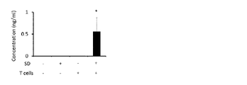
実施例1(1)で作製したマウスについて、EAE病原性CD4陽性T細胞移入から10日後に胃腸炎の病態を評価した。各マウスから採取した便を生理食塩水(便1gあたり20mL)に懸濁し、遠心分離(8000rpm、5分間)により回収した上清を生理食塩水で10倍希釈し、Hemastix(SIEMENS)により潜血便試験を行った。潜血便スコアはHemastixの容器に記載されているスコアリング法に従って算出した。 (2) Gastroenteritis Mice prepared in Example 1 (1) were evaluated for
結果を図4に示す。睡眠障害誘導後にEAE病原性CD4陽性T細胞移入を行った群(SD+ Tcells+)においてのみ潜血便が認められ、これと対応してヘマトクリット値が低下していた。 The results are shown in FIG. Only in the group that underwent EAE-pathogenic CD4-positive T cell transfer (SD+ Tcells+) after induction of sleep disturbance, occult blood was observed, with a corresponding decrease in hematocrit.
また、消化管各部位(胃、十二指腸、空腸、回腸、結腸、直腸)を1mLの生理食塩水で洗浄し、洗浄液について潜血便試験と同様にして血液含量を調べたところ、睡眠障害誘導後にEAE病原性CD4陽性T細胞移入を行った群において胃及び腸の上部から出血が生じていることが確認された(図5A)。この群のマウスの胃において限局性出血病変が認められ(図5B、黒い点)、胃及び十二指腸の上皮組織で炎症が生じていた(図5C)。 In addition, each part of the gastrointestinal tract (stomach, duodenum, jejunum, ileum, colon, rectum) was washed with 1 mL of physiological saline, and the blood content of the washing liquid was examined in the same manner as the occult blood test. Bleeding from the upper part of the stomach and intestine was confirmed in the group to which pathogenic CD4-positive T-cell transfer was performed (Fig. 5A). Focal bleeding lesions were observed in the stomach of this group of mice (Fig. 5B, black dots), and inflammation occurred in the epithelium of the stomach and duodenum (Fig. 5C).
次に、睡眠障害誘導後にEAE病原性CD4陽性T細胞移入を行ったマウスに、プロトンポンプ阻害剤であるランソプラゾール又は対照のカルボキシメチルセルロース(CMC)をEAE病原性CD4陽性T細胞移入後毎日、30mg/kg体重の用量で経口投与した。ランソプラゾールは、睡眠障害誘導後にEAE病原性CD4陽性T細胞移入を行ったマウスの臨床スコア、移入から10日後の致死率及び潜血便スコアのいずれも顕著に低減させた(図6)。
Next, mice that underwent EAE-pathogenic CD4-positive T-cell transfer after induction of sleep disturbance were treated with the proton pump inhibitor lansoprazole or control carboxymethylcellulose (CMC) at 30 mg/day daily after EAE-pathogenic CD4-positive T-cell transfer. It was administered orally at a dose of kg body weight. Lansoprazole markedly reduced both the clinical score, the
以上から、ストレス状態下でEAE病原性CD4陽性T細胞の移入を受けたマウスは、胃、消化管上部における炎症及び出血を特徴とする胃腸炎の病態を有することが確認された。また、プロトンポンプ阻害剤であるランソプラゾールは、臨床スコア及び胃腸炎の病態を改善し、また致死率を低減させることが確認された。 From the above, it was confirmed that mice that received EAE-pathogenic CD4-positive T cell transfer under stress conditions have a pathology of gastroenteritis characterized by inflammation and bleeding in the stomach and upper gastrointestinal tract. Lansoprazole, a proton pump inhibitor, has also been shown to improve clinical scores and pathology of gastroenteritis and reduce mortality.
(3)心筋障害
実施例1(1)で作製したマウスについて、心筋障害の病態を評価した。EAE病原性CD4陽性T細胞移入の9日後に血液を採取し、ELISA kit(Life diagnotics)を用いて血中トロポニンI濃度を、及びELISA kit(Lifespan BioSciences)を用いて血中クレアチンキナーゼMB濃度を測定した。ストレス状態下でEAE病原性CD4陽性T細胞の移入を受けたマウスは、血中トロポニンI濃度(図7)及び血中クレアチンキナーゼMB濃度(図8)のいずれも上昇しており、心臓における細胞死を特徴とする心筋障害の病態を有することが示唆された。この群のマウスの心電を測定したところ、心不全の兆候が検出された(データを図示せず)。 (3) Cardiomyopathy The mice prepared in Example 1(1) were evaluated for myocardial injury. Blood was collected 9 days after EAE pathogenic CD4-positive T cell transfer, and the blood troponin I concentration was measured using an ELISA kit (Life diagnostics), and the blood creatine kinase MB concentration was measured using an ELISA kit (Lifespan BioSciences). It was measured. Mice that received EAE-pathogenic CD4-positive T-cell transfer under stress conditions showed elevated blood troponin I concentrations (Fig. 7) and blood creatine kinase MB concentrations (Fig. 8), indicating that cells in the heart It was suggested to have a pathology of myocardial injury characterized by death. When electrocardiograms were measured in this group of mice, signs of heart failure were detected (data not shown).
また、活性化カスパーゼ3に対する抗体を用いた免疫化学染色の結果、睡眠障害誘導後にEAE病原性CD4陽性T細胞移入を行った群のマウスの心臓においてアポトーシスの増加が観察され、その傾向は心臓上部でより顕著であった(図9、矢印)。
In addition, as a result of immunochemical staining using an antibody against activated
アポトーシスのマーカーである活性化カスパーゼ3、血管マーカーであるCD31、交感神経マーカーであるチロシンヒドロキシラーゼ、迷走神経マーカーであるコリントランスポーター1の各々に対する抗体を用いた、睡眠障害誘導後にEAE病原性CD4陽性T細胞移入を行った疾患モデルマウスの心臓上部の免疫化学染色像を図10に示す。抗活性化カスパーゼ3抗体で強く染色された部分(図10左上写真の上の矢印)は、構造的に弁であると推定された。また、抗CD31抗体で染色された部分(図10右上写真の矢印)は抗活性化カスパーゼ3抗体でも染色されており(図10左上写真の下の矢印)、血管でもアポトーシスが生じているものと考えられた。また、抗活性化カスパーゼ3抗体で染色された部分の周囲は抗チロシンヒドロキシラーゼ抗体、抗コリントランスポーター1抗体で染色されていたことから、心臓細胞のアポトーシスに神経活動が関与していることが示唆された。
EAE pathogenic CD4 after sleep disturbance induction using antibodies against each of the apoptosis marker activated
以上から、ストレス状態下でEAE病原性CD4陽性T細胞の移入を受けたマウスは、心臓における細胞死を特徴とする心筋障害の病態を有することが確認された。 From the above, it was confirmed that mice receiving EAE-pathogenic CD4-positive T cell transfer under stress conditions have a pathology of myocardial injury characterized by cell death in the heart.
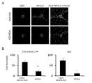
(4)脳内の特定血管における局所炎症
実施例1(1)で作製したマウスについて、MHCクラスII陽性細胞及びCD4陽性T細胞の集積を調べた。EAE病原性CD4陽性T細胞移入から10日後のマウスから摘出した第5腰髄及び脳をSCEM(SECTION-LAB)中に包埋し、マイクロトーム装置CM3050(Leica Microsystems)を用いて10μm厚の切片とした。切片はCryofilmタイプIIIC(16UF)(SECTION-LAB)を用いて回収し、ヘマトキシリン/エオジン染色、又は抗MHCクラスII抗体若しくは抗CD4抗体を用いて免疫組織化学的に染色し、BZ-9000顕微鏡(KEYENCE)で解析した。解析はBZ-IIアナライザー(KEYENCE)のHS ALLソフトウェアにより実施した。 (4) Local Inflammation in Specific Blood Vessels in the Brain The mice prepared in Example 1(1) were examined for accumulation of MHC class II-positive cells and CD4-positive T cells. The fifth lumbar spinal cord and brain excised from
EAE病原性CD4陽性T細胞移入のみを受けた群(SD-)においては、既に報告されている通り(Arima et al.(2012) Cell 148,447-457)、第5腰髄背側血管にMHCクラスII陽性細胞及びCD4陽性T細胞が集積していた(図11)。一方、睡眠障害誘導後にEAE病原性CD4陽性T細胞移入を行った群(SD+)においては、第三脳室、視床及び歯状回の境界領域に存在する特定の血管に両細胞の集積が認められた(図12)。CD8陽性T細胞、B細胞、NK細胞及び好中球等の様々な免疫細胞もまたこの血管に集積することが見出された(データを図示せず)。 In the group that received only EAE-pathogenic CD4-positive T cell transfer (SD-), as previously reported (Arima et al. (2012) Cell 148, 447-457), the dorsal vessels of the 5th lumbar spinal cord were affected. MHC class II-positive cells and CD4-positive T cells were accumulated (Fig. 11). On the other hand, in the group (SD+) in which EAE-pathogenic CD4-positive T-cells were transferred after induction of sleep disturbance, accumulation of both cells was observed in specific blood vessels present in the border regions of the third ventricle, thalamus, and dentate gyrus. (Fig. 12). Various immune cells such as CD8-positive T cells, B cells, NK cells and neutrophils were also found to accumulate in these vessels (data not shown).
また、EAE病原性CD4陽性T細胞移入の9日後に摘出した脳を小脳及び脳幹(Cerebellum and Brain stem)、大脳皮質(Cortex)、海馬及び間脳(Hippocampus and Interbrain)の各部位に解剖し、Neural Tissue Dissection Kit(Miltenyi Biotec)を用いて酵素消化することで単一細胞懸濁液を調製した。懸濁液中の106個の細胞を蛍光コンジュゲート抗体と共に氷上で30分間インキュベートし、細胞表面を標識した。次いで細胞をシアンフローサイトメーター(Beckman Coulter)で解析し、Summitソフトウェア(Beckman Coulter)又はFlowjoソフトウェア(Tree Star)を用いてデータ解析した。In addition, the brain excised 9 days after EAE-pathogenic CD4-positive T cell transfer was dissected into the cerebellum and brain stem, the cerebral cortex, the hippocampus and the interbrain, Single cell suspensions were prepared by enzymatic digestion using the Neural Tissue Dissection Kit (Miltenyi Biotec). 10 6 cells in suspension were incubated with a fluorescently conjugated antibody for 30 minutes on ice to label the cell surface. Cells were then analyzed on a cyan flow cytometer (Beckman Coulter) and data analyzed using Summit software (Beckman Coulter) or Flowjo software (Tree Star).
脳の各部位に存在するCD11b陽性MHCクラスII高発現細胞及びCD4陽性T細胞のフローサイトメトリー解析結果を図13に示す。睡眠障害誘導後にEAE病原性CD4陽性T細胞移入を行った群(SD+ Tcells+)において、特定血管が位置する海馬及び間脳へのMHCクラスII高発現細胞及びCD4陽性T細胞の顕著な集積が生じていた。 FIG. 13 shows the results of flow cytometry analysis of CD11b-positive MHC class II high-expressing cells and CD4-positive T cells present in each region of the brain. In the group (SD + Tcells +) in which EAE-pathogenic CD4-positive T-cell transfer was performed after sleep disorder induction, MHC class II high-expressing cells and CD4-positive T-cells in the hippocampus and diencephalon where specific blood vessels are located significantly accumulated. was
次に、海馬及び間脳に集積したMHCクラスII高発現細胞の由来を調べるため、C57BL/6マウスの代わりにCX3CR1CreER ROSA26-TdTomatoマウスを用いて、実施例1(1)と同様の方法で、睡眠障害誘導による疾患モデルマウスを作製した。タモキシフェンを2mg/マウスの用量で二日連続経口投与してTd-tomatoを発現させた後、投与後6週間のマウスにEAE病原性CD4陽性T細胞を移入し、移入の10日後に海馬及び間脳を摘出し、MHCクラスII高発現細胞におけるTd-tomatoの発現をフローサイトメトリーで解析した。Next, in order to investigate the origin of MHC class II high-expressing cells accumulated in the hippocampus and diencephalon, CX3CR1 CreER ROSA26-TdTomato mice were used instead of C57BL/6 mice in the same manner as in Example 1 (1). , created disease model mice by sleep disorder induction. Tamoxifen was orally administered at a dose of 2 mg/mouse for two consecutive days to express Td-tomato, and then EAE-pathogenic CD4-positive T cells were transferred to
結果を図14に示す。睡眠障害誘導後にEAE病原性CD4陽性T細胞移入を行った群(SD+ Tcells+)のマウスの海馬及び間脳には、Td-tomato陰性、すなわち末梢由来のMHCクラスII高発現細胞(単球)が多く集積しており、Td-tomato陽性、すなわち中枢由来のMHCクラスII高発現細胞(ミクログリア細胞)の集積は少量しか認められなかった。 The results are shown in FIG. In the hippocampus and diencephalon of mice in the group (SD + Tcells +) in which EAE-pathogenic CD4-positive T-cell transfer was performed after sleep disorder induction, Td-tomato-negative, that is, peripherally-derived MHC class II high-expressing cells (monocytes) were present. A large amount of Td-tomato-positive cells (microglial cells) derived from the central nervous system and highly expressing MHC class II were accumulated in only a small amount.
以上から、ストレス状態下でEAE病原性CD4陽性T細胞の移入を受けたマウスは、第三脳室、視床及び歯状回の境界領域の特定血管におけるMHCクラスII高発現細胞、CD4陽性T細胞等の免疫細胞の集積を特徴とする炎症の病態を有することが確認された。 From the above, mice that received EAE-pathogenic CD4-positive T-cell transfer under stress conditions showed high MHC class II-expressing cells and CD4-positive T-cells in specific blood vessels in the border region of the third ventricle, thalamus and dentate gyrus. It was confirmed to have an inflammatory pathology characterized by the accumulation of immune cells such as
実施例3 疾患モデルマウスにおけるCCL5及び炎症性サイトカインの機能解析
(1)CCL5
実施例1(1)で作製した睡眠障害誘導後にEAE病原性CD4陽性T細胞移入を行ったマウスに、抗CCL5抗体又は対照のラットIgG抗体(いずれも100μg/マウス)をEAE病原性CD4陽性T細胞移入後毎日腹腔内注射し、EAE病態を実施例2(1)と同様に評価した。また、EAE病原性CD4陽性T細胞移入から10日後のマウスを用いて、特定血管における細胞の集積を実施例2(4)と同様に調べた。 Example 3 Functional analysis of CCL5 and inflammatory cytokines in disease model mice
(1) CCL5
Anti-CCL5 antibody or control rat IgG antibody (both 100 µg/mouse) was applied to the mice that had been transferred with EAE-pathogenic CD4-positive T cells after sleep disorder induction, prepared in Example 1 (1), to induce EAE-pathogenic CD4-positive T cells. Intraperitoneal injections were performed every day after cell transfer, and EAE pathology was evaluated in the same manner as in Example 2 (1). In addition, using
抗CCL5抗体は、特定血管におけるCD4陽性T細胞及びCD11b陽性MHCクラスII高発現細胞の集積を抑制し(図15)、臨床スコア及び致死率を低減させた(図16)。一方、CCL2又はCX3CL1等の他のケモカインに対する抗体はかかる効果を示さなかった(データを図示せず)。 Anti-CCL5 antibody suppressed the accumulation of CD4-positive T cells and CD11b-positive MHC class II high-expressing cells in specific blood vessels (Fig. 15), and reduced the clinical score and lethality (Fig. 16). In contrast, antibodies to other chemokines such as CCL2 or CX3CL1 did not show such effects (data not shown).
次いで、特定血管におけるケモカインCCL5の発現量を以下のように測定した。EAE病原性CD4陽性T細胞移入から4日後のマウスより実施例2(4)と同様の方法で約100個の凍結切片(15μm厚)を調製し、PAXgene(QIAGEN)で15分間固定し、次いで100% EtOHで10分間固定した。切片中の第三脳室領域血管周辺の組織をレーザーマイクロダイセクションデバイスDM6000B(Leica Microsystems)により回収し、全RNAをRNeasyマイクロキット(QIAGEN)により抽出し、DNase及び逆転写反応を行ってcDNAを得た。
Next, the expression level of chemokine CCL5 in specific blood vessels was measured as follows. About 100 cryosections (15 μm thick) were prepared from the
得られたcDNAを、GeneAmp 5700シーケンス検出システム(ABI)及びKAPA PROBE FAST ABI Prism qPCRキット(Kapa Biosystems)を用いたリアルタイムqPCR解析に供し、CCL5及び内在性コントロールのHPRTの発現量を測定した。使用したPCRプライマーペア及びプライマーは、
マウスHPRTプライマー:
5’-AGCCCCAAAATGGTTAAGGTTG-3’(配列番号1)及び
5’-CAAGGGCATATCCAACAACAAAC-3’(配列番号2)
マウスHPRTプローブ:
5’-ATCCAACAAAGTCTGGCCTGTATCCAACAC-3’(配列番号3)
マウスCCL5プライマー:
5’-CTCCCTGCTGCTTTGCCTAC-3’(配列番号4)及び
5’-CGGTTCCTTCGAGTGACAAACA-3’(配列番号5)
マウスCCL5プローブ:5’-TGCCTCGTGCCCACGTCAAGGAGTATT-3’(配列番号6)
であった。リアルタイムPCR条件は、50℃ 2分間、95℃ 3分間の後、95℃ 3秒間、60℃ 30秒間を40サイクルであった。The resulting cDNA was subjected to real-time qPCR analysis using the GeneAmp 5700 Sequence Detection System (ABI) and KAPA PROBE FAST ABI Prism qPCR kit (Kapa Biosystems) to measure the expression levels of CCL5 and endogenous control HPRT. The PCR primer pair and primers used were
Mouse HPRT primer:
5′-AGCCCCAAATGGTTAAGGTTG-3′ (SEQ ID NO: 1) and 5′-CAAGGGCATATCCAACAACAAAC-3′ (SEQ ID NO: 2)
Mouse HPRT probe:
5′-ATCCAAAAAAGTCTGGCCTGTATCCAACAC-3′ (SEQ ID NO: 3)
Mouse CCL5 primer:
5′-CTCCCTGCTGCTTTGCCTAC-3′ (SEQ ID NO:4) and 5′-CGGTTCCTTCGAGTGACAAACA-3′ (SEQ ID NO:5)
Mouse CCL5 probe: 5'-TGCCTCGTGCCCACGTCAAGGAGTATT-3' (SEQ ID NO: 6)
Met. The real-time PCR conditions were 50°C for 2 minutes, 95°C for 3 minutes, followed by 40 cycles of 95°C for 3 seconds, 60°C for 30 seconds.
第三脳室領域血管周辺組織におけるCCL5の相対的発現量を図17に示す。EAE病原性CD4陽性T細胞移入の有無にかかわらず、睡眠障害誘導を行った群においてCCL5の発現量が増加していた。 FIG. 17 shows the relative expression levels of CCL5 in the perivascular tissue of the third ventricular region. Regardless of the presence or absence of EAE-pathogenic CD4-positive T cell transfer, the CCL5 expression level was increased in the sleep disorder induction group.
(2)炎症性サイトカイン
実施例1(1)で作製した睡眠障害誘導後にEAE病原性CD4陽性T細胞移入を行ったマウスに、抗IFN-γ抗体、抗IL17A抗体又は対照のラットIgG抗体(いずれも100μg/マウス)をEAE病原性CD4陽性T細胞移入後毎日腹腔内注射し、EAE病態を実施例2(1)と同様に評価した。また、EAE病原性CD4陽性T細胞移入から10日後のマウスを用いて、特定血管における細胞の集積を実施例2(4)と同様に調べた。 (2) Inflammatory cytokine Example 1 (1) After inducing the sleep disorder, the mice that were transferred with EAE-pathogenic CD4-positive T cells were treated with an anti-IFN-γ antibody, an anti-IL17A antibody, or a control rat IgG antibody (either 100 μg/mouse) was intraperitoneally injected every day after transfer of EAE-pathogenic CD4-positive T cells, and EAE pathology was evaluated in the same manner as in Example 2(1). In addition, using
抗IFN-γ抗体、抗IL17A抗体はそれぞれ特定血管におけるCD4陽性T細胞及びCD11b陽性MHCクラスII高発現細胞の集積を抑制した(図18)。これらの抗体の併用は、臨床スコア及び致死率を著しく低減させた(図19)。 Anti-IFN-γ antibody and anti-IL17A antibody suppressed accumulation of CD4-positive T cells and CD11b-positive MHC class II high expression cells in specific blood vessels, respectively (FIG. 18). The combination of these antibodies significantly reduced clinical scores and mortality (Figure 19).
次に、特定血管に炎症性サイトカインを直接的に注射することにより、局所脳炎が疾患を誘導するかを調べた。睡眠障害を誘導したC57BL/6マウス又は対照マウスの頭部を麻酔下で定位装置に固定し、頭蓋骨上の毛をそり、皮膚を70%エタノールで拭いた。30ゲージの針を特定血管(座標AP -1.06mm;ML 1mm;DV 2.25mm)に向けて下げ入れ、いずれも液量0.2μlのEAE病原性CD4陽性T細胞(1×10⁶個)+MOGをパルスした骨髄由来樹状細胞(5×10⁵個)、IFN-γ(50ng;PeproTech)+IL-17A(50ng;R&D Systems)又はIL-6(50ng;Toray)+IL-17A(50ng)を90秒かけて既報の通りにマイクロインジェクションにより投与した(Kim et al.(2011) Mol.Brain 19,4-6)。Next, we examined whether local encephalitis induces disease by directly injecting inflammatory cytokines into specific blood vessels. C57BL/6 mice with induced sleep disturbance or control mice were head-fixed in a stereotaxic apparatus under anesthesia, the hair on the skull was shaved, and the skin was wiped with 70% ethanol. A 30-gauge needle was lowered toward a specific blood vessel (coordinates AP -1.06 mm;
マイクロインジェクションから2日後のマウスの致死率を図20Aに、潜血便スコアを図20Bに示す。睡眠障害を誘導したマウスにおいては、EAE病原性CD4陽性T細胞+樹状細胞、IFN-γ+IL-17A及びIL-6+IL-17Aいずれの投与も胃腸炎を発症させ、致死率を大きく向上させた。
以上から、ストレス状態下、第三脳室、視床及び歯状回の境界領域の特定血管内で生じるCCL5がEAE病原性CD4陽性T細胞を集積させて炎症性サイトカインを産生させ、局所脳炎を確立することが示唆された。さらに、抗CCL5抗体及び抗炎症性サイトカイン抗体は、EAE及び胃腸炎の病態を改善し、致死率を低減させることが確認された。 Based on the above, CCL5 generated in specific blood vessels in the border region of the third ventricle, thalamus and dentate gyrus under stress conditions accumulates EAE-pathogenic CD4-positive T cells, produces inflammatory cytokines, and establishes focal encephalitis. It was suggested that Furthermore, anti-CCL5 antibody and anti-inflammatory cytokine antibody were confirmed to ameliorate the pathology of EAE and gastroenteritis and reduce the mortality rate.
実施例4 疾患モデルマウスの病態誘導に関与する神経路の解析
(1)PVNの交感神経と特定血管との連絡
C57BL/6マウスに対して、実施例3(2)と同様の方法で、ドーパミン/ノルアドレナリン作動性ニューロンを変性させる神経毒である6-ヒドロキシドーパミン(6-OHDA)塩酸塩(2mg/ml)又は溶媒の0.02%アスコルビン酸それぞれ0.2μlをEAE病原性CD4陽性T細胞移入の四日前(ストレス負荷の二日前)に特定血管にマイクロインジェクションした。その後、実施例1(1)と同様に、睡眠障害誘導及びEAE病原性CD4陽性T細胞移入を行った。 Example 4 Analysis of nerve tracts involved in pathological induction of disease model mice
(1) 6-hydroxydopamine, a neurotoxin that degenerates dopaminergic/noradrenergic neurons, was applied to C57BL/6 mice in the same manner as in Example 3 (2) in connection between PVN sympathetic nerves and specific blood vessels. 0.2 μl each of (6-OHDA) hydrochloride (2 mg/ml) or 0.02% ascorbic acid as a solvent was microinjected into specific blood vessels four days before transfer of EAE pathogenic CD4-positive T cells (two days before stress loading). did. Thereafter, sleep disorder induction and EAE-pathogenic CD4-positive T cell transfer were performed in the same manner as in Example 1 (1).
EAE病原性CD4陽性T細胞移入から10日後の化学的交感神経除去マウスの第三脳室領域を抗チロシンヒドロキシラーゼ抗体及び抗リン酸化CREB抗体で免疫染色した写真を図21に示す。6-OHDAは、チロシンヒドロキシラーゼ陽性の交感神経ニューロンを減少させ、ノルアドレナリン作動性受容体シグナル伝達により誘導されるCREBのリン酸化を抑制した。
FIG. 21 shows photographs of the immunostaining of the third ventricular region of the chemically sympathetic denervated mouse with an anti-tyrosine hydroxylase antibody and an
また、抗MHCクラスII抗体及び抗CD4抗体を用いた第三脳室領域の免疫染色写真及び染色細胞数のグラフを図22に、マウスの臨床スコア及び致死率を図23に示す。特定血管におけるCD11b陽性MHCクラスII高発現細胞及びCD4陽性T細胞の集積は6-OHDAの投与により抑制され、臨床スコア、致死率共に改善が認められた。 In addition, photographs of immunostaining of the third ventricular region using anti-MHC class II antibody and anti-CD4 antibody and a graph of the number of stained cells are shown in FIG. 22, and clinical scores and lethality of mice are shown in FIG. The accumulation of CD11b-positive MHC class II high-expressing cells and CD4-positive T cells in specific blood vessels was suppressed by administration of 6-OHDA, and both the clinical score and mortality rate were improved.
次に、実施例1(1)で作製した各マウスを用いて、PVN及びDMHにおける神経の活性化を調べた。EAE病原性CD4陽性T細胞移入から10日後に摘出した脳の切片を抗チロシンヒドロキシラーゼ抗体及び抗リン酸化cfos抗体で免疫染色した写真及び染色細胞数のグラフを図24に示す。睡眠障害誘導はPVNの交感神経ニューロンを活性化し、その程度はEAE病原性CD4陽性T細胞移入により増強された。また睡眠障害誘導後にEAE病原性CD4陽性T細胞移入を行った場合、DMHにおけるチロシンヒドロキシラーゼ陰性ニューロンの活性化が観察された。 Next, each mouse prepared in Example 1(1) was used to examine neural activation in PVN and DMH. FIG. 24 shows photographs of brain sections excised 10 days after transfer of EAE pathogenic CD4-positive T cells and immunostaining with anti-tyrosine hydroxylase antibody and anti-phosphorylated cfos antibody, and a graph of the number of stained cells. Sleep disturbance induction activated PVN sympathetic neurons, the extent of which was enhanced by EAE-pathogenic CD4-positive T-cell transfer. Activation of tyrosine hydroxylase-negative neurons in DMH was also observed when EAE-pathogenic CD4-positive T cell transfer was performed after sleep disorder induction.
さらに、逆行性トレーサーを用いて、特定血管とつながる神経路を調べた。EAE病原性CD4陽性T細胞移入から5日後に、逆行性トレーサーであるコレラ毒素BサブユニットのFITCコンジュゲート(FITC-CTB)(Sigma-Aldrich、1mg/ml)又は溶媒の生理食塩水を、睡眠障害誘導後にEAE病原性CD4陽性T細胞移入を行ったマウスの特定血管にマイクロインジェクションした。PVN及びDMHの蛍光写真及び染色細胞数のグラフを図25に示す。FITC-CTBはDMHよりもPVNにおいて多く観察された。また、抗チロシンヒドロキシラーゼ抗体、抗ノルアドレナリントランスポーター及び抗ドーパミントランスポーター抗体を用いたPVNの免疫染色写真を図26に示す。PVNのチロシンヒドロキシラーゼ陽性ニューロンはノルアドレナリントランスポーターを共発現するがドーパミントランスポーターを共発現していなかった。 In addition, retrograde tracers were used to investigate nerve tracts that connect to specific blood vessels. Five days after EAE-pathogenic CD4-positive T cell transfer, the retrograde tracer FITC conjugate of cholera toxin B subunit (FITC-CTB) (Sigma-Aldrich, 1 mg/ml) or vehicle saline was administered to sleep mice. Microinjection into specific blood vessels of mice with EAE-pathogenic CD4-positive T cell transfer after injury induction. Fluorescence photographs of PVN and DMH and graphs of stained cell counts are shown in FIG. FITC-CTB was observed more in PVN than in DMH. FIG. 26 shows immunostaining photographs of PVN using anti-tyrosine hydroxylase antibody, anti-noradrenaline transporter and anti-dopamine transporter antibodies. PVN tyrosine hydroxylase-positive neurons co-express noradrenaline transporters but not dopamine transporters.
以上から、特定血管はPVNの交感神経と直接的な連絡があり、この交感神経ニューロンはストレス状態下で特定血管においてノルアドレナリンを分泌し、ケモカインを誘導することが示唆された。また、ストレス状態下でのEAE病原性CD4陽性T細胞移入によりDMHにおける神経の活性化が確認されたことから、以下にさらなる試験を行った。 From the above, it was suggested that the specific blood vessels have direct communication with the sympathetic nerves of the PVN, and that these sympathetic neurons secrete noradrenaline in the specific blood vessels and induce chemokines under stress conditions. In addition, neuronal activation in DMH was confirmed by EAE-pathogenic CD4-positive T-cell transfer under stress conditions, and further studies were performed as follows.
(2)特定血管とDMHとの連絡
実施例1(1)で作製した睡眠障害誘導後にEAE病原性CD4陽性T細胞移入を行ったマウスに対して、EAE病原性CD4陽性T細胞移入から5日後に、順行性トレーサーであるPHA-L(25mg/ml)又は溶媒の生理食塩水を特定血管にマイクロインジェクションした。PHA-LはPVNよりもDMHにおいて多く観察された(図27)。また、FITC-CTBをDMH(座標AP -1.46mm;ML 0.37mm;DV 5mm)にマイクロインジェクションしたところ、特定血管においてFITCの蛍光が観察された(図28)。 (2) Communication between specific blood vessels and DMH Five days after transfer of EAE-pathogenic CD4-positive T cells to mice in which EAE-pathogenic CD4-positive T cells were transferred after sleep disorder induction prepared in Example 1 (1) Later, the anterograde tracer PHA-L (25 mg/ml) or vehicle saline was microinjected into specific vessels. PHA-L was observed more in DMH than in PVN (Figure 27). When FITC-CTB was microinjected into DMH (coordinates AP -1.46 mm; ML 0.37 mm;
また、神経活性化を抑制することが知られているGABAA受容体アゴニストのムシモール(Sigma-Aldrich、0.25mg/ml)又は溶媒の生理食塩水をEAE病原性CD4陽性T細胞移入後5日目にDMHにマイクロインジェクションし、臨床スコア、EAE病原性CD4陽性T細胞移入から10日後の致死率及び潜血便を評価した。ムシモールは臨床スコア、致死率及び潜血便のいずれも顕著に改善した(図29)。In addition, muscimol (Sigma-Aldrich, 0.25 mg / ml), a GABA A receptor agonist known to suppress neuronal activation, or physiological saline as a solvent was added to EAE pathogenic CD4-
以上から、特定血管における局所脳炎はDMHのニューロン、特にチロシンヒドロキシラーゼ陰性ニューロンを活性化することが示唆された。また、GABA受容体アゴニストは、EAE及び胃腸炎の病態を改善し、致死率を低減させることが確認された。 From the above, it was suggested that focal encephalitis in a specific blood vessel activates DMH neurons, especially tyrosine hydroxylase-negative neurons. It was also confirmed that GABA receptor agonists improve the pathology of EAE and gastroenteritis and reduce the mortality rate.
(3)PVN、DMH及び特定血管間の神経連絡と病態との関係
睡眠障害を誘導したC57BL/6マウスの特定血管に、EAE病原性CD4陽性T細胞+MOG樹状細胞又はIFN-γ+IL-17Aを実施例3(2)と同様にマイクロインジェクションした。マイクロインジェクションから2日後のPVN及びDMHにおいて、リン酸化cfosの増加、すなわち神経の活性化が観察された(図30)。 (3) Neural communication between PVN, DMH, and specific blood vessels and pathological conditions In specific blood vessels of C57BL/6 mice that induced sleep disorders, EAE-pathogenic CD4-positive T cells + MOG dendritic cells or IFN-γ + IL-17A were introduced. Microinjection was performed in the same manner as in Example 3(2). An increase in phosphorylated cfos, ie neuronal activation, was observed in PVN and
また、麻酔下でマウスの頭蓋骨にドリルで穴を開け、頭蓋骨を通して電極(Brain Science Idea)をPVN(座標AP -1.06mm;ML 0.25mm;DV 4.8mm)に挿入し、400uAの直流電流を5秒間負荷することで、PVNの一側性除去をストレス負荷の一週間前に行った。このマウスに対して、実施例1(1)と同様に睡眠障害誘導及びEAE病原性CD4陽性T細胞移入を行った。PVNの一側性除去は、特定血管におけるCD11b陽性MHCクラスII高発現細胞及びCD4陽性T細胞の集積を抑制し(図31)、EAE及び胃腸炎の病態を改善し、致死率を低減させた(図32)。 Alternatively, under anesthesia, the skull of a mouse was drilled and an electrode (Brain Science Idea) was inserted through the skull into the PVN (coordinates AP −1.06 mm; ML 0.25 mm; DV 4.8 mm) and a direct current of 400 uA was applied. Unilateral ablation of the PVN was performed one week prior to stress loading by applying a current for 5 seconds. Sleep disorder induction and EAE-pathogenic CD4-positive T cell transfer were performed on this mouse in the same manner as in Example 1(1). Unilateral ablation of PVN suppressed the accumulation of CD11b-positive MHC class II high-expressing cells and CD4-positive T cells in specific blood vessels (Fig. 31), improved the pathology of EAE and gastroenteritis, and reduced the mortality rate. (Fig. 32).
以上から、局所脳炎はPVN及びDMHの神経を活性化し、疾患モデルマウスの各種病態を誘発することが示唆された。 From the above, it was suggested that focal encephalitis activates PVN and DMH neurons and induces various pathological conditions in disease model mice.
次に、PVN、DMH及び特定血管間の神経連絡における神経伝達物質ATPの機能、及び疾患モデルマウスの病態への寄与を調べた。血管内皮細胞株BC1細胞(大阪大学宮坂昌之教授より分与)をDMEMで24時間、ヒトIL-6(50ng/ml)、ヒト可溶性IL-6受容体α(50ng/ml)及びマウスIL-17A(50ng/ml)を加えてさらに24時間培養した後、細胞内ATP産生量をATPアッセイkit(TOYO Ink)を用いて測定した。結果を図33に示す。炎症性サイトカインIL-17A+IL-6刺激は、血管内皮細胞におけるATP産生量を増加させた。 Next, we investigated the function of the neurotransmitter ATP in neural communication between PVN, DMH and specific blood vessels, and its contribution to the pathological conditions of disease model mice. Vascular endothelial cell line BC1 cells (provided by Professor Masayuki Miyasaka, Osaka University) were treated with DMEM for 24 hours, human IL-6 (50 ng/ml), human soluble IL-6 receptor α (50 ng/ml), and mouse IL-17A. (50 ng/ml) was added and cultured for an additional 24 hours, and then intracellular ATP production was measured using an ATP assay kit (TOYO Ink). The results are shown in FIG. Inflammatory cytokine IL-17A+IL-6 stimulation increased ATP production in vascular endothelial cells.
また、PVN及びDMHのニューロンは、睡眠誘導マウスの特定血管へのATP(Sigma-Aldrich、2μg)のマイクロインジェクションにより活性化された(図30)。 PVN and DMH neurons were also activated by microinjection of ATP (Sigma-Aldrich, 2 μg) into specific blood vessels in sleep-induced mice (FIG. 30).
さらに、睡眠障害を誘導したマウスの特定血管に、IL-6及びIL-17Aを、ATP受容体であるP2X受容体の選択的アンタゴニストであるA438079(1μg)を伴って又は伴わずに、実施例3(2)と同様にしてマイクロインジェクションした。マイクロインジェクションから2日後の第三脳室領域内のリン酸化cfos、潜血便及び致死率を評価した結果を図34及び図35に示す。A438079は、PVN及びDMHにおける神経活性化を抑制し、また胃腸炎及び致死率を改善した。
Furthermore, IL-6 and IL-17A, with or without A438079 (1 μg), which is a selective antagonist of the ATP receptor P2X receptor, were injected into specific blood vessels of mice that induced sleep disorders. Microinjection was performed in the same manner as in 3(2). Figures 34 and 35 show the results of evaluating phosphorylated cfos in the region of the third ventricle, occult blood, and
以上から、局所脳炎部位で炎症性サイトカインにより誘導されたATPはPVN及びDMHを活性化して疾患モデルマウスの病態を誘導することが示唆された。また、ATP受容体アンタゴニストはEAE及び胃腸炎の病態を改善し、致死率を低減させることが確認された。 From the above, it was suggested that ATP induced by inflammatory cytokines at the site of local encephalitis activates PVN and DMH to induce pathology in disease model mice. It was also confirmed that ATP receptor antagonists ameliorate the pathology of EAE and gastroenteritis and reduce the mortality rate.
(4)DMX(迷走神経背側核)及びNTS(孤束核)と病態との関係
上記(2)において特定血管にマイクロインジェクションされた順行性トレーサーPHA-LはDMXに到達していた(結果を図示せず)。また、実施例3(2)と同様に、睡眠障害を誘導したマウスの特定血管にEAE病原性CD4陽性T細胞+MOG樹状細胞をマイクロインジェクションしたところ、DMX及び求心性迷走神経の主要な核であるNTSにおけるリン酸化cfosの増加、すなわち神経の活性化が観察された(図36)。 (4) Relationship between DMX (dorsal vagal nucleus) and NTS (nucleus tractus solitarius) and pathology The anterograde tracer PHA-L microinjected into a specific blood vessel in (2) above reached DMX ( results not shown). In addition, in the same manner as in Example 3 (2), when EAE-pathogenic CD4-positive T cells + MOG dendritic cells were microinjected into specific blood vessels of mice that had induced sleep disorders, DMX and major nuclei of vagal afferent nerves An increase in phosphorylated cfos in certain NTS, ie, neuronal activation, was observed (Fig. 36).
また、麻酔下のマウスを上部正中線に沿って開腹し、胃を横隔膜の下に引き込んで両迷走神経幹を露出させて視認可能な迷走神経を少なくとも1mm切離し、さらに横隔膜直下の食道周辺にある全ての神経組織及び結合組織を取り除いて小さな迷走神経分岐を全て切除することで、マウスに横隔下迷走神経切離術を施した。このマウスに対して、実施例1(1)と同様に睡眠障害誘導及びEAE病原性CD4陽性T細胞移入を行って疾患モデルマウスを作製した。迷走神経切離術は、疾患モデルマウスのEAE及び胃腸炎の病態を改善し、致死率を低減させた(図37)。 Anesthetized mice were also laparotomyed along the upper midline, with the stomach retracted under the diaphragm to expose both vagus nerve trunks and cut the visible vagus nerve at least 1 mm, as well as around the esophagus just below the diaphragm. Mice underwent a subphrenic vagotomy by removing all nerve and connective tissue and excising all small vagus nerve branches. Sleep disorder induction and EAE-pathogenic CD4-positive T cell transfer were performed on this mouse in the same manner as in Example 1(1) to prepare a disease model mouse. Vagotomy ameliorated the pathology of EAE and gastroenteritis in disease model mice and reduced the mortality rate (Fig. 37).
以上から、局所脳炎はDMX及びNTSの求心性迷走神経を活性化し、疾患モデルマウスの各種病態を誘発することが示唆された。 From the above, it was suggested that focal encephalitis activates DMX and NTS afferent vagus nerves and induces various pathological conditions in disease model mice.
実施例1~4の結果から、ストレス負荷はPVN交感神経を活性化し、第三脳室、視床及び歯状回の境界領域に存在する特定血管においてノルアドレナリンを分泌させ、これによるCCL5産生、CD11b陽性MHCクラスII高発現細胞及びCD4陽性T細胞の集積並びに炎症性サイトカイン産生を引き起こした。さらにこのT細胞集積性局所脳炎はATPを介してDMHの神経を活性化し、これがさらにDMX及びNTSの迷走神経の活性化を介して疾患モデルマウスの各種病態を誘発するという機序が推定された。 From the results of Examples 1 to 4, the stress load activates the PVN sympathetic nerves and secretes noradrenaline in specific blood vessels present in the border regions of the third ventricle, the thalamus and the dentate gyrus, resulting in CCL5 production and CD11b positivity. It caused the accumulation of MHC class II high-expressing cells and CD4-positive T cells and the production of inflammatory cytokines. Furthermore, it was presumed that this T cell-accumulating focal encephalitis activates DMH nerves via ATP, which in turn induces various pathological conditions in disease model mice via DMX and NTS activation of vagus nerves. .
また、上記機序に関与する神経路又はCCL5若しくは炎症性サイトカインの抑制、遮断又は阻害は、疾患モデルマウスの病態の予防及び/又は治療に寄与することが確認された。 In addition, it was confirmed that suppression, blockage or inhibition of neural pathways involved in the above mechanisms or CCL5 or inflammatory cytokines contributes to the prevention and/or treatment of pathological conditions in disease model mice.
実施例5 ストレス負荷により脳内特定血管における発現が変動する遺伝子の解析
睡眠障害誘導を行って2日後のC57BL/6マウスから、約100個の脳の凍結切片(15μm厚)を調製し、PAXgene(QIAGEN)で15分間固定し、次いで100% EtOHで10分間固定した。切片中の第三脳室領域血管周辺の組織をレーザーマイクロダイセクションデバイスDM6000B(Leica Microsystems)により回収し、全RNAをRNeasyマイクロキット(QIAGEN)により抽出し、かずさDNA研究所に委託してRNAシークエンスを行い、全遺伝子を対象とした発現解析を実施した。 Example 5 Analysis of genes whose expression changes in specific blood vessels in the brain due to stress load Approximately 100 brain cryosections (15 μm thick) were prepared from C57BL/6 mice two days after sleep disorder induction, and PAXgene was prepared. (QIAGEN) for 15 minutes followed by 100% EtOH for 10 minutes. The tissue around the blood vessels in the third ventricular region in the section was collected with a laser microdissection device DM6000B (Leica Microsystems), total RNA was extracted with an RNeasy micro kit (QIAGEN), and RNA sequencing was performed by Kazusa DNA Research Institute. was performed, and expression analysis was performed for all genes.
解析の結果、ストレス負荷によりLY6G6C遺伝子及びα2Cアドレナリン受容体遺伝子の発現が上昇することが確認され(図38)、これらの因子が脳内特定血管における炎症及び疾患モデルマウスの病態に関与する可能性が示された。 As a result of the analysis, it was confirmed that the expression of the LY6G6C gene and the α2C adrenergic receptor gene increased due to stress load (Fig. 38), and the possibility that these factors are involved in the pathology of inflammation and disease model mice in specific blood vessels in the brain. It has been shown.
実施例6 抗LY6G6C抗体及び抗α2Cアドレナリン受容体抗体による疾患モデルマウスの病態改善
C57BL/6マウスに睡眠障害誘導後、EAE病原性CD4陽性T細胞を移入した。5日後に当該マウスの頭部を麻酔下で定位装置に固定し、頭蓋骨上の毛をそり、皮膚を70%エタノールで拭いた。30ゲージの針を特定血管(座標AP -1.06mm;ML 1mm;DV 2.25mm)に向けて下げ入れ、液量0.5μlの抗Ly6G6C抗体(clone;NIMP-R14) 0.1mg/ml(NOVUS BIOLOGICALS)、抗alpha 2c Adrenergic Receptor抗体 1mg/ml(GeneTex)、又はそれぞれのコントロール抗体(ラットIgG(Sigma)若しくはウサギIgG(Sigma))を90秒かけて、マイクロインジェクションにより投与した。その後、経時的にマウスの病態観察を行った。 Example 6 Improvement of Disease Model Mice by Anti-LY6G6C Antibody and Anti-α2C Adrenergic Receptor Antibody EAE-pathogenic CD4-positive T cells were transferred to C57BL/6 mice after inducing sleep disorder. Five days later, the head of the mouse was fixed in a stereotaxic apparatus under anesthesia, the hair on the skull was shaved, and the skin was wiped with 70% ethanol. A 30-gauge needle is lowered toward a specific blood vessel (coordinates AP -1.06 mm;
抗LY6G6C抗体を投与したマウスのEAE臨床スコアの経時変化を図39Aに、抗α2Cアドレナリン受容体抗体を投与したマウスの臨床スコアの経時変化を図39Bに示す。対照群では臨床スコアの急激な上昇が認められたのに対し、抗LY6G6C抗体処置群、抗α2Cアドレナリン受容体抗体処置群のいずれにおいても臨床スコアの上昇は認められず、病態の進行が有意に抑制された。このことから、LY6G6C又はα2Cアドレナリン受容体の抑制、遮断又は阻害は、疾患モデルマウスの病態の予防及び/又は治療に寄与することが確認された。 FIG. 39A shows time-course changes in the EAE clinical scores of mice administered with the anti-LY6G6C antibody, and FIG. 39B shows time-course changes in the clinical scores of mice administered with the anti-α2C adrenergic receptor antibody. A sharp increase in the clinical score was observed in the control group, whereas no increase in the clinical score was observed in either the anti-LY6G6C antibody-treated group or the anti-α2C adrenergic receptor antibody-treated group, and the progression of the disease was significant. Suppressed. From this, it was confirmed that suppression, blockade or inhibition of LY6G6C or α2C adrenergic receptors contributes to the prevention and/or treatment of pathological conditions in disease model mice.
配列番号1 マウスHPRTフォワードプライマー
配列番号2 マウスHPRTリバースプライマー
配列番号3 マウスHPRTプローブ
配列番号4 マウスCCL5フォワードプライマー
配列番号5 マウスCCL5リバースプライマー
配列番号6 マウスCCL5プローブSEQ ID NO: 1 mouse HPRT forward primer SEQ ID NO: 2 mouse HPRT reverse primer SEQ ID NO: 3 mouse HPRT probe SEQ ID NO: 4 mouse CCL5 forward primer SEQ ID NO: 5 mouse CCL5 reverse primer SEQ ID NO: 6 mouse CCL5 probe
Claims (5)
(ii)工程(i)の開始前から終了後のいずれかの時点で非ヒト動物に被験物質を投与する工程、並びに
(iii)被験物質を投与した非ヒト動物において、脳内血管の炎症、進行性多発性硬化症、胃腸炎、心筋障害及び突然死よりなる群から選択される少なくとも1の疾患又は症状の発症、進行又は発生を観察し、被験物質を投与しない非ヒト動物のそれと比較する工程
を含む、進行性多発性硬化症、胃腸炎、心筋障害及び突然死よりなる群から選択される少なくとも1の疾患又は症状を予防及び/又は治療するための薬剤のスクリーニング方法であって、
工程(i)が、ストレス状態にある非ヒト動物に中枢神経組織由来抗原反応性のCD4陽性T細胞を移入する工程、又はストレス状態にある非ヒト動物に中枢神経組織由来抗原を投与して中枢神経組織由来抗原反応性のCD4陽性T細胞を誘導する工程である、前記スクリーニング方法。 (i) a step of allowing central nervous tissue-derived antigen-reactive CD4-positive T cells to exist in the body of a non-human animal under stress;
(ii) administering a test substance to a non-human animal at any time from before the start of step (i) to after the end; Observe the onset, progression or occurrence of at least one disease or symptom selected from the group consisting of progressive multiple sclerosis, gastroenteritis, myocardial damage and sudden death, and compare with that in non-human animals to which the test substance is not administered. A method of screening for agents for preventing and/or treating at least one disease or condition selected from the group consisting of progressive multiple sclerosis, gastroenteritis, myocardial damage and sudden death, comprising the steps of:
The step (i) is a step of transferring central nervous tissue-derived antigen-reactive CD4-positive T cells into a non-human animal under stress, or administering a central nervous tissue-derived antigen to a non-human animal under stress The screening method, which is a step of inducing CD4-positive T cells reactive to neural tissue-derived antigens .
(ii)工程(i)の開始前から終了後のいずれかの時点で非ヒト動物に被験物質を投与する工程、並びに(ii) administering a test substance to a non-human animal at any time from before the start of step (i) to after the end, and
(iii)被験物質を投与した非ヒト動物において、脳内血管の炎症、進行性多発性硬化症、胃腸炎、心筋障害及び突然死よりなる群から選択される少なくとも1の疾患又は症状の発症、進行又は発生を観察し、被験物質を投与しない非ヒト動物のそれと比較する工程(iii) development of at least one disease or symptom selected from the group consisting of intracerebral vascular inflammation, progressive multiple sclerosis, gastroenteritis, myocardial injury and sudden death in non-human animals to which the test substance is administered; Observing the progress or development and comparing it with that of non-human animals not administered the test substance
を含む、被験物質の進行性多発性硬化症、胃腸炎、心筋障害及び突然死よりなる群から選択される少なくとも1の疾患又は症状に対する予防的及び/又は治療的効果を評価する方法であって、A method of evaluating the prophylactic and/or therapeutic effect of a test substance on at least one disease or symptom selected from the group consisting of progressive multiple sclerosis, gastroenteritis, myocardial injury and sudden death, comprising ,
工程(i)が、ストレス状態にある非ヒト動物に中枢神経組織由来抗原反応性のCD4陽性T細胞を移入する工程、又はストレス状態にある非ヒト動物に中枢神経組織由来抗原を投与して中枢神経組織由来抗原反応性のCD4陽性T細胞を誘導する工程である、前記評価方法。 The step (i) is a step of transferring central nervous tissue-derived antigen-reactive CD4-positive T cells into a non-human animal under stress, or administering a central nervous tissue-derived antigen to a non-human animal under stress The evaluation method, which is a step of inducing CD4-positive T cells reactive to neural tissue-derived antigens.
Applications Claiming Priority (3)
| Application Number | Priority Date | Filing Date | Title |
|---|---|---|---|
| JP2017038115 | 2017-03-01 | ||
| JP2017038115 | 2017-03-01 | ||
| PCT/JP2018/007901 WO2018159787A1 (en) | 2017-03-01 | 2018-03-01 | Production method for disease model non-human animal, disease model non-human animal, drug screening method using disease model non-human animal, and disease risk determination method |
Publications (2)
| Publication Number | Publication Date |
|---|---|
| JPWO2018159787A1 JPWO2018159787A1 (en) | 2019-12-26 |
| JP7122002B2 true JP7122002B2 (en) | 2022-08-19 |
Family
ID=63370490
Family Applications (1)
| Application Number | Title | Priority Date | Filing Date |
|---|---|---|---|
| JP2019503129A Active JP7122002B2 (en) | 2017-03-01 | 2018-03-01 | Method for producing a disease model non-human animal, disease model non-human animal, drug screening method using the animal, and disease risk determination method |
Country Status (3)
| Country | Link |
|---|---|
| US (1) | US11733232B2 (en) |
| JP (1) | JP7122002B2 (en) |
| WO (1) | WO2018159787A1 (en) |
Families Citing this family (2)
| Publication number | Priority date | Publication date | Assignee | Title |
|---|---|---|---|---|
| JP7122002B2 (en) | 2017-03-01 | 2022-08-19 | 国立大学法人北海道大学 | Method for producing a disease model non-human animal, disease model non-human animal, drug screening method using the animal, and disease risk determination method |
| US20210405011A1 (en) * | 2018-09-01 | 2021-12-30 | National University Corporation Hokkaido University | Pharmaceutical for preventing and/or treating stress load-related disease |
Citations (3)
| Publication number | Priority date | Publication date | Assignee | Title |
|---|---|---|---|---|
| JP2011500645A (en) | 2007-10-17 | 2011-01-06 | ティクセル | Composition for the treatment of multiple sclerosis |
| JP2015142578A (en) | 2006-08-11 | 2015-08-06 | ライフ・サイエンシーズ・リサーチ・パートナーズ・フェレニゲング・ゾンデル・ウィンストーメルクLife Sciences Research Partners Vzw | Immunogenic peptides and their use in immune disorders |
| WO2016141262A1 (en) | 2015-03-04 | 2016-09-09 | The Rockefeller University | Anti-inflammatory polypeptides |
Family Cites Families (16)
| Publication number | Priority date | Publication date | Assignee | Title |
|---|---|---|---|---|
| JP2001519154A (en) | 1998-10-05 | 2001-10-23 | 財団法人相模中央化学研究所 | Human protein having transmembrane domain and DNA encoding the same |
| US20040121004A1 (en) | 2002-12-20 | 2004-06-24 | Rajneesh Taneja | Dosage forms containing a PPI, NSAID, and buffer |
| JP4913751B2 (en) | 2004-12-21 | 2012-04-11 | メルク セローノ ソシエテ アノニム | Novel CC-chemokine antagonist |
| CA2590446A1 (en) | 2004-12-21 | 2006-06-29 | Laboratoires Serono S.A. | Cc-chemokine antagonists |
| US20070203216A1 (en) | 2006-02-14 | 2007-08-30 | Bjarke Ebert | Method of treating inflammatory diseases |
| US8338109B2 (en) | 2006-11-02 | 2012-12-25 | Mayo Foundation For Medical Education And Research | Predicting cancer outcome |
| ES2553412T3 (en) | 2007-10-31 | 2015-12-09 | Merck Sharp & Dohme Corp. | P2X3 receptor antagonists for pain management |
| HRP20150162T1 (en) | 2009-08-28 | 2015-05-08 | Regeneron Pharmaceuticals, Inc. | Antikine antibodies that bind to multiple cc chemokines |
| GB201117313D0 (en) | 2011-10-07 | 2011-11-16 | Gt Biolog Ltd | Bacterium for use in medicine |
| JP2014071016A (en) | 2012-09-28 | 2014-04-21 | Mitsubishi Chemical Medience Corp | Method of detecting nervous system degenerative disease |
| UA119032C2 (en) | 2012-10-02 | 2019-04-25 | Женеро Са | Compounds for treating the remyelination blockade in diseases associated with the expression of herv-w envelope protein |
| WO2015057939A1 (en) | 2013-10-18 | 2015-04-23 | Biogen Idec Ma Inc. | Anti-s1p4 antibodies and uses thereof |
| WO2016131944A1 (en) | 2015-02-20 | 2016-08-25 | INSERM (Institut National de la Santé et de la Recherche Médicale) | New method for treating cardiovascular diseases |
| JP7058417B2 (en) | 2016-08-18 | 2022-04-22 | 国立大学法人神戸大学 | Therapeutic agents for diseases associated with dystroglycan sugar chain modification abnormalities and related enzyme measurement methods |
| CN106544430A (en) | 2016-11-04 | 2017-03-29 | 叶伟亮 | A kind of molecular marked compound of detection carcinoma of prostate and its application |
| JP7122002B2 (en) | 2017-03-01 | 2022-08-19 | 国立大学法人北海道大学 | Method for producing a disease model non-human animal, disease model non-human animal, drug screening method using the animal, and disease risk determination method |
-
2018
- 2018-03-01 JP JP2019503129A patent/JP7122002B2/en active Active
- 2018-03-01 US US16/490,145 patent/US11733232B2/en active Active
- 2018-03-01 WO PCT/JP2018/007901 patent/WO2018159787A1/en not_active Ceased
Patent Citations (3)
| Publication number | Priority date | Publication date | Assignee | Title |
|---|---|---|---|---|
| JP2015142578A (en) | 2006-08-11 | 2015-08-06 | ライフ・サイエンシーズ・リサーチ・パートナーズ・フェレニゲング・ゾンデル・ウィンストーメルクLife Sciences Research Partners Vzw | Immunogenic peptides and their use in immune disorders |
| JP2011500645A (en) | 2007-10-17 | 2011-01-06 | ティクセル | Composition for the treatment of multiple sclerosis |
| WO2016141262A1 (en) | 2015-03-04 | 2016-09-09 | The Rockefeller University | Anti-inflammatory polypeptides |
Non-Patent Citations (1)
| Title |
|---|
| eLife (2015) Vol.4, e08733, pp.1-23 |
Also Published As
| Publication number | Publication date |
|---|---|
| WO2018159787A1 (en) | 2018-09-07 |
| US20200049696A1 (en) | 2020-02-13 |
| US11733232B2 (en) | 2023-08-22 |
| JPWO2018159787A1 (en) | 2019-12-26 |
Similar Documents
| Publication | Publication Date | Title |
|---|---|---|
| Martin et al. | New role of P2X7 receptor in an Alzheimer’s disease mouse model | |
| Wang et al. | P2X7R/NLRP3 signaling pathway-mediated pyroptosis and neuroinflammation contributed to cognitive impairment in a mouse model of migraine | |
| KR102248804B1 (en) | Reducing systemic regulatory t cell levels or activity for treatment of disease and injury of the cns | |
| Semple et al. | Interleukin-1 receptor in seizure susceptibility after traumatic injury to the pediatric brain | |
| Provencio et al. | Neutrophil depletion after subarachnoid hemorrhage improves memory via NMDA receptors | |
| Singh et al. | Allopregnanolone restores hippocampal-dependent learning and memory and neural progenitor survival in aging 3xTgAD and nonTg mice | |
| Cruz-Orengo et al. | Enhanced sphingosine-1-phosphate receptor 2 expression underlies female CNS autoimmunity susceptibility | |
| Kim et al. | Astrocyte CCL2 sustains immune cell infiltration in chronic experimental autoimmune encephalomyelitis | |
| Geppetti et al. | Antidromic vasodilatation and the migraine mechanism | |
| Dong et al. | Sitagliptin protects the cognition function of the Alzheimer’s disease mice through activating glucagon-like peptide-1 and BDNF-TrkB signalings | |
| Ineichen et al. | Nogo-A antibodies enhance axonal repair and remyelination in neuro-inflammatory and demyelinating pathology | |
| Marcon et al. | Exacerbation of DSS‐induced colitis in mice lacking kinin B 1 receptors through compensatory up‐regulation of kinin B 2 receptors: the role of tight junctions and intestinal homeostasis | |
| Liu et al. | Distinctive roles for α7*-and α9*-nicotinic acetylcholine receptors in inflammatory and autoimmune responses in the murine experimental autoimmune encephalomyelitis model of multiple sclerosis | |
| Zelena | Vasopressin in health and disease with a focus on affective disorders | |
| Vu et al. | Inhibition of vasoactive intestinal polypeptide (VIP) induces resistance to dextran sodium sulfate (DSS)-induced colitis in mice | |
| JP7122002B2 (en) | Method for producing a disease model non-human animal, disease model non-human animal, drug screening method using the animal, and disease risk determination method | |
| EP3096781B1 (en) | Agents for use in the treatment of retinal inflammation | |
| JP7779739B2 (en) | Pulsatile GNRH Administration for Treating Cognitive Impairment | |
| Fritze et al. | TNFα causes thrombin-dependent vagal neuron apoptosis in inflammatory bowel disease | |
| WO2019146805A1 (en) | Therapeutic agent for frontotemporal lobar degeneration, method for screening therapeutic agent for frontotemporal lobar degeneration and method for treating frontotemporal lobar degeneration | |
| JP7317377B2 (en) | Medicine for preventing and/or treating diseases associated with stress load | |
| Sipos | Neuropathological examinations in the mouse brain-investigation of a new pharmaceutical target in the cuprizone model and comparative histological analyses of primary cilia in the physiological central nervous system | |
| Kennedy | The role of Toll-like receptors in Parkinson's disease | |
| Liu et al. | Distinctive Roles For α7*-And α9*-Nicotinic Acetylcholine Receptors In Inflammatory And Autoimmune Responses In The Murine Experimental Autoimmune Encephalomyelitis Model Of Multiple Sclerosis | |
| Grandi et al. | Long-term peripheral infusion of nociceptin/orphanin FQ promotes hyperplasia, activation and migration of mucosal mast cells in the rat gastric fundus |
Legal Events
| Date | Code | Title | Description |
|---|---|---|---|
| A621 | Written request for application examination |
Free format text: JAPANESE INTERMEDIATE CODE: A621 Effective date: 20210225 |
|
| A131 | Notification of reasons for refusal |
Free format text: JAPANESE INTERMEDIATE CODE: A131 Effective date: 20220125 |
|
| A521 | Request for written amendment filed |
Free format text: JAPANESE INTERMEDIATE CODE: A523 Effective date: 20220325 |
|
| TRDD | Decision of grant or rejection written | ||
| A01 | Written decision to grant a patent or to grant a registration (utility model) |
Free format text: JAPANESE INTERMEDIATE CODE: A01 Effective date: 20220715 |
|
| A61 | First payment of annual fees (during grant procedure) |
Free format text: JAPANESE INTERMEDIATE CODE: A61 Effective date: 20220801 |
|
| R150 | Certificate of patent or registration of utility model |
Ref document number: 7122002 Country of ref document: JP Free format text: JAPANESE INTERMEDIATE CODE: R150 |
|
| R250 | Receipt of annual fees |
Free format text: JAPANESE INTERMEDIATE CODE: R250 |