JP7084417B2 - Treatment of cancer cells overexpressing somatostatin receptors with radioisotope-chelated octreotide derivatives - Google Patents
Treatment of cancer cells overexpressing somatostatin receptors with radioisotope-chelated octreotide derivatives Download PDFInfo
- Publication number
- JP7084417B2 JP7084417B2 JP2019558997A JP2019558997A JP7084417B2 JP 7084417 B2 JP7084417 B2 JP 7084417B2 JP 2019558997 A JP2019558997 A JP 2019558997A JP 2019558997 A JP2019558997 A JP 2019558997A JP 7084417 B2 JP7084417 B2 JP 7084417B2
- Authority
- JP
- Japan
- Prior art keywords
- alkyl
- group
- cancer
- dotamate
- μci
- Prior art date
- Legal status (The legal status is an assumption and is not a legal conclusion. Google has not performed a legal analysis and makes no representation as to the accuracy of the status listed.)
- Active
Links
Images


Classifications
-
- A—HUMAN NECESSITIES
- A61—MEDICAL OR VETERINARY SCIENCE; HYGIENE
- A61K—PREPARATIONS FOR MEDICAL, DENTAL OR TOILETRY PURPOSES
- A61K47/00—Medicinal preparations characterised by the non-active ingredients used, e.g. carriers or inert additives; Targeting or modifying agents chemically bound to the active ingredient
- A61K47/06—Organic compounds, e.g. natural or synthetic hydrocarbons, polyolefins, mineral oil, petrolatum or ozokerite
- A61K47/08—Organic compounds, e.g. natural or synthetic hydrocarbons, polyolefins, mineral oil, petrolatum or ozokerite containing oxygen, e.g. ethers, acetals, ketones, quinones, aldehydes, peroxides
- A61K47/10—Alcohols; Phenols; Salts thereof, e.g. glycerol; Polyethylene glycols [PEG]; Poloxamers; PEG/POE alkyl ethers
-
- A—HUMAN NECESSITIES
- A61—MEDICAL OR VETERINARY SCIENCE; HYGIENE
- A61K—PREPARATIONS FOR MEDICAL, DENTAL OR TOILETRY PURPOSES
- A61K47/00—Medicinal preparations characterised by the non-active ingredients used, e.g. carriers or inert additives; Targeting or modifying agents chemically bound to the active ingredient
- A61K47/06—Organic compounds, e.g. natural or synthetic hydrocarbons, polyolefins, mineral oil, petrolatum or ozokerite
- A61K47/08—Organic compounds, e.g. natural or synthetic hydrocarbons, polyolefins, mineral oil, petrolatum or ozokerite containing oxygen, e.g. ethers, acetals, ketones, quinones, aldehydes, peroxides
- A61K47/12—Carboxylic acids; Salts or anhydrides thereof
-
- A—HUMAN NECESSITIES
- A61—MEDICAL OR VETERINARY SCIENCE; HYGIENE
- A61K—PREPARATIONS FOR MEDICAL, DENTAL OR TOILETRY PURPOSES
- A61K47/00—Medicinal preparations characterised by the non-active ingredients used, e.g. carriers or inert additives; Targeting or modifying agents chemically bound to the active ingredient
- A61K47/06—Organic compounds, e.g. natural or synthetic hydrocarbons, polyolefins, mineral oil, petrolatum or ozokerite
- A61K47/16—Organic compounds, e.g. natural or synthetic hydrocarbons, polyolefins, mineral oil, petrolatum or ozokerite containing nitrogen, e.g. nitro-, nitroso-, azo-compounds, nitriles, cyanates
- A61K47/18—Amines; Amides; Ureas; Quaternary ammonium compounds; Amino acids; Oligopeptides having up to five amino acids
- A61K47/183—Amino acids, e.g. glycine, EDTA or aspartame
-
- A—HUMAN NECESSITIES
- A61—MEDICAL OR VETERINARY SCIENCE; HYGIENE
- A61K—PREPARATIONS FOR MEDICAL, DENTAL OR TOILETRY PURPOSES
- A61K47/00—Medicinal preparations characterised by the non-active ingredients used, e.g. carriers or inert additives; Targeting or modifying agents chemically bound to the active ingredient
- A61K47/06—Organic compounds, e.g. natural or synthetic hydrocarbons, polyolefins, mineral oil, petrolatum or ozokerite
- A61K47/22—Heterocyclic compounds, e.g. ascorbic acid, tocopherol or pyrrolidones
-
- A—HUMAN NECESSITIES
- A61—MEDICAL OR VETERINARY SCIENCE; HYGIENE
- A61K—PREPARATIONS FOR MEDICAL, DENTAL OR TOILETRY PURPOSES
- A61K51/00—Preparations containing radioactive substances for use in therapy or testing in vivo
- A61K51/02—Preparations containing radioactive substances for use in therapy or testing in vivo characterised by the carrier, i.e. characterised by the agent or material covalently linked or complexing the radioactive nucleus
- A61K51/04—Organic compounds
- A61K51/0474—Organic compounds complexes or complex-forming compounds, i.e. wherein a radioactive metal (e.g. 111In3+) is complexed or chelated by, e.g. a N2S2, N3S, NS3, N4 chelating group
- A61K51/0482—Organic compounds complexes or complex-forming compounds, i.e. wherein a radioactive metal (e.g. 111In3+) is complexed or chelated by, e.g. a N2S2, N3S, NS3, N4 chelating group chelates from cyclic ligands, e.g. DOTA
-
- A—HUMAN NECESSITIES
- A61—MEDICAL OR VETERINARY SCIENCE; HYGIENE
- A61K—PREPARATIONS FOR MEDICAL, DENTAL OR TOILETRY PURPOSES
- A61K51/00—Preparations containing radioactive substances for use in therapy or testing in vivo
- A61K51/02—Preparations containing radioactive substances for use in therapy or testing in vivo characterised by the carrier, i.e. characterised by the agent or material covalently linked or complexing the radioactive nucleus
- A61K51/04—Organic compounds
- A61K51/08—Peptides, e.g. proteins, carriers being peptides, polyamino acids, proteins
- A61K51/083—Peptides, e.g. proteins, carriers being peptides, polyamino acids, proteins the peptide being octreotide or a somatostatin-receptor-binding peptide
-
- A—HUMAN NECESSITIES
- A61—MEDICAL OR VETERINARY SCIENCE; HYGIENE
- A61K—PREPARATIONS FOR MEDICAL, DENTAL OR TOILETRY PURPOSES
- A61K51/00—Preparations containing radioactive substances for use in therapy or testing in vivo
- A61K51/02—Preparations containing radioactive substances for use in therapy or testing in vivo characterised by the carrier, i.e. characterised by the agent or material covalently linked or complexing the radioactive nucleus
- A61K51/04—Organic compounds
- A61K51/08—Peptides, e.g. proteins, carriers being peptides, polyamino acids, proteins
- A61K51/088—Peptides, e.g. proteins, carriers being peptides, polyamino acids, proteins conjugates with carriers being peptides, polyamino acids or proteins
-
- A—HUMAN NECESSITIES
- A61—MEDICAL OR VETERINARY SCIENCE; HYGIENE
- A61P—SPECIFIC THERAPEUTIC ACTIVITY OF CHEMICAL COMPOUNDS OR MEDICINAL PREPARATIONS
- A61P35/00—Antineoplastic agents
-
- A—HUMAN NECESSITIES
- A61—MEDICAL OR VETERINARY SCIENCE; HYGIENE
- A61K—PREPARATIONS FOR MEDICAL, DENTAL OR TOILETRY PURPOSES
- A61K45/00—Medicinal preparations containing active ingredients not provided for in groups A61K31/00 - A61K41/00
- A61K45/06—Mixtures of active ingredients without chemical characterisation, e.g. antiphlogistics and cardiaca
Description
本願は、2017年1月12日出願の米国仮特許出願第62/445,541号の利益を主張する。同出願の全内容は本明細書に援用される。 The present application claims the benefit of US Provisional Patent Application No. 62 / 445,541 filed January 12, 2017. The entire contents of this application are incorporated herein by reference.
本開示は一般にがん治療に関する。より詳細には、本開示は放射標識された結合体を用いるがん患者の標的放射線治療に関する。 This disclosure generally relates to cancer treatment. More specifically, the present disclosure relates to targeted radiotherapy for cancer patients using radiolabeled conjugates.
がん細胞の治療のため様々な薬物が開発されてきた。がん細胞を特異的に標的化するために、がん細胞の付近に存在し得る健常細胞に影響を与えることなくがん細胞を治療するための標的化組成物が開発されてきた。がん細胞を標的化するために、標的化組成物はがん細胞の一部に特異的に結合するよう設計された化学物質を含んでいる。そのような組成物は、がん細胞では健常細胞に比べて過剰発現されている可能性がある。これらの組成物はまた、患者の他の細胞を損傷することなくがん細胞に結合してこれを損傷するように設計されている。 Various drugs have been developed for the treatment of cancer cells. In order to specifically target cancer cells, targeting compositions for treating cancer cells without affecting healthy cells that may be present in the vicinity of the cancer cells have been developed. To target cancer cells, the targeting composition contains chemicals designed to specifically bind to a portion of the cancer cells. Such compositions may be overexpressed in cancer cells compared to healthy cells. These compositions are also designed to bind and damage cancer cells without damaging other cells of the patient.
がん治療に使用される結合体の例は、米国特許出願公開第2016/0143926号、第2015/0196673号、第2014/0228551号、第9408928号、第9217009号、第8858916号、第7202330号、第6225284号、第6683162号、第6358491号、および国際公開第2014/052471号に記載されている。その全内容は本明細書に援用される。腫瘍標的化組成物の例は、米国特許出願第2007/0025910号および米国特許第5804157号に記載されている。その全内容は本明細書に援用される。 Examples of conjugates used in the treatment of cancer are U.S. Patent Application Publications 2016/0143926, 2015/0196673, 2014/0228551, 9408928, 9217009, 8858916, 7202330. 62252284, 6683162, 6358491, and International Publication No. 2014/052471. The entire contents are incorporated herein by reference. Examples of tumor targeting compositions are described in US Patent Application No. 2007/0025910 and US Pat. No. 5,804,157. The entire contents are incorporated herein by reference.
がん治療に関するさらなる情報は以下の文献に記載されている:MilenicらによるBench to Bedside: Stability Studies of GMP Produced Trastuzumab-TCMC in Support of a Clinical Trial(基礎から臨床へ:医薬品及び医薬部外品の製造管理及び品質管理の基準(GMP)によって製造された治験を裏付けるトラスツズマブ-TCMCの安定性試験)(Pharmaceuticals, vol. 8, pp. 435-454 (2015));TanらによるBiodistribution of 212Pb Conjugated Trastuzumab in Mice(マウスにおける212Pb結合トラスツズマブの体内分布)(J Radioanal Nucl. Chem., Journal of Radioanalytical and Nuclear Chemistry、2012年4月);BoudousqらによるComparison between Internalizing Anti-HER2 mAbs and Non-Internalizing Anti-CEA mAbs in Alpha-Radioimmunotherapy of Small Volume Peritoneal Carcinomatosis Using 212Pb(212Pbを用いる体積の小さな腹膜がんのα線放射免疫療法における内部移行性の抗HER2mAbと非内部移行性の抗CEAmAbの比較(2013年7月);Dr. FisherによるDevelopment and Testing of a 212Pb/212Bi Peptide for Targeting Metastatic Melanoma(転移性黒色腫を標的化するための212Pb/212Biペプチドの開発と試験)(米国エネルギー省、2012年10月);MeredithらによるDose Escalation and Dosimetry of First in Human Alpha Radioimmuno-therapy with 212Pb-TCMC-trastuzumab(ヒトでは初の212Pb-TCMC-トラスツズマブを用いるα線放射免疫療法における用量増加と初の線量測定)(J Nucl Med. 55(10):1636~1642頁(2014年10月));ElgqvistらによるThe Potential and Hurdles of Targeted Alpha Therapy-Clinical Trials and Beyond(標的α線治療の可能性と障害-治験と予後)(Frontiers In Oncology、2014年1月14日);MiaoらによるMelanoma Therapy via Peptide-Targeted A-Radiation(ペプチドで標的化されたα線放射による黒色腫治療)(Clinical Cancer Research, 11(15), www.aacrjournals.org,2005年8月1日);MeredithらによるPharmacokinetics and Imaging of 212Pb-TCMC-Trastuzumab After Intraperitoneal Administration in Ovarian Cancer Patients(卵巣がん患者への腹腔内投与後の212Pb-TCMC-トラスツズマブの薬物動態と画像化)(Cancer Biotherapy and Radiopharmaceuticals、第29巻、第1号、(2014年));YongらによるTowards Translation of 212Pb as a Clinical Therapeutic: Getting The Lead In!(212Pbの臨床治療としての橋渡しに向けて:鉛の導入)(National Institute of Health, Dalton Trans., 40(23)(2011年6月21日));MilenicらによるToxicological Studies of 212Pb Intravenously or Intraperitoneally Injected into Mice for a Phase 1 Trial(第1相試験のためマウスに静脈注射または腹腔内注射された212Pbの毒性学的研究)(Pharmaceuticals、第8巻、416~434頁(2015)に記載されている。これらの文献の全内容は本明細書に援用される。
Further information on cancer treatment can be found in the following literature: Bench to Bedside: Stability Studies of GMP Produced Trastuzumab-TCMC in Support of a Clinical Trial by Milenic et al .: Pharmaceuticals and Non-Pharmaceutical Products Trastuzumab-TCMC Stability Tests Supporting Clinical Trials Manufactured by Good Manufacturing Practices (GMP) (Pharmaceuticals, vol. 8, pp. 435-454 (2015)); Biodistribution of 212 Pb Conjugated by Tan et al. Trastuzumab in Mice (J Radioanal Nucl. Chem., Journal of Radioanalytical and Nuclear Chemistry, April 2012); Comparison between Internalizing Anti-HER2 mAbs and Non-Internalizing Anti by Boudousq et al. -CEA mAbs in Alpha-Radioimmunotherapy of Small Volume Peritoneal Carcinomatosis Using 212 Pb (Comparation of Internally Transferable Anti-HER2 mAb and Non-Internally Transferable Anti-CEA mAb in α-ray Radiation Immunotherapy for Small Volume Peritoneal Carcinomatosis ( 212 Pb) July 2013); Development and Testing of a 212 Pb / 212 Bi Peptide for Targeting Metastatic Melanoma by Dr. Fisher (US Energy) Ministry, October 2012); Dose Escalation and Dosimetry of First in Human Alpha Radioimmuno-therapy with 212 Pb- TCMC -trastuzumab by Meredith et al. (J Nucl Med. 55 (10): pp. 1636–1642 (October 2014)); The Potential and Hurdles of Targeted Alpha Therapy-Clinical Trials and Beyond by Elgqvist et al. Possibility and Disorders of Linear Therapy (Frontiers In Oncology, January 14, 2014); Melanoma Therapy via Peptide-Targeted A-Radiation by Miao et al. Treatment) (Clinical Cancer Research, 11 (15), www.aacrjournals.org, August 1, 2005); Pharmacokinetics and Imaging of 212 Pb-TCMC-Trastuzumab After Intraperitoneal Administration in Ovarian Cancer Patients by Meredith et al. 212 Pb-TCMC-trastuzumab pharmacokinetics and imaging after intraperitoneal administration to patients (Cancer Biotherapy and Radiopharmaceuticals, Vol. 29, No. 1, (2014)); Towards Translation of 212 Pb as by Yong et al. a Clinical Therapeutic : Getting The Lead In! (National Institute of Health, Dalton Trans., 40 (23) (June 21, 2011)); Toxicological Studies of 212 Pb Intravenously or Intraperitoneally Injected into Mice for a
がん治療の進歩にもかかわらず、がん患者の健常細胞を損傷することなくがん細胞を除去する有効かつ安全な標的放射線治療が必要とされている。本開示はこの需要性を満たすことを目的とする。 Despite advances in cancer treatment, there is a need for effective and safe targeted radiation therapy that removes cancer cells without damaging the healthy cells of the cancer patient. This disclosure is intended to meet this demand.
少なくとも一側面では、本開示は、ソマトスタチン受容体を過剰発現しているがん細胞を治療するためのがん標的化組成物に関する。この組成物は放射性同位体とキレート剤と標的化部分を含む。このキレート剤は窒素環構造を含み、窒素環構造は、テトラアザシクロドデカン誘導体、トリアザシクロノナン誘導体、テトラアザビシクロ[6.6.2]ヘキサデカン誘導体からなる群から選択される誘導体を含む。標的化部分は、ソマトスタチン受容体標的化ペプチドを含む。ソマトスタチン受容体標的化ペプチドは、オクトレオチド誘導体を含んでおり、放射性同位元素を配位するキレート剤に結合されており、それによってがん細胞が除去の標的となり治療される。 In at least one aspect, the present disclosure relates to cancer targeting compositions for treating cancer cells that overexpress the somatostatin receptor. This composition contains a radioisotope, a chelating agent and a targeting moiety. The chelating agent comprises a nitrogen ring structure, which comprises a derivative selected from the group consisting of a tetraazacyclododecane derivative, a triazacyclononane derivative, and a tetraazabicyclo [6.6.2] hexadecane derivative. The targeting moiety comprises a somatostatin receptor targeting peptide. Somatostatin receptor-targeting peptides contain octreotide derivatives that are bound to chelating agents that coordinate radioisotopes, thereby targeting and treating cancer cells for removal.
ソマトスタチン受容体を過剰発現しているがん細胞を治療するためのがん標的化組成物が本明細書に開示されている。このがん標的化組成物は、放射性同位元素と;テトラアザシクロドデカン誘導体、トリアザシクロノナン誘導体、およびテトラアザビシクロ[6.6.2]ヘキサデカン誘導体からなる群から選択される誘導体を含む窒素環構造を含むキレート剤と;オクトレオチド誘導体を含むソマトスタチン受容体標的化ペプチドを含み、かつ前記放射性同位体を配位する前記キレート剤に結合されており、それによってがん細胞が除去対象となり治療される標的化部分またはその生成物とを含む。 A cancer targeting composition for treating cancer cells overexpressing the somatostatin receptor is disclosed herein. This cancer targeting composition comprises a radioisotope and a derivative selected from the group consisting of a tetraazacyclododecane derivative, a triazacyclononane derivative, and a tetraazabicyclo [6.6.2] hexadecane derivative. A chelating agent containing a ring structure; a somatostatin receptor-targeting peptide containing an octreotide derivative and bound to the chelating agent that coordinates the radioisotope, whereby cancer cells are targeted for removal and treated. Includes targeting moieties or products thereof.
前記組成物は以下の化学構造を持つ。

前記組成物は以下の化学構造を持つ。

前記放射性同位体は、α線放射体、β線放射体、γ線放射体、陽電子放射体、およびそれらの組み合わせのうちの少なくとも1つを含む。前記放射性同位体は、212Bi、212Pb、203Pb、およびそれらの組み合わせのうちの少なくとも1つを含む。前記キレート剤は、以下の一般式のうちの1つを含む:

前記放射性同位体は64Cuおよび67Cuのうちの少なくとも1つを含む。前記キレート剤は以下の一般式のうちの1つを含む:

前記放射性同位体は以下からなる群から選択される1種である:225Ac、231Am、243Am、211At、217At、247Bk、212Bi、213Bi、248Cf、250Cf、251Cf、240Cm、243Cm、245Cm、154Dy、252Es、253Es、255Es、252Fm、253Fm、221Fr、148Gd、174Hf、258Md、144Nd、237Np、186Os、190Pt、236Pu、238Pu、213Pa、231Pa、223Ra、224Ra、219Rn、146Sm、147Sm、149Tb、227Th、229Th、230U、236U、およびその組み合わせ。前記キレート剤は、1,4,7,10-テトラキス(カルバモイルメチル)-1,4,7,10-テトラアザシクロドデカンまたは1,4,7,10-テトラアザシクロドデカン-1,4,7-トリ(カルバモイルメチル)-10-酢酸を含む。前記キレート剤は、(2-(4-イソチオシアナトベンジル)-1,4,7,10-テトラアザ-1,4,7,10-テトラ-(2-カルバモイルメチル)-シクロドデカン)、S-2-(4-イソチオシアナトベンジル)-1,4,7,10-テトラアザ-1,4,7,10-テトラ(2-カルバモイルメチル)シクロドデカン、または2-(4,7,10-トリス(2-アミノ-2-オキソエチル)-3-(4-イソチオシアナトベンジル)-1,4,7,10-テトラアザシクロドデカン-1-イル)酢酸を含む。前記がん標的化組成物はリンカーをさらに含み、前記標的化部分はキレート剤とのリンカーを介して前記放射性同位体にキレートされる。リンカーは、直鎖(C1~C6)アルキル、分岐鎖(C1~C6)アルキル、ポリエチレングリコール、およびそれらの組み合わせのうちの少なくとも1つを含む。一態様では、前記オクトレオチド誘導体は、オクトレオチン酸の結合体(H-D-Phe-Cys-Phe-D-Trp-Lys-Thr-Cys-Thr-OH、C49H64N10O11S2)、(Tyr3)-オクトレオチン酸の結合体、オクトレオチド(H2N-D-Phe-Cys-Phe-D-Trp-Lys-Thr-Cys-Thr-オール、C49H66N10O10S2)、およびそれらの組み合わせのうちの1つを含む。前記がん標的化組成物は、メチルカルボキシル、アセトアミド、アルカン類、アルケン類、酢酸、およびカルボキシルアミンからなる群から選択される末端基をさらに含む。 The radioisotope is one selected from the group consisting of: 225 Ac, 231 Am, 243 Am, 211 At, 217 At, 247 Bk, 212 Bi, 213 Bi, 248 Cf, 250 Cf, 251 Cf. , 240 Cm, 243 Cm, 245 Cm, 154 Dy, 252 Es, 253 Es, 255 Es, 252 Fm, 253 Fm, 221 Fr, 148 Gd, 174 Hf, 258 Md, 144 Nd, 237 Np, 186 Os, 190 Pt, 236 Pu, 238 Pu, 213 Pa, 231 Pa, 223 Ra, 224 Ra, 219 Rn, 146 Sm, 147 Sm, 149 Tb, 227 Th, 229 Th, 230 U, 236 U, and combinations thereof. The chelating agent is 1,4,7,10-tetrakis (carbamoylmethyl) -1,4,7,10-tetraazacyclododecane or 1,4,7,10-tetraazacyclododecane-1,4,7. -Contains tri (carbamoylmethyl) -10-acetic acid. The chelating agent is (2- (4-isothiocyanatobenzyl) -1,4,7,10-tetraaza-1,4,7,10-tetra- (2-carbamoylmethyl) -cyclododecane), S-. 2- (4-Isotiocyanatobenzyl) -1,4,7,10-tetraaza-1,4,7,10-tetra (2-carbamoylmethyl) cyclododecane, or 2- (4,7,10-tris) (2-Amino-2-oxoethyl) -3- (4-isothiocyanatobenzyl) -1,4,7,10-tetraazacyclododecane-1-yl) acetic acid is included. The cancer targeting composition further comprises a linker, the targeting moiety being chelated to the radioisotope via a linker with a chelating agent. The linker comprises at least one of linear (C 1 to C 6 ) alkyl, branched chain (C 1 to C 6 ) alkyl, polyethylene glycol, and combinations thereof. In one aspect, the octreotide derivative is a conjugate of octreotide acid (HD-Phe-Cys-Phe-D-Trp-Lys-Thr-Cys-Thr-OH, C 49 H 64 N 10 O 11 S 2 ). , (Tyr3) -Octreotide conjugate, octreotide (H 2 N-D-Phe-Cys-Phe-D-Trp-Lys-Thr-Cys-Thr-all, C 49 H 66 N 10 O 10 S 2 ) , And one of their combinations. The cancer targeting composition further comprises a terminal group selected from the group consisting of methylcarboxyl, acetamide, alkanes, alkenes, acetic acid, and carboxylamine.
ソマトスタチン受容体を過剰発現しているがん細胞を治療するためのがん標的化キットが本明細書に開示されている。このがん標的化キットは、がん標的化組成物と緩衝剤を含む。前記組成物は、放射性同位元素と;テトラアザシクロドデカン誘導体、トリアザシクロノナン誘導体、およびテトラアザビシクロ[6.6.2]ヘキサデカン誘導体からなる群から選択される誘導体を含む窒素環構造を含むキレート剤と;オクトレオチド誘導体を含むソマトスタチン受容体標的化ペプチドを含み、かつ前記放射性同位体を配位する前記キレート剤によって前記放射性同位体にキレートされており、それによってがん細胞が除去対象となり治療される、標的化部分またはその生成物を含む。 A cancer targeting kit for treating cancer cells that overexpress the somatostatin receptor is disclosed herein. This cancer targeting kit contains a cancer targeting composition and a buffer. The composition comprises a radioisotope and a nitrogen ring structure comprising a derivative selected from the group consisting of a tetraazacyclododecane derivative, a triazacyclononane derivative, and a tetraazabicyclo [6.6.2] hexadecane derivative. Chelating agent; the radioisotope is chelated to the radioisotope by the chelating agent containing the somatostatin receptor targeting peptide containing the octreotide derivative and coordinating the radioisotope, whereby the cancer cells are targeted for removal and treated. Includes targeted moieties or products thereof.
前記がん標的化キットは、25~50μgの前記がん標的化組成物と0.4Mの酢酸アンモニウムを含む。一態様では、前記緩衝剤は酢酸アンモニウムを含む。前記がん標的化キットは、アスコルビン酸、ゲンチジン酸、エタノール、およびそれらの組み合わせからなる群から選択される少なくとも1種の抗酸化剤をさらに含む。前記がん標的化キットは、ジエチレントリアミン五酢酸、エチレンジアミン四酢酸、1,4,7,10-テトラアザシクロドデカン-1,4,7,10-四酢酸、およびそれらの組み合わせからなる群から選択される少なくとも1種の捕捉剤をさらに含む。 The cancer targeting kit comprises 25-50 μg of the cancer targeting composition and 0.4 M ammonium acetate. In one aspect, the buffer comprises ammonium acetate. The cancer targeting kit further comprises at least one antioxidant selected from the group consisting of ascorbic acid, gentisic acid, ethanol, and combinations thereof. The cancer targeting kit consists of a group consisting of diethylenetriaminepentaacetic acid, ethylenediaminetetraacetic acid, 1,4,7,10-tetraazacyclododecane-1,4,7,10-tetraacetic acid, and combinations thereof. It further comprises at least one scavenger of choice.
ソマトスタチン受容体を過剰発現しているがん細胞を治療する方法が本明細書に開示されている。この方法は、がん標的化組成物を提供することと;前記がん細胞を有する患者に前記がん標的化組成物を投与することを含む。前記がん標的化組成物は、放射性同位元素と;テトラアザシクロドデカン誘導体、トリアザシクロノナン誘導体、およびテトラアザビシクロ[6.6.2]ヘキサデカン誘導体からなる群から選択される誘導体を含む窒素環構造を含むキレート剤と;オクトレオチド誘導体を含むソマトスタチン受容体標的化ペプチドを含み、かつ前記キレート剤によって前記放射性同位体にキレートされており、それによって前記がん細胞が除去対象となり治療される、標的化部分とを含むか、それらの生成物である。 Disclosed herein are methods of treating cancer cells that overexpress the somatostatin receptor. The method comprises providing a cancer targeting composition; administering the cancer targeting composition to a patient having the cancer cells. The cancer targeting composition comprises a radioisotope and a derivative selected from the group consisting of a tetraazacyclododecane derivative, a triazacyclononane derivative, and a tetraazabicyclo [6.6.2] hexadecane derivative. A chelating agent comprising a ring structure; a somatostatin receptor targeting peptide containing an octreotide derivative, which is chelated to the radioisotope by the chelating agent, whereby the cancer cells are targeted for removal and treated. Includes or products of targeted moieties.
前記方法は、前記標的化部分を前記がん細胞に結合することをさらに含む。前記方法は、前記がん細胞による前記がん標的化組成物の取り込みをさらに含む。前記方法は、β粒子の放出によって前記放射性同位体を崩壊させることをさらに含む。この崩壊は、β粒子の放出による212Pbから212Biへの崩壊と、α粒子の放出による212Biから208Tiへの崩壊を含む。前記方法の一態様では、崩壊は前記がん細胞内またはその表面で起こる。前記方法は、α粒子で前記がん細胞を死滅させることをさらに含む。前記方法は、前記患者から前記がん標的化組成物を除去することをさらに含む。 The method further comprises binding the targeted moiety to the cancer cell. The method further comprises uptake of the cancer targeting composition by the cancer cells. The method further comprises the decay of the radioisotope by the release of β particles. This decay includes a decay from 212 Pb to 212 Bi due to the emission of β particles and a decay from 212 Bi to 208 Ti due to the emission of α particles. In one aspect of the method, disintegration occurs within or on the surface of the cancer cell. The method further comprises killing the cancer cells with alpha particles. The method further comprises removing the cancer targeting composition from the patient.
前記組成物は、以下の化学構造を持っていてもよい。


本発明に関して、本明細書で使用する「放射性同位体」という用語はそのイオンを含む。したがって、当業者はたとえば鉛、Pb、212Pbまたは203Pbという用語はその放射性同位元素のイオン形態も包含するという意味であることが分かっている。 With respect to the present invention, the term "radioisotope" as used herein includes its ions. Thus, one of ordinary skill in the art knows that the terms lead, Pb, 212 Pb or 203 Pb, for example, also mean to include the ionic form of the radioisotope.
前記放射性同位体は、α線放射体、β線放射体、γ線放射体、および/または陽電子放射体を含んでいてもよい。前記放射性同位体は、212Bi、212Pb、203Pb、64Cu、67Cu、225Ac、231Am、243Am、211At、217At、247Bk、212Bi、213Bi、248Cf、250Cf、251Cf、240Cm、243Cm、245Cm、154Dy、252Es、253Es、255Es、252Fm、253Fm、221Fr、148Gd、174Hf、258Md、144Nd、237Np、186Os、190Pt、236Pu、238Pu、213Pa、231Pa、223Ra、224Ra、219Rn、146Sm、147Sm、149Tb、227Th、229Th、230U、および/または236Uを含んでいてもよい。 The radioisotope may include an α-ray radiator, a β-ray radiator, a γ-ray radiator, and / or a positron radiator. The radioisotopes are 212 Bi, 212 Pb, 203 Pb, 64 Cu, 67 Cu, 225 Ac, 231 Am, 243 Am, 211 At, 217 At, 247 Bk, 212 Bi, 213 Bi, 248 Cf, 250 Cf. , 251 Cf, 240 Cm, 243 Cm, 245 Cm, 154 Dy, 252 Es, 253 Es, 255 Es, 252 Fm, 253 Fm, 221 Fr, 148 Gd, 174 Hf, 258 Md, 144 Nd, 237 Np, 186 Os, 190 Pt, 236 Pu, 238 Pu, 213 Pa, 231 Pa, 223 Ra, 224 Ra, 219 Rn, 146 Sm, 147 Sm, 149 Tb, 227 Th, 229 Th, 230 U, and / or 236 U. It may be included.
前記キレート剤は以下の一般式のうちの1つを含んでもよい:


前記キレート剤は、2-(4,7,10-トリス(2-アミノ-2-オキソエチル)-1,4,7,10-テトラアザシクロドデカン-1-イル)酢酸;2,2’,2’’,2’’’-(2-(4-イソチオシアナトベンジル)-1,4,7,10-テトラアザシクロドデカン-1,4,7,10-テトライル)テトラアセトアミド;2-(4,7,10-トリス(2-アミノ-2-オキソエチル)-3-(4-イソチオシアナトベンジル)-1,4,7,10-テトラアザシクロドデカン-1-イル)酢酸;6-(2-(4,7,10-トリス(2-(メチルアミノ)-2-オキソエチル)-1,4,7,10-テトラアザシクロドデカン-1-イル)アセトアミド)ヘキサン酸;2,2’,2’’,2’’’-((2,2’,2’’,2’’’-(2-(4-イソチオシアナトベンジル)-1,4,7,10-テトラアザシクロドデカン-1,4,7,10-テトライル)テトラキス(アセチル))テトラキス(アザンジイル))四酢酸;2,2’,2’’-(4-(4-イソチオシアナトベンジル)-3,6,9-トリアザ-1(2,6)-ピリジンアシクロデカファン-3,6,9-トリイル)三酢酸;2,2’,2’’-(2-(4-イソチオシアナトベンジル)-1,4,7-トリアゾナン-1,4,7-トリイル)三酢酸;2,2’,2’’-(10-(2-((2,5-ジオキソピロリジン-1-イル)オキシ)-2-オキソエチル)-1,4,7,10-テトラアザシクロドデカン-1,4,7-トリイル)三酢酸;および2-(11-(カルボキシメチル)-1,4,8,11-テトラアザビシクロ[6.6.2]ヘキサデカン-4-イル)-4-(4-イソチオシアナトフェニル)ブタン酸をそれぞれ含むことができる。前記キレート剤は、DOTAM(1,4,7,10-テトラキス(カルバモイルメチル)-1,4,7,10-テトラアザシクロドデカン)および/またはTCMC(2-(4-イソチオシアナトベンジル)-1,4,7,10-テトラアザ-1,4,7,10-テトラ-(2-カルバモイルメチル)-シクロドデカン)を含むことができる。 The chelating agent is 2- (4,7,10-tris (2-amino-2-oxoethyl) -1,4,7,10-tetraazacyclododecane-1-yl) acetic acid; 2,2', 2 '', 2''''-(2- (4-isothiocyanatobenzyl) -1,4,7,10-tetraazacyclododecane-1,4,7,10-tetrayl) tetraacetamide; 2- (4) , 7,10-Tris (2-amino-2-oxoethyl) -3- (4-isothiocyanatobenzyl) -1,4,7,10-tetraazacyclododecane-1-yl) acetic acid; 6- (2) -(4,7,10-tris (2- (methylamino) -2-oxoethyl) -1,4,7,10-tetraazacyclododecane-1-yl) acetic acid) hexanoic acid; 2,2', 2 '', 2''''-((2,2', 2'', 2'''-(2- (4-isothiocyanatobenzyl) -1,4,7,10-tetraazacyclododecane-1 , 4,7,10-tetrayl) tetrakis (acetyl)) tetrakis (azandyl)) tetraacetic acid; 2,2', 2''-(4- (4-isothiocyanatobenzyl) -3,6,9-triaza -1 (2,6) -pyridineacyclodecaphan-3,6,9-triyl) triacetic acid; 2,2', 2''-(2- (4-isothiocyanatobenzyl) -1,4 7-Triazonan-1,4,7-triyl) triacetic acid; 2,2', 2''-(10-(2-((2,5-dioxopyrrolidine-1-yl) oxy) -2-oxoethyl) )-1,4,7,10-tetraazacyclododecane-1,4,7-triyl) triacetic acid; and 2- (11- (carboxymethyl) -1,4,8,11-tetraazabicyclo [6] 6.6.2] Hexadecane-4-yl) -4- (4-isothiocyanatophenyl) butanoic acid can be contained respectively. The chelating agent is DOTAM (1,4,7,10-tetrakis (carbamoylmethyl) -1,4,7,10-tetraazacyclododecane) and / or TCMC (2- (4-isothiocyanatobenzyl)-. 1,4,7,10-tetraaza-1,4,7,10-tetra- (2-carbamoylmethyl) -cyclododecane) can be included.
前記がん標的化組成物はまた、リンカーを含んでいてもよい。前記標的化部分はこのリンカーを介して放射性同位体にキレートされてもよい。リンカーは、直鎖C1-C6アルキル、分岐鎖C1-C6アルキル、および/またはポリエチレングリコ-ルを含んでいてもよい。 The cancer targeting composition may also contain a linker. The targeted moiety may be chelated to a radioisotope via this linker. The linker may include straight chain C 1 -C 6 alkyl, branched chain C 1 -C 6 alkyl, and / or polyethylene glycol.
前記オクトレオチド誘導体は、オクトレオチン酸(H-D-Phe-Cys-Phe-D-Trp-Lys-Thr-Cys-Thr-OH、C49H64N10O11S2)、(Tyr3)-オクトレオチン酸の結合体、および/またはオクトレオチド(H2N-D-Phe-Cys-Phe-D-Trp-Lys-Thr-Cys-Thr-オ-ル、C49H66N10O10S2)を含んでもよい。前記がん標的化組成物はまた、末端基を含んでいてもよい。この末端基は、メチルカルボキシル、アセトアミド、アルカン類、アルケン類、酢酸、および/またはカルボキシルアミンであってもよい。特に他の記述がない限り、「オクトレオチド誘導体」という用語はメチルカルボキシル、アセトアミド、アルカン類、アルケン類、酢酸、および/またはカルボキシルアミンからなる群から選択される1つ以上の末端基を有するオクトレオチドを指す。 The octreotide derivative is octreotic acid (HD-Phe-Cys-Phe-D-Trp-Lys-Thr-Cys-Thr-OH, C 49 H 64 N 10 O 11 S 2 ), (Tyr3) -octreotic acid. And / or octreotide (H 2N - D-Phe-Cys-Phe-D-Trp-Lys-Thr-Cys-Thr-all, C 49 H 66 N 10 O 10 S 2 ). But it may be. The cancer targeting composition may also contain a terminal group. The terminal group may be methylcarboxyl, acetamide, alkanes, alkenes, acetic acid, and / or carboxylamine. Unless otherwise stated, the term "octreotide derivative" refers to octreotide having one or more end groups selected from the group consisting of methylcarboxyl, acetamide, alkanes, alkenes, acetic acid, and / or carboxylamine. Point to.
別の側面では、本開示は、ソマトスタチン受容体を過剰発現しているがん細胞を治療するためのがん標的化キットに関する。前記キットは、ソマトスタチン受容体を過剰発現しているがん細胞を治療するためのがん標的化組成物と緩衝剤を含む。前記組成物は、放射性同位体と、キレート剤と、標的化部分とを含む。前記キレート剤は窒素環構造を含む。この窒素環構造は、テトラアザシクロドデカン誘導体、トリアザシクロノナン誘導体、およびテトラアザビシクロ[6.6.2]ヘキサデカン誘導体からなる群から選択される誘導体を含む。これらの誘導体の例としては、2-(4,7,10-トリス(2-アミノ-2-オキソエチル)-1,4,7,10-テトラアザシクロドデカン-1-イル)酢酸;2,2’,2’’,2’’’-(2-(4-イソチオシアナトベンジル)-1,4,7,10-テトラアザシクロドデカン-1,4,7,10-テトライル)テトラアセトアミド;2-(4,7,10-トリス(2-アミノ-2-オキソエチル)-3-(4-イソチオシアナトベンジル)-1,4,7,10-テトラアザシクロドデカン-1-イル)酢酸;6-(2-(4,7,10-トリス(2-(メチルアミノ)-2-オキソエチル)-1,4,7,10-テトラアザシクロドデカン-1-イル)アセトアミド)ヘキサン酸;2,2’,2’’,2’’’-((2,2’,2’’,2’’’-(2-(4-イソチオシアナトベンジル)-1,4,7,10-テトラアザシクロドデカン-1,4,7,10-テトライル)テトラキス(アセチル))テトラキス(アザンジイル))四酢酸;2,2’,2’’-(4-(4-イソチオシアナトベンジル)-3,6,9-トリアザ-1(2,6)-ピリジンアシクロデカファン-3,6,9-トリイル)三酢酸;2,2’,2’’-(2-(4-イソチオシアナトベンジル)-1,4,7-トリアゾナン-1,4,7-トリイル)三酢酸;2,2’,2’’-(10-(2-((2,5-ジオキソピロリジン-1-イル)オキシ)-2-オキソエチル)-1,4,7,10-テトラアザシクロドデカン-1,4,7-トリイル)三酢酸;および2-(11-(カルボキシメチル)-1,4,8,11-テトラアザビシクロ[6.6.2]ヘキサデカン-4-イル)-4-(4-イソチオシアナトフェニル)ブタン酸;DOTAM(1,4,7,10-テトラキス(カルバモイルメチル)-1,4,7,10-テトラアザシクロドデカン);および/またはTCMC(2-(4-イソチオシアナトベンジル)-1,4,7,10-テトラアザ-1,4,7,10-テトラ-(2-カルバモイルメチル)-シクロドデカン)などが挙げられるが、これらに限定されるものではない。 In another aspect, the disclosure relates to a cancer targeting kit for treating cancer cells that overexpress the somatostatin receptor. The kit contains a cancer targeting composition and a buffer for treating cancer cells that overexpress the somatostatin receptor. The composition comprises a radioisotope, a chelating agent, and a targeted moiety. The chelating agent contains a nitrogen ring structure. This nitrogen ring structure comprises a derivative selected from the group consisting of a tetraazacyclododecane derivative, a triazacyclononane derivative, and a tetraazabicyclo [6.6.2] hexadecane derivative. Examples of these derivatives are 2- (4,7,10-tris (2-amino-2-oxoethyl) -1,4,7,10-tetraazacyclododecane-1-yl) acetic acid; 2,2. ', 2'', 2'''-(2- (4-isothiocyanatobenzyl) -1,4,7,10-tetraazacyclododecane-1,4,7,10-tetrayl) tetraacetamide; 2 -(4,7,10-Tris (2-amino-2-oxoethyl) -3- (4-isothiocyanatobenzyl) -1,4,7,10-tetraazacyclododecane-1-yl) acetic acid; 6 -(2- (4,7,10-tris (2- (methylamino) -2-oxoethyl) -1,4,7,10-tetraazacyclododecane-1-yl) acetamide) hexanoic acid; 2,2 ', 2'', 2'''-((2,2', 2'', 2'''-(2- (4-isothiocyanatobenzyl) -1,4,7,10-tetraazacyclo Dodecane-1,4,7,10-tetrayl) tetrakis (acetyl)) tetrakis (azandiyl)) tetraacetic acid; 2,2', 2''-(4- (4-isothiocyanatobenzyl) -3,6, 9-Triaza-1 (2,6) -pyridineacyclodecaphan-3,6,9-triyl) triacetic acid; 2,2', 2''-(2- (4-isothiocyanatobenzyl) -1 , 4,7-triazonan-1,4,7-triyl) triacetic acid; 2,2', 2''-(10-(2-((2,5-dioxopyrrolidine-1-yl) oxy)-)- 2-oxoethyl) -1,4,7,10-tetraazacyclododecane-1,4,7-triyl) triacetic acid; and 2- (11- (carboxymethyl) -1,4,8,11-tetraaza Bicyclo [6.6.2] hexadecane-4-yl) -4- (4-isothiocyanatophenyl) butanoic acid; DOTAM (1,4,7,10-tetrakis (carbamoylmethyl) -1,4,7, 10-Tetraazacyclododecane); and / or TCMC (2- (4-isothiocyanatobenzyl) -1,4,7,10-tetraaza-1,4,7,10-tetra- (2-carbamoylmethyl) -Cyclododecane), etc., but is not limited to these.
前記標的化部分はソマトスタチン受容体標的化ペプチドを含む。ソマトスタチン受容体標的化ペプチドは、オクトレオチド誘導体を含んでおり、放射性同位元素を配位するキレート剤に結合されており、それによってがん細胞が除去の標的となり治療される。前記キットはまた、抗酸化剤および/または捕捉剤を含んでいてもよい。前記がん標的化キットは約25~約50μgの前記がん標的化組成物と約0.4Mの酢酸アンモニウムを含んでいてもよい。 The targeted moiety comprises a somatostatin receptor targeting peptide. Somatostatin receptor-targeting peptides contain octreotide derivatives that are bound to chelating agents that coordinate radioisotopes, thereby targeting and treating cancer cells for removal. The kit may also contain antioxidants and / or scavengers. The cancer targeting kit may contain from about 25 to about 50 μg of the cancer targeting composition and about 0.4 M ammonium acetate.
別の側面では、本開示は、ソマトスタチン受容体を過剰発現しているがん細胞の標的治療方法に関する。この方法は、がん標的化組成物を提供することと、前記がん細胞を有する患者に前記がん標的化組成物を投与することを含む。前記がん標的化組成物は、放射性同位体と、キレート剤と、標的化部分とを含む。キレート剤は窒素環構造を含む。窒素環構造は、テトラアザシクロドデカン誘導体、トリアザシクロノナン誘導体、およびテトラアザビシクロ[6.6.2]ヘキサデカン誘導体からなる群から選択される誘導体を含む。他に記述がない限り、窒素環構造に関して用いられる「誘導体」という用語は、CH2C(=O)-OHおよびCH2C(=O)-NH2からなる群から選択される1つ以上の末端基を有する窒素環構造を指す。たとえば、テトラアザシクロドデカン誘導体、トリアザシクロノナン誘導体、およびテトラアザビシクロ[6.6.2]ヘキサデカン誘導体は、窒素原子のうち少なくとも1個がCH2C(=O)-OHおよびCH2C(=O)-NH2からなる群から選択される末端基を有する、テトラアザシクロドデカン誘導体、トリアザシクロノナン誘導体、およびテトラアザビシクロ[6.6.2]ヘキサデカン誘導体を指す。 In another aspect, the disclosure relates to targeted therapeutic methods for cancer cells overexpressing somatostatin receptors. The method comprises providing a cancer targeting composition and administering the cancer targeting composition to a patient having the cancer cells. The cancer targeting composition comprises a radioisotope, a chelating agent, and a targeting moiety. The chelating agent contains a nitrogen ring structure. The nitrogen ring structure comprises a derivative selected from the group consisting of a tetraazacyclododecane derivative, a triazacyclononane derivative, and a tetraazabicyclo [6.6.2] hexadecane derivative. Unless otherwise stated, the term "derivative" used with respect to the nitrogen ring structure is one or more selected from the group consisting of CH 2 C (= O) -OH and CH 2 C (= O) -NH 2 . Refers to a nitrogen ring structure having a terminal group of. For example, tetraazacyclododecane derivatives, triazacyclononane derivatives, and tetraazabicyclo [6.6.2] hexadecane derivatives have at least one of the nitrogen atoms CH 2 C (= O) -OH and CH 2 C. (= O) Refers to a tetraazacyclododecane derivative, a triazacyclononane derivative, and a tetraazabicyclo [6.6.2] hexadecane derivative having a terminal group selected from the group consisting of -NH 2 .
前記標的化部分は、ソマトスタチン受容体標的化ペプチドを含む。ソマトスタチン受容体標的化ペプチドはオクトレオチド誘導体を含み、かつ前記放射性同位体を配位する前記キレート剤に結合されており、それによって前記がん細胞が除去対象となり治療される。 The targeted moiety comprises a somatostatin receptor targeting peptide. The somatostatin receptor targeting peptide contains an octreotide derivative and is bound to the chelating agent that coordinates the radioisotope, whereby the cancer cells are targeted for removal and treated.
この概要は図面に示されている特徴を包含する。 This overview includes the features shown in the drawings.
添付の図面に示されている態様を参照すれば本開示のより詳細な説明が得られる。ただし、添付の図面は実施例を示しており、したがって本開示の範囲を制限するものとみなされるべきではない。図面は必ずしも一定の縮尺ではなく、明確・簡潔にするために特定の特徴や図面の特定の図を大きさや概略図において誇張する場合がある。
以下の説明は、本発明の技術を具現化する装置、方法、技術、および/または操作手順の例を含む。ただし当然のことだが、記載された態様はこれらの具体的な詳細以外の状態で実施されてもよい。 The following description includes examples of devices, methods, techniques, and / or operating procedures that embody the techniques of the invention. However, as a matter of course, the described embodiments may be carried out in a state other than these specific details.
ソマトスタチン受容体を過剰発現しているがん細胞を治療するためのがん標的化組成物が本明細書に開示されている。このがん標的化組成物は、式(I):M-Ch-L1-Tmの分子またはその薬学的に許容される塩を含む。式(I)中、Mは212Pb、203Pb、64Cu、67Cu、212Bi、68Ga、213Bi、225Ac、243Am、211At、217At、154Dy、148Gd、146Sm、147Sm、149Tb、227Th、229Th、59Fe、60Cu、61Cu、62Cu、67Ga、86Y、111In、153Gd、153Sm、および166Hoからなる群から選択される放射性同位体であり;Chは式(II)、式(III)、式(IV)、および式(V)からなる群から選択される構造を有するキレート剤であり:

R9、R10、R11、R12、R15、R16、R17、R18、R19、R20、R21、R22、R23、およびR24はH、D、F、Cl、および(C1~C6)アルキルからなる群から各々独立に選択され;
R7はH、D、F、Cl、(C1~C6)アルキル、(C1~C6)アルキル-C(=O)-N(-R25)-R26、およびL1からなる群から独立に選択され;
R13およびR14はH、D、F、Cl、(C1~C6)アルキル、およびL1からなる群から各々独立に選択され;
R25およびR26はH、D、(C1~C6)アルキル、および(C1~C6)アルキル-C(=O)-OHからなる群から各々独立に選択され;
L1は(C1~C6)アルキル-C(=O)-NH-(C1~C6)アルキル-C(=O)-NH、(C1~C6)アルキル-(C6H4)-NH-C(=S)-NH、C(-CO2H)-(C1~C6)アルキル-(C6H4)-NH-C(=S)-NH、(C1~C6)アルキル-C(=O)-NH、(C1~C6)アルキル-C(=O)-(O-CH2-CH2)1~20-C(=O)-NHからなる群から独立に選択される);かつ
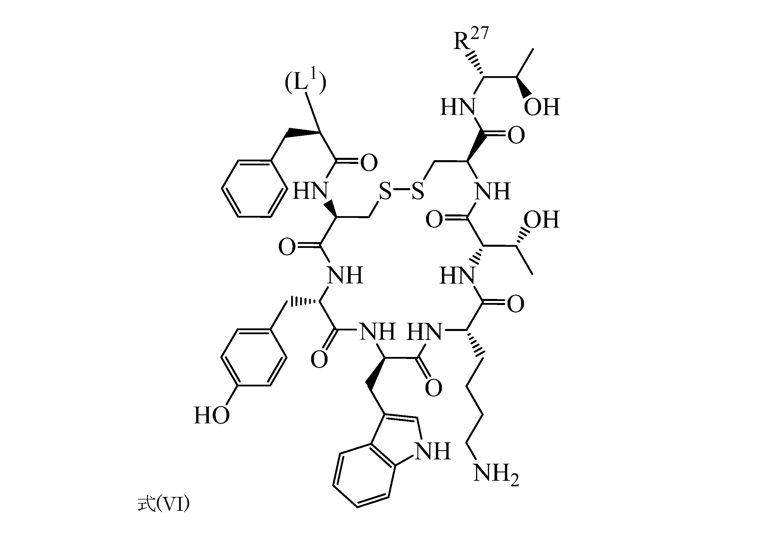
Tmは式(VI)の構造:

R7、R13、R14のうちの1つのみがL1である。他に記載のない限り、括弧内のL1を用いる場合、そのL1は、形式的にたとえばTmの一部であるのではなく、関連する結合点を示すTmの一部として示されている。
A cancer targeting composition for treating cancer cells overexpressing the somatostatin receptor is disclosed herein. The cancer targeting composition comprises a molecule of formula (I): M-Ch-L 1 -Tm or a pharmaceutically acceptable salt thereof. In formula (I), M is 212 Pb, 203 Pb, 64 Cu, 67 Cu, 212 Bi, 68 Ga, 213 Bi, 225 Ac, 243 Am, 211 At, 217 At, 154 Dy, 148 Gd, 146 Sm, Radioactivity selected from the group consisting of 147 Sm, 149 Tb, 227 Th, 229 Th, 59 Fe, 60 Cu, 61 Cu, 62 Cu, 67 Ga, 86 Y, 111 In, 153 Gd, 153 Sm, and 166 Ho. Isotope; Ch is a chelating agent having a structure selected from the group consisting of formula (II), formula (III), formula (IV), and formula (V):
R 9 , R 10 , R 11 , R 12 , R 15 , R 16 , R 17 , R 18 , R 19 , R 20 , R 21 , R 22 , R 23 , and R 24 are H, D, F, Cl. , And each independently selected from the group consisting of (C 1 to C 6 ) alkyl;
R 7 consists of H, D, F, Cl, (C 1 to C 6 ) alkyl, (C 1 to C 6 ) alkyl-C (= O) -N (-R 25 ) -R 26 , and L 1 . Selected independently of the group;
R 13 and R 14 are each independently selected from the group consisting of H, D, F, Cl, (C 1 to C 6 ) alkyl, and L 1 ;
R 25 and R 26 are each independently selected from the group consisting of H, D, (C 1 to C 6 ) alkyl, and (C 1 to C 6 ) alkyl-C (= O) -OH;
L 1 is (C 1 to C 6 ) alkyl-C (= O) -NH- (C 1 to C 6 ) alkyl-C (= O) -NH, (C 1 to C 6 ) alkyl- (C 6 H). 4 ) -NH-C (= S) -NH, C (-CO 2 H)-(C 1 to C 6 ) Alkyl- (C 6 H 4 ) -NH-C (= S) -NH, (C 1 ) From C 6 ) Alkyl-C (= O) -NH, (C 1 to C 6 ) Alkyl-C (= O)-(O-CH 2 -CH 2 ) 1 to 20 -C (= O) -NH (Selected independently from the group); and Tm is the structure of equation (VI):
前記がん標的化組成物は、R5、R6、R8のうちの1つ、2つ、または3つが(C1~C6)アルキル-C(=O)-N(-R25)-R26であってもよい。Mは、212Pb、203Pb、64Cu、67Cu、212Bi、225Ac、243Am、211At、217At、154Dy、148Gd、146Sm、147Sm、149Tb、227Th、229Th、59Fe、60Cu、61Cu、62Cu、67Ga、86Y、111In、153Gd、153Sm、および166Hoからなる群から選択されてもよい。Mは、212Pb、203Pb、64Cu、および67Cuからなる群から独立に選択されてもよい。Mは212Pb、203Pb、64Cu、67Cuおよび212Biからなる群から選択されてもよく;かつChは式(V)の構造を持っていてもよく;かつR27はCH2-OHである。また、Mは212Pb、203Pb、64Cu、67Cu、212Biおよび213Biからなる群から選択されてもよく;Chは式(V)の構造を持っていてもよく;かつR27はC(=O)-OHである。前記式(I)の分子は、少なくとも1種の化合物をキレート剤と反応させることによって生成され、前記キレート剤は、


前記がん標的化組成物は、式(VII)で表される構造またはその薬学的に許容される塩の構造を持っていてもよい:

R5、R6、およびR8はH、D、F、Cl、(C1~C6)アルキル、(C1~C6)アルキル-C(=O)-OR25、および(C1~C6)アルキル-C(=O)-N(-R25)-R26からなる群から各々独立に選択され;
R9、R10、R11、R12、R15、R16、R17、R18、R19、R20、R21、R22、R23、およびR24はH、D、F、Cl、および(C1~C6)アルキルからなる群から各々独立に選択され;
R13およびR14はH、D、F、Cl、および(C1~C6)アルキルからなる群から各々独立に選択され;
R25およびR26はH、D、(C1~C6)アルキル、および(C1~C6)アルキル-C(=O)-OHからなる群から各々独立に選択され;
L1は(C1~C6)アルキル-C(=O)-NH-(C1~C6)アルキル-C(=O)-NH、(C1~C6)アルキル-(C6H4)-NH-C(=S)-NH、C(-CO2H)-(C1~C6)アルキル-(C6H4)-NH-C(=S)-NH、(C1~C6)アルキル-C(=O)-NH、(C1~C6)アルキル-C(=O)-(O-CH2-CH2)1~20-C(=O)-NHからなる群から独立に選択され;かつ
R27はCH2-OHおよびC(=O)-OHからなる群から独立に選択される)。
The cancer targeting composition may have a structure represented by formula (VII) or a pharmaceutically acceptable salt structure thereof:
R 5 , R 6 and R 8 are H, D, F, Cl, (C 1 to C 6 ) alkyl, (C 1 to C 6 ) alkyl-C (= O) -OR 25 , and (C 1 to). C 6 ) Selected independently from the group consisting of alkyl-C (= O) -N (-R 25 ) -R 26 ;
R 9 , R 10 , R 11 , R 12 , R 15 , R 16 , R 17 , R 18 , R 19 , R 20 , R 21 , R 22 , R 23 , and R 24 are H, D, F, Cl. , And each independently selected from the group consisting of (C 1 to C 6 ) alkyl;
R 13 and R 14 are each independently selected from the group consisting of H, D, F, Cl, and (C 1 to C 6 ) alkyl;
R 25 and R 26 are each independently selected from the group consisting of H, D, (C 1 to C 6 ) alkyl, and (C 1 to C 6 ) alkyl-C (= O) -OH;
L 1 is (C 1 to C 6 ) alkyl-C (= O) -NH- (C 1 to C 6 ) alkyl-C (= O) -NH, (C 1 to C 6 ) alkyl- (C 6 H). 4 ) -NH-C (= S) -NH, C (-CO 2 H)-(C 1 to C 6 ) Alkyl- (C 6 H 4 ) -NH-C (= S) -NH, (C 1 ) From C 6 ) Alkyl-C (= O) -NH, (C 1 to C 6 ) Alkyl-C (= O)-(O-CH 2 -CH 2 ) 1 to 20 -C (= O) -NH Independently selected from the group consisting of CH 2 -OH and C (= O) -OH ).
前記がん標的化組成物は、式(VIII)で表される構造またはその薬学的に許容される塩の構造を持っていてもよい:

R5、R6、およびR8はH、D、F、Cl、(C1~C6)アルキル、(C1~C6)アルキル-C(=O)-OR25、および(C1~C6)アルキル-C(=O)-N(-R25)-R26からなる群から各々独立に選択され;
R9、R10、R11、R12、R15、R16、R17、R18、R19、R20、R21、R22、R23、およびR24は、H、D、F、Cl、および(C1~C6)アルキルからなる群から各々独立に選択され;
R7はH、D、F、Cl、(C1~C6)アルキルおよび(C1~C6)アルキル-C(=O)-N(-R25)-R26からなる群から独立に選択され;
R13はH、D、F、Cl、および(C1~C6)アルキルからなる群から独立に選択され;
R25およびR26は、H、D、(C1~C6)アルキル、および(C1~C6)アルキル-C(=O)-OHからなる群から各々独立に選択され;
L1は(C1~C6)アルキル-(C6H4)-NH-C(=S)-NHであり;かつ
R27はCH2-OHおよびC(=O)-OHからなる群から独立に選択される)。
The cancer targeting composition may have a structure represented by formula (VIII) or a pharmaceutically acceptable salt structure thereof:
R 5 , R 6 and R 8 are H, D, F, Cl, (C 1 to C 6 ) alkyl, (C 1 to C 6 ) alkyl-C (= O) -OR 25 , and (C 1 to). C 6 ) Selected independently from the group consisting of alkyl-C (= O) -N (-R 25 ) -R 26 ;
R 9 , R 10 , R 11 , R 12 , R 15 , R 16 , R 17 , R 18 , R 19 , R 20 , R 21 , R 22 , R 23 , and R 24 are H, D, F, Independently selected from the group consisting of Cl and (C 1 to C 6 ) alkyl;
R 7 is independent of the group consisting of H, D, F, Cl, (C 1 to C 6 ) alkyl and (C 1 to C 6 ) alkyl-C (= O) -N (-R 25 ) -R 26 . Selected;
R 13 is independently selected from the group consisting of H, D, F, Cl, and (C 1 -C 6 ) alkyl;
R 25 and R 26 are each independently selected from the group consisting of H, D, (C 1 to C 6 ) alkyl, and (C 1 to C 6 ) alkyl-C (= O) -OH;
L 1 is (C 1 to C 6 ) alkyl- (C 6 H 4 ) -NH-C (= S) -NH; and R 27 is a group consisting of CH 2 -OH and C (= O) -OH. Independently selected from).
前記がん標的化組成物は、式(IX)で表される構造またはその薬学的に許容される塩の構造を持っていてもよい:

R5、R6、およびR8はH、D、F、Cl、(C1~C6)アルキル、(C1~C6)アルキル-C(=O)-OR25、および(C1~C6)アルキル-C(=O)-N(-R25)-R26からなる群から各々独立に選択され;
R9、R10、R11、R12、R15、R16、R17、R18、R19、R20、R21、R22、R23、およびR24はH、D、F、Cl、および(C1~C6)アルキルからなる群から各々独立に選択され;
R13およびR14はH、D、F、Cl、および(C1~C6)アルキルからなる群から各々独立に選択され;
R25およびR26はH、D、(C1~C6)アルキル、および(C1~C6)アルキル-C(=O)-OHからなる群から各々独立に選択され;かつ
R27はCH2-OHおよびC(=O)-OHからなる群から独立に選択される)。
The cancer targeting composition may have a structure represented by formula (IX) or a pharmaceutically acceptable salt structure thereof:
R 5 , R 6 and R 8 are H, D, F, Cl, (C 1 to C 6 ) alkyl, (C 1 to C 6 ) alkyl-C (= O) -OR 25 , and (C 1 to). C 6 ) Selected independently from the group consisting of alkyl-C (= O) -N (-R 25 ) -R 26 ;
R 9 , R 10 , R 11 , R 12 , R 15 , R 16 , R 17 , R 18 , R 19 , R 20 , R 21 , R 22 , R 23 , and R 24 are H, D, F, Cl. , And each independently selected from the group consisting of (C 1 to C 6 ) alkyl;
R 13 and R 14 are each independently selected from the group consisting of H, D, F, Cl, and (C 1 to C 6 ) alkyl;
R 25 and R 26 are each independently selected from the group consisting of H, D, (C 1 to C 6 ) alkyl, and (C 1 to C 6 ) alkyl-C (= O) -OH; and R 27 is Independently selected from the group consisting of CH 2 -OH and C (= O) -OH).
前記がん標的化組成物は、式(X)の構造またはその薬学的に許容される塩の構造を持っていてもよい:

R5、R6、およびR8はH、D、F、Cl、(C1~C6)アルキル、(C1~C6)アルキル-C(=O)-OR25、および(C1~C6)アルキル-C(=O)-N(-R25)-R26からなる群から各々独立に選択され;
R9、R10、R11、R12、R15、R16、R17、R18、R19、R20、R21、R22、R23、およびR24はH、D、F、Cl、および(C1~C6)アルキルからなる群から各々独立に選択され;
R7はH、D、F、Cl、(C1~C6)アルキルおよび(C1~C6)アルキル-C(=O)-N(-R25)-R26からなる群から独立に選択され;
R13はH、D、F、Cl、および(C1~C6)アルキルからなる群から独立に選択され;
R25およびR26はH、D、(C1~C6)アルキル、および(C1~C6)アルキル-C(=O)-OHからなる群から各々独立に選択され;かつ
R27はCH2-OHおよびC(=O)-OHからなる群から独立に選択される)。
The cancer targeting composition may have a structure of formula (X) or a pharmaceutically acceptable salt structure thereof:
R 5 , R 6 and R 8 are H, D, F, Cl, (C 1 to C 6 ) alkyl, (C 1 to C 6 ) alkyl-C (= O) -OR 25 , and (C 1 to). C 6 ) Selected independently from the group consisting of alkyl-C (= O) -N (-R 25 ) -R 26 ;
R 9 , R 10 , R 11 , R 12 , R 15 , R 16 , R 17 , R 18 , R 19 , R 20 , R 21 , R 22 , R 23 , and R 24 are H, D, F, Cl. , And each independently selected from the group consisting of (C 1 to C 6 ) alkyl;
R 7 is independent of the group consisting of H, D, F, Cl, (C 1 to C 6 ) alkyl and (C 1 to C 6 ) alkyl-C (= O) -N (-R 25 ) -R 26 . Selected;
R 13 is independently selected from the group consisting of H, D, F, Cl, and (C 1 -C 6 ) alkyl;
R 25 and R 26 are each independently selected from the group consisting of H, D, (C 1 to C 6 ) alkyl, and (C 1 to C 6 ) alkyl-C (= O) -OH; and R 27 is Independently selected from the group consisting of CH 2 -OH and C (= O) -OH).
前記組成物は、式(I):M-Ch-L1-Tmの分子またはその薬学的に許容される塩を含んでいてもよい:
式(I)中、Mは212Pb、203Pb、64Cu、67Cu、212Bi、68Ga、213Bi、225Ac、243Am、211At、217At、154Dy、148Gd、146Sm、147Sm、149Tb、227Th、229Th、59Fe、60Cu、61Cu、62Cu、67Ga、86Y、111In、153Gd、153Sm、および166Hoからなる群から選択される放射性同位体であり;
Chは式(V)の構造を有するキレート剤であり:

R9、R10、R11、R12、R15、R16、R17、R18、R19、R20、R21、R22、R23、およびR24はH、D、F、Cl、および(C1~C6)アルキルからなる群から各々独立に選択され;
R7はH、D、F、Cl、(C1~C6)アルキル、(C1~C6)アルキル-C(=O)-N(-R25)-R26、およびL1からなる群から独立に選択され;
R13およびR14はH、D、F、Cl、(C1~C6)アルキル、およびL1からなる群から各々独立に選択され;
R25およびR26はH、D、(C1~C6)アルキル、および(C1~C6)アルキル-C(=O)-OHからなる群から各々独立に選択され;
L1は(C1~C6)アルキル-C(=O)-NH-(C1~C6)アルキル-C(=O)-NH、(C1~C6)アルキル-(C6H4)-NH-C(=S)-NH、C(-CO2H)-(C1~C6)アルキル-(C6H4)-NH-C(=S)-NH、(C1~C6)アルキル-C(=O)-NH、(C1~C6)アルキル-C(=O)-(O-CH2-CH2)1~20-C(=O)-NHからなる群から独立に選択される);
Tmは式(VI)の構造:

R7、R13、またはR14のうちの1つのみがL1である。
The composition may contain a molecule of formula (I): M-Ch-L 1 -Tm or a pharmaceutically acceptable salt thereof:
In formula (I), M is 212 Pb, 203 Pb, 64 Cu, 67 Cu, 212 Bi, 68 Ga, 213 Bi, 225 Ac, 243 Am, 211 At, 217 At, 154 Dy, 148 Gd, 146 Sm, Radioactivity selected from the group consisting of 147 Sm, 149 Tb, 227 Th, 229 Th, 59 Fe, 60 Cu, 61 Cu, 62 Cu, 67 Ga, 86 Y, 111 In, 153 Gd, 153 Sm, and 166 Ho. Isotope;
Ch is a chelating agent having the structure of formula (V):
R 9 , R 10 , R 11 , R 12 , R 15 , R 16 , R 17 , R 18 , R 19 , R 20 , R 21 , R 22 , R 23 , and R 24 are H, D, F, Cl. , And each independently selected from the group consisting of (C 1 to C 6 ) alkyl;
R 7 consists of H, D, F, Cl, (C 1 to C 6 ) alkyl, (C 1 to C 6 ) alkyl-C (= O) -N (-R 25 ) -R 26 , and L 1 . Selected independently of the group;
R 13 and R 14 are each independently selected from the group consisting of H, D, F, Cl, (C 1 to C 6 ) alkyl, and L 1 ;
R 25 and R 26 are each independently selected from the group consisting of H, D, (C 1 to C 6 ) alkyl, and (C 1 to C 6 ) alkyl-C (= O) -OH;
L 1 is (C 1 to C 6 ) alkyl-C (= O) -NH- (C 1 to C 6 ) alkyl-C (= O) -NH, (C 1 to C 6 ) alkyl- (C 6 H). 4 ) -NH-C (= S) -NH, C (-CO 2 H)-(C 1 to C 6 ) Alkyl- (C 6 H 4 ) -NH-C (= S) -NH, (C 1 ) From C 6 ) Alkyl-C (= O) -NH, (C 1 to C 6 ) Alkyl-C (= O)-(O-CH 2 -CH 2 ) 1 to 20 -C (= O) -NH (Selected independently from the group);
Tm is the structure of equation (VI):
ソマトスタチン受容体を過剰発現しているがん細胞を治療するためのがん標的化キットが本明細書に開示されている。このがん標的化キットは、本明細書に開示されているがん標的化組成物と、医薬的に許容される緩衝剤、抗酸化剤、および捕捉剤のうちの少なくとも1つを含んでいてもよい。前記がん標的化キットは、25~50μgの前記がん標的化組成物と0.4Mの酢酸アンモニウム緩衝剤を含んでいてもよい。前記がん標的化キットは、酢酸アンモニウム緩衝剤を含んでいてもよい。一態様では、前記緩衝剤は酢酸アンモニウム緩衝剤を含んでいる。前記抗酸化剤はアスコルビン酸、ゲンチジン酸、エタノール、またはそれらの組み合わせを含んでいてもよい。前記捕捉剤は、ジエチレントリアミノ五酢酸;エチレンジアミン四酢酸;1,4,7,10-テトラアザシクロドデカン-1,4,7,10-四酢酸;およびそれらの組み合わせからなる群から選択される1種であってもよい。 A cancer targeting kit for treating cancer cells that overexpress the somatostatin receptor is disclosed herein. The cancer targeting kit comprises the cancer targeting composition disclosed herein and at least one of a pharmaceutically acceptable buffer, antioxidant, and scavenger. May be good. The cancer targeting kit may include 25-50 μg of the cancer targeting composition and 0.4 M ammonium acetate buffer. The cancer targeting kit may include an ammonium acetate buffer. In one aspect, the buffer comprises an ammonium acetate buffer. The antioxidant may include ascorbic acid, gentisic acid, ethanol, or a combination thereof. The scavenger is selected from the group consisting of diethylenetriaminopentaacetic acid; ethylenediaminetetraacetic acid; 1,4,7,10-tetraazacyclododecane-1,4,7,10-tetraacetic acid; and combinations thereof. It may be a seed.
医薬製剤が本明細書に開示されている。この医薬製剤は、本明細書に開示されているがん標的化組成物と医薬的に許容される緩衝剤を含んでいてもよい。ソマトスタチン受容体を過剰発現しているがん細胞を治療する医薬として使用するためのがん標的化組成物が本明細書に開示されている。 Pharmaceutical formulations are disclosed herein. The pharmaceutical formulation may include the cancer targeting composition disclosed herein and a pharmaceutically acceptable buffer. A cancer targeting composition for use as a therapeutic agent for cancer cells overexpressing the somatostatin receptor is disclosed herein.
ソマトスタチン受容体を過剰発現しているがん細胞を治療するためのがん標的化組成物を必要な患者に投与する方法が本明細書に開示されている。この方法は、治療に有効な用量のがん標的化組成物を投与することを含んでいてもよく、前記がん標的化組成物は、式(I)の分子またはその薬学的に許容される塩を含み、
式(I)中、Mは212Pb、203Pb、64Cu、67Cu、212Bi、68Ga、213Bi、225Ac、243Am、211At、217At、154Dy、148Gd、146Sm、147Sm、149Tb、227Th、229Th、59Fe、60Cu、61Cu、62Cu、67Ga、86Y、111In、153Gd、153Sm、および166Hoからなる群から選択される放射性同位体であり;
Chは式(II)、式(III)、式(IV)、および式(V)からなる群から選択される構造を有するキレート剤であり:

R9、R10、R11、R12、R15、R16、R17、R18、R19、R20、R21、R22、R23、およびR24はH、D、F、Cl、および(C1~C6)アルキルからなる群から各々独立に選択され;
R7はH、D、F、Cl、(C1~C6)アルキル、(C1~C6)アルキル-C(=O)-N(-R25)-R26、およびL1からなる群から独立に選択され;
R13およびR14は、H、D、F、Cl、(C1~C6)アルキル、およびL1からなる群から各々独立に選択され;
R25およびR26はH、D、(C1~C6)アルキル、および(C1~C6)アルキル-C(=O)-OHからなる群から各々独立に選択され;
L1は(C1~C6)アルキル-C(=O)-NH-(C1~C6)アルキル-C(=O)-NH、(C1~C6)アルキル-(C6H4)-NH-C(=S)-NH、C(-CO2H)-(C1~C6)アルキル-(C6H4)-NH-C(=S)-NH、(C1~C6)アルキル-C(=O)-NH、(C1~C6)アルキル-C(=O)-(O-CH2-CH2)1~20-C(=O)-NHからなる群から独立に選択される);かつ
Tmは式(VI)の構造:
R7、R13、R14のうちの1つのみがL1である。
Disclosed herein are methods of administering to a patient in need a cancer targeting composition for treating cancer cells that overexpress the somatostatin receptor. The method may comprise administering a therapeutically effective dose of the cancer targeting composition, wherein the cancer targeting composition is a molecule of formula (I) or pharmaceutically acceptable thereof. Contains salt,
In formula (I), M is 212 Pb, 203 Pb, 64 Cu, 67 Cu, 212 Bi, 68 Ga, 213 Bi, 225 Ac, 243 Am, 211 At, 217 At, 154 Dy, 148 Gd, 146 Sm, Radioactivity selected from the group consisting of 147 Sm, 149 Tb, 227 Th, 229 Th, 59 Fe, 60 Cu, 61 Cu, 62 Cu, 67 Ga, 86 Y, 111 In, 153 Gd, 153 Sm, and 166 Ho. Isotope;
Ch is a chelating agent having a structure selected from the group consisting of formula (II), formula (III), formula (IV), and formula (V):
R 9 , R 10 , R 11 , R 12 , R 15 , R 16 , R 17 , R 18 , R 19 , R 20 , R 21 , R 22 , R 23 , and R 24 are H, D, F, Cl. , And each independently selected from the group consisting of (C 1 to C 6 ) alkyl;
R 7 consists of H, D, F, Cl, (C 1 to C 6 ) alkyl, (C 1 to C 6 ) alkyl-C (= O) -N (-R 25 ) -R 26 , and L 1 . Selected independently of the group;
R 13 and R 14 are each independently selected from the group consisting of H, D, F, Cl, (C 1 to C 6 ) alkyl, and L 1 ;
R 25 and R 26 are each independently selected from the group consisting of H, D, (C 1 to C 6 ) alkyl, and (C 1 to C 6 ) alkyl-C (= O) -OH;
L 1 is (C 1 to C 6 ) alkyl-C (= O) -NH- (C 1 to C 6 ) alkyl-C (= O) -NH, (C 1 to C 6 ) alkyl- (C 6 H). 4 ) -NH-C (= S) -NH, C (-CO 2 H)-(C 1 to C 6 ) Alkyl- (C 6 H 4 ) -NH-C (= S) -NH, (C 1 ) From C 6 ) Alkyl-C (= O) -NH, (C 1 to C 6 ) Alkyl-C (= O)-(O-CH 2 -CH 2 ) 1 to 20 -C (= O) -NH (Selected independently from the group); and Tm is the structure of equation (VI):
前記がんはソマトスタチン受容体を過剰発現している細胞を含んでいてもよい。前記がんは、心臓のがん、肺がん、消化管がん、尿生殖器がん、肝臓がん、骨がん、神経系がん、婦人科がん、血液がん、またはその組み合わせを含んでもよい。前記患者はヒト、イヌ、ネコ、ウマ、またはその他の哺乳動物であってもよい。前記がん標的化組成物は、少なくとも1種の制がん化合物と併用投与されてもよい。前記少なくとも1種の制がん化合物としては、アルデスロイキン;アレムツズマブ;アリトレチノイン;アロプリノール;アルトレタミン;アミフォスチン;アナストロゾール;三酸化二ヒ素;アスパラギナーゼ;BCG生;ベキサロテンカプセル;ベキサロテンゲル;ブレオマイシン;ブスルファン静脈剤;ブスルファン経口剤;カルステロン;カペシタビン;カルボプラチン;カルムスチン;カルムスチンとポリフェプロサン20留置剤;セレコキシブ;クロラムブシル;シスプラチン;クラドリビン;シクロフォスファミド;シタラビン;シタラビンリポソーム剤;ダカルバジン;ダクチノマイシン;アクチノマイシンD;ダルベポエチンアルファ;ダウノルビシンリポソーム剤;ダウノルビシン;ダウノマイシン;デニロイキン・ディフティトックス;デクスラゾキサン;ドセタキセル;ドキソルビシン;ドキソルビシンリポソーム剤;プロピオン酸ドロモスタノロン;エリオットB溶液剤;エピルビシン;エポエチンアルファ;エストラムスチン;エトポシドリン酸塩;エトポシド(VP-16);エキセメスタン;フィルグラスチム;フロクスウリジン(動脈剤);フルダラビン;フルオロウラシル(5-FU);フルベストラント;ゲムシタビン;ゲムツズマブ・オゾガマイシン;グリベック(イマチニブ);酢酸ゴセレリン;ヒドロキシ尿素;イブリツモマブ・チウキセタン;イダルビシン;イホスファミド;イマチニブメシル酸塩;インターフェロンアルファ-2a;インターフェロンアルファ-2b;イリノテカン;レトロゾール;ロイコボリン;レバミソール;ロムスチン(CCNU);メクロレタミン(ナイトロジェンマスタード);酢酸メゲストロール;メルファラン(L-プロトスペーサー隣接モチーフ);メルカプトプリン(6-MP);メスナ;メトトレキサート;メトクサレン;マイトマイシンC;ミトタン;ミトキサントロン;フェンプロピオン酸ナンドロロン;ノフェツモマブ;LOddC;オプレルベキン;オキサリプラチン;パクリタキセル;パミドロネート;ペガデマーゼ;ペガスパルガーゼ;ペグフィルグラスチム;ペントスタチン;ピポブロマン;プリカマイシン;ミトラマイシン;ポルフィマーナトリウム;プロカルバジン;キナクリン;ラスブリカーゼ;リツキシマブ;サルグラモスチム;ストレプトゾシン;スラフェニブ;テルビブジン(LDT);タルク;タモキシフェン;タルセバ(エルロチニブ);テモゾロミド;テニポシド(VM-26);テストラクトン;チオグアニン(6-TG);チオテパ;トポテカン;トレミフェン;トシツモマブ;トラスツズマブ;トレチノイン(ATRA);ウラシルマスタード;バルルビシン;バルトルシタビン(一価LDC);ビンブラスチン;ビノレルビン;ゾレドロナート;またはその混合物が挙げられる。前記制がん化合物は治療に有効な用量で投与されてもよい。 The cancer may include cells that overexpress the somatostatin receptor. The cancer may include heart cancer, lung cancer, gastrointestinal cancer, genitourinary cancer, liver cancer, bone cancer, nervous system cancer, gynecological cancer, blood cancer, or a combination thereof. good. The patient may be a human, dog, cat, horse, or other mammal. The cancer targeting composition may be administered in combination with at least one anticancer compound. The at least one anticancer compound includes aldesroykin; alemtuzumab; aritretinoin; alloprinol; altretamine; amifostine; anastrozole; hydride trioxide; asparaginase; BCG raw; bexarotene capsule; bexalotengel; bleomycin; busulfan. Intravenous; Busulfan Oral; Calsterone; Capecitabin; Carboplatin; Calmustin; Calmustin and Polyfeprosan 20 Instillation; Celecoxib; Chlorambusyl; Cisplatin; Cladribin; Cyclofosfamide; Citarabin; Citarabin Lipstick; Daunorubicin; Mycin D; daunorubicin alpha; daunorubicin liposomes; daunorubicin; daunomycin; deniroykin diffititox; dexrazoxane; docetaxel; doxorubicin; doxorubicin liposomes; propionate dromostanolone; elliott B solution; epirubicin; etoposide Etoposide; Etoposide (VP-16); Exemestane; Philgrastim; Floxuridine (arterial agent); Fludarabin; Fluorouracil (5-FU); Flubestland; Gemcitabine; Gemtuzumab / ozogamicin; Gleevec (imatinib); Goselin acetate Hydroxyurea; ibritumomab thiuxetane; daunorubicin; iphosphamide; imatinib mesylate; interferon alpha-2a; interferon alpha-2b; irinotecan; retrosol; leucovorin; levamisole; romustin (CCNU); mechloretamine (nitrogenmasterd); Guest rolls; merphalan (L-protospacer flanking motif); mercaptopurine (6-MP); mesna; methotrexate; methoxalen; mitomycin C; mittan; mitoxanthrone; fenpropionate nandrolone; nofetumomab; LOddC; oprelbekin; oxaliplatin Paclitaxel; Pamidronate; Pegademase; Pegaspargase; Pegfilgrastim; Pentostatin; Pipobroman; Plicamycin; Mitramycin; Porfimer sodium; Procarbazine; Kinacrine; Lasbricase; Ritziximab; Salgramostim; Streptozosine; Tarku; Tamo Xyphene; Tarceva (erlotinib); Temozolomid; Teniposide (VM-26); Test lactone; Thioguanin (6-TG); Thiotepa; Topotecan; Tretinoin; Toshitsumomab; Trastuzumab; Tretinoin (ATRA); LDC); vinblastine; vinorelbine; zoredronate; or a mixture thereof. The anti-cancer compound may be administered at a therapeutically effective dose.
ソマトスタチン受容体を過剰発現しているがん細胞を治療するためのがん標的化組成物を必要な患者に投与する方法が開示されている。この方法は、治療に有効な用量の式(I):M-Ch-L1-Tmの分子またはその薬学的に許容される塩と、
医薬的に許容できる担体に入れられた少なくとも1種の制がん化合物を投与することを含んでもよく、前記式(I)の分子において、
Mは212Pb、203Pb、64Cu、67Cu、212Bi、68Ga、213Bi、225Ac、243Am、211At、217At、154Dy、148Gd、146Sm、147Sm、149Tb、227Th、229Th、59Fe、60Cu、61Cu、62Cu、67Ga、86Y、111In、153Gd、153Sm、および166Hoからなる群から選択される放射性同位体であり;
Chは式(II)、式(III)、式(IV)および式(V)からなる群から選択される構造を有するキレート剤であり:

R9、R10、R11、R12、R15、R16、R17、R18、R19、R20、R21、R22、R23、およびR24はH、D、F、Cl、および(C1~C6)アルキルからなる群から各々独立に選択され;
R7はH、D、F、Cl、(C1~C6)アルキル、(C1~C6)アルキル-C(=O)-N(-R25)-R26、およびL1からなる群から独立に選択され;
R13およびR14はH、D、F、Cl、(C1~C6)アルキル、およびL1からなる群から各々独立に選択され;
R25およびR26は、H、D、(C1~C6)アルキル、および(C1~C6)アルキル-C(=O)-OHからなる群から各々独立に選択され;
L1は(C1~C6)アルキル-C(=O)-NH-(C1~C6)アルキル-C(=O)-NH、(C1~C6)アルキル-(C6H4)-NH-C(=S)-NH、C(-CO2H)-(C1~C6)アルキル-(C6H4)-NH-C(=S)-NH、(C1~C6)アルキル-C(=O)-NH、(C1~C6)アルキル-C(=O)-(O-CH2-CH2)1~20-C(=O)-NHからなる群から独立に選択される);かつ
Tmは式(VI)の構造:

R7、R13、またはR14のうちの1つのみがL1である。
A method of administering a cancer targeting composition for treating a cancer cell overexpressing a somatostatin receptor to a patient in need is disclosed. This method comprises a therapeutically effective dose of the formula (I): a molecule of M-Ch-L 1 -Tm or a pharmaceutically acceptable salt thereof.
It may include administering at least one anticancer compound contained in a pharmaceutically acceptable carrier, in the molecule of formula (I) above.
M is 212 Pb, 203 Pb, 64 Cu, 67 Cu, 212 Bi, 68 Ga, 213 Bi, 225 Ac, 243 Am, 211 At, 217 At, 154 Dy, 148 Gd, 146 Sm, 147 Sm, 149 Tb, A radioactive isotope selected from the group consisting of 227 Th, 229 Th, 59 Fe, 60 Cu, 61 Cu, 62 Cu, 67 Ga, 86 Y, 111 In, 153 Gd, 153 Sm, and 166 Ho;
Ch is a chelating agent having a structure selected from the group consisting of formula (II), formula (III), formula (IV) and formula (V):
R 9 , R 10 , R 11 , R 12 , R 15 , R 16 , R 17 , R 18 , R 19 , R 20 , R 21 , R 22 , R 23 , and R 24 are H, D, F, Cl. , And each independently selected from the group consisting of (C 1 to C 6 ) alkyl;
R 7 consists of H, D, F, Cl, (C 1 to C 6 ) alkyl, (C 1 to C 6 ) alkyl-C (= O) -N (-R 25 ) -R 26 , and L 1 . Selected independently of the group;
R 13 and R 14 are each independently selected from the group consisting of H, D, F, Cl, (C 1 to C 6 ) alkyl, and L 1 ;
R 25 and R 26 are each independently selected from the group consisting of H, D, (C 1 to C 6 ) alkyl, and (C 1 to C 6 ) alkyl-C (= O) -OH;
L 1 is (C 1 to C 6 ) alkyl-C (= O) -NH- (C 1 to C 6 ) alkyl-C (= O) -NH, (C 1 to C 6 ) alkyl- (C 6 H). 4 ) -NH-C (= S) -NH, C (-CO 2 H)-(C 1 to C 6 ) Alkyl- (C 6 H 4 ) -NH-C (= S) -NH, (C 1 ) From C 6 ) Alkyl-C (= O) -NH, (C 1 to C 6 ) Alkyl-C (= O)-(O-CH 2 -CH 2 ) 1 to 20 -C (= O) -NH (Selected independently from the group); and Tm is the structure of equation (VI):
前記少なくとも1種の制がん化合物は、アルデスロイキン;アレムツズマブ;アリトレチノイン;アロプリノール;アルトレタミン;アミフォスチン;アナストロゾール;三酸化二ヒ素;アスパラギナーゼ;BCG生;ベキサロテンカプセル;ベキサロテンゲル;ブレオマイシン;ブスルファン静脈剤;ブスルファン経口剤;カルステロン;カペシタビン;カルボプラチン;カルムスチン;カルムスチンとポリフェプロサン20留置剤;セレコキシブ;クロラムブシル;シスプラチン;クラドリビン;シクロフォスファミド;シタラビン;シタラビンリポソーム剤;ダカルバジン;ダクチノマイシン;アクチノマイシンD;ダルベポエチンアルファ;ダウノルビシンリポソーム剤;ダウノルビシン;ダウノマイシン;デニロイキン・ディフティトックス;デクスラゾキサン;ドセタキセル;ドキソルビシン;ドキソルビシンリポソーム剤;プロピオン酸ドロモスタノロン;エリオットB溶液剤;エピルビシン;エポエチンアルファ;エストラムスチン;エトポシドリン酸塩;エトポシド(VP-16);エキセメスタン;フィルグラスチム;フロクスウリジン(動脈剤);フルダラビン;フルオロウラシル(5-FU);フルベストラント;ゲムシタビン;ゲムツズマブ・オゾガマイシン;グリベック(イマチニブ);酢酸ゴセレリン;ヒドロキシ尿素;イブリツモマブ・チウキセタン;イダルビシン;イホスファミド;イマチニブメシル酸塩;インターフェロンアルファ-2a;インターフェロンアルファ-2b;イリノテカン;レトロゾール;ロイコボリン;レバミソール;ロムスチン(CCNU);メクロレタミン(ナイトロジェンマスタード);酢酸メゲストロール;メルファラン(L-プロトスペーサー隣接モチーフ);メルカプトプリン(6-MP);メスナ;メトトレキサート;メトクサレン;マイトマイシンC;ミトタン;ミトキサントロン;フェンプロピオン酸ナンドロロン;ノフェツモマブ;LOddC;オプレルベキン;オキサリプラチン;パクリタキセル;パミドロネート;ペガデマーゼ;ペガスパルガーゼ;ペグフィルグラスチム;ペントスタチン;ピポブロマン;プリカマイシン;ミトラマイシン;ポルフィマーナトリウム;プロカルバジン;キナクリン;ラスブリカーゼ;リツキシマブ;サルグラモスチム;ストレプトゾシン;スラフェニブ;テルビブジン(LDT);タルク;タモキシフェン;タルセバ(エルロチニブ);テモゾロミド;テニポシド(VM-26);テストラクトン;チオグアニン(6-TG);チオテパ;トポテカン;トレミフェン;トシツモマブ;トラスツズマブ;トレチノイン(ATRA);ウラシルマスタード;バルルビシン;バルトルシタビン(一価LDC);ビンブラスチン;ビノレルビン;ゾレドロナート;あるいはその組み合わせまたは混合物を含んでもよい。前記方法の一態様では、前記少なくとも1種の制がん化合物は治療に有効な用量で投与されてもよい。 The at least one anticancer compound is aldesroykin; alemtuzumab; aritretinoin; alloprinol; altretamine; amifostine; anastrozole; hydride trioxide; asparaginase; BCG raw; bexarotene capsule; bexalotengel; bleomycin; busulfan vein. Agents; Oral Busulfane; Calsterone; Capecitabin; Carboplatin; Carmustin; Carmustin and Polyfeprosan 20 Instillation; Celecoxib; Chlorambusyl; Cisplatin; Cladribin; Cyclophosfamide; Citarabin; Citalabine Lipbodies; Daunorubicin; Dactinomycin; D; daunorubicin alpha; daunorubicin liposomes; daunorubicin; daunomycin; deniroykin diffititox; dexrazoxane; docetaxel; doxorubicin; doxorubicin liposomes; propionate dromostanolone; elliott B solution; epirubicin; etoposide Salt; Etoposide (VP-16); Exemestane; Philgrastim; Floxuridine (arterial agent); Fludarabin; Fluorouracil (5-FU); Flubestland; Gemcitabine; Gemtuzumab / ozogamicin; Gleevec (imatinib); Goselin acetate; Hydroxyurea; ibritumomab thiuxetane; daunorubicin; iphosphamide; imatinib mesylate; interferon alpha-2a; interferon alpha-2b; irinotecan; retrosol; leucovorin; levamisole; romustin (CCNU); mechloretamine (nitrogen mustard); Rolls; Melfaran (L-protospacer flanking motif); Mercaptopurine (6-MP); Mesna; Metotrexate; Metoxalen; Mitomycin C; Mitotan; Mitoxantron; Nandrolone phenpropionate; Nofetumomab; Paclitaxel; Pamidronate; Pegademase; Pegaspargase; Pegfilgrastim; Pentostatin; Pipobroman; Plicamycin; Mitramycin; Porfimer sodium; Procarbazine; Kinacrine; Lasbricase; Ritzximab; Salgramostim; Streptzocin; Slavenib; Tarku; Tamoxyf En; Tarceva (erlotinib); Temozolomid; Teniposide (VM-26); Test lactone; Thioguanin (6-TG); Thiotepa; Topotecan; Tretinoin; Toshitsumomab; Trastuzumab; Tretinoin (ATRA); Valuation LDC); vinblastine; vinorelbine; zoledronate; or a combination or mixture thereof. In one aspect of the method, the at least one anticancer compound may be administered at a therapeutically effective dose.
式(I)またはその薬学的に許容される塩は、R5、R6、およびR8のうち少なくとも1つが(C1~C6)アルキル-C(=O)-OR25(R25はHまたは(C1~C6)アルキルである)であってもよい。 The formula (I) or a pharmaceutically acceptable salt thereof is such that at least one of R 5 , R 6 , and R 8 is (C 1 to C 6 ) alkyl-C (= O) -OR 25 (R 25 is). It may be H or (C 1 to C 6 ) alkyl).
式(I)またはその薬学的に許容される塩は、R5、R6、およびR8のうち少なくとも1つが(C1~C6)アルキル-C(=O)-N(-R25)-R26(R25およびR26はHおよび(C1~C6)アルキルからなる群から各々独立に選択される)であってもよい。Mが213Biである場合、R5、R6、およびR8はC1アルキル-C(=O)-OHではないことが好ましい。Mが213Biである場合、R5、R6、およびR8のうち1つ、2つ、または3つがCH2-C(=O)-NH2であることが好ましい。 The formula (I) or a pharmaceutically acceptable salt thereof has at least one of R 5 , R 6 , and R 8 (C 1 to C 6 ) alkyl-C (= O) -N (-R 25 ). -R 26 (R 25 and R 26 may be independently selected from the group consisting of H and (C 1 to C 6 ) alkyl, respectively). If M is 213 Bi, then R 5 , R 6 , and R 8 are preferably not C 1 alkyl-C (= O) -OH. When M is 213 Bi, it is preferred that one, two, or three of R 5 , R 6 , and R 8 are CH 2 -C (= O) -NH 2 .
式(I)またはその薬学的に許容される塩は、R9、R10、R11、R12、R15、R16、R17、R18、R19、R20、R21、R22、R23、およびR24のうちの少なくとも1つが、Hおよび(C1~C6)アルキルからなる群から各々独立に選択されてもよい。式(I)またはその薬学的に許容される塩は、R9、R10、R11、R12、R15、R16、R17、R18、R19、R20、R21、R22、R23、およびR24のうちの少なくとも1つが、HおよびDからなる群から各々独立に選択されてもよい。 Formula (I) or a pharmaceutically acceptable salt thereof is R 9 , R 10 , R 11 , R 12 , R 15 , R 16 , R 17 , R 18 , R 19 , R 20 , R 21 , R 22 . , R 23 , and R 24 may each be independently selected from the group consisting of H and (C 1 to C 6 ) alkyl. Formula (I) or a pharmaceutically acceptable salt thereof is R 9 , R 10 , R 11 , R 12 , R 15 , R 16 , R 17 , R 18 , R 19 , R 20 , R 21 , R 22 . , R 23 , and R 24 may each be independently selected from the group consisting of H and D.
式(I)またはその薬学的に許容される塩で、Mは212Pb、203Pb、64Cu、および67Cuからなる群から独立に選択されてもよく;Chは式(V)(式中、R5、R6、およびR8は(C1~C6)アルキル-C(=O)-N(-R25)-R26である)であり;R9、R10、R11、R12、R15、R16、R17、R18、R19、R20、R21、R22、R23、およびR24はHおよびDから各々独立に選択され;R7はL1であり;L1は(C1~C6)アルキル-C(=O)-NHであり;R13およびR14はHおよびDからなる群から各々独立に選択され;R25およびR26はHおよびDからなる群から各々独立に選択され;Tmは式(VI)の構造を持ち;かつR27はC(=O)-OHである。 In formula (I) or a pharmaceutically acceptable salt thereof, M may be independently selected from the group consisting of 212 Pb, 203 Pb, 64 Cu, and 67 Cu; Ch may be formula (V) (in formula). , R 5 , R 6 , and R 8 are (C 1 to C 6 ) alkyl-C (= O) -N (-R 25 ) -R 26 ); R 9 , R 10 , R 11 , R 12 , R 15 , R 16 , R 17 , R 18 , R 19 , R 20 , R 21 , R 22 , R 23 , and R 24 are independently selected from H and D; R 7 is L 1 . Yes; L 1 is (C 1 -C 6 ) alkyl-C (= O) -NH; R 13 and R 14 are independently selected from the group consisting of H and D, respectively; R 25 and R 26 are H. And each independently selected from the group consisting of D; Tm has the structure of formula (VI); and R 27 is C (= O) -OH.
式(I)またはその薬学的に許容される塩で、Mは212Pb、203Pb、64Cu、および67Cuからなる群から独立に選択されてもよく;Chは式(V)(式中、R5、R6、およびR8は(C1~C6)アルキル-C(=O)-N(-R25)-R26である)であり;R9、R10、R11、R12、R15、R16、R17、R18、R19、R20、R21、R22、R23、およびR24はHおよびDから各々独立に選択され;R7は(C1~C6)アルキル-C(=O)-N(-R25)-R26であり;R13はHおよびDからなる群から独立に選択され;R14はL1であり;L1は(C1~C6)アルキル-(C6H4)-NH-C(=S)-NHであり;かつR27はC(=O)-OHである。 In formula (I) or a pharmaceutically acceptable salt thereof, M may be independently selected from the group consisting of 212 Pb, 203 Pb, 64 Cu, and 67 Cu; Ch may be formula (V) (in formula). , R 5 , R 6 , and R 8 are (C 1 to C 6 ) alkyl-C (= O) -N (-R 25 ) -R 26 ); R 9 , R 10 , R 11 , R 12 , R 15 , R 16 , R 17 , R 18 , R 19 , R 20 , R 21 , R 22 , R 23 , and R 24 are independently selected from H and D; R 7 is (C 1 ). ~ C 6 ) Alkyl-C (= O) -N (-R 25 ) -R 26 ; R 13 is independently selected from the group consisting of H and D; R 14 is L 1 ; L 1 is (C 1 to C 6 ) Alkyl- (C 6 H 4 ) -NH-C (= S) -NH; and R 27 is C (= O) -OH.
「アルキル」という用語は、他に記載のない場合、それ自体または別の置換基の一部として、指定の数の炭素原子(たとえば、(C1~C6)は1~6個の炭素原子を指す)を有する直鎖、分岐鎖(キラルまたはアキラル)、または環状鎖炭化水素を指し、直鎖状、分岐鎖状、または環状の基を包含する。例としては、メチル、エチル、プロピル、イソプロピル、ブチル、イソブチル、tert-ブチル、ペンチル、ネオペンチル、ヘキシル、シクロヘキシル、およびシクロプロピルメチル(特にエチル、メチル、イソプロピル)が挙げられる。この用語は置換基とリンカー基の両方に関して用いられる。 The term "alkyl", unless otherwise stated, as itself or as part of another substituent, is a specified number of carbon atoms (eg, (C 1-6 ) is 1-6 carbon atoms. Refers to a linear, branched chain (chiral or achiral), or cyclic chain hydrocarbon having (pointing to) and includes linear, branched, or cyclic groups. Examples include methyl, ethyl, propyl, isopropyl, butyl, isobutyl, tert-butyl, pentyl, neopentyl, hexyl, cyclohexyl, and cyclopropylmethyl (particularly ethyl, methyl, isopropyl). The term is used for both substituents and linker groups.
文脈に応じて、式中で用いられる括弧は、枝に関する情報を一列に並べて伝えることができる。たとえば、(C1~C6)アルキル-C(=O)-OHを以下のように表すこともできる。


ソマトスタチン受容体を過剰発現しているがん細胞を治療するためのがん標的化キットが本明細書に開示されている。このソマトスタチン受容体を過剰発現しているがん細胞を治療するためのがん標的化キットは、上で定義した式(I)、(VII)、(VIII)、(IX)、および/または(X)のがん標的化組成物またはその医薬的に許容される塩と;医薬的に許容される緩衝剤、抗酸化剤、および捕捉剤のうちの少なくとも1つを含んでいてもよい。前記がん標的化キットは、25~50μgの前記がん標的化組成物と0.4Mの酢酸アンモニウム緩衝剤を含んでいる。前記がん標的化キットで、前記緩衝剤は酢酸アンモニウム緩衝剤を含む。前記がん標的化キットで、前記抗酸化剤はアスコルビン酸、ゲンチジン酸、エタノール、またはそれらの組み合わせを含む。前記がん標的化キットで、前記捕捉剤はジエチレントリアミノ五酢酸;エチレンジアミン四酢酸;1,4,7,10-テトラアザシクロドデカン-1,4,7,10-四酢酸;およびそれらの組み合わせからなる群から選択される。 A cancer targeting kit for treating cancer cells that overexpress the somatostatin receptor is disclosed herein. Cancer targeting kits for treating cancer cells overexpressing this somatostatin receptor are formulated above in formulas (I), (VII), (VIII), (IX), and / or ( X) a cancer targeting composition or a pharmaceutically acceptable salt thereof; may contain at least one of a pharmaceutically acceptable buffer, antioxidant, and scavenger. The cancer targeting kit contains 25-50 μg of the cancer targeting composition and 0.4 M ammonium acetate buffer. In the cancer targeting kit, the buffer comprises an ammonium acetate buffer. In the cancer targeting kit, the antioxidant comprises ascorbic acid, gentisic acid, ethanol, or a combination thereof. In the cancer targeting kit, the scavenger is from diethylenetriaminopentaacetic acid; ethylenediaminetetraacetic acid; 1,4,7,10-tetraazacyclododecane-1,4,7,10-4acetic acid; and combinations thereof. It is selected from the group of.
医薬製剤が開示されている。この医薬製剤は、上で定義した式(I)、(VII)、(VIII)、(IX)、および/または(X)のがん標的化組成物またはその医薬的に許容される塩と;医薬的に許容される緩衝剤を含む。 The pharmaceutical product is disclosed. This pharmaceutical product is the cancer targeting composition of formula (I), (VII), (VIII), (IX), and / or (X) defined above or a pharmaceutically acceptable salt thereof; Contains a pharmaceutically acceptable buffer.
ソマトスタチン受容体を過剰発現しているがん細胞を治療する医薬として使用するためのがん標的化組成物が本明細書に開示されている。このソマトスタチン受容体を過剰発現しているがん細胞を治療する医薬として使用するためのがん標的化組成物は、上で定義した式(I)、(VII)、(VIII)、(IX)、および/または(X)の組成物またはその医薬的に許容される塩を含む。 A cancer targeting composition for use as a therapeutic agent for cancer cells overexpressing the somatostatin receptor is disclosed herein. Cancer targeting compositions for use as a therapeutic agent for cancer cells overexpressing this somatostatin receptor are formulated above in formulas (I), (VII), (VIII), (IX). , And / or the composition of (X) or a pharmaceutically acceptable salt thereof.
ソマトスタチン受容体を過剰発現しているがん細胞を治療するためのがん標的化組成物を必要な患者に投与する方法が本明細書に開示されている。この方法は、ある用量のがん標的化組成物を投与することを含み、前記がん標的化組成物は、上で定義した式(I)、(VII)、(VIII)、(IX)、および/または(X)の分子またはその医薬的に許容される塩を含む。前記がんはソマトスタチン受容体を過剰発現している細胞を含んでもよい。前記がんは、心臓のがん、肺がん、消化管がん、尿生殖器がん、肝臓がん、骨がん、神経系がん、婦人科がん、血液がん、またはその組み合わせを含んでもよい。前記患者はヒト、イヌ、ネコ、ウマ、またはその他の哺乳動物であってもよい。 Disclosed herein are methods of administering to patients in need a cancer targeting composition for treating cancer cells that overexpress the somatostatin receptor. The method comprises administering a dose of the cancer targeting composition, wherein the cancer targeting composition comprises formulas (I), (VII), (VIII), (IX), as defined above. And / or contains the molecule of (X) or a pharmaceutically acceptable salt thereof. The cancer may include cells that overexpress the somatostatin receptor. The cancer may include heart cancer, lung cancer, gastrointestinal cancer, genitourinary cancer, liver cancer, bone cancer, nervous system cancer, gynecological cancer, blood cancer, or a combination thereof. good. The patient may be a human, dog, cat, horse, or other mammal.
本発明の化合物は、塩を形成できる基または原子で適切に置換されたときに塩の形態をとってもよい。このような基および原子は有機化学分野の当業者には周知である。「塩」という用語は、本発明の化合物である遊離酸または遊離塩基の付加塩を包含する。「医薬的に許容される塩」という用語は、医薬用途で利用できる範囲内の毒性を有する塩を指す。しかし、医薬的に許容されない塩は、本発明の実施に役立つ(たとえば、本発明の化合物の合成、精製、または製剤工程での有用性など)高結晶度などの特性を有する場合がある。 The compounds of the invention may take the form of salts when appropriately substituted with groups or atoms capable of forming salts. Such groups and atoms are well known to those of skill in the art of organic chemistry. The term "salt" includes an addition salt of a free acid or free base which is a compound of the invention. The term "pharmaceutically acceptable salt" refers to a salt that is toxic to the extent that it can be used in pharmaceutical applications. However, pharmaceutically unacceptable salts may have properties such as high crystallinity that are useful in the practice of the invention (eg, usefulness in the synthesis, purification, or formulation process of the compounds of the invention).
好適な医薬的に許容される酸付加塩は、無機酸または有機酸から調製できる。無機酸の例としては、塩酸、臭化水素酸、ヨウ化水素酸、硝酸、炭酸、硫酸、リン酸などが挙げられる。適切な有機酸は、脂肪族、脂環式、芳香族、芳香脂肪族、複素環の有機酸類、カルボキシ基を有する有機酸類、スルホン基を有する有機酸類から選択されてもよい。これらの例としては、たとえば、ギ酸、酢酸、プロピオン酸、コハク酸、グリコール酸、グルコン酸、乳酸、リンゴ酸、酒石酸、クエン酸、アスコルビン酸、グルクロン酸、マレイン酸、フマル酸、ピルビン酸、アスパラギン酸、グルタミン酸、安息香酸、アントラニル酸、4-ヒドロキシ安息香酸、フェニル酢酸、マンデル酸、エンボン酸(パモ酸)、メタンスルホン酸、エタンスルホン酸、ベンゼンスルホン酸、パントテン酸、トリフルオロメタンスルホン酸、2-ヒドロキシエタンスルホン酸、p-トルエンスルホン酸、スルファニル酸、シクロヘキシルアミノスルホン酸、ステアリン酸、アルギン酸、β-ヒドロキシ酪酸、サリチル酸、ガラクタル酸、ガラクツロン酸が挙げられる。医薬的に許容されない酸付加塩の例としては、たとえば、過塩素酸塩およびテトラフルオロホウ酸塩が挙げられる。 Suitable pharmaceutically acceptable acid addition salts can be prepared from inorganic or organic acids. Examples of the inorganic acid include hydrochloric acid, hydrobromic acid, hydroiodic acid, nitric acid, carbonic acid, sulfuric acid, phosphoric acid and the like. Suitable organic acids may be selected from aliphatic, alicyclic, aromatic, aromatic aliphatic, heterocyclic organic acids, organic acids having a carboxy group, organic acids having a sulfone group. Examples of these are formic acid, acetic acid, propionic acid, succinic acid, glycolic acid, gluconic acid, lactic acid, malic acid, tartrate acid, citric acid, ascorbic acid, glucuronic acid, maleic acid, fumaric acid, pyruvate, asparagine. Acids, glutamate, benzoic acid, anthranilic acid, 4-hydroxybenzoic acid, phenylacetic acid, mandelic acid, embon acid (pamoic acid), methanesulfonic acid, ethanesulfonic acid, benzenesulfonic acid, pantothenic acid, trifluoromethanesulfonic acid, 2 -Hydroxyethanesulfonic acid, p-toluenesulfonic acid, sulfanic acid, cyclohexylaminosulfonic acid, stearic acid, alginic acid, β-hydroxybutyric acid, salicylic acid, galactaric acid and galacturonic acid can be mentioned. Examples of pharmaceutically unacceptable acid addition salts include, for example, perchlorate and tetrafluoroborate.
本発明の化合物の好適な医薬的に許容される塩基付加塩としては、たとえば、アルカリ金属、アルカリ土類金属、遷移金属などの金属の塩(カルシウム塩、マグネシウム塩、カリウム塩、ナトリウム塩、亜鉛塩など)が挙げられる。医薬的に許容される塩基付加塩には、たとえば、N,N-ジベンジルエチレンジアミン、クロロプロカイン、コリン、ジエタノールアミン、エチレンジアミン、メグルミン(N-メチルグルカミン)、プロカインなどの塩基性アミンから形成された有機酸塩も含まれる。医薬的に許容されない塩基付加塩の例としては、たとえば、リチウム塩およびシアン酸塩が挙げられる。 Suitable pharmaceutically acceptable base addition salts of the compounds of the present invention include, for example, salts of metals such as alkali metals, alkaline earth metals, transition metals (calcium salts, magnesium salts, potassium salts, sodium salts, zinc). Salt, etc.). Pharmaceutically acceptable base addition salts were formed from basic amines such as N, N-dibenzylethylenediamine, chloroprocine, choline, diethanolamine, ethylenediamine, meglumine (N-methylglucamine), prokine and the like. Organic acid salts are also included. Examples of pharmaceutically unacceptable base addition salts include, for example, lithium salts and cyanates.
本開示には、ソマトスタチン受容体(SSTR)を過剰発現している神経内分泌腫瘍(NET)を治療(たとえば、画像化、診断、治療、放射線治療など)する組成物、キット、および方法が記載されている。この治療は、キレート剤「CA」または「Ch」によってソマトスタチン受容体標的化ペプチド(たとえば、「Tm」を含むオクトレオチン酸、オクトレオチド、および/またはその他の誘導体)を含む標的化部分にキレートされた放射線同位体(たとえば、α線放射体、β線放射体、γ線放射体、陽電子放射体、および/またはその他の放射線放射体)を含むがん標的化組成物の使用を含む。前記キレート剤は、テトラアザシクロドデカン誘導体、トリアザシクロノナン誘導体、および/またはテトラアザビシクロ[6.6.2]ヘキサデカン誘導体(たとえば、DOTAM、TCMC、DOTAなど)などの窒素環構造を持っていてもよい。式(I)のTmを参照のこと。 The disclosure describes compositions, kits, and methods for treating neuroendocrine tumors (NETs) that overexpress somatostatin receptors (SSTRs) (eg, imaging, diagnosis, treatment, radiation therapy, etc.). ing. This treatment involves radiation chelated by the chelating agent "CA" or "Ch" to a targeted moiety containing a somatostatin receptor targeting peptide (eg, octreotic acid containing "Tm", octreotide, and / or other derivatives). Includes the use of cancer targeting compositions containing isotopes (eg, α-ray, β-ray, γ-ray, positron, and / or other radioradiators). The chelating agent has a nitrogen ring structure such as a tetraazacyclododecane derivative, a triazacyclononane derivative, and / or a tetraazabicyclo [6.6.2] hexadecane derivative (eg, DOTAM, TCMC, DOTA, etc.). You may. See Tm in equation (I).
特に、DOTAMおよびTCMCを用いて、放射性同位体およびその放射性崩壊生成物を安定に配位できるような方法で放射性同位体(たとえば、鉛(Pb)または銅(Cu))を標的化部分(たとえば、オクトレオチン酸、オクトレオチド誘導体)にキレートしてもよい。本明細書の実験は、標的部分とキレート剤(たとえばDOTAM、TCMC)を含む分子が健常細胞への細胞傷害効果を制限しながら放射性同位体をがん細胞へ選択的に送達できることを示している。 In particular, moieties (eg, lead (Pb) or copper (Cu)) that target radioisotopes (eg, lead (Pb) or copper (Cu)) in a manner that allows stable coordination of radioisotopes and their radioactive decay products using DOTAM and TCMC. , Octreotic acid, octreotide derivative). Experiments herein show that molecules containing target moieties and chelators (eg, DOTAM, TCMC) can selectively deliver radioisotopes to cancer cells while limiting their cytotoxic effects on healthy cells. ..
放射標識された結合体は、放射性同位体を配位するキレート剤の誘導体と、がん細胞にある受容体または輸送体を認識するがん特異的な標的化リガンドである。この方法は、健常な細胞および組織に及ぼす影響を制限しながら放射性同位体をがん細胞へ選択的に送達するために用いられてもよい。本明細書の組成物は、がん細胞内のSSTRを標的とするペプチドで修飾されたキレート剤の結合体を提供することを目的とする。この組成物は、この組成物の放射性複合体の溶液を注射することで投与されてもよい。本明細書に記載の結合体は、がん治療のためにα線、β+線、β-線、および/またはγ線を放射する放射性核種との安定な複合体を生成するための基盤を提供することを目的とする。本明細書に記載の技術は、医薬的に許容できる注射液を患者に投与することで患者の病状を治療することを目的とする。 Radiolabeled conjugates are derivatives of chelating agents that coordinate radioisotopes and cancer-specific targeting ligands that recognize receptors or transporters in cancer cells. This method may be used to selectively deliver radioisotopes to cancer cells while limiting their effects on healthy cells and tissues. The compositions herein are intended to provide a conjugate of a chelating agent modified with a peptide that targets the SSTR in cancer cells. The composition may be administered by injecting a solution of the radioactive complex of the composition. The conjugates described herein provide the basis for the generation of stable complexes with radionuclides that emit alpha, β + , β - rays, and / or γ-rays for the treatment of cancer. The purpose is to provide. The techniques described herein are intended to treat a patient's condition by administering to the patient a pharmaceutically acceptable injection.
本明細書に記載の方法および組成物は特定のがん治療に関するが、循環器疾患、感染症、糖尿病、がん、および/または他の状態にも応用できる可能性がある。がんに関連する場合、がんは、たとえば、原発性または転移性の、肝臓がん、前立腺がん、膵臓がん、頭頚部がん、乳がん、脳がん、大腸がん、腺様がん、口腔がん、皮膚がん、肺がん、精巣がん、卵巣がん、子宮頚がん、子宮内膜がん、膀胱がん、胃がん、上皮がんなどのがんに由来する固形腫瘍であってもよい。 Although the methods and compositions described herein relate to a particular cancer treatment, they may also be applicable to cardiovascular disease, infections, diabetes, cancer, and / or other conditions. When associated with cancer, cancers include, for example, primary or metastatic liver cancer, prostate cancer, pancreatic cancer, head and neck cancer, breast cancer, brain cancer, colon cancer, glandular cancer. In solid tumors derived from cancers such as oral cancer, skin cancer, lung cancer, testis cancer, ovarian cancer, cervical cancer, endometrial cancer, bladder cancer, gastric cancer, and epithelial cancer. There may be.
別の側面では、細胞増殖性障害、特にがんを罹患した個体を治療する方法が提供される。この方法は、前記個体に有効量の少なくとも1種の式(I)の化合物またはその医薬的に許容される塩を単独または医薬的に許容できる担体と組み合わせて投与することを含む。 In another aspect, methods are provided for treating individuals with cell proliferation disorders, especially cancer. The method comprises administering to the individual an effective amount of at least one compound of formula (I) or a pharmaceutically acceptable salt thereof, alone or in combination with a pharmaceutically acceptable carrier.
さらに別の側面では、がんに罹患した個体の腫瘍細胞などのがん細胞のアポトーシスを誘導する方法が提供される。この方法は、前記個体に有効量の少なくとも1種の式(I)の化合物またはその医薬的に許容される塩を単独または医薬的に許容できる担体と組み合わせて投与することを含む。 Yet another aspect provides a method of inducing apoptosis of cancer cells, such as tumor cells of individuals affected by cancer. The method comprises administering to the individual an effective amount of at least one compound of formula (I) or a pharmaceutically acceptable salt thereof, alone or in combination with a pharmaceutically acceptable carrier.
式(I)の化合物は経口投与、直腸投与、舌下投与、非経口投与などの任意の経路で投与されてもよい。非経口投与の例としては、たとえば、静脈内投与、筋肉内投与、動脈内投与、腹腔内投与、鼻腔内投与、膣内投与、膀胱内投与(たとえば、膀胱への投与)、皮内投与、経皮投与、局所投与、皮下投与などが挙げられる。管理された処方で患者の体内に薬物を滴下注入し後からその薬物を全身放出または局所放出させることも本発明の範囲内だと考えられる。たとえば、薬物を血行路への徐放または腫瘍成長の局所部位への放出のため徐放性製剤に局在させてもよい。 The compound of formula (I) may be administered by any route such as oral administration, rectal administration, sublingual administration, and parenteral administration. Examples of parenteral administration include, for example, intravenous administration, intramuscular administration, intraarterial administration, intraperitoneal administration, intranasal administration, intravaginal administration, intravesical administration (eg, administration to the bladder), intradermal administration, Percutaneous administration, topical administration, subcutaneous administration and the like can be mentioned. It is also considered within the scope of the present invention to inject a drug into the patient's body in a controlled manner and then release the drug systemically or locally. For example, the drug may be localized to a sustained release formulation for sustained release into the bloodstream or to a localized site of tumor growth.
本開示の実用に有用な1種以上の化合物は、治療中、同じ経路または異なる経路で同時に投与されてもよいし別の時点で投与されてもよい。化合物は、他の薬品(他の抗増殖性化合物など)の前後に投与されてもよいし、それと同時に投与されてもよい。 One or more practically useful compounds of the present disclosure may be administered simultaneously by the same or different routes during treatment or at different times. The compound may be administered before, after, or at the same time as other agents (such as other antiproliferative compounds).
治療は、中断のない一つの期間でも、別々の期間でも、必要な長さの期間で実施されてもよい。治療医は患者の反応に基づいて治療を増減または中断する方法が分かっている。治療は約4週間~約16週間かけて実施されてもよい。必要に応じて治療予定を繰り返してもよい。
(標的がん治療)
1.DOTATATE
Treatment may be performed in one uninterrupted period, in separate periods, or for the required length of time. Therapists know how to increase, decrease, or discontinue treatment based on the patient's response. Treatment may be carried out over a period of about 4 to 16 weeks. The treatment schedule may be repeated as needed.
(Target cancer treatment)
1. 1. DOTATE
がん治療は、患者のがん細胞を標的化し細胞死(アポトーシス)を誘発する組成物の使用を含んでもよい。がん細胞の標的治療の形態によっては、がん細胞の特異的な抗原に結合する分子を有する組成物を用いてもよい。たとえば、がん細胞表面に配置できる特異的な細胞抗原を使用し、低分子量タンパク質またはモノクローナル抗体などの標的化部分を用いてがん細胞を認識させ、がん細胞に結合させてもよい。このペプチド類に細胞傷害性薬剤または同位体/金属でタグを付け、これらを標識し、かつ/またはアポトーシスを誘導することができる。このペプチド類の結合によって、画像化および/または治療に使用してもよいがん抗原提示細胞を特異的に認識できる。たとえば、ペプチド類、抗体、抗体断片などの標的化薬剤を化学療法薬および/または他のアポトーシス促進物質などの様々な細胞傷害性薬剤と結合させてもよい。 Cancer treatment may include the use of compositions that target the patient's cancer cells and induce cell death (apoptosis). Depending on the form of targeted treatment of cancer cells, a composition having a molecule that binds to a specific antigen of cancer cells may be used. For example, specific cellular antigens that can be placed on the surface of cancer cells may be used to recognize and bind to cancer cells using targeted moieties such as low molecular weight proteins or monoclonal antibodies. The peptides can be tagged with cytotoxic agents or isotopes / metals, labeled and / or induce apoptosis. The binding of these peptides specifically recognizes cancer antigen-presenting cells that may be used for imaging and / or treatment. For example, targeting agents such as peptides, antibodies, antibody fragments may be combined with various cytotoxic agents such as chemotherapeutic agents and / or other pro-apoptotic agents.
DOTATATEなどのがん標的化組成物は、特異的なソマトスタチン受容体を過剰発現している神経内分泌腫瘍(NET)などのがんの治療に使用されてもよい。本明細書で用いられるDOTATATEは、オクトレオチン酸などの標的化部分と結合されたDOTAキレート剤を指す。本明細書で用いられるDOTAは、式(CH2CH2NCH2CO2H)4を有する有機化合物を指し、1,4,7,10-テトラアザシクロドデカン-1,4,7,10-四酢酸である。DOTAは、テトラカルボン酸およびその様々な共役塩基を指す場合がある。DOTAは、リガンドが結合できる状態にある末端基を有する窒素原子のテトラアザ環を含む。DOTAは金属イオンと放射性同位体を結合するためのキレート剤として用いられてもよい。本明細書で用いられる標的化部分は、がん細胞などの標的細胞にある抗原に結合する、たとえばペプチド、タンパク質、抗体、ヌクレオシド、ヌクレオチド、アルコール、複素環式化合物、および/または他のリガンドを指す。標的化部分は標的がん細胞に入りアポトーシスを誘導してもよい。 Cancer-targeted compositions such as DOTATATE may be used in the treatment of cancers such as neuroendocrine tumors (NETs) that overexpress specific somatostatin receptors. As used herein, DOTAATE refers to a DOTA chelating agent bound to a targeting moiety such as octreotic acid. DOTA as used herein refers to an organic compound having the formula (CH 2 CH 2 NCH 2 CO 2 H) 4 , 1,4,7,10-tetraazacyclododecane-1,4,7,10-. It is tetraacetic acid. DOTA may refer to tetracarboxylic acid and its various conjugate bases. DOTA contains a tetraaza ring of a nitrogen atom with a terminal group ready for ligand binding. DOTA may be used as a chelating agent for binding metal ions and radioisotopes. Targeting moieties used herein include peptides, proteins, antibodies, nucleosides, nucleotides, alcohols, heterocyclic compounds, and / or other ligands that bind to antigens on target cells, such as cancer cells. Point to. The targeted moiety may enter the target cancer cell and induce apoptosis.
DOTATATEは、キレート剤であるDOTAと、配位された金属すなわち放射性同位体を含む。放射性同位体はがん標的化組成物によって配位(たとえば、含まれ、組み合わせられている)されていてもよく、がん細胞に選択的に送達されてもよい。この配位は、遊離放射性同位体および/またはその放射性崩壊生成物の副作用を最小にするために用いられてもよい。たとえば、90Y-DOTATOCまたは177Lu-DOTATATEなどの放射標識されたSSTR-リガンドをNETの治療に用いてもよい。安全性を高める可能性があることから、DOTATATEは多くの臨床試験に用いられてきた。たとえば、Bushnellらの 90Y-Edotreotide for Metastatic Carcinoid Refractory to Octreotide(オクトレオチド抵抗性の転移性カルチノイド)・・・・・・・・・・・・・・・・・・・・・・・・・・・・・・・・・・・・・・;およびKwekkeboom DJ, Bakker WH, Kam BLらのTreatment of Patients With Gastro-Entero-Pancreatic (GEP) Tumors With The Novel Radiolabelled Somatostatin Analogue [177Lu-DOTA0,Tyr3] Octreotate(新規な放射標識付きソマトスタチン類似体[177Lu-DOTA0,Tyr3]オクトレオチン酸による膵消化管(GEP)腫瘍患者の治療),European Journal of Nuclear Medicine and Molecular Imaging, 2003;・・・・・・・・・・・・・を参照のこと。これらの文献の全内容は本明細書に援用される。実験は、無増悪生存率の中央値(mPFS)の増加や病勢コントロール率(DCR:安定、部分奏功または完全奏功が見られる患者の割合)の増加などの好ましい効果を示している。 DOTATATE contains the chelating agent DOTA and a coordinated metal or radioisotope. Radioisotopes may be coordinated (eg, included and combined) by a cancer targeting composition or selectively delivered to cancer cells. This coordination may be used to minimize the side effects of free radioisotopes and / or their radioactive decay products. For example, radiolabeled SSTR-ligands such as 90 Y-DOTACT or 177 Lu-DOTATATE may be used to treat NET. DOTA-TATE has been used in many clinical trials because of its potential for increased safety. For example, 90Y-Edotreotide for Metastatic Carcinoid Refractory to Octreotide by Bushnell et al.・ ・ ・ ・ ・ ・ ・ ・ ・ ・ ・; And Treatment of Patients With Gastro-Entero-Pancreatic (GEP) Tumors With The Novel Radiolabelled Somatostatin Analogue [ 177 Lu-DOTA0, Tyr3] by Kwekkeboom DJ, Bakker WH, Kam BL et al. Octreotate (Treatment of patients with pancreatic gastrointestinal (GEP) tumors with a novel radiolabeled somatostatin analog [ 177 Lu-DOTA0, Tyr3] octreotide), European Journal of Nuclear Medicine and Molecular Imaging, 2003; ·······checking. The entire contents of these documents are incorporated herein by reference. Experiments have shown favorable effects such as increased median progression-free survival (mPFS) and increased disease control rate (DCR: percentage of patients with stable, partial or complete response).
本明細書にさらに記載されるように、DOTATATEは診断用放射性同位体および放射性同位体の前駆体と、放射性崩壊後の消費された原子および任意の中間の原子のいずれもキレートしてもよい。たとえば、DOTATATEは初めに放射性同位体をキレートし、後にその崩壊生成物のキレート化を保持してもよい。このことによって、遊離の(キレートされていない)放射性同位体が担体(DOTATATE)から分離して血中に入るのを防ぐことができる。キレート剤はさらに、生体内での崩壊後の消費された放射性同位体をキレートしてもよい。このことによって、遊離した放射性かつ/または毒性の崩壊原子がキレート剤から分離して血中に入るのを潜在的に防ぐことができる。
2.DOTAMTATEおよびTCMCTATE
As further described herein, DOTATATE may chelate diagnostic radioisotopes and precursors of radioisotopes with either consumed atoms after radioactive decay and any intermediate atoms. For example, DOTATE may first chelate the radioisotope and then retain the chelation of its decay product. This can prevent free (unchelated) radioisotopes from separating from the carrier (DOTATATE) and entering the blood. The chelating agent may further chelate the consumed radioisotopes after disintegration in vivo. This can potentially prevent free radioactive and / or toxic decay atoms from separating from the chelating agent and entering the blood.
2. 2. DOTAMATE and TCCMATE
同位体を安定に配位するために、DOTAM、TCMC-一酸、およびTCMC(本明細書でさらに定義する)などのその他のキレート剤を用いてもよい。このようなキレート剤は、診断用と治療用の両方の放射性同位体を配位でき、がん細胞の治療に使用されてもよい。DOTAMおよびTCMCはDOTAと似ているが、たとえば特定の放射性同位体および標的化部分と共に用いられた場合に配位安定性と放射線化学的安定性の特性を高める末端基が異なる。標的放射線療法は、放射性同位体を保持する(たとえば、分離を防ぐ、遅らせるなど)ように設計された組成物(オクトレオチン酸ペプチドなど)と組み合わせてDOTAMやTCMCなどのキレート剤を用いてもよい。これらの組成物は、放射性同位体を標的がん細胞に選択的に送達することと放射性同位体がキレート剤から分離するのを防ぐことを目的とする。 Other chelating agents such as DOTAM, TCMC-monoacid, and TCMC (further defined herein) may be used for stable coordination of isotopes. Such chelating agents can coordinate both diagnostic and therapeutic radioisotopes and may be used in the treatment of cancer cells. DOTAM and TCMC are similar to DOTA, except for the terminal groups that enhance coordination stability and radiochemical stability, for example when used with specific radioisotopes and targeting moieties. Targeted radiation therapy may use chelating agents such as DOTAM and TCMC in combination with compositions designed to retain (eg, prevent, delay, etc.) radioisotopes (such as octreotic acid peptides). These compositions are intended to selectively deliver the radioisotope to the target cancer cell and prevent the radioisotope from separating from the chelating agent.
特に、がん標的化組成物は、治療特性をさらに高めるために放射性同位体およびオクトレオチン酸ペプチド標的化部分と併用されるDOTAM、TCMC、およびTCMC-一酸キレート剤を含んでいてもよい。212Pb、203Pb、64Cu、および/または他の放射性核種α線放射体などの放射性同位体は、放射の線エネルギー付与(LET)が高く、路長が短い(たとえば約1~2細胞径の短距離を照射し、かつ/または酸素化や再生の必要なく腫瘍細胞を不可逆的に損傷する(たとえば死滅させる)かもしれない)。 In particular, the cancer targeting composition may include DOTAM, TCMC, and TCMC-monoacid chelating agents that are used in combination with radioisotopes and octreotinate peptide targeting moieties to further enhance therapeutic properties. Radioisotopes such as 212 Pb, 203 Pb, 64 Cu, and / or other radionuclide α-ray radiators have high radiation energy transfer (LET) and short path lengths (eg, about 1-2 cell diameters). Irradiates short distances and / or may irreversibly damage (eg kill) tumor cells without the need for oxygenation or regeneration.
本明細書に示されているように、これらの成分は同位体との安定な錯体を形成し、この錯体は、生体内などの穏やかな酸性条件で鉛の放射性同位体が結合体から分離するのを防ごうとする。本明細書に記載の実施例では、がんの標的画像化および治療のために212Pb、203Pb、または64Cuを放射性同位体としてDOTAM、TCMC、およびTCMC-一酸に結合させて用いている。他の放射性同位体としては、たとえば、鉄、コバルト、亜鉛、および密度が約3.5g/cm3を超えるその他の金属が挙げられる。 As shown herein, these components form a stable complex with the isotope, which separates the radioactive isotope of lead from the conjugate under mildly acidic conditions, such as in vivo. Try to prevent. In the examples described herein, 212 Pb, 203 Pb, or 64 Cu are used as radioisotopes in combination with DOTAM, TCMC, and TCMC-monoacid for targeted imaging and treatment of cancer. There is. Other radioisotopes include, for example, iron, cobalt, zinc, and other metals with densities greater than about 3.5 g / cm 3 .
DOTAM、TCMC、およびTCMC-一酸に基づくがん標的化組成物はさらに、他の放射性同位体との安定な錯体を形成することによってその放射性同位体を選択的にがん細胞に送達し、正常細胞に細胞傷害効果を起こす可能性のある放射性同位体の分離を防いでもよい。それらの特性から、このような組成物は、標的化部分(オクトレオチン酸、オクトレオチド、またはその他のソマトスタチン類似体など)によって同位体がSSTRを発現するがん細胞へ選択的に送達される特異的ながん治療でNET腫瘍の治療に用いられてもよい。オクトレオチン酸に基づく化合物は、たとえばγ線を放射する同位体を使用してSSTR陽性のNET患者の診断に用いられてもよく、かつ/またはβ線を放射する同位体(たとえば177Luおよび90Y)を使用してNET患者の治療に用いられてもよい。たとえば、Kwekkeboom, D.J. らのRadiolabeled Somatostatin analogue 177Lu-DOTA-tyr3 Octreotate in Patients with Endocrine Gastoentoeropancreatic Tumors(膵消化管内分泌腫瘍患者における放射標識付きソマトスタチン類似体177Lu-DOTA-Tyr3オクトレオチン酸), J Clin Oncol 23:2754-2762, (2005);van Essen, M. Krenning EPらのPeptide Receptor Radionuclide Therapy With 177Lu-Octreotate Foregut Carcinoid Tumors of Bronchial, Gastric and Thymic Origin(気管支、胃、胸腺を起源とする前腸のカルチノイド腫瘍の患者に対する177Lu-オクトレオチン酸を用いるペプチド受容体放射性核種治療)、European Jnl. of Nuclear Medicine and Molecular Imaging (2007)を参照のこと。これらの文献の全内容は本明細書に援用される。式(I)の分子またはその医薬的に許容される塩を含む組成物では、R5、R6、およびR8のうちの少なくとも1つは(C1~C6)アルキル-C(=O)-N(-R25)-R26であり、それによって、たとえば特定の放射性同位体および標的化部分と共に用いられた場合に配位安定性および放射線化学的安定性の特性を高めることができる。 Cancer targeting compositions based on DOTAM, TCMC, and TCMC-monoacid further selectively deliver the radioisotope to cancer cells by forming a stable complex with other radioisotopes. It may prevent the separation of radioisotopes that can cause cytotoxic effects on normal cells. Due to their properties, such compositions are specific for the selective delivery of isotopes to cancer cells expressing SSTR by targeting moieties such as octreotic acid, octreotide, or other somatostatin analogs. It may be used in the treatment of NET tumors in cancer treatment. Compounds based on octreotic acid may be used, for example, in the diagnosis of SSTR-positive NET patients using gamma-ray emitting isotopes and / or β-ray emitting isotopes (eg, 177 Lu and 90 Y). ) May be used to treat NET patients. For example, Radiolabeled Somatostatin analogue 177Lu-DOTA-tyr3 Octreotate in Patients with Endocrine Gastoentoeropancreatic Tumors by Kwekkeboom, DJ et al. 2754-2762, (2005); van Essen, M. Krenning EP et al. Peptide Receptor Radionuclide Therapy With 177 Lu-Octreotate Foregut Carcinoid Tumors of Bronchial, Gastric and Thymic Origin See European Jnl. Of Nuclear Medicine and Molecular Imaging (2007), Peptide Receptor Radionuclear Species Treatment with 177 Lu-octreotic Acid for Tumor Patients. The entire contents of these documents are incorporated herein by reference. In a composition comprising a molecule of formula (I) or a pharmaceutically acceptable salt thereof, at least one of R 5 , R 6 , and R 8 is (C 1 to C 6 ) alkyl-C (= O). )-N (-R 25 ) -R 26 , which can enhance coordination stability and radiochemical stability properties, for example when used with specific radioisotopes and targeting moieties. ..
放射性同位体は、たとえば間接的放射によりα線放射源を提供するために用いられてもよい。放射性同位体(たとえば212Pb、203Pb、64Cuなど)をキレート剤(たとえばDOTAM、TCMCなど)および標的化部分(たとえばオクトレオチン酸)と組み合わせてがん標的化組成物とし、組成物ががん細胞に迅速に取り込まれるようにしてもよい。患者の体内などの穏やかな酸性条件で放射性同位体が結合体から分離するのを避けるため、DOTAMおよびTCMCキレート剤を用いてもよい。 Radioisotopes may be used to provide an alpha radiation source, for example by indirect radiation. Radioisotopes (eg 212 Pb, 203 Pb, 64 Cu, etc.) are combined with chelating agents (eg, DOTAM, TCMC, etc.) and targeting moieties (eg, octreotic acid) to form cancer targeting compositions, the composition of which is cancer. It may be rapidly taken up by cells. DOTAM and TCMC chelating agents may be used to avoid separation of the radioisotope from the conjugate under mildly acidic conditions, such as in the patient's body.
標的がん治療は、特定のがん細胞で発現されている(または増加している)細胞表面受容体を認識してこれに結合する標的化部分と結合されたキレート剤に放射性同位体を結合して使用することを含んでいてもよい。このことは放射性同位体-キレート剤を特定のがん細胞に結合させることによって放射性同位体に放射性崩壊が起きたときにその特定のがん細胞の標的放射を起こすことができる。 Targeted cancer treatment recognizes (or is increasing) cell surface receptors expressed in specific cancer cells and binds radioisotopes to chelating agents that are bound to targeting moieties that bind to them. May include use in This allows the binding of a radioisotope-chelating agent to a particular cancer cell to cause targeted radiation of that particular cancer cell when the radioisotope undergoes radioactivity.
がん細胞の治療(たとえば画像化および/またはアポトーシス)は、放射体(たとえばα(アルファ)線、β(ベータ)線、γ(ガンマ)線、および/または陽電子を放射する放射性同位体など)を放射性同位体として使用することを含んでいてもよい。α線を放射する放射性同位体はSSTR標的化部分(オクトレオチン酸またはその他のオクトレオチド誘導体など)を介して標的がん細胞(たとえば、NET)へ送達されてもよい。これらのα線を放射する放射性同位体は特定の目的をもっていてもよい、というのもそれらは他の放射性同位体(177Lu、90Y、および/または他のβ線放射体)に比べてLETが高く、約1~約2個のがん細胞集団を追跡する約70~約100μmの長い経路で高いエネルギーを蓄積できる。この高LETの放射線は活発な細胞増殖または酸素化に依存しないかもしれないし、かつ/またはα粒子によって生じたデオキシリボ核酸(DNA)の損傷は、β線を放射する放射性同位体によって生じるものよりも修復が難しいかもしれない。というのも、α線を放射する放射性同位体の方が高いLETを有するからである。 Treatment of cancer cells (eg imaging and / or apoptosis) involves radios (eg, radioisotopes that emit α (alpha) rays, β (beta) rays, γ (gamma) rays, and / or positrons). May include the use of as a radioisotope. Radioisotopes that emit alpha rays may be delivered to the target cancer cells (eg, NET) via the SSTR targeting moiety (such as octreotic acid or other octreotide derivatives). The radioisotopes that emit these α-rays may have a specific purpose, because they are LET compared to other radioisotopes ( 177 Lu, 90 Y, and / or other β-ray radiators). It is high and can store high energy through a long pathway of about 70 to about 100 μm that tracks a population of about 1 to about 2 cancer cells. This high LET radiation may not be dependent on active cell proliferation or oxygenation, and / or the deoxyribonucleic acid (DNA) damage caused by alpha particles is greater than that caused by β-ray emitting radioisotopes. It may be difficult to repair. This is because radioisotopes that emit alpha rays have a higher LET.
α線を放射する放射性同位体は、強力でありさらにがん細胞の内部領域にほぼ限定されるLETを持っていてもよい。α線を放射する放射性同位体による放射はさらに、がん細胞に対してその生活環を待つ必要のない不可逆的な損傷(酸素化または再生など)を起こす能力を持っていてもよい。さらに、α線を放射する放射性同位体は、β線放射体治療に抵抗性をもつようになったがん細胞の死滅やアポトーシスを起こすことができる。 Radioisotopes that emit alpha rays may have a LET that is potent and more confined to the internal regions of the cancer cell. Radioisotope radiation that emits alpha rays may also have the ability to cause irreversible damage (such as oxygenation or regeneration) to cancer cells without having to wait for their life cycle. In addition, radioisotopes that emit alpha rays can cause the death and apoptosis of cancer cells that have become resistant to β-ray radiotherapy.
α粒子を放射する放射性同位体は、たとえば、鉛の放射性同位体(212Pb放射性同位体など)の崩壊時に生成されてもよい。212Pbはβ粒子を放射する放射性同位体で、半減期が約10.6時間であり、その放射線放射特性は、α線を放射する放射性同位体の特性を有するα線放射体である崩壊生成物を含む。212Pbは崩壊して212Bi(α線を放射する放射性同位体であり半減期が約60分である)になる。212Biは、α線放射によって崩壊して208Tl(半減期は約3分)になるか、β線放射によって崩壊して212Po(半減期は約0.3μ秒である)になる。208Tlは、β線放射によって崩壊して208Pb(安定である)になる。212Poは、α線放射によって崩壊して208Pbになる。 Radioisotopes that emit α-particles may be produced, for example, during the decay of lead radioisotopes (such as the 212 Pb radioisotope). 212 Pb is a radioactive isotope that emits β particles, has a half-life of about 10.6 hours, and its radioactive radiation characteristics are α-ray radiators that have the characteristics of radioactive isotopes that emit α-rays. Including things. 212 Pb decays to 212 Bi (a radioactive isotope that emits alpha rays and has a half-life of about 60 minutes). 212 Bi decays to 208 Tl (half-life is about 3 minutes) due to α-ray radiation, or decays to 212 Po (half-life is about 0.3 μsec) due to β-ray radiation. 208 Tl decays to 208 Pb (stable) due to β-ray radiation. 212 Po decays to 208 Pb due to α-ray radiation.
半減期が比較的長い放射性同位体(半減期が約10.6時間である212Pbなど)を用いることによって、放射標識された組成物を放射性医薬品薬局で集中的に生産してその組成物を患者に投与する診療所へ輸送することができる。がん細胞内での212Biによるα線放射による崩壊の発生を最大にすることによって、一度がん細胞に入ると最大のα線放射線損傷とがん細胞のアポトーシスおよび死滅をもたらすことができる。212Biがα線放射した後、最終的な結果は安定な208Pbである。 By using radioisotopes with a relatively long half-life (such as 212 Pb, which has a half-life of about 10.6 hours), radiolabeled compositions are intensively produced at radiopharmaceutical pharmacies to produce the compositions. It can be transported to the clinic where it is administered to the patient. By maximizing the occurrence of α-ray radiation-induced decay by 212 Bi within the cancer cell, once it enters the cancer cell, it can result in maximum α-ray radiation damage and cancer cell apoptosis and death. After 212 Bi emits alpha rays, the final result is a stable 208 Pb.
本明細書に記載の実験データが示すとおり、オクトレオチド誘導体であるソマトスタチン受容体標的化部分に結合したDOTAMまたはTCMCを用いてキレートされた特定の放射性同位体の組み合わせは、放射線化学的安定性の向上、がん細胞による結合や取り込みの改善、および/またはがん細胞内での高LET放射(その結果がん細胞のアポトーシスおよび/または標的体内分布につながる)などの治療特性を提供する。たとえば、放射標識されたオクトレオチン酸であるオクトレオチド結合体は、β線またはα線を放射する放射性同位体で放射標識されたキレート剤(たとえばTCMC、DOTAM)で修飾されたSSTR標的化ペプチドからなっていてもよい。
(組成物)
As the experimental data described herein show, the combination of specific radioisotopes chelated with DOTAM or TCMC bound to the apoptosis targeting moiety of the apoptosis statin receptor improves radiochemical stability. Provides therapeutic properties such as improved binding and uptake by cancer cells, and / or high LET radiation within cancer cells, resulting in apoptosis and / or distribution within the target body of the cancer cells. For example, an octreotide conjugate, which is a radiolabeled octreotic acid, consists of an SSTR-targeted peptide modified with a chelator (eg, TCMC, DOTAM) radiolabeled with a radioisotope that emits β-rays or α-rays. You may.
(Composition)
図1Aおよび1Bは、がん患者のがん細胞を治療するためのがん標的化組成物100および100’の例の概略図である。図1Aの例に示されているように、組成物100は放射性同位体102と、キレート剤104と、標的化部分108とを含む。
1A and 1B are schematic representations of examples of
この放射性同位体(すなわち放射性原子またはイオン)102は、α線放射体、β線放射体、γ線放射体、陽電子放射体、および/またはその他の放射線放射体など、患者の体内で放射性崩壊を起こすことができる原子またはイオンであってもよい。放射性同位体102はたとえば、212Pb、203Pb、64Cu、67Cu、212Bi、および/またはその他の放射線放射体などの放射線放射体であってもよい。前記放射性同位体として用いてもよい放射線放射体の非限定的な例としては、68Ga、177Lu、213Bi、および90Yが挙げられる。用いてもよい他の放射性同位体の例としては、225Ac、231Am、243Am、211At、217At、247Bk、248Cf、250Cf、251Cf、240Cm、243Cm、245Cm、154Dy、252Es、253Es、255Es、252Fm、253Fm、221Fr、148Gd、174Hf、258Md、144Nd、237Np、186Os、190Pt、236Pu、238Pu、213Pa、231Pa、223Ra、224Ra、219Rn、146Sm、147Sm、149Tb、227Th、229Th、230U、および/または236Uを挙げることができる。可能性のある他の放射性核種としては、45Ti、59Fe、60Cu、61Cu、62Cu、67Ga、89Sr、86Y、94mTc、99mTc、111In、149Pm、153Gd、153Sm、166Ho、186Re、188Re、211Atを挙げることができる。
This radioisotope (ie, a radioactive atom or ion) 102 causes radioactive decay in the patient's body, such as α-ray, β-ray, γ-ray, positron, and / or other radiation. It may be an atom or ion that can be raised. The
キレート剤[CA]104は放射性同位体102および標的化部分108に結合できる化学物質(たとえば有機化学物質)である。キレート剤104は環構造110と複数の末端基112を持つ。キレート剤104としては、たとえば、DOTA(1,4,7,10-テトラアザシクロドデカン-1,4,7,10-四酢酸)、DOTAM(1,4,7,10-テトラキス(カルバモイルメチル)-1,4,7,10-テトラアザシクロドデカン)、TCMC(2-(4-イソチオシアナトベンジル)-1,4,7,10-テトラアザ-1,4,7,10-テトラ-(2-カルバモイルメチル)-シクロドデカン)、および/または他のキレート剤などのテトラアザ環110を挙げることができる。標的化部分108に結合すると、キレート剤104は、DOTAMTATE、DOTATATE、TCMCTATE、および/または他のキレート化合物などの化合物を形成してもよい。
The chelating agent [CA] 104 is a chemical substance (eg, an organic chemical substance) that can bind to the
キレート剤104として使用できるキレート剤204a~hの化学構造例を図2A1~2B4に示す。図2A1~2A4は、212Pb、203Pb、および212Biと共に使用できるキレート剤の例を示す。図2B1~2B4は、64Cuおよび67Cuと共に使用できるキレート剤の例を示す。
Examples of chemical structures of the
再び図1Aを参照すると、環構造110は、炭素原子で結合された複数の窒素原子(N)を含んでいる(たとえば、図1Aの直線で結ばれた頂点で示されるアルカン類、アルケン類など)。環構造110はたとえば、4個の窒素原子を有するテトラアザ環であってもよい。図1Aの例が示すように、末端基112のうちの1つは環構造110の窒素原子の各々と結合していてもよい。図1Aに示されているように、末端基112のうちの少なくとも1つは標的化部分108で置き換えられていてもよい。末端基112の各々は、キレート化に用いられる1種以上の化学物質を含んでいてもよい。たとえば、末端基112は、がん標的化組成物100に結合能を与えるアルカン類、アルケン類、酢酸、カルボキシルアミン、および/または他の化学物質を含んでいてもよい。
Referring again to FIG. 1A, the
標的化部分108は、患者の体内のがん細胞に結合するソマトスタチン受容体(SSTR)標的化ペプチド(ソマトスタチン類似体)などの化学物質である。標的化部分108は、たとえば、オクトレオチン酸(H-D-Phe-Cys-Phe-D-Trp-Lys-Thr-Cys-Thr-OH、C49H64N10O11S2)、オクトレオチド(H2N-D-Phe-Cys-Phe-D-Trp-Lys-Thr-Cys-Thr-オ-ル、C49H66N10O10S2)、他のオクトレオチン酸/オクトレオチド誘導体、および/または他のがん標的化化学物質などのペプチドであってもよい。
The targeting
標的化部分108は、共有結合114によってキレート剤104に結合されていてもよい。この共有結合は、実線の結合114が概略的に示すようにアミド基に結合されていてもよいし、点線の結合114’が概略的に示すようにテトラアザ環構造110のうち別の部分(炭素など)に結合されていてもよい。
The targeting
必要に応じて、リンカー[L]x116もキレート剤104を標的化部分108に結合するために含まれていてもよい。リンカー116は、たとえば、アミノ酸、アルカン、アルキンなどの有機化合物であってもよい。リンカーは、アミノ酸、ペプチド類、アミノアルコール、ポリエチレングリコール、アルカン、アルケン、アルキン、アジド芳香族化合物、炭水化物、カルボン酸、エステル、リン酸基を含む有機化合物、およびスルホン酸塩の群から選択されてもよい。リンカー116は、キレート剤104と標的化部分108の間にたとえばイオン相互作用を避けるためにスペーサーを設けるよう定義されてもよい。
If desired, the linker [L] x116 may also be included to bind the
図1Bは、がん標的化組成物100'の別の構造例を示す。がん標的化組成物100'は、さらに定義される様々な末端基を除いて図1Aの組成物100と似ていてもよい。放射性同位体102'(一般にMと表される)は、図1Aの放射性同位体102に似たα線、β+線、β-線、γ線放射体および/または他の放射性同位体であってもよい。キレート剤104'は、図1Aのキレート剤104と同様に複数の窒素原子が互いに結合された環構造110'であってもよい。
FIG. 1B shows another structural example of the cancer targeting composition 100'. The cancer targeting composition 100'may be similar to the
この型では、末端基112と標的化部分108はいずれも環構造110’の各窒素原子と結合した酸素原子およびR2として示されている。図1Bの説明に示されているように、R2はOH、NH、N-(C1~C6)アルキル(直鎖または分岐鎖)、Nとポリエチレングリコールの組み合わせ、L1、Nと図3Aおよび3Bの官能基304a、bの組み合わせなど、可能性のある定義が複数あってもよい。
In this form, the
図3Aの官能基304aは、OとR4の組み合わせを含むようさらに定義される。R4はH、直鎖(C1~C6)アルキル、または分岐鎖(C1~C6)アルキルであってもよい。図3Bの官能基304bは、Cと二重結合したOと、Nと一重結合したR4を含むようさらに定義される。
The
再び図1Bを参照すると、標的化部分108'はリンカー116’によって環構造110’と結合されているように示されている。説明に示されているように、リンカー116'はキレート剤に結合されたリンカー([L]x-[CA])を包含するR2として示されている。キレート剤[CA]は、図1A、2A1~2B4のキレート剤104、204a~h(または本明細書に記載のその他のキレート剤)に似ていてもよい。リンカー116'は、図1Aのリンカー116(または本明細書に記載のその他のリンカー)に似ていてもよい。
Referring again to FIG. 1B, the targeting moiety 108'is shown to be attached to the ring structure 110' by the linker 116'. As shown in the description, linker 116'is shown as R 2 comprising a linker ([L] x- [CA]) attached to a chelating agent. The chelating agent [CA] may resemble the
図4Aに示されているように、リンカー116'は、標的化部分116'(CO2Hとして図示)と環構造110'(H2Nとして図示)の間に結合された酸素(O)などのリンカー[L]x416aであってもよい。図4Bに示されているように、リンカー116'は、標的化部分116'(CO2Hとして図示)と環構造110'(H2Nとして図示)の間に直接結合などのリンカー416bであってもよい。
As shown in FIG. 4A, the linker 116'is such as oxygen (O) bound between the targeting moiety 116' (shown as CO 2 H) and the ring structure 110'(shown as H 2 N). It may be the linker [L] x416a of. As shown in FIG. 4B, the linker 116'is a
図1A~4Bには、がん標的化組成物、標的化部分、キレート剤、および/または他の成分の具体的な構成が示されているが、様々な配置や組み合わせが設けられてもよい。たとえば、標的化部分はキレート剤に関して様々な位置にあってもよく、1個以上の様々な末端基が含まれていてもよい。その他の変形が施されてもよい。たとえば、上で本明細書に援用された米国特許/出願第2016/0143926号、第2014/0228551号、および第9408928号を参照のこと。 Although FIGS. 1A-4B show specific configurations of cancer targeting compositions, targeting moieties, chelating agents, and / or other components, various arrangements and combinations may be provided. .. For example, the targeting moiety may be at various positions with respect to the chelating agent and may contain one or more different end groups. Other modifications may be made. See, for example, US Pat. Nos./2016/0143926, 2014/0228551, and 9408928, incorporated herein above.
図5Aおよび5Bは、がん標的化組成物(たとえば100、100')の化学構造例500a、500bを示す。化学構造500a、bはそれぞれ、キレート剤[CA]504と標的化部分508a、bと、リンカー([L]x)516とを含んでいる。キレート剤504とリンカー516は、それぞれ図1Aおよび1Bに関して説明したキレート剤104、104'およびリンカー116、116'([L]x-[CA])に似ていてもよい。
5A and 5B show examples of
これらの型では、標的化部分508a、bはそれぞれ、TOCとTATEを含んでいる。DOTATOC(すなわちエドトレオチド、SMT487、DOTA0-Phe1-Tyr3-オクトレオチドまたはDOTA-Tyr3-オクトレオチド)は、化学式C65H92N14O18S2を有する。DOTATATE(すなわちDOTA-TATE、DOTA-オクトレオチン酸またはDOTA-(Tyr3)-オクトレオチン酸)はキレート剤として作用し化学式C65H90N14O19S2を有する酸DOTAのアミドである。TCMCTATE(本明細書でさらに説明する)は化学式S-2-(4-イソチオシアナトベンジル)-1,4,7,10-テトラアザ-1,4,7,10=テトラ(2-カルバモイルメチル)シクロドデカンを有するキレート剤である。
In these types, the targeted
DOTAMTOC、DOTAMTATE、およびTCMCTATEは本明細書でさらに説明する方法で合成されてもよい。 DOTAMTOC, DOTAMTATE, and TCCMATE may be synthesized by the methods further described herein.
図6A~6Cは、それぞれDOTATATE、DOTAMTATE、およびTCMCTATEを含むがん標的化組成物(たとえば100、100')のさらなる化学構造600a~cを示す。これらのがん標的化組成物600a~cはそれぞれPb放射性同位体602、602'と、テトラアザ環610と、キレート剤604、604'、604’’と、末端基612、612'、612’’と、オクトレオチン酸標的化部分608とを含んでいる。
6A-6C show
図6AのDOTATATEがん標的化組成物600aでは、放射性同位体(M)602は212Pbであり、末端基612はメチレンカルボン酸である。キレート剤604は4個の窒素原子を有するテトラアザ環610を含んでいる。各窒素原子はエタン基に結合しテトラアザ環610を形成している。3個の末端基612はテトラアザ環610に結合している。末端基612の各々は、メチルカルボキシル基を含み、テトラアザ環の窒素原子の1つと結合している。テトラアザ環610の残りの窒素原子は、結合614によってオクトレオチン酸標的化部分608に結合されている。
In the DOTATATE
図6BのDOTAMTATE型では、組成物600bは、キレート剤604'がDOTAMであり、末端基612が末端基612'、放射性同位体(M)602が放射性同位体602'に置き換えられている点を除いて図6Aのものに似ている。末端基612'はアセトアミド基を含み、放射性同位体602'は203Pbを含む。
In the DOTAMATE type of FIG. 6B, in the
図6CのTCMCTATE型では、標的化部分608がイソチオシアナート基リンカー616に結合され、末端基612'が末端基612’’に置き換えられている点を除き、組成物600cは図6Bのものに類似している。リンカー616は結合614’によってキレート剤604'に結合されている。この場合の末端基612’’はH2Nである。
In the TCCMATE form of FIG. 6C, the
図6A~6Cはがん標的化組成物の具体例を示しているが、当然ながら、様々な放射性同位体、キレート剤、標的化部分、リンカー、および/または他の成分が含まれていてもよい。成分の例は、米国特許出願第2009/0087377号、2014/228551号、2012/0052008号、および2010/0316566号に記載されている。これらの全内容は本明細書に援用される。成分の組み合わせは、本明細書でさらに説明されるように、所望のがん標的化特性が得られるように選択されてもよい。たとえば、様々なキレート剤を鉛の放射性同位体と組み合わせて用いてもよい。TCMCTATEおよびDOTAMTATEはDOTATATEと似た分子量を持っていてもよいが、分子全体の電荷が203Pb-DOTATATEの場合の(-1)から203Pb-TCMCTATEと203Pb-DOTAMTATEの場合の(+2)に変わってもよい。別の例では、図6A~6CのDOTATATE、DOTAMTATE、およびTCMCTATE組成物はオクトレオチン酸に結合された状態で図示されているが、標的化部分はがん細胞に結合できる任意のペプチドまたは他の標的化基であってもよい。 6A-6C show specific examples of cancer targeting compositions, but of course, even if they contain various radioisotopes, chelating agents, targeting moieties, linkers, and / or other components. good. Examples of ingredients are described in US Patent Applications 2009/0087377, 2014/228551, 2012/0052008, and 2010/0316566. All of these are incorporated herein by reference. The combination of ingredients may be selected to obtain the desired cancer targeting properties, as further described herein. For example, various chelating agents may be used in combination with radioisotopes of lead. TCMCATE and DOTAMTATE may have a molecular weight similar to DOTAMATATE, but the charge of the whole molecule is changed from (-1) when it is 203 Pb-DOTATE, to (+2) when it is 203 Pb-TCCMATE and 203 Pb-DOTATE. It may change. In another example, the DOTATATE, DOTAMATE, and TCCMATE compositions of FIGS. 6A-6C are shown bound to octreotic acid, while the targeting moiety is any peptide or other target capable of binding to cancer cells. It may be a chemical group.
(ペプチド合成)
本明細書に記載の実施例はペプチド合成を含んでもよい。たとえば、フルオレニルメチルオキシカルボニル(FMOC)方法を用いる固相ペプチド合成によって環状ペプチドを合成してもよい。固相担体から切断した後、テトラヒドロフラン(THF)および5mMの酢酸アンモニウム緩衝液(NH4OAc)に溶解した過酸化物によってジスルフィド結合が形成できる。最終生成物を液体クロマトグラフ-質量分析法(LC-MSまたはHPLC-MS)などの予備処理によって精製してもよい。使用できる合成の例はSchotteliusらのH.J. Wester Tetrahedron Letters vol. 44, pp. 2393-2396(2003)に記載されている。この文献の全内容は本明細書に援用される。
(Peptide synthesis)
The examples described herein may include peptide synthesis. For example, the cyclic peptide may be synthesized by solid phase peptide synthesis using the fluorenylmethyloxycarbonyl (FMOC) method. After cleavage from the solid phase carrier, a disulfide bond can be formed by the peroxide dissolved in tetrahydrofuran (THF) and 5 mM ammonium acetate buffer (NH 4OAc ). The final product may be purified by pretreatment such as liquid chromatography-mass spectrometry (LC-MS or HPLC-MS). Examples of synthetics that can be used are described in HJ Wester Tetrahedron Letters vol. 44, pp. 2393-2396 (2003) by Schottelius et al. The entire contents of this document are incorporated herein by reference.
1,4,7,10-テトラアザシクロドデカン-1,4,7(2-カルバモイルメチル)-10(モノ-N-ヒドロキシコハク酸イミドエステル[DOTAM-モノカルボン酸]を以下のように合成してもよい。
1.1,4,7,10-テトラアザシクロドデカン-1,4,7-トリス(t-ブトキシカルボニル)をアセトニトリルに溶解し、炭酸カリウムを添加する。ベンジルブロモ酢酸を原液で添加し、この溶液を室温で撹拌する。4日後、固形物を濾別する。溶媒を40℃で回転蒸発させて除去する。残留物をジクロロメタンに溶解し、水で洗浄する。有機層を硫酸ナトリウムで乾燥させる。乾燥剤を濾別する。回転蒸発によって濾液から溶媒を除去する。得られた固体を高真空で乾燥させ、生成物を得る。
2.工程1で分離した生成物をトリフルオロ酢酸(TFA)原液に溶解し、この溶液を1日撹拌する。TFAを回転蒸発で除去し、得られた油状物を水に溶解し、クロロホルムで洗浄する。水層を水酸化ナトリウムを用いてpH=11まで塩基性にする。生成物をクロロホルムで抽出する。有機層を硫酸ナトリウムで乾燥させ、溶液を濾過する。溶媒を回転蒸発で除去する。残留物を高真空で乾燥させ、油状生成物を得る。
3.工程2で分離した生成物をエタノールに溶解し、ジイソプロピルエチルアミンを添加する。次いでエタノールに溶解した2-ブロモアセトアミドを添加し、溶液を4時間以上撹拌する。溶媒を35℃で回転蒸発させて除去する。残留油状物をクロロホルムに溶解し、形成する固形物をすべて濾別し廃棄する。回転蒸発によって濾液から溶媒を除去し、残留物を高真空で2時間以上乾燥させる。残留物をアセトンに入れる。固形物が沈殿する。この固形物を濾別し、冷アセトンで洗浄する。固形物を高真空で乾燥させ、生成物を得る。
4.工程3で分離した生成物を、活性炭に担持させた10%Pd(パラジウム)の存在下、水素圧力30psi(207kPa)で水中で水素化する。溶液を濾過し、溶媒を回転蒸発で除去する。残留物をエタノールに入れ、激しく撹拌する。生成物が沈殿する。これを濾別し、高真空で乾燥させる。
1,4,7,10-Tetraazacyclododecane-1,4,7 (2-carbamoylmethyl) -10 (mono-N-hydroxysuccinimide ester [DOTAM-monocarboxylic acid]] was synthesized as follows. You may.
1. Dissolve 1,4,7,10-tetraazacyclododecane-1,4,7-tris (t-butoxycarbonyl) in acetonitrile and add potassium carbonate. Benzylbromoacetic acid is added as a stock solution and the solution is stirred at room temperature. After 4 days, the solids are filtered off. The solvent is removed by rotary evaporation at 40 ° C. Dissolve the residue in dichloromethane and wash with water. The organic layer is dried over sodium sulfate. Filter the desiccant. Remove the solvent from the filtrate by rotary evaporation. The resulting solid is dried in high vacuum to give the product.
2. 2. The product separated in
3. 3. The product separated in
4. The product separated in
TCMCTATEを以下のように合成してもよい。TATEを固相ペプチド合成(SPPS)により合成し、側鎖の保護基を外さずに樹脂から切断する。次いでTATEをジイソプロピルエチルアミン(2倍モル過剰)と共にアセトニトリルに溶解する。TCMCの溶液(Macrocyclics社の製品B-1005)を添加し、反応混合物を室温で撹拌する。反応の進行を液体クロマトグラフ-質量分析法(LC/MS)で監視する。反応完了後に溶液を真空濃縮する。トリフルオロ酢酸および遊離基捕捉剤の混合物で側鎖の保護基を外し、生成物をジエチルエーテルで沈殿させる。鎖状ペプチドを溶液中で環化し、粗生成物を分取逆相液体クロマトグラフィー(RP/LC)で精製する。 TCCMATE may be synthesized as follows. TATE is synthesized by solid phase peptide synthesis (SPPS) and cleaved from the resin without removing the protecting groups on the side chains. TATE is then dissolved in acetonitrile with diisopropylethylamine (2-fold molar excess). A solution of TCMC (Product B-1005 from Macrocylics) is added and the reaction mixture is stirred at room temperature. The progress of the reaction is monitored by liquid chromatography-mass spectrometry (LC / MS). After the reaction is complete, the solution is concentrated in vacuo. The protective groups on the side chains are removed with a mixture of trifluoroacetic acid and a free radical scavenger, and the product is precipitated with diethyl ether. The chain peptide is cyclized in solution and the crude product is purified by preparative reverse phase liquid chromatography (RP / LC).
DOTAMTATEを以下のように合成してもよい。TATEをSPPSによって合成し、樹脂に結合したままのペプチドにDOTAM-モノカルボン酸(Macrocyclics社の製品B-170)を結合させる。ペプチド結合体をトリフルオロ酢酸(TFA)および遊離基捕捉剤の混合物で樹脂から切断し、生成物をジエチルエーテルで沈殿させる。鎖状ペプチドを溶液中で環化し、粗生成物を分取逆相液体クロマトグラフィー(RP/LC)で精製する。 DOTAMATE may be synthesized as follows. TATE is synthesized by SPPS and DOTAM-monocarboxylic acid (Product B-170 from Macrocylics) is bound to the peptide that remains bound to the resin. The peptide bond is cleaved from the resin with a mixture of trifluoroacetic acid (TFA) and a free radical scavenger and the product is precipitated with diethyl ether. The chain peptide is cyclized in solution and the crude product is purified by preparative reverse phase liquid chromatography (RP / LC).
DOTAMTOCを以下のように合成してもよい。TOCをSPPSによって合成し、樹脂に結合したままのペプチドにDOTAM-モノカルボン酸(Macrocyclics社の製品B-170)を結合させる。ペプチド結合体をトリフルオロ酢酸(TFA)および遊離基捕捉剤の混合物で樹脂から切断し、生成物をジエチルエーテルで沈殿させる。鎖状ペプチドを溶液中で環化し、粗生成物を分取逆相液体クロマトグラフィー(RP/LC)で精製する。 DOTAMTOC may be synthesized as follows. TOC is synthesized by SPPS and DOTAM-monocarboxylic acid (Product B-170 from Macrocylics) is bound to the peptide that remains bound to the resin. The peptide bond is cleaved from the resin with a mixture of trifluoroacetic acid (TFA) and a free radical scavenger and the product is precipitated with diethyl ether. The chain peptide is cyclized in solution and the crude product is purified by preparative reverse phase liquid chromatography (RP / LC).
TCMCTOCを以下のように合成してもよい。TOCを固相ペプチド合成(SPPS)によって合成し、側鎖の保護基を外さずに樹脂から切断する。次いでTOCをジイソプロピルエチルアミン(2倍モル過剰)と共にアセトニトリルに溶解する。TCMCの溶液(Macrocyclics社の製品B-1005)を添加し、反応混合物を室温で撹拌する。反応の進行を液体クロマトグラフ-質量分析法(LC/MS)で監視する。完了したら溶液を真空濃縮する。トリフルオロ酢酸および遊離基捕捉剤の混合物で側鎖の保護基を外し、生成物をジエチルエーテルで沈殿させる。鎖状ペプチドを溶液中で環化し、粗生成物を分取逆相液体クロマトグラフィー(RP/LC)で精製する。図7A~20Eは、図6A~6Cのがん標的化組成物600a~cなどの様々な化合物を用いて得られた実験データを示す。これらの実験からわかるように、オクトレオチン酸標的化部分に結合したテトラアザ環でキレートした放射性同位体(たとえば鉛)を用いることでがん標的化組成物の治療効果を高めることができる。これらの実験結果から、212Pbを用いる標的がん治療のためのDOTAMTATEまたはTCMCTATEオクトレオチン酸結合体の選択基準が得られた。
(実験1-キレート剤への放射性同位体の結合)
TCMCTOC may be synthesized as follows. TOC is synthesized by solid phase peptide synthesis (SPPS) and cleaved from the resin without removing the protecting groups on the side chains. The TOC is then dissolved in acetonitrile with diisopropylethylamine (2-fold molar excess). A solution of TCMC (Product B-1005 from Macrocylics) is added and the reaction mixture is stirred at room temperature. The progress of the reaction is monitored by liquid chromatography-mass spectrometry (LC / MS). Vacuum concentrate the solution when complete. The protective groups on the side chains are removed with a mixture of trifluoroacetic acid and a free radical scavenger, and the product is precipitated with diethyl ether. The chain peptide is cyclized in solution and the crude product is purified by preparative reverse phase liquid chromatography (RP / LC). 7A-20E show experimental data obtained using various compounds such as the
(Experiment 1-Bound of radioisotope to chelating agent)
図7A~8Bは、図6Bおよび6Cの組成物について203Pb放射性同位体の安定性を実証する。図7A~8Bのグラフに示されているように、203Pb-DOTAMTATEおよび203Pb-TCMCTATEはいずれも高い放射線化学的収率で合成される。これらの組成物を、時間をかけいくつかの時点で試験したところ、室温のPBS緩衝液中での保持で高い化学的・放射線化学的安定性を示している。 7A-8B demonstrate the stability of the 203 Pb radioisotope for the compositions of FIGS. 6B and 6C. As shown in the graphs of FIGS. 7A-8B, 203 Pb-DOTAMATE and 203 Pb-TCCMATE are both synthesized in high radiochemical yields. These compositions have been tested at several time points over time and have shown high chemical and radiochemical stability when held in PBS buffer at room temperature.
特に、図7A~7Cは放射能検出器付き高速液体クロマトグラフィー(放射線HPLC)のクロマトグラム700a~cを示す。これらのグラフ700a~cはそれぞれ、DOTAMTATEを203Pbで標識してから0時間後、1時間後、24時間後に得た203Pb-DOTAMTATE(15μCi)(555kBq)を示す。各グラフ700a~cは、放射能検出器付きHPLC(高速液体クロマトグラフィー)の保持時間(x軸、分)に対する放射線強度(y軸、mV(検出器で測定))である。
In particular, FIGS. 7A-
これらのグラフはさらに、薬剤の放射線化学的収率および放射線化学的安定性を決定する後標識を示している。203Pb-DOTAMTATEは99.9%以上の放射線化学的収率で合成される。700a~cの3つのクロマトグラフすべてのピークは、203Pb-DOTAMTATEの高い放射線化学的安定性を示している。特に、遊離203Pbを示す第二のピークがないことから、これらのクロマトグラフは標識の少なくとも24時間後までは放射線化学的収率が98%以上であることを示す。これらのグラフからわかるように、203Pb-DOTAMTATEは試験期間の間は放射線化学的・化学的安定性を保っている。
These graphs further show post-labels that determine the radiochemical yield and radiochemical stability of the drug. 203 Pb-DOTAMATE is synthesized with a radiochemical yield of 99.9% or higher. The peaks of all three
図8Aおよび8Bは、それぞれ、TCMCTATEを203Pbで標識してから0時間後、18時間後に得た203Pb-TCMCTATE(555kBqまたは15μCi)の放射能検出器付きHPLCのクロマトグラム800a、bを示す。これらのグラフからわかるように、203Pb-TCMCTATEも試験時間の間は安定性を保っている。203Pb-TCMC-TATEの放射線化学的収率および放射線化学的安定性を決定するため、後標識のデータも得る。203Pb-TCMCTATEは99.9%以上の放射線化学的収率で合成される。図8Bに示されるように、203Pb-TCMCTATEは標識の18時間後までは高い放射線化学的安定性(たとえば約96%以上)を持っている。
8A and
図7A~8Bの実験は、DOTAMTATEおよびTCMCTATEの203Pbに対する結合親和性が高いことを示す。これらの図はさらに、203Pb放射性同位体は一度結合すると少なくとも数時間は結合したままであることを示している。
(実験2-放射性同位体の取り込み)
The experiments in FIGS. 7A-8B show that DOTAMTATE and TCCMATE have high binding affinities for 203 Pb. These figures further show that once bound, the 203 Pb radioisotope remains bound for at least several hours.
(Experiment 2-Incorporation of radioactive isotopes)
図9~11は放射性同位体で標識したDOTAMTATEとTCMCTATEのSSTR標的化特性を実証する試験結果を示す。図9は、64Cuで標識したDOTAMTATEおよびTCMCTATEと比べた場合の203Pbで標識したDOTAMTATEおよびTCMCTATEに関する取り込み試験を示す。図9は、様々なキレート剤(x軸)について1ミリグラムあたりの初期線量の割合(%ID/mg)(y軸)を示す棒グラフ900である。特に、細胞取り込み試験は、20%ウシ胎児血清(FBS)を含むATCC(登録商標)処方のF-12K培地中、37℃で1.5時間保温したAR42Jがん細胞株(100,000細胞/ウェル)における、203Pb標識した場合と64Cu標識した場合のDOTAMTATEおよびTCMCTATE(10μgの薬剤を37MBq(1mCi)の同位体で標識;888kBq(24μCi)/ウェル)を含む。本試験ではDOTAキレート剤(たとえば、標的化部分も放射性同位体もないDOTA)を陰性対照とする。
FIGS. 9-11 show test results demonstrating the SSTR targeting properties of radioisotope-labeled DOTAMTATE and TCCMATE. FIG. 9 shows uptake tests for 203 Pb-labeled DOTAMTATE and TCCMATE compared to 64 Cu-labeled DOTAMTATE and TCCMATE. FIG. 9 is a
TCMCTATEおよびDOTAMTATEキレート剤は、203Pbおよび64Cu同位体のいずれについても安定なキレートを示している。グラフ900は、203Pbで標識した場合と64Cuで標識した場合のTCMCTATE結合体およびDOTAMTATE結合体のいずれについても、AR42Jがん細胞株(SSTRを発現する)に特異的なSSTR選択性を示す。AR42J細胞株で、64Cuで標識した結合体は203Pbで標識した結合体と類似の取り込み率と蓄積、および類似のSSTR選択性を示している。生体外での203Pb-DOTAMTATEおよび203Pb-TCMCTATEのAR42Jがん細胞株への蓄積は、それぞれ21.4±2.26%ID/mgおよび33.41±0.49%ID/mgである。両方の結合体の蓄積と類似の傾向が64Cuで標識したこれらの類似体についても観察される(64Cu-DOTAMTATEの蓄積が33.41±0.49%ID/mgであり64Cu-TCMCTATEの蓄積が41.59±1.79%ID/mgであることなど)。このことから、放射標識したDOTAMTATEおよびTCMCTATEはSSTRを発現するがん細胞に選択的に蓄積することがわかる。
The TCCMATE and DOTAMATE chelating agents show stable chelation for both 203 Pb and 64 Cu isotopes.
図10は203Pbで標識したDOTAMTATEと標識のないDOTATATE(放射性同位体のないDOTATATE)の競合試験を示す。図10は様々なキレート剤(x軸)の細胞取り込み(%ID/mg)(y軸)を示すグラフ1000である。この図は、標識のないDOTATATE(放射性同位体のないDOTATATE)(5μg/ウェル)を、量を増やしながら203Pb-DOTAMTATEと共に加えることで実施した生体外での取り込みと競合試験を示す。いずれの組成物も、試験したがん細胞でSSTR特異的な蓄積を示している。競合試験には、20%FBSを含有するATCC処方のF-12K培地中37℃で2時間保温したAR42Jがん細胞株(100,000細胞/ウェル)で203Pb-DOTAMTATE(5μgの薬剤を17MBq(0.46mCi)の203Pbで標識;370kBq(10μCi)/ウェル)と標識のないDOTATATE(放射性同位体のないDOTATATE)を用いる。競合試験は、標識のないDOTATATEの量を増やしながら(10μg/ml;20μg/ml;50μg/ml)203Pb-DOTATATEと共に保温することで実施される。
FIG. 10 shows a competitive test between DOTAMTATE labeled with 203 Pb and unlabeled DOTATATE (DOTATATE without radioisotopes). FIG. 10 is a
図10は、2種類を共保温したときのAR42Jがん細胞への203Pb-DOTAMTATEの取り込みとその競合(この場合、標識のないDOTATATE)の量との反比例関係を示す。存在するDOTATATEの量を増やす(10μg/ml、20μg/ml、50μg/ml)と、203Pb-DOTAMTATEの蓄積はそれぞれ、14%、36%、65%減少する。このことは、DOTAMTATEがDOTATATEと同じSSTRに結合していることを示す。 FIG. 10 shows an inverse proportional relationship between the uptake of 203 Pb-DOTAMATE into AR42J cancer cells and the amount of competition (in this case, unlabeled DOTATE) when the two types are co-heated. Increasing the amount of DOTATETE present (10 μg / ml, 20 μg / ml, 50 μg / ml) reduces the accumulation of 203 Pb-DOTAMATE by 14%, 36% and 65%, respectively. This indicates that DOTAMATATE is bound to the same SSTR as DOTAMATATE.
図11は、両方の組成物の用量を増やしたときの203Pb-DOTAMTATEと203Pb-TCMCTATEの取り込みの比較を示す。これらの図は、SSTR陽性のAR42J膵臓がん細胞(AR42J、ATCC(登録商標)CRL-1492(商標))で実施した細胞取り込み試験と標識のないDOTATATEの存在下で実施した競合試験における、放射標識したTCMCTATEおよびDOTAMTATEのSSTR標的化の特性を示している。図11は、様々なキレート剤(x軸)について、細胞1mgあたりのバックグラウンド補正済みのカウント毎分(CPM)(y軸)のグラフ1100を示す。この図は、AR42Jがん細胞株(100,000細胞/ウェル)への203Pb-TCMCTATEおよび203Pb-DOTAMTATEの細胞取り込みを示す。AR42Jがん細胞は、20%FBSを含有するATCC(商標)処方のF-12K培地中、37℃で3時間保温される。
FIG. 11 shows a comparison of the uptake of 203 Pb-DOTAMATE and 203 Pb-TCCMATE when the doses of both compositions are increased. These figures show radiation in a cell uptake test performed on SSTR-positive AR42J pancreatic cancer cells (AR42J, ATCC® CRL-1492 ™) and a competitive test performed in the presence of unlabeled DOTATATE. It shows the characteristics of SSTR targeting of labeled TCCMATE and DOTAMTATE. FIG. 11 shows a background-corrected counts per minute (CPM) (y-axis)
TCMCTATE(10μg)を37MBq(1mCi)、152MBq(4.1mCi)、または233MBq(6.3mCi)の203Pb放射性同位体で標識することで203Pb-TCMCTATEを調製する。DOTAMTATE(5μg)を5.1MBq(0.14mCi)、21.4MBq(0.58mCi)、または26.6MBq(0.72mCi)の203Pb同位体で標識することで203Pb-DOTAMTATEを調製する。これらの試験では、標的化部分のない203Pb-TCMCを陰性対照とする。 203 Pb-TCCMATE is prepared by labeling TCCMATE (10 μg) with the 203 Pb radioisotope of 37 MBq (1 mCi), 152 MBq (4.1 mCi), or 233 MBq (6.3 mCi). 203 Pb-DOTAMATE is prepared by labeling DOTAMATE (5 μg) with the 203 Pb isotope of 5.1 MBq (0.14 mCi), 21.4 MBq (0.58 mCi), or 26.6 MBq (0.72 mCi). In these tests, 203 Pb-TCMC with no targeting moiety is used as a negative control.
203Pb-TCMCTATEおよび203Pb-DOTAMTATEのAR42J細胞で細胞1mgあたりのCPMとして測定した蓄積の増加は、試験細胞に加えたオクトレオチン酸結合体の量の増加(TCMCTATEについては0.018μg、0.029μg、0.12μg;DOTAMTATEについては0.108μg、0.453μg)と相関性がある。棒グラフは細胞1mgあたりのCPM値(バックグラウンド補正済み)を表す。線は試験に用いたペプチド結合体1mg毎の細胞1mgあたりのCPM値を表す。線の傾きが類似していることからわかるように、203Pb-DOTAMTATEと203Pb-TCMCTATEはいずれも濃度の増加につれた類似の挙動を持つ。 Increased accumulation measured as CPM per 1 mg of cells in AR42J cells of 203 Pb-TCMCATE and 203 Pb-DOTAMATE increased the amount of octreotate conjugates added to the test cells (0.018 μg, 0.029 μg for TCCMATE). , 0.12 μg; for DOTAMATE, 0.108 μg, 0.453 μg). The bar graph shows the CPM value (background corrected) per 1 mg of cells. The line represents the CPM value per 1 mg of cells per 1 mg of peptide bond used in the test. As can be seen from the similar slopes of the lines, both 203 Pb-DOTAMATE and 203 Pb-TCCMATE have similar behaviors with increasing concentration.
図11は、AR42Jがん細胞株へのがん標的化組成物の蓄積と取り込み試験に用いたがん標的化組成物の量の間に直接の相関性があることを示唆している。AR42Jがん細胞株において、203Pb-TCMCTATEと203Pb-DOTAMTATEの両者の取り込みはがん細胞に加える203Pb-DOTAMTATEおよび203Pb-TCMCTATEの量が増えるにつれて増加している。これらの結果は、放射性同位体で標識したDOTAMTATEおよびTCMCTATEのSSTR標的化の特性を示している。添加するペプチドの量を増やすにつれて受容体が飽和していること(ペプチド1mg毎の細胞1mgあたりのCPMが減少することによってわかる)によって特異性が実証されている。
(詳細)
(AR42J異種移植片をもつ胸腺欠損マウスの体内分布試験)
方法:
FIG. 11 suggests that there is a direct correlation between the accumulation of cancer targeting compositions in AR42J cancer cell lines and the amount of cancer targeting compositions used in the uptake test. In the AR42J cancer cell line, the uptake of both 203 Pb-TCMCATE and 203 Pb-DOTAMATE increases as the amount of 203 Pb-DOTAMATE and 203 Pb-TCCMATE added to the cancer cells increases. These results show the properties of SSTR targeting of radioisotope-labeled DOTAMTATE and TCCMATE. Specificity is demonstrated by the fact that the receptors are saturated as the amount of peptide added increases (as evidenced by the decrease in CPM per 1 mg of cell per 1 mg of peptide).
(detail)
(Internal distribution test of thymus-deficient mice with AR42J xenografts)
Method:
雌の胸腺欠損ヌードマウス(約20g)に、50%RPMI培地および50%マトリゲルに入れた2×10⁶個のAR42J細胞を皮下注射する。およその腫瘍体積が300mm3に達するまで腫瘍を成長させる。投与量(5μCi)の212Pb-DOTAMTATEをリン酸緩衝生理食塩水(PBS)中で調製し、200μlをマウスに静脈注射で投与する。薬物を注射してから1時間後、4時間後、24時間後の所定の時点で動物を屠殺する。各動物の組織を採取し、自動γ線計測器で各臓器の放射性物質の量を評価する。具体的には、臓器を採取し、秤量して12×55mmのポリプロピレンチューブに移す。較正したWizard2型γ線計測器(PerkinElmer(コネチカット州シェルトン))にチューブを入れ、3分間計測する(204~274keV)。注射量の20分の1からなる標準物質を各時点で計測する。バックグラウンドは自動的に計測値から引かれる。崩壊の補正にも標準物質を用いる。採取した各臓器について%ID/gを計算する。
結果と結論:
Female thymus-deficient nude mice (about 20 g) are subcutaneously injected with 2 × 10 6 AR42J cells in 50% RPMI medium and 50% Matrigel. The tumor is grown until the approximate tumor volume reaches 300 mm 3 . A dose (5 μCi) of 212 Pb-DOTAMATE is prepared in phosphate buffered saline (PBS) and 200 μl is administered intravenously to mice. Animals are sacrificed at
Results and conclusions:
腫瘍取り込みは薬物投与の1時間後に20%を超え、4時間後から24時間後まで一定であった。標的でない他の臓器では、注射の1時間後には薬物の蓄積は最大であるが投与の24時間後までに有意に減少した。膵臓と腎臓は標的以外で取り込みが最大であった2つの臓器であるが、これらの臓器は注射の24時間後までに蓄積の有意な減少を示した。この所見は、発明者らがこれまでに蓄積してきた毒性学データや効果のデータに基づけば懸念事項ではない。また、これらの臓器は、(ヒトの試験で有害作用にはなっていない)α線放射体に関する齧歯類を使った他の非臨床試験でも高い薬物取り込みを示している(Kratochwilらの文献、2014年;Norenbergらの文献、2006年)。
(実験3-体内分布)
Tumor uptake exceeded 20% 1 hour after drug administration and was constant from 4 hours to 24 hours. In other non-targeted organs, drug accumulation was maximal 1 hour after injection but significantly reduced by 24 hours after administration. The pancreas and kidney were the two organs with the highest non-target uptake, but these organs showed a significant reduction in accumulation by 24 hours after injection. This finding is not a concern based on the toxicological and efficacy data that the inventors have accumulated so far. These organs also show high drug uptake in other nonclinical studies using rodents for alpha-ray emitters (which have not been adversely affected in human studies) (Kratochwil et al., Literature, et al. 2014; Norenberg et al., 2006).
(Experiment 3-Internal distribution)
図12~13は、患者におけるがん標的化組成物の体内分布試験を示す。図12は腫瘍のないマウスに関する203Pb-DOTAMTATEおよび203Pb-TCMCTATEの体内分布を示す。図13は、腫瘍のないマウスに関する203Pb-酢酸塩、すなわちキレート剤も標的化部分も含まない放射性同位体の体内分布を示す。これらの図は、キレートされた放射性同位体の体内分布が腎臓に集中することを示しており、したがってこの放射性同位体がDOTAMTATEやTCMCTATEにキレートされるとより安全になる可能性があることを示唆している。 FIGS. 12-13 show biodistribution tests of cancer targeting compositions in patients. FIG. 12 shows the biodistribution of 203 Pb-DOTAMATE and 203 Pb-TCCMATE for tumor-free mice. FIG. 13 shows the biodistribution of 203 Pb-acetate, a radioisotope containing no chelating agent or targeting moiety, for tumor-free mice. These figures show that the biodistribution of the chelated radioisotope is concentrated in the kidney, thus suggesting that this radioisotope may be safer if chelated to DOTAMTATE or TCCMATE. is doing.
図12は様々な臓器(x軸)に関する体内分布(%ID/g)(y軸)を示す棒グラフ1200である。腫瘍のないマウスに関する注射4時間後の203Pb-TCMCTATEおよび203Pb-DOTAMTATEの体内分布を示す。腫瘍のないマウス(CD-1マウス、雌、体重20g、4~5週齢)中の、203Pb-TCMCTATEおよび203Pb-DOTAMTATEの体内分布試験をがん標的化組成物の注射の4時間後に完了する。
FIG. 12 is a
203Pb-DOTAMTATEの場合も203Pb-TCMCTATEの場合も、骨髄、肝臓、またはその他の臓器では、取り込みが見られないかわずかしか見られない。このことは、これらの特定のがん標的化組成物の放射線化学的安定性を示している。腎臓では薬剤の蓄積が増加しているが、他の臓器でのがん標的化組成物の貯留は2%ID/g未満である(臓器1グラムあたりの初期線量の割合(%))。両組成物は薬物動態学的特性が類似しており、骨髄、肝臓、および肺では薬剤の取り込みが見られないかわずかしか見られないことからわかる高い放射線化学的安定性を持っている。特に、腎臓は203Pbで標識したTCMCTATEおよび203Pb-DOTAMTATEの貯留がより高く、それぞれ23.53±1.54%ID/gおよび9.79±2.9%ID/gである。放射標識されたDOTATATE類似体の高い腎貯留は、ペプチド受容体放射性核種治療(PRRT)で正の電荷をもつアミノ酸を同時投与することによって減少する。このことは、放射性同位体が体内でキレート剤-標的化部分にしっかりと結合したままであることと、がん標的化組成物が標的以外の細胞に結合しないことを示している。 In both 203 Pb-DOTAMATE and 203 Pb-TCCMATE, uptake is absent or insignificant in the bone marrow, liver, or other organs. This indicates the radiochemical stability of these specific cancer targeting compositions. Although drug accumulation is increased in the kidney, retention of the cancer-targeted composition in other organs is less than 2% ID / g (rate of initial dose per gram of organ (%)). Both compositions have similar pharmacokinetic properties and have high radiochemical stability, as evidenced by the lack or minimal uptake of the drug in the bone marrow, liver, and lungs. In particular, the kidneys have higher retention of TMCTATE and 203 Pb-DOTAMATE labeled with 203 Pb, 23.53 ± 1.54% ID / g and 9.79 ± 2.9% ID / g, respectively. High renal retention of radiolabeled DOTATATE analogs is reduced by co-administration of positively charged amino acids with peptide receptor radionucleus therapy (PRRT). This indicates that the radioisotope remains tightly bound to the chelating agent-targeted moiety in the body and that the cancer targeting composition does not bind to non-target cells.
比較すると、図13は、腫瘍のないマウスで注射4時間後に実施した203Pb-酢酸塩(キレート剤も標的化部分もない鉛の放射性同位体)の体内分布試験であることを除けば図12のグラフに類似の棒グラフ1300である。図12のキレート化放射性同位体に比べて高い血液、腎臓、肝臓、および肺中に同位体の蓄積が観察される。203Pb-酢酸塩の体内分布試験は、注射の4時間後の骨髄、血液、および肝臓での同位体の貯留を示す。
By comparison, FIG. 13 is a biodistribution test of 203 Pb-acetate (a radioisotope of lead with no chelating agent or targeting moiety) performed in tumor-
図12と図13を比較してわかるように、203Pb-DOTAMTATEと203Pb-TCMCTATEの臓器内分布(図12)は、遊離の203Pb同位体の場合(図13)に観察された分布とは異なる。このことは、DOTAMTATEおよびTCMCTATEにキレートされた鉛の同位体の生体内での安定性を示している。 As can be seen by comparing FIGS. 12 and 13, the intraorgan distribution of 203 Pb-DOTAMATE and 203 Pb-TCCMATE (FIG. 12) is similar to the distribution observed for the free 203 Pb isotope (FIG. 13). Is different. This indicates the in vivo stability of the lead isotopes chelated to DOTAMTATE and TCCMATE.
図14および15は、2種の異なるAR42J株の腫瘍をもつマウスの212Pb-DOTAMTATEの体内分布を示す。これらの図は、異なる株の被験マウスでは組成物の臓器内分布がいくらか異なることを示している。 14 and 15 show the biodistribution of 212 Pb-DOTAMATE in mice with two different AR42J strain tumors. These figures show that the intra-organ distribution of the composition is somewhat different in the test mice of different strains.
図14は、様々な臓器(x軸)での組成物の体内分布の結果(%ID/g)(y軸)を時間関数で示す棒グラフ1400である。グラフは、腫瘍をもつマウス(AR42J腫瘍モデル)で注射後の様々な時点(1時間後、4時間後、24時間後)に得られた212Pb-DOTAMTATEの体内分布の結果を示す(棒)。
FIG. 14 is a
同様に、図15は様々な臓器(x軸)での組成物の体内分布の結果(%ID/g)(y軸)を示す棒グラフ1500である。図15は、注射の4時間後および24時間後に実施したAR42JマウスのCB17-SCID株における212Pb-DOTAM-TATEの体内分布の結果を示す(棒)。この実験は、図15では腫瘍をもち重症複合免疫不全(SCID)ももつマウスに組成物を投与している点を除き、図14の実験と似ている。
Similarly, FIG. 15 is a
図14および15からわかるように、組成物212Pb-DOTAM-TATEはSSTRを発現している腫瘍に蓄積し、SSTRの発現が多いと知られている正常な臓器(膵臓など)にも蓄積する。この組成物は膀胱および腎臓から排出されるため、これらの臓器に薬剤がより多く貯留する。図14と図15に示されているように、AR42Jマウス株間で組成物の体内分布の差異はあるが、いずれの場合も経時的に腫瘍に組成物が蓄積かつ貯留されている。このことは、対象の株(重症複合免疫不全(SCID)など)が違っても、SSTRを発現している腫瘍に組成物が集中する可能性があることを示す。 As can be seen from FIGS. 14 and 15, the composition 212 Pb-DOTAM-TATE accumulates in tumors expressing SSTR and also in normal organs known to be highly expressed (such as the pancreas). .. Since this composition is excreted from the bladder and kidneys, more drug is stored in these organs. As shown in FIGS. 14 and 15, there are differences in the biodistribution of the composition between the AR42J mouse strains, but in each case the composition is accumulated and stored in the tumor over time. This indicates that the composition may be concentrated in tumors expressing SSTR even if the target strain (such as severe combined immunodeficiency (SCID)) is different.
図16は、腫瘍のない胸腺欠損ヌードマウスの経時的な203Pb-DOTAMTATEの体内分布の結果を示す。図16は様々な臓器(y軸)での203Pb-DOTAMTATEの体内分布の結果(%ID/g)を示す棒グラフ1600である。注射の4時間後、24時間後、および48時間後に体内分布データを取る。図16は、203Pb-DOTAMTATEが初期には胸腺欠損ヌードマウスのSSTRを発現している臓器(膵臓、胃など)に蓄積することを示している。腎クリアランスによって組成物が除去されるため、腎臓や膀胱にも組成物の蓄積が観察される。これらの図からわかるように、組成物は腫瘍のないマウスで測定したすべての臓器から経時的に除去される。
(詳細)
胸腺欠損ヌードマウスにおける203Pb-DOTAMTATEの体内分布
FIG. 16 shows the results of the biodistribution of 203 Pb-DOTAMATE over time in tumor-free thymus-deficient nude mice. FIG. 16 is a
(detail)
Internal distribution of 203 Pb-DOTAMATE in thymus-deficient nude mice
発明者らのグループは動物とヒトの両方のモデルで203Pb-DOTAMTATEについて試験する。212Pb-DOTAMTATEの代替物として203Pb-DOTAMTATEを使用することは最近の探索的新薬臨床試験(eIND)(130,960)の対象である。
方法:
Our group will test 203 Pb-DOTAMATE in both animal and human models. The use of 203 Pb-DOTAMATE as an alternative to 212 Pb-DOTAMATE is the subject of recent exploratory new drug clinical trials (eIND) (130,960).
Method:
雌の胸腺欠損ヌードマウス(約20g)に単回用量の203Pb-DOTAMTATEを注射する。具体的には、10μCiの203Pb-DOTAMTATEをPBSに希釈し、100μlをマウスに静脈注射で投与する。薬物を注射してから4時間後、24時間後、48時間後の所定の時点で動物を屠殺する。各動物の組織を採取し、自動γ線計測器で各臓器の放射性物質の量を評価する。具体的には、臓器を採取し、秤量してポリプロピレンチューブに移す。較正したWizard2型γ線計測器(PerkinElmer(コネチカット州シェルトン))にチューブを入れ、3分間計測する(204~274keV)。注射量の20分の1からなる標準物質を各時点で計測する。バックグラウンドは自動的に計測値から引かれる。崩壊の補正にも標準物質を用いる。採取した各臓器について%ID/gを計算する。
結果と結論:
Female thymus-deficient nude mice (about 20 g) are injected with a single dose of 203 Pb-DOTAMATE. Specifically, 10 μCi of 203 Pb-DOTAMATE is diluted with PBS and 100 μl is administered intravenously to mice. Animals are sacrificed at
Results and conclusions:
図16を参照すると、203Pb-DOTAMTATEで治療した胸腺欠損ヌードマウスの臓器内取り込みは、この株のマウスで212Pb-DOTAMTATEに関して見られたもの(初期に膵臓および腎臓の薬物の取り込みが高いが経時的に減少し続ける)と類似している。このことは、203Pb-DOTAMTATEおよび212Pb-DOTAMTATEが同じペプチドと金属であることから予測されるとおり、体内での作用が類似していることを示している。 Referring to FIG. 16, intra-organ uptake of thymic-deficient nude mice treated with 203 Pb-DOTAMATE was seen for 212 Pb-DOTAMATE in mice of this strain (although early pancreatic and renal drug uptake was high). It continues to decrease over time). This indicates that the actions in the body are similar, as would be expected from the fact that 203 Pb-DOTAMATE and 212 Pb-DOTAMATE are the same peptide and metal.
図17は、腫瘍のない雌雄のマウスの212Pb-DOTAM-TATEの体内分布の比較である。図17は様々な臓器(x軸)での組成物の体内分布の結果(%ID/g)(y軸)を示す棒グラフ1700である。注射の4時間後と24時間後の両方で腫瘍のない雌雄のCD-1マウスの212Pb-DOTAM-TATE体内分布試験を行う。雌雄のマウスはいずれも類似の体内分布パターンを持っており、化合物の分布が対象の性別に大きく左右されないことを示している。
(詳細)
(腫瘍のない雌雄のマウスの212Pb-DOTAMTATEの体内分布)
FIG. 17 is a comparison of the biodistribution of 212 Pb-DOTAM-TATE in tumor-free male and female mice. FIG. 17 is a
(detail)
(Distribution of 212 Pb-DOTAMATE in tumor-free male and female mice)
多くの試験、特に優良試験所基準(GLP)毒性試験のための雌マウスの選択基準として、雌雄のマウスでほとんど差異がないことを裏付けるため広範な文献調査を行う。また、ほとんどないものの差異が観察されるということは雌マウスの感受性がより高いことを示しており、雌マウスが2つの性別における最悪の事態の想定であり(Lipnickらの文献、1995年)、結果的により一般的に安全性評価に用いられる(OECD、2000年)ことが示唆されている。 Extensive literature research will be conducted to confirm that there is little difference between male and female mice as a selection criterion for female mice for many studies, especially for the Best Laboratory Standards (GLP) toxicity test. Also, the observation of little but little difference indicates that female mice are more sensitive, which is an assumption of the worst of the two genders (Lipnick et al., 1995). As a result, it is suggested that it is more commonly used for safety assessment (OECD, 2000).
68Ga-DOTATATEの陽電子放射断層撮影・コンピュータ断層撮影(PET/CT)のいくつかの臨床試験では雌雄の患者間で放射性トレーサーの分布と臓器内貯留に差異は見られなかった。しかし、68Ga-DOTATATEのPET/CTの臨床試験に記載されている161名の患者のデータの最近の遡及評価から、いくつかの臓器で放射性トレーサーの蓄積に年齢や性別に関連する差異があることがわかった(Watts, Singh, Shukla, Sharma, & Mittal, 2014)。女性患者(n=31)は脳下垂体、甲状腺、耳下腺、脾臓、腎臓で男性(n=34)に比べて標準取込値(SUV)が高い(p<0.05)ことを示した。 68 In some clinical trials of Ga-DOTATATE positron emission tomography and computer tomography (PET / CT), there was no difference in radiotracer distribution and intra-organ retention between male and female patients. However, from a recent retrospective assessment of data from 161 patients described in a 68 Ga-DOTATATE PET / CT clinical trial, there are age- and gender-related differences in radiotracer accumulation in some organs. It turned out (Watts, Singh, Shukla, Sharma, & Mittal, 2014). Female patients (n = 31) showed higher standard uptake (SUV) (p <0.05) in the pituitary gland, thyroid gland, parotid gland, spleen, and kidney than male (n = 34). rice field.
111In-DTPA-オクトレオチドを注射した雌ラットの腎臓の放射活性は異なる局在化パターンを示した。雌ラットは、皮質に比べて髄質外層で高い取り込みを示した(Melisらの文献、2007年)。 The renal radioactivity of female rats injected with 111 In-DTPA-octreotide showed different localization patterns. Female rats showed higher uptake in the outer medullary layer than in the cortex (Melis et al., 2007).
放射線治療薬の腎貯留は腎毒性や腎不全につながる可能性がある。毒性試験に雌マウスを選択することによって、特に雌マウスでより高い薬剤貯留が予想される場合の腎機能に及ぼす212Pb-DOTAMTATEの影響を確認できる。 Renal retention of radiotherapy drugs can lead to nephrotoxicity and renal failure. By selecting female mice for toxicity testing, the effect of 212 Pb-DOTAMATE on renal function can be confirmed, especially when higher drug retention is expected in female mice.
この特定の放射線治療薬212Pb-DOTAMTATEの雌雄のマウスにおける類似性をよりよく説明するため、腫瘍のないCD-1マウスで2つの所定の時点で体内分布を実施する。
方法:
To better illustrate the similarity of this particular radiotherapy agent 212 Pb-DOTAMATE in male and female mice, a biodistribution is performed at two predetermined time points in tumor-free CD-1 mice.
Method:
雌雄のCD-1マウス(約20g)に単回用量の212Pb-DOTAMTATEを注射する。具体的には、5μCiの212Pb-DOTAMTATEをPBSに希釈し、100μlをマウスに静脈注射で投与する。薬物を注射してから4時間後および24時間後の所定の時点で動物を屠殺する。各動物の組織を採取し、自動γ線計測器で各臓器の放射性物質の量を評価する。具体的には、臓器を採取し、秤量してポリプロピレンチューブに移す。較正したWizard2型γ線計測器(PerkinElmer(コネチカット州シェルトン))にチューブを入れ、3分間計測する(204~274keV)。注射量の20分の1からなる標準物質を各時点で計測する。バックグラウンドは自動的に計測値から引かれる。崩壊の補正にも標準物質を用いる。採取した各臓器について%ID/gを計算する。
結果と結論:
Male and female CD-1 mice (about 20 g) are injected with a single dose of 212 Pb-DOTAMATE. Specifically, 5 μCi of 212 Pb-DOTAMATE is diluted with PBS and 100 μl is administered intravenously to mice. Animals are sacrificed at
Results and conclusions:
図17AおよびBを参照すると、212Pb-DOTAMTATEの雌雄のマウスの臓器内取り込みに有意な差はない。観察できる程度のわずかな差があるが、これは雄の方が質量が大きいことで説明できる。注射の4時間後および24時間後の両方で、雌雄のマウスの薬物取り込みはすべての臓器で類似していた。この雌マウスでわずかに高い%ID/g(したがって吸収される線量)により、毒性学的試験に雌マウスを用いることがさらに裏付けられる。
(実験4-効果)
With reference to FIGS. 17A and 17B, there is no significant difference in organ uptake of 212 Pb-DOTAMATE male and female mice. There is a slight difference that can be observed, but this can be explained by the fact that males have a larger mass. Drug uptake in male and female mice was similar in all organs, both 4 and 24 hours after injection. The slightly higher% ID / g (and thus the absorbed dose) in this female mouse further supports the use of the female mouse in toxicological studies.
(Experiment 4-Effect)
図18、19A~B、および20A~20Eは、AR42J腫瘍マウスに関する様々な投与量の212Pb-DOTAMTATEの治療効果を示す実験である。図18は、AR42J腫瘍マウスに組成物の用量を増やして投与した後の注射後の経時的な生存曲線のグラフ1800を示す。グラフ1800は、腫瘍をもつマウスの経時的な(週、x軸)生存(生存している割合(%))(y軸)を212Pb-DOTAMTATE用量の関数として示す。図18は、185kBq(5μCi)、2×185kBq(2×5μCi)、370kBq(10μCi)、2×370kBq(2×10μCi)、または3×370kBq(3×10μCi)の212Pb-DOTAMTATEを注射したAR42Jマウスの生存曲線を示す。また、PBS(リン酸緩衝生理食塩水)単独と放射標識のない非放射性DOTAMTATEのいずれかを投与される2つのマウス対照群を用いる。各群の生存しているマウスの割合は時間の関数として決定される。グラフは、212Pb-DOTAMTATEの用量の増加はマウスの生存率の増加と相関性があることを示している。組成物を投与したすべてのマウス群は対照群に比べて生存率が高い。
FIGS. 18, 19A-B, and 20A-20E are experiments showing the therapeutic effect of 212 Pb-DOTAMATE at various doses on AR42J tumor mice. FIG. 18 shows
図19A~Bおよび20A~Eは、試験群の個々のマウスの腫瘍体積変化を時間と注射線量の関数として示す。図19A~20Eは様々な投与量の212Pb-DOTAMTATEを投与した後の各試験群の個々のマウスの経時的な(x軸)腫瘍体積(mm3)の変化(y軸)を示すグラフ1900a~2000eである。図19A~19Bは、図18の対照と同様に、陰性対照として使用されたPBSおよび非放射性DOTAMTATEの場合をそれぞれ示すグラフ1900a~1900bである。 19A-B and 20A-E show tumor volume changes in individual mice in the test group as a function of time and injection dose. 19A-20E are graphs 1900a showing changes (y-axis) in (x-axis) tumor volume (mm 3 ) over time (y-axis) of individual mice in each test group after administration of various doses of 212 Pb-DOTAMATE. ~ 2000e. 19A-19B are graphs 1900a-1900b showing the cases of PBS and non-radioactive DOTAMTATE used as negative controls, respectively, as in the control of FIG.
図20A~20Eは、それぞれ212Pb-DOTAMTATEを185kBqで単回(5μCi)(20A)、185kBqで2回(2回×5μCi)(20B)、370kBqで単回(10μCi)(20C)、370kBqで2回(2回×10μCi)(20D)、370kBqで3回(3回×10μCi)(20E)注射した各AR42J腫瘍マウスで確認した、212Pb-DOTAMTATEの線量の腫瘍体積に及ぼす影響のグラフ2000a~eを示す。図18のデータと同様、図20A~20Eは212Pb-DOTAMTATEの用量の増加は経時的な腫瘍体積の減少に相関性があることを示している。
20A to 20E show 212 Pb-DOTAMATE at 185 kBq once (5 μCi) (20A), 185 kHz twice (2 times × 5 μCi) (20B), 370 kBq once (10 μCi) (20C), and 370 kBq, respectively.
図19A~19Bおよび20A~20Eは、組成物はSSTRを発現している腫瘍の治療に有効である可能性があることを示している。これらの実験は、212Pb-DOTAMTATEの用量の増加は経時的な生存率の増加にも腫瘍体積の減少にも相関性があることを示している。 19A-19B and 20A-20E show that the composition may be effective in treating tumors expressing SSTR. These experiments show that increasing the dose of 212 Pb-DOTAMATE correlates with increased survival over time as well as decreased tumor volume.
AR42J細胞の生体外での取り込み、DOTATATEとの競合試験、DOTATATE、DOTAMTATE、およびTCMCTATEの類似の体内分布特性(類似の腎クリアランスなど)の結果に基づき、DOTAMTATEおよびTCMCTATEについてはがん標的化組成物の探索的臨床試験のさらなる調査を考慮してもよい。 Based on the results of in vitro uptake of AR42J cells, competitive studies with DOTATE, and similar biodistribution characteristics of DOTATE, DOTAMTATE, and TCCMATE (such as similar renal clearance), cancer targeting compositions for DOTAMATATE and TCCMATE. Further investigation of exploratory clinical trials may be considered.
本明細書に記載の実験は特定の放射性同位体を用いるが、本開示は他の様々な放射性同位体を含む組成物への応用が意図されている。たとえば、α線を放射する放射性同位体のLETは、がん細胞または小さながん細胞クラスターに近いサイズの領域を照射する。このことは、標的がん細胞を超えて放射される過剰な放射線がほとんどないか全くない可能性があることを示している。それに比べて他の放射線放射は体内で長距離を移動し標的以外の細胞を損傷する可能性がある。 Although the experiments described herein use specific radioisotopes, the present disclosure is intended for application to compositions containing a variety of other radioisotopes. For example, the LET of a radioisotope that emits alpha rays irradiates a region of size close to a cancer cell or small cancer cell cluster. This indicates that there may be little or no excess radiation emitted beyond the target cancer cells. In comparison, other radiation can travel long distances in the body and damage cells other than the target.
また、本明細書のデータはキレート剤(DOTAMなど)の鉛放射性同位体を配位する能力を示しているため、放射性同位体の置き換えは重要ではないと考えてもよい。本明細書に記載のように、DOTAMおよびTCMCは、他のキレート剤(DOTAなど)に比べて鉛放射性同位体の分離がほとんどないか、全く見られない。このことはさらに、これらのキレート剤による放射性同位体の配位の安定性からキレート剤の放射性同位体との結合を推定できることを示している。 Also, since the data herein show the ability of chelating agents (such as DOTAM) to coordinate lead radioisotopes, it may be considered that replacement of radioisotopes is not important. As described herein, DOTAM and TCMC have little or no separation of lead radioisotopes compared to other chelating agents (such as DOTA). This further indicates that the binding of the chelating agent to the radioisotope can be estimated from the stability of the coordination of the radioisotope by these chelating agents.
図21は、がん治療キット2100と、関連する製造法および/使用法の概略図である。キット2100は、本明細書に記載の組成物(たとえば図5A~6Cを参照のこと)などの、キレート剤104および標的化部分108(たとえばDOTAMTATE、TCMCTATEなど)と放射性同位体102(たとえば203Pb、212Pbなど)とを含む組成物を含んでいる。組成物は、緩衝剤1124(たとえば酢酸アンモニウムなど)と混合されていてもよい。この混合物は、たとえば25~50μgのがん標的化組成物と0.4Mの酢酸アンモニウムを含んでいてもよい。
FIG. 21 is a schematic diagram of the
キットはまた、捕捉剤1126(たとえば、ジエチレントリアミノ五酢酸(DTPA)、エチレンジアミン四酢酸(EDTA)、DOTAなど)および/または酸化防止剤1128(たとえばアスコルビン酸、ゲンチジン酸、エタノール、ビタミンCなど)を必要に応じて含んでいてもよい。必要に応じて様々な添加剤を様々な用途のために含まれていてもよい。図にも示されているように、組成物を単独で混合してもよいし、それ以外の成分と組み合わせて混合して患者に投与してもよい。 The kit also contains scavengers 1126 (eg, diethylenetriaminopentaacetic acid (DTPA), ethylenediaminetetraacetic acid (EDTA), DOTA, etc.) and / or antioxidants 1128 (eg, ascorbic acid, gentizic acid, ethanol, vitamin C, etc.). It may be included if necessary. Various additives may be included for various uses as needed. As shown in the figure, the composition may be mixed alone or in combination with other components and administered to the patient.
この方法はまた、必要に応じて混合および/または加熱を含んでいてもよい。加熱の温度と時間はキットの成分に基づいて変化してもよい。たとえば、キレート剤がDOTAMである場合、混合物を15分かけて室温に加熱してもよい。別の例では、キレート剤がDOTAである場合、混合物を15分かけて85℃に加熱してもよい。 The method may also include mixing and / or heating as needed. The heating temperature and duration may vary based on the components of the kit. For example, if the chelating agent is DOTAM, the mixture may be heated to room temperature over 15 minutes. In another example, if the chelating agent is DOTA, the mixture may be heated to 85 ° C. over 15 minutes.
キットは、たとえば放射性医薬品の調製に使用されてもよい。キットは、所定量の組成物を含む密封された小びんまたは袋、あるいは他の任意の種類の適切な容器を含んでいてもよい。キットの成分は、液状、凍結形態、乾燥形態、および/または凍結乾燥形態などの任意の適切な形態であってもよい。 The kit may be used, for example, in the preparation of radiopharmaceuticals. The kit may include a sealed vial or bag containing a predetermined amount of composition, or any other suitable container of any kind. The components of the kit may be in any suitable form such as liquid, frozen form, dried form, and / or lyophilized form.
図22はがん細胞の標的放射線治療方法2200を示す流れ図である。この方法は、放射性同位体とキレート剤と、標的化部分とを含むがん標的化組成物を提供(たとえば混合)すること(2230)を含む。キレート剤は窒素環構造を含み、窒素環構造は、テトラアザシクロドデカン誘導体、トリアザシクロノナン誘導体、およびテトラアザビシクロ[6.6.2]ヘキサデカン誘導体の群から選択される誘導体を含んでいる。標的化部分は、ソマトスタチン受容体標的化ペプチドを含んでいる。ソマトスタチン受容体標的化ペプチドはオクトレオチド誘導体を含んでいる。標的化部分はキレート剤によって放射性同位体にキレートされている。標的化組成物は、本明細書に記載のうちのいずれの組成物であってもよい。たとえば、図6Bおよび6Cを参照のこと。
FIG. 22 is a flow chart showing a target
この方法はまた、がん細胞を持つ患者にがん標的化組成物を投与すること(2232)と、標的化部分をがん細胞に結合すること(2234)と、がん細胞内でがん標的化組成物を取り込むこと(2236)と、β粒子の放射を伴う放射性同位体の崩壊(2238)と、患者からがん標的化組成物を除去すること(2242)とを含む。崩壊2238は、β粒子の放射を伴う212Pbの212Biへの崩壊と、α粒子の放射を伴う212Biの208Tiへの崩壊(崩壊はがん細胞内で起こる)、および/またはα粒子でがん細胞を死滅させること(2240)を含んでいてもよい。
(詳細)
(212Pb-DOTAMTATEで治療したAR42J異種移植片を持つ胸腺欠損ヌードマウスに関する有効性試験)
方法:
This method also administers a cancer targeting composition to a patient with cancer cells (2232), binds the targeted moiety to the cancer cells (2234), and cancer within the cancer cells. It involves uptake of the targeting composition (2236), decay of the radioisotope with radiation of β particles (2238), and removal of the cancer targeting composition from the patient (2242). Disintegration 2238 is the decay of 212 Pb to 212 Bi with radiation of β particles, the decay of 212 Bi to 208 Ti with radiation of α particles (disintegration occurs within cancer cells), and / or α particles. May include killing cancer cells in (2240).
(detail)
(Effectiveness study of thymus-deficient nude mice with AR42J xenografts treated with 212 Pb-DOTAMATE)
Method:
200万(2×10⁶)個のAR42J細胞を各マウスの右側腹部に皮下移植し、約300mm3の腫瘍体積に達するまで腫瘍を成長させる。次に動物に5μCiまたは10μCiの212Pb-DOTAMTATE、非放射性DOTAMTATE、またはPBSを100μl注射する。動物を毎日監視し、週に3回ノギスで測定して腫瘍体積を監視する。腫瘍体積が2000mm3に達するか、他の所定の終了基準(2日間連続での15%を超える体重減少、重篤な出血、腫瘍の壊死または潰瘍化、5日間にわたる被毛の汚れまたは毛づくろいをしないこと、3日間にわたる嗜眠、5日間にわたる脱力感/平衡の問題、猫背の現れ、下痢または低体温)に達したときにマウスを屠殺する。 Two million (2 x 10⁶ ) AR42J cells are subcutaneously transplanted into the right abdomen of each mouse and the tumor grows to a tumor volume of approximately 300 mm 3 . The animal is then injected with 100 μl of 5 μCi or 10 μCi of 212 Pb-DOTAMATE, non-radioactive DOTAMATE, or PBS. Animals are monitored daily and measured with calipers three times a week to monitor tumor volume. Tumor volume reaches 2000 mm 3 or other predetermined termination criteria (weight loss> 15% for 2 consecutive days, severe bleeding, tumor necrosis or ulceration, 5 days of hair stain or hairiness Slaughter mice when they reach 3 days of sluggishness, 5 days of weakness / equilibrium problems, appearance of cat back, diarrhea or hypothermia).
3週間後、212Pb-DOTAMTATE(10μCi)群または212Pb-DOTAMTATE(5μCi)群のうち残っている動物の3分の2に、2回目のそれぞれ10μCiまたは5μCiの212Pb-DOTAMTATEの注射を行う。これらのマウスの監視データおよび腫瘍体積データを上記のように集める。腫瘍体積が2000mm3に達するか、上記の終了基準に達するまで動物を維持する。 After 3 weeks, two-thirds of the remaining animals in the 212 Pb-DOTAMATE (10 μCi) or 212 Pb-DOTAMATE (5 μCi) groups will be given a second injection of 10 μCi or 5 μCi of 212 Pb-DOTAMATE, respectively. .. Surveillance data and tumor volume data for these mice are collected as described above. Keep the animal until the tumor volume reaches 2000 mm 3 or the above termination criteria are reached.
3週間後、212Pb-DOTAMTATE(2×10μCi)群の残っている動物の半分に、3回目の10μCiの212Pb-DOTAMTATEの注射を行う。これらのマウスの監視データおよび腫瘍体積データを上記のように集める。腫瘍体積が2000mm3に達するか、上記の終了基準に達するまで動物を維持する。初回の注射の29週後に試験を終了する。
結果と結論:
After 3 weeks, half of the remaining animals in the 212 Pb-DOTAMATE (2 × 10 μCi) group will be given a third injection of 10 μCi of 212 Pb-DOTAMATE. Surveillance data and tumor volume data for these mice are collected as described above. Keep the animal until the tumor volume reaches 2000 mm 3 or the above termination criteria are reached. The study ends 29 weeks after the first injection.
Results and conclusions:
非放射性DOTAMTATEを注射した動物は、生存期間中央値が注射から3.4週間であった。PBSで処置した動物は生存期間中央値がこれと類似の注射後わずか3.5週間であった。5μCiの212Pb-DOTAMTATEを1回注射したマウスの生存期間中央値は6.3週間であるが、10μCiの212Pb-DOTAMTATEを1回注射したマウスの生存期間中央値は、8.5週間であり、線量が高い方がより有効な効果を持つことがわかる。5μCiの212Pb-DOTAMTATEを2回注射したマウスは生存期間中央値が7.1週間である。薬物1×10μCiを投与した動物と2×5μCiを投与した動物とでは生存期間中央値は類似している。10μCiの212Pb-DOTAMTATEを2回注射したマウスは生存期間中央値が10.9週間であり、試験終了時にマウスの20%は腫瘍を持っていなかった。3×10μCiを投与したマウスは、生存期間中央値が11.6週間であり、試験終了時(6か月)にこの群の動物の33%は腫瘍を持っていなかった。このことは、1回の注射が有毒となるかもしれない(試験NET0016を参照のこと)量で繰り返し注射することに、線量依存的な有効な効果があることを示唆している。カプラン・マイヤー生存曲線は、各注射群の生存期間の要約である。
SSTR発現細胞への212Pb-DOTAMTATEの結合効率
方法:
Animals injected with non-radioactive DOTAMATE had a median survival of 3.4 weeks after injection. Animals treated with PBS had a median survival of only 3.5 weeks after injection, similar to this. The median survival time of mice injected with a single injection of 5 μCi 212 Pb-DOTAMATE is 6.3 weeks, whereas the median survival time of mice injected with a single injection of 10 μCi 212 Pb-DOTAMATE is 8.5 weeks. It can be seen that the higher the dose, the more effective the effect. Mice injected twice with 5 μCi of 212 Pb-DOTAMATE have a median survival of 7.1 weeks. The median survival time is similar between animals treated with the
Method of binding efficiency of 212 Pb-DOTAMATE to SSTR-expressing cells:
ペプチドのソマトスタチン受容体2(SSTR2)への結合とKdを、SSTR2を発現するAR42J細胞で、250,000個の細胞を24ウェルプレートのウェルで48時間増殖させることにより評価する。AR42J細胞の入ったウェルの濃度0.5nM~64nMの212Pb‐DOTAMTATEを10分間、37°Cで保温する。各濃度について同じことを4回行う。次いで細胞をPBSで洗浄し、各ウェルの細胞の放射活性の有無を計測する。次いで結合曲線を作成し、Kdを計算する。
結果と結論:
Binding of the peptide to somatostatin receptor 2 (SSTR2) and Kd are assessed by growing 250,000 cells in AR42J cells expressing SSTR2 in 24-well plate wells for 48 hours. Insulate 212 Pb-DOTAMATE in wells containing AR42J cells at a concentration of 0.5 nM to 64 nM for 10 minutes at 37 ° C. Do the same for each concentration four times. The cells are then washed with PBS and the presence or absence of radioactivity in the cells in each well is measured. Then a join curve is created and K d is calculated.
Results and conclusions:
図23を参照する。GraphPad Prismで一部位の全飽和結合曲線を作成し、12.9nMのKdが得られている。これは他の研究者がDOTATATEについて観察してきたことと一致している(Ullrichら、2016年)。したがって、発明者らはAR42J細胞のSSTR2受容体への212Pb-DOTAMTATEの特異的結合を確認している。
(212Pb-DOTAMTATEがSSTR発現細胞に及ぼす細胞傷害効果)
方法:
See FIG. 23. A partially saturated bond curve was created with GraphPad Prism, and a K d of 12.9 nM was obtained. This is consistent with what other researchers have observed about DOTATE (Ullrich et al., 2016). Therefore, the inventors have confirmed the specific binding of 212 Pb-DOTAMATE to the SSTR2 receptor of AR42J cells.
(The cytotoxic effect of 212 Pb-DOTAMATE on SSTR-expressing cells)
Method:
30,000(3×10⁴)個のAR42J細胞を96ウェルプレートのウェルで2日間培養する。次いで212Pb-DOTAMTATEの用量を0nCi/ml~800nCi/mlの範囲で増加しながら細胞を4時間保温する。各群8個のウェルを処理する。細胞をPBSで洗浄して薬物を除去し、次いで新鮮な培地を導入する。細胞を37℃で6日間保温する。次いで細胞を洗浄し二酢酸フルオレセインと共に30分間保温し、485/535nmで蛍光光度計で読み取る。未処理の細胞(対照)に基づく生細胞の割合を計算する。
結果と結論:
30,000 (3 × 10 4 ) AR42J cells are cultured in 96-well plate wells for 2 days. The cells are then kept warm for 4 hours while increasing the dose of 212 Pb-DOTAMATE in the range of 0 nCi / ml to 800 nCi / ml. Treat 8 wells in each group. The cells are washed with PBS to remove the drug, then fresh medium is introduced. Insulate the cells at 37 ° C. for 6 days. The cells are then washed and warmed with fluorescein diacetate for 30 minutes and read with a fluorometer at 485/535 nm. Calculate the proportion of live cells based on untreated cells (control).
Results and conclusions:
図24を参照すると、800nCi/mlで完全な細胞死が起こる用量依存的な細胞傷害効果がわかる。12.5nCi/ml~25nCi/mlでは50%の生存率が観察される。このことは、AR42J細胞のSSTR2に特異性を示すペプチドによって標的化された細胞死滅を示唆している。DOTAM単独で処理した細胞(陰性対照)は用量依存的な効果を示さず、未処理の対照の47%~156%の範囲の生存率である(データは図示せず)。このことは、キレート単独は用量依存的に生存期間を短縮せず、SSTR2受容体に特異的ではないことを示唆している。したがって、ペプチドにはがん細胞の適切かつ有効な標的化と死滅が求められる。
(AR42J腫瘍体積と薬物取り込みの相関性)
方法:
With reference to FIG. 24, we can see the dose-dependent cytotoxic effect that causes complete cell death at 800 nCi / ml. A survival rate of 50% is observed at 12.5 nCi / ml to 25 nCi / ml. This suggests cell death targeted by peptides that are specific for SSTR2 in AR42J cells. Cells treated with DOTAM alone (negative control) showed no dose-dependent effect, with viability ranging from 47% to 156% of the untreated control (data not shown). This suggests that chelate alone does not shorten survival time in a dose-dependent manner and is not specific for the SSTR2 receptor. Therefore, peptides are required to properly and effectively target and kill cancer cells.
(Correlation between AR42J tumor volume and drug uptake)
Method:
試験NET001の胸腺欠損ヌードマウスのAR42J腫瘍体積を薬物投与の日に電子ノギスで1/2×長さ×幅2を測定することにより計算する。図25に示されているように、約300mm3の腫瘍体積が理想的であるが、いくらかの変動があった。
結果と結論:
The AR42J tumor volume of thymus-deficient nude mice in test NET001 is calculated by measuring 1/2 x length x width 2 with an electronic caliper on the day of drug administration. As shown in FIG. 25, a tumor volume of about 300 mm 3 is ideal, but there was some variation.
Results and conclusions:
図26を参照すると、腫瘍の大きさには変動があるものの、腫瘍の大きさと1グラムあたりの注射用量の割合(%)の間には目に見える相関性はない。ある群で最小の腫瘍は、より大きな腫瘍に比べて高い%ID/gを持っていたが、別の群で最小の腫瘍は、同じ群のより大きな腫瘍に対して低い%ID/gを持っていた。このことは、腫瘍の大きさの変動性を腫瘍取り込みの変動性に変換できるわけではないことを示唆している。
(胸腺欠損ヌードマウスについて、比放射能が減少しても受容体の飽和は起こらない)
方法:
Referring to FIG. 26, although there are variations in tumor size, there is no visible correlation between tumor size and the percentage of injection dose per gram. The smallest tumors in one group had higher% ID / g compared to larger tumors, while the smallest tumors in another group had lower% ID / g for larger tumors in the same group. Was there. This suggests that variability in tumor size cannot be converted into variability in tumor uptake.
(For thymic-deficient nude mice, receptor saturation does not occur even with reduced specific activity)
Method:
雌の胸腺欠損ヌードマウス(約20g)に、50%RPMI培地および50%マトリゲルに入れた2×10⁶個のAR42J細胞を皮下注射する。およその腫瘍体積が300mm3に達するまで腫瘍を成長させる。投与量(10μCi)の212Pb-DOTAMTATEを3つの異なる比放射能でPBS中で調製し、200μlをマウスに静脈注射で投与する。薬物を注射してから24時間後に動物を屠殺する。各動物の組織を採取し、自動γ線計測器で各臓器の放射性物質の量を評価する。具体的には、臓器を採取し、秤量してポリプロピレンチューブに移す。較正したWizard2型γ線計測器(PerkinElmer(コネチカット州シェルトン))にチューブを入れ、3分間計測する(204~274keV)。注射量の20分の1からなる標準物質を各時点で計測する。バックグラウンドは自動的に計測値から引かれる。崩壊の補正にも標準物質を用いる。採取した各臓器について%ID/gを計算する。
結論と結果:
Female thymus-deficient nude mice (about 20 g) are subcutaneously injected with 2 × 10 6 AR42J cells in 50% RPMI medium and 50% Matrigel. The tumor is grown until the approximate tumor volume reaches 300 mm 3 . A dose (10 μCi) of 212 Pb-DOTAMATE is prepared in PBS with three different specific activities and 200 μl is administered intravenously to mice. Animals are sacrificed 24 hours after injection of the drug. Tissues of each animal are collected and the amount of radioactive material in each organ is evaluated with an automatic gamma ray measuring instrument. Specifically, the organ is collected, weighed, and transferred to a polypropylene tube. Place the tube in a calibrated
Conclusions and results:
図27を参照する。体内分布試験で3つの比放射能を試験する。これまでほとんどの212Pb-DOTAMTATE試験に10μCi/4.1ngが用いられているが、比放射能の減少が腫瘍内取り込みに有意な影響を及ぼすようには見えない。このことは、これらの試験で主に用いられてきた比放射能の25分の1未満の比放射能でも受容体の飽和は起こっていないことを示唆している。
(212Pb-DOTAMTATEで治療したAR42J異種移植を持つ胸腺欠損ヌードマウスの2週間および3週間の治療周期の有効性試験)
方法:
See FIG. 27. Three specific activities are tested in the body distribution test. So far, 10 μCi / 4.1 ng has been used in most 212 Pb-DOTAMATE tests, but the reduction in specific activity does not appear to have a significant effect on intratumoral uptake. This suggests that receptor saturation does not occur even with a specific activity of less than 1/25 of the specific activity used primarily in these tests.
(Efficacy study of 2-week and 3-week treatment cycles of thymic-deficient nude mice with AR42J xenografts treated with 212 Pb-DOTAMATE)
Method:
200万(2×10⁶)個のAR42J細胞を各マウスの右側腹部に皮下移植し、約200~300mm3の腫瘍体積に達するまで腫瘍を成長させる。次に動物に10μCiの212Pb-DOTAMTATEまたは生理食塩水を100μl注射する。動物を毎日監視し、週に3回ノギスで測定して腫瘍体積を監視する。腫瘍体積が3000mm3に達するか、他の所定の終了基準(2日間連続での15%を超える体重減少、最初の体重から20%の体重減少、重篤な出血、腫瘍の壊死または潰瘍化、5日間にわたる被毛の汚れまたは毛づくろいをしないこと、3日間にわたる嗜眠、5日間にわたる脱力感/平衡の問題、猫背の現れ、下痢または低体温)に達したときにマウスを屠殺する。
Two million (2 x 10⁶ ) AR42J cells are subcutaneously transplanted into the right abdomen of each mouse and the tumor grows to a tumor volume of approximately 200-300 mm 3 . The animal is then injected with 100 μl of 10 μCi 212 Pb-DOTAMATE or saline. Animals are monitored daily and measured with calipers three times a week to monitor tumor volume. Tumor volume reaches 3000 mm 3 or other predetermined termination criteria (weight loss greater than 15% for 2 consecutive days,
2週間後または3週間後、動物に2回目の10μCiの212Pb-DOTAMTATEの投与を行う。これらのマウスの監視データおよび腫瘍体積データを上記のように集める。腫瘍体積が3000mm3に達するか、上記の終了基準に達するまで動物を維持する。 After 2 or 3 weeks, the animals are given a second dose of 10 μCi of 212 Pb-DOTAMATE. Surveillance data and tumor volume data for these mice are collected as described above. Animals are maintained until the tumor volume reaches 3000 mm 3 or the above termination criteria are reached.
2週間後または3週間後、動物に10μCiの212Pb-DOTAMTATEを投与する。これらのマウスの監視データおよび腫瘍体積データを上記のように集める。腫瘍体積が3000mm3に達するか、上記の終了基準に達するまで動物を維持する。試験は継続中である。
結果と結論:
After 2 or 3 weeks, the animals are administered 10 μCi of 212 Pb-DOTAMATE. Surveillance data and tumor volume data for these mice are collected as described above. Animals are maintained until the tumor volume reaches 3000 mm 3 or the above termination criteria are reached. Testing is ongoing.
Results and conclusions:
図28A~Cおよび29を参照すると、生理食塩水を注射した動物は、生存期間中央値が生理食塩水の注射後2.3週間であった。212Pb-DOTAMTATEを3回注射したマウスの生存期間中央値は細胞の注射後9.1週間であるが、11.1週間後までにすべての動物が死亡している。2週間の間隔で212Pb-DOTAMTATEを3回注射したマウスの生存期間中央値は11.9週間であり、細胞注射の21週間後に45%の動物が生存している。このデータから、薬物治療のタイミングが腫瘍体積に及ぼす影響にとって重大な意味をもつことがわかる。腫瘍体積は管理できるが、周期の間隔が長すぎる場合には治療の効果はより低い。
(CD-1マウスに静脈(IV)注射した212PB-DOTAMTATEの動物血中薬物動態)
方法:
Referring to FIGS. 28A-C and 29, the animals injected with saline had a median survival of 2.3 weeks after injection of saline. Median survival of mice injected with 212 Pb-DOTAMATE three times is 9.1 weeks after cell injection, but by 11.1 weeks all animals have died. Mice injected three times with 212 Pb-DOTAMATE at 2-week intervals had a median survival time of 11.9 weeks, with 45% of animals alive 21 weeks after cell injection. This data shows that the timing of drug treatment has significant implications for the effect on tumor volume. Tumor volume can be controlled, but treatment is less effective if the intervals between cycles are too long.
(Pharmacokinetics of 212 PB-DOTAMATE animal blood injected intravenously (IV) into CD-1 mice)
Method:
体内分布試験の一環としてCD-1マウスに10μCiの212Pb-DOTAMTATEを注射する。血液を、注射の15分後、1時間後、および4時間後に採取する。7週齢(本試験のマウスの週齢)のCD-1マウス10匹の平均をとることで体重を決定し、この体重を用いて、LeeとBlaufox(1985年)による式で血液量を推定する。次に、各群5匹のマウスについてマウス血中の%IDを計算する。
結果と結論:
As part of the biodistribution test, CD-1 mice are injected with 10 μCi of 212 Pb-DOTAMATE. Blood is collected 15 minutes, 1 hour, and 4 hours after injection. Body weight was determined by averaging 10 CD-1 mice at 7 weeks of age (the age of the mice in this study), and this weight was used to estimate blood volume using the formula by Lee and Blaufox (1985). do. Next, the% ID in mouse blood is calculated for 5 mice in each group.
Results and conclusions:
図30および表1を参照すると、212Pb-DOTAMTATEの注射の15分後、平均血中%IDは6.7%であり、急速なクリアランスが示唆される。注射の1時間後、血中の%IDはさらに1.8%まで減少する。注射の4時間後、血中の薬物のレベルは0.1%IDでほとんど検出できない。データを以下の表に示し、時間に対するグラフを作成する。

212Pb-DOTAMTATEの分布を腫瘍のないCD-1マウスの15分後から48時間後の複数の時点の体内分布試験で評価する。
方法:
The distribution of 212 Pb-DOTAMATE is evaluated in multiple time point biodistribution tests 15 minutes to 48 hours after tumor-free CD-1 mice.
Method:
雌のCD-1マウス(約20g)に212Pb-DOTAMTATEを単回注射する。具体的には、10μCiの212Pb-DOTAMTATEをPBSに希釈し、100μlをマウスに静脈注射で投与する。薬物を注射してから15分後、1時間後、4時間後、24時間後、および48時間後の所定の時点で動物を屠殺する。各動物の組織を採取し、自動γ線計測器で各臓器の放射性物質の量を評価する。具体的には、臓器を採取し、秤量してポリプロピレンチューブに移す。較正したWizard2型γ線計測器(PerkinElmer(コネチカット州シェルトン))にチューブを入れ、3分間計測する(204~274keV)。注射量の20分の1からなる標準物質を各時点で計測する。バックグラウンドは自動的に計測値から引かれる。崩壊の補正にも標準物質を用いる。採取した各臓器について%ID/gを計算する。
結果と結論:
Female CD-1 mice (about 20 g) are injected with a single injection of 212 Pb-DOTAMATE. Specifically, 10 μCi of 212 Pb-DOTAMATE is diluted with PBS and 100 μl is administered intravenously to mice. Animals are sacrificed at
Results and conclusions:
図31を参照すると、記載されている各時点について、腎臓以外のすべての臓器で、1gあたりの注射用量の割合(%)は10%未満である。212Pb-DOTAMTATEの最大の蓄積は腎臓で起こり、注射の1時間後に最大量が観察されている(約30%注射用量/g)。これは注射の24時間後までに約10%注射用量/gまで有意に減少し、48時間後には減少を続けている。腎臓は薬物のクリアランスの第1の方法であるため、これは予想外の所見ではなく、他のデータ(主に、発明者らが得てきた毒性学的データや有効性のデータ)に基づけば懸念の原因ではない。
(腫瘍のないCD-1マウスの203PB-DOTAMTATEおよび212PB-DOTAMTATEの体内分布)
方法:
Referring to FIG. 31, at each of the described time points, the percentage of injection dose per gram is less than 10% in all organs except the kidney. The maximum accumulation of 212 Pb-DOTAMATE occurs in the kidney and the maximum amount is observed 1 hour after injection (about 30% injection dose / g). It was significantly reduced to about 10% injection dose / g by 24 hours after injection and continued to decrease after 48 hours. Since the kidney is the first method of drug clearance, this is not an unexpected finding, but based on other data (mainly the toxicological and efficacy data obtained by the inventors). Not a cause of concern.
(Distribution of 203 PB-DOTAMATE and 212 PB-DOTAMATE in tumor-free CD-1 mice)
Method:
雌のCD-1マウス(約20g)に203Pb-DOTAMTATEまたは212Pb-DOTAMTATEを単回注射する。具体的には、10μCiの203Pb-DOTAMTATEまたは212Pb-DOTAMTATEを生理食塩水に希釈し、100μlをマウスに静脈注射で投与する。薬物を注射してから4時間後および24時間後の所定の時点で動物を屠殺する。各動物の組織を採取し、自動γ線計測器で各臓器の放射性物質の量を評価する。具体的には、臓器を採取し、秤量して12×55mmのポリプロピレンチューブに移す。較正したWizard2型γ線計測器(PerkinElmer(コネチカット州シェルトン))にチューブを入れ、3分間計測する(204~274keV)。注射量の20分の1からなる標準物質を各時点で計測する。バックグラウンドは自動的に計測値から引かれる。崩壊の補正にも標準物質を用いる。採取した各臓器について%ID/gを計算する。「%ID」は注射量の割合(%)を意味する。
結果と結論:
Female CD-1 mice (about 20 g) are injected with a single injection of 203 Pb-DOTAMATE or 212 Pb-DOTAMATE. Specifically, 10 μCi of 203 Pb-DOTAMATE or 212 Pb-DOTAMATE is diluted with physiological saline, and 100 μl is administered intravenously to mice. Animals are sacrificed at
Results and conclusions:
図32を参照すると、203Pb-DOTAMTATEで治療したCD-1マウスの臓器内取り込みは、いずれの決定臓器でも212Pb-DOTAMTATEで治療したマウスの臓器内取り込みと有意な差はない。このことから、これら2種の同位体は類似の薬物動態学的特性を持っていることが直接対照比較でさらに確認される。 Referring to FIG. 32, the intra-organ uptake of CD-1 mice treated with 203 Pb-DOTAMATE is not significantly different from the intra-organ uptake of mice treated with 212 Pb-DOTAMATE in any of the determined organs. From this, it is further confirmed by direct control comparison that these two isotopes have similar pharmacokinetic properties.
これらのデータや他のデータに基づき、eIND(探索的新薬臨床試験)を実施してソマトスタチンを発現する神経内分泌がんの患者に関して212Pb-DOTAMTATEの代替物としての203Pb-DOTAMTATEの線量測定と体内分布を評価する。203Pb-DOTAMTATEの分布と排泄特性は、市販のオクトレオチン酸薬物のPK(薬物動態)特性と非常に類似しており、腎臓は線量制限臓器である。
(212Pb-DOTAMTATE累積排泄)
方法:
Based on these and other data, dosimetry of 203 Pb-DOTAMATE as an alternative to 212 Pb-DOTAMATE for patients with neuroendocrine cancer expressing somatostatin by conducting an eIND (Exploratory New Drug Clinical Trial). Evaluate the distribution in the body. 203 The distribution and excretory characteristics of Pb-DOTAMATE are very similar to the PK (pharmacokinetic) characteristics of commercially available octreotic acid drugs, and the kidney is a dose-restricting organ.
( 212 Pb-DOTAMATE cumulative excretion)
Method:
雌のCD-1マウスに10μCiの212Pb-DOTAMTATEを静脈注射する。次いで、排泄物を採取しやすくするために動物を個々の代謝ケージに入れる。注射の1時間後、2時間後、3時間後、4時間後、5時間後、6時間後、および24時間後の所定の間隔で動物を代謝ケージから出し、新しい代謝ケージに入れる。次いでケージの漏斗をPBSで洗い流し、各マウスから採取した1mlを自動γ線計測器で計測する。便を採取し、別個の自動γ線計測チューブで分析する。
結果と結論:
Female CD-1 mice are intravenously injected with 10 μCi of 212 Pb-DOTAMATE. Animals are then placed in individual metabolic cages to facilitate collection of excrement. Animals are removed from the metabolic cage at
Results and conclusions:
図33を参照すると、212Pb-DOTAMTATE試験の結果は、薬物が腎臓によって代謝され尿や便になっていることを示す。24時間後の排泄物中の薬物の量は、同じくCD-1マウスで実施した体内分布試験の24時間後のデータから予想されるものと一致している。
(腎保護剤を含む212PB-DOTAMTATEの体内分布)
Referring to FIG. 33, the results of the 212 Pb-DOTAMATE test show that the drug is metabolized by the kidney to urine and stool. The amount of drug in the excrement after 24 hours is consistent with what is expected from the data after 24 hours of the biodistribution test also performed on CD-1 mice.
(Distribution of 212 PB-DOTAMATE containing renal protective agent in the body)
212Pb-DOTAMTATEが主要な薬物動態決定分子(酵素、薬物の輸送体、またはオーファン核内受容体など)と相互作用することは予想されないが、高用量の放射性核種治療に関して腎毒性は高線量の放射性核種治療について報告されている懸念事項である。薬物を正の電荷を持つアミノ酸と共に注入すると腎臓中の放射標識されたオクトレオチドの用量が25%減少することがわかっている(Hammondら、1993年)。その結果、212Pb-DOTAMTATEと様々な薬剤を用いて腎保護試験を実施し、治療中の腎臓の被ばくを最小にできるかを判断する。
方法:
212 Pb-DOTAMATE is not expected to interact with major pharmacokinetic determinants (such as enzymes, drug transporters, or orphan nuclear receptors), but renal toxicity is high for high dose radionuclide treatment. This is a reported concern for radionuclide treatment. Injection of the drug with positively charged amino acids has been shown to reduce the dose of radiolabeled octreotide in the kidney by 25% (Hammond et al., 1993). As a result, a renal protection test will be performed using 212 Pb-DOTAMATE and various drugs to determine whether exposure to the kidney during treatment can be minimized.
Method:
雌のCD-1マウス(約20g)に212Pb-DOTAMTATEを単回注射する。具体的には、5μCiの212Pb-DOTAMTATEをPBS(対照)、2.5%リシン-アルギニン混合物Aminomedix(600mg/kgのLys-Arg、15mg/kgのアミフォスチンをPBSで2分の1に希釈)、または4.2%Clinisolに希釈し、マウスに静脈注射で投与する。薬物を注射してから1時間後および4時間後の所定の時点で動物を屠殺する。各動物の組織を採取し、自動γ線計測器で各臓器の放射性物質の量を評価する。具体的には、臓器を採取し、秤量してポリプロピレンチューブに移す。較正したWizard2型γ線計測器(PerkinElmer(コネチカット州シェルトン))にチューブを入れ、3分間計測する(204~274keV)。注射量の20分の1からなる標準物質を各時点で計測する。バックグラウンドは自動的に計測値から引かれる。崩壊の補正にも標準物質を用いる。採取した各臓器について%ID/gを計算する。
結果と結論:
Female CD-1 mice (about 20 g) are injected with a single injection of 212 Pb-DOTAMATE. Specifically, 5 μCi 212 Pb-DOTAMATE was diluted with PBS (control), 2.5% lysine-arginine mixture Aminomedix (600 mg / kg Lys-Arg, 15 mg / kg amifostine diluted in half with PBS). , Or 4.2% Clinisolu and administer intravenously to mice. Animals are sacrificed at
Results and conclusions:
図34Aおよび34Bを参照すると、2.5%リシン-アルギニンからなる腎保護剤は、特に注射の1時間後の212Pb-DOTAMTATEの腎取り込みを減少させるのに最も有効である。2.5%Lys-Argを投与した動物では、肝臓中の薬物の取り込みの減少も観察される。それ以外の薬剤は、腎保護剤なしの対照に比べて有意な差を示していない。このことは、正荷電したアミノ酸(2.5%Lys-Arg)の併用が212Pb-DOTAMTATEの腎取り込みを減少させるのに最も有効な方法であることを示唆している。
(胸腺欠損ヌードマウスに関する用量範囲設定試験(非GLP))
方法:
With reference to FIGS. 34A and 34B, a renal protective agent consisting of 2.5% lysine-arginine is most effective in reducing renal uptake of 212 Pb-DOTAMATE, especially 1 hour after injection. Reduced uptake of the drug in the liver is also observed in animals treated with 2.5% Lys-Arg. Other drugs showed no significant difference compared to controls without renal protective agents. This suggests that the combined use of positively charged amino acids (2.5% Lys-Arg) is the most effective method for reducing renal uptake of 212 Pb-DOTAMATE.
(Dose range setting test for thymus-deficient nude mice (non-GLP))
Method:
雌の胸腺欠損ヌードマウス(約20g)に、10μCi、20μCi、40μCi、または60μCiの212Pb-DOTAMTATEを、あるいは対照のPBSを単回静脈注射する。各群5匹の動物を割り当てる。動物の体重を週に3回測定し、終了基準(2日間連続での15%の体重減少、5日間にわたり毛づくろいをしない、3日間にわたる嗜眠/脱力感、運動性の低下、背中を丸める、下痢、低体温)の兆候について毎日監視する。4週間後に試験を結論づける。
結果と結論:
Female thymus-deficient nude mice (about 20 g) are given a single intravenous injection of 10 μCi, 20 μCi, 40 μCi, or 60 μCi of 212 Pb-DOTAMATE, or control PBS. Allocate 5 animals in each group. Animals are weighed three times a week and the termination criteria (15% weight loss for two consecutive days, no hair loss for five days, lethargy / weakness for three days, decreased motility, curling back) , Diarrhea, hypothermia) and monitor daily for signs. Conclude the study after 4 weeks.
Results and conclusions:
図35および36を参照すると、より活性の高い212Pb-DOTAMTATE用量では急性毒性が観察されている。60μCiの212Pb-DOTAMTATEを投与した群の動物はすべて注射の7日後に死亡し、体重が有意に減少している。40μCi治療群の動物はすべて注射の8日後までに死亡し、死亡まで毎日体重が減少していた。対照群、10μCiおよび20μCiの212Pb-DOTAMTATE治療群の動物の100%は、4週間の試験の終了時まで生存し、体重が増加している。このことは、最大耐線量が20μCi~40μCiであることを示唆している。これらのデータに基づき、GLP毒性試験を最大40μCiの線量で開始する。
(マウスに関する静脈注射(IV)および腹腔内注射(IP)による遊離の212Pbの毒性試験)
With reference to FIGS. 35 and 36, acute toxicity has been observed at the more active 212 Pb-DOTAMATE dose. All animals in the 60 μCi 212 Pb-DOTAMATE group died 7 days after injection and lost significant weight. All animals in the 40 μCi treatment group died by 8 days after injection and lost weight daily until death. 100% of the animals in the control group, 10 μCi and 20 μCi of the 212 Pb-DOTAMATE treatment group, survived to the end of the 4-week study and gained weight. This suggests that the maximum withstand dose is 20 μCi to 40 μCi. Based on these data, GLP toxicity testing is initiated at doses up to 40 μCi.
(Toxicity test of free 212 Pb by intravenous injection (IV) and intraperitoneal injection (IP) in mice)
本試験の目的は、Balb/cマウスに静脈注射または腹腔内注射で投与した場合の生体内での遊離212Pbの急性・慢性毒性を評価することである。動物を7日目(急性)および90日目(慢性)に屠殺し、試験物質によって起こる急性および遅発性の作用(放射標識キレートの分離の完全な失敗という「最悪の場合」に起こる放射性核種の影響など)を評価する。放射性核種の使用に静脈注射は予定されていないが、可能性のあるいかなる毒性についても強調し対象臓器を特定するため、静脈注射と腹腔内注射の両方の経路について試験する。
結果:
The purpose of this study is to evaluate the acute and chronic toxicity of free 212 Pb in vivo when administered intravenously or intraperitoneally to Balb / c mice. Radionuclides that occur in the "worst case" of the acute and delayed effects of the test substance (the complete failure of the separation of the radiolabeled chelate) when the animals are sacrificed on days 7 (acute) and 90 (chronic). (Effects, etc.) are evaluated. Intravenous injection is not planned for the use of radionuclides, but both intravenous and intraperitoneal injection routes will be tested to highlight any possible toxicity and identify the target organ.
result:
2.5μCi以上の線量レベルの試験物質のIVまたはIP注射による単回投与は、骨髄毒性を示す急性(7日目までに)の顕著な血液指標値の減少を伴う。さらに、最大線量では放射線による腎毒性を意味する腎損傷と、おそらくいくらかの肝損傷がある。本試験の所見は、IVとIPのいずれの投与経路の場合も、2.5μCiはマウス体内の遊離212Pbの無毒性量(NOAEL)であり、死亡は20μCiのIV用量で、15μCiのIP用量で起こっていることを示している。 A single dose by IV or IP injection of the test substance at a dose level of 2.5 μCi or higher is accompanied by a marked decrease in acute (by day 7) acute blood index indicating myeloid toxicity. In addition, at maximum doses there is renal injury, which means radiation-induced nephrotoxicity, and perhaps some liver injury. The findings of this study were that 2.5 μCi was the NOAEL of free 212 Pb in mice for both IV and IP routes of administration, and mortality was an IV dose of 20 μCi and an IP dose of 15 μCi. Shows what is happening in.
IV経路でもIP経路でも、2.5μCi、5μCi、7.5μCi、10μCiでは死亡は起こらない。しかし、15μCiのIP用量では11日後、40日後、90日後に死亡が起こり(10匹中3匹)、20μCiのIV用量では16日後に死亡が起きている(5匹中2匹)。死亡はさらに、IP経路の場合は10μCiのとき69日後(5匹中1匹)、40μCiのとき8日後、11日後、および記録されていない日(4匹中3匹)、50μCiのとき9日後(5匹中3匹)に起こっている。20および30μCiの用量でIV投与した7日後に体重減少が観察されており、その変化は、IV投与量2.5μCiの場合と30μCiの場合(P<0.01)、または7.5μCiの場合と30μCiの場合(P<0.05)を比べると有意である)。90日後までにはそれ以上減少しなかったが、30μCiの場合の体重減少の有意性は後の時点も続いた(処置を受けていない対照に対しP<0.01)。両方の時点で、体重はIV用量レベルと逆相関性がある。10μCi以上のIP投与の7日後にもいくらかの体重減少が観察されるが、その効果は有意ではない。15μCiのIP投与では体重増加の弱まりが有意になっている(処置を受けていない対照に対しP<0.05)が90日後までに体重の回復が見られる。 Death does not occur at 2.5 μCi, 5 μCi, 7.5 μCi, and 10 μCi on either the IV route or the IP route. However, the IP dose of 15 μCi causes death after 11 days, 40 days, and 90 days (3 out of 10 animals), and the IV dose of 20 μCi causes death after 16 days (2 out of 5 animals). Deaths also occur 69 days after 10 μCi (1 of 5) for the IP route, 8 days, 11 days after 40 μCi, and unrecorded days (3 of 4), 9 days after 50 μCi. It's happening in (3 out of 5). Weight loss was observed 7 days after IV administration at doses of 20 and 30 μCi, with changes in IV doses of 2.5 μCi and 30 μCi (P <0.01), or 7.5 μCi. And 30 μCi (P <0.05) are significant). There was no further decrease by 90 days, but the significance of weight loss at 30 μCi continued at a later time point (P <0.01 compared to untreated controls). At both time points, body weight is inversely correlated with IV dose levels. Some weight loss is also observed 7 days after IP administration of 10 μCi or higher, but the effect is not significant. IP administration of 15 μCi markedly weakened weight gain (P <0.05 compared to untreated controls), but weight recovery was seen by 90 days.
用量に関連する血液指標値の減少はIV群でもIP群でも起こった。最小の用量レベル(2.5μCi)で開始したIV投与とIP投与の7日後、用量に関連する白血球数および血小板数の平均値の低下がある。すべての群で90日後に部分的に回復している。高線量の群で90日後にいくらか増加したと見られる肝臓指標ALT(アラニンアミノトランスフェラーゼ)とAST(アスパラギン酸塩アミノトランスフェラーゼ)を除き、全体的に臨床化学レベルは正常範囲のままであった。腎臓指標値は正常範囲内である。 Dose-related reductions in blood index values occurred in both IV and IP groups. Seven days after IV and IP administration starting at the lowest dose level (2.5 μCi), there is a decrease in dose-related mean white blood cell and platelet counts. All groups have partially recovered after 90 days. Overall clinical chemistry levels remained in the normal range, with the exception of the liver indicators ALT (alanine aminotransferase) and AST (aspartate aminotransferase), which appear to have increased somewhat after 90 days in the high-dose group. The kidney index value is within the normal range.
本試験の対象臓器は骨髄、腎臓、および肝臓である。この試験の組織病理学的所見は、5μCi以上の試験物質のIVおよびIP投与はいずれも骨髄中の赤血球、骨髄球および巨核球系の予想される減少に関連しており、かつ対応する血液指標値の変化に関連することを示している。放射線によって誘発される腎症と一致して7日後と90日後の両方で腎炎性変化もあり(Cohen&Robbins、2003)、それは時間が経つと不可逆性腎不全やエリスロポエチン不足による貧血を引き起こす可能性がある。腎臓は実質的な修復能力を持っているが、放射線感受性臓器であり、放射線治療により不可逆性の腎毒性が起こる可能性がある。おそらく治療に関連していると考えられる肝臓の変化は、7日後と90日後の両方で明らかであり、90日後のALTおよびASTの増加(50μCi、IP)と関連している。
結論:
The target organs of this study are bone marrow, kidney, and liver. The histopathological findings of this study are that IV and IP administration of the test substance above 5 μCi are both associated with the expected reduction of erythrocytes, myelocytes and megakaryocytes in the bone marrow, and the corresponding blood indicators. It shows that it is related to the change of the value. There are also nephritis changes at both 7 and 90 days, consistent with radiation-induced nephropathy (Cohen & Robbins, 2003), which can lead to irreversible renal failure and anemia due to erythropoietin deficiency over time. .. Although the kidney has substantial repair capacity, it is a radiation-sensitive organ and radiation therapy can cause irreversible nephrotoxicity. Liver changes, presumably treatment-related, are evident at both 7 and 90 days and are associated with increased ALT and AST (50 μCi, IP) after 90 days.
Conclusion:
2.5μCi以上の用量レベルの212PbのIVまたはIP注射による単回投与は、骨髄毒性を示す顕著な血液指標値の減少を伴う。さらに、最大用量では放射線による腎毒性を意味する腎損傷と、おそらくいくらかの肝損傷がある。本試験の所見は、IVとIPのいずれの投与経路の場合も、2.5μCiは無毒性量(NOAEL)であり、死亡は20μCiのIV用量で、15、30、40、および50μCiのIP用量で起こり始めることを示している。 A single dose of 212 Pb by IV or IP injection at a dose level of 2.5 μCi or higher is associated with a marked reduction in blood index values indicating myelotoxicity. In addition, at maximum doses there is renal injury, which means radiation-induced nephrotoxicity, and possibly some liver injury. The findings of this study were that 2.5 μCi was a NOAEL for both IV and IP routes of administration, and death was at an IV dose of 20 μCi and IP doses of 15, 30, 40, and 50 μCi. It shows that it starts to happen at.
IV経路でもIP経路でも、2.5μCi、5μCi、7.5μCi、10μCiでは死亡は起こらない。しかし、15μCiのIP用量では11日後、40日後、90日後に死亡が起こり(10匹中3匹)、20μCiのIV用量では16日後に死亡が起きている(5匹中2匹)。死亡はさらに、IP経路の場合は30μCiのとき69日後(5匹中1匹)、40μCiのとき8日後、11日後、および記録されていない日(4匹中3匹)、50μCiのとき9日後(5匹中3匹)に起こっている。血液学的検査用の採血に用いられたマウスのうち、IV注射群のすべてのマウスは90日間の試験期間中、生存している。IP注射群では、死亡は30μCiのとき69日後(5匹中1匹)、40μCiのとき10日後および16日後(5匹中2匹)、50μCiのとき7日後、10日後、16日後(それぞれ、5匹中3匹、1匹、1匹)に起こっている。20および30μCiの用量でIV投与した7日後に体重減少が観察されており、その変化は、IV投与量2.5μCiの場合と30μCiの場合(P<0.01)、または7.5μCiの場合と30μCiの場合(P<0.05)を比べると有意である)。90日後までにはそれ以上減少しなかったが、30μCiの場合の体重減少の有意性は後の時点も続いた(処置を受けていない対照に対しP<0.01)。両方の時点で、体重はIV用量レベルと逆相関性がある。10μCi以上のIP投与の7日後にもいくらかの体重減少が観察されるが、その効果は有意ではない。15μCiのIP投与では体重増加の弱まりが有意になっているが(処置を受けていない対照に対しP<0.05)90日後までに体重の回復が見られる。 Death does not occur at 2.5 μCi, 5 μCi, 7.5 μCi, and 10 μCi on either the IV route or the IP route. However, the IP dose of 15 μCi causes death after 11 days, 40 days, and 90 days (3 out of 10 animals), and the IV dose of 20 μCi causes death after 16 days (2 out of 5 animals). Deaths also occur 69 days after 30 μCi (1 of 5) for the IP route, 8 days, 11 days after 40 μCi, and unrecorded days (3 of 4), 9 days after 50 μCi. It's happening in (3 out of 5). Of the mice used to collect blood for hematological examination, all mice in the IV injection group survived during the 90-day study period. In the IP injection group, death was 69 days (1 in 5) at 30 μCi, 10 days and 16 days (2 out of 5) at 40 μCi, and 7 days, 10 days, and 16 days (2 out of 5) at 50 μCi, respectively. It is happening in 3 out of 5 animals, 1 animal, 1 animal). Weight loss was observed 7 days after IV administration at doses of 20 and 30 μCi, with changes in IV doses of 2.5 μCi and 30 μCi (P <0.01), or 7.5 μCi. And 30 μCi (P <0.05) are significant). There was no further decrease by 90 days, but the significance of weight loss at 30 μCi continued at a later time point (P <0.01 compared to untreated controls). At both time points, body weight is inversely correlated with IV dose levels. Some weight loss is also observed 7 days after IP administration of 10 μCi or higher, but the effect is not significant. IP administration of 15 μCi markedly weakened weight gain (P <0.05 compared to untreated controls), but weight recovery was seen by 90 days.
用量に関連する顕著な血液指標値の減少はIV群でもIP群でも起こった。最小の用量レベル(2.5μCi)でも、IV投与またはIP投与の7日後、用量に関連する白血球数および血小板数の平均値の低下が観察される。群の中で(動物間で)値の変動は見られるが、すべての群で90日後に部分的に回復している。最大用量の群で90日後に増加したと見られる肝臓指標ALTとASTを除き、全体的に臨床化学レベルは正常範囲のままである。腎臓指標値は正常範囲内である。本試験の対象臓器は骨髄、腎臓、おそらく肝臓である。この試験の組織病理学的所見は、5μCi以上の212PbのIVおよびIP投与はいずれも骨髄中の赤血球、骨髄球および巨核球系の予想される減少に関連しており、かつ対応する血液指標値の変化に関連することを示している。放射線によって誘発される腎症と一致して7日後と90日後の両方で腎炎性変化もあり(Cohen&Robbins、2003)、それは時間が経つと不可逆性腎不全やエリスロポエチン不足による貧血を引き起こす可能性がある。腎臓は実質的な修復能力を持っているが、放射線感受性臓器であり、放射線治療と共に不可逆性の腎毒性が起こる可能性がある。おそらく治療に関連していると考えられる肝臓の変化は、7日後と90日後の両方で明らかであり、90日後のALTおよびASTの増加(50μCi、IP)と関連している。膀胱、肺、腸、およびリンパ系で特に慎重な試験を実施し、これらのその他の臓器では治療に関連する所見は検出されていない。鉛(元素)の毒性によると考えられる変化はない。
(反復投与毒性)
方法:
Significant dose-related reductions in blood index values occurred in both IV and IP groups. Even at the lowest dose level (2.5 μCi), a dose-related decrease in white blood cell count and platelet count mean is observed 7 days after IV or IP dosing. Fluctuations in values were seen within the groups (between animals), but all groups recovered partially after 90 days. Overall clinical chemistry levels remain in the normal range, except for the liver indicators ALT and AST, which appear to increase after 90 days in the highest dose group. The kidney index value is within the normal range. The target organs for this study are the bone marrow, kidneys, and possibly the liver. The histopathological findings of this study are that IV and IP administration of 212 Pb above 5 μCi are both associated with the expected reduction of erythrocytes, myelocytes and megakaryocytes in the bone marrow, and the corresponding blood indicators. It is shown to be related to changes in values. There are also nephritis changes at both 7 and 90 days, consistent with radiation-induced nephropathy (Cohen & Robbins, 2003), which can cause irreversible renal failure and anemia due to erythropoietin deficiency over time. .. Although the kidney has substantial repair capacity, it is a radiation-sensitive organ and irreversible nephrotoxicity can occur with radiation therapy. Liver changes, presumably treatment-related, are evident at both 7 and 90 days and are associated with increased ALT and AST (50 μCi, IP) after 90 days. Particularly careful studies have been performed on the bladder, lungs, intestines, and lymphatic system, and no treatment-related findings have been detected in these other organs. There are no changes that could be attributed to lead (elemental) toxicity.
(Repeat administration toxicity)
Method:
腫瘍のない雌のCD-1マウスに40μCiの212Pb-DOTAMTATEを1回、20μCiの212Pb-DOTAMTATEを2回、または15μCiの212Pb-DOTAMTATEを3回、注射する。複数回の治療を受ける動物については、投与の間隔を3週間とする。動物の体重を週に3回測定し、終了基準(2日間にわたる15%の体重減少または最初の体重から20%の減少、5日間にわたり毛づくろいをしない、3日間にわたる嗜眠/脱力感、運動性の低下、背中を丸める、下痢、低体温)の兆候について毎日監視する。血液学的分析用の血液を週に1回採取する。
結果と結論:
Tumor-free female CD-1 mice are injected with 40 μCi 212 Pb-DOTAMATE once, 20 μCi 212 Pb-DOTAMATE twice, or 15 μCi 212 Pb-DOTAMATE three times. For animals receiving multiple treatments, the dosing interval should be 3 weeks. Animals are weighed three times a week and terminated criteria (15% weight loss over 2 days or 20% weight loss from initial weight, 5 days without hairdressing, 3 days of drowsiness / weakness, exercise Monitor daily for signs of decreased sex, curled back, diarrhea, hypothermia). Blood for hematological analysis is collected once a week.
Results and conclusions:
単回投与と分割の場合を比較するため、非GLP反復投与試験で急性毒性の兆候について試験する(後述)。本試験は胸腺欠損ヌードマウスで行われた観察に基づき設計されている。胸腺欠損ヌードマウスには40μCiの線量は深刻な有毒性を持ち、8日後には動物の100%が終了基準に達し、40μCiを2回に分けて20μCi注射として3週間空けて投与しても同じ毒性特性が得られるが、3週間空けた3回の15μCi注射は有意または不可逆的な毒性の兆候を示さない。この所見は、高線量群の生き残った動物の血液毒性が1ヶ月以内に回復可能であるというGLPの知見と関連している。腎臓や肝臓の毒性は累積するため、単回投与による治療と、同じ累積線量になる複数回投与による治療は類似しているはずである(Barendsen、1964年)。3週間空けて放射性物質を分割投与することは、単回注射と比較すると非常に類似した毒性特性を持っていた。これらの結果に基づき、腫瘍のないCD-1マウスでこれら3つの投与計画を比較する新しい試験を行う。
To compare single-dose and split-dose cases, non-GLP repeated-dose studies are tested for signs of acute toxicity (see below). This study is designed based on observations made in thymic-deficient nude mice. A dose of 40 μCi is severely toxic to thymic-deficient nude mice, and after 8
212Pb-DOTAMTATEの分割投与と単回投与を比べる毒性試験を腫瘍のないCD-1マウスで実施する(図37)。動物に単回用量の薬物または3週間ごとに2~3周期の薬物を与える。1回×40μCi群の動物のほぼ40%が注射の9日後に死亡したが、生存していた動物は残りの試験の間生き残ることができる。2回×20μCi群の動物の50%が試験の4週間以内、2回目の投与を受けてから1週間以内に死亡した。最初の2回の注射で生き残った動物は、試験終了まで生き残ることができる。3回・15μCiの212Pb-DOTAMTATEを投与した動物群では死亡しない。治療を受けた動物はすべて、処置しなかった対照と同じ比率で体重が増加するわけではなく、体重が減少して回復する各治療の後を除き、試験中ずっと類似の体重を維持しているようである。血液毒性が最初に述べた2つの群の死亡原因だと思われる。初期の毒性から回復できる動物は生き残ることができる。これは、薬物注射後の1×40μCi群および2×20μCi群の白血球数が少ないことから明らかである(図38)。3×15μCiの212Pb-DOTAMTATE投与を受けた動物でもWBC数は減少するが、各投与後に回復できる。本試験は、薬物の分割投与が同じ累積線量を可能にしながらも回復できる血液学的作用を伴うため最適であると示唆している。
(CD-1マウスの212Pb-DOTATOCの体内分布試験)
方法:
212 Toxicity studies comparing divided doses and single doses of Pb-DOTAMATE are performed in tumor-free CD-1 mice (Fig. 37). Animals are given a single dose of the drug or every 3 weeks with 2-3 cycles of the drug. Almost 40% of the animals in the single x 40 μCi group died 9 days after injection, but the surviving animals can survive the rest of the study. Fifty percent of the animals in the 2x20 μCi group died within 4 weeks of the study and within 1 week of receiving the second dose. Animals that survive the first two injections can survive until the end of the test. No deaths occur in animal groups treated with 3 doses of 15 μCi of 212 Pb-DOTAMATE. Not all treated animals gain weight at the same rate as untreated controls and maintain similar weight throughout the study except after each treatment of weight loss and recovery. It seems. Hematological toxicity appears to be the cause of death in the first two groups mentioned. Animals that can recover from their initial toxicity can survive. This is evident from the low white blood cell counts in the 1 × 40 μCi and 2 × 20 μCi groups after drug injection (FIG. 38). Animals receiving 3 × 15 μCi 212 Pb-DOTAMATE also have a reduced WBC count, but can recover after each dose. This study suggests that divided doses of the drug are optimal because they have hematological effects that can be recovered while allowing the same cumulative dose.
(Internal distribution test of 212 Pb-DOTACTO in CD-1 mice)
Method:
注射時に必要な活性に基づき212Pb-DOTATOCを調製する。チューブに10μCiの212Pbあたり4.1ngのペプチドを添加する。混合物を振盪しながら50℃で10分間保温する。ITLC(即時薄層クロマトグラフィー)でキレート化が>95%であることを確認する。100μlの212Pb-DOTATOCを各マウスの尾に静脈注射する。自動γ線計測器を用いて各臓器と対照のチューブの計測値を決定する。
結果:
Prepare 212 Pb-DOTATOC based on the activity required at the time of injection. Add 4.1 ng of peptide per 10 μCi of 212 Pb to the tube. Insulate the mixture at 50 ° C. for 10 minutes with shaking. Confirm that the chelation is> 95% by ITLC (immediate thin layer chromatography). 100 μl of 212 Pb-DOTACTO is injected intravenously into the tail of each mouse. Determine the measured values of each organ and control tube using an automatic gamma ray measuring instrument.
result:
腫瘍のない雌のCD-1マウスで10μCiの212Pb-DOTATOCを用いて30分後および4時間後の体内分布試験を実施する。データ(図40)は、腎臓で観察された最大の蓄積に関する急速な薬物クリアランスを示しており、薬物注射の30分後には19%ID/g、4時間後には22%ID/gが観察されている。このデータは、オクトレオチド誘導体および他の同位体で観察されたものと一致している(1、2)。この薬物は、212Pb-DOTATOCを注射してから4時間後までに他のすべての臓器でほぼ検出できなくなる。DOTATOCおよび212Pb-DOTATOCについてHPLCを実施する。システム適合性試験は、DOTATOCの保持時間が5.357分(図41)であり、Pb-DOTATOCの保持時間が5.54分(図示せず)であることを示している。212Pb-DOTATOCをHPLCに供し、画分を15秒間隔で合計10分間採取する。画分を自動γ線計測器で定量し、放射性測定のプロットをHPLCクロマトグラムに重ね合わせる。放射性測定の最大値は6.5分で観察される。このことは、212Pb-DOTATOCが非放射性Pb-DOTATOCで観察された保持時間の15%以内であることを示唆している。
(ADRUCIL(登録商標)と212Pb-DOTAMTATEで2週間・3週間の治療周期で治療した場合のAR42J異種移植片を持つ胸腺欠損ヌードマウスに関する併用治療効果試験)
方法:
Tumor-free female CD-1 mice are tested for biodistribution after 30 minutes and 4 hours with 10 μCi 212Pb-DOTACTO. The data (FIG. 40) show the rapid drug clearance for maximum accumulation observed in the kidney, with 19% ID / g observed 30 minutes after drug injection and 22% ID /
(Combination therapeutic effect test on thymus-deficient nude mice with AR42J xenografts when treated with ADRUCIL® and 212 Pb-DOTAMATE in a 2-week and 3-week treatment cycle)
Method:
胸腺欠損ヌードマウスにAR42J腫瘍を与え、腫瘍が約300mm3に達するまで成長させる。治療群のマウスに、100μlのADRUCIL(登録商標)(15mg/kg)を週に1回、合計9回注射する。10μCiの212Pb-DOTAMTATEを2週間または3週間の間隔で合計3回の治療で投与する。ADRUCIL(登録商標)治療の後24時間以内に212Pb-DOTAMTATEを投与する。ペプチド4.1ngあたり10μCiを使用し、累積注射線量は30μCiである。動物を毎日監視し、週に3回ノギスで測定し、体重を測定する。終了基準を満たすと動物を屠殺する。
結果:

result:
図42および43を参照すると、細胞注入後、ADRUCIL(登録商標)を単独で注射した動物の生存期間中央値は2.4週間であったのに対し、生理食塩水単独投与群の生存期間中央値は3.1週間であった。3週間の間隔で212Pb-DOTAMTATEのみを3回注射したマウスの生存期間中央値は9.14週間であるのに対し、ADRUCIL(登録商標)との併用治療の結果、より長い11.1週間という生存期間中央値が得られ、20%のマウスは細胞注入の21週間後も生存している。このことは、ADRUCIL(登録商標)放射線増感剤の添加によって3週間の212Pb-DOTAMTATE治療周期では生存期間中央値が18%改善することを示唆している。 Referring to FIGS. 42 and 43, the median survival time of animals injected with ADRUCIL® alone after cell injection was 2.4 weeks, whereas the median survival time of the saline alone group. The value was 3.1 weeks. Median survival of mice injected with only 212 Pb-DOTAMATE three times at 3-week intervals was 9.14 weeks, whereas the combination treatment with ADRUCIL® resulted in a longer 11.1 weeks. The median survival time was obtained, and 20% of the mice survived 21 weeks after the cell injection. This suggests that the addition of ADRUCIL® radiosensitizer improves median survival by 18% in a 3-week 212 Pb-DOTAMATE treatment cycle.
興味深いことに、212Pb-DOTAMTATEの注射の間隔を短縮することによって、より高い効果が観察される。2週間の間隔で3×10μCiの212Pb-DOTAMTATEを投与した治療群は生存期間中央値が11.9週間であり、動物の46%が細胞注入の21週間後でも残っていた。マウスを2週間の間隔で放射線増感剤ADRUCIL(登録商標)と212Pb-DOTAMTATEで治療したときに最大の効果が観察される。動物の85%が細胞注入の21週間後に生存しており、腫瘍はすべて200mm3の定量限界を下回っていた。
(ソマトスタチンを発現する神経内分泌腫瘍の患者に関する203PB-DOTAMTATEの線量測定と体内分布)
方法:
Interestingly, a higher effect is observed by shortening the interval between injections of 212 Pb-DOTAMATE. The treatment group receiving 3 × 10 μCi 212 Pb-DOTAMATE at 2-week intervals had a median survival of 11.9 weeks, with 46% of animals remaining 21 weeks after cell infusion. Maximum effects are observed when mice are treated with the radiosensitizer ADRUCIL® and 212 Pb-DOTAMATE at intervals of 2 weeks. Eighty-five percent of the animals survived 21 weeks after cell infusion, and all tumors were below the 200 mm 3 quantification limit.
(Dose measurement and biodistribution of 203 PB-DOTAMATE for patients with neuroendocrine tumors expressing somatostatin)
Method:
ヒトでは初の非盲検の203Pb-DOTAMTATE単回投与、線量測定、および体内分布試験に合計6名の患者が登録されている。 A total of 6 patients have been enrolled in the first open-label 203 Pb-DOTAMATE single dose, dosimetry, and biodistribution study in humans.
すべての患者(女性1名、男性5名)に平均線量4.94(4.66~5.26)mCiの203Pb-DOTAM-TATEを与え、注射の1時間後、4時間後、24時間後、および48時間後にSPECT-CT走査を行う。全6名の患者の民族はコーカサス人種である。 All patients (1 female, 5 males) were given an average dose of 4.94 (4.66-5.26) mCi of 203 Pb-DOTAM-TATE, 1 hour, 4 hours, and 24 hours after injection. Later, and 48 hours later, a SPECT-CT scan is performed. The ethnic groups of all six patients are of the Caucasian race.
203Pb-DOTAMTATE画像化から得た薬物動態学的データを用いて、203Pb-DOTAMTATE画像化による吸収線量を計算する。次に、このデータを外挿して将来の標的α粒子治療(TAT)のために212Pb-DOTAMTATEの投与後の組織吸収線量の予測値を計算する。 Using the pharmacokinetic data obtained from 203 Pb-DOTAMATE imaging, the absorbed dose by 203 Pb-DOTAMATE imaging is calculated. This data is then extrapolated to calculate predicted tissue absorbed doses after administration of 212 Pb-DOTAMATE for future Targeted Alpha Particle Therapy (TAT).
203Pb-DOTAM-TATEの線量測定から得た測定値データによれば、212Biおよび212Poのα粒子放射に関して生物学的効果比(RBE)を3とした場合、腎臓と肝臓は212Pbについて最大吸収線量(それぞれ、平均で19mGy/MBq、17mGy/MBq)を受ける。外部照射療法の経験は、全腎臓体積に対する18~23Gyの照射は5年間で5%の腎損傷リスクをもたらすことを示唆している。肝臓は27~30Gyに耐えることができる(1日2回に分割、1回あたり1.5Gy)。脾臓は最大吸収線量を受けるが、それは生命維持に関わる臓器ではないため、線量制限臓器ではない。この投与された活性では、骨髄、肺、心臓壁、骨形成細胞および脾臓への線量はそれぞれ1.6Gy、2.5Gy、3.7Gy、0.5Gy、31Gyだと考えられる。毒性限度が十分に確立されていない脾臓を除き、これらの線量はすべてこれらの臓器の毒性限度を下回っている。
(68GA-DOTATATEの陽電子放射断層撮影・コンピュータ断層撮影(PET/CT)走査と203Pb-DOTAMTATEの単一光子放射断層撮影・X線断層撮影(SPECT/CT)走査の比較)
According to the measured data obtained from the dose measurement of 203 Pb-DOTAM-TATE, when the biological effect ratio (RBE) is 3 for α particle radiation of 212 Bi and 212 Po, the kidney and liver are for 212 Pb. Receive maximum absorbed dose (19 mGy / MBq and 17 mGy / MBq on average, respectively). Experience with external radiation therapy suggests that irradiation of 18-23 Gy to total kidney volume poses a 5% risk of kidney injury over 5 years. The liver can tolerate 27-30 Gy (divided twice daily, 1.5 Gy per time). The spleen receives the maximum absorbed dose, but it is not a dose-restricting organ because it is not a life-sustaining organ. For this administered activity, the doses to the bone marrow, lungs, heart wall, bone-forming cells and spleen are considered to be 1.6 Gy, 2.5 Gy, 3.7 Gy, 0.5 Gy and 31 Gy, respectively. All of these doses are below the toxicity limits of these organs, except for the spleen, where toxicity limits are not well established.
(Comparison of 68 GA-DOTATETE positron emission tomography / computer tomography (PET / CT) scanning and 203 Pb-DOTACTATE single photon emission tomography / X-ray tomography (SPECT / CT) scanning)
これらの2つの画像診断方式に関する報告を、6名の登録患者に関する他の試験の結果を知らない2名の核医学の医師が独立して読む。68Ga-DOTATATE走査によって総数で177個の6名の患者の病変が検出されるのに対し、109個の病変が203Pb-DOTAMTATEによって検出できる。これらの2つの方式で検出された病変の間には非常に密接な相関(相関係数0.89)がある。臓器ごとの発見された病変の総数は、内臓病変(42対38)と結節病変(12対13)では同程度であるが、骨格病変(123対58)では一致しない。68Gaを用いるPET/CT走査は、203Pb-DOTAMTATE(合計34)と比べて、中軸骨格(椎骨、胸郭、骨盤)領域(合計95)の骨格病変の検出により感受性が高い。
結果:
Reports on these two diagnostic imaging schemes are read independently by two nuclear medicine physicians who are unaware of the results of other trials with six enrolled patients. 68 Ga-DOTATATE scans detect lesions in a total of 177 6 patients, whereas 109 lesions can be detected by 203 Pb-DOTATATE. There is a very close correlation (correlation coefficient 0.89) between lesions detected by these two methods. The total number of lesions found per organ is similar for visceral lesions (42:38) and nodular lesions (12:13), but not for skeletal lesions (123:58). PET / CT scanning with 68 Ga is more sensitive to detection of skeletal lesions in the central skeletal (vertebral, thorax, pelvic) region (95 total) compared to 203 Pb-DOTAMATE (34 total).
result:
68Ga-DOTATATEのPET/CTと203Pb-DOTAMTATEのSPECT/CTとの間に統計的に有意な差は観察されず、それにより、68Ga-DOTATATEを203Pb-SPECT/CTの代わりに使用して212Pb-DOTAMTATEを用いる標的α線治療(TAT)を受ける患者の適格性を評価できることが示される。 No statistically significant difference was observed between the PET / CT of 68 Ga-DOTATE and the SPECT / CT of 203 Pb-DOTATE, so 68 Ga-DOTATE was used in place of the 203 Pb-SPECT / CT. It is shown that the eligibility of patients undergoing targeted α-ray therapy (TAT) using 212 Pb-DOTAMTATE can be evaluated.
線量測定分析に基づき、腎臓に対する理論上の最大吸収線量は23Gyと推定され、これは212Pb-DOTA-MTATEの累積線量32.7mCiに相当する(合計3周期に対して治療周期あたり10.9mCi)。 Based on dosimetric analysis, the theoretical maximum absorbed dose to the kidney is estimated to be 23 Gy, which corresponds to a cumulative dose of 32.7 mCi for 212 Pb-DOTA-MTATE (10.9 mCi per treatment cycle for a total of 3 cycles). ).
本明細書の方法は任意の順序で実施されてもよく、必要に応じて繰り返されてもよい。 The methods herein may be performed in any order and may be repeated as needed.
実施態様は様々な実装例および実施例を参照して説明されているが、当然ながら、これらの実施形態は例示的なものであり、本発明の対象の範囲はそれらに限定されるものではない。多くの変形、修正、追加および改良が可能である。たとえば、本明細書に記載の技術の一部または全体の様々な組み合わせを実施してもよい。 The embodiments are described with reference to various examples and examples, but of course, these embodiments are exemplary and the scope of the invention is not limited thereto. .. Many modifications, modifications, additions and improvements are possible. For example, various combinations of some or all of the techniques described herein may be performed.
本明細書に単一の例として記載されている構成要素、操作、または構造について、複数の例を提供してもよい。一般に、例示的な構成において別々の構成要素として提示された構造および機能を、組み合わせた構造または構成要素として実施してもよい。同様に、単一の構成要素として提示された構造および機能を別々の構成要素として実施してもよい。これらやその他の変形、修正、追加、および改良は本発明の対象の範囲内となり得る。 Multiple examples may be provided for the components, operations, or structures described herein as a single example. In general, the structures and functions presented as separate components in an exemplary configuration may be implemented as a combined structure or component. Similarly, the structures and functions presented as a single component may be implemented as separate components. These and other modifications, modifications, additions, and improvements may be within the scope of the present invention.
本明細書および添付の図面が、本明細書の特許請求の範囲にない任意のさらなる対象を開示する限り、本発明は公共に捧げられるものではなく、このような追加の発明について1件以上の出願を提出し特許請求する権利は守られている。なお、本明細書では非常に狭い特許請求の範囲が提示される場合があるが、本発明の範囲は請求項によって提示される範囲よりもはるかに広い。本出願の優先権の利益を主張する出願でより広い特許請求の範囲が提出される可能性がある。 As long as this specification and the accompanying drawings disclose any further subject that is not in the claims of the present specification, the invention is not publicly devoted and one or more such additional inventions. The right to file an application and claim a patent is protected. Although the present specification may present a very narrow scope of claims, the scope of the present invention is much wider than the scope of claims. A broader claims may be filed in an application claiming the priority of this application.
Claims (9)
式(I)中、Mは212Pbおよび203Pbからなる群から選択される放射性同位体であり;
Chは式(V)の構造を有するキレート剤であり:

R9、R10、R11、R12、R15、R16、R17、R18、R19、R20、R21、R22、R23、およびR24はH、D、F、Cl、および(C1~C6)アルキルからなる群から各々独立に選択され;
R7は(C1~C6)アルキル-C(=O)-N(-R25)-R26、およびL 1 からなる群から独立に選択され;
R13およびR14はH、D、F、Cl、(C1~C6)アルキル、およびL1からなる群から各々独立に選択され;
R25およびR26はH、D、(C1~C6)アルキル、および(C1~C6)アルキル-C(=O)-OHからなる群から各々独立に選択され;
L1は(C1~C6)アルキル-C(=O)-NH-(C1~C6)アルキル-C(=O)-NH、(C1~C6)アルキル-(C6H4)-NH-C(=S)-NH、(C1~C6)アルキル-C(=O)-NH、(C1~C6)アルキル-C(=O)-(O-CH2-CH2)1~20-C(=O)-NHからなる群から独立に選択される);かつ
Tmは式(VI)の構造:

R7、R13、R14のうちの1つのみがL1である、組成物。 Formula (I): A cancer targeting composition comprising a molecule of M-Ch-L 1 -Tm or a pharmaceutically acceptable salt thereof.
In formula (I), M is a radioisotope selected from the group consisting of 212 Pb and 203 Pb;
Ch is a chelating agent having the structure of formula (V):
R 9 , R 10 , R 11 , R 12 , R 15 , R 16 , R 17 , R 18 , R 19 , R 20 , R 21 , R 22 , R 23 , and R 24 are H, D, F, Cl. , And each independently selected from the group consisting of (C 1 to C 6 ) alkyl;
R 7 was independently selected from the group consisting of (C 1 to C 6 ) alkyl-C (= O) -N (-R 25 ) -R 26 , and L 1 ;
R 13 and R 14 are each independently selected from the group consisting of H, D, F, Cl, (C 1 to C 6 ) alkyl, and L 1 ;
R 25 and R 26 are each independently selected from the group consisting of H, D, (C 1 to C 6 ) alkyl, and (C 1 to C 6 ) alkyl-C (= O) -OH;
L 1 is (C 1 to C 6 ) alkyl-C (= O) -NH- (C 1 to C 6 ) alkyl-C (= O) -NH, (C 1 to C 6 ) alkyl- (C 6 H). 4 ) -NH-C (= S) -NH , ( C 1 to C 6 ) alkyl-C (= O) -NH, (C 1 to C 6 ) alkyl-C (= O)-(O-CH 2 ) -CH 2 ) Selected independently from the group consisting of 1 to 20 -C (= O) -NH); and Tm is the structure of formula (VI):

R5、R6、およびR8は(C1~C6)アルキル-C(=O)-N(-R25)-R26であり;R9、R10、R11、R12、R15、R16、R17、R18、R19、R20、R21、R22、R23、およびR24はH、D、F、Cl、および(C1~C6)アルキルからなる群から各々独立に選択され;
R13およびR14はH、D、F、Cl、および(C1~C6)アルキルからなる群から各々独立に選択され;
R25およびR26はH、D、(C1~C6)アルキル、および(C1~C6)アルキル-C(=O)-OHからなる群から各々独立に選択され;
L1は(C1~C6)アルキル-C(=O)-NH-(C1~C6)アルキル-C(=O)-NH、(C1~C6)アルキル-(C6H4)-NH-C(=S)-NH、(C1~C6)アルキル-C(=O)-NH、および(C1~C6)アルキル-C(=O)-(O-CH2-CH2)1~20-C(=O)-NHからなる群から独立に選択され;かつ
R27はCH2-OHおよびC(=O)-OHからなる群から独立に選択される)
またはその薬学的に許容される塩の構造を持つ、請求項1に記載のがん標的化組成物。 Structure represented by formula (VII):
R 5 , R 6 , and R 8 are (C 1 to C 6 ) alkyl-C (= O) -N (-R 25 ) -R 26 ; R 9 , R 10 , R 11 , R 12 , R. 15 , R 16 , R 17 , R 18 , R 19 , R 20 , R 21 , R 22 , R 23 , and R 24 are groups consisting of H, D, F, Cl, and (C 1 to C 6 ) alkyl. Selected independently from;
R 13 and R 14 are each independently selected from the group consisting of H, D, F, Cl, and (C 1 to C 6 ) alkyl;
R 25 and R 26 are each independently selected from the group consisting of H, D, (C 1 to C 6 ) alkyl, and (C 1 to C 6 ) alkyl-C (= O) -OH;
L 1 is (C 1 to C 6 ) alkyl-C (= O) -NH- (C 1 to C 6 ) alkyl-C (= O) -NH, (C 1 to C 6 ) alkyl- (C 6 H). 4 ) -NH-C (= S) -NH , ( C 1 to C 6 ) alkyl-C (= O) -NH, and (C 1 to C 6 ) alkyl-C (= O)-(O-CH) 2 -CH 2 ) Independently selected from the group consisting of 1-20 -C (= O) -NH; and R 27 is independently selected from the group consisting of CH 2 -OH and C (= O) -OH. )
Or the cancer-targeting composition according to claim 1, which has a pharmaceutically acceptable salt structure thereof.

R5、R6、およびR8は(C1~C6)アルキル-C(=O)-N(-R25)-R26であり;
R9、R10、R11、R12、R15、R16、R17、R18、R19、R20、R21、R22、R23、およびR24はH、D、F、Cl、および(C1~C6)アルキルからなる群から各々独立に選択され;
R7は(C1~C6)アルキル-C(=O)-N(-R25)-R26であり;
R13はH、D、F、Cl、および(C1~C6)アルキルからなる群から独立に選択され;
R25およびR26はH、D、(C1~C6)アルキル、および(C1~C6)アルキル-C(=O)-OHからなる群から各々独立に選択され;
L1は(C1~C6)アルキル-(C6H4)-NH-C(=S)-NHであり;かつ
R27はCH2-OHおよびC(=O)-OHからなる群から独立に選択される)
またはその薬学的に許容される塩の構造を持つ、請求項1に記載のがん標的化組成物。 Structure represented by formula (VIII):
R 5 , R 6 and R 8 are (C 1 to C 6 ) alkyl-C (= O) -N (-R 25 ) -R 26 ;
R 9 , R 10 , R 11 , R 12 , R 15 , R 16 , R 17 , R 18 , R 19 , R 20 , R 21 , R 22 , R 23 , and R 24 are H, D, F, Cl. , And each independently selected from the group consisting of (C 1 to C 6 ) alkyl;
R 7 is (C 1 to C 6 ) alkyl-C (= O) -N (-R 25 ) -R 26 ;
R 13 is independently selected from the group consisting of H, D, F, Cl, and (C 1 -C 6 ) alkyl;
R 25 and R 26 are each independently selected from the group consisting of H, D, (C 1 to C 6 ) alkyl, and (C 1 to C 6 ) alkyl-C (= O) -OH;
L 1 is (C 1 to C 6 ) alkyl- (C 6 H 4 ) -NH-C (= S) -NH; and R 27 is a group consisting of CH 2 -OH and C (= O) -OH. (Selected independently from)
Or the cancer-targeting composition according to claim 1, which has a pharmaceutically acceptable salt structure thereof.

R5、R6、およびR8は(C1~C6)アルキル-C(=O)-N(-R25)-R26であり;
R9、R10、R11、R12、R15、R16、R17、R18、R19、R20、R21、R22、R23、およびR24はH、D、F、Cl、および(C1~C6)アルキルからなる群から各々独立に選
択され;
R13およびR14はH、D、F、Cl、および(C1~C6)アルキルからなる群から各々独立に選択され;
R25およびR26はH、D、(C1~C6)アルキル、および(C1~C6)アルキル-C(=O)-OHからなる群から各々独立に選択され;かつ
R27はCH2-OHおよびC(=O)-OHからなる群から独立に選択される)
またはその薬学的に許容される塩の構造を持つ、請求項1に記載のがん標的化組成物。 Structure of formula (IX):
R 5 , R 6 and R 8 are (C 1 to C 6 ) alkyl-C (= O) -N (-R 25 ) -R 26 ;
R 9 , R 10 , R 11 , R 12 , R 15 , R 16 , R 17 , R 18 , R 19 , R 20 , R 21 , R 22 , R 23 , and R 24 are H, D, F, Cl. , And each independently selected from the group consisting of (C 1 to C 6 ) alkyl;
R 13 and R 14 are each independently selected from the group consisting of H, D, F, Cl, and (C 1 to C 6 ) alkyl;
R 25 and R 26 are each independently selected from the group consisting of H, D, (C 1 to C 6 ) alkyl, and (C 1 to C 6 ) alkyl-C (= O) -OH; and R 27 is (Selected independently from the group consisting of CH 2 -OH and C (= O) -OH)
Or the cancer-targeting composition according to claim 1, which has a pharmaceutically acceptable salt structure thereof.


R5、R6、およびR8は(C1~C6)アルキル-C(=O)-N(-R25)-R26であり;
R9、R10、R11、R12、R15、R16、R17、R18、R19、R20、R21、R22、R23、およびR24はH、D、F、Cl、および(C1~C6)アルキルからなる群から各々独立に選
択され;
R7は(C1~C6)アルキル-C(=O)-N(-R25)-R26からなる群から独立に選択され;
R13はH、D、F、Cl、および(C1~C6)アルキルからなる群から独立に選択され;
R25およびR26はH、D、(C1~C6)アルキル、および(C1~C6)アルキル-C(=O)-OHからなる群から各々独立に選択され;かつ
R27はCH2-OHおよびC(=O)-OHからなる群から独立に選択される)
またはその薬学的に許容される塩の構造を持つ、請求項1に記載のがん標的化組成物。 Structure of equation (X):
R 5 , R 6 and R 8 are (C 1 to C 6 ) alkyl-C (= O) -N (-R 25 ) -R 26 ;
R 9 , R 10 , R 11 , R 12 , R 15 , R 16 , R 17 , R 18 , R 19 , R 20 , R 21 , R 22 , R 23 , and R 24 are H, D, F, Cl. , And each independently selected from the group consisting of (C 1 to C 6 ) alkyl;
R 7 is independently selected from the group consisting of (C 1 to C 6 ) alkyl-C (= O) -N (-R 25 ) -R 26 ;
R 13 was independently selected from the group consisting of H, D, F, Cl, and (C 1 -C 6 ) alkyl;
R 25 and R 26 are each independently selected from the group consisting of H, D, (C 1 to C 6 ) alkyl, and (C 1 to C 6 ) alkyl-C (= O) -OH; and R 27 is (Selected independently from the group consisting of CH 2 -OH and C (= O) -OH)
Or the cancer-targeting composition according to claim 1, which has a pharmaceutically acceptable salt structure thereof.
請求項1~3のいずれか1項に記載のがん標的化組成物と;
医薬的に許容される緩衝剤、抗酸化剤、および捕捉剤のうちの少なくとも1つを含む、キット。 A cancer targeting kit for treating cancer cells that overexpress the somatostatin receptor.
With the cancer targeting composition according to any one of claims 1 to 3;
A kit comprising at least one of a pharmaceutically acceptable buffer, antioxidant, and scavenger.
Applications Claiming Priority (3)
| Application Number | Priority Date | Filing Date | Title |
|---|---|---|---|
| US201762445541P | 2017-01-12 | 2017-01-12 | |
| US62/445,541 | 2017-01-12 | ||
| PCT/US2018/013640 WO2018132751A1 (en) | 2017-01-12 | 2018-01-12 | Treatment of cancer cells overexpressing somatostatin receptors using ocreotide derivatives chelated to radioisotopes |
Publications (2)
| Publication Number | Publication Date |
|---|---|
| JP2020505445A JP2020505445A (en) | 2020-02-20 |
| JP7084417B2 true JP7084417B2 (en) | 2022-06-14 |
Family
ID=61157311
Family Applications (1)
| Application Number | Title | Priority Date | Filing Date |
|---|---|---|---|
| JP2019558997A Active JP7084417B2 (en) | 2017-01-12 | 2018-01-12 | Treatment of cancer cells overexpressing somatostatin receptors with radioisotope-chelated octreotide derivatives |
Country Status (18)
| Country | Link |
|---|---|
| US (1) | US11541133B2 (en) |
| EP (1) | EP3568205B1 (en) |
| JP (1) | JP7084417B2 (en) |
| KR (1) | KR102296752B1 (en) |
| CN (1) | CN110678227B (en) |
| AU (1) | AU2018207190B2 (en) |
| CA (1) | CA3050094C (en) |
| DK (1) | DK3568205T3 (en) |
| ES (1) | ES2960030T3 (en) |
| FI (1) | FI3568205T3 (en) |
| HU (1) | HUE063981T2 (en) |
| IL (1) | IL268012B (en) |
| LT (1) | LT3568205T (en) |
| PL (1) | PL3568205T3 (en) |
| PT (1) | PT3568205T (en) |
| RS (1) | RS64815B1 (en) |
| SI (1) | SI3568205T1 (en) |
| WO (1) | WO2018132751A1 (en) |
Families Citing this family (7)
| Publication number | Priority date | Publication date | Assignee | Title |
|---|---|---|---|---|
| CA3098492A1 (en) * | 2018-04-27 | 2019-12-19 | University Of Iowa Research Foundation | Compositions for chelating metals at low temperatures |
| WO2021154921A1 (en) * | 2020-01-29 | 2021-08-05 | University Of Iowa Research Foundation | Structural optimization method to improve the theranostic performance of peptide receptor-targeted radionuclide therapy for cancers |
| US20230382819A1 (en) * | 2020-10-14 | 2023-11-30 | H. Lee Moffitt Cancer Center And Research Institute, Inc. | Processes for synthesis of alpha-emitting radiopharmaceuticals |
| NL2028075B1 (en) | 2021-04-26 | 2022-11-03 | Alphathera Ag | Targeting system for cancer comprising a radioisotope |
| CN113277989B (en) * | 2021-04-27 | 2022-05-31 | 中南大学湘雅医院 | A kind of68Ga-labeled molecular probe, preparation method and application thereof |
| US11541134B1 (en) | 2021-08-02 | 2023-01-03 | Rayzebio, Inc. | Stabilized compositions of radionuclides and uses thereof |
| EP4282439A1 (en) | 2022-05-23 | 2023-11-29 | Erasmus University Rotterdam Medical Center | Radioisotope labeled sstr2-agonists with linkers |
Citations (5)
| Publication number | Priority date | Publication date | Assignee | Title |
|---|---|---|---|---|
| JP2008513533A (en) | 2004-09-23 | 2008-05-01 | ゲルベ | Contrast medium encapsulation system for CEST imaging |
| JP2015501654A (en) | 2011-12-13 | 2015-01-19 | ノルディック ナノベクター アーエス | Therapeutic chimeric anti-CD37 antibody HH1 |
| US20150196673A1 (en) | 2005-07-29 | 2015-07-16 | Stc.Unm | Anticancer therapy |
| US20160251378A1 (en) | 2015-02-27 | 2016-09-01 | Rochester Institute Of Technology | Transmetalation Methods for the Synthesis of PET and SPECT Imaging Agents |
| WO2016179529A2 (en) | 2015-05-06 | 2016-11-10 | H. Lee Moffitt Cancer Center And Research Institute, Inc. | Radiotherapeutic and companion imaging agents to target mc1r |
Family Cites Families (41)
| Publication number | Priority date | Publication date | Assignee | Title |
|---|---|---|---|---|
| GB9004017D0 (en) | 1990-02-22 | 1990-04-18 | Krenning Eric P | Improvements in or relating to organic compounds |
| US6017512A (en) * | 1992-06-23 | 2000-01-25 | Diatide, Inc. | Radiolabeled peptides |
| GB9417873D0 (en) | 1994-09-06 | 1994-10-26 | Sandoz Ltd | Organic compounds |
| US5830431A (en) | 1995-06-07 | 1998-11-03 | Mallinckrodt Medical, Inc. | Radiolabeled peptide compositions for site-specific targeting |
| MY147327A (en) | 1995-06-29 | 2012-11-30 | Novartis Ag | Somatostatin peptides |
| US7361346B1 (en) | 1996-05-15 | 2008-04-22 | Altarex Corp. | Therapeutic compositions that produce an immune response |
| US6207805B1 (en) | 1997-07-18 | 2001-03-27 | University Of Iowa Research Foundation | Prostate cell surface antigen-specific antibodies |
| US6866837B2 (en) | 1998-06-05 | 2005-03-15 | Mallinckrodt Inc. | Radiolabeled peptides for the diagnosis and treatment of breast and prostate tumors and metastases of such tumors |
| US6926883B1 (en) | 1999-06-29 | 2005-08-09 | Mallinckrodt Inc | Group (VII) transition-metal complexes with multidentate aminopolycarboxylate ligands and a kit for producing them |
| US6358491B1 (en) | 1999-08-27 | 2002-03-19 | Berlex Laboratories, Inc. | Somatostatin analogs |
| CZ20023539A3 (en) | 2000-04-26 | 2003-09-17 | Biosynthema, Inc. | Rgd (arg-gly-asp) coupled to (neuro)peptides |
| EP1317268A4 (en) | 2000-09-15 | 2004-12-15 | Sloan Kettering Institutefor C | Targeted alpha particle therapy using actinium-225 conjugates |
| WO2002085418A2 (en) | 2001-04-23 | 2002-10-31 | Mallinckrodt Inc. | Tc and re labeler radioactive glycosylated octreotide derivatives |
| EP1283216A1 (en) | 2001-08-10 | 2003-02-12 | Mallinckrodt Inc. | Somatostatin analogues binding to all somatostatin receptor subtypes and their use |
| US7261876B2 (en) | 2002-03-01 | 2007-08-28 | Bracco International Bv | Multivalent constructs for therapeutic and diagnostic applications |
| US7122622B2 (en) * | 2002-04-16 | 2006-10-17 | Biosynthema Inc. | Peptide compounds having improved binding affinity to somatostatin receptors |
| US8097237B2 (en) | 2005-08-23 | 2012-01-17 | Stc.Unm | Non-invasive diagnostic agents of cancer and methods of diagnosing cancer, especially leukemia and lymphoma |
| CA2643145C (en) | 2006-02-24 | 2014-04-29 | Mallinckrodt Inc. | Bifunctional resorcinol, thioresorcinol, and dithioresorcinol derivative metal chelating conjugates |
| KR20080108989A (en) | 2006-03-14 | 2008-12-16 | 말린크로트, 인코포레이티드 | Metal complexes of tetraazamacrocycle derivatives |
| US20160045626A1 (en) * | 2007-01-11 | 2016-02-18 | Immunomedics, Inc. | Methods and Compositions for Improved Labeling of Targeting Peptides |
| BRPI0809442A2 (en) | 2007-03-27 | 2014-09-09 | Radiomedix Inc | THERAPY AND DIRECT IMAGE TRAINING COMPOSITIONS |
| US20100316566A1 (en) | 2008-02-27 | 2010-12-16 | Radiomedix Inc. | Radiolabeled hedgehog derivatives for imaging and therapy |
| EP2100900A1 (en) | 2008-03-07 | 2009-09-16 | Universitätsspital Basel | Bombesin analog peptide antagonist conjugates |
| WO2009151646A2 (en) | 2008-06-13 | 2009-12-17 | Stc.Unm | Non-invasive diagnostic agents and methods of diagnosing infectious disease |
| US8858916B2 (en) | 2008-09-30 | 2014-10-14 | Mallinckrodt Llc | Metal chelate linked to a hexose carrier for use as a metallopharmaceutical diagnostic or therapeutic agent |
| RU2704802C2 (en) | 2009-07-22 | 2019-10-31 | Актиниум Фармасьютикалз Инк. | Methods for producing radioimmunoconjugates |
| WO2011066521A2 (en) | 2009-11-30 | 2011-06-03 | Stc. Unm | Compounds with reduced ring size for use in diagnosing and treating melanoma, including metastatic melanoma and methods related to same |
| WO2012027556A1 (en) | 2010-08-25 | 2012-03-01 | William Marsh Rice University | Nanostructures comprising radioisotopes and/or metals |
| US9180214B1 (en) | 2011-04-22 | 2015-11-10 | Stc.Unm | Gonadotropin-releasing hormone receptor-targeting peptides and their use to treat and diagnose cancer |
| PL2721045T3 (en) | 2011-06-20 | 2017-11-30 | Radiomedix Inc. | Compositions, methods of synthesis and use of carbohydrate targeted agents |
| HUE044625T2 (en) * | 2011-08-31 | 2019-11-28 | Somscan Aps | Pet tracer for imaging of neuroendocrine tumors |
| PL2900279T3 (en) | 2012-09-25 | 2020-05-18 | Advanced Accelerator Applications Usa, Inc. | Grpr-antagonists for detection, diagnosis and treatment of grpr-positive cancer |
| WO2014055504A1 (en) | 2012-10-02 | 2014-04-10 | Mallinckrodt Llc | Process for the preparation of macrocyclic polyazacarboxylate ligands and chelates |
| US20140147381A1 (en) | 2012-11-29 | 2014-05-29 | Gregory David Espenan | 89zr compounds, to include somatostatin, apparatus and products comprising such compounds, methods of making same, and methods of using same for radio imaging and/or treatment |
| US20160143926A1 (en) | 2013-06-18 | 2016-05-26 | Aminomedix Inc. | Compositions and methods for the preparation of kidney protective agents comprising amifostine and amino acids |
| US20150157742A1 (en) | 2013-12-11 | 2015-06-11 | The European Atomic Energy Community (Euratom), Represented By The European Commission | SYNTHESIS OF BIOLOGICAL COMPOUNDS LABELED WITH THE ALPHA EMITTER Ac-225 |
| GB201417067D0 (en) | 2014-09-26 | 2014-11-12 | South African Nuclear Energy | Radiopharmaceutical conjugate |
| SI3111959T1 (en) * | 2015-07-03 | 2018-04-30 | Oncoinvent As | Radiotherapeutic particles and suspensions |
| CN106084005B (en) | 2016-06-15 | 2020-02-14 | 广州军区广州总医院 | Somatostatin receptor-targeted Al18F-NOTA-PEG6-TATE and preparation method and application thereof |
| FR3069245B1 (en) * | 2017-07-21 | 2019-07-26 | Guerbet | LIPOPHILIC MACROCYCLIC LIGANDS, THEIR COMPLEXES AND THEIR MEDICAL USES |
| WO2021154921A1 (en) * | 2020-01-29 | 2021-08-05 | University Of Iowa Research Foundation | Structural optimization method to improve the theranostic performance of peptide receptor-targeted radionuclide therapy for cancers |
-
2018
- 2018-01-12 LT LTEPPCT/US2018/013640T patent/LT3568205T/en unknown
- 2018-01-12 SI SI201831005T patent/SI3568205T1/en unknown
- 2018-01-12 PT PT187031224T patent/PT3568205T/en unknown
- 2018-01-12 FI FIEP18703122.4T patent/FI3568205T3/en active
- 2018-01-12 ES ES18703122T patent/ES2960030T3/en active Active
- 2018-01-12 AU AU2018207190A patent/AU2018207190B2/en active Active
- 2018-01-12 EP EP18703122.4A patent/EP3568205B1/en active Active
- 2018-01-12 JP JP2019558997A patent/JP7084417B2/en active Active
- 2018-01-12 RS RS20231056A patent/RS64815B1/en unknown
- 2018-01-12 KR KR1020197023200A patent/KR102296752B1/en active IP Right Grant
- 2018-01-12 CA CA3050094A patent/CA3050094C/en active Active
- 2018-01-12 PL PL18703122.4T patent/PL3568205T3/en unknown
- 2018-01-12 WO PCT/US2018/013640 patent/WO2018132751A1/en active Application Filing
- 2018-01-12 DK DK18703122.4T patent/DK3568205T3/en active
- 2018-01-12 US US16/477,623 patent/US11541133B2/en active Active
- 2018-01-12 CN CN201880016106.7A patent/CN110678227B/en active Active
- 2018-01-12 HU HUE18703122A patent/HUE063981T2/en unknown
-
2019
- 2019-07-11 IL IL268012A patent/IL268012B/en unknown
Patent Citations (5)
| Publication number | Priority date | Publication date | Assignee | Title |
|---|---|---|---|---|
| JP2008513533A (en) | 2004-09-23 | 2008-05-01 | ゲルベ | Contrast medium encapsulation system for CEST imaging |
| US20150196673A1 (en) | 2005-07-29 | 2015-07-16 | Stc.Unm | Anticancer therapy |
| JP2015501654A (en) | 2011-12-13 | 2015-01-19 | ノルディック ナノベクター アーエス | Therapeutic chimeric anti-CD37 antibody HH1 |
| US20160251378A1 (en) | 2015-02-27 | 2016-09-01 | Rochester Institute Of Technology | Transmetalation Methods for the Synthesis of PET and SPECT Imaging Agents |
| WO2016179529A2 (en) | 2015-05-06 | 2016-11-10 | H. Lee Moffitt Cancer Center And Research Institute, Inc. | Radiotherapeutic and companion imaging agents to target mc1r |
Non-Patent Citations (2)
| Title |
|---|
| Clinical Cancer Research,2004年,Vol.10,pp.8674-8682 |
| Nuclear Medicine & Biology,2000年,Vol.27,pp.93-100 |
Also Published As
| Publication number | Publication date |
|---|---|
| AU2018207190A1 (en) | 2019-08-29 |
| CN110678227A (en) | 2020-01-10 |
| DK3568205T3 (en) | 2023-10-09 |
| AU2018207190B2 (en) | 2020-12-03 |
| ES2960030T3 (en) | 2024-02-29 |
| JP2020505445A (en) | 2020-02-20 |
| US20190336623A1 (en) | 2019-11-07 |
| HUE063981T2 (en) | 2024-02-28 |
| EP3568205A1 (en) | 2019-11-20 |
| IL268012B (en) | 2021-12-01 |
| FI3568205T3 (en) | 2023-11-01 |
| WO2018132751A1 (en) | 2018-07-19 |
| LT3568205T (en) | 2024-02-12 |
| US11541133B2 (en) | 2023-01-03 |
| KR102296752B1 (en) | 2021-09-02 |
| EP3568205B1 (en) | 2023-08-16 |
| CA3050094A1 (en) | 2018-07-19 |
| CA3050094C (en) | 2024-01-02 |
| RS64815B1 (en) | 2023-12-29 |
| CN110678227B (en) | 2023-03-21 |
| KR20200016196A (en) | 2020-02-14 |
| PL3568205T3 (en) | 2024-03-18 |
| PT3568205T (en) | 2023-10-26 |
| IL268012A (en) | 2019-09-26 |
| SI3568205T1 (en) | 2023-12-29 |
Similar Documents
| Publication | Publication Date | Title |
|---|---|---|
| JP7084417B2 (en) | Treatment of cancer cells overexpressing somatostatin receptors with radioisotope-chelated octreotide derivatives | |
| JP2018058847A (en) | Labeled inhibitors of prostate specific membrane antigen (psma), their use as imaging agents and pharmaceutical agents for the treatment of prostate cancer | |
| US20210393809A1 (en) | Labeled inhibitors of prostate specific membrane antigen (psma), their use as imaging agents and pharmaceutical agents for the treatment of psma-expressing cancers | |
| AU2016352491B2 (en) | Peptide thiourea derivative, radioisotope labeled compound containing same, and pharmaceutical composition containing same as active ingredient for treating or diagnosing prostate cancer | |
| EA026305B1 (en) | Alpha-emitting complexes | |
| EP3609541A1 (en) | Trifunctional constructs with tunable pharmacokinetics useful in imaging and anti-tumor therapies | |
| US20210121584A1 (en) | Trifunctional constructs with tunable pharmacokinetics useful in imaging and anti-tumor therapies | |
| US20230211022A1 (en) | Radiotherapeutic and companion imaging agents to target mc1r | |
| JP7447300B2 (en) | DZ-1-Lys-DOTA conjugate and use in combination with radioactive metals | |
| JP2024028840A (en) | Shielding agents and their use | |
| WO2011031073A2 (en) | Novel tetra-aza macrocyclic compound, method for preparing same, and use thereof | |
| US20220402951A1 (en) | Radioisotope labeled compound for imaging or treatment of prostate cancer | |
| CN116217505A (en) | Novel marker targeting agents for diagnosis or treatment of cancers expressing prostate specific membrane antigen | |
| CN113557037A (en) | Radiopharmaceuticals for diagnostic/therapeutic use in nuclear medicine and radiation guided medicine | |
| JP2015083546A (en) | Radioactive labeling agent for inspection/therapy of cancer primary focus/bone metastasis |
Legal Events
| Date | Code | Title | Description |
|---|---|---|---|
| A521 | Request for written amendment filed |
Free format text: JAPANESE INTERMEDIATE CODE: A523 Effective date: 20190912 |
|
| A621 | Written request for application examination |
Free format text: JAPANESE INTERMEDIATE CODE: A621 Effective date: 20190912 |
|
| A977 | Report on retrieval |
Free format text: JAPANESE INTERMEDIATE CODE: A971007 Effective date: 20200715 |
|
| A131 | Notification of reasons for refusal |
Free format text: JAPANESE INTERMEDIATE CODE: A131 Effective date: 20200807 |
|
| A601 | Written request for extension of time |
Free format text: JAPANESE INTERMEDIATE CODE: A601 Effective date: 20201106 |
|
| RD03 | Notification of appointment of power of attorney |
Free format text: JAPANESE INTERMEDIATE CODE: A7423 Effective date: 20201106 |
|
| A521 | Request for written amendment filed |
Free format text: JAPANESE INTERMEDIATE CODE: A523 Effective date: 20210204 |
|
| A521 | Request for written amendment filed |
Free format text: JAPANESE INTERMEDIATE CODE: A821 Effective date: 20210205 |
|
| A131 | Notification of reasons for refusal |
Free format text: JAPANESE INTERMEDIATE CODE: A131 Effective date: 20210406 |
|
| A521 | Request for written amendment filed |
Free format text: JAPANESE INTERMEDIATE CODE: A523 Effective date: 20210706 |
|
| A131 | Notification of reasons for refusal |
Free format text: JAPANESE INTERMEDIATE CODE: A131 Effective date: 20210914 |
|
| A521 | Request for written amendment filed |
Free format text: JAPANESE INTERMEDIATE CODE: A523 Effective date: 20211213 |
|
| A131 | Notification of reasons for refusal |
Free format text: JAPANESE INTERMEDIATE CODE: A131 Effective date: 20220208 |
|
| A521 | Request for written amendment filed |
Free format text: JAPANESE INTERMEDIATE CODE: A523 Effective date: 20220408 |
|
| TRDD | Decision of grant or rejection written | ||
| A01 | Written decision to grant a patent or to grant a registration (utility model) |
Free format text: JAPANESE INTERMEDIATE CODE: A01 Effective date: 20220510 |
|
| A61 | First payment of annual fees (during grant procedure) |
Free format text: JAPANESE INTERMEDIATE CODE: A61 Effective date: 20220602 |
|
| R150 | Certificate of patent or registration of utility model |
Ref document number: 7084417 Country of ref document: JP Free format text: JAPANESE INTERMEDIATE CODE: R150 |