JP7073145B2 - How to operate the medical dimming control device and the medical dimming control device - Google Patents
How to operate the medical dimming control device and the medical dimming control device Download PDFInfo
- Publication number
- JP7073145B2 JP7073145B2 JP2018043854A JP2018043854A JP7073145B2 JP 7073145 B2 JP7073145 B2 JP 7073145B2 JP 2018043854 A JP2018043854 A JP 2018043854A JP 2018043854 A JP2018043854 A JP 2018043854A JP 7073145 B2 JP7073145 B2 JP 7073145B2
- Authority
- JP
- Japan
- Prior art keywords
- dimming
- medical
- mode
- medical observation
- image pickup
- Prior art date
- Legal status (The legal status is an assumption and is not a legal conclusion. Google has not performed a legal analysis and makes no representation as to the accuracy of the status listed.)
- Active
Links
Images


Classifications
-
- A—HUMAN NECESSITIES
- A61—MEDICAL OR VETERINARY SCIENCE; HYGIENE
- A61B—DIAGNOSIS; SURGERY; IDENTIFICATION
- A61B1/00—Instruments for performing medical examinations of the interior of cavities or tubes of the body by visual or photographical inspection, e.g. endoscopes; Illuminating arrangements therefor
- A61B1/06—Instruments for performing medical examinations of the interior of cavities or tubes of the body by visual or photographical inspection, e.g. endoscopes; Illuminating arrangements therefor with illuminating arrangements
- A61B1/0646—Instruments for performing medical examinations of the interior of cavities or tubes of the body by visual or photographical inspection, e.g. endoscopes; Illuminating arrangements therefor with illuminating arrangements with illumination filters
-
- A—HUMAN NECESSITIES
- A61—MEDICAL OR VETERINARY SCIENCE; HYGIENE
- A61B—DIAGNOSIS; SURGERY; IDENTIFICATION
- A61B1/00—Instruments for performing medical examinations of the interior of cavities or tubes of the body by visual or photographical inspection, e.g. endoscopes; Illuminating arrangements therefor
- A61B1/00147—Holding or positioning arrangements
- A61B1/00149—Holding or positioning arrangements using articulated arms
-
- A—HUMAN NECESSITIES
- A61—MEDICAL OR VETERINARY SCIENCE; HYGIENE
- A61B—DIAGNOSIS; SURGERY; IDENTIFICATION
- A61B1/00—Instruments for performing medical examinations of the interior of cavities or tubes of the body by visual or photographical inspection, e.g. endoscopes; Illuminating arrangements therefor
- A61B1/00147—Holding or positioning arrangements
- A61B1/0016—Holding or positioning arrangements using motor drive units
-
- A—HUMAN NECESSITIES
- A61—MEDICAL OR VETERINARY SCIENCE; HYGIENE
- A61B—DIAGNOSIS; SURGERY; IDENTIFICATION
- A61B1/00—Instruments for performing medical examinations of the interior of cavities or tubes of the body by visual or photographical inspection, e.g. endoscopes; Illuminating arrangements therefor
- A61B1/00163—Optical arrangements
- A61B1/00186—Optical arrangements with imaging filters
-
- A—HUMAN NECESSITIES
- A61—MEDICAL OR VETERINARY SCIENCE; HYGIENE
- A61B—DIAGNOSIS; SURGERY; IDENTIFICATION
- A61B1/00—Instruments for performing medical examinations of the interior of cavities or tubes of the body by visual or photographical inspection, e.g. endoscopes; Illuminating arrangements therefor
- A61B1/04—Instruments for performing medical examinations of the interior of cavities or tubes of the body by visual or photographical inspection, e.g. endoscopes; Illuminating arrangements therefor combined with photographic or television appliances
- A61B1/042—Instruments for performing medical examinations of the interior of cavities or tubes of the body by visual or photographical inspection, e.g. endoscopes; Illuminating arrangements therefor combined with photographic or television appliances characterised by a proximal camera, e.g. a CCD camera
-
- A—HUMAN NECESSITIES
- A61—MEDICAL OR VETERINARY SCIENCE; HYGIENE
- A61B—DIAGNOSIS; SURGERY; IDENTIFICATION
- A61B1/00—Instruments for performing medical examinations of the interior of cavities or tubes of the body by visual or photographical inspection, e.g. endoscopes; Illuminating arrangements therefor
- A61B1/04—Instruments for performing medical examinations of the interior of cavities or tubes of the body by visual or photographical inspection, e.g. endoscopes; Illuminating arrangements therefor combined with photographic or television appliances
- A61B1/045—Control thereof
-
- A—HUMAN NECESSITIES
- A61—MEDICAL OR VETERINARY SCIENCE; HYGIENE
- A61B—DIAGNOSIS; SURGERY; IDENTIFICATION
- A61B1/00—Instruments for performing medical examinations of the interior of cavities or tubes of the body by visual or photographical inspection, e.g. endoscopes; Illuminating arrangements therefor
- A61B1/06—Instruments for performing medical examinations of the interior of cavities or tubes of the body by visual or photographical inspection, e.g. endoscopes; Illuminating arrangements therefor with illuminating arrangements
- A61B1/0655—Control therefor
-
- G—PHYSICS
- G02—OPTICS
- G02B—OPTICAL ELEMENTS, SYSTEMS OR APPARATUS
- G02B21/00—Microscopes
- G02B21/0004—Microscopes specially adapted for specific applications
- G02B21/0012—Surgical microscopes
-
- G—PHYSICS
- G02—OPTICS
- G02B—OPTICAL ELEMENTS, SYSTEMS OR APPARATUS
- G02B21/00—Microscopes
- G02B21/18—Arrangements with more than one light path, e.g. for comparing two specimens
- G02B21/20—Binocular arrangements
- G02B21/22—Stereoscopic arrangements
-
- G—PHYSICS
- G02—OPTICS
- G02B—OPTICAL ELEMENTS, SYSTEMS OR APPARATUS
- G02B21/00—Microscopes
- G02B21/36—Microscopes arranged for photographic purposes or projection purposes or digital imaging or video purposes including associated control and data processing arrangements
- G02B21/365—Control or image processing arrangements for digital or video microscopes
-
- A—HUMAN NECESSITIES
- A61—MEDICAL OR VETERINARY SCIENCE; HYGIENE
- A61B—DIAGNOSIS; SURGERY; IDENTIFICATION
- A61B1/00—Instruments for performing medical examinations of the interior of cavities or tubes of the body by visual or photographical inspection, e.g. endoscopes; Illuminating arrangements therefor
- A61B1/04—Instruments for performing medical examinations of the interior of cavities or tubes of the body by visual or photographical inspection, e.g. endoscopes; Illuminating arrangements therefor combined with photographic or television appliances
- A61B1/043—Instruments for performing medical examinations of the interior of cavities or tubes of the body by visual or photographical inspection, e.g. endoscopes; Illuminating arrangements therefor combined with photographic or television appliances for fluorescence imaging
-
- A—HUMAN NECESSITIES
- A61—MEDICAL OR VETERINARY SCIENCE; HYGIENE
- A61B—DIAGNOSIS; SURGERY; IDENTIFICATION
- A61B1/00—Instruments for performing medical examinations of the interior of cavities or tubes of the body by visual or photographical inspection, e.g. endoscopes; Illuminating arrangements therefor
- A61B1/06—Instruments for performing medical examinations of the interior of cavities or tubes of the body by visual or photographical inspection, e.g. endoscopes; Illuminating arrangements therefor with illuminating arrangements
- A61B1/0638—Instruments for performing medical examinations of the interior of cavities or tubes of the body by visual or photographical inspection, e.g. endoscopes; Illuminating arrangements therefor with illuminating arrangements providing two or more wavelengths
-
- A—HUMAN NECESSITIES
- A61—MEDICAL OR VETERINARY SCIENCE; HYGIENE
- A61B—DIAGNOSIS; SURGERY; IDENTIFICATION
- A61B1/00—Instruments for performing medical examinations of the interior of cavities or tubes of the body by visual or photographical inspection, e.g. endoscopes; Illuminating arrangements therefor
- A61B1/06—Instruments for performing medical examinations of the interior of cavities or tubes of the body by visual or photographical inspection, e.g. endoscopes; Illuminating arrangements therefor with illuminating arrangements
- A61B1/0661—Endoscope light sources
-
- G—PHYSICS
- G02—OPTICS
- G02B—OPTICAL ELEMENTS, SYSTEMS OR APPARATUS
- G02B23/00—Telescopes, e.g. binoculars; Periscopes; Instruments for viewing the inside of hollow bodies; Viewfinders; Optical aiming or sighting devices
- G02B23/24—Instruments or systems for viewing the inside of hollow bodies, e.g. fibrescopes
Description
本開示は、医療用調光制御装置、および調光制御方法に関する。 The present disclosure relates to a medical dimming control device and a dimming control method.
近年、医療現場においては、例えば、脳神経外科手術などの微細手術(マイクロサージャリ)をサポートするためや、内視鏡手術を行うために、患部などの観察対象を拡大観察することが可能な医療用観察装置が用いられる場合がある。医療用観察装置としては、例えば、光学式の顕微鏡を備える医療用観察装置と、電子撮像式の顕微鏡として機能する撮像デバイスを備える医療用観察装置とが挙げられる。以下では、上記光学式の顕微鏡を備える医療用観察装置を「光学式の医療用観察装置」と示す。また、以下では、上記撮像デバイスを備える医療用観察装置を、「電子撮像式の医療用観察装置」または単に「医療用観察装置」と示す場合がある。また、以下では、医療用観察装置が備える撮像デバイスにより観察対象が撮像された撮像画像(動画像、または、静止画像。以下、同様とする。)を「医療用撮像画像」と示す。 In recent years, in the medical field, for example, in order to support microsurgery such as neurosurgery and to perform endoscopic surgery, it is possible to magnify the observation target such as the affected area. Surgical observation equipment may be used. Examples of the medical observation device include a medical observation device provided with an optical microscope and a medical observation device provided with an imaging device functioning as an electronic imaging type microscope. Hereinafter, the medical observation device provided with the optical microscope will be referred to as an “optical medical observation device”. Further, in the following, the medical observation device including the above-mentioned imaging device may be referred to as an "electronic imaging type medical observation device" or simply a "medical observation device". Further, in the following, a captured image (moving image or still image; the same shall apply hereinafter) of an observation target captured by an imaging device included in a medical observation device is referred to as a “medical captured image”.
電子撮像式の医療用観察装置は、撮像デバイスの高画質化や撮像された画像が表示される表示装置の高画質化などに伴い、光学式の医療用観察装置と同等以上の画質が得られるようになっている。また、電子撮像式の医療用観察装置を用いる利用者(例えば、術者や術者の助手などの医療従事者。以下、同様とする。)は、光学式の医療用観察装置を用いる場合のように光学式の顕微鏡を構成する接眼レンズを覗き込む必要はないので、撮像デバイスの位置をより自由に移動させることが可能である。そのため、電子撮像式の医療用観察装置が用いられることによって微細手術などをより柔軟にサポートすることができるという利点があり、医療現場での電子撮像式の医療用観察装置の利用が進んでいる。 The electronic imaging type medical observation device can obtain the same or higher image quality as the optical medical observation device due to the improvement in the image quality of the imaging device and the image quality of the display device on which the captured image is displayed. It has become like. In addition, a user who uses an electronic imaging type medical observation device (for example, a medical worker such as an operator or an assistant of the operator; the same shall apply hereinafter) may use an optical medical observation device. Since it is not necessary to look into the eyepieces that make up the optical microscope, the position of the imaging device can be moved more freely. Therefore, there is an advantage that microsurgery can be supported more flexibly by using an electronic imaging type medical observation device, and the use of an electronic imaging type medical observation device in the medical field is advancing. ..
このような中、手術中、術野に様々な処置器具が出入りしても、観察画面内における患部・術部の画像を最適な明るさに制御する技術が開発されている。上記技術としては、例えば下記の特許文献1に記載の技術が挙げられる。 Under these circumstances, techniques have been developed to control the image of the affected area / surgical area on the observation screen to the optimum brightness even if various treatment instruments enter and leave the surgical field during surgery. Examples of the above-mentioned technique include the technique described in Patent Document 1 below.
例えば特許文献1に記載の技術では、観察視野の変動・停止状態の検出結果に対応するように、調光制御が異なる制御に切り替えられる。特許文献1に記載の技術が用いられる場合、観察視野が安定していない状態では調光が素早く行われるが、観察視野が固定状態となった瞬間に調光が遅くなる。そのため、特許文献1に記載の技術が用いられる場合、観察視野が固定状態となった時点までに、観察視野が安定していない状態で行われる調光制御に応じた調光が完了していないときには、当該調光が完了した状態となるまでに時間を要してしまう。 For example, in the technique described in Patent Document 1, the dimming control is switched to a different control so as to correspond to the detection result of the fluctuation / stop state of the observation field of view. When the technique described in Patent Document 1 is used, dimming is performed quickly when the observation field of view is not stable, but dimming is delayed at the moment when the observation field of view becomes fixed. Therefore, when the technique described in Patent Document 1 is used, dimming according to the dimming control performed in a state where the observation field is not stable is not completed by the time when the observation field is fixed. Occasionally, it takes time for the dimming to be completed.
本開示では、撮像デバイスによる観察対象の撮像により適した調光制御を行うことが可能な、新規かつ改良された医療用調光制御装置、および調光制御方法を提案する。 The present disclosure proposes a new and improved medical dimming control device and a dimming control method capable of performing dimming control more suitable for imaging an observation target by an imaging device.
本開示によれば、設定される調光モードに従って、撮像デバイスによる観察対象の撮像に関する調光の制御を行う調光制御部を備え、上記調光モードには、少なくとも、第1の追従速度で調光を制御する第1の調光モードと、上記第1の追従速度よりも追従速度が遅い第2の追従速度で調光を制御する第2の調光モードとが含まれ、上記調光制御部は、上記撮像デバイスにおける撮像に関する動作の変化に基づいて上記第1の調光モードを設定し、上記第1の調光モードに従って制御を行っているときに、所定の条件を満たしたと判定した場合に、上記第2の調光モードを設定する、医療用調光制御装置が、提供される。 According to the present disclosure, a dimming control unit that controls dimming related to imaging of an observation target by an imaging device according to a set dimming mode is provided, and the dimming mode is at least at a first tracking speed. A first dimming mode for controlling dimming and a second dimming mode for controlling dimming at a second follow-up speed whose follow-up speed is slower than the first follow-up speed are included. The control unit sets the first dimming mode based on the change in the operation related to the image pickup in the image pickup device, and determines that the predetermined condition is satisfied when the control is performed according to the first dimming mode. If so, a medical dimming control device for setting the second dimming mode is provided.
また、本開示によれば、設定される調光モードに従って、撮像デバイスによる観察対象の撮像に関する調光の制御を行うステップを有し、上記調光モードには、少なくとも、第1の追従速度で調光を制御する第1の調光モードと、上記第1の追従速度よりも追従速度が遅い第2の追従速度で調光を制御する第2の調光モードとが含まれ、上記制御を行うステップでは、上記撮像デバイスにおける撮像に関する動作の変化に基づいて上記第1の調光モードが設定され、上記第1の調光モードに従って制御を行っているときに、所定の条件を満たしたと判定された場合に、上記第2の調光モードが設定される、医療用調光制御装置により実行される調光制御方法が、提供される。 Further, according to the present disclosure, there is a step of controlling dimming related to imaging of an observation target by an imaging device according to a set dimming mode, and the dimming mode is at least at a first tracking speed. A first dimming mode for controlling dimming and a second dimming mode for controlling dimming at a second follow-up speed whose follow-up speed is slower than the first follow-up speed are included, and the above control is performed. In the step to be performed, the first dimming mode is set based on the change in the operation related to the image pickup in the image pickup device, and it is determined that the predetermined condition is satisfied when the control is performed according to the first dimming mode. Provided is a dimming control method executed by the medical dimming control device, in which the second dimming mode is set when the light is dimmed.
本開示によれば、撮像デバイスによる観察対象の撮像により適した調光制御を行うことができる。 According to the present disclosure, it is possible to perform dimming control more suitable for imaging an observation target by an imaging device.
なお、上記の効果は必ずしも限定的なものではなく、上記の効果とともに、または上記の効果に代えて、本明細書に示されたいずれかの効果、または本明細書から把握されうる他の効果が奏されてもよい。 It should be noted that the above effects are not necessarily limited, and either along with or in place of the above effects, any of the effects shown herein, or any other effect that can be ascertained from this specification. May be played.
以下に添付図面を参照しながら、本開示の好適な実施の形態について詳細に説明する。なお、本明細書および図面において、実質的に同一の機能構成を有する構成要素については、同一の符号を付することにより重複説明を省略する。 Preferred embodiments of the present disclosure will be described in detail below with reference to the accompanying drawings. In the present specification and the drawings, components having substantially the same functional configuration are designated by the same reference numerals, and duplicate description will be omitted.
また、以下では、下記に示す順序で説明を行う。
1.本実施形態に係る医療用観察システム、および本実施形態に係る調光制御方法
[1]医療用観察システムの構成
[1-1]第1の例に係る医療用観察システム
[1-2]第2の例に係る医療用観察システム
[1-3]医療用観察装置の機能構成
[2]本実施形態に係る調光制御方法
[3]本実施形態に係る調光制御方法が用いられることにより奏される効果の一例
2.本実施形態に係るプログラム
Further, in the following, explanations will be given in the order shown below.
1. 1. The medical observation system according to the present embodiment and the dimming control method according to the present embodiment [1] Configuration of the medical observation system [1-1] The medical observation system according to the first example [1-2] First Medical observation system according to the second example [1-3] Functional configuration of the medical observation device [2] Dimming control method according to the present embodiment [3] By using the dimming control method according to the present embodiment An example of the effect that is achieved 2. Program related to this embodiment
(本実施形態に係る医療用観察システム、および本実施形態に係る調光制御方法)
以下、本実施形態に係る医療用観察システムの一例を説明しつつ、本実施形態に係る調光制御方法について説明する。
(Medical observation system according to this embodiment and dimming control method according to this embodiment)
Hereinafter, the dimming control method according to the present embodiment will be described while explaining an example of the medical observation system according to the present embodiment.
以下では、本実施形態に係る医療用観察装置が本実施形態に係る調光制御方法に係る処理を行う場合、すなわち、本実施形態に係る医療用観察装置が医療用調光制御装置として機能する場合について、主に説明する。なお、本実施形態に係る医療用観察システムにおいて、医療用調光制御装置として機能する装置は、本実施形態に係る医療用観察装置に限られない。例えば、本実施形態に係る医療用観察システムでは、後述する表示装置が、本実施形態に係る調光制御方法に係る処理を行う医療用調光制御装置として機能してもよい。また、本実施形態に係る医療用観察システムでは、メディカルコントローラなどの本実施形態に係る調光制御方法に係る処理を行うことが可能な任意の装置が、医療用調光制御装置として機能しうる。 In the following, when the medical observation device according to the present embodiment performs the processing according to the dimming control method according to the present embodiment, that is, the medical observation device according to the present embodiment functions as the medical dimming control device. The case will be mainly described. In the medical observation system according to the present embodiment, the device that functions as the medical dimming control device is not limited to the medical observation device according to the present embodiment. For example, in the medical observation system according to the present embodiment, the display device described later may function as a medical dimming control device that performs processing according to the dimming control method according to the present embodiment. Further, in the medical observation system according to the present embodiment, any device capable of performing processing according to the dimming control method according to the present embodiment, such as a medical controller, can function as a medical dimming control device. ..
[1]医療用観察システムの構成
[1-1]第1の例に係る医療用観察システム
図1は、本実施形態に係る医療用観察システム1000の構成の第1の例を示す説明図である。図1に示す医療用観察システム1000は、例えば、医療用観察装置100と、表示装置200とを有する。
[1] Configuration of Medical Observation System [1-1] Medical Observation System According to First Example FIG. 1 is an explanatory diagram showing a first example of the configuration of the
なお、第1の例に係る医療用観察システムは、図1に示す例に限られない。 The medical observation system according to the first example is not limited to the example shown in FIG.
例えば、第1の例に係る医療用観察システムは、医療用観察装置100における各種動作を制御する医療用制御装置(図示せず)を、さらに有していてもよい。図1に示す医療用観察システム1000では、後述するように、医療用観察装置100が制御部(後述する)を備えることにより、医療用観察装置100が医療用制御装置(図示せず)の機能を有している例を示している。
For example, the medical observation system according to the first example may further include a medical control device (not shown) that controls various operations in the
医療用制御装置(図示せず)としては、例えば、“メディカルコントローラ”や、“サーバなどのコンピュータ”などが、挙げられる。また、医療用制御装置(図示せず)は、例えば、上記のような機器に組み込むことが可能な、IC(Integrated Circuit)であってもよい。 Examples of the medical control device (not shown) include a “medical controller” and a “computer such as a server”. Further, the medical control device (not shown) may be, for example, an IC (Integrated Circuit) that can be incorporated into the above-mentioned device.
また、第1の例に係る医療用観察システムは、医療用観察装置100と表示装置200との一方または双方を複数有する構成であってもよい。医療用観察装置100を複数有する場合、医療用観察装置100それぞれにおいて、後述する調光制御方法に係る処理が行われる。また、第1の例に係る医療用観察システムが医療用観察装置100と表示装置200とを複数有する構成である場合、医療用観察装置100と表示装置200とが一対一に対応付けられていてもよいし、複数の医療用観察装置100が1つの表示装置200に対応付けられていてもよい。複数の医療用観察装置100が1つの表示装置200に対応付けられている場合、表示装置200では、例えば切り替え操作などが行われることによって、どの医療用観察装置100において撮像された医療用撮像画像を表示画面に表示させるのかが、切り替えられる。
Further, the medical observation system according to the first example may have a configuration having one or a plurality of the
図2は、本実施形態に係る医療用観察システム1000が使用されるユースケースの一例を示す説明図であり、第1の例に係る医療用観察システム1000が使用されるユースケースの一例を示している。
FIG. 2 is an explanatory diagram showing an example of a use case in which the
医療用観察装置100が備える撮像デバイス(後述する)によって、観察対象の患者PA(医療行為を受ける対象の患者)が撮像される。上記医療行為を受ける対象の患者が撮像された撮像画像が、医療用撮像画像の一例に該当する。
The patient PA (patient to be medically acted) to be observed is imaged by the image pickup device (described later) included in the
医療用観察装置100において撮像された医療用撮像画像は、表示装置200の表示画面に表示される。そして、医療用観察装置100を用いて医療行為を行う術者OP(医療用観察装置100の使用者の一例)は、表示装置200の表示画面に表示されている医療用撮像画像を見ながら、患者PAに対して医療行為を行う。
The medically captured image captured by the
また、術者OPは、フットスイッチFSなどの医療用観察装置100の外部の操作デバイス、または、医療用観察装置100が備える操作デバイス(後述する)を操作することによって、医療用観察装置100が備えるアーム(後述する)や撮像デバイス(後述する)などを動作させ、医療用観察装置100を所望の状態にさせる。
Further, the operator OP operates the
以下、図1に示す第1の例に係る医療用観察システム1000を構成する各装置について、説明する。
Hereinafter, each device constituting the
[1-1-1]表示装置200
表示装置200は、第1の例に係る医療用観察システム1000における表示手段であり、医療用観察装置100からみて外部の表示デバイスに該当する。表示装置200は、例えば、医療用観察装置100において撮像された医療用撮像画像や、UI(User Interface)に係る画像などの、様々な画像を表示画面に表示する。また、表示装置200は、任意の方式により3D表示が可能な構成を有していてもよい。表示装置200における表示は、例えば、医療用観察装置100、または、医療用制御装置(図示せず)によって制御される。
[1-1-1]
The
医療用観察システム1000において表示装置200は、手術室の壁面や天井、床面などの、手術室内において術者などの手術に関わる者により視認されうる任意の場所に設置される。
In the
表示装置200としては、例えば、液晶ディスプレイや有機EL(Electro-Luminescence)ディスプレイ、CRT(Cathode Ray Tube)ディスプレイなどが挙げられる。
Examples of the
なお、表示装置200は、上記に示す例に限られない。例えば、表示装置200は、ヘッドマウントディスプレイやアイウェア型の装置などのような、術者などが身体に装着して用いる任意のウェアラブル装置であってもよい。
The
表示装置200は、例えば、表示装置200が備えているバッテリなどの内部電源から供給される電力、または、接続されている外部電源から供給される電力などによって、駆動する。
The
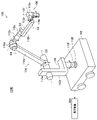
[1-1-2]医療用観察装置100
図1に示す医療用観察装置100は、電子撮像式の医療用観察装置である。例えば手術時に図1に示す医療用観察装置100が用いられる場合、術者(医療用観察装置100の使用者の一例)は、医療用観察装置100により撮像されて、表示装置200の表示画面に表示された医療用撮像画像を参照しながら術部(患部)を観察し、当該術部に対して、術式に応じた手技などの各種処置を行う。
[1-1-2]
The
図1に示すように、医療用観察装置100は、例えば、ベース102と、アーム104と、撮像デバイス106とを備える。
As shown in FIG. 1, the
また、図1では示していないが、医療用観察装置100は、例えば、MPU(Micro Processing Unit)などの演算回路で構成される、1または2以上のプロセッサ(図示せず)と、ROM(Read Only Memory。図示せず)と、RAM(Random Access Memory。図示せず)と、記録媒体(図示せず)と、通信デバイス(図示せず)とを、備えていてもよい。医療用観察装置100は、例えば、医療用観察装置100が備えているバッテリなどの内部電源から供給される電力、または、接続されている外部電源から供給される電力などによって、駆動する。
Further, although not shown in FIG. 1, the
プロセッサ(図示せず)は、医療用観察装置100における制御部(後述する)として機能する。ROM(図示せず)は、プロセッサ(図示せず)が使用するプログラムや演算パラメータなどの制御用データを記憶する。RAM(図示せず)は、プロセッサ(図示せず)により実行されるプログラムなどを一時的に記憶する。
The processor (not shown) functions as a control unit (described later) in the
記録媒体(図示せず)は、医療用観察装置100における記憶部(図示せず)として機能する。記録媒体(図示せず)には、例えば、本実施形態に係る調光制御方法に係るデータや、各種アプリケーションなどの、様々なデータが記憶される。ここで、記録媒体(図示せず)としては、例えば、ハードディスクなどの磁気記録媒体や、フラッシュメモリなどの不揮発性メモリなどが挙げられる。また、記録媒体(図示せず)は、医療用観察装置100から着脱可能であってもよい。
The recording medium (not shown) functions as a storage unit (not shown) in the
通信デバイス(図示せず)は、医療用観察装置100が備える通信手段であり、表示装置200などの外部装置と、無線または有線で通信を行う役目を果たす。ここで、通信デバイス(図示せず)としては、例えば、IEEE802.15.1ポートおよび送受信回路(無線通信)や、IEEE802.11ポートおよび送受信回路(無線通信)、通信アンテナおよびRF回路(無線通信)、あるいはLAN端子および送受信回路(有線通信)などが挙げられる。
The communication device (not shown) is a communication means included in the
[1-1-2-1]ベース102
ベース102は、医療用観察装置100の基台であり、アーム104の一端が接続されて、アーム104と撮像デバイス106とを支持する。
[1-1-2-1]
The
また、ベース102には例えばキャスタが設けられ、医療用観察装置100は、キャスタを介して床面と接地する。キャスタが設けられることにより、医療用観察装置100は、キャスタによって床面上を容易に移動することが可能である。
Further, for example, a caster is provided on the
[1-1-2-2]アーム104
アーム104は、複数のリンクが関節部によって互いに連結されて構成される。
[1-1-2-2]
The
また、アーム104は、撮像デバイス106を支持する。アーム104により支持された撮像デバイス106は3次元的に移動可能であり、移動後の撮像デバイス106は、アーム104によって、位置および姿勢が保持される。
The
より具体的には、アーム104は、例えば、複数の関節部110a、110b、110c、110d、110e、110fと、関節部110a、110b、110c、110d、110e、110fによって互いに回動可能に連結される複数のリンク112a、112b、112c、112d、112e、112fとから構成される。関節部110a、110b、110c、110d、110e、110fそれぞれの回転可能範囲は、アーム104の所望の動きが実現されるように、設計段階や製造段階などにおいて任意に設定される。
More specifically, the
つまり、図1に示す医療用観察装置100では、アーム104を構成する6つの関節部110a、110b、110c、110d、110e、110fに対応する6つの回転軸(第1軸O1、第2軸O2、第3軸O3、第4軸O4、第5軸O5、および第6軸O6)によって、撮像デバイス106の移動に関して6自由度が実現されている。より具体的には、図1に示す医療用観察装置100では、並進3自由度、および回転3自由度の6自由度の動きが実現される。
That is, in the
関節部110a、110b、110c、110d、110e、110fそれぞれには、アクチュエータ(図示せず)が設けられ、関節部110a、110b、110c、110d、110e、110fそれぞれは、アクチュエータ(図示せず)の駆動によって、対応する回転軸で回転する。アクチュエータ(図示せず)の駆動は、例えば、後述する制御部として機能するプロセッサ、または、外部の医療用制御装置(図示せず)によって制御される。
An actuator (not shown) is provided for each of the
関節部110a、110b、110c、110d、110e、110fそれぞれには、6つの回転軸における回転角度をそれぞれ検出することが可能な角度センサ(図示せず)が、設けられうる。角度センサとしては、例えば、ロータリエンコーダや角速度センサなどの、6つの回転軸それぞれにおける回転角度を得ることが可能な任意のセンサが、挙げられる。
Each of the
関節部110a、110b、110c、110d、110e、110fそれぞれが、アクチュエータ(図示せず)の駆動により対応する回転軸で回転することによって、例えばアーム104を伸ばす、縮める(折り畳む)などの、様々なアーム104の動作が、実現される。
The
関節部110aは、略円柱形状を有し、関節部110aの先端部分(図1における下端部分)で、撮像デバイス106(図1における撮像デバイス106の上端部分)を、撮像デバイス106の中心軸と平行な回転軸(第1軸O1)まわりに回動可能なように支持する。ここで、医療用観察装置100は、第1軸O1が撮像デバイス106における光軸と一致するように構成される。つまり、図1に示す第1軸O1まわりに撮像デバイス106を回動させることによって、撮像デバイス106により撮像された医療用撮像画像は、視野が回転するように変更される画像となる。
The
リンク112aは、略棒状の部材であり、関節部110aを固定的に支持する。リンク112aは、例えば、第1軸O1と直交する方向に延伸され、関節部110bに接続される。
The
関節部110bは、略円柱形状を有し、リンク112aを、第1軸O1と直交する回転軸(第2軸O2)まわりに回動可能なように支持する。また、関節部110bには、リンク112bが固定的に接続される。
The
リンク112bは、略棒状の部材であり、第2軸O2と直交する方向に延伸される。また、リンク112bには、関節部110bと関節部110cとがそれぞれ接続される。
The
関節部110cは、略円柱形状を有し、リンク112bを、第1軸O1および第2軸O2それぞれと互いに直交する回転軸(第3軸O3)まわりに回動可能なように支持する。また、関節部110cには、リンク112cの一端が固定的に接続される。
The
ここで、第2軸O2および第3軸O3まわりにアーム104の先端側(撮像デバイス106が設けられる側)が回動することによって、水平面内での撮像デバイス106の位置が変更されるように、撮像デバイス106を移動させることができる。つまり、医療用観察装置100では、第2軸O2および第3軸O3まわりの回転が制御されることにより、医療用撮像画像の視野を平面内で移動させることが可能になる。
Here, the position of the
リンク112cは、一端が略円柱形状を有し、他端が略棒状を有する部材である。リンク112cの一端側には、関節部110cの中心軸と略円柱形状の中心軸とが同一となるように、関節部110cが固定的に接続される。また、リンク112cの他端側には、関節部110dが接続される。
The
関節部110dは、略円柱形状を有し、リンク112cを、第3軸O3と直交する回転軸(第4軸O4)まわりに回動可能なように支持する。関節部110dには、リンク112dが固定的に接続される。
The
リンク112dは、略棒状の部材であり、第4軸O4と直交するように延伸される。リンク112dの一端は、関節部110dの略円柱形状の側面に当接するように、関節部110dに固定的に接続される。また、リンク112dの他端(関節部110dが接続される側とは反対側の端)には、関節部110eが接続される。
The
関節部110eは、略円柱形状を有し、リンク112dの一端を、第4軸O4と平行な回転軸(第5軸O5)まわりに回動可能なように支持する。また、関節部110eには、リンク112eの一端が固定的に接続される。
The
ここで、第4軸O4および第5軸O5は、撮像デバイス106を垂直方向に移動させうる回転軸である。第4軸O4および第5軸O5まわりにアーム104の先端側(撮像デバイス106が設けられる側)が回動することによって、撮像デバイス106の垂直方向の位置が変わる。よって、第4軸O4および第5軸O5まわりにアーム104の先端側(撮像デバイス106が設けられる側)が回動することによって、撮像デバイス106と、患者の術部などの観察対象との距離を変えることが、可能となる。
Here, the fourth axis O4 and the fifth axis O5 are rotation axes capable of moving the
リンク112eは、一辺が鉛直方向に延伸するとともに他辺が水平方向に延伸する略L字形状を有する第1の部材と、当該第1の部材の水平方向に延伸する部位から鉛直下向きに延伸する棒状の第2の部材とが、組み合わされて構成される部材である。リンク112eの第1の部材の鉛直方向に延伸する部位には、関節部110eが固定的に接続される。また、リンク112eの第2の部材には、関節部110fが接続される。
The
関節部110fは、略円柱形状を有し、リンク112eを、鉛直方向と平行な回転軸(第6軸O6)まわりに回動可能なように支持する。また、関節部110fには、リンク112fが固定的に接続される。
The
リンク112fは、略棒状の部材であり、鉛直方向に延伸される。リンク112fの一端は、関節部110fが接続される。また、リンク112fの他端(関節部110fが接続される側とは反対側の端)は、ベース102に固定的に接続される。
The
アーム104が上記に示す構成を有することによって、医療用観察装置100では、撮像デバイス106の移動に関して6自由度が実現される。
By having the configuration shown above, the
なお、アーム104の構成は、上記に示す例に限られない。
The configuration of the
例えば、アーム104の関節部110a、110b、110c、110d、110e、110fそれぞれには、関節部110a、110b、110c、110d、110e、110fそれぞれにおける回転を規制するブレーキが設けられていてもよい。本実施形態に係るブレーキとしては、例えば、機械的に駆動するブレーキや、電気的に駆動する電磁ブレーキなど、任意の方式のブレーキが挙げられる。
For example, each of the
上記ブレーキの駆動は、例えば、後述する制御部として機能するプロセッサ、または、外部の医療用制御装置(図示せず)によって制御される。上記ブレーキの駆動が制御されることにより、医療用観察装置100では、アーム104の動作モードが設定される。アーム104の動作モードとしては、例えば、固定モードとフリーモードとが挙げられる。
The drive of the brake is controlled by, for example, a processor that functions as a control unit described later, or an external medical control device (not shown). By controlling the drive of the brake, the operation mode of the
ここで、本実施形態に係る固定モードとは、例えば、アーム104に設けられる各回転軸における回転がブレーキにより規制されることにより、撮像デバイス106の位置および姿勢が固定される動作モードである。アーム104が固定モードとなることによって、医療用観察装置100の動作状態は、撮像デバイス106の位置および姿勢が固定される固定状態となる。
Here, the fixed mode according to the present embodiment is, for example, an operation mode in which the position and posture of the
また、本実施形態に係るフリーモードとは、上記ブレーキが解除されることにより、アーム104に設けられる各回転軸が自由に回転可能となる動作モードである。例えば、フリーモードでは、術者による直接的な操作によって撮像デバイス106の位置および姿勢を調整することが可能となる。ここで、本実施形態に係る直接的な操作とは、例えば、術者が手で撮像デバイス106を把持し、当該撮像デバイス106を直接移動させる操作のことを意味する。
Further, the free mode according to the present embodiment is an operation mode in which each rotation axis provided on the
[1-1-2-3]撮像デバイス106
撮像デバイス106は、アーム104により支持され、例えば患者の術部などの観察対象を撮像する。撮像デバイス106における撮像は、例えば、後述する制御部として機能するプロセッサ、または、外部の医療用制御装置(図示せず)によって制御される。
[1-1-2-3]
The
撮像デバイス106は、例えば電子撮像式の顕微鏡に対応する構成を有する。
The
図3は、本実施形態に係る医療用観察装置100が備える撮像デバイス106の構成の一例を説明するための説明図である。
FIG. 3 is an explanatory diagram for explaining an example of the configuration of the
撮像デバイス106は、例えば、撮像部材120と、略円筒形状を有する筒状部材122とを有し、撮像部材120は、筒状部材122内に設けられる。
The
筒状部材122の下端(図3における下側の端)の開口面には、例えば、撮像部材120を保護するためのカバーガラス(図示せず)が設けられる。
For example, a cover glass (not shown) for protecting the
また、例えば筒状部材122の内部には光源(図示せず)が設けられ、撮像時には、当該光源からカバーガラス越しに被写体に対して照明光が照射される。照明光が照射された被写体からの反射光(観察光)が、カバーガラス(図示せず)を介して撮像部材120に入射することにより、撮像部材120によって被写体を示す画像信号(医療用撮像画像を示す画像信号)が得られる。
Further, for example, a light source (not shown) is provided inside the
撮像部材120としては、各種の公知の電子撮像式の顕微鏡部に用いられている構成を適用することが可能である。
As the
一例を挙げると、撮像部材120は、例えば、光学系120aと、光学系120aを通過した光により観察対象の像を撮像する撮像素子を含むイメージセンサ120bとで構成される。光学系120aは、例えば、対物レンズ、ズームレンズおよびフォーカスレンズなどの1または2以上のレンズとミラーなどの光学素子で構成される。イメージセンサ120bとしては、例えば、CMOS(Complementary Metal Oxide Semiconductor)やCCD(Charge Coupled Device)などの撮像素子を複数用いたイメージセンサが、挙げられる。
As an example, the
撮像部材120は、例えば、光学系120aおよびイメージセンサ120bで構成される撮像デバイスを、2つ以上有することなどにより、いわゆるステレオカメラとして機能する。ステレオカメラとして機能する撮像デバイス106の構成において、光学系は、ガリレオ式光学系であってもよいし、グリノー式光学系であってもよい。
The
撮像部材120を構成する撮像デバイスには、ズーム機能(光学ズーム機能と電子ズーム機能との一方または双方)、AF(Auto Focus)機能などの、一般的に電子撮像式の顕微鏡部に備えられる1または2以上の機能が搭載される。
The imaging device constituting the
また、撮像部材120は、例えば4K、8Kなどの、いわゆる高解像度での撮像が可能な構成であってもよい。撮像部材120が高解像度での撮像が可能に構成されることにより、所定の解像度(例えば、Full HD画質など)を確保しつつ、例えば50インチ以上などの大画面の表示画面を有する表示装置200に画像を表示させることが可能となるので、当該表示画面を見る術者の視認性が向上する。また、撮像部材120が高解像度での撮像が可能に構成されることにより、撮像画像が電子ズーム機能によって拡大されて表示装置200の表示画面に表示されたとしても、所定の解像度を確保することが可能となる。さらに、電子ズーム機能を用いて所定の解像度が確保される場合には、撮像デバイス106における光学ズーム機能の性能を抑えることが可能となるので、撮像デバイス106の光学系をより簡易にすることができ、撮像デバイス106をより小型に構成することができる。
Further, the
撮像デバイス106には、例えば、撮像デバイス106の動作を制御するための各種の操作デバイスが設けられる。例えば図3では、ズームスイッチ124と、フォーカススイッチ126と、動作モード変更スイッチ128とが、撮像デバイス106に設けられている。なお、ズームスイッチ124、フォーカススイッチ126、および動作モード変更スイッチ128が設けられる位置と形状とが、図3に示す例に限られないことは、言うまでもない。
The
ズームスイッチ124とフォーカススイッチ126とは、撮像デバイス106における撮像条件を調整するための操作デバイスの一例である。
The
ズームスイッチ124は、例えば、ズーム倍率(拡大率)を大きくするズームインスイッチ124aと、ズーム倍率を小さくするズームアウトスイッチ124bとで構成される。ズームスイッチ124に対する操作が行われることによりズーム倍率が調整されて、ズームが調整される。
The
フォーカススイッチ126は、例えば、観察対象(被写体)までの焦点距離を遠くする遠景フォーカススイッチ126aと、観察対象までの焦点距離を近くする近景フォーカススイッチ126bとで構成される。フォーカススイッチ126に対する操作が行われることにより焦点距離が調整されて、フォーカスが調整される。
The
動作モード変更スイッチ128は、撮像デバイス106におけるアーム104の動作モードを変更するための操作デバイスの一例である。動作モード変更スイッチ128に対する操作が行われることにより、アーム104の動作モードが変更される。アーム104の動作モードとしては、例えば上述したように、固定モードとフリーモードとが挙げられる。
The operation
動作モード変更スイッチ128に対する操作の一例としては、動作モード変更スイッチ128を押下する操作が、挙げられる。例えば、術者が動作モード変更スイッチ128を押下している間、アーム104の動作モードがフリーモードとなり、術者が動作モード変更スイッチ128を押下していないときには、アーム104の動作モードが固定モードとなる。
As an example of the operation for the operation
また、撮像デバイス106には、各種操作デバイスに対する操作を行う操作者が操作を行う際の操作性や利便性などをより高めるために、例えば、滑り止め部材130と、突起部材132とが設けられる。
Further, the
滑り止め部材130は、例えば操作者が筒状部材122を手などの操作体で操作を行う際に、操作体の滑りを防止するために設けられる部材である。滑り止め部材130は、例えば、摩擦係数が大きい材料で形成され、凹凸などのより滑りにくい構造を有する。
The
突起部材132は、操作者が筒状部材122を手などの操作体で操作を行う際に、当該操作体が光学系120aの視野を遮ってしまうことや、当該操作体で操作を行う際に、カバーガラス(図示せず)に当該操作体が触れることにより当該カバーガラスが汚れることなどを、防止するために設けられる部材である。
When the operator operates the
なお、滑り止め部材130および突起部材132それぞれが設けられる位置と形状とが、図3に示す例に限られないことは、言うまでもない。また、撮像デバイス106には、滑り止め部材130と突起部材132との一方または双方が設けれられていなくてもよい。
Needless to say, the positions and shapes of the
撮像デバイス106における撮像により生成された画像信号(画像データ)は、例えば後述する制御部として機能するプロセッサにおいて、画像処理が行われる。本実施形態に係る画像処理としては、例えば、ガンマ補正、ホワイトバランスの調整、電子ズーム機能に係る画像の拡大または縮小、または、画素間補正などの各種処理のうちの、1または2以上の処理が、挙げられる。
The image signal (image data) generated by the image pickup in the
また、本実施形態に係る画像処理には、画像信号に基づいて観察対象の明るさを推定する処理が、含まれていてもよい。例えば、画像信号に基づいて観察対象の明るさを推定する処理としては、例えば、画像信号から輝度画像を生成し、生成された輝度画像から観察対象の明るさを推定する処理が、挙げられる。観察対象の明るさは、例えば、輝度画像における各画素の輝度値の平均値を算出することにより推定される。なお、画像信号に基づいて観察対象の明るさを推定する処理の例は、上記に示す例に限られず、画像信号に基づいて観察対象の明るさを推定することが可能な、任意の処理であってもよい。 Further, the image processing according to the present embodiment may include a processing for estimating the brightness of the observation target based on the image signal. For example, as a process of estimating the brightness of the observation target based on the image signal, for example, a process of generating a luminance image from the image signal and estimating the brightness of the observation target from the generated luminance image can be mentioned. The brightness of the observation target is estimated, for example, by calculating the average value of the brightness values of each pixel in the luminance image. The example of the process of estimating the brightness of the observation target based on the image signal is not limited to the example shown above, and any process capable of estimating the brightness of the observation target based on the image signal can be used. There may be.
なお、本実施形態に係る医療用観察システムが、医療用観察装置100における各種動作を制御する医療用制御装置(図示せず)を有する場合には、本実施形態に係る画像処理は、当該医療用制御装置(図示せず)において行われてもよい。
When the medical observation system according to the present embodiment has a medical control device (not shown) that controls various operations in the
医療用観察装置100は、例えば、表示制御信号と、ガンマ補正などの各種画像処理が行われた画像信号とを、表示装置200に送信する。
The
表示制御信号と画像信号とが表示装置200に送信されることによって、表示装置200の表示画面には、観察対象が撮像された医療用撮像画像(例えば、術部が撮像された撮像画像)が、光学ズーム機能と電子ズーム機能との一方または双方によって所望の倍率に拡大または縮小されて表示される。
By transmitting the display control signal and the image signal to the
図1に示す医療用観察装置100は、例えば図1、図3を参照して示したハードウェア構成を有する。
The
なお、本実施形態に係る医療用観察装置のハードウェア構成は、図1、図3を参照して示した構成に限られない。 The hardware configuration of the medical observation device according to the present embodiment is not limited to the configuration shown with reference to FIGS. 1 and 3.
例えば、本実施形態に係る医療用観察装置は、ベース102を備えず、手術室などの天井や壁面などにアーム104が直接取り付けられる構成であってもよい。例えば、天井にアーム104が取り付けられる場合には、本実施形態に係る医療用観察装置は、アーム104が天井から吊り下げられる構成となる。
For example, the medical observation device according to the present embodiment may not have the base 102 and may have a configuration in which the
また、図1では、アーム104が、撮像デバイス106の駆動に関して6自由度が実現されるように構成されている例を示しているが、アーム104の構成は、撮像デバイス106の駆動に関する自由度が6自由度となる構成に限られない。例えば、アーム104は、用途に応じて撮像デバイス106を適宜移動しうるように構成されればよく、関節部およびリンクの数や配置、関節部の駆動軸の方向などは、アーム104が所望の自由度を有するように適宜設定することが可能である。
Further, FIG. 1 shows an example in which the
また、図1、図3では、撮像デバイス106の動作を制御するための各種の操作デバイスが、撮像デバイス106に設けられる例を示しているが、図1、図3に示す操作デバイスのうちの一部または全部は、撮像デバイス106に設けられなくてもよい。一例を挙げると、撮像デバイス106の動作を制御するための各種の操作デバイスは、本実施形態に係る医療用観察装置を構成する撮像デバイス106以外の他の部位に設けられていてもよい。また、他の例を挙げると、撮像デバイス106の動作を制御するための各種の操作デバイスは、フットスイッチFSやリモートコントローラなどの、外部の操作デバイスであってもよい。
Further, FIGS. 1 and 3 show an example in which various operation devices for controlling the operation of the
また、撮像デバイス106は、複数の観察モードを切り替えることが可能な構成であってもよい。本実施形態に係る観察モードとしては、例えば、自然光で撮像を行う観察モード、特殊光で撮像を行う観察モード、NBI(Narrow Band Imaging)などの画像強調観察技術を利用して撮像を行う観察モードなどが、挙げられる。本実施形態に係る特殊光とは、例えば、近赤外線の波長帯域の光や、5-ALA(5-Aminolevulinic Acid)を用いた蛍光観察の蛍光波長帯域の光など、特定の波長帯域の光である。
Further, the
複数の観察モードを切り替えることが可能な撮像デバイス106の構成の一例としては、例えば、“特定の波長帯域の光を透過させ、他の波長帯域の光を透過させないフィルタと、当該フィルタを光路上に選択的に配置する移動機構と、を備える構成”が、挙げられる。本実施形態に係るフィルタが透過させる特定の波長帯域としては、例えば、近赤外線の波長帯域(例えば、約0.7[マイクロメートル]~2.5[マイクロメートル]の波長帯域)や、5-ALAを用いた蛍光観察による蛍光波長帯域(例えば、約0.6[マイクロメートル]~0.65[マイクロメートル]の波長帯域)、ICG(Indocyanine Green)の蛍光波長帯域(例えば、約0.82[マイクロメートル]~0.85[マイクロメートル]の波長帯域)などが、挙げられる。
As an example of the configuration of the
なお、撮像デバイス106には、透過させる波長帯域が異なる複数のフィルタが設けられていてもよい。また、上記では、フィルタが光路上に配置されることにより、特定の波長帯域の光で撮像が行われる例を示したが、特定の波長帯域の光で撮像を行うための撮像デバイス106の構成が、上記に示す例に限られないことは、言うまでもない。
The
[1-2]第2の例に係る医療用観察システム
本実施形態に係る医療用観察システム1000は、図1に示す第1の例に示す構成に限られない。次に、医療用観察システム1000の他の例として、内視鏡装置として機能する医療用観察装置100を有する医療用観察システム1000の構成の一例を説明する。
[1-2] Medical Observation System According to the Second Example The
図4は、本実施形態に係る医療用観察システム1000の構成の第2の例を示す説明図である。図4に示す医療用観察システム1000は、例えば、医療用観察装置100と、表示装置200とを有する。例えば図4に示す医療用観察装置100が手術時に用いられる場合、術者は、医療用観察装置100により撮像されて、表示装置200の表示画面に表示された医療用撮像画像を参照しながら術部を観察し、当該術部に対して、術式に応じた手技などの各種処置を行う。
FIG. 4 is an explanatory diagram showing a second example of the configuration of the
なお、第2の例に係る医療用観察システムは、図4に示す例に限られない。 The medical observation system according to the second example is not limited to the example shown in FIG.
例えば、第2の例に係る医療用観察システムでは、医療用観察装置100を保持する保持装置(図示せず)が設けられていてもよい。保持装置(図示せず)としては、“関節部およびリンクの数や配置、関節部の駆動軸の方向などが、所望の自由度を有するように適宜設定されているアーム”が、挙げられる。保持装置(図示せず)が設けられる第2の例に係る医療用観察システムでは、医療用観察装置100は、保持装置(図示せず)から着脱可能であってもよい。
For example, in the medical observation system according to the second example, a holding device (not shown) for holding the
また、例えば、第2の例に係る医療用観察システムは、第1の例に係る医療用観察システムと同様に、医療用観察装置100における各種動作を制御する医療用制御装置(図示せず)を、さらに有していてもよい。
Further, for example, the medical observation system according to the second example is a medical control device (not shown) that controls various operations in the
また、第2の例に係る医療用観察システムは、第1の例に係る医療用観察システムと同様に、医療用観察装置100と表示装置200との一方または双方を複数有する構成であってもよい。
Further, the medical observation system according to the second example may have a configuration having one or a plurality of the
以下、図4に示す第2の例に係る医療用観察システム1000を構成する各装置について、説明する。
Hereinafter, each device constituting the
[1-2-1]表示装置200
表示装置200は、第2の例に係る医療用観察システム1000における表示手段であり、医療用観察装置100からみて外部の表示デバイスに該当する。第2の例に係る医療用観察システム1000を構成する表示装置200は、第1の例に係る医療用観察システム1000を構成する表示装置200と同様である。
[1-2-1]
The
[1-2-2]医療用観察装置100
図4に示す医療用観察装置100は、例えば、挿入部材134と、光源ユニット136と、ライトガイド138と、カメラヘッド140と、ケーブル142と、制御ユニット144とを備える。医療用観察装置100は、例えば、医療用観察装置100が備えているバッテリなどの内部電源から供給される電力、または、接続されている外部電源から供給される電力などによって、駆動する。
[1-2-2]
The
挿入部材134は、細長形状を有し、入射光を集光する光学系を内部に備える。挿入部材134の先端は、例えば、患者の体腔内に挿入される。挿入部材134の後端はカメラヘッド140の先端と着脱可能に接続される。また、挿入部材134は、ライトガイド138を介して光源ユニット136と接続され、光源ユニット136から光が供給される。
The
挿入部材134は、例えば、可撓性を有さない素材で形成されてもよいし、可撓性を有する素材で形成されてもよい。挿入部材134を形成する素材によって、医療用観察装置100は、硬性鏡または軟性鏡と呼ばれうる。
The
光源ユニット136は、ライトガイド138を介して挿入部材134と接続される。光源ユニット136は、ライトガイド138を介して挿入部材134に光を供給する。
The
光源ユニット136は、例えば、波長が異なる光を発光する複数の光源を有する。光源ユニット136が有する複数の光源としては、例えば、赤色の光を発光する光源、緑色の光を発光する光源、および青色の光を発光する光源が挙げられる。赤色の光を発光する光源としては、例えば、1または2以上の赤色発光ダイオードが挙げられる。緑色の光を発光する光源としては、例えば、1または2以上の緑色発光ダイオードが挙げられる。青色の光を発光する光源としては、例えば、1または2以上の青色発光ダイオードが挙げられる。なお、光源ユニット136が有する複数の光源が、上記に示す例に限られないことは、言うまでもない。光源ユニット136は、例えば、複数の光源を単一チップで有し、または、複数の光源を複数のチップで有する。
The
光源ユニット136は、制御ユニット144と有線または無線で接続され、光源ユニット136における発光は、制御ユニット144により制御される。
The
挿入部材134に供給された光は、挿入部材134の先端から出射され、患者の体腔内組織などの観察対象に照射される。そして、観察対象からの反射光は、挿入部材134内の光学系によって集光される。
The light supplied to the
カメラヘッド140は、観察対象を撮像する機能を有する。カメラヘッド140は、信号伝送部材であるケーブル142を介して制御ユニット144と接続される。
The
カメラヘッド140は、イメージセンサを有し、挿入部材134によって集光された観察対象からの反射光を光電変換することにより観察対象を撮像し、撮像によって得られた画像信号(医療用撮像画像を示す信号)を制御ユニット144へケーブル142を介して出力する。カメラヘッド140が有するイメージセンサとしては、例えば、CMOSやCCDなどの撮像素子を複数用いたイメージセンサが、挙げられる。
The
内視鏡装置として機能する医療用観察装置100では、例えば、挿入部材134、光源ユニット136、およびカメラヘッド140が、“患者の体内に挿入されて、体内を撮像する撮像デバイス”の役目を果たす。
In the
なお、内視鏡装置として機能する医療用観察装置100は、例えば、いわゆるステレオカメラとして機能する複数の撮像デバイスを備える構成であってもよい。ステレオカメラとして機能する撮像デバイスの構成において、光学系は、第1の例に係る医療用観察システムを構成する医療用観察装置100と同様に、ガリレオ式光学系であってもよいし、グリノー式光学系であってもよい。
The
制御ユニット144は、撮像デバイスを制御する。より具体的には、制御ユニット144は、光源ユニット136およびカメラヘッド140それぞれを制御する。
The
また、制御ユニット144は、通信デバイス(図示せず)を含み、カメラヘッド140から出力された画像信号を任意の無線通信または任意の有線通信で、表示装置200へ送信する。制御ユニット144は、画像信号と表示制御信号とを表示装置200へ送信してもよい。
Further, the
制御ユニット144が含む通信デバイス(図示せず)としては、例えば、IEEE802.15.1ポートおよび送受信回路(無線通信)や、IEEE802.11ポートおよび送受信回路(無線通信)、通信アンテナおよびRF回路(無線通信)、光通信用デバイス(有線通信または無線通信)、あるいはLAN端子および送受信回路(有線通信)などが挙げられる。通信デバイス(図示せず)は、複数の通信方式によって、1または2以上の外部装置と通信を行うことが可能な構成であってもよい。
The communication device (not shown) included in the
また、制御ユニット144は、カメラヘッド140から出力された画像信号に対して所定の処理を行い、所定の処理が行われた画像信号を表示装置200へ送信してもよい。画像信号に対する所定の処理としては、例えば、ホワイトバランスの調整や、電子ズーム機能に係る画像の拡大または縮小、画素間補正などが、挙げられる。また、画像信号に対する所定の処理には、上述した画像信号に基づいて観察対象の明るさを推定する処理が、含まれていてもよい。
Further, the
なお、制御ユニット144は、画像信号に基づく医療用撮像画像を記憶してもよい。
The
制御ユニット144としては、例えばCCU(Camera Control Unit)が挙げられる。
Examples of the
内視鏡装置として機能する医療用観察装置100は、例えば図4を参照して示したハードウェア構成を有する。内視鏡装置として機能する医療用観察装置100では、例えば、挿入部材134、光源ユニット136、およびカメラヘッド140が、撮像デバイスの役目を果たし、制御ユニット144により撮像デバイスにおける撮像が制御される。
The
[1-3]医療用観察装置100の機能構成
次に、図1、図4に示す医療用観察装置100を、機能ブロックを用いて説明する。図5は、本実施形態に係る医療用観察装置100の構成の一例を示す機能ブロック図である。
[1-3] Functional Configuration of
医療用観察装置100は、例えば、撮像部150と、通信部152と、制御部154とを備える。
The
撮像部150は、観察対象を撮像する。撮像部150は、例えば、“撮像デバイス106”(図1に示す医療用観察装置100の場合)や、“挿入部材134、光源ユニット136、およびカメラヘッド140”
(図4に示す医療用観察装置100の場合)で構成される。撮像部150における撮像は、例えば制御部154によって制御される。
The
(In the case of the
通信部152は、医療用観察装置100が備える通信手段であり、表示装置200などの外部装置と無線または有線で通信を行う役目を果たす。通信部152は、例えば上述した通信デバイス(図示せず)で構成される。通信部152における通信は、例えば制御部154によって制御される。
The
制御部154は、例えば上述したプロセッサ(図示せず)で構成され、医療用観察装置100全体を制御する役目を果たす。また、制御部154は、後述する調光制御方法に係る処理を主導的に行う役目を果たす。なお、制御部154における調光制御方法に係る処理は、複数の処理回路(例えば、複数のプロセッサなど)で分散して行われてもよい。
The
より具体的には、制御部154は、例えば、撮像制御部156と、表示制御部158と、調光制御部160とを有する。
More specifically, the
撮像制御部156は、撮像部150を構成する撮像デバイスを制御する。撮像デバイスの制御としては、例えば、少なくともズーム機能(光学ズーム機能および電子ズーム機能)を含む、AF機能の制御などの一般的に電子撮像式の顕微鏡部に備えられる1または2以上の機能の制御が、挙げられる。
The image
表示制御部158は、例えば、表示制御信号と画像信号とを通信部152を構成する通信デバイス(図示せず)に伝達し、表示制御信号と画像信号とを表示装置200に対して送信させることによって、表示装置200における表示を制御する。なお、通信部152における通信の制御は、制御部154を構成する通信制御部(図示せず)により行われてもよい。
The
調光制御部160は、本実施形態に係る調光制御方法に係る処理を行う役目を果たす。調光制御部160は、設定される調光モードに従って、撮像デバイスによる観察対象の撮像に関する調光の制御を行う。
The dimming
本実施形態に係る調光モードとは、調光制御の仕方を規定する調光制御の方式である。本実施形態に係る調光モードには、調光制御の仕方が異なる複数の調光モードが含まれる。 The dimming mode according to the present embodiment is a dimming control method that defines a method of dimming control. The dimming mode according to the present embodiment includes a plurality of dimming modes having different dimming control methods.
本実施形態に係る調光の制御としては、例えば、撮像デバイスの露光時間の制御、撮像デバイスにより撮像された医療用撮像画像を示す画像信号のゲインの制御、観察対象に照明光を照射する光源の制御のうちの、一部または全部が、挙げられる。調光の制御に係る撮像デバイスとしては、撮像部150を構成する撮像デバイスが挙げられる。また、調光の制御に係る光源としては、例えば、筒状部材122の内部に設けられる光源(図示せず)の発光の制御(図1に示す医療用観察装置100の場合)や、光源ユニット136の発光の制御(図4に示す医療用観察装置100の場合)が、挙げられる。
The dimming control according to the present embodiment includes, for example, control of the exposure time of the image pickup device, control of the gain of the image signal indicating the medical image taken by the image pickup device, and the light source for irradiating the observation target with illumination light. Part or all of the control of. Examples of the image pickup device related to the control of dimming include an image pickup device constituting the
なお、本実施形態に係る調光の制御は、上記に示す例に限られない。例えば、本実施形態に係る調光の制御には、撮像デバイスによる撮像における観察対象の明るさ、または、撮像デバイスにより撮像された医療用撮像画像の明るさを変えることが可能な、任意の制御が含まれていてもよい。 The control of dimming according to the present embodiment is not limited to the example shown above. For example, in the control of dimming according to the present embodiment, any control that can change the brightness of the observation target in the image pickup by the image pickup device or the brightness of the medical image captured by the image pickup device. May be included.
本実施形態に係る調光モードの一例、および本実施形態に係る調光制御方法に係る処理の一例については、後述する。 An example of the dimming mode according to the present embodiment and an example of the processing related to the dimming control method according to the present embodiment will be described later.
制御部154は、例えば調光制御部160を有することにより、本実施形態に係る調光制御方法に係る処理を主導的に行う役目を果たす。また、制御部154は、例えば、撮像制御部156、および表示制御部158を有することによって、医療用観察装置100全体を制御する役目を果たす。
By having the dimming
なお、制御部154の機能構成は、図5に示す例に限られない。
The functional configuration of the
例えば、制御部154は、本実施形態に係る調光制御方法に係る処理の切り分け方に応じた構成など、医療用観察装置100が有する機能の切り分け方に応じた、任意の構成を有することが可能である。
For example, the
一例を挙げると、医療用観察装置100が図1に示す構成である場合、制御部154は、アーム104の駆動を制御するアーム制御部(図示せず)をさらに有していてもよい。アーム104の駆動の制御の一例としては、例えば、“関節部110a、110b、110c、110d、110e、110fそれぞれに対応するアクチュエータ(図示せず)に対して、駆動を制御する制御信号を印加すること”などが挙げられる。
As an example, when the
医療用観察装置100は、例えば図5に示す機能構成によって、後述する本実施形態に係る調光制御方法に係る処理を行う。
The
なお、本実施形態に係る医療用観察装置の機能構成は、図5に示す構成に限られない。 The functional configuration of the medical observation device according to the present embodiment is not limited to the configuration shown in FIG.
例えば、本実施形態に係る医療用観察装置は、図5に示す撮像制御部156と表示制御部158との一方または双方を、制御部154とは個別に備える(例えば、別の処理回路で実現する)ことができる。
For example, the medical observation device according to the present embodiment includes one or both of the image
また、本実施形態に係る医療用観察装置において本実施形態に係る調光制御方法に係る処理を実行することが可能な機能構成は、図5に示す構成に限られず、例えば、本実施形態に係る医療用観察装置は、本実施形態に係る調光制御方法に係る処理の切り分け方に応じた機能構成をとることが可能である。 Further, the functional configuration capable of executing the processing according to the dimming control method according to the present embodiment in the medical observation device according to the present embodiment is not limited to the configuration shown in FIG. 5, and is, for example, the present embodiment. The medical observation device can have a functional configuration according to the method of separating the processes according to the dimming control method according to the present embodiment.
また、本実施形態に係る医療用観察装置が図1に示す構成である場合、本実施形態に係る医療用観察装置は、アーム104で構成されるアーム部(図示せず)を有する。アーム部(図示せず)を構成するアーム104は、撮像部150を構成する撮像デバイス106を支持する。
Further, when the medical observation device according to the present embodiment has the configuration shown in FIG. 1, the medical observation device according to the present embodiment has an arm portion (not shown) composed of an
また、例えば、通信部152と同様の機能、構成を有する外部の通信デバイスを介して外部装置と通信を行う場合には、本実施形態に係る医療用観察装置は、通信部152を備えていなくてもよい。
Further, for example, when communicating with an external device via an external communication device having the same function and configuration as the
また、本実施形態に係る医療用観察システムが、医療用制御装置(図示せず)を有する構成であり、本実施形態に係る医療用観察装置が当該医療用制御装置(図示せず)により制御される場合、本実施形態に係る医療用観察装置は、制御部154を備えていなくてもよい。
Further, the medical observation system according to the present embodiment has a configuration having a medical control device (not shown), and the medical observation device according to the present embodiment is controlled by the medical control device (not shown). If this is the case, the medical observation device according to the present embodiment may not include the
ここで、医療用制御装置(図示せず)は、例えば、制御部154と同様の機能、構成を有する制御部を備えることによって、後述する本実施形態に係る調光制御方法に係る処理を行い、また、本実施形態に係る医療用観察装置が備える撮像部150などの各構成要素における動作を制御する。医療用制御装置(図示せず)は、備えている通信デバイス、または、接続されている外部の通信デバイスを介して、本実施形態に係る医療用観察装置と通信を行うことによって、本実施形態に係る医療用観察装置が備える各構成要素における動作を制御する。
Here, the medical control device (not shown) is provided with, for example, a control unit having the same function and configuration as the
さらに、本実施形態に係る医療用観察システムが、医療用制御装置(図示せず)を有する構成であり、本実施形態に係る医療用観察装置が当該医療用制御装置(図示せず)により制御される場合、本実施形態に係る医療用観察装置は、制御部154の一部の機能を有さない構成をとることも可能である。
Further, the medical observation system according to the present embodiment has a configuration having a medical control device (not shown), and the medical observation device according to the present embodiment is controlled by the medical control device (not shown). If this is the case, the medical observation device according to the present embodiment may have a configuration that does not have a part of the functions of the
[2]本実施形態に係る調光制御方法
次に、本実施形態に係る調光制御方法について、説明する。以下では、本実施形態に係る調光制御方法に係る処理を医療用観察装置100(より具体的には、例えば医療用観察装置100を構成する制御部154が有する調光制御部160)が行う場合を例に挙げる。なお、上述したように、本実施形態に係る医療用観察システムにおいて、本実施形態に係る調光制御方法に係る処理は、表示装置200や、医療用制御装置(図示せず)などにより行われてもよい。
[2] Dimming control method according to the present embodiment Next, the dimming control method according to the present embodiment will be described. In the following, the medical observation device 100 (more specifically, for example, the dimming
医療用観察装置100のような電子撮像式の医療用観察装置を含む医療用観察システムを用いて手術が行われる場合、図2を参照して示したユースケースに示すように、撮像デバイスによって観察対象の患者の術部が撮像される。ここで、例えば、“鉗子のような医療器具”や、“術者や助手が手にはめている手袋や、ガーゼなどの白っぽい物体”が、撮像デバイスの撮像範囲に入った場合には、当該医療器具による照明光の正反射や当該白っぽい物体によって、本来の観察対象である術部よりも当該医療器具や当該白っぽい物体が明るく撮像されることが多い。上記のように術部よりも医療器具や白っぽい物体が明るく撮像されるときに、一般的な調光制御が行われる場合には、明るさを下げるように調光制御がされ、その結果、本来の観察対象である術部が暗く撮像されてしまう。一般的な調光制御において本来の観察対象である術部が暗く撮像されてしまう事態が生じる主な要因は、例えば下記の通りである。
・常に一定基準で調光制御を動作させ続け、被写体側の明るさ環境が変わると調光制御もそれに追従しようとしてしまう。
・術部の環境は千差万別なため、全ての状況で術部の明るさが安定するような調光制御の万能な一定基準を、設定することができない。
When surgery is performed using a medical observation system that includes an electronic imaging type medical observation device such as the
-The dimming control is always operated with a certain standard, and when the brightness environment on the subject side changes, the dimming control tries to follow it.
-Since the environment of the surgical site varies widely, it is not possible to set a universal standard for dimming control that stabilizes the brightness of the surgical site in all situations.
ここで、上記のような本来の観察対象である術部が暗く撮像されてしまう事態の発生を防止する方法としては、例えば、測光エリアを狭く設定する方法、または、調光感度の応答性を鈍く設定する方法が、考えられる。しかしながら、測光エリアを狭くしてもその範囲内に対象物が入った場合には、調光は反応してしまうため、対策としては不十分である。また、調光感度の応答性を鈍くすると、撮像範囲内の環境の変化に対して調光が正しく合うまでの時間が常に長くなってしまい、表示画面に表示されている医療用撮像画像を見る者が煩わしさを感じてしまう恐れがあるという弊害が、発生する。 Here, as a method for preventing the occurrence of a situation in which the surgical site, which is the original observation target, is imaged darkly as described above, for example, a method of setting a narrow photometric area or a responsiveness of dimming sensitivity is used. A method of setting it dull is conceivable. However, even if the photometric area is narrowed, if an object enters the area, the dimming will react, which is insufficient as a countermeasure. In addition, if the responsiveness of the dimming sensitivity is slowed down, the time until the dimming is correctly adjusted to the change in the environment within the imaging range will always be long, and the medically captured image displayed on the display screen will be viewed. The harmful effect that a person may feel annoyed occurs.
そこで、医療用観察装置100は、調光制御を常に同じ基準で動作させるのではなく、追従速度が異なる調光モードを切り替えて設定し、設定されている調光モードに従って調光制御を行う。
Therefore, the
本実施形態に係る追従速度とは、例えば“調光制御の結果得られる明るさ(例えば、撮像デバイスによる撮像における観察対象の明るさ、または、撮像デバイスにより撮像された医療用撮像画像の明るさ。以下、同様とする。)が、被写体側の明るさ環境の明るさとなるまでに要する時間”により評価される、明るさの追従性の評価尺度の一例である。追従速度は、例えば、撮像デバイスの露光時間、画像信号のゲイン、および光源が発する照明光のうちの、一部または全部の変更の速さを変えることにより変わる。被写体側の明るさ環境の明るさは、例えば、上述した画像信号に基づいて観察対象の明るさを推定する処理により推定され、または、観察対象の明るさを検出することが可能なセンサ(例えば、輝度センサや照度センサなど)により検出される。上記観察対象の明るさを検出することが可能なセンサは、医療用観察装置100が備えるセンサであってもよいし、医療用観察装置100の外部のセンサであってもよい。
The tracking speed according to the present embodiment is, for example, "the brightness obtained as a result of dimming control (for example, the brightness of the observation target in the image pickup by the image pickup device, or the brightness of the medical image captured by the image pickup device". The same shall apply hereinafter.) Is an example of an evaluation scale for brightness followability, which is evaluated by "the time required for the brightness environment on the subject side to become bright". The tracking speed varies, for example, by changing the exposure time of the imaging device, the gain of the image signal, and the speed of change of some or all of the illumination light emitted by the light source. The brightness of the brightness environment on the subject side is estimated by, for example, a process of estimating the brightness of the observation target based on the above-mentioned image signal, or a sensor capable of detecting the brightness of the observation target (for example,). , Brightness sensor, illuminance sensor, etc.). The sensor capable of detecting the brightness of the observation target may be a sensor included in the
本実施形態に係る調光モードには、少なくとも、下記に示す第1の調光モードと、下記に示す第2の調光モードとが含まれる。
・第1の調光モード:第1の追従速度で調光を制御する調光モード
・第2の調光モード:第1の追従速度よりも追従速度が遅い第2の追従速度で調光を制御する調光モード
The dimming mode according to the present embodiment includes at least a first dimming mode shown below and a second dimming mode shown below.
-First dimming mode: Dimming mode that controls dimming at the first follow-up speed-Second dimming mode: Dimming at the second follow-up speed that is slower than the first follow-up speed Dimming mode to control
一例を挙げると、第2の調光モードとは、第1の調光モードよりも、観察対象の明るさの変化(被写体側の明るさ環境の変化)が生じたときに調光の制御を開始するタイミングが遅い調光モードである。例えば、第1の調光モードにおいて、被写体側の明るさ環境の明るさと調光制御により得る目標の明るさとの差の絶対値が、第1の閾値より大きいときに、調光の制御が開始される場合、第2の調光モードでは、当該差の絶対値が第2の閾値(第2の閾値>第1の閾値)より大きいときに、調光の制御が開始される。上記第1の閾値、上記第2の閾値それぞれは、予め設定されている固定値であってもよいし、医療用観察装置100の操作などに応じて変更可能な可変値であってもよい。
As an example, the second dimming mode controls dimming when the brightness of the observation target changes (change in the brightness environment on the subject side) compared to the first dimming mode. It is a dimming mode with a late start timing. For example, in the first dimming mode, dimming control starts when the absolute value of the difference between the brightness of the brightness environment on the subject side and the target brightness obtained by dimming control is larger than the first threshold value. If so, in the second dimming mode, dimming control is started when the absolute value of the difference is larger than the second threshold value (second threshold value> first threshold value). Each of the first threshold value and the second threshold value may be a preset fixed value or a variable value that can be changed according to the operation of the
また、他の例を挙げると、第2の調光モードとは、第1の調光モードよりも、単位時間あたりの観察対象の明るさの変化が小さい調光モードである。ここで、上記単位時間としては、10[秒]、30[秒]、1[分]などの、任意の時間間隔が挙げられる。ここで、第2の調光モードには、観察対象の明るさを変化させない調光モードが含まれていてもよい。 Further, to give another example, the second dimming mode is a dimming mode in which the change in brightness of the observation target per unit time is smaller than that of the first dimming mode. Here, examples of the unit time include arbitrary time intervals such as 10 [seconds], 30 [seconds], and 1 [minutes]. Here, the second dimming mode may include a dimming mode that does not change the brightness of the observation target.
さらに、第2の調光モードは、“第1の調光モードよりも観察対象の明るさの変化が生じたときに調光の制御を開始するタイミングが遅く、かつ、第1の調光モードよりも単位時間あたりの観察対象の明るさの変化が小さい調光モード”であってもよい。 Further, in the second dimming mode, "the timing of starting the dimming control when the brightness of the observation target changes occurs later than that of the first dimming mode, and the first dimming mode It may be a dimming mode in which the change in the brightness of the observation target per unit time is smaller than that.
なお、本実施形態に係る調光モードの例は、上記に示す例に限られない。例えば、第2の調光モードに観察対象の明るさを変化させない調光モードが含まれない場合、本実施形態に係る調光モードには、観察対象の明るさを変化させない固定の調光モードが、含まれていてもよい。 The example of the dimming mode according to the present embodiment is not limited to the example shown above. For example, when the second dimming mode does not include a dimming mode that does not change the brightness of the observation target, the dimming mode according to the present embodiment is a fixed dimming mode that does not change the brightness of the observation target. However, it may be included.
以下では、医療用観察装置100が、本実施形態に係る調光モードとして、第1の調光モードまたは第2の調光モードを設定し、設定されている調光モードに従って調光制御を行う場合を、例に挙げる。つまり、以下では、第2の調光モードに観察対象の明るさを変化させない調光モードが含まれうる場合を例に挙げる。
In the following, the
具体的には、医療用観察装置100は、例えば下記の(1)の処理および(2)の処理を行うことによって、第1の調光モードまたは第2の調光モードに従って調光制御を行う。
Specifically, the
(1)調光制御方法に係る処理の第1の例:第1の調光モードに従った調光制御
医療用観察装置100は、撮像デバイスにおける撮像に関する動作の変化に基づいて第1の調光モードを設定する。医療用観察装置100は、撮像デバイスにおける撮像に関する動作の変化が検出されたときに、第1の調光モードを設定する。そして、医療用観察装置100は、設定されている第1の調光モードに従った制御を行う。以下では、撮像デバイスにおける撮像に関する動作を単に「撮像に関する動作」と示す場合がある。
(1) First Example of Processing Related to Dimming Control Method: Dimming Control According to First Dimming Mode The
医療用観察装置100が撮像に関する動作の変化を検出する例としては、例えば下記に示す例が挙げられる。なお、医療用観察装置100が撮像に関する動作の変化を検出する例が、下記に示す例に限られないことは、言うまでもない。
・医療用観察装置100の電源がオン状態となったとき(医療用観察装置100の起動に伴う撮像に関する動作の変化が検出される場合の一例)
・医療用観察装置100が備える操作デバイス、または、フットスイッチFSなどの医療用観察装置100の外部の操作デバイスに対する所定の操作が検出されたとき(例えば、“視野移動の操作や、フォーカスを変える操作、ズームを変える操作などの観察視野を変える操作”、または、“明るさレベルを変える操作や、照明をオン/オフする操作、カラーモードを変更する操作、特殊光観察を行う操作などの、撮像条件を変える操作”
が検出されたとき)
Examples of the
When the power of the
When a predetermined operation is detected for an operation device included in the
When is detected)
医療用観察装置100は、撮像デバイスにおける撮像に関する動作の変化が検出されたときに第1の調光モードを設定し、第1の調光モードに従った制御を開始する。また、医療用観察装置100は、後述する所定の条件を満たしたと判定されるまで、第1の調光モードに従った制御を行う。ここで、撮像デバイスにおける撮像に関する動作の変化が検出されてから、後述する所定の条件を満たしたと判定されるまでの期間は、例えば“医療用観察装置100が、撮像デバイスにおける撮像に関する動作の遷移中の状態である期間”であると、捉えることが可能である。撮像デバイスにおける撮像に関する動作の遷移中の状態には、“撮像デバイスにおける撮像に関する動作が行われている状態”と、“撮像デバイスにおける撮像に関する動作が完了した後、後述する所定の条件を満たしたと判定されるまでの期間における状態”とが含まれる。
The
(2)調光制御方法に係る処理の第2の例:第2の調光モードに従った調光制御
医療用観察装置100は、例えば、上記(1)に示す第1の例に係る処理により第1の調光モードに従って制御を行っているときに、所定の条件を満たしたと判定した場合に、第2の調光モードを設定する。そして、医療用観察装置100は、設定されている第2の調光モードに従った制御を行う。
(2) Second example of the process according to the dimming control method: The dimming control according to the second dimming mode The
例えば、医療用観察装置100は、第1の調光モードに従った制御による調光が終了したときに、所定の条件を満たしたと判定する。
For example, the
第1の調光モードに従った制御による調光が終了したときとは、例えば、“調光制御の結果得られる明るさが被写体側の明るさ環境の明るさと一致したとき”、または、“調光制御の結果得られる明るさと被写体側の明るさ環境の明るさとの差の絶対値が、設定されている閾値以下となったとき(または、当該絶対値が当該閾値よりも小さくなったとき)”が、挙げられる。上記閾値は、予め設定されている固定値であってもよいし、医療用観察装置100の操作などに応じて変更可能な可変値であってもよい。なお、第1の調光モードに従った制御による調光が終了したかを判定する方法が、上記に示す例に限られないことは、言うまでもない。
When the dimming by the control according to the first dimming mode is completed, for example, "when the brightness obtained as a result of the dimming control matches the brightness of the brightness environment on the subject side" or ". When the absolute value of the difference between the brightness obtained as a result of dimming control and the brightness of the brightness environment on the subject side is less than or equal to the set threshold value (or when the absolute value becomes smaller than the threshold value). ) ”. The threshold value may be a preset fixed value or a variable value that can be changed according to the operation of the
また、医療用観察装置100は、第1の調光モードに従った制御が開始されてから所定の時間が経過したときに、所定の条件を満たしたと判定してもよい。所定の時間は、予め設定されている固定の時間間隔であってもよいし、医療用観察装置100の操作などに応じて変更可能な可変の時間間隔であってもよい。
Further, the
さらに、医療用観察装置100は、第1の調光モードに従った制御による調光が終了したとき、または、第1の調光モードに従った制御が開始されてから所定の時間が経過したときのいずれかを満たした場合に、所定の条件を満たしたと判定してもよい。
Further, in the
なお、第2の例に係る調光制御方法に係る処理は、上記に示す例に限られない。 The process related to the dimming control method according to the second example is not limited to the example shown above.
例えば、医療用観察装置100は、第1の調光モードに従って制御が行われていない場合において第2の調光モードを設定して、第2の調光モードに従った制御を行うことも可能である。
For example, the
医療用観察装置100は、例えば、撮像デバイスにおける撮像に関する動作の変化に基づいて第2の調光モードを設定する。具体的には、医療用観察装置100は、例えば、撮像に関する動作の変化が検出されていない場合に、第2の調光モードを設定する。そして、医療用観察装置100は、設定されている第2の調光モードに従った制御を行う。
The
例えば上記(1)に示す第1の例に係る処理および上記(2)に示す第2の例に係る処理を行うことによって、医療用観察装置100は、追従速度が異なる第1の調光モードと第2の調光モードとを切り替えて調光制御を行うことができる。
For example, by performing the process according to the first example shown in (1) above and the process according to the second example shown in (2) above, the
図6は、本実施形態に係る調光制御方法に係る処理の一例を示す流れ図であり、医療用観察装置100が、第1の調光モードと第2の調光モードとを切り替えて調光制御を行う場合における処理の一例を示している。
FIG. 6 is a flow chart showing an example of the process according to the dimming control method according to the present embodiment, in which the
医療用観察装置100は、システム動作中であるか否を判定する(S100)。医療用観察装置100は、例えば、撮像に関する動作の変化が検出されている場合に、システム動作中であると判定する。システム動作中であると判定されている状態は、撮像デバイスにおける撮像に関する動作が行われている状態に該当する。
The
ステップS100においてシステム動作中であると判定された場合、医療用観察装置100は、第1の調光モードを設定して、第1の調光モードに従って調光制御を行う(S102)。第1の調光モードに従って調光制御が行われることによって、撮像範囲内の環境の変化があったとしても、第2の調光モードに従って調光制御が行われる場合よりも素早く、調光制御の結果得られる明るさを、被写体側の明るさ環境の明るさに合わせることができる。
When it is determined in step S100 that the system is operating, the
ステップS102の処理を行うと、医療用観察装置100は、システムの動作が完了したか否かを判定する(S104)。医療用観察装置100は、例えば、医療用観察装置100の起動が完了したとき、または、医療用観察装置100が備える操作デバイスなどに対する所定の操作に応じた処理が完了したときに、システムの動作が完了したと判定する。
When the process of step S102 is performed, the
ステップS104においてシステムの動作が完了したと判定されない場合、医療用観察装置100は、ステップS102からの処理を繰り返す。
If it is not determined in step S104 that the operation of the system is completed, the
また、ステップS104においてシステムの動作が完了したと判定された場合、医療用観察装置100は、第1の調光モードに従って調光制御を行う(S106)。ここで、医療用観察装置100は、再度第1の調光モードを設定し直してもよいし、ステップS102において設定されている第1の調光モードの設定を引き継いでもよい。ステップS106における第1の調光モードに従った調光制御は、撮像デバイスにおける撮像に関する動作の遷移中の状態における調光制御に該当する。
Further, when it is determined in step S104 that the operation of the system is completed, the
ステップS106の処理を行うと、医療用観察装置100は、所定の条件を満たしたか否かを判定する(S108)。
When the process of step S106 is performed, the
ステップS108において所定の条件を満たしたと判定されない場合、医療用観察装置100は、ステップS106からの処理を繰り返す。また、ステップS108において所定の条件を満たしたと判定された場合、医療用観察装置100は、医療用観察装置100は、ステップS100からの処理を繰り返す。
If it is not determined in step S108 that the predetermined condition is satisfied, the
ステップS100においてシステム動作中であると判定されない場合、医療用観察装置100は、第2の調光モードを設定して、第2の調光モードに従って調光制御を行う(S110)。第2の調光モードに従って調光制御が行われることによって、撮像範囲内の環境の変化があった場合における追従速度は、第1の調光モードに従って調光制御が行われる場合よりも遅くなる。
If it is not determined in step S100 that the system is operating, the
ここで、システム動作中であると判定されない場合とは、撮像に関する動作の変化が検出されていない場合である。そのため、システム動作中であると判定されない場合には、撮像範囲内における観察対象の環境の変化は少ないと、考えられる。そのため、システム動作中であると判定されない場合には、調光制御の結果得られる明るさを被写体側の明るさ環境の明るさに素早く合わせる必要性は、小さいと考えられる。 Here, the case where it is not determined that the system is operating is the case where the change in the operation related to the imaging is not detected. Therefore, if it is not determined that the system is operating, it is considered that there is little change in the environment of the observation target within the imaging range. Therefore, if it is not determined that the system is operating, it is considered that there is little need to quickly adjust the brightness obtained as a result of the dimming control to the brightness of the brightness environment on the subject side.
また、ステップS110において第2の調光モードに従って調光制御が行われることにより、医療器具の正反射や白っぽい物体が撮像範囲に入った場合であっても、これらに起因して調光が過敏に反応することは防止される。 Further, since the dimming control is performed according to the second dimming mode in step S110, even when the specular reflection of the medical device or a whitish object enters the imaging range, the dimming is hypersensitive due to these. It is prevented from reacting to.
医療用観察装置100は、本実施形態に係る調光制御方法に係る処理として、例えば図6に示す処理を行う。ここで、医療用観察装置100は、調光制御を常に同じ基準で動作させるのではなく、追従速度が異なる第1の調光モードと第2の調光モードとを切り替えて調光制御を行う。
The
よって、図6に示す処理を行うことによって、医療用観察装置100は、撮像デバイスによる観察対象の撮像により適した調光制御を行うことができる。なお、本実施形態に係る調光制御方法に係る処理の例が、図6に示す例に限られないことは、言うまでもない。
Therefore, by performing the process shown in FIG. 6, the
なお、本実施形態に係る調光制御方法に係る処理は、上記に示す例に限られない。 The process related to the dimming control method according to the present embodiment is not limited to the example shown above.
例えば、医療用観察装置100は、所定の操作に基づいて、第2の調光モードを設定する機能を有効化し、または当該第2の調光モードを設定する機能を無効化してもよい。つまり、医療用観察装置100は、第1の調光モードに従った調光制御から第2の調光モードに従った調光制御へと切り替える機能を、選択的に有効化することが可能であってもよい。第2の調光モードを設定する機能が無効化される場合、医療用観察装置100は、例えば、第1の調光モードに従って調光制御を行う。
For example, the
本実施形態に係る所定の操作としては、例えば、医療用観察装置100が備える操作デバイスに対する上記機能の切り替え操作、リモートコントローラやフットスイッチFSなどの外部の操作デバイスに対する上記機能の切り替え操作などが、挙げられる。
Predetermined operations according to the present embodiment include, for example, a switching operation of the above function for an operating device included in the
また、医療用観察装置100は、例えば、設定されている調光モードを通知させる処理を、さらに行ってもよい。
Further, the
医療用観察装置100は、例えば、設定されている調光モードを示す文字と、設定されている調光モードを示すアイコンなどの画像との一方または双方を、表示装置200の表示画面などの、医療用観察装置100の使用者が見ることが可能な任意の表示画面に表示させることによって、設定されている調光モードを視覚的に通知させる。また、医療用観察装置100は、例えば、設定されている調光モードを示す音声を、スピーカなどの音声出力デバイスから出力させることによって、設定されている調光モードを聴覚的に通知させる。さらに、医療用観察装置100は、視覚的な通知方法と聴覚的な通知方法との双方によって、設定されている調光モードを通知させてもよい。上記音声出力デバイスは、医療用観察装置100が備える音声出力デバイスであってもよいし、医療用観察装置100の外部の音声出力デバイスであってもよい。
The
設定されている調光モードが通知されることによって、医療用観察装置100の使用者は、医療用観察装置100がどのような調光制御モードで調光制御を行っているかを、認識することができる。
By notifying the set dimming mode, the user of the
なお、設定されている調光モードを通知させる処理は、上記に示す例に限られない。例えば、医療用観察装置100は、第2の調光モードを設定する機能が有効化されているか、無効化されているかを、視覚的な通知方法と聴覚的な通知方法との一方または双方によって、通知させてもよい。
The process of notifying the set dimming mode is not limited to the above example. For example, the
[3]本実施形態に係る調光制御方法が用いられることにより奏される効果の一例
本実施形態に係る調光制御方法が用いられることによって、例えば下記に示す効果が奏される。なお、本実施形態に係る調光制御方法が用いられることにより奏される効果が、下記に示す例に限られないことは、言うまでもない。
・システムの動作状態が、観察対象の明るさが変動する状態では調光制御を素早く行うことによって、明るさがより適切な医療用撮像画像を素早く得ることができる。
・観察対象の明るさが変動しづらい状態では調光を鈍く行う、もしくは調光を固定することによって、意図しない明るさ変動に追従しないようになり、医療用観察装置100の使用者が本当に観察したい対象に対して調光が合った状態の医療用撮像画像を、医療用観察装置100の使用者に提供することができる。
[3] An example of the effect produced by using the dimming control method according to the present embodiment By using the dimming control method according to the present embodiment, for example, the following effects are exhibited. Needless to say, the effect produced by using the dimming control method according to the present embodiment is not limited to the examples shown below.
-By quickly performing dimming control when the brightness of the observation target fluctuates in the operating state of the system, it is possible to quickly obtain a medically captured image having a more appropriate brightness.
-In a state where the brightness of the observation target is hard to fluctuate, the dimming is slowed down or the dimming is fixed so that the unintended brightness fluctuation is not followed, and the user of the
(本実施形態に係るプログラム)
コンピュータシステムを、本実施形態に係る医療用観察装置(または、本実施形態に係る医療用調光制御装置)として機能させるためのプログラム(例えば、本実施形態に係る調光制御方法に係る処理を実行することが可能なプログラム)が、コンピュータシステムにおいてプロセッサなどにより実行されることによって、撮像デバイスによる観察対象の撮像により適した調光制御を行うことができる。ここで、本実施形態に係るコンピュータシステムとしては、単体のコンピュータ、または、複数のコンピュータが挙げられる。本実施形態に係るコンピュータシステムによって、本実施形態に係る調光制御方法に係る一連の処理が行われる。
(Program related to this embodiment)
A program for making the computer system function as a medical observation device (or a medical dimming control device according to the present embodiment) according to the present embodiment (for example, a process related to the dimming control method according to the present embodiment). By executing the program (executable program) by a processor or the like in a computer system, it is possible to perform dimming control more suitable for imaging the observation target by the imaging device. Here, examples of the computer system according to the present embodiment include a single computer or a plurality of computers. The computer system according to the present embodiment performs a series of processes related to the dimming control method according to the present embodiment.
また、コンピュータシステムを、本実施形態に係る医療用観察装置(または、本実施形態に係る医療用調光制御装置)として機能させるためのプログラムが、コンピュータシステムにおいてプロセッサなどにより実行されることによって、上述した本実施形態に係る調光制御方法に係る処理によって実現される表示によって奏される効果を、奏することができる。 Further, a program for making the computer system function as a medical observation device (or a medical dimming control device according to the present embodiment) according to the present embodiment is executed by a processor or the like in the computer system. The effect achieved by the display realized by the process according to the dimming control method according to the present embodiment described above can be achieved.
以上、添付図面を参照しながら本開示の好適な実施形態について詳細に説明したが、本開示の技術的範囲はかかる例に限定されない。本開示の技術分野における通常の知識を有する者であれば、特許請求の範囲に記載された技術的思想の範疇内において、各種の変更例または修正例に想到しうることは明らかであり、これらについても、当然に本開示の技術的範囲に属するものと了解される。 Although the preferred embodiments of the present disclosure have been described in detail with reference to the accompanying drawings, the technical scope of the present disclosure is not limited to such examples. It is clear that any person with ordinary knowledge in the technical field of the present disclosure may come up with various modifications or amendments within the scope of the technical ideas described in the claims. Is, of course, understood to belong to the technical scope of the present disclosure.
例えば、上記では、コンピュータシステムを、本実施形態に係る医療用観察装置(または、本実施形態に係る医療用調光制御装置)として機能させるためのプログラム(コンピュータプログラム)が提供されることを示したが、本実施形態は、さらに、上記プログラムを記憶させた記録媒体も、併せて提供することができる。 For example, the above shows that a program (computer program) for making a computer system function as a medical observation device (or a medical dimming control device according to the present embodiment) according to the present embodiment is provided. However, the present embodiment can also provide a recording medium in which the above program is stored.
上述した構成は、本実施形態の一例を示すものであり、当然に、本開示の技術的範囲に属するものである。 The configuration described above is an example of the present embodiment and, of course, belongs to the technical scope of the present disclosure.
また、本明細書に記載された効果は、あくまで説明的または例示的なものであって限定的ではない。つまり、本開示に係る技術は、上記の効果とともに、または上記の効果に代えて、本明細書の記載から当業者には明らかな他の効果を奏しうる。 In addition, the effects described herein are merely explanatory or exemplary and are not limited. That is, the techniques according to the present disclosure may have other effects apparent to those skilled in the art from the description herein, in addition to or in place of the above effects.
なお、以下のような構成も本開示の技術的範囲に属する。
(1)
設定される調光モードに従って、撮像デバイスによる観察対象の撮像に関する調光の制御を行う調光制御部を備え、
前記調光モードには、少なくとも、第1の追従速度で調光を制御する第1の調光モードと、前記第1の追従速度よりも追従速度が遅い第2の追従速度で調光を制御する第2の調光モードとが含まれ、
前記調光制御部は、
前記撮像デバイスにおける撮像に関する動作の変化に基づいて前記第1の調光モードを設定し、
前記第1の調光モードに従って制御を行っているときに、所定の条件を満たしたと判定した場合に、前記第2の調光モードを設定する、医療用調光制御装置。
(2)
前記調光制御部は、前記調光の制御として、前記撮像デバイスの露光時間の制御、前記撮像デバイスにより撮像された医療用撮像画像を示す画像信号のゲインの制御、前記観察対象に照明光を照射する光源の制御のうちの、一部または全部を行う、(1)に記載の医療用調光制御装置。
(3)
前記調光制御部は、前記第1の調光モードに従った制御による調光が終了したときに、前記所定の条件を満たしたと判定する、(1)または(2)に記載の医療用調光制御装置。
(4)
前記調光制御部は、前記第1の調光モードに従った制御が開始されてから所定の時間が経過したときに、前記所定の条件を満たしたと判定する、(1)~(3)のいずれか1つに記載の医療用調光制御装置。
(5)
前記第2の調光モードは、前記第1の調光モードよりも、前記観察対象の明るさの変化が生じたときに前記調光の制御を開始するタイミングが遅い調光モードである、(1)~(4)のいずれか1つに記載の医療用調光制御装置。
(6)
前記第2の調光モードは、前記第1の調光モードよりも、単位時間あたりの前記観察対象の明るさの変化が小さい調光モードである、(1)~(5)のいずれか1つに記載の医療用調光制御装置。
(7)
前記第2の調光モードは、前記観察対象の明るさを変化させない調光モードである、(6)に記載の医療用調光制御装置。
(8)
前記調光制御部は、
前記撮像デバイスにおける撮像に関する動作の変化が検出されたときに、前記第1の調光モードに従った制御を開始し、
前記所定の条件を満たしたと判定されるまで、前記第1の調光モードに従った制御を行う、(1)~(7)のいずれか1つに記載の医療用調光制御装置。
(9)
前記調光制御部は、所定の操作に基づいて、前記第2の調光モードを設定する機能を有効化し、または前記機能を無効化する、(1)~(8)のいずれか1つに記載の医療用調光制御装置。
(10)
前記調光制御部は、設定されている調光モードを通知させる、(1)~(9)のいずれか1つに記載の医療用調光制御装置。
(11)
複数のリンクが関節部によって互いに連結されて構成されるアームと、
前記アームにより支持されている前記撮像デバイスと、
をさらに備える、(1)~(10)のいずれか1つに記載の医療用調光制御装置。
(12)
患者の体内に挿入され、前記体内を前記観察対象として撮像する前記撮像デバイスをさらに備える、(1)~(10)のいずれか1つに記載の医療用調光制御装置。
(13)
設定される調光モードに従って、撮像デバイスによる観察対象の撮像に関する調光の制御を行うステップを有し、
前記調光モードには、少なくとも、第1の追従速度で調光を制御する第1の調光モードと、前記第1の追従速度よりも追従速度が遅い第2の追従速度で調光を制御する第2の調光モードとが含まれ、
前記制御を行うステップでは、
前記撮像デバイスにおける撮像に関する動作の変化に基づいて前記第1の調光モードが設定され、
前記第1の調光モードに従って制御を行っているときに、所定の条件を満たしたと判定された場合に、前記第2の調光モードが設定される、医療用調光制御装置により実行される調光制御方法。
The following configurations also belong to the technical scope of the present disclosure.
(1)
It is equipped with a dimming control unit that controls dimming related to the imaging of the observation target by the imaging device according to the set dimming mode.
In the dimming mode, at least, the dimming is controlled by the first dimming mode in which the dimming is controlled by the first follow-up speed and the second follow-up speed in which the follow-up speed is slower than the first follow-up speed. Includes a second dimming mode to
The dimming control unit
The first dimming mode is set based on the change in the operation related to the image pickup in the image pickup device.
A medical dimming control device that sets the second dimming mode when it is determined that a predetermined condition is satisfied while controlling according to the first dimming mode.
(2)
As the dimming control, the dimming control unit controls the exposure time of the image pickup device, controls the gain of the image signal indicating the medically captured image captured by the image pickup device, and emits the illumination light to the observation target. The medical dimming control device according to (1), which controls a part or all of the light source to be irradiated.
(3)
The medical adjustment according to (1) or (2), wherein the dimming control unit determines that the predetermined condition is satisfied when the dimming by the control according to the first dimming mode is completed. Optical control device.
(4)
The dimming control unit determines that the predetermined condition is satisfied when a predetermined time has elapsed from the start of the control according to the first dimming mode, according to (1) to (3). The medical dimming control device according to any one.
(5)
The second dimming mode is a dimming mode in which the timing of starting the dimming control when the brightness of the observation target changes occurs is later than that of the first dimming mode. 1) The medical dimming control device according to any one of (4).
(6)
The second dimming mode is any one of (1) to (5), which is a dimming mode in which the change in brightness of the observation target per unit time is smaller than that of the first dimming mode. The medical dimming control device described in 1.
(7)
The medical dimming control device according to (6), wherein the second dimming mode is a dimming mode that does not change the brightness of the observation target.
(8)
The dimming control unit
When a change in the operation related to the image pickup in the image pickup device is detected, the control according to the first dimming mode is started.
The medical dimming control device according to any one of (1) to (7), wherein control is performed according to the first dimming mode until it is determined that the predetermined condition is satisfied.
(9)
The dimming control unit is set to any one of (1) to (8), which enables or disables the function of setting the second dimming mode based on a predetermined operation. The medical dimming control device described.
(10)
The medical dimming control device according to any one of (1) to (9), wherein the dimming control unit notifies the set dimming mode.
(11)
An arm consisting of multiple links connected to each other by joints,
The imaging device supported by the arm and
The medical dimming control device according to any one of (1) to (10).
(12)
The medical dimming control device according to any one of (1) to (10), further comprising the image pickup device that is inserted into the body of a patient and images the inside of the body as the observation target.
(13)
It has a step of controlling the dimming of the image of the observation target by the image pickup device according to the set dimming mode.
In the dimming mode, at least, the dimming is controlled by the first dimming mode in which the dimming is controlled by the first follow-up speed and the second follow-up speed in which the follow-up speed is slower than the first follow-up speed. Includes a second dimming mode to
In the step of performing the control,
The first dimming mode is set based on the change in the operation related to the image pickup in the image pickup device.
It is executed by the medical dimming control device in which the second dimming mode is set when it is determined that a predetermined condition is satisfied while the control is performed according to the first dimming mode. Dimming control method.
100 医療用観察装置
102 ベース
104 アーム
106 撮像デバイス
110a、110b、110c、110d、110e、110f 関節部
112a、112b、112c、112d、112e、112f リンク
120 撮像部材
122 筒状部材
124 ズームスイッチ
126 フォーカススイッチ
128 動作モード変更スイッチ
134 挿入部材
136 光源ユニット
138 ライトガイド
140 カメラヘッド
142 ケーブル
144 制御ユニット
150 撮像部
152 通信部
154 制御部
156 撮像制御部
158 表示制御部
160 調光制御部
200 表示装置
1000 医療用観察システム
100
Claims (12)
前記調光モードには、少なくとも、第1の追従速度で調光を制御する第1の調光モードと、前記第1の追従速度よりも追従速度が遅い第2の追従速度で調光を制御する第2の調光モードとが含まれ、
前記調光制御部は、
前記撮像デバイスに対し、観察視野または撮像条件を変更する操作が行われた場合は、前記第1の調光モードを設定し、
前記観察視野または撮像条件を変更する操作に基づく制御が終了し前記第1の調光モードに従った制御による調光が終了した場合、または前記第1の調光モードに従った制御による調光が終了してから所定の時間が経過した場合に、前記第2の調光モードを設定する、医療用調光制御装置。 It is equipped with a dimming control unit that controls dimming related to the imaging of the observation target by the imaging device according to the set dimming mode.
In the dimming mode, at least, the dimming is controlled by the first dimming mode in which the dimming is controlled by the first follow-up speed and the second follow-up speed in which the follow-up speed is slower than the first follow-up speed. Includes a second dimming mode to
The dimming control unit
When an operation for changing the observation field of view or the imaging conditions is performed on the imaging device, the first dimming mode is set.
When the control based on the operation of changing the observation field of view or the imaging condition is completed and the dimming by the control according to the first dimming mode is completed, or the dimming by the control according to the first dimming mode is completed. A medical dimming control device that sets the second dimming mode when a predetermined time has elapsed since the end of .
前記アームにより支持されている前記撮像デバイスと、
をさらに備える、請求項1~9のいずれか1つに記載の医療用調光制御装置。 An arm consisting of multiple links connected to each other by joints,
The imaging device supported by the arm and
The medical dimming control device according to any one of claims 1 to 9 , further comprising.
前記調光制御部が、
前記撮像デバイスに対し、観察視野または撮像条件を変更する操作が行われた場合は、第1の追従速度で調光を制御する第1の調光モードに従って制御を行い、
前記観察視野または撮像条件を変更する操作に基づく制御が終了し前記第1の調光モードに従った制御による調光が終了した場合、または前記第1の調光モードに従った制御による調光が終了してから所定の時間が経過した場合に、前記第1の追従速度よりも追従速度が遅い第2の追従速度で調光を制御する第2の調光モードに従って制御を行う、医療用調光制御装置の作動方法。 It is an operation method of a medical dimming control device provided with a dimming control unit that controls dimming related to imaging of an observation target by an imaging device.
The dimming control unit
When the image pickup device is operated to change the observation field of view or the image pickup conditions, the control is performed according to the first dimming mode in which the dimming is controlled at the first tracking speed.
When the control based on the operation of changing the observation field or the imaging condition is completed and the dimming by the control according to the first dimming mode is completed, or the dimming by the control according to the first dimming mode is completed. When a predetermined time has elapsed since the end of the process, control is performed according to a second dimming mode in which dimming is controlled at a second following speed, which is slower than the first following speed . How to operate the dimming control device.
Priority Applications (2)
| Application Number | Priority Date | Filing Date | Title |
|---|---|---|---|
| JP2018043854A JP7073145B2 (en) | 2018-03-12 | 2018-03-12 | How to operate the medical dimming control device and the medical dimming control device |
| US16/259,089 US11464402B2 (en) | 2018-03-12 | 2019-01-28 | Medical dimming control apparatus and dimming control method |
Applications Claiming Priority (1)
| Application Number | Priority Date | Filing Date | Title |
|---|---|---|---|
| JP2018043854A JP7073145B2 (en) | 2018-03-12 | 2018-03-12 | How to operate the medical dimming control device and the medical dimming control device |
Publications (3)
| Publication Number | Publication Date |
|---|---|
| JP2019154680A JP2019154680A (en) | 2019-09-19 |
| JP2019154680A5 JP2019154680A5 (en) | 2021-04-30 |
| JP7073145B2 true JP7073145B2 (en) | 2022-05-23 |
Family
ID=67844105
Family Applications (1)
| Application Number | Title | Priority Date | Filing Date |
|---|---|---|---|
| JP2018043854A Active JP7073145B2 (en) | 2018-03-12 | 2018-03-12 | How to operate the medical dimming control device and the medical dimming control device |
Country Status (2)
| Country | Link |
|---|---|
| US (1) | US11464402B2 (en) |
| JP (1) | JP7073145B2 (en) |
Families Citing this family (1)
| Publication number | Priority date | Publication date | Assignee | Title |
|---|---|---|---|---|
| US10939970B2 (en) * | 2019-05-22 | 2021-03-09 | Titan Medical Inc. | Robotic surgery system |
Citations (7)
| Publication number | Priority date | Publication date | Assignee | Title |
|---|---|---|---|---|
| JP2000152913A (en) | 1998-09-16 | 2000-06-06 | Asahi Optical Co Ltd | Electric endoscope |
| JP2007097711A (en) | 2005-09-30 | 2007-04-19 | Fujinon Corp | Electronic endoscope apparatus |
| JP2009273577A (en) | 2008-05-13 | 2009-11-26 | Olympus Medical Systems Corp | Medical observation apparatus |
| JP2012085790A (en) | 2010-10-19 | 2012-05-10 | Hoya Corp | Endoscopic system |
| CN103860129A (en) | 2014-03-19 | 2014-06-18 | 深圳市开立科技有限公司 | Method and device for automatically adjusting endoscope light source brightness |
| JP2017038285A (en) | 2015-08-11 | 2017-02-16 | ソニー・オリンパスメディカルソリューションズ株式会社 | Medical treatment observation device, controller, and operation method and operation program of controller |
| WO2017057133A1 (en) | 2015-10-01 | 2017-04-06 | オリンパス株式会社 | Image capture system, processing device, control method, and control program |
Family Cites Families (13)
| Publication number | Priority date | Publication date | Assignee | Title |
|---|---|---|---|---|
| JPH05164976A (en) * | 1991-12-11 | 1993-06-29 | Toshiba Corp | Electronic endoscope device |
| JP3904560B2 (en) * | 2003-01-09 | 2007-04-11 | オリンパス株式会社 | Digital camera |
| EP2332460B1 (en) * | 2005-05-13 | 2013-06-05 | Olympus Medical Systems Corp. | Biological observation apparatus |
| JP4723297B2 (en) * | 2005-07-06 | 2011-07-13 | Hoya株式会社 | Endoscope light source device |
| JP2008048905A (en) * | 2006-08-24 | 2008-03-06 | Olympus Medical Systems Corp | Endoscope apparatus |
| US7824328B2 (en) * | 2006-09-18 | 2010-11-02 | Stryker Corporation | Method and apparatus for tracking a surgical instrument during surgery |
| JP5865606B2 (en) * | 2011-05-27 | 2016-02-17 | オリンパス株式会社 | Endoscope apparatus and method for operating endoscope apparatus |
| JP5855358B2 (en) * | 2011-05-27 | 2016-02-09 | オリンパス株式会社 | Endoscope apparatus and method for operating endoscope apparatus |
| JP5825946B2 (en) * | 2011-09-09 | 2015-12-02 | キヤノン株式会社 | Imaging apparatus and control method thereof |
| ITVI20120298A1 (en) * | 2012-11-08 | 2014-05-09 | Beghelli Spa | LIGHTING APPAREL WITH SELF-REGULATION OF THE BRIGHTNESS AND RELATIVE SELF-REGULATION METHOD |
| KR102425786B1 (en) * | 2014-03-17 | 2022-07-28 | 인튜어티브 서지컬 오퍼레이션즈 인코포레이티드 | System and method for tissue contact detection and for auto-exposure and illumination control |
| WO2015182185A1 (en) * | 2014-05-26 | 2015-12-03 | オリンパス株式会社 | Capsule endoscope apparatus |
| US10264193B2 (en) * | 2016-12-06 | 2019-04-16 | Polycom, Inc. | System and method for providing images and video having high dynamic range |
-
2018
- 2018-03-12 JP JP2018043854A patent/JP7073145B2/en active Active
-
2019
- 2019-01-28 US US16/259,089 patent/US11464402B2/en active Active
Patent Citations (7)
| Publication number | Priority date | Publication date | Assignee | Title |
|---|---|---|---|---|
| JP2000152913A (en) | 1998-09-16 | 2000-06-06 | Asahi Optical Co Ltd | Electric endoscope |
| JP2007097711A (en) | 2005-09-30 | 2007-04-19 | Fujinon Corp | Electronic endoscope apparatus |
| JP2009273577A (en) | 2008-05-13 | 2009-11-26 | Olympus Medical Systems Corp | Medical observation apparatus |
| JP2012085790A (en) | 2010-10-19 | 2012-05-10 | Hoya Corp | Endoscopic system |
| CN103860129A (en) | 2014-03-19 | 2014-06-18 | 深圳市开立科技有限公司 | Method and device for automatically adjusting endoscope light source brightness |
| JP2017038285A (en) | 2015-08-11 | 2017-02-16 | ソニー・オリンパスメディカルソリューションズ株式会社 | Medical treatment observation device, controller, and operation method and operation program of controller |
| WO2017057133A1 (en) | 2015-10-01 | 2017-04-06 | オリンパス株式会社 | Image capture system, processing device, control method, and control program |
Also Published As
| Publication number | Publication date |
|---|---|
| US11464402B2 (en) | 2022-10-11 |
| US20190274530A1 (en) | 2019-09-12 |
| JP2019154680A (en) | 2019-09-19 |
Similar Documents
| Publication | Publication Date | Title |
|---|---|---|
| JP7163282B2 (en) | Medical observation device, processing method, and medical observation system | |
| JP7034636B2 (en) | Medical observation device and medical observation system | |
| JP6918924B2 (en) | Medical display control device, medical observation device, display control method, and medical observation system | |
| EP3735934A1 (en) | Medical observation instrument | |
| JP2023099552A (en) | Medical observation system and medical control device | |
| JP2023095970A (en) | medical observation system | |
| JP7073145B2 (en) | How to operate the medical dimming control device and the medical dimming control device | |
| JP2023109802A (en) | Medical control apparatus and medical observation system | |
| US20190282063A1 (en) | Medical image processing apparatus, medical observation apparatus, and image processing method | |
| JP7094746B2 (en) | Medical observation device | |
| CN110383817B (en) | Image pickup apparatus, video signal processing apparatus, and video signal processing method | |
| US11699516B2 (en) | Medical image processing apparatus, medical observation apparatus, and image processing method | |
| WO2017221491A1 (en) | Control device, control system, and control method | |
| JP7134656B2 (en) | Medical display control device and display control method | |
| WO2019111482A1 (en) | Medical control apparatus and control method | |
| JP7023643B2 (en) | Medical observation device and medical observation system | |
| JPWO2018142993A1 (en) | Light emission control device, light emission control method, program, light emission device, and imaging device | |
| WO2019181212A1 (en) | Medical display control device and display control method |
Legal Events
| Date | Code | Title | Description |
|---|---|---|---|
| RD04 | Notification of resignation of power of attorney |
Free format text: JAPANESE INTERMEDIATE CODE: A7424 Effective date: 20190208 |
|
| RD04 | Notification of resignation of power of attorney |
Free format text: JAPANESE INTERMEDIATE CODE: A7424 Effective date: 20190222 |
|
| RD02 | Notification of acceptance of power of attorney |
Free format text: JAPANESE INTERMEDIATE CODE: A7422 Effective date: 20190515 |
|
| RD04 | Notification of resignation of power of attorney |
Free format text: JAPANESE INTERMEDIATE CODE: A7424 Effective date: 20190522 |
|
| A521 | Request for written amendment filed |
Free format text: JAPANESE INTERMEDIATE CODE: A523 Effective date: 20210311 |
|
| A621 | Written request for application examination |
Free format text: JAPANESE INTERMEDIATE CODE: A621 Effective date: 20210311 |
|
| A977 | Report on retrieval |
Free format text: JAPANESE INTERMEDIATE CODE: A971007 Effective date: 20211224 |
|
| A131 | Notification of reasons for refusal |
Free format text: JAPANESE INTERMEDIATE CODE: A131 Effective date: 20220104 |
|
| A521 | Request for written amendment filed |
Free format text: JAPANESE INTERMEDIATE CODE: A523 Effective date: 20220307 |
|
| TRDD | Decision of grant or rejection written | ||
| A01 | Written decision to grant a patent or to grant a registration (utility model) |
Free format text: JAPANESE INTERMEDIATE CODE: A01 Effective date: 20220419 |
|
| A61 | First payment of annual fees (during grant procedure) |
Free format text: JAPANESE INTERMEDIATE CODE: A61 Effective date: 20220511 |
|
| R151 | Written notification of patent or utility model registration |
Ref document number: 7073145 Country of ref document: JP Free format text: JAPANESE INTERMEDIATE CODE: R151 |