JP6998658B2 - Devices for detecting amino acid metabolism disorders, and methods of using the devices - Google Patents
Devices for detecting amino acid metabolism disorders, and methods of using the devices Download PDFInfo
- Publication number
- JP6998658B2 JP6998658B2 JP2016563054A JP2016563054A JP6998658B2 JP 6998658 B2 JP6998658 B2 JP 6998658B2 JP 2016563054 A JP2016563054 A JP 2016563054A JP 2016563054 A JP2016563054 A JP 2016563054A JP 6998658 B2 JP6998658 B2 JP 6998658B2
- Authority
- JP
- Japan
- Prior art keywords
- sample
- biosensor
- concentration
- amino acid
- metabolic
- Prior art date
- Legal status (The legal status is an assumption and is not a legal conclusion. Google has not performed a legal analysis and makes no representation as to the accuracy of the status listed.)
- Active
Links
- 0 CC(C)[N+](C)(*CCCCC(N=C)=C)[I-] Chemical compound CC(C)[N+](C)(*CCCCC(N=C)=C)[I-] 0.000 description 2
- MLGCQYQHJMWEMN-UHFFFAOYSA-N C(C1)C11CC(C2)C2C1 Chemical compound C(C1)C11CC(C2)C2C1 MLGCQYQHJMWEMN-UHFFFAOYSA-N 0.000 description 1
Images


Classifications
-
- C—CHEMISTRY; METALLURGY
- C12—BIOCHEMISTRY; BEER; SPIRITS; WINE; VINEGAR; MICROBIOLOGY; ENZYMOLOGY; MUTATION OR GENETIC ENGINEERING
- C12Q—MEASURING OR TESTING PROCESSES INVOLVING ENZYMES, NUCLEIC ACIDS OR MICROORGANISMS; COMPOSITIONS OR TEST PAPERS THEREFOR; PROCESSES OF PREPARING SUCH COMPOSITIONS; CONDITION-RESPONSIVE CONTROL IN MICROBIOLOGICAL OR ENZYMOLOGICAL PROCESSES
- C12Q1/00—Measuring or testing processes involving enzymes, nucleic acids or microorganisms; Compositions therefor; Processes of preparing such compositions
- C12Q1/26—Measuring or testing processes involving enzymes, nucleic acids or microorganisms; Compositions therefor; Processes of preparing such compositions involving oxidoreductase
- C12Q1/32—Measuring or testing processes involving enzymes, nucleic acids or microorganisms; Compositions therefor; Processes of preparing such compositions involving oxidoreductase involving dehydrogenase
-
- C—CHEMISTRY; METALLURGY
- C12—BIOCHEMISTRY; BEER; SPIRITS; WINE; VINEGAR; MICROBIOLOGY; ENZYMOLOGY; MUTATION OR GENETIC ENGINEERING
- C12Q—MEASURING OR TESTING PROCESSES INVOLVING ENZYMES, NUCLEIC ACIDS OR MICROORGANISMS; COMPOSITIONS OR TEST PAPERS THEREFOR; PROCESSES OF PREPARING SUCH COMPOSITIONS; CONDITION-RESPONSIVE CONTROL IN MICROBIOLOGICAL OR ENZYMOLOGICAL PROCESSES
- C12Q1/00—Measuring or testing processes involving enzymes, nucleic acids or microorganisms; Compositions therefor; Processes of preparing such compositions
- C12Q1/001—Enzyme electrodes
- C12Q1/005—Enzyme electrodes involving specific analytes or enzymes
-
- B—PERFORMING OPERATIONS; TRANSPORTING
- B01—PHYSICAL OR CHEMICAL PROCESSES OR APPARATUS IN GENERAL
- B01L—CHEMICAL OR PHYSICAL LABORATORY APPARATUS FOR GENERAL USE
- B01L3/00—Containers or dishes for laboratory use, e.g. laboratory glassware; Droppers
- B01L3/50—Containers for the purpose of retaining a material to be analysed, e.g. test tubes
- B01L3/502—Containers for the purpose of retaining a material to be analysed, e.g. test tubes with fluid transport, e.g. in multi-compartment structures
- B01L3/5027—Containers for the purpose of retaining a material to be analysed, e.g. test tubes with fluid transport, e.g. in multi-compartment structures by integrated microfluidic structures, i.e. dimensions of channels and chambers are such that surface tension forces are important, e.g. lab-on-a-chip
- B01L3/502715—Containers for the purpose of retaining a material to be analysed, e.g. test tubes with fluid transport, e.g. in multi-compartment structures by integrated microfluidic structures, i.e. dimensions of channels and chambers are such that surface tension forces are important, e.g. lab-on-a-chip characterised by interfacing components, e.g. fluidic, electrical, optical or mechanical interfaces
-
- G—PHYSICS
- G01—MEASURING; TESTING
- G01N—INVESTIGATING OR ANALYSING MATERIALS BY DETERMINING THEIR CHEMICAL OR PHYSICAL PROPERTIES
- G01N33/00—Investigating or analysing materials by specific methods not covered by groups G01N1/00 - G01N31/00
- G01N33/48—Biological material, e.g. blood, urine; Haemocytometers
- G01N33/50—Chemical analysis of biological material, e.g. blood, urine; Testing involving biospecific ligand binding methods; Immunological testing
- G01N33/52—Use of compounds or compositions for colorimetric, spectrophotometric or fluorometric investigation, e.g. use of reagent paper and including single- and multilayer analytical elements
- G01N33/521—Single-layer analytical elements
- G01N33/523—Single-layer analytical elements the element being adapted for a specific analyte
-
- G—PHYSICS
- G01—MEASURING; TESTING
- G01N—INVESTIGATING OR ANALYSING MATERIALS BY DETERMINING THEIR CHEMICAL OR PHYSICAL PROPERTIES
- G01N33/00—Investigating or analysing materials by specific methods not covered by groups G01N1/00 - G01N31/00
- G01N33/48—Biological material, e.g. blood, urine; Haemocytometers
- G01N33/50—Chemical analysis of biological material, e.g. blood, urine; Testing involving biospecific ligand binding methods; Immunological testing
- G01N33/68—Chemical analysis of biological material, e.g. blood, urine; Testing involving biospecific ligand binding methods; Immunological testing involving proteins, peptides or amino acids
- G01N33/6803—General methods of protein analysis not limited to specific proteins or families of proteins
- G01N33/6806—Determination of free amino acids
- G01N33/6812—Assays for specific amino acids
-
- G—PHYSICS
- G01—MEASURING; TESTING
- G01N—INVESTIGATING OR ANALYSING MATERIALS BY DETERMINING THEIR CHEMICAL OR PHYSICAL PROPERTIES
- G01N33/00—Investigating or analysing materials by specific methods not covered by groups G01N1/00 - G01N31/00
- G01N33/48—Biological material, e.g. blood, urine; Haemocytometers
- G01N33/50—Chemical analysis of biological material, e.g. blood, urine; Testing involving biospecific ligand binding methods; Immunological testing
- G01N33/68—Chemical analysis of biological material, e.g. blood, urine; Testing involving biospecific ligand binding methods; Immunological testing involving proteins, peptides or amino acids
- G01N33/6893—Chemical analysis of biological material, e.g. blood, urine; Testing involving biospecific ligand binding methods; Immunological testing involving proteins, peptides or amino acids related to diseases not provided for elsewhere
-
- B—PERFORMING OPERATIONS; TRANSPORTING
- B01—PHYSICAL OR CHEMICAL PROCESSES OR APPARATUS IN GENERAL
- B01L—CHEMICAL OR PHYSICAL LABORATORY APPARATUS FOR GENERAL USE
- B01L2200/00—Solutions for specific problems relating to chemical or physical laboratory apparatus
- B01L2200/10—Integrating sample preparation and analysis in single entity, e.g. lab-on-a-chip concept
-
- B—PERFORMING OPERATIONS; TRANSPORTING
- B01—PHYSICAL OR CHEMICAL PROCESSES OR APPARATUS IN GENERAL
- B01L—CHEMICAL OR PHYSICAL LABORATORY APPARATUS FOR GENERAL USE
- B01L2300/00—Additional constructional details
- B01L2300/08—Geometry, shape and general structure
- B01L2300/0887—Laminated structure
-
- G—PHYSICS
- G01—MEASURING; TESTING
- G01N—INVESTIGATING OR ANALYSING MATERIALS BY DETERMINING THEIR CHEMICAL OR PHYSICAL PROPERTIES
- G01N2333/00—Assays involving biological materials from specific organisms or of a specific nature
- G01N2333/90—Enzymes; Proenzymes
- G01N2333/902—Oxidoreductases (1.)
- G01N2333/904—Oxidoreductases (1.) acting on CHOH groups as donors, e.g. glucose oxidase, lactate dehydrogenase (1.1)
-
- G—PHYSICS
- G01—MEASURING; TESTING
- G01N—INVESTIGATING OR ANALYSING MATERIALS BY DETERMINING THEIR CHEMICAL OR PHYSICAL PROPERTIES
- G01N2333/00—Assays involving biological materials from specific organisms or of a specific nature
- G01N2333/90—Enzymes; Proenzymes
- G01N2333/902—Oxidoreductases (1.)
- G01N2333/906—Oxidoreductases (1.) acting on nitrogen containing compounds as donors (1.4, 1.5, 1.7)
- G01N2333/90605—Oxidoreductases (1.) acting on nitrogen containing compounds as donors (1.4, 1.5, 1.7) acting on the CH-NH2 group of donors (1.4)
- G01N2333/90611—Oxidoreductases (1.) acting on nitrogen containing compounds as donors (1.4, 1.5, 1.7) acting on the CH-NH2 group of donors (1.4) with NAD or NADP as acceptor (1.4.1) in general
- G01N2333/90616—Oxidoreductases (1.) acting on nitrogen containing compounds as donors (1.4, 1.5, 1.7) acting on the CH-NH2 group of donors (1.4) with NAD or NADP as acceptor (1.4.1) in general with a definite EC number (1.4.1.-)
- G01N2333/90622—Phenylalanine dehydrogenase (1.4.1.20)
-
- G—PHYSICS
- G01—MEASURING; TESTING
- G01N—INVESTIGATING OR ANALYSING MATERIALS BY DETERMINING THEIR CHEMICAL OR PHYSICAL PROPERTIES
- G01N2800/00—Detection or diagnosis of diseases
- G01N2800/04—Endocrine or metabolic disorders
-
- G—PHYSICS
- G01—MEASURING; TESTING
- G01N—INVESTIGATING OR ANALYSING MATERIALS BY DETERMINING THEIR CHEMICAL OR PHYSICAL PROPERTIES
- G01N2800/00—Detection or diagnosis of diseases
- G01N2800/52—Predicting or monitoring the response to treatment, e.g. for selection of therapy based on assay results in personalised medicine; Prognosis
Description
政府助成
本開示は、NIH補助金番号#HHSN268201200360P下で、Secretary of Health and Human Services及びNational Institutes of Health (NIH)によって代表される米国政府からの支援により行われた。米国政府は本発明において一定の権利を有する。
Government Grants This disclosure was made under the NIH grant number # HHSN268201200360P with the support of the United States Government, represented by the Secretary of Health and Human Services and the National Institutes of Health (NIH). The US Government has certain rights in the present invention.
関連出願の相互参照
本出願はアメリカ合衆国を指定国とし、35USC第120条下で出願された国際出願であり、それは、2014年4月17日に出願された米国仮出願シリアル番号61/981,126及び2015年2月4日に出願された米国仮出願シリアル番号62/112,019の優先権を主張し、それらの全体は参照により本明細書に援用される。
Cross-reference to related applications This application is an international application filed under Article 120 of 35USC with the United States of America as the designated country, which is the US provisional application serial number 61 / 981,126 filed on April 17, 2014. And the priority of US provisional application serial numbers 62 / 112,019 filed February 4, 2015, which are incorporated herein by reference in their entirety.
発明の分野
本開示は、一般的には、体液のサンプル中のアミノ酸の存在または非存在を定量及び同定するデバイスに関する。いくつかの実施形態において、本開示は、体液のサンプル中のアミノ酸の存在、非存在または量の検出に関する。いくつかの実施形態において、デバイスは、1つまたは複数のアミノ酸の検出及び/または定量のために体液のサンプルのみを要求する、無試薬バイオセンサーである。
INDUSTRIAL APPLICABILITY The present disclosure relates to devices that quantify and identify the presence or absence of amino acids in body fluid samples in general. In some embodiments, the present disclosure relates to the detection of the presence, absence or amount of amino acids in a sample of body fluid. In some embodiments, the device is a reagent-free biosensor that requires only a sample of body fluid for detection and / or quantification of one or more amino acids.
多数の代謝障害(高アンモニア血症及びアミノ酸代謝異常等)は、代謝調節、プロセス及びクリアランスに関与する酵素の機能不全に起因する、特異的な代謝物質の慢性的な上昇によって特徴づけられる。代謝物質のこれらの高レベルは、十分に定義された分析方法を使用する血漿レベルの測定及び特異的な組織毒性度における結果(それらは各々の疾患の症候を定義する)によって生化学的に評価することができる。例えばブドウ糖及び糖尿病により行われたものに類似の様式で特異的な血漿代謝産物をリアルタイムで検出することができるセンサーを開発することは、極めて有益且つ便利であろう。これらのセンサーは特異的な代謝物質の即時の血中レベル評価を行うことを可能にし、代謝障害の管理、治療及びフォローアップを促進する。米国における代謝障害及び内分泌障害の有病率の最近の推定から、集団の少なくとも5%が内分泌障害を患い、米国居住者の4700万以上がメタボリックシンドロームを有することが明らかにされている。非常に多くのヒトが患うこと及び医療費に大きく影響することの他に、これらの疾患の管理は、患者の薬剤投与、分析的モニタリング、及びフォローアップに関して困難かつ高価であるだけでなく、多くの事例において不必要な手順及び入院をもたらす。特異的な障害(糖尿病または高コレステロール血症等)における重要な進歩がなされたが、より低い罹患率の他の障害における進歩は遅れていた。例えば、高アンモニア血症及びアミノ酸代謝異常に罹患する患者のための新しい診断的または治療法的な解決策を得るような進歩はあまりなされていない。現在、代謝レベルのモニタリングは特殊化された質量分析機器類を装備した病院において行われなければならず、したがってこれらの患者は危機的な外観を有するときは毎回、対応する代謝物質の上昇に関連するか関連しないかに関わらず、特殊化された試験を遂行するために、病院を訪れる必要がある。本開示は、満たされていない有利な医学的必要性に対処する。患者生活の質及び財務管理の両方の見地から、本開示は、リアルタイムで上記の代謝物質を検出することができるデバイスに関する。 Numerous metabolic disorders (such as hyperammonemia and abnormal amino acid metabolism) are characterized by chronic elevation of specific metabolites due to dysfunction of enzymes involved in metabolic regulation, processes and clearance. These high levels of metabolites are biochemically assessed by measurement of plasma levels using well-defined analytical methods and results in specific tissue toxicity (they define the symptoms of each disease). can do. It would be extremely beneficial and convenient to develop sensors capable of detecting specific plasma metabolites in real time, for example in a manner similar to that performed by glucose and diabetes. These sensors enable immediate blood level assessment of specific metabolites, facilitating the management, treatment and follow-up of metabolic disorders. Recent estimates of the prevalence of metabolic and endocrine disorders in the United States indicate that at least 5% of the population suffers from endocrine disorders and more than 47 million US residents have metabolic syndrome. In addition to the large number of humans suffering and having a significant impact on medical costs, the management of these diseases is not only difficult and expensive with respect to patient drug administration, analytical monitoring, and follow-up, but also many. In the case of, it results in unnecessary procedures and hospitalization. Significant progress has been made in specific disorders (such as diabetes or hypercholesterolemia), but progress in other disorders with lower prevalence has been delayed. For example, little progress has been made to obtain new diagnostic or therapeutic solutions for patients suffering from hyperammonemia and amino acid metabolism disorders. Currently, metabolic level monitoring must be performed in hospitals equipped with specialized mass spectrometric instruments, so these patients are associated with an increase in the corresponding metabolite whenever they have a critical appearance. You need to visit a hospital to carry out specialized tests, whether they are done or not. This disclosure addresses an unmet favorable medical need. From the standpoint of both patient quality of life and financial management, the present disclosure relates to devices capable of detecting the above metabolites in real time.
本開示は、アミノ酸代謝異常が、体液(全血サンプルが含まれる)中のアミノ酸のレベルまたは量の同定によって特徴づけられる及び/または同定され得るという認識を包含する。第1のものを同定することが出願の目的である。いくつかの実施形態において、本陳述は、体液を本明細書において開示されるデバイスへ接触させることによって、体液中のアミノ酸の量、存在または非存在を同定または診断することに関する。いくつかの実施形態において、本明細書において開示される方法は、1つまたは複数のアミノ酸が体液中に存在するか存在しないかに関わらず、体液を同定の前に任意の試薬と接触させることを含まない。 The present disclosure includes the recognition that amino acid metabolism disorders can be characterized and / or identified by identification of the level or amount of amino acids in body fluids (including whole blood samples). The purpose of the application is to identify the first one. In some embodiments, the present statement relates to identifying or diagnosing the amount, presence or absence of amino acids in a body fluid by contacting the body fluid with the devices disclosed herein. In some embodiments, the method disclosed herein is to contact the body fluid with any reagent prior to identification, whether or not one or more amino acids are present in the body fluid. Does not include.
この目標の達成は、糖分子と共に特異的な酵素を反応表面へ固定化することであると想定された。光への曝露後に、反応産物フローから放射された光の波長を分析することができる。血液中の代謝物質の濃度は、反応表面でまたはその近位に配置された光検出器によって獲得された読み取りと相関する。本開示は、どのように代謝物質を選択するか、どのように固定化酵素を選択するか、どのように固定化を遂行するか(どんなポリマー、どんな添加物など)、どのように構成要素を反応表面へ付着するか、どのように測定を行なうか、及びどのようにプロトタイプを開発するか、を示すことに関する。 Achievement of this goal was assumed to be the immobilization of specific enzymes along with sugar molecules on the reaction surface. After exposure to light, the wavelength of light emitted from the reactant flow can be analyzed. The concentration of metabolites in the blood correlates with readings obtained by photodetectors located on or proximal to the reaction surface. The present disclosure describes how to select metabolites, how to select immobilized enzymes, how to perform immobilization (what polymers, what additives, etc.), and how to configure the components. It relates to showing how to adhere to the reaction surface, how to make measurements, and how to develop a prototype.
本開示は、患者の全血中の代謝物質をリアルタイムで測定する方法に関する。本明細書において開示されたセンサーは別として、全血から提案された代謝物質をリアルタイムで測定できる公知のセンサーはない。一実施形態において、小体積の全血は電極へ適用され、結果は数分間内に報告される。正確な検体に依存して、特異的な酵素(複数可)及びコファクター(複数可)は反応表面の上に取り込まれ、センサーは、検体に特異的な反応及び応答の量を検出することができる。例えば、フェニルアラニンの上昇を検出するために、酵素フェニルアラニン脱水素酵素はNAD+コファクターと一緒に取り込まれる。ある特定の光の波長へ曝露された場合、NADHは蛍光性である。 The present disclosure relates to a method for measuring metabolites in the whole blood of a patient in real time. Apart from the sensors disclosed herein, there are no known sensors capable of measuring the proposed metabolites from whole blood in real time. In one embodiment, a small volume of whole blood is applied to the electrodes and the results are reported within minutes. Depending on the exact sample, specific enzymes (s) and cofactors (s) are incorporated onto the reaction surface and the sensor can detect the amount of reaction and response specific to the sample. can. For example, the enzyme phenylalanine dehydrogenase is incorporated with NAD + cofactor to detect an increase in phenylalanine. NADH is fluorescent when exposed to certain wavelengths of light.
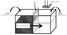
本開示は、光源と;光検出器と;少なくとも1つの還元剤及び少なくとも1つの代謝酵素またはその機能的断片を含む、少なくとも1つの反応表面とを含む、バイオセンサーであって;該反応表面が、導電性支持体を含まず、導電性支持体に付着されておらず;該光源が、該反応表面を照射するのに十分な該反応表面からの距離で配置され、該光検出器が、該反応表面からの照射光を収集するのに十分な該反応表面からの距離で配置される、該バイオセンサーに関する。いくつかの実施形態において、反応表面は、光検出器をコントローラーへ接続する少なくとも1つの回路を更に含む。いくつかの実施形態において、光検出器は、光加速器、コントローラー及びディスプレイへ作動可能に接続された光ダイオードであり、そして、アミノ酸基質を含むある量のサンプルへの反応表面の曝露に際して、コントローラーは光ダイオードからの電流を受け取り、電流をサンプル中のアミノ酸の濃度値に対応する電気信号へ相関させ、かかる電気信号はディスプレイへ伝導され、ディスプレイによって表示される。いくつかの実施形態において、反応表面は、ロイシン脱水素酵素、チロシン脱水素酵素、フェニルアラニン脱水素酵素、ロイシンオキシドレダクターゼ、チロシンモノオキシゲナーゼ、アラニン脱水素酵素若しくはグルタミン酸脱水素酵素;またはその機能的断片から選択されるアミノ酸のうちの少なくとも1つまたはその組み合わせを含む。いくつかの実施形態において、反応表面は、少なくとも1つの代謝酵素またはその機能的断片を含む濾紙である。いくつかの実施形態において、少なくとも1つの代謝酵素またはその機能的断片は、反応表面の上に凍結乾燥される。 The present disclosure is a biosensor comprising a light source; a photodetector; and at least one reaction surface comprising at least one reducing agent and at least one metabolic enzyme or functional fragment thereof; the reaction surface is The light source is located at a distance from the reaction surface sufficient to illuminate the reaction surface, and the photodetector is located. With respect to the biosensor located at a distance from the reaction surface sufficient to collect the irradiation light from the reaction surface. In some embodiments, the reaction surface further comprises at least one circuit connecting the photodetector to the controller. In some embodiments, the photodetector is a photodiode operably connected to a photoaccelerator, controller and display, and upon exposure of the reaction surface to a certain amount of sample containing an amino acid substrate, the controller. It receives the current from the light diode and correlates the current with the electrical signal corresponding to the concentration value of the amino acid in the sample, which is conducted to the display and displayed by the display. In some embodiments, the reaction surface is from leucine dehydrogenase, tyrosine dehydrogenase, phenylalanine dehydrogenase, leucine oxide reductase, tyrosine monooxygenase, alanine dehydrogenase or glutamate dehydrogenase; or functional fragments thereof. Includes at least one of the selected amino acids or a combination thereof. In some embodiments, the reaction surface is a filter paper containing at least one metabolic enzyme or functional fragment thereof. In some embodiments, the at least one metabolic enzyme or functional fragment thereof is lyophilized onto the reaction surface.
いくつかの実施形態において、バイオセンサーは、濾紙に隣接し、濾紙と流体的に連通されている少なくとも第1の流体開口部を含有している筺体を更に含み;濾紙は、反応表面を含むマイクロ流体チャンバーに直に隣接する。いくつかの実施形態において、反応表面は、以下の:(i)ウリカーゼまたはその機能的断片;(ii)デキストランまたはその誘導体を含むハイドロゲル;(iii)細菌細胞;(iv)電気泳動のために構成された電子双極子;(v)3,4-DHB、のうちの1つまたは複数を不含有である。いくつかの実施形態において、バイオセンサーは、反応表面へ機械的に連結された遠心運動が可能な遠心分離機またはローターを不含有である。いくつかの実施形態において、バイオセンサーは摂氏4度での保管において約30日後に少なくとも70%の生物学的活性がある。いくつかの実施形態において、反応表面は約10μL~約100μL以下の流体を保持する。 In some embodiments, the biosensor further comprises a housing that is adjacent to the filter paper and contains at least a first fluid opening that is fluidly communicated with the filter paper; the filter paper is a micro that includes a reaction surface. Directly adjacent to the fluid chamber. In some embodiments, the reaction surface is as follows: (i) uricase or a functional fragment thereof; (ii) a hydrogel containing dextran or a derivative thereof; (iii) bacterial cells; (iv) for electrophoresis. Consists of one or more of the constructed electron dipoles; (v) 3,4-DHB. In some embodiments, the biosensor is free of centrifuges or rotors capable of centrifugal movement mechanically coupled to the reaction surface. In some embodiments, the biosensor has at least 70% biological activity after about 30 days of storage at 4 degrees Celsius. In some embodiments, the reaction surface retains a fluid of about 10 μL to about 100 μL or less.
いくつかの実施形態において、少なくとも1つの酵素またはその機能的断片は細菌種に由来し、反応表面上で固定化される。いくつかの実施形態において、少なくとも1つの酵素またはその機能的断片は好熱性細菌種に由来し、ハイドロゲル中に固定化される。いくつかの実施形態において、少なくとも1つの酵素またはその機能的断片は、配列番号:1または配列番号:2へ少なくとも約70%の配列同一性を含む。いくつかの実施形態において、少なくとも1つの酵素は、好熱性細菌細胞から得られたフェニルアラニン脱水素酵素またはその機能的断片である。 In some embodiments, the at least one enzyme or functional fragment thereof is derived from a bacterial species and is immobilized on the reaction surface. In some embodiments, the at least one enzyme or functional fragment thereof is derived from a thermophilic bacterial species and is immobilized in a hydrogel. In some embodiments, the at least one enzyme or functional fragment thereof comprises at least about 70% sequence identity to SEQ ID NO: 1 or SEQ ID NO: 2. In some embodiments, the at least one enzyme is phenylalanine dehydrogenase or a functional fragment thereof obtained from thermophilic bacterial cells.
いくつかの実施形態において、アルギネートは、式

を備えたブロックポリマーを含む。
In some embodiments, the alginate is a formula.
Includes block polymers with.
いくつかの実施形態において、反応表面は、20μL以下の体積を備えたマイクロ流体チャンバーの少なくとも1つの側面を形成する。いくつかの実施形態において、少なくとも1つの電子伝達体は、チオニン、o-フェニレンジアミン、メチレンブルー及びトルイジンブルーから選択される。いくつかの実施形態において、少なくとも1つの還元剤は、NAD+またはFAD+から選択される。いくつかの実施形態において、反応表面は、少なくとも1つの凍結乾燥された代謝酵素またはその機能的断片及び約100mM~約400mMの濃度での糖の混合物を含む濾紙からなる。 In some embodiments, the reaction surface forms at least one side of a microfluidic chamber with a volume of 20 μL or less. In some embodiments, the at least one electron carrier is selected from thionin, o-phenylenediamine, methylene blue and toluidine blue. In some embodiments, the at least one reducing agent is selected from NAD + or FAD +. In some embodiments, the reaction surface consists of a filter paper containing at least one lyophilized metabolic enzyme or functional fragment thereof and a mixture of sugars at a concentration of about 100 mM to about 400 mM.
本開示は、少なくとも1つの電子伝達体、少なくとも1つの還元剤、及び少なくとも1つの代謝酵素またはその機能的断片を含む、少なくとも1つの反応表面を含む、バイオセンサーであって;該少なくとも1つの酵素またはその機能的断片が、Geobacillus thermoglucosidiasusからのフェニルアラニン脱水素酵素に少なくとも70%相同であり;該反応表面が、導電性支持体を含まず、導電性支持体に付着されない、該バイオセンサーにも関する。いくつかの実施形態において、バイオセンサーは電流計を不含有である。 The present disclosure is a biosensor comprising at least one reaction surface comprising at least one electron carrier, at least one reducing agent, and at least one metabolic enzyme or functional fragment thereof; said at least one enzyme. Alternatively, the functional fragment thereof is at least 70% homologous to phenylalanine dehydrogenase from Geobacillus thermoglucosidias; also the biosensor whose reaction surface does not contain a conductive support and does not adhere to the conductive support. .. In some embodiments, the biosensor is ammeter-free.
いくつかの実施形態において、酵素またはその機能的断片は、配列番号:1に少なくとも70%相同または配列番号:1の機能的断片に少なくとも70%相同である。いくつかの実施形態において、複数の酵素またはその機能的断片のうちの1つは細菌細胞に由来する。 In some embodiments, the enzyme or functional fragment thereof is at least 70% homologous to SEQ ID NO: 1 or at least 70% homologous to the functional fragment of SEQ ID NO: 1. In some embodiments, one of a plurality of enzymes or functional fragments thereof is derived from bacterial cells.
いくつかの実施形態において、少なくとも1つの酵素またはその機能的断片は、Geobacillus thermoglucosidiasusからのフェニルアラニン脱水素酵素に少なくとも70%相同であり;反応表面は、濾紙を含み、導電性支持体を不含有であり、導電性支持体に付着されておらず;光源は、反応表面を照射するのに十分な反応表面からの距離で配置され、光検出器は、反応表面からの照射光を収集するのに十分な反応表面からの距離で配置される。 In some embodiments, the at least one enzyme or functional fragment thereof is at least 70% homologous to phenylalanine dehydrogenase from Geobacillus thermoglucosidias; the reaction surface contains filter paper and is free of conductive supports. Yes, not attached to the conductive support; the light source is located at a distance from the reaction surface sufficient to illuminate the reaction surface, and the photodetector is to collect the irradiation light from the reaction surface. Placed at a sufficient distance from the reaction surface.
本開示は、少なくとも1つのコンピューターストレージメモリへ作動可能に接続される本明細書において開示されるバイオセンサーを含むシステムにも関する。いくつかの実施形態において、システムは、体液のサンプルを更に含む。いくつかの実施形態において、システムは、電流及び/または電圧の差の測定に対応する電気信号を電圧計及び/または電流計からデジタルディスプレイへ運搬することができる電気的回路によって、少なくとも1つの導電性支持体へ作動可能に接続されるデジタルディスプレイを更に含み、デジタルディスプレイは、少なくとも1つの導電性支持体が少なくとも1つの代謝酵素のために十分な時間的な期間でサンプルに接してそのアミノ酸基質の酸化を触媒する場合に、サンプル中のアミノ酸の濃度値を表示するように構成される。いくつかの実施形態において、システムは、少なくとも1つのコンピューターストレージメモリと作動可能に接続されるコンピュータープロセッサーを更に含む。いくつかの実施形態において、代謝酵素は、1つまたは複数の糖分子と共に反応表面へ固定化されたフェニルアラニン脱水素酵素である。 The present disclosure also relates to a system comprising a biosensor disclosed herein operably connected to at least one computer storage memory. In some embodiments, the system further comprises a sample of body fluid. In some embodiments, the system has at least one conductivity by means of an electrical circuit capable of carrying an electrical signal corresponding to the measurement of current and / or voltage difference from the voltmeter and / or ammeter to the digital display. Further including a digital display operably connected to the sex support, the digital display is an amino acid substrate in which at least one conductive support is in contact with the sample for a period of time sufficient for at least one metabolic enzyme. It is configured to display the concentration value of the amino acid in the sample when catalyzing the oxidation of. In some embodiments, the system further comprises at least one computer storage memory and a computer processor operably connected. In some embodiments, the metabolic enzyme is a phenylalanine dehydrogenase immobilized on the reaction surface with one or more sugar molecules.
本開示は、本明細書において開示されるバイオセンサーを含むキットにも関する。いくつかの実施形態において、反応表面は、少なくとも1つの流体入口及び1セットの説明書(電子媒体を介して随意に遠隔でアクセス可能)を有する除去可能な筺体内に含有される、試験ストリップまたは濾紙を構成する。 The present disclosure also relates to a kit comprising the biosensor disclosed herein. In some embodiments, the reaction surface is contained in a removable enclosure with at least one fluid inlet and a set of instructions (optionally remotely accessible via electronic media), a test strip or Consists of filter paper.
本開示は、体液のサンプルを、本明細書において開示されるバイオセンサー若しくは本明細書において開示されるシステム;または本明細書において開示される任意の試験ストリップへ接触させることと;サンプル中のアミノ酸の量を決定することとを含む、体液のサンプル中のアミノ酸の濃度を決定または同定する方法にも関する。いくつかの実施形態において、体液のサンプルは全血を含む。 The present disclosure comprises contacting a sample of body fluid with a biosensor disclosed herein or a system disclosed herein; or any test strip disclosed herein; amino acids in the sample. It also relates to methods of determining or identifying the concentration of amino acids in body fluid samples, including determining the amount of. In some embodiments, the body fluid sample comprises whole blood.
本開示は、体液のサンプルを、本明細書において開示されるバイオセンサー若しくは本明細書において開示されるシステム;または本明細書において開示される任意の試験ストリップへ接触させることを含む、被験体の体液のサンプル中の1つまたは複数アミノ酸の濃度を定量する方法にも関する。いくつかの実施形態において、方法は、定量する工程または同定する工程によって得られる濃度値を、1つまたは複数の代謝疾患と関連する閾値へ比較することを更に含む。いくつかの実施形態において、接触させる工程は、被験体の体液のサンプルを、本明細書において開示されるバイオセンサー若しくは本明細書において開示されるシステム;または本明細書において開示される任意の試験ストリップへ、代謝酵素またはその機能的断片による体液のサンプル中の少なくとも1つのアミノ酸の酸化を可能にする十分な期間で、曝露することを含む。いくつかの実施形態において、方法は、反応表面から放射される光の検出の前に、体液のサンプルを光源から放射される光へ曝露することを含む。いくつかの実施形態において、体液のサンプルは被験体からの全血または血清を含有する。いくつかの実施形態において、体液のサンプルは尿を不含有である。 The present disclosure comprises contacting a sample of body fluid with a biosensor disclosed herein or a system disclosed herein; or any test strip disclosed herein. It also relates to a method of quantifying the concentration of one or more amino acids in a sample of body fluid. In some embodiments, the method further comprises comparing the concentration values obtained by the quantifying or identifying steps to thresholds associated with one or more metabolic disorders. In some embodiments, the step of contacting is a sample of a subject's body fluid, a biosensor disclosed herein or a system disclosed herein; or any test disclosed herein. The strip comprises exposure to the strip for a sufficient period of time to allow oxidation of at least one amino acid in a sample of body fluid by a metabolic enzyme or functional fragment thereof. In some embodiments, the method comprises exposing a sample of body fluid to light emitted from a light source prior to detection of light emitted from the reaction surface. In some embodiments, the body fluid sample contains whole blood or serum from the subject. In some embodiments, fluid samples are urine-free.
本開示は、(a)体液のサンプルを、本明細書において開示されるバイオセンサー若しくは本明細書において開示されるシステム;または本明細書において開示される任意の試験ストリップへ接触させることと;(b)サンプル中のアミノ酸の1つまたは複数の濃度値を定量することと;(c)サンプル中のアミノ酸の1つまたは複数の濃度値を、健康な範囲中であるとして同定されたアミノ酸濃度の閾値へ比較することと;(d)サンプル中のアミノ酸の1つまたは複数の濃度値が、閾値を上回るかまたはそれ未満で収まるならば、代謝疾患を有するとして被験体を同定することとを含む、被験体における代謝疾患を診断する方法にも関する。いくつかの実施形態において、代謝疾患は、フェニルケトン尿症、高アンモニア血症及びメープルシロップ尿症から選択される、少なくとも1つまたはその組み合わせである。 The present disclosure comprises (a) contacting a sample of body fluid to a biosensor disclosed herein or a system disclosed herein; or any test strip disclosed herein; ( b) Quantifying the concentration of one or more amino acids in the sample; (c) the concentration of one or more amino acids in the sample of the amino acid concentration identified as being in the healthy range. Comparing to a threshold; (d) identifying a subject as having a metabolic disorder if the concentration of one or more amino acids in the sample falls above or below the threshold. Also related to how to diagnose metabolic disorders in a subject. In some embodiments, the metabolic disorder is at least one or a combination thereof selected from phenylketonuria, hyperammonemia and maple syrup urine.
本開示は、(a)体液のサンプルを、本明細書において開示されるバイオセンサー若しくは本明細書において開示されるシステム;または本明細書において開示される任意の試験ストリップへ接触させることと;(b)1つまたは複数のアミノ酸濃度値を定量することと;(c)1つまたは複数の濃度値を、代謝疾患と関連する1つまたは複数の閾値へ比較することとを含む、治療法に対する患者応答性を決定する方法にも関する。 The present disclosure comprises (a) contacting a sample of body fluid to a biosensor disclosed herein or a system disclosed herein; or any test strip disclosed herein; ( b) For treatments comprising quantifying one or more amino acid concentration values; (c) comparing one or more concentration values to one or more thresholds associated with metabolic disorders. It also concerns how to determine patient responsiveness.
本開示は、少なくとも1つの反応表面を含む試験ストリップであって、該反応表面が、少なくとも1つの電子伝達体、少なくとも1つの還元剤、少なくとも1つの代謝酵素またはその機能的断片、及びアルギネートを含み;該反応表面が、電極を含まず、電極に付着されない、該試験ストリップにも関する。いくつかの実施形態において、試験ストリップは、電圧計及び/または電流計ならびにデジタルディスプレイを含むポータブルデバイスに適合し、そして、試験ストリップがデバイスへ接触される場合、第1及び第2の電極は、電圧計及び/または電流計ならびにデジタルディスプレイを含む閉鎖された電気的回路へ作動可能に接続されるようになり、体液のサンプルとの接触に際して、少なくとも1つの代謝酵素またはその機能的断片はアミノ酸の酸化を触媒し、体液のサンプル中のアミノ酸の濃度値に対応する第1の電極上の電流をもたらし、かかる濃度値はポータブルデバイスのディスプレイ上で読み取り可能である。 The present disclosure is a test strip comprising at least one reaction surface, wherein the reaction surface comprises at least one electron carrier, at least one reducing agent, at least one metabolic enzyme or functional fragment thereof, and alginate. Also relevant to the test strip, where the reaction surface does not contain electrodes and does not adhere to the electrodes. In some embodiments, the test strip is compatible with portable devices including voltmeters and / or ammeters as well as digital displays, and if the test strip is in contact with the device, the first and second electrodes will be. It is now operably connected to closed electrical circuits, including voltmeters and / or ammeters and digital displays, and upon contact with a sample of body fluid, at least one metabolic enzyme or functional fragment thereof is of amino acid. It catalyzes oxidation and results in a current on the first electrode that corresponds to the concentration value of the amino acid in the body fluid sample, which concentration value is readable on the display of the portable device.
本開示は、本明細書において開示されるバイオセンサー若しくは本明細書において開示されるシステム;または本明細書において開示される任意の試験ストリップを製造する方法であって、1つまたは複数の代謝酵素またはその機能的断片を反応表面へ固定化することと;筺体中に該反応表面を包むこととを含む、該方法にも関する。いくつかの実施形態において、方法は、塩を不含有または実質的に不含有であるバッファー中で糖が約100~約300mMの濃度である、糖の混合物中で1つまたは複数の代謝酵素を固定化することを含む。 The present disclosure is a method of making a biosensor disclosed herein or a system disclosed herein; or any test strip disclosed herein, one or more metabolic enzymes. Alternatively, it also relates to the method comprising immobilizing the functional fragment thereof on the reaction surface; wrapping the reaction surface in a housing. In some embodiments, the method comprises one or more metabolic enzymes in a mixture of sugars in which the sugar is at a concentration of about 100-about 300 mM in a salt-free or substantially salt-free buffer. Includes immobilization.
(発明の詳細な説明)
本方法及び本開示の他の態様に関する様々な用語は、明細書及び請求項を通して使用される。かかる用語は、特別の指示のない限り、当該技術分野におけるそれらの通常の意味が与えられる。他の具体的に定義された用語は、本明細書において提供された定義と一致した様式で解釈されるべきである。
(Detailed description of the invention)
Various terms relating to this method and other aspects of the present disclosure are used throughout the specification and claims. Such terms are given their usual meaning in the art, unless otherwise specified. Other specifically defined terms should be construed in a manner consistent with the definitions provided herein.
本明細書及び添付の請求項において使用されるように、単数形「a」、「an」及び「the」には、特別に内容が明確に指示しない限り複数の指示物が含まれる。 As used herein and in the accompanying claims, the singular forms "a", "an" and "the" include a plurality of referents unless otherwise explicitly stated.
「約」という用語は、本明細書において使用される時、測定可能な値(量、時間的な継続期間、及び同種のもの等)を指す場合、かかる変動が開示される方法の遂行に適切であるような、指定された値からの±20%、±10%、±5%、±1%、または±0.1%の変動を包含することが意味される。 When the term "about" as used herein refers to a measurable value (quantity, temporal duration, and the like, etc.), it is appropriate to perform the method in which such variation is disclosed. It is meant to include ± 20%, ± 10%, ± 5%, ± 1%, or ± 0.1% variation from the specified value, such as.
「アドレス可能位置」という用語は、本明細書において使用される時、信号(反応表面で遂行される、本明細書において開示される反応から産生される反応産物から放射された波長等)が得られる試験ストリップ上またはバイオセンサー上の不連続の表面領域または位置が、固定化または吸収される1つまたは複数の接着セットであり、そして、生体材料または細胞を含むサンプルへ接着セットを1つまたは複数で十分な時間的な期間で曝露することが、細胞または生体材料と接着セットとの間の接触をもたらし得ること、を意味する。いくつかの実施形態において、本開示は、4平方ミクロン以下の表面を備えたバイオセンサーのアドレス可能位置の1つまたは複数を含むアレイに関する。本明細書において使用される時、「付着する」、「付着」、「接着する」、「接着された」、「接着性の」という用語、または同種の用語は、一般的には物理的吸収、化学結合及び類似のプロセス、またはその組み合わせ等によって、例えば基、化合物または接着のセットを表面へ固定化または固定することを指す。 The term "addressable location", when used herein, obtains a signal (such as a wavelength emitted from a reaction product produced from a reaction disclosed herein, performed on the reaction surface). Discontinuous surface areas or locations on the test strips or biosensors that are immobilized or absorbed are one or more adhesive sets, and one or more adhesive sets to samples containing biomaterials or cells. It means that multiple exposures for a sufficient time period can result in contact between the cell or biomaterial and the adhesive set. In some embodiments, the present disclosure relates to an array comprising one or more addressable positions of a biosensor with a surface of 4 square microns or less. As used herein, the terms "adhesive," "adhesive," "adhesive," "adhesive," "adhesive," or similar terms are generally physically absorbent. , Chemical bonding and similar processes, or combinations thereof, etc., to immobilize or immobilize, for example, a set of groups, compounds or bonds to a surface.
本明細書において使用される時、「電子媒体」という用語は、ハードディスク、ROM、EEPROM、RAM、フラッシュメモリ、不揮発性メモリまたは任意の実質的及び機能的に等価な媒体が含まれる、アクセスのためのエレクトロニクス技術を用いる任意の物理ストレージを意味する。いくつかの実施形態において、ソフトウェアストレージは本開示の実施形態を実施するプロセッサーと同位置であり得るか、またはソフトウェアストレージの少なくとも一部は遠隔に位置され得るが、必要な場合アクセス可能であり得る。 As used herein, the term "electronic medium" includes hard disks, ROMs, EEPROMs, RAMs, flash memories, non-volatile memories or any substantially and functionally equivalent medium for access. Means any physical storage that uses the electronics technology of. In some embodiments, the software storage may be co-located with the processor performing the embodiments of the present disclosure, or at least a portion of the software storage may be remotely located but accessible if necessary. ..
本明細書において使用される時、「配列同一性」は、2つの配列のBLASTによる比較のためのスタンドアロン型の実行可能なBLASTエンジンプログラム(bl2seq)の使用によって決定され、それはデフォルトパラメーターを使用して、National Center for Biotechnology Information (NCBI)ftpサイトからリトリーブすることができる(Tatusova and Madden, FEMS Microbiol Lett., 1999, 174, 247-250;その全体は参照により本明細書に援用される)。 As used herein, "sequence identity" is determined by the use of a stand-alone, executable BLAST engine program (bl2seq) for comparison by BLAST of two sequences, which uses default parameters. And can be retrieved from the National Center for Biotechnology Information (NCBI) FTP site (see Tatsusova and Madden, FEMS Microbiol Lett., 1999, 174, 247-250; in its entirety.
本明細書において使用される時、「生検」という用語は、分析のために被験体または患者から取り出された細胞サンプル、細胞の収集物または体液を意味する。いくつかの実施形態において、生検は、骨髄生検、パンチ生検、内視鏡生検、針生検、薄片生検、切開生検、切除生検または外科的切除である。 As used herein, the term "biopsy" means a cell sample, cell collection or body fluid taken from a subject or patient for analysis. In some embodiments, the biopsy is bone marrow biopsy, punch biopsy, endoscopic biopsy, needle biopsy, flaky biopsy, incision biopsy, excisional biopsy or surgical resection.
本明細書において使用される時、「体液」という用語は、血液サンプル、プロセシングされていない全血サンプル、血清サンプル、尿サンプル、粘液サンプル、唾液サンプル及び汗サンプルが含まれるが必ずしもこれらに限定されない、被験体から単離された任意の液体を意味する。サンプルは、静脈内穿刺、生検、スワブ、毛細管吸引、ランセット、針吸引、排泄液体の単純な捕捉による収集等の任意の手段によって被験体から得ることができる。 As used herein, the term "body fluid" includes, but is not limited to, blood samples, unprocessed whole blood samples, serum samples, urine samples, mucus samples, saliva samples and sweat samples. , Means any liquid isolated from the subject. Samples can be obtained from the subject by any means such as intravenous puncture, biopsy, swab, capillary aspiration, lancet, needle aspiration, collection by simple capture of excreted fluid.
本明細書において使用される時、「電子媒体」という用語は、ハードディスク、ROM、EEPROM、RAM、フラッシュメモリ、不揮発性メモリまたは任意の実質的及び機能的に等価な媒体が含まれる、アクセスのためのエレクトロニクス技術を用いる任意の物理ストレージを意味する。いくつかの実施形態において、ソフトウェアストレージは本開示の実施形態を実施するプロセッサーと同位置であり得るか、またはソフトウェアストレージの少なくとも一部は遠隔に位置され得るが、必要な場合アクセス可能であり得る。 As used herein, the term "electronic medium" includes hard disks, ROMs, EEPROMs, RAMs, flash memories, non-volatile memories or any substantially and functionally equivalent medium for access. Means any physical storage that uses the electronics technology of. In some embodiments, the software storage may be co-located with the processor performing the embodiments of the present disclosure, or at least a portion of the software storage may be remotely located but accessible if necessary. ..
本明細書において使用される時、「アミノ酸代謝異常」という用語は、被験体の体中のアミノ酸の過剰産生または低産生をもたらす、アミノ酸の代謝の触媒作用の機能不全によって特徴づけられる疾患及び障害を指すことを意味する。アミノ酸代謝異常の例は代謝疾患の定義中でリストされ、用語は本出願中で互換的に使用される。 As used herein, the term "amino acid metabolism disorders" refers to diseases and disorders characterized by dysfunction of catalytic activity of amino acid metabolism, resulting in overproduction or underproduction of amino acids throughout the body of a subject. Means to point to. Examples of amino acid metabolic disorders are listed in the definition of metabolic disorders and the terms are used interchangeably in this application.
「被験体」という用語を明細書を通して使用して、体液のサンプルが採取される動物を記載する。いくつかの実施形態において、動物はヒトである。特異的な被験体(ヒト等)のための特異的な病態の診断のために、「患者」という用語を互換的に使用することができる。本開示の記載中のいくつかの事例において、「患者」という用語は、特定の疾患または障害に罹患するヒト患者を指すだろう。いくつかの実施形態において、被験体は、アミノ酸代謝異常を発症するリスクを有する疑いがあるか、またはそのリスクがあると同定されているヒトであり得る。いくつかの実施形態において、被験体は少なくとも1つのアミノ酸代謝異常を有すると診断され得る。いくつかの実施形態において、被験体は、フェニルケトン尿症を有する疑いがあるか、またはそうであると診断されている。いくつかの実施形態において、被験体は、アミノ酸代謝異常を発症するリスクを有する疑いがあるか、またはそのリスクがあると同定されているヒトであり得る。いくつかの実施形態において、被験体は、体液の単離サンプルの源として機能する哺乳類であり得る。いくつかの実施形態において、被験体は、体液のサンプルが単離または提供される非ヒト動物であり得る。「哺乳類」という用語は、ヒト及び非ヒトの両方を包含し、ヒト、非ヒト霊長類、イヌ科の動物、ネコ科の動物、ネズミ科の動物、ウシ科の動物、ウマ科の動物及びブタ類が含まれるが、これらに限定されない。 The term "subject" is used throughout the specification to describe an animal from which a sample of body fluid is taken. In some embodiments, the animal is a human. The term "patient" can be used interchangeably for the diagnosis of a specific pathology for a specific subject (such as a human). In some of the cases described in this disclosure, the term "patient" will refer to a human patient suffering from a particular disease or disorder. In some embodiments, the subject can be a human who is suspected of having or has been identified as having a risk of developing an amino acid metabolism disorder. In some embodiments, the subject can be diagnosed with at least one amino acid metabolism disorder. In some embodiments, the subject is suspected of having or has been diagnosed with phenylketonuria. In some embodiments, the subject can be a human who is suspected of having or has been identified as having a risk of developing an amino acid metabolism disorder. In some embodiments, the subject can be a mammal that serves as a source of an isolated sample of body fluid. In some embodiments, the subject can be a non-human animal from which a sample of body fluid is isolated or provided. The term "mammal" includes both humans and non-humans, including humans, non-human primates, dogs, cats, rodents, bovines, horses and pigs. Includes, but is not limited to.
本明細書において使用される時、「保存的」アミノ酸置換は、表A、B、またはC中で以下に述べられるように定義され得る。代謝酵素には、本開示のポリペプチドをコードするポリヌクレオチドの修飾によって保存的置換が導入されたアミノ酸配列が含まれる。アミノ酸は、物理的特性ならびにタンパク質の二次構造及び三次構造への寄与に従って分類することができる。保存的置換は、類似の特性を有する別のアミノ酸についての1つアミノ酸の置換として当技術分野において認識される。例示的な保存的置換は、表A中で述べられる。
表A--保存的置換I
側鎖の特徴 アミノ酸
脂肪族
非極性 G A P I L V F
極性-非荷電性 C S T M N Q
極性-荷電性 D E K R
芳香族 H F W Y
他 N Q D E
As used herein, "conservative" amino acid substitutions can be defined in Tables A, B, or C as described below. Metabolizing enzymes include amino acid sequences into which conservative substitutions have been introduced by modification of the polynucleotides encoding the polypeptides of the present disclosure. Amino acids can be classified according to their physical properties and their contribution to the secondary and tertiary structure of the protein. Conservative substitutions are recognized in the art as substitutions of one amino acid for another amino acid with similar properties. Exemplary conservative substitutions are described in Table A.
Table A--Conservative Permutation I
Side chain characteristics Amino acids Aliphatic non-polar GAP I LV F
Polarity-Uncharged CST M N Q
Polarity-chargeability DE KR
Aromatic HFWY
Other N Q DE
あるいは、Lehninger(Biochemistry、第2版; Worth Publishers, Inc. NY、N.Y.(1975)、71~77ページ)中で記載されるように、保存的アミノ酸をグループ化することができ、表B中で説明される通りである。
表B--保存的置換II
側鎖の特徴 アミノ酸
非極性(疎水性)
脂肪族: A L I V P
芳香族: F W Y
含硫: M
境界: G Y
非荷電性-極性
水酸基: S T Y
アミド: N Q
スルフヒドリル:C
境界: G Y
正に荷電(塩基性):K R H
負に荷電(酸性): D E
Alternatively, conservative amino acids can be grouped as described in Lehninger (Biochemistry, 2nd Edition; Worth Publishers, Inc. NY, NY (1975), pp. 71-77), in a table. As explained in B.
Table B--Conservative Permutation II
Side chain characteristics Amino acid non-polar (hydrophobic)
Aliphatic: ALI VP
Aromatic: FW Y
Sulfur-containing: M
Boundary: G Y
Non-chargeable-polar hydroxyl group: STY
Amide: N Q
Sulf Hydrill: C
Boundary: G Y
Positively charged (basic): KRH
Negatively charged (acidic): DE
あるいは、例示的な保存的置換は、表C中で述べられる。
表C--保存的置換III
もとの残基 例示的な置換
Ala(A) Val Leu Ile Met
Arg(R) Lys His
Asn(N) Gln
Asp(D) Glu
Cys(C) Ser Thr
Gln(Q) Asn
Glu(E) Asp
Gly(G) Ala Val Leu Pro
His(H) Lys Arg
Ile(I) Leu Val Met Ala Phe
Leu(L) Ile Val Met Ala Phe
Lys(K) Arg His
Met(M) Leu Ile Val Ala
Phe(F) Trp Tyr Ile
Pro(P) Gly Ala Val Leu Ile
Ser(S) Thr
Thr(T) Ser
Trp(W) Tyr Phe Ile
Tyr(Y) Trp Phe Thr Ser
Val(V) Ile Leu Met Ala
Alternatively, exemplary conservative substitutions are described in Table C.
Table C--Conservative Permutation III
Original Residue Illustrative Substitution Ala (A) Val Leu Ile Met
Arg (R) Lys His
Asn (N) Gln
Asp (D) Glu
Cys (C) Ser Thr
Gln (Q) Asn
Glu (E) Asp
Gly (G) Ala Val Leu Pro
His (H) Lys Arg
Ile (I) Leu Val Met Ala Phe
Leu (L) Ile Val Met Ala Phe
Lys (K) Arg His
Met (M) Leu Ile Val Ala
Ph (F) Trp Tyr Ile
Pro (P) Gly Ala Val Leu Ile
Ser (S) Thr
Thr (T) Ser
Trp (W) Tyr Phe Ile
Tyr (Y) Trp Phe Thr Ser
Val (V) Ile Leu Met Ala
本明細書において記載される細胞外マトリックスと関連するポリペプチド配列を含むポリペプチドは、アミノ酸残基の1つまたは複数の挿入、欠失若しくは置換またはその任意の組み合わせに加えて、アミノ酸残基の挿入、欠失または置換以外の修飾を有するポリペプチドを含むことが意図されることを理解すべきである。 A polypeptide comprising a polypeptide sequence associated with the extracellular matrix described herein is an amino acid residue in addition to one or more insertions, deletions or substitutions of amino acid residues or any combination thereof. It should be understood that it is intended to include polypeptides with modifications other than insertions, deletions or substitutions.
本明細書において使用される時、「予後」という用語は、疾患の起こりそうな経過及び転帰を決定することを意味する。 As used herein, the term "prognosis" means determining the likely course and outcome of a disease.
本明細書において使用される時、「機能的断片」という用語は、断片が基づく野生型ポリペプチド機能に類似するかまたは実質的に類似する少なくとも部分的な生物学的機能を保持するのに十分な長さであるポリペプチドの任意の一部分を意味する。いくつかの実施形態において、代謝酵素の機能と関連するポリペプチドの機能的断片は、表3中で開示される任意のポリペプチドの少なくとも70%、75%、80、85、90、95、96、97、98、または99%の配列同一性を含み、ポリペプチドへ結合する1つまたは複数のリガンドへの少なくとも部分的な結合親和性を保持するのに十分な長さを有するポリペプチドである。いくつかの実施形態において、断片は、表3中で開示される任意のポリペプチドの断片であり、少なくとも約10、約20、約30、約40、約50、約60、約70、約80、約90または約100の連続したアミノ酸の長さを有する。いくつかの実施形態において、断片は表3中で開示した任意のポリペプチドの断片であり、少なくとも約50のアミノ酸の長さを有する。いくつかの実施形態において、断片は表3中で開示した任意のポリペプチドの断片であり、少なくとも約100のアミノ酸の長さを有する。いくつかの実施形態において、断片は表3中で開示した任意のポリペプチドの断片であり、少なくとも約150のアミノ酸の長さを有する。いくつかの実施形態において、断片は表3中で開示した任意のポリペプチドの断片であり、少なくとも約200のアミノ酸の長さを有する。いくつかの実施形態において、断片は表3中で開示した任意のポリペプチドの断片であり、少なくとも約250のアミノ酸の長さを有する。 As used herein, the term "functional fragment" is sufficient to retain at least a partial biological function that is similar to or substantially similar to the wild-type polypeptide function on which the fragment is based. Means any part of a polypeptide of length. In some embodiments, functional fragments of the polypeptide associated with the function of the metabolic enzyme are at least 70%, 75%, 80, 85, 90, 95, 96 of any of the polypeptides disclosed in Table 3. , 97, 98, or 99% sequence identity and long enough to retain at least a partial binding affinity for one or more ligands that bind to the polypeptide. .. In some embodiments, the fragment is a fragment of any polypeptide disclosed in Table 3, at least about 10, about 20, about 30, about 40, about 50, about 60, about 70, about 80. , Has a length of about 90 or about 100 consecutive amino acids. In some embodiments, the fragment is a fragment of any of the polypeptides disclosed in Table 3 and has a length of at least about 50 amino acids. In some embodiments, the fragment is a fragment of any of the polypeptides disclosed in Table 3 and has a length of at least about 100 amino acids. In some embodiments, the fragment is a fragment of any of the polypeptides disclosed in Table 3 and has a length of at least about 150 amino acids. In some embodiments, the fragment is a fragment of any of the polypeptides disclosed in Table 3 and has a length of at least about 200 amino acids. In some embodiments, the fragment is a fragment of any of the polypeptides disclosed in Table 3 and has a length of at least about 250 amino acids.
本明細書において使用される時、「代謝酵素と関連するポリペプチド配列」という用語は、任意のマクロ分子(糖分子またはマクロ分子等)によって修飾されるかまたは修飾されず、任意の多細胞生物中の細胞によって天然に産生され、本明細書において開示されるような代謝酵素またはその機能的断片である、任意のポリペプチドまたは断片を意味する。いくつかの実施形態において、細胞外マトリックスと関連するポリペプチド配列は、配列が表3中で開示されるポリペプチドのうちの任意のものを含む任意のポリペプチドである。いくつかの実施形態において、代謝酵素と関連するポリペプチド配列は、表3中で開示されるポリペプチドのうちの任意のものを含む任意のポリペプチド配列、または表3中で開示されるポリペプチドと85、90、95、96、97、98若しくは99%の配列同一性を共有する配列若しくはその機能的断片である。いくつかの実施形態において、代謝酵素と関連するポリペプチド配列は、表3中で開示されるポリペプチド、または表3中で開示されるポリペプチドと85、90、95、96、97、98または99%の配列同一性を共有する配列のうちの任意ものからなる。 As used herein, the term "polypeptide sequence associated with a metabolic enzyme" is any multicellular organism that is modified or unmodified by any macromolecule (such as a sugar molecule or macromolecule). Means any polypeptide or fragment that is naturally produced by the cells in it and is a metabolic enzyme or functional fragment thereof as disclosed herein. In some embodiments, the polypeptide sequence associated with the extracellular matrix is any polypeptide, including any of the polypeptides whose sequence is disclosed in Table 3. In some embodiments, the polypeptide sequence associated with the metabolic enzyme is any polypeptide sequence, including any of the polypeptides disclosed in Table 3, or the polypeptides disclosed in Table 3. And 85, 90, 95, 96, 97, 98 or 99% sequences that share sequence identity or functional fragments thereof. In some embodiments, the polypeptide sequence associated with the metabolic enzyme is 85, 90, 95, 96, 97, 98 or 85, 90, 95, 96, 97, 98 or the polypeptide disclosed in Table 3 or the polypeptide disclosed in Table 3. Consists of any of the sequences that share 99% sequence identity.
本明細書において使用される時、「光源」という用語は、電磁放射線を放射する任意のデバイスを指す。いくつかの実施形態において、本明細書において開示されるバイオセンサーまたはシステムは1つまたは複数の光源を含む。かかる光源は、LEDS、白熱灯、レーザー若しくは同種のもの、または光の波長を励起することができる他のデバイスであり得る。 As used herein, the term "light source" refers to any device that emits electromagnetic radiation. In some embodiments, the biosensors or systems disclosed herein include one or more light sources. Such light sources can be LEDs, incandescent lamps, lasers or the like, or other devices capable of exciting wavelengths of light.
本明細書において使用される時、「光検出器」という用語は、電磁放射線の存在を検出または定量することができる任意のデバイスを指す。いくつかの実施形態において、本明細書において開示されるバイオセンサーまたはシステムは、1つまたは複数の光検出器を含む。かかる光検出器には、光ダイオード、カメラ(CMOSカメラまたはCCDカメラ等)または分光光度計の組み合わせのうちの1つが含まれ得る。いくつかの実施形態において、フィルター及び/または電流加速器は光検出器と併用して使用される。 As used herein, the term "photodetector" refers to any device capable of detecting or quantifying the presence of electromagnetic radiation. In some embodiments, the biosensors or systems disclosed herein include one or more photodetectors. Such a photodetector may include one of a combination of a light diode, a camera (such as a CMOS camera or a CCD camera) or a spectrophotometer. In some embodiments, the filter and / or current accelerator is used in combination with a photodetector.
本明細書において使用される時、「抗体」という用語は、天然でまたは全合成若しくは部分合成で産生されたかにかかわらず、任意の免疫グロブリンを指す。いくつかの実施形態において、抗体は4つの全長ポリペプチド鎖からなる複合体であり、その各々はB細胞によって天然で産生される抗体の構造の例えば実質的な可変領域及び定常領域を含む。いくつかの実施形態において、抗体は単一鎖である。いくつかの実施形態において、抗体はラクダ様である。いくつかの実施形態において、抗体は抗体断片である。いくつかの実施形態において、抗体はキメラである。いくつかの実施形態において、抗体は二重特異性である。いくつかの実施形態において、抗体は多重特異性である。いくつかの実施形態において、抗体はモノクローナルである。いくつかの実施形態において、抗体はポリクローナルである。いくつかの実施形態において、抗体はコンジュゲートされる(すなわち他のタンパク質、放射性同位体標識、細胞毒素へコンジュゲートまたは融合された抗体)。いくつかの実施形態において、抗体はヒト抗体である。いくつかの実施形態において、抗体はマウス抗体である。いくつかの実施形態において、抗体はウサギ抗体である。いくつかの実施形態において、抗体はラット抗体である。いくつかの実施形態において、抗体はロバ抗体である。いくつかの実施形態において、本明細書において記載されるバイオセンサーまたはシステムは抗体(複数可)を含む。 As used herein, the term "antibody" refers to any immunoglobulin, whether produced naturally or totally or partially synthetically. In some embodiments, the antibody is a complex consisting of four full length polypeptide chains, each comprising, for example, substantially variable and constant regions of the structure of the antibody naturally produced by B cells. In some embodiments, the antibody is single chain. In some embodiments, the antibody is camel-like. In some embodiments, the antibody is an antibody fragment. In some embodiments, the antibody is chimeric. In some embodiments, the antibody is bispecific. In some embodiments, the antibody is multispecific. In some embodiments, the antibody is monoclonal. In some embodiments, the antibody is polyclonal. In some embodiments, the antibody is conjugated (ie, an antibody conjugated or fused to another protein, radioisotope label, cytotoxin). In some embodiments, the antibody is a human antibody. In some embodiments, the antibody is a mouse antibody. In some embodiments, the antibody is a rabbit antibody. In some embodiments, the antibody is a rat antibody. In some embodiments, the antibody is a donkey antibody. In some embodiments, the biosensors or systems described herein include antibodies (s).
特徴:本明細書において使用される時、「特徴」という用語は、体液の比較可能なサンプルと区別されることを可能にする、体液のサンプルの任意の検出可能な特色を指す。いくつかの実施形態において、特徴はアミノ酸の量または同一性である。いくつかの実施形態において、特徴は遺伝子転写物の量または配列である。いくつかの実施形態において、特徴は、アミノ酸の量、配列または修飾である。いくつかの実施形態において、特徴は炭水化物の量である。いくつかの実施形態において、特徴は小分子の量である。 Features: As used herein, the term "feature" refers to any detectable feature of a sample of body fluid that allows it to be distinguished from comparable samples of body fluid. In some embodiments, the feature is the amount or identity of the amino acids. In some embodiments, the feature is the amount or sequence of the gene transcript. In some embodiments, the feature is the amount, sequence or modification of the amino acid. In some embodiments, the feature is the amount of carbohydrates. In some embodiments, the feature is the amount of small molecules.
比較可能:本明細書において使用される時、「比較可能」という用語を使用して、比較を許容するのに十分なほど類似するが、少なくとも1つの特色で異なる2つの実体を指す。 Comparable: As used herein, the term "comparable" is used to refer to two entities that are similar enough to allow comparison, but differ in at least one feature.
代謝酵素:本明細書において使用される時、「代謝酵素」という用語は、1つまたは複数のアミノ酸の代謝経路における少なくとも1つの工程の触媒作用に関与する酵素を意味する。いくつかの実施形態において、代謝酵素は、フェニルアラニン脱水素酵素、グルタミン酸脱水素酵素、それぞれの機能的断片またはその組み合わせまたはその融合タンパク質である。 Metabolic Enzymes: As used herein, the term "metabolic enzyme" means an enzyme involved in the catalysis of at least one step in the metabolic pathway of one or more amino acids. In some embodiments, the metabolic enzyme is a phenylalanine dehydrogenase, a glutamate dehydrogenase, a functional fragment thereof or a combination thereof or a fusion protein thereof.
本明細書において使用される時、「代謝疾患」という用語は、1つまたは複数のアミノ酸の代謝経路における酵素工程中の、または細胞の中若しくは外へのある特定のアミノ酸の輸送のために必要なタンパク質媒介因子中の障害によって引き起こされる障害の群のうちの任意の1つである。いくつかの実施形態において、代謝疾患は、アルギニン血症(ARG、アルギナーゼ欠損症)、アルギニノコハク酸血症(ASA、アルギニノスクシナーゼ)、シトルリン血症I型(CIT-I、アルギニノコハク酸シンテターゼ)、シトルリン血症II型(CIT-II、シトリン欠損症)、バイオプテリンコファクター生合成の欠損(BIOPT-BS)、バイオプテリンコファクター再生の欠損(BIOPT-RG)、ホモシスチン尿症(HCY、シスタチオニンβシンターゼ)、高フェニルアラニン血症(H-PHE)、高メチオニン血症(MET)、メープルシロップ尿症(MSUD、分岐鎖ケト酸脱水素酵素)、フェニルケトン尿症(PKU、フェニルアラニン水酸化酵素)、チロシン血症I型(TYR-1、フマリルアセト酢酸ヒドラーゼ)、チロシン血症II型(TYR-II、チロシンアミノトランスフェラーゼ)、及びチロシン血症タイプIII(TYR-III、ヒドロキシフェニルピルビン酸ジオキシゲナーゼ)から選択され、そこで、各々の疾患状態の後のカッコでくくられた語句は、その疾患についての略語を表わし、疾患状態を罹患する被験体において一般的には欠損している酵素が添えられている。 As used herein, the term "metabolic disorder" is required for the transport of a particular amino acid during an enzymatic process in the metabolic pathway of one or more amino acids, or to the inside or outside of a cell. It is any one of a group of disorders caused by disorders in various protein mediators. In some embodiments, the metabolic disorders are arginineemia (ARG, arginase deficiency), argininosuccinic acidemia (ASA, argininosuccinase), citrulinemia type I (CIT-I, argininosuccinate synthetase), Citrulinemia type II (CIT-II, citrin deficiency), biopterincofactor biosynthesis deficiency (BIOPT-BS), biopterincofactor regeneration deficiency (BIOPT-RG), homocystinuria (HCY, cystathionine β) Syntase), hyperphenylalaninemia (H-PHE), hypermethioninemia (MET), maple syrupuria (MSUD, branched tyrosine dehydrogenase), phenylketonuria (PKU, phenylalanine hydroxylase), Select from tyrosineemia type I (TYR-1, fumarylacetacetate hydrase), tyrosineemia type II (TYR-II, tyrosine aminotransferase), and tyrosineemia type III (TYR-III, hydroxyphenylpyrbate dioxygenase) And there, the words in parentheses after each disease state represent an abbreviation for the disease and are accompanied by enzymes that are generally deficient in the subject suffering from the disease state.
ポリペプチド:「ポリペプチド」という用語は、一般的には本明細書において使用される時、少なくとも3つのアミノ酸のポリマーの、当技術分野で認められている意味を有する。「ポリペプチド」という用語は、本明細書において列挙される完全な配列を有するポリペプチドを包含するのみではなく、かかる完全なポリペプチドの機能的断片(すなわち少なくとも1つの活性を保持する断片)を表わすポリペプチドも包含するのに十分なほど一般的であることが意図される、ということを当業者は認識するだろう。さらに、タンパク質配列は、一般的には活性を破壊したりまたは有意に低減させずに、いくつかの置換を許容することを、当業者は理解する。したがって、活性を保持し、少なくとも約30~40%(多くの場合約50%、60%、70%、75%、80%または85%を超える)の全体的な配列同一性を共有し、通常1つまたは複数の高度に保存された領域中ではるかに高い同一性(多くの場合90%、または、場合によっては95%、96%、97%、98%若しくは99%を超える)の少なくとも1つの領域を更に含み、通常同じクラスの別のポリペプチドによる少なくとも3~4、多くの場合20以上までのアミノ酸を包含する、任意のポリペプチドは、本明細書において使用される時、関連する用語「ポリペプチド」内に包含される。 Polypeptides: The term "polypeptide" has the accepted meaning in the art of polymers of at least three amino acids, as commonly used herein. The term "polypeptide" not only includes polypeptides having the complete sequences listed herein, but also functional fragments of such complete polypeptides (ie, fragments that retain at least one activity). Those skilled in the art will recognize that it is intended to be general enough to include the polypeptides represented. Moreover, those skilled in the art will appreciate that protein sequences generally allow some substitutions without disrupting or significantly reducing activity. Therefore, they retain activity and share at least about 30-40% (often greater than about 50%, 60%, 70%, 75%, 80% or 85%) overall sequence identity and are usually At least one of much higher identity (often 90%, or in some cases greater than 95%, 96%, 97%, 98% or 99%) in one or more highly conserved areas. Any polypeptide, further comprising one region, usually comprising at least 3-4, often up to 20 or more amino acids from another polypeptide of the same class, is a relevant term as used herein. Included within a "polypeptide".
本明細書において使用される時、「閾値」という用語は、サンプル中のアミノ酸の量が異常に高いか低いかどうか判断され、特定の障害(代謝疾患等)の診断または推定診断をもたらすことを示す、体液のサンプル中のアミノ酸の濃度である。例えば、血液サンプルの事例において、ある特定のアミノ酸代謝異常についての公知の閾値は、表1中で以下に示される。
表1:アミノ酸代謝異常、及びサンプル中で検出可能なそれらに関連するアミノ酸マーカー

Table 1: Amino acid metabolism disorders and their related amino acid markers detectable in the sample
いくつかの実施形態において、体液の閾値または参照サンプルについての情報は、体液の実験サンプルについての情報の前または同時に得られる。いくつかの実施形態において、体液の閾値または参照サンプルについての情報は、体液の実験サンプルについての濃度の計算または検出の前または同時に得られる。いくつかの実施形態において、参照細胞または細胞タイプについての情報は過去のものである。いくつかの実施形態において、体液の閾値または参照サンプルについての情報は、例えばコンピューター読み取り可能なストレージ媒体中で保存される。いくつかの実施形態において、閾値または体液の参照サンプルと特定の濃度値の比較は、閾値と体液の実験サンプル中の1つまたは複数のアミノ酸の濃度値を鑑別し、それによって、1つ若しくは複数の代謝疾患に罹患する被験体または1つ若しくは複数の代謝疾患の重症度の変化の診断をもたらすような比較を可能にする。 In some embodiments, information about body fluid thresholds or reference samples is obtained before or at the same time as information about experimental samples of body fluids. In some embodiments, information about a fluid threshold or reference sample is obtained before or at the same time as the calculation or detection of a concentration of an experimental sample of fluid. In some embodiments, the information about the reference cell or cell type is a thing of the past. In some embodiments, information about fluid thresholds or reference samples is stored, for example, in a computer-readable storage medium. In some embodiments, comparison of a particular concentration value with a reference sample of threshold or body fluid differentiates the concentration value of one or more amino acids in the experimental sample of threshold or body fluid, thereby one or more. Allows comparisons that result in the diagnosis of a subject suffering from a metabolic disorder or a change in the severity of one or more metabolic disorders.
参照電極:文脈から理解されるように、参照電極または対照電極は、導電性支持体(本明細書において開示されるハイドロゲル及び/または固定化酵素を含む少なくとも1つの導電性支持体と共に回路中に設置される電極等)であり、参照電極または対照電極と、実験サンプルが測定されるであろう少なくとも1つの導電性支持体との間の電圧差の妥当な比較を許容するものである。いくつかの実施形態において、実験用電極または電極は、ハイドロゲル及び/または本明細書において開示される固定化酵素を含む。いくつかの実施形態において、参照電極は固定化酵素を含まない。 Reference electrode: As will be understood from the context, the reference electrode or control electrode is in the circuit with a conductive support, at least one conductive support comprising the hydrogel and / or immobilizing enzyme disclosed herein. (Ed., etc.), which allows a reasonable comparison of the voltage difference between the reference electrode or control electrode and at least one conductive support on which the experimental sample will be measured. In some embodiments, the experimental electrode or electrode comprises a hydrogel and / or an immobilized enzyme disclosed herein. In some embodiments, the reference electrode does not contain an immobilized enzyme.
参照表面:文脈から理解されるように、参照表面または対照表面は、対照反応が実行されるマイクロ流体チャンバーの一部分等である。いくつかの実施形態において、参照表面は、参照電極または対照電極と、サンプルが測定されるであろう少なくとも1つの実験用表面との間の光検出器測定の妥当な比較を許容するものである。いくつかの実施形態において、実験用表面は本明細書において開示される固定化酵素を含む。いくつかの実施形態において、参照電極は固定化酵素を含まない。 Reference surface: As is understood from the context, the reference surface or control surface is, for example, a part of the microfluidic chamber in which the control reaction is performed. In some embodiments, the reference surface allows a reasonable comparison of photodetector measurements between the reference electrode or control electrode and at least one experimental surface on which the sample will be measured. .. In some embodiments, the experimental surface comprises an immobilized enzyme disclosed herein. In some embodiments, the reference electrode does not contain an immobilized enzyme.
サンプル:本明細書において使用される時、「サンプル」という用語は、本明細書において記載されるような、対象となる源から得られるかまたは由来する生物学的サンプルを指す。いくつかの実施形態において、対象となる源は生物体(動物またはヒト等)を含む。いくつかの実施形態において、生物学的サンプルは生物学的な組織または液体を含む。いくつかの実施形態において、生物学的サンプルは、骨髄;血液;血球;腹水;組織または細針生検サンプル;細胞含有体液;浮遊性核酸;喀痰;唾液;尿;脳脊髄液、腹水;胸水;糞便;リンパ液;婦人科に関する液体;皮膚スワブ;膣スワブ;口腔スワブ;鼻腔スワブ;洗液または洗浄物(管洗浄物または気管支肺胞洗浄物等);吸引物;擦過物;骨髄試料;組織生検試料;外科試料;糞便、他の体液、分泌物及び/若しくは排泄物;ならびに/またはそれらからの細胞などであるか若しくはそれらを含み得る。いくつかの実施形態において、生物学的サンプルは、体液であるかまたは体液を含む。いくつかの実施形態において、サンプルは、任意の適切な手段によって対象となる源から直接得られた「一次サンプル」である。例えば、いくつかの実施形態において、一次生物学的サンプルは、生検(例えば細針吸引または組織生検)、手術、体液(例えば血液、リンパ液、糞便など)の収集などからなる群から選択される方法によって得られる。いくつかの実施形態において、文脈から明らかであるように、「サンプル」という用語は、一次サンプルをプロセシングすることによって(例えば、1つ若しくは複数の構成要素の除去によって、及び/または1つ若しくは複数の薬剤の添加によって)得られる調製物を指す。例えば、半透膜を使用して濾過すること。かかる「プロセシングされたサンプル」は、例えばサンプルから抽出されるか、またはmRNAの増幅若しくは逆転写、特定の構成要素の単離及び/若しくは精製など等の技法を一次サンプルに行うことによって得られる、核酸またはタンパク質を含み得る。いくつかの実施形態において、本明細書において開示される方法はプロセシングされたサンプルを含まない。いくつかの実施形態において、サンプルは、プロセシングされてないかまたは濾過されてない哺乳類からの全血または血漿である。 Samples: As used herein, the term "sample" refers to a biological sample obtained or derived from a source of interest, as described herein. In some embodiments, the source of interest comprises an organism (animal, human, etc.). In some embodiments, the biological sample comprises a biological tissue or liquid. In some embodiments, the biological sample is bone marrow; blood; blood cells; ascites; tissue or needle biopsy sample; cell-containing body fluids; floating nucleic acids; sputum; saliva; urine; cerebrospinal fluid, ascites; pleural effusion; Feces; Lymph fluid; Gynecological fluids; Skin swabs; Vaginal swabs; Oral swabs; Nasal swabs; Washing fluids or washings (tube washings or bronchial alveolar washings, etc.); Test samples; surgical samples; feces, other body fluids, secretions and / or excreta; and / or cells from them, etc. or may contain them. In some embodiments, the biological sample is or comprises body fluid. In some embodiments, the sample is a "primary sample" obtained directly from the source of interest by any suitable means. For example, in some embodiments, the primary biological sample is selected from the group consisting of biopsy (eg, needle aspiration or tissue biopsy), surgery, collection of body fluids (eg, blood, lymph, feces, etc.). Obtained by the method of In some embodiments, as is apparent from the context, the term "sample" is used by processing a primary sample (eg, by removing one or more components, and / or by one or more). Refers to the preparation obtained (by the addition of the drug). For example, filtering using a semipermeable membrane. Such "processed samples" can be obtained, for example, by being extracted from the sample or by performing techniques such as amplification or reverse transcription of mRNA, isolation and / or purification of specific components on the primary sample. It may contain nucleic acids or proteins. In some embodiments, the methods disclosed herein do not include processed samples. In some embodiments, the sample is whole blood or plasma from an unprocessed or unfiltered mammal.
本開示は、1つまたは複数の特異的なアミノ酸の総濃度の測定のためのバイオセンサー、及びそれを含むキットに関する。いくつかの実施形態において、アミノ酸バイオセンサーは、バイオセンサーに対して外部の地点から反応表面を分離する、バリア材料(プラスチックまたは金属等)の少なくとも1つまたは少なくとも2つの層を含む外側筺体を含む。いくつかの実施形態において、外側筺体は、1つのプラスチック矩形のフレーム、または、互いへ隣接する場合にカートリッジを形成する2つの矩形のフレームを含む。層のうちの少なくとも1つは、ディベット(divet)、ウェル、適用部位、またはサンプルを受け取るようにデザインされた他の流体開口部を曝露する開口部を、その面上に有し得る。筺体は、濾紙及び/またはバイオセンサーの内部部分の他の構成要素を収容する開口部を曝露するスリットまたはタブまたは可動部分も含み得る。いくつかの実施形態において、例えば、カートリッジはアクセスポイントにより再チャージ可能であり得、そして、このアクセスポイントは、バイオセンサーの内部部分中で濾紙及び/または新しいマイクロ流体チャンバーの除去及び挿入を可能にする。この手法において、カートリッジまたは筺体は、反応表面及びいくつかの事例においてマイクロ流体チャンバーの残りから外部環境を分離する、固い支持体を提供する。いくつかの実施形態において、プラスチックの1つまたは2つの層はラミネートプラスチックである。いくつかの実施形態において、マイクロ流体チャンバー及び濾紙を含む筺体全体は、約10立方インチ以下である。いくつかの実施形態において、マイクロ流体チャンバー及び濾紙を含む筺体全体は、約10立方インチ以下である。いくつかの実施形態において、マイクロ流体チャンバー及び濾紙を含む筺体全体は、約10立方インチ以下である。いくつかの実施形態において、マイクロ流体チャンバー及び濾紙を含む筺体全体は、約9立方インチ以下である。いくつかの実施形態において、マイクロ流体チャンバー及び濾紙を含む筺体全体は、約8立方インチ以下である。いくつかの実施形態において、マイクロ流体チャンバー及び濾紙を含む筺体全体は、約7立方インチ以下である。いくつかの実施形態において、マイクロ流体チャンバー及び濾紙を含む筺体全体は、約6立方インチ以下である。いくつかの実施形態において、マイクロ流体チャンバー及び濾紙を含む筺体全体は、約5立方インチ以下である。いくつかの実施形態において、マイクロ流体チャンバー及び濾紙を含む筺体全体は、約4立方インチ以下である。いくつかの実施形態において、マイクロ流体チャンバー及び濾紙を含む筺体全体は、約3立方インチ以下である。いくつかの実施形態において、マイクロ流体チャンバー及び濾紙を含む筺体全体は、約2立方インチ以下である。いくつかの実施形態において、マイクロ流体チャンバー及び濾紙を含む筺体全体は、約1立方インチ以下である。いくつかの実施形態において、マイクロ流体チャンバー及び濾紙を含む筺体の幅は、約1インチ~約3インチである。いくつかの実施形態において、マイクロ流体チャンバー及び濾紙を含む筺体の幅は、約0.5インチ~約5インチである。いくつかの実施形態において、マイクロ流体チャンバー及び濾紙を含む筺体の長さは、約1インチ~約3インチである。いくつかの実施形態において、マイクロ流体チャンバー及び濾紙を含む筺体の長さは、約0.5インチ~約5インチである。いくつかの実施形態において、マイクロ流体チャンバー及び濾紙を含む筺体の高さまたは厚さは、約0.1インチ~約1.5インチである。いくつかの実施形態において、マイクロ流体チャンバー及び濾紙を含む筺体の高さまたは厚さは、約0.2インチ~約1インチである。 The present disclosure relates to a biosensor for measuring the total concentration of one or more specific amino acids, and a kit containing the same. In some embodiments, the amino acid biosensor comprises an outer housing comprising at least one or at least two layers of barrier material (such as plastic or metal) that separates the reaction surface from points external to the biosensor. .. In some embodiments, the outer housing comprises one plastic rectangular frame, or two rectangular frames that form a cartridge when adjacent to each other. At least one of the layers may have an opening on its surface that exposes a divet, well, application site, or other fluid opening designed to receive the sample. The housing may also include slits or tabs or moving parts that expose the openings that house the filter paper and / or other components of the internal part of the biosensor. In some embodiments, for example, the cartridge may be rechargeable by an access point, which allows removal and insertion of filter paper and / or new microfluidic chambers within the internal portion of the biosensor. do. In this technique, the cartridge or housing provides a rigid support that separates the external environment from the reaction surface and, in some cases, the rest of the microfluidic chamber. In some embodiments, one or two layers of plastic are laminated plastics. In some embodiments, the entire housing, including the microfluidic chamber and filter paper, is about 10 cubic inches or less. In some embodiments, the entire housing, including the microfluidic chamber and filter paper, is about 10 cubic inches or less. In some embodiments, the entire housing, including the microfluidic chamber and filter paper, is about 10 cubic inches or less. In some embodiments, the entire housing, including the microfluidic chamber and filter paper, is about 9 cubic inches or less. In some embodiments, the entire housing, including the microfluidic chamber and filter paper, is about 8 cubic inches or less. In some embodiments, the entire housing, including the microfluidic chamber and filter paper, is about 7 cubic inches or less. In some embodiments, the entire housing, including the microfluidic chamber and filter paper, is about 6 cubic inches or less. In some embodiments, the entire housing, including the microfluidic chamber and filter paper, is about 5 cubic inches or less. In some embodiments, the entire housing, including the microfluidic chamber and filter paper, is about 4 cubic inches or less. In some embodiments, the entire housing, including the microfluidic chamber and filter paper, is about 3 cubic inches or less. In some embodiments, the entire housing, including the microfluidic chamber and filter paper, is about 2 cubic inches or less. In some embodiments, the entire housing, including the microfluidic chamber and filter paper, is about 1 cubic inch or less. In some embodiments, the width of the housing including the microfluidic chamber and filter paper is from about 1 inch to about 3 inches. In some embodiments, the width of the housing including the microfluidic chamber and filter paper is from about 0.5 inches to about 5 inches. In some embodiments, the length of the housing including the microfluidic chamber and filter paper is from about 1 inch to about 3 inches. In some embodiments, the length of the housing including the microfluidic chamber and filter paper is from about 0.5 inches to about 5 inches. In some embodiments, the height or thickness of the housing including the microfluidic chamber and filter paper is from about 0.1 inches to about 1.5 inches. In some embodiments, the height or thickness of the housing including the microfluidic chamber and filter paper is from about 0.2 inches to about 1 inch.
いくつかの実施形態において、濾紙は筺体のプラスチックの少なくとも1つの層へ隣接し、濾紙は、濾紙に隣接してまたは近位に配置された少なくとも1つのマイクロ流体チャンバーと流体的に連通されている。一実施形態において、バイオセンサーは、サンプルを受け取るための第1の流体開口部を備えた1つのプラスチック層及び第2のプラスチック層(プラスチック試験ストリップ支持体等)を含む筺体を含み、これらの層はアセンブルされた場合に空間をその間に可能にし、その中に少なくとも1つの濾紙及び1つのマイクロ流体チャンバーが配置される。いくつかの実施形態において、筺体は少なくとも1つの縁部上にスリットを含み、それを介して、濾紙及び/またはマイクロ流体チャンバーがサンプルの分析のために使用された後に、濾紙及び/または少なくとも1つマイクロ流体チャンバーは除去または新しい濾紙またはマイクロ流体チャンバーにより交換することができる。このような手法で、及びいくつかの実施形態において、カートリッジの形状での筺体は再使用することができ、未使用の濾紙及び/またはマイクロ流体チャンバーはデバイスの各々の使用後に1つのサンプルにより置き換えることができ、いくつかの実施形態において、内部反応は一回のみ及び連続して遂行することができる。いくつかの実施形態において、バイオセンサーまたは任意のシステムが本明細書において開示される。 In some embodiments, the filter paper is adjacent to at least one layer of plastic in the housing, and the filter paper is fluidly communicated with at least one microfluidic chamber located adjacent to or proximal to the filter paper. .. In one embodiment, the biosensor comprises a housing comprising one plastic layer with a first fluid opening for receiving a sample and a second plastic layer (such as a plastic test strip support), these layers. Allows space in between when assembled, in which at least one filter paper and one microfluidic chamber are placed. In some embodiments, the housing comprises a slit on at least one edge through which the filter paper and / or at least one after the filter paper and / or microfluidic chamber has been used for sample analysis. One microfluidic chamber can be removed or replaced with a new filter paper or microfluidic chamber. In this manner, and in some embodiments, the housing in the form of a cartridge can be reused and the unused filter paper and / or microfluidic chamber is replaced with one sample after each use of the device. The internal reaction can be carried out only once and continuously in some embodiments. In some embodiments, a biosensor or any system is disclosed herein.
いくつかの実施形態において、濾紙は、表2中で説明される材料のうちの1つまたはその組み合わせを含むかまたはそれらからなる。いくつかの実施形態において、濾紙は、体液のサンプルを受け取るようにデザインされたディベット、ウェル、適用部位または他の流体開口部の少なくとも1つの側面または表面を適切にコートするのに十分な幅及び長さである。いくつかの実施形態において、濾紙の厚さは約1ミクロン~約1000ミクロンである。いくつかの実施形態において、濾紙の厚さは約10ミクロン~約900ミクロンである。いくつかの実施形態において、濾紙の厚さは約10ミクロン~約800ミクロンである。いくつかの実施形態において、濾紙の厚さは約10ミクロン~約700ミクロンである。いくつかの実施形態において、濾紙の厚さは約10ミクロン~約600ミクロンである。いくつかの実施形態において、濾紙の厚さは約10ミクロン~約500ミクロンである。いくつかの実施形態において、濾紙の厚さは約10ミクロン~約400ミクロンである。いくつかの実施形態において、濾紙の厚さは約10ミクロン~約300ミクロンである。いくつかの実施形態において、濾紙の厚さは約10ミクロン~約200ミクロンである。いくつかの実施形態において、濾紙の厚さは約10ミクロン~約100ミクロンである。いくつかの実施形態において、濾紙の厚さは、(i)サンプル中の固体構成要素からの流体構成要素の分離;及び(ii)ディベット、ウェル、適用部位または他の流体開口部から少なくとも1つのマイクロ流体チャンバーへの流体構成要素の毛細管作用の両方を可能にするのに十分な厚さである。濾紙を介する効果的な毛細管作用は、マイクロ流体チャンバーを通してサンプルの流体構成要素の毛細管作用を助長し、マイクロ流体チャンバー中の少なくとも1つの反応表面へのサンプルの流体構成要素の曝露を促進する。 In some embodiments, the filter paper comprises or consists of one or a combination of the materials described in Table 2. In some embodiments, the filter paper is wide enough to adequately coat at least one side or surface of a divet, well, application site or other fluid opening designed to receive a sample of body fluid. The length. In some embodiments, the thickness of the filter paper is from about 1 micron to about 1000 microns. In some embodiments, the thickness of the filter paper is from about 10 microns to about 900 microns. In some embodiments, the thickness of the filter paper is from about 10 microns to about 800 microns. In some embodiments, the thickness of the filter paper is from about 10 microns to about 700 microns. In some embodiments, the thickness of the filter paper is from about 10 microns to about 600 microns. In some embodiments, the thickness of the filter paper is from about 10 microns to about 500 microns. In some embodiments, the thickness of the filter paper is from about 10 microns to about 400 microns. In some embodiments, the thickness of the filter paper is from about 10 microns to about 300 microns. In some embodiments, the thickness of the filter paper is from about 10 microns to about 200 microns. In some embodiments, the thickness of the filter paper is from about 10 microns to about 100 microns. In some embodiments, the thickness of the filter paper is (i) separation of the fluid component from the solid component in the sample; and (ii) at least one from the divet, well, application site or other fluid opening. It is thick enough to allow both capillary action of the fluid components on the microfluidic chamber. Effective capillary action through the filter paper facilitates capillary action of the fluid component of the sample through the microfluidic chamber, facilitating exposure of the fluid component of the sample to at least one reaction surface in the microfluidic chamber.
本開示のマイクロ流体チャンバーは1つまたは複数のチャンネルまたは分岐を含むことができ、それらを介して、チャンネルまたは分岐への接触に際して、サンプルの流体構成要素はフローすることができ、チャンネルの1つの端部は、ディベット、ウェル、適用部位または他の流体開口部と濾紙との間の境界に最も近位のマイクロ流体チャンバーのその部分と流体的に連通され、チャンネルの他の端部は、1つまたは複数の反応表面と流体的に連通され得る。いくつかの実施形態において、マイクロ流体チャンバーのチャンネルのうちの1つまたは複数は、1つまたは複数の実験用反応表面またはマイクロ流体チャンバーの参照表面と流体的に連通され得る。このような手法で、バイオセンサーへ適用される1つのサンプルの流体構成要素は、対照測定または参照測定及び実験測定の両方のために使用することができる。いくつかの実施形態において、参照表面は凍結乾燥された酵素またはその機能的断片を不含有であることが企図される。マイクロ流体チャンバーは、バイオセンサーへ適用された1つのサンプルの流体構成要素が、濾紙に最も近位のマイクロ流体チャンバーのセクションから反応表面に最も近位のマイクロ流体チャンバーの部分へ動くのに、十分な任意の幅であり得る。いくつかの実施形態において、チャンネルの幅は約1~約5000ミクロンである。いくつかの実施形態において、チャンネルの幅は約250~約2000ミクロンである。いくつかの実施形態において、チャンネルの幅は約250~約1800ミクロンである。いくつかの実施形態において、チャンネルの幅は約250~約1600ミクロンである。いくつかの実施形態において、チャンネルの幅は約250~約1400ミクロンである。いくつかの実施形態において、チャンネルの幅は約250~約1200ミクロンである。いくつかの実施形態において、チャンネルの幅は約250~約1000ミクロンである。いくつかの実施形態において、チャンネルの幅は約250~約800ミクロンである。いくつかの実施形態において、チャンネルの幅は約250~約600ミクロンである。いくつかの実施形態において、チャンネルの幅は約250~約400ミクロンである。いくつかの実施形態において、チャンネルの幅は約250~約350ミクロンである。いくつかの実施形態において、マイクロ流体チャンバーは、各々のチャンネルの長さにわたって可変的な幅を備えたチャンネルを含む。いくつかの実施形態において、チャンネルは、その側面の各々にわたって等しい幅または寸法を含む。いくつかの実施形態において、チャンネルは、1つまたは複数の反応表面を含むマイクロ流体チャンバーの部分の幅に等しい幅を含む。かかる事例において、マイクロ流体チャンバーは図18中で表わされるような外見であり得、そこで濾紙に最も近位のマイクロ流体チャンバーの部分から最も遠位のマイクロ流体チャンバーの部分は、チャンネル部分の幅に等しい幅を備えた反応表面を含む。この事例において、マイクロ流体チャンバーは、図示された実施形態の事例において「Y」または「T」を生成するように見える一様なパターンのチャンネルを生成する。いくつかの実施形態において、マイクロ流体チャンバーは分岐しているが、チャンネルの幅より大きい寸法を備えた少なくとも1つの反応表面を含む反応部分を含む。このタイプの実施形態は図16中で図示される。いくつかの実施形態において、濾紙は、ニトロセルロース、ガラス繊維、セルロース、ポリエステルのうちの1つまたはその組み合わせを含む。いくつかの実施形態において、濾紙は、セルロース、ガラス繊維、ニトロセルロースまたはポリエステルを不含有である。いくつかの実施形態において、濾紙はポリスチレンを不含有である。 The microfluidic chambers of the present disclosure can include one or more channels or branches through which the fluid components of the sample can flow upon contact with the channel or branch, one of the channels. The end is fluidly communicated with that part of the microfluidic chamber most proximal to the boundary between the divet, well, application site or other fluid opening and the filter paper, and the other end of the channel is 1 It can be fluidly communicated with one or more reaction surfaces. In some embodiments, one or more of the channels of the microfluidic chamber may be fluidly communicated with one or more experimental reaction surfaces or reference surfaces of the microfluidic chamber. In such a manner, the fluid component of one sample applied to the biosensor can be used for both control or reference and experimental measurements. In some embodiments, the reference surface is intended to be lyophilized free of enzymes or functional fragments thereof. The microfluidic chamber is sufficient for the fluid component of one sample applied to the biosensor to move from the section of the microfluidic chamber most proximal to the filter paper to the portion of the microfluidic chamber most proximal to the reaction surface. Can be any width. In some embodiments, the width of the channel is from about 1 to about 5000 microns. In some embodiments, the width of the channel is from about 250 to about 2000 microns. In some embodiments, the width of the channel is from about 250 to about 1800 microns. In some embodiments, the width of the channel is from about 250 to about 1600 microns. In some embodiments, the width of the channel is about 250 to about 1400 microns. In some embodiments, the width of the channel is from about 250 to about 1200 microns. In some embodiments, the width of the channel is from about 250 to about 1000 microns. In some embodiments, the width of the channel is from about 250 to about 800 microns. In some embodiments, the width of the channel is from about 250 to about 600 microns. In some embodiments, the width of the channel is from about 250 to about 400 microns. In some embodiments, the width of the channel is from about 250 to about 350 microns. In some embodiments, the microfluidic chamber comprises channels with variable widths over the length of each channel. In some embodiments, the channel comprises an equal width or dimension across each of its sides. In some embodiments, the channel comprises a width equal to the width of the portion of the microfluidic chamber comprising one or more reaction surfaces. In such cases, the microfluidic chamber can have the appearance as shown in FIG. 18, where the portion of the microfluidic chamber most proximal to the filter paper is the portion of the microfluidic chamber most distal to the width of the channel portion. Includes reaction surfaces of equal width. In this case, the microfluidic chamber produces a uniform pattern of channels that appear to produce a "Y" or "T" in the example of the illustrated embodiment. In some embodiments, the microfluidic chamber is branched but comprises a reaction moiety that includes at least one reaction surface with dimensions greater than the width of the channel. This type of embodiment is illustrated in FIG. In some embodiments, the filter paper comprises one or a combination of nitrocellulose, glass fiber, cellulose, polyester. In some embodiments, the filter paper is free of cellulose, glass fiber, nitrocellulose or polyester. In some embodiments, the filter paper is polystyrene-free.
いくつかの実施形態において、本開示のマイクロ流体チャンバーは1つまたは複数の反応表面を含む。いくつかの実施形態において、反応表面が、参照表面として使用される表面と比較する実験データの収集のためのものであるならば、反応表面は、本明細書において開示される凍結乾燥された代謝酵素及び1つまたは複数の糖種を含む。いくつかの実施形態において、糖はトレハロースである。いくつかの実施形態において、酵素またはその機能的断片は、約1μM~約500μMの濃度の糖の存在下において凍結乾燥される。いくつかの実施形態において、代謝酵素またはその機能的断片は、約1nM~約999mMの濃度を備えた糖溶液中で凍結乾燥される。いくつかの実施形態において、代謝酵素またはその機能的断片は、約100nM~約999mMの濃度を備えた糖溶液中で凍結乾燥される。いくつかの実施形態において、代謝酵素またはその機能的断片は、約1000nM~約999mMの濃度を備えた糖溶液中で凍結乾燥される。いくつかの実施形態において、代謝酵素またはその機能的断片は、約1μM~約1mMの濃度を備えた糖溶液中で凍結乾燥される。いくつかの実施形態において、代謝酵素またはその機能的断片は、約1μM~約500μMの濃度を備えた糖溶液中で凍結乾燥される。いくつかの実施形態において、代謝酵素またはその機能的断片は、約100μM~約500μMの濃度を備えた糖溶液中で凍結乾燥される。いくつかの実施形態において、糖溶液はショ糖及び/またはトレハロースを含む。いくつかの実施形態において、糖溶液は、塩を不含有であるかまたは実質的に不含有である。いくつかの実施形態において、糖溶液はトリスバッファーを含む。 In some embodiments, the microfluidic chambers of the present disclosure include one or more reaction surfaces. In some embodiments, if the reaction surface is for the collection of experimental data to be compared to a surface used as a reference surface, the reaction surface is lyophilized metabolism disclosed herein. Includes enzymes and one or more sugar species. In some embodiments, the sugar is trehalose. In some embodiments, the enzyme or functional fragment thereof is lyophilized in the presence of sugar at a concentration of about 1 μM to about 500 μM. In some embodiments, the metabolic enzyme or functional fragment thereof is lyophilized in a sugar solution having a concentration of about 1 nM to about 999 mM. In some embodiments, the metabolic enzyme or functional fragment thereof is lyophilized in a sugar solution having a concentration of about 100 nM to about 999 mM. In some embodiments, the metabolic enzyme or functional fragment thereof is lyophilized in a sugar solution having a concentration of about 1000 nM to about 999 mM. In some embodiments, the metabolic enzyme or functional fragment thereof is lyophilized in a sugar solution having a concentration of about 1 μM to about 1 mM. In some embodiments, the metabolic enzyme or functional fragment thereof is lyophilized in a sugar solution having a concentration of about 1 μM to about 500 μM. In some embodiments, the metabolic enzyme or functional fragment thereof is lyophilized in a sugar solution having a concentration of about 100 μM to about 500 μM. In some embodiments, the sugar solution comprises sucrose and / or trehalose. In some embodiments, the sugar solution is salt-free or substantially free of salt. In some embodiments, the sugar solution comprises Tris buffer.
本開示は、本明細書において開示されるバイオセンサーならびにコンピュータープロセッサー及びディスプレイを含むシステムにも関する。いくつかの実施形態において、本開示は、サンプル中のアミノ酸濃度を定量するコンピューター実装方法に関する。特定の波長での光の量または強度を測定するのに必要な装置には、光検出器が含まれる。光検出器は、顕微鏡、カメラ、光ダイオード、CCDカメラ若しくはCMOSカメラ、分光光度計、または光を収集して、光を画像化するかまたはコントローラーの存在若しくは非存在下において収集された光をアナログ信号及び/若しくはデジタル信号へ変換するかのいずれかができる他のデバイス(アナログ信号をデジタル信号へ変化させるデバイス)であり得る。いくつかの実施形態において、システムは、光源による表面の照射後に、光検出器が反応表面から光を受け取るのに十分な反応表面からの距離で配置された光検出器を含む。いくつかの実施形態において、光検出器は、少なくとも1つのコントローラー(電圧計または電流計等)及びディスプレイ(それを介してデジタル信号がユーザーへ表示される)を含む回路へ、電気的に連通されている。 The present disclosure also relates to systems including biosensors and computer processors and displays disclosed herein. In some embodiments, the present disclosure relates to computer mounting methods for quantifying amino acid concentrations in a sample. The equipment needed to measure the amount or intensity of light at a particular wavelength includes a photodetector. A photodetector collects light from a microscope, camera, photon diode, CCD or CMOS camera, spectrophotometer, or light and images the light or analogs the light collected in the presence or absence of a controller. It can be another device (a device that transforms an analog signal into a digital signal) that can either convert to a signal and / or a digital signal. In some embodiments, the system comprises a photodetector located at a distance from the reaction surface sufficient for the photodetector to receive light from the reaction surface after irradiation of the surface with a light source. In some embodiments, the photodetector is electrically communicated to a circuit that includes at least one controller (such as a voltmeter or ammeter) and a display (through which a digital signal is displayed to the user). ing.
いくつかの実施形態において、本開示は、被験体のサンプル中のアミノ酸濃度を定量するコンピューター実装方法を遂行するプロセッサーを含むシステムに関する。いくつかの実施形態において、プロセッサーは、光検出器へ付着された1つまたは複数の回路と作動可能に連通しているコントローラーの構成要素である。電流またはアナログ信号を受け取ることができ、光の存在、量、強度または吸収を表わすグラフを生成することができる任意のアルゴリズムを、バイオセンサーと共に使用することができる。いくつかの実施形態において、システムはアルゴリズムを利用する。いくつかの実施形態において、システムは、反応が遂行される位置でまたはその近位に随意に位置するプロセッサーを含む。いくつかの実施形態において、1つまたは複数の時間的な期間にわたって被験体の濃度値を保存するコンピューターストレージメモリへ作動可能に接続されたインターネット接続によって、プロセッサーはアクセス可能である。 In some embodiments, the present disclosure relates to a system comprising a processor performing a computer implementation method for quantifying amino acid concentrations in a sample of a subject. In some embodiments, the processor is a component of the controller that operably communicates with one or more circuits attached to the photodetector. Any algorithm that can receive a current or analog signal and can generate a graph representing the presence, quantity, intensity or absorption of light can be used with the biosensor. In some embodiments, the system utilizes an algorithm. In some embodiments, the system comprises a processor optionally located at or proximal to where the reaction is carried out. In some embodiments, the processor is accessible by an internet connection operably connected to a computer storage memory that stores the concentration value of the subject over one or more temporal periods.
電磁放射線(光等)を定量的出力へ変換するためのアルゴリズムは一般的には公知であるが、tSensors and Actuators 8 196 (2014): 156-160中で見出すことができる。簡潔には、スマートフォンカメラ中に存在する相補型金属酸化膜半導体(CMOS)センサーにより、着色した試験ゾーンからの電磁放射線を測定する。アルゴリズムは各々の試験ゾーン中の検体の濃度としてこの情報をプロセシングし、次いでアプリでスマートフォンスクリーン上に対応する値を表示する。比色測定の感度は、カメラのCMOSセンサーの正確性、比色反応の色均一性、及び較正点の数に基づく。 Algorithms for converting electromagnetic radiation (light, etc.) to quantitative output are generally known, but can be found in tSensors and Actuators 8 196 (2014): 156-160. Briefly, a complementary metal oxide semiconductor (CMOS) sensor present in a smartphone camera measures electromagnetic radiation from a colored test zone. The algorithm processes this information as the concentration of the sample in each test zone, and then the app displays the corresponding value on the smartphone screen. The sensitivity of the colorimetric measurement is based on the accuracy of the camera's CMOS sensor, the color uniformity of the colorimetric response, and the number of calibration points.
較正工程は、所与の比色センサーについての較正曲線及び環境光条件を収集する。ここで、ユーザーは、(i)センサータイプ、(ii)標的検体、(iii)濃度の単位、及び(iv)保存されるべき参照データ点の数を入力する。一旦この情報が入力されれば、ユーザーは較正点の画像を捕捉する。スマートフォンは、所与の周囲条件で、5cmの固定された距離でアッセイにわたって垂直に配置される。距離は、ソフトウェアによって定義された評価領域と比色ゾーンを一致させるために一定に保たれた。測定は周囲温度(24℃)で遂行された。較正点の捕捉は約1分内に達成され、較正は後の使用のためにスマートフォンのメモリ中に保存された。アプリは、5、4及び5のデータ点に基づいて、pH、ブドウ糖及びタンパク質における測定のためにそれぞれ較正された。アプリで参照色(100ピクセル)の位置を示し、CMOSデータを各々のピクセルについて非線形RGB(赤色、緑色、青色)値(Rc、Gc、Bc)へと変換し、平均化する。続いて、アプリで、以下の等式のセットを介してRGB値を線形化(Rl、Gl、Bl)する。

Raは赤色、Galは緑色、Ba青色についての測定された生のレベルである。
The calibration process collects calibration curves and ambient light conditions for a given colorimetric sensor. Here, the user inputs (i) a sensor type, (ii) a target sample, (iii) a unit of concentration, and (iv) the number of reference data points to be stored. Once this information is entered, the user captures an image of the calibration point. The smartphone is placed vertically across the assay at a fixed distance of 5 cm under given ambient conditions. The distance was kept constant to match the evaluation area defined by the software with the colorimetric zone. Measurements were performed at ambient temperature (24 ° C.). Calibration point capture was achieved within about 1 minute and the calibration was stored in the smartphone's memory for later use. The app was calibrated for measurements at pH, glucose and protein, respectively, based on 5, 4 and 5 data points. The app shows the position of the reference color (100 pixels) and converts the CMOS data into non-linear RGB (red, green, blue) values (Rc, Gc, Bc) for each pixel and averages them. Then, in the application, the RGB values are linearized (Rl, Gl, Bl) via the set of the following equations.
R a is red, Gal is green, and B a is the measured raw level for blue.
次に、線形RGB値は、以下の関係性によって三刺激値(X、Y、Z)に変換される。

最終的に、X、Y、Z三刺激値は、

j番目の濃度データ点Cjについてのxj及びyjの値を定義した後、携帯電話デバイスのメイン画面へ戻る前に、アプリで内部データベース中のデータ点をセーブして較正を完了する。 After defining the xj and yj values for the jth density data point Cj, the app saves the data points in the internal database and completes the calibration before returning to the main screen of the mobile phone device.
比色測定に対する蛍光。
ユーザーは、較正点についてのものと正確に同じ条件(例えば距離、照明及び温度)を使用して標的アッセイの画像を捕捉し、アプリで、較正について遂行された同じ工程を行う。簡潔には、アプリでCMOSデータをRGBへ変換し、それを三刺激値に変換し、線形化し、それを最終的には測定された2次元色度値(xm、ym)として表現する。
Fluorescence for colorimetric measurements.
The user captures an image of the target assay using exactly the same conditions as for the calibration point (eg distance, illumination and temperature) and the app performs the same steps performed for the calibration. Briefly, the app converts CMOS data to RGB, converts it to tristimulus values, linearizes it, and finally expresses it as a measured 2D chromaticity value (x m , ym ). ..
次いで、アプリで、較正曲線に対して標的データ値を比較することによって、最終測定を計算する。これは、計算幾何学における最近傍問題に類似する補間アルゴリズムによって達成される。較正曲線(j)中の各々の点について、測定値から較正点への最短距離は、

![]()

![]()
次いで、アプリで、以下の式

dks、dkssor、dsdの中で最大のものは決定及び保存される。±変動(v±)は、最大距離についての濃度範囲(dc)を考慮して、比または割合として、


計算された濃度(Cm)は、以前に計算された濃度範囲内にあるが、それは測定点と較正曲線との間の距離に基づいて変動する。アルゴリズム終了後に、ソフトウェアは計算された検体濃度についての診断的結果を表示する。表示はバイオセンサーディスプレイへ送られるか、または比色データを収集した携帯電話デバイスの構成要素としてシステム内に一体化することができる。 The calculated concentration (Cm) is within the previously calculated concentration range, but it varies based on the distance between the measurement point and the calibration curve. After the algorithm finishes, the software displays a diagnostic result for the calculated sample concentration. The display can be sent to a biosensor display or integrated into the system as a component of a mobile phone device that collects colorimetric data.
いくつかの実施形態において、被験体へのヘルスケア提供者の被験体、または被験体は、インターネットにアクセスしてコンピューターストレージメモリへリンクされたサーバーと情報をやりとりすることができる。被験体データの報告は、プロセッサーを通してリトリーブコマンドを開始した後に、被験体によって生成され、得ることができる。いくつかの実施形態において、システムは、バイオセンサーによって生成された電流信号をサンプル中の特定のアミノ酸の濃度へ変換する機能を遂行するコンピュータープログラム製品を含む。いくつかの実施形態において、本開示は、少なくとも1つのプロセッサー及びコンピューター読み取り可能メモリを含むシステムであって、該コンピューター読み取り可能なメモリが、体液のサンプル中のアミノ酸濃度の定量のためのプログラムコードをその上に保存しており、被験体と関連するデータの保存のための手段と;バイオセンサーまたはそのコンピューターストレージメモリからの電流応答のレベルの受信へ応答性があり、ユーザーインタフェースのパーツとしてユーザーへの濃度値の提示のための手段とを含む、該システムに関する。いくつかの実施形態において、ユーザーは、被験体、または被験体のヘルスケア提供者である。いくつかの実施形態において、本開示は、少なくとも1つのプロセッサー、プロセッサー上で実行可能なプログラムコードを保存するためのプログラムストレージ(メモリ等)、ならびに1つ若しくは複数の入出力デバイス及び/またはインターフェース(データ通信及び/または周辺機器及び/またはインターフェース等)を含むシステムに関する。いくつかの実施形態において、ユーザーデバイス及びコンピューターシステム(複数可)は、データ通信ネットワーク(ローカルエリアネットワーク(LAN)、インターネットまたは同種のもの等)によって通信的に接続され、それは多数の他のクライアントシステム及び/またはサーバーコンピューターシステムへも接続され得る。ユーザーデバイスならびにクライアントコンピューターシステム及び/またはサーバーコンピューターシステムは、適切なオペレーティングシステムソフトを更に含み得る。 In some embodiments, the subject of the health care provider to the subject, or subject, can access the Internet and interact with a server linked to computer storage memory. Report of subject data can be generated and obtained by the subject after initiating a retrieve command through the processor. In some embodiments, the system comprises a computer program product that performs the function of converting the current signal generated by the biosensor into the concentration of a particular amino acid in the sample. In some embodiments, the present disclosure comprises a system comprising at least one processor and computer-readable memory, wherein the computer-readable memory provides program code for quantifying amino acid concentrations in a body fluid sample. It is stored on it and as a means for storing data associated with the subject; it is responsive to receiving a level of current response from the biosensor or its computer storage memory and to the user as part of the user interface. With respect to the system, including means for presenting concentration values of. In some embodiments, the user is a subject, or a health care provider of the subject. In some embodiments, the present disclosure discloses at least one processor, program storage (such as memory) for storing program code that can be run on the processor, and one or more input / output devices and / or interfaces (such as memory). Data communication and / or peripherals and / or interfaces, etc.). In some embodiments, the user device and computer system (s) are communicatively connected by a data communication network (such as a local area network (LAN), the Internet or the like), which is a number of other client systems. And / or can also be connected to a server computer system. User devices and client computer systems and / or server computer systems may further include suitable operating system software.
酵素は、基質として各々供される1つまたは複数の特異的なアミノ酸に選択的に作用する。いくつかの実施形態において、酵素またはその機能的断片は、それが接触する各々の複数の特異的なアミノ酸への基質親和性を有する。酵素は、反応産物を形成するために、基質としての各々の複数の特異的なアミノ酸における反応を触媒するように作動可能である。伝達体を有する電極を含むいくつかの実施形態において、伝達体は、アミノ酸濃度の測定の間に反応産物と測定電極との間で電子を保有するように作動可能である。更に、アミノ酸バイオセンサーは、測定電極と対電極との間に電圧を測定点で適用するようにデザインされ、それは、各々の複数の特異的なアミノ酸についての特異的な濃度における適用電圧と電流値との間の関係性を表わす検量線において、適用電圧が、同じ濃度で及び同じ適用電圧で、アミノ酸についての電流値の多様性を可能にする電圧であるような様式である。いくつかの実施形態において、バイオセンサーまたはシステムは、反応表面で電極または導電性支持体を不含有である。 The enzyme selectively acts on one or more specific amino acids, each served as a substrate. In some embodiments, the enzyme or functional fragment thereof has a substrate affinity for each of the multiple specific amino acids with which it comes into contact. The enzyme can act to catalyze the reaction at each of a plurality of specific amino acids as a substrate to form a reaction product. In some embodiments comprising an electrode having a transmitter, the transmitter can be actuated to retain an electron between the reaction product and the measuring electrode during the measurement of amino acid concentration. In addition, the amino acid biosensor is designed to apply a voltage between the measuring electrode and the counter electrode at the measuring point, which is the applied voltage and current value at a specific concentration for each of a plurality of specific amino acids. In a calibration curve representing the relationship with, the mode is such that the applied voltage is a voltage that allows for a variety of current values for amino acids at the same concentration and at the same applied voltage. In some embodiments, the biosensor or system is free of electrodes or conductive supports on the reaction surface.
いくつかの実施形態において、本明細書において開示されるバイオセンサーまたはシステムは、以下の:
1.電極と電気的に接続し、作用電極の表面で伝達体の還元型の拡散律速性電気的酸化を引き起こすのに十分な電極の間の電位差を供給することができる、電力源;及び
2.回路と電気的に接続し、前述の電位差の適用による伝達体の還元型の酸化によって産生された拡散律速性電流を測定することができる、少なくとも1つの計測器(電圧計及び/または電流計等)
と併用して使用される。
In some embodiments, the biosensors or systems disclosed herein are:
1. 1. A power source that can be electrically connected to the electrode and provide a potential difference between the electrodes sufficient to cause reduced diffusion rate-determining electrical oxidation of the transmitter on the surface of the working electrode; and 2. At least one measuring instrument (voltmeter and / or ammeter, etc.) that is electrically connected to the circuit and can measure the diffusion rate-determining current produced by the reduced oxidation of the transmitter by applying the potential difference described above. )
Used in combination with.
計測器は、通常はアルゴリズムを電流測定へ適用し、それによって、検体濃度を提供し視覚的に表示するように適合されるだろう。かかる電力源、計測器及びバイオセンサーシステムにおける改善は、本発明の譲受人に譲渡された1990年10月16日に交付された米国特許第4,963,814号;1991年3月12日に交付された米国特許第4,999,632号;1991年3月12日に交付された米国特許第4,999,582号;1993年9月7日に交付された米国特許第5,243,516号;1994年10月4日に交付された米国特許第5,352,351号;1994年11月22日に交付された米国特許第5,366,609号;1995年4月11日に交付されたWhiteら、米国特許第5,405,511号;及び1995年8月1日に交付されたWhiteら.、米国特許第5,438,271号の対象であり、それらの開示は参照することによって本明細書に明示的に援用される。 The instrument will typically apply an algorithm to the current measurement, thereby providing a sample concentration and adapting to a visual display. Such improvements in power sources, measuring instruments and biosensor systems were transferred to the transferee of the invention, US Pat. No. 4,963,814, issued October 16, 1990; March 12, 1991. US Pat. No. 4,999,632 issued; US Pat. No. 4,999,582 issued on March 12, 1991; US Pat. No. 5,243 issued on September 7, 1993. 516; US Pat. No. 5,352,351 issued on October 4, 1994; US Pat. No. 5,366,609 issued on November 22, 1994; April 11, 1995. White et al., US Pat. No. 5,405,511; and White et al., Issued August 1, 1995. , US Pat. No. 5,438,271, and their disclosure is expressly incorporated herein by reference.
多くの液体サンプルを分析することができる。例えば、ヒト体液及び非ヒト体液(全血、血漿、血清、リンパ液、胆汁、尿、精液、脳脊髄液、脊髄分泌液、涙液及び便試料等)に加えて、当業者に容易に明らかな他の生物学的液体を測定することができる。環境混入物を含有する可能性のあるヒト及び非ヒト動物からの組織の液体調製物も、食品、発酵産物及び環境物質と一緒に、アッセイすることができる。いくつかの実施形態において、ヒト血清は本開示によりアッセイされる。 Many liquid samples can be analyzed. For example, in addition to human and non-human fluids (whole blood, plasma, serum, lymph, bile, urine, semen, cerebrospinal fluid, spinal fluid, tears and stool samples, etc.), it is readily apparent to those skilled in the art. Other biological fluids can be measured. Liquid preparations of tissues from humans and non-human animals that may contain environmental contaminants can also be assayed with foods, fermented products and environmental substances. In some embodiments, human serum is assayed according to the present disclosure.
反応が完了した後に、電力源(例えば電池)は光検出器と電気的に連通している回路を介して電流を適用する。いくつかの実施形態において、電力源の電位差が適用される場合、補助電極での伝達体の酸化型の量及び電位差は、作用電極の表面で少なくとも1つの伝達体の還元型の拡散律速性電気的酸化を引き起こすのに十分でなくてはならない。いくつかの実施形態において、作用電極は本明細書において開示されるハイドロゲルを含む。電流測定器(図示せず)は、作用電極の表面で伝達体の還元型の酸化によって生成された拡散律速性電流を測定する。以下の要求性が満たされる場合、測定された電流は、サンプル中の1つまたは複数のアミノ酸の濃度へ正確に相関させることができる。
1.伝達体の還元型の酸化の率は、作用電極の表面への伝達体の還元型の拡散率によって支配される。
2.産生された電流は、作用電極の表面での伝達体の還元型の酸化によって限定される。
After the reaction is complete, the power source (eg, the battery) applies current through a circuit that communicates electrically with the photodetector. In some embodiments, when the potential difference of the power source is applied, the amount and potential difference of the oxidized form of the transmitter at the auxiliary electrode is the reduced diffusion rate-determining electricity of at least one transmitter on the surface of the working electrode. It must be sufficient to cause target oxidation. In some embodiments, the working electrode comprises a hydrogel disclosed herein. A current measuring instrument (not shown) measures the diffusion rate-determining current generated by the reduced oxidation of the transmitter on the surface of the working electrode. The measured current can be accurately correlated to the concentration of one or more amino acids in the sample if the following requirements are met:
1. 1. The rate of oxidation of the reduced form of the transmitter is governed by the rate of diffusion of the reduced form of the transmitter onto the surface of the working electrode.
2. 2. The current produced is limited by the reduced oxidation of the transmitter on the surface of the working electrode.
バイオセンサーを製造するために、金属化フィルムのロールは、ガイドロールを介してアブレーション/洗浄及び乾燥のステーションの中へ供給される。底部プレート要素14をアブレーションにより除去することができるレーザーシステムは、当技術分野において当業者に公知である。その非限定例には、ミラー、レンズ及びマスクによって制御されたアブレーションのパターンによる、エキシマーレーザーが含まれる。かかるシステムの非限定例は、Garbsen、ドイツのLPKF Laser Electronic GmbHから商業的に入手可能なLPX-300またはLPX-200の両方である。 To manufacture the biosensor, a roll of metallised film is fed into the ablation / cleaning and drying station via a guide roll. Laser systems capable of removing the bottom plate element 14 by ablation are known to those of skill in the art. Non-limiting examples include excimer lasers with patterns of ablation controlled by mirrors, lenses and masks. Non-limiting examples of such systems are both LPX-300 or LPX-200 commercially available from Garbsen, LPKF Laser Electrical GmbH in Germany.
レーザーアブレーターにおいて、金属化フィルムの金属層を既定のパターンでアブレーションにより除去して、絶縁された電極セットのリボンを形成する。絶縁された電極セットが形成されて電気化学的領域に近接して配置される凹部が生成された後に、金属化フィルムは更に除去される。次いでリボンは、より多くのガイドロールを介して、テンションループにより、及び随意の検査カメラを介して通過される。カメラは欠損についてチェックするために品質管理として使用される。 In a laser ablator, the metal layer of the metallized film is ablated in a predetermined pattern to form a ribbon of an insulated electrode set. The metallized film is further removed after the insulated electrode set has been formed to create recesses that are placed in close proximity to the electrochemical region. The ribbon is then passed through more guide rolls, by tension loops, and through optional inspection cameras. The camera is used as a quality control to check for defects.
試薬は調合され、分配及び乾燥のステーションの電気化学的領域の中央へ液体形状で適用される。試薬適用技法は、米国特許第5,762,770号中に記載されるように当業者に周知であり、その開示は参照することによって本明細書において明示的に援用される。試薬が、本開示に従って、液体または他の形状でアレイへ適用され、電気化学的領域の中央の上に乾燥または半乾燥され得ることが認識される。 Reagents are formulated and applied in liquid form to the center of the electrochemical area of the distribution and drying station. Reagent application techniques are well known to those of skill in the art as described in US Pat. No. 5,762,770, the disclosure of which is expressly incorporated herein by reference. It is recognized that the reagent can be applied to the array in liquid or other form and dried or semi-dried over the center of the electrochemical region in accordance with the present disclosure.
加えて、ロールまたはトッププレート要素材料はスペーサー材料のロールと一緒にアセンブリーステーションの中へ供給される。スペーサー材料のいずれかの側面上のライナーはそのステーション中に取り出され、トッププレート要素または表面スキャフォールドはスペーサー材料の1つの側面へ適用されて、トッププレート要素/スペーサーサブアセンブリーを形成する。トッププレート要素/スペーサーサブアセンブリーは、バイオセンサーの列のために適切な幅でスリットを入れられる。次に、新しい剥離ライナーはカバーの反対側のスペーサー材料の側面へ添加され、サブアセンブリーはロールへと巻かれる。 In addition, the roll or top plate element material is fed into the assembly station along with the roll of spacer material. The liner on any side of the spacer material is removed into the station and the top plate element or surface scaffold is applied to one side of the spacer material to form the top plate element / spacer subassembly. The topplate element / spacer subassembly can be slit with an appropriate width for the row of biosensors. A new release liner is then added to the side of the spacer material on the opposite side of the cover and the subassembly is wound into a roll.
試薬をコートされた底部プレート要素のリボンは巻き付け解除され、トッププレート要素/スペーサーサブアセンブリーと一緒にセンサーアセンブリーステーションの中へ供給される。ライナーはスペーサーから除去され、サブアセンブリーは試薬をカバーするように底部プレート要素上に設置される。次に、アセンブルされた材料を切断して個々のバイオセンサーを形成し、それらはソートされ、バイアルの中へパックされ、各々はストッパーにより閉じられて、パッケージングされたセンサー試験ストリップを与える。 The reagent-coated bottom plate element ribbon is unwound and fed into the sensor assembly station along with the top plate element / spacer subassembly. The liner is removed from the spacer and the subassembly is placed on the bottom plate element to cover the reagents. The assembled material is then cut to form individual biosensors, which are sorted and packed into vials, each closed by a stopper to give a packaged sensor test strip.
凹部の除去は本明細書において記載されるが、底部プレート要素中の凹部を形成する方法も限定されないことが認識される。例えば、凹部は、エッチングすること(例えばフォトリゴグラフィー(photoligographic)法を使用して)または他の場合にはトッププレート要素の表面の一部分を除去することによって形成することができる。最も近傍の電極縁部は、凹部からおよそ10(im~500(im、好ましくは凹部から100(im~400(im、最も好ましくは凹部から200(im~300(imである。本開示に従って凹部を備えて形成されるバイオセンサーは、化学物質の全般的に一様な厚さを備えた試薬プロファイルをもたらす。化学物質の全般的に一様な厚さはより正確なサンプル分析を可能にする。 Although the removal of the recesses is described herein, it is recognized that the method of forming the recesses in the bottom plate element is also not limited. For example, the recesses can be formed by etching (eg, using a photographic method) or otherwise removing a portion of the surface of the top plate element. The nearest electrode edge is approximately 10 (im to 500 (im, preferably 100 from the recess, preferably 200 (im to 300 (im) from the recess, most preferably from the recess) from the recess. The biosensor formed with the chemical provides a reagent profile with a generally uniform thickness of the chemical. The generally uniform thickness of the chemical allows for more accurate sample analysis. ..
上記のプロセス及び産物は、特に診断用デバイスにおける使用のための使い捨てのバイオセンサーを含む。 The above processes and products include disposable biosensors for use specifically in diagnostic devices.
電極
いくつかの実施形態において、本明細書において開示されるバイオセンサー、システムまたは試験ストリップは、1つまたは複数の電極を含む。いくつかの実施形態において、本明細書において開示されるバイオセンサー、システムまたは試験ストリップは、反応表面でまたはその近くに1つまたは複数の電極を含まない。いくつかの実施形態において、本明細書において開示されるバイオセンサー、システムまたは試験ストリップは、1つまたは複数の電極を含まない。いくつかの実施形態において、1つまたは複数の電極は、代謝酵素またはその機能的断片とその1つまたは複数の基質との間の反応によって生成された電流変動を伝える。いくつかの実施形態において、1つまたは複数の基質は1つまたは複数のアミノ酸である。いくつかの実施形態において、電極は金属を含む。いくつかの実施形態において、電極は、金属が堆積する炭素スキャフォールドを含む。いくつかの実施形態において、電極は、カーボンナノチューブの炭素スキャフォールドを含む。
Electrodes In some embodiments, the biosensor, system or test strip disclosed herein comprises one or more electrodes. In some embodiments, the biosensors, systems or test strips disclosed herein do not include one or more electrodes on or near the reaction surface. In some embodiments, the biosensors, systems or test strips disclosed herein do not include one or more electrodes. In some embodiments, the one or more electrodes convey the current fluctuations generated by the reaction between the metabolic enzyme or functional fragment thereof and the one or more substrates thereof. In some embodiments, the substrate or substrate is one or more amino acids. In some embodiments, the electrodes include metal. In some embodiments, the electrode comprises a carbon scaffold on which metal is deposited. In some embodiments, the electrode comprises a carbon scaffold of carbon nanotubes.
本開示及びかかる構造の産生のための方法のために適切な電極構造は、他の目的のためのバイオセンサー技術において既に示唆されている。この点に関して、米国特許第6,645,359号が参照され、その内容はその全体を参照することによって本明細書において援用される。電極または導電性トラックは第1の表面上で生成または絶縁される。トラックは、バイオセンサーの電極を表わす。本明細書において使用される時、「電極セット」という語句は、1セットの少なくとも2つの電極、例えば2~200または3~20の電極である。これらの電極は、例えば作用(または測定)電極及び補助電極であり得る。いくつかの実施形態において、トラックは、凹部及びアレイから延長するリードの周辺部内に、ならびに端部に向かう凹部の間に、配置される櫛型電極アレイを形成するように協調して働く。 Suitable electrode structures for the present disclosure and methods for the production of such structures have already been suggested in biosensor technology for other purposes. In this regard, US Pat. No. 6,645,359, the contents of which are incorporated herein by reference in its entirety. Electrodes or conductive tracks are generated or insulated on the first surface. The track represents the electrode of the biosensor. As used herein, the phrase "electrode set" is a set of at least two electrodes, eg, 2 to 200 or 3 to 20 electrodes. These electrodes can be, for example, working (or measuring) electrodes and auxiliary electrodes. In some embodiments, the tracks work together to form a comb-shaped electrode array that is placed within the recesses and perimeters of the leads extending from the array, as well as between the recesses towards the ends.
トラックは導電性材料から構築される。導電性材料の非限定例には、アルミニウム、炭素(グラファイト等)、コバルト、銅、ガリウム、金、インジウム、イリジウム、鉄、鉛、マグネシウム、水銀(アマルガムとして)、ニッケル、ニオブ、オスミウム、パラジウム、白金、レニウム、ロジウム、セレン、ケイ素(高ドープ多結晶シリコン等)、銀、タンタル製、錫、チタン、タングステン、ウラニウム、バナジウム、亜鉛、ジルコニウム、その混合物、及びこれらの要素の合金、酸化物または金属化合物が含まれる。好ましくは、トラックは金、白金、パラジウム、イリジウムまたはこれらの金属の合金を含むが、これは、かかる貴金属及びそれらの合金が生物学的システム中で反応しないからである。いくつかの実施形態において、トラックは銀及び/または塩化銀から作製される作用電極であり、トラックは、銀及び/または塩化銀から作製され、作用電極と実質的に同じサイズである補助電極である。 Trucks are constructed from conductive materials. Non-limiting examples of conductive materials include aluminum, carbon (graphite, etc.), cobalt, copper, gallium, gold, indium, iridium, iron, lead, magnesium, mercury (as amalgam), nickel, niobium, osmium, palladium, Platinum, renium, rhodium, selenium, silicon (highly doped polycrystalline silicon, etc.), silver, tantalum, tin, titanium, tungsten, uranium, vanadium, zinc, zirconium, mixtures thereof, and alloys, oxides or or Contains metal compounds. Preferably, the truck comprises gold, platinum, palladium, iridium or an alloy of these metals, because such precious metals and their alloys do not react in the biological system. In some embodiments, the track is a working electrode made of silver and / or silver chloride, and the track is an auxiliary electrode made of silver and / or silver chloride and substantially the same size as the working electrode. be.
トラックはレーザアブレーションによって導電性表面の残りから絶縁される。表面上でレーザアブレーションを使用して、電極を形成するための技法が公知である。表面上でレーザアブレーションを使用して、電極を形成するための技法が公知である。例えば1999年10月4日に出願され、表題「LASER DEFINED FEATURES FOR PATTERNED LAMINATES AND ELECTRODE」の米国特許出願第09/411,940号(その開示は参照することによって本明細書において明示的に援用される)を参照されたい。トラックは、好ましくは電極の周囲に延長する領域から導電性材料を除去することによって生成される。したがって、トラックは、約5(im~約500(imの幅を有するギャップ、好ましくは約100(im~約200(imを有するギャップの幅によって、表面上の導電性材料の残りから絶縁される。あるいは、トラックが底部基板上でレーザアブレーション単独によって生成され得ることが認識される。更に、トラックは、ラミネートされるか、スクリーンプリントされるか、フォトリソグラフィーによって形成され得る。 The track is insulated from the rest of the conductive surface by laser ablation. Techniques for forming electrodes using laser ablation on the surface are known. Techniques for forming electrodes using laser ablation on the surface are known. For example, U.S. Patent Application No. 09 / 411,940, filed October 4, 1999, entitled "LASER DEFINED FEATURES FOR PATTERNED LAMINATES AND ELECTRODE" (disclosure of which is expressly incorporated herein by reference). Please refer to. The track is preferably produced by removing the conductive material from the region extending around the electrode. Thus, the track is insulated from the rest of the conductive material on the surface by about 5 (a gap having a width of im to about 500 (im), preferably about 100 (a gap width having an im to about 200 (im)). Alternatively, it is recognized that the tracks can be produced by laser ablation alone on the bottom substrate. In addition, the tracks can be laminated, screen printed, or formed by photolithography.
多重電極アレンジも本開示に従って可能である。例えば、追加の導電性トラックを含むバイオセンサーが形成され得ることが企図される。3電極アレンジ(図4中で図示されたアレンジ等)において、第1のトラックは作用電極である。第2のものは対電極である。第3の電極は参照電極である。代替の3電極アレンジが可能であり、そこでトラックは作用電極であり、第3の電極は補助電極または参照電極として提供されることも認識される。トラックの数に加えてアレイ中のトラックの間の間隔が、本開示に従って変動し得ること、及び当業者によって認識されるように多数のアレイが形成され得ることは認識される。いくつかの実施形態において、電極は、固体支持体(プラスチック材料及び/または紙材料を含む試験ストリップ等)上に組み込まれるか、またはそれに付着される。 Multiple electrode arrangements are also possible in accordance with the present disclosure. For example, it is contemplated that a biosensor containing an additional conductive track may be formed. In a three-electrode arrangement (such as the arrangement shown in FIG. 4), the first track is the working electrode. The second is the counter electrode. The third electrode is a reference electrode. It is also recognized that an alternative three-electrode arrangement is possible, where the track is a working electrode and the third electrode is provided as an auxiliary or reference electrode. It is recognized that the number of tracks as well as the spacing between the tracks in the array can vary according to the present disclosure, and that a large number of arrays can be formed as will be appreciated by those of skill in the art. In some embodiments, the electrodes are incorporated or attached to a solid support (such as a test strip containing plastic and / or paper material).
微小電極アレイは一般的に非常に小さな寸法の2つの電極を有する構造であり、典型的には各々の電極は通常要素及び電極要素または微小電極を有する。「櫛型」であるならば、アレイは交互に指状の様式でアレンジされる(例えば米国特許第5,670,031号を参照)。これらは一般に微小電極のサブクラスである。微小電極の櫛型アレイ(すなわちID A)は所望される性能特徴を示すことができ;例えば、それらの小さな寸法に起因して、ID Aは優れた信号対雑音比を示し得る。 Microelectrode arrays are generally structures with two electrodes of very small dimensions, typically each electrode having a normal element and an electrode element or microelectrode. If "comb-shaped," the arrays are alternately arranged in a finger-like fashion (see, eg, US Pat. No. 5,670,031). These are generally subclasses of microelectrodes. Comb-shaped arrays of microelectrodes (ie ID A) can exhibit the desired performance characteristics; for example, due to their small dimensions, ID A can exhibit a good signal-to-noise ratio.
櫛型アレイは、集積回路フォトリソグラフィー方法を使用して、非フレキシブル基板(シリコン基板またはガラス基板等)上に配置された。ID Aは非常に小さな寸法(例えば1~3μmの範囲中の外観寸法を備えて)で使用された場合に優れた性能特性を提供すると判断されたので、ID Aは非フレキシブル基板上で使用された。かかる小さな寸法では、基板の表面構造(例えば平坦度または粗度)は、IDAの実行において重要になる。非フレキシブル基板(特にシリコン)が非常に滑らかで平坦な表面へプロセシングできるので、これらはID Aにより使用された。いくつかの実施形態において、少なくとも1つの電極は、本明細書において開示される任意のIDAの構成要素である。 The comb array was placed on a non-flexible substrate (such as a silicon substrate or a glass substrate) using an integrated circuit photolithography method. ID A was used on non-flexible substrates because it was determined that ID A would provide excellent performance characteristics when used in very small dimensions (eg with external dimensions in the range 1-3 μm). rice field. At such small dimensions, the surface structure of the substrate (eg, flatness or roughness) becomes important in the practice of IDA. These were used by IDA because non-flexible substrates (especially silicon) can be processed into very smooth and flat surfaces. In some embodiments, the at least one electrode is a component of any IDA disclosed herein.
濾紙
本開示の一実施形態は、サンプル中のアミノ酸の濃度の決定のための本明細書において開示されるバイオセンサーを使用する方法に関する。いくつかの実施形態において、バイオセンサーは、血液サンプルを濾紙へ適用した後に血漿から血球を分離する濾紙を含む。いくつかの実施形態において、バイオセンサーは濾紙を含むが、反応表面へ接続または付着された電極を含まない。かかる実施形態において、反応産物は下記に述べられるレデュックス(redux)反応に類似する還元生成物であり、還元生成物の量を測定する手法は光の測定を介して遂行されるだろう。当業者は、還元生成物を含む反応表面から放射される光の量をどのように測定するのかを精通しているが、電極なしでは、操作者は、分光光度計の使用を介する吸収測定、または反応表面での還元生成物の励起後の蛍光測定を使用することができる。電極は光検出器のための回路のパーツとしてかかる実施形態において存在し得るが、電極は全く存在しないか、または反応表面で若しくはその近くに物理的に接続若しくは配置されなくてもよい。
表2
血漿分離選択の記載
1.Pall CorporationによるVivid Membrane(登録商標)
a.サイズ排除及び毛細管作用による分離
b.ポリ(エチルスルホン)膜
2.GEによる血液分離器
a.Fusion 5
b.GF/DVA - 結合ガラス繊維濾紙
c.LF1 - 結合ガラス繊維
d.MF1 - 結合ガラス繊維
e.VF2 - 結合ガラス繊維
3.赤血球を固定化し、全血から血漿を分離する、1つまたは複数の赤血球凝集抗体を含む固体表面。
Filter Paper One embodiment of the present disclosure relates to a method using the biosensor disclosed herein for determining the concentration of amino acids in a sample. In some embodiments, the biosensor comprises a filter paper that separates blood cells from plasma after applying a blood sample to the filter paper. In some embodiments, the biosensor comprises filter paper, but does not include electrodes connected or attached to the reaction surface. In such embodiments, the reaction product is a reduction product similar to the Redux reaction described below, and techniques for measuring the amount of reduction product will be performed via measurement of light. Those skilled in the art are familiar with how to measure the amount of light emitted from a reaction surface containing reduction products, but without electrodes, the operator can measure absorption through the use of a spectrophotometer, Alternatively, post-excitation fluorescence measurements of the reduction product on the reaction surface can be used. The electrodes may be present in such embodiments as part of a circuit for a photodetector, but the electrodes may not be present at all or may not be physically connected or placed on or near the reaction surface.
Table 2
Description of
a. Separation by size exclusion and capillarity b. Poly (ethyl sulfone)
b. GF / DVA-Bound fiberglass filter paper c. LF1-Bound fiberglass d. MF1-Bound glass fiber e. VF2-Bound
ハイドロゲル
いくつかの実施形態において、バイオセンサーはハイドロゲルを不含有である。いくつかの実施形態において、ハイドロゲルが存在するならば、ハイドロゲルは、水性液体(水または血漿等)中で膨潤するが溶解されない、架橋されたポリマー材料であり得る。ハイドロゲルは、液体の自身の重量の少なくとも約1~約10倍、一実施形態において少なくとも約100倍を吸収することができることができることが、想定される。バイオセンサーにおける使用のために選択されるハイドロゲルは、官能化の方法に直接依存するべきである。ハイドロゲルは生体適合性であり得ることが想定される。いくつかの実施形態において、ハイドロゲルはアルギン酸ナトリウムを含む。いくつかの実施形態において、ハイドロゲルは、約0.1%~約5%重量/体積のアルギネートを含む。いくつかの実施形態において、ハイドロゲルは、約0.1%~約4%重量/体積のアルギネートを含む。いくつかの実施形態において、ハイドロゲルは、約0.1%~約3%重量/体積のアルギネートを含む。いくつかの実施形態において、ハイドロゲルは、約0.1%~約2%重量/体積のアルギネートを含む。いくつかの実施形態において、ハイドロゲルは、約0.1%~約1%重量/体積のアルギネートを含む。いくつかの実施形態において、ハイドロゲルは、約0.1%~約1%重量/体積のアルギネートを含む。いくつかの実施形態において、ハイドロゲルは、約0.2%~約1%重量/体積のアルギネートを含む。いくつかの実施形態において、ハイドロゲルはアルギン酸ナトリウムを含む。いくつかの実施形態において、ハイドロゲルは、約0.3%~約1%重量/体積のアルギネートを含む。いくつかの実施形態において、ハイドロゲルは、約0.4%~約1%重量/体積のアルギネートを含む。いくつかの実施形態において、ハイドロゲルは、約0.5%~約1%重量/体積のアルギネートを含む。いくつかの実施形態において、ハイドロゲルは、約0.6%~約1%重量/体積のアルギネートを含む。いくつかの実施形態において、ハイドロゲルは、約0.7%~約1%重量/体積のアルギネートを含む。いくつかの実施形態において、ハイドロゲルは、約0.8%~約1%重量/体積のアルギネートを含む。いくつかの実施形態において、ハイドロゲルは、約0.9%~約1%重量/体積のアルギネートを含む。いくつかの実施形態において、ハイドロゲルは、約1.0%~約3.0%重量/体積のアルギネートを含む。いくつかの実施形態において、ハイドロゲルは、約1.0%~約2.0%重量/体積のアルギネートを含む。いくつかの実施形態において、ハイドロゲルは、約1.0%~約1.5%重量/体積のアルギネートを含む。いくつかの実施形態において、ハイドロゲルは約1%、約2%または約3%重量/体積のアルギネートを含む。いくつかの実施形態において、ハイドロゲルはアルギン酸ナトリウムを含む。アルギネートは、バルク形状、またはモノマー、Gブロック、Mブロック、及び/若しくはGMブロックの反復的なパターンにおいて使用されるアルギネートの任意の個々のポリマーであり得る。いくつかの実施形態において、アルギネートは、式

ハイドロゲルは、任意の数の分子も含み得る。例えば、ハイドロゲルは、重合されたモノマーまたはハイドロゲル、架橋剤及び随意に化学的誘導剤またはUV光活性化誘導剤を含み得る。かかるモノマーまたはダイマーの例には、酢酸ビニル、ビニルピロリドン、ビニルエーテル、オレフィン、スチレン、塩化ビニル、エチレン、アクリレート、メタクリレート、ニトリル、アクリルアミド、マレート、エポキシ、エポキシド、ラクトン、エチレンオキシド、エチレングリコール、エチルオキサゾリン、アミノ酸、糖類、タンパク質、無水物、アミド、カルボネート、フェニレン酸化物、アセタール、スルホン、フェニレンスルフィド、エステル、フルオロポリマー、イミド、アミド-イミド、エーテルイミド、アイオノマー、アリールエーテルケトン、アミン、フェノール、酸、ベンゼン、シンナメート、アゾール、シラン、塩化物、エポキシド、N,N≡-メチレンビスアクリルアミド、メチレンビスメタクリルアミドエチレングリコールジメタクリレート、N,N≡-メチレンビスアクリルアミド、ポリエチレングリコールジアクリレート(PEGDA)、ポリエチレングリコールジメタクリレート(PEGDMA)、ポリエチレングリコールジアクリレート(PEGDA)、ポリエチレングリコールジメタクリレート(PEGDMA)、ポリ(ビニリデンフッ化物)(PVdF)ベースのポリマー、ポリアクリロニトリル(PAN)ベースのポリマー、ポリメチルメタクリラート(PMMA)ベースのポリマー、ポリ塩化ビニル(PVC)ベースのポリマー、ならびにポリ(ビニリデンフッ化物)(PVdF)ベースのポリマー、ポリアクリロニトリル(PAN)ベースのポリマー、ポリメチルメタクリラート(PMMA)ベースのポリマー及びポリ塩化ビニル(PVC)ベースのポリマーの混合物、ならびにその2つの以上の任意の混合物が含まれる。いくつかの実施形態において、ハイドロゲルは、3,4-ジヒドロキシ安息香酸(3,4-DHB)またはそのアナログを含まない。 The hydrogel may also contain any number of molecules. For example, the hydrogel can include a polymerized monomer or hydrogel, a cross-linking agent and optionally a chemical inducer or a UV photoactivation inducer. Examples of such monomers or dimers include vinyl acetate, vinylpyrrolidone, vinyl ether, olefin, styrene, vinyl chloride, ethylene, acrylate, methacrylate, nitrile, acrylamide, malate, epoxy, epoxide, lactone, ethylene oxide, ethylene glycol, ethyloxazoline, Amino acids, sugars, proteins, anhydrides, amides, carbonates, phenylene oxides, acetals, sulfones, phenylene sulfides, esters, fluoropolymers, imides, amides-imides, etherimides, ionomers, aryl ether ketones, amines, phenols, acids, Benzene, cinnamate, azole, silane, chloride, epoxide, N, N≡-methylenebisacrylamide, methylenebismethacrylicamide ethylene glycol dimethacrylate, N, N≡-methylenebisacrylamide, polyethylene glycol diacrylate (PEGDA), polyethylene glycol Dimethacrylate (PEGDMA), polyethylene glycol diacrylate (PEGDA), polyethylene glycol dimethacrylate (PEGDMA), poly (vinylidene fluoride) (PVdF) -based polymer, polyacrylonitrile (PAN) -based polymer, polymethylmethacrylate (PMMA). ) -Based Polymers, Polyvinyl Chloride (PVC) -based Polymers, and Poly (vinylidene Fluoride) (PVdF) -based Polymers, Polyacrylonitrile (PAN) -based Polymers, Polymethylmethacrylate (PMMA) -based Polymers and Polys. Includes a mixture of vinyl chloride (PVC) based polymers, as well as any mixture of two or more thereof. In some embodiments, the hydrogel is free of 3,4-dihydroxybenzoic acid (3,4-DHB) or an analog thereof.
架橋剤及び随意に化学的誘導剤またはUV光活性化誘導剤には、N,N≡-メチレンビスアクリルアミド、メチレンビスメタクリルアミドエチレングリコール-ジメタクリレート及び薬剤N,N≡-メチレンビスアクリルアミド、Irgacure 2959(Ciba);2,2-ジメトキシ-2-フェニルアセトフェノン、2-メトキシ-2-フェニルアセトン、ベンジル-ジメチル-ケタール、硫酸アンモニウム、ベンゾフェノン、エチルベンゾインエーテル、イソプロピルベンゾインエーテル、α-メチルベンゾインエーテル、ベンゾインフェニルエーテル、2,2-ジエトキシアセトフェノン、1,1-ジクロロアセトフェノン、2-ヒドロキシ-2-メチル-1-フェニルプロパン1-オン、1-ヒドロキシシクロヘキシルフェニルケトン、アントラキノン、2-エチルアントラキノン、2-クロロアントラキノン、チオキサントン、イソプロピルチオキサントン、クロロチオキサントン、2,2-クロロベンゾフェノン、安息香酸ベンジル、及びベンゾイルベンゾエート、TEMED及び過硫酸アンモニウム(APS)が含まれ得る。いくつかの実施形態において、ハイドロゲルは、細胞外環境の中へ細胞によって分泌されるタンパク質、ペプチド、糖タンパク質、プロテオグリカン、グリコサミノグリカン、及び/または炭水化物を含む。いくつかの実施形態において、分泌タンパク質、ペプチド、糖タンパク質、プロテオグリカン、グリコサミノグリカン、及び/若しくは炭水化物、またはそれらからなる構造である。 Cross-linking agents and optionally chemical inducers or UV photoactivation inducers include N, N≡-methylenebisacrylamide, methylenebismethacrylateethyleneglycol-dimethacrylate and agents N, N≡-methylenebisacrylamide, Irgacure 2959. (Ciba); 2,2-dimethoxy-2-phenylacetophenone, 2-methoxy-2-phenylacetone, benzyl-dimethyl-ketal, ammonium sulfate, benzophenone, ethylbenzoin ether, isopropylbenzoin ether, α-methylbenzoin ether, benzoinphenyl Ether, 2,2-diethoxyacetophenone, 1,1-dichloroacetophenone, 2-hydroxy-2-methyl-1-phenylpropan-1-one, 1-hydroxycyclohexylphenylketone, anthraquinone, 2-ethylanthraquinone, 2-chloro Anthraquinone, thioxanthone, isopropylthioxanthone, chlorothioxanthone, 2,2-chlorobenzophenone, benzyl benzoate, and benzoylbenzoate, TEMED and ammonium persulfate (APS) may be included. In some embodiments, the hydrogel comprises a protein, peptide, glycoprotein, proteoglycan, glycosaminoglycan, and / or carbohydrate secreted by the cell into the extracellular environment. In some embodiments, it is a structure consisting of secretory proteins, peptides, glycoproteins, proteoglycans, glycosaminoglycans, and / or carbohydrates, or the like.
いくつかの実施形態において、本開示は、少なくとも1つの反応表面を含むコートされたバイオセンサーデバイスであって、該バイオセンサーが、該反応表面へ共有結合または固定化された代謝酵素またはその機能的断片を含み、該代謝酵素が、配列番号:1若しくは配列番号:2へ少なくとも70%の配列同一性を共有するか、または配列番号:1若しくは配列番号:2の機能的断片へ少なくとも70%の配列同一性を共有する、該バイオセンサーデバイスに関する。いくつかの実施形態において、本開示は、少なくとも1つのコーティングを含むコートされたバイオセンサーデバイスであって、該バイオセンサーが、該コーティング内に共有結合または固定化された代謝酵素またはその機能的断片を含み、該コーティングが、糖を含む組成物及び充填剤(グリシン/トリスバッファー等)を含む、該バイオセンサーに関する。いくつかの実施形態において、本開示は、少なくとも1つの表面を含むバイオセンサーデバイスであって、該バイオセンサー表面が、該表面へ共有結合または固定化された代謝酵素またはその機能的断片を含み、該表面が、ハイドロゲルマトリックスを含む組成物を含み、該マトリックスが、約1000の分子量を備えたポリ(エチレングリコール)ジメチアクリレート(dimethyacrylate)(PEGDMA-1000)、2-ヒドロキシ-2メチルプロピオフェノン(HMPP)及び少なくとも1つのアクリレートのうちの任意の1つまたはその組み合わせを含み、該アクリレートが、メタクリル酸(MAA)及びメチルメタクリレート(MMA)からなる群から選択され、該PEGDMA:アクリレートの比が、約10:90mol %~約70:30mol %であり、該HMPPが、全重量の約0.2%~約0.6%の濃度である、該バイオセンサーデバイスに関する。いくつかの実施形態において、本開示は、少なくとも1つの表面を含むバイオセンサーデバイスであって、該バイオセンサー表面が、該表面へ共有結合または固定化された代謝酵素またはその機能的断片を含み、該表面が、糖及び充填剤及び代謝酵素またはその機能的断片を含む、該バイオセンサーデバイスに関する。いくつかの実施形態において、反応表面の構成要素は、使用前に反応表面の上に凍結乾燥または乾燥される。いくつかの実施形態において、酵素または機能的断片が乾燥形状または凍結乾燥形状であるかにかかわらず、反応表面は塩を不含有であるかまたは実質的に不含有である。 In some embodiments, the present disclosure is a coated biosensor device comprising at least one reaction surface, wherein the biosensor is a metabolic enzyme covalently bound or immobilized to the reaction surface or a functional enzyme thereof. Containing a fragment, the metabolic enzyme shares at least 70% sequence identity to SEQ ID NO: 1 or SEQ ID NO: 2, or at least 70% to a functional fragment of SEQ ID NO: 1 or SEQ ID NO: 2. With respect to the biosensor device that shares sequence identity. In some embodiments, the present disclosure is a coated biosensor device comprising at least one coating, wherein the biosensor is a metabolic enzyme or functional fragment thereof covalently or immobilized within the coating. With respect to the biosensor, wherein the coating comprises a sugar-containing composition and filler (glycine / Tris buffer, etc.). In some embodiments, the present disclosure comprises a biosensor device comprising at least one surface, wherein the biosensor surface comprises a metabolic enzyme or a functional fragment thereof covalently bonded or immobilized to the surface. The surface comprises a composition comprising a hydrogel matrix, wherein the matrix comprises a poly (ethylene glycol) dimethacrylicate (PEGDMA-1000), 2-hydroxy-2methylpropiophenone having a molecular weight of about 1000. (HMPP) and any one or combination thereof of at least one acrylate is included, the acrylate being selected from the group consisting of methacrylic acid (MAA) and methyl methacrylate (MMA), the PEGDMA: acrylate ratio. With respect to the biosensor device, the HMPP is at a concentration of about 0.2% to about 0.6% of the total weight, from about 10:90 mol% to about 70:30 mol%. In some embodiments, the present disclosure comprises a biosensor device comprising at least one surface, wherein the biosensor surface comprises a metabolic enzyme or a functional fragment thereof covalently attached or immobilized on the surface. The surface relates to the biosensor device comprising sugars and fillers and metabolic enzymes or functional fragments thereof. In some embodiments, the components of the reaction surface are lyophilized or dried onto the reaction surface prior to use. In some embodiments, the reaction surface is salt-free or substantially free of salt, regardless of whether the enzyme or functional fragment is in dry or lyophilized form.
いくつかの実施形態において、反応表面はハイドロゲルを含む。硬化前のハイドロゲル溶液は、約1nM~約999mMの濃度でトレハロースまたはそのアナログを含む。いくつかの実施形態において、硬化前のハイドロゲル溶液は、約1nM~約10mMの濃度でトレハロースを含む。いくつかの実施形態において、硬化前のハイドロゲル溶液は、約1μM~約9mMの濃度でトレハロースを含む。いくつかの実施形態において、硬化前のハイドロゲル溶液は、約1μM~約8mMの濃度でトレハロースを含む。いくつかの実施形態において、硬化前のハイドロゲル溶液は、約1μM~約7mMの濃度でトレハロースを含む。いくつかの実施形態において、硬化前のハイドロゲル溶液は、約1μM~約6mMの濃度でトレハロースを含む。いくつかの実施形態において、硬化前のハイドロゲル溶液は、約1μM~約5mMの濃度でトレハロースを含む。いくつかの実施形態において、硬化前のハイドロゲル溶液は、約1μM~約4mMの濃度でトレハロースを含む。いくつかの実施形態において、硬化前のハイドロゲル溶液は、約1μM~約3mMの濃度でトレハロースを含む。いくつかの実施形態において、硬化前のハイドロゲル溶液は、約1μM~約2mMの濃度でトレハロースを含む。いくつかの実施形態において、硬化前のハイドロゲル溶液は、約1μM~約1mMの濃度でトレハロースを含む。いくつかの実施形態において、硬化前のハイドロゲル溶液は、約10μM~約1mMの濃度でトレハロースを含む。いくつかの実施形態において、硬化前のハイドロゲル溶液は、約100μM~約1mMの濃度でトレハロースを含む。いくつかの実施形態において、硬化前のハイドロゲル溶液は、約200μM~約1mMの濃度でトレハロースを含む。いくつかの実施形態において、硬化前のハイドロゲル溶液は、約300μM~約1mMの濃度でトレハロースを含む。いくつかの実施形態において、硬化前のハイドロゲル溶液は、約400μM~約1mMの濃度でトレハロースを含む。いくつかの実施形態において、硬化前のハイドロゲル溶液は、約500μM~約1mMの濃度でトレハロースを含む。いくつかの実施形態において、硬化前のハイドロゲル溶液は、約600μM~約1mMの濃度でトレハロースを含む。いくつかの実施形態において、硬化前のハイドロゲル溶液は、約700μM~約1mMの濃度でトレハロースを含む。いくつかの実施形態において、硬化前のハイドロゲル溶液は、約800μM~約1mMの濃度でトレハロースを含む。いくつかの実施形態において、ハイドロゲル溶液(電極との接触の前の)は、約900μM~約1mMの濃度でトレハロースを含む。 In some embodiments, the reaction surface comprises a hydrogel. The hydrogel solution before curing contains trehalose or an analog thereof at a concentration of about 1 nM to about 999 mM. In some embodiments, the pre-cured hydrogel solution contains trehalose at a concentration of about 1 nM to about 10 mM. In some embodiments, the pre-cured hydrogel solution contains trehalose at a concentration of about 1 μM to about 9 mM. In some embodiments, the pre-cured hydrogel solution contains trehalose at a concentration of about 1 μM to about 8 mM. In some embodiments, the pre-cured hydrogel solution contains trehalose at a concentration of about 1 μM to about 7 mM. In some embodiments, the pre-cured hydrogel solution contains trehalose at a concentration of about 1 μM to about 6 mM. In some embodiments, the pre-cured hydrogel solution contains trehalose at a concentration of about 1 μM to about 5 mM. In some embodiments, the pre-cured hydrogel solution contains trehalose at a concentration of about 1 μM to about 4 mM. In some embodiments, the pre-cured hydrogel solution contains trehalose at a concentration of about 1 μM to about 3 mM. In some embodiments, the pre-cured hydrogel solution contains trehalose at a concentration of about 1 μM to about 2 mM. In some embodiments, the pre-cured hydrogel solution contains trehalose at a concentration of about 1 μM to about 1 mM. In some embodiments, the pre-cured hydrogel solution contains trehalose at a concentration of about 10 μM to about 1 mM. In some embodiments, the pre-cured hydrogel solution contains trehalose at a concentration of about 100 μM to about 1 mM. In some embodiments, the pre-cured hydrogel solution contains trehalose at a concentration of about 200 μM to about 1 mM. In some embodiments, the pre-cured hydrogel solution contains trehalose at a concentration of about 300 μM to about 1 mM. In some embodiments, the pre-cured hydrogel solution contains trehalose at a concentration of about 400 μM to about 1 mM. In some embodiments, the pre-cured hydrogel solution contains trehalose at a concentration of about 500 μM to about 1 mM. In some embodiments, the pre-cured hydrogel solution contains trehalose at a concentration of about 600 μM to about 1 mM. In some embodiments, the pre-cured hydrogel solution contains trehalose at a concentration of about 700 μM to about 1 mM. In some embodiments, the pre-cured hydrogel solution contains trehalose at a concentration of about 800 μM to about 1 mM. In some embodiments, the hydrogel solution (prior to contact with the electrode) contains trehalose at a concentration of about 900 μM to about 1 mM.
本開示は、サンプル中の約125マイクロモル~約1000マイクロモルのアミノ酸の感度を有する、本明細書において記載されるバイオセンサーまたはシステムに関する。いくつかの実施形態において、本明細書において記載されるバイオセンサーまたはシステムは低減された誤差の程度を有する。いくつかの実施形態において、バイオセンサーまたはシステムの感度は、アミノ酸濃度の計算値の約10%以下の誤差を有する。いくつかの実施形態において、バイオセンサーまたはシステムの感度は、アミノ酸濃度の計算値の約9%以下の誤差を有する。いくつかの実施形態において、バイオセンサーまたはシステムの感度は、アミノ酸濃度の計算値の約8%以下の誤差を有する。いくつかの実施形態において、バイオセンサーまたはシステムの感度は、アミノ酸濃度の計算値の約7%以下の誤差を有する。いくつかの実施形態において、バイオセンサーまたはシステムの感度は、アミノ酸濃度の計算値の約6%以下の誤差を有する。いくつかの実施形態において、バイオセンサーまたはシステムの感度は、アミノ酸濃度の計算値の約5%以下の誤差を有し、プロセシングされない全血サンプル中の1000、500、250及び125マイクロモルのアミノ酸の濃度を鑑別することができる。 The present disclosure relates to the biosensors or systems described herein having a sensitivity of about 125 micromoles to about 1000 micromoles of amino acids in a sample. In some embodiments, the biosensors or systems described herein have a reduced degree of error. In some embodiments, the sensitivity of the biosensor or system has an error of about 10% or less of the calculated amino acid concentration. In some embodiments, the sensitivity of the biosensor or system has an error of about 9% or less of the calculated amino acid concentration. In some embodiments, the sensitivity of the biosensor or system has an error of about 8% or less of the calculated amino acid concentration. In some embodiments, the sensitivity of the biosensor or system has an error of about 7% or less of the calculated amino acid concentration. In some embodiments, the sensitivity of the biosensor or system has an error of about 6% or less of the calculated amino acid concentration. In some embodiments, the sensitivity of the biosensor or system has an error of about 5% or less of the calculated amino acid concentration and of 1000, 500, 250 and 125 micromolar amino acids in unprocessed whole blood samples. The concentration can be differentiated.
いくつかの実施形態において、バイオセンサー筺体は、折畳まれていない見本または濾紙片のための容量を有する。 In some embodiments, the biosensor housing has a capacity for an unfolded swatch or filter paper piece.
伝達体
いくつかの実施形態において、バイオセンサーは電子伝達体を不含有であり、いくつかの実施形態において、バイオセンサーは、チオニン、o-フェニレンジアミン、メチレンブルー及びトルイジンブルーから選択される電子伝達体のうちの少なくとも1つまたはその組み合わせを不含有である。
Transmitter In some embodiments, the biosensor is free of electron carriers, and in some embodiments, the biosensor is an electron carrier selected from thionin, o-phenylenediamine, methylene blue and toluidine blue. It does not contain at least one of them or a combination thereof.
いくつかの実施形態において、反応表面は電子伝達体を含む。伝達体は、電極への電子の輸送を促進する。いくつかの実施形態において、伝達体は電極へ付着される。いくつかの実施形態において、伝達体はハイドロゲル中に組み込まれる。いくつかの実施形態において、ハイドロゲルは、伝達体2-アクリルアミド-2-メチルプロパネル(methylpropanel)、スルホン酸IV、エタクリル酸2-スルホエチルメタクリレート、及び2-プロペン-l-スルホン酸から選択される伝達体のうちの1つまたはその組み合わせを含む。米国特許第4,254,222(号1981; Owen)及び米国特許第4,351,899号(1982; Owen)は、β-ヒドロキシブチレートのためのアッセイを開示し、そこで、3-ヒドロキシブチレートは、ニコチンアミドアデニンジヌクレオチド(NAD+)の存在下においてβ-ヒドロキシ酪酸脱水素酵素(HBDH)によってアセトアセテートへ酸化される。本反応から産生された還元されたNADHを、今度はテトラゾリウム色素と反応させて、着色したホルマザン化合物を形成する。色推移の程度及び強度はサンプル溶液中のβ-ヒドロキシブチレートの濃度へ相関する。米国特許第5,510,245号(1996; Magers)及び米国特許第5,326,697号(1994; Magers)は、リポアミド脱水素酵素(LADH)に基づく還元経路、及びチオール感受性指示色素(Ellman試薬等)を利用する改善された熱量測定方法を開示する。NADH(β-ヒドロキシ酪酸脱水素酵素の酵素反応から産生された)は、リポアミド脱水素酵素(LADH)及びD,L-リポアミドと相互作用してチオール化合物(6,8-ジメルカプトオクタミド(dimercaptooctamide))を形成できることが見出された。次いで6,8-ジメルカプトオクタミドはチオール応答性指示色素(Ellmanの試薬等)と相互作用する。反応に際して、チオール感受性指示色素は、血液サンプル中の3-ヒドロブチレートのレベルの測定に使用できる検出可能な色推移を経る。3-ヒドロブチレートについての比色法は、不良な安定性、血液中の共存種(アスコルビン酸塩、グルタチオンなど等)からの干渉、ならびに不十分な感度及び正確性の短所に悩まされる。多くのものが臨床的に価値のある基質(ブドウ糖及びD-3-ヒドロキシブチレート及び乳酸塩及びエタノール及びコレステロール等)を有する限りでは、NAD依存性酵素及びNADP依存性酵素は非常に興味深い。これらの基質及び他の検体の検出のための電流測定電極は、このクラスの酵素を取り込み、還元されたコファクターNADH及びNADPHの媒介性の酸化経由で電極との電気的通信を確立することによって、デザインすることができる。NAD依存性酵素及びNADP依存性酵素は、一般的に細胞内オキシドレダクターゼである。オキシドレダクターゼは、それらが作用する基質のドナー基の同一性に従って更に分類される。オキシドレダクターゼのカテゴリーは、酵素によって利用されるアクセプターのタイプに従っても分けられる。関連の酵素はアクセプターとしてNAD+またはNADP+を有する。これらの酵素は一般的にそれらの活性部位内にスルフヒドリル基を保持し、したがってチオール反応試薬(ヨード酢酸等)によって不可逆的に阻害することができる。不可逆的阻害剤は、多くの場合酵素活性のために不可欠な特定のアミノ酸残基との共有結合の形成を介して、安定的な化合物を形成する。米国特許第6,541,216号(2003; Wilseyら)は、電流測定計測器を使用して血液ケトン体を試験するバイオセンサー及び方法を開示する。試験ストリップは、サンプル溶液中のβ-ヒドロキシブチレートと反応性の試薬を有して、電気的出力信号を生成し、それはサンプル溶液中のβ-ヒドロキシブチレートの濃度に関連する。この方法における試薬は、伝達体としてフェリシアン塩、β-ヒドロキシブチレートの酸化を触媒するように作動可能な第1の酵素としてβ-ヒドロキシブチレート脱水素酵素、第1の酵素に対応するコファクターとしてNAD+、及びコファクター(NADH)の還元型の酸化を触媒するように作動可能な第2の酵素としてジアフォラーゼを含む。伝達体の酸化型は第2の酵素から電子を受け取り電極表面で電気信号を生成し、それはβ-ヒドロキシブチレートの濃度レベルに関連する。米国特許第6,736,957号(2004; Forrowら)及び研究論文(N.J. Forrowら, Biosensors & Bioelectronics, 2005, 20, 1617-1625)は、細胞内脱水素酵素の酵素の活性部位中のチオール基へ不可逆的に結合しないNAD+及びNADP伝達体化合物の解明に基づく、β-ヒドロキシブチレートについての電流測定バイオセンサーを開示する。電気化学的測定システム中で電子伝達体として使用されるこれらの伝達体化合物(1,10-フェナントロリンキノン(1,10-PQ)等)は、NAD依存性酵素及びNADP依存性酵素から構築される電流測定電極における応答の安定性及び信頼性を増加させることができる。乾燥試薬は1,10-フェナントロリンキノン(1,10-PQ)、β-ヒドロキシブチレート脱水素酵素、及びコファクターとしてNAD+を含む。このセンサーは、血液サンプル中のβ-ヒドロキシブチレートの濃度レベルへの信頼性のある感度の高い応答を示す。Meldolaブルー(MB)もシステム中で伝達体として研究されたが、MBによってβ-ヒドロキシブチレート脱水素酵の素酵素活性が阻害されること及び試験ストリップの長期間安定性が不良であることに起因して、MBは電気化学的試験システム中で働かないことが見出された。 In some embodiments, the reaction surface comprises an electron carrier. The transmitter facilitates the transport of electrons to the electrodes. In some embodiments, the transmitter is attached to an electrode. In some embodiments, the transmitter is incorporated into a hydrogel. In some embodiments, the hydrogel is selected from the mediator 2-acrylamide-2-methylpropanel, IV sulfonic acid, 2-sulfoethyl methacrylate etacrilate, and 2-propen-l-sulfonic acid. Includes one or a combination of these communicators. US Pat. No. 4,254,222 (1981; Owen) and US Pat. No. 4,351,899 (1982; Owen) disclose an assay for β-hydroxybutyrate, wherein 3-hydroxybutyrate is used. The rate is oxidized to acetoacetate by β-hydroxybutyric acid dehydrogenase (HBDH) in the presence of nicotinamide adenine dinucleotide (NAD + ). The reduced NADH produced from this reaction is then reacted with a tetrazolium dye to form a colored formazan compound. The degree and intensity of color transition correlates with the concentration of β-hydroxybutyrate in the sample solution. US Pat. No. 5,510,245 (1996; Magers) and US Pat. No. 5,326,697 (1994; Magers) have a lipoamide dehydrogenase (LADH) -based reduction pathway and a thiol-sensitive indicator dye (Ellman). Disclose an improved calorific value measurement method using reagents, etc.). NADH (produced from the enzymatic reaction of β-hydroxybutyric acid dehydrogenase) interacts with lipoamide dehydrogenase (LADH) and D, L-lipoamide to form a thiol compound (6,8-dimercaptooctamide). )) Was found to be able to form. 6,8-Dimercaptooctamide then interacts with a thiol-responsive indicator dye (such as Ellman's reagent). Upon reaction, the thiol-sensitive indicator dye undergoes a detectable color transition that can be used to measure the level of 3-hydrobutyrate in a blood sample. Colorimetric methods for 3-hydrobutyrate suffer from poor stability, interference from coexisting species in the blood (ascorbic acid salt, glutathione, etc.), and poor sensitivity and accuracy. NAD-dependent and NADP-dependent enzymes are of great interest, as long as many have clinically valuable substrates such as glucose and D-3-hydroxybutyrate and lactate and ethanol and cholesterol. Current measuring electrodes for the detection of these substrates and other specimens incorporate this class of enzymes and establish electrical communication with the electrodes via reduced cofactor NADH and NADPH-mediated oxidation. , Can be designed. NAD-dependent enzymes and NADP-dependent enzymes are generally intracellular oxidoreductases. Oxidoreductases are further classified according to the identity of the donor groups of the substrate on which they act. The categories of oxidoreductases are also divided according to the type of acceptor utilized by the enzyme. The relevant enzyme has NAD + or NADP + as an acceptor. These enzymes generally retain sulfhydryl groups within their active site and can therefore be irreversibly inhibited by thiol reaction reagents (such as iodoacetic acid). Irreversible inhibitors often form stable compounds through the formation of covalent bonds with specific amino acid residues that are essential for enzyme activity. US Pat. No. 6,541,216 (2003; Wilsey et al.) Discloses biosensors and methods for testing blood ketone bodies using current measuring instruments. The test strip has a reagent reactive with β-hydroxybutyrate in the sample solution and produces an electrical output signal, which is related to the concentration of β-hydroxybutyrate in the sample solution. Reagents in this method correspond to ferricyan salts as mediators, β-hydroxybutyrate dehydrogenase as the first enzyme that can act to catalyze the oxidation of β-hydroxybutyrate, and the first enzyme. It contains NAD + as a factor and diaphorase as a second enzyme that can act to catalyze the reduced form of oxidation of cofactor (NADH). The oxidized form of the transmitter receives electrons from the second enzyme and produces an electrical signal on the surface of the electrode, which is related to the concentration level of β-hydroxybutyrate. U.S. Pat. No. 6,736,957 (2004; Follow et al.) And research papers (NJ Follow et al., Biosensors & Bioelectronics, 2005, 20, 1617-1625) describe the active sites of the enzyme for intracellular dehydrogenase. Disclosed is a current measurement biosensor for β-hydroxybutyrate based on the elucidation of NAD + and NADP transmitter compounds that do not irreversibly bind to the thiol groups in them. These transmitter compounds (1,10-phenanthrolinkinone (1,10-PQ), etc.) used as electron carriers in electrochemical measurement systems are constructed from NAD-dependent enzymes and NADP-dependent enzymes. The stability and reliability of the response at the current measuring electrode can be increased. Dry reagents include 1,10-phenanthroline quinone (1,10-PQ), β-hydroxybutyrate dehydrogenase, and NAD + as a cofactor. This sensor provides a reliable and sensitive response to concentration levels of β-hydroxybutyrate in blood samples. Meldola blue (MB) was also studied as a transmitter in the system, but MB inhibited the enzymatic activity of β-hydroxybutyrate dehydrogenase and the long-term stability of the test strips was poor. Due to this, it was found that MB does not work in the electrochemical test system.
脱水素酵素(例えばグルコース脱水素酵素、D-3-ヒドロキシブチレート脱水素酵素(HBDH)、乳酸脱水素酵素他等)は、バイオセンサーの構築ための一般的な脱水素酵素であることが公知である。Forrowら.によって開示されるように、NADHについての効率的な伝達体と判断されるが、不可逆的酵素阻害剤である伝達体(Meldolaブルー、4-メチル-l,2-ベンゾキノン(4-MBQ)、1-メトキシフェナジンメトスルフェート(1-Meo-PMS)及び2,6-ジクロロインドフェノール(DCIP)等)が存在し、それらは、脱水素酵素を含有するセンサーにおいて酵素の活性の喪失、感度の低い応答、及び不良な安定性を引き起こす。いくつかの実施形態において、バイオセンサー、システムまたは試験ストリップは、本明細書において開示される伝達体のうちの任意の1つまたは複数を含む。いくつかの実施形態において、伝達体は、それらの基礎的構造要素において、オルト-キノン、パラ-キノン及びキノンイミンのうちの1つまたはその組み合わせから選択される。キノイド構造タイプの代表的な例には、ベンゾ-α-フェナゾキソニウムクロライド、Meldolaブルー(MB)、3,4-メチル-l,2-ベンゾキノン、1-メトキシフェナジンメトスルフェート、1,10-フェナントロリンキノン(1,10-PQ)が含まれるが、これらに限定されない。いくつかの実施形態において、少なくとも1つの伝達体は、以下の化合物:

コファクター/還元剤
NAD+
FAD+
アスコルビン酸
フラビンモノヌクレオチド
フラビンアデニンジヌクレオチド
コエンザイムF420
グルタチオン
ヘム
ピロロキノリンキノン
Dehydrogenases (eg, glucose dehydrogenase, D-3-hydroxybutyrate dehydrogenase (HBDH), lactate dehydrogenase, etc.) are known to be common dehydrogenases for the construction of biosensors. Is. Follow et al. As disclosed by, a transmitter that is determined to be an efficient transmitter for NADH, but is an irreversible enzyme inhibitor (Meldola blue, 4-methyl-l, 2-benzoquinone (4-MBQ), 1). -There are methoxyphenazinemethsulfate (1-Meo-PMS) and 2,6-dichloroindophenol (DCIP), etc.), which cause loss of enzyme activity and low sensitivity in sensors containing dehydrogenase. Causes response and poor stability. In some embodiments, the biosensor, system or test strip comprises any one or more of the communicators disclosed herein. In some embodiments, the transmitter is selected from one or a combination of ortho-quinones, para-quinones and quinoneimines in their underlying structural elements. Typical examples of quinoid structure types are benzo-α-phenazoxonium chloride, Meldola blue (MB), 3,4-methyl-l, 2-benzoquinone, 1-methoxyphenazinemethsulfate, 1,10. -Includes, but is not limited to, phenanthroline quinone (1,10-PQ). In some embodiments, the at least one transmitter is the following compound:
Cofactor / Reducing Agent NAD +
FAD +
Flavin ascorbic acid mononucleotide flavin adenine dinucleotide coenzyme F420
Glutathione hempyrroloquinoline quinone
酵素
任意の1つまたは複数の代謝酵素を選択して、本開示により使用することができる。個別に、または本明細書において開示されるバイオセンサー、システム若しくは試験ストリップと組み合わせて使用できる代謝酵素には、フェニルアラニン脱水素酵素、ヒスチジンアンモニアリアーゼ、ミスチジン(mistidine)酸化酵素、フェニルアラニンリアーゼ、グルタミン酸脱水素酵素の任意の細菌性クローンが含まれる。いくつかの実施形態において、酵素は、以下に開示される酵素または全長酵素若しくはかかる酵素をコードする核酸に少なくとも70%、80%、85%、90%、95%、96%、97%、98%若しくは99%相同なそれぞれの機能的断片のうちの任意の1つまたはその組み合わせから選択される。
表3


Table 3
固体支持体
アミノ酸測定デバイスの多くの形状があり;1つの通常のタイプは、酵素ベースの試験ストリップ経由で血液サンプルを受け取る携帯型の電子計測器によって表わされる。これらのシステムの使用において、例えば患者の指または代替の体部位をランセットで切開して血液サンプルを得ることができ、ストリップを計測器筺体中の試験ストリップ開口部の中へ挿入し、サンプルを試験ストリップ、マイクロ流体チャンバー、及び/または濾紙へ適用し、光源からの光への曝露後に、レドックス反応の反応産物(特に還元剤)は、反応表面で放射されている光の既知の波長によって検出することができる。還元試薬または反応産物から電磁放射線を得るのに十分な反応表面からの距離で配置された光検出器におけるエレクトロニクスは、バイオセンサーにおける光測定を電流へ変換する。電流の正規化された読み取りは、アミノ酸濃度値に対応するデジタル信号へ変化させることができる。
There are many forms of solid support amino acid measuring devices; one conventional type is represented by a portable electronic measuring instrument that receives blood samples via enzyme-based test strips. In the use of these systems, for example, a patient's finger or an alternative body part can be incised with a lancet to obtain a blood sample, the strip is inserted into the test strip opening in the instrument housing and the sample is tested. When applied to strips, microfluidic chambers, and / or filter papers, after exposure to light from a light source, the reactants of the redox reaction (particularly reducing agents) are detected by the known wavelength of light emitted on the reaction surface. be able to. Electronics in the photodetector, located at a distance from the reaction surface sufficient to obtain electromagnetic radiation from the reducing reagent or reaction product, convert the light measurement in the biosensor into an electric current. The normalized reading of the current can be transformed into a digital signal corresponding to the amino acid concentration value.
本開示の固体支持体は固体状態であり得るが、フレキシブル基板である。本開示に従って、濾紙及び/またはマイクロ流体チャンバーは、フレキシブル基板に対して近位に例えば上に配置される。フレキシブル基板として作用するために、材料は柔軟でありさらに絶縁されなければならず、典型的には比較的薄い。基材は、その表面へIDAの構成要素、またはセンサーの追加の構成要素を接着できるべきである。かかる薄い絶縁性のフレキシブル基板は、フレキシブル回路及びフレックス回路のフォトリソグラフィーの技術分野において公知である。本開示に記載の「フレキシブル基板」は、フレキシブル回路フォトリソグラフィーにおいてではなく、集積回路(IC)フォトリソグラフィーにおいて使用される非フレキシブル基板に対比され得る。ICフォトリソグラフィーにおいて使用される非フレキシブル基板の例には、シリコン、酸化アルミニウム及び他のセラミックスが含まれる。これらの非フレキシブル基板は、非常に平らな表面へプロセシング可能なように選択される。本開示で使用される典型的なフレキシブル基板は、薄いプラスチック材料(例えばポリエステル、特に高温ポリエステル材料);ポリエチレンナフタレート(PEN);及びポリイミド、またはこれらの2つ以上の混合物から構築される。ポリイミドは、例えばI.E. duPont de Nemours and Company of Wilmington, Del. (duPont)から、商標名Kapton(登録商標)下で商業的に利用可能である。ポリエチレンナフタレートは、同様にduPontから、Kaladex(登録商標)として商業的に入手可能である。特に好ましいフレキシブル基板は、7mil厚のKaladex(登録商標)フィルムである。 The solid support of the present disclosure can be in a solid state, but is a flexible substrate. According to the present disclosure, the filter paper and / or microfluidic chamber is placed, for example, proximal to the flexible substrate. In order to act as a flexible substrate, the material must be flexible and further insulated, typically relatively thin. The substrate should be able to adhere the components of the IDA, or additional components of the sensor, to its surface. Such thin insulating flexible substrates are known in the art of photolithography of flexible circuits and flex circuits. The "flexible substrate" described in the present disclosure may be contrasted with a non-flexible substrate used in integrated circuit (IC) photolithography rather than in flexible circuit photolithography. Examples of non-flexible substrates used in IC photolithography include silicon, aluminum oxide and other ceramics. These non-flexible substrates are selected so that they can be processed to very flat surfaces. Typical flexible substrates used in the present disclosure are constructed from thin plastic materials (eg polyesters, especially high temperature polyester materials); polyethylene naphthalate (PEN); and polyimides, or mixtures of two or more thereof. Polyimide is, for example, I.I. E. duPont de Nemours and Company of Wilmington, Del. (DuPont), commercially available under the trade name Kapton®. Polyethylene naphthalate is also commercially available from duPont as Kaladex®. A particularly preferred flexible substrate is a 7 mil thick Kaladex® film.
濾紙及び/またはマイクロ流体チャンバーは、例えば公知の電気化学検出方法と組み合わせて使用される電気化学センサーと併用して使用することができる。電気化学検出方法は、電気及び化学、または電気化学の原理上で(例えば物質内の化学種の存在に対して、物質を介する電流の流れるの大きさ、物質の抵抗値、または既知の電流を考慮して物質を横切る電圧に関する原理上で)作動する。これらの方法のうちのいくつかは、それらがどのように実践されるか(例えば、電位差または電流が制御されているか、それとも測定されるか)に依存して、電位差測定、クロノアンペロメトリー、またはインピーダンスと称され得る。方法及びセンサー(本開示のセンサーが含まれる)は、血液、血清、間質液または別の体液内の化合物等の特定の化学化合物(例えば検体または電気活性化合物)の存在に直接または間接的に起因する物質を介する電流の流れを測定して、例えばアミノ酸、血液尿素、窒素、コレステロール、乳酸塩及び同種のもののレベルを同定することができる。いくつかの電気化学的方法及び電気化学センサーの適合物、及びそれらの構築、エレクトロニクス及び電気化学的操作の特色は、例えば米国特許第5,698,083号、第5,670,031号、第5,128,015号及び第4,999,582号中に記載され、そのそれぞれは参照することにより本明細書に援用される。 The filter paper and / or microfluidic chamber can be used, for example, in combination with an electrochemical sensor used in combination with known electrochemical detection methods. Electrochemical detection methods are based on the principles of electricity and chemistry, or electrochemical (eg, the magnitude of current flow through a substance, the resistance of the substance, or a known current with respect to the presence of a chemical species in the substance. It works (on the principle of voltage across the substance in consideration). Some of these methods depend on how they are practiced (eg, whether the potential difference or current is controlled or measured), potentiometric titration, chronoamperometry, etc. Or it can be called impedance. Methods and sensors (including the sensors of the present disclosure) are directly or indirectly in the presence of certain chemical compounds (eg, specimens or electroactive compounds) such as compounds in blood, serum, interstitial fluid or other body fluids. The flow of current through the resulting substance can be measured, for example, to identify levels of amino acids, blood urea, nitrogen, cholesterol, lactates and the like. Conformities of several electrochemical methods and electrochemical sensors, and their construction, electronics and electrochemical operation features are described, for example, in US Pat. Nos. 5,698,083, 5,670,031, No. 5,128,015 and 4,999,582, each of which is incorporated herein by reference.
方法
本開示は、PKU、メープルシロップ尿症または高アンモニア血症に罹患する被験体の臨床転帰を診断または予測する方法であって、該被験体からの体液のサンプルと本明細書において開示されるセンサー、システムまたは試験ストリップを接触させ、該サンプル中のアミノ酸のレベルを定量することと;該体液中の正常なレベルのアミノ酸レベルと判断されるものの閾値に、該サンプル中のアミノ酸の該レベルを比較することとを含む、該方法に関する。いくつかの実施形態において、方法は、少なくとも1つのアミノ酸代謝異常を有する疑いがあるか、または以前に診断されていた被験体の臨床転帰を診断または予測する方法に関する。いくつかの実施形態において、方法は、少なくとも1つのPKU、メープルシロップ尿症または高アンモニア血症を有する疑いがあるか、または以前に診断されていた被験体の臨床転帰を診断または予測する方法に関する。
METHODS: The disclosure is a method of diagnosing or predicting the clinical outcome of a subject suffering from PKU, maple syrup urine disease or hyperammonemia, which is disclosed herein with a sample of body fluid from that subject. Contacting a sensor, system or test strip to quantify the levels of amino acids in the sample; to the threshold of what is determined to be normal levels of amino acids in the body fluid, the levels of amino acids in the sample. With respect to the method, including comparison. In some embodiments, the method relates to a method of diagnosing or predicting the clinical outcome of a subject suspected of having at least one amino acid metabolism disorder or previously diagnosed. In some embodiments, the method relates to a method of diagnosing or predicting the clinical outcome of a subject suspected of having at least one PKU, maple syrup urine disease or hyperammonemia, or previously diagnosed. ..
本開示は体液中のアミノ酸の存在または非存在を検出する方法に関し、本開示は被験体の体液中のアミノ酸の濃度を定量する方法にも関する。定量化は、反応表面へのサンプルの曝露後に回路内の検出可能な電流の生成によって引き起こされた迅速な酵素反応の読み出しに起因してケアの現場で起こり得る。いくつかの実施形態において、本明細書において記載されるデバイスまたはシステムを利用して、ある人が血液中のアミノ酸の異常に高い値を有しているかどうかを検出することができ、その後、次いで、電子的なメッセージ若しくは表示は、デバイスまたはシステムのユーザーへ提供されるか、または被験体の1つ若しくは複数の濃度値を含む1つ若しくは複数のストレージメモリに遠隔でまたは直接アクセスする1つ若しくは複数のプロセッサーによってディスプレイ上でアクティブ化され得る。いくつかの実施形態において、複数の濃度値は同時または連続のいずれかで得られ、本明細書において開示されるデバイスまたはシステムに作動可能に接続された1つまたは複数のプロセッサーによって比較または分析することができる。いくつかの実施形態において、ある時間的な期間にわたる被験体の複数の濃度値は、本明細書において開示されるデバイスまたはシステムに作動可能に接続された1つまたは複数のプロセッサーによって比較または分析することができ、その後、濃度値及び/または閾値を含むメッセージは表示される。いくつかの実施形態において、メッセージは、被験体が医学的治療を探索するべきか、または被験体中のアミノ酸レベルを制御するように食餌を改変するべきであることを示す信号を随意に含む。 The present disclosure relates to a method of detecting the presence or absence of amino acids in a body fluid, and the present disclosure also relates to a method of quantifying the concentration of amino acids in a subject's body fluid. Quantification can occur in the field of care due to the rapid readout of the enzymatic reaction caused by the generation of detectable currents in the circuit after exposure of the sample to the reaction surface. In some embodiments, the devices or systems described herein can be utilized to detect whether a person has abnormally high levels of amino acids in the blood, followed by Electronic messages or displays are provided to users of the device or system, or one or more remote or direct access to one or more storage memories containing one or more concentration values of a subject. Can be activated on the display by the processor of. In some embodiments, multiple concentration values are obtained either simultaneously or sequentially and are compared or analyzed by one or more processors operably connected to the devices or systems disclosed herein. be able to. In some embodiments, a plurality of concentration values of a subject over a period of time are compared or analyzed by one or more processors operably connected to the device or system disclosed herein. It can then be displayed with a message containing concentration values and / or thresholds. In some embodiments, the message optionally comprises a signal indicating that the subject should seek medical treatment or modify the diet to control amino acid levels in the subject.
本開示は、一般的には被験体の挙動の修飾を特徴づける濃度値の定義及び/または使用に関し、いくつかの実施形態において、体液のサンプル中のアミノ酸の濃度に対応する濃度値は、被験体が食餌を修飾するかまたは医学的治療を探索するように助言される程度を特徴づけることができる。 The present disclosure relates to the definition and / or use of concentration values that generally characterize the modification of subject behavior, and in some embodiments, concentration values corresponding to the concentration of amino acids in a sample of body fluid are tested. The extent to which the body is advised to modify the diet or seek medical treatment can be characterized.
いくつかの実施形態において、本開示は、診断用アッセイにおける使用のためのバイオセンサーまたは試験ストリップを提供する。いくつかの実施形態において、バイオセンサー及び/または試験ストリップは診断用キットまたは検出キットのパーツとして提供される。ある特定の実施形態において、本開示に従う使用のためのキットは、1つまたは複数の参照サンプル;説明書(例えば、サンプルのプロセシングのための、試験の遂行のための、結果の解釈のためのなど);媒体;及び/または試験を遂行するために必要な他の試薬を含み得る。 In some embodiments, the present disclosure provides biosensors or test strips for use in diagnostic assays. In some embodiments, the biosensor and / or test strip is provided as part of a diagnostic or detection kit. In certain embodiments, the kit for use in accordance with the present disclosure is one or more reference samples; instructions (eg, for processing the sample, for performing the test, for interpreting the results). Etc.); Medium; and / or may contain other reagents necessary to carry out the test.
本開示は、固体支持体及びマイクロ流体チャンバーを含み、該マイクロ流体チャンバーは少なくとも1つか複数の反応表面を含む、試験ストリップを提供する。いくつかの実施形態において、固体支持体はポリマーにより随意にコートされたスライドである。いくつかの実施形態において、固体支持体はポリマーによりコートされる。いくつかの実施形態において、ポリマーはポリアクリルアミドである。いくつかの実施形態において、固体支持体は、ポリステレン(polysterene)(TCPS)、ガラス、クォート、石英ガラス、ポリ(エチレンテレフタレート)(PET)、ポリエチレン、ポリビニルジフルオリド(PVDF)、ポリジメチルシロキサン(PDMS)、ポリテトラフルオロエチレン(PTFE)、ポリメチルメタクリラート(PMMA)、ポリカーボネート、ポリオレフィン、エチレン酢酸ビニル、ポリプロピレン、ポリスルフホン、ポリテトラフルオロエチレン、シリコーン、ポリ(メタ)アクリル酸、ポリアミド、ポリ塩化ビニル、ポリビニルフェノール、ならびにそのコポリマー及び混合物から選択される材料である。いくつかの実施形態において、試験ストリップは紙製品である。いくつかの実施形態において、固体支持体は電極を不含有である。 The present disclosure includes a solid support and a microfluidic chamber, the microfluidic chamber providing a test strip comprising at least one or more reaction surfaces. In some embodiments, the solid support is a slide optionally coated with a polymer. In some embodiments, the solid support is coated with a polymer. In some embodiments, the polymer is polyacrylamide. In some embodiments, the solid support is polypropylene (TCPS), glass, quart, quartz glass, poly (ethylene terephthalate) (PET), polyethylene, polyvinylidene fluoride (PVDF), polydimethylsiloxane (PDMS). ), Polytetrafluoroethylene (PTFE), Polymethylmethacrylate (PMMA), Polycarbonate, Polyethylene, Ethylenevinyl acetate, Polypropylene, Polysulfphon, Polytetrafluoroethylene, Silicone, Poly (meth) acrylic acid, Polyamide, Polyvinyl chloride, A material selected from polyvinylphenol, as well as copolymers and mixtures thereof. In some embodiments, the test strip is a paper product. In some embodiments, the solid support is electrode-free.
いくつかの実施形態によれば、本開示は、コンピューター読み取り可能なストレージ媒体上にエンコードされるソフトウェア構成要素または他の一時的でないコンピュータープログラム製品を提供し、それは説明書(プログラムされたスクリプトまたは同種のもの等)を随意に含み、それは、実施された場合に、アミノ酸濃度値の計算に関連する操作をもたらす。いくつかの実施形態において、コンピュータープログラム製品は、コンピューター読み取り可能なストレージ媒体上にエンコードされ、それは、実施された場合に、1つまたは複数のアミノ酸濃度値を定量し;データの対照セットにわたって1つまたは複数のアミノ酸濃度値を正規化し;被験体のアミノ酸プロファイルまたはシグネチャーを生成し;プロファイルまたはシグネチャーをコンピュータープログラム製品のユーザーへ表示する。いくつかの実施形態において、コンピュータープログラム製品は、コンピューター読み取り可能なストレージ媒体上にエンコードされ、それは、実施された場合に、1つまたは複数のアミノ酸濃度値を計算し、1つまたは複数のアミノ酸濃度値を正規化し、アミノ酸シグネチャーを生成し、該コンピュータープログラム製品は、アミノ酸シグネチャー及び/または1つまたは複数のアミノ酸濃度値をユーザーによって操作されるディスプレイ上に随意に表示する。いくつかの実施形態において、本開示は、1つまたは複数のアミノ酸濃度値の定量;及び1つまたは複数のアミノ酸濃度値のコンピュータープログラム製品のユーザーへの表示についての説明書を含むコンピューター読み取り可能なストレージ媒体上にエンコードされた一時的でないコンピュータープログラム製品に関する。コンピューター製品は本明細書において開示される任意のシステムを一体化することができ、本明細書において開示される回路への直接的な付着を介して、またはインターネットへアクセス可能なプロセッサー上のインターネットによって、デジタルでアクセス可能であり得る。いくつかの実施形態において、ソフトウェアはインターネットを介してアクセス可能なメモリ上に保存され、メモリ及び/またはソフトウェアは携帯電話上にある。 According to some embodiments, the present disclosure provides software components or other non-temporary computer program products encoded on a computer-readable storage medium, which are instructions (programmed scripts or the like). Etc.) are optionally included, which, when performed, result in operations related to the calculation of amino acid concentration values. In some embodiments, the computer program product is encoded on a computer-readable storage medium, which, when implemented, quantifies one or more amino acid concentration values; one across a control set of data. Or normalize multiple amino acid concentration values; generate an amino acid profile or signature of the subject; display the profile or signature to the user of the computer program product. In some embodiments, the computer program product is encoded on a computer-readable storage medium, which, when implemented, calculates one or more amino acid concentration values and one or more amino acid concentrations. Normalizing the values and generating an amino acid signature, the computer program product optionally displays the amino acid signature and / or one or more amino acid concentration values on a user-operated display. In some embodiments, the present disclosure includes quantification of one or more amino acid concentration values; and computer-readable instructions for displaying the one or more amino acid concentration values to a user of a computer program product. For non-temporary computer program products encoded on storage media. Computer products can integrate any system disclosed herein, either through direct attachment to the circuits disclosed herein, or by the Internet on a processor accessible to the Internet. , May be digitally accessible. In some embodiments, the software is stored on memory accessible via the Internet, and the memory and / or software is on a mobile phone.
いくつかの実施形態において、1つまたは複数のアミノ酸濃度値を計算する工程は、体液の1つまたは複数のサンプルとデバイスまたは試験ストリップを接触させる重複した試験でのカウントの平均及び標準偏差を定量することを含む。いくつかの実施形態において、1つまたは複数のアミノ酸濃度値を正規化する工程は、体液の1つまたは複数のサンプルとデバイスまたは試験ストリップを接触させる重複した試験でのカウントの平均及び標準偏差を定量することを含む。 In some embodiments, the step of calculating one or more amino acid concentration values quantifies the mean and standard deviation of counts in overlapping tests where one or more samples of body fluid are brought into contact with the device or test strip. Including doing. In some embodiments, the step of normalizing one or more amino acid concentration values is the mean and standard deviation of the counts in overlapping tests where one or more samples of body fluid are brought into contact with the device or test strip. Includes quantification.
いくつかの実施形態において、1つまたは複数の試験ストリップ、濾紙またはマイクロ流体チャンバーは、固体相支持体へ付着される。いくつかの実施形態において、固体相支持体は任意の固体表面または半固体表面を含む。いくつかの実施形態において、固体相は、培養中の細胞の増殖または維持のための任意の従来の実験用材料(ペトリディッシュ、ビーカー、フラスコ、試験管、マイクロタイタープレート、及び/または培養スライドが含まれる)を含む。いくつかの実施形態において、固体相は、スライドグラス、プラスチックスライド、紙製試験ストリップまたはその組み合わせを含む。 In some embodiments, the test strip, filter paper or microfluidic chamber is attached to a solid phase support. In some embodiments, the solid phase support comprises any solid or semi-solid surface. In some embodiments, the solid phase is any conventional experimental material for proliferation or maintenance of cells in culture (Petri dishes, beakers, flasks, test tubes, microtiter plates, and / or culture slides. Included). In some embodiments, the solid phase comprises a glass slide, a plastic slide, a paper test strip or a combination thereof.
いくつかの実施形態において、1つまたは複数の試験ストリップ、濾紙またはマイクロ流体チャンバーは、固体相支持体上で不連続のアドレス可能な部位へ付着されている。いくつかの実施形態において、固体相は、ポリアミド、ポリエステル、ポリスチレン、ポリプロピレン、ポリアクリレート、ポリビニル化合物(例えばポリ塩化ビニル)、ポリカーボネート、ポリテトラフルオロエチレン(PTFE)、ニトロセルロース、コットン、ポリグリコール酸(PGA)、セルロース、デキストラン、ゼラチン、ガラス、フルオロポリマー、フッ素化エチレンプロピレン、ポリビニリデン、ポリジメチルシロキサン、ポリスチレン、シリコン基板(融合シリカ、ポリシリコンまたは単一シリコン結晶等)またはその組み合わせを含む。 In some embodiments, one or more test strips, filter papers or microfluidic chambers are attached to discontinuous addressable sites on a solid phase support. In some embodiments, the solid phase is polyamide, polyester, polystyrene, polypropylene, polyacrylate, polyvinyl compound (eg, polyvinyl chloride), polycarbonate, polytetrafluoroethylene (PTFE), nitrocellulose, cotton, polyglycolic acid (eg, polyvinyl chloride). PGA), cellulose, dextran, gelatin, glass, fluoropolymer, fluorinated ethylene propylene, polyvinylidene, polydimethylsiloxane, polystyrene, silicon substrates (fusion silica, polysilicon or single silicon crystals, etc.) or combinations thereof.
いくつかの実施形態において、本開示は、本明細書において記載される方法のうちの1つまたは複数からの試験結果を含む被験体に関する医療記録のカタログに関する。かかるカタログは、いくつかの実施形態において、無線インターネット接続を介して遠隔でアクセス可能なコンピューター読み取り可能な媒体上に保存されている。 In some embodiments, the present disclosure relates to a catalog of medical records for a subject, including test results from one or more of the methods described herein. Such catalogs, in some embodiments, are stored on a computer-readable medium that is remotely accessible via a wireless internet connection.
上述のように、本開示の特定の実施形態を使用して、アミノ酸代謝異常を有するまたは有する疑いがある被験体及び代謝疾患を有していない被験体から採取された体液のサンプルを区別することができる。例えば、被験体の血液サンプルを試験して疾患が存在するかどうかを決定する場合、このシステムは潜在的に有用である。1つまたは複数のアミノ酸濃度値を使用して、患者を診断することは、例えば、被験体の測定された参照値または閾値と被験体からのサンプルの1つまたは複数のアミノ酸濃度値を比較することを含むだろう。 As mentioned above, the particular embodiments of the present disclosure are used to distinguish between body fluid samples taken from subjects who have or are suspected of having an amino acid metabolic disorder and who do not have a metabolic disorder. Can be done. For example, this system is potentially useful when testing a subject's blood sample to determine if a disease is present. Diagnosing a patient using one or more amino acid concentration values may, for example, compare the measured reference or threshold value of the subject with the one or more amino acid concentration values of the sample from the subject. Will include that.
本開示は、糖の存在下において、及び随意に充填剤の存在下において、酵素またはその機能的断片のサンプルを乾燥または凍結乾燥することによって、任意の1つまたは複数の酵素またはその機能的断片を反応表面へ接着する方法に関する。 The present disclosure discloses any one or more enzymes or functional fragments thereof by drying or lyophilizing a sample of the enzyme or functional fragment thereof in the presence of sugar and optionally in the presence of a filler. The present invention relates to a method of adhering to the reaction surface.
バイオセンサーが紙ベースの検出システムを含むいくつかの実施形態において、サンプル内のアミノ酸の濃度を検出する方法は、遠心分離の工程を含まない。 In some embodiments where the biosensor comprises a paper-based detection system, the method of detecting the concentration of amino acids in a sample does not include the step of centrifugation.
いくつかの実施形態において、本開示の方法は、バイオセンサーの適用部位へのサンプルの適用以外は操作者の行為を要求しない。 In some embodiments, the methods of the present disclosure do not require operator action other than the application of the sample to the application site of the biosensor.
キット
いくつかの実施形態において、本開示に従うキットを使用して体液のサンプル中のアミノ酸濃度を定量することができる。いくつかの実施形態において、本開示に従うキットを使用して、光放射体からの光が反応表面を励起するように、光放射体によりアドレス可能な表面を問い合わせる(interrogate)ことができる。いくつかの実施形態において、キットを使用して、全血サンプルから血漿を単離し光源から放射された光の問い合わせ(interrogation)または励起のための血漿サンプルを調製する。いくつかの実施形態において、キットは、1つまたは複数のUV光放射体または発光ダイオード(LED)を含む容器を含む。いくつかの実施形態において、キットは、1つまたは複数の固定化された代謝酵素またはその機能的断片を含む1つまたは複数の試験ストリップを含む。開示された任意のアレイ、システムまたはその構成要素は、キット中で個別にまたは他のアレイまたはシステムまたはその構成要素と組み合わせてアレンジすることができる。本開示は、本明細書において記載される方法のうちの任意のものを遂行するキットを提供する。いくつかの実施形態において、キットは、試験ストリップまたは濾紙と結び付けられたポリペプチド配列を含む1つまたは複数のポリペプチドを含む少なくとも1つの容器を含む。いくつかの実施形態において、キットは、本明細書において記載されるポリペプチドまたは機能的断片のうちの任意のものを含む、少なくとも1つの容器を含む。いくつかの実施形態において、ポリペプチドは、溶液(適切なpH及び/または長期保管の間にポリペプチドの分解を最小限にする他の必要な添加物を備えたバッファー等)中にある。いくつかの実施形態において、ポリペプチドは長期保管後の再懸濁のために凍結乾燥または乾燥される。いくつかの実施形態において、キットは、糖分子(またはその機能的断片)と結び付けられているかまたはその中でコートされたポリペプチド配列を含む1つまたは複数のポリペプチドを含む、少なくとも1つの容器と;ポリペプチドのうちの1つまたは複数が貼付され得る反応表面とを含む。
Kits In some embodiments, kits according to the present disclosure can be used to quantify amino acid concentrations in body fluid samples. In some embodiments, the kit according to the present disclosure can be used to interrogate a surface addressable by a photoradiator such that light from the photoradiator excites the reaction surface. In some embodiments, the kit is used to isolate plasma from a whole blood sample and prepare a plasma sample for interrogation or excitation of light emitted from a light source. In some embodiments, the kit comprises a container containing one or more UV light emitters or light emitting diodes (LEDs). In some embodiments, the kit comprises one or more test strips comprising one or more immobilized metabolic enzymes or functional fragments thereof. Any disclosed array, system or component thereof can be arranged individually in the kit or in combination with other arrays or systems or components thereof. The present disclosure provides a kit that carries out any of the methods described herein. In some embodiments, the kit comprises at least one container containing one or more polypeptides comprising a polypeptide sequence associated with a test strip or filter paper. In some embodiments, the kit comprises at least one container comprising any of the polypeptides or functional fragments described herein. In some embodiments, the polypeptide is in solution, such as a buffer with other necessary additives that minimize degradation of the polypeptide during proper pH and / or long-term storage. In some embodiments, the polypeptide is lyophilized or dried for resuspension after long-term storage. In some embodiments, the kit comprises at least one container comprising one or more polypeptides comprising a polypeptide sequence associated with or coated with a sugar molecule (or functional fragment thereof). And; including reaction surfaces to which one or more of the polypeptides can be applied.
いくつかの実施形態において、キットは、本明細書において記載される任意の方法のうちの任意の工程またはすべての工程を遂行する説明書を随意に含む。いくつかの実施形態において、キットは、本明細書において記載されるアレイまたはシステム、及び本明細書において開示されるコンピュータープログラム製品を使用して1つまたは複数の工程を実施するための説明書を含む。1つ若しくは複数のコンピュータープロセッサーを直接介して、または1つ若しくは複数のコンピュータープロセッサーへのインターネット接続若しくは他のバーチャルの接続経由で1つ若しくは複数のコンピュータープロセッサーを遠隔で介して、コンピューターストレージ媒体上にエンコードされたコンピュータープログラム製品へアクセスすることによって、本明細書において記載される方法の任意のものからの工程のうちの1つまたは複数を遂行できることが理解される。いくつかの実施形態において、キットは、本明細書において記載されるコンピュータープログラム製品、またはコンピューターストレージ媒体上に遠隔でエンコードされたコンピュータープログラム製品を含むコンピュータープロセッサーへのアクセスに必須の情報を含む。いくつかの実施形態において、コンピュータープログラム製品は、ユーザーによって実施された場合に、画像化データを収集し、反応表面に曝露されたサンプル中のアミノ酸の量と対応する特定の波長での光のレベルを定量する。いくつかの実施形態において、反応表面に曝露されたサンプル中のアミノ酸の量に対応する光量は、1つまたは複数の正規化された光値へ正規化され、プロセッサーは、対照値と比較したサンプル中のアミノ酸の濃度レベルまたは濃度レベルの範囲を示す1つまたは複数の確率スコアを生成する。いくつかの実施形態において、キットは、シグネチャーまたは1つまたは複数のアミノ酸濃度を生成して特定の時間的な期間で血液中のアミノ酸の量のリアルタイム指標を提供する、システムを提供する。いくつかの実施形態において、キットは、サンプル(全血等)中のユーザーのアミノ酸レベルのプロファイルをユーザーに提供するシステムを提供する。システムは、プロファイルを表示することができる、及び/または、正規化された光値、生データ若しくはシグネチャー(アミノ酸濃度プロファイル)のうちの任意の1つまたは複数を時間的な期間にわたってユーザーへ表示する。 In some embodiments, the kit optionally includes instructions for performing any step or all steps of any of the methods described herein. In some embodiments, the kit provides instructions for performing one or more steps using the arrays or systems described herein and the computer program products disclosed herein. include. On a computer storage medium, directly through one or more computer processors, or remotely through one or more computer processors via an internet connection or other virtual connection to one or more computer processors. It is understood that by accessing an encoded computer program product, one or more of the steps from any of the methods described herein can be performed. In some embodiments, the kit contains information essential for access to a computer program product described herein, or a computer program product including a computer program product remotely encoded on a computer storage medium. In some embodiments, the computer program product, when performed by the user, collects imaging data and the level of light at a particular wavelength corresponding to the amount of amino acids in the sample exposed to the reaction surface. Is quantified. In some embodiments, the amount of light corresponding to the amount of amino acids in the sample exposed to the reaction surface is normalized to one or more normalized light values and the processor is compared to the control value. Generates one or more probability scores indicating the concentration level or range of concentration levels of the amino acids in. In some embodiments, the kit provides a system that produces a signature or concentration of one or more amino acids to provide a real-time indicator of the amount of amino acids in the blood over a specific time period. In some embodiments, the kit provides a system that provides the user with a profile of the user's amino acid level in a sample (whole blood, etc.). The system can display the profile and / or display any one or more of the normalized light values, raw data or signature (amino acid concentration profile) to the user over time. ..
いくつかの実施形態において、キットは、本明細書において記載される方法の工程のうちの任意のものを遂行するための説明書を含む、コンピューター読み取り可能なストレージ媒体上にエンコードされたコンピュータープログラム製品を含む。いくつかの実施形態において、本開示は、特定の波長での光の1つまたは複数の数量または量を提供するための説明書を含むキットに関する。いくつかの実施形態において、キットは、反応表面と光検出器との間に配置されるフィルターを含み、反応表面からの光であるその光は、フィルターへ伝わり、サンプル中のアミノ酸の量に対応する反応表面での1つの波長またはある範囲の波長のみがプロセッサーによって計算される。いくつかの実施形態において、キットは、コンピューターストレージ媒体上にエンコードされたコンピュータープログラム製品を含み、それは、1つまたは複数のコンピュータープロセッサー上で実施された場合に、正規化された光値を定量する、被験体のアミノ酸シグネチャーまたはアミノ酸プロファイルを決定する、ならびに/またはアミノ酸シグネチャー、正規化された光値、アミノ酸濃度及び/若しくはその任意の組み合わせを表示する。いくつかの実施形態において、キットは、コンピューターストレージ媒体上にエンコードされたコンピュータープログラム製品を含み、それは、1つまたは複数のコンピュータープロセッサーによって実施された場合に、ある特定の波長での光の吸収を、反応表面に曝露されたサンプル中のアミノ酸のレベルと相関させる。いくつかの実施形態において、キットは、1つまたは複数のサンプルの吸収レベル及び/または蛍光レベルを定量し、吸収レベル及び/または蛍光レベルに少なくとも部分的に基づくシグネチャーを決定する、プロセッサーを含む。いくつかの実施形態において、キットは、バイオセンサーが適合し得る1つまたは複数のカメラを含む。いくつかの実施形態において、キットは、携帯電話、または携帯電話内に組み込まれたカメラによる使用のために構成されたバイオセンサーを提供する。いくつかの実施形態において、キットは、反応表面の画像データを収集し、サンプル中のアミノ酸の量に対応する特定の波長で収集した光の量について分析するように、光検出器として作用するカメラを提供する。 In some embodiments, the kit is a computer program product encoded on a computer-readable storage medium, including instructions for performing any of the steps of the methods described herein. including. In some embodiments, the present disclosure relates to a kit comprising instructions for providing one or more quantities or quantities of light at a particular wavelength. In some embodiments, the kit comprises a filter placed between the reaction surface and the photodetector, the light being light from the reaction surface being transmitted to the filter and corresponding to the amount of amino acids in the sample. Only one wavelength or a range of wavelengths on the reaction surface is calculated by the processor. In some embodiments, the kit comprises a computer program product encoded on a computer storage medium, which quantifies a normalized light value when implemented on one or more computer processors. , Determine the subject's amino acid signature or amino acid profile, and / or display the amino acid signature, normalized light value, amino acid concentration and / or any combination thereof. In some embodiments, the kit comprises a computer program product encoded on a computer storage medium, which absorbs light at a particular wavelength when implemented by one or more computer processors. Correlates with the level of amino acids in the sample exposed to the reaction surface. In some embodiments, the kit comprises a processor that quantifies the absorption and / or fluorescence levels of one or more samples and determines a signature that is at least partially based on the absorption and / or fluorescence levels. In some embodiments, the kit comprises one or more cameras to which the biosensor may fit. In some embodiments, the kit provides a mobile phone, or a biosensor configured for use with a camera built into the mobile phone. In some embodiments, the kit collects image data of the reaction surface and acts as a photodetector to analyze the amount of light collected at a particular wavelength corresponding to the amount of amino acids in the sample. I will provide a.
いくつかの実施形態において、コンピューターストレージ媒体へのアクセス、アミノ酸濃度値の定量、アミノ酸濃度値の正規化、サンプル(全血等)のアミノ酸シグネチャーの決定、及び/またはその工程の任意の組み合わせのための説明書がある。いくつかの実施形態において、コンピューター読み取り可能なストレージ媒体は、本明細書において記載される方法のうちの任意のものを遂行するための説明書を含む。いくつかの実施形態において、キットは、本明細書において開示されるアレイまたはシステム、及びコンピューターストレージ媒体上にエンコードされたコンピュータープログラム製品を含み、それは、実施された場合に、本明細書において開示される方法工程のうちの任意のものを個別にまたは組み合わせて遂行し、同じ工程のうちの任意のものを遂行するための説明書を提供する。いくつかの実施形態において、説明書は、本明細書において開示されるポリペプチドのうちの任意の1つまたは複数を、どのように反応表面(濾紙等)へ再構成または固定化するかに関する指示を含む。 In some embodiments, for access to computer storage media, quantification of amino acid concentration values, normalization of amino acid concentration values, determination of amino acid signatures of samples (such as whole blood), and / or any combination of steps thereof. There is a manual. In some embodiments, the computer-readable storage medium includes instructions for performing any of the methods described herein. In some embodiments, the kit comprises an array or system disclosed herein, and a computer program product encoded on a computer storage medium, which, when implemented, is disclosed herein. Methods To perform any of the steps individually or in combination and provide instructions for performing any of the same steps. In some embodiments, the instructions describe how any one or more of the polypeptides disclosed herein are reconstituted or immobilized on a reaction surface (filter paper, etc.). including.
本開示は、本明細書において開示される代謝酵素またはその断片のうちの1つまたは複数を含む1つまたは複数の容器を含むキットを更に提供する。いくつかの実施形態において、キットは、血液サンプルが反応表面への曝露の前に収集され一時的に保存され得るバイアルまたは他の容器を含む。いくつかの実施形態において、キットは、本明細書において開示されるバイオセンサーと、本明細書において開示される濾紙または反応表面と、本明細書において開示される任意の方法のうちの任意の1つまたは複数の工程を遂行する説明書を随意に含む、本明細書において開示されるコンピュータープログラム製品とを含む。いくつかの実施形態において、キットは、本明細書において開示される任意の1つの個別のペアのポリペプチドを不含有である固体支持体を含む。いくつかの実施形態において、キットは、被験体から血液を吸引するランスまたは針等のデバイスを含む。 The present disclosure further provides a kit comprising one or more containers comprising one or more of the metabolic enzymes or fragments thereof disclosed herein. In some embodiments, the kit comprises a vial or other container in which the blood sample can be collected and temporarily stored prior to exposure to the reaction surface. In some embodiments, the kit comprises a biosensor disclosed herein, a filter paper or reaction surface disclosed herein, and any one of any methods disclosed herein. Includes computer program products disclosed herein, optionally including instructions for performing one or more steps. In some embodiments, the kit comprises a solid support free of any one individual pair of polypeptides disclosed herein. In some embodiments, the kit comprises a device such as a lance or needle that draws blood from the subject.
キットは、バイオセンサーの準備及び/または使用のための説明書と一緒に2つ以上の容器、パックまたは分配器を含有することができる。いくつかの実施形態において、キットは、本明細書において記載されるバイオセンサーを含む少なくとも1つの容器、及び本明細書において開示される1つまたは複数の濾紙を含む第2の容器を含む。いくつかの実施形態において、キットは、溶液状態で、または再水和混合物が付随して凍結乾燥若しくは乾燥状態で、本明細書において開示される任意のポリペプチドを含む容器を含む。いくつかの実施形態において、ポリペプチド及び再水和混合物は1つまたは複数の追加の容器中に存在し得る。 The kit can include two or more containers, packs or distributors with instructions for preparing and / or using the biosensor. In some embodiments, the kit comprises at least one container comprising the biosensors described herein and a second container comprising one or more filter papers disclosed herein. In some embodiments, the kit comprises a container containing any of the polypeptides disclosed herein, either in solution or lyophilized or lyophilized with a rehydration mixture. In some embodiments, the polypeptide and rehydration mixture may be present in one or more additional containers.
キット中に含まれる組成物は、異なる構成要素が保管期間で保存され、容器の材料によって吸着されたりまたは変化されないように、任意の種類の容器中で供給され得る。例えば、適切な容器には、ガラス、有機ポリマー(ポリカーボネート、ポリスチレン、ポリプロピレン、ポリエチレン等)、セラミック、金属、または典型的には試薬または食品の保持に用いられる他の材料から製作され得る単純なボトル;箔に裏打ちされた内部(アルミニウムまたは合金等)からなるエンベロープが含まれる。他の容器には、試験管、バイアル、フラスコ及びシリンジが含まれる。容器は、除去に際して組成物の構成要素の混合を許容する、容易に除去可能な膜によって分離される2つのコンパートメントを有し得る。除去可能な膜は、ガラス、プラスチック、ゴムまたは他の不活性材であり得る。 The compositions included in the kit can be supplied in any type of container so that the different components are stored for storage and are not adsorbed or altered by the material of the container. For example, suitable containers may be made from glass, organic polymers (polycarbonate, polystyrene, polypropylene, polyethylene, etc.), ceramics, metals, or other materials typically used to hold reagents or food. Includes an envelope consisting of a foil-lined interior (such as aluminum or alloy). Other containers include test tubes, vials, flasks and syringes. The container may have two compartments separated by an easily removable membrane that allows mixing of the components of the composition upon removal. The removable membrane can be glass, plastic, rubber or other inert material.
キットは説明材料とも共に供給され得る。説明書は紙または他の基質にプリントされ得る、及び/または電子的な読み取り可能な媒体(フラッシュドライブ、CD-ROM、DVD-ROM、zipディスク、ビデオテープ、オーディオテープまたは他の読み取り可能なメモリストレージデバイス等)として供給され得る。詳細な説明書はキットと物理的に結び付いてなくてもよく;その代りに、ユーザーは、キットの製造業者または販売店によって指定されるか、または電子メールとして供給される、インターネットウェブサイトに導かれ得る。 The kit may be supplied with the explanatory material. Instructions may be printed on paper or other substrate and / or electronically readable medium (flash drive, CD-ROM, DVD-ROM, zip disk, videotape, audiotape or other readable memory). Can be supplied as a storage device, etc.). Detailed instructions do not have to be physically tied to the kit; instead, the user is directed to an internet website specified by the kit manufacturer or retailer or delivered as an email. Can be
図10は、本開示に従うバイオセンサー900の概略図を図示する。バイオセンサー900は、凍結乾燥された代謝酵素またはその機能的断片930により1つの端部でコートされた血漿分離フィルター910を含む。3つの光源(この事例においてLED)920から放射された光は、レデュックス(redux)反応産物(NADH+についてのNAD等)のある反応表面の上に光を放射する。励起された場合に、NADH+は青光を放射し、それはバンドパスフィルター940を介して送られ、次いで光ダイオードによって捕捉される。特殊な濾紙は毛細管作用を介して血漿を血球から分離することができる。酵素及びコファクターはこの紙上で凍結乾燥されるだろう。反応は進み、NADH(それは340~370nmの励起及び約450nmの放射で蛍光を発する)を産生するだろう。生成された450nmの光は、他の波長の光の通過を防止するバンドパスフィルターを介して通過する。次いでバンドパスフィルターを介して通過する450nmの光は光ダイオードにぶつかり、それは存在するNADHの濃度に比例した電流を生成し、それは存在するフェニルアラニンまたは他の生化学的因子へ相関させることができる。
FIG. 10 illustrates a schematic diagram of a
任意の及びすべての雑誌論文、特許出願、交付済み特許、または本明細書において開示される他の引用された参照文献は、それぞれの全体を参照することによって援用される。 Any and all journal articles, patent applications, issued patents, or other cited references disclosed herein are incorporated by reference in their entirety.
実施例1:フェニルアラニンプロセシング酵素
2つのバイオセンサーを、Geobacillus thermoglucosidasiusからクローン化されたフェニルアラニン脱水素酵素またはフェニルアラニンアンモニアリアーゼのいずれかから製作する。
Example 1: Phenylalanine Processing Enzyme Two biosensors are made from either phenylalanine dehydrogenase or phenylalanine ammonia-lyase cloned from Geobacillus thermoglucosidasisus.
実施例2:フェニルアラニンを分離する膜
妨害小分子、タンパク質及び細胞を分離して除くために、フェニルアラニンの透過を可能にするが、妨害種を阻止するフィルター膜を利用する。この膜は、ポリ(エーテルスルホン)、Nafion、ポリテトラフルオロエチレン、酢酸セルロース、セルロース、ポリプロピレン、セルロースエーテルまたは他の透析膜若しくはフィルター膜からなることができる。一実施形態を図8中に図示する。提示されたデザインは、フェニルアラニンの迅速で効果的なケアの現場での検出を可能にする。図示されたセンサーは、フェニルアラニン脱水素酵素に加えて好熱性起源の酵素が含まれるが、これらに限定されないフェニルアラニンプロセシング酵素を利用する。センサーは、膜フィルターによって小分割されたウェル、マイクロ流体または他の構成からなるだろう。1つの区分は、酵素及び任意の必要なコファクターまたは他の試薬を含有するだろう。分析されるバイオマトリックス(それは血液、尿、唾液、糞便または他の血清が含まれるがこれらに限定されない)は、他の区分へ添加されるだろう。フェニルアラニンは膜を介して迅速に通過し、その一方で他の妨害分子、タンパク質または細胞は膜を介して通過することを阻害されるだろう。フェニルアラニンが検知酵素によってプロセシングされるにつれて、酵素反応からもたらされた産物は、光ダイオード、光依存性抵抗器または他の電磁測定デバイスを使用して測定されるだろう。
Example 2: Membrane for Separating Phenylalanine A filter membrane that allows permeation of phenylalanine but blocks interfering species is utilized to separate and remove small interfering molecules, proteins and cells. This membrane can consist of poly (ether sulfone), Nafion, polytetrafluoroethylene, cellulose acetate, cellulose, polypropylene, cellulose ether or other dialysis or filter membranes. One embodiment is illustrated in FIG. The design presented allows for rapid and effective on-site detection of phenylalanine. The illustrated sensor utilizes a phenylalanine processing enzyme that includes, but is not limited to, an enzyme of thermophilic origin in addition to the phenylalanine dehydrogenase. The sensor will consist of wells, microfluidics or other configurations subdivided by a membrane filter. One segment will contain the enzyme and any required cofactors or other reagents. The biomatrix analyzed, including but not limited to blood, urine, saliva, feces or other sera, will be added to other categories. Phenylalanine will pass rapidly through the membrane, while other interfering molecules, proteins or cells will be blocked from passing through the membrane. As phenylalanine is processed by the detection enzyme, the product resulting from the enzymatic reaction will be measured using a photodiode, a photodependent resistor or other electromagnetic measuring device.
実施例3:センサー筺体の製作
センサー筺体を製作するために、小分割されたウェルは、シリコーンポリマー、ポリスチレン、アクリロニトリルブタジエンスチレンまたは他のポリマーの射出成型、3Dプリンティング、ソフトリソグラフィーまたはステレオリソグラフィーから産生されるだろう。
Example 3: Manufacture of Sensor Housing To make the sensor housing, the subdivided wells are produced from injection molding, 3D printing, soft lithography or stereolithography of silicone polymer, polystyrene, acrylonitrile butadiene styrene or other polymers. Will be.
実施例4:センサー製作
センサーを製作するために、ウェル、チャンネルまたは流体装置を準備した。システムはフィルター膜(酢酸セルロース等)によって小分割された。ウェルの反応側上に、40単位のフェニルアラニン脱水素酵素及び5mMの~-ニコチンアミドアデニンジヌクレオチド水和物の溶液を添加した。ウェルの検体側上に、フェニルアラニンを含有する血液を添加した。酵素を含有する側面へ膜にわたってフェニルアラニンを少なくとも10分間拡散させた。次いで酵素を含有する溶液は、光ダイオードを使用して340nmの波長光の吸収または透過を測定することによって、~-ニコチンアミドアデニンジヌクレオチド水和物の還元型の濃度を測定するようにプロービングした。反応溶液及び検体の両者の体積は10~100マイクロリットルの間であり得る。
Example 4: Sensor Fabrication Wells, channels or fluid devices were prepared to fabricate the sensor. The system was subdivided by a filter membrane (cellulose acetate, etc.). A solution of 40 units of phenylalanine dehydrogenase and 5 mM to-nicotinamide adenine dinucleotide hydrate was added onto the reaction side of the well. Blood containing phenylalanine was added onto the sample side of the well. Phenylalanine was diffused over the membrane to the side containing the enzyme for at least 10 minutes. The enzyme-containing solution was then probed to measure the reduced concentration of ~ -nicotinamide adenine dinucleotide hydrate by measuring the absorption or transmission of light at a wavelength of 340 nm using a photodiode. .. The volume of both the reaction solution and the sample can be between 10 and 100 microliters.
実施例5:フェニルアラニン脱水素酵素のクローニング
PKUの診断のために長い1日にわたる待機時間の欠点に対処するために、血液グルコメーターに相似するセンサーは、検出時間及び患者の生活の質を大いに改善するだろう。アンモニア及び様々なアミノ酸についてのこのタイプのセンサーの開発は、現在行なわれている。研究されるべき第1の代謝物質はフェニルアラニンであるだろう。フェニルアラニンの高い血清レベルは、一般的にアミノ酸代謝異常(フェニルケトン尿症)と関連する。酵素に基づいてフェニルアラニンの濃度を決定するために、電流測定電気化学センサーを用いるだろう。検査される特異的な酵素はフェニルアラニン脱水素酵素である。
Example 5: Cloning of Phenylalanine Dehydrogenase To address the shortcomings of long one-day waiting times for the diagnosis of PKU, a sensor similar to a blood glucometer greatly improves detection time and patient quality of life. will do. Development of this type of sensor for ammonia and various amino acids is currently underway. The first metabolite to be studied would be phenylalanine. High serum levels of phenylalanine are generally associated with amino acid metabolism disorders (phenylketonuria). A current measuring electrochemical sensor will be used to determine the concentration of phenylalanine based on the enzyme. The specific enzyme tested is phenylalanine dehydrogenase.
Geobacilus thermoglucosidiasius C56-YS93からのフェニルアラニン脱水素酵素(PheDH)についての遺伝子コーディングはクローン化され、図18及び19中で示されるように、タンパク質は細菌性クローニング/発現系を使用して発現されるだろう。カスタムデザインのプライマー及び異なる発現ベクター(pET24a及びpET28a)の使用によって、酵素の異なる3つのバージョン:N-Hisタグ、C-Hisタグ、及びタグなし、を生成した。 The genetic coding for phenylalanine dehydrogenase (PheDH) from Geobacillus thermoglucosidiasius C56-YS93 has been cloned and the protein is expressed using a bacterial cloning / expression system, as shown in FIGS. 18 and 19. Let's go. The use of custom designed primers and different expression vectors (pET24a and pET28a) produced three different versions of the enzyme: N-His tag, C-His tag, and untagged.
簡潔には、Geobacillus thermoglucosidiasiusからDNAを単離し、PheDH遺伝子を以下のプライマー:
フォワード5≡-TGTGCTAGCATGAATACCGTTACCAATCAGTGGAAAGC-3≡(配列番号:3)
リバース5≡-CTCGAGTCATTACCGGCGGATATCCCACTTCG-3≡(配列番号:4)
を使用してPCRによって増幅した。フォワードプライマーはNheI制限部位を導入し、リバースプライマーはXhoI制限部位と一緒に2つの余剰のストップコドンを導入する。増幅産物サイズをアガロース電気泳動によって決定した。単離されたゲノム配列からクローン化されたDNA配列は、Geobacillus thermoglucosidiasiusからのPheDHタンパク質である以下のアミノ酸配列:

![]()
をコードする。
Briefly, DNA was isolated from Geobacillus thermoglucosidiasius and the PhDH gene was added to the following primers:
Forward 5≡-TGTGCTAGCATGAATATACGTTACCAATCAGTCGAAAGC-3≡ (SEQ ID NO: 3)
Reverse 5≡-CTCGAGTCATTACCCGGCGGAATTCCCACTTCG-3≡ (SEQ ID NO: 4)
Was amplified by PCR using. The forward primer introduces a NheI restriction site and the reverse primer introduces two extra stop codons along with the XhoI restriction site. Amplified product size was determined by agarose gel electrophoresis. The DNA sequence cloned from the isolated genomic sequence is the PheDH protein from Geobacillus thermoglucosidiasius: The following amino acid sequence:
![]()
To code.
PCR産物は、製造者の説明書を使用してQiagen(登録商標)キットを使用してPCR反応混合物から直接精製し、続いて製造者の説明書と共にInvitrogenキットを使用してpCR-BluntII TOPOベクターの上へサブクローン化するために使用した。サブクローン化した反応物を使用してTOP 10化学的コンピテントセル(Invitrogen(登録商標))を形質変換し、陽性コロニーを抗生物質カナマイシンへの耐性によって選択した。カナマイシン耐性コロニー中に存在するプラスミドは製造者の説明書を使用してQiagen(登録商標)キットを用いて単離し、制限酵素(NheI及びXhoI)によってプラスミド中のインサートの存在についてスクリーニングした。陽性コロニーは、制限酵素による消化後のPCR産物サイズに対応するバンドの存在によって同定した。1つの陽性コロニーを、製造者の説明書に従ってQiagen(登録商標)maxipreprキットを使用する大量のプラスミドDNAの単離のために選択した。所望される遺伝子のクローニングのために、目的地のベクター(発現ベクター)pET24a及びpET28a(図18及び19)を、選択した陽性プラスミドと同時に制限酵素のNheI及びXhoIにより消化し、使用される消化断片を分離アガロースゲルから単離した。発現ベクターの上への遺伝子のクローニングについて、比1:3のベクター対インサートをライゲーション反応のために使用した。一旦ライゲーション反応が終わったならば、それを直接使用してTOP 10化学的コンピテントセルを形質変換した。プラスミドを含有するコロニーを抗生物質のカナマイシンへの耐性によって選択し、陽性コロニー(プラスミドの上へ挿入された遺伝子を有していたもの)を制限酵素による消化によってスクリーニングし、もたらされた断片をアガロース電気泳動によって分離した。1つの陽性コロニーを、製造者の説明書に従ってQiagen(登録商標)maxipreprキットを使用する大量のプラスミドDNAの単離のために選択した。
The PCR product is purified directly from the PCR reaction mixture using the Qiagen® kit using the manufacturer's instructions, followed by the pCR-BluntII TOPO vector using the Invitrogen kit with the manufacturer's instructions. Used to subclone onto. The subcloned reactants were used to transform
陽性プラスミドは、Novagen(登録商標)から商業的に入手可能な発現細胞株Rosetta 2上への形質変換によって導入された。細胞が対数増殖中期に到達した時に、最終濃度100μMのIPTGを添加することによってタンパク質産生が誘導された。タンパク質を誘導後の封入体から精製し、37℃で30分間後の消費されたフェニルアラニンの量をアミノ酸分析によって決定することによって、その活性を試験した。
pET24aベクター配列(配列番号:5)


pET24a vector sequence (SEQ ID NO: 5)
フェニルアラニン脱水素酵素の熱的に安定な形状をクローン化し使用して、フェニルアラニンの存在下においてNADHを生成した(図11及び図12)。フェニルアラニン脱水素酵素は、ショ糖と共に保存されれば、最小の活性損失で凍結乾燥された粉末として1年間保存することができる。反応によって産生されたNADHは、蛍光測定によって定量した。 The thermally stable form of phenylalanine dehydrogenase was cloned and used to generate NADH in the presence of phenylalanine (FIGS. 11 and 12). Phenylalanine dehydrogenase, when stored with sucrose, can be stored for one year as a lyophilized powder with minimal loss of activity. NADH produced by the reaction was quantified by fluorescence measurement.
クローニング検証(予測)
クローン化された遺伝子は、発現ベクターの上へサブクローン化する前に確認のためにシーケンスする。タンパク質はニッケル親和性クロマトグラフィー(Hisタグ付加バージョン)を使用して、または熱による所望されないタンパク質の沈殿によって(タグ付加なしバージョン)、精製されるだろう。酵素活性は、必要があれば、基質への親和性または安定性を増加させる部位特異的突然変異誘発によって改善されるだろう。かかる修飾は、酵素の脱水素酵素ファミリーの他のメンバーにおいて遂行された結晶学的及び生化学的研究に基づいて行われるだろう。G.thermoglucosidiasius及びThermus thermophilus生物体からの各々のフェニルアラニン脱水素酵素及びグルタミン酸脱水素酵素の2つのバージョンをそれぞれ単離及び精製するだろう。精製は、例えば細胞培養物の採取後に、細胞を溶解し、Hisタグ付加タンパク質配列への親和性を有するニッケルタグ付加カラム上で細胞溶解物を流し、続いて標準的に溶出して、完了することができる。
Cloning verification (prediction)
The cloned gene is sequenced for confirmation prior to subcloning onto the expression vector. The protein will be purified using nickel affinity chromatography (His tagged version) or by thermal precipitation of the undesired protein (untagged version). Enzyme activity will be improved, if necessary, by site-specific mutagenesis, which increases affinity or stability to the substrate. Such modifications will be based on crystallographic and biochemical studies performed on other members of the dehydrogenase family of enzymes. G. Two versions of each phenylalanine dehydrogenase and glutamate dehydrogenase from thermoglucosidiasius and Thermus thermophilus organisms will be isolated and purified, respectively. Purification is completed, for example, after harvesting the cell culture, lysing the cells, flushing the cytolysate on a nickel-tagged column with affinity for the His-tagged protein sequence, followed by standard elution. be able to.
実施例6:光放射及び電気化学センシングの組み合わせによるフェニルアラニンセンサーの製作(予測)
脱水素酵素は、一般的に第一級アミンを切断することによってアミノ酸に影響し、それによってアンモニアを生成する。これらの酵素へのコファクターはニコチンアミドアデニンジヌクレオチド(NAD+)である。触媒事象の間に、NAD+はNADHへ還元される。NADHは電気化学を使用して検出することができる還元剤であるので、これは有利である。この反応がある特定の電圧下で曝露される電極上で遂行されれば、NADHは電流を産生する電極へ電子を遊離するだろう。次いでこの電流の大きさは、フェニルアラニンの濃度へ相関させることができる。
Example 6: Manufacture (prediction) of a phenylalanine sensor by a combination of synchrotron radiation and electrochemical sensing
Dehydrogenases generally affect amino acids by cleaving primary amines, thereby producing ammonia. The cofactor for these enzymes is nicotinamide adenine dinucleotide (NAD +). During the catalytic event, NAD + is reduced to NADH. This is advantageous because NADH is a reducing agent that can be detected using electrochemical. If this reaction is carried out on an electrode exposed under a certain voltage, NADH will release electrons to the electrode that produces the current. The magnitude of this current can then be correlated with the concentration of phenylalanine.
ハイドロゲルマトリックスを製作するために(それについての概略図は図5中に掲載される)、以下の:
a.40単位のフェニルアラニン脱水素酵素
b.20mLの0.05Mトルイジンブルー(伝達体)
c.5mMのβ-ニコチンアミドアデニンジヌクレオチド(還元型ジカリウム塩)
d.褐藻からの1%重量/体積のアルギン酸ナトリウム
を含有する1×リン酸緩衝生理食塩水中の1mLのストック溶液は、以下の通り調製されるだろう。10mLの前ゲル溶液は3電極のスクリーンプリント炭素電極上へ広げられるだろう。電極は、銀/塩化銀参照電極に加えて対電極及び作用電極の両方を含有する。作用電極は検知電極として働く。次いで、7.5psiでBadger 200Nエアブラシを1秒間使用して0.1MのCaCl2溶液を電極上の前ゲル溶液に噴霧し、約5mLのCaCl2溶液を堆積させるだろう。湿潤環境において30分間ゲルを硬化させるだろう。
To make a hydrogel matrix (a schematic diagram of it is shown in Figure 5),
a. 40 units of phenylalanine dehydrogenase b. 20 mL 0.05M toluidine blue (transmitter)
c. 5 mM β-nicotinamide adenine dinucleotide (reduced dipotassium salt)
d. A 1 mL stock solution in 1 x phosphate buffered saline containing 1% weight / volume of sodium alginate from brown algae would be prepared as follows. A 10 mL pregel solution will be spread on a three-electrode screen-printed carbon electrode. The electrode contains both a counter electrode and a working electrode in addition to the silver / silver chloride reference electrode. The working electrode acts as a detection electrode. The Badger 200N airbrush will then be used at 7.5 psi for 1 second to spray a 0.1 M CaCl2 solution onto the pregel solution on the electrode to deposit about 5 mL of the CaCl2 solution. The gel will cure for 30 minutes in a moist environment.
上記の電極は、反応表面(濾紙及びマイクロ流体チャンバー等)を含むキュベットまたは他の固体支持体のいずれかを使用して、本明細書において記載される反応表面と組み合わせられるだろう。 The above electrodes will be combined with the reaction surfaces described herein using either cuvettes or other solid supports, including reaction surfaces (filter paper and microfluidic chambers, etc.).
追加の構成要素(濃度を変動させたトレハロース等)は上記のハイドロゲルマトリックス混合物へ添加されるだろう。ハイドロゲルの追加の構成要素には、1つまたは複数のアニオン性モノマー及び架橋剤も含まれ得る。 Additional components (such as trehalose with varying concentrations) will be added to the hydrogel matrix mixture described above. Additional components of the hydrogel may also include one or more anionic monomers and cross-linking agents.
アニオン性モノマー(予測)
アニオン性フィルターの促進のための複数の候補は同定され、その各々は電極を妨害し得る任意の陰イオンを反発するために負電荷を含有する。2-アクリルアミド-2-メチルプロパンスルホン酸(AMPS)は、酵素、コファクター及び架橋剤による溶液中のラジカル重合を経由して重合することができる、永久負電荷を備えたスルホン酸を含有するモノマーである。他の適切なアニオン性モノマー候補には、メタクリル酸、2-スルホエチルメルタクリレート(melthacrylate)及び2-プロペン-1-スルホン酸が含まれる。酵素活性は、各々の組成物についてポテンシオスタットによるアンペロメトリー検出を介して他のハイドロゲル構成要素と併用して検証されるだろう。

Multiple candidates for facilitating anionic filters have been identified, each containing a negative charge to repel any anion that can interfere with the electrode. 2-acrylamide-2-Methylpropanesulfonic acid (AMPS) is a monomer containing sulfonic acid with a permanent negative charge that can be polymerized via radical polymerization in solution with enzymes, cofactors and cross-linking agents. Is. Other suitable anionic monomer candidates include methacrylic acid, 2-sulfoethyl meltacryllate and 2-propene-1-sulfonic acid. Enzyme activity will be verified for each composition in combination with other hydrogel components via amperometry detection with a potentiostat.
伝達体と称される容易に還元される分子は、電気化学的事象の伝導を促進することができる。伝達体はNADHからの電子を電極へ効果的に往復させ、電極表面のより高い感度及び全体性の保護を可能にする。理想的には、伝達体分子は、電子の伝導における律速工程としての拡散を消失させるように電極表面に接近して固定化されている。NADHからの電子の往復のための通常の伝達体には、チオニン、o-フェニレンジアミン、メチレンブルー及びトルイジンブルーが含まれる。 Easily reduced molecules, called transmitters, can facilitate the conduction of electrochemical events. The transmitter effectively reciprocates electrons from NADH to the electrode, allowing for higher sensitivity and global protection of the electrode surface. Ideally, the transmitter molecule is immobilized close to the electrode surface so as to eliminate diffusion as a rate-determining step in electron conduction. Common carriers for the round trip of electrons from NADH include thionin, o-phenylenediamine, methylene blue and toluidine blue.
トルイジンブルーは電解重合によって電極の表面の上へ固定化されるだろう。加えて、酵素がこの電解重合事象の間に存在するならば、酵素は、伝達体及び随意のアルギネート溶液の重合された層中でトラップすることができる。かかる固定化の利点は、重合された伝達体層の付近で酵素がNADHを直接産生し、電流応答の伝導の律速工程としての拡散を完全に消失させるだろうという点である。 Toluidine blue will be immobilized on the surface of the electrode by electrolytic polymerization. In addition, if the enzyme is present during this electrolytic polymerization event, the enzyme can be trapped in the polymerized layer of the mediator and the optional alginate solution. The advantage of such immobilization is that the enzyme will produce NADH directly in the vicinity of the polymerized transmitter layer, completely eliminating the diffusion as a rate-determining step in the conduction of the current response.
架橋剤(予測)
ポリエチレングリコールジメタクリレート(PEGDA)またはテトラエチレングリコールジアクリレート(TEGDA)は、アニオン性ポリマー及び伝達体ポリマー中で随意の架橋剤として使用され、酵素及びそのコファクターについての分子量カットオフフィルター及び固定化メカニズムとして供されるハイドロゲルを生成する。
Crosslinker (prediction)
Polyethylene Glycol Dimethacrylate (PEGDA) or Tetraethylene Glycol Diacrylate (TEGDA) are used as optional cross-linking agents in anionic and transmitter polymers, with molecular weight cutoff filters and immobilization mechanisms for enzymes and their cofactors. Produces a hydrogel served as.
これらのポリエチレングリコール誘導体の量及び分子量の変動は、分子量カットオフフィルターのメッシュサイズを改変し、加えて拡散及び立体障害に起因して酵素の異なる安定性及び速度論をもたらすだろう。ポリエチレングリコール誘導体は、酵素による相互作用及び修飾を最小限にして吸着を防止するので、それらは架橋剤のために選択される。PEGDA及びTEGDAの両方は、フリーラジカル重合を経由して酵素及びコファクターを含有するモノマー溶液中で重合されるだろう。酵素活性は他のハイドロゲル構成要素と併用して検証されるだろう。 Variations in the amount and molecular weight of these polyethylene glycol derivatives will modify the mesh size of the molecular weight cutoff filter, as well as result in different stability and kinetics of the enzyme due to diffusion and steric hindrance. Polyethylene glycol derivatives are selected for cross-linking agents because they minimize enzymatic interactions and modifications to prevent adsorption. Both PEGDA and TEGDA will be polymerized in a monomer solution containing enzymes and cofactors via free radical polymerization. Enzyme activity will be verified in combination with other hydrogel components.
フェニルアラニン脱水素酵素の産生の成功に際して、フェニルアラニンの電気化学検出は緩衝溶液中の理想的な条件下で遂行されるだろう。フェニルアラニン脱水素酵素濃縮物、NAD+及びトルイジンブルーはリン酸緩衝生理食塩水(PBS)中で溶解され、銀/塩化銀参照電極、作用電極(または測定電極)及び対電極を含有するスクリーンプリントカーボン電極上に設置された。電極が平衡化した後に、PBS中のフェニルアラニンのストック溶液は電極上の酵素溶液へ直接添加され、電流はポテンシオスタットを使用してアンペロメトリー検出によって測定されるだろう。検出はサイクリックボルタンメトリーによって決定された操作電圧で実行されるだろう。60~1200mMの間の濃度中の範囲のフェニルアラニンの濃度が、健康な個人及びPKUに罹患する個体の生理的範囲内の標準的な曲線の生成に使用されるだろう。データ解析のための多様な技法を利用して、電流生成をフェニルアラニン濃度へ相関させる最も正確で最も再現性のある方法が同定されるだろう。この課題は、生理的フェニルアラニン濃度を検出する能力に加えて、この範囲中の正確な検出のために必要な酵素及びコファクターの濃度を決定する能力を実証するだろう。 Upon successful production of phenylalanine dehydrogenase, electrochemical detection of phenylalanine will be performed under ideal conditions in buffer solution. Phosphate-alanine dehydrogenase concentrate, NAD + and toluidine blue are dissolved in phosphate buffered saline (PBS) and contain a silver / silver chloride reference electrode, working electrode (or measuring electrode) and counter electrode. Installed on top. After the electrodes are equilibrated, the stock solution of phenylalanine in PBS will be added directly to the enzyme solution on the electrodes and the current will be measured by amperometry detection using a potentiostat. Detection will be performed at the operating voltage determined by cyclic voltammetry. Concentrations of phenylalanine in the range between 60 and 1200 mM will be used to generate standard curves within the physiological range of healthy individuals and individuals suffering from PKU. Various techniques for data analysis will be used to identify the most accurate and reproducible methods of correlating current generation to phenylalanine concentration. This task will demonstrate the ability to determine the concentration of enzymes and cofactors required for accurate detection within this range, in addition to the ability to detect physiological phenylalanine levels.
フェニルアラニン脱水素酵素及びそのコファクターの固定化についての最も良好な候補が同定された後に、電流生成は60~1200mMの範囲中のフェニルアラニン濃度のアンペロメトリー検出を経由して定量されるだろう。これは健康な個人及びPKUに罹患する個体の生理的範囲内の標準的な曲線の生成に使用されるだろう。データ解析のための多様な技法を利用して、電流生成をフェニルアラニン濃度へ対応させる最も正確で最も再現性のある方法が同定されるだろう。センサー安定性は、室温、摂氏約4度および摂氏約-20度での長期保存を経て査定されるだろう。次いで、検出持続能力はPBS中のフェニルアラニンのストック溶液を使用して、各々の保存条件について1、3、5及び10週間で決定されるだろう。 After the best candidates for phenylalanine dehydrogenase and its cofactor immobilization have been identified, current generation will be quantified via amperometric detection of phenylalanine concentrations in the range of 60-1200 mM. It will be used to generate standard curves within the physiological range of healthy individuals and individuals suffering from PKU. Various techniques for data analysis will be used to identify the most accurate and reproducible methods of mapping current generation to phenylalanine concentrations. Sensor stability will be assessed after long-term storage at room temperature, about 4 degrees Celsius and about -20 degrees Celsius. Detection persistence will then be determined at 1, 3, 5 and 10 weeks for each storage condition using a stock solution of phenylalanine in PBS.
マイクロ流体チャンバーへ固定化された任意のフェニルアラニン脱水素酵素は、トリス/グリシンバッファー中で約100マイクロモル~約500マイクロモルの糖を含む糖/充填剤溶液中で乾燥するまで凍結乾燥されるだろう。 Any phenylalanine dehydrogenase immobilized in a microfluidic chamber is freeze-dried in a sugar / filler solution containing about 100 micromoles to about 500 micromoles of sugar in Tris / glycine buffer until dried. Let's go.
実施例7:代替フェニルアラニンセンサー(予測)
体液(血液サンプルまたは血漿等)中のフェニルアラニンの量を測定するただ1つの方法に依存するのではなく、デバイスの他の実施形態は、マイクロ流体チャンバーについて同じ堆積化学を利用して行われるだろう。フェニルアラニン脱水素酵素は、一実施形態において、フェニルアラニンアンモニアリアーゼ(PAL)(配列番号:7、または配列番号:7に少なくとも70%相同であるその機能的断片)の固定化により置き換えられるだろう。この酵素は、フェニルアラニンの反応を触媒し、桂皮酸及びアンモニアを産生するだろう。それはFe及びKOHの存在下において青色を産生するので、桂皮酸は分光光度法で測定することができる。過酸化水素の存在下におけるアンモニア放出は、ハイドロゲルによりコートされた電極への電子移動を生ずるだろう。上述の方法に類似する電子移動からの電流は作用電極によって検出することができる。
Example 7: Alternative Phenylalanine Sensor (Prediction)
Rather than relying on just one method of measuring the amount of phenylalanine in body fluids (such as blood samples or plasma), other embodiments of the device will be performed utilizing the same sedimentary chemistry for microfluidic chambers. .. Phenylalanine dehydrogenase, in one embodiment, will be replaced by immobilization of phenylalanine ammonia-lyase (PAL), a functional fragment thereof that is at least 70% homologous to SEQ ID NO: 7, or SEQ ID NO: 7. This enzyme will catalyze the reaction of phenylalanine to produce cinnamic acid and ammonia. Cinnamic acid can be measured spectrophotometrically because it produces a blue color in the presence of Fe and KOH. Ammonia release in the presence of hydrogen peroxide will result in electron transfer to the hydrogel-coated electrode. A current from electron transfer similar to the method described above can be detected by a working electrode.
実施例8-血漿分離及びサンプル分布のための試験ストリッププラットフォーム
マイクロ流体は患者の近くにあるデバイス構築のために一般に研究されるオプションである。マイクロチャンネルは、小体積の血液の迅速なプロセシングを可能にする。それらを実験台からモバイルのデバイスへ移行させることは、手動または電気操作ポンプが要求されることによって妨害されてきた。加えて、それらの構築は煩わしく、大量生産は困難であり得る。近年、紙製マイクロ流体は、試験ストリップ構築についての従来のマイクロ流体を超える明瞭な利点に起因して、興味が増えていった。紙ベースの試験ストリップは、従来のマイクロ流体中で必要とされるポンプまたはモーターの必要性なしに、液体輸送のための親水性ポリマー孔の受動的毛細管作用に依存する。低費用の濾紙とこれをカップリングすることにより、紙はマイクロ流体試験ストリップの魅力的なオプションとなる。
Example 8-Test strip platform for plasma separation and sample distribution Microfluidics is an option commonly studied for device construction in the vicinity of the patient. Microchannels allow rapid processing of small volumes of blood. The migration of them from laboratory benches to mobile devices has been hampered by the demand for manual or electrically operated pumps. In addition, their construction is cumbersome and mass production can be difficult. In recent years, paper microfluidics have become of increasing interest due to the distinct advantages over traditional microfluidics for test strip construction. Paper-based test strips rely on the passive capillary action of hydrophilic polymer pores for liquid transport, without the need for pumps or motors required in conventional microfluidics. By coupling this with low-cost filter paper, paper becomes an attractive option for microfluidic test strips.
マイクロ流体試験カセットは、従来シリコーンポリマーから構築され、サンプルをプロセシングする電気または手動で駆動されるポンプを要求する。シリコーンポリマーの構築は、複雑な技法(フォトリソグラフィーまたは超高解像度3Dプリンティング等)を要求する。加えて、ポンプへの依存性は、有意な電力消費を要求する大きな試験カセットをもたらす。これらの問題は、紙の親水性孔が毛細管作用を通して受動的液体輸送を可能にするので、ポンプまたはモーターを要求しない紙ベースのマイクロ流体の使用によって容易に解決することができる。2パートの紙試験ストリップは血漿分離膜及びサンプル分布のための紙流体装置からなり、PheDh酵素と混合して研究されるだろう。血漿分離膜は紙の横断面においてさらに小さな孔を利用し、それは、血球を後に残しながら毛細管圧力を増加させることを介して血漿を引き上げるものである。次いで分離された血漿を紙製流体装置の使用によって抽出し、参照及び検体の両方測定のためのセクションへ分布させることができる。CO2レーザー切断(アクリルの薄いシートの切断に一般に使用される通常の技法)の使用によって、これらのチャンネル及びセクションを含むように、紙をパターン形成した。CO2レーザー切断を使用して紙を効果的にパターン形成できることが実証された。PheDh酵素を紙製流体装置の上へ直接凍結乾燥した。300mMのショ糖(それは生来の酵素構造を安定化する)の存在下において行われれば、活性の損失なしにPheDhを凍結乾燥できることが実証された。 Microfluidic test cassettes are traditionally constructed from silicone polymers and require an electrically or manually driven pump to process the sample. Construction of silicone polymers requires complex techniques (such as photolithography or ultra-high resolution 3D printing). In addition, the dependence on the pump results in a large test cassette that requires significant power consumption. These problems can be easily solved by the use of paper-based microfluidics that do not require a pump or motor, as the hydrophilic pores of the paper allow passive liquid transport through capillarity. The two-part paper test strip will consist of a plasma separation membrane and a paper fluid device for sample distribution and will be studied in combination with the PheDh enzyme. The plasma separation membrane utilizes smaller pores in the cross section of the paper, which pulls plasma through increasing capillary pressure while leaving blood cells behind. The isolated plasma can then be extracted using a paper fluid device and distributed to sections for both reference and sample measurements. Paper was patterned to include these channels and sections by the use of CO 2 laser cutting (a common technique commonly used for cutting thin sheets of acrylic). It has been demonstrated that paper can be effectively patterned using CO 2 laser cutting. The PhEDh enzyme was lyophilized directly onto a paper fluid system. It has been demonstrated that PhEDh can be lyophilized without loss of activity in the presence of 300 mM sucrose, which stabilizes the innate enzyme structure.
試験ストリップは1cm2の血漿分離紙のパッドを含有するようにデザインされた。これは、20mLの最大サンプルサイズ(約20mL/cm2の血漿分離紙の空隙体積)へ対応した。40WのCO2レーザーカッターを利用して、分離した1ピースの濾紙を、血漿分離膜と接触する吸収パッド及びチャンネルの両方によりパターン形成し、参照セクション及び測定セクション(これもCO2レーザーによってパターン形成される)へ血漿を分布させた。もたらされた紙製マイクロ流体は、6mL(抽出された血漿の予想された体積に近似的に等しい)の全体積を有していた(図16)。異なる材料を、血液分離紙からの血漿の吸液における効率について研究した。それらには、セルロース、ニトロセルロース、及びポリ(エチルスルホン)処理セルロースが含まれていた。抽出効率は、20mLの全血が適用された後に、血液分離紙へ曝露された各々の材料の2.5cm2のピースを使用して測定した。生成されたスポットサイズを測定し、抽出された血漿のもたらされた体積を計算した。 The test strip was designed to contain a pad of 1 cm 2 plasma separator. This corresponded to a maximum sample size of 20 mL (a gap volume of plasma separation paper of approximately 20 mL / cm 2 ). A 40 W CO 2 laser cutter was used to pattern the separated piece of filter paper with both absorption pads and channels in contact with the plasma separation membrane, and the reference and measurement sections (also patterned with a CO 2 laser). Plasma was distributed to). The resulting paper microfluidics had a total volume of 6 mL (approximately equal to the expected volume of extracted plasma) (FIG. 16). Different materials were studied for efficiency in plasma absorption from blood separators. They included cellulose, nitrocellulose, and poly (ethyl sulfone) treated cellulose. Extraction efficiency was measured using 2.5 cm 2 pieces of each material exposed to the blood separator after 20 mL of whole blood was applied. The spot size generated was measured and the resulting volume of extracted plasma was calculated.
紙上での少ないサンプル体積分析における主要な障害はバイオマトリックスの急速な蒸発である。この問題を回避するために、パターン化された紙製試験ストリップは、2ピースの0.2mm厚の透明なアクリルのシートの間にはさまれるだろう。これはサンプル蒸発を防止し、それに加えて、安定的な支持を提供して、取り扱いの容易性のために紙製試験ストリップの耐久性を増加させるだろう。アクリルをCO2レーザーにより切断して、血漿分離膜へのアクセスのための血液適用ウィンドウを備えて作るだろう。もたらされた試験ストリップは1cm×2cmの寸法を有するべきであり、それは小さくコンパクトであるが、それでも取り扱うことは容易である。 A major obstacle in small sample volume analysis on paper is the rapid evaporation of the biomatrix. To avoid this problem, the patterned paper test strips will be sandwiched between two pieces of 0.2 mm thick clear acrylic sheets. This will prevent sample evaporation and, in addition, provide stable support and increase the durability of the paper test strip for ease of handling. Acrylic will be cut with a CO 2 laser to create a blood application window for access to the plasma separation membrane. The resulting test strip should have dimensions of 1 cm x 2 cm, which is small and compact, but still easy to handle.
パターン化された濾紙試験ストリップに、0.28mgのPheDhを含有する100mMのトリス-HCl中の300mMのショ糖溶液の10mLのアリコートをロードした。300mMのショ糖溶液は酵素の安定的な凍結乾燥を支援した。ローディングは試験ストリップの測定ゾーンにおいて行なわれた。酵素のためのアッセイバッファー(200mMのグリシンKOH KCl)を、試験ストリップ上にロードした。図16及び図20中に図示されるように、異なる2つのパターンが生成された。 A patterned filter paper test strip was loaded with a 10 mL aliquot of a 300 mM sucrose solution in 100 mM Tris-HCl containing 0.28 mg of PheDh. A 300 mM sucrose solution supported stable lyophilization of the enzyme. Loading was done in the measurement zone of the test strip. Assay buffer for the enzyme (200 mM glycine KOH KCl) was loaded onto test strips. Two different patterns were generated, as illustrated in FIGS. 16 and 20.
酵素をロードした試験ストリップは、20~1200mMの範囲の連続的に増加するフェニルアラニン濃度を加えたヒト全血により調べられた。これは、健康なフェニルアラニンの濃度から疾患のフェニルアラニンの濃度を表し、携帯型のフルオロメーターによる試験ストリップの評価をタンデムで可能にした。20mLの全血を試験ストリップのサンプル適用ゾーンへ適用した。5分後に、コンパクトフルオロメーターは、参照ゾーン及び酵素を含有するサンプルゾーンの両方の蛍光画像を撮影するだろう。画像はプロセシングされ、赤色チャンネル、緑色チャンネル及び青色チャンネルが抽出されるだろう。次いで較正曲線は、試験ストリップの有効性の初期評価として、血液フェニルアラニン濃度のこの範囲を使用して生成されるだろう。 Test strips loaded with the enzyme were examined by human whole blood with continuously increasing phenylalanine concentrations in the range of 20-1200 mM. It represents the concentration of diseased phenylalanine from the concentration of healthy phenylalanine, allowing tandem evaluation of test strips with a portable fluorometer. 20 mL of whole blood was applied to the sample application zone of the test strip. After 5 minutes, the compact fluorometer will capture fluorescence images of both the reference zone and the sample zone containing the enzyme. The image will be processed and the red, green and blue channels will be extracted. A calibration curve will then be generated using this range of blood phenylalanine concentrations as an initial assessment of the effectiveness of the test strip.
実施例9-定量的なコンパクトフルオロメーター
正確な生化学分析は、先天性代謝異常(IEM)の診断及び管理において普遍的な難題である。これらの遺伝障害のための新生児スクリーニングは、医療従事者が治療及び管理のプロトコルに迅速に応じて、神経発生学的な合併症の発症を防止することを可能にする。一旦障害が同定されたならば、定期的な生化学分析はこれらの患者について生涯必要となり、IEMと関連する毒性代謝物質を食餌療法及び医薬品によって最小限に抑えることを保証するように実行されなければならない。IEMの検出及び管理のために現在使用されている技法には、酵素的アッセイ、高速液体クロマトグラフィー(HPLC)及び質量分光法が含まれる。これらの技法は高価な装置及び専門的に訓練された職員を要求し、それは外来診察室または家庭での患者ではなく、入院患者の試験に向いている。サンプルの輸送または主要な病院及び検査室への移動のいずれも時間がかかる課題であり、患者コンプライアンスを劇的に低下させる。実際、フェニルケトン尿症(PKU)(フェニルアラニン(Phe)代謝についてのIEM)に罹患する患者は、40%のみが所定範囲内の測定Pheレベルを有することが実証された。メープルシロップ尿症(分岐アミノ酸代謝についてのIEM)において、患者の34%のみが、規定範囲内のロイシンレベルを有していた。これは、血中代謝産物を効率的かつ適時にモニタリングすること、及び適切な治療に応じることが、現在のところ管理し難い課題であることに起因する。
Example 9-Quantitative Compact Fluorometer Accurate biochemical analysis is a universal challenge in the diagnosis and management of inborn errors of metabolism (IEM). Newborn screening for these genetic disorders allows healthcare professionals to respond quickly to treatment and management protocols to prevent the development of neuroembryological complications. Once the disorder is identified, regular biochemical analysis will be required for these patients for life and must be performed to ensure that IEM and associated toxic metabolites are minimized by diet and medication. Must be. Techniques currently used for the detection and control of IEMs include enzymatic assays, high performance liquid chromatography (HPLC) and mass spectrometry. These techniques require expensive equipment and professionally trained personnel, which are suitable for inpatient testing rather than outpatient room or home patients. Both transporting samples or moving to major hospitals and laboratories is a time-consuming task that dramatically reduces patient compliance. In fact, it has been demonstrated that only 40% of patients suffering from phenylketonuria (PKU) (IME for phenylalanine (Phe) metabolism) have measured Phe levels within a predetermined range. In maple syrup urine disease (IM for branched amino acid metabolism), only 34% of patients had leucine levels within the defined range. This is due to the fact that efficient and timely monitoring of blood metabolites and response to appropriate treatment are currently unmanageable challenges.
血液グルコメーターに類似する様式において、家庭だけでなく緊急状況においても同様にバイオマーカーを検出するための手段を提供する、臨床設定の外で迅速な生化学分析を提供するプラットフォームについての必要性がある。効果的なデバイスは、その操作においてトレーニングまたは判断がほとんどまたは全くなしに直接全血で分析を遂行する一方で、患者または患者の近くの介護者による携帯性の操作を可能にするように十分にコンパクトなままであるべきである。かかる品質を備えた患者の近くにあるデバイスは、検体の検出のための携帯型の蛍光測定法と併用して、血液サンプルのプロセシングのための紙製マイクロ流体の研究を介して提案されたアプローチによって完成することができる。このアプローチは、患者の近くでの生化学分析のための将来のデバイスのためのモデルとしてフェニルアラニンを検出するために調査された。フェニルアラニンプロセシング酵素(フェニルアラニン脱水素酵素(PheDh))が利用されるだろう。PheDhがPheをプロセシングするにつれて、PheDhはそのコファクター(NAD+)をエネルギー分子NADHに還元する。化学量論的に産生されあNADH分子は蛍光性であり、Phe定量化のために利用することができる。 There is a need for a platform that provides rapid biochemical analysis outside the clinical setting, providing a means to detect biomarkers not only at home but also in emergencies in a manner similar to a blood glucometer. be. Effective devices are sufficient to allow portable manipulation by the patient or caregivers near the patient, while performing the analysis directly on whole blood with little or no training or judgment in its operation. It should remain compact. A device near the patient with such quality is an approach proposed through the study of paper microfluidics for the processing of blood samples in combination with a portable fluorescence measurement method for sample detection. Can be completed by. This approach was investigated to detect phenylalanine as a model for future devices for biochemical analysis near patients. Phenylalanine processing enzyme (Phenylalanine dehydrogenase (PheDh)) will be utilized. As PhDh processes Phe, PheDh reduces its cofactor (NAD +) to the energy molecule NADH. The stoichiometrically produced NADH molecule is fluorescent and can be used for Ph quantification.
測光の検出は、酵素反応、免疫アッセイまたは比色化学反応の評価のためのマイクロ流体研究において一般に使用される技法である。近年、携帯型のデバイスにおける測光、比色及び蛍光検出は、低出力紫外線LED技術及びコンパクトな画像化デバイスの進歩によって可能になった。紙製デバイス上の吸収率及び反射率の測定によってこれらの進歩を活用する取り組みがあったが、紙の繊維状性質はプロービング光の散乱を引き起こし、不良な信号対雑音比をもたらす。あるいは、蛍光性の技法はプロービング光源からの損失ではなく、サンプルから放射された光を測定し、したがって紙ベースの測定については優れた信号対雑音比を有する。 Photometric detection is a commonly used technique in microfluidic studies for the evaluation of enzymatic reactions, immunoassays or colorimetric chemical reactions. In recent years, photometry, colorimetric and fluorescence detection in portable devices has been made possible by advances in low power UV LED technology and compact imaging devices. Efforts have been made to take advantage of these advances by measuring absorption and reflectance on paper devices, but the fibrous nature of paper causes scattering of probing light, resulting in a poor signal-to-noise ratio. Alternatively, the fluorescence technique measures the light emitted from the sample, not the loss from the probing light source, and thus has a good signal-to-noise ratio for paper-based measurements.
優れた信号対雑音比にもかかわらず、蛍光測定は、NADHに類似の蛍光特性を有する血液中の内因性の分子によって妨害され得る可能性がある。この障害を回避するために、蛍光性の事象は、相補型金属酸化膜半導体(CMOS)画像化デバイスの使用によって定量されるだろう。画像の捕捉によって、放射光を直接測定することとは対照的に、蛍光強度は、本明細書の23ページで開始するアルゴリズム支援を使用して、紙マイクロ流体上の複数の部位において定量することができる。[訳者注:PCT/US2015/026546では23ページにあり、本明細書では19ページに該当]これは、検知要素(すなわちPheDh)を含有していない参照領域の包含を可能にし、バックグラウンド測定を同時に行うことを可能にする。 Despite the excellent signal-to-noise ratio, fluorescence measurements can be disturbed by endogenous molecules in the blood that have fluorescence properties similar to NADH. To avoid this obstacle, fluorescent events will be quantified by the use of complementary metal oxide semiconductor (CMOS) imaging devices. Fluorescence intensity is quantified at multiple sites on paper microfluidic using algorithmic assistance beginning on page 23 of this specification, as opposed to measuring synchrotron radiation directly by image capture. Can be done. This is on page 23 in PCT / US2015 / 026546, which corresponds to page 19 in the present specification. This allows inclusion of reference regions that do not contain detection elements (ie Phedh) and allows background measurements. Allows you to do it at the same time.
インラインフルオロメーターは、2つのバンドパスフィルター、UV LED及びCMOSカメラからなって構築された(図13)。このデザインは、わずか長さ2×1×1インチであるコンパクトデバイスを提供する。NADHは、50mWのUV-LEDを使用して365nmで励起された。LEDは放射中に低エネルギーテイルを有し、したがっておよそ450nmでNADHの放射バンド内に小さな出力を有する。この問題を回避するために、275~375nmのスペクトルのバンドギャップを備えた色ガラス光学フィルターを、励起フィルターとして使用した。紙ベースの試験ストリップ内で産生されたNADHは、励起の間に450nmのピーク波長で青色光を放射した(図14-グレースケール化画像は、赤色波長及び緑色波長と比較して、青色波長中の光色調を提供する)。放射光は、10nmの半値全幅で457nmのピーク透過波長を備えた誘電バンドパスフィルターを介して通過された。この放射フィルターは、励起光源がCMOSデバイスに到達することを防止した。スマートフォン上に一体化されたCMOSデバイスからの生データは、蛍光事象を捕捉しものであり(図19A)、後続の画像分析を使用して正規化されたNADH濃度を決定した(本明細書の23ページ上のアルゴリズムを参照)。[訳者注:PCT/US2015/026546では23ページにあり、本明細書では19ページに該当]図15は、図16中に図示されたマイクロ流体チャンバー、筺体、及び濾紙を使用して、携帯電話上に取り付けられたCMOSカメラの使用からの、線形化された蛍光読み取りのグラフである。図15は、紙ベースの試験ストリップ上での蛍光の使用によって血漿サンプル中のフェニルアラニンを測定する有効性を実証する。カメラの使用は、多重測定及び参照測定の両方が同時に行われることを可能にする。蛍光性の事象の定量化は、紙製試験ストリップの100ピクセル領域のRGB色の平均化によって遂行された。 The inline fluorometer consisted of two bandpass filters, a UV LED and a CMOS camera (Fig. 13). This design provides a compact device that is only 2 x 1 x 1 inch in length. NADH was excited at 365 nm using a 50 mW UV-LED. LEDs have a low energy tail in the radiation and therefore have a small output within the radiation band of NADH at approximately 450 nm. To avoid this problem, a colored glass optical filter with a bandgap in the spectrum of 275 to 375 nm was used as the excitation filter. NADH produced in a paper-based test strip emitted blue light at a peak wavelength of 450 nm during excitation (Figure 14-grayscaled image is in blue wavelength compared to red and green wavelengths). Provides light tones). Synchrotron radiation was passed through a dielectric bandpass filter with a full width at half maximum of 10 nm and a peak transmission wavelength of 457 nm. This radiating filter prevented the excitation light source from reaching the CMOS device. Raw data from the CMOS device integrated on the smartphone captured the fluorescence event (FIG. 19A) and subsequent image analysis was used to determine the normalized NADH concentration (as used herein). See the algorithm on page 23). FIG. 15 is a mobile phone using the microfluidic chamber, housing, and filter paper illustrated in FIG. 16, which is on page 23 in PCT / US2015 / 026546 and corresponds to page 19 in this specification. It is a graph of a linearized fluorescence reading from the use of a CMOS camera mounted above. FIG. 15 demonstrates the effectiveness of measuring phenylalanine in plasma samples by using fluorescence on paper-based test strips. The use of a camera allows both multiplex and reference measurements to be made simultaneously. Quantification of fluorescent events was performed by averaging RGB colors in a 100 pixel area of a paper test strip.
紙ベースのPheセンサーは、蛍光を利用して、酵素によって産生されたNADH分子を検出する。NADHは365nmの蛍光励起及び450nmの放射を有する。 The paper-based Ph sensor utilizes fluorescence to detect NADH molecules produced by the enzyme. NADH has a fluorescence excitation of 365 nm and radiation of 450 nm.
信号損失(それは吸収率及び反射率の測定の事例である)ではなく信号獲得が測定されるので、蛍光は有利である。これは、紙が吸収測定において使用されるプロービング光源を散乱し得るような紙ベースの測定については特に重要であるが、蛍光の事例においてこの問題は存在しない。蛍光ベースのデバイスは365nmの50mWの紫外線LEDを利用した。光は、放射事象の測定領域(450nm)の中へLEDからの光が流れ出ないことを保証する励起フィルターを介して、光を当てる。次いで、光は酵素を含有する紙ベースの試験ストリップに到達する。次いで酵素によって産生された蛍光励起されたNADHは、450nmの波長を備えた光を放射する。この放射は、光ダイオードまたは相補型金属酸化膜半導体カメラまたは電荷結合素子カメラによってでさえ検出することができる。複数の領域(すなわち参照)が同時に測定されることが可能になるので、カメラの使用は光ダイオードの使用に有利であり得る。紙ベースの試験ストリップそれ自体は、血漿分離膜及び紙製流体装置からなる。全血を血漿分離膜へ適用することができ、次いで紙製流体装置は毛細管作用によってこの膜から血漿を抽出することができる。紙製流体装置に、操作する酵素のために必要とされる凍結乾燥酵素及びバッファーをロードした。血漿分離膜及び紙製流体装置は、光学的に透明なプラスチック支持体(アクリル、ポリスチレン、ポリカーボネートなど)の表面へ結合されるだろう。次いで頂部支持体を使用して、構成要素を間にはさむことができる。あるいは、頂部支持体はラミネーションプロセスまたは光学的に透明なテープによって置き換えることができる。マイクロ流体チャンバーの関連サイズを図17中に図示する。図19Bは、図18中に図示されるわずかに異なるマイクロ流体チャンバーを使用して、収集及び正規化されたデータを図示する。これらの測定は10マイクロリットルのみの血漿を使用して獲得した。125~1000マイクロモルの範囲の濃度を、5%までの誤差で測定した。図18中の構築された試験ストリップは、全血サンプルからの血漿のリアルタイム分離を遂行し、酵素を含有する紙製流体装置上の異なる領域に血漿を分布させることができた。
Fluorescence is advantageous because signal acquisition is measured rather than signal loss (which is an example of absorbance and reflectance measurements). This is especially important for paper-based measurements where paper can scatter the probing light source used in absorption measurements, but this problem does not exist in the case of fluorescence. The fluorescence-based device utilized a 365
各々の構成要素の間の距離は、0.5~5mmの間で変動させた。短距離はより強い信号を提供したが、励起事象の飽和を引き起こし得る可能性があった。信号対雑音比を各々の距離ごとに決定してセットアップを至適化した。距離の正確な修飾は、光学テーブル及び10mLの100mMのNADHまたは脱イオン水をチャージした濾紙を使用して遂行した。 The distance between each component varied between 0.5 and 5 mm. Short distances provided a stronger signal, but could cause saturation of the excitation event. The signal-to-noise ratio was determined for each distance to optimize the setup. The exact modification of the distance was performed using an optical table and a filter paper charged with 10 mL of 100 mM NADH or deionized water.
CMOSデバイスの使用は、蛍光プローブからの放射光の検出について光ダイオードを超える別の利点を有する。具体的には、多重検出及び参照検出は、試験ストリップの形で様々な画成された領域の分析によって遂行することができる。CMOSデバイスによって捕捉された画像は、平均400ピクセルの領域の検体セクション及び参照セクションによって分析されるだろう。これらのアルゴリズムは搭載されたマイクロコントローラーによって計算されるだろう。一旦この定量化プロトコルが確立されたならば、NADHは0.1~1000mMの範囲の濃度の10mLのアリコートを使用して分析された。 The use of CMOS devices has another advantage over photodiodes for the detection of synchrotron radiation from fluorescent probes. Specifically, multiplex detection and reference detection can be performed by analysis of various defined areas in the form of test strips. Images captured by CMOS devices will be analyzed by the sample and reference sections in an area of an average of 400 pixels. These algorithms will be calculated by the on-board microcontroller. Once this quantification protocol was established, NADH was analyzed using a 10 mL aliquot with a concentration in the range 0.1-1000 mM.
これを異なるバイオマトリックスにおいて遂行して、サンプルの任意の内部蛍光を引き算をすることで参照ウェルの効果的な使用を実証した(図16マイクロ流体vs図18)。これは、水、血清及び分離血漿において各々の濃度の5つのサンプルを使用して遂行した。 This was done in different biomatrix to demonstrate the effective use of the reference well by subtracting any internal fluorescence of the sample (FIG. 16 microfluidics vs. FIG. 18). This was accomplished using 5 samples of each concentration in water, serum and isolated plasma.
実施例10-血漿分離及びサンプル分布のための遠心分離ディスクプラットフォーム
紙ベースのマイクロ流体に加えて、全血からの血漿の分離は遠心分離によって遂行され得る。速く且つ証明されている方法である遠心分離は、迅速に血液サンプルを回転してより重い血球からより軽い血漿を分離することを含む。遠心分離機を運転するモーターの必要性のために、電力消費量は最小で維持されるべきである。側面のうちの1つの上にPheDh酵素及び他のものの上に釣り合いおもりを含有するマイクロ流体チャンネルからなる、遠心分離ディスクを研究した。ディスクは、透明なアクリルまたは他の非反応性で頑丈な材料から構築される(図2A)。全血はディスクのチャンネル側面中の小さな穴へ適用され、毛細管作用によってチャンネルの中へ吸引される(図2A)。次いでディスクを小さいブラシレス遠心分離機中に設置し迅速に回し、より重い血球及び他の要素がチャンネルの遠端に押されることを可能にした。より軽い血漿は血液の残りから分離し、ディスクの中央の方にとどまる(図1)。
Example 10-Centrifugation Disc Platform for Plasma Separation and Sample Distribution In addition to paper-based microfluidics, plasma separation from whole blood can be accomplished by centrifugation. Centrifugation, a fast and proven method, involves rapidly rotating a blood sample to separate lighter plasma from heavier blood cells. Due to the need for a motor to drive the centrifuge, power consumption should be kept to a minimum. Centrifugal discs consisting of microfluidic channels containing PheDh enzyme on one of the flanks and a counterweight on the other were studied. The disc is constructed from clear acrylic or other non-reactive, sturdy material (Fig. 2A). Whole blood is applied to a small hole in the side of the channel of the disc and is aspirated into the channel by capillary action (FIG. 2A). The disc was then placed in a small brushless centrifuge and turned quickly, allowing heavier blood cells and other elements to be pushed to the far end of the channel. The lighter plasma separates from the rest of the blood and stays towards the center of the disc (Figure 1).
この実施形態において、フェニルアラニン脱水素酵素によって産生されたNADHは、吸収率の使用を通して測定した。340nmの紫外線LEDに電源を入れ、血漿、酵素及びコファクター(NAD+)を含有するキュベットを介して光で照らした。NADHが産生されるにつれて、吸収された紫外線光の量は増加し、光ダイオードに到達する光の減少をもたらすだろう。光ダイオードに到達する光の減少は、生成される電流の減少をもたらした。図21中で参照されるように、これは実際に構築された。それにより図22中に図示されるような較正曲線も作製し、60~1000マイクロモルの範囲の血漿フェニルアラニン濃度を、5%までの誤差で検出できた。 In this embodiment, NADH produced by phenylalanine dehydrogenase was measured through the use of absorptivity. A 340 nm UV LED was powered on and illuminated with light through a cuvette containing plasma, enzymes and cofactor (NAD +). As NADH is produced, the amount of UV light absorbed will increase, resulting in a decrease in the light reaching the photodiode. The decrease in light reaching the photodiode resulted in a decrease in the current generated. It was actually constructed, as referenced in FIG. As a result, a calibration curve as shown in FIG. 22 was also prepared, and plasma phenylalanine concentration in the range of 60 to 1000 micromoles could be detected with an error of up to 5%.
一旦分離が完了したならば、ディスクをコンパクトフルオロメーターの前に置くことができる。PheDh酵素の存在はNADHの生成をもたらし(図2B)、ディスクが透明なので、フルオロメーターはチャンネルから血液を取り出さずに測定をとった(図6)。 Once the separation is complete, the disc can be placed in front of the compact fluorometer. The presence of the PheDh enzyme resulted in the production of NADH (FIG. 2B), and because the disc was transparent, the fluorometer took measurements without drawing blood from the channel (FIG. 6).
遠心分離ディスクの代替の実施形態は、ディスクの同じ側面上に並列して2つのマイクロ流体チャンネル(PheDh酵素を含有しているもの及び対照として含有してないもの)を備えたものである。ディスクは血液サンプルを遠心分離するようにデザインされた。ディスクの型が産生され、ブラシレスモーターと共に利用されるようにデザインされた。ディスクはPDMSを使用して産生されたが、任意の光学的に透明なポリマー材料で作製される。図中で以下に参照されるように、ディスクは、全血を5分以内に血漿及び赤血球へと効果的に分離することができた(図3)。小さな穴へ適用された血液は両方のチャンネルを等しく満たし、より大きな釣り合いおもりが血液量の増加を補うだろう。遠心分離後に、両方のチャンネルの血漿領域は互いに隣接し、フルオロメーターが酵素及び対照の両方のチャンネルを直ちに測定することを可能にした(図4)。生データが掲載される(図6)。いずれか実施形態においても、コンパクトフルオロメーターは、遠心分離から5分間後に蛍光画像を撮影した。画像はプロセシングされ、赤色チャンネル、緑色チャンネル及び青色チャンネルが抽出されるだろう。PheDh酵素の存在はNADHの生成をもたらした(図5)。
An alternative embodiment of the centrifuge disc comprises two microfluidic channels in parallel on the same flank of the disc, one containing the PheDh enzyme and one not containing it as a control. The disc was designed to centrifuge the blood sample. Disc molds were produced and designed to be used with brushless motors. The disc was produced using PDMS, but is made of any optically transparent polymer material. As referred to below in the figure, the disc was able to effectively separate whole blood into plasma and erythrocytes within 5 minutes (FIG. 3). Blood applied to the small holes will fill both channels equally, and a larger balanced weight will compensate for the increase in blood volume. After centrifugation, the plasma regions of both channels were adjacent to each other, allowing the fluorometer to immediately measure both the enzyme and control channels (Fig. 4). Raw data is posted (Fig. 6). In either embodiment, the compact fluorometer took a
実施例11-従来の検出と比較した紙製流体装置の検証
センサーの定量化の正確性、精度及び下限が決定されるだろう。全血中のフェニルアラニンの漸進的に低くした濃度を検討して、ブランクサンプルと比較した定量下限(LLoQ)を決定するだろう。これは、容認された規制ガイドラインについてのセンサーの感度を実証するだろう。この点から、後向き患者サンプル及び前向き患者サンプルを試験することができ、従来の技法を使用して遂行されている同じ血液フェニルアラニン試験と相関させることができる。構築された患者の近くにあるデバイスがHPLCと相関することの実証は、携帯型の蛍光測定法を生化学分析のために使用する実現可能性を証明するだろう。PKUに罹患する患者は定期的な生化学分析のために血液を抜くだろう。サンプルを非特定化し、フェニルアラニンセンサーを使用して測定した。次いで測定された濃度を、HPLCを使用して採取された測定と比較するだろう。
Example 11-Verification of Paper Fluids Compared to Conventional Detection The accuracy, accuracy and lower bounds of sensor quantification will be determined. The progressively lower concentration of phenylalanine in whole blood will be examined to determine the lower limit of quantification (LLoQ) compared to the blank sample. This will demonstrate the sensitivity of the sensor to the accepted regulatory guidelines. From this point, retrospective and prospective patient samples can be tested and correlated with the same blood phenylalanine test performed using conventional techniques. Demonstration that a device near the constructed patient correlates with HPLC will prove the feasibility of using a portable fluorescence measurement method for biochemical analysis. Patients with PKU will be drained for regular biochemical analysis. Samples were unspecified and measured using a phenylalanine sensor. The measured concentration will then be compared to the measurement taken using HPLC.
開発されたフェニルアラニンセンサーの性能は、医薬品安全性試験実施基準に基づくCenters for Disease Control and Prevention (CDC)レポートを、生化学的遺伝子検査のために使用して評価されるだろう。患者及び「正常者」の採血の様々なサンプリングは、正確性、精度及び定量下限(LLoQ)の決定に利用されるだろう。 The performance of the developed phenylalanine sensor will be assessed using the Centers for Disease Control and Production (CDC) report, which is based on the Pharmacovigilance Testing Conduct Standards, for biochemical genetic testing. Various samplings of patient and "normal" blood draws will be used to determine accuracy, accuracy and lower limit of quantification (LLoQ).
最初に、較正曲線は、1200mM及び20mM~1200mMのフェニルアラニンの範囲の濃度を産生するように連続的に希釈されたフェニルアラニンを添加した全血を利用して、生成されるだろう。使用された全血は、その真の値を決定するために、HPLCを使用してそのフェニルアラニンレベルが測定されるだろう。次いで各々の連続希釈物を5重で測定を行って、較正曲線を産生するだろう。 Initially, calibration curves will be generated utilizing whole blood supplemented with phenylalanine serially diluted to produce concentrations in the range 1200 mM and 20 mM to 1200 mM phenylalanine. The whole blood used will be measured for its phenylalanine level using HPLC to determine its true value. Each serial dilution will then be measured in quintuple to produce a calibration curve.
定期的な生化学分析のために来院する患者から得られた非特定化されたサンプルは、分割されるだろう。サンプルの2分の1は、HPLCによって従来通りに分析されるだろう。他の半分は開発された携帯型のフェニルアラニンセンサーを使用して分析されるだろう。センサーの正確性は、少なくとも32人の患者サンプル及び「正常」サンプルを使用して決定されるだろう。測定値は、HPLCによって決定された真の値の15%以内にあると予想される。加えて、開発されたフェニルアラニンセンサーとHPLCとの間の相関性が決定されるだろう。 Non-specified samples obtained from patients visiting for regular biochemical analysis will be divided. Half of the sample will be analyzed by HPLC as usual. The other half will be analyzed using the developed portable phenylalanine sensor. The accuracy of the sensor will be determined using at least 32 patient samples and "normal" samples. The measured value is expected to be within 15% of the true value determined by HPLC. In addition, the correlation between the developed phenylalanine sensor and HPLC will be determined.
精度は、同じ日の内で既知のフェニルアラニン濃度の少なくとも3つの患者サンプルを少なくとも5回試験することによって決定されるだろう。次いで同じサンプルは、全血で3日間の経過にわたって及び血漿サンプルで8週間の経過にわたって(全血は複数週の間生存可能であり続けないので)数回試験されるだろう。最終的に、同じサンプルは、少なくとも6名の異なる操作者を使用して測定した。これらの精密測定により、日ごと、操業ごと、操業内及び操作者の変動係数が決定されるだろう。 Accuracy will be determined by testing at least 3 patient samples with known phenylalanine concentrations at least 5 times within the same day. The same sample will then be tested several times over a 3-day course with whole blood and over an 8-week course with plasma samples (since whole blood does not remain viable for multiple weeks). Finally, the same sample was measured using at least 6 different operators. These precision measurements will determine the coefficient of variation on a daily, operation-by-operation, intra-operation and operator basis.
LLoQは、5つの異なる「ブランク」サンプルの5重での分析によって決定されるだろう。LLoQは、検出されたフェニルアラニンのより低い濃度(それはブランクサンプルの平均信号よりも少なくとも4標準偏差高い信号を産生する)として定義されるだろう。この定義されたLLoQは、このLLoQでの結果のうちの99.5%が使用のために適するだろうことを保証するだろう。 LLoQ will be determined by quintuple analysis of 5 different "blank" samples. LLoQ will be defined as a lower concentration of detected phenylalanine, which produces a signal that is at least 4 standard deviations higher than the average signal of the blank sample. This defined LLoQ will ensure that 99.5% of the results in this LLoQ will be suitable for use.
本発明は以下の実施形態を包含する:
1.光源と;
光検出器と;
少なくとも1つの電子伝達体、少なくとも1つの還元剤、及び少なくとも1つの代謝酵素またはその機能的断片を含む、少なくとも1つの反応表面と
を含む、バイオセンサーであって、
該反応表面が、導電性支持体を含まず、導電性支持体に付着されておらず;
該光源が、該反応表面を照射するのに十分な該反応表面からの距離で配置され、該光検出器が、該反応表面からの照射光を収集するのに十分な該反応表面からの距離で配置される、
該バイオセンサー。
2.前記反応表面が、光検出器をコントローラーへ接続する少なくとも1つの回路を更に含む、実施形態1に記載のバイオセンサー。
3.前記光検出器が、コントローラー及びディスプレイへ作動可能に接続された光ダイオードであり、そして、アミノ酸基質を含むある量のサンプルへの前記反応表面の曝露に際して、該コントローラーが該光ダイオードからの電流を受け取り、該電流を該サンプル中の該アミノ酸の濃度値に対応する電気信号に相関させ、かかる電気信号がディスプレイへ伝導され、該ディスプレイによって表示される、実施形態1に記載のバイオセンサー。
4.前記反応表面が、ロイシン脱水素酵素、チロシン脱水素酵素、フェニルアラニン脱水素酵素、ロイシンオキシドレダクターゼ、チロシンモノオキシゲナーゼ、アラニン脱水素酵素若しくはグルタミン酸脱水素酵素;またはその機能的断片から選択されるアミノ酸のうちの少なくとも1つまたはその組み合わせを含む、実施形態1~3のいずれかに記載のバイオセンサー。
5.前記反応表面が少なくとも1つの代謝酵素またはその機能的断片を含む濾紙である、実施形態1~4のいずれかに記載のバイオセンサー。
6.少なくとも1つの代謝酵素またはその機能的断片が、反応表面の上へ凍結乾燥される、実施形態1~5のいずれかに記載のバイオセンサー。
7.前記バイオセンサーが、濾紙に隣接し、濾紙と流体的に連通されている少なくとも第1の流体開口部を含有している筺体を更に含み;該濾紙が、前記反応表面を含むマイクロ流体チャンバーに直に隣接する、実施形態1~6のいずれかに記載のバイオセンサー。
8.前記反応表面が、以下の:(i)ウリカーゼまたはその機能的断片;(ii)デキストランまたはその誘導体を含むハイドロゲル;(iii)細菌細胞;(iv)電気泳動のために構成された電子双極子;並びに(v)3,4-DHB、のうちの1つまたは複数を不含有である、実施形態1~7のいずれかに記載のバイオセンサー。
9.前記バイオセンサーが、反応表面へ機械的に連結された遠心運動が可能な遠心分離機またはローターを不含有である、実施形態1~8のいずれかに記載のバイオセンサー。
10.前記バイオセンサーが摂氏4度での保管において約30日後に少なくとも70%の生物学的活性がある、実施形態1~9のいずれかに記載のバイオセンサー。
11.前記反応表面が約10μL~約100μL以下の液体を保持する、実施形態1~10のいずれかに記載のバイオセンサー。
12.前記少なくとも1つの酵素またはその機能的断片が、細菌種に由来し、前記反応表面上で固定化される、実施形態1~11のいずれかに記載のバイオセンサー。
13.前記少なくとも1つの酵素またはその機能的断片が、好熱性細菌種に由来し、ハイドロゲル中に固定化される、実施形態1~12のいずれかに記載のバイオセンサー。
14.前記少なくとも1つの酵素またはその機能的断片が、配列番号:1または配列番号:2へ少なくとも約70%の配列同一性を含む、実施形態1~13のいずれかに記載のバイオセンサー。
15.前記少なくとも1つの酵素が、好熱性細菌細胞から得られたフェニルアラニン脱水素酵素またはその機能的断片である、実施形態1~14のいずれかに記載のバイオセンサー。
16.アルギネートが式

を有するブロックポリマーを含む、実施形態1~15のいずれかに記載のバイオセンサー。
17.前記反応表面が、20μL以下の体積を備えたマイクロ流体チャンバーの少なくとも1つの側面を形成する、実施形態1~16のいずれかに記載のバイオセンサー。
18.前記少なくとも1つの電子伝達体が、チオニン、o-フェニレンジアミン、メチレンブルー及びトルイジンブルーから選択される、実施形態1~17のいずれかに記載のバイオセンサー。
19.前記少なくとも1つの還元剤が、NAD+またはFAD+から選択される、実施形態1~18のいずれかに記載のバイオセンサー。
20.前記反応表面が、少なくとも1つの凍結乾燥された代謝酵素またはその機能的断片及び約100mM~約400mMの濃度での糖の混合物を含む濾紙からなる、実施形態1~18のいずれかに記載のバイオセンサー。
21.少なくとも1つの電子伝達体、少なくとも1つの還元剤、及び少なくとも1つの代謝酵素またはその機能的断片を含む、少なくとも1つの反応表面を含む、バイオセンサーであって、
該少なくとも1つの酵素またはその機能的断片が、ジオバチルス・サーモグルコシダシウス(Geobacillus thermoglucosidiasus)由来のフェニルアラニン脱水素酵素に少なくとも70%相同であり;
該反応表面が、導電性支持体を含まず、導電性支持体に付着されない、
該バイオセンサー。
22.該バイオセンサーが電流計を不含有である、実施形態21に記載のバイオセンサー。
23.前記酵素またはその機能的断片が、配列番号:1に少なくとも70%相同または配列番号:1の機能的断片に少なくとも70%相同である、実施形態1~22のいずれかに記載のバイオセンサー。
24.前記複数の酵素またはその機能的断片のうちの1つが、細菌細胞に由来する、実施形態1~23のいずれかに記載のバイオセンサー。
25.前記少なくとも1つの酵素またはその機能的断片が、ジオバチルス・サーモグルコシダシウス(Geobacillus thermoglucosidiasus)由来のフェニルアラニン脱水素酵素に少なくとも70%相同であり;
前記反応表面が、濾紙を含み、導電性支持体を不含有であり、導電性支持体に付着されておらず;
前記光源が、前記反応表面を照射するのに十分な前記反応表面からの距離で配置され、前記光検出器が、前記反応表面からの照射光を収集するのに十分な前記反応表面からの距離で配置される、
実施形態1~24のいずれかに記載のバイオセンサー。
26.少なくとも1つのコンピューターストレージメモリへ作動可能に接続される、実施形態1~25のいずれかに記載のバイオセンサーを含むシステム。
27.体液のサンプルを更に含む、実施形態26に記載のシステム。
28.電流及び/または電圧の差の測定に対応する電気信号を電圧計及び/または電流計からデジタルディスプレイへ運搬することができる電気的回路によって、少なくとも1つの導電性支持体へ作動可能に接続されるデジタルディスプレイを更に含み、該デジタルディスプレイが、該少なくとも1つの導電性支持体が少なくとも1つの代謝酵素のために十分な時間的な期間でサンプルに接してそのアミノ酸基質の酸化を触媒する場合に、該サンプル中のアミノ酸の濃度値を表示するように構成される、実施形態26または27のいずれかに記載のシステム。
29.少なくとも1つのコンピューターストレージメモリと作動可能に接続されるコンピュータープロセッサーを更に含む、実施形態26~28のいずれかに記載のシステム。
30.前記代謝酵素が、1つまたは複数の糖分子と共に反応表面へ固定化されたフェニルアラニン脱水素酵素である、実施形態26~29のいずれかに記載のシステム。
31.実施形態1に記載のバイオセンサーを含む、キット。
32.前記反応表面が、少なくとも1つの流体入口及び1セットの説明書(電子媒体を介して随意に遠隔でアクセス可能)を有する除去可能な筺体内に含有される、試験ストリップまたは濾紙を含む、実施形態31に記載のキット。
33.体液のサンプルを、実施形態1~25のいずれかに記載のバイオセンサー若しくは実施形態26~29のいずれかに記載のシステム;または本明細書において開示される任意の試験ストリップへ接触させることと;サンプル中のアミノ酸の量を決定することとを含む、体液のサンプル中のアミノ酸の濃度を決定または同定する方法。
34.前記体液のサンプルが全血を含む、実施形態32に記載の方法。
35.体液のサンプルを、実施形態1~25のいずれかに記載のバイオセンサー若しくは実施形態26~29のいずれかに記載のシステム;または本明細書において開示される任意の試験ストリップへ接触させることを含む、被験体の体液のサンプル中の1つまたは複数のアミノ酸の濃度を定量する方法。
36.前記方法が、前記定量する工程または同定する工程によって得られる濃度値を、1つまたは複数の代謝疾患と関連する閾値と比較することを更に含む、実施形態33~35のいずれかに記載の方法。
37.前記接触させる工程が、被験体の体液のサンプルを、実施形態1~25のいずれかに記載のバイオセンサー若しくは実施形態26~29のいずれかに記載のシステム;または本明細書において開示される任意の試験ストリップへ、代謝酵素またはその機能的断片による体液のサンプル中の少なくとも1つのアミノ酸の酸化を可能にする十分な期間で、曝露することを含む、実施形態33~36のいずれかに記載の方法。
38.前記方法が、前記反応表面から放射される光の検出の前に、前記体液のサンプルを光源から放射される光へ曝露することを含む、実施形態33~37のいずれかに記載の方法。
39.前記体液のサンプルが、被験体からの全血または血清を含有する、実施形態33~38のいずれかに記載の方法。
40.前記体液のサンプルが尿を不含有である、実施形態33~38のいずれかに記載の方法。
41.(a)体液のサンプルを、実施形態1~25のいずれかに記載のバイオセンサー若しくは実施形態26~29のいずれかに記載のシステム;または本明細書において開示される任意の試験ストリップへ接触させることと;
(b)サンプル中のアミノ酸の1つまたは複数の濃度値を定量することと;
(c)サンプル中のアミノ酸の1つまたは複数の濃度値を、健康な範囲内であるとして同定されたアミノ酸濃度の閾値と比較することと;
(d)サンプル中のアミノ酸の1つまたは複数の濃度値が、閾値を上回るかまたはそれ未満で収まるならば、代謝疾患を有するとして被験体を同定することと
を含む、被験体における代謝疾患を診断する方法。
42.前記代謝疾患が、フェニルケトン尿症、高アンモニア血症及びメープルシロップ尿症から選択される、少なくとも1つまたはその組み合わせである、実施形態41に記載の方法。
43.(a)体液のサンプルを、実施形態1~25のいずれかに記載のバイオセンサー若しくは実施形態26~29のいずれかに記載のシステム;または本明細書において開示される任意の試験ストリップへ接触させることと;
(b)1つまたは複数のアミノ酸濃度値を定量することと;
(c)1つまたは複数の濃度値を、代謝疾患と関連する1つまたは複数の閾値と比較することと
を含む、治療法に対する患者応答性を決定する方法。
44.少なくとも1つの反応表面を含む試験ストリップであって、該反応表面が、少なくとも1つの還元剤及び少なくとも1つの代謝酵素またはその機能的断片を含み、該反応表面が、電極または導電性支持体を含まず、電極または導電性支持体に付着されない、該試験ストリップ。
45.前記試験ストリップが、少なくとも1つのマイクロ流体チャンバーを含み、電圧計及び/または電流計ならびにデジタルディスプレイを含むポータブルデバイスに適合し、そして、前記試験ストリップが該ポータブルデバイスへ接触される場合、該マイクロ流体チャンバーが、電圧計及び/または電流計、光源、光検出器ならびにデジタルディスプレイを含む閉鎖系になり、体液のサンプルとの接触に際して、及び該光源から放射された光の下で、前記少なくとも1つの代謝酵素またはその機能的断片がアミノ酸の酸化を触媒し、酸化反応産物の電磁放射線をもたらし、かかる電磁放射線が、該光検出器による検出が可能であり、該体液のサンプル中のアミノ酸の濃度値に対応し、該ディスプレイ上で読み取り可能であるデジタル信号に変換される、実施形態44に記載の試験ストリップ。
46.実施形態1~25のいずれかに記載のバイオセンサー若しくは実施形態26~29のいずれかに記載のシステム;または本明細書において開示される任意の試験ストリップを製造する方法であって、1つまたは複数の代謝酵素またはその機能的断片を反応表面へ固定化することと;筺体中に該反応表面を包むこととを含む、該方法。
47.前記方法が、塩を不含有または実質的に不含有であるバッファー中で糖が約100~約300mMの濃度である、糖の混合物中で1つまたは複数の代謝酵素を固定化することを含む、実施形態45に記載の方法。
等価物
当業者は、単なるルーチンの実験作業を使用するだけで、本明細書において記載される本開示の具体的な実施形態に対する多くの等価物を、認識するか、または確認できるだろう。本開示の範囲は上記の記載に限定されると意図されず、むしろ以下の請求項中で説明される通りである。
The present invention includes the following embodiments:
1. 1. With a light source;
With a photodetector;
With at least one reaction surface comprising at least one electron carrier, at least one reducing agent, and at least one metabolic enzyme or functional fragment thereof.
Is a biosensor, including
The reaction surface does not contain a conductive support and is not attached to the conductive support;
The light source is located at a distance from the reaction surface sufficient to illuminate the reaction surface and the photodetector is sufficiently distant from the reaction surface to collect the irradiation light from the reaction surface. Placed in,
The biosensor.
2. 2. The biosensor according to
3. 3. The photodetector is a light diode operably connected to a controller and display, and upon exposure of the reaction surface to an amount of sample containing an amino acid substrate, the controller draws current from the light diode. The biosensor according to
4. Of amino acids whose reaction surface is selected from leucine dehydrogenase, tyrosine dehydrogenase, phenylalanine dehydrogenase, leucine oxide reductase, tyrosine monooxygenase, alanine dehydrogenase or glutamate dehydrogenase; or functional fragments thereof. The biosensor according to any one of
5. The biosensor according to any one of
6. The biosensor according to any of embodiments 1-5, wherein at least one metabolic enzyme or functional fragment thereof is lyophilized onto the reaction surface.
7. The biosensor further comprises a housing adjacent to the filter paper and containing at least a first fluid opening that is fluidly communicated with the filter paper; the filter paper is directly connected to the microfluidic chamber containing the reaction surface. The biosensor according to any one of
8. The reaction surface is as follows: (i) uricase or a functional fragment thereof; (ii) a hydrogel containing dextran or a derivative thereof; (iii) bacterial cells; (iv) an electron dipole constructed for electrophoresis. The biosensor according to any one of
9. The biosensor according to any one of
10. The biosensor according to any of embodiments 1-9, wherein the biosensor has at least 70% biological activity after about 30 days of storage at 4 degrees Celsius.
11. The biosensor according to any one of
12. The biosensor according to any one of
13. The biosensor according to any of embodiments 1-12, wherein the at least one enzyme or a functional fragment thereof is derived from a thermophilic bacterial species and is immobilized in a hydrogel.
14. 13. The biosensor of any of embodiments 1-13, wherein the at least one enzyme or functional fragment thereof comprises at least about 70% sequence identity to SEQ ID NO: 1 or SEQ ID NO: 2.
15. The biosensor according to any of embodiments 1-14, wherein the at least one enzyme is a phenylalanine dehydrogenase obtained from a thermophilic bacterial cell or a functional fragment thereof.
16. Arginate is a ceremony
The biosensor according to any one of
17. The biosensor according to any one of
18. The biosensor according to any one of
19. The biosensor according to any one of embodiments 1-18, wherein the at least one reducing agent is selected from NAD + or FAD +.
20. The bio according to any of embodiments 1-18, wherein the reaction surface comprises a filter paper comprising at least one lyophilized metabolic enzyme or functional fragment thereof and a mixture of sugars at a concentration of about 100 mM to about 400 mM. sensor.
21. A biosensor comprising at least one reaction surface comprising at least one electron carrier, at least one reducing agent, and at least one metabolic enzyme or functional fragment thereof.
The at least one enzyme or functional fragment thereof is at least 70% homologous to the phenylalanine dehydrogenase from Geobacillus thermoglucosidias;
The reaction surface does not contain a conductive support and does not adhere to the conductive support.
The biosensor.
22. 21. The biosensor according to embodiment 21, wherein the biosensor does not contain an ammeter.
23. The biosensor according to any of embodiments 1-22, wherein the enzyme or functional fragment thereof is at least 70% homologous to SEQ ID NO: 1 or at least 70% homologous to the functional fragment of SEQ ID NO: 1.
24. The biosensor according to any one of
25. The at least one enzyme or functional fragment thereof is at least 70% homologous to the phenylalanine dehydrogenase from Geobacillus thermoglucosidias;
The reaction surface contains filter paper, does not contain a conductive support, and is not attached to the conductive support;
The light source is located at a distance from the reaction surface sufficient to illuminate the reaction surface and the photodetector is sufficiently distant from the reaction surface to collect the irradiation light from the reaction surface. Placed in,
The biosensor according to any one of
26. The system comprising the biosensor according to any one of embodiments 1-25, operably connected to at least one computer storage memory.
27. 26. The system of embodiment 26, further comprising a sample of body fluid.
28. An electrical signal corresponding to the measurement of current and / or voltage difference is operably connected to at least one conductive support by an electrical circuit capable of transporting the electrical signal from the voltmeter and / or ammeter to the digital display. Further comprising a digital display, where the digital display catalyzes the oxidation of its amino acid substrate by contacting the sample with a sufficient time period for the at least one conductive support for the at least one metabolic enzyme. The system according to any of embodiments 26 or 27, configured to display concentration values of amino acids in the sample.
29. The system according to any of embodiments 26-28, further comprising at least one computer storage memory and a computer processor operably connected.
30. The system according to any of embodiments 26-29, wherein the metabolic enzyme is a phenylalanine dehydrogenase immobilized on the reaction surface with one or more sugar molecules.
31. A kit comprising the biosensor according to
32. Embodiments said that the reaction surface comprises a test strip or filter paper contained in a removable enclosure having at least one fluid inlet and a set of instructions (optionally remotely accessible via electronic media). The kit according to 31.
33. Contacting a sample of body fluid with the biosensor according to any of embodiments 1-25 or the system according to any of embodiments 26-29; or any test strip disclosed herein; A method of determining or identifying the concentration of amino acids in a body fluid sample, including determining the amount of amino acids in the sample.
34. 32. The method of embodiment 32, wherein the body fluid sample comprises whole blood.
35. Containing contact of a sample of body fluid with the biosensor according to any of embodiments 1-25 or the system according to any of embodiments 26-29; or any test strip disclosed herein. , A method of quantifying the concentration of one or more amino acids in a sample of a subject's body fluid.
36. 35. The method of any of embodiments 33-35, wherein said method further comprises comparing the concentration value obtained by the quantifying or identifying step with a threshold associated with one or more metabolic disorders. ..
37. The contacting step is a sample of a subject's body fluid, the biosensor according to any of embodiments 1-25 or the system according to any of embodiments 26-29; or any disclosed herein. 33-36 of embodiments, comprising exposing the test strips of Method.
38. 33. The method of any of embodiments 33-37, wherein the method comprises exposing a sample of the body fluid to light emitted from a light source prior to detection of light emitted from the reaction surface.
39. 33-38. The method of any of embodiments 33-38, wherein the body fluid sample contains whole blood or serum from the subject.
40. The method according to any of embodiments 33-38, wherein the body fluid sample is urine-free.
41. (A) A sample of body fluid is contacted with the biosensor according to any of embodiments 1-25 or the system according to any of embodiments 26-29; or any test strip disclosed herein. And;
(B) To quantify the concentration value of one or more amino acids in the sample;
(C) Comparing the concentration value of one or more amino acids in the sample with the threshold of the amino acid concentration identified as being within the healthy range;
(D) Identifying a subject as having a metabolic disorder if the concentration of one or more amino acids in the sample falls below or below the threshold.
A method of diagnosing a metabolic disorder in a subject, including.
42. 41. The method of embodiment 41, wherein the metabolic disorder is at least one or a combination thereof selected from phenylketonuria, hyperammonemia and maple syrup urine.
43. (A) A sample of body fluid is contacted with the biosensor according to any of embodiments 1-25 or the system according to any of embodiments 26-29; or any test strip disclosed herein. And;
(B) To quantify the concentration value of one or more amino acids;
(C) Comparing one or more concentration values with one or more thresholds associated with metabolic disorders.
A method of determining patient responsiveness to a treatment, including.
44. A test strip comprising at least one reaction surface, wherein the reaction surface comprises at least one reducing agent and at least one metabolic enzyme or functional fragment thereof, and the reaction surface comprises an electrode or a conductive support. The test strip, which does not adhere to electrodes or conductive supports.
45. If the test strip comprises at least one microfluidic chamber and is compatible with portable devices including voltmeters and / or current meters and digital displays, and the test strips are in contact with the portable device, the microfluidic. The chamber becomes a closed system containing a voltmeter and / or current meter, a light source, a photodetector and a digital display, and at least one of the above when in contact with a sample of body fluid and under the light emitted from the light source. Metabolizing enzymes or functional fragments thereof catalyze the oxidation of amino acids, resulting in electromagnetic radiation of oxidation reaction products, which can be detected by the photodetector and the concentration of amino acids in the body fluid sample. 44. The test strip according to embodiment 44, which corresponds to and is converted into a digital signal readable on the display.
46. The biosensor according to any of embodiments 1-25 or the system according to any of embodiments 26-29; or any method of making test strips disclosed herein, one or the other. The method comprising immobilizing a plurality of metabolic enzymes or functional fragments thereof on a reaction surface; wrapping the reaction surface in a housing.
47. The method comprises immobilizing one or more metabolic enzymes in a mixture of sugars in which the sugar is at a concentration of about 100-about 300 mM in a salt-free or substantially salt-free buffer. , The method according to embodiment 45.
Equivalents One of ordinary skill in the art will recognize or confirm many equivalents to the specific embodiments of the present disclosure described herein using merely routine experimental work. The scope of this disclosure is not intended to be limited to the above description, but rather as described in the following claims.
Claims (18)
光検出器と;
少なくとも1つの酸化剤、及び1つ以上の代謝酵素またはその機能的断片を含む、1つ以上の反応表面と
を含む、バイオセンサーであって、
該1つ以上の反応表面が、導電性支持体を含まず、導電性支持体に付着されておらず;
該1つ以上の反応表面が、チオニン、o-フェニレンジアミン、メチレンブルー及びトルイジンブルーから選択される電子伝達体の少なくとも1つまたはその組み合わせを不含有であり、
該1つ以上の代謝酵素またはその機能的断片が、配列番号:1に示されるアミノ酸配列と少なくとも90%の配列同一性を有し、かつフェニルアラニン脱水素酵素活性を有するものであり、
該光源が、該1つ以上の反応表面の少なくとも1つを照射するのに十分な該1つ以上の反応表面の少なくとも1つからの距離で配置され、該光検出器が、該1つ以上の反応表面の少なくとも1つからの照射光を収集するのに十分な該1つ以上の反応表面の少なくとも1つからの距離で配置される、
該バイオセンサー。 With a light source;
With a photodetector;
A biosensor comprising one or more reaction surfaces comprising at least one oxidant and one or more metabolic enzymes or functional fragments thereof.
The one or more reaction surfaces do not contain a conductive support and are not attached to the conductive support;
The one or more reaction surfaces are free of at least one or a combination of electron carriers selected from thionin, o-phenylenediamine, methylene blue and toluidine blue.
The one or more metabolic enzymes or functional fragments thereof have at least 90% sequence identity with the amino acid sequence set forth in SEQ ID NO: 1 and have phenylalanine dehydrogenase activity.
The light source is located at a distance from at least one of the one or more reaction surfaces sufficient to illuminate at least one of the one or more reaction surfaces, and the photodetector is one or more . Arranged at a distance from at least one of the one or more reaction surfaces sufficient to collect irradiation light from at least one of the reaction surfaces of the.
The biosensor.
該少なくとも1つの代謝酵素またはその機能的断片が、配列番号:1のアミノ酸配列と少なくとも90%の配列同一性を有し、かつフェニルアラニン脱水素酵素活性を有し;
該少なくとも1つの反応表面が、チオニン、o-フェニレンジアミン、メチレンブルー及びトルイジンブルーから選択される電子伝達体の少なくとも1つまたはその組み合わせを不含有であり、
該少なくとも1つの反応表面が、導電性支持体を含まず、導電性支持体に付着されない、
該バイオセンサー。 A biosensor comprising at least one reaction surface comprising at least one oxidant and at least one metabolic enzyme or functional fragment thereof.
The at least one metabolic enzyme or functional fragment thereof has at least 90% sequence identity with the amino acid sequence of SEQ ID NO: 1 and has phenylalanine dehydrogenase activity;
The at least one reaction surface is free of at least one or a combination of electron carriers selected from thionin, o-phenylenediamine, methylene blue and toluidine blue.
The at least one reaction surface does not contain a conductive support and does not adhere to the conductive support.
The biosensor.
前記試験ストリップが、ポータブルデバイスに適合した少なくとも1つのマイクロ流体チャンバーをさらに含む、該試験ストリップ。 A test strip comprising one or more reaction surfaces, wherein the one or more reaction surfaces contain at least one oxidizing agent and one or more metabolic enzymes or functional fragments thereof. However, it does not contain an electrode or a conductive support, does not adhere to the electrode or the conductive support, and the reaction surface thereof is an electron carrier selected from thionin, o-phenylenediamine, methylene blue and toluidine blue. Is free of at least one or a combination thereof, the one or more metabolic enzymes or functional fragments thereof have at least 90% sequence identity with the amino acid sequence set forth in SEQ ID NO: 1 and phenylalanine. It has dehydrogenase activity and has dehydrogenase activity.
The test strip further comprises at least one microfluidic chamber suitable for the portable device.
前記接触させる工程が、被験体の体液のサンプルを、請求項1~11のいずれか一項に記載のバイオセンサーまたは請求項13に記載の試験ストリップへ、代謝酵素またはその機能的断片による体液のサンプル中の少なくとも1つのアミノ酸の酸化を可能にする十分な期間で、曝露することを含む、請求項14に記載の方法。 The method further comprises comparing the concentration value obtained by the determination step with a threshold associated with one or more metabolic disorders; or the contacting step claims a sample of a subject's body fluid. Sufficient to allow the oxidation of at least one amino acid in a body fluid sample by a metabolic enzyme or a functional fragment thereof to the biosensor according to any one of Items 1 to 11 or the test strip according to claim 13. 14. The method of claim 14, comprising exposing to a period of time.
(b)サンプル中のアミノ酸の1つまたは複数の濃度値を定量することと;
(c)サンプル中のアミノ酸の1つまたは複数の濃度値を、健康な範囲内であるとして同定されたアミノ酸濃度の閾値と比較することと;
(d)サンプル中のアミノ酸の1つまたは複数の濃度値が、閾値を上回るかまたはそれ未満で収まるならば、代謝疾患を有するとして被験体を同定することと
を含み、前記代謝疾患が、フェニルケトン尿症、高アンモニア血症及びメープルシロップ尿症から選択される、少なくとも1つまたはその組み合わせである、
被験体における代謝疾患の診断を補助するためにサンプルを分析する方法。 (A) Contacting a sample of body fluid with the biosensor according to any one of claims 1 to 11 or the test strip according to claim 13;
(B) To quantify the concentration value of one or more amino acids in the sample;
(C) Comparing the concentration value of one or more amino acids in the sample with the threshold of the amino acid concentration identified as being within the healthy range;
(D) If the concentration of one or more amino acids in the sample falls above or below the threshold, it comprises identifying the subject as having a metabolic disorder, wherein the metabolic disorder is phenyl. At least one or a combination thereof, selected from Ketonuria, hyperammonemia and maple syrup urine.
A method of analyzing a sample to aid in the diagnosis of metabolic disorders in a subject.
(b)1つまたは複数のアミノ酸濃度値を定量することと;
(c)1つまたは複数の濃度値を、代謝疾患と関連する1つまたは複数の閾値と比較することと
を含む、治療法に対する患者応答性の決定を補助するためにサンプルを分析する方法。 (A) Contacting a sample of body fluid with the biosensor according to any one of claims 1 to 11 or the test strip according to claim 13;
(B) To quantify the concentration value of one or more amino acids;
(C) A method of analyzing a sample to aid in determining patient responsiveness to a treatment, including comparing one or more concentration values with one or more thresholds associated with a metabolic disease.
Applications Claiming Priority (5)
| Application Number | Priority Date | Filing Date | Title |
|---|---|---|---|
| US201461981126P | 2014-04-17 | 2014-04-17 | |
| US61/981,126 | 2014-04-17 | ||
| US201562112019P | 2015-02-04 | 2015-02-04 | |
| US62/112,019 | 2015-02-04 | ||
| PCT/US2015/026546 WO2015161301A1 (en) | 2014-04-17 | 2015-04-17 | Device and methods of using device for detection of aminoacidopathies |
Publications (3)
| Publication Number | Publication Date |
|---|---|
| JP2017513487A JP2017513487A (en) | 2017-06-01 |
| JP2017513487A5 JP2017513487A5 (en) | 2018-03-15 |
| JP6998658B2 true JP6998658B2 (en) | 2022-01-18 |
Family
ID=54324646
Family Applications (1)
| Application Number | Title | Priority Date | Filing Date |
|---|---|---|---|
| JP2016563054A Active JP6998658B2 (en) | 2014-04-17 | 2015-04-17 | Devices for detecting amino acid metabolism disorders, and methods of using the devices |
Country Status (5)
| Country | Link |
|---|---|
| US (1) | US20170198329A1 (en) |
| EP (1) | EP3132049B1 (en) |
| JP (1) | JP6998658B2 (en) |
| DK (1) | DK3132049T3 (en) |
| WO (1) | WO2015161301A1 (en) |
Families Citing this family (8)
| Publication number | Priority date | Publication date | Assignee | Title |
|---|---|---|---|---|
| WO2017015154A1 (en) * | 2015-07-17 | 2017-01-26 | Children's National Medical Center | Methods and compositions for treating phenyltonuria |
| US20200064351A1 (en) * | 2017-05-05 | 2020-02-27 | Analytical Diagnostic Solutions, Inc. | Point-of-care device for the quantitative determination of branched-chain amino acids in biological samples |
| AU2018345841A1 (en) | 2017-10-06 | 2020-05-21 | The Research Foundation For The State University For The State Of New York | Selective optical aqueous and non-aqueous detection of free sulfites |
| WO2019079301A2 (en) | 2017-10-18 | 2019-04-25 | Group K Diagnostics, Inc. | Single-layer microfluidic device and methods of manufacture and use thereof |
| USD879999S1 (en) | 2018-11-02 | 2020-03-31 | Group K Diagnostics, Inc. | Microfluidic device |
| WO2021019552A1 (en) * | 2019-07-27 | 2021-02-04 | Indian Institute Of Technology, Kharagpur | System for estimation of plasma glucose by integrated paper-based device and method thereof |
| US20210208134A1 (en) * | 2020-01-03 | 2021-07-08 | Louay Khaled Fakhro | Systems and methods of optically determining analyte concentrations |
| CN115702336A (en) * | 2020-06-26 | 2023-02-14 | 松下控股株式会社 | Output device, method, program, and display device |
Citations (2)
| Publication number | Priority date | Publication date | Assignee | Title |
|---|---|---|---|---|
| JP2006271379A (en) | 2005-03-03 | 2006-10-12 | Toyama Prefecture | Function-modified phenylalanine dehydrogenase and method for analyzing amino acid in biological sample using the same |
| JP2009168671A (en) | 2008-01-17 | 2009-07-30 | Tokyo Medical & Dental Univ | Biosensor system |
Family Cites Families (37)
| Publication number | Priority date | Publication date | Assignee | Title |
|---|---|---|---|---|
| US1981A (en) | 1841-02-18 | backus | ||
| US4351899A (en) | 1978-07-19 | 1982-09-28 | Owen Oliver E | Semi-quantitative assay of metabolic acids |
| US4254222A (en) | 1978-07-19 | 1981-03-03 | Owen Oliver E | Semi-quantitative assay of lactic acid and β-hydroxy butyrate |
| US5041733A (en) * | 1987-03-20 | 1991-08-20 | Agency Of Industrial Science & Technology | Method and apparatus for identifying chromosomes or cells |
| US5128015A (en) | 1988-03-15 | 1992-07-07 | Tall Oak Ventures | Method and apparatus for amperometric diagnostic analysis |
| US4963814A (en) | 1989-12-15 | 1990-10-16 | Boehringer Mannheim Corporation | Regulated bifurcated power supply |
| US4999632A (en) | 1989-12-15 | 1991-03-12 | Boehringer Mannheim Corporation | Analog to digital conversion with noise reduction |
| US5243516A (en) | 1989-12-15 | 1993-09-07 | Boehringer Mannheim Corporation | Biosensing instrument and method |
| US4999582A (en) | 1989-12-15 | 1991-03-12 | Boehringer Mannheim Corp. | Biosensor electrode excitation circuit |
| US5190863A (en) * | 1990-06-29 | 1993-03-02 | Miles Inc. | Composition for determining the presence or concentration of D-β-hydroxybutyrate |
| GB9015198D0 (en) * | 1990-07-10 | 1990-08-29 | Brien Caroline J O | Binding substance |
| US5510245A (en) | 1992-09-08 | 1996-04-23 | Bayer Corporation | Composition and method of assaying for ketone bodies |
| DE4318519C2 (en) | 1993-06-03 | 1996-11-28 | Fraunhofer Ges Forschung | Electrochemical sensor |
| US5366609A (en) | 1993-06-08 | 1994-11-22 | Boehringer Mannheim Corporation | Biosensing meter with pluggable memory key |
| US5352351A (en) | 1993-06-08 | 1994-10-04 | Boehringer Mannheim Corporation | Biosensing meter with fail/safe procedures to prevent erroneous indications |
| US5405511A (en) | 1993-06-08 | 1995-04-11 | Boehringer Mannheim Corporation | Biosensing meter with ambient temperature estimation method and system |
| WO1994029705A1 (en) | 1993-06-08 | 1994-12-22 | Boehringer Mannheim Corporation | Biosensing meter which detects proper electrode engagement and distinguishes sample and check strips |
| US5762770A (en) | 1994-02-21 | 1998-06-09 | Boehringer Mannheim Corporation | Electrochemical biosensor test strip |
| US5698083A (en) | 1995-08-18 | 1997-12-16 | Regents Of The University Of California | Chemiresistor urea sensor |
| JPH1033196A (en) * | 1996-07-23 | 1998-02-10 | Unitika Ltd | Test piece |
| US6736957B1 (en) | 1997-10-16 | 2004-05-18 | Abbott Laboratories | Biosensor electrode mediators for regeneration of cofactors and process for using |
| US5902731A (en) * | 1998-09-28 | 1999-05-11 | Lifescan, Inc. | Diagnostics based on tetrazolium compounds |
| US6645359B1 (en) | 2000-10-06 | 2003-11-11 | Roche Diagnostics Corporation | Biosensor |
| US6541216B1 (en) | 1999-12-22 | 2003-04-01 | Roche Diagnostics Corporation | Amperometric biosensor test strip |
| GB0000896D0 (en) * | 2000-01-14 | 2000-03-08 | Univ Glasgow | Improved analytical chip |
| EP1373889A2 (en) * | 2000-07-31 | 2004-01-02 | Maxygen, Inc. | Biosensors, reagents and diagnostic applications of directed evolution |
| CN1461347A (en) * | 2000-08-28 | 2003-12-10 | 札幌免疫诊断研究所 | Method and apparatus for examining diseases with inborn errors of metabolism |
| AU2002309528A1 (en) * | 2001-04-02 | 2002-10-15 | Therasense, Inc. | Blood glucose tracking apparatus and methods |
| JP4688366B2 (en) * | 2001-08-24 | 2011-05-25 | 東亜ディーケーケー株式会社 | Absorbance detector |
| US6762035B1 (en) * | 2002-02-04 | 2004-07-13 | Surendra K. Gupta | Method and test strips for the measurement of fat loss during weight loss programs |
| CA2522290A1 (en) * | 2003-04-15 | 2004-10-28 | Metgen, Inc. | Medical device for monitoring blood phenylalanine levels |
| US20080262321A1 (en) * | 2004-08-06 | 2008-10-23 | Ramot At Tel Aviv University Ltd. | Early Detection of Harmful Agents: Method, System and Kit |
| US7611878B2 (en) * | 2005-09-30 | 2009-11-03 | Battelle Memorial Institute | Biocatalytic material comprising multilayer enzyme coated fiber |
| WO2008002462A2 (en) * | 2006-06-23 | 2008-01-03 | Micronics, Inc. | Methods and devices for microfluidic point-of-care immunoassays |
| WO2008029920A1 (en) * | 2006-09-08 | 2008-03-13 | Toyama Prefecture | Method for analysis of l-methionine in biological sample using function-modified phenylalanine dehydrogenase |
| JP5701528B2 (en) * | 2010-07-16 | 2015-04-15 | オリンパス株式会社 | Biological state quantity measuring device |
| US20120258292A1 (en) * | 2011-03-11 | 2012-10-11 | Florida State University Research Foundation | Methods and apparatus for lipid multilayer patterning |
-
2015
- 2015-04-17 DK DK15779256.5T patent/DK3132049T3/en active
- 2015-04-17 EP EP15779256.5A patent/EP3132049B1/en active Active
- 2015-04-17 US US15/304,852 patent/US20170198329A1/en active Pending
- 2015-04-17 JP JP2016563054A patent/JP6998658B2/en active Active
- 2015-04-17 WO PCT/US2015/026546 patent/WO2015161301A1/en active Application Filing
Patent Citations (2)
| Publication number | Priority date | Publication date | Assignee | Title |
|---|---|---|---|---|
| JP2006271379A (en) | 2005-03-03 | 2006-10-12 | Toyama Prefecture | Function-modified phenylalanine dehydrogenase and method for analyzing amino acid in biological sample using the same |
| JP2009168671A (en) | 2008-01-17 | 2009-07-30 | Tokyo Medical & Dental Univ | Biosensor system |
Also Published As
| Publication number | Publication date |
|---|---|
| EP3132049A1 (en) | 2017-02-22 |
| WO2015161301A1 (en) | 2015-10-22 |
| US20170198329A1 (en) | 2017-07-13 |
| EP3132049A4 (en) | 2018-03-21 |
| EP3132049B1 (en) | 2021-08-25 |
| DK3132049T3 (en) | 2021-09-20 |
| JP2017513487A (en) | 2017-06-01 |
Similar Documents
| Publication | Publication Date | Title |
|---|---|---|
| JP6998658B2 (en) | Devices for detecting amino acid metabolism disorders, and methods of using the devices | |
| JP6646579B2 (en) | Devices for the detection of hyperammonemia and methods of using the devices | |
| JP6397822B2 (en) | Device and method of using the device for detection of aminoacidopathy | |
| US10591495B2 (en) | Device and methods of using device for detection of hyperammonemia | |
| JP2017513487A5 (en) | ||
| US20120325680A1 (en) | Information acquisition apparatus on concentration of thioredoxins in sample, stress level information acquisition apparatus and stress level judging method | |
| EP1707635B1 (en) | Electrode substrate, detection divice having the substrate, kit having the detection device, and detection method using the kit | |
| de Araujo et al. | Low-Cost Biosensor Technologies for Rapid Detection of COVID-19 and Future Pandemics | |
| JP5247043B2 (en) | Information acquisition device for concentration of thioredoxins in sample, stress level information acquisition device, and stress level determination method | |
| Chang et al. | Compact optical microfluidic uric acid analysis system | |
| Pandey et al. | Emerging technologies for salivary biomarkers in cancer diagnostics | |
| Fu et al. | Chemical Substances | |
| Loo et al. | Essentials of Biosensors | |
| Ornelas Gonzalez | Development and optimization of a flexible enzyme-based platform for the colorimetric quantification of metabolic disease-related salivary biomarkers | |
| Choi et al. | Assay Galactose by Biosensors |
Legal Events
| Date | Code | Title | Description |
|---|---|---|---|
| A521 | Request for written amendment filed |
Free format text: JAPANESE INTERMEDIATE CODE: A523 Effective date: 20180205 |
|
| A621 | Written request for application examination |
Free format text: JAPANESE INTERMEDIATE CODE: A621 Effective date: 20180205 |
|
| A977 | Report on retrieval |
Free format text: JAPANESE INTERMEDIATE CODE: A971007 Effective date: 20181130 |
|
| A131 | Notification of reasons for refusal |
Free format text: JAPANESE INTERMEDIATE CODE: A131 Effective date: 20190108 |
|
| A601 | Written request for extension of time |
Free format text: JAPANESE INTERMEDIATE CODE: A601 Effective date: 20190402 |
|
| A521 | Request for written amendment filed |
Free format text: JAPANESE INTERMEDIATE CODE: A523 Effective date: 20190708 |
|
| A02 | Decision of refusal |
Free format text: JAPANESE INTERMEDIATE CODE: A02 Effective date: 20191203 |
|
| A521 | Request for written amendment filed |
Free format text: JAPANESE INTERMEDIATE CODE: A523 Effective date: 20200403 |
|
| C60 | Trial request (containing other claim documents, opposition documents) |
Free format text: JAPANESE INTERMEDIATE CODE: C60 Effective date: 20200403 |
|
| C11 | Written invitation by the commissioner to file amendments |
Free format text: JAPANESE INTERMEDIATE CODE: C11 Effective date: 20200414 |
|
| A521 | Request for written amendment filed |
Free format text: JAPANESE INTERMEDIATE CODE: A821 Effective date: 20200501 |
|
| A911 | Transfer to examiner for re-examination before appeal (zenchi) |
Free format text: JAPANESE INTERMEDIATE CODE: A911 Effective date: 20200605 |
|
| C21 | Notice of transfer of a case for reconsideration by examiners before appeal proceedings |
Free format text: JAPANESE INTERMEDIATE CODE: C21 Effective date: 20200609 |
|
| A912 | Re-examination (zenchi) completed and case transferred to appeal board |
Free format text: JAPANESE INTERMEDIATE CODE: A912 Effective date: 20200731 |
|
| C211 | Notice of termination of reconsideration by examiners before appeal proceedings |
Free format text: JAPANESE INTERMEDIATE CODE: C211 Effective date: 20200804 |
|
| C22 | Notice of designation (change) of administrative judge |
Free format text: JAPANESE INTERMEDIATE CODE: C22 Effective date: 20210112 |
|
| C13 | Notice of reasons for refusal |
Free format text: JAPANESE INTERMEDIATE CODE: C13 Effective date: 20210224 |
|
| C22 | Notice of designation (change) of administrative judge |
Free format text: JAPANESE INTERMEDIATE CODE: C22 Effective date: 20210413 |
|
| A601 | Written request for extension of time |
Free format text: JAPANESE INTERMEDIATE CODE: A601 Effective date: 20210518 |
|
| A521 | Request for written amendment filed |
Free format text: JAPANESE INTERMEDIATE CODE: A523 Effective date: 20210824 |
|
| C22 | Notice of designation (change) of administrative judge |
Free format text: JAPANESE INTERMEDIATE CODE: C22 Effective date: 20211012 |
|
| C23 | Notice of termination of proceedings |
Free format text: JAPANESE INTERMEDIATE CODE: C23 Effective date: 20211019 |
|
| C03 | Trial/appeal decision taken |
Free format text: JAPANESE INTERMEDIATE CODE: C03 Effective date: 20211124 |
|
| C30A | Notification sent |
Free format text: JAPANESE INTERMEDIATE CODE: C3012 Effective date: 20211124 |
|
| A61 | First payment of annual fees (during grant procedure) |
Free format text: JAPANESE INTERMEDIATE CODE: A61 Effective date: 20211221 |
|
| R150 | Certificate of patent or registration of utility model |
Ref document number: 6998658 Country of ref document: JP Free format text: JAPANESE INTERMEDIATE CODE: R150 |