JP6975235B2 - Vascular tree standardization for biophysical simulation and / or extended simulation for pruned parts - Google Patents
Vascular tree standardization for biophysical simulation and / or extended simulation for pruned parts Download PDFInfo
- Publication number
- JP6975235B2 JP6975235B2 JP2019527317A JP2019527317A JP6975235B2 JP 6975235 B2 JP6975235 B2 JP 6975235B2 JP 2019527317 A JP2019527317 A JP 2019527317A JP 2019527317 A JP2019527317 A JP 2019527317A JP 6975235 B2 JP6975235 B2 JP 6975235B2
- Authority
- JP
- Japan
- Prior art keywords
- vessel
- pruned
- pruning rule
- vascular tree
- tree
- Prior art date
- Legal status (The legal status is an assumption and is not a legal conclusion. Google has not performed a legal analysis and makes no representation as to the accuracy of the status listed.)
- Active
Links
- 230000002792 vascular Effects 0.000 title claims description 94
- 238000004088 simulation Methods 0.000 title claims description 60
- 238000013138 pruning Methods 0.000 claims description 83
- 210000004204 blood vessel Anatomy 0.000 claims description 37
- 230000011218 segmentation Effects 0.000 claims description 24
- 244000141353 Prunus domestica Species 0.000 claims description 5
- 210000000709 aorta Anatomy 0.000 claims description 2
- 238000000034 method Methods 0.000 description 27
- 208000031481 Pathologic Constriction Diseases 0.000 description 9
- 208000037804 stenosis Diseases 0.000 description 9
- 230000036262 stenosis Effects 0.000 description 9
- 230000005855 radiation Effects 0.000 description 7
- 230000017531 blood circulation Effects 0.000 description 5
- 210000004351 coronary vessel Anatomy 0.000 description 5
- 230000006870 function Effects 0.000 description 5
- 238000003384 imaging method Methods 0.000 description 5
- 230000036772 blood pressure Effects 0.000 description 3
- 238000012986 modification Methods 0.000 description 3
- 230000004048 modification Effects 0.000 description 3
- 230000008569 process Effects 0.000 description 3
- 238000012546 transfer Methods 0.000 description 3
- 230000002730 additional effect Effects 0.000 description 2
- 238000013459 approach Methods 0.000 description 2
- 238000009530 blood pressure measurement Methods 0.000 description 2
- 230000000747 cardiac effect Effects 0.000 description 2
- 238000010968 computed tomography angiography Methods 0.000 description 2
- 239000002872 contrast media Substances 0.000 description 2
- 230000000694 effects Effects 0.000 description 2
- 238000000605 extraction Methods 0.000 description 2
- 239000012530 fluid Substances 0.000 description 2
- 230000002452 interceptive effect Effects 0.000 description 2
- 238000002372 labelling Methods 0.000 description 2
- 230000003902 lesion Effects 0.000 description 2
- 239000003550 marker Substances 0.000 description 2
- 239000011159 matrix material Substances 0.000 description 2
- 238000005457 optimization Methods 0.000 description 2
- 238000012545 processing Methods 0.000 description 2
- 230000035945 sensitivity Effects 0.000 description 2
- 201000000057 Coronary Stenosis Diseases 0.000 description 1
- 230000003044 adaptive effect Effects 0.000 description 1
- 210000003484 anatomy Anatomy 0.000 description 1
- 239000008280 blood Substances 0.000 description 1
- 210000004369 blood Anatomy 0.000 description 1
- 238000009534 blood test Methods 0.000 description 1
- 238000004364 calculation method Methods 0.000 description 1
- 230000008859 change Effects 0.000 description 1
- 239000003795 chemical substances by application Substances 0.000 description 1
- 238000002591 computed tomography Methods 0.000 description 1
- 238000002586 coronary angiography Methods 0.000 description 1
- 208000029078 coronary artery disease Diseases 0.000 description 1
- 238000011156 evaluation Methods 0.000 description 1
- 210000001105 femoral artery Anatomy 0.000 description 1
- 230000000004 hemodynamic effect Effects 0.000 description 1
- 238000003780 insertion Methods 0.000 description 1
- 230000037431 insertion Effects 0.000 description 1
- 238000007689 inspection Methods 0.000 description 1
- 230000009545 invasion Effects 0.000 description 1
- 238000002595 magnetic resonance imaging Methods 0.000 description 1
- 230000002107 myocardial effect Effects 0.000 description 1
- 239000002245 particle Substances 0.000 description 1
- 210000002321 radial artery Anatomy 0.000 description 1
- 238000004335 scaling law Methods 0.000 description 1
- 230000002966 stenotic effect Effects 0.000 description 1
- 238000012549 training Methods 0.000 description 1
- 210000005166 vasculature Anatomy 0.000 description 1
Images


Classifications
-
- G—PHYSICS
- G06—COMPUTING; CALCULATING OR COUNTING
- G06T—IMAGE DATA PROCESSING OR GENERATION, IN GENERAL
- G06T7/00—Image analysis
- G06T7/0002—Inspection of images, e.g. flaw detection
- G06T7/0012—Biomedical image inspection
-
- A—HUMAN NECESSITIES
- A61—MEDICAL OR VETERINARY SCIENCE; HYGIENE
- A61B—DIAGNOSIS; SURGERY; IDENTIFICATION
- A61B6/00—Apparatus or devices for radiation diagnosis; Apparatus or devices for radiation diagnosis combined with radiation therapy equipment
- A61B6/02—Arrangements for diagnosis sequentially in different planes; Stereoscopic radiation diagnosis
- A61B6/03—Computed tomography [CT]
- A61B6/032—Transmission computed tomography [CT]
-
- A—HUMAN NECESSITIES
- A61—MEDICAL OR VETERINARY SCIENCE; HYGIENE
- A61B—DIAGNOSIS; SURGERY; IDENTIFICATION
- A61B6/00—Apparatus or devices for radiation diagnosis; Apparatus or devices for radiation diagnosis combined with radiation therapy equipment
- A61B6/50—Apparatus or devices for radiation diagnosis; Apparatus or devices for radiation diagnosis combined with radiation therapy equipment specially adapted for specific body parts; specially adapted for specific clinical applications
- A61B6/507—Apparatus or devices for radiation diagnosis; Apparatus or devices for radiation diagnosis combined with radiation therapy equipment specially adapted for specific body parts; specially adapted for specific clinical applications for determination of haemodynamic parameters, e.g. perfusion CT
-
- A—HUMAN NECESSITIES
- A61—MEDICAL OR VETERINARY SCIENCE; HYGIENE
- A61B—DIAGNOSIS; SURGERY; IDENTIFICATION
- A61B6/00—Apparatus or devices for radiation diagnosis; Apparatus or devices for radiation diagnosis combined with radiation therapy equipment
- A61B6/52—Devices using data or image processing specially adapted for radiation diagnosis
- A61B6/5211—Devices using data or image processing specially adapted for radiation diagnosis involving processing of medical diagnostic data
- A61B6/5217—Devices using data or image processing specially adapted for radiation diagnosis involving processing of medical diagnostic data extracting a diagnostic or physiological parameter from medical diagnostic data
-
- G—PHYSICS
- G06—COMPUTING; CALCULATING OR COUNTING
- G06T—IMAGE DATA PROCESSING OR GENERATION, IN GENERAL
- G06T7/00—Image analysis
- G06T7/10—Segmentation; Edge detection
- G06T7/11—Region-based segmentation
-
- G—PHYSICS
- G06—COMPUTING; CALCULATING OR COUNTING
- G06T—IMAGE DATA PROCESSING OR GENERATION, IN GENERAL
- G06T7/00—Image analysis
- G06T7/10—Segmentation; Edge detection
- G06T7/162—Segmentation; Edge detection involving graph-based methods
-
- G—PHYSICS
- G16—INFORMATION AND COMMUNICATION TECHNOLOGY [ICT] SPECIALLY ADAPTED FOR SPECIFIC APPLICATION FIELDS
- G16H—HEALTHCARE INFORMATICS, i.e. INFORMATION AND COMMUNICATION TECHNOLOGY [ICT] SPECIALLY ADAPTED FOR THE HANDLING OR PROCESSING OF MEDICAL OR HEALTHCARE DATA
- G16H30/00—ICT specially adapted for the handling or processing of medical images
- G16H30/40—ICT specially adapted for the handling or processing of medical images for processing medical images, e.g. editing
-
- G—PHYSICS
- G16—INFORMATION AND COMMUNICATION TECHNOLOGY [ICT] SPECIALLY ADAPTED FOR SPECIFIC APPLICATION FIELDS
- G16H—HEALTHCARE INFORMATICS, i.e. INFORMATION AND COMMUNICATION TECHNOLOGY [ICT] SPECIALLY ADAPTED FOR THE HANDLING OR PROCESSING OF MEDICAL OR HEALTHCARE DATA
- G16H40/00—ICT specially adapted for the management or administration of healthcare resources or facilities; ICT specially adapted for the management or operation of medical equipment or devices
- G16H40/60—ICT specially adapted for the management or administration of healthcare resources or facilities; ICT specially adapted for the management or operation of medical equipment or devices for the operation of medical equipment or devices
- G16H40/63—ICT specially adapted for the management or administration of healthcare resources or facilities; ICT specially adapted for the management or operation of medical equipment or devices for the operation of medical equipment or devices for local operation
-
- G—PHYSICS
- G16—INFORMATION AND COMMUNICATION TECHNOLOGY [ICT] SPECIALLY ADAPTED FOR SPECIFIC APPLICATION FIELDS
- G16H—HEALTHCARE INFORMATICS, i.e. INFORMATION AND COMMUNICATION TECHNOLOGY [ICT] SPECIALLY ADAPTED FOR THE HANDLING OR PROCESSING OF MEDICAL OR HEALTHCARE DATA
- G16H50/00—ICT specially adapted for medical diagnosis, medical simulation or medical data mining; ICT specially adapted for detecting, monitoring or modelling epidemics or pandemics
- G16H50/50—ICT specially adapted for medical diagnosis, medical simulation or medical data mining; ICT specially adapted for detecting, monitoring or modelling epidemics or pandemics for simulation or modelling of medical disorders
-
- G—PHYSICS
- G06—COMPUTING; CALCULATING OR COUNTING
- G06T—IMAGE DATA PROCESSING OR GENERATION, IN GENERAL
- G06T2207/00—Indexing scheme for image analysis or image enhancement
- G06T2207/10—Image acquisition modality
- G06T2207/10072—Tomographic images
-
- G—PHYSICS
- G06—COMPUTING; CALCULATING OR COUNTING
- G06T—IMAGE DATA PROCESSING OR GENERATION, IN GENERAL
- G06T2207/00—Indexing scheme for image analysis or image enhancement
- G06T2207/30—Subject of image; Context of image processing
- G06T2207/30004—Biomedical image processing
- G06T2207/30101—Blood vessel; Artery; Vein; Vascular
Landscapes
- Engineering & Computer Science (AREA)
- Health & Medical Sciences (AREA)
- Life Sciences & Earth Sciences (AREA)
- Medical Informatics (AREA)
- Physics & Mathematics (AREA)
- General Health & Medical Sciences (AREA)
- Public Health (AREA)
- Nuclear Medicine, Radiotherapy & Molecular Imaging (AREA)
- Radiology & Medical Imaging (AREA)
- Biomedical Technology (AREA)
- Computer Vision & Pattern Recognition (AREA)
- Theoretical Computer Science (AREA)
- Pathology (AREA)
- Veterinary Medicine (AREA)
- Optics & Photonics (AREA)
- Biophysics (AREA)
- Animal Behavior & Ethology (AREA)
- High Energy & Nuclear Physics (AREA)
- Heart & Thoracic Surgery (AREA)
- Molecular Biology (AREA)
- Surgery (AREA)
- General Physics & Mathematics (AREA)
- Primary Health Care (AREA)
- Epidemiology (AREA)
- Pulmonology (AREA)
- Oral & Maxillofacial Surgery (AREA)
- Physiology (AREA)
- Dentistry (AREA)
- Quality & Reliability (AREA)
- Business, Economics & Management (AREA)
- General Business, Economics & Management (AREA)
- Data Mining & Analysis (AREA)
- Databases & Information Systems (AREA)
- Apparatus For Radiation Diagnosis (AREA)
- Image Processing (AREA)
Description
以下は、概して、部分冠血流予備量比(FFR)、瞬時血流予備量比(iFR)、流動シミュレーション及び/又は他の生物物理学的シミュレーション等といった生物物理学的シミュレーションに関し、より具体的には、生物物理学的シミュレーション用の血管樹標準化、及び/又は、標準化の間に、血管樹からプルーニングされた(剪定された、枝刈りされた)血管の一部用の生物物理学的シミュレーションの拡張に関し、部分冠血流予備量比−コンピュータ断層撮影(FFR−CT)への特定の応用を用いて説明される。しかし、以下は、X線、磁気共鳴撮像(MRI)及び/若しくは他の撮像モダリティを含む他の撮像モダリティ並びに/又は他の生物物理学的シミュレーションにも適している。 The following are generally more specific with respect to biophysical simulations such as partial coronary reserve ratio (FFR), instantaneous blood flow reserve ratio (iFR), flow simulation and / or other biophysical simulations. Vascular tree standardization for biophysical simulations, and / or biophysical simulations for some of the pruned (pruned, pruned) blood vessels from the vascular tree during standardization. The expansion of is described with a specific application to partial coronary flow reserve ratio-computer tomography (FFR-CT). However, the following are also suitable for other imaging modalities, including X-rays, magnetic resonance imaging (MRI) and / or other imaging modalities, and / or other biophysical simulations.
冠動脈疾患は、世界で最大死因の一つである。部分冠血流予備量比(FFR)は、FFR指数を介して、石灰化プラーク又は軟質プラークによる冠動脈病変の血行動態的意義を定量化するための、カテーテル検査室(Cath Lab)における確立した侵襲的処置である。この指数は、冠動脈造影中に行われた圧力測定から計算される冠動脈狭窄の機能的重症度を示し、充血状態下での(小孔近くの)近位血圧に対する(狭窄の後ろの)遠位血圧として規定される。つまり、FFR指数は、狭窄がないと仮定した場合の最大流量と比較される狭窄がある場合の血管を流れる最大流量を表す。FFR値は、0から1の絶対数であり、値0.50は、所与の狭窄が50%の血圧低下を引き起こすことを示す。 Coronary artery disease is one of the leading causes of death in the world. Partial coronary reserve ratio (FFR) is an established invasion in the catheter laboratory (Cath Lab) to quantify the hemodynamic significance of coronary lesions due to calcified or soft plaques via the FFR index. It is a treatment. This index indicates the functional severity of coronary stenosis calculated from pressure measurements made during coronary angiography and is distal to proximal blood pressure (near the pit) under congestive conditions. It is defined as blood pressure. That is, the FFR index represents the maximum flow rate through the blood vessel with stenosis compared to the maximum flow rate assuming no stenosis. The FFR value is an absolute number from 0 to 1, with a value of 0.50 indicating that a given stenosis causes a 50% decrease in blood pressure.
FFRは、大腿動脈又は橈骨動脈へのカテーテルの挿入と、狭窄へのカテーテルの前進とを必要とするという点で侵襲的処置であり、狭窄では、カテーテルの先端にあるセンサが、血管形状、伸展性及び抵抗並びに/又は他の特性に影響を与える様々な薬品によって促進された状態の間、狭窄における圧力、温度及び流量を感知する。FFR−CTは、標準的な心臓CT血管造影図(CCTA)に基づく侵襲的FFRの非侵襲的シミュレーションに基づく代替である。この手法は、冠動脈を通る血液の流量及び圧力がシミュレートされる計算流体力学(CFD)シミュレーションを介して、FFR指数を推定する。1つのシミュレーション手法では、あいにく、心臓セグメンテーション、冠動脈セグメンテーション、FFRシミュレーションを含む処理のために、CCTAをオフサイトの中央データセンタに送る必要があり、これは結果を遅らせることがある。 FFR is an invasive procedure in that it requires the insertion of a catheter into the femoral or radial artery and the advancement of the catheter into the stenosis. It senses pressure, temperature and flow rate in stenosis during conditions promoted by various agents that affect sex and resistance and / or other properties. FFR-CT is a non-invasive simulation-based alternative to invasive FFR based on standard cardiac CT angiography (CCTA). This technique estimates the FFR index via computational fluid dynamics (CFD) simulations that simulate the flow and pressure of blood through the coronary arteries. Unfortunately, one simulation technique requires the CCTA to be sent to an offsite central data center for processing involving cardiac segmentation, coronary segmentation, and FFR simulation, which can delay results.
従来のCTAスキャンにおける自動的にセグメント化される冠動脈の長さは、有効画像解像度及び冠動脈における良好な造影剤取り込みに依存する。更に、冠動脈樹の抽出を誘導する人間のオペレータによる半自動セグメンテーションでは、人間のオペレータの能力及び努力もまた、抽出される血管の数及び長さに強く寄与する。FFR−CT予測に使用される患者固有の生物物理学的モデルの境界条件は、通常は画像に見えない微小血管系の影響をシミュレートすると考えられている。境界条件は、通常、冠動脈セグメンテーションの遠位端に適用される。母集団平均及び/又はスケーリング則を使用して、境界条件が作成される。したがって、境界条件は、血管の局所的な大きさ及び幅に依存し、結果として、オペレータ、画像解像度等にも依存する。あいにく、これは、FFR−CTシミュレーションの再現性及び信頼性を制限する可能性がある。 The length of the automatically segmented coronary arteries in conventional CTA scans depends on the effective image resolution and good contrast agent uptake in the coronary arteries. Furthermore, in semi-automatic segmentation by a human operator to guide the extraction of the coronary tree, the ability and effort of the human operator also strongly contributes to the number and length of blood vessels to be extracted. The boundary conditions of the patient-specific biophysical model used for FFR-CT prediction are thought to simulate the effects of the microvasculature, which is normally invisible to the image. Boundary conditions are usually applied to the distal end of coronary segmentation. Boundary conditions are created using population means and / or scaling rules. Therefore, the boundary conditions depend on the local size and width of the blood vessel and, as a result, on the operator, image resolution and the like. Unfortunately, this can limit the reproducibility and reliability of the FFR-CT simulation.
本明細書において説明される態様は、上記問題及び他の問題に対処する。 The embodiments described herein address the above and other problems.
一態様では、コンピューティングシステムは、ボリュメトリック画像データからセグメント化されたセグメント化血管樹及び所定のプルーニング規則セットから、標準化血管樹を決定するセグメンテーション標準化器、及び、標準化血管樹に基づいて、生物物理学的シミュレーションを行う生物物理学的シミュレータを含むコンピュータ実行可能命令を有するコンピュータ可読記憶媒体を含む。コンピューティングシステムは更に、ボリュメトリック画像データからセグメント化されたセグメント化血管樹及び所定のプルーニング規則セットから、標準化血管樹を決定するように、セグメンテーション標準化器を実行し、また、標準化血管樹に基づいて、生物物理学的シミュレーションを行うように、生物物理学的シミュレータを実行するプロセッサを含む。コンピューティングシステムは更に、標準化血管樹と生物物理学的シミュレーションの結果とのうちの少なくとも1つを表示するディスプレイを含む。 In one aspect, the computing system is based on a segmentation standardizer that determines a standardized vascular tree from a segmented vascular tree segmented from volumetric image data and a predetermined pruning rule set, and a standardized vascular tree. Includes computer-readable storage media with computer-executable instructions, including biophysical simulators that perform physical simulations. The computing system also runs a segmentation standardizer to determine a standardized vascular tree from a segmented vascular tree segmented from volumetric image data and a given pruning rule set, and is also based on the standardized vascular tree. Includes a processor that runs a biophysical simulator to perform biophysical simulations. The computing system also includes a display that displays at least one of the standardized vascular tree and the results of the biophysical simulation.
別の態様では、コンピュータ可読記憶媒体は、コンピューティングシステムのコンピュータプロセッサによって実行されると、コンピュータプロセッサに、所定のプルーニング規則セットを使用して、ボリュメトリック画像データからセグメント化されたセグメント化血管樹から、標準化血管樹を決定するセグメント化標準化器を実行させ、標準化血管樹に基づいて、生物物理学的シミュレーションを行う生物物理学的シミュレータを実行させ、標準化血管樹と生物物理学的シミュレーションの結果とのうちの少なくとも1つを表示するディスプレイモニタを介して表示させるコンピュータ可読命令が符号化されている。 In another aspect, when the computer-readable storage medium is run by the computer processor of the computing system, the computer processor uses a predetermined pruning rule set to segment the segmented vasculature from the volumetric image data. From, run a segmented standardizer to determine the standardized vascular tree, run a biophysical simulator to perform a biophysical simulation based on the standardized vascular tree, and run the standardized vascular tree and the results of the biophysical simulation. Computer-readable instructions are encoded for display via a display monitor that displays at least one of the above.
別の態様では、方法は、所定のプルーニング規則セットを使用して、セグメント化血管樹を標準化して、標準化血管樹を作成するステップを含む。方法は更に、標準化血管樹について、生物物理学的シミュレーションを行うステップを含む。方法は更に、生物物理学的シミュレーションの結果を表示するステップを含む。 In another aspect, the method comprises the step of standardizing a segmented vascular tree and creating a standardized vascular tree using a predetermined pruning rule set. The method further comprises performing a biophysical simulation of the standardized vascular tree. The method further comprises displaying the results of the biophysical simulation.
当業者であれば、添付の説明を読んで理解すると、本願の更に他の態様を認識するであろう。 Those skilled in the art will recognize yet other aspects of the present application upon reading and understanding the accompanying description.
本発明は、様々な構成要素及び構成要素の配置、並びに、様々なステップ及びステップの配置で形を取ることができる。図面は、好適な実施形態を例示することのみを目的としており、本発明を限定するものとして解釈されるべきではない。 The present invention can take the form of various components and arrangements of the components, as well as various steps and arrangements of steps. The drawings are intended only to illustrate suitable embodiments and should not be construed as limiting the invention.
以下は、概して、生物物理学的シミュレーション用の血管樹標準化、及び/又は、標準化の間に、血管樹からプルーニングされた(剪定された、枝刈りされた)血管の一部用の生物物理学的シミュレーションの拡張に関する。簡潔さ及び説明のために、以下は、FFR−CTの非限定的な例への特定の応用を用いて説明される。しかし、当然ながら、標準化された血管樹は、血管樹又は他の樹木構造を使用する応用に使用することができる。 The following is generally biophysics for some of the pruned (pruned, pruned) vessels from the vascular tree during vascular tree standardization and / or standardization for biophysical simulation. Regarding the extension of physics simulation. For brevity and explanation, the following will be described with specific applications to non-limiting examples of FFR-CT. However, of course, standardized vascular trees can be used for applications that use vascular trees or other tree structures.
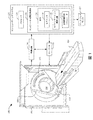
図1は、CTスキャナといった撮像システム100を概略的に示す。撮像システム100は、通常固定されているガントリ102と、固定ガントリ102によって回転可能に支持され、Z軸の周りで検査領域106の周りを回転する回転ガントリ104とを含む。カウチといった被験者支持体108が、検査領域106内の物体又は被験者を支持する。
FIG. 1 schematically shows an
X線管といった放射線源110が、回転ガントリ104によって回転可能に支持され、回転ガントリ104と共に回転し、検査領域106を横断する放射線を放出する。放射線感受性検出器アレイ112が、検査領域106を横断して放射線源110の反対側に角度のついた弧を成す。放射線感受性検出器アレイ112は、検査領域106を横断する放射線を検出し、各検出された光子について放射線を示す信号を生成する。
A
再構成器114が、投影データを再構成し、検査領域106内の被験者又は物体のスキャンされた部分を示すボリュメトリック画像データを生成する。汎用コンピューティングシステム又はコンピュータが、オペレータコンソール116として機能する。コンソール116は、モニタといった人間が読み取り可能な出力デバイスと、キーボード、マウスといった入力デバイスとを含む。コンソール116に常駐するソフトウェアは、オペレータが、グラフィカルユーザインタフェース(GUI)又は他の方法でスキャナ100とインタラクトし及び/又はスキャナ100を操作することを可能にする。
The
セグメンタ118が、ボリュメトリック画像データからの血管樹のセグメント化を容易にする。一例では、これには、冠動脈枝をセグメント化し、セグメント化された冠動脈枝の血管の中心線を特定し、左冠状動脈(LCA)、右冠状動脈(RCA)、左前下行枝(LDA)等といった主血管を標識化し、及び/又は、狭窄位置といった関心位置を標識化することが含まれる。手動、半自動及び/又は自動セグメンテーション手法を使用することができる。セグメンテーションの例は、Bulow他による「A General Framework for Tree Segmentation and Reconstruction from Medical Volume Data」(MICCAI2004、第3216巻、Lecture Notes in Computer Science、pp533〜540)及びGulsun他による「Coronary Centerline Extraction via Optimal Flow Paths and CNN Path Pruning」(MICCAI2016、第9902巻、Lecture Notes in Computer Science、pp317〜325)に説明されている。
セグメンテーション標準化器120が、セグメント化された血管樹を標準化する。以下により詳細に説明するように、1つの非限定的な例では、これには、関心位置を考慮することを含む、血管樹の1つ以上の血管を選択的にプルーニングする又は修正する所定の規則のセットを、「標準化された」構成又は樹に適用することが含まれる。したがって、FFRを決定するために使用される血管樹(標準化された血管樹)の形状、サイズ及び/又は幾何学は、元のセグメンテーションとは異なり、例えあったとしても、画像解像度、造影剤の取り込み及び/又はオペレータのセグメンテーションにほとんど依存しない。後続のFFR−CTシミュレーションは、忠実な境界条件及び正確な予測で信頼が高くなる。
The
生物物理学的シミュレータ122が、少なくとも、標準化されたセグメント化された血管樹を処理して、生物物理学的シミュレーションを行う。FFRに関して、生物物理学的シミュレータは、そのFFR指数を決定する。以下により詳細に説明するように、変形例では、シミュレーションは、プルーニングされた部分の初期条件を決定するために、標準化されたセグメント化された血管樹を用いたシミュレーションの結果を使用して、プルーニングされた領域まで拡張される。したがって、本明細書において説明される手法は、シミュレーションにおける境界条件を、入口及び出口だけでなく血管樹の血管の内側の位置にも実施する。結果として、境界条件をシフトさせ、シミュレーションをそれ以上に拡張することによって標準化が達成される標準化された患者特有の生物物理学的シミュレーションを行うことができる。
The
FFR手法の例は、2013年5月10日に出願され、「Determination of a fractional flow reserve (ffr) value for a stenosis of a vessel」なる名称の米国特許出願第14/396,407号(公開第2015/0092999A1号)、2013年10月24日に出願され、「Fractional flow reserve (ffr) index」なる名称の米国特許出願第14/437,990号(公開第2015/0282765A1号)、2013年10月22日に出願され、「Fractional flow reserve (ffr) index with adaptive boundary condition parameters」なる名称の米国特許出願第14/059,517号(公開第2015/0112191A1号)に説明されている。これらの3つはすべて、参照によりその全体が本明細書に組み込まれる。FFR指数は、ディスプレイモニタ132を介して表示され、記憶され、他のデバイス等に伝達されてもよい。 An example of the FFR method was filed on May 10, 2013 and is entitled "Determination of a fractional flow reserve (ffr) value for a stenosis of a vessel", US Patent Application No. 14 / 396, 407 (Published No. 14 / 396, 407). 2015/0092999A1), filed on October 24, 2013, US Patent Application No. 14 / 437,990 (Published No. 2015/0282765A1), entitled "Fractional flow reserve (ffr) index", 2013/10 It was filed on the 22nd of March and is described in US Patent Application No. 14 / 059,517 (Published No. 14/059,517) entitled "Fractional flow reserve (ffr) index with adaptive boundary stenosis parameters". All three are incorporated herein by reference in their entirety. The FFR index may be displayed, stored, and transmitted to other devices and the like via the display monitor 132.
図示する例では、セグメンタ118、セグメンテーション標準化器120及び/又は生物物理学的シミュレータ122は、物理メモリ及び/又は他の非一時的記憶媒体といった1つ以上のコンピュータ可読記憶媒体130(一時的媒体を除く)に記憶された1つ以上のコンピュータ可読命令128を実行するコンピューティングシステム126の1つ以上のコンピュータプロセッサ124(例えば中央処理演算装置、即ち、CPU、マイクロプロセッサ等)を用いて実現される。プロセッサ124は、追加的に又は代替的に、搬送波、信号及び/又は他の一時的媒体によって運ばれる1つ以上のコンピュータ可読命令を実行することができる。図示するように、命令128はオンサイト(現場)で実行される。別の例では、命令128のうちの1つ以上の命令は、例えば「クラウド」及び/又は他のコンピューティングリソース若しくはサービスを介して、システム126から遠く離れて計算される。
In the illustrated example, the
図2は、セグメンテーション標準化器120の一例を概略的に示す。
FIG. 2 schematically shows an example of the
図示するセグメンテーション標準化器120の一例は、所定のプルーニング規則セット204を使用して、セグメント化された血管樹をプルーニングする血管プルーナ(pruner)202を含む。所定のプルーニング規則セット204は、N個の規則を含み、Nは正の整数である。一例では、所定のプルーニング規則セット204は、デフォルトの規則セットである。別の例では、所定のプルーニング規則セット204は、少なくとも1つのユーザ定義規則及び/又は施設定義規則を含む。別の例では、所定のプルーニング規則セット204は、デフォルト規則と、ユーザ及び/又は施設定義の規則との組み合わせを含む。
An example of the illustrated
図示する所定のプルーニング規則セット204は、主血管プルーニング規則206、短血管プルーニング規則208、関心位置(LOI)プルーニング規則210、遠位セグメントプルーニング規則212、血管直径プルーニング規則214、及び/又は、1つ以上の他のプルーニング規則216を含む。変形例では、1つ以上の他のプルーニング規則216が省略され、所定のプルーニング規則セット204は、規則206〜214のみを含む。別の実施形態では、規則206〜214のうちの1つ以上が省略される。別のプルーニング規則は、画像データの解像度、患者の解剖学的構造、及び/又は、男性若しくは女性、成人若しくは子供等といった人口統計学データに特有であってよい患者特有のパラメータに基づくことができる。
The illustrated predetermined pruning rule set 204 includes a main
主血管プルーニング規則206の一例は、主血管(例えばRCA、LCA、LAD等)が、例えば中心線に沿った大動脈口からのその長さが、所定の長さよりも大きい場合にのみプルーニングされることである。主血管の長さが所定の長さ以下である場合、主血管はプルーニングされない。主血管の長さが所定の長さよりも大きい場合、主血管はプルーニングされるが、当該所定の長さまでしかプルーニングされない。一例では、所定の長さは8センチメートル(8cm)から12センチメートル(12cm)の範囲内の単一値である。別の例では、所定の長さは9センチメートル(9cm)から11センチメートル(11cm)の範囲内の単一値である。他の例では、所定の長さは10センチメートル(10cm)である。
An example of the main
短血管プルーニング規則210の一例は、血管が中心線に沿った所定の長さ未満の長さを有する場合、プルーニングされることである。したがって、血管が所定の長さ未満の長さを有する場合、当該血管は、血管樹から取り除かれる。血管が所定の長さ以上の長さを有する場合、当該血管はプルーニングされない。一例では、所定の長さは0.25から2センチメートル(0.25〜2.0cm)の範囲内である。他の例では、所定の長さは0.5から1.5センチメートル(0.5〜1.5cm)の範囲内である。他の例では、所定の長さは1センチメートル(1cm)である。
An example of a short blood
関心位置プルーニング規則210の一例は、血管の端部が、特定された関心位置からのその長さが所定の長さよりも大きい場合にのみプルーニングされることである。関心位置は、血管の表示されている部分でのマウスクリック及び/又は他の方法で特定することができる。長さが関心位置から所定の長さ以下である場合、端部はプルーニングされない。長さが所定の長さよりも大きい場合、端部はプルーニングされるが、当該所定の長さまでしかプルーニングされない。一例では、所定の長さは0.5から3センチメートル(0.5〜3.0cm)の範囲内である。他の例では、所定の長さは1から2.5センチメートル(1.0〜2.5cm)の範囲内である。他の例では、所定の長さは2センチメートル(2.0cm)である。
An example of the position of
遠位セグメントプルーニング規則212の一例は、最遠位枝の後ろのセグメントである最遠位セグメントが、その長さが分岐からの所定の長さよりも大きい場合にプルーニングされることである。最遠位セグメントの長さが所定の長さ以下である場合、最遠位セグメントはプルーニングされない。最遠位セグメントの長さが所定の長さよりも大きい場合、最遠位セグメントはプルーニングされるが、当該所定の長さまでしかプルーニングされない。一例では、所定の長さは0.5から3センチメートル(0.5〜3.0cm)の範囲内である。他の例では、所定の長さは1から2.5センチメートル(1.0〜2.5cm)の範囲内である。他の例では、所定の長さは2センチメートル(2.0cm)である。
An example of the distal
一例では、例示的な遠位セグメントプルーニング規則212は、主血管プルーニング規則206及び関心位置プルーニング規則210によって制約される。例えば遠位セグメントプルーニング規則212は、結果が主血管プルーニング規則206及び関心位置プルーニング規則210を満たす場合にのみ適用される。遠位セグメントプルーニング規則212の結果が、主血管プルーニング規則206及び関心位置プルーニング規則210を満たさない場合、遠位セグメントプルーニング規則212は適用されない。例えば遠位セグメントプルーニング規則212を適用して、10cm未満の主血管又は関心位置から2cm未満の遠位端をもたらす場合、規則212は適用されない。
In one example, the exemplary distal
血管直径プルーニング規則212の一例は、血管直径が所定の血管直径よりも大きい場合、血管が出口(血管端)から入口(当該血管が別の血管又は大動脈に接続される領域)に向かってプルーニングされることである。血管直径が所定の直径以下である場合、血管はプルーニングされない。血管直径が所定の直径よりも大きい場合、血管はプルーニングされるが、血管直径が所定の直径以下になるまでしかプルーニングされない。一例では、所定の直径は0.5から2.5ミリメートル(0.5〜2.5mm)の範囲内にある。他の例では、所定の直径は1から2ミリメートル(1.0〜2.0mm)の範囲内である。別の例では、所定の直径は1.5ミリメートル(1.5mm)である。
An example of vessel
本明細書において説明されるプルーニング規則を適用することによって、FFR指数を計算するために使用される血管樹である標準化された血管樹は、以下のうちの1つ以上を少なくとも満たす。1)血管セグメントは、最後の分岐位置の後は、標準化された長さを有する。2)固定の画像解像度では明確にできない小さ過ぎる直径を有する血管の遠位部は、ほとんど関連性がない。3)血管の断端、即ち、信頼性の低い大きさ/ボリュームを有する非常に短い血管は、無視される。4)LAD、LCA及びRCAといった主要血管は、最小長さを有する。5)狭窄及び/又は血管樹の位置といった人間のオペレータにとって関心のある可能性のある位置。規則は、中心線又はルーメンセグメンテーションは変更しない。規則は、単にシミュレーションのサポート、境界条件が適用される位置及び境界条件モデルの解剖学的パラメータを規定するだけである。 By applying the pruning rules described herein, the standardized vascular tree, which is the vascular tree used to calculate the FFR index, satisfies at least one or more of the following: 1) The vascular segment has a standardized length after the last bifurcation position. 2) Distal parts of blood vessels with diameters that are too small to be clearly visible at fixed image resolutions are of little relevance. 3) Vessel stumps, i.e. very short vessels with unreliable size / volume, are ignored. 4) Major blood vessels such as LAD, LCA and RCA have a minimum length. 5) Locations that may be of interest to human operators, such as stenosis and / or location of vascular trees. The rules do not change the centerline or lumen segmentation. The rules merely specify simulation support, the location to which the boundary conditions apply, and the anatomical parameters of the boundary condition model.
標準化パラメータは、すべての母集団について固定され、訓練母集団全体にわたる最適化プロセスによってある範囲から選択される。或いは、標準化パラメータは、患者の性別、体重、年齢、心臓の大きさといった人口統計学的及び/又は解剖学的患者特性を含むがこれらに限定されない患者特性を使用することによって、各患者の範囲から個別化することができる。 Standardization parameters are fixed for all populations and selected from a range by an optimization process across the training population. Alternatively, standardized parameters range from each patient by using patient characteristics including, but not limited to, demographic and / or anatomical patient characteristics such as patient gender, weight, age, heart size. Can be individualized from.
1つのFFR−CT手法では、プルーニング規則204は、集中モデルパイプラインのFFR−CT予測を、当該予測が可能な限り正確になるように最適化する。例えばプルーニング規則204は、Nickisch他による「Learning Patient−Specific Lumped Models for Interactive Coronary Blood Flow Simulations」(MICCAI2015、pp433−441)の集中モデルパイプラインのFFR−CT予測を最適化する。プルーニング規則204はまた、集中モデルパイプラインの他のFFR−CT予測も最適化することができる。経験的に、プルーニング規則204は、シミュレーションのユーザ依存性を減少させ、集中モデルパイプラインの精度を高める。集中モデルを使用しない他のFFR−CTを含む他のシミュレーションもまた、本明細書では想定されている。例えば有限要素法、有限体積法、有限差分法といったメッシュに基づくシミュレーションだけでなく、粒子法又は格子ボルツマン法といったメッシュフリー手法及び/又は他の手法が、本明細書では想定されている。
In one FFR-CT approach,
図3は、例えばセグメンタ118によって出力されるセグメント化された冠動脈樹を示す。図4は、セグメンテーション標準化器120が、図3のセグメント化された冠動脈樹にプルーニング規則204を適用した後の標準化されたセグメント化された冠動脈樹を示す。この例では、規則は、血管302、304、306、308、310、312、314、316及び318の端部をプルーニングする結果をもたらした。図4は、血管302、304、306、308、310、312、314、316及び318に沿った端部が切り取られた位置に配置されるプルーニングマーカ402、404、406、408、410、412、414、416及び418を示す。変形例では、プルーニングされた部分は、グレーアウトされるか、及び/又は、そうでなければ、マーキングされる。
FIG. 3 shows a segmented coronary tree output, for example, by
図5は、生物物理学的シミュレータ122が、FFR指数決定器である一例を概略的に示す。FFR指数決定器122は、標準化された血管樹(例えば図4)を入力として受信する。
FIG. 5 schematically shows an example in which the
FFR指数決定器122は、標準化された血管樹の1つ以上の境界条件を推定する境界条件推定器502を含む。例えば境界条件推定器502は、被験者の体重、体格指数(BMI)、性別、年齢、血液検査結果、解剖学的撮像データ(例えば心筋質量及び推定される1回拍出量)及び/又は被験者データといったデータに基づいて生成することができる流入量QO(即ち、小孔における流量)、及び、標準化された血管樹の幾何学(例えば小孔における直径DO)を決定及び/又は受信する。これに基づき、境界条件推定器502は、血管出口における流量Q、平均速度及び/又は抵抗といった少なくとも1つの境界条件を推定する。
The
非限定的な例として、一例では、境界条件推定器502は、出口における流量境界条件Qを、QO及びDOの関数として、

FFR指数決定器122は更に、境界条件を使用して、標準化された血管樹について計算流体力学(CFD)シミュレーションを行い、CFDの結果に基づいてFFRを決定する標準化された血管樹プロセッサ504を含む。CFDの出力は、圧力及び速度のボリュメトリック情報を含み、FFRは、これに基づいて計算される。例えばFFRは、狭窄病変から遠位の最大血流量(Pd)の同じ血管内の正常最大血流量(Pa)に対する比、即ち、FFR=Pd/Paとして計算される。別の適切な手法は、Nickisch他による「Learning Patient−Specific Lumped Models for Interactive Coronary Blood Flow Simulations」(MICCAI2015、第2部、LNCS9350、pp433〜441、2015年)に説明されている。
The
図6は、シミュレーション結果が(例えばグレースケールで)血管302、304、306、308、310、312、314、316及び318に重ね合わされた又はオーバーレイされた標準化された血管樹(図4)を示す表示出力例を示す。この例では、プルーニングマーカ402、404、406、408、410、412、414、416及び418は、血管302、304、306、308、310、312、314、316及び318がプルーニングされた位置を示すように表示される。これにより、ユーザはどこでプルーニングが行われたかを確認することができる。変形例では、プルーニングマーカは表示されない。図6では、表示されていない血管のプルーニングされた部分(即ち、プルーニングマーカ402〜418から遠位の部分)についてのシミュレーション結果はない。
FIG. 6 shows a standardized vessel tree (FIG. 4) in which the simulation results are superimposed or overlaid on
図7は、FFR指数決定器122が更に、プルーニングされた血管セグメントプロセッサ602を含むこと以外は、図5と実質的に同様である。プルーニングされた血管セグメントプロセッサ602は、標準化された血管樹プロセッサ504の出力に基づいた境界条件を使用して、血管のプルーニングされた部分のFFR値を決定する。プルーニングされた血管セグメントプロセッサ602は、流動シミュレーションを、標準化された血管樹(例えば図4)の最初のシミュレーション領域を越えて拡張する。一例では、プルーニングされた血管セグメントプロセッサ602は、以下に説明するように、集中定数モデルを利用する。しかし、本明細書において説明するように、メッシュ、メッシュフリー等といった他の手法も、本明細書では想定されている。
FIG. 7 is substantially similar to FIG. 5 except that the
図9に、適切な集中定数モデルの一例を示す。この例は、n個の要素と、接地を含むm個のノードとを含み、n及びmは正の整数である。中心線表現に基づいて、集中モデルが、非線形抵抗で設定される。黒い長方形は、流入及び流出境界条件を示す。樹セグメント伝達関数φ(f)を表す白い管は、局所的な血管形状及び水力学的効果の両方を反映する一連の線形及び非線形の抵抗要素で構成される。これに基づき、プルーニングされた血管セグメントプロセッサ602は、血管のプルーニングされた部分を、一連の非線形流体力学的抵抗要素と見なす。
FIG. 9 shows an example of an appropriate lumped constant model. This example includes n elements and m nodes including ground, where n and m are positive integers. Based on the centerline representation, the centralized model is set with nonlinear resistance. Black rectangles indicate inflow and outflow boundary conditions. The white tube representing the tree segment transfer function φ (f) is composed of a series of linear and non-linear resistance elements that reflect both local vascular shape and hydraulic effects. Based on this, the pruned
プルーニングされた血管の延長について、プルーニングされた位置(プルーニングマーカ402〜418)におけるプルーニングされたシミュレーションのボリュメトリック流量及び絶対圧力が取り出されるか又は読み取られる。これらの値は、プルーニング位置から遠位のプルーニングされた部分のみを含む後続のシミュレーションの初期値として使用される。分岐点のない血管では、流量は血管全体で一定である。シミュレーションは、プルーニングされた血管に沿った局所伝達関数の評価である。 For the extension of the pruned vessel, the volumetric flow rate and absolute pressure of the pruned simulation at the pruned position (pruning markers 402-418) are retrieved or read. These values are used as initial values for subsequent simulations that include only the pruned portion distal to the pruning position. In vessels without bifurcations, the flow rate is constant throughout the vessel. The simulation is an evaluation of the local transfer function along the pruned blood vessels.
下位血管樹全体がプルーニングされる場合、(対応する入口条件を持つ)水力学的網状組織全体がシミュレートされる。一例では、これは、それぞれの流量及び圧力を得るために、非線形方程式系を解くことを含む。非線形方程式系の一例は、
![]()
![]()
![]()
![]()
下位血管樹又は血管がプルーニング処理において完全に除去され、下位血管樹又は血管が別の血管から分岐していた場合、流れは残っておらず、ゼロの値が割り当てられるため、定圧及び定FFR値が下位血管樹全体に沿って割り当てられる。これ以外のものはすべて、主の流れのどの部分が下位血管樹又は血管を通るのかを決定する手段を必要とし、最終的には、プルーニングされた領域のシミュレーション結果の修正が必要となる。 If the subvessel tree or vessel was completely removed in the pruning process and the subvessel tree or vessel diverged from another vessel, no flow remained and a zero value was assigned, so constant pressure and constant FFR values. Is assigned along the entire subvessel tree. Everything else requires a means of determining which part of the main flow passes through the subvessel tree or vessel, and ultimately requires modification of the simulation results of the pruned area.
図8は、プルーニングされた部分及びそのシミュレーション結果が(グレースケールで)血管302、304、306、308、310、312、314、316及び318に重ね合わされた又はオーバーレイされた標準化された血管樹(図6)を示す表示出力例を示す。この例でも、プルーニングマーカ402、404、406、408、410、412、414、416及び418は、血管302、304、306、308、310、312、314、316及び318がプルーニングされた位置を示すように表示される。変形例では、プルーニングマーカは表示されない。
FIG. 8 shows a standardized vascular tree in which the pruned portion and its simulation results are superimposed or overlaid on
本明細書において説明される手法は、標準化がセグメンテーション標準化器120のハードプルーニングに制約されない、標準化された患者特有の生物物理学的シミュレーションを行うことを可能にする。例えば縮小領域での計算結果が出発点として使用され、その後、縮小領域での結果を修正することなく、領域全体の別個の部分についての結果が続いて追加される。
The techniques described herein make it possible to perform standardized patient-specific biophysical simulations where standardization is not constrained by the hard pruning of the
図10は、方法例を示す。当然ながら、上記行為の順序は、限定ではない。したがって、本明細書では他の順序も想定される。更に、1つ以上の行為を省略しても、及び/又は、1つ以上の追加の行為を含めてもよい。 FIG. 10 shows an example of the method. Of course, the order of the above actions is not limited. Therefore, other orders are assumed herein. Further, one or more actions may be omitted and / or one or more additional actions may be included.
ステップ1002において、血管樹がボリュメトリック画像データ(例えばCT)からセグメント化される。
In
ステップ1004において、セグメント化された血管樹が、本明細書に説明されるように、及び/又は、他の方法で標準化される。例えばセグメント化された血管樹は、セグメント化された血管樹の特定の血管の特定の部分をプルーニングする図2に関連して説明したような所定のプルーニング規則セットに基づいて標準化することができる。
In
ステップ1006において、生物物理学的シミュレーションが、標準化されたセグメント化された血管樹について行われる。
In
ステップ1008において、結果(例えば標準化されたセグメント化された血管樹及びFFR指数)が、ディスプレイモニタを介して視覚的に表示される。
In
図11は、別の方法例を示す。当然ながら、上記行為の順序は、限定ではない。したがって、本明細書では他の順序も想定される。更に、1つ以上の行為を省略しても、及び/又は、1つ以上の追加の行為を含めてもよい。 FIG. 11 shows another example of the method. Of course, the order of the above actions is not limited. Therefore, other orders are assumed herein. Further, one or more actions may be omitted and / or one or more additional actions may be included.
ステップ1102において、血管樹がボリュメトリック画像データ(例えばCT)からセグメント化される。
In
ステップ1104において、セグメント化された血管樹が、本明細書に説明されるように、及び/又は、他の方法で標準化される。例えばセグメント化された血管樹は、セグメント化された血管樹の特定の血管の特定の部分をプルーニングする図2に関連して説明したような所定のプルーニング規則セットに基づいて標準化することができる。
At
ステップ1106において、生物物理学的シミュレーションが、標準化されたセグメント化された血管樹について行われる。
At
ステップ1108において、本明細書において説明されるように、及び/又は、他の方法で、生物物理学的シミュレーションが、プルーニングされた部分について行われる。
At
ステップ1110において、結果(例えばセグメント化された血管樹と、標準化され及びプルーニングされた部分のFFR指数)が、ディスプレイモニタを介して視覚的に表示される。
At
上記は、コンピュータプロセッサによって実行されたときに、当該プロセッサに上記ステップを行わせる、コンピュータ可読記憶媒体上に符号化された又は埋め込まれたコンピュータ可読命令によって実現することができる。更に又は或いは、コンピュータ可読命令の少なくとも1つは、信号、搬送波又はコンピュータ可読記憶媒体ではない他の一時的媒体によって運ばれる。 The above can be accomplished by a computer-readable instruction encoded or embedded on a computer-readable storage medium that, when executed by a computer processor, causes the processor to perform the steps. Further or / or at least one of the computer readable instructions is carried by a signal, a carrier wave or another temporary medium that is not a computer readable storage medium.
本発明を、好適な実施形態を参照して説明した。上記詳細な説明を読んで理解すれば、修正態様及び変更態様を当業者が想到することができる。本発明は、添付の特許請求の範囲又はその均等物の範囲内である限り、そのようなすべての修正形態及び変更形態を含むように構成されることを意図している。
The present invention has been described with reference to preferred embodiments. A person skilled in the art can conceive a modified mode and a modified mode by reading and understanding the above detailed description. The present invention is intended to include all such modifications and modifications, as long as they are within the scope of the appended claims or their equivalents.
Claims (15)
ボリュメトリック画像データからセグメント化された前記セグメント化血管樹及び前記所定のプルーニング規則セットから、前記標準化血管樹を決定するように、前記セグメンテーション標準化器を実行し、また、前記標準化血管樹に基づいて、生物物理学的シミュレーションを行うように、前記生物物理学的シミュレータを実行するプロセッサと、
前記標準化血管樹と前記生物物理学的シミュレーションの結果とのうちの少なくとも1つを表示するディスプレイと、
を含む、コンピューティングシステム。 Based on a segmentation standardizer that determines a standardized vascular tree with vascular segments of a given length from a segmented vascular tree segmented from volumetric image data and a given pruning rule set, and the standardized vascular tree. Computer-readable storage media with computer-executable instructions, including biophysical simulators that perform biophysical simulations,
The segmentation standardizer is run to determine the standardized vascular tree from the segmented vascular tree segmented from volumetric image data and the predetermined pruning rule set, and is also based on the standardized vascular tree. A processor that executes the biophysical simulator, as if performing a biophysical simulation.
A display displaying at least one of the standardized vascular tree and the results of the biophysical simulation.
Including computing systems.
所定のプルーニング規則セットを使用して、ボリュメトリック画像データからセグメント化されたセグメント化血管樹から、所定の長さを有する血管セグメントを有する標準化血管樹を決定するセグメント化標準化器を実行させ、
前記標準化血管樹に基づいて、生物物理学的シミュレーションを行う生物物理学的シミュレータを実行させ、
前記標準化血管樹と前記生物物理学的シミュレーションの結果とのうちの少なくとも1つを表示するディスプレイモニタを介して表示させる、コンピュータ可読命令が符号化された、コンピュータ可読記憶媒体。 When executed by the processor of the computing system, the processor
Using a given pruning rule set, a segmented standardizer is run to determine a standardized vascular tree with vascular segments of a given length from a segmented vascular tree segmented from volumetric image data.
Based on the standardized vascular tree, a biophysical simulator that performs a biophysical simulation is executed.
A computer-readable storage medium encoded with computer-readable instructions for displaying at least one of the standardized vascular tree and the results of the biophysical simulation via a display monitor.
Applications Claiming Priority (3)
| Application Number | Priority Date | Filing Date | Title |
|---|---|---|---|
| US201662425181P | 2016-11-22 | 2016-11-22 | |
| US62/425,181 | 2016-11-22 | ||
| PCT/EP2017/079378 WO2018095791A1 (en) | 2016-11-22 | 2017-11-16 | Vascular tree standardization for biophysical simulation and/or an extension simulation for pruned portions |
Publications (3)
| Publication Number | Publication Date |
|---|---|
| JP2020510451A JP2020510451A (en) | 2020-04-09 |
| JP2020510451A5 JP2020510451A5 (en) | 2020-12-24 |
| JP6975235B2 true JP6975235B2 (en) | 2021-12-01 |
Family
ID=60582555
Family Applications (1)
| Application Number | Title | Priority Date | Filing Date |
|---|---|---|---|
| JP2019527317A Active JP6975235B2 (en) | 2016-11-22 | 2017-11-16 | Vascular tree standardization for biophysical simulation and / or extended simulation for pruned parts |
Country Status (5)
| Country | Link |
|---|---|
| US (1) | US11055845B2 (en) |
| EP (1) | EP3544512B1 (en) |
| JP (1) | JP6975235B2 (en) |
| CN (1) | CN109996495B (en) |
| WO (1) | WO2018095791A1 (en) |
Families Citing this family (8)
| Publication number | Priority date | Publication date | Assignee | Title |
|---|---|---|---|---|
| WO2017199245A1 (en) | 2016-05-16 | 2017-11-23 | Cathworks Ltd. | System for vascular assessment |
| IL263066B2 (en) | 2016-05-16 | 2023-09-01 | Cathworks Ltd | Vascular selection from images |
| CN110494893B (en) | 2017-03-31 | 2024-01-23 | 皇家飞利浦有限公司 | FFR-based interactive monitoring of non-invasive imaging |
| EP3948886A4 (en) | 2019-04-01 | 2022-12-21 | CathWorks Ltd. | Methods and apparatus for angiographic image selection |
| WO2021059165A1 (en) | 2019-09-23 | 2021-04-01 | Cathworks Ltd. | Methods, apparatus, and system for synchronization between a three-dimensional vascular model and an imaging device |
| CN112862759B (en) * | 2021-01-19 | 2023-06-23 | 杭州深睿博联科技有限公司 | Image processing method, device, equipment and computer readable storage medium |
| KR20220111526A (en) * | 2021-02-02 | 2022-08-09 | 자이메드 주식회사 | Apparatus and method for identifying real-time biometric image |
| CN114820554A (en) * | 2022-05-11 | 2022-07-29 | 杭州类脑科技有限公司 | Liver blood vessel extraction method, system and computer readable storage medium based on global automatic growth |
Family Cites Families (15)
| Publication number | Priority date | Publication date | Assignee | Title |
|---|---|---|---|---|
| US7225011B2 (en) | 2001-04-02 | 2007-05-29 | Koninklijke Philips Electronics, N.V. | Heart modeling using a template |
| US8543338B2 (en) | 2007-01-16 | 2013-09-24 | Simbionix Ltd. | System and method for performing computerized simulations for image-guided procedures using a patient specific model |
| WO2011093921A1 (en) * | 2010-01-29 | 2011-08-04 | Mayo Foundation For Medical Education And Research | Automated vascular region separation in medical imaging |
| US8315812B2 (en) | 2010-08-12 | 2012-11-20 | Heartflow, Inc. | Method and system for patient-specific modeling of blood flow |
| EP2749224A4 (en) | 2011-08-26 | 2015-12-02 | Ebm Corp | Bloodstream simulation system for simulating blood vessel treatment effect, method therefor, and computer software program |
| WO2013171644A1 (en) | 2012-05-14 | 2013-11-21 | Koninklijke Philips N.V. | Determination of a fractional flow reserve (ffr) value for a stenosis of a vessel |
| JP6302922B2 (en) * | 2012-11-06 | 2018-03-28 | コーニンクレッカ フィリップス エヌ ヴェKoninklijke Philips N.V. | Coronary flow reserve ratio (FFR) index |
| US9424395B2 (en) | 2013-03-04 | 2016-08-23 | Heartflow, Inc. | Method and system for sensitivity analysis in modeling blood flow characteristics |
| US9788807B2 (en) | 2013-09-06 | 2017-10-17 | Koninklijke Philips N.V. | Processing apparatus for processing cardiac data |
| US10595806B2 (en) | 2013-10-22 | 2020-03-24 | Koninklijke Philips N.V. | Fractional flow reserve (FFR) index with adaptive boundary condition parameters |
| US8977339B1 (en) | 2013-12-05 | 2015-03-10 | Intrinsic Medical Imaging Llc | Method for assessing stenosis severity through stenosis mapping |
| US9087147B1 (en) | 2014-03-31 | 2015-07-21 | Heartflow, Inc. | Systems and methods for determining blood flow characteristics using flow ratio |
| WO2016055899A1 (en) * | 2014-10-10 | 2016-04-14 | Koninklijke Philips N.V. | Tace navigation guidance based on tumor viability and vascular geometry |
| WO2016087396A1 (en) | 2014-12-02 | 2016-06-09 | Koninklijke Philips N.V. | Fractional flow reserve determination |
| WO2017102467A1 (en) * | 2015-12-15 | 2017-06-22 | Koninklijke Philips N.V. | Method of data processing for computed tomography |
-
2017
- 2017-11-16 EP EP17809208.6A patent/EP3544512B1/en active Active
- 2017-11-16 WO PCT/EP2017/079378 patent/WO2018095791A1/en unknown
- 2017-11-16 JP JP2019527317A patent/JP6975235B2/en active Active
- 2017-11-16 US US16/348,282 patent/US11055845B2/en active Active
- 2017-11-16 CN CN201780072241.9A patent/CN109996495B/en active Active
Also Published As
| Publication number | Publication date |
|---|---|
| US11055845B2 (en) | 2021-07-06 |
| WO2018095791A1 (en) | 2018-05-31 |
| JP2020510451A (en) | 2020-04-09 |
| CN109996495A (en) | 2019-07-09 |
| US20190318475A1 (en) | 2019-10-17 |
| CN109996495B (en) | 2023-04-28 |
| EP3544512B1 (en) | 2022-09-14 |
| EP3544512A1 (en) | 2019-10-02 |
Similar Documents
| Publication | Publication Date | Title |
|---|---|---|
| JP6975235B2 (en) | Vascular tree standardization for biophysical simulation and / or extended simulation for pruned parts | |
| JP6396468B2 (en) | Local FFR estimation and visualization to improve functional stenosis analysis | |
| US10595806B2 (en) | Fractional flow reserve (FFR) index with adaptive boundary condition parameters | |
| JP2022169579A (en) | Diagnostically useful results in real time | |
| JP6484760B2 (en) | Modeling collateral blood flow for non-invasive blood flow reserve ratio (FFR) | |
| EP3140757B1 (en) | Method and system for non-invasive functional assessment of coronary artery stenosis using flow computations in models based on diseased patients and hypothetical normal anatomical models | |
| CN106572824A (en) | Stenosis assessment | |
| JP7303260B2 (en) | A method for determining flow and pressure gradients in arterial networks from contrast distributions based on patient-specific computed tomography algorithms. | |
| CN108140430B (en) | Estimating flow, resistance or pressure from pressure or flow measurements and angiography | |
| CN104768465A (en) | Fractional flow reserve (FFR) index | |
| JP2019512131A (en) | System and method for identifying and modeling unresolved blood vessels in an image based patient specific hemodynamic model | |
| JP6749917B2 (en) | iFR-CT | |
| JP7300392B2 (en) | Standardized Coronary Artery Disease Metrics | |
| JP6873981B2 (en) | Mobile FFR simulation | |
| JP7426824B2 (en) | Non-invasive imaging-based FFR interaction monitoring | |
| JPWO2018185040A5 (en) | ||
| JP2020512117A (en) | Simulation of transcatheter aortic valve implantation (TAVI) on coronary blood flow and pressure | |
| WO2023274724A1 (en) | Generation of plaque information |
Legal Events
| Date | Code | Title | Description |
|---|---|---|---|
| A521 | Request for written amendment filed |
Free format text: JAPANESE INTERMEDIATE CODE: A523 Effective date: 20201112 |
|
| A621 | Written request for application examination |
Free format text: JAPANESE INTERMEDIATE CODE: A621 Effective date: 20201112 |
|
| A977 | Report on retrieval |
Free format text: JAPANESE INTERMEDIATE CODE: A971007 Effective date: 20210922 |
|
| TRDD | Decision of grant or rejection written | ||
| A01 | Written decision to grant a patent or to grant a registration (utility model) |
Free format text: JAPANESE INTERMEDIATE CODE: A01 Effective date: 20211008 |
|
| A61 | First payment of annual fees (during grant procedure) |
Free format text: JAPANESE INTERMEDIATE CODE: A61 Effective date: 20211105 |
|
| R150 | Certificate of patent or registration of utility model |
Ref document number: 6975235 Country of ref document: JP Free format text: JAPANESE INTERMEDIATE CODE: R150 |