JP6967006B2 - Nanopore analysis based on reduced background optics - Google Patents
Nanopore analysis based on reduced background optics Download PDFInfo
- Publication number
- JP6967006B2 JP6967006B2 JP2018536442A JP2018536442A JP6967006B2 JP 6967006 B2 JP6967006 B2 JP 6967006B2 JP 2018536442 A JP2018536442 A JP 2018536442A JP 2018536442 A JP2018536442 A JP 2018536442A JP 6967006 B2 JP6967006 B2 JP 6967006B2
- Authority
- JP
- Japan
- Prior art keywords
- label
- fluorescent
- nanopore
- nanopores
- polynucleotide
- Prior art date
- Legal status (The legal status is an assumption and is not a legal conclusion. Google has not performed a legal analysis and makes no representation as to the accuracy of the status listed.)
- Active
Links
- 238000004458 analytical method Methods 0.000 title description 12
- 230000002829 reductive effect Effects 0.000 title description 2
- 102000040430 polynucleotide Human genes 0.000 claims description 153
- 108091033319 polynucleotide Proteins 0.000 claims description 153
- 239000002157 polynucleotide Substances 0.000 claims description 153
- 239000007850 fluorescent dye Substances 0.000 claims description 126
- 125000003729 nucleotide group Chemical group 0.000 claims description 109
- 238000000034 method Methods 0.000 claims description 105
- 239000002773 nucleotide Substances 0.000 claims description 104
- 230000003287 optical effect Effects 0.000 claims description 87
- 238000010791 quenching Methods 0.000 claims description 82
- 102000004169 proteins and genes Human genes 0.000 claims description 79
- 108090000623 proteins and genes Proteins 0.000 claims description 78
- 238000001514 detection method Methods 0.000 claims description 61
- 230000005284 excitation Effects 0.000 claims description 58
- 239000012528 membrane Substances 0.000 claims description 56
- 230000007274 generation of a signal involved in cell-cell signaling Effects 0.000 claims description 38
- 239000007790 solid phase Substances 0.000 claims description 38
- 229910052751 metal Inorganic materials 0.000 claims description 25
- 239000002184 metal Substances 0.000 claims description 25
- 239000000232 Lipid Bilayer Substances 0.000 claims description 18
- 238000012163 sequencing technique Methods 0.000 claims description 16
- 238000004891 communication Methods 0.000 claims description 14
- 239000012530 fluid Substances 0.000 claims description 13
- 238000005286 illumination Methods 0.000 claims description 13
- 229910052782 aluminium Inorganic materials 0.000 claims description 11
- 229910052737 gold Inorganic materials 0.000 claims description 9
- 239000010931 gold Substances 0.000 claims description 9
- XAGFODPZIPBFFR-UHFFFAOYSA-N aluminium Chemical compound [Al] XAGFODPZIPBFFR-UHFFFAOYSA-N 0.000 claims description 6
- PCHJSUWPFVWCPO-UHFFFAOYSA-N gold Chemical compound [Au] PCHJSUWPFVWCPO-UHFFFAOYSA-N 0.000 claims description 4
- 239000010410 layer Substances 0.000 description 92
- 229920000642 polymer Polymers 0.000 description 91
- 235000018102 proteins Nutrition 0.000 description 77
- 239000000178 monomer Substances 0.000 description 76
- 230000000171 quenching effect Effects 0.000 description 65
- 150000007523 nucleic acids Chemical class 0.000 description 63
- 239000000370 acceptor Substances 0.000 description 60
- 102000039446 nucleic acids Human genes 0.000 description 60
- 108020004707 nucleic acids Proteins 0.000 description 60
- 108091034117 Oligonucleotide Proteins 0.000 description 38
- 238000006243 chemical reaction Methods 0.000 description 35
- 230000007704 transition Effects 0.000 description 34
- 239000007787 solid Substances 0.000 description 32
- 238000002372 labelling Methods 0.000 description 30
- 239000000975 dye Substances 0.000 description 28
- 239000010408 film Substances 0.000 description 25
- 239000011148 porous material Substances 0.000 description 22
- 239000000758 substrate Substances 0.000 description 21
- 108020004414 DNA Proteins 0.000 description 20
- 230000003993 interaction Effects 0.000 description 20
- 239000000463 material Substances 0.000 description 20
- 108091006146 Channels Proteins 0.000 description 19
- 239000011248 coating agent Substances 0.000 description 18
- 238000000576 coating method Methods 0.000 description 18
- 108090000765 processed proteins & peptides Proteins 0.000 description 18
- 238000012546 transfer Methods 0.000 description 17
- JLCPHMBAVCMARE-UHFFFAOYSA-N [3-[[3-[[3-[[3-[[3-[[3-[[3-[[3-[[3-[[3-[[3-[[5-(2-amino-6-oxo-1H-purin-9-yl)-3-[[3-[[3-[[3-[[3-[[3-[[5-(2-amino-6-oxo-1H-purin-9-yl)-3-[[5-(2-amino-6-oxo-1H-purin-9-yl)-3-hydroxyoxolan-2-yl]methoxy-hydroxyphosphoryl]oxyoxolan-2-yl]methoxy-hydroxyphosphoryl]oxy-5-(5-methyl-2,4-dioxopyrimidin-1-yl)oxolan-2-yl]methoxy-hydroxyphosphoryl]oxy-5-(6-aminopurin-9-yl)oxolan-2-yl]methoxy-hydroxyphosphoryl]oxy-5-(6-aminopurin-9-yl)oxolan-2-yl]methoxy-hydroxyphosphoryl]oxy-5-(6-aminopurin-9-yl)oxolan-2-yl]methoxy-hydroxyphosphoryl]oxy-5-(6-aminopurin-9-yl)oxolan-2-yl]methoxy-hydroxyphosphoryl]oxyoxolan-2-yl]methoxy-hydroxyphosphoryl]oxy-5-(5-methyl-2,4-dioxopyrimidin-1-yl)oxolan-2-yl]methoxy-hydroxyphosphoryl]oxy-5-(4-amino-2-oxopyrimidin-1-yl)oxolan-2-yl]methoxy-hydroxyphosphoryl]oxy-5-(5-methyl-2,4-dioxopyrimidin-1-yl)oxolan-2-yl]methoxy-hydroxyphosphoryl]oxy-5-(5-methyl-2,4-dioxopyrimidin-1-yl)oxolan-2-yl]methoxy-hydroxyphosphoryl]oxy-5-(6-aminopurin-9-yl)oxolan-2-yl]methoxy-hydroxyphosphoryl]oxy-5-(6-aminopurin-9-yl)oxolan-2-yl]methoxy-hydroxyphosphoryl]oxy-5-(4-amino-2-oxopyrimidin-1-yl)oxolan-2-yl]methoxy-hydroxyphosphoryl]oxy-5-(4-amino-2-oxopyrimidin-1-yl)oxolan-2-yl]methoxy-hydroxyphosphoryl]oxy-5-(4-amino-2-oxopyrimidin-1-yl)oxolan-2-yl]methoxy-hydroxyphosphoryl]oxy-5-(6-aminopurin-9-yl)oxolan-2-yl]methoxy-hydroxyphosphoryl]oxy-5-(4-amino-2-oxopyrimidin-1-yl)oxolan-2-yl]methyl [5-(6-aminopurin-9-yl)-2-(hydroxymethyl)oxolan-3-yl] hydrogen phosphate Polymers Cc1cn(C2CC(OP(O)(=O)OCC3OC(CC3OP(O)(=O)OCC3OC(CC3O)n3cnc4c3nc(N)[nH]c4=O)n3cnc4c3nc(N)[nH]c4=O)C(COP(O)(=O)OC3CC(OC3COP(O)(=O)OC3CC(OC3COP(O)(=O)OC3CC(OC3COP(O)(=O)OC3CC(OC3COP(O)(=O)OC3CC(OC3COP(O)(=O)OC3CC(OC3COP(O)(=O)OC3CC(OC3COP(O)(=O)OC3CC(OC3COP(O)(=O)OC3CC(OC3COP(O)(=O)OC3CC(OC3COP(O)(=O)OC3CC(OC3COP(O)(=O)OC3CC(OC3COP(O)(=O)OC3CC(OC3COP(O)(=O)OC3CC(OC3COP(O)(=O)OC3CC(OC3COP(O)(=O)OC3CC(OC3COP(O)(=O)OC3CC(OC3CO)n3cnc4c(N)ncnc34)n3ccc(N)nc3=O)n3cnc4c(N)ncnc34)n3ccc(N)nc3=O)n3ccc(N)nc3=O)n3ccc(N)nc3=O)n3cnc4c(N)ncnc34)n3cnc4c(N)ncnc34)n3cc(C)c(=O)[nH]c3=O)n3cc(C)c(=O)[nH]c3=O)n3ccc(N)nc3=O)n3cc(C)c(=O)[nH]c3=O)n3cnc4c3nc(N)[nH]c4=O)n3cnc4c(N)ncnc34)n3cnc4c(N)ncnc34)n3cnc4c(N)ncnc34)n3cnc4c(N)ncnc34)O2)c(=O)[nH]c1=O JLCPHMBAVCMARE-UHFFFAOYSA-N 0.000 description 16
- 238000003491 array Methods 0.000 description 15
- 238000007672 fourth generation sequencing Methods 0.000 description 15
- 239000000126 substance Substances 0.000 description 14
- 239000002096 quantum dot Substances 0.000 description 13
- 238000005259 measurement Methods 0.000 description 12
- 102000004196 processed proteins & peptides Human genes 0.000 description 12
- 239000012491 analyte Substances 0.000 description 11
- 238000009739 binding Methods 0.000 description 11
- 150000001875 compounds Chemical class 0.000 description 11
- 239000000243 solution Substances 0.000 description 11
- 125000000539 amino acid group Chemical group 0.000 description 10
- 230000027455 binding Effects 0.000 description 10
- 230000005684 electric field Effects 0.000 description 10
- 238000004519 manufacturing process Methods 0.000 description 10
- 239000002086 nanomaterial Substances 0.000 description 10
- 239000000523 sample Substances 0.000 description 10
- 102000053602 DNA Human genes 0.000 description 9
- 108010013381 Porins Proteins 0.000 description 9
- 102000017033 Porins Human genes 0.000 description 9
- 108020004682 Single-Stranded DNA Proteins 0.000 description 9
- ANRHNWWPFJCPAZ-UHFFFAOYSA-M thionine Chemical compound [Cl-].C1=CC(N)=CC2=[S+]C3=CC(N)=CC=C3N=C21 ANRHNWWPFJCPAZ-UHFFFAOYSA-M 0.000 description 9
- 238000009826 distribution Methods 0.000 description 8
- GNBHRKFJIUUOQI-UHFFFAOYSA-N fluorescein Chemical compound O1C(=O)C2=CC=CC=C2C21C1=CC=C(O)C=C1OC1=CC(O)=CC=C21 GNBHRKFJIUUOQI-UHFFFAOYSA-N 0.000 description 8
- -1 helium ion Chemical class 0.000 description 8
- 238000012986 modification Methods 0.000 description 8
- 230000004048 modification Effects 0.000 description 8
- 229920001184 polypeptide Polymers 0.000 description 8
- 150000003573 thiols Chemical class 0.000 description 8
- 238000000151 deposition Methods 0.000 description 7
- 239000011535 reaction buffer Substances 0.000 description 7
- 108091032973 (ribonucleotides)n+m Proteins 0.000 description 6
- 101710092462 Alpha-hemolysin Proteins 0.000 description 6
- OKTJSMMVPCPJKN-UHFFFAOYSA-N Carbon Chemical compound [C] OKTJSMMVPCPJKN-UHFFFAOYSA-N 0.000 description 6
- 229910052581 Si3N4 Inorganic materials 0.000 description 6
- VYPSYNLAJGMNEJ-UHFFFAOYSA-N Silicium dioxide Chemical compound O=[Si]=O VYPSYNLAJGMNEJ-UHFFFAOYSA-N 0.000 description 6
- 238000010521 absorption reaction Methods 0.000 description 6
- 235000001014 amino acid Nutrition 0.000 description 6
- 150000001413 amino acids Chemical class 0.000 description 6
- 239000002777 nucleoside Substances 0.000 description 6
- 230000005855 radiation Effects 0.000 description 6
- HQVNEWCFYHHQES-UHFFFAOYSA-N silicon nitride Chemical compound N12[Si]34N5[Si]62N3[Si]51N64 HQVNEWCFYHHQES-UHFFFAOYSA-N 0.000 description 6
- 102000004190 Enzymes Human genes 0.000 description 5
- 108090000790 Enzymes Proteins 0.000 description 5
- 230000015572 biosynthetic process Effects 0.000 description 5
- 229910052802 copper Inorganic materials 0.000 description 5
- 230000008021 deposition Effects 0.000 description 5
- 238000004020 luminiscence type Methods 0.000 description 5
- 230000001404 mediated effect Effects 0.000 description 5
- 239000000203 mixture Substances 0.000 description 5
- 150000003141 primary amines Chemical group 0.000 description 5
- 230000008569 process Effects 0.000 description 5
- 229910052709 silver Inorganic materials 0.000 description 5
- 229910004613 CdTe Inorganic materials 0.000 description 4
- 238000001712 DNA sequencing Methods 0.000 description 4
- BLRPTPMANUNPDV-UHFFFAOYSA-N Silane Chemical compound [SiH4] BLRPTPMANUNPDV-UHFFFAOYSA-N 0.000 description 4
- XUIMIQQOPSSXEZ-UHFFFAOYSA-N Silicon Chemical compound [Si] XUIMIQQOPSSXEZ-UHFFFAOYSA-N 0.000 description 4
- IQFYYKKMVGJFEH-XLPZGREQSA-N Thymidine Chemical compound O=C1NC(=O)C(C)=CN1[C@@H]1O[C@H](CO)[C@@H](O)C1 IQFYYKKMVGJFEH-XLPZGREQSA-N 0.000 description 4
- OIRDTQYFTABQOQ-KQYNXXCUSA-N adenosine Chemical compound C1=NC=2C(N)=NC=NC=2N1[C@@H]1O[C@H](CO)[C@@H](O)[C@H]1O OIRDTQYFTABQOQ-KQYNXXCUSA-N 0.000 description 4
- 239000007864 aqueous solution Substances 0.000 description 4
- 239000002041 carbon nanotube Substances 0.000 description 4
- 229910021393 carbon nanotube Inorganic materials 0.000 description 4
- 239000003153 chemical reaction reagent Substances 0.000 description 4
- 238000005229 chemical vapour deposition Methods 0.000 description 4
- 239000003792 electrolyte Substances 0.000 description 4
- 238000001962 electrophoresis Methods 0.000 description 4
- 238000000295 emission spectrum Methods 0.000 description 4
- 238000005516 engineering process Methods 0.000 description 4
- 238000010884 ion-beam technique Methods 0.000 description 4
- 150000002500 ions Chemical class 0.000 description 4
- 230000000670 limiting effect Effects 0.000 description 4
- 239000012071 phase Substances 0.000 description 4
- PYWVYCXTNDRMGF-UHFFFAOYSA-N rhodamine B Chemical compound [Cl-].C=12C=CC(=[N+](CC)CC)C=C2OC2=CC(N(CC)CC)=CC=C2C=1C1=CC=CC=C1C(O)=O PYWVYCXTNDRMGF-UHFFFAOYSA-N 0.000 description 4
- 230000011664 signaling Effects 0.000 description 4
- 229910000077 silane Inorganic materials 0.000 description 4
- 229910052710 silicon Inorganic materials 0.000 description 4
- 239000010703 silicon Substances 0.000 description 4
- 229910052814 silicon oxide Inorganic materials 0.000 description 4
- 230000003595 spectral effect Effects 0.000 description 4
- QTBSBXVTEAMEQO-UHFFFAOYSA-N Acetic acid Chemical compound CC(O)=O QTBSBXVTEAMEQO-UHFFFAOYSA-N 0.000 description 3
- LFQSCWFLJHTTHZ-UHFFFAOYSA-N Ethanol Chemical compound CCO LFQSCWFLJHTTHZ-UHFFFAOYSA-N 0.000 description 3
- 206010056740 Genital discharge Diseases 0.000 description 3
- PEDCQBHIVMGVHV-UHFFFAOYSA-N Glycerine Chemical compound OCC(O)CO PEDCQBHIVMGVHV-UHFFFAOYSA-N 0.000 description 3
- 229910004298 SiO 2 Inorganic materials 0.000 description 3
- 238000000862 absorption spectrum Methods 0.000 description 3
- 230000003321 amplification Effects 0.000 description 3
- 238000003556 assay Methods 0.000 description 3
- 230000008901 benefit Effects 0.000 description 3
- 230000000295 complement effect Effects 0.000 description 3
- 235000018417 cysteine Nutrition 0.000 description 3
- XUJNEKJLAYXESH-UHFFFAOYSA-N cysteine Natural products SCC(N)C(O)=O XUJNEKJLAYXESH-UHFFFAOYSA-N 0.000 description 3
- 150000002148 esters Chemical group 0.000 description 3
- 230000010354 integration Effects 0.000 description 3
- 125000005647 linker group Chemical group 0.000 description 3
- 239000003550 marker Substances 0.000 description 3
- 239000003068 molecular probe Substances 0.000 description 3
- 239000002105 nanoparticle Substances 0.000 description 3
- 238000003199 nucleic acid amplification method Methods 0.000 description 3
- 239000012038 nucleophile Substances 0.000 description 3
- 125000003835 nucleoside group Chemical group 0.000 description 3
- 238000002850 optical based method Methods 0.000 description 3
- 230000036961 partial effect Effects 0.000 description 3
- 239000011541 reaction mixture Substances 0.000 description 3
- 238000011160 research Methods 0.000 description 3
- 239000001022 rhodamine dye Substances 0.000 description 3
- 235000000346 sugar Nutrition 0.000 description 3
- 239000000979 synthetic dye Substances 0.000 description 3
- 239000001018 xanthene dye Substances 0.000 description 3
- 150000003732 xanthenes Chemical class 0.000 description 3
- YBJHBAHKTGYVGT-ZKWXMUAHSA-N (+)-Biotin Chemical compound N1C(=O)N[C@@H]2[C@H](CCCCC(=O)O)SC[C@@H]21 YBJHBAHKTGYVGT-ZKWXMUAHSA-N 0.000 description 2
- YKBGVTZYEHREMT-KVQBGUIXSA-N 2'-deoxyguanosine Chemical compound C1=NC=2C(=O)NC(N)=NC=2N1[C@H]1C[C@H](O)[C@@H](CO)O1 YKBGVTZYEHREMT-KVQBGUIXSA-N 0.000 description 2
- VGONTNSXDCQUGY-RRKCRQDMSA-N 2'-deoxyinosine Chemical compound C1[C@H](O)[C@@H](CO)O[C@H]1N1C(N=CNC2=O)=C2N=C1 VGONTNSXDCQUGY-RRKCRQDMSA-N 0.000 description 2
- 150000003923 2,5-pyrrolediones Chemical group 0.000 description 2
- IOOMXAQUNPWDLL-UHFFFAOYSA-N 2-[6-(diethylamino)-3-(diethyliminiumyl)-3h-xanthen-9-yl]-5-sulfobenzene-1-sulfonate Chemical compound C=12C=CC(=[N+](CC)CC)C=C2OC2=CC(N(CC)CC)=CC=C2C=1C1=CC=C(S(O)(=O)=O)C=C1S([O-])(=O)=O IOOMXAQUNPWDLL-UHFFFAOYSA-N 0.000 description 2
- CKTSBUTUHBMZGZ-ULQXZJNLSA-N 4-amino-1-[(2r,4s,5r)-4-hydroxy-5-(hydroxymethyl)oxolan-2-yl]-5-tritiopyrimidin-2-one Chemical compound O=C1N=C(N)C([3H])=CN1[C@@H]1O[C@H](CO)[C@@H](O)C1 CKTSBUTUHBMZGZ-ULQXZJNLSA-N 0.000 description 2
- HJCUTNIGJHJGCF-UHFFFAOYSA-N 9,10-dihydroacridine Chemical compound C1=CC=C2CC3=CC=CC=C3NC2=C1 HJCUTNIGJHJGCF-UHFFFAOYSA-N 0.000 description 2
- GJCOSYZMQJWQCA-UHFFFAOYSA-N 9H-xanthene Chemical compound C1=CC=C2CC3=CC=CC=C3OC2=C1 GJCOSYZMQJWQCA-UHFFFAOYSA-N 0.000 description 2
- 239000004971 Cross linker Substances 0.000 description 2
- 230000004568 DNA-binding Effects 0.000 description 2
- 102000016928 DNA-directed DNA polymerase Human genes 0.000 description 2
- 108010014303 DNA-directed DNA polymerase Proteins 0.000 description 2
- 239000004593 Epoxy Chemical group 0.000 description 2
- SIKJAQJRHWYJAI-UHFFFAOYSA-N Indole Chemical compound C1=CC=C2NC=CC2=C1 SIKJAQJRHWYJAI-UHFFFAOYSA-N 0.000 description 2
- 235000019687 Lamb Nutrition 0.000 description 2
- UFWIBTONFRDIAS-UHFFFAOYSA-N Naphthalene Chemical compound C1=CC=CC2=CC=CC=C21 UFWIBTONFRDIAS-UHFFFAOYSA-N 0.000 description 2
- 108091028043 Nucleic acid sequence Proteins 0.000 description 2
- 101710129178 Outer plastidial membrane protein porin Proteins 0.000 description 2
- 108091093037 Peptide nucleic acid Proteins 0.000 description 2
- JUJWROOIHBZHMG-UHFFFAOYSA-N Pyridine Chemical compound C1=CC=NC=C1 JUJWROOIHBZHMG-UHFFFAOYSA-N 0.000 description 2
- SMWDFEZZVXVKRB-UHFFFAOYSA-N Quinoline Chemical compound N1=CC=CC2=CC=CC=C21 SMWDFEZZVXVKRB-UHFFFAOYSA-N 0.000 description 2
- FAPWRFPIFSIZLT-UHFFFAOYSA-M Sodium chloride Chemical compound [Na+].[Cl-] FAPWRFPIFSIZLT-UHFFFAOYSA-M 0.000 description 2
- DRTQHJPVMGBUCF-XVFCMESISA-N Uridine Chemical compound O[C@@H]1[C@H](O)[C@@H](CO)O[C@H]1N1C(=O)NC(=O)C=C1 DRTQHJPVMGBUCF-XVFCMESISA-N 0.000 description 2
- 102100037820 Voltage-dependent anion-selective channel protein 1 Human genes 0.000 description 2
- 150000001412 amines Chemical class 0.000 description 2
- MWPLVEDNUUSJAV-UHFFFAOYSA-N anthracene Chemical compound C1=CC=CC2=CC3=CC=CC=C3C=C21 MWPLVEDNUUSJAV-UHFFFAOYSA-N 0.000 description 2
- 238000013459 approach Methods 0.000 description 2
- 125000003118 aryl group Chemical group 0.000 description 2
- 125000000751 azo group Chemical group [*]N=N[*] 0.000 description 2
- CUFNKYGDVFVPHO-UHFFFAOYSA-N azulene Chemical compound C1=CC=CC2=CC=CC2=C1 CUFNKYGDVFVPHO-UHFFFAOYSA-N 0.000 description 2
- IOJUPLGTWVMSFF-UHFFFAOYSA-N benzothiazole Chemical compound C1=CC=C2SC=NC2=C1 IOJUPLGTWVMSFF-UHFFFAOYSA-N 0.000 description 2
- DEGAKNSWVGKMLS-UHFFFAOYSA-N calcein Chemical compound O1C(=O)C2=CC=CC=C2C21C1=CC(CN(CC(O)=O)CC(O)=O)=C(O)C=C1OC1=C2C=C(CN(CC(O)=O)CC(=O)O)C(O)=C1 DEGAKNSWVGKMLS-UHFFFAOYSA-N 0.000 description 2
- 229910052799 carbon Inorganic materials 0.000 description 2
- 239000003795 chemical substances by application Substances 0.000 description 2
- 239000012141 concentrate Substances 0.000 description 2
- ZYGHJZDHTFUPRJ-UHFFFAOYSA-N coumarin Chemical compound C1=CC=C2OC(=O)C=CC2=C1 ZYGHJZDHTFUPRJ-UHFFFAOYSA-N 0.000 description 2
- 230000008878 coupling Effects 0.000 description 2
- 238000010168 coupling process Methods 0.000 description 2
- 238000005859 coupling reaction Methods 0.000 description 2
- 125000000151 cysteine group Chemical group N[C@@H](CS)C(=O)* 0.000 description 2
- OPTASPLRGRRNAP-UHFFFAOYSA-N cytosine Chemical compound NC=1C=CNC(=O)N=1 OPTASPLRGRRNAP-UHFFFAOYSA-N 0.000 description 2
- 230000001419 dependent effect Effects 0.000 description 2
- VGONTNSXDCQUGY-UHFFFAOYSA-N desoxyinosine Natural products C1C(O)C(CO)OC1N1C(NC=NC2=O)=C2N=C1 VGONTNSXDCQUGY-UHFFFAOYSA-N 0.000 description 2
- 230000002255 enzymatic effect Effects 0.000 description 2
- 238000001704 evaporation Methods 0.000 description 2
- 230000008020 evaporation Effects 0.000 description 2
- 229960002143 fluorescein Drugs 0.000 description 2
- MHMNJMPURVTYEJ-UHFFFAOYSA-N fluorescein-5-isothiocyanate Chemical compound O1C(=O)C2=CC(N=C=S)=CC=C2C21C1=CC=C(O)C=C1OC1=CC(O)=CC=C21 MHMNJMPURVTYEJ-UHFFFAOYSA-N 0.000 description 2
- 238000000799 fluorescence microscopy Methods 0.000 description 2
- 238000001506 fluorescence spectroscopy Methods 0.000 description 2
- 239000012634 fragment Substances 0.000 description 2
- 239000001307 helium Substances 0.000 description 2
- 229910052734 helium Inorganic materials 0.000 description 2
- 239000003228 hemolysin Substances 0.000 description 2
- 238000003780 insertion Methods 0.000 description 2
- 230000037431 insertion Effects 0.000 description 2
- 239000000138 intercalating agent Substances 0.000 description 2
- QDLAGTHXVHQKRE-UHFFFAOYSA-N lichenxanthone Natural products COC1=CC(O)=C2C(=O)C3=C(C)C=C(OC)C=C3OC2=C1 QDLAGTHXVHQKRE-UHFFFAOYSA-N 0.000 description 2
- KWGKDLIKAYFUFQ-UHFFFAOYSA-M lithium chloride Chemical compound [Li+].[Cl-] KWGKDLIKAYFUFQ-UHFFFAOYSA-M 0.000 description 2
- 108040007791 maltose transporting porin activity proteins Proteins 0.000 description 2
- 230000007246 mechanism Effects 0.000 description 2
- 239000002082 metal nanoparticle Substances 0.000 description 2
- 238000013508 migration Methods 0.000 description 2
- 230000005012 migration Effects 0.000 description 2
- 230000002438 mitochondrial effect Effects 0.000 description 2
- 229910052750 molybdenum Inorganic materials 0.000 description 2
- 239000002071 nanotube Substances 0.000 description 2
- 229910052759 nickel Inorganic materials 0.000 description 2
- 230000000269 nucleophilic effect Effects 0.000 description 2
- 150000003833 nucleoside derivatives Chemical class 0.000 description 2
- 229960002378 oftasceine Drugs 0.000 description 2
- BRJCLSQFZSHLRL-UHFFFAOYSA-N oregon green 488 Chemical compound OC(=O)C1=CC(C(=O)O)=CC=C1C1=C2C=C(F)C(=O)C=C2OC2=CC(O)=C(F)C=C21 BRJCLSQFZSHLRL-UHFFFAOYSA-N 0.000 description 2
- TWNQGVIAIRXVLR-UHFFFAOYSA-N oxo(oxoalumanyloxy)alumane Chemical compound O=[Al]O[Al]=O TWNQGVIAIRXVLR-UHFFFAOYSA-N 0.000 description 2
- 229920002120 photoresistant polymer Polymers 0.000 description 2
- 238000012545 processing Methods 0.000 description 2
- BBEAQIROQSPTKN-UHFFFAOYSA-N pyrene Chemical compound C1=CC=C2C=CC3=CC=CC4=CC=C1C2=C43 BBEAQIROQSPTKN-UHFFFAOYSA-N 0.000 description 2
- 238000006862 quantum yield reaction Methods 0.000 description 2
- 230000009467 reduction Effects 0.000 description 2
- 238000002165 resonance energy transfer Methods 0.000 description 2
- 230000002441 reversible effect Effects 0.000 description 2
- XLXOKMFKGASILN-UHFFFAOYSA-N rhodamine red-X Chemical compound C=12C=CC(=[N+](CC)CC)C=C2OC2=CC(N(CC)CC)=CC=C2C=1C1=CC=C(S(=O)(=O)NCCCCCC(O)=O)C=C1S([O-])(=O)=O XLXOKMFKGASILN-UHFFFAOYSA-N 0.000 description 2
- 150000003839 salts Chemical class 0.000 description 2
- 239000004065 semiconductor Substances 0.000 description 2
- 238000000926 separation method Methods 0.000 description 2
- 239000002356 single layer Substances 0.000 description 2
- 229910052715 tantalum Inorganic materials 0.000 description 2
- MPLHNVLQVRSVEE-UHFFFAOYSA-N texas red Chemical compound [O-]S(=O)(=O)C1=CC(S(Cl)(=O)=O)=CC=C1C(C1=CC=2CCCN3CCCC(C=23)=C1O1)=C2C1=C(CCC1)C3=[N+]1CCCC3=C2 MPLHNVLQVRSVEE-UHFFFAOYSA-N 0.000 description 2
- 229910052718 tin Inorganic materials 0.000 description 2
- 229910052719 titanium Inorganic materials 0.000 description 2
- 229910052721 tungsten Inorganic materials 0.000 description 2
- 229910052720 vanadium Inorganic materials 0.000 description 2
- BCMCBBGGLRIHSE-UHFFFAOYSA-N 1,3-benzoxazole Chemical compound C1=CC=C2OC=NC2=C1 BCMCBBGGLRIHSE-UHFFFAOYSA-N 0.000 description 1
- HIYWOHBEPVGIQN-UHFFFAOYSA-N 1h-benzo[g]indole Chemical compound C1=CC=CC2=C(NC=C3)C3=CC=C21 HIYWOHBEPVGIQN-UHFFFAOYSA-N 0.000 description 1
- GVJXGCIPWAVXJP-UHFFFAOYSA-N 2,5-dioxo-1-oxoniopyrrolidine-3-sulfonate Chemical compound ON1C(=O)CC(S(O)(=O)=O)C1=O GVJXGCIPWAVXJP-UHFFFAOYSA-N 0.000 description 1
- CMLFRMDBDNHMRA-UHFFFAOYSA-N 2h-1,2-benzoxazine Chemical compound C1=CC=C2C=CNOC2=C1 CMLFRMDBDNHMRA-UHFFFAOYSA-N 0.000 description 1
- WCICUBWFIOLNSV-UHFFFAOYSA-N 2h-oxazine-3,4-diamine Chemical class NC1=C(N)C=CON1 WCICUBWFIOLNSV-UHFFFAOYSA-N 0.000 description 1
- OALHHIHQOFIMEF-UHFFFAOYSA-N 3',6'-dihydroxy-2',4',5',7'-tetraiodo-3h-spiro[2-benzofuran-1,9'-xanthene]-3-one Chemical compound O1C(=O)C2=CC=CC=C2C21C1=CC(I)=C(O)C(I)=C1OC1=C(I)C(O)=C(I)C=C21 OALHHIHQOFIMEF-UHFFFAOYSA-N 0.000 description 1
- FPQQSJJWHUJYPU-UHFFFAOYSA-N 3-(dimethylamino)propyliminomethylidene-ethylazanium;chloride Chemical compound Cl.CCN=C=NCCCN(C)C FPQQSJJWHUJYPU-UHFFFAOYSA-N 0.000 description 1
- QWZHDKGQKYEBKK-UHFFFAOYSA-N 3-aminochromen-2-one Chemical class C1=CC=C2OC(=O)C(N)=CC2=C1 QWZHDKGQKYEBKK-UHFFFAOYSA-N 0.000 description 1
- MJKVTPMWOKAVMS-UHFFFAOYSA-N 3-hydroxy-1-benzopyran-2-one Chemical class C1=CC=C2OC(=O)C(O)=CC2=C1 MJKVTPMWOKAVMS-UHFFFAOYSA-N 0.000 description 1
- DKIDEFUBRARXTE-UHFFFAOYSA-N 3-mercaptopropanoic acid Chemical compound OC(=O)CCS DKIDEFUBRARXTE-UHFFFAOYSA-N 0.000 description 1
- 150000005244 3-nitropyrroles Chemical class 0.000 description 1
- WCKQPPQRFNHPRJ-UHFFFAOYSA-N 4-[[4-(dimethylamino)phenyl]diazenyl]benzoic acid Chemical compound C1=CC(N(C)C)=CC=C1N=NC1=CC=C(C(O)=O)C=C1 WCKQPPQRFNHPRJ-UHFFFAOYSA-N 0.000 description 1
- RGPBQGGBWIMGMA-BJMVGYQFSA-N 5-[(e)-[5-(4-bromophenyl)-6-hydroxy-3,6-dihydro-1,3,4-oxadiazin-2-ylidene]methyl]-1h-pyrimidine-2,4-dione Chemical compound OC1O\C(=C\C=2C(NC(=O)NC=2)=O)NN=C1C1=CC=C(Br)C=C1 RGPBQGGBWIMGMA-BJMVGYQFSA-N 0.000 description 1
- OZFPSOBLQZPIAV-UHFFFAOYSA-N 5-nitro-1h-indole Chemical class [O-][N+](=O)C1=CC=C2NC=CC2=C1 OZFPSOBLQZPIAV-UHFFFAOYSA-N 0.000 description 1
- QFFLRMDXYQOYKO-KVQBGUIXSA-N 7-[(2r,4s,5r)-4-hydroxy-5-(hydroxymethyl)oxolan-2-yl]-1h-imidazo[4,5-d]triazin-4-one Chemical compound C1[C@H](O)[C@@H](CO)O[C@H]1N1C2=NN=NC(O)=C2N=C1 QFFLRMDXYQOYKO-KVQBGUIXSA-N 0.000 description 1
- ZCYVEMRRCGMTRW-UHFFFAOYSA-N 7553-56-2 Chemical compound [I] ZCYVEMRRCGMTRW-UHFFFAOYSA-N 0.000 description 1
- SBJSOZIDESYQAY-UHFFFAOYSA-N 9h-xanthen-3-amine Chemical group C1=CC=C2OC3=CC(N)=CC=C3CC2=C1 SBJSOZIDESYQAY-UHFFFAOYSA-N 0.000 description 1
- 229910018072 Al 2 O 3 Inorganic materials 0.000 description 1
- 108091023037 Aptamer Proteins 0.000 description 1
- 241000193738 Bacillus anthracis Species 0.000 description 1
- DWRXFEITVBNRMK-UHFFFAOYSA-N Beta-D-1-Arabinofuranosylthymine Natural products O=C1NC(=O)C(C)=CN1C1C(O)C(O)C(CO)O1 DWRXFEITVBNRMK-UHFFFAOYSA-N 0.000 description 1
- WKBOTKDWSSQWDR-UHFFFAOYSA-N Bromine atom Chemical compound [Br] WKBOTKDWSSQWDR-UHFFFAOYSA-N 0.000 description 1
- HMFHBZSHGGEWLO-SOOFDHNKSA-N D-ribofuranose Chemical compound OC[C@H]1OC(O)[C@H](O)[C@@H]1O HMFHBZSHGGEWLO-SOOFDHNKSA-N 0.000 description 1
- 230000006820 DNA synthesis Effects 0.000 description 1
- 102000004163 DNA-directed RNA polymerases Human genes 0.000 description 1
- 108090000626 DNA-directed RNA polymerases Proteins 0.000 description 1
- 238000006117 Diels-Alder cycloaddition reaction Methods 0.000 description 1
- 102000003960 Ligases Human genes 0.000 description 1
- 108090000364 Ligases Proteins 0.000 description 1
- PEEHTFAAVSWFBL-UHFFFAOYSA-N Maleimide Chemical compound O=C1NC(=O)C=C1 PEEHTFAAVSWFBL-UHFFFAOYSA-N 0.000 description 1
- 238000006845 Michael addition reaction Methods 0.000 description 1
- 108010021466 Mutant Proteins Proteins 0.000 description 1
- 102000008300 Mutant Proteins Human genes 0.000 description 1
- 101710163270 Nuclease Proteins 0.000 description 1
- 108091005461 Nucleic proteins Proteins 0.000 description 1
- 102000015636 Oligopeptides Human genes 0.000 description 1
- 108010038807 Oligopeptides Proteins 0.000 description 1
- ZCQWOFVYLHDMMC-UHFFFAOYSA-N Oxazole Chemical compound C1=COC=N1 ZCQWOFVYLHDMMC-UHFFFAOYSA-N 0.000 description 1
- 102000007079 Peptide Fragments Human genes 0.000 description 1
- 108010033276 Peptide Fragments Proteins 0.000 description 1
- BELBBZDIHDAJOR-UHFFFAOYSA-N Phenolsulfonephthalein Chemical compound C1=CC(O)=CC=C1C1(C=2C=CC(O)=CC=2)C2=CC=CC=C2S(=O)(=O)O1 BELBBZDIHDAJOR-UHFFFAOYSA-N 0.000 description 1
- 239000002202 Polyethylene glycol Substances 0.000 description 1
- PYMYPHUHKUWMLA-LMVFSUKVSA-N Ribose Natural products OC[C@@H](O)[C@@H](O)[C@@H](O)C=O PYMYPHUHKUWMLA-LMVFSUKVSA-N 0.000 description 1
- FZWLAAWBMGSTSO-UHFFFAOYSA-N Thiazole Chemical compound C1=CSC=N1 FZWLAAWBMGSTSO-UHFFFAOYSA-N 0.000 description 1
- AYFVYJQAPQTCCC-UHFFFAOYSA-N Threonine Natural products CC(O)C(N)C(O)=O AYFVYJQAPQTCCC-UHFFFAOYSA-N 0.000 description 1
- 239000004473 Threonine Substances 0.000 description 1
- KFDLHDGFDLHFRW-UHFFFAOYSA-N [O-][N+](Br)=O Chemical compound [O-][N+](Br)=O KFDLHDGFDLHFRW-UHFFFAOYSA-N 0.000 description 1
- 230000009471 action Effects 0.000 description 1
- 230000004913 activation Effects 0.000 description 1
- 238000007792 addition Methods 0.000 description 1
- 150000001298 alcohols Chemical class 0.000 description 1
- 150000001335 aliphatic alkanes Chemical class 0.000 description 1
- HMFHBZSHGGEWLO-UHFFFAOYSA-N alpha-D-Furanose-Ribose Natural products OCC1OC(O)C(O)C1O HMFHBZSHGGEWLO-UHFFFAOYSA-N 0.000 description 1
- 238000010640 amide synthesis reaction Methods 0.000 description 1
- 230000002547 anomalous effect Effects 0.000 description 1
- RWZYAGGXGHYGMB-UHFFFAOYSA-N anthranilic acid Chemical compound NC1=CC=CC=C1C(O)=O RWZYAGGXGHYGMB-UHFFFAOYSA-N 0.000 description 1
- 239000012062 aqueous buffer Substances 0.000 description 1
- 201000009310 astigmatism Diseases 0.000 description 1
- 238000000231 atomic layer deposition Methods 0.000 description 1
- CREXVNNSNOKDHW-UHFFFAOYSA-N azaniumylideneazanide Chemical group N[N] CREXVNNSNOKDHW-UHFFFAOYSA-N 0.000 description 1
- 239000000987 azo dye Substances 0.000 description 1
- SJCPQBRQOOJBFM-UHFFFAOYSA-N benzo[a]phenalen-1-one Chemical compound C1=CC=C2C(C(=O)C=C3)=C4C3=CC=CC4=CC2=C1 SJCPQBRQOOJBFM-UHFFFAOYSA-N 0.000 description 1
- IQFYYKKMVGJFEH-UHFFFAOYSA-N beta-L-thymidine Natural products O=C1NC(=O)C(C)=CN1C1OC(CO)C(O)C1 IQFYYKKMVGJFEH-UHFFFAOYSA-N 0.000 description 1
- DRTQHJPVMGBUCF-PSQAKQOGSA-N beta-L-uridine Natural products O[C@H]1[C@@H](O)[C@H](CO)O[C@@H]1N1C(=O)NC(=O)C=C1 DRTQHJPVMGBUCF-PSQAKQOGSA-N 0.000 description 1
- 230000033228 biological regulation Effects 0.000 description 1
- 230000005540 biological transmission Effects 0.000 description 1
- 239000011616 biotin Substances 0.000 description 1
- 229960002685 biotin Drugs 0.000 description 1
- 235000020958 biotin Nutrition 0.000 description 1
- GDTBXPJZTBHREO-UHFFFAOYSA-N bromine Substances BrBr GDTBXPJZTBHREO-UHFFFAOYSA-N 0.000 description 1
- 229910052794 bromium Inorganic materials 0.000 description 1
- 239000000872 buffer Substances 0.000 description 1
- 230000003139 buffering effect Effects 0.000 description 1
- 125000002915 carbonyl group Chemical group [*:2]C([*:1])=O 0.000 description 1
- 125000005606 carbostyryl group Chemical group 0.000 description 1
- 150000001732 carboxylic acid derivatives Chemical group 0.000 description 1
- 150000001735 carboxylic acids Chemical group 0.000 description 1
- 238000005266 casting Methods 0.000 description 1
- 238000006555 catalytic reaction Methods 0.000 description 1
- 238000005234 chemical deposition Methods 0.000 description 1
- 238000004624 confocal microscopy Methods 0.000 description 1
- 238000012937 correction Methods 0.000 description 1
- 229960000956 coumarin Drugs 0.000 description 1
- 235000001671 coumarin Nutrition 0.000 description 1
- 238000004132 cross linking Methods 0.000 description 1
- 239000003431 cross linking reagent Substances 0.000 description 1
- 125000004122 cyclic group Chemical group 0.000 description 1
- 238000006352 cycloaddition reaction Methods 0.000 description 1
- 229940104302 cytosine Drugs 0.000 description 1
- 238000007405 data analysis Methods 0.000 description 1
- 230000007423 decrease Effects 0.000 description 1
- 238000013461 design Methods 0.000 description 1
- 230000000368 destabilizing effect Effects 0.000 description 1
- 230000000694 effects Effects 0.000 description 1
- 238000002848 electrochemical method Methods 0.000 description 1
- 230000005518 electrochemistry Effects 0.000 description 1
- 238000004070 electrodeposition Methods 0.000 description 1
- 238000012407 engineering method Methods 0.000 description 1
- 238000000407 epitaxy Methods 0.000 description 1
- 125000003700 epoxy group Chemical group 0.000 description 1
- 238000005530 etching Methods 0.000 description 1
- GVEPBJHOBDJJJI-UHFFFAOYSA-N fluoranthrene Natural products C1=CC(C2=CC=CC=C22)=C3C2=CC=CC3=C1 GVEPBJHOBDJJJI-UHFFFAOYSA-N 0.000 description 1
- 238000002866 fluorescence resonance energy transfer Methods 0.000 description 1
- 102000034287 fluorescent proteins Human genes 0.000 description 1
- 108091006047 fluorescent proteins Proteins 0.000 description 1
- 125000000524 functional group Chemical group 0.000 description 1
- 238000007306 functionalization reaction Methods 0.000 description 1
- 239000007789 gas Substances 0.000 description 1
- 238000013412 genome amplification Methods 0.000 description 1
- 239000011521 glass Substances 0.000 description 1
- 229910021389 graphene Inorganic materials 0.000 description 1
- 230000005283 ground state Effects 0.000 description 1
- 238000009396 hybridization Methods 0.000 description 1
- 230000007062 hydrolysis Effects 0.000 description 1
- 238000006460 hydrolysis reaction Methods 0.000 description 1
- 230000003100 immobilizing effect Effects 0.000 description 1
- PZOUSPYUWWUPPK-UHFFFAOYSA-N indole Natural products CC1=CC=CC2=C1C=CN2 PZOUSPYUWWUPPK-UHFFFAOYSA-N 0.000 description 1
- RKJUIXBNRJVNHR-UHFFFAOYSA-N indolenine Natural products C1=CC=C2CC=NC2=C1 RKJUIXBNRJVNHR-UHFFFAOYSA-N 0.000 description 1
- 230000002401 inhibitory effect Effects 0.000 description 1
- PNDPGZBMCMUPRI-UHFFFAOYSA-N iodine Chemical compound II PNDPGZBMCMUPRI-UHFFFAOYSA-N 0.000 description 1
- 229910052740 iodine Inorganic materials 0.000 description 1
- 239000011630 iodine Substances 0.000 description 1
- 239000012948 isocyanate Substances 0.000 description 1
- 150000002513 isocyanates Chemical class 0.000 description 1
- 239000003446 ligand Substances 0.000 description 1
- 150000002632 lipids Chemical class 0.000 description 1
- 125000003588 lysine group Chemical group [H]N([H])C([H])([H])C([H])([H])C([H])([H])C([H])([H])C([H])(N([H])[H])C(*)=O 0.000 description 1
- 229940107698 malachite green Drugs 0.000 description 1
- FDZZZRQASAIRJF-UHFFFAOYSA-M malachite green Chemical compound [Cl-].C1=CC(N(C)C)=CC=C1C(C=1C=CC=CC=1)=C1C=CC(=[N+](C)C)C=C1 FDZZZRQASAIRJF-UHFFFAOYSA-M 0.000 description 1
- 238000000691 measurement method Methods 0.000 description 1
- 229910044991 metal oxide Inorganic materials 0.000 description 1
- 150000004706 metal oxides Chemical class 0.000 description 1
- 150000002739 metals Chemical class 0.000 description 1
- 238000000386 microscopy Methods 0.000 description 1
- 238000003801 milling Methods 0.000 description 1
- 238000010369 molecular cloning Methods 0.000 description 1
- 150000002825 nitriles Chemical class 0.000 description 1
- 125000000449 nitro group Chemical group [O-][N+](*)=O 0.000 description 1
- 238000001668 nucleic acid synthesis Methods 0.000 description 1
- 238000000399 optical microscopy Methods 0.000 description 1
- 150000001282 organosilanes Chemical class 0.000 description 1
- 150000004893 oxazines Chemical class 0.000 description 1
- 230000003647 oxidation Effects 0.000 description 1
- 238000007254 oxidation reaction Methods 0.000 description 1
- 239000006174 pH buffer Substances 0.000 description 1
- 239000002245 particle Substances 0.000 description 1
- 125000002080 perylenyl group Chemical group C1(=CC=C2C=CC=C3C4=CC=CC5=CC=CC(C1=C23)=C45)* 0.000 description 1
- CSHWQDPOILHKBI-UHFFFAOYSA-N peryrene Natural products C1=CC(C2=CC=CC=3C2=C2C=CC=3)=C3C2=CC=CC3=C1 CSHWQDPOILHKBI-UHFFFAOYSA-N 0.000 description 1
- WWBGWPHHLRSTFI-UHFFFAOYSA-N phenalen-1-one Chemical compound C1=CC(C(=O)C=C2)=C3C2=CC=CC3=C1 WWBGWPHHLRSTFI-UHFFFAOYSA-N 0.000 description 1
- 229960003531 phenolsulfonphthalein Drugs 0.000 description 1
- 238000005289 physical deposition Methods 0.000 description 1
- 238000005240 physical vapour deposition Methods 0.000 description 1
- 239000004033 plastic Substances 0.000 description 1
- 229920003023 plastic Polymers 0.000 description 1
- 229920000647 polyepoxide Polymers 0.000 description 1
- 229920001223 polyethylene glycol Polymers 0.000 description 1
- 238000003752 polymerase chain reaction Methods 0.000 description 1
- 238000006116 polymerization reaction Methods 0.000 description 1
- 229920001296 polysiloxane Polymers 0.000 description 1
- 150000004032 porphyrins Chemical class 0.000 description 1
- 238000002360 preparation method Methods 0.000 description 1
- 239000011241 protective layer Substances 0.000 description 1
- UMJSCPRVCHMLSP-UHFFFAOYSA-N pyridine Natural products COC1=CC=CN=C1 UMJSCPRVCHMLSP-UHFFFAOYSA-N 0.000 description 1
- 238000012552 review Methods 0.000 description 1
- 238000005096 rolling process Methods 0.000 description 1
- YGSDEFSMJLZEOE-UHFFFAOYSA-M salicylate Chemical compound OC1=CC=CC=C1C([O-])=O YGSDEFSMJLZEOE-UHFFFAOYSA-M 0.000 description 1
- 229960001860 salicylate Drugs 0.000 description 1
- 238000002864 sequence alignment Methods 0.000 description 1
- 238000004904 shortening Methods 0.000 description 1
- 125000005372 silanol group Chemical group 0.000 description 1
- 235000012239 silicon dioxide Nutrition 0.000 description 1
- 239000000377 silicon dioxide Substances 0.000 description 1
- 238000004557 single molecule detection Methods 0.000 description 1
- 239000011780 sodium chloride Substances 0.000 description 1
- 230000009870 specific binding Effects 0.000 description 1
- 238000004544 sputter deposition Methods 0.000 description 1
- 238000010186 staining Methods 0.000 description 1
- 238000003756 stirring Methods 0.000 description 1
- 238000003860 storage Methods 0.000 description 1
- 150000008163 sugars Chemical class 0.000 description 1
- 238000003786 synthesis reaction Methods 0.000 description 1
- ABZLKHKQJHEPAX-UHFFFAOYSA-N tetramethylrhodamine Chemical compound C=12C=CC(N(C)C)=CC2=[O+]C2=CC(N(C)C)=CC=C2C=1C1=CC=CC=C1C([O-])=O ABZLKHKQJHEPAX-UHFFFAOYSA-N 0.000 description 1
- 239000010409 thin film Substances 0.000 description 1
- 125000003396 thiol group Chemical group [H]S* 0.000 description 1
- RYYWUUFWQRZTIU-UHFFFAOYSA-K thiophosphate Chemical compound [O-]P([O-])([O-])=S RYYWUUFWQRZTIU-UHFFFAOYSA-K 0.000 description 1
- 229940104230 thymidine Drugs 0.000 description 1
- 230000036962 time dependent Effects 0.000 description 1
- 230000001960 triggered effect Effects 0.000 description 1
- DRTQHJPVMGBUCF-UHFFFAOYSA-N uracil arabinoside Natural products OC1C(O)C(CO)OC1N1C(=O)NC(=O)C=C1 DRTQHJPVMGBUCF-UHFFFAOYSA-N 0.000 description 1
- 229940045145 uridine Drugs 0.000 description 1
- 238000007740 vapor deposition Methods 0.000 description 1
- XLYOFNOQVPJJNP-UHFFFAOYSA-N water Substances O XLYOFNOQVPJJNP-UHFFFAOYSA-N 0.000 description 1
- 229920001285 xanthan gum Polymers 0.000 description 1
- 125000001834 xanthenyl group Chemical group C1=CC=CC=2OC3=CC=CC=C3C(C12)* 0.000 description 1
Images


Classifications
-
- C—CHEMISTRY; METALLURGY
- C12—BIOCHEMISTRY; BEER; SPIRITS; WINE; VINEGAR; MICROBIOLOGY; ENZYMOLOGY; MUTATION OR GENETIC ENGINEERING
- C12Q—MEASURING OR TESTING PROCESSES INVOLVING ENZYMES, NUCLEIC ACIDS OR MICROORGANISMS; COMPOSITIONS OR TEST PAPERS THEREFOR; PROCESSES OF PREPARING SUCH COMPOSITIONS; CONDITION-RESPONSIVE CONTROL IN MICROBIOLOGICAL OR ENZYMOLOGICAL PROCESSES
- C12Q1/00—Measuring or testing processes involving enzymes, nucleic acids or microorganisms; Compositions therefor; Processes of preparing such compositions
- C12Q1/68—Measuring or testing processes involving enzymes, nucleic acids or microorganisms; Compositions therefor; Processes of preparing such compositions involving nucleic acids
- C12Q1/6869—Methods for sequencing
-
- G—PHYSICS
- G01—MEASURING; TESTING
- G01N—INVESTIGATING OR ANALYSING MATERIALS BY DETERMINING THEIR CHEMICAL OR PHYSICAL PROPERTIES
- G01N21/00—Investigating or analysing materials by the use of optical means, i.e. using sub-millimetre waves, infrared, visible or ultraviolet light
- G01N21/62—Systems in which the material investigated is excited whereby it emits light or causes a change in wavelength of the incident light
- G01N21/63—Systems in which the material investigated is excited whereby it emits light or causes a change in wavelength of the incident light optically excited
- G01N21/64—Fluorescence; Phosphorescence
- G01N21/6408—Fluorescence; Phosphorescence with measurement of decay time, time resolved fluorescence
-
- G—PHYSICS
- G01—MEASURING; TESTING
- G01N—INVESTIGATING OR ANALYSING MATERIALS BY DETERMINING THEIR CHEMICAL OR PHYSICAL PROPERTIES
- G01N21/00—Investigating or analysing materials by the use of optical means, i.e. using sub-millimetre waves, infrared, visible or ultraviolet light
- G01N21/62—Systems in which the material investigated is excited whereby it emits light or causes a change in wavelength of the incident light
- G01N21/63—Systems in which the material investigated is excited whereby it emits light or causes a change in wavelength of the incident light optically excited
- G01N21/64—Fluorescence; Phosphorescence
- G01N21/6428—Measuring fluorescence of fluorescent products of reactions or of fluorochrome labelled reactive substances, e.g. measuring quenching effects, using measuring "optrodes"
-
- G—PHYSICS
- G01—MEASURING; TESTING
- G01N—INVESTIGATING OR ANALYSING MATERIALS BY DETERMINING THEIR CHEMICAL OR PHYSICAL PROPERTIES
- G01N33/00—Investigating or analysing materials by specific methods not covered by groups G01N1/00 - G01N31/00
- G01N33/48—Biological material, e.g. blood, urine; Haemocytometers
- G01N33/483—Physical analysis of biological material
- G01N33/487—Physical analysis of biological material of liquid biological material
- G01N33/48707—Physical analysis of biological material of liquid biological material by electrical means
- G01N33/48721—Investigating individual macromolecules, e.g. by translocation through nanopores
-
- G—PHYSICS
- G01—MEASURING; TESTING
- G01N—INVESTIGATING OR ANALYSING MATERIALS BY DETERMINING THEIR CHEMICAL OR PHYSICAL PROPERTIES
- G01N21/00—Investigating or analysing materials by the use of optical means, i.e. using sub-millimetre waves, infrared, visible or ultraviolet light
- G01N21/62—Systems in which the material investigated is excited whereby it emits light or causes a change in wavelength of the incident light
- G01N21/63—Systems in which the material investigated is excited whereby it emits light or causes a change in wavelength of the incident light optically excited
- G01N21/64—Fluorescence; Phosphorescence
- G01N21/6428—Measuring fluorescence of fluorescent products of reactions or of fluorochrome labelled reactive substances, e.g. measuring quenching effects, using measuring "optrodes"
- G01N2021/6432—Quenching
-
- G—PHYSICS
- G01—MEASURING; TESTING
- G01N—INVESTIGATING OR ANALYSING MATERIALS BY DETERMINING THEIR CHEMICAL OR PHYSICAL PROPERTIES
- G01N21/00—Investigating or analysing materials by the use of optical means, i.e. using sub-millimetre waves, infrared, visible or ultraviolet light
- G01N21/62—Systems in which the material investigated is excited whereby it emits light or causes a change in wavelength of the incident light
- G01N21/63—Systems in which the material investigated is excited whereby it emits light or causes a change in wavelength of the incident light optically excited
- G01N21/64—Fluorescence; Phosphorescence
- G01N21/6428—Measuring fluorescence of fluorescent products of reactions or of fluorochrome labelled reactive substances, e.g. measuring quenching effects, using measuring "optrodes"
- G01N2021/6439—Measuring fluorescence of fluorescent products of reactions or of fluorochrome labelled reactive substances, e.g. measuring quenching effects, using measuring "optrodes" with indicators, stains, dyes, tags, labels, marks
- G01N2021/6441—Measuring fluorescence of fluorescent products of reactions or of fluorochrome labelled reactive substances, e.g. measuring quenching effects, using measuring "optrodes" with indicators, stains, dyes, tags, labels, marks with two or more labels
Landscapes
- Health & Medical Sciences (AREA)
- Life Sciences & Earth Sciences (AREA)
- Chemical & Material Sciences (AREA)
- Engineering & Computer Science (AREA)
- Physics & Mathematics (AREA)
- Immunology (AREA)
- Biochemistry (AREA)
- General Health & Medical Sciences (AREA)
- Analytical Chemistry (AREA)
- Biomedical Technology (AREA)
- Pathology (AREA)
- General Physics & Mathematics (AREA)
- Organic Chemistry (AREA)
- Proteomics, Peptides & Aminoacids (AREA)
- Molecular Biology (AREA)
- Zoology (AREA)
- Wood Science & Technology (AREA)
- Biophysics (AREA)
- Nuclear Medicine, Radiotherapy & Molecular Imaging (AREA)
- Hematology (AREA)
- Food Science & Technology (AREA)
- Medicinal Chemistry (AREA)
- Urology & Nephrology (AREA)
- Genetics & Genomics (AREA)
- Microbiology (AREA)
- Bioinformatics & Cheminformatics (AREA)
- General Engineering & Computer Science (AREA)
- Biotechnology (AREA)
- Spectroscopy & Molecular Physics (AREA)
- Chemical Kinetics & Catalysis (AREA)
- Optics & Photonics (AREA)
- Nanotechnology (AREA)
- Investigating, Analyzing Materials By Fluorescence Or Luminescence (AREA)
- Measuring Or Testing Involving Enzymes Or Micro-Organisms (AREA)
- Investigating Or Analysing Biological Materials (AREA)
- Investigating Or Analysing Materials By The Use Of Chemical Reactions (AREA)
Description
本願は、2016年1月15日に出願された米国仮特許出願番号第62/279,503号および2016年3月14日に出願された同第62/308,145号に基づく優先権の利益を主張しており、これら仮特許出願の各々はその全体が本明細書中に参考として援用される。 This application is the benefit of priority under US Provisional Patent Application No. 62 / 279,503 filed January 15, 2016 and the same No. 62 / 308,145 filed March 14, 2016. All of these provisional patent applications are incorporated herein by reference in their entirety.
(背景)
過去10年にわたり開発されてきたDNA配列決定技術は、生物科学に変革をもたらした、例えば、van Dijkら、Trends in Genetics, 30(9): 418−426 (2014)。しかし、本技術の潜在性を完全に実現するには、1ラン当たりの配列決定コストの削減、試料調製の単純化、ランタイムの短縮、読取り長の増大、およびデータ解析の改善などを含む、克服しなければならない多くの課題が残っている。ナノポアに基づく配列決定などの単一分子配列決定技術は、これらの課題のいくつかに対処することができるが、これらの手法には、それ自体の一連の技術的難題、例えば信頼性あるナノ構造製作、DNA移行速度の制御、不明瞭なヌクレオチド識別、およびナノスケールセンサーのラージアレイからのシグナルの検出および処理などがある、例えば、Brantonら、Nature Biotechnology、26巻(10号):1146〜1153頁(2008年)。
(background)
DNA sequencing techniques developed over the last decade have revolutionized biological sciences, such as van Dijk et al., Trends in Genetics, 30 (9): 418-426 (2014). However, the full potential of the technology can be overcome, including reducing sequencing costs per run, simplifying sample preparation, shortening runtimes, increasing read length, and improving data analysis. Many challenges remain to be done. Single-molecule sequencing techniques, such as nanopore-based sequencing techniques, can address some of these challenges, but these techniques present a set of technical challenges of their own, such as reliable nanostructures. Fabrication, control of DNA migration rates, obscure nucleotide identification, and detection and processing of signals from large arrays of nanoscale sensors, such as Branton et al., Nature Biotechnology, Vol. 26 (No. 10): 1146 to 1153. Page (2008).
ヌクレオチドの光学検出は、ナノポア配列決定の分野において、例えばナノポアの大きなアレイから独立したシグナルを収集することの困難さなどの技術的難題のいくつかに対する可能な解決策として提示されてきた。しかし、蛍光に基づくシグナルにおいて、単一分子の光学検出におけるバックグラウンドノイズを克服することは、顕著な課題として残っている。そのため全内部反射蛍光(TIRF)システムなどの顕微鏡システムがしばしば使用されるようになり、これはバックグラウンド励起を最小化するが、検出システムがさらに複雑かつ高価になった。
上記に鑑み、より簡単でより安価な顕微鏡システムを使用して単一分子解析のバックグラウンドの問題に対処できる方法およびデバイスが利用可能になれば、一般的ナノポアセンサー技術および光学に基づくナノポア配列決定などのその特定の適用例に有利であろう。
Optical detection of nucleotides has been presented in the field of nanopore sequencing as a possible solution to some of the technical challenges, such as the difficulty of collecting signals independent of large arrays of nanopores. However, overcoming background noise in single molecule optical detection in fluorescence-based signals remains a significant challenge. This has led to the frequent use of microscopic systems such as total internal reflection fluorescence (TIRF) systems, which minimize background excitation, but make detection systems more complex and expensive.
In view of the above, if methods and devices that can address the background problems of single molecule analysis using simpler and cheaper microscopy systems become available, nanopore sequencing based on common nanopore sensor technology and optics will be available. Would be advantageous for that particular application, such as.
発明の要旨
本発明は、光学標識およびナノポアを使用する単一分子解析のための方法およびデバイスを対象とする。一態様では、本発明の方法およびデバイスは、標識されたポリマー分析物がナノポアを通過する際に発生する光学シグナルの中のノイズを低減させることを対象とする。
Abstract of the Invention The present invention is directed to methods and devices for single molecule analysis using optical labels and nanopores. In one aspect, the methods and devices of the invention are intended to reduce noise in the optical signal generated as the labeled polymer analyte passes through the nanopores.
一部の実施形態では、本発明は、下記のステップ:(a)固相膜およびそれと同一の広がりを持つ不透明層とを含むナノポアアレイを提供するステップであって、前記ナノポアアレイが複数のアパーチャーを含み、第1のチャンバーと第2のチャンバーとを分離し、各アパーチャーが第1のチャンバーと第2のチャンバーとの間の流体連通を提供し、シグナル発生領域を有し、不透明層が、光がナノポアアレイを通過することを実質的に防止するステップと;(b)アパーチャーを通して第1のチャンバーから第2のチャンバーにポリマーを通過させるステップであって、各ポリマーが、それに取着された、ポリマーの特性を示す少なくとも第1の波長を有する光学シグナルを発生させることができる1つまたは複数の光学標識を有するステップと;(c)第2の波長を有する励起ビームで、ポリマーの光学標識を、それらがアパーチャーのシグナル発生領域を通過する際に励起するステップであって、検出領域における光学標識が、その第1の波長が第2の波長と異なる光学シグナルを発生するステップと;(d)シグナル発生領域における光学標識からの光学シグナルを検出するステップであってポリマーの特性を決定するステップとを含む、ポリヌクレオチドなどのポリマーの特性を決定する方法を対象とする。 In some embodiments, the invention provides a nanopore array comprising: (a) a solid phase membrane and an opaque layer having the same extent as the solid phase membrane, wherein the nanopore array has a plurality of apertures. The first chamber and the second chamber are separated, each aperture provides fluid communication between the first chamber and the second chamber, has a signal generation region, and has an opaque layer. A step of substantially preventing light from passing through the nanopore array; (b) a step of passing the polymer from the first chamber to the second chamber through the aperture, each polymer attached to it. With one or more optical labels capable of generating an optical signal having at least a first wavelength that is characteristic of the polymer; (c) an excitation beam with a second wavelength, the optical label of the polymer. The step in which they are excited as they pass through the signal generation region of the aperture, where the optical label in the detection region generates an optical signal whose first wavelength is different from the second wavelength; (d). ) The subject is a method of determining the properties of a polymer, such as a polynucleotide, comprising the steps of detecting an optical signal from an optical label in a signal generation region and determining the properties of the polymer.
一部の実施形態では、本発明は、下記のステップ:(a)第1の側面、第2の側面、およびそれらを通る複数のアパーチャーを有する固相膜を含むナノポアアレイを提供するステップであって、固相膜が第1のチャンバーと第2のチャンバーを分離し、それにより各アパーチャーが第1のチャンバーと第2のチャンバーとの間の流体連通を提供し、固相膜の第1の側面がその上に不透明なコーティングを有し、各アパーチャーが第1の側面の不透明コーティングから第2の側面に向けて延在する検出領域を有するステップと;(b)アパーチャーを通して第1のチャンバーから第2のチャンバーにポリマーを通過させるステップであって、各ポリマーが、それに取着された、ポリマーの特性を示す少なくとも第1の波長を有するシグナルを発生させることができる1つまたは複数の光学標識を有するステップと;(c)第2の波長を有する励起ビームでアパーチャーの検出領域における光学標識を固相膜の第2の側面から照明し、それにより検出領域における光学標識がその第1の波長が第2の波長と異なるシグナルを発生するステップと;(d)検出領域における光学標識からのシグナルを検出するステップであってポリマーの特性を決定するステップとを含む、ポリマーの特性を決定する方法を対象とする。 In some embodiments, the invention is a step of providing a nanopore array comprising the following steps: (a) a first aspect, a second aspect, and a solid phase film having multiple apertures passing through them. Thus, the solid phase membrane separates the first and second chambers, whereby each aperture provides fluid communication between the first chamber and the second chamber, the first of the solid phase membranes. With a step in which the sides have an opaque coating on it and each aperture has a detection area extending from the opaque coating on the first side towards the second side; (b) from the first chamber through the aperture. A step of passing the polymer through a second chamber, wherein each polymer can generate a signal attached to it having at least the first wavelength indicating the properties of the polymer. (C) The optical label in the detection region of the aperture is illuminated from the second side of the solid phase film with an excitation beam having a second wavelength, whereby the optical label in the detection region has its first wavelength. A method of determining the properties of a polymer, comprising: (d) detecting a signal from an optical label in the detection region and determining the properties of the polymer. Is targeted.
別の実施形態では、本発明は、ステップ:(a)第1の側面、第2の側面、およびそれらを通る複数のアパーチャーを有する固相膜を含むナノポアアレイを提供するステップであって、固相膜が第1のチャンバーと第2のチャンバーを分離し、それにより各アパーチャーが第1のチャンバーと第2のチャンバーとの間の流体連通を提供し、固相膜の第2の側面がその上に不透明なコーティングを有し、各アパーチャーが第2の側面の不透明コーティングから第2のチャンバー内に延在する検出領域を有するステップと;(b)アパーチャーを通して第1のチャンバーから第2のチャンバーにポリマーを通過させるステップであって、各ポリマーが、それに取着された、ポリマーの特性を示す少なくとも第1の波長を有するシグナルを発生させることができる1つまたは複数の光学標識を有するステップと;(c)第2の波長を有する励起ビームでアパーチャーの検出領域における光学標識を固相膜の第2の側面から照明し、それにより検出領域における光学標識がその第1の波長が第2の波長と異なるシグナルを発生するステップと;(d)検出領域における光学標識からのシグナルを検出するステップであってポリマーの特性を決定するステップとを含む、ポリマーの特性を決定する方法を対象とする。 In another embodiment, the invention is a step of providing a nanopore array comprising: (a) a first aspect, a second aspect, and a solid phase film having multiple apertures passing through them. The phase membrane separates the first and second chambers, whereby each aperture provides fluid communication between the first chamber and the second chamber, the second aspect of the solid phase membrane being its. With a step having an opaque coating on top and each aperture having a detection area extending into the second chamber from the opaque coating on the second side; (b) from the first chamber to the second chamber through the aperture. With the step of passing the polymer through, each polymer having one or more optical labels attached to it capable of generating a signal having at least a first wavelength indicating the properties of the polymer. (C) An excitation beam having a second wavelength illuminates the optical label in the detection region of the aperture from the second side of the solid phase membrane, whereby the optical label in the detection region has its first wavelength second. The subject is a method of determining the properties of a polymer, including the steps of generating a signal different from the wavelength and (d) the step of detecting a signal from an optical label in the detection region and determining the properties of the polymer. ..
さらに別の実施形態では、本発明は、下記のステップ:(a)固相膜およびそれと同一の広がりを持つ不透明層とを含むナノポアアレイを提供するステップであって、ナノポアアレイが複数のアパーチャーを含み、第1のチャンバーと第2のチャンバーとを分離し、各アパーチャーが第1のチャンバーと第2のチャンバーとの間の流体連通を提供し、シグナル発生領域を有し、不透明層が、光がナノポアアレイを通過することを実質的に防止するステップと;(b)アパーチャーを通して第1のチャンバーから第2のチャンバーにポリヌクレオチドを通過させるステップであって、ポリヌクレオチドの異なる種類のヌクレオチドが、区別できる蛍光シグナルを発生する異なる蛍光標識で標識されており、アパーチャーのそれぞれがポリヌクレオチドのヌクレオチドを拘束して、シグナル発生領域を一列で通過させるステップと;(c)励起ビームで、ポリヌクレオチドの前記蛍光標識を、それらがアパーチャーのシグナル発生領域を通過する際に励起するステップと;(d)シグナル発生領域における蛍光標識からの蛍光シグナルを検出するステップであってポリマーの特性を決定するステップと;(e)各アパーチャーのシグナル発生領域において検出された蛍光シグナルからヌクレオチドの配列を決定するステップとを含む、ポリヌクレオチドの配列を決定する方法を対象とする。 In yet another embodiment, the invention provides a nanopore array comprising (a) a solid phase membrane and an opaque layer having the same spread as the solid phase membrane, wherein the nanopore array has multiple apertures. Containing, separating the first chamber and the second chamber, each aperture provides fluid communication between the first chamber and the second chamber, has a signal generation region, and the opaque layer is light. A step of substantially preventing the nucleotide from passing through the nanopore array; (b) a step of passing the polynucleotide from the first chamber to the second chamber through the aperture, wherein different types of nucleotides of the polynucleotide are. Labeled with different fluorescent labels that generate distinctive fluorescent signals, each of the apertures constrains the nucleotides of the polynucleotide and passes through the signaling region in a row; (c) with an excitation beam of the polynucleotide. The steps of exciting the fluorescence labels as they pass through the signal generation region of the aperture; (d) the step of detecting the fluorescence signal from the fluorescence label in the signal generation region and determining the properties of the polymer. (E) The subject is a method of sequencing a polynucleotide, comprising the step of sequencing a nucleotide from a fluorescent signal detected in the signal generation region of each aperture.
本発明は光学に基づくナノポア解析において直接照明システムによって生じる光学的ノイズの問題を有利に克服する。本発明のこれらおよび他の利点は、いくつかの実現例および適用例で例示され、それらのいくつかを、以下に、また本明細書の全体を通して要約される。
本発明の実施形態において、例えば以下の項目が提供される。
(項目1)
ポリマーの特性を決定する方法であって、
固相膜およびそれと同一の広がりを持つ不透明層とを含むナノポアアレイを提供するステップであって、前記ナノポアアレイが複数のアパーチャーを含み、第1のチャンバーと第2のチャンバーとを分離し、各アパーチャーが前記第1のチャンバーと前記第2のチャンバーとの間の流体連通を提供し、シグナル発生領域を有し、前記不透明層が、光が前記ナノポアアレイを通過することを実質的に防止するステップと;
前記アパーチャーを通して前記第1のチャンバーから前記第2のチャンバーにポリマーを通過させるステップであって、各ポリマーが、それに取着された、前記ポリマーの特性を示す少なくとも第1の波長を有する光学シグナルを発生させることができる1つまたは複数の光学標識を有するステップと;
第2の波長を有する励起ビームで、前記ポリマーの前記光学標識を、それらが前記アパーチャーの前記シグナル発生領域を通過する際に励起するステップであって、前記検出領域における前記光学標識が、その第1の波長が前記第2の波長と異なる光学シグナルを発生するステップと;
前記シグナル発生領域における前記光学標識からの光学シグナルを検出するステップであって前記ポリマーの特性を決定するステップと
を含む方法。
(項目2)
前記不透明層が金属層である、項目1に記載の方法。
(項目3)
前記金属層がAl、Au、AgおよびCuからなる群から選択される金属を含む、項目2に記載の方法。
(項目4)
前記ポリマーがポリヌクレオチドであり、前記特性がそのヌクレオチド配列である、項目1に記載の方法。
(項目5)
前記光学標識が蛍光標識であり、前記光学シグナルが蛍光シグナルである、項目4に記載の方法。
(項目6)
前記アパーチャーのそれぞれの前記シグナル発生領域が、前記第2のチャンバーに最も近い前記不透明層の表面から前記第2のチャンバーに延在する、項目5に記載の方法。
(項目7)
前記ポリヌクレオチドの異なる種類のヌクレオチドが、区別できる蛍光シグナルを発生する異なる蛍光標識で標識されており、前記アパーチャーのそれぞれがポリヌクレオチドのヌクレオチドを拘束して、前記シグナル発生領域を一列で通過させる、項目6に記載の方法。
(項目8)
前記シグナル発生領域の外側の励起された蛍光標識からの前記蛍光シグナルを、非蛍光消光剤を使用して消光するステップをさらに含む、項目7に記載の方法。
(項目9)
前記消光剤が前記ポリヌクレオチドに結合する、項目8に記載の方法。
(項目10)
前記消光剤が前記第2のチャンバーに配置される、項目9に記載の方法。
(項目11)
前記シグナル発生領域の外側の励起された蛍光標識からの前記蛍光シグナルを、相互に自己消光性であるように前記蛍光標識を選択することによって消光するステップをさらに含む、項目5に記載の方法。
(項目12)
前記励起ビームが前記第2のチャンバーを通して前記ナノポアアレイに向けられ、それにより前記不透明層が前記第1のチャンバーにおける光学標識の励起を実質的に防止する、項目1に記載の方法。
(項目13)
前記ナノポアアレイの前記アパーチャーのそれぞれが、その中に固定化されたタンパク質ナノポアを含む、項目1に記載の方法。
(項目14)
前記光学標識がアクセプター標識であり、前記励起ビームが前記アパーチャーのそれぞれにおいて、アクセプター標識をそれらが前記シグナル発生領域を通過する際に励起するドナー標識を励起する、項目1に記載の方法。
(項目15)
ポリヌクレオチドの配列を決定する方法であって、
固相膜およびそれと同一の広がりを持つ不透明層とを含むナノポアアレイを提供するステップであって、前記ナノポアアレイが複数のアパーチャーを含み、第1のチャンバーと第2のチャンバーとを分離し、各アパーチャーが前記第1のチャンバーと前記第2のチャンバーとの間の流体連通を提供し、シグナル発生領域を有し、前記不透明層が、光が前記ナノポアアレイを通過することを実質的に防止するステップと;
前記アパーチャーを通して前記第1のチャンバーから前記第2のチャンバーにポリヌクレオチドを通過させるステップであって、前記ポリヌクレオチドの異なる種類のヌクレオチドが、区別できる蛍光シグナルを発生する異なる蛍光標識で標識されており、前記アパーチャーのそれぞれがポリヌクレオチドのヌクレオチドを拘束して、前記シグナル発生領域を一列で通過させるステップと;
励起ビームで、前記ポリヌクレオチドの前記蛍光標識を、それらが前記アパーチャーの前記シグナル発生領域を通過する際に励起するステップと;
前記シグナル発生領域における前記蛍光標識からの蛍光シグナルを検出するステップであって前記ポリマーの特性を決定するステップと;
各アパーチャーの前記シグナル発生領域において検出された前記蛍光シグナルからヌクレオチドの配列を決定するステップと
を含む方法。
(項目16)
前記不透明層が金属層である、項目15に記載の方法。
(項目17)
前記金属層がアルミニウム層または金層を含む、項目16に記載の方法。
(項目18)
前記アパーチャーのそれぞれの前記シグナル発生領域が、前記第2のチャンバーに最も近い前記金属層の表面から前記第2のチャンバーに延在する、項目16に記載の方法。
(項目19)
前記励起ビームが前記第2のチャンバーを通して前記ナノポアアレイに向けられ、それにより前記金属層が前記第1のチャンバーにおける光学標識の励起を実質的に防止する、項目16に記載の方法。
(項目20)
前記シグナル発生領域の外側の励起された蛍光標識からの前記蛍光シグナルを、非蛍光消光剤を使用して消光するステップをさらに含む、項目16に記載の方法。
(項目21)
前記消光剤が前記ポリヌクレオチドに結合する、項目20に記載の方法。
(項目22)
前記消光剤が前記第2のチャンバーに配置される、項目21に記載の方法。
(項目23)
前記シグナル発生領域の外側の励起された蛍光標識からの前記蛍光シグナルを、相互に自己消光性であるように前記蛍光標識を選択することによって消光するステップをさらに含む、項目16に記載の方法。
(項目24)
励起および検出する前記ステップが落射照明システムとともに実施される、項目16に記載の方法。
(項目25)
前記ナノポアアレイの前記アパーチャーのそれぞれが、その中に固定化されたタンパク質ナノポアを含む、項目16に記載の方法。
(項目26)
前記タンパク質ナノポアのそれぞれが、前記アパーチャーを横断して配置された脂質二重層に固定化される、項目25に記載の方法。
(項目27)
前記蛍光標識がアクセプター標識であり、前記励起ビームが前記アパーチャーのそれぞれにおいてドナー標識を励起し、前記ドナー標識が、前記アクセプター標識を、それらが前記シグナル発生領域を通過する際に励起する、項目16に記載の方法。
The present invention advantageously overcomes the problem of optical noise caused by direct lighting systems in optics-based nanopore analysis. These and other advantages of the invention are illustrated in some implementations and applications, some of which are summarized below and throughout the specification.
In the embodiment of the present invention, for example, the following items are provided.
(Item 1)
A method of determining the properties of a polymer,
A step of providing a nanopore array comprising a solid phase membrane and an opaque layer having the same spread as the nanopore array, wherein the nanopore array contains a plurality of apertures, and the first chamber and the second chamber are separated from each other. The aperture provides fluid communication between the first chamber and the second chamber, has a signal generation region, and the opaque layer substantially prevents light from passing through the nanopore array. With steps;
In the step of passing a polymer from the first chamber through the aperture to the second chamber, each polymer attaches to it an optical signal having at least a first wavelength characteristic of the polymer. With a step having one or more optical labels that can be generated;
A step of exciting the optical label of the polymer with an excitation beam having a second wavelength as they pass through the signal generation region of the aperture, wherein the optical label in the detection region is the first. With the step of generating an optical signal in which the wavelength of 1 is different from the second wavelength;
A step of detecting an optical signal from the optical label in the signal generation region and a step of determining the characteristics of the polymer.
How to include.
(Item 2)
The method according to
(Item 3)
The method of
(Item 4)
The method of
(Item 5)
The method according to
(Item 6)
5. The method of item 5, wherein each of the signal generating regions of the aperture extends from the surface of the opaque layer closest to the second chamber to the second chamber.
(Item 7)
Different types of nucleotides of the polynucleotide are labeled with different fluorescent labels that generate distinctive fluorescent signals, each of the apertures constraining the nucleotides of the polynucleotide and passing through the signal generation region in a row. The method according to item 6.
(Item 8)
7. The method of item 7, further comprising a step of quenching the fluorescent signal from the excited fluorescent label outside the signal generation region using a non-fluorescent quencher.
(Item 9)
8. The method of item 8, wherein the quencher binds to the polynucleotide.
(Item 10)
9. The method of item 9, wherein the quencher is placed in the second chamber.
(Item 11)
5. The method of item 5, further comprising extinguishing the fluorescent signal from the excited fluorescent label outside the signal generation region by selecting the fluorescent label to be mutually self-quenching.
(Item 12)
The method of
(Item 13)
The method of
(Item 14)
The method of
(Item 15)
A method of sequencing polynucleotides,
A step of providing a nanopore array comprising a solid phase membrane and an opaque layer having the same spread as the nanopore array, wherein the nanopore array contains a plurality of apertures, and the first chamber and the second chamber are separated from each other. The aperture provides fluid communication between the first chamber and the second chamber, has a signal generation region, and the opaque layer substantially prevents light from passing through the nanopore array. With steps;
In the step of passing a polynucleotide from the first chamber through the aperture to the second chamber, different types of nucleotides in the polynucleotide are labeled with different fluorescent labels that generate distinctive fluorescent signals. , Each of the apertures constrains the nucleotides of the polynucleotide and passes through the signaling region in a row;
With an excitation beam, the fluorescent label of the polynucleotide is excited as they pass through the signal generation region of the aperture;
A step of detecting a fluorescent signal from the fluorescent label in the signal generation region and a step of determining the characteristics of the polymer;
With the step of determining the sequence of nucleotides from the fluorescent signal detected in the signal generation region of each aperture.
How to include.
(Item 16)
The method according to item 15, wherein the opaque layer is a metal layer.
(Item 17)
The method of item 16, wherein the metal layer comprises an aluminum layer or a gold layer.
(Item 18)
16. The method of item 16, wherein each of the signal generating regions of the aperture extends from the surface of the metal layer closest to the second chamber to the second chamber.
(Item 19)
16. The method of item 16, wherein the excitation beam is directed at the nanopore array through the second chamber, whereby the metal layer substantially prevents excitation of the optical label in the first chamber.
(Item 20)
16. The method of item 16, further comprising the step of quenching the fluorescent signal from the excited fluorescent label outside the signal generation region using a non-fluorescent quencher.
(Item 21)
The method of
(Item 22)
21. The method of item 21, wherein the quencher is placed in the second chamber.
(Item 23)
16. The method of item 16, further comprising extinguishing the fluorescent signal from the excited fluorescent label outside the signal generation region by selecting the fluorescent label to be mutually self-quenching.
(Item 24)
16. The method of item 16, wherein the steps of excitation and detection are performed with an epi-illumination system.
(Item 25)
The method of item 16, wherein each of the apertures of the nanopore array comprises a protein nanopore immobilized therein.
(Item 26)
25. The method of item 25, wherein each of the protein nanopores is immobilized on a lipid bilayer disposed across the aperture.
(Item 27)
Item 16 where the fluorescent label is an acceptor label, the excitation beam excites a donor label at each of the apertures, and the donor label excites the acceptor label as they pass through the signal generation region. The method described in.
(発明の詳細な説明)
本発明は、様々な修正および代替形態を受け得るが、それらの詳細を図面に例として示しており、これより詳細に記述する。しかし、本発明を、記述される特定の実施形態に限定しようとするものではないことを理解すべきである。それどころか、本発明の精神および範囲内に入る全ての修正例、均等物、および代替例を包含するものである。例えば、本発明の特定のナノポアのタイプおよび数、特定の標識、FRET対、検出スキーム、製作手法は、例示の目的で示される。しかし本開示は、その他のタイプのナノポア、ナノポアのアレイ、およびその他の製作技術を利用して本明細書で論ずるシステムの様々な態様を実現することができるので、この点に限定するものではないことを理解すべきである。本発明の態様に関する指針は、その関連ある部分が参照により本明細書に組み込まれる、例えば、Cao,Nanostructures&Nanomaterials(Imperial College Press,2004);Levinson,Principles of Lithography,Second Edition(SPIE Press,2005);Doering and Nishi, Editors,Handbook of Semiconductor Manufacturing Technology,Second Edition(CRC Press,2007);Sawyer et al,Electrochemistry for Chemists,2nd edition(Wiley Interscience,1995);Bard and Faulkner,Electrochemical Methods:Fundamentals and Applications,2nd edition(Wiley,2000);Lakowicz,Principles of Fluorescence Spectroscopy,3rd edition(Springer,2006);Hermanson,Bioconjugate Techniques,Second Edition(Academic Press,2008);などを含む、当業者に周知の多くの入手可能な参考文献および論文に見出される。
(Detailed description of the invention)
The present invention is subject to various modifications and alternatives, the details of which are shown in the drawings as examples and will be described in more detail. However, it should be understood that the present invention is not intended to be limited to the particular embodiments described. On the contrary, it includes all modifications, equivalents, and alternatives that fall within the spirit and scope of the invention. For example, a particular nanopore type and number, a particular label, a FRET pair, a detection scheme, and a fabrication method of the invention are shown for illustrative purposes. However, the present disclosure is not limited to this, as other types of nanopores, arrays of nanopores, and other fabrication techniques can be utilized to implement various aspects of the system discussed herein. You should understand that. Guidance relating to aspects of the invention is incorporated herein by reference, eg, Cao, Nanostructures & Nanomaterials (Imperial College Press, 2004); Levinson, Springer science of Lithology, Section; Doering and Nishi, Editors, Handbook of Semiconductor Manufacturing Technology, Second Edition (CRC Press, 2007); Sawyer et al, Electrochemistry for Chemists, 2 nd edition (Wiley Interscience, 1995); Bard and Faulkner, Electrochemical Methods: Fundamentals and Applications, 2 nd edition (Wiley, 2000) ; Lakowicz, Principles of Fluorescence Spectroscopy, 3 rd edition (Springer, 2006); Hermanson, Bioconjugate Techniques, Second Edition (Academic Press, 2008); and the like, of a number of well-known to those skilled in the art Found in available references and articles.
本発明は、1つまたは複数の光遮蔽層、即ち1つまたは複数の不透明層を有するナノポアアレイを含む、核酸などの分子の光学に基づくナノポア解析のための方法およびデバイスを対象とする。典型的には、ナノポアアレイはケイ素、窒化ケイ素、酸化ケイ素、酸化アルミニウムなどの材料の薄いシートとして製造され、これらは特に使用される厚み、例えば50〜100nm未満で容易に光を透過する。分析物の電気的検出にはこれは問題ではない。しかし、ナノポアを通過する標識された分子の光学に基づく検出においては、アレイを通して透過される光は意図された反応部位、即ちシグナル発生領域の外側の材料を常に励起し、それにより、例えば非特異的バックグラウンド蛍光、ナノポアにまだ入っていない分子の標識からの蛍光などの光学的ノイズを発生させる。一態様では、本発明は、励起ビームからの光を反射および/または吸収する1つまたは複数の光遮蔽層を有するナノポアアレイを提供し、それによりアレイのナノポアに付随する意図された反応部位において発生する光学シグナルへのバックグラウンドノイズを低減することによって、この問題に対処する。一部の実施形態では、意図された反応部位(より完全に以下に記述する検出ゾーンまたはシグナル発生ゾーンなど)における光学標識が直接照明によって励起されることがこれによって可能になる。一部の実施形態では、不透明層は金属層であってもよい。そのような金属層はSn、Al、V、Ti、Ni、Mo、Ta、W、Au、AgまたはCuを含んでよい。一部の実施形態では、そのような金属層はAl、Au、AgまたはCuを含んでよい。さらに他の実施形態では、そのような金属層はアルミニウムもしくは金を含んでよく、またはアルミニウムのみを含んでもよい。不透明層の厚みは広く変化してもよく、層を構成する材料の物理的および化学的特性による。一部の実施形態では、不透明層の厚みは少なくとも5nm、または少なくとも10nm、または少なくとも40nmであってもよい。他の実施形態では、不透明層の厚みは5〜100nmの範囲であってもよく、他の実施形態では、不透明層の厚みは10〜80nmの範囲であってもよい。不透明層は励起ビームからの光を100パーセント遮蔽する(即ち反射しまたは吸収する)必要はない。一部の実施形態では、不透明層は励起ビームからの入射光を少なくとも10パーセント遮蔽してもよく、他の実施形態では、不透明層は励起ビームからの入射光を少なくとも50パーセント遮蔽してもよい。 The present invention is directed to methods and devices for optical-based nanopore analysis of molecules such as nucleic acids, including nanopore arrays having one or more light shielding layers, i.e., one or more opaque layers. Typically, nanopore arrays are manufactured as thin sheets of materials such as silicon, silicon nitride, silicon oxide, aluminum oxide, etc., which easily transmit light at thicknesses specifically used, such as less than 50-100 nm. This is not a problem for the electrical detection of the analyte. However, in optical-based detection of labeled molecules passing through nanopores, the light transmitted through the array always excites the material outside the intended reaction site, the signal-generating region, thereby, for example, non-specificity. Generates optical noise such as background fluorescence and fluorescence from labels of molecules that have not yet entered the nanopore. In one aspect, the invention provides a nanopore array with one or more light shielding layers that reflect and / or absorb light from an excitation beam, thereby at the intended reaction site associated with the nanopores of the array. This problem is addressed by reducing the background noise to the resulting optical signal. In some embodiments, this allows direct illumination to excite the optical label at the intended reaction site (such as the detection zone or signal generation zone described more completely below). In some embodiments, the opaque layer may be a metal layer. Such a metal layer may contain Sn, Al, V, Ti, Ni, Mo, Ta, W, Au, Ag or Cu. In some embodiments, such metal layers may comprise Al, Au, Ag or Cu. In yet other embodiments, such metal layers may contain aluminum or gold, or may contain only aluminum. The thickness of the opaque layer may vary widely, depending on the physical and chemical properties of the materials that make up the layer. In some embodiments, the thickness of the opaque layer may be at least 5 nm, or at least 10 nm, or at least 40 nm. In other embodiments, the thickness of the opaque layer may be in the range of 5 to 100 nm, and in other embodiments, the thickness of the opaque layer may be in the range of 10 to 80 nm. The opaque layer does not need to shield (ie reflect or absorb) 100% of the light from the excitation beam. In some embodiments, the opaque layer may shield incident light from the excitation beam by at least 10 percent, and in other embodiments, the opaque layer may shield incident light from the excitation beam by at least 50 percent. ..
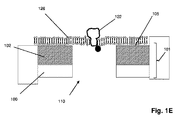
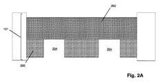
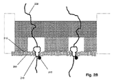
図1Aは特定の実施形態のための本発明の上記の態様を示す。固体状態(solid−state)の膜(100)は第1の側面(104)に不透明コーティング(102)を有して、第1のチャンバー(112)に面する表面(105)および第2のチャンバー(114)に面する第2の側面(106)を有する積層膜(101)を形成している。積層膜(101)は第1のチャンバー(112)を第2のチャンバー(114)から分離し、それぞれが第1のチャンバー(112)と第2のチャンバー(114)との間の流体連通を提供するアパーチャー(110)のアレイを含む。アパーチャー(110)は固体状態または合成のナノポアであってもよく、直接使用しても、それを通して通過が起こるタンパク質ナノポアを固定化するために使用してもよい。一般に、アパーチャーは励起ビームの波長より小さい直径または断面寸法を有し、それによりそのようなビームからの光はアパーチャーを通して透過されない。一部の実施形態では、アパーチャーは100nmまたはそれ未満の直径を有する。一部の実施形態では、円形アパーチャーの直径は励起ビームの波長の0.586倍またはそれ未満である。 FIG. 1A shows the above aspect of the invention for a particular embodiment. The solid-state membrane (100) has an opaque coating (102) on the first side surface (104), a surface (105) facing the first chamber (112) and a second chamber. It forms a laminated film (101) having a second side surface (106) facing (114). The laminated film (101) separates the first chamber (112) from the second chamber (114), each providing fluid communication between the first chamber (112) and the second chamber (114). Includes an array of apertures (110). The aperture (110) may be a solid state or synthetic nanopore and may be used directly or to immobilize a protein nanopore through which passage occurs. In general, apertures have diameters or cross-sectional dimensions that are smaller than the wavelength of the excitation beam, so that light from such beams is not transmitted through the aperture. In some embodiments, the aperture has a diameter of 100 nm or less. In some embodiments, the diameter of the circular aperture is 0.586 times or less than the wavelength of the excitation beam.
前述の段落の一部の実施形態では、本発明の方法は、下記のステップ:(a)第1の側面、第2の側面、およびそれらを通る複数のアパーチャーを有する固相膜を含むナノポアアレイを提供するステップであって、固相膜が第1のチャンバーと第2のチャンバーを分離し、それにより各アパーチャーが第1のチャンバーと第2のチャンバーとの間の流体連通を提供し、固相膜の第1の側面がその上に不透明なコーティングを有し、各アパーチャーが第1の側面の不透明コーティングから第2の側面に向けて延在する検出領域を有するステップと;(b)アパーチャーを通して第1のチャンバーから第2のチャンバーにポリマーを通過させるステップであって、各ポリマーが、それに取着された、ポリマーの特性を示す少なくとも第1の波長を有するシグナルを発生させることができる1つまたは複数の光学標識を有するステップと;(c)第2の波長を有する励起ビームでアパーチャーの検出領域における光学標識を固相膜の第2の側面から照明し、それにより検出領域における光学標識がその第1の波長が第2の波長と異なるシグナルを発生するステップと;(d)検出領域における光学標識からのシグナルを検出するステップであってポリマーの特性を決定するステップとによって実施され得る。 In some embodiments of the preceding paragraph, the method of the invention comprises the following steps: (a) a nanopore array comprising a solid phase film having a first aspect, a second aspect, and multiple apertures passing through them. A solid phase membrane separates the first chamber and the second chamber, whereby each aperture provides fluid communication between the first chamber and the second chamber and is solid. A step in which the first side of the phase membrane has an opaque coating on it, and each aperture has a detection area extending from the opaque coating on the first side towards the second side; (b) aperture. Through the step of passing the polymer from the first chamber to the second chamber, each polymer can generate a signal attached to it having at least the first wavelength characteristic of the polymer. Steps with one or more optical labels; (c) Illuminate the optical label in the detection region of the aperture from the second side of the solid phase film with an excitation beam having a second wavelength, thereby the optical label in the detection region. Can be carried out by a step of generating a signal whose first wavelength is different from that of the second wavelength; (d) a step of detecting a signal from an optical label in the detection region and determining the properties of the polymer. ..
別の実施形態では、図1Cに例示されるように、本発明の方法は、以下のステップ:(a)第1の側面、第2の側面、およびそれらを通る複数のアパーチャーを有する固相膜を含むナノポアアレイを提供するステップであって、固相膜が第1のチャンバーと第2のチャンバーを分離し、それにより各アパーチャーが第1のチャンバーと第2のチャンバーとの間の流体連通を提供し、固相膜の第2の側面がその上に不透明なコーティングを有し、各アパーチャーが第2の側面の不透明コーティングから第2のチャンバー内に延在する検出領域を有するステップと;(b)アパーチャーを通して第1のチャンバーから第2のチャンバーにポリマーを通過させるステップであって、各ポリマーが、それに取着された、ポリマーの特性を示す少なくとも第1の波長を有するシグナルを発生させることができる1つまたは複数の光学標識を有するステップと;(c)第2の波長を有する励起ビームでアパーチャーの検出領域における光学標識を固相膜の第2の側面から照明し、それにより検出領域における光学標識がその第1の波長が第2の波長と異なるシグナルを発生するステップと;(d)検出領域における光学標識からのシグナルを検出するステップであってポリマーの特性を決定するステップとによって実施され得る。 In another embodiment, as illustrated in FIG. 1C, the method of the invention comprises the following steps: (a) a solid phase membrane having a first aspect, a second aspect, and a plurality of apertures passing through them. A solid phase membrane separates the first and second chambers, whereby each aperture provides fluid communication between the first and second chambers. Provided with a step in which the second side of the solid phase film has an opaque coating on it and each aperture has a detection area extending into the second chamber from the opaque coating on the second side; b) In the step of passing the polymer from the first chamber to the second chamber through the aperture, each polymer generates a signal attached to it having at least the first wavelength characteristic of the polymer. With one or more optical labels that can be; (c) an excitation beam with a second wavelength illuminates the optical label in the detection region of the aperture from the second side of the solid phase membrane, thereby the detection region. By the step of generating a signal whose first wavelength is different from that of the second wavelength; (d) the step of detecting the signal from the optical label in the detection region and determining the properties of the polymer. Can be carried out.
一部の実施形態では、不透明コーティングまたは不透明層が金属の場合は常に、最近傍ナノポア距離と励起ビーム波長は、ナノポアアレイを通るプラスモン介在異常透過が最小になるように選択される。そのような選択の指針は、参考として援用される以下の引用文献に開示される:Ebbesenら、Nature, 391: 667−669(1998);Ebbesenら、米国特許第5973316号;同第6040936号;同第6236033号;同第6856715号;同第7057151号;同第7248756号;同第8174696号;Gurら、Optics Comm., 284: 3509−3517(2011);Ghaemiら、Physical Review B, 58: 6779−6782(1998);Pacificiら、Optics Express, 16(12):9222−9238(2008);など。一部の実施形態では、最近傍ナノポア距離(例えばナノポアのランダム(例えばPoisson分布)アレイにおいて予期される最近傍ナノポア距離)は、例えば光学標識を励起するための励起波長とほぼ等しくなるように選択される。 In some embodiments, whenever the opaque coating or opaque layer is metal, the nearest neighbor nanopore distance and excitation beam wavelength are selected to minimize plasmon-mediated anomalous transmission through the nanopore array. Guidance for such selection is disclosed in the following references incorporated by reference: Ebbesen et al., Nature, 391: 667-669 (1998); Ebbesen et al., U.S. Pat. No. 5,973,316; No. 6040936; No. 6236033; No. 6856715; No. 7057151; No. 7248756; No. 8174696; Gur et al., Optics Comm. , 284: 3509-3517 (2011); Ghaemi et al., Physical Review B, 58: 6779-6782 (1998); Pacificici et al., Optics Express, 16 (12): 9222-9238 (2008); etc. In some embodiments, the nearest nanopore distance (eg, the nearest nearest nanopore distance expected in a random (eg, Poisson distribution) array of nanopores) is selected to be approximately equal to, for example, the excitation wavelength for exciting the optical label. Will be done.
一部の実施形態では、シグナル発生領域(116)(または等価な意図された反応部位)はそれぞれ、ナノポアの内部および第2の側面(106)上のナノポアの出口のすぐ隣りに隣接する領域を含み、そのような領域は他のナノポアの等価な領域とは重ならない。一部の実施形態では、標識された核酸分子などの標識された分子が第1のチャンバー(112)に装荷され、またはその中に配置され、直接または挿入されたタンパク質ナノポアを介してのいずれかで、積層膜(101)のアパーチャーを通して第1のチャンバー(112)から第2のチャンバー(114)へ通過する。標識された分子がアパーチャー(110)を通過し、そして/またはアパーチャー(110)を出るとともに、これらの分子は励起ビーム(118)によって直接照明され得る。励起ビーム(118)は標識された分子を直接励起するか、または標識された分子をFRET相互作用を介して間接的に励起するように選択され、または構成され得る。一部の実施形態(図1B)では、シグナル発生領域(120)(または等価な意図された反応部位)はそれぞれ、第1の側面(104)から第2の側面(106)までのナノポアの内部および第2の側面(106)上のナノポアの出口のすぐ隣りに隣接する領域を含み、そのような領域は他のアパーチャー(またはナノポア)の等価な領域とは重ならない。さらに他の実施形態では、シグナル発生領域(または意図された反応部位)はそれぞれ、第1のチャンバーのその入口から第2のチャンバーのその出口までのナノポアの内部およびナノポアの入口と出口のすぐ隣りに隣接する領域を含み、そのような領域は他のアパーチャーの等価な領域とは重ならない。 In some embodiments, the signal generation region (116) (or equivalent intended reaction site) is located within the nanopore and immediately adjacent to the exit of the nanopore on the second aspect (106), respectively. Including, such regions do not overlap with the equivalent regions of other nanopores. In some embodiments, a labeled molecule, such as a labeled nucleic acid molecule, is loaded into or placed in a first chamber (112), either directly or via an inserted protein nanopore. Then, it passes from the first chamber (112) to the second chamber (114) through the aperture of the laminated film (101). As labeled molecules pass through the aperture (110) and / or exit the aperture (110), these molecules can be directly illuminated by the excitation beam (118). The excitation beam (118) can be selected or configured to directly excite the labeled molecule or indirectly excite the labeled molecule via FRET interaction. In some embodiments (FIG. 1B), the signal generation region (120) (or equivalent intended reaction site) is the interior of the nanopore from the first aspect (104) to the second aspect (106), respectively. And the region immediately adjacent to the exit of the nanopore on the second aspect (106), such region does not overlap with the equivalent region of the other aperture (or nanopore). In yet another embodiment, the signal generation regions (or intended reaction sites) are inside the nanopore from its inlet in the first chamber to its exit in the second chamber and immediately adjacent to the inlet and outlet of the nanopore, respectively. Containing regions adjacent to, such regions do not overlap with equivalent regions of other apertures.
上述のように、本発明の積層膜のアパーチャーは、直接ナノポアとして用いてもよく、1つまたは複数のタンパク質ナノポアを保持または固定化するために使用してもよい。後者の型の一部の実施形態では、1つまたは複数のタンパク質ナノポアを、不透明層の表面(例えば図1A、1B、または1Eの105)上、または積層膜の第2の側面(例えば図1Dの106)の表面上のいずれかに配置された脂質二重層の中に埋め込んでもよい。図1Dおよび1Eには、脂質二重層(126)(これは図1Dの表面(106)および図1Eの表面(105)の上に示されている)とともに不透明層またはコーティング(102)、固体状態の膜(100)、アパーチャー(110)を有する積層膜(101)の断面が示されている。示された特定の実施形態では、標識(124)(例えば量子ドット、金属ナノ粒子などのドナー標識、またはFRETシグナルを発生させるための他の蛍光ナノ粒子)が取着されたタンパク質ナノポア(122)が脂質二重層(126)の中に挿入されてが示されているが、他で注記するように、本発明はFRETシグナルの発生のための標識を有しない構成のタンパク質ナノポアを包含する。標識(124)に隣接する(即ち標識(124)のFRET距離内に)シグナル発生領域を有する図1Dの一部の実施形態は、シグナル発生領域が第1の側面(104)から第2の側面(106)のアパーチャーまたはナノポアの出口に近接したスペースまで延在する図1Bに示す構成に対応している。同様に、これも標識(124)に隣接する(例えば標識(124)のFRET距離内に)シグナル発生領域を有する図1Eの一部の実施形態は、シグナル発生領域が表面(105)から第2の側面(106)のアパーチャーまたはナノポアの出口に近接したスペースまで延在する図1Aに示す構成に対応している。 As mentioned above, the aperture of the laminated film of the present invention may be used directly as nanopores or may be used to retain or immobilize one or more protein nanopores. In some embodiments of the latter type, one or more protein nanopores may be placed on the surface of the opaque layer (eg 105 of FIGS. 1A, 1B, or 1E) or a second aspect of the laminated membrane (eg, FIG. 1D). It may be embedded in a lipid bilayer placed on any of the surfaces of 106). 1D and 1E show a lipid bilayer (126), which is shown above the surface (106) of FIG. 1D and the surface (105) of FIG. 1E, as well as an opaque layer or coating (102), solid state. The cross section of the laminated film (101) having the film (100) and the aperture (110) is shown. In the particular embodiment shown, a protein nanopore (122) to which a label (124) (eg, a donor label such as a quantum dot, metal nanoparticles, or other fluorescent nanoparticles for generating a FRET signal) is attached. Is shown inserted into the lipid bilayer (126), but as noted elsewhere, the invention includes protein nanoparticles with unlabeled configurations for the generation of FRET signals. In some embodiments of FIG. 1D having a signal generation region adjacent to the label (124) (ie, within the FRET distance of the label (124)), the signal generation region is from the first side surface (104) to the second side surface. It corresponds to the configuration shown in FIG. 1B extending to the space close to the aperture or the exit of the nanopore of (106). Similarly, in some embodiments of FIG. 1E, which also have a signal generation region adjacent to the label (124) (eg, within the FRET distance of the label (124)), the signal generation region is second from the surface (105). Corresponds to the configuration shown in FIG. 1A extending to the space close to the aperture or nanopore exit on the side surface (106) of.
図1A〜1Eに示すような一部の実施形態では、金属層を安定化させるために、積層膜は酸化ケイ素コーティングなどの酸化物コーティングなどの不動化コーティングをさらに含んでよい。例えば、物理的または化学的堆積手法を使用して金属などの不透明材料で窒化ケイ素膜をコーティングして積層膜を形成させ、その後で集束電子またはイオンビームを使用してアパーチャーをエッチングまたは穿孔して積層膜にアパーチャーのアレイを形成させてもよい。次いでそのようなアレイを、再び化学的配置または蒸着手法を使用して酸化ケイ素などの保護層でさらにコーティングしてもよい。 In some embodiments as shown in FIGS. 1A-1E, the laminated film may further include an immobilizing coating such as an oxide coating such as a silicon oxide coating in order to stabilize the metal layer. For example, a physical or chemical deposition technique is used to coat a silicon nitride film with an opaque material such as metal to form a laminated film, and then the aperture is etched or perforated using focused electrons or ion beams. An array of apertures may be formed on the laminated film. Such arrays may then be further coated with a protective layer such as silicon oxide again using chemical arrangements or vapor deposition techniques.
一部の実施形態では、本発明は、下記のステップ:(a)第1の側面、第2の側面、およびそれらを通る複数のアパーチャーを有する固相膜を含むナノポアアレイを提供するステップであって、固相膜が第1のチャンバーと第2のチャンバーを分離し、それにより各アパーチャーが第1のチャンバーと第2のチャンバーとの間の流体連通を提供し、固相膜の第1の側面がその上に不透明なコーティングを有し、各アパーチャーが第1の側面の不透明コーティングから第2の側面に向けて延在する検出領域を有するステップと;(b)アパーチャーを通して第1のチャンバーから第2のチャンバーにポリマーを通過させるステップであって、各ポリマーが、それに取着された、ポリマーの特性を示す少なくとも第1の波長を有するシグナルを発生させることができる1つまたは複数の光学標識を有するステップと;(c)少なくとも第2の波長を含む励起ビームでアパーチャーの検出領域における光学標識を固相膜の第2の側面から照明し、それにより検出領域における光学標識がその第1の波長が第2の波長と異なるシグナルを発生するステップと;(d)検出領域における光学標識からのシグナルを検出するステップであってポリマーの特性を決定するステップとによって、核酸およびタンパク質を含む生物学的ポリマーなどのポリマーを特性決定するための方法を含み得る。一部の実施形態では、ポリマーはポリヌクレオチドまたはタンパク質であってもよい。さらに他の実施形態では、ポリマーはポリヌクレオチドであってもよい。さらなる実施形態では、ポリヌクレオチドは一本鎖核酸であってもよい。一部の実施形態では、解析または決定されるポリマーの特性はヌクレオチド配列などのポリマーのモノマー配列である。一部の実施形態では、ポリマー上の光学標識は、参照により本明細書に組み込まれる米国特許ならびに特許および国際公開:第8,771,491号;第2013/0203050号;またはWO2014/190322に記載されているようなFRET標識である。一部の実施形態では、アパーチャーはタンパク質ナノポアを含む。簡単に言うと、一部の実施形態ではFRET標識は各シグナル発生領域に少なくとも1つのFRETドナー標識および少なくとも1つのFRETアクセプター標識を含み、励起ビームはFRETドナー標識を励起し、これが次にドナー標識のFRET距離内にあるFRETアクセプター標識にエネルギーを伝達し、これが次に光学シグナルを放出する。典型的には、励起ビームは第2の波長を含み、光学シグナルは第2の波長とは異なる第1の波長を含み、例えば落射照明システムの使用が可能になる。一部の実施形態では、検出領域は第1の側面の不透明コーティングから第2の側面に向けて延在し、アパーチャーおよび/またはナノポアの出口のすぐ近くに隣接する膜外スペースを含む。一部の実施形態では、そのような膜外スペースはナノポアまたはアパーチャーの出口から50nmを超えて延在しない。他の実施形態では、そのような膜外スペースはナノポアまたはアパーチャーの出口から10nmを超えて延在しない。 In some embodiments, the invention is a step of providing a nanopore array comprising the following steps: (a) a first aspect, a second aspect, and a solid phase film having multiple apertures passing through them. Thus, the solid phase membrane separates the first and second chambers, whereby each aperture provides fluid communication between the first chamber and the second chamber, the first of the solid phase membranes. With a step in which the sides have an opaque coating on it and each aperture has a detection area extending from the opaque coating on the first side towards the second side; (b) from the first chamber through the aperture. A step of passing the polymer through a second chamber, wherein each polymer can generate a signal attached to it having at least the first wavelength indicating the properties of the polymer. (C) Illuminate the optical label in the detection region of the aperture from the second side of the solid phase membrane with an excitation beam containing at least the second wavelength, whereby the optical label in the detection region is its first. Biology containing nucleic acids and proteins by the step of generating a signal whose wavelength is different from the second wavelength; (d) the step of detecting the signal from the optical label in the detection region and determining the properties of the polymer. It may include methods for characterizing a polymer, such as an optical polymer. In some embodiments, the polymer may be a polynucleotide or protein. In yet other embodiments, the polymer may be a polynucleotide. In a further embodiment, the polynucleotide may be a single-stranded nucleic acid. In some embodiments, the characteristic of the polymer analyzed or determined is the monomer sequence of the polymer, such as a nucleotide sequence. In some embodiments, optical labels on polymers are described in US Patents and Patents and Publications: 8,771,491; 2013/0203050; or WO2014 / 190322, which are incorporated herein by reference. It is a FRET label as it is. In some embodiments, the aperture comprises a protein nanopore. Briefly, in some embodiments, the FRET label comprises at least one FRET donor label and at least one FRET acceptor label in each signal generation region, and the excitation beam excites the FRET donor label, which in turn is the donor label. Transfers energy to a FRET acceptor label within the FRET distance of, which in turn emits an optical signal. Typically, the excitation beam contains a second wavelength and the optical signal contains a first wavelength that is different from the second wavelength, allowing the use of, for example, epi-illumination systems. In some embodiments, the detection area extends from the opaque coating on the first side to the second side and includes an adjacent extramembrane space in the immediate vicinity of the aperture and / or the exit of the nanopore. In some embodiments, such extramembrane space does not extend beyond 50 nm from the exit of the nanopore or aperture. In other embodiments, such extramembrane space does not extend beyond 10 nm from the exit of the nanopore or aperture.
簡単に言うと、米国特許第8,771,491号に、より完全に記載されているように、一部の実施形態では、アパーチャーおよび/またはナノポアは1つまたは複数のFRETドナーで標識され、ポリマーはそれぞれFRETアクセプターで標識されて、それにより少なくとも選択されたドナーおよびアクセプターがFRET対を形成し、即ちドナーの発光スペクトルが少なくとも1つのアクセプターの吸光スペクトルと重なり、それにより他の条件が合致すれば(例えばドナー励起、ドナーとアクセプターがFRET距離内に存在する、ドナーとアクセプターが適正な相対的配向にあるなど)、FRET相互作用が起こり得る。FRET相互作用においてはドナーの励起エネルギーが非放射的にアクセプターに伝達され、その後にアクセプターがドナーの励起エネルギーよりも低いエネルギーの光学シグナルを放出する。ドナーは通常、レーザーによって発生するような光ビームを照明することによって励起される。 Simply put, apertures and / or nanopores are labeled with one or more FRET donors in some embodiments, as described more fully in US Pat. No. 8,771,491. Each polymer is labeled with a FRET acceptor so that at least the selected donors and acceptors form a FRET pair, i.e. the donor emission spectrum overlaps the absorption spectrum of at least one acceptor, thereby meeting other conditions. If (eg, donor excitation, donor and acceptor are within FRET distance, donor and acceptor are in proper relative orientation, etc.), FRET interaction can occur. In a FRET interaction, the donor's excitation energy is non-radiatively transferred to the acceptor, after which the acceptor emits an optical signal with an energy lower than the donor's excitation energy. Donors are usually excited by illuminating a beam of light, such as that produced by a laser.
一部の実施形態では、参照により本明細書に組み込まれるHuberら、米国特許公開第2013/0203050号に記載されているように、タンパク質ナノポアを脂質二重層なしに、または僅か少量の脂質二重層とともに、固体状態の膜中に挿入してアレイを形成してもよい。そのようなタンパク質ナノポア/アパーチャー構成を図1Fに示す。この特定の実施形態では、固体状態の膜(100)および不透明層(102)を含む積層膜(101)は、量子ドットなどの光学活性粒子であってもよいドナー(128)で標識された、タンパク質ナノポア(122)がその中に固定化されたアパーチャー(107)を含む。アクセプターで標識されたポリマー(130)がタンパク質ナノポア(122)のボアを通過して出ていく際に、アクセプター標識はドナー(128)のFRET距離の中を通過する。アパーチャー(107)の大きさは単一のタンパク質ナノポアを固定化できるように作られている。励起ビーム(133)によってアパーチャー(107)を直接照明すれば、ドナー(128)が励起され、タンパク質ナノポア(122)を通過している標識されたポリマー(130)のアクセプターとのFRET相互作用に入ることが可能になる。一方、不透明層(102)は、ビーム(133)として不透明層(102)の反対側にある標識されたポリマー(131)の上にあるアクセプターを照明するビーム(133)の放射を減少させる。不透明層(102)は、ビーム(133)を吸収(135)および/または反射(137)することによって、ビーム(133)からの放射を遮蔽する。 In some embodiments, as described in Huber et al., US Patent Publication No. 2013/2030050, which is incorporated herein by reference, protein nanopores can be applied without a lipid bilayer or with only a small amount of lipid bilayer. At the same time, it may be inserted into a membrane in a solid state to form an array. The composition of such protein nanopores / apertures is shown in FIG. 1F. In this particular embodiment, the laminated film (101) comprising the solid state film (100) and the opaque layer (102) is labeled with a donor (128), which may be optically active particles such as quantum dots. The protein nanopore (122) contains an immobilized aperture (107) therein. As the acceptor-labeled polymer (130) exits through the bore of the protein nanopore (122), the acceptor label passes through the FRET distance of the donor (128). The size of the aperture (107) is designed to allow immobilization of a single protein nanopore. Direct illumination of the aperture (107) with the excitation beam (133) excites the donor (128) and enters the FRET interaction with the acceptor of the labeled polymer (130) passing through the protein nanopore (122). Will be possible. The opaque layer (102), on the other hand, reduces the emission of the beam (133) that illuminates the acceptor above the labeled polymer (131) on the opposite side of the opaque layer (102) as the beam (133). The opaque layer (102) shields radiation from the beam (133) by absorbing (135) and / or reflecting (137) the beam (133).
図1Gにさらなる実施形態を示す。ここでは金属層などの不透明層(102)の表面が第2のチャンバー(114)の境界を形成している。一部の実施形態では、固体状態のアパーチャー(146)のアレイが従来のマイクロマシン手法を使用して製造され得る。例えば、シリコン基板(140)に窒化ケイ素層などの非電導性の固体状態の膜(100)が例えば化学蒸着法などの手法で加えられる。一部の実施形態では、層(100)は30〜100nmの範囲である。層(100)の上に、例えば化学蒸着法などの手法で金属層などの不透明層(102)が加えられて三層シートが形成される。不透明層(102)にフォトレジストの層を加えてもよく、これが除去された後にアパーチャーの位置にフォトレジスト材料が現像され、不透明層(102)および固体状態の膜(100)を通してエッチングすることによってホールが形成されて、アパーチャー(146)の部分が形成される。シリコン層(140)を通して第2のエッチングが行なわれて、アレイ中に残りのアパーチャー(146)が形成される。本発明の一実施形態によるナノポアセンシングデバイスは、FRETドナー標識(124)を有する少なくとも1つのタンパク質ナノポア(122)がその中に挿入された不透明層(102)の表面(141)上に脂質二重層(126)を堆積配置することによって構成することができる。第1のチャンバー(112)の中に配置されると、例えば2つの異なる種類のモノマーのためのアクセプター標識(151)および(152)を含む帯電し標識されたポリマー(150)は、各アクセプター標識(151または152)を拘束するタンパク質ナノポア(122)を通過して、ドナー標識(124)のFRET距離(142)内に来ることができる。ドナー標識(124)はビーム(133)によって励起され、FRET距離(142)内にあるアクセプター標識に励起エネルギーを伝達し、これが次に標識が取着されたモノマーを示すFRETシグナル(144)を放出する。 FIG. 1G shows a further embodiment. Here, the surface of the opaque layer (102), such as the metal layer, forms the boundary of the second chamber (114). In some embodiments, an array of solid state apertures (146) can be manufactured using conventional micromachine techniques. For example, a non-conducting solid state film (100) such as a silicon nitride layer is added to a silicon substrate (140) by a method such as a chemical vapor deposition method. In some embodiments, the layer (100) is in the range of 30-100 nm. An opaque layer (102) such as a metal layer is added on the layer (100) by a method such as a chemical vapor deposition method to form a three-layer sheet. A layer of photoresist may be added to the opaque layer (102), after which the photoresist material is developed at the aperture position and etched through the opaque layer (102) and the solid state film (100). A hole is formed to form a portion of the aperture (146). A second etching is performed through the silicon layer (140) to form the remaining aperture (146) in the array. The nanopore sensing device according to one embodiment of the present invention is a lipid bilayer on the surface (141) of an opaque layer (102) into which at least one protein nanopore (122) having a FRET donor label (124) is inserted. It can be configured by depositing (126). When placed in the first chamber (112), the charged and labeled polymer (150), including, for example, the acceptor labels (151) and (152) for two different types of monomers, is each acceptor label. It can pass through the protein nanopores (122) that constrain (151 or 152) and come within the FRET distance (142) of the donor label (124). The donor label (124) is excited by the beam (133) and transfers excitation energy to the acceptor label within the FRET distance (142), which in turn emits a FRET signal (144) indicating the monomer to which the label is attached. do.
さらに他の実施形態では、図1Hに示すように、脂質二重層(126)が層(100)の表面(104)上に配置され、それにより励起ビーム(133)が向けられる側面に対向する積層膜(101)の側面にタンパク質ナノポア(122)が挿入される。 In yet another embodiment, as shown in FIG. 1H, the lipid bilayer (126) is placed on the surface (104) of the layer (100), thereby facing the side surface to which the excitation beam (133) is directed. A protein nanopore (122) is inserted into the side surface of the membrane (101).
一部の実施形態では不透明層は不透明な多孔質層を含んでもよく、これは一部の実施形態では、図2Aおよび2Bに示すように、不透明なナノ多孔質層であってもよい。そのような実施形態では、積層膜(101)は、固体状態の膜(200)と、アパーチャー(204)の層を横断する間接的なおよび/またはヘビ状の通路を提供するナノメーターサイズの孔を有する不透明材料を含むナノ多孔質層(202)とを含む。図2Bは、固体状態の膜(200)の第2の側面(216)に配置された脂質二重層(206)の中に挿入された、標識されたタンパク質ナノポア(210)を含む実施形態を示す。図1A〜1Hの実施形態と同様に、ナノ多孔質層(202)は固体状態の膜(200)の第2の側面(216)に向けられた放射がナノ多孔質層(202)の他の側面上の標識されたポリマーまたは他の材料に到達することを遮蔽または低減し、それによりタンパク質ナノポア(210)の出口における検出事象からの光学シグナルにおける望ましくないノイズの発生を低減する。 In some embodiments, the opaque layer may include an opaque porous layer, which in some embodiments may be an opaque nanoporous layer, as shown in FIGS. 2A and 2B. In such an embodiment, the laminated film (101) is a nanometer-sized hole that provides an indirect and / or snake-like passage across the solid state film (200) and the layer of the aperture (204). Includes a nanoporous layer (202) containing an opaque material having. FIG. 2B shows an embodiment comprising a labeled protein nanopore (210) inserted into a lipid bilayer (206) disposed on the second aspect (216) of a solid state membrane (200). .. Similar to the embodiments of FIGS. 1A-1H, the nanoporous layer (202) emits radiation directed at the second side surface (216) of the solid state membrane (200) to the other nanoporous layer (202). It shields or reduces access to the labeled polymer or other material on the flanks, thereby reducing the generation of unwanted noise in the optical signal from the detection event at the exit of the protein nanopore (210).
上述のように、一部の実施形態では、ポリマー分析物上の標識またはナノポア上のドナーを直接照明するために、励起ビームの送達と光学シグナルの収集が単一の対物レンズを通して起こる落射照明システムが使用される。本発明で使用する共焦点落射照明システムの基本的構成要素を図3に示す。励起ビーム(302)は二色性ミラー(304)を通って対物レンズ(306)に向かって通過し、対物レンズ(306)は励起ビーム(302)を積層膜(300)に集中させ(310)、その中で標識が直接励起されて蛍光シグナルなどの光学シグナルを放出するか、またはFRET相互作用を介して間接的に励起されて光学シグナルを放出する。そのような光学シグナルは対物レンズ(306)によって収集され、励起ビーム(302)の光は通過させるが光学シグナル(311)の光は反射するように選択された二色性ミラー(304)に向けられる。反射された光学シグナル(311)は、光学シグナル(311)をピンホール(316)を通して検出器(318)に集中させるレンズ(314)を通過する。 As mentioned above, in some embodiments, an epi-illumination system in which the delivery of an excitation beam and the collection of optical signals occur through a single objective lens to directly illuminate a marker on a polymer analyte or a donor on a nanopore. Is used. The basic components of the confocal epi-illumination system used in the present invention are shown in FIG. The excitation beam (302) passes through the bicolor mirror (304) toward the objective lens (306), and the objective lens (306) concentrates the excitation beam (302) on the laminated film (300) (310). In it, the label is directly excited to emit an optical signal such as a fluorescent signal, or indirectly excited via a FRET interaction to emit an optical signal. Such an optical signal is collected by the objective lens (306) and directed at a dichroic mirror (304) selected to pass the light of the excitation beam (302) but reflect the light of the optical signal (311). Be done. The reflected optical signal (311) passes through a lens (314) that concentrates the optical signal (311) through the pinhole (316) to the detector (318).
一部の実施形態では、一本鎖核酸を含むポリマーのための上記の方法を実施するためのデバイスは、典型的には積層膜とナノポアを横断する電場を確立するための一組の電極を含む。一本鎖核酸は、チャンバー内に陰極を配置することによって積層膜の「シス」側として構成された第1のチャンバー内の電解質の中に配置することによってナノポアに曝される。電場を印加すると、陰荷電を有する一本鎖核酸はナノポアに捕捉され、チャンバー内に陽極を配置することによって膜の「トランス」側として構成された積層膜の他の側の第2のチャンバーに通過する。通過速度は、第1および第2のチャンバー内の電解質のイオン強度ならびにナノポアを横断して印加された電圧に部分的に依存する。光学に基づく検出においては、通過速度は、例えば異なる電圧でナノポアあたりに予想される異なる速度でシグナルを発生する標識された一本鎖核酸の所定の標準を使用する予備的較正測定によって選択され得る。したがって、DNA配列決定用途には、通過速度はそのような較正測定からのシグナル速度に基づいて選択され得る。したがって、そのような測定から、例えばナノポアのアレイにわたって信頼できるヌクレオチドの識別が可能になるか最大である電圧が選択され得る。一部の実施形態では、そのような較正は(所定の標準配列の代わりに、またはそれに加えて)分析されるテンプレートの試料からの核酸を使用して行なうことができる。一部の実施形態では、そのような較正は配列決定ランの間にリアルタイムで行なってもよく、印加する電圧は例えばヌクレオチド特異的シグナルの取得を最大にするためにそのような測定に基づいてリアルタイムで改変してもよい。
ナノポアおよびナノポアアレイ
In some embodiments, the device for performing the above method for polymers containing single-stranded nucleic acids typically comprises a set of electrodes to establish an electric field across the laminated membrane and nanopores. include. Single-stranded nucleic acids are exposed to nanopores by placing them in an electrolyte in a first chamber configured as the "cis" side of the laminated membrane by placing a cathode in the chamber. When an electric field is applied, the negatively charged single-stranded nucleic acid is captured by the nanopores and placed in the chamber to place an anode in the second chamber on the other side of the laminated membrane, which is configured as the "trans" side of the membrane. pass. The passing speed depends in part on the ionic strength of the electrolyte in the first and second chambers and the voltage applied across the nanopores. In optical-based detection, the pass rate can be selected, for example, by preliminary calibration measurements using predetermined standards of labeled single-stranded nucleic acids that generate signals at different rates expected per nanopore at different voltages. .. Therefore, for DNA sequencing applications, transit rates can be selected based on signal rates from such calibration measurements. Thus, from such measurements, the voltage at which reliable nucleotide identification is possible or maximum, eg, across an array of nanopores, can be selected. In some embodiments, such calibration can be performed using nucleic acids from the sample of the template being analyzed (instead of or in addition to a given standard sequence). In some embodiments, such calibration may be performed in real time during the sequencing run, and the applied voltage is, for example, real time based on such measurements to maximize the acquisition of nucleotide-specific signals. It may be modified with.
Nanopores and Nanopore Arrays
上述のように、本発明で使用するナノポアは固体状態のナノポア、タンパク質ナノポア、またはタンパク質ナノポアもしくは有機ナノチューブ、例えば固体状態の膜もしくは同様のフレームワークで構成された炭素もしくはグラフェンナノチューブを含むハイブリッドナノポアであってもよい。ナノポアの重要な特徴には、ポリヌクレオチドなどのポリマー分析物を拘束し、それによりそのモノマーが検出ゾーン(またはシグナル発生領域)を順に通過する(即ちヌクレオチドが検出ゾーンを一度に一つずつ、または一列で通過する)ことが含まれる。一部の実施形態では、ナノポアの追加的な特徴には、二本鎖核酸または等価の嵩高な分子を通過させずに一本鎖核酸を通過させることが含まれる。 As mentioned above, the nanopores used in the present invention are solid nanopores, protein nanopores, or hybrid nanopores containing protein nanopores or organic nanotubes, such as carbon or graphene nanotubes composed of solid membranes or similar frameworks. There may be. An important feature of nanopores is that they constrain polymer analytes such as polynucleotides, whereby their monomers pass through detection zones (or signaling regions) in sequence (ie, nucleotides pass through detection zones one at a time, or one at a time. Passing in a row) is included. In some embodiments, additional features of nanopores include passing single-stranded nucleic acids without passing through double-stranded nucleic acids or equivalent bulky molecules.
一部の実施形態では、本発明の方法およびデバイスと併せて使用されるナノポアは、平面上に規則的に配置され得るナノポアのクラスターのアレイのような、アレイの形態で提供される。一部の実施形態では、異なるクラスターのナノポアからの光学シグナルが、用いられる光学検出システムによって区別可能であるように、クラスターはそれぞれ個別の分解能限界区域内にあるが、同じクラスター内のナノポアからの光学シグナルは、用いられる光学検出システムによって、そのようなクラスター内の特定のナノポアに必ずしも割り当てることができる必要はない。 In some embodiments, the nanopores used in conjunction with the methods and devices of the invention are provided in the form of an array, such as an array of clusters of nanopores that can be regularly arranged in a plane. In some embodiments, the clusters are within separate resolution limit areas, but from the nanopores within the same cluster, so that the optical signals from the nanopores of different clusters can be distinguished by the optical detection system used. The optical signal does not necessarily have to be able to be assigned to a particular nanopore within such a cluster by the optical detection system used.
固体状態のナノポアは、窒化ケイ素(Si3N4)および二酸化ケイ素(SiO2)などを含むがこれらに限定されない様々な材料において製作されてもよい。DNA配列決定などの分析の適用例に関するナノポアの製作および動作は、参照により組み込まれる以下の例示的な参考文献に開示されている:Ling,米国特許第7,678,562号;Hu et al,米国特許第7,397,232号;Golovchenko et al,米国特許第6,464,842号;Chu et al,米国特許第5,798,042号;Sauer et al,米国特許第7,001,792号;Su et al,米国特許第7,744,816号;Church et al,米国特許第5,795,782号;Bayley et al,米国特許第6,426,231号;Akeson et al,米国特許第7,189,503号;Bayley et al,米国特許第6,916,665号;Akeson et al,米国特許第6,267,872号;Meller et al,米国特許公開第2009/0029477号;Howorka et al,国際特許公開WO2009/007743;Brown et al,国際特許公開WO2011/067559;Meller et al,国際特許公開WO2009/020682;Polonsky et al,国際特許公開WO2008/092760;Van der Zaag et al,国際特許公開WO2010/007537;Yan et al,Nano Letters,5(6):1129−1134(2005);Iqbal et al,NatureNanotechnology,2:243−248(2007);Wanunu et al,Nano Letters,7(6):1580−1585(2007);Dekker, Nature Nanotechnology,2:209−215(2007);Storm et al,Nature Materials,2:537−540(2003);Wu et al,Electrophoresis,29(13):2754−2759(2008);Nakane et al,Electrophoresis,23:2592−2601(2002);Zhe et al,J.Micromech.Microeng.,17:304−313(2007);Henriquez et al,The Analyst,129:478−482(2004);Jagtiani et al,J.Micromech.Microeng.,16:1530−1539(2006);Nakane et al,J.Phys.Condens.Matter,15 R1365−R1393(2003);DeBlois et al,Rev.Sci.Instruments,41(7):909−916(1970);Clarke et al,Nature Nanotechnology,4(4):265−270(2009);Bayley et al,米国特許公開第2003/0215881号;など。
Nanopores in the solid state, may be fabricated in a variety of materials including, but not limited thereto, silicon nitride (
一部の実施形態では、本発明は、1つまたは複数の光遮蔽層、即ち1つまたは複数の不透明層を有するナノポアアレイを含む。典型的には、ナノポアアレイはケイ素、窒化ケイ素、酸化ケイ素、酸化アルミニウムなどの材料の薄いシートとして製造され、これらは特に使用される厚み、例えば50〜100nm未満で容易に光を透過する。分析物の電気的検出にはこれは問題ではない。しかし、ナノポアを通過する標識された分子の光学に基づく検出においては、アレイを通して透過される光は意図された反応部位の外側の材料を常に励起し、それにより、例えば非特異的バックグラウンド蛍光、ナノポアにまだ入っていない分子の標識からの蛍光などの光学的ノイズを発生させる。一態様では、本発明は、励起ビームからの光を反射および/または吸収する1つまたは複数の光遮蔽層を有するナノポアアレイを提供し、それによりアレイのナノポアに付随する意図された反応部位において発生する光学シグナルへのバックグラウンドノイズを低減することによって、この問題に対処する。一部の実施形態では、意図された反応部位における光学標識が直接照明によって励起されることがこれによって可能になる。一部の実施形態では、不透明層は金属層であってもよい。そのような金属層はSn、Al、V、Ti、Ni、Mo、Ta、W、Au、AgまたはCuを含んでよい。一部の実施形態では、そのような金属層はAl、Au、AgまたはCuを含んでよい。さらに他の実施形態では、そのような金属層はアルミニウムもしくは金を含んでよく、またはアルミニウムのみを含んでもよい。不透明層の厚みは広く変化してもよく、層を構成する材料の物理的および化学的特性による。一部の実施形態では、不透明層の厚みは少なくとも5nm、または少なくとも10nm、または少なくとも40nmであってもよい。他の実施形態では、不透明層の厚みは5〜100nmの範囲であってもよく、他の実施形態では、不透明層の厚みは10〜80nmの範囲であってもよい。不透明層は励起ビームからの光を100パーセント遮蔽する(即ち反射しまたは吸収する)必要はない。一部の実施形態では、不透明層は励起ビームからの入射光を少なくとも10パーセント遮蔽してもよく、他の実施形態では、不透明層は励起ビームからの入射光を少なくとも50パーセント遮蔽してもよい。 In some embodiments, the invention comprises a nanopore array having one or more light shielding layers, i.e., one or more opaque layers. Typically, nanopore arrays are manufactured as thin sheets of materials such as silicon, silicon nitride, silicon oxide, aluminum oxide, etc., which easily transmit light at thicknesses specifically used, such as less than 50-100 nm. This is not a problem for the electrical detection of the analyte. However, in the optical-based detection of labeled molecules passing through the nanopores, the light transmitted through the array always excites the material outside the intended reaction site, thereby, for example, non-specific background fluorescence, Generates optical noise such as fluorescence from the labels of molecules that have not yet entered the nanopores. In one aspect, the invention provides a nanopore array with one or more light shielding layers that reflect and / or absorb light from an excitation beam, thereby at the intended reaction site associated with the nanopores of the array. This problem is addressed by reducing the background noise to the resulting optical signal. In some embodiments, this allows the optical label at the intended reaction site to be excited by direct illumination. In some embodiments, the opaque layer may be a metal layer. Such a metal layer may contain Sn, Al, V, Ti, Ni, Mo, Ta, W, Au, Ag or Cu. In some embodiments, such metal layers may comprise Al, Au, Ag or Cu. In yet other embodiments, such metal layers may contain aluminum or gold, or may contain only aluminum. The thickness of the opaque layer may vary widely, depending on the physical and chemical properties of the materials that make up the layer. In some embodiments, the thickness of the opaque layer may be at least 5 nm, or at least 10 nm, or at least 40 nm. In other embodiments, the thickness of the opaque layer may be in the range of 5 to 100 nm, and in other embodiments, the thickness of the opaque layer may be in the range of 10 to 80 nm. The opaque layer does not need to shield (ie reflect or absorb) 100% of the light from the excitation beam. In some embodiments, the opaque layer may shield incident light from the excitation beam by at least 10 percent, and in other embodiments, the opaque layer may shield incident light from the excitation beam by at least 50 percent. ..
不透明層またはコーティングは、当該技術分野で公知の種々の手法によって固体状態の膜上に製作することができる。化学蒸着法、電着、エピタキシー、熱酸化、蒸発およびスパッタリングなどの物理蒸着法、キャスティングなどの材料堆積手法を使用することができる。一部の実施形態では、原子層堆積を使用することができる。例えば参照により組み込まれる米国特許第6,464,842号;Weiら、Small、6巻(13号)、1406〜1414頁(2010年)。 The opaque layer or coating can be made on a solid film by various techniques known in the art. Physical vapor deposition methods such as chemical vapor deposition, electrodeposition, epitaxy, thermal oxidation, evaporation and sputtering, and material deposition techniques such as casting can be used. In some embodiments, atomic layer deposition can be used. For example, US Pat. No. 6,464,842 incorporated by reference; Wei et al., Small, Vol. 6 (13), pp. 1406-1414 (2010).
一部の実施形態では、1〜100nmのチャネルまたはアパーチャーが固体基材、通常は平面基材、例えば、膜を通って形成されてよく、一本鎖DNAなどの分析物がそこを通って移行するように誘発される。別の実施形態では、2〜50nmのチャネルまたはアパーチャーが基材を通って形成され、さらに別の実施形態では、2〜30nm、または2〜20nm、または3〜30nm、または3〜20nm、または3〜10nmのチャネルまたはアパーチャーが基材を通って形成される。ナノポアを生成する固体状態の手法は、堅牢で耐久性を提供すると同時にナノポアのサイズおよび形状を調整する能力、ウェハー規模でナノポアの高密度アレイを製作する能力、脂質ベースの系に比べて優れた機械的、化学的および熱的特性、ならびに電子または光学的読出し技法と一体化する可能性を提供する。一方、生物学的ナノポアは、再現性のある狭いボアまたは管腔、特に1〜10ナノメートルの範囲にあるもの、ならびに従来のタンパク質を設計製作する方法によって、ナノポアの物理的および/または化学的性質を調整するための、かつFRETドナーまたはアクセプターであってもよい蛍光標識などの基または要素を直接または間接的に取着するための技法を提供する。タンパク質ナノポアは、典型的には、機械的支持体に関する繊細な脂質二重層に依拠し、精密な寸法を持つ固体状態のナノポアの製作は、依然として難しいままである。一部の実施形態では、固体状態のナノポアは、生物学的ナノポアと組合されていわゆる「ハイブリッド」ナノポアを形成し、これは、これらの欠点のいくつかを克服し、そうすることで固体状態のナノポアの安定性に生物学的ポアタンパク質の精密さを提供する。光学的読出し技法では、ハイブリッドナノポアが、ナノポアの精密な場所を提供し、このことがデータ獲得を大幅に単純化する。 In some embodiments, channels or apertures of 1-100 nm may be formed through a solid substrate, usually a planar substrate, eg, a membrane, through which the analyte, such as single-stranded DNA, migrates. Induced to do. In another embodiment a channel or aperture of 2-50 nm is formed through the substrate, and in yet another embodiment 2-30 nm, or 2-20 nm, or 3-30 nm, or 3-20 nm, or 3 A 10 nm channel or aperture is formed through the substrate. The solid-state approach to producing nanopores provides robustness and durability, while at the same time being able to adjust the size and shape of nanopores, to produce high density arrays of nanopores on a wafer scale, and to be superior to lipid-based systems. It offers mechanical, chemical and thermal properties, as well as the possibility of integrating with electronic or optical readout techniques. Biological nanopores, on the other hand, are physical and / or chemical nanopores by means of reproducible narrow bores or cavities, especially those in the range of 1-10 nanometers, and methods of designing and manufacturing conventional proteins. Techniques are provided for adjusting properties and for directly or indirectly attaching groups or elements such as fluorescent labels that may be FRET donors or acceptors. Protein nanopores typically rely on a delicate lipid bilayer for mechanical supports, and the production of solid-state nanopores with precise dimensions remains difficult. In some embodiments, solid-state nanopores are combined with biological nanopores to form so-called "hybrid" nanopores, which overcome some of these shortcomings and in doing so solid-state. It provides the precision of biological pore proteins for the stability of nanopores. In optical readout techniques, hybrid nanopores provide a precise location for the nanopores, which greatly simplifies data acquisition.
一部の実施形態では、クラスターは、アパーチャーのアレイを含有する固相膜によって支持された脂質二重層内に、タンパク質ナノポアを配置することによって形成されてもよい。例えば、そのようなアレイは、固相支持体に製作された(例えば、穴を開けられ、またはエッチングされるなど)アパーチャーを含んでいてもよい。そのようなアパーチャーの幾何形状は、用いられる製作技法に応じて変わってもよい。一部の実施形態では、そのようなアパーチャーのそれぞれは、個別の分解能限界区域に関連付けられ、または包含され;しかし他の実施形態では、多数のアパーチャーが同じ分解能限界区域内にあってもよい。アパーチャーの断面積は、広く様々であってもよく、異なるクラスター間と同じでもそうでなくてもよいが、そのような面積は通常、従来の製作手法の結果と実質的に同じである。一部の実施形態では、アパーチャーは、10から200nmの範囲の最小線寸法(例えば、円形アパーチャーの場合には直径)を有し、または約100から3×10⁴nm2の範囲の面積を有する。アパーチャーを横切って、脂質二重層が配置されてもよい。アパーチャー当たりのタンパク質ナノポアの分布は、例えば挿入ステップ中にタンパク質ナノポアの濃度を制御することによって、様々にしてもよい。そのような実施形態では、ナノポアのクラスターは、ランダムな数のナノポアを含んでいてもよい。タンパク質ナノポアがアパーチャーにランダムに挿入される一部の実施形態では、平均して1つまたは複数のアパーチャーを含有するクラスターは、ゼロよりも大きい所定の数のタンパク質ナノポアを有し;他の実施形態では、そのようなクラスターは、0.25よりも大きい所定の数のタンパク質ナノポアを有し;他の実施形態では、そのようなクラスターは、0.5よりも大きい所定の数のタンパク質ナノポアを有し;他の実施形態では、そのようなクラスターは、0.75よりも大きい所定の数のタンパク質ナノポアを有し;他の実施形態では、そのようなクラスターは、1.0よりも大きい所定の数のタンパク質ナノポアを有する。 In some embodiments, clusters may be formed by placing protein nanopores within a lipid bilayer supported by a solid phase membrane containing an array of apertures. For example, such an array may include apertures made into a solid phase support (eg, pierced or etched). The geometry of such apertures may vary depending on the fabrication technique used. In some embodiments, each of such apertures is associated with or included in a separate resolution limit zone; however, in other embodiments, multiple apertures may be within the same resolution limit zone. The cross-sectional area of the aperture may vary widely and may or may not be the same between different clusters, but such areas are usually substantially the same as the result of conventional fabrication techniques. In some embodiments, the aperture has a minimum line size ranging from 10 to 200 nm (eg, diameter in the case of a circular aperture), or an area ranging from about 100 to 3 × 10 4 nm 2. .. A lipid bilayer may be placed across the aperture. The distribution of protein nanopores per aperture may vary, for example by controlling the concentration of protein nanopores during the insertion step. In such an embodiment, the cluster of nanopores may contain a random number of nanopores. In some embodiments where protein nanopores are randomly inserted into apertures, clusters containing one or more apertures on average have a predetermined number of protein nanopores greater than zero; other embodiments. So, such clusters have a predetermined number of protein nanopores greater than 0.25; in other embodiments, such clusters have a predetermined number of protein nanopores greater than 0.5. In other embodiments, such clusters have a predetermined number of protein nanopores greater than 0.75; in other embodiments, such clusters have a predetermined number greater than 1.0. It has a number of protein nanopores.
一部の実施形態では、本発明の方法およびデバイスは、SiN膜などの固相膜を含み、それを通るアパーチャーのアレイを有して、第1のチャンバーと第2のチャンバー(場合によっては、「シスチャンバー」および「トランスチャンバー」とも呼ぶ)との間に連通を設け、第2のまたはトランスチャンバーに面する表面に脂質二重層を支持する。一部の実施形態では、そのような固相膜内のアパーチャーの直径は、10から200nmの範囲または20から100nmの範囲にあってもよい。一部の実施形態では、そのような固相膜は、そのような二重層がトランスチャンバーに面する表面のアパーチャーに跨る領域で、脂質二重層に挿入されたタンパク質ナノポアをさらに含む。一部の実施形態では、そのようなタンパク質ナノポアは、本明細書に記述される技法を使用して、固相膜のシス側から挿入される。一部の実施形態では、そのようなタンパク質ナノポアは、軸に沿ってバレルまたはボアを含み、かつ一端に「キャップ」構造を有し他端に「ステム」構造をするという点で(Songら、Science、274巻:1859〜1866頁(1996年)からの専門用語を使用する)、α−溶血素と同一のまたは類似の構造を有する。そのようなタンパク質ナノポアを使用する一部の実施形態では、脂質二重層への挿入は、そのキャップ構造がシスチャンバーに向けて露出し、かつそのステム構造がトランスチャンバーに向けて露出するように配向するタンパク質ナノポアをもたらす。 In some embodiments, the methods and devices of the invention include a solid phase membrane, such as a SiN membrane, which has an array of apertures through which a first chamber and a second chamber (possibly, in some cases). A communication is provided between the "cis chamber" and the "transchamber") to support the lipid bilayer on the surface facing the second or transchamber. In some embodiments, the diameter of the aperture in such a solid phase membrane may be in the range of 10 to 200 nm or 20 to 100 nm. In some embodiments, such solid phase membranes further include protein nanopores inserted into the lipid bilayer at the region where such bilayer straddles the surface aperture facing the transchamber. In some embodiments, such protein nanopores are inserted from the cis side of the solid phase membrane using the techniques described herein. In some embodiments, such protein nanopores include a barrel or bore along the axis and have a "cap" structure at one end and a "stem" structure at the other end (Song et al., Science, 274: using terminology from 1859-1866 (1996)), has the same or similar structure as α-hemolysin. In some embodiments using such protein nanopores, insertion into the lipid bilayer is oriented such that its cap structure is exposed towards the cis chamber and its stem structure is exposed towards the transchamber. Brings protein nanopores to
一部の実施形態では、本発明は、特にポリヌクレオチドの光学ベースのナノポア配列決定をするために、クラスターにおいてハイブリッドナノポアを用いてもよい。そのようなナノポアは、固体状態のオリフィスまたはアパーチャーを含み、その中に、タンパク質ナノポアなどのタンパク質バイオセンサーを安定して挿入する。帯電したポリマーは、従来のタンパク質工学技術によってタンパク質ナノポア(例えば、アルファ溶血素)に取着されてもよく、その後、印加電場が、タンパク質ナノポアを固体状態の膜のアパーチャー内に案内するのに使用されてもよい。一部の実施形態では、固体状態の基材におけるアパーチャーは、タンパク質よりも僅かに小さくなるように選択され、これによってタンパク質がアパーチャーを移行するのを妨げる。代わりに、タンパク質は、固体状態のオリフィスに埋め込まれることになる。 In some embodiments, the invention may use hybrid nanopores in a cluster, specifically for optical-based nanopore sequencing of polynucleotides. Such nanopores include solid-state orifices or apertures into which protein biosensors such as protein nanopores are stably inserted. The charged polymer may be attached to the protein nanopores (eg, alpha hemolysin) by conventional protein engineering techniques, after which the applied electric field is used to guide the protein nanopores into the aperture of the solid membrane. May be done. In some embodiments, the aperture on the solid substrate is chosen to be slightly smaller than the protein, which prevents the protein from migrating the aperture. Instead, the protein will be embedded in a solid orifice.
一部の実施形態では、ドナーフルオロフォアをタンパク質ナノポアに取着する。次いでこの複合体を、タンパク質ナノポアが固体状態のナノホールに輸送されてハイブリッドナノポアを形成するまで、固体状態のナノホールの両端間に電場を印加することによって、固体状態のアパーチャーまたはナノホール(例えば、直径3〜10nm)に挿入する。ハイブリッドナノポアの形成は、(a)固体状態のナノホール、またはアパーチャー、の部分遮断に基づく電流の降下を引き起こす、挿入されたタンパク質ナノポアによって、および(b)ドナーフルオロフォアの光学検出によって検証することができる。 In some embodiments, the donor fluorophore is attached to the protein nanopore. The complex is then subjected to a solid state aperture or nanohole (eg, diameter 3) by applying an electric field between both ends of the solid state nanohole until the protein nanopores are transported to the solid state nanoholes to form hybrid nanopores. Insert at 10 nm). The formation of hybrid nanopores can be verified by (a) inserted protein nanopores, which cause a current drop due to partial blockage of solid-state nanoholes or apertures, and (b) optical detection of donor fluorophores. can.
固体状態または合成によるナノポアは、上記引用した参考文献に例示されるような様々な方法で調製され得る。一部の実施形態では、例えば参照により本明細書に組み込まれるYangら、Nanotechnolgy、22巻:285310頁(2011年)に開示されるように、ヘリウムイオン顕微鏡を使用して様々な材料に合成ナノポアを開けてもよい。自立型膜に加工された薄膜材料、例えば窒化ケイ素の1つまたは複数の領域を支持するチップを、ヘリウムイオン顕微鏡(HIM)チャンバーに導入する。顕微鏡を低倍率に設定しながら、HIMモーター制御を使用して自立型膜をイオンビームの経路に導く。焦点および非点補正を含むビームパラメーターを、自立型膜に隣接するが固体基材上にある領域で調節する。パラメーターを適正に固定したら、自立型膜領域がイオンビームスキャン領域の中心になるようにかつビームがブランクされるように、チップ位置を移動させる。HIMの視野を、予想されるナノポアパターン全体を含有するのに十分な、かつ将来の光学読出しに役立てるのに十分な(即ち、光学倍率、カメラ解像度などに依存する)寸法(単位μm)に設定する。次いでイオンビームを、画素ドウェル時間で全視野を通して1回ラスター処理し、その結果、膜自動蛍光の全てまたはほとんどを除去するのに十分な全イオン線量が得られる。次いで視野を、適正な値(上記にて使用された場合よりも小さい)に設定して、単一ナノポアまたはナノポアのアレイのいずれかの、リソグラフィーにより画定されたミリングを行う。パターンの画素ドウェル時間は、試料加工前に較正試料の使用を通して決定された、1つまたは複数の所定の直径のナノポアをもたらすように設定する。この全プロセスを、単一チップ上でそれぞれ所望の領域で、および/またはHIMチャンバーに導入された各チップごとに、繰り返す。 Nanopores in solid state or synthetic can be prepared in a variety of ways as exemplified in the references cited above. In some embodiments, nanopores synthesized into various materials using a helium ion microscope, eg, as disclosed in Yang et al., Nanotechnology, Vol. 22, pp. 285310 (2011), which is incorporated herein by reference. You may open. A thin film material processed into a self-supporting membrane, such as a chip that supports one or more regions of silicon nitride, is introduced into a helium ion microscope (HIM) chamber. HIM motor control is used to guide the self-supporting membrane into the path of the ion beam while setting the microscope to a low magnification. Beam parameters, including focus and astigmatism correction, are adjusted in the region adjacent to the free-standing membrane but on a solid substrate. After properly fixing the parameters, move the chip position so that the free-standing membrane region is centered on the ion beam scan region and the beam is blanked. The field of view of the HIM is set to a size (in μm) sufficient to contain the entire expected nanopore pattern and to be useful for future optical readout (ie, depending on optical magnification, camera resolution, etc.). do. The ion beam is then rasterized once across the entire field of view with pixel dwell time, resulting in a total ion dose sufficient to remove all or most of the membrane autofluorescence. The field of view is then set to the appropriate value (smaller than when used above) and a lithography-defined milling of either a single nanopore or an array of nanopores is performed. The pixel dwell time of the pattern is set to yield nanopores of one or more predetermined diameters determined through the use of a calibrated sample prior to sample processing. This entire process is repeated on a single chip, each in the desired region and / or for each chip introduced into the HIM chamber.
一部の実施形態では、ナノポアには、光学に基づくナノポア配列決定方法における使用のために1つまたは複数の標識が取着されている。標識は、Forster共鳴エネルギー伝達(FRET)対のメンバーであってもよい。そのような標識は、有機フルオロフォア、化学ルミネセンス標識、量子ドット、金属ナノ粒子、および/または蛍光タンパク質を含んでいてもよい。標的核酸は、ヌクレオチド当たり1つの全く異なる標識を有していてもよい。ヌクレオチドに取着される標識は、有機フルオロフォアからなる群から選択されてもよい。ポアタンパク質中の標識取着部位は、従来のタンパク質工学方法によって発生させることができ、例えば、標識の特異的な結合が可能になる変異タンパク質を構築することができる。例としてシステイン残基を、標識を取着するのに使用することができるチオール(SH)基を挿入する、タンパク質の所望の位置に挿入してもよい。システインは、天然に存在するアミノ酸と置き換えることができ、または付加アミノ酸として組み込むことができる。次いでマレイミド活性化標識を、タンパク質ナノポアのチオール残基に共有結合に取着する。好ましい実施形態では、タンパク質ナノポアへの標識または核酸上の標識の取着は、可逆的である。切断可能な架橋剤を実現することによって、容易に破断可能な化学結合(例えば、S−S結合またはpHにより変化し易い結合)が導入され、標識は、該当する条件が満たされたときに除去されてもよい。
FRETシグナルによる光学に基づくナノポア配列決定
In some embodiments, the nanopores are attached with one or more labels for use in optical-based nanopore sequencing methods. The label may be a member of the Forester Resonance Energy Transfer (FRET) pair. Such labels may include organic fluorophores, chemical luminescence labels, quantum dots, metal nanoparticles, and / or fluorescent proteins. The target nucleic acid may have one completely different label per nucleotide. The label attached to the nucleotide may be selected from the group consisting of organic fluorophores. Label attachment sites in pore proteins can be generated by conventional protein engineering methods, for example, mutant proteins can be constructed that allow specific binding of labels. As an example, a cysteine residue may be inserted at the desired position of the protein, into which a thiol (SH) group that can be used to attach the label is inserted. Cysteine can be replaced with naturally occurring amino acids or incorporated as additional amino acids. The maleimide activation label is then covalently attached to the thiol residue of the protein nanopore. In a preferred embodiment, attachment of the label to the protein nanopore or label on the nucleic acid is reversible. By realizing a cleavable crosslinker, an easily breakable chemical bond (eg, an SS bond or a pH-variable bond) is introduced and the label is removed when the applicable conditions are met. May be done.
Optically based nanopore sequence determination by FRET signal
一部の実施形態では、ナノポアは、1つまたは複数の量子ドットで標識され得る。特に、一部の実施形態では、1つまたは複数の量子ドットは、ナノポアに取着されてもよく、または隣接する(ナノポアの入口および出口からのFRET距離内にある)固相支持体に取着されてもよく、分析物上のアクセプターとのFRET反応におけるドナーとして用いられてもよい。量子ドットのそのような使用は周知であり、参照により本明細書に組み込まれる米国特許第6,252,303号;第6,855,551号;第7,235,361号;などの、科学および特許文献に広く記載されている。 In some embodiments, the nanopores can be labeled with one or more quantum dots. In particular, in some embodiments, one or more quantum dots may be attached to the nanopores or to adjacent solid support (within FRET distances from the inlet and outlet of the nanopores). It may be attached or used as a donor in a FRET reaction with an acceptor on the analyte. Such uses of quantum dots are well known and are incorporated herein by reference in the science of US Pat. Nos. 6,252,303; 6,855,551; 7,235,361; etc. And widely described in patent literature.
ポア標識として利用され得る量子ドットの一例は、水溶液中で合成することができるCdTe量子ドットである。CdTe量子ドットは、求核基、例えば第1級アミン、チオール、またはカルボン酸などの官能基などで機能化されてもよい。CdTe量子ドットは、量子ドットをタンパク質ポアの外側の第1級アミンに共有結合により連結するのに利用することができるカルボン酸官能基を有する、メルカプトプロピオン酸キャッピングリガンドを含んでいてもよい。架橋反応は、生体共役反応の当業者に公知である標準的な架橋試薬(ホモ二官能性ならびにヘテロ二官能性)を使用して、実現されてもよい。修飾が、核酸がナノポアを通って移行するのを損なわないまたは実質的に損なわないことを確実にするのに、注意を払ってもよい。これはドナー標識をナノポアに取着するのに使用される、用いられる架橋剤分子の長さを様々にすることによって、実現されてもよい。 An example of a quantum dot that can be used as a pore marker is a CdTe quantum dot that can be synthesized in an aqueous solution. CdTe quantum dots may be functionalized with nucleophilic groups such as functional groups such as primary amines, thiols, or carboxylic acids. CdTe quantum dots may include a mercaptopropionic acid capping ligand having a carboxylic acid functional group that can be used to covalently link the quantum dots to the primary amine outside the protein pore. The cross-linking reaction may be accomplished using standard cross-linking reagents (homo-bifunctional as well as hetero-bifunctional) known to those skilled in the art of bioconjugation. Care may be taken to ensure that the modification does not impair or substantially impair the transfer of nucleic acid through the nanopores. This may be achieved by varying the length of the crosslinker molecules used to attach the donor label to the nanopores.
例えば、天然アルファ溶血素タンパク質のリシン残基131の第1級アミン(Song,L.ら、Science 274巻(1996年):1859〜1866頁)を使用して、カルボキシ修飾されたCdTe量子ドットを、1−エチル−3−[3−ジメチルアミノプロピル]カルボジイミド塩酸塩/N−ヒドロキシスルホスクシンイミド(EDC/NHS)カップリング化学を介して共有結合してもよい。あるいは、アミノ酸129(トレオニン)をシステインに交換してもよい。天然アルファ溶血素タンパク質には他のシステイン残基がないので、新たに挿入されたシステインのチオール側基を使用して、他の化学的部分を共有結合により取着してもよい。
For example, carboxy-modified CdTe quantum dots using a primary amine of
生物学的ポリマー、例えば核酸分子またはポリマーは、1つまたは複数のアクセプター標識で標識されてもよい。核酸分子の場合、4種のヌクレオチドのそれぞれまたは核酸分子のビルディングブロックをアクセプター標識で標識してもよく、それによって、天然に生ずるヌクレオチドのそれぞれに対する標識された(例えば、蛍光)対応物が創出される。アクセプター標識は、変換された核酸の一部または鎖全体にある1つまたは複数のヌクレオチドに取着することができる、エネルギー受容分子の形をとってもよい。 Biological polymers such as nucleic acid molecules or polymers may be labeled with one or more acceptor labels. For nucleic acid molecules, each of the four nucleotides or the building block of the nucleic acid molecule may be labeled with an acceptor label, thereby creating a labeled (eg, fluorescent) counterpart for each of the naturally occurring nucleotides. NS. The acceptor label may be in the form of an energy acceptor molecule that can be attached to one or more nucleotides on a portion of the converted nucleic acid or the entire strand.
核酸分子またはポリマーのモノマーまたはヌクレオチドを標識するのに、様々な方法を利用してもよい。標識されたヌクレオチドは、鋳型として当初の試料を使用して、新しい核酸の合成中に、核酸に組み込まれてもよい(「合成による標識」)。例えば核酸の標識は、PCR、全ゲノム増幅、ローリングサークル増幅、もしくはプライマー伸長などを介して、または当業者に公知の上記方法の様々な組合せおよび拡張を介して実現されてもよい。 Various methods may be utilized to label the monomers or nucleotides of nucleic acid molecules or polymers. The labeled nucleotide may be incorporated into the nucleic acid during the synthesis of the new nucleic acid using the original sample as a template (“synthetic labeling”). For example, labeling of nucleic acids may be achieved via PCR, whole genome amplification, rolling circle amplification, or primer extension, or through various combinations and extensions of the above methods known to those of skill in the art.
標識は、求核試薬など(アミン、チオールなど)の反応性基を含んでいてもよい。次いで、天然の核酸中に存在しない、そのような求核試薬は、NHSエステル、マレイミド、エポキシ環、イソシアネートなどのアミンまたはチオール反応性化学を介して蛍光標識を取着するのに使用することができる。そのような求核性の反応性蛍光色素(即ち、NHS−色素)は、異なる供給元から容易に購入可能である。小さい求核試薬で核酸を標識する利点は、「合成による標識」手法が使用されるときに、そのような標識されたヌクレオチドの組込みが高い効率でなされることにある。大量に蛍光標識された核酸ビルディングブロックは、新たに合成されたDNAに、重合プロセス中の標識の立体障害に起因してポリメラーゼにより不十分に組み込まれる可能性がある。 The label may contain a reactive group such as a nucleophile (amine, thiol, etc.). Such nucleophiles, which are not present in natural nucleic acids, can then be used to attach fluorescent labels via amine or thiol-reactive chemistries such as NHS esters, maleimides, epoxy rings, isocyanates. can. Such nucleophilic reactive fluorescent dyes (ie, NHS-dyes) are readily available from different sources. The advantage of labeling nucleic acids with small nucleophiles is that integration of such labeled nucleotides is highly efficient when the "synthetic labeling" technique is used. Highly fluorescently labeled nucleic acid building blocks can be poorly integrated into newly synthesized DNA by polymerases due to steric hindrance of the labels during the polymerization process.
2つまたはそれ超の互いに消光する色素が使用されるときにはいつでも、そのような色素を、直交付着化学を使用してDNAに取着してもよい。例えば、NHSエステルは、第1級アミンと非常に特異的に反応するように使用することができ、またはマレイミドは、チオール基と反応することになる。第1級アミン(NH2)またはチオール(SH)で修飾されたヌクレオチドは、市販されている。これらの比較的小さい修飾は、ポリメラーゼ媒介型DNA合成に容易に組み込まれ、NHSまたはマレイミドで修飾された色素のいずれかを使用する後続の標識反応に使用することができる。そのような直交リンカー化学を選択し使用するための指針は、Hermanson(前掲)に見出すことができる。 Whenever two or more mutually quenching dyes are used, such dyes may be attached to the DNA using orthogonal adhesion chemistry. For example, NHS esters can be used to react very specifically with primary amines, or maleimides will react with thiol groups. Nucleotides modified with primary amines (NH 2 ) or thiols (SH) are commercially available. These relatively small modifications are readily incorporated into polymerase-mediated DNA synthesis and can be used in subsequent labeling reactions using either NHS or maleimide-modified dyes. Guidance for selecting and using such orthogonal linker chemistry can be found in Hermanson (supra).
典型的な取着位置についての追加の直交取着化学として、銅触媒された反応および非触媒反応に関するHuisgen型環状付加;例えばGutsmiedlら、Org.Lett.、11巻:2405〜2408頁(2009年)に開示されるような、アルケンおよび酸化ニトリル環状付加;例えばSeeligら、Tetrahedron Lett.、38巻:7729〜7732頁(1997年)に開示される、Diels−Alder環状付加;例えばCasiら、J.Am.Chem.Soc.、134巻:5887〜5892頁(2012年);Shaoら、J.Am.Chem.Soc.、117巻:3893〜3899頁(1995年);Rideout、Science、233巻:561〜563頁(1986年)に開示されるような、カルボニルライゲーション;例えばBrinkley、Bioconjugate Chemistry、3巻:2〜13頁(1992年)に開示される、Michael付加;例えばSchulerら、Bioconjugate Chemistry、13巻:1039〜1043頁(2002年);Dawsonら、Science、266巻:776〜779頁(1994年);に開示される、本来の化学的ライゲーション;または例えばHermanson(前掲)に開示される、活性エステルを介したアミド形成、が挙げられる。 As additional orthogonal attachment chemistry for typical attachment positions, Huisgen-type cyclic additions for copper-catalyzed and non-catalytic reactions; eg, Gutsmiedl et al., Org. Let. , Vol. 11, pp. 2405-2408 (2009); alkane and oxidized nitrile cycloaddition; eg, Seleig et al., Tetrahedron Letter. , 38: 7729-7732 (1997), Diels-Alder cycloaddition; eg, Cassi et al., J. Mol. Am. Chem. Soc. , Vol. 134: pp. 5887-5892 (2012); Shao et al., J. Mol. Am. Chem. Soc. , 117: 3893-3899 (1995); Rideout, Science, 233: 563: 563 (1986); carbonyl ligation; eg, Brinkley, Bioconjugate Chemistry, 3: 2-13. Michael Addition, disclosed on page (1992); eg Schuller et al., Bioconjugate Chemistry, 13: 1039-1043 (2002); Dawson et al., Science, 266: 767-779 (1994); Disclosure of the original chemical ligation; or, for example, active ester-mediated amide formation disclosed in Hermanson (supra).
核酸鎖における1、2、3、または4つのヌクレオチドの組合せは、それらの標識された対応物と交換されてもよい。標識されたヌクレオチドの様々な組合せは、並行して配列決定することができ、例えば供給源である核酸またはDNAを、4つの単一標識された試料に加えて2つの標識されたヌクレオチドの組合せで標識し、その結果、合計で10個の異なる状態で標識された試料の核酸分子またはDNA(G、A、T、C、GA、GT、GC、AT、AC、TC)が得られることになる。得られた配列パターンは、冗長配列読出しにおけるヌクレオチドの位置の重なり合いに起因して、より正確な配列アライメントを可能にすることができる。一部の実施形態では、ポリヌクレオチドまたはペプチドなどのポリマーを、単一の種類のモノマーに取着された単一蛍光標識で標識し、例えば、ポリヌクレオチドの全てのT(または実質的に全てのT)が、蛍光標識、例えばシアニン色素で標識される。そのような実施形態では、ポリマーからの蛍光シグナルの収集または配列が、特定のポリマーに関するシグネチャーまたはフィンガープリントを形成し得る。一部のそのような実施形態では、そのようなフィンガープリントは、決定されるべきモノマーの配列に関する十分な情報を提供してもしなくてもよい。 Combinations of 1, 2, 3, or 4 nucleotides in a nucleic acid chain may be exchanged for their labeled counterparts. Various combinations of labeled nucleotides can be sequenced in parallel, eg with a combination of two labeled nucleotides in addition to four single labeled samples, for example the source nucleic acid or DNA. Labeling results in a total of 10 different labeled sample nucleic acid molecules or DNAs (G, A, T, C, GA, GT, GC, AT, AC, TC). .. The resulting sequence pattern can allow for more accurate sequence alignment due to the overlap of nucleotide positions in the redundant sequence readout. In some embodiments, polymers such as polynucleotides or peptides are labeled with a single fluorescent label attached to a single type of monomer, eg, all T's (or substantially all) of the polynucleotide. T) is labeled with a fluorescent label, such as a cyanine dye. In such embodiments, the collection or sequence of fluorescent signals from the polymer may form a signature or fingerprint for a particular polymer. In some such embodiments, such fingerprints may or may not provide sufficient information about the sequence of monomers to be determined.
一部の実施形態では、本発明の特徴は、互いに消光する組のメンバーである蛍光色素または標識による、ポリマー分析物の実質的に全てのモノマーの標識付けである。ポリマー分析物の標識付けに関連する「実質的に全ての」という用語の使用は、化学的および酵素標識技法が典型的には100パーセント未満で効率的であることを認めることである。一部の実施形態では、「実質的に全ての」は、全てのモノマーの少なくとも80パーセントに蛍光標識が取着されていることを意味する。他の実施形態では、「実質的に全ての」は、全てのモノマーの少なくとも90パーセントに蛍光標識が取着されていることを意味する。他の実施形態では、「実質的に全ての」は、全てのモノマーの少なくとも95パーセントに蛍光標識が取着されていることを意味する。 In some embodiments, a feature of the invention is the labeling of substantially all monomers of a polymer analyte with fluorescent dyes or labels that are members of a pair that quenches each other. The use of the term "substantially all" in relation to labeling polymer analytes is to acknowledge that chemical and enzyme labeling techniques are typically less than 100 percent efficient. In some embodiments, "substantially all" means that at least 80 percent of all monomers have a fluorescent label attached. In another embodiment, "substantially all" means that at least 90 percent of all monomers have a fluorescent label attached. In another embodiment, "substantially all" means that at least 95 percent of all monomers have a fluorescent label attached.
核酸分子などのポリマーを配列決定するための方法は、膜または膜様構造またはその他の基材に挿入されたナノポアまたはポアタンパク質(または合成ポア)を設けるステップを含む。ポアのベースまたはその他の部分は、1つまたは複数のポア標識で修飾されてもよい。ベースは、ポアのトランス側を指してもよい。任意選択で、ポアのシスおよび/またはトランス側を、1つまたは複数のポア標識で修飾してもよい。分析されまたは配列決定される核酸ポリマーは、標識されたタイプの核酸ポリマーであって、得られたポリマー中の4つのヌクレオチドの1つまたは最大で4つ全てのヌクレオチドがヌクレオチドの標識された類似体(複数可)で置き換えられた核酸ポリマーを、生成するための鋳型として使用されてもよい。電場をナノポアに印加して、標識された核酸ポリマーをナノポア内に強制的に通し、一方、外部単色またはその他の光源を使用してナノポアを照明し、それによってポア標識を励起してもよい。核酸の標識されたヌクレオチドがナノポアを通り、ナノポアから出て行き、またはナノポアに進入するにつれ、またはその後にもしくは前に、エネルギーはポア標識からヌクレオチド標識に伝達され、その結果、より低いエネルギー放射線の放出が生じる。次いでヌクレオチド標識放射線を、当業者に公知の単一分子検出が可能な、共焦点顕微鏡装置またはその他の光学検出システムまたは光顕微鏡法システムにより検出する。そのような検出システムの例には、共焦点顕微鏡法、落射照射蛍光顕微鏡法などが含まれるが、これらに限定されない。一部の実施形態では、落射照射蛍光顕微鏡法が使用される。 Methods for sequencing polymers such as nucleic acid molecules include the step of providing nanopores or pore proteins (or synthetic pores) inserted into a membrane or membrane-like structure or other substrate. The base or other portion of the pores may be modified with one or more pore markers. The base may point to the transformer side of the pores. Optionally, the cis and / or trans side of the pores may be modified with one or more pore labels. The nucleic acid polymer analyzed or sequenced is a labeled type of nucleic acid polymer in which one or up to all four nucleotides of the four nucleotides in the resulting polymer are labeled analogs of the nucleotides. The nucleic acid polymer replaced with (s) may be used as a template for the production. An electric field may be applied to the nanopore to force the labeled nucleic acid polymer through the nanopore, while using an external monochromatic or other light source to illuminate the nanopore, thereby exciting the pore label. As the nucleic acid labeled nucleotides pass through the nanopores, exit the nanopores, or enter the nanopores, or after or before, energy is transferred from the pore labels to the nucleotide labels, resulting in lower energy radiation. Release occurs. Nucleotide-labeled radiation is then detected by a confocal microscope or other optical detection system or optical microscopy system known to those of skill in the art capable of single molecule detection. Examples of such detection systems include, but are not limited to, confocal microscopy, epi-irradiation fluorescence microscopy, and the like. In some embodiments, epi-irradiation fluorescence microscopy is used.
エネルギーは、標識されたモノマーがナノポアから出て行き、ナノポアに進入し、またはナノポアを通るにつれ、またはその後もしくは前に、ポリマーのアクセプター標識モノマー(例えば、ヌクレオチド)のアクセプター標識がドナー標識と相互作用するとき、ポアまたはナノポアドナー標識(例えば、量子ドット)からポリマー(例えば、核酸)上のアクセプター標識に伝達されてもよい。例えば、ドナー標識は、標識されたモノマーがナノポアから出て行きかつナノポアのチャネルまたは開口の外側のドナー標識の付近または近傍に来るまで、ドナー標識とアクセプター標識との間の相互作用またはエネルギー伝達が生じないように、ナノポアのシスもしくはトランス側または表面のナノポアに位置決めされまたは取着されてもよい。その結果、標識間の相互作用、ドナー標識からアクセプター標識へのエネルギー伝達、アクセプター標識からのエネルギーの放出、および/またはアクセプター標識からのエネルギーの放出の測定もしくは検出を、ナノポア内、例えばナノポアのシスまたはトランス側のシスまたはトランスチャンバー内を走る通路、チャネル、または開口の外側で行ってもよい。モノマーのアクセプター標識から放出されるエネルギーの測定または検出は、モノマーを識別するのに利用されてもよい。 The energy is that the acceptor label of the polymer acceptor-labeled monomer (eg, nucleotide) interacts with the donor label as the labeled monomer exits the nanopore, enters the nanopore, or passes through the nanopore, or before or after. When doing so, the pore or nanopore donor label (eg, quantum dot) may be transmitted to the acceptor label on the polymer (eg, nucleic acid). For example, a donor label may have an interaction or energy transfer between the donor label and the acceptor label until the labeled monomer exits the nanopore and is near or near the donor label outside the channel or opening of the nanopore. It may be positioned or attached to the cis or trans side of the nanopore or the nanopore on the surface so that it does not occur. As a result, the interaction between the labels, the energy transfer from the donor label to the acceptor label, the release of energy from the acceptor label, and / or the measurement or detection of the release of energy from the acceptor label can be measured or detected within the nanopore, eg, the cis of the nanopore. Alternatively, it may be performed outside the passage, channel, or opening running in the cis or trans chamber on the transformer side. Measurement or detection of the energy emitted from the acceptor label of the monomer may be utilized to identify the monomer.
ナノポア標識は、標識が目に見えまたは露出して、標識の励起または照明が容易になるように、ナノポアの通路、チャネル、または開口の外側に位置決めされてもよい。ドナー標識とアクセプター標識との間の相互作用およびエネルギー伝達、ならびにエネルギー伝達の結果としてのアクセプター標識からのエネルギーの放出は、ナノポアの通路、チャネル、または開口の外側で生じ得る。これは例えば光学検出または測定デバイスを介した、アクセプター標識からのエネルギーまたは光放出の検出または測定の容易さおよび正確さを促進させると考えられる。 The nanopore label may be positioned outside the passage, channel, or opening of the nanopore so that the label is visible or exposed to facilitate excitation or illumination of the label. The interaction and energy transfer between the donor and acceptor labels, as well as the release of energy from the acceptor label as a result of energy transfer, can occur outside the nanopore passage, channel, or opening. This is believed to facilitate the ease and accuracy of detection or measurement of energy or light emission from the acceptor label, for example via an optical detection or measurement device.
ドナー標識は、様々な手法および/またはナノポア上の様々な部位で、取着されてもよい。例えば、ドナー標識は、ナノポアの一部または単位に、直接または間接的に取着されまたは接続されてもよい。あるいは、ドナー標識は、ナノポアに隣接して位置決めされてもよい。 The donor label may be attached by different methods and / or at different sites on the nanopores. For example, the donor label may be attached or connected directly or indirectly to a portion or unit of nanopore. Alternatively, the donor label may be positioned adjacent to the nanopore.
ポリマー(例えば、核酸)の、アクセプターで標識されたモノマー(例えば、ヌクレオチド)のそれぞれは、ポリマーが通過するナノポアまたはチャネルの出口の上にもしくは隣りに位置決めされまたは直接もしくは間接的に取着されたドナー標識と順次、互いに作用することができる。ドナーおよびアクセプター標識の間の相互作用は、例えばアクセプターで標識されたモノマーがナノポアから出て行った後にまたはモノマーがナノポアに進入する前に、ナノポアチャネルまたは開口の外側で生じ得る。相互作用は、例えばアクセプターで標識されたモノマーがナノポアを通り、ナノポアに進入し、またはナノポアから出て行く間に、ナノポアチャネルまたは開口の内部でまたは部分的に内部で生じ得る。 Each of the acceptor-labeled monomers (eg, nucleotides) of the polymer (eg, nucleic acid) was positioned or placed next to or directly or indirectly on or next to the exit of the nanopore or channel through which the polymer passes. It can interact with the donor label sequentially. Interactions between donor and acceptor labels can occur, for example, after the acceptor-labeled monomer exits the nanopore or before the monomer enters the nanopore, outside the nanopore channel or opening. Interactions can occur, for example, inside or partially inside a nanopore channel or opening while acceptor-labeled monomers pass through the nanopores, enter the nanopores, or exit the nanopores.
核酸の4つのヌクレオチドの1つが標識される場合、単一ヌクレオチド標識放出から生ずる時間依存性シグナルは、核酸配列中の標識されたヌクレオチドの位置に対応する配列に変換される。次いでプロセスを、個別の試料中の4つのヌクレオチドのそれぞれに関して繰り返し、次いで4つの部分配列を整列させて全核酸配列を組み立てる。 When one of the four nucleotides of a nucleic acid is labeled, the time-dependent signal resulting from the release of a single nucleotide label is converted to the sequence corresponding to the position of the labeled nucleotide in the nucleic acid sequence. The process is then repeated for each of the four nucleotides in the individual sample, then the four partial sequences are aligned to assemble the entire nucleic acid sequence.
多色標識された核酸(DNA)配列について分析する場合、1つまたは複数のドナー標識から、核酸分子上に存在し得る4つの全く異なるアクセプター標識のそれぞれへのエネルギー伝達は、4つの全く異なる波長または色(それぞれ、4つのヌクレオチドの1つに関連付けられる)で発光をもたらすことができ、直接配列読出しが可能になる。 When analyzing multicolor-labeled nucleic acid (DNA) sequences, the energy transfer from one or more donor labels to each of the four completely different acceptor labels that may be present on the nucleic acid molecule is four completely different wavelengths. Alternatively, the color (each associated with one of the four nucleotides) can result in luminescence, allowing direct sequence readout.
ドナー標識(本明細書において、場合によっては「ポア標識」とも呼ぶ)は、アパーチャーの可能な限り近くに(例えばナノポアの出口に)核酸がナノポアを通って移行するのを損なう閉塞を引き起こすことなく、配置されてもよい。ポア標識は、様々な適切な性質および/または特性を有していてもよい。例えば、ポア標識は、特定の要件を満たすエネルギー吸収特性を有していてもよい。ポア標識は、例えば約0から1000nmまたは約200か500nmに及ぶ、大きい放射線エネルギー吸収断面を有していてもよい。ポア標識は、アクセプター標識など、核酸標識のエネルギー吸収よりも高い、特定のエネルギー範囲内の放射線を吸収してもよい。ポア標識の吸収エネルギーは、エネルギー伝達が2つの標識間で生じ得る距離を制御するために、核酸標識の吸収エネルギーに対して調整されてもよい。ポア標識は、少なくとも106から109回の励起およびエネルギー伝達サイクルにわたり、安定かつ機能的であってもよい。 Donor labeling (also referred to herein as "pore labeling") is as close as possible to the aperture (eg, at the exit of the nanopore) without causing obstruction that impairs the transfer of nucleic acid through the nanopore. , May be placed. Pore markings may have a variety of suitable properties and / or properties. For example, the pore label may have energy absorption properties that meet specific requirements. The pore label may have a large radiation energy absorption cross section, for example ranging from about 0 to 1000 nm or about 200 or 500 nm. The pore label may absorb radiation within a specific energy range, such as an acceptor label, which is higher than the energy absorption of the nucleic acid label. The absorbed energy of the pore label may be adjusted relative to the absorbed energy of the nucleic acid label to control the distance at which energy transfer can occur between the two labels. Pore label over at least 10 6 10 9 times of excitation and energy transfer cycle may be stable and functional.
一部の実施形態では、モノマーの配列に取着された光学標識をそれぞれが有するポリマーを分析するためのデバイスは、以下の要素:(a)第1のチャンバーおよび第2のチャンバーを分離する固相膜内のナノポアアレイであって、ナノポアアレイのナノポアがそれぞれ第1のチャンバーと第2のチャンバーとの間に流体連通を提供し、かつナノポアの異なるクラスターのそれぞれが異なる分解能限界区域内に配置されるように、および各クラスターが、1よりも大きいか、またはゼロよりも大きい平均値を有するランダムな変数で所定の数のナノポアを含むように、クラスターに配置されているナノポアアレイと、(b)ナノポアアレイのナノポアを通して、第2のチャンバーに第1のチャンバー内でポリマーを移動させるための、ポリマー移行システムと、(c)光学標識が分解能限界区域内でナノポアから出て行くときにはいつでも、ポリマーに取着された光学標識によって発生した光学シグナルを収集するための、検出システムとを含む。
自己消光性色素および/または消光剤による光学に基づくナノポア配列決定
In some embodiments, the device for analyzing the polymer, each with an optical label attached to the sequence of monomers, has the following elements: (a) a solid that separates the first and second chambers. Nanopore arrays in the phase membrane, where the nanopores of the nanopore array provide fluid communication between the first and second chambers, respectively, and each of the different clusters of nanopores is located within a different resolution limit area. And with nanopore arrays arranged in the clusters so that each cluster contains a predetermined number of nanopores with random variables having an average value greater than 1 or greater than zero. b) A polymer migration system for transferring the polymer to the second chamber through the nanopores of the nanopore array in the first chamber, and (c) whenever the optical label exits the nanopores within the resolution limit area. Includes a detection system for collecting optical signals generated by optical labels attached to the polymer.
Optically based nanopore sequencing with self-quenching dyes and / or quenchers
一態様では、本発明には配列決定のための不透明層を有するナノポアアレイの使用および蛍光標識されたポリヌクレオチドの直接照明が含まれる。一部の実施形態では、そのような適用には、蛍光標識されたポリヌクレオチド分析物のヌクレオチドを逐次識別するための、蛍光消光、および蛍光シグナル伝達が含まれる。ポリヌクレオチド分析物のそのような分析は、複数のポリヌクレオチドに関して、同時に並行して、例えば不透明層を含有するナノポアのアレイを使用することによって実施されてもよい。一部の実施形態では、ポリヌクレオチドに取着している間に少なくとも3つの状態であることが可能な蛍光標識で、ヌクレオチドを標識する:(i)実質的に消光した状態であって、取着された蛍光標識の蛍光が、すぐ隣りに隣接したモノマーの蛍光標識または消光剤との相互作用によって消光した状態;例えば、本発明によるポリヌクレオチドに取着された蛍光標識は、標識されたポリヌクレオチドが、ポリヌクレオチドを研究し、操作するための従来の水溶液または水性緩衝液中で遊離しているとき、実質的に消光している。(ii)立体的に拘束された状態であって、自由溶液の運動または取着された蛍光標識のアライメントが破壊されまたは限定されて、蛍光標識から発生する検出可能な蛍光シグナルがほとんどまたは全くなくなるように、標識されたポリヌクレオチドが、ナノポアを移行している状態。(iii)遷移状態であって、蛍光標識のヌクレオチドがナノポアから出て行くときに(「遷移インターバル」または「インターバル」中)、ポリヌクレオチドに取着された蛍光標識が、立体的に拘束された状態から消光状態に遷移する状態。遷移インターバルの間、(そうでなければ実質的に完全に標識され、自己消光したまたは消光されたポリヌクレオチド上の)蛍光標識は検出可能な蛍光シグナルを発生することができ、しかも、測定されるシグナルに寄与する、出ていく標識の数は、標識されたポリヌクレオチドの通過速度を制御することによって(少なくとも部分的に)制御することができる。通過速度(例えばミリ秒あたりにナノポアを出ていくヌクレオチド)が遷移速度(シグナル可能から消光まで、即ち消光速度)より速い場合には、測定された蛍光シグナル、即ちシグナル試料は、1つより多い標識からの寄与を含み得る。 In one aspect, the invention includes the use of nanopore arrays with an opaque layer for sequencing and direct illumination of fluorescently labeled polynucleotides. In some embodiments, such applications include fluorescence quenching, and fluorescence signaling to sequentially identify the nucleotides of a fluorescently labeled polynucleotide analysis. Such analysis of the polynucleotide analysis may be performed on multiple polynucleotides simultaneously and in parallel, eg, by using an array of nanopores containing an opaque layer. In some embodiments, the nucleotide is labeled with a fluorescent label that can be in at least three states while attached to the polynucleotide: (i) substantially extinguished and taken. The fluorescence of the attached fluorescent label is extinguished by the interaction with the fluorescent label or quencher of the adjacent monomer; for example, the fluorescent label attached to the polynucleotide according to the present invention is the labeled poly. When the nucleotide is freed in a conventional aqueous solution or aqueous buffer for studying and manipulating the polynucleotide, it is substantially quenched. (Ii) In a sterically constrained state, the motion of the free solution or the alignment of the attached fluorescent label is disrupted or limited, with little or no detectable fluorescent signal emanating from the fluorescent label. As such, the labeled polynucleotide is migrating nanopores. (Iii) In the transition state, when the nucleotide of the fluorescent label exits the nanopore (during the "transition interval" or "interval"), the fluorescent label attached to the polynucleotide is sterically constrained. A state that transitions from a state to a extinguished state. During the transition interval, the fluorescent label (on a polynucleotide that is otherwise substantially completely labeled, self-quenched or extinguished) can generate a detectable fluorescent signal and is measured. The number of outgoing labels that contribute to the signal can be (at least partially) controlled by controlling the rate of passage of the labeled polynucleotide. If the transit rate (eg, nucleotides leaving the nanopores per millisecond) is faster than the transition rate (from signalable to quenching, ie quenching rate), then the measured fluorescence signal, ie the signal sample, is more than one. May include contributions from the label.
上記プロセスを裏付けるいかなる理論にも限定するものではないが、遷移インターバルの間に発生した蛍光シグナルは、ナノポアから出現する、蛍光標識の1つまたは複数の自由に回転可能な双極子の存在に起因すると考えられ、そのため蛍光標識は、例えば直接励起後に、またはFRETを介する励起を介して蛍光シグナルを発生させることが可能になる。一部の実施形態では、ポリヌクレオチドは、一本鎖ポリヌクレオチド、例えばDNAまたはRNAであるが、特に一本鎖DNAである。一部の実施形態では、本発明は、ポリヌクレオチドがナノポアを通って移行するときに、蛍光標識が1つずつナノポアから出て行くとき、その蛍光標識により発生したシグナルを記録することによって、ポリヌクレオチドのヌクレオチド配列を決定するための方法を含む。通過速度は測定された蛍光シグナルが実質的に単一の標識のみからの蛍光を含む可能性を最大にするように選択され、そのような選択は操作の間に制御できるパラメーター(ナノポアを横断する電圧、温度など)のリアルタイムの調節によって、または予め決定された装置の設定(例えば反応緩衝液の粘度、イオン濃度など)のいずれかによって行なうことができる。出る際に、取着された蛍光標識のそれぞれは、ナノポアにおける拘束状態から、自由溶液中のポリヌクレオチドでの消光状態へと、遷移インターバルの間に遷移する。遷移インターバルの間、標識は測定できる蛍光シグナルを発生することができる。言い換えれば、一部の実施形態では、方法のステップは、ナノポアにおける拘束状態から自由溶液中のポリマーでの消光状態へと遷移するときに、各蛍光標識を励起することを含んでもよい。上述のように、この遷移インターバルまたは期間の間に、蛍光標識は、それが取着されるヌクレオチドを示す、検出可能な蛍光シグナルを放出することが可能である。 Without limiting to any theory supporting the above process, the fluorescent signal generated during the transition interval is due to the presence of one or more freely rotatable dipoles of the fluorescent label emerging from the nanopores. Thus, the fluorescent label is capable of generating a fluorescent signal, for example after direct excitation or via FRET-mediated excitation. In some embodiments, the polynucleotide is a single-stranded polynucleotide, such as DNA or RNA, but in particular a single-stranded DNA. In some embodiments, the present invention polys by recording the signal generated by a fluorescent label as it exits the nanopore one by one as the polynucleotide translocates through the nanopore. Includes methods for determining the nucleotide sequence of a nucleotide. The transit rate is selected to maximize the likelihood that the measured fluorescence signal will contain fluorescence from virtually only a single label, such selection being a controllable parameter (crossing the nanopores) during the operation. It can be done either by real-time adjustment of voltage, temperature, etc., or by predetermined device settings (eg, reaction buffer viscosity, ion concentration, etc.). Upon exit, each of the attached fluorescent labels transitions from a constrained state in the nanopore to a quenching state in the polynucleotide in free solution during the transition interval. During the transition interval, the label can generate a measurable fluorescent signal. In other words, in some embodiments, the steps of the method may include exciting each fluorescent label as it transitions from the constrained state in the nanopores to the quenching state in the polymer in free solution. As mentioned above, during this transition interval or period, the fluorescent label is capable of emitting a detectable fluorescent signal indicating the nucleotide to which it is attached.
一部の実施形態では、上記使用される「実質的に消光した」は、蛍光標識が、同じ条件下であるが隣接した互いに消光する標識なくして発生したシグナルから、少なくとも30パーセント低減した蛍光シグナルを発生させることを意味する。一部の実施形態では、上記使用される「実質的に消光した」は、蛍光標識が、同じ条件下であるが隣接した互いに消光する標識なくして発生したシグナルから、少なくとも50パーセント低減した蛍光シグナルを発生させることを意味する。 In some embodiments, the "substantially quenched" used above is a fluorescent signal in which the fluorescent label is at least 30 percent less than the signal generated under the same conditions but without the adjacent mutually quenching labels. Means to generate. In some embodiments, the "substantially quenched" used above is a fluorescent signal in which the fluorescent label is at least 50 percent less than the signal generated under the same conditions but without the adjacent mutually quenching labels. Means to generate.
上記の概念を図4A〜4Bに示す。これらの図はナノポア(4002)を通過する標識されたポリヌクレオチド(4000)を図式的に示す。標識されたポリヌクレオチド(4000)は2つの標識「a」および「b」(例えば「a」で標識されたdCおよび「b」で標識されたdA、dGおよびdTなどに対応する)を含む。ナノポア(4002)から遊離しているヌクレオチドの標識は、他の標識(4011)との相互作用によって、または消光剤(図示せず)の作用のいずれかによって消光される。ナノポア(4002)内のヌクレオチドの標識は拘束され、そして/または配向されており(4014)、それによりナノポアを通るその通過の全てまたは一部の間に検出可能なシグナルを発生しない。標識されたポリヌクレオチド(4000)のヌクレオチドがナノポア(4002)の出口(4015)から現れると、これらは励起ビーム(4010)によって励起され、消光される前のインターバルで検出可能なシグナルを発生することができるようになる。通過速度V1が速ければ、消光される前にヌクレオチドが移動する距離(4008)はポリヌクレオチド(4000)のヌクレオチド間の距離を超え、それにより1つより多い標識(図4Aに示す)が検出器(4018)によって収集される蛍光シグナル、即ち測定される蛍光シグナルへの蛍光に寄与し得る。通過速度V2が遅ければ、消光される前にヌクレオチドが移動する距離(4008)はポリヌクレオチド(4000)のヌクレオチド間の距離とほぼ等しいかこれより小さく、それにより1つを超えない標識(図4Bに示す)が検出器(4018)によって収集される蛍光シグナル(即ち測定される蛍光シグナル)への蛍光に寄与し得る。隣接する標識の間の距離は励起光(4010)の回折限界より小さいので標識の順序に関する情報は得られないが、特別のアルゴリズム、例えばAndersonら、米国仮特許出願第62/322343号;Timpら、Biophys. J.、102巻、L37〜L39頁(2012年);Carsonら、Nanotechnology、26巻、074004頁(2015年)を使用してそのような情報を推測するアプローチがある。異なる発光バンドを有する蛍光標識を使用する光学検出の場合には、測定された蛍光シグナルを、例えばバンドパスフィルターを使用して、2つまたはそれより多いチャネルに分割し、複数の標識からの蛍光の相対的寄与を評価することができる。しかし、蛍光に寄与する蛍光標識の数が例えば3倍、4倍またはそれより多い倍数だけ増加するにつれて、ヌクレオチドの正しい順序を決定することの困難は増加する。2つのチャネル、例えば2つの蛍光標識の最大発光に対応するシグナル強度を、2つの蛍光標識が測定されたシグナルに寄与する図4A(4031および4032)ならびに単一の蛍光標識が測定されたシグナルに寄与する図4B(4041および4042)に示す。実線、例えば4033で表わされる強度値は標識「a」からのものであり、点線、例えば4036で表わされる強度値は標識「b」からのものである。図4Aの両方のチャネルにおける実線と点線の存在は、蛍光標識の重なる発光バンドを反映しており、測定された強度の量が両方の標識からのものであるので、これは共に収集した場合に解析を複雑にする。単一の蛍光標識のみが測定されたシグナルに寄与する図4Bでは、強度値は他の標識の重なる発光バンドによる寄与を含まず、したがって標識(およびしたがってヌクレオチド)の決定が容易になる。 The above concept is shown in FIGS. 4A-4B. These figures schematically show the labeled polynucleotide (4000) passing through the nanopore (4002). The labeled polynucleotide (4000) comprises two labels "a" and "b" (eg, corresponding to dC labeled "a" and dA, dG and dT labeled "b"). Labeling of nucleotides free from nanopores (4002) is quenched either by interaction with another label (4011) or by the action of a quencher (not shown). The labeling of nucleotides within the nanopore (4002) is constrained and / or oriented (4014), thereby producing no detectable signal during all or part of its passage through the nanopore. When the nucleotides of the labeled polynucleotide (4000) emerge from the exit (4015) of the nanopore (4002), they are excited by the excitation beam (4010) to generate a detectable signal at intervals prior to quenching. Will be able to. If the transit speed V 1 is fast, the distance (4008) that the nucleotides travel before being extinguished exceeds the distance between the nucleotides of the polynucleotide (4000), thereby detecting more than one label (shown in FIG. 4A). It can contribute to the fluorescence signal collected by the vessel (4018), i.e., the fluorescence signal being measured. If the transit velocity V 2 is slow, the distance (4008) that the nucleotides travel before being extinguished is approximately equal to or less than the distance between the nucleotides of the polynucleotide (4000), thereby no more than one label (Figure). (Shown in 4B) can contribute to fluorescence to the fluorescent signal (ie, the measured fluorescent signal) collected by the detector (4018). Information on the order of the labels is not available because the distance between adjacent labels is smaller than the diffraction limit of the excitation light (4010), but special algorithms such as Anderson et al., US Provisional Patent Application No. 62/322343; Timp et al. , Biophys. J., Vol. 102, pp. L37-L39 (2012); Carson et al., Nanotechnology, Vol. 26, pp. 074004 (2015), there is an approach to infer such information. In the case of optical detection using fluorescent labels with different emission bands, the measured fluorescent signal is divided into two or more channels, for example using a bandpass filter, and fluorescence from multiple labels. Relative contribution of can be evaluated. However, as the number of fluorescent labels that contribute to fluorescence increases, for example, by a factor of 3, 4, or more, the difficulty in determining the correct order of nucleotides increases. The signal intensities corresponding to the maximum emission of two channels, eg, two fluorescent labels, contribute to the signal measured by the two fluorescent labels in FIGS. 4A (4031 and 4032) and the signal measured by a single fluorescent label. Contributions are shown in FIGS. 4B (4041 and 4042). The intensity value represented by the solid line, for example 4033, is from the marker "a", and the intensity value represented by the dotted line, eg 4036, is from the label "b". The presence of solid and dotted lines in both channels of FIG. 4A reflects the overlapping emission bands of the fluorescent label, as the amount of intensity measured is from both labels, so this is when collected together. Complicate the analysis. In FIG. 4B, where only a single fluorescent label contributes to the measured signal, the intensity value does not include the contribution of the overlapping emission bands of the other labels, thus facilitating the determination of the label (and thus the nucleotide).
ナノポアを通るポリヌクレオチドの通過速度の役割およびその制御の必要性は、通過する分析物を識別するために電流の変化が使用されるナノポア技術の分野において認識されてきた。通過速度を制御するために広範囲の方法が使用されており、その中には顕著な困難なしにリアルタイムで調節できる方法(例えばナノポアを横断する電圧、温度など)および操作の間に困難さを伴ってのみ調節できる方法(反応緩衝液の粘度、タンパク質ナノポアのボアの中の帯電した側鎖の存在もしくは非存在、反応緩衝液のイオン組成および濃度、ポリヌクレオチド分析物に取着されたまたはハイブリダイズされた速度低減基、分子モーターなど)が含まれる。例えば、Batesら、Biophysical J., 84: 2366−2372(2003);Carsonら、Nanotechnology, 26(7): 074004(2015);Yehら、Electrophoresis, 33(23): 58−65(2012);Meller, J. Phys. Cond. Matter, 15: R581−R607(2003);Luanら、Nanoscale, 4(4): 1068−1077(2012);Keyser, J. R. Soc. Interface, 8: 1369−1378(2011);などであり、これらは参考として本明細書に援用される。一部の実施形態では、本発明の方法を実施する間に通過速度を積極的に制御するための1つまたは複数のステップ、例えば電圧、温度などが含まれる。他の実施形態では、本発明の方法を実施する間に積極的には制御または変動されない、通過速度を決定する1つまたは複数のステップ、例えば反応緩衝液粘度、イオン濃度などが含まれる。後者に関しては、一部の実施形態で1〜60パーセントの範囲のグリセロールまたは等価の試薬の濃度を有する反応緩衝液を提供することによって、通過速度が選択される。 The role of the rate of passage of polynucleotides through nanopores and the need for their control has been recognized in the field of nanopore technology where changes in current are used to identify passing analytes. A wide range of methods have been used to control transit speed, including methods that can be adjusted in real time without significant difficulty (eg, voltage across nanopores, temperature, etc.) and difficulties during operation. Only adjustable methods (thickness of reaction buffer, presence or absence of charged side chains in the bore of protein nanopores, ionic composition and concentration of reaction buffer, attached or hybridized to polynucleotide analysis. Includes speed-reducing groups, molecular motors, etc.). For example, Bates et al., Biophysical J. Mol. , 84: 2366-2372 (2003); Carson et al., Nanotechnology, 26 (7): 074004 (2015); Yeh et al., Electrophoresis, 33 (23): 58-65 (2012); Phys. Cond. Matter, 15: R581-R607 (2003); Luan et al., Nanoscale, 4 (4): 1068-1077 (2012); Keyser, J. Mol. R. Soc. Interface, 8: 1369-1378 (2011); etc., which are incorporated herein by reference. In some embodiments, one or more steps for positively controlling the passing speed during the implementation of the method of the invention, such as voltage, temperature, etc., are included. Other embodiments include one or more steps that determine the passage rate, such as reaction buffer viscosity, ion concentration, etc., which are not actively controlled or varied during the implementation of the methods of the invention. For the latter, the transit rate is selected by providing a reaction buffer having a concentration of glycerol or equivalent reagent in the range of 1-60 percent in some embodiments.
前者の実施形態(リアルタイムの通過速度調節)に関しては、1つまたは1つより多い標識が測定されたシグナルへの蛍光に寄与しているか否かの尺度は、蛍光を収集する複数のチャネルの間の蛍光強度の分布に基づいてもよい。典型的には複数のチャネルは使用する蛍光標識の発光バンドに対応する2、3、または4つのチャネルを含む。ナノポアの出口に隣接する領域から放射される蛍光の測定された試料において、単一の標識のみが測定されたシグナルに寄与するならば、異なるチャネル(例えば4つのチャネル)の間のシグナル強度の相対的分布は、理想的には(1,0,0,0);(0,1,0,0);(0,0,1,0);または(0,0,0,1)で表わされる。一方、1つより多い標識が測定された蛍光シグナルに寄与した場合には、相対的分布は1つより多いチャネルにおける非ゼロ値を含み、最悪の場合には4つの異なる標識が同等に寄与して、上の表現で(.25,.25,.25,.25)と表わされる。リアルタイムで通過速度を制御するために、相対的強度分布(1,0,0,0);(0,1,0,0);(0,0,1,0);または(0,0,0,1)に対応する最大値と相対的強度分布(.25,.25,.25,.25)に対応する最小値との間で単調に変化し得る尺度を使用することができる。例えば、初期通過速度はその最小値に近かったそのような尺度の値に基づいて低下させることができる。そのような低下は、例えばナノポアを横断する可能な電圧を所定量だけ低下させることによって実施でき、その後で尺度を再計算することができる。プロセスが最適化されるまで、そのようなステップを繰り返すことができる。 For the former embodiment (real-time transit rate regulation), a measure of whether one or more labels contribute to fluorescence to the measured signal is between multiple channels that collect fluorescence. It may be based on the distribution of the fluorescence intensity of. Typically, the plurality of channels include 2, 3, or 4 channels corresponding to the emission band of the fluorescent label used. In a measured sample of fluorescence emitted from a region adjacent to the exit of the nanopore, if only a single label contributes to the measured signal, then the relative signal intensities between different channels (eg, 4 channels). The target distribution is ideally represented by (1,0,0,0); (0,1,0,0); (0,0,1,0); or (0,0,0,1). Is done. On the other hand, if more than one label contributed to the measured fluorescence signal, the relative distribution would include non-zero values in more than one channel, and in the worst case four different labels would contribute equally. In the above expression, it is expressed as (.25, .25, .25, .25). Relative intensity distribution (1,0,0,0); (0,1,0,0); (0,0,1,0); or (0,0,0,) to control transit speed in real time. A scale that can vary monotonically between the maximum value corresponding to 0,1) and the minimum value corresponding to the relative intensity distribution (.25, .25, .25, .25) can be used. For example, the initial passing speed can be reduced based on the value of such a measure that was close to its minimum value. Such a reduction can be performed, for example, by reducing the possible voltage across the nanopores by a predetermined amount, after which the scale can be recalculated. Such steps can be repeated until the process is optimized.
上述のように、通過速度はナノポアを横断する電圧差(または電場強度)およびポリヌクレオチドが(例えば第1のチャンバーの1つの壁を構成する固相膜の中に配置された)ナノポアに曝される第1のチャンバーの反応混合物または緩衝液の条件に部分的に依存する。ナノポアによるポリヌクレオチドの捕捉速度はそのようなポリヌクレオチドの濃度に依存する。一部の実施形態では、ナノポア配列決定のための従来の反応混合物の条件、例えば1M KCl(またはNaCl、LiClなどの等価な塩)およびpH緩衝システム(これは例えば使用するタンパク質、例えばタンパク質ナノポア、ヌクレアーゼなどが変性しないことを保証する)が、(ナノポアを横断する電圧を変化させることによって通過速度を制御するために)本発明とともに用いられる。一部の実施形態では、pH緩衝システムはpHを6.8〜8.8の範囲の値に実質的に一定に保つために使用される。一部の実施形態では、ナノポアを横断する電圧差は70〜200mVの範囲であってもよい。他の実施形態では、ナノポアを横断する電圧差は80〜150mVの範囲であってもよい。操作のための適切な電圧は従来の測定手法を使用して選択される。ナノポアを横断する電流(または電圧)は市販の装置を使用して容易に測定できる。電圧差は通過速度が所望の範囲内になるように選択される。一部の実施形態では、通過速度の範囲は1秒あたり1000ヌクレオチド未満の速度を含む。別の実施形態では、通過速度の範囲は1秒あたり10〜800ヌクレオチドである;別の実施形態では、通過速度の範囲は1秒あたり10〜600ヌクレオチドである;別の実施形態では、通過速度の範囲は1秒あたり200〜800ヌクレオチドである;別の実施形態では、通過速度の範囲は1秒あたり200〜500ヌクレオチドである。同様に、通過速度に影響する他の因子、例えば温度、粘度、イオン濃度、タンパク質ナノポアのボアの中の帯電した側鎖などを選択して、上記の範囲の通過速度を得ることができる。 As mentioned above, the transit rate is the voltage difference (or electric field strength) across the nanopores and the polynucleotide is exposed to the nanopores (eg, placed in a solid phase membrane constituting one wall of the first chamber). It depends in part on the conditions of the reaction mixture or buffer in the first chamber. The rate of polynucleotide capture by nanopores depends on the concentration of such polynucleotides. In some embodiments, the conditions of the conventional reaction mixture for nanopore sequencing, eg 1M KCl (or equivalent salt of NaCl, LiCl, etc.) and pH buffering system (which is eg the protein used, eg protein nanopores,). (Assures that nucleases and the like are not denatured) are used with the present invention (to control transit speed by varying the voltage across the nanopore). In some embodiments, a pH buffer system is used to keep the pH substantially constant at a value in the range of 6.8 to 8.8. In some embodiments, the voltage difference across the nanopores may range from 70 to 200 mV. In other embodiments, the voltage difference across the nanopores may be in the range of 80-150 mV. The appropriate voltage for operation is selected using conventional measurement techniques. The current (or voltage) across the nanopore can be easily measured using a commercially available device. The voltage difference is selected so that the passing speed is within the desired range. In some embodiments, the range of transit rates includes rates of less than 1000 nucleotides per second. In another embodiment the pass rate range is 10 to 800 nucleotides per second; in another embodiment the pass rate range is 10 to 600 nucleotides per second; in another embodiment the pass rate. Range is 200-800 nucleotides per second; in another embodiment, the transit rate range is 200-500 nucleotides per second. Similarly, other factors that affect the passage rate, such as temperature, viscosity, ion concentration, charged side chains in the bore of the protein nanopore, etc., can be selected to obtain the passage rate in the above range.
一部の実施形態では、一本鎖核酸のための上記の方法を実施するためのデバイスは、典型的にはナノポア(アレイを含んでもよい)を横断する電場を確立するための一組の電極を提供することを含む。一本鎖核酸は、チャンバー内に陰極を配置することによって積層膜の「シス」側として構成された第1のチャンバー内の電解質(すなわち、反応緩衝液)の中に配置することによってナノポアに曝される。電場を印加すると、陰荷電を有する一本鎖核酸はナノポアに捕捉され、チャンバー内に陽極を配置することによって膜の「トランス」側として構成された積層膜の他の側の第2のチャンバーに通過する。上記の通り、通過速度は、第1および第2のチャンバー内の電解質のイオン強度ならびにナノポアを横断して印加された電圧に部分的に依存する。光学に基づく検出においては、通過速度は、例えば異なる電圧でナノポアあたりに予想される異なる速度でシグナルを発生する標識された一本鎖核酸の所定の標準を使用する予備的較正測定によって選択され得る。したがって、DNA配列決定用途には、初期通過速度はそのような較正測定からのシグナル速度に基づいて選択され得、同様に尺度は上記の相対シグナル強度分布に基づいてもよい。したがって、そのような測定から、例えばナノポアのアレイにわたって信頼できるヌクレオチドの識別が可能になるか最大である電圧が選択され得る。一部の実施形態では、そのような較正は(所定の標準配列の代わりに、またはそれに加えて)分析されるテンプレートの試料からの核酸を使用して行なうことができる。一部の実施形態では、そのような較正は配列決定ランの間にリアルタイムで行なってもよく、印加する電圧は例えばヌクレオチド特異的シグナルの取得を最大にするためにそのような測定に基づいてリアルタイムで改変してもよい。
相互消光標識および自己消光標識を用いる実施形態
In some embodiments, the device for performing the above method for single-stranded nucleic acids is typically a set of electrodes for establishing an electric field across nanopores (which may include an array). Including providing. Single-stranded nucleic acids are exposed to nanopores by placing them in an electrolyte (ie, reaction buffer) in a first chamber configured as the "cis" side of the laminated membrane by placing a cathode in the chamber. Will be done. When an electric field is applied, the negatively charged single-stranded nucleic acid is captured by the nanopores and placed in the chamber to place an anode in the second chamber on the other side of the laminated membrane, which is configured as the "trans" side of the membrane. pass. As mentioned above, the passing speed depends in part on the ionic strength of the electrolyte in the first and second chambers and the voltage applied across the nanopores. In optical-based detection, the pass rate can be selected, for example, by preliminary calibration measurements using predetermined standards of labeled single-stranded nucleic acids that generate signals at different rates expected per nanopore at different voltages. .. Therefore, for DNA sequencing applications, the initial transit rate may be selected based on the signal rate from such calibration measurements, as well as the scale may be based on the relative signal intensity distribution described above. Thus, from such measurements, the voltage at which reliable nucleotide identification is possible or maximum, eg, across an array of nanopores, can be selected. In some embodiments, such calibration can be performed using nucleic acids from the sample of the template being analyzed (instead of or in addition to a given standard sequence). In some embodiments, such calibration may be performed in real time during the sequencing run, and the applied voltage is, for example, real time based on such measurements to maximize the acquisition of nucleotide-specific signals. It may be modified with.
Embodiments with Mutual Quenching Markers and Self-Quenching Markers
上述のように、一部の実施形態では、ナノポアを出るヌクレオチド上の標識からの発光以外の蛍光の放出を低減するために、消光剤に加えて自己消光および相互消光蛍光標識を使用することができる。そのような蛍光標識の使用は、参照により組み込まれる米国特許出願公開第2016/0122812号に開示されている。一部の実施形態では、標的ポリヌクレオチドに取着している間に少なくとも3つの状態であることが可能な蛍光標識で、モノマーを標識する:(i)実質的に消光した状態であって、取着された蛍光標識の蛍光が、すぐ隣りに隣接したモノマーの蛍光標識消光した状態;例えば、本発明によるポリヌクレオチドに取着された蛍光標識は、標識されたポリヌクレオチドが、ポリヌクレオチドを研究し、操作するための従来の水溶液中で遊離しているとき、実質的に消光している。(ii)立体的に拘束された状態であって、自由溶液の運動または取着された蛍光標識のアライメントが破壊されまたは限定されて、蛍光標識から発生する検出可能な蛍光シグナルがほとんどまたは全くなくなるように、標識されたポリヌクレオチドが、ナノポアを移行している状態。(iii)遷移状態であって、ポリヌクレオチドがナノポアを移行しながら、蛍光標識がナノポアから出て行くときに(「遷移インターバル」中)、ポリヌクレオチドに取着された蛍光標識が、立体的に拘束された状態から消光状態に遷移する状態。 As mentioned above, in some embodiments, self-quenching and mutual quenching fluorescent labels may be used in addition to the quencher to reduce the emission of fluorescence other than luminescence from the label on the nucleotide exiting the nanopore. can. The use of such fluorescent labels is disclosed in US Patent Application Publication No. 2016/01/22812, which is incorporated by reference. In some embodiments, the monomer is labeled with a fluorescent label that can be in at least three states while attached to the target polynucleotide: (i) substantially extinguished. The fluorescence of the attached fluorescent label is quenched by the fluorescent label of the adjacent monomer; for example, the fluorescent label attached to the polynucleotide according to the present invention is the labeled polynucleotide studying the polynucleotide. And when freed in conventional aqueous solutions for manipulation, it is substantially extinguished. (Ii) In a sterically constrained state, the motion of the free solution or the alignment of the attached fluorescent label is disrupted or limited, with little or no detectable fluorescent signal emanating from the fluorescent label. As such, the labeled polynucleotide is migrating nanopores. (Iii) In the transition state, when the fluorescent label exits the nanopore (during the "transition interval") while the polynucleotide migrates through the nanopore, the fluorescent label attached to the polynucleotide is sterically attached. A state in which the state of transition from the restrained state to the extinguished state.
一部では、この例は、遷移インターバル中に、通常は蛍光標識(そうでなければ実質的に完全に標識され自己消光するポリヌクレオチド上の)が検出可能な蛍光シグナルを発生することが可能であるという発見の適用である。この発見を裏付けるいかなる理論にも限定するものではないが、遷移インターバルの間に発生した蛍光シグナルは、自由に回転可能な双極子起因すると考えられる。この発見を裏付けるいかなる理論にも限定するものではないが、遷移インターバルの間に発生した蛍光シグナルは、ナノポアから出現する、蛍光標識中の自由に回転可能な双極子の存在に起因すると考えられ、そのため蛍光標識は、例えば直接励起後に、またはFRETを介する励起を介して蛍光シグナルを発生させることが一時的に可能になる。立体的拘束された状態ならびに消光状態では、共に、双極子はその回転自由度が限定され、それによって、放出される光子の数が低減しまたは限定される。一部の実施形態では、ポリヌクレオチドがポリヌクレオチドであり、通常は一本鎖ポリヌクレオチド、例えばDNAまたはRNAであるが、特に一本鎖DNAである。一部の実施形態では、本発明は、ポリヌクレオチドがナノポアを通って移行するときに、取着された蛍光標識が1つずつナノポアから出て行くとき、その蛍光標識により発生したシグナルを記録することによって、ポリヌクレオチドのヌクレオチド配列を決定するための方法を含む。出る際に、取着された蛍光標識のそれぞれは、ナノポアにおける拘束状態から、自由溶液中のポリヌクレオチドでの消光状態へと、遷移インターバルの間に遷移する。言い換えれば、一部の実施形態では、本発明の方法のステップは、ナノポア中の拘束状態から自由溶液中のポリヌクレオチド上での消光状態へと遷移するときに、各蛍光標識を励起することを含む。上述のように、この遷移インターバルまたは期間の間に、蛍光標識は、それが取着されるヌクレオチドを示す、検出可能な蛍光シグナルを放出することが可能である。 In some cases, this example is capable of generating a fluorescent signal during the transition interval, which is normally detectable by a fluorescent label (on a polynucleotide that is otherwise substantially completely labeled and self-quenching). It is the application of the discovery that there is. Without limiting to any theory supporting this finding, the fluorescence signal generated during the transition interval is believed to be due to the freely rotatable dipole. Without limiting to any theory supporting this finding, the fluorescence signal generated during the transition interval is believed to be due to the presence of freely rotatable dipoles in the fluorescent label emerging from the nanopores. This allows the fluorescent label to temporarily generate a fluorescent signal, for example after direct excitation or via FRET-mediated excitation. In both the sterically constrained and extinguished states, the dipole has a limited degree of freedom of rotation, thereby reducing or limiting the number of emitted photons. In some embodiments, the polynucleotide is a polynucleotide, usually a single-stranded polynucleotide, such as DNA or RNA, but in particular a single-stranded DNA. In some embodiments, the invention records the signal generated by an attached fluorescent label as it exits the nanopore one by one as the polynucleotide translocates through the nanopore. Thereby comprising a method for determining the nucleotide sequence of a polynucleotide. Upon exit, each of the attached fluorescent labels transitions from a constrained state in the nanopore to a quenching state in the polynucleotide in free solution during the transition interval. In other words, in some embodiments, the steps of the method of the invention excite each fluorescent label as it transitions from a constrained state in nanopores to a quenching state on a polynucleotide in free solution. include. As mentioned above, during this transition interval or period, the fluorescent label is capable of emitting a detectable fluorescent signal indicating the nucleotide to which it is attached.
一部の実施形態では、本発明は、ポリヌクレオチドがナノポアを通って移行する間に、モノマーに取着された蛍光標識を強制的に拘束状態にして、蛍光標識が検出可能な蛍光シグナルを生成することができなくなる(または実質的にできなくなる)ように、蛍光標識およびナノポアを選択することができる、という発見の適用を含む。一部の実施形態では、ナノポアは、直径が1から4nmの範囲にあるボアまたは管腔を有するように選択され;他の実施形態では、ナノポアは、直径が2から3nmの範囲にあるボアまたは管腔を有するものが選択される。一部の実施形態では、そのようなボアの直径は、タンパク質ナノポアによって提供される。一部の実施形態では、そのようなナノポアは、本発明に従って、蛍光標識を強制的に拘束状態にするために使用され、したがって蛍光標識がナノポアから出るときはいつでも、蛍光シグナルを実質的に発生させることができない状態から、放出を誘発させることができる蛍光シグナルによって検出可能で識別可能な状態へと遷移する。このように、ポリヌクレオチドのモノマーの配列のそれぞれに取着された蛍光標識は、ナノポアの出口のすぐ隣りに隣接する領域で(「遷移ゾーン」または「遷移容積」または「検出ゾーン」)蛍光シグナルを突然発生させるので、逐次検出することができる。一部の実施形態では、有機蛍光色素を、上記直径のナノポアとともに蛍光標識として使用する。一部の実施形態では、少なくとも1つのそのような有機蛍光色素は、キサンタン色素、ローダミン色素、およびシアニン色素からなる組から選択される。ポリヌクレオチドのモノマー配列を決定するための一部の実施形態は、下記のステップ:(a)ナノポアを通ってポリマーを移行させるステップであって、ポリヌクレオチドのモノマーが蛍光標識で標識され、検出可能なシグナルがその中で実質的に発生しないように、ナノポアがそのボア内の蛍光標識を拘束状態に拘束するステップと;(b)ナノポアから出る際、各モノマーの蛍光標識を励起するステップと;(c)検出ゾーンにおいて、出て行く蛍光標識によって発生した蛍光シグナルを測定して、蛍光標識が取着されるモノマーを識別するステップと;(d)検出ゾーンの外で励起された蛍光標識からの蛍光シグナルを消光するステップと:(d)蛍光シグナルの配列からポリヌクレオチドのモノマー配列を決定するステップとで実施される。さらなる実施形態では、蛍光標識はFRET対のアクセプターであり、FRET対の1つまたは複数のドナーが、出口からFRET距離内のナノポアに取着される。 In some embodiments, the invention forces the fluorescent label attached to the monomer into a constrained state while the polynucleotide is migrating through the nanopores, producing a fluorescent signal in which the fluorescent label is detectable. Includes the application of the discovery that fluorescent labels and nanopores can be selected so that they cannot (or become substantially impossible). In some embodiments, the nanopores are selected to have bores or lumens in the range of 1 to 4 nm in diameter; in other embodiments, the nanopores have bores or lumens in the range of 2 to 3 nm in diameter. Those with a lumen are selected. In some embodiments, the diameter of such a bore is provided by the protein nanopore. In some embodiments, such nanopores are used to force the fluorescent label into a constrained state in accordance with the present invention, thus substantially generating a fluorescent signal whenever the fluorescent label exits the nanopore. It transitions from a state that cannot be caused to a state that can be detected and identified by a fluorescent signal that can induce emission. Thus, the fluorescent label attached to each of the sequences of the monomers of the polynucleotide is a fluorescent signal in the region immediately adjacent to the exit of the nanopore (“transition zone” or “transition volume” or “detection zone”). Is suddenly generated, so that it can be detected sequentially. In some embodiments, the organic fluorescent dye is used as a fluorescent label with the nanopores of the above diameter. In some embodiments, at least one such organic fluorescent dye is selected from a set consisting of a xanthan dye, a rhodamine dye, and a cyanine dye. Some embodiments for determining the monomer sequence of a polynucleotide are the following steps: (a) Transferring the polymer through nanopores, wherein the monomer of the polynucleotide is labeled with a fluorescent label and is detectable. A step in which the nanopore constrains the fluorescent label in its bore to a constrained state so that no signal is substantially generated in it; (b) a step in which the fluorescent label of each monomer is excited as it exits the nanopore; (C) In the detection zone, the step of measuring the fluorescence signal generated by the outgoing fluorescent label to identify the monomer to which the fluorescent label is attached; (d) from the fluorescent label excited outside the detection zone. It is carried out in a step of extinguishing the fluorescent signal of the above: (d) a step of determining the monomer sequence of the polypeptide from the sequence of the fluorescent signal. In a further embodiment, the fluorescent label is an acceptor of the FRET pair and one or more donors of the FRET pair are attached to the nanopores within the FRET distance from the outlet.
一部の実施形態では、上記使用される「実質的に消光した」は、蛍光標識が、同じ条件下であるが隣接した互いに消光する標識なくして発生したシグナルから、少なくとも30パーセント低減した蛍光シグナルを発生させることを意味する。一部の実施形態では、上記使用される「実質的に消光した」は、蛍光標識が、同じ条件下であるが隣接した互いに消光する標識なくして発生したシグナルから、少なくとも50パーセント低減した蛍光シグナルを発生させることを意味する。 In some embodiments, the "substantially quenched" used above is a fluorescent signal in which the fluorescent label is at least 30 percent less than the signal generated under the same conditions but without the adjacent mutually quenching labels. Means to generate. In some embodiments, the "substantially quenched" used above is a fluorescent signal in which the fluorescent label is at least 50 percent less than the signal generated under the same conditions but without the adjacent mutually quenching labels. Means to generate.
一部の実施形態では、標的ポリヌクレオチドのヌクレオチド配列は、4つの個別の反応を実施することによって決定され、これらの反応では、標的ポリヌクレオチドのコピーが、単一蛍光標識で標識された、その4つの異なる種類のヌクレオチド(A、C、G、およびT)のそれぞれを有する。そのような実施形態の変形例では、標的ポリヌクレオチドのヌクレオチド配列は、4つの個別の反応を実施することによって決定され、これらの反応では、標的ポリヌクレオチドのコピーが、1つの蛍光標識で標識されたその4つの異なる種類のヌクレオチド(A、C、G、およびT)のそれぞれを有し、それと同時に、同じ標的ポリヌクレオチドのその他のヌクレオチドは第2の蛍光標識で標識される。例えば、第1の蛍光標識が、第1の反応で標的ポリヌクレオチドのAに取着される場合、第2の蛍光標識は、第1の反応において標的ポリヌクレオチドのC、G、およびT(即ち、「非A」ヌクレオチド)に取着される。同様に、この例の続きとして、第2の反応では、第1の標識は、標的ポリヌクレオチドのCに取着され、第2の蛍光標識は、標的ポリヌクレオチドのA、G、およびT(即ち、「非C」ヌクレオチド)に取着される。以下、ヌクレオチドGおよびTについても同様である。 In some embodiments, the nucleotide sequence of the target polynucleotide is determined by performing four separate reactions, in which a copy of the target polynucleotide is labeled with a single fluorescent label. It has each of four different types of nucleotides (A, C, G, and T). In a variant of such an embodiment, the nucleotide sequence of the target polynucleotide is determined by performing four separate reactions, in which a copy of the target polynucleotide is labeled with one fluorescent label. It has each of its four different types of nucleotides (A, C, G, and T), while the other nucleotides of the same target polynucleotide are labeled with a second fluorescent label. For example, if the first fluorescent label is attached to the target polynucleotide A in the first reaction, the second fluorescent label will be the target polynucleotide C, G, and T (ie, in the first reaction). , "Non-A" nucleotides). Similarly, as a continuation of this example, in the second reaction, the first label is attached to the target polynucleotide C and the second fluorescent label is the target polynucleotide A, G, and T (ie, ie). , "Non-C" nucleotides). The same applies to nucleotides G and T below.
同じ標識スキームは、ヌクレオチド型のサブセットに関する従来の専門用語の観点から表すことができ;したがって上記例において、第1の反応では、第1の蛍光標識がAに取着され、第2の蛍光標識がBに取着され;第2の反応では、第1の蛍光標識がCに取着され、第2の蛍光標識がDに取着され;第3の反応では、第1の蛍光標識がGに取着され、第2の蛍光標識がHに取着され;第4の反応では、第1の蛍光標識がTに取着され、第2の蛍光標識がVに取着される。 The same labeling scheme can be expressed in terms of conventional technical terms for a subset of nucleotide types; therefore, in the above example, in the first reaction, the first fluorescent label is attached to A and the second fluorescent label is attached. Is attached to B; in the second reaction, the first fluorescent label is attached to C, the second fluorescent label is attached to D; in the third reaction, the first fluorescent label is G. The second fluorescent label is attached to H; in the fourth reaction, the first fluorescent label is attached to T and the second fluorescent label is attached to V.
一部の実施形態では、ポリヌクレオチドまたはペプチドなどのポリマーを、単一の種類のモノマーに取着された単一蛍光標識で標識し、例えば、ポリヌクレオチドの全てのT(または実質的に全てのT)が、蛍光標識、例えばシアニン色素で標識される。そのような実施形態では、ポリヌクレオチドからの蛍光シグナルの収集または配列が、特定のポリヌクレオチドに関するシグネチャーまたはフィンガープリントを形成し得る。一部のそのような実施形態では、そのようなフィンガープリントは、決定されるべきモノマーの配列に関する十分な情報を提供してもしなくてもよい。 In some embodiments, polymers such as polynucleotides or peptides are labeled with a single fluorescent label attached to a single type of monomer, eg, all T's (or substantially all) of the polynucleotide. T) is labeled with a fluorescent label, such as a cyanine dye. In such embodiments, the collection or sequence of fluorescent signals from the polynucleotide may form a signature or fingerprint for a particular polynucleotide. In some such embodiments, such fingerprints may or may not provide sufficient information about the sequence of monomers to be determined.
一部の実施形態では、本発明の特徴は、互いに消光する組のメンバーである蛍光色素または標識による、ポリヌクレオチド分析物の実質的に全てのモノマーの標識付けである。ポリヌクレオチド分析物の標識付けに関連する「実質的に全ての」という用語の使用は、化学的および酵素標識技法が典型的には100パーセント未満で効率的であることを認めることである。一部の実施形態では、「実質的に全ての」は、全てのモノマーの少なくとも80パーセントに蛍光標識が取着されていることを意味する。他の実施形態では、「実質的に全ての」は、全てのモノマーの少なくとも90パーセントに蛍光標識が取着されていることを意味する。他の実施形態では、「実質的に全ての」は、全てのモノマーの少なくとも95パーセントに蛍光標識が取着されていることを意味する。相互に消光する組の蛍光色素には、以下の性質がある:(i)各メンバーは、全てのメンバーの蛍光を消光し(例えば、FRETによってまたは静止もしくは接触機構によって)、(ii)各メンバーは、励起したとき、および非消光状態にあるときに、明確な蛍光シグナルを発生する。即ち、互いに消光する組が2種の色素、D1およびD2からなる場合、(i)D1は自己消光し(例えば、別のD1分子と接触消光することによって)、またD2によって消光し(例えば、接触消光によって)、(ii)D2は自己消光し(例えば、別のD2分子との接触消光によって)、またD1によって消光する(例えば、接触消光によって)。互いに消光する組に関して蛍光色素または標識を選択するための指針は、参照により本明細書に組み込まれる以下の参考文献に見出すことができる:Johansson、Methods in Molecular Biology、335巻:17〜29頁(2006年);およびMarrasら、Nucleic Acids Research、30巻:e122(2002年)など。一部の実施形態では、互いに消光する組のメンバーは、芳香環構造などの、相互作用を積み重ねることが可能な成分または部分である有機蛍光色素を含む。例示的な互いに消光する組の蛍光色素または標識は、ローダミン色素、フルオレセイン色素、およびシアニン色素から選択され得る。一実施形態では、互いに消光する組は、ローダミン色素、TAMRA、およびフルオレセイン色素、FAMを含んでいてもよい。別の実施形態では、互いに消光する組の蛍光色素は、Oregon Green 488、Fluorescein−EX、フルオレセインイソチオシアネート、Rhodamine Red−X、LissamineローダミンB、Calcein、Fluorescein、Rhodamine、1つまたは複数のBODIPY色素、Texas Red、Oregon Green 514、および1つまたは複数のAlexa Fluorからなる群から2種またはそれ超の色素を選択することによって形成されてもよい。代表的なBODIPY色素には、BODIPY FL、BODIPY R6G、BODIPY TMR、BODIPY 581/591、BODIPY TR、BODIPY 630/650、およびBODIPY 650/665が含まれる。代表的なAlexa Fluorには、Alexa Fluor 350、405、430、488、500、514、532、546、555、568、594、610、633、635、647、660、680、700、750、および790が含まれる。 In some embodiments, a feature of the invention is the labeling of substantially all monomers of a polynucleotide analysis with fluorescent dyes or labels that are members of a pair that quenches each other. The use of the term "substantially all" in relation to labeling polynucleotide analyzes is to recognize that chemical and enzyme labeling techniques are typically less than 100 percent efficient. In some embodiments, "substantially all" means that at least 80 percent of all monomers have a fluorescent label attached. In another embodiment, "substantially all" means that at least 90 percent of all monomers have a fluorescent label attached. In another embodiment, "substantially all" means that at least 95 percent of all monomers have a fluorescent label attached. The mutually quenching sets of fluorescent dyes have the following properties: (i) each member quenches the fluorescence of all members (eg, by FRET or by stationary or contact mechanism), and (ii) each member. Generates a clear fluorescent signal when excited and when in a non-quenched state. That is, if the pair that quenches each other consists of two dyes, D1 and D2, (i) D1 is self-quenched (eg, by contact quenching with another D1 molecule) and also quenched by D2 (eg, by contact quenching). (By contact quenching), (ii) D2 is self-quenched (eg, by contact quenching with another D2 molecule) and also by D1 (eg, by contact quenching). Guidance for selecting fluorescent dyes or labels with respect to mutually quenching pairs can be found in the following references incorporated herein by reference: Johannsson, Methods in Molecular Biology, Vol. 335: pp. 17-29 ( 2006); and Marras et al., Nucleic Acids Research, Volume 30: e122 (2002), etc. In some embodiments, the members of the set that quench each other include an organic fluorescent dye that is a component or moiety capable of stacking interactions, such as an aromatic ring structure. An exemplary set of fluorescent dyes or labels that quench each other can be selected from rhodamine dyes, fluorescein dyes, and cyanine dyes. In one embodiment, the pair that quenches each other may include a rhodamine dye, TAMRA, and a fluorescein dye, FAM. In another embodiment, the pair of fluorescent dyes that extinguish each other are Oregon Green 488, Fluorescein-EX, Fluorescein Isothiocyanate, Rhodamine Red-X, Lissamine Rhodamine B, Calcein, Fluorescein, Rhodamine, one or more BODIPYs. It may be formed by selecting two or more dyes from the group consisting of Texas Red, Fluorescein 514, and one or more Alexa Fluores. Representative BODIPY dyes include BODIPY FL, BODIPY R6G, BODIPY TMR, BODIPY 581/591, BODIPY TR, BODIPY 630/650, and BODIPY 650/665. Typical Alexa Fluors include Alexa Fluores 350, 405, 430, 488, 500, 514, 532, 546, 555, 568, 594, 610, 633, 635, 647, 660, 680, 700, 750, and 790. Is included.
上述のように、一部の実施形態では、標的ポリヌクレオチドのモノマー配列は、標的ポリヌクレオチドのコピーが互いに、または自己消光する蛍光標識で標識された異なる種類のモノマーをそれぞれ有する、個別の反応を実施することによって決定される(それぞれの種類のモノマーごとに1つ)。他の実施形態では、標的ポリヌクレオチドのモノマー配列は、標的ポリヌクレオチドのコピーが、同じ互いに消光する組から選択される、異なる互いに消光する蛍光標識で標識された異なる種類のモノマーのそれぞれを有する、個別の反応を実施することによって決定される(それぞれの種類のモノマーごとに1つ)。互いに消光する組が2種の色素のみを含む実施形態では、選択されたモノマー(即ち、モノマーX)は、第1の相互消光色素で標識され、他の全ての種類のモノマー(即ち、非モノマーX)は、同じ組からの第2の互いに消光する色素で標識される。このように、実施形態のステップは、一連の2つの異なる蛍光シグナル、モノマーXを示すシグナルと、非モノマーXを示す別のシグナルを発生させる。 As mentioned above, in some embodiments, the monomer sequences of the target polynucleotides have separate reactions, each with a copy of the target polynucleotide having different types of monomers labeled with each other or with a self-quenching fluorescent label. Determined by implementation (one for each type of monomer). In another embodiment, the monomer sequence of the target polynucleotide has each of the different types of monomers labeled with different quenching fluorescent labels, in which copies of the target polynucleotide are selected from the same pair of quenching each other. Determined by performing individual reactions (one for each type of monomer). In embodiments where the mutually quenching pair comprises only two dyes, the selected monomer (ie, monomer X) is labeled with the first mutual quenching dye and all other types of monomers (ie, non-monomers). X) is labeled with a second quenching dye from the same set. Thus, the steps of the embodiment generate a series of two different fluorescent signals, a signal indicating monomer X and another signal indicating non-monomer X.
一部の実施形態では、単一蛍光標識(例えば、多数の種類のモノマーを含むポリヌクレオチド中の、単一の種類のモノマーに取着された)は、ポリヌクレオチドの隣接するヌクレオチドなど、ポリヌクレオチド上の隣接するモノマー(同じ種類の)に取着されたときに自己消光するものが使用されてもよい。例示的な自己消光蛍光標識には、Oregon Green 488、フルオレセイン−EX、FITC、Rhodamine Red−X、LissamineローダミンB、カルセイン、フルオレセイン、ローダミン、BODIPYS、およびTexas Redであって、例えばMolecular Probes Handbook、11版(2010年)に開示されたものが含まれるが、これらに限定されない。
消光剤を用いる実施形態
In some embodiments, a single fluorescent label (eg, attached to a single type of monomer in a polynucleotide containing many types of monomers) is a polynucleotide, such as a nucleotide adjacent to the polynucleotide. Those that self-dimming when attached to the adjacent monomer (of the same type) above may be used. Exemplary self-extinguishing fluorescent labels include Oregon Green 488, Fluorescein-EX, FITC, Rhodamine Red-X, Lissamine Rhodamine B, Calcein, Fluorescein, Rhodamine, BODICYS, and Texas Red, eg Molecular Probes Hand. It includes, but is not limited to, those disclosed in the edition (2010).
Embodiments using a quencher
より完全に以下に説明するように、非対称シアニン色素などの多くの周知の有機色素の誘導体、ならびにそのような化合物とオリゴヌクレオチドのコンジュゲートおよび/またはそれらの類似体を含む様々な非蛍光消光剤が本発明とともに使用するために利用可能である。消光剤はシスチャンバー、トランスチャンバー、またはその両方のいずれかに配置することができる。図4Cは、以下の要素:脂質二重層(402)の中に配置されたタンパク質ナノポア(400);バックグラウンド蛍光を防止または低減するための固相膜(406)中の不透明層(408)を有する蛍光標識の落射照明;およびトランスチャンバー(426)内に配置された消光剤(410)を含む実施形態を示す。上記のように、蛍光標識されたヌクレオチド(標識を(422)のように「f」で示す)を含むポリヌクレオチド(420)は、ナノポア(400)を通ってシスチャンバー(424)からトランスチャンバー(426)へと通過する。オリゴヌクレオチド消光剤(410)は、ナノポア(400)から現れるポリヌクレオチド(420)の部分へのオリゴヌクレオチド消光剤(428)のハイブリダイゼーションを可能にする条件(例えば濃度、温度、塩濃度など)の下で、トランスチャンバー(426)内に配置される。ナノポア(400)は、参照により本明細書に組み込まれるHuberら、米国特許公開第2016/0076091号に記載されているように、ナノポアの通過の間に蛍光標識からのシグナルが抑制されるように選択され得る。したがって、標識されたヌクレオチドが領域(428)においてナノポア(400)から現れる際に、これらは抑制されなくなり、シグナルを発生することができる。全てでないにしても大部分の直接照明の形態(例えば非FRET)では、そのような現れた標識はトランスチャンバー(426)内にさらに移動するとともに蛍光を放出し続け、それによりシグナルの収集に大いに貢献する。現れるポリヌクレオチドに結合するトランスチャンバー(426)内の消光剤によって、そのような発光は顕著に低減され、検出ゾーン(428)を画定してそこから収集されたシグナルを解析してポリヌクレオチド(420)に関するヌクレオチド配列情報を得ることができる。一部の実施形態では、標識されたポリヌクレオチドが検出ゾーンを通って移動するとともに、単一の蛍光標識からの蛍光シグナルが検出時間の間に検出ゾーン(428)から検出される。他の実施形態では、複数の蛍光シグナルが所定の時間の間に検出ゾーン(428)で複数の蛍光標識から収集される。一部の実施形態では、そのような検出時間は、1msec未満、または0.1msec未満、または0.01msec未満である。一部の実施形態では、そのような検出時間は、少なくとも0.01msec、または少なくとも0.1msec、または少なくとも0.5msecである。 Various non-fluorescent quenchers, including derivatives of many well-known organic dyes such as asymmetric cyanine dyes, as well as conjugates of such compounds and oligonucleotides and / or analogs thereof, more completely as described below. Is available for use with the present invention. The quencher can be placed in either the cis chamber, the transchamber, or both. FIG. 4C shows the following elements: protein nanopores (400) placed in a lipid bilayer (402); an opaque layer (408) in a solid phase membrane (406) to prevent or reduce background fluorescence. An embodiment comprising a fluorescent label epi-illumination; and a quencher (410) disposed within a transchamber (426) is shown. As mentioned above, the polynucleotide (420) containing the fluorescently labeled nucleotide (labeled by "f" as in (422)) is passed through the nanopore (400) from the cis chamber (424) to the transchamber ( 426). The oligonucleotide quencher (410) is a condition (eg, concentration, temperature, salt concentration, etc.) that allows hybridization of the oligonucleotide quencher (428) to a portion of the polynucleotide (420) emerging from the nanopore (400). Below, it is placed in the transformer chamber (426). The nanopores (400) are such that the signal from the fluorescent label is suppressed during the passage of the nanopores, as described in Huber et al., U.S. Patent Publication No. 2016/0076091 which is incorporated herein by reference. Can be selected. Thus, as labeled nucleotides emerge from the nanopores (400) in the region (428), they are unsuppressed and can generate signals. In most, if not all, forms of direct illumination (eg, non-FRET), such manifested labels move further into the transchamber (426) and continue to emit fluorescence, thereby greatly in signal collection. To contribute. Quenching agents in the transchamber (426) that bind to the emerging polynucleotide significantly reduce such luminescence, demarcate the detection zone (428) and analyze the signals collected from it to the polynucleotide (420). ) Can be obtained. In some embodiments, the labeled polynucleotide moves through the detection zone and the fluorescent signal from a single fluorescent label is detected from the detection zone (428) during the detection time. In another embodiment, a plurality of fluorescent signals are collected from the plurality of fluorescent labels in the detection zone (428) during a predetermined time. In some embodiments, such detection time is less than 1 msec, less than 0.1 msec, or less than 0.01 msec. In some embodiments, such detection time is at least 0.01 msec, or at least 0.1 msec, or at least 0.5 msec.
本発明の消光剤には、ナノポア配列決定の条件下で(i)実質的に非蛍光性であり、(ii)一本鎖核酸、特に一本鎖DNAに結合し、(iii)他の分子から非放射的に励起エネルギーを吸収し、これを非放射的に放出する任意の化合物(または化合物の組)が含まれる。一部の実施形態では、消光剤は一本鎖DNAに非共有結合でさらに結合する。より完全に以下に記述されるように、これだけに限らないが、シアニンおよびキサンテン色素などの一般的な合成色素の非蛍光性誘導体を含む様々な消光化合物が本発明とともに使用するために利用可能である。消光性化合物の選択のガイダンスは、参照により本明細書に組み込まれる米国特許第6,323,337号;第6,750,024号などの参考文献に見出される。 The quenchers of the present invention are (i) substantially non-fluorescent under the conditions of nanopore sequencing, (ii) bind to single-stranded nucleic acids, especially single-stranded DNA, and (iii) other molecules. Includes any compound (or set of compounds) that non-radiatively absorbs and emits excitation energy from. In some embodiments, the quencher further binds to the single-stranded DNA in a non-covalent manner. Various quenching compounds, including, but not limited to, non-fluorescent derivatives of common synthetic dyes such as cyanine and xanthene dyes, are available for use with the present invention, as more fully described below. be. Guidance on the selection of quenching compounds can be found in references such as US Pat. No. 6,323,337; 6,750,024, which is incorporated herein by reference.
一部の実施形態では、消光剤は、蛍光を消光することが知られている重い原子(臭素またはヨウ素など)によって共有結合で修飾された、またはニトロ基もしくはアゾ基などの蛍光を消光することが知られている他の基によって共有結合で修飾された一本鎖DNA結合性色素であってもよい。一本鎖DNAに結合することが知られている色素の例には、Sybr Green(Zipperら、(2004年)、Nucleic Acid Research、32巻(12号))がある。シアニンSybr Green構造へのニトロ、臭素、ヨウ素、および/またはアゾ基の組み込みによって、DNA上に存在するかも知れない蛍光標識を消光する一本鎖DNA結合性基部分が得られる。 In some embodiments, the quencher is covalently modified with a heavy atom known to quench the fluorescence (such as bromine or iodine), or quenches the fluorescence such as a nitro group or an azo group. May be a single-stranded DNA-binding dye modified covalently by other known groups. Examples of dyes known to bind to single-stranded DNA are Sybr Green (Zipper et al. (2004), Nucleic Acid Research, Vol. 32 (No. 12)). Integration of nitro, bromine, iodine, and / or azo groups into the cyanine Sybr Green structure provides a single-stranded DNA-binding group moiety that quenches fluorescent labels that may be present on the DNA.
一部の実施形態では、消光剤は結合部分と1つまたは複数の消光部分とを含む。結合部分には、実質的な配列特異性なしに一本鎖核酸に結合する任意の化合物が含まれ得る。結合部分には、ペプチドもしくはオリゴヌクレオチドまたは修飾されたリンケージおよび/もしくはモノマーを有するいずれかの類似体が含まれ得る。オリゴヌクレオチドおよびその類似体は、二重鎖の形成を介して、または非塩基対のアプタマー結合を介して、ポリヌクレオチドへの結合を提供し得る。一部の実施形態では、結合部分は6〜60ヌクレオチドの範囲の長さを有するオリゴヌクレオチドまたはその類似体を含む。そのようなオリゴヌクレオチドまたは類似体は、1つの消光部分または複数の消光部分にコンジュゲートしてもよい。一部の実施形態では、各オリゴヌクレオチドまたは類似体にコンジュゲートした複数の消光部分は2または3である。結合部分にコンジュゲートした消光部分は同一でも異なっていてもよい。一部の実施形態では、結合部分がオリゴヌクレオチドまたは類似体である場合はいつも、2つの消光部分の1つはオリゴヌクレオチドの5’末端に、1つは3’末端にコンジュゲートしている。2〜3の消光部分を有するオリゴヌクレオチドまたは類似体は、例えば本明細書に引用した参考文献に開示されているように、従来の連結化学および合成化学を使用して合成することができる。 In some embodiments, the quencher comprises a binding moiety and one or more quenching moieties. The binding moiety may include any compound that binds to a single-stranded nucleic acid without substantial sequence specificity. Binding moieties can include peptides or oligonucleotides or analogs with modified linkages and / or monomers. Oligonucleotides and their analogs may provide binding to a polynucleotide through the formation of double chains or via non-base pair aptamer binding. In some embodiments, the binding moiety comprises an oligonucleotide or analog thereof having a length in the range of 6-60 nucleotides. Such oligonucleotides or analogs may be conjugated to one quenching moiety or multiple quenching moieties. In some embodiments, the plurality of quenching moieties conjugated to each oligonucleotide or analog is 2 or 3. The quenching portions conjugated to the coupling portions may be the same or different. In some embodiments, whenever the binding moiety is an oligonucleotide or analog, one of the two quenching moieties is conjugated to the 5'end of the oligonucleotide and one to the 3'end. Oligonucleotides or analogs with a few extinguished moieties can be synthesized using conventional ligation and synthetic chemistry, eg, as disclosed in the references cited herein.
オリゴヌクレオチドまたは類似体は単一種として提供され、または異なる配列、したがって異なる結合特異性を有する複数のオリゴヌクレオチドまたは類似体の混合物として提供され得る。一部の実施形態では、オリゴヌクレオチドまたは類似体はランダム配列ポリマーである。即ち、これらは所与の長さを有するあらゆる可能な配列の混合物として提供される。例えば、そのようなオリゴヌクレオチドまたは類似体は、6マーについては「NNNNNN」、または8マーについては「NNNNNNNN」という式で表わされ、ここでNはA、C、GもしくはT、またはその類似体であってもよい。 Oligonucleotides or analogs can be provided as a single species or as a mixture of multiple oligonucleotides or analogs with different sequences and thus different binding specificities. In some embodiments, the oligonucleotide or analog is a random sequence polymer. That is, they are provided as a mixture of any possible sequences with a given length. For example, such oligonucleotides or analogs are represented by the formula "NNNNNN" for 6 mars or "NNNNNNNN" for 8 mars, where N is A, C, G or T, or similar. It may be a body.
オリゴヌクレオチドに関する「類似体」は、1つまたは複数のヌクレオチド類似体を含むオリゴヌクレオチドを意味する。定義のセクションに記述するように、「ヌクレオチド類似体」は修飾された連結部分、糖部分、または塩基部分を有し得るヌクレオチドである。本発明とともに使用される例示的なオリゴヌクレオチド類似体には、これだけに限らないが、ペプチド核酸(PNA)、ロックされた核酸(LNA)(2’−O−メチルRNA)、ホスホロチオエートオリゴヌクレオチド、架橋された核酸(BNA)などが含まれる。 "Analog" with respect to an oligonucleotide means an oligonucleotide containing one or more nucleotide analogs. As described in the definition section, a "nucleotide analog" is a nucleotide that can have a modified linking moiety, sugar moiety, or base moiety. Exemplary oligonucleotide analogs used with the present invention include, but are not limited to, peptide nucleic acid (PNA), locked nucleic acid (LNA) (2'-O-methyl RNA), phosphorothioate oligonucleotides, crosslinks. Nucleic acid (BNA) and the like are included.
一部の実施形態では、オリゴヌクレオチド結合部分はユニバーサル塩基を含む。即ち、これらは塩基対相互作用を不安定化することなしに4つの天然のヌクレオチドのいずれかを置き換えることができる1つまたは複数のヌクレオチド類似体を含む。ユニバーサル塩基特性を有するヌクレオチド類似体は、参照により本明細書に組み込まれるLoakes、Nucleic Acids Research、29巻(12号)、2437〜2447頁(2001年)に記載されている。一部の実施形態では、オリゴヌクレオチド結合部分には2’−デオキシイノシン、7−デアザ−2’−デオキシイノシン、2−アザ−2’−デオキシイノシン、3−ニトロピロールヌクレオチド、5−ニトロインドールヌクレオチドなどが含まれる。 In some embodiments, the oligonucleotide binding moiety comprises a universal base. That is, they include one or more nucleotide analogs that can replace any of the four naturally occurring nucleotides without destabilizing base pair interactions. Nucleotide analogs with universal base properties are described in Loakes, Nucleic Acids Research, Vol. 29 (No. 12), pp. 2437-2447 (2001), which are incorporated herein by reference. In some embodiments, the oligonucleotide binding moieties are 2'-deoxyinosine, 7-deaza-2'-deoxyinosine, 2-aza-2'-deoxyinosine, 3-nitropyrrole nucleotide, 5-nitroindole nucleotide. Etc. are included.
一部の実施形態では、消光剤には共同で作用して一本鎖標識ポリヌクレオチドの望ましくない蛍光シグナルを消光する2つまたはそれより多い化合物の組合せが含まれ得る。例えば、消光剤には、標識されたポリヌクレオチドおよび個別に消光剤である二本鎖インターカレーターと二重鎖を形成し得るオリゴヌクレオチド(例えばポリデオキシイノシン)が含まれ得る。したがって、ポリデオキシイノシンが標識されたポリヌクレオチドに結合する場合にはいつも、得られる二重鎖に消光性インターカレーターが結合して、ポリヌクレオチドからの蛍光シグナルを消光する。 In some embodiments, the quencher may include a combination of two or more compounds that work together to quench the unwanted fluorescent signal of a single-stranded labeled polynucleotide. For example, the quencher can include a labeled polynucleotide and an oligonucleotide (eg, polydeoxyinosine) that can form a double strand with a double-stranded intercalator that is a separate quencher. Therefore, whenever polydeoxyinosine binds to a labeled polynucleotide, a quenching intercalator binds to the resulting double chain to quench the fluorescent signal from the polynucleotide.
標識されたポリヌクレオチドの蛍光標識の蛍光シグナルを検出可能に消光することができる任意の合成色素は、本発明の目的のための許容される消光部分である。具体的には、本発明で使用するように、消光部分は、標識されたポリヌクレオチドの蛍光標識の発光バンドと少なくともいくつかのスペクトルの重なりを示す吸収バンドを有する。十分なスペクトルの重なりが存在するならば、この重なりは、消光部分(アクセプター)の極大吸収波長よりも低い、またはより高い波長の発光極大において起こる蛍光標識(ドナー)の発光とともに起こり得る。エネルギー伝達は、ドナーの発光のアクセプターのより高い電子状態への移行を通しても起こり得る。当業者は、使用される蛍光標識の発光スペクトルに関して色素の励起バンドを検討することによって、所与の消光部分の有用性を決定することができる。 Any synthetic dye capable of detectively quenching the fluorescent signal of the fluorescent label of the labeled polynucleotide is an acceptable quenching moiety for the purposes of the present invention. Specifically, as used in the present invention, the quenching moiety has an absorption band that exhibits at least some spectral overlap with the emission band of the fluorescent label of the labeled polynucleotide. If sufficient spectral overlap is present, this overlap can occur with the emission of the fluorescent label (donor) that occurs at the emission maximum at a wavelength lower or higher than the maximum absorption wavelength of the quencher portion (acceptor). Energy transfer can also occur through the transition of the donor's luminescence acceptor to a higher electronic state. One of ordinary skill in the art can determine the usefulness of a given quenching moiety by examining the excitation band of the dye with respect to the emission spectrum of the fluorescent label used.
典型的には、本発明の蛍光消光は蛍光標識と本発明の消光部分との間の蛍光共鳴エネルギー伝達(FRETまたは電荷移動錯体の形成)を通して起こる。ドナーおよびアクセプター化合物のスペクトルおよび電子的特性は、標識されたポリヌクレオチドの蛍光標識と消光部分との間の分離距離と同様に、観察されるエネルギー伝達の程度に強い影響を有する。分離距離が増加するとともに、蛍光消光の程度は減少する。 Typically, the fluorescence quenching of the present invention occurs through fluorescence resonance energy transfer (FRET or formation of a charge transfer complex) between the fluorescent label and the quenching moiety of the present invention. The spectral and electronic properties of the donor and acceptor compounds have a strong influence on the degree of energy transfer observed, as well as the separation distance between the fluorescent label and the quencher portion of the labeled polynucleotide. As the separation distance increases, the degree of fluorescence quenching decreases.
標識されたポリヌクレオチドに結合した際の色素の極大発光波長が蛍光標識の極大発光波長から十分に分離されている場合には、消光部分は任意選択で蛍光性であってもよい。しかし好ましくは、オリゴヌクレオチドまたは類似体に共有結合でコンジュゲートしている場合には、消光部分は僅かに蛍光性であるか、実質的に非蛍光性である。本明細書において使用する実質的に非蛍光性とは、本明細書の方法のいずれにおいても、記載された分析溶液中における消光部分の蛍光効率が5パーセント未満またはそれに等しく、好ましくは1パーセント未満またはそれに等しいことを意味する。他の実施形態では、共有結合で結合した消光部分は約0.1未満、より好ましくは約0.01未満の量子収率を示す。一部の実施形態では、本発明のオリゴヌクレオチドの消光に付随する蛍光標識の蛍光は、共有結合で結合した消光部分がない場合に同じ蛍光標識に付随する同じオリゴヌクレオチドに対して50%を超えて消光される。別の実施形態では、蛍光標識は標識されていないオリゴヌクレオチドに対して90%を超えて消光される。さらに別の実施形態では、核酸染色は標識されていないオリゴヌクレオチドに対して95%を超えて消光される。 The quenching moiety may optionally be fluorescent if the maximum emission wavelength of the dye when bound to the labeled polynucleotide is sufficiently separated from the maximum emission wavelength of the fluorescent label. However, preferably, the quenching moiety is slightly fluorescent or substantially non-fluorescent when covalently conjugated to an oligonucleotide or analog. Substantially non-fluorescent as used herein means that in any of the methods herein, the fluorescence efficiency of the quenching moiety in the analytical solution described is less than or equal to, preferably less than 1 percent. Or equal to it. In other embodiments, the covalently bonded quenching moiety exhibits a quantum yield of less than about 0.1, more preferably less than about 0.01. In some embodiments, the fluorescence of the fluorescent label associated with the quenching of the oligonucleotide of the invention is greater than 50% with respect to the same oligonucleotide associated with the same fluorescent label in the absence of a covalently coupled quenching moiety. Is extinguished. In another embodiment, the fluorescent label is quenched by more than 90% of the unlabeled oligonucleotide. In yet another embodiment, nucleic acid staining is quenched by more than 95% of unlabeled oligonucleotides.
一部の実施形態では、消光部分はピレン、アンスラセン、ナフタレン、アクリジン、スチルベン、インドールもしくはベンズインドール、オキサゾールもしくはベンズオキサゾール、チアゾールもしくはベンゾチアゾール、4−アミノ−7−ニトロベンズ−2−オキサ−1,3−ジアゾール(NBD)、シアニン、カルボシアニン、カルボスチリル、ポルフィリン、サリシレート、アンスラニレート、アズレン、ペリレン、ピリジン、キノリン、クマリン(ヒドロキシクマリン類およびアミノクマリン類ならびにフッ素化されたおよびスルホン化されたそれらの誘導体を含む)(参考として援用されるGeeら(1998)の米国特許第5,830,912号、およびWangら(1997)の米国特許第5,696,157号に記載されるような)、ポリアザインダセン(例えば、Hauglandら(1988)の米国特許第4,774,339号;Kangら(1993)の米国特許第5,187,288号;Hauglandら(1993)の米国特許第5,248,782号;Kangら(1993)の米国特許第5,274,113号;Kangら(1995)の5,433,896号;Wuら(1999)の米国特許第6,005,113号であり、全て参考として援用される)、キサンテン、オキサジンもしくはベンズオキサジン、カルバジン(参照により組み込まれるCoreyの米国特許第4,810,636号(1989年))、またはフェナレノンもしくはベンズフェナレノン(参照により組み込まれるBabbらの米国特許第4,812,409号(1989年))であってもよい。 In some embodiments, the extinguishing moieties are pyrene, anthracene, naphthalene, aclysine, stillben, indole or benzindole, oxazole or benzoxazole, thiazole or benzothiazole, 4-amino-7-nitrobenz-2-oxa-1,3. -Diazol (NBD), cyanine, carbocyanin, carbostyryl, porphyrin, salicylate, anthranilate, azulene, perylene, pyridine, quinoline, coumarin (hydroxycoumarins and aminocoumarins as well as fluorinated and sulfonated ones). (Including derivatives of) (as described in US Pat. No. 5,830,912 of Gee et al. (1998) and US Pat. No. 5,696,157 of Wang et al. (1997), which are incorporated for reference). , Polyazindasen (eg, US Pat. No. 4,774,339 by Haugland et al. (1988); US Pat. No. 5,187,288 by Kang et al. (1993); US Pat. No. 5, by Haugland et al. (1993). 248,782; Kang et al. (1993) US Pat. No. 5,274,113; Kang et al. (1995) 5,433,896; Wu et al. (1999) US Pat. No. 6,005,113. Yes, all incorporated by reference), xanthene, oxadin or benzoxazine, carbazine (Corey's US Pat. No. 4,810,636 (1989) incorporated by reference), or phenalenone or benzphenalenone (incorporated by reference). It may be US Pat. No. 4,821,409 (1989) by Babb et al.
他の実施形態では、実質的に非蛍光性色素である消光部分には、特にアゾ色素(DABCYLまたはDABSYL色素およびそれらの構造的類似体など)、マラカイトグリーンまたはフェノールレッド、4’、5z−ジエーテル置換フルオレセイン(米国特許第4,318,846号(1982年))、または非対称シアニン色素消光剤(PCT国際出願WO9937,717(1999年))が含まれる。 In other embodiments, the quenching moieties that are substantially non-fluorescent dyes are particularly azo dyes (such as DABCYL or DABSYL dyes and their structural analogs), malachite green or phenol red, 4', 5z-diether. Substituted fluorescein (US Pat. No. 4,318,846 (1982)) or asymmetric cyanine dye quencher (PCT International Application WO9937,717 (1999)) is included.
消光部分がキサンテンである実施形態では、合成色素は任意選択でフルオレセイン、ロドール(参照により組み込まれるHauglandらの米国特許第5,227,487号(1993年))、またはローダミンである。本明細書で使用するフルオレセインには、ベンゾ−もしくはジベンゾフルオレセイン類、セミナフトフルオレセイン類、またはナフトフルオレセイン類が含まれる。同様に、本明細書で使用するロドールには、セミナフトロダフルオル類(参照により組み込まれるHauglandらの米国特許第4,945,171号(1990年))が含まれる。キサンテン類には、キサンテン色素のフッ素化誘導体(参照により組み込まれる国際公開第WO97/39064号、Molecular Probes,Inc.(1997年))、およびキサンテン色素のスルホン化誘導体(参照により組み込まれる国際公開第WO99/15517号、Molecular Probes,Inc.(1999年))が含まれる。本明細書で使用するオキサジン類には、レゾルフム類、アミノオキサジノン類、ジアミノオキサジン類、およびそれらのベンゾ置換類似体が含まれる。 In embodiments where the quenching moiety is xanthene, the synthetic dye is optionally fluorescein, rodole (Hugland et al., US Pat. No. 5,227,487 (1993) incorporated by reference), or rhodamine. Fluorescein as used herein includes benzo- or dibenzofluoresceins, seminars, fluoresceins, or naphthofluoresceins. Similarly, rodols as used herein include the seminarftro dafluols (US Pat. No. 4,945,171 (1990) by Haugland et al., Incorporated by reference). Xanthenes include fluorinated derivatives of xanthene dyes (International Publication No. WO 97/39064 incorporated by reference, Molecular Probes, Inc. (1997)), and sulfonated derivatives of xanthene dyes (International Publication No. incorporated by reference). WO99 / 15517, Molecular Probes, Inc. (1999)). Oxazines used herein include resorhums, aminooxadinones, diaminooxazines, and benzo-substituted analogs thereof.
さらなる実施形態では、消光部分は、例えば参照により本明細書に組み込まれる米国特許第6,399,392号に記載されている、1つまたは複数のアミノ窒素原子が芳香族または複素芳香族環システムによって置換された3−および/または6−アミノキサンテンの実質的に非蛍光性の誘導体である。これらの消光色素は典型的には530nmを超える吸収極大を有し、観察できる蛍光をほとんどまたは全く有さず、例えば化学発光体、リン光体、または蛍光体から放出される広いスペクトルの発光を効率的に消光する。一実施形態では、消光色素は置換ローダミンである。別の実施形態では、消光化合物は置換ロドールである。 In a further embodiment, the quenching moiety is described, for example, in US Pat. No. 6,399,392, which is incorporated herein by reference, in which one or more aminonitrogen atoms are aromatic or heteroaromatic ring systems. A substantially non-fluorescent derivative of 3-and / or 6-aminoxanthene substituted with. These quenchers typically have an absorption maximum above 530 nm and have little or no observable fluorescence, eg chemiluminescent, phosphorescent, or broad spectrum emission emitted from the fluorophore. Quench efficiently. In one embodiment, the quenching dye is a substituted rhodamine. In another embodiment, the quenching compound is a substituted rodol.
さらに他の実施形態では、消光部分は、参照により本明細書に組み込まれる以下の特許、第7,019,129号;第7,109,312号;第7,582,432号;第8,410,025号;第8,440,399号;第8,633,307号;第8,946,404号;第9,018,369号;または第9,139,610号に記載されている、Black Hole Quenchers(商標)化合物(BHQ)として公知の、1つまたは複数の非蛍光消光剤を含んでもよい。 In yet another embodiment, the quenching moiety is incorporated herein by reference to the following patents, No. 7,019,129; No. 7,109,312; No. 7,582,432; No. 8, 410,025; 8,440,399; 8,633,307; 8,946,404; 9,018,369; or 9,139,610. , Black Hole Quenchers ™ Compound (BHQ) may contain one or more non-fluorescent quenchers.
さらなる消光部分は、参照により本明細書に組み込まれる以下の米国特許第6,699,975号;第6,790,945号;および第8,114,979号に開示されている。 Further quenching moieties are disclosed in US Pat. Nos. 6,699,975; 6,790,945; and 8,114,979, which are incorporated herein by reference.
光学に基づくナノポア配列決定法における標的ポリヌクレオチドの通過
本実施例では、例示的な光学に基づくナノポア配列決定法とともに本発明が使用される。例示的な光学に基づくナノポア配列決定法では、少なくとも3つの状態であることが可能な蛍光標識で、標的ポリヌクレオチドのヌクレオチドを標識する:(i)消光した状態であって、取着された蛍光標識の蛍光が、すぐ隣りに隣接したヌクレオチドの蛍光標識によって消光した状態;例えば、ポリヌクレオチドに取着された蛍光標識は、標識されたポリヌクレオチドが、水溶液中で遊離しているとき、消光している。(ii)立体的に拘束された状態であって、自由溶液の運動または取着された蛍光標識のアライメントが破壊されまたは限定されて、蛍光標識から発生する検出可能なシグナルがほとんどまたは全くなくなるように、標識されたポリヌクレオチドが、ナノポアを移行している状態。(iii)遷移状態であって、ポリヌクレオチドがナノポアを移行しながら、蛍光標識がナノポアから出て行くときに(「遷移インターバル」中)、ポリヌクレオチドに取着された蛍光標識が、立体的に拘束された状態から消光状態に遷移する状態。ポリヌクレオチドがナノポアを移行するとき取着された蛍光標識が1つずつナノポアから出て行くとき、その蛍光標識により発生したシグナルを記録することによって、ポリヌクレオチドのヌクレオチド配列が決定される。出る際に、取着された蛍光標識のそれぞれは、ナノポアにおける拘束状態から、自由溶液中のポリヌクレオチドでの消光状態へと、遷移インターバルの間に遷移する。この遷移インターバルの間、蛍光標識はそれが取着されたヌクレオチドを示す検出可能な蛍光シグナルを放出することができる。
Passage of Target Polynucleotides in Optically Based Nanopore Sequencing Methods In this example, the present invention is used with an exemplary optics based nanopore sequencing method. In an exemplary optical-based nanopore sequencing method, the nucleotides of the target polynucleotide are labeled with a fluorescent label that can be in at least three states: (i) fluoresced and attached fluorescence. The fluorescence of the label is extinguished by the fluorescent label of the adjacent nucleotide; for example, the fluorescent label attached to the polynucleotide is extinguished when the labeled polynucleotide is free in the aqueous solution. ing. (Ii) In a sterically constrained state, the motion of the free solution or the alignment of the attached fluorescent label is disrupted or limited so that little or no detectable signal is generated from the fluorescent label. In the state where the labeled polynucleotide is migrating nanopores. (Iii) In the transition state, when the fluorescent label exits the nanopore (during the "transition interval") while the polynucleotide migrates through the nanopore, the fluorescent label attached to the polynucleotide is sterically attached. A state in which the state of transition from the restrained state to the extinguished state. As the adhered fluorescent labels exit the nanopores one by one as the polynucleotide translocates the nanopores, the signal generated by the fluorescent label is recorded to determine the nucleotide sequence of the polynucleotide. Upon exit, each of the attached fluorescent labels transitions from a constrained state in the nanopore to a quenching state in the polynucleotide in free solution during the transition interval. During this transition interval, the fluorescent label can emit a detectable fluorescent signal indicating the nucleotide to which it is attached.
一部の実施形態では、本発明は、以下のステップ:(a)反応混合物中のテンプレート上で5’非相補性テイルを有するプライマーを伸長するステップであって、一本鎖オーバーハングとして伸長された鎖と5’非相補性テイルを含む二本鎖生成物を産生するステップと;(b)第1のチャンバーと第2のチャンバーとを分離してその間に流体連通を提供するナノポア(またはナノポアのアレイ)を提供するステップであって、ナノポアが二本鎖核酸ではなく一本鎖核酸を通過させることができるステップと;(c)第1のチャンバー内に二本鎖生成物を配置するステップと;(d)ナノポアを横断して電場を印加することによって、分離された二本鎖生成物の5’非相補性テイルをナノポアによって捕捉するステップと;(e)ボアおよび出口を有するナノポアを通ってポリマー分析物を移行させるステップであって、各ポリマー分析物はモノマーの配列を含み、実質的にそれぞれのモノマーは、隣接するモノマーの蛍光標識がナノポアの外側での互いの自己消光によって消光状態にあるようにかつ蛍光標識が立体的に拘束された状態にあってナノポアの内部で検出可能な蛍光シグナルを発生させることができないように、蛍光標識で標識されているステップと;(f)各蛍光標識を、立体的に拘束された状態から消光状態に遷移するときに、ナノポアの出口で励起して、蛍光標識が取着されるモノマーを示す蛍光シグナルを発生させるようにするステップと;(g)蛍光シグナルを検出してモノマーを識別するステップとを使用する、このようなナノポア配列決定法とともに使用することができる。本明細書で使用される、「実質的にあらゆる」、「実質的に全ての」、または同様の用語は、モノマー、特にヌクレオチドの標識付けについて言及する場合、化学的標識手順は全てのモノマーの完全な標識化をもたらさなくてもよいと理解され;実施可能な程度まで、これらの用語は、本発明に関連した標識反応が終了するまで継続されると理解され;一部の実施形態では、そのような完全な標識反応は、モノマーの少なくとも50パーセントを標識することを含み;他の実施形態では、そのような標識反応は、モノマーの少なくとも80パーセントを標識することを含み;他の実施形態では、そのような標識反応は、モノマーの少なくとも95パーセントを標識することを含み;他の実施形態では、そのような標識反応は、モノマーの少なくとも99パーセントを標識することを含む。 In some embodiments, the invention is: (a) extending a primer with a 5'non-complementary tail on a template in a reaction mixture, extending as a single-stranded overhang. With steps to produce a double-stranded product containing a strand and a 5'non-complementary tail; (b) nanopores (or nanopores) that separate the first and second chambers and provide fluid communication between them. An array of), a step in which the nanopores can pass a single-stranded nucleic acid instead of a double-stranded nucleic acid; (c) a step of placing the double-stranded product in a first chamber. And; (d) the step of capturing the 5'non-complementary tail of the separated double-stranded product by the nanopore by applying an electric field across the nanopore; (e) the nanopore having a bore and an outlet. In the step of migrating the polymer analyte through, each polymer analyte contains an array of monomers, substantially each monomer being quenched by the self-quenching of the adjacent monomers by the fluorescent labels of the adjacent monomers. A step labeled with a fluorescent label so that it is in a state and the fluorescent label is sterically constrained so that no detectable fluorescent signal can be generated inside the nanopore; (f). As each fluorescent label transitions from a sterically constrained state to a quenching state, it is excited at the exit of the nanopore to generate a fluorescent signal indicating the monomer to which the fluorescent label is attached; (G) Can be used with such nanopore sequencing methods using a step of detecting a fluorescent signal to identify a monomer. As used herein, "substantially any," "substantially all," or similar terms refer to the labeling of monomers, especially nucleotides, where the chemical labeling procedure refers to all monomers. It is understood that it does not have to result in complete labeling; to the extent practicable, these terms are understood to continue until the labeling reaction associated with the invention is complete; in some embodiments, Such a complete labeling reaction comprises labeling at least 50% of the monomer; in other embodiments, such a labeling reaction comprises labeling at least 80% of the monomer; other embodiments. In, such a labeling reaction comprises labeling at least 95% of the monomer; in other embodiments, such a labeling reaction comprises labeling at least 99% of the monomer.
上記の方法の一部の実施形態では、蛍光標識は、FRET対のメンバーである。FRET対は一般に、1つまたは複数のFRETドナーおよび1つまたは複数のFRETアクセプターであり、各ドナーは各アクセプターとFRET反応することが可能である。一態様において、上記内容は、FRET対のドナーが、アクセプターの吸収スペクトルに実質的に重なる放出スペクトルを有することを意味する。別の態様では、ドナーおよびアクセプターの遷移双極子は、効率的なエネルギー伝達が可能になるように整列されなければならない。一部の態様において、本発明は部分的には、ナノポアの蛍光、特にFRET抑制特性の発見および理解と、この特性を、ナノポアを通って移行するする標識ポリマーの検出が可能になるように適用することに基づく。ナノポアは、ナノポアを通って移行する間にFRET対標識がFRET相互作用に関与するよう配向することができないように寸法決めされた、ボアを持つものが選択されてもよいと考えられるが、本発明はそれによって限定されるものではない。ナノポアのボアにおけるポリヌクレオチドの標識の双極子は、ナノポアの限定された直径に基づいてそれらの回転自由度が拘束される。ナノポアに取着された対応するFRET対のアライメントとの双極子のアライメントのこの低減は、FRET効率を劇的に限定する。標識されたポリヌクレオチドは、ポリマー(例えば、ポリヌクレオチド)上のFRETアクセプターまたはドナーが回転自由度を再び得てFRET事象が可能になる点で、ナノポアから出た後に、FRET相互作用に関与することができる。 In some embodiments of the above method, the fluorescent label is a member of the FRET pair. FRET pairs are generally one or more FRET donors and one or more FRET acceptors, and each donor can FRET react with each acceptor. In one aspect, the above content means that the donor of the FRET pair has an emission spectrum that substantially overlaps the absorption spectrum of the acceptor. In another aspect, the donor and acceptor transition dipoles must be aligned to allow efficient energy transfer. In some embodiments, the invention partially applies to discover and understand the fluorescence of nanopores, in particular the FRET inhibitory properties, and to detect labeled polymers that migrate through the nanopores. Based on doing. It is conceivable that the nanopores may be chosen to have bores that are sized so that the FRET pair label cannot be oriented to participate in the FRET interaction while migrating through the nanopores. The invention is not limited thereto. The dipoles of polynucleotide labeling in the bores of nanopores are constrained in their rotational degrees of freedom based on the limited diameter of the nanopores. This reduction in dipole alignment with the corresponding FRET pair alignment attached to the nanopore dramatically limits FRET efficiency. The labeled polynucleotide is involved in the FRET interaction after exiting the nanopore in that the FRET acceptor or donor on the polymer (eg, the polynucleotide) regains rotational degrees of freedom to allow the FRET event. Can be done.
定義
「FRET」または「Forsterまたは蛍光共鳴エネルギー伝達」は、励起されたドナーフルオロフォアから基底状態のアクセプターフルオロフォアへの非放射性双極子−双極子エネルギー伝達メカニズムを意味する。FRET相互作用におけるエネルギー伝達の速度は、ドナーの放出スペクトルとアクセプターの吸収スペクトルとのスペクトルの重なりの程度、ドナーの量子収率、ドナーおよびアクセプターの遷移双極子の相対的な配向、ならびにドナー分子とアクセプター分子との間の距離に依存し、Lakowitz、Principles of Fluorescence Spectroscopy、第3版、(Springer、2006年)がある。特定の目的のFRET相互作用は、アクセプターに伝達され、次にそのドナーを励起する光の周波数よりも低い周波数で、光子としてアクセプターにより放出される、エネルギーの一部をもたらすものである(即ち、「FRETシグナル」)。「FRET距離」は、FRET相互作用を引き起こすことができ、かつ検出可能なFRETシグナルがFRETアクセプターによって生成される、FRETドナーとFRETアクセプターとの間の距離を意味する。
Definition "FRET" or "Forster or Fluorophore Resonance Energy Transfer" means a non-radiopolar-dipole energy transfer mechanism from an excited donor fluorophore to a ground state acceptor fluorophore. The rate of energy transfer in the FRET interaction is the degree of spectral overlap between the donor emission spectrum and the acceptor absorption spectrum, the donor quantum yield, the relative orientation of the donor and acceptor transition bipolars, and the donor molecule. Depending on the distance to the acceptor molecule, there are Lakewitz, Fluorescence of Fluorescence Spectroscopy, 3rd Edition, (Springer, 2006). A FRET interaction of particular purpose results in a portion of the energy that is transmitted to the acceptor and then emitted by the acceptor as a photon at a frequency lower than the frequency of the light that excites its donor (ie,). "FRET signal"). "FRET distance" means the distance between a FRET donor and a FRET acceptor at which a FRET interaction can be triggered and a detectable FRET signal is generated by the FRET acceptor.
「キット」は、本発明の方法を実施する材料または試薬を送達するための、任意の送達システムを指す。反応アッセイの文脈において、そのような送達システムは、1つの場所から別の場所への、反応試薬(例えば、適切な容器内の、互いに消光する蛍光標識、蛍光標識連結剤、酵素などの蛍光標識)および/または支持材料(例えば、緩衝剤、アッセイを行うために文書にされた取扱説明書など)の貯蔵、輸送、または送達を可能にするシステムを含む。例えばキットは、関係のある反応試薬および/または支持材料を含有する1つまたは複数のエンクロージャー(例えば、ボックス)を含む。そのような内容物は、一緒にまたは別々に、意図される受取人に送達される。例えば、第1の容器には、アッセイで使用される酵素が入っていてもよく、一方、第2のまたはそれ超の容器には、互いに消光する蛍光標識が入っていてもよい。 "Kit" refers to any delivery system for delivering materials or reagents that carry out the methods of the invention. In the context of reaction assays, such delivery systems are fluorescent labels from one location to another, such as reaction reagents (eg, in suitable containers, quenching labels, fluorescent label linking agents, enzymes, etc.). ) And / or include a system that allows storage, transport, or delivery of supporting materials (eg, buffers, documented instruction manuals for performing assays, etc.). For example, the kit comprises one or more enclosures (eg, boxes) containing the relevant reaction reagents and / or supporting materials. Such contents are served together or separately to the intended recipient. For example, the first vessel may contain the enzyme used in the assay, while the second or more vessels may contain fluorescent labels that quench each other.
「ナノポア」は、基材が所定のまたは識別可能な順序で分析物を通る、またはポリマー分析物の場合、そのモノマー単位が所定のまたは識別可能な順序で基材を通るのを可能にする、基材に位置決めされた任意の開口を意味する。後者の場合、所定のまたは識別可能な順序は、ポリマー中のモノマー単位の一次配列であってもよい。ナノポアの例には、タンパク質様またはタンパク質ベースのナノポア、合成または固体状態のナノポア、およびタンパク質ナノポアがその内部に埋め込まれている固体状態のナノポアを含むハイブリッドナノポアが含まれる。ナノポアは、内径が1〜10nmまたは1〜5nmまたは1〜3nmであってもよい。タンパク質ナノポアの例には、アルファ−溶血素、電圧依存性ミトコンドリアポリン(VDAC)、OmpF、OmpC、MspA、およびLamB(マルトポリン)、例えば、参照により本明細書に組み込まれるRhee,M.ら、Trends in Biotechnology、25巻(4号)(2007年):174〜181頁;Bayleyら(前掲);Gundlachら、米国特許公開第2012/0055792号などに開示されているものが含まれるが、これらに限定されない。単一核酸分子の通過を可能にする任意のタンパク質ポアを用いてもよい。ナノポアタンパク質は、ポアの外側の特定の部位で、またはポア形成タンパク質を構成する1つまたは複数のモノマー単位の外側の特定の部位で標識されてもよい。ポアタンパク質は、アルファ−溶血素、MspA、電圧依存性ミトコンドリアポリン(VDAC)、アンスラックスポリン、OmpF、OmpC、およびLamB(マルトポリン)などであるがこれらに限定されないタンパク質の群から選択される。固体状態のホールへのポアタンパク質の一体化は、帯電したポリマーをポアタンパク質に取着することによって実現される。電場を印加した後、帯電した複合体を、電気泳動によって固体状態のホールに引き込む。合成ナノポアまたは固体状態のナノポアは、様々な形態の固体基材中に創出されてもよく、その例には、シリコーン(例えば、Si3N4、SiO2)、金属、金属酸化物(例えば、Al2O3)、プラスチック、ガラス、半導体材料、およびこれらの組合せが含まれるがこれらに限定されない。合成ナノポアは、脂質二重層膜内に位置決めされた生物学的タンパク質ポアよりも安定と考えられる。合成ナノポアは、重合エポキシなどであるがこれに限定されない適切な基材に埋め込まれたカーボンナノチューブを使用することによって、創出されてもよい。カーボンナノチューブは、均一で十分定められた化学的および構造的性質を有することができる。1から100ナノメートルに及ぶ様々なサイズのカーボンナノチューブを得ることができる。カーボンナノチューブの表面電荷は、約ゼロであることが公知であり、その結果、ナノポアを通る核酸の電気泳動輸送は単純かつ予測可能になる(Ito,T.ら、Chem.Commun.12巻(2003年):1482〜83頁)。合成ナノポアの基材表面は、タンパク質ポアの共有結合による取着が可能になるようにまたは表面特性を光学ナノポア配列決定に適切なものにするように、化学修飾されてもよい。そのような表面修飾は、共有結合または非共有結合にすることができる。ほとんどの共有結合による修飾は、オルガノシラン堆積を含み、それに関する最も一般的なプロトコールが記述されている:1)水性アルコールからの堆積。これはシリル化表面を調製するための最も容易な方法である。95%エタノール−5%水の溶液を、酢酸でpH4.5〜5.5に調節する。シランを、撹拌しながら添加して、2%の最終濃度が得られる。加水分解およびシラノール基形成の後、基材を2〜5分間添加する。濯いだ後、過剰な材料は、エタノール中に短時間浸漬することによってなくなる。シラン層の硬化は、摂氏110度で5〜10分である。2)気相堆積。乾燥非プロトン性条件下、化学気相堆積法によって、シランを基材に付着させることができる。これらの方法は、単層堆積が容易である。閉鎖チャンバーのデザインで、5mmの蒸気圧が達成されるような十分な温度に基材を加熱する。あるいは、シランの蒸発が観察されるまで、真空を印加することができる。3)スピンオン堆積。スピンオン付着は、最大源の官能化および多層堆積が好まれる加水分解条件下で、または単層堆積が好まれる乾燥条件下で行うことができる。一部の実施形態では、単一ナノポアが本発明では用いられる。他の実施形態では、複数のナノポアが用いられる。後者の実施形態のいくつかでは、複数のナノポアを、ナノポアのアレイとして用い、これらは固相膜などの平面基材に通常は堆積される。ナノポアアレイのナノポアは、規則的に、例えば直線的なパターンで、間隔を空けて配置されてもよく、またはランダムに間隔を空けて配置されてもよい。好ましい実施形態では、ナノポアは、平面状の固相基材において、直線上のパターンとして規則的に間隔を空けて配置される。 A "nanopore" allows a substrate to pass through an analyte in a predetermined or identifiable order, or, in the case of a polymeric analyte, its monomer units in a predetermined or identifiable order. Means any opening positioned on the substrate. In the latter case, the predetermined or identifiable order may be the primary sequence of the monomer units in the polymer. Examples of nanopores include protein-like or protein-based nanopores, synthetic or solid-state nanopores, and hybrid nanopores that include solid-state nanopores with protein nanopores embedded therein. The nanopore may have an inner diameter of 1 to 10 nm, 1 to 5 nm, or 1 to 3 nm. Examples of protein nanopores include alpha-hemolysin, voltage-dependent mitochondrial porin (VDAC), OpPF, OpPC, MspA, and LamB (maltoporin), eg, Rhee, M. et al., Incorporated herein by reference. Et al., Trends in Biotechnology, Vol. 25 (No. 4) (2007): pp. 174-181; Bayley et al. (Ibid.); Gundlach et al., US Patent Publication No. 2012/0055792, etc. , Not limited to these. Any protein pore that allows the passage of a single nucleic acid molecule may be used. The nanopore protein may be labeled at a specific site outside the pore, or at a specific site outside the one or more monomeric units that make up the pore-forming protein. Pore proteins are selected from the group of proteins such as, but not limited to, alpha-hemolysin, MspA, voltage-dependent mitochondrial porin (VDAC), anthrax porin, OpF, OppC, and LamB (maltoporin). The integration of the pore protein into the hole in the solid state is achieved by attaching the charged polymer to the pore protein. After applying an electric field, the charged complex is drawn into the solid state hole by electrophoresis. Synthetic nanopores or nanopores in the solid state may be created in various forms of solid substrates, such as silicones (eg Si 3N 4 , SiO 2 ), metals, metal oxides (eg, eg Si 3 N 4, SiO 2). Al 2 O 3 ), plastics, glass, semiconductor materials, and combinations thereof are included, but not limited to these. Synthetic nanopores appear to be more stable than biological protein pores positioned within the lipid bilayer membrane. Synthetic nanopores may be created by using carbon nanotubes embedded in suitable substrates such as, but not limited to, polymerized epoxies. Carbon nanotubes can have uniform and well-defined chemical and structural properties. Carbon nanotubes of various sizes ranging from 1 to 100 nanometers can be obtained. It is known that the surface charge of carbon nanotubes is about zero, so that the electrophoretic transport of nucleic acids through nanopores is simple and predictable (Ito, T. et al., Chem. Commun. Vol. 12 (2003). Year): pp. 1482-83). The substrate surface of the synthetic nanopores may be chemically modified to allow covalent attachment of the protein pores or to make the surface properties suitable for optical nanopore sequencing. Such surface modifications can be covalent or non-covalent. Most covalent modifications include organosilane deposits, the most common protocols for which have been described: 1) deposits from aqueous alcohols. This is the easiest way to prepare a silylated surface. A solution of 95% ethanol-5% water is adjusted to pH 4.5-5.5 with acetic acid. Silane is added with stirring to give a final concentration of 2%. After hydrolysis and silanol group formation, the substrate is added for 2-5 minutes. After rinsing, excess material is removed by immersing in ethanol for a short time. The curing of the silane layer is at 110 degrees Celsius for 5 to 10 minutes. 2) Gas phase deposition. Under dry aprotic conditions, silane can be attached to the substrate by chemical vapor deposition. These methods facilitate monolayer deposition. The closed chamber design heats the substrate to a temperature sufficient to achieve a vapor pressure of 5 mm. Alternatively, a vacuum can be applied until evaporation of the silane is observed. 3) Spin-on deposition. Spin-on adhesion can be carried out under hydrolyzed conditions where maximal source functionalization and multi-layer deposition are preferred, or under dry conditions where monolayer deposition is preferred. In some embodiments, a single nanopore is used in the present invention. In other embodiments, multiple nanopores are used. In some of the latter embodiments, multiple nanopores are used as an array of nanopores, which are usually deposited on a planar substrate such as a solid phase membrane. The nanopores in the nanopore array may be spaced regularly, for example in a linear pattern, or randomly spaced. In a preferred embodiment, the nanopores are regularly spaced as a linear pattern on a planar solid phase substrate.
「ナノ構造」(「ナノスケール構造」および「ナノスケール特徴」と交換可能に使用される)は、数ナノメートルから数百ナノメートルの範囲内、例えば1から1000ナノメートルの範囲内にある少なくとも1つの寸法を有する構造を意味する。いくつかの適用例では、そのような範囲は2から500ナノメートルであり;他の適用例では、そのような範囲は3から500ナノメートルである。ナノ構造の形状および幾何学的形状は、広く様々であってもよく、ナノポア、ナノウェル、ナノ粒子、および反応のシーケンスを実施するのに特に適した任意のその他の都合のよい形状を含むがこれらに限定されない。一部の実施形態では、ナノ構造は、固相膜に動作可能に関連付けられたタンパク質ナノポアであってもよい。ナノポアおよびナノウェルなどのいくつかのナノ構造は、ナノポアまたはナノウェルのアレイが形成されるよう、固相膜またはその他の固体などのより大きい共通基材に形成されてもよい。特定の目的のナノ構造は、化学的、物理的(例えば、FRET)、酵素的、および/もしくは結合反応、またはそのような反応のシーケンスを支持するか、または含有することが可能なものである。一部の実施形態では、ナノウェルなどのナノ構造は、1ナノリットル(10−9リットル)未満、1ピコリットル未満、または1フェムトリットル未満の体積を取り囲む。他の実施形態では、個々のナノウェルのそれぞれは、1000ゼプトリットル未満、100ゼプトリットル、80ゼプトリットル、または50ゼプトリットル未満、または1ゼプトリットル未満、またはさらに100ヤクトリットル未満の体積を提供する。一部の実施形態では、ナノウェルは、ゼロモード導波路を含む。 "Nanostructures" (used interchangeably with "nanoscale structures" and "nanoscale features") are at least in the range of a few nanometers to a few hundred nanometers, eg, 1 to 1000 nanometers. Means a structure with one dimension. In some applications such a range is 2 to 500 nanometers; in other applications such a range is 3 to 500 nanometers. The shape and geometry of the nanostructures may vary widely and include nanopores, nanowells, nanoparticles, and any other convenient shape that is particularly suitable for carrying out the sequence of reactions. Not limited to. In some embodiments, the nanostructure may be a protein nanopore operably associated with the solid phase membrane. Some nanostructures, such as nanopores and nanowells, may be formed on a larger common substrate such as a solid phase membrane or other solid so that an array of nanopores or nanowells is formed. Nanostructures of particular interest are capable of supporting or containing chemical, physical (eg, FRET), enzymatic, and / or binding reactions, or sequences of such reactions. .. In some embodiments, nanostructures such as nanowells surround a volume of less than 1 nanoliter (10-9 liters), less than 1 picolitre, or less than 1 femtolitre. In other embodiments, each of the individual nanowells provides a volume of less than 1000 zepto liters, 100 zepto liters, 80 zepto liters, or less than 50 zepto liters, or less than 1 zepto liter, or even less than 100 yac liters. In some embodiments, the nanowell comprises a zero-mode waveguide.
ペプチドについて言及する際の、「ペプチド」、「ペプチド断片」、「ポリペプチド」、「オリゴペプチド」、または「断片」は、本明細書では同義に使用され、ペプチド結合によって連結されたアミノ酸残基の単一の非分岐鎖を構成する化合物を指す。ペプチドまたはポリペプチド中のアミノ酸は、ポリエチレングリコール、色素、ビオチン、ハプテン、または同様の部分を含むがこれらに限定されない様々な部分により誘導体化されてもよい。タンパク質またはポリペプチドまたはペプチド中のアミノ酸残基の数は、広く様々であってもよく;しかし一部の実施形態では、本明細書で言及するタンパク質またはポリペプチドまたはペプチドは、2から70個のアミノ酸残基を有していてもよく;他の実施形態では、2から50個のアミノ酸残基を有していてもよい。他の実施形態では、本明細書で言及されるタンパク質またはポリペプチドまたはペプチドは、数十個のアミノ酸残基、例えば20個から、最大で千個またはそれ超のアミノ酸残基、例えば1200個までのアミノ酸残基を有していてもよい。さらに他の実施形態では、タンパク質、ポリペプチド、ペプチド、もしくはそれらの断片は、10から1000個のアミノ酸残基を有していてもよく;またはそれらは20から500個のアミノ酸残基を有していてもよく;またはそれらは20から200個のアミノ酸残基を有していてもよい。 When referring to a peptide, "peptide", "peptide fragment", "polypeptide", "oligopeptide", or "fragment" are used interchangeably herein and are amino acid residues linked by peptide bonds. Refers to the compounds that make up a single non-branched chain of. Amino acids in a peptide or polypeptide may be derivatized with various moieties including, but not limited to, polyethylene glycol, dye, biotin, hapten, or similar moieties. The number of amino acid residues in a protein or polypeptide or peptide may vary widely; but in some embodiments, the protein or polypeptide or peptide referred to herein is from 2 to 70. It may have amino acid residues; in other embodiments, it may have 2 to 50 amino acid residues. In other embodiments, the proteins or polypeptides or peptides referred to herein are from dozens of amino acid residues, such as 20, up to a thousand or more amino acid residues, such as 1200. It may have an amino acid residue of. In yet other embodiments, the protein, polypeptide, peptide, or fragment thereof may have 10 to 1000 amino acid residues; or they have 20 to 500 amino acid residues. They may have; or they may have 20 to 200 amino acid residues.
「ポリマー」は、直鎖状に接続された複数のモノマーを意味する。通常、ポリマーは、複数のタイプのモノマーを、例えばA、C、G、およびTを含むポリヌクレオチドとして、または複数の種類のアミノ酸を含むポリペプチドとして含む。モノマーは、ヌクレオシドおよびその誘導体または類似体、ならびにアミノ酸およびその誘導体および類似体を、限定することなく含んでいてもよい。一部の実施形態では、ポリマーは、ヌクレオシドモノマーがホスホジエステル結合によって接続されているポリヌクレオチド、またはその類似体である。 "Polymer" means multiple monomers connected in a straight line. Generally, a polymer comprises a plurality of types of monomers as a polynucleotide containing, for example, A, C, G, and T, or as a polypeptide containing a plurality of types of amino acids. Monomers may include, without limitation, nucleosides and their derivatives or analogs, as well as amino acids and their derivatives and analogs. In some embodiments, the polymer is a polynucleotide to which a nucleoside monomer is linked by a phosphodiester bond, or an analog thereof.
「ポリヌクレオチド」または「オリゴヌクレオチド」は交換可能に使用され、それぞれヌクレオチドモノマーの直鎖状ポリマーを意味する。ポリヌクレオチドおよびオリゴヌクレオチドを構成するモノマーは、Watson−Crick型の塩基対合、塩基スタッキング、またはHoogsteenもしくは可逆的Hoogsteen型の塩基対合などの、モノマー間相互作用の規則的なパターンを経て天然のポリヌクレオチドに特異的に結合することが可能である。そのようなモノマーおよびそれらのヌクレオシド間連結は、天然に生じてもよく、またはその類似体、例えば、天然に存在するか、または天然に存在しない類似体であってもよい。天然に存在しない類似体は、PNA、ホスホロチオエートヌクレオシド間連結、およびフルオロフォアまたはハプテンなどの標識の取着を可能にする連結基を含有する塩基などを含んでいてもよい。オリゴヌクレオチドまたはポリヌクレオチドの使用に際してポリメラーゼによる伸長またはリガーゼによるライゲーションなどの酵素処理を必要とするときにはいつでも、当業者には、オリゴヌクレオチドまたはポリヌクレオチドがそれらの場合に任意のまたはいくつかの位置でヌクレオシド間連結、糖部分、または塩基の類似体を含有しなくてもよいことを理解するであろう。ポリヌクレオチドは、典型的にはそのサイズが、それらが通常「オリゴヌクレオチド」と呼ばれる場合には、数モノマー単位、例えば5〜40単位から、数千モノマー単位に及ぶ。ポリヌクレオチドまたはオリゴヌクレオチドが、「ATGCCTG」などの文字(大文字または小文字)の配列によって表される場合はいつでも、ヌクレオチドは左から右に5’→3’の順序であり、文脈から他に指示されずまたは明らかにされない限り、「A」はデオキシアデノシンを示し、「C」はデオキシシチジンを示し、「G」はデオキシグアノシンを示し、「T」はチミジンを示し、「I」はデオキシイノシンを示し、「U」はウリジンを示すことが理解されよう。他に記述されない限り、専門用語および原子付番規則は、StrachanおよびRead、Human Molecular Genetics 2(Wiley−Liss、New York、1999年)に開示されたものに従うことになる。通常、ポリヌクレオチドは、ホスホジエステル結合によって連結された4種の天然ヌクレオシド(例えば、DNAに関してはデオキシアデノシン、デオキシシチジン、デオキシグアノシン、デオキシチミジンであり、またはRNAに関してはそれらのリボース対応物である)を含み;しかし、それらは、例えば修飾された塩基、糖、またはヌクレオチド間の結合を含む非天然ヌクレオチド類似体を含んでいてもよい。酵素が、活性に関して特定のオリゴヌクレオチドまたはポリヌクレオチド基材要件、例えば一本鎖DNA、またはRNA/DNA二重鎖などを有する場合、オリゴヌクレオチドまたはポリヌクレオチド基材に適切な組成物の選択は、特にSambrookら、Molecular Cloning、第2版(Cold Spring Harbor Laboratory、New York、1989年)などの論文および同様の参考文献による指針により、十分に当業者の知識の範囲内にあることは、当業者には明らかである。同様に、オリゴヌクレオチドおよびポリヌクレオチドは、一本鎖の形態または二本鎖の形態(即ち、オリゴヌクレオチドまたはポリヌクレオチドの二重鎖と、そのそれぞれの相補体)を指してもよい。どの形態が意図されるのかまたは両方の形態が意図されるのか否かは、用語の使用の文脈から、当業者には明らかであろう。 "Polynucleotide" or "oligonucleotide" is used interchangeably and means a linear polymer of nucleotide monomers, respectively. The monomers that make up polynucleotides and oligonucleotides are naturally occurring through regular patterns of intermonomer interactions, such as Watson-Crick type base pairing, base stacking, or Hoogsteen or reversible Hoogsteen type base pairing. It is possible to specifically bind to a polynucleotide. Such monomers and their nucleoside linkages may occur naturally or may be analogs thereof, eg, naturally occurring or non-naturally occurring analogs. Non-naturally occurring analogs may include PNAs, phosphorothioate nucleoside linkages, and bases containing linking groups that allow attachment of labels such as fluorophores or haptens. Whenever the use of oligonucleotides or polynucleotides requires enzymatic treatment such as elongation with polymerase or ligation with ligase, those skilled in the art will find that the oligonucleotides or polynucleotides are nucleosides at any or several positions in those cases. It will be appreciated that it does not have to contain interlinks, sugar moieties, or nucleotide analogs. Polynucleotides typically range in size from a few monomer units, such as 5-40 units, to thousands of monomer units when they are commonly referred to as "oligonucleotides". Whenever a polynucleotide or oligonucleotide is represented by a sequence of letters (upper or lower) such as "ATGCCTG", the nucleotides are in the order 5'→ 3'from left to right and are otherwise indicated from the context. Unless otherwise indicated, "A" indicates deoxyadenosine, "C" indicates deoxycytidine, "G" indicates deoxyguanosine, "T" indicates thymidine, and "I" indicates deoxyinosine. , "U" will be understood to indicate uridine. Unless otherwise stated, terminology and atomic numbering rules will follow those disclosed in Strachan and Read, Human Molecular Genetics 2 (Wiley-Lis, New York, 1999). Usually, polynucleotides are four natural nucleosides linked by phosphodiester bonds (eg, deoxyadenosine, deoxycytidine, deoxyguanosine, deoxythymidine for DNA, or their ribose counterparts for RNA). However, they may include, for example, modified bases, sugars, or unnatural nucleotide analogs containing linkages between nucleotides. If the enzyme has specific oligonucleotide or polynucleotide substrate requirements with respect to activity, such as single-stranded DNA, or RNA / DNA duplex, then the selection of the appropriate composition for the oligonucleotide or polynucleotide substrate can be used. In particular, by means of articles such as Sambrook et al., Molecular Cloning, 2nd Edition (Cold Spring Harbor Laboratory, New York, 1989) and similar references, it is well within the knowledge of those skilled in the art. Is obvious. Similarly, oligonucleotides and polynucleotides may refer to single-stranded or double-stranded forms (ie, oligonucleotides or double strands of polynucleotides and their respective complements). It will be apparent to those skilled in the art in the context of the use of the term which form is intended or whether both forms are intended.
「プライマー」は、ポリヌクレオチドテンプレートとの二重鎖の形成に際して、核酸合成の開始点として作用することができ、その3’末端からテンプレートに沿って伸長されて、それにより伸長された二重鎖を形成することができる、天然または合成のオリゴヌクレオチドを意味する。プライマーの伸長は、通常、DNAポリメラーゼまたはRNAポリメラーゼなどの核酸ポリメラーゼによって行なわれる。伸長プロセスにおいて添加されるヌクレオチドの配列は、テンプレートポリヌクレオチドの配列によって決定される。通常、プライマーはDNAポリメラーゼによって伸長される。プライマーは通常、14〜40ヌクレオチドの範囲、または18〜36ヌクレオチドの範囲の長さを有する。プライマーは種々の核酸増幅反応、例えば単一のプライマーを使用する直線的増幅反応、または2つもしくはそれより多いプライマーを用いるポリメラーゼ連鎖反応に用いられる。特定の用途のためのプライマーの長さおよび配列の選択のガイダンスは、参照により組み込まれる以下の参考文献:Dieffenbach編、PCR Primer:A Laboratory Manual、第2版(Cold Spring Harbor Press、New York、2003年)で証明されるように、当業者には周知である。 The "primer" can act as a starting point for nucleic acid synthesis in forming a duplex with a polynucleotide template, extending from its 3'end along the template and thereby extending the duplex. Means a natural or synthetic oligonucleotide that can form. Primer extension is usually performed by nucleic acid polymerases such as DNA polymerase or RNA polymerase. The sequence of nucleotides added in the extension process is determined by the sequence of template polynucleotides. Primers are usually extended by DNA polymerase. Primers typically range in length from 14 to 40 nucleotides, or 18 to 36 nucleotides. Primers are used in various nucleic acid amplification reactions, such as linear amplification reactions using a single primer, or polymerase chain reactions using two or more primers. Guidance on selecting primer lengths and sequences for specific applications is incorporated by reference below: Dieffenbach ed., PCR Primer: A Laboratory Manual, 2nd Edition (Cold Spring Harbor Press, NY, 2003). As evidenced by the year), it is well known to those skilled in the art.
ポリヌクレオチドに言及する際の「配列決定」、「配列決定すること」または「ヌクレオチド配列を決定すること」または同様の用語は、ポリヌクレオチドの部分的ならびに完全な配列情報の決定を含む。すなわち、上記用語は、4種の天然ヌクレオチド、A、C、G、およびTの全ての組のサブセットの配列、例えば標的ポリヌクレオチドのAおよびCのみの配列などを含む。即ち用語は、標的ポリヌクレオチド内のヌクレオチドの4つのタイプの1つ、2つ、3つ、または全ての同一性、順序、および場所を決定することを含む。一部の実施形態では、用語は、標的ポリヌクレオチド内のヌクレオチドの4つのタイプの2つ、3つ、または全ての同一性、順序、および場所を決定することを含む。一部の実施形態では、配列決定は、標的ポリヌクレオチド「catcgc...」内のヌクレオチドの単一タイプ、例えばシトシンの、順序および場所を識別することによって実現されてもよく、したがってその配列は、「c−(非c)(非c)c−(非c)−c...」などを表すバイナリーコード、例えば「100101...」として表されるようになる。一部の実施形態では、用語は、標的ポリヌクレオチドのフィンガープリントとしての役割をする標的ポリヌクレオチドの配列を含んでいてもよく;即ち、一組のポリヌクレオチド内の標的ポリヌクレオチドまたは標的ポリヌクレオチドのクラスを独自に識別する配列であり、例えばセルによって表される全ての異なるRNA配列である。 The terms "sequencing," "sequencing," or "sequencing nucleotides" or similar terms when referring to a polynucleotide include the determination of partial and complete sequencing information for a polynucleotide. That is, the term includes sequences of subsets of all sets of four native nucleotides, A, C, G, and T, such as sequences of only A and C of target polynucleotides. That is, the term includes determining the identity, order, and location of one, two, three, or all of the four types of nucleotides within a target polynucleotide. In some embodiments, the term comprises determining the identity, order, and location of two, three, or all of the four types of nucleotides within a target polynucleotide. In some embodiments, sequencing may be accomplished by identifying the order and location of a single type of nucleotide, eg cytosine, within the target polynucleotide "catccgc ...", thus the sequence is , "C- (non-c) (non-c) c- (non-c) -c ..." and the like, for example, "100101 ...". In some embodiments, the term may include a sequence of target polynucleotides that act as a fingerprint of the target polynucleotide; that is, the target polynucleotide or target polynucleotide within a set of polynucleotides. A sequence that uniquely identifies a class, eg, all different RNA sequences represented by cells.
本開示は、記述される特定の形態の範囲を限定するものではなく、本明細書に記述される変形例の代替例、修正例、および均等物を包含するものとする。さらに、本開示の範囲は、本開示に鑑み当業者に明らかになり得るその他の変形例を、完全に包含する。本発明の範囲は、添付される特許請求の範囲によってのみ限定される。 The present disclosure does not limit the scope of the particular form described, but includes alternatives, modifications, and equivalents of the modifications described herein. Moreover, the scope of the present disclosure fully includes other variations that may be apparent to those of skill in the art in light of the present disclosure. The scope of the invention is limited only by the appended claims.
Claims (8)
(a)第1の側面および第2の側面と、(b)固相膜およびそれと同一の広がりを持つ不透明層と、(c)複数のアパーチャーとを含むナノポアアレイを提供するステップであって、前記固相膜は、前記ナノポアアレイの前記第1の側面上の第1のチャンバーと前記ナノポアアレイの前記第2の側面上の第2のチャンバーとを分離し、各アパーチャーが、前記第1のチャンバーと前記第2のチャンバーとの間の流体連通を提供し、シグナル発生領域を有し、前記不透明層が、光が前記ナノポアアレイを通過することを実質的に防止するステップと;
前記アパーチャーを通して前記第1のチャンバーから前記第2のチャンバーにポリヌクレオチドを通過させるステップであって、
(i)各ポリヌクレオチドは、ポリヌクレオチド鎖の異なる種類のヌクレオチドが、区別できる蛍光シグナルを発生する異なる蛍光標識で標識されたポリヌクレオチド鎖を含み、
(ii)前記アパーチャーのそれぞれが、各前記ポリヌクレオチド鎖のヌクレオチドを拘束して、各アパーチャーの前記シグナル発生領域を一列で通過させ、
(iii)各ポリヌクレオチド鎖の前記蛍光標識は相互に自己消光性であり、そうすることで、前記標識が前記シグナル発生領域の外側にある場合に励起された相互に自己消光性である標識からの蛍光シグナルは消光する、
ステップと;
励起ビームで、前記ポリヌクレオチド鎖の前記蛍光標識を、それらが前記アパーチャーの前記シグナル発生領域を通過する際に励起するステップであって、前記励起ビームが前記第2のチャンバーを通して前記ナノポアアレイに向けられ、それにより金属層が前記第1のチャンバーにおける光学標識の励起を実質的に防止する、ステップと;
前記シグナル発生領域における前記蛍光標識からの蛍光シグナルを検出するステップであって前記ポリヌクレオチド鎖の特性を決定するステップと;
各アパーチャーの前記シグナル発生領域において検出された前記蛍光シグナルから前記ポリヌクレオチド鎖のヌクレオチドの配列を決定するステップと
を含む方法。 A method of sequencing polynucleotides,
A step of providing a nanopore array comprising (a) first and second aspects, (b) a solid phase membrane and an opaque layer having the same spread, and (c) a plurality of apertures. The solid phase membrane separates the first chamber on the first side surface of the nanopore array from the second chamber on the second side surface of the nanopore array , and each aperture is the first. With a step that provides fluid communication between the chamber and the second chamber, has a signal generation region, and the opaque layer substantially prevents light from passing through the nanopore array;
A step of passing a polynucleotide from the first chamber to the second chamber through the aperture.
(I) each polynucleotide, different types of nucleotides of a polynucleotide strand comprises a polynucleotide strand labeled with different fluorescent labels to generate a fluorescent signal that can be distinguished,
(Ii) Each of the apertures constrains the nucleotides of each of the polynucleotide chains and passes the signal generation region of each aperture in a row.
(Iii) From the mutually self-quenching labels that are excited when the fluorescent labels on each polynucleotide chain are mutually self-quenching and thereby the labels are outside the signal generation region. Fluorescent signal is extinguished,
With steps;
In the excitation beam, the fluorescent labels of the polynucleotide chain are excited as they pass through the signal generation region of the aperture , the excitation beam directing through the second chamber towards the nanopore array. And thereby the metal layer substantially prevents the excitation of the optical label in the first chamber ;
A step of detecting a fluorescent signal from the fluorescent label in the signal generation region and a step of determining the characteristics of the polynucleotide chain;
A method comprising the step of determining the nucleotide sequence of the polynucleotide chain from the fluorescent signal detected in the signal generation region of each aperture.
Claimed that the fluorescent label is an acceptor label, the excitation beam excites a donor label at each of the apertures, and the donor label excites the acceptor label as they pass through the signal generation region. The method according to 2.
Applications Claiming Priority (5)
| Application Number | Priority Date | Filing Date | Title |
|---|---|---|---|
| US201662279503P | 2016-01-15 | 2016-01-15 | |
| US62/279,503 | 2016-01-15 | ||
| US201662308145P | 2016-03-14 | 2016-03-14 | |
| US62/308,145 | 2016-03-14 | ||
| PCT/US2017/013036 WO2017123647A1 (en) | 2016-01-15 | 2017-01-11 | Optically-based nanopore analysis with reduced background |
Publications (2)
| Publication Number | Publication Date |
|---|---|
| JP2019504622A JP2019504622A (en) | 2019-02-21 |
| JP6967006B2 true JP6967006B2 (en) | 2021-11-17 |
Family
ID=59311842
Family Applications (1)
| Application Number | Title | Priority Date | Filing Date |
|---|---|---|---|
| JP2018536442A Active JP6967006B2 (en) | 2016-01-15 | 2017-01-11 | Nanopore analysis based on reduced background optics |
Country Status (5)
| Country | Link |
|---|---|
| US (2) | US11066702B2 (en) |
| EP (1) | EP3402903B1 (en) |
| JP (1) | JP6967006B2 (en) |
| CN (1) | CN108463560B (en) |
| WO (1) | WO2017123647A1 (en) |
Families Citing this family (66)
| Publication number | Priority date | Publication date | Assignee | Title |
|---|---|---|---|---|
| US8835358B2 (en) | 2009-12-15 | 2014-09-16 | Cellular Research, Inc. | Digital counting of individual molecules by stochastic attachment of diverse labels |
| US10787701B2 (en) | 2010-04-05 | 2020-09-29 | Prognosys Biosciences, Inc. | Spatially encoded biological assays |
| WO2013121201A1 (en) * | 2012-02-15 | 2013-08-22 | Oxford Nanopore Technologies Limited | Aptamer method |
| JP6596336B2 (en) * | 2012-07-24 | 2019-10-23 | プレジデント アンド フェローズ オブ ハーバード カレッジ | Self-assembly of nucleic acid nanostructures |
| GB201318465D0 (en) | 2013-10-18 | 2013-12-04 | Oxford Nanopore Tech Ltd | Method |
| GB201401885D0 (en) * | 2014-02-04 | 2014-03-19 | Olink Ab | Proximity assay with detection based on hybridisation chain reaction (HCR) |
| WO2015161173A1 (en) * | 2014-04-18 | 2015-10-22 | William Marsh Rice University | Competitive compositions of nucleic acid molecules for enrichment of rare-allele-bearing species |
| GB201413718D0 (en) * | 2014-08-01 | 2014-09-17 | Olink Ab | Method for selecting a target nucleic acid sequence |
| EP3237636B1 (en) * | 2014-12-22 | 2020-10-21 | Anapa Biotech A/S | Dual quenching assay for multiplex detection of target nucleic acids |
| CN107580632B (en) * | 2015-04-23 | 2021-12-28 | 贝克顿迪金森公司 | Methods and compositions for whole transcriptome amplification |
| GB201513128D0 (en) * | 2015-07-24 | 2015-09-09 | Sense Biodetection Ltd | Nucleic acid detection method |
| JP2018536416A (en) * | 2015-12-08 | 2018-12-13 | クアンタポール, インコーポレイテッド | Method for transferring nucleic acids through nanopores |
| CN108463560B (en) | 2016-01-15 | 2022-09-20 | 昆塔波尔公司 | Optical-based nanopore analysis with reduced background |
| EP4063014A1 (en) * | 2016-03-29 | 2022-09-28 | William Marsh Rice University | Surface-based detection of nucleic acid in a convection flow fluidic device |
| EP3443122B1 (en) * | 2016-04-14 | 2021-07-07 | Quantapore, Inc. | Mixed optical signals in polymer analysis with nanopores |
| CN109154021A (en) * | 2016-05-25 | 2019-01-04 | 生物辐射实验室股份有限公司 | Digital proximity assay |
| EP3299472A1 (en) * | 2016-09-27 | 2018-03-28 | Deutsches Krebsforschungszentrum Stiftung des Öffentlichen Rechts | Method for labeling oligonucleotide probes |
| US20190367971A1 (en) * | 2017-03-14 | 2019-12-05 | The Hong Kong University Of Science And Technology | Methods for haplotype and diplotype determination |
| US20200198965A1 (en) * | 2017-05-26 | 2020-06-25 | Quantapore, Inc. | Stable lipid bilayers on nanopore arrays |
| EP4464412A3 (en) * | 2017-09-14 | 2025-02-26 | Abbott Diagnostics Scarborough, Inc. | Detection of recombinase polymerase amplification using dual-hapten probe |
| EP3701044A1 (en) * | 2017-10-23 | 2020-09-02 | Base4 Innovation Limited | Single nucleotide analytical method and associated probes |
| US20210115504A1 (en) * | 2017-12-08 | 2021-04-22 | California Institute Of Technology | Multiplex labeling of molecules by sequential hybridization barcoding with rapid switching and rehybridization of probes |
| US20190256925A1 (en) * | 2018-02-20 | 2019-08-22 | Roche Molecular Systems, Inc. | Detection of microsatellite instability |
| EP3830262B1 (en) * | 2018-08-03 | 2024-08-28 | Becton Dickinson and Company | Nuclei barcoding and capture in single cells |
| US20240191286A1 (en) * | 2018-08-28 | 2024-06-13 | 10X Genomics, Inc. | Method for transposase-mediated spatial tagging and analyzing genomic dna in a biological sample |
| US11519033B2 (en) | 2018-08-28 | 2022-12-06 | 10X Genomics, Inc. | Method for transposase-mediated spatial tagging and analyzing genomic DNA in a biological sample |
| US12529094B2 (en) | 2018-12-10 | 2026-01-20 | 10X Genomics, Inc. | Imaging system hardware |
| EP3894592A2 (en) | 2018-12-10 | 2021-10-20 | 10X Genomics, Inc. | Generating spatial arrays with gradients |
| US20220136040A1 (en) * | 2019-02-26 | 2022-05-05 | Cornell University | Using tethered enzymes to detect nucleic acids |
| GB201902630D0 (en) * | 2019-02-27 | 2019-04-10 | Imperial Innovations Ltd | Analyte detection method |
| EP3931201A1 (en) | 2019-03-01 | 2022-01-05 | Meso Scale Technologies, LLC | Electrochemiluminescent labeled probes for use in immunoassay methods, methods using such and kits comprising same |
| WO2020193769A1 (en) * | 2019-03-27 | 2020-10-01 | Diagenode S.A. | A high throughput sequencing method and kit |
| CN110066723A (en) * | 2019-05-05 | 2019-07-30 | 京东方科技集团股份有限公司 | Gene sequencing chip, equipment, manufacturing method |
| SG11202110814QA (en) * | 2019-05-13 | 2021-11-29 | Pentabase Aps | Melting temperature methods, kits and reporter oligo for detecting variant nucleic acids |
| NL2023516B1 (en) * | 2019-05-28 | 2020-12-08 | Illumina Inc | Manufacturing a flowcell with a planar waveguide |
| CN110161600B (en) * | 2019-07-09 | 2022-08-30 | 京东方科技集团股份有限公司 | Array substrate, preparation method thereof and liquid crystal display device |
| US20210024985A1 (en) * | 2019-07-26 | 2021-01-28 | Emory University | Devices and Methods Useful for Imaging Transient and Rare Mechanical Events in Cells |
| EP4031682A1 (en) * | 2019-09-20 | 2022-07-27 | Illumina, Inc. | Methods and compositions for identifying ligands on arrays using indexes and barcodes |
| CN115176028A (en) * | 2019-10-16 | 2022-10-11 | 伊鲁米纳公司 | Systems and methods for detecting multiple analytes |
| WO2021087164A1 (en) * | 2019-10-30 | 2021-05-06 | Shire Human Genetic Therapies, Inc. | Methods for detecting oligonucleotides |
| WO2021097255A1 (en) * | 2019-11-13 | 2021-05-20 | 10X Genomics, Inc. | Generating capture probes for spatial analysis |
| JP2023516276A (en) * | 2020-02-10 | 2023-04-19 | パロゲン,インコーポレイテッド | Nanopore device and method for detecting and classifying charged particles using it |
| WO2021217146A1 (en) | 2020-04-24 | 2021-10-28 | Quantapore, Inc. | Fluorescent polynucleotide sequencing methods and compositions |
| US20210395804A1 (en) * | 2020-06-19 | 2021-12-23 | California Institute Of Technology | Sensitive and multiplexed detection of nucleic acids and proteins for large scale serological testing |
| US20220066190A1 (en) * | 2020-08-31 | 2022-03-03 | Applied Materials, Inc. | Slides for calibration of mfish |
| CA3193739A1 (en) * | 2020-09-02 | 2022-03-10 | Meso Scale Technologies, Llc. | Kits for detecting one or more target analytes in a sample and methods of making and using the same |
| US20240018572A1 (en) * | 2020-10-22 | 2024-01-18 | 10X Genomics, Inc. | Methods for spatial analysis using rolling circle amplification |
| CN115812103A (en) * | 2020-11-06 | 2023-03-17 | 伊鲁米纳公司 | Detection of materials in mixtures using oligonucleotides |
| EP4320270A1 (en) * | 2021-04-06 | 2024-02-14 | Akoya Biosciences, Inc. | Detection of labeled analytes in biological samples |
| US20240302276A1 (en) * | 2021-06-09 | 2024-09-12 | Quantapore, Inc. | Polypeptide sequencing and fingerprinting |
| US20230015226A1 (en) * | 2021-06-29 | 2023-01-19 | 10X Genomics, Inc. | Primer exchange reaction in a matrix-embedded sample |
| US20230034216A1 (en) * | 2021-07-28 | 2023-02-02 | 10X Genomics, Inc. | Multiplexed spatial capture of analytes |
| WO2023023561A1 (en) * | 2021-08-17 | 2023-02-23 | Quantapore, Inc. | Multiplex binding assays with optical nanopore readout |
| CN113607714B (en) * | 2021-10-08 | 2022-01-11 | 成都齐碳科技有限公司 | Molecular film forming or characterizing device, apparatus, method and biochip |
| WO2023076832A1 (en) * | 2021-10-25 | 2023-05-04 | Singular Genomics Systems, Inc. | Manipulating and detecting biological samples |
| US11892445B2 (en) * | 2021-12-08 | 2024-02-06 | Western Digital Technologies, Inc. | Devices, systems, and methods of using smart fluids to control translocation speed through a nanopore |
| US12487229B2 (en) | 2021-12-08 | 2025-12-02 | Western Digital Technologies, Inc. | Devices, systems, and methods of using smart fluids to control molecule speeds |
| EP4257702A1 (en) * | 2022-04-07 | 2023-10-11 | Miltenyi Biotec B.V. & Co. KG | Method combining in situ target capture and spatial unique molecular identifier (sumi) identification with in vitro sequencing for high density spatial multiomics |
| US20250171834A1 (en) * | 2022-06-07 | 2025-05-29 | The Johns Hopkins University | In situ nucleic acid analysis using probe pair ligation |
| GB2619548B (en) * | 2022-06-10 | 2024-11-20 | Nunabio Ltd | Nucleic acid and gene synthesis |
| CN117512070B (en) * | 2022-10-14 | 2024-08-16 | 深圳赛陆医疗科技有限公司 | Method for generating probe on carrier and method for detecting target substance in sample |
| EP4486913B1 (en) * | 2022-12-30 | 2025-07-16 | 10x Genomics, Inc. | Methods, compositions, and kits for multiple barcoding and/or high-density spatial barcoding |
| US12461064B2 (en) | 2023-06-23 | 2025-11-04 | Western Digital Technologies, Inc. | Magnetic control of molecule translocation speed through a nanopore |
| AU2024288804A1 (en) * | 2023-07-07 | 2026-01-22 | Stellaromics, Inc. | Methods, compositons, and systems for analyte detection |
| US20250122562A1 (en) * | 2023-10-17 | 2025-04-17 | Singular Genomics Systems, Inc. | Proximity detection of biomolecule interactions |
| EP4686762A1 (en) * | 2024-07-29 | 2026-02-04 | ICHORtec GmbH | A method to characterize nucleic acid |
Family Cites Families (71)
| Publication number | Priority date | Publication date | Assignee | Title |
|---|---|---|---|---|
| US4318846A (en) | 1979-09-07 | 1982-03-09 | Syva Company | Novel ether substituted fluorescein polyamino acid compounds as fluorescers and quenchers |
| US4812409A (en) | 1986-01-31 | 1989-03-14 | Eastman Kodak Company | Hydrolyzable fluorescent substrates and analytical determinations using same |
| US4810636A (en) | 1986-12-09 | 1989-03-07 | Miles Inc. | Chromogenic acridinone enzyme substrates |
| US4774339A (en) | 1987-08-10 | 1988-09-27 | Molecular Probes, Inc. | Chemically reactive dipyrrometheneboron difluoride dyes |
| US4945171A (en) | 1987-08-10 | 1990-07-31 | Molecular Probes, Inc. | Xanthene dyes having a fused (C) benzo ring |
| US5227487A (en) | 1990-04-16 | 1993-07-13 | Molecular Probes, Inc. | Certain tricyclic and pentacyclic-hetero nitrogen rhodol dyes |
| US5274113A (en) | 1991-11-01 | 1993-12-28 | Molecular Probes, Inc. | Long wavelength chemically reactive dipyrrometheneboron difluoride dyes and conjugates |
| US5433896A (en) | 1994-05-20 | 1995-07-18 | Molecular Probes, Inc. | Dibenzopyrrometheneboron difluoride dyes |
| US5248782A (en) | 1990-12-18 | 1993-09-28 | Molecular Probes, Inc. | Long wavelength heteroaryl-substituted dipyrrometheneboron difluoride dyes |
| US5187288A (en) | 1991-05-22 | 1993-02-16 | Molecular Probes, Inc. | Ethenyl-substituted dipyrrometheneboron difluoride dyes and their synthesis |
| US5798042A (en) | 1994-03-07 | 1998-08-25 | Regents Of The University Of California | Microfabricated filter with specially constructed channel walls, and containment well and capsule constructed with such filters |
| US6362002B1 (en) | 1995-03-17 | 2002-03-26 | President And Fellows Of Harvard College | Characterization of individual polymer molecules based on monomer-interface interactions |
| US5795782A (en) | 1995-03-17 | 1998-08-18 | President & Fellows Of Harvard College | Characterization of individual polymer molecules based on monomer-interface interactions |
| US6162931A (en) | 1996-04-12 | 2000-12-19 | Molecular Probes, Inc. | Fluorinated xanthene derivatives |
| US6005113A (en) | 1996-05-15 | 1999-12-21 | Molecular Probes, Inc. | Long wavelength dyes for infrared tracing |
| US5830912A (en) | 1996-11-15 | 1998-11-03 | Molecular Probes, Inc. | Derivatives of 6,8-difluoro-7-hydroxycoumarin |
| US5696157A (en) | 1996-11-15 | 1997-12-09 | Molecular Probes, Inc. | Sulfonated derivatives of 7-aminocoumarin |
| DE69825601T2 (en) * | 1997-02-12 | 2005-04-28 | Chan, Eugene Y, Brookline | METHOD FOR THE ANALYSIS OF POLYMERS |
| US5973316A (en) | 1997-07-08 | 1999-10-26 | Nec Research Institute, Inc. | Sub-wavelength aperture arrays with enhanced light transmission |
| US6130101A (en) | 1997-09-23 | 2000-10-10 | Molecular Probes, Inc. | Sulfonated xanthene derivatives |
| US6080868A (en) | 1998-01-23 | 2000-06-27 | The Perkin-Elmer Corporation | Nitro-substituted non-fluorescent asymmetric cyanine dye compounds |
| EP1271154A3 (en) | 1998-09-18 | 2005-08-17 | Massachusetts Institute Of Technology | Biological applications of semiconductor nanocrystals |
| US6040936A (en) | 1998-10-08 | 2000-03-21 | Nec Research Institute, Inc. | Optical transmission control apparatus utilizing metal films perforated with subwavelength-diameter holes |
| US6267872B1 (en) | 1998-11-06 | 2001-07-31 | The Regents Of The University Of California | Miniature support for thin films containing single channels or nanopores and methods for using same |
| US6426231B1 (en) | 1998-11-18 | 2002-07-30 | The Texas A&M University System | Analyte sensing mediated by adapter/carrier molecules |
| US6252303B1 (en) | 1998-12-02 | 2001-06-26 | Advanced Micro Devices, Inc. | Intergration of low-K SiOF as inter-layer dielectric |
| US6236033B1 (en) | 1998-12-09 | 2001-05-22 | Nec Research Institute, Inc. | Enhanced optical transmission apparatus utilizing metal films having apertures and periodic surface topography |
| WO2000064988A1 (en) | 1999-04-23 | 2000-11-02 | Molecular Probes, Inc. | Xanthene dyes and their application as luminescence quenching compounds |
| NO311317B1 (en) | 1999-04-30 | 2001-11-12 | Thin Film Electronics Asa | Apparatus comprising electronic and / or optoelectronic circuits and method of realizing and / or integrating circuits of this kind in the apparatus |
| US7056661B2 (en) | 1999-05-19 | 2006-06-06 | Cornell Research Foundation, Inc. | Method for sequencing nucleic acid molecules |
| US6464842B1 (en) | 1999-06-22 | 2002-10-15 | President And Fellows Of Harvard College | Control of solid state dimensional features |
| US6285020B1 (en) | 1999-11-05 | 2001-09-04 | Nec Research Institute, Inc. | Enhanced optical transmission apparatus with improved inter-surface coupling |
| US6727356B1 (en) | 1999-12-08 | 2004-04-27 | Epoch Pharmaceuticals, Inc. | Fluorescent quenching detection reagents and methods |
| US6916665B2 (en) | 2000-02-11 | 2005-07-12 | The Texas A&M University System | Biosensor compositions and methods of use |
| US7001792B2 (en) | 2000-04-24 | 2006-02-21 | Eagle Research & Development, Llc | Ultra-fast nucleic acid sequencing device and a method for making and using the same |
| US7019129B1 (en) | 2000-05-09 | 2006-03-28 | Biosearch Technologies, Inc. | Dark quenchers for donor-acceptor energy transfer |
| US6323337B1 (en) | 2000-05-12 | 2001-11-27 | Molecular Probes, Inc. | Quenching oligonucleotides |
| US6818907B2 (en) | 2000-10-17 | 2004-11-16 | The President And Fellows Of Harvard College | Surface plasmon enhanced illumination system |
| US20080099667A1 (en) | 2001-08-14 | 2008-05-01 | President And Fellows Of Harvard College | Methods and apparatus for sensing a physical substance |
| AU2002333890A1 (en) | 2001-08-31 | 2003-03-10 | Centre National De La Recherche Scientifique | Optical transmission apparatus with directionality and divergence control |
| US8278055B2 (en) * | 2002-05-01 | 2012-10-02 | Intel Corporation | Methods and device for analyte characterization |
| US7744816B2 (en) | 2002-05-01 | 2010-06-29 | Intel Corporation | Methods and device for biomolecule characterization |
| WO2003095669A1 (en) | 2002-05-10 | 2003-11-20 | The Texas A & M University System | Stochastic sensing through covalent interactions |
| US7005264B2 (en) | 2002-05-20 | 2006-02-28 | Intel Corporation | Method and apparatus for nucleic acid sequencing and identification |
| EP1556325A4 (en) | 2002-09-20 | 2007-09-19 | Integrated Dna Tech Inc | Anthraquinone quencher dyes, their methods of preparation and use |
| US20070042366A1 (en) | 2003-02-28 | 2007-02-22 | Brown University | Nanopores, methods for using same, methods for making same and methods for characterizing biomolecules using same |
| CN102925549A (en) | 2004-08-13 | 2013-02-13 | 哈佛学院院长等 | An ultra high-throughput opti-nanopore DNA readout platform |
| US7397232B2 (en) | 2005-10-21 | 2008-07-08 | The University Of Akron | Coulter counter having a plurality of channels |
| US8883689B2 (en) | 2005-12-06 | 2014-11-11 | Bayer Cropscience Lp | Stabilized herbicidal compositions |
| US8003319B2 (en) | 2007-02-02 | 2011-08-23 | International Business Machines Corporation | Systems and methods for controlling position of charged polymer inside nanopore |
| US8174696B2 (en) | 2007-03-22 | 2012-05-08 | Universite De Strasbourg | Device for sorting and concentrating electromagnetic energy and apparatus comprising at least one such device |
| EP3543357A1 (en) | 2007-05-08 | 2019-09-25 | Trustees of Boston University | Chemical functionalization of solid-state nanopores and nanopore arrays and applications thereof |
| GB0713143D0 (en) | 2007-07-06 | 2007-08-15 | Ucl Business Plc | Nucleic acid detection method |
| GB0716005D0 (en) * | 2007-08-16 | 2007-09-26 | Imp Innovations Ltd | Single molecule spectroscopy using nanoporpus membranes |
| US20110281740A1 (en) * | 2008-06-30 | 2011-11-17 | Joseph Beechem | Methods for Real Time Single Molecule Sequencing |
| US8784623B2 (en) | 2008-07-17 | 2014-07-22 | Koninklijke Philips N.V. | Nanopore device and a method for nucleic acid analysis |
| CA2931824C (en) | 2008-09-22 | 2020-09-08 | University Of Washington | Msp nanopores and related methods |
| JP5687683B2 (en) * | 2009-03-26 | 2015-03-18 | トラスティーズ オブ ボストン ユニバーシティ | Imaging method on thin solid interface between two kinds of fluids |
| US8906670B2 (en) | 2009-09-11 | 2014-12-09 | Pacific Bioscience Of California, Inc. | Zero-mode waveguides with non-reflecting walls |
| US20140335513A9 (en) * | 2009-09-30 | 2014-11-13 | Quantapore, Inc. | Hybrid nanopore device with optical detection and methods of using same |
| WO2011040996A1 (en) | 2009-09-30 | 2011-04-07 | Quantapore, Inc. | Ultrafast sequencing of biological polymers using a labeled nanopore |
| US9127313B2 (en) | 2009-12-01 | 2015-09-08 | Oxford Nanopore Technologies Limited | Biochemical analysis instrument |
| WO2012121756A1 (en) | 2011-03-04 | 2012-09-13 | Quantapore, Inc. | Apparatus and methods for performing optical nanopore detection or sequencing |
| WO2014066905A2 (en) | 2012-10-28 | 2014-05-01 | Quantapore, Inc. | Reducing background fluorescence in mems materials by low energy ion beam treatment |
| AU2014268322B2 (en) * | 2013-05-24 | 2019-01-24 | Quantapore, Inc. | Nanopore-based nucleic acid analysis with mixed FRET detection |
| EP3204519B1 (en) | 2014-10-10 | 2020-04-29 | Quantapore Inc. | Nanopore-based polymer analysis with mutually-quenching fluorescent labels |
| US9624537B2 (en) | 2014-10-24 | 2017-04-18 | Quantapore, Inc. | Efficient optical analysis of polymers using arrays of nanostructures |
| WO2016200737A1 (en) | 2015-06-09 | 2016-12-15 | Quantapore, Inc. | Nanopore-based analysis of compounds using mobile fret pairs |
| JP2018536416A (en) | 2015-12-08 | 2018-12-13 | クアンタポール, インコーポレイテッド | Method for transferring nucleic acids through nanopores |
| CN108463560B (en) | 2016-01-15 | 2022-09-20 | 昆塔波尔公司 | Optical-based nanopore analysis with reduced background |
| JP2019528776A (en) | 2016-10-10 | 2019-10-17 | クアンタポール, インコーポレイテッド | Control of fluorescent labeling compositions in optically based nanopore sequencing |
-
2017
- 2017-01-11 CN CN201780006777.0A patent/CN108463560B/en active Active
- 2017-01-11 WO PCT/US2017/013036 patent/WO2017123647A1/en not_active Ceased
- 2017-01-11 US US16/064,429 patent/US11066702B2/en active Active
- 2017-01-11 EP EP17738867.5A patent/EP3402903B1/en active Active
- 2017-01-11 JP JP2018536442A patent/JP6967006B2/en active Active
-
2021
- 2021-07-19 US US17/443,018 patent/US20220112551A1/en not_active Abandoned
Also Published As
| Publication number | Publication date |
|---|---|
| US11066702B2 (en) | 2021-07-20 |
| US20220112551A1 (en) | 2022-04-14 |
| JP2019504622A (en) | 2019-02-21 |
| EP3402903A1 (en) | 2018-11-21 |
| US20190002971A1 (en) | 2019-01-03 |
| EP3402903B1 (en) | 2022-03-02 |
| CN108463560B (en) | 2022-09-20 |
| CN108463560A (en) | 2018-08-28 |
| WO2017123647A1 (en) | 2017-07-20 |
| EP3402903A4 (en) | 2019-09-11 |
Similar Documents
| Publication | Publication Date | Title |
|---|---|---|
| JP6967006B2 (en) | Nanopore analysis based on reduced background optics | |
| JP6399610B2 (en) | Analysis of nucleic acids based on nanopores using mixed FRET detection | |
| JP6757316B2 (en) | Efficient optical analysis of polymers using nanostructured arrays | |
| US10823721B2 (en) | Optically based nanopore sequencing | |
| JP6740222B2 (en) | Nanopore-based polymer analysis with fluorescent labels quenching each other | |
| US20220170088A1 (en) | Mixed optical signals in polymer analysis with nanopores | |
| US20190078145A1 (en) | Method of translocating nucleic acids through nanopores | |
| US11453910B2 (en) | Controlling fluorescent signal composition in optically-based nanopore sequencing | |
| JP2019528065A (en) | Optically based nanopore sequencing using quenchers | |
| JP2019509039A (en) | Redundant polymer analysis by transition reversal | |
| US20190277829A1 (en) | Hybrid nanopores with annular dna nanostructures |
Legal Events
| Date | Code | Title | Description |
|---|---|---|---|
| A621 | Written request for application examination |
Free format text: JAPANESE INTERMEDIATE CODE: A621 Effective date: 20200106 |
|
| A977 | Report on retrieval |
Free format text: JAPANESE INTERMEDIATE CODE: A971007 Effective date: 20201007 |
|
| A131 | Notification of reasons for refusal |
Free format text: JAPANESE INTERMEDIATE CODE: A131 Effective date: 20201016 |
|
| A601 | Written request for extension of time |
Free format text: JAPANESE INTERMEDIATE CODE: A601 Effective date: 20201109 |
|
| A521 | Request for written amendment filed |
Free format text: JAPANESE INTERMEDIATE CODE: A523 Effective date: 20210414 |
|
| TRDD | Decision of grant or rejection written | ||
| A01 | Written decision to grant a patent or to grant a registration (utility model) |
Free format text: JAPANESE INTERMEDIATE CODE: A01 Effective date: 20210826 |
|
| A601 | Written request for extension of time |
Free format text: JAPANESE INTERMEDIATE CODE: A601 Effective date: 20210831 |
|
| A61 | First payment of annual fees (during grant procedure) |
Free format text: JAPANESE INTERMEDIATE CODE: A61 Effective date: 20211022 |
|
| R150 | Certificate of patent or registration of utility model |
Ref document number: 6967006 Country of ref document: JP Free format text: JAPANESE INTERMEDIATE CODE: R150 |
|
| R250 | Receipt of annual fees |
Free format text: JAPANESE INTERMEDIATE CODE: R250 |