JP6963560B2 - Methods of T cell expansion and activation - Google Patents
Methods of T cell expansion and activation Download PDFInfo
- Publication number
- JP6963560B2 JP6963560B2 JP2018548805A JP2018548805A JP6963560B2 JP 6963560 B2 JP6963560 B2 JP 6963560B2 JP 2018548805 A JP2018548805 A JP 2018548805A JP 2018548805 A JP2018548805 A JP 2018548805A JP 6963560 B2 JP6963560 B2 JP 6963560B2
- Authority
- JP
- Japan
- Prior art keywords
- cells
- cell
- baff
- activated
- expression
- Prior art date
- Legal status (The legal status is an assumption and is not a legal conclusion. Google has not performed a legal analysis and makes no representation as to the accuracy of the status listed.)
- Active
Links
- 210000001744 T-lymphocyte Anatomy 0.000 title claims description 329
- 238000000034 method Methods 0.000 title claims description 82
- 230000010261 cell growth Effects 0.000 title description 3
- 230000020411 cell activation Effects 0.000 title 1
- 102100029690 Tumor necrosis factor receptor superfamily member 13C Human genes 0.000 claims description 178
- 210000004027 cell Anatomy 0.000 claims description 146
- 210000000265 leukocyte Anatomy 0.000 claims description 62
- 101710178300 Tumor necrosis factor receptor superfamily member 13C Proteins 0.000 claims description 37
- 108091027967 Small hairpin RNA Proteins 0.000 claims description 34
- 239000000203 mixture Substances 0.000 claims description 33
- 101000914514 Homo sapiens T-cell-specific surface glycoprotein CD28 Proteins 0.000 claims description 29
- 102100027213 T-cell-specific surface glycoprotein CD28 Human genes 0.000 claims description 29
- 230000000694 effects Effects 0.000 claims description 26
- 108010019670 Chimeric Antigen Receptors Proteins 0.000 claims description 25
- 239000005557 antagonist Substances 0.000 claims description 15
- 230000003472 neutralizing effect Effects 0.000 claims description 15
- 102000005962 receptors Human genes 0.000 claims description 15
- 108020003175 receptors Proteins 0.000 claims description 15
- 238000012258 culturing Methods 0.000 claims description 12
- 102000017420 CD3 protein, epsilon/gamma/delta subunit Human genes 0.000 claims description 11
- 210000005259 peripheral blood Anatomy 0.000 claims description 11
- 239000011886 peripheral blood Substances 0.000 claims description 11
- 239000013598 vector Substances 0.000 claims description 9
- 239000000427 antigen Substances 0.000 claims description 8
- 102000036639 antigens Human genes 0.000 claims description 8
- 108091007433 antigens Proteins 0.000 claims description 8
- 231100000433 cytotoxic Toxicity 0.000 claims description 8
- 230000001472 cytotoxic effect Effects 0.000 claims description 8
- 239000012634 fragment Substances 0.000 claims description 8
- 210000003819 peripheral blood mononuclear cell Anatomy 0.000 claims description 8
- 238000002617 apheresis Methods 0.000 claims description 7
- 230000001939 inductive effect Effects 0.000 claims description 3
- 108020004707 nucleic acids Proteins 0.000 claims description 3
- 102000039446 nucleic acids Human genes 0.000 claims description 3
- 150000007523 nucleic acids Chemical class 0.000 claims description 3
- 230000014509 gene expression Effects 0.000 description 99
- 101001057504 Homo sapiens Interferon-stimulated gene 20 kDa protein Proteins 0.000 description 60
- 101001055144 Homo sapiens Interleukin-2 receptor subunit alpha Proteins 0.000 description 60
- 102100026878 Interleukin-2 receptor subunit alpha Human genes 0.000 description 60
- 102100037850 Interferon gamma Human genes 0.000 description 53
- 108010074328 Interferon-gamma Proteins 0.000 description 53
- 102000016605 B-Cell Activating Factor Human genes 0.000 description 52
- 108010028006 B-Cell Activating Factor Proteins 0.000 description 52
- 230000001965 increasing effect Effects 0.000 description 42
- 206010028980 Neoplasm Diseases 0.000 description 40
- 238000000684 flow cytometry Methods 0.000 description 39
- 230000004913 activation Effects 0.000 description 34
- 230000006044 T cell activation Effects 0.000 description 29
- 239000004055 small Interfering RNA Substances 0.000 description 28
- 230000032823 cell division Effects 0.000 description 27
- 241000283707 Capra Species 0.000 description 26
- 108010046304 B-Cell Activation Factor Receptor Proteins 0.000 description 23
- 101000795167 Homo sapiens Tumor necrosis factor receptor superfamily member 13B Proteins 0.000 description 21
- 102100029675 Tumor necrosis factor receptor superfamily member 13B Human genes 0.000 description 21
- 230000000903 blocking effect Effects 0.000 description 21
- 102000001398 Granzyme Human genes 0.000 description 20
- 108060005986 Granzyme Proteins 0.000 description 20
- 201000011510 cancer Diseases 0.000 description 20
- 201000010099 disease Diseases 0.000 description 20
- 208000037265 diseases, disorders, signs and symptoms Diseases 0.000 description 20
- 108010008014 B-Cell Maturation Antigen Proteins 0.000 description 19
- 102000006942 B-Cell Maturation Antigen Human genes 0.000 description 19
- 102100027816 Cytotoxic and regulatory T-cell molecule Human genes 0.000 description 19
- 108010072917 class-I restricted T cell-associated molecule Proteins 0.000 description 19
- BGFTWECWAICPDG-UHFFFAOYSA-N 2-[bis(4-chlorophenyl)methyl]-4-n-[3-[bis(4-chlorophenyl)methyl]-4-(dimethylamino)phenyl]-1-n,1-n-dimethylbenzene-1,4-diamine Chemical compound C1=C(C(C=2C=CC(Cl)=CC=2)C=2C=CC(Cl)=CC=2)C(N(C)C)=CC=C1NC(C=1)=CC=C(N(C)C)C=1C(C=1C=CC(Cl)=CC=1)C1=CC=C(Cl)C=C1 BGFTWECWAICPDG-UHFFFAOYSA-N 0.000 description 18
- 210000002901 mesenchymal stem cell Anatomy 0.000 description 18
- 238000011282 treatment Methods 0.000 description 18
- 238000002965 ELISA Methods 0.000 description 17
- 210000000662 T-lymphocyte subset Anatomy 0.000 description 16
- 239000011324 bead Substances 0.000 description 16
- 238000002474 experimental method Methods 0.000 description 15
- 201000001441 melanoma Diseases 0.000 description 15
- 239000000047 product Substances 0.000 description 15
- 210000000612 antigen-presenting cell Anatomy 0.000 description 14
- 230000006870 function Effects 0.000 description 14
- 230000003834 intracellular effect Effects 0.000 description 14
- 230000002147 killing effect Effects 0.000 description 14
- 108020004999 messenger RNA Proteins 0.000 description 13
- 238000006386 neutralization reaction Methods 0.000 description 13
- 230000001225 therapeutic effect Effects 0.000 description 13
- 230000001404 mediated effect Effects 0.000 description 12
- 238000011467 adoptive cell therapy Methods 0.000 description 11
- 238000003556 assay Methods 0.000 description 11
- 239000012228 culture supernatant Substances 0.000 description 11
- 239000003446 ligand Substances 0.000 description 11
- 230000000638 stimulation Effects 0.000 description 11
- 239000003814 drug Substances 0.000 description 10
- 208000015181 infectious disease Diseases 0.000 description 10
- 239000008194 pharmaceutical composition Substances 0.000 description 10
- 230000000284 resting effect Effects 0.000 description 10
- 210000001519 tissue Anatomy 0.000 description 10
- 210000004369 blood Anatomy 0.000 description 9
- 239000008280 blood Substances 0.000 description 9
- 210000004698 lymphocyte Anatomy 0.000 description 9
- 238000003753 real-time PCR Methods 0.000 description 9
- 239000006228 supernatant Substances 0.000 description 9
- 238000002560 therapeutic procedure Methods 0.000 description 9
- VDABVNMGKGUPEY-UHFFFAOYSA-N 6-carboxyfluorescein succinimidyl ester Chemical compound C=1C(O)=CC=C2C=1OC1=CC(O)=CC=C1C2(C1=C2)OC(=O)C1=CC=C2C(=O)ON1C(=O)CCC1=O VDABVNMGKGUPEY-UHFFFAOYSA-N 0.000 description 8
- 102100025137 Early activation antigen CD69 Human genes 0.000 description 8
- 101000934374 Homo sapiens Early activation antigen CD69 Proteins 0.000 description 8
- 108010002350 Interleukin-2 Proteins 0.000 description 8
- 102000000588 Interleukin-2 Human genes 0.000 description 8
- 108091008874 T cell receptors Proteins 0.000 description 8
- 102000016266 T-Cell Antigen Receptors Human genes 0.000 description 8
- 239000013543 active substance Substances 0.000 description 8
- 239000003795 chemical substances by application Substances 0.000 description 8
- 210000001151 cytotoxic T lymphocyte Anatomy 0.000 description 8
- 230000003013 cytotoxicity Effects 0.000 description 8
- 231100000135 cytotoxicity Toxicity 0.000 description 8
- 238000000338 in vitro Methods 0.000 description 8
- 238000001727 in vivo Methods 0.000 description 8
- 230000005764 inhibitory process Effects 0.000 description 8
- 230000002829 reductive effect Effects 0.000 description 8
- 229940124597 therapeutic agent Drugs 0.000 description 8
- 210000003171 tumor-infiltrating lymphocyte Anatomy 0.000 description 8
- 102000004127 Cytokines Human genes 0.000 description 7
- 108090000695 Cytokines Proteins 0.000 description 7
- 102000004503 Perforin Human genes 0.000 description 7
- 108010056995 Perforin Proteins 0.000 description 7
- KHGNFPUMBJSZSM-UHFFFAOYSA-N Perforine Natural products COC1=C2CCC(O)C(CCC(C)(C)O)(OC)C2=NC2=C1C=CO2 KHGNFPUMBJSZSM-UHFFFAOYSA-N 0.000 description 7
- 230000000735 allogeneic effect Effects 0.000 description 7
- 230000001461 cytolytic effect Effects 0.000 description 7
- 230000030279 gene silencing Effects 0.000 description 7
- 108020004201 indoleamine 2,3-dioxygenase Proteins 0.000 description 7
- 102000006639 indoleamine 2,3-dioxygenase Human genes 0.000 description 7
- 229930192851 perforin Natural products 0.000 description 7
- IAZDPXIOMUYVGZ-UHFFFAOYSA-N Dimethylsulphoxide Chemical compound CS(C)=O IAZDPXIOMUYVGZ-UHFFFAOYSA-N 0.000 description 6
- 102000004889 Interleukin-6 Human genes 0.000 description 6
- 108090001005 Interleukin-6 Proteins 0.000 description 6
- 230000003213 activating effect Effects 0.000 description 6
- 238000004113 cell culture Methods 0.000 description 6
- 230000003828 downregulation Effects 0.000 description 6
- 238000009472 formulation Methods 0.000 description 6
- 238000011534 incubation Methods 0.000 description 6
- 238000001802 infusion Methods 0.000 description 6
- 238000004519 manufacturing process Methods 0.000 description 6
- 230000000069 prophylactic effect Effects 0.000 description 6
- DAEPDZWVDSPTHF-UHFFFAOYSA-M sodium pyruvate Chemical compound [Na+].CC(=O)C([O-])=O DAEPDZWVDSPTHF-UHFFFAOYSA-M 0.000 description 6
- 230000001629 suppression Effects 0.000 description 6
- 208000024891 symptom Diseases 0.000 description 6
- 238000002054 transplantation Methods 0.000 description 6
- 208000023275 Autoimmune disease Diseases 0.000 description 5
- ZDXPYRJPNDTMRX-VKHMYHEASA-N L-glutamine Chemical compound OC(=O)[C@@H](N)CCC(N)=O ZDXPYRJPNDTMRX-VKHMYHEASA-N 0.000 description 5
- 206010025323 Lymphomas Diseases 0.000 description 5
- 241000699666 Mus <mouse, genus> Species 0.000 description 5
- 206010035226 Plasma cell myeloma Diseases 0.000 description 5
- FAPWRFPIFSIZLT-UHFFFAOYSA-M Sodium chloride Chemical compound [Na+].[Cl-] FAPWRFPIFSIZLT-UHFFFAOYSA-M 0.000 description 5
- 239000000853 adhesive Substances 0.000 description 5
- 230000001070 adhesive effect Effects 0.000 description 5
- 238000005206 flow analysis Methods 0.000 description 5
- 238000009169 immunotherapy Methods 0.000 description 5
- 239000000463 material Substances 0.000 description 5
- 230000001575 pathological effect Effects 0.000 description 5
- 239000013612 plasmid Substances 0.000 description 5
- 238000002360 preparation method Methods 0.000 description 5
- 230000001105 regulatory effect Effects 0.000 description 5
- 230000011664 signaling Effects 0.000 description 5
- 230000004083 survival effect Effects 0.000 description 5
- JKMHFZQWWAIEOD-UHFFFAOYSA-N 2-[4-(2-hydroxyethyl)piperazin-1-yl]ethanesulfonic acid Chemical compound OCC[NH+]1CCN(CCS([O-])(=O)=O)CC1 JKMHFZQWWAIEOD-UHFFFAOYSA-N 0.000 description 4
- 102100031181 Glyceraldehyde-3-phosphate dehydrogenase Human genes 0.000 description 4
- 239000007995 HEPES buffer Substances 0.000 description 4
- 108090000100 Hepatocyte Growth Factor Proteins 0.000 description 4
- 102000003745 Hepatocyte Growth Factor Human genes 0.000 description 4
- -1 IL-35 Chemical compound 0.000 description 4
- 241000124008 Mammalia Species 0.000 description 4
- 241001465754 Metazoa Species 0.000 description 4
- 101000971435 Oryctolagus cuniculus Protein kinase C gamma type Proteins 0.000 description 4
- 238000004458 analytical method Methods 0.000 description 4
- 210000003719 b-lymphocyte Anatomy 0.000 description 4
- 210000001185 bone marrow Anatomy 0.000 description 4
- 230000005779 cell damage Effects 0.000 description 4
- 208000037887 cell injury Diseases 0.000 description 4
- 230000022534 cell killing Effects 0.000 description 4
- 239000003153 chemical reaction reagent Substances 0.000 description 4
- 238000003501 co-culture Methods 0.000 description 4
- 230000010339 dilation Effects 0.000 description 4
- 239000012636 effector Substances 0.000 description 4
- ZDXPYRJPNDTMRX-UHFFFAOYSA-N glutamine Natural products OC(=O)C(N)CCC(N)=O ZDXPYRJPNDTMRX-UHFFFAOYSA-N 0.000 description 4
- 108020004445 glyceraldehyde-3-phosphate dehydrogenase Proteins 0.000 description 4
- 238000002347 injection Methods 0.000 description 4
- 239000007924 injection Substances 0.000 description 4
- 238000001990 intravenous administration Methods 0.000 description 4
- 208000032839 leukemia Diseases 0.000 description 4
- 230000003211 malignant effect Effects 0.000 description 4
- 239000003550 marker Substances 0.000 description 4
- 239000008188 pellet Substances 0.000 description 4
- 241000894007 species Species 0.000 description 4
- 230000004936 stimulating effect Effects 0.000 description 4
- 239000000126 substance Substances 0.000 description 4
- XLYOFNOQVPJJNP-UHFFFAOYSA-N water Chemical compound O XLYOFNOQVPJJNP-UHFFFAOYSA-N 0.000 description 4
- 208000035473 Communicable disease Diseases 0.000 description 3
- 102000004190 Enzymes Human genes 0.000 description 3
- 108090000790 Enzymes Proteins 0.000 description 3
- 108010017213 Granulocyte-Macrophage Colony-Stimulating Factor Proteins 0.000 description 3
- 102100039620 Granulocyte-macrophage colony-stimulating factor Human genes 0.000 description 3
- 241000282412 Homo Species 0.000 description 3
- 101000795169 Homo sapiens Tumor necrosis factor receptor superfamily member 13C Proteins 0.000 description 3
- 108700011259 MicroRNAs Proteins 0.000 description 3
- 108020004459 Small interfering RNA Proteins 0.000 description 3
- 108060008682 Tumor Necrosis Factor Proteins 0.000 description 3
- 102100040247 Tumor necrosis factor Human genes 0.000 description 3
- 241000700605 Viruses Species 0.000 description 3
- 230000009471 action Effects 0.000 description 3
- 239000000556 agonist Substances 0.000 description 3
- 238000010171 animal model Methods 0.000 description 3
- 230000009286 beneficial effect Effects 0.000 description 3
- 210000000170 cell membrane Anatomy 0.000 description 3
- 238000002659 cell therapy Methods 0.000 description 3
- 238000012226 gene silencing method Methods 0.000 description 3
- 239000003102 growth factor Substances 0.000 description 3
- 239000002955 immunomodulating agent Substances 0.000 description 3
- 229940121354 immunomodulator Drugs 0.000 description 3
- 230000006872 improvement Effects 0.000 description 3
- 238000007918 intramuscular administration Methods 0.000 description 3
- 238000002955 isolation Methods 0.000 description 3
- 210000001616 monocyte Anatomy 0.000 description 3
- 201000000050 myeloid neoplasm Diseases 0.000 description 3
- 239000002464 receptor antagonist Substances 0.000 description 3
- 229940044551 receptor antagonist Drugs 0.000 description 3
- 230000009467 reduction Effects 0.000 description 3
- 230000004044 response Effects 0.000 description 3
- 239000011780 sodium chloride Substances 0.000 description 3
- 229940054269 sodium pyruvate Drugs 0.000 description 3
- 239000007787 solid Substances 0.000 description 3
- 238000011476 stem cell transplantation Methods 0.000 description 3
- 238000007920 subcutaneous administration Methods 0.000 description 3
- 210000004881 tumor cell Anatomy 0.000 description 3
- 230000003612 virological effect Effects 0.000 description 3
- MZOFCQQQCNRIBI-VMXHOPILSA-N (3s)-4-[[(2s)-1-[[(2s)-1-[[(1s)-1-carboxy-2-hydroxyethyl]amino]-4-methyl-1-oxopentan-2-yl]amino]-5-(diaminomethylideneamino)-1-oxopentan-2-yl]amino]-3-[[2-[[(2s)-2,6-diaminohexanoyl]amino]acetyl]amino]-4-oxobutanoic acid Chemical compound OC[C@@H](C(O)=O)NC(=O)[C@H](CC(C)C)NC(=O)[C@H](CCCN=C(N)N)NC(=O)[C@H](CC(O)=O)NC(=O)CNC(=O)[C@@H](N)CCCCN MZOFCQQQCNRIBI-VMXHOPILSA-N 0.000 description 2
- 208000024893 Acute lymphoblastic leukemia Diseases 0.000 description 2
- 208000014697 Acute lymphocytic leukaemia Diseases 0.000 description 2
- IJGRMHOSHXDMSA-UHFFFAOYSA-N Atomic nitrogen Chemical compound N#N IJGRMHOSHXDMSA-UHFFFAOYSA-N 0.000 description 2
- 208000032791 BCR-ABL1 positive chronic myelogenous leukemia Diseases 0.000 description 2
- 102100025248 C-X-C motif chemokine 10 Human genes 0.000 description 2
- 101710098275 C-X-C motif chemokine 10 Proteins 0.000 description 2
- 102100025277 C-X-C motif chemokine 13 Human genes 0.000 description 2
- 208000010833 Chronic myeloid leukaemia Diseases 0.000 description 2
- 206010010264 Condition aggravated Diseases 0.000 description 2
- 238000008157 ELISA kit Methods 0.000 description 2
- 108010092408 Eosinophil Peroxidase Proteins 0.000 description 2
- 102100028471 Eosinophil peroxidase Human genes 0.000 description 2
- 102000009024 Epidermal Growth Factor Human genes 0.000 description 2
- 229920001917 Ficoll Polymers 0.000 description 2
- WQZGKKKJIJFFOK-GASJEMHNSA-N Glucose Natural products OC[C@H]1OC(O)[C@H](O)[C@@H](O)[C@@H]1O WQZGKKKJIJFFOK-GASJEMHNSA-N 0.000 description 2
- 102000004269 Granulocyte Colony-Stimulating Factor Human genes 0.000 description 2
- 108010017080 Granulocyte Colony-Stimulating Factor Proteins 0.000 description 2
- 208000002250 Hematologic Neoplasms Diseases 0.000 description 2
- 101000858064 Homo sapiens C-X-C motif chemokine 13 Proteins 0.000 description 2
- 102000008100 Human Serum Albumin Human genes 0.000 description 2
- 108091006905 Human Serum Albumin Proteins 0.000 description 2
- 206010061598 Immunodeficiency Diseases 0.000 description 2
- 208000029462 Immunodeficiency disease Diseases 0.000 description 2
- 102100040061 Indoleamine 2,3-dioxygenase 1 Human genes 0.000 description 2
- 102000006992 Interferon-alpha Human genes 0.000 description 2
- 108010047761 Interferon-alpha Proteins 0.000 description 2
- 108090000174 Interleukin-10 Proteins 0.000 description 2
- 108010065805 Interleukin-12 Proteins 0.000 description 2
- 108090000176 Interleukin-13 Proteins 0.000 description 2
- 108090000171 Interleukin-18 Proteins 0.000 description 2
- 108090000978 Interleukin-4 Proteins 0.000 description 2
- 208000008839 Kidney Neoplasms Diseases 0.000 description 2
- 208000034578 Multiple myelomas Diseases 0.000 description 2
- 241001529936 Murinae Species 0.000 description 2
- 208000033761 Myelogenous Chronic BCR-ABL Positive Leukemia Diseases 0.000 description 2
- 238000010222 PCR analysis Methods 0.000 description 2
- 208000006664 Precursor Cell Lymphoblastic Leukemia-Lymphoma Diseases 0.000 description 2
- 101710098940 Pro-epidermal growth factor Proteins 0.000 description 2
- 102100022661 Pro-neuregulin-1, membrane-bound isoform Human genes 0.000 description 2
- 239000012980 RPMI-1640 medium Substances 0.000 description 2
- 206010038389 Renal cancer Diseases 0.000 description 2
- 229940127174 UCHT1 Drugs 0.000 description 2
- 102000005789 Vascular Endothelial Growth Factors Human genes 0.000 description 2
- 108010019530 Vascular Endothelial Growth Factors Proteins 0.000 description 2
- 230000005856 abnormality Effects 0.000 description 2
- 239000000443 aerosol Substances 0.000 description 2
- 239000003708 ampul Substances 0.000 description 2
- 229940035676 analgesics Drugs 0.000 description 2
- 239000000730 antalgic agent Substances 0.000 description 2
- 239000003242 anti bacterial agent Substances 0.000 description 2
- 230000003092 anti-cytokine Effects 0.000 description 2
- 239000002260 anti-inflammatory agent Substances 0.000 description 2
- 229940121363 anti-inflammatory agent Drugs 0.000 description 2
- 229940088710 antibiotic agent Drugs 0.000 description 2
- 239000002246 antineoplastic agent Substances 0.000 description 2
- 239000002221 antipyretic Substances 0.000 description 2
- 229940125716 antipyretic agent Drugs 0.000 description 2
- 239000003443 antiviral agent Substances 0.000 description 2
- 230000001580 bacterial effect Effects 0.000 description 2
- 229960000074 biopharmaceutical Drugs 0.000 description 2
- 230000015556 catabolic process Effects 0.000 description 2
- 230000003833 cell viability Effects 0.000 description 2
- 230000000875 corresponding effect Effects 0.000 description 2
- 230000007423 decrease Effects 0.000 description 2
- 238000006731 degradation reaction Methods 0.000 description 2
- 239000003085 diluting agent Substances 0.000 description 2
- 230000009977 dual effect Effects 0.000 description 2
- 239000003797 essential amino acid Substances 0.000 description 2
- 235000020776 essential amino acid Nutrition 0.000 description 2
- 230000008014 freezing Effects 0.000 description 2
- 238000007710 freezing Methods 0.000 description 2
- 108010044853 histidine-rich proteins Proteins 0.000 description 2
- 210000002865 immune cell Anatomy 0.000 description 2
- 230000007813 immunodeficiency Effects 0.000 description 2
- 230000036512 infertility Effects 0.000 description 2
- 239000003112 inhibitor Substances 0.000 description 2
- 230000002401 inhibitory effect Effects 0.000 description 2
- 238000007912 intraperitoneal administration Methods 0.000 description 2
- 238000010253 intravenous injection Methods 0.000 description 2
- 201000010982 kidney cancer Diseases 0.000 description 2
- YGPSJZOEDVAXAB-UHFFFAOYSA-N kynurenine Chemical compound OC(=O)C(N)CC(=O)C1=CC=CC=C1N YGPSJZOEDVAXAB-UHFFFAOYSA-N 0.000 description 2
- 230000000670 limiting effect Effects 0.000 description 2
- 239000007788 liquid Substances 0.000 description 2
- 230000036210 malignancy Effects 0.000 description 2
- 230000007246 mechanism Effects 0.000 description 2
- 239000002609 medium Substances 0.000 description 2
- 239000012528 membrane Substances 0.000 description 2
- 239000002679 microRNA Substances 0.000 description 2
- OHDXDNUPVVYWOV-UHFFFAOYSA-N n-methyl-1-(2-naphthalen-1-ylsulfanylphenyl)methanamine Chemical compound CNCC1=CC=CC=C1SC1=CC=CC2=CC=CC=C12 OHDXDNUPVVYWOV-UHFFFAOYSA-N 0.000 description 2
- 230000003071 parasitic effect Effects 0.000 description 2
- BASFCYQUMIYNBI-UHFFFAOYSA-N platinum Chemical compound [Pt] BASFCYQUMIYNBI-UHFFFAOYSA-N 0.000 description 2
- 239000003755 preservative agent Substances 0.000 description 2
- 230000002265 prevention Effects 0.000 description 2
- 102000004169 proteins and genes Human genes 0.000 description 2
- 108090000623 proteins and genes Proteins 0.000 description 2
- 238000000746 purification Methods 0.000 description 2
- 238000000926 separation method Methods 0.000 description 2
- 210000002966 serum Anatomy 0.000 description 2
- 150000003384 small molecules Chemical class 0.000 description 2
- UCSJYZPVAKXKNQ-HZYVHMACSA-N streptomycin Chemical compound CN[C@H]1[C@H](O)[C@@H](O)[C@H](CO)O[C@H]1O[C@@H]1[C@](C=O)(O)[C@H](C)O[C@H]1O[C@@H]1[C@@H](NC(N)=N)[C@H](O)[C@@H](NC(N)=N)[C@H](O)[C@H]1O UCSJYZPVAKXKNQ-HZYVHMACSA-N 0.000 description 2
- 230000009885 systemic effect Effects 0.000 description 2
- 230000005909 tumor killing Effects 0.000 description 2
- FPVKHBSQESCIEP-UHFFFAOYSA-N (8S)-3-(2-deoxy-beta-D-erythro-pentofuranosyl)-3,6,7,8-tetrahydroimidazo[4,5-d][1,3]diazepin-8-ol Natural products C1C(O)C(CO)OC1N1C(NC=NCC2O)=C2N=C1 FPVKHBSQESCIEP-UHFFFAOYSA-N 0.000 description 1
- 108091032973 (ribonucleotides)n+m Proteins 0.000 description 1
- WNWVKZTYMQWFHE-UHFFFAOYSA-N 4-ethylmorpholine Chemical compound [CH2]CN1CCOCC1 WNWVKZTYMQWFHE-UHFFFAOYSA-N 0.000 description 1
- 208000030507 AIDS Diseases 0.000 description 1
- 241000251468 Actinopterygii Species 0.000 description 1
- 208000031261 Acute myeloid leukaemia Diseases 0.000 description 1
- 101150064590 BR3 gene Proteins 0.000 description 1
- 208000003174 Brain Neoplasms Diseases 0.000 description 1
- 238000011357 CAR T-cell therapy Methods 0.000 description 1
- 108091033409 CRISPR Proteins 0.000 description 1
- 238000010354 CRISPR gene editing Methods 0.000 description 1
- OYPRJOBELJOOCE-UHFFFAOYSA-N Calcium Chemical compound [Ca] OYPRJOBELJOOCE-UHFFFAOYSA-N 0.000 description 1
- 206010009944 Colon cancer Diseases 0.000 description 1
- 108010062580 Concanavalin A Proteins 0.000 description 1
- 102000001493 Cyclophilins Human genes 0.000 description 1
- 108010068682 Cyclophilins Proteins 0.000 description 1
- PMATZTZNYRCHOR-CGLBZJNRSA-N Cyclosporin A Chemical compound CC[C@@H]1NC(=O)[C@H]([C@H](O)[C@H](C)C\C=C\C)N(C)C(=O)[C@H](C(C)C)N(C)C(=O)[C@H](CC(C)C)N(C)C(=O)[C@H](CC(C)C)N(C)C(=O)[C@@H](C)NC(=O)[C@H](C)NC(=O)[C@H](CC(C)C)N(C)C(=O)[C@H](C(C)C)NC(=O)[C@H](CC(C)C)N(C)C(=O)CN(C)C1=O PMATZTZNYRCHOR-CGLBZJNRSA-N 0.000 description 1
- 108010036949 Cyclosporine Proteins 0.000 description 1
- 206010011968 Decreased immune responsiveness Diseases 0.000 description 1
- 206010058314 Dysplasia Diseases 0.000 description 1
- 108010008165 Etanercept Proteins 0.000 description 1
- 241000282326 Felis catus Species 0.000 description 1
- 108010044091 Globulins Proteins 0.000 description 1
- 102000006395 Globulins Human genes 0.000 description 1
- 102000006354 HLA-DR Antigens Human genes 0.000 description 1
- 108010058597 HLA-DR Antigens Proteins 0.000 description 1
- 208000017604 Hodgkin disease Diseases 0.000 description 1
- 208000021519 Hodgkin lymphoma Diseases 0.000 description 1
- 208000010747 Hodgkins lymphoma Diseases 0.000 description 1
- 101100005713 Homo sapiens CD4 gene Proteins 0.000 description 1
- 101001009603 Homo sapiens Granzyme B Proteins 0.000 description 1
- 101000851434 Homo sapiens Tumor necrosis factor ligand superfamily member 13B Proteins 0.000 description 1
- 101000801255 Homo sapiens Tumor necrosis factor receptor superfamily member 17 Proteins 0.000 description 1
- 101710120843 Indoleamine 2,3-dioxygenase 1 Proteins 0.000 description 1
- 206010061218 Inflammation Diseases 0.000 description 1
- 108010050904 Interferons Proteins 0.000 description 1
- 102000014150 Interferons Human genes 0.000 description 1
- 229930182816 L-glutamine Natural products 0.000 description 1
- FBOZXECLQNJBKD-ZDUSSCGKSA-N L-methotrexate Chemical compound C=1N=C2N=C(N)N=C(N)C2=NC=1CN(C)C1=CC=C(C(=O)N[C@@H](CCC(O)=O)C(O)=O)C=C1 FBOZXECLQNJBKD-ZDUSSCGKSA-N 0.000 description 1
- QIVBCDIJIAJPQS-VIFPVBQESA-N L-tryptophane Chemical compound C1=CC=C2C(C[C@H](N)C(O)=O)=CNC2=C1 QIVBCDIJIAJPQS-VIFPVBQESA-N 0.000 description 1
- 241000270322 Lepidosauria Species 0.000 description 1
- 206010058467 Lung neoplasm malignant Diseases 0.000 description 1
- 102100026907 Mitogen-activated protein kinase kinase kinase 8 Human genes 0.000 description 1
- 241000699660 Mus musculus Species 0.000 description 1
- 102000003945 NF-kappa B Human genes 0.000 description 1
- 108010057466 NF-kappa B Proteins 0.000 description 1
- 208000015914 Non-Hodgkin lymphomas Diseases 0.000 description 1
- 240000007594 Oryza sativa Species 0.000 description 1
- 235000007164 Oryza sativa Nutrition 0.000 description 1
- 238000009004 PCR Kit Methods 0.000 description 1
- 241001494479 Pecora Species 0.000 description 1
- 229930182555 Penicillin Natural products 0.000 description 1
- JGSARLDLIJGVTE-MBNYWOFBSA-N Penicillin G Chemical compound N([C@H]1[C@H]2SC([C@@H](N2C1=O)C(O)=O)(C)C)C(=O)CC1=CC=CC=C1 JGSARLDLIJGVTE-MBNYWOFBSA-N 0.000 description 1
- 108010076504 Protein Sorting Signals Proteins 0.000 description 1
- 108091030071 RNAI Proteins 0.000 description 1
- 108010008281 Recombinant Fusion Proteins Proteins 0.000 description 1
- 102000007056 Recombinant Fusion Proteins Human genes 0.000 description 1
- 201000001322 T cell deficiency Diseases 0.000 description 1
- 108700002718 TACI receptor-IgG Fc fragment fusion Proteins 0.000 description 1
- QJJXYPPXXYFBGM-LFZNUXCKSA-N Tacrolimus Chemical compound C1C[C@@H](O)[C@H](OC)C[C@@H]1\C=C(/C)[C@@H]1[C@H](C)[C@@H](O)CC(=O)[C@H](CC=C)/C=C(C)/C[C@H](C)C[C@H](OC)[C@H]([C@H](C[C@H]2C)OC)O[C@@]2(O)C(=O)C(=O)N2CCCC[C@H]2C(=O)O1 QJJXYPPXXYFBGM-LFZNUXCKSA-N 0.000 description 1
- 108700002109 Transmembrane Activator and CAML Interactor Proteins 0.000 description 1
- 102000050862 Transmembrane Activator and CAML Interactor Human genes 0.000 description 1
- 108060008683 Tumor Necrosis Factor Receptor Proteins 0.000 description 1
- 102000000852 Tumor Necrosis Factor-alpha Human genes 0.000 description 1
- 102100033726 Tumor necrosis factor receptor superfamily member 17 Human genes 0.000 description 1
- 108091005966 Type III transmembrane proteins Proteins 0.000 description 1
- 241000251539 Vertebrata <Metazoa> Species 0.000 description 1
- 108700005077 Viral Genes Proteins 0.000 description 1
- 208000036142 Viral infection Diseases 0.000 description 1
- 239000012190 activator Substances 0.000 description 1
- 239000000654 additive Substances 0.000 description 1
- 230000002411 adverse Effects 0.000 description 1
- 150000001413 amino acids Chemical class 0.000 description 1
- 230000001494 anti-thymocyte effect Effects 0.000 description 1
- 229940034982 antineoplastic agent Drugs 0.000 description 1
- 239000003435 antirheumatic agent Substances 0.000 description 1
- 239000012062 aqueous buffer Substances 0.000 description 1
- 229950009925 atacicept Drugs 0.000 description 1
- 230000008901 benefit Effects 0.000 description 1
- WQZGKKKJIJFFOK-VFUOTHLCSA-N beta-D-glucose Chemical compound OC[C@H]1O[C@@H](O)[C@H](O)[C@@H](O)[C@@H]1O WQZGKKKJIJFFOK-VFUOTHLCSA-N 0.000 description 1
- 230000033228 biological regulation Effects 0.000 description 1
- 230000008512 biological response Effects 0.000 description 1
- 230000015572 biosynthetic process Effects 0.000 description 1
- 230000037396 body weight Effects 0.000 description 1
- 239000000872 buffer Substances 0.000 description 1
- 238000010805 cDNA synthesis kit Methods 0.000 description 1
- 239000011575 calcium Substances 0.000 description 1
- 229910052791 calcium Inorganic materials 0.000 description 1
- 238000002619 cancer immunotherapy Methods 0.000 description 1
- JGPOSNWWINVNFV-UHFFFAOYSA-N carboxyfluorescein diacetate succinimidyl ester Chemical group C=1C(OC(=O)C)=CC=C2C=1OC1=CC(OC(C)=O)=CC=C1C2(C1=C2)OC(=O)C1=CC=C2C(=O)ON1C(=O)CCC1=O JGPOSNWWINVNFV-UHFFFAOYSA-N 0.000 description 1
- 239000000969 carrier Substances 0.000 description 1
- 230000030833 cell death Effects 0.000 description 1
- 230000024245 cell differentiation Effects 0.000 description 1
- 230000003915 cell function Effects 0.000 description 1
- 239000002771 cell marker Substances 0.000 description 1
- 230000004663 cell proliferation Effects 0.000 description 1
- 239000006285 cell suspension Substances 0.000 description 1
- 239000013000 chemical inhibitor Substances 0.000 description 1
- 238000006243 chemical reaction Methods 0.000 description 1
- 238000002512 chemotherapy Methods 0.000 description 1
- 229960001265 ciclosporin Drugs 0.000 description 1
- 238000003776 cleavage reaction Methods 0.000 description 1
- 238000011198 co-culture assay Methods 0.000 description 1
- 208000029742 colonic neoplasm Diseases 0.000 description 1
- 239000003086 colorant Substances 0.000 description 1
- 230000000295 complement effect Effects 0.000 description 1
- 239000002299 complementary DNA Substances 0.000 description 1
- 239000012141 concentrate Substances 0.000 description 1
- 239000000356 contaminant Substances 0.000 description 1
- 238000011109 contamination Methods 0.000 description 1
- 230000002596 correlated effect Effects 0.000 description 1
- 230000000139 costimulatory effect Effects 0.000 description 1
- 238000004132 cross linking Methods 0.000 description 1
- 229930182912 cyclosporin Natural products 0.000 description 1
- 230000009089 cytolysis Effects 0.000 description 1
- 210000000805 cytoplasm Anatomy 0.000 description 1
- 229940127089 cytotoxic agent Drugs 0.000 description 1
- 238000002784 cytotoxicity assay Methods 0.000 description 1
- 231100000263 cytotoxicity test Toxicity 0.000 description 1
- 239000002619 cytotoxin Substances 0.000 description 1
- 235000013365 dairy product Nutrition 0.000 description 1
- 238000007405 data analysis Methods 0.000 description 1
- 230000034994 death Effects 0.000 description 1
- 230000003247 decreasing effect Effects 0.000 description 1
- 230000007812 deficiency Effects 0.000 description 1
- 238000002716 delivery method Methods 0.000 description 1
- 238000000432 density-gradient centrifugation Methods 0.000 description 1
- 230000001627 detrimental effect Effects 0.000 description 1
- 238000011161 development Methods 0.000 description 1
- 230000018109 developmental process Effects 0.000 description 1
- 239000008121 dextrose Substances 0.000 description 1
- 235000005911 diet Nutrition 0.000 description 1
- 230000037213 diet Effects 0.000 description 1
- 239000002552 dosage form Substances 0.000 description 1
- 230000002222 downregulating effect Effects 0.000 description 1
- 230000007783 downstream signaling Effects 0.000 description 1
- 238000002651 drug therapy Methods 0.000 description 1
- 210000003162 effector t lymphocyte Anatomy 0.000 description 1
- 238000004520 electroporation Methods 0.000 description 1
- 239000003995 emulsifying agent Substances 0.000 description 1
- 239000000839 emulsion Substances 0.000 description 1
- 210000003038 endothelium Anatomy 0.000 description 1
- 238000005516 engineering process Methods 0.000 description 1
- 230000002708 enhancing effect Effects 0.000 description 1
- 210000003743 erythrocyte Anatomy 0.000 description 1
- 229960000403 etanercept Drugs 0.000 description 1
- 238000011156 evaluation Methods 0.000 description 1
- 230000003090 exacerbative effect Effects 0.000 description 1
- 239000007850 fluorescent dye Substances 0.000 description 1
- 239000003205 fragrance Substances 0.000 description 1
- 230000002538 fungal effect Effects 0.000 description 1
- 239000007789 gas Substances 0.000 description 1
- 230000009368 gene silencing by RNA Effects 0.000 description 1
- 238000010353 genetic engineering Methods 0.000 description 1
- 239000003862 glucocorticoid Substances 0.000 description 1
- 239000008103 glucose Substances 0.000 description 1
- 238000011194 good manufacturing practice Methods 0.000 description 1
- 210000003714 granulocyte Anatomy 0.000 description 1
- 230000006867 granzyme B production Effects 0.000 description 1
- 230000036541 health Effects 0.000 description 1
- 230000002489 hematologic effect Effects 0.000 description 1
- 230000003054 hormonal effect Effects 0.000 description 1
- 102000047802 human TNFRSF13C Human genes 0.000 description 1
- 102000050326 human TNFSF13B Human genes 0.000 description 1
- 230000002519 immonomodulatory effect Effects 0.000 description 1
- 239000012642 immune effector Substances 0.000 description 1
- 210000000987 immune system Anatomy 0.000 description 1
- 230000001506 immunosuppresive effect Effects 0.000 description 1
- 229960003444 immunosuppressant agent Drugs 0.000 description 1
- 239000003018 immunosuppressive agent Substances 0.000 description 1
- 238000002513 implantation Methods 0.000 description 1
- 238000010348 incorporation Methods 0.000 description 1
- 239000012678 infectious agent Substances 0.000 description 1
- 230000002757 inflammatory effect Effects 0.000 description 1
- 230000004054 inflammatory process Effects 0.000 description 1
- 229960000598 infliximab Drugs 0.000 description 1
- 239000004615 ingredient Substances 0.000 description 1
- 229940079322 interferon Drugs 0.000 description 1
- 229960003130 interferon gamma Drugs 0.000 description 1
- 230000004073 interleukin-2 production Effects 0.000 description 1
- 238000010255 intramuscular injection Methods 0.000 description 1
- 239000007927 intramuscular injection Substances 0.000 description 1
- 239000007928 intraperitoneal injection Substances 0.000 description 1
- 230000002601 intratumoral effect Effects 0.000 description 1
- 230000009545 invasion Effects 0.000 description 1
- 238000002372 labelling Methods 0.000 description 1
- 239000012669 liquid formulation Substances 0.000 description 1
- 201000007270 liver cancer Diseases 0.000 description 1
- 208000014018 liver neoplasm Diseases 0.000 description 1
- 244000144972 livestock Species 0.000 description 1
- 239000003589 local anesthetic agent Substances 0.000 description 1
- 210000004072 lung Anatomy 0.000 description 1
- 201000005202 lung cancer Diseases 0.000 description 1
- 208000020816 lung neoplasm Diseases 0.000 description 1
- 210000004324 lymphatic system Anatomy 0.000 description 1
- 230000002132 lysosomal effect Effects 0.000 description 1
- FVVLHONNBARESJ-NTOWJWGLSA-H magnesium;potassium;trisodium;(2r,3s,4r,5r)-2,3,4,5,6-pentahydroxyhexanoate;acetate;tetrachloride;nonahydrate Chemical compound O.O.O.O.O.O.O.O.O.[Na+].[Na+].[Na+].[Mg+2].[Cl-].[Cl-].[Cl-].[Cl-].[K+].CC([O-])=O.OC[C@@H](O)[C@@H](O)[C@H](O)[C@@H](O)C([O-])=O FVVLHONNBARESJ-NTOWJWGLSA-H 0.000 description 1
- 238000005259 measurement Methods 0.000 description 1
- 229960000485 methotrexate Drugs 0.000 description 1
- HPNSFSBZBAHARI-UHFFFAOYSA-N micophenolic acid Natural products OC1=C(CC=C(C)CCC(O)=O)C(OC)=C(C)C2=C1C(=O)OC2 HPNSFSBZBAHARI-UHFFFAOYSA-N 0.000 description 1
- 244000005700 microbiome Species 0.000 description 1
- 230000000116 mitigating effect Effects 0.000 description 1
- 239000003226 mitogen Substances 0.000 description 1
- 230000004048 modification Effects 0.000 description 1
- 238000012986 modification Methods 0.000 description 1
- 229950007856 mofetil Drugs 0.000 description 1
- 210000005087 mononuclear cell Anatomy 0.000 description 1
- 201000006417 multiple sclerosis Diseases 0.000 description 1
- 229940014456 mycophenolate Drugs 0.000 description 1
- HPNSFSBZBAHARI-RUDMXATFSA-N mycophenolic acid Chemical compound OC1=C(C\C=C(/C)CCC(O)=O)C(OC)=C(C)C2=C1C(=O)OC2 HPNSFSBZBAHARI-RUDMXATFSA-N 0.000 description 1
- 230000010807 negative regulation of binding Effects 0.000 description 1
- 229910052757 nitrogen Inorganic materials 0.000 description 1
- 239000006174 pH buffer Substances 0.000 description 1
- 238000007911 parenteral administration Methods 0.000 description 1
- 230000036961 partial effect Effects 0.000 description 1
- 239000002245 particle Substances 0.000 description 1
- 230000037361 pathway Effects 0.000 description 1
- 229940049954 penicillin Drugs 0.000 description 1
- 229960002340 pentostatin Drugs 0.000 description 1
- FPVKHBSQESCIEP-JQCXWYLXSA-N pentostatin Chemical compound C1[C@H](O)[C@@H](CO)O[C@H]1N1C(N=CNC[C@H]2O)=C2N=C1 FPVKHBSQESCIEP-JQCXWYLXSA-N 0.000 description 1
- 210000005105 peripheral blood lymphocyte Anatomy 0.000 description 1
- 230000002093 peripheral effect Effects 0.000 description 1
- 239000000546 pharmaceutical excipient Substances 0.000 description 1
- 239000002504 physiological saline solution Substances 0.000 description 1
- 229910052697 platinum Inorganic materials 0.000 description 1
- 229920001184 polypeptide Polymers 0.000 description 1
- 239000013641 positive control Substances 0.000 description 1
- 230000003389 potentiating effect Effects 0.000 description 1
- 238000011533 pre-incubation Methods 0.000 description 1
- 229960005205 prednisolone Drugs 0.000 description 1
- OIGNJSKKLXVSLS-VWUMJDOOSA-N prednisolone Chemical compound O=C1C=C[C@]2(C)[C@H]3[C@@H](O)C[C@](C)([C@@](CC4)(O)C(=O)CO)[C@@H]4[C@@H]3CCC2=C1 OIGNJSKKLXVSLS-VWUMJDOOSA-N 0.000 description 1
- 230000002335 preservative effect Effects 0.000 description 1
- 108090000765 processed proteins & peptides Proteins 0.000 description 1
- 102000004196 processed proteins & peptides Human genes 0.000 description 1
- 238000004393 prognosis Methods 0.000 description 1
- XJMOSONTPMZWPB-UHFFFAOYSA-M propidium iodide Chemical compound [I-].[I-].C12=CC(N)=CC=C2C2=CC=C(N)C=C2[N+](CCC[N+](C)(CC)CC)=C1C1=CC=CC=C1 XJMOSONTPMZWPB-UHFFFAOYSA-M 0.000 description 1
- ZAHRKKWIAAJSAO-UHFFFAOYSA-N rapamycin Natural products COCC(O)C(=C/C(C)C(=O)CC(OC(=O)C1CCCCN1C(=O)C(=O)C2(O)OC(CC(OC)C(=CC=CC=CC(C)CC(C)C(=O)C)C)CCC2C)C(C)CC3CCC(O)C(C3)OC)C ZAHRKKWIAAJSAO-UHFFFAOYSA-N 0.000 description 1
- 238000011084 recovery Methods 0.000 description 1
- 230000000306 recurrent effect Effects 0.000 description 1
- 230000008439 repair process Effects 0.000 description 1
- 238000011160 research Methods 0.000 description 1
- 230000002441 reversible effect Effects 0.000 description 1
- 235000009566 rice Nutrition 0.000 description 1
- 230000007017 scission Effects 0.000 description 1
- 230000028327 secretion Effects 0.000 description 1
- 229960002930 sirolimus Drugs 0.000 description 1
- QFJCIRLUMZQUOT-HPLJOQBZSA-N sirolimus Chemical compound C1C[C@@H](O)[C@H](OC)C[C@@H]1C[C@@H](C)[C@H]1OC(=O)[C@@H]2CCCCN2C(=O)C(=O)[C@](O)(O2)[C@H](C)CC[C@H]2C[C@H](OC)/C(C)=C/C=C/C=C/[C@@H](C)C[C@@H](C)C(=O)[C@H](OC)[C@H](O)/C(C)=C/[C@@H](C)C(=O)C1 QFJCIRLUMZQUOT-HPLJOQBZSA-N 0.000 description 1
- 239000000243 solution Substances 0.000 description 1
- 125000006850 spacer group Chemical group 0.000 description 1
- 210000000278 spinal cord Anatomy 0.000 description 1
- 230000000087 stabilizing effect Effects 0.000 description 1
- 239000008223 sterile water Substances 0.000 description 1
- 239000008227 sterile water for injection Substances 0.000 description 1
- 229960005322 streptomycin Drugs 0.000 description 1
- 238000010254 subcutaneous injection Methods 0.000 description 1
- 239000007929 subcutaneous injection Substances 0.000 description 1
- 239000000758 substrate Substances 0.000 description 1
- 239000013589 supplement Substances 0.000 description 1
- 239000000725 suspension Substances 0.000 description 1
- 230000008961 swelling Effects 0.000 description 1
- 208000011580 syndromic disease Diseases 0.000 description 1
- 238000003786 synthesis reaction Methods 0.000 description 1
- 229940037128 systemic glucocorticoids Drugs 0.000 description 1
- 229960001967 tacrolimus Drugs 0.000 description 1
- QJJXYPPXXYFBGM-SHYZHZOCSA-N tacrolimus Natural products CO[C@H]1C[C@H](CC[C@@H]1O)C=C(C)[C@H]2OC(=O)[C@H]3CCCCN3C(=O)C(=O)[C@@]4(O)O[C@@H]([C@H](C[C@H]4C)OC)[C@@H](C[C@H](C)CC(=C[C@@H](CC=C)C(=O)C[C@H](O)[C@H]2C)C)OC QJJXYPPXXYFBGM-SHYZHZOCSA-N 0.000 description 1
- 230000008685 targeting Effects 0.000 description 1
- 230000002123 temporal effect Effects 0.000 description 1
- 238000012360 testing method Methods 0.000 description 1
- 230000030968 tissue homeostasis Effects 0.000 description 1
- 230000017423 tissue regeneration Effects 0.000 description 1
- 238000004448 titration Methods 0.000 description 1
- 230000000699 topical effect Effects 0.000 description 1
- 238000001890 transfection Methods 0.000 description 1
- 238000012546 transfer Methods 0.000 description 1
- 238000011830 transgenic mouse model Methods 0.000 description 1
- 102000035160 transmembrane proteins Human genes 0.000 description 1
- 108091005703 transmembrane proteins Proteins 0.000 description 1
- 229960004799 tryptophan Drugs 0.000 description 1
- 230000004614 tumor growth Effects 0.000 description 1
- 102000003298 tumor necrosis factor receptor Human genes 0.000 description 1
- 229960005486 vaccine Drugs 0.000 description 1
- 230000035899 viability Effects 0.000 description 1
- 230000009385 viral infection Effects 0.000 description 1
- 238000009736 wetting Methods 0.000 description 1
Images


Classifications
-
- C—CHEMISTRY; METALLURGY
- C12—BIOCHEMISTRY; BEER; SPIRITS; WINE; VINEGAR; MICROBIOLOGY; ENZYMOLOGY; MUTATION OR GENETIC ENGINEERING
- C12N—MICROORGANISMS OR ENZYMES; COMPOSITIONS THEREOF; PROPAGATING, PRESERVING, OR MAINTAINING MICROORGANISMS; MUTATION OR GENETIC ENGINEERING; CULTURE MEDIA
- C12N5/00—Undifferentiated human, animal or plant cells, e.g. cell lines; Tissues; Cultivation or maintenance thereof; Culture media therefor
- C12N5/06—Animal cells or tissues; Human cells or tissues
- C12N5/0602—Vertebrate cells
- C12N5/0634—Cells from the blood or the immune system
- C12N5/0636—T lymphocytes
- C12N5/0638—Cytotoxic T lymphocytes [CTL] or lymphokine activated killer cells [LAK]
-
- A—HUMAN NECESSITIES
- A61—MEDICAL OR VETERINARY SCIENCE; HYGIENE
- A61K—PREPARATIONS FOR MEDICAL, DENTAL OR TOILETRY PURPOSES
- A61K35/00—Medicinal preparations containing materials or reaction products thereof with undetermined constitution
- A61K35/12—Materials from mammals; Compositions comprising non-specified tissues or cells; Compositions comprising non-embryonic stem cells; Genetically modified cells
- A61K35/14—Blood; Artificial blood
- A61K35/17—Lymphocytes; B-cells; T-cells; Natural killer cells; Interferon-activated or cytokine-activated lymphocytes
-
- A—HUMAN NECESSITIES
- A61—MEDICAL OR VETERINARY SCIENCE; HYGIENE
- A61K—PREPARATIONS FOR MEDICAL, DENTAL OR TOILETRY PURPOSES
- A61K39/00—Medicinal preparations containing antigens or antibodies
- A61K39/46—Cellular immunotherapy
- A61K39/461—Cellular immunotherapy characterised by the cell type used
- A61K39/4611—T-cells, e.g. tumor infiltrating lymphocytes [TIL], lymphokine-activated killer cells [LAK] or regulatory T cells [Treg]
-
- A—HUMAN NECESSITIES
- A61—MEDICAL OR VETERINARY SCIENCE; HYGIENE
- A61K—PREPARATIONS FOR MEDICAL, DENTAL OR TOILETRY PURPOSES
- A61K39/00—Medicinal preparations containing antigens or antibodies
- A61K39/46—Cellular immunotherapy
- A61K39/463—Cellular immunotherapy characterised by recombinant expression
- A61K39/4631—Chimeric Antigen Receptors [CAR]
-
- A—HUMAN NECESSITIES
- A61—MEDICAL OR VETERINARY SCIENCE; HYGIENE
- A61K—PREPARATIONS FOR MEDICAL, DENTAL OR TOILETRY PURPOSES
- A61K39/00—Medicinal preparations containing antigens or antibodies
- A61K39/46—Cellular immunotherapy
- A61K39/464—Cellular immunotherapy characterised by the antigen targeted or presented
- A61K39/4643—Vertebrate antigens
- A61K39/4644—Cancer antigens
- A61K39/46449—Melanoma antigens
-
- A—HUMAN NECESSITIES
- A61—MEDICAL OR VETERINARY SCIENCE; HYGIENE
- A61K—PREPARATIONS FOR MEDICAL, DENTAL OR TOILETRY PURPOSES
- A61K9/00—Medicinal preparations characterised by special physical form
- A61K9/0012—Galenical forms characterised by the site of application
- A61K9/0019—Injectable compositions; Intramuscular, intravenous, arterial, subcutaneous administration; Compositions to be administered through the skin in an invasive manner
-
- A—HUMAN NECESSITIES
- A61—MEDICAL OR VETERINARY SCIENCE; HYGIENE
- A61P—SPECIFIC THERAPEUTIC ACTIVITY OF CHEMICAL COMPOUNDS OR MEDICINAL PREPARATIONS
- A61P31/00—Antiinfectives, i.e. antibiotics, antiseptics, chemotherapeutics
-
- A—HUMAN NECESSITIES
- A61—MEDICAL OR VETERINARY SCIENCE; HYGIENE
- A61P—SPECIFIC THERAPEUTIC ACTIVITY OF CHEMICAL COMPOUNDS OR MEDICINAL PREPARATIONS
- A61P31/00—Antiinfectives, i.e. antibiotics, antiseptics, chemotherapeutics
- A61P31/04—Antibacterial agents
-
- A—HUMAN NECESSITIES
- A61—MEDICAL OR VETERINARY SCIENCE; HYGIENE
- A61P—SPECIFIC THERAPEUTIC ACTIVITY OF CHEMICAL COMPOUNDS OR MEDICINAL PREPARATIONS
- A61P31/00—Antiinfectives, i.e. antibiotics, antiseptics, chemotherapeutics
- A61P31/10—Antimycotics
-
- A—HUMAN NECESSITIES
- A61—MEDICAL OR VETERINARY SCIENCE; HYGIENE
- A61P—SPECIFIC THERAPEUTIC ACTIVITY OF CHEMICAL COMPOUNDS OR MEDICINAL PREPARATIONS
- A61P31/00—Antiinfectives, i.e. antibiotics, antiseptics, chemotherapeutics
- A61P31/12—Antivirals
-
- A—HUMAN NECESSITIES
- A61—MEDICAL OR VETERINARY SCIENCE; HYGIENE
- A61P—SPECIFIC THERAPEUTIC ACTIVITY OF CHEMICAL COMPOUNDS OR MEDICINAL PREPARATIONS
- A61P33/00—Antiparasitic agents
-
- A—HUMAN NECESSITIES
- A61—MEDICAL OR VETERINARY SCIENCE; HYGIENE
- A61P—SPECIFIC THERAPEUTIC ACTIVITY OF CHEMICAL COMPOUNDS OR MEDICINAL PREPARATIONS
- A61P35/00—Antineoplastic agents
-
- A—HUMAN NECESSITIES
- A61—MEDICAL OR VETERINARY SCIENCE; HYGIENE
- A61P—SPECIFIC THERAPEUTIC ACTIVITY OF CHEMICAL COMPOUNDS OR MEDICINAL PREPARATIONS
- A61P35/00—Antineoplastic agents
- A61P35/02—Antineoplastic agents specific for leukemia
-
- C—CHEMISTRY; METALLURGY
- C12—BIOCHEMISTRY; BEER; SPIRITS; WINE; VINEGAR; MICROBIOLOGY; ENZYMOLOGY; MUTATION OR GENETIC ENGINEERING
- C12N—MICROORGANISMS OR ENZYMES; COMPOSITIONS THEREOF; PROPAGATING, PRESERVING, OR MAINTAINING MICROORGANISMS; MUTATION OR GENETIC ENGINEERING; CULTURE MEDIA
- C12N5/00—Undifferentiated human, animal or plant cells, e.g. cell lines; Tissues; Cultivation or maintenance thereof; Culture media therefor
- C12N5/06—Animal cells or tissues; Human cells or tissues
- C12N5/0602—Vertebrate cells
- C12N5/0634—Cells from the blood or the immune system
- C12N5/0636—T lymphocytes
-
- A—HUMAN NECESSITIES
- A61—MEDICAL OR VETERINARY SCIENCE; HYGIENE
- A61K—PREPARATIONS FOR MEDICAL, DENTAL OR TOILETRY PURPOSES
- A61K39/00—Medicinal preparations containing antigens or antibodies
- A61K2039/51—Medicinal preparations containing antigens or antibodies comprising whole cells, viruses or DNA/RNA
- A61K2039/515—Animal cells
- A61K2039/5158—Antigen-pulsed cells, e.g. T-cells
-
- A—HUMAN NECESSITIES
- A61—MEDICAL OR VETERINARY SCIENCE; HYGIENE
- A61K—PREPARATIONS FOR MEDICAL, DENTAL OR TOILETRY PURPOSES
- A61K2239/00—Indexing codes associated with cellular immunotherapy of group A61K39/46
- A61K2239/46—Indexing codes associated with cellular immunotherapy of group A61K39/46 characterised by the cancer treated
- A61K2239/57—Skin; melanoma
-
- C—CHEMISTRY; METALLURGY
- C12—BIOCHEMISTRY; BEER; SPIRITS; WINE; VINEGAR; MICROBIOLOGY; ENZYMOLOGY; MUTATION OR GENETIC ENGINEERING
- C12N—MICROORGANISMS OR ENZYMES; COMPOSITIONS THEREOF; PROPAGATING, PRESERVING, OR MAINTAINING MICROORGANISMS; MUTATION OR GENETIC ENGINEERING; CULTURE MEDIA
- C12N2501/00—Active agents used in cell culture processes, e.g. differentation
- C12N2501/20—Cytokines; Chemokines
- C12N2501/25—Tumour necrosing factors [TNF]
-
- C—CHEMISTRY; METALLURGY
- C12—BIOCHEMISTRY; BEER; SPIRITS; WINE; VINEGAR; MICROBIOLOGY; ENZYMOLOGY; MUTATION OR GENETIC ENGINEERING
- C12N—MICROORGANISMS OR ENZYMES; COMPOSITIONS THEREOF; PROPAGATING, PRESERVING, OR MAINTAINING MICROORGANISMS; MUTATION OR GENETIC ENGINEERING; CULTURE MEDIA
- C12N2501/00—Active agents used in cell culture processes, e.g. differentation
- C12N2501/50—Cell markers; Cell surface determinants
- C12N2501/51—B7 molecules, e.g. CD80, CD86, CD28 (ligand), CD152 (ligand)
-
- C—CHEMISTRY; METALLURGY
- C12—BIOCHEMISTRY; BEER; SPIRITS; WINE; VINEGAR; MICROBIOLOGY; ENZYMOLOGY; MUTATION OR GENETIC ENGINEERING
- C12N—MICROORGANISMS OR ENZYMES; COMPOSITIONS THEREOF; PROPAGATING, PRESERVING, OR MAINTAINING MICROORGANISMS; MUTATION OR GENETIC ENGINEERING; CULTURE MEDIA
- C12N2501/00—Active agents used in cell culture processes, e.g. differentation
- C12N2501/50—Cell markers; Cell surface determinants
- C12N2501/515—CD3, T-cell receptor complex
-
- C—CHEMISTRY; METALLURGY
- C12—BIOCHEMISTRY; BEER; SPIRITS; WINE; VINEGAR; MICROBIOLOGY; ENZYMOLOGY; MUTATION OR GENETIC ENGINEERING
- C12N—MICROORGANISMS OR ENZYMES; COMPOSITIONS THEREOF; PROPAGATING, PRESERVING, OR MAINTAINING MICROORGANISMS; MUTATION OR GENETIC ENGINEERING; CULTURE MEDIA
- C12N2501/00—Active agents used in cell culture processes, e.g. differentation
- C12N2501/50—Cell markers; Cell surface determinants
- C12N2501/599—Cell markers; Cell surface determinants with CD designations not provided for elsewhere
-
- C—CHEMISTRY; METALLURGY
- C12—BIOCHEMISTRY; BEER; SPIRITS; WINE; VINEGAR; MICROBIOLOGY; ENZYMOLOGY; MUTATION OR GENETIC ENGINEERING
- C12N—MICROORGANISMS OR ENZYMES; COMPOSITIONS THEREOF; PROPAGATING, PRESERVING, OR MAINTAINING MICROORGANISMS; MUTATION OR GENETIC ENGINEERING; CULTURE MEDIA
- C12N2510/00—Genetically modified cells
Description
(関連出願の相互引用)本出願は米国仮特許出願No.62/307,989(2016年3月14日出願)に関し優先権を主張する(前記出願はその全体が完全に示されているかのように参照により本明細書に含まれる)。
(連邦政府支援研究に関する記載)本発明は、米国立衛生研究所のHHSN268201000010Cによる補助の下に政府の支援を受けて達成された。前記政府は本発明に一定の権利を有する。
(Mutual citation of related applications) This application claims priority with respect to US Provisional Patent Application No. 62 / 307,989 (filed March 14, 2016) (as if the application were shown in its entirety). Included herein by reference).
(Description of Federal Government-Supported Research) The present invention was accomplished with government support with the assistance of HHSN268201000010C of the National Institutes of Health. The government has certain rights to the invention.
T細胞系免疫療法は急速に進歩している分野であり、ここ数年で癌に対して目覚ましい臨床成果を上げている。特に、所望の特異性及び天然の免疫系と比較して強化された機能性を示すヒトT細胞を作り出すことは今や可能である。T細胞のex vivo拡張及び活性化は、T細胞免疫療法のいずれの形態でも必須条件である。下記を含む、いくつかの拡張及び活性化の方法論が開発されてきた:(i)固形腫瘍から単離される腫瘍浸潤リンパ球(TIL)の拡張のためのIL-2の使用、(ii)抗原提示細胞の使用、及び(iii)キメラ抗原受容体(CAR)T細胞の活性化のための抗CD3及び抗CD28の使用。しかしながら悪性疾患及び感染症治療のためのT細胞免疫療法の広範囲な利用は、in vivoで分裂しかつ持続する臨床級の治療用T細胞製作物の選別及び拡張のための迅速で対費用効果が高くかつ効率の良い方法の欠如によって阻まれている。したがって、臨床治療薬として潜在能力を有するT細胞集団を拡張するより確実な方法論が当業界ではなお希求されている。 T-cell immunotherapy is a rapidly advancing field and has achieved remarkable clinical results for cancer in the last few years. In particular, it is now possible to produce human T cells that exhibit the desired specificity and enhanced functionality compared to the natural immune system. Ex vivo expansion and activation of T cells is a prerequisite for any form of T cell immunotherapy. Several dilation and activation methodologies have been developed, including: (i) use of IL-2 for dilation of tumor-infiltrating lymphocytes (TILs) isolated from solid tumors, (ii) antigens. Use of presenting cells, and (iii) use of anti-CD3 and anti-CD28 for activation of chimeric antigen receptor (CAR) T cells. However, the widespread use of T-cell immunotherapy for the treatment of malignant diseases and infectious diseases is rapid and cost-effective for the selection and expansion of clinical-grade therapeutic T-cell products that divide and persist in vivo. It is hampered by the lack of expensive and efficient methods. Therefore, there is still a need in the industry for a more reliable methodology for expanding the T cell population with potential as a clinical therapeutic agent.
ある特徴では、T細胞の集団を調製する方法が本明細書で提供される。前記方法は、T細胞でBAFF-R受容体活性を低下させる工程、さらに抗CD3抗体又はそのCD3結合フラグメント及び抗CD28抗体又はそのCD28結合フラグメントの存在下で、細胞傷害性T細胞を活性化するために適切な条件の下にT細胞を約3から約14日間培養する工程を含み、ここで、前記低下工程及び培養工程は、T細胞を活性化させ活性化T細胞の分裂を誘発して、治療法で使用するために十分な数の活性化T細胞を含む集団を生じる。
ある実施態様では、T細胞は、白血球含有細胞混合物及び精製T細胞集団から成る群から選択される。ある実施態様では、白血球含有細胞混合物又は精製T細胞集団は、対象者の末梢血のアフェレーシスから入手される。別の実施態様では、白血球含有細胞混合物又は精製T細胞集団は、人間の対象者の末梢血単核球から入手される。
ある実施態様では、活性化T細胞集団は、活性化CD4+ T細胞及びCD8+ T細胞の少なくとも1つを含む。別の実施態様では、細胞傷害性CD8+ T細胞が活性化T細胞集団から優先的に拡張される。
いくつかの実施態様では、T細胞でBAFF-R受容体活性を低下させる方法は、BAFF-Rアンタゴニストの存在下でT細胞を培養する工程、及びT細胞をBAFF-R特異的shRNAと接触させる工程から成る群から選択される。ある実施態様では、BAFF-RアンタゴニストはBAFF-R中和抗体である。
In one feature, a method of preparing a population of T cells is provided herein. The method activates cytotoxic T cells in the presence of anti-CD3 antibody or CD3 binding fragment thereof and anti-CD28 antibody or CD28 binding fragment thereof, in addition to reducing BAFF-R receptor activity on T cells. In order to include the step of culturing T cells under appropriate conditions for about 3 to about 14 days, wherein the lowering and culturing steps activate the T cells and induce the division of the activated T cells. , Produces a population containing a sufficient number of activated T cells for use in therapeutic methods.
In certain embodiments, T cells are selected from the group consisting of a leukocyte-containing cell mixture and a purified T cell population. In certain embodiments, the leukocyte-containing cell mixture or purified T cell population is obtained from the subject's peripheral blood apheresis. In another embodiment, the leukocyte-containing cell mixture or purified T cell population is obtained from the peripheral blood mononuclear cells of a human subject.
In certain embodiments, the activated T cell population comprises at least one of activated CD4 + T cells and CD8 + T cells. In another embodiment, cytotoxic CD8 + T cells are preferentially expanded from the activated T cell population.
In some embodiments, the method of reducing BAFF-R receptor activity in T cells is the step of culturing the T cells in the presence of a BAFF-R antagonist, and contacting the T cells with BAFF-R specific shRNA. Selected from the group consisting of steps. In certain embodiments, the BAFF-R antagonist is a BAFF-R neutralizing antibody.
別の特徴では、T細胞の集団を調製する方法が本明細書で提供される。前記方法は、T細胞でBAFF-R受容体活性を低下させる工程、抗CD3抗体又はそのCD3結合フラグメント及び抗CD28抗体又はそのCD28結合フラグメントの存在下で、細胞傷害性T細胞を活性化するために適切な条件の下にT細胞を約3から約14日間培養する工程、及びT細胞でキメラ抗原受容体を提供して前記キメラ抗原受容体を含む活性化T細胞の集団を生成する工程を含む。
本発明のいくつかの実施態様では、T細胞でキメラ抗原受容体を提供する工程は、キメラ抗原受容体をT細胞に導入する工程及びキメラ抗原受容体をコードする核酸ベクターをT細胞にトランスフェクトしてT細胞に前記キメラ抗原受容体を発現させる工程から成る群から選択される。
別の特徴では、本明細書に記載の方法にしたがって調製されるヒトT細胞を含むex vivo培養T細胞集団が本明細書で提供される。
別の特徴では、その必要がある対象者で疾患を治療する方法が本明細書で提供される。前記方法は、本明細書に記載の方法によって作製されるT細胞集団の治療的に有効な量を前記対象者に投与する工程を含み、ここで、投与工程は対象者の疾患を治療し、前記疾患は癌及び感染症から成る群から選択される。
いくつかの実施態様では、癌は血液の悪性疾患である。いくつかの実施態様では、血液の悪性疾患は白血病又はリンパ腫である。いくつかの実施態様では、感染症は、細菌性、ウイルス性、真菌性、及び寄生動物性から成る群から選択される。いくつかの実施態様では、T細胞は医薬組成物として投与される。ある実施態様では、T細胞は静脈内注射によって投与される。
(参照による取り込み)本明細書に記載される全ての刊行物、特許及び特許出願は、個々の刊行物、特許及び特許出願の各々が、参照により取り込まれると特別にかつ個々に指示されたと同じ程度に参照により本明細書に取り込まれる。
In another feature, a method of preparing a population of T cells is provided herein. The method is for activating cytotoxic T cells in the presence of anti-CD3 antibody or CD3 binding fragment thereof and anti-CD28 antibody or CD28 binding fragment thereof, a step of reducing BAFF-R receptor activity on T cells. A step of culturing T cells under appropriate conditions for about 3 to about 14 days, and a step of providing a chimeric antigen receptor on the T cells to generate a population of activated T cells containing the chimeric antigen receptor. include.
In some embodiments of the invention, the step of providing a chimeric antigen receptor on a T cell is the step of introducing the chimeric antigen receptor into the T cell and transfecting the T cell with a nucleic acid vector encoding the chimeric antigen receptor. Then, it is selected from the group consisting of the steps of expressing the chimeric antigen receptor in T cells.
In another feature, ex vivo cultured T cell populations containing human T cells prepared according to the methods described herein are provided herein.
In another feature, a method of treating a disease in a subject in need thereof is provided herein. The method comprises administering to the subject a therapeutically effective amount of a T cell population produced by the methods described herein, wherein the administration step treats the subject's disease. The disease is selected from the group consisting of cancer and infectious diseases.
In some embodiments, the cancer is a malignant disease of the blood. In some embodiments, the malignant disease of the blood is leukemia or lymphoma. In some embodiments, the infection is selected from the group consisting of bacterial, viral, fungal, and parasitic. In some embodiments, the T cells are administered as a pharmaceutical composition. In certain embodiments, T cells are administered by intravenous injection.
(Incorporation by Reference) All publications, patents and patent applications described herein are the same as each of the individual publications, patents and patent applications specifically and individually indicated to be incorporated by reference. Incorporated herein by reference to the extent.
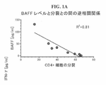
本明細書に開示する方法は、少なくとも部分的には、本発明者らのT細胞抑制におけるBAFF(B細胞活性化因子)及びAPRIL(分裂誘発リガンド)の役割の発見に基づく。BAFFは、B細胞分化のための重要な調節因子であり、末梢B細胞集団の生存及び活性化の調節に必須である。BAFFは下記の3つのTNF受容体サブファミリーメンバーと結合する:B細胞成熟抗原(BCMA/TNFRSF17)、トランスメンブレンアクチベーター及びカルシウム調節因子及びシクロフィリンリガンドインターアクター(TACI/TNFRSF13B)、並びにBAFF受容体(BAFF-R/BR3/TNFRSF13C/BLyS受容体3及びTNFRSF13Cとしても知られている)。これらの受容体は、シグナルペプチドを欠くIII型トランスメンブレンタンパク質である。TACI及びBCMAはBAFF及び別のTNFスーパーファミリーリガンド、APRIL(分裂誘発リガンド)の両方と結合するが、一方、BAFF-Rは選択的にBAFFと結合する。
本明細書で初めて記載するように、MSC由来BAFF/APRIL又は組換えBAFF/APRILのインターフェロンガンマ活性化MSC培養への添加は、インドールアミン2,3-ジオキシゲナーゼ(IDO1)酵素の発現及び活性を高めることができる(前記酵素は必須アミノ酸L-トリプトファンのN-フォルミルキヌレインへの分解を触媒する)。BAFF及びAPRILはIDO1発現のためのトグルスイッチとして機能するように思われる。いかなる特定の理論にも拘束されないが、発現を増大させるBAFF/APRIL受容体を特異的に遮断することによるIDO1のダウンレギュレーションは白血球機能を低下させ、エフェクターT細胞の分裂を高めるであろうと考える。
したがって、本開示は、養子細胞療法の細胞集団及び組成物を調製するための方法、細胞、及び組成物に関する。特に、遺伝子操作及び養子T細胞免疫療法のためのT細胞集団を確実に拡張及び活性化する効率的で効果的な方法を提供する。本発明の方法は、特定のT細胞集団を選択的に活性化することによって治療的方法で使用されるT細胞を提供する。さらにまた、前記方法によって作製される細胞及び組成物、並びにそれらを使用する方法が提供される。本開示はまた、T細胞のin vivo活性化及び拡張の刺激、及び抗BAFF-R剤のin vivo投与のための方法に関する。
The methods disclosed herein are, at least in part, based on our discovery of the role of BAFF (B cell activating factor) and APRIL (division-inducing ligand) in T cell suppression. BAFF is an important regulator of B cell differentiation and is essential for the regulation of survival and activation of peripheral B cell populations. BAFF binds to the following three TNF receptor subfamily members: B cell maturation antigen (BCMA / TNFRSF17), transmembrane activator and calcium regulator and cyclophilin ligand interactor (TACI / TNFRSF13B), and BAFF receptor (TACI / TNFRSF13B). Also known as BAFF-R / BR3 / TNFRSF13C / BLyS receptor 3 and TNFRSF13C). These receptors are type III transmembrane proteins that lack a signal peptide. TACI and BCMA bind both BAFF and another TNF superfamily ligand, APRIL (division-inducing ligand), while BAFF-R selectively binds BAFF.
As described for the first time herein, the addition of MSC-derived BAFF / APRIL or recombinant BAFF / APRIL to an interferon gamma-activated MSC culture results in the expression and activity of the
Accordingly, the present disclosure relates to methods, cells, and compositions for preparing cell populations and compositions for adoptive cell therapy. In particular, it provides an efficient and effective method of reliably expanding and activating a T cell population for genetic engineering and adopted T cell immunotherapy. The methods of the invention provide T cells for use in therapeutic methods by selectively activating specific T cell populations. Furthermore, the cells and compositions produced by the above method, as well as methods of using them, are provided. The present disclosure also relates to methods for in vivo activation and expansion of T cells, and in vivo administration of anti-BAFF-R agents.
方法
第一の特徴では、ex vivoでT細胞を拡張及び活性化することによる、T細胞の集団を調製する確実な方法が本明細書で提供される。本明細書で用いられるように、“ex vivo”という用語は生物の外部で生じる状態を指す。本開示の関係では、免疫細胞のex vivo処置は、そのような細胞をある種の生物学的分子(例えばアゴニスト、アンタゴニスト)にin vitro(すなわち生物の外部)で、好ましくは無菌的条件下で暴露することを意味する。いくつかの事例では、ex vivo方法は、人間から単離した免疫細胞を同じ人間の対象者に投与し直す前に培養する工程を追加的に含む。
第一の工程では、BAFF受容体はT細胞の集団でダウンレギュレート又は遮断され、それによってBAFF受容体の活性は消失又は低下する。BAFF受容体は、当業界で公知の適切な方法又は技術によってダウンレギュレート又は遮断され得る。遺伝子発現のダウンレギュレーション又は受容体活性の低下のための公知の方法には、CRISPR、ミクロRNA、shRNA、RNAi、中和抗体、小分子阻害因子、下流のシグナリング経路を遮断する化学的阻害因子などが含まれるが、ただしこれらに限定されない。いくつかの実施態様では、BAFF受容体活性又は遺伝子発現は、1%−100%の間で(すなわち、1%、5%、10%、15%、20%、25%、30%、35%、40%、45%、50%、55%、60%、65%、70%、75%、80%、85%、90%、95%、98%、99%、100%)低下する。ある実施態様では、T細胞はBAFF受容体アンタゴニストの存在下で培養される。ある実施態様では、T細胞をBAFF-R特異的shRNAと接触させてBAFF-R遺伝子発現を低下させる。
ある実施態様では、T細胞はBAFF受容体アンタゴニストの存在下で培養される。例示的な実施態様では、BAFF受容体アンタゴニストは、B細胞活性化因子受容体(BAFF-R)と反応する中和抗体である。ヒト抗BAFF-R抗体は、市場の供給業者(例えばR&D Systems及びInvitrogen)から入手できる。
Method In the first feature, a reliable method for preparing a T cell population by expanding and activating T cells ex vivo is provided herein. As used herein, the term "ex vivo" refers to a condition that occurs outside an organism. In the context of the present disclosure, ex vivo treatment of immune cells exposes such cells to certain biological molecules (eg, agonists, antagonists) in vitro (ie, outside the organism), preferably under sterile conditions. Means to expose. In some cases, the ex vivo method additionally comprises the step of culturing immune cells isolated from humans prior to re-administration to the same human subject.
In the first step, the BAFF receptor is down-regulated or blocked in a population of T cells, thereby eliminating or reducing the activity of the BAFF receptor. BAFF receptors can be down-regulated or blocked by suitable methods or techniques known in the art. Known methods for downregulating gene expression or reducing receptor activity include CRISPR, microRNA, shRNA, RNAi, neutralizing antibodies, small molecule inhibitors, chemical inhibitors that block downstream signaling pathways, etc. Includes, but is not limited to. In some embodiments, BAFF receptor activity or gene expression is between 1% and 100% (ie, 1%, 5%, 10%, 15%, 20%, 25%, 30%, 35%). , 40%, 45%, 50%, 55%, 60%, 65%, 70%, 75%, 80%, 85%, 90%, 95%, 98%, 99%, 100%) decrease. In certain embodiments, T cells are cultured in the presence of a BAFF receptor antagonist. In some embodiments, T cells are contacted with BAFF-R specific shRNAs to reduce BAFF-R gene expression.
In certain embodiments, T cells are cultured in the presence of a BAFF receptor antagonist. In an exemplary embodiment, the BAFF receptor antagonist is a neutralizing antibody that reacts with the B cell activating factor receptor (BAFF-R). Human anti-BAFF-R antibodies are available from market suppliers (eg R & D Systems and Invitrogen).
好ましくは、T細胞は白血球含有細胞混合物又は精製T細胞集団に存在する。いくつかの事例では、白血球含有細胞混合物又は精製T細胞集団は、例えば人間の対象者の末梢血のアフェレーシス又は人間の対象者の末梢血単核球から入手される。本明細書で用いられるように、“白血球含有細胞混合物”は、白血球細胞タイプ(顆粒球、リンパ球及び単球を含む)を含む細胞集団又は細胞組成物を指す。白血球含有細胞混合物は、好ましくは1つ以上の特異的な白血球細胞タイプを含む。好ましい細胞タイプは、リンパ球(特にT-リンパ球(“T細胞”)である。本明細書で用いられるように、“精製T細胞集団”は、血液又は白血球環境(例えば白血球アフェレーシスによって得られる)から単離、分離、或いは除去されたT細胞を指し、それにより単離/分離されたT細胞は、それらがin vivoで存在する環境とは異なる生理的環境に存在する。前記用語はいかなる特定の純度も示唆せず、さらに純度の絶対的レベルは必須ではない。当業者は、本明細書で提供する方法にしたがって使用される適切な純度レベルを容易に決定できる。
ある実施態様では、T細胞をBAFF-R特異的shRNAと接触させてBAFF-R遺伝子発現を低下させる。本発明に適するshRNAは、相補性BR3 mRNAの切断及びその後の分解を指令できるものである。適切なshRNA構築物はダーマコン社から購入できる。
Preferably, the T cells are present in a leukocyte-containing cell mixture or purified T cell population. In some cases, leukocyte-containing cell mixtures or purified T cell populations are obtained, for example, from peripheral blood apheresis in human subjects or peripheral blood mononuclear cells in human subjects. As used herein, "leukocyte-containing cell mixture" refers to a cell population or cell composition comprising leukocyte cell types, including granulocytes, lymphocytes and monocytes. The leukocyte-containing cell mixture preferably comprises one or more specific leukocyte cell types. A preferred cell type is lymphocytes, especially T-lymphocytes (“T cells”). As used herein, a “purified T cell population” is obtained by a blood or leukocyte environment (eg, leukocyte aferesis). ) Refers to T cells isolated, isolated, or removed, whereby T cells isolated / isolated are present in a physiological environment different from the environment in which they exist in vivo. No particular purity is suggested, and absolute levels of purity are not essential. One of those skilled in the art can easily determine the appropriate purity level to be used according to the methods provided herein.
In some embodiments, T cells are contacted with BAFF-R specific shRNAs to reduce BAFF-R gene expression. ShRNAs suitable for the present invention are capable of directing cleavage and subsequent degradation of complementary BR3 mRNA. Suitable shRNA constructs can be purchased from Dermacon.
次の工程では、BAFF受容体がダウンレギュレート又は遮断されたT細胞は、抗CD3抗体又はそのCD3結合フラグメント及び抗CD28抗体又はそのCD28結合フラグメントの存在下で約3日から約14日間(例えば約3、4、5、6、7、8、9、10、11、12、13、14日)、T細胞の活性化のために適切な条件下で培養される。この約3から14日培養工程を用いるin vitroのT細胞拡張はそのような培養T細胞を活性化し分裂を誘発して、治療法で使用するために十分な数の活性化された細胞傷害性T細胞を含む拡張集団を生じる。前記拡張T細胞集団はCD4陽性T細胞又はCD8陽性T細胞を含むことができる。いくつかの事例では、細胞傷害性CD8+ T細胞が前記活性化T細胞集団から優先的に拡張される。
T細胞の活性化のために適切な条件は、T細胞の生存能力の維持に適する任意の培地、並びに抗CD3抗体及びCD28抗体をT細胞と接触させることができる前記抗体の任意の処方を含む。いくつかの実施態様では、抗CD3及び抗CD28抗体は固体基材(例えばビーズ又はプレート表面)に固定できる。いくつかの実施態様では、抗CD3及び抗CD28抗体は可溶性である。ある実施態様では、白血球培養の例示的培養液はRPMI1640細胞培養液又は同様な細胞培養液である。場合によって、培地は約25%までの熱不活化ヒト血清アルブミンを含むことができる。BAFF-Rアンタゴニストを培養液に添加し、任意の適切な温度(例えば4℃、25℃又は37℃)でBAFF-Rアンタゴニストとのインキュベーションを実施することができる。好ましくは、BAFF-Rアンタゴニストインキュベーションは37℃で実施される。便利には当業者が適切な処置時間を最適化することができる。好ましくは、処置時間は約1時間から約24時間である。白血球との接触のための例示的BAFF-Rアンタゴニスト量には約0.1μg/mLから約100μg/mLが含まれる。当業者は有用なBAFF-Rアンタゴニスト量を容易に決定してBAFF-R活性を低下又は消失させることができる。
In the next step, T cells down-regulated or blocked by the BAFF receptor are subjected to about 3 to about 14 days (eg, in the presence of anti-CD3 antibody or CD3 binding fragment thereof and anti-CD28 antibody or CD28 binding fragment thereof). Approximately 3, 4, 5, 6, 7, 8, 9, 10, 11, 12, 13, 14 days), cultured under appropriate conditions for T cell activation. In vitro T cell expansion using this approximately 3 to 14 day culture step activates such cultured T cells and induces division, with a sufficient number of activated cytotoxicity for use in therapeutic methods. It gives rise to an expanded population containing T cells. The expanded T cell population can include CD4 + T cells or CD8 + T cells. In some cases, cytotoxic CD8 + T cells are preferentially expanded from the activated T cell population.
Suitable conditions for T cell activation include any medium suitable for maintaining T cell viability, as well as any formulation of said antibody capable of contacting anti-CD3 and CD28 antibodies with T cells. .. In some embodiments, the anti-CD3 and anti-CD28 antibodies can be immobilized on a solid substrate (eg, bead or plate surface). In some embodiments, the anti-CD3 and anti-CD28 antibodies are soluble. In certain embodiments, the exemplary culture of leukocyte culture is RPMI1640 cell culture or similar cell culture. In some cases, the medium can contain up to about 25% heat-inactivated human serum albumin. The BAFF-R antagonist can be added to the culture and incubated with the BAFF-R antagonist at any suitable temperature (eg 4 ° C, 25 ° C or 37 ° C). Preferably, the BAFF-R antagonist incubation is performed at 37 ° C. Conveniently, one of ordinary skill in the art can optimize the appropriate treatment time. Preferably, the treatment time is from about 1 hour to about 24 hours. Exemplary BAFF-R antagonist amounts for contact with leukocytes include from about 0.1 μg / mL to about 100 μg / mL. One of ordinary skill in the art can readily determine the amount of useful BAFF-R antagonist to reduce or eliminate BAFF-R activity.
T細胞は、その表面にCD4を発現する細胞(CD4陽性細胞とも称される)及びその表面にCD8を発現する細胞(CD8陽性細胞とも称される)におおまかに分けられる。本明細書で提供する方法にしたがって使用するために適切なT細胞は、人間のドナーの骨髄(BM)又は末梢血(PB)に由来する単核リンパ球(PBL)である。これらの細胞は、BM又はPBから直接採集するか、又は他家もしくは自家ドナーへの増殖因子及び/又はサイトカイン(例えば顆粒球-コロニー刺激因子(G-CSF)又は顆粒球マクロファージ-コロニー刺激因子(GM-CSF))の投与により動員又は刺激した後で採集できる。末梢血から末梢血単核球(PBMC)を単離する確立された多くのプロトコルが存在することは当業者には理解されよう。静脈穿刺により簡便に人間の末梢血を採集することができる。PBMCの単離は密度勾配分離プロトコルによって補助され、血液中の他の成分からリンパ球を分離するために通常は密度勾配遠心分離技術を利用し、フィコール-ハイパーク(Ficoll(商標)-Hypaque)又はヒストパーク(Histopaque(商標))を用いることができる。好ましくは、PBMC単離は無菌条件下で実施される。また別には、細胞エルトリエーション法を利用して、単核球集団を分離することができる。細胞エルトリエーション法の利点には無菌性及び効率性が含まれる。 T cells are roughly divided into cells that express CD4 on their surface (also called CD4 positive cells) and cells that express CD8 on their surface (also called CD8 positive cells). Suitable T cells for use according to the methods provided herein are mononuclear lymphocytes (PBL) from human donor bone marrow (BM) or peripheral blood (PB). These cells are collected directly from BM or PB, or growth factors and / or cytokines to allogeneic or autologous donors (eg, granulocyte-colony stimulating factor (G-CSF) or granulocyte macrophage-colony stimulating factor (eg. It can be collected after mobilization or stimulation by administration of GM-CSF)). Those skilled in the art will appreciate that there are many established protocols for isolating peripheral blood mononuclear cells (PBMCs) from peripheral blood. Human peripheral blood can be easily collected by venipuncture. Isolation of PBMCs is assisted by a density gradient separation protocol, usually utilizing density gradient centrifugation techniques to isolate lymphocytes from other components in the blood, Ficoll ™ -Hypaque. Alternatively, Histopaque ™ can be used. Preferably, PBMC isolation is performed under sterile conditions. Alternatively, the cell eltriation method can be used to segregate mononuclear cell populations. Advantages of the cell eltriation method include sterility and efficiency.
例示的な実施態様では、本明細書で提供する方法は、T細胞活性化を誘発する刺激でBAFF-R接触白血球を活性化する工程を含む。例示的な刺激には、分裂促進因子(例えばコンカナバリンA)、IL-2、及び抗CD2-、抗CD3-又は抗CD28-ビーズが含まれるが、ただしこれらに限定されない。CD28(T90/44抗原又はTp44としても知られている)はT細胞表面発現抗原であり、前記はT細胞上で働く共刺激タンパク質の受容体である。CD3は、成熟Tリンパ球の少なくとも5つの膜結合ポリペプチドの複合体であり、前記ポリペプチドは互いにさらにT細胞受容体と非共有結合により結合する。CD3複合体は、ガンマ、デルタ、エプシロン、ゼータ及びエータサブユニットを含む。抗原がT細胞受容体に結合するとき、CD3複合体は活性化シグナルをT細胞の細胞質に形質導入する。例えば、T細胞受容体と抗CD3モノクローナル抗体(mAb)との架橋は、T細胞の活性化、分裂、サイトカイン合成、及び腫瘍標的に向けられる非特異的細胞傷害性を生じる。これらの活性化T細胞は、IL-2生成増加を特徴とし、非MHC拘束細胞傷害性を示し、さらにIFNγ、TNFα及びGM-CSFを生成する。 In an exemplary embodiment, the method provided herein comprises activating BAFF-R contact leukocytes with a stimulus that induces T cell activation. Exemplary stimuli include, but are not limited to, mitogens (eg, concanavalin A), IL-2, and anti-CD2-, anti-CD3- or anti-CD28-beads. CD28 (also known as T90 / 44 antigen or Tp44) is a T cell surface-expressing antigen, which is a receptor for costimulatory proteins that act on T cells. CD3 is a complex of at least five membrane-bound polypeptides of mature T lymphocytes that bind to each other further by non-covalent binding to the T cell receptor. The CD3 complex contains gamma, delta, epsilon, zeta and eta subunits. When the antigen binds to the T cell receptor, the CD3 complex transduces an activation signal into the cytoplasm of T cells. For example, cross-linking of T cell receptors with anti-CD3 monoclonal antibodies (mAbs) results in T cell activation, division, cytokine synthesis, and non-specific cytotoxicity directed at tumor targets. These activated T cells are characterized by increased IL-2 production, exhibit non-MHC-restricted cytotoxicity, and produce IFNγ, TNFα and GM-CSF.
いくつかの事例では、本開示の方法は、遺伝子操作受容体又はキメラ抗原受容体を活性化T細胞に導入する工程を含み、それによって前記方法は、前記遺伝子操作又はキメラ抗原受容体を発現するCD4+ T細胞及びCD8+ T細胞を含む拡張集団を生じる。キメラ抗原受容体(CAR)(キメラT細胞受容体、人工T細胞受容体及びキメラ免疫受容体としても知られる)は操作された受容体であり、前記は特異性を免疫エフェクター細胞に移植する。一般的に、キメラ抗原受容体は標的抗原結合ドメインを有するトランスメンブレンタンパク質であり、前記結合ドメインはスペーサー及びトランスメンブレンドメインを介してシグナリングエンドドメインに融合される。CARがその標的抗原と結合するとき、活性化シグナルがT細胞に伝達される。ある実施態様では、キメラ抗原受容体又は遺伝子操作受容体がT細胞に導入される。ある実施態様では、キメラ抗原受容体又は遺伝子操作受容体をコードする核酸ベクターがT細胞にトランスフェクトされ、それによってT細胞はキメラ抗原受容体を発現する。
ex vivo操作手順の間に用いられる試薬及び他の物質(例えば抗体、サイトカイン、血清、他の化学品、又は固体支持体(例えばビーズ))及び特にウイルス系遺伝子ベクターは、治療用細胞プロダクトの無菌的製造に適合していなければならない。
In some cases, the methods of the present disclosure include the step of introducing a genetically engineered or chimeric antigen receptor into activated T cells, whereby the method expresses the genetically engineered or chimeric antigen receptor. It yields an expanded population containing CD4 + T cells and CD8 + T cells. Chimeric antigen receptors (CARs) (also known as chimeric T cell receptors, artificial T cell receptors and chimeric immunoreceptors) are engineered receptors that transplant specificity into immune effector cells. Generally, the chimeric antigen receptor is a transmembrane protein having a target antigen-binding domain, and the binding domain is fused to the signaling end domain via a spacer and a transmembrane domain. When CAR binds to its target antigen, an activation signal is transmitted to T cells. In certain embodiments, a chimeric antigen receptor or genetically engineered receptor is introduced into T cells. In some embodiments, a nucleic acid vector encoding a chimeric antigen receptor or genetically engineered receptor is transfected into a T cell, whereby the T cell expresses the chimeric antigen receptor.
Reagents and other substances used during ex vivo manipulation procedures (eg, antibodies, cytokines, sera, other chemicals, or solid supports (eg, beads)) and especially viral gene vectors are sterile in therapeutic cell products. Must be suitable for production.
本明細書で提供する方法にしたがって入手される拡張T細胞集団は、細胞免疫療法(T細胞療法、養子細胞療法(ACT)及びCAR T細胞療法を含む(ただしこれらに限定されない))で有用である。本明細書で用いられるように、“養子細胞療法”という用語は、エフェクター機能を媒介するリンパ球の移転を指す。例えば、本明細書の記載にしたがって入手される拡張T細胞集団をACT方法で用いて、癌(例えばリンパ腫)患者の機能的T細胞欠損をin vivo及びin vitroで逆転させることができる。養子T細胞療法は、キメラ抗原受容体を発現するように操作したT細胞の投与、遺伝子改変されていない腫瘍浸潤リンパ球(TIL)の投与、及びT細胞受容体(TCR)操作を実施したT細胞の投与を含む。T細胞チェックポイント療法及びTIL療法は、T細胞区画の本来備わっている腫瘍認識能力を活用する。遺伝子改変T細胞による養子療法は、他の状況では患者に存在しない腫瘍特異的T細胞区画を作り出すことによって完全に異なる要求を満たす潜在能力を有する。したがって、遺伝子改変ACTは、T細胞チェックポイント療法又はTIL療法に応答し得ない腫瘍タイプ(例えば小児に生じる大半の癌及び多くの血液学的悪性疾患)に対して潜在能力を有する。加えて、T細胞チェックポイント療法及び遺伝子改変ACTは相乗的に機能する潜在能力を有する。したがって、本明細書で提供する方法にしたがって入手されるT細胞を用いる養子細胞療法は、追加の治療技術(例えばチェックポイント遮断抗体、ワクチン及び標的誘導下薬剤療法)と組合わせ得ることが意図される。 Expanded T cell populations obtained according to the methods provided herein are useful in cell immunotherapy, including, but not limited to, T cell therapy, adoptive cell therapy (ACT) and CAR T cell therapy. be. As used herein, the term "adoptive cell therapy" refers to the transfer of lymphocytes that mediate effector function. For example, the expanded T cell population obtained as described herein can be used in the ACT method to reverse functional T cell deficiency in cancer (eg, lymphoma) patients in vivo and in vitro. Adopted T cell therapy includes administration of T cells engineered to express the chimeric antigen receptor, administration of unmodified tumor-infiltrating lymphocytes (TIL), and T cell receptor (TCR) manipulation. Includes administration of cells. T cell checkpoint therapy and TIL therapy utilize the inherent tumor recognition capabilities of T cell compartments. Adoption with genetically modified T cells has the potential to meet completely different requirements by creating tumor-specific T cell compartments that are otherwise absent in the patient. Therefore, genetically modified ACT has potential for tumor types that cannot respond to T cell checkpoint therapy or TIL therapy (eg, most cancers that occur in children and many hematological malignancies). In addition, T cell checkpoint therapy and genetically modified ACT have the potential to function synergistically. Therefore, it is intended that adoptive cell therapy with T cells obtained according to the methods provided herein can be combined with additional therapeutic techniques such as checkpoint blocking antibodies, vaccines and targeted guided drug therapies. NS.
ドナーリンパ球輸液(DLL)は直接的でかつ強力な移植片対腫瘍(GVT)効果を誘発する。前記はドナー移植片の同種異系幹細胞移植(SCT)後に再発する患者に特に有効である。Ex vivo活性化DLI(aDLI)の使用はいくつかの白血病の患者で有益であり得る。そのような事例では、活性化ドナーT細胞は、抗CD3(OKT3)及び/又は抗CD28被覆磁性ビーズへの暴露に続いて共同刺激及び拡張することによって得られる。一般的には、CD3及びCD28によるT細胞の共同刺激は活性化T細胞を生成することができ、前記T細胞は、疾患誘発アネルギーを克服し、CD4機能を保全及び増大させ、さらにGVT活性を強化することができる。本明細書で提供する方法にしたがって入手されるT細胞は、対象者(例えば同種異系SCT後に再発を示す対象者)に輸液できる。
いくつかの事例では、PBMC物からT細胞を同時に単離及び活性化(刺激)するのが有利であり得る。例えば、抗CD3及び抗CD28被覆磁性ビーズ(すなわちCTSダイナビーズCD3/CD28)を大きな磁石と一緒に用いて、磁性ビーズ結合細胞を磁性ビーズに結合しないものから分離することができる。
さらにまたT細胞をin vivoで活性化及び拡張するために、対象者に抗BAFF-R剤を投与する方法が本明細書で意図される。本明細書で用いられるように、“抗BAFF-R剤”は、BAFF-R受容体の活性又は遺伝子発現を阻害し又は低下させるものを指す。抗BAFF-R剤には、阻害性抗BAFF-Rモノクローナル抗体、小分子阻害剤、shRNA、shRNAベクター、ミクロRNA、ミクロRNAベクターなどが含まれるが、ただしこれらに限定されない。本明細書で提供する方法にしたがって入手されるT細胞の拡張集団を利用する治療手段は、T細胞のin vivo活性化のための補充となるか、又は代用となり得る。
Donor lymphocyte infusion (DLL) induces a direct and potent graft-versus-tumor (GVT) effect. The above is particularly effective for patients who relapse after allogeneic stem cell transplantation (SCT) of donor grafts. The use of ex vivo-activated DLI (aDLI) may be beneficial in some leukemia patients. In such cases, activated donor T cells are obtained by co-stimulation and expansion following exposure to anti-CD3 (OKT3) and / or anti-CD28 coated magnetic beads. In general, co-stimulation of T cells with CD3 and CD28 can generate activated T cells, which overcome disease-induced anergies, preserve and enhance CD4 function, and further enhance GVT activity. Can be strengthened. T cells obtained according to the methods provided herein can be infused into a subject (eg, a subject who shows recurrence after allogeneic SCT).
In some cases, it may be advantageous to simultaneously isolate and activate (stimulate) T cells from PBMCs. For example, anti-CD3 and anti-CD28 coated magnetic beads (ie, CTS Dynabeads CD3 / CD28) can be used with large magnets to separate magnetic bead-bound cells from those that do not bind to magnetic beads.
Furthermore, a method of administering an anti-BAFF-R agent to a subject to activate and expand T cells in vivo is contemplated herein. As used herein, "anti-BAFF-R agent" refers to one that inhibits or reduces BAFF-R receptor activity or gene expression. Anti-BAFF-R agents include, but are not limited to, inhibitory anti-BAFF-R monoclonal antibodies, small molecule inhibitors, shRNAs, shRNA vectors, microRNAs, microRNA vectors, and the like. Therapeutic means utilizing an expanded population of T cells obtained according to the methods provided herein can be a supplement or a substitute for in vivo activation of T cells.
本明細書で提供する方法にしたがって入手されるT細胞の拡張集団は、多様な疾患、例えば癌(血液の悪性疾患(例えばリンパ腫又は白血病)又は固形腫瘍(例えばメラノーマ又は腎癌))又は感染症(例えばHIV)の治療もしくは予防に有用である。本明細書で用いられるように、“治療する”又は“治療”は、治療的及び予防的又は防止的手段の両方を指し、この場合、目標は望ましくない生理学的変化又は病理学的異常を防止するか又は遅らせる(減少させる)ことである。本発明の目的のためには、癌の治療には、1つ以上の臨床指標の緩和、腫瘍の成長又は腫瘍細胞分裂の減少、癌の様態の1つ以上の臨床指標の重篤度の低下、前記様態の程度の軽減、対象者の病期の安定化(すなわち悪化しないこと)、癌進行の遅延、停止又は逆転、及び部分的又は完全な緩解の発生が含まれるが、ただしこれらに限定されない。癌の治療にはまた、本明細書で提供する方法にしたがって入手されるT細胞を取り入れない標準的医療で治療した場合の予後と比較して日数、週数、月数又は年数による生存延長が含まれる。治療を必要とする対象者には、すでに癌であるか又は癌と診断された人々とともに、癌に罹患しやすいもしくは癌発生の蓋然性が高い、又は癌(例えばリンパ腫又は多発性骨髄腫)もしくは感染症が疑われる人々が含まれ得る。いくつかの事例では、対象者は自己免疫疾患を有することがある。本明細書で用いられるように、“防止する”及び“防止”という用語は、望ましくない生理学的変化又は異常もしくは様態の発生の阻害を意図する予防的又は防止的手段を指す。例示的実施態様では、疾患又は様態の予防は、前記疾患又は様態(又はその症状)が出現もしくは存在する以前の時点で本明細書で提供する方法にしたがって入手されるT細胞の投与を開始することを含み、したがって、前記疾患もしくは様態、又はその症状、病理学的特色、結果、又は有害作用は出現しない。 The expanded population of T cells obtained according to the methods provided herein includes a variety of diseases such as cancer (such as blood malignancies (eg lymphoma or leukemia) or solid tumors (eg melanoma or renal cancer)) or infectious diseases. It is useful for the treatment or prevention of (eg, HIV). As used herein, "treating" or "treating" refers to both therapeutic and prophylactic or prophylactic measures, in which case the goal is to prevent unwanted physiological changes or pathological abnormalities. To do or to delay (decrease). For the purposes of the present invention, the treatment of cancer includes mitigation of one or more clinical indicators, reduction of tumor growth or tumor cell division, reduction of severity of one or more clinical indicators of cancer mode. Includes, but is limited to, reducing the degree of the above-mentioned conditions, stabilizing the stage of the subject (ie, not exacerbating), delaying, stopping or reversing cancer progression, and the occurrence of partial or complete remission. Not done. Treatment of cancer also prolongs survival by days, weeks, months or years compared to the prognosis when treated with standard medical care that does not incorporate T cells obtained according to the methods provided herein. included. Subjects in need of treatment, along with those who already have or have been diagnosed with cancer, are predisposed to or more likely to develop cancer, or have cancer (eg, lymphoma or multiple myeloma) or infection. May include people with suspected illness. In some cases, the subject may have an autoimmune disorder. As used herein, the terms "prevent" and "prevent" refer to prophylactic or prophylactic measures intended to inhibit the development of unwanted physiological changes or abnormalities or modalities. In an exemplary embodiment, prevention of a disease or mode initiates administration of T cells obtained according to the methods provided herein prior to the appearance or presence of said disease or mode (or symptoms thereof). Thus, the disease or mode, or its symptoms, pathological features, consequences, or adverse effects do not appear.
本明細書で提供する方法にしたがって入手されるT細胞は、癌(血液類リンパ系悪性疾患(白血病、リンパ腫、多発性骨髄腫)を含む悪性疾患を含む)の治療のために養子細胞療法の方法で用いることができる。本明細書に記載する治療に適する癌には、血液学的悪性疾患、例えば急性骨髄性白血病(AML)、急性リンパ球性白血病(ALL)、慢性骨髄性白血病(CML)、骨髄腫、非ホジキン及びホジキンリンパ腫(例えば再発性、難治性、又は化学療法耐性非ホジキンリンパ腫)、及び脊髄形成異常症候群(MDS)が含まれる。“癌”及び“腫瘍”という用語は本明細書では互換的に用いられる。治療に適する他の癌には固形腫瘍(例えばメラノーマ、腎癌、結腸癌、肺癌、脳の癌、及び肝癌)が含まれる。
本明細書で提供する方法にしたがって入手されるT細胞は、感染の治療のために養子細胞療法の方法で用いることができる。本明細書で用いられるように、“感染”という用語は、微生物又は他の感染性因子が健康な細胞に侵入する病的状態を指し、細菌性、ウイルス性又は寄生性(例えば原生動物)感染因子によって引き起こされる任意の様態又は病的状態が含まれる。例えば、“ウイルス感染”という用語は健康な細胞のウイルス(例えばHIV)による侵入を指し、この場合、ウイルスは細胞の増殖機構を使用して増加又は複製し、最終的には細胞を溶解させて細胞死、ウイルス粒子の放出、及び新規生成子孫ウイルスによる他の細胞の感染をもたらす。感染に関しては、“治療”という用語はさらに治療薬の適用又は投与を指し、この場合、その目的は、治癒(cure, heal)、緩和、軽減、改変、修復、好転、改善、又は前記感染、前記感染に関係する任意の症状もしくは感染発症素因に影響を与えることである。
T cells obtained according to the methods provided herein are of adoptive cell therapy for the treatment of cancer, including malignant diseases including hematological lymphatic system malignancies (leukemia, lymphoma, multiple myeloma). Can be used in a method. The therapeutically suitable cancers described herein include hematological malignancies such as acute myeloid leukemia (AML), acute lymphocytic leukemia (ALL), chronic myeloid leukemia (CML), myeloma, and non-Hodgkin. And Hodgkin's lymphoma (eg, recurrent, refractory, or chemotherapy-resistant non-Hodgkin's lymphoma), and spinal cord dysplasia syndrome (MDS). The terms "cancer" and "tumor" are used interchangeably herein. Other cancers that are suitable for treatment include solid tumors (eg, melanoma, kidney cancer, colon cancer, lung cancer, brain cancer, and liver cancer).
T cells obtained according to the methods provided herein can be used in adoptive cell therapy methods for the treatment of infections. As used herein, the term "infection" refers to a pathological condition in which a microorganism or other infectious agent invades healthy cells and is a bacterial, viral or parasitic (eg, protozoan) infection. Includes any mode or pathological condition caused by the factor. For example, the term "viral infection" refers to the invasion of healthy cells by a virus (eg, HIV), in which the virus uses the cell's proliferation mechanism to multiply or replicate and eventually lyse the cells. It results in cell death, release of viral particles, and infection of other cells by newly generated progeny virus. With respect to infection, the term "treatment" further refers to the application or administration of a therapeutic agent, in which case the purpose is cure, heal, alleviation, alleviation, modification, repair, improvement, improvement, or said infection. Affecting any of the symptoms associated with the infection or predisposition to develop the infection.
本明細書で用いられるように、“対象者”又は“患者”という用語は互換的に用いられ、任意の脊椎動物(人間、哺乳動物、爬虫類、両生動物及び魚類を含む(ただし前記に限定されない))を包含する。しかしながら、有利には対象者又は患者は、例えば人間のような哺乳動物、又は哺乳動物(例えば飼い慣らされた哺乳動物(例えばイヌ、ネコ、ウマ)又は家畜(例えば乳牛、ヒツジ、ブタなど))である。例示的な実施態様では、対象者は人間である。本明細書で用いられるように、“その必要がある”という語句は、治療的又は予防的手段が所望される対象者の状態を指す。そのような状態には、疾患又は様態(例えば癌)を有する対象者が含まれ得る(ただし前記に限定されない)。
いくつかの事例では、本明細書で提供する方法にしたがって入手されるT細胞を医薬組成物として投与することができ、前記組成物は、治療薬剤として(すなわち治療的な適用のために)有効な量のT細胞を含む。本明細書で用いられるように、“医薬組成物”という用語は、哺乳動物への投与に適する化学的又は生物学的組成物を指す。そのような治療的な適用に適する組成物の例には、非経口投与、皮下、経皮、皮内、筋肉内、冠状動脈内、心筋内、脳内、腫瘍内、腹腔内、静脈内(例えば注射可能)、又は気管内投与のための調製物(例えば無菌的懸濁物、乳濁液及びエーロゾル)が含まれる。気管内投与は、治療的に有効な量のT細胞を含む医薬組成物に肺組織(例えば肺胞)を接触又は暴露する工程を必要とする。いくつかの事例では、治療的な適用に適した医薬組成物は、1つ以上の医薬的に許容できる賦形剤、希釈剤又は担体(例えば滅菌水、生理学的食塩水、グルコースなど)との混合物中に存在し得る。例えば、本明細書に記載するT細胞は、食塩水溶液を含む医薬組成物として対象者に投与され得る。例示的な実施態様では、本明細書で提供する方法にしたがって拡張されるT細胞は、対象者に投与されるとき所望の治療的又は予防的効果を誘発することができる。
As used herein, the terms "subject" or "patient" are used interchangeably and include, but are not limited to, any vertebrate (human, mammalian, reptile, amphibian and fish). )) Includes. However, advantageously the subject or patient is, for example, a mammal such as a human, or a mammal (eg, a domesticated mammal (eg, dog, cat, horse) or livestock (eg, dairy cow, sheep, pig, etc.)). be. In an exemplary embodiment, the subject is a human. As used herein, the phrase "needed" refers to the condition of a subject for whom therapeutic or prophylactic measures are desired. Such conditions may include, but are not limited to, subjects with a disease or mode (eg, cancer).
In some cases, T cells obtained according to the methods provided herein can be administered as a pharmaceutical composition, wherein the composition is effective as a therapeutic agent (ie, for therapeutic application). Contains a large amount of T cells. As used herein, the term "pharmaceutical composition" refers to a chemical or biological composition suitable for administration to a mammal. Examples of compositions suitable for such therapeutic applications are parenteral administration, subcutaneous, transdermal, intradermal, intramuscular, intracoronary, intramyocardial, intracerebral, intratumoral, intraperitoneal, intravenous ( Included (eg, injectable) or preparations for intratracheal administration (eg, sterile suspensions, emulsions and aerosols). Intratracheal administration requires the step of contacting or exposing lung tissue (eg, alveoli) to a pharmaceutical composition containing a therapeutically effective amount of T cells. In some cases, pharmaceutical compositions suitable for therapeutic application are with one or more pharmaceutically acceptable excipients, diluents or carriers (eg sterile water, physiological saline, glucose, etc.). It may be present in the mixture. For example, the T cells described herein can be administered to a subject as a pharmaceutical composition containing an aqueous saline solution. In an exemplary embodiment, T cells that are expanded according to the methods provided herein can elicit the desired therapeutic or prophylactic effect when administered to a subject.
処方物は、経口投与、直腸、鼻内、局部又は経粘膜投与(頬用、舌下用、眼用、膣用及び直腸用を含む)、及び非経口投与(皮下用、筋肉内用、静脈内用、皮内用、腹腔内用、眼内用及び硬膜外用を含む)のために設計又は提供され得る。一般的には、水性もしくは非水性液状又はクリーム状処方物が非経口、経口又は局部ルートによってデリバリーされる。他の実施態様では、組成物は水性もしくは非水性液状処方物又は固形処方物として存在でき、前記処方物は、任意のルート(例えば経口、局部、頬、舌下、非経口)、エーロゾル、デポ剤(例えば皮下デポ剤又は腹腔もしくは筋肉内デポ剤)による投与に適切である。いくつかの事例では、医薬組成物は凍結乾燥される。他の事例では、本明細書で提供する医薬組成物は、所望される投与ルート及び調製に応じて、補助物質(例えば湿潤もしくは乳化剤、pH緩衝剤、ゲル化もしくは粘性強化添加物、保存料、香料、着色剤など)を含む。医薬組成物は、通常の製薬慣行にしたがって処方することができる(例えば以下を参照されたい: Remington: The Science and Practice of Pharmacy, 20th edition, 2000, ed.A.R.Gennaro, Lippincott Williams & Wilkins, Philadelphia;及びEncyclopedia of Pharmaceutical Technology, eds.J.Swarbrick and J.C.Boylan, 1988-1999, Marcel Dekker, New York)。
好ましいルートは、例えば、対象者の病理学的状態もしくは体重、又は対象者の治療方法に対する応答、又は状況に見合う治療方法により変動し得る。処方物はまた2つ以上のルートによって投与することが可能であり、この場合、デリバリーの方法は本質的に同時であるか、或いは組成物が対象者に投与される時間において本質的に時間的オーバーラップがほとんどないかもしくは全くない。
最初の投与及び更なる投与のための又は連続投与のための適切なレジメンもまた変動可能であり、最初の投与とその後に投与が続くレジメンが含まれ得るが、それにもかかわらず、当業者は、本開示、本明細書に引用する資料及び当業界の知識から適切なレジメンを確認することができる。
The prescriptions are oral, rectal, intranasal, local or transmucosal (including buccal, sublingual, ocular, vaginal and rectal), and parenteral (subcutaneous, intramuscular, intravenous). It may be designed or provided for internal use, intradermal use, intraperitoneal use, intraocular use and extradural use). Generally, aqueous or non-aqueous liquid or creamy formulations are delivered by parenteral, oral or local routes. In other embodiments, the composition can exist as an aqueous or non-aqueous liquid formulation or solid formulation, wherein the formulation is by any route (eg, oral, local, cheek, sublingual, parenteral), aerosol, depot. Suitable for administration by agents (eg, subcutaneous depot or peritoneal or intramuscular depot). In some cases, the pharmaceutical composition is lyophilized. In other cases, the pharmaceutical compositions provided herein are such as auxiliary substances (eg, wetting or emulsifiers, pH buffers, gelling or viscous enhancing additives, preservatives, etc., depending on the desired route of administration and preparation. Contains fragrances, colorants, etc.). Pharmaceutical compositions can be formulated according to conventional pharmaceutical practices (see, eg: Remington: The Science and Practice of Pharmacy, 20th edition, 2000, ed. ARGennaro, Lippincott Williams & Wilkins, Philadelphia; and Encyclopedia of Pharmaceutical Technology, eds.J.Swarbrick and JCBoylan, 1988-1999, Marcel Dekker, New York).
The preferred route may vary, for example, depending on the subject's pathological condition or weight, or the subject's response to the treatment method, or the treatment method appropriate for the situation. The formulation can also be administered by two or more routes, in which case the delivery methods are essentially simultaneous or essentially temporal in the time the composition is administered to the subject. There is little or no overlap.
Appropriate regimens for initial and additional doses or for continuous doses are also variable and may include regimens that are followed by the first dose, but nonetheless, those skilled in the art will appreciate it. Appropriate regimens can be identified from this disclosure, the materials cited herein and the knowledge of the art.
いくつかの事例では、T細胞は場合によって1つ以上の活性な薬剤と組合わせて投与され得る。そのような活性な薬剤には、抗炎症剤、抗サイトカイン剤、鎮痛剤、解熱剤、抗生物質及び抗ウイルス剤とともに増殖因子並びに免疫調節薬剤のアゴニスト、アンタゴニスト及び調節物質が含まれる(例えば、BAFF、APRIL、TNF-α、IL-2、IL-4、IL-6、IL-10、IL-12、IL-13、IL-17、IL-18、IL-21、IL-35、IFN-α、IFN-γ、CXCL13、IP-10、VEGF、EPO、EGF、HRG、肝細胞増殖因子(HGF)、ヘプシジン、前述のいずれかに対して反応する抗体、及びそれらの受容体のいずれかに対して反応する抗体)。そのような活性薬剤の任意の適切な組み合わせもまた意図される。1つ以上の活性薬剤と組合わせて投与されるとき、T細胞は他の活性薬剤と同時に又は連続して投与され得る。例えば、対象者には、回復を支援しさらに疾患もしくは様態を治療し緩和し又は重篤度を軽減するために十分な期間又は投薬レジメンにしたがって、T細胞及び本明細書に記載の薬剤の1つ以上を同時に投与できる。
いくつかの実施態様では、T細胞は、その必要がある対象者に輸液、局部適用、外科的移植又は埋め込みを用いて投与される。例示的な実施態様では、投与は全身性である。そのような事例では、T細胞は、その必要がある対象者に静脈内投与に適合させた医薬組成物として対象者に投与される。典型的には、静脈内投与用組成物は無菌的な等張水性緩衝液中の溶液である。そのような緩衝液及び希釈剤の使用は当業界で周知である。必要な場合には、組成物はまた局所麻酔剤を含み、注射部位のいずれの痛みも緩和することができる。一般的には、成分は別々に供給されるか、又はユニット投薬形として(例えば密閉容器(例えば活性薬剤の量を示すアンプル)中の冷凍保存濃縮物として)一緒に混合される。組成物が輸液によって投与される場合、組成物は無菌的な医薬等級水又は食塩水を含む輸液瓶に分注できる。組成物が注射によって投与される場合は、注射用滅菌水又は食塩水のアンプルを提供し、組成物を投与前に混合することができる。いくつかの事例では、本発明のT細胞を含む組成物は投与前には冷凍保存される。
In some cases, T cells can optionally be administered in combination with one or more active agents. Such active agents include growth factors as well as agonists, antagonists and regulators of immunomodulators as well as anti-inflammatory agents, anti-cytokine agents, analgesics, antipyretic agents, antibiotics and antiviral agents (eg, BAFF, BAFF, APRIL, TNF-α, IL-2, IL-4, IL-6, IL-10, IL-12, IL-13, IL-17, IL-18, IL-21, IL-35, IFN-α, For IFN-γ, CXCL13, IP-10, VEGF, EPO, EGF, HRG, hepatocyte growth factor (HGF), hepsidin, antibodies that react with any of the above, and any of their receptors. Reacting antibody). Any suitable combination of such active agents is also intended. When administered in combination with one or more active agents, T cells may be administered simultaneously with or consecutively with the other active agents. For example, subjects are given T cells and one of the agents described herein according to a duration or dosing regimen sufficient to assist recovery and further treat, alleviate or reduce severity of the disease or condition. One or more can be administered at the same time.
In some embodiments, T cells are administered to the subject in need using infusion, topical application, surgical transplantation or implantation. In an exemplary embodiment, administration is systemic. In such cases, T cells are administered to the subject in need as a pharmaceutical composition adapted for intravenous administration. Typically, the composition for intravenous administration is a solution in sterile isotonic aqueous buffer. The use of such buffers and diluents is well known in the art. If necessary, the composition also comprises a local anesthetic and can relieve any pain at the injection site. Generally, the ingredients are either fed separately or mixed together as a unit dosage form (eg, as a freezing concentrate in a closed container (eg, an ampoule indicating the amount of active agent)). If the composition is administered by infusion, the composition can be dispensed into an infusion bottle containing sterile pharmaceutical grade water or saline. If the composition is administered by injection, an ampoule of sterile water for injection or saline can be provided and the composition can be mixed prior to administration. In some cases, the T cell-containing compositions of the invention are stored frozen prior to administration.
本明細書で提供するT細胞の治療的に有効な量がその必要のある対象者に投与される。本明細書で用いられるように、“治療的に有効な用量”という用語は、進行を防ぐために、又は問題の疾患もしくは様態の軽減を引き起こすために十分な任意の用量を指すか、又は、治療的に有効な量は、前記疾患もしくは様態によって引き起こされる症状(例えば痛み又は腫脹)を緩和することができる。有効な用量又は量(前記は1回以上の投与で投与することができる)は、T細胞が投与される対象者で治療的効果を引き出すために十分なヒトT細胞の量である。治療的に有効な量は、ex vivoで拡張したT細胞培養の約50x106細胞から約700x106細胞の量であり得る。いくつかの事例では、有効な量は、レシピエントの体重1キログラム(kg)につき少なくとも1x10⁶細胞を含む投薬量として投与される。例えば、有効な量は、少なくとも1x10⁶細胞/kg、少なくとも10x106細胞/kg、少なくとも30x106細胞/kg、少なくとも100x106細胞/kg、又は少なくとも1000x106細胞/kgを含む用量で対象者に投与され得る。
有効な量は、投与に際して細胞の作用を改変する多様な要因及び細胞に対する対象者の生物学的応答(例えば患者の年齢、性別及び食事、炎症の重篤度、投与時期、並びに他の臨床的要因)によって影響を受けるであろう。本明細書で提供する方法にしたがって入手されるT細胞の治療的に有効な量を対象者に投与することができる。
A therapeutically effective amount of T cells provided herein is administered to a subject in need thereof. As used herein, the term "therapeutically effective dose" refers to or treats any dose sufficient to prevent progression or to cause relief of the disease or mode in question. Effective amounts can alleviate the symptoms (eg, pain or swelling) caused by the disease or mode. An effective dose or amount (which can be administered in one or more doses) is an amount of human T cells sufficient to elicit a therapeutic effect in the subject to whom the T cells are administered. A therapeutically effective amount may be an amount from about 50 × 10 6 cells of about 700X10 6 cells of T cell cultures expanded by ex vivo. In some cases, the effective amount is administered as a dosage containing at least 1x10 6 cells per kilogram (kg) of body weight of the recipient. For example, an effective amount is at least 1x10 6 cells / kg, administered at least 10x10 6 cells / kg, at least 30 × 10 6 cells / kg, at least 100 × 10 6 cells / kg, or at doses containing at least 1000X10 6 cells / kg to the subject Can be done.
Effective amounts are a variety of factors that alter the action of cells upon administration and the subject's biological response to the cells (eg, patient age, gender and diet, severity of inflammation, timing of administration, and other clinical aspects. Factors) will affect it. A therapeutically effective amount of T cells obtained according to the methods provided herein can be administered to the subject.
人間の対象者への投与のために治療的に有効な量は、動物試験及び動物に対して有効と決定される量を人間の投与のための目盛りとする任意の業界許容方法で決定することができる。例えば、最初に動物モデルで(例えば有益な又は所望の臨床結果を達成するために)有効な量を測定することができる。動物モデルから得られた量を、当業界で公知の変換係数を用いることによって人間のために有効な量の処方に用いることができる。ある動物モデルで得られた有効な量はまた、適切な変換係数(例えば体表面積係数)を用いることによって別の動物のために変換することができる。
個々の任意の対象者について、規定の投薬レジメンは、各個体の必要性及びT細胞を投与する者又は投与を監督する者の専門的な判断にしたがって時間をかけて調整されねばならないことは理解されるべきである。例えば、個々の対象者のT細胞投薬量は、より低い用量が検出可能な又は十分な改善を引き出さない場合には増加させることができる。
いくつかの事例では、T細胞の治療的に有効な量は、所望の症候緩和レベルに達するまで投薬量を徐々に増加させることにより治療薬の効果を対象者で測定することによって決定できる。持続投与又は反復投与レジメンもまた、所望の結果の達成又は維持のために用いることができる。当業界で公知の任意の他の技術も同様に用いて有効な量の範囲を決定することができる。もちろんのこと、規定の有効量は、治療される個々の様態、対象者の生理的様態、治療される動物のタイプ、治療期間、及び任意の同時実施療法の性質のような要件により変動するであろう。本明細書に記載の疾患もしくは様態の影響下にあるか、又はそれら疾患もしくは様態を発生させやすいか又は発生の蓋然性が高い個々の対象者へのT細胞の投与に続いて、臨床症状又は前記疾患もしくは様態の特質における正又は負の変化について前記対象者を観察及び評価する。例えば、対象者で癌を治療する方法の場合、治療の間又は治療後の正又は負の変化は、当業者に公知の任意の手段(腫瘍サイズの変化の測定を含むが、ただし前記に限定されない)によって決定され得る。
The therapeutically effective amount for administration to a human subject shall be determined by animal studies and any industry-acceptable method of grading the amount determined to be effective for an animal for human administration. Can be done. For example, an effective amount can first be measured in an animal model (eg, to achieve beneficial or desired clinical results). The amount obtained from the animal model can be used in the formulation of an amount effective for humans by using conversion factors known in the art. Effective quantities obtained in one animal model can also be converted for another animal by using an appropriate conversion factor (eg, body surface area factor).
It is understood that for any individual subject, the prescribed dosing regimen must be adjusted over time according to the needs of each individual and the professional judgment of the person administering or supervising the administration of T cells. It should be. For example, the T cell dosage of an individual subject can be increased if lower doses do not elicit a detectable or sufficient improvement.
In some cases, the therapeutically effective amount of T cells can be determined by measuring the effect of the therapeutic agent in the subject by gradually increasing the dosage until the desired symptom relief level is reached. Continuous or repeated dose regimens can also be used to achieve or maintain the desired results. Any other technique known in the art can be used as well to determine the range of effective amounts. Of course, the prescribed effective dose will vary depending on requirements such as the individual mode of treatment, the physiological mode of the subject, the type of animal being treated, the duration of treatment, and the nature of any concurrent therapy. There will be. Following administration of T cells to an individual subject who is under the influence of the diseases or modalities described herein, or is prone to or probable to develop those diseases or modalities, followed by clinical symptoms or said Observe and evaluate the subject for positive or negative changes in the nature of the disease or mode. For example, in the case of a method of treating cancer in a subject, positive or negative changes during or after treatment include, but are limited to, any means known to those of skill in the art, including measurement of changes in tumor size. Can be determined by).
本発明の方法のいずれにおいても、T細胞のドナーとレシピエントは同じ個体でも又は異なる個体でもよい。本明細書で用いられるように、“自家”という用語は、ある個体から入手され同じ個体に移植し直される細胞又は組織を指す。本明細書で用いられるように、“同種異系”という用語は同じ種の異なる個体から入手された細胞又は組織を指し、この場合ドナー及びレシピエントは遺伝的に同一ではない。本開示に関しては、同種異系細胞移植又は組織移植は、ドナーとレシピエントが同じ種の異なる個体である細胞又は組織の移植を必要とする。“異種間”という用語は、どちらかが異なる種の生物に由来するか又は異なる種の生物から入手されることを意味する。本開示に関しては、異種間細胞移植又は組織移植は、ドナーとレシピエントが異なる種の異なる個体である細胞又は組織の移植を必要とする。
対象者への投与は、局所注射もしくは全身注射によるか、又は局部適用によることができる。例えば、T細胞は、静脈内注射(例えば点滴輸液)、筋肉内注射、腹腔内注射、器官内注射、又は皮下注射によって投与され得る。いくつかの事例では、対象者は、組織の維持、組織の修復もしくは機能、又は全体的な様態に関して観察もしくは評価される。
In any of the methods of the invention, the donor and recipient of T cells may be the same or different individuals. As used herein, the term "autologous" refers to cells or tissues obtained from an individual and reimplanted into the same individual. As used herein, the term "allogeneic" refers to cells or tissues obtained from different individuals of the same species, in which case the donor and recipient are not genetically identical. For the present disclosure, allogeneic cell or tissue transplantation requires transplantation of cells or tissues in which the donor and recipient are different individuals of the same species. The term "interspecies" means that either is derived from or obtained from a different species of organism. For the present disclosure, heterologous cell or tissue transplantation requires transplantation of cells or tissues that are different individuals of different species with different donors and recipients.
Administration to the subject can be by local or systemic injection, or by local application. For example, T cells can be administered by intravenous injection (eg, drip infusion), intramuscular injection, intraperitoneal injection, intraorgan injection, or subcutaneous injection. In some cases, subjects are observed or evaluated for tissue maintenance, tissue repair or function, or overall mode.
製造物品
別の特徴では、本発明は養子細胞療法に有用な製造物品を提供する。いくつかの事例では、本発明のキットは人間のT細胞を収納する1つ以上の容器を含む。具体的な実施態様では、本明細書で提供する方法にしたがって拡張及び活性化される細胞がキットとして提供され、いくつかの事例では、前記細胞がキットの唯一の成分であり得る。キットは追加的に試薬及び材料を含むことができ、前記は、本明細書で提供するT細胞を拡張及び活性化して所望の細胞プロダクトを得るために有用である。例えば、キットは1つ以上のBAFF-Rアンタゴニストを含むことができる。
場合によって、キットはさらに1つ以上の試薬又は他の成分を含むことができ、前記は、本発明の方法にしたがってT細胞をその必要がある人間の対象者に投与するために必要である。いくつかの事例では、医薬的に許容できる冷凍保存料中の冷凍アリコットとしてT細胞を提供することは適切であり得る。
いくつかの事例では、本明細書で提供するT細胞に加えて、キットはまた第二の治療薬(例えば化学療法剤、ホルモン治療剤及び/又は免疫治療剤)を含む。キットは、ある個体の特定の癌に合わせて仕立て直し、前記個体のために対応する第二の治療薬を含むことができる。いくつかの事例では、キットはさらに、1つ以上の活性薬剤(例えば抗炎症剤、抗サイトカイン剤、鎮痛剤、解熱剤、抗生物質及び抗ウイルス剤)とともに増殖因子並びに免疫調節薬剤のアゴニスト、アンタゴニスト及び調節物質を含む(例えば、TNF-α、インターロイキン2(IL-2)、IL-4、IL-6、IL-10、IL-12、IL-13、IL-18、IFN-α、IFN-γ、BAFF、CXCL13、IP-10、VEGF、EPO、EGF、HRG、肝細胞増殖因子(HGF)、ヘプシジン、(前述のいずれかに対して反応する抗体、及びそれらの受容体のいずれかに対して反応する抗体を含む))。癌治療に有用な医薬クラスには以下が含まれる(ただしこれらに限定されない):グルココルチコイド(例えばプレドニソロン)、免疫抑制剤(例えばシクロスポリン、メトトレキセート、タクロリムス、ピメクロリムス、シロリムス、ミコフェノレート、モフェチル、ビジリズマブ、抗胸腺細胞グロブリン(ATG))、抗新生物剤(例えばペントスタチン)、及び抗リウマチ剤(例えばヒドロキシクロロキン、インフリキシマブ、エタネルセプト)。そのような活性な薬剤の適切な組合せを含むキットもまた意図される。そのような容器とともに、人間への投与のための指示及び生物学的製剤の製造、使用又は販売に関する規制庁の所定の形式の通知が提供される(前記通知は人間への投与について官庁の製造、使用又は販売の承認を示す)。
本発明は、以下の非限定的な実施例を考慮するときより完全に理解されるであろう。本明細書に開示する全ての成書、論文及び特許は参照によってその全体が本明細書に含まれる。
実施例
以下の例についてこれから述べる。これらの例は、上記の説明と一緒になって本発明を非限定的な態様で解説するであろう。
In terms of product- specific features, the present invention provides products that are useful for adoptive cell therapy. In some cases, the kit of the invention comprises one or more containers containing human T cells. In a specific embodiment, cells that are expanded and activated according to the methods provided herein are provided as a kit, and in some cases the cells may be the sole component of the kit. The kit can additionally contain reagents and materials, which are useful for expanding and activating the T cells provided herein to obtain the desired cell product. For example, the kit can contain one or more BAFF-R antagonists.
In some cases, the kit may further contain one or more reagents or other components, which are necessary for administering T cells to a human subject in need thereof according to the methods of the invention. In some cases, it may be appropriate to provide T cells as a frozen aliquot in a pharmaceutically acceptable freezing preservative.
In some cases, in addition to the T cells provided herein, the kit also comprises a second therapeutic agent (eg, a chemotherapeutic agent, a hormonal therapeutic agent and / or an immunotherapeutic agent). The kit can be tailored to a particular cancer of an individual and include a corresponding second therapeutic agent for said individual. In some cases, the kit also includes growth factors and immunomodulatory agonists, antagonists and immunomodulators along with one or more active agents (eg, anti-inflammatory agents, anti-cytokine agents, analgesics, antipyretic agents, antibiotics and antiviral agents). Contains regulators (eg, TNF-α, interleukin 2 (IL-2), IL-4, IL-6, IL-10, IL-12, IL-13, IL-18, IFN-α, IFN- Against γ, BAFF, CXCL13, IP-10, VEGF, EPO, EGF, HRG, hepatocyte growth factor (HGF), hepsidin, (antibodies that react to any of the above, and any of their receptors) Including antibodies that react with interferon)). Classes of medicine useful for treating cancer include, but are not limited to: glucocorticoids (eg, prednisolone), immunosuppressants (eg, cyclosporine, methotrexate, tacrolimus, pimechlorimus, sirolimus, mycophenolate, mofetil, vidirizumab). , Anti-thymocyte globulin (ATG)), anti-neoplastic agents (eg pentostatin), and anti-rheumatic agents (eg hydroxychlorokin, infliximab, etanercept). Kits containing the appropriate combination of such active agents are also contemplated. Along with such a container, instructions for administration to humans and a notice in the prescribed form of the regulatory agency regarding the manufacture, use or sale of biopharmacy are provided (the notice is the agency's manufacture for human administration). , Indicates approval for use or sale).
The present invention will be more fully understood when considering the following non-limiting examples. All texts, articles and patents disclosed herein are hereby incorporated by reference in their entirety.
Examples The following examples will be described below. These examples, together with the above description, will illustrate the invention in a non-limiting manner.
BAFFはMSC媒介T細胞抑制をin vitroで妨げる
我々は、間葉系幹細胞(MSC)由来BAFFがMSC媒介T細胞抑制をin vitroで妨げるか否かを決定するために、健康なドナーの骨髄由来MSCを調べてきた。我々は、‘免疫潜在能アッセイ’を設計し(Bloom et al., 2015.Cytotherapy 17:140-151)、健康なドナーから入手してカルボキシフルオレセインスクシンイミジルエステル(CFSE)で標識した末梢血白血球(PBL)を滴定数のMSCと共培養した。PBL環境内のT細胞を可溶性抗CD3/抗CD28で刺激した。T細胞サブセットの分裂をフローサイトメトリーで測定した。MSC非存在下の活性化T細胞の陽性コントロールに対してMSC媒介阻害を計測した。TACI-Fc(Atacicept)は、BAFF及びAPRILと効果的に結合する可溶性TACI受容体である。PBL:MSC共培養に添加したとき、TACI-FcはCD4+ T細胞阻害を逆転させた。T細胞阻害に対するMSC-BAFF下方調整の効果を測定した。
PBL共培養を伴う5つの実験(5つの異なるMSC株を用いる)の各々で、7つの異なるBAFF特異的shRNAプラスミドを用いた。驚くべきことに、MSC-BAFFのサイレンシングはT細胞抑制を逆転させた。BAFFレベルはCD4+ T細胞分裂と逆の相関性を示した(R2=0.81、図1A)。しかしながら、MSCによるIL-6の発現はBAFFの下方調整の影響を受けず(図1B)、MSC生存率にも変化はなかった。IDO1(インドールアミン-ピロール2,3-ジオキシゲナーゼ)mRNAレベル及び酵素活性の低下は下方調整BAFFレベルと相関性を示し、BAFFがこのT細胞抑制因子の発現を調節することを示唆する。
次に、BR3中和抗体(R&D Systems, Inc.)を用いてBR3機能を遮断した。4日間の共培養アッセイで、我々は、抗BR3はMSC共培養でCD4+ 分裂を増大させるが、MSC非存在下T細胞活性化PBLのCD4+ 分裂もまたPBLドナーに応じて様々な程度で強化されることを見出した。これらのデータは、BR3は少なくとも1つのCD4+ 細胞サブセットの正常な恒常的抑制を制御していることを示唆する。
BAFF Prevents MSC-Mediated T Cell Inhibition In vitro We Derived from the Bone Marrow of Healthy Donors to Determine Whether Mesenchymal Stem Cell (MSC) -Derived BAFF Prevents MSC-Mediated T Cell Inhibition In vitro I have investigated MSC. We designed an'immune potential assay'(Bloom et al., 2015. Cytotherapy 17: 140-151), obtained from healthy donors and labeled with carboxyfluorescein succinimidyl ester (CFSE) peripheral blood leukocytes. (PBL) was co-cultured with MSC titration. T cells in the PBL environment were stimulated with soluble anti-CD3 / anti-CD28. The division of T cell subsets was measured by flow cytometry. MSC-mediated inhibition was measured against positive control of activated T cells in the absence of MSC. TACI-Fc (Atacicept) is a soluble TACI receptor that effectively binds to BAFF and APRIL. When added to PBL: MSC co-culture, TACI-Fc reversed CD4 + T cell inhibition. The effect of downward adjustment of MSC-BAFF on T cell inhibition was measured.
Seven different BAFF-specific shRNA plasmids were used in each of the five experiments with PBL co-culture (using five different MSC strains). Surprisingly, silencing of MSC-BAFF reversed T cell suppression. BAFF levels were inversely associated with CD4 + T cell division (R 2 = 0.81, Figure 1A). However, the expression of IL-6 by MSC was not affected by the downward adjustment of BAFF (Fig. 1B), and the MSC survival rate was not changed. Decreased IDO1 (indoleamine-
Next, BR3 neutralizing antibody (R & D Systems, Inc.) was used to block BR3 function. 4 days co-culture assay, we anti-BR3 is to increase the CD4 + divide in MSC co-culture, even CD4 + divisions MSC absence T cell activation PBL In varying degrees depending on PBL donor Found to be strengthened. These data suggest that BR3 regulates the normal constitutive suppression of at least one CD4 + cell subset.
CD8+細胞を調べているときに、一貫した分裂増加が抗BR3で観察されたが、抗BCMA又は抗TACI遮断では認められなかった(図3A)。加えて、BR3遮断によりIFN-γ及びグランザイムBの発現は9−40倍及び3−7倍それぞれ増加した(図3B−3C)。重要なことに、IFN-γ及びグランザイムB発現の増加は、無差別的T細胞刺激を媒介する抗BR3遮断抗体が原因ではなかった。対照的に、ヒトサイトカイン多重分析は、他のT細胞サイトカインレベルは低下するか又は変化しないことを示した。予備的評価では、上清のIDO活性/キヌレニンレベルは抗BR3処理培養(24時間)で低下することが示されている。重要なことに、抗TACIは反対の効果(T細胞分裂及びIFN-γ発現は顕著に低下する)を示し、これはBAFFのT細胞に対する二元的作用を説明することができる。
総合すれば、これらのデータは、BAFFはIDO1の発現を増大させること、さらにBR3は、細胞傷害性(CD4+及びCD8+)T細胞分裂、IFN-γ発現及びグランザイムB生成の主要な負の調節因子であることを強く示している。我々は、細胞傷害性T細胞活性化のBR3媒介抑制のメカニズムの1つはIDO1発現を強化するその能力であると仮説を立てる。我々のin vitroデータは、BAFFは免疫抑制因子発現を媒介するという仮説を支持する。末梢血リンパ球がT細胞刺激抗体(抗CD3及び抗CD28)で刺激されるとき、CD4+ T細胞及びCD8+ T細胞の両方の分裂が増加する。BAFF受容体BR3を特異的に中和する抗体を添加したとき、CD4+ T細胞及びCD8+ T細胞の両方の分裂が顕著に増加する。興味深いことに、データはまた、BR3の遮断はIFN-γ生成とともにCD8+ T細胞毒素グランザイムBの生成及び/又は分泌を強化することを明示した。これらのデータは、BR3がCD8+ 細胞傷害性T細胞活性化及び分裂の必須の抑制因子の1つであることを強く示唆する。
Consistent increased division was observed with anti-BR3 when examining CD8 + cells, but not with anti-BCMA or anti-TACI blockade (Fig. 3A). In addition, BR3 blockade increased the expression of IFN-γ and granzyme B 9-40-fold and 3-7-fold, respectively (Fig. 3B-3C). Importantly, increased expression of IFN-γ and granzyme B was not due to anti-BR3 blocking antibodies that mediate indiscriminate T cell stimulation. In contrast, human cytokine multiplex analysis showed that other T cell cytokine levels were reduced or unchanged. Preliminary evaluations have shown that supernatant IDO activity / kynurenine levels are reduced in anti-BR3 treated cultures (24 hours). Importantly, anti-TACI has the opposite effect (significantly reduced T cell division and IFN-γ expression), which can explain the dual effect of BAFF on T cells.
Taken together, these data show that BAFF increases IDO1 expression, and BR3 is a major negative for cytotoxic (CD4 + and CD8 + ) T cell division, IFN-γ expression and granzyme B production. It strongly indicates that it is a regulator. We hypothesize that one of the mechanisms of BR3-mediated suppression of cytotoxic T cell activation is its ability to enhance IDO1 expression. Our in vitro data support the hypothesis that BAFF mediates immunosuppressive factor expression. When peripheral blood lymphocytes are stimulated with T cell-stimulating antibodies (anti-CD3 and anti-CD28), division of both CD4 + T cells and CD8 + T cells is increased. When an antibody that specifically neutralizes the BAFF receptor BR3 is added , the division of both CD4 + T cells and CD8 + T cells is significantly increased. Interestingly, the data also demonstrated that blockade of BR3 enhances the production and / or secretion of the CD8 + T cell toxin granzyme B along with IFN-γ production. These data strongly suggest that BR3 is one of the essential suppressors of CD8 + cytotoxic T cell activation and division.
方法及び材料
末梢血白血球の調製:健康な個体から採取した血液からPBLを単離した。アフェレーシスプロダクト又は全血を用いてもよい。細胞をフィコール-パークグラジエントに適用して、以前に記載されたように赤血球及び血小板夾雑物から白血球を精製する(Corkum et al., 2015.BMC Immunol.Aug 26;16:48)。PBLが生存可能なようにDMSOを用いてバイアルで凍結し液体窒素中で冷凍保存した。
抗体及び試薬:抗CD3εは、ヒトT細胞上のTCR複合体のエプシロン鎖を刺激するモノクローナル抗体である。クローンUCHT1をR&Dシステムズ社(cat# MAB100)から購入した。凍結乾燥生成物を500μg/mLでPBSに再懸濁し、適量ずつに分けて-20℃で長期間保存した。
抗CD28は、抗CD3εと一緒になってヒトT細胞を共刺激するモノクローナル抗体である。。クローン37407をR&Dシステムズ社(cat# MAB342)から購入した。凍結乾燥生成物を500μg/mLでPBSに再懸濁し、適量ずつに分けて-20℃で長期間保存した。
抗BAFF-R遮断抗体はヒトBR3を遮断する。前記はR&Dシステムズ社(cat# AF1162)から購入したヤギモノクローナル抗体である。凍結乾燥生成物を200μg/mLでPBSに再懸濁し、適量ずつに分けて-20℃で長期間保存した。
ヤギIgGコントロールはR&Dシステムズ社(cat# AB-108-C)から購入した。凍結乾燥生成物を200μg/mLでPBSに再懸濁し、適量ずつに分けて-20℃で長期間保存した。
完全RPMI培養液: 10%FBS(55℃で30分間熱不活化(Atlanta Biologicals))、グルタミン、非必須アミノ酸、HEPES、ピルビン酸ナトリウムを含むRPMI-1640。ペニシリン/ストレプトマイシン(Pen/Strep)は決まって用いられるわけではなく、その添加は結果を左右しない。培養液を使用前にろ過滅菌した。
CFSE(カルボキシフルオレセインジアセテートスクシンイミジルエステル):DMSO中に1mMのストック濃度。蛍光色素はInvitrogenから購入した。
フローサイトメトリーのための抗huCD4 APC標識抗体及び抗huCD8 APC標識抗体はR&Dシステムズ社から購入した。
Methods and materials
Preparation of peripheral blood leukocytes : PBL was isolated from blood collected from healthy individuals. Apheresis products or whole blood may be used. Cells are applied to Ficoll-Park gradients to purify leukocytes from erythrocytes and platelet contaminants as previously described (Corkum et al., 2015. BMC Immunol. Aug 26; 16:48). The PBL was frozen in vials using DMSO and stored frozen in liquid nitrogen so that it could survive.
Antibodies and Reagents : Anti-CD3ε is a monoclonal antibody that stimulates the epsilon chain of the TCR complex on human T cells. I purchased the clone UCHT1 from R & D Systems (cat # MAB100). The lyophilized product was resuspended in PBS at 500 μg / mL, divided into appropriate amounts and stored at -20 ° C for a long period of time.
Anti-CD28 is a monoclonal antibody that co-stimulates human T cells with anti-CD3ε. .. Clone 37407 was purchased from R & D Systems (cat # MAB342). The lyophilized product was resuspended in PBS at 500 μg / mL, divided into appropriate amounts and stored at -20 ° C for a long period of time.
Anti-BAFF-R blocking antibodies block human BR3. The above is a goat monoclonal antibody purchased from R & D Systems (cat # AF1162). The lyophilized product was resuspended in PBS at 200 μg / mL, divided into appropriate amounts and stored at -20 ° C for a long period of time.
Goat IgG controls were purchased from R & D Systems (cat # AB-108-C). The lyophilized product was resuspended in PBS at 200 μg / mL, divided into appropriate amounts and stored at -20 ° C for a long period of time.
Complete RPMI culture : RPMI-1640 containing 10% FBS (heat inactivated at 55 ° C for 30 minutes (Atlanta Biologicals)), glutamine, non-essential amino acids, HEPES, sodium pyruvate. Penicillin / streptomycin (Pen / Strep) is not routinely used and its addition does not affect the results. The culture was filtered and sterilized before use.
CFSE (Carboxyfluorescein Diacetate Succinimidyl Estel) : Stock concentration of 1 mM in DMSO. The fluorescent dye was purchased from Invitrogen.
Anti-huCD4 APC-labeled antibody and anti-huCD8 APC-labeled antibody for flow cytometry were purchased from R & D Systems.
BR3遮断プロトコル
実施例2に記載する実験は、分裂アッセイで使用される非精製T細胞でBR3遮断を達成するために使用される方法を提示する。末梢血白血球(PBL)を含むバイアルを37℃の水浴で迅速に(約2分間)融解してPBLを無菌的に15mLの円錐状チューブに移し、完全RPMI(Roswell Park Memorial Institute)培養液に再懸濁した。チューブを1200rpmで10分間遠心分離した。上清を細胞ペレットから吸引除去した。Ca2+又はMg2+を含まないD-PBSの10mLを細胞ペレットに添加し細胞を洗浄した。この細胞を1200rpmで10分間遠心分離した。上清を細胞ペレットから吸引除去し、10x106細胞につき1mLを超えないPBSを添加した。細胞1mLにつき2μLのCFSE(1mM)を添加して混合し、暗所で10分間37℃にてインキュベートした。等体積の冷(4℃)PBSを添加してCFSE標識を停止した。細胞を洗浄し、約4x10⁵細胞に対応する細胞懸濁物200μLが48ウェルプレートの各ウェルに添加できるように調製した。
抗BR3抗体又はコントロールヤギ抗IgG抗体を48ウェルプレートの各ウェルに添加した。5%のCO2を含むインキュベーターで細胞と抗体を37℃で30分間インキュベートした。
抗BR3又はIgGコントロール抗体とともにインキュベートした後、抗CD3/CD28刺激抗体(好ましくは5:1の比)を前記細胞培養に添加し、培養を3−4日間インキュベートしてT細胞拡張を進行させた。分裂を分析するために、各ウェルから細胞を採集し遠心チューブに入れて上清を分離し、細胞ペレットを収集した。更なる分析のために上清を別々に-20℃で保存した。抗CD4 APC又は抗CD8 APCフロー抗体のどちらかにサンプルを導入し、FL1(CFSE)及びFL4(APC)チャネルを用いてフローサイトメーターで分析した。非刺激コントロール(すなわち細胞分割を経た細胞の全パーセンテージ)に対して細胞を全分裂について分析した。標準的フローサイトメトリーデータ分析及びモデリングソフト(FlowJoTM又はModFit LTTM)を用いて、細胞分裂を分析した。
BR3 Blocking Protocol The experiments described in Example 2 present the methods used to achieve BR3 blocking in unpurified T cells used in the division assay. Vials containing peripheral blood leukocytes (PBL) are rapidly thawed in a water bath at 37 ° C (about 2 minutes) and the PBL is aseptically transferred to a 15 mL conical tube and reconstituted in complete RPMI (Roswell Park Memorial Institute) culture. Suspended. The tube was centrifuged at 1200 rpm for 10 minutes. The supernatant was removed by suction from the cell pellet. The cells were washed by adding 10 mL of D-PBS containing no Ca 2+ or Mg 2+ to the cell pellet. The cells were centrifuged at 1200 rpm for 10 minutes. The supernatant was removed by suction from the cell pellet and PBS not exceeding 1 mL was added per 10x10 6 cells. 2 μL of CFSE (1 mM) per mL of cells was added, mixed and incubated in the dark for 10 minutes at 37 ° C. Equal volume cold (4 ° C.) PBS was added to stop CFSE labeling. Cells were washed and cell suspension 200μL corresponding to approximately 4x10 5 cells were prepared so as to be added to each well of a 48 well plate.
Anti-BR3 antibody or control goat anti-IgG antibody was added to each well of a 48-well plate. Cells and antibodies were incubated at 37 ° C. for 30 minutes in an incubator containing 5% CO 2.
After incubation with anti-BR3 or IgG control antibody, anti-CD3 / CD28 stimulating antibody (preferably 5: 1 ratio) was added to the cell culture and the culture was incubated for 3-4 days to allow T cell expansion to proceed. .. To analyze division, cells were collected from each well and placed in a centrifuge tube to separate the supernatant and collect cell pellets. The supernatant was stored separately at -20 ° C for further analysis. Samples were introduced into either anti-CD4 APC or anti-CD8 APC flow antibodies and analyzed with a flow cytometer using FL1 (CFSE) and FL4 (APC) channels. Cells were analyzed for total division against non-stimulation controls (ie, the total percentage of cells that had undergone cell division). Cell division was analyzed using standard flow cytometry data analysis and modeling software (FlowJo TM or ModFit LT TM).
ドナー白血球輸液(DLI)のためのT細胞ex vivo共刺激及び拡張
白血球アフェレーシスの1日目又は2日目に収集したドナー白血球プロダクトに由来する細胞のアリコットをDLIの前にex vivo拡張のために取り出す。コールターマルチサイザー3(Coulter Multisizer3(Beckman Coulter, Fullerton, CA))でゲートコントロールしたとき、単球が白血球(WBC)の20%以上を構成する場合、閉鎖系での単球の磁性ビーズ枯渇を用いて洗浄アフェレーシスプロダクトをリンパ球について濃縮する。適切なFDAガイドライン及び適正製造基準に合致する態様でT細胞を処理する。
以下を補充したX VIVO 15(Cambrex, Walkersville, MD)含む気体浸透性フラスコに細胞を播種する:5%正常ヒトAB血清(Valley Biomedical, Winchester, VA)、2mLのL-グルタミン(Cambrex)、及び20mM HEPES(Cambrex)。抗CD3(OKT3;Ortho Biotech, Bridgewater, NJ)及び抗CD28(クローン9.3)モノクローナル抗体を結合させた磁性ビーズ(Dynal, Brown Deer, WI)をビーズ/CD3+細胞比3:1で添加し、輸液のために採集及び調製する前に12日まで前記培養を維持する。細胞培養完了後、磁性細胞分離系を用いて磁性ビーズを除去し、細胞を洗浄及び濃縮して100から250mLのプラズマライトA(PlasmaLyte A(Baxter Oncology))/5%デキストロース0.45%NaCl(1%のヒト血清アルブミン(Baxter Oncology)を含む)に再懸濁した。輸液される全てのT細胞プロダクトは、T細胞表現型、細胞生存率、発熱性、無菌性、及びビーズ夾雑フリーについて指定された放出基準に合致することが要求される。
T cells ex vivo co-stimulation and dilation for donor leukocyte infusion (DLI) Alicots of cells derived from donor leukocyte products collected on
Seed cells in gas permeable flasks containing X VIVO 15 (Cambrex, Walkersville, MD) supplemented with: 5% normal human AB serum (Valley Biomedical, Winchester, VA), 2 mL L-glutamine (Cambrex), and 20mM HEPES (Cambrex). Magnetic beads (Dynal, Brown Deer, WI) bound with anti-CD3 (OKT3; Ortho Biotech, Bridgewater, NJ) and anti-CD28 (clone 9.3) monoclonal antibody were added at a bead / CD3 + cell ratio of 3: 1 and infused. The culture is maintained for up to 12 days prior to collection and preparation. After cell culture is complete, the magnetic beads are removed using a magnetic cell separation system, the cells are washed and concentrated to 100-250 mL PlasmaLyte A (Baxter Oncology) / 5% dextrose 0.45% NaCl (1%). Resuspended in human serum albumin (including Baxter Oncology). All infused T cell products are required to meet the release criteria specified for T cell phenotype, cell viability, thermogenicity, sterility, and bead contamination free.
T細胞上のBAFF-Rの遮断はそれらの活性化及び細胞傷害性を強化する
材料と方法
細胞培養及び精製:初代ヒトT細胞を以下の業者(AllCells, LLC(Alameda, CA)又はKey Biologics, LLC(Memphis, TN))から購入した白血球アフェレーシスから入手した。到着時に、PBLをフィコール分離により単離し、将来の使用に向けて生存可能なように凍結した。T細胞実験のために、融解したPBLを活性化アッセイに直接用いるか、又は磁性ビーズ仕分けによってT細胞サブセットを精製した。精製のために、CD4又はCD8ビーズ(Miltenyi Biotec, Inc.(San Diego, CA))を製造業者のプロトコルにしたがって用いた。ビーズ仕分けのためにオートマックスソーター(AutoMacs sorter(Miltenyi Biotec))を用いた。CD4及びCD8 T細胞集団は典型的には90−95%の純度であった。全てのT細胞アッセイをRPMI(10%FBS、グルタミン、HEPES、ピルビン酸ナトリウム、NEAA、及びPen/Strepを含む)で実施した。メラノーマ株U266をRPMI(10%FBS、グルタミン、HEPES、ピルビン酸ナトリウム、NEAA、及びPen/Strepを含む)で培養した。付着性メラノーマ株A375をアルファ-MEM(10%FBS、グルタミン、NEAA、及びPen/Strepを含む)で拡張した。A375細胞は継代前に70−80%コンフルエンシーまで増殖させた。
フローサイトメトリー:全てのフローサイトメトリー実験を、4色分析のために2つのレーザーを有するアクリC6(Accuri C6(BD Biosciences, Inc.))で実施した。用いた抗体は以下の通りであった:抗BR3 PE、クローン11C1(BD Biosciences);抗CD25 FITC-バイオレット及びAPC、クローン3H3(Miltenyi Biotec);抗IFN-g APC、クローンB27(BD Biosciences);抗Granzyme B PE、クローンGB11(BD Biosciences);抗CRTAM PE、クローンCr24.1(Biolegend, Inc.)。分析は、CFlowPlus(BD Biosciences)又はFlowJo(TreeStar, Inc.)ソフトを用いて実施した。
抗BR3中和アッセイ:BAFF受容体遮断抗体(抗BR3(cat# AF1162)、抗TACI(cat# AF174)及び抗BCMA(cat# AF193))は、R&Dシステムズ社(Minneapolis, MN)から購入した。3つはいずれもヤギポリクローナル抗体である。いずれも凍結製品として受け取った。全てを推奨濃度でPBSに再懸濁し、小分けし、製造業者の指示にしたがって凍結した。抗体製品の使用については1回だけ融解するように注意を払った。遮断アッセイのために、CD4及びCD8 T細胞サブセットをビーズ選別し完全RPMI(上記参照)に1x10e6/mLに再懸濁した。10μg/mLの各中和抗体又は正常ヤギIgGコントロール(cat# AB-108-C)とともに細胞を25℃で30分間予備インキュベートした。2x10e5細胞/ウェルを96ウェル平底組織培養処理プレートに添加し、2x10e5/mLを48ウェルプレートに添加した。1μg/mLの抗CD3e(クローンUCHT1、R&D Systems, Inc.)及び0.2μg/mLの抗CD28(クローン37407、R&D Systems, Inc.)でプレートを37℃で8時間予め被覆し、続いてPBSで洗浄した。繰り返せば、抗CD3/CD28抗体は1回だけ融解するように注意した。37℃のインキュベーターで21−24時間T細胞の活性化及びインキュベーションを5%CO2下で実施した。内径が広いピペットチップを用いて細胞を穏やかに採集し、上清を収集して-20℃で保存した。
BR3 shRNA下方調整:ヒトBAFF-Rに特異的な4つのshRNAプラスミド構築物を業者(Dharmacon, Inc.)から購入した(GIPZ shRNAコントロールプラスミドを含む)。各プラスミドの1−2μgを2−4x10e6 CD4又はCD8 T細胞にアマクサ社(Amaxa)系ヌクレオフェクションを用いて導入した。ヒトT細胞トランスフェクションに特異的なアマクサキットは業者(Lonza, Inc.)から購入した。プログラムV24を製造業者の指示にしたがってエレクトロポレーションのために用い、その後で細胞を予め温めた完全RPMIを含む12ウェルプレートで養生させた。続いて細胞を穏やかに採集し、生死について分析し、さらに1x10e6生細胞/ウェルで12ウェルプレートに添加した(前記プレートは抗CD3/CD28(上記の通りそれぞれ1μg/mL及び0.2μg/mL)で予め被覆されている)。トランスフェクト細胞を21−24時間活性化した。続いて細胞を採集し、上記に記載したようにBR3、CD25及びIFN-gについて分析した。
細胞傷害性アッセイ:当業界で以前に記載されたCSFE/ヨウ化プロピジウムアッセイを用いて、細胞傷害性/標的殺滅を測定した。簡単に記せば、CFSE標識A375メラノーマ細胞を、精製したCD4+及びCD8+ T細胞と30:1−5:1の範囲のTeff:標的比で共培養した。続いて細胞を採集し、CFSE陽性ゲートで陽性PIパーセントについてフローサイトメトリーによって分析した。T細胞は抗CD2 APCでゲートアウトした。
Blockade of BAFF-R on T cells enhances their activation and cytotoxicity
Materials and methods
Cell culture and purification : Primary human T cells were obtained from leukocyte apheresis purchased from the following vendors (AllCells, LLC (Alameda, CA) or Key Biologics, LLC (Memphis, TN)). Upon arrival, PBL was isolated by Ficoll isolation and frozen for viability for future use. For T cell experiments, thawed PBL was used directly in the activation assay or T cell subsets were purified by magnetic bead sorting. For purification, CD4 or CD8 beads (Miltenyi Biotec, Inc. (San Diego, CA)) were used according to the manufacturer's protocol. An AutoMax sorter (AutoMacs sorter (Miltenyi Biotec)) was used for bead sorting. The CD4 and CD8 T cell populations were typically 90-95% pure. All T cell assays were performed with RPMI (including 10% FBS, glutamine, HEPES, sodium pyruvate, NEAA, and Pen / Strep). Melanoma strain U266 was cultured in RPMI (including 10% FBS, glutamine, HEPES, sodium pyruvate, NEAA, and Pen / Strep). Adhesive melanoma strain A375 was expanded with alpha-MEM, including 10% FBS, glutamine, NEAA, and Pen / Strep. A375 cells were grown to 70-80% confluency prior to passage.
Flow Cytometry : All flow cytometry experiments were performed on Accuri C6 (BD Biosciences, Inc.) with two lasers for four-color analysis. The antibodies used were: anti-BR3 PE, clone 11C1 (BD Biosciences); anti-CD25 FITC-violet and APC, clone 3H3 (Miltenyi Biotec); anti-IFN-g APC, clone B27 (BD Biosciences); Anti-Granzyme B PE, clone GB11 (BD Biosciences); anti-CRTAM PE, clone Cr24.1 (Biolegend, Inc.). The analysis was performed using CFlowPlus (BD Biosciences) or FlowJo (TreeStar, Inc.) software.
Anti-BR3 Neutralizing Assay : BAFF receptor blocking antibodies (anti-BR3 (cat # AF1162), anti-TACI (cat # AF174) and anti-BCMA (cat # AF193)) were purchased from R & D Systems, Inc. (Minneapolis, MN). All three are goat polyclonal antibodies. Both were received as frozen products. All were resuspended in PBS at the recommended concentration, subdivided and frozen according to the manufacturer's instructions. Care was taken to thaw only once for the use of antibody products. For the blockade assay, CD4 and CD8 T cell subsets were beaded and resuspended in complete RPMI (see above) at 1x10e6 / mL. Cells were preincubated at 25 ° C. for 30 minutes with each neutralizing antibody of 10 μg / mL or normal goat IgG control (cat # AB-108-C). 2x10e5 cells / well were added to a 96-well flat bottom tissue culture treatment plate and 2x10e5 / mL was added to a 48-well plate. Plates were pre-coated with 1 μg / mL anti-CD3e (clone UCHT1, R & D Systems, Inc.) and 0.2 μg / mL anti-CD28 (clone 37407, R & D Systems, Inc.) at 37 ° C. for 8 hours, followed by PBS. Washed. Repeatedly, care was taken to thaw the anti-CD3 / CD28 antibody only once. T cell activation and incubation were performed under 5% CO 2 in a 37 ° C. incubator for 21-24 hours. Cells were gently collected using a pipette tip with a large inner diameter, and the supernatant was collected and stored at -20 ° C.
BR3 shRNA Down-Preparation : Four shRNA plasmid constructs specific for human BAFF-R were purchased from a vendor (Dharmacon, Inc.) (including the GIPZ shRNA control plasmid). 1-2 μg of each plasmid was introduced into 2-4x10e6 CD4 or CD8 T cells using Amaxa-based nucleofection. Amaxakit specific for human T cell transfection was purchased from a vendor (Lonza, Inc.). Program V24 was used for electroporation according to the manufacturer's instructions, after which cells were cured in a 12-well plate containing pre-warmed full RPMI. Cells were then gently harvested, analyzed for life and death, and added to a 12-well plate with 1x10e6 live cells / well (the plates were anti-CD3 / CD28 (1 μg / mL and 0.2 μg / mL, respectively, as described above)). Pre-covered). Transfected cells were activated for 21-24 hours. Cells were then collected and analyzed for BR3, CD25 and IFN-g as described above.
Cytotoxicity Assay : Cytotoxicity / target killing was measured using the CSFE / propidium iodide assay previously described in the industry. Briefly, CFSE-labeled A375 melanoma cells were co-cultured with purified CD4 + and CD8 + T cells in a Teff: target ratio in the range of 30: 1-5: 1. Cells were then harvested and analyzed by flow cytometry for positive PI percent at CFSE-positive gates. T cells were gated out with anti-CD2 APC.
半定量PCR:抗BR3で21−24時間処理した活性化T細胞サブセット、或いは処理していないT細胞サブセットからRNAを単離した。RNAイージーキット(RNA Easy kits(Qiagen, Inc.))を用いて全mRNAを単離した。Verso cDNA合成キット(Thermo Fisher Scientific, Inc.)を用いてcDNAを作製した。GAPDH、CD25、CD69、IFN-g、IL-2、グランザイムB、グランザイムA、及びパーフォリンの増幅に用いたプライマーはいずれもキアゲン社(Qiagen, Inc.)のクォンティテックプライマー(Quantitect Primer)であった。SYBR系PCRキット(Applied Biosystems, Inc.)を用い、以前に確立されたプロトコル(Hope C., et al."TPL2 kinase regulates the inflammatory milieu of the myeloma niche," Blood, 2014, 123(21):3305-3315)にしたがい、ステップワンプラスサーモサイクラー(StepOnePlus thermocycler)でcDNAを増幅した。
ELISA:上記に記載したアッセイから21−24時間後に組織培養上清を採集し、-20℃で凍結した。上清は1か月以内に利用した。培養上清中のヒトIFN-g濃度をELISAキット(Thermo-Pierce, Inc.)により測定した。ヒトグランザイムBは以下の業者のELISAキットを用いて測定した(eBioscience, Inc.(Platinum ELISA)及びR&D Systems, Inc.(DuoSet))。
結果
BR3は休止T細胞及び抗CD3/CD28活性化T細胞で発現される:休止及び抗CD3/抗CD28活性化T細胞がそれらの細胞表面でBR3をどの程度発現するかを我々の系で調べることによって分析を開始した(BR3特異的抗体クローン1C11をフローサイトメトリーで用いた)。ビーズ選別によって精製したCD4+及びCD8+ T細胞を24時間休止させるか、又はプレート結合刺激抗体を用いて活性化させた。一連の異なる健康な血液ドナーを用い、我々は、休止CD4+ BR3+細胞では表面BR3レベルに顕著な変動性(10+/-8%)があるが、休止CD8+ BR3+細胞パーセンテージは変動性が顕著に低い(10+/-1%)ことを見出した(図4A)。抗CD25(抗IL-2α)抗体クローン4E3(Miltenyi Biotec, Inc.)を用いたとき、BR3発現は休止CD4+ CD25hi細胞分画で検出されたが、全休止CD4+集団の1%を超えることはなかった(データは示されていない)。休止CD8+ CD25hi細胞は検出されなかった。プレート結合抗CD3/CD28で24時間刺激したとき、BR3発現は、CD4+ T細胞で平均25%、CD8+ T細胞で12%まで増加した。
CD25及びCD69は確立されたT細胞活性化マーカーである。我々の系では、プレート結合刺激抗体(1μg/mLの抗CD3及び0.2μg/mLの抗CD28)を用いたとき、30−50%のT細胞がCD25を24時間で発現した。CD25+細胞の90%を超えるものがCD69を同時発現した。しかしながら、BR3+細胞のきわめてわずかな部分がCD25+/CD69+であった。平均して、CD4+BR3+ T細胞の30%がCD25を同時発現し、一方、CD8+BR3+細胞の20%だけがCD25を発現した(代表的ドットプロットは図4B、平均は図4C)。これは、BR3は活性化T細胞で普遍的に発現されるわけではないことを示唆している。重要なことに、CD4+BR3+及びCD8+BR3+集団の大半がIFN-γを活性化24時間後に産生しなかった(細胞内フローサイトメトリーで決定)(図4D)。IFN-γ+細胞の75−90%がCD25+BR3-であった。これは、我々の系では、BR3は最も機能的に活性なエフェクター細胞で発現されないことを示唆している。
重要なことに、我々は、活性化CD4+及びCD8+リンパ球でBAFF受容体BCMA及びTACIを検出できた。フローサイトメトリー分析は、刺激後24時間に10−20%のBCMA+ T細胞及び2−5%のTACI+細胞を示した(データは示されていない)。PCR分析は相対的なBCMA及びTACIタンパク質レベルを立証した(データは示されていない)。したがって、3つのBAFF受容体はいずれも本実験の活性化ヒトT細胞で発現された。
Semi-quantitative PCR : RNA was isolated from activated T cell subsets treated with anti-BR3 for 21-24 hours, or from untreated T cell subsets. Total mRNA was isolated using RNA Easy kits (Qiagen, Inc.). CDNA was prepared using the Verso cDNA synthesis kit (Thermo Fisher Scientific, Inc.). The primers used to amplify GAPDH, CD25, CD69, IFN-g, IL-2, Granzyme B, Granzyme A, and perforin were all Quantitect Primer from Qiagen, Inc. rice field. A previously established protocol using the SYBR-based PCR kit (Applied Biosystems, Inc.) (Hope C., et al. "TPL2 kinase regulates the inflammatory milieu of the myeloma niche," Blood, 2014, 123 (21): According to 3305-3315), the cDNA was amplified with the StepOnePlus thermocycler.
ELISA : Tissue culture supernatant was collected 21-24 hours after the assay described above and frozen at -20 ° C. The supernatant was used within 1 month. The human IFN-g concentration in the culture supernatant was measured by an ELISA kit (Thermo-Pierce, Inc.). Human Granzyme B was measured using an ELISA kit from the following vendors (eBioscience, Inc. (Platinum ELISA) and R & D Systems, Inc. (DuoSet)).
result
BR3 is expressed on resting and anti-CD3 / CD28-activated T cells: Examining in our system how much BR3 is expressed on the surface of resting and anti-CD3 / anti-CD28 activated T cells. (BR3-specific antibody clone 1C11 was used for flow cytometry). CD4 + and CD8 + T cells purified by bead sorting were rested for 24 hours or activated with plate binding stimulatory antibodies. Using a series of different healthy blood donors, we have marked variability (10 +/- 8%) in surface BR3 levels in resting CD4 + BR3 + cells, but significantly lower variability in resting CD8 + BR3 + cell percentages. We found that (10 +/- 1%) (Fig. 4A). When anti-CD25 (anti-IL-2α) antibody clone 4E3 (Miltenyi Biotec, Inc.) was used, BR3 expression was detected in the resting CD4 + CD25hi cell fraction, but not more than 1% of the total resting CD4 + population. (Data not shown). No dormant CD8 + CD25hi cells were detected. When stimulated with plate-bound anti-CD3 / CD28 for 24 hours, BR3 expression increased by an average of 25% in CD4 + T cells and 12% in CD8 + T cells.
CD25 and CD69 are established T cell activation markers. In our system, 30-50% of T cells expressed CD25 in 24 hours when using plate binding stimulatory antibodies (1 μg / mL anti-CD3 and 0.2 μg / mL anti-CD28). Over 90% of CD25 + cells co-expressed CD69. However, a very small portion of BR3 + cells was CD25 + / CD69 +. On average, 30% of CD4 + BR3 + T cells co-expressed CD25, while only 20% of CD8 + BR3 + cells expressed CD25 (typical dot plot is Figure 4B, mean Figure 4C). This suggests that BR3 is not universally expressed on activated T cells. Importantly, the majority of the CD4 + BR3 + and CD8 + BR3 + populations did not produce IFN-γ 24 hours after activation (determined by intracellular flow cytometry) (Fig. 4D). 75-90% of IFN-γ + cells were CD25 + BR3-. This suggests that BR3 is not expressed in the most functionally active effector cells in our system.
Importantly, we were able to detect BAFF receptors BCMA and TACI in activated CD4 + and CD8 + lymphocytes. Flow cytometric analysis showed 10-20% BCMA + T cells and 2-5% TACI + cells 24 hours after stimulation (data not shown). PCR analysis demonstrated relative BCMA and TACI protein levels (data not shown). Therefore, all three BAFF receptors were expressed on activated human T cells in this experiment.
抗BR3による中和はCD25発現を増加させる:他の研究で示唆されたようにBR3がヒトTリンパ球を共刺激するならば、BR3中和抗体はCD25発現を低下させ、TCR刺激T細胞でサイトカイン発現を減少させるはずであると我々は仮説を立てた。我々は、市場で入手可能なBR3遮断抗体(R&Dシステムズ社のヤギポリクローナル抗体)のみを我々の実験のために用いた。BR3のBAFF結合部位(アミノ酸71−121)を覆う組換えタンパク質を用いて遮断抗体が作製された。ヤギIgGとともに抗TACI及び抗BCMAヤギポリクローナル遮断抗体(前記もR&Dシステムズ社)がコントロールとして機能した。重要なことに、PBLミリュー内のB細胞は抗BR3抗体の存在下で生存の低下を示し(データは示されていない)、したがって我々は前記の適用をT細胞活性化に向けて推し進めた。
抗CD3/CD28で21−24時間刺激する前に、精製CD4+及びCD8+ T細胞を各中和抗体で30分間予めインキュベートした。CD25発現はフローサイトメトリー及び半定量PCRによって測定した。CD25発現のフロー分析は、抗BR3遮断はCD4+CD25+CD69+細胞及びCD8+CD25+CD69+細胞のパーセントを増加させ、CD25のメディアンチャネル蛍光(MCF)を顕著に増加させることを示した(図5A)。ヤギIgGコントロール、抗TACI及び抗BCMAはCD25発現に顕著な影響を与えなかった。CD25 mRNA発現は、CD4+細胞については2−6倍、及びCD8+細胞については2−3倍増加した(図5B)。CD69のMCFはCD69 mRNA発現と同様にBR3遮断で変化することはなかった(データは示されていない)。
これらのデータはBR3がT細胞活性化を抑制し続け得ることを示唆した。しかしながら、我々は、活性化T細胞によって発現され放出される内因性BAFFの結合を24時間にわたって遮断していた。したがって、可溶性BAFFレベルは比較的低かった(培養上清では10−30pg/mLの範囲)(データは示されていない)。我々は、CD25発現は、高濃度(2ng/mL)(前記濃度は自己免疫疾患及び腫瘍のミクロ環境で典型的に検出されるレベル)の可溶性組換えヒトBAFFの存在下でBR3遮断により低下するのではないかと考えた。T細胞を抗BR3遮断抗体で予備処理し、続いて1−2ng/mLのrhBAFFの存在下で抗CD3/CD28で刺激した。図2Cに示すように、CD25発現は、CD4+細胞及びCD8+細胞でBR3中和により顕著に増加した。これらの実験は、BR3は高濃度の可溶性BAFFの存在下ですらT細胞活性化を抑制することを示唆する。
Anti-BR3-neutralizing increases CD25 expression : If BR3 co-stimulates human T lymphocytes as suggested by other studies, BR3-neutralizing antibodies reduce CD25 expression in TCR-stimulated T cells. We hypothesized that it should reduce cytokine expression. We used only BR3 blocking antibodies available on the market (R & D Systems goat polyclonal antibody) for our experiments. Blocking antibodies were made using recombinant proteins covering the BAFF binding site of BR3 (amino acids 71-121). Anti-TACI and anti-BCMA goat polyclonal blockers (also R & D Systems) acted as controls along with goat IgG. Importantly, B cells within PBL Miryu showed reduced survival in the presence of anti-BR3 antibody (data not shown), so we pushed the application towards T cell activation.
Purified CD4 + and CD8 + T cells were pre-incubated with each neutralizing antibody for 30 minutes prior to stimulation with anti-CD3 / CD28 for 21-24 hours. CD25 expression was measured by flow cytometry and semi-quantitative PCR. Flow analysis of CD25 expression showed that anti-BR3 blockade increased the percentage of CD4 + CD25 + CD69 + cells and CD8 + CD25 + CD69 + cells and markedly increased median channel fluorescence (MCF) of CD25 (Fig. 5A). ). Goat IgG controls, anti-TACI and anti-BCMA did not significantly affect CD25 expression. CD25 mRNA expression was increased 2-3-fold for CD4 + cells and 2-3-fold for CD8 + cells (Fig. 5B). The MCF of CD69 was not altered by BR3 blockade, similar to CD69 mRNA expression (data not shown).
These data suggest that BR3 may continue to suppress T cell activation. However, we blocked the binding of endogenous BAFF expressed and released by activated T cells for 24 hours. Therefore, soluble BAFF levels were relatively low (range 10-30 pg / mL in culture supernatant) (data not shown). We show that CD25 expression is reduced by BR3 blockade in the presence of high concentrations (2 ng / mL) (the concentrations of which are typically detected in the microenvironment of autoimmune diseases and tumors) of soluble recombinant human BAFF. I thought it might be. T cells were pretreated with anti-BR3 blocking antibody and subsequently stimulated with anti-CD3 / CD28 in the presence of 1-2 ng / mL rhBAFF. As shown in FIG. 2C, CD25 expression was significantly increased by BR3 neutralization in CD4 + and CD8 + cells. These experiments suggest that BR3 suppresses T cell activation even in the presence of high concentrations of soluble BAFF.
BR3抗体による遮断はIFN-γの発現を増加させる:次に我々は、抗BR3がIFN-γ(T細胞活性化の開始時に発現される重要なサイトカイン)に対して影響を与えるか否かを調べた。上記のように、精製T細胞を一連の中和抗体とともにインキュベートし、続いて抗CD3/CD28で刺激した。続いて、IFN-γ発現を半定量PCR、細胞内フローサイトメトリー、及びELISAで分析した。活性化24時間後の培養上清のIFN-γレベルはPBLドナーに応じて抗BR3により2−5倍増加し(図6A)、統計的に有意であった(p<0.05)。抗BCMA及び抗TACIはIFN-γ発現を増加させなかった。図7Bに示されるように、抗BR3は、細胞内IFN-γ発現をCD4+サブセット及びCD8+サブセットについて2−3倍増加させた(代表的ドットプロットを参照)(図6B)。PCR分析は、IFN-γmRNA発現もまた、Igコントロールと比較して抗BR3処理グループではCD4+細胞で2−10倍及びCD8+細胞で2−6倍増加した(図6C)。この傾向に対してはいくつかの例外があり、6人のドナーのうち2人はIFN-γmRNA発現の増加を示さなかった。しかしながら、全ドナーが組織培養上清でIFN-γの増加を示した。これらのレベルは、2ng/mLのBAFFが抗BR3培養に添加されたときさらに増加した。 Blocking with BR3 antibody increases IFN-γ expression : Next, we determine whether anti-BR3 affects IFN-γ, an important cytokine expressed at the onset of T cell activation. Examined. As described above, purified T cells were incubated with a series of neutralizing antibodies followed by stimulation with anti-CD3 / CD28. IFN-γ expression was then analyzed by semi-quantitative PCR, intracellular flow cytometry, and ELISA. IFN-γ levels in the culture supernatant 24 hours after activation were increased 2-5 fold by anti-BR3 depending on the PBL donor (Fig. 6A) and were statistically significant (p <0.05). Anti-BCMA and anti-TACI did not increase IFN-γ expression. As shown in Figure 7B, anti-BR3 increased intracellular IFN-γ expression 2-3-fold for the CD4 + and CD8 + subsets (see representative dot plot) (Figure 6B). PCR analysis also showed that IFN-γ mRNA expression was also increased 2-10-fold in CD4 + cells and 2-6-fold in CD8 + cells in the anti-BR3 treated group compared to Ig control (Fig. 6C). There were some exceptions to this trend, with two of the six donors showing no increased IFN-γ mRNA expression. However, all donors showed an increase in IFN-γ in the tissue culture supernatant. These levels were further increased when 2 ng / mL BAFF was added to the anti-BR3 culture.
BR3遺伝子発現サイレンシングもまた抗CD3/CD28媒介T細胞活性化を増加させる:我々は、抗BR3遮断抗体によるT細胞活性化は抗体そのものの付帯現象であるのか否かを調べようとした。CD3/CD28活性化T細胞上のBR3はT細胞活性化に対して阻害性であるという主張を強化するために、我々は、活性化T細胞でのBR3のshRNA遺伝子サイレンシングは、T細胞活性化増強の面から抗BR3抗体を模倣するものであるか否かを調べた。
3つのBR3特異的shRNA構築物(Dharmacon, Inc.)をコントロールshRNA GIPZプラスミドとともに試験した。簡単に記せば、精製T細胞をヌクレオポレーション(Amaxa, Inc.)を介してトランスフェクトし、20時間養生し、続いてプレート結合抗CD3及び抗CD28で24時間活性化した(材料と方法の項を参照されたい)。BR3 shRNA構築物のうち、2つが、フローサイトメトリーによる分析でコントロールshRNAと比較して70%を超える効果的なBR3発現低下を示した(図7A)。BR3発現を低下させたこの2つのうち、構築物85のみがCD4+細胞及びCD8+細胞の両方でCD25発現を増加させた(図7B)。BR3特異的shRNAをトランスフェクトした活性化細胞のフォワードスキャターもまた増加した(データは示されていない)。
我々は、IFN-γ発現についても同様にBR3 shRNA構築物を細胞内フローサイトメトリーを用いて分析した。トランスフェクトT細胞をブレフェルジンAの存在下で5時間活性化した。図7Cに示すように、IFN-γ発現は、コントロールshRNAと比較して活性化BR3サイレンシングT細胞で顕著に増加した。これらのデータはさらに、BR3は抗CD3/CD28 T細胞活性化を阻害できるという仮説を支持する。
BR3 gene expression silencing also increases anti-CD3 / CD28-mediated T cell activation : We sought to determine whether T cell activation by anti-BR3 blocking antibodies is an ancillary phenomenon of the antibody itself. To reinforce the claim that BR3 on CD3 / CD28 activated T cells is inhibitory to T cell activation, we found that BR3 shRNA gene silencing in activated T cells was T cell activity. We investigated whether it mimics anti-BR3 antibody in terms of silencing enhancement.
Three BR3-specific shRNA constructs (Dharmacon, Inc.) were tested with the control shRNA GIPZ plasmid. Briefly, purified T cells were transfected via nucleoporation (Amaxa, Inc.), cured for 20 hours, and subsequently activated with plate-bound anti-CD3 and anti-CD28 for 24 hours (of materials and methods). See section). Two of the BR3 shRNA constructs showed an effective reduction in BR3 expression of more than 70% compared to the control shRNA by flow cytometric analysis (Fig. 7A). Of the two that reduced BR3 expression, only construct 85 increased CD25 expression in both CD4 + and CD8 + cells (Fig. 7B). Forward scatter of activated cells transfected with BR3-specific shRNA was also increased (data not shown).
We also analyzed the BR3 shRNA construct for IFN-γ expression using intracellular flow cytometry. Transfected T cells were activated in the presence of Breferdin A for 5 hours. As shown in FIG. 7C, IFN-γ expression was significantly increased in activated BR3 silencing T cells compared to control shRNA. These data further support the hypothesis that BR3 can inhibit anti-CD3 / CD28 T cell activation.
BR3の遮断はグランザイムB発現及び細胞溶解性T細胞活性化を増加させる:IFN-γがBR3遮断によりCD8+ T細胞で増加したので、我々は、細胞傷害に必要とされる因子も同様に増大されるか否かと考えた。したがって、我々はグランザイムB及びパーフォリンの発現を、mRNA及びタンパク質レベルでCD4及びCD8サブセットの両方について観察した。パーフォリン発現は抗BR3で影響されなかった(図8A)。しかしながら、グランザイムBレベルは、PCR及びELISAで示されるように、CD4+ T細胞とともにCD8+ T細胞で2−10倍増加した(図8A)。
我々は次に細胞傷害性T細胞マーカーCRTAMを用い、活性な細胞溶解性T細胞がBR3中和により活性化を増加させるか否かを判定した。図8Bに示すように、CRTAM+CD4+細胞及びCRTAM+CD8+細胞(ただしその程度は劣るが)の両方が、抗BR3の存在下でCD25発現を有意に増加させた。これは、BR3がT細胞の細胞溶解性機能を増大し得ることを示唆した。
抗BR3がT細胞の殺滅機能を増加できるか否かを判定するために、我々は、メラノーマ細胞株A375を活性化CD4及びCD8 Tエフェクター細胞の標的として用いた。A375細胞は内皮に由来する株であり、HLAクラスI及びクラスIIの両方を発現する。健康なドナーの精製Tエフェクター細胞(前記は抗CD3/抗CD28で活性化された総T細胞である)は、A375メラノーマ腫瘍細胞を殺滅することができた。抗BR3による予備インキュベーションとその後の総T細胞活性化は、A375殺滅をCFSE-ヨウ化プロピジウムアッセイで4倍強化した。培養4日後に、線維芽細胞様A375細胞は顕著に減少した(図8D)。総合すれば、これらのデータは、抗BR3はCD4+及びCD8+の両細胞のT細胞細胞傷害性を強化することを示唆する。
図8Cはミエローマ株U266の溶解を示し、細胞溶解性CD4+ T細胞は抗BR3の存在下でU266をより効率的に殺滅できることを表している。これは、それが、抗BR3の使用及びBR3活性の低下は単に古典的CD8+ CTLだけでなくCD4+ CTLの殺滅機能を増大できることを示しているので重要である。U266の殺滅はそのよい例である。なぜならば、前記アッセイで、CD4+ CTLはおそらくU266 B細胞上のクラスII(HLA-DR)不適合分子を標的としているからである。おそらく、CD4+ CTLが標的とすることが可能なクラスIIによって提示される多くの腫瘍抗原が存在するであろう。抗BR3は、このT細胞サブセットの活性化及び殺滅機能を顕著に増大させる有益な治療薬となろう。
Blocking BR3 increases granzyme B expression and cytolytic T cell activation : Since IFN-γ was increased in CD8 + T cells by blocking BR3, we also increased the factors required for cytotoxicity. I wondered if it would be possible. Therefore, we observed the expression of granzyme B and perforin for both CD4 and CD8 subsets at the mRNA and protein levels. Perforin expression was unaffected by anti-BR3 (Fig. 8A). However, granzyme B levels increased 2-10-fold in CD8 + T cells along with CD4 + T cells, as shown by PCR and ELISA (Fig. 8A).
We then used the cytotoxic T cell marker CRTAM to determine whether active cytolytic T cells increased activation by BR3 neutralization. As shown in FIG. 8B, both CRTAM + CD4 + and CRTAM + CD8 + cells (although to a lesser extent) significantly increased CD25 expression in the presence of anti-BR3. This suggested that BR3 could increase the cytolytic function of T cells.
To determine if anti-BR3 could increase the killing function of T cells, we used the melanoma cell line A375 as a target for activated CD4 and CD8 T effector cells. A375 cells are endothelium-derived strains that express both HLA class I and class II. Purified T-effector cells from healthy donors, which are total T cells activated with anti-CD3 / anti-CD28, were able to kill A375 melanoma tumor cells. Pre-incubation with anti-BR3 and subsequent total T cell activation enhanced A375 killing 4-fold in the CFSE-propidium iodide assay. After 4 days of culture, fibroblast-like A375 cells were significantly reduced (Fig. 8D). Taken together, these data suggest that anti-BR3 enhances T cell cytotoxicity in both CD4 + and CD8 + cells.
Figure 8C shows lysis of the myeloma strain U266, indicating that cytolytic CD4 + T cells can kill U266 more efficiently in the presence of anti-BR3. This is important because it shows that the use of anti-BR3 and reduced BR3 activity can increase the killing function of CD4 + CTLs as well as classical CD8 + CTLs. The killing of U266 is a good example. This is because in the assay, CD4 + CTLs are probably targeting class II (HLA-DR) incompatible molecules on U266 B cells. Perhaps there are many tumor antigens presented by class II that can be targeted by CD4 + CTL. Anti-BR3 will be a beneficial therapeutic agent that significantly enhances the activation and killing function of this T cell subset.
考察
本明細書に提示したデータは、ヒトCD4及びCD8 T細胞で発現されるBAFF-R/BR3は抗CD3/CD28媒介活性化及び細胞傷害性を制限できることを示す。BAFFとBR3との結合の阻害又はその発現の下方調整はIFN-γ及びグランザイムBの発現を増大させる。加えて、抗BR3による中和は腫瘍細胞株A375の殺滅をin vitroで促進する。
我々は、リガンドBAFFはCD4+ T細胞分裂を強化し抑制しないということを示す以前の報告は知っている。例えば、BAFFは、いくつかの実験で、CD28シグナリングの非存在下でネズミ及びヒトT細胞の両方でTCR活性化を共刺激した。これらの報告は、いくつかの明確な様式で我々の実験と相違する。第一に、マイクログラムレベルのプレート結合BAFFが活性化アッセイで用いられ、一方、我々は、活性化T細胞そのものによって発現される膜結合及び可溶性BAFFに依拠した。したがって、可溶性BAFFレベルはピコグラムレベルであった(前記レベルは正常なヒト血清のものにより類似する)。さらにまた、活性化及び分裂は刺激後72時間で測定され、最適活性化時間の24時間よりはるかに後である。最後に、これらのグループは特異的にBR3を下方調整したわけではなかった。その代わりに、BR3は、TACI及びBCMAの非発現に基づいてT細胞の活性化/分裂の増大を媒介するBAFF受容体と称された。我々は、BCMA特異的mRNAを増幅して活性化T細胞の細胞表面でBCMAを検出することができた。加えて、低レベルのTACI mRNAもまた我々の活性化T細胞で検出され、TACIはCD4及びCD8細胞の両方で発現された。したがって、BR3がヒトT細胞活性化をin vitroで抑制し得ることを決定するために、BR3の下方調整と同様にその特異的遮断が要求される。
in vivo実験では、BAFF-/-マウスは、心臓移植モデルの同種異系移植の生存で控えめな延長を示した。さらに、ある種のTH1応答はBAFF及びBAFF-Rトランスジェニックマウスで強化される。しかしながら、BAFFは単にT細胞活性化を強化するだけであるという考えは確定的というには程遠い。なぜならば、BAFF-/-マウスもまた正常なTH1応答を示すからである。さらにまた、ネズミのEAEモデルでは、BAFF-R欠損は疾患重篤度の悪化をもたらした。さらに、自己免疫疾患(例えば多発性硬化症)のために抗BAFF治療薬を用いた臨床試験では、数症例を超える疾患重篤度の悪化が見られた。したがって、T細胞に対するBAFF作用の二元的な性質の可能性を再度精査することが重要であった。我々のデーターの主要部を考慮して、我々は、BAFF/BR3は細胞傷害性T細胞機能を抑制することを提唱する。我々の知るかぎりでは、これは、ヒト細胞溶解性CD4+及びCD8+細胞に対するBR3の機能に注目した最初の報告である、
シグナリングという観点から見れば、TACI及びBCMAを除いて何がBR3を規定するのであろうか。3つのBAFF受容体のいずれも直接的及び間接的NF-kB経路を介してシグナルを発するが、BR3のみはPKC-δシグナリングを抑制することが示されている。PKC-δ(BR3によって抑制される)が抗BR3媒介中和により増加することは可能であり得る。PKC-δはCTLでリソソーム活性を高めることが示された。したがって、これはこの遮断抗体の作用の1つの可能な態様であり得る。PKC-δシグナリングはBR3媒介Tc抑制と連携しているか否かを決定する実験は現在進行中である。
本発見に対し多くの治療的示唆が存在する。癌、自己免疫疾患、及び免疫不全におけるBAFFレベルの増加はCTLを抑制し得る。したがって、自己免疫疾患及び移植のいくつかの例では、治療薬BAFFリガンド競合物質はBR3機能を中和して、自己反応性及び他家反応性T細胞の活性の有害な増加をもたらし得る。しかしながら、いくつかの癌及び免疫不全(例えばエイズ)の例では、リガンド競合物質又はBR3標的抗体によるBR3の遮断は、CTL機能を高め、疾患の矯正を助けることができよう。
BR3中和のための1つの明確な適用は、CAR-T又はTIL系癌免疫療法のためのex vivo Tcリンパ球活性化の適用である。これまでは、キメラT細胞及び腫瘍浸潤T細胞の活性化及び拡張は、主として、抗CD3及び抗CD28で細胞を刺激し続いてIL-2による拡張を実施することによって実行される。低親和性IL-2鎖CD25の発現増加を示す我々のデータが与えられるならば、我々は、抗BR3中和抗体の添加は活性化CD4+及びCD8+ CTLの拡張を強化し得るという仮説を提示する。活性化T細胞への抗BR3の添加がより有効な腫瘍殺滅プロダクトを生じるか否かを試験する実験は現在進行中である。
Discussion The data presented herein show that BAFF-R / BR3 expressed in human CD4 and CD8 T cells can limit anti-CD3 / CD28 mediated activation and cytotoxicity. Inhibition of binding between BAFF and BR3 or down-regulation of its expression increases the expression of IFN-γ and granzyme B. In addition, anti-BR3 neutralization promotes the killing of tumor cell line A375 in vitro.
We know of previous reports showing that the ligand BAFF does not enhance and suppress CD4 + T cell division. For example, BAFF co-stimulated TCR activation in both murine and human T cells in the absence of CD28 signaling in some experiments. These reports differ from our experiments in some well-defined manner. First, microgram-level plate-bound BAFF was used in the activation assay, while we relied on membrane-bound and soluble BAFF expressed by the activated T cells themselves. Therefore, soluble BAFF levels were picogram levels (these levels are more similar to those of normal human serum). Furthermore, activation and division are measured 72 hours after stimulation, well after 24 hours of optimal activation time. Finally, these groups did not specifically downgrade BR3. Instead, BR3 was referred to as the BAFF receptor that mediates increased T cell activation / division based on non-expression of TACI and BCMA. We were able to detect BCMA on the cell surface of activated T cells by amplifying BCMA-specific mRNA. In addition, low levels of TACI mRNA were also detected in our activated T cells, and TACI was expressed in both CD4 and CD8 cells. Therefore, specific blockade of BR3 as well as down-regulation of BR3 is required to determine that BR3 can suppress human T cell activation in vitro.
In in vivo experiments, BAFF-/-mice showed a modest prolongation of survival of allogeneic transplants in a heart transplant model. In addition, certain TH1 responses are enhanced in BAFF and BAFF-R transgenic mice. However, the idea that BAFF merely enhances T cell activation is far from deterministic. This is because BAFF-/-mice also show a normal TH1 response. Furthermore, in the murine EAE model, BAFF-R deficiency resulted in exacerbation of disease severity. In addition, clinical trials with anti-BAFF therapeutics for autoimmune diseases (eg multiple sclerosis) showed exacerbation of disease severity in more than a few cases. Therefore, it was important to re-examine the possible dual nature of BAFF action on T cells. Considering the main part of our data, we propose that BAFF / BR3 suppresses cytotoxic T cell function. To our knowledge, this is the first report to focus on the function of BR3 on human cytolytic CD4 + and CD8 + cells.
From a signaling point of view, what defines BR3, except for TACI and BCMA? All three BAFF receptors signal via the direct and indirect NF-kB pathway, but BR3 alone has been shown to suppress PKC-δ signaling. It may be possible that PKC-δ (suppressed by BR3) is increased by anti-BR3-mediated neutralization. PKC-δ has been shown to enhance lysosomal activity in CTL. Therefore, this may be one possible embodiment of the action of this blocking antibody. Experiments are currently underway to determine if PKC-δ signaling is linked to BR3-mediated Tc suppression.
There are many therapeutic implications for this finding. Increased BAFF levels in cancer, autoimmune disease, and immunodeficiency can suppress CTL. Thus, in some examples of autoimmune diseases and transplantation, therapeutic BAFF ligand competitors can neutralize BR3 function, resulting in a detrimental increase in the activity of autoreactive and allogeneic T cells. However, in some cancer and immunodeficiency (eg AIDS) cases, blocking BR3 with a ligand competitor or BR3 target antibody could enhance CTL function and help correct the disease.
One clear application for BR3 neutralization is the application of ex vivo Tc lymphocyte activation for CAR-T or TIL cancer immunotherapy. To date, activation and expansion of chimeric T cells and tumor-infiltrating T cells has been performed primarily by stimulating the cells with anti-CD3 and anti-CD28, followed by IL-2 expansion. Given our data showing increased expression of low-affinity IL-2 chain CD25, we propose the hypothesis that the addition of anti-BR3 neutralizing antibodies may enhance the expansion of activated CD4 + and CD8 + CTLs. .. Experiments are currently underway to test whether the addition of anti-BR3 to activated T cells results in a more effective tumor killing product.
参考文献

Claims (12)
(a)前記T細胞におけるBAFF-R受容体活性を低下させる工程、及び
(b)抗CD3抗体又はそのCD3結合フラグメント及び抗CD28抗体又はそのCD28結合フラグメントの存在下で、工程(a)のT細胞を3〜14日間、培養する工程、
を含み、前記低下工程及び培養工程が、前記T細胞を活性化し、得られた活性化T細胞の増殖を誘発して、前記活性化T細胞を含む集団を生成することを特徴とする方法。 A method for preparing a T cell population, the following steps,
(A) Steps to reduce BAFF-R receptor activity in the T cells, and (b) T in step (a) in the presence of anti-CD3 antibody or CD3 binding fragment thereof and anti-CD28 antibody or CD28 binding fragment thereof. cells 3 to 1 4 days, the step of culture,
Wherein the said lowering step and the culturing step is, the T cells activated by inducing the growth of the resulting activated T cells, a population comprising the revitalization T cells, characterized in Rukoto forming raw Method.
(i)BAFF-Rアンタゴニストの存在下でT細胞を培養する工程、及び
(ii)T細胞をBAFF-R特異的shRNAと接触させる工程、
から成る群から選択される、請求項1に記載の方法。 The method of step (a) is the following step,
(I) Culturing T cells in the presence of BAFF-R antagonists, and (ii) Contacting T cells with BAFF-R-specific shRNAs,
The method of claim 1, which is selected from the group consisting of.
(c)前記T細胞においてキメラ抗原受容体を提供して、前記キメラ抗原受容体を含む活性化T細胞の集団を生成する工程、
を追加的に含む、請求項1に記載の方法。 Under Symbol process,
(C) A step of providing a chimeric antigen receptor in the T cells to generate a population of activated T cells containing the chimeric antigen receptor.
The method according to claim 1, further comprising.
(i)前記キメラ抗原受容体を前記T細胞に導入する工程、及び
(ii)前記キメラ抗原受容体をコードする核酸ベクターを前記T細胞にトランスフェクトし、それによって前記T細胞が、前記キメラ抗原受容体を発現する工程、
から成る群から選択される工程を含む、請求項9に記載の方法、 The providing process (c) is the following process,
(I) The step of introducing the chimeric antigen receptor into the T cell, and (ii) transfecting the nucleic acid vector encoding the chimeric antigen receptor into the T cell, whereby the T cell becomes the chimeric antigen. The process of expressing the receptor,
9. The method of claim 9, comprising a step selected from the group consisting of
(i)BAFF-Rアンタゴニストの存在下でT細胞を培養する工程、及び
(ii)T細胞をBAFF-R特異的shRNAと接触させる工程、
から成る群から選択される工程を含む、請求項9に記載の方法。 The lowering step (a) is the following step,
(I) Culturing T cells in the presence of BAFF-R antagonists, and (ii) Contacting T cells with BAFF-R-specific shRNAs,
9. The method of claim 9, comprising a step selected from the group consisting of.
Priority Applications (1)
| Application Number | Priority Date | Filing Date | Title |
|---|---|---|---|
| JP2021169476A JP2022023136A (en) | 2016-03-14 | 2021-10-15 | Methods of T cell expansion and activation |
Applications Claiming Priority (3)
| Application Number | Priority Date | Filing Date | Title |
|---|---|---|---|
| US201662307989P | 2016-03-14 | 2016-03-14 | |
| US62/307,989 | 2016-03-14 | ||
| PCT/US2017/022260 WO2017160806A1 (en) | 2016-03-14 | 2017-03-14 | Methods of t cell expansion and activation |
Related Child Applications (1)
| Application Number | Title | Priority Date | Filing Date |
|---|---|---|---|
| JP2021169476A Division JP2022023136A (en) | 2016-03-14 | 2021-10-15 | Methods of T cell expansion and activation |
Publications (3)
| Publication Number | Publication Date |
|---|---|
| JP2019508056A JP2019508056A (en) | 2019-03-28 |
| JP2019508056A5 JP2019508056A5 (en) | 2020-04-23 |
| JP6963560B2 true JP6963560B2 (en) | 2021-11-10 |
Family
ID=58461445
Family Applications (2)
| Application Number | Title | Priority Date | Filing Date |
|---|---|---|---|
| JP2018548805A Active JP6963560B2 (en) | 2016-03-14 | 2017-03-14 | Methods of T cell expansion and activation |
| JP2021169476A Pending JP2022023136A (en) | 2016-03-14 | 2021-10-15 | Methods of T cell expansion and activation |
Family Applications After (1)
| Application Number | Title | Priority Date | Filing Date |
|---|---|---|---|
| JP2021169476A Pending JP2022023136A (en) | 2016-03-14 | 2021-10-15 | Methods of T cell expansion and activation |
Country Status (5)
| Country | Link |
|---|---|
| US (2) | US10563172B2 (en) |
| EP (1) | EP3430130B1 (en) |
| JP (2) | JP6963560B2 (en) |
| CA (1) | CA3017603A1 (en) |
| WO (1) | WO2017160806A1 (en) |
Families Citing this family (1)
| Publication number | Priority date | Publication date | Assignee | Title |
|---|---|---|---|---|
| SG11202004457XA (en) * | 2017-11-17 | 2020-06-29 | Iovance Biotherapeutics Inc | Til expansion from fine needle aspirates and small biopsies |
Family Cites Families (4)
| Publication number | Priority date | Publication date | Assignee | Title |
|---|---|---|---|---|
| AU779289B2 (en) | 2000-05-25 | 2005-01-13 | Xcyte Therapies, Inc. | Methods for restoring or enhancing T-cell immune surveillance following naturally or artifically induced immunosuppression |
| NZ612512A (en) * | 2010-12-09 | 2015-03-27 | Univ Pennsylvania | Use of chimeric antigen receptor-modified t cells to treat cancer |
| CA2920377A1 (en) | 2013-08-05 | 2015-02-12 | Cambridge Enterprise Limited | Inhibition of cxcr4 signaling in cancer immunotherapy |
| EP3097117B1 (en) | 2014-01-21 | 2023-10-04 | Novartis Ag | Enhanced antigen presenting ability of car t cells by co-introduction of costimulatory molecules |
-
2017
- 2017-03-14 CA CA3017603A patent/CA3017603A1/en active Pending
- 2017-03-14 JP JP2018548805A patent/JP6963560B2/en active Active
- 2017-03-14 WO PCT/US2017/022260 patent/WO2017160806A1/en active Application Filing
- 2017-03-14 US US15/458,339 patent/US10563172B2/en active Active
- 2017-03-14 EP EP17714950.7A patent/EP3430130B1/en active Active
-
2020
- 2020-01-07 US US16/736,389 patent/US20200149010A1/en active Pending
-
2021
- 2021-10-15 JP JP2021169476A patent/JP2022023136A/en active Pending
Also Published As
| Publication number | Publication date |
|---|---|
| EP3430130A1 (en) | 2019-01-23 |
| WO2017160806A1 (en) | 2017-09-21 |
| EP3430130B1 (en) | 2023-01-04 |
| CA3017603A1 (en) | 2017-09-21 |
| US20170260506A1 (en) | 2017-09-14 |
| JP2019508056A (en) | 2019-03-28 |
| US10563172B2 (en) | 2020-02-18 |
| US20200149010A1 (en) | 2020-05-14 |
| JP2022023136A (en) | 2022-02-07 |
Similar Documents
| Publication | Publication Date | Title |
|---|---|---|
| Zeiser et al. | Pathogenesis of acute graft‐versus‐host disease: from intestinal microbiota alterations to donor T cell activation | |
| US11730761B2 (en) | Combination immune therapy and cytokine control therapy for cancer treatment | |
| JP6858128B2 (en) | Combination of immunotherapy and cytokine control therapy for cancer treatment | |
| RU2739770C2 (en) | Expansion of lymphocyte with a cytokine composition for active cellular immunotherapy | |
| Chalmin et al. | Stat3 and Gfi-1 transcription factors control Th17 cell immunosuppressive activity via the regulation of ectonucleotidase expression | |
| ES2765884T3 (en) | Expansion methods and evaluation of B lymphocytes and use of expanded B lymphocytes for the treatment of diseases | |
| CN112534044A (en) | Modified pluripotent stem cells and methods of making and using | |
| CN117427091A (en) | Compositions and methods for administration in adoptive cell therapy | |
| JP2020526194A (en) | Mouse model for assessing toxicity associated with immunotherapeutic agents | |
| JP6449148B2 (en) | Method for producing immunosuppressive cells and method for using a composition comprising immunosuppressive cells | |
| Klingemann | The NK-92 cell line—30 years later: its impact on natural killer cell research and treatment of cancer | |
| WO2018229163A1 (en) | Methods of activating v delta 2 negative gamma delta t cells | |
| US20230312671A1 (en) | Grp78 targeted adoptive cell therapy | |
| US20230330145A1 (en) | Natural killer cells | |
| EP3834849A1 (en) | Method for treating tumor using immune effector cell | |
| Dwyer et al. | IL-33 acts as a costimulatory signal to generate alloreactive Th1 cells in graft-versus-host disease | |
| Canto et al. | Distinctive response of naive lymphocytes from cord blood to primary activation via TCR | |
| JP2022023136A (en) | Methods of T cell expansion and activation | |
| Kath et al. | CAR NK-92 cell–mediated depletion of residual TCR+ cells for ultrapure allogeneic TCR-deleted CAR T-cell products | |
| WO2022147075A1 (en) | Chimeric antigen receptors targeting splice variants of the extracellular matrix proteins tenascin c (tnc) and procollagen 11a1 (col11a1) | |
| US20220098268A1 (en) | Human immunodeficiency virus-specific t cell receptors | |
| Bloom | c12) United States Patent | |
| JP2004315381A (en) | THERAPEUTIC DRUG FOR IMMUNOLOGICAL DISEASE USING STIMULATION OF NATURAL KILLER T CELL BY INTERLEUKIN 18, THERAPEUTIC KIT, AND METHOD FOR CONTROLLING IgE PRODUCTION | |
| EP3789485A1 (en) | Cellular therapies for cancer | |
| Einsele et al. | OPEN ACCESS EDITED BY |
Legal Events
| Date | Code | Title | Description |
|---|---|---|---|
| A521 | Request for written amendment filed |
Free format text: JAPANESE INTERMEDIATE CODE: A523 Effective date: 20200311 |
|
| A621 | Written request for application examination |
Free format text: JAPANESE INTERMEDIATE CODE: A621 Effective date: 20200311 |
|
| A977 | Report on retrieval |
Free format text: JAPANESE INTERMEDIATE CODE: A971007 Effective date: 20210208 |
|
| A131 | Notification of reasons for refusal |
Free format text: JAPANESE INTERMEDIATE CODE: A131 Effective date: 20210218 |
|
| A601 | Written request for extension of time |
Free format text: JAPANESE INTERMEDIATE CODE: A601 Effective date: 20210518 |
|
| A521 | Request for written amendment filed |
Free format text: JAPANESE INTERMEDIATE CODE: A523 Effective date: 20210715 |
|
| TRDD | Decision of grant or rejection written | ||
| A01 | Written decision to grant a patent or to grant a registration (utility model) |
Free format text: JAPANESE INTERMEDIATE CODE: A01 Effective date: 20210915 |
|
| A61 | First payment of annual fees (during grant procedure) |
Free format text: JAPANESE INTERMEDIATE CODE: A61 Effective date: 20211015 |
|
| R150 | Certificate of patent or registration of utility model |
Ref document number: 6963560 Country of ref document: JP Free format text: JAPANESE INTERMEDIATE CODE: R150 |