JP6960922B2 - Ultrasound imaging device and ultrasonic imaging method for inspecting the volume of a subject - Google Patents
Ultrasound imaging device and ultrasonic imaging method for inspecting the volume of a subject Download PDFInfo
- Publication number
- JP6960922B2 JP6960922B2 JP2018532321A JP2018532321A JP6960922B2 JP 6960922 B2 JP6960922 B2 JP 6960922B2 JP 2018532321 A JP2018532321 A JP 2018532321A JP 2018532321 A JP2018532321 A JP 2018532321A JP 6960922 B2 JP6960922 B2 JP 6960922B2
- Authority
- JP
- Japan
- Prior art keywords
- ultrasonic
- image
- image data
- ultrasound
- dimensional
- Prior art date
- Legal status (The legal status is an assumption and is not a legal conclusion. Google has not performed a legal analysis and makes no representation as to the accuracy of the status listed.)
- Active
Links
Images


Classifications
-
- A—HUMAN NECESSITIES
- A61—MEDICAL OR VETERINARY SCIENCE; HYGIENE
- A61B—DIAGNOSIS; SURGERY; IDENTIFICATION
- A61B8/00—Diagnosis using ultrasonic, sonic or infrasonic waves
- A61B8/48—Diagnostic techniques
- A61B8/483—Diagnostic techniques involving the acquisition of a 3D volume of data
-
- A—HUMAN NECESSITIES
- A61—MEDICAL OR VETERINARY SCIENCE; HYGIENE
- A61B—DIAGNOSIS; SURGERY; IDENTIFICATION
- A61B8/00—Diagnosis using ultrasonic, sonic or infrasonic waves
- A61B8/42—Details of probe positioning or probe attachment to the patient
- A61B8/4245—Details of probe positioning or probe attachment to the patient involving determining the position of the probe, e.g. with respect to an external reference frame or to the patient
-
- A—HUMAN NECESSITIES
- A61—MEDICAL OR VETERINARY SCIENCE; HYGIENE
- A61B—DIAGNOSIS; SURGERY; IDENTIFICATION
- A61B8/00—Diagnosis using ultrasonic, sonic or infrasonic waves
- A61B8/52—Devices using data or image processing specially adapted for diagnosis using ultrasonic, sonic or infrasonic waves
- A61B8/5215—Devices using data or image processing specially adapted for diagnosis using ultrasonic, sonic or infrasonic waves involving processing of medical diagnostic data
- A61B8/5223—Devices using data or image processing specially adapted for diagnosis using ultrasonic, sonic or infrasonic waves involving processing of medical diagnostic data for extracting a diagnostic or physiological parameter from medical diagnostic data
-
- A—HUMAN NECESSITIES
- A61—MEDICAL OR VETERINARY SCIENCE; HYGIENE
- A61B—DIAGNOSIS; SURGERY; IDENTIFICATION
- A61B8/00—Diagnosis using ultrasonic, sonic or infrasonic waves
- A61B8/52—Devices using data or image processing specially adapted for diagnosis using ultrasonic, sonic or infrasonic waves
- A61B8/5215—Devices using data or image processing specially adapted for diagnosis using ultrasonic, sonic or infrasonic waves involving processing of medical diagnostic data
- A61B8/523—Devices using data or image processing specially adapted for diagnosis using ultrasonic, sonic or infrasonic waves involving processing of medical diagnostic data for generating planar views from image data in a user selectable plane not corresponding to the acquisition plane
-
- A—HUMAN NECESSITIES
- A61—MEDICAL OR VETERINARY SCIENCE; HYGIENE
- A61B—DIAGNOSIS; SURGERY; IDENTIFICATION
- A61B8/00—Diagnosis using ultrasonic, sonic or infrasonic waves
- A61B8/52—Devices using data or image processing specially adapted for diagnosis using ultrasonic, sonic or infrasonic waves
- A61B8/5269—Devices using data or image processing specially adapted for diagnosis using ultrasonic, sonic or infrasonic waves involving detection or reduction of artifacts
-
- G—PHYSICS
- G16—INFORMATION AND COMMUNICATION TECHNOLOGY [ICT] SPECIALLY ADAPTED FOR SPECIFIC APPLICATION FIELDS
- G16H—HEALTHCARE INFORMATICS, i.e. INFORMATION AND COMMUNICATION TECHNOLOGY [ICT] SPECIALLY ADAPTED FOR THE HANDLING OR PROCESSING OF MEDICAL OR HEALTHCARE DATA
- G16H50/00—ICT specially adapted for medical diagnosis, medical simulation or medical data mining; ICT specially adapted for detecting, monitoring or modelling epidemics or pandemics
- G16H50/30—ICT specially adapted for medical diagnosis, medical simulation or medical data mining; ICT specially adapted for detecting, monitoring or modelling epidemics or pandemics for calculating health indices; for individual health risk assessment
Description
本発明は、被験者のボリュームを検査する超音波撮像装置に関する。本発明は更に、被験者のボリュームを検査する超音波撮像方法に関する。本発明は特に、超音波データに基づいて被験者の解剖学的対象のサイズを測定するための被験者のボリュームの超音波検査中の像品質のリアルタイムユーザガイダンス及び最適化に関する。 The present invention relates to an ultrasonic imaging device that inspects the volume of a subject. The present invention further relates to an ultrasonic imaging method for inspecting the volume of a subject. The present invention specifically relates to real-time user guidance and optimization of image quality during ultrasound examination of a subject's volume for measuring the size of a subject's anatomical object based on ultrasound data.
医用撮像システムの分野において、患者の解剖学的対象の直径又はボリュームといった定量化解析及びサイズ測定のための3次元超音波測定ユニットは、よく知られている。対応する超音波装置が、例えば国際特許公開WO2014/097090A1から知られている。 In the field of medical imaging systems, three-dimensional ultrasound measurement units for quantitative analysis and size measurement of the diameter or volume of a patient's anatomical object are well known. Corresponding ultrasonic devices are known, for example, from International Patent Publication WO2014 / 097090A1.
3次元超音波システムは、視野内の膨大な量の測定データによって、空間分解能が低く、また、リアルタイム像品質も低いので、正確な定量化解析又はサイズ測定に関して有用性が限られる。更に、定量化解析又はサイズ測定のための超音波測定の有用性は、超音波データの像品質に大きく依存し、また、像品質は、測定される解剖学的構造物に対する超音波プローブの視線方向及び超音波ビームの方向に大きく依存するので、正確な超音波定量化又はサイズ測定のために高品質超音波像を得るには、ユーザの豊富な経験が必要である。 The 3D ultrasonic system has low spatial resolution and low real-time image quality due to the huge amount of measurement data in the visual field, which limits its usefulness for accurate quantification analysis or size measurement. Moreover, the usefulness of ultrasound measurements for quantification analysis or size measurement largely depends on the image quality of the ultrasound data, and the image quality is the line of sight of the ultrasound probe to the anatomical structure being measured. Due to the great dependence on the direction and the direction of the ultrasound beam, a wealth of user experience is required to obtain high quality ultrasound images for accurate ultrasound quantification or size measurement.
欧州特許第2612599A1号は、3次元弾性像の品質を向上させるために、弾性ボリュームデータの品質を評価する方法及びデバイスについて開示している。 European Patent No. 2612599A1 discloses methods and devices for assessing the quality of elastic volume data in order to improve the quality of 3D elastic images.
したがって、本発明は、少ない技術的努力で、像品質を向上させる改良型超音波撮像装置を提供することを目的とする。更に、対応する超音波撮像方法を提供することを目的とする。 Therefore, it is an object of the present invention to provide an improved ultrasonic imaging device that improves image quality with little technical effort. Further, it is an object of the present invention to provide a corresponding ultrasonic imaging method.
本発明の一態様によれば、被験者のボリュームを検査する超音波撮像装置が提供される。当該装置は、
視野内の3次元超音波データを取得し、視野内の2つの異なる像平面内の3次元像データ及び2次元超音波像データを提供する複数の超音波トランスデューサ素子を含む超音波プローブと、
超音波プローブに結合され、2つの交差する像平面内の2次元超音波像データを受信し、2次元超音波像データの2つの異なる像平面内の解剖学的特徴を決定する像処理ユニットと、
2次元超音波像データを評価し、像平面のそれぞれに対する解剖学的特徴の位置関係に基づいて、品質パラメータを決定する評価ユニットと、
位置関係及び品質パラメータに基づいて、像平面若しくは視野の位置合わせを行うか又は示す位置合わせユニットとを含む。
According to one aspect of the present invention, an ultrasonic imaging device for inspecting the volume of a subject is provided. The device is
An ultrasound probe that includes a plurality of ultrasound transducer elements that acquire 3D ultrasound data in the field and provide 3D image data and 2D ultrasound image data in two different image planes in the field.
An image processing unit that is coupled to an ultrasound probe to receive 2D ultrasound image data in two intersecting image planes and determine anatomical features in two different image planes of the 2D ultrasound image data. ,
An evaluation unit that evaluates 2D ultrasonic image data and determines quality parameters based on the positional relationship of anatomical features with respect to each image plane.
Includes an alignment unit that aligns or indicates an image plane or field of view based on positional relationships and quality parameters.
本発明の別の態様によれば、被験者のボリュームを検査する超音波撮像方法が提供される。当該方法は、
視野内の2つの異なる像平面内の2次元超音波像データを取得するステップと、
2つの交差する像平面内の2次元超音波像データを処理し、2次元超音波像データの2つの異なる像平面内の解剖学的特徴を決定するステップと、
2次元超音波像データを評価し、像平面のそれぞれに対する解剖学的特徴の位置関係に基づいて、品質パラメータを決定するステップと、
位置関係及び品質パラメータに基づいて、像平面の位置合わせを示すか、又は、像平面若しくは視野を位置合わせするステップと、
視野内の3次元超音波データを取得するステップとを含む。
According to another aspect of the present invention, there is provided an ultrasonic imaging method for examining the volume of a subject. The method is
Steps to acquire 2D ultrasound image data in two different image planes in the field of view,
A step of processing 2D ultrasound image data in two intersecting image planes and determining anatomical features in two different image planes of the 2D ultrasound image data.
Steps to evaluate 2D ultrasound image data and determine quality parameters based on the positional relationship of anatomical features with respect to each of the image planes.
The step of indicating the alignment of the image plane or aligning the image plane or the field of view based on the positional relationship and the quality parameter.
It includes a step of acquiring 3D ultrasonic data in the field of view.
本発明の好適な実施形態は、従属請求項に規定される。当然ながら、請求項に係る方法は、請求項に係るデバイスと同様及び/又は同一の、また、従属請求項に規定される好適な実施形態を有する。 A preferred embodiment of the present invention is set forth in the dependent claims. Of course, the method according to the claim has the same and / or the same as the device according to the claim, and has a preferred embodiment as defined in the dependent claim.
本発明は、超音波プローブによって視野内の超音波データを取得し、超音波データに基づいて、2つの異なる像平面内の2次元超音波像データを提供し、視野内の2次元超音波像データの像平面に対する解剖学的特徴の位置関係に基づいて、品質パラメータを決定するという考えに基づいている。視野内の2次元超音波像データは、バイプレーナモードにおいて、2つの異なる像平面内のリアルタイム像データとして提供される。このように決定された品質パラメータと、2つの像平面、視野又は2次元超音波像データの奥行きに対する解剖学的特徴の位置関係とに基づいて、像平面又は視野が、超音波プローブの視線方向を向上させるために適応され、したがって、定量化目的又はサイズ測定のために、最高品質の超音波像を提供することができる。解剖学的特徴の決定は、2次元超音波像データに基づいているため、超音波像データの像品質は、特にリアルタイムで向上され、像平面が位置合わせされるか、又は、位置合わせが示される。したがって、3D超音波像データは、向上された像品質を有する位置合わせされたプローブ位置に基づいて取得され、解剖学的特徴が正確に定量化される及び/又は測定される。 The present invention acquires ultrasonic data in the visual field with an ultrasonic probe, provides two-dimensional ultrasonic image data in two different image planes based on the ultrasonic data, and provides a two-dimensional ultrasonic image in the visual field. It is based on the idea of determining quality parameters based on the positional relationship of the anatomical features with respect to the image plane of the data. The two-dimensional ultrasound image data in the field of view is provided as real-time image data in two different image planes in the biplanar mode. Based on the quality parameters thus determined and the positional relationship of the anatomical features with respect to the depth of the two image planes, the visual field or the two-dimensional ultrasonic image data, the image plane or the visual field is the line-of-sight direction of the ultrasonic probe. It is adapted to improve and therefore can provide the highest quality ultrasound images for quantification purposes or size measurements. Since the determination of anatomical features is based on 2D ultrasound image data, the image quality of the ultrasound image data is particularly improved in real time and the image plane is aligned or indicated. Is done. Therefore, 3D ultrasound image data is acquired based on aligned probe positions with improved image quality, and anatomical features are accurately quantified and / or measured.
好適な実施形態では、超音波プローブは、視野内の2つの異なる像平面内の2次元超音波像データを同時に取得する。これにより、リアルタイムで異なる2次元像データが提供され、ユーザは、関心領域を網羅して、高品質像データが得られるように、リアルタイムで正確に誘導される。 In a preferred embodiment, the ultrasound probe simultaneously acquires two-dimensional ultrasound image data in two different image planes in the field of view. This provides different 2D image data in real time, and the user is accurately guided in real time to cover the area of interest and obtain high quality image data.
更なる好適な実施形態では、2つの異なる像平面は、互いに直交して配置される。超音波プローブは、特に、異なる像平面内の異なる2次元像を提供するために、バイプレーナモードを使用する。これにより、超音波データ取得及び解剖学的特徴に対する超音波プローブの位置合わせが向上されるように、より大きい視野内の2次元超音波像データが決定される。 In a more preferred embodiment, the two different image planes are arranged orthogonally to each other. Ultrasound probes use viplanar mode, in particular, to provide different two-dimensional images in different image planes. This determines the two-dimensional ultrasound image data in a larger field of view so as to improve ultrasound data acquisition and alignment of the ultrasound probe with respect to anatomical features.
好適な実施形態では、超音波撮像装置は更に、2次元超音波像データに基づいて、超音波像を表示する表示ユニットを含む。これにより、ユーザによる超音波像の目視検査に基づいて、超音波プローブの像平面が位置合わせされる。 In a preferred embodiment, the ultrasound imaging device further includes a display unit that displays an ultrasound image based on the two-dimensional ultrasound image data. As a result, the image plane of the ultrasonic probe is aligned based on the visual inspection of the ultrasonic image by the user.
好適な実施形態では、位置合わせユニットは、位置関係及び品質パラメータに基づいて、表示ユニット上で超音波プローブの移動方向を示す。これにより、解剖学的特徴と超音波プローブとの位置関係を適応させることで超音波像データの品質を向上させるように、ユーザが超音波プローブの手動位置合わせを行うことができるように、ユーザガイダンスがフィードバックとして提供される。 In a preferred embodiment, the alignment unit indicates the direction of movement of the ultrasonic probe on the display unit based on the positional relationship and quality parameters. This allows the user to manually align the ultrasound probe so that the quality of the ultrasound image data is improved by adapting the positional relationship between the anatomical features and the ultrasound probe. Guidance is provided as feedback.
好適な実施形態では、位置合わせユニットは、表示ユニット上で、所定品質限界に関して品質パラメータを示す。これにより、少ない技術的努力で、位置関係及び像データの品質が定量化される。 In a preferred embodiment, the alignment unit indicates a quality parameter on the display unit with respect to a predetermined quality limit. This quantifies the positional relationship and the quality of the image data with little technical effort.
好適な実施形態では、移動方向は超音波像内に示される。これにより、超音波プローブの手動位置合わせのための単純なフィードバック及び快適なユーザガイダンスが提供される。 In a preferred embodiment, the direction of travel is indicated in the ultrasound image. This provides simple feedback and comfortable user guidance for manual alignment of the ultrasonic probe.
好適な実施形態では、超音波撮像装置は更に、位置関係及び品質パラメータに基づいて、超音波プローブのステアリング方向を制御する制御ユニットを含む。これにより、ユーザによる手動位置合わせをすることなく、最良像品質が自動的に提供されるように、位置関係及び品質パラメータに基づいた像平面の自動位置合わせが提供される。 In a preferred embodiment, the ultrasound imaging device further includes a control unit that controls the steering direction of the ultrasound probe based on positional relationships and quality parameters. This provides automatic alignment of the image plane based on positional relationships and quality parameters so that the best image quality is automatically provided without manual alignment by the user.
好適な実施形態では、位置関係は、像平面内の2次元超音波像の中心から解剖学的特徴の中心までの距離である。これにより、少ない技術的努力で、2次元超音波像データに基づいて、解剖学的特徴に対する超音波プローブの位置が決定される。 In a preferred embodiment, the positional relationship is the distance from the center of the two-dimensional ultrasound image in the image plane to the center of the anatomical feature. This allows the position of the ultrasound probe with respect to the anatomical feature to be determined based on the two-dimensional ultrasound image data with little technical effort.
更なる好適な実施形態では、位置関係は、像平面内の2次元超音波像の水平軸と解剖学的特徴の長手軸との間の角度である。これにより、少ない技術的努力で且つ高精度に品質パラメータが決定されるように、超音波プローブによって放射された超音波の伝搬方向に対する解剖学的特徴の位置合わせが決定される。 In a more preferred embodiment, the positional relationship is the angle between the horizontal axis of the two-dimensional ultrasound image in the image plane and the longitudinal axis of the anatomical features. This determines the alignment of the anatomical features with respect to the propagation direction of the ultrasonic waves emitted by the ultrasonic probe so that the quality parameters are determined with low technical effort and with high accuracy.
更なる好適な実施形態では、位置関係は、像平面内の視野に対する解剖学的特徴の輪郭である。位置関係は、具体的には像平面内の視野のサイズに対する解剖学的特徴の輪郭のサイズである。これにより、2次元超音波像の境界に対する解剖学的特徴の位置関係に基づいて、品質パラメータが決定され、また、解剖学的特徴が、2次元超音波像内に又は2次元超音波像外に部分的に表示されるかどうかが決定される。 In a further preferred embodiment, the positional relationship is the contour of the anatomical feature with respect to the field of view in the image plane. The positional relationship is specifically the size of the contour of the anatomical feature relative to the size of the visual field in the image plane. Thereby, the quality parameter is determined based on the positional relationship of the anatomical feature with respect to the boundary of the two-dimensional ultrasonic image, and the anatomical feature is in the two-dimensional ultrasonic image or outside the two-dimensional ultrasonic image. It is determined whether or not it is partially displayed in.
更なる好適な実施形態では、位置関係は、超音波像データの像奥行きに関する解剖学的特徴の位置である。これにより、超音波像データの像奥行きが、超音波のビーム方向における解剖学的特徴の位置に適応されているかどうか、又は、像奥行きが大き過ぎるか若しくは小さ過ぎるかが決定される。 In a further preferred embodiment, the positional relationship is the position of the anatomical feature with respect to the image depth of the ultrasound image data. Thereby, it is determined whether the image depth of the ultrasonic image data is adapted to the position of the anatomical feature in the beam direction of the ultrasonic wave, or whether the image depth is too large or too small.
好適な実施形態では、像処理ユニットは、2次元超音波像データを連続データストリームとして受信し、像平面に対する解剖学的特徴の位置関係に基づいて、リアルタイムで品質パラメータを決定する。これにより、定量化及び/又はサイズ測定を少ない時間消費量で快適に行うことができるように、解剖学的特徴が超音波プローブに対して連続的に位置合わせされる。 In a preferred embodiment, the image processing unit receives the two-dimensional ultrasound image data as a continuous data stream and determines quality parameters in real time based on the positional relationship of the anatomical features with respect to the image plane. This allows the anatomical features to be continuously aligned with the ultrasonic probe so that quantification and / or size measurement can be comfortably performed with low time consumption.
好適な実施形態では、像処理ユニットは、2次元像データに基づいて、解剖学的特徴のサイズを測定する測定ユニットを含む。これにより、解剖学的特徴の測定にかかる時間が更に短縮される。これは、測定処理が2次元像データに基づいて行われるからである。 In a preferred embodiment, the image processing unit comprises a measuring unit that measures the size of an anatomical feature based on 2D image data. This further reduces the time it takes to measure anatomical features. This is because the measurement process is performed based on the two-dimensional image data.
更なる好適な実施形態では、像処理ユニットは、3次元超音波像データに基づいて、解剖学的特徴のサイズを測定する測定ユニットを含む。これにより、3次元超音波像データに基づいて、解剖学的特徴のサイズが高精度に決定される。 In a further preferred embodiment, the image processing unit comprises a measuring unit that measures the size of an anatomical feature based on three-dimensional ultrasound image data. Thereby, the size of the anatomical feature is determined with high accuracy based on the three-dimensional ultrasonic image data.
好適な実施形態では、測定ユニットは、3次元超音波像データ又は2次元超音波像データのセグメンテーションデータを提供し、セグメンテーションデータに基づいて、解剖学的特徴のサイズを測定するセグメンテーションユニットを含む。これにより、3次元超音波像データ又は2次元超音波像データに基づいて、解剖学的特徴のサイズが高精度に測定される。 In a preferred embodiment, the measurement unit provides segmentation data for 3D ultrasound image data or 2D ultrasound image data and includes a segmentation unit that measures the size of the anatomical feature based on the segmentation data. Thereby, the size of the anatomical feature is measured with high accuracy based on the three-dimensional ultrasonic image data or the two-dimensional ultrasonic image data.
好適な実施形態では、評価ユニットは、2次元超音波像データ内の解剖学的特徴のセグメンテーションデータを提供し、セグメンテーションデータに基づいて、位置関係を決定するセグメンテーションユニットを含む。これにより、像平面及び超音波プローブに対する解剖学的特徴の位置関係が正確に決定され、品質パラメータが正確に決定される。 In a preferred embodiment, the evaluation unit includes a segmentation unit that provides segmentation data for anatomical features within the two-dimensional ultrasound image data and determines the positional relationship based on the segmentation data. As a result, the positional relationship of the anatomical features with respect to the image plane and the ultrasonic probe is accurately determined, and the quality parameters are accurately determined.
好適な実施形態では、評価ユニットは、2つの異なる像平面に対する解剖学的特徴の位置関係に基づいて、品質パラメータを決定する。これにより、解剖学的特徴の測定が向上される。これは、超音波プローブに対する解剖学的特徴の位置合わせが、2つの異なる像平面に基づいているからである。 In a preferred embodiment, the evaluation unit determines quality parameters based on the positional relationship of anatomical features with respect to two different image planes. This improves the measurement of anatomical features. This is because the alignment of anatomical features with respect to the ultrasound probe is based on two different image planes.
好適な実施形態では、解剖学的特徴は、被験者の血管である。これにより、決定された品質パラメータに基づいた向上された品質で血管が定量化及び/又は測定される。 In a preferred embodiment, the anatomical feature is the subject's blood vessels. This results in quantification and / or measurement of blood vessels with improved quality based on the determined quality parameters.
上記されたように、本発明は、品質パラメータが2次元超音波像データの異なる像平面に対する解剖学的特徴の位置関係に基づいて決定され、また、像平面が対応して位置合わせされることから、解剖学的特徴を測定するための高品質超音波像を提供することができる。像平面に対する解剖学的特徴の位置合わせは、2次元超音波像データに基づいて行われるので、位置合わせは、超音波プローブからの連続データストリームに基づいて、リアルタイムで行うことができ、これにより、少ない時間消費量で正確な測定値が提供される。 As described above, the present invention determines that the quality parameters are determined based on the positional relationship of the anatomical features with respect to the different image planes of the two-dimensional ultrasound image data, and the image planes are aligned correspondingly. Therefore, a high-quality ultrasound image for measuring anatomical features can be provided. Alignment of anatomical features with respect to the image plane is based on 2D ultrasound image data, so alignment can be done in real time based on a continuous data stream from the ultrasound probe. Accurate measurements are provided with low time consumption.
本発明のこれらの及び他の態様は、以下に説明される実施形態から明らかとなり、また、当該実施形態を参照して説明される。 These and other aspects of the invention will become apparent from the embodiments described below and will be described with reference to those embodiments.
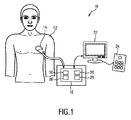
図1は、概して10と示される超音波撮像装置の模式図を示す。超音波撮像装置10は、解剖学的部位、具体的には患者12の解剖学的部位のボリュームを検査するために適用される。超音波撮像装置10は、超音波を送受信する複数のトランスデューサ素子を含む少なくとも1つのトランスデューサアレイを有する超音波プローブ14を含む。トランスデューサ素子は、多次元画像データ、特に3次元超音波像データ及びバイプレーン像データを提供するために、好適には2Dアレイに配置される。バイプレーン像データは、2つの交差する2D像平面をスイープすることによって取得される。一般に、バイプレーン撮像では、2つの2D平面はアレイの放射面と直交し、また、異なる角度で交差する。
FIG. 1 shows a schematic diagram of an ultrasonic imaging apparatus generally shown as 10. The
超音波撮像装置10は、一般に、超音波プローブ14に接続されて超音波プローブ14を制御し、超音波プローブ14から受信される超音波データを評価する制御ユニット16を含む。
The
超音波プローブ14は、患者12の解剖学的部位の視野内の3次元及び2次元超音波像データを提供する。2次元超音波像データは、超音波プローブ14によって放射された超音波の伝搬方向と平行である2つ以上の像平面で提供される。超音波プローブ14は、特に、バイプレーナモードでは、2次元超音波像をリアルタイムで提供する。2次元超音波像の像平面は、互いに直交して配置(交差)される(直交バイプレーン)か、又は、互いに異なる角度で配置(交差)される。2つの異なる像平面における2次元超音波像は、リアルタイムで同時に取得され、リアルタイムで表示される。
The
制御ユニット16は、超音波プローブ14に結合され、超音波プローブ14から3次元超音波像データ及び2次元超音波像データを受信する像処理ユニット18を含む。像処理ユニット18は、超音波像データ内の解剖学的特徴を決定する。像処理ユニット18は、パターン検出又はエッジ検出に基づいて、2次元超音波像データ内の解剖学的特徴の輪郭を決定し、また、解剖学的特徴のセグメンテーションを行い、解剖学的特徴の対応するセグメンテーションデータを提供する。つまり、像処理ユニット18は、解剖学的特徴又は解剖学的構造物の主な特徴の主な特性を決定する。
The control unit 16 includes an image processing unit 18 that is coupled to the
制御ユニット16は更に、2次元超音波像データを評価する評価ユニット20を含む。評価ユニット20は、超音波プローブ14の視野に対する及び2次元超音波像データの像平面に対する解剖学的特徴の位置関係を決定し、2次元超音波像データにおける解剖学的特徴の位置関係に基づいて、品質パラメータを決定する。
The control unit 16 further includes an
超音波放射装置10は更に、制御ユニット16から受信した像データを表示する表示ユニット22を含む。表示ユニット22は、像処理ユニット18から像データ全般を受信し、超音波プローブ14によって検出された2次元超音波像データ及び/又は3次元超音波像データを表示する。超音波撮像装置10は更に、像の取得全般を制御するために、表示ユニット22又は制御ユニット16に接続されてもよい入力デバイス24を含む。
The
好適には患者12の血管である解剖学的特徴を測定するために超音波像データが使用されるため、解剖学的特徴及び2次元超音波像データの像平面のうちの少なくとも1つの像平面に対する超音波プローブ14の正確な位置合わせが必要である。評価ユニット20は、像平面に対する及び超音波プローブ14の視線方向に対する解剖学的特徴の位置関係に基づいて、品質パラメータを決定し、表示ユニット22を介して、対応するフィードバックをユーザに提供する。評価ユニット20は、高品質像データを提供するために、解剖学的特徴が像平面に対して及び超音波プローブ14の視線方向に対して良好に位置合わせされているかどうかを決定し、これにより、解剖学的特徴の高品質ボリューム及び/又はサイズ測定が実現可能である。評価ユニットは、品質パラメータを所定品質限界と比較し、表示ユニット22を介して、対応するフィードバックをユーザに提供する。
Since the ultrasound image data is preferably used to measure the anatomical features of the patient 12's blood vessels, at least one of the anatomical features and the image plane of the two-dimensional ultrasound image data. Accurate alignment of the
品質パラメータが所定品質限界内である場合、像処理ユニット18の測定ユニットによって、超音波プローブ14から受信された対応する解剖学的特徴の2次元超音波像データ又は3次元超音波像データに基づいて、解剖学的特徴のサイズ及び/又はボリューム測定を行うことができる。
When the quality parameter is within a predetermined quality limit, the measurement unit of the image processing unit 18 is based on the 2D or 3D ultrasound image data of the corresponding anatomical features received from the
品質パラメータが所定品質限界内でない場合、制御ユニット16の位置合わせユニット28又はユーザガイダンスユニット28が、解剖学的特徴に対する超音波プローブ14の位置の位置合わせ又は適応を示す。
If the quality parameter is not within a predetermined quality limit, the
代替実施形態では、制御ユニット16は、像平面及び/又は超音波プローブ14の視線方向を電子的に位置合わせするために、位置関係と、位置合わせユニット28から受信した対応する品質パラメータとに基づいて超音波プローブ14のステアリング方向を制御する。位置合わせには、像平面の交差角、又は、プローブに対するそれらのステアリング方向(2D平面は、アレイの放射面に対する直交角度とは異なる角度でステアリングされる)を変化させることが含まれる。
In an alternative embodiment, the control unit 16 is based on a positional relationship and the corresponding quality parameters received from the
超音波プローブ14の位置が解剖学的特徴に位置合わせされた後、別のスキャン後の2次元超音波像データ又は3次元超音波像データに基づいて、解剖学的特徴のサイズ及び/又はボリュームが測定される。
After the position of the
図2A及び図2Bは、超音波プローブ14によって提供され、概して30と示される解剖学的特徴を含む概略的な2次元超音波画像を示す。超音波プローブ14の視野32内の超音波像データが取得される。
2A and 2B show schematic two-dimensional ultrasound images provided by the
評価ユニット20は、解剖学的特徴30の輪郭34を決定して、視野32及び/又は2次元像の像平面に対する解剖学的特徴30の位置関係が決定される。
The
評価ユニット20は、像平面及び/又は視野32に対する解剖学的特徴30の位置関係に基づいて、品質パラメータを決定する。品質パラメータがそこに基づいて決定される位置関係は、図2Aに示されるように、画像中心36からの解剖学的特徴30の距離、又は、図2Bに示されるように、2次元超音波画像の水平軸40に対する解剖学的特徴30の主軸の角度38である。更なる実施形態では、品質パラメータは、解剖学的特徴30に対する2次元画像の奥行き又は解剖学的特徴の全体が視野32内に含まれているかどうかに基づいて決定される。
The
解剖学的特徴30が、視野32及び/又は2次元超音波画像の像平面に対して良好に位置合わせされ、対応する品質パラメータが所定品質限界内である場合、超音波プローブ14から受信される、例えば別のスキャン後の2次元超音波像データ及び/又は3次元超音波像データに基づいて、解剖学的特徴30のサイズ及び/又はボリュームが測定される。
The
図3Aから図3Cは、様々な視線方向における解剖学的特徴34に対する2次元像データの2つの交差する像平面42、44と、各像平面42、44における解剖学的特徴34の概略的な断面図とを示す。2つの像平面42、44における2次元像データは同時に取得され、好適にはリアルタイムで表示される。
FIGS. 3A 3C is an
図3Aでは、2つの像平面42、44(バイプレーン)は、患者12の血管である解剖学的特徴30に対して配置される。各像平面42、44は良好に位置合わせされている。即ち、像平面42は、血管30の長手軸と直交して配置され、像平面44は、血管30の長手軸と平行に位置合わせされ、血管30に対して中心が置かれている。したがって、像平面42、44において捕捉される2次元像は、画像中心36及び2次元像データの水平軸40に対して中心が置かれる。
In FIG. 3A, the two
図3Bでは、解剖学的特徴34に対して像平面42だけが示される。ここでは、像平面42は、血管34の長手軸と直交には配置されておらず、したがって、像平面42の位置ずれが存在し、解剖学的特徴30のボリューム及び/又はサイズを正確に測定することができない。図3Bには、像平面42に対する解剖学的特徴30の各輪郭34が概略的に示される。
In FIG. 3B, only the
図3Cでは、解剖学的特徴30の長手軸と平行に位置合わせされた像平面44だけが示される。ここでは、像平面44は、解剖学的特徴30の中心に配置されておらず、したがって、解剖学的特徴30は、中心36に対して位置がずれており、図3Cに示されるように、解剖学的特徴30のサイズ及びボリュームを正確に測定することができない。
In FIG. 3C, only the image plane 44 aligned parallel to the longitudinal axis of the
したがって、ここでは、患者12の血管である解剖学的特徴30のサイズ及び/又はボリュームは、2つの像平面42、44が、中心36及び水平軸40に対して良好に位置合わせされる場合にのみ、正確に測定することができる。
Thus, here, the size and / or volume of the
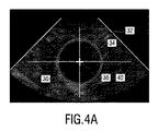
図4に、視野32内の2つの異なる2次元超音波像が概略的に示される。図4Aは、視野32の中心36に中心が置かれ、したがって、解剖学的特徴30のサイズ及び/又はボリュームの正確な測定を行うために良好に位置合わせされている解剖学的特徴30を含む2次元超音波像を示す。特定される品質パラメータは、解剖学的特徴30に対するこのプローブ位置では、比較的高い。図4Bは、像平面42、44及び視野32に対して位置がずれている解剖学的特徴30を含む2次元超音波像を示す。この具体例では、解剖学的特徴30の長手軸が、水平軸40に対して角度38だけ傾斜している。特定される品質パラメータは、解剖学的特徴30に対するこのプローブ位置及び平面方向では、比較的低い。図4Bに示される像を用いて行われる解剖学的特徴30の測定は、解剖学的特徴の実際のサイズに関して、誤差が大きくなる。位置合わせユニット28は、解剖学的特徴に対する像平面の向上された位置合わせを示す。視野32及び像平面42、44を解剖学的特徴34に対して位置合わせするために、像平面42、44の回転及び/又は並進か、又は、視野32に対して解剖学的特徴30を位置合わせするための超音波プローブ14の位置を示す指標46が2次元超音波像内に示される。指標46(矢印)は、ユーザに表示される2次元超音波像内に示され、これにより、ユーザは、超音波プローブ14を動かすことによって、像平面及び/又は視野32を位置合わせすることができる。本発明は、血管定量化において有利に実施されてよい。血管定量化では、定量化される血管に対する超音波平面の位置合わせが、超音波支援診断において重要な役割を果たす。
FIG. 4 schematically shows two different two-dimensional ultrasound images in the field of
品質パラメータが所定品質限界内である場合、解剖学的特徴30のサイズ及び/又はボリューム測定は、2次元超音波像データに基づいて行われるか、又は、サイズ若しくはボリュームを正確に測定するために、超音波プローブ14によって、フル3次元超音波スキャンが行われてもよい。
If the quality parameter is within a predetermined quality limit, the size and / or volume measurement of the
更なる実施形態では、サイズ及び/又はボリューム測定は、3次元超音波データの自動セグメンテーションと組み合わされる。 In a further embodiment, size and / or volume measurements are combined with automatic segmentation of 3D ultrasound data.
図5は、被験者12のボリュームを検査する超音波撮像方法の概略フロー図を示す。図5の方法は、概して50と示される。 FIG. 5 shows a schematic flow chart of an ultrasonic imaging method for inspecting the volume of the subject 12. The method of FIG. 5 is generally shown as 50.
方法50は、ステップ52に示されるように、2つの像平面42、44において、2次元超音波像データを同時に取得することから開始する。ステップ54に示されるように、解剖学的特徴30が、2つの異なる像平面42、44における2次元超音波データに基づいて、軸方向及び長手方向において自動的にセグメント化される。少なくとも、2次元像平面に対する解剖学的特徴の配向角が推定される。これは、ユーザガイダンスのためのフィードバックを提供するために、推定がフルセグメンテーションよりも単純であるため、技術的努力を全般的に減少することができる。
ステップ56において、各像平面42、44に対する解剖学的特徴30の位置関係に基づいて、品質パラメータが決定される。ステップ58において、品質パラメータは、位置合わせ品質を評価するために、所定品質限界と比較される。品質パラメータが所定品質限界内にない場合、品質パラメータを向上させるために、超音波プローブ14の回転及び/若しくは並進移動、又は、画像奥行きの適応を提案するために、指標46が、ユーザガイダンスとして、表示スクリーン22に表示される(ステップ60)。位置合わせ後、方法50は、フィードバックループ62によって示されるように、ステップ52に戻る。
In
品質パラメータが所定品質限界内である場合、ユーザは、ステップ64に示されるように、表示ユニット22を介してフィードバックを受け取り、超音波プローブ14の位置合わせされた位置における2次元超音波像データ又は3次元超音波像データが取得され、測定ユニットが、ステップ66に示されるように、任意選択的に、2次元超音波像データ又は3次元超音波像データに基づいて、解剖学的特徴30のサイズ及び/又はボリュームを決定する。
If the quality parameter is within a predetermined quality limit, the user receives feedback via the
ユーザは、任意選択的に、測定結果を確認し、フィードバックループ68によって示されるように、ステップ52に戻る。
The user optionally confirms the measurement result and returns to step 52 as indicated by the
本発明は、図面及び上記説明において詳細に例示及び説明されたが、当該例示及び説明は、例示であって、限定と解釈されるべきではない。本発明は、開示された実施形態に限定されない。開示された実施形態の他の変形態様は、図面、開示内容及び添付の請求項の検討から、請求項に係る発明を実施する当業者によって理解され、実施される。 The present invention has been exemplified and described in detail in the drawings and the above description, but the examples and description are exemplary and should not be construed as limiting. The present invention is not limited to the disclosed embodiments. Other modifications of the disclosed embodiments will be understood and implemented by those skilled in the art who will implement the claimed invention from the examination of the drawings, disclosure content and the accompanying claims.
請求項において、「含む」との用語は、他の要素又はステップを除外するものではなく、また、「a」又は「an」との不定冠詞も、複数形を除外するものではない。単一の要素又は他のユニットが、請求項に記載される幾つかのアイテムの機能を果たしてもよい。特定の手段が相互に異なる従属請求項に記載されることだけで、これらの手段の組み合わせを有利に使用することができないことを示すものではない。 In the claims, the term "contains" does not exclude other elements or steps, nor does the indefinite article "a" or "an" exclude the plural. A single element or other unit may perform the function of some of the items described in the claims. The fact that specific means are described in different dependent claims does not indicate that the combination of these means cannot be used in an advantageous manner.
コンピュータプログラムは、他のハードウェアと共に又はその一部として供給される光学記憶媒体又は固体媒体といった適切な媒体上に記憶及び/又は分散されてもよいが、インターネット又は他の有線若しくは無線通信システムを介するといった他の形式で分配されてもよい。 Computer programs may be stored and / or distributed on suitable media such as optical storage media or solid-state media supplied with or as part of other hardware, but may include the Internet or other wired or wireless communication systems. It may be distributed in other forms such as via.
請求項における任意の参照符号は、範囲を限定するものと解釈されるべきではない。 Any reference code in the claims should not be construed as limiting the scope.
Claims (15)
視野内の3次元超音波データを取得し、前記視野内の2つの交差する像平面内の3次元超音波像データ及び2次元超音波像データを提供する複数の超音波トランスデューサ素子を含む超音波プローブと、
前記超音波プローブに結合され、前記2つの交差する像平面内の前記2次元超音波像データを受信し、前記2次元超音波像データの前記2つの交差する像平面内の解剖学的特徴を決定する像処理ユニットと、
前記2次元超音波像データを評価し、前記像平面のそれぞれに対する前記解剖学的特徴の位置関係に基づいて、品質パラメータを決定する評価ユニットと、
前記位置関係及び前記品質パラメータに基づいて、前記像平面若しくは前記視野の位置合わせを行うか又は示す位置合わせユニットと、を含み、
前記評価ユニットは、前記品質パラメータを所定品質限界と比較し、前記位置合わせユニットは、前記品質パラメータが前記所定品質限界内でない場合、前記解剖学的特徴に対する前記超音波プローブの位置の位置合わせ又は適応を示す、超音波撮像装置。 An ultrasonic imaging device that inspects the volume of a subject.
Ultrasound including a plurality of ultrasound transducer elements that acquire 3D ultrasound data in the field of view and provide 3D ultrasound image data and 2D ultrasound image data in two intersecting image planes in the field. With the probe
It is coupled to the ultrasound probe to receive the two-dimensional ultrasound image data in the two intersecting image planes and captures the anatomical features of the two-dimensional ultrasound image data in the two intersecting image planes. The image processing unit to be determined and
An evaluation unit that evaluates the two-dimensional ultrasonic image data and determines quality parameters based on the positional relationship of the anatomical features with respect to each of the image planes.
Includes an alignment unit that aligns or indicates the image plane or the field of view based on the positional relationship and the quality parameters.
The evaluation unit compares the quality parameter to a predetermined quality limit, and the alignment unit aligns or aligns the ultrasonic probe with respect to the anatomical feature if the quality parameter is not within the predetermined quality limit. Ultrasound imaging device showing indication.
視野内の2つの交差する像平面内の2次元超音波像データを取得するステップと、
前記2つの交差する像平面内の前記2次元超音波像データを処理し、前記2次元超音波像データの前記2つの交差する像平面内の解剖学的特徴を決定するステップと、
前記2次元超音波像データを評価し、前記2つの交差する像平面に対する前記解剖学的特徴の位置関係に基づいて、品質パラメータを決定するステップと、
前記位置関係及び前記品質パラメータに基づいて、前記2つの交差する像平面の位置合わせを示すか、又は、前記2つの交差する像平面若しくは視野を位置合わせするステップと、
前記視野内の3次元超音波データを取得するステップと、
前記品質パラメータを所定品質限界と比較し、前記品質パラメータが前記所定品質限界内でない場合、前記解剖学的特徴に対する超音波プローブの位置の位置合わせ又は適応を示すステップと、
を含む、方法。 This is an ultrasonic imaging method for inspecting the volume of a subject.
Steps to acquire 2D ultrasound image data in two intersecting image planes in the field of view,
A step of processing the two-dimensional ultrasonic image data in the two intersecting image planes and determining the anatomical features of the two-dimensional ultrasonic image data in the two intersecting image planes.
A step of evaluating the two-dimensional ultrasonic image data and determining quality parameters based on the positional relationship of the anatomical features with respect to the two intersecting image planes.
A step of indicating the alignment of the two intersecting image planes or aligning the two intersecting image planes or the field of view based on the positional relationship and the quality parameter.
The step of acquiring the three-dimensional ultrasonic data in the field of view and
A step of comparing the quality parameter with a predetermined quality limit and indicating alignment or adaptation of the position of the ultrasonic probe to the anatomical feature if the quality parameter is not within the predetermined quality limit.
Including methods.
Applications Claiming Priority (3)
| Application Number | Priority Date | Filing Date | Title |
|---|---|---|---|
| EP15307070 | 2015-12-21 | ||
| EP15307070.1 | 2015-12-21 | ||
| PCT/EP2016/081684 WO2017108667A1 (en) | 2015-12-21 | 2016-12-19 | Ultrasound imaging apparatus and ultrasound imaging method for inspecting a volume of subject |
Publications (3)
| Publication Number | Publication Date |
|---|---|
| JP2019503748A JP2019503748A (en) | 2019-02-14 |
| JP2019503748A5 JP2019503748A5 (en) | 2020-02-06 |
| JP6960922B2 true JP6960922B2 (en) | 2021-11-05 |
Family
ID=55070813
Family Applications (1)
| Application Number | Title | Priority Date | Filing Date |
|---|---|---|---|
| JP2018532321A Active JP6960922B2 (en) | 2015-12-21 | 2016-12-19 | Ultrasound imaging device and ultrasonic imaging method for inspecting the volume of a subject |
Country Status (5)
| Country | Link |
|---|---|
| US (1) | US20190015076A1 (en) |
| EP (1) | EP3393366A1 (en) |
| JP (1) | JP6960922B2 (en) |
| CN (1) | CN108430334B (en) |
| WO (1) | WO2017108667A1 (en) |
Families Citing this family (11)
| Publication number | Priority date | Publication date | Assignee | Title |
|---|---|---|---|---|
| WO2019084411A1 (en) | 2017-10-27 | 2019-05-02 | Butterfly Network, Inc. | Quality indicators for collection of and automated measurement on ultrasound images |
| EP3569154A1 (en) | 2018-05-15 | 2019-11-20 | Koninklijke Philips N.V. | Ultrasound processing unit and method, and imaging system |
| US20200245970A1 (en) * | 2019-01-31 | 2020-08-06 | Bay Labs, Inc. | Prescriptive guidance for ultrasound diagnostics |
| EP3711673A1 (en) * | 2019-03-18 | 2020-09-23 | Koninklijke Philips N.V. | Methods and systems for adjusting the field of view of an ultrasound probe |
| GB201908052D0 (en) * | 2019-06-06 | 2019-07-24 | Nisonic As | Alignment of ultrasound image |
| US20210007710A1 (en) * | 2019-07-12 | 2021-01-14 | Verathon Inc. | Representation of a target during aiming of an ultrasound probe |
| US11844654B2 (en) | 2019-08-19 | 2023-12-19 | Caption Health, Inc. | Mid-procedure view change for ultrasound diagnostics |
| CN115175621A (en) * | 2019-12-31 | 2022-10-11 | 布弗莱运营公司 | Method and apparatus for modifying ultrasound imaging plane position |
| US11593933B2 (en) * | 2020-03-16 | 2023-02-28 | GE Precision Healthcare LLC | Systems and methods for ultrasound image quality determination |
| JP2021178175A (en) * | 2020-05-13 | 2021-11-18 | キヤノンメディカルシステムズ株式会社 | Ultrasonic diagnostic device, medical image processing device, medical image processing method, and program |
| EP4094695A1 (en) * | 2021-05-28 | 2022-11-30 | Koninklijke Philips N.V. | Ultrasound imaging system |
Family Cites Families (41)
| Publication number | Priority date | Publication date | Assignee | Title |
|---|---|---|---|---|
| AU1294995A (en) * | 1993-11-29 | 1995-06-19 | Perception, Inc. | Pc based ultrasound device with virtual control user interface |
| US5871019A (en) * | 1996-09-23 | 1999-02-16 | Mayo Foundation For Medical Education And Research | Fast cardiac boundary imaging |
| AUPP227898A0 (en) * | 1998-03-11 | 1998-04-09 | Commonwealth Scientific And Industrial Research Organisation | Improvements in ultrasound techniques |
| JP2001212144A (en) * | 2000-01-31 | 2001-08-07 | Toshiba Corp | Ultrasonic diagnostic apparatus and ultrasonic imaging method |
| US6761689B2 (en) * | 2000-08-17 | 2004-07-13 | Koninklijke Philips Electronics N.V. | Biplane ultrasonic imaging |
| US6537220B1 (en) * | 2001-08-31 | 2003-03-25 | Siemens Medical Solutions Usa, Inc. | Ultrasound imaging with acquisition of imaging data in perpendicular scan planes |
| US6961405B2 (en) * | 2002-10-07 | 2005-11-01 | Nomos Corporation | Method and apparatus for target position verification |
| US6835177B2 (en) * | 2002-11-06 | 2004-12-28 | Sonosite, Inc. | Ultrasonic blood vessel measurement apparatus and method |
| JP3905470B2 (en) * | 2002-12-26 | 2007-04-18 | アロカ株式会社 | Ultrasonic diagnostic equipment |
| JP4555619B2 (en) * | 2004-06-28 | 2010-10-06 | アロカ株式会社 | Ultrasonic diagnostic equipment |
| JP4868843B2 (en) * | 2005-01-26 | 2012-02-01 | 株式会社東芝 | Ultrasonic diagnostic apparatus and control program for ultrasonic diagnostic apparatus |
| EP1685799B1 (en) * | 2005-01-26 | 2013-02-27 | Kabushiki Kaisha Toshiba | Ultrasonic diagnostic apparatus and ultrasonic image acquiring method |
| JP4750429B2 (en) * | 2005-02-08 | 2011-08-17 | 株式会社日立メディコ | Image display device |
| CN101141920B (en) * | 2005-03-15 | 2011-12-14 | 株式会社东芝 | Ultrasonic diagnostic equipment and its controlling program |
| CN101179998B (en) * | 2005-05-20 | 2010-12-08 | 株式会社日立医药 | Image diagnosing device |
| CN101404931A (en) * | 2006-03-20 | 2009-04-08 | 皇家飞利浦电子股份有限公司 | Ultrasonic diagnosis by quantification of myocardial performance |
| JP5014051B2 (en) * | 2007-10-09 | 2012-08-29 | 株式会社ユネクス | Vascular ultrasound image measurement method |
| JP5134932B2 (en) * | 2007-12-03 | 2013-01-30 | 株式会社東芝 | Ultrasonic diagnostic apparatus and control program for ultrasonic diagnostic apparatus |
| JP5404141B2 (en) * | 2008-06-13 | 2014-01-29 | キヤノン株式会社 | Ultrasonic device and control method thereof |
| US8600133B2 (en) * | 2008-10-01 | 2013-12-03 | Koninklijke Philips N.V. | Selection of snapshots of a medical image sequence |
| JP5620666B2 (en) * | 2008-10-16 | 2014-11-05 | 株式会社東芝 | Ultrasonic diagnostic equipment, ultrasonic image processing equipment |
| US8265363B2 (en) * | 2009-02-04 | 2012-09-11 | General Electric Company | Method and apparatus for automatically identifying image views in a 3D dataset |
| US8355554B2 (en) * | 2009-04-14 | 2013-01-15 | Sonosite, Inc. | Systems and methods for adaptive volume imaging |
| KR101116925B1 (en) * | 2009-04-27 | 2012-05-30 | 삼성메디슨 주식회사 | Ultrasound system and method for aligning ultrasound image |
| US8939908B2 (en) * | 2009-07-16 | 2015-01-27 | Unex Corporation | Ultrasonic blood vessel inspecting apparatus |
| US9248316B2 (en) * | 2010-01-12 | 2016-02-02 | Elekta Ltd. | Feature tracking using ultrasound |
| JP5462076B2 (en) * | 2010-06-01 | 2014-04-02 | 株式会社東芝 | Ultrasonic diagnostic apparatus and image information management apparatus |
| JPWO2012029417A1 (en) * | 2010-08-31 | 2013-10-28 | 株式会社日立メディコ | Ultrasonic diagnostic apparatus and evaluation calculation method |
| US9119559B2 (en) * | 2011-06-16 | 2015-09-01 | Salient Imaging, Inc. | Method and system of generating a 3D visualization from 2D images |
| CN104066380B (en) * | 2012-02-01 | 2016-11-09 | 东芝医疗系统株式会社 | Diagnostic ultrasound equipment, image processing apparatus |
| JP6160487B2 (en) * | 2012-04-23 | 2017-07-12 | コニカミノルタ株式会社 | Ultrasonic diagnostic apparatus and control method thereof |
| KR20140024190A (en) * | 2012-08-20 | 2014-02-28 | 삼성메디슨 주식회사 | Method for managing and displaying ultrasound image, and apparatus thereto |
| US10424044B2 (en) * | 2012-12-21 | 2019-09-24 | Koninklijke Philips N.V. | Anatomically intelligent echocardiography for point-of-care |
| CN105101881A (en) * | 2013-03-29 | 2015-11-25 | 日立阿洛卡医疗株式会社 | Image alignment display method and ultrasonic diagnostic device |
| EP2807978A1 (en) * | 2013-05-28 | 2014-12-03 | Universität Bern | Method and system for 3D acquisition of ultrasound images |
| JP6890971B2 (en) * | 2013-12-09 | 2021-06-18 | コーニンクレッカ フィリップス エヌ ヴェKoninklijke Philips N.V. | Image imaging guidance using model-based segmentation |
| KR20150082945A (en) * | 2014-01-08 | 2015-07-16 | 삼성메디슨 주식회사 | Ultrasonic diagnostic apparatus and operating method for the same |
| US10349917B2 (en) * | 2014-06-11 | 2019-07-16 | The Johns Hopkins University | Synthetic aperture ultrasound system |
| US20160038125A1 (en) * | 2014-08-06 | 2016-02-11 | General Electric Company | Guided semiautomatic alignment of ultrasound volumes |
| WO2017003480A1 (en) * | 2015-07-02 | 2017-01-05 | Siemens Medical Solutions Usa, Inc. | Intervolume lesion detection and image preparation |
| KR20170068944A (en) * | 2015-12-10 | 2017-06-20 | 삼성메디슨 주식회사 | Method of displaying a ultrasound image and apparatus thereof |
-
2016
- 2016-12-19 CN CN201680075157.8A patent/CN108430334B/en active Active
- 2016-12-19 EP EP16816666.8A patent/EP3393366A1/en active Pending
- 2016-12-19 US US16/061,803 patent/US20190015076A1/en active Pending
- 2016-12-19 JP JP2018532321A patent/JP6960922B2/en active Active
- 2016-12-19 WO PCT/EP2016/081684 patent/WO2017108667A1/en active Application Filing
Also Published As
| Publication number | Publication date |
|---|---|
| JP2019503748A (en) | 2019-02-14 |
| CN108430334B (en) | 2021-06-01 |
| US20190015076A1 (en) | 2019-01-17 |
| CN108430334A (en) | 2018-08-21 |
| EP3393366A1 (en) | 2018-10-31 |
| WO2017108667A1 (en) | 2017-06-29 |
Similar Documents
| Publication | Publication Date | Title |
|---|---|---|
| JP6960922B2 (en) | Ultrasound imaging device and ultrasonic imaging method for inspecting the volume of a subject | |
| EP3013243B1 (en) | Elastography measurement system and method | |
| JP6430498B2 (en) | System and method for mapping of ultrasonic shear wave elastography measurements | |
| EP3554380B1 (en) | Target probe placement for lung ultrasound | |
| CN111629670B (en) | Echo window artifact classification and visual indicator for ultrasound systems | |
| JP2017527401A (en) | Ultrasonic imaging device | |
| JP2019503748A5 (en) | ||
| JP6865755B2 (en) | Medical imaging device and medical imaging method for inspecting the volume of a subject | |
| JP2015530182A (en) | Automated biplane PW workflow for ultrasound stenosis assessment | |
| US10736608B2 (en) | Ultrasound diagnostic device and ultrasound image processing method | |
| JP2013153882A5 (en) | ||
| KR20070074288A (en) | Apparatus and method for diagnosing a human bladder using ultrasound signal | |
| US20220304653A1 (en) | Alignment of ultrasound image | |
| WO2014148428A1 (en) | Ultrasonic diagnostic device | |
| JP2014124319A (en) | Ultrasonic calibration system, and ultrasonic calibration method | |
| JP6865695B2 (en) | Ultrasound imaging device | |
| RU2620865C2 (en) | System and method for three-dimensional ultrasound measuring volumetric areas | |
| JP2009045097A (en) | Three-dimensional image generating apparatus and three-dimensional image generating method | |
| KR102615722B1 (en) | Ultrasound scanner and method of guiding aim | |
| EP2807977B1 (en) | Ultrasound diagnosis method and aparatus using three-dimensional volume data | |
| JP6510570B2 (en) | Image processing device | |
| JP2001029324A (en) | Slice surface setting mechanism for tomographic image of testee body | |
| JP5802421B2 (en) | Diagnostic imaging system | |
| JP2017018510A (en) | Ultrasonic diagnostic equipment and image forming method thereof |
Legal Events
| Date | Code | Title | Description |
|---|---|---|---|
| A521 | Written amendment |
Free format text: JAPANESE INTERMEDIATE CODE: A523 Effective date: 20191217 |
|
| A621 | Written request for application examination |
Free format text: JAPANESE INTERMEDIATE CODE: A621 Effective date: 20191217 |
|
| A977 | Report on retrieval |
Free format text: JAPANESE INTERMEDIATE CODE: A971007 Effective date: 20201020 |
|
| A131 | Notification of reasons for refusal |
Free format text: JAPANESE INTERMEDIATE CODE: A131 Effective date: 20201124 |
|
| A601 | Written request for extension of time |
Free format text: JAPANESE INTERMEDIATE CODE: A601 Effective date: 20210219 |
|
| A521 | Written amendment |
Free format text: JAPANESE INTERMEDIATE CODE: A523 Effective date: 20210521 |
|
| TRDD | Decision of grant or rejection written | ||
| A01 | Written decision to grant a patent or to grant a registration (utility model) |
Free format text: JAPANESE INTERMEDIATE CODE: A01 Effective date: 20210913 |
|
| A61 | First payment of annual fees (during grant procedure) |
Free format text: JAPANESE INTERMEDIATE CODE: A61 Effective date: 20211012 |
|
| R150 | Certificate of patent or registration of utility model |
Ref document number: 6960922 Country of ref document: JP Free format text: JAPANESE INTERMEDIATE CODE: R150 |