JP6865695B2 - Ultrasound imaging device - Google Patents
Ultrasound imaging device Download PDFInfo
- Publication number
- JP6865695B2 JP6865695B2 JP2017563333A JP2017563333A JP6865695B2 JP 6865695 B2 JP6865695 B2 JP 6865695B2 JP 2017563333 A JP2017563333 A JP 2017563333A JP 2017563333 A JP2017563333 A JP 2017563333A JP 6865695 B2 JP6865695 B2 JP 6865695B2
- Authority
- JP
- Japan
- Prior art keywords
- ultrasonic
- anatomical object
- probes
- ultrasound
- data
- Prior art date
- Legal status (The legal status is an assumption and is not a legal conclusion. Google has not performed a legal analysis and makes no representation as to the accuracy of the status listed.)
- Active
Links
Images


Classifications
-
- A—HUMAN NECESSITIES
- A61—MEDICAL OR VETERINARY SCIENCE; HYGIENE
- A61B—DIAGNOSIS; SURGERY; IDENTIFICATION
- A61B8/00—Diagnosis using ultrasonic, sonic or infrasonic waves
- A61B8/44—Constructional features of the ultrasonic, sonic or infrasonic diagnostic device
- A61B8/4477—Constructional features of the ultrasonic, sonic or infrasonic diagnostic device using several separate ultrasound transducers or probes
-
- A—HUMAN NECESSITIES
- A61—MEDICAL OR VETERINARY SCIENCE; HYGIENE
- A61B—DIAGNOSIS; SURGERY; IDENTIFICATION
- A61B8/00—Diagnosis using ultrasonic, sonic or infrasonic waves
- A61B8/52—Devices using data or image processing specially adapted for diagnosis using ultrasonic, sonic or infrasonic waves
- A61B8/5215—Devices using data or image processing specially adapted for diagnosis using ultrasonic, sonic or infrasonic waves involving processing of medical diagnostic data
- A61B8/5223—Devices using data or image processing specially adapted for diagnosis using ultrasonic, sonic or infrasonic waves involving processing of medical diagnostic data for extracting a diagnostic or physiological parameter from medical diagnostic data
-
- A—HUMAN NECESSITIES
- A61—MEDICAL OR VETERINARY SCIENCE; HYGIENE
- A61B—DIAGNOSIS; SURGERY; IDENTIFICATION
- A61B8/00—Diagnosis using ultrasonic, sonic or infrasonic waves
- A61B8/52—Devices using data or image processing specially adapted for diagnosis using ultrasonic, sonic or infrasonic waves
- A61B8/5215—Devices using data or image processing specially adapted for diagnosis using ultrasonic, sonic or infrasonic waves involving processing of medical diagnostic data
- A61B8/5238—Devices using data or image processing specially adapted for diagnosis using ultrasonic, sonic or infrasonic waves involving processing of medical diagnostic data for combining image data of patient, e.g. merging several images from different acquisition modes into one image
- A61B8/5246—Devices using data or image processing specially adapted for diagnosis using ultrasonic, sonic or infrasonic waves involving processing of medical diagnostic data for combining image data of patient, e.g. merging several images from different acquisition modes into one image combining images from the same or different imaging techniques, e.g. color Doppler and B-mode
- A61B8/5253—Devices using data or image processing specially adapted for diagnosis using ultrasonic, sonic or infrasonic waves involving processing of medical diagnostic data for combining image data of patient, e.g. merging several images from different acquisition modes into one image combining images from the same or different imaging techniques, e.g. color Doppler and B-mode combining overlapping images, e.g. spatial compounding
-
- G—PHYSICS
- G01—MEASURING; TESTING
- G01S—RADIO DIRECTION-FINDING; RADIO NAVIGATION; DETERMINING DISTANCE OR VELOCITY BY USE OF RADIO WAVES; LOCATING OR PRESENCE-DETECTING BY USE OF THE REFLECTION OR RERADIATION OF RADIO WAVES; ANALOGOUS ARRANGEMENTS USING OTHER WAVES
- G01S15/00—Systems using the reflection or reradiation of acoustic waves, e.g. sonar systems
- G01S15/88—Sonar systems specially adapted for specific applications
- G01S15/89—Sonar systems specially adapted for specific applications for mapping or imaging
- G01S15/8906—Short-range imaging systems; Acoustic microscope systems using pulse-echo techniques
- G01S15/8995—Combining images from different aspect angles, e.g. spatial compounding
-
- G—PHYSICS
- G01—MEASURING; TESTING
- G01S—RADIO DIRECTION-FINDING; RADIO NAVIGATION; DETERMINING DISTANCE OR VELOCITY BY USE OF RADIO WAVES; LOCATING OR PRESENCE-DETECTING BY USE OF THE REFLECTION OR RERADIATION OF RADIO WAVES; ANALOGOUS ARRANGEMENTS USING OTHER WAVES
- G01S7/00—Details of systems according to groups G01S13/00, G01S15/00, G01S17/00
- G01S7/52—Details of systems according to groups G01S13/00, G01S15/00, G01S17/00 of systems according to group G01S15/00
- G01S7/52017—Details of systems according to groups G01S13/00, G01S15/00, G01S17/00 of systems according to group G01S15/00 particularly adapted to short-range imaging
- G01S7/52079—Constructional features
-
- G—PHYSICS
- G16—INFORMATION AND COMMUNICATION TECHNOLOGY [ICT] SPECIALLY ADAPTED FOR SPECIFIC APPLICATION FIELDS
- G16H—HEALTHCARE INFORMATICS, i.e. INFORMATION AND COMMUNICATION TECHNOLOGY [ICT] SPECIALLY ADAPTED FOR THE HANDLING OR PROCESSING OF MEDICAL OR HEALTHCARE DATA
- G16H50/00—ICT specially adapted for medical diagnosis, medical simulation or medical data mining; ICT specially adapted for detecting, monitoring or modelling epidemics or pandemics
- G16H50/30—ICT specially adapted for medical diagnosis, medical simulation or medical data mining; ICT specially adapted for detecting, monitoring or modelling epidemics or pandemics for calculating health indices; for individual health risk assessment
-
- A—HUMAN NECESSITIES
- A61—MEDICAL OR VETERINARY SCIENCE; HYGIENE
- A61B—DIAGNOSIS; SURGERY; IDENTIFICATION
- A61B8/00—Diagnosis using ultrasonic, sonic or infrasonic waves
- A61B8/06—Measuring blood flow
- A61B8/065—Measuring blood flow to determine blood output from the heart
-
- A—HUMAN NECESSITIES
- A61—MEDICAL OR VETERINARY SCIENCE; HYGIENE
- A61B—DIAGNOSIS; SURGERY; IDENTIFICATION
- A61B8/00—Diagnosis using ultrasonic, sonic or infrasonic waves
- A61B8/08—Detecting organic movements or changes, e.g. tumours, cysts, swellings
- A61B8/0883—Detecting organic movements or changes, e.g. tumours, cysts, swellings for diagnosis of the heart
-
- A—HUMAN NECESSITIES
- A61—MEDICAL OR VETERINARY SCIENCE; HYGIENE
- A61B—DIAGNOSIS; SURGERY; IDENTIFICATION
- A61B8/00—Diagnosis using ultrasonic, sonic or infrasonic waves
- A61B8/12—Diagnosis using ultrasonic, sonic or infrasonic waves in body cavities or body tracts, e.g. by using catheters
-
- A—HUMAN NECESSITIES
- A61—MEDICAL OR VETERINARY SCIENCE; HYGIENE
- A61B—DIAGNOSIS; SURGERY; IDENTIFICATION
- A61B8/00—Diagnosis using ultrasonic, sonic or infrasonic waves
- A61B8/13—Tomography
- A61B8/14—Echo-tomography
- A61B8/145—Echo-tomography characterised by scanning multiple planes
-
- A—HUMAN NECESSITIES
- A61—MEDICAL OR VETERINARY SCIENCE; HYGIENE
- A61B—DIAGNOSIS; SURGERY; IDENTIFICATION
- A61B8/00—Diagnosis using ultrasonic, sonic or infrasonic waves
- A61B8/44—Constructional features of the ultrasonic, sonic or infrasonic diagnostic device
- A61B8/4483—Constructional features of the ultrasonic, sonic or infrasonic diagnostic device characterised by features of the ultrasound transducer
- A61B8/4488—Constructional features of the ultrasonic, sonic or infrasonic diagnostic device characterised by features of the ultrasound transducer the transducer being a phased array
-
- A—HUMAN NECESSITIES
- A61—MEDICAL OR VETERINARY SCIENCE; HYGIENE
- A61B—DIAGNOSIS; SURGERY; IDENTIFICATION
- A61B8/00—Diagnosis using ultrasonic, sonic or infrasonic waves
- A61B8/48—Diagnostic techniques
- A61B8/488—Diagnostic techniques involving Doppler signals
-
- A—HUMAN NECESSITIES
- A61—MEDICAL OR VETERINARY SCIENCE; HYGIENE
- A61B—DIAGNOSIS; SURGERY; IDENTIFICATION
- A61B8/00—Diagnosis using ultrasonic, sonic or infrasonic waves
- A61B8/54—Control of the diagnostic device
Description
本発明は、患者の解剖学的対象に関連付けられる少なくとも1つの物理的パラメータを決定する超音波撮像装置に関する。 The present invention relates to an ultrasound imaging device that determines at least one physical parameter associated with a patient's anatomical object.
本発明は更に、患者の解剖学的対象に関連付けられる少なくとも1つの物理的パラメータを決定する超音波撮像方法と、コンピュータ上で実行されると、当該コンピュータに本発明による上記方法のステップを実行させるプログラムコード手段を含むコンピュータプログラムとに関する。 The invention further includes an ultrasound imaging method that determines at least one physical parameter associated with the patient's anatomical object and, when performed on a computer, causes the computer to perform the steps of the method according to the invention. With respect to computer programs, including program code means.
医用検出用の超音波撮像システムの分野において、画質を向上させ、解剖学的対象の遮られていない図を提供するために、様々な視線方向から解剖学的対象を検出することが一般に知られている。対応する超音波撮像システムは、例えば国際特許公開WO2014/207621A1から知られている。 In the field of ultrasound imaging systems for medical detection, it is generally known to detect anatomical objects from various gaze directions in order to improve image quality and provide an unobstructed view of the anatomical object. ing. Corresponding ultrasonic imaging systems are known, for example, from International Patent Publication WO2014 / 207621A1.
特に経胸壁心エコー検査(TTE)用の超音波システムの分野において、現在、大面積経胸壁心エコー検査(LATTE)とも知られているマルチプローブアレイを含む超音波プローブが開発中である。これらのマルチプローブアレイは、互いに対し固定関係にある複数のプローブを含み、プローブの超音波画像は、組み合わされて全体画像が形成されるか、又は、別々に表示されて、様々な視線方向から各臓器の様々な部分が表示される。 Particularly in the field of ultrasound systems for transthoracic echocardiography (TTE), ultrasound probes including a multiprobe array, also known as large area transthoracic echocardiography (LATTE), are currently under development. These multi-probe arrays include multiple probes that are in a fixed relationship with each other, and the ultrasound images of the probes can be combined to form the entire image or displayed separately from different gaze directions. Different parts of each organ are displayed.
既知の超音波システムのデメリットは、様々な解剖学的構造が、当該様々な構造の様々な向きに起因して様々な画質の超音波画像に表示され、撮像品質の手動最適化はユーザにとって厄介である点である。 The disadvantage of known ultrasound systems is that different anatomical structures appear in different quality ultrasound images due to different orientations of the different structures, and manual optimization of imaging quality is awkward for the user. Is the point.
したがって、本発明は、品質が向上され、ユーザの取り扱い努力が少なくてすむ超音波検出を提供する超音波撮像装置及び対応する超音波撮像方法を提供することを目的とする。 Therefore, it is an object of the present invention to provide an ultrasonic imaging apparatus and a corresponding ultrasonic imaging method that provide ultrasonic detection with improved quality and less user handling effort.
本発明の一態様によれば、超音波撮像装置が提供される。当該超音波撮像装置は、
複数の超音波プローブに接続される超音波取得ユニットであって、各超音波プローブは超音波プローブの視野内の患者の超音波撮像に適した超音波データを提供する、上記超音波取得ユニットと、
複数の超音波プローブのうちの少なくとも1つから受信される超音波データに基づいて視野内の患者の解剖学的対象を検出し、解剖学的対象と各超音波プローブとの空間的関係を決定する検出ユニットと、
検出ユニットに結合され、超音波データから検出可能である少なくとも1つの物理的パラメータの取得品質に基づいて複数の超音波プローブのうちの少なくとも1つを選択する選択ユニットと、
超音波取得ユニットに結合され、選択された少なくとも1つの超音波プローブから超音波データを受信し、選択された超音波プローブから受信される超音波データに基づいて、少なくとも1つの物理的パラメータを決定する評価ユニットとを含み、取得品質は、解剖学的対象と各超音波プローブとの空間的関係と、解剖学的対象の少なくとも1つの解剖学的特徴とに基づいて決定される。
According to one aspect of the present invention, an ultrasonic imaging device is provided. The ultrasonic imaging device is
An ultrasonic acquisition unit connected to a plurality of ultrasonic probes, and each ultrasonic probe provides ultrasonic data suitable for ultrasonic imaging of a patient in the field of view of the ultrasonic probe. ,
Detects a patient's anatomical object in the visual field based on ultrasonic data received from at least one of a plurality of ultrasonic probes, and determines the spatial relationship between the anatomical object and each ultrasonic probe. Detection unit and
A selection unit that is coupled to the detection unit and selects at least one of a plurality of ultrasonic probes based on the acquisition quality of at least one physical parameter that can be detected from the ultrasonic data.
Combined with an ultrasonic acquisition unit, it receives ultrasonic data from at least one selected ultrasonic probe and determines at least one physical parameter based on the ultrasonic data received from the selected ultrasonic probe. Acquisition quality is determined based on the spatial relationship between the anatomical object and each ultrasonic probe and at least one anatomical feature of the anatomical object.
本発明の別に態様によれば、超音波撮像方法が提供される。当該方法は、
複数の超音波プローブの視野内の患者の超音波撮像に適した超音波データを取得するステップと、
複数の超音波プローブのうちの少なくとも1つから受信される超音波データに基づいて、視野内の患者の解剖学的対象を決定するステップと、
解剖学的対象と各超音波プローブとの空間的関係を決定するステップと、
超音波データから検出可能である少なくとも1つの物理的パラメータの取得品質に基づいて、複数の超音波プローブのうちの少なくとも1つを選択するステップと、
選択された超音波プローブから受信される超音波データに基づいて、少なくとも1つの物理的パラメータを決定するステップとを含み、取得品質は、解剖学的対象と各超音波プローブとの空間的関係と、解剖学的対象の少なくとも1つの解剖学的特徴とに基づいて決定される。
According to another aspect of the present invention, an ultrasonic imaging method is provided. The method is
Steps to acquire ultrasound data suitable for ultrasound imaging of patients in the field of view of multiple ultrasound probes,
Steps to determine the patient's anatomical object in the field of view based on ultrasound data received from at least one of the ultrasound probes.
Steps to determine the spatial relationship between the anatomical object and each ultrasonic probe,
A step of selecting at least one of a plurality of ultrasonic probes based on the acquisition quality of at least one physical parameter that can be detected from the ultrasonic data.
The acquisition quality includes the spatial relationship between the anatomical object and each ultrasonic probe, including the step of determining at least one physical parameter based on the ultrasonic data received from the selected ultrasonic probe. , Determined based on at least one anatomical feature of the anatomical object.
本発明の更に別の態様によれば、コンピュータ上で実行されると、本発明による超音波撮像方法のステップをコンピュータに実行させるプログラムコード手段を含むコンピュータプログラムが提供される。 According to yet another aspect of the invention, there is provided a computer program that includes program code means that, when executed on a computer, causes the computer to perform the steps of the ultrasound imaging method according to the invention.
本発明の好適な実施形態は、従属請求項に規定される。当然ながら、請求項に掛かる方法は、請求項に係る装置と同様及び/又は同一の好適な実施形態を有し、また、従属請求項に規定される実施形態を有する。 A preferred embodiment of the present invention is set forth in the dependent claims. Of course, the method according to the claim has the same and / or the same preferred embodiment as the device according to the claim, and also has the embodiment defined in the dependent claim.
本発明は、複数の超音波プローブによって患者の超音波データを取得し、解剖学的対象の超音波プローブに対する空間的関係を決定し、1つの物理的パラメータを、超音波プローブのうち選択される少なくとも1つの超音波プローブから受信される超音波データに基づいて決定する考えに基づいている。上記超音波プローブは、空間的関係に基づいて決定又は推定される1つの物理的パラメータの取得品質に基づいて選択される。空間的関係に基づいて、取得品質を決定することにより、解剖学的対象の物理的パラメータが、当該物理的パラメータを決定するのに最良の視線方向及び最良の信号強度を有する超音波プローブによって決定されることが保証される。したがって、物理的パラメータを決定するために解剖学的対象に対して最適な空間的関係を有する超音波プローブを選択することにより、最良の画質を自動的に達成することができる。したがって、ユーザの取り扱い努力を増やすことなく取得品質を向上させることができる。 The present invention acquires ultrasonic data of a patient with a plurality of ultrasonic probes, determines the spatial relationship to the ultrasonic probe of an anatomical object, and selects one physical parameter from the ultrasonic probes. It is based on the idea of making decisions based on ultrasonic data received from at least one ultrasonic probe. The ultrasonic probe is selected based on the acquisition quality of one physical parameter determined or estimated based on spatial relationships. By determining the acquisition quality based on spatial relationships, the physical parameters of the anatomical object are determined by the ultrasonic probe with the best gaze direction and the best signal intensity to determine the physical parameters. It is guaranteed to be done. Therefore, the best image quality can be automatically achieved by selecting an ultrasonic probe that has the optimal spatial relationship to the anatomical object to determine the physical parameters. Therefore, the acquisition quality can be improved without increasing the handling effort of the user.
好適な実施形態では、検出ユニットは、解剖学的対象の繊維方向又は表面を検出し、各超音波プローブの視線方向に対する繊維方向又は表面の向きを、上記空間的関係として決定する。反射超音波の信号強度は、超音波の伝搬方向と撮像される解剖学的構造の表面又は繊維方向との間の角度に依存するので、解剖学的対象の繊維方向又は表面と、各超音波プローブの視線方向に対する向きとを考慮することによって、画質を向上させることができる。 In a preferred embodiment, the detection unit detects the fiber direction or surface of the anatomical object and determines the fiber direction or surface orientation with respect to the line-of-sight direction of each ultrasonic probe as the spatial relationship. Since the signal intensity of the reflected ultrasonic wave depends on the angle between the propagation direction of the ultrasonic wave and the surface or fiber direction of the anatomical structure to be imaged, the fiber direction or surface of the anatomical object and each ultrasonic wave. The image quality can be improved by considering the orientation of the probe with respect to the line-of-sight direction.
好適な実施形態では、取得品質は、対応する超音波プローブの視線方向に対する表面又は繊維方向の直交する向きについて比較的高い。これにより、決定された解剖学的対象に関して最良の視野角を有する超音波プローブを選択することができる。 In a preferred embodiment, the acquisition quality is relatively high for directions orthogonal to the surface or fiber direction with respect to the line-of-sight direction of the corresponding ultrasonic probe. This allows the selection of the ultrasonic probe with the best viewing angle for the determined anatomical object.
好適な実施形態では、超音波プローブは、互いに固定されている。これにより、超音波プローブの相対位置が固定され、計算パラメータとして利用可能であるため、解剖学的対象の各超音波プローブに対する空間的関係を決定する努力を軽減することができる。 In a preferred embodiment, the ultrasonic probes are fixed to each other. As a result, the relative position of the ultrasonic probe is fixed and can be used as a calculation parameter, so that the effort to determine the spatial relationship of the anatomical object to each ultrasonic probe can be reduced.
好適な実施形態では、検出ユニットは、超音波データに基づいて、解剖学的対象のセグメンテーションデータを決定し、セグメンテーションデータに基づいて空間的関係を決定するセグメンテーションユニットを含む。これにより、解剖学的対象及び解剖学的対象の解剖学的特徴を正確に決定することができ、したがって、取得品質を高精度に決定することができる。 In a preferred embodiment, the detection unit comprises a segmentation unit that determines the segmentation data of the anatomical object based on the ultrasonic data and determines the spatial relationship based on the segmentation data. Thereby, the anatomical object and the anatomical feature of the anatomical object can be accurately determined, and therefore the acquired quality can be determined with high accuracy.
好適な実施形態では、セグメンテーションユニットは、解剖学的対象の所定のセグメンテーションモデルに基づいて、セグメンテーションデータを提供する。これにより、所定のセグメンテーションモデルをセグメンテーションデータのベースとして使用することができるので、セグメンテーション努力を更に軽減し、セグメンテーションを向上させることができる。 In a preferred embodiment, the segmentation unit provides segmentation data based on a given segmentation model of the anatomical object. As a result, a predetermined segmentation model can be used as a base for the segmentation data, so that the segmentation effort can be further reduced and the segmentation can be improved.
好適な実施形態では、所定のセグメンテーションモデルは、解剖学的対象の解剖学的特徴(例えば組織型)を含む。これにより、超音波画像コントラストといった追加の特徴を様々な組織について事前に把握し、したがって、取得品質の決定を向上させることができるので、取得品質の決定を更に向上させることができる。 In a preferred embodiment, the given segmentation model comprises the anatomical features (eg, histological type) of the anatomical object. As a result, additional features such as ultrasonic image contrast can be grasped in advance for various tissues, and thus the determination of acquisition quality can be improved, so that the determination of acquisition quality can be further improved.
好適な実施形態では、検出ユニットは、各プローブの解剖学的対象に対する空間的関係に基づいて、複数の超音波プローブの互いに対する空間的関係を決定する。これにより、複数の超音波プローブの互いに対する空間的関係を、当該関係が分かっていなかった場合又はプローブが互いに固定されていない場合に決定することができ、したがって、様々な超音波取得ユニットを、適応努力を増やすことなく使用することができる。 In a preferred embodiment, the detection unit determines the spatial relationship of the plurality of ultrasonic probes to each other based on the spatial relationship of each probe to the anatomical object. This allows the spatial relationship of multiple ultrasonic probes to each other to be determined if the relationship is unknown or if the probes are not fixed to each other, thus providing various ultrasonic acquisition units. Can be used without increased adaptive effort.
好適な実施形態では、物理的パラメータは、解剖学的対象の超音波画像である。物理的パラメータは、具体的には、解剖学的対象のBモード超音波画像を形成する反射超音波信号である。これにより、対応する視線方向において最良の画質を提供する超音波プローブが選択されるので、画質が向上された解剖学的対象の超音波画像を提供することができる。 In a preferred embodiment, the physical parameter is an ultrasound image of the anatomical object. The physical parameter is specifically a reflected ultrasound signal that forms a B-mode ultrasound image of the anatomical object. As a result, the ultrasonic probe that provides the best image quality in the corresponding line-of-sight direction is selected, so that it is possible to provide an ultrasonic image of the anatomical object with improved image quality.
好適な実施形態では、超音波取得ユニットは、解剖学的対象への空間的関係に基づいて、超音波プローブのステアリング方向を制御する制御ユニットを含む。これにより、様々な視線方向からの詳細な超音波データを高品質で受信するために、解剖学的対象に対して様々な超音波プローブから生じる超音波ビームをステアリングすることができる。 In a preferred embodiment, the ultrasonic acquisition unit includes a control unit that controls the steering direction of the ultrasonic probe based on its spatial relationship to the anatomical object. This makes it possible to steer the ultrasonic beams generated from various ultrasonic probes to the anatomical object in order to receive detailed ultrasonic data from various line-of-sight directions with high quality.
更なる好適な実施形態では、制御ユニットは、解剖学的対象に対する空間的関係に基づいて、選択された超音波プローブからのビームをステアリングする。これにより、選択されたプローブの焦点が対応して合わせられるので、物理的パラメータの検出を更に向上させることができる。 In a more preferred embodiment, the control unit steers the beam from the selected ultrasonic probe based on its spatial relationship to the anatomical object. This allows the selected probe to be focused correspondingly, further improving the detection of physical parameters.
好適な実施形態では、超音波撮像装置は更に、複数の超音波プローブから受信される超音波データに基づいて、複合超音波画像を提供する撮像ユニットを含む。これにより、画像は、様々な視線方向から受信される様々な超音波プローブの超音波データの組み合わせに基づいているので、画質を更に向上させることができる。 In a preferred embodiment, the ultrasound imaging device further includes an imaging unit that provides a composite ultrasound image based on ultrasound data received from multiple ultrasound probes. Thereby, since the image is based on the combination of ultrasonic data of various ultrasonic probes received from various line-of-sight directions, the image quality can be further improved.
更なる好適な実施形態では、各超音波プローブの画像データは、決定された取得品質に基づいて重み付けされる。これにより、様々な超音波プローブからの様々な超音波データが重み付けされ、したがって、比較的低い(低下した)品質を有する超音波データよりも比較的高い品質を有する超音波データの方が組み合わせ画像に使用される確率が高いため、複合超音波画像の画質を更に向上させることができる。 In a more preferred embodiment, the image data of each ultrasound probe is weighted based on the determined acquisition quality. This weights the various ultrasound data from the different ultrasound probes, and therefore the combined image of the ultrasound data with relatively high quality is better than the ultrasound data with relatively low (decreased) quality. Since the probability of being used for is high, the image quality of the composite ultrasonic image can be further improved.
好適な実施形態では、検出ユニットは、解剖学的対象に関連付けられる流体の流れ方向を決定し、検出ユニットは、各超音波プローブの視線方向に対する流れ方向の向きを、空間的関係として決定する。これにより、解剖学的対象における又は解剖学的対象内の流体の動きを向上された精度で決定することができる。 In a preferred embodiment, the detection unit determines the flow direction of the fluid associated with the anatomical object, and the detection unit determines the direction of the flow direction with respect to the line-of-sight direction of each ultrasonic probe as a spatial relationship. This allows the movement of the fluid in or within the anatomical object to be determined with improved accuracy.
好適な実施形態では、物理的パラメータは、流体の流動パラメータである。これにより、解剖学的対象における又は解剖学的対象内の流体の流れを決定することができる。 In a preferred embodiment, the physical parameter is a fluid flow parameter. This allows the flow of fluid in or within an anatomical object to be determined.
更なる好適な実施形態では、取得品質は、対応する超音波プローブの視線方向に対する流れ方向の平行な向きについて比較的高い。これにより、解剖学的対象内又は解剖学的対象における流体の動きを決定するために、最良の視線方向を有する超音波プローブを選択することができ、したがって、流体の動きを高精度に決定することができる。解剖学的対象における又は解剖学的対象内の流体の動きを決定するために、ドップラー信号を使用することが特に望ましい。 In a more preferred embodiment, the acquisition quality is relatively high for the direction parallel to the flow direction of the corresponding ultrasonic probe with respect to the line-of-sight direction. This allows the ultrasonic probe with the best gaze direction to be selected to determine the movement of the fluid within or within the anatomical object, thus determining the movement of the fluid with high accuracy. be able to. It is especially desirable to use Doppler signals to determine the movement of fluid in or within an anatomical object.
更なる好適な実施形態では、物理的パラメータは、解剖学的対象に隣接する解剖学的構造である。取得品質は、解剖学的対象による解剖学的構造の遮断の度合いであり、評価ユニットは、選択された超音波プローブから受信される解剖学的構造の超音波画像データを提供する。これにより、解剖学的構造への最良の視線方向を有する超音波プローブが自動的に選択可能であるので、骨といった超音波を遮断する解剖学的対象に隣接する解剖学的構造の画像データを高精度且つユーザにとって少ない取り扱い努力で提供することができる。 In a further preferred embodiment, the physical parameter is the anatomical structure adjacent to the anatomical object. The acquisition quality is the degree of blockage of the anatomical structure by the anatomical object, and the evaluation unit provides ultrasonic image data of the anatomical structure received from the selected ultrasonic probe. This allows the ultrasonic probe with the best gaze direction to the anatomical structure to be automatically selected so that the image data of the anatomical structure adjacent to the anatomical object that blocks the ultrasound, such as bone, can be obtained. It can be provided with high accuracy and less handling effort for the user.
上記されたように、超音波取得ユニットの複数の超音波プローブのうちの少なくとも1つの超音波プローブの選択は、超音波データから検出可能である少なくとも1つの物理的パラメータの決定された取得品質に基づいて行われる。取得品質は、解剖学的対象への超音波プローブの視線方向に基づいて、また、特に、解剖学的対象の少なくとも1つの解剖学的特徴に基づいて決定又は推定される。取得品質は、超音波プローブの視線方向及び解剖学的対象の解剖学的特徴が信号強度全般に非常に関連するので、決定することができ、したがって、超音波データに基づいた物理パラメータの決定を著しく向上させることができる。 As mentioned above, the selection of at least one of the multiple ultrasound probes in the ultrasound acquisition unit will result in a determined acquisition quality of at least one physical parameter that can be detected from the ultrasound data. It is done based on. Acquisition quality is determined or estimated based on the direction of the ultrasonic probe's line of sight to the anatomical object, and in particular based on at least one anatomical feature of the anatomical object. Acquisition quality can be determined because the gaze direction of the ultrasonic probe and the anatomical features of the anatomical object are highly relevant to signal intensity in general, and therefore the determination of physical parameters based on ultrasonic data. It can be significantly improved.
超音波プローブの選択を、決定された空間的関係に基づいて自動的に行うことができるので、操作者の超音波取得品質を向上させる努力は増えない。 Since the selection of the ultrasonic probe can be made automatically based on the determined spatial relationship, the operator's efforts to improve the ultrasonic acquisition quality are not increased.
本発明のこれらの及び他の態様は、以下に説明される実施形態から明らかとなり、また、当該実施形態を参照して説明される。 These and other aspects of the invention will become apparent from the embodiments described below and will be described with reference to those embodiments.
図1は、全体的に10と示される超音波撮像装置の概略図を示す。超音波撮像装置10は、解剖学的部位、特に、患者12の解剖学的部位のボリュームを検査するために利用される。超音波撮像装置10は、超音波を送受信する複数の超音波プローブを含むトランスデューサアレイを含む超音波取得ユニット14を含む。トランスデューサアレイは、好適には、多次元画像データを提供するために、トランスデューサ要素の1つ以上の2Dアレイを含む。
FIG. 1 shows a schematic view of an ultrasonic imaging apparatus generally shown as 10. The
超音波撮像装置10は、一般に、超音波取得ユニット14に接続され、超音波取得ユニットから受信される超音波データを評価し、超音波データに基づいて超音波画像を提供する処理ユニット16を含む。
The
超音波取得ユニット14は、経胸壁心エコー検査(TTE)又は大面積経胸壁心エコー検査(LATTE)用のマルチプローブアレイとして形成され、好適には、互いに固定関係で接続されている複数の超音波プローブを含む。代替実施形態では、様々な超音波プローブは互いに移動可能であり、したがって、これらの超音波プローブは、様々な視線方向から個々に超音波データを提供するように使用することができる。
The
処理ユニット16は、超音波取得ユニット14及び超音波プローブを制御し、超音波データを受信し、超音波プローブの焦点を調整するために超音波プローブから出る超音波ビームのステアリング方向を制御するように設けられている制御ユニット18を含む。ビームステアリングは、2次元超音波アレイの電気的ステアリング又は1次元アレイの機械的ステアリングとして実現される。処理ユニット16は更に、超音波取得ユニット14及び様々な超音波プローブの超音波データを受信するために制御ユニット18に接続されている検出ユニット20を含む。検出ユニット20は、超音波データに基づいて、超音波取得ユニット14及び/又は超音波プローブの視野内の患者12の解剖学的対象を検出し、視野内の解剖学的対象の各超音波プローブに関する空間的関係を決定する。空間的関係は、具体的には、視野内の解剖学的対象の表面に対する各プローブの視線方向の向き(各プローブから生じるステアリングビームの方向)であり、視野角と呼ばれる。検出ユニット20は、超音波データをセグメント化し、セグメンテーションデータを視野内の解剖学的対象の3次元表現として提供するセグメンテーションユニットを含む。三角形のメッシュから形成されていてよいこのように決定されたセグメンテーションデータに基づいて、視野角は、少ない技術的努力で正確に決定することができる。検出ユニット20のセグメンテーションユニットは、解剖学的対象のセグメンテーションモデルを受信するために、外部又は内部データベース22に接続されている。これにより、セグメンテーションデータは、所定のセグメンテーションモデルに基づくことができ、また、超音波取得ユニット14から受信される超音波データに適合されることができる。
The
検出ユニット20は、解剖学的対象と各超音波プローブとの空間的関係に基づいて、特に、解剖学的対象の少なくとも1つの解剖学的特徴に基づいて、超音波データから検出可能である1つの物理的パラメータの取得品質(acquisition quality)を決定する選択ユニット24に接続されている。各超音波プローブの視野角は視野内の構造、要素又は流体の検出可能性に大きな影響を与えるので、視野内に検出可能である物理的パラメータの期待信号強度品質を決定することができる。各超音波プローブについて決定されるこの取得品質又は品質係数に基づいて、選択ユニット24によって、超音波プローブのうち、高品質の超音波測定値を提供する1つ以上の超音波プローブが選択される。
The
処理ユニット16は更に、超音波取得ユニット14及び選択ユニット24に接続され、少なくとも1つの選択された超音波プローブの超音波データを評価し、少なくとも1つの選択された超音波プローブから受信される超音波データに基づいて、少なくとも1つの物理的パラメータを決定する評価ユニット26を含む。物理的パラメータは、患者12の生命機能を解析するための視野内の解剖学的構造、解剖学的構造の動き、又は視野内の流体に対応する。評価ユニット26は、好適には、超音波データに基づいて画像データを提供し、当該画像データを表示ユニット28に提供して、各超音波画像が表示される。
The
超音波撮像装置10の使用において、超音波プローブのうちの少なくとも1つの超音波プローブが、最初に、全体画像を取得し、検出ユニット20が視野内の解析されるべき解剖学的対象を検出する。超音波プローブの互いに対する相対位置が分かっていない場合、各超音波プローブが全体画像を取得し、超音波プローブの互いに対する相対位置がセグメンテーションユニットによって決定される。全体画像は、モデルベースのセグメンテーションを使用してセグメント化され、したがって、解剖学的対象は、当該解剖学的対象の一部が不明瞭である又は検出不可であっても、セグメント化されることが可能である。
In the use of the
超音波プローブの解剖学的対象に対する相対位置から、各超音波プローブの視野角が決定され、したがって、各超音波プローブの期待画質及び各自の視線方向を決定することができる。 The viewing angle of each ultrasonic probe is determined from the position of the ultrasonic probe relative to the anatomical object, and therefore the expected image quality of each ultrasonic probe and the direction of each person's line of sight can be determined.
このように決定された期待画質に基づいて、最良画質を提供する超音波プローブを選択することができる。 Based on the expected image quality determined in this way, the ultrasonic probe that provides the best image quality can be selected.
セグメンテーションモデルは、それに基づいてセグメンテーションデータが決定されるが、解剖学的対象の解剖学的情報、特に解剖学的対象の繊維方向も含み、したがって、繊維方向に対する視野角も決定され、期待画質を推定又は決定するように使用することができる。 The segmentation model determines the segmentation data based on it, but also includes the anatomical information of the anatomical object, especially the fiber direction of the anatomical object, and therefore the viewing angle with respect to the fiber direction, which provides the expected image quality. It can be used to estimate or determine.
このように決定された画質に基づいて、選択ユニット24は、あらゆる状況について、どのプローブが最良画質を提供するかを選択することができる。解剖学的構造の近接像が取得されるべきである場合、最小視野角を有する超音波プローブが選択される。合成(複合)画像が取得されるべきである場合、解剖学的対象のすべての関連の解剖学的構造について、最小平均角を有する様々なプローブのセットアップが選択される。この場合、様々な超音波プローブから受信される超音波データは、例えば様々な超音波データに重みを付けることによって複合画像に組み合わされる。
Based on the image quality thus determined, the
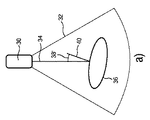
図2は、解剖学的対象の超音波プローブの視線方向に対する空間的関係に依存する様々な視野角の概略図を示す。図2では、超音波プローブは、概略的に示され、30と大まかに示されている。超音波プローブ30は、視野32内に及び視線方向34において超音波(ビーム)を放出し受信する。視野32内には、解剖学的物体36が概略的に示され、セグメンテーションモデルのメッシュによって表されている。視野角38は、視線方向34と解剖学的対象36の表面の法線方向40との間の角度として示される。図2aでは、視野角38’は比較的小さく(例えば30°未満)、図2bでは、視野角38’’は比較的大きい(例えば45°以上又は90°に近い)。
FIG. 2 shows a schematic view of various viewing angles depending on the spatial relationship of the ultrasonic probe of the anatomical object to the line-of-sight direction. In FIG. 2, the ultrasonic probe is shown schematically and roughly shown as 30. The
信号強度は、反射超音波によって決定され、したがって、超音波画像の画質は、超音波の伝搬方向と撮像される解剖学的対象36の表面との間の視野角38に依存する。信号強度は、表面と超音波とが互いに平行である場合は、比較的低く、表面と超音波とが互いに対し略垂直である場合は、比較的高く、したがって、解剖学的構造を比較的高い品質で撮像することができる。したがって、図2aに示されるように、視野角38’が小さい場合、画質は、視野角38’’が90°に近い状況と比べて比較的高い。
The signal strength is determined by the reflected ultrasound, so the quality of the ultrasound image depends on the
したがって、期待画質は、超音波プローブ30の視線方向34に対する解剖学的対象36の表面又は繊維構造の向きに基づいて決定される。
Therefore, the expected image quality is determined based on the orientation of the surface or fiber structure of the
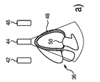
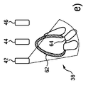
図3は、超音波取得ユニット14の3つの超音波プローブ42、44、46の様々な視線方向における解剖学的対象36を示す。
FIG. 3 shows the
図3a乃至図3cでは、1つの位置にある人間の心臓である解剖学的対象が示され、超音波プローブ42、44、46は、解剖学的対象36に対して様々な空間的関係で配置され、したがって、各超音波プローブ42、44、46は、解剖学的対象36の表面又は繊維構造へ様々な視野角38を有する。図3aでは、超音波プローブ44の超音波スキャンが示され、48と示される解剖学的対象36の側面に対する視野角38は約90°であり、したがって、低画質が予想される。50と示される更なる位置では、視野角は小さく、したがって、当該位置では、画像の高品質が予想される。
3a-3c show an anatomical object that is the human heart in one position, with
図3bでは、52と示される解剖学的対象36の側面へ小さい視野角38を有する超音波プローブ42の超音波スキャンが示される。したがって、当該位置の画質は高いと予想される。図3cでは、超音波プローブ46の超音波スキャンが示され、視野角38は、54、56と示される解剖学的対象36の2つの表面に関して小さく、したがって、これらの位置54、56の画質は高いと予想される。したがって、解剖学的対象の様々な位置の超音波画質は、超音波プローブ42、44、46の様々な視線方向について異なる。超音波画像を形成するために、画像データを提供する超音波プローブ42、44、46のうちの1つが選択される。又は、超音波画像は、関連の構造に対して最小視野角38を有し、重み係数によって重みが付けられてよい複数の超音波プローブ42、44、46の複合画像として形成される。
FIG. 3b shows an ultrasonic scan of an
図3d乃至図3fでは、解剖学的対象36は別の向きに示されており、3つの超音波プローブ42、44、46は、解剖学的対象36の様々な構造へ様々な視野角を有する。図3dでは、超音波プローブ44は、58と示される側面に関して大きい視野角を有し、60と示される内部構造に対して小さい視野角38を有するものとして示され、したがって、これらの構造の画質は異なる。図3eでは、超音波プローブ42は、62、64と示される解剖学的対象36の2つの異なる構造に関して小さい視野角を有するものとして示され、したがって、これらの構造の画質は高いと予想される。図3fでは、超音波プローブ46は、66と示される解剖学的対象36の構造に関して大きい視野角を有するものとして示され、したがって、この構造の画質は低いと予想される。超音波画像は、撮像対象である関連構造に関して最良画質を提供する1つ以上の選択された超音波プローブ42、44、46から受信される超音波データに基づいている。超音波画像は、複合画像として、複数の超音波データに基づいていてもよく、この場合、様々なデータは、期待画質に依存して重み付けされる。
In FIGS. 3d-3f, the
図4に、複数の超音波プローブ70、72、74を有する超音波取得ユニット14の一実施形態が示される。超音波プローブ70、72は、患者12の皮膚に配置される超音波アレイ76の一部であり、超音波プローブ74は、患者の体内に挿入され、経胸壁心エコー検査(TEE)プローブとして形成されている。本実施形態では、超音波プローブ70、72、74は、ドップラー信号に基づいて、解剖学的対象36内の血流を測定するように使用される。ドップラー信号に基づいて解剖学的構造又は流体の動きを測定するためには、大きい信号強度及び高品質の測定値を提供するように視線方向及び動きベクトルが平行であるべきである。血液の流れの方向は、検出された解剖学的対象36とセグメンテーションデータとに基づいて推定することができ、したがって、強いドップラー信号を受信するために、超音波プローブ70、72、74のうち上記流れの方向に関して最良の視野角を有する1つの超音波プローブを選択することができる。この場合、超音波プローブ72の視線方向が、解剖学的対象36の特定の解剖学的構造78内の血液の流れ方向と平行であり、したがって、この超音波プローブ72が、この解剖学的構造78内の血液の流れを測定するために選択される。
FIG. 4 shows an embodiment of an
流れ方向は、セグメンテーションモデルに基づいて決定されてもよく、この場合、流れ方向を解剖学的対象36の解剖学的特徴として記憶することができる。
The flow direction may be determined based on a segmentation model, in which case the flow direction can be stored as an anatomical feature of the
したがって、解剖学的対象36からのドップラー信号を受信するために最良の視野角を有する超音波プローブ70、72、74も選択することができる。
Therefore,
本発明は、図面及び上記説明において詳細に例示及び説明されたが、当該例示及び説明は、例示であって、限定と解釈されるべきではない。本発明は、開示された実施形態に限定されない。開示された実施形態の他の変形態様は、図面、開示内容及び添付の請求項の検討から、請求項に係る発明を実施する当業者によって理解され、実施される。 The present invention has been exemplified and described in detail in the drawings and the above description, but the examples and description are exemplary and should not be construed as limiting. The present invention is not limited to the disclosed embodiments. Other modifications of the disclosed embodiments will be understood and implemented by those skilled in the art who will implement the claimed invention from the examination of the drawings, disclosure content and the accompanying claims.
請求項において、「含む」との用語は、他の要素又はステップを除外するものではなく、また、「a」又は「an」との不定冠詞も、複数形を除外するものではない。単一の要素又は他のユニットが、請求項に記載される幾つかのアイテムの機能を果たしてもよい。特定の手段が相互に異なる従属請求項に記載されることだけで、これらの手段の組み合わせを有利に使用することができないことを示すものではない。 In the claims, the term "contains" does not exclude other elements or steps, nor does the indefinite article "a" or "an" exclude the plural. A single element or other unit may perform the function of some of the items described in the claims. The fact that specific means are described in different dependent claims does not indicate that the combination of these means cannot be used in an advantageous manner.
コンピュータプログラムは、他のハードウェアと共に又はその一部として供給される光学記憶媒体又は固体媒体といった適切な媒体上に記憶及び/又は分散されてもよいが、インターネット又は他の有線若しくは無線通信システムを介するといった他の形式で分配されてもよい。 Computer programs may be stored and / or distributed on suitable media such as optical storage media or solid-state media supplied with or as part of other hardware, but with the Internet or other wired or wireless communication systems. It may be distributed in other forms such as through.
請求項における任意の参照符号は、範囲を限定するものと解釈されるべきではない。 Any reference code in the claims should not be construed as limiting the scope.
Claims (13)
前記複数の超音波プローブのうちの少なくとも1つから受信される前記超音波データに基づいて前記視野内の前記患者の解剖学的対象を検出し、前記解剖学的対象と前記複数の超音波プローブそれぞれとの空間的関係を決定する検出ユニットと、
前記検出ユニットに結合され、前記超音波データから検出可能である少なくとも1つの物理的パラメータの取得品質に基づいて前記複数の超音波プローブのうちの少なくとも1つを選択する選択ユニットと、
前記超音波取得ユニットに結合され、選択された前記少なくとも1つの超音波プローブから前記超音波データを受信し、選択された前記少なくとも1つの超音波プローブから受信される前記超音波データに基づいて、前記少なくとも1つの物理的パラメータを決定する評価ユニットと、
を含む、超音波撮像装置であって、
前記取得品質は、前記解剖学的対象と前記複数の超音波プローブそれぞれとの前記空間的関係と、前記解剖学的対象の少なくとも1つの解剖学的特徴とに基づいて決定され、
前記検出ユニットは、(i)前記解剖学的対象の繊維方向、(ii)前記解剖学的対象の表面、又は(iii)前記解剖学的対象に関連付けられる流体の流れ方向を検出し、前記複数の超音波プローブそれぞれの視線方向に対する(i)前記繊維方向、(ii)前記表面、又は(iii)前記流れ方向の向きを、前記空間的関係として決定する、超音波撮像装置。 An ultrasound acquisition unit connected to a plurality of ultrasound probes, wherein each ultrasound probe provides ultrasound data suitable for ultrasound imaging of a patient within the field of view of the plurality of ultrasound probes. Acquisition unit and
The anatomical object of the patient in the visual field is detected based on the ultrasonic data received from at least one of the plurality of ultrasonic probes, and the anatomical object and the plurality of ultrasonic probes are detected. A detection unit that determines the spatial relationship with each,
A selection unit coupled to the detection unit and selecting at least one of the plurality of ultrasonic probes based on the acquisition quality of at least one physical parameter that can be detected from the ultrasonic data.
Based on the ultrasonic data coupled to the ultrasonic acquisition unit, receiving the ultrasonic data from the selected at least one ultrasonic probe and receiving from the selected at least one ultrasonic probe, An evaluation unit that determines at least one physical parameter,
The including, an ultrasound imaging apparatus,
The acquisition quality is determined based on the spatial relationship between the anatomical object and each of the plurality of ultrasonic probes and at least one anatomical feature of the anatomical object .
The detection unit detects (i) the fiber direction of the anatomical object, (ii) the surface of the anatomical object, or (iii) the flow direction of the fluid associated with the anatomical object, and the plurality. ultrasound probe (i) the fiber direction for each viewing direction, (ii) the surface, or (iii) the flow direction of the orientation, that determine as the spatial relation, the ultrasonic imaging apparatus.
前記複数の超音波プローブのうちの少なくとも1つから受信される前記超音波データに基づいて、前記視野内の前記患者の解剖学的対象を決定するステップと、
(i)前記解剖学的対象の繊維方向、(ii)前記解剖学的対象の表面、又は(iii)前記解剖学的対象に関連付けられる流体の流れ方向を検出するステップと、
前記解剖学的対象と前記複数の超音波プローブそれぞれとの空間的関係を決定するステップであって、前記空間的関係は、前記複数の超音波プローブそれぞれの視線方向に対する(i)前記繊維方向、(ii)前記表面、又は(iii)前記流れ方向の向きを含むステップと、
前記超音波データから検出可能である少なくとも1つの物理的パラメータの取得品質に基づいて、前記複数の超音波プローブのうちの少なくとも1つを選択するステップと、
選択された前記少なくとも1つの超音波プローブから受信される前記超音波データに基づいて、前記少なくとも1つの物理的パラメータを決定するステップと、
を含む、方法であって、
前記取得品質は、前記解剖学的対象と前記複数の超音波プローブそれぞれとの前記空間的関係と、前記解剖学的対象の少なくとも1つの解剖学的特徴とに基づいて決定される、超音波撮像の方法。 Steps to acquire ultrasound data suitable for ultrasound imaging of patients in the field of view of multiple ultrasound probes,
A step of determining the patient's anatomical object in the field of view based on the ultrasonic data received from at least one of the plurality of ultrasonic probes.
A step of detecting the fiber direction of the anatomical object, (ii) the surface of the anatomical object, or (iii) the flow direction of a fluid associated with the anatomical object.
A step of determining the spatial relationship between the anatomical object and each of the plurality of ultrasonic probes , wherein the spatial relationship is (i) the fiber direction with respect to the line-of-sight direction of each of the plurality of ultrasonic probes. (Ii) the surface, or (iii) the step including the direction of the flow direction, and
A step of selecting at least one of the plurality of ultrasonic probes based on the acquisition quality of at least one physical parameter that can be detected from the ultrasonic data.
A step of determining the at least one physical parameter based on the ultrasonic data received from the selected at least one ultrasonic probe.
The including, there is provided a method,
The acquisition quality is determined based on the spatial relationship between the anatomical object and each of the plurality of ultrasonic probes and at least one anatomical feature of the anatomical object. method of.
Applications Claiming Priority (3)
| Application Number | Priority Date | Filing Date | Title |
|---|---|---|---|
| EP15171400.3 | 2015-06-10 | ||
| EP15171400 | 2015-06-10 | ||
| PCT/EP2016/062922 WO2016198413A1 (en) | 2015-06-10 | 2016-06-07 | Ultrasound imaging apparatus |
Publications (3)
| Publication Number | Publication Date |
|---|---|
| JP2018521733A JP2018521733A (en) | 2018-08-09 |
| JP2018521733A5 JP2018521733A5 (en) | 2019-07-11 |
| JP6865695B2 true JP6865695B2 (en) | 2021-04-28 |
Family
ID=53365928
Family Applications (1)
| Application Number | Title | Priority Date | Filing Date |
|---|---|---|---|
| JP2017563333A Active JP6865695B2 (en) | 2015-06-10 | 2016-06-07 | Ultrasound imaging device |
Country Status (5)
| Country | Link |
|---|---|
| US (1) | US20180092626A1 (en) |
| EP (1) | EP3307171B1 (en) |
| JP (1) | JP6865695B2 (en) |
| CN (1) | CN107690312B (en) |
| WO (1) | WO2016198413A1 (en) |
Families Citing this family (3)
| Publication number | Priority date | Publication date | Assignee | Title |
|---|---|---|---|---|
| JP6767902B2 (en) | 2017-03-17 | 2020-10-14 | 株式会社日立製作所 | Ultrasound diagnostic equipment and programs |
| EP3513733A1 (en) | 2018-01-23 | 2019-07-24 | Koninklijke Philips N.V. | Ultrasound imaging apparatus and method |
| NL2024591B1 (en) * | 2019-12-30 | 2021-09-06 | Ronner Eelko | Scanning device for making echo scans of a person |
Family Cites Families (20)
| Publication number | Priority date | Publication date | Assignee | Title |
|---|---|---|---|---|
| US8012090B2 (en) * | 2004-06-22 | 2011-09-06 | General Electric Company | Method and apparatus for real time ultrasound multi-plane imaging |
| US20060184029A1 (en) * | 2005-01-13 | 2006-08-17 | Ronen Haim | Ultrasound guiding system and method for vascular access and operation mode |
| CN101309646A (en) * | 2005-11-17 | 2008-11-19 | 皇家飞利浦电子股份有限公司 | Vascular flow sensor with acoustic coupling detector |
| WO2007057826A1 (en) * | 2005-11-17 | 2007-05-24 | Koninklijke Philips Electronics N.V. | Vascular flow sensor with acoustic coupling detector |
| US7828735B2 (en) * | 2006-05-11 | 2010-11-09 | The Trustees Of Columbia In The City Of New York | Methods for providing diagnostic information using endocardial surface data for a patient's heart |
| JP5319157B2 (en) * | 2007-09-04 | 2013-10-16 | 株式会社東芝 | Ultrasonic diagnostic apparatus, ultrasonic image processing apparatus, and ultrasonic image processing program |
| EP2282671A4 (en) * | 2008-05-12 | 2012-11-21 | Cardio Art Technologies Ltd | Integrated heart monitoring device and method of using same |
| JP5259267B2 (en) * | 2008-06-19 | 2013-08-07 | 株式会社東芝 | Ultrasonic diagnostic apparatus, ultrasonic image processing apparatus, and ultrasonic image processing program |
| KR101659723B1 (en) * | 2009-04-14 | 2016-09-26 | 마우이 이미징, 인코포레이티드 | Multiple aperture ultrasound array alignment fixture |
| JP6230912B2 (en) * | 2010-10-14 | 2017-11-15 | コーニンクレッカ フィリップス エヌ ヴェKoninklijke Philips N.V. | Characteristic determination device for determining the characteristics of an object |
| CN102843973B (en) * | 2010-11-12 | 2017-02-08 | 柯尼卡美能达株式会社 | Ultrasound diagnostic apparatus and ultrasound diagnostic system |
| JP2012192075A (en) * | 2011-03-17 | 2012-10-11 | Fujifilm Corp | Ultrasound diagnostic apparatus and ultrasound image generation method |
| WO2013014647A1 (en) * | 2011-07-28 | 2013-01-31 | Koninklijke Philips Electronics N.V. | Ultrasound probe, method and device for acquiring a blood flow signal of an artery and system for measuring systolic blood pressure |
| EP2790586A2 (en) * | 2011-12-12 | 2014-10-22 | Koninklijke Philips N.V. | Automatic imaging plane selection for echocardiography |
| CN104203110B (en) * | 2012-03-26 | 2017-06-06 | 毛伊图像公司 | System and method for improving ultrasonoscopy quality by the application weighting factor |
| US20140058264A1 (en) * | 2012-08-24 | 2014-02-27 | Elwha LLC, a limited liability company of the State of Delaware | Adaptive Ultrasonic Array |
| EP2740410B1 (en) * | 2012-12-04 | 2018-05-16 | Canon Kabushiki Kaisha | Subject information acquisition device, method for controlling subject information acquisition device, and program therefor |
| JP6271579B2 (en) * | 2012-12-21 | 2018-01-31 | コーニンクレッカ フィリップス エヌ ヴェKoninklijke Philips N.V. | Anatomically intelligent echocardiography for point-of-care |
| JP6274421B2 (en) * | 2013-03-22 | 2018-02-07 | キヤノンメディカルシステムズ株式会社 | Ultrasonic diagnostic apparatus and control program therefor |
| JP2015066219A (en) * | 2013-09-30 | 2015-04-13 | セイコーエプソン株式会社 | Ultrasonic measuring apparatus and ultrasonic measuring method |
-
2016
- 2016-06-07 JP JP2017563333A patent/JP6865695B2/en active Active
- 2016-06-07 WO PCT/EP2016/062922 patent/WO2016198413A1/en active Application Filing
- 2016-06-07 EP EP16729232.5A patent/EP3307171B1/en active Active
- 2016-06-07 CN CN201680033217.XA patent/CN107690312B/en active Active
- 2016-06-07 US US15/580,704 patent/US20180092626A1/en not_active Abandoned
Also Published As
| Publication number | Publication date |
|---|---|
| EP3307171B1 (en) | 2021-08-11 |
| CN107690312A (en) | 2018-02-13 |
| CN107690312B (en) | 2021-01-05 |
| US20180092626A1 (en) | 2018-04-05 |
| WO2016198413A1 (en) | 2016-12-15 |
| JP2018521733A (en) | 2018-08-09 |
| EP3307171A1 (en) | 2018-04-18 |
Similar Documents
| Publication | Publication Date | Title |
|---|---|---|
| JP6994494B2 (en) | Elastography measurement system and its method | |
| CN105392428B (en) | System and method for mapping the measurement of ultrasonic shear wave elastogram | |
| CN103842841B (en) | The ultrasonic system set with automatic doppler flow | |
| JP6960922B2 (en) | Ultrasound imaging device and ultrasonic imaging method for inspecting the volume of a subject | |
| JP2016506809A (en) | Ultrasound imaging system and method | |
| CN109310399B (en) | Medical ultrasonic image processing apparatus | |
| JP2016527022A (en) | Non-imaging two-dimensional array probe and system for classifying carotid artery stenosis | |
| US11684344B2 (en) | Systems and methods for quantitative abdominal aortic aneurysm analysis using 3D ultrasound imaging | |
| JP6865695B2 (en) | Ultrasound imaging device | |
| JP2014124319A (en) | Ultrasonic calibration system, and ultrasonic calibration method | |
| CN112912010A (en) | Method and system for deriving a parameter related to flow from a blood vessel | |
| JP2020509862A (en) | Optimal scanning plane selection for organ recognition | |
| CA3126020C (en) | Systems and methods for quantitative abdominal aortic aneurysm analysis using 3d ultrasound imaging |
Legal Events
| Date | Code | Title | Description |
|---|---|---|---|
| A521 | Written amendment |
Free format text: JAPANESE INTERMEDIATE CODE: A523 Effective date: 20190604 |
|
| A621 | Written request for application examination |
Free format text: JAPANESE INTERMEDIATE CODE: A621 Effective date: 20190604 |
|
| A977 | Report on retrieval |
Free format text: JAPANESE INTERMEDIATE CODE: A971007 Effective date: 20200519 |
|
| A131 | Notification of reasons for refusal |
Free format text: JAPANESE INTERMEDIATE CODE: A131 Effective date: 20200601 |
|
| A601 | Written request for extension of time |
Free format text: JAPANESE INTERMEDIATE CODE: A601 Effective date: 20200827 |
|
| TRDD | Decision of grant or rejection written | ||
| A01 | Written decision to grant a patent or to grant a registration (utility model) |
Free format text: JAPANESE INTERMEDIATE CODE: A01 Effective date: 20210308 |
|
| A61 | First payment of annual fees (during grant procedure) |
Free format text: JAPANESE INTERMEDIATE CODE: A61 Effective date: 20210406 |
|
| R150 | Certificate of patent or registration of utility model |
Ref document number: 6865695 Country of ref document: JP Free format text: JAPANESE INTERMEDIATE CODE: R150 |