JP6945444B2 - High affinity PD-1 drug and its usage - Google Patents
High affinity PD-1 drug and its usage Download PDFInfo
- Publication number
- JP6945444B2 JP6945444B2 JP2017505807A JP2017505807A JP6945444B2 JP 6945444 B2 JP6945444 B2 JP 6945444B2 JP 2017505807 A JP2017505807 A JP 2017505807A JP 2017505807 A JP2017505807 A JP 2017505807A JP 6945444 B2 JP6945444 B2 JP 6945444B2
- Authority
- JP
- Japan
- Prior art keywords
- polypeptide
- cells
- affinity
- amino acid
- high affinity
- Prior art date
- Legal status (The legal status is an assumption and is not a legal conclusion. Google has not performed a legal analysis and makes no representation as to the accuracy of the status listed.)
- Active
Links
- 239000003814 drug Substances 0.000 title claims description 36
- 229940079593 drug Drugs 0.000 title description 18
- 108090000765 processed proteins & peptides Proteins 0.000 claims description 482
- 102000004196 processed proteins & peptides Human genes 0.000 claims description 466
- 229920001184 polypeptide Polymers 0.000 claims description 463
- 206010028980 Neoplasm Diseases 0.000 claims description 220
- 150000001413 amino acids Chemical class 0.000 claims description 184
- 102100040678 Programmed cell death protein 1 Human genes 0.000 claims description 183
- 101710089372 Programmed cell death protein 1 Proteins 0.000 claims description 172
- 201000011510 cancer Diseases 0.000 claims description 112
- 125000003275 alpha amino acid group Chemical group 0.000 claims description 81
- 102220496972 Platelet-activating factor acetylhydrolase 2, cytoplasmic_K53T_mutation Human genes 0.000 claims description 53
- 102220058921 rs786202731 Human genes 0.000 claims description 53
- 241000282414 Homo sapiens Species 0.000 claims description 52
- 102200118229 rs34665886 Human genes 0.000 claims description 50
- 102100024213 Programmed cell death 1 ligand 2 Human genes 0.000 claims description 46
- 238000011282 treatment Methods 0.000 claims description 45
- 239000012634 fragment Substances 0.000 claims description 44
- 108060003951 Immunoglobulin Proteins 0.000 claims description 41
- 102000018358 immunoglobulin Human genes 0.000 claims description 41
- 108700030875 Programmed Cell Death 1 Ligand 2 Proteins 0.000 claims description 39
- FWMNVWWHGCHHJJ-SKKKGAJSSA-N 4-amino-1-[(2r)-6-amino-2-[[(2r)-2-[[(2r)-2-[[(2r)-2-amino-3-phenylpropanoyl]amino]-3-phenylpropanoyl]amino]-4-methylpentanoyl]amino]hexanoyl]piperidine-4-carboxylic acid Chemical compound C([C@H](C(=O)N[C@H](CC(C)C)C(=O)N[C@H](CCCCN)C(=O)N1CCC(N)(CC1)C(O)=O)NC(=O)[C@H](N)CC=1C=CC=CC=1)C1=CC=CC=C1 FWMNVWWHGCHHJJ-SKKKGAJSSA-N 0.000 claims description 38
- 101100407308 Mus musculus Pdcd1lg2 gene Proteins 0.000 claims description 38
- 230000004927 fusion Effects 0.000 claims description 38
- 102220161672 rs148219510 Human genes 0.000 claims description 38
- 238000003384 imaging method Methods 0.000 claims description 35
- 102220586093 Homeobox protein DLX-1_V39R_mutation Human genes 0.000 claims description 32
- 102220507725 GTP-binding protein RAD_Q66P_mutation Human genes 0.000 claims description 30
- 102220354149 c.289C>A Human genes 0.000 claims description 29
- 102220249043 rs145159429 Human genes 0.000 claims description 26
- 102220311149 rs41271047 Human genes 0.000 claims description 26
- 102220279022 rs955258267 Human genes 0.000 claims description 26
- 230000003993 interaction Effects 0.000 claims description 25
- 102220226094 rs17217723 Human genes 0.000 claims description 25
- 102220568823 Dual specificity mitogen-activated protein kinase kinase 1_G65V_mutation Human genes 0.000 claims description 24
- 239000003446 ligand Substances 0.000 claims description 22
- 102220585589 Short-chain dehydrogenase/reductase family 42E member 1_Q63P_mutation Human genes 0.000 claims description 19
- 238000002600 positron emission tomography Methods 0.000 claims description 18
- 102000004127 Cytokines Human genes 0.000 claims description 17
- 108090000695 Cytokines Proteins 0.000 claims description 17
- 230000035772 mutation Effects 0.000 claims description 15
- 102220013742 rs397516741 Human genes 0.000 claims description 15
- 102220361097 c.307C>T Human genes 0.000 claims description 14
- 102220492635 Integrin alpha-3_D52Y_mutation Human genes 0.000 claims description 13
- 239000003112 inhibitor Substances 0.000 claims description 13
- 102100034458 Hepatitis A virus cellular receptor 2 Human genes 0.000 claims description 12
- 208000037581 Persistent Infection Diseases 0.000 claims description 11
- 102220257864 rs1050971384 Human genes 0.000 claims description 11
- 102220198136 rs1057519882 Human genes 0.000 claims description 11
- 102200116701 rs115206969 Human genes 0.000 claims description 11
- 102220250913 rs1364636517 Human genes 0.000 claims description 11
- 239000000556 agonist Substances 0.000 claims description 10
- 101001068133 Homo sapiens Hepatitis A virus cellular receptor 2 Proteins 0.000 claims description 9
- 102220492589 Protein numb homolog_S48D_mutation Human genes 0.000 claims description 8
- 102220364119 c.148C>A Human genes 0.000 claims description 8
- 230000001965 increasing effect Effects 0.000 claims description 8
- 102200160087 rs1131692231 Human genes 0.000 claims description 8
- 102200007406 rs148775298 Human genes 0.000 claims description 8
- 102200017902 rs267606847 Human genes 0.000 claims description 8
- 102220252689 rs373178770 Human genes 0.000 claims description 8
- 102100029822 B- and T-lymphocyte attenuator Human genes 0.000 claims description 6
- 108010062802 CD66 antigens Proteins 0.000 claims description 6
- 102100024533 Carcinoembryonic antigen-related cell adhesion molecule 1 Human genes 0.000 claims description 6
- 101000864344 Homo sapiens B- and T-lymphocyte attenuator Proteins 0.000 claims description 6
- 238000004519 manufacturing process Methods 0.000 claims description 6
- 102220477170 Y-box-binding protein 2_S102A_mutation Human genes 0.000 claims description 5
- 102220126393 rs886044243 Human genes 0.000 claims description 5
- 230000030833 cell death Effects 0.000 claims description 4
- 230000034994 death Effects 0.000 claims description 4
- 101150013553 CD40 gene Proteins 0.000 claims description 2
- 102100040245 Tumor necrosis factor receptor superfamily member 5 Human genes 0.000 claims description 2
- 230000002238 attenuated effect Effects 0.000 claims description 2
- 102220624338 Interferon alpha-8_T51A_mutation Human genes 0.000 claims 2
- 239000000825 pharmaceutical preparation Substances 0.000 claims 1
- 101100519207 Mus musculus Pdcd1 gene Proteins 0.000 description 460
- 210000004027 cell Anatomy 0.000 description 274
- 235000001014 amino acid Nutrition 0.000 description 219
- 229940024606 amino acid Drugs 0.000 description 187
- 108090000623 proteins and genes Proteins 0.000 description 89
- 235000018102 proteins Nutrition 0.000 description 82
- 102000004169 proteins and genes Human genes 0.000 description 82
- 230000027455 binding Effects 0.000 description 78
- 238000009739 binding Methods 0.000 description 75
- 238000000034 method Methods 0.000 description 73
- 210000001744 T-lymphocyte Anatomy 0.000 description 59
- 108091008874 T cell receptors Proteins 0.000 description 56
- 102000016266 T-Cell Antigen Receptors Human genes 0.000 description 56
- 239000000427 antigen Substances 0.000 description 54
- 102000036639 antigens Human genes 0.000 description 54
- 108091007433 antigens Proteins 0.000 description 54
- 206010039491 Sarcoma Diseases 0.000 description 51
- 150000007523 nucleic acids Chemical class 0.000 description 51
- 101000611936 Homo sapiens Programmed cell death protein 1 Proteins 0.000 description 50
- 102000039446 nucleic acids Human genes 0.000 description 50
- 108020004707 nucleic acids Proteins 0.000 description 50
- 208000037265 diseases, disorders, signs and symptoms Diseases 0.000 description 47
- 210000001519 tissue Anatomy 0.000 description 41
- 102000048362 human PDCD1 Human genes 0.000 description 39
- 101001117317 Homo sapiens Programmed cell death 1 ligand 1 Proteins 0.000 description 37
- 201000010099 disease Diseases 0.000 description 36
- 108010019670 Chimeric Antigen Receptors Proteins 0.000 description 35
- 102000048776 human CD274 Human genes 0.000 description 32
- 210000002865 immune cell Anatomy 0.000 description 25
- 239000000203 mixture Substances 0.000 description 25
- 241000700605 Viruses Species 0.000 description 23
- 125000005647 linker group Chemical group 0.000 description 23
- 206010025323 Lymphomas Diseases 0.000 description 22
- 230000014509 gene expression Effects 0.000 description 20
- 238000000338 in vitro Methods 0.000 description 20
- 230000001225 therapeutic effect Effects 0.000 description 20
- 239000013598 vector Substances 0.000 description 20
- 238000001727 in vivo Methods 0.000 description 19
- 241000699670 Mus sp. Species 0.000 description 18
- 108020004414 DNA Proteins 0.000 description 16
- -1 amihostin Chemical compound 0.000 description 16
- 239000003795 chemical substances by application Substances 0.000 description 16
- 230000003211 malignant effect Effects 0.000 description 16
- 239000008194 pharmaceutical composition Substances 0.000 description 16
- 229940124597 therapeutic agent Drugs 0.000 description 16
- 201000008808 Fibrosarcoma Diseases 0.000 description 15
- 208000015914 Non-Hodgkin lymphomas Diseases 0.000 description 15
- 210000000987 immune system Anatomy 0.000 description 15
- 208000015181 infectious disease Diseases 0.000 description 15
- 208000032839 leukemia Diseases 0.000 description 15
- 241000124008 Mammalia Species 0.000 description 14
- 230000008859 change Effects 0.000 description 14
- 239000002773 nucleotide Substances 0.000 description 13
- 125000003729 nucleotide group Chemical group 0.000 description 13
- 230000001105 regulatory effect Effects 0.000 description 13
- 208000024891 symptom Diseases 0.000 description 13
- 210000004881 tumor cell Anatomy 0.000 description 13
- 108010047041 Complementarity Determining Regions Proteins 0.000 description 12
- 150000001875 compounds Chemical class 0.000 description 12
- 210000000130 stem cell Anatomy 0.000 description 12
- 108010021625 Immunoglobulin Fragments Proteins 0.000 description 11
- 102000008394 Immunoglobulin Fragments Human genes 0.000 description 11
- 241001465754 Metazoa Species 0.000 description 11
- 108010076504 Protein Sorting Signals Proteins 0.000 description 11
- 208000035475 disorder Diseases 0.000 description 11
- 239000013604 expression vector Substances 0.000 description 11
- 230000006870 function Effects 0.000 description 11
- 102000037865 fusion proteins Human genes 0.000 description 11
- 108020001507 fusion proteins Proteins 0.000 description 11
- 102000005962 receptors Human genes 0.000 description 11
- 108020003175 receptors Proteins 0.000 description 11
- 238000002198 surface plasmon resonance spectroscopy Methods 0.000 description 11
- 201000009030 Carcinoma Diseases 0.000 description 10
- 208000035473 Communicable disease Diseases 0.000 description 10
- 208000017604 Hodgkin disease Diseases 0.000 description 10
- 208000021519 Hodgkin lymphoma Diseases 0.000 description 10
- 208000010747 Hodgkins lymphoma Diseases 0.000 description 10
- 101001117312 Homo sapiens Programmed cell death 1 ligand 2 Proteins 0.000 description 10
- 210000000612 antigen-presenting cell Anatomy 0.000 description 10
- 208000013056 classic Hodgkin lymphoma Diseases 0.000 description 10
- 210000003205 muscle Anatomy 0.000 description 10
- 239000000126 substance Substances 0.000 description 10
- 239000013603 viral vector Substances 0.000 description 10
- 102100022005 B-lymphocyte antigen CD20 Human genes 0.000 description 9
- 102000001301 EGF receptor Human genes 0.000 description 9
- 108060006698 EGF receptor Proteins 0.000 description 9
- 101000897405 Homo sapiens B-lymphocyte antigen CD20 Proteins 0.000 description 9
- 206010027476 Metastases Diseases 0.000 description 9
- 240000004808 Saccharomyces cerevisiae Species 0.000 description 9
- 238000010494 dissociation reaction Methods 0.000 description 9
- 230000005593 dissociations Effects 0.000 description 9
- 201000001441 melanoma Diseases 0.000 description 9
- 230000009401 metastasis Effects 0.000 description 9
- 230000003278 mimic effect Effects 0.000 description 9
- 210000000056 organ Anatomy 0.000 description 9
- 244000052769 pathogen Species 0.000 description 9
- 230000001717 pathogenic effect Effects 0.000 description 9
- 229920000642 polymer Polymers 0.000 description 9
- 239000000700 radioactive tracer Substances 0.000 description 9
- 230000009870 specific binding Effects 0.000 description 9
- 208000003174 Brain Neoplasms Diseases 0.000 description 8
- 208000026310 Breast neoplasm Diseases 0.000 description 8
- 101001012157 Homo sapiens Receptor tyrosine-protein kinase erbB-2 Proteins 0.000 description 8
- 241000699666 Mus <mouse, genus> Species 0.000 description 8
- 201000004458 Myoma Diseases 0.000 description 8
- 241000288906 Primates Species 0.000 description 8
- 102100030086 Receptor tyrosine-protein kinase erbB-2 Human genes 0.000 description 8
- 206010042971 T-cell lymphoma Diseases 0.000 description 8
- 208000027585 T-cell non-Hodgkin lymphoma Diseases 0.000 description 8
- 125000000539 amino acid group Chemical group 0.000 description 8
- 239000003153 chemical reaction reagent Substances 0.000 description 8
- 230000000694 effects Effects 0.000 description 8
- 230000009975 flexible effect Effects 0.000 description 8
- 230000028993 immune response Effects 0.000 description 8
- 230000001506 immunosuppresive effect Effects 0.000 description 8
- 230000002401 inhibitory effect Effects 0.000 description 8
- 210000000822 natural killer cell Anatomy 0.000 description 8
- 239000007787 solid Substances 0.000 description 8
- 230000002792 vascular Effects 0.000 description 8
- YBJHBAHKTGYVGT-ZKWXMUAHSA-N (+)-Biotin Chemical compound N1C(=O)N[C@@H]2[C@H](CCCCC(=O)O)SC[C@@H]21 YBJHBAHKTGYVGT-ZKWXMUAHSA-N 0.000 description 7
- 241000283690 Bos taurus Species 0.000 description 7
- 206010024612 Lipoma Diseases 0.000 description 7
- 241001494479 Pecora Species 0.000 description 7
- 102100024216 Programmed cell death 1 ligand 1 Human genes 0.000 description 7
- 241000700159 Rattus Species 0.000 description 7
- 241000283984 Rodentia Species 0.000 description 7
- 206010068771 Soft tissue neoplasm Diseases 0.000 description 7
- 210000003719 b-lymphocyte Anatomy 0.000 description 7
- 210000001185 bone marrow Anatomy 0.000 description 7
- 231100000433 cytotoxic Toxicity 0.000 description 7
- 229940127089 cytotoxic agent Drugs 0.000 description 7
- 230000001472 cytotoxic effect Effects 0.000 description 7
- 229940072221 immunoglobulins Drugs 0.000 description 7
- 206010024627 liposarcoma Diseases 0.000 description 7
- 210000004072 lung Anatomy 0.000 description 7
- 239000000463 material Substances 0.000 description 7
- 239000000178 monomer Substances 0.000 description 7
- 210000001778 pluripotent stem cell Anatomy 0.000 description 7
- 238000000746 purification Methods 0.000 description 7
- 239000000523 sample Substances 0.000 description 7
- 241000894007 species Species 0.000 description 7
- 201000003076 Angiosarcoma Diseases 0.000 description 6
- 206010061692 Benign muscle neoplasm Diseases 0.000 description 6
- 108010065524 CD52 Antigen Proteins 0.000 description 6
- 102000013135 CD52 Antigen Human genes 0.000 description 6
- 241000282693 Cercopithecidae Species 0.000 description 6
- 206010009944 Colon cancer Diseases 0.000 description 6
- 241000283086 Equidae Species 0.000 description 6
- 241000282326 Felis catus Species 0.000 description 6
- 208000001258 Hemangiosarcoma Diseases 0.000 description 6
- 208000028018 Lymphocytic leukaemia Diseases 0.000 description 6
- 108010090804 Streptavidin Proteins 0.000 description 6
- 208000009956 adenocarcinoma Diseases 0.000 description 6
- 239000002246 antineoplastic agent Substances 0.000 description 6
- 230000015572 biosynthetic process Effects 0.000 description 6
- 230000037396 body weight Effects 0.000 description 6
- 210000000988 bone and bone Anatomy 0.000 description 6
- 210000000481 breast Anatomy 0.000 description 6
- 230000006378 damage Effects 0.000 description 6
- 238000013461 design Methods 0.000 description 6
- 238000003745 diagnosis Methods 0.000 description 6
- 239000000539 dimer Substances 0.000 description 6
- 239000000975 dye Substances 0.000 description 6
- 239000003623 enhancer Substances 0.000 description 6
- 208000026278 immune system disease Diseases 0.000 description 6
- 230000001939 inductive effect Effects 0.000 description 6
- 210000000265 leukocyte Anatomy 0.000 description 6
- 208000003747 lymphoid leukemia Diseases 0.000 description 6
- 208000025113 myeloid leukemia Diseases 0.000 description 6
- 201000008968 osteosarcoma Diseases 0.000 description 6
- 210000000496 pancreas Anatomy 0.000 description 6
- 230000002093 peripheral effect Effects 0.000 description 6
- 239000000546 pharmaceutical excipient Substances 0.000 description 6
- 210000002307 prostate Anatomy 0.000 description 6
- 230000002829 reductive effect Effects 0.000 description 6
- 102220170950 rs769916550 Human genes 0.000 description 6
- 238000010186 staining Methods 0.000 description 6
- 208000031261 Acute myeloid leukaemia Diseases 0.000 description 5
- 206010006187 Breast cancer Diseases 0.000 description 5
- 241000282472 Canis lupus familiaris Species 0.000 description 5
- 208000005243 Chondrosarcoma Diseases 0.000 description 5
- 241000701022 Cytomegalovirus Species 0.000 description 5
- 241000282412 Homo Species 0.000 description 5
- 101000851376 Homo sapiens Tumor necrosis factor receptor superfamily member 8 Proteins 0.000 description 5
- 206010062016 Immunosuppression Diseases 0.000 description 5
- 208000033776 Myeloid Acute Leukemia Diseases 0.000 description 5
- 241000282887 Suidae Species 0.000 description 5
- 206010043276 Teratoma Diseases 0.000 description 5
- 102100036857 Tumor necrosis factor receptor superfamily member 8 Human genes 0.000 description 5
- 210000001789 adipocyte Anatomy 0.000 description 5
- 230000000903 blocking effect Effects 0.000 description 5
- 230000001684 chronic effect Effects 0.000 description 5
- 210000001072 colon Anatomy 0.000 description 5
- 210000004443 dendritic cell Anatomy 0.000 description 5
- LOKCTEFSRHRXRJ-UHFFFAOYSA-I dipotassium trisodium dihydrogen phosphate hydrogen phosphate dichloride Chemical compound P(=O)(O)(O)[O-].[K+].P(=O)(O)([O-])[O-].[Na+].[Na+].[Cl-].[K+].[Cl-].[Na+] LOKCTEFSRHRXRJ-UHFFFAOYSA-I 0.000 description 5
- 210000003162 effector t lymphocyte Anatomy 0.000 description 5
- 206010016629 fibroma Diseases 0.000 description 5
- 230000003325 follicular Effects 0.000 description 5
- 238000009472 formulation Methods 0.000 description 5
- 230000012010 growth Effects 0.000 description 5
- 102000048119 human PDCD1LG2 Human genes 0.000 description 5
- 230000003834 intracellular effect Effects 0.000 description 5
- 210000003734 kidney Anatomy 0.000 description 5
- 210000001165 lymph node Anatomy 0.000 description 5
- 210000002540 macrophage Anatomy 0.000 description 5
- 201000009020 malignant peripheral nerve sheath tumor Diseases 0.000 description 5
- 208000029974 neurofibrosarcoma Diseases 0.000 description 5
- 231100000252 nontoxic Toxicity 0.000 description 5
- 230000003000 nontoxic effect Effects 0.000 description 5
- 230000035515 penetration Effects 0.000 description 5
- 239000002953 phosphate buffered saline Substances 0.000 description 5
- 230000009467 reduction Effects 0.000 description 5
- 229960000575 trastuzumab Drugs 0.000 description 5
- 230000004614 tumor growth Effects 0.000 description 5
- 241001529453 unidentified herpesvirus Species 0.000 description 5
- 108091032973 (ribonucleotides)n+m Proteins 0.000 description 4
- GFZFAQOKWZGMQL-UHFFFAOYSA-N 3-[3,5-bis(trifluoromethyl)phenyl]-3-oxopropanenitrile Chemical compound FC(F)(F)C1=CC(C(=O)CC#N)=CC(C(F)(F)F)=C1 GFZFAQOKWZGMQL-UHFFFAOYSA-N 0.000 description 4
- 239000012103 Alexa Fluor 488 Substances 0.000 description 4
- 239000012110 Alexa Fluor 594 Substances 0.000 description 4
- 208000010839 B-cell chronic lymphocytic leukemia Diseases 0.000 description 4
- 102100038080 B-cell receptor CD22 Human genes 0.000 description 4
- 102100024222 B-lymphocyte antigen CD19 Human genes 0.000 description 4
- 206010057070 Dermatofibrosarcoma protuberans Diseases 0.000 description 4
- 241000483002 Euproctis similis Species 0.000 description 4
- 208000006168 Ewing Sarcoma Diseases 0.000 description 4
- DHMQDGOQFOQNFH-UHFFFAOYSA-N Glycine Chemical compound NCC(O)=O DHMQDGOQFOQNFH-UHFFFAOYSA-N 0.000 description 4
- 241001272567 Hominoidea Species 0.000 description 4
- 101000884305 Homo sapiens B-cell receptor CD22 Proteins 0.000 description 4
- 101000980825 Homo sapiens B-lymphocyte antigen CD19 Proteins 0.000 description 4
- 101001057504 Homo sapiens Interferon-stimulated gene 20 kDa protein Proteins 0.000 description 4
- 101001055144 Homo sapiens Interleukin-2 receptor subunit alpha Proteins 0.000 description 4
- 102100026878 Interleukin-2 receptor subunit alpha Human genes 0.000 description 4
- XUMBMVFBXHLACL-UHFFFAOYSA-N Melanin Chemical compound O=C1C(=O)C(C2=CNC3=C(C(C(=O)C4=C32)=O)C)=C2C4=CNC2=C1C XUMBMVFBXHLACL-UHFFFAOYSA-N 0.000 description 4
- 101001117316 Mus musculus Programmed cell death 1 ligand 1 Proteins 0.000 description 4
- 241000282579 Pan Species 0.000 description 4
- 208000006664 Precursor Cell Lymphoblastic Leukemia-Lymphoma Diseases 0.000 description 4
- 208000006265 Renal cell carcinoma Diseases 0.000 description 4
- 230000006044 T cell activation Effects 0.000 description 4
- 102100040418 Tumor protein D52 Human genes 0.000 description 4
- 230000002159 abnormal effect Effects 0.000 description 4
- 238000007792 addition Methods 0.000 description 4
- 229960000548 alemtuzumab Drugs 0.000 description 4
- 238000000540 analysis of variance Methods 0.000 description 4
- 230000000259 anti-tumor effect Effects 0.000 description 4
- 230000004071 biological effect Effects 0.000 description 4
- 229960002685 biotin Drugs 0.000 description 4
- 235000020958 biotin Nutrition 0.000 description 4
- 239000011616 biotin Substances 0.000 description 4
- 210000004369 blood Anatomy 0.000 description 4
- 239000008280 blood Substances 0.000 description 4
- 210000004556 brain Anatomy 0.000 description 4
- 239000000969 carrier Substances 0.000 description 4
- 239000002738 chelating agent Substances 0.000 description 4
- 230000002950 deficient Effects 0.000 description 4
- 239000003085 diluting agent Substances 0.000 description 4
- 239000002552 dosage form Substances 0.000 description 4
- 230000003511 endothelial effect Effects 0.000 description 4
- 210000003527 eukaryotic cell Anatomy 0.000 description 4
- 230000003352 fibrogenic effect Effects 0.000 description 4
- 239000007850 fluorescent dye Substances 0.000 description 4
- 201000011243 gastrointestinal stromal tumor Diseases 0.000 description 4
- 230000000762 glandular Effects 0.000 description 4
- 230000013595 glycosylation Effects 0.000 description 4
- 238000006206 glycosylation reaction Methods 0.000 description 4
- 210000004209 hair Anatomy 0.000 description 4
- 210000003958 hematopoietic stem cell Anatomy 0.000 description 4
- 210000003494 hepatocyte Anatomy 0.000 description 4
- 230000003308 immunostimulating effect Effects 0.000 description 4
- 239000012678 infectious agent Substances 0.000 description 4
- 230000005764 inhibitory process Effects 0.000 description 4
- 239000007788 liquid Substances 0.000 description 4
- 230000015654 memory Effects 0.000 description 4
- 210000003071 memory t lymphocyte Anatomy 0.000 description 4
- 210000002569 neuron Anatomy 0.000 description 4
- 210000001672 ovary Anatomy 0.000 description 4
- 229960001972 panitumumab Drugs 0.000 description 4
- 239000013612 plasmid Substances 0.000 description 4
- 230000008569 process Effects 0.000 description 4
- 238000002818 protein evolution Methods 0.000 description 4
- 238000003259 recombinant expression Methods 0.000 description 4
- 230000011664 signaling Effects 0.000 description 4
- 238000002603 single-photon emission computed tomography Methods 0.000 description 4
- 210000003491 skin Anatomy 0.000 description 4
- 210000004872 soft tissue Anatomy 0.000 description 4
- 239000007790 solid phase Substances 0.000 description 4
- 239000000243 solution Substances 0.000 description 4
- 230000000638 stimulation Effects 0.000 description 4
- 238000012360 testing method Methods 0.000 description 4
- 230000002103 transcriptional effect Effects 0.000 description 4
- 239000003981 vehicle Substances 0.000 description 4
- ZCYVEMRRCGMTRW-UHFFFAOYSA-N 7553-56-2 Chemical compound [I] ZCYVEMRRCGMTRW-UHFFFAOYSA-N 0.000 description 3
- 102100031585 ADP-ribosyl cyclase/cyclic ADP-ribose hydrolase 1 Human genes 0.000 description 3
- 102100026423 Adhesion G protein-coupled receptor E5 Human genes 0.000 description 3
- 241000894006 Bacteria Species 0.000 description 3
- 102100032912 CD44 antigen Human genes 0.000 description 3
- 102100025221 CD70 antigen Human genes 0.000 description 3
- 108060001253 CD99 Proteins 0.000 description 3
- 102000024905 CD99 Human genes 0.000 description 3
- 102100025570 Cancer/testis antigen 1 Human genes 0.000 description 3
- 102100025064 Cellular tumor antigen p53 Human genes 0.000 description 3
- 108020004705 Codon Proteins 0.000 description 3
- 206010051066 Gastrointestinal stromal tumour Diseases 0.000 description 3
- 108010007707 Hepatitis A Virus Cellular Receptor 2 Proteins 0.000 description 3
- 101000777636 Homo sapiens ADP-ribosyl cyclase/cyclic ADP-ribose hydrolase 1 Proteins 0.000 description 3
- 101000718243 Homo sapiens Adhesion G protein-coupled receptor E5 Proteins 0.000 description 3
- 101000868273 Homo sapiens CD44 antigen Proteins 0.000 description 3
- 101000934356 Homo sapiens CD70 antigen Proteins 0.000 description 3
- 101000856237 Homo sapiens Cancer/testis antigen 1 Proteins 0.000 description 3
- 101000721661 Homo sapiens Cellular tumor antigen p53 Proteins 0.000 description 3
- 101000998120 Homo sapiens Interleukin-3 receptor subunit alpha Proteins 0.000 description 3
- 101001008874 Homo sapiens Mast/stem cell growth factor receptor Kit Proteins 0.000 description 3
- 101000578784 Homo sapiens Melanoma antigen recognized by T-cells 1 Proteins 0.000 description 3
- 101000934338 Homo sapiens Myeloid cell surface antigen CD33 Proteins 0.000 description 3
- 101000581981 Homo sapiens Neural cell adhesion molecule 1 Proteins 0.000 description 3
- 101001084266 Homo sapiens Parathyroid hormone 2 receptor Proteins 0.000 description 3
- 101001094545 Homo sapiens Retrotransposon-like protein 1 Proteins 0.000 description 3
- 101000884271 Homo sapiens Signal transducer CD24 Proteins 0.000 description 3
- 101000596234 Homo sapiens T-cell surface protein tactile Proteins 0.000 description 3
- 241000725303 Human immunodeficiency virus Species 0.000 description 3
- 102100033493 Interleukin-3 receptor subunit alpha Human genes 0.000 description 3
- XUJNEKJLAYXESH-REOHCLBHSA-N L-Cysteine Chemical compound SC[C@H](N)C(O)=O XUJNEKJLAYXESH-REOHCLBHSA-N 0.000 description 3
- 241000222722 Leishmania <genus> Species 0.000 description 3
- 102100027754 Mast/stem cell growth factor receptor Kit Human genes 0.000 description 3
- 102100028389 Melanoma antigen recognized by T-cells 1 Human genes 0.000 description 3
- 102100025243 Myeloid cell surface antigen CD33 Human genes 0.000 description 3
- 102100027347 Neural cell adhesion molecule 1 Human genes 0.000 description 3
- 241000283973 Oryctolagus cuniculus Species 0.000 description 3
- 229930012538 Paclitaxel Natural products 0.000 description 3
- 102100030869 Parathyroid hormone 2 receptor Human genes 0.000 description 3
- 239000002202 Polyethylene glycol Substances 0.000 description 3
- 206010060862 Prostate cancer Diseases 0.000 description 3
- 241000607142 Salmonella Species 0.000 description 3
- 102100038081 Signal transducer CD24 Human genes 0.000 description 3
- 208000000389 T-cell leukemia Diseases 0.000 description 3
- 208000028530 T-cell lymphoblastic leukemia/lymphoma Diseases 0.000 description 3
- 102100035268 T-cell surface protein tactile Human genes 0.000 description 3
- 208000036142 Viral infection Diseases 0.000 description 3
- 230000004913 activation Effects 0.000 description 3
- 230000002457 bidirectional effect Effects 0.000 description 3
- 239000012472 biological sample Substances 0.000 description 3
- 230000004663 cell proliferation Effects 0.000 description 3
- 229960005395 cetuximab Drugs 0.000 description 3
- 238000010367 cloning Methods 0.000 description 3
- 208000029742 colonic neoplasm Diseases 0.000 description 3
- 238000002648 combination therapy Methods 0.000 description 3
- 239000002872 contrast media Substances 0.000 description 3
- 238000007796 conventional method Methods 0.000 description 3
- 235000018417 cysteine Nutrition 0.000 description 3
- 230000007423 decrease Effects 0.000 description 3
- 238000010586 diagram Methods 0.000 description 3
- 102000015694 estrogen receptors Human genes 0.000 description 3
- 108010038795 estrogen receptors Proteins 0.000 description 3
- 238000002474 experimental method Methods 0.000 description 3
- 230000001605 fetal effect Effects 0.000 description 3
- 238000000684 flow cytometry Methods 0.000 description 3
- 150000004676 glycans Chemical class 0.000 description 3
- 230000003394 haemopoietic effect Effects 0.000 description 3
- 229960001438 immunostimulant agent Drugs 0.000 description 3
- 239000003022 immunostimulating agent Substances 0.000 description 3
- 208000027866 inflammatory disease Diseases 0.000 description 3
- 239000007924 injection Substances 0.000 description 3
- 238000002347 injection Methods 0.000 description 3
- 229910052740 iodine Inorganic materials 0.000 description 3
- 239000011630 iodine Substances 0.000 description 3
- 210000003292 kidney cell Anatomy 0.000 description 3
- 230000000670 limiting effect Effects 0.000 description 3
- 210000004324 lymphatic system Anatomy 0.000 description 3
- 210000004698 lymphocyte Anatomy 0.000 description 3
- 230000036244 malformation Effects 0.000 description 3
- 206010027191 meningioma Diseases 0.000 description 3
- 206010061289 metastatic neoplasm Diseases 0.000 description 3
- 210000004940 nucleus Anatomy 0.000 description 3
- 229960001592 paclitaxel Drugs 0.000 description 3
- 244000045947 parasite Species 0.000 description 3
- 230000037361 pathway Effects 0.000 description 3
- 108091008695 photoreceptors Proteins 0.000 description 3
- 229920001223 polyethylene glycol Polymers 0.000 description 3
- 229920001282 polysaccharide Polymers 0.000 description 3
- 239000005017 polysaccharide Substances 0.000 description 3
- 208000029340 primitive neuroectodermal tumor Diseases 0.000 description 3
- 239000000047 product Substances 0.000 description 3
- 238000004393 prognosis Methods 0.000 description 3
- 230000000069 prophylactic effect Effects 0.000 description 3
- 230000010076 replication Effects 0.000 description 3
- 230000004044 response Effects 0.000 description 3
- 229960004641 rituximab Drugs 0.000 description 3
- 230000003248 secreting effect Effects 0.000 description 3
- 230000028327 secretion Effects 0.000 description 3
- 230000019491 signal transduction Effects 0.000 description 3
- 210000002460 smooth muscle Anatomy 0.000 description 3
- 238000007920 subcutaneous administration Methods 0.000 description 3
- 230000004083 survival effect Effects 0.000 description 3
- 206010042863 synovial sarcoma Diseases 0.000 description 3
- 238000003786 synthesis reaction Methods 0.000 description 3
- RCINICONZNJXQF-MZXODVADSA-N taxol Chemical compound O([C@@H]1[C@@]2(C[C@@H](C(C)=C(C2(C)C)[C@H](C([C@]2(C)[C@@H](O)C[C@H]3OC[C@]3([C@H]21)OC(C)=O)=O)OC(=O)C)OC(=O)[C@H](O)[C@@H](NC(=O)C=1C=CC=CC=1)C=1C=CC=CC=1)O)C(=O)C1=CC=CC=C1 RCINICONZNJXQF-MZXODVADSA-N 0.000 description 3
- 125000003396 thiol group Chemical group [H]S* 0.000 description 3
- 238000013519 translation Methods 0.000 description 3
- 241000701161 unidentified adenovirus Species 0.000 description 3
- 241001430294 unidentified retrovirus Species 0.000 description 3
- 230000009385 viral infection Effects 0.000 description 3
- 238000007794 visualization technique Methods 0.000 description 3
- FDKXTQMXEQVLRF-ZHACJKMWSA-N (E)-dacarbazine Chemical compound CN(C)\N=N\c1[nH]cnc1C(N)=O FDKXTQMXEQVLRF-ZHACJKMWSA-N 0.000 description 2
- GOLORTLGFDVFDW-UHFFFAOYSA-N 3-(1h-benzimidazol-2-yl)-7-(diethylamino)chromen-2-one Chemical compound C1=CC=C2NC(C3=CC4=CC=C(C=C4OC3=O)N(CC)CC)=NC2=C1 GOLORTLGFDVFDW-UHFFFAOYSA-N 0.000 description 2
- FWBHETKCLVMNFS-UHFFFAOYSA-N 4',6-Diamino-2-phenylindol Chemical compound C1=CC(C(=N)N)=CC=C1C1=CC2=CC=C(C(N)=N)C=C2N1 FWBHETKCLVMNFS-UHFFFAOYSA-N 0.000 description 2
- 208000002008 AIDS-Related Lymphoma Diseases 0.000 description 2
- 102220487428 Actin-related protein 2/3 complex subunit 3_R90K_mutation Human genes 0.000 description 2
- 102000007469 Actins Human genes 0.000 description 2
- 108010085238 Actins Proteins 0.000 description 2
- 208000024893 Acute lymphoblastic leukemia Diseases 0.000 description 2
- 101710159293 Acyl-CoA desaturase 1 Proteins 0.000 description 2
- 229920000936 Agarose Polymers 0.000 description 2
- 208000005748 Aggressive Fibromatosis Diseases 0.000 description 2
- 239000012114 Alexa Fluor 647 Substances 0.000 description 2
- 206010002412 Angiocentric lymphomas Diseases 0.000 description 2
- IJGRMHOSHXDMSA-UHFFFAOYSA-N Atomic nitrogen Chemical group N#N IJGRMHOSHXDMSA-UHFFFAOYSA-N 0.000 description 2
- 108090001008 Avidin Proteins 0.000 description 2
- 208000003950 B-cell lymphoma Diseases 0.000 description 2
- 208000032791 BCR-ABL1 positive chronic myelogenous leukemia Diseases 0.000 description 2
- 206010004146 Basal cell carcinoma Diseases 0.000 description 2
- 206010005969 Bone giant cell tumour Diseases 0.000 description 2
- 229940123189 CD40 agonist Drugs 0.000 description 2
- 201000004085 CLL/SLL Diseases 0.000 description 2
- 108010021064 CTLA-4 Antigen Proteins 0.000 description 2
- 102000008203 CTLA-4 Antigen Human genes 0.000 description 2
- 229940045513 CTLA4 antagonist Drugs 0.000 description 2
- 241000282465 Canis Species 0.000 description 2
- 208000009458 Carcinoma in Situ Diseases 0.000 description 2
- 241000606161 Chlamydia Species 0.000 description 2
- 108020004638 Circular DNA Proteins 0.000 description 2
- 206010073140 Clear cell sarcoma of soft tissue Diseases 0.000 description 2
- 108091026890 Coding region Proteins 0.000 description 2
- 241000699800 Cricetinae Species 0.000 description 2
- CMSMOCZEIVJLDB-UHFFFAOYSA-N Cyclophosphamide Chemical compound ClCCN(CCCl)P1(=O)NCCCO1 CMSMOCZEIVJLDB-UHFFFAOYSA-N 0.000 description 2
- 150000008574 D-amino acids Chemical class 0.000 description 2
- 108090000626 DNA-directed RNA polymerases Proteins 0.000 description 2
- 102000004163 DNA-directed RNA polymerases Human genes 0.000 description 2
- 206010059352 Desmoid tumour Diseases 0.000 description 2
- BWGNESOTFCXPMA-UHFFFAOYSA-N Dihydrogen disulfide Chemical compound SS BWGNESOTFCXPMA-UHFFFAOYSA-N 0.000 description 2
- 241000255925 Diptera Species 0.000 description 2
- AOJJSUZBOXZQNB-TZSSRYMLSA-N Doxorubicin Chemical compound O([C@H]1C[C@@](O)(CC=2C(O)=C3C(=O)C=4C=CC=C(C=4C(=O)C3=C(O)C=21)OC)C(=O)CO)[C@H]1C[C@H](N)[C@H](O)[C@H](C)O1 AOJJSUZBOXZQNB-TZSSRYMLSA-N 0.000 description 2
- 241000196324 Embryophyta Species 0.000 description 2
- 208000027666 Endometrial Stromal Tumors Diseases 0.000 description 2
- 241000991587 Enterovirus C Species 0.000 description 2
- 102000004190 Enzymes Human genes 0.000 description 2
- 108090000790 Enzymes Proteins 0.000 description 2
- 241000206602 Eukaryota Species 0.000 description 2
- 201000003364 Extraskeletal myxoid chondrosarcoma Diseases 0.000 description 2
- 206010016654 Fibrosis Diseases 0.000 description 2
- 206010053717 Fibrous histiocytoma Diseases 0.000 description 2
- 241000287828 Gallus gallus Species 0.000 description 2
- 108700028146 Genetic Enhancer Elements Proteins 0.000 description 2
- 208000007569 Giant Cell Tumors Diseases 0.000 description 2
- 241000224466 Giardia Species 0.000 description 2
- 206010018338 Glioma Diseases 0.000 description 2
- WQZGKKKJIJFFOK-GASJEMHNSA-N Glucose Chemical compound OC[C@H]1OC(O)[C@H](O)[C@@H](O)[C@@H]1O WQZGKKKJIJFFOK-GASJEMHNSA-N 0.000 description 2
- 239000004471 Glycine Substances 0.000 description 2
- 102000003886 Glycoproteins Human genes 0.000 description 2
- 108090000288 Glycoproteins Proteins 0.000 description 2
- 108010093488 His-His-His-His-His-His Proteins 0.000 description 2
- 101001062864 Homo sapiens Fatty acid-binding protein, adipocyte Proteins 0.000 description 2
- 101100407307 Homo sapiens PDCD1LG2 gene Proteins 0.000 description 2
- 101000914514 Homo sapiens T-cell-specific surface glycoprotein CD28 Proteins 0.000 description 2
- 206010053574 Immunoblastic lymphoma Diseases 0.000 description 2
- 108010054477 Immunoglobulin Fab Fragments Proteins 0.000 description 2
- 102000001706 Immunoglobulin Fab Fragments Human genes 0.000 description 2
- 102000006496 Immunoglobulin Heavy Chains Human genes 0.000 description 2
- 108010019476 Immunoglobulin Heavy Chains Proteins 0.000 description 2
- 102000013463 Immunoglobulin Light Chains Human genes 0.000 description 2
- 108010065825 Immunoglobulin Light Chains Proteins 0.000 description 2
- 201000003803 Inflammatory myofibroblastic tumor Diseases 0.000 description 2
- 206010067917 Inflammatory myofibroblastic tumour Diseases 0.000 description 2
- 108020004684 Internal Ribosome Entry Sites Proteins 0.000 description 2
- 241000701460 JC polyomavirus Species 0.000 description 2
- 208000007766 Kaposi sarcoma Diseases 0.000 description 2
- ROHFNLRQFUQHCH-YFKPBYRVSA-N L-leucine Chemical compound CC(C)C[C@H](N)C(O)=O ROHFNLRQFUQHCH-YFKPBYRVSA-N 0.000 description 2
- LRQKBLKVPFOOQJ-YFKPBYRVSA-N L-norleucine Chemical compound CCCC[C@H]([NH3+])C([O-])=O LRQKBLKVPFOOQJ-YFKPBYRVSA-N 0.000 description 2
- 206010023774 Large cell lung cancer Diseases 0.000 description 2
- 241000589248 Legionella Species 0.000 description 2
- 208000007764 Legionnaires' Disease Diseases 0.000 description 2
- 241000713666 Lentivirus Species 0.000 description 2
- ROHFNLRQFUQHCH-UHFFFAOYSA-N Leucine Natural products CC(C)CC(N)C(O)=O ROHFNLRQFUQHCH-UHFFFAOYSA-N 0.000 description 2
- 206010058467 Lung neoplasm malignant Diseases 0.000 description 2
- 208000031422 Lymphocytic Chronic B-Cell Leukemia Diseases 0.000 description 2
- 208000025205 Mantle-Cell Lymphoma Diseases 0.000 description 2
- 206010027406 Mesothelioma Diseases 0.000 description 2
- UBQYURCVBFRUQT-UHFFFAOYSA-N N-benzoyl-Ferrioxamine B Chemical compound CC(=O)N(O)CCCCCNC(=O)CCC(=O)N(O)CCCCCNC(=O)CCC(=O)N(O)CCCCCN UBQYURCVBFRUQT-UHFFFAOYSA-N 0.000 description 2
- 208000002454 Nasopharyngeal Carcinoma Diseases 0.000 description 2
- 206010061306 Nasopharyngeal cancer Diseases 0.000 description 2
- 206010029260 Neuroblastoma Diseases 0.000 description 2
- 208000000035 Osteochondroma Diseases 0.000 description 2
- 206010033128 Ovarian cancer Diseases 0.000 description 2
- 206010061535 Ovarian neoplasm Diseases 0.000 description 2
- 208000027190 Peripheral T-cell lymphomas Diseases 0.000 description 2
- 208000031839 Peripheral nerve sheath tumour malignant Diseases 0.000 description 2
- 102000012288 Phosphopyruvate Hydratase Human genes 0.000 description 2
- 108010022181 Phosphopyruvate Hydratase Proteins 0.000 description 2
- 208000007913 Pituitary Neoplasms Diseases 0.000 description 2
- 201000005746 Pituitary adenoma Diseases 0.000 description 2
- 206010061538 Pituitary tumour benign Diseases 0.000 description 2
- 206010035226 Plasma cell myeloma Diseases 0.000 description 2
- 206010057846 Primitive neuroectodermal tumour Diseases 0.000 description 2
- 208000037323 Rare tumor Diseases 0.000 description 2
- 108020004511 Recombinant DNA Proteins 0.000 description 2
- 206010038389 Renal cancer Diseases 0.000 description 2
- 208000034189 Sclerosis Diseases 0.000 description 2
- 206010041067 Small cell lung cancer Diseases 0.000 description 2
- 208000000102 Squamous Cell Carcinoma of Head and Neck Diseases 0.000 description 2
- 208000031672 T-Cell Peripheral Lymphoma Diseases 0.000 description 2
- 102100027213 T-cell-specific surface glycoprotein CD28 Human genes 0.000 description 2
- 101710137500 T7 RNA polymerase Proteins 0.000 description 2
- WDLRUFUQRNWCPK-UHFFFAOYSA-N Tetraxetan Chemical compound OC(=O)CN1CCN(CC(O)=O)CCN(CC(O)=O)CCN(CC(O)=O)CC1 WDLRUFUQRNWCPK-UHFFFAOYSA-N 0.000 description 2
- 208000014070 Vestibular schwannoma Diseases 0.000 description 2
- 235000009754 Vitis X bourquina Nutrition 0.000 description 2
- 235000012333 Vitis X labruscana Nutrition 0.000 description 2
- 240000006365 Vitis vinifera Species 0.000 description 2
- 235000014787 Vitis vinifera Nutrition 0.000 description 2
- 208000033559 Waldenström macroglobulinemia Diseases 0.000 description 2
- 239000002253 acid Substances 0.000 description 2
- 208000004064 acoustic neuroma Diseases 0.000 description 2
- 230000001154 acute effect Effects 0.000 description 2
- 239000002671 adjuvant Substances 0.000 description 2
- 238000001042 affinity chromatography Methods 0.000 description 2
- 125000003277 amino group Chemical group 0.000 description 2
- 238000004458 analytical method Methods 0.000 description 2
- 230000000890 antigenic effect Effects 0.000 description 2
- 239000007864 aqueous solution Substances 0.000 description 2
- 206010003246 arthritis Diseases 0.000 description 2
- 238000003556 assay Methods 0.000 description 2
- 125000004429 atom Chemical group 0.000 description 2
- 230000001580 bacterial effect Effects 0.000 description 2
- 244000052616 bacterial pathogen Species 0.000 description 2
- 239000011324 bead Substances 0.000 description 2
- 208000001119 benign fibrous histiocytoma Diseases 0.000 description 2
- 230000001588 bifunctional effect Effects 0.000 description 2
- 239000011230 binding agent Substances 0.000 description 2
- 230000031018 biological processes and functions Effects 0.000 description 2
- 238000001574 biopsy Methods 0.000 description 2
- 210000000601 blood cell Anatomy 0.000 description 2
- 210000004204 blood vessel Anatomy 0.000 description 2
- 201000011143 bone giant cell tumor Diseases 0.000 description 2
- 210000005013 brain tissue Anatomy 0.000 description 2
- 229960000455 brentuximab vedotin Drugs 0.000 description 2
- 239000000872 buffer Substances 0.000 description 2
- 102220349284 c.287A>T Human genes 0.000 description 2
- 125000003178 carboxy group Chemical group [H]OC(*)=O 0.000 description 2
- 210000004413 cardiac myocyte Anatomy 0.000 description 2
- 210000000845 cartilage Anatomy 0.000 description 2
- 230000010261 cell growth Effects 0.000 description 2
- 210000000170 cell membrane Anatomy 0.000 description 2
- 230000001413 cellular effect Effects 0.000 description 2
- 238000006243 chemical reaction Methods 0.000 description 2
- 238000002512 chemotherapy Methods 0.000 description 2
- 231100000196 chemotoxic Toxicity 0.000 description 2
- 230000002604 chemotoxic effect Effects 0.000 description 2
- 208000032852 chronic lymphocytic leukemia Diseases 0.000 description 2
- 208000023738 chronic lymphocytic leukemia/small lymphocytic lymphoma Diseases 0.000 description 2
- 201000000292 clear cell sarcoma Diseases 0.000 description 2
- 238000011260 co-administration Methods 0.000 description 2
- 238000012875 competitive assay Methods 0.000 description 2
- 210000002808 connective tissue Anatomy 0.000 description 2
- 229920001577 copolymer Polymers 0.000 description 2
- 239000010949 copper Substances 0.000 description 2
- 208000030381 cutaneous melanoma Diseases 0.000 description 2
- 238000005520 cutting process Methods 0.000 description 2
- 229960004397 cyclophosphamide Drugs 0.000 description 2
- 150000001945 cysteines Chemical class 0.000 description 2
- 239000002254 cytotoxic agent Substances 0.000 description 2
- 231100000599 cytotoxic agent Toxicity 0.000 description 2
- 229960000958 deferoxamine Drugs 0.000 description 2
- 230000001419 dependent effect Effects 0.000 description 2
- 238000013400 design of experiment Methods 0.000 description 2
- 201000006827 desmoid tumor Diseases 0.000 description 2
- 238000001514 detection method Methods 0.000 description 2
- 238000011161 development Methods 0.000 description 2
- 230000018109 developmental process Effects 0.000 description 2
- 238000006471 dimerization reaction Methods 0.000 description 2
- 206010013023 diphtheria Diseases 0.000 description 2
- 210000003981 ectoderm Anatomy 0.000 description 2
- 229960001776 edrecolomab Drugs 0.000 description 2
- 230000013020 embryo development Effects 0.000 description 2
- 210000001900 endoderm Anatomy 0.000 description 2
- 210000002889 endothelial cell Anatomy 0.000 description 2
- 229940088598 enzyme Drugs 0.000 description 2
- 210000002919 epithelial cell Anatomy 0.000 description 2
- 210000000981 epithelium Anatomy 0.000 description 2
- 208000021045 exocrine pancreatic carcinoma Diseases 0.000 description 2
- 201000008815 extraosseous osteosarcoma Diseases 0.000 description 2
- 206010049444 fibromatosis Diseases 0.000 description 2
- 230000004761 fibrosis Effects 0.000 description 2
- 201000010103 fibrous dysplasia Diseases 0.000 description 2
- GNBHRKFJIUUOQI-UHFFFAOYSA-N fluorescein Chemical compound O1C(=O)C2=CC=CC=C2C21C1=CC=C(O)C=C1OC1=CC(O)=CC=C21 GNBHRKFJIUUOQI-UHFFFAOYSA-N 0.000 description 2
- 201000003444 follicular lymphoma Diseases 0.000 description 2
- 230000002538 fungal effect Effects 0.000 description 2
- 210000004475 gamma-delta t lymphocyte Anatomy 0.000 description 2
- 230000002068 genetic effect Effects 0.000 description 2
- 210000001654 germ layer Anatomy 0.000 description 2
- 208000005017 glioblastoma Diseases 0.000 description 2
- 150000004820 halides Chemical class 0.000 description 2
- 210000003128 head Anatomy 0.000 description 2
- 201000000459 head and neck squamous cell carcinoma Diseases 0.000 description 2
- 201000005787 hematologic cancer Diseases 0.000 description 2
- 208000024200 hematopoietic and lymphoid system neoplasm Diseases 0.000 description 2
- 210000005260 human cell Anatomy 0.000 description 2
- 230000003463 hyperproliferative effect Effects 0.000 description 2
- 230000002434 immunopotentiative effect Effects 0.000 description 2
- 201000004933 in situ carcinoma Diseases 0.000 description 2
- 238000010348 incorporation Methods 0.000 description 2
- 230000008595 infiltration Effects 0.000 description 2
- 238000001764 infiltration Methods 0.000 description 2
- 210000004969 inflammatory cell Anatomy 0.000 description 2
- 206010022000 influenza Diseases 0.000 description 2
- 238000003780 insertion Methods 0.000 description 2
- 230000037431 insertion Effects 0.000 description 2
- 208000028774 intestinal disease Diseases 0.000 description 2
- 210000000936 intestine Anatomy 0.000 description 2
- 239000007928 intraperitoneal injection Substances 0.000 description 2
- 150000002500 ions Chemical class 0.000 description 2
- BPHPUYQFMNQIOC-NXRLNHOXSA-N isopropyl beta-D-thiogalactopyranoside Chemical compound CC(C)S[C@@H]1O[C@H](CO)[C@H](O)[C@H](O)[C@H]1O BPHPUYQFMNQIOC-NXRLNHOXSA-N 0.000 description 2
- 201000003445 large cell neuroendocrine carcinoma Diseases 0.000 description 2
- 230000003902 lesion Effects 0.000 description 2
- 239000002502 liposome Substances 0.000 description 2
- 201000007270 liver cancer Diseases 0.000 description 2
- 208000014018 liver neoplasm Diseases 0.000 description 2
- 201000005296 lung carcinoma Diseases 0.000 description 2
- 201000009546 lung large cell carcinoma Diseases 0.000 description 2
- 230000001926 lymphatic effect Effects 0.000 description 2
- 201000011649 lymphoblastic lymphoma Diseases 0.000 description 2
- 239000006166 lysate Substances 0.000 description 2
- 210000004962 mammalian cell Anatomy 0.000 description 2
- 201000007924 marginal zone B-cell lymphoma Diseases 0.000 description 2
- 208000021937 marginal zone lymphoma Diseases 0.000 description 2
- 239000003550 marker Substances 0.000 description 2
- 239000011159 matrix material Substances 0.000 description 2
- 208000020968 mature T-cell and NK-cell non-Hodgkin lymphoma Diseases 0.000 description 2
- 210000002752 melanocyte Anatomy 0.000 description 2
- 239000012528 membrane Substances 0.000 description 2
- 210000003716 mesoderm Anatomy 0.000 description 2
- 108020004999 messenger RNA Proteins 0.000 description 2
- 229910052751 metal Inorganic materials 0.000 description 2
- 239000002184 metal Substances 0.000 description 2
- 230000004048 modification Effects 0.000 description 2
- 238000012986 modification Methods 0.000 description 2
- 201000000050 myeloid neoplasm Diseases 0.000 description 2
- 201000011216 nasopharynx carcinoma Diseases 0.000 description 2
- 201000002120 neuroendocrine carcinoma Diseases 0.000 description 2
- 208000002154 non-small cell lung carcinoma Diseases 0.000 description 2
- 208000008443 pancreatic carcinoma Diseases 0.000 description 2
- 230000036961 partial effect Effects 0.000 description 2
- 239000002245 particle Substances 0.000 description 2
- 201000009612 pediatric lymphoma Diseases 0.000 description 2
- 210000005259 peripheral blood Anatomy 0.000 description 2
- 239000011886 peripheral blood Substances 0.000 description 2
- 229960002087 pertuzumab Drugs 0.000 description 2
- BZQFBWGGLXLEPQ-REOHCLBHSA-N phosphoserine Chemical compound OC(=O)[C@@H](N)COP(O)(O)=O BZQFBWGGLXLEPQ-REOHCLBHSA-N 0.000 description 2
- 230000001766 physiological effect Effects 0.000 description 2
- 239000000049 pigment Substances 0.000 description 2
- 208000021310 pituitary gland adenoma Diseases 0.000 description 2
- 102000040430 polynucleotide Human genes 0.000 description 2
- 108091033319 polynucleotide Proteins 0.000 description 2
- 239000002157 polynucleotide Substances 0.000 description 2
- 239000002244 precipitate Substances 0.000 description 2
- 238000002360 preparation method Methods 0.000 description 2
- 125000002924 primary amino group Chemical group [H]N([H])* 0.000 description 2
- 210000001236 prokaryotic cell Anatomy 0.000 description 2
- 201000001514 prostate carcinoma Diseases 0.000 description 2
- 230000005855 radiation Effects 0.000 description 2
- 230000002285 radioactive effect Effects 0.000 description 2
- 210000001350 reed-sternberg cell Anatomy 0.000 description 2
- 201000010174 renal carcinoma Diseases 0.000 description 2
- 238000002271 resection Methods 0.000 description 2
- 201000006845 reticulosarcoma Diseases 0.000 description 2
- 208000029922 reticulum cell sarcoma Diseases 0.000 description 2
- 201000009410 rhabdomyosarcoma Diseases 0.000 description 2
- PYWVYCXTNDRMGF-UHFFFAOYSA-N rhodamine B Chemical compound [Cl-].C=12C=CC(=[N+](CC)CC)C=C2OC2=CC(N(CC)CC)=CC=C2C=1C1=CC=CC=C1C(O)=O PYWVYCXTNDRMGF-UHFFFAOYSA-N 0.000 description 2
- 102220044703 rs118029772 Human genes 0.000 description 2
- 230000035945 sensitivity Effects 0.000 description 2
- 210000002966 serum Anatomy 0.000 description 2
- 201000003708 skin melanoma Diseases 0.000 description 2
- 208000000587 small cell lung carcinoma Diseases 0.000 description 2
- 208000010485 smooth muscle tumor Diseases 0.000 description 2
- 208000014653 solitary fibrous tumor Diseases 0.000 description 2
- 206010041823 squamous cell carcinoma Diseases 0.000 description 2
- 210000004500 stellate cell Anatomy 0.000 description 2
- 239000000758 substrate Substances 0.000 description 2
- 230000008093 supporting effect Effects 0.000 description 2
- MPLHNVLQVRSVEE-UHFFFAOYSA-N texas red Chemical compound [O-]S(=O)(=O)C1=CC(S(Cl)(=O)=O)=CC=C1C(C1=CC=2CCCN3CCCC(C=23)=C1O1)=C2C1=C(CCC1)C3=[N+]1CCCC3=C2 MPLHNVLQVRSVEE-UHFFFAOYSA-N 0.000 description 2
- ANRHNWWPFJCPAZ-UHFFFAOYSA-M thionine Chemical compound [Cl-].C1=CC(N)=CC2=[S+]C3=CC(N)=CC=C3N=C21 ANRHNWWPFJCPAZ-UHFFFAOYSA-M 0.000 description 2
- 108700012359 toxins Proteins 0.000 description 2
- 238000013518 transcription Methods 0.000 description 2
- 230000035897 transcription Effects 0.000 description 2
- 230000009261 transgenic effect Effects 0.000 description 2
- 206010044412 transitional cell carcinoma Diseases 0.000 description 2
- 238000011269 treatment regimen Methods 0.000 description 2
- 239000013638 trimer Substances 0.000 description 2
- 208000029729 tumor suppressor gene on chromosome 11 Diseases 0.000 description 2
- 210000003932 urinary bladder Anatomy 0.000 description 2
- 210000005167 vascular cell Anatomy 0.000 description 2
- 238000012795 verification Methods 0.000 description 2
- 230000003612 virological effect Effects 0.000 description 2
- 239000012905 visible particle Substances 0.000 description 2
- 238000012800 visualization Methods 0.000 description 2
- 238000005406 washing Methods 0.000 description 2
- XLYOFNOQVPJJNP-UHFFFAOYSA-N water Substances O XLYOFNOQVPJJNP-UHFFFAOYSA-N 0.000 description 2
- 208000022752 well-differentiated liposarcoma Diseases 0.000 description 2
- JARGNLJYKBUKSJ-KGZKBUQUSA-N (2r)-2-amino-5-[[(2r)-1-(carboxymethylamino)-3-hydroxy-1-oxopropan-2-yl]amino]-5-oxopentanoic acid;hydrobromide Chemical compound Br.OC(=O)[C@H](N)CCC(=O)N[C@H](CO)C(=O)NCC(O)=O JARGNLJYKBUKSJ-KGZKBUQUSA-N 0.000 description 1
- KUHSEZKIEJYEHN-BXRBKJIMSA-N (2s)-2-amino-3-hydroxypropanoic acid;(2s)-2-aminopropanoic acid Chemical compound C[C@H](N)C(O)=O.OC[C@H](N)C(O)=O KUHSEZKIEJYEHN-BXRBKJIMSA-N 0.000 description 1
- UKAUYVFTDYCKQA-UHFFFAOYSA-N -2-Amino-4-hydroxybutanoic acid Natural products OC(=O)C(N)CCO UKAUYVFTDYCKQA-UHFFFAOYSA-N 0.000 description 1
- VSNHCAURESNICA-NJFSPNSNSA-N 1-oxidanylurea Chemical compound N[14C](=O)NO VSNHCAURESNICA-NJFSPNSNSA-N 0.000 description 1
- NCYCYZXNIZJOKI-IOUUIBBYSA-N 11-cis-retinal Chemical compound O=C/C=C(\C)/C=C\C=C(/C)\C=C\C1=C(C)CCCC1(C)C NCYCYZXNIZJOKI-IOUUIBBYSA-N 0.000 description 1
- RTQWWZBSTRGEAV-PKHIMPSTSA-N 2-[[(2s)-2-[bis(carboxymethyl)amino]-3-[4-(methylcarbamoylamino)phenyl]propyl]-[2-[bis(carboxymethyl)amino]propyl]amino]acetic acid Chemical compound CNC(=O)NC1=CC=C(C[C@@H](CN(CC(C)N(CC(O)=O)CC(O)=O)CC(O)=O)N(CC(O)=O)CC(O)=O)C=C1 RTQWWZBSTRGEAV-PKHIMPSTSA-N 0.000 description 1
- 102000040125 5-hydroxytryptamine receptor family Human genes 0.000 description 1
- 108091032151 5-hydroxytryptamine receptor family Proteins 0.000 description 1
- SUBDBMMJDZJVOS-UHFFFAOYSA-N 5-methoxy-2-{[(4-methoxy-3,5-dimethylpyridin-2-yl)methyl]sulfinyl}-1H-benzimidazole Chemical compound N=1C2=CC(OC)=CC=C2NC=1S(=O)CC1=NC=C(C)C(OC)=C1C SUBDBMMJDZJVOS-UHFFFAOYSA-N 0.000 description 1
- VVIAGPKUTFNRDU-UHFFFAOYSA-N 6S-folinic acid Natural products C1NC=2NC(N)=NC(=O)C=2N(C=O)C1CNC1=CC=C(C(=O)NC(CCC(O)=O)C(O)=O)C=C1 VVIAGPKUTFNRDU-UHFFFAOYSA-N 0.000 description 1
- 241000235389 Absidia Species 0.000 description 1
- 241000224424 Acanthamoeba sp. Species 0.000 description 1
- 241000251468 Actinopterygii Species 0.000 description 1
- 208000003200 Adenoma Diseases 0.000 description 1
- 108010088751 Albumins Proteins 0.000 description 1
- 102000009027 Albumins Human genes 0.000 description 1
- 108010032595 Antibody Binding Sites Proteins 0.000 description 1
- 208000006400 Arbovirus Encephalitis Diseases 0.000 description 1
- 102000003823 Aromatic-L-amino-acid decarboxylases Human genes 0.000 description 1
- 108090000121 Aromatic-L-amino-acid decarboxylases Proteins 0.000 description 1
- 208000017925 Askin tumor Diseases 0.000 description 1
- 102000015790 Asparaginase Human genes 0.000 description 1
- 108010024976 Asparaginase Proteins 0.000 description 1
- 241000228212 Aspergillus Species 0.000 description 1
- 208000023275 Autoimmune disease Diseases 0.000 description 1
- 241000271566 Aves Species 0.000 description 1
- 230000003844 B-cell-activation Effects 0.000 description 1
- 108010074708 B7-H1 Antigen Proteins 0.000 description 1
- 241000223848 Babesia microti Species 0.000 description 1
- 208000035143 Bacterial infection Diseases 0.000 description 1
- 241001235572 Balantioides coli Species 0.000 description 1
- 241000228405 Blastomyces dermatitidis Species 0.000 description 1
- 108010006654 Bleomycin Proteins 0.000 description 1
- 241000589562 Brucella Species 0.000 description 1
- 102000049320 CD36 Human genes 0.000 description 1
- 108010045374 CD36 Antigens Proteins 0.000 description 1
- 208000016778 CD4+/CD56+ hematodermic neoplasm Diseases 0.000 description 1
- 108010029697 CD40 Ligand Proteins 0.000 description 1
- 102100032937 CD40 ligand Human genes 0.000 description 1
- 108091033409 CRISPR Proteins 0.000 description 1
- 101100421200 Caenorhabditis elegans sep-1 gene Proteins 0.000 description 1
- 101000909256 Caldicellulosiruptor bescii (strain ATCC BAA-1888 / DSM 6725 / Z-1320) DNA polymerase I Proteins 0.000 description 1
- 241000222120 Candida <Saccharomycetales> Species 0.000 description 1
- GAGWJHPBXLXJQN-UORFTKCHSA-N Capecitabine Chemical compound C1=C(F)C(NC(=O)OCCCCC)=NC(=O)N1[C@H]1[C@H](O)[C@H](O)[C@@H](C)O1 GAGWJHPBXLXJQN-UORFTKCHSA-N 0.000 description 1
- GAGWJHPBXLXJQN-UHFFFAOYSA-N Capecitabine Natural products C1=C(F)C(NC(=O)OCCCCC)=NC(=O)N1C1C(O)C(O)C(C)O1 GAGWJHPBXLXJQN-UHFFFAOYSA-N 0.000 description 1
- 241000283707 Capra Species 0.000 description 1
- OKTJSMMVPCPJKN-UHFFFAOYSA-N Carbon Chemical compound [C] OKTJSMMVPCPJKN-UHFFFAOYSA-N 0.000 description 1
- DLGOEMSEDOSKAD-UHFFFAOYSA-N Carmustine Chemical compound ClCCNC(=O)N(N=O)CCCl DLGOEMSEDOSKAD-UHFFFAOYSA-N 0.000 description 1
- 206010007953 Central nervous system lymphoma Diseases 0.000 description 1
- 206010008342 Cervix carcinoma Diseases 0.000 description 1
- 102000019034 Chemokines Human genes 0.000 description 1
- 108010012236 Chemokines Proteins 0.000 description 1
- 229920001661 Chitosan Polymers 0.000 description 1
- JWBOIMRXGHLCPP-UHFFFAOYSA-N Chloditan Chemical compound C=1C=CC=C(Cl)C=1C(C(Cl)Cl)C1=CC=C(Cl)C=C1 JWBOIMRXGHLCPP-UHFFFAOYSA-N 0.000 description 1
- 241000282552 Chlorocebus aethiops Species 0.000 description 1
- 206010008631 Cholera Diseases 0.000 description 1
- 235000008733 Citrus aurantifolia Nutrition 0.000 description 1
- PTOAARAWEBMLNO-KVQBGUIXSA-N Cladribine Chemical compound C1=NC=2C(N)=NC(Cl)=NC=2N1[C@H]1C[C@H](O)[C@@H](CO)O1 PTOAARAWEBMLNO-KVQBGUIXSA-N 0.000 description 1
- 241000223205 Coccidioides immitis Species 0.000 description 1
- 108091035707 Consensus sequence Proteins 0.000 description 1
- 241000711573 Coronaviridae Species 0.000 description 1
- 241000700626 Cowpox virus Species 0.000 description 1
- 241000709687 Coxsackievirus Species 0.000 description 1
- 241000938605 Crocodylia Species 0.000 description 1
- 201000007336 Cryptococcosis Diseases 0.000 description 1
- 241000221204 Cryptococcus neoformans Species 0.000 description 1
- 241000195493 Cryptophyta Species 0.000 description 1
- 241000295636 Cryptosporidium sp. Species 0.000 description 1
- JPVYNHNXODAKFH-UHFFFAOYSA-N Cu2+ Chemical compound [Cu+2] JPVYNHNXODAKFH-UHFFFAOYSA-N 0.000 description 1
- 102000053602 DNA Human genes 0.000 description 1
- 102100036279 DNA (cytosine-5)-methyltransferase 1 Human genes 0.000 description 1
- 230000004544 DNA amplification Effects 0.000 description 1
- 101710177611 DNA polymerase II large subunit Proteins 0.000 description 1
- 101710184669 DNA polymerase II small subunit Proteins 0.000 description 1
- 208000001490 Dengue Diseases 0.000 description 1
- 206010012310 Dengue fever Diseases 0.000 description 1
- 241000702421 Dependoparvovirus Species 0.000 description 1
- FEWJPZIEWOKRBE-JCYAYHJZSA-N Dextrotartaric acid Chemical compound OC(=O)[C@H](O)[C@@H](O)C(O)=O FEWJPZIEWOKRBE-JCYAYHJZSA-N 0.000 description 1
- 206010061818 Disease progression Diseases 0.000 description 1
- 208000010772 Dog disease Diseases 0.000 description 1
- CYQFCXCEBYINGO-DLBZAZTESA-N Dronabinol Natural products C1=C(C)CC[C@H]2C(C)(C)OC3=CC(CCCCC)=CC(O)=C3[C@H]21 CYQFCXCEBYINGO-DLBZAZTESA-N 0.000 description 1
- 206010059866 Drug resistance Diseases 0.000 description 1
- 229910052692 Dysprosium Inorganic materials 0.000 description 1
- 241001466953 Echovirus Species 0.000 description 1
- 241000224432 Entamoeba histolytica Species 0.000 description 1
- 241000709661 Enterovirus Species 0.000 description 1
- 206010014967 Ependymoma Diseases 0.000 description 1
- 229910052691 Erbium Inorganic materials 0.000 description 1
- 206010015848 Extraskeletal osteosarcomas Diseases 0.000 description 1
- 108700004922 F42A Proteins 0.000 description 1
- CWYNVVGOOAEACU-UHFFFAOYSA-N Fe2+ Chemical compound [Fe+2] CWYNVVGOOAEACU-UHFFFAOYSA-N 0.000 description 1
- VTLYFUHAOXGGBS-UHFFFAOYSA-N Fe3+ Chemical compound [Fe+3] VTLYFUHAOXGGBS-UHFFFAOYSA-N 0.000 description 1
- 108010029961 Filgrastim Proteins 0.000 description 1
- 241000710831 Flavivirus Species 0.000 description 1
- GHASVSINZRGABV-UHFFFAOYSA-N Fluorouracil Chemical compound FC1=CNC(=O)NC1=O GHASVSINZRGABV-UHFFFAOYSA-N 0.000 description 1
- 241000233866 Fungi Species 0.000 description 1
- 102000004437 G-Protein-Coupled Receptor Kinase 1 Human genes 0.000 description 1
- 108091004242 G-Protein-Coupled Receptor Kinase 1 Proteins 0.000 description 1
- 101150106478 GPS1 gene Proteins 0.000 description 1
- 229910052688 Gadolinium Inorganic materials 0.000 description 1
- 108700039691 Genetic Promoter Regions Proteins 0.000 description 1
- 208000021309 Germ cell tumor Diseases 0.000 description 1
- 241000224470 Giardia sp. Species 0.000 description 1
- BCCRXDTUTZHDEU-VKHMYHEASA-N Gly-Ser Chemical compound NCC(=O)N[C@@H](CO)C(O)=O BCCRXDTUTZHDEU-VKHMYHEASA-N 0.000 description 1
- NMJREATYWWNIKX-UHFFFAOYSA-N GnRH Chemical compound C1CCC(C(=O)NCC(N)=O)N1C(=O)C(CC(C)C)NC(=O)C(CC=1C2=CC=CC=C2NC=1)NC(=O)CNC(=O)C(NC(=O)C(CO)NC(=O)C(CC=1C2=CC=CC=C2NC=1)NC(=O)C(CC=1NC=NC=1)NC(=O)C1NC(=O)CC1)CC1=CC=C(O)C=C1 NMJREATYWWNIKX-UHFFFAOYSA-N 0.000 description 1
- 102100039619 Granulocyte colony-stimulating factor Human genes 0.000 description 1
- 239000012981 Hank's balanced salt solution Substances 0.000 description 1
- 241000589989 Helicobacter Species 0.000 description 1
- 101710154606 Hemagglutinin Proteins 0.000 description 1
- 208000002125 Hemangioendothelioma Diseases 0.000 description 1
- 206010019799 Hepatitis viral Diseases 0.000 description 1
- 241000228404 Histoplasma capsulatum Species 0.000 description 1
- 229910052689 Holmium Inorganic materials 0.000 description 1
- 101000834898 Homo sapiens Alpha-synuclein Proteins 0.000 description 1
- 101100407305 Homo sapiens CD274 gene Proteins 0.000 description 1
- 101000931098 Homo sapiens DNA (cytosine-5)-methyltransferase 1 Proteins 0.000 description 1
- 101000599940 Homo sapiens Interferon gamma Proteins 0.000 description 1
- 101000652359 Homo sapiens Spermatogenesis-associated protein 2 Proteins 0.000 description 1
- 241000700588 Human alphaherpesvirus 1 Species 0.000 description 1
- 241000701074 Human alphaherpesvirus 2 Species 0.000 description 1
- 241000701806 Human papillomavirus Species 0.000 description 1
- PMMYEEVYMWASQN-DMTCNVIQSA-N Hydroxyproline Chemical compound O[C@H]1CN[C@H](C(O)=O)C1 PMMYEEVYMWASQN-DMTCNVIQSA-N 0.000 description 1
- 206010020772 Hypertension Diseases 0.000 description 1
- XQFRJNBWHJMXHO-RRKCRQDMSA-N IDUR Chemical compound C1[C@H](O)[C@@H](CO)O[C@H]1N1C(=O)NC(=O)C(I)=C1 XQFRJNBWHJMXHO-RRKCRQDMSA-N 0.000 description 1
- XDXDZDZNSLXDNA-TZNDIEGXSA-N Idarubicin Chemical compound C1[C@H](N)[C@H](O)[C@H](C)O[C@H]1O[C@@H]1C2=C(O)C(C(=O)C3=CC=CC=C3C3=O)=C3C(O)=C2C[C@@](O)(C(C)=O)C1 XDXDZDZNSLXDNA-TZNDIEGXSA-N 0.000 description 1
- XDXDZDZNSLXDNA-UHFFFAOYSA-N Idarubicin Natural products C1C(N)C(O)C(C)OC1OC1C2=C(O)C(C(=O)C3=CC=CC=C3C3=O)=C3C(O)=C2CC(O)(C(C)=O)C1 XDXDZDZNSLXDNA-UHFFFAOYSA-N 0.000 description 1
- 206010061598 Immunodeficiency Diseases 0.000 description 1
- 208000029462 Immunodeficiency disease Diseases 0.000 description 1
- 102000009786 Immunoglobulin Constant Regions Human genes 0.000 description 1
- 108010009817 Immunoglobulin Constant Regions Proteins 0.000 description 1
- 102000000117 Immunoglobulin V-set domains Human genes 0.000 description 1
- 108050008468 Immunoglobulin V-set domains Proteins 0.000 description 1
- 108010067060 Immunoglobulin Variable Region Proteins 0.000 description 1
- 102000017727 Immunoglobulin Variable Region Human genes 0.000 description 1
- 102000016844 Immunoglobulin-like domains Human genes 0.000 description 1
- 108050006430 Immunoglobulin-like domains Proteins 0.000 description 1
- 102000006992 Interferon-alpha Human genes 0.000 description 1
- 108010047761 Interferon-alpha Proteins 0.000 description 1
- 102000000588 Interleukin-2 Human genes 0.000 description 1
- 108010002350 Interleukin-2 Proteins 0.000 description 1
- 108010038453 Interleukin-2 Receptors Proteins 0.000 description 1
- 102000010789 Interleukin-2 Receptors Human genes 0.000 description 1
- 101150105817 Irbp gene Proteins 0.000 description 1
- QNAYBMKLOCPYGJ-REOHCLBHSA-N L-alanine Chemical compound C[C@H](N)C(O)=O QNAYBMKLOCPYGJ-REOHCLBHSA-N 0.000 description 1
- 150000008575 L-amino acids Chemical class 0.000 description 1
- UKAUYVFTDYCKQA-VKHMYHEASA-N L-homoserine Chemical compound OC(=O)[C@@H](N)CCO UKAUYVFTDYCKQA-VKHMYHEASA-N 0.000 description 1
- QEFRNWWLZKMPFJ-ZXPFJRLXSA-N L-methionine (R)-S-oxide Chemical compound C[S@@](=O)CC[C@H]([NH3+])C([O-])=O QEFRNWWLZKMPFJ-ZXPFJRLXSA-N 0.000 description 1
- QEFRNWWLZKMPFJ-UHFFFAOYSA-N L-methionine sulphoxide Natural products CS(=O)CCC(N)C(O)=O QEFRNWWLZKMPFJ-UHFFFAOYSA-N 0.000 description 1
- OUYCCCASQSFEME-QMMMGPOBSA-N L-tyrosine Chemical compound OC(=O)[C@@H](N)CC1=CC=C(O)C=C1 OUYCCCASQSFEME-QMMMGPOBSA-N 0.000 description 1
- GUBGYTABKSRVRQ-QKKXKWKRSA-N Lactose Natural products OC[C@H]1O[C@@H](O[C@H]2[C@H](O)[C@@H](O)C(O)O[C@@H]2CO)[C@H](O)[C@@H](O)[C@H]1O GUBGYTABKSRVRQ-QKKXKWKRSA-N 0.000 description 1
- 241001245510 Lambia <signal fly> Species 0.000 description 1
- 102000016267 Leptin Human genes 0.000 description 1
- 108010092277 Leptin Proteins 0.000 description 1
- 206010024238 Leptospirosis Diseases 0.000 description 1
- HLFSDGLLUJUHTE-SNVBAGLBSA-N Levamisole Chemical compound C1([C@H]2CN3CCSC3=N2)=CC=CC=C1 HLFSDGLLUJUHTE-SNVBAGLBSA-N 0.000 description 1
- 239000000232 Lipid Bilayer Substances 0.000 description 1
- 108090000362 Lymphotoxin-beta Proteins 0.000 description 1
- PEEHTFAAVSWFBL-UHFFFAOYSA-N Maleimide Chemical compound O=C1NC(=O)C=C1 PEEHTFAAVSWFBL-UHFFFAOYSA-N 0.000 description 1
- WAEMQWOKJMHJLA-UHFFFAOYSA-N Manganese(2+) Chemical compound [Mn+2] WAEMQWOKJMHJLA-UHFFFAOYSA-N 0.000 description 1
- 241000712079 Measles morbillivirus Species 0.000 description 1
- 208000000172 Medulloblastoma Diseases 0.000 description 1
- 241000713333 Mouse mammary tumor virus Species 0.000 description 1
- 241000235395 Mucor Species 0.000 description 1
- 241000235388 Mucorales Species 0.000 description 1
- 241000711386 Mumps virus Species 0.000 description 1
- 241000186359 Mycobacterium Species 0.000 description 1
- 208000003926 Myelitis Diseases 0.000 description 1
- 208000009525 Myocarditis Diseases 0.000 description 1
- 208000005927 Myosarcoma Diseases 0.000 description 1
- 102100026925 Myosin regulatory light chain 2, ventricular/cardiac muscle isoform Human genes 0.000 description 1
- 206010073137 Myxoid liposarcoma Diseases 0.000 description 1
- ZDZOTLJHXYCWBA-VCVYQWHSSA-N N-debenzoyl-N-(tert-butoxycarbonyl)-10-deacetyltaxol Chemical compound O([C@H]1[C@H]2[C@@](C([C@H](O)C3=C(C)[C@@H](OC(=O)[C@H](O)[C@@H](NC(=O)OC(C)(C)C)C=4C=CC=CC=4)C[C@]1(O)C3(C)C)=O)(C)[C@@H](O)C[C@H]1OC[C@]12OC(=O)C)C(=O)C1=CC=CC=C1 ZDZOTLJHXYCWBA-VCVYQWHSSA-N 0.000 description 1
- 241000588653 Neisseria Species 0.000 description 1
- 241000244206 Nematoda Species 0.000 description 1
- 208000034176 Neoplasms, Germ Cell and Embryonal Diseases 0.000 description 1
- 201000004404 Neurofibroma Diseases 0.000 description 1
- 102000008763 Neurofilament Proteins Human genes 0.000 description 1
- 108010088373 Neurofilament Proteins Proteins 0.000 description 1
- VEQPNABPJHWNSG-UHFFFAOYSA-N Nickel(2+) Chemical compound [Ni+2] VEQPNABPJHWNSG-UHFFFAOYSA-N 0.000 description 1
- 108091028043 Nucleic acid sequence Proteins 0.000 description 1
- BZQFBWGGLXLEPQ-UHFFFAOYSA-N O-phosphoryl-L-serine Natural products OC(=O)C(N)COP(O)(O)=O BZQFBWGGLXLEPQ-UHFFFAOYSA-N 0.000 description 1
- 201000010133 Oligodendroglioma Diseases 0.000 description 1
- 108091034117 Oligonucleotide Proteins 0.000 description 1
- 108010038807 Oligopeptides Proteins 0.000 description 1
- 102000015636 Oligopeptides Human genes 0.000 description 1
- 240000007594 Oryza sativa Species 0.000 description 1
- 235000007164 Oryza sativa Nutrition 0.000 description 1
- 101710093908 Outer capsid protein VP4 Proteins 0.000 description 1
- 101710135467 Outer capsid protein sigma-1 Proteins 0.000 description 1
- 238000012879 PET imaging Methods 0.000 description 1
- 108090000526 Papain Proteins 0.000 description 1
- 241001631646 Papillomaviridae Species 0.000 description 1
- 241000526686 Paracoccidioides brasiliensis Species 0.000 description 1
- 102000057297 Pepsin A Human genes 0.000 description 1
- 108090000284 Pepsin A Proteins 0.000 description 1
- 206010073144 Peripheral primitive neuroectodermal tumour of soft tissue Diseases 0.000 description 1
- 108090001050 Phosphoric Diester Hydrolases Proteins 0.000 description 1
- 102000004160 Phosphoric Monoester Hydrolases Human genes 0.000 description 1
- 108090000608 Phosphoric Monoester Hydrolases Proteins 0.000 description 1
- 241001442539 Plasmodium sp. Species 0.000 description 1
- 241000223810 Plasmodium vivax Species 0.000 description 1
- 102100031574 Platelet glycoprotein 4 Human genes 0.000 description 1
- 101710202087 Platelet glycoprotein 4 Proteins 0.000 description 1
- 241000233872 Pneumocystis carinii Species 0.000 description 1
- 229920000954 Polyglycolide Polymers 0.000 description 1
- 239000004793 Polystyrene Substances 0.000 description 1
- 239000004372 Polyvinyl alcohol Substances 0.000 description 1
- 101710093543 Probable non-specific lipid-transfer protein Proteins 0.000 description 1
- 208000000236 Prostatic Neoplasms Diseases 0.000 description 1
- 239000004365 Protease Substances 0.000 description 1
- 101710176177 Protein A56 Proteins 0.000 description 1
- 102220472091 Protein ENL_D20T_mutation Human genes 0.000 description 1
- 102000001253 Protein Kinase Human genes 0.000 description 1
- 241000125945 Protoparvovirus Species 0.000 description 1
- 241000589516 Pseudomonas Species 0.000 description 1
- 102220495631 Putative uncharacterized protein LOC645739_F42A_mutation Human genes 0.000 description 1
- 101000902592 Pyrococcus furiosus (strain ATCC 43587 / DSM 3638 / JCM 8422 / Vc1) DNA polymerase Proteins 0.000 description 1
- 208000015634 Rectal Neoplasms Diseases 0.000 description 1
- 206010038910 Retinitis Diseases 0.000 description 1
- 102100040756 Rhodopsin Human genes 0.000 description 1
- 108090000820 Rhodopsin Proteins 0.000 description 1
- 108090000799 Rhodopsin kinases Proteins 0.000 description 1
- 241000606701 Rickettsia Species 0.000 description 1
- 241000702670 Rotavirus Species 0.000 description 1
- 206010073139 Round cell liposarcoma Diseases 0.000 description 1
- YDBYJHTYSHBBAU-YFKPBYRVSA-N S-methyl-L-methioninate Chemical compound C[S+](C)CC[C@H](N)C([O-])=O YDBYJHTYSHBBAU-YFKPBYRVSA-N 0.000 description 1
- 108010084592 Saporins Proteins 0.000 description 1
- 229920002684 Sepharose Polymers 0.000 description 1
- 108010003723 Single-Domain Antibodies Proteins 0.000 description 1
- 102100029937 Smoothelin Human genes 0.000 description 1
- 101710151526 Smoothelin Proteins 0.000 description 1
- 241000713896 Spleen necrosis virus Species 0.000 description 1
- 241001149962 Sporothrix Species 0.000 description 1
- 101000857870 Squalus acanthias Gonadoliberin Proteins 0.000 description 1
- 102100028897 Stearoyl-CoA desaturase Human genes 0.000 description 1
- 102000001435 Synapsin Human genes 0.000 description 1
- 108050009621 Synapsin Proteins 0.000 description 1
- 230000006052 T cell proliferation Effects 0.000 description 1
- CYQFCXCEBYINGO-UHFFFAOYSA-N THC Natural products C1=C(C)CCC2C(C)(C)OC3=CC(CCCCC)=CC(O)=C3C21 CYQFCXCEBYINGO-UHFFFAOYSA-N 0.000 description 1
- 229910052771 Terbium Inorganic materials 0.000 description 1
- 206010043376 Tetanus Diseases 0.000 description 1
- 239000004098 Tetracycline Substances 0.000 description 1
- 235000011941 Tilia x europaea Nutrition 0.000 description 1
- 108700029229 Transcriptional Regulatory Elements Proteins 0.000 description 1
- 102000013534 Troponin C Human genes 0.000 description 1
- 241000223105 Trypanosoma brucei Species 0.000 description 1
- 241000223109 Trypanosoma cruzi Species 0.000 description 1
- 208000026911 Tuberous sclerosis complex Diseases 0.000 description 1
- 108091000117 Tyrosine 3-Monooxygenase Proteins 0.000 description 1
- 102000048218 Tyrosine 3-monooxygenases Human genes 0.000 description 1
- 102100033019 Tyrosine-protein phosphatase non-receptor type 11 Human genes 0.000 description 1
- 101710116241 Tyrosine-protein phosphatase non-receptor type 11 Proteins 0.000 description 1
- 208000025865 Ulcer Diseases 0.000 description 1
- 208000006105 Uterine Cervical Neoplasms Diseases 0.000 description 1
- 241000700618 Vaccinia virus Species 0.000 description 1
- 108010051583 Ventricular Myosins Proteins 0.000 description 1
- 241000251539 Vertebrata <Metazoa> Species 0.000 description 1
- 208000016025 Waldenstroem macroglobulinemia Diseases 0.000 description 1
- 241000131891 Yersinia sp. Species 0.000 description 1
- 229910052769 Ytterbium Inorganic materials 0.000 description 1
- JLCPHMBAVCMARE-UHFFFAOYSA-N [3-[[3-[[3-[[3-[[3-[[3-[[3-[[3-[[3-[[3-[[3-[[5-(2-amino-6-oxo-1H-purin-9-yl)-3-[[3-[[3-[[3-[[3-[[3-[[5-(2-amino-6-oxo-1H-purin-9-yl)-3-[[5-(2-amino-6-oxo-1H-purin-9-yl)-3-hydroxyoxolan-2-yl]methoxy-hydroxyphosphoryl]oxyoxolan-2-yl]methoxy-hydroxyphosphoryl]oxy-5-(5-methyl-2,4-dioxopyrimidin-1-yl)oxolan-2-yl]methoxy-hydroxyphosphoryl]oxy-5-(6-aminopurin-9-yl)oxolan-2-yl]methoxy-hydroxyphosphoryl]oxy-5-(6-aminopurin-9-yl)oxolan-2-yl]methoxy-hydroxyphosphoryl]oxy-5-(6-aminopurin-9-yl)oxolan-2-yl]methoxy-hydroxyphosphoryl]oxy-5-(6-aminopurin-9-yl)oxolan-2-yl]methoxy-hydroxyphosphoryl]oxyoxolan-2-yl]methoxy-hydroxyphosphoryl]oxy-5-(5-methyl-2,4-dioxopyrimidin-1-yl)oxolan-2-yl]methoxy-hydroxyphosphoryl]oxy-5-(4-amino-2-oxopyrimidin-1-yl)oxolan-2-yl]methoxy-hydroxyphosphoryl]oxy-5-(5-methyl-2,4-dioxopyrimidin-1-yl)oxolan-2-yl]methoxy-hydroxyphosphoryl]oxy-5-(5-methyl-2,4-dioxopyrimidin-1-yl)oxolan-2-yl]methoxy-hydroxyphosphoryl]oxy-5-(6-aminopurin-9-yl)oxolan-2-yl]methoxy-hydroxyphosphoryl]oxy-5-(6-aminopurin-9-yl)oxolan-2-yl]methoxy-hydroxyphosphoryl]oxy-5-(4-amino-2-oxopyrimidin-1-yl)oxolan-2-yl]methoxy-hydroxyphosphoryl]oxy-5-(4-amino-2-oxopyrimidin-1-yl)oxolan-2-yl]methoxy-hydroxyphosphoryl]oxy-5-(4-amino-2-oxopyrimidin-1-yl)oxolan-2-yl]methoxy-hydroxyphosphoryl]oxy-5-(6-aminopurin-9-yl)oxolan-2-yl]methoxy-hydroxyphosphoryl]oxy-5-(4-amino-2-oxopyrimidin-1-yl)oxolan-2-yl]methyl [5-(6-aminopurin-9-yl)-2-(hydroxymethyl)oxolan-3-yl] hydrogen phosphate Polymers Cc1cn(C2CC(OP(O)(=O)OCC3OC(CC3OP(O)(=O)OCC3OC(CC3O)n3cnc4c3nc(N)[nH]c4=O)n3cnc4c3nc(N)[nH]c4=O)C(COP(O)(=O)OC3CC(OC3COP(O)(=O)OC3CC(OC3COP(O)(=O)OC3CC(OC3COP(O)(=O)OC3CC(OC3COP(O)(=O)OC3CC(OC3COP(O)(=O)OC3CC(OC3COP(O)(=O)OC3CC(OC3COP(O)(=O)OC3CC(OC3COP(O)(=O)OC3CC(OC3COP(O)(=O)OC3CC(OC3COP(O)(=O)OC3CC(OC3COP(O)(=O)OC3CC(OC3COP(O)(=O)OC3CC(OC3COP(O)(=O)OC3CC(OC3COP(O)(=O)OC3CC(OC3COP(O)(=O)OC3CC(OC3COP(O)(=O)OC3CC(OC3CO)n3cnc4c(N)ncnc34)n3ccc(N)nc3=O)n3cnc4c(N)ncnc34)n3ccc(N)nc3=O)n3ccc(N)nc3=O)n3ccc(N)nc3=O)n3cnc4c(N)ncnc34)n3cnc4c(N)ncnc34)n3cc(C)c(=O)[nH]c3=O)n3cc(C)c(=O)[nH]c3=O)n3ccc(N)nc3=O)n3cc(C)c(=O)[nH]c3=O)n3cnc4c3nc(N)[nH]c4=O)n3cnc4c(N)ncnc34)n3cnc4c(N)ncnc34)n3cnc4c(N)ncnc34)n3cnc4c(N)ncnc34)O2)c(=O)[nH]c1=O JLCPHMBAVCMARE-UHFFFAOYSA-N 0.000 description 1
- 230000021736 acetylation Effects 0.000 description 1
- 238000006640 acetylation reaction Methods 0.000 description 1
- 150000007513 acids Chemical class 0.000 description 1
- 239000000443 aerosol Substances 0.000 description 1
- 235000004279 alanine Nutrition 0.000 description 1
- 125000000217 alkyl group Chemical group 0.000 description 1
- 230000004075 alteration Effects 0.000 description 1
- 229960000473 altretamine Drugs 0.000 description 1
- 230000009435 amidation Effects 0.000 description 1
- 238000007112 amidation reaction Methods 0.000 description 1
- 230000003321 amplification Effects 0.000 description 1
- 239000004037 angiogenesis inhibitor Substances 0.000 description 1
- 229940121369 angiogenesis inhibitor Drugs 0.000 description 1
- 150000008064 anhydrides Chemical class 0.000 description 1
- 210000004102 animal cell Anatomy 0.000 description 1
- 230000008485 antagonism Effects 0.000 description 1
- 239000002260 anti-inflammatory agent Substances 0.000 description 1
- 229940121363 anti-inflammatory agent Drugs 0.000 description 1
- 239000008346 aqueous phase Substances 0.000 description 1
- 229960003272 asparaginase Drugs 0.000 description 1
- DCXYFEDJOCDNAF-UHFFFAOYSA-M asparaginate Chemical compound [O-]C(=O)C(N)CC(N)=O DCXYFEDJOCDNAF-UHFFFAOYSA-M 0.000 description 1
- 201000010878 atypical lipomatous tumor Diseases 0.000 description 1
- VSRXQHXAPYXROS-UHFFFAOYSA-N azanide;cyclobutane-1,1-dicarboxylic acid;platinum(2+) Chemical compound [NH2-].[NH2-].[Pt+2].OC(=O)C1(C(O)=O)CCC1 VSRXQHXAPYXROS-UHFFFAOYSA-N 0.000 description 1
- 208000022362 bacterial infectious disease Diseases 0.000 description 1
- 108010028263 bacteriophage T3 RNA polymerase Proteins 0.000 description 1
- 208000007456 balantidiasis Diseases 0.000 description 1
- 229910052788 barium Inorganic materials 0.000 description 1
- DSAJWYNOEDNPEQ-UHFFFAOYSA-N barium atom Chemical compound [Ba] DSAJWYNOEDNPEQ-UHFFFAOYSA-N 0.000 description 1
- 230000008901 benefit Effects 0.000 description 1
- 230000033228 biological regulation Effects 0.000 description 1
- 210000003969 blast cell Anatomy 0.000 description 1
- 230000001707 blastogenic effect Effects 0.000 description 1
- 201000000053 blastoma Diseases 0.000 description 1
- 229960001561 bleomycin Drugs 0.000 description 1
- OYVAGSVQBOHSSS-UAPAGMARSA-O bleomycin A2 Chemical compound N([C@H](C(=O)N[C@H](C)[C@@H](O)[C@H](C)C(=O)N[C@@H]([C@H](O)C)C(=O)NCCC=1SC=C(N=1)C=1SC=C(N=1)C(=O)NCCC[S+](C)C)[C@@H](O[C@H]1[C@H]([C@@H](O)[C@H](O)[C@H](CO)O1)O[C@@H]1[C@H]([C@@H](OC(N)=O)[C@H](O)[C@@H](CO)O1)O)C=1N=CNC=1)C(=O)C1=NC([C@H](CC(N)=O)NC[C@H](N)C(N)=O)=NC(N)=C1C OYVAGSVQBOHSSS-UAPAGMARSA-O 0.000 description 1
- 230000036765 blood level Effects 0.000 description 1
- 244000309464 bull Species 0.000 description 1
- 210000004899 c-terminal region Anatomy 0.000 description 1
- 229940112129 campath Drugs 0.000 description 1
- 230000009702 cancer cell proliferation Effects 0.000 description 1
- 208000035269 cancer or benign tumor Diseases 0.000 description 1
- 229960004117 capecitabine Drugs 0.000 description 1
- 150000001720 carbohydrates Chemical group 0.000 description 1
- 229910052799 carbon Inorganic materials 0.000 description 1
- 125000002915 carbonyl group Chemical group [*:2]C([*:1])=O 0.000 description 1
- 229960004562 carboplatin Drugs 0.000 description 1
- UHBYWPGGCSDKFX-UHFFFAOYSA-N carboxyglutamic acid Chemical compound OC(=O)C(N)CC(C(O)=O)C(O)=O UHBYWPGGCSDKFX-UHFFFAOYSA-N 0.000 description 1
- 230000021523 carboxylation Effects 0.000 description 1
- 238000006473 carboxylation reaction Methods 0.000 description 1
- 230000000747 cardiac effect Effects 0.000 description 1
- 229960005243 carmustine Drugs 0.000 description 1
- 230000024245 cell differentiation Effects 0.000 description 1
- 239000013592 cell lysate Substances 0.000 description 1
- 201000010882 cellular myxoid liposarcoma Diseases 0.000 description 1
- 230000005754 cellular signaling Effects 0.000 description 1
- 239000001913 cellulose Substances 0.000 description 1
- 229920002678 cellulose Polymers 0.000 description 1
- 210000003169 central nervous system Anatomy 0.000 description 1
- 201000010881 cervical cancer Diseases 0.000 description 1
- 239000003610 charcoal Substances 0.000 description 1
- 210000004978 chinese hamster ovary cell Anatomy 0.000 description 1
- 238000004587 chromatography analysis Methods 0.000 description 1
- BFGKITSFLPAWGI-UHFFFAOYSA-N chromium(3+) Chemical compound [Cr+3] BFGKITSFLPAWGI-UHFFFAOYSA-N 0.000 description 1
- 210000000349 chromosome Anatomy 0.000 description 1
- 230000001886 ciliary effect Effects 0.000 description 1
- DQLATGHUWYMOKM-UHFFFAOYSA-L cisplatin Chemical compound N[Pt](N)(Cl)Cl DQLATGHUWYMOKM-UHFFFAOYSA-L 0.000 description 1
- 229960004316 cisplatin Drugs 0.000 description 1
- 229960002436 cladribine Drugs 0.000 description 1
- 238000003776 cleavage reaction Methods 0.000 description 1
- 238000013170 computed tomography imaging Methods 0.000 description 1
- 239000012468 concentrated sample Substances 0.000 description 1
- 239000000470 constituent Substances 0.000 description 1
- 239000000356 contaminant Substances 0.000 description 1
- 239000005289 controlled pore glass Substances 0.000 description 1
- 108091008034 costimulatory receptors Proteins 0.000 description 1
- 238000004132 cross linking Methods 0.000 description 1
- 210000004748 cultured cell Anatomy 0.000 description 1
- 125000004122 cyclic group Chemical group 0.000 description 1
- PBKSAWGZZXKEBJ-UHFFFAOYSA-N cyclopenta-1,3-diene;4-cyclopenta-2,4-dien-1-ylphenol;iron(2+) Chemical compound [Fe+2].C=1C=C[CH-]C=1.C1=CC(O)=CC=C1[C-]1C=CC=C1 PBKSAWGZZXKEBJ-UHFFFAOYSA-N 0.000 description 1
- XUJNEKJLAYXESH-UHFFFAOYSA-N cysteine Natural products SCC(N)C(O)=O XUJNEKJLAYXESH-UHFFFAOYSA-N 0.000 description 1
- 125000000151 cysteine group Chemical group N[C@@H](CS)C(=O)* 0.000 description 1
- 230000016396 cytokine production Effects 0.000 description 1
- 210000005220 cytoplasmic tail Anatomy 0.000 description 1
- 230000003013 cytotoxicity Effects 0.000 description 1
- 231100000135 cytotoxicity Toxicity 0.000 description 1
- 229960003901 dacarbazine Drugs 0.000 description 1
- 230000007812 deficiency Effects 0.000 description 1
- 230000003111 delayed effect Effects 0.000 description 1
- 238000012217 deletion Methods 0.000 description 1
- 230000037430 deletion Effects 0.000 description 1
- CYQFCXCEBYINGO-IAGOWNOFSA-N delta1-THC Chemical compound C1=C(C)CC[C@H]2C(C)(C)OC3=CC(CCCCC)=CC(O)=C3[C@@H]21 CYQFCXCEBYINGO-IAGOWNOFSA-N 0.000 description 1
- 238000004925 denaturation Methods 0.000 description 1
- 230000036425 denaturation Effects 0.000 description 1
- 208000025729 dengue disease Diseases 0.000 description 1
- 230000006866 deterioration Effects 0.000 description 1
- 229950006137 dexfosfoserine Drugs 0.000 description 1
- 239000000032 diagnostic agent Substances 0.000 description 1
- 229940039227 diagnostic agent Drugs 0.000 description 1
- 230000004069 differentiation Effects 0.000 description 1
- 230000029087 digestion Effects 0.000 description 1
- 230000005750 disease progression Effects 0.000 description 1
- 239000006185 dispersion Substances 0.000 description 1
- 239000012153 distilled water Substances 0.000 description 1
- PMMYEEVYMWASQN-UHFFFAOYSA-N dl-hydroxyproline Natural products OC1C[NH2+]C(C([O-])=O)C1 PMMYEEVYMWASQN-UHFFFAOYSA-N 0.000 description 1
- 229960003668 docetaxel Drugs 0.000 description 1
- 229960004679 doxorubicin Drugs 0.000 description 1
- 229960004242 dronabinol Drugs 0.000 description 1
- 239000003937 drug carrier Substances 0.000 description 1
- KBQHZAAAGSGFKK-UHFFFAOYSA-N dysprosium atom Chemical compound [Dy] KBQHZAAAGSGFKK-UHFFFAOYSA-N 0.000 description 1
- 239000012636 effector Substances 0.000 description 1
- 201000008184 embryoma Diseases 0.000 description 1
- 239000000839 emulsion Substances 0.000 description 1
- 238000005538 encapsulation Methods 0.000 description 1
- 238000001839 endoscopy Methods 0.000 description 1
- 230000002708 enhancing effect Effects 0.000 description 1
- 229940007078 entamoeba histolytica Drugs 0.000 description 1
- 230000002255 enzymatic effect Effects 0.000 description 1
- 230000008029 eradication Effects 0.000 description 1
- 229940082789 erbitux Drugs 0.000 description 1
- UYAHIZSMUZPPFV-UHFFFAOYSA-N erbium Chemical compound [Er] UYAHIZSMUZPPFV-UHFFFAOYSA-N 0.000 description 1
- 150000002148 esters Chemical class 0.000 description 1
- 230000017188 evasion or tolerance of host immune response Effects 0.000 description 1
- 230000005284 excitation Effects 0.000 description 1
- 238000002270 exclusion chromatography Methods 0.000 description 1
- 201000009593 extraosseous chondrosarcoma Diseases 0.000 description 1
- 210000002950 fibroblast Anatomy 0.000 description 1
- 229960004177 filgrastim Drugs 0.000 description 1
- 239000012530 fluid Substances 0.000 description 1
- 238000002073 fluorescence micrograph Methods 0.000 description 1
- 238000000799 fluorescence microscopy Methods 0.000 description 1
- 238000001943 fluorescence-activated cell sorting Methods 0.000 description 1
- 238000003682 fluorination reaction Methods 0.000 description 1
- 229960002949 fluorouracil Drugs 0.000 description 1
- VVIAGPKUTFNRDU-ABLWVSNPSA-N folinic acid Chemical compound C1NC=2NC(N)=NC(=O)C=2N(C=O)C1CNC1=CC=C(C(=O)N[C@@H](CCC(O)=O)C(O)=O)C=C1 VVIAGPKUTFNRDU-ABLWVSNPSA-N 0.000 description 1
- 235000008191 folinic acid Nutrition 0.000 description 1
- 239000011672 folinic acid Substances 0.000 description 1
- 239000000576 food coloring agent Substances 0.000 description 1
- 239000000989 food dye Substances 0.000 description 1
- 125000000524 functional group Chemical group 0.000 description 1
- UIWYJDYFSGRHKR-UHFFFAOYSA-N gadolinium atom Chemical compound [Gd] UIWYJDYFSGRHKR-UHFFFAOYSA-N 0.000 description 1
- 230000005251 gamma ray Effects 0.000 description 1
- 108010044804 gamma-glutamyl-seryl-glycine Proteins 0.000 description 1
- 230000002496 gastric effect Effects 0.000 description 1
- 238000001502 gel electrophoresis Methods 0.000 description 1
- SDUQYLNIPVEERB-QPPQHZFASA-N gemcitabine Chemical compound O=C1N=C(N)C=CN1[C@H]1C(F)(F)[C@H](O)[C@@H](CO)O1 SDUQYLNIPVEERB-QPPQHZFASA-N 0.000 description 1
- 229960005277 gemcitabine Drugs 0.000 description 1
- 229960000578 gemtuzumab Drugs 0.000 description 1
- 229960003297 gemtuzumab ozogamicin Drugs 0.000 description 1
- 238000001415 gene therapy Methods 0.000 description 1
- 210000004602 germ cell Anatomy 0.000 description 1
- 210000004907 gland Anatomy 0.000 description 1
- 239000011521 glass Substances 0.000 description 1
- 230000002518 glial effect Effects 0.000 description 1
- 239000008103 glucose Substances 0.000 description 1
- 108700026078 glutathione trisulfide Proteins 0.000 description 1
- 125000003712 glycosamine group Chemical group 0.000 description 1
- PCHJSUWPFVWCPO-UHFFFAOYSA-N gold Chemical compound [Au] PCHJSUWPFVWCPO-UHFFFAOYSA-N 0.000 description 1
- 229910052737 gold Inorganic materials 0.000 description 1
- 239000010931 gold Substances 0.000 description 1
- 229960003727 granisetron Drugs 0.000 description 1
- MFWNKCLOYSRHCJ-BTTYYORXSA-N granisetron Chemical compound C1=CC=C2C(C(=O)N[C@H]3C[C@H]4CCC[C@@H](C3)N4C)=NN(C)C2=C1 MFWNKCLOYSRHCJ-BTTYYORXSA-N 0.000 description 1
- 210000003714 granulocyte Anatomy 0.000 description 1
- 239000010440 gypsum Substances 0.000 description 1
- 229910052602 gypsum Inorganic materials 0.000 description 1
- 101150118163 h gene Proteins 0.000 description 1
- 230000036541 health Effects 0.000 description 1
- 238000003505 heat denaturation Methods 0.000 description 1
- 229910001385 heavy metal Inorganic materials 0.000 description 1
- 244000000013 helminth Species 0.000 description 1
- 206010073071 hepatocellular carcinoma Diseases 0.000 description 1
- 231100000844 hepatocellular carcinoma Toxicity 0.000 description 1
- 229940022353 herceptin Drugs 0.000 description 1
- UUVWYPNAQBNQJQ-UHFFFAOYSA-N hexamethylmelamine Chemical compound CN(C)C1=NC(N(C)C)=NC(N(C)C)=N1 UUVWYPNAQBNQJQ-UHFFFAOYSA-N 0.000 description 1
- 238000004128 high performance liquid chromatography Methods 0.000 description 1
- KJZYNXUDTRRSPN-UHFFFAOYSA-N holmium atom Chemical compound [Ho] KJZYNXUDTRRSPN-UHFFFAOYSA-N 0.000 description 1
- 239000005556 hormone Substances 0.000 description 1
- 229940088597 hormone Drugs 0.000 description 1
- 102000043557 human IFNG Human genes 0.000 description 1
- 230000036571 hydration Effects 0.000 description 1
- 238000006703 hydration reaction Methods 0.000 description 1
- 229910052739 hydrogen Inorganic materials 0.000 description 1
- 239000001257 hydrogen Substances 0.000 description 1
- 125000004435 hydrogen atom Chemical class [H]* 0.000 description 1
- 229960002591 hydroxyproline Drugs 0.000 description 1
- 229960001001 ibritumomab tiuxetan Drugs 0.000 description 1
- 229960000908 idarubicin Drugs 0.000 description 1
- 230000003100 immobilizing effect Effects 0.000 description 1
- 230000002519 immonomodulatory effect Effects 0.000 description 1
- 230000001900 immune effect Effects 0.000 description 1
- 239000012642 immune effector Substances 0.000 description 1
- 230000037451 immune surveillance Effects 0.000 description 1
- 230000003053 immunization Effects 0.000 description 1
- 238000002649 immunization Methods 0.000 description 1
- 229940127121 immunoconjugate Drugs 0.000 description 1
- 230000007813 immunodeficiency Effects 0.000 description 1
- 230000002766 immunoenhancing effect Effects 0.000 description 1
- 230000002163 immunogen Effects 0.000 description 1
- 230000006054 immunological memory Effects 0.000 description 1
- 229940121354 immunomodulator Drugs 0.000 description 1
- 230000000091 immunopotentiator Effects 0.000 description 1
- 238000002513 implantation Methods 0.000 description 1
- 230000006872 improvement Effects 0.000 description 1
- 238000011503 in vivo imaging Methods 0.000 description 1
- 239000000411 inducer Substances 0.000 description 1
- 230000006698 induction Effects 0.000 description 1
- 230000002458 infectious effect Effects 0.000 description 1
- 230000002757 inflammatory effect Effects 0.000 description 1
- 230000028709 inflammatory response Effects 0.000 description 1
- 238000011835 investigation Methods 0.000 description 1
- 229910052741 iridium Inorganic materials 0.000 description 1
- GKOZUEZYRPOHIO-UHFFFAOYSA-N iridium atom Chemical compound [Ir] GKOZUEZYRPOHIO-UHFFFAOYSA-N 0.000 description 1
- 229960004768 irinotecan Drugs 0.000 description 1
- UWKQSNNFCGGAFS-XIFFEERXSA-N irinotecan Chemical compound C1=C2C(CC)=C3CN(C(C4=C([C@@](C(=O)OC4)(O)CC)C=4)=O)C=4C3=NC2=CC=C1OC(=O)N(CC1)CCC1N1CCCCC1 UWKQSNNFCGGAFS-XIFFEERXSA-N 0.000 description 1
- 230000001788 irregular Effects 0.000 description 1
- 210000004153 islets of langerhan Anatomy 0.000 description 1
- 238000002955 isolation Methods 0.000 description 1
- 238000002372 labelling Methods 0.000 description 1
- 239000008101 lactose Substances 0.000 description 1
- 229960003174 lansoprazole Drugs 0.000 description 1
- MJIHNNLFOKEZEW-UHFFFAOYSA-N lansoprazole Chemical compound CC1=C(OCC(F)(F)F)C=CN=C1CS(=O)C1=NC2=CC=CC=C2N1 MJIHNNLFOKEZEW-UHFFFAOYSA-N 0.000 description 1
- 239000004816 latex Substances 0.000 description 1
- 229920000126 latex Polymers 0.000 description 1
- 229940039781 leptin Drugs 0.000 description 1
- NRYBAZVQPHGZNS-ZSOCWYAHSA-N leptin Chemical compound O=C([C@H](CO)NC(=O)[C@H](CC(C)C)NC(=O)[C@H](CC(O)=O)NC(=O)[C@H](CC(C)C)NC(=O)[C@H](CCC(N)=O)NC(=O)[C@H](CC=1C2=CC=CC=C2NC=1)NC(=O)[C@H](CC(C)C)NC(=O)[C@@H](NC(=O)[C@H](CC(O)=O)NC(=O)[C@H](CCC(N)=O)NC(=O)[C@H](CC(C)C)NC(=O)[C@H](CO)NC(=O)CNC(=O)[C@H](CCC(N)=O)NC(=O)[C@@H](N)CC(C)C)CCSC)N1CCC[C@H]1C(=O)NCC(=O)N[C@@H](CS)C(O)=O NRYBAZVQPHGZNS-ZSOCWYAHSA-N 0.000 description 1
- 231100001231 less toxic Toxicity 0.000 description 1
- 229960001691 leucovorin Drugs 0.000 description 1
- 229960001614 levamisole Drugs 0.000 description 1
- 239000004571 lime Substances 0.000 description 1
- 150000002632 lipids Chemical class 0.000 description 1
- 244000144972 livestock Species 0.000 description 1
- 230000007774 longterm Effects 0.000 description 1
- 210000005265 lung cell Anatomy 0.000 description 1
- 230000000329 lymphopenic effect Effects 0.000 description 1
- 239000012931 lyophilized formulation Substances 0.000 description 1
- 230000002101 lytic effect Effects 0.000 description 1
- 229920002521 macromolecule Polymers 0.000 description 1
- 238000002595 magnetic resonance imaging Methods 0.000 description 1
- 201000004792 malaria Diseases 0.000 description 1
- 230000036210 malignancy Effects 0.000 description 1
- 210000001161 mammalian embryo Anatomy 0.000 description 1
- 238000005259 measurement Methods 0.000 description 1
- 230000001404 mediated effect Effects 0.000 description 1
- 239000002609 medium Substances 0.000 description 1
- 229910044991 metal oxide Inorganic materials 0.000 description 1
- 150000004706 metal oxides Chemical class 0.000 description 1
- 150000002739 metals Chemical class 0.000 description 1
- 230000001394 metastastic effect Effects 0.000 description 1
- 239000000693 micelle Substances 0.000 description 1
- 230000000813 microbial effect Effects 0.000 description 1
- 210000002500 microbody Anatomy 0.000 description 1
- 229960000350 mitotane Drugs 0.000 description 1
- 108091005601 modified peptides Proteins 0.000 description 1
- 201000006417 multiple sclerosis Diseases 0.000 description 1
- 201000002077 muscle cancer Diseases 0.000 description 1
- 238000002703 mutagenesis Methods 0.000 description 1
- 231100000350 mutagenesis Toxicity 0.000 description 1
- 230000007886 mutagenicity Effects 0.000 description 1
- 231100000299 mutagenicity Toxicity 0.000 description 1
- 210000000066 myeloid cell Anatomy 0.000 description 1
- 230000002107 myocardial effect Effects 0.000 description 1
- 210000000651 myofibroblast Anatomy 0.000 description 1
- 108010065781 myosin light chain 2 Proteins 0.000 description 1
- QEFYFXOXNSNQGX-UHFFFAOYSA-N neodymium atom Chemical compound [Nd] QEFYFXOXNSNQGX-UHFFFAOYSA-N 0.000 description 1
- GPLGAQQQNWMVMM-FCGWIEHOSA-N nerine Chemical compound C1C=C2CC(N(C)C)CC[C@]2(C)C2C1C1CCC3C(C)N(C)C[C@@]31CC2 GPLGAQQQNWMVMM-FCGWIEHOSA-N 0.000 description 1
- 210000005044 neurofilament Anatomy 0.000 description 1
- 230000003472 neutralizing effect Effects 0.000 description 1
- 210000000440 neutrophil Anatomy 0.000 description 1
- 229910052757 nitrogen Inorganic materials 0.000 description 1
- 230000000683 nonmetastatic effect Effects 0.000 description 1
- 238000003199 nucleic acid amplification method Methods 0.000 description 1
- 230000000269 nucleophilic effect Effects 0.000 description 1
- 235000015097 nutrients Nutrition 0.000 description 1
- 210000004248 oligodendroglia Anatomy 0.000 description 1
- 229960000381 omeprazole Drugs 0.000 description 1
- 229940055729 papain Drugs 0.000 description 1
- 235000019834 papain Nutrition 0.000 description 1
- 210000004738 parenchymal cell Anatomy 0.000 description 1
- 230000001575 pathological effect Effects 0.000 description 1
- 230000007170 pathology Effects 0.000 description 1
- 229940111202 pepsin Drugs 0.000 description 1
- 210000000578 peripheral nerve Anatomy 0.000 description 1
- 210000004786 perivascular cell Anatomy 0.000 description 1
- 230000035699 permeability Effects 0.000 description 1
- 230000000144 pharmacologic effect Effects 0.000 description 1
- USRGIUJOYOXOQJ-GBXIJSLDSA-N phosphothreonine Chemical compound OP(=O)(O)O[C@H](C)[C@H](N)C(O)=O USRGIUJOYOXOQJ-GBXIJSLDSA-N 0.000 description 1
- DCWXELXMIBXGTH-UHFFFAOYSA-N phosphotyrosine Chemical compound OC(=O)C(N)CC1=CC=C(OP(O)(O)=O)C=C1 DCWXELXMIBXGTH-UHFFFAOYSA-N 0.000 description 1
- 230000004962 physiological condition Effects 0.000 description 1
- 108700031989 pig PD1 Proteins 0.000 description 1
- 210000002381 plasma Anatomy 0.000 description 1
- 229920000747 poly(lactic acid) Polymers 0.000 description 1
- 229920002401 polyacrylamide Polymers 0.000 description 1
- 230000008488 polyadenylation Effects 0.000 description 1
- 239000004626 polylactic acid Substances 0.000 description 1
- 229920001296 polysiloxane Polymers 0.000 description 1
- 229920002223 polystyrene Polymers 0.000 description 1
- 229920002451 polyvinyl alcohol Polymers 0.000 description 1
- 230000003389 potentiating effect Effects 0.000 description 1
- 230000013823 prenylation Effects 0.000 description 1
- 208000016800 primary central nervous system lymphoma Diseases 0.000 description 1
- 238000012545 processing Methods 0.000 description 1
- WIKYUJGCLQQFNW-UHFFFAOYSA-N prochlorperazine Chemical compound C1CN(C)CCN1CCCN1C2=CC(Cl)=CC=C2SC2=CC=CC=C21 WIKYUJGCLQQFNW-UHFFFAOYSA-N 0.000 description 1
- 229960003111 prochlorperazine Drugs 0.000 description 1
- 230000000750 progressive effect Effects 0.000 description 1
- 230000002062 proliferating effect Effects 0.000 description 1
- 230000035755 proliferation Effects 0.000 description 1
- 238000011321 prophylaxis Methods 0.000 description 1
- 108060006633 protein kinase Proteins 0.000 description 1
- 230000004850 protein–protein interaction Effects 0.000 description 1
- 230000017854 proteolysis Effects 0.000 description 1
- 230000002797 proteolythic effect Effects 0.000 description 1
- 238000011002 quantification Methods 0.000 description 1
- 238000002601 radiography Methods 0.000 description 1
- 239000011541 reaction mixture Substances 0.000 description 1
- 230000009257 reactivity Effects 0.000 description 1
- 108700015048 receptor decoy activity proteins Proteins 0.000 description 1
- 238000010188 recombinant method Methods 0.000 description 1
- 238000010244 region-of-interest analysis Methods 0.000 description 1
- 230000022532 regulation of transcription, DNA-dependent Effects 0.000 description 1
- 231100000916 relative toxicity Toxicity 0.000 description 1
- 230000000241 respiratory effect Effects 0.000 description 1
- 230000000717 retained effect Effects 0.000 description 1
- 102000029752 retinol binding Human genes 0.000 description 1
- 108091000053 retinol binding Proteins 0.000 description 1
- 235000009566 rice Nutrition 0.000 description 1
- 150000003839 salts Chemical class 0.000 description 1
- DOSGOCSVHPUUIA-UHFFFAOYSA-N samarium(3+) Chemical compound [Sm+3] DOSGOCSVHPUUIA-UHFFFAOYSA-N 0.000 description 1
- 230000007017 scission Effects 0.000 description 1
- 238000012216 screening Methods 0.000 description 1
- 230000035939 shock Effects 0.000 description 1
- 150000003384 small molecules Chemical class 0.000 description 1
- 239000012453 solvate Substances 0.000 description 1
- 125000006850 spacer group Chemical group 0.000 description 1
- 230000003595 spectral effect Effects 0.000 description 1
- 210000000278 spinal cord Anatomy 0.000 description 1
- 230000002269 spontaneous effect Effects 0.000 description 1
- 239000003381 stabilizer Substances 0.000 description 1
- 238000007619 statistical method Methods 0.000 description 1
- 150000003431 steroids Chemical class 0.000 description 1
- 230000004936 stimulating effect Effects 0.000 description 1
- 238000006467 substitution reaction Methods 0.000 description 1
- RWSOTUBLDIXVET-UHFFFAOYSA-O sulfonium Chemical compound [SH3+] RWSOTUBLDIXVET-UHFFFAOYSA-O 0.000 description 1
- 125000004434 sulfur atom Chemical group 0.000 description 1
- 239000006228 supernatant Substances 0.000 description 1
- 230000009469 supplementation Effects 0.000 description 1
- 230000001629 suppression Effects 0.000 description 1
- 238000001356 surgical procedure Methods 0.000 description 1
- 208000034223 susceptibility to 2 systemic lupus erythematosus Diseases 0.000 description 1
- 238000004114 suspension culture Methods 0.000 description 1
- 238000013268 sustained release Methods 0.000 description 1
- 239000012730 sustained-release form Substances 0.000 description 1
- 230000002195 synergetic effect Effects 0.000 description 1
- 201000004595 synovitis Diseases 0.000 description 1
- 238000010189 synthetic method Methods 0.000 description 1
- 230000009885 systemic effect Effects 0.000 description 1
- 230000008685 targeting Effects 0.000 description 1
- 229940095064 tartrate Drugs 0.000 description 1
- 229910052713 technetium Inorganic materials 0.000 description 1
- GKLVYJBZJHMRIY-UHFFFAOYSA-N technetium atom Chemical compound [Tc] GKLVYJBZJHMRIY-UHFFFAOYSA-N 0.000 description 1
- 210000002435 tendon Anatomy 0.000 description 1
- GZCRRIHWUXGPOV-UHFFFAOYSA-N terbium atom Chemical compound [Tb] GZCRRIHWUXGPOV-UHFFFAOYSA-N 0.000 description 1
- 210000001550 testis Anatomy 0.000 description 1
- 229930101283 tetracycline Natural products 0.000 description 1
- 229960002180 tetracycline Drugs 0.000 description 1
- 235000019364 tetracycline Nutrition 0.000 description 1
- 150000003522 tetracyclines Chemical class 0.000 description 1
- 229910052716 thallium Inorganic materials 0.000 description 1
- BKVIYDNLLOSFOA-UHFFFAOYSA-N thallium Chemical compound [Tl] BKVIYDNLLOSFOA-UHFFFAOYSA-N 0.000 description 1
- 238000011287 therapeutic dose Methods 0.000 description 1
- UCFGDBYHRUNTLO-QHCPKHFHSA-N topotecan Chemical compound C1=C(O)C(CN(C)C)=C2C=C(CN3C4=CC5=C(C3=O)COC(=O)[C@]5(O)CC)C4=NC2=C1 UCFGDBYHRUNTLO-QHCPKHFHSA-N 0.000 description 1
- 229960002190 topotecan hydrochloride Drugs 0.000 description 1
- 229960005267 tositumomab Drugs 0.000 description 1
- 230000001988 toxicity Effects 0.000 description 1
- 231100000419 toxicity Toxicity 0.000 description 1
- 239000003053 toxin Substances 0.000 description 1
- 231100000765 toxin Toxicity 0.000 description 1
- FGMPLJWBKKVCDB-UHFFFAOYSA-N trans-L-hydroxy-proline Natural products ON1CCCC1C(O)=O FGMPLJWBKKVCDB-UHFFFAOYSA-N 0.000 description 1
- 230000005030 transcription termination Effects 0.000 description 1
- 108091008023 transcriptional regulators Proteins 0.000 description 1
- 238000012546 transfer Methods 0.000 description 1
- 238000002054 transplantation Methods 0.000 description 1
- 230000032258 transport Effects 0.000 description 1
- 230000001960 triggered effect Effects 0.000 description 1
- 208000009999 tuberous sclerosis Diseases 0.000 description 1
- 230000005740 tumor formation Effects 0.000 description 1
- 108010087967 type I signal peptidase Proteins 0.000 description 1
- OUYCCCASQSFEME-UHFFFAOYSA-N tyrosine Natural products OC(=O)C(N)CC1=CC=C(O)C=C1 OUYCCCASQSFEME-UHFFFAOYSA-N 0.000 description 1
- 231100000397 ulcer Toxicity 0.000 description 1
- 230000004222 uncontrolled growth Effects 0.000 description 1
- 241000712461 unidentified influenza virus Species 0.000 description 1
- 238000011144 upstream manufacturing Methods 0.000 description 1
- 210000004509 vascular smooth muscle cell Anatomy 0.000 description 1
- 230000002861 ventricular Effects 0.000 description 1
- 201000001862 viral hepatitis Diseases 0.000 description 1
- 238000001429 visible spectrum Methods 0.000 description 1
- 230000036642 wellbeing Effects 0.000 description 1
- NAWDYIZEMPQZHO-UHFFFAOYSA-N ytterbium Chemical compound [Yb] NAWDYIZEMPQZHO-UHFFFAOYSA-N 0.000 description 1
Images


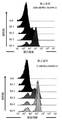


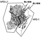


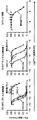


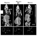


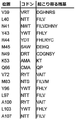


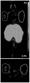


Classifications
-
- C—CHEMISTRY; METALLURGY
- C07—ORGANIC CHEMISTRY
- C07K—PEPTIDES
- C07K14/00—Peptides having more than 20 amino acids; Gastrins; Somatostatins; Melanotropins; Derivatives thereof
- C07K14/435—Peptides having more than 20 amino acids; Gastrins; Somatostatins; Melanotropins; Derivatives thereof from animals; from humans
- C07K14/705—Receptors; Cell surface antigens; Cell surface determinants
- C07K14/70503—Immunoglobulin superfamily
-
- A—HUMAN NECESSITIES
- A61—MEDICAL OR VETERINARY SCIENCE; HYGIENE
- A61K—PREPARATIONS FOR MEDICAL, DENTAL OR TOILETRY PURPOSES
- A61K35/00—Medicinal preparations containing materials or reaction products thereof with undetermined constitution
- A61K35/12—Materials from mammals; Compositions comprising non-specified tissues or cells; Compositions comprising non-embryonic stem cells; Genetically modified cells
- A61K35/14—Blood; Artificial blood
- A61K35/17—Lymphocytes; B-cells; T-cells; Natural killer cells; Interferon-activated or cytokine-activated lymphocytes
-
- A—HUMAN NECESSITIES
- A61—MEDICAL OR VETERINARY SCIENCE; HYGIENE
- A61K—PREPARATIONS FOR MEDICAL, DENTAL OR TOILETRY PURPOSES
- A61K38/00—Medicinal preparations containing peptides
- A61K38/16—Peptides having more than 20 amino acids; Gastrins; Somatostatins; Melanotropins; Derivatives thereof
- A61K38/17—Peptides having more than 20 amino acids; Gastrins; Somatostatins; Melanotropins; Derivatives thereof from animals; from humans
- A61K38/1703—Peptides having more than 20 amino acids; Gastrins; Somatostatins; Melanotropins; Derivatives thereof from animals; from humans from vertebrates
- A61K38/1709—Peptides having more than 20 amino acids; Gastrins; Somatostatins; Melanotropins; Derivatives thereof from animals; from humans from vertebrates from mammals
- A61K38/1761—Apoptosis related proteins, e.g. Apoptotic protease-activating factor-1 (APAF-1), Bax, Bax-inhibitory protein(s)(BI; bax-I), Myeloid cell leukemia associated protein (MCL-1), Inhibitor of apoptosis [IAP] or Bcl-2
-
- A—HUMAN NECESSITIES
- A61—MEDICAL OR VETERINARY SCIENCE; HYGIENE
- A61K—PREPARATIONS FOR MEDICAL, DENTAL OR TOILETRY PURPOSES
- A61K38/00—Medicinal preparations containing peptides
- A61K38/16—Peptides having more than 20 amino acids; Gastrins; Somatostatins; Melanotropins; Derivatives thereof
- A61K38/17—Peptides having more than 20 amino acids; Gastrins; Somatostatins; Melanotropins; Derivatives thereof from animals; from humans
- A61K38/177—Receptors; Cell surface antigens; Cell surface determinants
-
- A—HUMAN NECESSITIES
- A61—MEDICAL OR VETERINARY SCIENCE; HYGIENE
- A61K—PREPARATIONS FOR MEDICAL, DENTAL OR TOILETRY PURPOSES
- A61K45/00—Medicinal preparations containing active ingredients not provided for in groups A61K31/00 - A61K41/00
- A61K45/06—Mixtures of active ingredients without chemical characterisation, e.g. antiphlogistics and cardiaca
-
- A—HUMAN NECESSITIES
- A61—MEDICAL OR VETERINARY SCIENCE; HYGIENE
- A61K—PREPARATIONS FOR MEDICAL, DENTAL OR TOILETRY PURPOSES
- A61K47/00—Medicinal preparations characterised by the non-active ingredients used, e.g. carriers or inert additives; Targeting or modifying agents chemically bound to the active ingredient
- A61K47/50—Medicinal preparations characterised by the non-active ingredients used, e.g. carriers or inert additives; Targeting or modifying agents chemically bound to the active ingredient the non-active ingredient being chemically bound to the active ingredient, e.g. polymer-drug conjugates
- A61K47/51—Medicinal preparations characterised by the non-active ingredients used, e.g. carriers or inert additives; Targeting or modifying agents chemically bound to the active ingredient the non-active ingredient being chemically bound to the active ingredient, e.g. polymer-drug conjugates the non-active ingredient being a modifying agent
- A61K47/68—Medicinal preparations characterised by the non-active ingredients used, e.g. carriers or inert additives; Targeting or modifying agents chemically bound to the active ingredient the non-active ingredient being chemically bound to the active ingredient, e.g. polymer-drug conjugates the non-active ingredient being a modifying agent the modifying agent being an antibody, an immunoglobulin or a fragment thereof, e.g. an Fc-fragment
- A61K47/6835—Medicinal preparations characterised by the non-active ingredients used, e.g. carriers or inert additives; Targeting or modifying agents chemically bound to the active ingredient the non-active ingredient being chemically bound to the active ingredient, e.g. polymer-drug conjugates the non-active ingredient being a modifying agent the modifying agent being an antibody, an immunoglobulin or a fragment thereof, e.g. an Fc-fragment the modifying agent being an antibody or an immunoglobulin bearing at least one antigen-binding site
- A61K47/6849—Medicinal preparations characterised by the non-active ingredients used, e.g. carriers or inert additives; Targeting or modifying agents chemically bound to the active ingredient the non-active ingredient being chemically bound to the active ingredient, e.g. polymer-drug conjugates the non-active ingredient being a modifying agent the modifying agent being an antibody, an immunoglobulin or a fragment thereof, e.g. an Fc-fragment the modifying agent being an antibody or an immunoglobulin bearing at least one antigen-binding site the antibody targeting a receptor, a cell surface antigen or a cell surface determinant
-
- A—HUMAN NECESSITIES
- A61—MEDICAL OR VETERINARY SCIENCE; HYGIENE
- A61K—PREPARATIONS FOR MEDICAL, DENTAL OR TOILETRY PURPOSES
- A61K48/00—Medicinal preparations containing genetic material which is inserted into cells of the living body to treat genetic diseases; Gene therapy
-
- A—HUMAN NECESSITIES
- A61—MEDICAL OR VETERINARY SCIENCE; HYGIENE
- A61K—PREPARATIONS FOR MEDICAL, DENTAL OR TOILETRY PURPOSES
- A61K51/00—Preparations containing radioactive substances for use in therapy or testing in vivo
- A61K51/02—Preparations containing radioactive substances for use in therapy or testing in vivo characterised by the carrier, i.e. characterised by the agent or material covalently linked or complexing the radioactive nucleus
- A61K51/04—Organic compounds
- A61K51/08—Peptides, e.g. proteins, carriers being peptides, polyamino acids, proteins
- A61K51/088—Peptides, e.g. proteins, carriers being peptides, polyamino acids, proteins conjugates with carriers being peptides, polyamino acids or proteins
-
- A—HUMAN NECESSITIES
- A61—MEDICAL OR VETERINARY SCIENCE; HYGIENE
- A61K—PREPARATIONS FOR MEDICAL, DENTAL OR TOILETRY PURPOSES
- A61K51/00—Preparations containing radioactive substances for use in therapy or testing in vivo
- A61K51/02—Preparations containing radioactive substances for use in therapy or testing in vivo characterised by the carrier, i.e. characterised by the agent or material covalently linked or complexing the radioactive nucleus
- A61K51/04—Organic compounds
- A61K51/08—Peptides, e.g. proteins, carriers being peptides, polyamino acids, proteins
- A61K51/10—Antibodies or immunoglobulins; Fragments thereof, the carrier being an antibody, an immunoglobulin or a fragment thereof, e.g. a camelised human single domain antibody or the Fc fragment of an antibody
-
- A—HUMAN NECESSITIES
- A61—MEDICAL OR VETERINARY SCIENCE; HYGIENE
- A61P—SPECIFIC THERAPEUTIC ACTIVITY OF CHEMICAL COMPOUNDS OR MEDICINAL PREPARATIONS
- A61P35/00—Antineoplastic agents
-
- C—CHEMISTRY; METALLURGY
- C07—ORGANIC CHEMISTRY
- C07K—PEPTIDES
- C07K14/00—Peptides having more than 20 amino acids; Gastrins; Somatostatins; Melanotropins; Derivatives thereof
- C07K14/435—Peptides having more than 20 amino acids; Gastrins; Somatostatins; Melanotropins; Derivatives thereof from animals; from humans
- C07K14/46—Peptides having more than 20 amino acids; Gastrins; Somatostatins; Melanotropins; Derivatives thereof from animals; from humans from vertebrates
- C07K14/47—Peptides having more than 20 amino acids; Gastrins; Somatostatins; Melanotropins; Derivatives thereof from animals; from humans from vertebrates from mammals
- C07K14/4701—Peptides having more than 20 amino acids; Gastrins; Somatostatins; Melanotropins; Derivatives thereof from animals; from humans from vertebrates from mammals not used
- C07K14/4747—Apoptosis related proteins
-
- C—CHEMISTRY; METALLURGY
- C07—ORGANIC CHEMISTRY
- C07K—PEPTIDES
- C07K14/00—Peptides having more than 20 amino acids; Gastrins; Somatostatins; Melanotropins; Derivatives thereof
- C07K14/435—Peptides having more than 20 amino acids; Gastrins; Somatostatins; Melanotropins; Derivatives thereof from animals; from humans
- C07K14/705—Receptors; Cell surface antigens; Cell surface determinants
- C07K14/70503—Immunoglobulin superfamily
- C07K14/7051—T-cell receptor (TcR)-CD3 complex
-
- C—CHEMISTRY; METALLURGY
- C07—ORGANIC CHEMISTRY
- C07K—PEPTIDES
- C07K14/00—Peptides having more than 20 amino acids; Gastrins; Somatostatins; Melanotropins; Derivatives thereof
- C07K14/435—Peptides having more than 20 amino acids; Gastrins; Somatostatins; Melanotropins; Derivatives thereof from animals; from humans
- C07K14/705—Receptors; Cell surface antigens; Cell surface determinants
- C07K14/70503—Immunoglobulin superfamily
- C07K14/70532—B7 molecules, e.g. CD80, CD86
-
- A—HUMAN NECESSITIES
- A61—MEDICAL OR VETERINARY SCIENCE; HYGIENE
- A61K—PREPARATIONS FOR MEDICAL, DENTAL OR TOILETRY PURPOSES
- A61K38/00—Medicinal preparations containing peptides
-
- C—CHEMISTRY; METALLURGY
- C07—ORGANIC CHEMISTRY
- C07K—PEPTIDES
- C07K2319/00—Fusion polypeptide
- C07K2319/30—Non-immunoglobulin-derived peptide or protein having an immunoglobulin constant or Fc region, or a fragment thereof, attached thereto
Description
<相互参照>
本出願は、2014年8月8日に出願された米国仮特許出願第62/035,316号の利益と、2015年4月21日に出願された米国仮特許出願第62/150,789号の利益を主張するものであり、当該文献は各々、全体として参照により本明細書に組み込まれる。
<Cross reference>
This application is in the interest of US Provisional Patent Application No. 62 / 035,316 filed on August 8, 2014, and US Provisional Patent Application No. 62 / 150,789 filed on April 21, 2015. Each of the documents is incorporated herein by reference in its entirety.
テキストファイルとして提供される配列表の参照による組み込み
配列表は、2015年8月7日に作成されるとともに119KBのサイズを有するテキストファイル「STAN−1136WO2_SeqList_ST25.txt」として本明細書とともに提供される。テキストファイルの内容は全体として参照により本明細書に組み込まれる。
The built-in sequence listing by reference of the sequence listing provided as a text file is provided with the present specification as a text file "STAN-1136WO2_SeqList_ST25.txt" created on August 7, 2015 and having a size of 119KB. The contents of the text file are incorporated herein by reference in their entirety.
導入部
T細胞活性化は、T細胞受容体に提供される抗原特異的なシグナルに依存する。さらなるシグナル、例えば、共刺激(正)および/または共抑制(負)シグナルはこの応答を微調整して、その強さ、性質、および継続時間を決定しやすくする。共刺激相互作用はT細胞の活性化と増殖を強化し、その一方で共抑制相互作用は調節を促す。例えば、CD28とCTLA−4の共受容体は両方ともB7−1(CD80)とB7−2(CD86)の分子に結合する。CD28は強力な正の共刺激受容体として作用し、CTLA−4は共抑制受容体として作用する。
Introduction T cell activation depends on antigen-specific signals provided to the T cell receptor. Additional signals, such as co-stimulatory (positive) and / or co-suppressive (negative) signals, fine-tune this response to help determine its intensity, nature, and duration. Co-stimulatory interactions enhance T cell activation and proliferation, while co-suppressive interactions promote regulation. For example, the CD28 and CTLA-4 co-receptors both bind to the B7-1 (CD80) and B7-2 (CD86) molecules. CD28 acts as a potent positive co-stimulatory receptor and CTLA-4 acts as a co-suppressive receptor.
プログラム死−1(PD−1)受容体として知られている受容体は、T細胞、B細胞、および骨髄性細胞上で発現し、プログラム死リガンド(PD−L)に結合する。この受容体−リガンドペアは、主として抑制シグナル(例えば、PD−1の細胞質尾部の免疫受容体のチロシンベースのスイッチモチーフ(ITSM)へのSHP−2などのホスファターゼの補充を介して)を提供するように機能する。 Receptors known as programmed death-1 (PD-1) receptors are expressed on T cells, B cells, and myeloid cells and bind to programmed death ligands (PD-L). This receptor-ligand pair primarily provides inhibitory signals (eg, through supplementation of phosphatases such as SHP-2 to the tyrosine-based switch motif (ITSM) of the immunoreceptor in the cytoplasmic tail of PD-1). Works like.
PD−1シグナル伝達は周辺の許容差を引き起こすとともに維持するのに重要な役割を果たす。抗原提示細胞上のPD−1リガンド(PD−L)は自己反応性のT細胞を阻害するとともに周辺の許容差を引き起こすことが示されているが、実質細胞上のPD−1リガンドは、許容差を維持するためにエフェクターT細胞を抑えることによって組織の破壊を防ぐ。PD−1の抑制性の役割はPD−1欠損マウスの表現型によって強調され、これは遺伝的背景に依存して様々な自己免疫性疾患にかかる。PD−1/PD−Lの経路は、腫瘍細胞によるおよび様々な病原体による免疫回避の標的として頻繁に利用される。例えば、PD1:PD−L経路は、腫瘍とウイルス(例えば、慢性感染症を引き起こすウイルス)により活用(例えば、活性化過剰)可能であり、これはPD−Lタンパク質を発現してPD−1(例えばT細胞上で)を刺激することができ、それによって、免疫細胞(例えばT細胞)応答を減らし、免疫系による根絶を回避する。 PD-1 signaling plays an important role in causing and maintaining peripheral tolerances. PD-1 ligands (PD-L) on antigen-presenting cells have been shown to inhibit autoreactive T cells and cause peripheral tolerances, whereas PD-1 ligands on parenchymal cells are acceptable. Prevents tissue destruction by suppressing effector T cells to maintain the difference. The inhibitory role of PD-1 is emphasized by the phenotype of PD-1 deficient mice, which affects a variety of autoimmune disorders depending on their genetic background. The PD-1 / PD-L pathway is frequently used as a target for immune evasion by tumor cells and by various pathogens. For example, the PD1: PD-L pathway can be utilized (eg, overactivated) by tumors and viruses (eg, viruses that cause chronic infections), which expresses the PD-L protein and PD-1 (eg, PD-1). It can stimulate (eg, on T cells), thereby reducing immune cell (eg, T cell) responses and avoiding eradication by the immune system.
本開示は高親和性PD1模倣ポリペプチドを提供しており、これは、PD−L1に特異的に結合することにより、細胞(例えばT細胞のような免疫細胞)の表面上のPD−1とのPD−L1の相互作用を遮断する/減らすことにより、および、ゆえにPD−L1により刺激されたPD−1活性を遮断する/減らすことにより、PD−1を模倣する。さらに、PD−1活性を減少させるために高親和性PD1模倣ポリペプチドを使用する方法も開示されている。 The present disclosure provides a high affinity PD1 mimicking polypeptide, which by specifically binding to PD-L1 with PD-1 on the surface of a cell (eg, an immune cell such as a T cell). PD-1 is mimicked by blocking / reducing PD-L1 interaction and thus blocking / reducing PD-1 activity stimulated by PD-L1. In addition, methods of using high affinity PD1 mimetic polypeptides to reduce PD-1 activity have also been disclosed.
高親和性PD−1変異体(模倣)ポリペプチドが提供される。ポリペプチドは、野生型PD−1タンパク質(例えば、野生型ヒトPD−1タンパク質)の配列変異体であり、野生型PD−1タンパク質とそのリガンドPD−L(PD−L1および/またはPD−L2)の間の相互作用を阻害するインビボとインビトロの方法に対する実用性を有する。高親和性PD−1模倣ポリペプチドは、野生型のPD−1タンパク質に対して少なくとも1つのアミノ酸変化を含み、野生型のPD−1タンパク質に対してPD−L(PD−L1および/またはPD−L2)との親和性を増加させ、野生型PD−1タンパク質の膜貫通ドメインを欠いている。親和性を増加させるアミノ酸変化は、PD−1とPD−Lとの間の接触のアミノ酸位置へ局在化可能であり、および/またはそれが由来するPD−1タンパク質の免疫グロブリンドメイン中に位置付け可能である。 High affinity PD-1 mutant (mimicking) polypeptides are provided. Polypeptides are sequence variants of wild-type PD-1 proteins (eg, wild-type human PD-1 proteins) that include wild-type PD-1 proteins and their ligands PD-L (PD-L1 and / or PD-L2). ) Has practicality for in vivo and in vitro methods of inhibiting the interaction between. High-affinity PD-1 mimetic polypeptides contain at least one amino acid change to the wild-type PD-1 protein and PD-L (PD-L1 and / or PD) to the wild-type PD-1 protein. -Increases affinity with L2) and lacks the transmembrane domain of wild-type PD-1 protein. Amino acid changes that increase affinity can be localized to the amino acid position of contact between PD-1 and PD-L and / or located in the immunoglobulin domain of the PD-1 protein from which it is derived. It is possible.
高親和性PD−1模倣ポリペプチドは、例えば、グリコシル化、ペグ化などによって翻訳後に修飾可能である。高親和性PD−1模倣ポリペプチドは、融合タンパク質(つまり、追加のアミノ酸配列を含むことができる)、例えば、抗体Fc配列および/または対象となる抗原への特異的な結合をもたらす抗体の可変領域などを含む融合であり得る。高親和性PD−1模倣ポリペプチドは、単量体または多量体、つまり二量体、三量体、四量体などであってもよい。 High-affinity PD-1 mimetic polypeptides can be post-translationally modified by, for example, glycosylation, pegging and the like. High-affinity PD-1 mimetic polypeptides are fusion proteins (ie, they can contain additional amino acid sequences), eg, antibody Fc sequences and / or variability of antibodies that result in specific binding to the antigen of interest. It can be a fusion that includes regions and the like. The high affinity PD-1 mimetic polypeptide may be a monomer or a multimer, that is, a dimer, a trimer, a tetramer, or the like.
いくつかの実施形態では、方法は、PD−1とそのリガンドPD−L1および/またはPD−L2との間の生理学的な結合相互作用を阻害する高親和性PD−1模倣ポリペプチドを含む治療用量の医薬組成物を投与することにより、哺乳動物中の免疫細胞(T細胞、NK細胞など)の活性を調節するために提供される。 In some embodiments, the method comprises a treatment comprising a high affinity PD-1 mimicking polypeptide that inhibits the physiological binding interaction between PD-1 and its ligands PD-L1 and / or PD-L2. It is provided to regulate the activity of immune cells (T cells, NK cells, etc.) in mammals by administering a dose of the pharmaceutical composition.
本開示はさらに、薬学的に許容可能な賦形剤と組み合わせた高親和性PD−1模倣ポリペプチドを有する医薬製剤を含んでいる。こうした製剤は、単位投与量、例えば、個体内の第2の細胞上のPD−L(PD−L1および/またはPD−L2)との第1の細胞上のPD−1の相互作用を阻害するのに効果的な投与量として提供されることもある。医薬製剤はさらに、高親和性PD−1模倣ポリペプチドの凍結乾燥させた調合物またはそれ以外の調合物を含み、それらは使用のために再構成されてもよい。 The disclosure further includes pharmaceutical formulations with high affinity PD-1 mimetic polypeptides in combination with pharmaceutically acceptable excipients. Such a formulation inhibits the interaction of PD-1 on a first cell with a unit dose, eg, PD-L (PD-L1 and / or PD-L2) on a second cell in an individual. It may also be provided as an effective dose. The pharmaceutical formulation further comprises a lyophilized formulation of the high affinity PD-1 mimetic polypeptide or other formulation, which may be reconstituted for use.
いくつかの実施形態では、方法は、例えば、免疫系による生きている癌細胞の破壊を標的として、標的細胞への免疫応答を刺激するために提供される。こうした方法では、PD−L1を発現する細胞は、内在性のPD−1(例えば第1の細胞上)とPD−L(例えば、第2の細胞上のPD−L1および/またはPD−L2)の間の相互作用を阻害するのに有効な投与量の高親和性PD−1模倣ポリペプチドと接触させる。この相互作用を阻害することにより、高親和性PD−1模倣ポリペプチドのない状態では破壊されない標的細胞の免疫ベースの破壊が可能となる。接触は、例えば、治療目的のためにインビボで、例えば、スクリーニング検査などのためにインビトロで行われてもよい。こうした目的のための高親和性PD−1模倣ポリペプチドは、多量体または単量体であってもよい。単量体の試薬については、標的細胞へ選択的に結合する抗体と組み合わせて、投与のための特別の用途が見出される。 In some embodiments, the method is provided, for example, to target the destruction of living cancer cells by the immune system and stimulate an immune response to the target cells. In these methods, cells expressing PD-L1 are endogenous PD-1 (eg, on the first cell) and PD-L (eg, PD-L1 and / or PD-L2 on the second cell). Contact with a high affinity PD-1 mimetic polypeptide at a dose effective to inhibit the interaction between. Inhibiting this interaction allows the immune-based destruction of target cells, which is not destroyed in the absence of high-affinity PD-1 mimetic polypeptides. Contact may be made, for example, in vivo for therapeutic purposes, for example, in vitro for screening tests. The high affinity PD-1 mimicking polypeptide for this purpose may be a multimer or a monomer. For monomeric reagents, special uses have been found for administration in combination with antibodies that selectively bind to target cells.
高親和性PD−1模倣ポリペプチドで処置可能な病気に罹患した個体は、癌を抱える個体、感染症(例えば、慢性感染症、ウィルス感染症など)を抱える個体、免疫疾患(例えば、免疫抑制に関連する疾患)を抱える個体、炎症性障害を抱える個体、および/または他の過剰増殖性の疾患、例えば、硬化症、繊維症などを抱える個体を含む。場合によっては、癌細胞(例えば腫瘍細胞)は、PD−1のPD−Lとの相互作用を阻害するまたは遮蔽するのに効果的な投与量の高親和性PD−1模倣ポリペプチドに、免疫系の細胞を接触させることにより、免疫系の刺激を増加させて、除去の対象となる。場合によっては、標的細胞(例えば癌細胞、腫瘍細胞、感染細胞などのような損傷を受けた細胞)は、PD−L1および/またはPD−L2を発現し、高親和性PD−1模倣ポリペプチドは、標的細胞上のPD−Lの、免疫細胞(例えば、T細胞、NK細胞など)上のPD−1との相互作用を阻害し、これにより、標的細胞に対する免疫応答を抑える標的細胞の能力を阻害することができる。 Individuals with diseases that can be treated with high-affinity PD-1 mimetic polypeptides include individuals with cancer, individuals with infectious diseases (eg, chronic infections, viral infections, etc.), and immune disorders (eg, immunosuppression). Includes individuals with (related diseases), individuals with inflammatory disorders, and / or individuals with other hyperproliferative disorders such as sclerosis, fibrosis, and the like. In some cases, cancer cells (eg, tumor cells) are immune to high-affinity PD-1 mimetic polypeptides at doses that are effective in inhibiting or blocking the interaction of PD-1 with PD-L. Contact of cells of the system increases the stimulation of the immune system and is the target of removal. In some cases, target cells (damaged cells such as cancer cells, tumor cells, infected cells, etc.) express PD-L1 and / or PD-L2 and are high affinity PD-1 mimicry polypeptides. The ability of target cells to inhibit the interaction of PD-L on target cells with PD-1 on immune cells (eg, T cells, NK cells, etc.), thereby suppressing the immune response to the target cells. Can be inhibited.
患者への有効な量の高親和性PD−1模倣ポリペプチドの投与は、PD−1とPD−L1との間の相互作用を防ぎ、これにより、腫瘍細胞および/または感染細胞(例えば慢性的な感染細胞)の除去を増加させる場合がある。場合によっては、高親和性PD−1模倣ポリペプチドは1つ以上の腫瘍細胞マーカーに向けられたモノクローナル抗体と組み合わせ可能であり、この併用療法は、単一体としてのいずれかの薬剤の投与と比較して、癌細胞の除去を強化するのに相乗的であり得る。他の実施形態では、高親和性PD−1模倣ポリペプチドは検知可能な標識を含む。このような標識化した試薬は、インビトロまたはインビボでの画像化のために、例えば腫瘍の画像化において使用可能である。場合によっては、高親和性PD−1模倣ポリペプチドは、PD−L(例えば、PD−L1を発現する細胞)の検出のための診断ツールとして使用可能であり、特定の処置レジメンが成功したかどうかを評価するためのコンパニオン診断として使用可能である。 Administration of an effective amount of high affinity PD-1 mimicking polypeptide to the patient prevents the interaction between PD-1 and PD-L1 thereby causing tumor cells and / or infected cells (eg, chronic). Infected cells) may increase the removal. In some cases, high-affinity PD-1 mimetic polypeptides can be combined with monoclonal antibodies directed at one or more tumor cell markers, and this combination therapy is compared to administration of either drug as a single entity. And can be synergistic in enhancing the removal of cancer cells. In other embodiments, the high affinity PD-1 mimetic polypeptide comprises a detectable label. Such labeled reagents can be used for in vitro or in vivo imaging, eg in tumor imaging. In some cases, high-affinity PD-1 mimetic polypeptides can be used as diagnostic tools for the detection of PD-L (eg, cells expressing PD-L1) and has a particular treatment regimen been successful? It can be used as a companion diagnostic to evaluate whether or not.
高親和性PD−1模倣ポリペプチドが提供される。場合によっては、高親和性PD−1模倣ポリペプチドは、野生型のPD−1配列の変異体であるが、PD−1膜貫通ドメインを欠いており、野生型PD1−ポリペプチドの対応する配列に対して1つ以上のアミノ酸変化を含み、この1つ以上のアミノ酸変化は、対応する野生型PD−1ポリペプチドのPD−L1への親和性と比較して、PD−L1のポリペプチドの親和性を増加させる。場合によっては、PD−1模倣ポリペプチドは、PD−L1に対して1×10⁻⁷M以下のKdを有する。場合によっては、高親和性PD−1模倣ポリペプチドのPD−L1との親和性は、野生型PD−1ポリペプチドの対応する配列に対するアミノ酸変化を有していない上記PD−1模倣ポリペプチドのPD−L1との親和性よりも5倍以上大きい。場合によっては、高親和性PD−1模倣ポリペプチドは、野生型PD−1ポリペプチドの対応する配列に対するアミノ酸変化を有していない上記PD−1模倣ポリペプチドのPD−L2との親和性と比較して、PD−L2に対する親和性が低い。場合によっては、1つ以上のアミノ酸変化はPD−L1と接触するPD−1のアミノ酸位置に位置する。場合によっては、1つ以上のアミノ酸変化は、V39、N41、Y43、M45、S48、N49、Q50、T51、D52、K53、A56、Q63、G65、Q66、L97、S102、L103、A104、P105、K106、およびA107から選択されるSEQ ID NO:2で示されるタンパク質フラグメントに対するアミノ酸位置、または、別の野生型PD−1タンパク質に対する対応するアミノ酸位置にある。場合によっては、1つ以上のアミノ酸変化は、V39、L40、N41、Y43、R44、M45、S48、N49、Q50、T51、D52、K53、A56、Q63、G65、Q66、V72、H82、M83、R90、Y96、L97、A100、S102、L103、A104、P105、K106、およびA107から選択されるSEQ ID NO:2で示されるタンパク質フラグメントに対するアミノ酸位置、または、別の野生型PD−1タンパク質に対する対応するアミノ酸位置にある。場合によっては、1つ以上のアミノ酸変化は、5つ以上のアミノ酸変化である。 High affinity PD-1 mimetic polypeptides are provided. In some cases, the high affinity PD-1 mimicking polypeptide is a variant of the wild-type PD-1 sequence, but lacks the PD-1 transmembrane domain and the corresponding sequence of the wild-type PD1-polypeptide. Containing one or more amino acid changes with respect to, this one or more amino acid changes of the PD-L1 polypeptide as compared to the affinity of the corresponding wild-type PD-1 polypeptide for PD-L1. Increases affinity. In some cases, PD-1 mimetic polypeptides have a K d of 1 × 10⁻⁷ M or less relative to PD-L1. In some cases, the affinity of the high affinity PD-1 mimetic polypeptide with PD-L1 is that of the PD-1 mimicking polypeptide that does not have an amino acid change to the corresponding sequence of the wild PD-1 polypeptide. It is more than 5 times greater than the affinity with PD-L1. In some cases, the high affinity PD-1 mimetic polypeptide has an affinity for PD-L2 of the PD-1 mimetic polypeptide that does not have an amino acid change to the corresponding sequence of the wild PD-1 polypeptide. In comparison, it has a low affinity for PD-L2. In some cases, one or more amino acid changes are located at the amino acid position of PD-1 in contact with PD-L1. In some cases, one or more amino acid changes include V39, N41, Y43, M45, S48, N49, Q50, T51, D52, K53, A56, Q63, G65, Q66, L97, S102, L103, A104, P105, It is at the amino acid position for the protein fragment indicated by SEQ ID NO: 2 selected from K106 and A107, or the corresponding amino acid position for another wild-type PD-1 protein. In some cases, one or more amino acid changes include V39, L40, N41, Y43, R44, M45, S48, N49, Q50, T51, D52, K53, A56, Q63, G65, Q66, V72, H82, M83, Amino acid position for protein fragment indicated by SEQ ID NO: 2 selected from R90, Y96, L97, A100, S102, L103, A104, P105, K106, and A107, or correspondence to another wild-type PD-1 protein. At the amino acid position. In some cases, one or more amino acid changes are five or more amino acid changes.
さらに、以下から選択されるSEQ ID NO:2で示されるPD−1タンパク質フラグメントに対する1つ以上のアミノ酸変化:(1)V39HまたはV39R;(2)L40VまたはL40I;(3)N41IまたはN41V;(4)Y43FまたはY43H;(5)R44YまたはR44L;(6)M45Q、M45E、M45L、またはM45D;(7)S48D、S48L、S48N、S48G、またはS48V;(8)N49C、N49G、N49Y、またはN49S;(9)Q50K、Q50E、またはQ50H;(10)T51V、T51L、またはT51A;(11)D52F、D52R、D52Y、またはD52V;(12)K53TまたはK53L;(13)A56SまたはA56L;(14)Q63T、Q63I、Q63E、Q63L、またはQ63P;(15)G65N、G65R、G65I、G65L、G65F、またはG65V;(16)Q66P;(17)V72I;(18)H82Q;(19)M83LまたはM83F;(20)R90K;(21)Y96F;(22)L97Y、L97V、またはL97I;(23)A100IまたはA100V;(24)S102TまたはS102A;(25)L103I、L103Y、またはL103F;(26)A104S、A104H、またはA104D;(27)P105A;(28)K106G、K106E、K106I、K106V、K106R、またはK106T;および、(29)A107P、A107I、またはA107V;あるいは、別の野生型PD−1タンパク質に対して対応する位置で同じアミノ酸をもたらす変化、を含む高親和性PD−1模倣ポリペプチドも提供される。 In addition, one or more amino acid changes to the PD-1 protein fragment indicated by SEQ ID NO: 2 selected from: (1) V39H or V39R; (2) L40V or L40I; (3) N41I or N41V; 4) Y43F or Y43H; (5) R44Y or R44L; (6) M45Q, M45E, M45L, or M45D; (7) S48D, S48L, S48N, S48G, or S48V; (8) N49C, N49G, N49Y, or N49S (9) Q50K, Q50E, or Q50H; (10) T51V, T51L, or T51A; (11) D52F, D52R, D52Y, or D52V; (12) K53T or K53L; (13) A56S or A56L; (14) Q63T, Q63I, Q63E, Q63L, or Q63P; (15) G65N, G65R, G65I, G65L, G65F, or G65V; (16) Q66P; (17) V72I; (18) H82Q; (19) M83L or M83F; 20) R90K; (21) Y96F; (22) L97Y, L97V, or L97I; (23) A100I or A100V; (24) S102T or S102A; (25) L103I, L103Y, or L103F; (26) A104S, A104H, Or A104D; (27) P105A; (28) K106G, K106E, K106I, K106V, K106R, or K106T; and (29) A107P, A107I, or A107V; or for another wild-type PD-1 protein. High-affinity PD-1 mimetic polypeptides are also provided, including changes that result in the same amino acid at the desired position.
さらに、(a)V39、N41、Y43、M45、S48、N49、Q50、K53、A56、Q63、G65、Q66、L97、S102、L103、A104、K106、およびA107、または別の野生型PD−1タンパク質に対する対応するアミノ酸位置;(b)V39、N41、Y43、M45、S48、Q50、T51、D52F、K53、A56、Q63、G65、Q66、L97、S102、L103、A104、K106、およびA107、または別の野生型PD−1タンパク質に対する対応するアミノ酸位置;(c)V39、L40、N41、Y43、R44、M45、N49、K53、M83、L97、A100、およびA107、または別の野生型PD−1タンパク質に対する対応するアミノ酸位置;(d)V39、L40、N41、Y43、M45、N49、K53、Q66P、M83、L97、およびA107、または別の野生型PD−1タンパク質に対する対応するアミノ酸位置;(e)V39、L40、N41、Y43、M45、N49、K53、Q66P、H82、M83、L97、A100、およびA107、または別の野生型PD−1タンパク質に対する対応するアミノ酸位置;(f)V39、L40、N41、Y43、M45、N49、K53、M83、L97、A100、およびA107、または別の野生型PD−1タンパク質に対する対応するアミノ酸位置;(g)V39、L40、N41、Y43、R44、M45、N49、K53、L97、A100、およびA107、または別の野生型PD−1タンパク質に対する対応するアミノ酸位置;ならびに、(h)V39、L40、N41、Y43、M45、N49、K53、L97、A100、およびA107、または別の野生型PD−1タンパク質に対する対応するアミノ酸位置、から選択されるSEQ ID NO:2で示されるタンパク質フラグメントに対するアミノ酸位置にあるアミノ酸変化を含む、高親和性PD−1模倣ポリペプチドも提供される。 In addition, (a) V39, N41, Y43, M45, S48, N49, Q50, K53, A56, Q63, G65, Q66, L97, S102, L103, A104, K106, and A107, or another wild-type PD-1. Corresponding amino acid positions relative to proteins; (b) V39, N41, Y43, M45, S48, Q50, T51, D52F, K53, A56, Q63, G65, Q66, L97, S102, L103, A104, K106, and A107, or Corresponding amino acid positions for another wild-type PD-1 protein; (c) V39, L40, N41, Y43, R44, M45, N49, K53, M83, L97, A100, and A107, or another wild-type PD-1 Corresponding amino acid position for protein; (d) Corresponding amino acid position for V39, L40, N41, Y43, M45, N49, K53, Q66P, M83, L97, and A107, or another wild-type PD-1 protein; (e) ) V39, L40, N41, Y43, M45, N49, K53, Q66P, H82, M83, L97, A100, and A107, or the corresponding amino acid positions for another wild-type PD-1 protein; (f) V39, L40, Corresponding amino acid positions for N41, Y43, M45, N49, K53, M83, L97, A100, and A107, or another wild-type PD-1 protein; (g) V39, L40, N41, Y43, R44, M45, N49 , K53, L97, A100, and A107, or the corresponding amino acid positions for another wild-type PD-1 protein; and (h) V39, L40, N41, Y43, M45, N49, K53, L97, A100, and A107. Also a high affinity PD-1 mimicking polypeptide comprising an amino acid change at the amino acid position for the protein fragment indicated by SEQ ID NO: 2, selected from, or the corresponding amino acid position for another wild-type PD-1 protein. Provided.
さらに、以下から選択されるSEQ ID NO:2で示されるPD−1タンパク質フラグメントに対する、アミノ酸変化:(a){V39HまたはV39R}、{N41IまたはN41V}、{Y43FまたはY43H}、{M45Q、M45E、M45L、またはM45D}、{S48D、S48L、S48N、S48G、またはS48V}、{N49C、N49G、N49Y、またはN49S}、{Q50K、Q50E、またはQ50H}、{K53TまたはK53L}、{A56SまたはA56L}、{Q63T、Q63I、Q63E、Q63L、またはQ63P}、{G65N、G65R、G65I、G65L、G65F、またはG65V}、{Q66P}、{L97Y、L97V、またはL97I}、{S102TまたはS102A}、{L103I、L103Y、またはL103F}、{A104S、A104H、またはA104D}、{K106G、K106E、K106I、K106V、K106R、またはK106T}、および、{A107P、A107I、またはA107V};あるいは、別の野生型PD−1タンパク質に対して対応する位置で同じアミノ酸をもたらす変化;(b){V39HまたはV39R}、{N41IまたはN41V}、{Y43FまたはY43H}、{M45Q、M45E、M45L、またはM45D}、{S48D、S48L、S48N、S48G、またはS48V}、{Q50K、Q50E、またはQ50H}、{T51V、T51L、またはT51A}、{D52F、D52R、D52Y、またはD52V}、{K53TまたはK53L}、{A56SまたはA56L}、{Q63T、Q63I、Q63E、Q63L、またはQ63P}、{G65N、G65R、G65I、G65L、G65F、またはG65V}、{Q66P}、{L97Y、L97V、またはL97I}、{S102TまたはS102A}、{L103I、L103Y、またはL103F}、{A104S、A104H、またはA104D}、{K106G、K106E、K106I、K106V、K106R、またはK106T}、および{A107P、A107I、またはA107V};あるいは、別の野生型PD−1タンパク質に対して対応する位置で同じアミノ酸をもたらす変化;(c){V39HまたはV39R}、{L40VまたはL40I}、{N41IまたはN41V}、{Y43FまたはY43H}、{R44YまたはR44L}、{M45Q、M45E、M45L、またはM45D}、{N49C、N49G、N49Y、またはN49S}、{K53TまたはK53L}、{M83LまたはM83F}、{L97Y、L97V、またはL97I}、{A100IまたはA100V}、および{A107P、A107I、またはA107V};あるいは、別の野生型PD−1タンパク質に対して対応する位置で同じアミノ酸をもたらす変化;(d){V39HまたはV39R}、{L40VまたはL40I}、{N41IまたはN41V}、{Y43FまたはY43H}、{M45Q、M45E、M45L、またはM45D}、{N49C、N49G、N49Y、またはN49S}、{K53TまたはK53L}、{Q66P}、{M83LまたはM83F}、{L97Y、L97V、またはL97I}、および{A107P、A107I、またはA107V};あるいは、別の野生型PD−1タンパク質に対して対応する位置で同じアミノ酸をもたらす変化;
(e){V39HまたはV39R}、{L40VまたはL40I}、{N41IまたはN41V}、{Y43FまたはY43H}、{M45Q、M45E、M45L、またはM45D}、{N49C、N49G、N49Y、またはN49S}、{K53TまたはK53L}、{Q66P}、{H82Q}、{M83LまたはM83F}、{L97Y、L97V、またはL97I}、{A100IまたはA100V}、および{A107P、A107I、またはA107V};あるいは、別の野生型PD−1タンパク質に対して対応する位置で同じアミノ酸をもたらす変化;(f){V39HまたはV39R}、{L40VまたはL40I}、{N41IまたはN41V}、{Y43FまたはY43H}、{M45Q、M45E、M45L、またはM45D}、{N49C、N49G、N49Y、またはN49S}、{K53TまたはK53L}、{M83LまたはM83F}、{L97Y、L97V、またはL97I}、{A100IまたはA100V}、および{A107P、A107I、またはA107V};あるいは、別の野生型PD−1タンパク質に対して対応する位置で同じアミノ酸をもたらす変化;(g){V39HまたはV39R}、{L40VまたはL40I}、{N41IまたはN41V}、{Y43FまたはY43H}、{R44YまたはR44L}、{M45Q、M45E、M45L、またはM45D}、{N49C、N49G、N49Y、またはN49S}、{K53TまたはK53L}、{L97Y、L97V、またはL97I}、{A100IまたはA100V}、および{A107P、A107I、またはA107V};あるいは、別の野生型PD−1タンパク質に対して対応する位置で同じアミノ酸をもたらす変化;ならびに、(h){V39HまたはV39R}、{L40VまたはL40I}、{N41IまたはN41V}、{Y43FまたはY43H}、{M45Q、M45E、M45L、またはM45D}、{N49C、N49G、N49Y、またはN49S}、{K53TまたはK53L}、{L97Y、L97V、またはL97I}、{A100IまたはA100V}、および{A107P、A107I、またはA107V};あるいは、別の野生型PD−1タンパク質に対して対応する位置で同じアミノ酸をもたらす変化、を含む高親和性PD−1模倣ポリペプチドがさらに提供される。
Further, amino acid changes for the PD-1 protein fragment indicated by SEQ ID NO: 2 selected from the following: (a) {V39H or V39R}, {N41I or N41V}, {Y43F or Y43H}, {M45Q, M45E , M45L, or M45D}, {S48D, S48L, S48N, S48G, or S48V}, {N49C, N49G, N49Y, or N49S}, {Q50K, Q50E, or Q50H}, {K53T or K53L}, {A56S or A56L }, {Q63T, Q63I, Q63E, Q63L, or Q63P}, {G65N, G65R, G65I, G65L, G65F, or G65V}, {Q66P}, {L97Y, L97V, or L97I}, {S102T or S102A}, { L103I, L103Y, or L103F}, {A104S, A104H, or A104D}, {K106G, K106E, K106I, K106V, K106R, or K106T}, and {A107P, A107I, or A107V}; or another wild-type PD Changes that result in the same amino acid at the corresponding position relative to the -1 protein; (b) {V39H or V39R}, {N41I or N41V}, {Y43F or Y43H}, {M45Q, M45E, M45L, or M45D}, {S48D , S48L, S48N, S48G, or S48V}, {Q50K, Q50E, or Q50H}, {T51V, T51L, or T51A}, {D52F, D52R, D52Y, or D52V}, {K53T or K53L}, {A56S or A56L }, {Q63T, Q63I, Q63E, Q63L, or Q63P}, {G65N, G65R, G65I, G65L, G65F, or G65V}, {Q66P}, {L97Y, L97V, or L97I}, {S102T or S102A}, { L103I, L103Y, or L103F}, {A104S, A104H, or A104D}, {K106G, K106E, K106I, K106V, K106R, or K106T}, and {A107P, A107I, or A107V}; or another wild-type PD- Changes that result in the same amino acid at the corresponding position for one protein; (c) {V39H or V39R}, {L40V or L40I}, {N41I or N41V}, {Y 43F or Y43H}, {R44Y or R44L}, {M45Q, M45E, M45L, or M45D}, {N49C, N49G, N49Y, or N49S}, {K53T or K53L}, {M83L or M83F}, {L97Y, L97V, Or L97I}, {A100I or A100V}, and {A107P, A107I, or A107V}; or changes that result in the same amino acid at the corresponding position relative to another wild-type PD-1 protein; (d) {V39H or V39R }, {L40V or L40I}, {N41I or N41V}, {Y43F or Y43H}, {M45Q, M45E, M45L, or M45D}, {N49C, N49G, N49Y, or N49S}, {K53T or K53L}, {Q66P }, {M83L or M83F}, {L97Y, L97V, or L97I}, and {A107P, A107I, or A107V}; or changes that result in the same amino acid at the corresponding position relative to another wild-type PD-1 protein;
(E) {V39H or V39R}, {L40V or L40I}, {N41I or N41V}, {Y43F or Y43H}, {M45Q, M45E, M45L, or M45D}, {N49C, N49G, N49Y, or N49S}, { K53T or K53L}, {Q66P}, {H82Q}, {M83L or M83F}, {L97Y, L97V, or L97I}, {A100I or A100V}, and {A107P, A107I, or A107V}; or another wild type Changes that result in the same amino acids at the corresponding positions for the PD-1 protein; (f) {V39H or V39R}, {L40V or L40I}, {N41I or N41V}, {Y43F or Y43H}, {M45Q, M45E, M45L , Or M45D}, {N49C, N49G, N49Y, or N49S}, {K53T or K53L}, {M83L or M83F}, {L97Y, L97V, or L97I}, {A100I or A100V}, and {A107P, A107I, or A107V}; or a change that results in the same amino acid at the corresponding position relative to another wild PD-1 protein; (g) {V39H or V39R}, {L40V or L40I}, {N41I or N41V}, {Y43F or Y43H}, {R44Y or R44L}, {M45Q, M45E, M45L, or M45D}, {N49C, N49G, N49Y, or N49S}, {K53T or K53L}, {L97Y, L97V, or L97I}, {A100I or A100V }, And {A107P, A107I, or A107V}; or changes that result in the same amino acid at the corresponding position relative to another wild-type PD-1 protein; and (h) {V39H or V39R}, {L40V or L40I. }, {N41I or N41V}, {Y43F or Y43H}, {M45Q, M45E, M45L, or M45D}, {N49C, N49G, N49Y, or N49S}, {K53T or K53L}, {L97Y, L97V, or L97I} , {A100I or A100V}, and {A107P, A107I, or A107V}; or a high-affinity PD-1 mimicry poly that results in the same amino acid at the corresponding position relative to another wild-type PD-1 protein. More peptides Provided.
同様に、以下から選択されるSEQ ID NO:2で示されるPD−1タンパク質フラグメントに対するアミノ酸変化;(a)V39R、N41V、Y43H、M45E、S48G、N49Y、Q50E、K53T、A56S、Q63T、G65L、Q66P、L97V、S102A、L103F、A104H、K106V、およびA107I;あるいは、別の野生型PD−1タンパク質に対して対応する位置で同じアミノ酸をもたらす変化;(b)V39R、N41V、Y43H、M45E、S48N、Q50H、T51A、D52Y、K53T、A56L、Q63L、G65F、Q66P、L97I、S102T、L103F、A104D、K106R、およびA107I;あるいは、別の野生型PD−1タンパク質に対して対応する位置で同じアミノ酸をもたらす変化;(c)V39H、L40V、N41V、Y43H、R44Y、M45E、N49G、K53T、M83L、L97V、A100I、およびA107I;あるいは、別の野生型PD−1タンパク質に対して対応する位置で同じアミノ酸をもたらす変化;(d)V39H、L40V、N41V、Y43H、M45E、N49G、K53T、Q66P、M83L、L97V、およびA107I;あるいは、別の野生型PD−1タンパク質に対して対応する位置で同じアミノ酸をもたらす変化;(e)V39H、L40V、N41V、Y43H、M45E、N49S、K53T、Q66P、H82Q、M83L、L97V、A100V、およびA107I;あるいは、別の野生型PD−1タンパク質に対して対応する位置で同じアミノ酸をもたらす変化;(f)V39H、L40I、N41I、Y43H、M45E、N49G、K53T、M83L、L97V、A100V、およびA107I;あるいは、別の野生型PD−1タンパク質に対して対応する位置で同じアミノ酸をもたらす変化;(g)V39H、L40V、N41I、Y43H、R44L、M45E、N49G、K53T、L97V、A100V、およびA107I;あるいは、別の野生型PD−1タンパク質に対して対応する位置で同じアミノ酸をもたらす変化;(h)V39H、L40V、N41I、Y43H、M45E、N49G、K53T、L97V、A100V、およびA107I;あるいは、別の野生型PD−1タンパク質に対して対応する位置で同じアミノ酸をもたらす変化;ならびに、(i)V39H、L40V、N41V、Y43H、M45E、N49G、K53T、L97V、A100V、およびA107I;あるいは、別の野生型PD−1タンパク質に対して対応する位置で同じアミノ酸をもたらす変化、を含む高親和性PD−1模倣ポリペプチドも提供される。 Similarly, amino acid changes for the PD-1 protein fragment indicated by SEQ ID NO: 2 selected from the following; (a) V39R, N41V, Y43H, M45E, S48G, N49Y, Q50E, K53T, A56S, Q63T, G65L, Q66P, L97V, S102A, L103F, A104H, K106V, and A107I; or changes that result in the same amino acid at the corresponding position relative to another wild-type PD-1 protein; (b) V39R, N41V, Y43H, M45E, S48N , Q50H, T51A, D52Y, K53T, A56L, Q63L, G65F, Q66P, L97I, S102T, L103F, A104D, K106R, and A107I; Changes that result; (c) V39H, L40V, N41V, Y43H, R44Y, M45E, N49G, K53T, M83L, L97V, A100I, and A107I; or the same amino acid at the corresponding position for another wild-type PD-1 protein. (D) V39H, L40V, N41V, Y43H, M45E, N49G, K53T, Q66P, M83L, L97V, and A107I; or the same amino acid at the corresponding position for another wild-type PD-1 protein. Changes that result; (e) V39H, L40V, N41V, Y43H, M45E, N49S, K53T, Q66P, H82Q, M83L, L97V, A100V, and A107I; or at the corresponding positions for another wild-type PD-1 protein. Changes that result in the same amino acids; (f) V39H, L40I, N41I, Y43H, M45E, N49G, K53T, M83L, L97V, A100V, and A107I; or the same at the corresponding positions for another wild-type PD-1 protein. Changes that result in amino acids; (g) V39H, L40V, N41I, Y43H, R44L, M45E, N49G, K53T, L97V, A100V, and A107I; or the same amino acid at the corresponding position relative to another wild-type PD-1 protein. (H) V39H, L40V, N41I, Y43H, M45E, N49G, K53T, L97V, A100V, and A107I; Changes; and (i) V39H, L40V, N41V, Y43H, M45E, N49G, K53T, L97V, A100V, and A107I; or changes that result in the same amino acid at the corresponding position relative to another wild-type PD-1 protein. High affinity PD-1 mimetic polypeptides, including, are also provided.
場合によっては、高親和性PD−1模倣ポリペプチドは、融合パートナーを含む(例えば、融合される)。場合によっては、融合パートナーは、ヒト免疫グロブリンポリペプチド配列のフラグメントである(例えば、フラグメントは(a)CH3ドメイン、および、(b)Fc領域の一部または全体から選択される)。場合によっては、融合パートナーが以下から選択される:多量体化ドメイン;サイトカイン;弱毒化サイトカイン;41BBアゴニスト;CD40アゴニスト;BTLAおよび/またはCD160の阻害剤;ならびに、TIM3および/またはCEACAM1の阻害剤。 In some cases, the high affinity PD-1 mimicking polypeptide comprises a fusion partner (eg, fused). In some cases, the fusion partner is a fragment of the human immunoglobulin polypeptide sequence (eg, the fragment is selected from (a) the CH3 domain and (b) part or all of the Fc region). In some cases, fusion partners are selected from: multimeric domains; cytokines; attenuated cytokines; 41BB agonists; CD40 agonists; inhibitors of BTLA and / or CD160; and inhibitors of TIM3 and / or CEACAM1.
場合によっては、高親和性PD−1模倣ポリペプチドは多量体である(例えば、二量体)。このような場合、高親和性PD−1模倣ポリペプチドは融合パートナーを含み(例えば、融合され)、融合パートナーは多量体化(例えば二量化)ドメインを含む。例えば、場合によっては、融合パートナーは、(免疫グロブリンポリペプチド配列(例えば、ヒト免疫グロブリンポリペプチド配列)からの)CH3ドメインである。 In some cases, the high affinity PD-1 mimetic polypeptide is a multimer (eg, a dimer). In such cases, the high affinity PD-1 mimetic polypeptide comprises a fusion partner (eg, fused) and the fusion partner comprises a multimerization (eg, dimerization) domain. For example, in some cases, the fusion partner is the CH3 domain (from an immunoglobulin polypeptide sequence (eg, a human immunoglobulin polypeptide sequence)).
場合によっては、主題の高親和性PD−1模倣ポリペプチドは、SEQ ID NO:2で示されるPD−1タンパク質フラグメントに対してR87C、N91C、および/またはR122Cに対応する1つ以上の変異を含んでいる。場合によっては、主題の高親和性PD−1模倣ポリペプチドは、SEQ ID NO:3−25と39−46のいずれかにで示されるアミノ酸配列を含んでいる。場合によっては、高親和性PD−1模倣ポリペプチドは、検知可能な標識(例えば、陽電子放射断層撮影(PET)の画像化標識)を含んでいる。場合によっては、主題の高親和性PD−1模倣ポリペプチドは、SEQ ID NO:2で示されるPD−1タンパク質フラグメントに対してR87C、N91C、および/またはR122Cに対応する1つ以上の変異を含み、さらに、検知可能な標識(例えば陽電子放射断層撮影(PET)の画像化標識)を含んでいる。 In some cases, the subject high affinity PD-1 mimicking polypeptide has one or more mutations corresponding to R87C, N91C, and / or R122C to the PD-1 protein fragment represented by SEQ ID NO: 2. Includes. In some cases, the subject high affinity PD-1 mimicking polypeptide comprises the amino acid sequence set forth in either SEQ ID NO: 3-25 and 39-46. In some cases, the high affinity PD-1 mimicking polypeptide comprises a detectable label (eg, a positron emission tomography (PET) imaging label). In some cases, the subject high affinity PD-1 mimicking polypeptide has one or more mutations corresponding to R87C, N91C, and / or R122C to the PD-1 protein fragment represented by SEQ ID NO: 2. Includes, and also includes detectable labels (eg, positron emission tomography (PET) imaging labels).
さらに、主題の高親和性PD−1模倣ポリペプチドを含む医薬製剤が提供される。 In addition, pharmaceutical formulations containing the subject high affinity PD-1 mimetic polypeptides are provided.
さらに、核酸も提供される。主題の核酸は、高親和性PD−1模倣ポリペプチドをコード化するヌクレオチド配列を含んでいる。場合によっては、核酸は、(i)TCRをコード化するヌクレオチド配列(例えば、TCRのTCRアルファポリペプチドとTCRベータポリペプチドをコード化するヌクレオチド配列);および/または、(ii)キメラ抗原受容体(CAR)をコード化するヌクレオチド配列をさらに含む。場合によっては、核酸は、発現ベクター(例えば、直線ベクター、円形ベクター、プラスミド、ウイルスベクターなど)である。 In addition, nucleic acids are also provided. The subject nucleic acid comprises a nucleotide sequence encoding a high affinity PD-1 mimicking polypeptide. In some cases, the nucleic acid is (i) a nucleotide sequence encoding the TCR (eg, a nucleotide sequence encoding the TCR alpha polypeptide and TCR beta polypeptide of the TCR); and / or (ii) the chimeric antigen receptor. It further comprises a nucleotide sequence encoding (CAR). In some cases, the nucleic acid is an expression vector (eg, linear vector, circular vector, plasmid, viral vector, etc.).
さらに、こうした核酸を含む細胞(例えば、ヒト細胞、霊長類細胞、マウス細胞、哺乳動物細胞)(例えば、免疫細胞、白血球、T細胞、CD8T細胞、CD4T細胞、記憶/エフェクターT細胞、B細胞、抗原提示細胞(APC)、樹状細胞、マクロファージ、単球、NK細胞、幹細胞、造血幹細胞、多分化能性幹細胞、多能性幹細胞、組織特異的幹細胞など)も提供される。場合によっては、細胞は免疫細胞である。場合によっては、細胞は幹または前駆細胞である。場合によっては、細胞は造血幹細胞である。場合によっては、細胞は、T細胞(例えば、操作されたT細胞受容体(TCR)を含むT細胞、例えば、キメラ抗原受容体(CAR)T細胞)である。 In addition, cells containing these nucleic acids (eg, human cells, primate cells, mouse cells, mammalian cells) (eg, immune cells, leukocytes, T cells, CD8T cells, CD4T cells, memory / effector T cells, B cells, etc. Antigen-presenting cells (APCs), dendritic cells, macrophages, monospheres, NK cells, stem cells, hematopoietic stem cells, pluripotent stem cells, pluripotent stem cells, tissue-specific stem cells, etc.) are also provided. In some cases, the cell is an immune cell. In some cases, the cells are stem or progenitor cells. In some cases, the cells are hematopoietic stem cells. In some cases, the cell is a T cell (eg, a T cell containing an engineered T cell receptor (TCR), eg, a chimeric antigen receptor (CAR) T cell).
さらに、画像化の方法も提供される。場合によっては、方法は、主題の高親和性PD−1模倣ポリペプチドに、PD−L1(例えば、インビトロ、インビボ、エクスビボで)を発現する細胞を接触させる工程を含んでいる。場合によっては、上記接触させる工程は、高親和性PD−1模倣ポリペプチドを個体へ投与する工程を含む。場合によっては、方法は、個体の癌を診断および/または予測する方法である。したがって、場合によっては、画像化は個体の癌を診断および/または予測するために使用される。 Further, a method of imaging is also provided. In some cases, the method comprises contacting the subject high affinity PD-1 mimicking polypeptide with cells expressing PD-L1 (eg, in vitro, in vivo, ex vivo). In some cases, the contacting step comprises administering to the individual a high affinity PD-1 mimicking polypeptide. In some cases, the method is a method of diagnosing and / or predicting cancer in an individual. Therefore, in some cases, imaging is used to diagnose and / or predict cancer in an individual.
さらに、第1の細胞上のPD−1の第2の細胞上のPD−L1および/またはPD−L2との相互作用を阻害する方法も提供される。場合によっては、方法は、高親和性PD−1模倣ポリペプチドに第2の細胞を接触させる工程を含んでいる。場合によっては、第2の細胞は癌細胞または慢性的に感染した細胞である。場合によっては、接触させる工程はインビトロである。場合によっては、接触させる工程は、エクスビボ(例えば、1つ以上の細胞は、1つ以上の細胞が導入される個体に対して自家性であり得る)である。場合によっては、接触させる工程はインビボである。場合によっては、方法は、第2の細胞を、以下から選択される薬剤に接触させる工程を含む:免疫刺激剤、慢性感染症を治療する薬剤、細胞毒性薬、化学療法剤、細胞特異的な抗体、腫瘍細胞マーカーに選択的な抗体、および操作されたT細胞受容体(TCR)を含むT細胞。場合によっては、方法が第2の細胞を腫瘍特異抗体と接触させる工程を含んでいる。 Further provided is a method of inhibiting the interaction of PD-1 on a first cell with PD-L1 and / or PD-L2 on a second cell. In some cases, the method comprises contacting a second cell with a high affinity PD-1 mimetic polypeptide. In some cases, the second cell is a cancer cell or a chronically infected cell. In some cases, the contacting process is in vitro. In some cases, the contacting step is exvivo (eg, one or more cells can be autologous to an individual into which one or more cells are introduced). In some cases, the contacting step is in vivo. In some cases, the method comprises contacting a second cell with a drug selected from: immunostimulators, drugs for treating chronic infections, cytotoxic drugs, chemotherapeutic agents, cell-specific. T cells containing antibodies, antibodies that are selective for tumor cell markers, and engineered T cell receptors (TCRs). In some cases, the method involves contacting a second cell with a tumor-specific antibody.
さらに、癌を抱えるか、慢性感染症を抱えるか、または免疫抑制に関連する免疫学的疾患を抱える個体を処置する方法が提供される。例えば、方法は、主題の高親和性PD−1模倣ポリペプチドを(例えば、第1の細胞上のPD−1の第2の細胞上のPD−L1との結合を減少させるのに有効な量で)個体に投与する工程を含み得る。場合によっては、投与する工程は、PD−1模倣ポリペプチドをコード化する核酸を第3の細胞へ導入する工程を含んでいる。場合によっては、第3の細胞がインビボである。場合によっては、PD−1模倣ポリペプチドをコード化する核酸は、インビトロまたはエクスビボで第3の細胞に導入され、第3の細胞はその後、個体に導入される。場合によっては、第3の細胞は免疫細胞である。場合によっては、免疫細胞は操作されたT細胞受容体(TCR)を含むT細胞である。場合によっては、操作されたTCRはキメラ抗原受容体(CAR)である。場合によっては、個体は進行性腫瘍を有している。場合によっては、方法は、以下から選択される薬剤を個体に投与する工程を含む:免疫刺激剤、慢性感染症を処置する薬剤、細胞毒性薬、化学療法剤、細胞に特異的な抗体、腫瘍細胞マーカーに選択的な抗体、および操作されたT細胞受容体(TCR)を含むT細胞。 In addition, methods are provided for treating individuals with cancer, chronic infections, or immunological disorders associated with immunosuppression. For example, the method is an amount effective in reducing the binding of the subject high affinity PD-1 mimicking polypeptide (eg, PD-1 on the first cell to PD-L1 on the second cell). Can include the step of administering to an individual. In some cases, the step of administration comprises the step of introducing a nucleic acid encoding a PD-1 mimicking polypeptide into a third cell. In some cases, the third cell is in vivo. In some cases, the nucleic acid encoding the PD-1 mimicking polypeptide is introduced into a third cell in vitro or exvivo, which is then introduced into the individual. In some cases, the third cell is an immune cell. In some cases, immune cells are T cells that contain an engineered T cell receptor (TCR). In some cases, the engineered TCR is a chimeric antigen receptor (CAR). In some cases, the individual has a progressive tumor. In some cases, the method comprises administering to an individual a drug selected from: immunostimulants, drugs for treating chronic infections, cytotoxic drugs, chemotherapeutic agents, cell-specific antibodies, tumors. T cells containing antibodies that are selective for cell markers and the engineered T cell receptor (TCR).
本発明は、添付の図面と共に精読すると以下の詳細な記載から最も良く理解される。一般的な手法に従って、図面の様々な特徴は同一縮尺ではないことを強調しておく。これに反して、明瞭化のために様々な特徴の寸法が任意に拡大縮小される。図には以下の特徴が含まれている。
高親和性PD−1模倣ポリペプチドとその使用方法が提供される。高親和性PD−1模倣ポリペプチドは、野生型PD−1タンパク質(例えば、野生型ヒトPD−1タンパク質)の配列変異体であり、野生型PD−1タンパク質とそのリガンドPD−L(PD−L1および/またはPD−L2)の間の相互作用を阻害するインビボおよびインビトロの方法に対して実用性がある。高親和性PD−1模倣ポリペプチドは、野生型のPD−1タンパク質に対して少なくとも1つのアミノ酸変化を含み、野生型のPD−1タンパク質に対してPD−L(PD−L1および/またはPD−L2)との親和性が強く、野生型PD−1タンパク質の膜貫通ドメインを欠いている。親和性を増加させるアミノ酸変化は、PD−1とPD−Lとの間の接触のアミノ酸位置へ局在化可能であり、および/またはそれが由来するPD−1タンパク質の免疫グロブリンドメイン中に位置付け可能である。 High affinity PD-1 mimetic polypeptides and methods of their use are provided. High-affinity PD-1 mimicking polypeptide is a sequence variant of wild-type PD-1 protein (eg, wild-type human PD-1 protein), wild-type PD-1 protein and its ligand PD-L (PD-). It is practical for in vivo and in vitro methods of inhibiting the interaction between L1 and / or PD-L2). High-affinity PD-1 mimetic polypeptides contain at least one amino acid change to the wild-type PD-1 protein and PD-L (PD-L1 and / or PD) to the wild-type PD-1 protein. It has a strong affinity for −L2) and lacks the transmembrane domain of wild-type PD-1 protein. Amino acid changes that increase affinity can be localized to the amino acid position of contact between PD-1 and PD-L and / or located in the immunoglobulin domain of the PD-1 protein from which it is derived. It is possible.
本方法と組成物を記載する前に、本発明が記載される特定の方法または組成物に限定されず、当然のことながら変化することがあるということが理解されよう。さらに、本明細書で使用される用語は特定の実施形態だけを記載することを目的としており、本発明の範囲が添付の請求項によってのみ制限されるため、限定的であることを意図していないことも理解されよう。 Prior to describing the methods and compositions, it will be appreciated that the invention is not limited to the particular method or composition described and may change, of course. Furthermore, the terms used herein are intended to describe only certain embodiments and are intended to be limited as the scope of the invention is limited only by the appended claims. It will also be understood that there is no such thing.
様々な値の範囲が提供されている場合、特段の定めのない限り、その範囲の上限と下限の間の、下限の単位の小数第2位までの各々の介在する値が特に開示される。任意の記載された範囲の任意の記載された値または介在する値とその記載された範囲の任意の他の記載されたまたは介在する値との間のそれぞれの小さな範囲は本発明内に包含される。こうした小さな範囲の上下限は、範囲に独立して含まれることもあれば、排除されることもあり、その小さな範囲にいずれか、両方が含まれるか、またはいずれも含まれない場合、それぞれの範囲は、記載された範囲中の任意の具体的に除外された制限を受けて、本発明に包含される。記載された範囲が1つまたは両方の限界を含む場合、これらの含まれる限界の一方または両方を除外する範囲は本発明にも含まれる。 Where various values ranges are provided, unless otherwise specified, each intervening value between the upper and lower limits of the range, up to the second decimal place of the lower limit unit, is specifically disclosed. Each small range between any stated or intervening value in any stated range and any other stated or intervening value in that stated range is included within the invention. NS. The upper and lower limits of these small ranges may be included independently in the range or excluded, and if the small range contains either, both, or neither, then each The scope is included in the present invention, subject to any specifically excluded limitation within the scope described. Where the stated ranges include one or both limits, ranges that exclude one or both of these included limits are also included in the present invention.
別段の定めのない限り、本明細書に記載される技術的かつ科学的な用語はすべて、本発明が属する技術分野の当業者によって一般に理解されるのと同じ意味を有する。本明細書に記載される方法や材料と類似するまたは同等の任意の方法や材料を本発明の実施または試験の際に使用することができるが、可能性のあるおよび好ましい方法や材料がここで記載されている。本明細書で言及されるすべての出版物は、出版物の引用の際に関連付けられる方法および/または材料を開示および記載するために参照により本明細書に組み込まれる。本開示は矛盾がある程度まで、組み込まれた出版物の任意の開示に取って代わることが理解されよう。 Unless otherwise specified, all technical and scientific terms described herein have the same meaning as commonly understood by one of ordinary skill in the art to which the present invention belongs. Any method or material similar or equivalent to or equivalent to the methods or materials described herein can be used in the practice or testing of the present invention, but possible and preferred methods and materials are here. Have been described. All publications referred to herein are incorporated herein by reference to disclose and describe the methods and / or materials associated with the citation of the publication. It will be appreciated that this disclosure, to some extent, supersedes any disclosure of the incorporated publication.
本開示を精読した当業者に明らかとなるように、本明細書で記載および例証された個々の実施形態の各々は、本発明の範囲または精神から逸脱することなく、他の複数の実施形態のいずれかの特徴から容易に分離され得るまたは該特徴と組み合わせ得る別の構成要素と特徴を有している。記載されたどの方法も、記載された事象の順に、または、論理上可能な他の順に実行することができる。 As will be apparent to those skilled in the art who have perused this disclosure, each of the individual embodiments described and illustrated herein will not deviate from the scope or spirit of the invention of the other embodiments. It has other components and features that can be easily separated from or combined with any feature. Any of the methods described can be performed in the order in which they are described, or in any other order that is logically possible.
本明細書および添付の請求項で使用されるように、単数形(「a」、「an」、および「the」)は、文脈で特段の定めのない限り複数形を含むことを理解されたい。したがって、例えば、「細胞」への言及は、複数のそうした細胞を含んでおり、「ペプチド」への言及は、1つ以上のペプチドおよびその等価物(例えば、当業者に知られているポリペプチドなど)への言及を含んでいる。 As used herein and in the appended claims, it should be understood that the singular forms ("a", "an", and "the") include the plural unless otherwise specified in the context. .. Thus, for example, a reference to a "cell" comprises a plurality of such cells, and a reference to a "peptide" is one or more peptides and their equivalents (eg, polypeptides known to those of skill in the art). Etc.) are included.
本明細書で議論された出版物は、本出願の出願日よりも前に開示するためだけに提供される。本発明が先行発明によるこうした公開に先行するという資格がないという承認として解釈されるものは本明細書には何もない。
さらに、提供される公開日は、独立して確認される必要があることがある実際の公開日とは異なり得る。
The publications discussed herein are provided solely for disclosure prior to the filing date of this application. There is nothing in the specification that is construed as an endorsement that the present invention is not entitled to precede such publication by a prior invention.
In addition, the published dates provided may differ from the actual published dates, which may need to be confirmed independently.
(定義)
以下の記載では、当該分野で従来使用される多くの用語が用いられている。
(Definition)
In the following description, many terms conventionally used in the art are used.
明細書と請求項についての明瞭かつ一貫した理解とこうした用語に与えられる範囲を提供するために、以下の定義が与えられる。PD−1とそのリガンドPD−L1との間の相互作用の「阻害剤」、「遮断薬」、および「隠蔽剤」との用語は、PD−1とPD−L1の結合を防ぐ分子を指す。開発のために、結合は、実験条件下で、例えば、結合パートナーとして単離したタンパク質を用いて、結合パートナーとしてタンパク質の一部を用いて、結合パートナーとしてタンパク質またはタンパク質の一部の酵母ディスプレイを用いるなどして行われてもよい。 The following definitions are given to provide a clear and consistent understanding of the specification and claims and the scope given to these terms. The terms "inhibitor," "blocker," and "concealing agent" of the interaction between PD-1 and its ligand PD-L1 refer to molecules that prevent the binding of PD-1 to PD-L1. .. For development, binding under experimental conditions, eg, using a protein isolated as a binding partner, using a portion of the protein as a binding partner, and a yeast display of the protein or portion of the protein as a binding partner. It may be carried out by using it.
生理学的に関連する目的のために、PD−1とPD−L1の結合は、通常2つの細胞間の事象であり、ここで各細胞は結合パートナーの1つを発現する。場合によっては、PD−1は、免疫細胞(例えばT細胞)の表面上で発現し、PD−L1は、免疫系による破壊の標的となり得る細胞(例えば、腫瘍細胞、慢性感染症のような感染を抱える細胞など)上で発現される。阻害剤は、受容体またはリガンドの結合あるいはシグナル伝達について、インビトロおよびインビボの分析を使用して特定され得る。 For physiologically related purposes, PD-1 and PD-L1 binding is usually an event between two cells, where each cell expresses one of its binding partners. In some cases, PD-1 is expressed on the surface of immune cells (eg, T cells) and PD-L1 is a cell that can be the target of destruction by the immune system (eg, tumor cells, infections such as chronic infections). It is expressed on cells (such as cells that carry). Inhibitors can be identified using in vitro and in vivo analysis for receptor or ligand binding or signal transduction.
用語「ポリペプチド」、「ペプチド」、および「タンパク質」は本明細書では交換可能に使用され、アミノ酸残基のポリマーを指す。こうした用語は、1つ以上のアミノ酸残基が対応する天然のアミノ酸の人工的な化学薬品模倣物であるアミノ酸ポリマーや、天然のアミノ酸ポリマーと非天然のアミノ酸ポリマーにも当てはまる。 The terms "polypeptide", "peptide", and "protein" are used interchangeably herein to refer to a polymer of amino acid residues. These terms also apply to amino acid polymers in which one or more amino acid residues are artificial chemical mimics of the corresponding natural amino acids, as well as natural and non-natural amino acid polymers.
用語「アミノ酸」は、天然および合成のアミノ酸やアミノ酸アナログ、および天然のアミノ酸に類似したやり方で機能するアミノ酸模倣薬を指す。天然のアミノ酸は、遺伝コードによってコード化されたものや後に修飾されるアミノ酸、例えば、ヒドロキシプロリン、ガンマ−カルボキシグルタマート、O−ホスホセリンである。アミノ酸アナログは、水素、カルボキシル基、アミノ基、およびR基に結合する、天然のアミノ酸と同じ基本的な化学構造(すなわち、アルファ炭素)を有する化合物、例えば、ホモセリン、ノルロイシン、メチオニンスルホキシド、メチルメチオニンスルホニウムを指す。こうしたアナログは、修飾されたR基(例えば、ノルロイシン)または修飾されたペプチド骨格を有するが、天然のアミノ酸と同じ基本的な化学構造を保持する。アミノ酸模倣薬とは、アミノ酸の一般的な化学構造とは異なるが天然のアミノ酸に似た方法で機能する構造を有する化学化合物を指す。 The term "amino acid" refers to natural and synthetic amino acids and amino acid analogs, and amino acid mimetics that function in a manner similar to natural amino acids. Natural amino acids are those encoded by the genetic code and those that are later modified, such as hydroxyproline, gamma-carboxyglutamate, O-phosphoserine. Amino acid analogs are compounds that have the same basic chemical structure (ie, alpha carbon) as natural amino acids, such as homoserine, norleucine, methionine sulfoxide, methylmethionine, which are attached to hydrogen, carboxyl, amino, and R groups. Refers to sulfonium. These analogs have a modified R group (eg, norleucine) or a modified peptide backbone, but retain the same basic chemical structure as natural amino acids. Amino acid mimetics refer to chemical compounds that differ from the general chemical structure of amino acids but have a structure that functions in a manner similar to natural amino acids.
用語「レシピエント」、「個体」、「被験体」、「宿主」、および「患者」は本明細書では交換可能に使用され、診断、処置、または治療が望まれる任意の哺乳動物の被検体、特にヒトを指す。処置目的の「哺乳動物」とは、ヒト、飼育動物や家畜、および動物園の動物、競技用動物、またはペット用動物を含む、ウマ、ネコ、ウシ、ヒツジ、ヤギ、ブタなどの哺乳動物として分類される任意の動物を指す。いくつかの実施形態では、哺乳動物はヒトである。 The terms "recipient," "individual," "subject," "host," and "patient" are used interchangeably herein and are the subject of any mammal for which diagnosis, treatment, or treatment is desired. , Especially refers to humans. "Mammals" for treatment purposes are classified as mammals such as horses, cats, cows, sheep, goats, pigs, including humans, domestic animals and livestock, and zoo animals, competition animals, or pet animals. Refers to any animal that is to be. In some embodiments, the mammal is a human.
用語「癌」、「新生物」、および「腫瘍」は本明細書では交換可能に使用され、細胞増殖に対する重大な制御不能を特徴とする常軌を逸した成長表現型を呈するような自律的に制御されない成長を示す細胞を指す。本出願における検出、分析、または処置の対象となる細胞は、前癌性(例えば、良性)、悪性、前転移性、転移性、および非転移性の細胞を含んでいる。事実上すべての組織の癌が知られている。成句「癌負荷(cancer burden)」とは、被験体の癌細胞または癌量の量を指す。したがって、癌負荷を減らすことは被験体の癌細胞の数または癌量を減らすことを指す。本明細書で使用されるような用語「癌細胞」は、癌細胞か、癌細胞に由来する(例えば癌細胞のクローン)あらゆる細胞を指す。細胞腫、肉腫、膠芽腫、黒色腫、リンパ腫、骨髄腫などの固形腫瘍や、白血病などの循環癌を含む、多くのタイプの癌が当業者に知られている。 The terms "cancer," "neoplasm," and "tumor" are used interchangeably herein and autonomously exhibit an eccentric growth phenotype characterized by significant loss of control over cell proliferation. Refers to cells that exhibit uncontrolled growth. Cells to be detected, analyzed, or treated in this application include precancerous (eg, benign), malignant, premetastatic, metastatic, and non-metastatic cells. Cancer of virtually all tissues is known. The phrase "cancer load" refers to the amount of cancer cells or amount of cancer in a subject. Therefore, reducing the cancer load refers to reducing the number or amount of cancer cells in the subject. As used herein, the term "cancer cell" refers to a cancer cell or any cell derived from a cancer cell (eg, a clone of a cancer cell). Many types of cancer are known to those of skill in the art, including solid tumors such as cell tumors, sarcomas, glioblastomas, melanomas, lymphomas, myelomas, and circulating cancers such as leukemia.
本明細書で使用されるように、「癌」は、限定されないが、固形腫瘍癌(例えば、肺、前立腺、乳房、膀胱、結腸、卵巣、膵臓、腎臓、肝臓の癌、神経膠芽腫、髄芽細胞腫、平滑筋肉腫、頭部と頸部の扁平上皮癌、黒色腫、神経内分泌癌など)や液体の癌(例えば、血液の癌);細胞腫;軟組織腫瘍;肉腫;奇形腫;黒色腫;白血病;リンパ腫;および、微小残存病変を含む脳癌と、原発性腫瘍と転移性腫瘍の両方を含む、任意の形態の癌を含んでいる。任意の癌も主題の方法と組成物によって処置されるのに適切な癌である。場合によっては、癌細胞はPD−L1を発現する。場合によっては、癌細胞はPD−L1を発現しない(例えば、こうした場合、処置されている個体の免疫系の細胞はPD−L1を発現する)。 As used herein, "cancer" is, but is not limited to, solid tumor cancer (eg, lung, prostate, breast, bladder, colon, ovary, pancreas, kidney, liver cancer, sarcoma, etc. Medullary cell tumors, smooth muscle tumors, flat epithelial cancers of the head and neck, melanomas, neuroendocrine cancers, etc.) and liquid cancers (eg, blood cancers); cell tumors; soft tissue tumors; sarcomas; malformations; It includes sarcomas; leukemia; lymphomas; and any form of cancer, including brain cancers with minimal residual lesions and both primary and metastatic tumors. Any cancer is also a suitable cancer to be treated by the subject method and composition. In some cases, cancer cells express PD-L1. In some cases, cancer cells do not express PD-L1 (eg, in these cases, cells of the immune system of the treated individual express PD-L1).
細胞腫は上皮組織で生じる悪性腫瘍である。上皮細胞は身体の外表面を覆い、内部空洞を覆い、腺性組織の内層を形成する。細胞腫の例としては、限定されないが、腺癌(腺(分泌)細胞中で始まる癌)、例えば、乳房、膵臓、肺、前立腺、および結腸の癌は腺癌であり得る;副腎皮質癌;肝細胞癌;腎細胞癌;卵巣癌;上皮内癌;腺管癌;乳癌;基底細胞癌;扁平上皮癌;移行上皮癌;結腸癌;上咽頭癌;多胞性嚢胞性腎癌;燕麦細胞癌;大細胞肺癌;小細胞肺癌;非小細胞肺癌;などが挙げられる。細胞腫は、前立腺、膵臓、結腸、脳(通常、第2転移として)、肺、乳房、皮膚などで見られる。 Celloma is a malignant tumor that begins in epithelial tissue. Epithelial cells cover the outer surface of the body, the inner cavities, and the inner layers of glandular tissue. Examples of cell carcinomas include, but are not limited to, adenocarcinomas (carcinomas that begin in glandular (secretory) cells), such as breast, pancreas, lung, prostate, and colon carcinomas, which can be adenocarcinomas; Hepatic cell carcinoma; renal cell carcinoma; ovarian cancer; intraepithelial carcinoma; adenocarcinoma; breast cancer; basal cell carcinoma; squamous cell carcinoma; transitional cell carcinoma; colon carcinoma; nasopharyngeal carcinoma; polyvesicular cystic renal carcinoma; swallow cell carcinoma Large cell lung cancer; small cell lung cancer; non-small cell lung cancer; etc. Cellular tumors are found in the prostate, pancreas, colon, brain (usually as a second metastasis), lungs, breast, skin, and the like.
軟組織腫瘍は、結合組織に由来するまれな腫瘍の非常に多様な群である。軟組織腫瘍の例としては、限定されないが、胞状軟部肉種;血管腫様線維性組織球腫;軟骨粘液線維腫;骨格の軟骨肉腫;骨格外の粘液性軟骨肉腫;明細胞肉腫;線維形成性小円形細胞腫瘍;隆起性皮膚線維肉腫;子宮内膜間質腫瘍;ユーイング肉腫;線維腫症(デスモイド);乳児性の繊維肉腫;消化管間質腫瘍;骨巨細胞腫;腱滑膜巨細胞腫(tenosynovial giant cell tumor);炎症性筋線維芽細胞性腫瘍;子宮平滑筋腫;平滑筋肉腫;脂肪肉腫;典型的な脂肪腫;紡錘細胞または多形性脂肪腫;異型脂肪腫;軟骨様脂肪腫;十分に分化した脂肪肉腫;粘液性脂肪肉腫/円形細胞脂肪肉腫;多形態脂肪肉腫;粘液性悪性線維性組織球腫;高悪性度悪性線維性組織球腫;粘液線維肉腫;悪性末梢神経鞘腫;中皮腫;神経芽腫;骨軟骨腫;骨肉腫;原始神経外胚葉性腫瘍;胞巣状横紋筋肉腫;胎児性横紋筋肉腫;良性または悪性のシュワン腫;滑膜肉腫;Evan腫瘍;結節性筋膜炎;デスモイドタイプ線維腫症;単発性線維性腫瘍;隆起性皮膚線維肉腫(DFSP);血管肉腫;類上皮性血管内皮腫;腱滑膜巨細胞腫(TGCT);色素性絨毛結節性滑膜炎(PVNS);繊維性異形成症;粘液線維肉腫;線維肉腫;滑膜肉腫;悪性末梢神経鞘腫;神経線維腫;および、軟組織の多形腺腫;ならびに、繊維芽細胞、筋線維芽細胞、組織球、血管細胞/内皮細胞および髄鞘細胞に由来する腫瘍形成が挙げられる。 Soft tissue tumors are a very diverse group of rare tumors derived from connective tissue. Examples of soft tissue tumors include, but are not limited to, follicular soft sarcoma; hemangiomatous fibrous histiocytoma; chondromycotic fibroma; skeletal chondrosarcoma; extraskeletal mucinous chondrosarcoma; clear cell sarcoma; fibrogenic Small round cell tumor; Elevated cutaneous fibrosarcoma; Endometrial stromal tumor; Ewing sarcoma; Fibromatosis (desmoid); Infant fibrosarcoma; Gastrointestinal stromal tumor; Bone giant cell tumor; Tunosynovial giant cell tumor; inflammatory myofibroblastic tumor; uterine smooth myoma; smooth myoma; liposarcoma; typical lipoma; spindle cell or polymorphic lipoma; atypical lipoma; cartilage-like fat Tumor; Well-differentiated liposarcoma; Mucinous liposarcoma / round cell liposarcoma; Polymorphic liposarcoma; Mucinous malignant fibrous histiosarcoma; High-grade malignant fibrous histiosarcoma; Sheath tumor; mesopharyngeal tumor; neuroblastoma; osteochondroma; osteosarcoma; primitive neuroexternal sarcoma; follicular rhizome myoma; fetal rhombus myoma; benign or malignant Schwan tumor; synovial sarcoma Evan tumor; nodular myelitis; desmoid type fibromatosis; solitary fibrous tumor; elevated cutaneous fibrosarcoma (DFSP); angiosarcoma; epithelial vascular endothelial tumor; tendon synovial giant cell tumor (TGCT) Pigmented chorionic villous synovitis (PVNS); fibrous dysplasia; mucinous fibrosarcoma; fibrosarcoma; synovial sarcoma; malignant peripheral nerve sheath tumor; Examples include sarcoma formation derived from fibroblasts, myofibroblasts, tissue spheres, vascular / endothelial cells and sarcoma cells.
肉腫は、例えば、軟骨、脂肪、筋肉、血管、繊維組織、または他の接続組織あるいは支持組織を含む、身体の骨または軟組織中の間葉系起原の細胞中で生じるまれなタイプの癌である。様々なタイプの肉腫は、癌が生じる場所に基づく。例えば、骨肉腫は骨に形成され、脂肪肉腫は脂肪に形成され、横紋筋肉腫は筋肉に形成される。肉腫の例としては、限定されないが、アスキン腫瘍;葡萄状肉腫;軟骨肉腫;ユーイング肉腫;悪性血管内皮腫;悪性シュワン腫;骨肉腫;および、軟組織肉腫(例えば、胞状軟部肉腫;血管肉腫;嚢腫肉腫 葉状の隆起性皮膚線維肉腫(DFSP);類腱腫;線維形成性小円形細胞腫瘍;類上皮肉腫;骨格外の軟骨肉腫;骨格外の骨肉腫;繊維肉腫;消化管間質腫瘍(GIST);血管外皮胞腫;血管内皮腫(より一般には「血管肉腫」と呼ばれる);カポジ肉腫;平滑筋肉腫;脂肪肉腫;リンパ管肉腫;悪性末梢神経鞘腫(MPNST);神経繊維肉腫;滑膜肉腫;未分化の多形性肉腫などが挙げられる。 Sarcomas are a rare type of cancer that occurs in cells of mesenchymal origin in the bone or soft tissue of the body, including, for example, cartilage, adipose, muscle, blood vessels, fibrous tissue, or other connecting or supporting tissue. Various types of sarcomas are based on where the cancer begins. For example, osteosarcoma forms in bone, liposarcoma forms in fat, and rhabdomyosarcoma forms in muscle. Examples of sarcomas include, but are not limited to, Askin sarcoma; Grape sarcoma; Chondroid sarcoma; Ewing sarcoma; Malignant vascular endothelial tumor; Malignant Schwan tumor; Osteosarcoma; Sarcoma Leafy raised cutaneous fibrosarcoma (DFSP); tendonoma; fibrogenic small round cell tumor; epithelial sarcoma; extraskeletal chondrosarcoma; extraskeletal osteosarcoma; fibrosarcoma; gastrointestinal stromal tumor (GIST) ); Hemangiosarcoma; Hemangioendothelioma (more commonly referred to as "angisarcoma"); Kaposi sarcoma; Smooth muscle sarcoma; Fat sarcoma; Lymphatic sarcoma; Malignant peripheral sarcoma (MPNST); Sarcoma; examples include undifferentiated polymorphic sarcoma.
奇形腫は、例えば、毛、筋肉、および骨を含む、複数の種類の組織(例えば、3つの胚葉:内胚葉、中胚葉、および外胚葉のいずれかおよび/またはすべてに由来する組織を含み得る)を含み得る一種の胚細胞性腫瘍である。奇形腫は、女性の卵巣、男性のこう丸、および子どもの尾骨で最も頻繁に生じる。 Teratomas can include tissues derived from any and / or all of multiple types of tissues, including, for example, hair, muscle, and bone (eg, three germ layers: endoderm, mesoderm, and ectoderm. ) Is a type of germ cell tumor. Teratomas most often occur in the ovaries of women, the ulcers of men, and the coccyx of children.
黒色腫は、メラニン細胞(色素メラニンを作る細胞)中で始まる癌の形態である。黒色腫は黒子で発生することもあるが(皮膚黒色腫)、さらに、目または腸などの他の色素性組織で生じることもあり得る。 Melanoma is a form of cancer that begins in melanocytes (cells that make the pigment melanin). Melanoma can occur in moles (cutaneous melanoma), but can also occur in other pigmented tissues such as the eyes or intestines.
白血病は、骨髄などの造血組織で発生する癌であり、大量の異常な血球を生成して血流に入らせる。例えば、白血病は、血流中で正常に成熟する骨髄由来細胞で発生し得る。白血病の名前は、この疾患がどれだけ速く発症して進行するのか(例えば、急性対慢性)ということと、影響を受ける白血球のタイプ(例えば、骨髄性対リンパ性)に由来している。骨髄性白血病は骨髄性または骨髄芽球性白血病とも呼ばれる。リンパ性白血病はリンパ芽球またはリンパ性白血病とも呼ばれる。リンパ性白血病細胞はリンパ節に集まることがあり、リンパ節は腫大し得る。白血病の例としては、限定されないが、急性骨髄性白血病(AML)、急性リンパ芽球性白血病(ALL)、慢性骨髄性白血病(CML)、および慢性リンパ球性白血病(CLL)が挙げられる。 Leukemia is a cancer that begins in hematopoietic tissues such as the bone marrow and produces large numbers of abnormal blood cells that enter the bloodstream. For example, leukemia can occur in bone marrow-derived cells that mature normally in the bloodstream. The name of leukemia comes from how quickly the disease develops and progresses (eg, acute vs. chronic) and the type of white blood cells affected (eg, myelogenous vs. lymphatic). Myelogenous leukemia is also called myelogenous or myeloblastic leukemia. Lymphocytic leukemia is also called lymphoblast or lymphocytic leukemia. Lymphatic leukemia cells can collect in the lymph nodes, which can be swollen. Examples of leukemia include, but are not limited to, acute myelogenous leukemia (AML), acute lymphoblastic leukemia (ALL), chronic myelogenous leukemia (CML), and chronic lymphocytic leukemia (CLL).
リンパ腫は免疫系の細胞中で発生する癌である。例えば、リンパ腫は、リンパ系で正常に成熟する骨髄由来細胞中で生じ得る。2つの基本的な種類のリンパ腫がある。1つは、ホジキンリンパ腫(HL)であり、これは、リード−スターンバーグ細胞と呼ばれる一種の細胞の存在により特徴づけられる。現在、認識されているタイプのHLが6つある。ホジキンリンパ腫の例としては、限定されないが、結節硬化型古典的ホジキンリンパ腫(CHL)、混合細胞型CHL、リンパ球枯渇CHL、リンパ球豊富型CHL、および結節性リンパ球優位型HLが挙げられる。 Lymphoma is a cancer that begins in the cells of the immune system. For example, lymphoma can occur in bone marrow-derived cells that normally mature in the lymphatic system. There are two basic types of lymphoma. One is Hodgkin lymphoma (HL), which is characterized by the presence of a type of cell called Reed-Sternberg cells. Currently, there are six recognized types of HL. Examples of Hodgkin lymphoma include, but are not limited to, nodular sclerosing classical Hodgkin lymphoma (CHL), mixed cell type CHL, lymphocyte depleted CHL, lymphocyte-rich CHL, and nodular lymphocyte-dominant HL.
別のカテゴリーのリンパ腫は非ホジキンリンパ腫(NHL)であり、これは免疫細胞の癌の多くの多様な群を含んでいる。非ホジキンリンパ腫は、さらに低悪性度の(成長の遅い)コースがある癌、および攻撃的な(急成長している)コースがあるものに分けることができる。現在、61の認識されたタイプのNHLがある。非ホジキンリンパ腫の例としては、限定されないが、AIDS関連リンパ腫、未分化大細胞リンパ腫、血管免疫芽細胞性リンパ腫、芽細胞性NK細胞リンパ腫、バーキットリンパ腫、バーキット様リンパ腫(非切れ込み型小細胞リンパ腫)、慢性リンパ球性白血病/小リンパ球性リンパ腫、悪性皮膚T細胞リンパ腫、びまん性大細胞型B細胞リンパ腫、腸疾患型T細胞リンパ腫、濾胞性リンパ腫、肝脾ガンマ・デルタT細胞リンパ腫、T細胞白血病、リンパ芽球性リンパ腫、マントル細胞リンパ腫、辺縁帯リンパ腫、鼻のT細胞リンパ腫、小児リンパ腫、末梢性T細胞リンパ腫、原発性中枢神経系リンパ腫、形質転換リンパ腫、処置関連T細胞リンパ腫、およびワルデンストレームマクログロブリン血症が挙げられる。 Another category of lymphoma is non-Hodgkin's lymphoma (NHL), which contains many diverse groups of cancers of immune cells. Non-Hodgkin's lymphoma can be further divided into cancers with a lower grade (slow growth) course and those with an aggressive (fast growing) course. Currently, there are 61 recognized types of NHL. Examples of non-hodgkin lymphomas include, but are not limited to, AIDS-related lymphomas, undifferentiated large cell lymphomas, vascular immunoblastic lymphomas, blastogenic NK cell lymphomas, barkit lymphomas, barkit-like lymphomas (non-cutting small cells). Lymphoma), Chronic lymphocytic leukemia / small lymphocytic lymphoma, malignant skin T-cell lymphoma, diffuse large-cell B-cell lymphoma, intestinal disease-type T-cell lymphoma, follicular lymphoma, hepatosplenic gamma-delta T-cell lymphoma, T-cell leukemia, lymphoblastic lymphoma, mantle cell lymphoma, marginal zone lymphoma, nasal T-cell lymphoma, pediatric lymphoma, peripheral T-cell lymphoma, primary central nervous system lymphoma, transformed lymphoma, treatment-related T-cell lymphoma , And Waldenström macroglobulinemia.
脳腫瘍は、脳組織の任意の癌を含む。脳腫瘍の例としては、限定されないが、神経膠腫(例えば、神経膠芽腫、星状細胞腫、乏突起神経膠腫、上衣腫など)、髄膜腫、下垂体腺腫、前庭神経鞘腫、原始神経外胚葉性腫瘍(髄芽腫)などが挙げられる。 Brain tumors include any cancer of brain tissue. Examples of brain tumors include, but are not limited to, gliomas (eg, glioblastoma, stellate cell tumor, oligodendroglioma, ependymoma, etc.), meningioma, pituitary adenoma, vestibular schwannoma, etc. Primitive neuroectodermal tumors (medulloblastoma) and the like can be mentioned.
癌の「病状」は、患者の幸福を危険にさらす現象をすべて含んでいる。これは、限定されないが、異常なまたは制御不能な細胞成長、転移、隣接細胞の正常な機能に対する干渉、異常なレベルでのサイトカインまたは他の分泌物の放出、炎症反応または免疫応答の抑制または悪化、腫瘍形成、前悪性腫瘍、悪性腫瘍、リンパ節などの周囲または遠位の組織または器官の浸潤が挙げられる。 The "pathology" of cancer includes all the phenomena that endanger the well-being of a patient. This includes, but is not limited to, abnormal or uncontrolled cell growth, metastasis, interference with the normal functioning of adjacent cells, release of cytokines or other secretions at abnormal levels, suppression or deterioration of the inflammatory or immune response. , Tumor formation, premalignant tumors, malignant tumors, infiltration of surrounding or distal tissues or organs such as lymph nodes.
本明細書で使用されるように、「癌再発」や「腫瘍再発」という用語とその文法的な変異体は、癌の診断後の腫瘍細胞または癌細胞のさらなる成長を指す。とりわけ、さらなる癌細胞の成長が癌組織で生じる再発が起こることもある。同様に、腫瘍の細胞が局所的または離れた組織および器官に広まる「腫瘍拡散」が起こり、したがって、腫瘍拡散は腫瘍転移を包含する。腫瘍の増殖が局所的に拡散して正常な器官の機能の圧縮、破壊、または阻止によって関与組織の機能を危険にさらす「腫瘍浸潤」が生じる。 As used herein, the terms "cancer recurrence" and "tumor recurrence" and their grammatical variants refer to the further growth of tumor cells or cancer cells after the diagnosis of cancer. In particular, recurrence may occur in which further cancer cell growth occurs in the cancerous tissue. Similarly, "tumor spread" occurs in which tumor cells spread to local or distant tissues and organs, and thus tumor spread involves tumor metastasis. Tumor growth occurs locally, resulting in "tumor infiltration" that compromises the function of the tissues involved by compressing, destroying, or blocking the function of normal organs.
本明細書で使用されるように、用語「転移」とは、元々の癌腫瘍の器官に直接つながっていない器官または身体部分の癌腫瘍の成長を指す。転移は、元々の癌の腫瘍の器官に直接つながれていない器官または身体部分における検出不可能な量の癌細胞の存在である微小癌組織の転移を含むことが理解されよう。転移は、元々の腫瘍部位から癌細胞が離れることや癌細胞が身体の他の部分に遊走および/または浸潤することといったプロセスの複数の工程として定義可能である。 As used herein, the term "metastasis" refers to the growth of a cancerous tumor in an organ or body part that is not directly connected to the organ of the original cancerous tumor. It will be appreciated that metastasis involves metastasis of microcancerous tissue, which is the presence of undetectable amounts of cancer cells in organs or body parts that are not directly connected to the organs of the original cancer tumor. Metastasis can be defined as multiple steps in the process of moving cancer cells away from the original tumor site and migrating and / or infiltrating other parts of the body.
患者に対する「サンプル」との用語は、血液や生体起源の他の液体サンプル、生検材料または組織培養物またはそれに由来する細胞およびその後代などの個体組織サンプルを包含する。この定義は、試薬による処置、洗浄、または、癌細胞などの特定の細胞集団向けの濃縮などによって、調達後に任意の方法で操作されたサンプルを含んでいる。この定義は、特定のタイプの分子について濃縮されたサンプル、例えば、核酸、ポリペプチドなども含んでいる。用語「生体サンプル」は臨床のサンプルを包含し、同様に、外科的切除によって得られた組織、生検によって得られた組織、培養物中の細胞、細胞上清、細胞溶解物、組織サンプル、器官、骨髄、血液、血漿、血清なども含む。「生体サンプル」は、患者の癌細胞から得られたサンプル、例えば、患者の癌細胞(例えば、ポリヌクレオチドおよび/またはポリペプチドを含む細胞溶解物または他の細胞抽出液)から得られるポリヌクレオチドおよび/またはポリペプチドを含むサンプル);ならびに、患者からの癌細胞を含むサンプルを含む。患者からの癌細胞を含む生体サンプルはさらに非癌細胞を含み得る。 The term "sample" for a patient includes solid tissue samples such as blood and other liquid samples of biological origin, biopsy materials or tissue cultures or cells derived from them and progeny. This definition includes samples that have been manipulated in any way after procurement, such as by treatment with reagents, washing, or enrichment for specific cell populations such as cancer cells. This definition also includes concentrated samples for certain types of molecules, such as nucleic acids, polypeptides, and the like. The term "biological sample" includes clinical samples, as well as tissue obtained by surgical resection, tissue obtained by biopsy, cells in culture, cell supernatant, cell lysate, tissue sample, It also includes organs, bone marrow, blood, plasma, serum and the like. A "biological sample" is a sample obtained from a patient's cancer cells, eg, a polynucleotide obtained from a patient's cancer cells (eg, a cytolysate containing a polynucleotide and / or a polypeptide or another cell extract) and / Or a sample containing a polypeptide); as well as a sample containing cancer cells from a patient. A biological sample containing cancer cells from a patient may further contain non-cancer cells.
乳癌、前立腺癌または他のタイプの癌の分子の亜型の同定のような分子か病的な状態、疾患または症状の同定を参照するために、用語「診断」が使用される。 The term "diagnosis" is used to refer to the identification of a molecular or pathological condition, disease or symptom, such as the identification of a molecular subtype of breast cancer, prostate cancer or other types of cancer.
用語「予後」とは、癌の再発、癌の転移拡散、および薬剤耐性を含む疾患進行(例えば、癌に起因し得る死または進行)の可能性の予測を指すために使用される。用語「予測」は、観察、経験、または科学的推論に基づいて予告または評価する行為を指すために使用される。一例において、医師は、患者が、癌を再発させることなく原発腫瘍の外科的切除および/または一定期間の化学療法の後に生き残る可能性を予測することもある。 The term "prognosis" is used to refer to the prediction of the likelihood of cancer recurrence, cancer metastasis and spread, and disease progression (eg, death or progression that may result from cancer), including drug resistance. The term "prediction" is used to refer to the act of foretelling or assessing based on observation, experience, or scientific reasoning. In one example, doctors may predict the likelihood that a patient will survive after surgical resection of the primary tumor and / or chemotherapy for a period of time without recurrence of the cancer.
「特異的結合」、「特異的に結合する」などの用語は、溶液または反応混合物(例えば、抗体は、特に他の入手可能なポリペプチドに比べて特別のポリペプチドまたはエピトープに結合する)中の他の分子または実体と比較して、分子への非共有的または共有的な優先結合を指す。いくつかの実施形態では、1つの分子が別の分子に特異的に結合する親和性は、10−5M以下(例えば、10−6M以下、10−7M以下、10−8M以下、10−9M以下、10−10M以下、10−11M以下、10−12M以下、10−13M以下、10−14M以下、10−15M以下、または10−16M以下)のKd(解離定数)を特徴とする。「親和性」とは結合の強度を指し、結合親和性の増加はより低いKdと相関する。 Terms such as "specific binding" and "specific binding" are used in a solution or reaction mixture (eg, an antibody binds to a particular polypeptide or epitope, especially compared to other available polypeptides). Refers to a non-shared or shared preferred bond to a molecule as compared to other molecules or entities. In some embodiments, the affinity with which one molecule specifically binds to another molecule is 10-5 M or less (eg, 10-6 M or less, 10-7 M or less, 10-8 M or less, 10-9 M or less, 10-10 M or less, 10-11 M or less, 10-12 M or less, 10-13 M or less, 10-14 M or less, 10-15 M or less, or 10-16 M or less) It is characterized by K d (dissociation constant). “Affinity” refers to the strength of the bond, and an increase in binding affinity correlates with a lower K d.
本明細書で使用されるような用語「特異的結合メンバー」は、特異的結合ペアのメンバーを指す(つまり2つの分子、通常は、2つの異なる分子で、分子の1つ、例えば、第1の特異的結合メンバーが非共有結合手段を介して別の分子、例えば、第2の特異的結合メンバーと特異的に結合する)。 As used herein, the term "specific binding member" refers to a member of a specific binding pair (ie, two molecules, usually two different molecules, one of the molecules, eg, first. Specific binding member specifically binds to another molecule, eg, a second specific binding member, via non-covalent binding means).
「同時投与」、「同時投与する」および、「〜と併用して」との用語は、特定の時間制限なく、同時の、並行した、または連続した2つ以上の治療薬の投与を含む。1つの実施形態では、薬剤は、同時に細胞または被験体の身体に存在するか、あるいは同時に生物学的効果または治療的効果を働かせる。1つの実施形態では、治療薬は同じ組成物または単位剤形である。他の実施形態では、治療薬は別々の組成物または単位剤形である。ある実施形態では、第1の薬剤は、第2の治療薬の投与前(例えば、数分、15分、30分、45分、1時間、2時間、4時間、6時間、12時間、24時間、48時間、72時間、96時間、1週、2週、3週、4週、5週、6週、8週、または12週前)、第2の治療薬の投与と同時に、あるいは、第2の治療薬の投与後(例えば、5分、15分、30分、45分、1時間、2時間、4時間、6時間、12時間、24時間、48時間、72時間、96時間、1週、2週、3週、4週、5週、6週、8週、または12週後)に投与可能である。 The terms "co-administration," "co-administration," and "in combination with" include the administration of two or more therapeutic agents at the same time, in parallel, or in succession, without a specific time limit. In one embodiment, the agent is present in the body of the cell or subject at the same time, or exerts a biological or therapeutic effect at the same time. In one embodiment, the therapeutic agent is the same composition or unit dosage form. In other embodiments, the therapeutic agent is a separate composition or unit dosage form. In certain embodiments, the first agent is pre-administration of the second therapeutic agent (eg, minutes, 15 minutes, 30 minutes, 45 minutes, 1 hour, 2 hours, 4 hours, 6 hours, 12 hours, 24). Hours, 48 hours, 72 hours, 96 hours, 1 week, 2 weeks, 3 weeks, 4 weeks, 5 weeks, 6 weeks, 8 weeks, or 12 weeks ago), at the same time as the administration of the second therapeutic agent, or After administration of the second therapeutic agent (eg, 5 minutes, 15 minutes, 30 minutes, 45 minutes, 1 hour, 2 hours, 4 hours, 6 hours, 12 hours, 24 hours, 48 hours, 72 hours, 96 hours, It can be administered after 1 week, 2 weeks, 3 weeks, 4 weeks, 5 weeks, 6 weeks, 8 weeks, or 12 weeks).
例えば、癌治療薬、感染症を処置する治療薬、または腫瘍を対象とする抗体の、本開示の医薬組成物との「併用投与」とは、薬/抗体と本開示の組成物の両方が治療効果を持つような時間に高親和性PD−1模倣ポリペプチドを用いる投与を意味する。このような併用投与は、本開示の化合物の投与に対して、薬/抗体の投与と並行して(つまり、同時に)、投与前、または投与後を含み得る。当業者は、特別の医薬品および本開示の組成物の投与の適切なタイミング、順番、および量を決定するのには苦労しないだろう。 For example, "combined administration" of a therapeutic agent for a cancer, a therapeutic agent for treating an infectious disease, or an antibody for a tumor with a pharmaceutical composition of the present disclosure means that both the drug / antibody and the composition of the present disclosure are used. It means administration using a high-affinity PD-1 mimicking polypeptide at a time that has a therapeutic effect. Such concomitant administration may include administration of the compounds of the present disclosure in parallel (ie, simultaneously) with administration of the drug / antibody, before or after administration. One of ordinary skill in the art will have no difficulty in determining the appropriate timing, order, and amount of administration of the particular drug and the compositions of the present disclosure.
いくつかの実施形態では、処置は、本開示の高親和性PD−1模倣ポリペプチドの、別の薬剤(例えば、免疫刺激剤、慢性感染症を処置する薬剤、細胞毒性薬など)との組み合わせを投与することにより行われる。典型的な1つのクラスの細胞毒性薬は化学療法剤である。典型的な化学療法剤としては、限定されないが、アルデスロイキン、アルトレタミン、アミホスチン、アスパラギナーゼ、ブレオマイシン、カペシタビン、カルボプラチン、カルムスチン、クラドリビン、シサプリド、シスプラチン、シクロホスファミド、シタラビン、ダカルバジン(DTIC)、ダクチノマイシン、ドセタキセル、ドキソルビシン、ドロナビノール、デュオカルマイシン、エトポシド、フィルグラスチム、フルダラビン、フルオロウラシル、ゲムシタビン、グラニセトロン、ヒドロキシ尿素、イダルビシン、イホスファミド、インターフェロンα、イリノテカン、ランソプラゾール、レバミソール、ロイコボリン、メゲストロール、メスナ、メトトレザト、メトクロプラミド、マイトマイシン、ミトタン、ミトキサントロン、オメプラゾール、オンダンセトロン、パクリタキセル(タキソール(商標))、ピロカルピン、プロクロルペラジン、リツキシマブ、サポリン(saproin)、タモキシフェン、タキソール、塩酸トポテカン、トラスツズマブ、ビンブラスチン、ビンクリスチン、および酒石酸ビノレルビンが挙げられる。 In some embodiments, the treatment is a combination of the high affinity PD-1 mimetic polypeptide of the present disclosure with another agent, such as an immunostimulatory agent, an agent treating a chronic infection, a cytotoxic agent, etc. Is performed by administering. A typical class of cytotoxic agents is chemotherapeutic agents. Typical chemotherapeutic agents include, but are not limited to, aldesroykin, altretamine, amihostin, asparaginase, bleomycin, capecitabine, carboplatin, carmustine, cladribine, cissupride, cisplatin, cyclophosphamide, citalabine, dacarbazine (DTIC), dactino. Mycin, docetaxel, doxorubicin, dronabinol, duocamycin, etopocid, filgrastim, fludarabin, fluorouracil, gemcitabine, granisetron, hydroxyurea, idarubicin, cyclophosphamide, interferon α, irinotecan, lansoprazole, levamisole, leucovorin Metotrezato, Metoclopramid, Mightomycin, Mitotan, Mitoxanthron, Omeprazole, Ondancetron, Paclitaxel (Taxol ™), Pyrocarpine, Prochlorperazine, Lithuximab, Saporin (saproin), Tamoxyphene, Taxol, Topotecan hydrochloride, Trastuzumab , Binkristin, and binorelbin tartrate.
他の併用療法は、細胞特異的な抗体、例えば、腫瘍細胞マーカー、放射線、外科手術、および/またはホルモン欠乏に選択的な抗体を用いる投与を含んでいる(Kwon et al., Proc. Natl. Acad. Sci U.S.A., 96: 15074−9, 1999)。血管新生抑制剤は本開示の方法と組み合わせることもできる。現在、多くの抗体が癌の処置のための臨床で使用されており、それ以外のものは臨床開発の変動ステージである。例えば、B細胞悪性腫瘍の処置のための多くの抗原と対応するモノクローナル抗体がある。1つの標的抗原はCD20である。リツキシマブはCD20抗原を対象とするキメラ非抱合型モノクローナル抗体である。CD20はB細胞の活性化、増殖、および分化において重要な機能的役割を有する。CD52抗原は、慢性リンパ球性白血病の処置に用いられる、モノクローナル抗体であるアレムツズマブによって標的とされる。CD22は多くの抗体によって標的とされ、最近では化学療法抵抗性の毛様細胞性白血病中の毒素と組み合わせて有効性が実証されている。CD20を標的とする新しい2つのモノクローナル抗体(トシツモマブとイブリツモマブ)が、食品医薬品局(FDA)に提出されている。これらの抗体は放射性同位元素と共役する。アレムツズマブ(Campath)は慢性リンパ球性白血病の処置で使用され、ゲムツズマブ(Mylotarg)は急性骨髄性白血病の処置で使用され、イブリツモマブ(Zevalin)は非ホジキンリンパ腫の処置で使用され、パニツムマブ(Vectibix)は結腸癌の処置で使用される。 Other combination therapies include administration with cell-specific antibodies, such as antibodies selective for tumor cell markers, radiation, surgery, and / or hormone deficiency (Kwon et al., Proc. Natl. Acad. Sci USA, 96: 15074-9, 1999). Angiogenesis inhibitors can also be combined with the methods of the present disclosure. Currently, many antibodies are used clinically for the treatment of cancer, others are at variable stages of clinical development. For example, there are monoclonal antibodies that correspond to many antigens for the treatment of B cell malignancies. One target antigen is CD20. Rituximab is a chimeric non-conjugated monoclonal antibody that targets the CD20 antigen. CD20 has an important functional role in B cell activation, proliferation, and differentiation. The CD52 antigen is targeted by the monoclonal antibody alemtuzumab, which is used in the treatment of chronic lymphocytic leukemia. CD22 has been targeted by many antibodies and has recently been demonstrated to be effective in combination with toxins in chemotherapy-resistant ciliary cell leukemia. Two new monoclonal antibodies targeting CD20 (tositumomab and ibritumomab) have been submitted to the Food and Drug Administration (FDA). These antibodies are conjugate with radioisotopes. Alemtuzumab (Campath) is used in the treatment of chronic lymphocytic leukemia, gemtuzumab (Mylotarg) is used in the treatment of acute myeloid leukemia, ibritumomab (Zevalin) is used in the treatment of non-hodgkin lymphoma, and panitumumab (Vectibix) is used. Used in the treatment of colon cancer.
固形腫瘍で使用されてきた本開示の方法で有用なヒト化およびキメラ変異体を含むモノクローナル抗体は、限定されないが、エドレコロマブとトラスツズマブ(ハーセプチン)を含んでいる。エドレコロマブは、結腸癌と直腸癌で見られる17−1A抗原を標的とし、これらの兆候についてヨーロッパでの使用が承認されている。トラスツズマブはHER−2/neu抗原を標的とする。この抗原は乳癌の25%から35%で見られる。セツキシマブ(アービタックス)も本開示の方法での使用を対象としている。抗体はEGF受容体(EGFR)に結合し、結腸癌と頭部と頸部(SCCHN)の扁平上皮癌を含む固形腫瘍の処置で使用されている。 Monoclonal antibodies containing humanized and chimeric variants useful in the methods of the present disclosure that have been used in solid tumors include, but are not limited to, edrecolomab and trastuzumab (Herceptin). Edrecolomab targets the 17-1A antigen found in colon and rectal cancers and has been approved for use in Europe for these signs. Trastuzumab targets the HER-2 / neu antigen. This antigen is found in 25% to 35% of breast cancers. Cetuximab (Erbitux) is also intended for use in the methods disclosed. The antibody binds to the EGF receptor (EGFR) and is used in the treatment of solid tumors, including colon cancer and squamous cell carcinoma of the head and neck (SCCHN).
場合によっては、そのようなものとして、主題の高親和性PD−1模倣ポリペプチドは、PD−L1(例えば、CD19、CD20、CD22、CD24、CD25、CD30、CD33、CD38、CD44、CD52、CD56、CD70、CD96、CD97、CD99、CD123、CD279(PD−1)、EGFR、HER2、CD117、C−Met、PTHR2、HAVCR2(TIM3)など)を除く抗原に特異的に結合する薬剤(例えば抗体)と同時投与される。癌細胞マーカーに特異的に結合するCDRを含む抗体(したがって、併用療法(主題の高親和性PD−1模倣ポリペプチドと同時投与される)で使用することができる)の例としては、限定されないが、セツキシマブ(EGFRに結合する)、パニツムマブ(EGFRに結合する)、リツキシマブ(CD20に結合する)、トラスツズマブ(HER2に結合する)、ペルツズマブ(HER2に結合する)、アレムツズマブ(CD52に結合する)、およびブレンツキシマブベドチン(CD30に結合する)が挙げられる。 In some cases, as such, the subject high affinity PD-1 mimicking polypeptide is PD-L1 (eg, CD19, CD20, CD22, CD24, CD25, CD30, CD33, CD38, CD44, CD52, CD56. , CD70, CD96, CD97, CD99, CD123, CD279 (PD-1), EGFR, HER2, CD117, C-Met, PTHR2, HAVCR2 (TIM3), etc.) Is administered at the same time as. Examples of antibodies containing CDRs that specifically bind to cancer cell markers (thus, which can be used in combination therapy (which can be co-administered with the subject high affinity PD-1 mimetic polypeptide)) are not limited. Cetuximab (binding to EGFR), panitumumab (binding to EGFR), rituximab (binding to CD20), trastuzumab (binding to HER2), pertuzumab (binding to HER2), alemtuzumab (binding to CD52), And brentuximab vedotin (which binds to CD30).
場合によっては、主題の高親和性PD−1模倣ポリペプチドは、操作されたT細胞受容体(TCR)とともにT細胞に同時投与される(こうした細胞は本明細書で「TCR操作されたT細胞」とも呼ばれる)。TCR操作されたT細胞の非限定的な適切な例は次のとおりである:(i)キメラ抗原受容体(CAR)を含んでいるT細胞;および、(ii)癌抗原などの抗原に結合する異種起源のTCRを含んでいるT細胞(TCR操作されたT細胞は、核酸の導入について以下で詳細に記載されている)。 In some cases, the subject high affinity PD-1 mimicking polypeptide is co-administered to T cells along with the engineered T cell receptor (TCR) (these cells are referred to herein as "TCR engineered T cells". Also called). Non-limiting and appropriate examples of TCR-engineered T cells are: (i) T cells containing the chimeric antigen receptor (CAR); and (ii) binding to antigens such as cancer antigens. T cells containing TCRs of heterologous origin (TCR-engineered T cells are described in detail below for nucleic acid transfer).
主題の高親和性PD−1模倣ポリペプチドは、任意の便利な免疫調節剤(例えば、抗CTLA4抗体、抗−PD−1抗体、CD40アゴニスト、4−1BBモジュレーター(例えば41BBアゴニスト)など)とともに同時投与可能である。場合によっては、主題の高親和性PD−1模倣ポリペプチドは、BTLAおよび/またはCD160の阻害剤とともに同時投与される。場合によっては、主題の高親和性PD−1模倣ポリペプチドは、TIM3および/またはCEACAM1の阻害剤とともに同時投与される。 The subject high affinity PD-1 mimicry polypeptide is co-located with any convenient immunomodulatory agent (eg, anti-CTLA4 antibody, anti-PD-1 antibody, CD40 agonist, 4-1BB modulator (eg, 41BB agonist), etc.). It can be administered. In some cases, the subject high affinity PD-1 mimetic polypeptide is co-administered with an inhibitor of BTLA and / or CD160. In some cases, the subject high affinity PD-1 mimetic polypeptide is co-administered with an inhibitor of TIM3 and / or CEACAM1.
本明細書で使用されるように、成句「無病生存率」とは、患者の寿命に対する癌の影響に関して、診断後のこうした腫瘍の再発および/または拡散の欠如と患者の運命を指す。成句「全生存」とは、患者の死因が癌の直接的な影響によるものではない可能性の如何にかかわらず、診断後の患者の運命を指す。成句「無病生存率の可能性」、「再発リスク」、およびその変形は、癌の診断後の患者の腫瘍の再発または拡散の可能性を指し、その確率は本開示のプロセスによって決定される。 As used herein, the phrase "disease-free survival" refers to the lack of recurrence and / or spread of these tumors after diagnosis and the fate of the patient with respect to the effect of the cancer on the lifespan of the patient. The phrase "survival" refers to the fate of a patient after diagnosis, regardless of whether the cause of death of the patient may not be due to the direct effects of cancer. The phrases "probability of disease-free survival," "risk of recurrence," and variants thereof refer to the likelihood of tumor recurrence or spread in a patient after a diagnosis of cancer, the probability of which is determined by the process of the present disclosure.
本明細書で使用されるように、用語は「相互に関連する」または「〜に相互に関連する」および類似する用語は、2つの事象例の間の統計的な関連性を指し、事象は数、データセットなどを含む。例えば、事象が数を含む場合、正の相関(本明細書では「直接相関」とも呼ばれる)とは1つが増えると他も増えることを意味する。負の相関(本明細書では「逆相関」とも呼ばれる)は、1つが増えると他は減少することを意味する。 As used herein, the terms "interrelated" or "interrelated to" and similar terms refer to the statistical association between two event examples, where the event is Includes numbers, datasets, etc. For example, when an event involves a number, a positive correlation (also referred to herein as "direct correlation") means that as one increases, so does the other. Negative correlation (also referred to herein as "inverse correlation") means that as one increases, the other decreases.
「単位剤形」は、処置される特定の個体に対する単位投与量として適切な物理的に別の単位を指す。各単位は、要求された薬学担体に関連して所望の治療効果を生むように計算された所定量の活性化合物を含むことができる。単位剤形に関する明細書は、(a)活性化合物の特有の特性と達成される特定の治療効果、および(b)こうした活性化合物を調剤する技術に固有の限定によって決定可能である。 "Unit dosage form" refers to a physically different unit suitable as a unit dose for a particular individual to be treated. Each unit can contain a predetermined amount of active compound calculated to produce the desired therapeutic effect in relation to the required pharmaceutical carrier. The specification for a unit dosage form can be determined by (a) the unique properties of the active compound and the particular therapeutic effect achieved, and (b) the limitations specific to the technique of dispensing such active compound.
「薬学的に許容可能な賦形剤」とは、一般に安全で、無毒で、望ましい医薬組成物を調製するのに役立つ賦形剤を意味し、動物への使用とヒトの製薬用途にも許容可能な賦形剤を含んでいる。こうした賦形剤は固体、液体、半固体、または、エアロゾル組成物の場合にはガス状であり得る。 "Pharmaceutically acceptable Excipient" means an excipient that is generally safe, non-toxic and helps to prepare the desired pharmaceutical composition, and is also acceptable for animal and human pharmaceutical applications. Contains possible excipients. Such excipients can be solid, liquid, semi-solid, or gaseous in the case of aerosol compositions.
用語「薬学的に許容可能な」、「生理学的に容認できる」、および、その文法的な変形は、組成物、担体、希釈剤、および試薬を指すように交換可能に使用され、この材料が組成物の投与を禁止する程度の望ましくない生理的効果をもたらすことなくヒトへ投与することが可能であることを表す。 The terms "pharmaceutically acceptable", "physiologically acceptable", and their grammatical variants are used interchangeably to refer to compositions, carriers, diluents, and reagents, and this material is used. Indicates that the composition can be administered to a human without producing an undesired physiological effect that prohibits the administration of the composition.
「治療上有効な量」は、疾患を処置するために被験体に投与される際に、その疾患の処置をもたらすのに十分な量を意味する。 "Therapeutically effective amount" means an amount sufficient to provide treatment for a disease when administered to a subject to treat the disease.
本明細書で使用されるような用語「標的細胞」は、主題の高親和性PD−1模倣ポリペプチドの投与後に免疫系による破壊の標的にされる細胞を指す。場合によっては、標的細胞はPD−Lタンパク質(例えばPD−L1および/またはPD−L2)を発現する。主題の高親和性PD−1模倣ポリペプチドは、標的細胞の表面上で発現されたPD−Lタンパク質に結合することによって標的細胞へ結合することができる。したがって、主題の高親和性PD−1模倣ポリペプチドが、PD−L1発現細胞とPD−1発現細胞tとの間の相互作用を阻害することによって、PD−1発現細胞中でのPD−1シグナル伝達の減少を促すことから、用語「標的細胞」はPD−L1−発現細胞を指すことができる。 As used herein, the term "target cell" refers to a cell that is targeted for destruction by the immune system after administration of the subject high affinity PD-1 mimetic polypeptide. In some cases, target cells express PD-L proteins (eg PD-L1 and / or PD-L2). The subject high affinity PD-1 mimicking polypeptide can bind to the target cell by binding to the PD-L protein expressed on the surface of the target cell. Therefore, the subject high affinity PD-1 mimicking polypeptide inhibits the interaction between PD-L1 expressing cells and PD-1-expressing cells t, thereby causing PD-1 in PD-1-expressing cells. The term "target cell" can refer to PD-L1-expressing cells as it promotes reduced signaling.
しかしながら、標的細胞はPD−L1を発現する必要はない。場合によっては、標的細胞(例えば、感染細胞、癌細胞など)はPD−L1を発現しない。いくつかのこうした場合には、主題の高親和性PD−1模倣ポリペプチドの投与により、免疫系が刺激され、それにより、標的細胞が破壊されることになる。 However, the target cells do not need to express PD-L1. In some cases, target cells (eg, infected cells, cancer cells, etc.) do not express PD-L1. In some of these cases, administration of the subject high affinity PD-1 mimetic polypeptide will stimulate the immune system, thereby destroying the target cells.
場合によっては、標的細胞は「損傷を受けた」細胞(例えば、「損傷を受けた」個体からの細胞)であり、用語「損傷を受けた」は、主題の高親和性PD−1模倣ポリペプチドで処置することができる症状、病気、または疾患を抱える被験体を指すために本明細書では使用されている。「病気に罹患した」個体は、癌を抱えていることもあれば、感染症(例えば慢性感染症)を抱えていることもあれば、免疫障害(例えば、免疫抑制に関連する障害)を抱えていることもあれば、炎症性障害を抱えていることもあれば、および/または他の過剰増殖性の疾患、例えば、硬化症、繊維症などを抱えていることもある。「損傷を受けた細胞」は、症状、病気、または疾患を引き起こす細胞であり得る。非限定的な例として、病気に罹患した患者の損傷を受けた細胞は、PD−L1発現癌細胞、感染細胞、炎症細胞などであり得る。場合によっては、病気または疾患を主題の高親和性PD−1模倣ポリペプチドで治療することができるという1つの指標は、関与する細胞(つまり、損傷を受けた細胞(例えば、癌細胞、感染細胞、炎症細胞、免疫細胞など)がPD−L1を発現するということである。場合によっては、損傷を受けた細胞(例えば癌細胞)はPD−L1を発現しないが、その病気(例えば癌)は主題の高親和性PD−1模倣ポリペプチドを使用して依然として治療することが可能である。 In some cases, the target cell is a "damaged" cell (eg, a cell from a "damaged" individual), and the term "damaged" is the subject of high affinity PD-1 mimicry poly. It is used herein to refer to a subject with a symptom, disease, or disease that can be treated with a peptide. Individuals who are "affected" may have cancer, have an infection (eg, a chronic infection), or have an immune disorder (eg, a disorder associated with immunosuppression). They may have an inflammatory disorder, and / or have other hyperproliferative disorders such as sclerosis, fibrosis, and the like. A "damaged cell" can be a cell that causes a symptom, disease, or disease. As a non-limiting example, the damaged cells of a diseased patient can be PD-L1-expressing cancer cells, infected cells, inflammatory cells and the like. In some cases, one indicator that a disease or disease can be treated with a thematic high-affinity PD-1 mimetic polypeptide is one indicator that the cells involved (ie, damaged cells (eg, cancer cells, infected cells)). , Inflammatory cells, immune cells, etc.) express PD-L1. In some cases, damaged cells (eg, cancer cells) do not express PD-L1, but the disease (eg, cancer) does. It is still possible to treat with the subject high affinity PD-1 mimicry polypeptide.
用語「処置」、「処置すること」、「処置する」などは、望ましい薬理学的および/または生理学的効果を達成することを一般に指すために本明細書で使用される。この効果は、病気またはその症状を完全にまたは部分的に予防する点では予防的であり得、および/または、病気および/またはその病気に起因する副作用の部分的または完全な安定化または治癒の観点からは治療的なこともある。用語「処置」は、哺乳動物、とりわけヒトの疾患の任意の処置も包含し、以下を含む:(a)疾患または症状に陥りやすいがまだそれに罹患していると診断されていない被験体で疾患および/または症状が生じることを防ぐこと;(b)疾患および/または症状を阻害すること、つまり、それらの発症を妨げること、または(c)病徴を軽減すること、つまり、疾患および/または症状の退行をもたらすこと。処置を必要としている人は、すでに病気に罹患した人(例えば、癌を抱える人、感染症を抱える人、免疫不全を抱える人など)や、予防が望ましい人(例えば、癌に対する罹患率が高い人、感染症の可能性が高い人、癌に罹る疑いのある人、感染症を抱える疑いのある人など)を含む。 The terms "treatment," "treating," "treating," etc. are used herein to generally refer to achieving the desired pharmacological and / or physiological effect. This effect can be prophylactic in that it completely or partially prevents the disease or its symptoms, and / or partially or completely stabilizes or cures the disease and / or side effects resulting from the disease. From a point of view, it can be therapeutic. The term "treatment" also includes any treatment of diseases in mammals, especially humans, and includes: (a) Diseases in subjects who are prone to disease or symptoms but have not yet been diagnosed with it. And / or prevent the onset of symptoms; (b) inhibit the disease and / or the symptoms, i.e. prevent their onset, or (c) reduce the symptoms, i.e. the disease and / or Bringing symptom regression. Those in need of treatment are those who already have the disease (eg, those who have cancer, those who have an infection, those who have immunodeficiency, etc.) and those who want to be prevented (eg, those who have a high prevalence of cancer). Includes people, people who are likely to have an infectious disease, people who are suspected of having cancer, people who are suspected of having an infectious disease, etc.).
治療的処置とは被験体が投与前に病気に罹患した場合の治療であり、予防療法とは被験体が投与前に病気に罹患していない場合の治療である。いくつかの実施形態では、被験体は、処置の前に病気に罹患するか、病気に罹患している疑いのある可能性が高い。いくつかの実施形態では、被験体は、病気に罹患する可能性が高くなる疑いがある。 Therapeutic treatment is treatment when the subject is ill before administration, and prophylactic treatment is treatment when the subject is not ill before administration. In some embodiments, the subject is likely to have or are suspected of having the disease prior to treatment. In some embodiments, the subject is suspected of being more likely to suffer from the disease.
本明細書で使用されるとき、「標識」という言葉は、主題のPD−1模倣ポリペプチドに直接または間接的に共役する検知可能な化合物または組成物を指す。標識それ自体は単独で検知可能なこともあり(例えば、放射性同位元素標識または蛍光性標識)、あるいは、酵素の標識の場合には、検知可能な基質化合物または組成物の化学変換を触媒することがある。 As used herein, the term "label" refers to a detectable compound or composition that is directly or indirectly conjugated to a PD-1 mimetic polypeptide of interest. The label itself may be detectable on its own (eg, a radioactive isotope label or a fluorescent label), or in the case of an enzymatic label, catalyzing the chemical conversion of a detectable substrate compound or composition. There is.
「固相」によって、本開示の高親和性PD−1模倣ポリペプチドが付着することができる非水性のマトリックスを意味する。本明細書で包含される固相の例としては、ガラス(例えば、制御されたポアガラス)、多糖(例えば、アガロース)、ポリアクリルアミド、ポリスチレン、ポリビニルアルコール、およびシリコーンから部分的にまたは完全に形成されたものが挙げられる。ある実施形態では、文脈によっては、固相はアッセイプレートのウェルを含むことがあり、それ以外のものでは、精製カラム(例えば親和性クロマトグラフィーカラム)である。この用語はさらに離散粒子の不連続な固相を含んでいる。 By "solid phase" is meant a non-aqueous matrix to which the high affinity PD-1 mimetic polypeptides of the present disclosure can be attached. Examples of solid phases included herein are partially or completely formed from glass (eg, controlled pore glass), polysaccharides (eg, agarose), polyacrylamide, polystyrene, polyvinyl alcohol, and silicone. Can be mentioned. In some embodiments, the solid phase may include wells of the assay plate, in some contexts, and in others it is a purification column (eg, an affinity chromatography column). The term further includes a discontinuous solid phase of discrete particles.
用語「抗体」は最も広範な意味で使用され、望ましい生物活性を示す限り、モノクローナル抗体(完全長のモノクローナル抗体を含む)、ポリクローナル抗体、多特異性抗体(例えば、二重特異性抗体)、および抗体フラグメントを特異的に包含する。「抗体」(Abs)と「免疫グロブリン」(Ig)は同じ構造的な特性を有する糖タンパク質である。抗体が特異性抗原に対する結合特異性を示す一方で、免疫グロブリンは抗原特異性を欠いた抗体と他の抗体様分子の両方を含んでいる。後者の種類のポリペプチドは、例えば、リンパ系により低いレベルで、および骨髄腫により高いレベルで生成される。 The term "antibody" is used in the broadest sense, as long as it exhibits the desired biological activity, monoclonal antibodies (including full-length monoclonal antibodies), polyclonal antibodies, multispecific antibodies (eg, bispecific antibodies), and. Specific inclusion of antibody fragments. "Antibodies" (Abs) and "immunoglobulins" (Ig) are glycoproteins with the same structural properties. While antibodies exhibit binding specificity for specific antigens, immunoglobulins contain both non-antigen-specific antibodies and other antibody-like molecules. The latter type of polypeptide is produced, for example, at lower levels in the lymphatic system and at higher levels in myeloma.
「抗体フラグメント」およびそのすべての文法的な変異体は、本明細書で使用されるように、無処置の抗体の抗原結合部位または可変領域を含む無処置の抗体の一部として定義され、一部とは無処置の抗体のFc領域の定常重鎖ドメイン(つまり、抗体アイソタイプに依存してCH2、CH3、およびCH4)を含まない。抗体フラグメントの例としては、Fab、Fab’、Fab’−SH、F(ab’)2、およびFvフラグメント;二重特異性抗体;任意の抗体フラグメント、それはポリペプチドである、一次構造を持っていること、切れ目がないアミノ酸残基(引用された、本明細書で「一本鎖の抗体フラグメント」または「一本鎖・ポリペプチド」として)の1つの中断されない配列からなること、限定(1)一本鎖Fv(scFv)分子、(2)1つの軽鎖可変ドメイン、または関連する重鎖部分のない軽鎖可変ドメインの3つのCDRを含むそのフラグメントのみを含んでいる一本鎖ポリペプチド、(3)1つの重鎖可変領域、または関連する軽鎖部分のない重鎖可変領域の3つのCDRを含むそのフラグメントのみを含んでいる一本鎖ポリペプチド、および、(4)ヒト以外の種からの単一のIgドメインまたは他の特異的な単一ドメイン結合モジュールを含むナノボディ;および、抗体フラグメントから形成された多特異性構造または多価構造が挙げられる。1つ以上の重鎖を含む抗体フラグメントでは、重鎖は、無処置の抗体の非Fc領域で見られる任意の定常ドメイン配列(例えば、IgGアイソタイプ中のCH1)を含むことができ、および/または、無処置の抗体で見られる任意のヒンジ領域配列を含むことができ、および/または、重鎖のヒンジ領域配列または定常ドメイン配列に融合または位置するロイシンジッパー配列を含むことができる。 An "antibody fragment" and all grammatical variants thereof, as used herein, is defined as part of an untreated antibody that comprises an antigen binding site or variable region of an untreated antibody. The portion does not include the constant heavy chain domain of the Fc region of the untreated antibody (ie, CH2, CH3, and CH4 depending on the antibody isotype). Examples of antibody fragments are Fab, Fab', Fab'-SH, F (ab') 2, and Fv fragments; bispecific antibodies; any antibody fragment, which is a polypeptide, has a primary structure. Must consist of one uninterrupted sequence of unbroken amino acid residues (cited, as "single chain antibody fragment" or "single chain polypeptide" herein), limited (1). ) Single-chain polypeptide containing only a single-chain Fv (scFv) molecule, (2) one light chain variable domain, or a fragment thereof containing three CDRs of a light chain variable domain without associated heavy chain moieties. , (3) Single-chain polypeptides containing only one heavy chain variable region, or a fragment thereof containing three CDRs of a heavy chain variable region without an associated light chain portion, and (4) non-human. Nanobodies containing a single Ig domain from a species or other specific single domain binding modules; and multispecific or multivalent structures formed from antibody fragments. For antibody fragments containing one or more heavy chains, the heavy chain can contain any constant domain sequence found in the non-Fc region of the untreated antibody (eg, CH1 in an IgG isotype) and / or , Any hinge region sequence found in untreated antibodies can be included, and / or can include a leucine zipper sequence fused or located in a heavy chain hinge region sequence or constant domain sequence.
「天然の抗体と免疫グロブリン」は通常、2つの同一の軽(L)鎖と2つの同一の重(H)鎖から構成される、約150,000ダルトンのヘテロ四量体の糖タンパク質である。軽鎖はそれぞれ1つの共有ジスルフィド結合によって重鎖に連結されるが、ジスルフィド結合の数は様々な免疫グロブリンアイソタイプの重鎖間で異なる。重鎖と軽鎖はそれぞれ規則的に距離をおいた鎖内ジスルフィド架橋も有する。各重鎖は一方の端部に可変ドメイン(VH)とその後の多くの定常ドメインを有する。各軽鎖は一方の端部に可変ドメイン(VL)を、もう一方の端部に定常ドメインを有し、軽鎖の定常ドメインは重鎖の最初の定常ドメインと位置合わせされ、軽鎖可変ドメインは重鎖の可変ドメインと位置合わせされる。特定のアミノ酸残基は、軽鎖と重鎖の可変ドメイン間のインタフェースを形成すると考えられている(Clothia et al., J. Mol. Biol. 186:651 (1985); Novotny and Haber, Proc. Natl. Acad. Sci. U.S.A. 82:4592 (1985))。 "Natural antibodies and immunoglobulins" are usually approximately 150,000 daltons of heterotetrameric glycoproteins composed of two identical light (L) chains and two identical heavy (H) chains. .. Each light chain is linked to a heavy chain by one covalent disulfide bond, but the number of disulfide bonds varies between heavy chains of various immunoglobulin isotypes. The heavy and light chains also have intrachain disulfide bridges at regular intervals. Each heavy chain has a variable domain (VH) at one end and many subsequent constant domains. Each light chain has a variable domain (VL) at one end and a constant domain at the other end, the constant domain of the light chain is aligned with the first constant domain of the heavy chain, and the light chain variable domain. Is aligned with the variable domain of the heavy chain. Certain amino acid residues are thought to form an interface between the variable domains of the light and heavy chains (Clothia et al., J. Mol. Biol. 186: 651 (1985); Novotny and Haver, Proc. Natl. Acad. Sci. USA 82: 4592 (1985)).
用語「可変」とは、可変ドメインの特定の部分が抗体中で大きく異なり、その特定の抗原について各々の特定の抗体の結合と特異性で使用されるという事実を指す。しかしながら、可変性は抗体の可変ドメイン全体に均一に分布していない。それは、軽鎖と重鎖の両方の可変ドメイン中の相補性決定領域(CDR)または超可変領域と呼ばれる3つのセグメントに集中している。可変ドメインのより高度に保存された部分はフレームワーク(FR)と呼ばれる。天然の重鎖と軽鎖の可変ドメインは各々、b−シート構造を接続する、場合によってはb−シート構造の一部を形成するループを形成する3つのCDRにより接続された、b−シート構造を採用している4つのFR領域を含む。各鎖のCDRはFR領域によって極めて接近して一緒に保持され、他の鎖からのCDRとともに、抗体の抗原結合部位の形成に寄与する(Kabat et al., Sequences of Proteins of Immunological Interest, Fifth Edition, National Institute of Health, Bethesda, Md. (1991)を参照)。定常ドメインは抗体を抗原に結合することに直接関与しないが、抗体依存性の細胞毒性における抗体の関与などの様々なエフェクター機能を示す。 The term "variable" refers to the fact that certain parts of a variable domain differ significantly within an antibody and are used for the binding and specificity of each particular antibody for that particular antigen. However, variability is not evenly distributed throughout the variable domain of the antibody. It is concentrated in three segments called complementarity determining regions (CDRs) or hypervariable regions in both light and heavy chain variable domains. The more highly conserved part of the variable domain is called the framework (FR). The variable domains of the natural heavy and light chains are each connected by three CDRs that connect the b-sheet structures and, in some cases, form loops that form part of the b-sheet structure. Includes four FR regions that employ. The CDRs of each strand are held together in close proximity by the FR region and, together with the CDRs from the other strand, contribute to the formation of antigen-binding sites for the antibody (Kabat et al., Sequences of Proteins of Immunological Interest, Fifth Edition). , National Institute of Health, Bethesda, Md. (1991)). The constant domain is not directly involved in binding the antibody to the antigen, but exhibits various effector functions such as the involvement of the antibody in antibody-dependent cytotoxicity.
抗体のパパイン消化は、各々が単一の抗原結合部位を有する「Fab」フラグメントと呼ばれる2つの同一の抗原結合性フラグメントと、その名前が容易に結晶させる能力を反映している残基「Fc」フラグメントを生成する。ペプシン処置により、2つの抗原結合部位を有するとともに依然として抗原を架橋結合することができるF(ab’)2フラグメントが生成される。 Papain digestion of an antibody reflects two identical antigen-binding fragments, called "Fab" fragments, each with a single antigen-binding site, and the residue "Fc" whose name reflects the ability to crystallize easily. Generate a fragment. Pepsin treatment produces an F (ab') 2 fragment that has two antigen binding sites and is still capable of cross-linking the antigen.
「Fv」は、完全抗原認識部位と完全抗原結合部位を含む最小限の抗体フラグメントである。2つの鎖Fv種では、この領域は、緊密な非共有結合性会合中の1つの重鎖と1つの軽鎖の可変ドメインからなる。単一の鎖のFv種(scFv)では、1つの重鎖と1つの軽鎖の可変ドメインは、軽鎖と重鎖が2つの鎖Fv種においてそれに類似した「二量体」構造で会合することができるように、柔軟なペプチドリンカーによって共有結合することが可能である。VH−VL二量体の表面上の抗原結合部位を定義するために、それぞれの可変ドメインの3つのCDRが相互作用するのはこの構造内である。まとめて、6つのCDRが抗体に対する抗原結合特異性を与える。しかしながら、全結合部位よりも親和性は低いが、1つの可変ドメインでも(または、抗原に特異的なわずか3つのCDRしか含まないFvの半分)、抗原を認識して結合する能力を有している。scFvの調査については、The Pharmacology of Monoclonal Antibodies, vol. 113, Rosenburg and Moore eds., Springer−Verlag, New York, pp. 269−315 (1994)を参照されたい。 "Fv" is a minimal antibody fragment containing a complete antigen recognition site and a complete antigen binding site. In two-chain Fv species, this region consists of a variable domain of one heavy chain and one light chain in a tight non-covalent association. In a single chain Fv species (scFv), the variable domains of one heavy chain and one light chain associate the light chain and heavy chain in a similar "dimer" structure in the two chain Fv species. It is possible to covalently bind by a flexible peptide linker so that it can be. It is within this structure that the three CDRs of each variable domain interact to define the antigen binding site on the surface of the VH-VL dimer. Collectively, six CDRs confer antigen binding specificity for the antibody. However, although it has a lower affinity than the full binding site, even one variable domain (or half of the Fv containing only three CDRs specific for the antigen) has the ability to recognize and bind to the antigen. There is. For the investigation of scFv, see The Pharmalogy of Monoclonal Antibodies, vol. 113, Rosenburg and Moore eds. , Springer-Verlag, New York, pp. See 269-315 (1994).
Fabフラグメントはさらに、軽鎖の定常ドメインと重鎖の第1の定常ドメイン(CH1)を含む。Fab’フラグメントは、抗体ヒンジ領域からの1つ以上のシステインを含む重鎖CH1ドメインのカルボキシ末端に少数の残基を加えることでFabフラグメントとは異なる。Fab’−SHは、定常ドメインのシステイン残基が遊離チオール基を有するFab’に関する本明細書での命名である。F(ab’)2抗体フラグメントは、その間にヒンジ・システインを有するFav’フラグメントのペアとして生成された。抗体フラグメントの他の化学的結合も知られている。 The Fab fragment further comprises a light chain constant domain and a heavy chain first constant domain (CH1). Fab'fragments differ from Fab fragments by adding a small number of residues to the carboxy terminus of the heavy chain CH1 domain containing one or more cysteines from the antibody hinge region. Fab'-SH is the name herein for Fab'where the cysteine residue of the constant domain has a free thiol group. The F (ab') 2 antibody fragment was generated as a pair of Fab' fragments with hinge cysteine in between. Other chemical bindings of antibody fragments are also known.
5つの主要なクラスの免疫グロブリン:IgA、IgD、IgE、IgG、およびIgMがあり、これらのいくつかはさらにサブクラス(アイソタイプ)、例えば、IgG1、IgG2、IgG3、IgG4、IgA1、IgA2に分けることが可能である。様々なクラスの免疫グロブリンに対応する重鎖定常ドメインはそれぞれ、a、d、e、g、およびmと呼ばれる。様々なクラスの免疫グロブリンのサブユニット構造と三次元構造は周知である。免疫のエフェクターの機能、半減期、または血清安定性を増減させるものを含む免疫グロブリンサブクラスの操作された変異体もこの用語により包含される。 There are five major classes of immunoglobulins: IgA, IgD, IgE, IgG, and IgM, some of which can be further subclassed (isotypes), such as IgG1, IgG2, IgG3, IgG4, IgA1, IgA2. It is possible. The heavy chain constant domains corresponding to the various classes of immunoglobulins are called a, d, e, g, and m, respectively. The subunit and three-dimensional structure of various classes of immunoglobulins are well known. Manipulated variants of immunoglobulin subclasses, including those that increase or decrease the function, half-life, or serum stability of immune effectors are also included by this term.
それとは逆に別段の定めのない限り、本明細書に記載および主張される用語「抱合体」は、1つ以上の抗体フラグメントの1つ以上のポリマー分子との共有結合により形成された異種起源の分子として定義され、異種起源の分子は水に溶解可能であり、つまり、血液などの生理液に溶解可能であり、異種起源の分子は任意の構造化した凝集体を含まない。対象となる抱合体はPEGである。前述の定義の文脈において、用語「構造化した凝集体」とは、(1)異種起源の分子がミセルでも他のエマルジョン構造でもなく、脂質二重層、小胞、またはリポソームに固定されないように、スフェロイドまたはスフェロイド殻構造を有する水溶液中の分子の任意の凝集体、および、(2)水相と接触すると異種起源の分子を溶液へ放出しない、クロマトグラフィー・ビーズ・マトリックスなどの固体または不溶化形態の分子の任意の凝集体を指す。これに応じて、本明細書で定義されるような用語「抱合体」は、固体の水和時に前述の異種起源の分子を水溶液へ放出することができる、沈殿物、沈降物、生体分解性マトリックスまたは他の固体の前述の異種起源の分子を包含する。 Conversely, unless otherwise specified, the term "conjugate" described and claimed herein is a heterologous origin formed by covalent attachment of one or more antibody fragments to one or more polymer molecules. Molecules of heterologous origin are soluble in water, i.e., soluble in physiological fluids such as blood, and molecules of heterologous origin do not contain any structured aggregates. The conjugate of interest is PEG. In the context of the above definition, the term "structured aggregate" means (1) that molecules of heterologous origin are neither micelles nor other emulsion structures and are not immobilized on lipid bilayers, vesicles, or liposomes. Any aggregates of molecules in aqueous solution with spheroids or spheroid shell structures, and (2) solid or insolubilized forms such as chromatography bead matrices that do not release molecules of heterologous origin into solution upon contact with the aqueous phase. Refers to any aggregate of molecules. Accordingly, the term "conjugate" as defined herein is a precipitate, precipitate, biodegradable, capable of releasing the aforementioned heterologous molecules into an aqueous solution upon hydration of a solid. Includes the aforementioned heterologous molecules of matrix or other solids.
本開示で使用されるように、用語「エピトープ」は、抗体のパラトープが結合する抗原上の任意の抗原決定基を意味する。エピトープ決定基は通常、アミノ酸または砂糖側鎖のような分子の化学的に活性な表面のグループからなり、通常、比電荷特性と同様に特異的な3次元の構造的な特性を有している。 As used herein, the term "epitope" means any antigenic determinant on the antigen to which the antibody paratope binds. Epitope determinants usually consist of a group of chemically active surfaces of the molecule, such as amino acids or sugar side chains, and usually have specific three-dimensional structural properties as well as specific charge properties. ..
(組成物)
高親和性PD−1模倣ポリペプチドとそのアナログが提供され、これは一般的に高親和性PD−1試薬と呼ばれることがある。高親和性PD−1模倣ポリペプチドは野生型ヒトPD−1タンパク質の変異体である。いくつかの実施形態では、本開示は高親和性PD−1模倣ポリペプチドを提供し、ポリペプチドはPD−1膜貫通ドメインを欠いており(および、溶解可能な高親和性PD−1でありえる)、野生型のPD−1配列に対して少なくとも1つのアミノ酸変化を含み、アミノ酸変化は、(例えば、少なくとも10倍、少なくとも20倍、少なくとも50倍、少なくとも100倍、少なくとも500倍またはそれ以上、オフレート(off rate)を低下させることによって)PD−L1への結合に対するPD−1模倣ポリペプチドの親和性を増加させる。主題の高親和性PD−1模倣ポリペプチドがPD−1膜貫通ドメインを欠いているとき、膜貫通ドメイン(例えばPD−1膜貫通ドメイン)からのいくつかのアミノ酸は依然として存在することがある(例えば、タンパク質が所望の機能を保持する限り、膜貫通ドメインからのいくつかのアミノ酸は保持されることがある)を理解されたい。場合によっては、主題の高親和性PD−1模倣ポリペプチドは溶解可能である。場合によっては、主題の高親和性PD−1模倣ポリペプチドはPD−1膜貫通ドメインを欠いているが、異種起源の膜貫通ドメインを含む(つまり、膜貫通ドメインはPD−1以外のタンパク質を形成する)。場合によっては、主題の高親和性PD−1模倣ポリペプチドは膜貫通ドメインを含み(例えば、異種起源の膜貫通ドメイン、PD−1膜貫通ドメインなど)、外部ドメイン部分と膜貫通ドメインとの間に開裂可能なリンカーを含んでいる。
(Composition)
High-affinity PD-1 mimetic polypeptides and analogs thereof are provided, which are commonly referred to as high-affinity PD-1 reagents. The high-affinity PD-1 mimetic polypeptide is a variant of the wild-type human PD-1 protein. In some embodiments, the disclosure provides a high affinity PD-1 mimicking polypeptide, which lacks the PD-1 transmembrane domain (and can be a soluble high affinity PD-1). ), Containing at least one amino acid change relative to the wild PD-1 sequence, the amino acid change (eg, at least 10-fold, at least 20-fold, at least 50-fold, at least 100-fold, at least 500-fold or more, Increases the affinity of PD-1 mimicking polypeptides for binding to PD-L1 (by reducing the off rate). When a subject high affinity PD-1 mimicking polypeptide lacks a PD-1 transmembrane domain, some amino acids from the transmembrane domain (eg, PD-1 transmembrane domain) may still be present (eg, PD-1 transmembrane domain). For example, some amino acids from the transmembrane domain may be retained as long as the protein retains the desired function). In some cases, the subject high affinity PD-1 mimetic polypeptide is solubilizable. In some cases, the subject high affinity PD-1 mimetic polypeptide lacks a PD-1 transmembrane domain, but contains a heterologous transmembrane domain (ie, the transmembrane domain contains proteins other than PD-1. Form). In some cases, the subject high affinity PD-1 mimetic polypeptide comprises a transmembrane domain (eg, a heterologous transmembrane domain, PD-1 transmembrane domain, etc.) and is between the external domain portion and the transmembrane domain. Contains a cleavable linker.
(ポリペプチド)
本明細書で使用されるような「PD−1模倣ポリペプチド」は、認識しうる親和性でPD−L(例えばPD−L1および/またはPD−L2)に結合するのに十分であるが、膜貫通ドメインを欠いている(例えば、野生型PD−1タンパク質の天然に存在する膜貫通ドメインを欠いている)、PD−1タンパク質の一部を有するポリペプチドを指す。したがって、天然に存在するPD−1ポリペプチドとは異なり、主題のPD−1模倣ポリペプチドは、膜貫通ドメインによって細胞膜に永久には結合されない。いくつかの実施形態では、主題のPD−1模倣ポリペプチドは溶解可能である。細胞の原形質膜に通常結合されるタンパク質の細胞外ドメインは、当該技術分野ではしばしば外部ドメインと呼ばれる。したがって、PD−1模倣ポリペプチドは、PD−1の外部ドメインである(またはそれに由来する)とみなされることがあるか、あるいは、PD−1ポリペプチドの外部ドメインの少なくとも一部(またはそれに由来する一部)を含むとみなされ得る。
(Polypeptide)
A "PD-1 mimicking polypeptide" as used herein is sufficient to bind a PD-L (eg, PD-L1 and / or PD-L2) with a recognizable affinity, Refers to a polypeptide that lacks a transmembrane domain (eg, lacks the naturally occurring transmembrane domain of a wild-type PD-1 protein) and has a portion of the PD-1 protein. Thus, unlike naturally occurring PD-1 polypeptides, the subject PD-1 mimetic polypeptide is not permanently bound to the cell membrane by the transmembrane domain. In some embodiments, the subject PD-1 mimicking polypeptide is solubilizable. The extracellular domain of a protein that normally binds to the plasma membrane of a cell is often referred to in the art as the external domain. Thus, a PD-1 mimetic polypeptide may be considered to be (or be derived from) the external domain of PD-1, or at least a portion (or derived from) the external domain of the PD-1 polypeptide. Can be considered to include).
野生型PD−1タンパク質は膜貫通ドメインを有しており、細胞表面上で発現し、細胞表面上で表現するそのPD−Lリガンド(PD−L1とPD−L2)に特異的に結合する。したがって、第1の細胞の表面上で発現される野生型PD−1は、第2の細胞の表面上で発現されるPD−L1および/またはPD−L2に特異的に結合する。PD−1模倣ポリペプチドが対応する(例えば、PD−1模倣ポリペプチドの由来する元である)PD−1タンパク質は、任意のPD−1タンパク質(例えば野生型PD−1タンパク質)であり得る。例として、PD−1タンパク質は、任意の種、例えば、哺乳動物のPD−1タンパク質、げっ歯動物のPD−1タンパク質、霊長類のPD−1タンパク質、ラットのPD−1タンパク質、マウスのPD−1タンパク質、ブタのPD−1タンパク質、ウシのPD−1タンパク質、ヒツジのPD−1タンパク質、ウサギのPD−1タンパク質、イヌのPD−1タンパク質、ヒトPD−1タンパク質などからのタンパク質を含む。様々な野生型PD−1ポリペプチド配列(例えば、イヌ、ウシ、ヒツジ、ウマ、ブタ、げっ歯動物、マウス、ラット、ネコ、霊長類、サル、類人猿、チンパンジーなど)の配列は容易に見つけることが可能であり、当業者は容易に入手可能である。例えば、ヒトPD−1タンパク質(SEQ ID NO:1として示される)は次のとおりである: The wild-type PD-1 protein has a transmembrane domain, is expressed on the cell surface, and specifically binds to its PD-L ligands (PD-L1 and PD-L2) expressed on the cell surface. Therefore, wild-type PD-1 expressed on the surface of the first cell specifically binds to PD-L1 and / or PD-L2 expressed on the surface of the second cell. The PD-1 protein to which the PD-1 mimicking polypeptide corresponds (eg, from which the PD-1 mimicking polypeptide is derived) can be any PD-1 protein (eg, wild-type PD-1 protein). As an example, PD-1 protein can be any species, such as mammalian PD-1 protein, rodent PD-1 protein, primate PD-1 protein, rat PD-1 protein, mouse PD. Includes proteins from -1 protein, pig PD-1 protein, bovine PD-1 protein, sheep PD-1 protein, rabbit PD-1 protein, canine PD-1 protein, human PD-1 protein, etc. .. Sequences of various wild-type PD-1 polypeptide sequences (eg, dogs, cows, sheep, horses, pigs, rodents, mice, rats, cats, primates, monkeys, apes, chimpanzees, etc.) are easy to find. Is possible and is readily available to those skilled in the art. For example, the human PD-1 protein (indicated as SEQ ID NO: 1) is:
野生型ヒトPD−1
(「プログラム細胞死1」、PDCD1、CD279、PD1、SLEB2、hPD−1、hPD−lおよびhSLE1としても知られている)
Wild-type human PD-1
(Also known as "Programmed

PD−Lタンパク質はPD−1への膜結合型リガンドである。当該技術分野でPD−L1とPD−L2と呼ばれる2つのヒトPD−Lタンパク質がある。例として、PD−Lタンパク質は、任意の種、例えば、哺乳動物のPD−Lタンパク質、げっ歯動物のPD−Lタンパク質、霊長類のPD−Lタンパク質、ラットのPD−Lタンパク質、マウスのPD−Lタンパク質、ブタのPD−Lタンパク質、ウシのPD−Lタンパク質、ヒツジのPD−Lタンパク質、ウサギのPD−Lタンパク質、イヌのPD−Lタンパク質、ヒトのPD−Lタンパク質などからのタンパク質を含んでいる。様々な野生型PD−Lポリペプチド配列(例えば、イヌ、ウシ、ヒツジ、ウマ、ブタ、げっ歯動物、マウス、ラット、ネコ、霊長類、サル、類人猿、チンパンジーなど)の配列は容易に見つけることが可能であり、当業者は容易に入手可能である。例えば、ヒトのPD−L1とPD−L2のタンパク質(それぞれSEQ ID NO:36−38として示される)は次のとおりである: The PD-L protein is a membrane-bound ligand for PD-1. There are two human PD-L proteins in the art called PD-L1 and PD-L2. As an example, the PD-L protein can be any species such as mammalian PD-L protein, rodent PD-L protein, primate PD-L protein, rat PD-L protein, mouse PD. -L protein, porcine PD-L protein, bovine PD-L protein, sheep PD-L protein, rabbit PD-L protein, canine PD-L protein, human PD-L protein, etc. Includes. Sequences of various wild-type PD-L polypeptide sequences (eg, dogs, cows, sheep, horses, pigs, rodents, mice, rats, cats, primates, monkeys, apes, chimpanzees, etc.) are easy to find. Is possible and is readily available to those skilled in the art. For example, human PD-L1 and PD-L2 proteins (represented as SEQ ID NO: 36-38, respectively) are:
野生型ヒトPD−L1
(「プログラム細胞死1リガンド1」、PDCD1LG1、CD274、B7−H、B7H1、PDL1、PD−L1、PDCD1L1としても知られている)
Wild-type human PD-L1
(Also known as "Programmed
野生型ヒトPD−L2
(「プログラム細胞死1リガンド2」、PDCD1LG2、CD273、B7DC、Btdc、PDL2、PDCD1L2、bA574F11.2としても知られている)
Wild-type human PD-L2
(Also known as "Programmed
野生型PD−1の膜貫通ドメインは容易に識別可能である。例示的な例として、SEQ ID NO:1で示される野生型ヒトPD−1タンパク質上で3つの様々なドメイン予測プログラムが実行され、以下の重複するアミノ酸領域が膜貫通ドメインを定義すると判定された:168−191、167−189および169−191。したがって、膜貫通ドメインは、SEQ ID NO:1で示される野生型ヒトPD−1タンパク質のアミノ酸167−191(例えば、168−191、167−189、および/または169−191)に存在する。ゆえに、場合によっては、PD−1模倣ポリペプチドは、SEQ ID NO:1で示される野生型ヒトPD−1タンパク質のアミノ酸167−191、168−191、167−189、および/または169−191を欠いているか、あるいは、別の野生型PD−1タンパク質の対応する領域を欠いている。様々な追加の野生型PD−1ポリペプチド配列(例えば、イヌ、ウシ、ヒツジ、ウマ、ブタ、げっ歯動物、マウス、ラット、ネコ、霊長類、サル、類人猿、チンパンジーなど)の配列は容易に見つけることが可能であり、当業者は容易に入手可能である。 The transmembrane domain of wild-type PD-1 is easily identifiable. As an exemplary example, three different domain prediction programs were run on the wild-type human PD-1 protein indicated by SEQ ID NO: 1 and it was determined that the following overlapping amino acid regions define the transmembrane domain: 168-191, 167-189 and 169-191. Thus, the transmembrane domain is present in amino acids 167-191 (eg, 168-191, 167-189, and / or 169-191) of the wild-type human PD-1 protein represented by SEQ ID NO: 1. Therefore, in some cases, the PD-1 mimicking polypeptide may contain amino acids 167-191, 168-191, 167-189, and / or 169-191 of the wild-type human PD-1 protein represented by SEQ ID NO: 1. It is either deficient or lacks the corresponding region of another wild-type PD-1 protein. Sequences of various additional wild-type PD-1 polypeptide sequences (eg, dogs, cows, sheep, horses, pigs, rodents, mice, rats, cats, primates, monkeys, apes, chimpanzees, etc.) are readily available. It can be found and is readily available to those skilled in the art.
適切なPD−1模倣ポリペプチドはPD−L(例えば、標的細胞上、例えば癌細胞上のPD−L1および/またはPD−L2)に特異的に結合し、それによってPD−LとPD−1(例えば、免疫細胞、例えば、T細胞上のPD−1)との間の相互作用を低下させる(例えば、阻害する、防ぐなど)。ゆえに、主題のPD−1模倣ポリペプチドは、PD−L(例えば、PD−L1および/またはPD−L2)の操作されたデコイ受容体であると考えることができる。PD−LとPD−1の間の相互作用を低下させることによって、主題のPD−1模倣ポリペプチドは、PD−L/PD−1相互作用によって生成される免疫抑制シグナルを減少させることができ、したがって、(例えば、T細胞活性化を増加させることにより)免疫応答を増加させることができる。 Suitable PD-1 mimetic polypeptides specifically bind to PD-L (eg, PD-L1 and / or PD-L2 on target cells, eg, cancer cells), thereby PD-L and PD-1. Reduces interaction with (eg, PD-1 on immune cells, eg, T cells) (eg, inhibits, prevents, etc.). Therefore, the subject PD-1 mimicking polypeptide can be thought of as an engineered decoy receptor for PD-L (eg, PD-L1 and / or PD-L2). By reducing the interaction between PD-L and PD-1, the subject PD-1 mimetic polypeptide can reduce the immunosuppressive signal produced by the PD-L / PD-1 interaction. Therefore, the immune response can be increased (eg, by increasing T cell activation).
適切なPD−1模倣ポリペプチドは、認識しうる親和性、例えば、高親和性でPD−L1に結合するのに十分なPD−1の部分であって、シグナル配列と膜貫通ドメインの間に通常位置する部分、または結合活性を保持するそのフラグメントを含む。ゆえに、主題のPD−1模倣ポリペプチドは、免疫グロブリンドメイン、または認識しうる親和性でPD−L1と結合するのに十分な(以下に記載されるような)その一部を含むことができる。主題のPD−1模倣ポリペプチド(例えば、高親和性PD−1模倣ポリペプチド)は、例えば、SEQ ID NO:2と27−34で示される配列(または、他のPD−1タンパク質、例えば、他の哺乳動物のPD−1タンパク質の対応する配列)のいずれかの隣接するアミノ酸を含む、免疫グロブリンドメイン以外の野生型PD−1タンパク質の一部を含むことができる。 A suitable PD-1 mimetic polypeptide is a portion of PD-1 that has a recognizable affinity, eg, high affinity and sufficient to bind PD-L1, between the signal sequence and the transmembrane domain. Includes a normally located moiety or fragment thereof that retains binding activity. Thus, the subject PD-1 mimicking polypeptide can include an immunoglobulin domain, or a portion thereof (as described below) sufficient to bind PD-L1 with a recognizable affinity. .. The subject PD-1 mimetic polypeptide (eg, high affinity PD-1 mimic polypeptide) is, for example, the sequence shown by SEQ ID NO: 2 and 27-34 (or other PD-1 protein, eg, other PD-1 protein, eg. It can include a portion of wild PD-1 protein other than the immunoglobulin domain, which comprises an adjacent amino acid of any of the other mammalian PD-1 protein (corresponding sequences).
場合によっては、PD−L1および/またはPD−L2に結合するのに十分なPD−1の部分は、当業者により容易に識別可能な野生型PD−1ポリペプチドの免疫グロブリンドメインのすべてまたは一部を含んでいる。例えば、SEQ ID NO:1で示される野生型のヒトPD−1配列の走査は、アミノ酸35−146からの領域が免疫グロブリンドメインを含むことを明らかにする(表1)。PD−1模倣ポリペプチド(例えば、高親和性PD−1模倣ポリペプチド)は、PD−1の免疫グロブリンドメインのすべてまたは一部を含むことがあり、免疫グロブリンドメインのPD−1外部からの1つ以上のアミノ酸をさらに含むことがあり、限定なしに免疫グロブリンFc領域配列を含む、PD−1以外のアミノ酸配列を含むことがある。 In some cases, sufficient portion of PD-1 to bind PD-L1 and / or PD-L2 is all or one of the immunoglobulin domains of the wild-type PD-1 polypeptide that can be readily identified by those skilled in the art. Includes part. For example, a scan of the wild-type human PD-1 sequence, indicated by SEQ ID NO: 1, reveals that the region from amino acids 35-146 contains an immunoglobulin domain (Table 1). A PD-1 mimicking polypeptide (eg, a high affinity PD-1 mimicking polypeptide) may contain all or part of the immunoglobulin domain of PD-1, one from outside PD-1 of the immunoglobulin domain. It may further comprise one or more amino acids, and may include an amino acid sequence other than PD-1, including, without limitation, an immunoglobulin Fc region sequence.
場合によっては、PD−1模倣ポリペプチドは、野生型PD−1ポリペプチドに対して(例えば、野生型PD−1ポリペプチドの対応領域に対して)(例えば、哺乳動物の野生型PD−1ポリペプチド;SEQ ID NO:1に明記されるヒト野生型PD−1タンパク質など)、85%以上の配列同一性(例えば、90%以上、95%以上、98%以上、99%以上、99.2%以上、99.5%以上、99.8%以上、99.9%以上、又は100%の配列同一性)を有しているアミノ酸配列を含む。 In some cases, PD-1 mimicking polypeptides are relative to wild PD-1 polypeptides (eg, relative to the corresponding region of wild PD-1 polypeptides) (eg, mammalian wild PD-1). Polypeptide; human wild-type PD-1 protein specified in SEQ ID NO: 1), 85% or more sequence identity (eg, 90% or more, 95% or more, 98% or more, 99% or more, 99. Includes an amino acid sequence having 2% or more, 99.5% or more, 99.8% or more, 99.9% or more, or 100% sequence identity).
場合によっては、PD−1模倣ポリペプチドは、野生型PD−1ポリペプチドの免疫グロブリンドメイン(例えば、免疫グロブリンV−セットドメイン又は免疫グロブリンV型ドメイン(IGvドメイン);免疫グロブリン様フォールド;免疫グロブリン可変ドメイン;免疫グロブリン様ドメインなど)に対して85%以上の配列同一性(例えば、90%以上、95%以上、98%以上、99%以上、99.2%以上、99.5%以上、99.8%以上、99.9%以上、又は100%の配列同一性)を有しているアミノ酸配列を含む。様々な付加的な野生型PD−1ポリペプチド配列に関する配列(例えば、イヌ、ウシ、ヒツジ、ウマ、ブタ、げっ歯類、マウス、ラット、ネコ、霊長類、サル、類人猿、チンパンジーなど)は、容易に見出すことができ、且つ当業者に容易に利用可能となる。 In some cases, PD-1 mimetic polypeptides are the immunoglobulin domains of wild-type PD-1 polypeptides (eg, immunoglobulin V-set domains or immunoglobulin V-type domains (IGv domains); immunoglobulin-like folds; immunoglobulins. Variable domain; 85% or more sequence identity (eg, 90% or more, 95% or more, 98% or more, 99% or more, 99.2% or more, 99.5% or more, 99.5% or more, relative to immunoglobulin-like domain, etc.) Includes an amino acid sequence having 99.8% or more, 99.9% or more, or 100% sequence identity). Sequences relating to various additional wild-type PD-1 polypeptide sequences (eg, dogs, cows, sheep, horses, pigs, rodents, mice, rats, cats, primates, monkeys, apes, chimpanzees, etc.) It can be easily found and easily available to those skilled in the art.
場合によっては、PD−1模倣ポリペプチドは、SEQ ID NO:1に明記されるヒトPD−1アミノ酸配列の免疫グロブリンドメイン含有領域;又は別の野生型PD−1タンパク質(例えば、別の哺乳動物の野生型PD−1タンパク質)の対応領域に対して85%の配列同一性(例えば、90%以上、95%以上、98%以上、99%以上、99.2%以上、99.5%以上、99.8%以上、99.9%以上、又は100%の配列同一性)を有しているアミノ酸配列を含む。場合によっては、PD−1模倣ポリペプチドは、SEQ ID NO:1に明記される野生型ヒトPD−1ポリペプチドのアミノ酸配列のアミノ酸35−145(SEQ ID NO:27)、38−128(SEQ ID NO:28)、39−145(SEQ ID NO:29)、39−146(SEQ ID NO:30)、49−125(SEQ ID NO:31)、42−136(SEQ ID NO:32)、39−125(SEQ ID NO:33)、35−146(SEQ ID NO:34)、1−166(SEQ ID NO:35)、及び/又は26−147(SEQ ID NO:2);又は別の野生型PD−1タンパク質(例えば、別の哺乳動物の野生型PD−1タンパク質)の対応領域に対して、85%の配列同一性(例えば、90%以上、95%以上、98%以上、99%以上、99.2%以上、99.5%以上、99.8%以上、99.9%以上、又は100%の配列同一性)を有しているアミノ酸配列を含む。場合によっては、PD−1模倣ポリペプチドは、SEQ ID NO:1に明記される野生型ヒトPD−1タンパク質配列(アミノ酸配列)のアミノ酸35−145、38−128、39−145、39−146、49−125、42−136、39−125、35−146、1−166、及び/又は26−147(SEQ ID NO:2);又は別の野生型PD−1タンパク質(例えば、別の哺乳動物の野生型PD−1タンパク質)の対応領域を含む。 In some cases, the PD-1 mimicking polypeptide is the immunoglobulin domain-containing region of the human PD-1 amino acid sequence specified in SEQ ID NO: 1; or another wild-type PD-1 protein (eg, another mammal). 85% sequence identity (eg, 90% or more, 95% or more, 98% or more, 99% or more, 99.2% or more, 99.5% or more) with respect to the corresponding region of the wild-type PD-1 protein. , 99.8% or more, 99.9% or more, or 100% sequence identity). In some cases, PD-1 mimicking polypeptides are amino acids 35-145 (SEQ ID NO: 27), 38-128 (SEQ ID NO: 27) of the amino acid sequence of the wild-type human PD-1 polypeptide specified in SEQ ID NO: 1. ID NO: 28), 39-145 (SEQ ID NO: 29), 39-146 (SEQ ID NO: 30), 49-125 (SEQ ID NO: 31), 42-136 (SEQ ID NO: 32), 39-125 (SEQ ID NO: 33), 35-146 (SEQ ID NO: 34), 1-166 (SEQ ID NO: 35), and / or 26-147 (SEQ ID NO: 2); or another 85% sequence identity (eg, 90% or higher, 95% or higher, 98% or higher, 99) relative to the corresponding region of wild PD-1 protein (eg, wild PD-1 protein from another mammal). Amino acid sequences having% or more, 99.2% or more, 99.5% or more, 99.8% or more, 99.9% or more, or 100% sequence identity) are included. In some cases, PD-1 mimicking polypeptides are amino acids 35-145, 38-128, 39-145, 39-146 of the wild human PD-1 protein sequence (amino acid sequence) specified in SEQ ID NO: 1. , 49-125, 42-136, 39-125, 35-146, 1-166, and / or 26-147 (SEQ ID NO: 2); or another wild-type PD-1 protein (eg, another mammal). Includes the corresponding region of animal wild PD-1 protein).
場合によっては、PD−1模倣ポリペプチドは、SEQ ID NO:2に明記される天然のPD−1模倣ポリペプチドのアミノ酸配列(即ち、SEQ ID NO:2に明記されるPD−1タンパク質フラグメント);又は別の野生型PD−1タンパク質(例えば、別の哺乳動物の野生型PD−1タンパク質)の対応領域に対して85%の配列同一性(例えば、90%以上、95%以上、98%以上、99%以上、99.2%以上、99.5%以上、99.8%以上、99.9%以上、又は100%の配列同一性)を有しているアミノ酸配列を含む。SEQ ID NO:2に明記されるポリペプチドは、野生型ヒトPD−1タンパク質(SEQ ID NO:1)のタンパク質フラグメント(アミノ酸26−147)である。SEQ ID NO:2に明記されるアミノ酸配列は、野生型ヒトPD−1ポリペプチドの免疫グロブリンドメインを含む。場合によっては、PD−1模倣ポリペプチドは、SEQ ID NO:2に明記されるアミノ酸配列(即ち、SEQ ID NO:1に明記されるヒトPD−1タンパク質配列のアミノ酸26−147)(即ち、場合によっては、PD−1模倣ポリペプチドはSEQ ID NO:2に明記されるPD−1タンパク質フラグメントを含む);又は別の野生型PD−1タンパク質、例えば、別の哺乳動物の野生型PD−1タンパク質の対応領域を含む。 In some cases, the PD-1 mimicking polypeptide is the amino acid sequence of the native PD-1 mimicking polypeptide specified in SEQ ID NO: 2 (ie, the PD-1 protein fragment specified in SEQ ID NO: 2). Or 85% sequence identity (eg, 90% or more, 95% or more, 98%) with respect to the corresponding region of another wild PD-1 protein (eg, another mammalian wild PD-1 protein). Includes an amino acid sequence having 99% or more, 99.2% or more, 99.5% or more, 99.8% or more, 99.9% or more, or 100% sequence identity). The polypeptide specified in SEQ ID NO: 2 is a protein fragment (amino acid 26-147) of wild-type human PD-1 protein (SEQ ID NO: 1). The amino acid sequence specified in SEQ ID NO: 2 comprises the immunoglobulin domain of the wild-type human PD-1 polypeptide. In some cases, the PD-1 mimicking polypeptide is the amino acid sequence specified in SEQ ID NO: 2 (ie, amino acids 26-147 of the human PD-1 protein sequence specified in SEQ ID NO: 1) (ie, In some cases, PD-1 mimicking polypeptides include the PD-1 protein fragment specified in SEQ ID NO: 2); or another wild PD-1 protein, eg, another mammalian wild PD-. Includes the corresponding region of one protein.
場合によっては、PD−1模倣ポリペプチドは、SEQ ID NO:2−25の何れかに明記されるアミノ酸配列に対して85%以上の配列同一性(例えば、90%以上、95%以上、98%以上、99%以上、99.2%以上、99.5%以上、99.8%以上、99.9%以上、又は100%の配列同一性)(例えば、SEQ ID NO:3−25、SEQ ID NO:2−23、SEQ ID NO:3−23、SEQ ID NO:24−25などの何れかに対し85%以上、90%以上、95%以上、98%以上、99%以上、99.2%以上、99.5%以上、99.8%以上、99.9%以上、又は100%の配列同一性)を有しているアミノ酸配列を含む。場合によっては、PD−1模倣ポリペプチドは、SEQ ID NO:2−25(例えば、SEQ ID NO:3−25、SEQ ID NO:2−23、SEQ ID NO:3−23、SEQ ID NO:24−25など)の何れかに明記されるアミノ酸配列を含む。 In some cases, the PD-1 mimicking polypeptide has 85% or more sequence identity (eg, 90% or more, 95% or more, 98) with respect to the amino acid sequence specified in any of SEQ ID NO: 2-25. % Or more, 99% or more, 99.2% or more, 99.5% or more, 99.8% or more, 99.9% or more, or 100% sequence identity) (eg, SEQ ID NO: 3-25, 85% or more, 90% or more, 95% or more, 98% or more, 99% or more, 99 of any of SEQ ID NO: 2-23, SEQ ID NO: 3-23, SEQ ID NO: 24-25, etc. Includes amino acid sequences having .2% or more, 99.5% or more, 99.8% or more, 99.9% or more, or 100% sequence identity). In some cases, PD-1 mimetic polypeptides may have SEQ ID NO: 2-25 (eg, SEQ ID NO: 3-25, SEQ ID NO: 2-23, SEQ ID NO: 3-23, SEQ ID NO:: 24-25, etc.) includes the amino acid sequence specified in any of.
野生型PD−1タンパク質の対応領域に対して変異がないPD−1模倣ポリペプチド(即ち、PD−1模倣ポリペプチドが野生型タンパク質のフラグメントである場合)は、本明細書では「天然のPD−1模倣ポリペプチド」と称される。天然のPD−1模倣ポリペプチドは、様々な例、例えば、PD−1模倣ポリペプチドが「高親和性PD−1模倣ポリペプチド」であるかどうかを判定する幾つかの場合において、対照として使用され得る。 PD-1 mimetic polypeptides that are not mutated to the corresponding region of the wild-type PD-1 protein (ie, if the PD-1 mimic polypeptide is a fragment of the wild-type protein) are referred to herein as "natural PD. It is called "-1 mimicking polypeptide". Natural PD-1 mimetic polypeptides are used as controls in various examples, eg, in some cases to determine if a PD-1 mimetic polypeptide is a "high affinity PD-1 mimetic polypeptide". Can be done.
高親和性PD−1模倣ポリペプチド。「高親和性PD−1模倣ポリペプチド」は、野生型PD−1タンパク質に対して(例えば、野生型PD−1タンパク質の対応領域に対して、野生型PD−1タンパク質の細胞外ドメインに対して、野生型PD−1タンパク質の免疫グロブリンドメインに対して、天然のPD−1模倣ポリペプチドに対して、など)、アミノ酸の変異(例えば、アミノ酸の変化)があるPD−1模倣ポリペプチド(上記に定義されるようなものであり、故に野生型PD−1タンパク質の膜貫通ドメインを欠いている)であり、ここで、アミノ酸変異は、PD−L1に関する高親和性のPD−1模倣ポリペプチドの親和性が、PD−L1に関するPD−1模倣タンパク質(及び/又は、対応する天然のPD−1模倣ポリペプチド)の親和性よりも大きくなるように、PD−L1に関するPD−1模倣ポリペプチドの親和性を増加させる。例えば、アミノ酸変異は、少なくとも10倍、少なくとも20倍、少なくとも50倍、少なくとも100倍、少なくとも500倍、又はそれ以上、オフレートを減らすことにより、親和性を増加することができる。 High affinity PD-1 mimicking polypeptide. The "high affinity PD-1 mimicking polypeptide" is for the wild PD-1 protein (eg, for the corresponding region of the wild PD-1 protein, for the extracellular domain of the wild PD-1 protein). For the immunoglobulin domain of wild-type PD-1 protein, for natural PD-1 mimicking polypeptide, etc.), PD-1 mimicry polypeptide with amino acid mutations (eg, amino acid changes) (eg, for amino acid changes) (As defined above, and thus lacking the transmembrane domain of the wild-type PD-1 protein), where the amino acid mutation is a high-affinity PD-1 mimicry poly for PD-L1. PD-1 mimicry poly for PD-L1 such that the affinity of the peptide is greater than that of the PD-1 mimetic protein (and / or the corresponding native PD-1 mimicry polypeptide) for PD-L1. Increases the affinity of the peptide. For example, amino acid mutations can increase affinity by reducing the offrate by at least 10-fold, at least 20-fold, at least 50-fold, at least 100-fold, at least 500-fold, or more.
本開示によれば、アミノ酸変異(即ち、変化)は、当該技術分野で既知の又は後に発見される、任意の自然発生又は人為的なアミノ酸修飾を含む。幾つかの実施形態において、アミノ酸変化は、例えば1以上のアミノ酸の置換、欠失、付加、挿入などを含む。幾つかの実施形態において、アミノ酸変化は、既存のアミノ酸を別のアミノ酸に置き換えることを含む。関連する実施形態において、アミノ酸変化は、1以上の既存のアミノ酸を非天然アミノ酸に置き換える、又は1以上の非天然アミノ酸を挿入することを含む。アミノ酸変化は、野生型配列に対して1以上(例えば、2以上、3以上、4以上、5以上、6以上、7以上、8以上、9以上、10以上、11以上、12以上、13以上、14以上、15以上、16以上、17以上、18以上、19以上、20以上など)のアミノ酸残基において行われ得る。1以上のアミノ酸変化は、高親和性PD−1模倣ポリペプチドに様々な特性を付与することができ、例えば、安定性、結合活性、及び/又は特異性などに影響を及ぼす。 According to the present disclosure, amino acid mutations (ie, alterations) include any spontaneous or anthropogenic amino acid modifications known or later discovered in the art. In some embodiments, amino acid changes include, for example, substitutions, deletions, additions, insertions, etc. of one or more amino acids. In some embodiments, the amino acid change comprises replacing an existing amino acid with another amino acid. In a related embodiment, the amino acid change comprises replacing one or more existing amino acids with unnatural amino acids, or inserting one or more unnatural amino acids. Amino acid changes are 1 or more (for example, 2 or more, 3 or more, 4 or more, 5 or more, 6 or more, 7 or more, 8 or more, 9 or more, 10 or more, 11 or more, 12 or more, 13 or more) with respect to the wild-type sequence. , 14 or more, 15 or more, 16 or more, 17 or more, 18 or more, 19 or more, 20 or more, etc.). One or more amino acid changes can impart various properties to the high affinity PD-1 mimetic polypeptide, for example, affecting stability, binding activity, and / or specificity.
場合によっては、高親和性PD−1模倣ポリペプチドは、野生型PD−1ポリペプチド(例えば、哺乳動物の野生型PD−1ポリペプチド;SEQ ID NO:1に明記されるヒト野生型PD−1タンパク質)に対して(例えば、野生型PD−1ポリペプチドの対応領域に対して)、アミノ酸変化(例えば、1以上、2以上、3以上、4以上、5以上、6以上、7以上、8以上、9以上、10以上、11以上、12以上、13以上、14以上、15以上、16以上、17以上、18以上、19以上、又は20のアミノ酸変化)を含む。 In some cases, the high affinity PD-1 mimicking polypeptide is a wild PD-1 polypeptide (eg, a mammalian wild PD-1 polypeptide; human wild PD- specified in SEQ ID NO: 1. For 1 protein) (eg, for the corresponding region of wild PD-1 polypeptide), amino acid changes (eg, 1 or more, 2 or more, 3 or more, 4 or more, 5 or more, 6 or more, 7 or more, 8 or more, 9 or more, 10 or more, 11 or more, 12 or more, 13 or more, 14 or more, 15 or more, 16 or more, 17 or more, 18 or more, 19 or more, or 20 amino acid changes).
場合によっては、高親和性PD−1模倣ポリペプチドは、野生型PD−1ポリペプチドの免疫グロブリンドメイン(例えば、SEQ ID NO:1に明記される野生型ヒトPD−1ポリペプチドのアミノ酸配列のアミノ酸35−145、38−128、39−145、39−146、49−125、42−136、39−125、35−146、1−166、及び/又は26−147(SEQ ID NO:2);又は別の野生型PD−1タンパク質、例えば別の哺乳動物の野生型PD−1タンパク質の対応領域)に対して、アミノ酸変化(例えば、1以上、2以上、3以上、4以上、5以上、6以上、7以上、8以上、9以上、10以上、11以上、12以上、13以上、14以上、15以上、16以上、17以上、18以上、19以上、又は20のアミノ酸変化)を含む。場合によっては、高親和性PD−1模倣ポリペプチドは、野生型PD−1ポリペプチドの細胞外ドメインに対して、アミノ酸変化(例えば1以上、2以上、3以上、4以上、5以上、6以上、7以上、8以上、9以上、10以上、11以上、12以上、13以上、14以上、15以上、16以上、17以上、18以上、19以上、又は20のアミノ酸変化)を含む。 In some cases, the high affinity PD-1 mimicking polypeptide is the amino acid sequence of the wild human PD-1 polypeptide specified in the immunoglobulin domain of the wild PD-1 polypeptide (eg, SEQ ID NO: 1). Amino acids 35-145, 38-128, 39-145, 39-146, 49-125, 42-136, 39-125, 35-146, 1-166, and / or 26-147 (SEQ ID NO: 2) Or for another wild PD-1 protein, eg, the corresponding region of another mammalian wild PD-1 protein), amino acid changes (eg, 1 or more, 2 or more, 3 or more, 4 or more, 5 or more). , 6 or more, 7 or more, 8 or more, 9 or more, 10 or more, 11 or more, 12 or more, 13 or more, 14 or more, 15 or more, 16 or more, 17 or more, 18 or more, 19 or more, or 20 amino acid changes) include. In some cases, high-affinity PD-1 mimetic polypeptides have amino acid changes (eg, 1 or more, 2 or more, 3 or more, 4 or more, 5 or more, 6 or more) with respect to the extracellular domain of wild-type PD-1 polypeptides. 7 or more, 8 or more, 9 or more, 10 or more, 11 or more, 12 or more, 13 or more, 14 or more, 15 or more, 16 or more, 17 or more, 18 or more, 19 or more, or 20 amino acid changes).
場合によっては、高親和性PD−1模倣ポリペプチドは、(上記に定義されるように、ヒトの「天然のPD−1模倣ポリペプチド」である)SEQ ID NO:2に明記されるアミノ酸配列;又は別の野生型PD−1タンパク質、例えば別の哺乳動物の野生型PD−1タンパク質の対応領域に対して、アミノ酸変化(例えば1以上、2以上、3以上、4以上、5以上、6以上、7以上、8以上、9以上、10以上、11以上、12以上、13以上、14以上、15以上、16以上、17以上、18以上、19以上、又は20のアミノ酸変化)を含む。場合によっては、高親和性PD−1模倣ポリペプチドは、SEQ ID NOs:2−25の何れかに明記されるアミノ酸配列に対して(例えば、SEQ ID NOs:3−25、SEQ ID NOs:2−23、SEQ ID NOs:3−23、SEQ ID NOs:24−25などの何れかに明記されるアミノ酸配列に対して)、アミノ酸変化(例えば1以上、2以上、3以上、4以上、5以上、6以上、7以上、8以上、9以上、10以上、11以上、12以上、13以上、14以上、15以上、16以上、17以上、18以上、19以上、又は20のアミノ酸変化)を含む。 In some cases, the high affinity PD-1 mimicking polypeptide is the amino acid sequence specified in SEQ ID NO: 2 (which, as defined above, is a human "natural PD-1 mimicking polypeptide"). Amino acid changes (eg, 1 or more, 2 or more, 3 or more, 4 or more, 5 or more, 6) with respect to the corresponding region of another wild PD-1 protein, eg, another mammalian wild PD-1 protein. 7 or more, 8 or more, 9 or more, 10 or more, 11 or more, 12 or more, 13 or more, 14 or more, 15 or more, 16 or more, 17 or more, 18 or more, 19 or more, or 20 amino acid changes). In some cases, the high affinity PD-1 mimicking polypeptide is for the amino acid sequence specified in any of SEQ ID NOs: 2-25 (eg, SEQ ID NOs: 3-25, SEQ ID NOs: 2). For amino acid sequences specified in any of -23, SEQ ID NOs: 3-23, SEQ ID NOs: 24-25, etc.), amino acid changes (eg, 1 or more, 2 or more, 3 or more, 4 or more, 5) 6 or more, 7 or more, 8 or more, 9 or more, 10 or more, 11 or more, 12 or more, 13 or more, 14 or more, 15 or more, 16 or more, 17 or more, 18 or more, 19 or more, or 20 amino acid changes) including.
場合によっては、高親和性PD−1模倣ポリペプチドは、野生型PD−1ポリペプチド(例えば、野生型PD−1ポリペプチドの対応領域)(例えば、哺乳動物の野生型PD−1ポリペプチド;SEQ ID NO:1に明記されるヒト野生型PD−1タンパク質など)に対して、アミノ酸変化(例えば、1以上、2以上、3以上、4以上、5以上、6以上、7以上、8以上、9以上、10以上、11以上、12以上、13以上、14以上、15以上、16以上、17以上、18以上、19以上、又は20のアミノ酸変化)を含み、且つ、野生型PD−1ポリペプチド(例えば、野生型PD−1ポリペプチドの対応領域)(例えば、哺乳動物の野生型PD−1ポリペプチド;SEQ ID NO:1に明記されるヒト野生型PD−1タンパク質など)に対して85%以上の配列同一性(例えば、90%以上、95%以上、98%以上、99%以上、99.2%以上、99.5%以上、99.8%以上、99.9%以上、又は100%の配列同一性)を有しているアミノ酸配列を含む。 In some cases, the high affinity PD-1 mimicking polypeptide is a wild PD-1 polypeptide (eg, the corresponding region of the wild PD-1 polypeptide) (eg, a mammalian wild PD-1 polypeptide; Amino acid changes (eg, 1 or more, 2 or more, 3 or more, 4 or more, 5 or more, 6 or more, 7 or more, 8 or more) with respect to human wild-type PD-1 protein specified in SEQ ID NO: 1. , 9 or more, 10 or more, 11 or more, 12 or more, 13 or more, 14 or more, 15 or more, 16 or more, 17 or more, 18 or more, 19 or more, or 20 amino acid changes) and wild type PD-1 For polypeptides (eg, corresponding regions of wild PD-1 polypeptides) (eg, mammalian wild PD-1 polypeptides; human wild PD-1 proteins as specified in SEQ ID NO: 1). 85% or more sequence identity (for example, 90% or more, 95% or more, 98% or more, 99% or more, 99.2% or more, 99.5% or more, 99.8% or more, 99.9% or more , Or an amino acid sequence having 100% sequence identity).
場合によっては、高親和性PD−1模倣ポリペプチドは、野生型PD−1ポリペプチドの免疫グロブリンドメイン;又は別の野生型PD−1タンパク質(例えば、別の哺乳動物の野生型PD−1タンパク質)の対応領域に対して、アミノ酸変化(例えば、1以上、2以上、3以上、4以上、5以上、6以上、7以上、8以上、9以上、10以上、11以上、12以上、13以上、14以上、15以上、16以上、17以上、18以上、19以上、又は20のアミノ酸変化)を含み、且つ、野生型PD−1ポリペプチドの免疫グロブリンドメイン(例えば、SEQ ID NO:1に明記される野生型ヒトPD−1ポリペプチドのアミノ酸配列のアミノ酸35−145、38−128、39−145、39−146、49−125、42−136、39−125、又は35−146);又は別の野生型PD−1タンパク質(例えば、別の哺乳動物の野生型PD−1タンパク質)の対応領域に対して85%以上の配列同一性(例えば、90%以上、95%以上、98%以上、99%以上、99.2%以上、99.5%以上、99.8%以上、99.9%以上、又は100%の配列同一性)を有しているアミノ酸配列を含む。 In some cases, the high affinity PD-1 mimicking polypeptide is the immunoglobulin domain of the wild PD-1 polypeptide; or another wild PD-1 protein (eg, another mammalian wild PD-1 protein). ) Corresponding region, amino acid change (for example, 1 or more, 2 or more, 3 or more, 4 or more, 5 or more, 6 or more, 7 or more, 8 or more, 9 or more, 10 or more, 11 or more, 12 or more, 13 Contains 14 or more, 15 or more, 16 or more, 17 or more, 18 or more, 19 or more, or 20 amino acid changes) and the immunoglobulin domain of the wild-type PD-1 polypeptide (eg, SEQ ID NO: 1). Amino acids 35-145, 38-128, 39-145, 39-146, 49-125, 42-136, 39-125, or 35-146 of the amino acid sequence of the wild-type human PD-1 polypeptide specified in ; Or 85% or more sequence identity (eg, 90% or more, 95% or more, 98) with respect to the corresponding region of another wild PD-1 protein (eg, another mammalian wild PD-1 protein). Amino acid sequences having% or more, 99% or more, 99.2% or more, 99.5% or more, 99.8% or more, 99.9% or more, or 100% sequence identity).
場合によっては、高親和性PD−1模倣ポリペプチドは、SEQ ID NO:1に明記される野生型ヒトPD−1ポリペプチドのアミノ酸配列のアミノ酸35−145、38−128、39−145、39−146、49−125、42−136、39−125、35−146、1−166、及び/又は26−147(SEQ ID NO:2);又は別の野生型PD−1タンパク質(例えば、別の哺乳動物の野生型PD−1タンパク質)の対応領域に対して、アミノ酸変化(例えば1以上、2以上、3以上、4以上、5以上、6以上、7以上、8以上、9以上、10以上、11以上、12以上、13以上、14以上、15以上、16以上、17以上、18以上、19以上、又は20のアミノ酸変化)を含み、且つ、SEQ ID NO:1に明記される野生型ヒトPD−1ポリペプチドのアミノ酸配列のアミノ酸35−145、38−128、39−145、39−146、49−125、42−136、39−125、35−146、1−166、及び/又は26−147(SEQ ID NO:2);又は別の野生型PD−1タンパク質(例えば、別の哺乳動物の野生型PD−1タンパク質)の対応領域に対して85%以上の配列同一性(例えば、90%以上、95%以上、98%以上、99%以上、99.2%以上、99.5%以上、99.8%以上、99.9%以上、又は100%の配列同一性)を有しているアミノ酸配列を含む。 In some cases, the high affinity PD-1 mimicking polypeptide is the amino acids 35-145, 38-128, 39-145, 39 of the amino acid sequence of the wild human PD-1 polypeptide specified in SEQ ID NO: 1. -146, 49-125, 42-136, 39-125, 35-146, 1-166, and / or 26-147 (SEQ ID NO: 2); or another wild-type PD-1 protein (eg, another). Amino acid changes (eg, 1 or more, 2 or more, 3 or more, 4 or more, 5 or more, 6 or more, 7 or more, 8 or more, 9 or more, 10 or more) with respect to the corresponding region of the mammalian wild-type PD-1 protein. 11 or more, 12 or more, 13 or more, 14 or more, 15 or more, 16 or more, 17 or more, 18 or more, 19 or more, or 20 amino acid changes), and wild as specified in SEQ ID NO: 1. Amino acids in the amino acid sequence of type human PD-1 polypeptide 35-145, 38-128, 39-145, 39-146, 49-125, 42-136, 39-125, 35-146, 1-166, and / Or 26-147 (SEQ ID NO: 2); or 85% or more sequence identity (eg, 85% or more sequence identity to the corresponding region of another wild PD-1 protein (eg, another mammalian wild PD-1 protein). For example, 90% or more, 95% or more, 98% or more, 99% or more, 99.2% or more, 99.5% or more, 99.8% or more, 99.9% or more, or 100% sequence identity) Contains an amino acid sequence having.
場合によっては、高親和性PD−1模倣ポリペプチドは、SEQ ID NO:2に明記されるアミノ酸配列;又は別の野生型PD−1タンパク質(例えば、別の哺乳動物の野生型PD−1タンパク質)の対応領域に対して、アミノ酸変化(例えば1以上、2以上、3以上、4以上、5以上、6以上、7以上、8以上、9以上、10以上、11以上、12以上、13以上、14以上、15以上、16以上、17以上、18以上、19以上、又は20のアミノ酸変化)を含み、且つ、SEQ ID NO:2に明記されるアミノ酸配列;又は別の野生型PD−1タンパク質(例えば、別の哺乳動物の野生型PD−1タンパク質)の対応領域に対して85%以上の配列同一性(例えば、90%以上、95%以上、98%以上、99%以上、99.2%以上、99.5%以上、99.8%以上、99.9%以上、又は100%の配列同一性)を有しているアミノ酸配列を含む。 In some cases, the high affinity PD-1 mimicking polypeptide is the amino acid sequence specified in SEQ ID NO: 2; or another wild PD-1 protein (eg, another mammalian wild PD-1 protein). ) Corresponding region, amino acid changes (for example, 1 or more, 2 or more, 3 or more, 4 or more, 5 or more, 6 or more, 7 or more, 8 or more, 9 or more, 10 or more, 11 or more, 12 or more, 13 or more , 14 or more, 15 or more, 16 or more, 17 or more, 18 or more, 19 or more, or 20 amino acid changes) and the amino acid sequence specified in SEQ ID NO: 2; or another wild-type PD-1 85% or more sequence identity (eg, 90% or more, 95% or more, 98% or more, 99% or more, 99% or more) relative to the corresponding region of the protein (eg, another mammalian wild-type PD-1 protein). Includes an amino acid sequence having 2% or more, 99.5% or more, 99.8% or more, 99.9% or more, or 100% sequence identity).
場合によっては、高親和性PD−1模倣ポリペプチドは、SEQ ID NOs:2−25の何れかに明記されるアミノ酸配列に対して(例えば、SEQ ID NOs:3−25、SEQ ID NOs:2−23、SEQ ID NOs:3−23、SEQ ID NOs:24−25などの何れかに対して)、アミノ酸変化(例えば1以上、2以上、3以上、4以上、5以上、6以上、7以上、8以上、9以上、10以上、11以上、12以上、13以上、14以上、15以上、16以上、17以上、18以上、19以上、又は20のアミノ酸変化)を含み、且つ、SEQ ID NOs:2−25の何れかに明記されるアミノ酸配列に対して85%以上の配列同一性(例えば、90%以上、95%以上、98%以上、99%以上、99.2%以上、99.5%以上、99.8%以上、99.9%以上、又は100%の配列同一性)(例えば、SEQ ID NOs:3−25、SEQ ID NOs:2−23、SEQ ID NOs:3−23、SEQ ID NOs:24−25などの何れかに対して85%以上、90%以上、95%以上、98%以上、99%以上、99.2%以上、99.5%以上、99.8%以上、99.9%以上、又は100%の配列同一性)を有しているアミノ酸配列を含む。
In some cases, the high affinity PD-1 mimicking polypeptide is for the amino acid sequence specified in any of SEQ ID NOs: 2-25 (eg, SEQ ID NOs: 3-25, SEQ ID NOs: 2). -23, SEQ ID NOs: 3-23, SEQ ID NOs: 24-25, etc.), amino acid changes (
場合によっては、本開示の高親和性PD−1模倣ポリペプチドは、PD−L1に接触するPD−1のアミノ酸位置に位置する、1以上(例えば、2以上、3以上、4以上、5以上、6以上、7以上、8以上、9以上、10以上、11以上、12以上、13以上、14以上、15以上、16以上、17以上、18以上、14以上、15以上))のアミノ酸を含む。例えば、場合によっては、1以上(例えば、2以上、3以上、4以上、5以上、6以上、7以上、8以上、9以上、10以上、11以上、12以上、13以上、14以上、15以上、16以上、17以上、18以上、14以上、15以上)のアミノ酸変化は、V39、N41、Y43、M45、S48、N49、Q50、T51、D52、K53、A56、Q63、G65、Q66、L97、S102、L103、A104、P105、K106、及びA107から選択される、(ヒト野生型PD−1タンパク質のタンパク質フラグメントである)SEQ ID NO:2に明記される(ヒト野生型PD−1の)タンパク質フラグメントに対して、アミノ酸位置に;又は別の野生型PD−1タンパク質に対する対応アミノ酸位置に位置付けられる。例えば、図3を参照されたい。 In some cases, the high-affinity PD-1 mimetic polypeptides of the present disclosure are located at amino acid positions of PD-1 in contact with PD-L1 and are located at one or more (eg, 2 or more, 3 or more, 4 or more, 5 or more). , 6 or more, 7 or more, 8 or more, 9 or more, 10 or more, 11 or more, 12 or more, 13 or more, 14 or more, 15 or more, 16 or more, 17 or more, 18 or more, 14 or more, 15 or more)) include. For example, in some cases, 1 or more (for example, 2 or more, 3 or more, 4 or more, 5 or more, 6 or more, 7 or more, 8 or more, 9 or more, 10 or more, 11 or more, 12 or more, 13 or more, 14 or more, Amino acid changes of 15 or more, 16 or more, 17 or more, 18 or more, 14 or more, 15 or more) are V39, N41, Y43, M45, S48, N49, Q50, T51, D52, K53, A56, Q63, G65, Q66. , L97, S102, L103, A104, P105, K106, and A107, as specified in SEQ ID NO: 2 (a protein fragment of the human wild-type PD-1 protein) (human wild-type PD-1). At the amino acid position for the protein fragment; or at the corresponding amino acid position for another wild-type PD-1 protein. See, for example, FIG.
場合によっては、本開示の抗親和性PD−1模倣ポリペプチドは、1以上(例えば、2以上、3以上、4以上、5以上、6以上、7以上、8以上、9以上、10以上、11以上、12以上、13以上、14以上、15以上、16以上、17以上、18以上、14以上、15以上)のアミノ酸変化は、V39、L40、N41、Y43、R44、M45、S48、N49、Q50、T51、D52、K53、A56、Q63、G65、Q66、V72、H82、M83、R90、Y96、L97、A100、S102、L103、A104、P105、K106、及びA107から選択される、(ヒト野生型PD−1タンパク質のタンパク質フラグメントである)SEQ ID NO:2に明記される(ヒト野生型PD−1の)タンパク質フラグメントに対して、アミノ酸位置に;又は別の野生型PD−1タンパク質に対する対応アミノ酸位置に位置付けられる。例えば、図3を参照されたい。 In some cases, the anti-affinity PD-1 mimetic polypeptides of the present disclosure are 1 or more (eg, 2 or more, 3 or more, 4 or more, 5 or more, 6 or more, 7 or more, 8 or more, 9 or more, 10 or more, Amino acid changes of 11 or more, 12 or more, 13 or more, 14 or more, 15 or more, 16 or more, 17 or more, 18 or more, 14 or more, 15 or more) are V39, L40, N41, Y43, R44, M45, S48, N49. , Q50, T51, D52, K53, A56, Q63, G65, Q66, V72, H82, M83, R90, Y96, L97, A100, S102, L103, A104, P105, K106, and A107, (human). For the protein fragment (of human wild-type PD-1) specified in SEQ ID NO: 2 (which is a protein fragment of wild-type PD-1 protein) at the amino acid position; or for another wild-type PD-1 protein. Positioned at the corresponding amino acid position. See, for example, FIG.
場合によっては、本開示の高親和性PD−1模倣ポリペプチドは、(ヒト野生型PD−1タンパク質のタンパク質フラグメントである)SEQ ID NO:2に明記される(ヒト野生型PD−1の)タンパク質フラグメントに対して、アミノ酸位置に、又は別の野生型PD−1タンパク質に対する対応アミノ酸位置に位置付けられるアミノ酸変化を含み、アミノ酸位置は、以下から選択される:(a)V39、N41、Y43、M45、S48、N49、Q50、K53、A56、Q63、G65、Q66、L97、S102、L103、A104、K106、及びA107、又は別の野生型PD−1タンパク質に対する対応アミノ酸位置;(b)V39、N41、Y43、M45、S48、Q50、T51、D52F、K53、A56、Q63、G65、Q66、L97、S102、L103、A104、K106、及びA107、又は別の野生型PD−1タンパク質に対する対応アミノ酸位置;(c)V39、L40、N41、Y43、R44、M45、N49、K53、M83、L97、A100、及びA107、又は別の野生型PD−1タンパク質に対する対応アミノ酸位置;(d)V39、L40、N41、Y43、M45、N49、K53、Q66P、M83、L97、及びA107、又は別の野生型PD−1タンパク質に対する対応アミノ酸位置;(e)V39、L40、N41、Y43、M45、N49、K53、Q66P、H82、M83、L97、A100、及びA107、又は別の野生型PD−1タンパク質に対する対応アミノ酸位置;(f)V39、L40、N41、Y43、M45、N49、K53、M83、L97、A100、及びA107、又は別の野生型PD−1タンパク質に対する対応アミノ酸位置;(g)V39、L40、N41、Y43、R44、M45、N49、K53、L97、A100、及びA107、又は別の野生型PD−1タンパク質に対する対応アミノ酸位置;及び(h)V39、L40、N41、Y43、M45、N49、K53、L97、A100、及びA107。例えば、図3を参照されたい。 In some cases, the high affinity PD-1 mimicking polypeptides of the present disclosure are specified in SEQ ID NO: 2 (which is a protein fragment of the human wild-type PD-1 protein) (of human wild-type PD-1). Containing amino acid changes located at the amino acid position for the protein fragment or at the corresponding amino acid position for another wild-type PD-1 protein, the amino acid position is selected from: (a) V39, N41, Y43, Corresponding amino acid position for M45, S48, N49, Q50, K53, A56, Q63, G65, Q66, L97, S102, L103, A104, K106, and A107, or another wild-type PD-1 protein; (b) V39, Corresponding amino acid positions for N41, Y43, M45, S48, Q50, T51, D52F, K53, A56, Q63, G65, Q66, L97, S102, L103, A104, K106, and A107, or another wild PD-1 protein. (C) Corresponding amino acid positions for V39, L40, N41, Y43, R44, M45, N49, K53, M83, L97, A100, and A107, or another wild-type PD-1 protein; (d) V39, L40, Corresponding amino acid positions for N41, Y43, M45, N49, K53, Q66P, M83, L97, and A107, or another wild-type PD-1 protein; (e) V39, L40, N41, Y43, M45, N49, K53, Corresponding amino acid positions for Q66P, H82, M83, L97, A100, and A107, or another wild-type PD-1 protein; (f) V39, L40, N41, Y43, M45, N49, K53, M83, L97, A100, And A107, or the corresponding amino acid position for another wild-type PD-1 protein; (g) V39, L40, N41, Y43, R44, M45, N49, K53, L97, A100, and A107, or another wild-type PD- Corresponding amino acid positions for one protein; and (h) V39, L40, N41, Y43, M45, N49, K53, L97, A100, and A107. See, for example, FIG.
場合によっては、本開示の高親和性PD−1模倣ポリペプチドは、(ヒト野生型PD−1タンパク質のタンパク質フラグメントである)SEQ ID NO:2に明記される(ヒト野生型PD−1の)タンパク質フラグメントに対して、1以上(例えば、2以上、3以上、4以上、5以上、6以上、7以上、8以上、9以上、10以上、11以上、12以上、13以上、14以上、15以上、16以上、17以上、18以上、14以上、15以上)のアミノ酸変化を含み、アミノ酸変化は以下から選択される:(1)V39H又はV39R;(2)L40V又はL40I;(3)N41I又はN41V;(4)Y43F又はY43H;(5)R44Y又はR44L;(6)M45Q、M45E、M45L、又はM45D;(7)S48D、S48L、S48N、S48G、又はS48V;(8)N49C、N49G、N49Y、又はN49S;(9)Q50K、Q50E、又はQ50H;(10)T51V、T51L、又はT51A;(11)D52F、D52R、D52Y、又はD52V;(12)K53T又はK53L;(13)A56S又はA56L;(14)Q63T、Q63I、Q63E、Q63L、又はQ63P;(15)G65N、G65R、G65I、G65L、G65F、又はG65V;(16)Q66P;(17)V72I;(18)H82Q;(19)M83L又はM83F;(20)R90K;(21)Y96F;(22)L97Y、L97V、又はL97I;(23)A100I又はA100V;(24)S102T又はS102A;(25)L103I、L103Y、又はL103F;(26)A104S、A104H、又はA104D;(27)P105A;(28)K106G、K106E、K106I、K106V、K106R、又はK106T;及び(29)A107P、A107I、又はA107V;又は、前記高親和性PD−1模倣ポリペプチドは、別の野生型PD−1タンパク質に関連のある対応位置にて同じアミノ酸を結果としてもたらす変化を含む。例えば、図3を参照されたい。 In some cases, the high affinity PD-1 mimicking polypeptides of the present disclosure are specified in SEQ ID NO: 2 (which is a protein fragment of human wild PD-1 protein) (of human wild PD-1). For protein fragments, 1 or more (for example, 2 or more, 3 or more, 4 or more, 5 or more, 6 or more, 7 or more, 8 or more, 9 or more, 10 or more, 11 or more, 12 or more, 13 or more, 14 or more, 15 or more, 16 or more, 17 or more, 18 or more, 14 or more, 15 or more) amino acid changes are selected from: (1) V39H or V39R; (2) L40V or L40I; (3) N41I or N41V; (4) Y43F or Y43H; (5) R44Y or R44L; (6) M45Q, M45E, M45L, or M45D; (7) S48D, S48L, S48N, S48G, or S48V; (8) N49C, N49G , N49Y, or N49S; (9) Q50K, Q50E, or Q50H; (10) T51V, T51L, or T51A; (11) D52F, D52R, D52Y, or D52V; (12) K53T or K53L; (13) A56S or A56L; (14) Q63T, Q63I, Q63E, Q63L, or Q63P; (15) G65N, G65R, G65I, G65L, G65F, or G65V; (16) Q66P; (17) V72I; (18) H82Q; (19) M83L or M83F; (20) R90K; (21) Y96F; (22) L97Y, L97V, or L97I; (23) A100I or A100V; (24) S102T or S102A; (25) L103I, L103Y, or L103F; (26) ) A104S, A104H, or A104D; (27) P105A; (28) K106G, K106E, K106I, K106V, K106R, or K106T; and (29) A107P, A107I, or A107V; or the high affinity PD-1 mimic. Polypeptides contain changes that result in the same amino acids at corresponding positions associated with another wild-type PD-1 protein. See, for example, FIG.
場合によっては、本開示の高親和性PD−1模倣ポリペプチドは、(ヒト野生型PD−1タンパク質のタンパク質フラグメントである)SEQ ID NO:2に明記される(ヒト野生型PD−1の)タンパク質フラグメントに対して、アミノ酸変化を含み、アミノ酸変化は、以下から選択される。
(a){V39H又はV39R}、{N41I又はN41V}、{Y43F又はY43H}、{M45Q、M45E、M45L、又はM45D}、{S48D、S48L、S48N、S48G、又はS48V}、{N49C、N49G、N49Y、又はN49S}、{Q50K、Q50E、又はQ50H}、{K53T又はK53L}、{A56S又はA56L}、{Q63T、Q63I、Q63E、Q63L、又はQ63P}、{G65N、G65R、G65I、G65L、G65F、又はG65V}、{Q66P}、{L97Y、L97V、又はL97I}、{S102T又はS102A}、{L103I、L103Y、又はL103F}、{A104S、A104H、又はA104D}、{K106G、K106E、K106I、K106V、K106R、又はK106T}、及び{A107P、A107I、又はA107V};又は別の野生型PD−1タンパク質に対して対応する位置で同じアミノ酸を結果としてもたらす変化;
(b){V39H又はV39R}、{N41I又はN41V}、{Y43F又はY43H}、{M45Q、M45E、M45L、又はM45D}、{S48D、S48L、S48N、S48G、又はS48V}、{Q50K、Q50E、又はQ50H}、{T51V、T51L、又はT51A}、{D52F、D52R、D52Y、又はD52V}、{K53T又はK53L}、{A56S又はA56L}、{Q63T、Q63I、Q63E、Q63L、又はQ63P}、{G65N、G65R、G65I、G65L、G65F、又はG65V}、{Q66P}、{L97Y、L97V、又はL97I}、{S102T又はS102A}、{L103I、L103Y、又はL103F}、{A104S、A104H、又はA104D}、{K106G、K106E、K106I、K106V、K106R、又はK106T}、及び{A107P、A107I、又はA107V};又は別の野生型PD−1タンパク質に対して対応する位置で同じアミノ酸を結果としてもたらす変化;
(c){V39H又はV39R}、{L40V又はL40I}、{N41I又はN41V}、{Y43F又はY43H}、{R44Y又はR44L}、{M45Q、M45E、M45L、又はM45D}、{N49C、N49G、N49Y、又はN49S}、{K53T又はK53L}、{M83L又はM83F}、{L97Y、L97V、又はL97I}、{A100I又はA100V}、及び{A107P、A107I、又はA107V};又は別の野生型PD−1タンパク質に対して対応する位置で同じアミノ酸を結果としてもたらす変化;
(d){V39H又はV39R}、{L40V又はL40I}、{N41I又はN41V}、{Y43F又はY43H}、{M45Q、M45E、M45L、又はM45D}、{N49C、N49G、N49Y、又はN49S}、{K53T又はK53L}、{Q66P}、{M83L又はM83F}、{L97Y、L97V、又はL97I}、及び{A107P、A107I、又はA107V};又は別の野生型PD−1タンパク質に対して対応する位置で同じアミノ酸を結果としてもたらす変化;
(e){V39H又はV39R}、{L40V又はL40I}、{N41I又はN41V}、{Y43F又はY43H}、{M45Q、M45E、M45L、又はM45D}、{N49C、N49G、N49Y、又はN49S}、{K53T又はK53L}、{Q66P}、{H82Q}、{M83L又はM83F}、{L97Y、L97V、又はL97I}、{A100I又はA100V}、及び{A107P、A107I、又はA107V};又は別の野生型PD−1タンパク質に対して対応する位置で同じアミノ酸を結果としてもたらす変化;
(f){V39H又はV39R}、{L40V又はL40I}、{N41I又はN41V}、{Y43F又はY43H}、{M45Q、M45E、M45L、又はM45D}、{N49C、N49G、N49Y、又はN49S}、{K53T又はK53L}、{M83L又はM83F}、{L97Y、L97V、又はL97I}、{A100I又はA100V}、及び{A107P、A107I、又はA107V};又は別の野生型PD−1タンパク質に対して対応する位置で同じアミノ酸を結果としてもたらす変化;
(g){V39H又はV39R}、{L40V又はL40I}、{N41I又はN41V}、{Y43F又はY43H}、{R44Y又はR44L}、{M45Q、M45E、M45L、又はM45D}、{N49C、N49G、N49Y、又はN49S}、{K53T又はK53L}、{L97Y、L97V、又はL97I}、{A100I又はA100V}、及び{A107P、A107I、又はA107V};又は別の野生型PD−1タンパク質に対して対応する位置で同じアミノ酸を結果としてもたらす変化;及び
(h){V39H又はV39R}、{L40V又はL40I}、{N41I又はN41V}、{Y43F又はY43H}、{M45Q、M45E、M45L、又はM45D}、{N49C、N49G、N49Y、又はN49S}、{K53T又はK53L}、{L97Y、L97V、又はL97I}、{A100I又はA100V}、及び{A107P、A107I、又はA107V};又は別の野生型PD−1タンパク質に対して対応する位置で同じアミノ酸を結果としてもたらす変化。例えば、図3を参照されたい。
In some cases, the high affinity PD-1 mimicking polypeptides of the present disclosure are specified in SEQ ID NO: 2 (which is a protein fragment of human wild-type PD-1 protein) (of human wild-type PD-1). For protein fragments, including amino acid changes, amino acid changes are selected from:
(A) {V39H or V39R}, {N41I or N41V}, {Y43F or Y43H}, {M45Q, M45E, M45L, or M45D}, {S48D, S48L, S48N, S48G, or S48V}, {N49C, N49G, N49Y or N49S}, {Q50K, Q50E, or Q50H}, {K53T or K53L}, {A56S or A56L}, {Q63T, Q63I, Q63E, Q63L, or Q63P}, {G65N, G65R, G65I, G65L, G65F , Or G65V}, {Q66P}, {L97Y, L97V, or L97I}, {S102T or S102A}, {L103I, L103Y, or L103F}, {A104S, A104H, or A104D}, {K106G, K106E, K106I, K106V , K106R, or K106T}, and {A107P, A107I, or A107V}; or changes that result in the same amino acid at the corresponding position relative to another wild-type PD-1 protein;
(B) {V39H or V39R}, {N41I or N41V}, {Y43F or Y43H}, {M45Q, M45E, M45L, or M45D}, {S48D, S48L, S48N, S48G, or S48V}, {Q50K, Q50E, Or Q50H}, {T51V, T51L, or T51A}, {D52F, D52R, D52Y, or D52V}, {K53T or K53L}, {A56S or A56L}, {Q63T, Q63I, Q63E, Q63L, or Q63P}, { G65N, G65R, G65I, G65L, G65F, or G65V}, {Q66P}, {L97Y, L97V, or L97I}, {S102T or S102A}, {L103I, L103Y, or L103F}, {A104S, A104H, or A104D} , {K106G, K106E, K106I, K106V, K106R, or K106T}, and {A107P, A107I, or A107V}; or changes that result in the same amino acid at the corresponding position for another wild-type PD-1 protein;
(C) {V39H or V39R}, {L40V or L40I}, {N41I or N41V}, {Y43F or Y43H}, {R44Y or R44L}, {M45Q, M45E, M45L, or M45D}, {N49C, N49G, N49Y , Or N49S}, {K53T or K53L}, {M83L or M83F}, {L97Y, L97V, or L97I}, {A100I or A100V}, and {A107P, A107I, or A107V}; or another wild-type PD-1 Changes that result in the same amino acid at the corresponding position relative to the protein;
(D) {V39H or V39R}, {L40V or L40I}, {N41I or N41V}, {Y43F or Y43H}, {M45Q, M45E, M45L, or M45D}, {N49C, N49G, N49Y, or N49S}, { K53T or K53L}, {Q66P}, {M83L or M83F}, {L97Y, L97V, or L97I}, and {A107P, A107I, or A107V}; Changes that result in the same amino acids;
(E) {V39H or V39R}, {L40V or L40I}, {N41I or N41V}, {Y43F or Y43H}, {M45Q, M45E, M45L, or M45D}, {N49C, N49G, N49Y, or N49S}, { K53T or K53L}, {Q66P}, {H82Q}, {M83L or M83F}, {L97Y, L97V, or L97I}, {A100I or A100V}, and {A107P, A107I, or A107V}; or another wild-type PD -1 Changes that result in the same amino acid at the corresponding position relative to the protein;
(F) {V39H or V39R}, {L40V or L40I}, {N41I or N41V}, {Y43F or Y43H}, {M45Q, M45E, M45L, or M45D}, {N49C, N49G, N49Y, or N49S}, { Corresponds to K53T or K53L}, {M83L or M83F}, {L97Y, L97V, or L97I}, {A100I or A100V}, and {A107P, A107I, or A107V}; or another wild-type PD-1 protein. Changes resulting in the same amino acid at position;
(G) {V39H or V39R}, {L40V or L40I}, {N41I or N41V}, {Y43F or Y43H}, {R44Y or R44L}, {M45Q, M45E, M45L, or M45D}, {N49C, N49G, N49Y , Or N49S}, {K53T or K53L}, {L97Y, L97V, or L97I}, {A100I or A100V}, and {A107P, A107I, or A107V}; or corresponding to another wild-type PD-1 protein. Changes resulting in the same amino acid at position; and (h) {V39H or V39R}, {L40V or L40I}, {N41I or N41V}, {Y43F or Y43H}, {M45Q, M45E, M45L, or M45D}, { N49C, N49G, N49Y, or N49S}, {K53T or K53L}, {L97Y, L97V, or L97I}, {A100I or A100V}, and {A107P, A107I, or A107V}; or another wild-type PD-1 protein Changes that result in the same amino acid at the corresponding position. See, for example, FIG.
場合によっては、本開示の高親和性PD−1模倣ポリペプチドは、(ヒト野生型PD−1タンパク質のタンパク質フラグメントである)SEQ ID NO:2に明記される(ヒト野生型PD−1の)タンパク質フラグメントに対して、アミノ酸変化を含み、アミノ酸変化は、以下から選択される。
(a)V39R、N41V、Y43H、M45E、S48G、N49Y、Q50E、K53T、A56S、Q63T、G65L、Q66P、L97V、S102A、L103F、A104H、K106V、及びA107I;又は別の野生型PD−1タンパク質に対して対応する位置で同じアミノ酸を結果としてもたらす変化;
(b)V39R、N41V、Y43H、M45E、S48N、Q50H、T51A、D52Y、K53T、A56L、Q63L、G65F、Q66P、L97I、S102T、L103F、A104D、K106R、及びA107I;又は別の野生型PD−1タンパク質に対して対応する位置で同じアミノ酸を結果としてもたらす変化;
(c)V39H、L40V、N41V、Y43H、R44Y、M45E、N49G、K53T、M83L、L97V、A100I、及びA107I;又は別の野生型PD−1タンパク質に対して対応する位置で同じアミノ酸を結果としてもたらす変化;
(d)V39H、L40V、N41V、Y43H、M45E、N49G、K53T、Q66P、M83L、L97V、及びA107I;又は別の野生型PD−1タンパク質に対して対応する位置で同じアミノ酸を結果としてもたらす変化;
(e)V39H、L40V、N41V、Y43H、M45E、N49S、K53T、Q66P、H82Q、M83L、L97V、A100V、及びA107I;又は別の野生型PD−1タンパク質に対して対応する位置で同じアミノ酸を結果としてもたらす変化;
(f)V39H、L40I、N41I、Y43H、M45E、N49G、K53T、M83L、L97V、A100V、及びA107I;又は別の野生型PD−1タンパク質に対して対応する位置で同じアミノ酸を結果としてもたらす変化;
(g)V39H、L40V、N41I、Y43H、R44L、M45E、N49G、K53T、L97V、A100V、及びA107I;又は別の野生型PD−1タンパク質に対して対応する位置で同じアミノ酸を結果としてもたらす変化;
(h)V39H、L40V、N41I、Y43H、M45E、N49G、K53T、L97V、A100V、及びA107I;又は別の野生型PD−1タンパク質に対して対応する位置で同じアミノ酸を結果としてもたらす変化;及び
(i)V39H、L40V、N41V、Y43H、M45E、N49G、K53T、L97V、A100V、及びA107I;又は別の野生型PD−1タンパク質に対して対応する位置で同じアミノ酸を結果としてもたらす変化。例えば、図3を参照されたい。
In some cases, the high affinity PD-1 mimicking polypeptides of the present disclosure are specified in SEQ ID NO: 2 (which is a protein fragment of human wild-type PD-1 protein) (of human wild-type PD-1). For protein fragments, including amino acid changes, amino acid changes are selected from:
(A) For V39R, N41V, Y43H, M45E, S48G, N49Y, Q50E, K53T, A56S, Q63T, G65L, Q66P, L97V, S102A, L103F, A104H, K106V, and A107I; or another wild-type PD-1 protein. Changes that result in the same amino acid at the corresponding position;
(B) V39R, N41V, Y43H, M45E, S48N, Q50H, T51A, D52Y, K53T, A56L, Q63L, G65F, Q66P, L97I, S102T, L103F, A104D, K106R, and A107I; or another wild-type PD-1 Changes that result in the same amino acid at the corresponding position relative to the protein;
(C) V39H, L40V, N41V, Y43H, R44Y, M45E, N49G, K53T, M83L, L97V, A100I, and A107I; or result in the same amino acid at the corresponding position relative to another wild-type PD-1 protein. change;
(D) V39H, L40V, N41V, Y43H, M45E, N49G, K53T, Q66P, M83L, L97V, and A107I; or changes that result in the same amino acid at the corresponding position relative to another wild-type PD-1 protein;
(E) Results of the same amino acid at corresponding positions for V39H, L40V, N41V, Y43H, M45E, N49S, K53T, Q66P, H82Q, M83L, L97V, A100V, and A107I; or another wild-type PD-1 protein. Changes brought about as;
(F) V39H, L40I, N41I, Y43H, M45E, N49G, K53T, M83L, L97V, A100V, and A107I; or changes that result in the same amino acid at the corresponding position relative to another wild-type PD-1 protein;
(G) V39H, L40V, N41I, Y43H, R44L, M45E, N49G, K53T, L97V, A100V, and A107I; or changes that result in the same amino acid at the corresponding position relative to another wild-type PD-1 protein;
(H) V39H, L40V, N41I, Y43H, M45E, N49G, K53T, L97V, A100V, and A107I; or changes that result in the same amino acid at the corresponding position for another wild-type PD-1 protein; and ( i) V39H, L40V, N41V, Y43H, M45E, N49G, K53T, L97V, A100V, and A107I; or changes that result in the same amino acid at the corresponding position relative to another wild-type PD-1 protein. See, for example, FIG.
<親和性>
主題の高親和性PD−1模倣ポリペプチドは、野生型PD−1タンパク質のPD−L1に関する親和性と比較して、及び/又は、野生型PD−1ポリペプチド(例えば、上記に定義されるような天然のPD−1模倣ポリペプチド)の対応配列に対してアミノ酸変化が無いPD−1模倣ポリペプチドのPD−L1に関する親和性と比較して、PD−L1に関する増大した親和性を有している。
<Affinity>
The subject high affinity PD-1 mimicking polypeptide is compared to the affinity of the wild PD-1 protein for PD-L1 and / or the wild PD-1 polypeptide (eg, defined above). It has an increased affinity for PD-L1 compared to the PD-1 mimetic polypeptide's affinity for PD-L1 with no amino acid changes to the corresponding sequence of such a natural PD-1 mimic polypeptide). ing.
幾つかの実施形態において、高親和性PD−1模倣ポリペプチドは、PD−L1に関する1×10−7M以下(例えば、10−8M以下、10−9M以下、10−10M以下、10−11M以下、10−12M以下、10−13M以下、10−14M以下、10−15M以下、又は10−16M以下)のKdを有している。場合によっては、高親和性PD−1模倣ポリペプチドは、1fMから1μMの範囲(例えば、1fMから800nM、10fMから500nM、100fMから100nM、500fMから50nM、800fMから50nM、1pMから50nM、10pMから50nM、50pMから50nM、100pMから50nM、500fMから100nM、800fMから100nM、1pMから100nM、10pMから100nM、50pMから100nM、又は100pMから100nM)で、PD−L1に関する親和性を有している。場合によっては、高親和性PD−1模倣ポリペプチドは、1μM以上(例えば、800nM以上、500nM以上、200nM以上、100nM以上、50nM以上、10nM以上、1nM以上、900pM以上、750pM以上、500pM以上、200pM以上、100pM以上、10pM以上、1pM以上など)の親和性でPD−L1に結合する(ここで、親和性は値を減少させるにつれて増加する)。
In some embodiments, the high affinity PD-1 mimetic polypeptides relates PD-
幾つかの実施形態において、高親和性PD−1模倣ポリペプチドは、野生型PD−1タンパク質のPD−L1に関する親和性よりも2倍以上大きい(例えば、5倍以上、10倍以上、100倍以上、500倍以上、1000倍以上倍、5000倍以上、104倍以上、105倍以上、106倍以上、107倍以上倍、108倍以上など);及び/又は、野生型PD−1ポリペプチド(例えば、上述に定められるような天然のPD−1模倣ポリペプチド)の対応配列に対してアミノ酸変化が無いPD−1模倣ポリペプチドのPD−L1に関する親和性よりも2倍以上大きい(例えば、5倍以上、10倍以上、100倍以上、500倍以上、1000倍以上、5000倍以上、104倍以上、105倍以上、106倍以上、107倍以上、108倍以上など)、PD−L1に関する親和性を有している。 In some embodiments, the high affinity PD-1 mimicking polypeptide is more than 2-fold greater than the affinity of the wild-type PD-1 protein for PD-L1 (eg, 5-fold, 10-fold, 100-fold). above, more than 500 times, 1000 times or more times, 5000 times or more, 10 4 times, 10 5 times or more, 10 6 times or more, 10 7 times or more times, such as 10 8 times or more); and / or wild-type PD More than twice as much as the PD-1 mimetic polypeptide's affinity for PD-L1 with no amino acid changes to the corresponding sequence of the -1 polypeptide (eg, a natural PD-1 mimicking polypeptide as defined above) large (for example, 5 times or more, 10 times or more, more than 100 times, 500 times or more, 1000 or more times, 5000 times or more, 10 4 times, 10 5 times or more, 10 6 times or more, 10 7 times or more, 10 8 It has an affinity for PD-L1 (more than double, etc.).
幾つかの実施形態において、高親和性PD−1模倣ポリペプチドは、野生型PD−1タンパク質のPD−L1に関する解離半減期よりも2倍以上大きい(例えば、5倍以上、10倍以上、100倍以上、500倍以上、1000倍以上倍、5000倍以上、104倍以上、105倍以上、106倍以上、107倍以上、108倍以上など);及び/又は、野生型PD−1ポリペプチド(例えば、上述に定められるような天然のPD−1模倣ポリペプチド)の対応配列に対してアミノ酸変化が無いPD−1模倣ポリペプチドのPD−L1に関する解離半減期よりも2倍以上大きい(例えば、5倍以上、10倍以上、100倍以上、500倍以上、1000倍以上、5000倍以上、104倍以上、105倍以上、106倍以上、107倍以上、108倍以上など)、PD−L1に関する解離半減期を有している。例えば、場合によっては、(上記に定義されるような)天然のPD−1模倣ポリペプチドは、1秒未満のPD−L1に関する解離半減期を有しており、一方で主題の高親和性PD−1模倣ポリペプチドは、5秒以上(例えば、30秒以上、1分以上、5分以上、10分以上、20分以上、30分以上、40分以上など)の解離半減期を有し得る。例えば、被験体の抗親和性PD−1模倣ポリペプチドのアミノ酸変異は、少なくとも10倍、少なくとも20倍、少なくとも50倍、少なくとも100倍、少なくとも500倍、又はそれ以上オフレート(off−rate)を減らすことにより、親和性を増加することができる。 In some embodiments, the high affinity PD-1 mimicking polypeptide is more than twice as large as the dissociation half-life for PD-L1 of the wild PD-1 protein (eg, more than 5 times, more than 10 times, 100). more than doubled, more than 500 times, 1000 times or more times, 5000 times or more, 10 4 times, 10 5 times or more, 10 6 times or more, 10 7 times or more, such as 10 8 times or more); and / or wild-type PD Twice the dissociation half-life for PD-L1 of a PD-1 mimetic polypeptide with no amino acid changes relative to the corresponding sequence of a -1 polypeptide (eg, a natural PD-1 mimetic polypeptide as defined above). or greater (e.g., 5 times or more, 10 times or more, more than 100 times, 500 times or more, 1000 or more times, 5000 times or more, 10 4 times, 10 5 times or more, 10 6 times or more, 10 7 times or more, 10 It has a dissociation half-life for PD-L1 (8 times or more, etc.). For example, in some cases, natural PD-1 mimetic polypeptides (as defined above) have a dissociation half-life for PD-L1 of less than 1 second, while the subject high affinity PD. -1 Mimicking polypeptide can have a dissociation half-life of 5 seconds or longer (eg, 30 seconds or longer, 1 minute or longer, 5 minutes or longer, 10 minutes or longer, 20 minutes or longer, 30 minutes or longer, 40 minutes or longer, etc.) .. For example, amino acid mutations in a subject's anti-affinity PD-1 mimetic polypeptide can be at least 10-fold, at least 20-fold, at least 50-fold, at least 100-fold, at least 500-fold, or more off-rate. By reducing it, the affinity can be increased.
場合によっては、主題の高親和性PD−1模倣ポリペプチドは、野生型PD−1ポリペプチドに対してアミノ酸変化を有していない、対応するPD−1模倣ポリペプチドのPD−L2に関する親和性と比較して、PD−L2に関する減少した親和性を有している(例えば、上記に定義されるように、対応する天然のPD−1のPD−L2に関する親和性と比較して、PD−L2に関する減少した親和性)。 In some cases, the subject high affinity PD-1 mimetic polypeptide has no amino acid changes to the wild PD-1 polypeptide and has an affinity for PD-L2 of the corresponding PD-1 mimetic polypeptide. Has a reduced affinity for PD-L2 as compared to (eg, as defined above, PD- as compared to the affinity of the corresponding native PD-1 for PD-L2. Reduced affinity for L2).
場合によっては、主題の高親和性PD−1模倣ポリペプチドは、PD−L2に関するものよりも大きな、PD−L1に関する親和性を有している。例えば、場合によっては、主題の高親和性PD−1模倣ポリペプチドは、PD−L2ではなくPD−L1に特異的に結合する。場合によっては、主題の高親和性PD−1模倣ポリペプチドは、PD−L1に関する高親和性PD−1模倣ポリペプチドの親和性を特徴化するKdよりも2倍以上大きな(例えば、5倍以上、10倍以上、100倍以上、500倍以上、1000倍以上、5000倍以上、104倍以上、105倍以上、106倍以上、107倍以上、108倍以上、など)Kd(解離定数)を特徴とする、PD−L2に関する親和性を有している。言いかえれば、場合によっては、PD−L1に関する主題の高親和性PD−1模倣ポリペプチドの親和性は、PD−L2に関する高親和性PD−1模倣ポリペプチドの親和性よりも2倍以上大きい(例えば、5倍以上、10倍以上、100倍以上、500倍以上、1000倍以上、5000倍以上、104倍以上、105倍以上、106倍以上、107倍以上、108倍以上、など)場合がある。 In some cases, the subject high affinity PD-1 mimetic polypeptide has a greater affinity for PD-L1 than that for PD-L2. For example, in some cases, the subject high affinity PD-1 mimetic polypeptide specifically binds to PD-L1 rather than PD-L2. In some cases, the subject high-affinity PD-1 mimetic polypeptide is more than two-fold (eg, five-fold) greater than K d, which characterizes the affinity of the high-affinity PD-1 mimetic polypeptide for PD-L1. more, 10 times or more, more than 100 times, 500 times or more, 1000 or more times, 5000 times or more, 10 4 times, 10 5 times or more, 10 6 times or more, 10 7 times or more, 10 8 times or more, etc.) K It has an affinity for PD-L2, which is characterized by d (dissociation constant). In other words, in some cases, the affinity of the subject high-affinity PD-1 mimetic polypeptide for PD-L1 is more than twice greater than the affinity of the high-affinity PD-1 mimetic polypeptide for PD-L2. (e.g., 5 times or more, 10 times or more, more than 100 times, 500 times or more, 1000 or more times, 5000 times or more, 10 4 times, 10 5 times or more, 10 6 times or more, 10 7 times or more, 10 8 times Above, etc.) In some cases.
PD−L1及び/又はPD−L2に結合する親和性は、例えば、アッセイプレート上で覆われる;微生物細胞表面上で表示される;溶液中に存在するPD−L1及び/又はPD−L2に、高親和性PD−1模倣ポリペプチドが結合する能力により、判定され得る。PD−L1及び/又はPD−L2に対する、本開示の高親和性PD−1模倣ポリペプチドの結合活性は、ビーズ、基質、細胞などに、リガンド(例えば、PD−L1及び/又はPD−L2)或いは高親和性PD−1模倣ポリペプチドを固定することにより、分析され得る。適切な緩衝液中に薬剤が加えられ、結合パートナーは与えられた温度で一定の期間にわたりインキュベートされる。洗浄により結合されない物質を取り除いた後、結合したタンパク質を、例えば、SDS、高pHを持つ緩衝液で放出し、分析することができる。例えば、表面プラズモン共鳴(SPR)のプロット(即ち、高親和性PD−1模倣ポリペプチドがPD−L1に結合する能力を試験したSPR実験に起因する)を表す図4を参照する。結合はまた、例えば、標識化されていない高親和性PD−1模倣ポリペプチドが、PD−L1に結合するために標識化されたPD−1ポリペプチド(例えば、上記に定義されるような標識化された天然のPD−1模倣ポリペプチド)と競合する能力を測定することにより、判定され得る(例えば、図5A−5Cと図6A−6Bを参照)。従って、相対的な結合は、候補となる標識化されていない高親和性PD−1模倣ポリペプチドを用いた結果を、標識化されていない天然のPD−1模倣ポリペプチド(上記に定義されるように、野生型PD−1の対応配列に対してアミノ酸変化がないPD−1模倣ポリペプチド)と比較することにより、評価され得る。 The affinity that binds PD-L1 and / or PD-L2 is covered, for example, on the assay plate; displayed on the surface of microbial cells; to PD-L1 and / or PD-L2 present in the solution. It can be determined by the ability of the high affinity PD-1 mimetic polypeptide to bind. The binding activity of the high-affinity PD-1 mimicking polypeptide of the present disclosure to PD-L1 and / or PD-L2 is such that a ligand (eg, PD-L1 and / or PD-L2) is attached to beads, substrates, cells and the like. Alternatively, it can be analyzed by immobilizing a high affinity PD-1 mimetic polypeptide. The drug is added to the appropriate buffer and the binding partner is incubated at a given temperature for a period of time. After removing the unbound substances by washing, the bound proteins can be released and analyzed, for example, in SDS, a buffer with high pH. See, for example, FIG. 4 showing a plot of surface plasmon resonance (SPR) (ie, due to an SPR experiment testing the ability of a high affinity PD-1 mimetic polypeptide to bind PD-L1). Binding is also such that, for example, an unlabeled high affinity PD-1 mimicking polypeptide is labeled to bind PD-L1 (eg, a label as defined above). It can be determined by measuring its ability to compete with the naturalized PD-1 mimetic polypeptide) (see, eg, FIGS. 5A-5C and 6A-6B). Therefore, relative binding is the result of using a candidate unlabeled high-affinity PD-1 mimetic polypeptide as defined above in an unlabeled natural PD-1 mimetic polypeptide (as defined above). As described above, it can be evaluated by comparing with a PD-1 mimicking polypeptide) in which there is no amino acid change with respect to the corresponding sequence of wild-type PD-1.
主題の高親和性PD−1模倣ポリペプチドを生成するのに任意の都合の良い方法が、使用され得る。限定しない一例として、変異したPD−1模倣ポリペプチドの集合を生成するために、変異誘発が実行され得る(更に大きな親和性を持つポリペプチドを生成するために、天然のPD−1模倣ポリペプチドで始まり、又は高親和性PD−1模倣ポリペプチドで始まる)。変異誘発は、特定のアミノ酸にて変化をもたらすために標的化され、又は無作為化され得る。その後、変異したPD−1模倣ポリペプチドは、PD−Lタンパク質(例えば、PD−L1及び/又はPD−L2)を結合する自身の能力についてスクリーニングされ得る。例えば、PD−Lタンパク質(又はPD−Lタンパク質の変異体、例えば膜貫通ドメインを欠くバージョン)は、(例えば、放射性同位体、蛍光部分などの直接の標識;又は抗原、親和性タグ、ビオチンなどの間接の標識により)標識化され、その後、候補となる高親和性PD−1模倣ポリペプチドに接触させるために使用され得る(例えば、ここで、候補となる高親和性PD−1模倣ポリペプチドは、固体表面に付けられ、又は細胞(例えば酵母菌)の膜の上に表示され得る)。使用されるPD−Lの濃度を変えることにより、候補の中から(即ち、変異したPD−1模倣ポリペプチドの集合の中から)高親和性PD−1模倣ポリペプチドを識別することができる。主題の高親和性PD−1模倣ポリペプチドをどのように識別することができるかについての限定しない例については、図2A−2Bを参照。 Any convenient method can be used to produce the subject high affinity PD-1 mimetic polypeptide. As a non-limiting example, mutagenesis can be performed to generate a collection of mutated PD-1 mimetic polypeptides (to produce a polypeptide with greater affinity, a naturally occurring PD-1 mimic polypeptide. (Starts with, or begins with a high affinity PD-1 mimicking polypeptide). Mutagenicity can be targeted or randomized to produce changes at specific amino acids. The mutated PD-1 mimicking polypeptide can then be screened for its ability to bind PD-L proteins (eg, PD-L1 and / or PD-L2). For example, a PD-L protein (or a variant of a PD-L protein, eg, a version lacking a transmembrane domain) can be a direct label (eg, a radioisotope, a fluorescent moiety, etc.; or an antigen, affinity tag, biotin, etc. It can be labeled (by indirect labeling of) and then used to contact a candidate high affinity PD-1 mimetic polypeptide (eg, here, a candidate high affinity PD-1 mimetic polypeptide). Can be attached to a solid surface or displayed on a membrane of cells (eg, yeast). By varying the concentration of PD-L used, high affinity PD-1 mimetic polypeptides can be identified from among the candidates (ie, from the set of mutated PD-1 mimetic polypeptides). See FIGS. 2A-2B for an unlimited example of how the subject high affinity PD-1 mimetic polypeptides can be identified.
幾つかの実施形態において、本開示の高親和性PD−1模倣ポリペプチドは、融合タンパク質(例えば、第2ポリペプチド(融合パートナー)とフレーム中で融合される)である。幾つかの実施形態において、第2のポリペプチドは、例えば、融合タンパク質が循環から急速に取り除かれないように、融合タンパク質の大きさを増大することができる。組織透過性(即ち、組織に浸透する能力)が、その比較的小さな大きさにより(例えば、抗体(例えば抗PD−1又は抗PD−L抗体)などのはるかに大きなタンパク質と比較して)、主題の高親和性PD−1模倣ポリペプチドを使用するという明確な利点であり得るので、場合によっては、高親和性PD−1模倣ポリペプチドは、第2のポリペプチドに融合されず、或いは、(特定の方法及び/又は所望の結果のコンテキストに依存する)受け入れがたいレベルに高親和性PD−1模倣ポリペプチドの組織浸透を制限しないのに十分に小さな第2のポリペプチドに融合される。故に、場合によっては、第2のポリペプチド(即ち、主題の高親和性PD−1模倣ポリペプチドが融合されるポリペプチド)は、200以下のアミノ酸(例えば、190以下のアミノ酸、180以下のアミノ酸、170以下のアミノ酸、160以下のアミノ酸、150以下のアミノ酸、140以下のアミノ酸、130以下のアミノ酸、120以下のアミノ酸、110以下のアミノ酸、100以下のアミノ酸、90以下のアミノ酸、80以下のアミノ酸、70以下のアミノ酸、60以下のアミノ酸、50以下のアミノ酸、40以下のアミノ酸、又は30以下のアミノ酸)である。場合によっては、融合タンパク質は、200kD以下、150kD以下、100kD以下、90kD以下、80kD以下、70kD以下、60kD以下、50kD以下、40kD以下、又は30kD以下の平均分子量を有している。 In some embodiments, the high affinity PD-1 mimicking polypeptide of the present disclosure is a fusion protein (eg, fused in frame with a second polypeptide (fusion partner)). In some embodiments, the second polypeptide can increase the size of the fusion protein, eg, so that the fusion protein is not rapidly removed from the circulation. Due to its relatively small size (ie, its ability to penetrate tissues), tissue permeability (ie, compared to much larger proteins such as antibodies (eg, anti-PD-1 or anti-PD-L antibodies)). In some cases, the high affinity PD-1 mimicking polypeptide is not fused to or fused to a second polypeptide, as it can be a clear advantage of using the subject high affinity PD-1 mimicking polypeptide. Fused into a second polypeptide small enough not to limit tissue penetration of the high affinity PD-1 mimicking polypeptide to unacceptable levels (depending on the particular method and / or context of the desired result). .. Therefore, in some cases, the second polypeptide (ie, the polypeptide to which the subject high affinity PD-1 mimicking polypeptide is fused) is 200 or less amino acids (eg, 190 or less amino acids, 180 or less amino acids). , 170 or less amino acids, 160 or less amino acids, 150 or less amino acids, 140 or less amino acids, 130 or less amino acids, 120 or less amino acids, 110 or less amino acids, 100 or less amino acids, 90 or less amino acids, 80 or less amino acids , 70 or less amino acids, 60 or less amino acids, 50 or less amino acids, 40 or less amino acids, or 30 or less amino acids). In some cases, the fusion protein has an average molecular weight of 200 kD or less, 150 kD or less, 100 kD or less, 90 kD or less, 80 kD or less, 70 kD or less, 60 kD or less, 50 kD or less, 40 kD or less, or 30 kD or less.
幾つかの実施形態において、第2のポリペプチドは、免疫グロブリンFc領域(例えば、ヒト免疫グロブリンFc領域)(例えば、抗体Fc配列、CH3ドメインなど)の一部又は全体である。他の実施形態において、第2のポリペプチドは、例えば大きさの増加、多量体化ドメイン、及び/又はIg分子との付加的な結合又は相互作用をもたらす、Fcと実質的に同様の任意の適切なポリペプチドである。このような融合タンパク質は、精製、多量体化を促進し、インビボでの半減期の増加を示すことができる。ジスルフィド連結多重体構造を持つ融合タンパク質はまた、モノマーの高親和性PD−1模倣ポリペプチド以外の分子の結合及び中和においてより効果的となり得る。 In some embodiments, the second polypeptide is part or all of an immunoglobulin Fc region (eg, human immunoglobulin Fc region) (eg, antibody Fc sequence, CH3 domain, etc.). In other embodiments, the second polypeptide is substantially similar to Fc, subject to, for example, increased size, multimerized domain, and / or additional binding or interaction with the Ig molecule. A suitable polypeptide. Such fusion proteins can promote purification, multimerization and exhibit an increased half-life in vivo. Fusion proteins with a disulfide-linked complex structure can also be more effective in binding and neutralizing molecules other than the monomer's high affinity PD-1 mimetic polypeptides.
異種のポリペプチドに融合された場合、高親和性PD−1模倣ポリペプチドに対応する部分は、「高親和性PD−1模倣ポリペプチド部分」と称され得る。高親和性PD−1模倣ポリペプチド(例えば、高親和性PD−1模倣ポリペプチド部分)は、膜貫通ドメインへのN末端である野生型タンパク質の部分(例えば、ヒトPD−1タンパク質に関する165−170のアミノ酸)の総長までの、長さ70以上のアミノ酸(例えば、75以上のアミノ酸、80以上のアミノ酸、85以上のアミノ酸、90以上のアミノ酸、95以上のアミノ酸、100以上のアミノ酸、105以上のアミノ酸、110以上のアミノ酸、115以上のアミノ酸、120以上のアミノ酸、125以上のアミノ酸、又は130以上のアミノ酸)であり、異種のポリペプチド、例えば免疫グロブリンFc)に融合され得る。場合によっては、高親和性PD−1模倣ポリペプチド(例えば、高親和性PD−1模倣ポリペプチド部分)は、70から170のアミノ酸の範囲での長さ(例えば、70から166のアミノ酸、75から170のアミノ酸、80から170のアミノ酸など)を有している。 When fused to a heterologous polypeptide, the moiety corresponding to the high affinity PD-1 mimetic polypeptide can be referred to as the "high affinity PD-1 mimetic polypeptide moiety". High-affinity PD-1 mimetic polypeptides (eg, high-affinity PD-1 mimetic polypeptide moieties) are N-terminal wild protein moieties to the transmembrane domain (eg, 165-for human PD-1 proteins). Amino acids with a length of 70 or more (eg, 75 or more amino acids, 80 or more amino acids, 85 or more amino acids, 90 or more amino acids, 95 or more amino acids, 100 or more amino acids, 105 or more) up to the total length of 170 amino acids) Amino acids, 110 or more amino acids, 115 or more amino acids, 120 or more amino acids, 125 or more amino acids, or 130 or more amino acids), which can be fused to heterologous polypeptides such as immunoglobulin Fc). In some cases, high affinity PD-1 mimetic polypeptides (eg, high affinity PD-1 mimetic polypeptide moieties) have lengths ranging from 70 to 170 amino acids (eg, 70 to 166 amino acids, 75). To 170 amino acids, 80 to 170 amino acids, etc.).
幾つかの実施形態において、高親和性PD−1模倣ポリペプチドは、キメラタンパク質を形成するために免疫グロブリン配列に融合されるか、そうでなければ結合される。免疫グロブリン配列は免疫グロブリン不変ドメインであり得る。そのようなキメラにおける免疫グロブリン部分は、任意の種(通常はヒト)から得られる場合があり、IgG1、IgG2、IgG3、又はIgG4の亜型、IgA、IgE、IgD、又はIgMを含む。免疫グロブリン部分は、1以上のドメイン、例えばCH1、CH2、CH3などを含み得る。 In some embodiments, the high affinity PD-1 mimicking polypeptide is fused to or otherwise bound to an immunoglobulin sequence to form a chimeric protein. The immunoglobulin sequence can be an immunoglobulin invariant domain. The immunoglobulin moiety in such a chimera may be obtained from any species (usually human) and comprises IgG1, IgG2, IgG3, or IgG4 subtypes, IgA, IgE, IgD, or IgM. The immunoglobulin moiety may include one or more domains, such as CH1, CH2, CH3, and the like.
適切な免疫グロブリン不変ドメイン配列に連結された配列から構築されたキメラは、当該技術分野で既知である。そのような融合において、コード化されたキメラのポリペプチドは、免疫グロブリン重鎖の定常領域の、少なくとも機能的に活性なヒンジ、CH2、及びCH3のドメインを保持し得る。融合はまた、不変ドメインのFc部分のC末端、又は、直に重鎖のCH1或いは軽鎖の対応領域へのN末端に対して行われる。融合が行われる正確な部位は重大ではなく;特定の部位は周知であり、高親和性PD−1模倣ポリペプチド:免疫グロブリンキメラの生物活性、分泌、又は結合特性を最適化するために選択され得る。幾つかの実施形態において、高親和性PD−1模倣ポリペプチド:免疫グロブリンキメラは、モノマー、又はヘテロ多量体或いはホモ多量体、及び場合によっては二量体又は四量体として組み立てられる。 Chimeras constructed from sequences linked to suitable immunoglobulin invariant domain sequences are known in the art. In such fusion, the encoded chimeric polypeptide may retain at least the functionally active hinge, CH2, and CH3 domains of the constant region of the immunoglobulin heavy chain. Fusion is also performed at the C-terminus of the Fc portion of the invariant domain, or directly at the N-terminus of the heavy chain CH1 or to the corresponding region of the light chain. The exact site where the fusion takes place is not critical; the specific site is well known and selected to optimize the biological activity, secretion, or binding properties of the high affinity PD-1 mimetic polypeptide: immunoglobulin chimera. obtain. In some embodiments, the high affinity PD-1 mimicking polypeptide: immunoglobulin chimera is assembled as a monomer, or heteromultimer or homomultimer, and optionally a dimer or tetramer.
免疫グロブリン軽鎖の存在は必要ではないが、免疫グロブリン軽鎖は、高親和性PD−1模倣ポリペプチド:免疫グロブリン重鎖融合ポリペプチドに共有結合的に関連付けられ、或いは該ポリペプチドに直接融合されることで、含まれる場合がある。重鎖及び軽鎖両方の定常領域を提供するために単鎖の構築物が使用され得る。 Although the presence of an immunoglobulin light chain is not required, the immunoglobulin light chain is covalently associated with or fused directly to a high affinity PD-1 mimicking polypeptide: immunoglobulin heavy chain fusion polypeptide. It may be included by being done. Single chain constructs can be used to provide constant regions for both heavy and light chains.
他の融合タンパク質構築物において、第2のポリペプチドは、融合ポリペプチドの精製を促進するペプチドなどの、マーカー配列である。例えば、マーカーアミノ酸配列は、pQEベクトル(QIAGEN, Inc., 9259 Eton Avenue, Chatsworth, Calif., 91311)において提供されるタグなどのヘキサ−ヒスチジンペプチドであり得、とりわけそれらの多くは市販で入手可能である。Gentz et al., Proc. Natl. Acad. Sci. USA 86: 821−824, 1989に記載されるように、例えば、ヘキサ−ヒスチジンは、融合タンパク質の都合の良い精製をもたらす。精製に有用な別のペプチドタグ、「HA」タグは、インフルエンザヘマグルチニンタンパク質に由来するエピトープに相当する。Wilson et al., Cell 37: 767, 1984。ポリペプチドの取り扱いを促進するペプチド部分の付加は、当該技術分野で良く知られた慣習的な技術である。 In other fusion protein constructs, the second polypeptide is a marker sequence, such as a peptide that facilitates purification of the fusion polypeptide. For example, the marker amino acid sequence can be a hexa-histidine peptide such as a tag provided in the pQE vector (QIAGEN, Inc., 9259 Eton Avenue, Chatsworth, California, 91311), many of which are commercially available. Is. Gentz et al. , Proc. Natl. Acad. Sci. As described in USA 86: 821-824, 1989, for example, hexa-histidine results in a convenient purification of the fusion protein. Another peptide tag useful for purification, the "HA" tag, corresponds to an epitope derived from the influenza hemagglutinin protein. Wilson et al. , Cell 37: 767, 1984. Addition of peptide moieties that facilitate the handling of polypeptides is a well-known and conventional technique in the art.
場合によっては、主題の高親和性PD−1模倣ポリペプチドは、多価性(例えば4価)である。 In some cases, the subject high affinity PD-1 mimetic polypeptide is multivalent (eg, tetravalent).
高親和性PD−1模倣ポリペプチドは、単量体、又は多量体、即ち二量体、三量体、四量体などであり得る。例えば、1以上のPD−L1(及び/又はPD−L2)結合ドメインは、共有結合により連結され、或いは、非共有結合により、例えば、融合タンパク質として;ジスルフィド結合として;アビジン、ストレプトアビジンなどへのビオチン結合を介して連結され得る。そのような単量体又は多量体の高親和性PD−1模倣ポリペプチドは、免疫応答を刺激するための(例えば、全身的免疫応答を刺激、及び/又はPD−L1を発現する細胞、例えば癌細胞に向けられた反応を刺激する)単一の薬剤として;又は、他の結合剤(例えばモノクローナル抗体)と組み合わせて、有用となる。 The high affinity PD-1 mimicking polypeptide can be a monomer or multimer, i.e. a dimer, a trimer, a tetramer, or the like. For example, one or more PD-L1 (and / or PD-L2) binding domains are covalently linked or non-covalently linked, eg, as a fusion protein; as a disulfide bond; to avidin, streptavidin, etc. It can be linked via a biotin bond. Such monomeric or multimeric high affinity PD-1 mimicking polypeptides are used to stimulate an immune response (eg, cells that stimulate a systemic immune response and / or express PD-L1, eg, It is useful as a single agent (which stimulates a response directed at cancer cells); or in combination with other binding agents (eg, monoclonal antibodies).
例えば、主題の高親和性PD−1模倣ポリペプチドは、融合パートナーが、多量体化ドメイン(即ち、多量体化を促進するドメイン、例えば二量体化を促進するドメイン)を提供する、融合パートナーを有し得る。例えば、融合パートナーは、任意の都合の良いタンパク質間相互作用ドメイン(例えば、ロイシンジッパーモチーフ、synzipポリペプチド(ポリペプチドの対)、CH3ドメインなど)であり得る。 For example, a subject high affinity PD-1 mimicking polypeptide is one in which the fusion partner provides a multimerization domain (ie, a domain that promotes multimerization, eg, a domain that promotes dimerization). Can have. For example, the fusion partner can be any convenient protein-protein interaction domain (eg, leucine zipper motif, synzip polypeptide (pair of polypeptide), CH3 domain, etc.).
本開示の高親和性PD−1模倣ポリペプチドは修飾され得、例えば、様々な目的のために多様な他のオリゴペプチド又はタンパク質に結合され得る。例えば、プレニル化、アセチル化、アミド化、カルボキシル化、グリコシル化、ペグ化(ポリエチレングリコール(PEG)ポリマー鎖の共有結合)などにより翻訳後に修飾される。そのような修飾はまた、グリコシル化の修飾、例えば、その合成及び処理の間に、又は更なる処理工程においてポリペプチドのグリコシル化パターンを修飾することにより、例えば、酵素をグリコシル化又は脱グリコシル化する哺乳動物などの、グリコシル化に影響する酵素にポリペプチドを晒すことにより行われるものを含み得る。幾つかの実施形態において、主題の高親和性PD−1模倣ポリペプチドは、1以上のリン酸化したアミノ酸残基、例えばホスホチロシン、ホスホセリン、又はホスホトレオニンを有している。 The high affinity PD-1 mimicry polypeptides of the present disclosure can be modified and, for example, can be attached to a variety of other oligopeptides or proteins for a variety of purposes. For example, it is modified after translation by prenylation, acetylation, amidation, carboxylation, glycosylation, pegification (covalent bond of polyethylene glycol (PEG) polymer chain) and the like. Such modifications also, for example, glycosylate or deglycosylate the enzyme by modifying the glycosylation pattern of the polypeptide, eg, during its synthesis and treatment, or in a further processing step. It may include those performed by exposing the polypeptide to enzymes that affect glycosylation, such as mammals. In some embodiments, the subject high affinity PD-1 mimicking polypeptide has one or more phosphorylated amino acid residues such as phosphotyrosine, phosphoserine, or phosphothreonine.
幾つかの他の実施形態において、本開示の高親和性PD−1模倣ポリペプチドは、タンパク質分解に対する耐性を改善し且つ溶解特性を最適化するように、又は治療剤としてより適切なものにするように更に修飾された、試薬を含む。例えば、本開示の変異体は、自然発生のL−アミノ酸以外の残基、例えば、D−アミノ酸又は非自然発生の合成アミノ酸を含むアナログを更に含む。D−アミノ酸はアミノ酸残基の幾つか又は全てに置換され得る。 In some other embodiments, the high affinity PD-1 mimicry polypeptides of the present disclosure improve resistance to proteolysis and optimize lytic properties or make them more suitable as therapeutic agents. Includes reagents further modified as such. For example, the variants of the present disclosure further include analogs containing residues other than naturally occurring L-amino acids, such as D-amino acids or non-naturally occurring synthetic amino acids. The D-amino acid can be replaced with some or all of the amino acid residues.
様々な障害及び疾患の処置としての使用に加えて、高親和性PD−1模倣ポリペプチドは、例えば、(例えば、マクロファージ、樹状細胞、好中球、顆粒球、抗原提示細胞、T細胞などの多くの免疫細胞を刺激、又はその阻害を減少させることにより)(例えば、特定の結合剤、例えば抗体に、場合によっては本明細書で定められるような腫瘍細胞特異抗体に組み合わされる時に)免疫機能を増大させるためのアジュバントとして有用である。 In addition to its use as a treatment for various disorders and disorders, high-affinity PD-1 mimicry polypeptides include, for example, macrophages, dendritic cells, neutrophils, granulocytes, antigen-presenting cells, T cells, etc. Immunization (eg, by stimulating or reducing its inhibition) (eg, when combined with a particular binding agent, eg, an antibody, and possibly a tumor cell-specific antibody as defined herein). It is useful as an adjuvant to increase function.
高親和性PD−1模倣ポリペプチドはまた、例えば、検知可能な標識に接合された時に、様々な目的のために、例えば診断薬として使用され得る、造影剤として有用である。例えば、場合によっては、主題の方法は、個体における癌(例えば、検知可能なPD−L1の存在、レベル、及び/又は位置が診断的(diagnostic)及び/又は予後徴候であり得る癌)を診断又は予後判定する方法である。 High-affinity PD-1 mimetic polypeptides are also useful as contrast agents, which can be used, for example, as diagnostic agents for a variety of purposes when conjugated to a detectable label. For example, in some cases, the subject method diagnoses cancer in an individual (eg, cancer in which the presence, level, and / or location of detectable PD-L1 can be diagnostic and / or prognostic). Alternatively, it is a method of determining the prognosis.
本開示の幾つかの実施形態において、高親和性PD−1模倣ポリペプチドは、1以上の撮像部分、即ち検知可能な標識に結合又は接合される。本明細書で使用されるように、「細胞傷害性部分」は、細胞に近い又は細胞により吸収される場合、細胞増殖を阻害する又は細胞死を促進する部分を指す。この点における適切な細胞傷害性部分は、放射性同位体(放射性核種)、分化誘導物質及び小さな化学毒性薬物などの化学毒性薬剤、毒素タンパク質、及びそれらの誘導体を含む。 In some embodiments of the disclosure, the high affinity PD-1 mimicking polypeptide is bound or conjugated to one or more imaging moieties, ie, detectable labels. As used herein, "cytotoxic portion" refers to a portion that inhibits cell proliferation or promotes cell death when it is close to or absorbed by a cell. Suitable cytotoxic moieties in this regard include radioisotopes (radionuclides), chemotoxic agents such as differentiation inducers and small chemotoxic drugs, toxin proteins, and derivatives thereof.
本明細書で利用されるように、「撮像部分」、又は検知可能な標識は、可視化技術、例えばX線撮影法、陽電子放射断層撮影(PET)、磁気共鳴画像法(MRI)、直接又は間接的な目視検査において、腫瘍と周囲の健全な組織との間の対比を増大させるために利用され得る部分を指す。故に、適切な撮像部分は、X線撮影部分、例えば、重金属及び放射線放射部分、陽電子放出部分、磁気共鳴対比部分、光学的に視認できる部分(例えば、蛍光性又は可視スペクトルの色素、視認できる粒子など)を含む。治療部分であるものと、撮像部分であるものとの間には幾つかの重なりが生じることが、当業者に認識される。 As used herein, the "imaging portion", or detectable label, is a visualization technique such as radiography, positron emission tomography (PET), magnetic resonance imaging (MRI), direct or indirect. Refers to the part that can be used to increase the contrast between the tumor and the surrounding healthy tissue in a tomographic examination. Therefore, suitable imaging parts are X-ray imaging parts, such as heavy metal and radiation emitting parts, positron emitting parts, magnetic resonance contrast parts, optically visible parts (eg, fluorescent or visible spectrum dyes, visible particles). Etc.). Those skilled in the art will recognize that there will be some overlap between what is the therapeutic part and what is the imaging part.
通常、治療剤又は造影剤は、患者にとっての薬物動態学的な安定性及び全体的な毒性の低減の必要性を適切に考慮して、任意の適切な技術により高親和性PD−1模倣ポリペプチド部分に共役され得る。薬剤とPD−L1との間の直接的な反応は、各々が他方に反応することが可能な官能基を持つ場合に可能である。例えば、アミノ基又はスルフヒドリル基などの求核基は、無水物又は酸ハロゲン化物などのカルボニル含有基、又は優れた脱離基(例えばハロゲン化物)を含有するアルキル基に反応することが可能な場合もある。代替的に、適切な化学リンカー基が使用され得る。リンカー基は、結合能に対する干渉を回避するためにスペーサーとして機能することができる。 Usually, therapeutic or contrast agents are high-affinity PD-1 mimicry polys by any suitable technique, appropriately considering the need for pharmacokinetic stability and overall toxicity reduction for the patient. Can be conjugated to a peptide moiety. A direct reaction between the drug and PD-L1 is possible if each has a functional group capable of reacting with the other. For example, if a nucleophilic group such as an amino group or a sulfhydryl group is capable of reacting with a carbonyl-containing group such as an anhydride or an acid halide, or an alkyl group containing an excellent leaving group (eg, a halide). There is also. Alternatively, a suitable chemical linker group can be used. The linker group can act as a spacer to avoid interference with the binding ability.
様々な二官能性試薬又は多官能性試薬、ホモ官能性及びヘテロ官能性の両方(Pierce Chemical Co., Rockford, Ill.のカタログに記載されるものなど)は、リンカー基として利用され得ることが、当業者に明らかとなる。結合は、例えば、アミノ基、カルボキシル基、スルフヒドリル基、又は酸化炭水化物残基を介して達成され得る。そのような方法を説明する多くの言及が存在する。代替的に、高親和性PD−1模倣ポリペプチドは、ストレプトアビジン/ビオチン、又はアビジン/ビオチンなどの非共有結合の対を使用することにより、細胞傷害性部分又は撮像部分に連結される。これら実施形態において、対の一方のメンバーは、高親和性PD−1模倣ポリペプチドに共有結合され、結合対の他方のメンバーは、細胞傷害性部分又は撮像部分に共有結合される。1より多くの細胞傷害性部分及び/又は撮像部分を結合することが望ましい場合もある。高親和性PD−1模倣ポリペプチドをポリ誘導する(poly−derivatizing)ことにより、様々な戦略が一斉に実践されてもよく、抗体は、様々な可視化技術のために造影剤(contrasting agent)として有用なものとして作られ、又は治療抗体は、可視化技術による追跡のために標識化され得る。 Various bifunctional or polyfunctional reagents, both homofunctional and heterofunctional (such as those listed in the Pierce Chemical Co., Rockford, Ill. Catalog) can be utilized as linker groups. , Will be apparent to those skilled in the art. Bonding can be achieved, for example, via an amino group, a carboxyl group, a sulfhydryl group, or an oxidized carbohydrate residue. There are many references that explain such a method. Alternatively, the high affinity PD-1 mimicking polypeptide is linked to the cytotoxic or imaging moiety by using a non-covalent pair such as streptavidin / biotin or avidin / biotin. In these embodiments, one member of the pair is covalently attached to a high affinity PD-1 mimetic polypeptide and the other member of the binding pair is covalently attached to a cytotoxic or imaging moiety. It may be desirable to combine more than one cytotoxic and / or imaging moiety. By poly-derivating high-affinity PD-1 mimetic polypeptides, various strategies may be practiced simultaneously, with antibodies as contrasting agents for various visualization techniques. Made as useful, or therapeutic antibodies can be labeled for follow-up by visualization techniques.
担体は、共有結合を直接又はリンカー基を介して含み、且つ非共有結合の集まりを含む、様々な方法における薬剤を有し得る。適切な共有結合担体は、アルブミン、ペプチド、及び、アミノデキストランなどの多糖類などのタンパク質を含み、その各々が、部分の結合のための複数の部位を有している。担体はまた、非共有結合などの非共有結合の集まりにより、又はカプセル化により、薬剤を有し得る。 The carrier can have agents in a variety of ways, including covalent bonds, either directly or via a linker group, and comprising a collection of non-covalent bonds. Suitable covalent carriers include proteins such as albumin, peptides, and polysaccharides such as aminodextran, each of which has multiple sites for partial binding. The carrier may also carry the agent by a collection of non-covalent bonds, such as non-covalent bonds, or by encapsulation.
放射性核種薬剤に特異的な担体及びリンカーは、放射性ハロゲン化小分子及びキレート化化合物を含む。放射性核種キレート薬は、金属、又は金属酸化物、放射性核種の結合ためにドナー原子として窒素原子と硫黄原子を含有するものを含むキレート化化合物から、形成され得る。 Carriers and linkers specific for radionuclide agents include radiohalogenated small molecules and chelating compounds. Radionucleus chelating agents can be formed from metals, or chelating compounds containing metal oxides, those containing nitrogen and sulfur atoms as donor atoms for the binding of radioactive nuclei.
本開示において撮像部分として使用されるX線写真部分は、金、イリジウム、テクネチウム、バリウム、タリウム、ヨウ素、及びそれらの同位体などの比較的大きな原子を持つ、化合物及びキレート薬を含む。場合によっては、ヨウ素又はヨウ素同位体などの、あまり有毒でないX線写真の撮像部分が、本開示の組成物と方法において利用され得る。そのような部分は、許容可能な化学リンカー又はキレート化担体を介して、高親和性PD−1模倣ポリペプチドに共役され得る。本開示で使用される陽電子放出部分は18Fを含み、これは、高親和性PD−1模倣ポリペプチドとのフッ素化反応により容易に共役され得る。PET放射体の例は、64Cu、68Ga、89Zr、及び18Fを含む。 The radiographic portion used as the imaging portion in the present disclosure includes compounds and chelating agents having relatively large atoms such as gold, iridium, technetium, barium, thallium, iodine and their isotopes. In some cases, less toxic radiographic imaging portions, such as iodine or iodine isotopes, may be utilized in the compositions and methods of the present disclosure. Such moieties can be conjugated to high affinity PD-1 mimetic polypeptides via an acceptable chemical linker or chelating carrier. The positron emitting moiety used in the present disclosure comprises 18 F, which can be readily conjugated by a fluorination reaction with a high affinity PD-1 mimetic polypeptide. Examples of PET radiators include 64 Cu, 68 Ga, 89 Zr, and 18 F.
PET撮像は、タンパク質に陽電子放射体を結合することを含み得る。PET放射体の例は、64Cu、68Ga、89Zr、及び18Fを含む。このような薬剤は、例えば、キレート化基(例えば、1,4,7,10−テトラアザシクロドデカン−1,4,7,10−四酢酸(DOTA)、1,4,7−トリアザシクロノナン−1,4,7−三酢酸(NOTA)、デフェロキサミン(DFO))を介して、任意の都合の良い方法を使用してタンパク質に結合され得る。そのようなキレート化剤は、任意の都合の良い方法を使用して、例えば、遊離システイン残基(例えば、システインR87C、N91C、及び/又はR122Cなどにおいて改変された遊離システイン残基)でのマレイミド化学により(SEQ ID NO:2に明記されるPD−1タンパク質フラグメントに対してR87C、とN91C、及び/又はR122Cに対応する変異)、共有結合的に及び部位特異的にタンパク質に結合され得る。 PET imaging may include binding a positron emitter to a protein. Examples of PET radiators include 64 Cu, 68 Ga, 89 Zr, and 18 F. Such agents include, for example, chelating groups (eg, 1,4,7,10-tetraazacyclododecane-1,4,7,10-tetraacetic acid (DOTA), 1,4,7-triazacyclo). It can be attached to the protein via any convenient method via nonan-1,4,7-triacetic acid (NOTA), deferoxamine (DFO)). Such chelating agents use any convenient method, eg, maleimide at free cysteine residues (eg, free cysteine residues modified in cysteines R87C, N91C, and / or R122C, etc.). By chemistry (mutations corresponding to R87C, and N91C, and / or R122C to the PD-1 protein fragment specified in SEQ ID NO: 2), it can be covalently and site-specifically bound to the protein.
本開示において撮像部分として使用される単一光子放射型コンピュータ断層撮影法(SPECT)部分。SPECTは、放射トレーサー物質の使用及びγ線の検出において、陽電子放射断層撮影(PET)と同様である。しかし、PETとは対照的に、SPECTに使用されるトレーサーは、直接測定されるγ線を放出する一方で、PETトレーサーは、数ミリメートル先に、電子と共に消滅する(annihilate)陽電子を放出し、反対方向に2つのガンマ光子を放出する。 A single photon emission computed tomography (SPECT) portion used as an imaging portion in the present disclosure. SPECT is similar to positron emission tomography (PET) in the use of radiated tracer material and the detection of gamma rays. However, in contrast to PET, the tracer used for SPECT emits gamma rays that are directly measured, while the PET tracer emits annihilate positrons that are annihilated with the electron a few millimeters away. It emits two gamma photons in opposite directions.
磁気共鳴対比部分は、クロミウム(III)、マンガン(II)、鉄(II)、ニッケル(II)、銅(II)、プラセオジミウム(III)、ネオジム(III)、サマリウム(III)、及びイッテルビウム(III)のイオンのキレート薬を含み得る。それらの非常に強い磁気モーメントにより、ガドリニウム(III)、テルビウム(III)、ジスプロシウム(III)、ホルミウム(III)、エルビウム(III)、及び鉄(III)のイオンも、対比部分と考慮される。 The magnetic resonance contrasting parts are chromium (III), manganese (II), iron (II), nickel (II), copper (II), placeodimium (III), neodymium (III), samarium (III), and ytterbium (III). ) Ion chelating agent may be included. Due to their very strong magnetic moments, the ions of gadolinium (III), terbium (III), dysprosium (III), holmium (III), erbium (III), and iron (III) are also considered as contrasting parts.
撮像部分として使用される光学的に視認可能な部分は、蛍光色素、又は可視スペクトル色素、視認可能な粒子、及び他の視認可能な標識化部分を含む。フルオレセイン、クマリン、ローダミン、bodipy Texas red、及びシアニン色素などの蛍光色素は、視覚的に検査される部位に十分な励起エネルギーを提供することができる場合に有用である。内視鏡検査法の可視化手順は、そのような標識の使用に更に適合し得る。許容可能な色素は、毒性ではない、FDAに承認された食用色素及び着色料を含むが、内政のために承認された薬学的に許容可能な色素が好ましい。 Optically visible portions used as imaging portions include fluorescent dyes or visible spectral dyes, visible particles, and other visible labeled portions. Fluorescent dyes such as fluorescein, coumarin, rhodamine, bodypy Texas red, and cyanine dyes are useful when they can provide sufficient excitation energy to the site to be visually inspected. The visualization procedure of endoscopy may be more suitable for the use of such markers. Acceptable dyes include non-toxic, FDA approved food dyes and colorants, but pharmaceutically acceptable dyes approved for internal affairs are preferred.
特定の患者に与えられる撮像用の結合体組成物の有効な量は、様々な要因に依存し得、その幾つかは患者ごとに異なる。有能な臨床医は、例えば腫瘍の可視化を促進するのに有効な量を判定することができる。投与量は、腫瘍、投与経路、治療の性質、治療、治療に対する腫瘍の感度などに依存する。当該技術分野における技術を用いて、有能な臨床医は、慣例的な臨床試験の間にわたり、特定の治療用又は撮像用組成物の投与量を最適化することができる。 The effective amount of imaging conjugate composition given to a particular patient can depend on a variety of factors, some of which vary from patient to patient. A competent clinician can determine, for example, an amount effective in facilitating tumor visualization. The dose depends on the tumor, the route of administration, the nature of the treatment, the treatment, the sensitivity of the tumor to the treatment, and the like. Using techniques in the art, competent clinicians can optimize the dosage of a particular therapeutic or imaging composition during routine clinical trials.
典型的な用量は、被験体の体重1キログラムにつき0.001から100ミリグラムの結合体であり得る。患者の体重1キログラムにつき0.1から10mgの範囲の比較的大量の用量が、比較的無毒な撮像部分を持つ撮像用結合体に使用され得る。利用される量は、撮像法の感度、及び撮像部分の相対的な毒性に依存する。 A typical dose can be 0.001 to 100 milligrams of conjugate per kilogram of subject body weight. Relatively large doses, ranging from 0.1 to 10 mg per kilogram of patient body weight, can be used for imaging conjugates with relatively non-toxic imaging moieties. The amount used depends on the sensitivity of the imaging method and the relative toxicity of the imaging area.
本開示の高親和性PD−1模倣ポリペプチドは、当該技術分野で既知又は後に発見される任意の適切な手段により生成することができ、例えば、真核生物又は原核細胞から生成され、インビトロで合成されるなどである。タンパク質は、原核細胞により生成される場合、変性(例えば熱変性、DTT減少など)により更に処理され、及び当該技術分野で既知の方法を用いて更に再変性され得る。 The high affinity PD-1 mimetic polypeptides of the present disclosure can be produced by any suitable means known or later discovered in the art, eg, produced from eukaryotes or prokaryotic cells and in vitro. It is synthesized and so on. When produced by prokaryotic cells, the protein can be further treated by denaturation (eg, heat denaturation, DTT reduction, etc.) and further re-denatured using methods known in the art.
場合によっては、主題の高親和性PD−1模倣ポリペプチドは、SEQ ID NO:2に明記されるPD−1タンパク質フラグメントに対して、R87C、N91C、及び/又はR122Cに対応する1以上の変異を含む。場合によっては、主題の高親和性PD−1模倣ポリペプチドは、SEQ ID NOs:3−25及び39−46の何れかに明記されるアミノ酸配列を含む。上述のように、場合によっては、高親和性PD−1模倣ポリペプチドは、検知可能な標識(例えば、陽電子放射断層撮影(PET)撮像標識)を含む。場合によっては、主題の高親和性PD−1模倣ポリペプチドは、SEQ ID NO:2に明記されるPD−1タンパク質フラグメントに対して、R87C、N91C、及び/又はR122Cに対応する1以上の変異を含み、検知可能な標識(例えば、陽電子放射断層撮影(PET)撮像標識)も含む。場合によっては、主題の高親和性PD−1模倣ポリペプチド(例えば、SEQ ID NO:2に明記されるPD−1タンパク質フラグメントに対して、R87C、N91C、及び/又はR122Cに対応する1以上の変異を含む主題の高親和性PD−1模倣ポリペプチド)は、陽電子放射断層撮影(PET)撮像(例えば、64Cu、68Ga、89Zr、及び18FなどのPET放射体)、SPECT(例えばγ線放射体)、及び/又は蛍光撮像(例えば、フルオレセイン、クマリン、ローダミン、bodipy Texas red、シアニン色素などの蛍光色素)のための撮像部分を含む。 In some cases, the subject high affinity PD-1 mimetic polypeptide has one or more mutations corresponding to R87C, N91C, and / or R122C to the PD-1 protein fragment specified in SEQ ID NO: 2. including. In some cases, the subject high affinity PD-1 mimicking polypeptide comprises the amino acid sequence specified in any of SEQ ID NOs: 3-25 and 39-46. As mentioned above, in some cases, high affinity PD-1 mimetic polypeptides include detectable labels (eg, positron emission tomography (PET) imaging labels). In some cases, the subject high affinity PD-1 mimicking polypeptide has one or more mutations corresponding to R87C, N91C, and / or R122C to the PD-1 protein fragment specified in SEQ ID NO: 2. Also includes detectable markers (eg, positron emission tomography (PET) imaging labels). In some cases, one or more corresponding R87C, N91C, and / or R122C for the subject high affinity PD-1 mimicking polypeptide (eg, PD-1 protein fragment specified in SEQ ID NO: 2). Subject high-affinity PD-1 mimicry polypeptides containing mutations) include positron emission tomography (PET) imaging (eg PET emitters such as 64Cu, 68Ga, 89Zr, and 18F), SPECT (eg γ-ray radiators). ) And / or an imaging portion for fluorescence imaging (eg, fluorescent dyes such as fluorescein, coumarin, rhodamine, bodypy Texas red, cyanine dyes).
ポリペプチドは、当該技術分野で既知の従来の方法を使用して、無細胞翻訳系又はインビトロでの合成により調製され得る。様々な商用の合成装置が利用可能であり、例えば、Applied Biosystems, Inc., Foster City, CA, Beckmanの自動合成装置などがある。合成装置の使用により、自然発生のアミノ酸は、非天然のアミノ酸と置換され得る。特定の配列及び調製の方法は、利便性、経済性、必要とされる純度などにより決定される。 Polypeptides can be prepared by cell-free translation systems or in vitro synthesis using conventional methods known in the art. A variety of commercial synthesizers are available, such as Applied Biosystems, Inc. , Foster City, CA, Beckman's automatic synthesizer, etc. By using a synthesizer, naturally occurring amino acids can be replaced with non-natural amino acids. The particular sequence and method of preparation will be determined by convenience, economy, required purity and the like.
ポリペプチドはまた、組み換え合成の従来の方法に従い分離且つ精製され得る。溶解物は発現宿主から調製され得、該溶解物は、HPLC、排除クロマトグラフィー、ゲル電気泳動法、親和性クロマトグラフィー、又は他の精製法を用いて精製される。大部分に関して、使用される組成物は、少なくとも20重量%の所望の生成物、より一般的には約75重量%、好ましくは約95重量%の生成物を含み、及び治療目的のために、生成物の調製及びその精製の方法に関連する汚染物質に関して、通常は少なくとも約99.5重量%の生成物を含む。通常、パーセンテージは総タンパク量に基づく。 The polypeptide can also be separated and purified according to conventional methods of recombinant synthesis. The lysate can be prepared from the expression host and the lysate is purified using HPLC, exclusion chromatography, gel electrophoresis, affinity chromatography, or other purification method. For the most part, the compositions used contain at least 20% by weight of the desired product, more generally about 75% by weight, preferably about 95% by weight, and for therapeutic purposes, With respect to contaminants related to the method of product preparation and purification thereof, it usually contains at least about 99.5% by weight of the product. Percentages are usually based on total protein content.
当業者に周知の方法は、コード配列、及び適切な転写/翻訳調節シグナルを含有する発現ベクターを構築するために使用され得る。このような方法は、例えば、インビトロでの組み換えDNA技術、合成技術、及びインビボでの組み換え又は遺伝子組み換えを含む。代替的に、対象のポリペプチドをコード化することができるRNAが化学的に合成されてもよい。当業者は、本開示のポリペプチドの何れかに関する適切なコード配列をもたらすために、周知のコドン利用表と合成方法を容易に利用することができる。核酸は実質的な純度で分離され且つ獲得され得る。通常、DNA又はRNAの何れかとしての核酸は、通常は少なくとも約50%、大抵は少なくとも約90%の純度である他の自然発生の核酸配列が実質的に無い状態で得られ、及び、前記核酸は典型的に「組み換え型」であり、例えば、自然発生の染色体上に通常は関連しない1以上のヌクレオチドによりフランクされる(flanked)。本開示の核酸は、直線分子として、又は環状分子内で提供され得、及び、自律複製分子(ベクター)内、又は複製配列の無い分子内で提供され得る。核酸の発現は、それ自体により、又は当該技術分野で既知の他の調節配列により調節され得る。本開示の核酸は、当該技術分野で利用可能な様々な技術を使用して、適切な宿主細胞に導入され得る。 Methods well known to those of skill in the art can be used to construct expression vectors containing coding sequences and suitable transcriptional / translational regulatory signals. Such methods include, for example, in vitro recombinant DNA techniques, synthetic techniques, and in vivo recombinant or genetically modified. Alternatively, RNA that can encode the polypeptide of interest may be chemically synthesized. One of ordinary skill in the art can readily utilize well-known codon utilization tables and synthetic methods to provide the appropriate coding sequences for any of the polypeptides disclosed. Nucleic acids can be separated and obtained with substantial purity. Nucleic acids, usually either DNA or RNA, are obtained in the absence of other naturally occurring nucleic acid sequences, usually at least about 50% pure, usually at least about 90% pure, and said. Nucleic acids are typically "recombinant" and are flanked, for example, by one or more nucleotides that are not normally associated on naturally occurring chromosomes. The nucleic acids of the present disclosure can be provided as linear molecules or within cyclic molecules, and within autonomous replication molecules (vectors) or molecules without replication sequences. Nucleic acid expression can be regulated by itself or by other regulatory sequences known in the art. The nucleic acids of the present disclosure can be introduced into a suitable host cell using a variety of techniques available in the art.
本開示によると、高親和性PD−1模倣ポリペプチドは、治療上の使用、例えばヒトの処置に適している医薬組成物(医薬製剤)において提供され得る。幾つかの実施形態において、本開示の医薬組成物は、本開示の1以上の治療実態、又はその薬学的に許容可能な塩、エステル、或いは溶媒和物を含む。幾つかの実施形態において、本開示の医薬組成物は、別の治療剤、例えば別の抗腫瘍剤と組み合わせた、本開示の1以上の治療実体を含む。 According to the present disclosure, a high affinity PD-1 mimicking polypeptide can be provided in a pharmaceutical composition (pharmaceutical formulation) suitable for therapeutic use, eg, human treatment. In some embodiments, the pharmaceutical compositions of the present disclosure comprise one or more therapeutic conditions of the present disclosure, or pharmaceutically acceptable salts, esters, or solvates thereof. In some embodiments, the pharmaceutical composition of the present disclosure comprises one or more therapeutic entities of the present disclosure in combination with another therapeutic agent, eg, another antitumor agent.
本開示の治療実体は、活性な治療剤及び他の薬学的に許容可能な賦形剤を含む医薬組成物(医薬製剤)として頻繁に投与される。好ましい形態は、投与と治療用途の意図した様式に依存する。組成物はまた、所望の製剤に依存して、薬学的に許容可能な、非毒性の担体又は希釈剤を含み得、これらは、動物又はヒトの投与のための医薬組成物を処方するために共通して使用されるビヒクルとして定められる。希釈剤は、組み合わせの生物活性に影響しないように選択される。そのような希釈剤の例は、蒸留水、生理的なリン酸緩衝生理食塩水、リンガー液、デキストロース溶液、及びハンクス溶液である。加えて、医薬組成物又は製剤はまた、他の担体、アジュバント、又は非毒性の、非治療用の、非免疫原性の安定剤などを含み得る。 The therapeutic entities of the present disclosure are frequently administered as pharmaceutical compositions (pharmaceutical formulations) containing active therapeutic agents and other pharmaceutically acceptable excipients. The preferred form depends on the intended mode of administration and therapeutic use. The composition may also include a pharmaceutically acceptable, non-toxic carrier or diluent depending on the desired formulation, which is for prescribing a pharmaceutical composition for animal or human administration. It is defined as a commonly used vehicle. Diluents are selected so as not to affect the biological activity of the combination. Examples of such diluents are distilled water, physiological phosphate buffered saline, Ringer's solution, dextrose solution, and Hanks solution. In addition, the pharmaceutical composition or formulation may also include other carriers, adjuvants, or non-toxic, non-therapeutic, non-immunogenic stabilizers and the like.
また幾つかの他の実施形態において、本開示の医薬組成物はまた、タンパク質、キトサン、ポリ乳酸、ポリグリコール酸、及びコポリマー(ラテックス官能化Sepharose(商標)、アガロース、セルロースなど)などの多糖類、ポリマーアミノ酸、アミノ酸コポリマー、及び脂質凝集物(油の液滴又はリポソームなど)などの、大きくてゆっくりと代謝される巨大分子を含み得る。 Also in some other embodiments, the pharmaceutical compositions of the present disclosure are also polysaccharides such as proteins, chitosans, polylactic acid, polyglycolic acids, and copolymers (latex functionalized Sepharose ™, agarose, cellulose, etc.). , Polymeric amino acids, amino acid copolymers, and lipid aggregates (such as oil droplets or liposomes), which can contain large, slowly metabolized macromolecules.
幾つかの実施形態において、主題の高親和性PD−1模倣ポリペプチドは、多特異性(例えば二重特異性)である。用語「多特異性」又は「二重特異性」は通常、第1の抗原に特異的な(例えば、第1の抗原の可変領域に由来する)少なくとも1つの領域、及び、第2の抗原に特異的な(例えば、第2の抗原の可変領域に由来する)少なくとも第2の領域を持つことにより、2以上の異なる抗原を認識する抗体を指すように、当該技術分野で使用される(このような抗体は、二官能性抗体又は多官能性抗体としても知られる)。二重特異性抗体は、2つの標的抗原に特異的に結合し、故に多特異性抗体の一種である。多特異性抗体は、組換DNA方法により生成され得、又は、限定されないが任意の都合の良い方法により化学的に生成された抗体を含み得る。二重特異性抗体は、全ての抗体、抗体の結合体、又は2つの異なる抗原を認識することができる抗体のポリマー形態を含む。二重特異性抗体は、それらの二価の特徴を保持するように減少且つ改良された抗体、及び、各抗原に関する様々な抗原識別部を持つことができるように化学的に結合された抗体を含む。 In some embodiments, the subject high affinity PD-1 mimetic polypeptide is multispecific (eg, bispecific). The term "multispecific" or "bispecific" usually refers to at least one region specific for the first antigen (eg, derived from the variable region of the first antigen) and a second antigen. Used in the art to refer to an antibody that recognizes two or more different antigens by having at least a second region specific (eg, derived from the variable region of the second antigen). Such antibodies are also known as bifunctional or polyfunctional antibodies). Bispecific antibodies specifically bind to two target antigens and are therefore a type of multispecific antibody. Multispecific antibodies can include antibodies produced by recombinant DNA methods or chemically produced by any, but not limited to, convenient method. Bispecific antibodies include all antibodies, antibody conjugates, or polymeric forms of antibodies capable of recognizing two different antigens. Bispecific antibodies are antibodies that have been reduced and modified to retain their divalent characteristics, and antibodies that have been chemically bound so that they can have various antigenic identifiers for each antigen. include.
幾つかの実施形態において、主題の高親和性PD−1模倣ポリペプチドは多特異性(例えば二重特異性)である。例えば、主題の高親和性PD−1模倣ポリペプチドは多特異性(例えば二重特異性)であり得、それにより、ポリペプチドの第1の領域は、(PD−L1に特異的に結合する)主題の高親和性PD−1模倣ポリペプチド配列、及び別の標的(例えば抗原、受容体、リガンドなど)に特異的に結合する第2の領域(融合パートナー)(例えば、抗体由来の配列、例えば、抗体のCDRを含む抗体の結合領域;特定の結合ポリペプチド;リガンドの結合部分;受容体の結合部分など)に相当する。例えば、場合によっては、高親和性PD−1模倣ポリペプチドは、PD−L1以外の標的配列に特異的に結合する、第2のポリペプチド(融合パートナー)に融合される。場合によっては、高親和性PD−1模倣ポリペプチドは、PD−L1以外の標的に特異的に結合する、第2のポリペプチド(融合パートナー)に融合される(故に、そのような多重結合の高親和性PD−1模倣ポリペプチドは二重特異性であり、それ故2つの異なる標的/部分を結合することになる)。 In some embodiments, the subject high affinity PD-1 mimetic polypeptide is multispecific (eg, bispecific). For example, the subject high affinity PD-1 mimicking polypeptide can be multispecific (eg, bispecific) so that the first region of the polypeptide specifically binds (PD-L1). ) Subject high-affinity PD-1 mimetic polypeptide sequence, and a second region (fusion partner) that specifically binds to another target (eg, antigen, receptor, ligand, etc.) (eg, antibody-derived sequence, For example, it corresponds to the binding region of the antibody containing the CDR of the antibody; a specific binding polypeptide; the binding portion of the ligand; the binding portion of the receptor, etc.). For example, in some cases, a high affinity PD-1 mimicking polypeptide is fused to a second polypeptide (fusion partner) that specifically binds to a target sequence other than PD-L1. In some cases, the high-affinity PD-1 mimicking polypeptide is fused to a second polypeptide (fusion partner) that specifically binds to targets other than PD-L1 (hence, such multi-binding). High-affinity PD-1 mimetic polypeptides are bispecific and therefore bind two different targets / moieties).
場合によっては、被験体の多重結合の高親和性PD−1模倣ポリペプチドは、その標的に結合する融合パートナーの結果としてシグナル伝達を変更する。例えば、場合によっては、融合パートナーは、融合パートナーとして、リガンド、又はリガンドの結合領域(例えば、サイトカイン、弱化されたサイトカインなど)を含み、多重結合の高親和性PD−1模倣ポリペプチドは、リガンドがその標的(例えば受容体)に結合する場合にシグナル伝達を変更する。同様に、場合によっては、融合パートナーは、融合パートナーとして、受容体または受容体の結合領域を含める。また、受容体がその標的(例えばリガンド)に結合する場合、多重結合の高親和性のPD−1の模倣のポリペプチドはシグナル伝達を変える。 In some cases, a subject's multiple-bonded high-affinity PD-1 mimicking polypeptide alters signal transduction as a result of a fusion partner that binds to its target. For example, in some cases, the fusion partner comprises, as the fusion partner, a ligand, or a binding region of the ligand (eg, a cytokine, a weakened cytokine, etc.), and a multi-binding high affinity PD-1 mimicking polypeptide is a ligand. Alters signaling when binds to its target (eg, receptor). Similarly, in some cases, the fusion partner comprises the receptor or receptor binding region as the fusion partner. Also, when a receptor binds to its target (eg, a ligand), a multiple-bonded, high-affinity mimicking polypeptide of PD-1 alters signal transduction.
適切な融合パートナーの例は、限定されないが、CD19、CD20、CD22、CD24、CD25、CD30、CD33、CD38、CD44、CD52、CD56、CD70、CD96、CD97、CD99、CD123、CD279(PD−1)、EGFR、HER2、CD117、C−Met、PTHR2、HAVCR2(TIM3)などの癌細胞マーカーに対する抗体からの結合配列を含む。癌細胞マーカーへの特異的結合をもたらすCDRを持つ抗体の例は、限定されないが、CETUXIMAB(EGFRに結合)、PANITUMUMAB(EGFRに結合)、RITUXIMAB(CD20に結合)、TRASTUZUMAB(HER2に結合)、PERTUZUMAB(HER2に結合)、ALEMTUZUMAB(CD52に結合)、BRENTUXIMAB(CD30に結合)などを含む。 Examples of suitable fusion partners are, but are not limited to, CD19, CD20, CD22, CD24, CD25, CD30, CD33, CD38, CD44, CD52, CD56, CD70, CD96, CD97, CD99, CD123, CD279 (PD-1). , EGFR, HER2, CD117, C-Met, PTHR2, HAVCR2 (TIM3) and other binding sequences from antibodies to cancer cell markers. Examples of antibodies with CDRs that result in specific binding to cancer cell markers include, but are not limited to, CETUXIMAB (binding to EGFR), PANITUMUMAB (binding to EGFR), RITUXIMAB (binding to CD20), TRASTUZUMAB (binding to HER2), Includes PERTUZUMAB (bound to HER2), ALEMTUZUMAB (bound to CD52), BRENTUXIMAB (bound to CD30) and the like.
適切な融合パートナーの例は、サイトカイン;弱化されたサイトカイン;41BB−アゴニスト;CD40−アゴニスト;BTLA及び/又はCD160の阻害剤;及びTIM3及び/又はCEACAM1の阻害剤を含む。例えば、場合によっては、高親和性PD−1模倣ポリペプチドは、多特異性の高親和性PD−1模倣ポリペプチドであり、以下から選択された1以上の融合パートナーに融合される:サイトカイン;弱化されたサイトカイン;41BB−アゴニスト;CD40−アゴニスト;BTLA及び/又はCD160の阻害剤;及びTIM3及び/又はCEACAM1の阻害剤。例えば、多特異性の高親和性PD−1模倣ポリペプチドは、そのリガンド/受容体のために減少した親和性(対応する野生型サイトカインに対して減少される)を有している、修飾されたサイトカインである融合パートナーに融合され得る。そのような修飾されたサイトカインは、本明細書では「弱化されたサイトカイン」と称される。弱化されたサイトカインの一例は(可能な融合パートナーの一例)、IL−2受容体サブユニット(例えば、CD25への結合を減少させるF42A、及びCD122への結合を減少させるD20T)(例えば、SEQ ID NO:39を参照)のうち2つに関して親和性を減少させる変異を持つ、IL−2タンパク質である。 Examples of suitable fusion partners include cytokines; weakened cytokines; 41BB-agonists; CD40-agonists; inhibitors of BTLA and / or CD160; and inhibitors of TIM3 and / or CEACAM1. For example, in some cases, a high affinity PD-1 mimicking polypeptide is a multispecific, high affinity PD-1 mimicking polypeptide that is fused to one or more fusion partners selected from: cytokines; Weakened cytokines; 41BB-agonists; CD40-agonists; inhibitors of BTLA and / or CD160; and inhibitors of TIM3 and / or CEACAM1. For example, a multispecific, high-affinity PD-1 mimicking polypeptide has a reduced affinity (reduced for the corresponding wild-type cytokine) due to its ligand / receptor and is modified. It can be fused to a fusion partner that is a cytokine. Such modified cytokines are referred to herein as "weakened cytokines." An example of a weakened cytokine (an example of a possible fusion partner) is the IL-2 receptor subunit (eg, F42A, which reduces binding to CD25, and D20T, which reduces binding to CD122) (eg, SEQ ID). NO: 39) is an IL-2 protein with mutations that reduce affinity for two of them.
場合によっては、高親和性PD−1模倣ポリペプチドは、多特異性の高親和性PD−1模倣ポリペプチドであり、41BB−アゴニスト(例えば、41BBL(例えばSEQ ID NO:40を参照))である融合パートナーを有している。場合によっては、高親和性PD−1模倣ポリペプチドは、多特異性の高親和性PD−1模倣ポリペプチドであり、CD40−アゴニスト(例えば、CD40L(例えばSEQ ID NO:41を参照))である融合パートナーを有している。場合によっては、高親和性PD−1模倣ポリペプチドは、多特異性の高親和性PD−1模倣ポリペプチドであり、BTLA及び/又はCD160の阻害剤(例えば、SEQ ID NO:42を参照)である融合パートナーを有している。場合によっては、高親和性PD−1模倣ポリペプチドは、多特異性の高親和性PD−1模倣ポリペプチドであり、TIM3及び/又はCEACAM1(例えば、SEQ ID NO:43を参照)である融合パートナーを有している。 In some cases, the high-affinity PD-1 mimetic polypeptide is a multispecific, high-affinity PD-1 mimetic polypeptide with a 41BB-agonist (eg, 41BBL (see, eg, SEQ ID NO: 40)). Has a fusion partner. In some cases, the high-affinity PD-1 mimetic polypeptide is a multispecific, high-affinity PD-1 mimetic polypeptide with a CD40-agonist (eg, CD40L (see, eg, SEQ ID NO: 41)). Has a fusion partner. In some cases, the high-affinity PD-1 mimetic polypeptide is a multispecific, high-affinity PD-1 mimetic polypeptide that is an inhibitor of BTLA and / or CD160 (see, eg, SEQ ID NO: 42). Has a fusion partner that is. In some cases, the high-affinity PD-1 mimetic polypeptide is a multispecific, high-affinity PD-1 mimetic polypeptide that is a fusion of TIM3 and / or CEACAM1 (see, eg, SEQ ID NO: 43). Have a partner.
幾つかの実施形態において、主題の高親和性PD−1模倣ポリペプチドと融合パートナーは、リンカー(例えばリンカーポリペプチド)により分離される。リンカーポリペプチドは、様々なアミノ酸配列の何れかを有し得る。タンパク質は、柔軟な性質を持ち得るリンカーポリペプチド(例えば柔軟なリンカーポリペプチド)により結合され得るが、他の化学結合は除外されない。適切なリンカーは、長さが約6のアミノ酸と約40のアミノ酸との間、又は長さが約6のアミノ酸と約25のアミノ酸との間で、ポリペプチドを含む。このようなリンカーは、タンパク質を結合するために合成的なリンカーコード化オリゴヌクレオチドを使用することにより生成され得る。ある程度の柔軟性を持つペプチドリンカーが使用され得る。結合ペプチドは事実上、任意のアミノ酸配列を有しており、場合によっては、リンカーは、通常柔軟なペプチドを結果としてもたらす配列を有することを念頭に置く。グリシン及びアラニンなどの小さなアミノ酸の使用は、柔軟なペプチドの作成に役立つ。このような配列の作成は、当業者には慣例的なものである。様々な異なるリンカーが市販で入手可能で、使用に適していると考慮される。 In some embodiments, the subject high affinity PD-1 mimetic polypeptide and fusion partner are separated by a linker (eg, a linker polypeptide). The linker polypeptide can have any of a variety of amino acid sequences. The protein can be bound by a linker polypeptide that can have flexible properties (eg, a flexible linker polypeptide), but other chemical bonds are not excluded. Suitable linkers include polypeptides between about 6 and about 40 amino acids, or between about 6 and about 25 amino acids. Such linkers can be produced by using synthetic linker-encoded oligonucleotides to bind the protein. Peptide linkers with some flexibility can be used. It is borne in mind that the binding peptide has virtually any amino acid sequence and, in some cases, the linker usually has a sequence that results in a flexible peptide. The use of small amino acids such as glycine and alanine helps to create flexible peptides. Creating such an array is customary to those skilled in the art. A variety of different linkers are commercially available and are considered suitable for use.
リンカーポリペプチドの例は、グリシンポリマー(G)n及びグリシン−セリンポリマー(例えば、(GS)n、GSGGSn(SEQ ID NO:47)、GGSGGSn(SEQ ID NO:48)、及びGGGSn(SEQ ID NO:49)を含み、ここで、nは少なくとも1つの整数であり(例えばnは、1、2、3、4、5、6、7、8、9、10、又は10より上の整数である))、グリシン−アラニンポリマー、アラニン−セリンポリマーを含む。例示的なリンカーは、GGSG(SEQ ID NO:50)、GGSGG(SEQ ID NO:51)、GSGSG(SEQ ID NO:52)、GSGGG(SEQ ID NO:53)、GGGSG(SEQ ID NO:54)、GSSSG(SEQ ID NO:55)などを含むがこれらに限定されないアミノ酸配列を含み得る。当業者は、上述の任意の要素に共役されるペプチドの設計は、全体的又は部分的に柔軟なリンカーを含み得、それにより、リンカーは、あまり柔軟でない構造を付与する1以上の部分と同様に、柔軟なリンカーも含み得ることを、認識する。 Examples of linker polypeptides are glycine polymer (G) n and glycine-serine polymer (eg, (GS) n , GSGGS n (SEQ ID NO: 47), GGSGGS n (SEQ ID NO: 48), and GGGS n (. Contains SEQ ID NO: 49), where n is at least one integer (eg, n is above 1, 2, 3, 4, 5, 6, 7, 8, 9, 10, or 10). Includes integers)), glycine-alanine polymer, and alanine-serine polymer. Exemplary linkers are GGSG (SEQ ID NO: 50), GGSGG (SEQ ID NO: 51), GSGSG (SEQ ID NO: 52), GSGGG (SEQ ID NO: 53), GGGSG (SEQ ID NO: 54). , GSSSG (SEQ ID NO: 55) and the like, but may include amino acid sequences not limited thereto. Those skilled in the art will appreciate that the design of a peptide conjugated to any of the elements described above may include a totally or partially flexible linker, whereby the linker is similar to one or more moieties that impart a less flexible structure. Recognize that flexible linkers may also be included.
核酸。本開示はまた、主題の高親和性PD−1模倣ポリペプチドをコード化する分離された核酸、核酸を含むベクター及び宿主細胞、及び高親和性PD−1模倣ポリペプチドの生成のための組み換え技術を提供する。 Nucleic acid. The present disclosure also presents isolated nucleic acids encoding the subject high-affinity PD-1 mimetic polypeptides, vectors and host cells containing the nucleic acids, and recombinant techniques for the production of high-affinity PD-1 mimetic polypeptides. I will provide a.
高親和性PD−1模倣ポリペプチドの組み換え産生のために、高親和性PD−1模倣ポリペプチドをコード化する核酸は、更なるクローン化(DNAの増幅)、又は発現のために複製可能なベクターに挿入され得る。主題の高親和性PD−1模倣のポリペプチドをコード化するDNAは、従来の手順を使用して容易に分離且つ配列され得る。多くのベクターが利用可能である。ベクターの構成要素は一般的に、限定されないが、以下のものの1以上を含む:シグナル配列、複製起点、1以上の標識遺伝子、エンハンサー要素、プロモーター、及び転写終結配列。 For recombinant production of high-affinity PD-1 mimetic polypeptides, the nucleic acid encoding the high-affinity PD-1 mimetic polypeptide is replicable for further cloning (amplification of DNA), or expression. Can be inserted into the vector. The DNA encoding the subject high affinity PD-1 mimicking polypeptide can be readily separated and sequenced using conventional procedures. Many vectors are available. Vector components generally include, but are not limited to, one or more of the following: signal sequence, origin of replication, one or more labeled genes, enhancer elements, promoters, and transcription termination sequences.
本開示の主題の高親和性PD−1模倣ポリペプチドは、直接的だけでなく、異種又は同種のポリペプチドとの融合ポリペプチドとして組み換えにより精製され得、これは、シグナル配列、又は、成熟したタンパク質或いはポリペプチドのN末端にて特異的な開裂部位を有する他のポリペプチド、免疫グロブリン定常領域配列などを含み得る。選択される異種のシグナル配列は、宿主細胞によって認識され且つ加工される(即ち、シグナルペプチターゼによって開裂される)ものが好ましい場合もある。天然の抗体シグナル配列を認識せず且つ加工しない原核生物の宿主細胞について、シグナル配列は、選択された原核生物のシグナル配列により置換される。 The high affinity PD-1 mimicking polypeptide of the subject of the present disclosure can be purified recombinantly as a fusion polypeptide not only directly but also with a heterologous or homologous polypeptide, which can be signal sequence or mature. It may include a protein or other polypeptide having a specific cleavage site at the N-terminus of the polypeptide, an immunoglobulin constant region sequence, and the like. The heterologous signal sequence selected may preferably be one that is recognized and processed by the host cell (ie, cleaved by the signal peptidase). For prokaryotic host cells that do not recognize and process the natural antibody signal sequence, the signal sequence is replaced by the selected prokaryotic signal sequence.
「単離された」核酸分子は、単離前に、通常関係している少なくとも1つの汚染核酸分子から同定且つ分離される核酸分子である。単離された核酸分子は、自然に見出すことが可能な形態又は設定以外にも存在する。それ故、単離された核酸分子は、自然細胞に存在するため、核酸分子と区別される。 An "isolated" nucleic acid molecule is a nucleic acid molecule that is identified and separated from at least one contaminated nucleic acid molecule that is normally associated with it prior to isolation. The isolated nucleic acid molecule exists in addition to the forms or settings that can be found naturally. Therefore, isolated nucleic acid molecules are distinguished from nucleic acid molecules because they are present in natural cells.
被験体の核酸をクローン化又は発現するのに適切な宿主細胞の例は、原核生物、酵母菌、又は高等真核生物細胞を含むが、これらに限定される必要はない。有用な哺乳動物宿主細胞株の例は、SV40(COS−7ATCCCRL1651)によって変形されるサル腎臓CV1株;ヒト胎児腎臓株(懸濁培養における増殖のためにサブクローン化される293又は293の細胞、Graham et al.,J.Gen Virol.36:59(1977));ベビーハムスター腎臓細胞(BHK,ATCC CCL 10);チャイニーズハムスター卵巣細胞/−DHFR(CHO,Urlaub et al., Proc. Natl. Acad. Sci. USA 77:4216 (1980));マウスセルトリ細胞(TM4, Mather, Biol. Reprod. 23:243−251(1980));サル腎培養細胞(CV1 ATCC CCL 70);アフリカミドリザル腎臓細胞(VERO−76,ATCC CRL−1587);ヒト子宮頸癌細胞(HELA,ATCC CCL2);ベビーハムスター腎臓細胞(MDCK,ATCC CCL 34);バッファローラット肝細胞(BRL 3A,ATCC CRL1442);ヒト肺細胞(W138,ATCC CCL 75);ヒト肝細胞(Hep G2, HB 8065);マウス乳房腫瘍(MMT 060562,ATCC CCL51);TR1細胞(Mather et al., Annals N.Y. Acad. Sci. 383:44−68 (1.982));MRC 5細胞;FS4細胞;及びヒト肝細胞癌株(Hep G2)である。宿主細胞は、高親和性PD−1模倣ポリペプチドの生成のために上述の発現ベクター又はクローン化ベクターにより変形され、及び、プロモーターの誘導、形質転換体の選択、又は所望の配列をコード化する遺伝子の増幅に適切なものとして修飾された、従来の栄養培地中で培養される。
Examples of suitable host cells for cloning or expressing a subject's nucleic acid include, but are not limited to, prokaryotic, yeast, or higher eukaryotic cells. Examples of useful mammalian host cell lines are monkey kidney CV1 strains modified by SV40 (COS-7ATCCCRL1651); human fetal kidney lines (293 or 293 cells subcloned for proliferation in suspension culture, Graham et al., J. Gen Virol. 36:59 (1977)); Baby hamster kidney cells (BHK, ATCC CCL 10); Chinese hamster ovary cells / -DHFR (CHO, Mammal et al., Proc. Natl. Acad). Sci. USA 77: 4216 (1980); Mouse cell line cells (TM4, Mother, Biol. Report. 23: 243-251 (1980)); Monkey kidney cultured cells (CV1 ATCC CCL 70); African green monkey kidney cells (1980). VERO-76, ATCC CRL-1587); Human cervical cancer cells (HELA, ATCC CCL2); Baby hamster kidney cells (MDCK, ATCC CCL 34); Buffalo lat hepatocytes (BRL 3A, ATCC CRL1442); Human lung cells ( W138, ATCC CCL 75); Human Hepatocytes (Hep G2, HB 8065); Mouse Breast Tumors (MMT 060562, ATCC CCL51); TR1 Cells (Mother et al., Anals NY Acad. Sci. 383: 44-) 68 (1.982));
核酸の導入。場合によっては、主題の高親和性PD−1模倣ポリペプチドが、高親和性PD−1模倣ポリペプチドをコード化する核酸(例えば、RNA(例えばmRNA);又はDNA(例えば組み換え発現ベクター、直線DNA、環状DNA、プラスミド、ウイルスベクターなど))として、高親和性PD−1模倣ポリペプチドを提供することにより、個体に投与される。本開示は、そのような方法、及び該方法のための核酸も提供する。 Introduction of nucleic acid. In some cases, the subject high-affinity PD-1 mimetic polypeptide is a nucleic acid encoding a high-affinity PD-1 mimetic polypeptide (eg, RNA (eg, mRNA); or DNA (eg, recombinant expression vector, linear DNA). , Circular DNA, plasmid, viral vector, etc.)) to be administered to an individual by providing a high affinity PD-1 mimicking polypeptide. The disclosure also provides such a method, and nucleic acids for that method.
例えば、主題の高親和性PD−1模倣ポリペプチドをコード化するmRNAは細胞に導入することができ、該細胞は後に翻訳されたタンパク質を分泌する(secret)ことができる。別の例として、主題の高親和性PD−1模倣ポリペプチドをコード化するDNA(例えば、組み換え発現ベクター、直線DNA、環状DNA、プラスミド、ウイルスベクターなど)は、細胞に導入することができ、該細胞は後にコード化されたタンパク質を生成且つ分泌することができる。それ故、場合によっては、主題の高親和性PD−1模倣ポリペプチドをコード化する核酸は、シグナル配列(例えば、高親和性PD−1模倣ポリペプチドをコード化するヌクレオチド配列を持つフレームの上流、及びその中にある)をコード化するヌクレオチド配列を含む。当業者に容易に認識されるように、本明細書で引用されるようなシグナル配列は、真核細胞のシグナル認識粒子により認識され得る、新生タンパク質のアミノ末端に、又はその付近にあるアミノ酸配列であり、結果として、細胞の分泌経路へのタンパク質の輸送をもたらし、故に細胞からのタンパク質の分泌を促進する(例えば、シグナル配列はタンパク質から開裂され得る)。任意の都合の良いシグナル配列が使用され得る。 For example, the mRNA encoding the subject high affinity PD-1 mimicking polypeptide can be introduced into a cell, which can later secrete the translated protein. As another example, DNA encoding a subject high affinity PD-1 mimicking polypeptide (eg, recombinant expression vector, linear DNA, circular DNA, plasmid, viral vector, etc.) can be introduced into cells. The cell can later produce and secrete the encoded protein. Therefore, in some cases, the nucleic acid encoding the subject high affinity PD-1 mimetic polypeptide is upstream of the frame having the signal sequence (eg, the nucleotide sequence encoding the high affinity PD-1 mimetic polypeptide). , And the nucleotide sequence encoding) in it. As readily recognized by those skilled in the art, signal sequences such as those cited herein are amino acid sequences located at or near the amino end of a nascent protein that can be recognized by the signal recognition particles of eukaryotic cells. As a result, it results in the transport of the protein into the cell's secretory pathway and thus promotes the secretion of the protein from the cell (eg, the signal sequence can be cleaved from the protein). Any convenient signal sequence can be used.
場合によっては、主題の高親和性PD−1模倣ポリペプチドをコード化する核酸は、細胞(例えば、インビボ、エキソビボ、インビトロ)に導入され、該細胞は後にコード化されたタンパク質を生成且つ分泌することができる。場合によっては、細胞はインビトロである。場合によっては、細胞はエキソビボである。場合によっては、細胞はインビボである。例えば、場合によっては、高親和性PD−1模倣ポリペプチドをコード化する核酸は、インビボである細胞に導入される(例えば、場合によっては、高親和性PD−1模倣ポリペプチドをコード化する核酸は、個体に核酸を投与することによりインビボの細胞に導入される)。場合によっては、主題の高親和性PD−1模倣ポリペプチドをコード化する核酸は、細胞(例えば、エキソビボ、インビトロ)に導入され、該細胞は後に個体に導入される。場合によっては、細胞は、個体に対して自家製である(autologous)(例えば、細胞は個体から単離され、又は個体から単離された細胞の後代である)。 In some cases, the nucleic acid encoding the subject high affinity PD-1 mimetic polypeptide is introduced into a cell (eg, in vivo, ex vivo, in vitro), which later produces and secretes the encoded protein. be able to. In some cases, the cells are in vitro. In some cases, the cells are exobibo. In some cases, the cells are in vivo. For example, in some cases, a nucleic acid encoding a high affinity PD-1 mimicking polypeptide is introduced into a cell in vivo (eg, in some cases encoding a high affinity PD-1 mimicking polypeptide). Nucleic acid is introduced into cells in vivo by administering the nucleic acid to an individual). In some cases, the nucleic acid encoding the subject high affinity PD-1 mimicking polypeptide is introduced into a cell (eg, exovivo, in vitro), which cell is later introduced into an individual. In some cases, the cells are autologous to the individual (eg, the cells are isolated from the individual or are progeny of cells isolated from the individual).
場合によっては(例えば、上述のシナリオ、例えばインビトロ、エキソビボ、インビボの何れかにおいて)、主題の高親和性PD−1模倣ポリペプチドをコード化する核酸が導入される細胞は、免疫細胞(例えば、白血球、T細胞、CD8 T細胞、CD4 T細胞、メモリー/エフェクターT細胞、B細胞、抗原提示細胞(APC)、樹状細胞、マクロファージ、単球、NK細胞など)である。場合によっては(例えば、上述のシナリオ、例えばインビトロ、エキソビボ、インビボの何れかにおいて)、主題の高親和性PD−1模倣ポリペプチドをコード化する核酸が導入される細胞は、幹細胞(例えば、造血幹細胞、多能性幹細胞、複能性幹細胞、組織制限幹細胞(tissue restricted stem cell)など)である。場合によっては(例えば、上述のシナリオ、例えばインビトロ、エキソビボ、インビボの何れかにおいて)、主題の高親和性PD−1模倣ポリペプチドをコード化する核酸が導入される細胞は、免疫細胞(例えば、白血球、T細胞、CD8 T細胞、CD4 T細胞、メモリー/エフェクターT細胞、B細胞、抗原提示細胞(APC)、樹状細胞、マクロファージ、単球、NK細胞など)、又は幹細胞(例えば、造血幹細胞、多能性幹細胞、多能性幹細胞、組織制限幹細胞など)である。 In some cases (eg, in the scenarios described above, eg, in vitro, exovivo, or in vivo), the cell into which the nucleic acid encoding the subject high affinity PD-1 mimic polypeptide is introduced is an immune cell (eg, in an immune cell, eg. Leukocytes, T cells, CD8 T cells, CD4 T cells, memory / effector T cells, B cells, antigen presenting cells (APCs), dendritic cells, macrophages, monospheres, NK cells, etc.). In some cases (eg, in the scenarios described above, eg, in vitro, exovivo, or in vivo), the cell into which the nucleic acid encoding the subject high affinity PD-1 mimic polypeptide is introduced is a stem cell (eg, hematopoietic). Stem cells, pluripotent stem cells, pluripotent stem cells, tissue-restricted stem cells, etc.). In some cases (eg, in the scenarios described above, eg, in vitro, exovivo, or in vivo), the cell into which the nucleic acid encoding the subject high affinity PD-1 mimic polypeptide is introduced is an immune cell (eg, in an immune cell, eg. Leukocytes, T cells, CD8 T cells, CD4 T cells, memory / effector T cells, B cells, antigen-presenting cells (APC), dendritic cells, macrophages, monospheres, NK cells, etc.), or stem cells (eg, hematopoietic stem cells) , Pluripotent stem cells, pluripotent stem cells, tissue-restricted stem cells, etc.).
場合によっては(例えば、上述のシナリオ、例えばインビトロ、エキソビボ、インビボの何れかにおいて)、主題の高親和性PD−1模倣ポリペプチドをコード化する核酸が導入される細胞は、操作されたT細胞受容体(TCR)を持つT細胞である(そのような細胞はまた、本明細書では「TCR−操作したT細胞」と称される)。本明細書で使用されるように、用語「TCR−操作したT細胞」は、T細胞に対して異種であるT細胞受容体を有する任意のT細胞を指す。適切な例は、限定されないが(i)キメラ抗原受容体(CAR)を含むT細胞(そのような細胞はまた、本明細書では「CAR−T細胞」又は「操作したCAR−T細胞」と称される);及び(ii)癌抗原などの抗原(例えば、MART1、NY−ESO−1、p53など)に結合する異種のTCRを含むT細胞(例えば、そのような細胞は、癌抗原などの抗原(例えば、MART1、NY−ESO−1、p53など)に結合するTCRなどの、異種のTCRのTcR−アルファ及びTcR−ベータポリペプチドをコード化する核酸を含み得る)を含む。 In some cases (eg, in the scenarios described above, eg, in vitro, exobibo, or in vivo), the cells into which the nucleic acid encoding the subject high affinity PD-1 mimicking polypeptide is introduced are engineered T cells. T cells that carry the receptor (TCR) (such cells are also referred to herein as "TCR-manipulated T cells"). As used herein, the term "TCR-manipulated T cell" refers to any T cell that has a T cell receptor that is heterologous to the T cell. Suitable examples are not limited to (i) T cells containing a chimeric antigen receptor (CAR), which are also referred to herein as "CAR-T cells" or "manipulated CAR-T cells". (I referred to); and (ii) T cells containing heterologous TCRs that bind to antigens such as cancer antigens (eg, MART1, NY-ESO-1, p53, etc.) (eg, such cells are cancer antigens, etc.) Includes nucleic acids encoding TcR-alpha and TcR-beta polypeptides of heterologous TCRs, such as TCRs that bind to antigens of (eg, MART1, NY-ESO-1, p53, etc.).
場合によっては、適切なTCR−操作したT細胞は、癌マーカー(例えば、CD19、CD20、CD22、CD24、CD25、CD30、CD33、CD38、CD44、CD52、CD56、CD70、CD96、CD97、CD99、CD123、CD279(PD−1)、EGFR、HER2、CD117、C−Met、PTHR2、及び/又はHAVCR2(TIM3))に結合する、操作したTCR(例えば、抗原、CARなどに結合する、異種のTCR)を有し得る。場合によっては、適切なTCR−操作したT細胞は、PD−L1(例えば、抗PD−L1抗体由来のCAR)及びPD−1(例えば、抗PD−1抗体由来のCAR)から選択された標的抗原に結合する、操作したTCR(例えば、抗原、CARなどに結合する、異種のTCR)を有し得る。場合によっては、適切なTCR−操作したT細胞は、PD−L1(例えば、抗PD−L1抗体由来のCAR)に結合する、操作したTCR(例えば、抗原、CARなどに結合する、異種のTCR)を有し得る。場合によっては、適切なTCR−操作したT細胞は、PD−1(例えば、抗PD−1抗体由来のCAR)に結合する、操作したTCR(例えば、抗原、CARなどに結合する、異種のTCR)を有し得る。場合によっては、適切なTCR−操作したT細胞は、CD19(例えば、1D3 CAR)に結合する、操作したTCR(例えば、抗原、CARなどに結合する、異種のTCR)を有し得る。 In some cases, suitable TCR-manipulated T cells can be cancer markers (eg, CD19, CD20, CD22, CD24, CD25, CD30, CD33, CD38, CD44, CD52, CD56, CD70, CD96, CD97, CD99, CD123. , CD279 (PD-1), EGFR, HER2, CD117, C-Met, PTHR2, and / or HAVCR2 (TIM3)), engineered TCRs (eg, heterologous TCRs that bind antigens, CARs, etc.) Can have. In some cases, the appropriate TCR-manipulated T cells are targets selected from PD-L1 (eg, CAR from anti-PD-L1 antibody) and PD-1 (eg, CAR from anti-PD-1 antibody). It may have an engineered TCR that binds to an antigen (eg, a heterologous TCR that binds to an antigen, CAR, etc.). In some cases, the appropriate TCR-manipulated T cells bind to PD-L1 (eg, CAR derived from anti-PD-L1 antibody), engineered TCR (eg, antigen, CAR, etc.), heterologous TCR. ) Can have. In some cases, the appropriate TCR-manipulated T cells bind to PD-1 (eg, CAR derived from anti-PD-1 antibody), engineered TCR (eg, antigen, CAR, etc.), heterologous TCR. ) Can have. In some cases, a suitable TCR-engineered T cell may have an engineered TCR (eg, a heterologous TCR that binds to an antigen, CAR, etc.) that binds to CD19 (eg, 1D3 CAR).
場合によっては、主題の高親和性PD−1模倣ポリペプチドをコード化する核酸は、T細胞受容体(TCR)をコード化するヌクレオチド配列も含む。幾つかのそのような場合において、核酸は、TCRのTCRアルファポリペプチド及びTCRベータポリペプチドの両方をコード化するヌクレオチド配列を含む。場合によっては、主題の高親和性PD−1模倣ポリペプチドをコード化する核酸は、異種のTCR(癌抗原などの抗原(例えばMART1、NY−ESO−1、p53など)に結合するTCRなど)のTcRアルファポリペプチド及びTcRベータポリペプチドをコード化するヌクレオチド配列も含み、CAR(例えば、第一世代CAR、第二世代CAR、第三世代CARなど)をコード化する。主題の高親和性PD−1模倣ポリペプチドをコード化する配列だけでなく、異種のTCRのTcRアルファポリペプチドとTcRベータポリペプチドをコード化する配列、及びCARをコード化する配列を含む、様々な構成成分は、モジュラーである。故に、様々な配列は各々、同じ又は異なるプロモーターに操作自在に結合され得る。同じプロモーターに操作自在に結合されるそのような構成成分は、別個のポリペプチドとしてタンパク質が最終的に存在するのを可能にする配列(例えば、内部リボソーム侵入部位(IRES)、2Aペプチド配列など)により、分離され得る。様々な可能な配置の例は、限定されないが、図20A−20E及び図21A−21Dに記載されるものを含む。故に、主題の高親和性PD−1模倣ポリペプチドをコード化する核酸の例は、限定されないが、図20A−20E及び図21A−21Dに記載されるものを含む。 In some cases, the nucleic acid encoding the subject high affinity PD-1 mimicking polypeptide also comprises a nucleotide sequence encoding the T cell receptor (TCR). In some such cases, the nucleic acid comprises a nucleotide sequence that encodes both the TCR alpha polypeptide and the TCR beta polypeptide of TCR. In some cases, the nucleic acid encoding the subject high affinity PD-1 mimicking polypeptide is a heterologous TCR (such as a TCR that binds to an antigen such as a cancer antigen (eg, MART1, NY-ESO-1, p53, etc.)). Also includes a nucleotide sequence encoding a TcR alpha polypeptide and a TcR beta polypeptide of, encoding a CAR (eg, 1st generation CAR, 2nd generation CAR, 3rd generation CAR, etc.). Various, including sequences encoding TcR alpha and TcR beta polypeptides of heterologous TCRs, as well as sequences encoding CARs, as well as sequences encoding the subject high affinity PD-1 mimetic polypeptides. The constituents are modular. Therefore, the various sequences can each be operably linked to the same or different promoters. Such components that are manipulated to the same promoter are sequences that allow the protein to ultimately exist as a separate polypeptide (eg, internal ribosome entry site (IRES), 2A peptide sequence, etc.). Can be separated by. Examples of various possible arrangements include, but are not limited to, those shown in FIGS. 20A-20E and 21A-21D. Thus, examples of nucleic acids encoding the subject high affinity PD-1 mimetic polypeptides include, but are not limited to, those described in FIGS. 20A-20E and 21A-21D.
「ベクター」又は「発現ベクター」は、プラスミド、ファージ、ウイルス、又はコスミドなどのレプリコンであり、これに対して別のDNAセグメント(即ち、「挿入物」)が結合され得、それにより細胞中の結合されたセグメントの複製が引き起こされる。 A "vector" or "expression vector" is a replicon such as a plasmid, phage, virus, or cosmid, to which another DNA segment (ie, "insertion") can be attached, thereby in the cell. Duplicate of the combined segments is triggered.
「発現カセット」は、プロモーターに操作自在に結合されたDNAコード配列を含む。「操作自在に結合された」は並置を表わし、ここで、そのように記載された構成成分は、意図された方法でそれらを機能させるのを可能にする関連性にある。例えば、プロモーターは、その転写又は発現に影響する場合に、コード配列に操作自在に結合される。 An "expression cassette" comprises a DNA coding sequence operably linked to a promoter. "Manipulatively coupled" represents juxtaposition, where the components so described are related to allowing them to function in the intended manner. For example, a promoter is operably linked to a coding sequence if it affects its transcription or expression.
用語「組み換え発現ベクター」又は「DNA構築物」又は「発現ベクター」、及び当該技術分野で同様の用語は、ベクター及び少なくとも1つの挿入物を含むDNA分子を指すものとして、本明細書で互換的に使用される。組み換え発現ベクターは、挿入物を発現及び/又は伝播させるために、又は、他の組み換え型ヌクレオチド配列の構築のために生成され得る。挿入物(例えば、主題の高親和性PD−1模倣ポリペプチドをコード化するヌクレオチド配列)は、プロモーター配列に操作自在に結合される又は結合されない場合があり、DNA調節配列に操作自在に結合される又はされない場合がある。故に、場合によっては、主題の高親和性PD−1模倣ポリペプチドをコード化するヌクレオチド配列は、プロモーター(例えば、所望の細胞型(例えば真核細胞、哺乳動物細胞、霊長類細胞、ヒト細胞、免疫細胞、白血球、T細胞、CD8 T細胞、CD4 T細胞、メモリー/エフェクターT細胞、B細胞、抗原提示細胞(APC)、樹状細胞、マクロファージ、単球、NK細胞、幹細胞、造血幹細胞、多能性幹細胞、複能性幹細胞、組織制限幹細胞など)において操作自在なもの)に操作自在に結合される。 The terms "recombinant expression vector" or "DNA construct" or "expression vector", and similar terms in the art, are interchangeably herein as referring to a DNA molecule comprising a vector and at least one insert. used. Recombinant expression vectors can be generated to express and / or propagate inserts or to construct other recombinant nucleotide sequences. The insert (eg, the nucleotide sequence encoding the subject high affinity PD-1 mimicking polypeptide) may or may not be operably linked to the promoter sequence and operably linked to the DNA regulatory sequence. May or may not be done. Therefore, in some cases, the nucleotide sequence encoding the subject high affinity PD-1 mimicking polypeptide may be a promoter (eg, a desired cell type (eg, eukaryotic cell, mammalian cell, primate cell, human cell, etc.). Immune cells, leukocytes, T cells, CD8 T cells, CD4 T cells, memory / effector T cells, B cells, antigen presenting cells (APC), dendritic cells, macrophages, monospheres, NK cells, stem cells, hematopoietic stem cells, poly It is operably bound to (manipulable) in capable stem cells, pluripotent stem cells, tissue-restricted stem cells, etc.).
プロモーターは、構成的に活性なプロモーター(即ち、構成的に活性/「ON」の状態にあるプロモーター)、誘導プロモーター(即ち、活性/「ON」又は不活性/「OFF」という状態が、外部刺激(例えば、特定の温度、化合物、又はタンパク質の存在)により制御される、プロモーター)、空間的に制限されたプロモーター(即ち、転写調節要素、エンハンサーなど)(例えば、組織特異的プロモーター、細胞型特異的プロモーターなど)、一時的に制限されたプロモーター(即ち、プロモーターは、胚発生の特定の段階中、又は生物学的プロセス(例えば、マウスの毛嚢循環)の特定の段階中に、「ON」の状態又は「OFF」の状態にある)であり得る。 As for promoters, a constitutively active promoter (that is, a promoter in a constitutively active / "ON" state) and an inducible promoter (that is, an active / "ON" or inactive / "OFF" state are external stimuli. (For example, promoters controlled by the presence of a particular temperature, compound, or protein), spatially restricted promoters (ie, transcriptional regulators, enhancers, etc.) (eg, tissue-specific promoters, cell-type-specific) Temporary promoters (such as promoters), temporarily restricted promoters (ie, promoters are "ON" during specific stages of embryonic development, or during specific stages of biological processes (eg, mouse hair sac circulation). It can be in the state of (or in the state of "OFF").
適切なプロモーターは、ウイルス由来であり、それ故ウイルスのプロモーターと称され得、又は、原核生物或いは真核生物を含む任意の生体に由来し得る。適切なプロモーターは、任意のRNAポリメラーゼ(例えば、pol I、pol II、pol III)による発現を促進するために使用され得る。例示的なプロモーターは、限定されないが、SV40初期プロモーター、マウス乳腺腫瘍ウイルス長末端反復(LTR)プロモーター;アデノウイルス主要後期プロモーター(Ad MLP);単純ヘルペスウイルス(HSV)プロモーター、CMV即時型早期プロモーター領域(CMVIE)などのサイトメガロウイルス(CMV)プロモーター、ラウス肉腫ウイルス(RSV)プロモーター、ヒトU6小核プロモーター(U6)(Miyagishi et al. , Nature Biotechnology 20, 497 − 500 (2002))、増強されたU6プロモーター(例えば、Xia et al., Nucleic Acids Res. 2003 Sep 1;31(17))、ヒトH1プロモータ(H1)などを含む。
Suitable promoters are of viral origin and can therefore be referred to as viral promoters, or of any organism, including prokaryotes or eukaryotes. Suitable promoters can be used to promote expression by any RNA polymerase (eg, pol I, pol II, pol III). Exemplary promoters are, but are not limited to, SV40 early promoter, mouse mammary tumor virus long-term repeat (LTR) promoter; adenovirus major late promoter (Ad MLP); simple herpesvirus (HSV) promoter, CMV immediate early promoter region. Cytomegalovirus (CMV) promoters such as (CMVIE), Raus sarcoma virus (RSV) promoters, human U6 micronucleus promoter (U6) (Miyagishi et al.,
誘導プロモーターの例は、限定されないが、T7 RNAポリメラーゼプロモーター、T3 RNAポリメラーゼプロモーター、イソプロピル−ベータ−D−チオガラクトピラノシド(IPTG)−調節プロモーター、ラクトース誘導プロモーター、熱ショックプロモーター、テトラサイクリン調節プロモーター、ステロイド調節プロモーター、金属調節プロモーター、エストロゲン受容体調節プロモーターなどを含む。それ故、誘導プロモーターは、以下を含むがこれらに限定されない分子により調節され得る:ドキシサイクリン;RNAポリメラーゼ、例えば、T7 RNAポリメラーゼ;エストロゲン受容体;エストロゲン受容体融合物など。 Examples of inducible promoters include, but are not limited to, T7 RNA polymerase promoter, T3 RNA polymerase promoter, isopropyl-beta-D-thiogalactopyranoside (IPTG) -regulatory promoter, lactose-induced promoter, heat shock promoter, tetracycline regulatory promoter, Includes steroid-regulated promoters, metal-regulated promoters, estrogen receptor-regulated promoters, and the like. Therefore, the inducible promoter can be regulated by molecules including, but not limited to: doxicycline; RNA polymerase, such as T7 RNA polymerase; estrogen receptor; estrogen receptor fusion.
幾つかの実施形態において、プロモーターは、空間的に制限されたプロモーター(即ち、細胞型特異的プロモーター、組織特異的プロモーターなど)であり、それにより、多細胞の生体において、プロモーターは特異的細胞の亜群において活性(即ち、「ON」)である。立体的に制限されたプロモーターは、エンハンサー、転写調節要素、対照配列などとも称され得る。任意の都合の良い、空間的に制限されたプロモーターが使用され、適切なプロモーター(例えば、脳特異的プロモーター、神経細胞の亜群における発現を促進するプロモーター、生殖系列における発現を促進するプロモーター、肺における発現を促進するプロモーター、筋肉における発現を促進するプロモーター、膵臓の島細胞における発現を促進するプロモーターなど)の選択は、生体に依存する。例えば、様々な空間的に制限されたプロモーターは、植物、ハエ、蠕虫類、哺乳動物、マウスなどで知られている。故に、空間的に制限されたプロモーターは、生体に依存して、多様な異なる組織と細胞型における被験体の部位特異的な修飾ポリペプチドをコード化する核酸の発現を調節するために使用され得る。幾つかの空間的に制限されたプロモーターはまた、一時的に制限され、それにより、プロモーターは、胚発生の特定の段階中、又は生物学的プロセス(例えばマウスの毛嚢循環)の特定の段階中に、「ON」状態又は「OFF」状態にある。 In some embodiments, the promoter is a spatially restricted promoter (ie, cell type-specific promoter, tissue-specific promoter, etc.), whereby in a multicellular organism, the promoter is of a specific cell. It is active (ie, "ON") in the subgroup. Three-dimensionally restricted promoters can also be referred to as enhancers, transcriptional regulatory elements, control sequences, and the like. Any convenient, spatially restricted promoter is used and the appropriate promoter (eg, brain-specific promoter, promoter that promotes expression in a subgroup of nerve cells, promoter that promotes expression in the germline, lung The selection of a promoter that promotes expression in the muscle, a promoter that promotes expression in muscle, a promoter that promotes expression in pancreatic islet cells, etc.) depends on the living body. For example, various spatially restricted promoters are known in plants, flies, helminths, mammals, mice and the like. Thus, spatially restricted promoters can be used to regulate the expression of nucleic acids encoding subject site-specific modified polypeptides in a wide variety of different tissues and cell types, depending on the organism. .. Some spatially restricted promoters are also temporarily restricted, whereby the promoter can be used during a particular stage of embryogenesis or at a particular stage of a biological process (eg, mouse hair sac circulation). Inside, it is in the "ON" state or the "OFF" state.
例示目的のために、空間的に制限されたプロモーターの例は、限定されないが、ニューロン特異的プロモーター、脂肪細胞特異的プロモーター、心筋細胞特異的プロモーター、平滑筋特異的プロモーター、光受容体特異的プロモーターなどを含む。ニューロン特異的な空間的に制限されたプロモーターは、限定されないが、ニューロン特異的エノラーゼ(NSE)プロモーター(例えば、EMBL HSENO2,X51956を参照);芳香族アミノ酸デカルボキシラーゼ(AADC)プロモーター;神経フィラメントプロモーター(例えば、GenBank HUMNFL,L04147を参照);シナプシンプロモーター(例えば、GenBank HUMSYNIB,M55301を参照);thy−1プロモーター(例えば、Chen et al. (1987) Cell 51:7−19;及びLlewellyn, et al. (2010) Nat. Med. 16(10):1161−1166を参照);セロトニン受容体プロモーター(例えばGenBank S62283を参照);チロシンヒドロキシラーゼプロモーター(TH)(例えば、Oh et al. (2009) Gene Ther 16:437; Sasaoka et al. (1992) Mol. Brain Res. 16:274; Boundy et al. (1998) J. Neurosci. 18:9989;及びKaneda et al. (1991) Neuron 6:583−594を参照);GnRHプロモーター(例えば、Radovick et al. (1991) Proc. Natl. Acad. Sci. USA 88:3402−3406を参照);L7プロモーター(例えば、Oberdick et al. (1990) Science 248:223−226を参照);DNMTプロモーター(例えば、Bartge et al. (1988) Proc. Natl. Acad. Sci. USA 85:3648−3652を参照);エンケファリンプロモーター(例えば、Comb et al. (1988) EMBO J. 17:3793−3805を参照);ミエリン塩基性タンパク質(MBP)プロモーター;Ca2+−カルモジュリン依存性プロテインキナーゼII−アルファ(CamKIIα)プロモーター(例えば、Mayford et al. (1996) Proc. Natl. Acad. Sci. USA 93:13250;及びCasanova et al. (2001) Genesis 31:37を参照);CMVエンハンサー/血小板由来増殖因子−βプロモーター(例えば、Liu et al. (2004) Gene Therapy 11:52−60を参照)など。 Examples of spatially restricted promoters for illustrative purposes are, but are not limited to, neuron-specific promoters, adipocyte-specific promoters, cardiomyocyte-specific promoters, smooth muscle-specific promoters, photoreceptor-specific promoters. And so on. Neuron-specific spatially restricted promoters are, but are not limited to, neuron-specific enolase (NSE) promoters (see, eg, EMBL HSENO2, X51956); aromatic amino acid decarboxylase (AADC) promoters; neurofilament promoters (see, eg, EMBL HSENO2, X51956). For example, see GenBank HUMNFL, L04147); Synapsin promoter (see, eg, GenBank HUMSYNIB, M55301); th-1 promoter (eg, Chen et al. (1987) Cell 51: 7-19; and Llewellyn, et al. (2010) Nat. Med. 16 (10): 1161-1166); Serotonin receptor promoter (see, eg, GenBank S62283); Tyrosine hydroxylase promoter (TH) (see, eg, Oh et al. (2009) Gene). The 16: 437; Sasaoka et al. (1992) Mol. Brain Res. 16: 274; Boundy et al. (1998) J. Neurosci. 18: 9989; and Kaneda et al. (1991) Neuron 6 (See); GnRH promoter (see, eg, Radovick et al. (1991) Proc. Natl. Acad. Sci. USA 88: 3402-3406); L7 promoter (eg, Oberdic et al. (1990) Science 248: 223). -226); DNMT promoter (see, eg, Bartge et al. (1988) Proc. Natl. Acad. Sci. USA 85: 3648-3652); Enkefarin promoter (eg, Comb et al. (1988) EMBO J. 17: 3793-3805); Myerin Basic Protein (MBP) Promoter; Ca2 + -Carmodulin Dependent Protein Kinase II-Alpha (CamKIIα) Promoter (eg, Mayford et al. (1996) Proc) .. Natl. Acad. Sci. USA 93: 13250; and Casanova et al. (See 2001) Genesis 31:37); CMV enhancer / platelet-derived growth factor-β promoter (see, eg, Liu et al. (2004) Gene Therapy 11: 52-60) and the like.
脂肪細胞特異的な空間的に制限されたプロモーターは、限定されないが、aP2遺伝子プロモーター/エンハンサー、例えば、ヒトaP2遺伝子の−5.4kbから+21bpまでの領域(例えば、Tozzo et al. (1997) Endocrinol. 138:1604; Ross et al. (1990) Proc. Natl. Acad. Sci. USA 87:9590;及びPavjani et al. (2005) Nat. Med. 11:797を参照);グルコース輸送体−4プロモーター(例えば、Knight et al. (2003) Proc. Natl. Acad. Sci. USA 100:14725を参照);脂肪酸トランスロカーゼ(FAT/CD36)プロモーター(例えば、Kuriki et al. (2002) Biol. Pharm. Bull. 25:1476;及びSato et al. (2002) J. Biol. Chem. 277:15703を参照);ステアロイル−CoAデサチュラーゼ−1(SCD1)プロモーター(Tabor et al. (1999) J. Biol. Chem. 274:20603);レプチンプロモーター(例えば、Mason et al. (1998) Endocrinol. 139:1013;及びChen et al. (1999) Biochem. Biophys. Res. Comm. 262:187を参照);アディポネクチンプロモーター(例えば、Kita et al. (2005) Biochem. Biophys. Res. Comm. 331:484;及びChakrabarti (2010) Endocrinol. 151:2408を参照);アジプシンプロモーター(例えば、Platt et al. (1989) Proc. Natl. Acad. Sci. USA 86:7490を参照);レジスチンプロモーター(例えば、Seo et al. (2003) Molec. Endocrinol. 17:1522を参照)など。 Adipocyte-specific spatially restricted promoters are, but are not limited to, aP2 gene promoters / enhancers, such as the region of the human aP2 gene from -5.4 kb to + 21 bp (eg, Tozo et al. (1997) Endocrinol. 138: 1604; Ross et al. (1990) Proc. Natl. Acad. Sci. USA 87: 9590; and Pavjani et al. (2005) Nat. Med. 11: 797); Glucose Enhancer-4 Promoter (See, for example, Knight et al. (2003) Proc. Natl. Acad. Sci. USA 100: 14725); fatty acid translocase (FAT / CD36) promoter (eg, Kuriki et al. (2002) Biol. Pharma. Bull. 25: 1476; and Sato et al. (2002) J. Biol. Chem. 277: 15703); Stearoyl-CoA desaturase-1 (SCD1) promoter (Tabor et al. (1999) J. Biol. Chem. 274: 20603); Leptin promoter (see, eg, Mason et al. (1998) Endocrinol. 139: 1013; and Chen et al. (1999) Biochem. Biophys. Res. Comm. 262: 187); Adipocyte promoter. For example, see Kita et al. (2005) Biochem. Biophyss. Res. Comm. 331: 484; and Chakraberti (2010) Enhancerinol. 151: 2408); Adipocyte promoter (eg, Platt et al. (1989). Natl. Acad. Sci. USA 86: 7490); adipocyte promoter (see, eg, Seo et al. (2003) Molec. Endocrinol. 17: 1522) and the like.
心筋細胞特異的な空間的に制限されたプロモーターは、限定されないが、以下の遺伝子:ミオシン軽鎖−2、α−ミオシン重鎖、AE3、心筋トロポニンC、心臓アクチンなどに由来する対照配列を含む。Franz et al. (1997) Cardiovasc. Res. 35:560−566; Robbins et al. (1995) Ann. N.Y. Acad. Sci. 752:492−505; Linn et al. (1995) Circ. Res. 76:584−591; Parmacek et al. (1994) Mol. Cell. Biol. 14:1870−1885; Hunter et al. (1993) Hypertension 22:608−617;及びSartorelli et al. (1992) Proc. Natl. Acad. Sci. USA 89:4047−4051。 Cardiomyocyte-specific spatially restricted promoters include, but are not limited to, control sequences derived from the following genes: myosin light chain-2, α-myosin heavy chain, AE3, myocardial troponin C, cardiac actin, etc. .. Franz et al. (1997) Cardiovasc. Res. 35: 560-566; Robbins et al. (1995) Ann. N. Y. Acad. Sci. 752: 492-505; Linn et al. (1995) Circ. Res. 76: 584-591; Parmake et al. (1994) Mol. Cell. Biol. 14: 1870-1885; Hunter et al. (1993) Hypertension 22: 608-617; and Sartorelli et al. (1992) Proc. Natl. Acad. Sci. USA 89: 4047-4051.
平滑筋特異的な空間的に制限されたプロモーターは、限定されないが、SM22αプロモーター(例えば、Akyurek et al. (2000) Mol. Med. 6:983;及び米国特許第7,169,874号を参照);smoothelinプロモーター(例えば、WO2001/018048を参照);α−平滑筋アクチンプロモーターなどを含む。例えば、2つのCArG要素が中にある、SM22αプロモーターの0.4kbの領域は、血管平滑筋細胞特異的発現を媒介すると示されてきた(例えば、Kim, et al. (1997) Mol. Cell. Biol. 17, 2266−2278; Li, et al., (1996) J. Cell Biol. 132, 849−859;及びMoessler, et al. (1996) Development 122, 2415−2425を参照)。 Smooth muscle-specific spatially restricted promoters are, but are not limited to, the SM22α promoter (eg, Akyurek et al. (2000) Mol. Med. 6: 983; and US Pat. No. 7,169,874. ); smoothelin promoter (see, eg, WO2001 / 018048); includes the α-smooth muscle actin promoter and the like. For example, the 0.4 kb region of the SM22α promoter, which contains two CARG elements, has been shown to mediate vascular smooth muscle cell-specific expression (eg, Kim, et al. (1997) Mol. Cell. Biol. 17, 2266-2278; Li, et al., (1996) J. Cell Biol. 132, 849-859; and Mossler, et al. (1996) Developer 122, 2415-2425).
光受容体特異的な空間的に制限されたプロモーターは、限定されないが、ロドプシンプロモーター;ロドプシンキナーゼプロモーター(Young et al. (2003) Ophthalmol. Vis. Sci. 44:4076);ベータホスホジエステラーゼ遺伝子プロモーター(Nicoud et al. (2007) J. Gene Med. 9:1015);色素性網膜炎遺伝子プロモーター(上述のNicoud et al. (2007));光受容体間レチノイド結合タンパク質(IRBP)遺伝子エンハンサー(上述のNicoud et al. (2007));IRBP遺伝子プロモーター(Yokoyama et al. (1992) Exp Eye Res. 55:225)などを含む。 Photoreceptor-specific spatially restricted promoters include, but are not limited to, the rhodopsin promoter; the rhodopsin kinase promoter (Young et al. (2003) Opphalmol. Vis. Sci. 44: 4076); the beta phosphodiesterase gene promoter (Nicoud). et al. (2007) J. Gene Med. 9: 1015); Pigmented retinitis gene promoter (Nicoud et al. (2007) above); Photoreceptor interreceptor retinoid binding protein (IRBP) gene enhancer (Nicoud described above). et al. (2007)); includes the IRBP gene promoter (Yokoyama et al. (1992) Exp Eye Res. 55: 225) and the like.
本明細書で互換的に使用される用語「DNA調節配列」、「制御要素」、及び「調節要素」は、プロモーター、エンハンサー、ポリアデニル化シグナル、ターミネーター(terminators)、タンパク質分解シグナルなどの、転写制御配列と翻訳制御配列を指し、これらは、非コード配列(例えば、DNA標的RNA)又はコード配列(例えば、部位特異的修飾ポリペプチド、又はCas9/Csn1ポリペプチド)の転写をもたらす及び/又は調節し、及び/又は、コード化されたポリペプチドの翻訳を調節する。 As used interchangeably herein, the terms "DNA regulatory sequence," "regulatory element," and "regulatory element" are transcriptional controls such as promoters, enhancers, polyadenylation signals, terminators, proteolytic signals, and the like. Refers to sequences and translational control sequences, which result in and / or regulate transcription of non-coding sequences (eg, DNA target RNA) or coding sequences (eg, site-specific modified polypeptides, or Cas9 / Csn1 polypeptides). , And / or regulate the translation of the encoded polypeptide.
適切な発現ベクターは、限定されないが、ウイルスベクター(例えば、牛痘ウイルスをベースとするウイルスベクター;を含む。ポリオウイルス;アデノウイルス(例えば、Li et al., Invest Opthalmol Vis Sci 35:2543 2549, 1994; Borras et al., Gene Ther 6:515 524, 1999; Li and Davidson, PNAS 92:7700 7704, 1995; Sakamoto et al., H Gene Ther 5:1088 1097, 1999; WO 94/12649, WO 93/03769; WO 93/19191; WO 94/28938; WO 95/11984、及びWO 95/00655を参照);アデノ随伴ウイルス(例えば、Ali et al., Hum Gene Ther 9:81 86, 1998, Flannery et al., PNAS 94:6916 6921, 1997; Bennett et al., Invest Opthalmol Vis Sci 38:2857 2863, 1997; Jomary et al., Gene Ther 4:683 690, 1997, Rolling et al., Hum Gene Ther 10:641 648, 1999; Ali et al., Hum Mol Genet 5:591 594, 1996; Srivastava in WO 93/09239, Samulski et al., J. Vir. (1989) 63:3822 3828; Mendelson et al., Virol. (1988) 166:154 165;及びFlotte et al., PNAS (1993) 90:10613 10617を参照);SV40;単純ヘルペスウイルス;ヒト免疫不全ウイルス(例えば、Miyoshi et al., PNAS 94:10319 23, 1997; Takahashi et al., J Virol 73:7812 7816, 1999を参照);レトロウイルスベクター(例えば、マウス白血病ウイルス、脾臓壊死ウイルス、及び、ニワトリ肉腫ウイルス、ハーベイ肉腫ウイルス、ニワトリ白血病ウイルス、レンチウイルス、ヒト免疫不全ウイルス、脊髄増殖性肉腫ウイルス、及び乳癌ウイルスなどのレトロウイルス由来のベクター)などを含む。 Suitable expression vectors include, but are not limited to, viral vectors (eg, cowpox virus-based viral vectors; poliovirus; adenovirus (eg, Li et al., Invest Opthalmol Vis Sci 35: 2543 2549, 1994). Borras et al., Gene Ther 6: 515 524, 1999; Li and Davidson, PNAS 92: 7700 7704, 1995; Sakamoto et al., H Gene Ther 5: 1088/109 03769; WO 93/19191; WO 94/28938; WO 95/11984, and WO 95/00655); adeno-associated viruses (eg, Ali et al., Hum Gene Ther 9: 81 86, 1998, French et al. ., PNAS 94: 6916 6921, 1997; Bennett et al., Invest Opthermol Vis Sci 38: 2857 2863, 1997; 641 648, 1999; Ali et al., Hum Mol Genet 5: 591 594, 1996; Srivastava in WO 93/09239, Samulski et al., J. Virus. (1989) 63: 3822 ol. (1988) 166: 154 165; and Flotte et al., PNAS (1993) 90: 10613 10617); SV40; simple herpesvirus; human immunodeficiency virus (eg, Miyoshi et al., PNAS 94: 10319 23). , 1997; Takahashi et al. , J Virus 73: 7812 7816, 1999); Retrovirus vectors (eg, mouse leukemia virus, spleen necrosis virus, and chicken sarcoma virus, Harvey sarcoma virus, chicken leukemia virus, lentivirus, human immunodeficiency virus, spinal cord Proliferative sarcoma virus, and vectors derived from retroviruses such as breast cancer virus) and the like.
多数の適切な発現ベクターは、当業者に既知であり、その多くが市販で入手可能である。以下のベクターは一例として提供される;真核生物の宿主細胞:pXT1、pSG5(Stratagene)、pSVK3、pBPV、pMSG、及びpSVLSV40(Pharmacia)。しかし、任意の他のベクターが、宿主細胞に適合する限り使用されてもよい。 Many suitable expression vectors are known to those of skill in the art, many of which are commercially available. The following vectors are provided as an example; eukaryotic host cells: pXT1, pSG5 (Stratagene), pSVK3, pBPV, pMSG, and pSVLSV40 (Pharmacia). However, any other vector may be used as long as it is compatible with the host cell.
利用された宿主/ベクターの系に依存して、構築プロモーター及び誘導プロモーター、転写エンハンサー要素、転写ターミネーターなどを含む、多くの適切な転写制御要素と翻訳制御要素の何れかは、発現ベクターに使用され得る(例えば、Bitter et al. (1987) Methods in Enzymology, 153:516−544を参照)。 Depending on the host / vector system utilized, any of many suitable transcriptional and translational regulatory elements, including constructive and inducible promoters, transcriptional enhancer elements, transcriptional terminators, etc., are used in the expression vector. (See, eg, Bitter et al. (1987) Promoters in Enzymology, 153: 516-544).
また、主題の高親和性PD−1模倣ポリペプチドをコード化するヌクレオチド配列を含む核酸(例えば、上述のようなもの)を含む細胞も、本開示に提供される。そのような細胞は、任意の生体に由来する細胞であり得る(例えば、細菌細胞、古細菌細胞、単細胞真核生物の細胞、植物細胞、藻細胞、真菌細胞(例えば酵母菌)、動物細胞、無脊髄動物(例えばショウジョウバエ、刺胞動物、棘皮動物、線虫など)由来の細胞、脊椎動物(例えば、魚、両生類、爬虫類、鳥類、哺乳動物)由来の細胞、哺乳動物由来の細胞、げっ歯類由来の細胞、ヒト由来の細胞など)。 Also provided in the present disclosure are cells containing nucleic acids (eg, such as those described above) that contain a nucleotide sequence that encodes a subject high affinity PD-1 mimicking polypeptide. Such cells can be cells of any living organism (eg, bacterial cells, paleobacterial cells, single-cell eukaryotic cells, plant cells, algae cells, fungal cells (eg yeast), animal cells, etc. Cells derived from spinal animals (eg, gypsum flies, spores, spines, nematodes, etc.), cells derived from vertebrates (eg, fish, amphibians, reptiles, birds, mammals), cells derived from mammals, rodents Class-derived cells, human-derived cells, etc.).
<使用方法>
癌を処置、低減、及び/又は予防し;感染症(例えば慢性感染症)を処置、低減、及び/又は予防し;及び/又は免疫疾患或いは障害(例えば、炎症性疾患、免疫抑制に関連した疾病など)(例えば、多発性硬化症、関節炎など)を処置、低減、及び/又は予防する方法が、提供される。例えば、場合によっては、主題の高親和性PD−1模倣ポリペプチドは、免疫刺激剤(例えば、免疫強化のために使用される)として使用され得る。
<How to use>
Treating, reducing, and / or preventing cancer; treating, reducing, and / or preventing infectious diseases (eg, chronic infections); and / or associated with immune disorders or disorders (eg, inflammatory diseases, immunosuppression) Methods are provided for treating, reducing, and / or preventing diseases (eg, multiple sclerosis, arthritis, etc.). For example, in some cases, the subject high affinity PD-1 mimetic polypeptide can be used as an immunostimulant (eg, used for immunopotentiation).
場合によっては、主題の方法は、個体の癌細胞の数の減少を結果としてもたらす。場合によっては、主題の方法は腫瘍の大きさの減少を結果としてもたらす。場合によっては、主題の方法は腫瘍の大きさの減少を結果としてもたらす。場合によっては、主題の方法は、第2の細胞上のPD−L1に対する第1の細胞上のPD−1の結合を減少させる。 In some cases, the subject method results in a reduction in the number of cancer cells in an individual. In some cases, the subject method results in a reduction in tumor size. In some cases, the subject method results in a reduction in tumor size. In some cases, the subject method reduces the binding of PD-1 on the first cell to PD-L1 on the second cell.
場合によっては、主題の方法は、個体を診断又は予後判定する方法である(例えば、個体の癌を診断及び/又は予後判定する)。例えば、高親和性PD−1模倣ポリペプチドは、造影剤として有用であり、例えば、PET標識及び/又は蛍光標識(例えば、本開示に別記されるようなもの)などの検知可能な標識に共役される場合に、それは診断/予後の試薬などの様々な目的のために使用され得る。例えば、場合によっては、主題の方法は、個体における癌(例えば、検知可能なPD−L1の存在、レベル、及び/又は位置が診断的(diagnostic)及び/又は予後徴候であり得る癌)を診断又は予後判定する方法である。 In some cases, the subject method is a method of diagnosing or prognosing an individual (eg, diagnosing and / or prognosing an individual's cancer). For example, high affinity PD-1 mimicry polypeptides are useful as contrast agents and are conjugated to detectable labels such as, for example, PET labels and / or fluorescent labels (eg, as described separately in the present disclosure). When used, it can be used for various purposes such as diagnostic / prognostic reagents. For example, in some cases, the subject method diagnoses cancer in an individual (eg, cancer in which the presence, level, and / or location of detectable PD-L1 can be diagnostic and / or prognostic). Alternatively, it is a method of determining the prognosis.
治療剤(例えば、主題の高親和性PD−1模倣ポリペプチド)によるPD−1媒介性細胞シグナル伝達の阻害は、免疫細胞(例えば、T細胞、B細胞、NK細胞など)を活性化し、それ故、癌細胞増殖及び/又はウイルス感染を阻害するといった免疫細胞の機能を増強し、及びヒトの疾患を処置するために免疫監視機構と免疫記憶機能を回復することができる。主題の高親和性PD−1模倣ポリペプチドにより処置することができる、症状、病気、及び/又は疾患は、限定されないが、癌(癌腫、軟組織腫瘍、肉腫、奇形腫、黒色腫、白血病、リンパ腫、脳腫瘍、固形腫瘍、中皮腫(MSTO)などを含むがこれらに限定されない癌の任意の形態);感染症(例えば、慢性感染症);及び/又は、免疫疾患又は障害(例えば、炎症性疾患)(例えば、多発性硬化症、関節炎など)を含む。例えば、場合によっては、主題の高親和性PD−1模倣ポリペプチドは、免疫刺激剤(例えば、免疫強化のために使用される)として使用され得る。(例えば、PD−1、PD−L1、又はPD−L2シグナル伝達により誘導された、免疫抑制シグナルにより引き起こされる)免疫抑制に関与する任意の疾患、障害、又は病気(ailment)は、主題の高親和性PD−1模倣ポリペプチドを使用して処置され得る。 Inhibition of PD-1-mediated cell signaling by a therapeutic agent (eg, a subject high affinity PD-1 mimicking polypeptide) activates immune cells (eg, T cells, B cells, NK cells, etc.), which Therefore, it is possible to enhance the function of immune cells such as inhibiting cancer cell proliferation and / or viral infection, and to restore immune surveillance and immunological memory function to treat human diseases. Symptoms, illnesses, and / or diseases that can be treated with the subject high affinity PD-1 mimicry polypeptide are, but are not limited to, cancers (canceromas, soft tissue tumors, sarcomas, malformations, melanomas, leukemias, lymphomas). , Brain tumors, solid tumors, mesothelioma (MSTO), but not limited to any form of cancer); infectious diseases (eg, chronic infections); and / or immune disorders or disorders (eg, inflammatory) Diseases) (eg, polysclerosis, arthritis, etc.). For example, in some cases, the subject high affinity PD-1 mimetic polypeptide can be used as an immunostimulant (eg, used for immunopotentiation). Any disease, disorder, or disease involved in immunosuppression (eg, caused by an immunosuppressive signal induced by PD-1, PD-L1, or PD-L2 signaling) is a subject high subject. Affinity PD-1 mimicry polypeptide can be used for treatment.
任意の癌は、主題の方法と組成物により処置されるのに適切な癌である。場合によっては、癌の癌細胞は、PD−L1を発現する(即ち、PD−L1発現に陽性である)。場合によっては、癌の癌細胞はPD−L1を発現しないが、しかし、そのような癌は、(例えば、高親和性PD−1模倣ポリペプチドによる免疫強化により)主題の高親和性PD−1模倣ポリペプチドで今なお処置され得る。 Any cancer is a cancer suitable for treatment by the subject method and composition. In some cases, cancer cells of cancer express PD-L1 (ie, positive for PD-L1 expression). In some cases, cancer cells of cancer do not express PD-L1, but such cancers are subject to high-affinity PD-1 (eg, by immunoenhancement with a high-affinity PD-1 mimetic polypeptide). It can still be treated with mimetic polypeptides.
本明細書で使用されるように、「癌」は、限定されないが、固形腫瘍癌(例えば、肺、前立腺、乳房、膀胱、結腸、卵巣、膵臓、腎臓、肝臓の癌、神経膠芽腫、髄芽細胞腫、平滑筋肉腫、頭部と頸部の扁平上皮癌、黒色腫、神経内分泌癌など)や液体の癌(例えば、血液の癌);細胞腫;軟組織腫瘍;肉腫;奇形腫;黒色腫;白血病;リンパ腫;および、微小残存病変を含む脳癌と、原発性腫瘍と転移性腫瘍の両方を含む、任意の形態の癌を含んでいる。任意の癌も主題の方法と組成物によって処置されるのに適切な癌である。場合によっては、癌細胞はPD−L1を発現する。場合によっては、癌細胞はPD−L1を発現しない(例えば、こうした場合、処置されている個体の免疫系の細胞はPD−L1を発現する)。 As used herein, "cancer" is, but is not limited to, solid tumor cancer (eg, lung, prostate, breast, bladder, colon, ovary, pancreas, kidney, liver cancer, sarcoma, etc. Medullary cell tumors, smooth muscle tumors, flat epithelial cancers of the head and neck, melanomas, neuroendocrine cancers, etc.) and liquid cancers (eg, blood cancers); cell tumors; soft tissue tumors; sarcomas; malformations; It includes sarcomas; leukemia; lymphomas; and any form of cancer, including brain cancers with minimal residual lesions and both primary and metastatic tumors. Any cancer is also a suitable cancer to be treated by the subject method and composition. In some cases, cancer cells express PD-L1. In some cases, cancer cells do not express PD-L1 (eg, in these cases, cells of the immune system of the treated individual express PD-L1).
細胞腫は上皮組織で生じる悪性腫瘍である。上皮細胞は身体の外表面を覆い、内部空洞を覆い、腺性組織の内層を形成する。細胞腫の例としては、限定されないが、腺癌(腺(分泌)細胞中で始まる癌)、例えば、乳房、膵臓、肺、前立腺、および結腸の癌は腺癌であり得る;副腎皮質癌;肝細胞癌;腎細胞癌;卵巣癌;上皮内癌;腺管癌;乳癌;基底細胞癌;扁平上皮癌;移行上皮癌;結腸癌;上咽頭癌;多胞性嚢胞性腎癌;燕麦細胞癌;大細胞肺癌;小細胞肺癌;非小細胞肺癌;などが挙げられる。細胞腫は、前立腺、膵臓、結腸、脳(通常、第2転移として)、肺、乳房、皮膚などで見られる。 Celloma is a malignant tumor that begins in epithelial tissue. Epithelial cells cover the outer surface of the body, the inner cavities, and the inner layers of glandular tissue. Examples of cell carcinomas include, but are not limited to, adenocarcinomas (carcinomas that begin in glandular (secretory) cells), such as breast, pancreas, lung, prostate, and colon carcinomas, which can be adenocarcinomas; Hepatic cell carcinoma; renal cell carcinoma; ovarian cancer; intraepithelial carcinoma; adenocarcinoma; breast cancer; basal cell carcinoma; squamous cell carcinoma; transitional cell carcinoma; colon carcinoma; nasopharyngeal carcinoma; polyvesicular cystic renal carcinoma; swallow cell carcinoma Large cell lung cancer; small cell lung cancer; non-small cell lung cancer; etc. Cellular tumors are found in the prostate, pancreas, colon, brain (usually as a second metastasis), lungs, breast, skin, and the like.
軟組織腫瘍は、結合組織に由来するまれな腫瘍の非常に多様な群である。軟組織腫瘍の例としては、限定されないが、胞状軟部肉種;血管腫様線維性組織球腫;軟骨粘液線維腫;骨格の軟骨肉腫;骨格外の粘液性軟骨肉腫;明細胞肉腫;線維形成性小円形細胞腫瘍;隆起性皮膚線維肉腫;子宮内膜間質腫瘍;ユーイング肉腫;線維腫症(デスモイド);乳児性の繊維肉腫;消化管間質腫瘍;骨巨細胞腫;腱滑膜巨細胞腫;炎症性筋線維芽細胞性腫瘍;子宮平滑筋腫;平滑筋肉腫;脂肪肉腫;典型的な脂肪腫;紡錘細胞または多形性脂肪腫;異型脂肪腫;軟骨様脂肪腫;十分に分化した脂肪肉腫;粘液性脂肪肉腫/円形細胞脂肪肉腫;多形態脂肪肉腫;粘液性悪性線維性組織球腫;高悪性度悪性線維性組織球腫;粘液線維肉腫;悪性末梢神経鞘腫;中皮腫;神経芽腫;骨軟骨腫;骨肉腫;原始神経外胚葉性腫瘍;胞巣状横紋筋肉腫;胎児性横紋筋肉腫;良性または悪性のシュワン腫;滑膜肉腫;Evan腫瘍;結節性筋膜炎;デスモイドタイプ線維腫症;単発性線維性腫瘍;隆起性皮膚線維肉腫(DFSP);血管肉腫;類上皮性血管内皮腫;腱滑膜巨細胞腫(TGCT);色素性絨毛結節性滑膜炎(PVNS);繊維性異形成症;粘液線維肉腫;線維肉腫;滑膜肉腫;悪性末梢神経鞘腫;神経線維腫;および、軟組織の多形腺腫;ならびに、繊維芽細胞、筋線維芽細胞、組織球、血管細胞/内皮細胞および髄鞘細胞に由来する腫瘍形成が挙げられる。 Soft tissue tumors are a very diverse group of rare tumors derived from connective tissue. Examples of soft tissue tumors include, but are not limited to, follicular soft sarcoma; hemangiomatous fibrous histiocytoma; chondromycotic fibroma; skeletal chondrosarcoma; extraskeletal mucinous chondrosarcoma; clear cell sarcoma; fibrogenic Small round cell tumor; Elevated cutaneous fibrosarcoma; Endometrial stromal tumor; Ewing sarcoma; Fibromatosis (desmoid); Infant fibrosarcoma; Gastrointestinal stromal tumor; Bone giant cell tumor; Tumors; inflammatory myofibroblastic tumors; uterine smooth myomas; smooth myomas; liposarcomas; typical lipomas; spindle cells or polymorphic lipomas; atypical lipomas; cartilage-like lipomas; well-differentiated Fat sarcoma; mucoid sarcoma / round cell sarcoma; polymorphic sarcoma; mucinous malignant fibrous histiosarcoma; high-grade malignant fibrous sarcoma; mucinous fibrosarcoma; malignant peripheral nerve sarcoma; Neuroblastoma; Osteochondroma; Osteosarcoma; Primitive neuroectodermal tumor; Follicular rhizome myoma; Fetal rhizome myoma; Benign or malignant Schwan tumor; Luminous sarcoma; Evan tumor; Nodular Myocarditis; Desmoid-type fibromatosis; Solitary fibrous tumor; Elevated cutaneous fibrosarcoma (DFSP); Hemangiosarcoma; Epithelial vascular endothelial tumor; Luminitis (PVNS); fibrous dysplasia; mucinous fibrosarcoma; fibrosarcoma; synovial sarcoma; malignant peripheral nerve sheath tumor; neurofibromas; and soft tissue polymorphic adenomas; Examples include sarcoma formation derived from blast cells, tissue spheres, vascular / endothelial cells and sarcoma cells.
肉腫は、間充織起原の細胞、例えば骨または身体の軟組織、において生じる珍しい型の癌である。軟組織には軟骨、脂肪、筋肉、血管、線維組織または他の接続組織や支持組織を含む。異なる型の肉腫は、どこで癌を形成するかに基づく。例えば、骨肉腫は骨で形成され、脂肪肉腫は脂肪で形成され、そして横紋筋肉腫は筋肉で形成される。肉腫の例は、限定されないが次のものを含む:アスキン腫瘍;ブドウ状肉腫;軟骨肉腫;ユーイング肉腫;悪性血管内皮腫;悪性シュワン腫;骨肉腫;および軟部組織肉腫(例えば胞状軟部肉種;血管肉腫;葉状嚢胞性肉腫;隆起性皮膚線維肉腫(DFSP);硬性線維腫;線維形成性小円形細胞腫瘍;類上皮肉腫;骨外性軟骨肉腫;骨外性骨肉腫;線維肉腫;胃腸間質性腫瘍(GIST);血管周囲細胞腫;血管肉腫(hemangiosarcoma)(より一般に血管肉腫(angiosarcoma)と呼ばれる);カポジ肉腫;平滑筋肉腫;脂肪肉腫;リンパ管肉腫;悪性末梢神経鞘腫(MPNST);神経線維肉腫;滑膜肉腫;未分多形肉腫など)。 Sarcomas are a rare type of cancer that begins in the cells of the mesenchymal origin, such as the bones or soft tissues of the body. Soft tissues include cartilage, adipose, muscle, blood vessels, fibrous tissue or other connecting or supporting tissues. Different types of sarcomas are based on where the cancer forms. For example, osteosarcoma is formed in bone, liposarcoma is formed in fat, and rhabdomyosarcoma is formed in muscle. Examples of sarcomas include, but are not limited to: Askin tumors; grape sarcomas; chondrosarcoma; Ewing sarcomas; malignant vascular endothelial tumors; malignant Schwan tumors; osteosarcomas; and soft tissue sarcomas (eg, follicular soft sarcomas; Hemangiosarcoma; foliar cystic sarcoma; elevated cutaneous fibrosarcoma (DFSP); rigid fibroma; fibrogenic small round cell tumor; epithelial sarcoma; extraosseous chondrosarcoma; extraosseous osteosarcoma; fibrosarcoma; gastrointestinal Qualitative tumor (GIST); Perivascular cell tumor; Hemangiosarcoma (more commonly referred to as angiosarcoma); Kaposi sarcoma; Smooth myosarcoma; Fat sarcoma; Lymphatic sarcoma; Malignant peripheral sarcoma (MPNST) ); Neurofibrosarcoma; Luminous sarcoma; Undivided polymorphic sarcoma, etc.).
奇形腫は、いくつかの異なる種類の組織(例えば、3つの胚葉:内胚葉、中胚葉および外胚葉の由来のいずれかの及び/又はすべての組織を含み得る)を含むであろう一種の胚細胞腫瘍である。奇形腫は、例えば、毛、筋肉および骨を含む。奇形腫は、女性における卵巣、男性における精巣および、子供における尾骨において最も頻繁に生じる。 A teratoma is a type of embryo that may contain several different types of tissue (eg, three germ layers: any and / or all tissues of origin of endoderm, mesoderm and ectoderm). It is a cell tumor. Teratomas include, for example, hair, muscle and bone. Teratomas most often occur in the ovaries in women, the testes in men, and the coccyx in children.
黒色腫は、メラノサイト(色素メラニンを作る細胞)において発生する癌の形態である。それはホクロ(皮膚メラノーマ)において発生するかもしれないが、目または腸のような他の着色された組織でも発生し得る。 Melanoma is a form of cancer that begins in melanocytes (cells that make the pigment melanin). It may occur in moles (skin melanoma), but it can also occur in other colored tissues such as the eyes or intestines.
白血病は、骨髄のような造血組織で始まる癌であり、また多くの異常な血液細胞を生成し血流に流し込む原因になる。例えば白血病は、血流において通常成熟する骨髄由来細胞を起源とすることができる。白血病は、疾患がどれくらいの速さで発達し進行するか(例えば、急性対慢性)、影響を受ける白血球の種類(例えば、脊髄性対リンパ性)に基づき名づけられる。骨髄性白血病は、また骨髄性白血病もしくは骨髄芽球性白血病とも呼ばれる。リンパ性白血病は、リンパ芽球性白血病もしくはリンパ性白血病とも呼ばれる。リンパ性白血病細胞はリンパ節に集まり肥大し得る。白血病の例は、限定されないが次のものを含む:急性骨髄性白血病(AML)、急性リンパ芽球性白血病(ALL)、慢性骨髄性白血病(CML)および慢性リンパ性白血病(CLL)。 Leukemia is a cancer that begins in hematopoietic tissue, such as the bone marrow, and causes many abnormal blood cells to form and flow into the bloodstream. For example, leukemia can originate from bone marrow-derived cells that normally mature in the bloodstream. Leukemia is named based on how quickly the disease develops and progresses (eg, acute vs. chronic) and the type of leukocyte affected (eg, spinal vs. lymphatic). Myeloid leukemia is also called myeloid leukemia or myeloblastic leukemia. Lymphocytic leukemia is also called lymphoblastic leukemia or lymphocytic leukemia. Lymphatic leukemia cells can collect in the lymph nodes and become hypertrophied. Examples of leukemia include, but are not limited to: acute myelogenous leukemia (AML), acute lymphoblastic leukemia (ALL), chronic myelogenous leukemia (CML) and chronic lymphocytic leukemia (CLL).
リンパ腫は、免疫系の細胞において発生する癌である。例えばリンパ腫は、リンパ系において通常成熟する骨髄由来細胞を起源とすることができる。リンパ腫瘍には2つの基本的分類がある。1つはホジキンリンパ腫(HL)であり、これはリード−ステルンベルグ細胞と呼ばれる一種の細胞の存在により特徴づけられる。現在、6つの認知されたHLの種類がある。ホジキンリンパ腫の例は、次のものを含む:結節性硬化症の古典的ホジキンリンパ腫(CHL)、混合細胞型CHL、リンパ球減少性CHL、リンパ球豊富型CHLおよび結節性リンパ球優位型HL。 Lymphoma is a cancer that begins in the cells of the immune system. Lymphomas, for example, can originate from bone marrow-derived cells that normally mature in the lymphatic system. There are two basic categories of lymphoma. One is Hodgkin lymphoma (HL), which is characterized by the presence of a type of cell called Reed-Sternberg cells. Currently, there are six recognized HL types. Examples of Hodgkin lymphoma include: classical Hodgkin lymphoma (CHL) with tuberous sclerosis, mixed cell type CHL, lymphopenic CHL, lymphocyte-rich CHL and nodular lymphocyte-dominant HL.
他の分類のリンパ腫は非ホジキンリンパ腫(NHL)であり、これは免疫系細胞の癌の大きく多様な群を含む。非ホジキンリンパ腫は、さらに遅発性の(ゆっくり成長する)経過がある癌と、侵攻性の(早く成長する)経過があるものに分けることができる。現在、61の認知されたNHLの種類がある。非ホジキンリンパ腫の例は、限定されないが次のものを含む:エイズ関連リンパ腫、未分化大細胞型リンパ腫、血管免疫芽球性リンパ腫、芽球性NK細胞リンパ腫、バーキットリンパ腫、バーキット様リンパ腫(小型非切れ込み核細胞性リンパ腫)、慢性リンパ性白血病/小リンパ球性リンパ腫、悪性皮膚T細胞リンパ腫、びまん性大細胞型B細胞リンパ腫、腸疾患型T細胞リンパ腫、濾胞性リンパ腫、肝脾のガンマ・デルタT細胞リンパ腫、T細胞白血病、リンパ芽球性リンパ腫、マントル細胞リンパ腫、辺縁帯リンパ腫、鼻部T細胞性リンパ腫、小児リンパ腫、末梢T細胞性リンパ腫、中枢神経原発リンパ腫、トランスフォームしたリンパ腫、治療に関連したT細胞リンパ腫およびワルデンストローム・マクログロブリン血症。 Another category of lymphoma is non-Hodgkin's lymphoma (NHL), which includes a large and diverse group of cancers of the immune system cells. Non-Hodgkin's lymphoma can be divided into cancers with a more delayed (slow-growing) course and those with an invasive (fast-growing) course. Currently, there are 61 recognized NHL types. Examples of non-hodgkin lymphomas include, but are not limited to: AIDS-related lymphomas, undifferentiated large cell lymphomas, vascular immunoblastic lymphomas, blastic NK cell lymphomas, barkit lymphomas, barkit-like lymphomas ( Small non-cutting nuclear cell lymphoma), chronic lymphocytic leukemia / small lymphocytic lymphoma, malignant skin T-cell lymphoma, diffuse large-cell B-cell lymphoma, intestinal disease-type T-cell lymphoma, follicular lymphoma, hepatosplenic gamma Delta T-cell lymphoma, T-cell leukemia, lymphoblastic lymphoma, mantle-cell lymphoma, marginal zone lymphoma, nasal T-cell lymphoma, pediatric lymphoma, peripheral T-cell lymphoma, central nervous system primary lymphoma, transformed lymphoma , Treatment-related T-cell lymphoma and Waldenstrom macroglobulinemia.
脳腫瘍は、脳組織のいずれかの癌を含む。脳腫瘍の例は、限定されないが次のものを含む:神経膠腫(例えばグリア芽腫、星状細胞腫、希突起神経膠腫、脳室上衣細胞腫など)、髄膜腫、下垂体腺腫、前庭神経鞘腫、原始神経外胚葉性腫瘍(髄芽細胞腫)、等。 Brain tumors include cancers of any of the brain tissues. Examples of brain tumors include, but are not limited to: gliomas (eg, glial blastoma, stellate cell tumor, oligodendrocyte, ventricular ependymal cell tumor, etc.), meningioma, pituitary adenoma, Vestibular schwannoma, primitive neuroectodermal tumor (meningioma), etc.
本明細書で使用されている用語の「感染症」は、感染病原体(例えば、被験体は細胞内病原体感染症、例えば、慢性細胞内病原体感染症を有する)により感染した生物体(すなわち被験体)の少なくとも1個の細胞におけるあらゆる状態を指す。本明細書で使用されている用語の「感染病原体」は、感染した生物体の少なくとも1個の細胞においてPD−L(PD−L1及び/又はPD−L2)発現(例えば増大したPD−L発現)を誘導する外来の生物学的存在(すなわち病原体)を指す。例えば感染病原体は、限定されないが細菌、ウイルス、原生動物、及び真菌を含む。細胞内病原体は、特に興味深い。感染症は感染病原体により引き起こされた障害である。幾つかの感染病原体は、ある条件の下では認識し得る症状や疾患を引き起こさないが、変化した条件の下では症状または疾患の原因となる可能性がある。主題となる方法は、慢性的な病原体感染の処置において使用され得る。例えば、該病原体感染は、限定されないが以下を含む。ウイルス感染(例えばレトロウイルス、レンチウイルス、ヘパドナウイルス、ヘルペスウイルス、ポックスウイルス、ヒトパピローマウイルス、等);細胞内の細菌感染(例えばMycobacterium、Chlamydophila、Ehrlichia、Rickettsia、Brucella、Legionella、Francisella、Listeria、Coxiella、Neisseria、Salmonella、Yersinia sp.、Helicobacter pylori等);また細胞内の原生動物病原体(例えばPlasmodium sp.、 Trypanosoma sp.、 Giardia sp.、 Toxoplasma sp.、 Leishmania sp.等)。 As used herein, the term "infectious disease" refers to an organism (ie, subject) infected by an infectious agent (eg, a subject has an intracellular pathogen infection, eg, a chronic intracellular pathogen infection). ) Refers to any state in at least one cell. As used herein, the term "infectious pathogen" refers to PD-L (PD-L1 and / or PD-L2) expression (eg, increased PD-L expression) in at least one cell of an infected organism. ) Refers to a foreign biological entity (ie, a pathogen) that induces. For example, infectious agents include, but are not limited to, bacteria, viruses, protozoa, and fungi. Intracellular pathogens are of particular interest. Infectious diseases are disorders caused by infectious agents. Some infectious agents do not cause recognizable symptoms or illness under certain conditions, but can cause symptoms or illness under altered conditions. The subject method can be used in the treatment of chronic pathogen infections. For example, the pathogen infection includes, but is not limited to: Viral infections (eg retrovirus, lentivirus, hepadonavirus, herpesvirus, poxvirus, human papillomavirus, etc.); intracellular bacterial infections (eg, Mycobacterium, Chlamydophila, Ehrlicia, Rickettsia, Brucella, Legionella, Francella) , Neisseria, Salmonella, Yersinia sp., Helicobacter virus, etc.; and intracellular protozoal pathogens (eg, Plasmodium sp., Tripanosoma sp., Giardia sp., Toxoma sp., Toxoma.
主題の高親和性PD−1模倣ポリペプチドを使用し処置し得る感染症は、限定されないが以下を含む。:HIV、インフルエンザ、ヘルペス、Giardia、Malaria、Leishmania;ウイルス肝炎(A、B及びC)、ヘルペスウイルス(例えばVZV、HSV−1、HAV−6、HSV−2、またCMV、エプスタイン−バーウイルス)、アデノウイルス、インフルエンザウイルス、フラビウイルス、エコーウイルス、ライノウイルス、コクサッキーウイルス、コロナウイルス、呼吸器多核体ウイルス、ムンプスウイルス、ロタウイルス、麻疹ウイルス、風疹ウイルス、パルボウイルス、ワクシニアウイルス、ヒトT細胞白血病ウイルス、デング熱ウイルス、乳頭腫ウイルス、軟属腫ウイルス、ポリオウイルス、狂犬病ウイルス、JCウイルスおよびアルボウイルス脳炎ウイルスによる病原性の感染症、細菌クラミジア、リケッチア性細菌、mycobacteria、staphylococci、streptococci、pneumonococci、meningococciとconococci、klebsiella、proteus、serratia、pseudomonas、E.coli、legionella、diphtheria、salmonella、bacilli、ジフテリア、salmonella、bacilli、コレラ、テタヌス、ボツリスム、炭疽、ペスト、レプトスピラ症、及びライム病細菌による病原性の感染症、菌類Candida(albicans、krusei、glabrata、tropicalis、など)、Cryptococcus neoformans、Aspergillus(fumigatus、niger、など)、Mucorales属(mucor、absidia、rhizophus)、Sporothrix schenkii、Blastomyces dermatitidis、Paracoccidioides brasiliensis、Coccidioides immitis及びHistoplasma capsulatumによる病原性の感染症、そしてEntamoeba histolytica、Balantidium coli、Naegleriafowleri、Acanthamoeba sp.、Giardia lambia、Cryptosporidium sp.、Pneumocystis carinii、Plasmodium vivax、Babesia microti、Trypanosoma brucei、Trypanosoma cruzi、Leishmania donovani、Toxoplasma gondi、及び/又はNippostrongylus brasiliensisなど寄生生物による病原性の感染症。 Infectious diseases that can be treated with the subject high affinity PD-1 mimetic polypeptides include, but are not limited to: : HIV, influenza, herpes, Giardia, Malaria, Leishmania; viral hepatitis (A, B and C), herpesviruses (eg VZV, HSV-1, HAV-6, HSV-2, also CMV, Epstein-Bar virus), Adenovirus, influenza virus, flavivirus, echovirus, rhinovirus, coxsackie virus, coronavirus, respiratory polynuclear virus, mumps virus, rotavirus, measles virus, ruin virus, parvovirus, vaccinia virus, human T-cell leukemia virus, Pathogenic infections caused by dengue fever virus, papillomavirus, soft tumor virus, poliovirus, mad dog disease virus, JC virus and arbovirus encephalitis virus, bacterial chlamydia, liquettia, mycobacteria, stapleilococci, streptococci, pneumonicocico , Virus, virus, virusia, pseudomonas, E. et al. Cori, legionella, diphtheria, salmonella, bacilli, diphtheria, salmonella, bacilli, cholera, tetanus, botulisum, charcoal, pesto, leptospirosis, and pathogenic infections caused by Lime's disease bacteria, fungal Candida (albicans) , etc.), Cryptococcus neoformans, Aspergillus (fumigatus, niger, etc.), Mucorales spp (mucor, absidia, rhizophus), Sporothrix schenkii, Blastomyces dermatitidis, Paracoccidioides brasiliensis, Coccidioides immitis and Histoplasma capsulatum by pathogenic infections, and Entamoeba histolytica , Balantidium coli, Naegreliafowleri, Acanthamoeba sp. , Giardia lambia, Cryptosporidium sp. , Pneumocystis carinii, Plasmodium vivax, Babesia microti, Trypanosoma brucei, Trypanosoma cruzi, Leishmania parasite or parasite, Leishmania parasite.
いくつかの実施形態において、主題の高親和性PD−1模倣ポリペプチドは、PD−L(例えばPD−L1及び/又はPD−L2)により誘導された阻害シグナルを遮断することができ、その結果として、免疫細胞の活性化を可能にする。したがって、主題の高親和性PD−1模倣ポリペプチドは、免疫細胞、特に細胞表面上でPD−1を発現する免疫細胞によるサイトカイン及び/又はケモカイン生成を促進及び/又は刺激することができる。例えば、免疫細胞と相互に作用する免疫複合体(すなわち抗原−抗体複合体)の存在は、免疫細胞を活性化し、また免疫細胞によるサイトカイン生成を誘導する。しかし、この活性化(刺激)は、第2の細胞の表面上のPD−Lによって阻害され得る。主題の高親和性PD−1模倣ポリペプチドは、免疫細胞の免疫応答を変えるために使用され得る、またその結果として免疫疾患または障害(例えば免疫抑制に関連した障害)を処置または予防するのに有用かもしれない。言いかえれば、主題の高親和性PD−1模倣ポリペプチドは免疫系をシミュレート薬剤として免疫強化(免疫系の刺激)のために使用され得る。 In some embodiments, the subject high affinity PD-1 mimicking polypeptide is capable of blocking the inhibition signal induced by PD-L (eg PD-L1 and / or PD-L2), resulting in As it enables activation of immune cells. Thus, the subject high affinity PD-1 mimetic polypeptides can promote and / or stimulate the production of cytokines and / or chemokines by immune cells, especially those that express PD-1 on the cell surface. For example, the presence of immune complexes that interact with immune cells (ie, antigen-antibody complexes) activates immune cells and induces cytokine production by the immune cells. However, this activation (stimulation) can be inhibited by PD-L on the surface of the second cell. Thematic high-affinity PD-1 mimetic polypeptides can be used to alter the immune response of immune cells and, as a result, to treat or prevent immune disorders or disorders (eg, immunosuppressive-related disorders). May be useful. In other words, the subject high affinity PD-1 mimicry polypeptide can be used for immune enhancement (stimulation of the immune system) using the immune system as a simulating agent.
上記の方法は、高親和性PD−1模倣ポリペプチドと薬物(例えば化学療法薬、腫瘍特異抗体、抗炎症薬、感染症を処置する薬物、免疫刺激剤すなわち免疫増強剤、免疫系を刺激する薬剤など)の組み合わせを限定することなく、治療上有効な量または有効投与量を投与する工程を含み、主題の高親和性PD−1模倣ポリペプチドを、処置を必要とする固体に投与する工程を含む。 The above method stimulates high affinity PD-1 mimetic polypeptides and drugs (eg, chemotherapeutic agents, tumor-specific antibodies, anti-inflammatory agents, drugs for treating infectious diseases, immunostimulators or immunopotentiators, the immune system. The step of administering the subject high affinity PD-1 mimicking polypeptide to a solid in need of treatment, comprising the step of administering a therapeutically effective amount or effective dose without limiting the combination of (drugs, etc.) including.
例えば癌の処置のための本開示の治療用薬剤の有効量は、投与の方法、標的部位、患者の生理学的な状態、患者がヒトであるか動物であるか、投与される他の薬、処置は予防的か治療的かどうかを含む様々な要因に依存して異なる。一般的に患者はヒトであるが、ヒト以外の哺乳動物、例えば、イヌ、ネコ、ウマなどのコンパニオン・アニマル、ウサギ、マウス、ラットなどの実験用哺乳動物なども処置され得る。処置投与量は安全性と効果を最適化するように滴定され得る。 For example, an effective amount of a therapeutic agent of the present disclosure for the treatment of cancer may be the method of administration, the target site, the physiological condition of the patient, whether the patient is human or animal, or any other agent administered. Treatment depends on a variety of factors, including prophylactic or therapeutic. Generally, the patient is a human, but non-human mammals such as companion animals such as dogs, cats and horses, and experimental mammals such as rabbits, mice and rats can also be treated. The treatment dose can be titrated to optimize safety and efficacy.
いくつかの実施形態において治療上の投与量は、宿主体重の約0.0001〜100mg/kg、およびより一般的には0.01〜5mg/kgで範囲であり得る。例えば、投与量は、体重あたり1mg/kgまたは体重あたり10mg/kg、あるいは1−10mg/kgの範囲内になり得る。典型的な処置レジメは、2週ごとに1度、あるいは月に1度、あるいは3〜6か月に1度の投与を含む。本開示の治療用薬剤は、一般的に複数回投与される。1度の投与の間隔は、週1度、月1度、あるいは年1度であり得る。また間隔は、患者の治療用薬剤の血中濃度を測定することによって示されるように不規則になり得る。代替的に、本開示の治療用薬剤は持続放出製剤として投与され得る。その場合には、それほど頻繁な投与は必要ない。投与量と頻度は、患者のポリペプチドの半減期に依存し異なる。 In some embodiments, the therapeutic dose can range from about 0.0001 to 100 mg / kg of host body weight, and more generally 0.01 to 5 mg / kg. For example, the dose can be in the range of 1 mg / kg body weight, 10 mg / kg body weight, or 1-10 mg / kg body weight. A typical treatment regimen comprises administration once every two weeks, once a month, or once every three to six months. The therapeutic agents of the present disclosure are generally administered multiple times. The interval between doses may be once a week, once a month, or once a year. The intervals can also be irregular, as shown by measuring the blood levels of the patient's therapeutic agent. Alternatively, the therapeutic agents of the present disclosure can be administered as a sustained release formulation. In that case, less frequent administration is not necessary. Dosage and frequency will vary depending on the half-life of the patient's polypeptide.
予防的な利用において、比較的少ない投与量は、長期にわたって比較的頻繁ではない間隔で投与され得る。残りの生涯にわたって処置を受け続ける患者もいる。他の治療利用において、疾患の進行が縮小するか終結するまで、および好ましくは患者が疾患の症状の部分的または完全な改善を示すまで、比較的短い間隔での比較的多い投与量は、時に必要とされる。その後、本特許では予防的な治療法が施され得る。 In prophylactic use, relatively small doses may be administered at relatively infrequent intervals over a long period of time. Some patients continue to be treated for the rest of their lives. In other therapeutic uses, relatively high doses at relatively short intervals are sometimes used until the progression of the disease diminishes or ends, and preferably until the patient shows partial or complete improvement in the symptoms of the disease. Needed. Subsequently, the present patent may provide prophylactic treatment.
さらに他の実施形態において、本開示の方法は、上記の議論された状態、病気及び/又は疾患(例えばリンパ腫、白血病、癌腫、黒色腫、グリア芽腫、肉腫、骨髄腫、等を含む癌の腫瘍成長、腫瘍転移または腫瘍浸潤)のいずれかの処置、減少または予防する工程を含む。予防的な利用のために、生化学的な、組織学的な、及び/又は行動学的な疾患の症状、疾患の発達の間に提示される合併症と中間の病理学的な表現型を含む、疾患のリスクを除去または軽減するか、重症度を和らげるか、または疾患の発症を遅らせるために十分な量を、疾患を患い易いか、さもなければ疾患の他のリスクがある患者に、医薬品組成物または薬物は投与される。 In yet other embodiments, the methods of the present disclosure are for cancers including the conditions, diseases and / or diseases discussed above (eg, lymphoma, leukemia, carcinoma, melanoma, glial blastoma, sarcoma, myeloma, etc.). Includes the treatment, reduction or prevention of any of tumor growth, tumor metastasis or tumor infiltration). For prophylactic use, biochemical, histological and / or behavioral disease symptoms, complications presented during disease development and intermediate pathological phenotypes. Including enough to eliminate or reduce the risk of the disease, reduce the severity of the disease, or delay the onset of the disease, in patients who are susceptible to the disease or who are otherwise at risk of the disease. The pharmaceutical composition or drug is administered.
主題の高親和性PD−1模倣ポリペプチドは、インビトロとインビボで疾患治療の経過を監視するのに使用され得る。例えば、PD−L(PD−L1及び/又はPD−L2)を発現する細胞、特に慢性感染細胞及び/又はPD−L1を発現する癌細胞の数の増減を測定することによって、疾患の改善を目的とした特定の治療レジメが効果的かどうかを決定することができる。前記目的のために、高親和性PD−1模倣ポリペプチドは検出可能に標識化され得る。 The subject high affinity PD-1 mimicry polypeptides can be used to monitor the course of disease treatment in vitro and in vivo. For example, improvement of the disease can be improved by measuring the increase or decrease in the number of cells expressing PD-L (PD-L1 and / or PD-L2), particularly chronically infected cells and / or cancer cells expressing PD-L1. It is possible to determine whether a particular treatment regimen of interest is effective. For the above purpose, high affinity PD-1 mimetic polypeptides can be detectably labeled.
主題の高親和性PD−1模倣ポリペプチドは、液相中で利用されるか、又は固体相担体として利用され得る結合アッセイにおいてインビトロで使用され得る。さらに、これらの免疫アッセイのポリペプチドは、様々な方法で検出可能に標識化される。高親和性PD−1模倣ポリペプチドを利用できるアッセイの型の例は、FACS、MACS、組織化学、直接的または間接的な形式どちらかの競合的または非競合的免疫アッセイなどのフローサイトメトリーである。高親和性PD−1模倣ポリペプチドを使用するPD−Lの検知は、生理学的サンプルに関する組織学的アッセイを含む、順、逆のどちらか、または同時の様式で実行されるアッセイとして行われ得る。 The subject high affinity PD-1 mimicry polypeptide can be used in vitro in binding assays that can be utilized in the liquid phase or as a solid phase carrier. In addition, the polypeptides of these immunoassays are detectablely labeled in a variety of ways. Examples of assay types in which high affinity PD-1 mimetic polypeptides are available are in flow cytometry such as FACS, MACS, histochemistry, competitive or non-competitive immunoassays in either direct or indirect form. be. Detection of PD-L using high-affinity PD-1 mimetic polypeptides can be performed as an assay performed in either forward, reverse, or concurrent fashion, including histological assays for physiological samples. ..
主題の高親和性PD−1模倣ポリペプチドは、様々な担体に結合し、PD−L(PD−L1及び/又はPD−L2)を発現する細胞の存在を検出するために使用され得る。周知の担体の例は、ガラス、ポリスチレン、ポリプロピレン、ポリエチレン、デキストラン、ナイロン、アミラーゼ、天然と修飾されたセルロース、ポリアクリルアミド、アガロースおよびマグネタイトを含む。担体の性質は、本開示の目的のために可溶性または不溶性であり得る。当業者は、結合タンパク質のために他の適切な担体として認識するだろう、または日常の実験などを使用して確認することができるだろう。 The subject high affinity PD-1 mimicry polypeptides can be used to bind to various carriers and detect the presence of cells expressing PD-L (PD-L1 and / or PD-L2). Examples of well-known carriers include glass, polystyrene, polypropylene, polyethylene, dextran, nylon, amylase, natural and modified cellulose, polyacrylamide, agarose and magnetite. The nature of the carrier can be soluble or insoluble for the purposes of the present disclosure. Those skilled in the art will recognize it as another suitable carrier for the binding protein, or will be able to confirm it using routine experiments and the like.
当業者に知られた様々な異なる標識と標識化の方法がある。本開示で使用され得る標識のタイプの例は、限定されないが、酵素、放射性同位体、蛍光化合物、コロイド金属、ナノ粒子、化学発光化合物およびバイオ発光化合物を含む。当業者は、本開示のポリペプチドに結合する他の適切な標識として認識するだろう、または日常の実験など使用して確認することができるだろう。更に、本開示のポリペプチドへのこれら標識の結合は、当業者が通常有する技術的範囲と使用して行われ得る。 There are various different markings and labeling methods known to those of skill in the art. Examples of labeling types that can be used in the present disclosure include, but are not limited to, enzymes, radioisotopes, fluorescent compounds, colloidal metals, nanoparticles, chemically luminescent compounds and bioluminescent compounds. One of ordinary skill in the art will recognize it as another suitable label that binds to the polypeptides of the present disclosure, or will be able to confirm it using routine experiments and the like. Moreover, the binding of these labels to the polypeptides of the present disclosure can be carried out using the technical scope normally possessed by those skilled in the art.
体液と組織においてPD−Lがある時、PD−Lは、主題の高親和性PD−1模倣ポリペプチドにより検出されるかもしれない。検出可能な量のPD−Lを含んでいる任意のサンプルは使用され得る。サンプルは、尿、唾液、脳脊髄液、血液、血清などのような液体、または組織、便、生検などのような半固形物、または代替的に組織診に一般的に使用されるような固体の組織であり得る。 When PD-L is present in body fluids and tissues, PD-L may be detected by the subject high affinity PD-1 mimetic polypeptide. Any sample containing a detectable amount of PD-L can be used. Samples may be liquids such as urine, saliva, cerebrospinal fluid, blood, serum, or semi-solids such as tissues, stools, biopsies, etc., or as alternatives commonly used for histology. It can be a solid structure.
結果的により高い好感度をもたらすであろう他の標識技術は、低分子量ハプテンにポリペプチドを結合することから成る。その後、これらのハプテンは、第2の反応によって明確に検出することができる。例えば、アビジンと反応するビオチン、または特異的な抗ハプテン抗体と反応するジニトロフェノール、ピリドキサール、またはフルオレセインのようなハプテンを使用することは一般的である。 Another labeling technique that will result in higher likability consists of binding the polypeptide to a low molecular weight hapten. These haptens can then be clearly detected by a second reaction. For example, it is common to use biotin that reacts with avidin, or haptens such as dinitrophenol, pyridoxal, or fluorescein that react with specific anti-hapten antibodies.
高親和性PD−1模倣ポリペプチドの画像化接合体は、一連の1回を超える投与として被験体に投与され得る。画像化接合組成物は、可視化技術の前の適切な時間に投与されるだろう。例えば、直接の目視検査前の1時間以内の投与は、適切であり得るか、あるいはPETまたはMRIスキャンの前12時間以内の投与が適切かもしれない。しかし、注意するべきことがある。それは、画像化化合物は患者の系から結局は除去されるであろうため、投与と可視化の間にあまり多くの時間を空けることはできない。 Imaging conjugates of high-affinity PD-1 mimetic polypeptides can be administered to a subject as a series of more than one dose. The imaging junction composition will be administered at an appropriate time prior to visualization techniques. For example, administration within 1 hour prior to direct visual inspection may be appropriate, or administration within 12 hours prior to PET or MRI scans may be appropriate. However, there is something to be aware of. It is not possible to leave too much time between administration and visualization, as the imaging compound will eventually be removed from the patient's system.
処置のための(例えば癌、慢性感染症、免疫抑制、炎症、等の処置のための)組成物は、非経口、局所的、静脈内、腫瘍内、経口、皮下、動脈内、頭蓋内、腹腔内、鼻腔内、もしくは筋肉内への手段により投与され得る。典型的な投与経路は静脈内かまたは腫瘍内である、しかし他の経路も等しく有効になり得る。 Compositions for treatment (eg, for treatment of cancer, chronic infections, immunosuppression, inflammation, etc.) are parenteral, topical, intravenous, intratumoral, oral, subcutaneous, intraarterial, intracranial, It can be administered by means intraperitoneally, intranasally, or intramuscularly. Typical routes of administration are intravenous or intratumoral, but other routes can be equally effective.
組成物は、溶液または懸濁液のいずれかの注入可能なものとして調製され得る;注入前の液体ビヒクル中の溶液もしくは懸濁液に適切な固体形態も、また調製され得る。上に議論されるように、またその調剤は、リポソームまたはアジュバント効果を高めるためのポリラクチド、ポリグリコール酸あるいはコポリマーのようなミクロ粒子状物質の中で乳化されるかカプセル化されることもあり得る。Langer,Science 249:
1527, 1990 and Hanes, Advanced Drug Delivery Reviews 28:97−119, 1997.。本開示の薬剤は、有効成分の持続放出または拍動性放出を可能にするような方式で調剤され得るデポ注射またはインプラントの調製の形で投与され得る。医薬組成物は、無菌、実質的に等張なものとして、そしてアメリカ食品薬品局のすべての医薬品適正製造基準(GMP)の規則に完全に従って、一般的に調剤され得る。
The composition can be prepared as injectable, either in solution or suspension; solid forms suitable for the solution or suspension in the liquid vehicle prior to injection can also be prepared. As discussed above, the preparation can also be emulsified or encapsulated in liposomes or microparticulate matter such as polylactide, polyglycolic acid or copolymers to enhance the adjuvant effect. .. Ranger, Science 249:
1527, 1990 and Hanes, Advanced Drug Delivery Reviews 28: 97-119, 1997. .. The agents of the present disclosure can be administered in the form of depot injections or implant preparations that can be dispensed in a manner that allows sustained or pulsatile release of the active ingredient. Pharmaceutical compositions can be generally dispensed as sterile, substantially isotonic, and in full compliance with all Good Manufacturing Practice (GMP) rules of the US Food and Drug Administration.
本明細書に記載されている高親和性PD−1模倣ポリペプチドの毒性は、細胞培養または実験動物における標準の製薬手順により、例えばLD50(個体群の50%に対して致死的な量)またはLD100(個体群の100%に対して致死的な量)によって決定され得る。有毒と治療効果の間の用量比は、治療指数である。細胞培養アッセイと動物研究から得たデータは、ヒトで使用に毒性のない投与量範囲を処方するために使用され得る。本明細書に記述されたタンパク質の投与量は、好ましくは毒性がほとんどない又は全くない有効量を含む血中濃度の範囲内にある。投与量は、利用される剤形及び利用される投与経路に依存して、この範囲内で変化し得る。正確な製剤、投与経路および投与量は、患者の状態を考慮して個々の医者によって選択され得る。 The toxicity of the high affinity PD-1 mimicking polypeptides described herein can be determined, for example, by means of standard pharmaceutical procedures in cell culture or laboratory animals, eg LD 50 (lethal dose to 50% of the population). Alternatively, it can be determined by LD 100 (a lethal dose to 100% of the population). The dose ratio between toxic and therapeutic effect is the therapeutic index. Data obtained from cell culture assays and animal studies can be used to formulate dose ranges that are not toxic for use in humans. The doses of the proteins described herein are preferably in the range of blood concentrations, including effective amounts with little or no toxicity. The dosage can vary within this range, depending on the dosage form used and the route of administration used. The exact formulation, route of administration and dosage can be selected by the individual physician taking into account the patient's condition.
本開示の範囲内においては、本開示の組成物(例えば高親和性PD−1模倣ポリペプチドとその製剤)と使用説明書を含むキットがある。キットは、少なくとも1つの付加的な試薬、例えば化学療法薬、抗腫瘍抗体、抗感染症薬物、例えば抗ウイルス薬、をさらに含み得る。キットは、典型的にキットの内容物の意図した使用を示すラベルを含む。用語ラベルは、そのキット上、そうでなければキットと共に提供される、任意の記述または記録される材料を含む。 Within the scope of the present disclosure, there are kits including the compositions of the present disclosure (eg, high-affinity PD-1 mimetic polypeptides and their formulations) and instructions for use. The kit may further include at least one additional reagent, such as a chemotherapeutic agent, an antitumor antibody, an anti-infective agent, such as an antiviral agent. The kit typically includes a label indicating the intended use of the contents of the kit. Term labels include any written or recorded material on the kit, or otherwise provided with the kit.
配列リストに対するカギ Key to array list
野生型ヒトPD−1タンパク質
(PDCD1、CD279、PD1、SLEB2、hPD−1、hPD−lおよびhSLE1としても知られている)
(太字:膜貫通ドメイン、アミノ酸168−191;下線:アミノ酸26−147)
Wild-type human PD-1 proteins (also known as PDCD1, CD279, PD1, SLEB2, hPD-1, hPD-l and hSLE1)
(Bold: transmembrane domain, amino acid 168-191; underline: amino acid 26-147)
野生型ヒトPD−1ポリペプチドのフラグメント(R87、N91およびR122は下線が引かれている)
(主題のPD−1模倣ポリペプチドの例; 「野生型フラグメントのPD−1の模のポリペプチド」)
Fragments of wild-type human PD-1 polypeptide (R87, N91 and R122 are underlined)
(Example of PD-1 mimicking polypeptide of the subject; "Polypeptide imitating PD-1 of wild-type fragment")
HAC−I PD−1(位置41でのイソロイシンを備えた高親和性コンセンサス) HAC-I PD-1 (High Affinity Consensus with Isoleucine at Position 41)
HAC−V PD−1(位置41でのバリンを備えた高親和性コンセンサス) HAC-V PD-1 (High affinity consensus with valine at position 41)
G2 4−1(世代2、クローン4−1)
G2 4-1 (
G2 4−2G2 4-2
G2 4−3G2 4-3
G2 4−5G2 4-5
G2 4−12G2 4-12
G1 4−12G1 4-12
G1 4−2G1 4-2
G1 4−5G1 4-5
G1 4−1G1 4-1
G1 4−4G1 4-4
G1 4−10G1 4-10
G2 4−10G2 4-10
G2 4−14G2 4-14
G2 4−4G2 4-4
G2 4−22G2 4-22
G2 4−6G2 4-6
G2 4−7G2 4-7
G2 4−18G2 4-18
G2 4−23G2 4-23
多量体化された高親和性PD−1模倣ポリペプチド(例えば改善された薬物動態のための)
(すなわち多量体化ドメインへの融合)
HAC−V「ミクロボディ」
(ヒンジ領域を含むヒトIgG1 CH3ドメインに融合されたHAC−V PD−1)
Multimerized high affinity PD-1 mimetic polypeptide (eg for improved pharmacokinetics)
(That is, fusion to the multimerization domain)
HAC-V "Microbody"
(HAC-V PD-1 fused to human IgG1 CH3 domain including hinge region)
HAC−V Fc融合
(ヒトIgG Fcに融合したHAC−V PD−1;ここでは(他のFc領域と比較した)低下したエフェクター機能のためのヒトIgG4及びfabアーム交換を防ぐためのS228P変異;PD−1変異体とFcの間のAAAリンカーが含まれる)
HAC-V Fc fusion (HAC-V PD-1 fused to human IgG Fc; here S228P mutation to prevent human IgG4 and fab arm exchange for reduced effector function (compared to other Fc regions); Contains AAA linker between PD-1 mutant and Fc)
「弱毒化された」サイトカインへの融合:
例:HAC−IL2(F42a/D20T)
Fusion to "attenuated" cytokines:
Example: HAC-IL2 (F42a / D20T)
41BB―アゴニストへの融合
例:HAC−41BBL
Example of fusion to 41BB-agonist: HAC-41BBL
CD40−アゴニストへの融合
例:HAC−CD40L
Example of fusion with CD40-agonist: HAC-CD40L
BTLA及び/又はCD160の阻害剤への融合
例:HAC−BTLAデコイ:
Example of fusion of BTLA and / or CD160 with an inhibitor: HAC-BTLA decoy:
TIM3及び/又はCEACAM1の阻害剤への融合:
例:HAC−TIM3デコイ:
Fusion of TIM3 and / or CEACAM1 to an inhibitor:
Example: HAC-TIM3 decoy:
システイン変異体(例えばPET標識化のための)
HAC−V N91C
Cysteine variants (eg for PET labeling)
HAC-V N91C
HAC−V R87C HAC-V R87C
HAC−V R122C HAC-VR122C
ここで完全に充分に本発明は、様々な変化と修正は本発明の精神または範囲から逸脱することなくなされる得ることが、当業者にとって明白になるでしょう。 It will be apparent to those skilled in the art that various changes and modifications can be made herein without departing from the spirit or scope of the invention.
<実験>
以下の実施例は、本発明の作成及び使用の方法の完全な開示と記載を当業者に提供するように掲載されるものであり、本発明者が本発明と見なすものの範囲を限定するようには意図されておらず、以下の実験が、実行される全ての実験または唯一の実験であることを表わすようには意図されていない。使用される数字(例えば、量、温度など)に関する正確さを確かなものとする努力がなされているが、実験的な誤差及び偏差もいくらか含まれるはずである。他に示されない限り、部分(parts)は重量部であり、分子量は重量平均分子量であり、温度は摂氏度であり、圧力は大気圧(atmospheric)であるか又はそれに近いものである。
<Experiment>
The following examples are published to provide those skilled in the art with complete disclosure and description of methods of making and using the invention, to limit the scope of what the invention considers to be the invention. Is not intended and is not intended to indicate that the following experiments are all or only experiments performed. Efforts have been made to ensure accuracy with respect to the numbers used (eg, quantity, temperature, etc.), but some experimental errors and deviations should also be included. Unless otherwise indicated, parts are parts by weight, molecular weight is weight average molecular weight, temperature is degrees Celsius, and pressure is at or near atmospheric pressure.
本明細書で引用される全ての公報および特許出願は、個々の公報または特許出願が引用によって組み込まれるよう具体的且つ個別に示されたものであるかのように、引用によって本明細書に組み込まれる。 All publications and patent applications cited herein are incorporated herein by reference as if the individual publications or patent applications were specifically and individually indicated to be incorporated by reference. Is done.
本発明は、本発明の実施のための好ましい様式を含むために、本発明者によって発見または提案された特定の実施形態に関して記載されている。本開示に照らして、多数の修正および変更が、本発明の意図した範囲から逸脱することなく例証される特定の実施形態においてなされ得ることが当業者によって認識される。例えば、コドン縮重が原因で、タンパク質配列に影響を与えることなく、元々の(underlying)DNA配列において変更がなされ得る。さらに、生物学的な機能等価性の考慮が原因で、種類または量の点で生物学的作用に影響を与えることなく、タンパク質構造において変更がなされ得る。すべてのそのような修正は、本明細書に添付の請求項の範囲内に含まれるように意図される。 The present invention has been described with respect to specific embodiments discovered or proposed by the inventor to include preferred embodiments for the practice of the present invention. It will be appreciated by those skilled in the art that in the light of the present disclosure, numerous modifications and modifications may be made in the particular embodiments exemplified without departing from the intended scope of the invention. For example, due to codon decompression, changes can be made in the original (underlying) DNA sequence without affecting the protein sequence. In addition, due to the consideration of biological functional equivalence, changes can be made in protein structure without affecting biological action in terms of type or quantity. All such amendments are intended to be included within the scope of the claims attached herein.
<実施例1>
以下の実施例は、PD−1とそのリガンドPD−L1との間の相互作用を有効に拮抗する高親和性PD−1模倣ポリペプチドの生成を実証している。高親和性PD−1模倣ポリペプチドは、PD−1抗体およびPD−L1抗体としての同じ指標のための治療薬(例えば、現在臨床試験中であるもの)として使用され得る。
<Example 1>
The following examples demonstrate the production of high affinity PD-1 mimetic polypeptides that effectively antagonize the interaction between PD-1 and its ligand PD-L1. High-affinity PD-1 mimetic polypeptides can be used as therapeutic agents (eg, those currently in clinical trials) for the same indicators as PD-1 and PD-L1 antibodies.
図1Aは、T細胞の表面上のPD−1に特異的に結合して、T細胞の活性化を阻害し、それにより、腫瘍細胞が免疫系によって破壊を回避することを可能にする、腫瘍細胞の表面上のPD−L1を例証する概略図である。図1Bは、癌細胞の表面上のPD−L1に特異的に結合し、それにより、T細胞活性化を阻害する癌細胞が能力を低下させ、続いて、免疫応答を回避する癌細胞の能力を低下させる、主題の高親和性PD−1模倣ポリペプチドを例証する概略図を示す。 FIG. 1A shows tumors that specifically bind to PD-1 on the surface of T cells and inhibit T cell activation, thereby allowing tumor cells to avoid destruction by the immune system. It is a schematic diagram which illustrates PD-L1 on the surface of a cell. FIG. 1B shows the ability of cancer cells to specifically bind PD-L1 on the surface of cancer cells, thereby reducing their ability to inhibit T cell activation and subsequently avoiding an immune response. FIG. 6 shows a schematic diagram illustrating a subject high affinity PD-1 mimicking polypeptide that reduces.
図2Aは、PD−1(右上)とPD−L1(左下)との相互作用の構造表現を示す。PD−L1との接触部位に位置するPD−1の残基は、球体として表わされる。(野生型アミノ酸残基)を含むPD−1模倣ポリペプチドを、酵母細胞の表面上にディスプレイされた変異したポリペプチドの第一世代ライブラリ(世代1)を生成するためにPD−L1と接触する残基で変異誘発した。その後、結合に基づく選択を、100nのビオチン化したヒトPD−L1を使用して実行した。PD−L1に対してさらに大きな親和性を有するPD−1模倣ポリペプチドに対してスクリーニングするために、変異したポリペプチドの第二世代ライブラリ(世代2)を生成し、変異誘発を収束位置に集中させる。世代2ライブラリをスクリーニングするために、1nMのビオチン化したヒトPD−L1を使用した。スクリーニングからの結果に関して図2Bを参照する。
FIG. 2A shows a structural representation of the interaction between PD-1 (upper right) and PD-L1 (lower left). Residues of PD-1 located at the site of contact with PD-L1 are represented as spheres. A PD-1 mimicking polypeptide containing (wild-type amino acid residue) is contacted with PD-L1 to generate a first generation library (generation 1) of the mutated polypeptide displayed on the surface of yeast cells. Mutation was induced at the residue. Binding-based selection was then performed using 100 n biotinylated human PD-L1. To screen for PD-1 mimicking polypeptides that have even greater affinity for PD-L1, a second generation library of mutated polypeptides (generation 2) was generated and mutagenesis was concentrated at the convergence position. Let me. To screen the
図3の表は、生成された操作した変異体(主題の高親和性PD−1模倣ポリペプチド)の配列を反映している。「G1」変異体は世代1ライブラリからのものであり、「G2」変異体は世代2ライブラリからのものである(図2A−2Bを参照)。各番号を付けた行は、SEQ ID NO:2として明記される天然PD−1模倣ポリペプチドに相対的な各々の示された残基に対するアミノ酸位置を表わす(SEQ ID NO:2のポリペプチドは、野生型PD−1配列を含むが、膜貫通ドメインを欠き、野生型PD−1の第1の25のアミノ酸を欠く、PD−1模倣ポリペプチドである)。回収された各PD−1模倣ポリペプチドのために、野生型アミノ酸残基との相違が、各変異体に対して結果として生じる変異のための一文字コードで示される。PD−L1に対する測定された表面プラズモン共鳴(SPR)の親和性が、(測定されるときに)右で示される。回収された配列に基づいて、コンセンサスアミノ酸変異を含む、高親和性PD−1模倣ポリペプチドが生成され、「HAC」(高親和性コンセンサス)と呼ばれている。
The table in FIG. 3 reflects the sequence of the engineered mutants generated (thematic high-affinity PD-1 mimetic polypeptides). The "G1" mutant is from the
図4は、実行された(PD−L1への結合のための)結合実験からの2つの代表的な表面プラズモン共鳴(SPR)プロットを示す。(野生型ヒトPD−1配列を有する)天然PD−1模倣ポリペプチドのための解離半減期は、1秒未満であった。対照的に、高親和性コンセンサスPD−1変異体HAC−I(主題の高親和性PD−1模倣ポリペプチド)のための解離半減期は、42.4分であり、したがって、高親和性PD−1模倣ポリペプチドが、天然PD−1模倣ポリペプチドよりもPD−L1へのはるかに高い親和性を有して結合されたことが実証されている。 FIG. 4 shows two representative surface plasmon resonance (SPR) plots from binding experiments performed (for binding to PD-L1). The dissociation half-life for native PD-1 mimetic polypeptides (having wild-type human PD-1 sequences) was less than 1 second. In contrast, the dissociation half-life for the high-affinity consensus PD-1 variant HAC-I (thematic high-affinity PD-1 mimicking polypeptide) is 42.4 minutes, and therefore high-affinity PD. It has been demonstrated that the -1 mimicking polypeptide was bound with a much higher affinity for PD-L1 than the native PD-1 mimicking polypeptide.
その後、天然PD−1模倣ポリペプチドと比較した、生成された高親和性PD−1模倣ポリペプチドの幾つかの結合特性をさらに試験するための実験を行った。図5A−5Cは、生成した高親和性PD−1模倣ポリペプチドが、PD−L1に強力且つ特異的に結合したことを示す。酵母ディスプレイ:(図5A)ヒトPD−L1、(図5B)ヒトPD−L2、または(図5C)マウスPD−L1を、標識化した天然PD−1模倣ポリペプチドのストレプトアビジン四量体(野生型ヒトPD−1配列を有し、Alexa647に結合された対照PD−1模倣ポリペプチド)を用いて染色した。PD−L1への標識化した天然PD−1模倣ポリペプチドの結合を、標識化していない高親和性PD−1模倣ポリペプチドの可変濃度(X軸上で示される濃度)と競合させた。 Experiments were then performed to further test some of the binding properties of the high affinity PD-1 mimetic polypeptides produced compared to the native PD-1 mimetic polypeptides. FIG. 5A-5C shows that the produced high affinity PD-1 mimicking polypeptide bound strongly and specifically to PD-L1. Yeast display: Streptavidin tetramer (wild) of a native PD-1 mimetic polypeptide labeled human PD-L1, (FIG. 5B) human PD-L2, or (FIG. 5C) mouse PD-L1. It had a type human PD-1 sequence and was stained with a control PD-1 mimetic polypeptide) bound to Alexa647. Binding of labeled native PD-1 mimetic polypeptide to PD-L1 was competed with variable concentrations of unlabeled high affinity PD-1 mimetic polypeptides (concentrations shown on the X-axis).
(野生型ヒトPD−1配列を有する)標識化していない天然PD−1模倣ポリペプチドは、PD−1/PD−L1相互作用を拮抗した。高親和性PD−1模倣ポリペプチド(HAC−V PD−1、G2 4−1およびG2 4−2)は、天然PD−1模倣ポリペプチドよりもはるかに低い濃度でPD−1/PD−L1の相互作用を強力に拮抗し、それ故、それらが実際に高親和性PD−1模倣ポリペプチドであることが実証されている(図1A)。高親和性PD1模倣ポリペプチドは、PD−1:PD−L2の相互作用の拮抗作用を実証しなかったが、天然PD1模倣ポリペプチドは、PD−1:PD−L2の相互作用を拮抗した。したがって、これら特定の高親和性PD1模倣ポリペプチドは、天然PD1模倣ポリペプチドのPD−L1に対する親和性と比較して、PD−L1に対する親和性を増大させたが、天然PD1模倣ポリペプチドのPD−L2に対する親和性と比較して、PD−L2に対する親和性を減少させた(図1B)。生成された高親和性PD1模倣ポリペプチドは、マウスPD−L1(図5C)への結合を競合することができた。 Unlabeled native PD-1 mimicking polypeptides (having wild-type human PD-1 sequences) antagonized PD-1 / PD-L1 interactions. High-affinity PD-1 mimetic polypeptides (HAC-V PD-1, G2 4-1 and G2 4-2) are PD-1 / PD-L1 at much lower concentrations than native PD-1 mimetic polypeptides. Strongly antagonize the interactions of, and therefore, it has been demonstrated that they are in fact high affinity PD-1 mimetic polypeptides (Fig. 1A). High-affinity PD1-mimetic polypeptides did not demonstrate antagonism of PD-1: PD-L2 interactions, whereas native PD1-mimetic polypeptides antagonized PD-1: PD-L2 interactions. Thus, these specific high-affinity PD1 mimetic polypeptides increased the affinity for PD-L1 compared to the affinity of the natural PD1 mimetic polypeptide for PD-L1, but the PD of the natural PD1 mimic polypeptide. The affinity for PD-L2 was reduced compared to the affinity for −L2 (FIG. 1B). The high affinity PD1 mimicry polypeptide produced was able to compete for binding to mouse PD-L1 (FIG. 5C).
その後、ヒト癌細胞に対してPD−L1を拮抗する生成された高親和性PD−1模倣ポリペプチドの能力を試験した。図6Aは、24時間の2000U/mLのヒトインターフェロン−ガンマ(IFNγ)での刺激による誘導後にヒト黒色腫細胞株SKMEL28上で、PD−L1が発現されたことを実証している(PD−L1染色を、誘導(プラスIFNγ)条件対非誘導(マイナスIFNγ)条件下でフローサイトメトリーによって評価した)。IFNγ刺激したSKMEL28細胞を、標識化していない高親和性のPD−1模倣ポリペプチドの可変濃度(X軸上で示される濃度)で、標識化した天然PD−1模倣ポリペプチドのストレプトアビジン四量体(野生型ヒトPD−1配列を有し、Alexa647に結合された対照PD−1模倣ポリペプチド)を用いて染色した(図6B)。(野生型ヒトPD−1配列を有する)標識化していない天然PD−1模倣ポリペプチドは、SKMEL28細胞への標識化した天然PD−1模倣ポリペプチドの結合を防ぐこと(防ぐために高濃度を必要とする)に有効ではなかった(IC50=8.2μM)。対照的に、HAC−V(高親和性PD−1模倣ポリペプチド)は、標識化した天然PD−1模倣ポリペプチドの結合を強力に阻害した(210pMのIC50)。HAC−MBH(ヒトIgG1のCH3ドメインへと融合した、HAC−V、高親和性PD−1模倣ポリペプチド)は、さらに増強された効能で標識化した天然PD−1模倣ポリペプチドの結合を阻害した(55pMのIC50)。 The ability of the produced high-affinity PD-1 mimetic polypeptide to antagonize PD-L1 against human cancer cells was then tested. FIG. 6A demonstrates that PD-L1 was expressed on human melanoma cell line SKMEL28 after induction by stimulation with 2000 U / mL human interferon-gamma (IFNγ) for 24 hours (PD-L1). Staining was evaluated by flow cytometry under inductive (plus IFNγ) vs. non-inducible (minus IFNγ) conditions). Streptavidin tetramer of a native PD-1 mimetic polypeptide labeled IFNγ-stimulated SKMEL28 cells at variable concentrations (concentrations shown on the X-axis) of unlabeled high-affinity PD-1 mimetic polypeptide. Staining was performed with the body (a control PD-1 mimicking polypeptide having a wild-type human PD-1 sequence and bound to Alexa647) (FIG. 6B). Unlabeled native PD-1 mimetic polypeptides (having wild-type human PD-1 sequences) prevent (need high concentrations to prevent) binding of labeled native PD-1 mimetic polypeptides to SKMEL28 cells. It was not effective (IC 50 = 8.2 μM). In contrast, HAC-V (high affinity PD-1 mimetic polypeptide) strongly inhibited the binding of labeled native PD-1 mimetic polypeptide (210 pM IC 50 ). HAC-MBH (HAC-V, high affinity PD-1 mimetic polypeptide fused to the CH3 domain of human IgG1) inhibits binding of labeled native PD-1 mimetic polypeptide with further enhanced efficacy. (55pM IC 50 ).
<実施例2:最適化された免疫療法とimmunoPET画像化のための高親和性PD−1変異体の操作(幾つかのデータは実施例1と共有される)>
免疫チェックポイントPD−1を介するシグナル伝達は、抗腫瘍免疫応答を低下させることによる腫瘍進行を可能にする。PD−1とモノクローナル抗体を有するそのリガンドPD−L1との間のシグナル伝達軸の治療的阻害は、癌の処置における著しい臨床的効果を示した。しかし、抗体は、乏しい組織/腫瘍浸透度および免疫細胞を枯渇させる有害なFcエフェクター機能を含む、このセッティングにおけるその効能を縮小しかねない固有の制限がある。PD−1/PD−L1に向けられた免疫療法が、より小さな、非抗体治療薬で改善され得るかを判定するために、酵母表面ディスプレイによる定向進化を本明細書で使用し、PD−1細胞外ドメインをPD−L1の高親和性(100pM)競合的アンタゴニストとして操作した。抗PD−L1モノクローナル抗体とは対照的に、高親和性PD−1は、末梢エフェクターT細胞の枯渇を誘導することなく優れた腫瘍浸透を実証した。これらの利点と一致して、同系CT26腫瘍モデルにおいて、高親和性PD−1は、小さな腫瘍(〜50mm3)および大きな腫瘍(>150mm3)の両方を処置するのに有効であったが、一方で抗PD−L1抗体の活性は、大きな腫瘍に対して完全に抑止された。さらに、高親和性PD−1を、放射標識し、PET画像化トレーサーとして適用して、生きたマウスにおいてPD−L1陽性腫瘍とPD−L1陰性腫瘍を効率的に区別し、侵襲的生検および組織学的分析の代案方法を提供する。これらの結果は、増強された癌免疫療法および免疫診断に対する、小さな、非抗体治療薬の好ましい薬理を強調している。
<Example 2: Manipulation of high-affinity PD-1 mutants for optimized immunotherapy and immunoPET imaging (some data are shared with Example 1)>
Signal transduction via immune checkpoint PD-1 allows tumor progression by reducing the antitumor immune response. Therapeutic inhibition of the signal transduction axis between PD-1 and its ligand PD-L1 with a monoclonal antibody has shown significant clinical effects in the treatment of cancer. However, antibodies have inherent limitations that can diminish their efficacy in this setting, including poor tissue / tumor permeability and harmful Fc effector function that depletes immune cells. Directed evolution with a yeast surface display is used herein to determine if immunotherapy directed to PD-1 / PD-L1 can be improved with smaller, non-antibody therapeutics, PD-1 The extracellular domain was engineered as a high affinity (100 pM) competitive antagonist of PD-L1. In contrast to the anti-PD-L1 monoclonal antibody, high affinity PD-1 demonstrated excellent tumor penetration without inducing depletion of peripheral effector T cells. Consistent with these advantages, in syngeneic CT26 tumor models, high affinity PD-1 is a small tumor (~50mm 3) and large tumors (> 150 mm 3) was effective for both treatment, On the other hand, the activity of anti-PD-L1 antibody was completely suppressed for large tumors. In addition, high affinity PD-1 was radiolabeled and applied as a PET imaging tracer to efficiently distinguish PD-L1-positive and PD-L1-negative tumors in live mice for invasive biopsy and invasive biopsy. Provide alternative methods for histological analysis. These results underscore the preferred pharmacology of small, non-antibody therapeutics for enhanced cancer immunotherapy and immunodiagnosis.
<結果>
PD−L1を拮抗する高親和性PD−1変異体の定向進化。PD−L1(8.2μMのKD)18に対するその適度の親和性を考慮すると、野生型PD−1細胞外ドメインは、治療上の観点でPD−1:PD−L1の相互作用を競争的に拮抗するには不十分な候補である。それ故、PD−L1に対するPD−1の親和性を、酵母表面ディスプレイでの定向進化を使用して増強させた。操作戦略は、2ライブラリの(two−library)アプローチを利用した。親和性の大きな増加を与える変異「ホットスポット」を特定するために、第1ライブラリを使用し、第2ライブラリは、第1ライブラリに由来する良性変異の最適な組み合わせを判定する働きをした。
<Result>
Directed evolution of high-affinity PD-1 mutants that antagonize PD-L1. Considering its moderate affinity for PD-L1 (8.2MyuM of K D) 18, a wild-type PD-1 extracellular domain, PD-1 in terms of therapeutic: competitive interaction PD-L1 It is an insufficient candidate to compete with. Therefore, the affinity of PD-1 for PD-L1 was enhanced using directed evolution on yeast surface displays. The operating strategy utilized a two-library (two-library) approach. The first library was used to identify mutations "hot spots" that gave a large increase in affinity, and the second library served to determine the optimal combination of benign mutations derived from the first library.
最初の「第一世代」ライブラリを設計するために、ネズミPD−1(mPD−1)とヒトPD−L1(hPD−L1)19との間の複合体の結晶構造を使用して、無作為化に関するPD−L1との接触界面でヒトPD−1(hPD−1)における22の対応する残基を特定した(図7A;図12A−12B)。このライブラリは、酵母の表面上にディスプレイされ、組み換え型の、ビオチン化したhPD−L1細胞外ドメインを選択試薬として使用して、4ラウンドの選択を実行した(図7B、「世代1」)。表面プラズモン共鳴によって測定されるように、残りのクローンの生物物理学的特徴づけは、hPD−L1に対する親和性の400乃至500倍の増加を示した(図7C)。しかし、クローンは乏しい生化学的挙動を示し、発現量の減少および凝集傾向があった。変異体の配列の検査(図7C)は、1クローン当たり16の変異の平均を示し、無作為化された位置のいくつかが、変異の小さなセット(例えばV39、N41)に収束し、一方で、他の位置は、完全に相違した(例えば、S48、D52)か、または逆に、元の野生型残基(例えば、P105、E111)を厳密に優先した(a strict preference)ようであった。結果は、108の酵母形質転換体とともにサンプリングされたライブラリの非常に大きな理論的多様性(およそ1020)を考慮して予期されるように、「第一世代」変異体が、良性変異、非機能性のパッセンジャー変異、および有害突然変異の混合物を含む傾向にあることを示唆した。
Randomly using the crystal structure of the complex between murine PD-1 (mPD-1) and human PD-L1 (hPD-L1) 19 to design the first "first generation" library. Twenty-two corresponding residues in human PD-1 (hPD-1) were identified at the interface with PD-L1 for crystallization (FIGS. 7A; 12A-12B). The library was displayed on the surface of yeast and a four-round selection was performed using the recombinant, biotinylated hPD-L1 extracellular domain as a selection reagent (FIG. 7B, "
したがって、「第二世代」ライブラリが不必要且つ有害な置換を排除するように作成されたが、増強された親和性を与える変異の組み合わせを同時に最適化していた(図13A−13B)。ライブラリを、野生型から離れて収束するように見え、PD−1細胞外ドメイン内の「コア」位置で変更を導入したこれらの位置上に集中させた(図7A)。5ラウンドの選択を通して、PD−L1に強く結合した変異体を得た(図7B、「世代2」)。野生型hPD−1と比較して、選択された変異体は、15,000−40,000倍増強された親和性を有するhPD−L1と結合し、8つの接触残基および2つのコア残基を含む10のアミノ酸置換基のコンセンサス配列上への収束への強い傾向を示した(図7C)。位置41でのイソロイシンまたはバリン(それぞれ、名付けられたHAC−IおよびHAC−Vと呼ばれる)のみが異なる、この「高親和性コンセンサス」(HAC)PD−1の2つのバージョンが生成され、親和性または生化学的挙動による区別がつかないことが分かった。HAC−PD−1変異体の両方が、容易に発現され得、単量体であり、およそ100pMのKD値を有してhPD−L1と結合した(図7C)。他の高親和性変異体と同様に、親和性のこの増加は、野生型のhPD−1:hPD−L1の相互作用の1秒未満と比較して、オフレートの劇的な低下によって大きく促進され、およそ40分の解離半減期がもたらされる(図8A)。
Therefore, while the "second generation" library was created to eliminate unnecessary and harmful substitutions, it simultaneously optimized a combination of mutations that gave enhanced affinity (FIGS. 13A-13B). The library appeared to converge away from the wild type and was concentrated on these locations where changes were introduced at the "core" location within the PD-1 extracellular domain (Fig. 7A). Through 5 rounds of selection, mutants strongly bound to PD-L1 were obtained (FIG. 7B, "
癌細胞上でPD−L1を拮抗する操作されたPD−1変異体が能力を評価するために、競合結合実験をヒトおよびネズミの黒色腫細胞株上で実行した。ヒトSK−MEL−28細胞上で、HAC−Vは、野生型PD−1(IC50=8.2μM)と比較したときに、40,000倍の効能の増強となる、210pMのIC50を有する野生型PD−1四量体の結合をブロックした(図8B)。ヒトPD−L1を使用して選択を実行したが、HAC−Vもまた、野生型hPD−1(IC50=2.6μM)と比較して、ネズミB16−F10細胞(IC50=69nM)上のPD−L1の増強された遮断を示し、それにもかかわらず、ヒト細胞上のそのブロッキングに比べて効能が低下した(図8B)。インビボの研究のための、mPD−L1をより効率的に拮抗することができるHAC−PD−1変異体を生成するために、HAC−Vの配列をヒトIgG1の二量体CH3ドメインに融合し、HAC「ミクロボディ」を作り出した(HACmb;図14)。その二量体構造によって与えられた結合活性の増加によって、HACmbは、SKMEL28およびB16−F10の細胞上の、それぞれ、hPD−L1(IC50=55pM)およびmPD−L1(IC50=1.2nM)の両方を強力にブロックした(図8B)。PD−1、PD−L2の第2リガンドのためのHAC−PD−1の交差反応性も特徴づけた。hPD−L2の細胞外ドメインをディスプレイする酵母に関する競合結合実験において、HAC−PD−1は、野生型PD−1(IC50=2.5μM 図8B)と比較して、PD−1:PD−L2の相互作用を測定可能に阻害しなかった。まとめると、これらの結果は、HAC−PD−1がPD−L1を強力に且つ特異的に拮抗することができることを示した(およびそれ故、更なる操作のためのモジュラー足場(scaffold)としても有用であり得る)。 Competitive binding experiments were performed on human and murine melanoma cell lines to assess the ability of engineered PD-1 mutants to antagonize PD-L1 on cancer cells. On human SK-MEL-28 cells, HAC-V, when compared to the wild-type PD-1 (IC 50 = 8.2μM ), the enhancement of 40,000 times the potency, the IC 50 of 210pM Blocked the binding of wild-type PD-1 tetramers with (Fig. 8B). Selection was performed using human PD-L1, but HAC-V was also on murine B16-F10 cells (IC 50 = 69 nM) compared to wild-type hPD-1 (IC 50 = 2.6 μM). PD-L1 showed enhanced blocking, nevertheless reduced efficacy compared to its blocking on human cells (FIG. 8B). To generate a HAC-PD-1 mutant capable of more efficiently antagonizing mPD-L1 for in vivo studies, the sequence of HAC-V was fused to the dimeric CH3 domain of human IgG1. , HAC "microbody" was created (HACmb; FIG. 14). Due to the increased binding activity conferred by its dimeric structure, HACmb became hPD-L1 (IC 50 = 55 pM) and mPD-L1 (IC 50 = 1.2 nM) on cells of SKMEL28 and B16-F10, respectively. ) Was strongly blocked (Fig. 8B). The cross-reactivity of HAC-PD-1 for the second ligand of PD-1, PD-L2 was also characterized. In a competitive binding experiment on yeast displaying the extracellular domain of hPD-L2, HAC-PD-1 was compared to wild-type PD-1 (IC 50 = 2.5 μM FIG. 8B) with PD-1: PD-. It did not measurable inhibition of L2 interaction. Taken together, these results show that HAC-PD-1 can strongly and specifically antagonize PD-L1 (and therefore also as a modular scaffold for further manipulation). Can be useful).
腫瘍浸透およびT細胞枯渇試験。インビボでのHAC−PD−1のPD−L1結合および腫瘍浸透度を評価するために、ゲノム編集技術を使用して、mPD−L1発現に対して決定的に陰性であったか、またはmPD−L1に対して陰性であったがhPD−L1発現に対して構成的に陽性であった、マウス結腸癌株CT26の亜系統を生成した。これらのPD−L1陽性および陰性の細胞株は、蛍光標識した抗hPD−L1抗体または蛍光標識したHAC−PD−1タンパク質のいずれかでのインビボの染色によって容易に区別され得る(図15A)。これらの操作した株を使用して、マウスにPD−L1陰性腫瘍およびhPD−L1陽性腫瘍を両側的に移植した。腫瘍が直径およそ1cmまで成長すると、蛍光標識した抗hPD−L1抗体と蛍光標識したHAC−PD−1の混合物を、 腹腔内注入によって全身に送達した。4時間後、対になった腫瘍を切開し、各薬剤による結合の程度を、蛍光顕微鏡法とFACS分析の両方を使用して評価した。 Tumor penetration and T cell depletion tests. Genome editing techniques were used to assess PD-L1 binding and tumor penetrance of HAC-PD-1 in vivo and were either critically negative for mPD-L1 expression or to mPD-L1. A subline of mouse colon cancer strain CT26, which was negative but constitutively positive for hPD-L1 expression, was generated. These PD-L1 positive and negative cell lines can be easily distinguished by in vivo staining with either a fluorescently labeled anti-hPD-L1 antibody or a fluorescently labeled HAC-PD-1 protein (FIG. 15A). Using these engineered strains, mice were bilaterally transplanted with PD-L1-negative and hPD-L1-positive tumors. As the tumor grew to approximately 1 cm in diameter, a mixture of fluorescently labeled anti-hPD-L1 antibody and fluorescently labeled HAC-PD-1 was delivered systemically by intraperitoneal infusion. After 4 hours, the paired tumors were incised and the degree of binding by each drug was evaluated using both fluorescence microscopy and FACS analysis.
すべてのPD−L1陰性腫瘍において、組織学的分析は、抗PD−L1抗体またはHAC−PD−1のいずれかによる検出可能な結合を明らかにせず、両方の薬剤の特異性を確証した(図9A)。対照的に、hPD−L1陽性腫瘍における抗体とHAC−PD−1両方の結合が明確に観察されたが、著しく異なる分布であった。抗体関連の蛍光シグナルは、血管に近接している腫瘍および細胞の末梢領域に限定されたが、HAC−PD−1染色は広範囲であり、腫瘍内深くの領域まで及んだ(図9Aおよび図15B)。これらの定性的観察は、非酵素的な解離に続く対になったPD−L1陽性および陰性の腫瘍のFACS分析に支持された。抗体もHAC−PD−1も、PD−L1陰性腫瘍の細胞と認識できるほどには相互作用しなかった(図9B)。しかし、hPD−L1発現する腫瘍において、多くの細胞がHAC−PD−1染色に対して陽性であり、相当な個体数が、抗PD−L1抗体およびHAC−PD−1結合の両方に対して陽性であった(図9B)。対照的に、抗PD−L1抗体染色のみに対して陽性であった細胞は、あったとしてもごく少数であった(図9B)。複数の実験にわたるこのシグナルの定量化は、HAC−PD−1結合(p<1×10−4)の著しい利点を明らかにし、抗PD−L1抗体よりもHAC−PD−1によって平均で2倍を超える数の細胞が結合された(図9C)。まとめると、これらのデータは、HAC−PD−1が腫瘍細胞上のPD−L1と結合することができた(さもなければ、抗体結合にアクセスできなかった)ことを例証している。 In all PD-L1-negative tumors, histological analysis did not reveal detectable binding by either anti-PD-L1 antibody or HAC-PD-1, confirming the specificity of both agents (Figure). 9A). In contrast, binding of both antibody and HAC-PD-1 in hPD-L1-positive tumors was clearly observed, but with significantly different distributions. Antibody-related fluorescent signals were confined to the peripheral regions of tumors and cells in close proximity to blood vessels, while HAC-PD-1 staining was extensive and extended deep into the tumor (FIGS. 9A and FIG. 15B). These qualitative observations were supported by FACS analysis of paired PD-L1 positive and negative tumors following non-enzymatic dissection. Neither the antibody nor HAC-PD-1 interacted recognizablely with PD-L1-negative tumor cells (Fig. 9B). However, in hPD-L1-expressing tumors, many cells are positive for HAC-PD-1 staining, and a significant number of individuals are against both anti-PD-L1 antibody and HAC-PD-1 binding. It was positive (Fig. 9B). In contrast, very few, if any, cells were positive for anti-PD-L1 antibody staining alone (Fig. 9B). Quantification of this signal across multiple experiments revealed significant advantages of HAC-PD-1 binding (p <1 × 10 -4 ), averaging twice as much by HAC-PD-1 over anti-PD-L1 antibody. More than one cell was bound (Fig. 9C). Taken together, these data illustrate that HAC-PD-1 was able to bind PD-L1 on tumor cells (otherwise no access to antibody binding).
そのより小さなサイズに加えて、HAC−PD−1はFcドメインを欠いており、それ故、抗体とは対照的に、それが循環するT細胞数の免疫媒介性の枯渇に寄与しないであろうことが説かれた。この仮説を試験するために、野生型Balb/cマウスに、同系結腸癌株CT26に由来する腫瘍を移植し、移植の14日後から、(mPD−L1へのその増強された結合のためにこの場合は使用される)PBS、抗PD−L1抗体、またはHACmbの毎日の処置を施した。処置の開始の72時間後に、抗PD−L1抗体を注入されたマウスは、循環する末梢血CD8+T細胞の15%の減少(p=0.011)を示した(図9D)。PD−L1発現はCD4+およびCD8+の細胞の大多数上で検出可能であった(図16)が、枯渇効果はCD8+T細胞に特有であり、CD4+区画(compartment)を残した(図9D)。抗体とは対照的に、HACmbタンパク質による毎日の処置は、循環するT細胞レベル(図9D)に対する検出可能な効果がなかったが、リンパ節T細胞に対するその効果はわずかにより複雑であった。血液中でのように、抗PD−L1抗体による処置は、CD8+T細胞の著しい枯渇につながった(図9E、〜20%、p<1×10−4)。しかしながら、効果がなかった血液中とは異なり、HACmbによる処置は、リンパ節におけるCD8+T細胞レベルのわずかな減少につながったが、抗PD−L1抗体よりも著しく少ない程度であった(図9E、〜10%、p=0.022)。
この観察は、PD−1/PD−L1に向けられた薬剤が、Fc媒介性の枯渇の他に腫瘍へのT細胞移動のシミュレーションも含むT細胞動態に対する多面的効果を有し得ることを示唆している。
In addition to its smaller size, HAC-PD-1 lacks the Fc domain and therefore, in contrast to antibodies, will not contribute to the immune-mediated depletion of the number of T cells it circulates. Was explained. To test this hypothesis, wild-type Balb / c mice were transplanted with a tumor derived from the syngeneic colon cancer strain CT26, and from 14 days after transplantation (due to its enhanced binding to mPD-L1). Daily treatment with PBS, anti-PD-L1 antibody, or HACmb (if used) was performed. Mice injected with anti-PD-
This observation suggests that drugs directed to PD-1 / PD-L1 may have multifaceted effects on T cell kinetics, including simulation of T cell migration to tumors, as well as Fc-mediated depletion. doing.
同系腫瘍モデルにおけるHAC−PD−1の治療効果。HAC−PD−1薬剤がヒトおよびマウスのPD−L1両方を効果的に拮抗したことを考慮して、この遮断が、延長線上で(by extension)、抗PD−L1抗体の抗腫瘍効果を再生することができるかを試験した。野生型の、HAC−PD−1のインビボ効果の最初の試験として、抗PD−L1抗体に応答すると前に示された同系CT26腫瘍を免疫応答性Balb/cマウスに移植した。移植後7日目に、腫瘍が平均でおよそ50mm3の大きさに達したときに、マウスを処置コホートに無作為化し、PBS、抗PD−L1抗体、またはHACmbによる毎日の注入を開始した(図10A)。予想通り、PBS処置したマウスの腫瘍は急速に成長した(図10B)。しかしながら、14日目までに、抗PD−L1またはHACmbのいずれかによる処置は、対照に比べて腫瘍成長を著しく遅らせた(図10B、それぞれ、p=2×10−4およびp<p<1×10−4)。これらの2つの薬剤は、小腫瘍研究のほぼ同一の効果を示し、2つの治療群間に腫瘍成長の統計的差はなかった(図10B、p=0.99)。これらのインビボでの治療結果から、比較的小さな腫瘍のセッティングにおいて、HACmbが、十分に検証された抗PD−L1モノクローナル抗体での処置と区別がつかないほど効果的であることが結論付けられる。
Therapeutic effect of HAC-PD-1 in syngeneic tumor models. Given that the HAC-PD-1 drug effectively antagonized both human and mouse PD-L1, this blockade regenerates the antitumor effect of the anti-PD-L1 antibody by extension. Tested if it can be done. As the first test of the in vivo effect of wild-type HAC-PD-1, allogeneic CT26 tumors previously shown to respond to anti-PD-L1 antibody were transplanted into immune-responsive Balb / c mice. On
初期実験の設計でのように、ネズミ癌モデルの多くの報告は、強い治療効果を実証するために、まさに早期の腫瘍の処置に依存する。しかしながら、HAC−PD−1の優れた組織浸透度と、循環するT細胞の逆効果の枯渇を誘導することなくPD−1:PD−L1の相互作用をブロックするその能力を考慮すると、抗体との比較におけるその利点が、より大きく、より問題のある腫瘍を処置しようとするときに最も明白であるかもしれないことが仮定された。この目的のために、実験を開始し、ここで、Balb/cマウスにCT26細胞を移植し、その腫瘍容積を毎日モニタリングした;個々の腫瘍が150mm3の最小容積に達したとき、または前の実験の平均の開始時の大きさのおよそ3倍に達したときのみに、宿主マウスをコホートに無作為化し、処置を開始した。実験のプロトコルのこの単純な変更は、これらの薬剤の効力比較に対して甚大な効果があった。抗PD−L1抗体およびHACmbは、非常に小さなCT26腫瘍の処置において等価であった(図10B)が、より大きな腫瘍の場合には、抗PD−L1抗体を毎日注入しても、PBS処置に対する測定可能な効果が示されなかった(図10D、左、p=0.464)。全く対照的に、HACmbは、PBS処置したマウス(図10D、右、p<1×10−4)または抗体処置したマウス(図10D、左、p<1×10−4)のいずれと比較しても、研究の期間にわたって大きな腫瘍における腫瘍成長を著しく減少させた。 Many reports of murine cancer models, as in the design of early experiments, rely on the treatment of very early tumors to demonstrate a strong therapeutic effect. However, given the excellent tissue permeability of HAC-PD-1 and its ability to block the PD-1: PD-L1 interaction without inducing the adverse depletion of circulating T cells, it is associated with antibodies. It was hypothesized that its benefits in comparison may be most apparent when trying to treat larger, more problematic tumors. For this purpose, experiments were initiated, where CT26 cells were transplanted into Balb / c mice and their tumor volume was monitored daily; when or before individual tumors reached a minimum volume of 150 mm 3. Host mice were randomized into a cohort and treatment was initiated only when the average starting size of the experiment was reached. This simple change in the experimental protocol had a profound effect on the efficacy comparison of these agents. Anti-PD-L1 antibody and HACmb were equivalent in the treatment of very small CT26 tumors (Fig. 10B), but in the case of larger tumors, daily infusion of anti-PD-L1 antibody can also be used for PBS treatment. No measurable effect was shown (Fig. 10D, left, p = 0.464). In stark contrast, HACmb was compared to either PBS-treated mice (Fig. 10D, right, p <1 × 10 -4 ) or antibody-treated mice (Fig. 10D, left, p <1 × 10 -4 ). However, it significantly reduced tumor growth in large tumors over the duration of the study.
次に、単独療法としてのHACmbの優れた効果が、組み合わせのセッティング(例えば、抗CTLA4抗体との)においても発揮されるかどうかを試験した。抗CTLA4抗体治療は、単独で、この大きな腫瘍モデルにおいて有効であり、PBS処置に比べて腫瘍の成長を遅らせた(図10D、左および右、p<1×10⁻⁴);しかし、抗PD−L1抗体と抗CTLA4抗体との併用治療は、抗CTLA4単独以上の付加的な利点をもたらさなかった(図10D、左、p=0.756)。対照的に、HACmb(図10D、p=0.012)、または抗CTLA4単独で(図10D、p=0.006)と比較して、抗CTLA4とHACmbの組み合わせで処置したマウスは、著しくより小さな腫瘍を有していたため、HACmbは抗CTLA4治療を向上させた。 Next, it was tested whether the excellent effect of HACmb as a monotherapy was also exerted in a combination setting (eg, with anti-CTLA4 antibody). Anti-CTLA4 antibody therapy alone was effective in this large tumor model and slowed tumor growth compared to PBS treatment (Fig. 10D, left and right, p <1 × 10 -4 ); but anti-PD Combination therapy with -L1 antibody and anti-CTLA4 antibody did not provide additional benefits over anti-CTLA4 alone (Fig. 10D, left, p = 0.756). In contrast, mice treated with a combination of anti-CTLA4 and HACmb were significantly more than HACmb (FIG. 10D, p = 0.012) or anti-CTLA4 alone (FIG. 10D, p = 0.006). HACmb improved anti-CTLA4 treatment because it had a small tumor.
要約すると、これらのインビボ研究は、HAC−PD−1が同系マウス腫瘍を処置するのに有効であることを実証している。重要なことに、結果は、腫瘍サイズの増大が抗PD−L1抗体の効能に偏った影響を与え(disproportionately affect)(実際には、腫瘍が一定のサイズ閾値を超えると効果がなくなる)、一方で、HAC−PD−1タンパク質が、困難且つより臨床的に現実的な腫瘍モデルにおいて効果的なままであることを例証している。したがって、この観察は、抗PD−1または抗PD−L1抗体が、PD−1:PD−L1遮断の最大の治療効果を十分には得ていないこと、および最適化された治療剤により更なる改善が可能であることを示唆している。 In summary, these in vivo studies demonstrate that HAC-PD-1 is effective in treating allogeneic mouse tumors. Importantly, the results show that increased tumor size has a biased effect on the efficacy of anti-PD-L1 antibodies (disproportionately effect) (actually, it becomes ineffective when the tumor exceeds a certain size threshold). Illustrates that the HAC-PD-1 protein remains effective in difficult and more clinically viable tumor models. Therefore, this observation is further enhanced by the fact that the anti-PD-1 or anti-PD-L1 antibody does not adequately provide the maximum therapeutic effect of PD-1: PD-L1 blockade, and by the optimized therapeutic agent. It suggests that improvement is possible.
64Cu放射標識したHAC−PD−1を用いる陽電子放射断層撮影(PET)によるPD−L1発現のインビボ検出。腫瘍PD−L1発現は、PD−1またはPD−L1に向けられた免疫療法に対する応答を予測する潜在的なバイオマーカーとして示唆されてきた。現在、腫瘍上のPD−L1発現は、最も一般的には、生検を介して評価され、免疫組織化学的染色が後続する。しかし、生検手順の関連する危険性と禁忌(contraindications)に加えて、結果として生じる組織解析は、腫瘍内のPD−L1の不均一な空間的発現パターンによって複雑化される。「ImmunoPET」は、組織を切除する必要性なしに腫瘍全体にわたってPD−L1の発現を同時に測定する、非侵襲的手段を提供することができる。PD−L1に対するその高親和性および特異性に加えて、その増強された組織透過性のおかげで、放射標識したHAC−PD1は、腫瘍PD−L1発現を評価する有効なPETプローブとして機能し得る。 In vivo detection of PD-L1 expression by positron emission tomography (PET) using 64 Cu radiolabeled HAC-PD-1. Tumor PD-L1 expression has been suggested as a potential biomarker that predicts the response to immunotherapy directed at PD-1 or PD-L1. Currently, PD-L1 expression on tumors is most commonly assessed via biopsy, followed by immunohistochemical staining. However, in addition to the associated risks and contraindications of the biopsy procedure, the resulting histological analysis is complicated by the heterogeneous spatial expression pattern of PD-L1 within the tumor. "ImmunoPET" can provide a non-invasive means of simultaneously measuring PD-L1 expression throughout a tumor without the need to resect tissue. Due to its high affinity and specificity for PD-L1 as well as its enhanced tissue permeability, radiolabeled HAC-PD1 can function as an effective PET probe for assessing tumor PD-L1 expression. ..
HAC−PD−1足場に基づいてPETトレーサーを発達させるために、変異した変異体、HAC−N91Cを、チオール反応性の二官能性キレートDOTA−マレイミド20と結合させた。DOTA−HACの見かけのhPD−L1親和性がその親配列HAC−Vより弱かったが、DOTA−HACは、それにもかかわらず、WT PD−1より1,200倍より強力にhPD−L1を拮抗した(図17A)。続く64Cuでの放射標識は、hPD−L1特異的な放射性タンパク質(radio−protein)64Cu−DOTA−HACをもたらし、これは、1μg当たり8−10μCiの比放射能および98%を超える放射化学的純度を有していた(図17B)。このPETトレーサーを使用して、生きたマウスにおける全身のhPD−L1発現を視覚化した。 To develop a PET tracer based on the HAC-PD-1 scaffold, a mutated variant, HAC-N91C, was combined with a thiol-reactive bifunctional chelate DOTA-maleimide 20 . Although the apparent hPD-L1 affinity of DOTA-HAC was weaker than its parent sequence HAC-V, DOTA-HAC nevertheless antagonized hPD-L1 more than 1,200 times more strongly than WT PD-1. (Fig. 17A). Subsequent radiolabeling with 64 Cu resulted in hPD-L1-specific radioprotein (radio-protein) 64 Cu-DOTA-HAC, which resulted in a specific activity of 8-10 μCi per μg and over 98% radiochemistry. It had the desired purity (Fig. 17B). This PET tracer was used to visualize systemic hPD-L1 expression in live mice.
64Cu−DOTA−HACは、注入の1時間後に強い腫瘍/筋肉のシグナル(6倍の増強、p<0.05)を示し(図11A、図18A)、腎臓では高い取り込みがあり、これは、血液からの遊離薬物の急速な腎クリアランスと、肝臓で発現したタンパク質による銅特異的結合と一致した、肝臓中の高いシグナルを示している(図18B、図18E)。PD−L1陰性腫瘍(図11A、図18C)内、または500μgの冷たいHAC−PD1の前の注入によってブロックされたhPD−L1陽性腫瘍(図11A−11B)におけるシグナルの欠如は、PD−L1結合に対する64Cu−DOTA−HAC−PD1の高度の特異性を示した。追加のスキャンを、2、4および24時間(図18A、図18D)で得て、24時間で生体内分布を評価した(図19A−19B)。注入の1時間後に最大の腫瘍取り込みが観察されたが、強いシグナルが少なくとも24時間hPD−L1(+)腫瘍内で持続した。要するに、64Cu−DOTA−HACの急速且つ特異的なhPD−L1(+)腫瘍取り込みは、臨床的画像適用のための試薬としてその使用を促進する。 64 Cu-DOTA-HAC showed a strong tumor / muscle signal (6-fold enhancement, p <0.05) 1 hour after infusion (FIGS. 11A, 18A) with high uptake in the kidney, which is Shows high signal in the liver, consistent with rapid renal clearance of free drug from blood and copper-specific binding by proteins expressed in the liver (FIGS. 18B, 18E). Lack of signal in PD-L1-negative tumors (FIGS. 11A, 18C) or in hPD-L1-positive tumors (FIG. 11A-11B) blocked by prior injection of 500 μg cold HAC-PD1 is PD-L1 binding. It showed a high degree of specificity of 64 Cu-DOTA-HAC-PD1. Additional scans were taken at 2, 4 and 24 hours (FIGS. 18A, 18D) and biodistribution was assessed at 24 hours (FIGS. 19A-19B). Maximum tumor uptake was observed 1 hour after infusion, but a strong signal persisted within the hPD-L1 (+) tumor for at least 24 hours. In short, the rapid and specific hPD-L1 (+) tumor uptake of 64 Cu-DOTA-HAC facilitates its use as a reagent for clinical imaging applications.
<考察>
癌免疫療法は、その顕著な治療可能性がまさに十分に実現され始めている治療パラダイムである。PD−1:PD−L1軸を標的とする抗体での成功が癌患者において達成されているが、本明細書で提供されるデータは、操作されたPD−1デコイ受容体(receptor decoy)、HAC−PD−1を使用して、追加の効能が達成され得ることを示している。このタンパク質は、乏しい腫瘍浸透およびエフェクターT細胞の望まれない枯渇の抗体固有の制限を共有していない。したがって、それは、より大きい且つより確立された腫瘍に対して、抗PD−L1抗体よりも増強された抗腫瘍活性を発揮する。それ故、これらの結果は、患者のための小さなタンパク質生物製剤の可能性および免疫系の調節におけるそれらの広範囲の適用を強調している。
<Discussion>
Cancer immunotherapy is a therapeutic paradigm whose remarkable therapeutic potential is just beginning to be fully realized. PD-1: Success with antibodies targeting the PD-L1 axis has been achieved in cancer patients, but the data provided herein are based on engineered PD-1 receptor decoy. It has been shown that additional efficacy can be achieved using HAC-PD-1. This protein does not share antibody-specific limitations of poor tumor penetration and unwanted depletion of effector T cells. Therefore, it exerts enhanced anti-tumor activity over larger and more established tumors than anti-PD-L1 antibody. Therefore, these results underscore the potential of small protein biologics for patients and their widespread application in the regulation of the immune system.
腫瘍への増強された送達に加えて、HAC−PD−1のような小さなタンパク質のモジュラー性質によって、他の免疫治療薬との容易な組み合わせが可能となる。これは、黒色腫患者21におけるチェックポイント遮断とニボルマブ(抗PD−1)およびイピリムマブ(抗CTLA4)との組み合わせの効能に照らして、並びにPD−1/PD−L1およびTIM−322、LAG−323、GITR24、OX−4025、および4−1BB26などの追加の免疫調節経路を標的とする抗体間の相乗効果を実証した多数の前臨床試験において、重要な考察である。HAC−PD−1の場合には、相乗的な免疫調節経路を標的とする多特異性の薬剤は、他の操作されたデコイ受容体または単一ドメイン抗体を含む、複数の小さなタンパク質モジュールを単に融合させることによって容易に産生され得る。この設計は、同じ細胞上の異なる免疫チェックポイントリガンド及び/又は受容体の同時発現を利用して、増強された結合力、それ故、効能を組み合わさった薬剤に提供する。さらに、多特異性の治療は、別々に投与された薬物の数を減少させることによって処置レジメンを単純化し、延長線上で、それらの別々の製造および開発に関連するコストを下げる。 In addition to enhanced delivery to tumors, the modular nature of small proteins such as HAC-PD-1 allows easy combination with other immunotherapeutic agents. This is in light of the efficacy of the combination of checkpoint blockade and nivolumab (anti-PD-1) and ipilimumab (anti-CTLA4) in melanoma patient 21 , as well as PD-1 / PD-L1 and TIM-3 22 , LAG- 3 23, GITR 24, OX- 40 25, and the additional immunomodulatory pathways such 4-1BB 26 in a number of preclinical trials synergy demonstrated between antibodies that target, is an important consideration. In the case of HAC-PD-1, multispecific agents that target synergistic immunomodulatory pathways simply include multiple small protein modules, including other engineered decoy receptors or single domain antibodies. It can be easily produced by fusing. This design utilizes the co-expression of different immune checkpoint ligands and / or receptors on the same cell to provide a drug with enhanced binding and therefore efficacy. In addition, multispecific treatment simplifies treatment regimens by reducing the number of separately administered drugs and, on the extension line, lowers the costs associated with their separate production and development.
抗PD−1抗体および抗PD−L1抗体などの免疫調節薬は、幾つかの他の癌治療に比べて一般に十分に耐性があるが、自己免疫性肝炎、肺炎、および大腸炎8,9を含む、軽度の下痢から生命を脅かす免疫関連の有害事象までの範囲に及ぶ毒性を有する。患者における不必要な毒性を回避する(さもなければ免疫療法から恩恵を得ない)ために、どの患者が処置に反応するかを特定するためのバイオマーカーおよび方法が緊急に必要とされている。免疫組織化学的検査(IHC)による腫瘍PD−L1発現は、ここまでのところ、抗PD−1/抗PD−L1応答27の部分的ではあるが不完全な予測因子(predictor)を証明してきた。しかし、IHCは、腫瘍PD−L1発現の非感受性の手段(insensitive measure)であり得、この方法がPD−L1陽性腫瘍を陰性として誤って特徴づけ得ることが考えられる。ここで提示される研究は、免疫組織化学的検査に代わる代案方法として腫瘍PD−L1発現のHAC−PD−1 immunoPET画像化が使用され得ることを実証している。この非侵襲性のアプローチによって、PD−L1発現状態の原発腫瘍とは異なり得る、腫瘍全体および関連する転移の同時の撮像が可能となる。さらに、異なる時点(例えば、処置の前後)での同じ腫瘍の繰り返しの撮像にPET画像化が使用され得、それによって、従来の生検/IHCのアプローチで達成するのが困難であるか又は不可能であろう豊富なセットの診断情報がもたらされる。 Immunomodulators, such as anti-PD-1 and anti-PD-L1 antibodies, are generally well tolerated compared to some other cancer treatments, but can cause autoimmune hepatitis, pneumonia, and colitis 8,9 . It has toxicity ranging from mild diarrhea to life-threatening immune-related adverse events, including. There is an urgent need for biomarkers and methods to identify which patients respond to treatment in order to avoid unnecessary toxicity in patients (otherwise they do not benefit from immunotherapy). Tumor PD-L1 expression by immunohistochemical examination (IHC) has so far demonstrated a partial but incomplete predictor of anti-PD-1 / anti-PD-L1 response 27. .. However, IHC can be an immunohistochemical means of tumor PD-L1 expression, and it is conceivable that this method can erroneously characterize PD-L1 positive tumors as negative. The studies presented here demonstrate that HAC-PD-1 immunoPET imaging of tumor PD-L1 expression can be used as an alternative to immunohistochemical testing. This non-invasive approach allows simultaneous imaging of the entire tumor and associated metastases, which may differ from primary tumors with PD-L1 expression. In addition, PET imaging can be used for repeated imaging of the same tumor at different time points (eg, before and after treatment), which makes it difficult or unattainable to achieve with conventional biopsy / IHC approaches. It provides a rich set of diagnostic information that may be possible.
<方法>
マウス。スタンフォード大学のAdministrative Panel on Laboratory Animal Careからの承認に従って、動物研究を行った。同系腫瘍移植および処置に応じたT細胞レベルの評価のために使用される、6−8週齢のBalb/cマウスを、The Jackson Laboratoryから直接得た。腫瘍浸透度のインビボでの評価およびPET研究のために使用される、Nod.Cg−Prkdc.scid.IL2rg.tm1Wjl/SzJ(NSG)マウスを、室内の種畜から得た。
<Method>
mouse. Animal studies were conducted with approval from Stanford University's Administrative Panel on Laboratory Animal Care. 6-8 week old Balb / c mice used for T cell level assessment in line with allogeneic tumor transplantation and treatment were obtained directly from The Jackson Laboratory. Used for in vivo evaluation of tumor permeability and PET studies, Nod. Cg-Prkdc. scid. IL2rg. tm1Wjl / SzJ (NSG) mice were obtained from indoor breeding animals.
細胞株。ヒト黒色腫細胞株SK−MEL−28、ネズミ黒色腫細胞株B16.F10、およびネズミ結腸癌細胞株CT26を、ATCCから得た。すべての細胞株を、37℃で、加湿した、5% CO2インキュベーターにおいて維持した。SK−MEL−28細胞を、10%のウシ胎児血清(FBS;Thermo Fisher Scientific)を補足したEMEM培地(ATCC)中で継代培養した。B16.F10細胞を、10%のFBSおよび55μMの2−メルカプトエタノール(Sigma)を補足したDMEM培地(Life Technologies)中で継代培養した。CT26細胞を、10%のFBSを補足したRPMI中で成長させた。CT26細胞とCas9発現するレンチウイルスとの同時の形質導入、およびgenome−engineering.org28でのツールを使用して設計された、2つのmPD−L1を標的とするsgRNA[それぞれ、配列 GGCTCCAAAGGACTTGTACG(SEQ ID NO:56)およびGGTCCAGCTCCCGTTCTACA(SEQ ID NO:57)]の混合物をコード化するレンチウイルスのプールによって、CT26の遺伝子変異体を作成した。感染の6日後に、細胞を、100ng/mLのマウスIFNγによる処置を介して高レベルのPD−L1を発現するように誘導し、感染の7日後に、細胞を採取し、APC標識した10F.9G2抗体で染色した。陰性群を、選別し、培養し、細胞数の回収の数日後に、これらの細胞は、2ラウンドの追加の連続する選別を受けた。この安定した陰性群を、CT26−Δ(mPD−L1)として定義した。ヒトPD−L1発現するネズミ癌株を生成するために、hPD−L1の構成的な、EF1Aで促進された発現をコード化するレンチウイルスを、CT26−Δ(mPD−L1)細胞を感染させるように生成し、使用した。これらの細胞を採取し、PE−抗PD−L1(クローンMIH1、eBioscience)で染色し、純度ごとに選別した。この選別を合計3回繰り返して、操作された亜系統CT26−Tg(hPD−L1)−Δ(mPD−L1)を生成した。 Cell line. Human melanoma cell line SK-MEL-28, murine melanoma cell line B16. F10 and the murine colon cancer cell line CT26 were obtained from the ATCC. All cell lines were maintained in a humidified 5% CO 2 incubator at 37 ° C. SK-MEL-28 cells were subcultured in EMEM medium (ATCC) supplemented with 10% fetal bovine serum (FBS; Thermo Fisher Scientific). B16. F10 cells were subcultured in DMEM medium (Life Technologies) supplemented with 10% FBS and 55 μM 2-mercaptoethanol (Sigma). CT26 cells were grown in RPMI supplemented with 10% FBS. Simultaneous transduction of CT26 cells with Cas9-expressing lentivirus, and genome-engaging. A mixture of two mPD-L1 targeting sgRNAs designed using the tool at org 28 [sequences GGCTCCAAAAGGACTTGTACG (SEQ ID NO: 56) and GGTCCAGCTCCCGTTTCTACA (SEQ ID NO: 57), respectively] is encoded. A gene variant of CT26 was created by a pool of lentiviruses. Six days after infection, cells were induced to express high levels of PD-L1 via treatment with 100 ng / mL mouse IFNγ, and 7 days after infection, cells were harvested and APC labeled 10F. Stained with 9G2 antibody. Negative groups were sorted, cultured, and a few days after cell number recovery, these cells underwent two rounds of additional consecutive sorting. This stable negative group was defined as CT26-Δ (mPD-L1). To infect CT26-Δ (mPD-L1) cells with a lentivirus encoding the constitutive, EF1A-promoted expression of hPD-L1 to generate a murine cancer strain expressing human PD-L1. Generated and used in. These cells were collected, stained with PE-anti-PD-L1 (clone MIH1, eBioscience), and sorted by purity. This selection was repeated a total of 3 times to generate the engineered subline CT26-Tg (hPD-L1) -Δ (mPD-L1).
タンパク質の発現および精製。hPD−1 IgVドメイン(残基26−147)、hPD−L1 IgVおよびIgCのドメイン(残基19−239)、高親和性PD−1変異体、およびHACmbを、IDTによってgBlocksとして集め、バキュロウイルスを使用してTrichoplusia ni(High Five)細胞からの分泌物のためのカルボキシ末端8×ヒスチジン標識を有するpAcGP67aへとインフレームでクローン化した。N91C変異を、PCR媒介性の部位特異的変異誘発を使用してHAC−Vへと導入した。分泌タンパク質を、ニッケル−ニトリロ三酢酸(Ni−NTA)クロマトグラフィーによって調整培地から精製し、リン酸緩衝生理食塩水(PBS)へと脱塩した。マウスにおける機能的研究またはインビボ研究のために使用されるタンパク質をさらに、Triton X−114を用いてカラム洗浄液(column washes)にさらし、内毒素を除去した。カルボキシ末端のビオチンアクセプターペプチド配列(GLNDIFEAQKIEWHE(SEQ ID NO:58))の付加およびBirAリガーゼでの酵素ビオチン化によって、ビオチン化したタンパク質を得た。 Expression and purification of proteins. The hPD-1 IgV domain (residues 26-147), hPD-L1 IgV and IgC domains (residues 19-239), high affinity PD-1 variants, and HACmbs were collected as gBlocks by IDT and baculovirus. Was cloned in-frame to pAcGP67a with a carboxy-terminal 8 x histidine label for secretions from Trichoplusia ni (High Five) cells. The N91C mutation was introduced into HAC-V using PCR-mediated site-directed mutagenesis. The secreted protein was purified from the conditioning medium by nickel-nitrilotriacetic acid (Ni-NTA) chromatography and desalted into phosphate buffered saline (PBS). Proteins used for functional or in vivo studies in mice were further exposed to column wash with Triton X-114 to remove endotoxin. Biotinylated protein was obtained by addition of a carboxy-terminal biotin acceptor peptide sequence (GLNDIFEAQKIEWHE (SEQ ID NO: 58)) and enzymatic biotinlation with BirA ligase.
アミン反応性プローブおよびシステイン反応性プローブでのタンパク質標識化。HAC−V N91Cを、上に記載されるように発現させ、精製し、トリス(2−カルボキシエチル)ホスフィン(TCEP)の適用によって、1mMの終末濃度まで還元した。その後、還元したタンパク質を、20倍モル過剰量のAlexaFluor 594 C5マレイミド(Life Technologies)、AlexaFluor 647 C2マレイミド(Life Technologies)、またはマレイミド−モノ−アミド−DOTA(Macrocyclics)と組み合わせ、1時間室温でインキュベートし、その後さらに12時間、4℃でインキュベートした。VivaSpinのタンパク質濃縮装置(Sartorius Stedim)を使用して、反応混合物をPBSへと脱塩することによって、過剰の遊離プローブを除去した。DOTA−HACに関して、反応タンパク質を、Hepes緩衝食塩水(HBS;10mMのHepes pH7.4、150mMのNaCl)に交換し、〜5mg/mLに濃縮した。1抗体当たりの結合されたキレート剤の数(c/a)を、未反応のHAC−N91CおよびHAC−DOTAの比較によって、マトリクス支援レーザー脱離イオン化質量分析法(MALDI−TOF−MS)を用いて推測した。
Protein labeling with amine-reactive and cysteine-reactive probes. HAC-V N91C was expressed as described above, purified and reduced to a terminal concentration of 1 mM by application of tris (2-carboxyethyl) phosphine (TCEP). The reduced protein is then combined with a 20-fold molar excess of
低内毒素/アジドを含まない抗hPD−L1(クローン29E.2A3、BioLegend)を、室温で2時間、8倍モル過剰量のAlexaFluor 488 NHSエステルで標識化した(Life Technologies)。遊離色素を、20mMの終末濃度までTRIS pH8.0を加えることによってクエンチし、標識抗体をVivaSpinのタンパク質濃縮装置で脱塩した。
Low endotoxin / azide-free anti-hPD-L1 (Clone 29E.2A3, BioLegend) was labeled with 8-fold molar excess of
酵母ディスプレイおよび定向進化。hPD−1のIgVドメイン(残基26−147)、hPD−L1のIgVおよびIgCのドメイン(残基19−132)、およびhPD−L2(残基20−123)を、前に記載したようにpYALベクターを使用して、Aga2に対するN末端融合としてS.cerevisiae株EBY100の表面上にディスプレイした。 Yeast display and directed evolution. The IgV domain of hPD-1 (residues 26-147), the IgV and IgC domains of hPD-L1 (residues 19-132), and hPD-L2 (residues 20-123) are as described above. Using the pYAL vector, S. cerevisiae as an N-terminal fusion to Aga2. Displayed on the surface of S. cerevisiae strain EBY100.
第一世代hPD−1ライブラリの構築および選択:hPD−L1に複合体化されたhPD−1の結晶構造がまだ報告されていないため、22の有望な接触残基を、hPD−L1に結合されたmPD−1の構造(PDB ID 3SBW)によって推測した。表2にリストされるプライマーを用いるアセンブリPCRを使用して、図12A−12Bに記載されるように、これらの残基を無作為化するライブラリを生成した。ライブラリは、理論的に多様なおよそ9.5×10¹⁹の特有のタンパク質配列を有した。PCR産物を、pYALベクターに対する相同性を含んでいるプライマーを用いてさらに増幅し、直線化したpYALと一緒にEBY100酵母へと同時に電気穿孔した。結果として生じるライブラリは、0.9×10⁸の形質転換体を含んでいた。 Construction and selection of first-generation hPD-1 library: Since the crystal structure of hPD-1 complexed with hPD-L1 has not yet been reported, 22 promising contact residues were attached to hPD-L1. It was estimated by the structure of mPD-1 (PDB ID 3SBW). Assembly PCR with the primers listed in Table 2 was used to generate a library to randomize these residues as described in FIGS. 12A-12B. The library had approximately 9.5 × 10 19 unique protein sequences that were theoretically diverse. The PCR product was further amplified with primers containing homology to the pYAL vector and simultaneously electroporated into EBY100 yeast with linearized pYAL. Library The resulting contained transformants 0.9 × 10 8.
形質転換酵母を回収し、30℃で液状のSDCAA培地中で膨張させ、液状のSGCAA培地への1:10の希釈により誘導し、24時間20℃で培養した。各ラウンドで適切な数の誘導された酵母を使用して、各工程でのライブラリの予期される多様性の少なくとも10倍の適用範囲(coverage)、および108以上の細胞を確かなものとした。すべての選択工程を、MACS緩衝液(0.5%のウシ血清アルブミンおよび2mMのEDTAを有するPBS)を使用して4℃で実行した。各ラウンド前に、ストレプトアビジン−AlexaFluor 647(室内で生成された)に対するプレクリアリング(pre−clearing)を、抗Cy5/Alexa Fluor 647ミクロビーズ(Miltenyi)およびLD MACSカラム(Miltenyi)を用いて実行した。ラウンド1−3に関して、4℃で1時間、誘導した酵母を1μMのビオチン化したhPD−L1で標識化すること、およびその後のストレプトアビジン−AlexaFluor 647での二次染色、および抗Cy5/AlexaFluor 647ミクロビーズおよびLS MACSカラム(Miltenyi)での磁気選別によって、正の選択を実行した。ラウンド4に関して、10nMのビオチン化したhPD−L1での染色およびストレプトアビジン−AlexaFluor 647での二次標識化によって、正の選択を実行した。AlexaFluor 488結合した抗cMyc(Cell Signaling Technologies)での染色によって、ディスプレイレベルを判定し、ディスプレイを標準化した(display−normalized)hPD−L1結合剤の上位1%を、FACS Ariaのセルソーターを用いる蛍光活性化細胞分類(FACS)を使用して分離した。選択の各ラウンド後に、回収した酵母を、一晩30℃でSDCAA培地中で膨張させ、その後、24時間SGCAA培地中への1:10の希釈によって20℃で誘導した。
Transformed yeast was harvested, swollen in liquid SDCAA medium at 30 ° C., induced by 1:10 dilution in liquid SGCAA medium and cultured at 20 ° C. for 24 hours. Using the appropriate number of induced yeast in each round, at least 10-fold coverage of the expected diversity of the library in each step (coverage), and was 10 8 or more cells as credible .. All selection steps were performed at 4 ° C. using MACS buffer (PBS with 0.5% bovine serum albumin and 2 mM EDTA). Prior to each round, pre-clearing against streptavidin-AlexaFluor 647 (generated indoors) was performed using anti-Cy5 / AlexaFluor 647 microbeads (Miltenyi) and LD MACS column (Miltenyi). bottom. For rounds 1-3, the induced yeast was labeled with 1 μM biotinylated hPD-L1 at 4 ° C. for 1 hour, followed by secondary staining with streptavidin-AlexaFluor 647, and anti-Cy5 / AlexaFluor 647. Positive selection was performed by magnetic sorting with microbeads and LS MACS column (Miltenyi). For
第二世代hPD−1ライブラリの構築および選択:第二世代ライブラリを、野生型残基から離れた収束を実証した第1ライブラリからの10の接触位置に加えて、7つの追加のコア位置を無作為化するように設計した。図13で例証される設計は、理論的に多様なおよそ9.1×10⁹の特有のタンパク質配列を有した。第一世代ライブラリと同様に、第二世代ライブラリを、表3にリストされるプライマーを用いてアセンブリPCRによって構築し、EBY100酵母へとpYALと同時に電気穿孔した。結果として生じるライブラリは、1.2×10⁸の形質転換体をもたらした。第二世代ライブラリを、若干の修正を除いて第一世代ライブラリと同様に選択した。ラウンド1−3を、上に記載されるように、1μMのビオチン化したhPD−L1での染色および磁気ビーズ選択によって実行した。ラウンド4および5に関して、低下したオフレートを有する変異体を選択するために、動的選択(kinetic selection)を実行した。簡潔には、酵母を、4℃で1時間10nMのビオチン化したhPD−L1で染色した。MACS緩衝液で洗浄後、酵母を、撹拌しながら室温で6時間1μMのビオチン化していないhPD−L1でインキュベートした。競合後の酵母を、その後、ストレプトアビジン−AlexaFluor 647およびAlexaFluor 488結合した抗cMycで染色し、ディスプレイを標準化した結合剤の上位1%を、FACS分類によって分離した。
Construction and selection of second-generation hPD-1 libraries: The second-generation library lacks seven additional core positions in addition to the ten contact positions from the first library that have demonstrated convergence away from wild-type residues. Designed to be artificial. Is the design illustrated in Figure 13 had a unique protein sequences theoretically diverse approximately 9.1 × 10 9. Similar to the 1st generation library, the 2nd generation library was constructed by assembly PCR using the primers listed in Table 3 and electroporated into EBY100 yeast at the same time as pYAL. Library resulting resulted in transformants 1.2 × 10 8. The second generation library was selected in the same way as the first generation library, with minor modifications. Rounds 1-3 were performed by staining with 1 μM biotinylated hPD-L1 and magnetic bead selection as described above. For
表面プラズモン共鳴。
Biacore T100を使用して、25℃で実験を行った。ビオチン化したPD−L1を、Biacoreのストレプトアビジン(SA)センサチップ(GE Healthcare)上に固定して、およそ100RUのRmaxを得た。無関係なビオチン化したタンパク質(ヒトCD47のIgSFドメイン)を、一致するRU値を有する基準面上に固定して、非特異的結合に関して制御した。図8Aに示されるように、HBS−P+緩衝液(10mMのHEPES pH7.4、150mMのNaCl、0.005%の界面活性剤P20)中のPD−1変異体の連続希釈によって測定を行った(GE Healthcare)。PD−L1表面を、50%のv/vエチレングリコール、100mMのグリシン pH9.5の3回の60秒の注入によって再生した。データをすべて、1:1のLangmuirの結合モデルを備えるBiacore T100の評価ソフトウェア・バージョン2.0を用いて分析した。
Surface plasmon resonance.
Experiments were performed at 25 ° C. using Biacore T100. Biotinylated PD-L1 was immobilized on Biacore's streptavidin (SA) sensor chip (GE Healthcare) to give an Rmax of approximately 100 RU. An irrelevant biotinylated protein (IgSF domain of human CD47) was immobilized on a reference plane with a matching RU value and controlled for non-specific binding. Measurements were made by serial dilution of PD-1 variants in HBS-P + buffer (10 mM HEPES pH 7.4, 150 mM NaCl, 0.005% detergent P20) as shown in FIG. 8A. (GE Healthcare). The PD-L1 surface was regenerated by three 60 second injections of 50% v / v ethylene glycol and 100 mM glycine pH 9.5. All data were analyzed using evaluation software version 2.0 of the Biacore T100 with a 1: 1 Langmuir binding model.
PD−1細胞競合結合アッセイ。
ビオチン化したWT PD−1をAlexaFluor 647結合したストレプトアビジンで4:1のモル比でインキュベートすることによって、WT PD−1四量体を形成した。PD−L1発現を、2000U/mLのヒトIFNγによる一晩のシミュレーションによってGFP−ルシフェラーゼ+SK−MEL−28細胞上で誘導した。その後、100nMのWT PD−1四量体を、滴定濃度(titrating concentrations)のWT PD−1単量体、HAC−V、またはHACmbと組み合わせ、100,000の誘導されたSK−MEL−28細胞に同時に加えた。細胞を、60分間氷上で試薬混合物でインキュベートし、その後、洗浄して、非結合の四量体を除去した。AlexaFluor 647の蛍光強度を、Accuri C6のフローサイトメーター(BD Biosciences)を使用して、フローサイトメトリーによって定量化した。
PD-1 cell competitive binding assay.
Biotinylated WT PD-1 was incubated with AlexaFluor 647-conjugated streptavidin at a molar ratio of 4: 1 to form the WT PD-1 tetramer. PD-L1 expression was induced on GFP-luciferase + SK-MEL-28 cells by overnight simulation with 2000 U / mL human IFNγ. Then, 100 nM of WT PD-1 tetramer was combined with titrating concentrations of WT PD-1 monomer, HAC-V, or HACmb, and 100,000 induced SK-MEL-28 cells. Was added at the same time. The cells were incubated with the reagent mixture on ice for 60 minutes and then washed to remove unbound tetramers. The fluorescence intensity of AlexaFluor 647 was quantified by flow cytometry using an Accuri C6 flow cytometer (BD Biosciences).
インビボでの腫瘍浸透の研究。
各注入に対して75%のRPMI(Life Technologies)および25%の中密度のマトリゲル(Corning)の50μLの懸濁液中で、6−8週齢の雌のNod.Cg−Prkdc.scid.IL2rg.tm1Wjl/SzJ(NSG)マウスに、遺伝子組換えの結腸癌株CT26−Δ(mPD−L1)の1×106細胞を左肩へと、およびCT26−Tg(hPD−L1)−Δ(mPD−L1)の1×106細胞を右肩へと皮下に注入した。14日後、腫瘍が直径およそ1cmまで成長したときに、マウスに、100μgのAlexaFluor 488結合した抗PD−L1抗体(クローン29E.2A3、BioLegend)と100μgのAlexaFluor 594結合したHAC PD−1単量体の混合物を腹腔内に注入した。4時間後、マウスを安楽死させ、それらの腫瘍を切開した。冷たいPBSで洗浄して過剰の血液を除去するいくらかのラウンド後に、各腫瘍をおよそ半分に切断した。半分を、振動させながら4℃で一晩PBS中の1%のPFAの溶液中でインキュベートし、PBSで洗浄し、Tissue Tek Optimal Cutting Temperature(O.C.T.)(Sakura)に埋め込んだ。これらの組織の7ミクロンの凍結切片を、切断し、30分間解凍し、4分間4℃でアセトン中で洗浄し、10分間空気乾燥し、PBSで洗浄し(3回、各5分)、およびFluoromount G(Southern Biotech)を取り付ける前に、Hoechst 33342(Invitrogen)で標識化した。
スライドを、10×または20×の倍率でEclipse e800の蛍光顕微鏡(Nikon)上で視覚化した。蛍光チャネルの虚色、チャネル合流(channel merge)、および明るさを含む、基本写真処理と、コントラスト調節を、Adobe Photoshop(Adobe)を使用して実行した。FACS分析のために、各腫瘍の残り半分を、西洋かみそりで細かく刻み、刻んだ組織を、100μMのメッシュセルストレーナー(mesh cell strainer)を介してプレスし、PBSで洗い流し、最終的に液体懸濁液中にある間に40μMのセルストレーナーに通した。すべての処理工程の全体にわたって、サンプルをできる限り4℃近くに維持した。最終的に、結果として生じる単一細胞の懸濁液を、1%のPFA溶液中に固定させ、LSRFortessa FACS Analyzer(BD Biosciences)上の抗体およびHAC由来の蛍光シグナルに対して分析した。
Study of tumor penetration in vivo.
In a suspension of 50 μL of 75% RPMI (Life Technologies) and 25% medium density Corning for each infusion, 6-8 week old female Nod. Cg-Prkdc. scid. IL2rg. In tm1Wjl / SzJ (NSG) mice, 1 × 10 6 cells of the recombinant colon cancer line CT26-Δ (mPD-L1) were transferred to the left shoulder, and CT26-Tg (hPD-L1) -Δ (mPD-L1). ) 1 × 10 6 cells were subcutaneously injected into the right shoulder. After 14 days, when the tumor grew to approximately 1 cm in diameter, 100 μg of AlexaFluor 488-bound anti-PD-L1 antibody (clone 29E.2A3, BioLegend) and 100 μg of AlexaFluor 594-bound HAC PD-1 monomer were added to the mouse. The mixture was injected intraperitoneally. After 4 hours, the mice were euthanized and their tumors were dissected. Each tumor was cut approximately in half after several rounds of washing with cold PBS to remove excess blood. Half was incubated overnight at 4 ° C. in a solution of 1% PFA in PBS with vibration, washed with PBS and implanted in Tissue Tek Optimal Cutting Temperature (OCT) (Sakura). 7 micron frozen sections of these tissues were cut, thawed for 30 minutes, washed in acetone for 4 minutes at 4 ° C., air dried for 10 minutes, washed with PBS (3 times, 5 minutes each), and. Prior to mounting the Fluoromount G (Southern Biotech), it was labeled with Hoechst 33342 (Invitrogen).
Slides were visualized on an Eclipse e800 fluorescence microscope (Nikon) at a magnification of 10x or 20x. Basic photographic processing, including fluorescence channel imagination, channel merge, and brightness, and contrast adjustments were performed using Adobe Photoshop (Adobe). For FACS analysis, the other half of each tumor is finely chopped with a Western sieve, the chopped tissue is pressed through a 100 μM mesh cell strainer, rinsed with PBS, and finally liquid-suspended. It was passed through a 40 μM cell strainer while in liquid. The sample was maintained as close to 4 ° C as possible throughout all processing steps. Finally, the resulting suspension of single cells was immobilized in 1% PFA solution and analyzed for antibody and HAC-derived fluorescent signals on the LSRFortessa FACS Analyzer (BD Biosciences).
T細胞枯渇の研究。
6−8週齢の野生型雌Balb/cマウスを、その下方背側上で剪毛し、75%のRPMI(Life Technologies)および25%の中密度のマトリゲル(Corning)の50μLの懸濁液中で、マウスに結腸癌株CT26の1×10⁶細胞を皮下に注入した。目視検査によって7日以内の腫瘍の移植に失敗したと分かったマウスを、更なる研究から除外した。可視の、触知可能な腫瘍を有するマウスを、random.orgでのツールを使用して、1群当たり10匹のマウスの処置群に無作為化した。マウスを、各々が2.5mg/mLの濃度に調節された、100μlのPBS、250μgの抗PD−L1抗体(クローン10F.9G2、BioXcell)、または250μgの精製したHACmbタンパク質の1日1回の腹腔内注入によって3日間処置した。3日間の処置後、末梢血およびリンパ節を、各マウスから集め、抗体の以下のパネルで染色した(BioLegend):AlexaFluor488 CD45(クローン30−F11)、PerCP−Cy5−5 CD8(クローン53−6.7)、AlexaFluor700 Nk1.1(クローンPK136)、APC−Cy7 B220(クローンRA3−6B2)、PE−Dazzle CD11b(クローンM1/70)、PE−Cy5 F4/80(クローンBM8)、PE−Cy7 CD4(GK1.5)、およびAPC PD−L1(クローン10F.9G2)。DAPIを生存率染色として使用した。サンプルを、LSRFortessa FACS Analyzer(BD Biosciences)上で分析した。
Study of T cell depletion.
6-8 week old wild female Balb / c mice were shaved on their lower dorsal surface and in a suspension of 50 μL of 75% RPMI (Life Technologies) and 25% medium density Corning. in the 1 × 10 6 cells of colon cancer line CT26 were injected subcutaneously into mice. Mice that were found to have failed tumor transplantation within 7 days by visual inspection were excluded from further studies. Mice with visible, palpable tumors were described in random. The tools at org were used to randomize to a treatment group of 10 mice per group. Mice were subjected to once-daily 100 μl PBS, 250 μg anti-PD-L1 antibody (clone 10F.9G2, BioXcell), or 250 μg purified HACmb protein, each adjusted to a concentration of 2.5 mg / mL. Treated by intraperitoneal injection for 3 days. After 3 days of treatment, peripheral blood and lymph nodes were collected from each mouse and stained with the following panel of antibodies (BioLegend): AlexaFluor488 CD45 (clone 30-F11), PerCP-Cy5-5 CD8 (clone 53-6). .7), AlexaFluor700 Nk1.1 (clone PK136), APC-Cy7 B220 (clone RA3-6B2), PE-Dazzle CD11b (clone M1 / 70), PE-Cy5 F4 / 80 (clone BM8), PE-Cy7 CD4 (GK1.5), and APC PD-L1 (clone 10F.9G2). DAPI was used as a viability stain. Samples were analyzed on LSRFortessa FACS Analyzer (BD Biosciences).
CT26腫瘍モデル。
6−8週齢の野生型雌Balb/cマウスを、その下方背側上で剪毛し、75%のRPMI(Life Technologies)および25%の中密度のマトリゲル(Corning)の50μLの懸濁液中で、マウスに結腸癌株CT26の1×10⁶細胞を皮下に注入した。目視検査によって7日以内の腫瘍の移植に失敗したと分かったマウスを、更なる研究から除外した。小さな腫瘍の処置研究に関して、マウスを、random.orgでのリスト無作為ツール(list randomization tools)を使用してコホートへと無作為化し、すべてのマウスに対して移植の7日後から処置を開始した。これらの小さな腫瘍の研究において、デジタルキャリパー測定値(digital caliper measurements)を3日ごとに得て、10日目に測定値に正規化されるように、値を倍率変化としてグラフ表示した。大きな腫瘍の研究に関して、上に記載されるようにマウスに移植を行い、8日目から腫瘍を毎日測定した。すべての場合において移植のおよそ10−14日後に、マウスを処置コホートに個々に分類し、腫瘍が150mm3の閾値に達したときにだけ処置を開始した。デジタルキャリパー測定値を、処置の期間の間、大きな腫瘍の実験においてすべてのマウスに対して毎日得た。測定値のランダムな日毎の変動を減少させるために、この実験においてグラフ表示した腫瘍容積は、当日、前日、および翌日の測定値を含むスライディングウィンドウ内で評価されるような平均である。大きな腫瘍の研究からの値を、絶対的な腫瘍容積(mm3)としてグラフ表示した。両方の実験において、マウスに、各々が2.5mg/mLの濃度に調節された、100μlのPBS、250μgの抗PD−L1抗体(クローン10F.9G2、BioXcell)、または250μgの精製したHACmbタンパク質を、14日間毎日腹腔内に注入した。腫瘍を2つの半径xおよびyを有する楕円体として近似し、ここでxは腫瘍の最大の測定可能な寸法であり、yはxに直接に(immediately)垂直な寸法である:容積(4/3)*(tt)*(x/2)*(y/2)2。
CT26 tumor model.
6-8 week old wild female Balb / c mice were shaved on their lower dorsal surface and in a suspension of 50 μL of 75% RPMI (Life Technologies) and 25% medium density Corning. in the 1 × 10 6 cells of colon cancer line CT26 were injected subcutaneously into mice. Mice that were found to have failed tumor transplantation within 7 days by visual inspection were excluded from further studies. Mice for treatment studies of small tumors, random. A cohort was randomized using a list randomization tools in org, and treatment was started 7 days after transplantation in all mice. In the study of these small tumors, digital caliper measurements were taken every 3 days and graphed as magnification changes so that they were normalized to the measurements on
DOTA−HACの64Cu標識化。DOTA−HACを、64CuCl2(ウィスコンシン大学マディソン校)で放射標識した:0.1mMの酢酸アンモニウム緩衝液(pH5.5)、200μl中の500μgのDOTA−HACを、1時間37℃で中和した64CuCl2溶液の〜370MBq(〜10mCi)で反応させた。インキュベーション後、5mMのエチレンジアミン四酢酸(EDTA)pH7.0を15分間室温で加え、反応混合物中でキレート化されていない64CuCl2を除去した(scavenge)。64Cu−DOTA−HACの精製を、1.0mL/分の流速でSEC 3000 HPLCを使用して実行した(0.1Mのリン酸塩緩衝液、pH7.0)。放射化学的純度を放射性HPLCによって評価した。最終用量の放射性コンジュゲート(radioconjugate)を、0.2μmのフィルターに通して無菌バイアルへと移した。 64 Cu labeling of DOTA-HAC. DOTA-HAC was radiolabeled with 64 CuCl 2 (University of Wisconsin, Madison): 0.1 mM ammonium acetate buffer (pH 5.5), 500 μg DOTA-HAC in 200 μl neutralized at 37 ° C. for 1 hour. The reaction was carried out with ~ 370 MBq (10 mCi) of the resulting 64 CuCl 2 solution. After incubation, 5 mM ethylenediaminetetraacetic acid (EDTA) pH 7.0 was added for 15 minutes at room temperature to remove unchelated 64 CuCl 2 in the reaction mixture (scave). Purification of 64 Cu-DOTA-HAC was performed using SEC 3000 HPLC at a flow rate of 1.0 mL / min (0.1 M phosphate buffer, pH 7.0). Radiochemical purity was evaluated by radio HPLC. The final dose of radioconjugate was passed through a 0.2 μm filter and transferred to sterile vials.
放射性トレーサーの細胞結合アッセイ
インビトロでの細胞結合アッセイを、hPDL1(+)細胞、HAC−Vで予めブロックされたhPDL1(+)細胞、および対照hPDL1(−)細胞を使用して実行し、免疫反応性を評価した。0.1mL中の2.5×10⁵細胞を、3等分し、PBSA(1%のウシ血清アルブミンを補足したPBS)で洗浄した。各チューブを、45分間0.1mL、5nmol/Lの64Cu−DOTA−HAC(5−6MBq/nmol)でインキュベートした。インキュベーション後、細胞を1%のPBSAで3回洗浄した。各細胞ペレットにおける活性を、ガンマカウンター(1470 WIZARD Automatic Gamma Counter; Perkin−Elmer)を使用して定量化した。
Radiotracer Cell Junction Assay An in vitro cell junction assay was performed using hPDL1 (+) cells, HAC-V pre-blocked hPDL1 (+) cells, and control hPDL1 (-) cells to perform an immune response. The sex was evaluated. The 2.5 × 10 5 cells in 0.1 mL, 3 aliquoted, washed with PBSA (PBS supplemented with 1% bovine serum albumin). Each tube was incubated with 0.1 mL, 5 nmol / L 64 Cu-DOTA-HAC (5-6 MBq / nmol) for 45 minutes. After incubation, cells were washed 3 times with 1% PBSA. The activity in each cell pellet was quantified using a gamma counter (1470 WIZARD Acoustic Gamma Counter; Perkin-Elmer).
小動物のMicro−PET画像化。皮下のhPDL1陽性(n=4)またはhPDL1陰性(n=4)のCT26腫瘍を有するNSGマウスに、64Cu−DOTA−HAC(〜230μCi/25μgのタンパク質/200μlのPBS)を静脈内に注入した。1つの群は、PET放射性トレーサーの注入の2時間前に、遮断量(blocking dose)(n=2)の500μg/200μlの冷たいHACを受けた。マウスに麻酔をかけ、マウスを、注入の1、2、4、および24時間後の時点で、Siemens Inveonの小動物多様式PET/CTシステム(Preclinical Solutions; Siemens Healthcare)上で画像化した。CTの原画像を、コーンビームのマイクロX線源(50μmの焦点サイズ)および2,048×3,072ピクセルのX線検出器を用いて、80kVp/500μA、回転のハーフスキャン220°、ベッド(bed)位置当たり120の投影で、第2のベッド位置において取得した。CTデータセットを、Shepp−Loganのフィルターおよびコーンビームのフィルター補正逆投影を使用して復元した。CTスキャンからの減衰補正に基づいて、静的PET画像を、3.4nsのデフォルト一致のタイミングウィンドウおよび350−650keVのエネルギーウィンドウを用いて取得した。3分(1、2時間)、5分(4時間)、および10分(24時間)のPETスキャン取得時間の長さを、注入後の時間に基づいて選択した。PETデータセットを、二次元のサブセット化による期待値最大化法(two−dimensional ordered−subset expectation maximization)(OSEM 2D)アルゴリズム29を使用して復元した。
画像分析を、Inveon Research Workspace (IRW)を利用して実行した。各microPETスキャンのために、三次元の関心領域(ROI)を、減衰補正した全身画像上で、肝臓、ひ臓、腎臓、および腫瘍に引いた。関心領域(ROI;nCi/cc)中の平均ピクセル値を注入量の合計で割ることによって、各器官中の組織のグラム当たりのパーセント注入量(%ID/g)を得た。部分的な容積補正は実行しなかった。統計分析を双方向ANOVA(GraphPad)によって実行した。
Micro-PET imaging of small animals. NSG mice with subcutaneous hPDL1-positive (n = 4) or hPDL1-negative (n = 4) CT26 tumors were intravenously injected with 64 Cu-DOTA-HAC (~ 230 μCi / 25 μg protein / 200 μl PBS). .. One group received a blocking dose (n = 2) of 500 μg / 200 μl of
Image analysis was performed utilizing the Inveon Research Workspace (IRW). For each microPET scan, a three-dimensional region of interest (ROI) was drawn on the liver, spleen, kidney, and tumor on a decay-corrected whole body image. The average pixel value in the region of interest (ROI; nCi / cc) was divided by the total injection volume to obtain the percent injection volume (% ID / g) per gram of tissue in each organ. No partial volume correction was performed. Statistical analysis was performed by bidirectional ANOVA (GraphPad).
生体内分布の研究。注入の24時間の時点でのマイクロ−PET/CT画像化の完了後に、生体内分布のために、マウスを安楽死させ、切開した。血液および器官(心臓、肺、肝臓、ひ臓、膵臓、胃、小腸、大腸、腎臓、筋肉、骨、骨髄、皮膚、脳、腫瘍、および尾部)を収集し、計量した。ガンマカウンター測定値からの各器官に対するCPM値を、組織のグラム当たりのパーセント注入量に変換した。データを注入時間に減衰補正した。 Study of biodistribution. After completion of micro-PET / CT imaging at 24 hours of infusion, mice were euthanized and incised for biodistribution. Blood and organs (heart, lung, liver, hiatus, pancreas, stomach, small intestine, large intestine, kidney, muscle, bone, bone marrow, skin, brain, tumor, and tail) were collected and weighed. CPM values for each organ from gamma counter measurements were converted to percentage injections per gram of tissue. The data were attenuated and corrected for injection time.
<表> <Table>
Claims (15)
高親和性PD−1模倣ポリペプチドはヒト野生型のPD−1配列の変異体であり、SEQ ID NO:2−23のいずれか1つに明記されるアミノ酸配列に対して90%以上の配列同一性を有しているアミノ酸配列を含み、および、高親和性PD−1模倣ポリペプチドは:
(i)PD−1膜貫通ドメインを欠いており、および、
(ii)
(a){V39HまたはV39R}、{N41IまたはN41V}、{Y43FまたはY43H}、{M45Q、M45E、M45L、またはM45D}、{S48D、S48L、S48N、S48G、またはS48V}、{N49C、N49G、N49Y、またはN49S}、{Q50K、Q50E、またはQ50H}、{K53TまたはK53L}、{A56SまたはA56L}、{Q63T、Q63I、Q63E、Q63L、またはQ63P}、{G65N、G65R、G65I、G65L、G65F、またはG65V}、{Q66P}、{L97Y、L97V、またはL97I}、{S102TまたはS102A}、{L103I、L103Y、またはL103F}、{A104S、A104H、またはA104D}、{K106G、K106E、K106I、K106V、K106R、またはK106T}、および、{A107P、A107I、またはA107V};
(b){V39HまたはV39R}、{N41IまたはN41V}、{Y43FまたはY43H}、{M45Q、M45E、M45L、またはM45D}、{S48D、S48L、S48N、S48G、またはS48V}、{Q50K、Q50E、またはQ50H}、{T51V、T51L、またはT51A}、{D52F、D52R、D52Y、またはD52V}、{K53TまたはK53L}、{A56SまたはA56L}、{Q63T、Q63I、Q63E、Q63L、またはQ63P}、{G65N、G65R、G65I、G65L、G65F、またはG65V}、{Q66P}、{L97Y、L97V、またはL97I}、{S102TまたはS102A}、{L103I、L103Y、またはL103F}、{A104S、A104H、またはA104D}、{K106G、K106E、K106I、K106V、K106R、またはK106T}、および{A107P、A107I、またはA107V};
(c){V39HまたはV39R}、{L40VまたはL40I}、{N41IまたはN41V}、{Y43FまたはY43H}、{R44YまたはR44L}、{M45Q、M45E、M45L、またはM45D}、{N49C、N49G、N49Y、またはN49S}、{K53TまたはK53L}、{M83LまたはM83F}、{L97Y、L97V、またはL97I}、{A100IまたはA100V}、および{A107P、A107I、またはA107V};
(d){V39HまたはV39R}、{L40VまたはL40I}、{N41IまたはN41V}、{Y43FまたはY43H}、{M45Q、M45E、M45L、またはM45D}、{N49C、N49G、N49Y、またはN49S}、{K53TまたはK53L}、{Q66P}、{M83LまたはM83F}、{L97Y、L97V、またはL97I}、および{A107P、A107I、またはA107V};
(e){V39HまたはV39R}、{L40VまたはL40I}、{N41IまたはN41V}、{Y43FまたはY43H}、{M45Q、M45E、M45L、またはM45D}、{N49C、N49G、N49Y、またはN49S}、{K53TまたはK53L}、{Q66P}、{H82Q}、{M83LまたはM83F}、{L97Y、L97V、またはL97I}、{A100IまたはA100V}、および{A107P、A107I、またはA107V};
(f){V39HまたはV39R}、{L40VまたはL40I}、{N41IまたはN41V}、{Y43FまたはY43H}、{M45Q、M45E、M45L、またはM45D}、{N49C、N49G、N49Y、またはN49S}、{K53TまたはK53L}、{M83LまたはM83F}、{L97Y、L97V、またはL97I}、{A100IまたはA100V}、および{A107P、A107I、またはA107V};
(g){V39HまたはV39R}、{L40VまたはL40I}、{N41IまたはN41V}、{Y43FまたはY43H}、{R44YまたはR44L}、{M45Q、M45E、M45L、またはM45D}、{N49C、N49G、N49Y、またはN49S}、{K53TまたはK53L}、{L97Y、L97V、またはL97I}、{A100IまたはA100V}、および{A107P、A107I、またはA107V};ならびに、
(h){V39HまたはV39R}、{L40VまたはL40I}、{N41IまたはN41V}、{Y43FまたはY43H}、{M45Q、M45E、M45L、またはM45D}、{N49C、N49G、N49Y、またはN49S}、{K53TまたはK53L}、{L97Y、L97V、またはL97I}、{A100IまたはA100V}、および{A107P、A107I、またはA107V}、
から選択されるアミノ酸位置にあるアミノ酸変化を含み、
前記アミノ酸位置が、SEQ ID NO:2に明記されるPD−1タンパク質フラグメントに対する位置にある、高親和性プログラム細胞死1(PD−1)模倣ポリペプチド。 High-affinity programmed cell death 1 (PD-1) mimicking polypeptide, the high-affinity PD-1 mimicking polypeptide is a programmed cell of the human wild-type PD-1 polypeptide specified by SEQ ID NO: 1. It has an increased affinity for PD-L1 as compared to its affinity for Death 1 Ligand 1 (PD-L1).
The high-affinity PD-1 mimicking polypeptide is a variant of the human wild-type PD-1 sequence and has a sequence of 90% or more of the amino acid sequence specified in any one of SEQ ID NO: 2-23. A high affinity PD-1 mimicking polypeptide containing an amino acid sequence having identity is:
(I) lacking a PD-1 transmembrane domain and
(Ii)
(A) {V39H or V39R}, {N41I or N41V}, {Y43F or Y43H}, {M45Q, M45E, M45L, or M45D}, {S48D, S48L, S48N, S48G, or S48V}, {N49C, N49G, N49Y or N49S}, {Q50K, Q50E, or Q50H}, {K53T or K53L}, {A56S or A56L}, {Q63T, Q63I, Q63E, Q63L, or Q63P}, {G65N, G65R, G65I, G65L, G65F , Or G65V}, {Q66P}, {L97Y, L97V, or L97I}, {S102T or S102A}, {L103I, L103Y, or L103F}, {A104S, A104H, or A104D}, {K106G, K106E, K106I, K106V , K106R, or K106T}, and {A107P, A107I, or A107V};
(B) {V39H or V39R}, {N41I or N41V}, {Y43F or Y43H}, {M45Q, M45E, M45L, or M45D}, {S48D, S48L, S48N, S48G, or S48V}, {Q50K, Q50E, Or Q50H}, {T51V, T51L, or T51A}, {D52F, D52R, D52Y, or D52V}, {K53T or K53L}, {A56S or A56L}, {Q63T, Q63I, Q63E, Q63L, or Q63P}, { G65N, G65R, G65I, G65L, G65F, or G65V}, {Q66P}, {L97Y, L97V, or L97I}, {S102T or S102A}, {L103I, L103Y, or L103F}, {A104S, A104H, or A104D} , {K106G, K106E, K106I, K106V, K106R, or K106T}, and {A107P, A107I, or A107V};
(C) {V39H or V39R}, {L40V or L40I}, {N41I or N41V}, {Y43F or Y43H}, {R44Y or R44L}, {M45Q, M45E, M45L, or M45D}, {N49C, N49G, N49Y , Or N49S}, {K53T or K53L}, {M83L or M83F}, {L97Y, L97V, or L97I}, {A100I or A100V}, and {A107P, A107I, or A107V};
(D) {V39H or V39R}, {L40V or L40I}, {N41I or N41V}, {Y43F or Y43H}, {M45Q, M45E, M45L, or M45D}, {N49C, N49G, N49Y, or N49S}, { K53T or K53L}, {Q66P}, {M83L or M83F}, {L97Y, L97V, or L97I}, and {A107P, A107I, or A107V};
(E) {V39H or V39R}, {L40V or L40I}, {N41I or N41V}, {Y43F or Y43H}, {M45Q, M45E, M45L, or M45D}, {N49C, N49G, N49Y, or N49S}, { K53T or K53L}, {Q66P}, {H82Q}, {M83L or M83F}, {L97Y, L97V, or L97I}, {A100I or A100V}, and {A107P, A107I, or A107V};
(F) {V39H or V39R}, {L40V or L40I}, {N41I or N41V}, {Y43F or Y43H}, {M45Q, M45E, M45L, or M45D}, {N49C, N49G, N49Y, or N49S}, { K53T or K53L}, {M83L or M83F}, {L97Y, L97V, or L97I}, {A100I or A100V}, and {A107P, A107I, or A107V};
(G) {V39H or V39R}, {L40V or L40I}, {N41I or N41V}, {Y43F or Y43H}, {R44Y or R44L}, {M45Q, M45E, M45L, or M45D}, {N49C, N49G, N49Y , Or N49S}, {K53T or K53L}, {L97Y, L97V, or L97I}, {A100I or A100V}, and {A107P, A107I, or A107V};
(H) {V39H or V39R}, {L40V or L40I}, {N41I or N41V}, {Y43F or Y43H}, {M45Q, M45E, M45L, or M45D}, {N49C, N49G, N49Y, or N49S}, { K53T or K53L}, {L97Y, L97V, or L97I}, {A100I or A100V}, and {A107P, A107I, or A107V},
Contains amino acid changes at amino acid positions selected from
A high affinity programmed cell death 1 (PD-1) mimicking polypeptide in which the amino acid position is relative to the PD-1 protein fragment specified in SEQ ID NO: 2.
(b)V39R、N41V、Y43H、M45E、S48N、Q50H、T51A、D52Y、K53T、A56L、Q63L、G65F、Q66P、L97I、S102T、L103F、A104D、K106R、およびA107I;
(c)V39H、L40V、N41V、Y43H、R44Y、M45E、N49G、K53T、M83L、L97V、A100I、およびA107I;
(d)V39H、L40V、N41V、Y43H、M45E、N49G、K53T、Q66P、M83L、L97V、およびA107I;
(e)V39H、L40V、N41V、Y43H、M45E、N49S、K53T、Q66P、H82Q、M83L、L97V、A100V、およびA107I;
(f)V39H、L40I、N41I、Y43H、M45E、N49G、K53T、M83L、L97V、A100V、およびA107I;
(g)V39H、L40V、N41I、Y43H、R44L、M45E、N49G、K53T、L97V、A100V、およびA107I;
(h)V39H、L40V、N41I、Y43H、M45E、N49G、K53T、L97V、A100V、およびA107I;ならびに、
(i)V39H、L40V、N41V、Y43H、M45E、N49G、K53T、L97V、A100V、およびA107I、
から選択されるアミノ酸変化を含み、アミノ酸変化が、SEQ ID NO:2に明記されるPD−1タンパク質フラグメントに対するものである、請求項1に記載の高親和性プログラム細胞死1(PD−1)模倣ポリペプチド。 (A) V39R, N41V, Y43H, M45E, S48G, N49Y, Q50E, K53T, A56S, Q63T, G65L, Q66P, L97V, S102A, L103F, A104H, K106V, and A107I;
(B) V39R, N41V, Y43H, M45E, S48N, Q50H, T51A, D52Y, K53T, A56L, Q63L, G65F, Q66P, L97I, S102T, L103F, A104D, K106R, and A107I;
(C) V39H, L40V, N41V, Y43H, R44Y, M45E, N49G, K53T, M83L, L97V, A100I, and A107I;
(D) V39H, L40V, N41V, Y43H, M45E, N49G, K53T, Q66P, M83L, L97V, and A107I;
(E) V39H, L40V, N41V, Y43H, M45E, N49S, K53T, Q66P, H82Q, M83L, L97V, A100V, and A107I;
(F) V39H, L40I, N41I, Y43H, M45E, N49G, K53T, M83L, L97V, A100V, and A107I;
(G) V39H, L40V, N41I, Y43H, R44L, M45E, N49G, K53T, L97V, A100V, and A107I;
(H) V39H, L40V, N41I, Y43H, M45E, N49G, K53T, L97V, A100V, and A107I;
(I) V39H, L40V, N41V, Y43H, M45E, N49G, K53T, L97V, A100V, and A107I,
The high-affinity programmed cell death 1 (PD-1) according to claim 1, wherein the amino acid changes are for the PD-1 protein fragment specified in SEQ ID NO: 2, including amino acid changes selected from. Mimicking polypeptide.
Applications Claiming Priority (5)
| Application Number | Priority Date | Filing Date | Title |
|---|---|---|---|
| US201462035316P | 2014-08-08 | 2014-08-08 | |
| US62/035,316 | 2014-08-08 | ||
| US201562150789P | 2015-04-21 | 2015-04-21 | |
| US62/150,789 | 2015-04-21 | ||
| PCT/US2015/044356 WO2016022994A2 (en) | 2014-08-08 | 2015-08-07 | High affinity pd-1 agents and methods of use |
Publications (3)
| Publication Number | Publication Date |
|---|---|
| JP2017525354A JP2017525354A (en) | 2017-09-07 |
| JP2017525354A5 JP2017525354A5 (en) | 2018-09-13 |
| JP6945444B2 true JP6945444B2 (en) | 2021-10-06 |
Family
ID=55264785
Family Applications (1)
| Application Number | Title | Priority Date | Filing Date |
|---|---|---|---|
| JP2017505807A Active JP6945444B2 (en) | 2014-08-08 | 2015-08-07 | High affinity PD-1 drug and its usage |
Country Status (13)
| Country | Link |
|---|---|
| US (4) | US10800830B2 (en) |
| EP (2) | EP3699189A1 (en) |
| JP (1) | JP6945444B2 (en) |
| CN (1) | CN107108707A (en) |
| CA (1) | CA2994927A1 (en) |
| CY (1) | CY1123385T1 (en) |
| DK (1) | DK3177640T3 (en) |
| ES (1) | ES2819451T3 (en) |
| HR (1) | HRP20201153T1 (en) |
| HU (1) | HUE050406T2 (en) |
| PL (1) | PL3177640T3 (en) |
| PT (1) | PT3177640T (en) |
| WO (1) | WO2016022994A2 (en) |
Families Citing this family (71)
| Publication number | Priority date | Publication date | Assignee | Title |
|---|---|---|---|---|
| HUE050406T2 (en) | 2014-08-08 | 2020-12-28 | Univ Leland Stanford Junior | High affinity pd-1 agents and methods of use |
| AU2015326996B2 (en) | 2014-09-30 | 2021-05-20 | Intervet International B.V. | PD-L1 antibodies binding canine PD-L1 |
| CN114591420A (en) | 2014-11-25 | 2022-06-07 | 百时美施贵宝公司 | Novel PD-L1 binding polypeptides for imaging |
| JP6701217B2 (en) | 2014-11-25 | 2020-05-27 | ブリストル−マイヤーズ スクイブ カンパニーBristol−Myers Squibb Company | 18F-radiolabeling methods and compositions for biologics |
| EP3089761B1 (en) * | 2015-03-02 | 2019-06-12 | Innovative Cellular Therapeutics Co., Ltd. | Reducing immune tolerance induced by pd l1 |
| JP2018512856A (en) | 2015-04-17 | 2018-05-24 | アルパイン イミューン サイエンシズ インコーポレイテッド | Immunomodulating proteins with tunable affinity |
| CN107995913B (en) | 2015-05-18 | 2022-02-11 | T细胞受体治疗公司 | Compositions and methods for reprogramming TCRs using fusion proteins |
| SG10201913303XA (en) | 2015-07-13 | 2020-03-30 | Cytomx Therapeutics Inc | Anti-pd-1 antibodies, activatable anti-pd-1 antibodies, and methods of use thereof |
| KR20180036974A (en) | 2015-07-16 | 2018-04-10 | 바이오엑셀 테라퓨틱스 인코포레이티드 | A novel approach to the treatment of cancer using immunomodulation |
| AU2016304794B2 (en) | 2015-08-07 | 2021-07-15 | ALX Oncology Inc. | Constructs having a SIRP-alpha domain or variant thereof |
| AU2016328279A1 (en) * | 2015-09-24 | 2018-05-10 | The University Of North Carolina At Chapel Hill | Methods and compositions for reducing metastases |
| KR102536850B1 (en) | 2016-04-15 | 2023-05-26 | 알파인 이뮨 사이언시즈, 인코포레이티드 | ICOS Ligand Variant Immunomodulatory Proteins and Uses Thereof |
| NZ746934A (en) | 2016-04-15 | 2023-11-24 | Alpine Immune Sciences Inc | Cd80 variant immunomodulatory proteins and uses thereof |
| WO2017200796A1 (en) | 2016-05-17 | 2017-11-23 | Albert Einstein College Of Medicine, Inc. | Engineered pd-1 variants |
| US11344639B2 (en) | 2016-06-01 | 2022-05-31 | Bristol-Myers Squibb Company | PET imaging with PD-L1 binding polypeptides |
| WO2017210335A1 (en) * | 2016-06-01 | 2017-12-07 | Bristol-Myers Squibb Company | Imaging methods using 18f-radiolabeled biologics |
| RU2656181C1 (en) * | 2016-07-13 | 2018-05-31 | Закрытое Акционерное Общество "Биокад" | Anti-pd-1 antibodies, method for their production, and method of application |
| US11471488B2 (en) | 2016-07-28 | 2022-10-18 | Alpine Immune Sciences, Inc. | CD155 variant immunomodulatory proteins and uses thereof |
| WO2018022945A1 (en) | 2016-07-28 | 2018-02-01 | Alpine Immune Sciences, Inc. | Cd112 variant immunomodulatory proteins and uses thereof |
| WO2018026953A1 (en) | 2016-08-02 | 2018-02-08 | TCR2 Therapeutics Inc. | Compositions and methods for tcr reprogramming using fusion proteins |
| AU2016419048B2 (en) * | 2016-08-11 | 2024-02-15 | The Council Of The Queensland Institute Of Medical Research | Immune-modulating compounds |
| US11447550B2 (en) * | 2016-10-04 | 2022-09-20 | The Curators Of The University Of Missouri | Peptides for molecular detection of PD-L1 |
| CN109689686B (en) | 2016-10-07 | 2020-08-25 | T细胞受体治疗公司 | Compositions and methods for T cell receptor weight programming using fusion proteins |
| ES2898025T3 (en) | 2016-11-02 | 2022-03-03 | Jounce Therapeutics Inc | Antibodies against PD-1 and their uses |
| CA3043329A1 (en) | 2016-11-09 | 2018-05-17 | Healthtell Inc. | Coatings with tunable amine density |
| CA3044593A1 (en) | 2016-11-22 | 2018-05-31 | TCR2 Therapeutics Inc. | Compositions and methods for tcr reprogramming using fusion proteins |
| WO2018102682A1 (en) | 2016-12-01 | 2018-06-07 | Regeneron Pharmaceuticals, Inc. | Radiolabeled anti-pd-l1 antibodies for immuno-pet imaging |
| LT3565579T (en) * | 2017-01-05 | 2023-09-11 | Kahr Medical Ltd. | A pd1-41bbl fusion protein and methods of use thereof |
| WO2018127916A1 (en) | 2017-01-05 | 2018-07-12 | Kahr Medical Ltd. | A pd1-cd70 fusion protein and methods of use thereof |
| CN110573522B (en) | 2017-01-05 | 2023-09-19 | 卡尔医学有限公司 | SIRP alpha-41 BBL fusion proteins and methods of use thereof |
| WO2018127918A1 (en) | 2017-01-05 | 2018-07-12 | Kahr Medical Ltd. | A sirp alpha-cd70 fusion protein and methods of use thereof |
| CN110612119A (en) | 2017-02-07 | 2019-12-24 | 西雅图儿童医院(Dba西雅图儿童研究所) | Phospholipid ether (PLE) CAR T cell tumor targeting (CTCT) agents |
| CA3053348A1 (en) | 2017-02-10 | 2018-08-16 | Regeneron Pharmaceuticals, Inc. | Radiolabeled anti-lag3 antibodies for immuno-pet imaging |
| US11850262B2 (en) | 2017-02-28 | 2023-12-26 | Purdue Research Foundation | Compositions and methods for CAR T cell therapy |
| CA3054068A1 (en) | 2017-03-16 | 2018-09-20 | Alpine Immune Sciences, Inc. | Cd80 variant immunomodulatory proteins and uses thereof |
| AU2018235835A1 (en) | 2017-03-16 | 2019-09-05 | Alpine Immune Sciences, Inc. | PD-L2 variant immunomodulatory proteins and uses thereof |
| CN110636855A (en) * | 2017-03-28 | 2019-12-31 | 俄亥俄州创新基金会 | Human PD1 peptide vaccine and application thereof |
| WO2018192443A1 (en) * | 2017-04-17 | 2018-10-25 | 中国科学院苏州纳米技术与纳米仿生研究所 | Polypeptide and application thereof |
| CA3060443A1 (en) | 2017-04-19 | 2018-10-25 | Board Of Regents, The University Of Texas System | Immune cells expressing engineered antigen receptors |
| CN111432828A (en) * | 2017-05-26 | 2020-07-17 | 约翰霍普金斯大学 | Multifunctional antibody-ligand capture agents to modulate immune tolerance |
| JP7304846B2 (en) | 2017-07-24 | 2023-07-07 | リジェネロン・ファーマシューティカルズ・インコーポレイテッド | ANTI-CD8 ANTIBODY AND USES THEREOF |
| US20200206344A1 (en) * | 2017-08-21 | 2020-07-02 | Dana-Farber Cancer Institute, Inc. | Methods for modulating the interaction between ews-fli1 and baf complexes |
| CN111801347A (en) | 2017-10-10 | 2020-10-20 | 高山免疫科学股份有限公司 | CTLA-4 variant immunomodulatory proteins and uses thereof |
| EP4177270A1 (en) | 2017-10-18 | 2023-05-10 | Forty Seven, Inc. | Anti-cd47 agent-based ovarian cancer therapy |
| TW201925223A (en) | 2017-10-18 | 2019-07-01 | 美商艾爾潘免疫科學有限公司 | Variant ICOS ligand immunomodulatory proteins and related compositions and methods |
| CN107916255B (en) * | 2017-11-16 | 2021-06-04 | 复旦大学 | PDL-1 gene targeted specific methylation epigenetic modification antitumor molecule and application thereof |
| JP2021512147A (en) | 2018-01-22 | 2021-05-13 | エンドサイト・インコーポレイテッドEndocyte, Inc. | How to use CAR T cells |
| CN108794619B (en) * | 2018-05-31 | 2021-09-17 | 郑州大学 | High-affinity PD-1 protein mutant |
| WO2019227490A1 (en) * | 2018-06-01 | 2019-12-05 | Tayu Huaxia Biotech Medical Group Co., Ltd. | Compositions and methods for imaging |
| EP3814385A4 (en) | 2018-07-11 | 2022-04-06 | KAHR Medical Ltd. | Sirpalpha-4-1bbl variant fusion protein and methods of use thereof |
| JP2021531265A (en) * | 2018-07-11 | 2021-11-18 | カール メディカル リミテッド | PD1-4-1BBL variant fusion protein and its usage |
| CN108997492B (en) * | 2018-07-19 | 2022-04-12 | 海珂分子(北京)科技有限责任公司 | High-affinity PD1 extracellular region mutant polypeptide and related fusion protein |
| EP3850003A1 (en) * | 2018-09-14 | 2021-07-21 | The Board of Trustees of the Leland Stanford Junior University | Spd-1 variant - fc fusion proteins |
| WO2020102365A1 (en) * | 2018-11-14 | 2020-05-22 | Healthtell Inc. | Methods of selecting functional interface mimics, and compositions thereof |
| WO2020106924A1 (en) | 2018-11-21 | 2020-05-28 | Mayo Foundation For Medical Education And Research | Adenoviruses and methods for using adenoviruses |
| CN111100199B (en) * | 2018-12-29 | 2021-02-12 | 北京百普赛斯生物科技股份有限公司 | Fluorescein labeled protein tetramer and preparation method and application thereof |
| CN111714618A (en) * | 2019-03-22 | 2020-09-29 | 广东香雪精准医疗技术有限公司 | Combination of T cells and high affinity PD-1 fusion proteins |
| CN110054701B (en) * | 2019-04-17 | 2022-12-27 | 中国医学科学院血液病医院(血液学研究所) | Anti-tumor target drug delivery system based on mesenchymal stem cells and application thereof |
| SG11202109196XA (en) | 2019-04-29 | 2021-09-29 | Mayo Found Medical Education & Res | Multivalent pd-l1 binding compounds for treating cancer |
| KR20220016475A (en) | 2019-05-01 | 2022-02-09 | 주노 쎄러퓨티크스 인코퍼레이티드 | Cells, Associated Polynucleotides and Methods Expressing Recombinant Receptors at the Modified TFTFR2 Locus |
| US20220218750A1 (en) | 2019-05-01 | 2022-07-14 | Juno Therapeutics, Inc. | Cells expressing a chimeric receptor from a modified cd247 locus, related polynucleotides and methods |
| WO2020243338A1 (en) | 2019-05-31 | 2020-12-03 | ALX Oncology Inc. | Methods of treating cancer with sirp alpha fc fusion in combination with an immune checkpoint inhibitor |
| CN112279923B (en) * | 2019-07-22 | 2023-07-18 | 南京助天中科科技发展有限公司 | Chimeric antigen receptor and application thereof |
| CA3157416A1 (en) * | 2019-11-14 | 2021-05-20 | George Coukos | Compositions and methods for immunotherapy |
| US20230183318A1 (en) * | 2020-05-06 | 2023-06-15 | Korea University Research And Business Foundation | Pd-1 variants having increased pd-l1 affinity |
| KR102623161B1 (en) * | 2020-10-08 | 2024-01-09 | 고려대학교 산학협력단 | PD-1 variants with increased binding affinity to PD-L1 |
| JP2023531531A (en) | 2020-06-26 | 2023-07-24 | ジュノ セラピューティクス ゲーエムベーハー | Engineered T Cells Conditionally Expressing Recombinant Receptors, Related Polynucleotides, and Methods |
| US20230414660A1 (en) * | 2020-11-13 | 2023-12-28 | Ludwig Institute For Cancer Research Ltd | Pd-1 decoy variants for immunotherapy |
| CN112999368A (en) * | 2021-04-09 | 2021-06-22 | 广东药康生物科技有限公司 | Establishment of method for evaluating drug effect by using humanized model |
| WO2023097313A1 (en) * | 2021-11-26 | 2023-06-01 | Vita Therapeutics, Inc. | Immune cell therapy of pd-l1 positive cancers |
| CN117777303A (en) * | 2022-09-27 | 2024-03-29 | 北京大学 | High-affinity PD1 protein conjugate and application thereof |
Family Cites Families (33)
| Publication number | Priority date | Publication date | Assignee | Title |
|---|---|---|---|---|
| US5585362A (en) | 1989-08-22 | 1996-12-17 | The Regents Of The University Of Michigan | Adenovirus vectors for gene therapy |
| ES2197145T3 (en) | 1991-08-20 | 2004-01-01 | The Government Of The Usa As Represented By The Secretary Of The Deptm. Of Health And Human Services | TRANSFER OF GENES MEDIATED BY ADENOVIRUS TO GASTROINTESTINAL. |
| US5252479A (en) | 1991-11-08 | 1993-10-12 | Research Corporation Technologies, Inc. | Safe vector for gene therapy |
| FR2688514A1 (en) | 1992-03-16 | 1993-09-17 | Centre Nat Rech Scient | Defective recombinant adenoviruses expressing cytokines and antitumour drugs containing them |
| AU680459B2 (en) | 1992-12-03 | 1997-07-31 | Genzyme Corporation | Gene therapy for cystic fibrosis |
| JP3532566B2 (en) | 1993-06-24 | 2004-05-31 | エル. グラハム,フランク | Adenovirus vectors for gene therapy |
| ES2328585T3 (en) | 1993-10-25 | 2009-11-16 | Canji, Inc. | VENUE OF ADENOVIRUS RECOMBINANT AND METHOD OF USE. |
| CA2143491C (en) | 1994-03-01 | 2011-02-22 | Yasumasa Ishida | A novel peptide related to human programmed cell death and dna encoding it |
| EP1083231A1 (en) | 1999-09-09 | 2001-03-14 | Introgene B.V. | Smooth muscle cell promoter and uses thereof |
| US7879328B2 (en) | 2000-06-16 | 2011-02-01 | Human Genome Sciences, Inc. | Antibodies that immunospecifically bind to B lymphocyte stimulator |
| US7332567B2 (en) | 2001-08-28 | 2008-02-19 | Allergan, Inc. | Fret protease assays for clostridial toxins |
| US7169874B2 (en) | 2001-11-02 | 2007-01-30 | Bausch & Lomb Incorporated | High refractive index polymeric siloxysilane compositions |
| RS54271B1 (en) * | 2005-07-01 | 2016-02-29 | E. R. Squibb & Sons, L.L.C. | Human monoclonal antibodies to programmed death ligand 1 (pd-l1) |
| EP2262837A4 (en) | 2008-03-12 | 2011-04-06 | Merck Sharp & Dohme | Pd-1 binding proteins |
| MX2011002252A (en) * | 2008-08-25 | 2011-06-24 | Amplimmune Inc | Compositions of pd-1 antagonists and methods of use. |
| KR20110074850A (en) | 2008-08-25 | 2011-07-04 | 앰플리뮨, 인크. | Pd-1 antagonists and methods of use thereof |
| WO2010063011A2 (en) | 2008-11-28 | 2010-06-03 | Emory University | Methods for the treatment of infections and tumors |
| WO2011044279A2 (en) * | 2009-10-06 | 2011-04-14 | Bio Architecture Lab, Inc. | Microbial systems for producing commodity chemicals |
| EP2638061B1 (en) * | 2010-11-11 | 2015-04-22 | The University of Hong Kong | Soluble pd-1 variants, fusion constructs, and uses thereof |
| CN103561771B (en) | 2011-03-17 | 2019-01-04 | 伯明翰大学 | The immunization therapy redirected |
| DK2699264T3 (en) * | 2011-04-20 | 2018-06-25 | Medimmune Llc | ANTIBODIES AND OTHER MOLECULES BINDING B7-H1 AND PD-1 |
| AU2012290121B2 (en) | 2011-08-01 | 2015-11-26 | Genentech, Inc. | Methods of treating cancer using PD-1 axis binding antagonists and MEK inhibitors |
| US8956619B2 (en) * | 2011-10-25 | 2015-02-17 | University Of Maryland, Baltimore County | Soluble CD80 as a therapeutic to reverse immune supression in cancer patients |
| EP2804617B9 (en) | 2012-01-17 | 2021-04-07 | The Board of Trustees of the Leland Stanford Junior University | High affinity sirp-alpha reagents |
| JP2015506372A (en) * | 2012-01-27 | 2015-03-02 | グリックニック インコーポレイテッド | Fusion protein containing IgG2 hinge domain |
| BR112014029417B1 (en) * | 2012-05-25 | 2023-03-07 | Cellectis | EX VIVO METHOD FOR THE PREPARATION OF T CELLS FOR IMMUNOTHERAPY |
| JP6403166B2 (en) | 2012-08-03 | 2018-10-10 | ダナ−ファーバー キャンサー インスティテュート, インコーポレイテッド | Single antigen anti-PD-L1 and PD-L2 double-binding antibodies and methods of use thereof |
| CN112430580A (en) | 2012-10-02 | 2021-03-02 | 纪念斯隆-凯特琳癌症中心 | Compositions and methods for immunotherapy |
| HUE058790T2 (en) | 2012-12-17 | 2022-09-28 | Pf Argentum Ip Holdings Llc | Treatment of cd47+ disease cells with sirp alpha-fc fusions |
| US20150368316A1 (en) | 2013-02-07 | 2015-12-24 | Albert Einstein College Of Medicine Of Yeshiva University | A selective high-affinity immune stimulatory reagent and uses thereof |
| IL298125A (en) | 2013-02-26 | 2023-01-01 | Memorial Sloan Kettering Cancer Center | Compositions and methods for immunotherapy |
| HUE050406T2 (en) * | 2014-08-08 | 2020-12-28 | Univ Leland Stanford Junior | High affinity pd-1 agents and methods of use |
| CA2994935A1 (en) * | 2015-05-18 | 2016-11-24 | Ab Initio Biotherapeutics, Inc. | Sirp polypeptide compositions and methods of use |
-
2015
- 2015-08-07 HU HUE15829652A patent/HUE050406T2/en unknown
- 2015-08-07 CA CA2994927A patent/CA2994927A1/en active Pending
- 2015-08-07 US US15/502,439 patent/US10800830B2/en active Active
- 2015-08-07 CN CN201580054333.5A patent/CN107108707A/en active Pending
- 2015-08-07 PL PL15829652T patent/PL3177640T3/en unknown
- 2015-08-07 US US14/821,589 patent/US9546206B2/en active Active
- 2015-08-07 JP JP2017505807A patent/JP6945444B2/en active Active
- 2015-08-07 PT PT158296525T patent/PT3177640T/en unknown
- 2015-08-07 ES ES15829652T patent/ES2819451T3/en active Active
- 2015-08-07 DK DK15829652.5T patent/DK3177640T3/en active
- 2015-08-07 EP EP20166891.0A patent/EP3699189A1/en active Pending
- 2015-08-07 EP EP15829652.5A patent/EP3177640B1/en active Active
- 2015-08-07 WO PCT/US2015/044356 patent/WO2016022994A2/en active Application Filing
- 2015-11-03 US US14/931,725 patent/US9562087B2/en active Active
-
2020
- 2020-05-26 US US16/883,396 patent/US11814419B2/en active Active
- 2020-07-23 HR HRP20201153TT patent/HRP20201153T1/en unknown
- 2020-08-05 CY CY20201100728T patent/CY1123385T1/en unknown
Also Published As
| Publication number | Publication date |
|---|---|
| US9546206B2 (en) | 2017-01-17 |
| CA2994927A1 (en) | 2016-02-11 |
| EP3177640B1 (en) | 2020-05-06 |
| US20170233451A1 (en) | 2017-08-17 |
| EP3177640A2 (en) | 2017-06-14 |
| HUE050406T2 (en) | 2020-12-28 |
| CN107108707A (en) | 2017-08-29 |
| HRP20201153T1 (en) | 2021-01-22 |
| US9562087B2 (en) | 2017-02-07 |
| WO2016022994A9 (en) | 2017-05-11 |
| US20210040176A1 (en) | 2021-02-11 |
| DK3177640T3 (en) | 2020-08-10 |
| US20160052990A1 (en) | 2016-02-25 |
| EP3699189A1 (en) | 2020-08-26 |
| WO2016022994A2 (en) | 2016-02-11 |
| US20160039903A1 (en) | 2016-02-11 |
| EP3177640A4 (en) | 2018-01-03 |
| WO2016022994A3 (en) | 2016-05-06 |
| CY1123385T1 (en) | 2021-12-31 |
| PL3177640T3 (en) | 2020-11-02 |
| JP2017525354A (en) | 2017-09-07 |
| US10800830B2 (en) | 2020-10-13 |
| US11814419B2 (en) | 2023-11-14 |
| PT3177640T (en) | 2020-08-31 |
| ES2819451T3 (en) | 2021-04-16 |
Similar Documents
| Publication | Publication Date | Title |
|---|---|---|
| JP6945444B2 (en) | High affinity PD-1 drug and its usage | |
| AU2018214147B2 (en) | Modified Antibody Compositions, Methods of Making and Using Thereof | |
| JP7425049B2 (en) | DLL3 binding protein and method of use | |
| KR102569133B1 (en) | Trispecific proteins and methods of use | |
| WO2016023001A1 (en) | Multispecific high affinity pd-1 agents and methods of use | |
| JP6560682B2 (en) | Targeted therapy for small cell lung cancer | |
| JP7284707B2 (en) | Engineered Cells Expressing Prostate-Specific Membrane Antigen (PSMA) or Modified Forms Thereof and Related Methods | |
| CN104662044B (en) | For treating ROR1 cancers and inhibiting the antibody and vaccine that shift | |
| US20180147257A1 (en) | Btn3a ectodomain proteins and methods of use | |
| KR20210087938A (en) | CD8 imaging constructs and methods of use thereof | |
| EA006972B1 (en) | Human monoclonal antibodies to ctla-4 and methods for use thereof | |
| KR102412805B1 (en) | Compositions and methods for cellular immunotherapy | |
| KR20240040097A (en) | CD3 targeting antibodies and uses thereof | |
| WO2019243428A1 (en) | Antibodies anti tumor associated antigens and method for obtaining them |
Legal Events
| Date | Code | Title | Description |
|---|---|---|---|
| A521 | Request for written amendment filed |
Free format text: JAPANESE INTERMEDIATE CODE: A523 Effective date: 20170227 |
|
| A521 | Request for written amendment filed |
Free format text: JAPANESE INTERMEDIATE CODE: A523 Effective date: 20170822 |
|
| A521 | Request for written amendment filed |
Free format text: JAPANESE INTERMEDIATE CODE: A523 Effective date: 20180801 |
|
| A621 | Written request for application examination |
Free format text: JAPANESE INTERMEDIATE CODE: A621 Effective date: 20180802 |
|
| A131 | Notification of reasons for refusal |
Free format text: JAPANESE INTERMEDIATE CODE: A131 Effective date: 20190917 |
|
| A601 | Written request for extension of time |
Free format text: JAPANESE INTERMEDIATE CODE: A601 Effective date: 20191211 |
|
| A521 | Request for written amendment filed |
Free format text: JAPANESE INTERMEDIATE CODE: A523 Effective date: 20200131 |
|
| A131 | Notification of reasons for refusal |
Free format text: JAPANESE INTERMEDIATE CODE: A131 Effective date: 20200715 |
|
| A521 | Request for written amendment filed |
Free format text: JAPANESE INTERMEDIATE CODE: A523 Effective date: 20201012 |
|
| A131 | Notification of reasons for refusal |
Free format text: JAPANESE INTERMEDIATE CODE: A131 Effective date: 20210324 |
|
| A521 | Request for written amendment filed |
Free format text: JAPANESE INTERMEDIATE CODE: A523 Effective date: 20210621 |
|
| TRDD | Decision of grant or rejection written | ||
| A01 | Written decision to grant a patent or to grant a registration (utility model) |
Free format text: JAPANESE INTERMEDIATE CODE: A01 Effective date: 20210817 |
|
| A61 | First payment of annual fees (during grant procedure) |
Free format text: JAPANESE INTERMEDIATE CODE: A61 Effective date: 20210914 |
|
| R150 | Certificate of patent or registration of utility model |
Ref document number: 6945444 Country of ref document: JP Free format text: JAPANESE INTERMEDIATE CODE: R150 |