JP6921755B2 - Methods and Compositions to Reduce Brain Tumor Stem Cell Growth, Migration and Infiltration to Improve Survival in Brain Tumor Patients - Google Patents
Methods and Compositions to Reduce Brain Tumor Stem Cell Growth, Migration and Infiltration to Improve Survival in Brain Tumor Patients Download PDFInfo
- Publication number
- JP6921755B2 JP6921755B2 JP2017549860A JP2017549860A JP6921755B2 JP 6921755 B2 JP6921755 B2 JP 6921755B2 JP 2017549860 A JP2017549860 A JP 2017549860A JP 2017549860 A JP2017549860 A JP 2017549860A JP 6921755 B2 JP6921755 B2 JP 6921755B2
- Authority
- JP
- Japan
- Prior art keywords
- wnt5a
- cells
- tumor
- cell
- pharmaceutical composition
- Prior art date
- Legal status (The legal status is an assumption and is not a legal conclusion. Google has not performed a legal analysis and makes no representation as to the accuracy of the status listed.)
- Active
Links
- 230000008595 infiltration Effects 0.000 title claims description 62
- 238000001764 infiltration Methods 0.000 title claims description 62
- 208000003174 Brain Neoplasms Diseases 0.000 title claims description 32
- 230000012292 cell migration Effects 0.000 title claims description 16
- 239000000203 mixture Substances 0.000 title description 106
- 238000000034 method Methods 0.000 title description 70
- 210000000130 stem cell Anatomy 0.000 title description 68
- 238000013508 migration Methods 0.000 title description 45
- 230000004083 survival effect Effects 0.000 title description 26
- 230000010261 cell growth Effects 0.000 title description 2
- 108700020483 Wnt-5a Proteins 0.000 claims description 227
- 102000043366 Wnt-5a Human genes 0.000 claims description 227
- 101150109862 WNT-5A gene Proteins 0.000 claims description 223
- 210000004027 cell Anatomy 0.000 claims description 215
- 206010028980 Neoplasm Diseases 0.000 claims description 209
- 108090000765 processed proteins & peptides Proteins 0.000 claims description 109
- 230000014509 gene expression Effects 0.000 claims description 89
- 208000005017 glioblastoma Diseases 0.000 claims description 84
- 206010018338 Glioma Diseases 0.000 claims description 79
- 208000032612 Glial tumor Diseases 0.000 claims description 57
- 210000001519 tissue Anatomy 0.000 claims description 57
- 239000003814 drug Substances 0.000 claims description 55
- 150000003839 salts Chemical group 0.000 claims description 42
- 239000008194 pharmaceutical composition Substances 0.000 claims description 39
- 229940124597 therapeutic agent Drugs 0.000 claims description 33
- 230000001225 therapeutic effect Effects 0.000 claims description 29
- 230000001965 increasing effect Effects 0.000 claims description 28
- -1 t - butyl oxycarbonyl Chemical group 0.000 claims description 28
- 210000004556 brain Anatomy 0.000 claims description 27
- 208000000172 Medulloblastoma Diseases 0.000 claims description 24
- 239000003446 ligand Substances 0.000 claims description 22
- 101150051240 DLX2 gene Proteins 0.000 claims description 21
- 206010003571 Astrocytoma Diseases 0.000 claims description 20
- 239000003550 marker Substances 0.000 claims description 18
- 102100032912 CD44 antigen Human genes 0.000 claims description 11
- 206010014967 Ependymoma Diseases 0.000 claims description 11
- 101000868273 Homo sapiens CD44 antigen Proteins 0.000 claims description 11
- 230000001603 reducing effect Effects 0.000 claims description 11
- FWMNVWWHGCHHJJ-SKKKGAJSSA-N 4-amino-1-[(2r)-6-amino-2-[[(2r)-2-[[(2r)-2-[[(2r)-2-amino-3-phenylpropanoyl]amino]-3-phenylpropanoyl]amino]-4-methylpentanoyl]amino]hexanoyl]piperidine-4-carboxylic acid Chemical compound C([C@H](C(=O)N[C@H](CC(C)C)C(=O)N[C@H](CCCCN)C(=O)N1CCC(N)(CC1)C(O)=O)NC(=O)[C@H](N)CC=1C=CC=CC=1)C1=CC=CC=C1 FWMNVWWHGCHHJJ-SKKKGAJSSA-N 0.000 claims description 10
- 201000010133 Oligodendroglioma Diseases 0.000 claims description 8
- 108010057417 polysialyl neural cell adhesion molecule Proteins 0.000 claims description 8
- 125000003275 alpha amino acid group Chemical group 0.000 claims description 7
- 210000003757 neuroblast Anatomy 0.000 claims description 7
- 108010055196 EphA2 Receptor Proteins 0.000 claims description 5
- 102000051096 EphA2 Receptor Human genes 0.000 claims description 5
- 210000000944 nerve tissue Anatomy 0.000 claims description 4
- 208000009798 Craniopharyngioma Diseases 0.000 claims description 3
- 102100035441 FRAS1-related extracellular matrix protein 2 Human genes 0.000 claims description 3
- 101710168206 FRAS1-related extracellular matrix protein 2 Proteins 0.000 claims description 3
- 208000021309 Germ cell tumor Diseases 0.000 claims description 3
- 208000034176 Neoplasms, Germ Cell and Embryonal Diseases 0.000 claims description 3
- 208000007641 Pinealoma Diseases 0.000 claims description 3
- 206010027191 meningioma Diseases 0.000 claims description 3
- 208000007538 neurilemmoma Diseases 0.000 claims description 3
- 208000024724 pineal body neoplasm Diseases 0.000 claims description 3
- 206010039667 schwannoma Diseases 0.000 claims description 3
- 201000011510 cancer Diseases 0.000 description 88
- 108050003627 Wnt Proteins 0.000 description 64
- 102000013814 Wnt Human genes 0.000 description 63
- 230000037361 pathway Effects 0.000 description 60
- 108090000623 proteins and genes Proteins 0.000 description 55
- 235000001014 amino acid Nutrition 0.000 description 47
- 230000019491 signal transduction Effects 0.000 description 46
- 229940024606 amino acid Drugs 0.000 description 45
- 102000005962 receptors Human genes 0.000 description 45
- 108020003175 receptors Proteins 0.000 description 45
- 230000005012 migration Effects 0.000 description 42
- 235000018102 proteins Nutrition 0.000 description 42
- 102000004169 proteins and genes Human genes 0.000 description 42
- 150000001413 amino acids Chemical class 0.000 description 41
- 230000000694 effects Effects 0.000 description 40
- 208000037265 diseases, disorders, signs and symptoms Diseases 0.000 description 38
- 230000011664 signaling Effects 0.000 description 33
- 239000005557 antagonist Substances 0.000 description 30
- 102100030340 Ephrin type-A receptor 2 Human genes 0.000 description 29
- 102000004196 processed proteins & peptides Human genes 0.000 description 29
- 101710116743 Ephrin type-A receptor 2 Proteins 0.000 description 27
- 239000000556 agonist Substances 0.000 description 24
- 238000011282 treatment Methods 0.000 description 24
- 239000002245 particle Substances 0.000 description 21
- 239000000843 powder Substances 0.000 description 21
- 239000000126 substance Substances 0.000 description 20
- 230000004614 tumor growth Effects 0.000 description 20
- 201000010099 disease Diseases 0.000 description 19
- 208000035475 disorder Diseases 0.000 description 19
- 229940079593 drug Drugs 0.000 description 19
- 102000027426 receptor tyrosine kinases Human genes 0.000 description 19
- 108091008598 receptor tyrosine kinases Proteins 0.000 description 19
- 108060000903 Beta-catenin Proteins 0.000 description 18
- 102000015735 Beta-catenin Human genes 0.000 description 18
- 210000001178 neural stem cell Anatomy 0.000 description 18
- 230000012010 growth Effects 0.000 description 17
- 230000035772 mutation Effects 0.000 description 17
- 230000001404 mediated effect Effects 0.000 description 16
- 230000035755 proliferation Effects 0.000 description 16
- 210000004881 tumor cell Anatomy 0.000 description 16
- 102100025064 Cellular tumor antigen p53 Human genes 0.000 description 15
- 230000004913 activation Effects 0.000 description 15
- 238000003556 assay Methods 0.000 description 15
- 150000001875 compounds Chemical class 0.000 description 15
- 238000001727 in vivo Methods 0.000 description 15
- 230000004044 response Effects 0.000 description 15
- 102000001301 EGF receptor Human genes 0.000 description 14
- 108060006698 EGF receptor Proteins 0.000 description 14
- 108091008611 Protein Kinase B Proteins 0.000 description 14
- 230000004156 Wnt signaling pathway Effects 0.000 description 14
- 239000003102 growth factor Substances 0.000 description 14
- 101000721661 Homo sapiens Cellular tumor antigen p53 Proteins 0.000 description 13
- 102000038030 PI3Ks Human genes 0.000 description 13
- 108091007960 PI3Ks Proteins 0.000 description 13
- 239000004480 active ingredient Substances 0.000 description 13
- 210000003169 central nervous system Anatomy 0.000 description 13
- 230000001419 dependent effect Effects 0.000 description 13
- 238000000338 in vitro Methods 0.000 description 13
- 239000000725 suspension Substances 0.000 description 13
- 230000006870 function Effects 0.000 description 12
- 229920001184 polypeptide Polymers 0.000 description 12
- 230000008569 process Effects 0.000 description 12
- 239000000654 additive Substances 0.000 description 11
- 238000002347 injection Methods 0.000 description 11
- 239000007924 injection Substances 0.000 description 11
- 108020004999 messenger RNA Proteins 0.000 description 11
- 230000002018 overexpression Effects 0.000 description 11
- 239000000243 solution Substances 0.000 description 11
- 102100024458 Cyclin-dependent kinase inhibitor 2A Human genes 0.000 description 10
- 239000011575 calcium Substances 0.000 description 10
- 238000009472 formulation Methods 0.000 description 10
- 239000000463 material Substances 0.000 description 10
- 230000001537 neural effect Effects 0.000 description 10
- 210000002569 neuron Anatomy 0.000 description 10
- 239000000546 pharmaceutical excipient Substances 0.000 description 10
- 208000024891 symptom Diseases 0.000 description 10
- XLYOFNOQVPJJNP-UHFFFAOYSA-N water Substances O XLYOFNOQVPJJNP-UHFFFAOYSA-N 0.000 description 10
- 208000026310 Breast neoplasm Diseases 0.000 description 9
- 230000002159 abnormal effect Effects 0.000 description 9
- 230000005856 abnormality Effects 0.000 description 9
- 238000007792 addition Methods 0.000 description 9
- 230000033115 angiogenesis Effects 0.000 description 9
- 239000007788 liquid Substances 0.000 description 9
- 238000002054 transplantation Methods 0.000 description 9
- 241000713666 Lentivirus Species 0.000 description 8
- 206010027476 Metastases Diseases 0.000 description 8
- 241000699670 Mus sp. Species 0.000 description 8
- 239000002253 acid Substances 0.000 description 8
- 230000003321 amplification Effects 0.000 description 8
- 102000036639 antigens Human genes 0.000 description 8
- 108091007433 antigens Proteins 0.000 description 8
- 230000008901 benefit Effects 0.000 description 8
- 230000000903 blocking effect Effects 0.000 description 8
- 239000003795 chemical substances by application Substances 0.000 description 8
- 230000004186 co-expression Effects 0.000 description 8
- 238000011161 development Methods 0.000 description 8
- 230000018109 developmental process Effects 0.000 description 8
- 230000004069 differentiation Effects 0.000 description 8
- 239000000839 emulsion Substances 0.000 description 8
- 238000000684 flow cytometry Methods 0.000 description 8
- 230000003993 interaction Effects 0.000 description 8
- 230000036210 malignancy Effects 0.000 description 8
- 230000009401 metastasis Effects 0.000 description 8
- 238000003199 nucleic acid amplification method Methods 0.000 description 8
- 238000010647 peptide synthesis reaction Methods 0.000 description 8
- 238000002360 preparation method Methods 0.000 description 8
- 230000001105 regulatory effect Effects 0.000 description 8
- 238000011160 research Methods 0.000 description 8
- 239000007787 solid Substances 0.000 description 8
- 238000013518 transcription Methods 0.000 description 8
- 230000035897 transcription Effects 0.000 description 8
- 206010006187 Breast cancer Diseases 0.000 description 7
- OYPRJOBELJOOCE-UHFFFAOYSA-N Calcium Chemical compound [Ca] OYPRJOBELJOOCE-UHFFFAOYSA-N 0.000 description 7
- 102000012199 E3 ubiquitin-protein ligase Mdm2 Human genes 0.000 description 7
- 108010037362 Extracellular Matrix Proteins Proteins 0.000 description 7
- 102000010834 Extracellular Matrix Proteins Human genes 0.000 description 7
- PEDCQBHIVMGVHV-UHFFFAOYSA-N Glycerine Chemical compound OCC(O)CO PEDCQBHIVMGVHV-UHFFFAOYSA-N 0.000 description 7
- 102100022377 Homeobox protein DLX-2 Human genes 0.000 description 7
- VEXZGXHMUGYJMC-UHFFFAOYSA-N Hydrochloric acid Chemical compound Cl VEXZGXHMUGYJMC-UHFFFAOYSA-N 0.000 description 7
- MUBZPKHOEPUJKR-UHFFFAOYSA-N Oxalic acid Chemical compound OC(=O)C(O)=O MUBZPKHOEPUJKR-UHFFFAOYSA-N 0.000 description 7
- 239000000427 antigen Substances 0.000 description 7
- 239000002246 antineoplastic agent Substances 0.000 description 7
- 210000001130 astrocyte Anatomy 0.000 description 7
- 229910052791 calcium Inorganic materials 0.000 description 7
- 239000000969 carrier Substances 0.000 description 7
- 230000015556 catabolic process Effects 0.000 description 7
- 230000004663 cell proliferation Effects 0.000 description 7
- 238000006731 degradation reaction Methods 0.000 description 7
- 238000012217 deletion Methods 0.000 description 7
- 230000037430 deletion Effects 0.000 description 7
- 235000014113 dietary fatty acids Nutrition 0.000 description 7
- 210000002744 extracellular matrix Anatomy 0.000 description 7
- 229930195729 fatty acid Natural products 0.000 description 7
- 239000000194 fatty acid Substances 0.000 description 7
- 230000002452 interceptive effect Effects 0.000 description 7
- 230000009545 invasion Effects 0.000 description 7
- 210000004072 lung Anatomy 0.000 description 7
- 230000003211 malignant effect Effects 0.000 description 7
- 108010082117 matrigel Proteins 0.000 description 7
- 230000004048 modification Effects 0.000 description 7
- 238000012986 modification Methods 0.000 description 7
- 230000036961 partial effect Effects 0.000 description 7
- 230000002062 proliferating effect Effects 0.000 description 7
- 230000002829 reductive effect Effects 0.000 description 7
- 239000002904 solvent Substances 0.000 description 7
- 239000000375 suspending agent Substances 0.000 description 7
- 239000000080 wetting agent Substances 0.000 description 7
- QTBSBXVTEAMEQO-UHFFFAOYSA-N Acetic acid Chemical compound CC(O)=O QTBSBXVTEAMEQO-UHFFFAOYSA-N 0.000 description 6
- 102000008137 Bone Morphogenetic Protein 4 Human genes 0.000 description 6
- 108010049955 Bone Morphogenetic Protein 4 Proteins 0.000 description 6
- DHMQDGOQFOQNFH-UHFFFAOYSA-N Glycine Chemical compound NCC(O)=O DHMQDGOQFOQNFH-UHFFFAOYSA-N 0.000 description 6
- 241000282412 Homo Species 0.000 description 6
- 101000901635 Homo sapiens Homeobox protein DLX-2 Proteins 0.000 description 6
- 108091054455 MAP kinase family Proteins 0.000 description 6
- 102000043136 MAP kinase family Human genes 0.000 description 6
- 108090000744 Mitogen-Activated Protein Kinase Kinases Proteins 0.000 description 6
- 102000004232 Mitogen-Activated Protein Kinase Kinases Human genes 0.000 description 6
- 206010061309 Neoplasm progression Diseases 0.000 description 6
- 101800005149 Peptide B Proteins 0.000 description 6
- NBIIXXVUZAFLBC-UHFFFAOYSA-N Phosphoric acid Chemical compound OP(O)(O)=O NBIIXXVUZAFLBC-UHFFFAOYSA-N 0.000 description 6
- DNIAPMSPPWPWGF-UHFFFAOYSA-N Propylene glycol Chemical compound CC(O)CO DNIAPMSPPWPWGF-UHFFFAOYSA-N 0.000 description 6
- 102100030053 Secreted frizzled-related protein 3 Human genes 0.000 description 6
- QAOWNCQODCNURD-UHFFFAOYSA-N Sulfuric acid Chemical compound OS(O)(=O)=O QAOWNCQODCNURD-UHFFFAOYSA-N 0.000 description 6
- ZMANZCXQSJIPKH-UHFFFAOYSA-N Triethylamine Chemical compound CCN(CC)CC ZMANZCXQSJIPKH-UHFFFAOYSA-N 0.000 description 6
- 108010020277 WD repeat containing planar cell polarity effector Proteins 0.000 description 6
- 239000000443 aerosol Substances 0.000 description 6
- 239000007900 aqueous suspension Substances 0.000 description 6
- 239000002585 base Substances 0.000 description 6
- 238000002512 chemotherapy Methods 0.000 description 6
- KRKNYBCHXYNGOX-UHFFFAOYSA-N citric acid Chemical compound OC(=O)CC(O)(C(O)=O)CC(O)=O KRKNYBCHXYNGOX-UHFFFAOYSA-N 0.000 description 6
- 229940127089 cytotoxic agent Drugs 0.000 description 6
- 239000003937 drug carrier Substances 0.000 description 6
- 229920001971 elastomer Polymers 0.000 description 6
- 150000002148 esters Chemical class 0.000 description 6
- 239000000796 flavoring agent Substances 0.000 description 6
- 230000003834 intracellular effect Effects 0.000 description 6
- HQKMJHAJHXVSDF-UHFFFAOYSA-L magnesium stearate Chemical compound [Mg+2].CCCCCCCCCCCCCCCCCC([O-])=O.CCCCCCCCCCCCCCCCCC([O-])=O HQKMJHAJHXVSDF-UHFFFAOYSA-L 0.000 description 6
- 230000007246 mechanism Effects 0.000 description 6
- 210000004498 neuroglial cell Anatomy 0.000 description 6
- 231100000252 nontoxic Toxicity 0.000 description 6
- 230000003000 nontoxic effect Effects 0.000 description 6
- 108700025694 p53 Genes Proteins 0.000 description 6
- 239000000047 product Substances 0.000 description 6
- 239000005060 rubber Substances 0.000 description 6
- 239000003826 tablet Substances 0.000 description 6
- 230000005751 tumor progression Effects 0.000 description 6
- 230000035899 viability Effects 0.000 description 6
- WFZPJYYCTSHDJI-ATIWLJMLSA-N (2S)-2-[[(2S)-4-carboxy-2-[[(2R)-2-[[2-[[(2S)-3-carboxy-2-[[(2S)-2-formamido-4-(methylthio)-1-oxobutyl]amino]-1-oxopropyl]amino]-1-oxoethyl]amino]-3-mercapto-1-oxopropyl]amino]-1-oxobutyl]amino]-4-methylpentanoic acid Chemical compound CSCC[C@H](NC=O)C(=O)N[C@@H](CC(O)=O)C(=O)NCC(=O)N[C@@H](CS)C(=O)N[C@@H](CCC(O)=O)C(=O)N[C@@H](CC(C)C)C(O)=O WFZPJYYCTSHDJI-ATIWLJMLSA-N 0.000 description 5
- 208000005623 Carcinogenesis Diseases 0.000 description 5
- 108010009392 Cyclin-Dependent Kinase Inhibitor p16 Proteins 0.000 description 5
- CKLJMWTZIZZHCS-REOHCLBHSA-N L-aspartic acid Chemical compound OC(=O)[C@@H](N)CC(O)=O CKLJMWTZIZZHCS-REOHCLBHSA-N 0.000 description 5
- GUBGYTABKSRVRQ-QKKXKWKRSA-N Lactose Natural products OC[C@H]1O[C@@H](O[C@H]2[C@H](O)[C@@H](O)C(O)O[C@@H]2CO)[C@H](O)[C@@H](O)[C@H]1O GUBGYTABKSRVRQ-QKKXKWKRSA-N 0.000 description 5
- 241001465754 Metazoa Species 0.000 description 5
- 108091008606 PDGF receptors Proteins 0.000 description 5
- 102000011653 Platelet-Derived Growth Factor Receptors Human genes 0.000 description 5
- 108010065917 TOR Serine-Threonine Kinases Proteins 0.000 description 5
- 102000013530 TOR Serine-Threonine Kinases Human genes 0.000 description 5
- 108700025716 Tumor Suppressor Genes Proteins 0.000 description 5
- 102000044209 Tumor Suppressor Genes Human genes 0.000 description 5
- 102000044880 Wnt3A Human genes 0.000 description 5
- 108700013515 Wnt3A Proteins 0.000 description 5
- 230000009471 action Effects 0.000 description 5
- 239000013543 active substance Substances 0.000 description 5
- 230000000996 additive effect Effects 0.000 description 5
- 230000037396 body weight Effects 0.000 description 5
- 230000036952 cancer formation Effects 0.000 description 5
- 231100000504 carcinogenesis Toxicity 0.000 description 5
- 230000005754 cellular signaling Effects 0.000 description 5
- 238000011156 evaluation Methods 0.000 description 5
- 150000004665 fatty acids Chemical class 0.000 description 5
- 235000003599 food sweetener Nutrition 0.000 description 5
- 230000000977 initiatory effect Effects 0.000 description 5
- 230000002427 irreversible effect Effects 0.000 description 5
- 239000008101 lactose Substances 0.000 description 5
- 239000011159 matrix material Substances 0.000 description 5
- 201000001441 melanoma Diseases 0.000 description 5
- 230000001575 pathological effect Effects 0.000 description 5
- 239000012071 phase Substances 0.000 description 5
- 230000000069 prophylactic effect Effects 0.000 description 5
- 229920006395 saturated elastomer Polymers 0.000 description 5
- 230000003248 secreting effect Effects 0.000 description 5
- 239000007921 spray Substances 0.000 description 5
- 238000006467 substitution reaction Methods 0.000 description 5
- 239000003765 sweetening agent Substances 0.000 description 5
- VBEQCZHXXJYVRD-GACYYNSASA-N uroanthelone Chemical compound C([C@@H](C(=O)N[C@H](C(=O)N[C@@H](CS)C(=O)N[C@@H](CC(N)=O)C(=O)N[C@@H](CS)C(=O)N[C@H](C(=O)N[C@@H]([C@@H](C)CC)C(=O)NCC(=O)N[C@@H](CC=1C=CC(O)=CC=1)C(=O)N[C@@H](CO)C(=O)NCC(=O)N[C@@H](CC(O)=O)C(=O)N[C@@H](CCCNC(N)=N)C(=O)N[C@@H](CS)C(=O)N[C@@H](CCC(N)=O)C(=O)N[C@@H]([C@@H](C)O)C(=O)N[C@@H](CCCNC(N)=N)C(=O)N[C@@H](CC(O)=O)C(=O)N[C@@H](CC(C)C)C(=O)N[C@@H](CCCNC(N)=N)C(=O)N[C@@H](CC=1C2=CC=CC=C2NC=1)C(=O)N[C@@H](CC=1C2=CC=CC=C2NC=1)C(=O)N[C@@H](CCC(O)=O)C(=O)N[C@@H](CC(C)C)C(=O)N[C@@H](CCCNC(N)=N)C(O)=O)C(C)C)[C@@H](C)O)NC(=O)[C@H](CO)NC(=O)[C@H](CC(O)=O)NC(=O)[C@H](CC(C)C)NC(=O)[C@H](CO)NC(=O)[C@H](CCC(O)=O)NC(=O)[C@@H](NC(=O)[C@H](CC=1NC=NC=1)NC(=O)[C@H](CCSC)NC(=O)[C@H](CS)NC(=O)[C@@H](NC(=O)CNC(=O)CNC(=O)[C@H](CC(N)=O)NC(=O)[C@H](CC(C)C)NC(=O)[C@H](CS)NC(=O)[C@H](CC=1C=CC(O)=CC=1)NC(=O)CNC(=O)[C@H](CC(O)=O)NC(=O)[C@H](CC=1C=CC(O)=CC=1)NC(=O)[C@H](CO)NC(=O)[C@H](CO)NC(=O)[C@H]1N(CCC1)C(=O)[C@H](CS)NC(=O)CNC(=O)[C@H]1N(CCC1)C(=O)[C@H](CC=1C=CC(O)=CC=1)NC(=O)[C@H](CO)NC(=O)[C@@H](N)CC(N)=O)C(C)C)[C@@H](C)CC)C1=CC=C(O)C=C1 VBEQCZHXXJYVRD-GACYYNSASA-N 0.000 description 5
- QGZKDVFQNNGYKY-UHFFFAOYSA-N Ammonia Chemical compound N QGZKDVFQNNGYKY-UHFFFAOYSA-N 0.000 description 4
- VTYYLEPIZMXCLO-UHFFFAOYSA-L Calcium carbonate Chemical compound [Ca+2].[O-]C([O-])=O VTYYLEPIZMXCLO-UHFFFAOYSA-L 0.000 description 4
- 108010025464 Cyclin-Dependent Kinase 4 Proteins 0.000 description 4
- 102000013701 Cyclin-Dependent Kinase 4 Human genes 0.000 description 4
- IAYPIBMASNFSPL-UHFFFAOYSA-N Ethylene oxide Chemical compound C1CO1 IAYPIBMASNFSPL-UHFFFAOYSA-N 0.000 description 4
- 102000003974 Fibroblast growth factor 2 Human genes 0.000 description 4
- 108090000379 Fibroblast growth factor 2 Proteins 0.000 description 4
- 108010010803 Gelatin Proteins 0.000 description 4
- WHUUTDBJXJRKMK-UHFFFAOYSA-N Glutamic acid Natural products OC(=O)C(N)CCC(O)=O WHUUTDBJXJRKMK-UHFFFAOYSA-N 0.000 description 4
- 108010041100 Integrin alpha6 Proteins 0.000 description 4
- 102000000426 Integrin alpha6 Human genes 0.000 description 4
- ROHFNLRQFUQHCH-YFKPBYRVSA-N L-leucine Chemical compound CC(C)C[C@H](N)C(O)=O ROHFNLRQFUQHCH-YFKPBYRVSA-N 0.000 description 4
- ROHFNLRQFUQHCH-UHFFFAOYSA-N Leucine Natural products CC(C)CC(N)C(O)=O ROHFNLRQFUQHCH-UHFFFAOYSA-N 0.000 description 4
- 108060001084 Luciferase Proteins 0.000 description 4
- 239000005089 Luciferase Substances 0.000 description 4
- KDXKERNSBIXSRK-UHFFFAOYSA-N Lysine Natural products NCCCCC(N)C(O)=O KDXKERNSBIXSRK-UHFFFAOYSA-N 0.000 description 4
- 241000699666 Mus <mouse, genus> Species 0.000 description 4
- OFOBLEOULBTSOW-UHFFFAOYSA-N Propanedioic acid Natural products OC(=O)CC(O)=O OFOBLEOULBTSOW-UHFFFAOYSA-N 0.000 description 4
- VYPSYNLAJGMNEJ-UHFFFAOYSA-N Silicium dioxide Chemical compound O=[Si]=O VYPSYNLAJGMNEJ-UHFFFAOYSA-N 0.000 description 4
- 229920002472 Starch Polymers 0.000 description 4
- 239000000853 adhesive Substances 0.000 description 4
- 238000004458 analytical method Methods 0.000 description 4
- 230000006907 apoptotic process Effects 0.000 description 4
- 239000011230 binding agent Substances 0.000 description 4
- 230000004071 biological effect Effects 0.000 description 4
- 230000033228 biological regulation Effects 0.000 description 4
- 210000005013 brain tissue Anatomy 0.000 description 4
- 239000000872 buffer Substances 0.000 description 4
- 150000001720 carbohydrates Chemical class 0.000 description 4
- 235000014633 carbohydrates Nutrition 0.000 description 4
- 239000006285 cell suspension Substances 0.000 description 4
- 238000006243 chemical reaction Methods 0.000 description 4
- 208000029742 colonic neoplasm Diseases 0.000 description 4
- 239000003086 colorant Substances 0.000 description 4
- 239000006059 cover glass Substances 0.000 description 4
- 230000003247 decreasing effect Effects 0.000 description 4
- 239000002270 dispersing agent Substances 0.000 description 4
- 239000003995 emulsifying agent Substances 0.000 description 4
- 238000002474 experimental method Methods 0.000 description 4
- 235000019634 flavors Nutrition 0.000 description 4
- 229920000159 gelatin Polymers 0.000 description 4
- 239000008273 gelatin Substances 0.000 description 4
- 229940014259 gelatin Drugs 0.000 description 4
- 235000019322 gelatine Nutrition 0.000 description 4
- 235000011852 gelatine desserts Nutrition 0.000 description 4
- 239000008187 granular material Substances 0.000 description 4
- 238000003384 imaging method Methods 0.000 description 4
- 238000007917 intracranial administration Methods 0.000 description 4
- 230000007794 irritation Effects 0.000 description 4
- 238000002955 isolation Methods 0.000 description 4
- 238000002372 labelling Methods 0.000 description 4
- 150000002632 lipids Chemical class 0.000 description 4
- 239000000314 lubricant Substances 0.000 description 4
- 238000004519 manufacturing process Methods 0.000 description 4
- 238000001840 matrix-assisted laser desorption--ionisation time-of-flight mass spectrometry Methods 0.000 description 4
- 239000002609 medium Substances 0.000 description 4
- 210000004379 membrane Anatomy 0.000 description 4
- 239000012528 membrane Substances 0.000 description 4
- 230000000926 neurological effect Effects 0.000 description 4
- 239000003921 oil Substances 0.000 description 4
- 235000019198 oils Nutrition 0.000 description 4
- 235000008390 olive oil Nutrition 0.000 description 4
- 239000004006 olive oil Substances 0.000 description 4
- 229920000642 polymer Polymers 0.000 description 4
- 239000003755 preservative agent Substances 0.000 description 4
- 208000030266 primary brain neoplasm Diseases 0.000 description 4
- 238000001959 radiotherapy Methods 0.000 description 4
- MUPFEKGTMRGPLJ-ZQSKZDJDSA-N raffinose Chemical compound O[C@H]1[C@H](O)[C@@H](CO)O[C@@]1(CO)O[C@@H]1[C@H](O)[C@@H](O)[C@H](O)[C@@H](CO[C@@H]2[C@@H]([C@@H](O)[C@@H](O)[C@@H](CO)O2)O)O1 MUPFEKGTMRGPLJ-ZQSKZDJDSA-N 0.000 description 4
- 235000019698 starch Nutrition 0.000 description 4
- 235000000346 sugar Nutrition 0.000 description 4
- 150000008163 sugars Chemical class 0.000 description 4
- 239000004094 surface-active agent Substances 0.000 description 4
- 230000008685 targeting Effects 0.000 description 4
- VZCYOOQTPOCHFL-UHFFFAOYSA-N trans-butenedioic acid Natural products OC(=O)C=CC(O)=O VZCYOOQTPOCHFL-UHFFFAOYSA-N 0.000 description 4
- 230000007704 transition Effects 0.000 description 4
- OUYCCCASQSFEME-UHFFFAOYSA-N tyrosine Natural products OC(=O)C(N)CC1=CC=C(O)C=C1 OUYCCCASQSFEME-UHFFFAOYSA-N 0.000 description 4
- 235000015112 vegetable and seed oil Nutrition 0.000 description 4
- 239000008158 vegetable oil Substances 0.000 description 4
- GTACSIONMHMRPD-UHFFFAOYSA-N 2-[4-[2-(benzenesulfonamido)ethylsulfanyl]-2,6-difluorophenoxy]acetamide Chemical compound C1=C(F)C(OCC(=O)N)=C(F)C=C1SCCNS(=O)(=O)C1=CC=CC=C1 GTACSIONMHMRPD-UHFFFAOYSA-N 0.000 description 3
- 102100026802 72 kDa type IV collagenase Human genes 0.000 description 3
- 101710151806 72 kDa type IV collagenase Proteins 0.000 description 3
- CSCPPACGZOOCGX-UHFFFAOYSA-N Acetone Chemical compound CC(C)=O CSCPPACGZOOCGX-UHFFFAOYSA-N 0.000 description 3
- WEVYAHXRMPXWCK-UHFFFAOYSA-N Acetonitrile Chemical compound CC#N WEVYAHXRMPXWCK-UHFFFAOYSA-N 0.000 description 3
- GUBGYTABKSRVRQ-XLOQQCSPSA-N Alpha-Lactose Chemical compound O[C@@H]1[C@@H](O)[C@@H](O)[C@@H](CO)O[C@H]1O[C@@H]1[C@@H](CO)O[C@H](O)[C@H](O)[C@H]1O GUBGYTABKSRVRQ-XLOQQCSPSA-N 0.000 description 3
- 101710130081 Aspergillopepsin-1 Proteins 0.000 description 3
- 101001042041 Bos taurus Isocitrate dehydrogenase [NAD] subunit beta, mitochondrial Proteins 0.000 description 3
- 206010009944 Colon cancer Diseases 0.000 description 3
- 229920002261 Corn starch Polymers 0.000 description 3
- 102100031007 Cytosolic non-specific dipeptidase Human genes 0.000 description 3
- 150000008574 D-amino acids Chemical class 0.000 description 3
- 108020004414 DNA Proteins 0.000 description 3
- FEWJPZIEWOKRBE-JCYAYHJZSA-N Dextrotartaric acid Chemical compound OC(=O)[C@H](O)[C@@H](O)C(O)=O FEWJPZIEWOKRBE-JCYAYHJZSA-N 0.000 description 3
- 101150076616 EPHA2 gene Proteins 0.000 description 3
- 102400001368 Epidermal growth factor Human genes 0.000 description 3
- 101800003838 Epidermal growth factor Proteins 0.000 description 3
- 101000960234 Homo sapiens Isocitrate dehydrogenase [NADP] cytoplasmic Proteins 0.000 description 3
- 101000742859 Homo sapiens Retinoblastoma-associated protein Proteins 0.000 description 3
- DGAQECJNVWCQMB-PUAWFVPOSA-M Ilexoside XXIX Chemical compound C[C@@H]1CC[C@@]2(CC[C@@]3(C(=CC[C@H]4[C@]3(CC[C@@H]5[C@@]4(CC[C@@H](C5(C)C)OS(=O)(=O)[O-])C)C)[C@@H]2[C@]1(C)O)C)C(=O)O[C@H]6[C@@H]([C@H]([C@@H]([C@H](O6)CO)O)O)O.[Na+] DGAQECJNVWCQMB-PUAWFVPOSA-M 0.000 description 3
- QNAYBMKLOCPYGJ-REOHCLBHSA-N L-alanine Chemical compound C[C@H](N)C(O)=O QNAYBMKLOCPYGJ-REOHCLBHSA-N 0.000 description 3
- 150000008575 L-amino acids Chemical class 0.000 description 3
- WHUUTDBJXJRKMK-VKHMYHEASA-N L-glutamic acid Chemical compound OC(=O)[C@@H](N)CCC(O)=O WHUUTDBJXJRKMK-VKHMYHEASA-N 0.000 description 3
- HNDVDQJCIGZPNO-YFKPBYRVSA-N L-histidine Chemical compound OC(=O)[C@@H](N)CC1=CN=CN1 HNDVDQJCIGZPNO-YFKPBYRVSA-N 0.000 description 3
- AGPKZVBTJJNPAG-WHFBIAKZSA-N L-isoleucine Chemical compound CC[C@H](C)[C@H](N)C(O)=O AGPKZVBTJJNPAG-WHFBIAKZSA-N 0.000 description 3
- COLNVLDHVKWLRT-QMMMGPOBSA-N L-phenylalanine Chemical compound OC(=O)[C@@H](N)CC1=CC=CC=C1 COLNVLDHVKWLRT-QMMMGPOBSA-N 0.000 description 3
- OUYCCCASQSFEME-QMMMGPOBSA-N L-tyrosine Chemical compound OC(=O)[C@@H](N)CC1=CC=C(O)C=C1 OUYCCCASQSFEME-QMMMGPOBSA-N 0.000 description 3
- 239000005411 L01XE02 - Gefitinib Substances 0.000 description 3
- 108010085895 Laminin Proteins 0.000 description 3
- 206010025323 Lymphomas Diseases 0.000 description 3
- 239000004472 Lysine Substances 0.000 description 3
- AFVFQIVMOAPDHO-UHFFFAOYSA-N Methanesulfonic acid Chemical compound CS(O)(=O)=O AFVFQIVMOAPDHO-UHFFFAOYSA-N 0.000 description 3
- 208000003019 Neurofibromatosis 1 Diseases 0.000 description 3
- 208000024834 Neurofibromatosis type 1 Diseases 0.000 description 3
- 206010061902 Pancreatic neoplasm Diseases 0.000 description 3
- 108010033276 Peptide Fragments Proteins 0.000 description 3
- 102000007079 Peptide Fragments Human genes 0.000 description 3
- KWYUFKZDYYNOTN-UHFFFAOYSA-M Potassium hydroxide Chemical compound [OH-].[K+] KWYUFKZDYYNOTN-UHFFFAOYSA-M 0.000 description 3
- 239000004365 Protease Substances 0.000 description 3
- 102000001253 Protein Kinase Human genes 0.000 description 3
- 201000000582 Retinoblastoma Diseases 0.000 description 3
- 102100038042 Retinoblastoma-associated protein Human genes 0.000 description 3
- 238000011579 SCID mouse model Methods 0.000 description 3
- MTCFGRXMJLQNBG-UHFFFAOYSA-N Serine Natural products OCC(N)C(O)=O MTCFGRXMJLQNBG-UHFFFAOYSA-N 0.000 description 3
- FAPWRFPIFSIZLT-UHFFFAOYSA-M Sodium chloride Chemical compound [Na+].[Cl-] FAPWRFPIFSIZLT-UHFFFAOYSA-M 0.000 description 3
- HEMHJVSKTPXQMS-UHFFFAOYSA-M Sodium hydroxide Chemical compound [OH-].[Na+] HEMHJVSKTPXQMS-UHFFFAOYSA-M 0.000 description 3
- KDYFGRWQOYBRFD-UHFFFAOYSA-N Succinic acid Natural products OC(=O)CCC(O)=O KDYFGRWQOYBRFD-UHFFFAOYSA-N 0.000 description 3
- 102000040945 Transcription factor Human genes 0.000 description 3
- 108091023040 Transcription factor Proteins 0.000 description 3
- QIVBCDIJIAJPQS-UHFFFAOYSA-N Tryptophan Natural products C1=CC=C2C(CC(N)C(O)=O)=CNC2=C1 QIVBCDIJIAJPQS-UHFFFAOYSA-N 0.000 description 3
- 102000001742 Tumor Suppressor Proteins Human genes 0.000 description 3
- 108010040002 Tumor Suppressor Proteins Proteins 0.000 description 3
- 108091008605 VEGF receptors Proteins 0.000 description 3
- 102000009484 Vascular Endothelial Growth Factor Receptors Human genes 0.000 description 3
- 238000010521 absorption reaction Methods 0.000 description 3
- 230000001070 adhesive effect Effects 0.000 description 3
- 235000004279 alanine Nutrition 0.000 description 3
- 235000010443 alginic acid Nutrition 0.000 description 3
- 229920000615 alginic acid Polymers 0.000 description 3
- 229910052783 alkali metal Inorganic materials 0.000 description 3
- 229910000147 aluminium phosphate Inorganic materials 0.000 description 3
- 125000000539 amino acid group Chemical group 0.000 description 3
- 125000003277 amino group Chemical group 0.000 description 3
- 235000003704 aspartic acid Nutrition 0.000 description 3
- OQFSQFPPLPISGP-UHFFFAOYSA-N beta-carboxyaspartic acid Natural products OC(=O)C(N)C(C(O)=O)C(O)=O OQFSQFPPLPISGP-UHFFFAOYSA-N 0.000 description 3
- 230000015572 biosynthetic process Effects 0.000 description 3
- 210000004369 blood Anatomy 0.000 description 3
- 239000008280 blood Substances 0.000 description 3
- 230000000711 cancerogenic effect Effects 0.000 description 3
- 231100000315 carcinogenic Toxicity 0.000 description 3
- 230000022131 cell cycle Effects 0.000 description 3
- 239000002771 cell marker Substances 0.000 description 3
- 230000017455 cell-cell adhesion Effects 0.000 description 3
- 208000025997 central nervous system neoplasm Diseases 0.000 description 3
- 238000007385 chemical modification Methods 0.000 description 3
- 239000011248 coating agent Substances 0.000 description 3
- 238000000576 coating method Methods 0.000 description 3
- 238000002591 computed tomography Methods 0.000 description 3
- 238000009833 condensation Methods 0.000 description 3
- 230000005494 condensation Effects 0.000 description 3
- 239000000470 constituent Substances 0.000 description 3
- 239000008120 corn starch Substances 0.000 description 3
- 229940099112 cornstarch Drugs 0.000 description 3
- 230000002596 correlated effect Effects 0.000 description 3
- 230000000875 corresponding effect Effects 0.000 description 3
- 239000003431 cross linking reagent Substances 0.000 description 3
- 235000018417 cysteine Nutrition 0.000 description 3
- XUJNEKJLAYXESH-UHFFFAOYSA-N cysteine Natural products SCC(N)C(O)=O XUJNEKJLAYXESH-UHFFFAOYSA-N 0.000 description 3
- 230000002950 deficient Effects 0.000 description 3
- 239000007884 disintegrant Substances 0.000 description 3
- 238000009826 distribution Methods 0.000 description 3
- 230000008482 dysregulation Effects 0.000 description 3
- 229940116977 epidermal growth factor Drugs 0.000 description 3
- 239000012530 fluid Substances 0.000 description 3
- 239000007789 gas Substances 0.000 description 3
- XGALLCVXEZPNRQ-UHFFFAOYSA-N gefitinib Chemical compound C=12C=C(OCCCN3CCOCC3)C(OC)=CC2=NC=NC=1NC1=CC=C(F)C(Cl)=C1 XGALLCVXEZPNRQ-UHFFFAOYSA-N 0.000 description 3
- 229960002584 gefitinib Drugs 0.000 description 3
- 239000007903 gelatin capsule Substances 0.000 description 3
- 230000002518 glial effect Effects 0.000 description 3
- 235000013922 glutamic acid Nutrition 0.000 description 3
- 239000004220 glutamic acid Substances 0.000 description 3
- 125000000291 glutamic acid group Chemical group N[C@@H](CCC(O)=O)C(=O)* 0.000 description 3
- 235000011187 glycerol Nutrition 0.000 description 3
- HNDVDQJCIGZPNO-UHFFFAOYSA-N histidine Natural products OC(=O)C(N)CC1=CN=CN1 HNDVDQJCIGZPNO-UHFFFAOYSA-N 0.000 description 3
- 235000010979 hydroxypropyl methyl cellulose Nutrition 0.000 description 3
- 239000001866 hydroxypropyl methyl cellulose Substances 0.000 description 3
- 229920003088 hydroxypropyl methyl cellulose Polymers 0.000 description 3
- UFVKGYZPFZQRLF-UHFFFAOYSA-N hydroxypropyl methyl cellulose Chemical compound OC1C(O)C(OC)OC(CO)C1OC1C(O)C(O)C(OC2C(C(O)C(OC3C(C(O)C(O)C(CO)O3)O)C(CO)O2)O)C(CO)O1 UFVKGYZPFZQRLF-UHFFFAOYSA-N 0.000 description 3
- 230000002779 inactivation Effects 0.000 description 3
- 239000003112 inhibitor Substances 0.000 description 3
- 230000005764 inhibitory process Effects 0.000 description 3
- AGPKZVBTJJNPAG-UHFFFAOYSA-N isoleucine Natural products CCC(C)C(N)C(O)=O AGPKZVBTJJNPAG-UHFFFAOYSA-N 0.000 description 3
- 229960000310 isoleucine Drugs 0.000 description 3
- 229940057995 liquid paraffin Drugs 0.000 description 3
- 208000030173 low grade glioma Diseases 0.000 description 3
- 210000002540 macrophage Anatomy 0.000 description 3
- 235000019359 magnesium stearate Nutrition 0.000 description 3
- VZCYOOQTPOCHFL-UPHRSURJSA-N maleic acid Chemical compound OC(=O)\C=C/C(O)=O VZCYOOQTPOCHFL-UPHRSURJSA-N 0.000 description 3
- 230000001394 metastastic effect Effects 0.000 description 3
- 206010061289 metastatic neoplasm Diseases 0.000 description 3
- 230000001617 migratory effect Effects 0.000 description 3
- 210000000214 mouth Anatomy 0.000 description 3
- 210000005036 nerve Anatomy 0.000 description 3
- 238000003012 network analysis Methods 0.000 description 3
- 210000005155 neural progenitor cell Anatomy 0.000 description 3
- 150000007523 nucleic acids Chemical group 0.000 description 3
- QIQXTHQIDYTFRH-UHFFFAOYSA-N octadecanoic acid Chemical compound CCCCCCCCCCCCCCCCCC(O)=O QIQXTHQIDYTFRH-UHFFFAOYSA-N 0.000 description 3
- 150000007524 organic acids Chemical class 0.000 description 3
- 239000003960 organic solvent Substances 0.000 description 3
- 201000002528 pancreatic cancer Diseases 0.000 description 3
- 239000003961 penetration enhancing agent Substances 0.000 description 3
- 230000000144 pharmacologic effect Effects 0.000 description 3
- 230000026731 phosphorylation Effects 0.000 description 3
- 238000006366 phosphorylation reaction Methods 0.000 description 3
- 229920001223 polyethylene glycol Polymers 0.000 description 3
- 239000001267 polyvinylpyrrolidone Substances 0.000 description 3
- 229920000036 polyvinylpyrrolidone Polymers 0.000 description 3
- 235000013855 polyvinylpyrrolidone Nutrition 0.000 description 3
- 208000029340 primitive neuroectodermal tumor Diseases 0.000 description 3
- 108060006633 protein kinase Proteins 0.000 description 3
- 230000002685 pulmonary effect Effects 0.000 description 3
- 230000005855 radiation Effects 0.000 description 3
- 239000011734 sodium Substances 0.000 description 3
- 229910052708 sodium Inorganic materials 0.000 description 3
- 239000007790 solid phase Substances 0.000 description 3
- 238000001694 spray drying Methods 0.000 description 3
- 239000003381 stabilizer Substances 0.000 description 3
- 238000010186 staining Methods 0.000 description 3
- 239000008107 starch Substances 0.000 description 3
- 229940032147 starch Drugs 0.000 description 3
- 239000006188 syrup Substances 0.000 description 3
- 235000020357 syrup Nutrition 0.000 description 3
- 239000000454 talc Substances 0.000 description 3
- 235000012222 talc Nutrition 0.000 description 3
- 229910052623 talc Inorganic materials 0.000 description 3
- 238000002560 therapeutic procedure Methods 0.000 description 3
- JOXIMZWYDAKGHI-UHFFFAOYSA-N toluene-4-sulfonic acid Chemical compound CC1=CC=C(S(O)(=O)=O)C=C1 JOXIMZWYDAKGHI-UHFFFAOYSA-N 0.000 description 3
- 238000011200 topical administration Methods 0.000 description 3
- 230000000699 topical effect Effects 0.000 description 3
- HDTRYLNUVZCQOY-UHFFFAOYSA-N α-D-glucopyranosyl-α-D-glucopyranoside Natural products OC1C(O)C(O)C(CO)OC1OC1C(O)C(O)C(O)C(CO)O1 HDTRYLNUVZCQOY-UHFFFAOYSA-N 0.000 description 2
- PUPZLCDOIYMWBV-UHFFFAOYSA-N (+/-)-1,3-Butanediol Chemical compound CC(O)CCO PUPZLCDOIYMWBV-UHFFFAOYSA-N 0.000 description 2
- ZORQXIQZAOLNGE-UHFFFAOYSA-N 1,1-difluorocyclohexane Chemical compound FC1(F)CCCCC1 ZORQXIQZAOLNGE-UHFFFAOYSA-N 0.000 description 2
- IXPNQXFRVYWDDI-UHFFFAOYSA-N 1-methyl-2,4-dioxo-1,3-diazinane-5-carboximidamide Chemical compound CN1CC(C(N)=N)C(=O)NC1=O IXPNQXFRVYWDDI-UHFFFAOYSA-N 0.000 description 2
- VBICKXHEKHSIBG-UHFFFAOYSA-N 1-monostearoylglycerol Chemical compound CCCCCCCCCCCCCCCCCC(=O)OCC(O)CO VBICKXHEKHSIBG-UHFFFAOYSA-N 0.000 description 2
- HZAXFHJVJLSVMW-UHFFFAOYSA-N 2-Aminoethan-1-ol Chemical compound NCCO HZAXFHJVJLSVMW-UHFFFAOYSA-N 0.000 description 2
- IZHVBANLECCAGF-UHFFFAOYSA-N 2-hydroxy-3-(octadecanoyloxy)propyl octadecanoate Chemical compound CCCCCCCCCCCCCCCCCC(=O)OCC(O)COC(=O)CCCCCCCCCCCCCCCCC IZHVBANLECCAGF-UHFFFAOYSA-N 0.000 description 2
- 108010082078 3-Phosphoinositide-Dependent Protein Kinases Proteins 0.000 description 2
- 102000003737 3-Phosphoinositide-Dependent Protein Kinases Human genes 0.000 description 2
- BMYNFMYTOJXKLE-UHFFFAOYSA-N 3-azaniumyl-2-hydroxypropanoate Chemical compound NCC(O)C(O)=O BMYNFMYTOJXKLE-UHFFFAOYSA-N 0.000 description 2
- 101800000263 Acidic protein Proteins 0.000 description 2
- 101710111216 Activated RNA polymerase II transcriptional coactivator p15 Proteins 0.000 description 2
- 102100035248 Alpha-(1,3)-fucosyltransferase 4 Human genes 0.000 description 2
- 239000004475 Arginine Substances 0.000 description 2
- CIWBSHSKHKDKBQ-JLAZNSOCSA-N Ascorbic acid Chemical compound OC[C@H](O)[C@H]1OC(=O)C(O)=C1O CIWBSHSKHKDKBQ-JLAZNSOCSA-N 0.000 description 2
- 241000416162 Astragalus gummifer Species 0.000 description 2
- BVKZGUZCCUSVTD-UHFFFAOYSA-M Bicarbonate Chemical compound OC([O-])=O BVKZGUZCCUSVTD-UHFFFAOYSA-M 0.000 description 2
- 208000018084 Bone neoplasm Diseases 0.000 description 2
- 210000003771 C cell Anatomy 0.000 description 2
- 101100356682 Caenorhabditis elegans rho-1 gene Proteins 0.000 description 2
- 102000004657 Calcium-Calmodulin-Dependent Protein Kinase Type 2 Human genes 0.000 description 2
- 108010003721 Calcium-Calmodulin-Dependent Protein Kinase Type 2 Proteins 0.000 description 2
- 108010072135 Cell Adhesion Molecule-1 Proteins 0.000 description 2
- 102100024649 Cell adhesion molecule 1 Human genes 0.000 description 2
- 208000001333 Colorectal Neoplasms Diseases 0.000 description 2
- 108091029523 CpG island Proteins 0.000 description 2
- 102000001189 Cyclic Peptides Human genes 0.000 description 2
- 108010069514 Cyclic Peptides Proteins 0.000 description 2
- FBPFZTCFMRRESA-FSIIMWSLSA-N D-Glucitol Natural products OC[C@H](O)[C@H](O)[C@@H](O)[C@H](O)CO FBPFZTCFMRRESA-FSIIMWSLSA-N 0.000 description 2
- FBPFZTCFMRRESA-KVTDHHQDSA-N D-Mannitol Chemical compound OC[C@@H](O)[C@@H](O)[C@H](O)[C@H](O)CO FBPFZTCFMRRESA-KVTDHHQDSA-N 0.000 description 2
- FBPFZTCFMRRESA-JGWLITMVSA-N D-glucitol Chemical compound OC[C@H](O)[C@@H](O)[C@H](O)[C@H](O)CO FBPFZTCFMRRESA-JGWLITMVSA-N 0.000 description 2
- 101100264065 Danio rerio wnt5b gene Proteins 0.000 description 2
- 229920002307 Dextran Polymers 0.000 description 2
- ROSDSFDQCJNGOL-UHFFFAOYSA-N Dimethylamine Chemical compound CNC ROSDSFDQCJNGOL-UHFFFAOYSA-N 0.000 description 2
- 101150039808 Egfr gene Proteins 0.000 description 2
- 102000050554 Eph Family Receptors Human genes 0.000 description 2
- 108091008815 Eph receptors Proteins 0.000 description 2
- QUSNBJAOOMFDIB-UHFFFAOYSA-N Ethylamine Chemical compound CCN QUSNBJAOOMFDIB-UHFFFAOYSA-N 0.000 description 2
- 101710202081 FAD-linked sulfhydryl oxidase Proteins 0.000 description 2
- 108010066147 Foxy-5 peptide Proteins 0.000 description 2
- 102000003688 G-Protein-Coupled Receptors Human genes 0.000 description 2
- 108090000045 G-Protein-Coupled Receptors Proteins 0.000 description 2
- 201000004066 Ganglioglioma Diseases 0.000 description 2
- 102100039289 Glial fibrillary acidic protein Human genes 0.000 description 2
- 101710193519 Glial fibrillary acidic protein Proteins 0.000 description 2
- 239000004471 Glycine Substances 0.000 description 2
- 102000019058 Glycogen Synthase Kinase 3 beta Human genes 0.000 description 2
- 108010051975 Glycogen Synthase Kinase 3 beta Proteins 0.000 description 2
- 108010043121 Green Fluorescent Proteins Proteins 0.000 description 2
- 102000004144 Green Fluorescent Proteins Human genes 0.000 description 2
- WZUVPPKBWHMQCE-UHFFFAOYSA-N Haematoxylin Chemical compound C12=CC(O)=C(O)C=C2CC2(O)C1C1=CC=C(O)C(O)=C1OC2 WZUVPPKBWHMQCE-UHFFFAOYSA-N 0.000 description 2
- 108090000031 Hedgehog Proteins Proteins 0.000 description 2
- 102000003693 Hedgehog Proteins Human genes 0.000 description 2
- 206010019695 Hepatic neoplasm Diseases 0.000 description 2
- 108700005087 Homeobox Genes Proteins 0.000 description 2
- 101001022185 Homo sapiens Alpha-(1,3)-fucosyltransferase 4 Proteins 0.000 description 2
- 102000008100 Human Serum Albumin Human genes 0.000 description 2
- 108091006905 Human Serum Albumin Proteins 0.000 description 2
- 206010020751 Hypersensitivity Diseases 0.000 description 2
- 102100020944 Integrin-linked protein kinase Human genes 0.000 description 2
- 102100039905 Isocitrate dehydrogenase [NADP] cytoplasmic Human genes 0.000 description 2
- AHLPHDHHMVZTML-BYPYZUCNSA-N L-Ornithine Chemical compound NCCC[C@H](N)C(O)=O AHLPHDHHMVZTML-BYPYZUCNSA-N 0.000 description 2
- DCXYFEDJOCDNAF-REOHCLBHSA-N L-asparagine Chemical compound OC(=O)[C@@H](N)CC(N)=O DCXYFEDJOCDNAF-REOHCLBHSA-N 0.000 description 2
- QIVBCDIJIAJPQS-VIFPVBQESA-N L-tryptophane Chemical compound C1=CC=C2C(C[C@H](N)C(O)=O)=CNC2=C1 QIVBCDIJIAJPQS-VIFPVBQESA-N 0.000 description 2
- KZSNJWFQEVHDMF-BYPYZUCNSA-N L-valine Chemical compound CC(C)[C@H](N)C(O)=O KZSNJWFQEVHDMF-BYPYZUCNSA-N 0.000 description 2
- 206010064912 Malignant transformation Diseases 0.000 description 2
- 241000124008 Mammalia Species 0.000 description 2
- 229930195725 Mannitol Natural products 0.000 description 2
- 102000018697 Membrane Proteins Human genes 0.000 description 2
- 108010052285 Membrane Proteins Proteins 0.000 description 2
- BAVYZALUXZFZLV-UHFFFAOYSA-N Methylamine Chemical compound NC BAVYZALUXZFZLV-UHFFFAOYSA-N 0.000 description 2
- 101100058550 Mus musculus Bmi1 gene Proteins 0.000 description 2
- 206010029098 Neoplasm skin Diseases 0.000 description 2
- 108700026223 Neurofibromatosis 1 Genes Proteins 0.000 description 2
- 108091028043 Nucleic acid sequence Proteins 0.000 description 2
- AHLPHDHHMVZTML-UHFFFAOYSA-N Orn-delta-NH2 Natural products NCCCC(N)C(O)=O AHLPHDHHMVZTML-UHFFFAOYSA-N 0.000 description 2
- UTJLXEIPEHZYQJ-UHFFFAOYSA-N Ornithine Natural products OC(=O)C(C)CCCN UTJLXEIPEHZYQJ-UHFFFAOYSA-N 0.000 description 2
- 206010061535 Ovarian neoplasm Diseases 0.000 description 2
- 229930040373 Paraformaldehyde Natural products 0.000 description 2
- 108091005804 Peptidases Proteins 0.000 description 2
- 108700019535 Phosphoprotein Phosphatases Proteins 0.000 description 2
- 102000045595 Phosphoprotein Phosphatases Human genes 0.000 description 2
- 108091000080 Phosphotransferase Proteins 0.000 description 2
- GLUUGHFHXGJENI-UHFFFAOYSA-N Piperazine Chemical compound C1CNCCN1 GLUUGHFHXGJENI-UHFFFAOYSA-N 0.000 description 2
- NQRYJNQNLNOLGT-UHFFFAOYSA-N Piperidine Chemical compound C1CCNCC1 NQRYJNQNLNOLGT-UHFFFAOYSA-N 0.000 description 2
- 108010038512 Platelet-Derived Growth Factor Proteins 0.000 description 2
- 102000010780 Platelet-Derived Growth Factor Human genes 0.000 description 2
- 239000004698 Polyethylene Substances 0.000 description 2
- 239000002202 Polyethylene glycol Substances 0.000 description 2
- ZLMJMSJWJFRBEC-UHFFFAOYSA-N Potassium Chemical compound [K] ZLMJMSJWJFRBEC-UHFFFAOYSA-N 0.000 description 2
- 206010060862 Prostate cancer Diseases 0.000 description 2
- 208000000236 Prostatic Neoplasms Diseases 0.000 description 2
- 108090000315 Protein Kinase C Proteins 0.000 description 2
- 102000003923 Protein Kinase C Human genes 0.000 description 2
- 101710156990 Protein S100-A9 Proteins 0.000 description 2
- 101150111584 RHOA gene Proteins 0.000 description 2
- MUPFEKGTMRGPLJ-RMMQSMQOSA-N Raffinose Natural products O(C[C@H]1[C@@H](O)[C@H](O)[C@@H](O)[C@@H](O[C@@]2(CO)[C@H](O)[C@@H](O)[C@@H](CO)O2)O1)[C@@H]1[C@H](O)[C@@H](O)[C@@H](O)[C@@H](CO)O1 MUPFEKGTMRGPLJ-RMMQSMQOSA-N 0.000 description 2
- 102000004278 Receptor Protein-Tyrosine Kinases Human genes 0.000 description 2
- 108090000873 Receptor Protein-Tyrosine Kinases Proteins 0.000 description 2
- 230000018199 S phase Effects 0.000 description 2
- 206010039491 Sarcoma Diseases 0.000 description 2
- 208000000453 Skin Neoplasms Diseases 0.000 description 2
- CDBYLPFSWZWCQE-UHFFFAOYSA-L Sodium Carbonate Chemical compound [Na+].[Na+].[O-]C([O-])=O CDBYLPFSWZWCQE-UHFFFAOYSA-L 0.000 description 2
- 229920002125 Sokalan® Polymers 0.000 description 2
- 235000021355 Stearic acid Nutrition 0.000 description 2
- 229930006000 Sucrose Natural products 0.000 description 2
- CZMRCDWAGMRECN-UGDNZRGBSA-N Sucrose Chemical compound O[C@H]1[C@H](O)[C@@H](CO)O[C@@]1(CO)O[C@@H]1[C@H](O)[C@@H](O)[C@H](O)[C@@H](CO)O1 CZMRCDWAGMRECN-UGDNZRGBSA-N 0.000 description 2
- AYFVYJQAPQTCCC-UHFFFAOYSA-N Threonine Natural products CC(O)C(N)C(O)=O AYFVYJQAPQTCCC-UHFFFAOYSA-N 0.000 description 2
- 239000004473 Threonine Substances 0.000 description 2
- HDTRYLNUVZCQOY-WSWWMNSNSA-N Trehalose Natural products O[C@@H]1[C@@H](O)[C@@H](O)[C@@H](CO)O[C@@H]1O[C@@H]1[C@H](O)[C@@H](O)[C@@H](O)[C@@H](CO)O1 HDTRYLNUVZCQOY-WSWWMNSNSA-N 0.000 description 2
- 108010078814 Tumor Suppressor Protein p53 Proteins 0.000 description 2
- MUPFEKGTMRGPLJ-UHFFFAOYSA-N UNPD196149 Natural products OC1C(O)C(CO)OC1(CO)OC1C(O)C(O)C(O)C(COC2C(C(O)C(O)C(CO)O2)O)O1 MUPFEKGTMRGPLJ-UHFFFAOYSA-N 0.000 description 2
- KZSNJWFQEVHDMF-UHFFFAOYSA-N Valine Natural products CC(C)C(N)C(O)=O KZSNJWFQEVHDMF-UHFFFAOYSA-N 0.000 description 2
- 108010073929 Vascular Endothelial Growth Factor A Proteins 0.000 description 2
- 102000005789 Vascular Endothelial Growth Factors Human genes 0.000 description 2
- 108010019530 Vascular Endothelial Growth Factors Proteins 0.000 description 2
- 101150010310 WNT-4 gene Proteins 0.000 description 2
- 101150019524 WNT2 gene Proteins 0.000 description 2
- 230000031712 Wnt receptor signaling pathway, planar cell polarity pathway Effects 0.000 description 2
- 108700020987 Wnt-1 Proteins 0.000 description 2
- 102000052547 Wnt-1 Human genes 0.000 description 2
- 108700020986 Wnt-2 Proteins 0.000 description 2
- 102000052556 Wnt-2 Human genes 0.000 description 2
- 102000052548 Wnt-4 Human genes 0.000 description 2
- 108700020984 Wnt-4 Proteins 0.000 description 2
- 101100485097 Xenopus laevis wnt11b gene Proteins 0.000 description 2
- 230000035508 accumulation Effects 0.000 description 2
- 238000009825 accumulation Methods 0.000 description 2
- 235000011054 acetic acid Nutrition 0.000 description 2
- DPXJVFZANSGRMM-UHFFFAOYSA-N acetic acid;2,3,4,5,6-pentahydroxyhexanal;sodium Chemical compound [Na].CC(O)=O.OCC(O)C(O)C(O)C(O)C=O DPXJVFZANSGRMM-UHFFFAOYSA-N 0.000 description 2
- 230000003213 activating effect Effects 0.000 description 2
- 230000002411 adverse Effects 0.000 description 2
- 150000001340 alkali metals Chemical class 0.000 description 2
- 229910052784 alkaline earth metal Inorganic materials 0.000 description 2
- 150000001342 alkaline earth metals Chemical class 0.000 description 2
- HDTRYLNUVZCQOY-LIZSDCNHSA-N alpha,alpha-trehalose Chemical compound O[C@@H]1[C@@H](O)[C@H](O)[C@@H](CO)O[C@@H]1O[C@@H]1[C@H](O)[C@@H](O)[C@H](O)[C@@H](CO)O1 HDTRYLNUVZCQOY-LIZSDCNHSA-N 0.000 description 2
- 150000001412 amines Chemical class 0.000 description 2
- 229910021529 ammonia Inorganic materials 0.000 description 2
- 230000002491 angiogenic effect Effects 0.000 description 2
- 230000000890 antigenic effect Effects 0.000 description 2
- 239000008346 aqueous phase Substances 0.000 description 2
- 239000007864 aqueous solution Substances 0.000 description 2
- ODKSFYDXXFIFQN-UHFFFAOYSA-N arginine Natural products OC(=O)C(N)CCCNC(N)=N ODKSFYDXXFIFQN-UHFFFAOYSA-N 0.000 description 2
- 238000000149 argon plasma sintering Methods 0.000 description 2
- 230000003305 autocrine Effects 0.000 description 2
- 230000006399 behavior Effects 0.000 description 2
- 208000013404 behavioral symptom Diseases 0.000 description 2
- 230000009286 beneficial effect Effects 0.000 description 2
- ISAOCJYIOMOJEB-UHFFFAOYSA-N benzoin Chemical compound C=1C=CC=CC=1C(O)C(=O)C1=CC=CC=C1 ISAOCJYIOMOJEB-UHFFFAOYSA-N 0.000 description 2
- 238000003766 bioinformatics method Methods 0.000 description 2
- 230000008512 biological response Effects 0.000 description 2
- 210000004204 blood vessel Anatomy 0.000 description 2
- 210000000133 brain stem Anatomy 0.000 description 2
- GZUXJHMPEANEGY-UHFFFAOYSA-N bromomethane Chemical compound BrC GZUXJHMPEANEGY-UHFFFAOYSA-N 0.000 description 2
- 239000004067 bulking agent Substances 0.000 description 2
- 229910000019 calcium carbonate Inorganic materials 0.000 description 2
- 239000001506 calcium phosphate Substances 0.000 description 2
- 229910000389 calcium phosphate Inorganic materials 0.000 description 2
- 235000011010 calcium phosphates Nutrition 0.000 description 2
- OSGAYBCDTDRGGQ-UHFFFAOYSA-L calcium sulfate Chemical compound [Ca+2].[O-]S([O-])(=O)=O OSGAYBCDTDRGGQ-UHFFFAOYSA-L 0.000 description 2
- 125000003178 carboxy group Chemical group [H]OC(*)=O 0.000 description 2
- 239000001768 carboxy methyl cellulose Substances 0.000 description 2
- 230000021164 cell adhesion Effects 0.000 description 2
- 230000011712 cell development Effects 0.000 description 2
- 230000032823 cell division Effects 0.000 description 2
- 239000002458 cell surface marker Substances 0.000 description 2
- 230000001413 cellular effect Effects 0.000 description 2
- 230000033077 cellular process Effects 0.000 description 2
- 201000007455 central nervous system cancer Diseases 0.000 description 2
- 210000001638 cerebellum Anatomy 0.000 description 2
- 230000002490 cerebral effect Effects 0.000 description 2
- 230000008859 change Effects 0.000 description 2
- HVYWMOMLDIMFJA-DPAQBDIFSA-N cholesterol Chemical compound C1C=C2C[C@@H](O)CC[C@]2(C)[C@@H]2[C@@H]1[C@@H]1CC[C@H]([C@H](C)CCCC(C)C)[C@@]1(C)CC2 HVYWMOMLDIMFJA-DPAQBDIFSA-N 0.000 description 2
- 230000007012 clinical effect Effects 0.000 description 2
- 238000010367 cloning Methods 0.000 description 2
- 230000006854 communication Effects 0.000 description 2
- 238000004891 communication Methods 0.000 description 2
- 238000007796 conventional method Methods 0.000 description 2
- 125000000151 cysteine group Chemical group N[C@@H](CS)C(=O)* 0.000 description 2
- 230000003436 cytoskeletal effect Effects 0.000 description 2
- 210000004292 cytoskeleton Anatomy 0.000 description 2
- 230000006378 damage Effects 0.000 description 2
- 230000003111 delayed effect Effects 0.000 description 2
- 238000003795 desorption Methods 0.000 description 2
- 239000003085 diluting agent Substances 0.000 description 2
- 238000010790 dilution Methods 0.000 description 2
- 239000012895 dilution Substances 0.000 description 2
- 231100000673 dose–response relationship Toxicity 0.000 description 2
- 230000007783 downstream signaling Effects 0.000 description 2
- 210000002889 endothelial cell Anatomy 0.000 description 2
- 239000003623 enhancer Substances 0.000 description 2
- 108700021358 erbB-1 Genes Proteins 0.000 description 2
- 238000010195 expression analysis Methods 0.000 description 2
- 239000000945 filler Substances 0.000 description 2
- 238000000799 fluorescence microscopy Methods 0.000 description 2
- 239000011888 foil Substances 0.000 description 2
- 235000013355 food flavoring agent Nutrition 0.000 description 2
- 238000004108 freeze drying Methods 0.000 description 2
- 230000004927 fusion Effects 0.000 description 2
- 239000000499 gel Substances 0.000 description 2
- 239000011521 glass Substances 0.000 description 2
- 210000005046 glial fibrillary acidic protein Anatomy 0.000 description 2
- 230000013595 glycosylation Effects 0.000 description 2
- 238000006206 glycosylation reaction Methods 0.000 description 2
- 239000005090 green fluorescent protein Substances 0.000 description 2
- 229940093915 gynecological organic acid Drugs 0.000 description 2
- 210000003128 head Anatomy 0.000 description 2
- 230000036541 health Effects 0.000 description 2
- 210000003958 hematopoietic stem cell Anatomy 0.000 description 2
- BXWNKGSJHAJOGX-UHFFFAOYSA-N hexadecan-1-ol Chemical compound CCCCCCCCCCCCCCCCO BXWNKGSJHAJOGX-UHFFFAOYSA-N 0.000 description 2
- 230000002209 hydrophobic effect Effects 0.000 description 2
- 230000028993 immune response Effects 0.000 description 2
- 238000010166 immunofluorescence Methods 0.000 description 2
- 238000010569 immunofluorescence imaging Methods 0.000 description 2
- 238000011503 in vivo imaging Methods 0.000 description 2
- 238000011065 in-situ storage Methods 0.000 description 2
- CGIGDMFJXJATDK-UHFFFAOYSA-N indomethacin Chemical compound CC1=C(CC(O)=O)C2=CC(OC)=CC=C2N1C(=O)C1=CC=C(Cl)C=C1 CGIGDMFJXJATDK-UHFFFAOYSA-N 0.000 description 2
- 230000002401 inhibitory effect Effects 0.000 description 2
- 108010059517 integrin-linked kinase Proteins 0.000 description 2
- 102000006495 integrins Human genes 0.000 description 2
- 108010044426 integrins Proteins 0.000 description 2
- 238000000752 ionisation method Methods 0.000 description 2
- 238000010902 jet-milling Methods 0.000 description 2
- 239000004922 lacquer Substances 0.000 description 2
- 239000002650 laminated plastic Substances 0.000 description 2
- 230000003902 lesion Effects 0.000 description 2
- 239000002502 liposome Substances 0.000 description 2
- 208000014018 liver neoplasm Diseases 0.000 description 2
- 231100000053 low toxicity Toxicity 0.000 description 2
- 239000007937 lozenge Substances 0.000 description 2
- 208000037841 lung tumor Diseases 0.000 description 2
- 239000011976 maleic acid Substances 0.000 description 2
- 230000036212 malign transformation Effects 0.000 description 2
- 210000004962 mammalian cell Anatomy 0.000 description 2
- 239000000594 mannitol Substances 0.000 description 2
- 235000010355 mannitol Nutrition 0.000 description 2
- 238000004949 mass spectrometry Methods 0.000 description 2
- 238000005259 measurement Methods 0.000 description 2
- 102000006240 membrane receptors Human genes 0.000 description 2
- 229910052751 metal Inorganic materials 0.000 description 2
- 239000002184 metal Substances 0.000 description 2
- BDAGIHXWWSANSR-UHFFFAOYSA-N methanoic acid Natural products OC=O BDAGIHXWWSANSR-UHFFFAOYSA-N 0.000 description 2
- 229930182817 methionine Natural products 0.000 description 2
- 239000002480 mineral oil Substances 0.000 description 2
- 235000010446 mineral oil Nutrition 0.000 description 2
- 150000007522 mineralic acids Chemical class 0.000 description 2
- 230000000394 mitotic effect Effects 0.000 description 2
- 238000002156 mixing Methods 0.000 description 2
- 239000001788 mono and diglycerides of fatty acids Substances 0.000 description 2
- 210000004877 mucosa Anatomy 0.000 description 2
- 230000002956 necrotizing effect Effects 0.000 description 2
- 230000001613 neoplastic effect Effects 0.000 description 2
- 229940021182 non-steroidal anti-inflammatory drug Drugs 0.000 description 2
- 102000039446 nucleic acids Human genes 0.000 description 2
- 108020004707 nucleic acids Proteins 0.000 description 2
- OQCDKBAXFALNLD-UHFFFAOYSA-N octadecanoic acid Natural products CCCCCCCC(C)CCCCCCCCC(O)=O OQCDKBAXFALNLD-UHFFFAOYSA-N 0.000 description 2
- 210000004248 oligodendroglia Anatomy 0.000 description 2
- 238000012634 optical imaging Methods 0.000 description 2
- 210000000056 organ Anatomy 0.000 description 2
- 235000005985 organic acids Nutrition 0.000 description 2
- 229960003104 ornithine Drugs 0.000 description 2
- 230000003204 osmotic effect Effects 0.000 description 2
- 235000006408 oxalic acid Nutrition 0.000 description 2
- 230000026792 palmitoylation Effects 0.000 description 2
- 230000003076 paracrine Effects 0.000 description 2
- 239000012188 paraffin wax Substances 0.000 description 2
- 229920002866 paraformaldehyde Polymers 0.000 description 2
- 108010091748 peptide A Proteins 0.000 description 2
- 230000004526 pharmaceutical effect Effects 0.000 description 2
- COLNVLDHVKWLRT-UHFFFAOYSA-N phenylalanine Natural products OC(=O)C(N)CC1=CC=CC=C1 COLNVLDHVKWLRT-UHFFFAOYSA-N 0.000 description 2
- 150000003916 phosphatidylinositol 3,4,5-trisphosphates Chemical class 0.000 description 2
- 150000003904 phospholipids Chemical class 0.000 description 2
- 102000020233 phosphotransferase Human genes 0.000 description 2
- 229920003023 plastic Polymers 0.000 description 2
- 239000004033 plastic Substances 0.000 description 2
- 231100000572 poisoning Toxicity 0.000 description 2
- 230000000607 poisoning effect Effects 0.000 description 2
- 239000013641 positive control Substances 0.000 description 2
- 229910052700 potassium Inorganic materials 0.000 description 2
- 239000011591 potassium Substances 0.000 description 2
- 230000003389 potentiating effect Effects 0.000 description 2
- 230000003405 preventing effect Effects 0.000 description 2
- 125000002924 primary amino group Chemical group [H]N([H])* 0.000 description 2
- 238000012545 processing Methods 0.000 description 2
- 238000004393 prognosis Methods 0.000 description 2
- 239000003380 propellant Substances 0.000 description 2
- 208000023958 prostate neoplasm Diseases 0.000 description 2
- 230000006916 protein interaction Effects 0.000 description 2
- 238000011002 quantification Methods 0.000 description 2
- 238000004445 quantitative analysis Methods 0.000 description 2
- 238000011127 radiochemotherapy Methods 0.000 description 2
- 210000000664 rectum Anatomy 0.000 description 2
- 238000007634 remodeling Methods 0.000 description 2
- 230000004043 responsiveness Effects 0.000 description 2
- YGSDEFSMJLZEOE-UHFFFAOYSA-N salicylic acid Chemical compound OC(=O)C1=CC=CC=C1O YGSDEFSMJLZEOE-UHFFFAOYSA-N 0.000 description 2
- 238000004904 shortening Methods 0.000 description 2
- 239000000377 silicon dioxide Substances 0.000 description 2
- 235000012239 silicon dioxide Nutrition 0.000 description 2
- 235000010413 sodium alginate Nutrition 0.000 description 2
- 239000000661 sodium alginate Substances 0.000 description 2
- 229940005550 sodium alginate Drugs 0.000 description 2
- 235000019812 sodium carboxymethyl cellulose Nutrition 0.000 description 2
- 229920001027 sodium carboxymethylcellulose Polymers 0.000 description 2
- 238000010532 solid phase synthesis reaction Methods 0.000 description 2
- 235000011069 sorbitan monooleate Nutrition 0.000 description 2
- 239000001593 sorbitan monooleate Substances 0.000 description 2
- 229940035049 sorbitan monooleate Drugs 0.000 description 2
- 239000000600 sorbitol Substances 0.000 description 2
- 238000001228 spectrum Methods 0.000 description 2
- 210000000278 spinal cord Anatomy 0.000 description 2
- 238000010561 standard procedure Methods 0.000 description 2
- 239000008117 stearic acid Substances 0.000 description 2
- 239000005720 sucrose Substances 0.000 description 2
- 230000002483 superagonistic effect Effects 0.000 description 2
- 239000000829 suppository Substances 0.000 description 2
- 238000001356 surgical procedure Methods 0.000 description 2
- 229940095064 tartrate Drugs 0.000 description 2
- 238000011287 therapeutic dose Methods 0.000 description 2
- 230000002103 transcriptional effect Effects 0.000 description 2
- 238000012546 transfer Methods 0.000 description 2
- 230000001052 transient effect Effects 0.000 description 2
- QORWJWZARLRLPR-UHFFFAOYSA-H tricalcium bis(phosphate) Chemical compound [Ca+2].[Ca+2].[Ca+2].[O-]P([O-])([O-])=O.[O-]P([O-])([O-])=O QORWJWZARLRLPR-UHFFFAOYSA-H 0.000 description 2
- GETQZCLCWQTVFV-UHFFFAOYSA-N trimethylamine Chemical compound CN(C)C GETQZCLCWQTVFV-UHFFFAOYSA-N 0.000 description 2
- 230000005740 tumor formation Effects 0.000 description 2
- 125000001493 tyrosinyl group Chemical group [H]OC1=C([H])C([H])=C(C([H])=C1[H])C([H])([H])C([H])(N([H])[H])C(*)=O 0.000 description 2
- 230000034512 ubiquitination Effects 0.000 description 2
- 238000010798 ubiquitination Methods 0.000 description 2
- RSJKGSCJYJTIGS-UHFFFAOYSA-N undecane Chemical compound CCCCCCCCCCC RSJKGSCJYJTIGS-UHFFFAOYSA-N 0.000 description 2
- 208000024719 uterine cervix neoplasm Diseases 0.000 description 2
- 239000004474 valine Substances 0.000 description 2
- 229940124676 vascular endothelial growth factor receptor Drugs 0.000 description 2
- SNICXCGAKADSCV-JTQLQIEISA-N (-)-Nicotine Chemical compound CN1CCC[C@H]1C1=CC=CN=C1 SNICXCGAKADSCV-JTQLQIEISA-N 0.000 description 1
- LNAZSHAWQACDHT-XIYTZBAFSA-N (2r,3r,4s,5r,6s)-4,5-dimethoxy-2-(methoxymethyl)-3-[(2s,3r,4s,5r,6r)-3,4,5-trimethoxy-6-(methoxymethyl)oxan-2-yl]oxy-6-[(2r,3r,4s,5r,6r)-4,5,6-trimethoxy-2-(methoxymethyl)oxan-3-yl]oxyoxane Chemical compound CO[C@@H]1[C@@H](OC)[C@H](OC)[C@@H](COC)O[C@H]1O[C@H]1[C@H](OC)[C@@H](OC)[C@H](O[C@H]2[C@@H]([C@@H](OC)[C@H](OC)O[C@@H]2COC)OC)O[C@@H]1COC LNAZSHAWQACDHT-XIYTZBAFSA-N 0.000 description 1
- 125000003088 (fluoren-9-ylmethoxy)carbonyl group Chemical group 0.000 description 1
- UKAUYVFTDYCKQA-UHFFFAOYSA-N -2-Amino-4-hydroxybutanoic acid Natural products OC(=O)C(N)CCO UKAUYVFTDYCKQA-UHFFFAOYSA-N 0.000 description 1
- MPPPKRYCTPRNTB-UHFFFAOYSA-N 1-bromobutane Chemical compound CCCCBr MPPPKRYCTPRNTB-UHFFFAOYSA-N 0.000 description 1
- MYMSJFSOOQERIO-UHFFFAOYSA-N 1-bromodecane Chemical compound CCCCCCCCCCBr MYMSJFSOOQERIO-UHFFFAOYSA-N 0.000 description 1
- WSULSMOGMLRGKU-UHFFFAOYSA-N 1-bromooctadecane Chemical compound CCCCCCCCCCCCCCCCCCBr WSULSMOGMLRGKU-UHFFFAOYSA-N 0.000 description 1
- CYNYIHKIEHGYOZ-UHFFFAOYSA-N 1-bromopropane Chemical compound CCCBr CYNYIHKIEHGYOZ-UHFFFAOYSA-N 0.000 description 1
- KOFZTCSTGIWCQG-UHFFFAOYSA-N 1-bromotetradecane Chemical compound CCCCCCCCCCCCCCBr KOFZTCSTGIWCQG-UHFFFAOYSA-N 0.000 description 1
- VFWCMGCRMGJXDK-UHFFFAOYSA-N 1-chlorobutane Chemical compound CCCCCl VFWCMGCRMGJXDK-UHFFFAOYSA-N 0.000 description 1
- ZTEHOZMYMCEYRM-UHFFFAOYSA-N 1-chlorodecane Chemical compound CCCCCCCCCCCl ZTEHOZMYMCEYRM-UHFFFAOYSA-N 0.000 description 1
- YAYNEUUHHLGGAH-UHFFFAOYSA-N 1-chlorododecane Chemical compound CCCCCCCCCCCCCl YAYNEUUHHLGGAH-UHFFFAOYSA-N 0.000 description 1
- VUQPJRPDRDVQMN-UHFFFAOYSA-N 1-chlorooctadecane Chemical compound CCCCCCCCCCCCCCCCCCCl VUQPJRPDRDVQMN-UHFFFAOYSA-N 0.000 description 1
- RNHWYOLIEJIAMV-UHFFFAOYSA-N 1-chlorotetradecane Chemical compound CCCCCCCCCCCCCCCl RNHWYOLIEJIAMV-UHFFFAOYSA-N 0.000 description 1
- JLPULHDHAOZNQI-ZTIMHPMXSA-N 1-hexadecanoyl-2-(9Z,12Z-octadecadienoyl)-sn-glycero-3-phosphocholine Chemical compound CCCCCCCCCCCCCCCC(=O)OC[C@H](COP([O-])(=O)OCC[N+](C)(C)C)OC(=O)CCCCCCC\C=C/C\C=C/CCCCC JLPULHDHAOZNQI-ZTIMHPMXSA-N 0.000 description 1
- SKIDNYUZJPMKFC-UHFFFAOYSA-N 1-iododecane Chemical compound CCCCCCCCCCI SKIDNYUZJPMKFC-UHFFFAOYSA-N 0.000 description 1
- GCDPERPXPREHJF-UHFFFAOYSA-N 1-iodododecane Chemical compound CCCCCCCCCCCCI GCDPERPXPREHJF-UHFFFAOYSA-N 0.000 description 1
- ZNJOCVLVYVOUGB-UHFFFAOYSA-N 1-iodooctadecane Chemical compound CCCCCCCCCCCCCCCCCCI ZNJOCVLVYVOUGB-UHFFFAOYSA-N 0.000 description 1
- FHQCFGPKNSSISL-UHFFFAOYSA-N 1-iodotetradecane Chemical compound CCCCCCCCCCCCCCI FHQCFGPKNSSISL-UHFFFAOYSA-N 0.000 description 1
- IIZPXYDJLKNOIY-JXPKJXOSSA-N 1-palmitoyl-2-arachidonoyl-sn-glycero-3-phosphocholine Chemical compound CCCCCCCCCCCCCCCC(=O)OC[C@H](COP([O-])(=O)OCC[N+](C)(C)C)OC(=O)CCC\C=C/C\C=C/C\C=C/C\C=C/CCCCC IIZPXYDJLKNOIY-JXPKJXOSSA-N 0.000 description 1
- VOXZDWNPVJITMN-ZBRFXRBCSA-N 17β-estradiol Chemical compound OC1=CC=C2[C@H]3CC[C@](C)([C@H](CC4)O)[C@@H]4[C@@H]3CCC2=C1 VOXZDWNPVJITMN-ZBRFXRBCSA-N 0.000 description 1
- MIJDSYMOBYNHOT-UHFFFAOYSA-N 2-(ethylamino)ethanol Chemical compound CCNCCO MIJDSYMOBYNHOT-UHFFFAOYSA-N 0.000 description 1
- 229940080296 2-naphthalenesulfonate Drugs 0.000 description 1
- WMPPDTMATNBGJN-UHFFFAOYSA-N 2-phenylethylbromide Chemical compound BrCCC1=CC=CC=C1 WMPPDTMATNBGJN-UHFFFAOYSA-N 0.000 description 1
- BRMWTNUJHUMWMS-UHFFFAOYSA-N 3-Methylhistidine Natural products CN1C=NC(CC(N)C(O)=O)=C1 BRMWTNUJHUMWMS-UHFFFAOYSA-N 0.000 description 1
- XMIIGOLPHOKFCH-UHFFFAOYSA-M 3-phenylpropionate Chemical compound [O-]C(=O)CCC1=CC=CC=C1 XMIIGOLPHOKFCH-UHFFFAOYSA-M 0.000 description 1
- OSWFIVFLDKOXQC-UHFFFAOYSA-N 4-(3-methoxyphenyl)aniline Chemical compound COC1=CC=CC(C=2C=CC(N)=CC=2)=C1 OSWFIVFLDKOXQC-UHFFFAOYSA-N 0.000 description 1
- 229940117976 5-hydroxylysine Drugs 0.000 description 1
- FHVDTGUDJYJELY-UHFFFAOYSA-N 6-{[2-carboxy-4,5-dihydroxy-6-(phosphanyloxy)oxan-3-yl]oxy}-4,5-dihydroxy-3-phosphanyloxane-2-carboxylic acid Chemical compound O1C(C(O)=O)C(P)C(O)C(O)C1OC1C(C(O)=O)OC(OP)C(O)C1O FHVDTGUDJYJELY-UHFFFAOYSA-N 0.000 description 1
- 229930000680 A04AD01 - Scopolamine Natural products 0.000 description 1
- 108010005465 AC133 Antigen Proteins 0.000 description 1
- 102000005908 AC133 Antigen Human genes 0.000 description 1
- 230000005730 ADP ribosylation Effects 0.000 description 1
- 244000215068 Acacia senegal Species 0.000 description 1
- QTBSBXVTEAMEQO-UHFFFAOYSA-M Acetate Chemical compound CC([O-])=O QTBSBXVTEAMEQO-UHFFFAOYSA-M 0.000 description 1
- NIXOWILDQLNWCW-UHFFFAOYSA-N Acrylic acid Chemical compound OC(=O)C=C NIXOWILDQLNWCW-UHFFFAOYSA-N 0.000 description 1
- 208000031261 Acute myeloid leukaemia Diseases 0.000 description 1
- 229920001817 Agar Polymers 0.000 description 1
- 229940126638 Akt inhibitor Drugs 0.000 description 1
- 230000007730 Akt signaling Effects 0.000 description 1
- 239000005995 Aluminium silicate Substances 0.000 description 1
- VHUUQVKOLVNVRT-UHFFFAOYSA-N Ammonium hydroxide Chemical compound [NH4+].[OH-] VHUUQVKOLVNVRT-UHFFFAOYSA-N 0.000 description 1
- 229920000856 Amylose Polymers 0.000 description 1
- 206010002383 Angina Pectoris Diseases 0.000 description 1
- DCXYFEDJOCDNAF-UHFFFAOYSA-N Asparagine Natural products OC(=O)C(N)CC(N)=O DCXYFEDJOCDNAF-UHFFFAOYSA-N 0.000 description 1
- 108010011485 Aspartame Proteins 0.000 description 1
- 206010006143 Brain stem glioma Diseases 0.000 description 1
- CPELXLSAUQHCOX-UHFFFAOYSA-M Bromide Chemical compound [Br-] CPELXLSAUQHCOX-UHFFFAOYSA-M 0.000 description 1
- FERIUCNNQQJTOY-UHFFFAOYSA-M Butyrate Chemical compound CCCC([O-])=O FERIUCNNQQJTOY-UHFFFAOYSA-M 0.000 description 1
- FERIUCNNQQJTOY-UHFFFAOYSA-N Butyric acid Natural products CCCC(O)=O FERIUCNNQQJTOY-UHFFFAOYSA-N 0.000 description 1
- 102000027791 CD44 antigen Human genes 0.000 description 1
- 108091016585 CD44 antigen Proteins 0.000 description 1
- 101710167800 Capsid assembly scaffolding protein Proteins 0.000 description 1
- 239000004215 Carbon black (E152) Substances 0.000 description 1
- 108010078791 Carrier Proteins Proteins 0.000 description 1
- 108010031425 Casein Kinases Proteins 0.000 description 1
- 102000005403 Casein Kinases Human genes 0.000 description 1
- 102000016362 Catenins Human genes 0.000 description 1
- 108010067316 Catenins Proteins 0.000 description 1
- 102000011068 Cdc42 Human genes 0.000 description 1
- 108050001278 Cdc42 Proteins 0.000 description 1
- 108010067225 Cell Adhesion Molecules Proteins 0.000 description 1
- 102000016289 Cell Adhesion Molecules Human genes 0.000 description 1
- 108010001857 Cell Surface Receptors Proteins 0.000 description 1
- KRKNYBCHXYNGOX-UHFFFAOYSA-K Citrate Chemical compound [O-]C(=O)CC(O)(CC([O-])=O)C([O-])=O KRKNYBCHXYNGOX-UHFFFAOYSA-K 0.000 description 1
- GJSURZIOUXUGAL-UHFFFAOYSA-N Clonidine Chemical compound ClC1=CC=CC(Cl)=C1NC1=NCCN1 GJSURZIOUXUGAL-UHFFFAOYSA-N 0.000 description 1
- 208000028698 Cognitive impairment Diseases 0.000 description 1
- 102000012422 Collagen Type I Human genes 0.000 description 1
- 108010022452 Collagen Type I Proteins 0.000 description 1
- 102000004266 Collagen Type IV Human genes 0.000 description 1
- 108010042086 Collagen Type IV Proteins 0.000 description 1
- 206010010099 Combined immunodeficiency Diseases 0.000 description 1
- 229920000742 Cotton Polymers 0.000 description 1
- 102000006311 Cyclin D1 Human genes 0.000 description 1
- 108010058546 Cyclin D1 Proteins 0.000 description 1
- 102000009508 Cyclin-Dependent Kinase Inhibitor p16 Human genes 0.000 description 1
- 229920000858 Cyclodextrin Polymers 0.000 description 1
- WQZGKKKJIJFFOK-QTVWNMPRSA-N D-mannopyranose Chemical compound OC[C@H]1OC(O)[C@@H](O)[C@@H](O)[C@@H]1O WQZGKKKJIJFFOK-QTVWNMPRSA-N 0.000 description 1
- 230000007067 DNA methylation Effects 0.000 description 1
- 230000033616 DNA repair Effects 0.000 description 1
- 102000016911 Deoxyribonucleases Human genes 0.000 description 1
- 108010053770 Deoxyribonucleases Proteins 0.000 description 1
- 206010012335 Dependence Diseases 0.000 description 1
- 229920001353 Dextrin Polymers 0.000 description 1
- 239000004375 Dextrin Substances 0.000 description 1
- 208000021994 Diffuse astrocytoma Diseases 0.000 description 1
- 108010016626 Dipeptides Proteins 0.000 description 1
- 101100373143 Drosophila melanogaster Wnt5 gene Proteins 0.000 description 1
- 108050002772 E3 ubiquitin-protein ligase Mdm2 Proteins 0.000 description 1
- 101150033452 Elk1 gene Proteins 0.000 description 1
- 241000196324 Embryophyta Species 0.000 description 1
- 102000004190 Enzymes Human genes 0.000 description 1
- 108090000790 Enzymes Proteins 0.000 description 1
- LFQSCWFLJHTTHZ-UHFFFAOYSA-N Ethanol Chemical compound CCO LFQSCWFLJHTTHZ-UHFFFAOYSA-N 0.000 description 1
- 239000001856 Ethyl cellulose Substances 0.000 description 1
- ZZSNKZQZMQGXPY-UHFFFAOYSA-N Ethyl cellulose Chemical compound CCOCC1OC(OC)C(OCC)C(OCC)C1OC1C(O)C(O)C(OC)C(CO)O1 ZZSNKZQZMQGXPY-UHFFFAOYSA-N 0.000 description 1
- PIICEJLVQHRZGT-UHFFFAOYSA-N Ethylenediamine Chemical compound NCCN PIICEJLVQHRZGT-UHFFFAOYSA-N 0.000 description 1
- 108010067306 Fibronectins Proteins 0.000 description 1
- 102000016359 Fibronectins Human genes 0.000 description 1
- 102100021259 Frizzled-1 Human genes 0.000 description 1
- 102100039818 Frizzled-5 Human genes 0.000 description 1
- 101710140951 Frizzled-5 Proteins 0.000 description 1
- VZCYOOQTPOCHFL-OWOJBTEDSA-N Fumaric acid Chemical compound OC(=O)\C=C\C(O)=O VZCYOOQTPOCHFL-OWOJBTEDSA-N 0.000 description 1
- 108091006027 G proteins Proteins 0.000 description 1
- 230000010190 G1 phase Effects 0.000 description 1
- 108091006228 GABA transporters Proteins 0.000 description 1
- 102000037078 GABA transporters Human genes 0.000 description 1
- 102000017696 GABRA1 Human genes 0.000 description 1
- 102000027587 GPCRs class F Human genes 0.000 description 1
- 108091008884 GPCRs class F Proteins 0.000 description 1
- 108091007911 GSKs Proteins 0.000 description 1
- 102000030782 GTP binding Human genes 0.000 description 1
- 108091000058 GTP-Binding Proteins 0.000 description 1
- 208000000527 Germinoma Diseases 0.000 description 1
- 108010007979 Glycocholic Acid Proteins 0.000 description 1
- 102000004103 Glycogen Synthase Kinases Human genes 0.000 description 1
- 102000003886 Glycoproteins Human genes 0.000 description 1
- 108090000288 Glycoproteins Proteins 0.000 description 1
- 229920000084 Gum arabic Polymers 0.000 description 1
- 244000043261 Hevea brasiliensis Species 0.000 description 1
- 101710121372 Homeobox protein DLX-2 Proteins 0.000 description 1
- 101000893331 Homo sapiens Gamma-aminobutyric acid receptor subunit alpha-1 Proteins 0.000 description 1
- 101001103036 Homo sapiens Nuclear receptor ROR-alpha Proteins 0.000 description 1
- 101000804792 Homo sapiens Protein Wnt-5a Proteins 0.000 description 1
- 101001059454 Homo sapiens Serine/threonine-protein kinase MARK2 Proteins 0.000 description 1
- 101000617830 Homo sapiens Sterol O-acyltransferase 1 Proteins 0.000 description 1
- 101000714470 Homo sapiens Synaptotagmin-1 Proteins 0.000 description 1
- 101000820294 Homo sapiens Tyrosine-protein kinase Yes Proteins 0.000 description 1
- 101001103033 Homo sapiens Tyrosine-protein kinase transmembrane receptor ROR2 Proteins 0.000 description 1
- CPELXLSAUQHCOX-UHFFFAOYSA-N Hydrogen bromide Chemical compound Br CPELXLSAUQHCOX-UHFFFAOYSA-N 0.000 description 1
- 239000004354 Hydroxyethyl cellulose Substances 0.000 description 1
- 229920000663 Hydroxyethyl cellulose Polymers 0.000 description 1
- PMMYEEVYMWASQN-DMTCNVIQSA-N Hydroxyproline Chemical compound O[C@H]1CN[C@H](C(O)=O)C1 PMMYEEVYMWASQN-DMTCNVIQSA-N 0.000 description 1
- 229920002153 Hydroxypropyl cellulose Polymers 0.000 description 1
- STECJAGHUSJQJN-GAUPFVANSA-N Hyoscine Natural products C1([C@H](CO)C(=O)OC2C[C@@H]3N([C@H](C2)[C@@H]2[C@H]3O2)C)=CC=CC=C1 STECJAGHUSJQJN-GAUPFVANSA-N 0.000 description 1
- 206010020772 Hypertension Diseases 0.000 description 1
- HEFNNWSXXWATRW-UHFFFAOYSA-N Ibuprofen Chemical compound CC(C)CC1=CC=C(C(C)C(O)=O)C=C1 HEFNNWSXXWATRW-UHFFFAOYSA-N 0.000 description 1
- 208000037396 Intraductal Noninfiltrating Carcinoma Diseases 0.000 description 1
- 206010073094 Intraductal proliferative breast lesion Diseases 0.000 description 1
- 108010075869 Isocitrate Dehydrogenase Proteins 0.000 description 1
- 102000012011 Isocitrate Dehydrogenase Human genes 0.000 description 1
- LKDRXBCSQODPBY-AMVSKUEXSA-N L-(-)-Sorbose Chemical compound OCC1(O)OC[C@H](O)[C@@H](O)[C@@H]1O LKDRXBCSQODPBY-AMVSKUEXSA-N 0.000 description 1
- UKAUYVFTDYCKQA-VKHMYHEASA-N L-homoserine Chemical compound OC(=O)[C@@H](N)CCO UKAUYVFTDYCKQA-VKHMYHEASA-N 0.000 description 1
- FFEARJCKVFRZRR-BYPYZUCNSA-N L-methionine Chemical compound CSCC[C@H](N)C(O)=O FFEARJCKVFRZRR-BYPYZUCNSA-N 0.000 description 1
- LRQKBLKVPFOOQJ-YFKPBYRVSA-N L-norleucine Chemical compound CCCC[C@H]([NH3+])C([O-])=O LRQKBLKVPFOOQJ-YFKPBYRVSA-N 0.000 description 1
- 239000005517 L01XE01 - Imatinib Substances 0.000 description 1
- 239000005551 L01XE03 - Erlotinib Substances 0.000 description 1
- 208000034693 Laceration Diseases 0.000 description 1
- JVTAAEKCZFNVCJ-UHFFFAOYSA-M Lactate Chemical compound CC(O)C([O-])=O JVTAAEKCZFNVCJ-UHFFFAOYSA-M 0.000 description 1
- 229920002884 Laureth 4 Polymers 0.000 description 1
- WHXSMMKQMYFTQS-UHFFFAOYSA-N Lithium Chemical compound [Li] WHXSMMKQMYFTQS-UHFFFAOYSA-N 0.000 description 1
- 101710172064 Low-density lipoprotein receptor-related protein Proteins 0.000 description 1
- FYYHWMGAXLPEAU-UHFFFAOYSA-N Magnesium Chemical compound [Mg] FYYHWMGAXLPEAU-UHFFFAOYSA-N 0.000 description 1
- 239000005913 Maltodextrin Substances 0.000 description 1
- 229920002774 Maltodextrin Polymers 0.000 description 1
- 102000000424 Matrix Metalloproteinase 2 Human genes 0.000 description 1
- 108010016165 Matrix Metalloproteinase 2 Proteins 0.000 description 1
- 102000002274 Matrix Metalloproteinases Human genes 0.000 description 1
- 108010000684 Matrix Metalloproteinases Proteins 0.000 description 1
- 102000012750 Membrane Glycoproteins Human genes 0.000 description 1
- 108010090054 Membrane Glycoproteins Proteins 0.000 description 1
- 206010027480 Metastatic malignant melanoma Diseases 0.000 description 1
- 108700011259 MicroRNAs Proteins 0.000 description 1
- 229920000168 Microcrystalline cellulose Polymers 0.000 description 1
- 229920000715 Mucilage Polymers 0.000 description 1
- 102100038895 Myc proto-oncogene protein Human genes 0.000 description 1
- 101710135898 Myc proto-oncogene protein Proteins 0.000 description 1
- JDHILDINMRGULE-LURJTMIESA-N N(pros)-methyl-L-histidine Chemical compound CN1C=NC=C1C[C@H](N)C(O)=O JDHILDINMRGULE-LURJTMIESA-N 0.000 description 1
- STECJAGHUSJQJN-UHFFFAOYSA-N N-Methyl-scopolamin Natural products C1C(C2C3O2)N(C)C3CC1OC(=O)C(CO)C1=CC=CC=C1 STECJAGHUSJQJN-UHFFFAOYSA-N 0.000 description 1
- XUYPXLNMDZIRQH-LURJTMIESA-N N-acetyl-L-methionine Chemical group CSCC[C@@H](C(O)=O)NC(C)=O XUYPXLNMDZIRQH-LURJTMIESA-N 0.000 description 1
- 108010082191 N-butyloxycarbonyl-methionyl-aspartyl-glycyl-cysteinyl-glutamyl-leucine Proteins 0.000 description 1
- 229910002651 NO3 Inorganic materials 0.000 description 1
- 102000008730 Nestin Human genes 0.000 description 1
- 108010088225 Nestin Proteins 0.000 description 1
- 206010029260 Neuroblastoma Diseases 0.000 description 1
- 102000007530 Neurofibromin 1 Human genes 0.000 description 1
- 108010085793 Neurofibromin 1 Proteins 0.000 description 1
- 102100023057 Neurofilament light polypeptide Human genes 0.000 description 1
- PVNIIMVLHYAWGP-UHFFFAOYSA-N Niacin Chemical compound OC(=O)C1=CC=CN=C1 PVNIIMVLHYAWGP-UHFFFAOYSA-N 0.000 description 1
- NHNBFGGVMKEFGY-UHFFFAOYSA-N Nitrate Chemical compound [O-][N+]([O-])=O NHNBFGGVMKEFGY-UHFFFAOYSA-N 0.000 description 1
- SNIOPGDIGTZGOP-UHFFFAOYSA-N Nitroglycerin Chemical compound [O-][N+](=O)OCC(O[N+]([O-])=O)CO[N+]([O-])=O SNIOPGDIGTZGOP-UHFFFAOYSA-N 0.000 description 1
- 239000000006 Nitroglycerin Substances 0.000 description 1
- REYJJPSVUYRZGE-UHFFFAOYSA-N Octadecylamine Chemical compound CCCCCCCCCCCCCCCCCCN REYJJPSVUYRZGE-UHFFFAOYSA-N 0.000 description 1
- 108700020796 Oncogene Proteins 0.000 description 1
- 102000016978 Orphan receptors Human genes 0.000 description 1
- 108070000031 Orphan receptors Proteins 0.000 description 1
- 108010070519 PAR-1 Receptor Proteins 0.000 description 1
- 229910019142 PO4 Inorganic materials 0.000 description 1
- 108010011536 PTEN Phosphohydrolase Proteins 0.000 description 1
- 102000014160 PTEN Phosphohydrolase Human genes 0.000 description 1
- 101150073900 PTEN gene Proteins 0.000 description 1
- 108090000526 Papain Proteins 0.000 description 1
- 235000019483 Peanut oil Nutrition 0.000 description 1
- 102000035195 Peptidases Human genes 0.000 description 1
- 108090000029 Peroxisome Proliferator-Activated Receptors Proteins 0.000 description 1
- 102100038831 Peroxisome proliferator-activated receptor alpha Human genes 0.000 description 1
- 108090000430 Phosphatidylinositol 3-kinases Proteins 0.000 description 1
- 102000003993 Phosphatidylinositol 3-kinases Human genes 0.000 description 1
- 102000004160 Phosphoric Monoester Hydrolases Human genes 0.000 description 1
- 108090000608 Phosphoric Monoester Hydrolases Proteins 0.000 description 1
- 201000007286 Pilocytic astrocytoma Diseases 0.000 description 1
- 102100030485 Platelet-derived growth factor receptor alpha Human genes 0.000 description 1
- 101710148465 Platelet-derived growth factor receptor alpha Proteins 0.000 description 1
- 229920003171 Poly (ethylene oxide) Polymers 0.000 description 1
- 229920001214 Polysorbate 60 Polymers 0.000 description 1
- 206010057846 Primitive neuroectodermal tumour Diseases 0.000 description 1
- 101710130420 Probable capsid assembly scaffolding protein Proteins 0.000 description 1
- ONIBWKKTOPOVIA-UHFFFAOYSA-N Proline Natural products OC(=O)C1CCCN1 ONIBWKKTOPOVIA-UHFFFAOYSA-N 0.000 description 1
- XBDQKXXYIPTUBI-UHFFFAOYSA-M Propionate Chemical compound CCC([O-])=O XBDQKXXYIPTUBI-UHFFFAOYSA-M 0.000 description 1
- 102000004885 Protease-activated receptor 4 Human genes 0.000 description 1
- 108090001010 Protease-activated receptor 4 Proteins 0.000 description 1
- 102000004245 Proteasome Endopeptidase Complex Human genes 0.000 description 1
- 108090000708 Proteasome Endopeptidase Complex Proteins 0.000 description 1
- 102000004022 Protein-Tyrosine Kinases Human genes 0.000 description 1
- 108090000412 Protein-Tyrosine Kinases Proteins 0.000 description 1
- 102100037136 Proteinase-activated receptor 1 Human genes 0.000 description 1
- 101150058540 RAC1 gene Proteins 0.000 description 1
- 238000012228 RNA interference-mediated gene silencing Methods 0.000 description 1
- 102100034026 RNA-binding protein Musashi homolog 1 Human genes 0.000 description 1
- 101710129077 RNA-binding protein Musashi homolog 1 Proteins 0.000 description 1
- 102100022122 Ras-related C3 botulinum toxin substrate 1 Human genes 0.000 description 1
- 101100287693 Rattus norvegicus Kcnh4 gene Proteins 0.000 description 1
- 101100287705 Rattus norvegicus Kcnh8 gene Proteins 0.000 description 1
- 230000010799 Receptor Interactions Effects 0.000 description 1
- 102100037486 Reverse transcriptase/ribonuclease H Human genes 0.000 description 1
- 239000012891 Ringer solution Substances 0.000 description 1
- 108091006634 SLC12A5 Proteins 0.000 description 1
- 102000005028 SLC6A1 Human genes 0.000 description 1
- 108060007759 SLC6A1 Proteins 0.000 description 1
- 108010044012 STAT1 Transcription Factor Proteins 0.000 description 1
- 108010017324 STAT3 Transcription Factor Proteins 0.000 description 1
- 101710204410 Scaffold protein Proteins 0.000 description 1
- 108091058545 Secretory proteins Proteins 0.000 description 1
- 102000040739 Secretory proteins Human genes 0.000 description 1
- 238000012300 Sequence Analysis Methods 0.000 description 1
- 102100028904 Serine/threonine-protein kinase MARK2 Human genes 0.000 description 1
- 102100029904 Signal transducer and activator of transcription 1-alpha/beta Human genes 0.000 description 1
- 102100024040 Signal transducer and activator of transcription 3 Human genes 0.000 description 1
- VMHLLURERBWHNL-UHFFFAOYSA-M Sodium acetate Chemical compound [Na+].CC([O-])=O VMHLLURERBWHNL-UHFFFAOYSA-M 0.000 description 1
- DBMJMQXJHONAFJ-UHFFFAOYSA-M Sodium laurylsulphate Chemical compound [Na+].CCCCCCCCCCCCOS([O-])(=O)=O DBMJMQXJHONAFJ-UHFFFAOYSA-M 0.000 description 1
- 102100034250 Solute carrier family 12 member 5 Human genes 0.000 description 1
- 102100021993 Sterol O-acyltransferase 1 Human genes 0.000 description 1
- 208000005718 Stomach Neoplasms Diseases 0.000 description 1
- 101000697584 Streptomyces lavendulae Streptothricin acetyltransferase Proteins 0.000 description 1
- 244000028419 Styrax benzoin Species 0.000 description 1
- 235000000126 Styrax benzoin Nutrition 0.000 description 1
- 235000008411 Sumatra benzointree Nutrition 0.000 description 1
- 102100036417 Synaptotagmin-1 Human genes 0.000 description 1
- 108010016283 TCF Transcription Factors Proteins 0.000 description 1
- 102000000479 TCF Transcription Factors Human genes 0.000 description 1
- 101150080074 TP53 gene Proteins 0.000 description 1
- FEWJPZIEWOKRBE-UHFFFAOYSA-N Tartaric acid Natural products [H+].[H+].[O-]C(=O)C(O)C(O)C([O-])=O FEWJPZIEWOKRBE-UHFFFAOYSA-N 0.000 description 1
- 108010017842 Telomerase Proteins 0.000 description 1
- BPEGJWRSRHCHSN-UHFFFAOYSA-N Temozolomide Chemical compound O=C1N(C)N=NC2=C(C(N)=O)N=CN21 BPEGJWRSRHCHSN-UHFFFAOYSA-N 0.000 description 1
- 244000299461 Theobroma cacao Species 0.000 description 1
- 235000005764 Theobroma cacao ssp. cacao Nutrition 0.000 description 1
- 235000005767 Theobroma cacao ssp. sphaerocarpum Nutrition 0.000 description 1
- ZMZDMBWJUHKJPS-UHFFFAOYSA-M Thiocyanate anion Chemical compound [S-]C#N ZMZDMBWJUHKJPS-UHFFFAOYSA-M 0.000 description 1
- 101710150448 Transcriptional regulator Myc Proteins 0.000 description 1
- 102100023935 Transmembrane glycoprotein NMB Human genes 0.000 description 1
- 206010066901 Treatment failure Diseases 0.000 description 1
- 229920004890 Triton X-100 Polymers 0.000 description 1
- 239000013504 Triton X-100 Substances 0.000 description 1
- 102000004243 Tubulin Human genes 0.000 description 1
- 108090000704 Tubulin Proteins 0.000 description 1
- 108010079351 Tumor Suppressor Protein p14ARF Proteins 0.000 description 1
- 102100021788 Tyrosine-protein kinase Yes Human genes 0.000 description 1
- 102100039616 Tyrosine-protein kinase transmembrane receptor ROR2 Human genes 0.000 description 1
- 241000700605 Viruses Species 0.000 description 1
- 108010047118 Wnt Receptors Proteins 0.000 description 1
- 102000052549 Wnt-3 Human genes 0.000 description 1
- 108700020985 Wnt-3 Proteins 0.000 description 1
- 101150074422 Wnt5 gene Proteins 0.000 description 1
- 208000027418 Wounds and injury Diseases 0.000 description 1
- TVXBFESIOXBWNM-UHFFFAOYSA-N Xylitol Natural products OCCC(O)C(O)C(O)CCO TVXBFESIOXBWNM-UHFFFAOYSA-N 0.000 description 1
- WERKSKAQRVDLDW-ANOHMWSOSA-N [(2s,3r,4r,5r)-2,3,4,5,6-pentahydroxyhexyl] (z)-octadec-9-enoate Chemical compound CCCCCCCC\C=C/CCCCCCCC(=O)OC[C@H](O)[C@@H](O)[C@H](O)[C@H](O)CO WERKSKAQRVDLDW-ANOHMWSOSA-N 0.000 description 1
- GELXFVQAWNTGPQ-UHFFFAOYSA-N [N].C1=CNC=N1 Chemical compound [N].C1=CNC=N1 GELXFVQAWNTGPQ-UHFFFAOYSA-N 0.000 description 1
- 238000005299 abrasion Methods 0.000 description 1
- 235000010489 acacia gum Nutrition 0.000 description 1
- 239000000205 acacia gum Substances 0.000 description 1
- 230000001133 acceleration Effects 0.000 description 1
- 150000007513 acids Chemical class 0.000 description 1
- 239000012190 activator Substances 0.000 description 1
- 230000001154 acute effect Effects 0.000 description 1
- 230000010933 acylation Effects 0.000 description 1
- 238000005917 acylation reaction Methods 0.000 description 1
- 108060000200 adenylate cyclase Proteins 0.000 description 1
- 102000030621 adenylate cyclase Human genes 0.000 description 1
- 230000001464 adherent effect Effects 0.000 description 1
- WNLRTRBMVRJNCN-UHFFFAOYSA-L adipate(2-) Chemical compound [O-]C(=O)CCCCC([O-])=O WNLRTRBMVRJNCN-UHFFFAOYSA-L 0.000 description 1
- 210000000577 adipose tissue Anatomy 0.000 description 1
- 238000009098 adjuvant therapy Methods 0.000 description 1
- 210000004504 adult stem cell Anatomy 0.000 description 1
- 238000012387 aerosolization Methods 0.000 description 1
- 239000008272 agar Substances 0.000 description 1
- 235000010419 agar Nutrition 0.000 description 1
- 238000005054 agglomeration Methods 0.000 description 1
- 230000002776 aggregation Effects 0.000 description 1
- 230000032683 aging Effects 0.000 description 1
- NDAUXUAQIAJITI-UHFFFAOYSA-N albuterol Chemical compound CC(C)(C)NCC(O)C1=CC=C(O)C(CO)=C1 NDAUXUAQIAJITI-UHFFFAOYSA-N 0.000 description 1
- 150000001298 alcohols Chemical class 0.000 description 1
- 229940072056 alginate Drugs 0.000 description 1
- 239000000783 alginic acid Substances 0.000 description 1
- 229960001126 alginic acid Drugs 0.000 description 1
- 150000004781 alginic acids Chemical class 0.000 description 1
- 239000003513 alkali Substances 0.000 description 1
- 150000001350 alkyl halides Chemical class 0.000 description 1
- 125000002947 alkylene group Chemical group 0.000 description 1
- WQZGKKKJIJFFOK-PHYPRBDBSA-N alpha-D-galactose Chemical compound OC[C@H]1O[C@H](O)[C@H](O)[C@@H](O)[C@H]1O WQZGKKKJIJFFOK-PHYPRBDBSA-N 0.000 description 1
- AWUCVROLDVIAJX-UHFFFAOYSA-N alpha-glycerophosphate Natural products OCC(O)COP(O)(O)=O AWUCVROLDVIAJX-UHFFFAOYSA-N 0.000 description 1
- AZDRQVAHHNSJOQ-UHFFFAOYSA-N alumane Chemical class [AlH3] AZDRQVAHHNSJOQ-UHFFFAOYSA-N 0.000 description 1
- 229910052782 aluminium Inorganic materials 0.000 description 1
- XAGFODPZIPBFFR-UHFFFAOYSA-N aluminium Chemical compound [Al] XAGFODPZIPBFFR-UHFFFAOYSA-N 0.000 description 1
- 235000012211 aluminium silicate Nutrition 0.000 description 1
- 150000001408 amides Chemical class 0.000 description 1
- 150000003862 amino acid derivatives Chemical class 0.000 description 1
- 239000000908 ammonium hydroxide Substances 0.000 description 1
- 210000003484 anatomy Anatomy 0.000 description 1
- 239000002870 angiogenesis inducing agent Substances 0.000 description 1
- 238000010171 animal model Methods 0.000 description 1
- 125000000129 anionic group Chemical group 0.000 description 1
- 239000003945 anionic surfactant Substances 0.000 description 1
- 150000001450 anions Chemical class 0.000 description 1
- 230000003042 antagnostic effect Effects 0.000 description 1
- 230000003110 anti-inflammatory effect Effects 0.000 description 1
- 229940034982 antineoplastic agent Drugs 0.000 description 1
- 239000003963 antioxidant agent Substances 0.000 description 1
- 230000003078 antioxidant effect Effects 0.000 description 1
- 235000006708 antioxidants Nutrition 0.000 description 1
- 125000003118 aryl group Chemical group 0.000 description 1
- 235000010323 ascorbic acid Nutrition 0.000 description 1
- 229960005070 ascorbic acid Drugs 0.000 description 1
- 239000011668 ascorbic acid Substances 0.000 description 1
- 235000009582 asparagine Nutrition 0.000 description 1
- 229960001230 asparagine Drugs 0.000 description 1
- 239000000605 aspartame Substances 0.000 description 1
- IAOZJIPTCAWIRG-QWRGUYRKSA-N aspartame Chemical compound OC(=O)C[C@H](N)C(=O)N[C@H](C(=O)OC)CC1=CC=CC=C1 IAOZJIPTCAWIRG-QWRGUYRKSA-N 0.000 description 1
- 235000010357 aspartame Nutrition 0.000 description 1
- 229960003438 aspartame Drugs 0.000 description 1
- 239000012298 atmosphere Substances 0.000 description 1
- 239000012752 auxiliary agent Substances 0.000 description 1
- 210000002469 basement membrane Anatomy 0.000 description 1
- 235000013871 bee wax Nutrition 0.000 description 1
- 239000012166 beeswax Substances 0.000 description 1
- 229940077388 benzenesulfonate Drugs 0.000 description 1
- SRSXLGNVWSONIS-UHFFFAOYSA-M benzenesulfonate Chemical compound [O-]S(=O)(=O)C1=CC=CC=C1 SRSXLGNVWSONIS-UHFFFAOYSA-M 0.000 description 1
- SRSXLGNVWSONIS-UHFFFAOYSA-N benzenesulfonic acid Chemical compound OS(=O)(=O)C1=CC=CC=C1 SRSXLGNVWSONIS-UHFFFAOYSA-N 0.000 description 1
- 229940092714 benzenesulfonic acid Drugs 0.000 description 1
- WPYMKLBDIGXBTP-UHFFFAOYSA-N benzoic acid Chemical compound OC(=O)C1=CC=CC=C1 WPYMKLBDIGXBTP-UHFFFAOYSA-N 0.000 description 1
- 229960002130 benzoin Drugs 0.000 description 1
- AGEZXYOZHKGVCM-UHFFFAOYSA-N benzyl bromide Chemical compound BrCC1=CC=CC=C1 AGEZXYOZHKGVCM-UHFFFAOYSA-N 0.000 description 1
- XMIIGOLPHOKFCH-UHFFFAOYSA-N beta-phenylpropanoic acid Natural products OC(=O)CCC1=CC=CC=C1 XMIIGOLPHOKFCH-UHFFFAOYSA-N 0.000 description 1
- 239000003833 bile salt Substances 0.000 description 1
- 229940093761 bile salts Drugs 0.000 description 1
- 238000007622 bioinformatic analysis Methods 0.000 description 1
- 230000008827 biological function Effects 0.000 description 1
- 230000007321 biological mechanism Effects 0.000 description 1
- 230000031018 biological processes and functions Effects 0.000 description 1
- 230000029918 bioluminescence Effects 0.000 description 1
- 238000005415 bioluminescence Methods 0.000 description 1
- 239000000090 biomarker Substances 0.000 description 1
- 229920001222 biopolymer Polymers 0.000 description 1
- 230000008499 blood brain barrier function Effects 0.000 description 1
- 210000000601 blood cell Anatomy 0.000 description 1
- 210000001218 blood-brain barrier Anatomy 0.000 description 1
- 210000000988 bone and bone Anatomy 0.000 description 1
- 210000001185 bone marrow Anatomy 0.000 description 1
- 210000004958 brain cell Anatomy 0.000 description 1
- 210000000481 breast Anatomy 0.000 description 1
- RDHPKYGYEGBMSE-UHFFFAOYSA-N bromoethane Chemical compound CCBr RDHPKYGYEGBMSE-UHFFFAOYSA-N 0.000 description 1
- KDYFGRWQOYBRFD-NUQCWPJISA-N butanedioic acid Chemical compound O[14C](=O)CC[14C](O)=O KDYFGRWQOYBRFD-NUQCWPJISA-N 0.000 description 1
- 235000014121 butter Nutrition 0.000 description 1
- KMGBZBJJOKUPIA-UHFFFAOYSA-N butyl iodide Chemical compound CCCCI KMGBZBJJOKUPIA-UHFFFAOYSA-N 0.000 description 1
- 235000001046 cacaotero Nutrition 0.000 description 1
- 235000010216 calcium carbonate Nutrition 0.000 description 1
- AXCZMVOFGPJBDE-UHFFFAOYSA-L calcium dihydroxide Chemical compound [OH-].[OH-].[Ca+2] AXCZMVOFGPJBDE-UHFFFAOYSA-L 0.000 description 1
- FUFJGUQYACFECW-UHFFFAOYSA-L calcium hydrogenphosphate Chemical compound [Ca+2].OP([O-])([O-])=O FUFJGUQYACFECW-UHFFFAOYSA-L 0.000 description 1
- 239000000920 calcium hydroxide Substances 0.000 description 1
- 229910001861 calcium hydroxide Inorganic materials 0.000 description 1
- 159000000007 calcium salts Chemical class 0.000 description 1
- 229940095672 calcium sulfate Drugs 0.000 description 1
- 235000011132 calcium sulphate Nutrition 0.000 description 1
- 230000009702 cancer cell proliferation Effects 0.000 description 1
- 239000002775 capsule Substances 0.000 description 1
- 230000021235 carbamoylation Effects 0.000 description 1
- 229960001631 carbomer Drugs 0.000 description 1
- 229910002091 carbon monoxide Inorganic materials 0.000 description 1
- 150000004649 carbonic acid derivatives Chemical class 0.000 description 1
- 150000001732 carboxylic acid derivatives Chemical class 0.000 description 1
- 125000002843 carboxylic acid group Chemical class 0.000 description 1
- 150000001735 carboxylic acids Chemical class 0.000 description 1
- 239000012876 carrier material Substances 0.000 description 1
- 239000003093 cationic surfactant Substances 0.000 description 1
- 150000001768 cations Chemical class 0.000 description 1
- 102000013515 cdc42 GTP-Binding Protein Human genes 0.000 description 1
- 108010051348 cdc42 GTP-Binding Protein Proteins 0.000 description 1
- 230000025084 cell cycle arrest Effects 0.000 description 1
- 230000006369 cell cycle progression Effects 0.000 description 1
- 230000030833 cell death Effects 0.000 description 1
- 230000024245 cell differentiation Effects 0.000 description 1
- 230000033026 cell fate determination Effects 0.000 description 1
- 210000000170 cell membrane Anatomy 0.000 description 1
- 230000036755 cellular response Effects 0.000 description 1
- 210000003850 cellular structure Anatomy 0.000 description 1
- 230000021617 central nervous system development Effects 0.000 description 1
- 229960000541 cetyl alcohol Drugs 0.000 description 1
- 239000002975 chemoattractant Substances 0.000 description 1
- 238000009104 chemotherapy regimen Methods 0.000 description 1
- NEHMKBQYUWJMIP-NJFSPNSNSA-N chloro(114C)methane Chemical compound [14CH3]Cl NEHMKBQYUWJMIP-NJFSPNSNSA-N 0.000 description 1
- 125000002668 chloroacetyl group Chemical group ClCC(=O)* 0.000 description 1
- HRYZWHHZPQKTII-UHFFFAOYSA-N chloroethane Chemical compound CCCl HRYZWHHZPQKTII-UHFFFAOYSA-N 0.000 description 1
- 235000012000 cholesterol Nutrition 0.000 description 1
- 230000008711 chromosomal rearrangement Effects 0.000 description 1
- 230000001684 chronic effect Effects 0.000 description 1
- 229960002896 clonidine Drugs 0.000 description 1
- 238000011278 co-treatment Methods 0.000 description 1
- 239000003240 coconut oil Substances 0.000 description 1
- 235000019864 coconut oil Nutrition 0.000 description 1
- 208000010877 cognitive disease Diseases 0.000 description 1
- 210000001072 colon Anatomy 0.000 description 1
- 238000009096 combination chemotherapy Methods 0.000 description 1
- 230000002860 competitive effect Effects 0.000 description 1
- 238000012790 confirmation Methods 0.000 description 1
- 230000006552 constitutive activation Effects 0.000 description 1
- 230000037011 constitutive activity Effects 0.000 description 1
- 230000001276 controlling effect Effects 0.000 description 1
- 238000011254 conventional chemotherapy Methods 0.000 description 1
- 239000002537 cosmetic Substances 0.000 description 1
- 230000008878 coupling Effects 0.000 description 1
- 238000010168 coupling process Methods 0.000 description 1
- 238000005859 coupling reaction Methods 0.000 description 1
- 239000006071 cream Substances 0.000 description 1
- 238000012136 culture method Methods 0.000 description 1
- 238000012258 culturing Methods 0.000 description 1
- 238000005520 cutting process Methods 0.000 description 1
- 125000004122 cyclic group Chemical group 0.000 description 1
- 229940097362 cyclodextrins Drugs 0.000 description 1
- 238000012303 cytoplasmic staining Methods 0.000 description 1
- 230000001086 cytosolic effect Effects 0.000 description 1
- 230000010013 cytotoxic mechanism Effects 0.000 description 1
- 230000034994 death Effects 0.000 description 1
- 230000007423 decrease Effects 0.000 description 1
- 230000001934 delay Effects 0.000 description 1
- YSMODUONRAFBET-UHFFFAOYSA-N delta-DL-hydroxylysine Natural products NCC(O)CCC(N)C(O)=O YSMODUONRAFBET-UHFFFAOYSA-N 0.000 description 1
- 239000000412 dendrimer Substances 0.000 description 1
- 210000001787 dendrite Anatomy 0.000 description 1
- 229920000736 dendritic polymer Polymers 0.000 description 1
- 229940009976 deoxycholate Drugs 0.000 description 1
- KXGVEGMKQFWNSR-LLQZFEROSA-N deoxycholic acid Chemical compound C([C@H]1CC2)[C@H](O)CC[C@]1(C)[C@@H]1[C@@H]2[C@@H]2CC[C@H]([C@@H](CCC(O)=O)C)[C@@]2(C)[C@@H](O)C1 KXGVEGMKQFWNSR-LLQZFEROSA-N 0.000 description 1
- 238000010511 deprotection reaction Methods 0.000 description 1
- 230000002074 deregulated effect Effects 0.000 description 1
- 230000003831 deregulation Effects 0.000 description 1
- 238000000586 desensitisation Methods 0.000 description 1
- 238000001514 detection method Methods 0.000 description 1
- 235000019425 dextrin Nutrition 0.000 description 1
- 238000010586 diagram Methods 0.000 description 1
- 150000008050 dialkyl sulfates Chemical class 0.000 description 1
- LMEDOLJKVASKTP-UHFFFAOYSA-N dibutyl sulfate Chemical compound CCCCOS(=O)(=O)OCCCC LMEDOLJKVASKTP-UHFFFAOYSA-N 0.000 description 1
- 235000019700 dicalcium phosphate Nutrition 0.000 description 1
- ZBCBWPMODOFKDW-UHFFFAOYSA-N diethanolamine Chemical compound OCCNCCO ZBCBWPMODOFKDW-UHFFFAOYSA-N 0.000 description 1
- DENRZWYUOJLTMF-UHFFFAOYSA-N diethyl sulfate Chemical compound CCOS(=O)(=O)OCC DENRZWYUOJLTMF-UHFFFAOYSA-N 0.000 description 1
- 229940008406 diethyl sulfate Drugs 0.000 description 1
- HPNMFZURTQLUMO-UHFFFAOYSA-N diethylamine Chemical compound CCNCC HPNMFZURTQLUMO-UHFFFAOYSA-N 0.000 description 1
- VAYGXNSJCAHWJZ-UHFFFAOYSA-N dimethyl sulfate Chemical compound COS(=O)(=O)OC VAYGXNSJCAHWJZ-UHFFFAOYSA-N 0.000 description 1
- GAFRWLVTHPVQGK-UHFFFAOYSA-N dipentyl sulfate Chemical compound CCCCCOS(=O)(=O)OCCCCC GAFRWLVTHPVQGK-UHFFFAOYSA-N 0.000 description 1
- 150000002016 disaccharides Chemical class 0.000 description 1
- BFMYDTVEBKDAKJ-UHFFFAOYSA-L disodium;(2',7'-dibromo-3',6'-dioxido-3-oxospiro[2-benzofuran-1,9'-xanthene]-4'-yl)mercury;hydrate Chemical compound O.[Na+].[Na+].O1C(=O)C2=CC=CC=C2C21C1=CC(Br)=C([O-])C([Hg])=C1OC1=C2C=C(Br)C([O-])=C1 BFMYDTVEBKDAKJ-UHFFFAOYSA-L 0.000 description 1
- 239000006185 dispersion Substances 0.000 description 1
- PMMYEEVYMWASQN-UHFFFAOYSA-N dl-hydroxyproline Natural products OC1C[NH2+]C(C([O-])=O)C1 PMMYEEVYMWASQN-UHFFFAOYSA-N 0.000 description 1
- 125000003438 dodecyl group Chemical group [H]C([H])([H])C([H])([H])C([H])([H])C([H])([H])C([H])([H])C([H])([H])C([H])([H])C([H])([H])C([H])([H])C([H])([H])C([H])([H])C([H])([H])* 0.000 description 1
- 239000002552 dosage form Substances 0.000 description 1
- 230000003828 downregulation Effects 0.000 description 1
- 238000012377 drug delivery Methods 0.000 description 1
- 238000001035 drying Methods 0.000 description 1
- 208000028715 ductal breast carcinoma in situ Diseases 0.000 description 1
- 201000007273 ductal carcinoma in situ Diseases 0.000 description 1
- 230000005106 durotaxis Effects 0.000 description 1
- 239000000975 dye Substances 0.000 description 1
- 229940121647 egfr inhibitor Drugs 0.000 description 1
- 230000008030 elimination Effects 0.000 description 1
- 238000003379 elimination reaction Methods 0.000 description 1
- 230000013020 embryo development Effects 0.000 description 1
- 230000000408 embryogenic effect Effects 0.000 description 1
- 210000001671 embryonic stem cell Anatomy 0.000 description 1
- 239000008393 encapsulating agent Substances 0.000 description 1
- 238000005516 engineering process Methods 0.000 description 1
- 229940088598 enzyme Drugs 0.000 description 1
- YQGOJNYOYNNSMM-UHFFFAOYSA-N eosin Chemical compound [Na+].OC(=O)C1=CC=CC=C1C1=C2C=C(Br)C(=O)C(Br)=C2OC2=C(Br)C(O)=C(Br)C=C21 YQGOJNYOYNNSMM-UHFFFAOYSA-N 0.000 description 1
- 108060002566 ephrin Proteins 0.000 description 1
- 102000012803 ephrin Human genes 0.000 description 1
- 108010087914 epidermal growth factor receptor VIII Proteins 0.000 description 1
- 230000001973 epigenetic effect Effects 0.000 description 1
- 210000002919 epithelial cell Anatomy 0.000 description 1
- 229960001433 erlotinib Drugs 0.000 description 1
- AAKJLRGGTJKAMG-UHFFFAOYSA-N erlotinib Chemical compound C=12C=C(OCCOC)C(OCCOC)=CC2=NC=NC=1NC1=CC=CC(C#C)=C1 AAKJLRGGTJKAMG-UHFFFAOYSA-N 0.000 description 1
- YSMODUONRAFBET-UHNVWZDZSA-N erythro-5-hydroxy-L-lysine Chemical compound NC[C@H](O)CC[C@H](N)C(O)=O YSMODUONRAFBET-UHNVWZDZSA-N 0.000 description 1
- 229930182833 estradiol Natural products 0.000 description 1
- 229960005309 estradiol Drugs 0.000 description 1
- 235000019325 ethyl cellulose Nutrition 0.000 description 1
- 229920001249 ethyl cellulose Polymers 0.000 description 1
- 229960004667 ethyl cellulose Drugs 0.000 description 1
- 229960003750 ethyl chloride Drugs 0.000 description 1
- 230000003203 everyday effect Effects 0.000 description 1
- 230000005713 exacerbation Effects 0.000 description 1
- 230000007717 exclusion Effects 0.000 description 1
- 230000029142 excretion Effects 0.000 description 1
- 229960004887 ferric hydroxide Drugs 0.000 description 1
- 239000000835 fiber Substances 0.000 description 1
- 201000001169 fibrillary astrocytoma Diseases 0.000 description 1
- 210000002950 fibroblast Anatomy 0.000 description 1
- 230000003176 fibrotic effect Effects 0.000 description 1
- 239000010419 fine particle Substances 0.000 description 1
- 239000000834 fixative Substances 0.000 description 1
- MHMNJMPURVTYEJ-UHFFFAOYSA-N fluorescein-5-isothiocyanate Chemical compound O1C(=O)C2=CC(N=C=S)=CC=C2C21C1=CC=C(O)C=C1OC1=CC(O)=CC=C21 MHMNJMPURVTYEJ-UHFFFAOYSA-N 0.000 description 1
- 239000007850 fluorescent dye Substances 0.000 description 1
- 229960002390 flurbiprofen Drugs 0.000 description 1
- SYTBZMRGLBWNTM-UHFFFAOYSA-N flurbiprofen Chemical compound FC1=CC(C(C(O)=O)C)=CC=C1C1=CC=CC=C1 SYTBZMRGLBWNTM-UHFFFAOYSA-N 0.000 description 1
- 235000019253 formic acid Nutrition 0.000 description 1
- 238000006170 formylation reaction Methods 0.000 description 1
- 239000012634 fragment Substances 0.000 description 1
- 239000012458 free base Substances 0.000 description 1
- 125000000524 functional group Chemical group 0.000 description 1
- 229930182830 galactose Natural products 0.000 description 1
- 229960003692 gamma aminobutyric acid Drugs 0.000 description 1
- BTCSSZJGUNDROE-UHFFFAOYSA-N gamma-aminobutyric acid Chemical compound NCCCC(O)=O BTCSSZJGUNDROE-UHFFFAOYSA-N 0.000 description 1
- 230000006251 gamma-carboxylation Effects 0.000 description 1
- 210000000609 ganglia Anatomy 0.000 description 1
- 206010017758 gastric cancer Diseases 0.000 description 1
- 210000001035 gastrointestinal tract Anatomy 0.000 description 1
- 238000011223 gene expression profiling Methods 0.000 description 1
- 230000009368 gene silencing by RNA Effects 0.000 description 1
- 238000001415 gene therapy Methods 0.000 description 1
- 238000009650 gentamicin protection assay Methods 0.000 description 1
- 201000003115 germ cell cancer Diseases 0.000 description 1
- 201000011610 giant cell glioblastoma Diseases 0.000 description 1
- 229930195712 glutamate Natural products 0.000 description 1
- ZDXPYRJPNDTMRX-UHFFFAOYSA-N glutamine Natural products OC(=O)C(N)CCC(N)=O ZDXPYRJPNDTMRX-UHFFFAOYSA-N 0.000 description 1
- 229940074045 glyceryl distearate Drugs 0.000 description 1
- 229940075507 glyceryl monostearate Drugs 0.000 description 1
- 229960003711 glyceryl trinitrate Drugs 0.000 description 1
- RFDAIACWWDREDC-FRVQLJSFSA-N glycocholic acid Chemical compound C([C@H]1C[C@H]2O)[C@H](O)CC[C@]1(C)[C@@H]1[C@@H]2[C@@H]2CC[C@H]([C@@H](CCC(=O)NCC(O)=O)C)[C@@]2(C)[C@@H](O)C1 RFDAIACWWDREDC-FRVQLJSFSA-N 0.000 description 1
- 150000002334 glycols Chemical class 0.000 description 1
- PCHJSUWPFVWCPO-UHFFFAOYSA-N gold Chemical compound [Au] PCHJSUWPFVWCPO-UHFFFAOYSA-N 0.000 description 1
- 239000010931 gold Substances 0.000 description 1
- 229910052737 gold Inorganic materials 0.000 description 1
- 230000009640 growth promoting pathway Effects 0.000 description 1
- 235000019382 gum benzoic Nutrition 0.000 description 1
- 239000010440 gypsum Substances 0.000 description 1
- 229910052602 gypsum Inorganic materials 0.000 description 1
- 230000003394 haemopoietic effect Effects 0.000 description 1
- 150000004820 halides Chemical class 0.000 description 1
- 239000007902 hard capsule Substances 0.000 description 1
- 210000002216 heart Anatomy 0.000 description 1
- MNWFXJYAOYHMED-UHFFFAOYSA-N heptanoic acid Chemical compound CCCCCCC(O)=O MNWFXJYAOYHMED-UHFFFAOYSA-N 0.000 description 1
- FBPFZTCFMRRESA-UHFFFAOYSA-N hexane-1,2,3,4,5,6-hexol Chemical compound OCC(O)C(O)C(O)C(O)CO FBPFZTCFMRRESA-UHFFFAOYSA-N 0.000 description 1
- 208000029824 high grade glioma Diseases 0.000 description 1
- 238000012333 histopathological diagnosis Methods 0.000 description 1
- 239000008240 homogeneous mixture Substances 0.000 description 1
- 229940042795 hydrazides for tuberculosis treatment Drugs 0.000 description 1
- 150000004678 hydrides Chemical class 0.000 description 1
- 229930195733 hydrocarbon Natural products 0.000 description 1
- 150000002430 hydrocarbons Chemical class 0.000 description 1
- 239000000017 hydrogel Substances 0.000 description 1
- 239000001257 hydrogen Substances 0.000 description 1
- 229910052739 hydrogen Inorganic materials 0.000 description 1
- XMBWDFGMSWQBCA-UHFFFAOYSA-N hydrogen iodide Chemical compound I XMBWDFGMSWQBCA-UHFFFAOYSA-N 0.000 description 1
- ZMZDMBWJUHKJPS-UHFFFAOYSA-N hydrogen thiocyanate Natural products SC#N ZMZDMBWJUHKJPS-UHFFFAOYSA-N 0.000 description 1
- QAOWNCQODCNURD-UHFFFAOYSA-M hydrogensulfate Chemical compound OS([O-])(=O)=O QAOWNCQODCNURD-UHFFFAOYSA-M 0.000 description 1
- 125000002887 hydroxy group Chemical group [H]O* 0.000 description 1
- 235000019447 hydroxyethyl cellulose Nutrition 0.000 description 1
- 230000033444 hydroxylation Effects 0.000 description 1
- 238000005805 hydroxylation reaction Methods 0.000 description 1
- 229920003063 hydroxymethyl cellulose Polymers 0.000 description 1
- 229940031574 hydroxymethyl cellulose Drugs 0.000 description 1
- 229960002591 hydroxyproline Drugs 0.000 description 1
- 235000010977 hydroxypropyl cellulose Nutrition 0.000 description 1
- 239000001863 hydroxypropyl cellulose Substances 0.000 description 1
- 230000006607 hypermethylation Effects 0.000 description 1
- 229960001680 ibuprofen Drugs 0.000 description 1
- 101150046722 idh1 gene Proteins 0.000 description 1
- KTUFNOKKBVMGRW-UHFFFAOYSA-N imatinib Chemical compound C1CN(C)CCN1CC1=CC=C(C(=O)NC=2C=C(NC=3N=C(C=CN=3)C=3C=NC=CC=3)C(C)=CC=2)C=C1 KTUFNOKKBVMGRW-UHFFFAOYSA-N 0.000 description 1
- 229960002411 imatinib Drugs 0.000 description 1
- 210000002865 immune cell Anatomy 0.000 description 1
- 230000036039 immunity Effects 0.000 description 1
- 238000003125 immunofluorescent labeling Methods 0.000 description 1
- 238000002991 immunohistochemical analysis Methods 0.000 description 1
- 230000002055 immunohistochemical effect Effects 0.000 description 1
- 239000007943 implant Substances 0.000 description 1
- 230000000415 inactivating effect Effects 0.000 description 1
- 229960000905 indomethacin Drugs 0.000 description 1
- 230000006698 induction Effects 0.000 description 1
- 230000001939 inductive effect Effects 0.000 description 1
- 239000003701 inert diluent Substances 0.000 description 1
- 239000007972 injectable composition Substances 0.000 description 1
- 229940102223 injectable solution Drugs 0.000 description 1
- 229940102213 injectable suspension Drugs 0.000 description 1
- 208000014674 injury Diseases 0.000 description 1
- 238000003780 insertion Methods 0.000 description 1
- 230000037431 insertion Effects 0.000 description 1
- 230000010354 integration Effects 0.000 description 1
- 230000008611 intercellular interaction Effects 0.000 description 1
- 210000004347 intestinal mucosa Anatomy 0.000 description 1
- 230000037041 intracellular level Effects 0.000 description 1
- 238000007918 intramuscular administration Methods 0.000 description 1
- 238000010255 intramuscular injection Methods 0.000 description 1
- 239000007927 intramuscular injection Substances 0.000 description 1
- 238000007913 intrathecal administration Methods 0.000 description 1
- 238000001990 intravenous administration Methods 0.000 description 1
- 238000010253 intravenous injection Methods 0.000 description 1
- 229940125425 inverse agonist Drugs 0.000 description 1
- 230000026045 iodination Effects 0.000 description 1
- 238000006192 iodination reaction Methods 0.000 description 1
- HVTICUPFWKNHNG-UHFFFAOYSA-N iodoethane Chemical compound CCI HVTICUPFWKNHNG-UHFFFAOYSA-N 0.000 description 1
- INQOMBQAUSQDDS-UHFFFAOYSA-N iodomethane Chemical compound IC INQOMBQAUSQDDS-UHFFFAOYSA-N 0.000 description 1
- 150000002500 ions Chemical class 0.000 description 1
- IEECXTSVVFWGSE-UHFFFAOYSA-M iron(3+);oxygen(2-);hydroxide Chemical compound [OH-].[O-2].[Fe+3] IEECXTSVVFWGSE-UHFFFAOYSA-M 0.000 description 1
- SUMDYPCJJOFFON-UHFFFAOYSA-N isethionic acid Chemical compound OCCS(O)(=O)=O SUMDYPCJJOFFON-UHFFFAOYSA-N 0.000 description 1
- 125000000741 isoleucyl group Chemical group [H]N([H])C(C(C([H])([H])[H])C([H])([H])C([H])([H])[H])C(=O)O* 0.000 description 1
- JJWLVOIRVHMVIS-UHFFFAOYSA-N isopropylamine Chemical compound CC(C)N JJWLVOIRVHMVIS-UHFFFAOYSA-N 0.000 description 1
- NLYAJNPCOHFWQQ-UHFFFAOYSA-N kaolin Chemical compound O.O.O=[Al]O[Si](=O)O[Si](=O)O[Al]=O NLYAJNPCOHFWQQ-UHFFFAOYSA-N 0.000 description 1
- 210000003140 lateral ventricle Anatomy 0.000 description 1
- 229940062711 laureth-9 Drugs 0.000 description 1
- 235000010445 lecithin Nutrition 0.000 description 1
- 239000000787 lecithin Substances 0.000 description 1
- 229940067606 lecithin Drugs 0.000 description 1
- 125000001909 leucine group Chemical group [H]N(*)C(C(*)=O)C([H])([H])C(C([H])([H])[H])C([H])([H])[H] 0.000 description 1
- 208000032839 leukemia Diseases 0.000 description 1
- 210000000265 leukocyte Anatomy 0.000 description 1
- 230000000670 limiting effect Effects 0.000 description 1
- 125000005647 linker group Chemical group 0.000 description 1
- 239000007791 liquid phase Substances 0.000 description 1
- 150000002641 lithium Chemical class 0.000 description 1
- 229910052744 lithium Inorganic materials 0.000 description 1
- 210000004185 liver Anatomy 0.000 description 1
- 230000004807 localization Effects 0.000 description 1
- 238000011866 long-term treatment Methods 0.000 description 1
- 230000007774 longterm Effects 0.000 description 1
- 239000006210 lotion Substances 0.000 description 1
- 208000022080 low-grade astrocytoma Diseases 0.000 description 1
- 210000004324 lymphatic system Anatomy 0.000 description 1
- 150000002680 magnesium Chemical class 0.000 description 1
- 229910052749 magnesium Inorganic materials 0.000 description 1
- 239000011777 magnesium Substances 0.000 description 1
- 238000002595 magnetic resonance imaging Methods 0.000 description 1
- 238000012423 maintenance Methods 0.000 description 1
- 201000011614 malignant glioma Diseases 0.000 description 1
- 208000015486 malignant pancreatic neoplasm Diseases 0.000 description 1
- 229940035034 maltodextrin Drugs 0.000 description 1
- 210000001161 mammalian embryo Anatomy 0.000 description 1
- 210000005075 mammary gland Anatomy 0.000 description 1
- 238000007726 management method Methods 0.000 description 1
- 231100000682 maximum tolerated dose Toxicity 0.000 description 1
- 101150024228 mdm2 gene Proteins 0.000 description 1
- 239000000155 melt Substances 0.000 description 1
- 108020004084 membrane receptors Proteins 0.000 description 1
- HEBKCHPVOIAQTA-UHFFFAOYSA-N meso ribitol Natural products OCC(O)C(O)C(O)CO HEBKCHPVOIAQTA-UHFFFAOYSA-N 0.000 description 1
- 208000021039 metastatic melanoma Diseases 0.000 description 1
- 229940098779 methanesulfonic acid Drugs 0.000 description 1
- 229940102396 methyl bromide Drugs 0.000 description 1
- 229920000609 methyl cellulose Polymers 0.000 description 1
- 125000002496 methyl group Chemical group [H]C([H])([H])* 0.000 description 1
- 239000012022 methylating agents Substances 0.000 description 1
- 230000011987 methylation Effects 0.000 description 1
- 238000007069 methylation reaction Methods 0.000 description 1
- 235000010981 methylcellulose Nutrition 0.000 description 1
- 239000001923 methylcellulose Substances 0.000 description 1
- 238000001531 micro-dissection Methods 0.000 description 1
- 239000002679 microRNA Substances 0.000 description 1
- 239000003094 microcapsule Substances 0.000 description 1
- 235000019813 microcrystalline cellulose Nutrition 0.000 description 1
- 229940016286 microcrystalline cellulose Drugs 0.000 description 1
- 239000008108 microcrystalline cellulose Substances 0.000 description 1
- 238000000386 microscopy Methods 0.000 description 1
- 239000004005 microsphere Substances 0.000 description 1
- 210000004088 microvessel Anatomy 0.000 description 1
- 230000003278 mimic effect Effects 0.000 description 1
- 230000011278 mitosis Effects 0.000 description 1
- 201000004058 mixed glioma Diseases 0.000 description 1
- 150000002772 monosaccharides Chemical class 0.000 description 1
- 230000000877 morphologic effect Effects 0.000 description 1
- 201000003152 motion sickness Diseases 0.000 description 1
- 239000002324 mouth wash Substances 0.000 description 1
- 210000004400 mucous membrane Anatomy 0.000 description 1
- 210000003205 muscle Anatomy 0.000 description 1
- 239000003471 mutagenic agent Substances 0.000 description 1
- 231100000707 mutagenic chemical Toxicity 0.000 description 1
- 230000003505 mutagenic effect Effects 0.000 description 1
- SNMVRZFUUCLYTO-UHFFFAOYSA-N n-propyl chloride Chemical compound CCCCl SNMVRZFUUCLYTO-UHFFFAOYSA-N 0.000 description 1
- PVWOIHVRPOBWPI-UHFFFAOYSA-N n-propyl iodide Chemical compound CCCI PVWOIHVRPOBWPI-UHFFFAOYSA-N 0.000 description 1
- KVBGVZZKJNLNJU-UHFFFAOYSA-M naphthalene-2-sulfonate Chemical compound C1=CC=CC2=CC(S(=O)(=O)[O-])=CC=C21 KVBGVZZKJNLNJU-UHFFFAOYSA-M 0.000 description 1
- KVBGVZZKJNLNJU-UHFFFAOYSA-N naphthalene-2-sulfonic acid Chemical compound C1=CC=CC2=CC(S(=O)(=O)O)=CC=C21 KVBGVZZKJNLNJU-UHFFFAOYSA-N 0.000 description 1
- 239000007922 nasal spray Substances 0.000 description 1
- 229920003052 natural elastomer Polymers 0.000 description 1
- 229940042880 natural phospholipid Drugs 0.000 description 1
- 229920005615 natural polymer Polymers 0.000 description 1
- 229920001194 natural rubber Polymers 0.000 description 1
- 230000017074 necrotic cell death Effects 0.000 description 1
- 210000005055 nestin Anatomy 0.000 description 1
- 230000014390 neural crest cell migration Effects 0.000 description 1
- 108010090677 neurofilament protein L Proteins 0.000 description 1
- 230000001272 neurogenic effect Effects 0.000 description 1
- 230000016273 neuron death Effects 0.000 description 1
- 208000014500 neuronal tumor Diseases 0.000 description 1
- 238000006386 neutralization reaction Methods 0.000 description 1
- 229960002715 nicotine Drugs 0.000 description 1
- SNICXCGAKADSCV-UHFFFAOYSA-N nicotine Natural products CN1CCCC1C1=CC=CN=C1 SNICXCGAKADSCV-UHFFFAOYSA-N 0.000 description 1
- 235000001968 nicotinic acid Nutrition 0.000 description 1
- 239000011664 nicotinic acid Substances 0.000 description 1
- QJGQUHMNIGDVPM-UHFFFAOYSA-N nitrogen group Chemical group [N] QJGQUHMNIGDVPM-UHFFFAOYSA-N 0.000 description 1
- 231100000344 non-irritating Toxicity 0.000 description 1
- 208000002154 non-small cell lung carcinoma Diseases 0.000 description 1
- 239000000041 non-steroidal anti-inflammatory agent Substances 0.000 description 1
- 230000036963 noncompetitive effect Effects 0.000 description 1
- 239000000346 nonvolatile oil Substances 0.000 description 1
- 238000012758 nuclear staining Methods 0.000 description 1
- 239000007764 o/w emulsion Substances 0.000 description 1
- 239000002674 ointment Substances 0.000 description 1
- 210000001328 optic nerve Anatomy 0.000 description 1
- 230000003287 optical effect Effects 0.000 description 1
- 230000008520 organization Effects 0.000 description 1
- 238000012261 overproduction Methods 0.000 description 1
- 208000008443 pancreatic carcinoma Diseases 0.000 description 1
- FJKROLUGYXJWQN-UHFFFAOYSA-N papa-hydroxy-benzoic acid Natural products OC(=O)C1=CC=C(O)C=C1 FJKROLUGYXJWQN-UHFFFAOYSA-N 0.000 description 1
- 229940055729 papain Drugs 0.000 description 1
- 235000019834 papain Nutrition 0.000 description 1
- 238000007911 parenteral administration Methods 0.000 description 1
- 239000004031 partial agonist Substances 0.000 description 1
- 239000006072 paste Substances 0.000 description 1
- 230000001991 pathophysiological effect Effects 0.000 description 1
- 239000000312 peanut oil Substances 0.000 description 1
- 230000035515 penetration Effects 0.000 description 1
- 239000002304 perfume Substances 0.000 description 1
- JRKICGRDRMAZLK-UHFFFAOYSA-L peroxydisulfate Chemical compound [O-]S(=O)(=O)OOS([O-])(=O)=O JRKICGRDRMAZLK-UHFFFAOYSA-L 0.000 description 1
- 238000005191 phase separation Methods 0.000 description 1
- NBIIXXVUZAFLBC-UHFFFAOYSA-K phosphate Chemical compound [O-]P([O-])([O-])=O NBIIXXVUZAFLBC-UHFFFAOYSA-K 0.000 description 1
- 239000010452 phosphate Substances 0.000 description 1
- WTJKGGKOPKCXLL-RRHRGVEJSA-N phosphatidylcholine Chemical compound CCCCCCCCCCCCCCCC(=O)OC[C@H](COP([O-])(=O)OCC[N+](C)(C)C)OC(=O)CCCCCCCC=CCCCCCCCC WTJKGGKOPKCXLL-RRHRGVEJSA-N 0.000 description 1
- 229950010765 pivalate Drugs 0.000 description 1
- IUGYQRQAERSCNH-UHFFFAOYSA-N pivalic acid Chemical compound CC(C)(C)C(O)=O IUGYQRQAERSCNH-UHFFFAOYSA-N 0.000 description 1
- ONJQDTZCDSESIW-UHFFFAOYSA-N polidocanol Chemical compound CCCCCCCCCCCCOCCOCCOCCOCCOCCOCCOCCOCCOCCO ONJQDTZCDSESIW-UHFFFAOYSA-N 0.000 description 1
- 239000004584 polyacrylic acid Substances 0.000 description 1
- 229920000573 polyethylene Polymers 0.000 description 1
- 102000054765 polymorphisms of proteins Human genes 0.000 description 1
- 208000015768 polyposis Diseases 0.000 description 1
- 229920000136 polysorbate Polymers 0.000 description 1
- 238000010837 poor prognosis Methods 0.000 description 1
- 238000002600 positron emission tomography Methods 0.000 description 1
- 230000001323 posttranslational effect Effects 0.000 description 1
- 159000000001 potassium salts Chemical class 0.000 description 1
- 239000002244 precipitate Substances 0.000 description 1
- 239000002243 precursor Substances 0.000 description 1
- 150000003141 primary amines Chemical class 0.000 description 1
- 230000001737 promoting effect Effects 0.000 description 1
- 238000011321 prophylaxis Methods 0.000 description 1
- 235000013772 propylene glycol Nutrition 0.000 description 1
- 210000004129 prosencephalon Anatomy 0.000 description 1
- 235000019419 proteases Nutrition 0.000 description 1
- 239000003223 protective agent Substances 0.000 description 1
- 239000003197 protein kinase B inhibitor Substances 0.000 description 1
- 230000009145 protein modification Effects 0.000 description 1
- 230000004850 protein–protein interaction Effects 0.000 description 1
- 238000000746 purification Methods 0.000 description 1
- 238000003753 real-time PCR Methods 0.000 description 1
- 230000008707 rearrangement Effects 0.000 description 1
- 239000000018 receptor agonist Substances 0.000 description 1
- 229940044601 receptor agonist Drugs 0.000 description 1
- 229940044551 receptor antagonist Drugs 0.000 description 1
- 239000002464 receptor antagonist Substances 0.000 description 1
- 230000007115 recruitment Effects 0.000 description 1
- 230000000306 recurrent effect Effects 0.000 description 1
- 230000009467 reduction Effects 0.000 description 1
- 230000001172 regenerating effect Effects 0.000 description 1
- 230000008929 regeneration Effects 0.000 description 1
- 238000011069 regeneration method Methods 0.000 description 1
- 230000008439 repair process Effects 0.000 description 1
- 239000011347 resin Substances 0.000 description 1
- 229920005989 resin Polymers 0.000 description 1
- 238000004007 reversed phase HPLC Methods 0.000 description 1
- 230000002441 reversible effect Effects 0.000 description 1
- CVHZOJJKTDOEJC-UHFFFAOYSA-N saccharin Chemical compound C1=CC=C2C(=O)NS(=O)(=O)C2=C1 CVHZOJJKTDOEJC-UHFFFAOYSA-N 0.000 description 1
- 235000019204 saccharin Nutrition 0.000 description 1
- 229940081974 saccharin Drugs 0.000 description 1
- 239000000901 saccharin and its Na,K and Ca salt Substances 0.000 description 1
- 229960004889 salicylic acid Drugs 0.000 description 1
- 239000012266 salt solution Substances 0.000 description 1
- 108010038379 sargramostim Proteins 0.000 description 1
- STECJAGHUSJQJN-FWXGHANASA-N scopolamine Chemical compound C1([C@@H](CO)C(=O)O[C@H]2C[C@@H]3N([C@H](C2)[C@@H]2[C@H]3O2)C)=CC=CC=C1 STECJAGHUSJQJN-FWXGHANASA-N 0.000 description 1
- 229960002646 scopolamine Drugs 0.000 description 1
- 150000003335 secondary amines Chemical class 0.000 description 1
- 238000000926 separation method Methods 0.000 description 1
- 125000003607 serino group Chemical group [H]N([H])[C@]([H])(C(=O)[*])C(O[H])([H])[H] 0.000 description 1
- 239000012679 serum free medium Substances 0.000 description 1
- 239000008159 sesame oil Substances 0.000 description 1
- 235000011803 sesame oil Nutrition 0.000 description 1
- 208000002491 severe combined immunodeficiency Diseases 0.000 description 1
- 108091006024 signal transducing proteins Proteins 0.000 description 1
- 102000034285 signal transducing proteins Human genes 0.000 description 1
- RMAQACBXLXPBSY-UHFFFAOYSA-N silicic acid Chemical compound O[Si](O)(O)O RMAQACBXLXPBSY-UHFFFAOYSA-N 0.000 description 1
- 239000002002 slurry Substances 0.000 description 1
- 102000030938 small GTPase Human genes 0.000 description 1
- 108060007624 small GTPase Proteins 0.000 description 1
- 210000000813 small intestine Anatomy 0.000 description 1
- 230000005586 smoking cessation Effects 0.000 description 1
- AWUCVROLDVIAJX-GSVOUGTGSA-N sn-glycerol 3-phosphate Chemical compound OC[C@@H](O)COP(O)(O)=O AWUCVROLDVIAJX-GSVOUGTGSA-N 0.000 description 1
- 239000001632 sodium acetate Substances 0.000 description 1
- 235000017281 sodium acetate Nutrition 0.000 description 1
- WXMKPNITSTVMEF-UHFFFAOYSA-M sodium benzoate Chemical compound [Na+].[O-]C(=O)C1=CC=CC=C1 WXMKPNITSTVMEF-UHFFFAOYSA-M 0.000 description 1
- 239000004299 sodium benzoate Substances 0.000 description 1
- 235000010234 sodium benzoate Nutrition 0.000 description 1
- 229910001467 sodium calcium phosphate Inorganic materials 0.000 description 1
- 229910000029 sodium carbonate Inorganic materials 0.000 description 1
- 239000011780 sodium chloride Substances 0.000 description 1
- 235000019333 sodium laurylsulphate Nutrition 0.000 description 1
- 239000001488 sodium phosphate Substances 0.000 description 1
- 159000000000 sodium salts Chemical class 0.000 description 1
- 229920003109 sodium starch glycolate Polymers 0.000 description 1
- 239000008109 sodium starch glycolate Substances 0.000 description 1
- 229940079832 sodium starch glycolate Drugs 0.000 description 1
- 239000007901 soft capsule Substances 0.000 description 1
- 229940083466 soybean lecithin Drugs 0.000 description 1
- 241000894007 species Species 0.000 description 1
- 239000012192 staining solution Substances 0.000 description 1
- 238000007619 statistical method Methods 0.000 description 1
- 210000002784 stomach Anatomy 0.000 description 1
- 201000011549 stomach cancer Diseases 0.000 description 1
- 238000003860 storage Methods 0.000 description 1
- 230000035882 stress Effects 0.000 description 1
- 238000010254 subcutaneous injection Methods 0.000 description 1
- 239000007929 subcutaneous injection Substances 0.000 description 1
- 239000000758 substrate Substances 0.000 description 1
- KDYFGRWQOYBRFD-UHFFFAOYSA-L succinate(2-) Chemical compound [O-]C(=O)CCC([O-])=O KDYFGRWQOYBRFD-UHFFFAOYSA-L 0.000 description 1
- 239000001384 succinic acid Substances 0.000 description 1
- 230000019635 sulfation Effects 0.000 description 1
- 238000005670 sulfation reaction Methods 0.000 description 1
- 230000008093 supporting effect Effects 0.000 description 1
- 230000001629 suppression Effects 0.000 description 1
- 230000002459 sustained effect Effects 0.000 description 1
- 238000013268 sustained release Methods 0.000 description 1
- 239000012730 sustained-release form Substances 0.000 description 1
- 230000008961 swelling Effects 0.000 description 1
- 230000002522 swelling effect Effects 0.000 description 1
- 238000003786 synthesis reaction Methods 0.000 description 1
- 238000010189 synthetic method Methods 0.000 description 1
- 229920001059 synthetic polymer Polymers 0.000 description 1
- 230000009885 systemic effect Effects 0.000 description 1
- 239000011975 tartaric acid Substances 0.000 description 1
- 235000002906 tartaric acid Nutrition 0.000 description 1
- WBWWGRHZICKQGZ-HZAMXZRMSA-N taurocholic acid Chemical compound C([C@H]1C[C@H]2O)[C@H](O)CC[C@]1(C)[C@@H]1[C@@H]2[C@@H]2CC[C@H]([C@@H](CCC(=O)NCCS(O)(=O)=O)C)[C@@]2(C)[C@@H](O)C1 WBWWGRHZICKQGZ-HZAMXZRMSA-N 0.000 description 1
- 229960004964 temozolomide Drugs 0.000 description 1
- 150000003512 tertiary amines Chemical class 0.000 description 1
- 238000012360 testing method Methods 0.000 description 1
- CBXCPBUEXACCNR-UHFFFAOYSA-N tetraethylammonium Chemical compound CC[N+](CC)(CC)CC CBXCPBUEXACCNR-UHFFFAOYSA-N 0.000 description 1
- QEMXHQIAXOOASZ-UHFFFAOYSA-N tetramethylammonium Chemical compound C[N+](C)(C)C QEMXHQIAXOOASZ-UHFFFAOYSA-N 0.000 description 1
- 238000011285 therapeutic regimen Methods 0.000 description 1
- 239000002562 thickening agent Substances 0.000 description 1
- 239000003104 tissue culture media Substances 0.000 description 1
- 231100000331 toxic Toxicity 0.000 description 1
- 230000002588 toxic effect Effects 0.000 description 1
- 239000003440 toxic substance Substances 0.000 description 1
- 231100000419 toxicity Toxicity 0.000 description 1
- 230000001988 toxicity Effects 0.000 description 1
- 230000037317 transdermal delivery Effects 0.000 description 1
- 230000009261 transgenic effect Effects 0.000 description 1
- 238000013519 translation Methods 0.000 description 1
- 230000005945 translocation Effects 0.000 description 1
- 108091007466 transmembrane glycoproteins Proteins 0.000 description 1
- ODLHGICHYURWBS-LKONHMLTSA-N trappsol cyclo Chemical compound CC(O)COC[C@H]([C@H]([C@@H]([C@H]1O)O)O[C@H]2O[C@@H]([C@@H](O[C@H]3O[C@H](COCC(C)O)[C@H]([C@@H]([C@H]3O)O)O[C@H]3O[C@H](COCC(C)O)[C@H]([C@@H]([C@H]3O)O)O[C@H]3O[C@H](COCC(C)O)[C@H]([C@@H]([C@H]3O)O)O[C@H]3O[C@H](COCC(C)O)[C@H]([C@@H]([C@H]3O)O)O3)[C@H](O)[C@H]2O)COCC(O)C)O[C@@H]1O[C@H]1[C@H](O)[C@@H](O)[C@@H]3O[C@@H]1COCC(C)O ODLHGICHYURWBS-LKONHMLTSA-N 0.000 description 1
- UFTFJSFQGQCHQW-UHFFFAOYSA-N triformin Chemical compound O=COCC(OC=O)COC=O UFTFJSFQGQCHQW-UHFFFAOYSA-N 0.000 description 1
- 230000001960 triggered effect Effects 0.000 description 1
- RYFMWSXOAZQYPI-UHFFFAOYSA-K trisodium phosphate Chemical compound [Na+].[Na+].[Na+].[O-]P([O-])([O-])=O RYFMWSXOAZQYPI-UHFFFAOYSA-K 0.000 description 1
- 230000001228 trophic effect Effects 0.000 description 1
- 125000000430 tryptophan group Chemical group [H]N([H])C(C(=O)O*)C([H])([H])C1=C([H])N([H])C2=C([H])C([H])=C([H])C([H])=C12 0.000 description 1
- 230000005748 tumor development Effects 0.000 description 1
- 208000029729 tumor suppressor gene on chromosome 11 Diseases 0.000 description 1
- 230000005760 tumorsuppression Effects 0.000 description 1
- 230000006453 vascular barrier function Effects 0.000 description 1
- 210000003556 vascular endothelial cell Anatomy 0.000 description 1
- 229940070384 ventolin Drugs 0.000 description 1
- 239000011800 void material Substances 0.000 description 1
- 238000005406 washing Methods 0.000 description 1
- 238000009736 wetting Methods 0.000 description 1
- 210000004885 white matter Anatomy 0.000 description 1
- 230000029663 wound healing Effects 0.000 description 1
- 239000000811 xylitol Substances 0.000 description 1
- 235000010447 xylitol Nutrition 0.000 description 1
- HEBKCHPVOIAQTA-SCDXWVJYSA-N xylitol Chemical compound OC[C@H](O)[C@@H](O)[C@H](O)CO HEBKCHPVOIAQTA-SCDXWVJYSA-N 0.000 description 1
- 229960002675 xylitol Drugs 0.000 description 1
Images


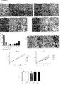


Classifications
-
- A—HUMAN NECESSITIES
- A61—MEDICAL OR VETERINARY SCIENCE; HYGIENE
- A61K—PREPARATIONS FOR MEDICAL, DENTAL OR TOILETRY PURPOSES
- A61K38/00—Medicinal preparations containing peptides
- A61K38/04—Peptides having up to 20 amino acids in a fully defined sequence; Derivatives thereof
- A61K38/08—Peptides having 5 to 11 amino acids
-
- A—HUMAN NECESSITIES
- A61—MEDICAL OR VETERINARY SCIENCE; HYGIENE
- A61K—PREPARATIONS FOR MEDICAL, DENTAL OR TOILETRY PURPOSES
- A61K38/00—Medicinal preparations containing peptides
- A61K38/16—Peptides having more than 20 amino acids; Gastrins; Somatostatins; Melanotropins; Derivatives thereof
- A61K38/17—Peptides having more than 20 amino acids; Gastrins; Somatostatins; Melanotropins; Derivatives thereof from animals; from humans
- A61K38/1703—Peptides having more than 20 amino acids; Gastrins; Somatostatins; Melanotropins; Derivatives thereof from animals; from humans from vertebrates
- A61K38/1709—Peptides having more than 20 amino acids; Gastrins; Somatostatins; Melanotropins; Derivatives thereof from animals; from humans from vertebrates from mammals
-
- A—HUMAN NECESSITIES
- A61—MEDICAL OR VETERINARY SCIENCE; HYGIENE
- A61K—PREPARATIONS FOR MEDICAL, DENTAL OR TOILETRY PURPOSES
- A61K39/00—Medicinal preparations containing antigens or antibodies
- A61K39/0005—Vertebrate antigens
- A61K39/0011—Cancer antigens
-
- A—HUMAN NECESSITIES
- A61—MEDICAL OR VETERINARY SCIENCE; HYGIENE
- A61K—PREPARATIONS FOR MEDICAL, DENTAL OR TOILETRY PURPOSES
- A61K39/00—Medicinal preparations containing antigens or antibodies
- A61K39/0005—Vertebrate antigens
- A61K39/0011—Cancer antigens
- A61K39/001102—Receptors, cell surface antigens or cell surface determinants
-
- A—HUMAN NECESSITIES
- A61—MEDICAL OR VETERINARY SCIENCE; HYGIENE
- A61K—PREPARATIONS FOR MEDICAL, DENTAL OR TOILETRY PURPOSES
- A61K39/00—Medicinal preparations containing antigens or antibodies
- A61K39/395—Antibodies; Immunoglobulins; Immune serum, e.g. antilymphocytic serum
-
- A—HUMAN NECESSITIES
- A61—MEDICAL OR VETERINARY SCIENCE; HYGIENE
- A61P—SPECIFIC THERAPEUTIC ACTIVITY OF CHEMICAL COMPOUNDS OR MEDICINAL PREPARATIONS
- A61P1/00—Drugs for disorders of the alimentary tract or the digestive system
- A61P1/04—Drugs for disorders of the alimentary tract or the digestive system for ulcers, gastritis or reflux esophagitis, e.g. antacids, inhibitors of acid secretion, mucosal protectants
-
- A—HUMAN NECESSITIES
- A61—MEDICAL OR VETERINARY SCIENCE; HYGIENE
- A61P—SPECIFIC THERAPEUTIC ACTIVITY OF CHEMICAL COMPOUNDS OR MEDICINAL PREPARATIONS
- A61P1/00—Drugs for disorders of the alimentary tract or the digestive system
- A61P1/16—Drugs for disorders of the alimentary tract or the digestive system for liver or gallbladder disorders, e.g. hepatoprotective agents, cholagogues, litholytics
-
- A—HUMAN NECESSITIES
- A61—MEDICAL OR VETERINARY SCIENCE; HYGIENE
- A61P—SPECIFIC THERAPEUTIC ACTIVITY OF CHEMICAL COMPOUNDS OR MEDICINAL PREPARATIONS
- A61P1/00—Drugs for disorders of the alimentary tract or the digestive system
- A61P1/18—Drugs for disorders of the alimentary tract or the digestive system for pancreatic disorders, e.g. pancreatic enzymes
-
- A—HUMAN NECESSITIES
- A61—MEDICAL OR VETERINARY SCIENCE; HYGIENE
- A61P—SPECIFIC THERAPEUTIC ACTIVITY OF CHEMICAL COMPOUNDS OR MEDICINAL PREPARATIONS
- A61P11/00—Drugs for disorders of the respiratory system
-
- A—HUMAN NECESSITIES
- A61—MEDICAL OR VETERINARY SCIENCE; HYGIENE
- A61P—SPECIFIC THERAPEUTIC ACTIVITY OF CHEMICAL COMPOUNDS OR MEDICINAL PREPARATIONS
- A61P13/00—Drugs for disorders of the urinary system
- A61P13/08—Drugs for disorders of the urinary system of the prostate
-
- A—HUMAN NECESSITIES
- A61—MEDICAL OR VETERINARY SCIENCE; HYGIENE
- A61P—SPECIFIC THERAPEUTIC ACTIVITY OF CHEMICAL COMPOUNDS OR MEDICINAL PREPARATIONS
- A61P15/00—Drugs for genital or sexual disorders; Contraceptives
-
- A—HUMAN NECESSITIES
- A61—MEDICAL OR VETERINARY SCIENCE; HYGIENE
- A61P—SPECIFIC THERAPEUTIC ACTIVITY OF CHEMICAL COMPOUNDS OR MEDICINAL PREPARATIONS
- A61P17/00—Drugs for dermatological disorders
-
- A—HUMAN NECESSITIES
- A61—MEDICAL OR VETERINARY SCIENCE; HYGIENE
- A61P—SPECIFIC THERAPEUTIC ACTIVITY OF CHEMICAL COMPOUNDS OR MEDICINAL PREPARATIONS
- A61P19/00—Drugs for skeletal disorders
-
- A—HUMAN NECESSITIES
- A61—MEDICAL OR VETERINARY SCIENCE; HYGIENE
- A61P—SPECIFIC THERAPEUTIC ACTIVITY OF CHEMICAL COMPOUNDS OR MEDICINAL PREPARATIONS
- A61P25/00—Drugs for disorders of the nervous system
-
- A—HUMAN NECESSITIES
- A61—MEDICAL OR VETERINARY SCIENCE; HYGIENE
- A61P—SPECIFIC THERAPEUTIC ACTIVITY OF CHEMICAL COMPOUNDS OR MEDICINAL PREPARATIONS
- A61P35/00—Antineoplastic agents
-
- A—HUMAN NECESSITIES
- A61—MEDICAL OR VETERINARY SCIENCE; HYGIENE
- A61P—SPECIFIC THERAPEUTIC ACTIVITY OF CHEMICAL COMPOUNDS OR MEDICINAL PREPARATIONS
- A61P43/00—Drugs for specific purposes, not provided for in groups A61P1/00-A61P41/00
-
- C—CHEMISTRY; METALLURGY
- C07—ORGANIC CHEMISTRY
- C07K—PEPTIDES
- C07K7/00—Peptides having 5 to 20 amino acids in a fully defined sequence; Derivatives thereof
- C07K7/04—Linear peptides containing only normal peptide links
- C07K7/06—Linear peptides containing only normal peptide links having 5 to 11 amino acids
Landscapes
- Health & Medical Sciences (AREA)
- Life Sciences & Earth Sciences (AREA)
- Chemical & Material Sciences (AREA)
- Medicinal Chemistry (AREA)
- Pharmacology & Pharmacy (AREA)
- Animal Behavior & Ethology (AREA)
- General Health & Medical Sciences (AREA)
- Public Health (AREA)
- Veterinary Medicine (AREA)
- Engineering & Computer Science (AREA)
- Bioinformatics & Cheminformatics (AREA)
- Chemical Kinetics & Catalysis (AREA)
- Organic Chemistry (AREA)
- Nuclear Medicine, Radiotherapy & Molecular Imaging (AREA)
- General Chemical & Material Sciences (AREA)
- Epidemiology (AREA)
- Immunology (AREA)
- Mycology (AREA)
- Microbiology (AREA)
- Oncology (AREA)
- Gastroenterology & Hepatology (AREA)
- Proteomics, Peptides & Aminoacids (AREA)
- Cell Biology (AREA)
- Zoology (AREA)
- Marine Sciences & Fisheries (AREA)
- Physical Education & Sports Medicine (AREA)
- Neurosurgery (AREA)
- Dermatology (AREA)
- Endocrinology (AREA)
- Reproductive Health (AREA)
- Urology & Nephrology (AREA)
- Pulmonology (AREA)
- Neurology (AREA)
- Biomedical Technology (AREA)
- Medicines That Contain Protein Lipid Enzymes And Other Medicines (AREA)
- Pharmaceuticals Containing Other Organic And Inorganic Compounds (AREA)
- Peptides Or Proteins (AREA)
- Medicines Containing Antibodies Or Antigens For Use As Internal Diagnostic Agents (AREA)
Description
(関連出願の相互参照)
本願は、2014年12月10日に出願された「METHODS AND COMPOSITIONS FOR REDUCING GROWTH,MIGRATION AND INVASIVENESS OF BRAIN CANCER STEM CELLS AND IMPROVING SURVIVAL OF PATIENTS WITH BRAIN TUMORS」と題する米国仮特許出願第62/090,029号に対する優先権の利益を主張するものであり、上記出願の内容は、その全体が参照により本明細書に組み込まれる。
(Cross-reference of related applications)
This application is filed on December 10, 2014, "METHODS AND COMPOSITIONS FOR REDUCING GROWTH, MIGRATION AND INVASIVEENSS OF BRAIN CANCER STEM CELLS AND IMPROVING Claiming the benefit of priority to the issue, the content of the above application is incorporated herein by reference in its entirety.
記載される発明は、腫瘍の成長、遊走、浸潤またはその組合せを低下させ対象の生存率を対照に比して改善する方法および組成物に関する。 The invention described relates to a method and composition that reduces tumor growth, migration, infiltration or a combination thereof and improves the survival rate of a subject as compared to a control.
悪性腫瘍/癌
癌は、細胞数(増殖)または細胞サイズの点で細胞が異常に成長し、身体の他の部位へ浸潤または拡散する(転移)可能性のある疾患である。癌細胞増殖はよく知られているが、転移を引き起こす機序(1つまたは複数)についてはよく知られていない。癌はゲノムが変化する疾患であり、具体的には、DNA配列の変化、コピー数の異常、染色体再配列およびDNAメチル化の修飾がヒト悪性腫瘍の発生および進行を引き起こす。(The Cancer Genome Atlas Network(TCGA),Nature 2008,455(23):1061−68。)
Malignancies / cancer Cancer is a disease cell number (proliferation) or cells abnormally grow in terms of cell size, invasion or spread to other parts of the body (metastasis) potential. Cancer cell proliferation is well known, but the mechanism (s) that cause metastasis is not well known. Cancer is a genome-altering disease, in which changes in DNA sequences, copy number abnormalities, chromosomal rearrangements and DNA methylation modifications cause the development and progression of human malignancies. (The Cancer Genome Atlas Network (TCGA), Nature 2008,455 (23): 1061-68.)
神経膠腫
神経膠腫は、脳または脊椎のグリア細胞から生じる腫瘍の一種である。神経膠腫が最もよくみられる部位は脳である。神経膠腫は、脳および中枢神経系の腫瘍全体の約30%を占め、悪性脳腫瘍全体の80%を占めている。
Glioma Glioma is a type of tumor that arises from glial cells in the brain or spine. The most common site for glioma is the brain. Gliomas make up about 30% of all tumors of the brain and central nervous system and 80% of all malignant brain tumors.
中枢神経系に最も多く存在する細胞型であるグリア細胞は、ニューロンを取り囲み、ニューロンを支持および絶縁する支持細胞である。グリア細胞はニューロンとは異なり、電気インパルスを伝導しない。中枢神経系のグリア細胞には2種類の主要なクラス、すなわち星状膠細胞および乏突起膠細胞がある(Kandel ERら,Principles of Neural Science,第4版,McGraw−Hill New York(2000),第2章,pp.20−21)。
The most abundant cell type in the central nervous system, glial cells, are supporting cells that surround, support and insulate neurons. Glial cells, unlike neurons, do not conduct electrical impulses. There are two major classes of glial cells in the central nervous system: astrocytes and oligodendrocytes (Kandel ER et al., Principles of Neuronal Science, 4th Edition, McGraw-Hill New York (2000),
神経膠腫は、起源が必ずしも共通するわけではないが組織学的特徴が同じである細胞の特定のタイプに従って命名される。主な神経膠腫のタイプには、星状細胞腫(星状膠細胞と組織学的特徴が同じである)(例えば、多形神経膠芽腫は悪性星状細胞腫の1つであり、成人に最もよくみられる原発性脳腫瘍である);乏突起神経膠腫(乏突起膠細胞と組織学的特徴が同じである);脳幹神経膠腫(脳幹に発生する神経膠腫である);視神経神経膠腫(視神経の中または周囲に発生する神経膠腫である);ならびに混合神経膠腫、例えば乏突起膠細胞および星状膠細胞と組織学的特徴が同じ細胞を含む乏突起星細胞腫などがある。 Gliomas are named according to a particular type of cell that does not necessarily have a common origin but has the same histological characteristics. The main types of glioma are astrocytomas (which have the same histological characteristics as astrocytomas) (eg, polyglioma is one of the malignant gliomas, The most common primary brain tumor in adults); oligodendrogliomas (having the same histological characteristics as oligodendroglioma cells); brain stem gliomas (gliomas that occur in the brain stem); Glioblastoma (a glioma that develops in or around the optic nerve); and mixed gliomas, such as oligodendroglioma and astrocytoma containing cells with the same histological characteristics as stellate glioma. There is a swelling.
神経膠腫は、腫瘍の病理学的評価によって判定されるそのグレードに従ってさらに分類される。用いられている多数の分類体系のうち、神経膠腫に最もよく用いられるのが世界保健機関(WHO)の分類法であり、この分類法では、腫瘍をグレートI〜IVに分類する(Louis DNら,Acta Neuropathol,2007,114(2):97−109)。 Gliomas are further classified according to their grade as determined by the pathological assessment of the tumor. Of the many classification systems used, the most commonly used for glioma is the World Health Organization (WHO) classification, which classifies tumors into Great I-IV (Louis DN). Et al., Acta Neuropathol, 2007, 114 (2): 97-109).
グレードIの腫瘍は、成長が遅く非悪性であり、生存期間が長くなる(例えば、毛様細胞性星細胞腫)。 Grade I tumors are slow-growing, non-malignant, and have a long survival (eg, pilocytic astrocytoma).
グレードIIの腫瘍は、成長は比較的遅いが、さらに高いグレードの腫瘍として再発することがある。非悪性であることも悪性であることもある(例えば、びまん性星細胞腫)。 Grade II tumors grow relatively slowly, but may recur as higher grade tumors. It can be non-malignant or malignant (eg, diffuse astrocytoma).
グレードIIIの腫瘍は悪性であり、さらに高いグレードの腫瘍として再発することが多い(例えば、未分化星状細胞腫)。 Grade III tumors are malignant and often recur as higher grade tumors (eg, undifferentiated astrocytomas).
グレードIVの腫瘍は増殖速度が速く、極めて侵襲性の高い悪性腫瘍である(例えば、膠芽腫、巨細胞膠芽腫および膠肉腫)。 Grade IV tumors are fast-growing, highly invasive malignancies (eg, glioblastoma, giant cell glioblastoma, and glioblastoma).
髄芽腫は、小児に最もよく発生するタイプの原発性脳腫瘍である。「髄芽腫」という用語は、小児の小脳にみられる一連の腫瘍を指す。髄芽腫(WHOグレードIVの腫瘍)は最初、神経膠腫に分類されていたが、現在は胚芽腫と呼ばれている。この腫瘍は、小脳の外顆粒層の前駆細胞の悪性形質転換によって発生すると考えられており、頭蓋内腫瘍全体の約7〜8%、小児脳腫瘍の30%を占め、膠腫とは逆にニューロン分化を主な特徴とする。 Medulloblastoma is the most common type of primary brain tumor in children. The term "medulloblastoma" refers to a series of tumors found in the cerebellum of children. Medulloblastoma (a WHO grade IV tumor) was initially classified as a glioma, but is now called a germinoma. This tumor is thought to be caused by malignant transformation of progenitor cells in the outer granules of the cerebellum, accounting for about 7-8% of all intracranial tumors and 30% of pediatric brain tumors, and neurons as opposed to gliomas. The main feature is differentiation.
膠芽腫
WHOグレードIV、分類名「膠芽腫」の悪性神経膠腫である多形神経膠芽腫(GBM)は、成人に最もよくみられ最も侵襲性の高い原発性脳腫瘍である。GBMはグリア細胞から発生し、びまん性星細胞系腫瘍全体の40%〜60%、頭蓋内腫瘍性病変全体の10%〜15%を占める。この腫瘍の生物学的特徴の代表的なものとして、顕著な増殖性、活発な浸潤性および旺盛な血管新生が挙げられる。(Nakada,M.ら,Cancers,2011,3:3242−3278)。GBMは、分化に乏しい腫瘍性星状膠細胞からなる。GBMの組織病理学的診断には微小血管の増殖および/または壊死の存在が不可欠である。
Glioblastoma WHO grade IV, a malignant glioma of the classification name "glioblastoma", polymorphoglioma (GBM) is the most common and most invasive primary brain tumor in adults. GBM originates from glial cells and accounts for 40% to 60% of all diffuse astrocyte tumors and 10% to 15% of all intracranial neoplastic lesions. Typical biological features of this tumor include prominent proliferative, active infiltrative and vigorous angiogenesis. (Nakada, M. et al., Cancers, 2011, 3: 3242-3278). GBM consists of poorly differentiated neoplastic astrocytes. The presence of microvessel proliferation and / or necrosis is essential for the histopathological diagnosis of GBM.
GBMは、ヒト癌のうち最も侵襲性の高いものの1つであり、複数の因子が複雑に絡み合っていること、すなわち:従来の治療法に対する腫瘍細胞の抵抗性が極めて高いこと;脳が従来の治療法による損傷を受けやすいこと;脳そのものの修復能が極めて低いこと;および血液脳関門を通過して腫瘍に作用することができない薬物が多いことにより、治療するのが極めて困難である。 GBM is one of the most invasive of human cancers, with multiple factors intricately intertwined: the tumor cells are extremely resistant to conventional treatments; the brain is conventional. It is extremely difficult to treat due to its susceptibility to treatment damage; its extremely poor ability to repair the brain itself; and the large number of drugs that cannot cross the blood-brain barrier and act on tumors.
治療では放射線照射、外科手術およびメチル化剤のテモゾロミドによる化学療法を実施し得る(P.J.Noughtonら,Clin.Cancer Res.(2000)6:4110−4118)が、外科手術療法、放射線療法および化学療法を数十年間実施しても、GBMの生存期間に目覚ましい変化をみるに至っていない。集学的療法を実施したGBM患者の臨床試験集団の生存期間中央値は約12〜15か月であり、生存期間が36か月を超えた患者はわずか3%〜5%である。(McNamara.M.G.ら,Cancers,2013,5:1103−1119)。 Treatment may include irradiation, surgery and chemotherapy with the methylating agent temozolomide (PJ Noughton et al., Clin. Cancer Res. (2000) 6: 4110-4118), but surgical treatment, radiotherapy. And even after decades of chemotherapy, no significant changes have been seen in GBM survival. The median survival of clinical trial populations of GBM patients who received multidisciplinary therapy was approximately 12 to 15 months, with only 3% to 5% of patients surviving more than 36 months. (McNamara.MG et al., Cancers, 2013, 5: 1103-1119).
臨床経験に基づき、GBMには原発性膠芽腫および続発性膠芽腫の2つのサブグループが確立されているが、この2つのグループを組織学的に区別することはできない。原発性膠芽腫は生検症例または切除症例の90%超を占め、低悪性度疾患の先行既往がなくde novoで発生するのに対し、続発性膠芽腫は既に診断された低悪性度神経膠腫から進行する。 Based on clinical experience, two subgroups of primary glioblastoma and secondary glioblastoma have been established in GBM, but the two groups cannot be histologically distinguished. Primary glioblastoma accounts for more than 90% of biopsied or resected cases and occurs in de novo with no prior history of low-grade disease, whereas secondary glioblastoma has already been diagnosed with low-grade glioblastoma. It progresses from glioblastoma.
The Cancer Genome Atlas(TCGA)Networkによって記載されている遺伝子発現に基づく分子分類によれば、多形神経膠芽腫には、4つの異なる分子学的サブタイプ、すなわち、典型的、前神経性、間葉系および神経性がある(TCGA Research Network,Nature,2008,455:1061−1068)。 According to the molecular classification based on gene expression described by The Cancer Genome Atlas (TCGA) Network, polymorphic glioblastoma has four different molecular subtypes: typical, proneurotic, It is mesenchymal and neurogenic (TCGA Research Network, Nature, 2008, 455: 1061-168).
典型的GBM腫瘍は、一部の細胞の表面にあるタンパク質の1つであって、上皮成長因子が結合すると、細胞数の増大(増殖)を継続させるシグナルを送るタンパク質である上皮成長因子受容体(EGFR)の増幅に異常を来たすほか、その濃度が高くなることを特徴とする。これ以外の3つのGBMサブタイプでは、EGFRに異常がみられる割合ははるかに低い。TP53遺伝子は、通常であれば腫瘍成長を低下させる腫瘍タンパク質p53をコードする。TP53は、典型的GBM腫瘍サブタイプで変異することはまれであるが、これ以外のGBMのサブタイプでは最も変異の頻度が高い遺伝子である。 A typical GBM tumor is one of the proteins on the surface of some cells, the epidermal growth factor receptor, which is a protein that, when bound by epidermal growth factor, sends a signal that continues to increase (proliferate) the number of cells. In addition to causing abnormalities in the amplification of (EGFR), it is characterized by an increase in its concentration. The other three GBM subtypes have a much lower rate of abnormal EGFR. The TP53 gene encodes the tumor protein p53, which normally reduces tumor growth. TP53 is rarely mutated in typical GBM tumor subtypes, but is the most frequently mutated gene in other GBM subtypes.
前神経性GBM腫瘍は、血小板由来成長因子受容体A(PDGFRA)の変化およびIDH1の点変異を特徴とする。イソクエン酸デヒドロゲナーゼ1をコードする遺伝子IDH1に変異があると、細胞増殖異常の一因となり得るタンパク質をコードする。PDGFRAは細胞増殖、細胞遊走および血管新生に重要な役割を果たし、ほかにも、変異して異常に多量に発現することがわかっている。PDGFRAの変化がみられるのは前神経性腫瘍に限られ、他のサブタイプにはみられない。PDGFRAが変化すると、そのタンパク質が過剰に産生され、制御できない腫瘍成長をもたらし得る。このサブタイプの患者は、他のサブタイプの患者よりも若く、生存期間が長くなる傾向がある。
Proneurological GBM tumors are characterized by changes in platelet-derived growth factor receptor A (PDGFRA) and point mutations in IDH1. Mutations in the gene IDH1 encoding
間葉系サブグループは、神経線維腫症1型(NF1)腫瘍抑制遺伝子の変異の頻度が最も高い。間葉系サブグループはこのほか、腫瘍抑制遺伝子PTEN(ホスファターゼ・テンシン・ホモログ)およびTP53の変異頻度が高い。PTENタンパク質は腫瘍抑制因子として作用し、細胞分裂の周期の調節に働く。 The mesenchymal subgroup has the highest frequency of mutations in the neurofibromatosis type 1 (NF1) tumor suppressor gene. The mesenchymal subgroup also has a high frequency of mutations in the tumor suppressor genes PTEN (phosphatase, tencin, homologue) and TP53. The PTEN protein acts as a tumor suppressor and regulates the cycle of cell division.
神経性サブグループは他のグループと同じ遺伝子の多くに変異がみられるが、変異率が他のグループよりも際立って高いわけでも低いわけでもない。神経性グループは、脳の正常な非癌性の神経細胞またはニューロンの典型的なマーカーでもある複数のマーカー、例えばNEFL、GABRA1、SYT1およびSLC12A5などを発現するのが特徴である。 The neurological subgroup has mutations in many of the same genes as the other groups, but the mutation rate is neither significantly higher nor lower than the other groups. Nervous groups are characterized by expressing multiple markers that are also typical markers of normal non-cancerous neurons or neurons in the brain, such as NEFL, GABRA1, SYT1 and SLC12A5.
上に挙げた多形膠芽腫の分子学的サブタイプは、その臨床経過および治療効果に差があるものと考えられる。例えば、積極的化学療法および放射線療法に対する応答はサブタイプによって異なり、サブタイプ間の差は約50%である。各サブタイプの病的状態が様々なタイプの細胞から始まり、このことが治療に対する応答に差がみられる理由となっている可能性が示唆されている。最も大きな便益がみられたのは典型的サブタイプおよび間葉系サブタイプであり、集中的治療によって死亡率の大幅な減少がみられたほか、神経性サブタイプにも何らかの効果があることがうかがえたが、前神経性サブタイプは、従来の化学療法または化学放射線療法を含めた集中的治療に対する応答は芳しいものではなかった。(Verhaak,RGら,Cancer Cell,2010,17(1):98−110。) The molecular subtypes of glioblastoma polymorphisms listed above may differ in their clinical course and therapeutic efficacy. For example, the response to aggressive chemotherapy and radiation therapy depends on the subtype, with a difference of about 50% between the subtypes. It has been suggested that each subtype of pathological condition begins with different types of cells, which may be the reason for the different responses to treatment. The greatest benefits were seen in the typical and mesenchymal subtypes, where intensive treatment significantly reduced mortality and may have some effect on the neurological subtype. However, the anterior neurological subtypes did not respond well to intensive treatment, including conventional chemotherapy or chemoradiotherapy. (Verhaak, RG et al., Cancer Cell, 2010, 17 (1): 98-110.)

これとは逆に、ヒト悪性神経膠腫では、間葉系の表現型が腫瘍侵襲性の顕著な特徴となっている。間葉系サブクラスおよび典型的サブクラスは前神経性腫瘍よりも予後が不良であり、このことは、一部の前神経性腫瘍には、好ましい予後予測因子であるIDH1遺伝子および神経膠腫CpGアイランドメチル化因子表現型(G−CIMP)の両方に変異がみられることと関係があると思われる(Verhaak,RGら,Cancer Cell,2010,17(1):98−110)。 Conversely, in human malignant gliomas, the mesenchymal phenotype is a hallmark of tumor invasiveness. The mesenchymal and typical subclasses have a poorer prognosis than anterior nerve tumors, which is a preferred prognostic predictor for some anterior nerve tumors: the IDH1 gene and glioma CpG island methyl. It appears to be related to the presence of mutations in both the factoric phenotype (G-CIMP) (Verhaak, RG et al., Cancer Cell, 2010, 17 (1): 98-110).
GBMの侵襲性の増殖、活発な浸潤性および血管新生
GBMの侵襲性の増殖、活発な浸潤性および血管新生は主として、腫瘍のシグナル伝達経路の大幅な調節解除に起因する。
Invasive proliferation of GBM, active infiltration and angiogenesis Invasive proliferation of GBM, active infiltration and angiogenesis are primarily due to significant deregulation of tumor signaling pathways.
GBM症例ではほぼ例外なく、組織病理学的に検出可能な有糸分裂を伴う増殖活性が顕著である。神経膠腫で調節解除されている頻度の高い増殖シグナル伝達カスケードのうち最も重要な2つにものは、のちに考察するPI3K/Akt/mTOR経路およびRas/MEK/MAPK経路である。 Almost without exception in GBM cases, histopathologically detectable proliferative activity with mitosis is prominent. Two of the most important proliferative signaling cascades that are frequently deregulated in gliomas are the PI3K / Akt / mTOR pathway and the Ras / MEK / MAPK pathway, which will be discussed later.
遍在する血管新生がGBMの際立った特徴である。血管新生の程度は、神経膠腫の悪性度、腫瘍侵襲性および臨床予後と有意に相関する。血管新生を促進する経路には、血管内皮成長因子(VEGF)などの血管新生因子が発現し、次いでこれが血管内皮細胞上のそのコグネート受容体に結合することによって開始される一続きの協働事象が含まれる。 Angiogenesis that is ubiquitous is a distinguishing feature of GBM. The degree of angiogenesis significantly correlates with glioma malignancy, tumor invasiveness and clinical prognosis. The pathways that promote angiogenesis are a series of collaborative events initiated by the expression of angiogenic factors such as vascular endothelial growth factor (VEGF), which then bind to its cognate receptor on vascular endothelial cells. Is included.
GBMは浸潤性が高い。神経膠腫浸潤は、(1)本来の部位からの離脱;(2)細胞外マトリックス(ECM)への接着;(3)ECMのリモデリング;および(4)細胞遊走を含む複雑な過程である。遊走神経膠腫細胞は、白質の血管、樹状突起および線維に沿って移動する傾向がある。これらの特徴から、GBMにはその浸潤性を媒介する特有の生物学的機序があることが示唆される。ヒト神経膠腫の高い浸潤性はCNS発生時のグリア前駆細胞の遊走挙動にもみられることから、神経堤の細胞遊走に寄与する活性化因子、受容体およびシグナル伝達タンパク質が神経膠腫浸潤に中心的役割を果たしていることが示唆される。チロシンキナーゼ受容体(RTK)、インテグリン、CD44およびGタンパク質共役受容体(GPCR)などの数種類の膜型タンパク質のほか、PI3K/AktならびにRac1、cdc42およびRhoAなどの小型のGTPアーゼを含めた細胞内シグナル伝達分子によって浸潤シグナル伝達が誘導されることを示す研究結果が集まりつつある。CD44抗原は、細胞間相互作用、細胞接着および細胞遊走に関与する細胞表面糖タンパク質である。 GBM is highly invasive. Glioma infiltration is a complex process involving (1) withdrawal from the original site; (2) adhesion to extracellular matrix (ECM); (3) remodeling of ECM; and (4) cell migration. .. Migratory glioma cells tend to migrate along white matter blood vessels, dendrites and fibers. These characteristics suggest that GBM has a unique biological mechanism that mediates its infiltration. Since the high invasiveness of human glioma is also observed in the migration behavior of glial precursor cells during CNS development, activators, receptors and signaling proteins that contribute to neural crest cell migration are central to glioma invasion. It is suggested that it plays a key role. Intracellular including several membrane-type proteins such as tyrosine kinase receptor (RTK), integrin, CD44 and G protein-coupled receptor (GPCR), as well as small GTPases such as PI3K / Akt and Rac1, cdc42 and RhoA. Studies are gathering to show that signaling molecules induce integrin signaling. The CD44 antigen is a cell surface glycoprotein involved in cell-cell interactions, cell adhesion and cell migration.
EGFRとEphA2(エフリンA型受容体2)RTKはGBMに発現し、細胞表面に共局在する。EphA2のリン酸化はEGFR活性に依存し、EphA2のダウンレギュレーションはEGFRのリン酸化、下流のシグナル伝達およびEGF誘導性の細胞生存を阻害する(Ramnarain,D.B.;Cancer Res.2006,66,867−874)。 EGFR and EphA2 (ephrin type A receptor 2) RTK are expressed on GBM and co-localize on the cell surface. Phosphorylation of EphA2 depends on EGFR activity, and downregulation of EphA2 inhibits EGFR phosphorylation, downstream signaling and EGF-induced cell survival (Ramnarain, DB; Cancer Res. 2006, 66, 867-874).
主要な神経膠腫シグナル伝達経路
神経膠腫と関係のある主要なシグナル伝達経路が数種類あることがわかっている(Nakada,M.ら,Cancers,2011,3:3242−3278)。
Major glioma signaling pathways It is known that there are several major signaling pathways associated with glioma (Nakada, M. et al., Cancers, 2011, 3: 3242-3278).
1.受容体チロシンキナーゼ経路(RTK/PI3K/Akt/mTOR経路)。
RTK/P13K/Akt経路は、増殖、成長、アポトーシスおよび細胞骨格再構成など様々な基本的な細胞プロセスを調節している。この経路には、受容体チロシンキナーゼ(RTK)、例えば上皮成長因子受容体(EGFR)、血小板由来成長因子受容体(PDGFR)および血管内皮成長因子受容体(VEGFR)などのほかにも、腫瘍抑制因子であるプロテインホスファターゼ、例えばホスファターゼ・テンシン・ホモログ(PTEN)ならびにプロテインキナーゼのPI3K、AktおよびmTORが関与している。受容体チロシンキナーゼ経路(RTK/PI3K/Akt/mTOR経路)を図1に示す。
1. 1. Receptor tyrosine kinase pathway (RTK / PI3K / Akt / mTOR pathway).
The RTK / P13K / Akt pathway regulates a variety of basic cellular processes such as proliferation, growth, apoptosis and cytoskeletal rearrangement. This pathway includes receptor tyrosine kinase (RTK), such as epidermal growth factor receptor (EGFR), platelet-derived growth factor receptor (PDGFR) and vascular endothelial growth factor receptor (VEGFR), as well as tumor suppression. Factors such as protein phosphatase, such as phosphatase tencin homologue (PTEN) and protein kinases PI3K, Akt and mTOR are involved. The receptor tyrosine kinase pathway (RTK / PI3K / Akt / mTOR pathway) is shown in FIG.
EGFR遺伝子の増幅はGBMで最も頻度の高い異常(約40%)である。EGFRはErbB受容体ファミリーの膜貫通型糖タンパク質のメンバーである。GBMでは、EGFRが過剰発現によって調節不全の状態にあり、この調節不全はEGFR遺伝子の増幅またはリガンド依存性シグナル伝達を引き起こすEGFRvIIIなどの変異の活性化が原因で起こる。EGFR異常は典型的サブタイプのGBMと関係があることがわかっている(TCGA Research Network,Nature 455:1061−68;Verhaak,Roel G.W.ら,Cancer Cell,2010,17:98−110)。EGFRの異常はGBMの侵襲性の増大と関係があることが示唆されている(Nakada,M.ら,Cancers,2011,3:3242−3278)が、臨床試験ではEGFR阻害剤(例えば、ゲフィニチブ(Gefinitib)、エルロチニブ)がGBM患者に臨床効果を引き起こしたという報告はない(Rich,J.N.ら,N.Engl.J.Med.2004,351,1260−1261;Haas−Kogan,D.A.ら,J.Natl.Cancer Inst.2005,97,880−887;van den Bent,M.J.ら,J.Clin.Oncol.2009,27,1268−1274)。 Amplification of the EGFR gene is the most common abnormality (about 40%) in GBM. EGFR is a member of the ErbB receptor family of transmembrane glycoproteins. In GBM, EGFR is dysregulated by overexpression, which is caused by activation of mutations such as EGFRvIII that trigger amplification of the EGFR gene or ligand-gated signaling. EGFR abnormalities have been shown to be associated with a typical subtype of GBM (TCGA Research Network, Nature 455: 1061-68; Verhaak, Roel GW et al., Cancer Cell, 2010, 17: 98-110). .. EGFR abnormalities have been suggested to be associated with increased invasiveness of GBM (Nakada, M. et al., Cancers, 2011, 3: 3242-3278), but in clinical trials EGFR inhibitors (eg, gefitinib (eg, gefitinib) Gefitinib), erlotinib) has not been reported to cause clinical effects in GBM patients (Rich, JN et al., N. Engl. J. Med. 2004, 351, 1260-1261; Haas-Kogan, DA). Et al., J. Natl. Cancer Inst. 2005, 97, 880-887; van den Bent, MJ et al., J. Clin. Oncol. 2009, 27, 1268-1274).
星状細胞腫瘍では、いずれのグレードにも血小板由来成長因子受容体(PDGFR)、特にPDGFR−αおよび血小板由来成長因子(PDGF)の過剰発現が認められ、悪性化との相関が示唆されている(Nakada,M.ら,Cancers,2011,3:3242−3278)。TCGA分類法によれば、PDGFRAの増幅(14%)およびIDH1の変異が前神経性サブタイプのGBMの主要な特徴である(TCGA Research Network,Nature 455:1061−68;Verhaak,R.G.ら,Cancer Cell,2010,17:98−110)。この分子とGBMとの間には深い関係があるものの、イマチニブを用いた抗PDGFR療法で得られた臨床効果はごくわずかである(Reardon,D.A.ら,J.Clin.Oncol.2005,23,9359−9368;Reardon,D.A.ら,Br.J.Cancer 2009,101,1995−2004)。
In astrocyte tumors, overexpression of platelet-derived growth factor receptor (PDGFR), especially PDGFR-α and platelet-derived growth factor (PDGF), was observed in all grades, suggesting a correlation with malignant transformation. (Nakada, M. et al., Cancers, 2011, 3: 3242-3278). According to the TCGA classification, PDGFRA amplification (14%) and IDH1 mutations are key features of the anterior neural subtype GBM (TCGA Research Network, Nature 455: 1061-68; Verhaak, RG. Et al., Cancer Cell, 2010, 17: 98-110). Although there is a deep relationship between this molecule and GBM, the clinical effect obtained by anti-PDGFR therapy with imatinib is negligible (Reardon, DA et al., J. Clin. Oncol. 2005, 23, 9359-9368; Realdon, DA et al.,
PI3Kは広く発現する脂質キナーゼであり、多様な生物学的機能を促進する。PI3KとRTKが結合すると、ホスファチジルイノシトール3,4,5−三リン酸(PiP3)および3−ホスホイノシチド依存性プロテインキナーゼ−1(PDK1)を介してAktが活性化され、これが細胞の生存、増殖および遊走性を含めた複数の基本的な細胞プロセスに影響を及ぼす。GBMの統合的なゲノム分類によれば、PI3K変異(15%)は前神経性サブタイプと関係がある(TCGA Research Network,Nature,2008,455:1061−68;Verhaak,R.G.ら,Cancer Cell,2010,17:98−110)。
PI3K is a widely expressed lipid kinase that promotes diverse biological functions. Binding of PI3K and RTK activates Akt via
PTENは、PI3K機能に拮抗することによってRTK/PI3K/Akt経路を負に調節しているため、PTEN活性が低下するとRTK/PI3K/Akt経路が活性化され得る。PTENのホモ接合性欠失または変異はGBMによくみられる特徴(40%)であり、RTK/PI3K/Akt経路の構成的活性化をもたらす。TCGAの研究によれば、PTENの欠失は典型的サブタイプおよび間葉系サブタイプの両方のGBMと関係がある(TCGA Research Network,Nature 455(23):1061−68)。 Since PTEN negatively regulates the RTK / PI3K / Akt pathway by antagonizing PI3K function, the RTK / PI3K / Akt pathway can be activated when PTEN activity decreases. Homozygous deletions or mutations in PTEN are a common feature (40%) in GBM and result in constitutive activation of the RTK / PI3K / Akt pathway. According to TCGA studies, PTEN deletions are associated with GBM in both typical and mesenchymal subtypes (TCGA Research Network, Nature 455 (23): 1061-68).
Aktは、細胞の成長、増殖およびアポトーシスを調節するSTK(セリン/トレオニン特異的プロテインキナーゼ)である。ヒトGBMの約80%にAktの活性化が報告されており、GBMの88%にRTK/PI3K/Aktシグナル伝達の異常が認められることと関係があるとされている(TCGA Research Network,Nature 455(23):1061−68)。GBMに発癌性のAkt変異は検出されていない。Akt阻害剤であるペリフォシンは、悪性神経膠腫を対象とする臨床評価の段階にある(Nakada,M.ら,Cancers,2011,3:3242−3278)。 Akt is an STK (serine / threonine-specific protein kinase) that regulates cell growth, proliferation and apoptosis. Akt activation has been reported in approximately 80% of human GBMs and is associated with abnormal RTK / PI3K / Akt signaling in 88% of GBMs (TCGA Research Network, Nature 455). (23): 1061-68). No carcinogenic Akt mutation has been detected in GBM. The Akt inhibitor perifosin is in the stage of clinical evaluation in malignant gliomas (Nakada, M. et al., Cancers, 2011, 3: 3242-3278).
2.p14ARF/MDM2/p53経路。
p53遺伝子は、多様な細胞ストレスに応答して細胞周期の停止、細胞死、細胞分化、老化、DNA修復および血管新生を誘導する標的遺伝子を調節するタンパク質をコードする。DNAが損傷を受けると、p53が活性化され、G1期の細胞周期進行の調節因子として機能する遺伝子(p21Waf1/Cip1など)の転写が誘導される。Mouse Double Minute 2ホモログ(MDM2)発癌遺伝子は、p53遺伝子と強固な複合体を形成することによってp53転写活性を阻害し、p53の分解に関与する。p14ARF遺伝子は、MDM2に直接結合しMDM2を介したp53分解を阻害するタンパク質をコードする。次にp14ARFの発現がp53によって負に調節される。したがって、p53遺伝子、MDM2遺伝子またはp14ARF遺伝子のいずれかの発現に異常が生じることによってp14ARF/MDM2/p53の不活性化が起こる。p53経路は続発性GBMの発生に極めて重要な役割を果たしている。p53遺伝子は神経膠腫で最もよく変異が認められるp53経路遺伝子であるが、この経路の他の遺伝子に及ぶ分子レベルの異常についても記載されている。(Nakada,M.ら,Cancers,2011,3:3242−3278)。p14ARF/MDM2/p53経路を図2に示す。
2. p14 ARF / MDM2 / p53 route.
The p53 gene encodes a protein that regulates target genes that induce cell cycle arrest, cell death, cell differentiation, aging, DNA repair and angiogenesis in response to various cell stresses. When DNA is damaged, p53 is activated and transcription of genes (such as p21Waf1 / Cip1) that function as regulators of cell cycle progression in the G1 phase is induced. The
3.RB経路。
RB(網膜芽細胞腫抑制タンパク質)経路は、p53経路と同じように細胞周期の移行および進行を抑制する。RB1(13q14)がコードする107kDaのRB1タンパク質は、細胞周期のG1期からS期への進行を制御する(Serrano,M.ら,Nature,1993,366:704−707)。CDKN2Aタンパク質(すなわち、サイクリン依存性キナーゼ阻害因子2Aであるp16INK4a)はサイクリン依存性キナーゼ4(CDK4)に結合してCDK4/サイクリンD1複合体を阻害し、これにより細胞周期のG1期からS期への移行を阻害する。したがって、RB1、CDK4またはCDKN2Aに異常があると、G1−S期移行の調節異常が起こり得る。しかし、RB経路のみに異常が生じても腫瘍形成を誘導するには不十分である。EGFRの増幅はPI3K成長促進経路を増強し、通常、CDKN2A欠失と関連がある。TCGAの研究によれば、CDKN2A欠失は典型的サブタイプのGBMと関係がある。(Nakada,M.ら,Cancers,2011,3:3242−3278)。RB経路を図2に示す。
3. 3. RB route.
The RB (retinoblastoma inhibitory protein) pathway, like the p53 pathway, suppresses cell cycle transition and progression. The 107 kDa RB1 protein encoded by RB1 (13q14) regulates the progression of the cell cycle from G1 to S phase (Serrano, M. et al., Nature, 1993, 366: 704-707). The CDKN2A protein (ie, p16 INK4a , a cyclin-
4.Ras/MEK/MAPK経路。
RAS(ラット肉腫)タンパク質は、RTKおよび神経線維腫症1型腫瘍抑制遺伝子(NF−1)によって制御されるオン/オフ(RAS−GDP/RAS−GTP)スイッチとして働く。活性化されたRAS(RAS−GTP)は次いでセリン/トレオニンキナーゼであるRAFを活性化させる。RAFは、MEKとも呼ばれるマイトジェン活性化プロテインキナーゼキナーゼ(MAPKK)を活性化させ、次いでこれがMAPKを活性化させる。MAPKの活性化により、Elk1、c−myc、Ets、STAT1/3およびPPARなどの様々な転写因子が活性化される。
4. Ras / MEK / MAPK route.
The RAS (rat sarcoma) protein acts as an on / off (RAS-GDP / RAS-GTP) switch controlled by RTK and the
NF−1腫瘍抑制遺伝子はニューロフィブロミンをコードし、ニューロフィブロミンは主にRASの負の調節因子として機能し、アデニル酸シクラーゼを介する経路およびAkt−mTORを介する経路に何らかの役割を果たしている。NF−1遺伝子がNF−1に関連する神経膠腫のみならず、孤発性の神経膠腫の腫瘍形成にも関与することを示す証拠が集まりつつある。TCGAの予備的研究では、GBMの18%にNF−1の変異/ホモ接合性欠失が確認されている。間葉系GBMは、NF−1遺伝子(37%)、p53遺伝子(32%)およびPTEN遺伝子の不活性化の頻度が高く、積極的化学放射線照射補助療法が奏効する。(Nakada,M.ら,Cancers,2011,3:3242−3278)。Ras/MAPK経路を図3に示す。上に挙げたシグナル伝達経路の全体像を図4に示す。 The NF-1 tumor suppressor gene encodes neurofibromin, which functions primarily as a negative regulator of RAS and plays some role in the adenylate cyclase-mediated and Akt-mTOR-mediated pathways. Evidence is gathering that the NF-1 gene is involved in tumorigenesis of sporadic gliomas as well as NF-1-related gliomas. Preliminary studies of TCGA have confirmed mutations / homozygous deletions of NF-1 in 18% of GBMs. In mesenchymal GBM, the frequency of inactivation of the NF-1 gene (37%), p53 gene (32%) and PTEN gene is high, and aggressive chemoradiation adjuvant therapy is effective. (Nakada, M. et al., Cancers, 2011, 3: 3242-3278). The Ras / MAPK route is shown in FIG. The overall picture of the signal transduction pathways listed above is shown in FIG.
上に挙げたシグナル伝達経路に加えて、それ以外のシグナル伝達経路もGBMのイニシエーション、遊走および浸潤に何らかの役割を果たしている可能性がある。 In addition to the signaling pathways listed above, other signaling pathways may also play a role in GBM initiation, migration and infiltration.
Wnt(wingless関連/マウス乳腺腫瘍ウイルス組込みファミリー)シグナル伝達経路
WNT遺伝子がコードするタンパク質は正常な胚発生に何らかの役割を果たしている。このタンパク質が制御する胚発生過程としては、体軸パターンの形成、細胞運命の決定、細胞増殖および細胞遊走が挙げられる。これらの過程は、骨、心臓および筋肉を含めた重要な組織が正しく形成されるのに必要なものである。Wntシグナル伝達経路は複雑な経路であり、その活性化に異常がみられる悪性腫瘍は実に多岐にわたる。Wntタンパク質は腫瘍形成にも関与することがわかっており、Wnt経路の活性化に異常があると、乳癌、前立腺癌、膠芽腫をはじめとする癌を含めた数種類の癌が発生する。(Camilli,T.C.,Biochem.2010,Pharmacol.80(5):702−711;Polakis P.,Curr Opin Genet Dev 2007:17(1):45−51)。
Wnt (wingless-related / mouse mammary oncovirus virus integration family) signaling pathway The protein encoded by the WNT gene plays a role in normal embryogenesis. Embryogenic processes controlled by this protein include the formation of axial patterns, cell fate determination, cell proliferation and cell migration. These processes are necessary for the proper formation of important tissues, including bones, heart and muscles. The Wnt signaling pathway is a complex pathway, and there are a wide variety of malignant tumors with abnormal activation. The Wnt protein is also known to be involved in tumorigenesis, and abnormal activation of the Wnt pathway causes several types of cancer, including breast cancer, prostate cancer, and glioblastoma. (Camilli, TC, Biochem. 2010, Pharmacol. 80 (5): 702-711; Polakis P., Curr Opin Genet Dev 2007: 17 (1): 45-51).
Wntシグナル伝達経路は、細胞外から細胞表面受容体を介して細胞内にシグナルを伝達するタンパク質のシグナル伝達経路のグループである。Wntシグナル伝達に関与する様々な受容体およびリガンドによって多数の多様なシグナル伝達カスケードが生じる。 The Wnt signaling pathway is a group of protein signaling pathways that transmit signals from the outside of the cell to the inside of the cell via cell surface receptors. The various receptors and ligands involved in Wnt signaling give rise to a large number of diverse signaling cascades.
Wntファミリーのタンパク質は、19の既知のヒトメンバー(Wnt1、Wnt2、Wnt2B、Wnt3、Wnt3A、Wnt4、Wnt5A、Wnt5B、Wnt6、Wnt7A、Wnt7B、Wnt8A、Wnt8B、Wnt9A、Wnt9B、Wnt10A、Wnt10B、Wnt11、Wnt16)からなる。これらの分泌脂質修飾シグナル伝達糖タンパク質は、長さ350〜400アミノ酸であり、アミノ酸同一性が20〜85%であり、23〜24のシステイン残基からなる保存されたパターンを有する。これらのタンパク質に起こる脂質修飾の種類は、23〜24のシステイン残基からなる保存されたパターンに含まれるしステインのパルミトイル化である。パルミトイル化が起こると、Wntタンパク質の細胞膜への標的化が惹起されて同タンパク質が分泌され、脂肪酸の共有結合によりその受容体と結合する。分泌されたWntタンパク質は、その合成後にグリコシル化によって修飾される。Wntシグナル伝達では、これらの分泌タンパク質がリガンドとして働き、パラクリン経路およびオートクリン経路を介して様々なWnt経路を活性化させる。 The Wnt family of proteins includes 19 known human members (Wnt1, Wnt2, Wnt2B, Wnt3, Wnt3A, Wnt4, Wnt5A, Wnt5B, Wnt6, Wnt7A, Wnt7B, Wnt8A, Wnt8B, Wnt9A, Wnt9B, Wnt10A, Wnt10B, Wnt10A, Wnt10B ) Consists of. These secreted lipid-modified signaling glycoproteins are 350-400 amino acids in length, have 20-85% amino acid identity, and have a conserved pattern consisting of 23-24 cysteine residues. The type of lipid modification that occurs in these proteins is palmitoylation of stains contained in a conserved pattern consisting of 23-24 cysteine residues. When palmitoylation occurs, the targeting of the Wnt protein to the cell membrane is triggered, the protein is secreted, and the protein is covalently bound to its receptor. The secreted Wnt protein is modified by glycosylation after its synthesis. In Wnt signaling, these secretory proteins act as ligands, activating various Wnt pathways via the paracrine and autocrine pathways.
Wntタンパク質リガンドがFrizzled(「Fz」)ファミリー受容体に結合することによってWntシグナル伝達経路が活性化されると、その生物学的シグナルが細胞内のタンパク質であるDishevelledに伝達される。Frizzledファミリー受容体はこれまでに、少なくとも10種類のメンバーが同定されており、いずれも細胞外N末端に保存されWntと相互作用するシステインリッチドメイン(CRD)を特徴とする7回膜貫通型タンパク質である。ただし、Wntシグナル伝達を促進するには、Wntタンパク質とFz受容体との間の相互作用と同時に共受容体も必要とされることがある。例としては、低密度リポタンパク質受容体関連タンパク質(Lrp5/6)、受容体チロシンキナーゼ(Ryk)およびRor2が挙げられる。 When the Wnt signaling pathway is activated by binding of the Wnt protein ligand to Frizzled (“Fz”) family receptors, the biological signal is transmitted to the intracellular protein Disheveled. At least 10 members of the Frizzled family of receptors have been identified so far, all of which are 7-transmembrane proteins characterized by a cysteine-rich domain (CRD) that is conserved at the extracellular N-terminus and interacts with Wnt. Is. However, co-receptors may be required at the same time as the interaction between the Wnt protein and the Fz receptor to promote Wnt signaling. Examples include low density lipoprotein receptor-related proteins (Lrp5 / 6), receptor tyrosine kinases (Ryk) and Ror2.
Wntとその受容体および共受容体との相互作用と関係のあるシグナル伝達経路には少なくとも3種類、すなわち古典的Wnt/β−カテニン経路、非古典的(または非正統的)平面内細胞極性(PCP)経路および非古典的(または非正統的)Wnt/Ca2+経路がある。図5に、これらの3種類の代表的なWntシグナル伝達経路を示す。Fz受容体には様々なWntリガンドを識別する能力があり、このため、リガンド/受容体相互作用の性質が活性化を左右するのは、これら3種類の経路のうちの1種類である。(Camilli,T.C.,Biochem.2010,Pharmacol.80(5):702−711)。古典的Wnt経路は遺伝子転写の調節を引き起こし、非古典的平面内細胞極性経路は、細胞の形状に関与する細胞骨格を調節し、非古典的Wnt/カルシウム経路は細胞内のカルシウムを調節する。Wntシグナル伝達経路では、近傍の細胞間のコミュニケーション(パラクリン)または同一細胞のコミュニケーション(オートクリン)が利用される。 There are at least three signaling pathways involved in the interaction of Wnt with its receptors and co-receptors: the classical Wnt / β-catenin pathway, the non-classical (or non-orthodox) in-plane cell polarity ( There are PCP) pathways and non-classical (or non-orthodox) Wnt / Ca 2+ pathways. FIG. 5 shows these three typical Wnt signaling pathways. The Fz receptor has the ability to identify a variety of Wnt ligands, so it is one of these three pathways that the nature of the ligand / receptor interaction determines activation. (Camilli, TC, Biochem. 2010, Pharmacol. 80 (5): 702-711). The classical Wnt pathway causes regulation of gene transcription, the non-classical intraplanar cell polarity pathway regulates the cytoskeleton involved in cell shape, and the non-classical Wnt / calcium pathway regulates intracellular calcium. In the Wnt signaling pathway, communication between neighboring cells (paracrine) or communication between the same cells (autocrine) is utilized.
古典的Wntシグナル伝達経路
古典的Wntシグナル伝達経路は十分に確立されたβ−カテニン依存性シグナル伝達経路であり、重要なメディエーターとしてβ−カテニンを含む。Wntシグナル伝達の不在下では、足場タンパク質であるアキシン(Axin)および腫瘍抑制遺伝子産物であるAPC(腺腫性大腸ポリポーシス)を含めた数種類のタンパク質によって形成される「分解複合体」の中のカゼインキナーゼ1(CK1)およびグリコーゲン合成酵素キナーゼ3ベータ(GSK3β)によってβ−カテニンがリン酸化される。次いで、リン酸化されたβ−カテニンがユビキチン化機構によって認識され、プロテアソームに送られ分解される。Wntがその受容体FzおよびLrp5/6に結合すると、Lrp5/6がリン酸化され、Dishevelledが活性化されて、β−カテニン「分解複合体」の不活性化または分解が起こり、これによりβ−カテニンのリン酸化が低下し、β−カテニンが安定化する。次いで、安定化したβ−カテニンが核に移行し、そこでLef(リンパ系増強転写因子(Lymphoid enhanced transcription factor))およびTcf(T細胞因子)と結合することによって下流の遺伝子発現を調節し、これにより、増殖および腫瘍進行に関与するWnt標的遺伝子の転写が起こる。経路のメンバーのなかには、Wntシグナル伝達とは独立して調節され得るものがいくつかある。例えば、GSK−3βはILK(インテグリン結合キナーゼ)によって阻害され得るメンバーであり、その発現を調節する多数の経路が交わる部分に存在する。古典的Wntタンパク質としては、Wnt1、Wnt2、Wnt3a、Wnt8a、Wnt8b、Wnt10a、Wnt10bが挙げられる(Jiar CH,J Oral Pathol.Med.,2012,41(4):332−339)。
Classical Wnt signaling pathway The classical Wnt signaling pathway is a well-established β-catenin-dependent signaling pathway, including β-catenin as an important mediator. In the absence of Wnt signaling, casein kinase in a "degradation complex" formed by several proteins, including the scaffold protein Axin and the tumor suppressor gene product APC (adenomatous colon polyposis) Β-catenin is phosphorylated by 1 (CK1) and glycogen
癌の古典的Wnt経路
β−カテニンが安定化して分解されなくなり、最終的には核内に蓄積することは、分化が不十分な形態(Endo Kら,Hum Pathol 2003,31(5):558−565)、高い増殖活性(Inagawa S.ら,Clin Cancer Res 2002,8(2):450−456)および予後不良(Wang CMら,Cancer 2001,92(1):136−145)と関係があることがわかっている。β−カテニンの運命、すなわちその蓄積または分解は、多数のタンパク質によって調節されており、そのタンパク質が適切に調節または発現されないと、それが癌におけるβ−カテニン発現の増大の主な原因となる。この調節異常は、シグナル伝達経路の様々なメンバーの変異またはエピジェネティックな事象が原因となって起こり得るものである。Wntそのものが変異することはまれである。しかし、癌では、下流の標的に影響を及ぼす変異が極めて頻繁にみられる。
The classic Wnt pathway of cancer, β-catenin, stabilizes and is no longer degraded, and eventually accumulates in the nucleus, a form of poor differentiation (Endo K et al., Hum Pathol 2003, 31 (5): 558). -565), high proliferative activity (Inagawa S. et al., Clin Cancer Res 2002, 8 (2): 450-456) and poor prognosis (Wang CM et al.,
Wnt/β−カテニンシグナル伝達の役割のうち最初に記載され、おそらく最もよく知られているのは、結腸癌のものであり、結腸癌の約90%にβ−カテニン変異をもたらす変異がみられる。 The first and perhaps best-known role of Wnt / β-catenin signaling is in colon cancer, with approximately 90% of colon cancers having mutations that result in β-catenin mutations. ..
β−カテニン経路を標的とするWnt治療剤のうち、ヒトでの臨床試験の対象とされているものがいくつかある。 Some Wnt therapeutic agents that target the β-catenin pathway have been the subject of clinical trials in humans.
非古典的シグナル伝達経路
非古典的シグナル伝達経路は、β−カテニンを介する転写を促進しないあらゆるWnt活性化細胞シグナル伝達経路を包括した用語であり、そのような経路が多数確認されている。古典的Wntとは異なり、非古典的経路は乳腺上皮細胞を形質転換させることができず、主に細胞の移動および極性に関与すると考えられている(Veeman MTら,Curr Biol 2003,13(8):680−685;Kikuchi Aら,Cancer Sci 2008,99(2):202−208)。主要な非古典的Wnt経路には少なくとも、平面内細胞極性(PCP)経路およびWnt/Ca2+経路の2種類がある。ただ、両経路ともWnt5AおよびROR2などの重要な分子が含まれているため、ヒト癌のWnt/PCP経路とWnt/Ca2+経路を判別することは極めて困難である。
Non-classical signaling pathways Non-classical signaling pathways are a term that includes all Wnt-activated cellular signaling pathways that do not promote β-catenin-mediated transcription, and many such pathways have been identified. Unlike classical Wnt, non-classical pathways are unable to transform mammary epithelial cells and are thought to be primarily involved in cell migration and polarity (Veeman MT et al., Curr Biol 2003, 13 (8). ): 680-685; Kikuchi A et al., Cancer Sci 2008, 99 (2): 202-208). There are at least two major non-classical Wnt pathways, the intraplanar cell polarity (PCP) pathway and the Wnt / Ca 2+ pathway. However, since both pathways contain important molecules such as Wnt5A and ROR2, it is extremely difficult to distinguish between the Wnt / PCP pathway and the Wnt / Ca 2+ pathway in human cancer.
Wnt/PCP経路は、発生過程で胚の軸に沿って細胞の極性化を調和させることが記載されている。これにはSTAT3の活性化およびJAK/STATシグナル伝達が含まれる(Miyagi Cら,J Cell Biol 2004,166(7):975−981)。Wnt/PCPシグナル伝達に何らかの役割を果たすWntとしては、Wnt5A,Wnt11およびWnt7aが挙げられる(Wang Y.,Mol Cancer Ther,2009;8(8):2103−2109)。Wnt/PCPシグナル伝達の際には、Wnt/Fz/Ror2相互作用によってdisheveled(Dsh/Dvl)が膜に動員され、隣接細胞の膜へのvangおよびprickleの動員が誘発され、これらの間のバランスによって極性が調節される。次いで、Disheveled依存性Wnt/PCPシグナル伝達がJNK、Jun、Daam、RhoA、Rac、Cdc42およびロフィリンを介してシグナルを伝達し、これらは最終的には、細胞骨格に対して作用し、極性および遊走性を制御する(Carreira−Barbosa Fら,Developmetn 2003,130(17):4037−4046;Takeuchi Mら,Curr Biol 2003:,13(8):674−679;Qian Dら,Dev Biol 2007,306(1):121−133)。これらの特徴(極性および遊走性を意味する)は腫瘍進行に極めて重要であることから、Wnt/PCP シグナル伝達は癌に関与すると考えられている。(Camilli,T.C.,Biochem.2010,Pharmacol.80(5):702−711)。 The Wnt / PCP pathway has been described to coordinate cell polarity along the embryonic axis during development. This includes activation of STAT3 and JAK / STAT signaling (Miyagi C et al., J Cell Biol 2004, 166 (7): 975-981). Wnts that play some role in Wnt / PCP signaling include Wnt5A, Wnt11 and Wnt7a (Wang Y., Mol Cancer Ther, 2009; 8 (8): 2103-2109). During Wnt / PCP signaling, Wnt / Fz / Ror2 interactions mobilize dishheled (Dsh / Dvr) into the membrane, triggering the recruitment of vangs and gold to the membrane of adjacent cells, and the balance between them. The polarity is adjusted by. Disveled-dependent Wnt / PCP signaling then signals via JNK, Jun, Dam, RhoA, Rac, Cdc42 and lophyllin, which ultimately act on the cytoskeleton for polarity and migration. Controlling sex (Carreira-Barbosa F et al., Developmett 2003, 130 (17): 4037-4046; Takeuchi M et al., Curr Biol 2003 :, 13 (8): 674-679; Qian D et al., Dev Biol 2007, 306 (1): 121-133). Since these characteristics (meaning polarity and migration) are crucial for tumor progression, Wnt / PCP signaling is thought to be involved in cancer. (Camilli, TC, Biochem. 2010, Pharmacol. 80 (5): 702-711).
Wnt/Ca2+経路では、Wntシグナル伝達の下流で細胞内カルシウムが放出される。Wnt/Ca2+シグナル伝達経路に関与するWntファミリーのメンバーとしては、Wnt5a、Wnt11およびWnt4が挙げられ、これらのWntによってFz受容体が活性化されると、カルモジュリン依存性プロテインキナーゼII(CAMKII)およびプロテインキナーゼC(PKC)などのカルシウム依存性シグナル伝達分子の活性化が起こることがわかっている。これらの分子は、細胞内の状況に依存することが多い下流シグナル伝達に対する作用を多数有し得る。(Camilli,T.C.,Biochem.2010,Pharmacol.80(5):702−711)。 In the Wnt / Ca 2+ pathway, intracellular calcium is released downstream of Wnt signaling. Members of the Wnt family involved in the Wnt / Ca2 + signaling pathway include Wnt5a, Wnt11 and Wnt4, and when Fz receptors are activated by these Wnts, calmodulin-dependent protein kinase II (CAMKII) and It is known that activation of calcium-dependent signaling molecules such as protein kinase C (PKC) occurs. These molecules can have a number of effects on downstream signaling, which are often dependent on intracellular conditions. (Camilli, TC, Biochem. 2010, Pharmacol. 80 (5): 702-711).
さらに多くの非古典的Wntカスケード(経路)の存在が示唆されており、Wnt−RAP1シグナル伝達;Wnt−受容体チロシンキナーゼ様オーファン受容体2(Ror2)シグナル伝達;Wnt−プロテインキナーゼAシグナル伝達;Wnt−GSK−3−mirotubuleシグナル伝達;Wnt−非定型プロテインキナーゼC(PKC)シグナル伝達;Wnt−受容体様チロシンキナーゼシグナル伝達;およびWnt−哺乳類ランパマイシン(rampamycin)標的シグナル伝達がこれに含まれる。これらの分類は、諸経路が互いに重複および交差しているため固定されたものではなく、現在も発展中である。(Semenov,M.V.;Cell 2007,131:1378)。 The existence of more non-classical Wnt cascades has been suggested: Wnt-RAP1 signaling; Wnt-receptor tyrosine kinase-like orphan receptor 2 (Ror2) signaling; Wnt-protein kinase A signaling Wnt-GSK-3-mirotubule signaling; Wnt-atypical protein kinase C (PKC) signaling; Wnt-receptor-like tyrosine kinase signaling; and Wnt-mammalian lampamycin target signaling. Is done. These classifications are not fixed because the routes overlap and intersect each other and are still evolving. (Semenov, MV; Cell 2007, 131: 1378).
内因性Wntアンタゴニスト
Wnt経路の活性化は分泌型Wnt阻害因子によって調節されている(Miller JRら,Oncogene 1999,18(55):7860−72)。これらの阻害因子は、Wntリガンドとその受容体または共受容体との結合に影響を及ぼす。このようなWntアンタゴニストとしては、Wntタンパク質と直接結合する分泌型frizzled関連タンパク質(sFRP)のメンバーおよびWnt共受容体LRPと結合するDikkopf(Dkk)ファミリーのメンバーが挙げられる。結腸直腸癌を含めたいくつかの癌でsFRPの転写休止が検出されている(Suzuki Hら,2002,31(2):141−9)。このほか、DkkファミリーのメンバーがWntシグナル伝達に対して阻害作用を有することが明らかにされている(Wu,Wら,Curr Biol 2000,10(24):611−1614)。
Activation of the endogenous Wnt antagonist Wnt pathway is regulated by secretory Wnt inhibitors (Miller JR et al., Oncogene 1999, 18 (55): 7860-72). These inhibitors affect the binding of Wnt ligands to their receptors or co-receptors. Such Wnt antagonists include members of the secretory frizzled-related protein (sFRP) that binds directly to the Wnt protein and members of the Danish krone (Dkk) family that binds to the Wnt co-receptor LRP. Transcriptional arrest of sFRP has been detected in several cancers, including colorectal cancer (Suzuki H et al., 2002, 31 (2): 141-9). In addition, members of the Dkk family have been shown to have an inhibitory effect on Wnt signaling (Wu, W et al.,
Wnt5aの役割および機能
Wnt5aは、様々な受容体の存在下で様々な作用を示すように、癌の種類に応じて腫瘍抑制機能または発癌機能のいずれかを有することが明らかにされている。例えば、結腸直腸癌、乳管癌、白血病および神経芽腫ではWnt5aの発現がダウンレギュレートされる。(Blanc,E.ら,Oncol Rep.,2005 14(6):1583−1588)。これとは逆に、胃癌、膵癌、非小細胞肺癌および前立腺癌ではWnt5aが過剰発現することが明らかにされている。転移性の強いメラノーマ細胞ではWnt5a遺伝子発現が増大することがわかっており、発現の増大によって遊走性が増大する。(Weeraratna ATら,Cancer Cell,2002,1(3):279−288)。
Roles and Functions of Wnt5a Wnt5a has been shown to have either a tumor suppressor function or a carcinogenic function, depending on the type of cancer, to exhibit different actions in the presence of different receptors. For example, expression of Wnt5a is down-regulated in colorectal cancer, ductal carcinoma in situ, leukemia and neuroblastoma. (Blanc, E. et al., Oncol Rep., 2005 14 (6): 1583-1588). Conversely, it has been shown that Wnt5a is overexpressed in gastric cancer, pancreatic cancer, non-small cell lung cancer and prostate cancer. It is known that Wnt5a gene expression is increased in highly metastatic melanoma cells, and increased expression increases migration. (Weeraratna AT et al., Cancer Cell, 2002, 1 (3): 279-288).
Wnt5aタンパク質の発現が低下すると、乳癌患者の無再発生存期間が短くなるほか、乳腺細胞系の遊走性が増大する。Wnt5aの配列解析に基づき、14ペプチドのフラグメントおよび様々なペプチド誘導体が同定されており、これらは乳癌細胞系の乳腺細胞の接着および遊走障害に対するWnt5aの作用を模倣する能力を有することが報告されている。Foxy−5は、乳癌に使用するために開発されたヘキサペプチド(ホルミル−Met−Asp−Gly−Cys−Glu−Leu)であり、Wnt5aの12アミノ酸長ペプチドフラグメント175(Asn−Lys−Thr−Ser−Glu−Gly−Met−Asp−Gly−Cys−Glu−Leu)に由来し、Wnt5aのアゴニストとして作用する。Foxy−5ペプチドがFrizzled−5受容体に依存する機序を介して、接着を回復させ、腫瘍細胞の遊走性を低下させたことが報告されている。このホルミル化ヘキサペプチドリガンドは、迅速な細胞質カルシウムシグナルを誘導したが、非リン酸化β−カテニンの細胞内レベルにも活性JNKの細胞内レベルにも影響を及ぼさなかった。Foxy−5は、Gタンパク質共役型プロテアーゼ活性化受容体1および同受容体4を特異的に活性化させる。哺乳動物細胞では、ヘキサペプチド配列Met−Asp−Gly−Cys−Glu−LeuはWnt−5タンパク質にのみ存在する。哺乳動物細胞にはヘキサペプチドのN−ホルミル化はみられない。in vitroの解析から、組換えWnt5aおよびWnt5a由来Foxy−5ペプチドはともに、4T1乳癌細胞のアポトーシスにも増殖にも影響を及ぼさずに遊走および浸潤を低下させることが明らかにされている。in vivoの実験では、Foxy−5のi.p.注射によって、接種した4T1乳癌細胞の乳腺脂肪体から肺および肝臓への転移が70%〜90%阻害されることが明らかにされている(Safholm,Aら,Clin Cancer Res,2008,14(20):6556−6563)。
Decreased expression of the Wnt5a protein shortens the recurrence-free survival of breast cancer patients and increases the migration of the mammary gland cell line. Based on Wnt5a sequence analysis, 14 peptide fragments and various peptide derivatives have been identified, which have been reported to have the ability to mimic the effects of Wnt5a on breast cell adhesion and migration disorders of breast cancer cell lines. There is. Foxy-5 is a hexapeptide (formyl-Met-Asp-Gly-Cys-Glu-Leu) developed for use in breast cancer and is a 12 amino acid long peptide fragment 175 (Asn-Lys-Thr-Ser) of Wnt5a. -Glu-Gly-Met-Asp-Gly-Cys-Glu-Leu), which acts as an agonist of Wnt5a. It has been reported that the Foxy-5 peptide restored adhesion and reduced the migration of tumor cells through a mechanism dependent on the Frizzled-5 receptor. This formylated hexapeptide ligand induced a rapid cytoplasmic calcium signal, but did not affect the intracellular levels of non-phosphorylated β-catenin or active JNK. Foxy-5 specifically activates G protein-coupled protease-activated
メラノーマでは、Wnt5a発現の増大が細胞遊走性を増大させ、転移を引き起こす。これらの作用を打ち消すため、2つの方法、具体的にはWnt5a発現の阻害またはWnt5aシグナル伝達の直接的遮断が検討されている。Box5は、メラノーマに使用するためにFoxy−5を改変して開発されたヘキサペプチド(t−ブトキシカルボニル−Met−Asp−Gly−Cys−Glu−Leu)であり、メラノーマの転移過程に不可欠な要素であるメラノーマ細胞のWnt5aを介した遊走および浸潤に対する強力な選択的アンタゴニストである(Jenei,V.ら,PNAS,2009,106(46):19473−19478)。 In melanoma, increased Wnt5a expression increases cell migration and causes metastasis. In order to counteract these effects, two methods, specifically inhibition of Wnt5a expression or direct blockade of Wnt5a signaling, have been investigated. Box5 is a hexapeptide (t-butoxycarbonyl-Met-Asp-Gly-Cys-Glu-Leu) developed by modifying Foxy-5 for use in melanoma and is an essential element in the melanoma transfer process. It is a potent selective antagonist for Wnt5a-mediated migration and infiltration of melanoma cells (Jenei, V. et al., PNAS, 2009, 106 (46): 19473-19478).
遺伝子発現プロファイリングから、Wnt5a過剰発現が生存率および転移の進展と有意に相関することがわかっており、Wnt5aがメラノーマの侵襲性のマーカーとなり得ることがうかがえる(Bittner Mら,Nature,2000,406:536−540)。 Gene expression profiling shows that Wnt5a overexpression significantly correlates with survival and metastatic progression, suggesting that Wnt5a can be a marker of melanoma invasiveness (Bitner M et al., Nature, 2000, 406: 536-540).
Wnt5aは、脳以外の腫瘍の転移過程に関与すると考えられているが、神経膠腫ではあまり検討されていない。 Wnt5a is thought to be involved in the metastatic process of tumors other than the brain, but has not been studied much in gliomas.
脳腫瘍(神経膠腫および髄芽腫)およびWnt5a経路
免疫組織化学的解析から、ヒトGBMでのWnt5a発現は正常な脳組織および低悪性度の星状細胞腫よりも高いことが明らかにされている。Wnt5aが過剰発現すると、in vitroのGBM−05細胞およびU87MG細胞の増殖が増大した。これに対し、RNA干渉によってWnt5a発現がダウンレギュレートされると、in vitroのGBM−05細胞およびU87MG細胞の増殖が低下し、in vivoのこれらの細胞の腫瘍形成能が低下した。以上のデータから、Wnt5aシグナル伝達がヒト神経膠腫細胞の増殖の重要な調節因子のひとつであることが示唆される(Yu,J.M.ら,Cancer Lett.,2007,257(2):172−181)。
Brain tumors (gliomas and medulloblastomas) and Wnt5a pathway immunohistochemical analysis reveal that Wnt5a expression in human GBM is higher than in normal brain tissue and low-grade astrocytomas. .. Overexpression of Wnt5a increased the proliferation of in vitro GBM-05 and U87MG cells. On the other hand, when Wnt5a expression was down-regulated by RNA interference, the proliferation of in vitro GBM-05 cells and U87MG cells decreased, and the tumorigenicity of these cells in vivo decreased. These data suggest that Wnt5a signaling is one of the important regulators of human glioma cell proliferation (Yu, JM et al., Cancer Let., 2007, 257 (2): 172-181).
神経膠腫が進行すると、周囲の神経組織に広範囲に浸潤する。この浸潤性を刺激する機序を明らかにするべく、Wnt5aシグナル伝達がヒト神経膠腫に果たす役割に関する試験を独立に実施した。その結果から、神経膠腫由来細胞系では、全19種類のWntファミリーのうち主としてWnt5aが高頻度で過剰発現し、神経膠腫由来細胞系では、10種類のFzメンバーのうちFz−2、Fz−6およびFz−7が優位に発現し、Wnt5a受容体であるRor2の発現は極めて低いことがわかった。以上の所見から、神経膠腫細胞ではWntまたはFzの過剰発現によってシグナル伝達経路が活性化される可能性が示唆される。このほか、免疫組織化学的試験では、ヒト神経膠腫33症例のうち26例(79%)にWnt−5aが高度な発現が認められている。Wnt−5a発現の陽性度は臨床グレードと相関していた。Wnt−5a発現のノックダウンにより、神経膠腫細胞の遊走、浸潤およびマトリックスメタロプロテイナーゼ2の発現が抑制された。逆に、精製Wnt5aリガンドで処理したところ、細胞遊走および浸潤が刺激された。MMP−2阻害剤はU251細胞のWnt5a依存性の浸潤を抑制した(Kamino,M.ら,Cancer Sci,2011,102(3):540−548)。
As glioma progresses, it invades the surrounding nerve tissue extensively. To elucidate the mechanism that stimulates this infiltration, an independent study was conducted on the role of Wnt5a signaling in human gliomas. From the results, in the glioma-derived cell line, Wnt5a was mainly overexpressed with high frequency among all 19 types of Wnt families, and in the glioma-derived cell line, Fz-2 and Fz among 10 types of Fz members. It was found that -6 and Fz-7 were predominantly expressed, and the expression of the Wnt5a receptor Ror2 was extremely low. These findings suggest that overexpression of Wnt or Fz may activate signaling pathways in glioma cells. In addition, in immunohistochemical studies, Wnt-5a was highly expressed in 26 cases (79%) out of 33 cases of human glioma. The positiveness of Wnt-5a expression was correlated with clinical grade. Knockdown of Wnt-5a expression suppressed glioma cell migration, infiltration and expression of
これまでに、神経膠腫の細胞応答を媒介するWnt5aの受容体は同定されていない。神経膠腫細胞では、Ror2ではなく受容体様チロシンキナーゼ(Ryk)のノックダウンによってMMP−2の活性およびWnt5a依存性の浸潤活性が抑制されることが報告されている。この結果は、神経膠腫由来細胞のWnt5a依存性のMMP−2誘導および浸潤活性にはRykが重要であることのほか、Rykが成体の癌浸潤に何らかの新規な病態生理学的機能を果たしている可能性を示唆している(Habu,M.ら,J.Biochem,2014,156(1):29−38)。 To date, no Wnt5a receptor has been identified that mediates the cellular response of gliomas. In glioma cells, knockdown of receptor tyrosine kinase (Ryk) rather than Ror2 has been reported to suppress MMP-2 activity and Wnt5a-dependent infiltration activity. This result indicates that Ryk is important for Wnt5a-dependent MMP-2 induction and infiltration activity of glioma-derived cells, and that Ryk may play some new pathophysiological function for adult cancer infiltration. It suggests sex (Habu, M. et al., J. Biochem, 2014, 156 (1): 29-38).
小児の原発性脳腫瘍のうち最もよくみられる種類である髄芽腫(MB)は、脳に発生する最もよくみられるテント下原始神経外胚葉性腫瘍(PNET)であり、高悪性度の原発性脳腫瘍である。MBの古典的シグナル伝達経路はよく知られている。これに対し、MBの非古典的Wntシグナル伝達経路に関する研究はほとんど例がない。MBに関する最近の試験では、Wnt5aおよびRor2は非古典的Wntシグナル伝達経路の調節異常に寄与するさらなる機序であり、Ror2は腫瘍抑制因子としての役割を果たしている可能性があることがわかっている(Lee,S.E.ら,2013,23:445−453)。 The most common type of primary brain tumor in children, medulloblastoma (MB), is the most common subtental primitive neuroectodermal tumor (PNET) in the brain and is a high-grade primary tumor. It is a brain tumor. The classical signaling pathway of MB is well known. In contrast, there are few studies on the non-classical Wnt signaling pathway of MB. Recent studies on MB have shown that Wnt5a and Ror2 are additional mechanisms that contribute to dysregulation of the nonclassical Wnt signaling pathway, and that Ror2 may play a role as a tumor suppressor. (Lee, SE et al., 2013, 23: 445-453).
上記の試験はいずれも、一塊の脳神経膠腫腫瘍細胞におけるWnt5aシグナル伝達経路を対象としたものである。 All of the above tests have targeted the Wnt5a signaling pathway in a mass of cerebral glioma tumor cells.
脳腫瘍幹細胞(またはCNS癌幹細胞)
幹細胞は従来、分化した細胞の損失が極めて激しく、多量に補充する必要が生じやすい組織、例えば皮膚(Huelskenら,Cell 105:533−45,2001)、腸上皮(Pottenら,Development 110:1001−20,1990)および血液(Morrisonら,Annu Rev Cell Dev Biol 11:3−71,1995)にのみ存在すると考えてきた。実際、成体幹細胞の最もよく知られた例が造血幹細胞(HSC)であり、HSCは骨髄にみられ、究極的には動物の全生涯を通じてあらゆる種類の血液細胞の発生に寄与する(Morrisonら,上記;Weissman,Cell 100:157−68,2000;Weissman,Science 287:1442−6,2000)。成体の中枢神経系(CNS)は神経細胞死があまりみられず、再生能はないと考えられていたため、神経幹細胞が存在する可能性は低く、かつその必要もないものと思われていた。しかし、1992年、2つの独立したグループが、成体哺乳動物のCNS内に新たなニューロンを生じる能力のある前駆細胞が存在することを示すことに成功した(ReynoldsおよびWeiss,Science 255:1707−10,1992;Richardsら,Proc Natl Acad Sci USA 89:8591−5,1992)。この新たなニューロンの発生源は、成体哺乳動物CNSの脳室内の中枢神経軸全体を裏打ちする幹細胞であることが明らかにされた(ReynoldsおよびWeiss,1992)。
Brain tumor stem cells (or CNS cancer stem cells)
Stem cells have traditionally been extremely depleted of differentiated cells and tend to need to be replenished in large amounts, such as skin (Huelsken et al., Cell 105: 533-45, 2001), intestinal epithelium (Potten et al., Development 110: 1001-). 20, 1990) and blood (Morrison et al., Annu Rev Cell Dev Biol 11: 3-71, 1995) have been considered to be present only. In fact, the best-known example of adult stem cells is hematopoietic stem cells (HSCs), which are found in the bone marrow and ultimately contribute to the development of all types of blood cells throughout the life of the animal (Morrison et al., Above; Weissman, Cell 100: 157-68, 2000; Weissman, Science 287: 1442-6, 2000). Since the adult central nervous system (CNS) had less neuronal cell death and was thought to be incapable of regeneration, it was thought that neural stem cells were unlikely to be present and that they were not necessary. However, in 1992, two independent groups succeeded in demonstrating the presence of progenitor cells in the CNS of adult mammals capable of producing new neurons (Reynolds and Weiss, Science 255: 1707-10). , 1992; Richards et al., Proc Natl Acad Sci USA 89: 8591-5, 1992). The source of this new neuron has been shown to be stem cells that line the entire central neural axis in the ventricles of adult mammalian CNS (Reynolds and Weiss, 1992).
CNS幹細胞(または神経幹細胞(NSC))は、他の組織にみられる幹細胞と同じく、増殖する、自己複製が盛んである、多数の子孫を生じる、多系列分化能を有するといったin vitroの幹細胞の決定的な特徴(Hallら,Development 106:619−33,1989;Pottenら,上記)のほか、損傷した組織を再生するというin vivoの特徴を示すことがわかっている。 CNS stem cells (or neural stem cells (NSCs)), like stem cells found in other tissues, are in vitro stem cells that proliferate, have active self-renewal, produce a large number of offspring, and have multilineage differentiation potential. It has been shown to exhibit definitive features (Hall et al., Development 106: 619-33,1989; Potten et al., Supra) as well as in vivo features of regenerating damaged tissue.
幹細胞の役割の1つとして、分裂し、増殖して多数の未分化細胞を生じる能力を有する拘束の進んだ前駆細胞を生じるというものがある。究極的には、分化した子孫を生じるのは、この拘束の進んだタイプの前駆細胞の子孫である。したがって、幹細胞は、動物の全生涯を通じて分裂能を有し、増殖能が高く、拘束されていない細胞を蓄える比較的静止状態にある貯蔵所である考えることができるのに対し、前駆細胞は、拘束が進み、分裂の頻度が高いが、徐々に増殖能が制限されていく。発生過程でも成体でも、幹細胞および前駆細胞の増殖が細胞発生を下支えする。 One of the roles of stem cells is to produce highly constrained progenitor cells that have the ability to divide and proliferate to produce a large number of undifferentiated cells. Ultimately, it is the progenitor cells of this highly constrained type that give rise to differentiated progeny. Thus, stem cells can be thought of as relatively quiescent reservoirs that are capable of dividing, proliferating, and storing unrestrained cells throughout the life of the animal, whereas progenitor cells are Restraint progresses and the frequency of division is high, but the proliferative capacity is gradually restricted. Proliferation of stem cells and progenitor cells supports cell development, both in development and in adults.
腫瘍の成長および増殖の一因となる幹細胞特性を有する小さい細胞集団から腫瘍が発生するという概念は、癌生物学の分野にとって新しいものではない。そのような考えは1970年代初頭に提唱され、急性骨髄性白血病(AML)を対象とした諸試験で実験的に裏付けられており、この試験では、低頻度でみられる腫瘍始原細胞が正常な造血幹細胞(HSC)に類似していることが明らかになった。これらの試験から、白血病幹細胞が、HSCの直接の子孫または分化の進んだ細胞がHSCの特徴を獲得したものであることが示唆された。血液系の外部の幹細胞が発見されたことにより、固形組織の癌にも幹細胞様細胞が含まれている可能性が浮上した。初めて固形腫瘍に腫瘍始原幹細胞様細胞の存在が明らかにされ単離されたのはヒト乳癌組織であり、その方法はCNSの腫瘍にも応用されている。 The notion that tumors develop from small cell populations that have stem cell properties that contribute to tumor growth and growth is not new to the field of cancer biology. Such an idea was proposed in the early 1970s and has been experimentally supported by trials in acute myeloid leukemia (AML), in which infrequent tumor progenitor cells are normal hematopoietic. It was found to be similar to stem cells (HSC). These studies suggested that leukemic stem cells were the direct progeny of HSC or the more differentiated cells that acquired the characteristics of HSC. The discovery of stem cells outside the blood system has raised the possibility that solid tissue cancers also contain stem cell-like cells. Human breast cancer tissue was the first to be revealed and isolated of tumor primordial stem cell-like cells in solid tumors, and the method has also been applied to CNS tumors.
ヒト神経膠腫組織に由来する細胞を培養してニューロスフェア様細胞を生じさせることが可能であることを報告したグループがいくつかあり、CNS腫瘍内にNSCが存在することが示唆される。蛍光標示式細胞分取(FACS)による「サイドポピュレーション」細胞の単離に基づき、十分に確立された神経膠腫細胞系U87MGにはin vivoの悪性腫瘍を維持するニューロスフェア形成細胞がごく少数含まれていることが明らかにされた(C.Hirschmann−Jax,Proc Natl Acad Sci 2004,101(39):14228−233)。Galliら(Galliら,Cancer Research(2004)64:7011−7021)は、胚CNSおよび成人CNSに由来するNSCとほぼ同じ機能的特性を示すヒト多形神経膠芽腫(GBM)からの腫瘍神経幹細胞(tNSC)の単離、増殖および連続移植を報告している。このGBM tNSCは、神経幹細胞の極めて重要なin vitro特徴を示すプロミニン陽性前駆細胞であり、安定に拡大することができ、連続移植と培養のサイクル全体を通じて元の腫瘍開始特性を再現することが可能なものである。以上をまとめると、上記の試験は、CNS腫瘍には腫瘍のイニシエーションおよび悪性化の原因となり得る幹細胞集団が含まれているとする仮説を強く裏付けるものである。tNSCは、CD133が発現することにより、FACSを用いて他のGBM細胞から分取することが可能である(Singhら,Nature(2004)532:396−401)。 Several groups have reported that it is possible to culture cells derived from human glioma tissue to give rise to neurosphere-like cells, suggesting the presence of NSCs within CNS tumors. Based on the isolation of "side-population" cells by fluorescence-labeled cell separation (FACS), the well-established glioma cell line U87MG has very few neurosphere-forming cells that maintain in vivo malignancies. It was revealed that it was contained (C. Fluorescent-Jax, Proc Natl Acad Sci 2004, 101 (39): 14228-233). Galli et al. (Galli et al., Cancer Research (2004) 64: 7011-7021) are tumor nerves from human polymorphon glioblastoma (GBM) that exhibit similar functional properties as NSCs derived from embryonic CNS and adult CNS. We report the isolation, proliferation and continuous transplantation of stem cells (tNSC). This GBM tNSC is a prominin-positive progenitor cell that exhibits the vital in vitro characteristics of neural stem cells, can expand stably, and can reproduce the original tumor initiation characteristics throughout the continuous transplantation and culture cycle. It is a thing. In summary, the above studies strongly support the hypothesis that CNS tumors contain a population of stem cells that can contribute to tumor initiation and malignancy. tNSC can be fractionated from other GBM cells using FACS by expressing CD133 (Singh et al., Nature (2004) 532: 396-401).
癌幹細胞(CSC)仮説は、癌が、無制限に複製する一部の親CSCから増殖能に制約のある「分化した」娘細胞が生じる異常な細胞階層として組織される、すなわち、CSCのみが腫瘍成長を維持する能力を有し、治療が失敗した後の再発の原因となることを提唱するものである(Gilbertson,Nature,2012,488(7412):462−463)。2012年まで、癌幹細胞の存在を示す証拠が議論の的になっていた。Drissens(Driessens,G.,Nature,2012,742:527−530)およびChen(Chen,J,Nature,2012,7412:522−526)によって、CSCの存在を明快に裏付ける証拠がもたらされたことにより、癌に対する考え方およびその治療方法が大きく様変わりしている。 The cancer stem cell (CSC) hypothesis is that cancer is organized as an abnormal cell hierarchy in which some parental CSCs that replicate indefinitely give rise to "differentiated" daughter cells with restricted proliferative potential, i.e., only CSCs are tumors. It proposes the ability to sustain growth and cause recurrence after treatment failure (Gilbertson, Nature, 2012, 488 (7412): 462-463). Until 2012, evidence of the presence of cancer stem cells was controversial. Drissens (Driessens, G., Nature, 2012, 742: 527-530) and Chen (Chen, J, Nature, 2012, 7412: 522-526) provided clear evidence to support the existence of CSC. As a result, the way of thinking about cancer and its treatment method have changed drastically.
癌幹細胞マーカー
CD133は、多様な正常組織および癌の種類の幹細胞のマーカーであると考えられている。脳腫瘍に関しては、Singhらが、幹細胞特性を有し、免疫不全マウスの脳内に移植しても自己複製および元の腫瘍の正確な再現が可能なCD133陽性腫瘍細胞集団について初めて記載している(Singh SKら,Nature 2004,432:396−401;Singh SKら,Cancer Res 2003,63:5821−5828)。GCSCの推定マーカーとしてはほかにも、L1CAM、CD44、CD15、インテグリンα6(Brescia P.,J Carcinogene Mutagene 2011,S1)およびEphA2(Binda E.ら,Cancer Cell 2012,22(6):765−780)がある。幹細胞様特性を有するCD133陽性神経膠腫細胞の成長および生存の維持には神経細胞接着分子L1CAM(L1、CD171)が必要である。様々な種類の腫瘍の癌幹細胞を同定するのに細胞表面マーカーCD44が利用できることを示した報告が、膠芽腫の幹細胞マーカーとしてCD44を使用した例(Anido Jら,Cancer Cell 2010,18:656−668)を含め、いくつかある。CD15は、腫瘍イニシエーション能を有する細胞に選択的に発現する細胞表面タンパク質である。インテグリンα6は、微小環境内でラミニン発現内皮細胞と相互作用するのに重要な細胞外マトリックスの構成成分の1つであり、神経膠腫の幹細胞維持にはインテグリンα6と接触していることが重要である。インテグリン−α6とラミニンとの相互作用は、成体脳の側脳室の脳室下帯(SVZ)で重要な役割を担っていることが報告されている。EphA2受容体チロシンキナーゼは、hGBM TPCに過剰発現し、hGBM TPCの自己複製および腫瘍形成を引き起こす(Binda E.ら,Cancer Cell 2012,22(6):765−780)。
Cancer stem cell marker CD133 is believed to be a marker for stem cells in a variety of normal tissues and cancer types. Regarding brain tumors, Singh et al. Describe for the first time a CD133-positive tumor cell population that has stem cell properties and is capable of self-renewal and accurate reproduction of the original tumor when transplanted into the brain of immunodeficient mice ( Singh SK et al., Nature 2004, 432: 396-401; Singh SK et al., Cancer Res 2003, 63: 5821-5828). Other putative markers for GCSC include L1CAM, CD44, CD15, integrin α6 (Brescia P., J Carcinogene Mutagen 2011, S1) and EphA2 (Binda E. et al., Cancer Cell 2012,22 (6): 765-780). ). The nerve cell adhesion molecule L1CAM (L1, CD171) is required for the maintenance of growth and survival of CD133-positive glioma cells with stem cell-like properties. A report showing that the cell surface marker CD44 can be used to identify cancer stem cells in various types of tumors is an example of using CD44 as a stem cell marker for glioblastoma (Anido J et al., Cancer Cell 2010, 18: 656). There are several, including -668). CD15 is a cell surface protein that is selectively expressed in cells capable of tumor initiation. Integrin α6 is one of the components of the extracellular matrix important for interacting with laminin-expressing endothelial cells in the microenvironment, and contact with integrin α6 is important for maintaining glioma stem cells. Is. It has been reported that the interaction between integrin-α6 and laminin plays an important role in the subventricular zone (SVZ) of the lateral ventricle of the adult brain. EphA2 receptor tyrosine kinase is overexpressed in hGBM TPC and causes self-renewal and tumorigenesis of hGBM TPC (Binda E. et al., Cancer Cell 2012, 22 (6): 765-780).
神経膠腫癌幹細胞(GCSC)標的化治療
GCSCは、自己複製、脳腫瘍イニシエーション能、神経幹細胞マーカーの発現のほか、星状細胞または乏突起膠細胞の表現型を有する細胞に分化する能力である多能性によって識別される。GCSCは、神経幹細胞および神経前駆細胞に特異的な抗原、具体的にはネスチン、CD133(プロミニン−1)、Musashi−1およびBmi−1を発現する。ソニックヘッジホッグホモログ(SHH)およびNotchは神経前駆細胞の重要な調節因子であり、GCSCにはその異常または過剰発現がみられることがわかっている(Nakadaら,Cancer,2011,3:3242−3278)。図6に神経膠腫癌幹細胞の経路を示す。
Glioma Cancer Stem Cell (GCSC) Targeted Treatment GCSC is capable of self-renewal, brain tumor initiation, expression of neural stem cell markers, as well as the ability to differentiate into cells with astrocyte or oligodendrocyte phenotype. Identified by ability. GCSC expresses neural stem and neural progenitor cell-specific antigens, specifically nestin, CD133 (prominine-1), Musashi-1 and Bmi-1. Sonic hedgehog homologues (SHH) and Notch are important regulators of neural progenitor cells and have been shown to be abnormal or overexpressed in GCSC (Nakada et al., Cancer, 2011, 3: 3242-3278). ). FIG. 6 shows the pathway of glioma cancer stem cells.
GCSCは放射線にも化学療法剤にも抵抗性があり、いずれは腫瘍が再発する。治療にはGCSCの標的化が極めて重要である。GSCを標的化する一般的な方法として、(1)CSCのシグナル伝達経路を標的とする治療剤を新たに開発すること、(2)放射線増感剤を用いて放射線療法のCSCに対する効果を増大させること、(3)免疫細胞を用いてCSCを攻撃すること、(4)分化剤を用いてCSCの正常な細胞への分化を促進すること、(5)遺伝子治療の5つが提唱されている(Choら,Cell Transplant.2013,22(4):731−9)。 GCSC is resistant to both radiation and chemotherapeutic agents, and eventually the tumor will recur. Targeting GCSC is crucial for treatment. Common methods of targeting GSC include (1) developing new therapeutic agents that target the signaling pathway of CSC, and (2) increasing the effect of radiation therapy on CSC with radiosensitizers. Five proposals have been made: (3) attacking CSC using immune cells, (4) promoting differentiation of CSC into normal cells using differentiating agents, and (5) gene therapy. (Cho et al., Cell Transunt. 2013, 22 (4): 731-9).
Wnt経路、ソニックヘッジホッグ(shh)、Notch、ホメオボックス(HOx)ファミリー、B−リンパ腫Mo−MLV挿入領域1ホモログ(Bmi−1)、PTEN、テロメラーゼ、排出輸送体、EGF、マイクロRNAおよびVEGF受容体を含むシグナル経路を標的化してGBMを治療する治療剤がいくつか用いられてきた(Choら,Cell Transplant.2013,22(4):731−9)。
Wnt pathway, sonic hedgehog (sh), Notch, homeobox (HOx) family, B-lymphoma Mo-
古典的Wntシグナル伝達によってβ−カテニンの核への移行が活性化され、β−カテニンは核内で特異的標的遺伝子の転写因子として作用し、Wnt−β−カテニンシグナル伝達は正常な幹細胞でもGCSCでもいくつかの役割を担っていることが明らかにされている。Wnt−β−カテニンシグナル伝達は、GCSCの放射線抵抗性に寄与している可能性があるほか、この経路が神経膠腫の遊走性/浸潤性を支え、上皮系から間葉系への転換に類似した変化を引き起こすことから、脳腫瘍のGCSCに対する治療標的となり得る。(Cruceru,M.L.ら,J.of Cellular & Molecular Medicine,2013,17(10):1218−1235)。 Classical Wnt signaling activates the translocation of β-catenin into the nucleus, β-catenin acts as a transcription factor for specific target genes in the nucleus, and Wnt-β-catenin signaling is GCSC even in normal stem cells. However, it has been revealed that it plays several roles. Wnt-β-catenin signaling may contribute to the radiation resistance of GCSC, and this pathway supports the migration / infiltration of gliomas, leading to the conversion from epithelial to mesenchymal. Because it causes similar changes, it can be a therapeutic target for GCSC in brain tumors. (Cruceru, ML et al., J. of Cellular & Molecular Medicine, 2013, 17 (10): 1218-1235).
これまでに、脳神経膠腫の遊走、浸潤および転移の背後にある経路/機序については未だ理解されていない。現在、癌幹細胞が化学療法および放射線療法に対する抵抗性ならびに腫瘍細胞の再発の原因であると考えられているが、Wnt5a非古典的シグナル伝達経路を標的化して神経膠腫癌幹細胞の浸潤性を低下させる研究はこれまで報告されておらず、脳腫瘍幹細胞のWnt5a非古典的シグナル伝達経路を特異的に標的とする治療剤は確認されていない。記載される発明は、これらの問題に対処するものであり、少なくとも部分的には直接的または間接的にWnt5aシグナル伝達に影響を及ぼすことによって、神経膠腫癌幹細胞の浸潤性を低下させるWnt5aのペプチド誘導体を提供する。 To date, the pathways / mechanisms behind migration, infiltration and metastasis of cerebral gliomas are not yet understood. Cancer stem cells are currently thought to be responsible for resistance to chemotherapy and radiation therapy as well as tumor cell recurrence, but target the Wnt5a nonclassical signaling pathway to reduce the invasiveness of glioma cancer stem cells. No studies have been reported to cause this, and no therapeutic agent has been identified that specifically targets the Wnt5a nonclassical signaling pathway of brain tumor stem cells. The invention described addresses these issues and reduces the infiltration of glioma cancer stem cells by affecting Wnt5a signaling, at least in part, directly or indirectly. Provided are peptide derivatives.
記載される発明は、(1)腫瘍の成長、遊走、浸潤またはその組合せを低下させ、かつ(2)対象の生存率を対照に比して改善するのに有効な治療剤を含む、医薬組成物を提供する。 The invention described is a pharmaceutical composition comprising a therapeutic agent effective in (1) reducing tumor growth, migration, infiltration or a combination thereof and (2) improving the survival rate of a subject as compared to a control. Provide things.
一態様では、記載される発明は、対象の幹細胞様特性を有する腫瘍伝播細胞(TPC)の集団を含む固形腫瘍を治療する医薬組成物であって、治療量の治療剤と薬学的に許容される担体とを含み、治療剤が、Wnt5aのペプチド誘導体であり、治療量の療剤が、(1)Wnt5aの発現レベルに影響を及ぼすことによって腫瘍の成長、遊走、浸潤またはその組合せを対照に比して低下させ;(2)対象の生存率を対照に比して増大させるのに有効である、医薬組成物を提供する。 In one aspect, the described invention is a pharmaceutical composition for treating a solid tumor comprising a population of tumor-transmitting cells (TPCs) having stem cell-like properties of interest, which is pharmaceutically acceptable as a therapeutic amount of a therapeutic agent. The therapeutic agent is a peptide derivative of Wnt5a, and the therapeutic amount of the therapeutic agent (1) compares tumor growth, migration, invasion or a combination thereof by affecting the expression level of Wnt5a. (2) Provided is a pharmaceutical composition that is effective in increasing the viability of a subject as compared to a control.
別の態様では、記載される発明は、対象の幹細胞様特性を有する腫瘍伝播細胞(TPC)の集団を含む固形腫瘍を治療する方法であって、治療量の治療剤と薬学的に許容される担体とを含む医薬組成物を対象に投与することを含み、治療剤が、Wnt5aのペプチド誘導体であり、治療量の治療剤が、(1)Wnt5aの発現レベルに影響を及ぼすことによって腫瘍の成長、遊走、浸潤またはその組合せを対照に比して低下させ;(2)対象の生存率を対照に比して増大させるのに有効である、方法を提供する。 In another aspect, the described invention is a method of treating a solid tumor comprising a population of tumor-transmitting cells (TPCs) having stem cell-like properties of interest, which is pharmaceutically acceptable with therapeutic amounts of therapeutic agents. Tumor growth, comprising administering to a subject a pharmaceutical composition comprising a carrier, wherein the therapeutic agent is a peptide derivative of Wnt5a and a therapeutic amount of the therapeutic agent affects (1) the expression level of Wnt5a. , To reduce migration, invasion or a combination thereof compared to a control; (2) provide a method that is effective in increasing the viability of a subject compared to a control.
別の態様では、記載される発明は、対象の幹細胞様特性を有する腫瘍伝播細胞(TPC)の集団を含む固形腫瘍の治療に使用する医薬組成物であって、治療量の治療剤と薬学的に許容される担体とを含み、治療剤が、Wnt5aのペプチド誘導体であり、治療量の治療剤が、(1)Wnt5aの発現レベルに影響を及ぼすことによって腫瘍の成長、遊走、浸潤またはその組合せを対照に比して低下させ;(2)対象の生存率を対照に比して増大させるのに有効である、医薬組成物を提供する。 In another aspect, the described invention is a pharmaceutical composition used for the treatment of solid tumors comprising a population of tumor-transmitting cells (TPCs) having stem cell-like properties of interest, therapeutic amounts of therapeutic agents and pharmaceuticals. The therapeutic agent is a peptide derivative of Wnt5a, and the therapeutic amount of the therapeutic agent is (1) tumor growth, migration, invasion or a combination thereof by affecting the expression level of Wnt5a. (2) Provided is a pharmaceutical composition effective in increasing the viability of a subject as compared to a control.
別の態様では、記載される発明は、対象の幹細胞様特性を有する腫瘍伝播細胞(TPC)の集団を含む固形腫瘍を治療する薬物の製造への医薬組成物の使用であって、治療量の治療剤と薬学的に許容される担体とを含む医薬組成物を対象に投与することを含み、治療剤が、Wnt5aのペプチド誘導体であり、治療量の治療剤が、(1)Wnt5aの発現レベルに影響を及ぼすことによって腫瘍の成長、遊走、浸潤またはその組合せを対照に比して低下させ;(2)対象の生存率を対照に比して増大させるのに有効である、使用を提供する。 In another aspect, the described invention is the use of a pharmaceutical composition in the manufacture of a drug for treating a solid tumor comprising a population of tumor-transmitting cells (TPCs) having stem cell-like properties of interest, in therapeutic amounts. The therapeutic agent is a peptide derivative of Wnt5a and the therapeutic amount of the therapeutic agent is (1) the expression level of Wnt5a, which comprises administering to the subject a pharmaceutical composition comprising a therapeutic agent and a pharmaceutically acceptable carrier. Provides use, which is effective in reducing tumor growth, migration, invasion or a combination thereof compared to controls; (2) increasing the viability of the subject compared to controls. ..
一実施形態では、固形腫瘍は、脳腫瘍、結腸腫瘍、前立腺腫瘍、乳房腫瘍、肺腫瘍、皮膚腫瘍、肝腫瘍、骨腫瘍、卵巣腫瘍、膵腫瘍、頭部腫瘍、頸部腫瘍、神経腫瘍およびリンパ腫からなる群より選択される。別の実施形態では、固形腫瘍は脳腫瘍である。別の実施形態では、脳腫瘍は神経膠腫である。別の実施形態では、神経膠腫は、星状細胞腫、乏突起神経膠腫および上衣腫からなる群より選択される。別の実施形態では、星状細胞腫、乏突起神経膠腫および上衣腫は退形成性である。別の実施形態では、星状細胞腫は多形神経膠芽腫である。別の実施形態では、脳腫瘍は、髄芽腫、髄膜腫、シュワン細胞腫、頭蓋咽頭腫、胚細胞腫瘍および松果体部腫瘍からなる群より選択される。 In one embodiment, the solid tumors are brain tumors, colon tumors, prostate tumors, breast tumors, lung tumors, skin tumors, liver tumors, bone tumors, ovarian tumors, pancreatic tumors, head tumors, cervical tumors, neurotumors and lymphomas. Selected from the group consisting of. In another embodiment, the solid tumor is a brain tumor. In another embodiment, the brain tumor is a glioma. In another embodiment, the glioma is selected from the group consisting of astrocytoma, oligodendroglioma and ependymoma. In another embodiment, the astrocytoma, oligodendroglioma and ependymoma are anaplastic. In another embodiment, the astrocytoma is a polymorphic glioblastoma. In another embodiment, the brain tumor is selected from the group consisting of medulloblastoma, meningioma, Schwannoma, craniopharyngioma, germ cell tumor and pineal tumor.
一実施形態では、ペプチドはWnt5aアンタゴニストである。別の実施形態では、ペプチドは合成ペプチドである。別の実施形態では、ペプチドは、ヘキサペプチド、ペンタペプチドおよびその組合せからなる群より選択される。別の実施形態では、Wnt5aのペプチド誘導体はBox5(配列番号1)の誘導体である。別の実施形態では、ヘキサペプチドはアミノ配列MDGCEL(配列番号1)からなる。別の実施形態では、ヘキサペプチドはアミノ配列LECGDM(配列番号2)からなる。別の実施形態では、ペンタペプチドはアミノ酸配列LEGDM(配列番号3)からなる。 In one embodiment, the peptide is a Wnt5a antagonist. In another embodiment, the peptide is a synthetic peptide. In another embodiment, the peptide is selected from the group consisting of hexapeptides, pentapeptides and combinations thereof. In another embodiment, the peptide derivative of Wnt5a is a derivative of Box5 (SEQ ID NO: 1). In another embodiment, the hexapeptide consists of the amino sequence MDGCEL (SEQ ID NO: 1). In another embodiment, the hexapeptide consists of the amino sequence LECGDM (SEQ ID NO: 2). In another embodiment, the pentapeptide consists of the amino acid sequence LEGDM (SEQ ID NO: 3).
一実施形態では、対照は、医薬組成物で治療していない対象である。 In one embodiment, the control is a subject that has not been treated with the pharmaceutical composition.
一実施形態では、神経膠腫は、間葉系組織、前神経組織、典型的組織またはその組合せを含む。別の実施形態では、間葉系組織および前神経組織は、典型的組織と比較したWnt5aリガンドの発現レベルの増大を特徴とする。別の実施形態では、Wnt5aリガンドの発現レベルの増大が細胞遊走の指標となる。 In one embodiment, the glioma comprises mesenchymal tissue, anterior nervous tissue, typical tissue or a combination thereof. In another embodiment, mesenchymal and anterior nerve tissues are characterized by increased expression levels of Wnt5a ligand compared to typical tissues. In another embodiment, increased expression levels of the Wnt5a ligand are indicators of cell migration.
一実施形態では、幹細胞様特性を有する腫瘍伝播細胞(TPC)の集団は、浸潤性表現型のものである。 In one embodiment, the population of tumor-transmitting cells (TPCs) with stem cell-like properties is of the invasive phenotype.
一実施形態では、ペプチド誘導体によるWnt5aの発現および/または活性の調節は、GFI TPCの浸潤性を用量依存的に低下させるのに有効である。 In one embodiment, regulation of Wnt5a expression and / or activity by peptide derivatives is effective in reducing the infiltration of GFI TPC in a dose-dependent manner.
一実施形態では、神経膠腫は、Wnt5aリガンド陽性細胞を含む。別の実施形態では、Wnt5aリガンド陽性細胞は、推定神経芽細胞マーカーであるPSA−NCAMを共発現し、Wnt5a陽性細胞およびDlx2陽性細胞の最大60%がPSA−NCAMに対して免疫反応性である。別の実施形態では、Wnt5aリガンド陽性細胞は、対照と比較したCD44の発現レベルの増大を特徴とする。別の実施形態では、Wnt5aリガンド陽性細胞は、推定幹細胞様腫瘍伝播細胞(TPC)マーカーであるEphA2を共発現しない。 In one embodiment, the glioma comprises Wnt5a ligand positive cells. In another embodiment, Wnt5a ligand-positive cells co-express the putative neuroblast marker PSA-NCAM and up to 60% of Wnt5a-positive and Dlx2-positive cells are immunoreactive to PSA-NCAM. .. In another embodiment, Wnt5a ligand-positive cells are characterized by increased levels of CD44 expression compared to controls. In another embodiment, Wnt5a ligand-positive cells do not co-express the putative stem cell-like tumor-transmitting cell (TPC) marker, EphA2.
一実施形態では、浸潤性表現型は、浸潤マーカーであるFRAS1関連細胞外マトリックスタンパク質2(Frem2)の発現を含む。 In one embodiment, the infiltrative phenotype comprises the expression of the infiltration marker FRAS1-related extracellular matrix protein 2 (Frem2).
一実施形態では、ペプチド誘導体は、幹細胞様特性を有する腫瘍伝播細胞(TPC)の集団を含む腫瘍の脳実質への浸潤を未処理対照に比して低下させるのに有効である。 In one embodiment, peptide derivatives are effective in reducing infiltration of tumors, including populations of tumor-transmitting cells (TPCs) with stem cell-like properties, into the brain parenchyma as compared to untreated controls.
一実施形態では、この方法は、腫瘍の成長、遊走および浸潤からなる群より選択される少なくとも1つのものを測定する段階をさらに含む。別の実施形態では、この方法は、第二の治療剤を投与する段階をさらに含む。別の実施形態では、第二の治療剤は化学療法剤である。別の実施形態では、第二の治療剤はWnt5aアンタゴニストである。別の実施形態では、Wnt5aアンタゴニストは抗体である。別の実施形態では、Wnt5aアンタゴニストはWnt3aである。別の実施形態では、Wnt5aアンタゴニストはfrizzled関連タンパク質である。 In one embodiment, the method further comprises measuring at least one selected from the group consisting of tumor growth, migration and infiltration. In another embodiment, the method further comprises the step of administering a second therapeutic agent. In another embodiment, the second therapeutic agent is a chemotherapeutic agent. In another embodiment, the second therapeutic agent is a Wnt5a antagonist. In another embodiment the Wnt5a antagonist is an antibody. In another embodiment the Wnt5a antagonist is Wnt3a. In another embodiment, the Wnt5a antagonist is a frizzled-related protein.
一実施形態では、組成物を経口投与、口腔内投与、非経口投与、鼻腔内投与、直腸内投与または局所投与する。 In one embodiment, the composition is administered orally, orally, parenterally, intranasally, rectally or topically.
(発明の詳細な説明)
本明細書で使用される「投与すること」または「投与」という用語は、物質を投与または塗布することを意味するのに互換的に使用され、in vivo投与のほかにも、組織にex vivoで直接投与することを包含する。
(Detailed description of the invention)
As used herein, the terms "administer" or "administer" are used interchangeably to mean administering or applying a substance, in addition to in vivo administration, ex vivo to tissues. Includes direct administration in.
本明細書で使用される「アゴニスト」という用語は、受容体を活性化させて完全なまたは部分的な薬理応答を引き起こすことができる化学物質を指す。内因性アゴニスト、外因性アゴニスト、内因性アンタゴニストまたは外因性アンタゴニストのいずれかによって受容体を活性化または不活性化させることにより、生物学的応答を刺激または抑制することができる。生理的アゴニストとは、同じ身体応答を生じさせるが、同じ受容体とは結合しない物質のことである。特定の受容体に対する内因性アゴニストとは、体内で天然に産生され、その受容体に結合して活性化させる化合物のことである。スーパーアゴニストとは、標的受容体に対する内因性アゴニストよりも大きい最大応答、ひいては100%超の効率をもたらすことができる化合物のことである。このことは、必ずしもスーパーアゴニストが内因性アゴニストよりも強力であることを意味するものではなく、むしろ、受容体結合後に細胞内に生じ得る最大限可能な応答を比較したうえでのことである。完全アゴニストは、受容体に結合して活性化させ、その受容体に対して完全な効果を示す。部分アゴニストも所与の受容体に結合して活性化させるが、受容体に対する効果は完全アゴニストに比べ部分的なものにとどまる。逆アゴニストとは、その受容体のアゴニストと同じ受容体結合部位と結合し、受容体の構成的活性を逆転させる作用物質のことである。逆アゴニストは受容体アゴニストと逆の薬理効果を発揮する。不可逆的アゴニストとは、受容体が永続的に活性化されるようその受容体と永続的に結合するタイプのアゴニストのことである。アゴニストと受容体との結合が可逆的であるのに対し、不可逆的アゴニストと受容体との結合は不可逆的であると考えられるという点で、不可逆的アゴニストは単なるアゴニストとは異なるものである。このため、不可逆的アゴニストによってアゴニスト活性が一時的に高まった後、受容体の脱感作および内部移行が起こり、長期治療では、むしろアンタゴニストのような効果が得られる。選択的アゴニストとは、ある特定のタイプの受容体に特異的なアゴニストのことである。 As used herein, the term "agonist" refers to a chemical substance that can activate a receptor to elicit a complete or partial pharmacological response. The biological response can be stimulated or suppressed by activating or inactivating the receptor with either an endogenous agonist, an extrinsic agonist, an endogenous antagonist or an extrinsic antagonist. Physiological agonists are substances that produce the same physical response but do not bind to the same receptor. An endogenous agonist for a particular receptor is a compound that is naturally produced in the body and binds to and activates that receptor. A superagonist is a compound that can provide a greater maximal response to a target receptor than an endogenous agonist, and thus more than 100% efficiency. This does not necessarily mean that superagonists are more potent than endogenous agonists, but rather after comparing the maximum possible responses that can occur intracellularly after receptor binding. A complete agonist binds to and activates a receptor and exerts a complete effect on that receptor. Partial agonists also bind to and activate a given receptor, but their effects on the receptor are only partial compared to full agonists. An inverse agonist is an agonist that binds to the same receptor binding site as the agonist of the receptor and reverses the constitutive activity of the receptor. Inverse agonists exert the opposite pharmacological effect as receptor agonists. An irreversible agonist is a type of agonist that permanently binds to a receptor so that it is permanently activated. Irreversible agonists differ from mere agonists in that the binding of an agonist to a receptor is reversible, whereas the binding of an irreversible agonist to a receptor is considered irreversible. Therefore, after the agonist activity is temporarily increased by the irreversible agonist, desensitization and internal migration of the receptor occur, and in long-term treatment, an effect rather like an antagonist is obtained. Selective agonists are agonists that are specific for a particular type of receptor.
「アミノ酸残基」、「アミノ酸」または「残基」という用語は、特に限定されないが、天然のアミノ酸および天然のアミノ酸と同じように機能し得る天然アミノ酸の既知の類似体を含め、タンパク質、ポリペプチドまたはペプチドに組み込まれるアミノ酸を指すのに互換的に使用される。 The terms "amino acid residue", "amino acid" or "residue" are not particularly limited, but include proteins, polys, including natural amino acids and known analogs of natural amino acids that may function in the same manner as natural amino acids. Used interchangeably to refer to a peptide or an amino acid incorporated into a peptide.
本明細書で使用されるアミノ酸の略号は、以下の従来用いられている略号である:A=Ala=アラニン;R=Arg=アルギニン;N=Asn=アスパラギン;D=Asp=アスパラギン酸;C=Cys=システイン;Q=G1n=グルタミン;E=Glu=グルタミン酸;G=Gly=グリシン;H=His=ヒスチジン;I=Ile=イソロイシン;L=Leu=ロイシン;K=Lys=リジン;M=Met=メチオニン;F=Phe=フェニアラニン(Phenyalanine);P=Pro=プロリン;S=Ser=セリン;T=Thr=トレオニン;W=Trp=トリプトファン;Y=Tyr=チロシン;V=Val=バリン。アミノ酸は、L−アミノ酸であってもD−アミノ酸であってもよい。アミノ酸を、ペプチドの半減期が長くなる、ペプチドの効力が増大する、またはペプチドのバイオアベイラビリティが増大するよう改変した合成アミノ酸に置き換えてもよい。 The abbreviations for amino acids used herein are the following conventionally used abbreviations: A = Ala = alanine; R = Arg = arginine; N = Asn = aspartic acid; D = Asp = aspartic acid; C = Cys = cysteine; Q = G1n = glutamic acid; E = Glu = glutamic acid; G = Gly = glycine; H = His = histidine; I = Ile = isoleucine; L = Leu = leucine; K = Lys = lysine; M = Met = Methionin; F = Phe = phenialanine; P = Pro = proline; S = Ser = serine; T = Thr = treonine; W = Trp = tryptophan; Y = Tyr = tyrosine; V = Val = valine. The amino acid may be an L-amino acid or a D-amino acid. Amino acids may be replaced with synthetic amino acids modified to increase the half-life of the peptide, increase the potency of the peptide, or increase the bioavailability of the peptide.
互いに保存的置換となるアミノ酸のグループを以下に示す:
アラニン(A)、セリン(S)、トレオニン(T);
アスパラギン酸(D)、グルタミン酸(E);
アスパラギン(N)、グルタミン(Q);
アルギニン(R)、リジン(K);
イソロイシン(I)、ロイシン(L)、メチオニン(M)、バリン(V);および
フェニルアラニン(F)、チロシン(Y)、トリプトファン(W)。
The groups of amino acids that are conservatively substituted with each other are shown below:
Alanine (A), Serine (S), Threonine (T);
Aspartic acid (D), glutamic acid (E);
Asparagine (N), glutamine (Q);
Arginine (R), Lysine (K);
Isoleucine (I), leucine (L), methionine (M), valine (V); and phenylalanine (F), tyrosine (Y), tryptophan (W).
本明細書で使用される「アンタゴニスト」という用語は、別の物質の効果を打ち消す物質を指す。本明細書で使用される「Wnt5aのアンタゴニスト」という用語は、共有結合、イオン結合、水素結合、疎水性相互作用またはその組合せを介してWnt5a、Wnt5a受容体またはWnt5a共受容体と競合的または非競合的に結合し、Wnt5aシグナル伝達経路を直接的または間接的に不活性化することができるペプチド、タンパク質または抗体を指す。Wnt5aアンタゴニストは、それ自体を製剤化しても塩形態で製剤化してもよい。 As used herein, the term "antagonist" refers to a substance that counteracts the effects of another substance. As used herein, the term "antagonist of Wnt5a" is competitive or non-competitive with the Wnt5a, Wnt5a receptor or Wnt5a co-receptor via covalent bonds, ionic bonds, hydrogen bonds, hydrophobic interactions or combinations thereof. Refers to a peptide, protein or antibody that can competitively bind and directly or indirectly inactivate the Wnt5a signaling pathway. The Wnt5a antagonist may be formulated by itself or in the salt form.
本明細書で使用される「癌」または「悪性腫瘍」という用語は、異常な細胞が制御されずに分裂し近傍組織に浸潤し得る疾患を指す。癌細胞はほかにも、血液およびリンパ系から身体の他の部分に拡散し得る。中枢神経系の癌とは、脳および脊髄の組織に発生する癌のことである。 As used herein, the term "cancer" or "malignant tumor" refers to a disease in which abnormal cells can divide uncontrolled and invade nearby tissues. Cancer cells can also spread from the blood and lymphatic system to other parts of the body. Cancer of the central nervous system is cancer that begins in the tissues of the brain and spinal cord.
本明細書で使用される「癌幹細胞」は、自己複製が可能であり、免疫不全マウスの脳内に移植しても元の腫瘍を正確に再現することができる細胞の集団を指す。 As used herein, "cancer stem cell" refers to a population of cells that are capable of self-renewal and that can accurately reproduce the original tumor when transplanted into the brain of an immunodeficient mouse.
本明細書で使用される「担体」という用語は、生物体に著しい刺激を引き起こさず、記載される発明の組成物の活性化合物の生物活性および特性を打ち消すことのない材料を表す。担体は、治療する哺乳動物への投与に適したものとなる程度に純度が高く毒性が低いものでなければならない。担体は、不活性なものであっても、あるいは医薬品としての有益性、化粧品としての有益性またはその両方を有するものであってもよい。「添加剤」、「担体」または「賦形剤」という用語は、本明細書に記載される薬学的に許容される組成物の製剤化および投与に適した担体材料を指すのに互換的に使用される。本明細書で有用な担体および賦形剤としては、当該技術分野で公知の任意のそのような材料であって、無毒性で他の成分と相互作用しない材料が挙げられる。 As used herein, the term "carrier" refers to a material that does not cause significant irritation to an organism and does not negate the biological activity and properties of the active compounds of the compositions of the described invention. The carrier must be of high purity and low toxicity to the extent that it is suitable for administration to the mammal to be treated. The carrier may be inert or may have medicinal benefits, cosmetic benefits, or both. The terms "additive", "carrier" or "excipient" are interchangeably used to refer to carrier materials suitable for the formulation and administration of the pharmaceutically acceptable compositions described herein. used. Carriers and excipients useful herein include any such material known in the art that is non-toxic and does not interact with other components.
「細胞」という用語は、生物体の構造単位および機能の単位を指すのに使用され、生物体に分類される有機体の最小単位のことである。 The term "cell" is used to refer to the structural and functional units of an organism and is the smallest unit of organism that is classified as an organism.
本明細書で使用される「細胞系」という用語は、形質転換しており、培養して無制限に継代することが可能な不死化細胞を指す。 As used herein, the term "cell line" refers to immortalized cells that have been transformed and can be cultured and passaged indefinitely.
本明細書で使用される「化学療法剤」という用語は、疾患の治療または管理に有用な化学物質を指す。 As used herein, the term "chemotherapeutic agent" refers to a chemical substance useful in the treatment or management of a disease.
本明細書で使用される「化学療法」という用語は、1つまたは複数の化学療法剤を用いる一連の治療を指す。 As used herein, the term "chemotherapy" refers to a series of treatments with one or more chemotherapeutic agents.
「化学療法レジメン」(「併用化学療法」)という用語は、2つ以上の薬物の異なる毒性から有益性を引き出すために2つ以上の薬物を用いる化学療法を意味する。併用癌治療の原理は、薬物が異なれば、それが作用する際の細胞毒性機序も異なり、用量を制限する有害作用も異なることから、それぞれ最大限の用量で一緒に投与することが可能であるというものである。 The term "chemotherapy regimen" ("combination chemotherapy") means chemotherapy in which two or more drugs are used to derive benefits from the different toxicity of the two or more drugs. The principle of combination cancer treatment is that different drugs have different cytotoxic mechanisms when they act and different dose-limiting adverse effects, so they can be administered together at the maximum dose. There is.
本明細書で使用される「適合性がある」という用語は、通常の使用条件下で組成物の効果を実質的に低下させる相互作用が存在しないように組成物の諸成分を互いに組み合わせることが可能であることを意味する。 As used herein, the term "compatible" refers to combining the components of a composition with each other so that there are no interactions that substantially reduce the effectiveness of the composition under normal conditions of use. It means that it is possible.
本明細書で使用される「組成物」という用語は、複数の成分の混合物または2つ以上の物質からなる材料を指す。 As used herein, the term "composition" refers to a material consisting of a mixture of multiple components or two or more substances.
本明細書で使用される「誘導体」という用語は、構造の類似した別の化合物から1つまたは複数の段階で生成し得る化合物を意味する。ペプチドまたは化合物の「誘導体(1つまたは複数)」は、そのペプチドまたは化合物の所望の機能を少なくともある程度は保持している。したがって、「誘導体」に代わる用語として「機能性誘導体」がある。誘導体は、ペプチドの化学修飾、例えばアキル化(akylation)、アシル化、カルバミル化、ヨウ素化またはペプチドを誘導体化する任意の修飾などを含み得る。このような誘導体化分子としては、例えば、遊離アミノ基が誘導体化されてアミン塩酸塩、p−トルエンスルホニル基、カルボベンゾキシ基、t−ブチルオキシカルボニル基、クロロアセチル基またはホルマール基を形成している分子が挙げられる。遊離カルボキシル基は、誘導体化されて塩、エステル、アミドまたはヒドラジドを形成し得る。遊離ヒドロキシル基は、誘導体化されてO−アシル誘導体またはO−アルキル誘導体を形成し得る。ヒスチジンのイミダゾール窒素は、誘導体化されてN−im−ベンジルヒスチジンを形成し得る。誘導体または類似体としてはこのほか、20種類の標準アミノ酸の天然のアミノ酸誘導体、例えば4−ヒドロキシプロリン、5−ヒドロキシリジン、3−メチルヒスチジン、ホモセリン、オルニチンまたはカルボキシグルタミアート(carboxyglutamiate)を1つまたは複数含有し、またペプチド結合によって結合していないアミノ酸を含み得るペプチドが挙げられる。このようなペプチド誘導体をペプチド合成時に組み込むか、周知の化学修飾法によってペプチドを修飾することができる(例えば、Glazerら,Chemical Modification of Proteins,Selected Methods and Analytical Procedures,Elsevier Biomedical Press,New York(1975)を参照されたい)。本明細書で使用される「ペプチド誘導体」は、Wnt5a由来ペプチドから直接的にまたはWnt5aペプチドの修飾もしくは部分置換によって作製されたアミノ酸配列を指す。例えば、特に限定されないが、Wnt5aのペプチド誘導体としては、Wnt5aの短縮産物および融合産物が挙げられる。Wnt5aのペプチド誘導体は、それ自体を製剤化しても塩形態で製剤化してもよい。 As used herein, the term "derivative" means a compound that can be produced in one or more steps from another compound having a similar structure. A "derivative (s)" of a peptide or compound retains at least some of the desired function of the peptide or compound. Therefore, there is "functional derivative" as an alternative term to "derivative". Derivatives can include chemical modifications of the peptide, such as acrylication, acylation, carbamylation, iodination or any modification to derivatize the peptide. As such a derivatized molecule, for example, a free amino group is derivatized to form an amine hydrochloride, a p-toluenesulfonyl group, a carbobenzoxyl group, a t-butyloxycarbonyl group, a chloroacetyl group or a formal group. Examples of molecules. Free carboxyl groups can be derivatized to form salts, esters, amides or hydrazides. Free hydroxyl groups can be derivatized to form O-acyl or O-alkyl derivatives. The imidazole nitrogen of histidine can be derivatized to form N-im-benzylhistidine. Other derivatives or analogs include one or more natural amino acid derivatives of 20 standard amino acids, such as 4-hydroxyproline, 5-hydroxylysine, 3-methylhistidine, homoserine, ornithine or carboxyglutamiate. Examples thereof include peptides that contain a plurality of amino acids and may contain amino acids that are not bound by peptide bonds. Such peptide derivatives can be incorporated during peptide synthesis or the peptides can be modified by well-known chemical modification methods (eg, Glazer et al., Chemical Modification of Proteins, Selected Methods and Analytical Procedures, ElsevierBiomedical). )). As used herein, "peptide derivative" refers to an amino acid sequence made directly from a Wnt5a-derived peptide or by modification or partial substitution of a Wnt5a peptide. For example, without particular limitation, the peptide derivative of Wnt5a includes shortened products and fusion products of Wnt5a. The peptide derivative of Wnt5a may be formulated by itself or in the salt form.
本明細書で使用される「有効量」という用語は、所望の生物学的効果を得るのに必要な量または十分な量を指す。 As used herein, the term "effective amount" refers to the amount required or sufficient to obtain the desired biological effect.
本明細書で使用される「製剤」という用語は、特定の処方、配合または規則に従って準備した特定の手順、処方に従って調製される混合物を指す。 As used herein, the term "formulation" refers to a particular formulation, formulation or mixture prepared according to a particular procedure, formulation.
本明細書で使用される「フローサイトメトリー」という用語は、細胞の表現型および特徴を調べるツールを指す。フローサイトメトリーは、液体流中の細胞または粒子が感知領域を通過するレーザー(輻射の誘導放出による光増幅)/光ビームの中を移動する際にそれを感知するシステムである。顕微鏡レベルの粒子の相対的な光散乱および色別の蛍光が測定される。細胞の解析および鑑別は、大きさ、粒度のほか、抗体または色素のいずれかの形態の蛍光分子を有するかどうかに基づくものである。細胞がレーザー光の中を通過するとき、光が全方向に散乱し、軸から低角度(0.5〜10°)で前方に散乱する光は、球の半径の2乗、つまり細胞または粒子の大きさに比例する。光が細胞内に入ることがあり、したがって、90°の光(直角、側方)散乱を蛍光色素結合抗体で標識するか、蛍光膜染色、細胞質染色または核染色により染色し得る。このため、細胞型の鑑別、膜受容体および抗原の有無、膜電位、pH、酵素活性およびDNA含有量が容易になり得る。フローサイトメータは多パラメータであり、各細胞について複数の測定値を記録する。このため、異種集団内の均一な亜群を同定することが可能である(Marion G.Macey,Flow cytometry:principles and applications,Humana Press,2007)。 As used herein, the term "flow cytometry" refers to a tool for examining cell phenotypes and characteristics. Flow cytometry is a system that senses cells or particles in a liquid stream as they move through a laser (stimulated emission of radiation) / light beam that passes through the sensing area. Relative light scattering and color-specific fluorescence of microscopic particles are measured. Cell analysis and differentiation is based on size, particle size, as well as the presence or absence of fluorescent molecules in either the form of antibodies or dyes. When a cell passes through a laser beam, the light is scattered in all directions, and the light scattered forward at a low angle (0.5-10 °) from the axis is the square of the radius of the sphere, that is, the cell or particle. Is proportional to the size of. Light can enter the cell and therefore 90 ° light (rectangular, lateral) scattering can be labeled with a fluorochrome-binding antibody or stained by fluorescent membrane staining, cytoplasmic staining or nuclear staining. This can facilitate cell type differentiation, presence or absence of membrane receptors and antigens, membrane potential, pH, enzyme activity and DNA content. The flow cytometer is multi-parameter and records multiple measurements for each cell. Therefore, it is possible to identify uniform subgroups within a heterologous population (Marion G. Macey, Flow cytometry: principals and applications, Humana Press, 2007).
本明細書で使用される「フラグメント」または「ペプチドフラグメント」という用語は、大きいペプチド、ポリペプチドまたはタンパク質から誘導、切断または分解され、その大きいペプチド、ポリペプチドまたはタンパク質の所望の生物活性を保持している小部分を指す。 As used herein, the term "fragment" or "peptide fragment" is derived, cleaved or degraded from a large peptide, polypeptide or protein and retains the desired biological activity of that large peptide, polypeptide or protein. Refers to the small part of the protein.
本明細書で使用される「神経膠腫」という用語は、脳または脊椎のグリア細胞から生じる腫瘍の一種を指す。 As used herein, the term "glioma" refers to a type of tumor that arises from glial cells of the brain or spine.
本明細書で使用される「成長」という用語は、大きくなる、長くなるもしくは数が増える過程または大きさ、数もしくは体積の増大を指す。 As used herein, the term "growth" refers to the process of growing, lengthening or increasing in number or increasing in size, number or volume.
本明細書で使用される「阻害する」または「〜を阻害する」という用語は、作用または発生を妨害する、遅らせる、鈍らせる、妨げる、遮る、阻止する、低下させる、または防ぐことを指す。例として、受容体アンタゴニストは、生物学的応答そのものを誘発するのではなく、アゴニストを介した応答を阻害する(例えば、阻止する、または妨げる)。 As used herein, the terms "inhibit" or "inhibit" refer to interfering, delaying, blunting, interfering, interfering, blocking, interfering, reducing, or preventing the action or occurrence. As an example, a receptor antagonist inhibits (eg, blocks or interferes with) an agonist-mediated response rather than eliciting the biological response itself.
本明細書で使用される「浸潤」または「浸潤性」という用語は、悪性細胞が周辺組織に侵入し、その中を移動する過程を指す。 As used herein, the term "infiltrate" or "invasive" refers to the process by which malignant cells invade and migrate through surrounding tissues.
本明細書で使用される「カプラン・マイヤープロット」または「カプラン・マイヤー生存曲線」という用語は、多数の小さい間隔で時間を考慮に入れて臨床試験対象が所与の期間生存する確率をプロットしたものを指す。カプラン・マイヤープロットは以下のことを前提とする:(i)打ち切った(すなわち、いなくなった)対象は常に、追跡を継続する対象と同じ生存率が見込まれる;(ii)試験に登録した時期が早い対象も遅い対象も生存確率は同じである;(iii)事象(例えば、死亡)は特定の時間に起こる。ある時点で事象が発生する確率を計算し、連続する確率と任意の前の時間に計算した確率とを乗じて最終的な推定値を求める。任意の特定の時間における生存確率は、生存対象数をリスクのある対象の数で除した商として計算する。死亡した対象、試験から脱落した対象または試験を打ち切った対象はリスクのある対象としてカウントしない。 As used herein, the term "Kaplan-Meier plot" or "Kaplan-Meier survival curve" plots the probability that a clinical trial subject will survive for a given period of time, taking into account time at a number of small intervals. Refers to things. The Kaplan-Meier plot presupposes that: (i) subjects who are discontinued (ie, lost) are always expected to have the same survival rate as subjects who continue to be followed; (ii) when they were enrolled in the study. Survival probabilities are the same for both early and late subjects; (iii) events (eg, death) occur at specific times. Calculate the probability that an event will occur at a certain point in time, and multiply the continuous probability by the probability calculated at any previous time to obtain the final estimate. The probability of survival at any particular time is calculated as the quotient of the number of surviving subjects divided by the number of subjects at risk. Subjects who die, drop out of the study, or discontinue the study are not counted as at-risk subjects.
本明細書で使用される「リガンド」という用語は、ある分子と選択的に結合することができるため、定量化可能なアッセイによってリガンドとその結合パートナーとの間の結合相互作用が非特異的相互作用よりも優先的に検出可能である分子を指す。この用語の定義には誘導体、類似体および模倣体化合物が包含されるものとする。 As used herein, the term "ligand" can selectively bind to a molecule so that a quantifiable assay allows non-specific interactions between the ligand and its binding partner. Refers to molecules that can be detected in preference to action. The definition of this term shall include derivatives, analogs and mimetic compounds.
「マーカー」および「細胞表面マーカー」という用語は、本明細書では互換的に使用され、ある細胞型を別の種類の細胞と区別することを可能にする、細胞の表面に存在する受容体、受容体の組合せまたは抗原決定基もしくは抗原エピトープを指す。体内のいかなる細胞も、他のシグナル伝達分子に選択的に結合または接着する能力を有する専門化したタンパク質受容体(マーカー)がその表面を覆っている。細胞は、他の細胞とコミュニケートする方法として、また体内で正しく機能するために、これらの受容体およびそれに結合する分子を利用する。細胞分取技術は細胞のバイオマーカーに基づくものであり、この場合、細胞集団からのポジティブ選択またはネガティブ選択、すなわち包含または排除に細胞表面マーカー(1つまたは複数)を用い得る。 The terms "marker" and "cell surface marker" are used interchangeably herein, a receptor present on the surface of a cell, which allows one cell type to be distinguished from another type of cell. Refers to a combination of receptors or an antigenic determinant or antigenic epitope. Every cell in the body is overlaid with specialized protein receptors (markers) that have the ability to selectively bind or adhere to other signaling molecules. Cells utilize these receptors and the molecules that bind to them as a way to communicate with other cells and to function properly in the body. Cell sorting techniques are based on cell biomarkers, in which case cell surface markers (s) can be used for positive or negative selection from the cell population, ie inclusion or exclusion.
本明細書で使用される「遊走」という用語は、細胞集団がある場所から別の場所へ移動することを指す。 As used herein, the term "migratory" refers to the migration of a cell population from one location to another.
本明細書で使用される「分裂指数」という用語は、細胞集団内で有糸分裂(細胞分裂)している細胞の数と有糸分裂していない細胞の数との比を指す。 As used herein, the term "division index" refers to the ratio of the number of mitotic (cell division) cells to the number of non-mitotic cells in a cell population.
「ペプチド」という用語は、本明細書では、2個以上のアミノ酸がペプチド結合によって連結したものを指すのに使用される。 The term "peptide" is used herein to refer to two or more amino acids linked by a peptide bond.
「医薬組成物」という用語は、本明細書では、標的とする状態または疾患を予防する、その強度を低下させる、それを治癒する、あるいはそれを治療するために用いる組成物を指すのに使用される。 The term "pharmaceutical composition" is used herein to refer to a composition used to prevent a target condition or disease, reduce its intensity, cure it, or treat it. Will be done.
「ポリペプチド」という用語は、本明細書では、その最も広い意味で一続きのサブユニットアミノ酸、アミノ酸類似体またはペプチド模倣物を指すのに使用され、ここでは、サブユニットはペプチド結合によって連結したものである。 The term "polypeptide" is used herein to refer to a series of subunit amino acids, amino acid analogs or peptide mimetics in the broadest sense, where the subunits are linked by peptide bonds. It is a thing.
「タンパク質」という用語は、本明細書では、アミノ酸からなる大きく複雑な分子またはポリペプチドを指すのに使用される。タンパク質中のアミノ酸の配列は、それをコードする核酸配列中の塩基の配列によって決まる。 The term "protein" is used herein to refer to a large and complex molecule or polypeptide consisting of amino acids. The sequence of amino acids in a protein is determined by the sequence of bases in the nucleic acid sequence that encodes it.
「ペプチド」、「ポリペプチド」および「タンパク質」という用語はほかにも、1つまたは複数のアミノ酸残基が、対応する天然のアミノ酸の人工の化学的類似体であるアミノ酸ポリマーおよび天然のアミノ酸ポリマーに適用される。このような天然のアミノ酸の類似体をタンパク質に組み込む場合に不可欠となる性質は、タンパク質が、天然のアミノ酸のみからなる同じタンパク質に対して生じる抗体に特異的に反応を示すことである。「ポリペプチド」、「ペプチド」および「タンパク質」という用語はこのほか、特に限定されないが、グリコシル化、脂質付加、硫酸化、グルタミン酸残基ガンマ−カルボキシル化、ヒドロキシル化およびADPリボシル化を含めた修飾を含む。周知のように、また上記のように、ポリペプチドは完全に直鎖状でない場合もあることが理解されよう。例えば、ポリペプチドは、ユビキチン化によって分岐状になることもあれば、一般には、天然のプロセシング事象および天然には存在しない人為的操作によって起こる事象を含めた翻訳後事象によって、分岐の有無に関係なく環状になることもある。翻訳以外の天然の過程のほか、完全に合成による方法でも環状、分岐状および分岐環状のポリペプチドが合成され得る。 The terms "peptide", "polypeptide" and "protein" also include amino acid polymers and natural amino acid polymers in which one or more amino acid residues are artificial chemical analogs of the corresponding natural amino acids. Applies to. An essential property when incorporating analogs of such natural amino acids into a protein is that the protein reacts specifically with antibodies produced against the same protein consisting only of natural amino acids. The terms "polypeptide", "peptide" and "protein" are not particularly limited, but modifications including glycosylation, lipid addition, sulfation, glutamic acid residue gamma-carboxylation, hydroxylation and ADP ribosylation. including. As is well known, and as mentioned above, it will be appreciated that polypeptides may not be completely linear. For example, polypeptides can be branched by ubiquitination or are generally related to the presence or absence of branching by post-translational events, including events caused by natural processing events and non-naturally occurring human interventions. It may be ring-shaped. In addition to natural processes other than translation, fully synthetic methods can also be used to synthesize cyclic, branched and branched polypeptides.
本明細書で使用される「薬学的に許容される塩」という用語は、正当な医学的判断の範囲内で、過度の中毒、刺激、アレルギー反応などを引き起こさずにヒトおよびヒトより下等な動物の組織に接触させて使用するのに適しており、利益/リスク比の釣り合いが妥当な塩を指す。医薬に使用する場合、塩は薬学的に許容されるものであるべきであるが、薬学的に許容されない塩を用いてその薬学的に許容される塩を調製するのが好都合な場合もある。このような塩としては、特に限定されないが、以下の塩から調製される塩が挙げられる:塩酸、臭化水素酸、硫酸、硝酸、リン酸、マレイン酸、酢酸、サリチル酸、p−トルエンスルホン酸、酒石酸、クエン酸、メタンスルホン酸、ギ酸、マロン酸、コハク酸、ナフタレン−2−スルホン酸およびベンゼンスルホン酸。このような塩はほかにも、カルボン酸基のナトリウム塩、カリウム塩またはカルシウム塩などのアルカリ金属塩またはアルカリ土類塩として調製され得る。「薬学的に許容される塩」は、正当な医学的判断の範囲内で、過度の中毒、刺激、アレルギー反応などを引き起こさずにヒトおよびヒトより下等な動物の組織に接触させて使用するのに適しており、利益/リスク比の釣り合いが妥当な塩を意味する。薬学的に許容される塩は当該技術分野で周知である。例えば、P.H.Stahlらは、「Handbook of Pharmaceutical Salts:Properties,Selection,and Use」(Wiley VCH,Zurich,Switzerland:2002)で薬学的に許容される塩について詳細に記載している。塩は、記載される発明内に記載されている化合物を最終的に単離および精製する際にin situで調製しても、遊離塩基官能基と適切な有機酸とを反応させることによって別個に調製してもよい。代表的な酸付加塩としては、特に限定されないが、酢酸塩、アジピン酸塩、アルギン酸塩、クエン酸塩、アスパラギン酸塩、安息香酸塩、ベンゼンスルホン酸塩、硫酸水素塩、酪酸塩、ショウノウ酸塩、カンフォスホナート(camphorsufonate)、ジグルコン酸塩、グリセロリン酸塩、ヘミ硫酸塩、ヘプタン酸塩、ヘキサン酸塩、フマル酸塩、塩酸塩、臭化水素酸塩、ヨウ化水素酸塩、2−ヒドロキシエタンスルホン酸塩(イセチオン酸塩)、乳酸塩、マレイン酸塩、メタンスルホン酸塩、ニコチン酸塩、2−ナフタレンスルホン酸塩、シュウ酸塩、パモ酸塩、ペクチン酸塩、過硫酸塩、3−フェニルプロピオン酸塩、ピクリン酸塩、ピバル酸塩、プロピオン酸塩、コハク酸塩、酒石酸塩、チオシアン酸塩、リン酸塩、グルタミン酸塩、炭酸水素塩、p−トルエンスルホン酸塩およびウンデカン酸塩が挙げられる。このほか、塩基性窒素含有基を低級ハロゲン化アルキル、例えば塩化メチル、塩化エチル、塩化プロピル、塩化ブチル、臭化メチル、臭化エチル、臭化プロピル、臭化ブチル、ヨウ化メチル、ヨウ化エチル、ヨウ化プロピル、ヨウ化ブチルなど;硫酸ジメチル、硫酸ジエチル、硫酸ジブチルなどの硫酸ジアルキルおよび硫酸ジアミル;長鎖ハロゲン化物、例えば塩化デシル、塩化ラウリル、塩化ミリスチル、塩化ステアリル、臭化デシル、臭化ラウリル、臭化ミリスチル、臭化ステアリル、ヨウ化デシル、ヨウ化ラウリル、ヨウ化ミリスチル、ヨウ化ステアリルなど;臭化ベンジルおよび臭化フェネチルなどのハロゲン化アリールアルキル物などの薬剤で四級化し得る。それにより、水溶性、油溶性または分散性の生成物が得られる。薬学的に許容される酸付加塩の生成に用い得る酸の例としては、塩酸、臭化水素酸、硫酸およびリン酸などの無機酸ならびにシュウ酸、マレイン酸、コハク酸およびクエン酸などの有機酸が挙げられる。塩基付加塩は、本発明内に記載される化合物を最終的に単離および精製する際に、カルボン酸含有部分と、薬学的に許容される金属カチオンの水酸化物、炭酸塩もしくは炭酸水素塩などの適切な塩基あるいはアンモニアまたは有機第一級アミン、第二級アミンもしくは第三級アミンとを反応させることによってin situで調製し得る。薬学的に許容される塩としては、特に限定されないが、アルカリ金属またはアルカリ土類金属をベースとするカチオン、例えばリチウム、ナトリウム、カリウム、カルシウム、マグネシウムおよびアルミニウムの塩などのほか、無毒性の第四級アンモニアおよびテトラメチルアンモニウム、テトラエチルアンモニウム、メチルアミン、ジメチルアミン、トリメチルアミン、トリエチルアミン、ジエチルアミン、エチルアミンなどを含めたアミンカチオンが挙げられる。塩基付加塩の生成に有用なその他の代表的な有機アミンとしては、エチレンジアミン、エタノールアミン、ジエタノールアミン、ピペリジン、ピペラジンなどが挙げられる。薬学的に許容される塩はほかにも、当該技術分野で周知の標準的な手順を用いて、例えば、アミンなどの十分に塩基性の化合物と適切な酸とを反応させて、生理的に許容されるアニオンを生じさせることによって得られる。このほか、カルボン酸のアルカリ金属(例えば、ナトリウム、カリウムまたはリチウム)塩またはアルカリ土類金属(例えば、カルシウムまたはアルカリマグネシウム)塩を作製してもよい。 As used herein, the term "pharmaceutically acceptable salt" is inferior to humans and humans to the extent of legitimate medical judgment without causing excessive poisoning, irritation, allergic reactions, etc. A salt that is suitable for use in contact with animal tissues and has a reasonable benefit / risk ratio. For pharmaceutical use, the salt should be pharmaceutically acceptable, but it may be convenient to prepare the pharmaceutically acceptable salt with a pharmaceutically unacceptable salt. Such salts include, but are not limited to, salts prepared from the following salts: hydrochloric acid, hydrobromic acid, sulfuric acid, nitrate, phosphoric acid, maleic acid, acetic acid, salicylic acid, p-toluenesulfonic acid. , Tartrate acid, citric acid, methanesulfonic acid, formic acid, malonic acid, succinic acid, naphthalene-2-sulfonic acid and benzenesulfonic acid. Such salts can also be prepared as alkali metal salts such as sodium salts, potassium salts or calcium salts of carboxylic acid groups or alkaline earth salts. "Pharmaceutically acceptable salts" should be used in contact with human and subhuman animal tissues within the scope of legitimate medical judgment without causing excessive poisoning, irritation, allergic reactions, etc. Means a salt that is suitable for and has a reasonable profit / risk ratio balance. Pharmaceutically acceptable salts are well known in the art. For example, P.I. H. Stahl et al. Describe in detail pharmaceutically acceptable salts in "Handbook of Pharmaceutical Salts: Properties, Selection, and Use" (Wiley VCH, Zurich, Switzerland: 2002). Salts can be prepared in situ during the final isolation and purification of the compounds described within the described invention, but separately by reacting the free base functional group with the appropriate organic acid. May be prepared. Typical acid addition salts are not particularly limited, but are acetate, adipate, alginate, citrate, asparagate, benzoate, benzenesulfonate, hydrogen sulfate, butyrate, gypsum acid. Salt, camphorsusfonate, digluconate, glycerophosphate, hemisulfate, heptaneate, hexaxate, fumarate, hydrochloride, hydrobromide, hydroiodide, 2 -Hydroxyethane sulfonate (icethionate), lactate, maleate, methanesulfonate, nicotinate, 2-naphthalene sulfonate, oxalate, pamoate, pectinate, persulfate , 3-Phenylpropionate, picphosphate, pivalate, propionate, succinate, tartrate, thiocyanate, phosphate, glutamate, hydrogen carbonate, p-toluenesulfonate and undecane Examples include acid salts. In addition, the basic nitrogen-containing group is a lower alkyl halide such as methyl chloride, ethyl chloride, propyl chloride, butyl chloride, methyl bromide, ethyl bromide, propyl bromide, butyl bromide, methyl iodide, ethyl iodide. , Propyl iodide, butyl iodide, etc .; dialkyl sulfates such as dimethyl sulfate, diethyl sulfate, dibutyl sulfate and diamyl sulfate; long chain halides such as decyl chloride, lauryl chloride, myristyl chloride, stearyl chloride, decyl bromide, bromide. Lauryl, myristyl bromide, stearyl bromide, decyl iodide, lauryl iodide, myristyl iodide, stearyl iodide, etc .; can be quaternized with agents such as arylalkyl halides such as benzyl bromide and phenethyl bromide. This gives a water-soluble, oil-soluble or dispersible product. Examples of acids that can be used to produce pharmaceutically acceptable acid addition salts include inorganic acids such as hydrochloric acid, hydrobromic acid, sulfuric acid and phosphoric acid and organic acids such as oxalic acid, maleic acid, succinic acid and citric acid. Acid is mentioned. Base addition salts are carboxylic acid-containing moieties and pharmaceutically acceptable metal cation hydroxides, carbonates or bicarbonates when the compounds described in the present invention are finally isolated and purified. It can be prepared in situ by reacting with a suitable base such as, ammonia or an organic primary amine, secondary amine or tertiary amine. Pharmaceutically acceptable salts include, but are not limited to, alkali metal or alkaline earth metal based cations such as lithium, sodium, potassium, calcium, magnesium and aluminum salts, as well as non-toxic quaternary salts. Quaternary ammonia and amine cations including tetramethylammonium, tetraethylammonium, methylamine, dimethylamine, trimethylamine, triethylamine, diethylamine, ethylamine and the like can be mentioned. Other typical organic amines useful for the production of base addition salts include ethylenediamine, ethanolamine, diethanolamine, piperidine, piperazine and the like. Other pharmaceutically acceptable salts can be physiologically reacted by reacting a well-basic compound, such as an amine, with a suitable acid using standard procedures well known in the art. Obtained by producing an acceptable anion. In addition, alkali metal (eg, sodium, potassium or lithium) salts or alkaline earth metal (eg, calcium or alkali magnesium) salts of carboxylic acids may be made.
「増殖誘導成長因子」という用語は、本明細書では上皮成長因子[EGF]、塩基性線維芽細胞成長因子[bFGF]などを指すのに使用される。 The term "proliferation-induced growth factor" is used herein to refer to epidermal growth factor [EGF], basic fibroblast growth factor [bFGF], and the like.
本明細書で使用される「対象」という用語は、ヒトを含めた哺乳動物起源の動物種を包含する。この用語はさらに、これらの種に由来する細胞および組織を包含する。 The term "subject" as used herein includes animal species of mammalian origin, including humans. The term further includes cells and tissues derived from these species.
本明細書で使用される「必要とする対象」という語句は、同語句の文脈および用法から別の意味であることが明らかでない限り、(i)少なくとも記載される発明のペプチド類似体が投与される予定であるか、(ii)少なくとも記載される発明のペプチド類似体を投与されているか、(iii)記載される発明のペプチド類似体を少なくとも1つ投与されたことのある患者を指す。 Unless it is clear from the context and usage of the phrase "required subject" as used herein, (i) at least the peptide analogs of the invention described are administered. Refers to a patient who is scheduled to be administered, (ii) has been administered at least one peptide analog of the invention described, or (iii) has been administered at least one peptide analog of the invention described.
本明細書で使用される「標的」という用語は、外部からの刺激によって活性が修飾され得る生物学的実体、例えば、特に限定されないがタンパク質、細胞、器官または核酸などを指す。刺激の性質に応じて、標的に直接的な変化がみられないこともあれば、標的の立体構造の変化が誘導されることもある。 As used herein, the term "target" refers to a biological entity whose activity can be modified by external stimuli, such as, but not limited to, proteins, cells, organs or nucleic acids. Depending on the nature of the stimulus, there may be no direct change in the target, or a change in the three-dimensional structure of the target may be induced.
本明細書で使用される「治療剤」という用語は、治療効果をもたらす薬物、分子、核酸、ペプチド、タンパク質、組成物をはじめとする物質を指す。本明細書で使用される「活性」という用語は記載される発明の組成物の成分、構成成分または要素のうち、意図する治療効果の要因となるものを指す。本明細書では、「治療剤」と「活性薬剤」は互換的に使用される。 As used herein, the term "therapeutic agent" refers to a substance, including a drug, molecule, nucleic acid, peptide, protein, composition, that provides a therapeutic effect. As used herein, the term "activity" refers to a component, component or element of the composition of the described invention that contributes to the intended therapeutic effect. In the present specification, "therapeutic agent" and "active agent" are used interchangeably.
1つまたは複数の活性薬剤の「治療有効量」、「有効量」または「薬学的有効量」という用語は、意図する治療利益をもたらすのに十分な量を指すのに互換的に使用される。記載される発明に従って使用し得る活性薬剤の有効量は一般に、約0.01mg/kg体重〜約100g/kg体重の範囲内にある。ただし、投与量レベルは、損傷の種類、患者の年齢、体重、性別、医学的状態、状態の重症度、投与経路および用いる具体的な活性薬剤を含めた様々な因子に基づく。このため、投与レジメンは多岐にわたるものとなり得るが、医師が標準的方法を用いて慣例的に決定することが可能である。さらに、「治療有効量」および「薬学的有効量」という用語は、記載される発明の組成物の予防量を包含する。記載される発明の予防への適用では、アミロイドペプチドの蓄積によって生じる疾患、障害または状態に罹患しやすいか、そのリスクのある患者に、その疾患、障害または状態の生化学的症状、組織学的症状および/または行動症状、その合併症ならびに疾患、障害または状態の発現過程でみられる中間的な病理学的表現型を含め、その疾患、障害または状態のリスクを排除または軽減したり、重症度を軽減したり、発症を遅らせたりするのに十分な量の医薬組成物または薬剤を投与する。 The terms "therapeutically effective amount", "effective amount" or "pharmaceutically effective amount" of one or more active agents are used interchangeably to refer to an amount sufficient to provide the intended therapeutic benefit. .. Effective amounts of active agents that can be used in accordance with the described inventions generally range from about 0.01 mg / kg body weight to about 100 g / kg body weight. However, dosage levels are based on a variety of factors, including the type of injury, patient age, weight, gender, medical condition, severity of condition, route of administration and the specific active agent used. This can result in a wide variety of dosing regimens, but can be routinely determined by the physician using standard methods. In addition, the terms "therapeutically effective amount" and "pharmaceutically effective amount" include prophylactic amounts of the compositions of the invention described. In the prophylactic application of the described inventions, the biochemical symptoms, histology of the disease, disorder or condition in patients who are susceptible to or at risk of the disease, disorder or condition caused by the accumulation of amyloid peptides. Eliminate or reduce or reduce the risk of the disease, disorder or condition, including symptoms and / or behavioral symptoms, their complications and intermediate pathological phenotypes found in the process of developing the disease, disorder or condition, or severity. Administer a pharmaceutical composition or drug in an amount sufficient to alleviate or delay the onset of the disease.
「治療」または「治療すること」という用語は、疾患、状態または障害を消失させること、実質的に抑制すること、その進行を遅らせたり逆行させたりすること、状態の臨床症状または審美的症状を実質的に改善すること、疾患、状態または障害の臨床症状または審美的症状の出現を実質的に予防することのほか、有害な症状または不快な症状から保護することを包含する。治療することはさらに、以下に挙げるうちの1つまたは複数のものを達成することを指す:(a)障害の重症度を軽減すること;(b)治療する障害(1つまたは複数)に特徴的な症状の発現を抑えること;(c)治療する障害(1つまたは複数)に特徴的な症状の悪化を抑えること;(d)以前に障害(1つまたは複数)が認められた患者にその障害(1つまたは複数)が再発するのを抑えること;および(e)以前は障害(1つまたは複数)が無症状であった患者に症状が再発するのを抑えること。 The term "treatment" or "treating" refers to the elimination of a disease, condition or disorder, its substantial suppression, its progression being slowed or reversed, the clinical or aesthetic symptoms of the condition. This includes substantially improving, substantially preventing the appearance of clinical or aesthetic symptoms of the disease, condition or disorder, as well as protecting against harmful or unpleasant symptoms. Treating further refers to achieving one or more of the following: (a) reducing the severity of the disorder; (b) characterized by the disorder to be treated (s). Suppressing the onset of symptoms; (c) Suppressing the exacerbation of symptoms characteristic of the disorder (s) to be treated; (d) For patients with previously observed disorders (s) To prevent the disorder (s) from recurring; and (e) to prevent the symptoms from recurring in patients who were previously asymptomatic (s).
本明細書で使用される「短縮する」という用語は、残基を切り取って短くすること;切り詰めることを指す。 As used herein, the term "shortening" refers to cutting and shortening residues; truncation.
本明細書で使用される「腫瘍」という用語は、細胞数(増殖)または細胞サイズの点で細胞が異常に成長し、身体の他の部位へ浸潤または拡散する(転移)可能性のある疾患を指す。 As used herein, the term "tumor" refers to a disease in which cells grow abnormally in terms of cell number (proliferation) or cell size and can invade or spread to other parts of the body (metastasis). Point to.
一態様では、記載される発明は、治療量の治療剤を含み、治療剤が、ペプチドであり、かつ(1)腫瘍の成長、遊走、浸潤またはその組合せを低下させ、(2)対象の生存率を対照に比して改善するのに有効である、医薬組成物を提供する。 In one aspect, the described invention comprises a therapeutic amount of a therapeutic agent that is a peptide and (1) reduces tumor growth, migration, infiltration or a combination thereof, and (2) survival of the subject. Provided are a pharmaceutical composition that is effective in improving the rate as compared to a control.
一実施形態では、ペプチドはBox5のペプチド誘導体である。このようなペプチド誘導体としては、特に限定されないが、アミノ酸配列Met−Asp−Gly−Cys−Glu−Leuのペプチド(MDGCEL)[配列番号1]、配列Leu−Glu−Cys−Gly−Asp−Metのペプチド(LECGDM)[配列番号2]、配列Leu−Glu−Gly−Asp−Metのペプチド(LEGDM)[配列番号3]ならびにその短縮産物および融合産物が挙げられる。 In one embodiment, the peptide is a peptide derivative of Box5. Such peptide derivatives are not particularly limited, but have the amino acid sequence Met-Asp-Gly-Cys-Glu-Leu peptide (MDGCEL) [SEQ ID NO: 1] and the sequence Leu-Glu-Cys-Gly-Asp-Met. Examples thereof include peptide (LECGDM) [SEQ ID NO: 2], peptide (LEGDM) [SEQ ID NO: 3] of sequence Leu-Glu-Gly-Asp-Met, and shortened products and fusion products thereof.
一実施形態では、ペプチドはWnt5aアンタゴニストである。Wnt5aアンタゴニストは、例えば、Wnt5aと結合すること、Wnt5a受容体と結合すること、Wnt5a遺伝子の発現を妨害したり低下させたりすること、Wnt5a標的遺伝子の発現を妨害したり低下させたりすることなどによって、Wnt5a活性化シグナル伝達経路に直接的または間接的に干渉するよう機能し得る。Wnt5a活性化シグナル伝達経路としては、特に限定されないが、成長、遊走、浸潤またはその組合せに関連するシグナル伝達経路、Wnt5aを含むシグナル伝達経路、古典的経路、非古典的経路などが挙げられる。一実施形態では、Wnt5aアンタゴニストは、癌幹細胞(CSC)の成長、遊走、浸潤またはその組合せに関連するシグナル伝達経路に干渉する。別の実施形態では、Wnt5aアンタゴニストは、癌幹細胞(CSC)の成長に関連するシグナル伝達経路に干渉する。別の実施形態では、Wnt5aアンタゴニストは、癌幹細胞(CSC)の遊走に関連するシグナル伝達経路に干渉する。別の実施形態では、Wnt5aアンタゴニストは、癌幹細胞(CSC)の浸潤に関連するシグナル伝達経路に干渉する。 In one embodiment, the peptide is a Wnt5a antagonist. The Wnt5a antagonist can, for example, bind to Wnt5a, bind to the Wnt5a receptor, interfere with or reduce the expression of the Wnt5a gene, interfere with or reduce the expression of the Wnt5a target gene, and the like. , Can function to directly or indirectly interfere with the Wnt5a activation signaling pathway. The Wnt5a activation signal transduction pathway is not particularly limited, and examples thereof include a signal transduction pathway related to growth, migration, infiltration or a combination thereof, a signal transduction pathway including Wnt5a, a classical pathway, and a non-classical pathway. In one embodiment, the Wnt5a antagonist interferes with signaling pathways associated with cancer stem cell (CSC) growth, migration, infiltration or a combination thereof. In another embodiment, the Wnt5a antagonist interferes with signaling pathways associated with cancer stem cell (CSC) growth. In another embodiment, the Wnt5a antagonist interferes with signaling pathways associated with cancer stem cell (CSC) migration. In another embodiment, the Wnt5a antagonist interferes with signaling pathways associated with cancer stem cell (CSC) infiltration.
記載される発明の一実施形態では、シグナル伝達経路は、□−カテニンを介する転写を促進しないWnt活性化細胞シグナル伝達経路である。一実施形態では、□−カテニンを介する転写を促進しないWnt活性化細胞シグナル伝達経路は、非古典的Wntシグナル伝達経路である。別の実施形態では、非古典的Wntシグナル伝達経路はWnt5aを含むものである。 In one embodiment of the invention described, the signaling pathway is a Wnt-activated cell signaling pathway that does not promote transcription via □ -catenin. In one embodiment, the Wnt-activated cell signaling pathway that does not promote □ -catenin-mediated transcription is a non-classical Wnt signaling pathway. In another embodiment, the non-classical Wnt signaling pathway comprises Wnt5a.
記載される発明のペプチドは、組換えにより発現させるか、化学的に合成することができる。組換え発現ペプチドまたは化学合成ペプチドを作製する方法は当該技術分野で公知である。 The peptides of the invention described can be expressed recombinantly or chemically synthesized. Methods for making recombinantly expressed peptides or chemically synthesized peptides are known in the art.
一実施形態では、記載される発明は合成ペプチドを提供する。合成ペプチドを調製する方法の例は、例えば、Peptide Synthesis Protocols,Methods in Molecular Biology,35巻,Pennington,MWおよびDunn,BM,1995,XII(Humana Press社、トトワ、ニュージャージー州)ならびにPeptides:Synthesis,Structures and Applications,Gutte,B,1995(Academic Press社、サンディエゴ、カリフォルニア州)に記載されている。固相法、液相法もしくはペプチド縮合法またはその任意の組合せを用いて調製される合成ペプチドは、天然アミノ酸および非天然アミノ酸を含み得る。ペプチド合成に使用するアミノ酸は、Merrifield(1963,J.Am.Chem.Soc.85:2149−2154)の最初の固相法の標準的な脱保護、中和、カップリングおよび洗浄のプロトコルによる標準的なBoc(N−α−アミノ保護N−α−t−ブチルオキシカルボニル)アミノ酸樹脂またはCarpinoおよびHan(1972,J.Org.Chem.37:3403−3409)が最初に記載した塩基不安定性N−α−アミノ保護9−フルオレニルメトキシカルボニル(Fmoc)アミノ酸であり得る。Fmoc N−α−アミノ保護アミノ酸およびBoc N−α−アミノ保護アミノ酸はともに、例えばSigma社、Cambridge Research Biochemical社をはじめとする化学薬品会社から購入することができる。あるいは、他の任意のN−α−保護基を用いてペプチドを合成してもよい。 In one embodiment, the described invention provides a synthetic peptide. Examples of methods for preparing synthetic peptides include, for example, Peptide Synthesis Protocols, Methods in Molecular Biology, Vol. 35, Pennington, MW and Dunn, BM, 1995, XII (Humana Press), Totowa, NJ, Totowa, NJ. It is described in Structures and Applications, Gutte, B, 1995 (Acadamic Press, San Diego, Calif.). Synthetic peptides prepared using solid phase, liquid phase or peptide condensation methods or any combination thereof may include natural and unnatural amino acids. The amino acids used for peptide synthesis are standard according to the standard deprotection, neutralization, coupling and washing protocols of the first solid phase method of Merrifield (1963, J. Am. Chem. Soc. 85: 2149-2154). Boc (N-α-Amino Protected N-α-t-Butyloxycarbonyl) Amino Acid Resin or Carpino and Han (1972, J. Org. Chem. 37: 3403-3409) first described base instability N It can be a -α-amino protected 9-fluorenylmethoxycarbonyl (Fmoc) amino acid. Both Fmoc N-α-amino-protected amino acids and Boc N-α-amino-protected amino acids can be purchased from chemical companies such as Sigma and Cambridge Research Biochemical. Alternatively, the peptide may be synthesized using any other N-α-protecting group.
固相ペプチド合成は、例えば、StewartおよびYoung,1984,Solid Phase Synthesis,第2版(Pierce Chemical社、ロックフォード、イリノイ州);FieldsおよびNoble,1990,Int.J.Pept.Protein Res.35:161−214に記載される通りに実施しても、自動合成装置を用いて実施してもよい。本発明のペプチドは、特殊な特性をもたらすようにD−アミノ酸(in vivoでL−アミノ酸特異的プロテアーゼに対して耐性を示す)、D−アミノ酸とL−アミノ酸の組合せおよび様々な「デザイナー」アミノ(例えば、β−メチルアミノ酸、C−α−メチルアミノ酸およびN−α−メチルアミノ酸など)を含み得る。合成アミノ酸としては、リジンに対するオルニチンおよびロイシンまたはイソロイシンに対するノルロイシンが挙げられる。 Solid-phase peptide synthesis is described, for example, in Stewart and Young, 1984, Solid Phase Synthesis, 2nd Edition (Pierce Chemical, Rockford, Illinois); Fields and Noble, 1990, Int. J. Pept. Protein Res. It may be carried out as described in 35: 161-214, or it may be carried out using an automatic synthesizer. The peptides of the invention are D-amino acids (in vivo resistant to L-amino acid specific proteases), combinations of D-amino acids and L-amino acids and various "designer" amino acids to provide special properties. (For example, β-methyl amino acid, C-α-methyl amino acid and N-α-methyl amino acid, etc.) can be included. Synthetic amino acids include ornithine for lysine and norleucine for leucine or isoleucine.
さらに、ペプチドにエステル結合などのペプチド模倣結合をもたせて、新規な特性を有するペプチドを調製することができる。例えば、還元型ペプチド結合、すなわち、R1およびR2をアミノ酸残基またはアミノ酸配列とするR1−CH2−NH−R2を組み込んだペプチドを作製し得る。還元型ペプチド結合はジペプチドサブユニットとして導入し得る。このようなペプチドは、プロテアーゼ活性に対して耐性を示し、in vivoでの半減期が長くなるものと考えられる。 Further, a peptide having a novel property can be prepared by imparting a peptide mimicking bond such as an ester bond to the peptide. For example, a reduced peptide bond, that is, a peptide incorporating R1-CH 2- NH-R2 having R1 and R2 as amino acid residues or amino acid sequences can be prepared. Reduced peptide bonds can be introduced as dipeptide subunits. Such peptides are thought to be resistant to protease activity and have a long half-life in vivo.
合成ペプチドは長さが約2〜約30アミノ酸であり得ることが理解される。一実施形態では、記載される発明は、配列番号1で表される合成ペプチドおよびその変異型を提供する。一実施形態では、記載される発明は、配列番号2で表される合成ペプチドおよびその変異型を提供する。一実施形態では、記載される発明は、配列番号3で表される合成ペプチドおよびその変異型を提供する。合成ペプチドの変異型は、アミノ酸の置換、欠失または付加を含み得る。置換は保存的アミノ酸置換を含み得る。欠失または付加は、単一のアミノ酸または複数のアミノ酸を含み得る。 It is understood that synthetic peptides can be about 2 to about 30 amino acids in length. In one embodiment, the described invention provides a synthetic peptide represented by SEQ ID NO: 1 and variants thereof. In one embodiment, the described invention provides a synthetic peptide represented by SEQ ID NO: 2 and variants thereof. In one embodiment, the described invention provides a synthetic peptide represented by SEQ ID NO: 3 and variants thereof. Variants of synthetic peptides can include amino acid substitutions, deletions or additions. Substitutions may include conservative amino acid substitutions. Deletions or additions can include a single amino acid or multiple amino acids.
記載される発明は、直鎖状のペプチドおよび環状のペプチドの両方を企図する。環状ペプチドは、例えばアミド結合またはジスルフィド架橋によって形成され得る。ジスルフィド架橋は、アミノ酸であるシステインの2つの残基の間で形成され得る。 The invention described contemplates both linear and cyclic peptides. Cyclic peptides can be formed, for example, by amide bonds or disulfide bridges. Disulfide bridges can be formed between two residues of the amino acid cysteine.
記載される発明は、記載されるペプチドに加えて第二の治療剤を企図する。第二の治療剤の非限定的な例としては、化学療法剤、Wnt5aのアンタゴニストなどが挙げられる。Wnt5aアンタゴニストの非限定的な例としては、抗体(例えば、Wnt5a遮断抗体)、frizzled関連タンパク質もしくは同タンパク質の類似体またはWnt3aタンパク質もしくは同タンパク質の類似体が挙げられる。Wnt5aアンタゴニストは、例えば、Wnt5aと結合すること、Wnt5a受容体と結合すること、Wnt5a遺伝子の発現を妨害したり低下させたりすること、Wnt5a標的遺伝子の発現を妨害したり低下させたりすることなどによって、Wnt5a活性化シグナル伝達経路に直接的または間接的に干渉するよう機能し得る。Wnt5a活性化シグナル伝達経路としては、特に限定されないが、成長、遊走、浸潤またはその組合せに関連するシグナル伝達経路、Wnt5aを含むシグナル伝達経路、古典的経路、非古典的経路などが挙げられる。一実施形態では、Wntaアンタゴニストは、癌幹細胞(CSC)の成長、遊走、浸潤またはその組合せに関連するシグナル伝達経路に干渉する。一実施形態では、Wnt5aアンタゴニストは、癌幹細胞(CSC)の成長に関連するシグナル伝達経路に干渉する。一実施形態では、Wnt5aアンタゴニストは、癌幹細胞(CSC)の遊走に関連するシグナル伝達経路に干渉する。一実施形態では、Wnt5aアンタゴニストは、癌幹細胞(CSC)の浸潤に関連するシグナル伝達経路に干渉する。 The described invention contemplates a second therapeutic agent in addition to the described peptides. Non-limiting examples of the second therapeutic agent include chemotherapeutic agents, antagonists of Wnt5a and the like. Non-limiting examples of Wnt5a antagonists include antibodies (eg, Wnt5a blocking antibodies), frizzled-related proteins or analogs of the same, or Wnt3a proteins or analogs of the same. The Wnt5a antagonist can, for example, bind to Wnt5a, bind to the Wnt5a receptor, interfere with or reduce the expression of the Wnt5a gene, interfere with or reduce the expression of the Wnt5a target gene, and the like. , Can function to directly or indirectly interfere with the Wnt5a activation signaling pathway. The Wnt5a activation signal transduction pathway is not particularly limited, and examples thereof include a signal transduction pathway related to growth, migration, infiltration or a combination thereof, a signal transduction pathway including Wnt5a, a classical pathway, and a non-classical pathway. In one embodiment, the Wnta antagonist interferes with signaling pathways associated with cancer stem cell (CSC) growth, migration, infiltration or a combination thereof. In one embodiment, the Wnt5a antagonist interferes with signaling pathways associated with cancer stem cell (CSC) growth. In one embodiment, the Wnt5a antagonist interferes with signal transduction pathways associated with cancer stem cell (CSC) migration. In one embodiment, the Wnt5a antagonist interferes with signaling pathways associated with cancer stem cell (CSC) infiltration.
記載される発明の一実施形態では、シグナル伝達経路は、□−カテニンを介する転写を促進しないWnt活性化細胞シグナル伝達経路である。一実施形態では、□−カテニンを介する転写を促進しないWnt活性化細胞シグナル伝達経路は、非古典的Wntシグナル伝達経路である。別の実施形態では、非古典的Wntシグナル伝達経路はWnt5aを含むものである。 In one embodiment of the invention described, the signaling pathway is a Wnt-activated cell signaling pathway that does not promote transcription via □ -catenin. In one embodiment, the Wnt-activated cell signaling pathway that does not promote □ -catenin-mediated transcription is a non-classical Wnt signaling pathway. In another embodiment, the non-classical Wnt signaling pathway comprises Wnt5a.
一実施形態では、腫瘍は固形腫瘍である。固形腫瘍としては、特に限定されないが、脳腫瘍、結腸腫瘍、前立腺腫瘍、乳房腫瘍、肺腫瘍、皮膚腫瘍、肝腫瘍、骨腫瘍、卵巣腫瘍、膵腫瘍、頭部腫瘍、頸部腫瘍、神経腫瘍およびリンパ腫が挙げられる。脳腫瘍の非限定的な例としては、髄芽腫、髄膜腫、シュワン細胞腫、頭蓋咽頭腫、胚細胞腫瘍、松果体部腫瘍および神経膠腫が挙げられる。例示的な神経膠腫としては、星状細胞腫、乏突起神経膠腫、上衣腫などが挙げられる。星状細胞腫の非限定的な例には多形神経膠芽腫がある。 In one embodiment, the tumor is a solid tumor. Solid tumors are not particularly limited, but include brain tumors, colon tumors, prostate tumors, breast tumors, lung tumors, skin tumors, liver tumors, bone tumors, ovarian tumors, pancreatic tumors, head tumors, cervical tumors, neurotumors and Examples include lymphoma. Non-limiting examples of brain tumors include medulloblastoma, meningioma, Schwannoma, craniopharyngioma, germ cell tumor, pineal tumor and glioma. Exemplary gliomas include astrocytomas, oligodendroglioma, ependymoma and the like. A non-limiting example of astrocytoma is polymorphic glioblastoma.
一実施形態では、神経膠腫は腫瘍細胞を含む。別の実施形態では、腫瘍細胞は癌幹細胞を含む。 In one embodiment, the glioma comprises tumor cells. In another embodiment, the tumor cells include cancer stem cells.
一実施形態では、腫瘍は生存腫瘍細胞の集団を含む。別の実施形態では、生存腫瘍細胞の集団は癌幹細胞の集団を含む。 In one embodiment, the tumor comprises a population of living tumor cells. In another embodiment, the population of viable tumor cells comprises a population of cancer stem cells.
一実施形態では、癌幹細胞の集団は浸潤性表現型を特徴とする。浸潤性表現型は、Ki67マーカー、CD147マーカー、Frem2マーカーまたはその組合せの発現を特徴とし得る。別の実施形態では、癌幹細胞の集団は、Wnt5aとEphA2の共発現がまれである。 In one embodiment, the cancer stem cell population is characterized by an invasive phenotype. The infiltrative phenotype can be characterized by the expression of Ki67 markers, CD147 markers, Frem2 markers or combinations thereof. In another embodiment, the cancer stem cell population rarely co-expresses Wnt5a and EphA2.
一実施形態では、癌幹細胞の浸潤性表現型は、Wnt5aの高レベルの発現を示す。 In one embodiment, the infiltrating phenotype of cancer stem cells exhibits high levels of expression of Wnt5a.
別の実施形態では、腫瘍細胞の集団は、(1)Wnt5aおよびCD44の高レベルの発現、(2)Wnt5aもしくはEphA2の高レベルの発現または(3)Wnt5aおよびDlx2の高レベルの発現のうちの少なくとも1つを特徴とする。別の実施形態では、腫瘍細胞の集団は、3種類の過剰発現Wnt5a細胞型、すなわち、(1)脳室下帯(SVZ)星状膠細胞、B型:グリア線維性酸性タンパク質(GFAP+);(2)一過性増幅前駆細胞、C型:GFAP−、Dlx2+、PSA−NCAM−および(3)神経芽細胞、A型:GFAP−、Dlx2+、PSA−NCAM+の存在を特徴とする。 In another embodiment, the population of tumor cells is of (1) high levels of expression of Wnt5a and CD44, (2) high levels of expression of Wnt5a or EphA2, or (3) high levels of expression of Wnt5a and Dlx2. It features at least one. In another embodiment, the tumor cell population consists of three overexpressing Wnt5a cell types: (1) subventricular zone (SVZ) astrocytes, type B: glial fibrous acidic protein (GFAP +); It is characterized by the presence of (2) transient amplification progenitor cells, type C: GFAP-, Dlx2 +, PSA-NCAM- and (3) neuroblasts, type A: GFAP-, Dlx2 +, PSA-NCAM +.
一実施形態では、癌幹細胞の集団を成長因子に曝露せずに培養する。別の実施形態では、成長因子に曝露せずに培養した癌幹細胞は、成長因子とともに培養した癌幹細胞の集団(対照)と比較したとき、Wnt5aの高レベルの発現を示す。 In one embodiment, a population of cancer stem cells is cultured without exposure to growth factors. In another embodiment, cancer stem cells cultured without exposure to growth factors exhibit high levels of expression of Wnt5a when compared to a population of cancer stem cells cultured with growth factors (control).
一実施形態では、記載される発明は、癌幹細胞の浸潤を低下させる治療剤を含む医薬組成物を提供する。 In one embodiment, the described invention provides a pharmaceutical composition comprising a therapeutic agent that reduces the infiltration of cancer stem cells.
一実施形態では、記載される発明は、Wnt5a発現のレベルを低下させる治療剤を提供する。 In one embodiment, the described invention provides a therapeutic agent that reduces the level of Wnt5a expression.
別の実施形態では、記載される発明の医薬組成物は、薬学的に許容される担体をさらに含む。別の実施形態では、医薬組成物は化学療法剤をさらに含む。 In another embodiment, the pharmaceutical composition of the described invention further comprises a pharmaceutically acceptable carrier. In another embodiment, the pharmaceutical composition further comprises a chemotherapeutic agent.
一実施形態では、記載される発明の医薬組成物は、腫瘍の成長、遊走および浸潤に関連するシグナル伝達経路の1つまたは複数の段階に干渉するのに有効である。別の実施形態では、記載される発明の医薬組成物は、Wnt5aシグナル伝達経路のある段階に干渉するのに有効である。 In one embodiment, the pharmaceutical compositions of the described invention are effective in interfering with one or more stages of signaling pathways associated with tumor growth, migration and infiltration. In another embodiment, the pharmaceutical compositions of the described invention are effective in interfering with certain stages of the Wnt5a signaling pathway.
別の態様では、記載される発明は、必要とする対象の腫瘍の成長、遊走、浸潤またはその組合せを低下させる方法であって、(1)対象に治療量のペプチドを含む医薬組成物を投与する段階であって、治療量が、腫瘍の成長、遊走、浸潤またはその組合せに関連するシグナル伝達経路のある段階に干渉するのに有効である段階を含む、方法を提供する。 In another aspect, the described invention is a method of reducing tumor growth, migration, infiltration or a combination thereof in need of a subject, wherein (1) the subject is administered a pharmaceutical composition comprising a therapeutic amount of the peptide. It provides a method that comprises a step in which a therapeutic amount is effective in interfering with a step in a signaling pathway associated with tumor growth, migration, infiltration or a combination thereof.
一実施形態では、記載される発明はほかにも、CNS固体腫瘍を有する対象を治療する方法であって、(1)治療量の治療剤を含む医薬組成物であって、治療剤がペプチドであり、治療量の治療剤が、腫瘍の成長、遊走、浸潤またはその組合せを低下させるのに有効である医薬組成物を提供することと、(2)その組成物を必要とする対象に投与することとを含む、方法を提供する。 In one embodiment, the invention described is another method of treating a subject having a CNS solid tumor, (1) a pharmaceutical composition comprising a therapeutic amount of a therapeutic agent, wherein the therapeutic agent is a peptide. There is a therapeutic amount of a therapeutic agent that provides a pharmaceutical composition that is effective in reducing tumor growth, migration, infiltration or a combination thereof, and (2) administers the composition to a subject in need. Provide methods, including that.
一実施形態では、この方法は、対象に組成物を投与することを含み、組成物は、(1)治療量のBox5ペプチド誘導体、Wnt5a遮断抗体、Wnt5aアンタゴニスト、Wnt5a受容体と結合するタンパク質、Wnt5a共受容体と結合するタンパク質またはWnt3aと、(2)薬学的に許容される担体とを含む。 In one embodiment, the method comprises administering the composition to a subject, wherein the composition is (1) a therapeutic amount of a Box5 peptide derivative, a Wnt5a blocking antibody, a Wnt5a antagonist, a protein that binds to the Wnt5a receptor, Wnt5a. It comprises a protein or Wnt3a that binds to a co-receptor and (2) a pharmaceutically acceptable carrier.
一実施形態では、記載される発明は、浸潤性表現型の癌幹細胞の集団を含む腫瘍を有する対象を治療する方法を提供し、この方法は、必要とする対象に医薬組成物を投与することを含み、浸潤性表現型の癌幹細胞は、Wnt5aの発現レベルが高い。 In one embodiment, the described invention provides a method of treating a subject having a tumor comprising a population of invasive phenotypic cancer stem cells, the method of administering the pharmaceutical composition to the subject in need. Invasive phenotypic cancer stem cells contain high levels of Wnt5a expression.
一実施形態では、医薬組成物は、治療有効量のBox5のペプチド誘導体を含み、この誘導体は、ペプチドA(配列番号2)、ペプチドB(配列番号3)またはその組合せから選択される。 In one embodiment, the pharmaceutical composition comprises a therapeutically effective amount of a peptide derivative of Box5, which derivative is selected from peptide A (SEQ ID NO: 2), peptide B (SEQ ID NO: 3) or a combination thereof.
一実施形態では、記載される発明は、医薬組成物を投与する経路を提供する。投与の経路としては、特に限定されないが、経口経路、口腔内経路、非経口経路、鼻腔内経路、直腸内経路および局所経路が挙げられる。 In one embodiment, the described invention provides a route for administering a pharmaceutical composition. The route of administration is not particularly limited, and examples thereof include an oral route, an oral route, a parenteral route, an intranasal route, an intrarectal route, and a local route.
記載される発明の医薬組成物は、経口使用に適した形態、例えば錠剤、トローチ剤、水性懸濁剤、油性懸濁剤、分散性散剤、分散性顆粒剤、乳剤、硬カプセル剤、軟カプセル剤、シロップ剤またはエリキシル剤などであり得る。本明細書で使用される「経口」または「経口的に」という用語は、口腔から体内に導入し、口腔、胃、小腸、肺(他と区別して吸入ともいう)および舌の下にある小血管(他と区別して舌下ともいう)のうちの1つまたは複数の領域で吸収させることを指す。経口使用を目的とする組成物は任意の既知の方法に従って調製され得るものであり、そのような組成物は、薬学的に洗練された美味な製剤を提供するため、甘味剤、香味剤、着色剤および保存剤からなる群より選択される1つまたは複数の薬剤を含有し得る。錠剤は、活性成分(1つまたは複数)を錠剤の製造に適した無毒性の薬学的に許容される添加剤との混合物の形で含有し得る。このような添加剤は例えば、不活性希釈剤、例えば炭酸カルシウム、炭酸ナトリウム、ラクトース、リン酸カルシウムまたはリン酸ナトリウムなど;造粒剤および崩壊剤、例えばコーンスターチまたはアルギン酸;結合剤、例えばデンプン、ゼラチンまたはアラビアゴム;および滑沢剤、例えばステアリン酸マグネシウム、ステアリン酸またはタルクであり得る。錠剤は未コーティングのものであってよく、あるいは、消化管での崩壊および吸収を遅らせ、長時間にわたって作用を持続させるため、既知の技術によって錠剤をコーティングしてもよい。例えば、モノステアリン酸グリセリルまたはジステアリン酸グリセリルなどの時間遅延材料を使用し得る。このほか、放出を制御するために錠剤をコーティングしてもよい。 The pharmaceutical compositions of the invention described are in forms suitable for oral use, such as tablets, lozenges, aqueous suspensions, oily suspensions, dispersible powders, dispersible granules, emulsions, hard capsules, soft capsules. It can be an agent, a syrup agent or an elixir agent. As used herein, the term "oral" or "orally" is introduced into the body through the oral cavity and is located in the oral cavity, stomach, small intestine, lungs (also referred to as inhalation to distinguish it from others) and under the tongue. It refers to absorption in one or more areas of blood vessels (also called sublingual to distinguish them from others). Compositions intended for oral use can be prepared according to any known method, and such compositions are sweetened, flavored, colored to provide a pharmaceutically sophisticated and tasty formulation. It may contain one or more agents selected from the group consisting of agents and preservatives. The tablet may contain the active ingredient (s) in the form of a mixture with a non-toxic, pharmaceutically acceptable additive suitable for the manufacture of the tablet. Such additives include, for example, inert diluents such as calcium carbonate, sodium carbonate, lactose, calcium phosphate or sodium phosphate; granulators and disintegrants such as cornstarch or alginic acid; binders such as starch, gelatin or Arabia. Rubber; and lubricants such as magnesium stearate, stearic acid or talc. The tablets may be uncoated, or they may be coated by known techniques to delay disintegration and absorption in the gastrointestinal tract and to sustain action for extended periods of time. For example, time delay materials such as glyceryl monostearate or glyceryl distearate can be used. In addition, tablets may be coated to control release.
記載される発明の組成物はこのほか、経口使用するために、活性成分(1つまたは複数)を不活性固体希釈剤、例えば炭酸カルシウム、リン酸カルシウムもしくはカオリンと混合した硬ゼラチンカプセル剤または活性成分(1つまたは複数)を水または油性媒質、例えばラッカセイ油、流動パラフィンまたはオリーブ油と混合した軟ゼラチンカプセル剤として製剤化してもよい。 The compositions of the invention described are also hard gelatin capsules or active ingredients in which the active ingredient (s) is mixed with an inert solid diluent such as calcium carbonate, calcium phosphate or kaolin for oral use. One or more) may be formulated as a soft gelatin capsule mixed with water or an oily medium such as lacquer oil, liquid paraffin or olive oil.
記載される発明の組成物は、活性成分(1つまたは複数)を水性懸濁液の製造に適した添加剤と混合した水性懸濁液として製剤化してよい。このような添加剤には、懸濁化剤、例えばカルボキシメチルセルロースナトリウム、メチルセルロース、ヒドロキシ−プロピルメチルセルロース、アルギン酸ナトリウム、ポリビニルピロリドン、トラガントゴムおよびアラビアゴムがあり;分散剤または湿潤剤は、レシチンなどの天然のリン脂質またはアルキレンオキシドと脂肪酸との縮合物、例えばポリオキシエチレンステアリン酸、エチレンオキシドと長鎖脂肪族アルコールとの縮合物、例えばヘプタデカエチル−エンオキシセタノール、エチレンオキシドと脂肪酸およびヘキシトール由来の部分エステルとの縮合物、例えばポリオキシエチレンソルビトールモノオレアートなどまたはエチレンオキシドと脂肪酸および無水ヘキシトール由来の部分エステルとの縮合物、例えばポリエチレンソルビタンモノオレアートであり得る。水性懸濁液はほかにも、1つまたは複数の着色剤、1つまたは複数の香味剤および1つまたは複数の甘味剤、例えばスクロースまたはサッカリンなどを含有し得る。 The composition of the invention described may be formulated as an aqueous suspension in which the active ingredient (s) is mixed with an additive suitable for producing an aqueous suspension. Such additives include suspending agents such as sodium carboxymethyl cellulose, methyl cellulose, hydroxy-propyl methyl cellulose, sodium alginate, polyvinylpyrrolidone, traganth rubber and arabic rubber; dispersants or wetting agents are natural such as lecithin. Condensations of phospholipids or alkylene oxides with fatty acids, such as polyoxyethylene stearate, condensates of ethylene oxide with long-chain aliphatic alcohols, such as heptadecaethyl-eneoxycetanol, ethylene oxide with fatty acids and partial esters derived from hexitol. Can be a condensate of, such as polyoxyethylene sorbitol monooleate, or a condensate of ethylene oxide with a fatty acid and a partial ester derived from hexitol anhydride, such as polyethylene sorbitan monooleate. Aqueous suspensions may also contain one or more colorants, one or more flavors and one or more sweeteners such as sucrose or saccharin.
記載される発明の組成物は、活性成分を植物油、例えばラッカセイ油、オリーブ油、ゴマ油もしくはヤシ油または鉱油、例えば流動パラフィンなどに懸濁させることにより油性懸濁剤として製剤化してもよい。油性懸濁剤は増粘剤、例えばミツロウ、硬パラフィンまたはセチルアルコールを含有し得る。上記のような甘味剤および香味剤を添加して美味な経口製剤を提供してもよい。アスコルビン酸などの酸化防止剤の添加によってこれらの組成物を保存してもよい。 The compositions of the invention described may be formulated as an oily suspending agent by suspending the active ingredient in a vegetable oil such as lacquer oil, olive oil, sesame oil or coconut oil or mineral oil such as liquid paraffin. The oily suspension may contain thickeners such as beeswax, hard paraffin or cetyl alcohol. A delicious oral preparation may be provided by adding a sweetening agent and a flavoring agent as described above. These compositions may be preserved by the addition of an antioxidant such as ascorbic acid.
記載される発明の組成物は、水の添加によって水性懸濁液を調製するのに適した分散性の散剤および顆粒剤の形態で製剤化してもよい。このような散剤および顆粒剤中の活性成分は、分散剤、湿潤剤、懸濁化剤および1つまたは複数の保存剤との混合物の形態で提供される。適切な分散剤、湿潤剤および懸濁化剤は、既に上に記載したものが代表的なものである。ほかにも、追加の添加剤、例えば甘味剤、香味剤および着色剤が存在してもよい。 The compositions of the invention described may be formulated in the form of dispersible powders and granules suitable for preparing aqueous suspensions by the addition of water. The active ingredients in such powders and granules are provided in the form of mixtures with dispersants, wetting agents, suspending agents and one or more preservatives. Suitable dispersants, wetting agents and suspending agents are typically those already described above. In addition, additional additives such as sweeteners, flavors and colorants may be present.
記載される発明の組成物はこのほか、乳剤の形態であってもよい。乳剤とは、不混和性の液体担体を2種類組み合わせることによって調製される2相系のことであり、2種類の液体担体うち一方は、他方の全体に均一に分配され、最も大きいコロイド粒子の以上の直径を有する小滴からなるものである。小滴の大きさは極めて重要であり、系の安定性が最大限になるものでなければならない。通常、第三の物質である乳化剤を組み込まない限り、2相の分離が起こることはない。このため、基剤となる乳剤は、液体担体2種類と乳化剤の少なくとも3種類の構成成分のほかに活性成分を含有する。ほとんどの乳剤は水相を非水相に組み込んだもの(またはその逆)である。ただし、基本的に非水性である乳剤、例えば、非水性で不混和系のグリセリンとオリーブ油のアニオン性界面活性剤およびカチオン性界面活性剤を調製することも可能である。したがって、本発明の組成物は水中油型乳剤の形態であってもよい。油相は植物油、例えばオリーブ油もしくはラッカセイ油または鉱油、例えば流動パラフィンあるいはその混合物であり得る。適切な乳化剤は、天然のゴム、例えばアラビアゴムまたはトラガントゴム、天然のリン脂質、例えば大豆レシチンのほか、脂肪酸および無水ヘキシトール由来のエステルまたは部分エステル、例えばソルビタンモノオレアートならびに部分エステルとエチレンオキシドとの縮合物、例えばポリオキシエチレンソルビタンモノオレアートであり得る。乳剤はほかにも、甘味剤および香味剤を含有し得る。 The compositions of the invention described may also be in the form of emulsions. An emulsion is a two-phase system prepared by combining two types of immiscible liquid carriers, one of which is uniformly distributed throughout the other and which is the largest colloidal particle. It consists of small droplets having the above diameter. The size of the droplets is extremely important and should maximize the stability of the system. Normally, two-phase separation does not occur unless an emulsifier, which is a third substance, is incorporated. Therefore, the emulsion as a base contains an active ingredient in addition to two kinds of liquid carriers and at least three kinds of components of an emulsifier. Most emulsions incorporate an aqueous phase into a non-aqueous phase (or vice versa). However, it is also possible to prepare emulsions that are basically non-aqueous, for example, non-aqueous and immiscible glycerin and olive oil anionic and cationic surfactants. Therefore, the composition of the present invention may be in the form of an oil-in-water emulsion. The oil phase can be a vegetable oil such as olive oil or peanut oil or mineral oil, such as liquid paraffin or a mixture thereof. Suitable emulsifiers are natural rubbers such as Arabic or tragant rubber, natural phospholipids such as soybean lecithin, as well as esters or partial esters derived from fatty acids and hexitol anhydride, such as sorbitan monooleate and condensation of partial esters with ethylene oxide. It can be an object, such as a polyoxyethylene sorbitan monoole art. Emulsions may also contain sweeteners and flavors.
記載される発明の組成物はこのほか、シロップ剤およびエリキシル剤として製剤化してもよい。シロップ剤およびエリキシル剤は、甘味剤、例えばグリセロール、プロピレングリコール、ソルビトールまたはスクロースとともに製剤化してもよい。このような製剤はほかにも、粘滑剤、保存剤、香味剤および着色剤を含有し得る。粘滑剤は、特に粘膜または擦過傷(裂傷または切り傷という意味)のある組織への刺激の軽減を主な目的として用いる保護的な薬剤である。粘滑特性を有する化学物質は多数存在する。このような物質としては、アルギン酸塩、ゴム糊、ゴム、デキストリン、デンプン、ある種の糖およびポリマー性多価グリコールが挙げられる。これ以外のものとしては、アラビアゴム、寒天、ベンゾイン、カルボマー、ゼラチン、グリセリン、ヒドロキシエチルセルロース、ヒドロキシプロピルセルロース、ヒドロキシプロピルメチルセルロース、プロピレングリコール、アルギン酸ナトリウム、トラガント、ヒドロゲルなどが挙げられる。 The compositions of the invention described may also be formulated as syrups and elixirs. The syrup and elixir may be formulated with a sweetening agent such as glycerol, propylene glycol, sorbitol or sucrose. Such formulations may also contain mucilages, preservatives, flavors and colorants. Glidants are protective agents whose primary purpose is to reduce irritation, especially to mucous membranes or tissues with abrasions (meaning lacerations or cuts). There are many chemical substances with viscous properties. Such substances include alginates, rubber pastes, rubbers, dextrins, starches, certain sugars and polymeric polyvalent glycols. Other examples include gum arabic, agar, benzoin, carbomer, gelatin, glycerin, hydroxyethyl cellulose, hydroxypropyl cellulose, hydroxypropylmethyl cellulose, propylene glycol, sodium alginate, tragant, hydrogel and the like.
口腔内投与には、記載される発明の組成物は、従来の方法で製剤化される錠剤またはトローチ剤の形態をとり得る。 For oral administration, the compositions of the invention described may take the form of tablets or lozenges formulated by conventional methods.
記載される発明の組成物は、水性または油性の無菌注射用懸濁液の形態であってもよい。本明細書で使用される「非経口」という用語は、例えば皮下注射(すなわち、皮膚の下への注射)、筋肉内(すなわち、筋肉内への注射);静脈内注射(すなわち、静脈内への注射)、髄腔内注射(すなわち、脊髄周囲の空隙内への注射)、胸骨内注射または注入の技術を含めた注射(すなわち、注射による投与)により体内に導入することを指す。記載される発明の非経口投与組成物は、針、例えば外科用針を用いて送達される。本明細書で使用される「外科用針」という用語は、記載される発明の流体(すなわち、流動可能な)組成物を選択した解剖学的構造内に送達するのに適した任意の針を指す。水性または油性の無菌注射用懸濁液などの注射用製剤は、既知の技術に従い、適切な分散剤または湿潤剤および懸濁化剤を用いて製剤化され得る。 The compositions of the invention described may be in the form of aqueous or oily sterile injectable suspensions. As used herein, the term "parenteral" refers to, for example, subcutaneous injection (ie, injection under the skin), intramuscular (ie, intramuscular injection); intravenous injection (ie, intravenous). Injection), intrathecal injection (ie, injection into the void around the spinal cord), intrathoracic injection or injection including injection techniques (ie, administration by injection). The parenteral administration composition of the described invention is delivered using a needle, such as a surgical needle. As used herein, the term "surgical needle" refers to any needle suitable for delivering the fluid (ie, fluid) composition of the invention described within a selected anatomy. Point to. Injectable formulations, such as aqueous or oily sterile injectable suspensions, can be formulated according to known techniques with appropriate dispersants or wetting and suspending agents.
無菌注射用製剤はこのほか、非経口的に許容される無毒性の希釈剤または溶媒を用いた無菌注射用液または注射用懸濁液、例えば1,3−ブタンジオールの溶液であってもよい。溶液は一般に、2種類以上の物質の均一な混合物であると考えられ、必ずしもというわけではないが、多くの場合、液体である。溶液中では、溶質(または溶解した物質)の分子は溶媒分子の間に均一に分布している。懸濁液とは、微粉化した種とまた別の種とを混ぜ合わせた分散物(混合物)であり、前者は短時間で沈降しないよう微粉化され混合されている。日常生活で最もよくみられる懸濁液は、液体の水に固体を懸濁させたものである。使用し得る許容される賦形剤および溶媒には、水、リンガー溶液および等張塩化ナトリウム溶液がある。さらに、無菌不揮発性油を従来通りに溶媒または懸濁媒として用いる。非経口適用に特に適した賦形剤は、溶液、好ましくは油性溶液または水溶液のほか、懸濁剤、乳剤または埋植物からなるものである。水性懸濁液は、懸濁液の粘度を増大させる物質を含有してよく、例えばカルボキシメチルセルロースナトリウム、ソルビトールおよび/またはデキストランがこれに含まれる。任意選択で、懸濁液は安定剤も含有し得る。 The sterile injectable preparation may also be a sterile injectable solution or an injectable suspension using a parenterally acceptable non-toxic diluent or solvent, such as a solution of 1,3-butanediol. .. A solution is generally considered to be a homogeneous mixture of two or more substances, and is often, but not always, a liquid. In solution, solute (or dissolved substance) molecules are uniformly distributed among solvent molecules. The suspension is a dispersion (mixture) in which a pulverized seed and another seed are mixed, and the former is pulverized and mixed so as not to settle in a short time. The most common suspension in everyday life is a solid suspended in liquid water. Acceptable excipients and solvents that can be used include water, Ringer solution and isotonic sodium chloride solution. In addition, sterile non-volatile oil is used as the solvent or suspension medium as before. Excipients particularly suitable for parenteral application consist of solutions, preferably oily solutions or aqueous solutions, as well as suspending agents, emulsions or buried plants. Aqueous suspensions may contain substances that increase the viscosity of the suspension, including, for example, sodium carboxymethyl cellulose, sorbitol and / or dextran. Optionally, the suspension may also contain stabilizers.
記載される発明の組成物は、吸入または吹入によって(口または鼻から)送達する分散性乾燥粉末の形態であってもよい。乾燥粉末組成物は、国際公開第91/16038号および米国特許第6,921,527号(両開示は参照により組み込まれる)に開示されている凍結乾燥およびジェットミリングなどの当該技術分野で公知の工程によって調製され得る。例えば、噴霧乾燥は、ノズル(例えば、二流体ノズル)、回転円板またはそれに相当する装置によって薬物と担体との均一な水性混合物を熱ガス流中に導入し、その溶液を噴霧して微細な液滴を形成させる工程である。水性混合物は、溶液、懸濁液、スラリーなどであってよいが、構成成分が混合物中、最終的には粉末組成物中に均一に分布するよう均一なもので必要がある。溶媒は一般に水であり、液滴から迅速に蒸発し、粒子の直径が約1μm〜5μmの微細な乾燥粉末を生じる。噴霧乾燥は、吸入可能な粒子径を有し、含水量が少なく、即時にエアゾール化が可能な流動性を有し、組成が均一で実質的に無定形な粉末を生じる条件下で実施する。生じる粉末の粒子径は、質量の約98%が直径約10μm以下の粒子となり、質量の約90%が直径5μm未満となる粒子径であるのが好ましい。あるいは、質量の約95%が直径10μm未満の粒子となり、粒子の質量の約80%が直径5μm未満となる。このほか、国際公開第91/16038号(その開示が参照により組み込まれる)に開示されている凍結乾燥およびジェットミリングによって乾燥粉末組成物を調製してもよい。 The compositions of the invention described may be in the form of a dispersible dry powder delivered by inhalation or inhalation (by mouth or nose). Dry powder compositions are known in the art such as lyophilization and jet milling disclosed in WO 91/16038 and US Pat. No. 6,921,527 (both disclosures are incorporated by reference). Can be prepared by the process. For example, in spray drying, a uniform aqueous mixture of drug and carrier is introduced into a hot gas stream by means of a nozzle (eg, a two-fluid nozzle), a rotating disk or equivalent device, and the solution is sprayed into fine particles. This is a step of forming droplets. The aqueous mixture may be a solution, suspension, slurry or the like, but it needs to be uniform so that the constituents are evenly distributed in the mixture and finally in the powder composition. The solvent is generally water, which evaporates rapidly from the droplets, resulting in a fine dry powder with a particle diameter of about 1 μm to 5 μm. Spray drying is carried out under conditions of inhalable particle size, low water content, fluidity for immediate aerosolization, uniform composition and substantially amorphous powder. The particle size of the resulting powder is preferably such that about 98% of the mass is particles having a diameter of about 10 μm or less, and about 90% of the mass is particles having a diameter of less than 5 μm. Alternatively, about 95% of the mass is particles with a diameter of less than 10 μm, and about 80% of the mass of the particles is less than 5 μm in diameter. Alternatively, a dry powder composition may be prepared by lyophilization and jet milling disclosed in WO 91/16038, the disclosure of which is incorporated by reference.
「分散性」は、含水量が約10重量%(%w)未満、通常約5%w未満、好ましくは約3%w未満であり、粒子径が約1.0〜5.0μm質量中位径(MMD)、通常1.0〜0−4.0μm MMD、好ましくは1.0〜3.0μm MMDであり、送達される用量が約30%超、通常40%超、好ましくは50%超、最も好ましくは60%超であり、エアゾール粒子径分布が約1.0〜5.0μm空気力学的質量中位径(MMAD)、通常1.5〜4.5μm MMAD、好ましくは1.5〜4.0μm MMADである乾燥粉末を意味する。1995年4月14日に出願された米国特許出願第08/423,568号(その開示が参照により本明細書に組み込まれる)には、分散性を改善する方法および組成物が開示されている。 “Dispersibility” means that the water content is less than about 10% by weight (% w), usually less than about 5% w, preferably less than about 3% w, and the particle size is about 1.0 to 5.0 μm mass medium. Diameter (MMD), typically 1.0-0-4.0 μm MMD, preferably 1.0-3.0 μm MMD, with delivered doses greater than about 30%, usually greater than 40%, preferably greater than 50%. Most preferably over 60%, the aerosol particle size distribution is about 1.0-5.0 μm aerodynamic mass median diameter (MMAD), usually 1.5-4.5 μm MMAD, preferably 1.5-. It means a dry powder having a 4.0 μm MMAD. U.S. Patent Application No. 08 / 423,568, filed April 14, 1995, the disclosure of which is incorporated herein by reference, discloses methods and compositions for improving dispersibility. ..
「粉末」という用語は、微細に分散させた固体粒子であって、自由流動性であり、吸入装置内で容易に分散し、次いで対象によって吸引されて、粒子が肺に到達し肺胞内への浸透が可能な粒子からなる組成物を意味する。したがって、粉末は「吸入可能」であるといえる。好ましくは、平均粒子径は直径約10ミクロン(μm)未満であり、比較的均一な回転楕円状の分布を示す。より好ましくは、直径は約7.5μm未満、最も好ましくは約5.0μm未満である。粒子径分布は通常、直径約0.1μm〜約5μm、具体的には約0.3μm〜約5μmである。 The term "powder" is a finely dispersed solid particle that is free-flowing, easily dispersed in an inhaler, and then aspirated by the subject to allow the particles to reach the lungs and into the alveoli. Means a composition consisting of particles capable of permeating. Therefore, it can be said that the powder is "inhalable". Preferably, the average particle size is less than about 10 microns (μm) in diameter and exhibits a relatively uniform spheroidal distribution. More preferably, the diameter is less than about 7.5 μm, most preferably less than about 5.0 μm. The particle size distribution is usually about 0.1 μm to about 5 μm in diameter, specifically about 0.3 μm to about 5 μm.
「乾燥」という用語は、組成物の含水量が、粒子が吸入装置内で容易に分散してエアゾールを形成する程度であることを意味する。含水量は一般に、約10重量%(%w)未満、通常約5%w未満、好ましくは約3%w未満である。 The term "drying" means that the water content of the composition is such that the particles are easily dispersed in the inhaler to form an aerosol. The water content is generally less than about 10% by weight (% w), usually less than about 5% w, preferably less than about 3% w.
薬学的に許容される担体の量とは、必要とする対象の肺に組成物を均一に送達するのに必要な安定性、分散性、粘稠性および膨化特性をもたらすのに必要な量のことである。数値で言えば、その量は、用いる薬物の活性に応じて約0.05%w〜約99.95%wであり得る。約5%w〜約95%用いるのが好ましい。担体は、1種類の医薬品添加剤であっても、2種類以上の医薬品添加剤を組み合わせたものであってもよいが、一般に、いかなる「浸透促進剤」も実質的に含まない。浸透促進剤は、薬物が粘膜または粘膜内層から浸透するのを促進する界面活性化合物であり、鼻腔内製剤、直腸内製剤および膣内製剤に使用することが考えられる。例示的な浸透促進剤としては、胆汁酸塩、例えばタウロコール酸塩、グリココール酸塩およびデオキシコール酸塩;フシジン酸塩、例えばタウロデヒドロフシジン酸塩;および生体適合性界面活性剤、例えばTween、Laureth−9などが挙げられる。しかし、浸透促進剤は肺の上皮血管バリアに悪影響を及ぼし得るため、肺用の製剤にそのような界面活性化合物を使用するのは一般に望ましくない。記載される発明の乾燥粉末組成物は、浸透促進剤を用いる必要なく容易に肺に吸収される。 The pharmaceutically acceptable amount of carrier is the amount required to provide the stability, dispersibility, consistency and swelling properties required to uniformly deliver the composition to the lungs of the subject in need. That is. Numerically speaking, the amount can be from about 0.05% w to about 99.95% w, depending on the activity of the drug used. It is preferable to use about 5% w to about 95%. The carrier may be one type of pharmaceutical additive or a combination of two or more pharmaceutical additives, but generally does not substantially contain any "penetration enhancer". The penetration enhancer is a surfactant that promotes the penetration of the drug through the mucosa or the inner layer of the mucosa, and is considered to be used for an intranasal preparation, an intrarectal preparation and an intravaginal preparation. Exemplary permeation enhancers include bile salts such as taurocholate, glycocholate and deoxycholate; fusidates such as taurodehydrofusidinate; and biocompatible surfactants such as Tween. Laureth-9 and the like can be mentioned. However, it is generally undesirable to use such surfactants in lung formulations because permeation enhancers can adversely affect the epithelial vascular barrier of the lung. The dry powder composition of the described invention is easily absorbed into the lungs without the need for a penetration enhancer.
肺送達用の担体として有用な医薬品添加剤の種類としては、ヒト血清アルブミン(HSA)などの安定剤、炭水化物などの増量剤、アミノ酸およびポリペプチド;pH調整剤または緩衝剤;塩化ナトリウムなどの塩などが挙げられる。これらの担体は、結晶形態または非晶質形態であっても、両者の混合物であってもよい。 Types of pharmaceutical additives useful as carriers for pulmonary delivery include stabilizers such as human serum albumin (HSA), bulking agents such as carbohydrates, amino acids and polypeptides; pH regulators or buffers; salts such as sodium chloride. And so on. These carriers may be in crystalline or amorphous form, or may be a mixture of both.
肺送達に特に価値のある増量剤としては、適合性のある炭水化物、ポリペプチド、アミノ酸またはその組合せが挙げられる。適切な炭水化物としては、ガラクトース、D−マンノース、ソルボースなどの単糖類;ラクトース、トレハロースなどの二糖類;2−ヒドロキシプロピル−β−シクロデキストリンなどのシクロデキストリン;およびラフィノース、マルトデキストリン、デキストランなどの多糖類;マンニトール、キシリトールなどのアルジトールが挙げられる。好ましい炭水化物のグループとしては、ラクトース、トレハロース、ラフィノース、マルトデキストリンおよびマンニトールが挙げられる。適切なポリペプチドとしてはアスパルテームが挙げられる。アミノ酸としては、アラニンおよびグリシンが挙げられ、グリシンが好ましい。 Bulking agents of particular value for pulmonary delivery include compatible carbohydrates, polypeptides, amino acids or combinations thereof. Suitable carbohydrates include monosaccharides such as galactose, D-mannose, sorbose; disaccharides such as lactose, trehalose; cyclodextrins such as 2-hydroxypropyl-β-cyclodextrin; and many such as raffinose, maltdextrin, dextran. Sugars; argitols such as mannitol and xylitol can be mentioned. Preferred groups of carbohydrates include lactose, trehalose, raffinose, maltodextrin and mannitol. Suitable polypeptides include aspartame. Examples of amino acids include alanine and glycine, with glycine being preferred.
肺送達用の組成物に微量に含まれる構成成分である添加剤は、噴霧乾燥時に立体構造を安定させるために含まれるものまたは粉末の分散性を向上させるものであり得る。このような添加剤としては、トリプトファン、チロシン、ロイシン、フェニルアラニンなどの疎水性アミノ酸が挙げられる。 Additives, which are constituents in trace amounts in compositions for pulmonary delivery, may be included to stabilize the conformation during spray drying or improve the dispersibility of the powder. Examples of such additives include hydrophobic amino acids such as tryptophan, tyrosine, leucine and phenylalanine.
吸入または吹入による送達には、対象に単位投与治療をもたらすのに十分な量の記載される発明の組成物を適切な投与容器内に入れる。投与容器とは、乾燥粉末組成物をガス流中に分散させてエアゾールを形成させることによってエアゾール化した後、そのように生成したエアゾールを治療を必要とする対象がのちに吸入するためのマウスピースが装着されたチャンバ内に捕捉するのに適した吸入装置内に収まる容器のことである。このような投与容器としては、ゼラチンカプセルまたはプラスチックカプセルなどの当該技術分野で公知の組成物を封入する任意の容器であって、気体(例えば、空気)の流れを容器内に向け乾燥粉末組成物を分散させる取り外し可能な部分を有する容器が挙げられる。このような容器の代表的なものとして、米国特許第4,227,522号;米国特許第4,192,309号;および米国特許第4,105,027号に示される容器がある。適切な容器としてはほかにも、GlaxoSmithKline社の商標名Ventolin(登録商標)Rotohalerの粉末吸入器またはFison社の商標名Spinhaler(登録商標)の粉末吸入器とともに使用する容器が挙げられる。また別の適切な単位用量容器として、アルミホイルプラスチック積層板から形成され、防湿性に優れたものがある。成形可能なホイルのくぼみの中に医薬品を主体とする粉末を重量単位または体積単位で充填し、ホイル−プラスチック積層板のカバーで密封する。粉末吸入装置とともに使用するこのような容器は米国特許第4,778,054号に記載されており、GlaxoSmithKline社のDiskhaler(登録商標)(米国特許第4,627,432号;同第4,811,731号;および同第5,035,237号)とともに使用される。上記の参考文献はいずれも、参照により本明細書に組み込まれる。 For delivery by inhalation or inhalation, a sufficient amount of the composition of the invention described to provide the subject with unit dose therapy is placed in a suitable dosing container. A dosing container is a mouthpiece for a subject in need of treatment to later inhale the aerosol produced by dispersing the dry powder composition in a gas stream to form an aerosol. A container that fits in an inhaler suitable for trapping in an aerosol-mounted chamber. Such an administration container is an arbitrary container for encapsulating a composition known in the art such as a gelatin capsule or a plastic capsule, and is a dry powder composition in which the flow of gas (for example, air) is directed into the container. Examples include containers with removable portions that disperse. Representative of such containers are those shown in US Pat. No. 4,227,522; US Pat. No. 4,192,309; and US Pat. No. 4,105,027. Other suitable containers include those used with the GlaxoSmithKline brand name Ventolin® Rotohaler powder inhaler or the Fison brand name Spinhaler® powder inhaler. Another suitable unit dose container is one that is formed from an aluminum foil plastic laminate and has excellent moisture resistance. The moldable foil recess is filled with a pharmaceutical-based powder by weight or volume and sealed with a foil-plastic laminate cover. Such containers for use with powder inhalers are described in US Pat. No. 4,778,054 and GlaxoSmithKline's Dischaler® (US Pat. No. 4,627,432; US Pat. No. 4,811). , 731; and 5,035,237). All of the above references are incorporated herein by reference.
記載される発明の組成物は、滴剤またはスプレー剤(例えば、経鼻スプレー、エアゾールスプレーまたはポンプスプレー)をはじめとする鼻腔投与(鼻腔内送達)用の賦形剤の形態で使用してもよい。エアゾールスプレー製剤は、炭化水素噴射剤などの適切な噴射剤を含む加圧容器に収納することができる。ポンプスプレーディスペンサは、計量済みの用量または特定の粒子径または液滴径を有する用量を投薬することができるものである。単一の用量のみまたは複数の用量を投薬するよう任意の投薬装置を配置することができる。より一般的には、本発明の組成物、特に鼻腔内投与用に製剤化された組成物はこのほか、液剤、懸濁剤または粘稠性組成物(例えば、ゲル剤、ローション剤、クリーム剤または軟膏剤)として提供することができる。 The compositions of the invention described may also be used in the form of excipients for nasal administration (intranasal delivery), including drops or sprays (eg, nasal sprays, aerosol sprays or pump sprays). good. The aerosol spray formulation can be stored in a pressurized container containing a suitable propellant, such as a hydrocarbon propellant. Pump spray dispensers are capable of administering a measured dose or a dose having a particular particle size or droplet size. Any dosing device can be arranged to administer only a single dose or multiple doses. More generally, the compositions of the present invention, particularly those formulated for intranasal administration, are other liquids, suspensions or viscous compositions (eg, gels, lotions, creams). Or it can be provided as an ointment).
記載される発明の組成物は、組成物を直腸内に投与する坐剤の形態であってもよい。本明細書で使用される「直腸内」または「直腸内に」は、直腸から体内に導入し、直腸壁から吸収させることを指す。これらの組成物は、薬物と適切な無刺激性の添加剤、例えばカカオ脂およびポリエチレングリコールなどと混合することによって調製することができ、上記の添加剤は、常温では固体であるが直腸内温度では液体であるため、直腸内で融解して薬物を放出する。本発明の組成物を坐剤として製剤化する場合、従来の結合剤および担体、例えばトリグリセリドなどを用いて製剤化し得る。 The composition of the invention described may be in the form of a suppository in which the composition is administered intrarectally. As used herein, "intrarectal" or "intrarectal" refers to introduction from the rectum into the body and absorption through the rectal wall. These compositions can be prepared by mixing the drug with suitable non-irritating additives such as cacao butter and polyethylene glycol, which are solid at room temperature but at rectal temperature. Since it is a liquid, it melts in the rectum and releases the drug. When the composition of the present invention is formulated as a suppository, it can be formulated using a conventional binder and carrier, for example, triglyceride.
「局所」という用語は、本発明の組成物を適用する地点またはその直下に投与することを指す。「局所適用」という語句は、上表面を含めた1つまたは複数の表面に適用することを表す。局所投与は、経皮投与とは対照的に、全身効果ではなく局所効果が得られるのが一般的であるが、本明細書で使用される場合、特に明記または暗示されない限り、局所投与と経皮投与という用語は互換的に使用される。本願の目的には、局所適用は口腔洗浄剤および含嗽剤を含むものとする。 The term "topical" refers to administration at or just below the point of application of the compositions of the invention. The phrase "topically applied" refers to application to one or more surfaces, including the upper surface. Topical administration, in contrast to transdermal administration, generally provides a local effect rather than a systemic effect, but when used herein, topical administration and sutra unless otherwise stated or implied. The term skin administration is used interchangeably. For the purposes of the present application, topical application shall include oral cleansers and mouthwashes.
局所投与ではほかにも、当該技術分野で周知の技術および方法によって調製される経皮パッチまたはイオン導入装置などの経皮投与を用い得る。「経皮送達システム」、「経皮パッチ」または「パッチ」という用語は、皮膚に貼付して、薬物(1つまたは複数)が剤形から皮膚を通り、体循環を介して分布できるようにすることによって、時間放出用量の薬物を送達する粘着システムを指す。経皮パッチは十分に認められている技術であり、特に限定されないが、動揺病に対するスコポラミン、狭心症を治療するニトログリセリン、高血圧症に対するクロニジン、閉経後適応のエストラジオールおよび禁煙のためのニコチンを含めた多岐にわたる医薬品の送達に使用される。 In addition to topical administration, transdermal administration such as a transdermal patch or an iontophoresis device prepared by techniques and methods well known in the art can be used. The terms "transdermal delivery system," "transdermal patch," or "patch" are applied to the skin to allow the drug (s) to be distributed from the dosage form through the skin and through the body circulation. Refers to an adhesive system that delivers a time-release dose of a drug by. Transdermal patches are a well-recognized technique that includes, but is not limited to, scopolamine for motion sickness, nitroglycerin for treating angina, clonidine for hypertension, estradiol for postmenopausal indications, and nicotine for smoking cessation. Used for the delivery of a wide variety of medicines, including.
記載される発明に使用するのに適したパッチとしては、特に限定されないが、(1)マトリック型スパッチ;(2)リザーバ型パッチ;(3)複層型薬物溶解型粘着剤パッチ;および(4)モノリシック型薬物溶解型粘着剤パッチ;TRANSDERMAL AND TOPICAL DRUG DELIVERY SYSTEMS,pp.249−297(Tapash K.Ghoshら編,1997)(参照により本明細書に組み込まれる)が挙げられる。これらのパッチは当該技術分野で周知であり、一般に市販されている。 Patches suitable for use in the described invention are not particularly limited, but are: (1) matrick patch; (2) reservoir patch; (3) multi-layer drug-dissolving adhesive patch; and (4). ) Monolithic drug-dissolving adhesive patch; TRANSDERMAL AND TOPICAL DRUG DELIVERY SYSTEMS, pp. 249-297 (Tapash K. Ghosh et al., 1997) (incorporated herein by reference). These patches are well known in the art and are generally commercially available.
いくつかの実施形態では、溶媒、懸濁化剤、結合剤、充填剤、滑沢剤、崩壊剤および湿潤剤/界面活性剤/可溶化剤から選択される添加剤、賦形剤または担体を用いて、記載される発明の組成物を製剤化し得る。「添加剤」、「賦形剤」または「担体」という用語は、活性化合物(1つまたは複数)と混合すると、その使用が容易になるが、それと有害な反応は起こさない物質を指す。「活性」という用語は、記載される発明の組成物の成分、構成成分または要素のうち、意図する治療効果の要因となるものを指す。担体は、治療する対象に投与するのに適したものになる程度に純度が高く、毒性が低いものでなければならない。担体は、不活性なものであっても、医薬品としての有益性を有するものであってもよい。 In some embodiments, an additive, excipient or carrier selected from solvents, suspending agents, binders, fillers, lubricants, disintegrants and wetting agents / surfactants / solubilizers It can be used to formulate the compositions of the invention described. The terms "additive", "excipient" or "carrier" refer to a substance that, when mixed with an active compound (s), facilitates its use but does not cause a harmful reaction with it. The term "activity" refers to a component, component or element of the composition of the described invention that contributes to the intended therapeutic effect. The carrier must be of high purity and low toxicity to the extent that it is suitable for administration to the subject to be treated. The carrier may be inactive or may have medicinal benefits.
担体は液体であっても固体であってもよく、所与の組成物の活性な構成成分およびその他の構成成分と組み合わせたときに所望のかさ、粘稠度などが得られるよう、企図する投与方法を考慮に入れて選択する。典型的な医薬担体としては、特に限定されないが、結合剤(特に限定されないが、予めゼラチン化したトウモロコシデンプン、ポリビニルピロリドンまたはヒドロキシプロピルメチルセルロースを含む);充填剤(特に限定されないが、ラクトースをはじめとする糖類、微結晶性セルロース、ペクチン、ゼラチン、硫酸カルシウム、エチルセルロース、ポリアクリル酸またはリン酸水素カルシウムを含む);滑沢剤(特に限定されないが、ステアリン酸マグネシウム、タルク、シリカ、ソリダル(sollidal)二酸化ケイ素、ステアリン酸、ステアリン酸金属塩、水素化植物油、コーンスターチ、ポリエチレングリコール、安息香酸ナトリウム、酢酸ナトリウムを含む);崩壊剤(特に限定されないが、デンプン、デンプングリコール酸ナトリウム)および湿潤剤(特に限定されないが、ラウリル硫酸ナトリウムを含む)が挙げられる。記載される発明の組成物に適したほかの担体としては、特に限定されないが、水、塩溶液、アルコール、植物油、ポリエチレングリコール、ゼラチン、ラクトース、アミロース、ステアリン酸マグネシウム、タルク、ケイ酸、粘性パラフィン、香油;脂肪酸モノグリセリドおよび脂肪酸ジグリセリド、ペトロエトラル(petroethral)脂肪酸エステル、ヒドロキシメチルセルロース、ポリビニルピロリドンなどが挙げられる。医薬製剤を滅菌し、必要に応じて、活性化合物と有害な反応を起こさない補助剤、例えば滑沢剤、保存剤、安定剤、湿潤剤、乳化剤、浸透圧に影響を及ぼす塩、緩衝剤、着色剤、香味剤および/または芳香物質などと混合することができる。 The carrier may be liquid or solid and is administered as intended to give the desired bulk, consistency, etc. when combined with the active and other constituents of a given composition. Choose with the method in mind. Typical pharmaceutical carriers include, but are not limited to, binders (including, but not limited to, pregelatinized corn starch, polyvinylpyrrolidone or hydroxypropylmethylcellulose); fillers (including, but not limited to, lactose). Sugars, microcrystalline cellulose, starch, gelatin, calcium sulfate, ethyl cellulose, polyacrylic acid or calcium hydrogen phosphate); lubricants (including, but not limited to, magnesium stearate, talc, silica, sodium) Silicon dioxide, stearic acid, metal stearate, hydride vegetable oil, corn starch, polyethylene glycol, sodium benzoate, sodium acetate); disintegrants (including but not limited to starch, sodium starch glycolate) and wetting agents (particularly (Including, but not limited to, sodium lauryl sulfate). Other carriers suitable for the compositions of the described invention are not particularly limited, but are limited to water, salt solutions, alcohols, vegetable oils, polyethylene glycols, gelatin, lactose, amylose, magnesium stearate, talc, silicic acid, viscous paraffin. , Perfume oils; fatty acid monoglycerides and fatty acid diglycerides, petroethral fatty acid esters, hydroxymethyl cellulose, polyvinylpyrrolidone and the like. Auxiliary agents that sterilize pharmaceutical formulations and, if necessary, do not cause harmful reactions with active compounds, such as lubricants, preservatives, stabilizers, wetting agents, emulsifiers, salts that affect osmotic pressure, buffers, etc. It can be mixed with a colorant, a flavoring agent and / or an aromatic substance and the like.
本明細書で使用される「薬学的に許容される担体」という用語は、活性な構成成分の安定性およびバイオアベイラビリティが維持され、医薬品の投与に従来通り有用な実質的に無毒性の任意の担体を指す。いくつかの実施形態では、記載される発明の組成物の薬学的に許容される担体は、徐放担体または遅延放出担体などの放出剤を含む。このような実施形態では、担体は、治療量の治療剤の徐放または遅延放出を可能にしてより効率的な投与をもたらし、それにより活性成分の投与頻度および/または用量を減らし、取り扱いを容易にし、作用時間を長くしたり遅らせたりする任意の材料であり得る。このような担体の非限定的な例としては、天然ポリマーおよび合成ポリマーのリポソーム、マイクロスポンジ、マイクロスフェアまたはマイクロカプセルなどが挙げられる。リポソームは、コレステロール、ステアリルアミンまたはホスファチジルコリンなどの様々なリン脂質から形成され得る。 As used herein, the term "pharmaceutically acceptable carrier" is any substantially non-toxic substance that maintains the stability and bioavailability of the active component and is still useful for the administration of pharmaceuticals. Refers to a carrier. In some embodiments, the pharmaceutically acceptable carrier of the composition of the described invention comprises a release agent such as a sustained release carrier or a delayed release carrier. In such embodiments, the carrier allows for sustained or delayed release of a therapeutic amount of therapeutic agent, resulting in more efficient administration, thereby reducing the frequency and / or dose of active ingredient and making it easier to handle. It can be any material that increases or delays the duration of action. Non-limiting examples of such carriers include liposomes, microsponges, microspheres or microcapsules of natural and synthetic polymers. Liposomes can be formed from various phospholipids such as cholesterol, stearylamine or phosphatidylcholine.
記載される発明の治療活性ペプチドは、それ自体を製剤化しても、塩形態で製剤化してもよい。「薬学的に許容される塩」という用語は、記載される発明のペプチドの無毒性塩を指す。本発明に使用し得るペプチド塩は、有機酸の薬学的に許容される塩または無機酸の薬学的に許容される塩である。このような薬学的に許容されるペプチド塩の例としては、特に限定されないが、遊離アミノ基を用いて形成される塩、例えば塩酸、リン酸、硫酸、酢酸、シュウ酸、酒石酸などに由来する塩などおよび遊離カルボキシル基を用いて形成される塩、例えば水酸化ナトリウム、水酸化カリウム、水酸化アンモニウム、水酸化カルシウム、水酸化第二鉄、イソプロピルアミン、トリエチルアミン、2−エチルアミノエタノール、ヒスチジン、プロカインなどに由来する塩などが挙げられる。 The therapeutically active peptide of the invention described may be formulated as itself or in salt form. The term "pharmaceutically acceptable salt" refers to a non-toxic salt of the peptides of the invention described. Peptide salts that can be used in the present invention are pharmaceutically acceptable salts of organic acids or pharmaceutically acceptable salts of inorganic acids. Examples of such pharmaceutically acceptable peptide salts are not particularly limited, but are derived from salts formed using free amino groups, such as hydrochloric acid, phosphoric acid, sulfuric acid, acetic acid, oxalic acid, and tartaric acid. Salts formed using salts and the like and free carboxyl groups, such as sodium hydroxide, potassium hydroxide, ammonium hydroxide, calcium hydroxide, ferric hydroxide, isopropylamine, triethylamine, 2-ethylaminoethanol, histidine, Examples include salts derived from prokine and the like.
当該技術分野で公知の技術、例えばペンシルベニア州イーストンのMack Publishing社が出版しているRemington’s Pharmaceutical Sciences,第18版または第19版(参照により本明細書に組み込まれる)に記載されている技術を用いて、記載される発明のさらなる組成物を調製することができる。 Techniques known in the art, such as those described in Remington's Pharmaceutical Sciences, 18th or 19th edition (incorporated herein by reference), published by Mack Publishing, Easton, PA. Can be used to prepare additional compositions of the invention described.
いくつかの実施形態では、記載される発明の組成物は、Wnt5a誘導体ペプチド、Wnt5a模倣ペプチド、Wnt5aアンタゴニストまたはWnt5a遮断抗体によって得られる医薬効果に加えて、また別の医薬効果を組成物に付与することを目的とする1つまたは複数の適合性のある活性成分をさらに含み得る。本明細書で使用される「適合性のある」は、通常の使用条件下で各活性成分または組成物の効果を大幅に低下させる相互作用がないように、そのような組成物の活性成分を互いに組み合わせることが可能であることを意味する。 In some embodiments, the compositions of the invention described provide the composition with yet another pharmaceutical effect in addition to the pharmaceutical effect obtained by the Wnt5a derivative peptide, Wnt5a mimetic peptide, Wnt5a antagonist or Wnt5a blocking antibody. It may further comprise one or more compatible active ingredients for which purpose. As used herein, "compatible" refers to the active ingredients of such compositions so that there are no interactions that would significantly reduce the effectiveness of each active ingredient or composition under normal conditions of use. It means that they can be combined with each other.
記載される発明の組成物はほかにも、脳腫瘍の治療に他の組成物と連続的に投与しても、併用して投与してもよい。例えば、特に限定されないが、そのような他の組成物は、骨形成タンパク質4(BMP4);および抗炎症化合物(特に限定されないが、イブプロフェン、インドメタシンおよびフルルビプロフェンなどの非ステロイド性抗炎症剤(NSAID)を含む)を含むものであり得る。 The compositions of the invention described may also be administered continuously or in combination with other compositions for the treatment of brain tumors. For example, but not particularly limited, such other compositions include bone morphogenetic protein 4 (BMP4); and anti-inflammatory compounds (but not particularly limited, non-steroidal anti-inflammatory drugs such as ibuprofen, indomethacin and flurbiprofen. (Including (NSAID))).
記載される発明の組成物は、単独でまたは他の活性成分と組み合わせて、単回投与または長期間にわたる反復投与で対象に投与し得る。本明細書で使用される「治療有効量」、「治療量」および「薬学的有効量」という用語は、対象に投与すると治療効果または有益な効果が得られる本発明の組成物の量を指すのに互換的に使用される。さらに、「治療有効量」、「治療量」および「薬学的有効量」という用語は、記載される発明の組成物の予防量を包含する。記載される発明の予防への適用では、浸潤性表現型を有する癌幹細胞の集団を含む腫瘍に罹患しやすいか、そのリスクのある患者に、その腫瘍の生化学的症状、組織学的症状および/または行動症状、その合併症ならびに腫瘍の進展過程でみられる中間的な病理学的表現型を含め、その腫瘍のリスクを排除または軽減したり、重症度を軽減したり、発症を遅らせたりするのに十分な量の医薬組成物または薬剤を投与する。 The compositions of the invention described may be administered to a subject in a single dose or in combination with other active ingredients, either alone or over a long period of time. As used herein, the terms "therapeutically effective amount," "therapeutically effective amount," and "pharmaceutically effective amount" refer to the amount of the composition of the invention that, when administered to a subject, produces a therapeutic or beneficial effect. Used interchangeably with. In addition, the terms "therapeutically effective amount", "therapeutic amount" and "pharmaceutically effective amount" include prophylactic amounts of the compositions of the invention described. In the prophylactic application of the described inventions, the biochemical, histological and histological symptoms of the tumor are given to patients who are susceptible to or at risk of a tumor containing a population of cancer stem cells having an invasive phenotype. / Or eliminate or reduce the risk of the tumor, reduce its severity, or delay its onset, including behavioral symptoms, their complications, and intermediate pathological phenotypes found in the course of tumor development. Administer a sufficient amount of the pharmaceutical composition or drug.
活性物質の濃度は、その治療効果が発揮されるが、当業者の範囲内および当業者の正当な判断の範囲内で重大な副作用を回避できる程度に低いものを選択する。組成物の有効量は、治療する生体対象の年齢および健康状態、状態の重症度、治療の持続期間、現時点で用いている治療法の性質、使用する具体的な化合物、組成物をはじめとする活性成分、用いる具体的な担体などの因子によって異なり得る。当業者は、そのような因子を容易に評価し、この情報に基づいて、意図する目的に使用する記載される発明の組成物の具体的な濃度を決定することができる。さらに、記載される発明の治療への適用では、そのような疾患、障害または状態が疑われる患者、それを有する患者または既にそれに罹患している患者に、その疾患、障害または状態の発現過程でみられるその合併症および中間的な病理学的表現型を含め、その疾患、障害または状態の症状を治癒するか、少なくとも部分的に阻止するのに十分な量の組成物または薬剤を投与する。いくつかの方法では、記載される発明の組成物の投与によって、疾患、障害または状態に特徴的な病態を未だ発現していない患者の認知障害が軽減されるか、消失する。 The concentration of the active substance should be low enough to avoid serious side effects within the range of those skilled in the art and within the judgment of those skilled in the art, although the therapeutic effect is exhibited. The effective amount of the composition includes the age and health condition of the biological subject to be treated, the severity of the condition, the duration of treatment, the nature of the treatment method currently used, the specific compound used, and the composition. It may vary depending on factors such as the active ingredient and the specific carrier used. One of ordinary skill in the art can readily evaluate such factors and, based on this information, determine the specific concentration of the composition of the described invention to be used for the intended purpose. Further, in the therapeutic application of the described invention, in the process of developing the disease, disorder or condition in a patient suspected of having such a disease, disorder or condition, in a patient having it or already suffering from it. Administer an amount of composition or drug sufficient to cure or at least partially prevent the symptoms of the disease, disorder or condition, including its complications and intermediate pathological phenotypes. In some methods, administration of the compositions of the invention described reduces or eliminates cognitive impairment in patients who have not yet developed a condition characteristic of the disease, disorder or condition.
本明細書では、治療または予防的治療を達成するのに十分な量を治療有効量と定義する。予防レジメンおよび治療レジメンではいずれも通常、ある量の記載される発明の組成物を有益な応答が十分に得られるまで複数回投与する。通常、応答をモニターし、応答が弱まり始めれば、反復投与を実施する。当業者であれば、以降「単位用量」と呼ぶ所与の強度の効果をもたらす投与単位(使用単位という意味)での用量を求めることによって、本発明の組成物の薬学的有効量を求めることができる。「用量強度関係」という用語は、個々の被投与者における効果の強度と用量との関係の様相を指す。一般にいう効果の強度とは、最大強度の50%のことである。それに対応する用量は50%有効量または個別ED50という。「個別」という用語を使用することによって、本明細書で使用される効果の強度に基づくED50と、集団内での応答データの頻度から求められる半数有効量(ED50とも略される)とを区別する。本明細書で使用される「効果」は、記載される発明の組成物が所望の応答をもたらす特性を指し、「最大効果」は、得られる最大の効果を指す。特定の障害または状態の治療に効果的な記載される発明の組成物中の化合物の量は、障害または状態の性質によって決まり、標準的な臨床技術によって求めることができる。(例えば、Goodman and Gilman’s THE PHARMACOLOGICAL BASIS OF THERAPEUTICS,Joel G.Harman,Lee E.Limbird編;McGraw Hill,N.Y.,2001;THE PHYSICIAN’S DESK REFERENCE,Medical Economics Company,Inc.,Oradell,N.J.,1995;およびDRUG FACTS AND COMPARISONS,FACTS AND COMPARISONS,INC.,St.Louis,Mo.,1993を参照されたい)。製剤に用いる正確な用量も投与経路および疾患または障害の重篤度によって決まり、医師の判断および各患者の状況に応じて決定するべきである。当業者には様々な投与パターンが明らかになるであろう。 As used herein, an amount sufficient to achieve therapeutic or prophylactic treatment is defined as a therapeutically effective amount. In both prophylactic and therapeutic regimens, a certain amount of the composition of the invention described is usually administered multiple times until a beneficial response is adequate. Usually, the response is monitored and repeated doses are given if the response begins to weaken. Those skilled in the art will obtain a pharmaceutically effective amount of the composition of the present invention by obtaining a dose in an administration unit (meaning a unit of use) that produces an effect of a given intensity, which is hereinafter referred to as a "unit dose". Can be done. The term "dose intensity relationship" refers to the aspect of the relationship between the intensity of the effect and the dose in an individual recipient. Generally speaking, the intensity of the effect is 50% of the maximum intensity. The corresponding dose is referred to as the 50% effective dose or individual ED50. The use of the term "individual" distinguishes between the ED50 based on the intensity of the effect used herein and the half effective dose (also abbreviated as ED50) determined from the frequency of response data within the population. do. As used herein, "effect" refers to the property that the composition of the described invention provides the desired response, and "maximum effect" refers to the maximum effect that can be obtained. The amount of compound in the composition of the described invention that is effective in treating a particular disorder or condition depends on the nature of the disorder or condition and can be determined by standard clinical techniques. (For example, Goodman and Gilman's THE PHARMACOLOGICAL BASIS OF THERAPEUTICS, Joel G. Harman, Lee E. Limbird ed .; McGraw Hill, N.Y., 2001; THE , NJ, 1995; and DRUG FACTS AND COMPARISONS, FACTS AND COMPARISONS, INC., St. Louis, Mo., 1993). The exact dose used for the formulation also depends on the route of administration and the severity of the disease or disorder and should be determined according to the judgment of the physician and the circumstances of each patient. Various dosing patterns will be apparent to those of skill in the art.
記載される発明の組成物を投与する際の用量範囲は、所望の治療効果を得るのに十分な量となる。治療有効量の記載される発明の組成物は、1日に1回または複数回、定期的に投与することができる。対象に投与する典型的な用量は、組成物として1日に(体重)1kg当たり約0.01mg〜約0.5mgである。例えば、特に限定されないが、、組成物の最小用量としては、約0.01mg/(kg・日)、約0.025mg/(kg・日)、約0.05mg/(kg・日)、約0.075mg/(kg・日)、約0.08mg/(kg・日)、約0.1mg/(kg・日)、約0.125mg/(kg・日)、約0.15mg/(kg・日)、約0.175mg/(kg・日)、約0.2mg/(kg・日)、約0.225mg/(kg・日)、約0.25mg/(kg・日)、約0.275mg/(kg・日)、約0.3mg/(kg・日)、約0.325mg/(kg・日)、約0.35mg/(kg・日)、約0.375mg/(kg・日)、約0.4mg/(kg・日)、約0.45mg/(kg・日)、約0.475mg/(kg・日)または約0.5mg/(kg・日)が考えられ、最大用量としては、約0.5mg/(kg・日)、約0.475mg/(kg・日)、約0.45mg/(kg・日)、約0.4mg/(kg・日)、約0.375mg/(kg・日)、約0.35mg/(kg・日)、約0.325mg/(kg・日)、約0.3mg/(kg・日)、約0.275mg/(kg・日)、約0.25mg/(kg・日)、約0.225mg/(kg・日)、約0.2mg/(kg・日)、約0.175mg/(kg・日)、約0.15mg/(kg・日)、約0.125mg/(kg・日)、約0.1mg/(kg・日)、約0.08mg/(kg・日)、約0.075mg/(kg・日)、約0.05mg/(kg・日)、約0.025mg/(kg・日)または約0.01mg/(kg・日)が考えられる。ヒトを対象とする本発明のいくつかの実施形態では、用量は、組成物として1日に(体重)1kg当たり約0.01mg〜約0.3mgであり得、ヒトを対象とする他の実施形態では、用量は、組成物として1日に(体重)1kg当たり0.01〜0.08mgであり得る。 The dose range for administering the compositions of the described invention will be sufficient to obtain the desired therapeutic effect. The composition of the invention described in a therapeutically effective amount can be administered periodically once or multiple times a day. A typical dose administered to a subject is from about 0.01 mg to about 0.5 mg per kg (body weight) per day as a composition. For example, although not particularly limited, the minimum dose of the composition is about 0.01 mg / (kg / day), about 0.025 mg / (kg / day), about 0.05 mg / (kg / day), and about. 0.075 mg / (kg / day), about 0.08 mg / (kg / day), about 0.1 mg / (kg / day), about 0.125 mg / (kg / day), about 0.15 mg / (kg) -Days), about 0.175 mg / (kg / day), about 0.2 mg / (kg / day), about 0.225 mg / (kg / day), about 0.25 mg / (kg / day), about 0 .275 mg / (kg / day), about 0.3 mg / (kg / day), about 0.325 mg / (kg / day), about 0.35 mg / (kg / day), about 0.375 mg / (kg / day) (Days), about 0.4 mg / (kg / day), about 0.45 mg / (kg / day), about 0.475 mg / (kg / day) or about 0.5 mg / (kg / day), The maximum dose is about 0.5 mg / (kg / day), about 0.475 mg / (kg / day), about 0.45 mg / (kg / day), about 0.4 mg / (kg / day), about. 0.375 mg / (kg / day), about 0.35 mg / (kg / day), about 0.325 mg / (kg / day), about 0.3 mg / (kg / day), about 0.275 mg / (kg) -Days), about 0.25 mg / (kg / day), about 0.225 mg / (kg / day), about 0.2 mg / (kg / day), about 0.175 mg / (kg / day), about 0 .15 mg / (kg / day), about 0.125 mg / (kg / day), about 0.1 mg / (kg / day), about 0.08 mg / (kg / day), about 0.075 mg / (kg / day) (Days), about 0.05 mg / (kg / day), about 0.025 mg / (kg / day) or about 0.01 mg / (kg / day). In some embodiments of the invention for humans, the dose can be from about 0.01 mg to about 0.3 mg per kg (body weight) per day as the composition, and other embodiments for humans. In form, the dose can be 0.01-0.08 mg per kg (body weight) per day as the composition.
当業者は、記載される発明の組成物のしかるべき治療用量の初期指標がin vitroおよびin vivoの動物モデル系ならびにヒト臨床試験で決定され得ることを認識するであろう。このような試験の目的は、中毒をはじめとする副作用を引き起こさずに安全に投与することができる用量を確認することである。急性治療では、治療用量を最大耐量に近いものにする。慢性治療では、長期毒性作用が懸念されることから、用量を低くするのが望ましい場合がある。 Those skilled in the art will recognize that initial indicators of appropriate therapeutic doses of the compositions of the described invention can be determined in in vitro and in vivo animal model systems as well as in human clinical trials. The purpose of such studies is to identify doses that can be safely administered without causing side effects such as addiction. For acute treatment, the therapeutic dose should be close to the maximum tolerated dose. In chronic treatment, lower doses may be desirable due to concerns about long-term toxic effects.
治療効果、すなわち、ヒト対象の腫瘍の成長、遊走、浸潤またはその組合せの低下は、当該技術分野で公知の方法によって判定することができる。腫瘍成長は、特に限定されないが、倍加時間(DT)(すなわち、腫瘍の体積が2倍になるのにかかる時間)、比成長速度(すなわち、1日当たりに腫瘍が成長する割合)、固形腫瘍の応答評価基準(Response Evaluation Criteria in Solid Tumors)(RECIST)などを含めた技術によって判定することができる。このような技術では通常、当該技術分野で公知のイメージング技術を1つまたは複数用いる。例示的なイメージング技術としては、コンピュータ断層撮影法(CT)またはコンピュータ体軸断層撮影法(CAT)によるスキャン、陽電子放射断層撮影法(PET)、磁気共鳴画像法(MRI)、光学イメージングなどが挙げられる。光学イメージングとしては、特に限定されないが、生物発光イメージング(BLI)および蛍光イメージング(FLI)が挙げられる。腫瘍遊走は、特に限定されないが、トランスウェルシステムまたはボイデンチャンバ、創傷治癒、デュロタキシスアッセイ、マイクロピペット、3D細胞外マトリックス(ECM)、マイクロパターン化、マイクロ流体および生体内イメージングを含めた技術によって判定することができる。生体内イメージングとしては、特に限定されないが多光子顕微鏡法を含めた当該技術で日常的に用いられているイメージング技術が挙げられる。腫瘍浸潤は、特に限定されないが、Matrigel(商標)(Engelbreth−Holm−Swarmマウス肉腫細胞から抽出し可溶化した基底膜調製物)アッセイ、1型ラミニンアッセイ、I型コラーゲンアッセイおよびIV型コラーゲンアッセイを含めた技術によって判定することができる。
The therapeutic effect, i.e., reduction of tumor growth, migration, infiltration or combination thereof in human subjects can be determined by methods known in the art. Tumor growth is not particularly limited, but is doubling time (DT) (ie, the time it takes for the tumor to double in volume), specific growth rate (ie, the rate at which the tumor grows per day), and solid tumors. It can be judged by a technique including response evaluation criteria (Response Evolution Criteria in Solid Tumors) (RECIST) and the like. Such techniques typically use one or more imaging techniques known in the art. Exemplary imaging techniques include computed tomography (CT) or computed tomography (CAT) scanning, positron emission tomography (PET), magnetic resonance imaging (MRI), and optical imaging. Be done. Optical imaging includes, but is not limited to, bioluminescence imaging (BLI) and fluorescence imaging (FLI). Tumor migration is not particularly limited, but techniques including transwell system or Boyden chamber, wound healing, durotaxis assay, micropipette, 3D extracellular matrix (ECM), micropatterning, microfluidics and in vivo imaging. Can be determined by. The in-vivo imaging is not particularly limited, and examples thereof include imaging techniques that are routinely used in the technique including multiphoton microscopy. Tumor infiltration is not particularly limited, but Matrigel ™ (engelbreth-Holm-Swarm basement membrane preparation extracted from mouse sarcoma cells) assay,
ある範囲の数値が記載される場合、文脈上明らかにそうでない限り、その範囲の上限値と下限値の間にあり下限値の10分の1の単位までの各数値およびその記載される範囲内に記載されているかその間にある任意の数値は、本発明の範囲に包含されることが理解される。これらの狭い方の範囲の上限値および下限値は、その狭い方の範囲に独立して含まれ得るものであり、これらの数値も本発明に包含され、記載される範囲内で具体的に除外される任意の限界値の対象となる。記載される範囲に限界値の一方または両方が含まれる場合、それらの含まれる両方の限界値を除いた範囲も本発明に含まれる。 When a range of numbers is stated, unless the context clearly indicates, each number between the upper and lower limits of the range and up to one tenth of the lower limit and within the stated range. It is understood that any numerical value described in or in between is included in the scope of the present invention. The upper and lower limits of these narrower ranges can be independently included in the narrower range, and these values are also included in the present invention and specifically excluded within the range described. It is subject to any limit value that is applied. If the range described includes one or both of the limits, then the range excluding both of those limits is also included in the invention.
別途定義されない限り、本明細書で使用する技術用語および科学用語はいずれも、本発明が属する技術分野の当業者によって一般に理解されるものと同じ意味を有する。記載される発明を実施または検討するにあたっては、本明細書に記載されるものと類似または同等の任意の方法および材料を用いることも可能であるが、好ましい方法および材料を以下に記載する。本明細書で言及される刊行物はいずれも、その刊行物に記載される方法および材料を開示および記載するために、参照により本明細書に組み込まれる。 Unless otherwise defined, all technical and scientific terms used herein have the same meaning as commonly understood by one of ordinary skill in the art to which the present invention belongs. In carrying out or examining the described invention, any method and material similar to or equivalent to that described herein can be used, but preferred methods and materials are described below. Any publication referred to herein is incorporated herein by reference to disclose and describe the methods and materials described in that publication.
本明細書および添付の「特許請求の範囲」で使用される単数形「a」、「an」および「the」は、文脈上明らかにそうでない限り、複数形の指示対象を包含することに留意しなければならない。本明細書で使用される技術用語および科学用語はいずれも同じ意味を有する。 Note that the singular forms "a", "an" and "the" as used herein and in the accompanying "Claims" include the plural referent unless the context clearly dictates otherwise. Must. Both technical and scientific terms used herein have the same meaning.
本明細書で取り上げる刊行物は、単に本願の出願日よりも前に開示されたという理由で記載されるものであり、それぞれその全体が参照により組み込まれる。本明細書に記載されるいかなる事柄も、先行発明であるという理由でそのような刊行物に先行する権利を有さないことを認めるものとして解釈されるべきではない。さらに、記載される公開日は実際の公開日と異なる場合があり、独自に確認する必要が生じ得る。 The publications discussed herein are described solely because they were disclosed prior to the filing date of the present application, each of which is incorporated by reference in its entirety. Nothing described herein should be construed as acknowledging that it does not have the right to precede such publications because it is a prior invention. In addition, the published dates listed may differ from the actual published dates, which may require independent confirmation.
以下の実施例は、記載される発明の作製法および使用法を当業者に全面的に開示および説明するために記載するものであって、本発明者らが自身の発明であると考えるものの範囲を限定することを意図するものでも、以下の実験以外に実験は一切実施していないことを示すことを意図するものでもない。用いた数値(例えば、量、温度など)に関しては正確を期するよう努めたが、実験による多少の誤差および偏差は考慮に入れるべきである。特に明示されない限り、部分は重量部とし、分子量は重量平均分子量とし、温度は摂氏度で表し、圧力は大気圧またはその前後とする。 The following examples are described for the purpose of fully disclosing and explaining the production method and usage of the described invention to those skilled in the art, and are within the scope of what the inventors consider to be their own invention. It is not intended to limit the above, nor is it intended to indicate that no experiments have been conducted other than the following experiments. Efforts have been made to ensure accuracy with respect to the values used (eg, quantity, temperature, etc.), but some experimental errors and deviations should be taken into account. Unless otherwise stated, parts are by weight, molecular weight is weight average molecular weight, temperature is expressed in degrees Celsius, and pressure is at or around atmospheric pressure.
方法
免疫標識/免疫蛍光標識
免疫標識/免疫蛍光標識は、細胞、組織または器官内の特定の部位に対する抗原の検出および位置特定を可能にする生化学的手順である。細胞染色は、細胞調製、固定、抗体適用および評価の4つの段階に分けることができる。
Method
Immunolabels / Immunofluorescent Labels Immunolabels / immunofluorescent labels are biochemical procedures that allow the detection and localization of antigens at specific sites within cells, tissues or organs. Cell staining can be divided into four stages: cell preparation, fixation, antibody application and evaluation.
最初に、のちの手順での操作を容易にするため、細胞を固体支持体に付着させる。この作業はいくつかの方法によって実施することができ、例えば、顕微鏡スライド、カバーガラスまたは光学的に適切なプラスチック製の支持体の上で付着細胞を増殖させ得る。懸濁細胞をスライドガラス上に遠心分離し、化学的リンカーを用いて固体支持体に結合させるか、場合によっては懸濁させた状態で取り扱う。次いで、細胞を固定し、抗体がその抗原に到達するように透過処理する。用いることができる固定液は様々なものがあり、正しい方法の選択は、検討する抗原の性質および使用する抗体の特性によって決まる。固定法は一般に、有機溶媒と架橋試薬の2種類に分類される。アルコールおよびアセトンなどの有機溶媒は、脂質を除去し細胞を脱水するとともに、細胞内構造上にタンパク質を沈殿させる。架橋試薬(パラホルムアルデヒドなど)は、通常は遊離アミノ基を介して分子間に架橋を形成し、これにより抗原が網状に連結される。架橋剤は有機溶媒よりも細胞構造の保存に優れているが、一部の細胞構成成分の抗原性が低下し、抗体を標本と接触させるために透過処理段階がさらに必要になる場合がある。いずれの方法で固定してもタンパク質抗原が変性することがあり、このため、変性タンパク質に対する抗体を調製する方が細胞染色に有用であり得る。関連する用途に応じてしかるべき固定法を選択することができる。免疫蛍光および標識の例示的なプロトコルを以下に記載する。 First, cells are attached to a solid support to facilitate manipulation in later procedures. This task can be performed by several methods, for example, the adherent cells can be grown on a microscope slide, cover glass or an optically suitable plastic support. Suspended cells are centrifuged on glass slides and attached to a solid support using a chemical linker or, in some cases, handled in suspension. The cells are then immobilized and permeabilized so that the antibody reaches the antigen. There are a variety of fixatives that can be used, and the choice of the correct method depends on the nature of the antigen to be examined and the characteristics of the antibody used. Fixation methods are generally classified into two types: organic solvents and cross-linking reagents. Organic solvents such as alcohol and acetone remove lipids, dehydrate cells and precipitate proteins on intracellular structures. Crosslinking reagents (such as paraformaldehyde) usually form intermolecular crosslinks via free amino groups, which allow the antigens to be linked in a network. Although cross-linking agents are superior to organic solvents in preserving cell structure, they may reduce the antigenicity of some cell components and require additional permeation steps to bring the antibody into contact with the specimen. The protein antigen may be denatured by any method of fixation, and therefore it may be more useful for cell staining to prepare an antibody against the denatured protein. The appropriate fixation method can be selected according to the relevant application. Illustrative protocols for immunofluorescence and labeling are described below.
恒温器から細胞を取り出し、PBSで洗浄する。余分な溶液を除去し、細胞を3〜4%パラホルムアルデヒドで10〜20分間固定した後、PBSで短時間洗浄する。次いで、細胞を0.5%Triton X−100で2〜20分間透過処理した後、PBSで3回(それぞれ少なくとも5分間)洗浄する。一次抗体をPBSでしかるべき希釈率になるまで希釈し、カバーガラスをかけ、室温で60分間インキュベートする。次いで、細胞をPBSで3回(それぞれ少なくとも5分間)洗浄する。蛍光標識した二次抗体をPBSでしかるべき希釈率になるまで希釈し、カバーガラスをかけ、室温で30分間インキュベートする。次いで、細胞をPBSで3回(それぞれ少なくとも5分間)洗浄する。余分なPBSを除去し、カバーガラスに封入剤を載せ、反転させてスライドガラスの上に載せる。 Remove cells from the incubator and wash with PBS. The excess solution is removed and the cells are fixed with 3-4% paraformaldehyde for 10-20 minutes and then washed briefly with PBS. The cells are then permeated with 0.5% Triton X-100 for 2-20 minutes and then washed 3 times with PBS (each for at least 5 minutes). The primary antibody is diluted with PBS to the appropriate dilution, covered with a cover glass and incubated at room temperature for 60 minutes. The cells are then washed 3 times with PBS (each for at least 5 minutes). Fluorescently labeled secondary antibody is diluted with PBS to the appropriate dilution, covered with a cover glass and incubated at room temperature for 30 minutes. The cells are then washed 3 times with PBS (each for at least 5 minutes). Remove excess PBS, place encapsulant on cover glass, invert and place on glass slide.
ニューロスフェアアッセイ(NA)−in vitroでの神経幹細胞(NSC)の同定、増殖およびカウント
幹細胞には形態学的にも、分子レベルでも、抗原にも特異的な特徴が一切ないため、これまで機能に関する基準に基づいて同定されてきた。ニューロスフェアアッセ(NA)と呼ばれる培養法(RenoldsおよびWeiss,Science,1992,225:1707−1710)は、in vitroでのNSCの同定、増殖およびカウントに用いることができる。簡潔に述べれば、NAでは、CNS組織の顕微解剖(例えば、胚から成体まで)、細胞間の接触の分断および単細胞の懸濁液の作製を実施する。組織培養器中、増殖を誘導する成長因子(すなわち、上皮成長因子[EGF]、塩基性線維芽細胞成長因子[bFGF]など)が少なくとも1種類存在する規定の無血清培地に細胞を(通常、低密度で)播く。この条件下で2〜5日以内に、多能性NSC集団が分裂を開始し、ニューロスフェアと呼ばれるクローニングによりに生じた未分化細胞の集塊を形成する。増殖誘導因子が存在し続けると、ニューロスフェアの細胞が分裂し続けることにより、ニューロスフェアを構成する細胞の数が増大し、その結果、ニューロスフェアのサイズが増大する。ニューロスフェアを採取し、分断して単細胞懸濁液にし、細胞を再播種して培養し、新たにニューロスフェアを形成させる。この方法でNSCを継代することにより、生存可能なCNS前駆細胞が算術級数的に増加する。NAアッセイでは、規定条件下でのNSCの単離および拡大が可能であり、したがって、推定幹細胞の挙動を様々な実験条件下で検討することができる。
Neurosphere Assay (NA) -Identification, Proliferation and Counting of Neural Stem Cells (NSCs) in In Vitro Stem cells have previously functioned because they have no morphological, molecular, or antigen-specific features. Has been identified based on criteria for. A culture method called Neurofairasse (NA) (Renolds and Weiss, Science, 1992, 225: 1707-1710) can be used for the identification, growth and counting of NSCs in vitro. Briefly, NA performs microdissection of CNS tissue (eg, from embryo to adult), disruption of cell-cell contact and preparation of unicellular suspensions. Cells (usually, in a tissue culture medium) in a defined serum-free medium in which at least one growth factor (ie, epidermal growth factor [EGF], basic fibroblast growth factor [bFGF], etc.) that induces growth is present. Sow (at low density). Within 2-5 days under these conditions, the pluripotent NSC population begins to divide, forming an agglomeration of undifferentiated cells resulting from cloning called neurospheres. In the continued presence of growth-inducing factors, the cells of the neurosphere continue to divide, increasing the number of cells that make up the neurosphere, resulting in an increase in the size of the neurosphere. Neurospheres are collected, divided into single cell suspensions, cells are reseeded and cultured to form new neurospheres. By substituting NSCs in this way, viable CNS progenitor cells are arithmetically increased. The NA assay allows isolation and expansion of NSCs under specified conditions, thus allowing the behavior of putative stem cells to be investigated under a variety of experimental conditions.
浸潤アッセイ−Matrigel浸潤アッセイ
浸潤アッセイは、悪性細胞および正常細胞の浸潤能を検討するためのin vitroシステムである。具体的な用途としては、腫瘍細胞の転移能、細胞外マトリックス構成成分または抗悪性腫瘍薬による転移の阻害、転移性細胞の表面タンパク質またはマトリックスメタロプロテイナーゼの発現異常ならびに胚性幹細胞、栄養膜細胞層、内皮細胞および線維芽細胞などの正常細胞の浸潤の評価が挙げられる。
Infiltration Assay-Matrixel Infiltration Assay The infiltration assay is an in vitro system for examining the infiltration capacity of malignant and normal cells. Specific uses include tumor cell metastasis, inhibition of metastasis by extracellular matrix components or antineoplastic agents, abnormal expression of metastatic cell surface proteins or matrix metalloproteinases, and embryonic stem cells and trophic cell layers. , Evaluation of infiltration of normal cells such as endothelial cells and fibroblasts.
一実施形態では、以下のMatrigel浸潤アッセイプロトコルが浸潤アッセイの一例である。コーティング緩衝液を調製し、Matrigelマトリックスのアリコートを4℃で解凍する。次いで、Matrigelマトリックスとコーティング緩衝液とを混合することによってコーティング溶液を調製する。24ウェルプレートのウェルにいくつかの透過性支持体を移し、24ウェルプレートの各ウェルに希釈したMatrigel 100μlを入れる。次いで、コーティングした透過性支持体の入ったプレートを37℃で2時間インキュベートしてゲル化する。浸潤アッセイ用に細胞を培養して調製する、例えば、幹細胞様特性を有する成長因子依存性hGBM腫瘍伝播細胞(GFD TPC)を飽和濃度の組換えWnt5aタンパク質(GFD+R5);レンチウイルス媒介性過剰発現GFD細胞(GFD+LV5)で前処理する;成長因子依存性(GFI)TPCを飽和濃度のWnt5a遮断抗体(AbW5)、組換えWnt3aタンパク質(R3)もしくは組換え分泌型frizzled関連タンパク質1(SFRP1)で処理するか、GFI TPCをBox5(配列番号1)、ペプチドA(配列番号2)およびペプチドB(配列番号3)と称されるWnt5a由来ペプチドに曝露する。24ウェル浸潤チャンバ用に、5×10⁴細胞/mLを含有する培地で細胞懸濁液を調製する。次いで、各24ウェル浸潤チャンバに細胞懸濁液の0.5mLのアリコート(2.5×104細胞)を加える。プレートのウェルに化学誘引物質を加える(付着基質としてフィブロネクチンなど)。次いで、細胞浸潤チャンバを加湿した組織培養恒温器内で5%CO2雰囲気下、37℃で一晩インキュベートする。Matrigelでコートした透過性支持体の上端にある浸潤していない細胞を綿棒で擦り取る。ついで、24ウェルプレートの透過性支持体を取り出し、細胞染色溶液で染色し、光学顕微鏡下で浸潤した細胞をカウントする。
In one embodiment, the following Matrigel infiltration assay protocol is an example of an infiltration assay. A coating buffer is prepared and the aliquot of the Matrigel matrix is thawed at 4 ° C. The coating solution is then prepared by mixing the Matrigel matrix with the coating buffer. Transfer some permeable supports into the wells of a 24-well plate and
定量的フローサイトメトリー解析
TPCまたは腫瘍関連マクロファージを解離してパパイン溶液で消化し、単細胞懸濁液を得る。細胞分取解析には、細胞を遠心分離し、DNアーゼを含有するPBSに再懸濁させる。次いで、細胞をWnt5a、CD44、EphA2を検出する蛍光色素結合モノクローナル抗体の「カクテル」と4℃で30分間インキュベートし、FACSにより分取および解析する。前方光散乱シグナルおよび直交光散乱シグナル(FSCおよびSSC)ならびに蛍光シグナル(FITCまたはPE)で細胞を同定し、電子的にゲートをかけて、Wnt5a、CD44またはEphA2の発現に基づく別個の集団に分ける。一次抗体を個別のアイソタイプ対照に置き換えることによってバックグランド蛍光を推定した。ほかにも、試験した各条件について、自家蛍光の測定を慣例的な方法で実施した。機器の未処理データを保管およびデータ処理用に電子的に保存した。
Quantitative Flow Cytometry Analysis TPCs or tumor-related macrophages are dissociated and digested with papain solution to give a single cell suspension. For cell preparative analysis, cells are centrifuged and resuspended in PBS containing DNase. The cells are then incubated with a "cocktail" of a fluorescent dye-binding monoclonal antibody that detects Wnt5a, CD44, EphA2 at 4 ° C. for 30 minutes and sorted and analyzed by FACS. Cells are identified by forward and orthogonal light scattering signals (FSC and SSC) and fluorescent signals (FITC or PE), electronically gated and separated into separate populations based on Wnt5a, CD44 or EphA2 expression. .. Background fluorescence was estimated by replacing the primary antibody with a separate isotype control. In addition, autofluorescence measurements were performed by conventional methods for each of the tested conditions. The raw data of the equipment was stored electronically for storage and data processing.
同所移植によるin vivoでの腫瘍形成能および浸潤性の評価
免疫が欠損した重症複合免疫不全(SCID)の成体マウスに腫瘍細胞を注射することによって腫瘍形成性能を判定した(Galliら,Cancer Res.2004,65:7011−7021)。腫瘍細胞、例えば、Box5(配列番号1)で前処理したGFI TPC、GFD TPC、GFI TPC、ペプチドA(配列番号2)で前処理したGFI TPCをSCIDマウスに同所注射して、腫瘍形成能および浸潤性をin vivoで評価した。このほか、GFD TPCまたはGFI TPCおよびWnt5a遮断タンパク質(AbWt)を単独で、またはBMP4(B4)、Box5(配列番号1)およびペプチドA(配列番号2)と組み合わせてSCIDマウスに同時注射した。
Evaluation of in vivo tumorigenicity and infiltration by orthotopic transplantation Tumor formation performance was determined by injecting tumor cells into adult mice with severe combined immunodeficiency (SCID) deficient in immunity (Galli et al., Cancer Res). 2004, 65: 7011-7021). Tumor formation ability by orthotopically injecting tumor cells, eg, GFI TPC, GFD TPC, GFI TPC pretreated with Box 5 (SEQ ID NO: 1), GFI TPC pretreated with peptide A (SEQ ID NO: 2) into SCID mice. And invasiveness were evaluated in vivo. In addition, GFD TPC or GFI TPC and Wnt5a blocking protein (AbWt) were co-injected into SCID mice alone or in combination with BMP4 (B4), Box5 (SEQ ID NO: 1) and peptide A (SEQ ID NO: 2).
様々な時点でマウスを屠殺した。マウス脳切片を連続的に組織学的に再構築したものをルシフェラーゼに対して免疫標識し評価した。 Mice were sacrificed at various times. A series of histologically reconstructed mouse brain sections were immunolabeled and evaluated for luciferase.
in vivo実験の全結果を統計解析に供した。カプラン・マイヤー法を用いて生存曲線を推定した。 All results of in vivo experiments were submitted for statistical analysis. The survival curve was estimated using the Kaplan-Meier method.
高速液体クロマトグラフ(HPLC)
一実施形態では、以下のHPLC条件を用いてペプチドの純度を明らかにした:C18 Vydacカラム、移動相A:TFAの0.1%水溶液、移動相B:TFAの0.1%アセトニトリル溶液、20分間で5%のBから65%のBへの勾配。
High Performance Liquid Chromatograph (HPLC)
In one embodiment, the purity of the peptide was determined using the following HPLC conditions: C18 Vydac column, mobile phase A: 0.1% aqueous solution of TFA, mobile phase B: 0.1% acetonitrile solution of TFA, 20 Gradient from 5% B to 65% B in minutes.
マトリックス支援レーザー脱離/イオン化法(Maldi−Tof)
マトリックス支援レーザー脱離/イオン化法(MALDI)は質量分析に用いるソフトイオン化技術であり、この方法では、脆弱でレーザーを連続的に用いる従来のイオン化法によってイオン化すると断片化する傾向のある生体分子(DNA、タンパク質、ペプチドおよび糖などの生体高分子)および大きい有機分子(ポリマー、デンドリマーをはじめとする高分子など)を、短いレーザーパルスを用いて分析する。一実施形態では、Maldi−Tofに以下のパラメータを用いてペプチドの分子量を決定する:加速電圧:20000、グリッド電圧:93.7%、ガイドワイヤ電圧:0.070%、低質量ゲート:オフ、遅延:100オフ、負イオン:オフ。
Matrix-assisted laser desorption / ionization method (Maldi-Tof)
Matrix-assisted laser desorption / ionization (MALDI) is a soft ionization technique used for mass spectrometry, in which biomolecules tend to be fragmented when ionized by conventional ionization methods that are fragile and use continuous lasers. Biopolymers such as DNA, proteins, peptides and sugars) and large organic molecules (polymers, polymers such as dendrimers, etc.) are analyzed using short laser pulses. In one embodiment, the molecular weight of the peptide is determined for Maldi-Tof using the following parameters: acceleration voltage: 20000, grid voltage: 93.7%, guide wire voltage: 0.070%, low mass gate: off, Delay: 100 off, negative ion: off.
材料
Wnt5a遮断抗体(R&D Systems社、ミネアポリス、ミネソタ州)、組換えWnt3aタンパク質(R&D Systems社、ミネアポリス、ミネソタ州)、組換え分泌型frizzled関連タンパク質1(R&D System社)、Box5(Primm srl Peptide Synthesis社)、ペプチドA(Primm srl Peptide Synthesis社)、ペプチドB(Primm srl Peptide Synthesis社)。
Materials Wnt5a blocking antibody (R & D Systems, Minneapolis, Minnesota), recombinant Wnt3a protein (R & D Systems, Minneapolis, Minnesota), recombinant secretory frizzled-related protein 1 (R & D Peptide Synthesis), Box5 , Peptide A (Primm thrl Peptide Synthesis), Peptide B (Primm thrl Peptide Synthesis).
Box5(配列番号1)、ペプチドA(配列番号2)およびペプチドB(配列番号3)は、Primm srl社(ミラノ、イタリア)で固相ペプチド合成により合成したものである。この合成ペプチドをRP−HPLCおよび質量分析により品質管理した。 Box 5 (SEQ ID NO: 1), Peptide A (SEQ ID NO: 2) and Peptide B (SEQ ID NO: 3) were synthesized by Primm srl (Milan, Italy) by solid phase peptide synthesis. The synthetic peptide was quality controlled by RP-HPLC and mass spectrometry.
幹細胞様特性を有するhGBM腫瘍伝播細胞(TPC)、間葉系TPCおよび前神経性hGBM由来TPCをニューロスフェアアッセイ(NA)(RenoldsおよびWeiss,Science,1992,225:1707−1710)により調製した。 HGBM tumor-transmitting cells (TPCs) with stem cell-like properties, mesenchymal TPCs and anterior neurological hGBM-derived TPCs were prepared by neurosphere assay (NA) (Renolds and Weiss, Science, 1992, 225: 1707-1710).
飽和濃度の組換えWnt5aタンパク質(GFD+R5)による処理またはレンチウイルス媒介性過剰発現(GFD+LV5)のいずれによってもWnt5a発現が増強される。 Wnt5a expression is enhanced by either treatment with saturated concentrations of recombinant Wnt5a protein (GFD + R5) or lentivirus-mediated overexpression (GFD + LV5).
実施例1
Wnt5a遺伝子発現と組織学的にグレード分けした神経膠腫患者由来の腫瘍試料との相関関係
この試験では、神経膠腫患者から採取した腫瘍試料を組織学的にグレード分けし、The Cancer Genome Atlas Network(TCGA)(Nature 2008,455(23):1061−68)、Phillips(Cancer Cell 2006;9:157−173)、Freije(Cancer Res.2004;64:6503−6510)、Murat(J.Clin.Oncol.2008;26:3015−3024)、Lee(BMC med.Genomics 2008;1:52)およびBeroukhim(PNAS 2007;104(50):20007−20012)の公開されているデータセットを用いて、そのWnt5a遺伝子発現レベルを評価した。
Example 1
Correlation between Wnt5a gene expression and histologically graded tumor samples from glioma patients In this study, tumor samples collected from glioma patients were histologically graded and The Cancer Genome Atlas Network (TCGA) (Nature 2008,455 (23): 1061-68), Phillips (Cancer Cell 2006; 9: 157-173), Freeje (Cancer Res. 2004; 64: 6503-6510), Murat (J. Clin. Using the published datasets of Oncol. 2008; 26: 3015-3024), Lee (BMC med. Genetics 2008; 1:52) and Beroukhim (PNAS 2007; 104 (50): 20007-20012). The Wnt5a gene expression level was evaluated.
ヒトの正常な脳、膠芽腫(GBM)およびその他の脳腫瘍の組織でのWnt5a遺伝子発現を図7に示す。公開されているデータセットに基づけば、Wnt5aの発現は、非悪性脳よりもヒト膠芽腫(hGBM)の方が統計学的に有意に高く(図7a)、他の脳腫瘍組織よりも膠芽腫の方が統計学的に有意に高く(図7b)、WHOグレードIVの非壊死性神経膠腫よりも高壊死性膠芽腫の方が統計学的に有意に高かった(図7c)。再発性膠芽腫のWnt5aの発現は原発性膠芽腫よりも統計学的に有意に高いものではなかった(図7d)。Wnt5aの発現レベルについて算出したZスコアは、非G−CIMPに比して多数の病巣における問題の過剰メチル化(神経膠腫CpGアイランドメチル化因子表現型;G−CIMP)の存在と有意な正の相関がみられた(図7e)。TCGAデータセットのサブグループ内では、Wnt5a遺伝子発現は、前神経性サブタイプおよび典型的サブタイプよりも間葉系サブタイプの方が有意に高かった(図7f〜7g)。Wnt5aのコピー数と神経膠腫患者の悪性腫瘍との間には相関がみられた(BeroukhimおよびTCGA Brain Statistics)(図7h〜7i)。膠芽腫の患者では、Wnt5a遺伝子発現レベルが高い患者は、Wnt5a遺伝子発現レベルが低い患者よりも生存期間が有意に短かった(Philips(図7j)、Lee(図7k)およびMurat(図7l)のデータセット)。 The expression of the Wnt5a gene in the tissues of normal human brain, glioblastoma (GBM) and other brain tumors is shown in FIG. Based on published datasets, Wnt5a expression was statistically significantly higher in human glioblastoma (hGBM) than in non-malignant brain (Fig. 7a) and glioblastoma than in other brain tumor tissues. Tumors were statistically significantly higher (Fig. 7b), and highly necrotizing glioblastomas were statistically significantly higher than WHO grade IV non-necrotizing gliomas (Fig. 7c). The expression of Wnt5a in recurrent glioblastoma was not statistically significantly higher than that in primary glioblastoma (Fig. 7d). The Z-score calculated for the expression level of Wnt5a is significantly positive with the presence of problematic hypermethylation (glioma CpG island methylation factor phenotype; G-CIMP) in more lesions compared to non-G-CIMP. Correlation was observed (Fig. 7e). Within the TCGA dataset subgroup, Wnt5a gene expression was significantly higher in the mesenchymal subtype than in the anterior neural and typical subtypes (FIGS. 7f-7g). There was a correlation between the number of copies of Wnt5a and the malignancies of patients with glioma (Beroukhim and TCGA Brain Statistics) (Figs. 7h-7i). Among patients with glioblastoma, patients with high Wnt5a gene expression levels had significantly shorter survival times than patients with low Wnt5a gene expression levels (Philipps (Fig. 7j), Lee (Fig. 7k) and Murat (Fig. 7l)). Dataset).
組織学的にグレード分けした腫瘍試料のWnt5a mRNAの発現レベルを図8に示す。図8aから、コンピュータでの発現プロファイリングによって検出したWnt5a mRNAのレベルは、上衣腫(EP)、低悪性度神経膠腫(LOW)および髄芽腫(MDB)の組織よりも未分化星状細胞腫(ANA)およびhGBM(GBM H−TEX)の方が高いことがわかる。特定の成長因子の組合せに同時に曝露せずに培養した幹細胞様特性を有するhGBM腫瘍伝播細胞(TPC)(成長因子非依存性;GFI)(CTR)は、その同胞の成長因子依存性(GFD)細胞TPCよりもWnt5aレベルが高かった(図8c)。TPC分化はWnt5a発現に検出可能な変化を引き起こすことはなかった(DOUB対DIFF)。対照細胞には正常な神経幹細胞(hNSC;B10)およびヒト星状細胞腫U87MGを用いた。バイオインフォマティック解析では、間葉系hGBM組織および前神経性hGBM組織ならびにそれに関連するTPCは、典型的な組織または細胞系および脳全体または神経幹細胞(B10、健常)との比較でWnt5a mRNAの発現レベルが最も高いことがわかった(図8b〜8e)。図8fおよび8gからわかるように、リアルタイムPCR(qPCR)によるWnt5a mRNAレベルの定量化から、未分化星状細胞腫(AA)および原発性hGBMではヒトの正常な脳全体(WHB)、低悪性度神経膠腫および他の種類の脳腫瘍(Ep;上衣腫)に比してWnt5aがアップレギュレートされていることが確認された。理論に束縛されるものではないが、この証拠から、Wnt5aが、腫瘍細胞が正常な脳組織に広範囲に浸潤する能力を調節していることが示唆される。図8hから、hGBM間葉系(130419−131030)および前神経性(130424)の幹細胞様特性を有する腫瘍伝播細胞(TPC)が、典型的TPC(081104−081031−090310)およびヒト神経幹細胞(hNSC)よりも高いレベルのWnt5a mRNAを発現したことがわかる。 The expression level of Wnt5a mRNA in histologically graded tumor samples is shown in FIG. From FIG. 8a, the levels of Wnt5a mRNA detected by computerized expression profiling are more undifferentiated astrocytomas than the tissues of ependymoma (EP), low-grade glioma (LOW) and medulloblastoma (MDB). It can be seen that (ANA) and hGBM (GBM H-TEX) are higher. HGBM tumor-transmitting cells (TPCs) (growth factor-independent; GFI) (CTR) with stem cell-like properties cultured without simultaneous exposure to a particular growth factor combination are growth factor-dependent (GFD) of their siblings. Wnt5a levels were higher than cellular TPC (Fig. 8c). TPC differentiation did not cause detectable changes in Wnt5a expression (DOUB vs. DIFF). Normal neural stem cells (hNSC; B10) and human astrocytoma U87MG were used as control cells. In bioinformatics, mesenchymal hGBM tissue and anterior neural hGBM tissue and their associated TPCs express Wnt5a mRNA in comparison to typical tissues or cell lines and whole brain or neural stem cells (B10, healthy). The level was found to be the highest (Figs. 8b-8e). As can be seen from FIGS. 8f and 8g, from the quantification of Wnt5a mRNA levels by real-time PCR (qPCR), in undifferentiated astrocytoma (AA) and primary hGBM, human normal whole brain (WHB), low grade. It was confirmed that Wnt5a was up-regulated compared to glioma and other types of brain tumors (Ep; ependymoma). Without being bound by theory, this evidence suggests that Wnt5a regulates the ability of tumor cells to invade normal brain tissue extensively. From FIG. 8h, tumor-transmitting cells (TPCs) with hGBM mesenchymal (130419-131030) and anterior neural (130424) stem cell-like properties are typical TPCs (081104-0801031-090310) and human neural stem cells (hNSC). It can be seen that a higher level of Wnt5a mRNA was expressed.
hGBM、MDB、低悪性度(LG)および神経節膠腫の外科手術試料のWnt5aおよびA型エフリン受容体2(EphA2)タンパク質に対する免疫標識を図9に示す。図9aでは、外科手術標本の免疫標識から、低悪性度(LG)組織および神経節膠腫組織に陽性細胞がほとんどみられなかったのに比して、hGBM組織およびMDB組織の多くの細胞に強いWnt5a免疫反応性がみられるのがわかった。hGBM組織(130424)の免疫標識ではほかにも、Wnt5aタンパク質とTPCマーカータンパク質EphA2の同時発現の頻度が低いことがわかった。このほか、MDB130221よりも侵襲性および浸潤性の高い髄芽腫(線維形成性MDB130920)の方がWnt5aタンパク質の発現が高かった(図9a)。図9bからわかるように、hGBM間葉系組織(131030および130419)および前神経性(130424)組織は、hGBM典型的組織に比してWnt5aタンパク質に対する免疫反応性が広範囲に及んでいた(スケールバー、50μm)。 Immunolabels for Wnt5a and type A ephrin receptor 2 (EphA2) proteins in surgical samples of hGBM, MDB, low grade (LG) and ganglioglioma are shown in FIG. In FIG. 9a, many cells in hGBM and MDB tissues were found in immunolabels of surgical specimens, compared to few positive cells in low-grade (LG) and ganglia glioma tissues. It was found that strong Wnt5a immunoreactivity was observed. In addition to the immune labeling of hGBM tissue (130424), it was found that the frequency of co-expression of Wnt5a protein and TPC marker protein EphA2 was low. In addition, Wnt5a protein expression was higher in the more invasive and infiltrating medulloblastoma (fibrotic MDB130920) than in MDB130221 (Fig. 9a). As can be seen from FIG. 9b, the hGBM mesenchymal tissues (131030 and 130419) and the anterior neural (130424) tissues had a wider range of immune responsiveness to the Wnt5a protein than the hGBM typical tissues (scale bar). , 50 μm).
実施例2
Wnt5aタンパク質発現の免疫蛍光イメージングおよび定量的フローサイトメトリー解析
この試験では、免疫蛍光イメージングおよび定量的フローサイトメトリーを用いて、間葉系hGBM由来TPCおよび前神経性hGBM由来TPCのWnt5aタンパク質発現を検出し典型的細胞と比較した。
Example 2
Immunofluorescence Imaging and Quantitative Flow Cytometry Analysis of Wnt5a Protein Expression This study uses immunofluorescence imaging and quantitative flow cytometry to detect Wnt5a protein expression in mesenchymal hGBM-derived TPCs and anterior neural hGBM-derived TPCs. And compared with typical cells.
免疫蛍光画像では、間葉系hGBM由来TPC(131030および130419)および前神経性(040622)hGBM由来TPCにWnt5aタンパク質に対する強い陽性反応が見られたのに対し、典型的細胞(0627および0913)は標識が微弱であった。陽性対照にはレンチウイルス媒介性過剰発現Wnt5a(040622−5A)を用いた(スケールバー、20μm、30μmおよび50μm)(図10a)。このほか、Wnt5aタンパク質発現とコピー数変異との間に相関がみられた(データ不掲載)。 Immunofluorescent images showed strong positive reactions to the Wnt5a protein in mesenchymal hGBM-derived TPCs (131030 and 130419) and anterior (040622) hGBM-derived TPCs, whereas typical cells (0627 and 0913) The sign was weak. Lentivirus-mediated overexpression Wnt5a (040622-5A) was used as a positive control (scale bars, 20 μm, 30 μm and 50 μm) (FIG. 10a). In addition, a correlation was found between Wnt5a protein expression and copy number variation (data not shown).
予め確立した成長因子依存性のコグネート細胞系(GFD TPC)または患者の原発性腫瘍組織から単離した成長因子非依存性hGBM TPC(GFI TPC)には、明瞭で強い免疫反応が見られた(図10bおよび10c)。GFI細胞は、そのGFD対応物よりもWnt5aタンパク質発現が高かった(スケールバー、20μm、30μmおよび50μm)(図10bおよび10c)。共焦点画像から、GFD TPCおよびGFI TPCはEphA2のシグナルの明瞭さも強度も低く、Wnt5aタンパク質とEphA2タンパク質の同時発現の頻度が低いことがわかった(スケールバー、10μmおよび50μm)(図10bおよび10c)。 A clear and strong immune response was seen in the pre-established growth factor-dependent cognate cell line (GFD TPC) or in the growth factor-independent hGBM TPC (GFI TPC) isolated from the patient's primary tumor tissue (GFI TPC). 10b and 10c). GFI cells expressed higher Wnt5a protein expression than their GFD counterparts (scale bars, 20 μm, 30 μm and 50 μm) (FIGS. 10b and 10c). Confocal images showed that GFD TPC and GFI TPC had low EphA2 signal clarity and intensity, and that the frequency of co-expression of Wnt5a and EphA2 proteins was low (scale bars, 10 μm and 50 μm) (FIGS. 10b and 10c). ).
図11aからわかるように、定量的フローサイトメトリー解析では、GFD TPCおよびGFI TPCが、細胞の接着および遊走の推定マーカーであるCD44を高レベルで発現し、Wnt5aタンパク質とEphA2タンパク質の同時発現の頻度が低いことが確認された。 As can be seen from FIG. 11a, in quantitative flow cytometric analysis, GFD TPC and GFI TPC express high levels of CD44, a putative marker of cell adhesion and migration, and the frequency of co-expression of Wnt5a and EphA2 proteins. Was confirmed to be low.
GFD TPC細胞およびGFI TPC細胞のWnt5aとDlx2またはWnt5aとWnt3aのmRNAレベルをqPCRによって定量化し、分化TPCおよびヒトの正常な神経幹細胞(hNSC)と比較したものを図11bおよび11cに示す。Dlx2遺伝子は、移行増幅細胞のマーカーであるホメオボックスタンパク質DLX2をコードし、前脳および頭蓋顔面の発生に何らかの役割を担っていると考えられている。 The mRNA levels of Wnt5a and Dlx2 or Wnt5a and Wnt3a of GFD TPC cells and GFI TPC cells were quantified by qPCR and compared with differentiated TPC and normal human neural stem cells (hNSC) are shown in FIGS. 11b and 11c. The Dlx2 gene encodes the homeobox protein DLX2, which is a marker for translocated amplified cells, and is thought to play a role in the development of the forebrain and craniofacial region.
qPCRによりWnt5aおよびEphA2のmRNAレベルを経時的に定量化し、GFD TPC(左)またはGFI TPC(右)と分化TPCとで比較したものを図11dに示す。 The mRNA levels of Wnt5a and EphA2 were quantified over time by qPCR, and a comparison between GFD TPC (left) or GFI TPC (right) and differentiated TPC is shown in FIG. 11d.
図23に、示される細胞系についてWnt5a発現とEphA2発現とを相関させたものを示す。図の単位は、Y軸がWnt5a発現、X軸が受容体発現である。Wnt5発現が高いほど細胞の遊走が増大し、受容体発現が高いほど細胞の増殖が増大している。Wnt5a発現と受容体発現とを相関させることによって、各細胞系を示されるクラスターに割り当てることができる。最も侵襲性の高い細胞型は典型的サブタイプ(丸)に分類される。 FIG. 23 shows a correlation between Wnt5a expression and EphA2 expression for the cell lines shown. The units in the figure are Wnt5a expression on the Y-axis and receptor expression on the X-axis. The higher the Wnt5 expression, the higher the cell migration, and the higher the receptor expression, the higher the cell proliferation. By correlating Wnt5a expression with receptor expression, each cell line can be assigned to the indicated cluster. The most invasive cell types are classified into the typical subtypes (circles).
実施例3
hGBMのWnt5a遺伝子発現のバイオインフォマティック解析
この試験では、染色し、ゲートをかけ、Wnt5aの発現レベルに従ってFACS分取したhGBM標本にバイオインフォマティクス解析を実施した。
Example 3
Bioinformatics analysis of hGBM Wnt5a gene expression In this study, bioinformatics analysis was performed on hGBM specimens that were stained, gated, and FACS-sorted according to Wnt5a expression levels.
染色し、ゲートをかけ、Wnt5aの発現レベルに従ってFACS分取したhGBM標本で実施したバイオインフォマティック解析を図12aおよび12bに示す。TPCおよび腫瘍関連マクロファージの高Wnt5a対低Wnt5aの遺伝子サイン/活性化は、侵襲性および血管新生性の高い表現型との相関がみられた。理論に束縛されるものではないが、このデータから、Wnt5aが腫瘍細胞および腫瘍マクロファージの両方の活性を調節し、これにより腫瘍浸潤が増大することが示唆される(図12aおよび12b)。 Bioinformatical analyzes performed on hGBM specimens stained, gated and FACS-sorted according to Wnt5a expression levels are shown in FIGS. 12a and 12b. High Wnt5a vs. low Wnt5a gene signing / activation of TPC and tumor-related macrophages was correlated with highly invasive and angiogenic phenotypes. Without being bound by theory, this data suggests that Wnt5a regulates the activity of both tumor cells and tumor macrophages, which increases tumor infiltration (FIGS. 12a and 12b).
遺伝子オントロジー(GO)の項目を高Wnt5aと相関させ、統計的有意性のレベルによって分類し、図12cに棒グラフで表した。棒グラフが高い位置にあるほど、対応する項目のp値が小さい。 The Gene Ontology (GO) items were correlated with high Wnt5a, classified according to the level of statistical significance, and shown in a bar graph in FIG. 12c. The higher the bar graph, the smaller the p-value of the corresponding item.
図12dおよび12fに、高Wnt5a対低Wnt5aで強化した細胞発生、細胞接着および細胞遊走に関与する生物学的過程のセットを示す。 12d and 12f show a set of biological processes involved in high Wnt5a vs. low Wnt5a enhanced cell development, cell adhesion and cell migration.
実施例4
ヒトGBM組織でのWnt5a、EphA2およびDLX2の発現および相関
この試験では、公開されているデータセット(TCGA、PhillipsおよびLee)の解析ならびにhGBMおよびMDBの免疫蛍光染色を用いて、Wnt5aとEphA2またはDLX2との同時発現が神経膠腫患者の悪性腫瘍および/または生存率と相関するかどうかを明らかにした。
Example 4
Expression and Correlation of Wnt5a, EphA2 and DLX2 in Human GBM Tissues In this study, Wnt5a and EphA2 or DLX2 were analyzed using publicly available datasets (TCGA, Phillips and Lee) and immunofluorescent staining of hGBM and MDB. We clarified whether co-expression with and / or malignancy of glioma patients correlates with malignant tumor and / or survival rate.
図13に、ヒトGBM組織でのWnt5a、EphA2およびDlX2の発現と神経膠腫患者の生存率との相関を示す。TCGAのデータセットでは、Wnt5a遺伝子とEphA2遺伝子またはWnt5a遺伝子とDlx2遺伝子の間に相互に排除する強い傾向があることが際立っていた(データ不掲載)。図13aに示されるように、Wnt5a、EphA2およびDlx2のサインに関するタンパク質相互作用ネットワーク解析から、Wnt5aとEphA2は、細胞の増殖および生存、アポトーシス、細胞間接着、細胞骨格再構築ならびに分化の調節に関与する非受容体タンパク質チロシンキナーゼであるYES1を介して関連していることがわかった。さらに、このタンパク質相互作用ネットワーク解析から、Wnt5aとDlx2は、GABAの迅速な除去を媒介して細胞外レベルを低く維持するGABA輸送体であるSLC6A1を介して関連していることがわかった。LeeおよびPhilipsのデータセットから得た患者のカプラン・マイヤープロットから、Wnt5a−Dlx2またはWnt5a−EphA2の発現が高い神経膠腫患者の生存率が有意に低いことがわかった。(図13b)。qPCRにより原発性hGBM、低悪性度神経膠腫、未分化星状細胞腫および髄芽腫のWnt5aおよびDlx2のmRNAレベルを定量化し、ヒトの正常な脳全体(WHB)と比較したものを図13cに示す。同じ原発性試料およびその他の種類の脳組織(上衣腫;Epおよび原始神経外胚葉性腫瘍;PNET)の非古典的Wnt5aのmRNAレベルと古典的Wnt3a(Wnt5aの発現を阻害することがわかっている)のmRNAレベルを定量化し比較したものを図13dに示す。 FIG. 13 shows the correlation between the expression of Wnt5a, EphA2 and DlX2 in human GBM tissues and the survival rate of glioblastoma patients. In the TCGA dataset, it was striking that there was a strong tendency to mutually eliminate between the Wnt5a and EphA2 genes or the Wnt5a and Dlx2 genes (data not shown). As shown in FIG. 13a, from protein interaction network analysis of the signs of Wnt5a, EphA2 and Dlx2, Wnt5a and EphA2 are involved in cell proliferation and survival, apoptosis, cell-cell adhesion, cytoskeletal remodeling and regulation of differentiation. It was found to be related via YES1, which is a non-receptor protein tyrosine kinase. Furthermore, this protein-protein interaction network analysis revealed that Wnt5a and Dlx2 are associated via SLC6A1, a GABA transporter that mediates rapid removal of GABA and keeps extracellular levels low. Kaplan-Meier plots of patients from the Lee and Phillips datasets showed that patients with glioma with high expression of Wnt5a-Dlx2 or Wnt5a-EphA2 had significantly lower survival rates. (Fig. 13b). The mRNA levels of Wnt5a and Dlx2 in primary hGBM, low-grade glioma, undifferentiated astrocytoma and medulloblastoma were quantified by qPCR and compared with the normal whole human brain (WHB) in FIG. 13c. Shown in. It has been shown to inhibit non-classical Wnt5a mRNA levels and classical Wnt3a (Wnt5a expression) in the same primary sample and other types of brain tissue (ependymoma; Ep and primitive neuroectodermal tumors; PNET). ) Is quantified and compared in FIG. 13d.
Wnt5aと、移行増幅C細胞および神経芽細胞の推定マーカーであるDlx2との同時発現を、hGBMおよびMDBと低悪性度原発性標本(LG)とで比較したものを図14aに示す(スケールバー、20μmおよび50μm)。様々な倍率の共焦点画像から、hGBM組織の細胞表面にはWnt5aとDlx2が広範囲に共局在し、Wnt5a、Dlx2およびEphA2の同時発現はわずかであることがわかる(図14b)。 A comparison of co-expression of Wnt5a with Dlx2, a putative marker for translocated amplified C cells and neuroblasts, between hGBM and MDB and a low-grade primary specimen (LG) is shown in FIG. 14a (scale bar, scale bar, 20 μm and 50 μm). Confocal images at various magnifications show that Wnt5a and Dlx2 are extensively co-localized on the cell surface of the hGBM tissue, with little co-expression of Wnt5a, Dlx2 and EphA2 (FIG. 14b).
図15に、ヒト脳腫瘍の悪性腫瘍スケール全体にわたるWnt5A、EPHA2、DLX2および神経細胞接着分子1(NCAM1)の遺伝子発現を示す。図15aは、Gene Expression Atlas(TCGA)から得たヒト脳腫瘍の悪性腫瘍スケール全体(すなわち、低悪性度神経膠腫(星状細胞腫、乏突起神経膠腫;WHOグレードIおよびII)およびhGBM(グレードIV))およびヒト膠芽腫細胞系のWNT5A、EPHA2、DLX2およびNCAM1の遺伝子発現を示している。図15bは、Wnt5a、EphA2、Dlx2およびNCAM1のサインのタンパク質相互作用ネットワーク解析を示している。図15cの上パネルでは、hGBM組織(130424)の様々な倍率の免疫標識画像から、多数のWnt5aリガンド陽性細胞が推定神経芽細胞マーカーであるPSA−NCAMを共発現し、Wnt5a陽性細胞およびDlx2陽性細胞の最大60%がPSA−NCAMに対して免疫反応を示すことがわかる(スケールバー、20μmおよび50μm)。図15cの下パネルでは、図15cの上パネルに示したもの同じhGBM組織の共焦点画像から、hGBM組織の細胞表面にはDlx2と神経前駆細胞マーカーの□チューブリンIIIの同時発現が見られるのに対し、原発性低悪性度(LG)および正常な脳組織には標識がまれであることがわかる(スケールバー、20μmおよび50μm)。図15dに陽性細胞の割合を示す。各バーは平均±SEMを表す。 FIG. 15 shows gene expression of Wnt5A, EPHA2, DLX2 and nerve cell adhesion molecule 1 (NCAM1) across the malignant tumor scale of human brain tumors. FIG. 15a shows the entire malignant tumor scale of human brain tumors obtained from Gene Expression Atlas (TCGA) (ie, low-grade gliomas (astrocytomas, oligodendroglioblastomas; WHO grades I and II) and hGBM (ie). It shows the gene expression of WNT5A, EPHA2, DLX2 and NCAM1 in grade IV)) and human glioblastoma cell lines. FIG. 15b shows a protein interaction network analysis of the signatures of Wnt5a, EphA2, Dlx2 and NCAM1. In the upper panel of FIG. 15c, from immunolabeled images of hGBM tissue (130424) at various magnifications, a large number of Wnt5a ligand-positive cells co-expressed the presumed neuroblast marker PSA-NCAM, Wnt5a-positive cells and Dlx2-positive. It can be seen that up to 60% of cells respond immune to PSA-NCAM (scale bars, 20 μm and 50 μm). In the lower panel of FIG. 15c, from the confocal image of the same hGBM tissue shown in the upper panel of FIG. 15c, co-expression of Dlx2 and the neural progenitor cell marker □ tubulin III can be seen on the cell surface of the hGBM tissue. In contrast, labeling is rare in primary low-grade (LG) and normal brain tissue (scale bars, 20 μm and 50 μm). FIG. 15d shows the percentage of positive cells. Each bar represents mean ± SEM.
理論に束縛されるものではないが、hGBM組織でのWnt5a、EphA2およびDLX2の発現の相関から、この種類の組織には過剰発現Wnt5a細胞型脳室下帯(SVZ)星状膠細胞、B型:グリア線維性酸性タンパク質(GFAP+);一過性増幅前駆細胞、C型:GFAP−、Dlx2+、PSA−NCAM−および神経芽細胞、A型:GFAP−、Dlx2+、PSA−NCAM+が存在することが示唆される。 Although not bound by theory, the correlation of Wnt5a, EphA2 and DLX2 expression in hGBM tissues suggests that this type of tissue is overexpressed in Wnt5a cell-type subventricular zone (SVZ) astrocytes, type B. : Glial fibrous acidic protein (GFAP +); transient amplification progenitor cells, type C: GFAP-, Dlx2 +, PSA-NCAM- and neuroblasts, type A: GFAP-, Dlx2 +, PSA-NCAM + may be present It is suggested.
実施例5
Wnt5aの発現および/または活性調節がhGBM TPCおよびU87MG細胞の遊走に影響を及ぼす
この試験では、組換えWnt5aタンパク質およびWnt5aタンパク質のレンチウイルス媒介性過剰発現を用いて、Wnt5aが腫瘍の進展、遊走および浸潤性に及ぼす影響をin vitroおよびin vivoで明らかにした。
Example 5
Regulation of Wnt5a expression and / or activity affects migration of hGBM TPC and U87MG cells In this study, Wnt5a was used for tumor progression, migration and migration using lentivirus-mediated overexpression of recombinant Wnt5a and Wnt5a proteins. The effect on invasiveness was clarified in vitro and in vivo.
図16に、in vitroでの遊走および浸潤をWnt5a過剰発現GFI TPCと予め確立したGFD対応物とで比較したものを示す。Matrigel浸潤アッセイから、Wnt5a過剰発現GFI TPCはin vitroで、予め確立したそのGFD対応物よりも効率的に遊走および浸潤することがわかる。飽和濃度の組換えWnt5aタンパク質(GFD+R5)によるGFD細胞の処理およびレンチウイルス媒介性過剰発現(GFD+LV5)によってWnt5a発現が増強されるのに合わせて、細胞遊走が増大している(図16a)。Wnt5a遮断抗体(AbW5;R&D Systems社)、飽和濃度の組換えWnt3aタンパク質(R3;R&D System社)または組換え分泌型frizzled関連タンパク質1(R&D System社;SFRP1)(内因性Wntアンタゴニスト)の投与によりGFI TPCの浸潤性が低下した(図16b)。このほか、Wnt5a由来ペプチドであるBox5、PEPAおよびPEPBに曝露したGFI TPCの浸潤性が用量依存的に低下した(図16cおよび16d)。図16eに、Matrigelでコーティングしたトランスウェル装置の中を浸潤する割合を定量化したもの示す(ヒストグラム、平均+SEM)。原発性患者の腫瘍組織から単離したGFI細胞をAbW5に曝露すると、GFDよりも遊走効率が低下した(図16f)。図16gから、AbW5、Box5およびPEPAでWnt5a活性を攪乱しても、曲線の傾きから間接的に測定されるGFI TPCまたはGFD TPCが拡大する能力にほとんど影響を及ぼさなかったことがわかる。陽性対照の骨形成タンパク質4(BMP4)およびエフリンA1−Fcは、成長動態からの大幅な負の逸脱を引き起こした(図(Galliら,Cancer Res.2004,65:7011−7021)。16g)。このほか、Wnt5aの阻害はTPCのクローン効率(すなわち、自己再生能)に負の影響をほとんど及ぼさなかった(図16h)。 FIG. 16 shows a comparison of in vitro migration and infiltration between Wnt5a overexpressing GFI TPCs and pre-established GFD counterparts. The Matrigel infiltration assay shows that Wnt5a overexpressing GFI TPC migrates and infiltrates in vitro more efficiently than its pre-established GFD counterpart. Cell migration is increased as Wnt5a expression is enhanced by treatment of GFD cells with saturated concentrations of recombinant Wnt5a protein (GFD + R5) and lentivirus-mediated overexpression (GFD + LV5) (FIG. 16a). By administration of Wnt5a blocking antibody (AbW5; R & D Systems), saturated concentration of recombinant Wnt3a protein (R3; R & D Systems) or recombinant secretory frizzled-related protein 1 (R & D Systems; SFRP1) (endogenous Wnt antagonist). The infiltration of GFI TPC was reduced (Fig. 16b). In addition, the infiltration of GFI TPCs exposed to the Wnt5a-derived peptides Box5, PEPA and PEPB decreased in a dose-dependent manner (FIGS. 16c and 16d). FIG. 16e shows a quantified rate of infiltration in a Matrigel-coated transwell device (histogram, average + SEM). Exposure of GFI cells isolated from tumor tissue of primary patients to AbW5 resulted in lower migration efficiency than GFD (Fig. 16f). From FIG. 16g, it can be seen that disrupting Wnt5a activity with AbW5, Box5 and PEPA had little effect on the ability of GFI TPC or GFD TPC, which is indirectly measured from the slope of the curve, to expand. Positive control bone morphogenetic protein 4 (BMP4) and ephrin A1-Fc caused a significant negative deviation from growth kinetics (Fig. (Galli et al., Cancer Res. 2004, 65: 7011-7021). 16 g). In addition, inhibition of Wnt5a had little negative effect on TPC cloning efficiency (ie, self-renewal ability) (FIG. 16h).
図17は、レンチウイルス媒介性Wnt5a過剰発現U87MG細胞(U87−5A)によって腫瘍の進展および浸潤性が対照細胞(U87−wt)に比して大幅に増大したことを示している。ヘマトキシリン・エオシン(H&E)で対比染色した代表的な脳切片から、レンチウイルス媒介性Wnt5a過剰発現U87MG細胞(U87−5A)によって腫瘍の進展および浸潤性がU87−wt対照細胞に比して大幅に増強することがわかった(図17a)。同じ脳切片から、野生型(U87−wt)のヒト白血球マーカー(HLA)に対する免疫反応性がWnt5a発現U87MG異種移植片に比して広範囲に及び均一であることがわかった(図17a)(スケールバー、25μmおよび50μm)。図17bに、緑色蛍光タンパク質(GFP)で免疫標識したマウスの脳切片を連続的に組織学的に再構築したものを示す。頭蓋内に異種移植したトランスジェニックU87MG細胞(U87/5A)から、極めて大きいが遊走能が全くみられないwt U87由来腫瘍よりも遊走能、拡散能および浸潤能が増大した腫瘍が発生した。このwt U87由来腫瘍は、注射部位に限局され漸進性に拡大する輪郭が明瞭な塊からなる(図17b)。図17cに、KI67(分裂指数のマーカー)およびCD147に対する免疫反応性を野生型細胞とWnt5a発現U87MG細胞とで直接比較したものを示す。Wnt5a過剰発現により、局所および遠位の両方で頭蓋内での増殖または血管新生が大幅に増大した(スケールバー、20μmおよび50μm)。図17dおよび17eに異種移植腫瘍の免疫標識を示す。野生型U87の切片では、浸潤性の推定マーカーであるFRAS1関連細胞外マトリックスタンパク質2(Frem2)の標識が低頻度で微弱であったのに対し、U87−Wnt5a由来腫瘍では、中心部および腫瘍辺縁部ともに免疫反応性が強く高頻度であった(スケールバー、50μm)。 FIG. 17 shows that lentivirus-mediated Wnt5a overexpressing U87MG cells (U87-5A) significantly increased tumor progression and infiltration compared to control cells (U87-wt). From typical brain sections counterstained with hematoxylin and eosin (H & E), lentivirus-mediated Wnt5a overexpressing U87MG cells (U87-5A) significantly increased tumor progression and infiltration compared to U87-wt control cells. It was found to enhance (Fig. 17a). From the same brain section, it was found that the immune response to the wild-type (U87-wt) human leukocyte marker (HLA) was broader and more uniform than that of the Wnt5a-expressing U87MG xenograft (Fig. 17a) (scale). Bars, 25 μm and 50 μm). FIG. 17b shows a series of histologically reconstructed brain sections of mice immunolabeled with green fluorescent protein (GFP). Transgenic U87MG cells (U87 / 5A) xenografted intracranial developed tumors with increased migration, diffusivity and infiltration than wt U87-derived tumors, which were extremely large but had no migration. This wt U87-derived tumor consists of a well-defined mass that is localized to the injection site and gradually expands (Fig. 17b). FIG. 17c shows a direct comparison of immune responsiveness to KI67 (marker of division index) and CD147 between wild-type cells and Wnt5a-expressing U87MG cells. Wnt5a overexpression significantly increased intracranial proliferation or angiogenesis, both locally and distally (scale bars, 20 μm and 50 μm). Immune labels for xenograft tumors are shown in FIGS. 17d and 17e. In the wild-type U87 section, the labeling of FRAS1-related extracellular matrix protein 2 (Frem2), which is a putative marker of infiltration, was infrequent and weak, whereas in U87-Wnt5a-derived tumors, the central and tumor sides Both edges were highly immunoreactive and frequently (scale bar, 50 μm).
実施例6
Wnt5aの攪乱はin vivoでTPCの腫瘍形成能および浸潤性に影響を及ぼす
この試験では、Wnt5aが腫瘍の成長および浸潤性ならびに全生存率に及ぼす影響を明らかにするため、免疫が欠損した成体SCIDマウスにGFD TPCおよびGFI TPCを移植した。
Example 6
Disturbance of Wnt5a affects tumorigenesis and invasiveness of TPCs in vivo This study reveals the effect of Wnt5a on tumor growth and invasiveness as well as overall survival, resulting in immune-deficient adult SCID GFD TPC and GFI TPC were transplanted into mice.
図18は、Wnt5aの攪乱がin vivoでTPCの腫瘍形成能および浸潤性に影響を及ぼすことを示している。ルシフェラーゼに対して免疫標識したマウスの脳切片を連続的に組織学的に再構築したものから、免疫が欠損した成体SCIDマウスに同所移植を実施すると、GFI TPCの移植片から発生した腫瘍が、移植後の様々な時点(DPT;移植後の日数)において、そのGFD対応細胞から発生した腫瘍よりも広範囲に拡大し、脳実質に効率的に浸潤することが可能であることがわかった(図18a)(スケールバー、1mm)。GFD TPC由来腫瘍およびGFI TPC由来腫瘍の体軸方向の拡大を測定したところ、早くも移植後40日の時点で、GFI細胞から成長した腫瘍の方が、そのGFD対応物から成長した腫瘍よりも広範囲に拡大していることがわかった(データ不掲載)。GFI−GFD TPCを移植したマウスの生存率のカプラン・マイヤープロットでは、GFI細胞の腫瘍形成能の増大が全生存率にも反映されていた(図18b)。図18cにルシフェラーゼ標識TPC(luc−TPC)の経時的定量解析を示す。培養中のGFI細胞を移植前にBox5(配列番号1)およびペプチドA(配列番号2)で処理すること(前処理)により腫瘍成長が低下した(図18c)(ヒストグラム、平均±SEM)。ルシフェラーゼに対して免疫標識したマウスの脳切片から、Box5およびPEPAで前処理したluc−GFIから確立された腫瘍の脳実質への拡散は、未処理GFI TPCから確立された腫瘍よりも少ないことが確認された(図18d)(スケールバー、1mm)。luc−TPCシグナルの経時的定量解析から、GFD TPCまたはGFI TPCとAbW5を単独で、またはBMP4(B4)と組み合わせて同時注射(すなわち、同時処置)することにより、Box5およびPepAの腫瘍成長が阻害されることがわかった(図18eおよび18f)(ヒストグラム、平均±SEM)。GFI TPC移植後(処置後)10日からミニ浸透圧ポンプにより腫瘍周辺にAbW5を注射することによって、ほぼ同じ結果が得られた。(図18g)(各バーは平均±SEMを表す)。 FIG. 18 shows that disturbance of Wnt5a affects the tumorigenicity and invasiveness of TPC in vivo. When orthotopic transplantation was performed on immune-deficient adult SCID mice from a series of histologically reconstructed brain sections of mice immunolabeled to luciferase, tumors developed from GFI TPC implants were found. , It was found that at various time points after transplantation (DPT; days after transplantation), it was possible to spread more extensively than the tumor developed from the GFD-compatible cells and efficiently infiltrate the brain parenchyma (). FIG. 18a) (scale bar, 1 mm). Axial enlargement of GFD TPC-derived tumors and GFI TPC-derived tumors was measured, and as early as 40 days after transplantation, tumors grown from GFI cells were more likely than tumors grown from their GFD counterparts. It was found that it has expanded widely (data not shown). In the Kaplan-Meier plot of viability of mice transplanted with GFI-GFD TPC, increased tumorigenicity of GFI cells was also reflected in overall viability (Fig. 18b). FIG. 18c shows a quantitative analysis of luciferase-labeled TPC (luc-TPC) over time. Tumor growth was reduced (pretreatment) by treating cultured GFI cells with Box 5 (SEQ ID NO: 1) and Peptide A (SEQ ID NO: 2) prior to transplantation (FIG. 18c) (histogram, mean ± SEM). Tumors established from luc-GFI pretreated with Box5 and PEPA from brain sections of mice immunolabeled to luciferase may spread less to the brain parenchyma than tumors established from untreated GFI TPC. Confirmed (Fig. 18d) (scale bar, 1 mm). Tumor growth of Box5 and PepA is inhibited by co-injection (ie, co-treatment) of GFD TPC or GFI TPC with AbW5 alone or in combination with BMP4 (B4) from a quantitative analysis of luc-TPC signals over time. It was found to be (FIGS. 18e and 18f) (histogram, mean ± SEM). Almost the same results were obtained by injecting AbW5 around the tumor with a mini osmotic pump from 10 days after GFI TPC transplantation (after treatment). (Fig. 18g) (each bar represents mean ± SEM).
ここまで、記載される発明をその特定の実施形態に言及しながら説明してきたが、当業者は、本発明の本来の趣旨および範囲を逸脱することなく様々な改変および均等物による置換を施し得ることを理解するべきである。さらに、具体的な状況、材料、組成物、工程、工程の段階(1つまたは複数)を記載される発明の目的、趣旨および範囲に適合させるため、多数の修正を施し得る。このような修正はいずれも、本明細書に添付する請求項の範囲内に含まれるものとする。 Although the described invention has been described so far with reference to the specific embodiment, those skilled in the art can make various modifications and substitutions with equivalents without departing from the original purpose and scope of the present invention. You should understand that. In addition, numerous modifications may be made to adapt the specific circumstances, materials, compositions, processes, steps (s) of the steps to the stated objectives, intent and scope of the invention. Any such amendment shall be included within the scope of the claims attached herein.
Claims (11)
前記治療剤が、Wnt5aのペプチド誘導体であり、前記Wnt5aのペプチド誘導体が、アミノ酸配列LECGDM(配列番号2)を有するヘキサペプチドであり、
Wnt5aのペプチド誘導体は、それ自体が製剤化されるか、塩形態で製剤化され、
Wnt5aのペプチド誘導体が、t−ブチルオキシカルボニルペプチド誘導体である、
医薬組成物。 A pharmaceutical composition for treating a brain tumor comprising a population of tumor-transmitting cells (TPCs) having stem cell-like properties of interest, wherein the pharmaceutical composition comprises a therapeutic amount of a therapeutic agent and is pharmaceutically acceptable. It may contain a carrier and may contain a carrier.
Wherein the therapeutic agent is a peptide derivative of Wnt5a, peptide derivatives of the Wnt5a is a hexapeptide having the amino acid sequence LECGDM (SEQ ID NO: 2),
The peptide derivative of Wnt5a can be formulated by itself or in the salt form.
Peptide derivatives of Wnt5a is, t - butyl oxycarbonyl peptide derivatives,
Pharmaceutical composition.
(i)神経膠腫であるか、或いは
(ii)髄芽腫、髄膜腫、シュワン細胞腫、頭蓋咽頭腫、胚細胞腫瘍および松果体部腫瘍からなる群より選択される、
請求項1に記載の医薬組成物。 The brain tumor
(i) Glioma or
(ii) Selected from the group consisting of medulloblastoma, meningioma, Schwannoma, craniopharyngioma, germ cell tumor and pineal tumor.
The pharmaceutical composition according to claim 1.
(a)星状細胞腫、乏突起神経膠腫および上衣腫からなる群より選択されるか、
(b)間葉系組織、前神経組織、典型的組織またはその組合せを含むか、或いは
(c)Wnt5aリガンド陽性細胞を含む、請求項2に記載の医薬組成物。 The glioma
(a) Whether selected from the group consisting of astrocytoma, oligodendroglioma and ependymoma
(b) Includes mesenchymal tissue, anterior nervous tissue, typical tissue or a combination thereof, or
(c) The pharmaceutical composition according to claim 2, which comprises Wnt5a ligand-positive cells.
前記間葉系組織および前記前神経組織が、典型的組織と比較したWnt5aリガンドの発現レベルの増大を特徴とする、請求項3に記載の医薬組成物。 The glioma comprises (b) mesenchymal tissue, anterior nerve tissue, typical tissue or a combination thereof, and the mesenchymal tissue and the anterior nerve tissue express Wnt5a ligand as compared to the typical tissue. The pharmaceutical composition according to claim 3, characterized by increased levels.
前記Wnt5aリガンド陽性細胞が、
(a')推定神経芽細胞マーカーであるPSA−NCAMを共発現し、Wnt5a陽性細胞およびDlx2陽性細胞の最大60%がPSA−NCAMに対して免疫反応性であるか、
(b')対照と比較したCD44の発現レベルの増大によって特徴付けられるか、或いは
(c')推定幹細胞様腫瘍伝播細胞(TPC)マーカーであるEphA2を共発現しない、
請求項3に記載の医薬組成物。 The glioma contains (c) Wnt5a ligand-positive cells, and the Wnt5a ligand-positive cells are
(a') Whether PSA-NCAM, a putative neuroblast marker, is co-expressed and up to 60% of Wnt5a-positive and Dlx2-positive cells are immunoreactive to PSA-NCAM.
(b') Characterized by increased expression levels of CD44 compared to controls or
(c') Do not co-express EphA2, a putative stem cell-like tumor-transmitting cell (TPC) marker,
The pharmaceutical composition according to claim 3.
前記ペプチド誘導体が、TPCの集団を含む腫瘍の脳実質への浸潤を未処理対照に比して低下させるのに有効である、請求項2に記載の医薬組成物。 2. The brain tumor is (i) a glioma, and the peptide derivative is effective in reducing the infiltration of the tumor containing the population of TPC into the brain parenchyma as compared to an untreated control. The pharmaceutical composition according to.
Applications Claiming Priority (3)
| Application Number | Priority Date | Filing Date | Title |
|---|---|---|---|
| US201462090029P | 2014-12-10 | 2014-12-10 | |
| US62/090,029 | 2014-12-10 | ||
| PCT/IB2015/002577 WO2016092378A1 (en) | 2014-12-10 | 2015-12-09 | Methods and compositions for reducing growth, migration and invasiveness of brain cancer stem cells and improving survival of patients with brian tumors |
Publications (3)
| Publication Number | Publication Date |
|---|---|
| JP2018500390A JP2018500390A (en) | 2018-01-11 |
| JP2018500390A5 JP2018500390A5 (en) | 2019-01-24 |
| JP6921755B2 true JP6921755B2 (en) | 2021-08-18 |
Family
ID=55353245
Family Applications (1)
| Application Number | Title | Priority Date | Filing Date |
|---|---|---|---|
| JP2017549860A Active JP6921755B2 (en) | 2014-12-10 | 2015-12-09 | Methods and Compositions to Reduce Brain Tumor Stem Cell Growth, Migration and Infiltration to Improve Survival in Brain Tumor Patients |
Country Status (10)
| Country | Link |
|---|---|
| US (2) | US10688167B2 (en) |
| EP (1) | EP3229831B1 (en) |
| JP (1) | JP6921755B2 (en) |
| KR (1) | KR102674191B1 (en) |
| AU (1) | AU2015359043C1 (en) |
| CA (1) | CA2970140A1 (en) |
| DK (1) | DK3229831T3 (en) |
| ES (1) | ES2796973T3 (en) |
| HK (1) | HK1245152A1 (en) |
| WO (1) | WO2016092378A1 (en) |
Families Citing this family (8)
| Publication number | Priority date | Publication date | Assignee | Title |
|---|---|---|---|---|
| JP6921755B2 (en) * | 2014-12-10 | 2021-08-18 | ハイパーステム ソシエテ アノニム | Methods and Compositions to Reduce Brain Tumor Stem Cell Growth, Migration and Infiltration to Improve Survival in Brain Tumor Patients |
| ES2963914T3 (en) * | 2017-10-25 | 2024-04-03 | Wntresearch Ab | WNT5A peptides in the reduction of cancer stem cells |
| KR101921836B1 (en) | 2017-11-23 | 2018-11-26 | 서울대학교병원 | Method, composition and kit for regulating differentiation of skin adult stem cell |
| US20210041443A1 (en) * | 2018-04-24 | 2021-02-11 | Universidade Do Minho | Wnt6 as glioblastoma oncogenic biomarker, and uses of inhibitors thereof |
| EP3613757A1 (en) | 2018-08-20 | 2020-02-26 | Wntresearch AB | Solution phase routes for wnt hexapeptides |
| EP3666789A1 (en) * | 2018-12-14 | 2020-06-17 | Wntresearch AB | Linear solution phase routes for wnt hexapeptides |
| WO2020212514A1 (en) * | 2019-04-16 | 2020-10-22 | Wntresearch Ab | Peptides in combination with immune checkpoint inhibitors for use in treatment of cancer |
| CN114081954B (en) * | 2021-10-30 | 2023-02-28 | 中国人民解放军陆军军医大学第一附属医院 | Pharmaceutical composition for glioblastoma treatment and diagnostic reagent for prognosis |
Family Cites Families (16)
| Publication number | Priority date | Publication date | Assignee | Title |
|---|---|---|---|---|
| GB1521000A (en) | 1975-06-13 | 1978-08-09 | Syntex Puerto Rico Inc | Inhalation device |
| US4227522A (en) | 1978-09-05 | 1980-10-14 | Syntex Puerto Rico, Inc. | Inhalation device |
| US4192309A (en) | 1978-09-05 | 1980-03-11 | Syntex Puerto Rico, Inc. | Inhalation device with capsule opener |
| US4778054A (en) | 1982-10-08 | 1988-10-18 | Glaxo Group Limited | Pack for administering medicaments to patients |
| CH662277A5 (en) | 1982-10-08 | 1987-09-30 | Glaxo Group Ltd | MEDICINE ADMINISTRATOR. |
| PH26882A (en) | 1985-07-30 | 1992-11-16 | Glaxo Group Ltd | Devices for administering medicaments to patients |
| JPH05963A (en) | 1990-04-13 | 1993-01-08 | Toray Ind Inc | Polypeptide composition |
| US6582728B1 (en) | 1992-07-08 | 2003-06-24 | Inhale Therapeutic Systems, Inc. | Spray drying of macromolecules to produce inhaleable dry powders |
| EP1226172A1 (en) | 1999-11-05 | 2002-07-31 | Forskarpatent i Syd AB | Compounds, antibodies and methods for treating, preventing or diagnosing tumour metastasis |
| ES2543473T3 (en) * | 2005-05-30 | 2015-08-19 | Wntresearch Ab | A peptide ligand to alter the migration of cancer cells |
| RU2517190C2 (en) * | 2008-08-13 | 2014-05-27 | Внтрисерч Аб | Application of peptide wnt5-a derivatives for treatment of gastric melanoma and cancer |
| AU2011205409B2 (en) * | 2010-01-12 | 2015-08-20 | Oncomed Pharmaceuticals, Inc. | Wnt-binding agents and uses thereof |
| EP2446895A1 (en) * | 2010-10-01 | 2012-05-02 | Stemgen S.P.A. | EPH receptor expression in tumor stem cells |
| EP2726880B1 (en) * | 2011-07-01 | 2019-04-17 | Wntresearch AB | Treatment of prostate cancer and a method for determining the prognosis for prostate cancer patients |
| US20140171356A1 (en) | 2012-12-12 | 2014-06-19 | The Board Of Trustees Of The Leland Stanford Junior University | Chemically immobilized wnt protein and methods of use |
| JP6921755B2 (en) * | 2014-12-10 | 2021-08-18 | ハイパーステム ソシエテ アノニム | Methods and Compositions to Reduce Brain Tumor Stem Cell Growth, Migration and Infiltration to Improve Survival in Brain Tumor Patients |
-
2015
- 2015-12-09 JP JP2017549860A patent/JP6921755B2/en active Active
- 2015-12-09 WO PCT/IB2015/002577 patent/WO2016092378A1/en active Application Filing
- 2015-12-09 ES ES15834638T patent/ES2796973T3/en active Active
- 2015-12-09 US US15/533,842 patent/US10688167B2/en active Active
- 2015-12-09 KR KR1020177018938A patent/KR102674191B1/en active IP Right Grant
- 2015-12-09 DK DK15834638.7T patent/DK3229831T3/en active
- 2015-12-09 AU AU2015359043A patent/AU2015359043C1/en active Active
- 2015-12-09 EP EP15834638.7A patent/EP3229831B1/en active Active
- 2015-12-09 CA CA2970140A patent/CA2970140A1/en active Pending
-
2018
- 2018-04-17 HK HK18104951.9A patent/HK1245152A1/en unknown
-
2020
- 2020-05-13 US US15/931,081 patent/US11357840B2/en active Active
Also Published As
| Publication number | Publication date |
|---|---|
| ES2796973T3 (en) | 2020-11-30 |
| US11357840B2 (en) | 2022-06-14 |
| JP2018500390A (en) | 2018-01-11 |
| DK3229831T3 (en) | 2020-06-15 |
| US10688167B2 (en) | 2020-06-23 |
| KR20170098861A (en) | 2017-08-30 |
| EP3229831A1 (en) | 2017-10-18 |
| KR102674191B1 (en) | 2024-06-11 |
| EP3229831B1 (en) | 2020-03-11 |
| WO2016092378A1 (en) | 2016-06-16 |
| CA2970140A1 (en) | 2016-06-16 |
| AU2015359043B2 (en) | 2021-09-02 |
| HK1245152A1 (en) | 2018-08-24 |
| AU2015359043C1 (en) | 2022-01-06 |
| US20170368158A1 (en) | 2017-12-28 |
| US20200345821A1 (en) | 2020-11-05 |
| AU2015359043A1 (en) | 2017-06-29 |
Similar Documents
| Publication | Publication Date | Title |
|---|---|---|
| JP6921755B2 (en) | Methods and Compositions to Reduce Brain Tumor Stem Cell Growth, Migration and Infiltration to Improve Survival in Brain Tumor Patients | |
| JP7379428B2 (en) | Inhibition of cytokine-induced SH2 protein in NK cells | |
| Lonardo et al. | Pancreatic stellate cells form a niche for cancer stem cells and promote their self-renewal and invasiveness | |
| ES2363765T3 (en) | FGFR AGONISTS. | |
| CN110194787B (en) | Polypeptide for targeted inhibition of Wnt/beta-catenin signal activity and application thereof | |
| KR20200100866A (en) | Inhibition of axl signaling in anti-metastatic therapy | |
| US10010581B2 (en) | Syndecan peptides and polypeptides and uses thereof | |
| Nair | Epidermal growth factor receptor family and its role in cancer progression | |
| US20210087247A1 (en) | Mps peptides and use thereof | |
| Mizejewski | Breast Cancer, chemokines, and metastasis: a search for decoy ligands of the CXCR4 receptor | |
| ES2330918T3 (en) | INHIBITOR OF THE ACTIVATOR OF THE GROWTH FACTOR OF HEPATOCITS TO USE IN THE MODULATION OF ANGIOGENESIS AND CARDIOVASCULARIZATION. | |
| KR20140142733A (en) | Egfl7 targeting and/or binding polypeptides and methods for inhibiting angiogenesis | |
| TWI609692B (en) | Novel stip1 polypeptides and uses thereof | |
| US7534436B2 (en) | Peptide fragments of the harp factor inhibiting angiogenesis | |
| US9550816B2 (en) | Syndecan peptides and polypeptides as inhibitors of cancer | |
| KR20190064562A (en) | Methods and compositions for chemotherapeutic therapy targeting MENA protein isoform kinase | |
| JP2023503615A (en) | Compositions and methods for treating diseases and conditions by depleting mitochondrial or genomic DNA from circulation | |
| KR102384795B1 (en) | Pharmaceutical compositions and methods for the treatment and prevention of metastatic cancer | |
| TWI808063B (en) | Method for treatment or prevention of a cancer | |
| Benvenuti et al. | Discovery and Function of the HGF/MET and the MSP/RON Kinase Signaling Pathways in Cancer | |
| CN111139299A (en) | Application of JOSD2 protein in preparing medicine for treating malignant tumor | |
| Neklyudova | Role of the CXCR4/SDF-1 Chemokine Axis in Metastasis Formation in Osteosarcoma | |
| AU2008200559A1 (en) | VEGFR-3 Binding peptides and their use for inhibiting angiogenesis |
Legal Events
| Date | Code | Title | Description |
|---|---|---|---|
| A521 | Request for written amendment filed |
Free format text: JAPANESE INTERMEDIATE CODE: A523 Effective date: 20181210 |
|
| A621 | Written request for application examination |
Free format text: JAPANESE INTERMEDIATE CODE: A621 Effective date: 20181210 |
|
| A131 | Notification of reasons for refusal |
Free format text: JAPANESE INTERMEDIATE CODE: A131 Effective date: 20190830 |
|
| A977 | Report on retrieval |
Free format text: JAPANESE INTERMEDIATE CODE: A971007 Effective date: 20190830 |
|
| A521 | Request for written amendment filed |
Free format text: JAPANESE INTERMEDIATE CODE: A523 Effective date: 20191129 |
|
| A131 | Notification of reasons for refusal |
Free format text: JAPANESE INTERMEDIATE CODE: A131 Effective date: 20200210 |
|
| A601 | Written request for extension of time |
Free format text: JAPANESE INTERMEDIATE CODE: A601 Effective date: 20200507 |
|
| A521 | Request for written amendment filed |
Free format text: JAPANESE INTERMEDIATE CODE: A523 Effective date: 20200710 |
|
| A02 | Decision of refusal |
Free format text: JAPANESE INTERMEDIATE CODE: A02 Effective date: 20201105 |
|
| A521 | Request for written amendment filed |
Free format text: JAPANESE INTERMEDIATE CODE: A523 Effective date: 20210305 |
|
| C60 | Trial request (containing other claim documents, opposition documents) |
Free format text: JAPANESE INTERMEDIATE CODE: C60 Effective date: 20210305 |
|
| C11 | Written invitation by the commissioner to file amendments |
Free format text: JAPANESE INTERMEDIATE CODE: C11 Effective date: 20210325 |
|
| A911 | Transfer to examiner for re-examination before appeal (zenchi) |
Free format text: JAPANESE INTERMEDIATE CODE: A911 Effective date: 20210604 |
|
| C21 | Notice of transfer of a case for reconsideration by examiners before appeal proceedings |
Free format text: JAPANESE INTERMEDIATE CODE: C21 Effective date: 20210607 |
|
| TRDD | Decision of grant or rejection written | ||
| A01 | Written decision to grant a patent or to grant a registration (utility model) |
Free format text: JAPANESE INTERMEDIATE CODE: A01 Effective date: 20210701 |
|
| A61 | First payment of annual fees (during grant procedure) |
Free format text: JAPANESE INTERMEDIATE CODE: A61 Effective date: 20210728 |
|
| R150 | Certificate of patent or registration of utility model |
Ref document number: 6921755 Country of ref document: JP Free format text: JAPANESE INTERMEDIATE CODE: R150 |
|
| R250 | Receipt of annual fees |
Free format text: JAPANESE INTERMEDIATE CODE: R250 |