JP6898011B2 - Blood sample pretreatment method - Google Patents
Blood sample pretreatment method Download PDFInfo
- Publication number
- JP6898011B2 JP6898011B2 JP2019553690A JP2019553690A JP6898011B2 JP 6898011 B2 JP6898011 B2 JP 6898011B2 JP 2019553690 A JP2019553690 A JP 2019553690A JP 2019553690 A JP2019553690 A JP 2019553690A JP 6898011 B2 JP6898011 B2 JP 6898011B2
- Authority
- JP
- Japan
- Prior art keywords
- blood
- atp
- platelets
- pretreating
- sample
- Prior art date
- Legal status (The legal status is an assumption and is not a legal conclusion. Google has not performed a legal analysis and makes no representation as to the accuracy of the status listed.)
- Active
Links
- 210000004369 blood Anatomy 0.000 title claims description 53
- 239000008280 blood Substances 0.000 title claims description 53
- 238000002203 pretreatment Methods 0.000 title claims description 6
- 241000894006 Bacteria Species 0.000 claims description 56
- 238000005259 measurement Methods 0.000 claims description 33
- 239000008188 pellet Substances 0.000 claims description 33
- 238000009640 blood culture Methods 0.000 claims description 28
- 238000000034 method Methods 0.000 claims description 28
- 210000001772 blood platelet Anatomy 0.000 claims description 25
- 108091005804 Peptidases Proteins 0.000 claims description 19
- 239000004365 Protease Substances 0.000 claims description 19
- 102100037486 Reverse transcriptase/ribonuclease H Human genes 0.000 claims description 18
- 239000000815 hypotonic solution Substances 0.000 claims description 17
- 239000004094 surface-active agent Substances 0.000 claims description 16
- 239000000243 solution Substances 0.000 claims description 13
- 210000003743 erythrocyte Anatomy 0.000 claims description 9
- 210000000265 leukocyte Anatomy 0.000 claims description 9
- 210000000170 cell membrane Anatomy 0.000 claims description 8
- 238000005119 centrifugation Methods 0.000 claims description 8
- 230000002209 hydrophobic effect Effects 0.000 claims description 6
- 239000006228 supernatant Substances 0.000 claims description 6
- 239000003945 anionic surfactant Substances 0.000 claims description 5
- 230000003204 osmotic effect Effects 0.000 claims description 5
- 239000004215 Carbon black (E152) Substances 0.000 claims description 4
- 102000018697 Membrane Proteins Human genes 0.000 claims description 4
- 108010052285 Membrane Proteins Proteins 0.000 claims description 4
- 125000000753 cycloalkyl group Chemical group 0.000 claims description 4
- 238000000354 decomposition reaction Methods 0.000 claims description 4
- 229930195733 hydrocarbon Natural products 0.000 claims description 4
- 150000002430 hydrocarbons Chemical class 0.000 claims description 4
- 239000003146 anticoagulant agent Substances 0.000 claims description 3
- 229940127219 anticoagulant drug Drugs 0.000 claims description 3
- 239000003795 chemical substances by application Substances 0.000 claims description 3
- 230000015556 catabolic process Effects 0.000 claims description 2
- 238000006731 degradation reaction Methods 0.000 claims description 2
- 230000000813 microbial effect Effects 0.000 claims description 2
- 239000007788 liquid Substances 0.000 claims 2
- ZKHQWZAMYRWXGA-KQYNXXCUSA-J ATP(4-) Chemical compound C1=NC=2C(N)=NC=NC=2N1[C@@H]1O[C@H](COP([O-])(=O)OP([O-])(=O)OP([O-])([O-])=O)[C@@H](O)[C@H]1O ZKHQWZAMYRWXGA-KQYNXXCUSA-J 0.000 description 89
- ZKHQWZAMYRWXGA-UHFFFAOYSA-N Adenosine triphosphate Natural products C1=NC=2C(N)=NC=NC=2N1C1OC(COP(O)(=O)OP(O)(=O)OP(O)(O)=O)C(O)C1O ZKHQWZAMYRWXGA-UHFFFAOYSA-N 0.000 description 89
- 239000000523 sample Substances 0.000 description 38
- 229940079593 drug Drugs 0.000 description 29
- 239000003814 drug Substances 0.000 description 29
- 238000012360 testing method Methods 0.000 description 21
- GSDSWSVVBLHKDQ-JTQLQIEISA-N Levofloxacin Chemical compound C([C@@H](N1C2=C(C(C(C(O)=O)=C1)=O)C=C1F)C)OC2=C1N1CCN(C)CC1 GSDSWSVVBLHKDQ-JTQLQIEISA-N 0.000 description 19
- 229960003376 levofloxacin Drugs 0.000 description 19
- 239000003153 chemical reaction reagent Substances 0.000 description 17
- 230000001580 bacterial effect Effects 0.000 description 14
- DBMJMQXJHONAFJ-UHFFFAOYSA-M Sodium laurylsulphate Chemical compound [Na+].CCCCCCCCCCCCOS([O-])(=O)=O DBMJMQXJHONAFJ-UHFFFAOYSA-M 0.000 description 13
- 238000012258 culturing Methods 0.000 description 11
- XLYOFNOQVPJJNP-UHFFFAOYSA-N water Chemical compound O XLYOFNOQVPJJNP-UHFFFAOYSA-N 0.000 description 11
- 239000003599 detergent Substances 0.000 description 10
- 229940124350 antibacterial drug Drugs 0.000 description 9
- 206010040047 Sepsis Diseases 0.000 description 8
- 210000000601 blood cell Anatomy 0.000 description 8
- 239000000203 mixture Substances 0.000 description 8
- 210000004027 cell Anatomy 0.000 description 7
- 238000000605 extraction Methods 0.000 description 7
- 239000012134 supernatant fraction Substances 0.000 description 7
- 239000012153 distilled water Substances 0.000 description 6
- 239000001397 quillaja saponaria molina bark Substances 0.000 description 6
- 238000011084 recovery Methods 0.000 description 6
- 229930182490 saponin Natural products 0.000 description 6
- 150000007949 saponins Chemical class 0.000 description 6
- 230000003834 intracellular effect Effects 0.000 description 5
- 230000035945 sensitivity Effects 0.000 description 5
- JCLFHZLOKITRCE-UHFFFAOYSA-N 4-pentoxyphenol Chemical compound CCCCCOC1=CC=C(O)C=C1 JCLFHZLOKITRCE-UHFFFAOYSA-N 0.000 description 4
- WCUXLLCKKVVCTQ-UHFFFAOYSA-M Potassium chloride Chemical compound [Cl-].[K+] WCUXLLCKKVVCTQ-UHFFFAOYSA-M 0.000 description 4
- 206010042674 Swelling Diseases 0.000 description 4
- 239000003242 anti bacterial agent Substances 0.000 description 4
- 238000005415 bioluminescence Methods 0.000 description 4
- 238000011534 incubation Methods 0.000 description 4
- 230000008961 swelling Effects 0.000 description 4
- 238000005406 washing Methods 0.000 description 4
- JKMHFZQWWAIEOD-UHFFFAOYSA-N 2-[4-(2-hydroxyethyl)piperazin-1-yl]ethanesulfonic acid Chemical compound OCC[NH+]1CCN(CCS([O-])(=O)=O)CC1 JKMHFZQWWAIEOD-UHFFFAOYSA-N 0.000 description 3
- KCXVZYZYPLLWCC-UHFFFAOYSA-N EDTA Chemical compound OC(=O)CN(CC(O)=O)CCN(CC(O)=O)CC(O)=O KCXVZYZYPLLWCC-UHFFFAOYSA-N 0.000 description 3
- 241000233866 Fungi Species 0.000 description 3
- MSFSPUZXLOGKHJ-UHFFFAOYSA-N Muraminsaeure Natural products OC(=O)C(C)OC1C(N)C(O)OC(CO)C1O MSFSPUZXLOGKHJ-UHFFFAOYSA-N 0.000 description 3
- 108010013639 Peptidoglycan Proteins 0.000 description 3
- 230000029918 bioluminescence Effects 0.000 description 3
- 230000006378 damage Effects 0.000 description 3
- 238000010790 dilution Methods 0.000 description 3
- 239000012895 dilution Substances 0.000 description 3
- 230000000694 effects Effects 0.000 description 3
- 210000005260 human cell Anatomy 0.000 description 3
- 239000012528 membrane Substances 0.000 description 3
- 239000002953 phosphate buffered saline Substances 0.000 description 3
- 235000019333 sodium laurylsulphate Nutrition 0.000 description 3
- 239000008223 sterile water Substances 0.000 description 3
- LSNNMFCWUKXFEE-UHFFFAOYSA-M Bisulfite Chemical compound OS([O-])=O LSNNMFCWUKXFEE-UHFFFAOYSA-M 0.000 description 2
- 102000016911 Deoxyribonucleases Human genes 0.000 description 2
- 108010053770 Deoxyribonucleases Proteins 0.000 description 2
- 241000588724 Escherichia coli Species 0.000 description 2
- 239000007995 HEPES buffer Substances 0.000 description 2
- 241000282412 Homo Species 0.000 description 2
- DGAQECJNVWCQMB-PUAWFVPOSA-M Ilexoside XXIX Chemical compound C[C@@H]1CC[C@@]2(CC[C@@]3(C(=CC[C@H]4[C@]3(CC[C@@H]5[C@@]4(CC[C@@H](C5(C)C)OS(=O)(=O)[O-])C)C)[C@@H]2[C@]1(C)O)C)C(=O)O[C@H]6[C@@H]([C@H]([C@@H]([C@H](O6)CO)O)O)O.[Na+] DGAQECJNVWCQMB-PUAWFVPOSA-M 0.000 description 2
- TWRXJAOTZQYOKJ-UHFFFAOYSA-L Magnesium chloride Chemical compound [Mg+2].[Cl-].[Cl-] TWRXJAOTZQYOKJ-UHFFFAOYSA-L 0.000 description 2
- 206010035664 Pneumonia Diseases 0.000 description 2
- 206010067268 Post procedural infection Diseases 0.000 description 2
- 241000193985 Streptococcus agalactiae Species 0.000 description 2
- TTWYZDPBDWHJOR-IDIVVRGQSA-L adenosine triphosphate disodium Chemical compound [Na+].[Na+].C1=NC=2C(N)=NC=NC=2N1[C@@H]1O[C@H](COP(O)(=O)OP(O)(=O)OP([O-])([O-])=O)[C@@H](O)[C@H]1O TTWYZDPBDWHJOR-IDIVVRGQSA-L 0.000 description 2
- 125000000217 alkyl group Chemical group 0.000 description 2
- 239000012496 blank sample Substances 0.000 description 2
- 239000007853 buffer solution Substances 0.000 description 2
- 238000011088 calibration curve Methods 0.000 description 2
- 210000002421 cell wall Anatomy 0.000 description 2
- 238000006243 chemical reaction Methods 0.000 description 2
- HVYWMOMLDIMFJA-DPAQBDIFSA-N cholesterol Chemical compound C1C=C2C[C@@H](O)CC[C@]2(C)[C@@H]2[C@@H]1[C@@H]1CC[C@H]([C@H](C)CCCC(C)C)[C@@]1(C)CC2 HVYWMOMLDIMFJA-DPAQBDIFSA-N 0.000 description 2
- 238000007796 conventional method Methods 0.000 description 2
- 238000010586 diagram Methods 0.000 description 2
- 238000007865 diluting Methods 0.000 description 2
- 238000011156 evaluation Methods 0.000 description 2
- 239000012530 fluid Substances 0.000 description 2
- YFVGRULMIQXYNE-UHFFFAOYSA-M lithium;dodecyl sulfate Chemical compound [Li+].CCCCCCCCCCCCOS([O-])(=O)=O YFVGRULMIQXYNE-UHFFFAOYSA-M 0.000 description 2
- 238000001000 micrograph Methods 0.000 description 2
- 208000008494 pericarditis Diseases 0.000 description 2
- 206010034674 peritonitis Diseases 0.000 description 2
- 150000003904 phospholipids Chemical class 0.000 description 2
- 239000001103 potassium chloride Substances 0.000 description 2
- 235000011164 potassium chloride Nutrition 0.000 description 2
- 238000002360 preparation method Methods 0.000 description 2
- 229910052708 sodium Inorganic materials 0.000 description 2
- 239000011734 sodium Substances 0.000 description 2
- 150000003431 steroids Chemical class 0.000 description 2
- 239000000126 substance Substances 0.000 description 2
- 241000228212 Aspergillus Species 0.000 description 1
- 108090000145 Bacillolysin Proteins 0.000 description 1
- 241000193830 Bacillus <bacterium> Species 0.000 description 1
- 108091005658 Basic proteases Proteins 0.000 description 1
- 208000035473 Communicable disease Diseases 0.000 description 1
- 241000194032 Enterococcus faecalis Species 0.000 description 1
- 102100037644 Kelch-like protein 41 Human genes 0.000 description 1
- 108050003242 Kelch-like protein 41 Proteins 0.000 description 1
- 239000000232 Lipid Bilayer Substances 0.000 description 1
- 108060001084 Luciferase Proteins 0.000 description 1
- 239000005089 Luciferase Substances 0.000 description 1
- 206010027202 Meningitis bacterial Diseases 0.000 description 1
- FSYKKLYZXJSNPZ-UHFFFAOYSA-N N-methylaminoacetic acid Natural products C[NH2+]CC([O-])=O FSYKKLYZXJSNPZ-UHFFFAOYSA-N 0.000 description 1
- 102000035092 Neutral proteases Human genes 0.000 description 1
- 108091005507 Neutral proteases Proteins 0.000 description 1
- 102000035195 Peptidases Human genes 0.000 description 1
- 208000002151 Pleural effusion Diseases 0.000 description 1
- 206010057751 Post procedural discharge Diseases 0.000 description 1
- 241000588770 Proteus mirabilis Species 0.000 description 1
- 241000589517 Pseudomonas aeruginosa Species 0.000 description 1
- 206010041925 Staphylococcal infections Diseases 0.000 description 1
- 241001147736 Staphylococcus capitis Species 0.000 description 1
- 241001147695 Staphylococcus caprae Species 0.000 description 1
- 241000194008 Streptococcus anginosus Species 0.000 description 1
- 230000002378 acidificating effect Effects 0.000 description 1
- 235000021120 animal protein Nutrition 0.000 description 1
- 238000009635 antibiotic susceptibility testing Methods 0.000 description 1
- 239000004599 antimicrobial Substances 0.000 description 1
- 206010003246 arthritis Diseases 0.000 description 1
- 201000009904 bacterial meningitis Diseases 0.000 description 1
- 238000010241 blood sampling Methods 0.000 description 1
- 150000001735 carboxylic acids Chemical class 0.000 description 1
- 210000001175 cerebrospinal fluid Anatomy 0.000 description 1
- 238000002512 chemotherapy Methods 0.000 description 1
- 235000012000 cholesterol Nutrition 0.000 description 1
- 238000004140 cleaning Methods 0.000 description 1
- 150000001875 compounds Chemical class 0.000 description 1
- 239000000470 constituent Substances 0.000 description 1
- 230000003247 decreasing effect Effects 0.000 description 1
- KXGVEGMKQFWNSR-LLQZFEROSA-N deoxycholic acid Chemical compound C([C@H]1CC2)[C@H](O)CC[C@]1(C)[C@@H]1[C@@H]2[C@@H]2CC[C@H]([C@@H](CCC(O)=O)C)[C@@]2(C)[C@@H](O)C1 KXGVEGMKQFWNSR-LLQZFEROSA-N 0.000 description 1
- 229960003964 deoxycholic acid Drugs 0.000 description 1
- LOKCTEFSRHRXRJ-UHFFFAOYSA-I dipotassium trisodium dihydrogen phosphate hydrogen phosphate dichloride Chemical compound P(=O)(O)(O)[O-].[K+].P(=O)(O)([O-])[O-].[Na+].[Na+].[Cl-].[K+].[Cl-].[Na+] LOKCTEFSRHRXRJ-UHFFFAOYSA-I 0.000 description 1
- 231100000676 disease causative agent Toxicity 0.000 description 1
- 238000001962 electrophoresis Methods 0.000 description 1
- 238000004945 emulsification Methods 0.000 description 1
- 206010014801 endophthalmitis Diseases 0.000 description 1
- 238000005516 engineering process Methods 0.000 description 1
- 229940032049 enterococcus faecalis Drugs 0.000 description 1
- 230000002068 genetic effect Effects 0.000 description 1
- 208000015181 infectious disease Diseases 0.000 description 1
- 230000007774 longterm Effects 0.000 description 1
- 238000002796 luminescence method Methods 0.000 description 1
- 229910001629 magnesium chloride Inorganic materials 0.000 description 1
- 208000015688 methicillin-resistant staphylococcus aureus infectious disease Diseases 0.000 description 1
- 238000002156 mixing Methods 0.000 description 1
- 231100000252 nontoxic Toxicity 0.000 description 1
- 230000003000 nontoxic effect Effects 0.000 description 1
- 210000003463 organelle Anatomy 0.000 description 1
- NPKKRSHVJIQBKU-UHFFFAOYSA-N ornogenin Natural products CC(OC(=O)C=Cc1ccccc1)C2(O)CCC3(O)C4(O)CC=C5CC(O)CCC5(C)C4CC(OC(=O)C=Cc6ccccc6)C23C NPKKRSHVJIQBKU-UHFFFAOYSA-N 0.000 description 1
- 230000000399 orthopedic effect Effects 0.000 description 1
- 210000001672 ovary Anatomy 0.000 description 1
- 238000012545 processing Methods 0.000 description 1
- 239000000047 product Substances 0.000 description 1
- 238000004393 prognosis Methods 0.000 description 1
- 230000017854 proteolysis Effects 0.000 description 1
- 238000011160 research Methods 0.000 description 1
- 230000002000 scavenging effect Effects 0.000 description 1
- 238000000926 separation method Methods 0.000 description 1
- 208000013223 septicemia Diseases 0.000 description 1
- 241000894007 species Species 0.000 description 1
- -1 sulfate ester Chemical class 0.000 description 1
- 238000001356 surgical procedure Methods 0.000 description 1
- 238000010998 test method Methods 0.000 description 1
- 229910021642 ultra pure water Inorganic materials 0.000 description 1
- 239000012498 ultrapure water Substances 0.000 description 1
- 208000019206 urinary tract infection Diseases 0.000 description 1
- 210000002700 urine Anatomy 0.000 description 1
- 238000003260 vortexing Methods 0.000 description 1
- 235000020138 yakult Nutrition 0.000 description 1
Images


Classifications
-
- G—PHYSICS
- G01—MEASURING; TESTING
- G01N—INVESTIGATING OR ANALYSING MATERIALS BY DETERMINING THEIR CHEMICAL OR PHYSICAL PROPERTIES
- G01N1/00—Sampling; Preparing specimens for investigation
- G01N1/28—Preparing specimens for investigation including physical details of (bio-)chemical methods covered elsewhere, e.g. G01N33/50, C12Q
- G01N1/40—Concentrating samples
- G01N1/4044—Concentrating samples by chemical techniques; Digestion; Chemical decomposition
-
- C—CHEMISTRY; METALLURGY
- C12—BIOCHEMISTRY; BEER; SPIRITS; WINE; VINEGAR; MICROBIOLOGY; ENZYMOLOGY; MUTATION OR GENETIC ENGINEERING
- C12Q—MEASURING OR TESTING PROCESSES INVOLVING ENZYMES, NUCLEIC ACIDS OR MICROORGANISMS; COMPOSITIONS OR TEST PAPERS THEREFOR; PROCESSES OF PREPARING SUCH COMPOSITIONS; CONDITION-RESPONSIVE CONTROL IN MICROBIOLOGICAL OR ENZYMOLOGICAL PROCESSES
- C12Q1/00—Measuring or testing processes involving enzymes, nucleic acids or microorganisms; Compositions therefor; Processes of preparing such compositions
- C12Q1/008—Measuring or testing processes involving enzymes, nucleic acids or microorganisms; Compositions therefor; Processes of preparing such compositions for determining co-enzymes or co-factors, e.g. NAD, ATP
-
- C—CHEMISTRY; METALLURGY
- C12—BIOCHEMISTRY; BEER; SPIRITS; WINE; VINEGAR; MICROBIOLOGY; ENZYMOLOGY; MUTATION OR GENETIC ENGINEERING
- C12N—MICROORGANISMS OR ENZYMES; COMPOSITIONS THEREOF; PROPAGATING, PRESERVING, OR MAINTAINING MICROORGANISMS; MUTATION OR GENETIC ENGINEERING; CULTURE MEDIA
- C12N9/00—Enzymes; Proenzymes; Compositions thereof; Processes for preparing, activating, inhibiting, separating or purifying enzymes
- C12N9/14—Hydrolases (3)
- C12N9/48—Hydrolases (3) acting on peptide bonds (3.4)
- C12N9/50—Proteinases, e.g. Endopeptidases (3.4.21-3.4.25)
- C12N9/52—Proteinases, e.g. Endopeptidases (3.4.21-3.4.25) derived from bacteria or Archaea
- C12N9/54—Proteinases, e.g. Endopeptidases (3.4.21-3.4.25) derived from bacteria or Archaea bacteria being Bacillus
-
- C—CHEMISTRY; METALLURGY
- C12—BIOCHEMISTRY; BEER; SPIRITS; WINE; VINEGAR; MICROBIOLOGY; ENZYMOLOGY; MUTATION OR GENETIC ENGINEERING
- C12Q—MEASURING OR TESTING PROCESSES INVOLVING ENZYMES, NUCLEIC ACIDS OR MICROORGANISMS; COMPOSITIONS OR TEST PAPERS THEREFOR; PROCESSES OF PREPARING SUCH COMPOSITIONS; CONDITION-RESPONSIVE CONTROL IN MICROBIOLOGICAL OR ENZYMOLOGICAL PROCESSES
- C12Q1/00—Measuring or testing processes involving enzymes, nucleic acids or microorganisms; Compositions therefor; Processes of preparing such compositions
- C12Q1/02—Measuring or testing processes involving enzymes, nucleic acids or microorganisms; Compositions therefor; Processes of preparing such compositions involving viable microorganisms
- C12Q1/24—Methods of sampling, or inoculating or spreading a sample; Methods of physically isolating an intact microorganisms
-
- G—PHYSICS
- G01—MEASURING; TESTING
- G01N—INVESTIGATING OR ANALYSING MATERIALS BY DETERMINING THEIR CHEMICAL OR PHYSICAL PROPERTIES
- G01N1/00—Sampling; Preparing specimens for investigation
- G01N1/28—Preparing specimens for investigation including physical details of (bio-)chemical methods covered elsewhere, e.g. G01N33/50, C12Q
- G01N1/40—Concentrating samples
- G01N1/4077—Concentrating samples by other techniques involving separation of suspended solids
-
- G01N15/01—
-
- G—PHYSICS
- G01—MEASURING; TESTING
- G01N—INVESTIGATING OR ANALYSING MATERIALS BY DETERMINING THEIR CHEMICAL OR PHYSICAL PROPERTIES
- G01N1/00—Sampling; Preparing specimens for investigation
- G01N1/28—Preparing specimens for investigation including physical details of (bio-)chemical methods covered elsewhere, e.g. G01N33/50, C12Q
- G01N1/40—Concentrating samples
- G01N1/4005—Concentrating samples by transferring a selected component through a membrane
- G01N2001/4016—Concentrating samples by transferring a selected component through a membrane being a selective membrane, e.g. dialysis or osmosis
-
- G—PHYSICS
- G01—MEASURING; TESTING
- G01N—INVESTIGATING OR ANALYSING MATERIALS BY DETERMINING THEIR CHEMICAL OR PHYSICAL PROPERTIES
- G01N35/00—Automatic analysis not limited to methods or materials provided for in any single one of groups G01N1/00 - G01N33/00; Handling materials therefor
- G01N2035/00465—Separating and mixing arrangements
- G01N2035/00495—Centrifuges
Description
本発明は、例えば敗血症等の感染症の起炎菌を検体から生菌の状態で直接回収したり、血液培養ボトル(血液培養検体)から起炎菌を直接回収したりする時に、ヒト由来のATP(アデノシン三リン酸)を最小化して、起炎菌のATPを測定するための血液検体の前処理方法に関する。 The present invention is derived from humans, for example, when the causative bacteria of an infectious disease such as sepsis are directly recovered from a sample in a viable state, or when the causative bacteria are directly recovered from a blood culture bottle (blood culture sample). The present invention relates to a method for pretreating a blood sample for measuring ATP of a causative organism by minimizing ATP (adenosine triphosphate).
全ての生物は生命活動を行うためにATP(アデノシン三リン酸)をエネルギーとして用いており、血液中のヒト由来のATPを取り除き最小化し、敗血症等の起炎菌を生菌の状態で回収できれば、ATP測定により生菌(起炎菌)の有無や薬剤感受性試験を行うことができる。
現行の薬剤感受性試験は菌の濁度で判定するため、先ず起炎菌を分離培養し、次に薬剤感受性試験にて菌の十分な増殖を待たなければならず、通常、採血から2〜3日を要する。
それに対し、菌が産生するATPを測定することによって、細菌コロニーからスタートする薬剤感受性試験の迅速化が報告されている。(非特許文献1)
同報告によれば従来法が細菌コロニーのスタートから18〜24時間要するのに対して、ATP測定ではおよそ6時間で可能とされている。
しかし上記方法はコロニーからのスタートであり、このコロニーを得るまでの分離培養に1〜2日間は必要であるため、採血からの時間としては迅速であるとは言いがたい。All living organisms use ATP (adenosine triphosphate) as energy to carry out vital activities, and if human-derived ATP in the blood can be removed and minimized, and causative bacteria such as sepsis can be recovered in a viable state. , The presence or absence of viable bacteria (causing bacteria) and drug susceptibility test can be performed by ATP measurement.
In the current drug susceptibility test, since the turbidity of the bacterium is judged, the causative bacterium must be isolated and cultured first, and then the drug susceptibility test must wait for the bacterium to grow sufficiently. It takes days.
On the other hand, it has been reported that the drug susceptibility test starting from a bacterial colony is accelerated by measuring the ATP produced by the bacterium. (Non-Patent Document 1)
According to the report, the conventional method takes 18 to 24 hours from the start of the bacterial colony, whereas the ATP measurement allows it in about 6 hours.
However, since the above method starts from a colony and requires 1 to 2 days for separation and culture until this colony is obtained, it cannot be said that the time from blood collection is quick.
特許文献1には例えば、0.1,0.2重量%のSDS(ドデシル硫酸ナトリウム)と2重量%のサポニンからなる界面活性剤処理液にて、血球が破壊されたことが記載されている。
しかし、SDSの濃度が0.1%,0.2%のように高濃度であると、例えば敗血症の起炎菌として度々検出される[Streptococcus agalactiae]の場合に殆どが死滅して生菌として回収できなかった。
一方、SDSの濃度を0.05%まで下げると、上記菌の生菌回収率が向上したが、血液中の血小板は殆ど破壊されないため、ヒト由来のATPを最小化することができず生菌をATP測定できなかった。
However, when the concentration of SDS is as high as 0.1% or 0.2%, most of them are killed and become live bacteria in the case of [Streptococcus agalactiae], which is often detected as a causative agent of sepsis, for example. Could not be recovered.
On the other hand, when the SDS concentration was lowered to 0.05%, the viable cell recovery rate of the above-mentioned bacteria was improved, but since platelets in the blood were hardly destroyed, human-derived ATP could not be minimized and the viable bacteria could not be minimized. Could not be measured by ATP.
本発明は、血液検体中(血液培養検体を含む)のヒト由来のATPを最小化することを可能とし、且つ起炎菌を生菌の状態で血液検体から直接回収できる前処理方法の提供を目的とする。 The present invention provides a pretreatment method capable of minimizing human-derived ATP in a blood sample (including a blood culture sample) and directly recovering the causative organism from the blood sample in a viable state. The purpose.
本発明は、血液中の起炎菌をATP測定するための血液検体の前処理方法であって、血液検体から血小板と起炎菌のペレットを作成するステップと、前記血小板と起炎菌のペレットを、下記(A)〜(C)のステップを任意の順であるいは、複数のステップを同時に処理するステップとを、有することを特徴とする。
(A)血小板の細胞膜蛋白をプロテアーゼ分解処理する。
(B)血小板を低張液にて膨化処理する。
(C)起炎菌への影響を抑えた条件下で、血小板の細胞膜を界面活性剤溶液にて破壊処理する。
本発明にて起炎菌への影響を抑えるとは、生菌としての回収率が敗血症起炎菌の主要19菌属の平均で50%以上であることをいう。The present invention is a method for pretreating a blood sample for measuring ATP of a causative organism in blood, in which a step of preparing a pellet of platelets and the causative organism from the blood sample and a pellet of the platelet and the causative organism are described. The present invention is characterized in that the following steps (A) to (C) are processed in an arbitrary order or a plurality of steps are simultaneously processed.
(A) The cell membrane protein of platelets is subjected to protease degradation treatment.
(B) Platelets are swollen with a hypotonic solution.
(C) The cell membrane of platelets is disrupted with a surfactant solution under conditions in which the influence on the causative organism is suppressed.
In the present invention, suppressing the influence on the causative organism means that the recovery rate as a viable bacterium is 50% or more on average of the 19 major genus of septic causative organism.
ここで、前記血小板と起炎菌のペレットは、抗凝固剤入り採血管で採血した血液検体から遠心操作又は分離剤により、赤血球及び白血球を除いた上清を遠心操作にてペレット化したものである。
抗凝固剤としては、例えばEDTAなどが挙げられる。Here, the pellets of the platelets and the causative organism are pellets obtained by centrifuging the supernatant of blood samples collected in a blood collection tube containing an anticoagulant and removing red blood cells and leukocytes by centrifugation or a separating agent. is there.
Examples of the anticoagulant include EDTA.
本発明において、前記ステップ(A)のプロテアーゼ分解処理に用いるプロテアーゼは、動物性蛋白質の分解に用いられる細菌、真菌などの微生物起源のプロテアーゼ(至適pHにより酸性プロテアーゼ、中性プロテアーゼ、アルカリ性プロテアーゼと区分される)である。
具体的には、細菌のバシラス属起源のプロテアーゼ(例えば、アロアーゼの商品名で市販されているもの)、真菌のアスペルギルス属起源のプロテアーゼ(例えば、パンチダーゼの商品名で市販されているもの)などが挙げられる。In the present invention, the protease used for the protease decomposition treatment of the step (A) is a protease of microbial origin such as bacteria and fungi used for decomposition of animal protein (acidic protease, neutral protease, alkaline protease depending on the optimum pH). It is classified).
Specifically, a protease originating from the genus Bacillus of a bacterium (for example, one commercially available under the trade name of Aloase), a protease originating from the genus Aspergillus of the fungus (for example, one commercially available under the trade name of Punchdase), etc. Can be mentioned.
本発明において、前記ステップ(B)の膨化処理に用いる低張液は、浸透圧が、血液浸透圧より低い液であり、細菌には無毒・無害でヒト血液細胞のみを膨化・破壊するものであれば、特に限定されないが、例えば、4-(2-HydroxyEthyl)-1-Piperazine Ethane Sulfonic acid(HEPES)、塩化カリウムおよび塩化マグネシウムを構成成分とする低張緩衝液などが挙げられる。 In the present invention, the hypotonic solution used for the swelling treatment in step (B) is a solution having an osmotic pressure lower than the blood osmotic pressure, is nontoxic and harmless to bacteria, and swells and destroys only human blood cells. If there is, for example, 4- (2-HydroxyEthyl) -1-Piperazine Ethane Sulfonic acid (HEPES), a hypotonic buffer solution containing potassium chloride and magnesium chloride as constituents can be mentioned.
本発明において、前記ステップ(C)に用いる界面活性剤溶液は、疎水性部分に長鎖アルキル基等の鎖状炭化水素を有する陰イオン性界面活性剤(D1)、疎水性部分に構造中にステロイド核などの環状炭化水素を有する界面活性剤(D2)のうち1種以上を含有する溶液である。In the present invention, the surfactant solution used in the step (C) is an anionic surfactant (D 1 ) having a chain hydrocarbon such as a long-chain alkyl group in the hydrophobic portion, and has a structure in the hydrophobic portion. a solution containing one or more of the surfactants having a cyclic hydrocarbon such as the steroid nucleus (D 2) on.
長鎖アルキル基などの鎖状炭化水素を有する陰イオン性界面活性剤(D1)としては、カルボン酸型、スルホン酸型および硫酸エステル型のいずれの界面活性剤でもよく、具体的には、ドデシル硫酸ナトリウム(SDS)、ラウリル硫酸ナトリウム、ドデシル硫酸リチウム(LDS)、N−ラウロイルサルコシンナトリウムなどが挙げられる。
それらは、1種あるいは2種以上の使用が好ましい。
また、陰イオン性界面活性剤(D1)の全溶液中の濃度は、0.05重量%以下であることが好ましい。 The anionic surfactant (D 1 ) having a chain hydrocarbon such as a long-chain alkyl group may be any of carboxylic acid type, sulfonic acid type and sulfate ester type surfactants, and specifically, Examples thereof include sodium dodecyl sulfate (SDS), sodium lauryl sulfate, lithium dodecyl sulfate (LDS), and sodium N-lauroyl sarcosin.
It is preferable to use one kind or two or more kinds of them.
The concentration of the anionic surfactant (D 1 ) in the total solution is preferably 0.05% by weight or less.
前記界面活性剤(D2)は構造中にステロイド核などの環状炭化水素を有する化合物であり、具体的には、サポニン、コール酸ナトリウム、デオキシコール酸ナトリウム、3−[(3−コラミドプロピル)ジメチルアンモニオ]−1−プロパンスルホナート(CHAPS)、3−[(3−コラミドプロピル)ジメチルアンモニオ]−2−ヒドロキシ−1−プロパンスルホナート(CHAPSO)などを挙げることができる。
界面活性剤(D2)は使用しなくてもよいが、1種あるいは2種以上の使用が好ましい。The surfactant (D 2 ) is a compound having a cyclic hydrocarbon such as a steroid nucleus in its structure, and specifically, saponin, sodium sulfonic acid, sodium deoxycholate, 3-[(3-cholamidepropyl). ) Dimethylammonio] -1-propanesulfonate (CHASPS), 3-[(3-colamidpropyl) dimethylammonio] -2-hydroxy-1-propanesulfonate (CHASPO) and the like can be mentioned.
It is not necessary to use the surfactant (D 2 ), but it is preferable to use one type or two or more types.
本発明に係る血液検体の前処理方法にあっては、細菌がペプチドグリカン壁を有していることから、プロテアーゼで分解されることがなく、低張液では膨化しない。
また、界面活性剤溶液による処理においては、細菌へのダメージを抑えている。
これらにより、血小板及び極小量含まれている赤血球や白血球が、プロテアーゼによる細胞膜蛋白の分解、低張液による内圧の上昇と膨化、界面活性剤溶液によるリン脂質の乳化、の3つの作用の組み合わせにて細胞膜が破壊され、ヒト由来のATPが細胞外に放出され、洗浄工程などを経て、ヒト由来のATPを最小化し起炎菌のみのペレットを生菌の状態で回収することができる。In the method for pretreating a blood sample according to the present invention, since the bacterium has a peptidoglycan wall, it is not decomposed by protease and does not swell in a hypotonic solution.
In addition, in the treatment with a surfactant solution, damage to bacteria is suppressed.
As a result, platelets and erythrocytes and leukocytes contained in a very small amount are combined into three actions: decomposition of cell membrane proteins by protease, increase and swelling of internal pressure by hypotonic solution, and emulsification of phospholipids by surfactant solution. The cell membrane is destroyed, human-derived ATP is released to the outside of the cell, and through a washing step or the like, human-derived ATP can be minimized and pellets containing only the causative organism can be recovered in a viable state.
上記処理より常法に基づいて、例えば、ルシフェラーゼ発光法などによるATP測定が可能になる。
本発明においてヒト由来のATPを最小化するとは、起炎菌のATP測定に影響を与えない程度にヒト由来のATPを除去することをいう。
本発明においては、ヒト由来のATPを約1/2,500,000以下に抑えることができる。
また、起炎菌を生菌として回収できるので、検体中の生菌の有無判定や薬剤感受性試験に供することができる。
本発明により、採血から僅か約6時間程度の短時間で薬剤感受性試験の判定結果が得られるようになり、予後を大きく左右する敗血症早期の治療に役立つことが期待される。From the above treatment, ATP measurement can be performed based on a conventional method, for example, by a luciferase luminescence method or the like.
In the present invention, minimizing human-derived ATP means removing human-derived ATP to the extent that it does not affect the ATP measurement of the causative organism.
In the present invention, human-derived ATP can be suppressed to about 1/2,500,000 or less.
In addition, since the causative organism can be recovered as a viable bacterium, it can be used for determining the presence or absence of a viable bacterium in a sample and for a drug susceptibility test.
INDUSTRIAL APPLICABILITY According to the present invention, the determination result of the drug susceptibility test can be obtained in a short time of only about 6 hours after blood collection, and it is expected to be useful for the treatment of early sepsis, which greatly affects the prognosis.
次に採血からATP測定及び薬剤感受性の判定までを実施例で説明する。
なお、本発明に係る検体前処理方法をBAMB(Bacterial ATP Measurement in Blood)Procedureと表現した。
全体のステップの流れを図12に示す。
ここで、ステップ2〜4の順序は問わない。
血液検体は、富山大学病院と流杉老人病院において、敗血症が疑われる患者から採取した全血であり、以下の実施例のすべての手技は、富山大学倫理委員会および流杉老人病院倫理委員会の承認並びにすべての患者から文書による同意を得てられ行われ、また実施例の方法は、承認されたガイドラインに従って実施された。Next, from blood sampling to ATP measurement and determination of drug susceptibility will be described with examples.
The sample pretreatment method according to the present invention was expressed as BAMB (Bacterial ATP Measurement in Blood) Procedure.
The flow of the entire step is shown in FIG.
Here, the order of
Blood samples are whole blood collected from patients suspected of having sepsis at Toyama University Hospital and Nagaresugi Geriatric Hospital, and all procedures in the following examples are approved by the Toyama University Ethics Committee and the Nagaresugi Geriatric Hospital Ethics Committee. Also performed with written consent from all patients, and the methods of the examples were performed in accordance with approved guidelines.
血液検体の前処理を(A)、(B)、(C)の順で行った場合。
<ステップ1:全血からの血小板および細菌のペレット化>
血液検体から赤血球・白血球を取り除き、「血小板(極少量の赤血球や白血球を含む)と起炎菌のペレット」を作る。
図1に示すように、例えば分離剤入りEDTA採血管で採血し、軽度の遠心操作にて上清を得る。
この上清をチューブに移し、強い遠心操作を行うことで血小板と起炎菌のペレットを得る。
具体的には、血漿分離EDTAチューブ(Vacutainer(登録商標)PPTTM Plasma Preparation Tube、BD Biosciences、CA、USA)に合計5mLの静脈血を採取した。
その後、血液サンプルを1100×gで10分間遠心分離して血球を遠心分離し、得られた血漿および細菌である上清画分(2mL)を使用した。
上清を等分して2つの部分(それぞれ1mL)に分け、2000×gで10分間再度遠心分離し、次いで上清画分900μLを慎重に取り出してペレットを乱さないようにした。When pretreatment of blood samples is performed in the order of (A), (B), and (C).
<Step 1: Pellet of platelets and bacteria from whole blood>
Remove red blood cells and white blood cells from the blood sample to make "platelets (including a very small amount of red blood cells and white blood cells) and pellets of the causative organism".
As shown in FIG. 1, for example, blood is collected from an EDTA blood collection tube containing a separating agent, and a supernatant is obtained by a slight centrifugation operation.
The supernatant is transferred to a tube and subjected to strong centrifugation to obtain pellets of platelets and causative organisms.
Specifically, a total of 5 mL of venous blood was collected in a plasma-separated EDTA tube (Vacutainer® PPT TM Plasma Preparation Tube, BD Biosciences, CA, USA).
Then, the blood sample was centrifuged at 1100 xg for 10 minutes to centrifuge the blood cells, and the obtained plasma and bacterial supernatant fraction (2 mL) was used.
The supernatant was divided equally into two portions (1 mL each), centrifuged again at 2000 xg for 10 minutes, and then 900 μL of the supernatant fraction was carefully removed without disturbing the pellet.
<ステップ2:プロテアーゼ処理(血小板細胞膜のタンパク質分解)>
血小板(および極少量の赤血球や白血球)の細胞膜蛋白の分解を行う。
細菌はプロテオグリカン壁を持つため、プロテアーゼでは分解されない。
図2に示すように例えば、プロテアーゼを加え、インキュベートする。
具体的には、分子生物学研究グレードの蒸留水(UltraPureTM DNase / RNase-Free蒸留水、Thermo Fisher Scientific、Massachusetts、USA、以下、蒸留水)で溶解したプロテアーゼ(Aroase NP-10:ヤクルト製薬工業、東京、日本)1mL(10%w/v)をペレットに添加し、10回ピペッティングした後、37℃で10分間インキュベートした。
インキュベーション後、混合物を2000×gで5分間遠心分離し、次いでペレットを乱さないように上清画分1mLを注意深く除去した。<Step 2: Protease treatment (proteolysis of platelet cell membrane)>
Decomposes cell membrane proteins of platelets (and very small amounts of red blood cells and white blood cells).
Bacteria have a proteoglycan wall and are not degraded by proteases.
As shown in FIG. 2, for example, protease is added and incubated.
Specifically, a protease (Aroase NP-10: Yakult Pharmaceutical Co., Ltd.) dissolved in molecular biology research grade distilled water (UltraPure TM DNase / RNase-Free distilled water, Thermo Fisher Scientific, Massachusetts, USA, hereinafter referred to as distilled water). , Tokyo, Japan) 1 mL (10% w / v) was added to the pellet, pipeted 10 times and then incubated at 37 ° C. for 10 minutes.
After incubation, the mixture was centrifuged at 2000 xg for 5 minutes and then 1 mL of the supernatant fraction was carefully removed without disturbing the pellet.
<ステップ3:低張液による処理(血小板の膨化)>
血小板(および極少量の赤血球や白血球)の内圧を高め、膨化させ壊れ易くする。
細菌はペプチドグリカン壁を持つため、膨化されない。
図3に示すように低浸透圧の低張液にて処理する。
具体的には、10mM HEPES(pH7.9.DOJINDO Molecular Technologies、Tokyo、Japan)、1.5mM KCl(和光純薬工業(株)、東京)および10mM MgCl2・6H2Oとなるように蒸留水で溶解した後、オートクレーブしてpH7.9の低張液を作成する。
そして、低張液800μLをペレットに加えて10回ピペッティングした後、室温で5分間インキュベートした。<Step 3: Treatment with hypotonic solution (platelet swelling)>
Increases the internal pressure of platelets (and very small amounts of red blood cells and white blood cells), causing them to swell and become fragile.
Bacteria have a peptidoglycan wall and therefore do not swell.
As shown in FIG. 3, the treatment is performed with a hypotonic solution having a low osmotic pressure.
Specifically, 10mM HEPES (pH7.9.DOJINDO Molecular Technologies, Tokyo, Japan), 1.5mM KCl ( Wako Pure Chemical Industries, Ltd., Tokyo) and distilled water so that 10mM MgCl 2 · 6H 2 O After dissolving in, autoclave to prepare a hypotonic solution having a pH of 7.9.
Then, 800 μL of hypotonic solution was added to the pellet, pipetted 10 times, and then incubated at room temperature for 5 minutes.
<ステップ4:Detergent処理>
血小板(および極少量の赤血球や白血球)の細胞膜を破壊する。
低張液処理後、菌をペレット化するために遠心操作を行うが、遠心前に低張液にDetergent(界面活性剤)溶液を加える工程が細菌の回収にとって必要である。
例えば、緑膿菌はチューブ内壁に付着しやすいため、Detergent無しで遠心すると、内壁に付着したままで殆ど回収できない。
但し、他の殆どの菌種ではそのような問題は生じない。
疎水性部分に環状炭化水素を有する界面活性剤の1種であるサポニンはヒト細胞膜のコレステロールに作用して楔を打つように入り込み、細胞膜の構造を緩める(破壊する)。
一方、細菌の細胞壁はペプチドグリカン層であるため、サポニンは作用しない。
疎水性部分に鎖状炭化水素を有する陰イオン性界面活性剤の1種であるSDSは脂質二重層であるヒト細胞膜のリン脂質を乳化して細胞膜を破壊する。
SDSは細菌の細胞壁をも障害するが、例えばSDS濃度を0.05%まで薄めれば、Detergent処理で死滅し易いStreptococcus agalactiaeでさえ殆ど死滅しない。
0.05%の低濃度で処理することで、細菌への影響を最小化してヒト細胞膜の破壊を主とした作用をもたらす。
その操作の流れを図4に示す。
具体的には、ステップ3のインキュベーション後、2%サポニン(SERVA Electrophoresis GmbH、Heidelberg、Germany)および0.05%SDS(和光純薬工業(株)製)をpH7.4のリン酸緩衝食塩水(PBS:Thermo Fisher Scientific)で溶解したDetergent A 200μLを低張液・ペレット混合液に加え、混合物を10回ピペッティングした後、2,000×gで5分間遠心分離し、ペレットを乱さないように注意深く1mLの上清画分を除去した。
次に、1mLのDetergent Aをペレットに再度加え、混合物を20回ピペッティングし、続いて室温で15分間インキュベートした。<Step 4: Detergent processing>
It destroys the cell membranes of platelets (and very small amounts of red blood cells and white blood cells).
After the hypotonic solution treatment, a centrifugation operation is performed to pelletize the bacteria, but a step of adding a detergent (surfactant) solution to the hypotonic solution before centrifugation is necessary for the recovery of the bacteria.
For example, Pseudomonas aeruginosa easily adheres to the inner wall of a tube, so that when it is centrifuged without detergent, it remains attached to the inner wall and can hardly be recovered.
However, such problems do not occur with most other bacterial species.
Saponin, which is one of the surfactants having cyclic hydrocarbons in the hydrophobic part, acts on cholesterol in the human cell membrane and enters like a wedge to loosen (destroy) the structure of the cell membrane.
On the other hand, since the cell wall of bacteria is a peptidoglycan layer, saponin does not act.
SDS, which is one of the anionic surfactants having a chain hydrocarbon in the hydrophobic portion, emulsifies the phospholipid of the human cell membrane, which is a lipid bilayer, and destroys the cell membrane.
SDS also damages the cell wall of bacteria, but if the SDS concentration is reduced to 0.05%, for example, even Streptococcus agalactiae, which is easily killed by detergent treatment, is hardly killed.
Treatment at a low concentration of 0.05% minimizes the effect on bacteria and mainly has the effect of destroying human cell membranes.
The flow of the operation is shown in FIG.
Specifically, after the incubation in
Next, 1 mL of Detergent A was added to the pellet again, the mixture was pipetted 20 times, and then incubated at room temperature for 15 minutes.
<ステップ5:ペレットの洗浄(ヒト由来ATPのバックグラウンドを最小化して生菌をペレット化する)>
血小板(および極少量の赤血球や白血球)由来のATPや細胞小器官をDetergent成分と共に洗い流し、起炎菌のみを生菌の状態で回収する。
その操作の流れを図5に示す。
具体的には、ステップ4のインキュベーション後、混合物を2000×gで5分間遠心分離し、ペレットを乱さないように上清画分1mLを注意深く除去した。
次に、ペレットにMuller−Hinton培地(Muller-Hinton IIブロス、Cation-adjusted、BD Biosciences)1mLを加え、数回穏やかに転倒混和した後、再び2000×gで5分間遠心分離した。
遠心分離後、ペレットを乱さないように、1mLの上清画分を注意深く除去した。
その後、このペレット洗浄工程を再度繰り返した。
すなわち、ペレットに1mLのMuller−Hinton培地を加え、穏やかに転倒混和した後、再び2,000×gで5分間遠心分離した。
遠心分離後、ペレットを乱さないように、1mLの上清画分を注意深く除去した。
これらは、ヒト由来のATPバックグラウンドを最小化した敗血症起炎菌の細菌ペレットである。<Step 5: Washing pellets (minimizing the background of human-derived ATP to pellet viable bacteria)>
ATP and organelles derived from platelets (and very small amounts of erythrocytes and leukocytes) are washed away together with the detergent component, and only the causative organism is recovered in a viable state.
The flow of the operation is shown in FIG.
Specifically, after the incubation in
Next, 1 mL of Mueller-Hinton medium (Muller-Hinton II broth, Cation-adjusted, BD Biosciences) was added to the pellets, and the mixture was gently inverted and mixed several times, and then centrifuged again at 2000 × g for 5 minutes.
After centrifugation, 1 mL of the supernatant fraction was carefully removed without disturbing the pellet.
Then, this pellet washing step was repeated again.
That is, 1 mL of Mueller-Hinton medium was added to the pellet, and the mixture was gently inverted and mixed, and then centrifuged again at 2,000 × g for 5 minutes.
After centrifugation, 1 mL of the supernatant fraction was carefully removed without disturbing the pellet.
These are bacterial pellets of septic-causing bacteria that minimize the ATP background of human origin.
<ステップ6:起炎菌の培養(抗菌薬±)>
血液検体より回収された起炎菌(生菌)を抗菌薬存在下、および非存在下で振盪培養する。
その操作例を図6に示す。
具体的には、BAMB procedureの後、1mLのMuller−Hinton培地をペレットに添加した。
これら2チューブの混合物を1つに戻し、再び2等分に分割して菌数を揃えた。
その後、Levofloxacin(LVFX)(Sigma-Aldrich、USA)2.0μg/ml存在下、非存在下の各々の条件で、37℃、0,2,4,6,12,24時間の振盪培養を行った。<Step 6: Culturing the causative organism (antibacterial agent ±)>
The causative bacteria (live bacteria) collected from the blood sample are shake-cultured in the presence and absence of an antibacterial agent.
An operation example thereof is shown in FIG.
Specifically, after BAMB procedure, 1 mL of Mueller-Hinton medium was added to the pellet.
The mixture of these two tubes was returned to one and divided into two equal parts again to equalize the number of bacteria.
Then, under the conditions of Levofloxacin (LVFX) (Sigma-Aldrich, USA) 2.0 μg / ml in the presence and absence, shaking culture was carried out at 37 ° C. for 0,2,4,6,12,24 hours. It was.
<ステップ7:起炎菌培養後の生菌ATPの測定と薬剤感受性の判定>
培養後の各タイムポイントで生菌ATPを測定し、薬剤感受性を判定する。
通常、培養後4時間程度、すなわち採血後6時間程度(前処理工程の2時間含む)の迅速さで薬剤感受性が判定可能。
その操作例を図7に示す。
具体的には、ATPバイオルミネッセンスによる生菌のATP測定を以下の手順で行った。
なお、ATPの測定にはATPバイオルミノメーター自動測定装置を使用した。
ATPバイオルミノメーター自動測定装置は日立製作所が開発したプロトタイプモデルであり、単菌を検出するためのattomoleレベルでのATP測定を可能とする。<Step 7: Measurement of viable ATP and determination of drug susceptibility after culturing the causative organism>
Viable ATP is measured at each time point after culturing to determine drug susceptibility.
Usually, drug sensitivity can be determined as quickly as about 4 hours after culturing, that is, about 6 hours after blood collection (including 2 hours in the pretreatment step).
An operation example thereof is shown in FIG.
Specifically, ATP measurement of live bacteria by ATP bioluminescence was performed by the following procedure.
An ATP bioluminometer automatic measuring device was used for the measurement of ATP.
The ATP bioluminometer automatic measuring device is a prototype model developed by Hitachi, Ltd., and enables ATP measurement at the atomole level for detecting a single bacterium.
生菌ATPの測定試薬は、CheckLiteTM HSセット(Kikkoman Biochemifa、Tokyo、Japan)およびATP標準サンプル(Kikkoman Biochemifa)を使用した。
CheckLiteTM HSセットには、ATP除去試薬、ATP抽出試薬、および生物発光試薬が含まれている。
洗浄液と希釈用緩衝液として、蒸留水(UltraPureTM DNase / RNase-Free蒸留水、Thermo Fisher Scientific)を用いて、それぞれ細菌試料とATP標準サンプルの希釈系列を作成した。 CheckLite TM HS set (Kikkoman Biochemifa, Tokyo, Japan) and ATP standard sample (Kikkoman Biochemifa) were used as reagents for measuring live bacterial ATP.
The CheckLite TM HS set includes ATP removal reagents, ATP extraction reagents, and bioluminescent reagents.
Distilled water (UltraPure TM DNase / RNase-Free distilled water, Thermo Fisher Scientific) was used as a washing solution and a buffer solution for dilution, and dilution series of bacterial samples and ATP standard samples were prepared, respectively.
ATP検量線作成は、ATP標準サンプル(Kikkoman Biochemifa)を用い、高濃度測定用と低濃度測定用の2つの希釈系列を調製した。
高濃度測定用には100〜2×10⁹amol/mLのATP標準サンプルを滅菌水で希釈することによって調製した。
低濃度測定用には100〜500amol/mLのATP標準サンプルを滅菌水またはATP抽出試薬で希釈することにより調製した。
ブランクサンプルとして滅菌水またはATP抽出試薬を使用した。
ATP生物発光は以下のステップで測定した。
まず、反応チューブ内に滅菌水またはATP抽出試薬で希釈したATP標準サンプル10μLを、試薬チューブ内に生物発光試薬1mLを、それぞれATPバイオルミノメーター自動測定装置にセットした。
そして、ATPバイオルミノメーター自動測定装置により50μLの生物発光試薬をサンプルに添加した。For ATP calibration curve preparation, an ATP standard sample (Kikkoman Biochemifa) was used, and two dilution series for high-concentration measurement and low-concentration measurement were prepared.
For high concentration measurements, 100-2 × 10 9 amol / mL ATP standard samples were prepared by diluting with sterile water.
For low concentration measurements, 100-500 amol / mL ATP standard samples were prepared by diluting with sterile water or ATP extraction reagents.
Sterilized water or ATP extraction reagent was used as a blank sample.
ATP bioluminescence was measured in the following steps.
First, 10 μL of ATP standard sample diluted with sterile water or an ATP extraction reagent was set in a reaction tube, and 1 mL of a bioluminescent reagent was set in a reagent tube in an automatic ATP bioluminometer measuring device.
Then, 50 μL of the bioluminescent reagent was added to the sample by the ATP bioluminometer automatic measuring device.
<細菌の細胞内ATP測定の手順>
まずATP除去試薬をPBSで20%(v/v)に希釈した。
次に振盪培養後の細菌試料12.5μLに20%(v/v)ATP除去試薬12.5μLを添加した。
そして細胞外ATPおよび死菌ATPを除去するために30分間インキュベートした。
インキュベーション後、25μLのATP抽出試薬を細菌試料またはブランク試料に添加した。
生菌の細胞内ATPを抽出するために、試料を1分間インキュベートした。
細胞内ATP抽出後の試料10μLを反応チューブに加え、ATPバイオルミノメーター自動測定装置にセットした。
試料中の生菌細胞内ATPを生物発光試薬と混合し、30秒間測定した。
相対光強度(RLU)は、試料と生物発光試薬を混合した後30秒以内にその最大強度から5秒間の平均値として計算した。
サンプルのATP生物発光(RLU)をATP検量線(RLU/amol)で比較定量することにより、全細菌細胞内ATP(amol)を決定した。<Procedure for intracellular ATP measurement of bacteria>
First, the ATP removing reagent was diluted with PBS to 20% (v / v).
Next, 12.5 μL of 20% (v / v) ATP removing reagent was added to 12.5 μL of the bacterial sample after shaking culture.
It was then incubated for 30 minutes to remove extracellular ATP and killed ATP.
After incubation, 25 μL of ATP extraction reagent was added to the bacterial or blank sample.
Samples were incubated for 1 minute to extract the intracellular ATP of viable cells.
10 μL of the sample after intracellular ATP extraction was added to the reaction tube and set in an automatic ATP bioluminometer measuring device.
The intracellular ATP of viable cells in the sample was mixed with the bioluminescent reagent and measured for 30 seconds.
The relative light intensity (RLU) was calculated as an average value for 5 seconds from the maximum intensity within 30 seconds after mixing the sample and the bioluminescent reagent.
The intracellular ATP (amol) of all bacteria was determined by comparatively quantifying the ATP bioluminescence (RLU) of the sample with an ATP calibration curve (RLU / amol).
図8に、ヒト由来ATP除去能の評価結果を示す。
図8(a)は、本発明に係るプロセス(BAMB procedure)の処理前と処理後の血漿ペレットの顕微鏡写真を示す。
処理後には、血球のすべてが破壊され、除去されているのが分かる。
図8(b)は、健常人5人から採血した血液の処理前後における血漿ペレットのATP測定結果を示す。
このグラフから、ヒト由来のATPが約1/2,500,000に低下しているのが分かる。FIG. 8 shows the evaluation results of the human-derived ATP removing ability.
FIG. 8 (a) shows micrographs of plasma pellets before and after the treatment of the process (BAMB procedure) according to the present invention.
After the treatment, it can be seen that all of the blood cells have been destroyed and removed.
FIG. 8B shows the ATP measurement results of plasma pellets before and after treatment of blood collected from 5 healthy subjects.
From this graph, it can be seen that ATP derived from humans is reduced to about 1/2,500,000.
図9(表1)に敗血症血液の血液培養から検出される頻度が高い19菌属の回収結果を示す。
その平均回収率はATP測定で93.6%,CFU測定にて71.9%であった。FIG. 9 (Table 1) shows the recovery results of 19 genera that are frequently detected in the blood culture of septic blood.
The average recovery rate was 93.6% by ATP measurement and 71.9% by CFU measurement.
図10,図11に臨床検体に本発明に係るBAMB procedureを用いた例を示す。
検体(a),(b):敗血症疑い患者の血液検体で、血液培養陰性であった例を示す。
BAMB procedure後の培養時間経過でATPの上昇は認められず、血液中に生菌がいないことが分かる。
検体(c),(d):敗血症患者の血液検体で、血液培養陽性、抗菌薬のLVFX(レボフロキサシン)に耐性のE.coli(c)とMRSA(d)とが検出された例を示す。
BAMB procedure後の培養4時間でLVFX±に関わらずATPの上昇が認められた。
つまり、培養後4時間(採血後6時間)で「血液中に起炎菌(生菌)が存在し、それがLVFX耐性菌である」ことがATP測定で判定された。
検体(e):敗血症患者の血液検体で、血液培養陽性、抗菌薬のLVFXに感受性のあるProteus mirabilisとEnterococcus faecalisの2菌が検出された例を示す。
BAMB procedure後の培養4時間でLVFX無しでATP上昇、LVFXありでATP抑制が認められた。
つまり、培養後4時間(採血後6時間)で、「血液中に起炎菌(生菌)が存在し、それがLVFX感受性菌である」ことがATP測定で判定された。10 and 11 show an example in which the BAMB process according to the present invention was used as a clinical sample.
Specimens (a) and (b): Blood samples of patients suspected of having sepsis, showing an example in which blood culture was negative.
No increase in ATP was observed with the lapse of culture time after BAMB process, indicating that there were no viable bacteria in the blood.
Specimens (c), (d): Blood samples of septic patients, E. coli positive for blood culture and resistant to the antibacterial drug LVFX (levofloxacin). An example in which colli (c) and MRSA (d) are detected is shown.
An increase in ATP was observed regardless of LVFX ± 4 hours after culturing after BAMB process.
That is, 4 hours after culturing (6 hours after blood collection), it was determined by ATP measurement that "a causative bacterium (live bacterium) is present in the blood and it is an LVFX resistant bacterium".
Specimen (e): An example is shown in which two bacteria, Proteus mirabilis and Enterococcus faecalis, which are positive for blood culture and sensitive to the antibacterial drug LVFX, were detected in a blood sample of a septic patient.
ATP increased without LVFX and ATP suppressed with LVFX was observed 4 hours after culturing after BAMB process.
That is, 4 hours after culturing (6 hours after blood collection), it was determined by ATP measurement that "a causative bacterium (live bacterium) is present in the blood and it is an LVFX-sensitive bacterium".
実施例1のステップ2〜4の順序をステップ3、2、4と変更し、その他のステップなどは実施例1と同様にして3回実験した。
その結果、バックグラウンド(図10において0時間のCPS値)の平均は476であった。The order of
As a result, the average background (CPS value at 0 hours in FIG. 10) was 476.
実施例1のステップ2〜4の順序をステップ4、2、3と変更し、その他のステップなどは実施例1と同様にして3回実験した。
その結果、バックグラウンド(図10において0時間のCPS値)の平均は500であった。The order of
As a result, the average background (CPS value at 0 hours in FIG. 10) was 500.
実施例1のステップ3おける界面活性剤をSDSのみに変更し、その他のステップなどは実施例1と同様にして3回実験した。
その結果、バックグラウンド(図10において0時間のCPS値)の平均は479であった。The surfactant in
As a result, the average background (CPS value at 0 hours in FIG. 10) was 479.
<血液培養ボトルから生菌を回収し、ATP測定による迅速薬剤感受性試験を行う手順>
本実施例では、血液培養陽性となったボトルからATPバックグラウンドを最小化して起炎菌を生菌の状態で直接回収する方法を示す。
血液培養した後では血球の細胞膜が既に壊れ易くなっているため、実施例1のステップ2におけるプロテアーゼ処理を必要とせず、低張液とDetergent Aを混合した迅速・簡便な前処理方法のみで血球(血小板を含む)を破壊することができる。
その後、回収した生菌を培養してATP測定による迅速薬剤感受性試験を行う。
以下、本実施例のプロトコールを記載する。
1.培養検体(血液培養陽性化から1時間後)100μlを1.5mlのエッペンチューブに採る。
2.低張液800μl+Detergent A 200μlを添加し、20回ピペッティング。それから遠心(2000×G,10分間,室温)して菌のペレット化を行う。
3.上清1000μlを除去
4.ミューラーヒントン培地を用いて3000倍希釈
5.1.5mlのエッペンチューブ2本それぞれに希釈した産物を270μlずつ添加
6.エッペンチューブ2本それぞれにATP消去試薬30μlを添加
7.エッペンチューブの1本にはレボフロキサシン(LVFX 20μg/ml)を33.3μl添加、もう1本には超純水33.3μlを添加して、それぞれ35℃で培養
8.測定ポイントは2時間,4時間,6時間,(24時間)
9.培養後、1ポイントあたり10μlずつ3回サンプルを採取
10.ATP抽出試薬10μlを添加してボルテックス後にATP量をそれぞれ測定
本プロトコールにおいてもDetergent Aは必須である(図13)。
表2に示すように、健常人血液を4時間血液培養してヒト由来ATPバックグラウンドを測定した結果、低張液+0.05%SDS(サポニン無し)、或いは低張液のみでは血球を十分破壊することは出来ないことが分かる。<Procedure for collecting live bacteria from a blood culture bottle and performing a rapid drug susceptibility test by ATP measurement>
In this example, a method of directly recovering the causative organism in a viable state by minimizing the ATP background from a bottle that has become positive for blood culture is shown.
Since the cell membrane of blood cells is already fragile after blood culture, the protease treatment in
Then, the collected live bacteria are cultured and a rapid drug susceptibility test by ATP measurement is performed.
Hereinafter, the protocol of this embodiment will be described.
1. 1. Collect 100 μl of the culture sample (1 hour after the positive blood culture) in a 1.5 ml Eppen tube.
2. Add 800 μl of hypotonic solution + 200 μl of Detergent A and pipette 20 times. Then, centrifuge (2000 × G, 10 minutes, room temperature) to pelletize the bacteria.
3. 3. Remove 1000 μl of
9. After culturing, take 10 μl of
As shown in Table 2, as a result of measuring the human-derived ATP background by culturing healthy human blood for 4 hours, blood cells were sufficiently destroyed by hypotonic solution + 0.05% SDS (without saponin) or hypotonic solution alone. It turns out that you can't do it.
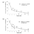
図14に本実施例の迅速薬剤感受性試験結果を示す。
菌ATP量は抗菌薬有り・無しに関わらず培養時間が進むにつれて増加しており、抗菌薬(レボフロキサシン)耐性であることが培養4時間後には明らかである。
実際、従来の検査法ではレボフロキサシン耐性のEscherichia coli(大腸菌)が検出され、薬剤感受性結果は一致した。
以上により、血液培養陽性時より僅か4時間で正確な感受性結果を得ることが出来た。FIG. 14 shows the results of the rapid drug susceptibility test of this example.
The amount of bacterial ATP increased with the progress of the culture time regardless of the presence or absence of the antibacterial drug, and it is clear that the bacteria are resistant to the antibacterial drug (levofloxacin) after 4 hours of the culture.
In fact, conventional testing detected levofloxacin-resistant Escherichia coli, and drug susceptibility results were consistent.
From the above, an accurate susceptibility result could be obtained in only 4 hours from the time when the blood culture was positive.
<血液培養ボトルからのATP測定による迅速薬剤感受性試験の症例2>
実施例5と同じプロトコール(但し血液培養陽性化から4時間後の培養検体を使用)で、実施例5とは別な敗血症患者の血培ボトルから迅速薬剤感受性試験を行った。
図15に本実施例の迅速薬剤感受性試験結果を示す。
培養時間が進むにつれ、抗菌薬無しでは菌ATP量は増加、抗菌薬(レボフロキサシン)有りでは菌ATP量は減少した。
つまり、抗菌薬(レボフロキサシン)感受性であることが培養4時間後には明らかである。
実際、従来の検査法ではレボフロキサシン感受性のStreptococcus anginosusが検出され、薬剤感受性結果は一致した。
以上により、血液培養陽性時より僅か4時間で正確な感受性結果を得ることが出来た。<
A rapid drug susceptibility test was performed from a blood culture bottle of a septic patient different from Example 5 using the same protocol as in Example 5 (however, a
FIG. 15 shows the results of the rapid drug susceptibility test of this example.
As the culture time progressed, the amount of bacterial ATP increased without the antibacterial agent, and decreased with the presence of the antibacterial agent (levofloxacin).
That is, it is clear that it is sensitive to an antibacterial drug (levofloxacin) after 4 hours of culturing.
In fact, conventional tests detected levofloxacin-sensitive Streptococcus anginosus, and the drug-susceptibility results were in agreement.
From the above, an accurate susceptibility result could be obtained in only 4 hours from the time when the blood culture was positive.
<血液培養ボトルからのATP測定による迅速薬剤感受性試験の症例3>
実施例5と同じプロトコール(但し血液培養陽性化から5時間後の培養検体を使用)で、実施例5,6とは別な敗血症患者の血培ボトルから迅速薬剤感受性試験を行った。
図16に本実施例の迅速薬剤感受性試験結果を示す。
菌ATP量は抗菌薬有り・無しに関わらず培養時間が進むにつれて増加しており、抗菌薬(レボフロキサシン)耐性であることが培養4〜6時間後には明らかである。
但し抗菌薬有り・無しで菌ATP量の増加に差があるため、抗菌薬耐性菌と感受性菌とが混合していた可能性がある。従来の検査法ではレボフロキサシン耐性のStaphylococcus capitis or Staphylococcus capraeが検出され、薬剤感受性結果は一致した。
以上、血液培養陽性時より僅か4〜6時間で正確な感受性結果を得ることが出来た。<
A rapid drug susceptibility test was performed from a blood culture bottle of a septic patient different from Examples 5 and 6 using the same protocol as in Example 5 (however, a
FIG. 16 shows the results of the rapid drug susceptibility test of this example.
The amount of bacterial ATP increased as the culture time progressed regardless of the presence or absence of the antibacterial drug, and it is clear that the cells are resistant to the antibacterial drug (levofloxacin) 4 to 6 hours after the culture.
However, since there is a difference in the increase in the amount of bacterial ATP with and without the antibacterial drug, it is possible that the antibacterial drug-resistant bacteria and the susceptible bacteria were mixed. The conventional test method detected levofloxacin-resistant Staphylococcus capitis or Staphylococcus caprae, and the drug susceptibility results were in agreement.
As described above, an accurate susceptibility result could be obtained in only 4 to 6 hours from the time when the blood culture was positive.
敗血症疑い患者の血液検体における、ATP測定を用いた迅速薬剤感受性試験の実施に利用できる。
血液検体中の生菌の有無について、ATP測定により迅速判定(遺伝子検査の弱点を補える)の実施に利用できる。
血液培養ボトルからの血液培養検体における、ATP測定を用いた迅速薬剤感受性試験の実施に利用できる。
血液検体以外にも、脳脊髄液(細菌性髄膜炎)、心嚢水(心膜炎)、胸水(胸膜炎)、腹水(腹膜炎)、関節包液(整形外科の術後感染症)、眼房水(眼内炎)、肺胞洗浄液(肺炎)、尿(尿路感染症)、術後のドレーン排液(術後感染症)、CVカテーテル先端(長期臥床患者のカテ先バイオフィルムによる敗血症)などで、生菌の有無の迅速判定、および迅速薬剤感受性試験等の実施に利用できる。It can be used to perform a rapid drug susceptibility test using ATP measurement in a blood sample of a patient suspected of having sepsis.
The presence or absence of viable bacteria in a blood sample can be used for rapid determination (compensating for weaknesses in genetic testing) by ATP measurement.
It can be used to perform a rapid drug susceptibility test using ATP measurement in a blood culture sample from a blood culture bottle.
In addition to blood samples, cerebrospinal fluid (bacterial meningitis), pericarditis (pericarditis), pleural effusion (pneumonia), peritonitis (peritonitis), arthritis (postoperative infection of orthopedic surgery), ovary Water (endophthalmitis), alveolar lavage fluid (pneumonia), urine (urinary tract infection), postoperative drainage fluid (postoperative infection), CV catheter tip (septicemia due to catheter tip biofilm in long-term lying patients) It can be used for rapid determination of the presence or absence of viable bacteria and rapid drug susceptibility testing.
Claims (6)
血液検体から血小板と起炎菌のペレットを作成するステップと、
前記血小板と起炎菌のペレットを、下記(A)〜(C)のステップを任意の順であるいは、
複数のステップを同時に処理するステップとを、
有することを特徴とする血液検体の前処理方法。
(A)血小板の細胞膜蛋白をプロテアーゼ分解処理する。
(B)血小板を低張液にて膨化処理する。
(C)起炎菌への影響を抑えた条件下で、血小板の細胞膜を界面活性剤溶液にて破壊処理する。A pretreatment method for blood samples for ATP measurement of causative bacteria in blood.
Steps to make pellets of platelets and causative organisms from blood samples,
The platelets and pellets of the causative organism are subjected to the following steps (A) to (C) in any order, or
A step that processes multiple steps at the same time,
A method for pretreating a blood sample, which comprises having.
(A) The cell membrane protein of platelets is subjected to protease degradation treatment.
(B) Platelets are swollen with a hypotonic solution.
(C) The cell membrane of platelets is disrupted with a surfactant solution under conditions in which the influence on the causative organism is suppressed.
Applications Claiming Priority (3)
| Application Number | Priority Date | Filing Date | Title |
|---|---|---|---|
| JP2017219547 | 2017-11-15 | ||
| JP2017219547 | 2017-11-15 | ||
| PCT/JP2018/023388 WO2019097752A1 (en) | 2017-11-15 | 2018-06-20 | Blood sample pretreatment method |
Publications (2)
| Publication Number | Publication Date |
|---|---|
| JPWO2019097752A1 JPWO2019097752A1 (en) | 2020-12-03 |
| JP6898011B2 true JP6898011B2 (en) | 2021-07-07 |
Family
ID=66540110
Family Applications (1)
| Application Number | Title | Priority Date | Filing Date |
|---|---|---|---|
| JP2019553690A Active JP6898011B2 (en) | 2017-11-15 | 2018-06-20 | Blood sample pretreatment method |
Country Status (4)
| Country | Link |
|---|---|
| US (1) | US20200355588A1 (en) |
| JP (1) | JP6898011B2 (en) |
| KR (1) | KR20200088390A (en) |
| WO (1) | WO2019097752A1 (en) |
Families Citing this family (1)
| Publication number | Priority date | Publication date | Assignee | Title |
|---|---|---|---|---|
| CN115461466A (en) | 2020-05-12 | 2022-12-09 | 株式会社日立高新技术 | Automatic analysis device and automatic analysis method |
Family Cites Families (8)
| Publication number | Priority date | Publication date | Assignee | Title |
|---|---|---|---|---|
| US3745090A (en) * | 1970-08-04 | 1973-07-10 | Nasa | Method of detecting and counting bacteria in body fluids |
| JPS53139785A (en) * | 1977-01-19 | 1978-12-06 | Nasa | Detecting of bacteria in blood |
| US4693972A (en) * | 1984-01-16 | 1987-09-15 | Becton, Dickinson And Company | Composition and method for rapid detection of microorganisms in clinical samples |
| US7419798B2 (en) * | 2004-10-19 | 2008-09-02 | Zybac Llc | Rapid and sensitive detection of bacteria in blood products, urine, and other fluids |
| GB0513535D0 (en) * | 2005-07-01 | 2005-08-10 | Iseao Technologies Ltd | Improved methods of detecting microbes |
| RU2384624C2 (en) * | 2005-07-21 | 2010-03-20 | Моринага Милк Индастри Ко., Лтд. | Method for sampling for detecting microorganism, method for detecting microorganism and set for detecting microorganism |
| US9260737B2 (en) * | 2011-08-11 | 2016-02-16 | Kyle R. Brandy | Rapid and sensitive detection of bacteria in blood products, urine, and other fluids |
| JP6177009B2 (en) | 2013-06-03 | 2017-08-09 | 株式会社日立ハイテクノロジーズ | Blood cell destruction reagent and blood cell destruction method using the same |
-
2018
- 2018-06-20 KR KR1020207016833A patent/KR20200088390A/en not_active Application Discontinuation
- 2018-06-20 JP JP2019553690A patent/JP6898011B2/en active Active
- 2018-06-20 WO PCT/JP2018/023388 patent/WO2019097752A1/en active Application Filing
-
2020
- 2020-05-14 US US16/874,008 patent/US20200355588A1/en active Pending
Also Published As
| Publication number | Publication date |
|---|---|
| KR20200088390A (en) | 2020-07-22 |
| WO2019097752A1 (en) | 2019-05-23 |
| US20200355588A1 (en) | 2020-11-12 |
| JPWO2019097752A1 (en) | 2020-12-03 |
Similar Documents
| Publication | Publication Date | Title |
|---|---|---|
| RU2384624C2 (en) | Method for sampling for detecting microorganism, method for detecting microorganism and set for detecting microorganism | |
| Matsuda et al. | Sensitive quantitative detection of commensal bacteria by rRNA-targeted reverse transcription-PCR | |
| JP2017093464A (en) | Selective lysis of cells by ionic surfactants | |
| EP2821499B1 (en) | Method for the rapid determination of susceptibility or resistance of bacteria to antibiotics | |
| JP5475278B2 (en) | Cell discrimination in samples using extracellular enzyme inactivation prior to intracellular enzyme release | |
| US20060134729A1 (en) | Process for the universal detection of microorganisms and reaction environment permitting the implementation of the process | |
| JP2019201654A (en) | Test method for target antibiotic sensitivity | |
| JP2008194057A (en) | Method of evaluating function of phagocyte and utilization thereof | |
| US20170336424A1 (en) | Method for separating target molecules or particles from fibrinogen-containing samples including blood components | |
| KR20200062321A (en) | How to determine the concentration of intact microorganisms in a sample | |
| KR20200111720A (en) | Method for determining microbial concentration | |
| JP6898011B2 (en) | Blood sample pretreatment method | |
| EP2888364B1 (en) | Method for efficient isolation of microbial dna from blood | |
| Goyani et al. | Sputum-Spotted Solid Matrix Designed to Release Diagnostic-Grade Mycobacterium tuberculosis DNA Demonstrate Optimal Biocontainment Property | |
| Prakash et al. | Clinical evaluation of the mycobacteriophage-based assay in rapid detection of Mycobacterium tuberculosis in respiratory specimens | |
| RU2518249C1 (en) | Method of determining nonspecific resistance of pathogenic microorganisms to antibiotics based on measuring catalytic activity of phosphodiesterases cleaving cyclic diguanosine monophosphate | |
| KR100592693B1 (en) | Antibiotic sensitivity testing and a test kit | |
| JP2010213724A (en) | Composition for increasing permeability of microorganism cell wall, and method for detecting the microorganism on membrane | |
| IL270342A (en) | A method for determining bacterial susceptibility to antibiotics | |
| US20200208200A1 (en) | Viability detection and quantification assay of waterborne pathogens by enrichment | |
| Doganay et al. | Brucella: potential biothreat agent | |
| US20130217024A1 (en) | Methods for diagnosing bacteremia and fungemi | |
| WO2020085420A1 (en) | Cell collection method | |
| JP2008125457A (en) | Method for detection of microorganism | |
| Burnet et al. | A Host-Directed Approach to the Detection of Infection in Hard-to-Heal Wounds. Diagnostics 2022, 12, 2408 |
Legal Events
| Date | Code | Title | Description |
|---|---|---|---|
| A80 | Written request to apply exceptions to lack of novelty of invention |
Free format text: JAPANESE INTERMEDIATE CODE: A801 Effective date: 20200512 |
|
| A80 | Written request to apply exceptions to lack of novelty of invention |
Free format text: JAPANESE INTERMEDIATE CODE: A80 Effective date: 20200513 |
|
| A621 | Written request for application examination |
Free format text: JAPANESE INTERMEDIATE CODE: A621 Effective date: 20200727 |
|
| TRDD | Decision of grant or rejection written | ||
| A01 | Written decision to grant a patent or to grant a registration (utility model) |
Free format text: JAPANESE INTERMEDIATE CODE: A01 Effective date: 20210602 |
|
| A61 | First payment of annual fees (during grant procedure) |
Free format text: JAPANESE INTERMEDIATE CODE: A61 Effective date: 20210603 |
|
| R150 | Certificate of patent or registration of utility model |
Ref document number: 6898011 Country of ref document: JP Free format text: JAPANESE INTERMEDIATE CODE: R150 |
|
| S111 | Request for change of ownership or part of ownership |
Free format text: JAPANESE INTERMEDIATE CODE: R313113 |
|
| R350 | Written notification of registration of transfer |
Free format text: JAPANESE INTERMEDIATE CODE: R350 |