JP6884792B2 - Treatment methods and therapeutic compositions for cancer and neoplasms - Google Patents
Treatment methods and therapeutic compositions for cancer and neoplasms Download PDFInfo
- Publication number
- JP6884792B2 JP6884792B2 JP2018543712A JP2018543712A JP6884792B2 JP 6884792 B2 JP6884792 B2 JP 6884792B2 JP 2018543712 A JP2018543712 A JP 2018543712A JP 2018543712 A JP2018543712 A JP 2018543712A JP 6884792 B2 JP6884792 B2 JP 6884792B2
- Authority
- JP
- Japan
- Prior art keywords
- cells
- asc
- cancer
- cell
- culture
- Prior art date
- Legal status (The legal status is an assumption and is not a legal conclusion. Google has not performed a legal analysis and makes no representation as to the accuracy of the status listed.)
- Expired - Fee Related
Links
Images


Classifications
-
- A—HUMAN NECESSITIES
- A61—MEDICAL OR VETERINARY SCIENCE; HYGIENE
- A61K—PREPARATIONS FOR MEDICAL, DENTAL OR TOILETRY PURPOSES
- A61K35/00—Medicinal preparations containing materials or reaction products thereof with undetermined constitution
- A61K35/12—Materials from mammals; Compositions comprising non-specified tissues or cells; Compositions comprising non-embryonic stem cells; Genetically modified cells
- A61K35/28—Bone marrow; Haematopoietic stem cells; Mesenchymal stem cells of any origin, e.g. adipose-derived stem cells
-
- A—HUMAN NECESSITIES
- A61—MEDICAL OR VETERINARY SCIENCE; HYGIENE
- A61K—PREPARATIONS FOR MEDICAL, DENTAL OR TOILETRY PURPOSES
- A61K35/00—Medicinal preparations containing materials or reaction products thereof with undetermined constitution
- A61K35/12—Materials from mammals; Compositions comprising non-specified tissues or cells; Compositions comprising non-embryonic stem cells; Genetically modified cells
- A61K35/35—Fat tissue; Adipocytes; Stromal cells; Connective tissues
-
- A—HUMAN NECESSITIES
- A61—MEDICAL OR VETERINARY SCIENCE; HYGIENE
- A61P—SPECIFIC THERAPEUTIC ACTIVITY OF CHEMICAL COMPOUNDS OR MEDICINAL PREPARATIONS
- A61P35/00—Antineoplastic agents
-
- A—HUMAN NECESSITIES
- A61—MEDICAL OR VETERINARY SCIENCE; HYGIENE
- A61P—SPECIFIC THERAPEUTIC ACTIVITY OF CHEMICAL COMPOUNDS OR MEDICINAL PREPARATIONS
- A61P35/00—Antineoplastic agents
- A61P35/04—Antineoplastic agents specific for metastasis
-
- C—CHEMISTRY; METALLURGY
- C12—BIOCHEMISTRY; BEER; SPIRITS; WINE; VINEGAR; MICROBIOLOGY; ENZYMOLOGY; MUTATION OR GENETIC ENGINEERING
- C12N—MICROORGANISMS OR ENZYMES; COMPOSITIONS THEREOF; PROPAGATING, PRESERVING, OR MAINTAINING MICROORGANISMS; MUTATION OR GENETIC ENGINEERING; CULTURE MEDIA
- C12N5/00—Undifferentiated human, animal or plant cells, e.g. cell lines; Tissues; Cultivation or maintenance thereof; Culture media therefor
- C12N5/0062—General methods for three-dimensional culture
Landscapes
- Health & Medical Sciences (AREA)
- Life Sciences & Earth Sciences (AREA)
- Chemical & Material Sciences (AREA)
- Engineering & Computer Science (AREA)
- General Health & Medical Sciences (AREA)
- Biomedical Technology (AREA)
- Cell Biology (AREA)
- Organic Chemistry (AREA)
- Pharmacology & Pharmacy (AREA)
- Animal Behavior & Ethology (AREA)
- Public Health (AREA)
- Veterinary Medicine (AREA)
- Medicinal Chemistry (AREA)
- Biotechnology (AREA)
- Zoology (AREA)
- Developmental Biology & Embryology (AREA)
- Immunology (AREA)
- Nuclear Medicine, Radiotherapy & Molecular Imaging (AREA)
- General Chemical & Material Sciences (AREA)
- Chemical Kinetics & Catalysis (AREA)
- Bioinformatics & Cheminformatics (AREA)
- Genetics & Genomics (AREA)
- Wood Science & Technology (AREA)
- Virology (AREA)
- Epidemiology (AREA)
- Microbiology (AREA)
- Biochemistry (AREA)
- General Engineering & Computer Science (AREA)
- Hematology (AREA)
- Oncology (AREA)
- Medicines Containing Material From Animals Or Micro-Organisms (AREA)
- Micro-Organisms Or Cultivation Processes Thereof (AREA)
- Pharmaceuticals Containing Other Organic And Inorganic Compounds (AREA)
Description
胎盤細胞を使用した抗腫瘍治療方法が、本明細書に記載される。 Antitumor treatment methods using placental cells are described herein.
要約
以前の研究によって、3D条件下で接着性間質細胞(ASC)を培養することによって、これまでに記載されていなかった特性及び特徴を有するASCが生成されることが確立されている。がん、腫瘍及び新生物の増殖を治療、予防及び阻害するためにASCを使用する方法が、本明細書に記載される。
Summary Previous studies have established that culturing adhesive stromal cells (ASCs) under 3D conditions produces ASCs with properties and characteristics not previously described. Methods of using ASC to treat, prevent and inhibit the growth of cancer, tumors and neoplasms are described herein.
ある実施形態においては、2次元(2D)培養、3次元(3D)培養又はその組合せにおいて培養することによって、記載されたASCが調製されている。2D及び3D培養条件の非限定的な例が、詳細な説明及び実施例において提供される。代替的又は追加的に、幾つかの実施形態においては、炎症促進性サイトカインを用いて細胞が処理されており;及び/又は該細胞は胎盤細胞調製物である。ある実施形態においては、該胎盤細胞調製物は主に胎児細胞であり;主に母体細胞であり;又はより具体的な実施形態においては、胎児細胞が濃縮されているか、若しくは母体細胞が濃縮されている、胎児細胞と母体細胞の混合物である。別段の記載がない場合、用語「ASC」は、様々な実施形態において、炎症促進性サイトカインを用いたインキュベーション前又は後のいずれかの接着性間質細胞を指す場合がある。更に他の実施形態においては、ASCは、炎症促進性サイトカインを用いてインキュベートされていない接着性間質細胞を指す。 In certain embodiments, the described ASCs are prepared by culturing in a two-dimensional (2D) culture, a three-dimensional (3D) culture, or a combination thereof. Non-limiting examples of 2D and 3D culture conditions are provided in the detailed description and examples. Alternatively or additionally, in some embodiments, the cells have been treated with pro-inflammatory cytokines; and / or the cells are placental cell preparations. In certain embodiments, the placental cell preparation is predominantly fetal cells; predominantly maternal cells; or in more specific embodiments, the fetal cells are enriched or the maternal cells are enriched. It is a mixture of fetal cells and maternal cells. Unless otherwise stated, the term "ASC" may, in various embodiments, refer to adherent stromal cells either before or after incubation with pro-inflammatory cytokines. In yet another embodiment, ASC refers to adhesive stromal cells that have not been incubated with pro-inflammatory cytokines.
代替的又は追加的に、細胞は間葉系様(mesenchymal-like)ASCであり、間葉系間質細胞に類似するマーカーパターンを示すが、「標準的な」間葉系幹細胞(MSC)が骨細胞に分化する条件下で、骨細胞に分化しない。他の実施形態においては、細胞は、MSCに類似するマーカーパターンを示すが、MSCが脂肪細胞に分化する条件下で、脂肪細胞に分化しない。更に他の実施形態においては、細胞は、MSCに類似するマーカーパターンを示すが、間葉系幹細胞が骨細胞又は脂肪細胞にそれぞれ分化する条件下で、骨細胞又は脂肪細胞のいずれにも分化しない。幾つかの実施形態においては、これらのアッセイにおいて比較に使用されるMSCは、骨髄(BM)から採取され、2D条件下で培養されているMSCである。他の実施形態においては、比較に使用されるMSCは、BMから採取され、2D条件下で培養され、続けて3D条件によって培養されている。 Alternatively or additionally, the cells are mesenchymal-like ASCs, exhibiting marker patterns similar to mesenchymal stromal cells, but with "standard" mesenchymal stem cells (MSCs). Under the condition that it differentiates into bone cells, it does not differentiate into bone cells. In other embodiments, the cells exhibit a marker pattern similar to MSCs, but do not differentiate into adipocytes under conditions where the MSCs differentiate into adipocytes. In yet another embodiment, the cells exhibit a marker pattern similar to MSC, but do not differentiate into either osteoocytes or adipocytes under the condition that mesenchymal stem cells differentiate into osteoocytes or adipocytes, respectively. .. In some embodiments, the MSCs used for comparison in these assays are MSCs taken from bone marrow (BM) and cultured under 2D conditions. In other embodiments, the MSCs used for comparison are taken from the BM, cultured under 2D conditions, followed by culture under 3D conditions.
様々な実施形態において、記載されたASCは記載された治療効果を発揮することができ、それらはそれぞれ、ASC自身が宿主に生着しているか又は生着していない、別個の実施形態と考えられる。例えば、様々な実施形態において、細胞は、3日間超、4日間超、5日間超、6日間超、7日間超、8日間超、9日間超、10日間超若しくは14日間超で細胞自身が生存することなく、治療効果を発揮することができる場合があり;又は細胞は、3日間超、4日間超、5日間超、6日間超、7日間超、8日間超、9日間超、10日間超若しくは14日間超で生存する。 In various embodiments, the described ASCs can exert the described therapeutic effects, each considered as a separate embodiment in which the ASC itself engrafts or does not engraft in the host. Be done. For example, in various embodiments, the cells themselves are over 3 days, over 4 days, over 5 days, over 6 days, over 7 days, over 8 days, over 9 days, over 10 days or over 14 days. It may be possible to exert a therapeutic effect without survival; or cells may have more than 3 days, more than 4 days, more than 5 days, more than 6 days, more than 7 days, more than 8 days, more than 9 days, 10 Survive for more than 14 days or more than 14 days.
本明細書における細胞集団の「増殖(growth)」についての言及は、細胞集団の増殖(expansion)と同義語であることを意図する。
別段の記載がない場合、本明細書に言及される全ての範囲は境界を含む。
別段定義される場合を除いて、本明細書で使用された全ての技術用語及び科学用語は、本発明が属する技術分野の当業者によって一般に理解されるのと同じ意味を有する。発明の実施又はテストにおいて、本明細書に記載されたものと同様又は同等な方法及び材料が使用され得るが、好適な方法及び材料を以下に記載する。矛盾する場合は、定義を含む特許明細書によって調整される。更に、材料、方法及び実施例は、例示的なものにすぎず、限定することを意図するものではない。
References herein to "growth" of a cell population are intended to be synonymous with expansion of a cell population.
Unless otherwise stated, all scopes referred to herein include boundaries.
Unless otherwise defined, all technical and scientific terms used herein have the same meaning as commonly understood by one of ordinary skill in the art to which the present invention belongs. Methods and materials similar to or equivalent to those described herein may be used in the practice or testing of the invention, but suitable methods and materials are described below. In case of inconsistency, it will be adjusted by the patent specification including the definition. Moreover, the materials, methods and examples are exemplary only and are not intended to be limiting.
本発明は、例示の目的でのみ、添付の図面を参照して本明細書に記載される。ここで図面を詳細に具体的に参照して、示される特定のものは、例示の目的であり、発明の実施形態の例示的な検討のためのものに過ぎず、発明の原理及び概念的側面の最も有用かつ容易に理解される記述であると考えられるものを提供するために提示されることが、強調される。この点について、発明の基礎的な理解に必要であるものを超えて、詳細に発明の構造的な詳細を示すことは試みておらず、図面を用いた説明によって、本発明の幾つかの形態を実際にどのように実施し得るかが、当業者に明らかになる。
図面は以下の通り:
The drawing is as follows:
詳細な説明
本発明の少なくとも1つの実施形態を詳細に説明する前に、本発明はその適用において、以下の説明に記載された又は実施例によって例示された詳細に限定されないことが理解される。本発明は、他の実施形態であることができるか、又は多様な方法で実施若しくは実行することができる。また、本明細書で使用される語句と用語は、説明の目的のためのものであり、限定的なものとしてみなされるべきでないことが理解される。
Detailed Description Before explaining in detail at least one embodiment of the present invention, it is understood that the present invention is not limited to the details described in the following description or exemplified by Examples in its application. The present invention can be in other embodiments or can be implemented or practiced in a variety of ways. It is also understood that the terms and terms used herein are for illustration purposes only and should not be considered as limiting.
ある実施形態においては、がんの治療を、それを必要とする対象においてする方法であって、治療有効量の接着性間質細胞(ASC)を対象に投与する工程を含み、それによりがんを治療する、方法が提供される。他の実施形態においては、新生物の治療を、それを必要とする対象においてする方法であって、ASCを対象に投与する工程を含む、方法が提供される。ASCは、胎盤に、又は他の実施形態においては、脂肪組織に、又は他の実施形態においては、本明細書に記載された他のソースに由来しても良い。本明細書において記述される場合、ASCの投与は、新生物の増殖の治療において有用である。 In certain embodiments, a method of treating cancer in a subject in need thereof comprises administering a therapeutically effective amount of adhesive stromal cells (ASC) to the subject, thereby the cancer. A method is provided to treat the disease. In another embodiment, a method of treating a neoplasm in a subject in need thereof, comprising the step of administering the ASC to the subject, is provided. The ASC may be derived from the placenta, or in other embodiments, from adipose tissue, or, in other embodiments, from other sources described herein. As described herein, administration of ASC is useful in the treatment of neoplasmic growth.
より具体的には、ASCは、スフェロイド(spheroid)の研究において、様々な腫瘍細胞株の増殖を阻害することが示された(実施例10)。更に、in vivo研究では、ASCの投与によって、移植した腫瘍の増殖が停止又は阻害されることが示された。最初の研究においては(実施例16)、IV投与によって明瞭な統計的有意性のある効果を示した一方で、IM投与によって少なくとも効能は有する傾向を示した。2番目の研究においては(実施例17)、IM投与は持続的な効果を与え、IV投与は腫瘍増殖の少なくとも一時的な阻害は与えた。IM及びIV ASC処理の両方によって、肺転移が完全に予防され、IM処理によって、乳腺脂肪体の最初のドレナージ(primary drainage)リンパ節である、腋窩リンパ節への転移が減少した(Kobayashi Hら)。 More specifically, ASC has been shown to inhibit the growth of various tumor cell lines in spheroid studies (Example 10). In addition, in vivo studies have shown that administration of ASC stops or inhibits the growth of transplanted tumors. In the first study (Example 16), IV administration showed a clear, statistically significant effect, while IM administration tended to have at least an effect. In the second study (Example 17), IM administration provided a sustained effect and IV administration provided at least a temporary inhibition of tumor growth. Both IM and IV ASC treatment completely prevented lung metastases, and IM treatment reduced metastases to the axillary lymph nodes, the primary drainage lymph nodes of the mammary fat pad (Kobayashi H et al.) ).
他の実施形態においては、新生物の増殖の予防を、それを必要とする対象においてする方法であって、治療有効量のASCを対象に投与する工程を含み、それにより対象における新生物の増殖を予防する、方法が提供される。他の実施形態においては、新生物の増殖の発生の低減を、それを必要とする対象においてする方法であって、ASCを対象に投与する工程を含む、方法が提供される。様々な実施形態において、新生物の増殖は、がん、腫瘍又は新生物であり得る。 In another embodiment, the prevention of neoplasm growth in a subject in need thereof comprises administering a therapeutically effective amount of ASC to the subject, thereby propagating the neoplasm in the subject. A method is provided to prevent the disease. In another embodiment, there is provided a method of reducing the occurrence of neoplasmic proliferation in a subject in need thereof, comprising the step of administering the ASC to the subject. In various embodiments, the growth of the neoplasm can be cancer, tumor or neoplasm.
更に他の実施形態においては、腫瘍の増殖の阻害を、それを必要とする対象においてする方法であって、治療有効量のASCを対象に投与する工程を含み、それにより対象における腫瘍の増殖を阻害する、方法が提供される。他の実施形態においては、がんの増殖の阻害方法であって、ASCを対象に投与する工程を含む、方法が提供される。 In yet another embodiment, a method of inhibiting tumor growth in a subject in need thereof comprises administering a therapeutically effective amount of ASC to the subject, thereby causing tumor growth in the subject. A method of inhibiting is provided. In another embodiment, a method of inhibiting the growth of cancer, comprising the step of administering ASC to a subject, is provided.
更に他の実施形態においては、腫瘍の転移を、そのリスクがある対象において阻害する方法であって、治療有効量のASCを対象に投与する工程を含む、方法が提供される。他の実施形態においては、がんの転移の阻害方法であって、ASCを対象に投与する工程を含む、方法が提供される。ある実施形態においては、対象は、手術不可能である原発性腫瘍を有する。他の実施形態においては、原発性腫瘍は手術可能である。 In yet another embodiment, there is provided a method of inhibiting tumor metastasis in a subject at risk thereof, comprising administering a therapeutically effective amount of ASC to the subject. In another embodiment, a method of inhibiting cancer metastasis, comprising the step of administering ASC to a subject, is provided. In certain embodiments, the subject has a primary tumor that is inoperable. In other embodiments, the primary tumor is operable.
他の実施形態においては、がんの治療を、それを必要とする対象においてする方法であって、ASCに由来する治療有効量のCMを対象に投与する工程を含み、それによりがんを治療する、方法が提供される。他の実施形態においては、新生物の治療を、それを必要とする対象においてする方法であって、CMを対象に投与する工程を含む、方法が提供される。ASCは、胎盤に、又は他の実施形態においては、脂肪組織に、又は他の実施形態においては、本明細書に記載された他のソースに由来しても良い。 In other embodiments, a method of treating cancer in a subject in need thereof comprises administering to the subject a therapeutically effective amount of CM derived from ASC, thereby treating the cancer. A way to do it is provided. In another embodiment, there is provided a method of treating the neoplasm in a subject in need thereof, comprising the step of administering the CM to the subject. The ASC may be derived from the placenta, or in other embodiments, from adipose tissue, or, in other embodiments, from other sources described herein.
他の実施形態においては、新生物の増殖の予防を、それを必要とする対象においてする方法であって、ASCに由来する治療有効量のCMを対象に投与する工程を含み、それにより対象における新生物の増殖を予防する、方法が提供される。他の実施形態においては、新生物の増殖の発生の低減を、それを必要とする対象においてする方法であって、CMを対象に投与する工程を含む、方法が提供される。様々な実施形態において、新生物の増殖は、がん、腫瘍又は新生物であっても良い。 In other embodiments, a method of preventing the growth of a neoplasm in a subject in need thereof comprises administering to the subject a therapeutically effective amount of CM derived from ASC, thereby in the subject. Methods are provided to prevent the growth of neoplasms. In another embodiment, there is provided a method of reducing the occurrence of neoplasmic proliferation in a subject in need thereof, comprising the step of administering the CM to the subject. In various embodiments, the growth of the neoplasm may be cancer, tumor or neoplasm.
更に他の実施形態においては、腫瘍の増殖の阻害を、それを必要とする対象においてする方法であって、ASCに由来する治療有効量のCMを対象に投与する工程を含み、それにより対象における腫瘍の増殖を阻害する、方法が提供される。他の実施形態においては、がんの増殖の阻害方法であって、CMを対象に投与する工程を含む、方法が提供される。 In yet another embodiment, a method of inhibiting tumor growth in a subject in need thereof comprises administering to the subject a therapeutically effective amount of CM derived from ASC, thereby in the subject. Methods are provided that inhibit tumor growth. In another embodiment, a method of inhibiting the growth of cancer, comprising the step of administering CM to a subject, is provided.
更に他の実施形態においては、腫瘍の転移を、そのリスクがある対象において阻害する方法であって、ASCに由来する治療有効量のCMを対象に投与する工程を含む、方法が提供される。他の実施形態においては、がんの転移の阻害方法であって、CMを対象に投与する工程を含む、方法が提供される。ある実施形態においては、対象は手術不可能である原発性腫瘍を有する。他の実施形態においては、原発性腫瘍は手術可能である。より具体的な実施形態においては、腫瘍は、約200 mm3より小さく、50〜200 mm3の間、100〜200 mm3の間、125〜1000 mm3の間、150〜1000 mm3の間、200〜1000 mm3の間、250〜1000 mm3の間、300〜1000 mm3の間、150 mm3超、200 mm3超、又は300 mm3超であっても良い。 In yet another embodiment, there is provided a method of inhibiting tumor metastasis in a subject at risk thereof, comprising administering to the subject a therapeutically effective amount of CM derived from ASC. In another embodiment, a method of inhibiting cancer metastasis, comprising the step of administering CM to a subject, is provided. In certain embodiments, the subject has a primary tumor that is inoperable. In other embodiments, the primary tumor is operable. In a more specific embodiment, the tumor is less than about 200 mm 3, between 50 to 200 mm 3, between 100 to 200 mm 3, while the 125-1000 mm 3, between 150 to 1000 mm 3 , 200-1000 mm 3 , 250-1000 mm 3 , 300-1000 mm 3 , more than 150 mm 3, more than 200 mm 3 , or more than 300 mm 3.
各ケースにおいて、記載されたASCは、胎盤に、又は他の実施形態においては、脂肪組織に、又は他の実施形態においては、本明細書に記載された他のソースに由来しても良い。 In each case, the described ASC may be derived from the placenta, or in other embodiments, to adipose tissue, or, in other embodiments, from other sources described herein.
他に指示がない場合、ASCを用いた処理は、生きたASC全体を用いた、がん細胞の処理を指す。代替的な実施形態においては、がん細胞は、ASCの画分、又はASCに由来する因子を用いて処理される。 Unless otherwise instructed, treatment with ASC refers to treatment of cancer cells with whole living ASC. In an alternative embodiment, cancer cells are treated with a fraction of ASC, or a factor derived from ASC.
他に指示がない場合、馴化培地(CM)を用いた処理は、ASCを用いてインキュベートされている培地用いた、がん細胞の処理を指す。代替的な実施形態においては、がん細胞は、ASCを用いてインキュベートされているCMの画分を用いてか、又はASCを用いてインキュベートされているCMに由来する因子を用いて、処理される。 Unless otherwise indicated, treatment with conditioned medium (CM) refers to treatment of cancer cells with medium incubated with ASC. In an alternative embodiment, cancer cells are treated with a fraction of CM incubated with ASC or with factors derived from CM incubated with ASC. To.
ある実施形態においては、がんは以下:急性リンパ芽球性白血病、副腎皮質がん、AIDSに関連するリンパ腫、肛門がん、虫垂がん、星細胞腫(小児期の小脳又は大脳)、基底細胞がん、胆管がん、膀胱がん、骨がん、脳幹神経膠腫、脳腫瘍(小脳星細胞腫、大脳星細胞腫/悪性神経膠腫、上衣腫、髄芽細胞種、テント上(supratentorial)原始神経外胚葉腫瘍、視覚経路及び視床下部神経膠腫)、乳がん、気管支腺腫、肺のカルチノイド腫瘍、胃のカルチノイド、他のカルチノイド腫瘍(例、小児性)、バーキットリンパ腫、原発不明のがん、中枢神経系リンパ腫(例、原発性)、小脳星細胞腫、悪性神経膠腫(例、大脳星細胞腫)、子宮頸部がん、慢性リンパ球性白血病、慢性骨髄性白血病、結腸がん、皮膚T細胞リンパ腫、線維形成性小円形細胞腫瘍、子宮内膜がん、上衣腫、食道がん、ユーイング肉腫、頭蓋外胚胚細胞腫瘍(例、小児性)、性腺外生殖細胞腫瘍、肝外胆管がん、眼のがん(例、眼内黒色腫、網膜芽細胞腫)、胆嚢がん、胃(胃)がん、胃腸間質腫瘍、胚胚細胞腫瘍(例、小児性頭蓋外)、妊娠性絨毛腫瘍、毛様細胞白血病、頭部及び頸部のがん、肝細胞(肝臓)がん、ホジキンリンパ腫、他のリンパ腫(AIDSに関連する、非ホジキン、原発性中枢神経系)、下咽頭がん、眼内黒色腫、膵島細胞がん、カポジ肉腫、喉頭がん、白血病(例、急性リンパ芽球性、慢性リンパ球性、慢性骨髄性、毛様細胞性)、口唇及び口腔がん、原発性肝臓がん、小細胞肺がん、非小細胞肺がん、マクログロブリン血症(ヴァルデンストレーム)、骨の悪性線維性組織球腫、髄芽細胞種(例、小児性)、眼内黒色腫、他の黒色腫、メルケル細胞がん、中皮腫(例、成人悪性、小児性)、原発不明の転移性頸部扁平上皮がん、口のがん、多発性内分泌腫瘍症候群(例、小児患者における)、形質細胞新生物(例、多発性骨髄腫)、菌状息肉腫、骨髄性白血病(例、慢性)、鼻腔及び副鼻腔のがん、鼻咽頭がん、神経芽細胞腫、口腔がん、口腔咽頭がん、骨肉腫、卵巣がん、卵巣上皮がん(例、表層上皮性間質性腫瘍)、卵巣生殖細胞腫瘍、卵巣低悪性度腫瘍、膵島細胞膵臓がん、他の膵臓がん、副鼻腔及び鼻腔のがん、副甲状腺がん、陰茎のがん、咽頭がん、褐色細胞腫、松果体星細胞腫、松果体胚細胞腫、松果体芽細胞腫及びテント上原始神経外胚葉腫瘍(小児性)、下垂体腺腫、形質細胞新生物、胸膜肺芽腫、原発性中枢神経系リンパ腫、前立腺がん、直腸がん、腎細胞がん(腎臓がん)、腎盂及び尿管移行細胞がん、網膜芽細胞腫、横紋筋肉腫(小児性)、唾液腺がん、軟組織肉腫、子宮肉腫、セザリー症候群、黒色腫、皮膚がん (例、メルケル細胞)、他の皮膚がん、小腸がん、扁平上皮がん、テント上原始神経外胚葉腫瘍(例、小児性)、精巣がん、喉のがん、胸腺腫(例、小児性)、胸腺がん、甲状腺がん(小児性又は成人性)、尿道がん、子宮内膜性子宮がん、膣のがん、外陰部のがん、ヴァルデンストレームマクログロブリン血症、並びにウィルムス腫瘍から選択される。 In certain embodiments, the cancers are: acute lymphoblastic leukemia, adrenal cortex cancer, AIDS-related lymphoma, anal cancer, worm drop cancer, stellate cell tumor (childhood cerebral or cerebral), basal Cellular cancer, bile duct cancer, bladder cancer, bone cancer, cerebral stem glioma, brain tumor (cerebral astrocytoma, cerebral astrocytoma / malignant glioma, garment tumor, medulla cell type, supratentorial ) Primordial nerve epidermoid tumor, visual pathway and hypothalamic glioma), breast cancer, bronchial adenomas, lung cartinoid tumors, gastric cartinoids, other cartinoid tumors (eg, childhood), barkit lymphoma, unknown primary , Central nervous system lymphoma (eg, primary), cerebral astrocytes, malignant glioma (eg, cerebral astroma), cervical cancer, chronic lymphocytic leukemia, chronic myeloid leukemia, colon , Cutaneous T-cell lymphoma, fibrogenic small round cell tumor, endometrial cancer, lining tumor, esophageal cancer, Ewing sarcoma, extracranial embryonic cell tumor (eg, pediatric), extragonadal germ cell tumor, Extrahepatic bile duct cancer, eye cancer (eg, intraocular melanoma, retinoblastoma), bile sac cancer, gastric (gastric) cancer, gastrointestinal stromal tumor, germ cell tumor (eg, pediatric skull) (External), gestational chorionic villus tumor, hairy cell leukemia, head and neck cancer, hepatocellular (liver) cancer, Hodgkin lymphoma, other lymphoma (AIDS-related, non-Hojikin, primary central nervous system) ), Hypopharyngeal carcinoma, Intraocular melanoma, Pancreatic islet cell carcinoma, Kaposi sarcoma, Laryngeal carcinoma, Leukemia (eg, acute lymphoblastic, chronic lymphocytic, chronic myeloid, hairy cell), lip And oral cancer, primary liver cancer, small cell lung cancer, non-small cell lung cancer, macroglobulinemia (Waldenstrain), malignant fibrous histiocytoma of bone, myelloid cell type (eg, childhood) , Intraocular melanoma, other melanomas, Merkel cell carcinoma, mesopharyngeal carcinoma (eg, adult malignancies, childhood), metastatic cervical squamous cell carcinoma of unknown origin, mouth cancer, multiple endocrine tumors Syndrome (eg, in pediatric patients), plasma cell neoplasm (eg, multiple myeloma), mycobacterial sarcoma, myeloid leukemia (eg, chronic), nasal and sinus cancer, nasopharyngeal cancer, nerves Blast cell tumor, oral cancer, oropharyngeal cancer, osteosarcoma, ovarian cancer, ovarian epithelial cancer (eg, superficial epithelial interstitial tumor), ovarian germ cell tumor, low-grade ovarian tumor, pancreatic islet cell pancreas Cancer, other pancreatic cancers, sinus and nasal cancers, parathyroid cancers, penis cancers, pharyngeal cancers, brown cell tumors, pine fruit stellate cell tumors, pine fruit embryo cell tumors, pine Fruit blastoma and tent primordial nerve ectodermal tumor (pediatric), pituitary adenoma, plasma cell neoplasia , Pectoral lung blastoma, primary central nervous system lymphoma, prostate cancer, rectal cancer, renal cell carcinoma (kidney cancer), renal pelvis and urinary tract transition cell carcinoma, retinal blastoma, rhizome myoma ( Pediatric), salivary adenocarcinoma, soft tissue sarcoma, uterine sarcoma, cesarly syndrome, melanoma, skin cancer (eg, Mercel cells), other skin cancers, small bowel cancer, squamous epithelial cancer, extra-primitive nerves on the tent Embryonic tumor (eg, childhood), testicular cancer, throat cancer, thoracic adenoma (eg, childhood), thoracic adenocarcinoma, thyroid cancer (pediatric or adult), urinary tract cancer, endometrial It is selected from uterine cancer, vaginal cancer, genital cancer, Waldenström macroglobulinemia, and Wilms tumor.
ある実施形態においては、処理された腫瘍は、TRAIL(腫瘍壊死因子リガンドスーパーファミリーメンバー10又はApo-2L;Uniprotアクセッション番号:P50591、(Uniprotは2015年12月29日に接続された)としても知られる)に対して感受性である。当業者は、細胞又は細胞株のTRAIL感受性が容易に決定され得ることを理解する。それらを行うための例示的なプロトコールは、James MAら及びそこで引用された参考文献に記載されている。腫瘍増殖阻害又は死の誘導がTRAILによって媒介されることを確認するための例示的なプロトコールが、抗TRAIL抗体[75411.11] (ab10516、Abcam)についての製品文献、Rouxら及びそこで引用された参考文献において、記載されている。
In certain embodiments, the treated tumor is also TRAIL (tumor necrosis factor
他の実施形態においては、がん又は新生物は、骨肉腫、前立腺がん、尿路上皮膀胱がん、腎細胞腺がん、胃腺がん、膵臓腺がん、乳管がん、肝細胞がん、扁平上皮がん、未分化甲状腺がん、未分化肺がん、黒色腫、結腸直腸腺がん、神経膠芽細胞腫、前立腺がん、卵巣明細胞がん、子宮肉腫、肺腺がん、気管支肺胞がん、大細胞肺がん、横紋筋肉腫、神経芽細胞腫、星細胞腫、及び直腸腺がんから選択される。ある実施形態においては、腫瘍はTRAIL感受性である。 In other embodiments, the cancer or neoplasm is osteosarcoma, prostate cancer, urinary epithelial bladder cancer, renal cell adenocarcinoma, gastric adenocarcinoma, pancreatic adenocarcinoma, mammary duct cancer, hepatocellular carcinoma. Cancer, squamous cell carcinoma, undifferentiated thyroid cancer, undifferentiated lung cancer, melanoma, colon-rectal adenocarcinoma, glioblastoma, prostate cancer, clear ovarian cell cancer, uterine sarcoma, lung adenocarcinoma , Bronchial alveolar cancer, large cell lung cancer, horizontal print myoma, neuroblastoma, stellate cell tumor, and rectal adenocarcinoma. In certain embodiments, the tumor is TRAIL sensitive.
ある実施形態においては、腫瘍は、乳がんであり、より具体的な実施形態においては、がん、又は他の実施形態においては、腺がんである。ある実施形態においては、乳がんは間葉系の表現型を有する。当業者は、間葉系の表現型を有する乳がん細胞は、典型的には、ビメンチン(Uniprotアクセッション番号:P08670);並びにカベオリン-1(Uniprotアクセッション番号:Q03135)及びカベオリン-2(Uniprotアクセッション番号:P51636及びQ712N7)を高いレベルで発現し、E-カドヘリン(Uniprotアクセッション番号:P12830)を低いレベルで発現することを理解する。代替的又は追加的に、乳がんはTRAIL感受性であり、及び/又は三重陰性 (TN)腫瘍である。当業者は、TN乳がん細胞は、エストロゲン(ER;Uniprotアクセッション番号:P03372)及びプロゲステロン(PR;Uniprotアクセッション番号:P06401)に対する受容体を欠いており、ヒト表皮増殖因子受容体2(HER2;Uniprotアクセッション番号:P04626)遺伝子コピー数又は発現において増幅がない、ことを理解する。これらの受容体の存在は、例えば蛍光活性化細胞選別によって、容易に確認し得る。本段落で言及されたUniprot入力内容は、2015年12月29日又は2016年1月3日に接続された。 In some embodiments, the tumor is breast cancer, in more specific embodiments cancer, or, in other embodiments, adenocarcinoma. In certain embodiments, the breast cancer has a mesenchymal phenotype. Breast cancer cells with a mesenchymal phenotype are typically vimentin (Uniprot accession number: P08670); and caveolin-1 (Uniprot accession number: Q03135) and caveolin-2 (Uniprot accession number: Q03135). Understand that session numbers: P51636 and Q712N7) are expressed at high levels and E-caveolin (Uniprot accession numbers: P12830) is expressed at low levels. Alternatively or additionally, breast cancer is TRAIL-sensitive and / or triple-negative (TN) tumor. Those skilled in the art have found that TN breast cancer cells lack receptors for estrogen (ER; Uniprot accession number: P03372) and progesterone (PR; Uniprot accession number: P06401) and human epidermal growth factor receptor 2 (HER2; Uniprot accession number: P04626) Understand that there is no amplification in gene copy count or expression. The presence of these receptors can be easily confirmed, for example, by selection of fluorescently activated cells. The Uniprot input mentioned in this paragraph was connected on December 29, 2015 or January 3, 2016.
他の実施形態においては、記載された組成物によって、治療されるがん若しくは新生物、又は他の実施形態においては、予防されるがん若しくは新生物は、化生、異形成、新生組織形成及び白板症から選択される。他の実施形態においては、がん又は新生物は、乳房、皮膚、前立腺、結腸、膀胱、子宮頸部、子宮、胃、肺、食道、咽頭、口腔のがんから選択される。更に他の実施形態においては、がん又は新生物は固形腫瘍であり、ある実施形態においては、線維肉腫、粘液肉腫、脂肪肉腫、軟骨肉腫、骨肉腫、脊索腫、血管肉腫、内皮肉腫、リンパ血管肉腫、リンパ管内皮肉腫、滑液腫瘍、中皮腫、ユーイング腫瘍、平滑筋肉腫、横紋筋肉腫、結腸がん、膵臓がん、乳がん、卵巣がん、前立腺がん、扁平上皮がん、基底細胞がん、腺がん、汗腺がん、皮脂腺がん、乳頭がん、乳頭腺がん、嚢胞腺がん、髄質がん、気管支がん、腎細胞がん、肝がん、胆管がん、絨毛がん、精上皮腫、胚性がん、ウィルムス腫瘍、子宮頸部がん、精巣腫瘍、肺がん、小細胞肺がん、膀胱がん、上皮がん、神経膠腫、星細胞腫、髄芽細胞種、頭蓋咽頭腫、上衣腫、松果体腫、血管芽細胞腫、聴神経腫、乏突起膠腫、髄膜腫、黒色腫、神経芽細胞腫及び網膜芽細胞腫から選択される。更に他の実施形態においては、新生物は、粘膜の乳頭腫である。 In other embodiments, the cancer or neoplasm being treated by the described composition, or in other embodiments being prevented, the cancer or neoplasm is metaplasia, dysplasia, neoplastic tissue formation. And leukoplakia. In other embodiments, the cancer or neoplasm is selected from cancers of the breast, skin, prostate, colon, bladder, cervix, uterus, stomach, lungs, esophagus, pharynx, oral cavity. In yet other embodiments, the cancer or neoplasm is a solid tumor, and in one embodiment, fibrosarcoma, mucinosarcoma, liposarcoma, chondrosarcoma, osteosarcoma, spondyloma, angiosarcoma, endothelial sarcoma, lymph. Hemangiosarcoma, lymphatic endothelial sarcoma, synovial tumor, mesenteric tumor, Ewing tumor, smooth muscle tumor, horizontal print muscle tumor, colon cancer, pancreatic cancer, breast cancer, ovarian cancer, prostate cancer, squamous epithelial cancer , Basal cell cancer, adenocarcinoma, sweat adenocarcinoma, sebaceous adenocarcinoma, papillary cancer, papillary adenocarcinoma, cyst adenocarcinoma, medullary cancer, bronchial cancer, renal cell carcinoma, liver cancer, bile duct Cancer, chorionic villi cancer, sperm epithelioma, embryonic cancer, Wilms tumor, cervical cancer, testis tumor, lung cancer, small cell lung cancer, bladder cancer, epithelial cancer, sarcoma, stellate cell tumor, Selected from medullary cell types, cranial pharyngoma, sarcoma, pine fruit tumor, hemangioblastoma, acoustic neuroma, oligodendroglioma, sarcoma, sarcoma, neuroblastoma and retinoblastoma .. In yet another embodiment, the neoplasm is a mucosal papilloma.
様々な実施形態において、がんは、非ホジキンリンパ腫、結腸直腸がん、悪性黒色腫、甲状腺がん、非小細胞肺がん又は肺腺がんである。更に他の実施形態においては、がん又は新生物は、非ホジキンリンパ腫、結腸直腸がん、悪性黒色腫、甲状腺がん及び非小細胞肺がん(例、肺腺がん)から選択される。 In various embodiments, the cancer is non-Hodgkin's lymphoma, colorectal cancer, malignant melanoma, thyroid cancer, non-small cell lung cancer or adenocarcinoma of the lung. In yet another embodiment, the cancer or neoplasm is selected from non-Hodgkin's lymphoma, colorectal cancer, malignant melanoma, thyroid cancer and non-small cell lung cancer (eg, lung adenocarcinoma).
様々な他の実施形態においては、がん又は新生物は、腎細胞がん、黒色腫、乳がん、肝細胞がん、結腸直腸腺がん、乳腺がん、肺腺がん、大細胞肺がん又は横紋筋肉腫である。様々な他の実施形態においては、がん又は新生物は、腎細胞がん、黒色腫、乳がん、肝細胞がん、結腸直腸腺がん、乳腺がん、肺腺がん、大細胞肺がん及び横紋筋肉腫から選択される。より具体的な実施形態においては、がん又は新生物は、腎細胞がん、肝細胞がん及び肺腺がんから選択される。ある実施形態においては、腫瘍はTRAIL感受性である。 In various other embodiments, the cancer or neoplasm is renal cell carcinoma, melanoma, breast cancer, hepatocellular carcinoma, colorectal adenocarcinoma, breast adenocarcinoma, lung adenocarcinoma, large cell lung cancer or It is a horizontal print myoma. In various other embodiments, the cancer or neoplasm is renal cell carcinoma, melanoma, breast cancer, hepatocellular carcinoma, colorectal adenocarcinoma, breast adenocarcinoma, lung adenocarcinoma, large cell lung cancer and Selected from collateral myoma. In a more specific embodiment, the cancer or neoplasm is selected from renal cell carcinoma, hepatocellular carcinoma and lung adenocarcinoma. In certain embodiments, the tumor is TRAIL sensitive.
固形腫瘍のケースにおいては、記載されたASC若しくはそれを含む医薬組成物は、幾つかの実施形態においては、腫瘍内に投与され;又は他の実施形態においては、腫瘍が位置する体の領域に投与され;又は他の実施形態においては、新生物の再発を予防するために、切除した腫瘍の腫瘍床に投与される。他の実施形態においては、ASC又は組成物は、筋肉内、皮下又は全身に投与される。 In the case of solid tumors, the described ASC or pharmaceutical composition comprising it is administered intratumorally in some embodiments; or in the region of the body where the tumor is located. Administered; or in other embodiments, administered to the tumor bed of the resected tumor to prevent recurrence of the neoplasm. In other embodiments, the ASC or composition is administered intramuscularly, subcutaneously or systemically.
別の実施形態においては、がんを治療することが確認された、医薬の製造のためのASCの使用が提供される。別の実施形態においては、新生物を治療することが確認された、医薬の製造のためのASCの使用が提供される。別の実施形態においては、新生物の増殖の発生を抑制又は低減することが確認された、医薬の製造のためのASCの使用が提供される。別の実施形態においては、新生物の増殖の転移を抑制することが確認された、医薬の製造のためのASCの使用が提供される。更に他の実施形態においては、記載されたASCを含む、腫瘍の増殖を阻害するためか、又は新生物の増殖を阻害するための医薬組成物が提供される。ある実施形態においては、腫瘍は、乳房腫瘍であり、より具体的な実施形態においては、がん、又は他の実施形態においては、腺がんである。ある実施形態においては、乳がんは間葉系の表現型を有する。代替的又は追加的に、乳がんはTN腫瘍である。 In another embodiment, the use of ASC for the manufacture of a pharmaceutical, which has been confirmed to treat cancer, is provided. In another embodiment, the use of ASC for the manufacture of a pharmaceutical, which has been confirmed to treat a neoplasm, is provided. In another embodiment, the use of ASC for the manufacture of a drug, which has been found to suppress or reduce the occurrence of neoplasmic proliferation, is provided. In another embodiment, the use of ASC for the manufacture of a drug, which has been shown to suppress the growth metastasis of neoplasms, is provided. In yet another embodiment, a pharmaceutical composition comprising the described ASC for inhibiting tumor growth or for inhibiting neoplasmic growth is provided. In some embodiments, the tumor is a breast tumor, in more specific embodiments cancer, or, in other embodiments, adenocarcinoma. In certain embodiments, the breast cancer has a mesenchymal phenotype. Alternatively or additionally, breast cancer is a TN tumor.
更に別の実施形態においては、(a)包装材料が、がん、腫瘍又は新生物の治療、予防又はその増殖の阻害における使用を記載するラベルを含む、包装材料;及び(b)ASCを含む医薬組成物、を含む、製造品が提供される。他の実施形態においては、医薬品が包装材料内に含まれ、該医薬品は、がん、腫瘍又は新生物を治療、予防又はその増殖を阻害するために効果的であり;該包装材料は、該医薬品を先述の用途(複数可)のために使用し得ることを示すラベルを含む。幾つかの実施形態においては、医薬組成物は凍結される。他の実施形態においては、ラベルは、がん、腫瘍又は新生物の治療における用途を示す。更に他の実施形態においては、ラベルは、がん、腫瘍又は新生物の増殖の阻害における用途を示す。更に他の実施形態においては、ラベルは、がん、腫瘍又は新生物の発生の抑制又は低減における用途を示す。ある実施形態においては、腫瘍は、乳がんであり、それは、より具体的な実施形態においては、がん(carcinoma)であり、又は他の実施形態においては、腺がん(adenocarcinoma)である。ある実施形態においては、乳がんは間葉系の表現型を有する。代替的又は追加的に、乳がんはTN腫瘍である。 In yet another embodiment, the packaging material comprises (a) a packaging material comprising a label describing its use in the treatment, prevention or inhibition of growth of a cancer, tumor or neoplasm; and (b) ASC. Products are provided, including the pharmaceutical composition. In other embodiments, the medicinal product is contained within the packaging material, which is effective for treating, preventing or inhibiting the growth of cancer, tumor or neoplasm; the packaging material is said to be said. Includes a label indicating that the drug may be used for the aforementioned uses (s). In some embodiments, the pharmaceutical composition is frozen. In other embodiments, the label indicates an application in the treatment of cancer, tumor or neoplasm. In yet other embodiments, the label indicates its use in inhibiting the growth of cancer, tumor or neoplasm. In yet other embodiments, the label indicates an application in controlling or reducing the development of cancer, tumor or neoplasm. In some embodiments, the tumor is breast cancer, which in more specific embodiments is cancer, or in other embodiments adenocarcinoma. In certain embodiments, the breast cancer has a mesenchymal phenotype. Alternatively or additionally, breast cancer is a TN tumor.
他の実施形態においては、抗がん治療剤の製造方法であって、ASCを、がん細胞と、又は他の実施形態においては、がん細胞株と共インキュベートし、続いてASCを単離する、又は他の実施形態においては、該共インキュベーションに由来する馴化培地(「CM」)を単離する工程を含み、前記ASC又はCMが抗がん活性を有する、方法が提供される。 In other embodiments, it is a method of producing an anti-cancer therapeutic agent, in which ASC is co-incubated with cancer cells or, in other embodiments, with a cancer cell line, followed by isolation of ASC. Or, in other embodiments, a method is provided that comprises the step of isolating the acclimatized medium (“CM”) derived from the co-incubation, wherein the ASC or CM has anti-cancer activity.
他の実施形態においては、抗がん治療剤の製造方法であって、(a)がん細胞、又は他の実施形態においては、がん細胞株を培養し、該培養からCMを単離し(以後「がん細胞CM」として参照される);(b)がん細胞CM中でASCをインキュベートし;及び(c)ASCを単離する(前記ASCは抗がん活性を有する)、工程を含む、方法が提供される。代替的には、工程(c)は、ASCインキュベーションに由来するCM(「ASC CM」)を単離する(該ASC CMは抗がん活性を有する)ことを含む。他の実施形態においては、抗がん治療剤の製造方法であって、(a)がん細胞CM中で、ASCをインキュベートし;(b)ASCを単離する(前記ASCは抗がん活性を有する)か、又は他の実施形態においては、ASC CMを単離する(該ASC CMは抗がん活性を有する)、工程を含む、方法が提供される。 In another embodiment, it is a method for producing an anticancer therapeutic agent, (a) a cancer cell, or in another embodiment, a cancer cell line is cultured, and CM is isolated from the culture ( Hereafter referred to as "cancer cell CM"); (b) incubate the ASC in the cancer cell CM; and (c) isolate the ASC (the ASC has anti-cancer activity). Including, methods are provided. Alternatively, step (c) involves isolating the CM (“ASC CM”) derived from the ASC incubation (the ASC CM has anti-cancer activity). In another embodiment, it is a method for producing an anti-cancer therapeutic agent, in which (a) ASC is incubated in cancer cell CM; (b) ASC is isolated (the ASC is anti-cancer activity). In other embodiments, methods are provided that include the step of isolating the ASC CM (the ASC CM has anti-cancer activity).
更に他の実施形態においては、抗がん治療剤の製造方法であって、(a)がん細胞、又は他の実施形態においては、がん細胞株を培養し、がん細胞CMの画分、又はがん細胞CMに由来する1以上の因子を単離し;(b)先述の画分又は1以上の因子を、培養培地に添加し;(c)工程(c)に由来する培地中で、ASCをインキュベートし;及び(d)ASCを単離する(前記ASCは抗がん活性を有する)、工程を含む、方法が提供される。 In yet another embodiment, it is a method for producing an anticancer therapeutic agent, wherein (a) cancer cells, or in another embodiment, a cancer cell line is cultured and a fraction of cancer cell CM Or, isolate one or more factors derived from cancer cell CM; (b) add the above-mentioned fraction or one or more factors to the culture medium; (c) in the medium derived from step (c). , Incubate the ASC; and (d) isolate the ASC (the ASC has anti-cancer activity), a method comprising the steps is provided.
がん細胞の生存率及び複製に対する細胞及び溶液(例、CM)の効果を決定するための方法は、当該分野において周知である。幾つかの実施形態においては、標的がん細胞を収容し、細胞培養物の形成を促進するために、3Dプレートが利用される。好適なプレートの例示的な型は、株式会社クラレ(Tokyo、JP)から市販されているElplasiaTMプレートである。そのようなプレートの使用は、とりわけKobayashi Kら、Nakamuraら、及びそこで引用された参考文献に記載されている。共培養モデルのために、がん細胞及びASCは、例えばCellTrace色素(即ち、CSFE (カルボキシフルオレセインジアセテートスクシンイミジルエステル[Lyonsら]及びCellTraceTM Violet)、又はQtrackerTMキット)(共にThermoFisher Scientificから入手可能)等の様々な試薬を用いて標識され得る。アポトーシスは、例えばアネキシンV-FITC、及びヨウ化プロピジウム(PI)又はTUNEL染色(共にRocheから入手可能)等の試薬を使用して検出し得る。細胞生存率を決定するために、CyQUANT(登録商標)(Invitrogen);カルセインAM (Molecular ProbesTM);並びにRealTime GLO、CellTiter GLO及びアラマーブルー(すべてPromega由来)を使用し得る。CyQUANT(登録商標)は、蛍光光度計を使用して検出し得、CellTiter GLOは、照度計を使用して検出し得る。複製の阻害は、治療効能の証拠である。代替的には、異なる色を使用して、がん細胞及びASCの両方を染色することによって、ASCをがん細胞から区別し得る。更に他の実施形態においては、ASCは、例えば、抗CD73又は抗(ant-)CD105等の公知のMSCマーカーを用いて標識し得る。がん細胞の生存率及び複製に対する細胞及び溶液の効果を決定するためのキットは、例えばBioensis Preclinical Services (Bellevue、WA)等の販売会社から市販されている。がん細胞のスフェロイドを生成する方法は、当該分野において周知であり、例えばPerche Fら、2012、Friedrich Jら、2009、Phung YTら、2011、Korff Tら、1998、Ivascu Aら、2006及びそこで引用された参考文献において記載されている。非限定的なプロトコールにおいては、polyHEMAコートした96ウェルプレートの各ウェルに、10,000細胞を添加する。プレートを、5分間、800 rpmで軽くスピンし、次いで、スフェロイドを形成するまで、5%CO2を含む、37℃加湿インキュベーター内に置く。幾つかの実施形態においては、Ivascu Aら、2006において記載されたように、任意選択で、基底膜抽出MatrigelTMをウェルに添加し得る。別の非限定的なプロトコールにおいては、マイクロモールドによってキャストした(cast by micromolds)非接着性のヒドロゲルを使用して、それぞれ平均250細胞のマイクロスフェロイドを生成し得る。3%アガロースゲル (Ultrapureアガロース;Invitrogen、Carlsbad、CA)が、マイクロモールドを使用することによってキャストされ、ゲル表面のくぼみを生成する。次いで、完全培養培地を用いて、ゲルを終夜、平衡化する。トリプシン処理した細胞を適切な細胞密度に再懸濁し、次いで、ゲル上にピペットで移す。24時間超(H1299)又は48時間超(A549)で、くぼみ内の細胞は、凝集体を形成し、遠心分離によってゲルから回収する。本明細書に記載された他の効能テスト方法も好適である。 Methods for determining the effect of cells and solutions (eg, CM) on cancer cell viability and replication are well known in the art. In some embodiments, 3D plates are utilized to contain the target cancer cells and promote the formation of cell cultures. An exemplary type of suitable plate is the Elplasia TM plate commercially available from Kuraray, Inc. (Tokyo, JP). The use of such plates is described, among other things, in Kobayashi K et al., Nakamura et al., And the references cited therein. For co-culture models, cancer cells and ASCs are, for example, CellTrace dyes (ie, CSFE (carboxyfluorescein diacetate succinimidyl ester [Lyons et al.] And CellTrace TM Violet), or Qtracker TM kit) (both from Thermo Fisher Scientific). Can be labeled with various reagents such as (available). Apoptosis can be detected using reagents such as Annexin V-FITC and reagents such as propidium iodide (PI) or TUNEL staining (both available from Roche). CyQUANT® (Invitrogen); Calcein AM (Molecular Probes TM ); and RealTime GLO, CellTiter GLO and Alamar Blue (all from Promega) can be used to determine cell viability. CyQUANT® can be detected using a fluorometer and CellTiter GLO can be detected using an illuminometer. Inhibition of replication is evidence of therapeutic efficacy. Alternatively, the ASC can be distinguished from the cancer cells by staining both the cancer cells and the ASC using different colors. In yet other embodiments, the ASC can be labeled with known MSC markers such as, for example, anti-CD73 or anti- (ant-) CD105. Kits for determining the effect of cells and solutions on cancer cell viability and replication are commercially available from distributors such as Bioensis Preclinical Services (Bellevue, WA). Methods of producing spheroids in cancer cells are well known in the art, such as Perche F et al., 2012, Friedrich J et al., 2009, Phung YT et al., 2011, Korff T et al., 1998, Ivascu A et al., 2006 and there. It is described in the cited references. In a non-limiting protocol, 10,000 cells are added to each well of a polyHEMA coated 96-well plate. The plate is lightly spun at 800 rpm for 5 minutes and then placed in a 37 ° C humidified incubator containing 5% CO 2 until spheroids are formed. In some embodiments, basement membrane-extracted Matrigel TM can be optionally added to the wells, as described in Ivascu A et al., 2006. In another non-limiting protocol, non-adhesive hydrogels cast by micromolds can be used to produce an average of 250 cells of microspheroids, respectively. A 3% agarose gel (Ultrapure agarose; Invitrogen, Carlsbad, CA) is cast by using a micromold to create a depression on the gel surface. The gel is then equilibrated overnight using complete culture medium. Trypsinized cells are resuspended to the appropriate cell density and then pipette onto a gel. For more than 24 hours (H1299) or more than 48 hours (A549), the cells in the indentation form aggregates and are collected from the gel by centrifugation. Other efficacy testing methods described herein are also suitable.
追加的には、動物の腫瘍モデルは当該分野において周知であり、とりわけ、異所異種移植モデル、同所異種移植モデル、遺伝学的に操作された腫瘍モデル及び発癌物質誘発性腫瘍モデルが挙げられる。そのようなモデルは、とりわけRuggeri BAら、Walker JDら、Rocha NSら及びそこで引用された参考文献に記載されている。ヒト対象における抗がん治療の効能を決定する方法はまた、当該分野において周知であり、例えばOh WK (Urol. Oncol. 2003)、Ramseyら及びそこで引用された参考文献等に記載されているように、腫瘍画像化、腫瘍マーカータンパク質の測定及び患者の健康状態の評価が挙げられる。in vivoモデルの他の非限定的な例が、本明細書に記載されている。 In addition, animal tumor models are well known in the art and include, among others, ectopic xenograft models, ectopic xenograft models, genetically engineered tumor models and carcinogen-induced tumor models. .. Such models are described, among other things, in Ruggeri BA et al., Walker JD et al., Rocha NS et al. And the references cited therein. Methods for determining the efficacy of anti-cancer treatment in human subjects are also well known in the art, as described, for example, in Oh WK (Urol. Oncol. 2003), Ramsey et al. And the references cited therein. Examples include tumor imaging, measurement of tumor marker proteins, and evaluation of patient health. Other non-limiting examples of in vivo models are described herein.
当業者は、転移抑制のための動物モデルが、当該分野において周知であり、限定されないが、Yang Sら、Bonnomet Aら、及びZhang Fらに記載されたもの、上記記載されたもの並びにJones LMらに記載された誘導性PyVmT乳腺腫瘍モデルが挙げられることを理解する。腫瘍組織の無傷な断片の移植(Zhang Yら)、並びにヒト化マウスの使用(Mortonら、及びそこで引用された参考文献)を含む、患者由来異種移植片(xenograph)(PDX)モデルもまた使用され得る。 Those skilled in the art have developed animal models for metastasis suppression that are well known and, but not limited to, those described by Yang S et al., Bonnomet A et al., And Zhang F et al., The ones described above and Jones LM. Understand that the inducible PyVmT breast tumor model described in the above can be mentioned. Patient-derived xenograph (PDX) models are also used, including transplantation of intact fragments of tumor tissue (Zhang Y et al.) And use of humanized mice (Morton et al. And references cited therein). Can be done.
ASCの調製方法
幾つかの実施形態においては、ASCは、2次元(「2D」)培養条件、3次元(「3D」)培養条件又はそれらの組合せを使用することによって、増殖させ(propagated)得る。2D及び3D培養において、ASCを増殖させる条件は、以下の本明細書中及びそれに続く実施例の節において更に記載される。これらの工程は、それぞれが別の実施形態と考えられる、培養方法、細胞の特徴、又は治療パラメーターについて記載された他の実施形態のいずれかと自由に組合せても良い。
Methods for Preparing ASCs In some embodiments, ASCs can be propagated by using two-dimensional (“2D”) culture conditions, three-dimensional (“3D”) culture conditions, or a combination thereof. .. The conditions for growing ASC in 2D and 3D cultures are further described herein and in the sections of the Examples that follow. These steps may be freely combined with any of the other embodiments described for culture methods, cell characteristics, or therapeutic parameters, each of which is considered to be a separate embodiment.
上述のように、幾つかの実施形態においては、細胞は2D培養条件下で増殖させられている。用語「2D培養」及び「2D培養条件」は、細胞が、細胞増殖に適合している条件に曝され、細胞を、単層中で増殖させることができる、培養を指し、それは、「2次元(2D)培養装置」として参照される。そのような装置は、典型的には、平坦な増殖表面を有し、幾つかの実施形態においては、接着性材料を含み、それは平面又は曲面であっても良い。2D培養用の装置の非限定的な例は、細胞培養ディッシュ及びプレートである。この定義には、各層が単層培養を支持するという条件で、例えば、NuncTMによって製造されるCell FactoryTM等の多層トレイが含まれる。2D装置においてでさえ、細胞は、オーバーコンフルエント(over-confluent)になることが許される場合には、互いに乗り越えて増殖し得ることが理解される。このことは、「2次元」としての装置の分類に影響しない。 As mentioned above, in some embodiments, the cells are grown under 2D culture conditions. The terms "2D culture" and "2D culture conditions" refer to cultures in which cells are exposed to conditions suitable for cell proliferation and the cells can grow in a monolayer, which is "two-dimensional". It is referred to as "(2D) culture device". Such devices typically have a flat growth surface and, in some embodiments, include an adhesive material, which may be flat or curved. Non-limiting examples of devices for 2D culture are cell culture dishes and plates. This definition includes multi-layer trays such as Cell Factory TM manufactured by Nunc TM , provided that each layer supports monolayer culture. It is understood that even in 2D devices, cells can proliferate over each other if allowed to be over-confluent. This does not affect the classification of the device as "two-dimensional".
他の実施形態においては、細胞は3D培養条件下で増殖させられている。用語「3D培養」及び「3D培養条件」は、細胞が、細胞増殖に適合している条件に曝され、細胞が互いに対して3D方向に増殖することを可能にする、培養を指す。用語「3次元[又は3D]培養装置」は、細胞増殖に適合し、細胞が互いに対して3D方向に増殖することを可能にする条件下で、細胞を培養するための装置を指す。そのような装置は、典型的には、3D増殖表面を有し、幾つかの実施形態においては、接着性材料を含む。ASCの増殖に好適である、3D培養条件のある非限定的な実施形態は、参照によりその全体が完全に本明細書に取り込まれる、PCT出願公開番号第WO/2007/108003号において記載されている。 In other embodiments, the cells are grown under 3D culture conditions. The terms "3D culture" and "3D culture conditions" refer to cultures in which cells are exposed to conditions that are compatible with cell proliferation, allowing the cells to grow in the 3D direction with respect to each other. The term "three-dimensional [or 3D] culture device" refers to a device for culturing cells under conditions that are compatible with cell proliferation and allow the cells to grow in the 3D direction with respect to each other. Such devices typically have a 3D growth surface and, in some embodiments, include an adhesive material. Non-limiting embodiments with 3D culture conditions that are suitable for ASC growth are described in PCT Application Publication No. WO / 2007/108003, which is incorporated herein by reference in its entirety. There is.
様々な実施形態において、「接着性材料」は、合成の、又は他の実施形態においては、天然で生じる、又は他の実施形態においては、それらの組合せである、材料を指す。ある実施形態においては、該材料は、細胞毒性がない(又は、他の実施形態においては、生物学的に適合している)。代替的又は追加的に、該材料は繊維性であり、より具体的な実施形態においては、繊維性マトリクス、例、織布の繊維性マトリクス、不織布の繊維性マトリクス、又はそのいずれもであり得る。更に他の実施形態においては、該材料は細胞の接着を可能にする化学構造、例えば帯電した表面露出部分を示す。本態様に従って使用され得る接着性材料の非限定的な例としては、ポリエステル、ポリプロピレン、ポリアルキレン、ポリフルオロ-クロロ-エチレン、ポリ塩化ビニル、ポリスチレン、ポリスルホン、ポリ-L-乳酸、セルロースアセテート、ガラス繊維、セラミック粒子及び不活性金属繊維;又は、より具体的な実施形態においては、ポリエステル、ポリプロピレン、ポリアルキレン、ポリフルオロクロロエチレン、ポリ塩化ビニル、ポリスチレン、ポリスルホン、セルロースアセテート及びポリ-L-乳酸が挙げられる。他の実施形態としては、MatrigelTM、細胞外マトリクス成分(例、フィブロネクチン、コンドロネクチン、ラミニン)及びコラーゲンが挙げられる。より具体的な実施形態においては、材料は、ポリエステル及びポリプロピレンから選択され得る。合成接着性材料の非限定的な例としては、ポリエステル、ポリプロピレン、ポリアルキレン、ポリフルオロクロロエチレン、ポリ塩化ビニル、ポリスチレン、ポリスルホン、セルロースアセテート、及びポリ-L-乳酸、ガラス繊維、セラミック粒子、並びに不活性金属繊維が挙げられる。より具体的な実施形態においては、合成接着性材料は、ポリエステル、ポリプロピレン、ポリアルキレン、ポリフルオロクロロエチレン、ポリ塩化ビニル、ポリスチレン、ポリスルホン、セルロースアセテート及びポリ-L-乳酸から選択される。 In various embodiments, "adhesive material" refers to a material that is synthetic or, in other embodiments, naturally occurring, or in other embodiments, a combination thereof. In some embodiments, the material is not cytotoxic (or in other embodiments, it is biologically compatible). Alternatively or additionally, the material is fibrous and, in more specific embodiments, may be a fibrous matrix, eg, a fibrous matrix of woven fabrics, a fibrous matrix of non-woven fabrics, or any of them. .. In yet another embodiment, the material exhibits a chemical structure that allows cell adhesion, such as a charged surface exposed portion. Non-limiting examples of adhesive materials that can be used according to this embodiment are polyester, polypropylene, polyalkylene, polyfluoro-chloro-ethylene, polyvinyl chloride, polystyrene, polysulfone, poly-L-lactic acid, cellulose acetate, glass. Fibers, ceramic particles and inert metal fibers; or, in more specific embodiments, polyester, polypropylene, polyalkylene, polyfluorochloroethylene, polyvinyl chloride, polystyrene, polysulfone, cellulose acetate and poly-L-lactic acid. Can be mentioned. Other embodiments include Matrigel TM , extracellular matrix components (eg, fibronectin, chondronectin, laminin) and collagen. In more specific embodiments, the material can be selected from polyester and polypropylene. Non-limiting examples of synthetic adhesive materials include polyester, polypropylene, polyalkylene, polyfluorochloroethylene, polyvinyl chloride, polystyrene, polysulfone, cellulose acetate, and poly-L-lactic acid, glass fiber, ceramic particles, and Examples include inert metal fibers. In a more specific embodiment, the synthetic adhesive material is selected from polyester, polypropylene, polyalkylene, polyfluorochloroethylene, polyvinyl chloride, polystyrene, polysulfone, cellulose acetate and poly-L-lactic acid.
代替的又は追加的に、記載されたASCは、3D培養の工程に先立って、2D接着性細胞培養装置中でインキュベートされている。幾つかの実施形態においては、細胞は(幾つかの実施形態においては、胎盤、脂肪組織等からの抽出後)、次いで2D接着性細胞培養装置中でのインキュベーションの先立った工程に供され、記載された3D培養工程が続けられる。本工程は、各々が別の実施形態と考えられる、培養方法、細胞の特徴、又は治療パラメーターについて記載された他の実施形態のいずれかと自由に組合せても良い。 Alternatively or additionally, the described ASCs are incubated in a 2D adhesive cell culture apparatus prior to the 3D culture step. In some embodiments, the cells (in some embodiments, after extraction from placenta, adipose tissue, etc.) are then subjected to a prior step of incubation in a 2D adhesive cell culture apparatus and described. The completed 3D culture process is continued. The steps may be freely combined with any of the other embodiments described for culture methods, cell characteristics, or therapeutic parameters, each of which is considered to be a separate embodiment.
他の実施形態においては、3D培養の期間は、少なくとも4日;4〜12日の間;他の実施形態においては、4〜11日の間;他の実施形態においては、4〜10日の間;他の実施形態においては、4〜9日の間;他の実施形態においては、5〜9日の間;他の実施形態においては、5〜8日の間;他の実施形態においては、6〜8日の間;又は、他の実施形態においては、5〜7日の間である。他の実施形態においては、3D培養は5〜15回の細胞倍化、他の実施形態においては、5〜14回の倍化、他の実施形態においては、5〜13回の倍化、他の実施形態においては、5〜12回の倍化、他の実施形態においては、5〜11回の倍化、他の実施形態においては、5〜10回の倍化、他の実施形態においては、6〜15回の細胞倍化、他の実施形態においては、6〜14回の倍化、他の実施形態においては、6〜13回の倍化、又は他の実施形態においては、6〜12回の倍化、他の実施形態においては、6〜11回の倍化、又は他の実施形態においては、6〜10回の倍化の間に行われる。 In other embodiments, the duration of 3D culture is at least 4 days; between 4-12 days; in other embodiments, between 4-11 days; in other embodiments, 4-10 days. Between; 4-9 days in other embodiments; 5-9 days in other embodiments; 5-8 days in other embodiments; , Between 6 and 8 days; or, in other embodiments, between 5 and 7 days. In other embodiments, the 3D culture has 5-15 cell doublings, in other embodiments 5-14 doublings, in other embodiments 5-13 doublings, etc. 5-12 doublings in one embodiment, 5-11 doublings in other embodiments, 5-10 doublings in other embodiments, 5-10 doublings in other embodiments. , 6 to 15 cell doublings, 6 to 14 doublings in other embodiments, 6 to 13 doublings in other embodiments, or 6 to 6 in other embodiments. It takes place between 12 doublings, 6-11 doublings in other embodiments, or 6-10 doublings in other embodiments.
他の実施形態によれば、記載された3D培養は、少なくとも4回の倍化、少なくとも5回の倍化、少なくとも6回の倍化、少なくとも7回の倍化、少なくとも8回の倍化、少なくとも9回の倍化又は少なくとも10回の倍化の間に行われる。ある実施形態においては、培養が約70〜90%コンフルエンスに達した時、典型的には、3〜5日(例、1〜3回の倍化)後に、細胞を継代する。 According to other embodiments, the described 3D cultures have at least 4 doublings, at least 5 doublings, at least 6 doublings, at least 7 doublings, at least 8 doublings, It takes place between at least 9 doublings or at least 10 doublings. In certain embodiments, cells are passaged when the culture reaches about 70-90% confluence, typically after 3-5 days (eg, 1-3 doublings).
ある実施形態においては、3D培養は3Dバイオリアクター中で行われる。幾つかの実施形態においては、3Dバイオリアクターは、培地を保持するための容器及びその中に配置される3D付着基材(担体);並びにpH、温度及び酸素レベル、任意選択で、他のパラメーターを制御するための制御装置を含む。代替的又は追加的に、バイオリアクターは、新鮮な培地及びガスを流入及び流出させるためのポートを含む。 In certain embodiments, the 3D culture is performed in a 3D bioreactor. In some embodiments, the 3D bioreactor is a container for holding the medium and a 3D adherent substrate (carrier) placed therein; and pH, temperature and oxygen levels, optionally other parameters. Includes a control device for controlling. Alternatively or additionally, the bioreactor includes ports for inflow and outflow of fresh medium and gas.
バイオリアクターの例としては、連続撹拌槽バイオリアクター;並びにEppendorf社から入手できるNew BrunswickTMCelliGen(登録商標)及びBIOFLO(登録商標)バイオリアクターシステムが挙げられるが、それらに限定されない。 Examples of bioreactors include, but are not limited to, continuous stirrer bioreactors; and New Brunswick TM CelliGen® and BIOFLO® bioreactor systems available from Eppendorf.
本明細書において提供される場合、ある実施形態においては、3Dバイオリアクターは、制御条件(例、pH、温度及び酸素レベル)下で、増殖培地の灌流、幾つかの実施形態においては、一定(constant)灌流、他の実施形態においては、グルコース又は他の成分の目標レベルを維持するために調整された灌流を行って、ASCを3D増殖することができる。目標グルコース濃度の非限定的な実施形態は、400〜700 mg/Lの間、450〜650 mg/Lの間、475〜625 mg/Lの間、500〜600 mg/Lの間又は525〜575 mg/Lの間である。代替的又は追加的に、細胞培養は、乳酸、グルタミン、グルタミン酸及びアンモニウムの濃度を直接モニターされ得る。幾つかの実施形態においては、接着性細胞のグルコース消費速度及び乳酸生成速度によって、細胞の増殖速度を概算し、最適な回収時期を決定することが可能になる。 As provided herein, in certain embodiments, the 3D bioreactor is perfused with growth medium under controlled conditions (eg, pH, temperature and oxygen levels), and in some embodiments, constant (eg, pH, temperature and oxygen levels). constant) Perfusion, in other embodiments, can be performed to proliferate ASC in 3D by performing perfusion adjusted to maintain target levels of glucose or other components. Non-limiting embodiments of the target glucose concentration are between 400-700 mg / L, 450-650 mg / L, 475-625 mg / L, 500-600 mg / L or 525-. It is between 575 mg / L. Alternatively or additionally, the cell culture can directly monitor the concentrations of lactic acid, glutamine, glutamic acid and ammonium. In some embodiments, the glucose consumption rate and lactate production rate of the adherent cells allow the cell growth rate to be estimated and the optimal recovery time to be determined.
幾つかの実施形態においては、例えばCMが回収されている場合、連続撹拌槽バイオリアクターが使用され、リアクター内の時間で不変の(time-constant)定常状態を維持するために、培養培地はバイオリアクターに連続的に供給され、生成物が連続的に出ていく。繊維性ベッドバスケット(bed basket)を備えた撹拌槽バイオリアクターは、例えばNew Brunswick Scientific社 (Edison、NJ)から入手できる。使用され得る更なるバイオリアクターは、幾つかの実施形態においては、固定床バイオリアクターであり;また、典型的には、エアーが、中央のドラフトチューブ(draught tube)の底部に供給され、気泡を形成しながら上昇し、カラムの上部で排ガスを解放する、エアリフト型(air-lift)バイオリアクターである。更に可能であるものは、ポリアクティブフォーム(polyactive foam)を用いる、細胞播種型(cell-seeding)灌流バイオリアクター[Wendt、D.ら、Biotechnol Bioeng 84:205〜214 (2003)に記載されている]及び管状ポリ-L-乳酸(PLLA)多孔性足場を含むラジアルフロー型(radial-flow)灌流バイオリアクター[Kitagawaら、Biotechnology and Bioengineering 93(5):947-954 (2006)に記載されている]である。使用し得る他のバイオリアクターは、参照により本明細書に取り込まれる、米国特許番号第6,277,151号;第6,197,575号;第6,139,578号;第6,132,463号;第5,902,741号;及び第5,629,186号に記載されている。「固定床バイオリアクター」は、増殖培地の存在下で、細胞の増殖基材が、インキュベーション容器の底から通常は持ち上げられない、バイオリアクターを指す。例えば、基材は、持ち上げられるのを防ぐための十分な密度を有する場合があり、及び/又は持ち上げられるのを防ぐための機械的な圧力によって、詰め込まれる場合がある。基材は、単一物体(single body)又は複数物体(multiple bodies)のいずれであっても良い。典型的には、基材は、バイオリアクターの標準的な灌流速度の間、実質的に定位置に留まる。ある実施形態においては、基材は、異常に速い灌流速度、例えば200 rpm超で、持ち上げられる場合がある。 In some embodiments, for example, when CM is being recovered, a continuous stirrer bioreactor is used and the culture medium is bio-based to maintain a time-constant steady state within the reactor. It is continuously supplied to the reactor and the product is continuously discharged. Stirring tank bioreactors with fibrous bed baskets are available, for example, from New Brunswick Scientific (Edison, NJ). An additional bioreactor that can be used is, in some embodiments, a fixed bed bioreactor; also typically air is supplied to the bottom of a central draft tube to create air bubbles. An air-lift bioreactor that rises as it forms and releases exhaust gas at the top of the column. Further possible are described in cell-seeding perfusion bioreactors using polyactive foam [Wendt, D. et al., Biotechnol Bioeng 84: 205-214 (2003)). ] And a radial-flow perfusion bioreactor containing a tubular poly-L-lactic acid (PLLA) porous scaffold [Kitagawa et al., Biotechnology and Bioengineering 93 (5): 947-954 (2006). ]. Other bioreactors that may be used are described in US Pat. Nos. 6,277,151; 6,197,575; 6,139,578; 6,132,463; 5,902,741; and 5,629,186, which are incorporated herein by reference. .. "Fixed bed bioreactor" refers to a bioreactor in which the growth substrate of cells is normally not lifted from the bottom of the incubation vessel in the presence of growth medium. For example, the substrate may have sufficient density to prevent it from being lifted and / or may be packed by mechanical pressure to prevent it from being lifted. The base material may be either a single body or multiple bodies. Typically, the substrate remains substantially in place during the standard perfusion rate of the bioreactor. In certain embodiments, the substrate may be lifted at an unusually high perfusion rate, eg, above 200 rpm.
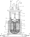
別の例示的で、非限定的なバイオリアクターである、Celligen 310バイオリアクターを図1に示す。繊維性ベッドバスケット(16)には、ポリエステルディスク (10)が装着される。幾つかの実施形態においては、容器は、外部ポート(1;[他の実施形態においては、本ポートは細胞回収用にも使用され得る])を介して、脱イオン水又は等張なバッファーが充填され、次いで、任意選択で、オートクレーブされる。他の実施形態においては、滅菌に続いて、液は、(9)に示したように、ディスクベッド(disk bed)を満たす増殖培地で置換される。また更なる実施形態においては、植え継ぎに先立って、温度、pH、溶存酸素濃度等が設定される。なお更なる実施形態においては、細胞付着を促進するために、初期に遅い撹拌速度が使用され、次いで、撹拌が速められる。代替的又は追加的に、外部ポート(2)を介して、新鮮な培地を添加することによって、灌流が開始される。必要に応じて、代謝生成物は、バスケット(8)の上の細胞を含まない培地から回収しても良い。幾つかの実施形態においては、インペラーの回転によって、ドラフトチューブ(18)内を陰圧にし、ドラフトチューブ、次いでインペラーポート(19)を通って、リザーバー(15)から細胞を含まない廃液を引き、それによって培地が連続ループ(continuous loop)内で均一に循環(12)する。また更なる実施形態においては、チューブ(6)の調整によって、液面を制御し;幾つかの実施形態においては、本チューブの外部開口部(4)は、回収のために使用される。他の実施形態においては、リングスパージャ(ring sparger)(見えない)は、外部ポート(3)から添加されたガスによって、インペラーを通って流れる培地に酸素を送るために、インペラーエアレーションチャンバー(11)内部に位置し、筺体(5)及びスパージャーライン(7)内部に維持されても良い。代替的又は追加的に、リモートチャンバーに閉じ込められたスパージド・ガス(sparged gas)は、固定化された細胞に打ち寄せる栄養培地によって吸収される。更に他の実施形態においては、ジャケット水を入れる(13)及び排出する(14)ためのポートを備えた、ウォータージャケット(water jacket)(17)が存在する。 Another exemplary, non-limiting bioreactor, the Celligen 310 bioreactor, is shown in Figure 1. A polyester disc (10) is attached to the fibrous bed basket (16). In some embodiments, the vessel is provided with deionized water or an isotonic buffer via an external port (1; [in other embodiments, the port may also be used for cell recovery]). It is filled and then, optionally, autoclaved. In another embodiment, following sterilization, the solution is replaced with a growth medium that fills a disk bed, as shown in (9). In a further embodiment, the temperature, pH, dissolved oxygen concentration and the like are set prior to subculture. In a further embodiment, a slow agitation rate is initially used to promote cell adhesion, followed by an increased agitation. Alternatively or additionally, perfusion is initiated by adding fresh medium via the external port (2). If desired, the metabolites may be recovered from the cell-free medium above the basket (8). In some embodiments, rotation of the impeller creates a negative pressure in the draft tube (18) and draws cell-free effluent from the reservoir (15) through the draft tube and then the impeller port (19). This causes the medium to circulate uniformly (12) in a continuous loop. In a further embodiment, the liquid level is controlled by adjusting the tube (6); in some embodiments, the external opening (4) of the tube is used for recovery. In another embodiment, the ring sparger (invisible) is an impeller aeration chamber (11) to deliver oxygen to the medium flowing through the impeller by the gas added from the external port (3). It may be located inside and maintained inside the housing (5) and sparger line (7). Alternatively or additionally, the sparged gas confined in the remote chamber is absorbed by the nutrient medium that slams into the immobilized cells. In yet another embodiment, there is a water jacket (17) provided with ports for entering (13) and draining (14) jacket water.
ある実施形態においては、灌流チャンバーが担体を含む、灌流バイオリアクターが使用される。より具体的な実施形態においては、担体は、マクロキャリア、マイクロキャリア、又はその両方から選択しても良い。マイクロキャリアは当業者に周知であり、例えば参照により本明細書に取り込まれる、米国特許番号第8,828,720号、第7,531,334号、第5,006,467号に記載されている。マイクロキャリアはまた、例えばCytodexTM(Pharmacia Fine Chemical社から入手可能)、Superbeads (Flow Lab社から市販)として、DE-52及びDE-53 (Whatman社から市販)として市販されている。市販されているマイクロキャリアの他の非限定的な例としては、アルギン酸塩ベース (GEM、Global Cell Solutions)、デキストランベース (Cytodex、GE Healthcare)、コラーゲンベース (Cultispher、Percell)及びポリスチレンベース (SoloHill Engineering)のマイクロキャリアが挙げられる。 In certain embodiments, a perfusion bioreactor is used in which the perfusion chamber comprises a carrier. In more specific embodiments, the carrier may be selected from macrocarriers, microcarriers, or both. Microcarriers are well known to those of skill in the art and are described, for example, in US Pat. Nos. 8,828,720, 7,531,334, 5,006,467, which are incorporated herein by reference. Microcarriers are also marketed, for example, Cytodex TM (available from Pharmacia Fine Chemical), Superbeads (commercially available from Flow Lab), DE-52 and DE-53 (commercially available from Whatman). Other non-limiting examples of commercially available microcarriers include alginate-based (GEM, Global Cell Solutions), dextran-based (Cytodex, GE Healthcare), collagen-based (Cultispher, Percell) and polystyrene-based (SoloHill Engineering). ) Microcarriers can be mentioned.
幾つかの実施形態においては、灌流バイオリアクター中に担体が充填され、例えば、栄養培地中に浸漬される充填層(packed bed)を形成する。ある実施形態においては、マイクロキャリアは、灌流バイオリアクター内部に充填される。代替的又は追加的に、担体は接着性材料を含み得る。他の実施形態においては、担体の表面は接着性材料を含むか、又は担体の表面は接着性である。更に他の実施形態においては、材料は、例えば細胞の接着を可能にする、表面に露出した帯電した基等の化学構造を示す。本態様に従って使用され得る接着性材料の非限定的な例としては、ポリエステル、ポリプロピレン、ポリアルキレン、ポリフルオロクロロエチレン、ポリ塩化ビニル、ポリスチレン、ポリスルホン、セルロースアセテート、ガラス繊維、セラミック粒子、ポリ-L-乳酸及び不活性金属繊維が挙げられる。更に特定の実施形態においては、材料はポリエステル及びポリプロピレンから選択され得る。様々な実施形態において、「接着性材料」は、合成か、又は他の実施形態においては、天然で生じるか、又は他の実施形態においては、それらの組合せである材料を指す。ある実施形態においては、材料は細胞毒性がない(又は、他の実施形態においては、生物学的に適合性がある)。合成接着性材料の非限定的な例としては、ポリエステル、ポリプロピレン、ポリアルキレン、ポリフルオロクロロエチレン、ポリ塩化ビニル、ポリスチレン、ポリスルホン、セルロースアセテート、及びポリ-L-乳酸、ガラス繊維、セラミック粒子及び不活性金属繊維、又はより具体的な実施形態においては、ポリエステル、ポリプロピレン、ポリアルキレン、ポリフルオロクロロエチレン、ポリ塩化ビニル、ポリスチレン、ポリスルホン、セルロースアセテート及びポリ-L-乳酸が挙げられる。他の実施形態としては、MatrigelTM、細胞外マトリクス成分(例、フィブロネクチン、コンドロネクチン、ラミニン)及びコラーゲンが挙げられる。 In some embodiments, the carrier is packed in a perfusion bioreactor to form, for example, a packed bed that is immersed in a nutrient medium. In certain embodiments, the microcarriers are packed inside a perfusion bioreactor. Alternatively or additionally, the carrier may include an adhesive material. In other embodiments, the surface of the carrier contains an adhesive material or the surface of the carrier is adhesive. In yet another embodiment, the material exhibits a chemical structure, such as a charged group exposed on the surface, which allows for cell adhesion, for example. Non-limiting examples of adhesive materials that can be used according to this embodiment are polyester, polypropylene, polyalkylene, polyfluorochloroethylene, polyvinyl chloride, polystyrene, polysulfone, cellulose acetate, glass fiber, ceramic particles, poly-L. -Includes lactic acid and inert metal fibers. In a further specific embodiment, the material may be selected from polyester and polypropylene. In various embodiments, "adhesive material" refers to a material that is synthetic or, in other embodiments, naturally occurring, or in other embodiments, a combination thereof. In some embodiments, the material is not cytotoxic (or in other embodiments, it is biologically compatible). Non-limiting examples of synthetic adhesive materials include polyester, polypropylene, polyalkylene, polyfluorochloroethylene, polyvinyl chloride, polystyrene, polysulfone, cellulose acetate, and poly-L-lactic acid, glass fiber, ceramic particles and non-polyesters. Active metal fibers, or in more specific embodiments, include polyester, polypropylene, polyalkylene, polyfluorochloroethylene, polyvinyl chloride, polystyrene, polysulfone, cellulose acetate and poly-L-lactic acid. Other embodiments include Matrigel TM , extracellular matrix components (eg, fibronectin, chondronectin, laminin) and collagen.
代替的又は追加的に、接着性材料は繊維性であり、より具体的な実施形態においては、織布の繊維性マトリクス、不織布の繊維性マトリクス、又はその両方であり得る。更に他の実施形態においては、材料は、細胞の接着を可能にする、例えば帯電した表面の基等の化学構造、例、ポリエステル、ポリプロピレン、ポリアルキレン、ポリフルオロクロロエチレン、ポリ塩化ビニル、ポリスチレン、ポリスルホン、セルロースアセテート及びポリ-L-乳酸を示す。更に特定の実施形態においては、材料は、ポリエステル及びポリプロピレンから選択され得る。 Alternatively or additionally, the adhesive material is fibrous and, in more specific embodiments, may be a fibrous matrix of woven fabrics, a fibrous matrix of non-woven fabrics, or both. In yet other embodiments, the material allows for cell adhesion, eg, chemical structures such as charged surface groups, eg polyester, polypropylene, polyalkylene, polyfluorochloroethylene, polyvinyl chloride, polystyrene, etc. Shows polysulfone, cellulose acetate and poly-L-lactic acid. In a further specific embodiment, the material can be selected from polyester and polypropylene.
代替的又は追加的に、担体は、任意選択で、接着性の繊維性材料、より具体的な実施形態においては、織布の繊維性マトリクス、不織布の繊維性マトリクス、又はその両方であり得る、繊維性材料を含む。繊維性担体の非限定的な例は、Eppendorf社(Enfield、CT)から市販されているNew Brunswick Scientific Fibracel(登録商標)担体であり、ポリエステル及びポリプロピレンからなり;並びにCESCO BioProducts (Atlanta、GA)から市販されているBioNOC II担体であり、PET(ポリエチレンテレフタレート)からなる。ある実施形態においては、繊維性マトリクスが指すものは、ポリエステル、ポリプロピレン、ポリアルキレン、ポリフルオロクロロエチレン、ポリ塩化ビニル、ポリスチレン又はポリスルホンを含む。更に特定の実施形態においては、繊維性マトリクスは、ポリエステル及びポリプロピレンから選択される。 Alternatively or additionally, the carrier can optionally be an adhesive fibrous material, and in more specific embodiments, a fibrous matrix of woven fabrics, a fibrous matrix of non-woven fabrics, or both. Includes fibrous materials. A non-limiting example of a fibrous carrier is the New Brunswick Scientific Fibracel® carrier commercially available from Eppendorf (Enfield, CT), which consists of polyester and polypropylene; and from CESCO BioProducts (Atlanta, GA). It is a commercially available BioNOC II carrier and consists of PET (polyethylene terephthalate). In certain embodiments, what the fibrous matrix refers to includes polyester, polypropylene, polyalkylene, polyfluorochloroethylene, polyvinyl chloride, polystyrene or polysulfone. In a further specific embodiment, the fibrous matrix is selected from polyester and polypropylene.
ある実施形態においては、ASCは、担体、例えば充填された担体上でのインキュベーションに先立って、2D装置、例えば組織培養プレート又はディッシュ中でインキュベートしても良い。他の実施形態においては、ASCは、担体、例えば充填された担体上でのインキュベーションに先立って、2D装置中でインキュベートされない。 In certain embodiments, the ASC may be incubated in a 2D device, such as a tissue culture plate or dish, prior to incubation on a carrier, such as a packed carrier. In other embodiments, the ASC is not incubated in a 2D device prior to incubation on a carrier, eg, a packed carrier.
他の実施形態においては、細胞は充填層スピナーフラスコを使用して生成される。より具体的な実施形態においては、充填層は、スピナーフラスコ及びマグネティックスターラーを含んでも良い。幾つかの実施形態においては、スピナーフラスコは、CelligenTM Plug Flowバイオリアクターに類似する充填床装置に取り付けても良く、それは、ある実施形態においては、Fibra-cel(登録商標)(又は、他の実施形態においては、他の担体)が充填されている。ある実施形態においては、スピナーは、バッチで供給され(batch fed)(又は、他の代替的な実施形態においては、灌流によって供給される)、1以上の滅菌フィルターが取り付けられ、組織培養インキュベーター内に配置される。更なる実施形態においては、細胞は、培地中で懸濁し、培地を装置に導入することによって、足場に播種される。また更なる実施形態においては、撹拌速度を次第に増加し、例えば40 RPMで4時間から開始し、次いで120 RPMにまで次第に速度を上げる。ある実施形態においては、培地のグルコースレベルを、定期的に(即ち、毎日)テストしても良く、調整された灌流速度は、ある実施形態においては、400〜700 mg/Lの間、450〜650 mg/Lの間、475〜625 mg/Lの間、500〜600 mg/Lの間又は525〜575 mg/Lの間である、許容可能なグルコース濃度を維持する。更に他の実施形態においては、培養プロセスの終わりに、担体を充填層から取り出し、任意選択で、等張なバッファーで洗浄し、撹拌及び/又は酵素による分解によって、細胞は処理され、又は担体から回収される。 In other embodiments, cells are generated using a packed bed spinner flask. In a more specific embodiment, the packed bed may include a spinner flask and a magnetic stirrer. In some embodiments, the spinner flask may be attached to a packed bed device similar to the Celligen TM Plug Flow bioreactor, which in some embodiments is Fibra-cel® (or other). In the embodiment, another carrier) is packed. In one embodiment, the spinner is batch fed (or, in other alternative embodiments, fed by perfusion), fitted with one or more sterile filters, and in a tissue culture incubator. Is placed in. In a further embodiment, the cells are seeded on the scaffold by suspending in medium and introducing the medium into the device. In a further embodiment, the agitation rate is gradually increased, starting at, for example, 40 RPM for 4 hours, then gradually increasing to 120 RPM. In some embodiments, glucose levels in the medium may be tested on a regular basis (ie, daily), with adjusted perfusion rates ranging from 450 to 700 mg / L in some embodiments. Maintain an acceptable glucose concentration between 650 mg / L, 475-625 mg / L, 500-600 mg / L or 525-575 mg / L. In yet other embodiments, at the end of the culture process, the carrier is removed from the packed bed, optionally washed with isotonic buffer, stirred and / or degraded by enzyme, the cells are treated or removed from the carrier. Will be recovered.
ある実施形態においては、3D増殖装置(幾つかの実施形態においては、先述のバイオリアクター)は繊維性ベッド(fibrous bed)を含む。より具体的な実施形態においては、繊維性ベッドは、ポリエステル、ポリプロピレン、ポリアルキレン、ポリフルオロクロロエチレン、ポリ塩化ビニル、ポリスチレン、ポリスルホン又はポリアミド(例、脂肪族ポリアミド)を含み得る。他の実施形態においては、ガラス繊維若しくは金属繊維(例、不活性金属繊維)が存在しても良く;又はセルロース繊維(その非限定的な例は、レーヨンである)が存在しても良い。 In some embodiments, the 3D breeder (in some embodiments, the bioreactor described above) comprises a fibrous bed. In a more specific embodiment, the fibrous bed may include polyester, polypropylene, polyalkylene, polyfluorochloroethylene, polyvinyl chloride, polystyrene, polysulfone or polyamide (eg, aliphatic polyamides). In other embodiments, glass or metal fibers (eg, inert metal fibers) may be present; or cellulose fibers (of which a non-limiting example is rayon) may be present.
他の実施形態においては、装置又はバイオリアクターは、ゲルマトリクスを含む。典型的には、ゲル相を形成する、ゲル捕集水(gels trap water)である。他の実施形態においては、装置又はバイオリアクターは、細胞が繊維の空隙(lumen)内で増殖及び増殖する(grow and proliferate)ように構成された中空繊維マトリックスを含む。更に他の実施形態においては、装置又はバイオリアクターは、細胞増殖のための基材として働く球体、ビーズ又は担体を有する、充填層マトリクス又は流動床マトリクスを含む。様々な実施形態において、球体又はビーズは、微多孔質、多孔質又は非多孔質であっても良く、後者のケースにおいては、細胞はその表面にのみ付着する。更に他の実施形態においては、装置又はバイオリアクターは、スポンジ様の構造を有するマトリクスを含む。 In other embodiments, the device or bioreactor comprises a gel matrix. Typically, it is gels trap water that forms the gel phase. In other embodiments, the apparatus or bioreactor comprises a hollow fiber matrix configured such that cells grow and proliferate within a lumen of fibers. In yet other embodiments, the apparatus or bioreactor comprises a packed bed matrix or fluidized bed matrix having spheres, beads or carriers that serve as substrates for cell proliferation. In various embodiments, the spheres or beads may be microporous, porous or non-porous, in the latter case the cells attach only to their surface. In yet another embodiment, the apparatus or bioreactor comprises a matrix having a sponge-like structure.
ある実施形態においては、バイオリアクターは、培地1 ml当たり10,000〜2,000,000細胞の間の濃度で、他の実施形態においては、20,000〜2,000,000細胞/ml、他の実施形態においては、30,000〜1,500,000細胞/ml、他の実施形態においては、40,000〜1,400,000細胞/ml、他の実施形態においては、50,000〜1,300,000細胞/ml、他の実施形態においては、60,000〜1,200,000細胞/ml、他の実施形態においては、70,000〜1,100,000細胞/ml、他の実施形態においては、80,000〜1,000,000細胞/ml、他の実施形態においては、80,000〜900,000細胞/ml、他の実施形態においては、80,000〜800,000細胞/ml、他の実施形態においては、80,000〜700,000細胞/ml、他の実施形態においては、80,000〜600,000細胞/ml、他の実施形態においては、80,000〜500,000細胞/ml、他の実施形態においては、80,000〜400,000細胞/ml、他の実施形態においては、90,000〜300,000細胞/ml、他の実施形態においては、90,000〜250,000細胞/ml、他の実施形態においては、90,000〜200,000細胞/ml、他の実施形態においては、100,000〜200,000細胞/ml、他の実施形態においては、110,000〜1,900,000細胞/ml、他の実施形態においては、120,000〜1,800,000細胞/ml、他の実施形態においては、130,000〜1,700,000細胞/ml、他の実施形態においては、140,000〜1,600,000細胞/mlの間の濃度で播種される。 In some embodiments, the bioreactor has a concentration between 10,000 and 2,000,000 cells per ml of medium, in other embodiments 20,000 to 2,000,000 cells / ml, and in other embodiments 30,000 to 1,500,000 cells / ml. ml, 40,000 to 1,400,000 cells / ml in other embodiments, 50,000 to 1,300,000 cells / ml in other embodiments, 60,000 to 1,200,000 cells / ml in other embodiments, in other embodiments. , 70,000 to 1,100,000 cells / ml, 80,000 to 1,000,000 cells / ml in other embodiments, 80,000 to 900,000 cells / ml in other embodiments, 80,000 to 800,000 cells / ml in other embodiments, 80,000 to 700,000 cells / ml in other embodiments, 80,000 to 600,000 cells / ml in other embodiments, 80,000 to 500,000 cells / ml in other embodiments, 80,000 in other embodiments. ~ 400,000 cells / ml, in other embodiments 90,000 to 300,000 cells / ml, in other embodiments 90,000 to 250,000 cells / ml, in other embodiments 90,000 to 200,000 cells / ml, other 100,000 to 200,000 cells / ml in embodiments, 110,000 to 1,900,000 cells / ml in other embodiments, 120,000 to 1,800,000 cells / ml in other embodiments, 130,000 to 1,700,000 in other embodiments. Cells / ml, in other embodiments, are seeded at concentrations between 140,000 and 1,600,000 cells / ml.
更に他の実施形態においては、担体(基材)1 g (gr)当たり1〜20×106細胞の間で播種され、又は他の実施形態においては、担体1 g当たり1.5〜20×106細胞、又は他の実施形態においては、担体1 g当たり1.5〜18×106細胞、又は他の実施形態においては、担体1 g当たり1.8〜18×106細胞、又は他の実施形態においては、担体1 g当たり2〜18×106細胞、又は他の実施形態においては、担体1 g当たり3〜18×106細胞、又は他の実施形態においては、担体1 g当たり2.5〜15×106細胞、又は他の実施形態においては、担体1 g当たり3〜15×106細胞、又は他の実施形態においては、担体1 g当たり3〜14×106細胞、又は他の実施形態においては、担体1 g当たり3〜12×106細胞、又は他の実施形態においては、担体1 g当たり3.5〜12×106細胞、又は他の実施形態においては、担体1 g当たり3〜10×106細胞、又は他の実施形態においては、担体1 g当たり3〜9×10⁶細胞、又は他の実施形態においては、担体1 g当たり4〜9×10⁶細胞、又は他の実施形態においては、担体1 g当たり4〜8×10⁶細胞、又は他の実施形態においては、担体1 g当たり4〜7×10⁶細胞、又は他の実施形態においては、担体1 g当たり4.5〜6.5×10⁶細胞が播種される。 In yet other embodiments, it is seeded between 1-20 × 10 6 cells per 1 g (gr) of carrier (base material) , or in other embodiments 1.5-20 × 10 6 per 1 g of carrier. Cells, or in other embodiments 1.5-18 × 10 6 cells per 1 g of carrier, or in other embodiments 1.8-18 × 10 6 cells per 1 g of carrier, or in other embodiments, carrier 1 g per 2 to 18 × 10 6 cells, or in other embodiments, the carrier 1 g per 3 to 18 × 10 6 cells, or in other embodiments, the carrier 1 g per 2.5 to 15 × 10 6 Cells, or in other embodiments 3-15 × 10 6 cells per 1 g of carrier, or in other embodiments 3-14 × 10 6 cells per 1 g of carrier, or in other embodiments, carrier 1 g per 3 to 12 × 10 6 cells, or in other embodiments, the carrier 1 g per 3.5 to 12 × 10 6 cells, or in other embodiments, the carrier 1 g per 3 to 10 × 10 6 Cells, or in other embodiments 3-9 × 10 6 cells per 1 g of carrier, or in other embodiments 4-9 × 10 6 cells per 1 g of carrier, or in other embodiments, 4-8 x 10 6 cells per 1 g of carrier , or 4-7 x 10 6 cells per 1 g of carrier in other embodiments, or 4.5-6.5 x 10 6 cells per 1 g of carrier in other embodiments. The cells are seeded.
幾つかの実施形態においては、図22A〜Bに準拠し、また参照することによりその全体が本明細書に取り込まれる、2014年3月13日に公開された国際公開第2014/037862号に記載されているように、ASCの増殖及び/又はインキュベーションのために溝付き担体30が使用される。様々な実施形態において、担体は、(例、培養プレート又はディッシュ上での)2Dインキュベーション後、又は前もって2Dインキュベーションを行わずに、使用されても良い。他の実施形態においては、担体上でのインキュベーションに、例えば、充填層基材若しくはマイクロキャリアであり得る、バイオリアクター中の3D基材上でのインキュベーションが続けられる場合があり;又は担体上でのインキュベーションに、3D基材上でのインキュベーションが続けられない場合がある。担体30は、担体30の外部から担体30の内部に向かって伸びる、複数の2次元(2D)表面12を含み得る。示されるように、表面は、離間した一群のリブ14によって形成され、開口部16を形成し、それは、使用の間、細胞及び培養培地 (非表示)が流れるような大きさであり得る。図22Cに準拠して、担体30はまた、担体30の中心の担体軸18から伸び、開口部16を形成するよう離間したリブ14に対してほぼ垂直に伸びる、複数の2D表面12を含み得、複数の2D表面12を創出する。幾つかの実施形態においては、担体30は、国際公開第2014/037862号に記載されているような「3Dボディ」であり;3Dボディに関連する内容は、参照により本明細書に組み込まれる。
In some embodiments, it is described in WO 2014/037862, published March 13, 2014, which is in accordance with FIGS. 22A-B and is incorporated herein by reference in its entirety.
ある実施形態においては、バイオリアクター中で、記載された担体(例、溝付き担体)が使用される。その幾つかにおいては、担体は充填された形態である。 In certain embodiments, the described carriers (eg, grooved carriers) are used in the bioreactor. In some of them, the carrier is in packed form.
更に他の実施形態においては、複数の2D表面を形成する材料は、少なくとも1つのポリマーを含む。幾つかの実施形態においては、細胞付着又は細胞生物学のパラメーターを制御するために、好適なコーティングを選択しても良い。 In yet another embodiment, the material forming the plurality of 2D surfaces comprises at least one polymer. In some embodiments, suitable coatings may be selected to control cell adhesion or cell biology parameters.
ある実施形態においては、記載された方法は、3D培養装置からASCを取り出すことによってASCを回収する(様々な実施形態において、サイトカインの添加あり又は無しであっても良い、記載された3Dインキュベーションに続く)、後続の工程を更に含む。更に特定の実施形態においては、細胞は3Dマトリクスから取り出され得る一方で、マトリクスはバイオリアクター内に残る。ある実施形態においては、バイオリアクターからの回収の時点で、細胞の少なくとも約10%、少なくとも12%、少なくとも14%、少なくとも16%、少なくとも18%、少なくとも20%、少なくとも22%、少なくとも24%、少なくとも26%、少なくとも28%、又は少なくとも30%が、S及びG2/M期(まとめて)にある。細胞周期のフェーズは、当該分野において公知である様々な方法、例えばFACS検出によってアッセイし得る。典型的には、FACSのケースにおいては、S及びG2/M期の細胞のパーセンテージは、例えば前方散乱/側方散乱ゲートを使用して、生細胞についてゲーティング後、生細胞のパーセンテージで表される。当業者は、これらのフェーズにある細胞のパーセンテージが、増殖している細胞のパーセンテージと相関することを理解する。幾つかのケースにおいては、有意に指数増殖期を経過して、バイオリアクター内に細胞が残るようにすることで、増殖している細胞数における減少が起こる。 In certain embodiments, the described method recovers the ASC by removing the ASC from the 3D incubator (in various embodiments, with or without the addition of cytokines, to the described 3D incubation. (Continued), further including subsequent steps. In a further specific embodiment, the cells can be removed from the 3D matrix, while the matrix remains in the bioreactor. In certain embodiments, at least about 10%, at least 12%, at least 14%, at least 16%, at least 18%, at least 20%, at least 22%, at least 24% of the cells at the time of recovery from the bioreactor. At least 26%, at least 28%, or at least 30% are in the S and G2 / M phases (collectively). The phase of the cell cycle can be assayed by various methods known in the art, such as FACS detection. Typically, in the case of FACS, the percentage of cells in S and G2 / M phases is expressed as the percentage of live cells after gating for live cells, for example using a forward / side scatter gate. To. Those skilled in the art will appreciate that the percentage of cells in these phases correlates with the percentage of proliferating cells. In some cases, allowing cells to remain in the bioreactor after a significant exponential growth phase results in a decrease in the number of proliferating cells.
ある実施形態においては、抗がん剤として使用されるASCは、がん細胞と、又は他の実施形態においては、がん細胞若しくはがん細胞株に由来する馴化培地(「CM」)中でインキュベートした1以上のがん細胞株と、以前に共インキュベートされているか、或いはがん細胞若しくはがん細胞株に由来するCMの画分を含む培地中でインキュベートされている。他の実施形態においては、抗がん剤としての使用のためのCMを生成するために使用されたASCは、がん細胞と、又は他の実施形態においては、1以上のがん細胞株と、共インキュベートされている。幾つかの実施形態においては、共インキュベーションは、ASCと、がん細胞又は細胞株とが互いに接触する条件下で行われる。そのような条件としては、同じ装置中に、ASCと、がん細胞又は細胞株とを播種することが挙げられ、様々な実施形態において、一緒に播種する、最初にASCを播種する、又は最初にがん細胞若しくは細胞株を播種するのいずれかである。幾つかの実施形態においては、共インキュベーションは組織培養装置中で行われ、又は他の実施形態においては、バイオリアクター中で行われ、それは、幾つかの実施形態においては、3D増殖基材を含んでも良い。 In some embodiments, the ASC used as an anti-cancer agent is in a cancer cell and, in another embodiment, in a conditioned medium (“CM”) derived from the cancer cell or cancer cell line. It has been co-incubated with one or more cancer cell lines that have been incubated, or in a medium containing a fraction of the cancer cells or CM derived from the cancer cell line. In other embodiments, the ASCs used to generate CM for use as anti-cancer agents are with cancer cells, or in other embodiments, with one or more cancer cell lines. , Co-incubated. In some embodiments, co-incubation is performed under conditions where the ASC and the cancer cell or cell line are in contact with each other. Such conditions include seeding the ASC with cancer cells or cell lines in the same device, and in various embodiments, seeding together, seeding the ASC first, or seeding the ASC first. Either cancer cells or cell lines are seeded. In some embodiments, co-incubation is performed in a tissue culture apparatus, or in other embodiments, in a bioreactor, which in some embodiments comprises a 3D growth substrate. But it's okay.
他の実施形態においては、条件は、ASCと、がん細胞又は細胞株とが互いに接触しないが、培地及びその可溶性の成分が2つの細胞集団の間で交換されるような条件である。当業者は、培地の交換を可能にしながら、2つの細胞集団の間での接触を防ぐために、様々な手段が利用可能である(例えばそれに溶解した流体及び因子に対して透過性である膜、又は規定されたサイズよりも小さい可溶性因子がそれを通って拡散することができる半透過性の膜を用いて、細胞集団を分離することによる)ことを理解する。 In other embodiments, the condition is such that the ASC and the cancer cell or cell line do not contact each other, but the medium and its soluble components are exchanged between the two cell populations. Various means are available to those skilled in the art to prevent contact between the two cell populations while allowing the exchange of media (eg, membranes that are permeable to fluids and factors dissolved therein). Or by separating the cell population using a semi-permeable membrane through which soluble factors smaller than the specified size can diffuse.
他の実施形態においては、抗がん剤として使用されるASCは、がん細胞又はがん細胞株に由来するCM中で、前もってインキュベートされている。他の実施形態においては、抗がん剤として使用するためのCMを生成するために使用されるASCは、がん細胞又はがん細胞株に由来するCM中で、インキュベートされている。幾つかの実施形態においては、そのプロセスの第一段階において、がん細胞が培養され、インキュベーションによって生じる培地(「がん細胞CM」)が単離される。様々な実施形態において、第二段階において、ASCは、幾つかの実施形態においては、3D増殖基材を含み得る、培養ウェルを含むがそれに限定されない組織培養装置中、又は他の実施形態においては、バイオリアクター中で、がん細胞CMとインキュベートされる。更に他の実施形態においては、ASCは、がん細胞CM中でのインキュベーションに先立って、炎症性サイトカインに曝露されている。他の実施形態においては、(or)1以上のサイトカイン、ビタミン又は生物学的に活性なタンパク質が、がん細胞CMに添加される。更に他の実施形態においては、ASCのインキュベーションは、例えば低酸素、又はpH若しくは大気圧の変化といった非標準的な条件下で行われる。更に他の実施形態においては、ASCのインキュベーションによって生じるCM(「ASC CM」)、又は他の実施形態においては、ASCそのものが、抗がん剤として使用される。更に他の実施形態においては、ASCは、主に母体細胞、又は胎児細胞、又は胎児細胞と母体細胞の混合物である、胎盤ASCである。 In other embodiments, the ASC used as an anti-cancer agent is pre-incubated in a CM derived from a cancer cell or cancer cell line. In other embodiments, the ASC used to generate a CM for use as an anti-cancer agent is incubated in a CM derived from a cancer cell or cancer cell line. In some embodiments, in the first step of the process, the cancer cells are cultured and the medium produced by the incubation (“cancer cell CM”) is isolated. In various embodiments, in the second step, the ASC may, in some embodiments, contain a 3D growth substrate, in a tissue culture apparatus comprising but not limited to culture wells, or in other embodiments. , Incubated with cancer cell CM in a bioreactor. In yet another embodiment, the ASC is exposed to inflammatory cytokines prior to incubation in cancer cell CM. In other embodiments, one or more cytokines, vitamins or biologically active proteins are added to the cancer cell CM. In yet another embodiment, the incubation of ASC is performed under non-standard conditions such as hypoxia or changes in pH or atmospheric pressure. In yet other embodiments, the CM produced by the incubation of ASC (“ASC CM”), or in other embodiments, ASC itself is used as an anti-cancer agent. In yet another embodiment, the ASC is a placental ASC, which is primarily a maternal cell, or fetal cell, or a mixture of fetal and maternal cells.
他の実施形態においては、抗がん剤として使用されるASCは、がん細胞又はがん細胞株に由来するCMの画分を含む培地中でインキュベートされている。他の実施形態においては、抗がん剤として使用するためのCMを生成するために使用されるASCは、がん細胞又はがん細胞株に由来するCMの画分を含む培地中でインキュベートされている。幾つかの実施形態においては、第一段階においては、がん細胞が培養され、がん細胞CM画分が単離され、標準的な培養培地に添加される。ある実施形態においては、プロセスは、例えばバイオリアクター中、又は培養ウェル中で、第一段階で生成された培地を用いて、ASCがインキュベートされる、第二段階を含む。 In other embodiments, the ASC used as an anti-cancer agent is incubated in medium containing a fraction of CM derived from a cancer cell or cancer cell line. In other embodiments, the ASC used to generate CM for use as an anti-cancer agent is incubated in a medium containing a fraction of CM derived from a cancer cell or cancer cell line. ing. In some embodiments, in the first step, the cancer cells are cultured, the cancer cell CM fraction is isolated and added to standard culture medium. In certain embodiments, the process comprises a second step in which the ASC is incubated with the medium produced in the first step, eg, in a bioreactor or in a culture well.
ある実施形態においては、先述のインキュベーションの条件は、共インキュベーションの間に、がん細胞若しくは細胞株がスフェロイドを形成し、又は他の実施形態においては、マイクロスフェロイドを形成するような条件である。 In some embodiments, the incubation conditions described above are such that the cancer cells or cell lines form spheroids during co-incubation, or in other embodiments, microspheroids.
様々な実施形態において、がん細胞若しくはがん細胞株を用いた、又はそれらに由来するCMを用いた、ASCのインキュベーションは、がん細胞、がん細胞株又はそれらに由来するCMの非存在下での、本明細書に記載されるような、ASCの3D増殖後に行われる。他の実施形態においては、ASCの3D増殖の全プロセスは、がん細胞、がん細胞株又はそれらに由来するCMの存在下で行われる。更に他の実施形態においては、がん細胞、がん細胞株又はそれらに由来するCMの非存在下でのASCの増殖は、がん細胞、がん細胞株又はそれらに由来するCMとの、ASCの共インキュベーション後に起こる。ある実施形態においては、がん細胞、がん細胞株又はそれらに由来するCMを用いない、及び/又はそれらを用いた、ASCの3Dインキュベーション前に、ASCの2D増殖が行われる。 In various embodiments, incubation of ASC with or with CMs derived from cancer cells or cancer cell lines results in the absence of cancer cells, cancer cell lines or CMs derived from them. Performed below, after 3D proliferation of ASCs, as described herein. In other embodiments, the entire process of 3D proliferation of ASC is carried out in the presence of cancer cells, cancer cell lines or CMs derived from them. In yet another embodiment, the growth of ASC in the absence of cancer cells, cancer cell lines or CMs derived from them is such that with cancer cells, cancer cell lines or CMs derived from them. Occurs after co-incubation of ASCs. In certain embodiments, 2D proliferation of ASC is performed prior to 3D incubation of ASC without and / or with cancer cells, cancer cell lines or CMs derived from them.
ある実施形態においては、ASCは、例えば、がん細胞又はがん細胞株を用いた、追加的なインキュベーションを行うのに先立って、それらを増殖する(expand)ために使用されたバイオリアクター中で、炎症性サイトカインに曝露されている。他の実施形態においては、(i)バイオリアクター中でASCを増殖し、(ii)任意選択で、バイオリアクターからそれらを回収し、(iii)ASCを、がん細胞又はがん細胞株と共に更にインキュベートした後に、ASCは炎症性サイトカインに曝露されている。様々な実施形態において、培養プレート中、任意選択で、例えば低酸素、又はpH若しくは周囲圧力の変化といった非標準的な条件下で、更なるインキュベーションが行われる。 In certain embodiments, the ASC is in a bioreactor used to expand, for example, cancer cells or cancer cell lines prior to additional incubation. , Exposed to inflammatory cytokines. In other embodiments, (i) ASCs are grown in a bioreactor, (ii) optionally recovered from the bioreactor, and (iii) ASCs are further added with cancer cells or cell lines. After incubation, ASCs have been exposed to inflammatory cytokines. In various embodiments, further incubation is carried out in the culture plate, optionally under non-standard conditions such as hypoxia or changes in pH or ambient pressure.
幾つかの実施形態においては、ASCと共に若しくはそれに近接してインキュベートされる先述のがん細胞、又はASCと共にインキュベートされるCM(若しくはその画分)は、治療される患者のものであり;或いは、他の実施形態においては、治療される腫瘍と同じ、がんの種類由来であり;或いは、他の実施形態においては、がんの進行及び/又はその治療の過程において、変化を経ている、がん細胞の亜集団由来である。他の実施形態においては、がん細胞は、任意の他のがん細胞であり;例、ASCに治療因子の分泌を誘導することができる種類である。 In some embodiments, the aforementioned cancer cells incubated with or in close proximity to the ASC, or the CM (or fraction thereof) incubated with the ASC, is that of the patient being treated; or. In other embodiments, it is derived from the same type of cancer as the tumor being treated; or, in other embodiments, it has undergone changes in the progression of the cancer and / or in the course of its treatment. It is derived from a subpopulation of cancer cells. In other embodiments, the cancer cell is any other cancer cell; eg, a type capable of inducing the secretion of a therapeutic factor into ASC.
更に他の実施形態においては、増殖した細胞は、例えば、参照によって本明細書に取り込まれる、PCT国際特許出願公開番号第WO2012/140519号に記載されているように、振動又は撹拌を含む過程によって、バイオリアクターから回収される。ある実施形態においては、回収している間、任意選択で、カルシウムキレーターも含むプロテアーゼを用いた処理の間、若しくは他の実施形態においては、その処理の間及び後に、細胞は0.7〜6ヘルツの振動によって撹拌され、又は他の実施形態においては、1〜3ヘルツの振動によって撹拌される。ある実施形態においては、細胞を含む担体は、任意選択で、カルシウムキレーターも含む、プロテアーゼを含む溶液若しくは培地に浸漬している間に、0.7〜6ヘルツ、又は他の実施形態においては、1〜3ヘルツで撹拌される。プロテアーゼ及びカルシウムキレーターの非限定的な例は、EDTAを含む、トリプシン、又は任意選択で、他の酵素と組み合わされて同様の活性を有する別の酵素であり、それらの非限定的な例は、I型、II型、III型及びIV型コラゲナーゼである。トリプシンと同様の活性を有する酵素は、当該分野において周知であり;非限定的な例は、Thermo Fisher Scientific (Waltham MA)から市販されている、TrypLETM、真菌トリプシン様プロテアーゼ、並びにI型、II型、III型及びIV型コラゲナーゼである。コラゲナーゼと同様の同様の活性を有する酵素は、当該分野において周知であり;非限定的な例は、Sigma-Aldrichから市販されている、ディスパーゼ(Dispase)I及びディスパーゼIIである。 In yet another embodiment, the proliferated cells are incorporated by a process involving vibration or agitation, eg, as described in PCT International Patent Application Publication No. WO 2012/140519, which is incorporated herein by reference. , Recovered from the bioreactor. In some embodiments, during recovery, optionally during and after treatment with a protease that also contains a calcium chelator, or in other embodiments, during and after that treatment, the cells are 0.7-6 hertz. It is agitated by vibration, or in other embodiments, by vibration of 1-3 hertz. In some embodiments, the cell-containing carrier is optionally 0.7-6 hertz while immersed in a protease-containing solution or medium, including a calcium chelator, or, in other embodiments, 1-. Stir at 3 hertz. Non-limiting examples of proteases and calcium chelators are trypsin, including EDTA, or, optionally, another enzyme having similar activity in combination with other enzymes, and those non-limiting examples are: Type I, type II, type III and type IV collagenase. Enzymes with similar activity to trypsin are well known in the art; non-limiting examples are commercially available from Thermo Fisher Scientific (Waltham MA), TrypLE TM , fungal trypsin-like proteases, and type I, II. Type III and type IV collagenase. Enzymes with similar activity to collagenase are well known in the art; non-limiting examples are Dispase I and Dispase II, commercially available from Sigma-Aldrich.
他の実施形態においては、1以上のサイトカイン、ビタミン、又は生物学的に活性なタンパク質が、共インキュベーションのために使用される培地に添加される。 In other embodiments, one or more cytokines, vitamins, or biologically active proteins are added to the medium used for co-incubation.
他の実施形態においては、共インキュベーションは、例えば低酸素、又はpH若しくは大気圧の変化といった、非標準的な条件下で行われる。 In other embodiments, co-incubation is performed under non-standard conditions, such as hypoxia, or changes in pH or atmospheric pressure.
ある実施形態においては、共インキュベーションにおいて使用される細胞株は、取得したASC又はCMの治療標的であるがん細胞と同じ種類である必要がない。他の実施形態においては、共インキュベーションにおいて使用される細胞株は、取得したASC又はCMの治療標的であるがん細胞と同じ種類である。 In certain embodiments, the cell line used in the co-incubation does not have to be of the same type as the cancer cell that is the therapeutic target of the acquired ASC or CM. In other embodiments, the cell line used in the co-incubation is of the same type as the cancer cells that are the therapeutic target of the acquired ASC or CM.
様々な実施形態において、記載された共インキュベーション法の各々で使用されるASCは、主に母体細胞、若しくは主に胎児細胞、又は胎児細胞及び母体細胞の混合物である胎盤ASCを利用しても良い。これらの実施形態の各々を、ASCと、がん細胞若しくはがん細胞株との共インキュベーションの記載された実施形態と自由に組み合わせも良い。 In various embodiments, the ASC used in each of the described co-incubation methods may utilize predominantly maternal cells, or predominantly fetal cells, or placental ASC, which is a mixture of fetal cells and maternal cells. .. Each of these embodiments may be freely combined with the described embodiments of co-incubation of ASC with cancer cells or cancer cell lines.
炎症促進性サイトカインを用いた細胞の処理
記載された方法及び組成物のある実施形態においては、細胞を増殖するために使用される培養プロセスの過程において、培地の組成は変化しない。換言すると、因子を添加若しくは除去することによって、又は以前の培地とは異なる組成を有する新鮮な培地を添加することによって、培地組成を意図的に変化させようとはしない。当業者によって理解されるように、培地の組成を変化させることに関する言及には、例えばそれに含まれる細胞による栄養の吸収及び代謝物の分泌のために、長期培養の結果として自動的に生じる培地組成における変化は含まれない。
Treatment of Cells with Inflammatory Cytokines In certain embodiments of the methods and compositions described, the composition of the medium does not change during the course of the culture process used to proliferate the cells. In other words, it does not attempt to intentionally change the medium composition by adding or removing factors, or by adding fresh medium with a composition different from the previous medium. As will be appreciated by those skilled in the art, references to altering the composition of the medium will automatically occur as a result of long-term culture, for example due to the absorption of nutrients and the secretion of metabolites by the cells contained therein. Changes in are not included.
他の実施形態においては、細胞を調製するために使用される3D培養法は、以下:(a)3D培養装置中、第一の増殖培地中で、ASCをインキュベートし(該第一の増殖培地には炎症性サイトカインが添加されていない);及び(b)その後、3D培養装置中、第二の増殖培地中で、ASCをインキュベートする(該第二の増殖培地には1以上の炎症促進性サイトカインが添加されている)、副工程を含む。当業者は、本開示を踏まえて、培養装置中の培地に単にサイトカインを添加することによって、又は他の実施形態においては、培養装置から培地を除去し、サイトカインを含む培地でそれを置換することによって、第一及び第二の増殖培地中でインキュベートするために、同じ3D培養装置を使用しても良いことを理解する。他の実施形態においては、例えば、サイトカイン含有培地を添加する前に、異なるインキュベーターに細胞を移す(例、継代する)ことによって、サイトカインの存在下でのインキュベーションのために、異なる3D培養装置を使用しても良い。様々な実施形態において、当業者は、本開示を踏まえて、記載された方法において使用されるASCは、本明細書において更に記載されるように、胎盤から、脂肪組織から、又は他のソースから抽出しても良いことを理解する。 In other embodiments, the 3D culture method used to prepare the cells is as follows: (a) Incubate the ASC in the 3D culture medium, in the first growth medium (the first growth medium). No inflammatory cytokines added); and (b) then incubate the ASC in a 3D culture medium, in a second growth medium (the second growth medium is one or more pro-inflammatory). Cytokines have been added), including sub-steps. In light of the present disclosure, those skilled in the art may simply add cytokines to the medium in the incubator, or, in other embodiments, remove the medium from the incubator and replace it with a medium containing cytokines. Understand that the same 3D incubator may be used for incubation in the first and second growth media. In other embodiments, different 3D culture devices are used for incubation in the presence of cytokines, eg, by transferring (eg, subculturing) cells to different incubators prior to adding cytokine-containing medium. You may use it. In various embodiments, one of ordinary skill in the art, in light of the present disclosure, will use the ASCs used in the described methods from the placenta, from adipose tissue, or from other sources, as further described herein. Understand that it is okay to extract.
本明細書において、交換可能に使用される、1以上の「炎症促進性」サイトカイン又は「炎症性サイトカイン」についての言及は、哺乳動物宿主、例えばヒト宿主における炎症反応を媒介する少なくとも1つのサイトカインが存在することを意味する。非限定的なサイトカインの一覧は、インターフェロン-γ(IFN-γ;UniProt識別子P01579)、IL-22(UniProt識別子Q9GZX6)、腫瘍壊死因子-アルファ(TNF-アルファ;UniProt識別子P01375)、IFN-アルファ(IFN-α)、IFN-ベータ(UniProt識別子P01574)、IL-1アルファ(UniProt識別子P01583)、IL-1ベータ(UniProt識別子P01584)、IL-17(UniProt識別子Q5QEX9)、IL-23(UniProt識別子Q9NPF7)、IL-17A(UniProt識別子Q16552)、IL-17F(UniProt識別子Q96PD4)、IL-21(UniProt識別子Q9HBE4)、IL-13(UniProt識別子P35225)、IL-5(UniProt識別子P05113)、IL-4(UniProt識別子P05112)、IL-33(UniProt識別子O95760)、IL-1RL1(UniProt識別子Q01638)、TNF-ベータ(UniProt識別子P01374)、IL-11(UniProt識別子P20809)、IL-9(UniProt識別子P15248)、IL-2(UniProt識別子P60568)、IL-21(UniProt識別子Q9HBE4)、腫瘍壊死因子様リガンド(TL1A;別名TNFリガンドスーパーファミリーメンバー15;UniProt識別子O95150)、IL-12(アルファ及びベータサブユニットについてのそれぞれのUniProt識別子はP29459及びP29460)、並びにIL-18(UniProt識別子Q14116)である。更なるサイトカインとしては(それらに限定されないが):白血病阻害因子(LIF;UniProt識別子P15018)、オンコスタチンM(OSM;UniProt識別子P13725)、毛様体神経栄養因子(CNTF;UniProt識別子P26441)及びIL-8(UniProt識別子P10145)が挙げられる。本段落における全Swissprot及びUniProt入力内容は、2014年7月24日に接続された。特定のサイトカイン、それらの組合せ及び量、並びに特定の方法工程を含む、サイトカインを用いたASCのインキュベーションの様々な実施形態は、参照することによりその全体が本明細書に取り込まれる、Eytan Abrahamらの名義のPCT第IB2016/053310号において記載されている。
References herein to one or more "pro-inflammatory" or "inflammatory cytokines" used interchangeably include at least one cytokine that mediates an inflammatory response in a mammalian host, eg, a human host. It means that it exists. A list of non-limiting cytokines includes interferon-γ (IFN-γ; UniProt identifier P01579), IL-22 (UniProt identifier Q9GZX6), tumor necrosis factor-alpha (TNF-alpha; UniProt identifier P01375), IFN-alpha (IFN-alpha). IFN-α), IFN-Beta (UniProt identifier P01574), IL-1 alpha (UniProt identifier P01583), IL-1 beta (UniProt identifier P01584), IL-17 (UniProt identifier Q5QEX9), IL-23 (UniProt identifier Q9NPF7) ), IL-17A (UniProt identifier Q16552), IL-17F (UniProt identifier Q96PD4), IL-21 (UniProt identifier Q9HBE4), IL-13 (UniProt identifier P35225), IL-5 (UniProt identifier P05113), IL-4 (UniProt identifier P05112), IL-33 (UniProt identifier O95760), IL-1RL1 (UniProt identifier Q01638), TNF-beta (UniProt identifier P01374), IL-11 (UniProt identifier P20809), IL-9 (UniProt identifier P15248) , IL-2 (UniProt identifier P60568), IL-21 (UniProt identifier Q9HBE4), Tumor necrosis factor-like ligand (TL1A; also known as TNF
他に指示がない場合、サイトカイン又は他のタンパク質に関する言及は、タンパク質の全てのアイソフォームを含むことを意図する。例えば、IFN-αは、例えば、IFN-α17、IFN-α4、IFN-α7、IFN-α8及びIFN-α110等(それらに限定されない)の、その全てのサブタイプ及びアイソフォームを含む。IFN-αについての代表的で非限定的なUniProt識別子は、P01571、P05014、P01567、P32881及びP01566である。当業者は、多くの非ヒト(例、動物)サイトカインはヒト細胞に対して活性があるので、ヒト細胞のケースにおいてさえ、先述のサイトカインはヒトサイトカインである必要がないことを理解する。同様に、ネイティブの形態と同様の活性を有する改変サイトカインの使用は、記載された方法及び組成物の範囲に含まれる。 Unless otherwise indicated, references to cytokines or other proteins are intended to include all isoforms of the protein. For example, IFN-α includes all subtypes and isoforms thereof, such as, but not limited to, IFN-α17, IFN-α4, IFN-α7, IFN-α8 and IFN-α110. Representative and non-limiting UniProt identifiers for IFN-α are P01571, P05014, P01567, P32881 and P01566. Those skilled in the art will appreciate that the aforementioned cytokines need not be human cytokines, even in the case of human cells, as many non-human (eg, animal) cytokines are active against human cells. Similarly, the use of modified cytokines with activity similar to that of the native form is within the scope of the described methods and compositions.
ある実施形態においては、記載された培地中に存在するサイトカインは、又は他の実施形態においては、1超のサイトカインが存在する場合、該サイトカインの少なくとも1つは、先天的な免疫応答に影響する炎症性サイトカインである。更なる実施形態においては、サイトカインは、TNF-α、IL-1-α、IL-12、IFN-α、IFN-ベータ又はIFN-ガンマのうちの1、又は他の実施形態においては、それらのうちの1超である。 In some embodiments, the cytokines present in the medium described, or in other embodiments, if more than one cytokine is present, at least one of the cytokines affects the innate immune response. It is an inflammatory cytokine. In a further embodiment, the cytokine is one of TNF-α, IL-1-α, IL-12, IFN-α, IFN-beta or IFN-gamma, or in other embodiments, their More than one of them.
他の実施形態においては、サイトカインは、又は他の実施形態においては、1超のサイトカインが存在する場合、該サイトカインの少なくとも1つは、適応免疫応答に影響する炎症性サイトカインである。更なる実施形態においては、サイトカインは、IL-2、IL-4、IL-5、TGF-β若しくはIFN-γのうちの1、又は他の実施形態においては、それらのうちの1超である。 In other embodiments, the cytokine, or in other embodiments, if more than one cytokine is present, at least one of the cytokines is an inflammatory cytokine that affects the adaptive immune response. In a further embodiment, the cytokine is one of IL-2, IL-4, IL-5, TGF-β or IFN-γ, or, in other embodiments, more than one of them. ..
更に他の実施形態においては、サイトカインは、又は他の実施形態においては、1超のサイトカインが存在する場合、該サイトカインの少なくとも1つは、Th1サイトカインである。更なる実施形態においては、サイトカインは、IFN-γ、IL-22、TNF-α、IL-α若しくはIL-1βのうちの1、又は他の実施形態においては、それらのうちの1超である。 In yet other embodiments, the cytokine, or in other embodiments, if more than one cytokine is present, at least one of the cytokines is a Th1 cytokine. In a further embodiment, the cytokine is one of IFN-γ, IL-22, TNF-α, IL-α or IL-1β, or, in other embodiments, more than one of them. ..
更に他の実施形態においては、サイトカインは、又は他の実施形態においては、1超のサイトカインが存在する場合、該サイトカインの少なくとも1つは、Th17サイトカインである。更なる実施形態においては、サイトカインはIL-17、IL-23、IL-17A、IL-17F、IL-21、IL-22、TNF-α若しくは顆粒球マクロファージコロニー刺激因子(GM-CSF;UniProt識別子P04141)のうちの1、又は他の実施形態においては、それらのうちの1超である。 In yet other embodiments, the cytokine, or in other embodiments, if more than one cytokine is present, at least one of the cytokines is a Th17 cytokine. In a further embodiment, the cytokine is IL-17, IL-23, IL-17A, IL-17F, IL-21, IL-22, TNF-α or granulocyte macrophage colony stimulating factor (GM-CSF; UniProt identifier). One of P04141), or more than one of them in other embodiments.
更に他の実施形態においては、サイトカインは、又は他の実施形態においては、1超のサイトカインが存在する場合、該サイトカインの少なくとも1つは、Th1サイトカイン及びTh17サイトカインから選択される。 In yet other embodiments, the cytokine, or in other embodiments, if more than one cytokine is present, at least one of the cytokines is selected from Th1 and Th17 cytokines.
更に他の実施形態においては、サイトカインは、又は他の実施形態においては、1超のサイトカインが存在する場合、該サイトカインの少なくとも1つは、Th2サイトカインである。更なる実施形態においては、サイトカインはIL-13、IL-5、IL-4、IL-33、IL-1RL1、TNF-α及びTNF-βのうちの1、又は他の実施形態においては、それらのうちの1超である。他の実施形態においては、サイトカインはIL-13、IL-5、IL-33、IL-1RL1、TNF-アルファ若しくはTNF-ベータのうちの1、又は他の実施形態においては、それらのうちの1超である。 In yet other embodiments, the cytokine, or in other embodiments, if more than one cytokine is present, at least one of the cytokines is a Th2 cytokine. In a further embodiment, the cytokine is one of IL-13, IL-5, IL-4, IL-33, IL-1RL1, TNF-α and TNF-β, or in other embodiments, they. It is more than one of them. In other embodiments, the cytokine is one of IL-13, IL-5, IL-33, IL-1RL1, TNF-alpha or TNF-beta, or, in other embodiments, one of them. It's super.
更に他の実施形態においては、サイトカイン(複数可)はIL-11、白血病阻害因子(LIF)、オンコスタチンM(OSM)、CNTF、GM-CSF及びIL-8のうちの1、又は他の実施形態においては、それらのうちの1超である。更なる実施形態においては、サイトカイン(複数可)は、IL-11、LIF、OSM、CNTF、GM-CSF又はIL-8のうちの1以上である。他の実施形態においては、サイトカイン(複数可)はIL-9、IL-2及びIL-21のうちの1以上である。 In yet other embodiments, the cytokine (s) are IL-11, leukemia inhibitory factor (LIF), oncostatin M (OSM), CNTF, GM-CSF and one of IL-8, or other embodiments. In morphology, it is more than one of them. In a further embodiment, the cytokine (s) is one or more of IL-11, LIF, OSM, CNTF, GM-CSF or IL-8. In other embodiments, the cytokine (s) is one or more of IL-9, IL-2 and IL-21.
他の実施形態においては、サイトカイン(複数可)は:TNF-α、IL-1β若しくはTL1Aのうちの1、又は他の実施形態においては、それらのうちの1超である。 In other embodiments, the cytokine (s) are: one of TNF-α, IL-1β or TL1A, or, in other embodiments, more than one of them.
更に他の実施形態においては、サイトカイン(複数可)はIL-12、IL-18、TNF-αのうちの1、又は他の実施形態においては、それらのうちの1超である。 In yet other embodiments, the cytokine (s) is one of IL-12, IL-18, TNF-α, or more than one of them in other embodiments.
より具体的な実施形態においては、先述のサイトカインのうちの1は、0.1〜10 ng/ml;0.15〜10 ng/ml;0.2〜10 ng/ml;0.3〜10 ng/ml;0.4〜10 ng/ml;0.5〜10 ng/ml;0.7〜10 ng/ml;1〜10 ng/ml;1.5〜10 ng/ml;2〜10 ng/ml;3〜10 ng/ml;4〜10 ng/ml;5〜10 ng/ml;0.1〜5 ng/ml;0.2〜5 ng/ml;0.3〜5 ng/ml;0.4〜5 ng/ml;0.5〜5 ng/ml;0.7〜5 ng/ml;1〜5 ng/ml;2〜5 ng/ml;0.1〜3 ng/ml;0.2〜3 ng/ml;0.3〜3 ng/ml;0.4〜3 ng/ml;0.5〜3 ng/ml;0.6〜3 ng/ml;0.8〜3 ng/ml;1〜3 ng/ml;1.5〜3 ng/ml;0.1〜2 ng/ml;0.2〜2 ng/ml;0.3〜2 ng/ml;0.4〜2 ng/ml;0.5〜2 ng/ml;0.6〜2 ng/ml;0.8〜2 ng/ml;1〜2 ng/ml;0.5〜1.5 ng/ml;0.6〜1.5 ng/ml;0.6〜1.4 ng/ml;0.7〜1.3 ng/ml;0.8〜1.2 ng/ml;0.1〜0.8 ng/ml;0.1〜0.6 ng/ml;0.1〜0.5 ng/ml;0.1〜0.4 ng/ml;0.2〜1 ng/ml;0.2〜0.8 ng/ml;0.2〜0.6 ng/ml;0.2〜0.5 ng/ml;0.2〜0.4 ng/ml;1〜100 ng/ml;2〜100 ng/ml;3〜100 ng/ml;4〜100 ng/ml;5〜100 ng/ml;7〜100 ng/ml;10〜100 ng/ml;15〜100 ng/ml;20〜100 ng/ml;30〜100 ng/ml;40〜100 ng/ml;50〜100 ng/ml;1〜50 ng/ml;2〜50 ng/ml;3〜50 ng/ml;4〜50 ng/ml;5〜50 ng/ml;7〜50 ng/ml;10〜50 ng/ml;20〜50 ng/ml;1〜30 ng/ml;2〜30 ng/ml;3〜30 ng/ml;4〜30 ng/ml;5〜30 ng/ml;6〜30 ng/ml;8〜30 ng/ml;10〜30 ng/ml;15〜30 ng/ml;1〜20 ng/ml;2〜20 ng/ml;3〜20 ng/ml;4〜20 ng/ml;5〜20 ng/ml;6〜20 ng/ml;8〜20 ng/ml;10〜20 ng/ml;5〜15 ng/ml;6〜15 ng/ml;6〜14 ng/ml;7〜13 ng/ml;8〜12 ng/ml;9〜11 ng/ml;9.5〜10.5 ng/ml;1〜10 ng/ml;1〜8 ng/ml;1〜6 ng/ml;1〜5 ng/ml;1〜4 ng/ml;2〜10 ng/ml;2〜8 ng/ml;2〜6 ng/ml;2〜5 ng/ml;2〜4 ng/ml;10〜1000 ng/ml;20〜1000 ng/ml;30〜1000 ng/ml;40〜1000 ng/ml;50〜1000 ng/ml;70〜1000 ng/ml;100〜1000 ng/ml;150〜1000 ng/ml;200〜1000 ng/ml;300〜1000 ng/ml;400〜1000 ng/ml;500〜1000 ng/ml;10〜500 ng/ml;20〜500 ng/ml;30〜500 ng/ml;40〜500 ng/ml;50〜500 ng/ml;70〜500 ng/ml;100〜500 ng/ml;200〜500 ng/ml;10〜300 ng/ml;20〜300 ng/ml;30〜300 ng/ml;40〜300 ng/ml;50〜300 ng/ml;60〜300 ng/ml;80〜300 ng/ml;100〜300 ng/ml;150〜300 ng/ml;10〜200 ng/ml;20〜200 ng/ml;30〜200 ng/ml;40〜200 ng/ml;50〜200 ng/ml;60〜200 ng/ml;80〜200 ng/ml;100〜200 ng/ml;50〜150 ng/ml;60〜15 ng/ml;60〜14 ng/ml;70〜130 ng/ml;80〜120 ng/ml;10〜100 ng/ml;10〜80 ng/ml;10〜60 ng/ml;10〜50 ng/ml;10〜40 ng/ml;20〜100 ng/ml;20〜80 ng/ml;20〜60 ng/ml;20〜50 ng/ml;又は20〜40 ng/mlの量で、培地中に存在する。更に他の実施形態においては、1超のサイトカインが存在する場合、それらの各々が、独立して、自由に組合せても良い上記の量から選択される量で存在する。様々な他の実施形態においては、存在する各々の炎症促進性サイトカインの量は、各々上記の1の範囲内である。 In a more specific embodiment, one of the aforementioned cytokines is 0.1-10 ng / ml; 0.15-10 ng / ml; 0.2-10 ng / ml; 0.3-10 ng / ml; 0.4-10 ng. / Ml; 0.5 to 10 ng / ml; 0.7 to 10 ng / ml; 1 to 10 ng / ml; 1.5 to 10 ng / ml; 2 to 10 ng / ml; 3 to 10 ng / ml; 4 to 10 ng / ml; 5 to 10 ng / ml; 0.1 to 5 ng / ml; 0.2 to 5 ng / ml; 0.3 to 5 ng / ml; 0.4 to 5 ng / ml; 0.5 to 5 ng / ml; 0.7 to 5 ng / ml 1 to 5 ng / ml; 2 to 5 ng / ml; 0.1 to 3 ng / ml; 0.2 to 3 ng / ml; 0.3 to 3 ng / ml; 0.4 to 3 ng / ml; 0.5 to 3 ng / ml; 0.6 to 3 ng / ml; 0.8 to 3 ng / ml; 1 to 3 ng / ml; 1.5 to 3 ng / ml; 0.1 to 2 ng / ml; 0.2 to 2 ng / ml; 0.3 to 2 ng / ml; 0.4 ~ 2 ng / ml; 0.5 ~ 2 ng / ml; 0.6 ~ 2 ng / ml; 0.8 ~ 2 ng / ml; 1-2 ng / ml; 0.5 ~ 1.5 ng / ml; 0.6 ~ 1.5 ng / ml; 0.6 ~ 1.4 ng / ml; 0.7 to 1.3 ng / ml; 0.8 to 1.2 ng / ml; 0.1 to 0.8 ng / ml; 0.1 to 0.6 ng / ml; 0.1 to 0.5 ng / ml; 0.1 to 0.4 ng / ml; 0.2 to 1 ng / ml; 0.2 to 0.8 ng / ml; 0.2 to 0.6 ng / ml; 0.2 to 0.5 ng / ml; 0.2 to 0.4 ng / ml; 1 to 100 ng / ml; 2 to 100 ng / ml; 3 to 100 ng / Ml; 4 to 100 ng / ml; 5 to 100 ng / ml; 7 to 100 ng / ml; 10 to 100 ng / ml; 15 to 100 ng / ml; 20 to 100 ng / ml; 30 to 100 ng / ml; 40-100 ng / ml; 50-100 ng / ml; 1-50 ng / ml; 2-50 ng / ml; 3-50 ng / ml; 4-50 ng / ml; 5-50 ng / ml 7 to 50 ng / ml; 10 to 50 ng / ml; 20 to 50 ng / ml; 1 to 30 ng / ml; 2 to 30 ng / ml; 3 to 30 ng / ml; 4 to 30 ng / ml; 5 to 30 ng / ml; 6 to 30 ng / ml; 8 to 30 ng / ml; 10 to 30 ng / ml; 15 to 30 ng / ml; 1 to 20 ng / ml; 2 to 20 ng / ml; 3 to 20 ng / ml; 4 to 20 ng / ml; 5 to 20 ng / ml; 6 to 20 ng / Ml; 8 to 20 ng / ml; 10 to 20 ng / ml; 5 to 15 ng / ml; 6 to 15 ng / ml; 6 to 14 ng / ml; 7 to 13 ng / ml; 8 to 12 ng / ml; 9-11 ng / ml; 9.5-10.5 ng / ml; 1-10 ng / ml; 1-8 ng / ml; 1-6 ng / ml; 1-5 ng / ml; 1-4 ng / ml 2 to 10 ng / ml; 2 to 8 ng / ml; 2 to 6 ng / ml; 2 to 5 ng / ml; 2 to 4 ng / ml; 10 to 1000 ng / ml; 20 to 100 ng / ml; 30-1000 ng / ml; 40-100 ng / ml; 50-1000 ng / ml; 70-1000 ng / ml; 100-1000 ng / ml; 150-1000 ng / ml; 200-1000 ng / ml; 300 ~ 1000 ng / ml; 400 ~ 1000 ng / ml; 500 ~ 1000 ng / ml; 10 ~ 500 ng / ml; 20 ~ 500 ng / ml; 30 ~ 500 ng / ml; 40 ~ 500 ng / ml; 50 ~ 500 ng / ml; 70-500 ng / ml; 100-500 ng / ml; 200-500 ng / ml; 10-300 ng / ml; 20-300 ng / ml; 30-300 ng / ml; 40-300 ng / ml; 50-300 ng / ml; 60-300 ng / ml; 80-300 ng / ml; 100-300 ng / ml; 150-300 ng / ml; 10-200 ng / ml; 20-200 ng / Ml; 30-200 ng / ml; 40-200 ng / ml; 50-200 ng / ml; 60-200 ng / ml; 80-200 ng / ml; 100-200 ng / ml; 50-150 ng / ml; 60 to 15 ng / ml; 60 to 14 ng / ml; 70 to 130 ng / ml; 80 to 120 ng / ml; 10 to 100 ng / ml; 10 to 80 ng / ml; 10 to 60 ng / ml 10 to 50 ng / ml; 10 to 40 ng / ml; 20 to 100 ng / ml; 20 to 80 ng / ml; 20 to 60 ng / ml; 20 to 50 ng / ml; or 20 to 40 ng / ml Is present in the medium. In yet other embodiments, if more than one cytokine is present, each of them is present in an amount selected from the above amounts, which may be freely combined independently. In various other embodiments, the amount of each pro-inflammatory cytokine present is within the range of 1 above.
ある実施形態においては、サイトカインの1以上は、TNF-アルファ(TNF-α)である。より具体的な実施形態においては、TNF-αは、先述の量又は範囲のうちの1で存在する。より具体的な実施形態においては、TNF-αは、存在する唯一のサイトカインであっても良く、又は他の実施形態においては、ある実施形態においては、先述のサイトカインのうちの1であり得る、更なる炎症性サイトカインと一緒に存在しても良い。幾つかの実施形態においては、TNF-αは、IFN-ガンマ(IFN-γ)と一緒に培地中に存在する。これらの2のサイトカインだけが添加されたサイトカインであっても良く、又は他の実施形態においては、更なる炎症促進性サイトカインと共に存在しても良い。 In certain embodiments, one or more of the cytokines is TNF-alpha (TNF-α). In a more specific embodiment, TNF-α is present in one of the aforementioned quantities or ranges. In a more specific embodiment, TNF-α may be the only cytokine present, or in other embodiments, in some embodiments, it may be one of the aforementioned cytokines. It may be present with additional inflammatory cytokines. In some embodiments, TNF-α is present in the medium along with IFN-gamma (IFN-γ). Only these two cytokines may be added to the cytokine, or in other embodiments, they may be present with additional pro-inflammatory cytokines.
上述のように、幾つかの実施形態においては、TNF-αは、先述のサイトカインのうちの1と、又は他の実施形態においては、それらのうちの2、3、4、5以上と一緒に存在する。更に他の実施形態においては、TNF-αと、更なるサイトカインのうちの1とは、又は他の実施形態においては、それらのうちの1超とは、先述の量又は範囲のうちの1から独立して選択される量で、各々存在する。各々の組合せは、別の実施形態と考えても良い。 As mentioned above, in some embodiments, TNF-α is combined with one of the aforementioned cytokines, or in other embodiments, with 2, 3, 4, 5 or more of them. Exists. In yet other embodiments, TNF-α and one of the additional cytokines, or in other embodiments, more than one of them, is from one of the aforementioned amounts or ranges. Each is present in an independently selected amount. Each combination may be considered as a different embodiment.
ある実施形態においては、サイトカインの1以上はIFN-γである。より具体的な実施形態においては、IFN-γは存在する唯一のサイトカインであっても良く、或いは他の実施形態においては、1、2、3、4、5、6、1〜2、1〜3、1〜4、1〜5若しくは1〜6以上の他のサイトカインと一緒に存在しても良い。より具体的な実施形態においては、IFN-γは、先述の量又は範囲のうちの1で存在する。
In certain embodiments, one or more of the cytokines is IFN-γ. In more specific embodiments, IFN-γ may be the only cytokine present, or in
上述のように、幾つかの実施形態においては、IFN-γは先述のサイトカインのうちの1と一緒に存在する。これらの2のサイトカインは、2つだけが添加されたサイトカインであっても良く、又は他の実施形態においては、更なる炎症促進性サイトカインと共に存在しても良い。更に他の実施形態においては、IFN-γと、更なるサイトカインのうちの1とは、又は他の実施形態においては、それらのうちの1超とは、それぞれ先述の量又は範囲のうちの1から独立して選択される量で存在する。各組合せは、別の実施形態と考えても良い。 As mentioned above, in some embodiments, IFN-γ is present with one of the aforementioned cytokines. These two cytokines may be cytokines to which only two have been added, or in other embodiments, may be present with additional pro-inflammatory cytokines. In yet other embodiments, IFN-γ and one of the additional cytokines, or, in other embodiments, more than one of them, respectively, is one of the amounts or ranges described above. It exists in an amount that is selected independently of. Each combination may be considered as a different embodiment.
他の実施形態においては、先述の工程(a)(添加された炎症性サイトカイン[複数可]の非存在下での3Dインキュベーション)は、少なくとも3日間、少なくとも4日間、少なくとも5日間、少なくとも6日間、又は少なくとも7日間行われる。他の実施形態においては、工程(a)は、3〜4日の間、3〜5日の間、3〜6日の間、3〜7日の間、4〜5日の間、4〜6日の間、4〜7日の間、5〜6日の間、5〜7日の間、又は6〜7日の間行われる。更に他の実施形態においては、工程(a)は、少なくとも1回の集団倍化、少なくとも2回の倍化、少なくとも3回の倍化、少なくとも4回の倍化、1〜2回の倍化、1〜3回の倍化、1〜4回の倍化、2〜3回の倍化、2〜4回の倍化、又は3〜4回の倍化の間に行われる。 In other embodiments, the aforementioned step (a) (3D incubation in the absence of added inflammatory cytokines [s)) is performed for at least 3 days, at least 4 days, at least 5 days, at least 6 days. , Or for at least 7 days. In other embodiments, step (a) is between 3-4 days, 3-5 days, 3-6 days, 3-7 days, 4-5 days, 4-5 days. It takes place between 6 days, 4-7 days, 5-6 days, 5-7 days, or 6-7 days. In yet another embodiment, step (a) involves at least one population doubling, at least two doublings, at least three doublings, at least four doublings, and one or two doublings. , 1 to 3 times doubling, 1 to 4 times doubling, 2 to 3 times doubling, 2 to 4 times doubling, or 3 to 4 times doubling.
代替的又は追加的に、先述の工程(b)(添加された炎症性サイトカイン[複数可]の非存在下での3Dインキュベーション)は、6〜48時間、8〜48時間、10〜48時間、12〜48時間、14〜48時間、16〜48時間、20〜48時間、6〜36時間、8〜36時間、10〜36時間、12〜36時間、14〜36時間、16〜36時間、20〜36時間、24〜36時間、28〜36時間、6〜24時間、8〜24時間、10〜24時間、12〜24時間、14〜24時間、16〜24時間、20〜24時間、8〜18時間、10〜18時間、12〜18時間、又は14〜18時間の間行われる。 Alternatively or additionally, step (b) described above (3D incubation in the absence of added inflammatory cytokines [s)) was performed for 6-48 hours, 8-48 hours, 10-48 hours, 12-48 hours, 14-48 hours, 16-48 hours, 20-48 hours, 6-36 hours, 8-36 hours, 10-36 hours, 12-36 hours, 14-36 hours, 16-36 hours, 20-36 hours, 24-36 hours, 28-36 hours, 6-24 hours, 8-24 hours, 10-24 hours, 12-24 hours, 14-24 hours, 16-24 hours, 20-24 hours, It takes place between 8-18 hours, 10-18 hours, 12-18 hours, or 14-18 hours.
ある実施形態においては、先述の工程(a)の少なくとも一部は灌流状態で行われる。他の実施形態においては、工程(a)の大部分(添加された炎症性サイトカインの非存在下での3D培養時間の大部分)は灌流状態で行われる。更に他の実施形態においては、工程(a)の全ては灌流状態で行われる。他の実施形態においては、工程(a)の少なくとも一部は回分状態で行われる。 In certain embodiments, at least part of step (a) above is performed in a perfused state. In other embodiments, most of step (a) is performed in a perfused state (most of the 3D culture time in the absence of added inflammatory cytokines). In yet another embodiment, all of step (a) is performed in a perfused state. In other embodiments, at least part of step (a) is performed in batches.
代替的又は追加的に、工程(b)の少なくとも一部は回分状態で行われる。他の実施形態においては、工程(b)の大部分(添加された炎症性サイトカイン[複数可]の存在下での3D培養時間の大部分)は回分状態で行われる。更に他の実施形態においては、工程(b)の全ては回分状態で行われる。他の実施形態においては、工程(b)の少なくとも一部は灌流状態で行われる。ある実施形態においては、工程(a)の大部分が灌流状態で行われ、工程(b)の大部分が回分状態で行われる。 Alternatively or additionally, at least part of step (b) is performed in batches. In other embodiments, most of step (b) is performed in batches (most of the 3D culture time in the presence of the added inflammatory cytokines [s)). In yet another embodiment, all of step (b) is performed in batches. In other embodiments, at least part of step (b) is performed in a perfused state. In certain embodiments, most of step (a) is performed in a perfused state and most of step (b) is performed in a batch state.
ある実施形態においては、バイオリアクターは、所望の濃度のサイトカインを含む外部培地リザーバー(例、バイオリアクターを灌流するために使用される)に接続される。代替的又は追加的に、バイオリアクター内のサイトカイン濃度を急速に所望の濃度にするために、サイトカインインキュベーションの最初に、1以上のサイトカインをバイオリアクター中の培地に加える。本明細書において提供される場合、バイオリアクター培地への添加によって、サイトカインの存在下でのインキュベーション時間を削減することができ、結果として細胞生存率が高まる。他の実施形態においては、工程(b)は、(i)炎症促進性サイトカイン(複数可)をバイオリアクター中の培地にボーラス添加し、それにより炎症性サイトカインを含む増殖培地を生成し;及び(ii)バイオリアクター中の増殖培地を、炎症性サイトカインを含む更なる量の増殖培地を含む外部リザーバーに操作可能に接続する、の副工程を含む。 In certain embodiments, the bioreactor is connected to an external medium reservoir (eg, used to perfuse the bioreactor) that contains the desired concentration of cytokines. Alternatively or additionally, one or more cytokines are added to the medium in the bioreactor at the beginning of the cytokine incubation in order to rapidly bring the cytokine concentration in the bioreactor to the desired concentration. As provided herein, addition to bioreactor medium can reduce incubation time in the presence of cytokines, resulting in increased cell viability. In another embodiment, step (b) is (i) bolus addition of pro-inflammatory cytokines (s) to the medium in the bioreactor, thereby producing a growth medium containing the inflammatory cytokines; and ( ii) Includes a sub-step of operably connecting the growth medium in the bioreactor to an external reservoir containing an additional amount of growth medium containing inflammatory cytokines.
幾つかの実施形態においては、インキュベーション時間及び添加(spiking)の先述の実施形態と組合せても良い、更に他の実施形態においては、培養が指数増殖期である場合、工程(b)が開始される。より具体的な実施形態においては、培養が指数増殖期の後半である場合、工程(b)が開始される。幾つかの実施形態においては、培養は、工程(b)の終わりに、なお指数増殖期である。他の実施形態においては、培養は、工程(b)の終わりに、後期指数増殖期である。幾つかの実施形態においては、細胞は、バイオリアクター中にあり、それは、より具体的な実施形態においては、充填層バイオリアクターである。本明細書において提供される場合、指数増殖期におけるASCのサイトカイン処理は、細胞にタンパク質発現プロファイルをもたらす。 In some embodiments, incubation time and spiking may be combined with the aforementioned embodiments, and in yet other embodiments, step (b) is initiated when the culture is in the exponential growth phase. To. In a more specific embodiment, step (b) is initiated if the culture is in the second half of the exponential growth phase. In some embodiments, the culture is still in the exponential growth phase at the end of step (b). In another embodiment, the culture is in the late exponential growth phase at the end of step (b). In some embodiments, the cell is in a bioreactor, which in more specific embodiments is a packed bed bioreactor. As provided herein, cytokine treatment of ASC during the exponential growth phase results in a protein expression profile in the cell.
別段の記載がない場合、用語「指数増殖期」及び「指数増殖期」は、特定のシステムについて細胞分裂速度が最大値又はほぼ最大値である期間を指し、細胞分裂速度は、相対的な集団サイズの対数(ln(N/N0)、式中、N=細胞数、N0=接種時の細胞数)で表される。より具体的な定義においては、細胞分裂速度は、最大の細胞分裂速度の少なくとも70%である。最大の細胞分裂速度は、時間に対する、相対的な集団サイズの対数のプロットの接線の傾きで定義され得る。説明のためだけに提供される理論的プロットを図23に示す。 Unless otherwise stated, the terms "exponential growth phase" and "exponential growth phase" refer to the period during which the cell division rate is at or near maximum for a particular system, and the cell division rate is the relative population. It is represented by the logarithm of size (ln (N / N0), in the formula, N = number of cells, N0 = number of cells at the time of inoculation). In a more specific definition, the rate of cell division is at least 70% of the maximum rate of cell division. The maximum cell division rate can be defined by the slope of the tangent to the logarithmic plot of the relative population size with respect to time. A theoretical plot provided for illustration purposes is shown in Figure 23.
当業者は、細胞を培養システム(例えば、バイオリアクター)に播種する場合、遅滞期がしばしばあり、その間は細胞分裂が比較的遅いことを理解する。遅滞期の終わりは、数学的には先述の接線のX軸の切片として定義され得る。遅滞期に指数増殖期が続く。環境因子が制限的になる場合、細胞分裂速度は目に見えて遅くなり始める。例えば、細胞分裂速度は、その最大値の60%未満にまで遅くなる場合がある。このフェーズは、時々、「後期指数期」又は「後期指数増殖期」として参照される。より具体的な定義においては、後期指数増殖期中の細胞分裂速度は、最大の細胞分裂速度の30%〜60%の間である。最終的に、培養は、細胞数において感知できるほどの正味の増加がない、定常期に達する。 Those skilled in the art will appreciate that when seeding cells into a culture system (eg, a bioreactor), there is often a lag phase during which cell division is relatively slow. The end of the lag period can be mathematically defined as the X-axis intercept of the tangent mentioned above. The exponential growth phase follows the lagging phase. When environmental factors become restrictive, the rate of cell division begins to visibly slow down. For example, the rate of cell division can slow down to less than 60% of its maximum. This phase is sometimes referred to as the "late exponential phase" or "late exponential growth phase". In a more specific definition, the rate of cell division during the late exponential growth phase is between 30% and 60% of the maximum rate of cell division. Eventually, the culture reaches a stationary phase with no noticeable net increase in cell number.
ある実施形態においては、サイトカインに曝露されるASCは、胎盤由来、脂肪由来又はBM由来ASCである。代替的又は追加的に、ASCは、間葉系様ASCであり、「標準的な」MSCに類似するマーカーパターンを示すが、「標準的な」MSCが骨細胞に分化する条件下で、骨細胞に分化しない。他の実施形態においては、細胞は、MSCに類似するマーカーパターンを示すが、MSCが脂肪細胞に分化する条件下で、脂肪細胞に分化しない。更に他の実施形態においては、細胞は、MSCに類似するマーカーパターンを示すが、MSCが骨細胞又は脂肪細胞にそれぞれ分化する条件下で、骨細胞又は脂肪細胞のいずれにも分化しない。一実施形態においては、これらのアッセイにおいて比較に使用されるMSCは、BMから採取され、2D条件下で培養されているMSCである。他の実施形態においては、比較に使用されるMSCは、BMから採取され、2D条件下で培養され、続けて3D条件で培養されている。更に特定の実施形態においては、間葉系様ASCは、母体細胞であり、又は他の実施形態においては、胎児細胞であり、又は他の実施形態においては、胎児細胞及び母体細胞の混合物である。 In certain embodiments, the ASC exposed to the cytokine is a placenta-derived, adipose-derived or BM-derived ASC. Alternatively or additionally, the ASC is a mesenchymal-like ASC, showing a marker pattern similar to the "standard" MSC, but under the condition that the "standard" MSC differentiates into osteoocytes. Does not differentiate into cells. In other embodiments, the cells exhibit a marker pattern similar to MSCs, but do not differentiate into adipocytes under conditions where the MSCs differentiate into adipocytes. In yet another embodiment, the cells exhibit a marker pattern similar to MSC, but do not differentiate into either osteoocytes or adipocytes under the condition that MSC differentiates into osteoocytes or adipocytes, respectively. In one embodiment, the MSCs used for comparison in these assays are MSCs taken from BM and cultured under 2D conditions. In other embodiments, the MSCs used for comparison are taken from the BM, cultured under 2D conditions, followed by culture under 3D conditions. In a further specific embodiment, the mesenchymal-like ASC is a maternal cell, or in other embodiments a fetal cell, or in another embodiment a mixture of fetal cells and maternal cells. ..
ある実施形態においては、ASCは、「標準的な」MSCに類似するマーカーパターンを示すが、サイトカインに対するex vivoでの曝露に続き、「標準的な」MSCが骨細胞に分化する条件下で、骨細胞に分化しない。他の実施形態においては、細胞は、MSCに類似するマーカーパターンを示すが、MSCが脂肪細胞に分化する条件下で、脂肪細胞に分化しない。更に他の実施形態においては、細胞は、MSCに類似するマーカーパターンを示すが、MSCが骨細胞又は脂肪細胞にそれぞれ分化する条件下で、骨細胞又は脂肪細胞のいずれにも分化しない。一実施形態においては、これらのアッセイにおいて比較に使用されるMSCは、BMから採取され、2D条件下で培養されているMSCである。他の実施形態においては、比較に使用されるMSCは、BMから採取され、2D条件下で培養され、続けて3D条件で培養されている。更に特定の実施形態においては、間葉系様ASCは、母体細胞であり、又は他の実施形態においては、胎児細胞であり、又は他の実施形態においては、胎児細胞及び母体細胞の混合物である。 In certain embodiments, the ASCs exhibit a marker pattern similar to the "standard" MSCs, but under conditions where the "standard" MSCs differentiate into osteoocytes following ex vivo exposure to cytokines. Does not differentiate into bone cells. In other embodiments, the cells exhibit a marker pattern similar to MSCs, but do not differentiate into adipocytes under conditions where the MSCs differentiate into adipocytes. In yet another embodiment, the cells exhibit a marker pattern similar to MSC, but do not differentiate into either osteoocytes or adipocytes under the condition that MSC differentiates into osteoocytes or adipocytes, respectively. In one embodiment, the MSCs used for comparison in these assays are MSCs taken from BM and cultured under 2D conditions. In other embodiments, the MSCs used for comparison are taken from the BM, cultured under 2D conditions, followed by culture under 3D conditions. In a further specific embodiment, the mesenchymal-like ASC is a maternal cell, or in other embodiments a fetal cell, or in another embodiment a mixture of fetal cells and maternal cells. ..
任意の更なる調製工程
ある実施形態においては、ASCの精製又は濃縮の更なる工程を、細胞調製プロセスの一部として行っても良い。そのような方法としては、ASC及び/又は、様々な実施形態において、間葉系間質細胞若しくは間葉系様ASCについてのマーカーを使用した細胞選別が挙げられるが、それらに限定されない。
Any Further Preparation Step In certain embodiments, a further step of purifying or concentrating ASC may be performed as part of the cell preparation process. Such methods include, but are not limited to, ASC and / or, in various embodiments, cell selection using markers for mesenchymal stromal cells or mesenchymal-like ASCs.
この文脈において、細胞選別は、1以上のマーカーの発現、1以上のマーカーの発現の欠如、又はそれらの組合せに基づいて細胞を選択する、手動か、自動か等を問わない任意の手順を指す。当業者は、1以上のマーカーのデータを、選別プロセスにおいて、個別に、又は組合せて使用し得ることを理解する。 In this context, cell selection refers to any procedure, manual or automatic, for selecting cells based on expression of one or more markers, lack of expression of one or more markers, or a combination thereof. .. Those skilled in the art will appreciate that the data of one or more markers can be used individually or in combination in the sorting process.
バッファー
当業者は、様々な等張なバッファーが、細胞を洗浄するため及び同様の用途のために使用し得ることを理解する。Hank's平衡塩溶液(HBSS;Life Technologies)は、使用し得る多くのバッファーの一つに過ぎない。
Buffers One of ordinary skill in the art will appreciate that various isotonic buffers can be used to wash cells and for similar purposes. Hank's Balanced Salt Solution (HBSS; Life Technologies) is just one of many buffers available.
2D及び3D培養で有用である基礎培地の非限定的な例としては、199培地、CMRL 1415、CMRL 1969、CMRL 1066、NCTC 135、MB 75261、MAB 8713、DM 145、Williams' G、Neuman & Tytell、Higuchi、MCDB 301、MCDB 202、MCDB 501、MCDB 401、MCDB 411、MDBC 153を含む、多くの中でも、とりわけ、Eagle最小必須培地(Minimum Essential Medium)、ADC-1、LPM(ウシ血清アルブミンフリー)、F10(HAM)、F12(HAM)、DCCM1、DCCM2、RPMI 1640、BGJ培地(Fitton-Jacksonの改変あり及びなし)、Eagle基礎培地(Earle's基礎塩を添加したBME)、Dulbecco's改変Eagle培地(血清を含まないDMEM)、Yamane、IMEM-20、Glasgow改変Eagle培地(GMEM)、Leibovitz L-15培地、McCoy's 5A培地、M199培地(Earle's基礎塩(sale base)を含むM199E)、M199培地(Hank's基礎塩を含むM199H)、Eagle最小必須培地(Earle's基礎塩を含むMEM-E)、Eagle最小必須培地(Hank's基礎塩を含むMEM-H)及びEagle最小必須培地(非必須アミノ酸を含むMEM-NAA)が挙げられる。ある実施形態においては、DMEMが使用される。これら及び他の有用な培地は、とりわけGIBCO(Grand Island、N.Y.、USA)及びBiological Industries(Bet HaEmek、Israel)から入手できる。 Non-limiting examples of basal media useful in 2D and 3D culture are 199 media, CMRL 1415, CMRL 1969, CMRL 1066, NCTC 135, MB 75261, MAB 8713, DM 145, Williams' G, Neuman & Tytell. , Higuchi, MCDB 301, MCDB 202, MCDB 501, MCDB 401, MCDB 411, MDBC 153, among others, Eagle's Minimum Essential Medium, ADC-1, LPM (Bovine Serum Albumin Free) , F10 (HAM), F12 (HAM), DCCM1, DCCM2, RPMI 1640, BGJ medium (with and without Fitton-Jackson modification), Eagle's basal medium (BME with Earle's basal salt added), Dulbecco's modified Eagle's medium (serum) DMEM without DMEM), Yamane, IMEM-20, Glassgow Modified Eagle Medium (GMEM), Leibovitz L-15 Medium, McCoy's 5A Medium, M199 Medium (M199E with Earle's Basic Salt (sale base)), M199 Medium (Hank's Basic) M199H containing salt), Eagle's minimum essential medium (MEM-E containing Eagle's basal salt), Eagle's minimum essential medium (MEM-H containing Hank's basal salt) and Eagle's minimum essential medium (MEM-NAA containing non-essential amino acids) Can be mentioned. In certain embodiments, DMEM is used. These and other useful media are available, among others, from GIBCO (Grand Island, NY, USA) and Biological Industries (Bet HaEmek, Israel).
幾つかの実施形態においては、炎症性サイトカインが添加されるか、されないかに関わりなく、更なる物質を培地に補充しても良い。そのような物質の非限定的な例は、血清であり、それは、幾つかの実施形態においては、ウシ又は他の種の胎児血清であり、それは、幾つかの実施形態においては、培地の体積の5〜15%である。ある実施形態においては、培地は、1〜5%、2〜5%、3〜5%、1〜10%、2〜10%、3〜10%、4〜15%、5〜14%、6〜14%、6〜13%、7〜13%、8〜12%、8〜13%、9〜12%、9〜11%又は9.5%〜10.5%の血清を含み、それは、ウシ胎児血清、又は他の実施形態においては、別の動物の血清であっても良い。更に他の実施形態においては、培地は血清フリーである。 In some embodiments, the medium may be supplemented with additional material with or without inflammatory cytokines added. A non-limiting example of such a substance is serum, which in some embodiments is fetal bovine or other species of serum, which in some embodiments is the volume of medium. 5 to 15% of. In certain embodiments, the medium is 1-5%, 2-5%, 3-5%, 1-10%, 2-10%, 3-10%, 4-15%, 5-14%, 6 Contains ~ 14%, 6-13%, 7-13%, 8-12%, 8-13%, 9-12%, 9-11% or 9.5% -10.5% serum, which is fetal bovine serum. Alternatively, in other embodiments, it may be serum from another animal. In yet other embodiments, the medium is serum-free.
代替的又は追加的に、増殖因子、ビタミン(例、アスコルビン酸)、塩(例、β-グリセロホスフェート)、ステロイド(例、デキサメタゾン)及びホルモン、例、成長ホルモン、エリスロポエチン、トロンボポエチン、インターロイキン3、インターロイキン7、マクロファージコロニー刺激因子、c-kitリガンド/幹細胞因子、オステオプロテゲリンリガンド、インスリン、インスリン様増殖因子、表皮増殖因子、線維芽細胞増殖因子、神経増殖因子、毛様体神経栄養因子、血小板由来増殖因子及び骨形成タンパク質を、培地に補充しても良い。
Alternatively or additionally, growth factors, vitamins (eg, ascorbic acid), salts (eg, β-glycerophosphate), steroids (eg, dexamethasone) and hormones, eg, growth hormones, erythropoietin, thrombopoietin,
更なる成分を培養培地に添加しても良いことが理解される。そのような成分は、抗生物質、抗真菌剤、アルブミン、アミノ酸、及び細胞培養についての技術分野で公知である他の成分であっても良い。 It is understood that additional components may be added to the culture medium. Such components may be antibiotics, antifungal agents, albumin, amino acids, and other components known in the art for cell culture.
当業者は、動物の血清及び増殖因子の他のソースが、増殖培地にしばしば含まれることを理解する。幾つかのケースにおいては、動物の血清は、炎症性サイトカインを含んでいる場合があるが、一般には、それは大量には存在しない。幾つかの調製物は、存在するサイトカインの殆んど又は全てを除去するために、例えば、木炭を用いて処理されている血清を利用する。いずれにしても、本明細書における「添加されたサイトカイン」「サイトカインを含む培地」等についての言及は、通例、培地に含められる、動物の血清中に存在するサイトカインが存在することを包含しない。 Those skilled in the art will appreciate that animal serum and other sources of growth factors are often included in the growth medium. In some cases, animal sera may contain inflammatory cytokines, which are generally absent in large quantities. Some preparations utilize, for example, sera treated with charcoal to remove most or all of the cytokines present. In any case, the reference to "added cytokine", "medium containing cytokine" and the like in the present specification does not include the presence of cytokines present in animal serum, which are usually contained in the medium.
ある実施形態においては、記載されたASCがヒト対象に投与されることを意図する場合、細胞及び培養培地(例、上記の培地添加物を含む)は、実質的にゼノフリー(xeno-free)、即ち、あらゆる動物の混入物質、例、マイコプラズマを含まないことも理解される。例えば、培養培地には、血清代替品、ヒト血清及び/又は合成若しくは組換えによって生成した因子を補充し得る。 In certain embodiments, when the described ASC is intended to be administered to a human subject, the cells and culture medium (eg, including the medium additives described above) are substantially xeno-free. That is, it is also understood that it does not contain any animal contaminants, eg mycoplasma. For example, culture medium can be supplemented with serum substitutes, human serum and / or factors produced by synthesis or recombination.
本明細書に記載された様々な培地、即ち、(該当する場合)2D増殖培地、第一の3D増殖培地及び/又は第二の3D増殖培地は、培地組成に関して記載された実施形態の各々から独立して、選択しても良い。ある実施形態においては、第一と第二の3D増殖培地の間における違いは、添加されたサイトカインの存在のみである。他の実施形態においては、第一及び第二の3D増殖培地は、他の点において異なる。様々な実施形態において、バイオリアクター中での細胞の増殖に好適な任意の培地を使用しても良い。 The various media described herein, ie, 2D growth medium (if applicable), first 3D growth medium and / or second 3D growth medium, are from each of the embodiments described with respect to medium composition. It may be selected independently. In certain embodiments, the only difference between the first and second 3D growth media is the presence of added cytokines. In other embodiments, the first and second 3D growth media differ in other respects. In various embodiments, any medium suitable for cell growth in the bioreactor may be used.
組織のソース及び細胞の特徴
ある実施形態においては、(例、該当する場合、炎症性サイトカインを用いたインキュベーション前の)記載されたASCは、間葉系間質細胞(MSC)である。幾つかの実施形態においては、これらの細胞は、例えば胎盤、BM及び脂肪等の多くの成人組織から単離しても良い。更なる実施形態においては、細胞は、3つの基準:1.標準的な培養条件(20%ウシ胎児血清(FBS)を添加した、α最小必須培地)で維持される場合のプラスチック付着性、2.表面分子CD105、CD73及びCD90の発現、並びにCD45、CD34、CD14又はCD11b、CD79α又はCD19、及びHLA-DRの発現の欠如、3.in vitroでの骨芽細胞、脂肪細胞及び軟骨芽細胞への分化、に基づいて、The Mesenchymal and Tissue Stem Cell Committee of the International Society for Cellular Therapy (Dominiciら、2006i)によって定義される、ヒトMSCである。
In a characteristic embodiment of tissue sources and cells, the described ASC (eg, prior to incubation with inflammatory cytokines, if applicable) is mesenchymal stromal cells (MSCs). In some embodiments, these cells may be isolated from many adult tissues such as placenta, BM and fat. In a further embodiment, the cell has three criteria: 1. 2. Plastic adhesion when maintained under standard culture conditions (α minimum essential medium supplemented with 20% fetal bovine serum (FBS)). 2. Lack of expression of surface molecules CD105, CD73 and CD90, and expression of CD45, CD34, CD14 or CD11b, CD79α or CD19, and HLA-DR. Human MSCs, defined by The Mesenchymal and Tissue Stem Cell Committee of the International Society for Cellular Therapy (Dominici et al., 2006 i), based on in vitro differentiation into osteoblasts, adipocytes and chondroblasts. Is.
代替的又は追加的に、記載されたASCは、「標準的な」MSCに類似するマーカーパターンを示すが、「標準的な」MSCが骨細胞に分化する条件下で、骨細胞に分化しない、間葉系様ASC細胞である。他の実施形態においては、細胞は、MSCに類似するマーカーパターンを示すが、MSCが脂肪細胞に分化する条件下で、脂肪細胞に分化しない。更に他の実施形態においては、細胞は、MSCに類似するマーカーパターンを示すが、MSCが骨細胞又は脂肪細胞にそれぞれ分化する条件下で、骨細胞又は脂肪細胞のいずれにも分化しない。一実施形態においては、これらのアッセイにおいて比較に使用されるMSCは、BMから採取され、2D条件下で培養されているMSCである。他の実施形態においては、比較に使用されるMSCは、BMから採取され、2D条件下で培養され、続けて3D条件で培養されている。更に特定の実施形態においては、間葉系様ASCは、母体細胞であり、又は他の実施形態においては、胎児細胞であり、又は他の実施形態においては、胎児細胞及び母体細胞の混合物である。 Alternatively or additionally, the described ASCs exhibit a marker pattern similar to the "standard" MSC, but do not differentiate into osteoocytes under the condition that the "standard" MSC differentiates into bone cells. Mesenchymal-like ASC cells. In other embodiments, the cells exhibit a marker pattern similar to MSCs, but do not differentiate into adipocytes under conditions where the MSCs differentiate into adipocytes. In yet another embodiment, the cells exhibit a marker pattern similar to MSC, but do not differentiate into either osteoocytes or adipocytes under the condition that MSC differentiates into osteoocytes or adipocytes, respectively. In one embodiment, the MSCs used for comparison in these assays are MSCs taken from BM and cultured under 2D conditions. In other embodiments, the MSCs used for comparison are taken from the BM, cultured under 2D conditions, followed by culture under 3D conditions. In a further specific embodiment, the mesenchymal-like ASC is a maternal cell, or in other embodiments a fetal cell, or in another embodiment a mixture of fetal cells and maternal cells. ..
胎盤由来間質細胞
本明細書で他に指示がない場合、用語「胎盤」「胎盤組織」等は、胎盤の任意の部分を指す。様々な実施形態において、胎盤由来ASCは、胎児、又は他の実施形態においては、胎盤の母体領域のいずれか、又は他の実施形態においては、両方の領域から取得しても良い。母体ソースのより具体的な実施形態は、基底脱落膜及び壁側脱落膜である。胎児ソースのより具体的な実施形態は、羊膜、絨毛膜及び絨毛である。ある実施形態においては、組織試料を、生理的バッファー [例、リン酸緩衝生理食塩水(PBS)又はHank'sバッファー]中で洗浄する。他の実施形態においては、消化酵素(以下を参照)によって組織を処理すること又は/及び物理的な破砕(その非限定的な例は、ナイロンフィルターを通して組織部分を細かくし洗浄すること、又は洗浄培地を用いてやさしくピペッティングすること(Falcon、Becton、Dickinson、San Jose、CA))によって、単細胞の懸濁液を作製し得る。幾つかの実施形態においては、組織の処理には、DNAse(その非限定的な例はMerckのベンゾナーゼ(Benzonase)である)の使用を含む。他の実施形態においては、胎盤細胞は満期又は前期(pre-term)胎盤から取得しても良い。
Placenta-derived stromal cells Unless otherwise indicated herein, the terms "placenta,""placentatissue," etc. refer to any part of the placenta. In various embodiments, the placenta-derived ASC may be obtained from either the fetal or, in other embodiments, the maternal region of the placenta, or, in other embodiments, both regions. More specific embodiments of the maternal source are basal decidua and mural decidua. More specific embodiments of fetal sources are amniotic membrane, chorion and villi. In certain embodiments, tissue samples are washed in physiological buffer [eg, phosphate buffered saline (PBS) or Hank's buffer]. In other embodiments, treating the tissue with a digestive enzyme (see below) and / and physical disruption (a non-limiting example thereof is finely cleaning or cleaning the tissue portion through a nylon filter). Suspensions of single cells can be made by gentle pipetting with medium (Falcon, Becton, Dickinson, San Jose, CA). In some embodiments, tissue treatment involves the use of DNAse, a non-limiting example thereof being Merck's Benzonase. In other embodiments, placental cells may be obtained from a term or pre-term placenta.
幾つかの実施形態においては、細胞を回収する前に、残っている血液を胎盤から除去する。これは、当業者に公知である様々な方法によって、例えば灌流によって、行っても良い。本明細書中で使用する場合、用語「灌流する」又は「灌流」は、器官又は組織の上又は中に、流体を注入又は通過させる行為を指す。ある実施形態においては、胎盤組織は、任意の哺乳動物由来であっても良い一方で、他の実施形態においては、胎盤組織はヒトのである。 In some embodiments, the remaining blood is removed from the placenta before the cells are collected. This may be done by various methods known to those of skill in the art, for example by perfusion. As used herein, the term "perfusion" or "perfusion" refers to the act of injecting or passing a fluid over or into an organ or tissue. In some embodiments, the placental tissue may be of any mammalian origin, while in other embodiments, the placental tissue is human.
胎盤組織の便利なソースは分娩後胎盤(例、誕生後10時間未満)であるが、当業者であれば胎盤組織又は細胞の様々なソースを考慮し得る。他の実施形態においては、胎盤は誕生の8時間以内、6時間以内、5時間以内、4時間以内、3時間以内、2時間以内、又は1時間以内に使用される。ある実施形態においては、胎盤は細胞の回収に先立って冷蔵で維持される。他の実施形態においては、産前胎盤組織が使用される。そのような組織は、例えば絨毛膜絨毛試料から取得しても良く、又は当該分野において公知である他の方法によって取得しても良い。ある実施形態においては、一旦、胎盤細胞を取得すれば、それらを(例、表面として構成された)接着性材料に付着させ、それにより接着性細胞を単離する。幾つかの実施形態においては、ドナーは35歳以下である一方で、他の実施形態においては、ドナーは出産適齢期の任意の女性であっても良い。 A convenient source of placenta tissue is the postpartum placenta (eg, less than 10 hours after birth), but those skilled in the art may consider various sources of placenta tissue or cells. In other embodiments, the placenta is used within 8 hours, within 6 hours, within 5 hours, within 4 hours, within 3 hours, within 2 hours, or within 1 hour of birth. In certain embodiments, the placenta is kept refrigerated prior to cell recovery. In other embodiments, prenatal placental tissue is used. Such tissue may be obtained, for example, from a chorionic villi sample, or by other methods known in the art. In certain embodiments, once placental cells are obtained, they are attached to an adhesive material (eg, configured as a surface), thereby isolating the adhesive cells. In some embodiments, the donor is 35 years or younger, while in other embodiments, the donor may be any female of childbearing age.
胎児細胞又は母体細胞を濃縮した胎盤細胞調製物
他の実施形態においては、記載されたASCは、母体及び胎児細胞の両方を含む胎盤調製物である。ある実施形態においては、調製物は母体細胞について濃縮されている。多くの標準的な培養条件下では、数代継代後に母体細胞が2D及び3D培養を優占する傾向がある。他の実施形態においては、記載された細胞の少なくとも60%、少なくとも65%、少なくとも70%、少なくとも75%、少なくとも80%、少なくとも85%、少なくとも90%、少なくとも92%、少なくとも95%、少なくとも96%、少なくとも97%、少なくとも98%、少なくとも99%、少なくとも99.5%、少なくとも99.7%、又は少なくとも99.9%は、母体由来細胞である。陰性発現を定義するための対照のアイソタイプを使用した、フローサイトメトリーによって測定されるように、CD200の発現の欠如は、胎児細胞のマーカーとして使用し得る。
Placental cell preparation enriched with fetal cells or maternal cells In other embodiments, the described ASC is a placental preparation containing both maternal and fetal cells. In certain embodiments, the preparation is enriched for maternal cells. Under many standard culture conditions, maternal cells tend to dominate 2D and 3D cultures after several passages. In other embodiments, at least 60%, at least 65%, at least 70%, at least 75%, at least 80%, at least 85%, at least 90%, at least 92%, at least 95%, at least 96 of the described cells. %, At least 97%, at least 98%, at least 99%, at least 99.5%, at least 99.7%, or at least 99.9% are maternally derived cells. Lack of expression of CD200 can be used as a marker for fetal cells, as measured by flow cytometry, using a control isotype to define negative expression.
母体由来及び胎児由来ASCを調製し、特徴を調べる方法は、参照することによりその全体が本明細書に取り込まれる国際公開第2011/064669号に記載されている。幾つかの実施形態においては、母体及び胎児胎盤ASCは、遺伝子型及び/又は核型(例、FISH)分析に基づいて特定される。例えば、雄性胚の胎盤由来ASCは、核型分析(即ち、XX細胞は母体である一方、XY細胞は胎児である)に基づいて、胎児細胞と母体細胞に分離し得る。幾つかの実施形態においては、胎盤の胎児部分に由来するASC(例、絨毛膜絨毛からなる又はそれを含む)はCD200を発現する。他の実施形態においては、陰性発現を定義するための対照のアイソタイプを使用した、フローサイトメトリーによって測定されるように、母体胎盤細胞調製物由来のASCの3.5%以下、3%以下、2%以下、又は1%以下はCD200を発現する。 Methods for preparing maternal and fetal-derived ASCs and examining their characteristics are described in WO 2011/064669, which is incorporated herein by reference in its entirety. In some embodiments, maternal and fetal placental ASCs are identified based on genotype and / or karyotype (eg, FISH) analysis. For example, placenta-derived ASCs in male embryos can be separated into fetal and maternal cells based on karyotype analysis (ie, XX cells are maternal, while XY cells are fetal). In some embodiments, ASCs derived from the fetal portion of the placenta (eg, consisting of or including chorionic villi) express CD200. In other embodiments, 3.5% or less, 3% or less, 2% of ASCs from maternal placental cell preparations, as measured by flow cytometry, using control isotypes to define negative expression. Below, or below 1%, express CD200.
他の実施形態においては、調製物は胎児細胞について濃縮されている。より具体的な実施形態においては、混合物は少なくとも70%胎児細胞を含む。より具体的な実施形態においては、細胞の少なくとも約30%、35%、40%、45%、50%、55%、60%、65%、75%、80%、85%、90%、95%、96%、97%、98%、99%又は100%は胎児細胞である。陰性発現を定義するための対照のアイソタイプを使用した、フローサイトメトリーによって測定されるように、CD200の発現は、幾つかの条件下で、胎児細胞のマーカーとして使用し得る。更に他の実施形態においては、記載された細胞の少なくとも60%、少なくとも65%、少なくとも70%、少なくとも75%、少なくとも80%、少なくとも85%、少なくとも90%、少なくとも92%、少なくとも95%、少なくとも96%、少なくとも97%、少なくとも98%、少なくとも99%、少なくとも99.5%、少なくとも99.7%、又は少なくとも99.9%は胎児細胞である。 In other embodiments, the preparation is enriched for fetal cells. In a more specific embodiment, the mixture comprises at least 70% fetal cells. In more specific embodiments, at least about 30%, 35%, 40%, 45%, 50%, 55%, 60%, 65%, 75%, 80%, 85%, 90%, 95 of cells. %, 96%, 97%, 98%, 99% or 100% are fetal cells. Expression of CD200 can be used as a marker for fetal cells under several conditions, as measured by flow cytometry, using a control isotype to define negative expression. In yet another embodiment, at least 60%, at least 65%, at least 70%, at least 75%, at least 80%, at least 85%, at least 90%, at least 92%, at least 95%, at least of the described cells. 96%, at least 97%, at least 98%, at least 99%, at least 99.5%, at least 99.7%, or at least 99.9% are fetal cells.
更に他の実施形態においては、調製物は、胎児細胞と母体細胞の混合物である胎盤細胞集団である。より具体的な実施形態においては、混合物は20〜80%胎児細胞;30〜80%胎児細胞;40〜80%胎児細胞;50〜80%胎児細胞;60〜80%胎児細胞;20〜90%胎児細胞;30〜90%胎児細胞;40〜90%胎児細胞;50〜90%胎児細胞;60〜90%胎児細胞;20〜80%母体細胞;30〜80%母体細胞;40〜80%母体細胞;50〜80%母体細胞;60〜80%母体細胞;20〜90%母体細胞;30〜90%母体細胞;40〜90%母体細胞;50〜90%母体細胞;又は60〜90%母体細胞を含む。 In yet another embodiment, the preparation is a placental cell population that is a mixture of fetal cells and maternal cells. In a more specific embodiment, the mixture is 20-80% fetal cells; 30-80% fetal cells; 40-80% fetal cells; 50-80% fetal cells; 60-80% fetal cells; 20-90% Fetal cells; 30-90% fetal cells; 40-90% fetal cells; 50-90% fetal cells; 60-90% fetal cells; 20-80% maternal cells; 30-80% maternal cells; 40-80% maternal cells Cells; 50-80% maternal cells; 60-80% maternal cells; 20-90% maternal cells; 30-90% maternal cells; 40-90% maternal cells; 50-90% maternal cells; or 60-90% maternal cells Contains cells.
脂肪由来間質細胞
本明細書において使用される場合、語句「脂肪組織」は、脂肪(fat)細胞(脂肪細胞)を含む結合組織を指す。様々な実施形態において、脂肪組織由来ASCは、当業者に公知である様々な方法によって、例えば、参照により本明細書に取り込まれる米国特許番号第6,153,432号に記載されている方法によって、抽出しても良い。他の実施形態においては、脂肪組織は大網/内臓、乳腺、性腺又は他の脂肪組織の部位に由来しても良い。幾つかの実施形態においては、脂肪は脂肪吸引によって単離し得る。
Fat-derived stromal cells As used herein, the term "adipose tissue" refers to connective tissue, including adipocytes (adipocytes). In various embodiments, the adipose tissue-derived ASC is extracted by various methods known to those of skill in the art, eg, by the method described in US Pat. No. 6,153,432, which is incorporated herein by reference. Is also good. In other embodiments, the adipose tissue may be derived from the omentum / viscera, mammary gland, gonad or other site of adipose tissue. In some embodiments, fat can be isolated by liposuction.
他の実施形態においては、ASCは消化酵素(その非限定的な例はコラゲナーゼ、トリプシン、ディスパーゼ、ヒアルロニダーゼ又はDNAseである);及びエチレンジアミン四酢酸(EDTA)を用いて、組織を処理することによって、脂肪組織に由来しても良い。幾つかの実施形態においては、細胞を、例えば、ナイロン又はチーズクロスメッシュフィルターを使用した物理的な破砕に供しても良い。他の実施形態においては、細胞は、直接、培地中、又はFicollTM、PercollTM若しくは他の粒子勾配(参照により本明細書に取り込まれる米国特許番号第7,078,230号を参照)で、分画遠心分離に供される。 In other embodiments, the ASC is by treating the tissue with a digestive enzyme (of which non-limiting examples are collagenase, trypsin, dispase, hyaluronidase or DNAse); and ethylenediaminetetraacetic acid (EDTA). It may be derived from adipose tissue. In some embodiments, the cells may be subjected to physical disruption using, for example, a nylon or cheesecloth mesh filter. In other embodiments, cells are fractionally centrifuged either directly in the medium or at Ficoll TM , Percoll TM or other particle gradients (see US Pat. No. 7,078,230 incorporated herein by reference). It is offered to.
他のソース由来の間質細胞
様々な実施形態において、ASCは、それらの全てがASCを含むことが知られる、例えば、胎盤;脂肪組織;BM;末梢血;臍帯血;滑液;滑膜;脾臓;胸腺;粘膜(例えば鼻粘膜);輪部間質(limbal stroma);靱帯、例えば歯周靭帯;頭皮;毛嚢、睾丸;胚性卵黄嚢;及び羊水に由来しても良い。ある実施形態においては、ASCのソースは、非胎児ソース、例えば、胎盤由来の母体細胞、又は小児若しくは成人ドナー由来の体組織、例えば、脂肪組織、BM、末梢血、臍帯血、滑液、滑膜、及び例えば歯周靭帯等の靱帯である。幾つかの実施形態においては、ASCはヒトASCである一方で、他の実施形態においては、それらは動物のASCであっても良い。特定の実施形態においては、ASCは胎盤組織由来であるか、又は脂肪組織由来である。
Stromal cells from other sources In various embodiments, ASCs are known to contain all of them, eg, placenta; adipose tissue; BM; peripheral blood; cord blood; synovial fluid; synovial membrane; It may be derived from spleen; thoracic gland; mucosa (eg, nasal mucosa); limbal stroma; ligaments, such as periodontal ligaments; scalp; hair sac, testicles; embryonic egg sac; and amniotic fluid. In certain embodiments, the source of ASC is a non-fetal source, such as a maternal cell from the placenta, or a body tissue from a pediatric or adult donor, such as adipose tissue, BM, peripheral blood, cord blood, synovial fluid, synovial fluid. The membrane and ligaments such as the periodontal ligament. In some embodiments, the ASCs are human ASCs, while in other embodiments they may be animal ASCs. In certain embodiments, the ASC is from placental tissue or from adipose tissue.
特徴の特定
上述のように、幾つかの実施形態においては、記載されたASCは、「標準的な」間葉系幹細胞が骨細胞に分化する条件下で、骨細胞に分化しない。幾つかの実施形態においては、条件は、ビトロネクチン及びコラーゲンでコートしたプレート中における、0.1マイクロモーラー(mcM)デキサメタゾン、0.2 mMアスコルビン酸及び10 mM グリセロール-2-リン酸を含む溶液を用いた、17日間のインキュベーションである。更に他の実施形態においては、条件は、ビトロネクチン及びコラーゲンでコートしたプレート中における、10 mcMデキサメタゾン、0.2 mMアスコルビン酸、10 mMグリセロール-2-リン酸及び10 nMビタミンDを含む溶液を用いた、26日間のインキュベーションである。典型的には、先述の溶液は、当業者によって理解されるように、例えばDMEM+10%血清等の細胞培養培地を含む。
Identification of Features As mentioned above, in some embodiments, the described ASCs do not differentiate into osteoocytes under the condition that "standard" mesenchymal stem cells differentiate into bone cells. In some embodiments, the condition used a solution containing 0.1 micromolar (mcM) dexamethasone, 0.2 mM ascorbic acid and 10 mM glycerol-2-phosphate in a plate coated with vitronectin and collagen, 17 Incubation for days. In yet another embodiment, the condition used was a solution containing 10 mcM dexamethasone, 0.2 mM ascorbic acid, 10 mM glycerol-2-phosphate and 10 nM vitamin D in a plate coated with vitronectin and collagen. 26 days of incubation. Typically, the solution described above comprises a cell culture medium such as, for example, DMEM + 10% serum, as will be appreciated by those skilled in the art.
他の実施形態においては、記載されたASCは、間葉系幹細胞が脂肪細胞に分化する条件下で、脂肪細胞に分化しない。幾つかの実施形態においては、条件は、脂肪生成誘導培地、即ち、1、3、5、9、11、13、17、19及び21日目に添加される、1 mcMデキサメタゾン、0.5 mM 3-イソブチル-1-メチルキサンチン(IBMX)、10 mcg/mlインスリン及び100 mcMインドメタシンを含む溶液の、合計で25日間のインキュベーションである一方で、該培地は、7及び15日目に、脂肪生成維持培地、即ち、10 mcg/mlインスリンを含む溶液で置換される。更に他の実施形態においては、1 mcMデキサメタゾン、0.5 mM IBMX、10 mcg/mlインスリン及び200 mcMインドメタシンを含む改変脂肪生成誘導培地が使用され、インキュベーションは合計26日間である。典型的には、先述の溶液は、当業者によって理解されるように、例えばDMEM+10%血清等の細胞培養培地を含む。
In other embodiments, the described ASCs do not differentiate into adipocytes under conditions where mesenchymal stem cells differentiate into adipocytes. In some embodiments, the condition is a lipogenesis-inducing medium, i.e., added on
他の実施形態においては、記載されたASCは、2D条件下で培養された場合に紡錘形を示す。 In other embodiments, the described ASCs exhibit a spindle shape when cultured under 2D conditions.
代替的に又は追加的に、ASCは、MSC又は間葉系様間質細胞に特徴的な、マーカー(例、表面マーカー)又は一群のマーカーを発現しても良い。表面マーカーの例としては、CD105(UniProtKBアクセッション番号:P17813)、CD29(UniProtKBアクセッション番号:P05556)、CD44(UniProtKBアクセッション番号:P16070)、CD73(UniProtKBアクセッション番号:P21589)及びCD90(UniProtKBアクセッション番号:P04216)が挙げられるが、それらに限定されない。間質細胞で欠いていると予想されるマーカーの例は、CD3(UniProtKBアクセッション番号P09693[γ鎖]、P04234[δ鎖]、P07766[ε鎖]及びP20963[ζ鎖])、CD4(UniProtKBアクセッション番号:P01730)、CD34(UniProtKBアクセッション番号:P28906)、CD45(UniProtKBアクセッション番号:P08575)、CD80(UniProtKBアクセッション番号:P33681)、CD19(UniProtKBアクセッション番号:P15391)、CD5(UniProtKBアクセッション番号:P06127)、CD20(UniProtKBアクセッション番号:P11836)、CD11B(UniProtKBアクセッション番号:P11215)、CD14(UniProtKBアクセッション番号:P08571)、CD79-α(UniProtKBアクセッション番号:B5QTD1)、並びにHLA-DR(UniProtKBアクセッション番号P04233[γ鎖]、P01903[α鎖]及びP01911[β鎖])である。別段の記載がない場合、全UniProtKB入力内容は、2014年7月7日に接続された。当業者は、例えばCD3及びHLA-DR等の複合抗原の存在を、例えば、それに限定されないが本明細書に記載されたもの等の、成分部分のいずれかを認識する抗体によって検出しても良いことを理解する。 Alternatively or additionally, the ASC may express a marker (eg, a surface marker) or a group of markers characteristic of MSCs or mesenchymal-like stromal cells. Examples of surface markers are CD105 (UniProtKB Accession Number: P17813), CD29 (UniProtKB Accession Number: P05556), CD44 (UniProtKB Accession Number: P16070), CD73 (UniProtKB Accession Number: P21589) and CD90 (UniProtKB). Accession number: P04216), but not limited to them. Examples of markers that are expected to be deficient in stromal cells are CD3 (UniProtKB accession numbers P09693 [γ chain], P04234 [δ chain], P07766 [ε chain] and P20963 [ζ chain]), CD4 (UniProtKB). Accession number: P01730), CD34 (UniProtKB accession number: P28906), CD45 (UniProtKB accession number: P08575), CD80 (UniProtKB accession number: P33681), CD19 (UniProtKB accession number: P15391), CD5 (UniProtKB) Accession number: P06127), CD20 (UniProtKB accession number: P11836), CD11B (UniProtKB accession number: P11215), CD14 (UniProtKB accession number: P08571), CD79-α (UniProtKB accession number: B5QTD1), and HLA-DR (UniProtKB accession numbers P04233 [γ chain], P01903 [α chain] and P01911 [β chain]). Unless otherwise stated, all UniProtKB inputs were connected on July 7, 2014. One of ordinary skill in the art may detect the presence of a complex antigen such as CD3 and HLA-DR with an antibody that recognizes any of the component moieties, such as, but not limited to, those described herein. Understand that.
ある実施形態においては、記載されたASCの90%超はCD29、CD90及びCD54について陽性である。他の実施形態においては、記載されたASCの90%超はCD29、CD90及びCD54について陽性であり、記載された細胞の1%未満はCD14、CD19、CD31、CD34、CD39、CD45、HLA-DR及びGlyAについて陽性である。他の実施形態においては、記載された細胞の85%超はCD29、CD73、CD90及びCD105について陽性であり;記載された細胞の65%超はCD49について陽性である。更に他の実施形態においては、記載された細胞の1%未満はCD14、CD19、CD31、CD34、CD39、CD45、HLA-DR及びGlyAについて陽性であり;細胞の少なくとも30%はCD200について陽性であり;細胞の6%未満はGlyAについて陽性であり;細胞の20%未満はSSEA4について陽性である。より具体的な実施形態においては、記載された細胞の90%超はCD29、CD90及びCD54について陽性であり;細胞の85%超はCD73及びCD105について陽性であり;細胞の65%超はCD49について陽性である。更に他の実施形態においては、(a)記載された細胞の90%超はCD29、CD90及びCD54について陽性であり;(b)細胞の85%超はCD73及びCD105について陽性であり;(c)細胞の65%超はCD49について陽性であり;(d)細胞の1%未満はCD14、CD19、CD31、CD34、CD39、CD45、HLA-DR、GlyAについて陽性であり;(e)細胞の少なくとも30%はCD200について陽性であり;(f)細胞の6%未満はGlyAについて陽性であり;(g)細胞の50%未満はCD56(NCAM1;Uniprotアクセッション番号:P13591 [2017年2月12日に接続された])について陽性であり;及び/又は(h)細胞の20%未満はSSEA4について陽性である。代替的には、細胞の50%超はCD56について陽性である。直前で述べた特徴(a)〜(h)の各組合せは別の実施形態を表す。他の実施形態においては、炎症性サイトカインを用いてインキュベートされている記載されたASCは、先述のマーカーを発現する特徴を示す。特定の細胞表面マーカー、分化能及びそれらの欠如、並びにそれらの組合せを含む、サイトカイン刺激前、刺激後又は刺激なしのASCの様々な実施形態は、参照することによりその全体が本明細書に取り込まれる、Eytan Abrahamらの名義のPCT/IB2016/053310号に記載されている。 In certain embodiments, more than 90% of the described ASCs are positive for CD29, CD90 and CD54. In other embodiments, more than 90% of the described ASCs are positive for CD29, CD90 and CD54, and less than 1% of the described cells are CD14, CD19, CD31, CD34, CD39, CD45, HLA-DR. And GlyA are positive. In other embodiments, more than 85% of the described cells are positive for CD29, CD73, CD90 and CD105; more than 65% of the described cells are positive for CD49. In yet other embodiments, less than 1% of the listed cells are positive for CD14, CD19, CD31, CD34, CD39, CD45, HLA-DR and GlyA; at least 30% of the cells are positive for CD200. Less than 6% of cells are positive for GlyA; less than 20% of cells are positive for SSEA4. In a more specific embodiment, more than 90% of the described cells are positive for CD29, CD90 and CD54; more than 85% of the cells are positive for CD73 and CD105; more than 65% of the cells are for CD49. It is positive. In yet other embodiments, (a) more than 90% of the cells described are positive for CD29, CD90 and CD54; (b) more than 85% of the cells are positive for CD73 and CD105; (c). Over 65% of cells are positive for CD49; (d) less than 1% of cells are positive for CD14, CD19, CD31, CD34, CD39, CD45, HLA-DR, GlyA; (e) at least 30 of cells % Are positive for CD200; (f) less than 6% of cells are positive for GlyA; (g) less than 50% of cells are CD56 (NCAM1; Uniprot accession number: P13591 [February 12, 2017] Connected]) is positive; and / or (h) less than 20% of cells are positive for SSEA4. Alternatively, more than 50% of cells are positive for CD56. Each combination of the features (a) to (h) described immediately before represents another embodiment. In other embodiments, the described ASCs incubated with inflammatory cytokines exhibit the characteristics of expressing the aforementioned markers. Various embodiments of ASC before, after, or without cytokine stimulation, including specific cell surface markers, potency and their lack, and combinations thereof, are incorporated herein by reference in their entirety. It is described in PCT / IB2016 / 053310 in the name of Eytan Abraham et al.
ASCによる因子、例、c-kitリガンド/幹細胞因子(SCF;Uniprotアクセッション番号P21583);受容体型チロシン-タンパク質キナーゼFLT3(Flt-3;Uniprotアクセッション番号P36888);アルデヒドデヒドロゲナーゼX(ALDH X;Uniprotアクセッション番号P30837);インターロイキン-6(IL-6;UniProt番号P05231);真核翻訳伸長因子2(EEEF2);レティキュロカルビン3;EFハンドカルシウム結合ドメイン(RCN2);カルポニン1、塩基性、平滑筋(CNN1);血管内皮増殖因子(VEGF);MCP-1、MCP2及びMCP-3(単球走化性物質タンパク質1、2及び3/それぞれのUniProt番号P13500、P80075及びP80098);GM-CSF;並びに/又はRANTES(C-Cモチーフケモカイン5;UniProt番号P13501)の発現又は分泌に関する、PCT/IB2016/053310号もまた、(各タンパク質について必要に応じて)参照によって取り込まれる。ある実施形態においては、本明細書において言及されるASCは、例えばSCF、Flt-3、ALDH X、IL-6、EEEF2、レティキュロカルビン3、RCN2、CNN1、VEGF、MCP-1、MCP2、MCP-3、GM-CSF、G-CSF(顆粒球コロニー刺激因子;UniProt番号P09919)及び/又はHGF(肝細胞増殖因子;UniProt番号P14210)等の因子を上昇したレベルで分泌する。ある実施形態においては、ASCは、2倍、3倍、4倍、5倍、6倍、8倍、10倍、15倍、20倍、30倍、40倍、50倍、60倍、80倍、100倍、150倍、200倍、300倍、500倍又は1000倍のレベルの、1、2、3、4、5、6、1以上、2以上、3以上、4以上、5以上又は6以上の先述の因子を分泌する。これらの因子の各々、及びそれらの各組合せは、別の実施形態を表す。各レベルの増加倍数は、別の実施形態を表し、これらの実施形態は、因子及びそれらの組合せと自由に組み合わせても良い。各因子、それらの組合せ、及び増加倍数のレベルは、特定の細胞表面マーカー、分化能及びそれらの欠如、並びにそれらの組合せと自由に組み合わせても良い。
Factors by ASC, eg, c-kit ligand / stem cell factor (SCF; Uniprot accession number P21583); receptor tyrosine-protein kinase FLT3 (Flt-3; Uniprot accession number P36888); aldehyde dehydrogenase X (ALDH X; Uniprot) Accession No. P30837); Interleukin-6 (IL-6; UniProt No. P05231); Eukaryotic Elongator 2 (EEEF2);
他の実施形態においては、細胞は、ビトロネクチン及びコラーゲンでコートしたプレート中における、0.1 mcMデキサメタゾン、0.2 mMアスコルビン酸及び10 mMグリセロール-2-リン酸を含む溶液を用いた、17日間のインキュベーション後に骨細胞に分化しない。更に他の実施形態においては、細胞は、骨細胞への分化の欠如を示し、先述の細胞表面マーカーパターン(patters)の1以上もまた有する。 In other embodiments, cells are bone after 17 days of incubation with a solution containing 0.1 mcM dexamethasone, 0.2 mM ascorbic acid and 10 mM glycerol-2-phosphate in a plate coated with vitronectin and collagen. Does not differentiate into cells. In yet another embodiment, the cells exhibit a lack of differentiation into osteoocytes and also have one or more of the aforementioned cell surface marker patterns (patters).
他の実施形態においては、細胞は、1、3、5、9、11、13、17、19及び21日目における、脂肪生成誘導培地、即ち、1 mcMデキサメタゾン、0.5 mM 3-イソブチル-1-メチルキサンチン(IBMX)、10 mcg/mlインスリン及び100 mcMインドメタシンを含む溶液中での、合計25日間のインキュベーション;並びに7及び15日目における、脂肪生成維持培地、即ち、10 mcg/mlインスリンを含む溶液を含む培地の置換、インキュベーション後、脂肪細胞に分化しない。更に他の実施形態においては、細胞は、脂肪細胞への分化の欠如を示し、先述の細胞表面マーカーパターン(patters)の1以上もまた有する。
In other embodiments, the cells are alipogenic media at
より具体的な実施形態においては、ASCの50%超、他の実施形態においては、55%超、他の実施形態においては、60%超、他の実施形態においては、65%超、他の実施形態においては、70%超、他の実施形態においては、75%超、他の実施形態においては、80%超、他の実施形態においては、85%超、他の実施形態においては、90%超、他の実施形態においては、95%超、他の実施形態においては、96%超、他の実施形態においては、97%超、他の実施形態においては、98%超、他の実施形態においては、99%超は、CD73、CD90、CD29及びCD105から選択されるマーカー、又は他の実施形態においては、これらのマーカーの2以上、又は他の実施形態においては、これらのマーカーの3以上、又は他の実施形態においては、これらのマーカーの4つ全てを組合せて発現する。他の実施形態においては、炎症性サイトカインを用いてインキュベートされているASCは、先述のマーカーを発現する特徴を示す。 More than 50% of ASC in more specific embodiments, more than 55% in other embodiments, more than 60% in other embodiments, more than 65% in other embodiments, other More than 70% in embodiments, more than 75% in other embodiments, more than 80% in other embodiments, more than 85% in other embodiments, 90 in other embodiments. More than%, more than 95% in other embodiments, more than 96% in other embodiments, more than 97% in other embodiments, more than 98% in other embodiments, other embodiments In embodiments, more than 99% are markers selected from CD73, CD90, CD29 and CD105, or in other embodiments two or more of these markers, or in other embodiments three of these markers. In the above, or in other embodiments, all four of these markers are expressed in combination. In other embodiments, ASCs incubated with inflammatory cytokines exhibit the characteristics of expressing the aforementioned markers.
幾つかの実施形態によれば、ASCは、CD200を発現するか、又は他の実施形態においては、その発現を欠く。更に他の実施形態においては、ASCの30%未満、25%、20%、15%、10%、8%、6%、5%、4%、3%、又は2%、1%、又は0.5%未満が、CD200を発現する。更に他の実施形態においては、ASCの30%超、35%、40%、45%、50%、55%、60%、65%、70%、75%、80%、85%、90%、92%、94%、95%、96%、97%、98%、99%、又は99.5%超が、CD200を発現する。他の実施形態においては、炎症性サイトカインを用いてインキュベートされているASCは、先述のマーカーを発現する特徴を示す。 According to some embodiments, the ASC expresses CD200 or, in other embodiments, lacks its expression. In yet other embodiments, less than 30%, 25%, 20%, 15%, 10%, 8%, 6%, 5%, 4%, 3%, or 2%, 1%, or 0.5 of ASC. Less than% express CD200. In yet other embodiments, more than 30%, 35%, 40%, 45%, 50%, 55%, 60%, 65%, 70%, 75%, 80%, 85%, 90% of ASC, 92%, 94%, 95%, 96%, 97%, 98%, 99%, or more than 99.5% express CD200. In other embodiments, ASCs incubated with inflammatory cytokines exhibit the characteristics of expressing the aforementioned markers.
幾つかの実施形態によれば、ASCの50%超、他の実施形態においては、55%超、他の実施形態においては、60%超、他の実施形態においては、65%超、他の実施形態においては、70%超、他の実施形態においては、75%超、他の実施形態においては、80%超、他の実施形態においては、85%超、他の実施形態においては、90%超、他の実施形態においては、95%超、他の実施形態においては、96%超、他の実施形態においては、97%超、他の実施形態においては、98%超、他の実施形態においては、99%超は、CD3、CD4、CD45、CD80、HLA-DR、CD11b、CD14、CD19、CD34及びCD79-αから選択されるマーカーを発現しないか、又は他の実施形態においては、これらのマーカーの2以上、又は他の実施形態においては、これらのマーカーの3以上、又は他の実施形態においては、これらのマーカーの4以上、又は他の実施形態においては、これらのマーカーの5以上、又は他の実施形態においては、これらのマーカーの6以上、又は他の実施形態においては、これらのマーカーの7以上、又は他の実施形態においては、これらのマーカーの8以上、又は他の実施形態においては、これらのマーカーの9以上、又は他の実施形態においては、これらのマーカーの10全てを発現しない。他の実施形態においては、炎症性サイトカインを用いてインキュベートされているASCは、先述のマーカーを発現する特徴を示す。 According to some embodiments, more than 50% of ASC, more than 55% in other embodiments, more than 60% in other embodiments, more than 65% in other embodiments, others. More than 70% in embodiments, more than 75% in other embodiments, more than 80% in other embodiments, more than 85% in other embodiments, 90 in other embodiments More than%, more than 95% in other embodiments, more than 96% in other embodiments, more than 97% in other embodiments, more than 98% in other embodiments, other embodiments In morphology, more than 99% do not express markers selected from CD3, CD4, CD45, CD80, HLA-DR, CD11b, CD14, CD19, CD34 and CD79-α, or in other embodiments, Two or more of these markers, or, in other embodiments, three or more of these markers, or four or more of these markers in other embodiments, or five of these markers in other embodiments. Above, or in other embodiments, 6 or more of these markers, or in other embodiments, 7 or more of these markers, or in other embodiments, 8 or more of these markers, or other. In embodiments, 9 or more of these markers, or in other embodiments, do not express all 10 of these markers. In other embodiments, ASCs incubated with inflammatory cytokines exhibit the characteristics of expressing the aforementioned markers.
ある実施形態においては、記載された細胞は、ある実施形態において、抗腫瘍因子であっても良い、1以上の治療因子を用いてトランスフェクトされている。他の実施形態においては、細胞は、何ら外来遺伝物質を用いてトランスフェクトされている必要はない。 In certain embodiments, the described cells have been transfected with one or more therapeutic factors, which in certain embodiments may be antitumor factors. In other embodiments, the cells do not need to be transfected with any foreign genetic material.
更に他の実施形態においては、ASCは、同種異系であっても良く、又は他の実施形態においては、細胞は自己であっても良い。他の実施形態においては、細胞は新鮮であっても良く、又は他の実施形態においては、凍結(例、凍結保存)されても良い。 In yet other embodiments, the ASC may be allogeneic, or in other embodiments, the cells may be self. In other embodiments, the cells may be fresh, or in other embodiments, they may be frozen (eg, cryopreserved).
記載された治療適応のために、記載された方法によって生成される馴化培地(CM)の使用、他の実施形態においては、記載されたCMを含む医薬組成物も提供される。当業者は、ある実施形態においては、CMを調製するために、プラグフロー型バイオリアクター及び固定床(stationary-bed)バイオリアクター (Kompier Rら、Use of a stationary bed reactor and serum-free medium for the production of recombinant proteins in insect cells. Enzyme Microb Technol. 1991. 13(10):822〜7)を含むが、それらに限定されない、様々なバイオリアクターを使用しても良いことを理解する。CMを含む医薬組成物は、培養方法工程、細胞の特徴又は治療パラメーターについて記載された実施形態のいずれかと自由に組み合わせても良い。 Use of conditioned medium (CM) produced by the described method for the described therapeutic indications, in other embodiments, pharmaceutical compositions comprising the described CM are also provided. Those skilled in the art, in certain embodiments, use of a stationary bed reactor and serum-free medium for the to prepare CMs. Understand that various bioreactors may be used, including, but not limited to, production of recombinant proteins in insect cells. Enzyme Microb Technol. 1991. 13 (10): 822-7). The pharmaceutical composition comprising CM may be freely combined with any of the embodiments described for culture method steps, cell characteristics or therapeutic parameters.
記載されたCMの各実施形態は、治療方法又は医薬組成物に関連する各実施形態と自由に組み合わせても良いことが明らかにされる。 It will be revealed that each embodiment of the described CM may be freely combined with each embodiment related to a therapeutic method or pharmaceutical composition.
エキソソーム及びその使用
本明細書においては、記載された治療適応のために、細胞外小胞、例、記載されたASCによって分泌されるエキソソームの使用も提供される。エキソソーム及び他の細胞外小胞を単離する方法は、当該分野において周知であり、例えば、Clayton Aら、2001;Mathias RAら、2009;及びCrescitelli Rら、2013に記載されているように、例えば、免疫磁気単離が挙げられる。
Exosomes and Their Uses Also provided herein are the use of extracellular vesicles, eg, exosomes secreted by the described ASCs, for the therapeutic indications described. Methods for isolating exosomes and other extracellular vesicles are well known in the art and are described, for example, in Clayton A et al., 2001; Mathias RA et al., 2009; and Crescitelli R et al., 2013. For example, immunomagnetic isolation can be mentioned.
幾つかの実施形態においては、細胞外小胞は、ASCがインキュベートされている3Dバイオリアクターから回収される。幾つかの実施形態においては、3Dバイオリアクター中での培養物は、炎症性サイトカインを含む。他の実施形態においては、3D培養は標準的な培地を利用する。代替的又は追加的に、ASCは、胎盤由来ASCであり、より具体的な実施形態においては、胎児細胞と母体細胞の混合物であっても良く、更なる実施形態においては、胎児細胞又は母体細胞について濃縮されても良い(by)。 In some embodiments, extracellular vesicles are harvested from the 3D bioreactor in which the ASC is incubated. In some embodiments, the culture in the 3D bioreactor comprises an inflammatory cytokine. In other embodiments, 3D culture utilizes standard medium. Alternatively or additionally, the ASC is a placenta-derived ASC, which in more specific embodiments may be a mixture of fetal cells and maternal cells, and in further embodiments, fetal cells or maternal cells. May be concentrated (by).
代替的には、細胞は、3D培養に続いて、又は他の実施形態においては、2D培養に続いて凍結保存され、次いで、解凍され、その後にエキソソーム又は他の細胞外小胞が単離される。幾つかの実施形態においては、解凍後、細胞を2D培養で培養し、そこから細胞外小胞が回収される。ある実施形態においては、2D培養は、炎症性サイトカインの存在下で行われ、それは、様々な実施形態において、本明細書で言及されるサイトカインのいずれかの存在下であり得る。他の実施形態においては、2D培養は標準培地を利用する。代替的又は追加的に、ASCは、胎盤由来ASCであり、より具体的な実施形態においては、胎児細胞と母体細胞の混合物であっても良く、更なる実施形態においては、胎児細胞又は母体細胞中で濃縮されても良い(by)。 Alternatively, cells are cryopreserved following 3D culture or, in other embodiments, 2D culture, then thawed, followed by isolation of exosomes or other extracellular vesicles. .. In some embodiments, after thawing, the cells are cultured in 2D culture from which extracellular vesicles are recovered. In certain embodiments, the 2D culture is performed in the presence of inflammatory cytokines, which in various embodiments can be in the presence of any of the cytokines referred to herein. In other embodiments, 2D culture utilizes standard medium. Alternatively or additionally, the ASC is a placenta-derived ASC, which in more specific embodiments may be a mixture of fetal cells and maternal cells, and in further embodiments, fetal cells or maternal cells. It may be concentrated in (by).
他の実施形態においては、がん、腫瘍又は新生物の治療、予防又は増殖の阻害方法であって、記載されたエキソソームを含む医薬組成物を対象に投与する工程を含む、方法が提供される。がん、腫瘍又は新生物を治療、予防又はその増殖を阻害するための組成物であって、記載されたエキソソームを含む組成物も提供される。更に、がん、腫瘍又は新生物を治療、予防又はその増殖を阻害するための医薬品の調製における、記載されたエキソソームの使用が提供される。 In other embodiments, there is provided a method of treating, preventing or inhibiting the growth of a cancer, tumor or neoplasm, comprising the step of administering to the subject a pharmaceutical composition comprising the described exosome. .. Compositions for treating, preventing or inhibiting the growth of cancers, tumors or neoplasms, including the described exosomes, are also provided. In addition, the use of described exosomes in the preparation of pharmaceuticals for treating, preventing or inhibiting the growth of cancers, tumors or neoplasms is provided.
記載されたエキソソームの各実施形態は、治療方法又は医薬組成物に関連する各実施形態と自由に組合せても良いことは明らかにされる。 It is clarified that each embodiment of the described exosomes may be freely combined with each embodiment related to a therapeutic method or pharmaceutical composition.
医薬組成物
細胞、それらに由来するCM、又はそれらに由来するエキソソームは、1以上の医薬的に許容される担体を更に含む医薬組成物の一部として投与され得る。以下において、用語「医薬的に許容される担体」は、対象に重篤な刺激を引き起こさず、投与された細胞の生物学的活性及び特性を無効にしない、担体又は希釈剤を指す。担体の限定されない例は、プロピレングリコール、生理食塩水、エマルジョン及び有機溶媒と水との混合物である。幾つかの実施形態においては、医薬的な担体は生理食塩水の水溶液である。他の実施形態においては、組成物は、賦形剤、例、医薬的に許容される賦形剤を更に含む。ある実施形態においては、組成物は、がん、新生物、腫瘍及び/若しくは悪性腫瘍の治療に;又は他の実施形態においては、がん及び/若しくは新生物の転移の抑制に処方される。
Pharmaceutical Composition Cells, CMs derived from them, or exosomes derived from them can be administered as part of a pharmaceutical composition further comprising one or more pharmaceutically acceptable carriers. In the following, the term "pharmaceutically acceptable carrier" refers to a carrier or diluent that does not cause serious irritation to the subject and does not negate the biological activity and properties of the administered cells. Non-limiting examples of carriers are propylene glycol, saline, emulsions and mixtures of organic solvents with water. In some embodiments, the pharmaceutical carrier is an aqueous solution of saline. In other embodiments, the composition further comprises an excipient, eg, a pharmaceutically acceptable excipient. In some embodiments, the composition is formulated for the treatment of cancer, neoplasms, tumors and / or malignant tumors; or in other embodiments, for suppression of metastasis of cancer and / or neoplasms.
更なる実施形態においては、賦形剤は、凍結及び氷の形成の有害な影響から細胞を保護する剤である、浸透圧保護剤又は凍結保護剤であり、幾つかの実施形態においては、浸透性化合物であっても良く、その非限定的な例はジメチルスルホキシド(DMSO)、グリセロール、エチレングリコール、ホルムアミド、プロパンジオール、ポリエチレングリコール、アセトアミド、プロピレングリコール及びアドニトールであり;又は他の実施形態においては、非浸透性化合物であっても良く、その非限定的な例はラクトース、ラフィノース、スクロース、トレハロース及びd-マンニトールである。他の実施形態においては、浸透性凍結保護剤及び非浸透性凍結保護剤の両方が存在する。他の実施形態においては、賦形剤は担体タンパク質であり、その非限定的な例はアルブミンである。更に他の実施形態においては、浸透圧保護剤及び担体タンパク質の両方が存在し;ある実施形態においては、浸透圧保護剤及び担体タンパク質は同じ化合物であっても良い。代替的又は追加的に、組成物は凍結される。細胞は本明細書において言及されるASCの任意の実施形態であっても良く、その各々は別の実施形態と考えられる。 In a further embodiment, the excipient is an osmoprotectant or cryoprotectant, which is an agent that protects cells from the harmful effects of freezing and ice formation, and in some embodiments, permeation. It may be a sex compound, and non-limiting examples thereof are dimethyl sulfoxide (DMSO), glycerol, ethylene glycol, formamide, propanediol, polyethylene glycol, acetamide, propylene glycol and adonitol; or in other embodiments. , May be non-permeable compounds, non-limiting examples thereof are lactose, raffinose, sucrose, trehalose and d-mannitol. In other embodiments, both permeable cryoprotectants and non-permeable cryoprotectants are present. In other embodiments, the excipient is a carrier protein, a non-limiting example thereof being albumin. In yet other embodiments, both the osmoprotectant and the carrier protein are present; in one embodiment, the osmoprotectant and the carrier protein may be the same compound. Alternatively or additionally, the composition is frozen. The cells may be any embodiment of ASC referred to herein, each of which is considered a separate embodiment.
幾つかのケースにおいては、対象に投与される場合、非自己細胞は免疫反応を誘導する場合があるので、非自己細胞の拒絶の可能性を低減するために、本明細書において提供される方法に従って、幾つかのアプローチを利用しても良い。幾つかの実施形態においては、これらのアプローチは、移植前に、レシピエントの免疫系を抑制するか、又は非自己細胞を免疫隔離する半透過性の膜に封入するかのいずれかを含む。幾つかの実施形態においては、このことを、ASC自身が宿主に生着するかどうかに関わらずに行っても良い。様々な実施形態において、例えば、細胞の大部分は、移植後3日間超、4日間超、5日間超、6日間超、7日間超、8日間超、9日間超、10日間超、又は14日間超で生存しない場合がある。 In some cases, non-autologous cells may induce an immune response when administered to a subject, and thus the methods provided herein to reduce the likelihood of non-autologous cell rejection. Therefore, several approaches may be used. In some embodiments, these approaches either suppress the recipient's immune system or enclose non-autologous cells in a semi-permeable membrane that immunosects before transplantation. In some embodiments, this may be done regardless of whether the ASC itself engrafts the host. In various embodiments, for example, the majority of cells are more than 3 days, more than 4 days, more than 5 days, more than 6 days, more than 7 days, more than 8 days, more than 9 days, more than 10 days, or 14 after transplantation. It may not survive for more than a day.
本明細書において提供される方法及び組成物において使用しても良い免疫抑制剤の例としては、メトトレキセート(methotrexate)、シクロフォスファミド(cyclophosphamide)、シクロスポリン(cyclosporine)、シクロスポリンA(cyclosporine A)、クロロキン(chloroquine)、ヒドロキシクロロキン(hydroxychloroquine)、スルファサラジン(sulfasalazine)(スルファサラゾピリン(sulphasalazopyrine))、金塩(gold salts)、D-ペニシラミン(D-penicillamine)、レフルノミド(leflunomide)、アザチオプリン(azathioprine)、アナキンラ(anakinra)、インフリキシマブ(infliximab)(REMICADE)、エタネルセプト(etanercept)、TNF-アルファブロッカー、1以上の炎症性サイトカインを拮抗する生物学的な剤、及び非ステロイド性抗炎症薬 (NSAIDs)が挙げられるが、それらに限定されない。NSAIDsの例としては、アセチルサリチル酸(acetyl salicylic acid)、サリチル酸コリンマグネシウム(choline magnesium salicylate)、ジフルニサル(diflunisal)、サリチル酸マグネシウム(magnesium salicylate)、サルサレート(salsalate)、サリチル酸ナトリウム(sodium salicylate)、ジクロフェナク(diclofenac)、エトドラク(etodolac)、フェノプロフェン(fenoprofen)、フルルビプロフェン(flurbiprofen)、インドメタシン(indomethacin)、ケトプロフェン(ketoprofen)、ケトロラク(ketorolac)、メクロフェナメート(meclofenamate)、ナプロキセン(naproxen)、ナブメトン(nabumetone)、フェニルブタゾン(phenylbutazone)、ピロキシカム(piroxicam)、スリンダック(sulindac)、トルメチン(tolmetin)、アセトアミノフェン(acetaminophen)、イブプロフェン(ibuprofen)、Cox-2阻害剤(Cox-2 inhibitors)及びトラマドール(tramadol)が挙げられるが、それらに限定されない。 Examples of immunosuppressants that may be used in the methods and compositions provided herein include methotrexate, cyclophosphamide, cyclosporine, cyclosporine A, Chloroquine, hydroxychloroquine, sulfasalazine (sulphasalazopyrine), gold salts, D-penicillamine, leflunomide, azathioprine, Listed include anakinra, infliximab (REMICADE), etanercept, TNF-alpha blockers, biological agents that antagonize one or more inflammatory cytokines, and non-steroidal anti-inflammatory agents (NSAIDs). However, it is not limited to them. Examples of NSAIDs are acetyl salicylic acid, choline magnesium salicylate, diflunisal, magnesium salicylate, salsalate, sodium salicylate, diclofenac (sodium salicylate). diclofenac, etodolac, fenoprofen, flurbiprofen, indomethacin, ketoprofen, ketorolac, meclofenamate, naproxen , Nabumetone, phenylbutazone, piroxicam, surindac, tolmetin, acetaminophen, ibuprofen, Cox-2 inhibitors ) And tramadol, but are not limited to them.
様々な実施形態において、全身投与の様式で医薬組成物を投与しても良い。代替的には、医薬組成物を、局所的に、例、非限定的な実施形態においては、例えば腫瘍内投与等の、患者の組織領域への医薬組成物の直接的な注射を介して投与しても良い。他の実施形態においては、細胞は、筋肉内、静脈内(IV)、皮下(SC)、骨髄内経路によって(例、骨髄内注入によって)、又は腹腔内(IP)に投与され、その各々は別の実施形態と考えられる。更に他の実施形態においては、医薬組成物は、例えば、参照によってその全体が本明細書に取り込まれる、Eleuterio Lombardo及びDirk Buscherの名義の米国特許番号第8,679,834号に記載されているように、リンパ管内に投与される。 In various embodiments, the pharmaceutical composition may be administered in the form of systemic administration. Alternatively, the pharmaceutical composition is administered topically, eg, in a non-limiting embodiment, via direct injection of the pharmaceutical composition into a patient's tissue area, eg, intratumoral administration. You may. In other embodiments, cells are administered intramuscularly, intravenously (IV), subcutaneously (SC), by the intramedullary route (eg, by intramedullary injection), or intraperitoneally (IP), each of which is administered. Considered another embodiment. In yet another embodiment, the pharmaceutical composition is lymphatic, eg, as described in US Pat. No. 8,679,834 in the name of Eleuterio Lombardo and Dirk Buscher, which is incorporated herein by reference in its entirety. It is administered intraductally.
他の実施形態においては、注射のために、記載された細胞を、水溶液、例、任意選択で、凍結保存剤を含む培地と組合せた、例えばHank's溶液、Ringer's溶液又は生理的塩バッファー等の生理的に適合するバッファー中で製剤化しても良い。 In other embodiments, for injection, the described cells are combined with an aqueous solution, eg, a medium optionally containing a cryopreservative, such as Hank's solution, Ringer's solution or physiological salt buffer. It may be formulated in a buffer suitable for the purpose.
治療される新生物の深刻度及び応答性に応じて、投薬は単回、又は他の実施形態においては、2、3、4、少なくとも2、少なくとも3、少なくとも4、4超、又は複数回の投与であり得、数日〜数週続く治療期間を伴い、又はそれは、他の実施形態においては、疾患の状態の改善が達成されるまでである。幾つかの実施形態においては、投薬の間隔は、1時間と10日の間であり;換言すれば、投薬は1時間以上及び10日以下の期間によって隔てられる。他の実施形態においては、投薬の間隔は、2時間と10日の間;3時間と10日の間;4時間と10日の間;6時間と10日の間;8時間と10日の間;12時間と10日の間;24時間と10日の間;1〜24時間の間;2〜24時間の間;3〜24時間の間;4〜24時間の間;6〜24時間の間;8〜24時間の間;12〜24時間の間;1〜5日の間;1〜10日の間;1〜15日の間;1〜20日の間;2〜5日の間;2〜10日の間;2〜15日の間;2〜20日の間;2〜30日の間;3〜10日の間;3〜15日の間;3〜20日の間;3〜30日の間;又は5〜30日の間である。 Depending on the severity and responsiveness of the neoplasm being treated, the medication may be single dose, or in other embodiments, 2, 3, 4, at least 2, at least 3, at least 4, 4 or more, or multiple doses. It can be administered, with a treatment period lasting days to weeks, or, in other embodiments, until an improvement in the condition of the disease is achieved. In some embodiments, the dosing interval is between 1 hour and 10 days; in other words, the dosing is separated by a period of 1 hour or more and 10 days or less. In other embodiments, the dosing interval is between 2 hours and 10 days; between 3 hours and 10 days; between 4 hours and 10 days; between 6 hours and 10 days; 8 hours and 10 days. Between 12 hours and 10 days; between 24 hours and 10 days; between 1 and 24 hours; between 2 and 24 hours; between 3 and 24 hours; between 4 and 24 hours; between 6 and 24 hours Between 8 and 24 hours; between 12 and 24 hours; between 1 and 5 days; between 1 and 10 days; between 1 and 15 days; between 1 and 20 days; between 2 and 5 days Between 2-10 days; between 2-15 days; between 2-20 days; between 2-30 days; between 3-10 days; between 3-15 days; between 3-20 days Between 3 and 30 days; or between 5 and 30 days.
ある実施形態においては、投与に続いて、細胞の大部分、他の実施形態においては、細胞の60%超、70%超、80%超、90%超、95%超、96%超、97%超、98%超、又は99%超は、投与後1か月でもはや対象内から検出できない。 In some embodiments, following administration, the majority of cells, in other embodiments, greater than 60%, greater than 70%, greater than 80%, greater than 90%, greater than 95%, greater than 96%, 97. More than%, more than 98%, or more than 99% can no longer be detected in the subject one month after administration.
ある実施形態においては、適合可能な医薬的な担体中で製剤化される、記載された調製物を含む組成物が調製され、好適な容器内に置かれ、表示された状態の治療、例えば、抗がん治療に関するラベルが付される。容器はまた、医薬品の製造、使用又は販売を規制する政府機関によって規定された形態で、容器に付随する通知によって適合されても良く、通知は、組成物の形態又はヒト若しくは動物用投与についての機関の承認を反映する。そのような通知は、例えば、処方薬について米国食品医薬品局によって承認されたラベルのもの、又は承認された製品挿入物(approved product insert)のものであっても良い。 In certain embodiments, a composition comprising the described preparation, which is formulated in a compatible pharmaceutical carrier, is prepared and placed in a suitable container for treatment of the indicated condition, eg, Labeled for anti-cancer treatment. The container may also be adapted by the notice accompanying the container in the form prescribed by the government agency regulating the manufacture, use or sale of the drug, the notice regarding the form of the composition or administration for humans or animals. Reflect institutional approval. Such notices may be, for example, those labeled as approved by the US Food and Drug Administration for prescription drugs, or those with approved product inserts.
他の実施形態においては、記載されたASCは、製造品として好適にパッケージされ得る医薬組成物として、好適に製剤化される。そのような製造品は、本明細書に記載されるように、抗がん治療、又は抗転移治療における使用に関するラベルを含む、包装材料を含む。 In other embodiments, the described ASC is suitably formulated as a pharmaceutical composition that can be suitably packaged as a product. Such products include packaging materials, including labels for use in anti-cancer or anti-metastasis treatment, as described herein.
幾つかの実施形態においては、単独で使用される、記載されたASCの典型的な用量は、ヒト対象については、投与毎に、約1000万〜約5億細胞の範囲である。例えば、幾つかの実施形態においては、用量は、1000万、2000万、3000万、4000万、5000万、6000万、7000万、8000万、9000万、1億、1億2500万、1億5000万、1億7500万、2億、2億2500万、2億5000万、2億7500万、3億、3億2500万、3億5000万、3億7500万、4億、4億2500万、4億5000万、4億7500万、若しくは5億細胞、又はこれらの数字の間の任意の量であり得る。約1000万〜約5億細胞、約1億〜約4億細胞、約1億5000万〜約3億細胞を含む、ASCの範囲が使用され得ることが更に理解される。従って、治療方法であって、治療又は予防有効量のASCを対象に投与することを含み、対象に投与される用量が、1000万、2000万、3000万、4000万、5000万、6000万、7000万、8000万、9000万、1億、1億2500万、1億5000万、1億7500万、2億、2億2500万、2億5000万、2億7500万、3億、3億2500万、3億5000万、3億7500万、4億、4億2500万、4億5000万、4億7500万若しくは5億細胞、又は、他の実施形態においては、1億5000万〜3億細胞の間である、方法が本明細書において開示される。様々な実施形態において、ASC、ASCを含む組成物及び/又はASCを使用して製造される医薬品は、一連の1、2、3、4、5、6、7、8、9、10、11、12、13、14、15、1〜10、1〜15、1〜20、2〜10、2〜15、2〜20、3〜20、4〜20、5〜20、5〜25、5〜30、5〜40若しくは5〜50回、又はそれら超の注射で投与され得る。 In some embodiments, the typical dose of ASC described, used alone, ranges from about 10 million to about 500 million cells per dose for human subjects. For example, in some embodiments, the doses are 10 million, 20 million, 30 million, 40 million, 50 million, 60 million, 70 million, 80 million, 90 million, 100 million, 125 million, 100 million. 50 million, 175 million, 250 million, 225 million, 250 million, 275 million, 300 million, 325 million, 350 million, 375 million, 400 million, 425 million It can be 10,000, 450 million, 475 million, or 500 million cells, or any amount between these numbers. It is further understood that a range of ASCs can be used, including from about 10 million to about 500 million cells, from about 100 million to about 400 million cells, from about 150 million to about 300 million cells. Therefore, it is a therapeutic method, including administration of a therapeutic or prophylactically effective amount of ASC to a subject, and the doses administered to the subject are 10 million, 20 million, 30 million, 40 million, 50 million, 60 million, 70 million, 80 million, 90 million, 100 million, 125 million, 150 million, 175 million, 200 million, 225 million, 250 million, 275 million, 300 million, 300 million 25 million, 350 million, 375 million, 400 million, 425 million, 450 million, 475 million or 500 million cells, or 150 million to 3 in other embodiments. The method, which is between 100 million cells, is disclosed herein. In various embodiments, the ASC, the composition comprising the ASC and / or the pharmaceutical product produced using the ASC is a series of 1, 2, 3, 4, 5, 6, 7, 8, 9, 10, 11 , 12, 13, 14, 15, 1-10, 1-15, 1-20, 2-10, 2-15, 2-20, 3-20, 4-20, 5-20, 5-25, 5 It can be administered by injections of ~ 30, 5-40 or 5-50 times, or more.
更に他の実施形態においては、本明細書に記載された医薬品の調製における、記載されたASCを含む、バイオリアクターの使用が提供される。幾つかの実施形態においては、バイオリアクターは、3D基材;及び/又は合成培地;及び/又は炎症性サイトカインである合成材料を更に含む。細胞は、本明細書において言及されるASCの任意の実施形態であっても良く、それらは各々、別の実施形態と考えられる。 In yet another embodiment, the use of a bioreactor, including the described ASC, in the preparation of the pharmaceuticals described herein is provided. In some embodiments, the bioreactor further comprises a 3D substrate; and / or a synthetic medium; and / or a synthetic material that is an inflammatory cytokine. The cells may be any embodiment of ASC referred to herein, each of which is considered a separate embodiment.
記載されたASCの各実施形態は、治療方法又は医薬組成物に関連する各実施形態と自由に組み合わせても良いことは明らかにされる。 It is clarified that each of the described embodiments of ASC may be freely combined with each embodiment related to a therapeutic method or pharmaceutical composition.
更に他の実施形態においては、記載されたCMは、記載された治療方法のいずれかにおいて使用される。馴化培地の各実施形態は、治療方法又は医薬組成物に関連する各実施形態と自由に組み合わせても良い。 In yet other embodiments, the described CM is used in any of the described therapeutic methods. Each embodiment of the conditioned medium may be freely combined with each embodiment related to the therapeutic method or pharmaceutical composition.
ある実施形態においては、記載された方法及び組成物の一部として、更なる治療剤又は細胞を、対象に投与しても良い。 In certain embodiments, additional therapeutic agents or cells may be administered to the subject as part of the described methods and compositions.
ある実施形態においては、追加的な治療剤は化学療法剤である。より具体的な実施形態においては、化学療法剤は、例えばナイトロジェン・マスタード(例、クロラムブシル(chlorambucil)、クロルメチン(chlormethine)、シクロフォスファミド(cyclophosphamide)、イフォスファミド(ifosfamide)及びメルファラン(melphalan))、ニトロソウレア(例、カルムスチン(carmustine)、フォテムスチン(fotemustine)、ロムスチン(lomustine)及びストレプトゾシン(streptozocin))、プラチナ製剤(即ち、アルキル化様剤)(例、カルボプラチン(carboplatin)、シスプラチン(cisplatin)、オキサリプラチン(oxaliplatin)、BBR3464及びサトラプラチン(satraplatin))、ブスルファン(busulfan)、ダカルバジン(dacarbazine)、プロカルバジン(procarbazine)、テモゾロミド(temozolomide)、チオテパ(thioTEPA)、トレオスルファン(treosulfan)及びウラムスチン(uramustine)等のアルキル化剤並びにアルキル化様剤;例えば葉酸等の代謝拮抗物質(例、アミノプテリン(aminopterin)、メトトレキセート(methotrexate)、ペメトレキセド(pemetrexed)及びラルチトレキセド(raltitrexed));例えばクラドリビン(cladribine)、クロファラビン(clofarabine)、フルダラビン(fludarabine)、メルカプトプリン(mercaptopurine)、ペントスタチン(pentostatin)及びチオグアニン(thioguanine)等のプリン;例えばカペシタビン(capecitabine)、シタラビン(cytarabine)、フルオロウラシル(fluorouracil)、フロクスウリジン(floxuridine)及びゲムシタビン(gemcitabine)等のピリミジン;例えばタキサン(例、ドセタキセル(docetaxel)、パクリタキセル(paclitaxel)、カバジタキセル(cabazitaxel))並びにビンカ(例、ビンブラスチン(vinblastine)、ビンクリスチン(vincristine)、ビンデシン(vindesine)及びビノレルビン(vinorelbine))等の紡錘体毒/有糸分裂阻害剤;例えばアントラサイクリン(such anthracyclines)(例、ダウノルビシン(daunorubicin)、ドキソルビシン(doxorubicin)、エピルビシン(epirubicin)、イダルビシン(idarubicin)、ミトキサントロン(mitoxantrone)、ピクサントロン(pixantrone)及びバルルビシン(valrubicin))、ストレプトマイセスの様々な種によって天然で生成される化合物(例、アクチノマイシン(actinomycin)、ブレオマイシン(bleomycin)、マイトマイシン(mitomycin)、プリカマイシン(plicamycin))並びにヒドロキシウレア(hydroxyurea)等の細胞毒性/抗腫瘍抗生物質;例えばカンプトテカ(例、カンプトテシン(camptothecin)、トポテカン(topotecan)及びイリノテカン(irinotecan))並びにポドフィルム(例、エトポシド(etoposide)、テニポシド(teniposide))等のトポイソメラーゼ阻害剤;例えば、抗受容体チロシンキナーゼ(例、セツキシマブ(cetuximab)、パニツムマブ(panitumumab)、トラスツズマブ(trastuzumab))、抗CD20 (例、リツキシマブ(rituximab)及びトシツモマブ(tositumomab))、並びに、その他のもの、例えば、アレムツズマブ(alemtuzumab)、ベバシズマブ(bevacizumab)及びゲムツズマブ(gemtuzumab)等のがん免疫療法用のモノクローナル抗体;例えばアミノレブリン酸(aminolevulinic acid)、メチルアミノレブリン酸塩(methylaminolevulinate)、ポルフィマーナトリウム(porfimer sodium)及びベルテポルフィン(verteporfin)等の光線感作物質;例えばセディラニブ(cediranib)、ダサチニブ(dasatinib)、エルロチニブ(erlotinib)、ゲフィチニブ(gefitinib)、イマチニブ(imatinib)、ラパチニブ(lapatinib)、ニロチニブ(nilotinib)、ソラフェニブ(sorafenib)、スニチニブ(sunitinib)及びバンデタニブ(vandetanib)等のチロシンキナーゼ阻害剤;セリン/スレオニンキナーゼ阻害剤(例、AbI阻害剤、c-Kit阻害剤、インスリン受容体ファミリーメンバー(複数可)阻害剤、EGF受容体ファミリーメンバー(複数可)阻害剤、Akt阻害剤、mTOR阻害剤[例、ラパマイシン又はその類似体、mTORC1及び/又はmTORC2の直接の阻害剤]、Rafキナーゼファミリー阻害剤、例えばPI3キナーゼ等のホスファチジルイノシトール(PI)キナーゼ阻害剤、PIキナーゼ様キナーゼファミリーメンバー阻害剤、サイクリン依存性キナーゼファミリーメンバー阻害剤及びオーロラキナーゼファミリー阻害剤)、増殖因子受容体アンタゴニスト、レチノイド(例、アリトレチノイン(alitretinoin)及びトレチノイン(tretinoin))、アルテレタミン(altretamine)、アムサクリン(amsacrine)、アナグレリド(anagrelide)、三酸化二ヒ素(arsenic trioxide)、アスパラギナーゼ(asparaginase)(例、ペガススパルガーゼ(pegaspargase))、ベキサロテン(bexarotene)、ボルテゾミブ(bortezomib)、デニロイキンジフチトックス(denileukin diftitox)、エストラムスチン(estramustine)、イクサベピロン(ixabepilone)、マソプロコール(masoprocol)、ミトタン(mitotane)、及びテストラクトン(testolactone)、Hsp90阻害剤、プロテアソーム阻害剤、HDAC阻害剤、血管新生阻害剤、例、例えば、ベバシズマブ(bevacizumab)又はVEGF-Trap、マトリクス・メタロプロテイナーゼ阻害剤及びプロアポトーシス剤(例、アポトーシス誘導物質)等の抗血管内皮増殖因子剤から選択しても良い。他の実施形態においては、追加的な治療剤は三重陰性乳がんに対する活性を有する。そのような剤の非限定的な例は、アントラサイクリン;パクリタキセル;ドセタキセル;エリブリン(eribulin);イクサベピロン;カペシタビン;ティガツズマブ(Tigatuzumab);3-(フェニルエチニル)-1H-ピラゾロ[3,4-d]ピリミジン-4-アミン誘導体 (Zhang CHら);テリフルノミド(Teriflunomide);カルボプラチン、CB(2) カンナビノイドo-キノン化合物 (Moralesら);アラントラクトン(alantolactone);カバジタキセル;及びデュタステリド(dutasteride)である。 In certain embodiments, the additional therapeutic agent is a chemotherapeutic agent. In a more specific embodiment, the chemotherapeutic agent is, for example, nitrogen mustard (eg, chlorambucil, chlormethine, cyclophosphamide, ifosfamide and melphalan). ), Nitrosourea (eg, carmustine, fotemustine, lomustine and streptozocin), platinum preparations (ie, alkylation-like agents) (eg, carboplatin, cisplatin) ), Oxaliplatin, BBR3464 and satraplatin), busulfan, dacarbazine, procarbazine, temozolomide, thioTEPA, treosulfan and uramstin (treosulfan). Alkating agents such as uramustine and alkylation-like agents; for example, metabolic antagonists such as folic acid (eg, aminopterin, metotrexate, pemetrexed and raltitrexed); for example, cladribine. , Clofarabine, fludarabine, mercaptopurine, pentostatin and thioguanine and other purines; for example, capecitabine, cytarabine, fluorouracil, fluorouracil Pyrimidines such as (floxuridine) and gemcitabine; eg taxan (eg, docetaxel, paclitaxel, cabazitaxel) and vinca (eg, vinblastine, vincristine, vincristine) Spinal toxins / filamentous division inhibitors such as ine and vinorelbine; such as anthracyclines (eg, daunorubicin, doxorubicin, epirubicin, idarubicin, idarubicin, etc. Compounds naturally produced by various species of mitoxantrone, pixantrone and valrubicin, streptomyces (eg, actinomycin, bleomycin, mitomycin) , Plicamycin) and cytotoxic / antitumor antibiotics such as hydroxyurea; eg camptothecin, topotecan and irinotecan and podofilms (eg etoposide) Topoisomerase inhibitors such as etoposide, teniposide); eg, antireceptor tyrosine kinases (eg, cetuximab, panitumumab, trastuzumab), anti-CD20 (eg, rituximab) and Tositumomab) and others, such as monoclonal antibodies for cancer immunotherapy such as alemtuzumab, bevacizumab and gemtuzumab; eg aminolevulinic acid, methylaminolevulinic acid. Photosensitizers such as methylaminolevulinate, porfimer sodium and verteporfin; eg cediranib, dasatinib, erlotinib, gefitinib, imatinib , Lapatinib, nilotinib, sorafenib, su Tyrosine kinase inhibitors such as sunitinib and vandetanib; serine / threonine kinase inhibitors (eg, AbI inhibitors, c-Kit inhibitors, insulin receptor family member (s) inhibitors, EGF receptors Family member (s) inhibitors, Akt inhibitors, mTOR inhibitors [eg, rapamycin or analogs thereof, direct inhibitors of mTORC1 and / or mTORC2], Raf kinase family inhibitors such as phosphatidylinositol such as PI3 kinase (PI) kinase inhibitors, PI kinase-like kinase family member inhibitors, cyclin-dependent kinase family member inhibitors and aurora kinase family inhibitors), proliferative factor receptor antagonists, retinoids (eg, alitretinoin and tretinoin (eg, alitretinoin) tretinoin)), altretamine, amsacrine, anagrelide, arsenic trioxide, asparaginase (eg, pegaspargase), bexarotene, voltezomib bortezomib), denileukin diftitox, estramustine, ixabepilone, massoprocol, mitotane, and testolactone, Hsp90 inhibitors, proteasome inhibitors, Select from anti-vascular endothelial growth factor agents such as HDAC inhibitors, angiogenesis inhibitors, eg bevacizumab or VEGF-Trap, matrix metalloproteinase inhibitors and pro-apoptogenic agents (eg, apoptosis-inducing agents). You may. In other embodiments, the additional therapeutic agent has activity against triple negative breast cancer. Non-limiting examples of such agents are anthracyclines; paclitaxel; docetaxel; eribulin; ixavepyrone; capecitabine; Tigatuzumab; 3- (phenylethynyl) -1H-pyrazolo [3,4-d]. Pyrimidine-4-amine derivative (Zhang CH et al.); Teriflunomide; carboplatin, CB (2) cannabinoid o-quinone compound (Morales et al.); Alantolactone; cabazitaxel; and dutasteride.
対象
ある実施形態においては、記載された方法及び組成物によって治療される対象はヒトである。他の実施形態においては、対象は動物であっても良い。代替的又は追加的に、対象は、がん、新生物及び/又は腫瘍を有する。更に他の実施形態においては、対象は転移のリスクがある原発性腫瘍を有する。より具体的な実施形態においては、原発性腫瘍は手術可能であっても良く、又は他の実施形態においては、手術不可能であっても良い。
Subject In certain embodiments, the subject treated by the methods and compositions described is a human. In other embodiments, the subject may be an animal. Alternatively or additionally, the subject has cancer, neoplasms and / or tumors. In yet another embodiment, the subject has a primary tumor at risk of metastasis. In more specific embodiments, the primary tumor may be operable, or in other embodiments, it may be inoperable.
本明細書において開示される方法の実施において使用し得る薬剤に関する(that are drawn to reagents)キット及び製品も本明細書において開示される。キット及び製品は、本明細書において検討される、又は開示された方法の実施において必要であるか若しくは役立つことが理解される、ASCを含む、任意の薬剤若しくは薬剤の組合せが挙げられ得る。別の態様においては、キット及び製品は、例えば、腫瘍、がん若しくは新生物の治療に関する;又はそれらの転移の抑制に関する、ラベル、説明書及び包装材料を含んでも良い。 Kits and products that are drawn to reagents that can be used in the practice of the methods disclosed herein are also disclosed herein. Kits and products can include any agent or combination of agents, including ASC, that are considered necessary or understood to be useful in the practice of the methods discussed or disclosed herein. In another aspect, the kit and product may include, for example, labels, instructions and packaging materials relating to the treatment of tumors, cancers or neoplasms; or the suppression of their metastasis.
本発明の更なる目的、利点及び新規な特徴は、限定されることを意図しない以下の実施例の試験によって当業者に明らかとなる。追加的には、上記において記載され、以下の特許請求の範囲の節で主張される、本発明の様々な実施形態及び態様の各々は、以下の実施例において実験的な裏付けがあることが見出される。 Further objects, advantages and novel features of the present invention will be apparent to those skilled in the art by testing the following examples, which are not intended to be limited. Additionally, it has been found that each of the various embodiments and embodiments of the invention described above and claimed in the claims section below has experimental support in the following examples. Is done.
非限定的な様式で、ある実施形態を説明する上の記載と一緒に、これより以下の実施例が参照される。 The following examples are referred to herein, in a non-limiting manner, with the above description illustrating an embodiment.
実施例1:接着性間質細胞の生成及び培養
細胞生成物を製造するプロセスは、2つの段階からなる:
段階1 中間細胞ストック(ICS)の生成は以下の工程を含む:
1.胎盤からのASCの抽出
2.最大12回まで集団を倍化するための2次元(2D)細胞増殖
3.細胞の濃縮、製剤化、充填及び凍結保存
段階2 ICSの解凍及び更なる培養は、以下の工程を含む:
1.最大8回まで解凍したICSを更に倍化する2D細胞増殖
2.最大10回まで更に倍化するバイオリアクター(複数可)中での3次元(3D)細胞増殖及びバイオリアクター(複数可)からの回収
3.下流の処理:細胞の濃縮、洗浄、製剤化、充填及び凍結保存。
該手順には、増殖培地の無菌性及び混入について定期的にテストすることが含まれる。
参照することによりその全体が本明細書に取り込まれる、Pluristem社の国際公開第2016/151476号の実施例1において更なる詳細が提供される。
Example 1: The process of producing adhesive stromal cell and cultured cell products consists of two steps:
1. 1. Extraction of ASC from
1. 1. 2D cell proliferation that further doubles ICS thawed up to 8
The procedure involves regularly testing the sterility and contamination of the growth medium.
Further details are provided in Example 1 of Pluristem International Publication No. 2016/151476, which is incorporated herein by reference in its entirety.
実施例2:骨細胞及び脂肪分化アッセイ
方法
骨髄接着性細胞−BM接着性細胞は、参照することによりその全体が本明細書に取り込まれる、Esther Lukasiewicz Hagai及びRachel Ofirの国際公開第2016/098061号に記載されるように取得した。骨生成及び脂肪生成アッセイは、国際公開第2016/098061号に記載されるように行った。
結果
骨細胞誘導 陽性アリザリンレッド染色によって示されたように、骨形成誘導培地中でのBM由来接着性細胞のインキュベーションによって、BM細胞の50%超が分化した。反対に、いずれの胎盤由来細胞も骨形成分化の兆候を示さなかった。
次に、ビタミンD及びより高い濃度のデキサメタゾンを含む改変骨形成培地を使用した。BM細胞の50%超が骨細胞への分化を経た一方で、いずれの胎盤由来細胞も骨形成分化の兆候を示さなかった。
脂肪細胞誘導 陽性オイルレッド染色及び典型的な形態的変化(例、細胞質中の油滴の蓄積)によって示されたように、脂肪細胞誘導培地中での胎盤由来又はBM由来接着性細胞の脂肪細胞分化で、BM由来細胞の50%超が分化した。それに対して、いずれの胎盤由来細胞も脂肪細胞に分化しなかった。
次に、より高い濃度のインドメタシンを含む改変培地を使用した。BM由来細胞の50%超が脂肪細胞への分化を経た。それに対して、いずれの胎盤由来細胞も脂肪細胞への典型的な形態的変化を示さなかった。
Example 2: Osteocyte and adipose differentiation assay
METHODS Bone marrow-adhesive cells-BM-adhesive cells were obtained as described in International Publication No. 2016/098061 of Esther Lukasiewicz Hagai and Rachel Ofir, which are incorporated herein by reference in their entirety. Bone and fat production assays were performed as described in WO 2016/098061.
Results Incubation of BM-derived adherent cells in osteogenesis-inducing medium differentiated more than 50% of BM cells, as shown by osteocytoidation-positive alizarin red staining. Conversely, none of the placenta-derived cells showed signs of bone formation differentiation.
A modified bone-forming medium containing vitamin D and higher concentrations of dexamethasone was then used. While more than 50% of BM cells underwent differentiation into bone cells, none of the placenta-derived cells showed signs of bone marrow differentiation.
Adipocytes of placenta-derived or BM-derived adherent cells in adipocyte-induced medium, as shown by positive oil red staining and typical morphological changes (eg, accumulation of oil droplets in the cytoplasm) By differentiation, more than 50% of BM-derived cells differentiated. In contrast, none of the placenta-derived cells differentiated into adipocytes.
A modified medium containing higher concentrations of indomethacin was then used. Over 50% of BM-derived cells have undergone adipocyte differentiation. In contrast, none of the placenta-derived cells showed typical morphological changes to adipocytes.
実施例3:接着性間質細胞のマーカー発現
方法(実施例3〜4)
参照することによりその全体が本明細書に取り込まれる、国際公開第2016/098061号に記載されているように、膜マーカーのFACS分析を行った。
結果
単離された細胞上での細胞マーカーの発現−モノクローナル抗体を使用して、単離された細胞によって発現される表面抗原を調べた。細胞は、CD73、CD29及びCD105を発現し、CD34、CD45、CD19、CD14及びHLA-DRのマーカーを発現しなかった。より具体的には、細胞の90%超が全ての陽性マーカーを発現し、細胞の3%未満が全ての陰性マーカーを発現した。
更に、2つの内皮マーカーであるCD31及びKDRについての陰性染色によって示されるように、細胞は内皮マーカーを発現しなかった。しかしながら、線維芽細胞に典型的なマーカーであるD7-fibの発現は明白であった。
Example 3: Marker expression of adhesive stromal cells
Method (Examples 3 to 4)
FACS analysis of membrane markers was performed as described in WO 2016/098061, which is incorporated herein by reference in its entirety.
Results Expression of Cell Markers on Isolated Cells-Monclonal antibodies were used to examine surface antigens expressed by isolated cells. The cells expressed CD73, CD29 and CD105 and did not express the markers for CD34, CD45, CD19, CD14 and HLA-DR. More specifically, more than 90% of cells expressed all positive markers and less than 3% of cells expressed all negative markers.
In addition, cells did not express endothelial markers, as shown by negative staining for the two endothelial markers CD31 and KDR. However, the expression of D7-fib, a marker typical of fibroblasts, was apparent.
実施例4:ASCの低免疫原性
実施例1に記載したようにASCを調製し、それらの共刺激性分子の発現を測定した。FACS分析によって、細胞膜上にCD80、CD86及びCD40を欠いていることが示された(図2A〜C)。更に、HLA A/B/Cについての染色によって検出されたように、細胞はHLAクラスI分子を低レベルで発現した(図2D)。ASCはアロ認識(allo-recognition)を免れることも示された。
Example 4: Hypoimmunogenicity of ASCs ASCs were prepared as described in Example 1 and the expression of their co-stimulatory molecules was measured. FACS analysis showed a lack of CD80, CD86 and CD40 on the cell membrane (FIGS. 2A-C). In addition, cells expressed low levels of HLA class I molecules, as detected by staining for HLA A / B / C (Fig. 2D). ASC has also been shown to escape allo-recognition.
実施例5:ASCは内皮細胞の増殖を刺激する
プロトコール−内皮細胞増殖 (ECP)アッセイ:
実施例1に記載したように胎盤ASCを調製し、凍結保存した。1×10⁶の解凍したASCを2 ml DMEM培地に播種した。24時間 (hr)後、培地をEBM-2培地(Lonza Group社、Basel、Switzerland)で置換し、細胞を、低酸素条件(1%O2)下で更に24時間インキュベートし、その後、馴化培地(CM)を回収した。並行して、750のヒト臍帯内皮細胞(HUVEC)を播種し、24時間インキュベートし、次いで、正常酸素条件下、37℃で4日間、CMを用いてインキュベートした。CMを除去した後、AlamarBlue(登録商標)蛍光アッセイを使用して、HUVEC細胞の増殖をアッセイした。結果は、ASC非存在下(任意に100%で設定)で観察されたパーセントECP(%ECP)で表す。
結果
サイトカイン(ヒト)抗体アレイCシリーズ4000 (RayBio)を使用して、正常酸素条件又は低酸素条件下で培養したASCをタンパク質分泌についてテストした。図3に示されるように、低酸素条件下で、幾つかの血管新生促進因子の分泌が上方制御された。
更なる実験において、ASCの様々な集団(batches)を、HUVEC細胞と共インキュベートし、ECPに対するそれらの影響をテストした。典型的には、ASC非存在下で観察されたECPの少なくとも135%のレベルまでECPの刺激が観察された。
Example 5: ASC stimulates endothelial cell proliferation
Protocol-Endothelial Cell Proliferation (ECP) Assay :
Placental ASCs were prepared and cryopreserved as described in Example 1. 1 × 10 6 thawed ASC was seeded in 2 ml DMEM medium. After 24 hours (hr), the medium is replaced with EBM-2 medium (Lonza Group, Basel, Switzerland) and the cells are incubated under hypoxic conditions (1% O 2 ) for an additional 24 hours, after which the conditioned medium. (CM) was collected. In parallel, 750 human umbilical cord endothelial cells (HUVEC) were seeded and incubated for 24 hours, then incubated with CM at 37 ° C. for 4 days under normal oxygen conditions. After removing the CM, HUVEC cell proliferation was assayed using the AlamarBlue® fluorescence assay. Results are expressed as percent ECP (% ECP) observed in the absence of ASC (optionally set at 100%).
Results Cytokine (human) antibody array C Series 4000 (RayBio) was used to test ASCs cultured under normal or hypoxic conditions for protein secretion. As shown in FIG. 3, under hypoxic conditions, the secretion of some angiogenesis-promoting factors was upregulated.
In a further experiment, various populations of ASCs (batches) were co-incubated with HUVEC cells and their effects on ECP were tested. Typically, ECP stimulation was observed up to a level of at least 135% of the ECP observed in the absence of ASC.
実施例6:3D培養の間における炎症促進性サイトカインを用いたASCの処理
方法
実験プロトコール全般 3D培養の最終日(典型的には、開始から5日又は6日目)は、10ナノグラム/ミリリットル (ng/ml)腫瘍壊死因子アルファ(TNF-α)、10 ng/mlインターフェロン・ガンマ(IFN-γ)及び/若しくは10%FBSを含む(又は含まない [陰性対照])DMEM中で行い(表1を参照)、バイオリアクターで更に1日インキュベートしたこと以外は、全て実施例1に記載したように、胎盤からASCを取得し、2D条件下で培養し、次いで、3D条件下で培養し、次いで、回収した。RayBio(登録商標)ヒトサイトカインアレイキットを使用して、バイオリアクター培地中の分泌されたサイトカインレベルを測定した。
低酸素インキュベーション 1×10⁶の解凍したASCを2 ml DMEM培地に播種した。24時間 (hr)後、培地をEBM-2培地 (Lonza Group社、Basel、Switzerland)で置換し、細胞を低酸素条件(1%O2)下で更に24時間インキュベートし、その後、CMを回収した。
Example 6: Treatment of ASC with pro-inflammatory cytokines during 3D culture
Method Experimental Protocol General On the final day of 3D culture (typically 5 or 6 days from start), 10 nanograms / milliliter (ng / ml) tumor necrosis factor alpha (TNF-α), 10 ng / ml interferon All Examples except that they were performed in DMEM with (or not [negative control]) gamma (IFN-γ) and / or 10% FBS (see Table 1) and incubated in a bioreactor for an additional day. As described in 1, ASCs were obtained from the placenta, cultured under 2D conditions, then cultured under 3D conditions, and then recovered. RayBio® Human Cytokine Array Kit was used to measure secreted cytokine levels in bioreactor medium.
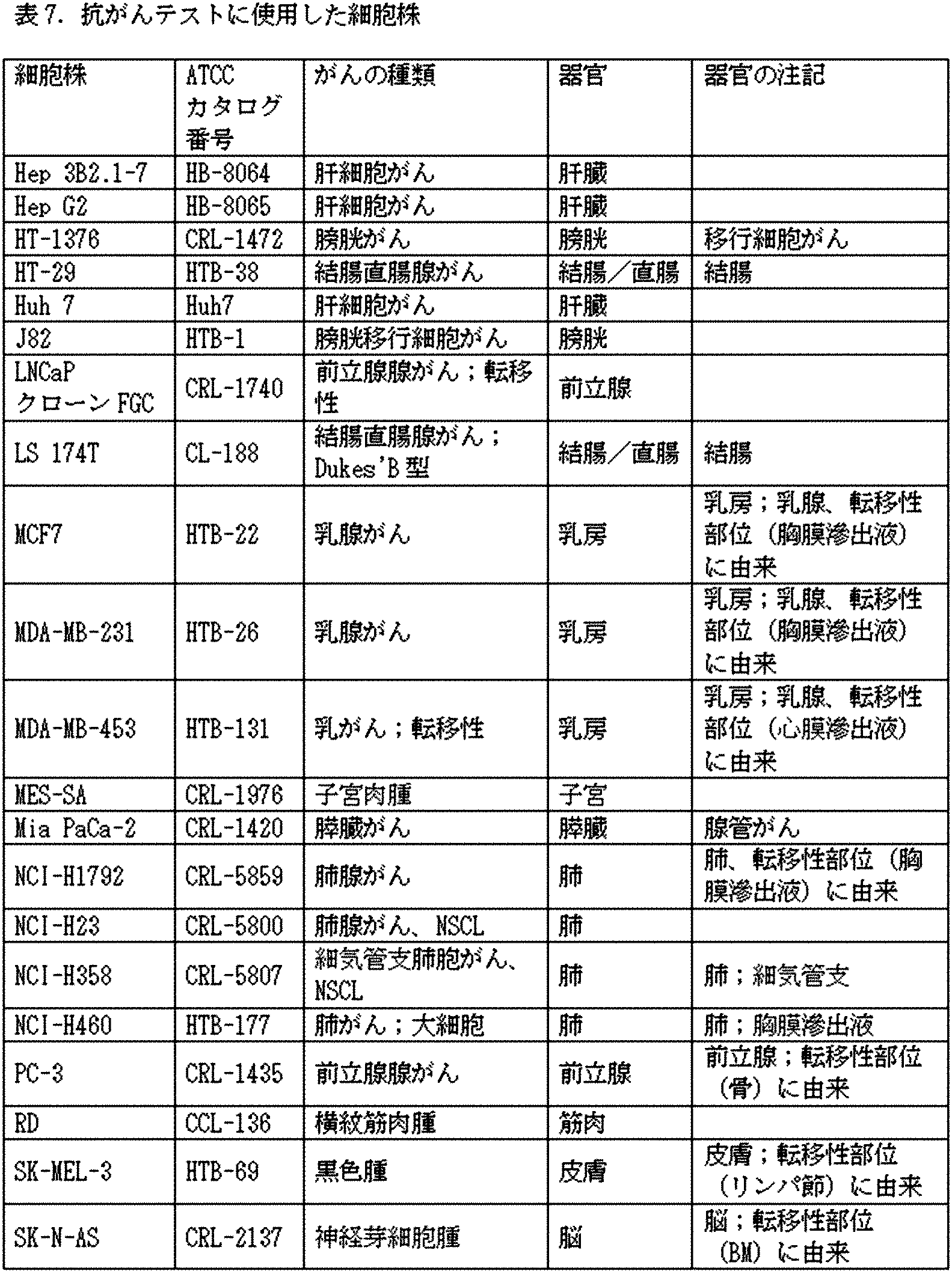
幾つかの実験においては、上記のように、その後の低酸素インキュベーションに由来するCM中で、分泌されたサイトカインレベルを測定した。
分泌されたタンパク質の定量的検出:それぞれのヒト免疫アッセイQuantikine(登録商標)ELISAキット (R&D Systems)を使用して、IL-6及びVEGFを定量的に測定した。
結果
様々な条件を比較(side-by-side)テストする一連の実験において、上記実施例で記載したように、ASCをバイオリアクター中でインキュベートした。バイオリアクターでのインキュベーションの最終日に、培地を、FBSの存在下又は非存在下で、添加されたTNF-α又はTNF-α+IFN-γを含むか又は含まない培地で置換した。ELISAによって、バイオリアクター培地中におけるVEGF及びIL-6の分泌を測定した。IFN-γが存在するか否かに関わらず、TNF-αを含めることによって、VEGFの分泌が有意に増加した(表2)。
In some experiments, secreted cytokine levels were measured in CMs derived from subsequent hypoxic incubations, as described above.
Quantitative detection of secreted proteins: IL-6 and VEGF were quantitatively measured using their respective human immunoassays Quantikine® ELISA Kit (R & D Systems).
Results In a series of side-by-side testing experiments, ASCs were incubated in a bioreactor as described in the examples above. On the final day of incubation in the bioreactor, the medium was replaced with medium with or without added TNF-α or TNF-α + IFN-γ in the presence or absence of FBS. VEGF and IL-6 secretion in bioreactor medium was measured by ELISA. The inclusion of TNF-α, with or without the presence of IFN-γ, significantly increased VEGF secretion (Table 2).
別の実験によって、TNF-αを含めることで、IL-6分泌が有意に増加し、それはIFN-γによって更に増加することが示された。
本実施例の終わりまでの以下に記載される実験は全て、血清を含まない培地で増殖させた試料に対して行った。蛍光ベースのサイトカインアレイアッセイを使用して、先述の実験に由来する選択された試料のバイオリアクター培地について、一団の因子の発現を探索した。GRO(CXCL1;Uniprotアクセッション番号:P09341)、IL-6、IL-8、MCP-1、MCP-2、MCP-3、RANTES及びIP-10(Uniprotアクセッション番号:P02778)を含む、幾つかの因子の発現の増加が、サイトカインインキュベーションの後に観察された(図4A)。別の実験においては、TNF-α単独を、サイトカイン添加なしと比較した。GRO、IL-8、MCP-1、RANTESの発現の上昇、より少ない程度に、IL-6、MCP-3、アンギオゲニン、インスリン様増殖因子結合タンパク質-2(IGFBP-2)、オステオポンチン及びオステオプロテゲリンの発現の上昇が示された(図4B〜C)。
幾つかの実験において、定量的ELISAによって、バイオリアクター培地中におけるMCP-1及びGM-CSFの上昇した発現を確認した。その結果によって、MCP-1誘導のためには、TNF-α+IFN-γが、TNF-α単独よりも強力であることが示された(図5A)一方で、GM-CSF誘導のためには、TNF-α単独が僅かに優れているようであることが示された(図5B)。TNF-α+IFN-γ試験に由来する対照培地(サイトカインを含まない)と比べて、サイトカイン濃度及び変化倍数を、以下の表3に示す。
Another experiment showed that the inclusion of TNF-α significantly increased IL-6 secretion, which was further increased by IFN-γ.
All of the experiments described below to the end of this example were performed on samples grown in serum-free medium. Fluorescence-based cytokine array assays were used to search for the expression of a group of factors in the bioreactor medium of selected samples from the previous experiments. Some, including GRO (CXCL1; Uniprot accession number: P09341), IL-6, IL-8, MCP-1, MCP-2, MCP-3, RANTES and IP-10 (Uniprot accession number: P02778) Increased expression of the factor was observed after cytokine incubation (Fig. 4A). In another experiment, TNF-α alone was compared to no cytokine addition. Increased expression of GRO, IL-8, MCP-1, RANTES, to a lesser extent, IL-6, MCP-3, angiogenin, insulin-like growth factor binding protein-2 (IGFBP-2), osteopontin and osteoprote Increased expression of gerin was shown (Figs. 4B-C).
In some experiments, quantitative ELISA confirmed elevated expression of MCP-1 and GM-CSF in bioreactor medium. The results showed that TNF-α + IFN-γ was more potent for MCP-1 induction than TNF-α alone (Fig. 5A), while for GM-CSF induction. It was shown that TNF-α alone appears to be slightly superior (Fig. 5B). Table 3 below shows the cytokine concentration and multiples of change compared to the control medium (without cytokines) derived from the TNF-α + IFN-γ test.
TNF-α+IFN-γ、又はTNF-α単独を使用した幾つかの実験で、先述のサイトカインアレイによって、幾つかの他の因子の誘導を検出した。表4に記載されているように、多くのタンパク質が一貫して上方制御された。 In some experiments using TNF-α + IFN-γ or TNF-α alone, induction of several other factors was detected by the cytokine array described above. As shown in Table 4, many proteins were consistently upregulated.
遥かに大きいGM-CSFの上方制御が観察されたこと、G-CSF、HGF及びTRAILの発現をテストして上方制御が見出されたこと、TNF-αが上方制御されなかったこと以外は、サイトカインのボーラス投与(with a bolus of)で刺激されたASCを用いて(以下の実施例9において、本明細書に記載されるように)、実施例中、上記に示されたのと同様の結果を得た。定量的ポリメラーゼ連鎖反応を使用して、mRNAレベルで本実施例で言及されるタンパク質の発現が増加することも確認した。 Except that much greater GM-CSF upregulation was observed, G-CSF, HGF and TRAIL expression was tested and upregulation was found, and TNF-α was not upregulated. Using ASCs stimulated with a bolus of cytokines (as described herein in Example 9 below), similar to those shown above in Examples. I got the result. Quantitative polymerase chain reaction was also used to confirm increased expression of the proteins referred to in this example at the mRNA level.
実施例7:3D培養中のASCの炎症促進性サイトカイン処理に対する血清の影響
本実験では、TNF-α+IFN-γ(図6A)又はTNF-α単独(図6B)による、先述の一団の因子の誘導に対する、FBSの影響を調べた。FBSの存在下又は非存在下で、類似する一式の主要なタンパク質を誘導した。TNF-アルファ単独については、FBS非存在下よりも存在下において、IL-6が遥かに強く誘導されるようであった。
Example 7: Effect of serum on pro-inflammatory cytokine treatment of ASC in 3D culture In this experiment, induction of the above-mentioned group of factors by TNF-α + IFN-γ (Fig. 6A) or TNF-α alone (Fig. 6B) The effect of FBS on was investigated. A similar set of major proteins was induced in the presence or absence of FBS. For TNF-alpha alone, IL-6 appeared to be induced much more strongly in the presence of FBS than in the absence.
実施例8:炎症性サイトカインを用いて処理したASCのマーカー表現型
幾つかの実験で、炎症促進性サイトカインを用いて前処理しているASCのマーカー表現型を調べた。一貫して、細胞は、CD29、CD90及びCD54について90%超が陽性であり;CD73及びCD105について85%超が陽性であり;並びにCD49について65%超が陽性であった。追加的には、細胞は、CD14、CD19、CD31、CD34、CD39及びCD45について1%未満が陽性であり;CD200について3%未満が陽性であり;GlyAについて6%未満が陽性であり;並びにSSEA4について20%未満が陽性であった。
Example 8: Marker phenotype of ASC treated with inflammatory cytokines In some experiments, the marker phenotype of ASC pretreated with pro-inflammatory cytokines was investigated. Consistently, cells were more than 90% positive for CD29, CD90 and CD54; more than 85% positive for CD73 and CD105; and more than 65% positive for CD49. In addition, cells are less than 1% positive for CD14, CD19, CD31, CD34, CD39 and CD45; less than 3% positive for CD200; less than 6% positive for GlyA; and SSEA4. Was positive in less than 20%.
実施例9:サイトカイン条件の変更は細胞生存率を改善する
2つの点:1.サイトカインへの曝露は24時間であったこと;及び2.24時間インキュベーションの最初に濃縮されたストック溶液を使用して、サイトカインをバイオリアクター培地に添加し、目標のサイトカイン濃度にまで急速に持っていったことを除き、実施例6において記載されたのと同様の様式で、炎症性サイトカインを用いてASCを刺激した。それに続く24時間に亘って、目標のサイトカイン濃度を含む新鮮な培地をバイオリアクターに灌流した。指数増殖期に対応する、バイオリアクターに播種後5日で、24時間インキュベーションを開始した。サイトカイン処理の結果によって、細胞の増殖は倍化速度が遅くなり始める時点に到達していた。ASCを凍結した。解凍した細胞を組織培養ディッシュに播種し、播種後3日及び4日目に回収したプレート中の細胞密度を測定し比較することによって、集団倍化時間(PDT)を測定した。
24時間刺激した細胞は、PDTにおける有意な減少を示した(図7)。表5は、各試料の条件について記載する。
Example 9: Changes in cytokine conditions improve cell viability
Two points: 1. Exposure to cytokines was 24 hours; and 2. Using the first concentrated stock solution of the 24-hour incubation, the cytokines were added to the bioreactor medium and rapidly brought to the target cytokine concentration. ASC was stimulated with inflammatory cytokines in a manner similar to that described in Example 6, except that For the next 24 hours, fresh medium containing the target cytokine concentration was perfused into the bioreactor. Incubation was started for 24
Cells stimulated for 24 hours showed a significant reduction in PDT (Fig. 7). Table 5 describes the conditions for each sample.
実施例10:ASC CMは腫瘍細胞の複製及び生存に影響する
方法
CM生成:実施例1及び6において記載したように、バイオリアクターインキュベーション及びその後の凍結保存を行った。これらの工程に続けて、500,000細胞を、マルチウェルプレート中、2 mM L-グルタミン及び10%FBS、幾つかのケースにおいては、1ウェル当たり40 ngのIFN-γを補充した4 ml DMEM中に播種した。24時間後、培地を吸引し、細胞を洗浄し、RPMI(特に断りのない限りFBSを含まない)を添加した。24時間のインキュベーション後、培地を回収し、遠心分離し、5%FBSを培地に添加した。
抗がんアッセイ 59細胞株を、培地(RPMI+10%FBS、2 mM L-アラニル-L-グルタミン及び1 mMピルビン酸ナトリウム)中で増殖させ、ポリヒドロキシエチルメタクリレート(pHEMA)で予めコートしたマルチウェル3Dプレート(細胞を自己集合させる表面上の微空間を有するElplasiaTMプレート)中でスフェロイドを形成させるために上記の培地(但し、5%FBSを含む)に播種した。CMを2倍で連続希釈し、幾つかの濃度で、3回反復でアッセイした。希釈なしのCM、又は1:2、1:4、若しくは1:8で希釈したCMを、25 μLの体積で、播種後24時間で添加し、3日毎に交換した。全ての細胞株について対照(陽性及び陰性)を含めた。CellTiter-Glo(登録商標)細胞生存率アッセイを使用して、細胞を溶解し、分析し、細胞の生存率及び複製に対するCMの影響を決定した。ビヒクルと比較して20〜40%の阻害は、標準偏差に比較して統計的に有意であり、部分的な阻害と定義した一方で、40%以上の阻害を阻害と定義した。
Example 10: How ASC CM Affects Tumor Cell Replication and Survival
CM generation: Bioreactor incubation followed by cryopreservation as described in Examples 1 and 6. Following these steps, 500,000 cells were placed in a multi-well plate in 4 ml DMEM supplemented with 2 mM L-glutamine and 10% FBS, and in some
Anticancer Assay 59 cell lines were grown in medium (RPMI + 10% FBS, 2 mM L-alanyl-L-glutamine and 1 mM sodium pyruvate) and pre-coated with polyhydroxyethyl methacrylate (pHEMA) multiwell 3D. Seeded in the above medium (but containing 5% FBS) to form spheroids in a plate (Elplasia TM plate with microspaces on the surface that self-assembles cells). CM was serially diluted 2-fold and assayed at several concentrations in 3 iterations. Undiluted CM or CM diluted 1: 2, 1: 4, or 1: 8 was added in a volume of 25
結果
母体ASC、又は母体/胎児ASCの混合のいずれかをバイオリアクター中で生成し、馴化培地(CM)を調製するために使用した。CMを、母体ASC、及び母体/胎児ASCの集団から調製し、そのうち幾らかをCM生成に先立って又はその最中に処理に供し(表6)、様々ながん細胞株の複製を阻害する能力についてテストした(表7)。
Results Either maternal ASC or maternal / fetal ASC mixture was produced in the bioreactor and used to prepare conditioned medium (CM). CMs are prepared from maternal ASCs and maternal / fetal ASC populations, some of which are treated prior to or during CM production (Table 6) to inhibit replication of various cancer cell lines. Ability was tested (Table 7).
下記の分析は、TNF-アルファ/IFN-ガンマを用いて処理したASCに由来するCMを投与した、実験群1に焦点を当てる。全体:
・ 12細胞株は群1によって阻害された(<60%の増殖)。
・ 14細胞株は群1によって部分的に阻害された(60〜80%の増殖)。
・ 28細胞株はCMによって阻害されなかった。
・ 4細胞株は群1によって部分的に刺激された(120〜140%の増殖)。
・ 1細胞株は群1によって刺激された(>140%の増殖)。
幾つかの種類のがん、即ち、腎細胞がん(テストした4細胞株のうち2細胞株;表8)、黒色腫(1/4株)、肝細胞がん(2/5株;表9)、結腸直腸がん(2/10)、乳がん(2/6株;表10)、肺腺がん(1/1株;表11)、大細胞肺がん(1/1)、及び横紋筋肉腫(1/1株;表12)は、少なくとも最高濃度の群1によって、阻害(増殖における少なくとも40%の低減で定義される)を示した。
The analysis below focuses on
12 cell lines were inhibited by group 1 (<60% proliferation).
14 cell lines were partially inhibited by Group 1 (60-80% proliferation).
28 cell lines were not inhibited by CM.
The 4 cell lines were partially stimulated by Group 1 (120-140% proliferation).
• One cell line was stimulated by Group 1 (> 140% proliferation).
Several types of cancer: renal cell carcinoma (2 out of 4 cell lines tested; Table 8), rhabdomyosarcoma (1/4 line), hepatocellular carcinoma (2/5 line; table) 9), colorectal cancer (2/10), breast cancer (2/6 strains; Table 10), lung adenocarcinoma (1/1 strain; Table 11), large cell lung cancer (1/1), and rhabdomyosarcoma Myomas (1/1 strain; Table 12) showed inhibition (defined by a reduction of at least 40% in proliferation) by at least the highest concentration of
実施例11:応答性細胞株と他の細胞株との間で異なって発現される遺伝子の分析
ASC-TNFα/IFNγ処理に対して、応答性及び非応答性のがん細胞株間で、異なって発現されるマーカー遺伝子を特定するために、細胞株を器官によってグループ分けした。それゆえ、マーカー遺伝子選択のために選択されたがん細胞株は、5器官:乳房、大腸、腎臓、肝臓及び肺に由来する。応答性の細胞株RD(横紋筋肉腫)及びCHL-1(黒色腫)は、これらの器官に起源するがんについて、2クラスの各々について、2細胞株を確保するためのデータが不十分であったため、調査から除外した。
2つのクラスを各器官について以下の通り割り当てた(表13):
・ クラス0:応答性の細胞株(希釈していないCMを用いて、対照の%(POC)≦60%)。
・ クラス1:希釈していないCMを用いて、POC≧79%である細胞株。
60%<POC<79%であることで定義される、僅かに応答性の細胞株は、両方のクラスから除外した。
Example 11: Analysis of genes expressed differently between responsive cell lines and other cell lines
Cell lines were grouped by organ to identify marker genes that were expressed differently among cancer cell lines that were responsive and non-responsive to ASC-TNFα / IFNγ treatment. Therefore, the cancer cell lines selected for marker gene selection are derived from 5 organs: breast, large intestine, kidney, liver and lung. Responsive cell lines RD (rhabdomyosarcoma) and CHL-1 (melanoma) have insufficient data to secure two cell lines for each of the two classes of cancers originating from these organs. Therefore, it was excluded from the survey.
Two classes were assigned to each organ as follows (Table 13):
-Class 0 : Responsive cell line (control% (POC) ≤ 60% using undiluted CM).
-Class 1 : Cell lines with POC ≥ 79% using undiluted CM.
Slightly responsive cell lines, defined as 60% <POC <79%, were excluded from both classes.
1000を超える細胞株について、ゲノムデータへのアクセス、分析及び視覚化を提供する、がん細胞株事典(CCLE;Barretina、J.ら)から、遺伝子発現データを得た。マーカー遺伝子を特定するために、細胞増殖アッセイにおいて使用したがん細胞株についてのmRNA発現アレイデータをダウンロードした。データセットのダウンロードに先立って、ロバストマルチアレイアベレージ(Robust Multi-array Average)(RMA)を使用して、元のAffymetrix U133+2アレイに由来する、加工前の(raw)Affymetrix CELファイルを、各プローブセットについて単一の値に変換し、分位点正規化(quantile normalization)を使用して正規化した。BrainarrayのHGU133Plus2_Hs_ENTREZG_15.0.0パッケージ由来の再定義したカスタムCDFファイルを、要約のために使用した。
マーカー遺伝子を特定し選択するために、GenePatternにおけるComparativeMarkerSelectionモジュール(Reichら)を利用した。
プロファイルされた各遺伝子についての両側t検定の値を計算することによって、遺伝子をスコア化した。検定統計量が>5又は<−5である場合に、マーカー遺伝子を選択した。正及び負の値は、応答性の細胞株において、それぞれ上方制御された遺伝子及び下方制御された遺伝子を示す。表14は、>5及び<−5のスコアを有するマーカー遺伝子の数を示す。
Gene expression data were obtained from the Cancer Cell Line Encyclopedia (CCLE; Barretina, J. et al.), Which provides access, analysis and visualization of genomic data for over 1000 cell lines. To identify the marker genes, we downloaded mRNA expression array data for cancer cell lines used in the cell proliferation assay. Prior to downloading the dataset, each probe set uses Robust Multi-array Average (RMA) to extract the raw Affymetrix CEL file from the original Affymetrix U133 + 2 array. Was converted to a single value and normalized using quantile normalization. A redefined custom CDF file from the Brainarray HGU133Plus2_Hs_ENTREZG_15.0.0 package was used for summarization.
The Comparative Marker Selection module (Reich et al.) In Gene Pattern was used to identify and select marker genes.
Genes were scored by calculating the values of the two-sided t-test for each profiled gene. Marker genes were selected when the test statistic was> 5 or <-5. Positive and negative values indicate upregulated and downregulated genes, respectively, in responsive cell lines. Table 14 shows the number of marker genes with scores of> 5 and <-5.
例として、図8Aは、乳がん細胞株分析についてのプロファイリングされた各遺伝子のスコアのグラフ表示を示す。応答性の細胞株において上方制御された遺伝子をグラフの左側に示す一方で、応答性の細胞株において下方制御された遺伝子(他の細胞株において上方制御された)を右側に示す。図8Bは、応答性の乳房細胞株において、下方制御された遺伝子(スコア<-5)の全てについて、5株の乳がん細胞株の平均発現値を示すセントロイドプロットである。その2株の応答性の乳がん細胞株(HCC-1395及びMDA-MB-231)を左に示し、他の3株の乳がん細胞株(BT474、MCF7及びT47D)を右に示す。 As an example, FIG. 8A shows a graphical representation of the score of each profiled gene for breast cancer cell line analysis. Genes that are upregulated in responsive cell lines are shown on the left side of the graph, while genes that are downregulated in responsive cell lines (upregulated in other cell lines) are shown on the right side. FIG. 8B is a centroid plot showing the mean expression of 5 breast cancer cell lines for all downregulated genes (score <-5) in responsive breast cell lines. The two responsive breast cancer cell lines (HCC-1395 and MDA-MB-231) are shown on the left, and the other three breast cancer cell lines (BT474, MCF7 and T47D) are shown on the right.
実施例12:応答性株において有意に摂動された経路
各器官内のクラス0とクラス1細胞株との間で摂動された(perturbed)関連する生物学的経路を決定するために、各器官内で最も統計的に有意に上方制御及び下方制御された遺伝子を使用して、Reactome Pathway Database V53 (Croftら)を探索した。表15は、少なくとも1つのリアクトーム経路(Reactome pathway)において見出される5器官の各々に由来する、上方制御及び下方制御された遺伝子数を示す。
Example 12: Significantly perturbed pathways in responsive strains Within each organ to determine the associated biological pathways perturbed between
器官の間で共通した重要な経路について、データを検索した。以下の5経路は、4/5アウトプット一覧において、リアクトーム経路の上位200の一覧にあった:
1.IFN-アルファ/ベータ経路のRIG-I/MDA5媒介性誘導(R-HSA-168928)
2.インターフェロンシグナル伝達(R-HSA-913531)
3.免疫系におけるサイトカインシグナル伝達(R-HSA-1280215)
4.細胞の老化(R-HSA-2559583)
5.ベータ-カテニンのトランス活性化複合体の失活(R-HSA-3769402)
Data were searched for important pathways common between organs. The following 5 routes were in the top 200 list of reactor routes in the 4/5 output list:
1. 1. RIG-I / MDA5-mediated induction of the IFN-alpha / beta pathway (R-HSA-168928)
2. Interferon signaling (R-HSA-913531)
3. 3. Cytokine signaling in the immune system (R-HSA-1280215)
Four. Cellular senescence (R-HSA-2559583)
Five. Inactivation of the beta-catenin transactivation complex (R-HSA-3769402)
次に、最も統計的に有意な上方制御及び下方制御された遺伝子を、5つのマーカー遺伝子セットの各々からプールし、Reactome Pathway Databaseを探索するために使用した。データベースを三回、探索した:
1.上方制御された全遺伝子
2.下方制御された全遺伝子
3.上方制御及び下方制御された全遺伝子
The most statistically significant upregulated and downregulated genes were then pooled from each of the five marker gene sets and used to explore the Reactome Pathway Database. Searched the database three times:
1. 1. All up-regulated genes
2. All downregulated genes
3. 3. All genes up-regulated and down-regulated
統計的に有意なものとして生物学的経路を定義するための統計的カットオフは、偽発見率(FDR)≦0.05であった。表16〜18は、それぞれ上方制御された遺伝子、下方制御された遺伝子、及びその両方のために、5器官にまたがり、クラス0と1の間で摂動された、最も統計的に関連する生物学的経路を示す。表17において太字で表した経路は、変異遺伝子を分析に加える場合に選択プロセスで残る(表20)。
The statistical cutoff for defining the biological pathway as statistically significant was false discovery rate (FDR) ≤ 0.05. Tables 16-18 show the most statistically relevant biology spanning 5 organs and perturbed between
分かるように、クラス0の下方制御された遺伝子のプールされたセットによって、データベースを探索する場合に、最も高い統計的有意性を示す生物学的経路が明らかにされる。
As can be seen, the pooled set of
実施例13:応答性及び他の細胞株のエキソームの変異分析
(記載された統計的有意性に加えて)生物学的に最も重要な経路を探索するために、COSMICがん細胞株プロジェクト (Forbesら)に由来する完全エキソームシーケンシングによって体細胞変異を分析した。付加、欠失、置換、フレームシフト及びスプライス部位変異を考慮した一方で、CDSサイレント変異を除外した。Hep-G2、AGS、DLD1、LS-174T及びSW480からのデータは、データベース中に現れなかった。これらの細胞株に加えて、11株の応答性の細胞株において合計>10,300変異を考慮し、他の43細胞株において>53,000変異を考慮した。
Example 13: Mutation analysis of responsiveness and exomes of other cell lines (in addition to the statistical significance described) The COSMIC Cancer Cell Line Project (Forbes) to explore the most biologically important pathways. Et al.) Analyzed somatic mutations by complete exome sequencing. CDS silent mutations were excluded while considering additions, deletions, substitutions, frameshifts and splice site mutations. Data from Hep-G2, AGS, DLD1, LS-174T and SW480 did not appear in the database. In addition to these cell lines, a total of> 10,300 mutations were considered in 11 responsive cell lines and> 53,000 mutations in the other 43 cell lines.
リアクトームデータベースを以下で探索した:
・専ら11株の応答性の細胞株において変異している全ての遺伝子
・専ら他の43細胞株(僅かな応答性及び非応答性の株)において変異している全ての遺伝子
・専ら応答性の細胞株において変異している全ての遺伝子のプールした一覧;及び応答性の細胞株において下方制御された全ての遺伝子(上記の表15について記載したように)
最後のクエリ(query)は最も有益であり、その結果は以下において記載する。
表19は、専ら応答性株において変異している全ての遺伝子の数を示す。
We searched the reactor database below:
-All genes mutated exclusively in 11 responsive cell lines-All genes mutated exclusively in other 43 cell lines (slightly responsive and non-responsive) -Exclusively responsive A pooled list of all genes mutated in the cell line; and all genes down-regulated in the responsive cell line (as described in Table 15 above).
The final query is the most informative and the results are described below.
Table 19 shows the number of all genes mutated exclusively in responsive strains.
表20に、最後のクエリの結果を示す。統計的なカットオフは、エンティティFDR≦0.05であった。 Table 20 shows the results of the last query. The statistical cutoff was entity FDR ≤ 0.05.
本分析によって、応答性のがん細胞株は、2つの重要な経路:MHCクラスI抗原プロセッシング及び提示(それらとしては、エンドソーム/液胞、抗原提示:クラスI MHCのフォールディング、会合及びペプチドローディング、ER-ファゴソーム、並びに抗原プロセッシング交差提示経路が挙げられる)、並びにサイトカインシグナル伝達(それらとしては、インターフェロンアルファ/ベータシグナル伝達、インターフェロンガンマシグナル伝達、及びインターフェロンシグナル伝達経路が挙げられる)における、下方制御又は調節異常を有する細胞株であることが示された。これらの経路は、以前の分析において、統計的に有意であることが見出された経路(表17)と有意に重複する。このことにより、該統計的分析及びこれらの特定の経路の重要性が確認される。 According to this analysis, responsive cancer cell lines have two important pathways: MHC class I antigen processing and presentation (such as endosomes / vacuoles, antigen presentation: class I MHC folding, association and peptide loading, Downward regulation or downregulation in ER-fagosomes, as well as antigen processing cross-presentation pathways), and cytokine signaling, including interferon alpha / beta signaling, interferon gamma signaling, and interferon gamma signaling. It has been shown to be a cell line with dysregulation. These pathways significantly overlap with the pathways found to be statistically significant in previous analyzes (Table 17). This confirms the importance of the statistical analysis and these specific routes.
図9A〜Bは、応答性の細胞株の各々において下方制御されたこれらの経路における遺伝子及び/又は専ら応答性の細胞株の各々において変異した遺伝子についてまとめる。Aにおいては、列記された最初の8遺伝子はサイトカインシグナル伝達/インターフェロン経路中にも存在する。UBE2Q1以外の最後の19遺伝子は、ユビキチンリガーゼ経路に関与する。Bにおいては、3つのHLA遺伝子、UBB、及びPSMで始まる3遺伝子は、抗原プロセッシング/提示経路中にも存在する。
結論として、上記のデータは、下方制御された若しくは異常調節されたMHCクラスI抗原プロセッシング及び提示経路並びに/又は下方制御された若しくは異常調節されたサイトカインシグナル伝達経路を有する、がん細胞株は、ASCを用いた処理に対して感受性であることを示す。
Figures 9A-B summarize genes in these pathways downregulated in each of the responsive cell lines and / or mutated genes in each of the exclusively responsive cell lines. In A, the first eight genes listed are also present in the cytokine signaling / interferon pathway. The last 19 genes other than UBE2Q1 are involved in the ubiquitin ligase pathway. In B, the three HLA genes, UBB, and the three genes beginning with PSM are also present in the antigen processing / presentation pathway.
In conclusion, the above data show that cancer cell lines have downregulated or abnormally regulated MHC class I antigen processing and presentation pathways and / or downregulated or abnormally regulated cytokine signaling pathways. Shows that it is sensitive to treatment with ASC.
実施例14:応答性及び非応答性の乳がん細胞株の更なる性状解析
次に、階層的クラスタリングによって、乳がん株をLuminal、Basal A又はBasal Bとして特徴づけるために有用な305分類遺伝子(Neveら)の発現に基づいて、応答性及び非応答性の乳がん細胞株の表現型を決定した。論文のデータをダウンロードし、GenePatternソフトウェアツール(Reichら)を使用して、再現した(図10)。列及び行の両方についての距離を測定するために、ピアソン相関クラスタリングを使用し、階層的クラスタリング法はペアワイズ平均連結法であった。
Example 14: Further phenotypic analysis of responsive and non-responsive breast cancer cell lines Next, 305 classification genes useful for characterizing breast cancer lines as Luminal, Basal A or Basal B by hierarchical clustering (Neve et al.) ) Was used to determine the phenotype of responsive and non-responsive breast cancer cell lines. The data from the treatise was downloaded and reproduced using the GenePattern software tool (Reich et al.) (Fig. 10). Pearson correlation clustering was used to measure distances for both columns and rows, and the hierarchical clustering method was the pairwise average concatenation method.
図11Aは、図10の上部を示し、どの細胞株が性状解析されたかを示し、それらは、ASC感受性について本明細書でテストした6細胞株のうち、5細胞株を含む。記載された株のうち、HCC38、SUM149PT、MDA-MB-157、BT549、HSS78T、SUM159PT、MDA-MB-436及びMDA-MB-231をTRAIL感受性についてテストした。それらはTNである。
図11Aは、11株の三重陰性(TN)株を含む、20株の乳がん細胞株を、TRAIL感受性についてテストした、Rahmanらのデータも取り込み;これらは、シンプルなアスタリスク(TRAIL非感受性)及び丸で囲ったアスタリスク(TRAIL感受性)によって印を付けている。TRAIL感受性であるTN細胞株の殆んどは、Basal Bのクラスターに入るが、そこから外れた2株がある。Basal Bである全8株のTRAIL感受性TN細胞株は「間葉系の表現型」を有し、Basal Aである全3株のTRAIL非感受性のものは「上皮系の表現型」を有する。
間葉系の表現型は、高レベルのビメンチン、高レベルのカベオリン、及び低レベルのE-カドヘリンを有すると定義される。上皮系の表現型は、高レベルのE-カドヘリン、豊富なケラチン、及び低レベルのビメンチンを有すると定義される。
FIG. 11A shows the upper part of FIG. 10 showing which cell lines were characterization, including 5 of the 6 cell lines tested herein for ASC susceptibility. Of the listed strains, HCC38, SUM149PT, MDA-MB-157, BT549, HSS78T, SUM159PT, MDA-MB-436 and MDA-MB-231 were tested for TRAIL susceptibility. They are TN.
Figure 11A also incorporates data from Rahman et al., Who tested 20 breast cancer cell lines for TRAIL susceptibility, including 11 triple negative (TN) strains; these are simple asterisks (TRAIL insensitivity) and circles. Marked by an asterisk (TRAIL sensitivity) surrounded by. Most TRAIL-sensitive TN cell lines enter the Basal B cluster, but there are two out of it. All eight Basal B strains of TRAIL-sensitive TN cell lines have a "mesenchymal phenotype" and all three Basal A strains of TRAIL-insensitive cells have an "epithelial phenotype".
The mesenchymal phenotype is defined as having high levels of vimentin, high levels of caveolin, and low levels of E-cadherin. The epithelial phenotype is defined as having high levels of E-cadherin, abundant keratin, and low levels of vimentin.
図11Bは、図11Aからの、テストした乳がん細胞株のデータを表の形態で示し、臨床サブタイプにおける情報、即ち、エストロゲン受容体(ER)又はプロゲステロン受容体(PR)が存在するか否か、及びHer2/neuが増幅されるかも含んでいる。
結論として、TN乳がんは、ASCを用いた治療に対する感受性を示す。
FIG. 11B shows the data of the tested breast cancer cell lines from FIG. 11A in the form of a table, and the information in the clinical subtype, that is, the presence or absence of the estrogen receptor (ER) or progesterone receptor (PR). , And Her2 / neu may be amplified.
In conclusion, TN breast cancer is vulnerable to treatment with ASC.
HCC1395は、ASC感受性について本明細書でテストされ、Neveらによる先述の階層的クラスタリング分析において分析されなかった、唯一の乳がん株である。この細胞株を使用し、ASCに対する乳がんの感受性が、TN表現型と並行するという仮説を検証した。HCC1395は、ASC処理に対して感受性であったので、先述の分析によって、三重陰性細胞株のHCC1395はTRAIL感受性であり、Basal Bクラスターに入ることが予想される。本分析は、別のデータセットを必要とした。Neveらによる階層的クラスタリング分析もなされた乳がん細胞株について、がん細胞株事典 (CCLE;Barretinaら)を探索した。37株の乳がん細胞株のAffymetrix遺伝子発現データをダウンロードし、上述のように処理した。 HCC1395 is the only breast cancer strain tested herein for ASC susceptibility and not analyzed in the earlier hierarchical clustering analysis by Neve et al. Using this cell line, we tested the hypothesis that breast cancer susceptibility to ASC parallels the TN phenotype. Since HCC1395 was sensitive to ASC treatment, the above analysis predicts that the triple negative cell line HCC1395 is TRAIL sensitive and will enter the Basal B cluster. This analysis required a separate dataset. We searched the Cancer Cell Line Encyclopedia (CCLE; Barretina et al.) For breast cancer cell lines that were also subjected to hierarchical clustering analysis by Neve et al. Affymetrix gene expression data from 37 breast cancer cell lines were downloaded and processed as described above.
「遺伝子に対するAffyプローブ」の一致を、Neveらのデータセットにおける遺伝子を特定するのに使用し、それを、今度はCCLE由来の37株の関連する遺伝子発現データを選択するために使用した。この処理によって、169のプローブセットを生成して、階層的クラスタリングを行い、Neveらと同様なクラスタリングを生成した(図12A)。HCC-1395は、MDA-MB-231と一緒に、明瞭にTN/Basal Bグループにクラスター化され、従って、このことで、ASCに対する乳がんの感受性がTN表現型と並行するという仮説が確認される。
図12Bは、図12Aの上部を示す。(丸で囲った)2株の細胞株のみが、以前の分析とは異なってクラスター化された。18,000のプローブセット(37株のCCLE細胞株のセットに由来する遺伝子発現データにおけるプローブ数)又は169のプローブセットを使用したかに関わらず、事実上、同一な階層的クラスタリングが得られ、従って、このことで、クラスタリングスキームが確認される(図12C)。
The "Affy probe for genes" match was used to identify genes in the Neve et al. Dataset, which in turn was used to select relevant gene expression data for 37 strains from CCLE. By this process, 169 probe sets were generated, hierarchical clustering was performed, and clustering similar to that of Neve et al. Was generated (Fig. 12A). HCC-1395, along with MDA-MB-231, is clearly clustered into the TN / Basal B group, thus confirming the hypothesis that breast cancer susceptibility to ASC parallels the TN phenotype. ..
FIG. 12B shows the upper part of FIG. 12A. Only two cell lines (circled) were clustered unlike previous analyzes. Virtually the same hierarchical clustering was obtained regardless of whether 18,000 probe sets (number of probes in gene expression data from a set of 37 CCLE cell lines) or 169 probe sets were used, thus resulting in virtually identical hierarchical clustering. This confirms the clustering scheme (Fig. 12C).
実施例15:乳房腫瘍株のクラスタリングに関与する遺伝子は、抗原提示及びIFNシグナル伝達に関与する
Luminal、Basal A及びBasal Bにクラスタリングするために重要であるとして、Neveらによる先述の階層的クラスタリング分析において特定された遺伝子を、Reactome Pathway Database(各セクション個別)に入力し、分類遺伝子が関与する経路を特定した(図13)。分類遺伝子の真ん中の行の区分としては、HLA及び2、3の他の抗原プロセッシング/提示遺伝子、並びにIFNシグナル伝達経路遺伝子が挙げられ、従って、このことで、先述の分析の正当性が更に確認され、以前、特定された経路が、ASC-TNFα/IFNγによって最初にテストしたよりも、広いスペクトルの乳がん細胞株に当てはまることが確認された。
Example 15: Genes involved in breast tumor strain clustering are involved in antigen presentation and IFN signaling
The genes identified in the above-mentioned hierarchical clustering analysis by Neve et al., Which are important for clustering to Luminal, Basal A and Basal B, are input to the Reactome Pathway Database (individually for each section), and the classification genes are involved. The route was identified (Fig. 13). The division in the middle row of the classifying genes includes HLA and a few other antigen processing / presenting genes, as well as IFN signaling pathway genes, thus further confirming the validity of the aforementioned analysis. It was confirmed that the previously identified pathway applies to a broader spectrum of breast cancer cell lines than originally tested with ASC-TNFα / IFNγ.
実施例16:腫瘍移植モデルにおけるASCのIN VIVO抗腫瘍活性
方法
93匹の無胸腺Foxn1nuヌードの右脇の皮下に、0.2 ml PBS中の3×10⁶のMDA-MB-231腺がん細胞を注射した(全群)―この日を0日目とみなした。実験群を表21に示す:
Example 16: ASC IN VIVO antitumor activity method in a tumor transplant model
Three x 10 6 MDA-MB-231 adenocarcinoma cells in 0.2 ml PBS were injected subcutaneously on the right flank of 93 athymic Foxn1 nu nudes (all groups) -this day is considered day 0 It was. The experimental groups are shown in Table 21:
TNF-α+IFN-γで刺激したASCを、筋肉内(IM)又は静脈内(IV)投与のために、それぞれ50 mcl(マイクロリットル)又は250 mclの体積で懸濁した。1日目に、ASCを、群6及び7の各10匹のマウスに、それぞれIM又はIV投与した。9日目に、73匹の未処理マウス(先に、対照群として参照される)から、36〜88mm3の範囲外の腫瘍サイズである23匹のマウスを取り除き、50匹の未処理マウスを残した。50匹の動物を、無作為に群1、2、3、4及び5に割り当てた。各群がおおよそ同じ平均サイズの腫瘍を有するマウスとなるように、腫瘍のサイズに従って無作為に群分けした。従って、最終的に各群に10匹ずつのマウスが存在した。同じ日に、表21に示すように、マウスを、処理しないか、モックのIM若しくはIV注射のいずれかで投与したか(それぞれ群1、2及び3);或いは、IM(それぞれ、最初の若しくは更なる投与を受けた群4及び6)又はIV(それぞれ、最初の若しくは更なる投与を受けた群5及び7)で、ASCを投与した。
電子キャリパーを使用して、腫瘍体積を測定した。
ASCs stimulated with TNF-α + IFN-γ were suspended in volumes of 50 mcl (microliters) or 250 mcl, respectively, for intramuscular (IM) or intravenous (IV) administration. On
Tumor volume was measured using an electronic caliper.
結果
ASCの抗腫瘍効果を、in vivo腫瘍移植モデルでテストした。1日目にASC投与を受けたマウスは、低減された腫瘍サイズを示した。各時点、即ち、5、7及び9日目で、IVを対照と比較した場合、阻害は統計的に有意であった(片側t検定で、それぞれp=0.0055、0.0067及び0.041)(図14A)。有効性の傾向は、IV注射(図14B〜C)及びIM注射(図14D〜E)したマウスの両方で見られた。12〜16日目(表22〜23);9〜28日目(表24);及び9〜16日目(表25)の腫瘍体積における変化倍数を観察した場合、阻害効果が強く見られた。
result
The antitumor effect of ASC was tested in an in vivo tumor transplant model. Mice receiving ASC on
これらのデータによって、非常に急速に増殖している腫瘍モデルにおいてさえ、ASCがin vivoにおける腫瘍増殖を阻害することが確認される。 These data confirm that ASC inhibits tumor growth in vivo, even in tumor models that are growing very rapidly.
実施例17:更なるIN VIVO腫瘍移植モデルにおけるASCのテスト
概要
ASCの抗腫瘍効果を研究するために、別の腫瘍移植モデルを使用した。このケースにおいては、腫瘍細胞を鼠径部乳腺脂肪体(inguinal mammary fat pad)に注射した。その後、0.5若しくは1.0×10⁶のASC、又はPlasmaLyte (陰性対照;「Lyte」)を、筋肉内(IM)、静脈内(IV)、又はIM及びIV交互(「IV/IM」)に注射した。
Example 17: ASC test summary in a further IN VIVO tumor transplant model
Another tumor transplant model was used to study the antitumor effect of ASC. In this case, tumor cells were injected into the inguinal mammary fat pad. Then 0.5 or 1.0 × 10 6 ASC, or PlasmaLyte (negative control; “Lyte”) was injected intramuscularly (IM), intravenously (IV), or alternating IM and IV (“IV / IM”). ..
方法
処理をしなかった群9を除き、3×10⁶のMDA-MB-231細胞を、第4の(鼠径部)乳腺脂肪体に注射し、0日目とした。表26は、全ての群に施した処理を記載する。研究期間中に、3匹のマウス(8週目に群5の2匹、11週目に群7の1匹)が死亡した。体重に対するASC投与による有意な影響は見られなかった。
METHODS: Except for
組織学:組織試料には、異種移植腫瘍、肺及びリンパ節(腋窩、鼠径部及び腰部)が含まれていた。組織試料を緩衝ホルマリン中で浸漬固定し、パラフィン包埋のために処理した。試料を以下のように処理した:
1.異種移植腫瘍 腫瘍の真ん中を通って、標準的な5マイクロメーターの切片が調製され得るように、腫瘍試料のパラフィンブロックを調製した。一般的な組織病理学的評価のために、腫瘍切片をヘマトキシリン−エオシンを用いて染色した。
Histology: Tissue samples included xenograft tumors, lungs and lymph nodes (axilla, groin and lumbar region). Tissue samples were immersed and fixed in buffered formalin and treated for paraffin embedding. The sample was processed as follows:
1. 1. Xenograft Tumor A paraffin block of tumor sample was prepared so that a standard 5 micrometer section could be prepared through the middle of the tumor. Tumor sections were stained with hematoxylin-eosin for general histopathological evaluation.
1.1. 腫瘍細胞増殖 Ki67抗原に対する抗体を用いた免疫組織化学(IHC)染色のために、腫瘍切片を処理した。切片を脱パラフィンし、0.05%無水シトラコン酸(CA;Aldrich;Cat番号 125318)溶液(pH 7.4)中で20分間煮沸することによって、熱誘導抗原賦活化(HIER)に供した。室温(rt)まで冷却後、TBST(10 mM Tris-HCl、150 mM NaCl、0.1%Tween-20、pH 7.5)中、1:200で希釈した、Ki67に対するウサギモノクローナル抗体 (クローンSP6;Abcam Cat番号ab16667)を用いて、切片を室温で1時間、インキュベートした。切片をTBST中で洗浄し、セイヨウワサビ・ペルオキシダーゼ(HRP)標識抗ウサギIgG高分子試薬 (ZytoCem Plus HRP-ポリマー抗ウサギ;ZytoMed、Cat#ZUC032-10)を用いて、30分間インキュベートした。次いで、切片を洗浄し、0.05%ジアミノベンジジン (DABx4HCl [Sigma;Cat番号32750])及び0.015%過酸化水素を含む、TIBSバッファー(0.15 M NaCl、10 mMトリス、5 mMイミダゾール;pH 7.5)中で3分間インキュベートすることによって、HRP活性をアッセイした。スライドをヘマトキシリンで軽く対比染色し、DPXマウント剤 (Sigma;Cat番号06522)を使用してカバースリップをかけ、RGB調整可能な画像化フィルター(CRI)と組み合わせた、QuantiFire XI CCDカメラ(Optronics)を備える、Olympus BX-50顕微鏡を使用して画像を撮影した。腫瘍試料毎に、対物20倍で、3〜5の重複しない画像を取得した。
ImageJを使用して、画像を背景サブトラクション(subtraction)及びカラーデコンボリューション(color deconvolution)に供し、全ての核及びKi67で免疫染色した核をそれぞれ示す、青色及び茶色のモノクローム画像に分離した画像を得た。次いで、自動閾値処理によって、画像を二値化し、核を「分水界(watershed)」コマンドを使用して分離し、「粒子分析」コマンドを使用して自動で計数した。同じ試料に関連する全画像におけるKi67で標識した核のパーセンテージを、増殖指数として使用した。
1.1. Tumor cell proliferation Tumor sections were treated for immunohistochemistry (IHC) staining with antibodies against Ki67 antigen. Sections were deparaffinized and subjected to heat-induced antigen activation (HIER) by boiling in 0.05% citraconic anhydride (CA; Aldrich; Cat number 125318) solution (pH 7.4) for 20 minutes. Rabbit monoclonal antibody against Ki67 (clone SP6; Abcam Cat number) diluted 1: 200 in TBST (10 mM Tris-HCl, 150 mM NaCl, 0.1% Tween-20, pH 7.5) after cooling to room temperature (rt). Sections were incubated using ab16667) for 1 hour at room temperature. Sections were washed in TBST and incubated with horseradish peroxidase (HRP) -labeled anti-rabbit IgG polymer reagent (ZytoCem Plus HRP-polymer anti-rabbit; ZytoMed, Cat # ZUC032-10) for 30 minutes. The sections are then washed and in TIBS buffer (0.15 M NaCl, 10 mM Tris, 5 mM imidazole; pH 7.5) containing 0.05% diaminobenzidine (DABx4HCl [Sigma; Cat No. 32750]) and 0.015% hydrogen peroxide. HRP activity was assayed by incubating for 3 minutes. A QuantiFire XI CCD camera (Optronics) with slides lightly counterstained with hematoxylin, covered with a DPX mount (Sigma; Cat number 06522), and combined with an RGB adjustable imaging filter (CRI). Images were taken using the Olympus BX-50 microscope. For each tumor sample, 3-5 non-overlapping images were acquired with a 20x objective.
ImageJ was used to subject the image to background subtraction and color deconvolution to obtain images separated into blue and brown monochrome images showing all nuclei and ki67 immunostained nuclei, respectively. It was. Images were then binarized by automatic thresholding, nuclei separated using the "watershed" command, and automatically counted using the "particle analysis" command. The percentage of Ki67-labeled nuclei in all images associated with the same sample was used as the growth index.
1.2. 腫瘍細胞のアポトーシス アポトーシス細胞を検出するために、抗活性カスパーゼ抗体を用いて、腫瘍切片を免疫染色した。切片を脱パラフィンし、10 mMクエン酸バッファー(pH 6.0)中で20分間、煮沸することによってHIERに供した。室温で1時間冷却した後、TBST中で1:1000で希釈した、活性カスパーゼ3に対するウサギモノクローナル抗体 (クローンE83-77;Abcam Cat番号ab32042)を用いて、室温で1時間、切片をインキュベートし、抗Ki67抗体については上記のように、インキュベーション、検出及び対比染色を行った。アポトーシスを定量するために、免疫染色細胞の切片を、6〜10高出力(対物40倍)の顕微鏡視野において、手動で計数した。 1.2. Apoptosis of tumor cells Tumor sections were immunostained with anti-active caspase antibody to detect apoptotic cells. Sections were deparaffinized and subjected to HIER by boiling in 10 mM citrate buffer (pH 6.0) for 20 minutes. After cooling at room temperature for 1 hour, the sections were incubated for 1 hour at room temperature using a rabbit monoclonal antibody against active caspase 3 (clone E83-77; Abcam Cat number ab32042) diluted 1: 1000 in TBST. The anti-Ki67 antibody was incubated, detected and counterstained as described above. To quantify apoptosis, sections of immunostained cells were manually counted in a 6-10 high power (40x objective) microscopic field of view.
1.3. 腫瘍血管新生 腫瘍血管新生を定量するために、マウスCD34に対するウサギモノクローナル抗体 (クローンEP373Y、Abcam;Cat番号ab81289;1:1000希釈した)を用いて、切片を免疫染色し、内皮細胞を可視化した。抗活性カスパーゼについては、上記のように免疫染色を行った。CD34+(内皮)細胞の密度が最も高い領域(「ホットスポット」)を、顕微鏡対物20倍を使用して、撮影し、2048×2048ピクセルのカラーのデジタル画像を得た。カラーデコンボリューション、自動閾値化及び二値化の後、ImageJを使用して、DABを用いて茶色に染色した領域(ピクセル数)を決定した。画像サイズ(4.19 Mpi)に関連して、免疫染色の領域のパーセンテージを計算した。 1.3. Tumor angiogenesis To quantify tumor angiogenesis, sections were immunostained with rabbit monoclonal antibody against mouse CD34 (clone EP373Y, Abcam; Cat number ab81289; 1: 1000 diluted) to visualize endothelial cells. did. The anti-active caspase was immunostained as described above. The region with the highest density of CD34 + (endothelial) cells (“hotspots”) was photographed using a 20x microscope objective to obtain a 2048 × 2048 pixel color digital image. After color deconvolution, auto-thresholding and binarization, ImageJ was used to determine the area (pixel count) stained brown with DAB. Percentages of immunostaining areas were calculated in relation to image size (4.19 Mpi).
2.肺転移 肺試料を含むパラフィンブロックを、網羅的、体系的な切片化に供し、300μmで分離した切片(肺試料毎に8〜20個)を生成し、ガラススライド上にマウントした。TEバッファー(pH 8.0)中、HIERに供した肺切片における、腫瘍細胞を検出するために、1:600で希釈したウサギモノクローナル抗サイトケラチン18(CK18)抗体 (クローンEPR1626;Abcam、Cat番号ab133263)を用いたIHC染色を使用した。IHCの残りの手順は、上記の通りであった。 2. Pulmonary metastases Paraffin blocks containing lung samples were subjected to comprehensive and systematic sectioning to generate 300 μm separated sections (8-20 pieces per lung sample) and mounted on glass slides. Rabbit monoclonal anti-cytokeratin 18 (CK18) antibody (clone EPR1626; Abcam, Cat number ab133263) diluted 1: 600 to detect tumor cells in lung sections subjected to HIER in TE buffer (pH 8.0). IHC staining was used. The rest of the IHC procedure was as described above.
3.リンパ節転移 リンパ節(腋窩、鼠径部及び腰部)の各ペアを1個のパラフィンブロックに包埋し、75 μmで分離した切片(リンパ節試料毎に8〜25個)を得るために、網羅的、体系的な切片化に供した。ガラススライド上に、切片をマウントし、肺切片について記載したように、抗CK18抗体を用いて免疫染色した。
P値は、StudentのT検定(両側分析)を使用して計算した。
3. 3. Lymph node metastasis Each pair of lymph nodes (axilla, groin and lumbar) is embedded in a single paraffin block and covered to obtain sections separated at 75 μm (8-25 per lymph node sample). It was used for systematic and systematic sectioning. Sections were mounted on glass slides and immunostained with anti-CK18 antibody as described for lung sections.
The P value was calculated using Student's T-test (two-sided analysis).
結果
ASCを用いた後期IM処理(48日目に開始)は、腫瘍増殖を有意に遅くし又は停止し、それは、平均腫瘍体積のプロット(図15A〜C)又は47日目からの腫瘍体積におけるパーセント変化(図15D〜E)であるかに関わらず、明らかであった。図15C及びEにおいては、異常値を除き、「調整した」数を生成した。群2においては0%、群1及び3においては5%であったのと比べ、群4におけるマウスの30%が84日目までに完全に寛解を示した。
result
Late IM treatment with ASC (starting on day 48) significantly slowed or stopped tumor growth, which was a plot of mean tumor volume (FIGS. 15A-C) or a percentage of tumor volume from
IV-ASC処理は、最初に腫瘍増殖を阻害し(図16)、具体的には、38日目に、操作されていないか、又は調整した平均を考慮した場合に、増殖の34%又は29%の阻害であった(それぞれ、図17A〜B及びC〜D)。それより遅い時点では、増殖阻害は検出されなかった。しかしながら、PlasmaLyte処理群における腫瘍は、約55日目の後に増殖を停止し(図17A及びC)、これは、IV-ASC処理によって、持続する腫瘍増殖の阻害が、人為的に(artefactually)マスクされている可能性がある。交互のIV/IM-ASC処理によって、IV処理と同様の結果が得られた。 IV-ASC treatment initially inhibited tumor growth (Figure 16), specifically on day 38, 34% or 29 of growth, taking into account unmanipulated or adjusted averages. % Inhibition (FIGS. 17A-B and C-D, respectively). At a later time, no growth inhibition was detected. However, tumors in the PlasmaLyte-treated group stopped growing after about 55 days (FIGS. 17A and C), which is an artificially masked inhibition of persistent tumor growth by IV-ASC treatment. It may have been. Alternating IV / IM-ASC treatments gave results similar to IV treatments.
群2、4、5及び6で組織学分析を行った。後期IM処理は、腫瘍内の細胞の増殖における、統計的に有意な48%の低減(p=0.0056)を誘導した(図18)。IV処理マウスにおいては、増殖における効果は見られなかった。
Histological analysis was performed in
後期IM処理は、腫瘍の血管新生においても、統計的に有意な58%の低減(p=0.0064)を誘導した(図19)。IV処理マウスも、血管新生が低下する傾向を示した(図20)。いずれの効果も腫瘍の重量や増殖と相関しておらず、このことは血管新生に対する独立した影響を示した。これらのデータは、ASC-IMを用いて予め存在している腫瘍を処理する場合に、腫瘍増殖を阻害するよう一緒に働く、少なくとも2つの別のMOAがあることを示す。 Late IM treatment also induced a statistically significant 58% reduction (p = 0.0064) in tumor angiogenesis (Fig. 19). IV-treated mice also tended to have reduced angiogenesis (Fig. 20). Neither effect correlated with tumor weight or growth, indicating an independent effect on angiogenesis. These data indicate that there are at least two other MOAs that work together to inhibit tumor growth when treating pre-existing tumors with ASC-IM.
IV処理マウスはまた、腫瘍内のアポトーシスにおいて15%の増加を示した(p=0.064)一方、IM処理マウスでは効果が見られなかった。
更に、後期IM及びIV処理マウスにおいて、肺転移の完全な欠如が観察された一方、両方の対照群では肺転移があった(表27)。腋窩、鼠径部及び腰部リンパ節もまた、転移が観察された。後期-IM処理マウス及びIV処理マウスは、それぞれ腋窩及び腰部に対する転移が低減する傾向を示した(表28)。
IV-treated mice also showed a 15% increase in intratumoral apoptosis (p = 0.064), while IM-treated mice showed no effect.
In addition, a complete lack of lung metastases was observed in late IM and IV treated mice, while both controls had lung metastases (Table 27). Metastases were also observed in the axilla, inguinal and lumbar lymph nodes. Late-IM-treated and IV-treated mice tended to have reduced axillary and lumbar metastases, respectively (Table 28).
実施例18:ASC-CMによる腫瘍細胞増殖阻害のIN-VITROでの更なる確認
実施例6又は実施例9で記載したように調製した、3集団(batches)のASCをテストした。500,000 ASCを、がん細胞増殖培地(RPMI又は高グルコースDMEM、適宜、血清なし)中で24時間インキュベートし、CMを生成した。3株のがん細胞株(TRAIL感受性であるNCI-H460及びMDA-MB-231;並びにTRAIL非感受性であるMCF7)を、1ウェル当たり1500、3000、6000又は12000細胞の初期密度で、48ウェルプレートに播種した。次の日、培地消費による非特異的な増殖阻害を回避するために、CMに5%FBS及び1%グルタミンを添加した後に、細胞を洗浄し、ASC-CMに曝露したか、又は対照として使用した標準の増殖培地+5%FBSに曝露した。ASC-CM又は培地における3日間のインキュベーション後、細胞を洗浄し、凍結した。細胞を播種した1日後に、各密度で播種したベースラインプレートを洗浄し、凍結し、次いで、他の試料と同等に以降の処理を行った。CyQUANT GRを使用して、生存細胞を定量した。ASC-CMは、NCI-H460(図21A)及びMDA-MB-231(図21B)の増殖を阻害した。
Example 18: Further confirmation of tumor cell proliferation inhibition by ASC-CM in IN-VITRO Three populations (batches) of ASC prepared as described in Example 6 or Example 9 were tested. 500,000 ASCs were incubated in cancer cell growth medium (RPMI or high glucose DMEM, optionally without serum) for 24 hours to generate CM. Three cancer cell lines (TRAIL-sensitive NCI-H460 and MDA-MB-231; and TRAIL-insensitive MCF7) at an initial density of 1500, 3000, 6000 or 12000 cells per well, 48 wells. Seeded on a plate. The next day, after adding 5% FBS and 1% glutamine to CM to avoid non-specific growth inhibition due to medium consumption, cells were washed and exposed to ASC-CM or used as a control. Exposure to standard growth medium + 5% FBS. After 3 days of incubation in ASC-CM or medium, cells were washed and frozen. One day after seeding the cells, the baseline plates seeded at each density were washed, frozen, and then treated in the same manner as the other samples. Surviving cells were quantified using CyQUANT GR. ASC-CM inhibited the growth of NCI-H460 (Fig. 21A) and MDA-MB-231 (Fig. 21B).
実施例19:転移阻害モデルにおけるASCのIN-VIVOテスト
ルシフェラーゼ標識MDA-MB-231、ヒト乳房腫瘍細胞 (Yangら)を、NOD-SCID免疫不全マウスの乳腺脂肪体に注射し、先述の実施例で記載のように、IV及びIMで与えられるASCを用いてマウスを処理する。肺におけるMDA-MB-231腫瘍細胞のコロニー形成をIVIS画像化システム (PerkinElmer、American Fork、Utah)を用いてモニターする。
Example 19: ASC's IN-VIVO test in a metastasis inhibition model Luciferase-labeled MDA-MB-231, human breast tumor cells (Yang et al.) Are injected into the mammary fat pad of NOD-SCID immunodeficient mice, as described above. Mice are treated with ASCs given in IV and IM as described in. Colonization of MDA-MB-231 tumor cells in the lung is monitored using the IVIS imaging system (PerkinElmer, American Fork, Utah).
明確性のために、別の実施形態の文脈で記載された本発明のある特徴は、一実施形態において、組合せて提供されても良いことが理解される。逆に、簡潔さのために、一実施形態の文脈において記載された本発明の様々な特徴はまた、別にか又は任意の好適なサブコンビネーションで提供されても良い。 For clarity, it is understood that certain features of the invention described in the context of another embodiment may be provided in combination in one embodiment. Conversely, for brevity, the various features of the invention described in the context of one embodiment may also be provided separately or in any suitable subcombination.
本発明はその具体的な実施形態と併せて記載されているが、多くの代替、改変及び変形が当業者に明らかになることは明白である。従って、添付の特許請求の範囲の概念及び広い範囲内に入る、そのような代替、改変及び変形の全てを包含することを意図する。本明細書において言及される全ての刊行物、特許、特許出願、並びにUniprot及びGenBankアクセッション番号は、個々の刊行物、特許、又は特許出願、又はGenBankアクセッション番号のそれぞれを具体的、かつ個別に、参照により本明細書に取り込まれることが示されるのと同程度に、参照によりその全体が本明細書に取り込まれる。更に、本出願における任意の参考文献の引用又は特定は、そのような参考文献が本発明の先行技術として利用可能であることを認めるものとして解釈されるべきでない。 Although the present invention has been described in conjunction with its specific embodiments, it will be apparent to those skilled in the art that many alternatives, modifications and variations will be apparent. Therefore, it is intended to include all such alternatives, modifications and modifications that fall within the concept and broad scope of the appended claims. All publications, patents, patent applications, and Uniprot and GenBank accession numbers referred to herein are specific and individual individual publications, patents, or patent applications, or GenBank accession numbers. To the same extent that reference indicates that it is incorporated herein, reference is incorporated herein in its entirety. Furthermore, any citation or specification of any reference in this application should not be construed as recognizing that such a reference is available as prior art of the present invention.
参考文献(更なる参考文献が本文中で引用される)
Barretina, J. et al. (2012). "The Cancer Cell Line Encyclopedia enables predictive modelling of anticancer drug sensitivity." Nature 483(7391): 603-607.
Bonnomet A et al, A dynamic in vivo model of epithelial-to-mesenchymal transitions in circulating tumor cells and metastases of breast cancer. Oncogene. 2012 Aug 16;31(33):3741-53.
Clayton A et al, Analysis of antigen presenting cell derived exosomes, based on immuno-magnetic isolation and flow cytometry. J Immunol Methods. 2001;247(1-2):163-74.
Crescitelli R et al, Distinct RNA profiles in subpopulations of extracellular vesicles: apoptotic bodies, microvesicles and exosomes. J Extracell Vesicles. 2013 Sep 12;2.
Croft, D., et al. (2014). "The Reactome pathway knowledgebase." Nucleic Acids Res 42(Database issue): D472-477.
Dominici et al. Minimal criteria for defining multipotent mesenchymal stromal cells. The International Society for Cellular Therapy position statement. Cytotherapy. 2006; 8(4):315-7.
Forbes, S. A., et al. (2015). "COSMIC: exploring the world's knowledge of somatic mutations in human cancer." Nucleic Acids Research 43(D1): D805-D811
Friedrich J et al, Spheroid-based drug screen: considerations and practical approach. Nature Protocols 4(3): 309-324, 2009.
Ivascu A et al. Rapid generation of single-tumor spheroids for highthroughput cell function and toxicity analysis. J. Biomol. Screen 11: 922-932 (2006).
James MA et al, A novel, soluble compound, C25, sensitizes to TRAIL-induced apoptosis through upregulation of DR5 expression. Anticancer Drugs. 2015 Jun;26(5):518-30.
Jones LM et al, STAT3 Establishes an Immunosuppressive Microenvironment during the Early Stages of Breast Carcinogenesis to Promote Tumor Growth and Metastasis. Cancer Res. 2016 Mar 15;76(6):1416-28.
Kobayashi H et al, Lymphatic drainageimaging of breast cancer in mice by micro-magnetic resonance lymphangiography using a nano-size paramagnetic contrast agent. J Natl Cancer Inst. 2004 May 5;96(9):703-8.
Kobayashi K et al, Cytotoxic effects of benzbromarone and its 1'-hydroxy metabolite in human hepatocarcinoma FLC4 cells cultured on micro-space cell culture plates. Drug Metab Pharmacokinet. 2013;28(3):265-8.
Korff T et al. Integration of endothelial cells in multicellular spheroids prevents apoptosis and induces differentiation. J. Cell Biol. 143: 1341-1352 (1998).
Lyons AB. Analysing cell division in vivo and in vitro using flow cytometric measurement of CFSE dye dilution. J Immunol Methods. 2000 Sep 21;243(1-2):147-54.
Mathias RA et al, Isolation of extracellular membranous vesicles for proteomic analysis. Methods Mol Biol. 2009;528:227-42.
Morales P et al, Selective, nontoxic CB(2) cannabinoid o-quinone with in vivo activity against triple-negative breast cancer. J Med Chem. 2015 Mar 12;58(5):2256-64. doi: 10.1021/acs.jmedchem.5b00078. Epub 2015 Feb 20.
Morton JJ et al, Humanized Mouse Xenograft Models: Narrowing the Tumor-Microenvironment Gap. Cancer Res. 2016 Nov 1;76(21):6153-6158. Epub 2016 Sep 1.
Nakamura et al., Evaluation of drug toxicity with hepatocytes cultured in a micro-space cell culture system. J Biosci Bioeng. 2011 Jan;111(1):78-84.
Neve et al. “A Collection of Breast Cancer Cell Lines for the Study of Functionally Distinct Cancer Subtypes.” Cancer cell 10.6 (2006): 515-527
Oh WK, Neoadjuvant therapy before radical prostatectomy in high-risk localized prostate cancer: defining appropriate endpoints. Urol Oncol. 2003 May-Jun;21(3):229-34.
Perche F et al, Cancer cell spheroids as a model to evaluate chemotherapy protocols. Cancer Biology & Therapy 13:12, 1205-1213, 2012.
Phung YT et al, Rapid Generation of In Vitro Multicellular Spheroids for the Study of Monoclonal Antibody Therapy, J Canc 2: 507-514, 2011.
Rahman et al. “TRAIL Induces Apoptosis in Triple-Negative Breast Cancer Cells with a Mesenchymal Phenotype.” Breast cancer research and treatment 113.2 (2009): 217-230.
Ramsey SD et al, Integrating comparative effectiveness design elements and endpoints into a phase III, randomized clinical trial (SWOG S1007) evaluating oncotypeDX-guided management for women with breast cancer involving lymph nodes. Contemp Clin Trials. 2013 Jan;34(1):1-9.
Reich, M., et al. (2006). "GenePattern 2.0." Nat Genet 38(5): 500-501.
Rocha NS et al, (2002) Effects of fasting and intermittent fasting on rat hepatocarcinogeneis induced by diethylnitrosamine. Teratog Carcinog Mutagen. 22(2): 129-138.
Roux S et al, CD4+CD25+ Tregs control the TRAIL-dependent cytotoxicity of tumor-infiltrating DCs in rodent models of colon cancer. J Clin Invest. 2008 Nov;118(11):3751-61.
Ruggeri BA et al, Animal models of disease: pre-clinical animal models of cancer and their applications and utility in drug discovery. Biochem Pharmacol. 2014 Jan 1;87(1):150-61.
Walker JD et al, Oncolytic herpes simplex virus 1 encoding 15-prostaglandin dehydrogenase mitigates immune suppression and reduces ectopic primary and metastatic breast cancer in mice. J Virol. 2011 Jul;85(14):7363-71.
Yang S et al, Mousemodels for tumor metastasis. Methods Mol Biol. 2012;928:221-8.
Zhang CH et al, Design, Synthesis, and Structure-Activity Relationship Studies of 3-(Phenylethynyl)-1H-pyrazolo[3,4-d]pyrimidin-4-amine Derivatives as a New Class of Src Inhibitors with Potent Activities in Models of Triple Negative Breast Cancer. J Med Chem. 2015 May 14;58(9):3957-74. Epub 2015 Apr 16.
Zhang F et al, ING5 inhibits cancer aggressiveness via preventing EMT and is a potential prognostic biomarker for lung cancer. Oncotarget. 2015 Jun 30;6(18):16239-52.
Zhang Y et al, Real-Time GFP Intravital Imaging of the Differences in Cellular and Angiogenic Behavior of Subcutaneous and Orthotopic Nude-Mouse Models of Human PC-3 Prostate Cancer. J Cell Biochem. 2016 Nov;117(11):2546-51.
References (further references are cited in the text)
Barretina, J. et al. (2012). "The Cancer Cell Line Encyclopedia enables predictive modeling of anticancer drug sensitivity." Nature 483 (7391): 603-607.
Bonnomet A et al, A dynamic in vivo model of epithelial-to-mesenchymal transitions in circulating tumor cells and metastases of breast cancer. Oncogene. 2012
Clayton A et al, Analysis of antigen presenting cell derived exosomes, based on immuno-magnetic isolation and flow cytometry. J Immunol Methods. 2001; 247 (1-2): 163-74.
Crescitelli R et al, Distinct RNA profiles in subpopulations of extracellular vesicles: apoptotic bodies, microvesicles and exosomes. J Extracell Vesicles. 2013
Croft, D., et al. (2014). "The Reactome pathway knowledgebase." Nucleic Acids Res 42 (Database issue): D472-477.
Dominici et al. Minimal criteria for defining multipotent mesenchymal stromal cells. The International Society for Cellular Therapy position statement. Cytotherapy. 2006; 8 (4): 315-7.
Forbes, SA, et al. (2015). "COSMIC: exploring the world's knowledge of somatic mutations in human cancer." Nucleic Acids Research 43 (D1): D805-D811
Friedrich J et al, Spheroid-based drug screen: considerations and practical approach. Nature Protocols 4 (3): 309-324, 2009.
Ivascu A et al. Rapid generation of single-tumor spheroids for highthroughput cell function and toxicity analysis. J. Biomol. Screen 11: 922-932 (2006).
James MA et al, A novel, soluble compound, C25, sensitizes to TRAIL-induced apoptosis through upregulation of DR5 expression. Anticancer Drugs. 2015 Jun; 26 (5): 518-30.
Jones LM et al, STAT3 Establishes an Immunosuppressive Microenvironment during the Early Stages of Breast Carcinogenesis to Promote Tumor Growth and Metastasis. Cancer Res. 2016
Kobayashi H et al, Lymphatic drainageimaging of breast cancer in mice by micro-magnetic resonance lymphangiography using a nano-size paramagnetic contrast agent. J Natl Cancer Inst. 2004 May 5; 96 (9): 703-8.
Kobayashi K et al, Cytotoxic effects of benzbromarone and its 1'-hydroxy metabolite in human hepatocarcinoma FLC4 cells cultured on micro-space cell culture plates. Drug Metab Pharmacokinet. 2013; 28 (3): 265-8.
Korff T et al. Integration of endothelial cells in multicellular spheroids prevents apoptosis and induces differentiation. J. Cell Biol. 143: 1341-1352 (1998).
Lyons AB. Analysing cell division in vivo and in vitro using flow cytometric measurement of CFSE dye dilution. J Immunol Methods. 2000
Mathias RA et al, Isolation of extracellular membranous vesicles for proteomic analysis. Methods Mol Biol. 2009; 528: 227-42.
Morales P et al, Selective, nontoxic CB (2) cannabinoid o-quinone with in vivo activity against triple-negative breast cancer. J Med Chem. 2015
Morton JJ et al, Humanized Mouse Xenograft Models: Narrowing the Tumor-Microenvironment Gap. Cancer Res. 2016
Nakamura et al., Evaluation of drug toxicity with hepatocytes cultured in a micro-space cell culture system. J Biosci Bioeng. 2011 Jan; 111 (1): 78-84.
Neve et al. “A Collection of Breast Cancer Cell Lines for the Study of Functionally Distinct Cancer Subtypes.” Cancer cell 10.6 (2006): 515-527
Oh WK, Neoadjuvant therapy before radical prostatectomy in high-risk localized prostate cancer: defining appropriate endpoints. Urol Oncol. 2003 May-Jun; 21 (3): 229-34.
Perche F et al, Cancer cell spheroids as a model to evaluate chemotherapy protocols. Cancer Biology & Therapy 13:12, 1205-1213, 2012.
Phung YT et al, Rapid Generation of In Vitro Multicellular Spheroids for the Study of Monoclonal Antibody Therapy, J Canc 2: 507-514, 2011.
Rahman et al. “TRAIL Induces Apoptosis in Triple-Negative Breast Cancer Cells with a Mesenchymal Phenotype.” Breast cancer research and treatment 113.2 (2009): 217-230.
Ramsey SD et al, Integrating comparative effectiveness design elements and endpoints into a phase III, randomized clinical trial (SWOG S1007) evaluating oncotypeDX-guided management for women with breast cancer involving lymph nodes. Contemp Clin Trials. 2013 Jan; 34 (1): 1-9.
Reich, M., et al. (2006). "GenePattern 2.0." Nat Genet 38 (5): 500-501.
Rocha NS et al, (2002) Effects of fasting and intermittent fasting on rat hepatocarcinogeneis induced by esternitrosamine. Teratog Carcinog Mutagen. 22 (2): 129-138.
Roux S et al, CD4 + CD25 + Tregs control the TRAIL-dependent cytotoxicity of tumor-infiltrating DCs in rodent models of colon cancer. J Clin Invest. 2008 Nov; 118 (11): 3751-61.
Ruggeri BA et al, Animal models of disease: pre-clinical animal models of cancer and their applications and utility in drug discovery. Biochem Pharmacol. 2014
Walker JD et al, Oncolytic
Yang S et al, Mousemodels for tumor metastasis. Methods Mol Biol. 2012; 928: 221-8.
Zhang CH et al, Design, Synthesis, and Structure-Activity Relationship Studies of 3- (Phenylethynyl) -1H-pyrazolo [3,4-d] pyrimidin-4-amine Derivatives as a New Class of Src Inhibitors with Potent Activities in Models of Triple Negative Breast Cancer. J Med Chem. 2015 May 14; 58 (9): 3957-74. Epub 2015
Zhang F et al, ING5 inhibits cancer aggressiveness via preventing EMT and is a potential prognostic biomarker for lung cancer. Oncotarget. 2015
Zhang Y et al, Real-Time GFP Intravital Imaging of the Differences in Cellular and Angiogenic Behavior of Subcutaneous and Orthotopic Nude-Mouse Models of Human PC-3 Prostate Cancer. J Cell Biochem. 2016 Nov; 117 (11): 2546-51 ..
Claims (12)
Applications Claiming Priority (3)
| Application Number | Priority Date | Filing Date | Title |
|---|---|---|---|
| US201662296621P | 2016-02-18 | 2016-02-18 | |
| US62/296,621 | 2016-02-18 | ||
| PCT/IB2017/050868 WO2017141181A1 (en) | 2016-02-18 | 2017-02-16 | Methods and compositions for treating cancers and neoplasms |
Publications (3)
| Publication Number | Publication Date |
|---|---|
| JP2019509999A JP2019509999A (en) | 2019-04-11 |
| JP2019509999A5 JP2019509999A5 (en) | 2020-03-26 |
| JP6884792B2 true JP6884792B2 (en) | 2021-06-09 |
Family
ID=58266008
Family Applications (1)
| Application Number | Title | Priority Date | Filing Date |
|---|---|---|---|
| JP2018543712A Expired - Fee Related JP6884792B2 (en) | 2016-02-18 | 2017-02-16 | Treatment methods and therapeutic compositions for cancer and neoplasms |
Country Status (7)
| Country | Link |
|---|---|
| US (1) | US20210205370A1 (en) |
| EP (1) | EP3416662B1 (en) |
| JP (1) | JP6884792B2 (en) |
| AU (1) | AU2017220148A1 (en) |
| CA (1) | CA3012741A1 (en) |
| IL (1) | IL261146B (en) |
| WO (1) | WO2017141181A1 (en) |
Families Citing this family (9)
| Publication number | Priority date | Publication date | Assignee | Title |
|---|---|---|---|---|
| EP4317422A3 (en) | 2017-04-13 | 2024-05-01 | Senti Biosciences, Inc. | Combinatorial cancer immunotherapy |
| US20210000882A1 (en) * | 2018-02-28 | 2021-01-07 | Crown Scientific, LLC | Production and use of extracellular vesicles |
| SG11202009393QA (en) * | 2018-03-28 | 2020-10-29 | Pluristem Ltd | Methods for expanding adherent stromal cells and cells obtained thereby |
| CN112334190B (en) * | 2018-04-30 | 2024-09-20 | 儿童医学中心公司 | Mesenchymal stromal cell exosomes and uses thereof |
| SG11202103317XA (en) * | 2018-10-17 | 2021-05-28 | Senti Biosciences Inc | Combinatorial cancer immunotherapy |
| US11419898B2 (en) | 2018-10-17 | 2022-08-23 | Senti Biosciences, Inc. | Combinatorial cancer immunotherapy |
| WO2021046550A1 (en) * | 2019-09-06 | 2021-03-11 | Mantra Bio, Inc. | Extracellular vesicle-fenretinide compositions, extracellular vesicle-c-kit inhibitor compositions, methods of making and uses thereof |
| WO2022059026A1 (en) * | 2020-09-21 | 2022-03-24 | Mestastop Solutions Private Limited | Systems and methods for predicting cancer metastasis and screening of drugs |
| KR20240047474A (en) * | 2021-09-27 | 2024-04-12 | 비타스프링 바이오메디컬 컴퍼니 리미티드. | Decidual placental mesenchymal stem cells and their use in the preparation of pharmaceutical compositions for promoting angiogenesis |
Family Cites Families (24)
| Publication number | Priority date | Publication date | Assignee | Title |
|---|---|---|---|---|
| US5902741A (en) | 1986-04-18 | 1999-05-11 | Advanced Tissue Sciences, Inc. | Three-dimensional cartilage cultures |
| JPS6410979A (en) | 1987-07-03 | 1989-01-13 | Mitsubishi Chem Ind | Microcarrier for cultivating cell |
| US5629186A (en) | 1994-04-28 | 1997-05-13 | Lockheed Martin Corporation | Porous matrix and method of its production |
| US6132463A (en) | 1995-05-19 | 2000-10-17 | Etex Corporation | Cell seeding of ceramic compositions |
| WO1999047922A2 (en) | 1998-03-18 | 1999-09-23 | Massachusetts Institute Of Technology | Vascularized perfused microtissue/micro-organ arrays |
| US6153432A (en) | 1999-01-29 | 2000-11-28 | Zen-Bio, Inc | Methods for the differentiation of human preadipocytes into adipocytes |
| ES2300320T3 (en) | 2000-02-26 | 2008-06-16 | Artecel, Inc. | PLURIPOTENT MOTHER CELLS GENERATED FROM STROMA CELLS DERIVED FROM ADIPOSE TISSUE AND ITS USES. |
| WO2007011693A2 (en) * | 2005-07-14 | 2007-01-25 | Medistem Laboratories, Inc. | Compositions of placentally-derived stem cells for the treatment of cancer |
| EP2548951B1 (en) | 2006-03-23 | 2016-05-04 | Pluristem Ltd. | Methods for cell expansion and uses of cells and conditioned media produced thereby for therapy |
| US7531334B2 (en) | 2006-04-14 | 2009-05-12 | Xerox Corporation | Polymeric microcarriers for cell culture functions |
| US7993918B2 (en) * | 2006-08-04 | 2011-08-09 | Anthrogenesis Corporation | Tumor suppression using placental stem cells |
| KR20140107677A (en) * | 2007-09-19 | 2014-09-04 | 플루리스템 리미티드 | Adherent cells from adipose or placenta tissues and use thereof in therapy |
| RU2536242C2 (en) * | 2007-09-28 | 2014-12-20 | Антродженезис Корпорейшн | Tumour suppression with use of human placental perfusate and auxiliary natural killer cells recovered from human placenta |
| US8828720B2 (en) | 2008-03-17 | 2014-09-09 | Agency For Science, Technology And Research | Microcarriers for stem cell culture |
| RU2539786C2 (en) * | 2008-09-02 | 2015-01-27 | Плуристем Лтд. | Placental tissue cells grown in substrate-attached culture and using them in treating |
| CA2767300C (en) | 2009-07-09 | 2020-01-07 | Cellerix Sa | Methods and compositions for use in cellular therapies |
| CA2781649C (en) * | 2009-11-30 | 2017-07-11 | Pluristem Ltd. | Adherent cells from placenta and use of same in disease treatment |
| DK3284818T3 (en) * | 2010-01-26 | 2022-06-20 | Celularity Inc | Treatment of bone-related cancer using placenta stem cells |
| US20130039892A1 (en) * | 2010-04-23 | 2013-02-14 | Pluristem Ltd. | Adherent stromal cells derived from plancentas of multiple donors and uses thereof |
| SG10201601537QA (en) | 2011-04-15 | 2016-04-28 | Pluristem Ltd | Methods and systems for harvesting cells |
| EP2902478A1 (en) | 2012-09-06 | 2015-08-05 | Pluristem Ltd. | Devices and methods for culture of cells |
| WO2014066122A1 (en) * | 2012-10-24 | 2014-05-01 | The Texas A&M University System | Treatment of tumors with activated mesenchymal stem cells |
| US10258652B2 (en) | 2014-12-18 | 2019-04-16 | Pluristem Ltd. | Methods and compositions for treating and preventing muscle wasting disorders |
| WO2016151476A1 (en) | 2015-03-23 | 2016-09-29 | Pluristem Ltd. | Use of adherent stromal cells for enhancing hematopoiesis in a subject in need thereof |
-
2017
- 2017-02-16 US US15/999,461 patent/US20210205370A1/en not_active Abandoned
- 2017-02-16 CA CA3012741A patent/CA3012741A1/en active Pending
- 2017-02-16 AU AU2017220148A patent/AU2017220148A1/en not_active Abandoned
- 2017-02-16 IL IL261146A patent/IL261146B/en unknown
- 2017-02-16 EP EP17710054.2A patent/EP3416662B1/en active Active
- 2017-02-16 WO PCT/IB2017/050868 patent/WO2017141181A1/en not_active Ceased
- 2017-02-16 JP JP2018543712A patent/JP6884792B2/en not_active Expired - Fee Related
Also Published As
| Publication number | Publication date |
|---|---|
| WO2017141181A1 (en) | 2017-08-24 |
| EP3416662B1 (en) | 2020-07-29 |
| CA3012741A1 (en) | 2017-08-24 |
| IL261146B (en) | 2022-07-01 |
| JP2019509999A (en) | 2019-04-11 |
| EP3416662A1 (en) | 2018-12-26 |
| AU2017220148A1 (en) | 2018-07-19 |
| US20210205370A1 (en) | 2021-07-08 |
| IL261146A (en) | 2018-10-31 |
Similar Documents
| Publication | Publication Date | Title |
|---|---|---|
| JP6884792B2 (en) | Treatment methods and therapeutic compositions for cancer and neoplasms | |
| Raynaud et al. | Comprehensive characterization of mesenchymal stem cells from human placenta and fetal membrane and their response to osteoactivin stimulation | |
| Noll et al. | Myeloma plasma cells alter the bone marrow microenvironment by stimulating the proliferation of mesenchymal stromal cells | |
| US10722541B2 (en) | Methods for treating radiation or chemical injury | |
| Gamwell et al. | The mouse ovarian surface epithelium contains a population of LY6A (SCA-1) expressing progenitor cells that are regulated by ovulation-associated factors | |
| US20110171182A1 (en) | Methods for cell expansion and uses of cells and conditioned media produced thereby for therapy | |
| JP6382408B2 (en) | Somatic stem cells | |
| US20230126485A1 (en) | Pancreatic stem cells and uses thereof | |
| Ordonez et al. | Human limbal epithelial progenitor cells express αvβ5-integrin and the interferon-inducible chemokine CXCL10/IP-10 | |
| JP2023026662A (en) | Modulation of angiogenesis | |
| WO2011062584A1 (en) | Reducing inflammation using cell therapy | |
| JP2016540770A (en) | How to treat neoplasm | |
| Nitta et al. | Conversion of mesenchymal stem cells into a canine hepatocyte-like cells by Foxa1 and Hnf4a | |
| IL268268A (en) | Methods and compositions for tumor assessment | |
| CN103540566A (en) | Methods and compositions for long term hematopoietic repopulation | |
| Manohar et al. | Identification and expansion of a unique stem cell population from adult mouse gallbladder | |
| JP2008531007A (en) | Method for obtaining human hematopoietic stem cell population | |
| KR102011634B1 (en) | Enhanced postnatal adherent cells and use thereof | |
| WO2020095230A1 (en) | Therapeutic methods and compositions | |
| Brown | Investigating the effects of mesenchymal stem cells and exercise on breast cancer progression in vitro | |
| KR20250145615A (en) | Efficacy test and manufacturing method | |
| US20150157663A1 (en) | Pharmaceutical Composition Comprising Human-Blood-Derived-Cell Mass | |
| WO2011162623A1 (en) | Stem cell population and methods of isolation and use thereof |
Legal Events
| Date | Code | Title | Description |
|---|---|---|---|
| A521 | Request for written amendment filed |
Free format text: JAPANESE INTERMEDIATE CODE: A523 Effective date: 20200214 |
|
| A621 | Written request for application examination |
Free format text: JAPANESE INTERMEDIATE CODE: A621 Effective date: 20200214 |
|
| A871 | Explanation of circumstances concerning accelerated examination |
Free format text: JAPANESE INTERMEDIATE CODE: A871 Effective date: 20200408 |
|
| A975 | Report on accelerated examination |
Free format text: JAPANESE INTERMEDIATE CODE: A971005 Effective date: 20200512 |
|
| A131 | Notification of reasons for refusal |
Free format text: JAPANESE INTERMEDIATE CODE: A131 Effective date: 20200609 |
|
| A521 | Request for written amendment filed |
Free format text: JAPANESE INTERMEDIATE CODE: A523 Effective date: 20200909 |
|
| A131 | Notification of reasons for refusal |
Free format text: JAPANESE INTERMEDIATE CODE: A131 Effective date: 20201201 |
|
| A521 | Request for written amendment filed |
Free format text: JAPANESE INTERMEDIATE CODE: A523 Effective date: 20210226 |
|
| TRDD | Decision of grant or rejection written | ||
| A01 | Written decision to grant a patent or to grant a registration (utility model) |
Free format text: JAPANESE INTERMEDIATE CODE: A01 Effective date: 20210413 |
|
| A61 | First payment of annual fees (during grant procedure) |
Free format text: JAPANESE INTERMEDIATE CODE: A61 Effective date: 20210512 |
|
| R150 | Certificate of patent or registration of utility model |
Ref document number: 6884792 Country of ref document: JP Free format text: JAPANESE INTERMEDIATE CODE: R150 |
|
| S533 | Written request for registration of change of name |
Free format text: JAPANESE INTERMEDIATE CODE: R313533 |
|
| R350 | Written notification of registration of transfer |
Free format text: JAPANESE INTERMEDIATE CODE: R350 |
|
| R250 | Receipt of annual fees |
Free format text: JAPANESE INTERMEDIATE CODE: R250 |
|
| LAPS | Cancellation because of no payment of annual fees |