JP6879905B2 - RGMa fragment-based diagnostic assay - Google Patents
RGMa fragment-based diagnostic assay Download PDFInfo
- Publication number
- JP6879905B2 JP6879905B2 JP2017513509A JP2017513509A JP6879905B2 JP 6879905 B2 JP6879905 B2 JP 6879905B2 JP 2017513509 A JP2017513509 A JP 2017513509A JP 2017513509 A JP2017513509 A JP 2017513509A JP 6879905 B2 JP6879905 B2 JP 6879905B2
- Authority
- JP
- Japan
- Prior art keywords
- rgma
- kda
- fragment
- rgma fragment
- treatment
- Prior art date
- Legal status (The legal status is an assumption and is not a legal conclusion. Google has not performed a legal analysis and makes no representation as to the accuracy of the status listed.)
- Active
Links
Images


Classifications
-
- G—PHYSICS
- G01—MEASURING; TESTING
- G01N—INVESTIGATING OR ANALYSING MATERIALS BY DETERMINING THEIR CHEMICAL OR PHYSICAL PROPERTIES
- G01N33/00—Investigating or analysing materials by specific methods not covered by groups G01N1/00 - G01N31/00
- G01N33/48—Biological material, e.g. blood, urine; Haemocytometers
- G01N33/50—Chemical analysis of biological material, e.g. blood, urine; Testing involving biospecific ligand binding methods; Immunological testing
- G01N33/68—Chemical analysis of biological material, e.g. blood, urine; Testing involving biospecific ligand binding methods; Immunological testing involving proteins, peptides or amino acids
- G01N33/6893—Chemical analysis of biological material, e.g. blood, urine; Testing involving biospecific ligand binding methods; Immunological testing involving proteins, peptides or amino acids related to diseases not provided for elsewhere
- G01N33/6896—Neurological disorders, e.g. Alzheimer's disease
-
- A—HUMAN NECESSITIES
- A61—MEDICAL OR VETERINARY SCIENCE; HYGIENE
- A61K—PREPARATIONS FOR MEDICAL, DENTAL OR TOILETRY PURPOSES
- A61K31/00—Medicinal preparations containing organic active ingredients
- A61K31/56—Compounds containing cyclopenta[a]hydrophenanthrene ring systems; Derivatives thereof, e.g. steroids
- A61K31/58—Compounds containing cyclopenta[a]hydrophenanthrene ring systems; Derivatives thereof, e.g. steroids containing heterocyclic rings, e.g. danazol, stanozolol, pancuronium or digitogenin
-
- A—HUMAN NECESSITIES
- A61—MEDICAL OR VETERINARY SCIENCE; HYGIENE
- A61P—SPECIFIC THERAPEUTIC ACTIVITY OF CHEMICAL COMPOUNDS OR MEDICINAL PREPARATIONS
- A61P25/00—Drugs for disorders of the nervous system
-
- A—HUMAN NECESSITIES
- A61—MEDICAL OR VETERINARY SCIENCE; HYGIENE
- A61P—SPECIFIC THERAPEUTIC ACTIVITY OF CHEMICAL COMPOUNDS OR MEDICINAL PREPARATIONS
- A61P25/00—Drugs for disorders of the nervous system
- A61P25/14—Drugs for disorders of the nervous system for treating abnormal movements, e.g. chorea, dyskinesia
- A61P25/16—Anti-Parkinson drugs
-
- A—HUMAN NECESSITIES
- A61—MEDICAL OR VETERINARY SCIENCE; HYGIENE
- A61P—SPECIFIC THERAPEUTIC ACTIVITY OF CHEMICAL COMPOUNDS OR MEDICINAL PREPARATIONS
- A61P25/00—Drugs for disorders of the nervous system
- A61P25/28—Drugs for disorders of the nervous system for treating neurodegenerative disorders of the central nervous system, e.g. nootropic agents, cognition enhancers, drugs for treating Alzheimer's disease or other forms of dementia
-
- A—HUMAN NECESSITIES
- A61—MEDICAL OR VETERINARY SCIENCE; HYGIENE
- A61P—SPECIFIC THERAPEUTIC ACTIVITY OF CHEMICAL COMPOUNDS OR MEDICINAL PREPARATIONS
- A61P27/00—Drugs for disorders of the senses
- A61P27/02—Ophthalmic agents
-
- A—HUMAN NECESSITIES
- A61—MEDICAL OR VETERINARY SCIENCE; HYGIENE
- A61P—SPECIFIC THERAPEUTIC ACTIVITY OF CHEMICAL COMPOUNDS OR MEDICINAL PREPARATIONS
- A61P27/00—Drugs for disorders of the senses
- A61P27/02—Ophthalmic agents
- A61P27/06—Antiglaucoma agents or miotics
-
- A—HUMAN NECESSITIES
- A61—MEDICAL OR VETERINARY SCIENCE; HYGIENE
- A61P—SPECIFIC THERAPEUTIC ACTIVITY OF CHEMICAL COMPOUNDS OR MEDICINAL PREPARATIONS
- A61P3/00—Drugs for disorders of the metabolism
- A61P3/02—Nutrients, e.g. vitamins, minerals
-
- G—PHYSICS
- G01—MEASURING; TESTING
- G01N—INVESTIGATING OR ANALYSING MATERIALS BY DETERMINING THEIR CHEMICAL OR PHYSICAL PROPERTIES
- G01N33/00—Investigating or analysing materials by specific methods not covered by groups G01N1/00 - G01N31/00
- G01N33/48—Biological material, e.g. blood, urine; Haemocytometers
- G01N33/50—Chemical analysis of biological material, e.g. blood, urine; Testing involving biospecific ligand binding methods; Immunological testing
- G01N33/5005—Chemical analysis of biological material, e.g. blood, urine; Testing involving biospecific ligand binding methods; Immunological testing involving human or animal cells
- G01N33/5008—Chemical analysis of biological material, e.g. blood, urine; Testing involving biospecific ligand binding methods; Immunological testing involving human or animal cells for testing or evaluating the effect of chemical or biological compounds, e.g. drugs, cosmetics
- G01N33/502—Chemical analysis of biological material, e.g. blood, urine; Testing involving biospecific ligand binding methods; Immunological testing involving human or animal cells for testing or evaluating the effect of chemical or biological compounds, e.g. drugs, cosmetics for testing non-proliferative effects
- G01N33/5023—Chemical analysis of biological material, e.g. blood, urine; Testing involving biospecific ligand binding methods; Immunological testing involving human or animal cells for testing or evaluating the effect of chemical or biological compounds, e.g. drugs, cosmetics for testing non-proliferative effects on expression patterns
-
- G—PHYSICS
- G01—MEASURING; TESTING
- G01N—INVESTIGATING OR ANALYSING MATERIALS BY DETERMINING THEIR CHEMICAL OR PHYSICAL PROPERTIES
- G01N2333/00—Assays involving biological materials from specific organisms or of a specific nature
- G01N2333/435—Assays involving biological materials from specific organisms or of a specific nature from animals; from humans
- G01N2333/705—Assays involving receptors, cell surface antigens or cell surface determinants
-
- G—PHYSICS
- G01—MEASURING; TESTING
- G01N—INVESTIGATING OR ANALYSING MATERIALS BY DETERMINING THEIR CHEMICAL OR PHYSICAL PROPERTIES
- G01N2800/00—Detection or diagnosis of diseases
- G01N2800/28—Neurological disorders
- G01N2800/285—Demyelinating diseases; Multipel sclerosis
-
- G—PHYSICS
- G01—MEASURING; TESTING
- G01N—INVESTIGATING OR ANALYSING MATERIALS BY DETERMINING THEIR CHEMICAL OR PHYSICAL PROPERTIES
- G01N2800/00—Detection or diagnosis of diseases
- G01N2800/52—Predicting or monitoring the response to treatment, e.g. for selection of therapy based on assay results in personalised medicine; Prognosis
Description
本発明は、サンプル中のRGMa断片を検出し、測定するためのアッセイ、及び神経変性疾患を患っている被験者の治療の判定、最適化、予測及びモニタリングにおける前記アッセイの使用に関する。 The present invention relates to an assay for detecting and measuring RGMa fragments in a sample, and the use of the assay in determining, optimizing, predicting and monitoring treatment of a subject suffering from a neurodegenerative disease.
多発性硬化症(MS)は中枢神経系の慢性炎症性疾患である。ガドリニウムを適用する現在利用されているMRI技術は疾患過程で生ずる各種病巣を可視化し、生物学的マーカーとして役立つ。しかしながら、脳脊髄液(CSF)及びタンパク質の研究は疾患の進行、慢性炎症、及び髄腔内遅放性ステロイド適用のような治療に対する応答の相互作用に対する更なる洞察を与え得る。 Multiple sclerosis (MS) is a chronic inflammatory disease of the central nervous system. Currently used MRI techniques for applying gadolinium visualize various lesions that occur during the disease process and serve as biological markers. However, studies of cerebrospinal fluid (CSF) and proteins can provide additional insight into disease progression, chronic inflammation, and the interaction of responses to treatments such as intrathecal delayed-release steroid application.
軸索再生のプロセスは多発性硬化症の臨床成果を改善する。ミエリン及びグリア構造中の幾つかの再生インヒビター、すなわちミエリン結合糖タンパク質、NogoA、OMgp(希突起膠細胞ミエリン糖タンパク質)の存在が公知である。1つの更なるインヒビターは反発性誘導分子A(RGMa)であり、これはニューロン再生及び機能回復をも妨げる。RGMaは、神経突起伸張の強力なインヒビターであり、CNS外傷または炎症後のニューロン再生及び機能回復を阻害する重要な因子として現れるグリコシルホスファチジルイノシトール(GPI)アンカー型糖タンパク質である。RGMaは膜結合型可溶性形態で存在し、これらは神経突起伸長を阻害する。RGMaは外傷性脳損傷または虚血性卒中を患っているヒトのCNSミエリン、新鮮病巣及び成熟瘢痕組織に局在化している。 The process of axonal regeneration improves the clinical outcome of multiple sclerosis. The presence of some regeneration inhibitors in myelin and glial structures, namely myelin-binding glycoproteins, NogoA, OMgp (oligodendrocyte myelin glycoprotein), is known. One additional inhibitor is the repulsive inducer molecule A (RGMa), which also interferes with neuronal regeneration and functional recovery. RGMa is a glycosylphosphatidylinositol (GPI) anchored glycoprotein that is a potent inhibitor of neurite outgrowth and appears as a key factor in inhibiting neuronal regeneration and functional recovery after CNS trauma or inflammation. RGMa exists in a membrane-bound soluble form, which inhibits neurite outgrowth. RGMa is localized to human CNS myelin, fresh lesions and mature scar tissue suffering from traumatic brain injury or ischemic stroke.
脳及び脊髄中に存在しているRGMa及びその断片は神経変性を促進し、神経再生を阻害し得る。RGMaは神経線維の再生及びニューロンの変性に影響を与える。ELISAベースアッセイが再生活性の阻害を同定すべくRGMa種を検出するために使用されてきた。しかしながら、これらのアッセイはサイズの異なるRGMa断片を区別することができず、通常各種RGMa断片を検出するために必要な高感度のレベルを示さない。加えて、ELISAベースアッセイは体液中のタンパク質量が少ないためにRGMaを検出することができない。より高い感度を有し、RGMa断片を検出し、区別することができ、RGMa断片をMSまたは他の神経変性疾患を患っている患者における高い機能回復及び再生と相関させることができる診断アッセイが必要とされている。 RGMa and fragments thereof present in the brain and spinal cord can promote neurodegeneration and inhibit nerve regeneration. RGMa affects nerve fiber regeneration and neuronal degeneration. ELISA-based assays have been used to detect RGMa species to identify inhibition of regenerative activity. However, these assays are unable to distinguish between RGMa fragments of different sizes and usually do not show the sensitive levels required to detect various RGMa fragments. In addition, ELISA-based assays cannot detect RGMa due to the low amount of protein in body fluids. Diagnostic assays are needed that are more sensitive, can detect and distinguish RGMa fragments, and can correlate RGMa fragments with high functional recovery and regeneration in patients suffering from MS or other neurodegenerative diseases. It is said that.
本発明は、サンプル中の少なくとも1個のRGMa断片を検出し、定量化する方法に関する。この方法は、(a)被験者から少なくとも1個のRGMa断片を含むサンプルを得るステップと、(b)サンプルをキャプチャー結合タンパク質と接触させるステップと、ここで、キャプチャー結合タンパク質は、少なくとも1個のRGMa断片に結合して、キャプチャー結合タンパク質−RGMa断片複合体を形成する、(c)サンプルを検出結合タンパク質と接触させるステップと、ここで、検出結合タンパク質は、キャプチャー結合タンパク質と相互作用して、検出結合タンパク質−キャプチャー結合タンパク質RGMa断片複合体を形成する、(d)サンプル中の少なくとも1個のRGMa断片を検出し、定量化するステップとを含む。少なくとも1個のRGMa断片は、約1kDa〜約65kDaのサイズを有するRGMa断片であり得る。RGMa断片は、10kDa、18kDa、20kDa、30kDa、40kDa、50kDaまたは65kDaのサイズを有し得る。RGMa断片は、18kDaのRGMa断片、30kDaのRGMa断片及び40kDaのRGMa断片からなる群から選択され得る。ステップ(b)の前に、ゲル電気泳動を用いて、少なくとも1個のRGMa断片を分離してもよい。少なくとも2個のRGMa断片を検出し得る。少なくとも2個のRGMa断片は、30kDa及び40kDaのサイズを有し得る。少なくとも3個のRGMa断片を検出し得る。少なくとも3個のRGMa断片は、18kDa、30kDa及び40kDaのサイズを有し得る。少なくとも1個のRGMa断片は、可溶性RGMa断片であり得る。RGMa断片のサイズはSDS−PAGEにより測定され得る。SDS PAGEは4〜15%であり得る。キャプチャー結合タンパク質は、RGMa選択的抗体であり得る。抗体は、ビオチン化RGMa選択的抗体であり得る。検出結合タンパク質は、四価アビジンであり、検出可能標識は、ビオチン化ホースラディッシュペルオキシダーゼであり得る。少なくとも1個のRGMa断片は、ペルオキシダーゼ染色キットを用いて検出され得る。RGMa断片は、ヒトRGMa断片であり得る。方法サンプルは脳脊髄液、血液、血清、または血漿を含み得る。 The present invention relates to a method for detecting and quantifying at least one RGMa fragment in a sample. The method comprises (a) obtaining a sample containing at least one RGMa fragment from a subject and (b) contacting the sample with a capture binding protein, wherein the capture binding protein is at least one RGMa. Bind to the fragment to form a capture-binding protein-RGMa fragment complex, (c) contacting the sample with the detection-binding protein, where the detection-binding protein interacts with the capture-binding protein to detect Includes (d) the step of detecting and quantifying at least one RGMa fragment in a sample to form a binding protein-capture binding protein RGMa fragment complex. At least one RGMa fragment can be an RGMa fragment having a size of about 1 kDa to about 65 kDa. The RGMa fragment can have a size of 10 kDa, 18 kDa, 20 kDa, 30 kDa, 40 kDa, 50 kDa or 65 kDa. The RGMa fragment can be selected from the group consisting of an 18 kDa RGMa fragment, a 30 kDa RGMa fragment and a 40 kDa RGMa fragment. Prior to step (b), gel electrophoresis may be used to separate at least one RGMa fragment. At least two RGMa fragments can be detected. At least two RGMa fragments can have sizes of 30 kDa and 40 kDa. At least 3 RGMa fragments can be detected. At least three RGMa fragments can have sizes of 18 kDa, 30 kDa and 40 kDa. At least one RGMa fragment can be a soluble RGMa fragment. The size of the RGMa fragment can be measured by SDS-PAGE. SDS PAGE can be 4-15%. The capture binding protein can be an RGMa selective antibody. The antibody can be a biotinylated RGMa selective antibody. The detection binding protein can be tetravalent avidin and the detectable label can be biotinylated horseradish peroxidase. At least one RGMa fragment can be detected using a peroxidase staining kit. The RGMa fragment can be a human RGMa fragment. Method Samples can include cerebrospinal fluid, blood, serum, or plasma.
本発明は、サンプル中の少なくとも1個のRGMa断片を検出し、定量化する方法に関する。この方法は、(a)被験者から少なくとも1個のRGMa断片を含むサンプルを得るステップと、(b)サンプルをキャプチャー結合タンパク質と接触させるステップと、ここで、キャプチャー結合タンパク質は、少なくとも1個のRGMa断片に結合して、キャプチャー結合タンパク質−RGMa断片複合体を形成する、(c)サンプルを検出結合タンパク質と接触させるステップと、ここで、検出結合タンパク質は、キャプチャー結合タンパク質と相互作用して、検出結合タンパク質−キャプチャー結合タンパク質RGMa断片複合体を形成する、(d)サンプル中の少なくとも1個のRGMa断片を検出し、定量化するステップとを含む。少なくとも1個のRGMa断片は約1kDa〜約65kDaのサイズを有するRGMa断片であり得る。RGMa断片は10kDa、18kDa、20kDa、30kDa、40kDa、50kDaまたは65kDaのサイズを有し得る。RGMa断片は18kDaのRGMa断片、30kDaのRGMa断片及び40kDaのRGMa断片からなる群から選択され得る。ステップ(b)の前に、ゲル電気泳動を用いて、少なくとも1個のRGMa断片を分離してもよい。方法は更に、ステップ(b)の前に、少なくとも1個のRGMa断片を膜に固定化してウェスタンブロッティング膜を生成し;ステップ(b)においてウェスタンブロッティング膜をキャプチャー結合タンパク質と接触させるステップと、ここで、キャプチャー結合タンパク質は、ウェスタンブロッティング膜上に固定化した少なくとも1個のRGMa断片に結合して、キャプチャー結合タンパク質−RGMa断片複合体を形成するステップ(c)においてウェスタンブロッティング膜を検出結合タンパク質と接触させるステップと、ここで、検出結合タンパク質は、キャプチャー結合タンパク質と相互作用して、検出結合タンパク質−キャプチャー結合タンパク質RGMa断片複合体を形成することを含む。少なくとも2個のRGMa断片を検出し得る。少なくとも2個のRGMa断片は、30kDa及び40kDaのサイズを有し得る。少なくとも3個のRGMa断片を検出し得る。少なくとも3個のRGMa断片は18kDa、30kDa及び40kDaのサイズを有し得る。少なくとも1個のRGMa断片は可溶性RGMa断片であり得る。RGMa断片のサイズはSDS−PAGEにより測定され得る。SDS PAGEは4〜15%であり得る。膜は、ニトロセルロース膜であり得る。キャプチャー結合タンパク質は、RGMa選択的抗体であり得る。抗体は、ビオチン化RGMa選択的抗体であり得る。検出結合タンパク質は、四価アビジンであり、検出可能標識は、ビオチン化ホースラディッシュペルオキシダーゼであり得る。ペルオキシダーゼ染色キットを用いて、少なくとも1個のRGMa断片が検出され得る。RGMa断片は、ヒトRGMa断片であり得る。方法サンプルは脳脊髄液、血液、血清、または血漿を含み得る。 The present invention relates to a method for detecting and quantifying at least one RGMa fragment in a sample. The method comprises (a) obtaining a sample containing at least one RGMa fragment from a subject and (b) contacting the sample with a capture binding protein, wherein the capture binding protein is at least one RGMa. Bind to the fragment to form a capture-binding protein-RGMa fragment complex, (c) contacting the sample with the detection-binding protein, where the detection-binding protein interacts with the capture-binding protein to detect Includes (d) the step of detecting and quantifying at least one RGMa fragment in a sample to form a binding protein-capture binding protein RGMa fragment complex. At least one RGMa fragment can be an RGMa fragment having a size of about 1 kDa to about 65 kDa. The RGMa fragment can have a size of 10 kDa, 18 kDa, 20 kDa, 30 kDa, 40 kDa, 50 kDa or 65 kDa. The RGMa fragment can be selected from the group consisting of an 18 kDa RGMa fragment, a 30 kDa RGMa fragment and a 40 kDa RGMa fragment. Prior to step (b), gel electrophoresis may be used to separate at least one RGMa fragment. The method further comprises immobilizing at least one RGMa fragment on the membrane prior to step (b) to produce a Western blotting membrane; in step (b) contacting the Western blotting membrane with the capture binding protein, and here. In step (c), the capture-binding protein binds to at least one RGMa fragment immobilized on the Western blotting membrane to form a capture-binding protein-RGMa fragment complex, and the Western blotting membrane is detected with the binding protein. The contacting step, wherein the detection binding protein comprises interacting with the capture binding protein to form a detection binding protein-capture binding protein RGMa fragment complex. At least two RGMa fragments can be detected. At least two RGMa fragments can have sizes of 30 kDa and 40 kDa. At least 3 RGMa fragments can be detected. At least three RGMa fragments can have sizes of 18 kDa, 30 kDa and 40 kDa. At least one RGMa fragment can be a soluble RGMa fragment. The size of the RGMa fragment can be measured by SDS-PAGE. SDS PAGE can be 4-15%. The membrane can be a nitrocellulose membrane. The capture binding protein can be an RGMa selective antibody. The antibody can be a biotinylated RGMa selective antibody. The detection binding protein can be tetravalent avidin and the detectable label can be biotinylated horseradish peroxidase. At least one RGMa fragment can be detected using a peroxidase staining kit. The RGMa fragment can be a human RGMa fragment. Method Samples can include cerebrospinal fluid, blood, serum, or plasma.
本発明は、サンプル中の少なくとも1個のRGMa断片を検出し、定量化する方法に関する。この方法は、(a)被験者から少なくとも1個のRGMa断片を含むサンプルを得るステップと、(b)サンプルをキャプチャー結合タンパク質と接触させるステップと、ここで、キャプチャー結合タンパク質は、少なくとも1個のRGMa断片に結合して、キャプチャー結合タンパク質−RGMa断片複合体を形成する、(c)サンプルを検出結合タンパク質と接触させるステップと、ここで、検出結合タンパク質は、キャプチャー結合タンパク質と相互作用して、検出結合タンパク質−キャプチャー結合タンパク質RGMa断片複合体を形成する、(d)サンプル中の少なくとも1個のRGMa断片を検出し、定量化するステップとを含む。少なくとも1個のRGMa断片は約1kDa〜約65kDaのサイズを有するRGMa断片であり得る。RGMa断片は10kDa、18kDa、20kDa、30kDa、40kDa、50kDaまたは65kDaのサイズを有し得る。RGMa断片は18kDaのRGMa断片、30kDaのRGMa断片及び40kDaのRGMa断片からなる群から選択され得る。ステップ(b)の前に、ゲル電気泳動を用いて、少なくとも1個のRGMa断片を分離してもよい。方法は、ステップ(b)の前に、少なくとも1個のRGMa断片を膜に固定化してウェスタンブロッティング膜を作成させるステップと、ステップ(b)においてウェスタンブロッティング膜をキャプチャー結合タンパク質と接触させるステップと、ここで、キャプチャー結合タンパク質は、ウェスタンブロッティング膜上に固定化した少なくとも1個のRGMa断片に結合して、キャプチャー結合タンパク質−RGMa断片複合体を形成する、ステップ(c)においてウェスタンブロッティング膜を検出結合タンパク質と接触させるステップと、ここで、検出結合タンパク質は、キャプチャー結合タンパク質と相互作用して、検出結合タンパク質−キャプチャー結合タンパク質RGMa断片複合体を形成するステップとをさらに含む。少なくとも2個のRGMa断片を検出し得る。少なくとも2個のRGMa断片は、30kDa及び40kDaのサイズを有し得る。少なくとも3個のRGMa断片を検出し得る。少なくとも3個のRGMa断片は18kDa、30kDa及び40kDaのサイズを有し得る。少なくとも1個のRGMa断片は可溶性RGMa断片であり得る。方法は、ステップ(b)においてゲル上でサンプル中のタンパク質と同時にRGMaタンパク質標準を分離するステップと、(g)少なくとも1個のRGMa断片を分離したRGMaタンパク質標準と比較して、断片を定量化するステップとをさらに含む。RGMaタンパク質標準は組換えRGMa断片の勾配であり得る。勾配は、RGMaタンパク質標準10、25、50、100及び200pg/mLを含み得る。RGMa断片のサイズはSDS−PAGEにより測定され得る。SDS PAGEは4〜15%であり得る。膜は、ニトロセルロース膜であり得る。キャプチャー結合タンパク質は、RGMa選択的抗体であり得る。抗体は、ビオチン化RGMa選択的抗体であり得る。検出結合タンパク質は、四価アビジンであり、検出可能標識は、ビオチン化ホースラディッシュペルオキシダーゼであり得る。ペルオキシダーゼ染色キットを用いて、少なくとも1個のRGMa断片が検出され得る。RGMa断片は、ヒトRGMa断片であり得る。方法サンプルは脳脊髄液、血液、血清、または血漿を含み得る。
The present invention relates to a method for detecting and quantifying at least one RGMa fragment in a sample. The method comprises (a) obtaining a sample containing at least one RGMa fragment from a subject and (b) contacting the sample with a capture binding protein, wherein the capture binding protein is at least one RGMa. Bind to the fragment to form a capture-binding protein-RGMa fragment complex, (c) contacting the sample with the detection-binding protein, where the detection-binding protein interacts with the capture-binding protein to detect Includes (d) the step of detecting and quantifying at least one RGMa fragment in a sample to form a binding protein-capture binding protein RGMa fragment complex. At least one RGMa fragment can be an RGMa fragment having a size of about 1 kDa to about 65 kDa. The RGMa fragment can have a size of 10 kDa, 18 kDa, 20 kDa, 30 kDa, 40 kDa, 50 kDa or 65 kDa. The RGMa fragment can be selected from the group consisting of an 18 kDa RGMa fragment, a 30 kDa RGMa fragment and a 40 kDa RGMa fragment. Prior to step (b), gel electrophoresis may be used to separate at least one RGMa fragment. The method includes immobilizing at least one RGMa fragment on the membrane to form a Western blotting membrane prior to step (b), and contacting the Western blotting membrane with a capture binding protein in step (b). Here, the capture-binding protein binds to at least one RGMa fragment immobilized on the Western blotting membrane to form a capture-binding protein-RGMa fragment complex, and the Western blotting membrane is detected and bound in step (c). The step of contacting the protein, where the detection binding protein interacts with the capture binding protein, further comprises the step of forming a detection binding protein-capture binding protein RGMa fragment complex. At least two RGMa fragments can be detected. At least two RGMa fragments can have sizes of 30 kDa and 40 kDa. At least 3 RGMa fragments can be detected. At least three RGMa fragments can have sizes of 18 kDa, 30 kDa and 40 kDa. At least one RGMa fragment can be a soluble RGMa fragment. The method quantifies the fragments by comparing step (b) with the step of separating the RGMa protein standard at the same time as the protein in the sample on the gel and (g) the RGMa protein standard with at least one RGMa fragment separated. Further includes steps to be performed. The RGMa protein standard can be a gradient of recombinant RGMa fragments. Gradients may include
本発明は、神経変性疾患に対する治療が必要な被験者における神経変性疾患に対する治療の有効性の判定方法に関する。この方法は、(a)被験者からのサンプル中の少なくとも1個のRGMa断片のレベルを、請求項1〜21のいずれか1項に記載の方法を用いて測定するステップと、(b)被験者からのサンプル中の少なくとも1個のRGMa断片のレベルを少なくとも1個のRGMa断片の対照レベルと比較するステップと、ここで、少なくとも1個の断片のレベルが対照レベルと比較して高い場合、治療は、神経変性疾患の治療において無効であると判定され、少なくとも1個の断片のレベルが対照レベルと比較して同一であるかまたは低い場合、治療は、神経変性疾患の治療において有効であると判定されることを含み得る。少なくとも1個のRGMa断片の対照レベルは、神経変性疾患にかかっているが、神経変性疾患に対する治療を受けたことがない被験者中の少なくとも1個のRGMa断片のレベルであり得る。治療は、神経回復薬、神経保護薬、または神経再生薬を含み得る。治療は、トリアムシノロンアセトニド(TCA)、テクフィデラ/BG−12(フマル酸ジメチル)、ジレニア(フィンゴリモド)、ラキニモド、β−インターフェロン、コパキソン、ダクリズマブ、アレムツズマブ、リツキシマブ、またはその組合せの少なくとも1つを含み得る。治療は、トリアムシノロンアセトニド(TCA)を含み得る。少なくとも2個のRGMa断片を検出し得る。少なくとも2個のRGMa断片は、30kDa及び40kDaのサイズを有し得る。少なくとも3個のRGMa断片を検出し得る。少なくとも3個のRGMa断片は18kDa、30kDa及び40kDaのサイズを有し得る。神経変性疾患または障害は、多発性硬化症、パーキンソン病、アルツハイマー病、テイ−サックス病、ニーマン−ピック病、ゴーシェ病、ハーラー症候群、ハンチントン病、筋萎縮性側策硬化症、特発性炎症性脱髄疾患、ビタミンB12欠乏症、橋中心髄鞘崩壊症、脊髄ろう、横断性脊髄炎、デビック病、進行性多巣性白質脳症、視神経炎、脊髄損傷、外傷性脳損傷、卒中、緑内障、糖尿病性網膜症、加齢黄斑変性、または白質ジストロフィーであり得る。神経変性疾患または障害は、多発性硬化症であり得る。RGMa断片は、ヒトRGMa断片であり得る。方法サンプルは脳脊髄液、血液、血清、または血漿を含み得る。 The present invention relates to a method for determining the effectiveness of treatment for a neurodegenerative disease in a subject who requires treatment for the neurodegenerative disease. The method comprises (a) measuring the level of at least one RGMa fragment in a sample from a subject using the method according to any one of claims 1-21, and (b) from the subject. The step of comparing the level of at least one RGMa fragment in the sample with the control level of at least one RGMa fragment, and where the level of at least one fragment is high compared to the control level, the treatment is , If the level of at least one fragment is the same or lower than the control level, then the treatment is determined to be effective in the treatment of the neurodegenerative disease. May include being done. The control level of at least one RGMa fragment can be the level of at least one RGMa fragment in a subject who has a neurodegenerative disease but has never been treated for the neurodegenerative disease. Treatment may include neurorecovery agents, neuroprotective agents, or neuroregenerative agents. Treatment may include at least one of triamcinolone acetonide (TCA), techfidera / BG-12 (dimethyl fumarate), gilenia (fingolimod), lakinimod, β-interferon, copaxone, daclizumab, alemtuzumab, rituximab, or a combination thereof. .. Treatment may include triamcinolone acetonide (TCA). At least two RGMa fragments can be detected. At least two RGMa fragments can have sizes of 30 kDa and 40 kDa. At least 3 RGMa fragments can be detected. At least three RGMa fragments can have sizes of 18 kDa, 30 kDa and 40 kDa. Neurodegenerative diseases or disorders include multiple sclerosis, Parkinson's disease, Alzheimer's disease, Teisax's disease, Niemann-Pick's disease, Gauche's disease, Harler's syndrome, Huntington's disease, muscular atrophic lateral sclerosis, idiopathic inflammatory demyelinating disease. Myelinating disease, vitamin B12 deficiency, pons central medulla disintegration, spinal cord fistula, transverse myelitis, Devic's disease, progressive multifocal leukoencephalopathy, optic neuritis, spinal cord injury, traumatic brain injury, stroke, glaucoma, diabetic It can be retinopathy, age-related yellow spot degeneration, or white dystrophy. The neurodegenerative disease or disorder can be multiple sclerosis. The RGMa fragment can be a human RGMa fragment. Method Samples can include cerebrospinal fluid, blood, serum, or plasma.
本発明は、神経変性疾患に対する治療が必要な被験者における、神経変性疾患に対する治療の有効性の判定方法に関する。この方法は、(a)被験者からのサンプル中の少なくとも1個のRGMa断片のレベルを、請求項1〜21のいずれか1項に記載の方法を用いて測定するステップと、(b)被験者からのサンプル中の少なくとも1個のRGMa断片のレベルを、少なくとも1個のRGMa断片の対照レベルと比較するステップとを含み、ここで、少なくとも1個の断片のレベルを対照レベルと比較して高い場合、治療は神経変性疾患の治療において無効であると判定され、少なくとも1個の断片のレベルが対照レベルと比較して同一であるかまたは低い場合、治療は神経変性疾患の治療において有効であると判定される。方法は神経変性疾患の治療が必要な被験者に対して、神経変性疾患の治療に有効であると判定された治療を施行し続けることをさらに含む。少なくとも1個のRGMa断片の対照レベルは、神経変性疾患にかかっているが、神経変性疾患に対する治療を受けたことがない被験者中の少なくとも1個のRGMa断片のレベルであり得る。治療は、神経回復薬、神経保護薬、または神経再生薬を含み得る。治療はトリアムシノロンアセトニド(TCA)、テクフィデラ/BG−12(フマル酸ジメチル)、ジレニア(フィンゴリモド)、ラキニモド、β−インターフェロン、コパキソン、ダクリズマブ、アレムツズマブ、リツキシマブ、またはその組合せの少なくとも1つを含み得る。治療はトリアムシノロンアセトニド(TCA)を含み得る。少なくとも2個のRGMa断片を検出し得る。少なくとも2個のRGMa断片は、30kDa及び40kDaのサイズを有し得る。少なくとも3個のRGMa断片を検出し得る、少なくとも3個のRGMa断片は18kDa、30kDa及び40kDaのサイズを有し得る。神経変性疾患または障害は、多発性硬化症、パーキンソン病、アルツハイマー病、テイ−サックス病、ニーマン−ピック病、ゴーシェ病、ハーラー症候群、ハンチントン病、筋萎縮性側策硬化症、特発性炎症性脱髄疾患、ビタミンB12欠乏症、橋中心髄鞘崩壊症、脊髄ろう、横断性脊髄炎、デビック病、進行性多巣性白質脳症、視神経炎、脊髄損傷、外傷性脳損傷、卒中、緑内障、糖尿病性網膜症、加齢黄斑変性、または白質ジストロフィーであり得る。神経変性疾患または障害は、多発性硬化症であり得る。RGMa断片は、ヒトRGMa断片であり得る。方法サンプルは脳脊髄液、血液、血清、または血漿を含み得る。 The present invention relates to a method for determining the effectiveness of treatment for a neurodegenerative disease in a subject who requires treatment for the neurodegenerative disease. The method comprises (a) measuring the level of at least one RGMa fragment in a sample from a subject using the method according to any one of claims 1-21, and (b) from the subject. Including the step of comparing the level of at least one RGMa fragment in the sample with the control level of at least one RGMa fragment, wherein the level of at least one fragment is higher than the control level. If the treatment is determined to be ineffective in the treatment of the neurodegenerative disease and the level of at least one fragment is the same or lower than the control level, then the treatment is effective in the treatment of the neurodegenerative disease. It is judged. The method further comprises continuing to administer treatment determined to be effective in treating the neurodegenerative disease to a subject in need of treatment for the neurodegenerative disease. The control level of at least one RGMa fragment can be the level of at least one RGMa fragment in a subject who has a neurodegenerative disease but has never been treated for the neurodegenerative disease. Treatment may include neurorecovery agents, neuroprotective agents, or neuroregenerative agents. Treatment may include at least one of triamcinolone acetonide (TCA), techfidera / BG-12 (dimethyl fumarate), gilenia (fingolimod), lakinimod, β-interferon, copaxone, daclizumab, alemtuzumab, rituximab, or a combination thereof. Treatment may include triamcinolone acetonide (TCA). At least two RGMa fragments can be detected. At least two RGMa fragments can have sizes of 30 kDa and 40 kDa. At least 3 RGMa fragments can be detected, at least 3 RGMa fragments can have sizes of 18 kDa, 30 kDa and 40 kDa. Neurodegenerative diseases or disorders include multiple sclerosis, Parkinson's disease, Alzheimer's disease, Teisax's disease, Niemann-Pick's disease, Gauche's disease, Harler's syndrome, Huntington's disease, muscular atrophic lateral sclerosis, idiopathic inflammatory demyelinating disease. Myelinating disease, vitamin B12 deficiency, pons central medulla disintegration, spinal cord fistula, transverse myelitis, Devic's disease, progressive multifocal leukoencephalopathy, optic neuritis, spinal cord injury, traumatic brain injury, stroke, glaucoma, diabetic It can be retinopathy, age-related yellow spot degeneration, or white dystrophy. The neurodegenerative disease or disorder can be multiple sclerosis. The RGMa fragment can be a human RGMa fragment. Method Samples can include cerebrospinal fluid, blood, serum, or plasma.
本発明は、神経変性疾患を患っている被験者の治療に対する応答性の予測方法に関する。この方法は、(a)被験者からのサンプル中の少なくとも1個のRGMa断片のレベルを、請求項1〜21のいずれか1項に記載の方法を用いて測定するステップと、(b)被験者からのサンプル中の少なくとも1個のRGMa断片のレベルを少なくとも1個のRGMa断片の対照レベルと比較するステップと;(c)サンプル中の少なくとも1個のRGMa断片のレベルが対照レベルと比較して低い場合、被験者の治療に対する応答性の予測を与えるステップとを含む。治療は神経回復薬、神経保護薬、または神経再生薬を含み得る。治療はトリアムシノロンアセトニド(TCA)、テクフィデラ/BG−12(フマル酸ジメチル)、ジレニア(フィンゴリモド)、ラキニモド、β−インターフェロン、コパキソン、ダクリズマブ、アレムツズマブ、リツキシマブ、またはその組合せの少なくとも1つを含み得る。治療はトリアムシノロンアセトニド(TCA)を含み得る。少なくとも2個のRGMa断片を検出し得る。少なくとも2個のRGMa断片は、30kDa及び40kDaのサイズを有し得る。少なくとも3個のRGMa断片を検出し得る。少なくとも3個のRGMa断片は18kDa、30kDa及び40kDaのサイズを有し得る。神経変性疾患または障害は、多発性硬化症、パーキンソン病、アルツハイマー病、テイ−サックス病、ニーマン−ピック病、ゴーシェ病、ハーラー症候群、ハンチントン病、筋萎縮性側策硬化症、特発性炎症性脱髄疾患、ビタミンB12欠乏症、橋中心髄鞘崩壊症、脊髄ろう、横断性脊髄炎、デビック病、進行性多巣性白質脳症、視神経炎、脊髄損傷、外傷性脳損傷、卒中、緑内障、糖尿病性網膜症、加齢黄斑変性、または白質ジストロフィーであり得る。神経変性疾患または障害は、多発性硬化症であり得る。RGMa断片は、ヒトRGMa断片であり得る。方法サンプルは脳脊髄液、血液、血清、または血漿を含み得る。 The present invention relates to a method for predicting responsiveness to treatment of a subject suffering from a neurodegenerative disease. The method comprises (a) measuring the level of at least one RGMa fragment in a sample from a subject using the method according to any one of claims 1-21, and (b) from the subject. With the step of comparing the level of at least one RGMa fragment in the sample with the control level of at least one RGMa fragment; (c) the level of at least one RGMa fragment in the sample is low compared to the control level. The case includes a step of giving a prediction of the subject's responsiveness to treatment. Treatment may include neurorecovery agents, neuroprotective agents, or neuroregenerative agents. Treatment may include at least one of triamcinolone acetonide (TCA), techfidera / BG-12 (dimethyl fumarate), gilenia (fingolimod), lakinimod, β-interferon, copaxone, daclizumab, alemtuzumab, rituximab, or a combination thereof. Treatment may include triamcinolone acetonide (TCA). At least two RGMa fragments can be detected. At least two RGMa fragments can have sizes of 30 kDa and 40 kDa. At least 3 RGMa fragments can be detected. At least three RGMa fragments can have sizes of 18 kDa, 30 kDa and 40 kDa. Neurodegenerative diseases or disorders include multiple sclerosis, Parkinson's disease, Alzheimer's disease, Teisax's disease, Niemann-Pick's disease, Gauche's disease, Harler's syndrome, Huntington's disease, muscular atrophic lateral sclerosis, idiopathic inflammatory demyelinating disease. Myelinating disease, vitamin B12 deficiency, pons central medulla disintegration, spinal cord fistula, transverse myelitis, Devic's disease, progressive multifocal leukoencephalopathy, optic neuritis, spinal cord injury, traumatic brain injury, stroke, glaucoma, diabetic It can be retinopathy, age-related yellow spot degeneration, or white dystrophy. The neurodegenerative disease or disorder can be multiple sclerosis. The RGMa fragment can be a human RGMa fragment. Method Samples can include cerebrospinal fluid, blood, serum, or plasma.
本発明は、神経変性疾患を患っている被験者の治療に対する応答性の予測方法に関する。この方法は、(a)被験者からのサンプル中の少なくとも1個のRGMa断片のレベルを、請求項1〜21のいずれか1項に記載の方法を用いて測定するステップと(b)被験者からのサンプル中の少なくとも1個のRGMa断片のレベルを少なくとも1個のRGMa断片の対照レベルと比較するステップと;(c)サンプル中の少なくとも1個のRGMa断片のレベルを対照レベルと比較して低い場合、被験者の治療に対する応答性の予測を与えるステップとを含む。方法は治療に対して応答すると予測された被験者に対して治療を施行することをさらに含む。治療は、神経回復薬、神経保護薬、または神経再生薬を含み得る。治療はトリアムシノロンアセトニド(TCA)、テクフィデラ/BG−12(フマル酸ジメチル)、ジレニア(フィンゴリモド)、ラキニモド、β−インターフェロン、コパキソン、ダクリズマブ、アレムツズマブ、リツキシマブ、またはその組合せの少なくとも1つを含み得る。治療はトリアムシノロンアセトニド(TCA)を含み得る。少なくとも2個のRGMa断片を検出し得る。少なくとも2個のRGMa断片は、30kDa及び40kDaのサイズを有し得る。少なくとも3個のRGMa断片を検出し得る。少なくとも3個のRGMa断片は18kDa、30kDa及び40kDaのサイズを有し得る。神経変性疾患または障害は、多発性硬化症、パーキンソン病、アルツハイマー病、テイ−サックス病、ニーマン−ピック病、ゴーシェ病、ハーラー症候群、ハンチントン病、筋萎縮性側策硬化症、特発性炎症性脱髄疾患、ビタミンB12欠乏症、橋中心髄鞘崩壊症、脊髄ろう、横断性脊髄炎、デビック病、進行性多巣性白質脳症、視神経炎、脊髄損傷、外傷性脳損傷、卒中、緑内障、糖尿病性網膜症、加齢黄斑変性、または白質ジストロフィーであり得る。神経変性疾患または障害は、多発性硬化症であり得る。RGMa断片は、ヒトRGMa断片であり得る。方法サンプルは脳脊髄液、血液、血清、または血漿を含み得る。 The present invention relates to a method for predicting responsiveness to treatment of a subject suffering from a neurodegenerative disease. The method comprises (a) measuring the level of at least one RGMa fragment in a sample from a subject using the method according to any one of claims 1-21 and (b) a step from the subject. With the step of comparing the level of at least one RGMa fragment in the sample with the control level of at least one RGMa fragment; (c) when the level of at least one RGMa fragment in the sample is low compared to the control level. Includes steps to give a prediction of the subject's responsiveness to treatment. The method further comprises administering treatment to a subject who is predicted to respond to the treatment. Treatment may include neurorecovery agents, neuroprotective agents, or neuroregenerative agents. Treatment may include at least one of triamcinolone acetonide (TCA), techfidera / BG-12 (dimethyl fumarate), gilenia (fingolimod), lakinimod, β-interferon, copaxone, daclizumab, alemtuzumab, rituximab, or a combination thereof. Treatment may include triamcinolone acetonide (TCA). At least two RGMa fragments can be detected. At least two RGMa fragments can have sizes of 30 kDa and 40 kDa. At least 3 RGMa fragments can be detected. At least three RGMa fragments can have sizes of 18 kDa, 30 kDa and 40 kDa. Neurodegenerative diseases or disorders include multiple sclerosis, Parkinson's disease, Alzheimer's disease, Teisax's disease, Niemann-Pick's disease, Gauche's disease, Harler's syndrome, Huntington's disease, muscular atrophic lateral sclerosis, idiopathic inflammatory demyelinating disease. Myelinating disease, vitamin B12 deficiency, pons central medulla disintegration, spinal cord fistula, transverse myelitis, Devic's disease, progressive multifocal leukoencephalopathy, optic neuritis, spinal cord injury, traumatic brain injury, stroke, glaucoma, diabetic It can be retinopathy, age-related yellow spot degeneration, or white dystrophy. The neurodegenerative disease or disorder can be multiple sclerosis. The RGMa fragment can be a human RGMa fragment. Method Samples can include cerebrospinal fluid, blood, serum, or plasma.
本発明は、神経変性疾患を患っている被験者の治療方法に関する。この方法は、(a)被験者からのサンプル中の少なくとも1個のRGMa断片のレベルを、請求項1〜21のいずれか1項に記載の方法を用いて測定するステップと、(b)被験者からのサンプル中の少なくとも1個のRGMa断片のレベルを少なくとも1個のRGMa断片の対照レベルと比較するステップと;(c)断片のレベルが対照レベルと比較して高い場合、被験者に対して治療レジメンを投与するステップとを含む。治療は神経回復薬、神経保護薬、または神経再生薬を含み得る。治療はトリアムシノロンアセトニド(TCA)、テクフィデラ/BG−12(フマル酸ジメチル)、ジレニア(フィンゴリモド)、ラキニモド、β−インターフェロン、コパキソン、ダクリズマブ、アレムツズマブ、リツキシマブ、またはその組合せの少なくとも1つを含み得る。治療はトリアムシノロンアセトニド(TCA)を含み得る。少なくとも2個のRGMa断片を検出し得る。少なくとも2個のRGMa断片は、30kDa及び40kDaのサイズを有し得る。少なくとも3個のRGMa断片を検出し得る。少なくとも3個のRGMa断片は18kDa、30kDa及び40kDaのサイズを有し得る。神経変性疾患または障害は、多発性硬化症、パーキンソン病、アルツハイマー病、テイ−サックス病、ニーマン−ピック病、ゴーシェ病、ハーラー症候群、ハンチントン病、筋萎縮性側策硬化症、特発性炎症性脱髄疾患、ビタミンB12欠乏症、橋中心髄鞘崩壊症、脊髄ろう、横断性脊髄炎、デビック病、進行性多巣性白質脳症、視神経炎、脊髄損傷、外傷性脳損傷、卒中、緑内障、糖尿病性網膜症、加齢黄斑変性、または白質ジストロフィーであり得る。神経変性疾患または障害は、多発性硬化症であり得る。RGMa断片は、ヒトRGMa断片であり得る。方法サンプルは脳脊髄液、血液、血清、または血漿を含み得る。 The present invention relates to a method of treating a subject suffering from a neurodegenerative disease. The method comprises (a) measuring the level of at least one RGMa fragment in a sample from a subject using the method according to any one of claims 1-21, and (b) from the subject. With the step of comparing the level of at least one RGMa fragment in the sample with the control level of at least one RGMa fragment; (c) the treatment regimen for the subject if the level of the fragment is high compared to the control level. Including the step of administering. Treatment may include neurorecovery agents, neuroprotective agents, or neuroregenerative agents. Treatment may include at least one of triamcinolone acetonide (TCA), techfidera / BG-12 (dimethyl fumarate), gilenia (fingolimod), lakinimod, β-interferon, copaxone, daclizumab, alemtuzumab, rituximab, or a combination thereof. Treatment may include triamcinolone acetonide (TCA). At least two RGMa fragments can be detected. At least two RGMa fragments can have sizes of 30 kDa and 40 kDa. At least 3 RGMa fragments can be detected. At least three RGMa fragments can have sizes of 18 kDa, 30 kDa and 40 kDa. Neurodegenerative diseases or disorders include multiple sclerosis, Parkinson's disease, Alzheimer's disease, Teisax's disease, Niemann-Pick's disease, Gauche's disease, Harler's syndrome, Huntington's disease, muscular atrophic lateral sclerosis, idiopathic inflammatory demyelinating disease. Myelinating disease, vitamin B12 deficiency, pons central medulla disintegration, spinal cord fistula, transverse myelitis, Devic's disease, progressive multifocal leukoencephalopathy, optic neuritis, spinal cord injury, traumatic brain injury, stroke, glaucoma, diabetic It can be retinopathy, age-related yellow spot degeneration, or white dystrophy. The neurodegenerative disease or disorder can be multiple sclerosis. The RGMa fragment can be a human RGMa fragment. Method Samples can include cerebrospinal fluid, blood, serum, or plasma.
本発明は、神経変性疾患を患っている被験者に対する治療レジメンの最適化方法に関する。この方法は、(a)被験者から得られた第1サンプル中の少なくとも1個のRGMa断片の第1レベルを請求項1〜20のいずれか1項に記載の方法を用いて測定するステップと、ここで、第1サンプルは、被験者が治療レジメンを始める前またはその間の時点で被験者から採取される;(b)ステップ(a)より後の時点で被験者から得られた第2サンプル中の少なくとも1個のRGMa断片の第2レベルを測定するステップと、ここで、少なくとも1個のRGMa断片の第2レベルが少なくとも1個のRGMa断片の第1レベルと比較して低いことは当該治療レジメンが神経変性疾患に対して治療効果を有していることを示し;(c)被験者からの第1サンプル中の少なくとも1個のRGMa断片のレベルを請求項1に記載の方法を用いて測定するステップと、(d)被験者からのサンプル中の少なくとも1個のRGMa断片のレベルを、少なくとも1個のRGMa断片の対照レベルと比較するステップと;(e)サンプル中の少なくとも1個のRGMa断片のレベルが対照レベルと比較して低い場合、被験者の治療に対する応答性の予測を与えるステップとを含む。治療レジメンは、神経回復治療レジメンであり得る。神経回復治療レジメンの成功率を向上させ得る。治療レジメンは、神経保護治療レジメンであり得る。神経保護治療レジメンの成功率を向上させ得る。少なくとも2個のRGMa断片を検出し得る。少なくとも2個のRGMa断片は、30kDa及び40kDaのサイズを有し得る。少なくとも3個のRGMa断片を検出し得る。少なくとも3個のRGMa断片は18kDa、30kDa及び40kDaのサイズを有し得る。神経変性疾患または障害は、多発性硬化症、パーキンソン病、アルツハイマー病、テイ−サックス病、ニーマン−ピック病、ゴーシェ病、ハーラー症候群、ハンチントン病、筋萎縮性側策硬化症、特発性炎症性脱髄疾患、ビタミンB12欠乏症、橋中心髄鞘崩壊症、脊髄ろう、横断性脊髄炎、デビック病、進行性多巣性白質脳症、視神経炎、脊髄損傷、外傷性脳損傷、卒中、緑内障、糖尿病性網膜症、加齢黄斑変性、または白質ジストロフィーであり得る。神経変性疾患または障害は、多発性硬化症であり得る。RGMa断片は、ヒトRGMa断片であり得る。方法サンプルは脳脊髄液、血液、血清、または血漿を含み得る。 The present invention relates to a method of optimizing a treatment regimen for a subject suffering from a neurodegenerative disease. This method comprises (a) measuring the first level of at least one RGMa fragment in a first sample obtained from a subject using the method according to any one of claims 1-20. Here, the first sample is taken from the subject before or in between when the subject begins the treatment regimen; (b) at least one of the second samples obtained from the subject at a time after step (a). The step of measuring the second level of an RGMa fragment and the fact that the second level of at least one RGMa fragment is lower than the first level of at least one RGMa fragment is that the treatment regimen is neurological. It has been shown to have a therapeutic effect on degenerative diseases; (c) the step of measuring the level of at least one RGMa fragment in a first sample from a subject using the method according to claim 1. , (D) the step of comparing the level of at least one RGMa fragment in the sample from the subject with the control level of at least one RGMa fragment; (e) the level of at least one RGMa fragment in the sample. When low compared to the control level, it includes a step that gives a prediction of the subject's responsiveness to treatment. The treatment regimen can be a nerve recovery treatment regimen. It can improve the success rate of neurorecovery treatment regimens. The treatment regimen can be a neuroprotective treatment regimen. It can improve the success rate of neuroprotective treatment regimens. At least two RGMa fragments can be detected. At least two RGMa fragments can have sizes of 30 kDa and 40 kDa. At least 3 RGMa fragments can be detected. At least three RGMa fragments can have sizes of 18 kDa, 30 kDa and 40 kDa. Neurodegenerative diseases or disorders include multiple sclerosis, Parkinson's disease, Alzheimer's disease, Teisax's disease, Niemann-Pick's disease, Gauche's disease, Harler's syndrome, Huntington's disease, muscular atrophic lateral sclerosis, idiopathic inflammatory demyelinating disease. Myelinating disease, vitamin B12 deficiency, pons central medulla disintegration, spinal cord fistula, transverse myelitis, Devic's disease, progressive multifocal leukoencephalopathy, optic neuritis, spinal cord injury, traumatic brain injury, stroke, glaucoma, diabetic It can be retinopathy, age-related yellow spot degeneration, or white dystrophy. The neurodegenerative disease or disorder can be multiple sclerosis. The RGMa fragment can be a human RGMa fragment. Method Samples can include cerebrospinal fluid, blood, serum, or plasma.
本発明は、神経変性疾患を患っている被験者の再生促進薬物治療のモニタリング方法に関する。この方法は、(a)被験者から得られた第1サンプル中の少なくとも1個のRGMa断片の第1レベルを請求項1〜21のいずれか1項に記載の方法を用いて測定するステップと、ここで、第一サンプルは、被験者が薬物治療を始める前またはその間の時点で被験者から採取される、(b)ステップ(a)の後の時点で被験者から得られた第2サンプル中の少なくとも1個のRGMa断片の第2レベルを測定するステップと、ここで、少なくとも1個のRGMa断片の第2レベルが少なくとも1個のRGMa断片の第1レベルと比較して低いことは薬物治療レジメンが神経変性疾患に対して治療効果を有していることを示し、少なくとも1個のRGMa断片の第2レベルが少なくとも1個のRGMa断片の第1レベルと比較して高いことは薬物治療レジメンが神経変性疾患に対して治療効果を有していないことを示し;(c)薬物治療レジメンが神経変性疾患に対して治療効果を有していない場合、別の治療を被験者に対して施行するステップとを含む。少なくとも2個のRGMa断片を検出し得る。少なくとも2個のRGMa断片は、30kDa及び40kDaのサイズを有し得る。少なくとも3個のRGMa断片を検出し得る。少なくとも3個のRGMa断片は18kDa、30kDa及び40kDaのサイズを有し得る。神経変性疾患または障害は、多発性硬化症、パーキンソン病、アルツハイマー病、テイ−サックス病、ニーマン−ピック病、ゴーシェ病、ハーラー症候群、ハンチントン病、筋萎縮性側策硬化症、特発性炎症性脱髄疾患、ビタミンB12欠乏症、橋中心髄鞘崩壊症、脊髄ろう、横断性脊髄炎、デビック病、進行性多巣性白質脳症、視神経炎、脊髄損傷、外傷性脳損傷、卒中、緑内障、糖尿病性網膜症、加齢黄斑変性、または白質ジストロフィーであり得る。神経変性疾患または障害は、多発性硬化症であり得る。RGMa断片は、ヒトRGMa断片であり得る。方法サンプルは脳脊髄液、血液、血清、または血漿を含み得る。 The present invention relates to a method for monitoring regeneration-promoting drug treatment of a subject suffering from a neurodegenerative disease. This method comprises (a) measuring the first level of at least one RGMa fragment in a first sample obtained from a subject using the method according to any one of claims 1-21. Here, the first sample is taken from the subject before or during or before the subject begins drug treatment, (b) at least one of the second samples obtained from the subject at a time after step (a). The step of measuring the second level of an RGMa fragment and where the second level of at least one RGMa fragment is lower than the first level of at least one RGMa fragment is that the drug treatment regimen is neurological. It indicates that it has a therapeutic effect on degenerative diseases, and the fact that the second level of at least one RGMa fragment is higher than that of the first level of at least one RGMa fragment indicates that the drug treatment regimen has neurodegeneration. Show that it has no therapeutic effect on the disease; (c) If the drug treatment regimen has no therapeutic effect on the neurodegenerative disease, then another treatment is given to the subject. Including. At least two RGMa fragments can be detected. At least two RGMa fragments can have sizes of 30 kDa and 40 kDa. At least 3 RGMa fragments can be detected. At least three RGMa fragments can have sizes of 18 kDa, 30 kDa and 40 kDa. Neurodegenerative diseases or disorders include multiple sclerosis, Parkinson's disease, Alzheimer's disease, Teisax's disease, Niemann-Pick's disease, Gauche's disease, Harler's syndrome, Huntington's disease, muscular atrophic lateral sclerosis, idiopathic inflammatory demyelinating disease. Myelinating disease, vitamin B12 deficiency, pons central medulla disintegration, spinal cord fistula, transverse myelitis, Devic's disease, progressive multifocal leukoencephalopathy, optic neuritis, spinal cord injury, traumatic brain injury, stroke, glaucoma, diabetic It can be retinopathy, age-related yellow spot degeneration, or white dystrophy. The neurodegenerative disease or disorder can be multiple sclerosis. The RGMa fragment can be a human RGMa fragment. Method Samples can include cerebrospinal fluid, blood, serum, or plasma.
本発明は、神経変性疾患に対する治療効果についての化合物のスクリーニング方法に関する。この方法は、(a)細胞を含むサンプル中の少なくとも1個のRGMa断片の第1レベルを請求項1〜21のいずれか1項に記載の方法を用いて測定するステップと、(b)サンプルを化合物と接触させるステップと;(c)ステップ(b)より後の時点で被験者から得られた第2サンプル中の少なくとも1個のRGMa断片の第2レベルを測定するステップと、ここで、少なくとも1個のRGMa断片の第2レベルが少なくとも1個のRGMa断片の第1レベルと比較して低いことは該化合物が神経変性疾患に対して治療効果を有していることを示し、少なくとも1個のRGMa断片の第2レベルが少なくとも1個のRGMa断片の第1レベルと比較して高いことは該化合物が神経変性疾患に対して治療効果を有していないことを示し;(d)治療効果を有するとして同定された化合物を選択するステップとを含む。少なくとも2個のRGMa断片を検出し得る。少なくとも2個のRGMa断片は、30kDa及び40kDaのサイズを有し得る。少なくとも3個のRGMa断片を検出し得る。少なくとも3個のRGMa断片は18kDa、30kDa及び40kDaのサイズを有し得る。神経変性疾患または障害は、多発性硬化症、パーキンソン病、アルツハイマー病、テイ−サックス病、ニーマン−ピック病、ゴーシェ病、ハーラー症候群、ハンチントン病、筋萎縮性側策硬化症、特発性炎症性脱髄疾患、ビタミンB12欠乏症、橋中心髄鞘崩壊症、脊髄ろう、横断性脊髄炎、デビック病、進行性多巣性白質脳症、視神経炎、脊髄損傷、外傷性脳損傷、卒中、緑内障、糖尿病性網膜症、加齢黄斑変性、または白質ジストロフィーであり得る。神経変性疾患または障害は、多発性硬化症であり得る。RGMa断片は、ヒトRGMa断片であり得る。方法サンプルは脳脊髄液、血液、血清、または血漿を含み得る。 The present invention relates to a method for screening a compound for a therapeutic effect on a neurodegenerative disease. This method comprises (a) measuring the first level of at least one RGMa fragment in a sample containing cells using the method according to any one of claims 1-21, and (b) the sample. In contact with the compound; (c) measuring the second level of at least one RGMa fragment in the second sample obtained from the subject at a time after step (b), and here at least. A lower second level of one RGMa fragment compared to a first level of at least one RGMa fragment indicates that the compound has a therapeutic effect on neurodegenerative diseases, at least one. The second level of the RGMa fragment of the above is higher than that of the first level of at least one RGMa fragment, indicating that the compound has no therapeutic effect on neurodegenerative diseases; (d) therapeutic effect. Includes a step of selecting a compound identified as having. At least two RGMa fragments can be detected. At least two RGMa fragments can have sizes of 30 kDa and 40 kDa. At least 3 RGMa fragments can be detected. At least three RGMa fragments can have sizes of 18 kDa, 30 kDa and 40 kDa. Neurodegenerative diseases or disorders include multiple sclerosis, Parkinson's disease, Alzheimer's disease, Teisax's disease, Niemann-Pick's disease, Gauche's disease, Harler's syndrome, Huntington's disease, muscular atrophic lateral sclerosis, idiopathic inflammatory demyelinating disease. Myelinating disease, vitamin B12 deficiency, pons central medulla disintegration, spinal cord fistula, transverse myelitis, Devic's disease, progressive multifocal leukoencephalopathy, optic neuritis, spinal cord injury, traumatic brain injury, stroke, glaucoma, diabetic It can be retinopathy, age-related yellow spot degeneration, or white dystrophy. The neurodegenerative disease or disorder can be multiple sclerosis. The RGMa fragment can be a human RGMa fragment. Method Samples can include cerebrospinal fluid, blood, serum, or plasma.
本発明は、RGMa断片のレベルを分析し、被験者の神経変性疾患に対する治療レジメンが必要な被験者における、神経変性疾患に対する治療レジメンを判定し、最適化し、予測し、モニターするためのアッセイに関する。RGMa断片ベース診断アッセイは具体的サイズの特定RGMa断片を検出するために使用され得る。内因性及び組換えRGMa断片の免疫検出は、神経変性疾患を患っているかまたはその症状を呈している被験者における治療を判定し、最適化し、予測し、モニターするために使用され得る。RGMa断片ベース診断アッセイは、CSF、血液、血清及び血漿のようなヒト体液中に存在している可溶性再生抑制RGMa断片の濃度を少量用いて定量的に測定する。この診断アッセイは高感度(ヒト材料中の低ピコグラム(pg)量のRGMaを検出)を与え、RGMaタンパク質標準と組み合わせて多発性硬化症のような神経変性疾患を患っている患者の体液中のRGMa濃度を同定するための定量ツールである。加えて、このアッセイはRGMaの別の断片を区別し、疾患の進行中にこれらの断片のパターンシフトをモニターすることができる。従って、神経回復薬物トライアルにおける患者層化のための手段、再生促進薬に対して積極的に応答し得る患者を追跡するための手段、前記トライアルにおける非応答者を同定するための手段、神経回復治療作戦を最適化するための手段、及び神経回復薬アプローチの成功率を向上させるための手段を与えるので、この方法は全RGMタンパク質のみを調べる現在のテクノロジーに比して優れている。 The present invention relates to an assay for analyzing the level of an RGMa fragment to determine, optimize, predict, and monitor a therapeutic regimen for a neurodegenerative disease in a subject who requires a therapeutic regimen for the subject's neurodegenerative disease. The RGMa fragment-based diagnostic assay can be used to detect specific RGMa fragments of specific size. Immunodetection of endogenous and recombinant RGMa fragments can be used to determine, optimize, predict and monitor treatment in subjects suffering from or presenting with neurodegenerative diseases. The RGMa fragment-based diagnostic assay quantitatively measures the concentration of soluble regeneration-suppressing RGMa fragments present in human body fluids such as CSF, blood, serum and plasma using small amounts. This diagnostic assay provides high sensitivity (detecting low picogram (pg) amounts of RGMa in human material) and in combination with the RGMa protein standard in body fluids of patients suffering from neurodegenerative diseases such as multiple sclerosis. A quantitative tool for identifying RGMa concentrations. In addition, this assay can distinguish other fragments of RGMa and monitor pattern shifts in these fragments during the course of the disease. Thus, means for patient stratification in nerve recovery drug trials, means for tracking patients who may respond positively to regeneration-promoting drugs, means for identifying non-responders in said trial, nerve recovery. This method is superior to current technology that examines only total RGM proteins because it provides a means for optimizing therapeutic operations and for improving the success rate of neurorecovery drug approaches.
本セクションで使用されているセクションの見出し及びこの中の全開示内容は系統立ての目的のみであり、限定的と意図されない。 The section headings used in this section and all disclosures herein are for systematic purposes only and are not intended to be limiting.
1.定義
別段の定めがない限り、本明細書中で使用されているすべての技術用語及び科学用語は、当業者が通常理解しているのと同じ意味を有する。矛盾する場合、定義を含めた本明細書が支配する。好ましい方法及び材料が以下に記載されているが、本明細書中に記載されているものと類似または均等の方法及び材料が本発明の実施または試験の際に使用され得る。本明細書中に挙げられているすべての刊行物、特許出願明細書、特許文献及び他の文献は全文を参照により組み入れる。本明細書中に開示されている材料、方法及び例は例示に過ぎず、限定的と意図されない。
1. 1. Definitions Unless otherwise specified, all technical and scientific terms used herein have the same meaning as those skilled in the art would normally understand. In case of conflict, this specification, including the definition, governs. Preferred methods and materials are described below, but methods and materials similar or equivalent to those described herein can be used in the practice or testing of the present invention. All publications, patent application specifications, patent documents and other documents cited herein are incorporated by reference in their entirety. The materials, methods and examples disclosed herein are exemplary only and are not intended to be limiting.
本明細書中で使用されている単数形“a”、“an”及び“the”は文脈上そうでないとする明確な指示がない限り、複数形を含む。本明細書中の数値の範囲を列挙する場合もその間にある各数値は同一の精度で明示的に意図される。例えば、範囲6〜9の場合には6及び9に加えて数値7及び8が考えられ、範囲6.0〜7.0の場合には数値6.0、6.1、6.2、6.3、6.4、6.5、6.6、6.7、6.8、6.9及び7.0が明示的に意図される。
As used herein, the singular forms "a", "an" and "the" include the plural unless expressly dictated otherwise in the context. When listing the range of numbers in the specification, each number in between is expressly intended with the same accuracy. For example, in the case of the
「または」の使用は、特に言及のない限り「及び/または」を意味する。更に、用語「含む(including)」及び「有する(having)」、並びにこれらの用語の他の形、例えば“includes”、“included”、“has”及び“have”は限定的でない。 The use of "or" means "and / or" unless otherwise stated. Moreover, the terms "inclusion" and "having", as well as other forms of these terms, such as "includes", "included", "has" and "have", are not limiting.
明細書及び特許請求の範囲中で使用されている場合、次の用語は以下の意味を有する:
本明細書中で使用されている用語「対照被験者」は健康な被験者、すなわち多発性硬化症(MS)のような神経変性疾患の臨床兆候または症状を有していない被験者を意味する。対照被験者は、MSの兆候または症状が他の方法で不検出であると臨床的に評価されており、その評価にはルーチンの身体検査及び/または臨床検査が含まれ得る。本明細書中で使用されている「対照群」は対照被験者または健康被験者の群、すなわちMSのような神経変性疾患の臨床兆候または症状を持たない被験者の群を指す。
As used in the specification and claims, the following terms have the following meanings:
As used herein, the term "control subject" means a healthy subject, i.e., a subject who does not have clinical signs or symptoms of a neurodegenerative disease such as multiple sclerosis (MS). Control subjects have been clinically assessed as having no signs or symptoms of MS otherwise undetected, which assessment may include routine physical and / or laboratory tests. As used herein, "control group" refers to a group of control or healthy subjects, i.e., a group of subjects who have no clinical signs or symptoms of a neurodegenerative disease such as MS.
本明細書中で使用されている「サンプル」、「生体サンプル」、「試験サンプル」、「標本」、「被験者からのサンプル」及び「患者サンプル」は互換可能に使用され得、血液、組織、尿、血清、血漿、羊水、脳脊髄液、胎盤細胞または組織、内皮細胞、白血球または単球のサンプルであり得る。サンプルは患者から入手したまま直接使用しても、または本明細書中で検討されているかまたは当業界で公知である幾つかの方法でサンプルの特徴を修飾するために、例えば濾過、蒸留、抽出、濃縮、遠心、干渉成分の不活化、試薬の添加等により前処理してもよい。 As used herein, "sample", "biological sample", "test sample", "specimen", "sample from subject" and "patient sample" may be used interchangeably, blood, tissue, and so on. It can be a sample of urine, serum, plasma, amniotic fluid, cerebrospinal fluid, placenta cells or tissues, endothelial cells, white blood cells or monocytes. The sample can be used directly as obtained from the patient, or to modify the characteristics of the sample in several ways as discussed herein or known in the art, eg, filtration, distillation, extraction. , Concentration, centrifugation, inactivation of interfering components, addition of reagents, etc. may be used for pretreatment.
本明細書中で使用されている用語「被験者」、「患者」または「方法における被験者」は互換可能に脊椎動物を意味し、脊椎動物には哺乳動物(例えば、ウシ、ブタ、ラクダ、ラマ、ウマ、ヤギ、家兔、ヒツジ、ハムスター、モルモット、ネコ、イヌ、ラット及びマウス)、非ヒト霊長類(例えば、カニクイザルまたはアカゲザルのようなサル、チンパンジー)、及びヒトが含まれるが、これらに限定されない。幾つかの実施形態では、被験者はヒトまたは非ヒトであり得る。幾つかの実施形態では、被験者はMSのような神経変性疾患を発症するリスクがあるかまたはそのリスクがあると疑われている、または既に発症しているヒト被験者であり得る。 As used herein, the terms "subject," "patient," or "subject in method" interchangeably mean vertebrates, and vertebrates include mammals (eg, cows, pigs, camels, llamas, etc.). Includes, but is limited to, horses, goats, rabbits, sheep, hamsters, guinea pigs, cats, dogs, rats and mice), non-human primates (eg, monkeys such as cynomolgus monkeys or red-tailed monkeys, chimpanzees), and humans. Not done. In some embodiments, the subject can be human or non-human. In some embodiments, the subject can be a human subject who is at or is suspected of developing a neurodegenerative disease such as MS, or who has already developed it.
本明細書中で使用されている用語「治療する(treat)」、「治療した(treated)」、または「治療する(treating)」は、対象者が望ましくない生理的状態、障害または疾患を鈍化させる(緩和する)、または有利なまたは望ましい臨床結果を得る治療を指す。本発明の目的のために、有利なまたは望ましい臨床結果には、症状の緩和;状態、障害または疾患の程度の減少;状態、障害または疾患の状態の安定化(すなわち、悪化せず);状態、障害または疾患の発症の遅れまたは進行の遅延;状態、障害または疾患状態の緩和;及び検出できるかまたは検出できない(部分的または全体的)寛解、或いは状態、障害または疾患の強化または改善が含まれるが、これらに限定されない。治療には、治療を受けていない場合の予想される生存と比較して延長した生存も含まれる。 As used herein, the terms "treat," "treated," or "treating" blunt a subject's undesired physiological condition, disorder, or disease. Refers to treatment that causes (alleviates) or obtains favorable or desirable clinical results. For the purposes of the present invention, favorable or desirable clinical outcomes include alleviation of symptoms; reduction of the degree of condition, disorder or disease; stabilization of the condition, disorder or condition (ie, without exacerbation); condition. , Delayed onset or progression of disorder or disease; alleviation of condition, disorder or disease state; and detectable or undetectable (partial or total) remission, or enhancement or amelioration of condition, disorder or disease However, it is not limited to these. Treatment also includes prolonged survival compared to expected survival without treatment.
本明細書中に別段の定めがない限り、本開示に関連して使用されている科学及び技術用語は当業者が通常理解している意味を有する。例えば、本明細書中に記載されている細胞及び組織培養、分子生物学、免疫学、微生物学、遺伝学、タンパク質及び核酸化学、及びハイブリダイゼーションに関連して使用されている専門用語及び技術は当業界で公知であり、通常使用されているものである。用語の意味及び範囲は明確でなければならない。しかしながら、潜在的な曖昧さがある場合には、本明細書中に記載されている定義が辞書または外部の定義に優先する。更に、文脈上別段の指定がない限り、単数形の用語は複数形を含み、複数形の用語は単数形を含む。 Unless otherwise specified herein, the scientific and technical terms used in connection with this disclosure have meanings commonly understood by one of ordinary skill in the art. For example, the terminology and techniques used in connection with cell and tissue culture, molecular biology, immunology, microbiology, genetics, protein and nucleic acid chemistry, and hybridization described herein It is known in the art and is commonly used. The meaning and scope of the terms must be clear. However, where there is potential ambiguity, the definitions given herein take precedence over dictionaries or external definitions. Furthermore, unless otherwise specified in the context, singular terms include the plural and plural terms include the singular.
2.RGMa断片ベース診断アッセイ
本発明は、サンプル中のRGMa断片を定量化し、検出するための診断アッセイに関する。RGMaはRGMa断片であり得る。診断アッセイは少なくとも1個のRGMa断片を定量化し、検出し得る。RGMaは、47アミノ酸(aa)シグナル配列、121aa N末端プロセグメント、256成熟領域及び26aa C末端プロセグメントを含む450aa プレプロタンパク質として合成される。N末端プロセグメントはRGDトリペプチド及び分子のたった2個の可能性あるN連結グリコシル化部位を含む。成熟セグメントはフォン・ビルブランド因子ドメインに対して構造相同性を有する省略短縮ドメインを示す。タンパク質分解プロセッシングがアスパラギン酸−プロリン結合で起こり、予測32kDa成熟領域が生ずる。GPIアンカーRGMaタンパク質は、フーリン及びプロタンパク質変換酵素SKI−1により多数の膜結合型可溶性断片にプロセッシングされ、このブロセッシングはその適切なインビボ機能のために必要である。
2. RGMa Fragment Based Diagnostic Assay The present invention relates to a diagnostic assay for quantifying and detecting RGMa fragments in a sample. RGMa can be an RGMa fragment. Diagnostic assays can quantify and detect at least one RGMa fragment. RGMa is synthesized as a 450aa preproprotein containing a 47 amino acid (aa) signal sequence, 121aa N-terminal prosegment, 256 mature region and 26aa C-terminal prosegment. The N-terminal prosegment contains an RGD tripeptide and only two possible N-linked glycosylation sites in the molecule. The mature segment shows abbreviated shortened domains that have structural homology to the von Billbrand factor domain. Proteolytic processing occurs at the aspartate-proline bond, resulting in a predicted 32 kDa maturation region. The GPI-anchored RGMa protein is processed into a large number of membrane-bound soluble fragments by furin and the proprotein converting enzyme SKI-1, which is required for its proper in vivo function.
RGMaに対する受容体はネオゲニンであると報告されている。RGM−Aは骨形成タンパク質補助受容体であり、BMP−2、BMP−4、BMP−5及びBMP−6に結合し得ることも判明している。RGMaの幾つかの異なる断片は、そのニューロン受容体ネオゲニンに結合することにより神経突起伸長抑制機能を発揮する。ネオゲニンは免疫グロブリンスーパーファミリーのメンバーであり、4個のN末端免疫グロブリン様ドメイン(Ig)、6個のフィブロネクチンIII型(FNIII)ドメイン、貫膜ドメイン及びC末端内部ドメインからなる。2個の異なるRGMa断片のN末端(30kDa)及びC末端断片(40kDa)は、配列相同性を欠くにもかかわらずネオゲニンの同一FNIIIドメイン(ドメイン3−4)に結合する。RGMa断片はインビトロで神経突起伸長を抑制することが判明している。脊髄損傷モデルにおいてRGMa活性をポリクローナルRGMa抗体で中和すると長距離軸索再生が生じ、機能回復が改善された。脳卒中モデルでは、RGMaをダウンレギュレーションすると、神経保護が生じ、軸索誘導及び細胞分化における基本的役割のために公知であるネオゲニンを介して作用する機能回復が強化された。ヒトCSF中の2個の再生抑制性RGMa断片(30及び40kDa)の存在から、これらのタンパク質が進行性MS患者における再生失敗及び神経変性に寄与することが示唆される。MS患者では、RGMaは脳及び脊髄中で未熟及び成熟樹状細胞により発現する。RGMaはCD68陽性マクロファージ及びCD4陽性Tリンパ球上で発現するので、免疫系において、例えばミクログリア細胞上またはT細胞応答の調節においても役割を有し得る。脳では、活性化ミクログリア細胞はその表面上でRGMaを発現し、ミクログリアRGMa発現が低下するとインビトロ及びインビボの両方で軸索成長が高まる。加えて、RGMa遺伝子はMS患者及び実験的自己免疫脳脊髄炎で誘導したあるラットの緊張で疾患関連遺伝子として同定された。 The receptor for RGMa has been reported to be neogenin. It has also been found that RGM-A is a bone morphogenetic protein co-receptor and can bind to BMP-2, BMP-4, BMP-5 and BMP-6. Several different fragments of RGMa exert their neurite outgrowth inhibitory function by binding to their neuronal receptor neogenin. Neogenin is a member of the immunoglobulin superfamily and consists of 4 N-terminal immunoglobulin-like domains (Ig), 6 fibronectin type III (FNIII) domains, transmembrane domains and C-terminal internal domains. The N-terminus (30 kDa) and C-terminus (40 kDa) of two different RGMa fragments bind to the same FNIII domain (domain 3-4) of neogenin despite lacking sequence homology. The RGMa fragment has been shown to suppress neurite outgrowth in vitro. Neutralizing RGMa activity with a polyclonal RGMa antibody in a spinal cord injury model resulted in long-range axon regeneration and improved functional recovery. In the stroke model, downregulation of RGMa resulted in neuroprotection and enhanced functional recovery acting via neogenin, which is known for its fundamental role in axon guidance and cell differentiation. The presence of two inhibitory RGMa fragments (30 and 40 kDa) in human CSF suggests that these proteins contribute to regeneration failure and neurodegeneration in patients with advanced MS. In MS patients, RGMa is expressed by immature and mature dendritic cells in the brain and spinal cord. Since RGMa is expressed on CD68-positive macrophages and CD4-positive T lymphocytes, it may also play a role in the immune system, eg, on microglial cells or in the regulation of T cell responses. In the brain, activated microglial cells express RGMa on their surface, and reduced microglial RGMa expression enhances axon growth both in vitro and in vivo. In addition, the RGMa gene was identified as a disease-related gene in tension in MS patients and in some rats induced by experimental autoimmune encephalomyelitis.
診断アッセイは、被験者から少なくとも1個のRGMa断片を含むサンプルを入手し;サンプルをキャプチャー結合タンパク質と接触させ、ここで、キャプチャー結合タンパク質は、少なくとも1個のRGMa断片と結合して、キャプチャー結合タンパク質−RGMa断片複合体を形成する、サンプルを検出結合タンパク質と接触させ、ここで、検出結合タンパク質は、キャプチャー結合タンパク質と相互作用して、検出結合タンパク質−キャプチャー結合タンパク質RGMa断片複合体を形成する、サンプル中の少なくとも1個のRGMa断片を検出し、定量化することを含む。少なくとも1個のRGMa断片は約1kDa〜約65kDaのサイズを有し得る。少なくとも1個のRGMa断片は約10kDa、約18kDa、約20kDa、約30kDa、約40kDa、約50kDaまたは約65kDaのサイズを有し得る。少なくとも1個のRMGa断片は18kDaのRGMa断片、30kDaのRGMa断片及び40kDaのRGMa断片からなる群から選択され得る。少なくとも1個のRGMa断片はサンプルの他の成分、例えば異なるサイズを有する他のRGMa断片から分離され得る。幾つかの実施形態では、アッセイはゲル電気泳動、カラムクロマトグラフィーや質量分光法のような分離技術を用いてサイズに応じて断片を分離することを含む。 The diagnostic assay obtains a sample containing at least one RGMa fragment from the subject; the sample is contacted with the capture binding protein, where the capture binding protein binds to at least one RGMa fragment and the capture binding protein -The sample is contacted with the detection binding protein to form the RGMa fragment complex, where the detection binding protein interacts with the capture binding protein to form the detection binding protein-capture binding protein RGMa fragment complex. Includes detecting and quantifying at least one RGMa fragment in a sample. At least one RGMa fragment can have a size of about 1 kDa to about 65 kDa. At least one RGMa fragment can have a size of about 10 kDa, about 18 kDa, about 20 kDa, about 30 kDa, about 40 kDa, about 50 kDa or about 65 kDa. At least one RMGa fragment can be selected from the group consisting of an 18 kDa RGMa fragment, a 30 kDa RGMa fragment and a 40 kDa RGMa fragment. At least one RGMa fragment can be separated from other components of the sample, such as other RGMa fragments of different sizes. In some embodiments, the assay comprises separating fragments according to size using separation techniques such as gel electrophoresis, column chromatography and mass spectrometry.
a.RGMa断片
RGMaを診断アッセイにより検出する。RGMaはRGMa断片であり得る。アッセイは少なくとも1個のRGMa断片を検出し得る。
a. The RGMa fragment RGMa is detected by a diagnostic assay. RGMa can be an RGMa fragment. The assay can detect at least one RGMa fragment.
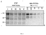
RGMaは、N末端アミノ酸168でN末端ドメイン内で2個のプロテアーゼのプロタンパク質変換酵素SKI−1及びフーリンにより切断されて、機能的に活性なタンパク質及び18、30及び40kDaの活性断片が生ずる(図1)。30kDa断片はジスルフィド結合(S−S)を介して膜結合型C末端40kDa断片にリンクしている。C末端(矢印,シェディング)GPIアンカードメイン内で切断すると、RGMaの可溶性形態を形成する3個の断片が遊離する。切断がC末端でシェダーゼ及びGPIアンカーを切断する酵素により起こると、これらの断片の可溶性形態が生ずる。膜アンカー形態のように、これら3個の可溶性断片(18kDa=末端切断型N末端ドメイン,30kDa=N末端ドメイン,40kDa=C末端ドメイン)はすべて神経突起伸長及び再生インヒビターとして活性である。 RGMa is cleaved with the proprotein converting enzymes SKI-1 and furin of two proteases in the N-terminal domain at the N-terminal amino acid 168 to give a functionally active protein and active fragments of 18, 30 and 40 kDa (1). Figure 1). The 30 kDa fragment is linked to the membrane-bound C-terminal 40 kDa fragment via a disulfide bond (SS). Cleavage within the C-terminal (arrow, shedding) GPI anchor domain releases three fragments that form the soluble form of RGMa. When cleavage occurs by an enzyme that cleaves the shedase and GPI anchors at the C-terminus, soluble forms of these fragments occur. Like the membrane anchor morphology, these three soluble fragments (18 kDa = terminally truncated N-terminal domain, 30 kDa = N-terminal domain, 40 kDa = C-terminal domain) are all active as neurite outgrowth and regeneration inhibitors.
RGMa断片ベース診断アッセイは、約1kDa〜約65kDaのサイズを有する少なくとも1個のRGMa断片を検出し得る。RGMa断片は約1kDa、約2kDa、約3kDa、約4kDa、約5kDa、約6kDa、約7kDa、約8kDa、約9kDa、約10kDa、約11kDa、約12kDa、約13kDa、約14kDa、約15kDa、約16kDa、約17kDa、約18kDa、約19kDa、約20kDa、約21kDa、約22kDa、約23kDa、約24kDa、約25kDa、約26kDa、約27kDa、約28kDa、約29kDa、約30kDa、約31kDa、約32kDa、約33kDa、約34kDa、約35kDa、約36kDa、約37kDa、約38kDa、約39kDa、約40kDa、約41kDa、約42kDa、約43kDa、約44kDa、約45kDa、約4kDa、約47kDa、約48kDa、約49kDa、約50kDa、約51kDa、約52kDa、約53kDa、約54kDa、約55kDa、約56kDa、約57kDa、約58kDa、約59kDa、約60kDa、約61kDa、約62kDa、約63kDa、約64kDa、約65kDa、またはその組合せであり得る。 The RGMa fragment-based diagnostic assay can detect at least one RGMa fragment having a size of about 1 kDa to about 65 kDa. The RGMa fragments are about 1 kDa, about 2 kDa, about 3 kDa, about 4 kDa, about 5 kDa, about 6 kDa, about 7 kDa, about 8 kDa, about 9 kDa, about 10 kDa, about 11 kDa, about 12 kDa, about 13 kDa, about 14 kDa, about 15 kDa. , About 17 kDa, about 18 kDa, about 19 kDa, about 20 kDa, about 21 kDa, about 22 kDa, about 23 kDa, about 24 kDa, about 25 kDa, about 26 kDa, about 27 kDa, about 28 kDa, about 29 kDa, about 30 kDa, about 31 kDa, about 32 kDa. 33 kDa, about 34 kDa, about 35 kDa, about 36 kDa, about 37 kDa, about 38 kDa, about 39 kDa, about 40 kDa, about 41 kDa, about 42 kDa, about 43 kDa, about 44 kDa, about 45 kDa, about 4 kDa, about 47 kDa, about 48 kDa, about 48 kDa. About 50 kDa, about 51 kDa, about 52 kDa, about 53 kDa, about 54 kDa, about 55 kDa, about 56 kDa, about 57 kDa, about 58 kDa, about 59 kDa, about 60 kDa, about 61 kDa, about 62 kDa, about 63 kDa, about 64 kDa, about 65 kDa, or about 65 kDa. It can be a combination.
RGMa断片ベース診断アッセイは、少なくとも1個のRGMa断片、少なくとも2個のRGMa断片、少なくとも3個のRGMa断片、少なくとも4個のRGMa断片、少なくとも5個のRGMa断片、少なくとも6個のRGMa断片、または少なくとも7個のRGMa断片を検出し得る。RGMa断片ベース診断アッセイは、10kDaのRGMa断片、18kDaのRGMa断片、20kDaのRGMa断片、30kDaのRGMa断片、40kDaのRGMa断片、50kDaのRGMa断片、65kDaのRGMa断片、またはその組合せを検出し得る。例えば、RGMa断片ベース診断アッセイは、10kDaのRGMa断片;18kDaのRGMa断片;20kDaのRGMa断片;30kDaのRGMa断片;40kDaのRGMa断片;50kDaのRGMa断片;65kDaのRGMa断片;10kDaのRMGa断片及び18kDaのRGMa断片;10kDaのRMGa断片及び20kDaのRGMa断片;10kDaのRMGa断片及び30kDaのRGMa断片;10kDaのRMGa断片及び40kDaのRGMa断片;10kDaのRMGa断片及び50kDaのRGMa断片;10kDaのRMGa断片及び60kDaのRGMa断片;18kDaのRMGa断片及び20kDaのRGMa断片;18kDaのRMGa断片及び30kDaのRGMa断片;18kDaのRMGa断片及び40kDaのRGMa断片;18kDaのRMGa断片及び50kDaのRGMa断片;18kDaのRMGa断片及び60kDaのRGMa断片;20kDaのRMGa断片及び30kDaのRGMa断片;20kDaのRMGa断片及び40kDaのRGMa断片;20kDaのRMGa断片及び50kDaのRGMa断片;20kDaのRMGa断片及び60kDaのRGMa断片;30kDaのRMGa断片及び40kDaのRGMa断片;30kDaのRMGa断片及び50kDaのRGMa断片;30kDaのRMGa断片及び60kDaのRGMa断片;40kDaのRMGa断片及び50kDaのRGMa断片;40kDaのRMGa断片及び60kDaのRGMa断片;50kDaのRMGa断片及び60kDaのRGMa断片;10kDaのRGMa断片及び少なくとも2個、少なくとも3個、少なくとも4個、少なくとも5個、または少なくとも6個の18kDaのRGMa断片、20kDaのRGMa断片、30kDaのRGMa断片、40kDaのRGMa断片、50kDaのRGMa断片または65kDaのRGMa断片;18kDaのRGMa断片及び少なくとも2個、少なくとも3個、少なくとも4個、少なくとも5個、または少なくとも6個の10kDaのRGMa断片、20kDaのRGMa断片、30kDaのRGMa断片、40kDaのRGMa断片、50kDaのRGMa断片または65kDaのRGMa断片;20kDaのRGMa断片及び少なくとも2個、少なくとも3個、少なくとも4個、少なくとも5個、または少なくとも6個の10kDaのRGMa断片、18kDaのRGMa断片、30kDaのRGMa断片、40kDaのRGMa断片、50kDaのRGMa断片または65kDaのRGMa断片;30kDaのRGMa断片及び少なくとも2個、少なくとも3個、少なくとも4個、少なくとも5個、または少なくとも6個の10kDaのRGMa断片、18kDaのRGMa断片、20kDaのRGMa断片、40kDaのRGMa断片、50kDaのRGMa断片または65kDaのRGMa断片;40kDaのRGMa断片及び少なくとも2個、少なくとも3個、少なくとも4個、少なくとも5個、または少なくとも6個の10kDaのRGMa断片、18kDaのRGMa断片、20kDaのRGMa断片、30kDaのRGMa断片、50kDaのRGMa断片または65kDaのRGMa断片;50kDaのRGMa断片及び少なくとも2個、少なくとも3個、少なくとも4個、少なくとも5個、または少なくとも6個の10kDaのRGMa断片、18kDaのRGMa断片、20kDaのRGMa断片、30kDaのRGMa断片、40kDaのRGMa断片または65kDaのRGMa断片;65kDaのRGMa断片及び少なくとも2個、少なくとも3個、少なくとも4個、少なくとも5個、または少なくとも6個の10kDaのRGMa断片、18kDaのRGMa断片、20kDaのRGMa断片、30kDaのRGMa断片、40kDaのRGMa断片または50kDaのRGMa断片を検出し得る。RGMa断片ベース診断アッセイは、18kDaのRGMa断片、30kDaのRGMa断片及び40kDaのRGMa断片が以下に検討する抗RGMa抗体のようなキャプチャー結合タンパク質に対する結合エピトーブ部位を保持している限り、これら3個の断片を検出し得る。 The RGMa fragment-based diagnostic assay is performed by at least one RGMa fragment, at least two RGMa fragments, at least three RGMa fragments, at least four RGMa fragments, at least five RGMa fragments, at least six RGMa fragments, or At least 7 RGMa fragments can be detected. The RGMa fragment-based diagnostic assay may detect a 10 kDa RGMa fragment, an 18 kDa RGMa fragment, a 20 kDa RGMa fragment, a 30 kDa RGMa fragment, a 40 kDa RGMa fragment, a 50 kDa RGMa fragment, a 65 kDa RGMa fragment, or a combination thereof. For example, the RGMa fragment-based diagnostic assay is a 10 kDa RGMa fragment; an 18 kDa RGMa fragment; a 20 kDa RGMa fragment; a 30 kDa RGMa fragment; a 40 kDa RGMa fragment; a 50 kDa RGMa fragment; a 65 kDa RGMa fragment; a 10 kDa RGMa fragment. RGMa Fragment; 10 kDa RMGa Fragment and 20 kDa RGMa Fragment; 10 kDa RMGa Fragment and 30 kDa RGMa Fragment; 10 kDa RMGa Fragment and 40 kDa RGMa Fragment; 10 kDa RMGa Fragment and 50 kDa RGMa Fragment; RGMa Fragment; 18 kDa RMGa Fragment and 20 kDa RGMa Fragment; 18 kDa RMGa Fragment and 30 kDa RGMa Fragment; 18 kDa RMGa Fragment and 40 kDa RGMa Fragment; 18 kDa RMGa Fragment and 50 kDa RGMa Fragment; RGMa Fragment; 20 kDa RMGa Fragment and 30 kDa RGMa Fragment; 20 kDa RMGa Fragment and 40 kDa RGMa Fragment; 20 kDa RMGa Fragment and 50 kDa RGMa Fragment; 20 kDa RMGa Fragment and 60 kDa RGMa Fragment; RGMa Fragment; 30 kDa RMGa Fragment and 50 kDa RGMa Fragment; 30 kDa RMGa Fragment and 60 kDa RGMa Fragment; 40 kDa RMGa Fragment and 50 kDa RGMa Fragment; 40 kDa RMGa Fragment and 60 kDa RGMa Fragment; RGMa Fragment; 10 kDa RGMa Fragment and At least 2, At least 3, At least 4, At least 5, or At least 6 18 kDa RGMa Fragments, 20 kDa RGMa Fragments, 30 kDa RGMa Fragments, 40 kDa RGMa Fragments, 50 kDa RGMa fragment or 65 kDa RGMa fragment; 18 kDa RGMa fragment and at least 2, at least 3, at least 4, at least 5, or at least 6 10 kDa RGMa fragments, 20 kDa RGMa fragment, 30 kDa RGMa fragment , 40 kDa RGMa fragment, 50 kDa RGMa fragment or 65 kDa RGMa fragment; 20 kDa RGMa fragment and at least 2, at least 3, at least 4, at least 5, Or at least 6 10 kDa RGMa fragments, 18 kDa RGMa fragments, 30 kDa RGMa fragments, 40 kDa RGMa fragments, 50 kDa RGMa fragments or 65 kDa RGMa fragments; 30 kDa RGMa fragments and at least 2, at least 3 4, at least 5, or at least 6 10 kDa RGMa fragments, 18 kDa RGMa fragments, 20 kDa RGMa fragments, 40 kDa RGMa fragments, 50 kDa RGMa fragments or 65 kDa RGMa fragments; 40 kDa RGMa fragments and at least 2 , At least 3, at least 4, at least 5, or at least 6 10 kDa RGMa fragments, 18 kDa RGMa fragments, 20 kDa RGMa fragments, 30 kDa RGMa fragments, 50 kDa RGMa fragments or 65 kDa RGMa fragments; RGMa fragment and at least 2, at least 3, at least 4, at least 5, or at least 6 10 kDa RGMa fragments, 18 kDa RGMa fragment, 20 kDa RGMa fragment, 30 kDa RGMa fragment, 40 kDa RGMa fragment or 65 kDa RGMa Fragment; 65 kDa RGMa Fragment and At least 2, At least 3, At least 4, At least 5, or At least 6 10 kDa RGMa Fragments, 18 kDa RGMa Fragments, 20 kDa RGMa Fragments, 30 kDa RGMa Fragments, A 40 kDa RGMa fragment or a 50 kDa RGMa fragment can be detected. The RGMa fragment-based diagnostic assay is performed on these three as long as the 18 kDa RGMa fragment, the 30 kDa RGMa fragment and the 40 kDa RGMa fragment retain binding epitove sites for capture binding proteins such as the anti-RGMa antibody discussed below. Fragments can be detected.
b.断片検出
被験者からのサンプル中のRGMa断片は、該断片を分離し、その断片のサイズを測定するための各種手段により検出され、定量化され得る。断片は、RGMa断片に特異的に結合するキャプチャー結合タンパク質、例えば抗RGMa抗体のようなRGMa断片結合タンパク質を用いて検出され得る。キャプチャー結合タンパク質は、検出可能標識を有し得、または検出可能標識を有する検出結合タンパク質により認識される。検出可能標識によりRGMa断片を同定できる。
b. Fragment detection RGMa fragments in a sample from a subject can be detected and quantified by various means for separating the fragments and measuring the size of the fragments. Fragments can be detected using a capture binding protein that specifically binds to an RGMa fragment, eg, an RGMa fragment binding protein such as an anti-RGMa antibody. The capture binding protein may have a detectable label or is recognized by a detection binding protein having a detectable label. RGMa fragments can be identified by the detectable label.
幾つかの実施形態では、RGMa断片はSDS−PAGE/ウェスタンブロッティング分析を用いて同定、サイズ分け及び定量化され得る。幾つかの実施形態では、RGMa断片はカラムクロマトグラフィー技術を用いて同定、サイズ分け及び定量化され得る。幾つかの実施形態では、RGMa断片は質量分析法を用いて同定、サイズ分け及び定量化され得る。 In some embodiments, the RGMa fragment can be identified, sized and quantified using SDS-PAGE / Western blotting analysis. In some embodiments, the RGMa fragment can be identified, sized and quantified using column chromatography techniques. In some embodiments, the RGMa fragment can be identified, sized and quantified using mass spectrometry.
キャプチャー結合タンパク質は、抗RGMa抗体、例えばビオチン化RGMa選択的抗体(BAF2459 R&D Systems)、または米国特許出願公開Nos.2004/0102376、2010/0028340、2011/0135664、2013/0330347及び2014/0023659に記載されているRGMa抗体であり得る。RGMa断片に結合する抗体は、ABCペルオキシダーゼ染色キット(Pierce;32020)または高感度ECL溶液(Thermo Scientific,SuperSignal West Femto Chemiluminescence Substrate,34094)とインキュベートした後可視化され、VersaDocイメージャー(BioRad)を用いてスキャンされ得る。体液中の組換えRGMa(R&D Systems,2459−RM−050)及び1個のRGMa断片のバンド強度を定量化するためにQuantity One Version 4.6.9(BioRad)が使用され得る。 Capture-binding proteins include anti-RGMa antibodies, such as biotinylated RGMa selective antibodies (BAF2459 R & D Systems), or US Patent Application Publication Nos. It can be the RGMa antibody described in 2004/0102376, 2010/0028340, 2011/01335664, 2013/0330347 and 2014/0023569. Antibodies that bind to the RGMa fragment are visualized after incubation with an ABC peroxidase staining kit (Pierce; 32020) or a sensitive ECL solution (Thermo Scientific, SuperSignal West Femto Chemiluminescence Substrate, 34094), and then bio-imaged using Bio-Rad Laboratories. Can be scanned. Quantity One Version 4.6.9 (BioRad) can be used to quantify the band intensity of recombinant RGMa (R & D Systems, 2459-RM-050) and one RGMa fragment in body fluids.
(1)SDS−PAGE/ウェスタンブロッティング
RGMa断片ベース診断アッセイは、少なくとも1個のRGMa断片を膜に固定化してウェスタンブロッティング膜を作成させるステップと、ウェスタンブロッティング膜をキャプチャー結合タンパク質と接触させるステップと、ここで、キャプチャー結合タンパク質は、ウェスタンブロッティング膜上に固定化した少なくとも1個のRGMa断片に結合して、キャプチャー結合タンパク質−RGMa断片複合体を形成する、ウェスタンブロッティング膜を検出結合タンパク質と接触させるステップと、ここで、検出結合タンパク質は、キャプチャー結合タンパク質と相互作用して、検出結合タンパク質−キャプチャー結合タンパク質RGMa断片複合体を形成するステップとをさらに含み得る。
(1) The SDS-PAGE / Western blotting RGMa fragment-based diagnostic assay includes a step of immobilizing at least one RGMa fragment on the membrane to form a Western blotting membrane, a step of contacting the Western blotting membrane with a capture binding protein, and a step of contacting the Western blotting membrane with a capture binding protein. Here, the capture-binding protein binds to at least one RGMa fragment immobilized on the Western blotting membrane to form a capture-binding protein-RGMa fragment complex, a step of contacting the Western blotting membrane with the detection-binding protein. And, here, the detection binding protein may further comprise the step of interacting with the capture binding protein to form a detection binding protein-capture binding protein RGMa fragment complex.
RGMaタンパク質標準マーカーを使用し、サンプルと同時にSDS−PAGEを用いて分離し得る。少なくとも1個のRGMa断片結合強度をRGMaタンパク質標準マーカーと比較して、RGMa断片のサイズを測定し、及び/または少なくとも1個のRGMa断片の量を定量化する。RGMaタンパク質標準は組換えRGMa断片の勾配であり得る。幾つかの実施形態では、組換えRGMa断片の勾配は、10、25、50、100及び200pg/mLを含む。SDS−PAGEは5%〜25% アクリルアミドを有し得る。幾つかの実施形態では、SDS−PAGEは4〜15% アクリルアミド勾配ゲルであり得る。膜は、ニトロセルロースまたはPVDF膜であり得る。 RGMa protein standard markers can be used and separated using SDS-PAGE at the same time as the sample. The binding strength of at least one RGMa fragment is compared to the RGMa protein standard marker to measure the size of the RGMa fragment and / or to quantify the amount of at least one RGMa fragment. The RGMa protein standard can be a gradient of recombinant RGMa fragments. In some embodiments, the gradient of the recombinant RGMa fragment comprises 10, 25, 50, 100 and 200 pg / mL. SDS-PAGE can have 5% to 25% acrylamide. In some embodiments, SDS-PAGE can be a 4-15% acrylamide gel gradient. The membrane can be a nitrocellulose or PVDF membrane.
3.RGMa断片ベース診断アッセイの使用方法−診断、予測、または治療/予防治療の効果の評価
本発明では、RGMa断片ベース診断アッセイの使用方法も提供する。方法は、その必要がある被験者からサンプルを入手することを含む。方法は、被験者から得たサンプル中の少なくとも1個の上記RGMa断片の存在及び/またはレベルを検出するためにRGMa断片ベース診断アッセイを利用する。被験者は神経変性疾患を患っているかそのリスクを有し得る。
3. 3. How to Use the RGMa Fragment-Based Diagnostic Assay-Assessment of Diagnostic, Predictive, or Therapeutic / Preventive Therapeutic Efficacy The present invention also provides a method of using the RGMa fragment-based diagnostic assay. The method involves obtaining a sample from a subject who needs it. The method utilizes an RGMa fragment-based diagnostic assay to detect the presence and / or level of at least one of the above RGMa fragments in a sample obtained from a subject. Subjects may have or be at risk of neurodegenerative disease.
方法は、神経変性疾患に対する治療または治療レジメンの有効性を判定するためにRGMa断片ベース診断アッセイを利用する。他の実施形態では、方法は、神経変性疾患を患っている被験者の治療または治療レジメンに対する応答性を予測するためにRGMa断片ベース診断アッセイを利用する。幾つかの実施形態では、方法は、治療または治療レジメンを被験者に投与すべきかを判定するためにRGMa断片ベース診断アッセイを利用する。もっと他の実施形態では、方法は、神経変性疾患を患っている被験者に対する治療または治療レジメンを最適化するためにRGMa断片ベース診断アッセイを利用する。幾つかの実施形態では、方法は、神経変性疾患を患っている被験者の治療または治療レジメンをモニターするためにRGMa断片ベース診断アッセイを使用し得る。他の実施形態では、方法は、神経変性疾患に対して治療上有効である化合物をスクリーニングするためにRGMa断片ベース診断アッセイを利用する。 The method utilizes an RGMa fragment-based diagnostic assay to determine the effectiveness of a treatment or treatment regimen for neurodegenerative diseases. In other embodiments, the method utilizes an RGMa fragment-based diagnostic assay to predict the responsiveness of a subject suffering from a neurodegenerative disease to treatment or treatment regimen. In some embodiments, the method utilizes an RGMa fragment-based diagnostic assay to determine whether treatment or treatment regimen should be administered to a subject. In yet another embodiment, the method utilizes an RGMa fragment-based diagnostic assay to optimize treatment or treatment regimen for subjects suffering from neurodegenerative disease. In some embodiments, the method may use an RGMa fragment-based diagnostic assay to monitor the treatment or treatment regimen of a subject suffering from a neurodegenerative disease. In other embodiments, the method utilizes an RGMa fragment-based diagnostic assay to screen for therapeutically effective compounds for neurodegenerative diseases.
a.神経変性疾患
RGMaは、慢性疾患の神経変性及び進行と再生の間の相互作用のモジュレーターとして役割を発揮し得る。神経変性疾患はRGMaの存在が疾患に関係している、すなわちRGMa活性が有害である疾患であり得る。例えば、RGMaは虚血損傷ヒト脳組織中、外傷性脳損傷を患っているヒトの病巣中、AD患者のプラーク領域中、パーキンソン病患者の黒質中、及びMS患者中に見られた。神経変性疾患または障害は、多発性硬化症、パーキンソン病、アルツハイマー病、テイ−サックス病、ニーマン−ピック病、ゴーシェ病、ハーラー症候群、ハンチントン病、筋萎縮性側策硬化症、特発性炎症性脱髄疾患、ビタミンB12欠乏症、橋中心髄鞘崩壊症、脊髄ろう、横断性脊髄炎、デビック病、進行性多巣性白質脳症、視神経炎、脊髄損傷のようなCNSに対する外傷性損傷、外傷性脳損傷、虚血性脳卒中のような卒中、緑内障、糖尿病性網膜症、加齢黄斑変性及び白質ジストロフィーであり得る。
a. Neurodegenerative disease RGMa can act as a modulator of the interaction between neurodegeneration and progression and regeneration of chronic diseases. Neurodegenerative diseases can be diseases in which the presence of RGMa is associated with the disease, i.e., where RGMa activity is detrimental. For example, RGMa was found in ischemic-injured human brain tissue, in human lesions suffering from traumatic brain injury, in the plaque region of AD patients, in the substantia nigra of Parkinson's disease patients, and in MS patients. Neurodegenerative diseases or disorders include multiple sclerosis, Parkinson's disease, Alzheimer's disease, Teisax's disease, Niemann-Pick's disease, Gauche's disease, Harler's syndrome, Huntington's disease, muscular atrophic lateral sclerosis, idiopathic inflammatory prolapse. Traumatic injuries to CNS, traumatic brain such as myelopathy, vitamin B12 deficiency, pons central medulla disintegration, spinal fistula, transverse myelitis, Devic's disease, progressive multifocal leukoencephalopathy, optic neuritis, spinal cord injury It can be injury, stroke such as ischemic stroke, glaucoma, diabetic retinopathy, age-related macular degeneration and leukodystrophy.
(1)多発性硬化症
多発性硬化症(MS)は、脳内及び脳と身体間の情報の流れを分断する中枢神経系の身体障害性疾患である。MSは、身体の免疫系の異常応答が脳、脊髄、網膜及び視神経から構成される中枢神経系(CNS)に向けられている免疫媒介プロセスを伴う。この疾患は、その進行、及び炎症病巣のその顕著な大脳及び脊髄局在に関して各種サブタイプにより特徴づけられる。前記サブタイプには、再発寛解型MS(RRMS)、二次性進行型MS(SPMS)、一次性進行型MS(PPMS)及び進行再発型MS(PRMS)が含まれる。
(1) Multiple sclerosis Multiple sclerosis (MS) is a disability disorder of the central nervous system that divides the flow of information in the brain and between the brain and the body. MS involves an immune-mediated process in which an abnormal response of the body's immune system is directed to the central nervous system (CNS), which consists of the brain, spinal cord, retina and optic nerve. The disease is characterized by various subtypes with respect to its progression and its prominent cerebral and spinal cord localization of inflammatory lesions. The subtypes include relapsing-remitting MS (RRMS), secondary progressive MS (SPMS), primary progressive MS (PPMS) and advanced recurrent MS (PRMS).
RRMSは、しばしば再発、再燃または憎悪と呼ばれる悪化する神経機能の明確に規定された発作の後、その間に症状が部分的または完全に改善され、疾患の明らかな進行がない部分的または完全回復期間(寛解)により特徴づけられる。SPMSはMSの二次相として特徴づけられ、当初RRMS疾患経過を有していた個人で生ずる。SPMSにかかっている個人は、疾患が次第にRRMSで見られる炎症プロセスから神経損傷または損失により特徴づけられるより着実に進行する相に徐々に変化するので、炎症に起因する再発を経験し続けることも、しないこともある。PPMSは明確な再発または寛解期間なく神経機能の着実な悪化により特徴づけられる。PPMSにかかっている個人はたまの停滞期または一時的な回復があるが、進行は続いている長期間にわたって変わり得る進行速度を有している。PRMSは、PRMSにかかっている個人がRRMSにかかっている人が経験するようなたまの回復に加えて、非常に初期から神経機能の着実な悪化を経験する点でPPMSに類似している。 RRMS is a period of partial or complete recovery in which symptoms are partially or completely improved during a well-defined attack of worsening neurological function, often referred to as recurrence, relapse or exacerbation, with no apparent progression of the disease. Characterized by (remission). SPMS is characterized as a secondary phase of MS and occurs in individuals who initially had a course of RRMS disease. Individuals with SPMS may continue to experience recurrence due to inflammation as the disease gradually changes from the inflammatory process seen in RRMS to a more steadily progressive phase characterized by nerve damage or loss. , Sometimes not. PPMS is characterized by a steady deterioration of neural function without a definite recurrence or duration of remission. Individuals with PPMS have occasional stagnation or temporary recovery, but progression has a variable rate of progression over a long period of time. PRMS is similar to PPMS in that individuals with PRMS experience a steady deterioration in neural function from the very beginning, in addition to the occasional recovery experienced by those with RRMS.
用語「最初の臨床症状」(CIS)は、少なくとも24時間続き、中枢神経系の1つ以上の部位における炎症及び脱髄に起因している神経症状の第1エピソードを表すために使用される。CISは、人が1つの病巣に起因する1つの神経兆候または症状を経験する単発性、または人が2つ以上の場所の病巣に起因する2つ以上の兆候または症状を経験する多発性であり得る。CISを経験した人はMSを発症することも、しないこともある。長期間には、多くの患者は最後には神経変性を有する進行性くすぶり型慢性炎症性プロセスに至る。 The term "first clinical manifestations" (CIS) lasts at least 24 hours and is used to describe the first episode of neurological symptoms resulting from inflammation and demyelination in one or more parts of the central nervous system. CIS is solitary, in which a person experiences one neurological sign or symptom due to one lesion, or multiple, in which a person experiences two or more signs or symptoms due to a lesion in more than one place. obtain. People who have experienced CIS may or may not develop MS. Over the long term, many patients eventually lead to a progressive smoldering chronic inflammatory process with neurodegeneration.
MSを患っているかまたは患っていると疑われる被験者は総合障害度評価スコア(EDSS)及び/または最大歩行距離及び/または歩行速度の評価を用いて評価または診断され得る。幾つかの実施形態では、低いEDSSスコア、増加した歩行距離及び/または低下した歩行速度を有する被験者は治療または治療レジメンが該被験者に対して有効であることを示し得る。 Subjects with or suspected of having MS can be evaluated or diagnosed using the Comprehensive Disability Rating Score (EDSS) and / or the assessment of maximum walking distance and / or walking speed. In some embodiments, a subject with a low EDSS score, increased gait distance and / or decreased gait speed may indicate that the treatment or treatment regimen is effective for the subject.
例えば、被験者のEDSSスコアが治療前の被験者のEDSSスコアと比較して少なくとも約0.1、少なくとも約0.2、少なくとも約0.3、少なくとも約0.4、少なくとも約0.5、少なくとも約0.6、少なくとも約0.7、少なくとも約0.8、少なくとも約0.9、少なくとも約1.0、少なくとも約2.0、少なくとも約3.0、少なくとも約4.0、少なくとも約5.0、または少なくとも約6.0低いことは、治療または治療レジメンが神経変性疾患に有効であることを示す。 For example, a subject's EDSS score is at least about 0.1, at least about 0.2, at least about 0.3, at least about 0.4, at least about 0.5, at least about, compared to the subject's EDSS score before treatment. 0.6, at least about 0.7, at least about 0.8, at least about 0.9, at least about 1.0, at least about 2.0, at least about 3.0, at least about 4.0, at least about 5. A zero, or at least about 6.0 lower, indicates that the treatment or treatment regimen is effective for neurodegenerative diseases.
例えば、被験者の歩行距離が約70m〜約150m、約70m〜約145m、約70m〜約140m、約70m〜約135m、約70m〜約130m、約70m〜約125m、約70m〜約120m、約70m〜約115m、約70m〜約110m、約70m〜約105m、約70m〜約100m、約70m〜約95m、約70m〜約90m、約70m〜約85m、約70m〜約80m、約70m〜約75m、約75m〜約150m、約75m〜約145m、約75m〜約140m、約75m〜約135m、約75m〜約130m、約75m〜約125m、約75m〜約120m、約75m〜約115m、約75m〜約110m、約75m〜約105m、約75m〜約100m、約75m〜約95m、約75m〜約90m、約75m〜約85m、約75m〜約80m、約80m〜約150m、約80m〜約145m、約80m〜約140m、約80m〜約135m、約80m〜約130m、約80m〜約125m、約80m〜約120m、約80m〜約115m、約80m〜約110m、約80m〜約105m、約80m〜約100m、約80m〜約95m、約80m〜約90m、約80m〜約85m、約85m〜約150m、約85m〜約145m、約85m〜約140m、約85m〜約135m、約85m〜約130m、約85m〜約125m、約85m〜約120m、約85m〜約115m、約85m〜約110m、約85m〜約105m、約85m〜約100m、約85m〜約95m、約85m〜約90m、約90m〜約150m、約90m〜約145m、約90m〜約140m、約90m〜約135m、約90m〜約130m、約90m〜約125m、約90m〜約120m、約90m〜約115m、約90m〜約110m、約90m〜約105m、約90m〜約100m、約90m〜約95m、約95m〜約150m、約95m〜約145m、約95m〜約140m、約95m〜約135m、約95m〜約130m、約95m〜約125m、約95m〜約120m、約95m〜約115m、約95m〜約110m、約95m〜約105m、約95m〜約100m、約100m〜約150m、約100m〜約145m、約100m〜約140m、約100m〜約135m、約100m〜約130m、約100m〜約125m、約100m〜約120m、約100m〜約115m、約100m〜約110m、約100m〜約105m、約105m〜約150m、約105m〜約145m、約105m〜約140m、約105m〜約135m、約105m〜約130m、約105m〜約125m、約105m〜約120m、約105m〜約115m、約105m〜約110m、約110m〜約150m、約110m〜約145m、約110m〜約140m、約110m〜約135m、約110m〜約130m、約110m〜約125m、約110m〜約120m、約110m〜約115m、約115m〜約150m、約115m〜約145m、約115m〜約140m、約115m〜約135m、約115m〜約130m、約115m〜約125m、約115m〜約120m、約120m〜約150m、約120m〜約145m、約120m〜約140m、約120m〜約135m、約120m〜約130m、約120m〜約125m、約250m〜約750m、約250m〜約700m、約250m〜約650m、約250m〜約600m、約250m〜約550m、約250m〜約500m、約250m〜約450m、約250m〜約400m、約250m〜約350m、約250m〜約300m、約300m〜約750m、約300m〜約700m、約300m〜約650m、約300m〜約600m、約300m〜約550m、約300m〜約500m、約300m〜約450m、約300m〜約400m、約300m〜約350m、約350m〜約750m、約350m〜約700m、約350m〜約650m、約350m〜約600m、約350m〜約550m、約350m〜約500m、約350m〜約450m、約350m〜約400m、約400m〜約750m、約400m〜約700m、約400m〜約650m、約400m〜約600m、約400m〜約550m、約400m〜約500m、約400m〜約450m、約450m〜約750m、約450m〜約700m、約450m〜約650m、約450m〜約600m、約450m〜約550m、約450m〜約500m、約500m〜約750m、約500m〜約700m、約500m〜約650m、約500m〜約600m、約500m〜約550m、約550m〜約750m、約550m〜約700m、約550m〜約650m、約550m〜約600m、約600m〜約750m、約600m〜約700m、約600m〜約650m、約650m〜約750m、または約650m〜約700m増加したことは、治療または治療レジメンが被験者における神経変性疾患に有効であることを示す。被験者の歩行距離が少なくとも約1m、少なくとも約2m、少なくとも約3m、少なくとも約4m、少なくとも約5m、少なくとも約10m、少なくとも約15m、少なくとも約20m、少なくとも約25m、少なくとも約30m、少なくとも約35m、少なくとも約40m、少なくとも約45m、少なくとも約50m、少なくとも約55m、少なくとも約60m、少なくとも約65m、少なくとも約70m、少なくとも約75m、少なくとも約80m、少なくとも約85m、少なくとも約90m、少なくとも約95m、少なくとも約100m、少なくとも約105m、少なくとも約110m、少なくとも約115m、少なくとも約120m、少なくとも約125m、少なくとも約130m、少なくとも約135m、少なくとも約140m、少なくとも約145m、少なくとも約150m、少なくとも約155m、少なくとも約160m、少なくとも約165m、少なくとも約170m、少なくとも約175m、少なくとも約180m、少なくとも約185m、少なくとも約190m、少なくとも約195m、または少なくとも約200m増加したことは、治療または治療レジメンが被験者における神経変性疾患に有効であることを示す。 For example, the walking distance of the subject is about 70 m to about 150 m, about 70 m to about 145 m, about 70 m to about 140 m, about 70 m to about 135 m, about 70 m to about 130 m, about 70 m to about 125 m, about 70 m to about 120 m, about. 70m to about 115m, about 70m to about 110m, about 70m to about 105m, about 70m to about 100m, about 70m to about 95m, about 70m to about 90m, about 70m to about 85m, about 70m to about 80m, about 70m ~ About 75m, about 75m to about 150m, about 75m to about 145m, about 75m to about 140m, about 75m to about 135m, about 75m to about 130m, about 75m to about 125m, about 75m to about 120m, about 75m to about 115m , About 75m to about 110m, about 75m to about 105m, about 75m to about 100m, about 75m to about 95m, about 75m to about 90m, about 75m to about 85m, about 75m to about 80m, about 80m to about 150m, about 80m to about 145m, about 80m to about 140m, about 80m to about 135m, about 80m to about 130m, about 80m to about 125m, about 80m to about 120m, about 80m to about 115m, about 80m to about 110m, about 80m ~ About 105m, about 80m to about 100m, about 80m to about 95m, about 80m to about 90m, about 80m to about 85m, about 85m to about 150m, about 85m to about 145m, about 85m to about 140m, about 85m to about 135m , About 85m to about 130m, about 85m to about 125m, about 85m to about 120m, about 85m to about 115m, about 85m to about 110m, about 85m to about 105m, about 85m to about 100m, about 85m to about 95m, about 85m to about 90m, about 90m to about 150m, about 90m to about 145m, about 90m to about 140m, about 90m to about 135m, about 90m to about 130m, about 90m to about 125m, about 90m to about 120m, about 90m ~ About 115m, about 90m to about 110m, about 90m to about 105m, about 90m to about 100m, about 90m to about 95m, about 95m to about 150m, about 95m to about 145m, about 95m to about 140m, about 95m to about 135m , About 95m to about 130m, about 95m to about 125m, about 95m to about 120m, about 95m to about 115m, about 95m to about 110m, about 95m to about 105m, about 95m to about 100m, about 100m to about 150m, about 100m to about 145m, about 100m to about 140m, about 100m to about 135m, about 100m to about 130m, about 100m to about 125m, about 100m to about 120m, about 100m to about 115m, about 100m to about 110m, about 10 0m to about 105m, about 105m to about 150m, about 105m to about 145m, about 105m to about 140m, about 105m to about 135m, about 105m to about 130m, about 105m to about 125m, about 105m to about 120m, about 105m ~ About 115m, about 105m to about 110m, about 110m to about 150m, about 110m to about 145m, about 110m to about 140m, about 110m to about 135m, about 110m to about 130m, about 110m to about 125m, about 110m to about 120m , About 110m to about 115m, about 115m to about 150m, about 115m to about 145m, about 115m to about 140m, about 115m to about 135m, about 115m to about 130m, about 115m to about 125m, about 115m to about 120m, about 120m to about 150m, about 120m to about 145m, about 120m to about 140m, about 120m to about 135m, about 120m to about 130m, about 120m to about 125m, about 250m to about 750m, about 250m to about 700m, about 250m ~ About 650m, about 250m to about 600m, about 250m to about 550m, about 250m to about 500m, about 250m to about 450m, about 250m to about 400m, about 250m to about 350m, about 250m to about 300m, about 300m to about 750m , About 300m to about 700m, about 300m to about 650m, about 300m to about 600m, about 300m to about 550m, about 300m to about 500m, about 300m to about 450m, about 300m to about 400m, about 300m to about 350m, about 350m to about 750m, about 350m to about 700m, about 350m to about 650m, about 350m to about 600m, about 350m to about 550m, about 350m to about 500m, about 350m to about 450m, about 350m to about 400m, about 400m to About 750m, about 400m to about 700m, about 400m to about 650m, about 400m to about 600m, about 400m to about 550m, about 400m to about 500m, about 400m to about 450m, about 450m to about 750m, about 450m to about 700m , About 450m to about 650m, about 450m to about 600m, about 450m to about 550m, about 450m to about 500m, about 500m to about 750m, about 500m to about 700m, about 500m to about 650m, about 500m to about 600m, about 500m to about 550m, about 550m to about 750m, about 550m to about 700m, about 550m to about 650m, about 550m to about 600m, about 600m to about 750m, about 600m to about 700m, about 600m to about 650m, about 650m about An increase of 750 m, or about 650 m to about 700 m, indicates that the treatment or treatment regimen is effective for neurodegenerative disease in the subject. The walking distance of the subject is at least about 1 m, at least about 2 m, at least about 3 m, at least about 4 m, at least about 5 m, at least about 10 m, at least about 15 m, at least about 20 m, at least about 25 m, at least about 30 m, at least about 35 m, at least. About 40m, at least about 45m, at least about 50m, at least about 55m, at least about 60m, at least about 65m, at least about 70m, at least about 75m, at least about 80m, at least about 85m, at least about 90m, at least about 95m, at least about 100m At least about 105m, at least about 110m, at least about 115m, at least about 120m, at least about 125m, at least about 130m, at least about 135m, at least about 140m, at least about 145m, at least about 150m, at least about 155m, at least about 160m, at least An increase of about 165 m, at least about 170 m, at least about 175 m, at least about 180 m, at least about 185 m, at least about 190 m, at least about 195 m, or at least about 200 m is that the treatment or treatment regimen is effective for neurodegenerative diseases in the subject. Show that.
例えば、被験者の歩行速度(s/8m)が少なくとも約30s/8m〜少なくとも約10s/8m、少なくとも約25s/8m〜少なくとも約10s/8m、少なくとも約20s/8m〜少なくとも約10s/8m、少なくとも約15s/8m〜少なくとも約10s/8m、少なくとも約30s/8m〜少なくとも約15s/8m、少なくとも約25s/8m〜少なくとも約15s/8m、少なくとも約20s/8m〜少なくとも約15s/8m、少なくとも約30s/8m〜少なくとも約20s/8m、少なくとも約25s/8m〜少なくとも約20s/8m、または少なくとも約30s/8m〜少なくとも約25s/8m増加したことは、治療または治療レジメンが被験者における神経変性疾患に有効であることを示す。被験者が約30.0s/8m未満、約25.0s/8m未満、約20.0s/8m未満、約15.0s/8m未満、約14.5s/8m未満、約14.0s/8m未満、約13.5s/8m未満、約13.0s/8m未満、約12.9s/8m未満、約12.8s/8m未満、約12.7s/8m未満、約12.6s/8m未満、約12.5s/8m未満、約12.4s/8m未満、約12.3s/8m未満、約12.2s/8m未満、約12.1s/8m未満、約12.0s/8m未満、約11.9s/8m未満、約11.8s/8m未満、約11.7s/8m未満、約11.6s/8m未満、約11.5s/8m未満、約11.4s/8m未満、約11.3s/8m未満、約11.2s/8m未満、約11.1s/8m未満、約11.0s/8m未満、約10.5s/8m未満、約10.0s/8m未満、約9.5s/8m未満、約9.0s/8m未満、約8.5s/8m未満、または約8.0s/8m未満の歩行速度を有していることは、治療または治療レジメンが被験者の神経変性疾患に有効であることを示す。 For example, the walking speed (s / 8m) of the subject is at least about 30s / 8m to at least about 10s / 8m, at least about 25s / 8m to at least about 10s / 8m, at least about 20s / 8m to at least about 10s / 8m, at least about about. 15s / 8m to at least about 10s / 8m, at least about 30s / 8m to at least about 15s / 8m, at least about 25s / 8m to at least about 15s / 8m, at least about 20s / 8m to at least about 15s / 8m, at least about 30s / An increase of 8 m to at least about 20 s / 8 m, at least about 25 s / 8 m to at least about 20 s / 8 m, or at least about 30 s / 8 m to at least about 25 s / 8 m indicates that the treatment or treatment regimen is effective for neurodegenerative disease in the subject. Indicates that there is. Subjects are less than about 30.0 s / 8 m, less than about 25.0 s / 8 m, less than about 20.0 s / 8 m, less than about 15.0 s / 8 m, less than about 14.5 s / 8 m, less than about 14.0 s / 8 m, Less than about 13.5s / 8m, less than about 13.0s / 8m, less than about 12.9s / 8m, less than about 12.8s / 8m, less than about 12.7s / 8m, less than about 12.6s / 8m, about 12 Less than .5s / 8m, less than about 12.4s / 8m, less than about 12.3s / 8m, less than about 12.2s / 8m, less than about 12.1s / 8m, less than about 12.0s / 8m, about 11.9s Less than / 8m, less than about 11.8s / 8m, less than about 11.7s / 8m, less than about 11.6s / 8m, less than about 11.5s / 8m, less than about 11.4s / 8m, about 11.3s / 8m Less than, less than about 11.2s / 8m, less than about 11.1s / 8m, less than about 11.0s / 8m, less than about 10.5s / 8m, less than about 10.0s / 8m, less than about 9.5s / 8m, Having a walking speed of less than about 9.0 s / 8 m, less than about 8.5 s / 8 m, or less than about 8.0 s / 8 m means that the treatment or treatment regimen is effective for the subject's neurodegenerative disease. Is shown.
例えば、被験者が8m歩行するのに要する時間が少なくとも約0.5秒、少なくとも約1秒、少なくとも約2秒、少なくとも約3秒、少なくとも約4秒、少なくとも約5秒、少なくとも約6秒、少なくとも約7秒、少なくとも約8秒、少なくとも約9秒、少なくとも約10秒、少なくとも約11秒、少なくとも約12秒、少なくとも約13秒、少なくとも約14秒、少なくとも約15秒、少なくとも約16秒、少なくとも約17秒、少なくとも約18秒、少なくとも約19秒、少なくとも約20秒、少なくとも約21秒、少なくとも約22秒、少なくとも約23秒、少なくとも約24秒、または少なくとも約25秒だけ短いことは、治療または治療レジメンが被験者における神経変性疾患に有効であることを示す。 For example, the time required for a subject to walk 8 m is at least about 0.5 seconds, at least about 1 second, at least about 2 seconds, at least about 3 seconds, at least about 4 seconds, at least about 5 seconds, at least about 6 seconds, at least. About 7 seconds, at least about 8 seconds, at least about 9 seconds, at least about 10 seconds, at least about 11 seconds, at least about 12 seconds, at least about 13 seconds, at least about 14 seconds, at least about 15 seconds, at least about 16 seconds, at least About 17 seconds, at least about 18 seconds, at least about 19 seconds, at least about 20 seconds, at least about 21 seconds, at least about 22 seconds, at least about 23 seconds, at least about 24 seconds, or at least about 25 seconds shorter is a treatment Alternatively, show that the treatment regimen is effective for neurodegenerative diseases in the subject.
b.対照/基準レベル
対照サンプルを含めることが望ましいことがある。対照サンプルは上記した被験者からのサンプルと同時に分析され得る。被験者サンプルから得た結果は対照サンプルから得た結果と比較し得る。標準曲線を用意し得、これと生体サンプルについてのアッセイ結果を比較し得る。標準曲線は、アッセイ単位、すなわち化学発光標識を使用する場合、化学発光シグナル強度の関数としてマーカーレベルを与える。複数のドナーから採取したサンプルを用いて、標準曲線は正常健康組織または治療を受けたことのないMS組織中のRGMa断片の対照レベルに対して用意され得る。
b. Control / Reference Level It may be desirable to include control samples. The control sample can be analyzed at the same time as the sample from the subject described above. The results obtained from the subject sample can be compared with the results obtained from the control sample. A standard curve can be prepared and the assay results for biological samples can be compared. The standard curve gives marker levels as a function of the assay unit, or chemiluminescent signal intensity, when using chemiluminescent labels. Using samples taken from multiple donors, standard curves can be prepared for control levels of RGMa fragments in normal healthy tissue or untreated MS tissue.
よって、上記にてらして、試験サンプル中のRGMa断片の存在、量または濃度の測定方法を提供する。方法は、(1)RGMa断片について試験サンプルをウェスタンブロット分析により、例えばRGMa断片上のエピトープに結合する少なくとも1つのキャプチャー抗体、及びキャプチャー抗体またはキャプチャー抗体に対するエピトープとは異なるRGMa断片上のエピトープに結合し、場合により検出可能標識を含む少なくとも1つの検出抗体を使用することによりアッセイすることを含む。方法は更に、(2)試験サンプル中のRGMa断片の存在、量または濃度の直接または間接指標として検出可能標識より生じたシグナルをキャリブレーター中のRGMa断片の存在、量または濃度の直接または間接指標として生じたシグナルと比較することを含む。キャリブレーターは場合により、キャリブレーターの各々が一連の他のキャリブレーターとRGMa断片の濃度だけ異なる一連のキャリブレーターの一部であり、そうであることが好ましい。 Therefore, as described above, a method for measuring the presence, amount or concentration of an RGMa fragment in a test sample is provided. The methods are as follows: (1) Western blot analysis of a test sample on an RGMa fragment, eg, at least one capture antibody that binds to an epitope on the RGMa fragment, and an epitope on the capture antibody or epitope that is different from the epitope for the capture antibody. And optionally including assaying by using at least one detection antibody comprising a detectable label. The method further comprises (2) as a direct or indirect indicator of the presence, amount or concentration of the RGMa fragment in the test sample, as a direct or indirect indicator of the presence, amount or concentration of the RGMa fragment in the calibrator. Includes comparison with the resulting signal. The calibrator is optionally part of a series of calibrators, each of which differs from the other calibrators in the series by the concentration of the RGMa fragment, and is preferred.
(1)基準レベル
本明細書中に記載されている方法は、(1)神経変性疾患を患っている被験者に対する治療または治療レジメンの有効性を同定し、判定する;(2)神経変性疾患を患っている被験者の治療または治療レジメンに対する応答性を予測する;(3)神経変性疾患を患っている被験者に対して治療または治療レジメンを与える;(4)神経変性疾患を患っている被験者に対する治療または治療レジメンを最適化する;(5)神経変性疾患を患っている被験者に対する再生促進薬物治療または治療レジメンをモニターする;及び(6)化合物を神経変性疾患に対する治療効果についてスクリーニングするために被験者のRGMa断片の基準レベルを使用する。
(1) Reference Levels The methods described herein (1) identify and determine the effectiveness of a treatment or treatment regimen for a subject suffering from a neurodegenerative disease; (2) determine the neurodegenerative disease. Predict the responsiveness of affected subjects to treatment or treatment regimens; (3) give treatment or treatment regimens to subjects suffering from neurodegenerative diseases; (4) treat subjects suffering from neurodegenerative diseases Or optimize the treatment regimen; (5) monitor regeneration-promoting drug treatment or treatment regimens for subjects suffering from neurodegenerative diseases; and (6) monitor subjects for therapeutic effects on neurodegenerative diseases. Use the reference level of the RGMa fragment.
通常、所定または基準レベルがベンチマークとして使用され得、このベンチマークに対して試験サンプルをRGMa断片(例えば、18kDaのRGMa断片、30kDaのRGMa断片及び/または40kDaのRGMa断片)についてアッセイしたときに得られる結果を評価する。通常、比較する際、所定レベルはアナライトの存在、量または濃度を具体的指標を有するMSの特定段階またはエンドポイントと連係または関連させ得るように特定のアッセイを適切な条件で十分な回数実施することにより得られる。典型的には、所定レベルは基準被験者(または、被験者の集団)のアッセイで得られる。基準被験者は神経疾患にかかっていない正常または健康被験者のような対照被験者、またはMS被験者のような神経疾患にかかっている被験者であり得る。MS被験者は、MSに対する治療を受けているかまたは受けていないMSの特定段階または前段階(すなわち、RRMS、SPMS、PPMS、PRMSまたはCIS)を有する被験者である。基準群の基準集団は対照群またはMS群であり得る。MS群には、MSの特定段階または前段階(すなわち、RRMS、SPMS、PPMS、PRMSまたはCIS)を有するMS被験者、及び/またはMSにかかっているが、MSに対する治療を受けていない被験者が含まれ得る。基準レベルは治療または治療レジメンを被験者に投与する前の被験者のRGMa断片レベルであり得る。 Usually a given or reference level can be used as a benchmark and is obtained when test samples are assayed against this benchmark on RGMa fragments (eg, 18 kDa RGMa fragment, 30 kDa RGMa fragment and / or 40 kDa RGMa fragment). Evaluate the results. Usually, when comparing, a particular assay is performed a sufficient number of times under appropriate conditions so that a given level can link or correlate the presence, amount or concentration of an analysis with a particular step or endpoint of MS having a specific indicator. Obtained by doing. Typically, a given level is obtained in an assay of a reference subject (or population of subjects). The reference subject can be a control subject, such as a normal or healthy subject who does not have a neurological disorder, or a subject who has a neurological disorder, such as an MS subject. An MS subject is a subject who has a specific stage or pre-stage (ie, RRMS, SPMS, PPMS, PRMS or CIS) of MS receiving or not receiving treatment for MS. The reference population of the reference group can be the control group or the MS group. The MS group includes MS subjects who have a specific or pre-stage of MS (ie, RRMS, SPMS, PPMS, PRMS or CIS) and / or subjects who have MS but have not been treated for MS. It can be. The reference level can be the subject's RGMa fragment level prior to administration of the treatment or treatment regimen to the subject.
特に、治療応答性について使用される所定レベルに関して、上記したRGMa断片の量または濃度は「変化なし」、「有利」(または、「有利に変化した」)、または「不利」(または、「不利に変化した」)であり得る。「高い」または「上昇した」は、試験サンプル中の量または濃度が典型的または正常レベルまたは範囲(例えば、所定レベル)、例えば対照群またはMS群で見られる典型的または正常レベルよりも高いかまたは大きいこと、または別の基準レベルまたは範囲(例えば、前のまたはベースラインサンプル)よりも高いかまたは大きいことを指す。「高い」または「上昇した」は、試験サンプル中の量または濃度が治療を開始する前の被験者で見られるレベルまたは範囲よりも高いかまたは大きいことをも指し得る。用語「低い」または「減少した」は、試験サンプル中の量または濃度が典型的または正常レベルまたは範囲(例えば、所定レベル)、例えば対照群またはMS群で見られる典型的または正常レベル以下またはそれ未満であること、または別の基準レベルまたは範囲(例えば、前のまたはベースラインサンプル)以下またはそれ未満であることを指す。用語「低い」または「減少した」は、試験サンプル中の量または濃度が治療を開始する前の被験者で見られるレベルまたは範囲以下またはそれ未満であることをも指す。用語「変化した」は、サンプル中の量または濃度が典型的または正常レベルまたは範囲(例えば、所定レベル)、例えば対照群またはMS群で見られる典型的または正常レベルに比べて、または別の基準レベルまたは範囲(例えば、前のまたはベースラインサンプル)に比べて変化していることを指す。 In particular, with respect to a given level used for treatment responsiveness, the amount or concentration of the RGMa fragment described above is "no change", "advantageous" (or "favorably changed"), or "disadvantageous" (or "disadvantageous"). Changed to "). "High" or "elevated" means that the amount or concentration in the test sample is higher than the typical or normal level or range (eg, predetermined level), eg, the typical or normal level found in the control or MS group. Or greater than, or higher or greater than another reference level or range (eg, previous or baseline sample). "High" or "elevated" can also mean that the amount or concentration in the test sample is higher or greater than the level or range seen in the subject before initiating treatment. The term "low" or "decreased" means that the amount or concentration in the test sample is at or below the typical or normal level or range (eg, predetermined level), eg, below the typical or normal level found in the control or MS group. Less than, or less than or equal to another reference level or range (eg, previous or baseline sample). The term "low" or "decreased" also refers to the amount or concentration in the test sample being below or below the level or range seen in the subject prior to the start of treatment. The term "altered" refers to an amount or concentration in a sample relative to or another criterion at a typical or normal level or range (eg, a predetermined level), eg, a typical or normal level found in a control or MS group. Refers to changes compared to a level or range (eg, previous or baseline sample).
上記したRGMa断片についての典型的または正常レベルまたは範囲、または別の基準レベルまたは範囲(例えば、前のまたはベースラインサンプル)は標準プラクティスに従って規定される。所謂変化したレベルまたは変化は、典型的または正常レベルまたは範囲、または基準レベルまたは範囲と比較して、実験誤差またはサンプル分散により説明することができない正味変化があるときに生じると見なされ得る。幾つかの実施形態では、具体的サンプルで測定されたレベルを所謂正常被験者、すなわち対照被験者からの類似サンプルで測定したレベルまたはレベルの範囲と比較する。これに関連して、「正常な」(時に「対照」または「健康な」)被験者はMS不検出の個人であり、「正常な」患者または集団は例えば検出できないMSを示すものである。「見かけ正常な被験者」はRGMa断片(例えば、18kDaのRGMa断片、30kDaのRGMa断片及び/または40kDaのRGMa断片)をまだ評価していないか評価しているものである。アナライトのレベルは、アナライトが通常不検出である(例えば、正常レベルは0、または正常集団の約25〜約75パ―センタイルの範囲内である)が、試験サンプル中で検出されるときに、及びアナライトが試験サンプル中に正常レベルよりも多く存在しているときに「高い」と言われる。幾つかの実施形態では、具体的サンプルで測定されたレベルをMS被験者からの類似サンプル、または治療を開始する前の被験者から採取した前のまたはベースラインサンプル中で測定したレベルまたはレベルの範囲と比較する。 Typical or normal levels or ranges for the RGMa fragments described above, or other reference levels or ranges (eg, previous or baseline samples) are defined according to standard practices. So-called altered levels or changes can be considered to occur when there are net changes that cannot be explained by experimental error or sample variance compared to typical or normal levels or ranges, or reference levels or ranges. In some embodiments, the level measured in a specific sample is compared to the level or range of levels measured in a so-called normal subject, i.e. a similar sample from a control subject. In this regard, "normal" (sometimes "control" or "healthy") subjects are individuals who do not detect MS, and "normal" patients or populations represent, for example, undetectable MS. An "apparently normal subject" is assessing whether or not an RGMa fragment (eg, an 18 kDa RGMa fragment, a 30 kDa RGMa fragment and / or a 40 kDa RGMa fragment) has not yet been evaluated. Annalite levels are when the announcer is normally undetected (eg, the normal level is 0, or is in the range of about 25 to about 75 percentiles of the normal population), but is detected in the test sample. And when there is more than normal levels of analysts in the test sample, it is said to be "high". In some embodiments, the level measured in a specific sample is a similar sample from an MS subject, or a range of levels or levels measured in a pre-taken or baseline sample from a subject prior to starting treatment. Compare.
幾つかの実施形態では、基準レベルがMSに対する治療を受けたかまたは受けていないMS被験者におけるRGMa断片レベルである場合、RGMa断片(例えば、18kDaのRGMa断片、30kDaのRGMa断片及び/または40kDaのRGMa断片)の基準レベルより高いレベルは被験者が治療または治療レジメンに応答しないと同定し、治療がMSの治療のために有効でないと同定する。RGMa断片(例えば、18kDaのRGMa断片、30kDaのRGMa断片及び/または40kDaのRGMa断片)の基準レベルと同等またはそれ以下のレベルは被験者が治療または治療レジメンに対して応答すると同定し、または治療がMSの治療のために有効であると同定する。 In some embodiments, if the reference level is the RGMa fragment level in an MS subject who has received or has not received treatment for MS, then the RGMa fragment (eg, 18 kDa RGMa fragment, 30 kDa RGMa fragment and / or 40 kDa RGMa). Levels above the reference level of (fragment) identify the subject as not responding to treatment or treatment regimen and identify that treatment is not effective for the treatment of MS. Levels equal to or below the reference level of the RGMa fragment (eg, 18 kDa RGMa fragment, 30 kDa RGMa fragment and / or 40 kDa RGMa fragment) are identified as the subject responding to treatment or treatment regimen, or treatment Identify as effective for the treatment of MS.
幾つかの実施形態では、対照、ベースライン、または前のレベルまたは範囲と比較したサンプル中の相対的RGMa断片レベルの変化は、被験者が治療または治療レジメンに応答すると同定し、または治療がMSの治療のために有効であると同定する。幾つかの実施形態では、被験者から採取したサンプル中の相対的RGMa断片レベルが対照、前のまたはベースラインサンプル中のRGMa断片レベルと比較して約100%未満、約95%未満、約85%未満、約80%未満、約75%未満、約70%未満、約65%未満、約55%未満、約50%未満、約45%未満、約40%未満、約35%未満、約30%未満、約25%未満、約20%未満、約15%未満、約10%未満、または約5%未満であることは、被験者が治療または治療レジメンに応答するかまたは応答すると予測されると同定し、または治療がMSの治療のために有効であると同定する。 In some embodiments, changes in relative RGMa fragment levels in a sample compared to a control, baseline, or previous level or range identify the subject to respond to treatment or treatment regimen, or treatment is MS. Identify as effective for treatment. In some embodiments, the relative RGMa fragment level in the sample taken from the subject is less than about 100%, less than about 95%, about 85% compared to the level of the RGMa fragment in the control, previous or baseline sample. Less than, less than about 80%, less than about 75%, less than about 70%, less than about 65%, less than about 55%, less than about 50%, less than about 45%, less than about 40%, less than about 35%, about 30% Less than, less than about 25%, less than about 20%, less than about 15%, less than about 10%, or less than about 5% identified that the subject responds to or is expected to respond to treatment or treatment regimen Or identify that the treatment is effective for the treatment of MS.
c.サンプル
RGMa断片ベース診断アッセイの方法は被験者から1つ以上のサンプルを得ることを含み得る。1つ以上のサンプルは脳脊髄液(CSF)サンプルであり得る。他の実施形態では、1つ以上のサンプルは任意のソース、例えば組織、血液、血漿、唾液、つば、粘液、汗、尿、尿道スワブ、頸管スワブ、尿生殖器または肛門スワブ、結膜スワブ、眼液(ocular lens fluid)、または脳脊髄液から採取し得る。1つ以上のサンプルは、(i)被験者から得たまま直接、または(ii)1つ以上のサンプルの特性を修飾するために前処理した後使用し得る。よって、1つ以上のサンプルは、例えば血液からの血漿または血清の調製、細胞の破壊、固体材料からの液体材料の調製、粘性流体の希釈、液体の濾過、試薬の添加、核酸の精製、タンパク質の精製等により前処理され得る。
c. Sample RGMa Fragment-Based Diagnostic Assay Methods may include obtaining one or more samples from a subject. One or more samples can be cerebrospinal fluid (CSF) samples. In other embodiments, one or more samples are from any source, such as tissue, blood, plasma, saliva, brim, mucus, sweat, urine, urethral swab, cervical swab, urethral or anal swab, conjunctival swab, ocular fluid. (Ocular lens fluid), or can be collected from cerebrospinal fluid. The one or more samples may be used (i) directly as obtained from the subject or (ii) after pretreatment to modify the properties of the one or more samples. Thus, one or more samples include, for example, preparation of plasma or serum from blood, destruction of cells, preparation of liquid material from solid material, dilution of viscous fluid, filtration of liquid, addition of reagents, purification of nucleic acid, protein. Can be pretreated by purification or the like.
サンプルは治療または治療レジメンを被験者に投与する前またはその後の各種時点で採取され得る。例えば、サンプルは治療または治療レジメンの被験者への投与の1日前、0日前、1日後、2日後、3日後、4日後、5日後、6日後、7日後、8日後、9日後、または10日後に採取され得る。 Samples can be taken at various times before or after administration of the treatment or treatment regimen to the subject. For example, the sample is 1 day, 0 days, 1 day, 2 days, 3 days, 4 days, 5 days, 6 days, 7 days, 8 days, 9 days, or 10 days before administration to a subject of treatment or treatment regimen. Can be collected later.
d.他のバイオマーカーとの組合せ
本明細書中に記載されている方法は、(1)神経変性疾患を患っている被験者に対する治療または治療レジメンの有効性を同定し、判定する;(2)神経変性疾患を患っている被験者の治療または治療レジメンに対する応答性を予測する;(3)神経変性疾患を患っている被験者に対して治療または治療レジメンを与える;(4)神経変性疾患を患っている被験者に対する治療または治療レジメンを最適化する;(5)神経変性疾患を患っている被験者に対する再生促進薬物治療または治療レジメンをモニターする;及び(6)化合物を神経変性疾患に対する治療効果についてスクリーニングするために、RGMa断片ベース診断アッセイを別のバイオマーカーと組み合わせて使用することを含み得る。幾つかの実施形態では、バイオマーカーはMSのバイオマーカー、例えばNOGO A、Nogo受容体(NgR)に対するリガンド、NOGO受容体、希突起膠細胞ミエリン糖タンパク質(OMgp)、ミエリン塩基性タンパク質(MBP)、ニューロフィラメント(Nf)、成長関連タンパク質3(GAP−43);オステオポンチン;インターロイキン17、インターロイキン6、インターフェロンγ及びTNF−αであり得る。幾つかの実施形態では、RGMa断片のレベルの変化と共にMSのバイオマーカーのレベルの変化、すなわち上昇または低下は治療または治療レジメンが有効であるかを示す。
d. Combinations with Other Biomarkers The methods described herein identify and determine the effectiveness of a treatment or treatment regimen for a subject suffering from (1) neurodegenerative disease; (2) neurodegeneration. Predict the responsiveness of a subject suffering from a disease to a treatment or treatment regimen; (3) give a treatment or treatment regimen to a subject suffering from a neurodegenerative disease; (4) a subject suffering from a neurodegenerative disease Optimize treatment or treatment regimen for; (5) Monitor regeneration-promoting drug treatment or treatment regimen for subjects suffering from neurodegenerative disease; and (6) To screen compounds for therapeutic effect on neurodegenerative disease , The use of an RGMa fragment-based diagnostic assay in combination with another biomarker may be included. In some embodiments, the biomarker is a biomarker for MS, such as a ligand for NOGO A, Nogo receptor (NgR), NOGO receptor, oligodendrocyte myelin glycoprotein (OMgp), myelin basic protein (MBP). , Neurofilament (Nf), Growth-Related Protein 3 (GAP-43); Osteopontin; Interleukin 17,
e.治療レジメン
RGMa断片ベース診断アッセイの使用方法は、神経変性疾患に対する治療または治療レジメンにおいて使用され得る。治療または治療レジメンには、神経再生薬、神経保護薬、またはその組合せを含めた神経回復薬が含まれ得る。神経回復は、シナプス強度の変化、シナプス活性のシフト、サイレストシナプスの活性化、抑制性シナプスのサイレンシングによる機能不全ニューロンネットワークの補正を包含する。神経回復は神経再生プロセスを含む。神経再生は、損傷線維の再成長により、損傷線維路または健康な隣接非損傷路の側枝発芽、再成長後の新しいシナプスの形成、及び後の新しいミエリンシートのもっと後の形成による損傷されたニューロンネットワークの修復である。神経保護は、ニューロンの損失を停止または少なくとも遅らすことによる疾患の進行及び二次的損傷を予防または遅延するためのニューロン構造及び/または機能の相対的保存である。
e. Therapeutic regimen The use of the RGMa fragment-based diagnostic assay can be used in a therapeutic or therapeutic regimen for neurodegenerative diseases. Therapeutic or therapeutic regimens may include neuroregenerative agents, neuroprotective agents, or neurorecovery agents, including combinations thereof. Nerve recovery involves changes in synaptic intensity, shifts in synaptic activity, activation of synaptic synapses, and correction of dysfunctional neuronal networks by silencing inhibitory synapses. Nerve recovery involves the process of nerve regeneration. Nerve regeneration is a neuron damaged by regrowth of injured fibers, lateral branch germination of the injured or adjacent uninjured tract, new synapse formation after regrowth, and later formation of a later new myelin sheet. Network repair. Neuroprotection is the relative conservation of neuronal structure and / or function to prevent or delay disease progression and secondary damage by stopping or at least delaying neuronal loss.
治療または治療レジメンには、コルチコステロイド、例えばトリアムシノロンアセトニド(TCA);テクフィデラ/BG−12(フマル酸ジメチル)、ジレニア(フィンゴリモド)、ラキニモド、ダクリズマブ、アレムツズマブ、リツキシマブ、プレドニゾロン;メチルプレドニゾロン;アザチオプリン;シクロホスファミド;シクロスポリン;メトトレキサート;4−アミノピリジン;チザニジン;インターフェロンβ1a(AVONEX;Biogen);インターフェロンβ1b(BETASERON;Chiron/Berlex);インターフェロンα−n3(Interferon Sciences/Fujimoto)、インターフェロンα(Alfa Wassermann/J&J)、インターフェロンβ1A−IF(Serono/Inhale Therapeutics)、ペグインターフェロンα2b(Enzon/Schering−Plough)、コポリマー1(Cop−1;コパキソン;Teva Pharmaceutical Industries,Inc.);高圧酸素;静注用免疫グロブリン;クラドリビン;他のヒトサイトカイン、または増殖因子及びその受容体、例えばTNF、LT、IL−1、IL−2、IL−6、IL−7、IL−8、IL−23、IL−15、IL−16、IL−18、EMAP−11、GM−CSF、FGF及びPDGFに対する抗体またはアンタゴニスト;細胞表面分子、例えばCD2、CD3、CD4、CDS、CD19、CD20、CD25、CD28、CD30、CD40、CD45、CD69、CD80、CD86、CD90、またはそのリガンドに対する抗体、ナタリズマブ、物質、例えばメトトレキサート、シクロスポリン、FK506、ラパマイトン、ミコフェノール酸モフェチル、テリフルノミド、レフルノミド、グラチラマー酢酸塩、ジレニア(フィンゴリモド)、ミトキサントロン、ペグインターフェロンβ−1a、フマル酸ジメチル、NSAID、例えばイブプロフェン、コルチコステロイド、例えばプレドニゾロン、ホスホジエステラーゼ阻害剤、アデノシンアゴニスト、抗血栓薬、補体阻害剤、アドレナリン作用薬、炎症性サイトカイン、例えばTNFαまたはIL−1によるシグナリングと干渉する物質(例えば、IRAK、NIK、IKK、p38またはMAPキナーゼ阻害剤)、IL−1β変換酵素阻害剤、TACE阻害剤、T細胞シグナリング阻害剤、例えばキナーゼ阻害剤、メタロプロテアーゼ阻害剤、スルファサラジン、アザチオプリン、6−メルカプトプリン、アンジオテンシン変換酵素阻害剤、可溶性サイトカイン受容体及びその誘導体(例えば、可溶性p55またはp75 TNF受容体、siL−lRI、siL−lRII、siL−6R)、抗炎症性サイトカイン(例えば、IL−4、IL−10、IL−13及びTGFβ)、またはその組合せが含まれ得る。治療または治療レジメンには、プロ再生性RGMa抗体が含まれ得る。プロ再生性RGMa抗体は、米国特許出願公開Nos.2004/0102376、2010/0028340、2011/0135664、2013/0330347及び2014/0023659に記載されている。 Treatment or treatment regimens include corticosteroids such as triamsinolone acetonide (TCA); techfidera / BG-12 (dimethyl fumarate), gilenia (fingolimod), lakinimod, dacrizumab, alemtuzumab, rituximab, prednisolone; methylprednisolone; Cyclophosphamide; Cyclosporin; Methotrexate; 4-Aminopyridine; Tizanidine; Interferon β1a (AVONEX; Biogen); Interferon β1b (BETASERON; Chiron / Berlex); Interferon α-n3 (Interferon Science / Fingolimod) / J & J), interferon β1A-IF (Serono / Inhale Therapeutics), peg interferon α2b (Enzon / Schering-Plough), copolymer 1 (Cop-1; copaxone; Globulin; cladribine; other human cytokines or growth factors and their receptors such as TNF, LT, IL-1, IL-2, IL-6, IL-7, IL-8, IL-23, IL-15, Antibodies or antagonists to IL-16, IL-18, EMAP-11, GM-CSF, FGF and PDGF; cell surface molecules such as CD2, CD3, CD4, CDS, CD19, CD20, CD25, CD28, CD30, CD40, CD45. , CD69, CD80, CD86, CD90, or antibodies to CD69, CD80, CD86, CD90, or ligands thereof, natalizumab, substances such as methotrexate, cyclosporin, FK506, lapamiton, mofetil mycophenolate, teriflunomide, leflunomide, glatiramer acetate, gilenia (fingolimod), mitoxanthrone, Peg interferon β-1a, dimethyl fumarate, NSAIDs such as ibuprofen, corticosteroids such as prednisolone, phosphodiesterase inhibitors, adenosine agonists, antithrombotic agents, complement inhibitors, adrenaline agonists, inflammatory cytokines such as TNFα or IL Substances that interfere with -1 signaling (eg, IRAK, NIK, IKK, p38 or MAP kinase inhibitors), IL-1β Converting enzyme inhibitors, TACE inhibitors, T cell signaling inhibitors such as kinase inhibitors, metalloprotease inhibitors, sulfasalazine, azathiopurine, 6-mercaptopurine, angiotensin converting enzyme inhibitors, soluble cytokine receptors and derivatives thereof (eg, for example. Soluble p55 or p75 TNF receptors, siL-lRI, siL-lRII, siL-6R), anti-inflammatory cytokines (eg IL-4, IL-10, IL-13 and TGFβ), or combinations thereof may be included. .. The treatment or treatment regimen may include proregenerative RGMa antibodies. Pro-regenerative RGMa antibodies are available from US Patent Application Publication Nos. 2004/0102376, 2010/0028340, 2011/01335664, 2013/0330347 and 2014/0023569.
治療には、更に治療薬、造影剤、細胞傷害性物質、血管形成抑制剤;キナーゼ阻害剤;共刺激分子ブロッカー;細胞接着分子ブロッカー;抗サイトカイン抗体またはその機能性断片;メトトレキサート;シクロスポリン;ラパマイシン;FK506;検出可能標識またはリポーター;TNFアンタゴニスト;抗リウマチ薬;筋弛緩薬、睡眠薬、非ステロイド抗炎症薬(NSAID)、鎮痛薬、麻酔薬、鎮静薬、局所麻酔薬、神経筋ブロッカー、抗微生物薬、抗乾癬薬、コルチコステロイド、蛋白同化ステロイド、エリスロポエチン、免疫治療薬、免疫グロブリン、免疫抑制薬、成長ホルモン、ホルモン補充薬、放射性医薬品、抗うつ薬、抗精神薬、刺激薬、喘息治療薬、β−アゴニスト、吸入用ステロイド、エピネフリンまたはアナログ、サイトカイン、サイトカインアンタゴニスト、抗酸化剤のような神経保護薬、ラジカルスカベンジャー、フェニトインのような抗痙攣薬、貧血治療薬エリスロポエチン、またはその組合せが含まれ得る。 For treatment, further therapeutic agents, contrasting agents, cytotoxic substances, angiogenesis inhibitors; kinase inhibitors; costimulatory molecule blockers; cell adhesion molecule blockers; anti-cytokine antibodies or functional fragments thereof; methotrexate; cyclosporin; rapamycin; FK506; detectable label or reporter; TNF antagonist; anti-rheumatic drug; muscle relaxant, sleeping pill, non-steroidal anti-inflammatory drug (NSAID), analgesic, anesthetic, sedative, local anesthetic, neuromuscular blocker, antimicrobial drug , Anti-pruritic drugs, corticosteroids, anabolic steroids, erythropoetins, immunotherapeutic drugs, immunoglobulins, immunosuppressive drugs, growth hormones, hormone replacement drugs, radiopharmaceuticals, antidepressants, antipsychotics, stimulants, asthma drugs , Beta-agonists, inhalable steroids, epinephrine or analogs, cytokines, cytokine antagonists, neuroprotective drugs such as antioxidants, radical scavengers, anti-convulsants such as phenitoin, anemia treatment erythropoetin, or combinations thereof obtain.
(1)TCA
神経変性疾患の治療または治療レジメンは、トリアムシノロンアセトニド治療であり得る。トリアムシノロンアセトニド(TCA=VOLON A)と呼ばれるデポ剤コルチコステロイドの髄腔内適用がMS患者で使用され得る。多数のTCA研究は、治療患者において低下したEDSS(総合障害度評価スコア)、増加した歩行距離及び低下した歩行速度で有意な改善を示した。しかしながら、MS患者のこの改善された機能回復についての根本的な分子メカニズムは完全に不明であった。
(1) TCA
The treatment or treatment regimen for neurodegenerative diseases can be triamcinolone acetonide treatment. Intrathecal application of a depot corticosteroid called triamcinolone acetonide (TCA = VOLON A) can be used in MS patients. Numerous TCA studies showed significant improvement in reduced EDSS (Comprehensive Disability Rating Score), increased walking distance and decreased walking speed in treated patients. However, the underlying molecular mechanism for this improved functional recovery in MS patients was completely unclear.
前の非盲検観察トライアルは、原発性及び続発性進行性MS患者において、特に脊髄症状を被っているときの徐放性ステロイドのトリアムシノロンアセトニド(TCA)の繰返し髄腔内適用効果を記載していた。臨床的に、TCA治療に対して3種の応答:(I)患者は4〜6回のTCAの一連の適用中に即時の改善を報告し得る;(II)数回TCA注射後に疾患症状が遅れて改善;または(III)効果なしがある。TCA治療に対するこれら3つの挙動応答パターンの生化学的及び薬理学的理由は詳細に知られていない。上肢及び下肢機能の明らかな即時強化を有する患者ではフリーラジカルのCSF合成が少ないことが判明した。通常、フリーラジカルは組織破壊を媒介し、各種CSFタンパク質の産生を調節する能力を有している。徐放性ステロイドのトリアムシノロンアセトニドの反復髄腔適用は進行性多発性硬化症患者において有利であり得る。 A previous open-label observation trial described the repeated intrathecal application of the sustained-release steroid triamcinolone acetonide (TCA) in patients with primary and secondary progressive MS, especially when suffering from spinal cord symptoms. Was there. Clinically, there are three responses to TCA treatment: (I) patients may report immediate improvement during a series of 4-6 TCA applications; (II) disease symptoms after several TCA injections. Delayed improvement; or (III) no effect. The biochemical and pharmacological reasons for these three behavioral response patterns to TCA treatment are unknown. Patients with apparent immediate enhancement of upper and lower limb function were found to have less CSF synthesis of free radicals. Free radicals usually mediate tissue destruction and have the ability to regulate the production of various CSF proteins. Repeated intrathecal application of the sustained-release steroid triamcinolone acetonide may be advantageous in patients with progressive multiple sclerosis.
f.神経変性疾患に対する治療有効性の判定方法
RGMa断片ベース診断アッセイの使用方法は、神経変性疾患に対する治療の必要な被験者における、神経変性疾患に対する治療または治療レジメンの有効性を判定するための方法において使用され得る。判定方法は、被験者から得たサンプル中の少なくとも1個のRGMa断片のレベルの測定を含み得る。判定は、サンプル中の少なくとも1個のRGMa断片の存在及び/またはレベルを検出または測定するためにRGMa断片ベース診断アッセイを使用することを含み得る。
f. Methods for Determining Therapeutic Efficacy for Neurodegenerative Diseases The use of the RGMa fragment-based diagnostic assay is used in methods for determining the effectiveness of treatment or treatment regimens for neurodegenerative diseases in subjects in need of treatment for neurodegenerative diseases. Can be done. The determination method may include measuring the level of at least one RGMa fragment in a sample obtained from a subject. Judgment may include using an RGMa fragment-based diagnostic assay to detect or measure the presence and / or level of at least one RGMa fragment in a sample.
判定方法は、少なくとも1個のRGMa断片のレベルを少なくとも1個のRGMa断片の対照レベルと比較することを含み得る。少なくとも1個のRGMa断片のレベルが少なくとも1個のRGMa断片の対照レベルと比較して高い場合、治療または治療レジメンは、神経変性疾患の治療において無効であると判定され得る。治療または治療レジメンが神経変性疾患の治療において無効である判定されたときには、方法は無効の治療または治療レジメンとは異なる治療または治療レジメンを被験者に対して施行することをも含み得る。 The determination method may include comparing the level of at least one RGMa fragment with the control level of at least one RGMa fragment. If the level of at least one RGMa fragment is high compared to the control level of at least one RGMa fragment, the treatment or treatment regimen may be determined to be ineffective in the treatment of neurodegenerative diseases. When a treatment or treatment regimen is determined to be ineffective in the treatment of a neurodegenerative disease, the method may also include administering to the subject a treatment or treatment regimen different from the ineffective treatment or treatment regimen.
少なくとも1個のRGMa断片のレベルが少なくとも1個のRGMa断片の対照レベルと比較して低い場合、治療または治療レジメンは、神経変性疾患において有効であると判定され得る。治療または治療レジメンが神経変性疾患の治療において有効である判定されたときには、方法は有効な治療または治療レジメンを被験者に対して投与し続けることをも含み得る。 A therapeutic or therapeutic regimen may be determined to be effective in a neurodegenerative disease if the level of at least one RGMa fragment is low compared to the control level of at least one RGMa fragment. When a treatment or treatment regimen is determined to be effective in treating a neurodegenerative disease, the method may also include continuing to administer an effective treatment or treatment regimen to the subject.
g.神経変性疾患を患っている被験者の治療に対する応答性の予測方法
RGMa断片ベース診断アッセイの使用方法は、神経変性疾患を患っている被験者の治療に対する応答性を予測する方法において使用され得る。方法は、被験者から得たサンプル中の少なくとも1個のRGMa断片のレベルを測定することを含み得る。測定は、サンプル中の少なくとも1個のRGMa断片の存在及び/またはレベルを検出または測定するためにRGMa断片ベース診断アッセイを使用することを含み得る。
g. Methods of Predicting Treatment Responsiveness of Subjects with Neurodegenerative Diseases Methods of using the RGMa fragment-based diagnostic assay can be used in methods of predicting treatment responsiveness of subjects suffering from neurodegenerative diseases. The method may include measuring the level of at least one RGMa fragment in a sample obtained from a subject. Measurements may include using an RGMa fragment-based diagnostic assay to detect or measure the presence and / or level of at least one RGMa fragment in a sample.
予測方法は、少なくとも1個のRGMa断片のレベルを少なくとも1個のRGMa断片の対照レベルと比較することをも含み得る。予測方法は更に、少なくとも1個のRGMa断片のレベルがRGMa断片の対照レベルと比較して低い場合、被験者の治療または治療レジメンに対する応答性の予測を与えることをも含み得る。応答性の予測を与える場合、予測方法は被験者に対して治療または治療レジメンを投与することをも含み得る。 The prediction method may also include comparing the level of at least one RGMa fragment with the control level of at least one RGMa fragment. Predictive methods may further include predicting a subject's responsiveness to treatment or treatment regimen if the level of at least one RGMa fragment is low compared to the control level of the RGMa fragment. When giving a prediction of responsiveness, the prediction method may also include administering treatment or a therapeutic regimen to the subject.
h.神経変性疾患を患っている被験者の治療方法
RGMa断片ベース診断アッセイの使用方法は、神経変性疾患を患っている被験者の治療方法において使用され得る。方法は、被験者から得たサンプル中の少なくとも1個のRGMa断片のレベルを測定することを含み得る。測定は、サンプル中の少なくとも1個のRGMa断片の存在及び/またはレベルを検出または測定するためにRGMa断片ベース診断アッセイを使用することを含み得る。
h. Methods of Treatment for Subjects Having Neurodegenerative Diseases Methods of using the RGMa fragment-based diagnostic assay can be used in methods of treating subjects suffering from neurodegenerative diseases. The method may include measuring the level of at least one RGMa fragment in a sample obtained from a subject. Measurements may include using an RGMa fragment-based diagnostic assay to detect or measure the presence and / or level of at least one RGMa fragment in a sample.
方法は、少なくとも1個のRGMa断片のレベルを少なくとも1個のRGMa断片の対照レベルと比較することをも含み得る。方法は更に、少なくとも1個のRGMa断片のレベルがRGMa断片の対照レベルと比較して高い場合、治療または治療レジメンを被験者に対して投与することを含み得る。 The method may also include comparing the level of at least one RGMa fragment with the control level of at least one RGMa fragment. The method may further comprise administering to the subject a treatment or treatment regimen if the level of at least one RGMa fragment is high relative to the control level of the RGMa fragment.
i.神経変性疾患を患っている被験者に対する治療レジメンの最適化方法
RGMa断片ベース診断アッセイの使用方法は、神経変性疾患を患っている被験者に対する治療または治療レジメンの最適化方法において使用され得る。方法は、被験者から得られた第1サンプル中の少なくとも1個のRGMa断片の第1レベルを測定することを含み得る。第1サンプルは、被験者が治療または治療レジメンを始める前またはその間の時点で採取し得る。方法は、被験者から得られた第2サンプル中の少なくとも1個のRGMa断片の第2レベルを測定することをも含み得る。第2サンプルは第1時点より後の時点で被験者から得られ得る。第2サンプルは、第1サンプルを採取してから少なくとも約1時間後、少なくとも約2時間後、少なくとも約3時間後、少なくとも約4時間後、少なくとも約5時間後、少なくとも約6時間後、少なくとも約7時間後、少なくとも約8時間後、少なくとも約9時間後、少なくとも約10時間後、少なくとも約12時間後、少なくとも約24時間後、少なくとも約2日後、少なくとも約3日後、少なくとも約4日後、少なくとも約5日後、少なくとも約4日後、少なくとも約5日後、少なくとも約2週間後、少なくとも約1ヶ月後、または少なくとも約1年後に採取し得る。第1及び第2レベルの測定は、それぞれのサンプル中の少なくとも1個のRGMa断片の存在及び/またはレベルを検出または測定するためにRGMa断片ベース診断アッセイを使用することを含み得る。
i. Methods for Optimizing Treatment Regimen for Subjects with Neurodegenerative Diseases Methods of using the RGMa fragment-based diagnostic assay can be used in methods for optimizing treatment or treatment regimens for subjects with neurodegenerative diseases. The method may include measuring the first level of at least one RGMa fragment in the first sample obtained from the subject. The first sample may be taken before or in between when the subject begins treatment or treatment regimen. The method may also include measuring a second level of at least one RGMa fragment in a second sample obtained from the subject. A second sample can be obtained from the subject at a time point after the first time point. The second sample is at least about 1 hour, at least about 2 hours, at least about 3 hours, at least about 4 hours, at least about 5 hours, at least about 6 hours, and at least after the first sample is taken. After about 7 hours, at least about 8 hours, at least about 9 hours, at least about 10 hours, at least about 12 hours, at least about 24 hours, at least about 2 days, at least about 3 days, at least about 4 days, It can be collected at least about 5 days later, at least about 4 days later, at least about 5 days later, at least about 2 weeks later, at least about 1 month later, or at least about 1 year later. First and second level measurements may include using an RGMa fragment-based diagnostic assay to detect or measure the presence and / or level of at least one RGMa fragment in each sample.
少なくとも1個のRGMa断片の第2レベルが少なくとも1個のRGMa断片の第1レベル未満の場合、治療または治療レジメンは、神経変性疾患に対して有効であり得、治療レジメンを変更しない。少なくとも1個のRGMa断片の第2レベルが少なくとも1個のRGMa断片の第1レベルと同等またはそれ以上の場合、治療または治療レジメンは、神経変性疾患に対して有効でないことがあり、治療レジメンを変更する。 If the second level of at least one RGMa fragment is less than the first level of at least one RGMa fragment, the treatment or treatment regimen may be effective for neurodegenerative diseases and does not change the treatment regimen. If the second level of at least one RGMa fragment is equal to or greater than the first level of at least one RGMa fragment, then the treatment or treatment regimen may not be effective against neurodegenerative diseases and the treatment regimen change.
j.神経変性疾患を患っている被験者の再生促進薬物治療のモニタリング方法
RGMa断片ベース診断アッセイの使用方法は、神経変性疾患を患っている被験者の再生促進薬物治療のモニタリング方法において使用され得る。方法は、被験者から得た第1サンプル中の少なくとも1個のRGMa断片の第1レベルを測定することを含み得る。第1サンプルは、被験者が治療または治療レジメンを始める前またはその間の時点で採取し得る。方法は、被験者から得た第2サンプル中の少なくとも1個のRGMa断片の第2レベルを測定することをも含み得る。第2サンプルは、第1時点よりも後の時点で被験者から得られ得る。第2サンプルは、第1サンプルを採取してから少なくとも約1時間後、少なくとも約2時間後、少なくとも約3時間後、少なくとも約4時間後、少なくとも約5時間後、少なくとも約6時間後、少なくとも約7時間後、少なくとも約8時間後、少なくとも約9時間後、少なくとも約10時間後、少なくとも約12時間後、少なくとも約24時間後、少なくとも約2日後、少なくとも約3日後、少なくとも約4日後、少なくとも約5日後、少なくとも約4日後、少なくとも約5日後、少なくとも約2週間後、少なくとも約1ヶ月後、または少なくとも約1年後に採取し得る。第1及び第2レベルの測定は、それぞれのサンプル中の少なくとも1個のRGMa断片の存在及び/またはレベルを検出または測定するためにRGMa断片ベース診断アッセイを使用することを含み得る。
j. Methods for Monitoring Regeneration-Promoting Drug Treatment in Subjects with Neurodegenerative Diseases Methods of using the RGMa fragment-based diagnostic assay can be used in methods for monitoring regeneration-promoting drug treatment in subjects suffering from neurodegenerative diseases. The method may include measuring the first level of at least one RGMa fragment in a first sample obtained from a subject. The first sample may be taken before or in between when the subject begins treatment or treatment regimen. The method may also include measuring a second level of at least one RGMa fragment in a second sample obtained from the subject. A second sample can be obtained from the subject at a time point later than the first time point. The second sample is at least about 1 hour, at least about 2 hours, at least about 3 hours, at least about 4 hours, at least about 5 hours, at least about 6 hours, and at least after the first sample is taken. After about 7 hours, at least about 8 hours, at least about 9 hours, at least about 10 hours, at least about 12 hours, at least about 24 hours, at least about 2 days, at least about 3 days, at least about 4 days, It can be collected at least about 5 days later, at least about 4 days later, at least about 5 days later, at least about 2 weeks later, at least about 1 month later, or at least about 1 year later. First and second level measurements may include using an RGMa fragment-based diagnostic assay to detect or measure the presence and / or level of at least one RGMa fragment in each sample.
少なくとも1個のRGMa断片の第2レベルが少なくとも1個のRGMa断片の第1レベルと比較して低いことは治療または治療レジメンが神経変性疾患に対して治療効果を有することを示し得る。治療または治療レジメンが神経変性疾患に対して治療上有効であると判定される場合、モニタリング方法は治療上有効な治療または治療レジメンを被験者に対して投与し続けることを含み得る。 A lower second level of at least one RGMa fragment compared to a first level of at least one RGMa fragment may indicate that the therapeutic or therapeutic regimen has a therapeutic effect on neurodegenerative diseases. If a therapeutic or therapeutic regimen is determined to be therapeutically effective for a neurodegenerative disease, the monitoring method may include continuing to administer a therapeutically effective treatment or therapeutic regimen to the subject.
少なくとも1個のRGMa断片の第2レベルが少なくとも1個のRGMa断片の第1レベルと比較して高いことは治療または治療レジメンが神経変性疾患に対して治療効果を有さないことを示し得る。治療または治療レジメンが神経変性疾患に対して治療上有効ではないと判定される場合、モニタリング方法は治療上無効な治療または治療レジメンとは異なる治療または治療レジメンを被験者に投与することを含み得る。 Higher second levels of at least one RGMa fragment compared to first level of at least one RGMa fragment may indicate that the therapeutic or therapeutic regimen has no therapeutic effect on neurodegenerative diseases. If a treatment or treatment regimen is determined to be ineffective for a neurodegenerative disease, the monitoring method may include administering to the subject a treatment or treatment regimen that is different from the treatment or treatment regimen that is therapeutically ineffective.
k.神経変性疾患に対する治療効果についての化合物のスクリーニング方法
RGMa断片ベース診断アッセイの使用方法は、神経変性疾患に対して治療効果を有する化合物のスクリーニング方法において使用され得る。例えば、RGMa断片ベース診断アッセイは神経再生性臨床薬候補、神経可塑性を強化する臨床薬候補、または髄鞘再生を促進する臨床薬候補を評価するために使用され得る。方法は、細胞を含むサンプル中の少なくとも1個のRGMa断片の第1レベルを測定することを含み得る。方法は、サンプルを化合物と接触させることをも含み得る。方法は更に、サンプル中の少なくとも1個のRGMa断片の第2レベルを測定することを含み得る。第2レベルは、サンプルを化合物と接触させてから少なくとも約1時間後、少なくとも約2時間後、少なくとも約3時間後、少なくとも約4時間後、少なくとも約5時間後、少なくとも約6時間後、少なくとも約7時間後、少なくとも約8時間後、少なくとも約9時間後、少なくとも約10時間後、少なくとも約12時間後、少なくとも約24時間後、少なくとも約2日後、少なくとも約3日後、少なくとも約4日後、少なくとも約5日後、少なくとも約4日後、少なくとも約5日後、少なくとも約2週間後、少なくとも約1ヶ月後、または少なくとも約1年後に測定され得る。第1及び第2レベルの測定は、それぞれのサンプル中の少なくとも1個のRGMa断片の存在及び/またはレベルを検出または測定するためにRGMa断片ベース診断アッセイを使用することを含み得る。
k. Methods for Screening Compounds for Therapeutic Effect on Neurodegenerative Diseases Methods of using the RGMa fragment-based diagnostic assay can be used in methods for screening compounds that have a therapeutic effect on neurodegenerative diseases. For example, an RGMa fragment-based diagnostic assay can be used to evaluate neuroregenerative clinical drug candidates, neuroplasticity-enhancing clinical drug candidates, or myelin sheath regeneration-promoting clinical drug candidates. The method may include measuring the first level of at least one RGMa fragment in a sample containing cells. The method may also include contacting the sample with the compound. The method may further include measuring a second level of at least one RGMa fragment in the sample. The second level is at least about 1 hour, at least about 2 hours, at least about 3 hours, at least about 4 hours, at least about 5 hours, at least about 6 hours, and at least after contacting the sample with the compound. After about 7 hours, at least about 8 hours, at least about 9 hours, at least about 10 hours, at least about 12 hours, at least about 24 hours, at least about 2 days, at least about 3 days, at least about 4 days, It can be measured after at least about 5 days, at least about 4 days, at least about 5 days, at least about 2 weeks, at least about 1 month, or at least about 1 year. First and second level measurements may include using an RGMa fragment-based diagnostic assay to detect or measure the presence and / or level of at least one RGMa fragment in each sample.
少なくとも1個のRGMa断片の第2レベルが少なくとも1個のRGMa断片の第1レベルと比較して低いことは化合物が神経変性疾患に対して治療効果を有することを示し得る。少なくとも1個のRGMa断片の第2レベルが少なくとも1個のRGMa断片の第1レベルと比較して高いことは化合物が神経変性疾患に対して治療効果を有さないことを示し得る。 The fact that the second level of at least one RGMa fragment is lower than that of the first level of at least one RGMa fragment may indicate that the compound has a therapeutic effect on neurodegenerative diseases. Higher second levels of at least one RGMa fragment compared to first level of at least one RGMa fragment may indicate that the compound has no therapeutic effect on neurodegenerative diseases.
4.方法を実施するためのキット
本発明は、上記した方法を実施するために使用され得るキットを提供する。キットは、(1)被験者から単離した生体サンプル中のRGMa断片、例えば18kDaのRGMa断片、30kDaのRGMa断片及び/または40kDaのRGMa断片の各々のレベルを定量化するためにRGMa断片のいずれか、例えば18kDaのRGMa断片、30kDaのRGMa断片及び/または40kDaのRGMa断片の各々に特異的に結合し得る試薬、及び(2)RGMa断片、例えば18kDaのRGMa断片、30kDaのRGMa断片及び/または40kDaのRGMa断片の各々の基準レベルを示す基準標準を含み得、少なくとも1個の試薬は適当なマーカーに特異的に結合し得る少なくとも1つの抗体を含む。キットは、少なくとも1個のRGMa断片に特異的に結合し得る試薬、生体サンプル中の各バイオマーカーの濃度を定量化するための試薬、及び生体サンプル中のRGMa断片(すなわち、18kDaのRGMa断片、30kDaのRGMa断片及び/または40kDaのRGMa断片)の基準レベルを示す基準標準を含み得る。キット、MSの少なくとも1個の追加バイオマーカー、例えばNOGO A、NOGO受容体、OMgp、MBP、Nf、GAP−43、オステオポンチン、インターロイキン17、インターロイキン6、インターフェロンγ及びTNF−αに特異的に結合し得る少なくとも1つの試薬(すなわち、抗体)、及び存在する場合、MSの少なくとも1つの追加バイオマーカーの基準レベルを示す基準標準をさらに、含み得る。
4. Kits for Carrying Out The Invention provides kits that can be used to carry out the methods described above. The kit includes (1) any of the RGMa fragments in a biological sample isolated from the subject to quantify the respective levels of, for example, 18 kDa RGMa fragment, 30 kDa RGMa fragment and / or 40 kDa RGMa fragment. Reagents capable of specifically binding to each of, for example, an 18 kDa RGMa fragment, a 30 kDa RGMa fragment and / or a 40 kDa RGMa fragment, and (2) an RGMa fragment, such as an 18 kDa RGMa fragment, a 30 kDa RGMa fragment and / or 40 kDa. A reference standard indicating the reference level of each of the RGMa fragments of the above can be included, and at least one reagent contains at least one antibody capable of specifically binding to a suitable marker. The kit includes reagents that can specifically bind to at least one RGMa fragment, reagents for quantifying the concentration of each biomarker in a biological sample, and an RGMa fragment in a biological sample (ie, an 18 kDa RGMa fragment). A reference standard indicating a reference level of a 30 kDa RGMa fragment and / or a 40 kDa RGMa fragment) may be included. Specific to the kit, at least one additional biomarker of MS, such as NOGO A, NOGO receptor, OMgp, MBP, Nf, GAP-43, osteopontin, interleukin 17,
キットは抗体及び該抗体を投与するための手段を含み得る。キットは、キットを使用し、分析、モニタリングまたは治療を実施するための指示書をさらに含み得る。 The kit may include an antibody and means for administering the antibody. The kit may further include instructions for using the kit and performing analysis, monitoring or treatment.
キットは1つ以上の容器、例えばバイアルまたはボトルをも含み得、各容器は別々の試薬を含有している。キットは、分析、モニタリング、治療、または本明細書中に記載されている方法をどのように実施または解釈するかを記載している説明書をさらに含み得る。 The kit may also include one or more containers, such as vials or bottles, each containing a separate reagent. The kit may further include instructions describing how to perform or interpret analysis, monitoring, treatment, or the methods described herein.
例えば、キットは、試験サンプルを18kDaのRGMa断片、30kDaのRGMa断片及び/または40kDaのRGMa断片についてウェスタンブロット分析によりアッセイするための指示書を含み得る。指示書は紙の形態、またはコンピューター読み取り可能な形態、例えばディスク、CD、DVD等の形態であり得る。抗体は、18kDaのRGMa断片、30kDaのRGMa断片及び/または40kDaのRGMa断片キャプチャー抗体、及び/または18kDaのRGMa断片、30kDaのRGMa断片及び/または40kDaのRGMa断片検出抗体(検出可能標識で標識されている抗体を意味する)であり得る。例えば、キットは少なくとも1個のRGMa断片に特異的に結合する少なくとも1つのキャプチャー抗体を含み得る。キットはキャプチャー抗体に対するコンジュゲート抗体(例えば、検出可能標識で標識されている抗体)(すなわち、18kDaのRGMa断片、30kDaのRGMa断片及び/または40kDaのRGMa断片に特異的に結合するキャプチャー抗体に対するコンジュゲート抗体)をも含み得る。代替的または追加的に、キットは、例えば精製されており、場合により凍結乾燥されているキャリブレーターまたは対照(例えば、18kDaのRGMa断片、30kDaのRGMa断片及び/または40kDaのRGMa断片)、及び/またはアッセイを実施するための少なくとも1つの容器(例えば、既に抗−18kDaのRGMa断片、30kDaのRGMa断片及び/または40kDaのRGMa断片モノクローナル抗体で被覆されていてもよいチューブ、マイクロタイタープレートまたはストリップ)、及び/またはバッファー、例えば濃厚溶液、検出可能標識(例えば、酵素標識)に対する基質溶液、またはストップ溶液として用意され得るアッセイバッファーまたは洗浄バッファーを含み得る。好ましくは、キットは、アッセイを実施するために必要なすべての成分、すなわち試薬、標準、バッファー、希釈剤等を含む。指示書は18kDaのRGMa断片、30kDaのRGMa断片及び/または40kDaのRGMa断片を定量化する目的で標準曲線または基準標準を作成するための指示書をも含み得る。 For example, the kit may include instructions for assaying test samples by Western blot analysis on 18 kDa RGMa fragments, 30 kDa RGMa fragments and / or 40 kDa RGMa fragments. The instructions can be in the form of paper or computer readable, such as discs, CDs, DVDs and the like. Antibodies are 18 kDa RGMa fragment, 30 kDa RGMa fragment and / or 40 kDa RGMa fragment capture antibody, and / or 18 kDa RGMa fragment, 30 kDa RGMa fragment and / or 40 kDa RGMa fragment detection antibody (labeled with a detectable label). Means an antibody that is present). For example, the kit may include at least one capture antibody that specifically binds to at least one RGMa fragment. The kit is a conjugate antibody to a capture antibody (eg, an antibody labeled with a detectable label) (ie, a conjugate to a capture antibody that specifically binds to an 18 kDa RGMa fragment, a 30 kDa RGMa fragment and / or a 40 kDa RGMa fragment. It may also contain a gated antibody). Alternatively or additionally, the kit is, for example, a calibrator or control that has been purified and optionally lyophilized (eg, 18 kDa RGMa fragment, 30 kDa RGMa fragment and / or 40 kDa RGMa fragment), and / or At least one vessel for performing the assay (eg, a tube, microtiter plate or strip that may already be coated with an anti-18 kDa RGMa fragment, a 30 kDa RGMa fragment and / or a 40 kDa RGMa fragment monoclonal antibody), And / or a buffer, such as a concentrated solution, a substrate solution for a detectable label (eg, an enzyme label), or an assay buffer or wash buffer that may be prepared as a stop solution. Preferably, the kit contains all the components necessary to carry out the assay, namely reagents, standards, buffers, diluents and the like. The instructions may also include instructions for creating a standard curve or reference standard for the purpose of quantifying 18 kDa RGMa fragments, 30 kDa RGMa fragments and / or 40 kDa RGMa fragments.
前に示唆したように、キット中に用意されている抗体、例えば18kDaのRGMa断片、30kDaのRGMa断片及び/または40kDaのRGMa断片に対して特異的な組換え抗体は、検出可能標識、例えば蛍光体、放射活性部分、酵素、ビオチン/アビジン標識、発色団、化学発光標識等を配合し得る。または、キットは抗体または抗体を検出するための試薬(例えば、検出抗体)を標識するための試薬、及び/またはアナライトまたはアナライトを検出するための試薬を標識するための試薬を含み得る。抗体、キャリブレーター及び/または対照を別々の容器中に用意するか、または適切なアッセイフォーマット、例えばマイクロタイタープレートに予め分配してもよい。 As previously suggested, recombinant antibodies specific for the antibodies provided in the kit, such as 18 kDa RGMa fragment, 30 kDa RGMa fragment and / or 40 kDa RGMa fragment, are detectable labels such as fluorescent. The body, radioactive moiety, enzyme, biotin / avidin label, chromophore, chemiluminescent label and the like may be blended. Alternatively, the kit may include a reagent for labeling an antibody or a reagent for detecting an antibody (eg, a detection antibody) and / or a reagent for labeling an analyzer or a reagent for detecting an antibody. Antibodies, calibrators and / or controls may be prepared in separate containers or pre-divided into suitable assay formats such as microtiter plates.
場合により、キットは品質管理成分(例えば、感度パネル、キャリブレーター、及びポジティブ対照)を含む。品質管理試薬の作成は当業界で公知であり、各種免疫診断製品のインサートシートに記載されている。感度パネル部材は場合によりアッセイ性能特性を確認するために使用され、更に場合によりウェスタンブロットキット試薬及びアッセイの標準化の完全性の有用な指標である。 Optionally, the kit contains quality control ingredients (eg, sensitivity panel, calibrator, and positive control). The production of quality control reagents is well known in the art and is described on insert sheets for various immunodiagnostic products. Sensitivity panel members are optionally used to confirm assay performance characteristics and are optionally a useful indicator of the integrity of Western blot kit reagents and assay standardization.
キットは場合により、診断アッセイを実施し、または品質管理評価を容易にするために必要な他の試薬、例えばバッファー、塩、酵素、酵素補因子、基質、検出試薬等をも含み得る。他の成分、例えばバッファー及び試験サンプルの単離及び/または処理のための溶液(例えば、前処理試薬)をキットに配合することもできる。キットは更に1つ以上の他の対照を配合し得る。キットの1つ以上の成分が凍結乾燥されていてもよく、この場合キットは更に凍結成分を再構成するのに適している試薬を含み得る。 The kit may optionally also include other reagents needed to perform diagnostic assays or facilitate quality control assessments, such as buffers, salts, enzymes, enzyme cofactors, substrates, detection reagents, and the like. Other components such as buffers and solutions for isolation and / or treatment of test samples (eg, pretreatment reagents) can also be incorporated into the kit. The kit may further contain one or more other controls. One or more components of the kit may be lyophilized, in which case the kit may further contain reagents suitable for reconstitution of the frozen components.
場合により、キットの各種成分は必要に応じて適当な容器、例えばマイクロタイタープレート中に用意されている。キットは、サンプルを保持または保存するための容器(例えば、血液サンプルのための容器またはカートリッジ)をさらに含み得る。適切な場合には、キットは場合により、試薬または試験サンプルの作成を容易にする反応容器、混合容器、及び他の成分をも含み得る。キットは試験サンプルの入手を助ける1つ以上の器具、例えば針、ピペット、鉗子、計量スプーン等をも含み得る。 Optionally, the various components of the kit are provided in suitable containers, such as microtiter plates, as needed. The kit may further include a container for holding or storing the sample (eg, a container or cartridge for a blood sample). Where appropriate, the kit may optionally also include reaction vessels, mixing vessels, and other components that facilitate the preparation of reagents or test samples. The kit may also include one or more instruments that help obtain test samples, such as needles, pipettes, forceps, measuring spoons, and the like.
検出可能標識が少なくとも1つのアクリニジウム化合物である場合、キットは少なくとも1つのアクリジニウム−9−カルボキサミド、少なくとも1つのアクリジニウム−9−カルボキシレートアリールエステル、またはその組合せを含み得る。検出可能標識が少なくとも1つのアクリジニウム化合物である場合、キットは過酸化水素源、例えばバッファー、溶液及び/または少なくとも1つの塩基性溶液をも含み得る。 If the detectable label is at least one aclinium compound, the kit may include at least one acridinium-9-carboxamide, at least one acridinium-9-carboxylate aryl ester, or a combination thereof. If the detectable label is at least one acridinium compound, the kit may also include hydrogen peroxide sources such as buffers, solutions and / or at least one basic solution.
所望により、キットは固相、たとえは磁気粒子、ビーズ、試験管、マイクロタイタープレート、セル、膜、足場分子、フィルム、濾紙、石英結晶、ディスクまたはチップを含み得る。キットは、検出抗体として機能する抗体のような抗体であり得るかまたは抗体にコンジュゲートされる検出可能標識をも含み得る。検出可能標識は、例えば酵素、オリゴヌクレオチド、ナノ粒子、化学発光物質、蛍光団、蛍光クエンチャー、化学発光クエンチャー、またはビオチンであり得る直接標識であり得る。キットは場合により標識を検出するために必要な追加試薬を含み得る。 If desired, the kit may include solid phases, such as magnetic particles, beads, test tubes, microtiter plates, cells, membranes, scaffolding molecules, films, filter papers, quartz crystals, discs or chips. The kit can be an antibody, such as an antibody that acts as a detection antibody, or can also include a detectable label conjugated to the antibody. The detectable label can be, for example, an enzyme, an oligonucleotide, nanoparticles, a chemiluminescent material, a fluorophore, a fluorescent quencher, a chemiluminescent quencher, or a direct label which can be a biotin. The kit may optionally include additional reagents needed to detect the label.
所望により、キットは更に、別のアナライトについて試験サンプルをアッセイするために1つ以上の成分を単独で、または指示書と組み合わせて含み得、前記アナライトはバイオマーカー、例えばNOGO A、NOGO受容体、OMgp、MBP、Nf、GAP−43、オステオポンチン;インターロイキン17、インターロイキン6、インターフェロンγ及びTNF−αのようなMSのバイオマーカーであり得る、アナライトの例には、18kDaのRGMa断片、30kDaのRGMa断片及び/または40kDaのRGMa断片、並びに本明細書中で検討されているかまたは当業界で公知の他のアナライト及びバイオマーカーが含まれるが、これらに限定されない。幾つかの実施形態では、18kDaのRGMa断片、30kDaのRGMa断片及び/または40kDaのRGMa断片について試験サンプルをアッセイするための1つ以上の成分により、18kDaのRGMa断片、30kDaのRGMa断片及び/または40kDaのRGMa断片の存在、量または濃度を測定することができる。サンプル、例えば血清サンプルは18kDaのRGMa断片、30kDaのRGMa断片及び/または40kDaのRGMa断片についてTOF−MS及び内部標準を用いてアッセイされ得る。
If desired, the kit may further comprise one or more components alone or in combination with instructions for assaying test samples for another analyte, said analog being biomarkers such as NOGO A, NOGO acceptance. Body, OMgp, MBP, Nf, GAP-43, osteopontin; RGMa fragment of 18 kDa, an example of an assay that can be a biomarker of MS such as interleukin 17,
本明細書中に開示されている本発明の方法の他の適当な修飾及び改変は容易に適用され且つ自明であり、本発明の範囲、または本明細書中に開示されている態様及び実施形態を逸脱することなく適当な均等物を用いてなされ得ることは当業者にとって容易に分かるであろう。本発明を詳細に説明してきたが、本発明は本発明の幾つかの態様及び実施形態を例示するためにのみ意図されている以下の実施例を参照することにより明白に理解され、発明の範囲を限定するものと見なすべきではない。本明細書中に挙げられているすべての雑誌論文、米国特許及び公開明細書の開示内容はその全文を参照により本明細書に組み入れる。 Other suitable modifications and modifications of the methods of the invention disclosed herein are readily applicable and self-evident, the scope of the invention, or aspects and embodiments disclosed herein. It will be readily apparent to those skilled in the art that it can be done with suitable equivalents without departing from the above. Having described the invention in detail, the invention is clearly understood by reference to the following examples, which are intended only to illustrate some aspects and embodiments of the invention, and the scope of the invention. Should not be considered as limiting. The disclosures of all journal articles, US patents and published specifications listed herein are incorporated herein by reference in their entirety.
本発明は以下の非限定的実施例により説明される複数の態様を有する。 The present invention has a plurality of aspects as described by the following non-limiting examples.
[実施例1]
材料及び方法
被験者 25人のMS患者(年齢:50±1.64[平均値±SEM,才]を研究した。平均MS期間は14.02±1.71[年]であった。被験者には、14人の女性及び11人の男性を含めた。被験者には、13人の二次性進行性[8人の女性、5人の男性]及び12人の原発性進行性[6人の女性、6人の男性])を含めた。除外基準は、憎悪の急性発症または症状の最近明らかに高まった進行であった。
[Example 1]
Materials and methods
MS patients (age of 25 subjects:.. 50 ± 1.64 [mean ± SEM, talent] studied the average MS period was 14.02 ± 1.71 [year] in subjects, 14 people The subjects included 13 secondary progressive [8 females, 5 males] and 12 primary progressives [6 females, 6 males]. Male]) was included. The exclusion criteria were the acute onset of exacerbations or the recently apparently increased progression of symptoms.
計画 TCA適用後、CSF及び脊髄中のTCAの拡散を助け、容易にするために少なくとも6時間ベッドに止まるように命じられた。腰椎穿刺を非外傷性スプロット針を用いて実施した。既存の免疫系調節性薬物治療は安定した状態を保っていた。痙縮抑制治療は変えなかった。総合障害度評価スコア(EDSS)評点、最大歩行距離及び歩行速度評価をベースライン時、及び10mlの食塩液に溶解させた40mgのトリアムシノロンアセトニド(TCA)を投与後毎日、最長第4回TCA適用まで実施した。 After applying the planned TCA, he was instructed to stay in bed for at least 6 hours to help and facilitate the spread of TCA in the CSF and spinal cord. Lumbar puncture was performed using a non-traumatic splot needle. Existing immune system-regulating drug therapies remained stable. The antispasmodic treatment was unchanged. Comprehensive disability rating score (EDSS) score, maximum walking distance and walking speed rating at baseline, and up to 4th TCA application daily after administration of 40 mg triamcinolone acetonide (TCA) dissolved in 10 ml saline Was carried out.
CSFサンプリング及びCSF分析 髄腔内TCA適用前にCSFを採取した。約1mlのCSFのアリコートを滅菌エッペンドルフチューブ中に集め、直ちに凍結し、−20℃で保存した。RGMa測定を第1回TCA適用前のベースライン時(モーメントI)、及びTCA注射後毎日(モーメントII、III、IV及びV)実施した。タンパク質濃度はNanoDropアナライザー(Thermo Scientific)を用いる測定により調べた。 CSF sampling and CSF analysis CSF was collected prior to intrathecal TCA application. Approximately 1 ml of CSF aliquots were collected in sterile Eppendorf tubes, immediately frozen and stored at −20 ° C. RGMa measurements were performed at baseline before the first TCA application (moment I) and daily after TCA injection (moment II, III, IV and V). The protein concentration was determined by measurement using a NanoDrop analyzer (Thermo Scientific).
ウェスタンブロット分析 CSF RGMaレベルをウェスタンブロッティング及び免疫検出(Schaffarら,J Neurochem,107:418−431(2008))により分析した。簡単に説明すると、10μlの各CSFサンプルを10μlのSDS充填染料(Life Technologies)と混合し、95℃で10分間インキュベートした。これらのサンプルの10μlをSDS−PA−Gels(Life Technologies)を用いて分離し、ニトロセルロース膜に移した。抗RGMa抗体(R&D Systems,BAF2459)及び二次試薬の超高感度ABCペルオキシダーゼ染色キット(Pierce,32050)を用いて免疫染色した後、膜を発光試薬(Thermo Scientific,SuperSignal West Femto Chemiluminescence Substrate,34094)とインキュベートした。 Western Blot Analysis CSF RGMa levels were analyzed by Western blotting and immunodetection (Schaffar et al., J Neurochem, 107: 418-431 (2008)). Briefly, 10 μl of each CSF sample was mixed with 10 μl of SDS-filled dyes (Life Technologies) and incubated at 95 ° C. for 10 minutes. 10 μl of these samples were separated using SDS-PA-Gels (Life Technologies) and transferred to a nitrocellulose membrane. After immunostaining with an anti-RGMa antibody (R & D Systems, BAF2459) and an ultrasensitive ABC peroxidase staining kit (Pierce, 32050) as a secondary reagent, the membrane was immunostained with a luminescent reagent (Thermo Scientific, SuperSignal WestStem Incubated with.
バンド強度分析 バンド強度をQuantity One Version 4.6.9(BioRad)を用いて測定した。簡単に説明すると、バンド分析クイックガイド中の“Frame lanes…”を選択し、レーンの数を選んだ。最後に、レーンを“Add/Adjust Anchors”ツールを用いて調節し、当該バンドを“Detect Bands…”ツールを用いて手動で検出した。“All Lane Report”を用いて、トレース強度x mmを検出し、分析のために使用した。患者毎に、モーメントIを100%として計算し、モーメントII、III及びIVをモーメントIに対するパーセントで関連づけた。これらのパーセントの平均をグラフにプロットし、統計解析をGraphPad Prism 5においてANOVAとボンフェローニ多重比較検定を用いて行った。 Band Intensity Analysis Band intensities were measured using Quantity One Version 4.6.9 (BioRad). Briefly, I selected "Flame lanes ..." in the band analysis quick guide and selected the number of lanes. Finally, the lanes were adjusted using the "Add / Adjust Anchors" tool and the bands were manually detected using the "Detect Bands ..." tool. A trace intensity x mm was detected using the "All Lane Report" and used for analysis. For each patient, moment I was calculated as 100% and moments II, III and IV were associated as a percentage of moment I. The mean of these percentages was plotted on a graph and statistical analysis was performed on GraphPad Prism 5 using ANOVA and Bonferroni multiple comparison test.
検査結果の統計評価の手順:25人の患者すべてのCSFを臨床スコアとは独立してRGMa発現についてウェスタンブロッティングにより分析した。等量のCSFを同一容量中のRGMaレベルを比較するためにウェスタンブロッティングによる分析のために充填した。この群の25人の患者すべてのCSF中の30kDa及び40kDaバンドをデンシトメーター測定により分析した。臨床データを観察インターバル中即時応答者及び非即時非応答者に分類した。 Procedure for statistical evaluation of test results: CSF of all 25 patients was analyzed by Western blotting for RGMa expression independently of clinical score. Equal volumes of CSF were loaded for analysis by Western blotting to compare RGMa levels in the same volume. The 30 kDa and 40 kDa bands in the CSF of all 25 patients in this group were analyzed by densitometer measurements. Clinical data were classified into immediate responders and non-immediate non-responders during the observation interval.
統計 このパイロットトライアルの探索的解析のために反復測定デザインのANCOVAを使用した。共変量はMS期間、MSタイプ、性別及び年齢であった。事後解析はベースラインに対するテューキー多重比較検定を用いて行った。統計解析はGraphPad Prism 5ソフトウェアを用いて実施しした。 Statistics ANCOVA with repeated measures design was used for the exploratory analysis of this pilot trial. The covariates were MS duration, MS type, gender and age. Post-hoc analysis was performed using the Tukey multiple comparison test for baseline. Statistical analysis was performed using GraphPad Prism 5 software.
[実施例2]
MS患者の脳脊髄液中のRGMa
RGMa断片がMSを患っている患者の脳脊髄液中に存在しているかを調べるために、ヒトCSFサンプルのゲル電気泳動、ウェスタンブロット、及びRGMa特異的抗体を用いる免疫検出を実施例2に記載されているように実施した。図1は、ヒトCSF中の上記したRGMa断片すべての存在を示す(Key and Lah,Cell Adhesion & Migration 6:2,85−90(2012)から改変)。RGMa特異的抗体を用いて免疫検出すると、約40、30及び18kDaのサイズを有する3つの断片が検出された。TCA=進行性MS患者の髄腔内治療のために使用したデポ剤コルチコステロイドのトリアムシノロンアセトニド。I〜II、III、IV、Vはそれぞれ第1回、第2回、第3回、第4回及び第5回TCA治療前。
[Example 2]
RGMa in cerebrospinal fluid of MS patients
Example 2 describes gel electrophoresis of human CSF samples, Western blots, and immunodetection using RGMa-specific antibodies to determine if the RGMa fragment is present in the cerebrospinal fluid of a patient suffering from MS. It was carried out as it was done. FIG. 1 shows the presence of all of the above-mentioned RGMa fragments in human CSF (modified from Key and Lah, Cell Adhesion & Migration 6: 2,85-90 (2012)). Immunodetection using RGMa-specific antibodies detected three fragments with sizes of about 40, 30 and 18 kDa. TCA = Triamcinolone acetonide, a depot corticosteroid used for intrathecal treatment in patients with advanced MS. I-II, III, IV, V are before the 1st, 2nd, 3rd, 4th and 5th TCA treatments, respectively.
[実施例3]
多発性硬化症患者におけるトリアムシノロンアセトニドの効果
多くの一般的な薬物はMSの疾患進行を遅らすが、MS患者の機能回復を高めない。実験は、脳脊髄液中のRGMaレベルと共に、1日おきの4回のトリアムシノロン適用の効果を立証するために実施した。25人の進行性多発性硬化症患者において臨床評価をベースライン時及びトリアムシノロン投与後毎日実施した。TCAを患者に投与する前に、CSFの1〜2mlアリコートを採取した。各トリアムシノロン適用前にRGMa濃度をウェスタンブロット分析により測定し、定量化した。患者は、疾患活動性に応じて通常TCAの4〜6回適用を受けた。
[Example 3]
Effect of Triamcinolone Acetonide on Multiple Sclerosis Patients Many common drugs slow the progression of MS disease but do not enhance functional recovery in MS patients. Experiments were performed to demonstrate the effect of triamcinolone application four times every other day, along with RGMa levels in cerebrospinal fluid. Clinical evaluations were performed in 25 patients with progressive multiple sclerosis at baseline and daily after triamcinolone administration. Prior to administration of TCA to the patient, 1-2 ml aliquots of CSF were collected. Prior to application of each triamcinolone, RGMa concentration was measured and quantified by Western blot analysis. Patients usually received 4-6 doses of TCA, depending on disease activity.
応答者の臨床データ 17人の患者(10人の男性,7人の女性;年齢:52.18±2.04,MS期間:15.74±1.92)は治療後改善した。これらの患者のEDSSスコア(F=8.55;p<0.009[図3A])は低下し、最大歩行距離は増加し(F=3.64;p=0.01[図3B])、歩行速度は向上した(F=3.42;p<0.01[図3C])。3人の患者は車椅子に束縛されており、彼らのデータは歩行能力の解析に含めなかった。 Respondent clinical data 17 patients (10 males, 7 females; age: 52.18 ± 2.04, MS duration: 15.74 ± 1.92) improved after treatment. The EDSS score (F = 8.55; p <0.009 [Fig. 3A]) of these patients decreased and the maximum walking distance increased (F = 3.64; p = 0.01 [Fig. 3B]). , The walking speed was improved (F = 3.42; p <0.01 [Fig. 3C]). Three patients were wheelchair-bound and their data were not included in the gait ability analysis.
即時応答しなかった患者の臨床データ 8人の患者(1人の男性,7人の女性;年齢:45.38±2.04;MS期間:10.38±3.24)は観察期間中即時応答しなかった。EDSSスコア(F=1;ns[図4A])、最大歩行距離(F=1.52;ns[図4B])及び歩行速度(F=0.021;ns[図4C])に有意な変化はなかった。6人の患者はTCA適用から3週間以内に遅れた改善を報告した。 Clinical data of patients who did not respond immediately Eight patients (1 male, 7 females; age: 45.38 ± 2.04; MS period: 10.38 ± 3.24) were immediate during the observation period. Did not respond. Significant changes in EDSS score (F = 1; ns [Fig. 4A]), maximum walking distance (F = 1.52; ns [Fig. 4B]) and walking speed (F = 0.021; ns [Fig. 4C]) There was no. Six patients reported improvement delayed within 3 weeks of TCA application.
通常、すべての参加者で重篤な副作用は現れなかった。全分析で共変数の影響は見られなかった。 Usually, all participants had no serious side effects. No covariate effect was seen in all analyses.
検査結果 応答者 このコホートではRGMaレベルが低下した。低下は、40kDa形態(F=9.12;p<0.0001[図5A])よりも30kDa(F=3.82;p<0.05[図5B])であまりはっきりしなかった。タンパク質CSF濃度の有意な変化はなかった(F=2.77;ns[図5C])。図6は3つの代表的ウェスタンブロットを示す。 Test Results Respondents RGMa levels decreased in this cohort. The decrease was less pronounced at 30 kDa (F = 3.82; p <0.05 [FIG. 5B]) than at the 40 kDa morphology (F = 9.12; p <0.0001 [FIG. 5A]). There was no significant change in protein CSF concentration (F = 2.77; ns [Fig. 5C]). FIG. 6 shows three representative Western blots.
即時応答しない患者 CSF中のRGMaの両形態(30kDa:F=2.98;ns[図7B];40kDa:F=0.84;ns[図7A])及びタンパク質含量(F=2.86;ns[図7C])に有意な変化は見られなかった。図8はこの群の3つの代表的ウェスタンブロットを示す。 Both forms of RGMa in patient CSF that do not respond immediately (30 kDa: F = 2.98; ns [FIG. 7B]; 40 kDa: F = 0.84; ns [FIG. 7A]) and protein content (F = 2.86; No significant change was observed in ns [Fig. 7C]). FIG. 8 shows three representative Western blots in this group.
17人の患者では、多発性硬化症の臨床スコアが改善し、最大歩行距離及び歩行速度が改善した。これらの応答者ではRGMaレベルが低下した。残りの患者は迅速な臨床効果も、RGMa濃度の低下も示さなかった。RGMaの低下は進行性多発性硬化症患者におけるトリアムシノロンによる再生及び機能回復を反映し得る。タンパク質濃度は応答者と非応答者の間で異なっていなかった。両コホートにおいてそれぞれ両者間のCSF中の細胞数の相対的変化はなかった。 In 17 patients, the clinical score for multiple sclerosis improved, and maximum walking distance and walking speed improved. RGMa levels were reduced in these respondents. The remaining patients showed no rapid clinical effect or decreased RGMa levels. Decreased RGMa may reflect triamcinolone regeneration and functional recovery in patients with progressive multiple sclerosis. Protein concentrations were not different between responders and non-responders. There was no relative change in the number of cells in the CSF between the two cohorts.
TCA適用を反復すると、調べたRGMa断片のすべての濃度の低下が引き起こされた。驚くことに、CSF中の可溶性RGMa断片のTCAに起因する減少は機能改善を示した患者で観察され、RGMa断片がMS患者の成果を評価するために使用され得ることを示している。このことは、TCAで治療したMS患者の別の群はRGMa断片CSF濃度の低下を示さず、機能も回復しなかったという第2の観察によりさらに補強される。 Repeated TCA applications caused a decrease in all concentrations of the examined RGMa fragments. Surprisingly, a TCA-induced reduction of soluble RGMa fragments in CSF has been observed in patients showing functional improvement, indicating that RGMa fragments can be used to assess outcomes in MS patients. This is further reinforced by a second observation that another group of MS patients treated with TCA did not show a decrease in RGMa fragment CSF concentration and did not recover function.
上記した詳細説明及び添付の実施例は単なる例示であり、添付の特許請求の範囲によってのみ規定される発明の範囲及びその均等物に対する限定としてとられるべきではないと理解されたい。 It should be understood that the above detailed description and the accompanying examples are merely examples and should not be taken as a limitation on the scope of the invention and its equivalents as defined solely by the appended claims.
開示されている実施形態の各種改変及び修飾は当業者に自明であろう。このような改変及び修飾には、非限定的に本発明の化学構造、置換基、誘導体、中間体、合成、組成物、製剤または使用方法が含まれるが、これらは発明の趣旨及び範囲を逸脱することなく加え得る。 Various modifications and modifications of the disclosed embodiments will be obvious to those skilled in the art. Such modifications and modifications include, but are not limited to, the chemical structures, substituents, derivatives, intermediates, synthesis, compositions, formulations or methods of use of the invention, which deviate from the spirit and scope of the invention. Can be added without doing.
網羅のために、本発明の各種態様を以下の番号を付した条項に記載する:
条項1. サンプル中の少なくとも1個のRGMa断片を検出し、定量化する方法であって、前記方法は、(a)被験者から少なくとも1個のRGMa断片を含むサンプルを得るステップと、(b)サンプルをキャプチャー結合タンパク質と接触させるステップと、ここで、キャプチャー結合タンパク質は、少なくとも1個のRGMa断片に結合して、キャプチャー結合タンパク質−RGMa断片複合体を形成する、(c)サンプルを検出結合タンパク質と接触させるステップと、ここで、検出結合タンパク質は、キャプチャー結合タンパク質と相互作用して、検出結合タンパク質−キャプチャー結合タンパク質RGMa断片複合体を形成する、(d)サンプル中の少なくとも1個のRGMa断片を検出し、定量化するステップとを含む前記方法。
For complete purposes, various aspects of the invention are described in the following numbered provisions:
Clause 1. A method of detecting and quantifying at least one RGMa fragment in a sample, wherein the method (a) obtains a sample containing at least one RGMa fragment from a subject and (b) captures the sample. The step of contacting the binding protein, where the capture binding protein binds to at least one RGMa fragment to form a capture binding protein-RGMa fragment complex, (c) contacting the sample with the detection binding protein. Step and here, the detection binding protein interacts with the capture binding protein to form a detection binding protein-capture binding protein RGMa fragment complex, (d) detecting at least one RGMa fragment in the sample. The method comprising the steps of quantifying and quantifying.
条項2. 少なくとも1個のRGMa断片は約1kDa〜約65kDaのサイズを有するRGMa断片である、条項1に記載の方法。 Clause 2. The method of clause 1, wherein at least one RGMa fragment is an RGMa fragment having a size of about 1 kDa to about 65 kDa.
条項3. RGMa断片は、10kDa、18kDa、20kDa、30kDa、40kDa、50kDまたは65kDaのサイズを有する、条項1または2に記載の方法。 Clause 3. The method of clause 1 or 2, wherein the RGMa fragment has a size of 10 kDa, 18 kDa, 20 kDa, 30 kDa, 40 kDa, 50 kDa or 65 kDa.
条項4. RGMa断片は18kDaのRGMa断片、30kDaのRGMa断片及び40kDaのRGMa断片からなる群から選択される、条項1〜3のいずれか1項に記載の方法。 Clause 4. The method according to any one of Clauses 1 to 3, wherein the RGMa fragment is selected from the group consisting of an 18 kDa RGMa fragment, a 30 kDa RGMa fragment and a 40 kDa RGMa fragment.
条項5. ステップ(b)の前に、電気泳動を用いて、少なくとも1個のRGMa断片をゲル分離する、条項1〜4のいずれか1項に記載の方法。 Clause 5. The method according to any one of Clauses 1 to 4, wherein at least one RGMa fragment is gel-separated using electrophoresis prior to step (b).
条項6. ステップ(b)の前に、少なくとも1個のRGMa断片を膜に固定化してウェスタンブロッティング膜を作成させるステップと、ステップ(b)においてウェスタンブロッティング膜をキャプチャー結合タンパク質と接触させるステップと、ここで、キャプチャー結合タンパク質は、ウェスタンブロッティング膜上に固定化した少なくとも1個のRGMa断片に結合して、キャプチャー結合タンパク質−RGMa断片複合体を形成する、ステップ(c)においてウェスタンブロッティング膜を検出結合タンパク質と接触させるステップと、ここで、検出結合タンパク質は、キャプチャー結合タンパク質と相互作用して、検出結合タンパク質−キャプチャー結合タンパク質RGMa断片複合体を形成するステップをさらに含む、条項5に記載の方法。
条項7. 少なくとも2個のRGMa断片を検出する、条項1〜6のいずれか1項に記載の方法。 Clause 7. The method according to any one of clauses 1 to 6, wherein at least two RGMa fragments are detected.
条項8. 少なくとも2個のRGMa断片は、30kDa及び40kDaのサイズを有する、条項7に記載の方法。
条項9. 少なくとも3個のRGMa断片を検出する、条項1〜6のいずれか1項に記載の方法。 Clause 9. The method according to any one of clauses 1 to 6, wherein at least 3 RGMa fragments are detected.
条項10. 少なくとも3個のRGMa断片は、18kDa、30kDa及び40kDaのサイズを有する、条項9に記載の方法。
条項11. 少なくとも1個のRGMa断片は、可溶性RGMa断片である、条項1〜10のいずれか1項に記載の方法。 Clause 11. The method according to any one of Articles 1 to 10, wherein at least one RGMa fragment is a soluble RGMa fragment.
条項12. ステップ(b)においてゲル上でサンプル中のタンパク質と同時にRGMaタンパク質標準を分離するステップと、(g)少なくとも1個のRGMa断片を分離したRGMaタンパク質標準と比較して断片を定量化するステップとをさらに含む、条項5〜11のいずれか1項に記載の方法。 Clause 12. In step (b), a step of separating the RGMa protein standard at the same time as the protein in the sample on the gel, and (g) a step of quantifying the fragment by comparing with the separated RGMa protein standard of at least one RGMa fragment. The method according to any one of Articles 5 to 11, further comprising.
条項13. RGMaタンパク質標準は組換えRGMa断片の勾配である、条項12に記載の方法。 Clause 13. The method of Clause 12, wherein the RGMa protein standard is a gradient of recombinant RGMa fragments.
条項14. 勾配は、RGMaタンパク質標準10、25、50、100及び200pg/mLを含む、条項13に記載の方法。
Clause 14. The method of Clause 13, wherein the gradient comprises
条項15. RGMa断片のサイズをSDS−PAGEにより測定する、条項1〜14のいずれか1項に記載の方法。 Clause 15. The method according to any one of Clauses 1 to 14, wherein the size of the RGMa fragment is measured by SDS-PAGE.
条項16. SDS PAGEは4〜15%である、条項15に記載の方法。 Clause 16. The method according to clause 15, wherein SDS PAGE is 4-15%.
条項17. 膜は、ニトロセルロース膜である、条項6〜16のいずれか1項に記載の方法。
Clause 17. The method according to any one of
条項18. キャプチャー結合タンパク質は、RGMa選択的抗体である、条項1〜17のいずれか1項に記載の方法。
条項19. 抗体は、ビオチン化RGMa選択的抗体である、条項18に記載の方法。
Clause 19. The method of
条項20. 検出結合タンパク質は、四価アビジンであり、検出可能標識は、ビオチン化ホースラディッシュペルオキシダーゼである、条項19に記載の方法。
条項21. ペルオキシダーゼ染色キットを用いて、少なくとも1個のRGMa断片を検出する、条項20に記載の方法。
Clause 21. The method of
条項22. 神経変性疾患に対する治療の必要な被験者における、神経変性疾患に対する治療の有効性の判定方法であって、前記方法は、(a)被験者からのサンプル中の少なくとも1個のRGMa断片のレベルを条項1〜21のいずれか1項に記載の方法を用いて測定するステップと、(b)被験者からのサンプル中の少なくとも1個のRGMa断片のレベルを少なくとも1個のRGMa断片の対照レベルと比較するステップと、ここで、少なくとも1個の断片のレベルが対照レベルと比較して高い場合、治療は神経変性疾患の治療に無効であると判定され、少なくとも1個の断片のレベルが対照レベルと比較して同一であるかまたは低い場合、治療は神経変性疾患の治療に有効であると判定されることを含む前記方法。 Clause 22. A method of determining the effectiveness of treatment for a neurodegenerative disease in a subject in need of treatment for a neurodegenerative disease, wherein the method (a) clauses the level of at least one RGMa fragment in a sample from the subject. A step of measuring using the method according to any one of ~ 21 and (b) a step of comparing the level of at least one RGMa fragment in a sample from a subject with the control level of at least one RGMa fragment. And here, if the level of at least one fragment is higher than the control level, then the treatment is determined to be ineffective in treating neurodegenerative disease and the level of at least one fragment is compared to the control level. The method comprising determining that the treatment is effective in treating a neurodegenerative disease if they are the same or lower.
条項23. 神経変性疾患に対する治療の必要な被験者に対して神経変性疾患の治療に有効であると判定された治療を施行し続けることをさらに含む、条項22に記載の方法。 Clause 23. 22. The method of clause 22, further comprising continuing to administer treatment determined to be effective in treating a neurodegenerative disease to a subject in need of treatment for the neurodegenerative disease.
条項24. 少なくとも1個のRGMa断片の対照レベルは神経変性疾患にかかっているが、神経変性疾患に対する治療を受けたことがない被験者中の少なくとも1個のRGMa断片のレベルである、条項22または23に記載の方法。 Clause 24. The control level of at least one RGMa fragment is the level of at least one RGMa fragment in a subject who has neurodegenerative disease but has never been treated for neurodegenerative disease, as described in Clause 22 or 23. the method of.
条項25. 神経変性疾患を患っている被験者の治療に対する応答性の予測方法であって、前記方法は、(a)被験者からのサンプル中の少なくとも1個のRGMa断片のレベルを、条項1〜21のいずれか1項に記載の方法を用いて測定するステップ、(b)被験者からのサンプル中の少なくとも1個のRGMa断片のレベルを少なくとも1個のRGMa断片の対照レベルと比較するステップと;(c)サンプル中の少なくとも1個のRGMa断片のレベルが対照レベルと比較して低い場合、被験者の治療に対する応答性の予測を与えるステップとを含む、前記方法。
条項26. 治療に応答すると予測された被験者に対して該治療を施行することをさらに含む、条項25に記載の方法。
Clause 26. 25. The method of
条項27. 神経変性疾患を患っている被験者の治療方法であって、前記方法は、(a)被験者からのサンプル中の少なくとも1個のRGMa断片のレベルを、条項1〜21のいずれか1項に記載の方法を用いて測定するステップと、(b)被験者からのサンプル中の少なくとも1個のRGMa断片のレベルを少なくとも1個のRGMa断片の対照レベルと比較するステップと;(c)断片のレベルが対照レベルと比較して高い場合、被験者に対して治療レジメンを投与するステップとを含む前記方法。 Clause 27. A method of treating a subject suffering from a neurodegenerative disease, wherein (a) the level of at least one RGMa fragment in a sample from the subject is described in any one of clauses 1-21. The steps of measuring using the method and (b) comparing the level of at least one RGMa fragment in a sample from a subject with the control level of at least one RGMa fragment; (c) controlling the level of the fragment. Said method comprising the step of administering a therapeutic regimen to a subject when high relative to the level.
条項28. 治療は、神経回復薬、神経保護薬、または神経再生薬を含む、条項22〜27のいずれか1項に記載の方法。 Clause 28. The method according to any one of Articles 22-27, wherein the treatment comprises a neurorecovery agent, a neuroprotective agent, or a nerve regenerating agent.
条項29. 治療は、トリアムシノロンアセトニド(TCA)、テクフィデラ/BG−12(フマル酸ジメチル)、ジレニア(フィンゴリモド)、ラキニモド、β−インターフェロン、コパキソン、ダクリズマブ、アレムツズマブ、リツキシマブ、またはその組合せの少なくとも1つを含む、条項22〜28のいずれか1項に記載の方法。 Clause 29. Treatment includes at least one of triamcinolone acetonide (TCA), techfidera / BG-12 (dimethyl fumarate), gilenia (fingolimod), lakinimod, β-interferon, copaxone, daclizumab, alemtuzumab, rituximab, or a combination thereof. The method according to any one of Articles 22 to 28.
条項30. 治療は、トリアムシノロンアセトニド(TCA)を含む、条項26〜29のいずれか1つに記載の方法。
条項31. 神経変性疾患を患っている被験者に対する治療レジメンの最適化方法であって、(a)被験者から得られた第1サンプル中の少なくとも1個のRGMa断片の第1レベルを、条項1〜20のいずれか1項に記載の方法を用いて測定するステップ、ここで、第一サンプルは、被験者が治療レジメンを始める前またはその間の時点で被験者から採取される、(b)ステップ(a)より後の時点で被験者から得られた第2サンプル中の少なくとも1個のRGMa断片の第2レベルを測定するステップと、ここで、少なくとも1個のRGMa断片の第1レベルと比較した少なくとも1個のRGMa断片の第2レベルの低下は該治療レジメンが神経変性疾患に対して治療効果を有していることを示し;(c)被験者からの第1サンプル中の少なくとも1個のRGMa断片のレベルを条項1に記載の方法を用いて測定するステップと、(d)被験者からのサンプル中の少なくとも1個のRGMa断片のレベルを、少なくとも1個のRGMa断片の対照レベルと比較するステップと;(e)サンプル中の少なくとも1個のRGMa断片のレベルが対照レベルと比較して低い場合、被験者の治療に対する応答性の予測を与えるステップとを含む前記方法。 Clause 31. A method of optimizing a treatment regimen for a subject suffering from a neurodegenerative disease, wherein (a) the first level of at least one RGMa fragment in a first sample obtained from the subject is any of clauses 1-20. The step of measuring using the method according to paragraph 1, where the first sample is taken from the subject before or during the treatment regimen of the subject, (b) after step (a). A step of measuring a second level of at least one RGMa fragment in a second sample obtained from a subject at a time point, and here at least one RGMa fragment compared to a first level of at least one RGMa fragment. A decrease in the second level of the treatment regimen indicates that the treatment regimen has a therapeutic effect on neurodegenerative diseases; (c) Clause 1 the level of at least one RGMa fragment in the first sample from the subject. Measured using the method described in (d) and (d) comparing the level of at least one RGMa fragment in a sample from a subject with the control level of at least one RGMa fragment; (e) sample. The method comprising the step of giving a prediction of a subject's responsiveness to treatment when the level of at least one RGMa fragment in is low relative to a control level.
条項32. 治療レジメンは、神経回復治療レジメンである、条項31に記載の方法。 Clause 32. The method according to clause 31, wherein the treatment regimen is a nerve recovery treatment regimen.
条項33. 神経回復治療レジメンの成功率を向上させる、条項32に記載の方法。 Clause 33. The method described in Clause 32, which improves the success rate of a neurorecovery treatment regimen.
条項34. 治療レジメンは、神経保護治療レジメンである、条項31に記載の方法。 Clause 34. The method according to clause 31, wherein the treatment regimen is a neuroprotective treatment regimen.
条項35. 神経保護治療レジメンの成功率を向上させる、条項34に記載の方法。 Clause 35. The method described in Clause 34, which improves the success rate of neuroprotective treatment regimens.
条項36. 神経変性疾患を患っている被験者の再生促進薬物治療のモニタリング方法であって、(a)被験者から得られた第1サンプル中の少なくとも1個のRGMa断片の第1レベルを条項1〜21のいずれか1項に記載の方法を用いて測定するステップと、ここで、第1サンプルは、被験者が薬物治療を始める前またはその間の時点で被験者から採取される(b)ステップ(a)より後の時点で被験者から得られた第2サンプル中の少なくとも1個のRGMa断片の第2レベルを測定するステップと、ここで、少なくとも1個のRGMa断片の第1レベルと比較した少なくとも1個のRGMa断片の第2レベルの低下は、該薬物治療レジメンが神経変性疾患に対して治療効果を有していることを示し、少なくとも1個のRGMa断片の第1レベルと比較した少なくとも1個のRGMa断片の第2レベルの上昇は、該薬物治療レジメンが神経変性疾患に対して治療効果を有していないことを示し;(c)薬物治療レジメンが神経変性疾患に対して治療効果を有していない場合、被験者に対して別の薬物治療を施行するステップとを含む前記方法。 Clause 36. A method for monitoring regeneration-promoting drug therapy in a subject suffering from a neurodegenerative disease, wherein (a) the first level of at least one RGMa fragment in a first sample obtained from the subject is defined as any of Articles 1 to 21. The step of measuring using the method according to paragraph 1 and here, the first sample is taken from the subject before or during the time when the subject starts drug treatment (b) after step (a). A step of measuring a second level of at least one RGMa fragment in a second sample obtained from a subject at a time point, and here at least one RGMa fragment compared to a first level of at least one RGMa fragment. A decrease in the second level of the drug treatment regimen indicates that the drug treatment regimen has a therapeutic effect on neurodegenerative diseases, and that of at least one RGMa fragment compared to the first level of at least one RGMa fragment. An increase in the second level indicates that the drug treatment regimen has no therapeutic effect on the neurodegenerative disease; (c) if the drug treatment regimen has no therapeutic effect on the neurodegenerative disease. The method comprising the step of performing another drug treatment on the subject.
条項37. 神経変性疾患に対する治療効果についての化合物のスクリーニング方法であって、前記方法は、(a)細胞を含むサンプル中の少なくとも1個のRGMa断片の第1レベルを条項1〜21のいずれか1項に記載の方法を用いて測定するステップと、(b)サンプルを化合物と接触させるステップと;(c)ステップ(b)より後の時点で被験者から得られた第2サンプル中の少なくとも1個のRGMa断片の第2レベルを測定するステップと、ここで、少なくとも1個のRGMa断片の第1レベルと比較した少なくとも1個のRGMa断片の第2レベルの低下は、該化合物が神経変性疾患に対して治療効果を有していることを示し、少なくとも1個のRGMa断片の第1レベルと比較した少なくとも1個のRGMa断片の第2レベルの上昇は、該化合物が神経変性疾患に対して治療効果を有していないことを示し;(d)治療効果を有すると同定された化合物を選択するステップとを含む前記方法。 Clause 37. A method of screening a compound for a therapeutic effect on a neurodegenerative disease, wherein the first level of at least one RGMa fragment in a sample containing (a) cells is set forth in any one of clauses 1-21. A step of measuring using the method described, a step of (b) contacting the sample with the compound; and (c) at least one RGMa in a second sample obtained from the subject at a time point after step (b). The step of measuring the second level of the fragment and here the reduction of the second level of at least one RGMa fragment compared to the first level of at least one RGMa fragment is that the compound is resistant to neurodegenerative disease. An increase in the second level of at least one RGMa fragment compared to the first level of at least one RGMa fragment indicates that it has a therapeutic effect, indicating that the compound has a therapeutic effect on neurodegenerative diseases. Said method comprising: (d) selecting a compound identified as having a therapeutic effect.
条項38. 少なくとも2個のRGMa断片を検出する、条項22〜37のいずれか1項に記載の方法。 Clause 38. The method according to any one of Articles 22 to 37, wherein at least two RGMa fragments are detected.
条項39. 少なくとも2個のRGMa断片は、30kDa及び40kDaのサイズを有する、条項38に記載の方法。 Clause 39. 38. The method of clause 38, wherein at least two RGMa fragments have sizes of 30 kDa and 40 kDa.
条項40. 少なくとも3個のRGMa断片を検出する、条項22〜37のいずれか1項に記載の方法。
条項41. 少なくとも3個のRGMa断片は18kDa、30kDa及び40kDaのサイズを有する、条項40に記載の方法。
Clause 41. The method of
条項42. 神経変性疾患または障害は、多発性硬化症、パーキンソン病、アルツハイマー病、テイ−サックス病、ニーマン−ピック病、ゴーシェ病、ハーラー症候群、ハンチントン病、筋萎縮性側策硬化症、特発性炎症性脱髄疾患、ビタミンB12欠乏症、橋中心髄鞘崩壊症、脊髄ろう、横断性脊髄炎、デビック病、進行性多巣性白質脳症、視神経炎、脊髄損傷、外傷性脳損傷、卒中、緑内障、糖尿病性網膜症、加齢黄斑変性、または白質ジストロフィーである、条項22〜41のいずれか1項に記載の方法。 Clause 42. Neurodegenerative diseases or disorders include multiple sclerosis, Parkinson's disease, Alzheimer's disease, Teisax's disease, Niemann-Pick's disease, Gauche's disease, Harler's syndrome, Huntington's disease, muscular atrophic lateral sclerosis, idiopathic inflammatory prolapse. Myelopathy, Vitamin B12 deficiency, pons central medullary disintegration, spinal fistula, transverse myelitis, Devic's disease, progressive multifocal leukoencephalopathy, optic neuritis, spinal cord injury, traumatic brain injury, stroke, glaucoma, diabetic The method according to any one of Articles 22 to 41, which is retinopathy, age-related yellow spot degeneration, or white dystrophy.
条項43. 神経変性疾患または障害は、多発性硬化症である、条項22〜42のいずれか1項に記載の方法。 Clause 43. The method of any one of clauses 22-42, wherein the neurodegenerative disease or disorder is multiple sclerosis.
条項44. RGMa断片はヒトRGMa断片である、条項1〜43のいずれか1項に記載の方法。 Clause 44. The method according to any one of clauses 1-43, wherein the RGMa fragment is a human RGMa fragment.
条項45. サンプルは脳脊髄液、血液、血清または血漿からなる、条項1〜44のいずれか1項に記載の方法。 Clause 45. The method according to any one of clauses 1-44, wherein the sample comprises cerebrospinal fluid, blood, serum or plasma.
Claims (13)
(a)前記被験者からの体液サンプル中の少なくとも一のヒトRGMa断片のレベルを、
(a1)少なくとも一のヒトRGMa断片を含む、前記被験者から得られた体液サンプルを提供するステップと、
(a2)前記サンプルをキャプチャー結合タンパク質に接触させるステップと、ここで、前記キャプチャー結合タンパク質は、前記少なくとも一のヒトRGMa断片に結合して、キャプチャー結合タンパク質−RGMa断片複合体を形成する、
(a3)前記サンプルを検出結合タンパク質に接触させるステップと、ここで、前記検出結合タンパク質は、前記キャプチャー結合タンパク質と相互作用して、検出結合タンパク質−キャプチャー結合タンパク質RGMa断片複合体を形成する、
(a4)前記サンプル中の前記少なくとも一のヒトRGMa断片を検出し定量するステップと、ここで、前記少なくとも一のヒトRGMa断片は、18kDaのRGMa断片、30kDaのRGMa断片及び40kDaのRGMa断片からなる群から選択される、
を含む方法により測定するステップと;
(b)前記少なくとも一の断片のレベルが対照レベルと比較して高い場合、前記治療は多発性硬化症の治療に無効であると判定し、前記少なくとも一の断片のレベルが前記対照レベルと比較して低い場合、前記治療は多発性硬化症の治療に有効であると判定するために、前記被験者からのサンプル中の前記少なくとも一のヒトRGMa断片のレベルを、前記少なくとも一のヒトRGMa断片の前記対照レベルと比較するステップと
を含む、方法。 A method for providing data for determining the effectiveness of treatment for multiple sclerosis in subjects in need of treatment for multiple sclerosis.
(A) Levels of at least one human RGMa fragment in a body fluid sample from said subject.
(A1) A step of providing a body fluid sample obtained from the subject, which comprises at least one human RGMa fragment.
(A2) The step of contacting the sample with the capture-binding protein, wherein the capture-binding protein binds to the at least one human RGMa fragment to form a capture-binding protein-RGMa fragment complex.
(A3) The step of contacting the sample with the detection binding protein, wherein the detection binding protein interacts with the capture binding protein to form a detection binding protein-capture binding protein RGMa fragment complex.
(A4) A step of detecting and quantifying the at least one human RGMa fragment in the sample, wherein the at least one human RGMa fragment comprises an 18 kDa RGMa fragment, a 30 kDa RGMa fragment and a 40 kDa RGMa fragment. Selected from the group,
With steps to measure by methods including
(B) If the level of the at least one fragment is higher than the control level, then the treatment is determined to be ineffective in treating multiple sclerosis and the level of the at least one fragment is compared to the control level. If low, the level of the at least one human RGMa fragment in a sample from the subject is set to that of the at least one human RGMa fragment in order to determine that the treatment is effective in treating multiple sclerosis. A method comprising a step of comparing to the control level.
(a)被験者から得られた第1のサンプル中の少なくとも一のヒトRGMa断片の第1のレベルを、請求項1に記載の(a1)〜(a4)による方法を用いて測定するステップと、ここで、第1のサンプルは、前記被験者が薬物治療を始める前またはその間の時点で前記被験者から採取される、
(b)前記少なくとも一のヒトRGMa断片の第1のレベルと比較した前記少なくとも一のヒトRGMa断片の第2のレベルの低下が検出された場合、前記薬物治療レジメンが多発性硬化症の治療に有効であると判定し、前記少なくとも一のヒトRGMa断片の第1のレベルと比較した前記少なくとも一のヒトRGMa断片の第2のレベルの無変化又は上昇が検出された場合、該薬物治療レジメンが多発性硬化症の治療に無効であると判定するために、ステップ(a)より後の時点で、前記被験者から得られた第2のサンプル中の少なくとも一のヒトRGMa断片の第2のレベルを請求項1に記載の(a1)〜(a4)による方法を用いて測定するステップと
を含む、方法。 A method for monitoring regeneration-promoting drug treatment in subjects suffering from multiple sclerosis or providing data for optimizing treatment regimens for subjects suffering from multiple sclerosis.
(A) The step of measuring the first level of at least one human RGMa fragment in the first sample obtained from the subject using the method according to (a1) to (a4) according to claim 1. Here, the first sample is taken from the subject before or during the subject's onset of drug treatment.
(B) If a decrease in a second level of the at least one human RGMa fragment compared to a first level of the at least one human RGMa fragment is detected, then the drug treatment regimen is suitable for the treatment of multiple sclerosis. If no change or elevation of a second level of the at least one human RGMa fragment is detected compared to the first level of the at least one human RGMa fragment as determined to be effective, then the drug treatment regimen. To determine ineffective for the treatment of multiple sclerosis, at a time after step (a), a second level of at least one human RGMa fragment in a second sample obtained from said subject. A method comprising the step of measuring using the method according to (a1) to (a4) according to claim 1.
ステップ(a2)において、前記ウェスタンブロッティング膜を前記キャプチャー結合タンパク質に接触させるステップと、ここで、前記キャプチャー結合タンパク質は前記ウェスタンブロッティング膜上に固定化した前記少なくとも一のヒトRGMa断片に結合して、キャプチャー結合タンパク質−RGMa断片複合体を形成するステップと、
ステップ(a3)において、前記ウェスタンブロッティング膜を検出結合タンパク質に接触させるステップと、ここで、前記検出結合タンパク質は前記キャプチャー結合タンパク質と相互作用して、検出結合タンパク質−キャプチャー結合タンパク質RGMa断片複合体を形成する、
をさらに含む、請求項1〜5のいずれか一項に記載の方法。 Prior to step (a2), the step of immobilizing at least one human RGMa fragment on the membrane to prepare a Western blotting membrane, and
In step (a2), the Western blotting membrane is brought into contact with the capture-binding protein, wherein the capture-binding protein is bound to the at least one human RGMa fragment immobilized on the Western blotting membrane. The steps to form a capture-binding protein-RGMa fragment complex,
In step (a3), the Western blotting membrane is brought into contact with the detection binding protein, where the detection binding protein interacts with the capture binding protein to form a detection binding protein-capture binding protein RGMa fragment complex. Form,
The method according to any one of claims 1 to 5, further comprising.
前記少なくとも一のヒトRGMa断片を前記分離したRGMaタンパク質標準と比較して、前記断片を定量化するステップと、ここで、前記RGMaタンパク質標準は、組換えRGM断片の勾配であり、前記勾配は、10、25、50、100及び200pg/mLのRGMaタンパク質標準を含む、
をさらに含む、請求項6〜8のいずれか一項に記載の方法。 In step (a2), the RGMa protein standard is separated on the gel at the same time as the protein in the sample.
The step of quantifying the fragment by comparing the at least one human RGMa fragment with the separated RGMa protein standard, wherein the RGMa protein standard is a gradient of the recombinant RGM fragment. Includes 10, 25, 50, 100 and 200 pg / mL RGMa protein standards,
The method according to any one of claims 6 to 8, further comprising.
Applications Claiming Priority (3)
| Application Number | Priority Date | Filing Date | Title |
|---|---|---|---|
| US201462048745P | 2014-09-10 | 2014-09-10 | |
| US62/048,745 | 2014-09-10 | ||
| PCT/EP2015/070603 WO2016038084A1 (en) | 2014-09-10 | 2015-09-09 | Rgma fragment based diagnostic assay |
Publications (3)
| Publication Number | Publication Date |
|---|---|
| JP2017526930A JP2017526930A (en) | 2017-09-14 |
| JP2017526930A5 JP2017526930A5 (en) | 2018-10-18 |
| JP6879905B2 true JP6879905B2 (en) | 2021-06-02 |
Family
ID=54064367
Family Applications (1)
| Application Number | Title | Priority Date | Filing Date |
|---|---|---|---|
| JP2017513509A Active JP6879905B2 (en) | 2014-09-10 | 2015-09-09 | RGMa fragment-based diagnostic assay |
Country Status (10)
| Country | Link |
|---|---|
| US (3) | US20160069907A1 (en) |
| EP (1) | EP3191847A1 (en) |
| JP (1) | JP6879905B2 (en) |
| CN (2) | CN107076757A (en) |
| AU (2) | AU2015314240A1 (en) |
| BR (1) | BR112017004883A2 (en) |
| CA (1) | CA2956814A1 (en) |
| MX (2) | MX2017003063A (en) |
| TW (2) | TW202119030A (en) |
| WO (1) | WO2016038084A1 (en) |
Families Citing this family (5)
| Publication number | Priority date | Publication date | Assignee | Title |
|---|---|---|---|---|
| JP2009518399A (en) * | 2005-12-05 | 2009-05-07 | アフィニウム ファーマシューティカルズ, インク. | Heterocyclic acrylamide compounds as FABI inhibitors and antibacterial agents |
| CA2780069C (en) * | 2009-12-08 | 2018-07-17 | Abbott Gmbh & Co. Kg | Monoclonal antibodies against the rgm a protein for use in the treatment of retinal nerve fiber layer degeneration |
| SG11202100561QA (en) | 2018-07-19 | 2021-02-25 | Univ Tokyo | Therapeutic or prophylactic agent for htlv-1-associated myelopathy (ham), and method for treating ham |
| CN112402554B (en) * | 2020-10-26 | 2022-03-01 | 中国中医科学院广安门医院 | Traditional Chinese medicine composition for treating leukoencephalopathy and application thereof |
| WO2023004436A1 (en) * | 2021-07-23 | 2023-01-26 | Cedars-Sinai Medical Center | Methods and systems for early detection of ocular and/or neurological conditions |
Family Cites Families (9)
| Publication number | Priority date | Publication date | Assignee | Title |
|---|---|---|---|---|
| EP1095278B1 (en) * | 1998-07-03 | 2005-12-07 | Innogenetics N.V. | Differential diagnosis of neurodegeneration |
| US7981420B2 (en) | 2000-12-22 | 2011-07-19 | Max-Planck-Gesellschaft Zur Foederung Der Wissenschaften E.V. | Therapeutic use of antibodies directed against repulsive guidance molecule (RGM) |
| US8962803B2 (en) | 2008-02-29 | 2015-02-24 | AbbVie Deutschland GmbH & Co. KG | Antibodies against the RGM A protein and uses thereof |
| CA2780069C (en) * | 2009-12-08 | 2018-07-17 | Abbott Gmbh & Co. Kg | Monoclonal antibodies against the rgm a protein for use in the treatment of retinal nerve fiber layer degeneration |
| EP2510948B1 (en) * | 2009-12-09 | 2015-09-09 | Mitsubishi Tanabe Pharma Corporation | T cell activation inhibitor, pharmaceutical composition containing same, and screening method for t cell activation inhibiting substance |
| MX341579B (en) * | 2010-08-03 | 2016-08-25 | Abbvie Inc * | Dual variable domain immunoglobulins and uses thereof. |
| WO2012047706A2 (en) * | 2010-10-06 | 2012-04-12 | Massachusetts Eye & Ear Infirmary | Methods for promotiing reinnervation of auditory hair cells |
| US20140120562A1 (en) * | 2011-06-22 | 2014-05-01 | Universite Laval | Methods for the prognostic and/or diagnostic of neurodegenerative disease, methods to identify candidate compounds and compounds for treating neurodegenerative disease |
| NZ625403A (en) | 2012-01-27 | 2016-03-31 | Abbvie Inc | Composition and method for diagnosis and treatment of diseases associated with neurite degeneration |
-
2015
- 2015-09-09 EP EP15759819.4A patent/EP3191847A1/en not_active Withdrawn
- 2015-09-09 BR BR112017004883A patent/BR112017004883A2/en not_active IP Right Cessation
- 2015-09-09 JP JP2017513509A patent/JP6879905B2/en active Active
- 2015-09-09 CA CA2956814A patent/CA2956814A1/en not_active Abandoned
- 2015-09-09 WO PCT/EP2015/070603 patent/WO2016038084A1/en active Application Filing
- 2015-09-09 TW TW109124697A patent/TW202119030A/en unknown
- 2015-09-09 CN CN201580048412.5A patent/CN107076757A/en active Pending
- 2015-09-09 MX MX2017003063A patent/MX2017003063A/en unknown
- 2015-09-09 TW TW104129821A patent/TW201617612A/en unknown
- 2015-09-09 CN CN202110521844.0A patent/CN113267630A/en active Pending
- 2015-09-09 AU AU2015314240A patent/AU2015314240A1/en not_active Abandoned
- 2015-09-10 US US14/850,185 patent/US20160069907A1/en not_active Abandoned
-
2017
- 2017-03-08 MX MX2021009528A patent/MX2021009528A/en unknown
-
2019
- 2019-09-04 US US16/560,632 patent/US20200241012A1/en not_active Abandoned
-
2021
- 2021-03-02 US US17/190,135 patent/US20220018855A1/en not_active Abandoned
-
2022
- 2022-01-12 AU AU2022200160A patent/AU2022200160A1/en not_active Abandoned
Also Published As
| Publication number | Publication date |
|---|---|
| US20200241012A1 (en) | 2020-07-30 |
| AU2022200160A1 (en) | 2022-02-10 |
| MX2021009528A (en) | 2021-09-08 |
| TW201617612A (en) | 2016-05-16 |
| WO2016038084A1 (en) | 2016-03-17 |
| US20220018855A1 (en) | 2022-01-20 |
| TW202119030A (en) | 2021-05-16 |
| CN113267630A (en) | 2021-08-17 |
| JP2017526930A (en) | 2017-09-14 |
| CA2956814A1 (en) | 2016-03-17 |
| AU2015314240A1 (en) | 2017-02-09 |
| US20160069907A1 (en) | 2016-03-10 |
| CN107076757A (en) | 2017-08-18 |
| BR112017004883A2 (en) | 2017-12-05 |
| MX2017003063A (en) | 2017-06-14 |
| EP3191847A1 (en) | 2017-07-19 |
Similar Documents
| Publication | Publication Date | Title |
|---|---|---|
| US20220018855A1 (en) | RGMa Fragment Based Diagnostic Assay | |
| Garnero | The utility of biomarkers in osteoporosis management | |
| US8048635B2 (en) | Measurement of soluble Tweak levels for evaluation of lupus patients | |
| US10274503B2 (en) | Methods of using VEGF-C biomarkers for age-related macular degeneration (AMD) diagnosis | |
| Wang et al. | Serum matrix metalloproteinase-2: A potential biomarker for diagnosis of epilepsy | |
| US7709215B2 (en) | Method for diagnosing and treating acute joint injury | |
| BRPI0807601B1 (en) | method for identifying a patient who is at risk for developing a thyroid disorder and using a lymphocyte-depleting agent | |
| JP2021170021A (en) | Test method enabling diagnosis specific to early pathology of diabetic nephropathy | |
| US8609432B2 (en) | Method and kit for detection of autoimmune chronic urticaria | |
| US9139879B2 (en) | TAM receptors and TAM receptor ligands in detection and modulation of neuropathological disease | |
| US20060263761A1 (en) | Methods and compositions for beryllium-induced Disease | |
| US20200124612A1 (en) | Detection of predictors of preeclampsia | |
| JPWO2009020058A1 (en) | Drug delivery system for demyelinating lesions and biochemical markers of demyelinating lesions | |
| US20230190967A1 (en) | Method and Composition for Evaluating Response to Neurodegenerative Disease Treatment Agent | |
| US11041863B2 (en) | Frizzled-4 mutation indicator of retinopathy and intrauterine growth restriction | |
| JP2023500711A (en) | Biomarkers of drug-induced cytotoxicity and depression | |
| EP3570028A1 (en) | Assessing responsiveness of rheumatoid arthritis patients to biological treatment | |
| WO2023170317A1 (en) | Diagnostic markers of parkinson's disease | |
| US20090162854A1 (en) | Methods and kits for diagnosis of schizophrenia | |
| EP1506411A1 (en) | Method for identifying type-i interferon responsive ms patients by determining trail expression |
Legal Events
| Date | Code | Title | Description |
|---|---|---|---|
| A521 | Request for written amendment filed |
Free format text: JAPANESE INTERMEDIATE CODE: A523 Effective date: 20180905 |
|
| A621 | Written request for application examination |
Free format text: JAPANESE INTERMEDIATE CODE: A621 Effective date: 20180905 |
|
| A977 | Report on retrieval |
Free format text: JAPANESE INTERMEDIATE CODE: A971007 Effective date: 20190221 |
|
| A131 | Notification of reasons for refusal |
Free format text: JAPANESE INTERMEDIATE CODE: A131 Effective date: 20190312 |
|
| A601 | Written request for extension of time |
Free format text: JAPANESE INTERMEDIATE CODE: A601 Effective date: 20190604 |
|
| A521 | Request for written amendment filed |
Free format text: JAPANESE INTERMEDIATE CODE: A523 Effective date: 20190912 |
|
| A131 | Notification of reasons for refusal |
Free format text: JAPANESE INTERMEDIATE CODE: A131 Effective date: 20191203 |
|
| A601 | Written request for extension of time |
Free format text: JAPANESE INTERMEDIATE CODE: A601 Effective date: 20200226 |
|
| A521 | Request for written amendment filed |
Free format text: JAPANESE INTERMEDIATE CODE: A523 Effective date: 20200602 |
|
| A131 | Notification of reasons for refusal |
Free format text: JAPANESE INTERMEDIATE CODE: A131 Effective date: 20200901 |
|
| A601 | Written request for extension of time |
Free format text: JAPANESE INTERMEDIATE CODE: A601 Effective date: 20201125 |
|
| A521 | Request for written amendment filed |
Free format text: JAPANESE INTERMEDIATE CODE: A523 Effective date: 20210226 |
|
| TRDD | Decision of grant or rejection written | ||
| A01 | Written decision to grant a patent or to grant a registration (utility model) |
Free format text: JAPANESE INTERMEDIATE CODE: A01 Effective date: 20210406 |
|
| A61 | First payment of annual fees (during grant procedure) |
Free format text: JAPANESE INTERMEDIATE CODE: A61 Effective date: 20210430 |
|
| R150 | Certificate of patent or registration of utility model |
Ref document number: 6879905 Country of ref document: JP Free format text: JAPANESE INTERMEDIATE CODE: R150 |