JP6861717B2 - Computer-implemented composite tissue image with real-time adjustable interface - Google Patents
Computer-implemented composite tissue image with real-time adjustable interface Download PDFInfo
- Publication number
- JP6861717B2 JP6861717B2 JP2018541532A JP2018541532A JP6861717B2 JP 6861717 B2 JP6861717 B2 JP 6861717B2 JP 2018541532 A JP2018541532 A JP 2018541532A JP 2018541532 A JP2018541532 A JP 2018541532A JP 6861717 B2 JP6861717 B2 JP 6861717B2
- Authority
- JP
- Japan
- Prior art keywords
- image
- digital
- images
- different
- tissue
- Prior art date
- Legal status (The legal status is an assumption and is not a legal conclusion. Google has not performed a legal analysis and makes no representation as to the accuracy of the status listed.)
- Active
Links
- 239000002131 composite material Substances 0.000 title claims description 54
- 238000000034 method Methods 0.000 claims description 77
- 238000003384 imaging method Methods 0.000 claims description 26
- 239000000975 dye Substances 0.000 claims description 24
- 238000010191 image analysis Methods 0.000 claims description 17
- 230000008569 process Effects 0.000 claims description 15
- 230000004048 modification Effects 0.000 claims description 8
- 238000012986 modification Methods 0.000 claims description 8
- 238000004590 computer program Methods 0.000 claims description 6
- 230000004044 response Effects 0.000 claims description 5
- WZUVPPKBWHMQCE-UHFFFAOYSA-N Haematoxylin Chemical group C12=CC(O)=C(O)C=C2CC2(O)C1C1=CC=C(O)C(O)=C1OC2 WZUVPPKBWHMQCE-UHFFFAOYSA-N 0.000 claims description 4
- 238000000339 bright-field microscopy Methods 0.000 claims description 3
- 238000000799 fluorescence microscopy Methods 0.000 claims description 3
- 230000001960 triggered effect Effects 0.000 claims description 3
- YQGOJNYOYNNSMM-UHFFFAOYSA-N eosin Chemical compound [Na+].OC(=O)C1=CC=CC=C1C1=C2C=C(Br)C(=O)C(Br)=C2OC2=C(Br)C(O)=C(Br)C=C21 YQGOJNYOYNNSMM-UHFFFAOYSA-N 0.000 claims description 2
- 230000002055 immunohistochemical effect Effects 0.000 claims 1
- 230000004927 fusion Effects 0.000 description 46
- 238000012545 processing Methods 0.000 description 33
- 239000000523 sample Substances 0.000 description 22
- 238000003556 assay Methods 0.000 description 15
- 230000005055 memory storage Effects 0.000 description 10
- 238000004458 analytical method Methods 0.000 description 8
- 238000010186 staining Methods 0.000 description 8
- 206010028980 Neoplasm Diseases 0.000 description 7
- 239000011521 glass Substances 0.000 description 7
- 239000002096 quantum dot Substances 0.000 description 7
- 230000006870 function Effects 0.000 description 6
- 230000002452 interceptive effect Effects 0.000 description 6
- 102000008096 B7-H1 Antigen Human genes 0.000 description 5
- 108010074708 B7-H1 Antigen Proteins 0.000 description 5
- 241000699666 Mus <mouse, genus> Species 0.000 description 5
- 239000012472 biological sample Substances 0.000 description 5
- 210000002865 immune cell Anatomy 0.000 description 5
- 230000003287 optical effect Effects 0.000 description 5
- 230000001575 pathological effect Effects 0.000 description 5
- 230000007170 pathology Effects 0.000 description 5
- 241001465754 Metazoa Species 0.000 description 4
- 238000001514 detection method Methods 0.000 description 4
- 238000005516 engineering process Methods 0.000 description 4
- 238000007726 management method Methods 0.000 description 4
- 230000000007 visual effect Effects 0.000 description 4
- 238000012800 visualization Methods 0.000 description 4
- 210000003484 anatomy Anatomy 0.000 description 3
- 238000004891 communication Methods 0.000 description 3
- 230000003247 decreasing effect Effects 0.000 description 3
- 238000010586 diagram Methods 0.000 description 3
- 201000010099 disease Diseases 0.000 description 3
- 208000037265 diseases, disorders, signs and symptoms Diseases 0.000 description 3
- 238000011532 immunohistochemical staining Methods 0.000 description 3
- 230000003993 interaction Effects 0.000 description 3
- 230000000670 limiting effect Effects 0.000 description 3
- 230000002093 peripheral effect Effects 0.000 description 3
- 108090000623 proteins and genes Proteins 0.000 description 3
- 239000000758 substrate Substances 0.000 description 3
- OAICVXFJPJFONN-UHFFFAOYSA-N Phosphorus Chemical compound [P] OAICVXFJPJFONN-UHFFFAOYSA-N 0.000 description 2
- 210000001744 T-lymphocyte Anatomy 0.000 description 2
- 230000009471 action Effects 0.000 description 2
- 210000004027 cell Anatomy 0.000 description 2
- 230000028993 immune response Effects 0.000 description 2
- 230000002601 intratumoral effect Effects 0.000 description 2
- 239000003550 marker Substances 0.000 description 2
- 239000000463 material Substances 0.000 description 2
- 238000005259 measurement Methods 0.000 description 2
- 238000004091 panning Methods 0.000 description 2
- 230000036961 partial effect Effects 0.000 description 2
- 102000004169 proteins and genes Human genes 0.000 description 2
- 230000003595 spectral effect Effects 0.000 description 2
- 238000001228 spectrum Methods 0.000 description 2
- 238000007447 staining method Methods 0.000 description 2
- 230000009466 transformation Effects 0.000 description 2
- 210000004881 tumor cell Anatomy 0.000 description 2
- FWBHETKCLVMNFS-UHFFFAOYSA-N 4',6-Diamino-2-phenylindol Chemical compound C1=CC(C(=N)N)=CC=C1C1=CC2=CC=C(C(N)=N)C=C2N1 FWBHETKCLVMNFS-UHFFFAOYSA-N 0.000 description 1
- VYZAMTAEIAYCRO-UHFFFAOYSA-N Chromium Chemical compound [Cr] VYZAMTAEIAYCRO-UHFFFAOYSA-N 0.000 description 1
- 241000699670 Mus sp. Species 0.000 description 1
- 241000233805 Phoenix Species 0.000 description 1
- 241000700605 Viruses Species 0.000 description 1
- 230000002411 adverse Effects 0.000 description 1
- 230000008901 benefit Effects 0.000 description 1
- 201000011510 cancer Diseases 0.000 description 1
- 230000008859 change Effects 0.000 description 1
- 238000000701 chemical imaging Methods 0.000 description 1
- 230000002380 cytological effect Effects 0.000 description 1
- 238000013523 data management Methods 0.000 description 1
- 238000003745 diagnosis Methods 0.000 description 1
- 230000009977 dual effect Effects 0.000 description 1
- 230000000694 effects Effects 0.000 description 1
- 230000007717 exclusion Effects 0.000 description 1
- 239000012634 fragment Substances 0.000 description 1
- 210000004907 gland Anatomy 0.000 description 1
- 210000001365 lymphatic vessel Anatomy 0.000 description 1
- 108020004999 messenger RNA Proteins 0.000 description 1
- 238000000386 microscopy Methods 0.000 description 1
- 230000008520 organization Effects 0.000 description 1
- 238000002331 protein detection Methods 0.000 description 1
- 238000004445 quantitative analysis Methods 0.000 description 1
- 230000002829 reductive effect Effects 0.000 description 1
- 239000004065 semiconductor Substances 0.000 description 1
- 238000011524 similarity measure Methods 0.000 description 1
- MPLHNVLQVRSVEE-UHFFFAOYSA-N texas red Chemical compound [O-]S(=O)(=O)C1=CC(S(Cl)(=O)=O)=CC=C1C(C1=CC=2CCCN3CCCC(C=23)=C1O1)=C2C1=C(CCC1)C3=[N+]1CCCC3=C2 MPLHNVLQVRSVEE-UHFFFAOYSA-N 0.000 description 1
- 238000000844 transformation Methods 0.000 description 1
- 238000013519 translation Methods 0.000 description 1
- 230000002747 voluntary effect Effects 0.000 description 1
Images


Classifications
-
- G—PHYSICS
- G06—COMPUTING; CALCULATING OR COUNTING
- G06T—IMAGE DATA PROCESSING OR GENERATION, IN GENERAL
- G06T11/00—2D [Two Dimensional] image generation
- G06T11/003—Reconstruction from projections, e.g. tomography
- G06T11/006—Inverse problem, transformation from projection-space into object-space, e.g. transform methods, back-projection, algebraic methods
-
- G—PHYSICS
- G06—COMPUTING; CALCULATING OR COUNTING
- G06T—IMAGE DATA PROCESSING OR GENERATION, IN GENERAL
- G06T11/00—2D [Two Dimensional] image generation
-
- G—PHYSICS
- G06—COMPUTING; CALCULATING OR COUNTING
- G06T—IMAGE DATA PROCESSING OR GENERATION, IN GENERAL
- G06T11/00—2D [Two Dimensional] image generation
- G06T11/003—Reconstruction from projections, e.g. tomography
- G06T11/008—Specific post-processing after tomographic reconstruction, e.g. voxelisation, metal artifact correction
-
- G—PHYSICS
- G06—COMPUTING; CALCULATING OR COUNTING
- G06T—IMAGE DATA PROCESSING OR GENERATION, IN GENERAL
- G06T7/00—Image analysis
- G06T7/0002—Inspection of images, e.g. flaw detection
- G06T7/0012—Biomedical image inspection
-
- G—PHYSICS
- G06—COMPUTING; CALCULATING OR COUNTING
- G06T—IMAGE DATA PROCESSING OR GENERATION, IN GENERAL
- G06T7/00—Image analysis
- G06T7/10—Segmentation; Edge detection
- G06T7/174—Segmentation; Edge detection involving the use of two or more images
-
- G—PHYSICS
- G06—COMPUTING; CALCULATING OR COUNTING
- G06T—IMAGE DATA PROCESSING OR GENERATION, IN GENERAL
- G06T7/00—Image analysis
- G06T7/30—Determination of transform parameters for the alignment of images, i.e. image registration
- G06T7/33—Determination of transform parameters for the alignment of images, i.e. image registration using feature-based methods
-
- G—PHYSICS
- G16—INFORMATION AND COMMUNICATION TECHNOLOGY [ICT] SPECIALLY ADAPTED FOR SPECIFIC APPLICATION FIELDS
- G16H—HEALTHCARE INFORMATICS, i.e. INFORMATION AND COMMUNICATION TECHNOLOGY [ICT] SPECIALLY ADAPTED FOR THE HANDLING OR PROCESSING OF MEDICAL OR HEALTHCARE DATA
- G16H30/00—ICT specially adapted for the handling or processing of medical images
- G16H30/40—ICT specially adapted for the handling or processing of medical images for processing medical images, e.g. editing
-
- G—PHYSICS
- G06—COMPUTING; CALCULATING OR COUNTING
- G06T—IMAGE DATA PROCESSING OR GENERATION, IN GENERAL
- G06T2207/00—Indexing scheme for image analysis or image enhancement
- G06T2207/10—Image acquisition modality
- G06T2207/10056—Microscopic image
-
- G—PHYSICS
- G06—COMPUTING; CALCULATING OR COUNTING
- G06T—IMAGE DATA PROCESSING OR GENERATION, IN GENERAL
- G06T2207/00—Indexing scheme for image analysis or image enhancement
- G06T2207/20—Special algorithmic details
- G06T2207/20004—Adaptive image processing
- G06T2207/20012—Locally adaptive
-
- G—PHYSICS
- G06—COMPUTING; CALCULATING OR COUNTING
- G06T—IMAGE DATA PROCESSING OR GENERATION, IN GENERAL
- G06T2207/00—Indexing scheme for image analysis or image enhancement
- G06T2207/20—Special algorithmic details
- G06T2207/20092—Interactive image processing based on input by user
-
- G—PHYSICS
- G06—COMPUTING; CALCULATING OR COUNTING
- G06T—IMAGE DATA PROCESSING OR GENERATION, IN GENERAL
- G06T2207/00—Indexing scheme for image analysis or image enhancement
- G06T2207/20—Special algorithmic details
- G06T2207/20212—Image combination
- G06T2207/20221—Image fusion; Image merging
-
- G—PHYSICS
- G06—COMPUTING; CALCULATING OR COUNTING
- G06T—IMAGE DATA PROCESSING OR GENERATION, IN GENERAL
- G06T2207/00—Indexing scheme for image analysis or image enhancement
- G06T2207/30—Subject of image; Context of image processing
- G06T2207/30004—Biomedical image processing
- G06T2207/30024—Cell structures in vitro; Tissue sections in vitro
Description
関連出願
本開示は、2015年11月3日に出願された米国仮特許出願第62/250,413号の優先権を主張する。この出願をここで引用したことにより、その内容は本願にも含まれるものとする。
Related Applications This disclosure claims the priority of US Provisional Patent Application No. 62 / 250,413 filed on November 3, 2015. By quoting this application here, its contents are also included in the present application.
分野
本明細書は、とりわけ、組織標本(tissue sample)のディジタル化画像の操作および/または分析のためのデバイス、システム、および方法に関する。また、本明細書は、2つ以上の一連の組織切片標本の複合画像のような組織標本を視認するというように、同じまたは異なる患者、動物、あるいはその他の試料(specimen)からの2つ以上の画像の複合画像を視認するためのコンピュータ実装デバイス、システム、および方法に関する。また、本明細書は、リアル・タイムで調節可能なインターフェースを有し、複合画像のような、同じまたは異なる患者、動物、あるいはその他の試料からの組織試料を比較するコンピュータ実装デバイス、システム、および方法に関する。
Areas The present specification relates, among other things, to devices, systems, and methods for manipulating and / or analyzing digitized images of tissue samples. Also herein, two or more from the same or different patient, animal, or other sample (specimen), such as visualizing a tissue specimen such as a composite image of two or more series of tissue section specimens. With respect to computer-mounted devices, systems, and methods for viewing composite images of images in. Also documented herein are computer-mounted devices, systems, and systems that have a real-time adjustable interface to compare tissue samples from the same or different patients, animals, or other samples, such as composite images. Regarding the method.
ディジタル病理学とは、ディジタル環境における病理学情報の管理および解釈を言う。組織切片のスライドを撮像するためにスキャニング・デバイスが使用される。組織切片は、ディジタル・スライド、例えば、スライド画像全体が生成されるように染色することができる。ディジタル病理学ソフトウェアは、ディジタル・スライドをコンピュータ・メモリ・デバイスに格納し、コンピュータ・モニタ上で目視し、病理学情報を求めて分析することを可能にする。しかしながら、ディジタル病理学が広く採用され、種々の利益が約束されるためには、撮像性能、スケーラビリティ、および管理というような、多くの障害がある。 Digital pathology refers to the management and interpretation of pathological information in a digital environment. A scanning device is used to image slides of tissue sections. Tissue sections can be stained to produce digital slides, eg, entire slide images. Digital pathology software allows digital slides to be stored in computer memory devices, viewed on a computer monitor, and for seeking and analyzing pathological information. However, there are many obstacles to the widespread adoption of digital pathology and the promise of various benefits, such as imaging performance, scalability, and management.
以下では特定の新規な特徴を示して説明するが、それらの一部または全部が請求項において指摘されるにしても、本開示のデバイス、システム、および方法は、特定的に説明する詳細に限定されることを意図するのではない。何故なら、関連技術における当業者であれば、例示する実施形態およびその動作における形態および詳細の種々の省略、修正(modification)、交換、および変更(change)が、本開示の主旨から全く逸脱することなく、行えることを熟知しているからである。本明細書において説明する特徴について、明示的に「肝要である」または「必須である」と明言されていないならば、その特徴は肝要でも必須でもない。 Although specific novel features are described below, the devices, systems, and methods of the present disclosure are limited to specific details, even if some or all of them are pointed out in the claims. It is not intended to be done. This is because, for those skilled in the art, various omissions, modifications, exchanges, and changes of embodiments and details in the exemplary embodiments and operations thereof deviate entirely from the gist of the present disclosure. Because he knows what he can do without it. Unless the features described herein are explicitly stated to be "essential" or "essential," the features are neither essential nor essential.
本開示は、組織標本のディジタル化画像の操作および/または分析のためのデバイス、システム、および方法を提供する。例えば、ある実施形態では、本開示は、組織標本を、2つ以上のディジタル画像の複合ディジタル画像として可視化するためのコンピュータ実装デバイス、システム、および方法を提供する。更に他の実施形態では、各複合ディジタル画像が、2つ以上のディジタル画像の他方と比較して、異なる撮像モード(例えば、明視野顕微鏡撮影法および蛍光顕微鏡撮影法)を使用したガラス・スライドから、または異なる染料(例えば、HE、IHC染料)を使用して組織切片が準備されたガラス・スライドから、または双方から、生成される。 The present disclosure provides devices, systems, and methods for manipulating and / or analyzing digitized images of tissue specimens. For example, in certain embodiments, the present disclosure provides computer-mounted devices, systems, and methods for visualizing a tissue specimen as a composite digital image of two or more digital images. In yet another embodiment, each composite digital image is from a glass slide using different imaging modes (eg, brightfield microscopy and fluorescence microscopy) as compared to the other of the two or more digital images. , Or from glass slides prepared with tissue sections using different dyes (eg, HE, IHC dyes), or from both.
ある実施形態では、本プロセスは、組織標本の1つの出力ディジタル画像表示を作成するステップを含む。この1つの出力ディジタル画像表示は、標本の隣接する組織切片の2つ以上のディジタル画像ファイルの複合体である。更に他の実施形態では、2つ以上のディジタル画像ファイルは、2〜5つのディジタル画像ファイルからのものである。ある実施形態では、2つ以上のディジタル画像ファイルは、2つのディジタル画像ファイルである。ある実施形態では、ディジタル画像表示は対話型であり、調節可能なインターフェースを含む。更に他の実施形態では、ディジタル画像表示は2つ以上の領域を含み、各領域は、ディスプレイとの対話処理によって変更することができるサイズを有し、これらの領域の内少なくとも2つは、異なる隣接組織切片からの画像データを示す。更に他の実施形態では、1つの出力ディジタル画像を修正するためにディスプレイとの対話処理を行うことによって、修正領域間の界面(interface)に沿って組織構造を一致させるために、局所画像位置決めプロセスが実行される。ある実施形態では、複合画像を並存カーテン・ビュー(side-by-side curtain view)で表示する。ある実施形態では、複合画像を重ね合わせフラッシュライト・ビュー(overlaid flashlight view)で表示する。 In certain embodiments, the process comprises creating an output digital image representation of one of the tissue specimens. This one output digital image representation is a complex of two or more digital image files of adjacent tissue sections of a sample. In yet another embodiment, the two or more digital image files are from 2 to 5 digital image files. In certain embodiments, the two or more digital image files are two digital image files. In certain embodiments, the digital image display is interactive and includes an adjustable interface. In yet another embodiment, the digital image display comprises two or more areas, each area having a size that can be changed by interaction with the display, at least two of these areas being different. Image data from adjacent tissue sections is shown. In yet another embodiment, a local image positioning process is performed to match the tissue structure along the interface between the modified regions by interacting with the display to modify one output digital image. Is executed. In one embodiment, the composite image is displayed in a side-by-side curtain view. In one embodiment, the composite image is displayed in an overlaid flashlight view.
ある実施形態では、本開示は画像分析システムを提供する。この画像分析システムは、1)プロセッサと、このプロセッサによる実行のための命令を収容するメモリであって、命令が実行されると、組織切片のディジタル複合画像の表示であって、複合画像が2つ以上の領域を含み、各領域が調節可能な境界において終了し、隣接する組織切片の1組のスライドにおける異なるスライドからの画像データから得られる、表示、1つ以上の領域のサイズ、形状、または双方の修正、および領域の数の修正の内1つ以上が行われる、メモリと、2)プロセッサに命令を実行させるクライアント・ユーザ・インターフェースと、3)クライアント・ユーザ・インターフェース、隣接組織切片の1つ以上の画像、結果、およびこれらの組み合わせを表示することができるモニタと、を含む。ある実施形態では、領域のサイズ、形状、または双方を修正する命令は、調節可能な境界を移動させるために、ユーザがディスプレイと対話処理することによって呼び出される(trigger)。他の実施形態では、領域のサイズ、形状、または双方を修正する命令は、局所画像位置決めプロセスを含む。 In certain embodiments, the present disclosure provides an image analysis system. This image analysis system is 1) a processor and a memory that houses instructions for execution by the processor, and when the instructions are executed, it is a display of a digital composite image of a tissue section, and the composite image is 2 Display, size, shape, of one or more regions, including one or more regions, each region ending at an adjustable boundary and obtained from image data from different slides in a set of slides of adjacent tissue sections. Or both modifications, and one or more of the modification of the number of areas, memory, 2) a client user interface that causes the processor to execute an instruction, 3) a client user interface, an adjacent tissue section. Includes a monitor capable of displaying one or more images, results, and combinations thereof. In some embodiments, instructions that modify the size, shape, or both of the areas are triggered by the user interacting with the display to move the adjustable boundaries. In other embodiments, the instructions to modify the size, shape, or both of the regions include a local image positioning process.
ある実施形態では、本開示は、組織標本を可視化するためのコンピュータ・プログラム製品を提供する。このコンピュータ・プログラム製品は、コンピュータ読み取り可能プログラム・コードが内部に具体化された有形コンピュータ読み取り可能記憶媒体を含む。コンピュータ読み取り可能プログラム・コードは、1)標本の隣接する組織切片の1組のディジタル画像における1つ以上のディジタル画像の複合体である、組織標本の画像を生成および表示するように構成される。1つ以上のディジタル画像の各々は、複合画像のある割合を構成し(comprise)、隣接する組織切片の1組のディジタル画像における各ディジタル画像が、異なる染料、異なる撮像モード、または双方を使用して得られ、b)複合画像におけるディジタル画像の1つ以上の割合を修正するように構成される。ある実施形態では、組織標本の複合画像を生成すること、および/または複合画像における1つ以上のディジタル画像の割合を修正することは、複合画像のディジタル画像間の境界において、局所画像位置決めを実行する(implement)ことを含む。 In certain embodiments, the present disclosure provides a computer program product for visualizing tissue specimens. This computer program product includes a tangible computer readable storage medium in which the computer readable program code is embodied internally. The computer-readable program code is configured to 1) generate and display an image of a tissue specimen, which is a complex of one or more digital images in a set of digital images of adjacent tissue sections of the specimen. Each of one or more digital images complements a proportion of the composite image, and each digital image in a set of digital images of adjacent tissue sections uses different dyes, different imaging modes, or both. And b) configured to modify one or more proportions of the digital image in the composite image. In certain embodiments, generating a composite image of a tissue specimen and / or modifying the proportion of one or more digital images in the composite image performs local image positioning at the boundaries between the digital images of the composite image. Including implementing.
本明細書では、位置決め(registration)とは、組織切片のディジタル画像を整列する(align)ための画像分析ステップを言う。このステップの目標は、組織切片を作成する前に組織ブロックにおいて空間的に近い組織の部分が、この組織ブロックからの切片から取り込まれ整列されたディジタル画像において近くなるように、整列ディジタル画像を供給することである。ディジタル画像を位置決めし整列する多くの方法が当業者には知られており、1つの実施態様例が、Biomedical Imaging (ISBI), 2014 IEEE 11th International Symposium on, pp. 762-765. IEEE, 2014におけるSarkar A., Quan Yuan, and Chukka Srinivas, "A robust method for inter-marker whole slide registration of digital pathology images using lines based features" (ラインに基づく構造を使用するディジタル病理学画像のマーカ間ホール・スライド位置決めのためのロバストな方法)という文献に記載されている。この文献をここで引用したことにより、本願にもその内容全体が含まれるものとする。本開示では、2種類の異なる位置決めについて述べる。全域位置決め方法(global registration method)は、組織ブロックにおいて近かった組織切片を識別することを可能にする(provide for)制約を課し、対応するディジタル画像における組織切片の近さを反映する全体画像を生成することによって、画像を整列する。局所位置決め方法(local registration method)は、画像における対象領域に対してこの制約が最良に満たされる解を求める。ある実施形態では、局所位置決めの対象エリアは、隣り合って位置する2つ以上の組織切片からの画像データを分離する表示上の界面となる。 As used herein, registration refers to an image analysis step for aligning a digital image of a tissue section. The goal of this step is to provide an aligned digital image so that the spatially close parts of the tissue in the tissue block are closer in the aligned digital image taken from the section from this tissue block before creating the tissue section. It is to be. Many methods of positioning and aligning digital images are known to those of skill in the art, and one embodiment is described in Biomedical Imaging (ISBI), 2014 IEEE 11th International Symposium on, pp. 762-765. IEEE, 2014. Sarkar A., Quan Yuan, and Chukka Srinivas, "A robust method for inter-marker whole slide registration of digital pathology images using lines based features" (Robust method for) described in the literature. By quoting this document here, it is assumed that the present application also includes the entire contents. This disclosure describes two different types of positioning. The global registration method imposes a provide for constraint on a tissue block that reflects the closeness of the tissue sections in the corresponding digital image. Align images by generating. The local registration method seeks a solution that best satisfies this constraint for the area of interest in the image. In certain embodiments, the area of interest for local positioning is a display interface that separates image data from two or more tissue sections located next to each other.
ある実施形態では、本開示は、組織標本をディジタル的に目視する方法を提供する。この方法は、1)隣接する組織切片の1組のディジタル画像から第1画像を選択するステップであって、各画像が、異なる染料、異なる撮像モード、または双方を使用して得られたスライドから生成される、ステップと、2)1組から1つ以上の第2画像を選択するステップと、3)選択した第1画像および1つ以上の第2画像を積み重ねて整列し、整列画像層を形成する命令を実行するようにコンピュータ・プロセッサに命令するステップであって、以前に画像が位置決めされていない場合、更に、1つ以上の表示画像の位置および向きを調節するステップを伴う(involve)のでもよい、ステップと、4)第2画像の内1つ以上の一部を表出させることによって組織標本の複合画像を表示する命令を実行するようにコンピュータ・プロセッサに命令するステップとを含む。ある実施形態では、この方法は、更に、1つ以上の画像の表出部分を修正する命令を実行するようにコンピュータ・プロセッサに命令するステップを含む。ある実施形態では、修正を命令するステップが、隣接する画像間に表示される境界を移動させるステップを伴い、修正命令を実行するステップが、隣接する画像間における境界に沿って組織構造を一致させるために、局所画像位置決めプロセスを実行するステップを伴う。 In certain embodiments, the present disclosure provides a method of digitally visualizing a tissue specimen. This method is 1) the step of selecting a first image from a set of digital images of adjacent tissue sections, where each image is from slides obtained using different dyes, different imaging modes, or both. The generated steps, 2) the step of selecting one or more second images from a set, and 3) the selected first image and one or more second images are stacked and aligned to form an aligned image layer. The step of instructing the computer processor to execute the command to form, if the image has not been previously positioned, further involves adjusting the position and orientation of one or more displayed images (involve). May include, 4) instructing the computer processor to execute an instruction to display a composite image of the tissue specimen by exposing one or more parts of the second image. .. In certain embodiments, the method further comprises instructing the computer processor to execute an instruction to modify the exposed portion of one or more images. In one embodiment, the step of ordering the modification involves moving the boundaries displayed between adjacent images, and the step of executing the modification commands aligns the organizational structure along the boundaries between the adjacent images. Therefore, it involves a step of performing a local image positioning process.
本開示は特定の具体的な実施形態を提示するが、本発明はこれらの実施形態に限定されるのではない。本明細書における説明から、当業者には、説明される実施形態には修正を行うことができ、したがって、本明細書は、説明する実施形態よりも範囲が広いことが認められよう。したがって、全ての例は非限定的である。 Although the present disclosure presents specific specific embodiments, the present invention is not limited to these embodiments. From the description herein, one of ordinary skill in the art will appreciate that the embodiments described may be modified and therefore the specification is broader than the embodiments described. Therefore, all examples are non-limiting.
本明細書では、1つ以上の実施形態について詳細な説明を行う。しなしながら、本開示によるデバイス、システム、および方法は、種々の形態で具体化できることは理解されよう。したがって、本明細書において開示する具体的な詳細を限定と解釈してはならず、逆に、請求項に対する代表的な論拠として、そして本デバイス、システム、および方法を任意の適した様式で採用することを当業者に教示するための代表的な論拠として解釈するものとする。 In the present specification, one or more embodiments will be described in detail. Nevertheless, it will be appreciated that the devices, systems, and methods according to the present disclosure can be embodied in various forms. Therefore, the specific details disclosed herein shall not be construed as limiting, and conversely, the device, system, and method shall be adopted as a representative argument to the claims and in any suitable manner. It shall be interpreted as a representative rationale for teaching those skilled in the art.
特に定められていなければ、本明細書において使用する全ての技術用語および科学用語は、本開示が属する技術分野における当業者によって一般に理解されるのと同じ意味を有するものとする。本明細書において1つの用語に対して複数の定義がある場合、特に記載がない場合、本章におけるものを優先する。 Unless otherwise specified, all technical and scientific terms used herein shall have the same meaning as commonly understood by one of ordinary skill in the art to which this disclosure belongs. If there are multiple definitions for one term in this specification, the ones in this chapter shall prevail unless otherwise specified.
「例えば」(for example)、「のような」(such as)、「含む」(including)等の用語が本明細書において使用されるときはいつでも、明示的にそうでないことが言明されていなければ、「そして限定ではなく」(and without limitation)という語句が以下に続くと理解されるものとする。同様に、「例」(example)、「例示的な」(exemplary)等は非限定的であると理解されるものとする。 Whenever terms such as "for example," "such as," and "including" are used herein, it must be explicitly stated that they are not. For example, it is understood that the phrase "and without limitation" follows. Similarly, "example", "exemplary", etc. shall be understood to be non-limiting.
「実質的に」(substantially)という用語は、意図する目的に悪影響を及ぼさない、記述子(descriptor)からの逸脱(deviation)を認める。記述的用語(descriptive terms)は、「実質的に」という単語が明示的に記載されていなくても、「実質的に」という用語によって修飾されていると理解することとする。 The term "substantially" allows deviations from the descriptor that do not adversely affect the intended purpose. It is understood that the descriptive terms are modified by the term "substantially" even if the word "substantially" is not explicitly stated.
「約」(about)という用語は、実験誤差によるばらつきを含む(account for)ことを意味する。測定値または数量が明示的に「約」という単語によって修飾されていなくても、全ての測定値または数量は、暗示的に「約」という単語によって修飾されていると理解することとする。 The term "about" means to account for variability due to experimental error. It is understood that all measurements or quantities are implicitly modified by the word "about", even if the measurements or quantities are not explicitly modified by the word "about".
「備えている」(comprising)、「含んでいる」(including)、「有している」(having)、および「伴っている」(involving) 等の用語は、相互交換可能に使用され、同じ意味を有するものとする。同様に、「備える」(comprises)、「含む」(includes)、「有する」(has)、および「伴う」(involves)等も、相互交換可能に使用され、同じ意味を有するものとする。具体的には、これらの用語の各々は、共通の米国特許法における「備えている」(comprising)の法的な定義に倣って定義されることとし、したがって「少なくとも以下の」を意味する開放用語(open term)であると解釈され、更に追加の特徴、限定、態様等を除外しないと解釈されるものとする。つまり、例えば、「コンポーネントa、b、およびcを有するデバイス」とは、このデバイスが少なくともコンポーネントa、b、およびcを含むことを意味する。同様に、「ステップa、b、およびcを伴う方法」という語句は、この方法が少なくともステップa、b、およびcを含むことを意味する。 Terms such as "comprising," "including," "having," and "involving" are used interchangeably and are the same. It shall be meaningful. Similarly, "comprises", "includes", "has", "involves", etc. are also used interchangeably and shall have the same meaning. Specifically, each of these terms shall be defined following the legal definition of "comprising" in common US patent law, and thus an open meaning "at least:" It shall be construed as an open term and shall be construed as not excluding additional features, limitations, aspects, etc. That is, for example, "device having components a, b, and c" means that this device includes at least components a, b, and c. Similarly, the phrase "method involving steps a, b, and c" means that the method comprises at least steps a, b, and c.
「a」または「an」という用語が使用されるときはいつでも、別段明示的に言明されていないならば、またはこのような解釈が文脈上無意味でないならば、「1つ以上」と理解されるものとする。 Whenever the term "a" or "an" is used, it is understood as "one or more" unless otherwise explicitly stated, or if such an interpretation is not meaningless in the context. It shall be.
「整列する」(align)および「位置決めする」(register)という用語、ならびにそれらの形態の全て(例えば、「整列している」(aligning)および「位置決めしている」(registering))は、選択的に使用され、「画像」という用語と関連して使用されるときには同じことを意味するものとする。例えば、「整列された画像」(aligned images)および「位置決めされた画像」(registered images)という語句は、選択的に使用され、画像位置決めプロセス(例えば、粗い位置決めおよび/または細かい位置決めプロセス)を受けたディジタル画像を記述する。 The terms "align" and "register", as well as all of their forms (eg, "aligning" and "registering") are selectable. And shall mean the same when used in connection with the term "image". For example, the terms "aligned images" and "registered images" are used selectively and undergo an image positioning process (eg, a coarse positioning and / or a fine positioning process). Describe the digital image.
「組織標本の隣接する組織切片」とは、スライド上における使用のために準備された組織標本の切片を指す。本開示のコンテキストでは、隣接組織切片とは、組織切片が同じ標本から取り出されたことだけを意味するのであり、組織切片が必ずしも互いに境を接することを意味するのではない。つまり、例えば、本開示が2つのディジタル画像、第1組織切片から得られた1つおよび隣接する組織切片から得られた他の1つ、または隣接する組織切片から得られた1つおよび第2の隣接する組織切片から得られた他の1つを互いに融合することに言及する場合、2つの切片は同じ組織標本から得られたことだけを意味し、2つの切片が組織標本において互いに境を接することを必ずしも意味するのではない。 "Adjacent tissue section of tissue specimen" refers to a section of tissue specimen prepared for use on a slide. In the context of the present disclosure, adjacent tissue sections only mean that the tissue sections were taken from the same specimen, not necessarily that the tissue sections border each other. That is, for example, two digital images, one obtained from a first tissue section and another obtained from an adjacent tissue section, or one and a second obtained from adjacent tissue sections. When referring to fusing the other one from adjacent tissue sections of, it means that the two sections were only obtained from the same tissue sample, and the two sections border each other in the tissue sample. It does not necessarily mean to be in contact.
当技術分野では理解されるように、ディジタル画像ファイルはデータ(画像データ)を含む。したがって、ディジタル画像に言及するときは、画像データに言及することにもなる。例えば、1組のディジタル画像に言及するときは、暗示的に、1つ以上の画像データ・ファイルを含む1組の画像データを開示する/に言及することになる。 As will be appreciated in the art, digital image files include data (image data). Therefore, when we refer to digital images, we also refer to image data. For example, when referring to a set of digital images, we are implicitly referring to / disclosing a set of image data that includes one or more image data files.
診断、治療決定、および追跡のための病理学スライドの分析では、一般に、細胞、腺、腫瘍等のような生物学的構造の染色応答の存在、形状、強度、およびその他の特徴を評価する。多くの場合、1つのスライド上で見ることができる情報だけでは、当面の作業には十分ではなく、組織の隣接する切片を異なるアッセイによって染色し、組織から複数のプロパティのデータを得る(interrogate)ことができる。複数のアッセイによって染色された複数のスライドから入手可能な情報は、通例、連続する組織切片の分析において最大限使用されない。何故なら、組織において一致する領域(matching regions)を発見し、視認し、分析することは、観察者にとって困難でありそして厄介であるからである。従来の顕微鏡撮影法では、一度に見ることができるスライドは1つだけであり、異なるスライド上の組織上において同じ領域および位置に誘導するツールは容易に入手できない。 Analysis of pathological slides for diagnosis, treatment decision, and follow-up generally assesses the presence, shape, intensity, and other features of the staining response of biological structures such as cells, glands, tumors, and the like. In many cases, the information available on one slide is not sufficient for immediate work, and adjacent sections of tissue are stained by different assays to obtain data on multiple properties from the tissue (interrogate). be able to. Information available from multiple slides stained by multiple assays is typically not used maximally in the analysis of contiguous tissue sections. This is because finding, visualizing, and analyzing matching regions in an organization is difficult and cumbersome for the observer. With conventional microscopy, only one slide can be viewed at a time, and tools that guide to the same area and position on tissues on different slides are not readily available.
本開示は、ディジタル病理学に関し、ビュー融合コンピュータ実装画像処理のためのデバイス、システム、および方法を含む。ある実施形態では、これらのデバイス、システム、および方法を単体のワークステーション(インターネットへのアクセスのためのモデムを含むとよい)上に実装する。ある実施形態では、これらのデバイス、システム、および方法は、コンピュータ・ネットワークを通じて実装することもできる。 The present disclosure includes devices, systems, and methods for view fusion computer-mounted image processing with respect to digital pathology. In one embodiment, these devices, systems, and methods are implemented on a single workstation, preferably a modem for accessing the Internet. In certain embodiments, these devices, systems, and methods can also be implemented through a computer network.
病理学データに対する2つの異なるアッセイおよび分析問題を使用して、本明細書において開示するビュー融合ディジタル画像処理デバイス、システム、および方法を例示し、そのコンテキストを規定する(provide)。一実施形態では、腫瘍に対する免疫応答の評価のために、免疫細胞をIHCアッセイによって染色する。例えば、アッセイは、抗CD3一次抗体染色Tリンパ球を含み、腫瘍、腫瘍周囲間質、または腫瘍内の間質におけるTリンパ球の発生(occurrence)の評価を容易にする。更に総合的な分析のために、このような免疫細胞が腫瘍または周囲構造に浸透したか否か、そしてどのように浸透したか評価することができる。通例、免疫細胞は、IHC染色によって見ることができるようになり、一方腫瘍、腫瘍内の間質、リンパ管等は、ヘマトキシリンおよびエオジン(H&E)によって染色されたスライド上の方が良く見ることができる。したがって、一例では、CD3−IHC染色組織切片を使用して準備されたスライドから得られたディジタル画像と、H&E染色された隣接組織切片を使用して準備されたスライドから得られたディジタル画像との融合(fusion)を伴う。第2実施形態では、腫瘍に対する免疫応答の評価のために、PD−L1/IHCアッセイを使用してPD−L1陽性免疫細胞を染色する。腫瘍細胞もPD−L1抗体を搬送する(carry)ことができ、そしてPD−L1染色細胞が腫瘍細胞かまたは免疫細胞か判断することも望ましいので、H&E染色アッセイを隣接する組織切片上において使用する。つまり、第2の例では、PD−L1/IHC染色組織切片を使用して準備されたスライドから得られたディジタル画像と、H&E染色された隣接組織切片を使用して準備されたスライドから得られたディジタル画像との融合を伴う。しかしながら、双方の例は、H&E染色と組み合わされた、CD3およびPD−L1IHC染色の内の1つを伴い、そして双方共2つのスライドの融合を伴うが、本開示は、異なるIHCおよび特殊な染色アッセイによって染色された複数のスライドから得られるディジタル画像の融合を包含し、更に、これらのディジタル画像をH&E染色スライドから得られるディジタル画像と組み合わせることもできるが、組み合わせなくてもよい。 Two different assay and analytical problems for pathological data are used to illustrate and provide context for the view fusion digital image processing devices, systems, and methods disclosed herein. In one embodiment, immune cells are stained by an IHC assay to evaluate the immune response to the tumor. For example, the assay contains anti-CD3 primary antibody-stained T lymphocytes, facilitating the assessment of T lymphocyte occurrence in the tumor, peritumor stroma, or intratumoral stroma. For a more comprehensive analysis, it is possible to assess whether and how such immune cells have penetrated the tumor or surrounding structures. Usually, immune cells become visible by IHC staining, while tumors, intratumoral stroma, lymphatic vessels, etc. are better visible on slides stained with hematoxylin and eosin (H & E). it can. Thus, in one example, a digital image obtained from a slide prepared using a CD3-IHC stained tissue section and a digital image obtained from a slide prepared using an H & E stained adjacent tissue section. With fusion. In the second embodiment, PD-L1 positive immune cells are stained using the PD-L1 / IHC assay to evaluate the immune response to the tumor. The H & E staining assay is used on adjacent tissue sections because tumor cells can also carry PD-L1 antibodies and it is also desirable to determine if PD-L1 stained cells are tumor cells or immune cells. .. That is, in the second example, it is obtained from a digital image obtained from a slide prepared using a PD-L1 / IHC stained tissue section and a slide prepared using an H & E stained adjacent tissue section. Accompanied by fusion with digital images. However, although both examples involve one of the CD3 and PD-L1IHC stains combined with the H & E stain, and both involve the fusion of two slides, the present disclosure presents different IHC and special stains. It includes the fusion of digital images obtained from multiple slides stained by the assay and may or may not be combined with digital images obtained from H & E stained slides.
単体ワークステーション上に実装されるか、ネットワークを通じて実装されるかには関係なく、本開示によるシステムは、次にあげるハードウェア・コンポーネント、即ち、モニタのような画像および/または結果を表示するための出力デバイスと、ソフトウェア・プログラムと対話処理するためのキーボードおよびマウスまたはトラックボールのような1つ以上の入力デバイスと、ソフトウェア・プログラムを実行するためのプロセッサとを含むコンピュータの内、少なくとも一部を含むことができる。また、本システムは、複数組のディジタル画像ファイルを格納するための記憶デバイスも含むことができ、各1組は、1人の患者の同じ組織の隣接する組織切片の1つ以上のホール・スライド画像を含む。1組における各ディジタル画像ファイルは、その組における他方のディジタル画像ファイルと比較して、異なる撮像モード(例えば、明視野顕微鏡撮影法および蛍光顕微鏡撮影法)を使用してガラス・スライドから生成することができ、または異なる染料(例えば、HE、IHC染料)を使用して組織切片が準備されたガラス・スライドから生成することができ、または双方から生成することができる。記憶デバイスは、コンピュータ自体の一部となることができ、またはネットワーク・アクセス可能な記憶デバイスのような、別個のデバイスであることも可能である。また、本システムは、ガラス・スライドからディジタル画像ファイルを生成するためのスキャナも含むことができる。 Regardless of whether it is implemented on a standalone workstation or over a network, the system according to the present disclosure is intended to display the following hardware components: monitor-like images and / or results. At least a portion of a computer that includes an output device and one or more input devices such as a keyboard and mouse or trackball for interacting with the software program, and a processor for running the software program. Can be included. The system can also include storage devices for storing multiple sets of digital image files, each set of one or more hole slides of adjacent tissue sections of the same tissue of one patient. Includes images. Each digital image file in one set shall be generated from a glass slide using different imaging modes (eg, brightfield microscopy and fluorescence microscopy) compared to the other digital image file in the set. Can be produced from glass slides prepared with tissue sections using different dyes (eg HE, IHC dyes), or can be produced from both. The storage device can be part of the computer itself, or it can be a separate device, such as a network-accessible storage device. The system can also include a scanner for generating digital image files from glass slides.
本開示の範囲内における特定の実施形態では、生物学的試料(組織試料であってもなくてもよい)を基板上に載せる。基板は、ガラスまたは顕微鏡スライドであってもなくてもよい。本開示の範囲内における特定の実施形態では、撮像および比較される生物学的試料(例えば、組織試料)は、患者の同じ切片またはブロックから採取されなくてもよい(originate)。本開示の範囲内における特定の実施形態では、本開示の範囲内の方法にしたがって位置決めされ使用のために利用可能になるディジタル画像は、1人の患者からの隣接しない組織切片の画像であってもよい。本開示の範囲内における特定の実施形態では、本開示の範囲内の方法にしたがって位置決めされ使用のために利用可能になるディジタル画像は、異なる患者からの生物学的試料の画像であってもよい。本開示の範囲内における特定の実施形態では、本システムは、同じまたは異なる患者、動物、またはその他の試料からの組織試料を比較するために使用することもできる。このような実施形態についての一例では、正常な組織および罹患された組織、あるいは罹患過程の異なる段階における組織を、並存させて視認および比較しつつ、視認された組織ブロックの基礎的な解剖構造(underlying anatomy)を照合する(matching)することを可能にする。 In certain embodiments within the scope of the present disclosure, a biological sample (which may or may not be a tissue sample) is placed on a substrate. The substrate may or may not be glass or a microscope slide. In certain embodiments within the scope of the present disclosure, the biological sample to be imaged and compared (eg, tissue sample) does not have to be taken from the same section or block of the patient (originate). In certain embodiments within the scope of the present disclosure, the digital images that are positioned and made available for use according to the methods within the scope of the present disclosure are images of non-adjacent tissue sections from one patient. May be good. In certain embodiments within the scope of the present disclosure, the digital images that are positioned and made available for use according to the methods within the scope of the present disclosure may be images of biological samples from different patients. .. In certain embodiments within the scope of this disclosure, the system can also be used to compare tissue samples from the same or different patients, animals, or other samples. In one example of such an embodiment, the basic anatomical structure of the visible tissue block, with normal and affected tissue, or tissue at different stages of the disease process coexisting, visually and comparing. Allows matching of underlying anatomy).
単体ワークステーション上に実装されるか、ネットワークを通じて実装されるかには関係なく、本システムは、次にあげるソフトウェア・コンポーネント、即ち、画像位置決めモジュールとビュー融合画像処理モジュール(これ自体が、随意の局所位置決めモジュールと、任意の全域位置決めモジュール(粗雑位置決めモジュールおよび/または精細位置決めモジュールを有する)とを含むことができる)とを含む画像分析プログラムも含むことができる。 Regardless of whether it is implemented on a standalone workstation or over a network, the system provides the following software components: image positioning modules and view fusion image processing modules (which themselves are optional). An image analysis program that includes a local positioning module and any area positioning module (which may include a coarse positioning module and / or a fine positioning module) can also be included.
ビュー融合画像処理モジュールは、プロセッサによって実行されると、組織標本の画像が得られる。この画像は、位置決めされている2つ以上の選択されたディジタル画像の複合体であり、選択されたディジタル画像の各々は、組織標本の異なる隣接組織切片から得られたものである。言い換えると、複合画像の異なる領域は、異なるディジタル隣接組織切片のスライド画像からの画像データを示す。更に、ビュー融合画像処理モジュールは、ユーザが、複合画像上のどの領域が、どの選択されたディジタル画像からのデータを含むのか、クライアント・ユーザ・インターフェースを使用して、判定し、その領域を操作することを可能にする。1つの可能な実施形態では、表示された複合画像と同じサイズの2つ以上の位置決めされた視野(FOV)を、2つ以上のディジタル組織画像からの領域として取得する。選択された領域は、画像情報の部分集合(subset)を含むことができ、またはこれらがディジタル画像全体であることも可能である。FOV画像の第1、第2、または任意のその他の画像からの画像コンテンツを示すために、領域を表示エリアに割り当てる。これらの領域の表示エリアへの割り当ては、入力デバイスを使用するユーザによって、または表示画像と対話処理することによって、対話的に修正することができる。実施形態のいくつかでは、表示は、ユーザが領域の割り当てを変更したときはいつでも、リアル・タイムに更新される。例えば、ある実施形態では、クライアント・ユーザ・インターフェースが「カーテン・ビュー」(curtain view)内に実装され、隣接する領域の境界にスライダが設けられ、このスライダが、選択された画像の1つから複合画像に供給される画像データの割合を高めつつ、境を接するディジタル画像から複合画像に供給される画像データの割合を同時に下げることをユーザに可能にする。他のまたは別の例(another or further example)では、クライアント・ユーザ・インターフェースが「フラッシュライト・ビュー」(flashlight view)に実装され、ディジタル画像の一部が、例えば、円盤形状に「照明されて」、副選択ディジタル画像(secondary, selected digital)からの画像データを円盤形状内に供給し、主選択ディジタル画像(the main, selected digital image)からの画像データを円盤形状の外側に残す。この実施形態では、複合ディスプレイにおける第2画像に割り当てられた領域は円盤形状をなし、この円盤の位置およびサイズは、システムのユーザによって調節することができる。しかしながら、この領域は円盤形状に限定されるのではなく、矩形または正方形のような、任意の他の所望の形状にすることもできる。照明エリアは、画像のどの部分を、主選択スライドではなく、副選択スライドから得るのか選択するために、拡大する、縮小する、および/または画像の周囲において、移動させることができる。また、この例では、ユーザの対話処理によって、表示エリアにおける円盤形状領域のそれぞれの割り当てを修正する。 The view fusion image processing module, when executed by a processor, obtains an image of a tissue specimen. This image is a complex of two or more selected digital images that are positioned, and each of the selected digital images is taken from different adjacent tissue sections of the tissue specimen. In other words, different regions of the composite image represent image data from slide images of different digital adjacent tissue sections. In addition, the view fusion image processing module uses the client user interface to determine which region on the composite image contains data from which selected digital image and manipulate that region. Allows you to. In one possible embodiment, two or more positioned fields of view (FOVs) of the same size as the displayed composite image are acquired as regions from the two or more digital tissue images. The selected area can contain subsets of image information, or these can be the entire digital image. Areas are allocated to display areas to indicate image content from the first, second, or any other image of the FOV image. The allocation of these areas to the display area can be modified interactively by the user using the input device or by interacting with the display image. In some embodiments, the display is updated in real time whenever the user changes the space allocation. For example, in one embodiment, a client user interface is implemented within a "curtain view" and a slider is provided at the boundary of adjacent areas, which slider is from one of the selected images. It enables the user to increase the proportion of image data supplied to the composite image while simultaneously decreasing the proportion of image data supplied to the composite image from the bordering digital image. In another or further example, the client user interface is implemented in a "flashlight view" where a portion of the digital image is "illuminated, for example, in a disk shape." , The image data from the secondary, selected digital image is supplied into the disk shape, and the image data from the main, selected digital image is left outside the disk shape. In this embodiment, the area assigned to the second image on the composite display has a disk shape, and the position and size of this disk can be adjusted by the user of the system. However, this region is not limited to a disk shape, but can be any other desired shape, such as a rectangle or a square. The illuminated area can be magnified, reduced, and / or moved around the image to select which portion of the image is obtained from the subselected slides rather than the primary selected slides. Further, in this example, the allocation of each disk-shaped area in the display area is modified by the user's interactive processing.
「全域」位置決めモジュールは、プロセッサによって実行することによって、隣接組織切片の1組のディジタル画像において少なくとも2つのディジタル画像を整列し、これによって1組の整列ディジタル画像を形成する。位置決めは、当技術分野において知られている任意の方法によって行うことができ、例えば、2014年3月12日に出願され、"Whole Slide Image Registration and Cross-Image Annotation Devices, Systems and Methods"(ホール・スライド画像位置決めおよび画像間注釈付けデバイス、システム、ならびに方法)と題するPCT出願第PCT/EP2014/054781号に記載されているように達成することができる。この出願をここで引用したことにより、その内容全体が本願にも含まれるものとする。 The "whole area" positioning module, executed by a processor, aligns at least two digital images in a set of digital images of adjacent tissue sections, thereby forming a set of aligned digital images. Positioning can be performed by any method known in the art, for example, filed March 12, 2014, "Whole Slide Image Registration and Cross-Image Annotation Devices, Systems and Methods". It can be achieved as described in PCT Application No. PCT / EP2014 / 054781, entitled Slide Image Positioning and Inter-Image Annotation Devices, Systems, and Methods). By quoting this application here, the entire contents shall be included in this application as well.
ビュー融合画像処理モジュール内における随意の「局所」位置決めプロセスは、界面が調節されたときに、ビュー融合画像上において、ビュー融合画像を構成する2つ以上のディジタル画像を、2つ以上のディジタル画像間の界面に沿って整列させるように動作する。例えば、実施形態において、他の画像を犠牲にして1つの画像のビューを広げるように、隣接画像間の界面を示す(denote)スライダを移動させた場合、2つの画像間の重なり合い(overlay)は、画像が隣接する切片から得られたが同一ではない可能性があるという事実から、不完全となるおそれがある。局所位置決めは、例えば、境界領域に沿って組織構造を一致させることによって、重なり合う領域の境界における視覚的アーチファクトを最小限に抑えるまたは低減することを意図している。カーテン・ビュー、フラッシュライト・ビュー、および本発明のその他の実施形態では、界面領域とは、表示画像において、領域のディスプレイに対する割り当てが変化する領域と定義する。局所位置決めのプロセスは、この界面領域内に表示された2つ以上のFOVからの画像コンテンツができるだけ同様になるように、表示されたFOVの内1つ以上の位置および/または方位を修正する。局所位置決めは、画像変換、例えば、画像の内、界面エリアにおける画像類似性の定量的尺度を最大にする1つの並行移動および回転を決定することによって、実施することができる。例えば、界面領域における1つ以上のFOV画像の相関を、類似性尺度として使用することができる。尚、このタスクを遂行するために、多くの異なる種類の画像変換、および画像類似性の多くの異なる尺度を使用できることは、当業者には明らかであろう。 A voluntary "local" positioning process within the view fusion image processing module causes the two or more digital images that make up the view fusion image to appear on the view fusion image when the interface is adjusted. It works to align along the interface between them. For example, in an embodiment, if the denote slider that indicates the interface between adjacent images is moved to widen the view of one image at the expense of another image, the overlay between the two images will be. , Can be incomplete due to the fact that the images were obtained from adjacent sections but may not be identical. Local positioning is intended to minimize or reduce visual artifacts at the boundaries of overlapping areas, for example by matching tissue structures along the boundary areas. In a curtain view, a flashlight view, and other embodiments of the present invention, an interface region is defined as a region in the display image where the allocation of the region to the display changes. The process of local positioning modifies the position and / or orientation of one or more of the displayed FOVs so that the image content from the two or more FOVs displayed within this interface region is as similar as possible. Local positioning can be performed by image transformation, eg, determining one translation and rotation within the image that maximizes a quantitative measure of image similarity in the interface area. For example, the correlation of one or more FOV images in the interface region can be used as a similarity measure. It will be apparent to those skilled in the art that many different types of image transformations and many different measures of image similarity can be used to accomplish this task.
ある実施形態では、コンピュータ実装方法は、更に、1人の患者の同じ組織ブロック、切片、または標本からの少なくとも2つのディジタル画像を整列させ、1組の整列ディジタル画像を得るためのコンピュータ実装「全域」位置決めプロセスも含む。1組における各ディジタル画像は、その1組における他方のディジタル画像と比較して、異なる染料、異なる撮像モード、または双方を使用して得られた画像から得ることもできる。本発明のある実施形態では、全域画像位置決めステップは、異なる組織画像から、同じ組織領域を示すFOVを選択するために使用され、一方局所位置決めは、複合表示の界面領域における表示組織の類似性を向上させるために、FOV画像に適用される。 In one embodiment, the computer implementation method further aligns at least two digital images from the same tissue block, section, or specimen of one patient to obtain a set of aligned digital images. Also includes the positioning process. Each digital image in a set can also be obtained from images obtained using different dyes, different imaging modes, or both as compared to the other digital image in the set. In one embodiment of the invention, the global image positioning step is used to select FOVs that represent the same tissue region from different tissue images, while local positioning provides the similarity of the display structure at the interface region of the composite display. Applied to FOV images to improve.
本明細書において説明する例では、特定の染色または撮像方法を使用して準備されたスライドのディジタル撮像を引用するが、本明細書はこれらの染色や撮像方法には限定されず、スライドを準備するときに可能な全てのディジタル撮像を包含する。更に、本明細書において説明する例は、特定の画像を主画像(例えば、H&E画像)と記述し、他の画像を副画像(例えば、IHC染色)とするが、本明細書はこれらのコンテキストに限定されるのでもない。例えば、特定の実施形態では、IHC染色が主であり、H&Eが副であってもよい。また、例では、「カーテン」ビューおよび「フラッシュライト」ビューについて記述するが、本明細書はこれらの撮像モードにも限定されず、複合画像の1つの領域における1つのディジタル画像からの画像データを視認し、スライドの他の領域における1つ以上の追加のディジタル画像からの画像データを視認することによってビュー融合画像を分析する全ての可能な方法を包含する。言い換えると、本明細書は、包括的に、組織標本の2つの画像の典型的な並存表示(side-by-side viewing)ではなく、組織標本の任意の2つ以上のスライドから得られる情報を、この組織標本の1つの複合画像において可視化することを対象とし、複数のスライド上において複数のアッセイによって染色された組織の分析のための機能を提供する。これは、典型的な並存表示手法では得ることができない、即ち、それとは相違するものである。 The examples described herein cite digital imaging of slides prepared using a particular staining or imaging method, but the specification is not limited to these staining or imaging methods and slides are prepared. Includes all possible digital imaging when Further, in the examples described herein, a particular image is described as a primary image (eg, H & E image) and another image is referred to as a sub-image (eg, IHC staining), but the present specification describes these contexts. It is not limited to. For example, in certain embodiments, IHC staining may be predominant and H & E may be subordinate. Further, although the "curtain" view and the "flash light" view are described in the example, the present specification is not limited to these imaging modes, and image data from one digital image in one area of the composite image is used. Includes all possible methods of visualizing and analyzing view fusion images by visualizing image data from one or more additional digital images in other areas of the slide. In other words, the specification comprehensively provides information obtained from any two or more slides of a tissue specimen rather than the typical side-by-side viewing of two images of the tissue specimen. It is intended to be visualized in one composite image of this tissue specimen and provides a function for analysis of tissues stained by multiple assays on multiple slides. This is not possible with the typical coexistence display method, that is, it is different.
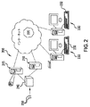
これより図面を参照するが、同様の参照番号は全体を通じて同様の部分を指す。図1は、医療用画像処理ワークステーション・システム10の実施形態の斜視視覚表現図である。このシステム10において、本開示によるデバイス、システム、および方法を実装することができる。図示のように、医療用画像処理ワークステーション・システム10は、プロセッサ(「CPU」)(図示せず)、記憶デバイス(図示せず)、グラフィクス・プロセッサ・ユニット(「GPU」)(図示せず)、および、必要に応じて、モデム(図示せず)というようなハードウェア・コンポーネント30のための筐体を有するコンピュータ20、図示する例ではモニタ40である第1出力デバイス、図示する例ではキーボード50である第1ユーザ入力デバイス、ならびに図示する例では、トラックボールまたはマウス60のような、ディスプレイと対話処理するためのポインティング・デバイスである第2ユーザ入力デバイスを含む。当技術分野では周知であるが、コンピュータ20、ハードウェア・コンポーネント30、モニタ40、およびユーザ入力デバイス50、60は別個のコンポーネントとして図示されているが、これらは全てラップトップ・コンピュータの形態で統合することもできるので、これらを統合して部品数を減らすこともできる。また、医療用画像処理ワークステーション・システム10は、コンピュータ実装医療用画像処理システムに付随することが知られているデバイスの中でもとりわけ、図示する例ではスライド・スキャナ70である第3入力デバイス、図示する例ではプリンタ80である第2出力デバイス、バックアップ電源90、および外部記憶デバイス(図示せず)のような、追加の周辺機器も含むことができる。ある実施形態では、医療用画像処理ワークステーション・システム10は、複数の画面上における複数のディジタル組織画像の同時視認を容易にするために、1つよりも多いモニタ40を含むこともできる。当業者には認められるであろうが、これらの具体的なコンポーネントは、技術が変更すれば、変更することもあり得る。例えば、周辺ポインティング・デバイスは、画面がユーザの指または音声コマンドに応答する場合、不要になる可能性がある。
We will refer to the drawings from now on, but similar reference numbers refer to similar parts throughout. FIG. 1 is a perspective visual representation of an embodiment of a medical image
また、医療用画像処理ワークステーション・システム10は、画像分析プログラムのような、ソフトウェア・コンポーネントも含む。画像分析プログラムは、局所位置決めモジュール、および、必要に応じて、全域位置決めモジュールを含む、ビュー融合画像処理モジュールを含む。ソフトウェア・コンポーネントは、記憶デバイス上に格納される(例えば、ソフトウェア・コンポーネントを内部ハード・ドライブ上に格納してもよい)、1つ以上のファイルであってもよく、および/またはソフトウェア・コンポーネントは、DVD、CDのようなメモリ・ディスク、またはメモリ・カード上に格納されてもよく、メモリ・ディスク受入ポート25を介してメモリ・ディスクが筐体30に挿入されたときに、プロセッサによってメモリ・ディスクにアクセスすることができる。
The medical image
CPUは、記憶デバイスおよびGPUを含む、種々の周辺およびハードウェア・コンポーネントに動作可能に接続されている。記憶デバイスは、複数組のディジタル画像を一時的または永続的に格納することができる。ディジタル画像は、例えば、スキャニング・デバイスによってシステムにインポートすることができる。複数組のディジタル画像は、1人の患者の隣接する組織切片の1つ以上のディジタル画像を含み、各画像は、他の画像と比較して、異なる染料/標識/マーカ、異なる撮像モード、または双方を使用して得ることができる。GPUは、画像表示プログラムおよび画像分析プログラム(1つのプログラムにおいて組み合わせてもよい)からの命令を処理する。例えば、GPUによって実行することによって、画像表示プログラムは、複数のウィンドウがあるウィンドウ型グラフィカル・ユーザ・インターフェース(「GUI」)をモニタ40上に表示する(provide)ことができ、ユーザはGUIと対話処理して、例えばCPUのようなプロセッサに、画像分析プログラムの1つ以上の態様(aspect)を実行させる命令を与えること、および/またはモニタ40の1つ以上おいて、格納されているディジタル画像の内1つ以上を、それらの本来の(元々スキャンされたときのままの)フォーマットで、または画像分析プログラムによって修正して、表示することができる。既に述べたように、画像分析プログラムは、位置決めモジュールとビュー融合画像処理モジュールとを含むことができる。例えばCPUによって実行することによって、位置決めモジュールは、格納されているディジタル画像が、異なる染色、異なる撮像モード、または双方を使用して得られたのであっても、格納されているディジタル画像の内少なくとも2つを整列させて、1組の整列画像を形成する。例えばCPUによって実行することによって、ビュー融合画像処理モジュールは、組織標本の画像を表示する。この画像は、隣接する組織切片のスライドから得られた2つ以上のディジタル画像で構成され、この画像を修正することにより、1つのディジタル画像から複合画像に取り込む部分を、他の画像と対比して、変更することができる。
The CPU is operably connected to various peripheral and hardware components, including storage devices and GPUs. The storage device can temporarily or permanently store a plurality of sets of digital images. Digital images can be imported into the system, for example, by scanning devices. Multiple sets of digital images include one or more digital images of adjacent tissue sections of a patient, each image being compared to another image with a different dye / label / marker, different imaging mode, or. Can be obtained using both. The GPU processes instructions from an image display program and an image analysis program (which may be combined in one program). For example, by running on a GPU, an image display program can provide a windowed graphical user interface (“GUI”) with multiple windows on the
図14は、本発明の例示的実施形態によるシステム900、例えば、画像分析用画像処理システムを示す。システム900は、多チャネル画像または多チャネル画像データ(例えば、RGB画像またはRGB画像データおよび/または多スペクトル画像または多スペクトル画像データ)を生成するソース901を含む。例えば、ソース901は、蛍光顕微鏡、カメラ、光学スキャナ、CCD、または蛍光画像を生成する撮像システム、もしくは明視野顕微鏡、カメラ、光学スキャナ、あるいはRGB画像、多スペクトル画像、および/またはRGBまたは多スペクトル画像データを生成する撮像システムであってもよく、またはこれらを含むのでもよい。撮像システムの例には、例えば、スペクトル・フィルタ・ホイール(spectral filter wheel)またはホール・スライド・スキャナを有する任意の蛍光顕微鏡または明視野顕微鏡をあげることができる。ソース901は、メモリ903と通信することができ、メモリ903は、複数の処理モジュールまたは論理動作を含み、これらはコンピュータ・インターフェース907に結合されたプロセッサ905によって実行される。例えば、顕微鏡、カメラ、スキャナ、CCD、またはメモリ903に結合された他の光学システムによる撮像の目的のために、生物学的試料のような標本をスライドまたは他の基板またはデバイス上に装着することができ、試料の画像の分析は、本開示にしたがってメモリ903上に格納されている複数のモジュールの内1つ以上を実行するプロセッサ905によって実行される。分析は、試料の同定(identification)および研究を目的とするのでもよい。例えば、生物学的システムまたは病理学的システムが試料を研究して、タンパク質、タンパク質断片、あるいは癌またはその他の疾患の存在を示すその他のマーカというような生物学的情報を求めることもでき、あるいはゲノムDNA検出、メッセンジャRNA検出、タンパク質検出、ウィルスの検出、遺伝子の検出等というような、他の目的のために研究することもできる。
FIG. 14 shows a
試料、例えば、組織試料または細胞診試料は、1つ以上の異なる量子ドット、蛍光体(1つまたは複数)、またはその他の染料を含有することができる1つ以上の異なる染料の適用によって、染色することができる。例えば、蛍光スライドでは、異なる染料が異なる量子ドットおよび/または蛍光体に対応することができる。蛍光体は、1つ以上のナノ結晶半導体蛍光体(例えば、量子ドット)を構成する(comprise)ことができ、各々異なる波長範囲においてピーク発光応答を生成する。量子ドットは、周知であり、Invitrogen Corp.、Evident Technologies、およびその他から商業的に入手することができる。例えば、565、585、605、および655nmにおいてピーク発光応答をそれぞれ生成する様々な異なる量子ドットによって、試料を処置することができる。試料に適用される蛍光体の内1つ以上は、有機蛍光体14(例えば、DAPI、Texas Red)であってもよい。有機蛍光体14は、当技術分野では周知であり、少なくとも、本願と所有者および譲受者が同じである米国特許第8,290,236号に記載されている。この特許をここで引用したことにより、その内容全体が本願にも含まれるものとする。更に、典型的な試料は、染色/アッセイ・プラットフォームを利用して処理される。この染色/アッセイ・プラットフォームは、自動化することができ、染料、例えば、量子ドットおよび/または有機蛍光体を含有する染料を試料に適用する。市場には、染色/アッセイ・プラットフォームとしての使用に適した種々の市販品(commercial products)がある。 A sample, eg, a tissue sample or a cytological sample, is stained by application of one or more different dyes, which can contain one or more different quantum dots, phosphors (s), or other dyes. can do. For example, in fluorescent slides, different dyes can correspond to different quantum dots and / or phosphors. Fluorescent materials can compute one or more nanocrystalline semiconductor phosphors (eg, quantum dots), each producing a peak emission response in different wavelength ranges. Quantum dots are well known and are known in Invitrogen Corp. , Evident Technologies, and others. For example, the sample can be treated with a variety of different quantum dots that produce peak emission responses at 565, 585, 605, and 655 nm, respectively. One or more of the phosphors applied to the sample may be the organic phosphor 14 (eg, DAPI, Texas Red). The organic phosphor 14 is well known in the art and is described at least in US Pat. No. 8,290,236, which has the same owner and assignee as the present application. By quoting this patent here, the entire contents are also included in the present application. In addition, typical samples are processed utilizing a staining / assay platform. This staining / assay platform can be automated and applies dyes, such as dyes containing quantum dots and / or organic phosphors, to the sample. There are a variety of commercial products on the market that are suitable for use as a staining / assay platform.
暫定的な組織処理および染色の後、ソース901において、例えば、スキャナ、CCDアレイ・スペクトル・カメラ、または材料の標本を含む(contain)スライドを撮像するために使用されるその他の撮像システムによって、試料の1つ以上のディジタル画像を取り込みし、スライド上の標本のディジタル画像を生成することができる(本発明によれば、融合画像は少なくとも2つの異なるスライドの複数の部分を含む)。標本を含むスライドは、試料に適用された染料からの発光応答を生成することを意図した波長で試料を照明するために、光源に晒される(subjected to)。量子ドットの場合、光源は広帯域スペクトル光源とするとよい。あるいは、光源は、レーザのような、狭帯域光源を含んでもよい。RGB明視野画像も取り込むことができる。撮像システムは、例えば、ディジタル・カメラ、顕微鏡、または1つ以上の対物レンズ、および光源、更には1組のスペクトル・フィルタを有するその他の光学システムを含むことができる。異なる波長において画像を取り込む他の技法を使用することもできる。染色された生物学的試料を撮像するのに適したカメラ・プラットフォームは、当技術分野では周知であり、Zeiss、Canon、Applied Spectral Imaging、およびその他というような会社から市販されており、このようなプラットフォームは、本開示のシステム、方法、および装置における使用のために、容易に適応可能である。画像は、メモリまたは記憶デバイス903に、ワイヤレスまたはワイヤライン接続を介して、例えば、ソース901とコンピュータ907との間のケーブル接続を介して、コンピュータ・ネットワークを通じて、あるいはディジタル情報をコンピュータ間で転送するために一般に使用される任意の他の媒体を使用して供給することができる。また、画像は、ネットワークを通じてネットワーク・サーバまたはデータベースに、コンピュータ907による格納および後における検索(retrieval)のために供給することもできる。プロセッサ905およびメモリ903以外にも、コンピュータ907は、キーボード、マウス、スタイラス、およびディスプレイ/タッチスクリーンのような、ユーザ入力および出力デバイスも含む。以下の論述において説明するが、プロセッサ905は、メモリ903上に格納されているモジュールを実行し、画像の分析、このような画像から得られる画像または画像データの(of the image or image data derived from such images)、定量的分析を行い、更に定量的結果/結果の図表を、コンピュータ907を操作するユーザに表示する。
After tentative tissue treatment and staining, the sample is sampled at
メモリ903上に格納されているモジュールは、以上で説明し更に本明細書において説明するように、画像位置決めモジュール902と、ビュー融合画像処理モジュール904とを含むことができる。しかしながら、これらのモジュールによって実行される動作は、本明細書において記載するものに限定されるのではなく、モジュールのシーケンス、構成(arrangement)、および総数は変更されてもよく、ここに説明する実施形態は単に例示を目的にするに過ぎない。ソフトウェア・モジュールは、クライアント・ユーザ・インターフェース、例えば、コンピュータ907に関連付けられたユーザ・インターフェースを介してアクセスすることができる。
The module stored on the
一旦プログラムを起動したなら、ユーザは、分析のためにディジタル画像921を選択することができる。ここで説明する実施形態では、ユーザは、隣接する連続切片の一連の画像から画像を選択し、例えば、これらの切片の内少なくとも1つはH&Eによって染色されており、他の切片は1つ以上の異なるIHC染料によって染色されている。しかしながら、本発明は、隣接する一連の切片のビュー融合画像処理に限定されるのではない。例えば、他の実施形態では、観察者(viewer)が、例えば、通常および罹患組織、または疾病過程の異なる段階における組織を視認および比較するために、同じまたは異なる患者、動物、またはその他の試料からの組織試料の画像から選択することができる。 Once the program is launched, the user can select the digital image 921 for analysis. In the embodiments described herein, the user selects an image from a series of images of adjacent continuous sections, for example, at least one of these sections is stained by H & E and one or more of the other sections. It is dyed with different IHC dyes. However, the present invention is not limited to view fusion image processing of adjacent series of sections. For example, in other embodiments, the observer, for example, from the same or different patient, animal, or other sample to view and compare normal and affected tissue, or tissue at different stages of the disease process. It can be selected from the image of the tissue sample of.
クライアント・インターフェースを介して、次に、ユーザは画像位置決めモジュール902を呼び出すことができる。画像位置決めモジュール902は、例えば、CPUまたはプロセッサ905によって実行されると、例えば、視認された組織ブロックの基礎解剖学的構造の照合(matching)によって、選択された画像を整列する(align)。
Through the client interface, the user can then call the
クライアント・インターフェースを介して、ユーザはビュー融合画像処理モジュール904も呼び出すことができる。ビュー融合画像処理モジュール904は、例えば、CPUまたはプロセッサ905によって実行されると、画像位置決めモジュールによって位置決めされた選択画像(selected images)921の融合画像を生成する。例えば、ビュー融合画像処理モジュール904は、選択され位置決めされたスライドの複合画像を、並存「カーテン」ビュー(side-by-side curtain view)(1つ以上のカーテンを含む)で、および/または「フラッシュライト」ビューで生成することができる。1つ以上の副スライドの1つ以上の部分は、主スライド内における1つ以上の部分と入れ替わるように現れる。
Through the client interface, the user can also call the view fusion
図2は、本開示によるデバイス、システム、および方法を実装することができるネットワーク型システムの実施形態を示すネットワーク図である。図示のように、システム200は、データベース・サーバ210と、ネットワーク・アクセス可能な記憶デバイス215とを含む。これらの各々は、ネットワーク220に接続されている。記憶デバイス215は、複数組のディジタル画像を格納する。各組は、1人の患者の隣接する組織切片の1つ以上のディジタル画像を含む。1組における各画像は、1組における他の画像と比較して、異なる染料、異なる撮像モード、または双方を使用することによって得ることができる。1つ以上のクライアント・コンピュータ230は、キーボード232、マウス(図示せず)、およびプリンタ(図示せず)のような関連する入力および出力デバイスを有することができ、当技術分野では周知の任意の手段によってネットワーク220にも接続されている(例えば、専用接続、DSLまたはケーブル・モデム、ワイヤレス・インターネット接続、ダイアルアップ・モデム等)。クライアント・コンピュータ230はウェブ・ブラウザを含む。ウェブ・ブラウザは、記憶デバイス215内にあるディジタル画像にアクセスするために使用される。本発明の例示的な実施形態では、ディジタル画像を格納するために、クラウド・ストレージを利用することもできる。
FIG. 2 is a network diagram showing an embodiment of a network type system in which the devices, systems, and methods according to the present disclosure can be implemented. As shown, the
クライアント・コンピュータ230は、画像分析プログラムに関係する命令を実行するように構成された少なくとも1つのプロセッサを含む。画像分析プログラムは、サーバ210からクライアント・コンピュータ230にダウンロードすることができる。画像分析プログラムは、画像ビューア・モジュールを含むことができる。クライアント・ユーザ・インターフェースを提供する画像ビューア・モジュールを実行することによって、この画像ビューア・モジュールがウィンドウ型GUIを表示する(provide)ことができ(更に、複数のウィンドウを含むことができる)、このウィンドウ型GUIが、プロセッサに画像分析プログラムの1つ以上の態様(aspect)を実行させる命令をユーザが与えることを可能にし、および/または格納されているディジタル画像の内1つ以上を、それらの元々スキャンされたフォーマットのまま表示する、あるいは画像分析プログラムによって修正された通りに表示することができる。格納されている画像が未だ位置決めされていない場合(あるいはユーザが第2のまたは異なる位置決め方法を使用することを望む場合)、画像分析プログラムは、必要に応じて、ユーザが、1人の患者の組織切片から得られた1組の画像から、位置決めのために画像を選択することを可能にすることができ、この1組における画像の少なくとも一部は、この1組における他の画像と比較して、異なる染料、または異なるモード、または双方を使用して作られたのでもよい。また、画像分析プログラムは、1組のディジタル画像における2つ以上のディジタル画像の複合体である組織標本のディジタル画像を、ユーザが視認することを可能にする。ある実施形態では、システム200は、ホール・スライド250をスキャンし、ディジタル画像を生成するスキャナ240も含む。ディジタル画像は、記憶デバイス215内に格納される。
The
当業者には理解されようが、コンピュータ化ネットワークのコンテキストにおいて画像分析プログラムを実装すると、そうしない単体のワークステーションでは限定される特定のアクティビティが可能になる。例えば、同じ場所におらず、実際には互いに離れている病理学者であっても、画像の分析において協働することができ、または場所には関係なく、相応しい(correct)病理学者に正確な時に到達することができる。 As those skilled in the art will understand, implementing an image analysis program in the context of a computerized network allows certain activities that are limited on a single workstation that does not. For example, pathologists who are not in the same location but are actually separated from each other can collaborate in the analysis of images, or regardless of location, at the correct time to the correct pathologist. Can be reached.
図1および図2は、1つ以上のコンピュータ・システムまたはネットワーク・トポロジ内に存在することができる特定のエレメントを示す。本開示によるデバイスおよびシステムを実装することができるコンピュータ・システムおよびネットワークは、他のコンピュータ・システムやネットワーク・トポロジを包含することができ、更にそれら他のコンピュータ・システムおよびネットワーク・トポロジにおいて、もっと多いまたは少ないエレメントを含んでもよいことは、習熟者(person of skill)には理解されよう。言い換えると、図1および図2の実施形態は限定ではない。例えば、ある実施形態では、ディジタル画像を格納するために、クラウド・ストレージを利用することもできる。 1 and 2 show specific elements that can be present in one or more computer systems or network topologies. The computer systems and networks in which the devices and systems according to the present disclosure can be implemented can include other computer systems and network topologies, and more in those other computer systems and network topologies. Or it will be understood by the person of skill that it may contain fewer elements. In other words, the embodiments of FIGS. 1 and 2 are not limited. For example, in some embodiments, cloud storage can also be utilized to store digital images.
したがって、本開示にしたがった使用のためのコンピュータ・システムの例示的な実施形態は、ワークステーション、パーソナル・コンピュータ、サーバ、ハンドヘルド・デバイス、マルチプロセッサ・システム、マイクロプロセッサ・ベースまたはプログラマブル消費者用電子機器、ネットワークPC、ミニコンピュータ、メインフレーム・コンピュータ、あるいは任意のその他の現行または今後のコンピュータというような、任意の数のコンピュータ・プラットフォームまたは複数の型式のコンピュータ・プラットフォームを含むことができる。 Accordingly, exemplary embodiments of computer systems for use in accordance with the present disclosure are workstations, personal computers, servers, handheld devices, multiprocessor systems, microprocessor-based or programmable consumer electronics. It can include any number of computer platforms or multiple types of computer platforms, such as equipment, network PCs, minicomputers, mainframe computers, or any other current or future computer.
また、例示的な実施形態は、分散型コンピューティング環境において実施することもでき、この場合、通信ネットワークにおいて接続されている(例えば、ハードワイヤ接続、ワイヤレス接続、またはこれらの組み合わせによって)ローカルおよび/またはリモート処理デバイスによってタスクが実行される。分散型コンピューティング環境では、プログラム・モジュールは、メモリ記憶デバイスを含むローカルおよびリモート双方のコンピュータ記憶媒体に配置することもできる。しかしながら、ここで説明するような前述のコンピュータ・プラットフォームは、説明する発明の特殊な動作を実行するように特別に構成され、汎用コンピュータとは考えられないことは、当業者には認められよう。 Illustrative embodiments can also be implemented in a decentralized computing environment, in which case they are connected in a communication network (eg, by hardwire connections, wireless connections, or a combination thereof) and / or local. Or the task is performed by a remote processing device. In a distributed computing environment, program modules can also be located on both local and remote computer storage media, including memory storage devices. However, it will be appreciated by those skilled in the art that the aforementioned computer platforms as described herein are specifically configured to perform the special operations of the invention described and are not considered general purpose computers.
コンピュータは、通例、プロセッサ、オペレーティング・システム、システム・メモリ、メモリ記憶デバイス、入力−出力コントローラ、入力−出力デバイス、およびディスプレイ・デバイスのような、周知のコンポーネントを含む。また、関連技術の当業者には、コンピュータには多くの可能な構成およびコンポーネントがあり、キャッシュ・メモリ、データ・バックアップ・ユニット、および多くのその他のデバイスも含んでもよいことも理解されよう。 Computers typically include well-known components such as processors, operating systems, system memory, memory storage devices, input-output controllers, input-output devices, and display devices. Those skilled in the art will also appreciate that computers have many possible configurations and components, including cache memory, data backup units, and many other devices.
入力デバイスの例には、キーボード、カーソル制御デバイス(例えば、マウス)、マイクロフォン、スキャナ等が含まれる。 Examples of input devices include keyboards, cursor control devices (eg, mice), microphones, scanners and the like.
出力デバイスの例には、ディスプレイ・デバイス(例えば、モニタまたは投射機)、スピーカ、プリンタ、ネットワーク・カード等が含まれる。ディスプレイ・デバイスは、視覚情報を提供するディスプレイ・デバイスを含むことができ、この情報は、通例、画素のアレイとして論理的および/または物理的に編成することができる。 Examples of output devices include display devices (eg, monitors or projectors), speakers, printers, network cards and the like. The display device can include a display device that provides visual information, which information can typically be logically and / or physically organized as an array of pixels.
また、インターフェース・コントローラも含むことができる。インターフェース・コントローラは、入力および出力インターフェースを設けるための種々の既知のまたは今後のソフトウェア・プログラムの内任意のものを含むことができる。例えば、インターフェースは、1つ以上のグラフィック表現をユーザに提示する(provide)、一般に「グラフィカル・ユーザ・インターフェース」と呼ばれる(GUIと呼ばれることが多い)ものを含んでもよい。インターフェースは、通例、関連技術における当業者には周知の選択手段または入力手段を使用して、ユーザ入力を受け入れるために使用可能である。また、インターフェースはタッチ・スクリーン・デバイスでもよい。 It can also include an interface controller. The interface controller can include any of a variety of known or upcoming software programs for providing input and output interfaces. For example, the interface may include one that provides one or more graphic representations to the user, commonly referred to as a "graphical user interface" (often referred to as a GUI). Interfaces can typically be used to accept user input using selection or input means well known to those skilled in the art in related arts. The interface may also be a touch screen device.
同じまたは代わりの実施形態において、コンピュータ上のアプリケーションは、「コマンド・ライン・インターフェース」(CLIと呼ばれることが多い)と呼ばれるものを含むインターフェースを採用することもできる。CLIは、通例、アプリケーションとユーザとの間におけるテキスト・ベースの対話処理に対応する。通例、コマンド・ライン・インターフェースは、ディスプレイ・デバイスを通じて、テキストのラインとして出力を提示し、入力を受け取る。例えば、ある実施態様では、関連技術における当業者には周知の、Unix ShellsまたはMicrosoft Windows Powershellのような、「シェル」と呼ばれるものを含むことができる。Microsoft Windows Powershellは、Microsoft NETフレームワークのような、オブジェクト指向型プログラミング・アーキテクチャを採用する。尚、インターフェースは、1つ以上のGUI、CLI、またはこれらの組み合わせを含んでもよいことは、関連技術における当業者には認められよう。 In the same or alternative embodiments, the application on the computer can also employ an interface that includes what is called a "command line interface" (often referred to as CLI). The CLI typically supports text-based interaction between the application and the user. Typically, the command line interface presents the output as a line of text and receives the input through the display device. For example, in certain embodiments, it may include what is called a "shell", such as Unix Shells or Microsoft Windows Powershell, which is well known to those skilled in the art in the art. Microsoft Windows PowerShell employs an object-oriented programming architecture, such as the Microsoft .NET framework. Those skilled in the art will appreciate that the interface may include one or more GUIs, CLIs, or combinations thereof.
プロセッサは、Intel Corporationが製造するCeleron、Core、またはPentiumプロセッサ、Sun Microsystemsが製造するSPARCプロセッサ、AMD Corporationが製造するAthlon、Sempron、Phenom、またはOpteronというような、市販のプロセッサを含むことができ、あるいは入手可能なまたは今後入手可能になる他のプロセッサの1つであってもよい。プロセッサの実施形態の中には、マルチコア・プロセッサと呼ばれるものを含み、および/または単一コアまたはマルチコア構成において並列処理技術を採用することが可能なものもある。例えば、マルチコア・アーキテクチャは、通例、2つ以上のプロセッサ「実行コア」を含む。本例では、各実行コアは、複数のスレッドの並列実行を可能にする独立したプロセッサとして実行することができる。加えて、プロセッサは、一般に32または64ビット・アーキテクチャと呼ばれるもの、あるいは現在知られているまたは今後開発される可能性があるその他のアーキテクチャ構成で構成することもできることは、関連技術における当業者には認められよう。 Processors can include off-the-shelf processors such as Celeron, Core, or Pentium processors manufactured by Intel Corporation, SPARC processors manufactured by Sun Microsystems, Athlon, Sempron, Phoenix, or Opteron manufactured by AMD Corporation. Alternatively, it may be one of the other processors available or will be available in the future. Some processor embodiments include what is referred to as a multi-core processor and / or may employ parallel processing techniques in a single-core or multi-core configuration. For example, a multi-core architecture typically includes more than one processor "execution core". In this example, each execution core can be executed as an independent processor that enables parallel execution of multiple threads. In addition, processors can be configured with what is commonly referred to as a 32-bit or 64-bit architecture, or with other architectural configurations currently known or may be developed in the future, to those skilled in the art. Will be recognized.
プロセッサは、通例、オペレーティング・システムを実行する。オペレーティング・システムは、例えば、Microsoft CorporationからのWindows型オペレーティング・システム、Apple Computer Corp.からのMac OS Xオペレーティング・システム、多くの販売業者から入手可能なUnixまたはLinux型オペレーティング・システム、あるいはオープン・ソースと呼ばれるもの、その他のオペレーティング・システムまたは今後のオペレーティング・システム、もしくはこれらの何らかの組み合わせであってもよい。オペレーティング・システムは、ファームウェアおよびハードウェアと周知の様式でインターフェースし、種々のプログラミング言語で書かれることもある種々のコンピュータ・プログラムの機能を調整および実行するときに、プロセッサを補助する(facilitate)。オペレーティング・システムは、通例、プロセッサと協働して、コンピュータの他のコンポーネントの機能を調整および実行する。また、オペレーティング・システムは、スケジューリング、入力−出力制御、ファイルおよびデータ管理、メモリ管理、ならびに通信制御および関連サービスも、全て周知の技法にしたがって、提供する。 The processor typically runs the operating system. The operating system is, for example, a Windows-type operating system from Microsoft Corporation, Apple Computer Corp. Mac OS X operating system from, Unix or Linux operating systems available from many distributors, or what is called open source, other operating systems or future operating systems, or any combination thereof. It may be. The operating system facilitates the processor in coordinating and performing the functions of various computer programs that interface with firmware and hardware in a well-known manner and may be written in different programming languages. The operating system typically works with the processor to coordinate and perform the functions of other components of the computer. The operating system also provides scheduling, input-output control, file and data management, memory management, and communication control and related services, all according to well-known techniques.
システム・メモリは、所望の情報を格納するために使用することができ、コンピュータによってアクセスすることができる種々の既知のメモリ記憶デバイスまたは今後のメモリ記憶デバイスの内任意のものを含んでもよい。コンピュータ読み取り可能記憶媒体は、揮発性および不揮発性、リムーバブルおよび非リムーバブル媒体を含むことができ、コンピュータ読み取り可能命令、データ構造、プログラム・モジュール、またはその他のデータというような情報の格納のための任意の方法または技術で実現される。例には、一般に入手可能なあらゆるランダム・アクセス・メモリ(RAM)、リード・オンリ・メモリ(ROM)、電子的消去可能プログラマブル・リード・オンリ・メモリ(EEPROM)、ディジタル・バーサタイル・ディスク(DVD)、常駐ハード・ディスクまたはテープのような磁気媒体、リードおよびライト・コンパクト・ディスクのような光媒体、あるいはその他のメモリ記憶デバイスが含まれる。メモリ記憶デバイスには、種々の周知のデバイスまたは今後のデバイスの内任意のものを含んでもよく、コンパクト・ディスク・ドライブ、テープ・ドライブ、リムーバブル・ハード・ディスク・ドライブ、USBまたはフラッシュ・ドライブ、あるいはディスケット・ドライブが含まれる。このようなタイプのメモリ記憶デバイスは、通例、コンパクト・ディスク、磁気テープ、リムーバブル・ハード・ディスク、USBまたはフラッシュ・ドライブ、あるいはフロッピ・ディスケットのようなプログラム記憶媒体からそれぞれ読み取りを行い、および/またはこれらに書き込みを行う。これらのプログラム記憶媒体、あるいは現在使用されている他のプログラム記憶媒体、または今後開発される可能性があるプログラム記憶媒体はいずれも、コンピュータ・プログラム製品と見なすことができる。 System memory may include any of a variety of known memory storage devices or future memory storage devices that can be used to store desired information and can be accessed by a computer. Computer-readable storage media can include volatile and non-volatile, removable and non-removable media, and are optional for storing information such as computer-readable instructions, data structures, program modules, or other data. It is realized by the method or technique of. Examples include all commonly available random access memory (RAM), read-only memory (ROM), electronically erasable programmable read-only memory (EEPROM), and digital versatile discs (DVD). Includes magnetic media such as resident hard disks or tapes, optical media such as read and write compact disks, or other memory storage devices. Memory storage devices may include any of a variety of well-known or upcoming devices, including compact disk drives, tape drives, removable hard disk drives, USB or flash drives, or Includes diskette drive. These types of memory storage devices typically read from and / or programmatic storage media such as compact discs, magnetic tapes, removable hard disks, USB or flash drives, or floppy disks, respectively. Write to these. Any of these program storage media, or any other program storage medium currently in use, or any program storage medium that may be developed in the future, can be considered a computer program product.
認められるであろうが、これらのプログラム記憶媒体は、通例、コンピュータ・ソフトウェア・プログラムおよび/またはデータを格納する。コンピュータ・ソフトウェア・プログラムは、コンピュータ制御ロジックとも呼ばれ、メモリ記憶デバイスと共に使用されるシステム・メモリおよび/またはプログラム記憶デバイスに格納されるのが通例である。ある実施形態では、コンピュータ・プログラム製品は、制御ロジック(プログラム・コードを含むコンピュータ・ソフトウェア・プログラム)が内部に格納されているコンピュータ使用可能媒体を含むというように記述される。制御ロジックは、プロセッサによって実行されると、本明細書において説明した機能をこのプロセッサに実行させる。他の実施形態では、一部の機能は、例えば、ハードウェア状態機械を使用して、主にハードウェアで実現される。本明細書において説明した機能を実行するハードウェア状態機械の実現は、関連技術の当業者には明白であろう。 As will be appreciated, these program storage media typically store computer software programs and / or data. Computer software programs, also called computer-controlled logic, are typically stored in system memory and / or program storage devices used with memory storage devices. In one embodiment, a computer program product is described as including a computer-enabled medium in which control logic (a computer software program containing program code) is stored. When the control logic is executed by a processor, it causes the processor to perform the functions described herein. In other embodiments, some functions are implemented primarily in hardware, for example using a hardware state machine. The realization of a hardware state machine that performs the functions described herein will be apparent to those skilled in the art.
入力−出力コントローラは、人間または機械に関係なく、ローカルまたはリモートにも関係なく、ユーザから情報を受け入れて処理するための種々の既知のデバイスの内任意のものを含むことができる。このようなデバイスは、例えば、モデム・カード、ワイヤレス・カード、ネットワーク・インターフェース・カード、サウンド・カード、または種々の既知の入力デバイスの内任意のもののための他のタイプのコントローラを含む。出力コントローラは、人間または機械に関係なく、ローカルまたはリモートにも関係なく、ユーザに情報を提示するための種々の既知のディスプレイ・デバイスの内任意のもののためのコントローラを含むことができる。 The input-output controller can include any of a variety of known devices for accepting and processing information from users, whether human or machine, local or remote. Such devices include, for example, modem cards, wireless cards, network interface cards, sound cards, or other types of controllers for any of a variety of known input devices. The output controller can include a controller for any of a variety of known display devices for presenting information to the user, whether human or machine, local or remote.
現在説明している実施形態では、コンピュータの機能エレメントがシステム・バスを介して互いに通信する。コンピュータの実施形態には、ネットワークまたは他のタイプの遠隔通信を使用して、いくつかの機能エレメントと通信できるものもある。関連技術の当業者には明白であろうが、計器制御および/またはデータ処理アプリケーションがソフトウェアで実装される場合、システム・メモリおよび/またはメモリ記憶デバイスにロードされ、そこから実行されればよい。また、計器制御および/またはデータ処理アプリケーションの全部または一部は、メモリ記憶デバイスのリード・オンリ・メモリまたは同様のデバイスに存在してもよく、このようなデバイスは、計器制御および/またはデータ処理アプリケーションを最初に入力−出力コントローラを介してロードする必要がない。尚、実行に有利であれば、計器制御および/またはデータ処理アプリケーション、あるいはその一部がプロセッサによってシステム・メモリ、またはキャッシュ・メモリ、あるいは双方に、既知の様式でロードされてもよいことは、関連技術の当業者には理解されよう。 In the embodiments currently described, the functional elements of the computer communicate with each other via the system bus. Some computer embodiments can communicate with several functional elements using a network or other types of telecommunications. As will be apparent to those skilled in the art, if an instrument control and / or data processing application is implemented in software, it may be loaded into and executed from system memory and / or memory storage devices. Also, all or part of the instrument control and / or data processing application may reside in read-only memory of a memory storage device or similar device, such device as instrument control and / or data processing. There is no need to load the application first through the input-output controller. It should be noted that the instrument control and / or data processing application, or part thereof, may be loaded by the processor into system memory, cache memory, or both in a known manner if it is advantageous to execute. It will be understood by those involved in the related technology.
加えて、インターネット・クライアントが、ネットワークを使用して他のコンピュータ上にあるリモート・サービスにアクセスするために使用可能な(enabled)アプリケーションを含んでもよく、例えば、一般に「ウェブ・ブラウザ」と呼ばれるものを備えてもよい(comprise)。この例では、いくつかの一般に採用されるウェブ・ブラウザには、Microsoft Corporationから入手可能なMicrosoft Internet Explorer、Mozilla CorporationからのMozilla Firefox、Apple Computer Corp.からのSafari、Google CorporationからのGoogle Chrome、あるいは当技術分野において現在知られているまたは今後開発されるその他のタイプのウェブ・ブラウザが含まれる。また、同じまたは他の実施形態において、インターネット・クライアントが、生物学的用途のためのデータ処理アプリケーションのような、ネットワークを通じて遠隔情報にアクセスするために使用可能な、特殊ソフトウェア・アプリケーションを含んでもよく、またはそのエレメントであることも可能である。 In addition, Internet clients may include enabled applications that can be used to access remote services on other computers over the network, for example what is commonly referred to as a "web browser." May be provided (comprise). In this example, some commonly adopted web browsers include Microsoft Internet Explorer, available from Microsoft Corporation, Mozilla Firefox from Mozilla Corporation, Apple Computer Corp. Includes Safari from, Google Chrome from Google Corporation, or other types of web browsers currently known or developed in the art. Also, in the same or other embodiments, Internet clients may include specialized software applications that can be used to access remote information over a network, such as data processing applications for biological applications. , Or its element.
ネットワークは、当業者には周知の多くの種々のタイプのネットワークの1つ以上を含むことができる。例えば、ネットワークは、TCP/IPプロトコル・スイートと一般に呼ばれるものを通信のために採用することがある、ローカルまたはワイド・エリア・ネットワークを含むことができる。ネットワークは、一般にインターネットと呼ばれる、相互接続コンピュータ・ネットワークの世界規模のシステムを構成するネットワークを含んでもよく、または種々のイントラネット・アーキテクチャを含むこともできる。また、ネットワーク接続環境におけるユーザの中には、ハードウェアおよび/またはソフトウェア・システムとの間の情報トラフィックを制御するために、一般に「ファイアウォール」(ときとしてパケット・フィルタまたは境界保護デバイスと呼ばれることもある)と呼ばれるものを採用することを好む者がいることも、関連技術における当業者には認められよう。 The network can include one or more of many different types of networks well known to those of skill in the art. For example, the network can include a local or wide area network, which may employ what is commonly referred to as the TCP / IP protocol suite for communication. The network may include networks that make up a worldwide system of interconnected computer networks, commonly referred to as the Internet, or may include various intranet architectures. Also, some users in a networked environment are commonly referred to as "firewalls" (sometimes referred to as packet filters or perimeter protection devices) to control information traffic to and from hardware and / or software systems. It will also be acknowledged by those skilled in the art of related technology that some people prefer to adopt what is called).
図3〜図5は、纏めて、ビュー融合フォーマットで画像を表示する(view)ためにプロセッサと対話処理するクライアント・ユーザ・インターフェースの実施形態を示す。図示する実施形態では、クライアント・ユーザ・インターフェースは、2つの基本的なツールに跨がって実装され、これらのツールに基づいて、ビュー融合画像処理フレームワークが提示される。第1ソフトウェア・ツール(図3)は、利用可能な組織画像のリストを提示する。通例、組織画像は、組織切片が得られた組織ブロック毎に一緒に集合化される。このリストにおいてスライドを選択することができ、全域位置決め(global registration)またはスライドの表示(viewing)というようなアクションを起動する(trigger)ことができる。例えば、図4に示すように、1つの組織ブロックからの全てのスライド(即ち、画像毎にブロックIdは同じである)が選択され、「スライドを位置決めする」というメニュー選択肢が強調および/または選択される。別の例として、そして図5に示すように、組織ブロックから特定の位置決めされたスライドが選択され、「ビューア」(viewer)という異なるメニュー選択肢が強調および/または選択される。第2のソフトウェア・ツール(図7〜図13)は、本開示において更に開示される表示機能(viewing functionality)を提供する。しかしながら、本開示において提供される機能は、他の注釈/ビューア(annotation/viewer)GUIツールおよび他の画像分析/管理ツールと統合することができる。例えば、Ventana Image Viewer、Ventana VIRTUOSO Viewer、およびVentana VECTORビューアを含む、本開示の製品および方法を実装するために、任意のホール・スライド・ビューアを使用することができる。 3-5, together, show embodiments of a client-user interface that interacts with a processor to view an image in a view fusion format. In the illustrated embodiment, the client user interface is implemented across two basic tools, based on which a view fusion image processing framework is presented. The first software tool (FIG. 3) presents a list of available tissue images. Usually, tissue images are assembled together for each tissue block from which tissue sections were obtained. Slides can be selected in this list and actions such as global registration or viewing slides can be triggered. For example, as shown in FIG. 4, all slides from one tissue block (ie, the block Id is the same for each image) are selected and the menu option "Position slides" is highlighted and / or selected. Will be done. As another example, and as shown in FIG. 5, a particular positioned slide is selected from the tissue block and a different menu option called "viewer" is highlighted and / or selected. A second software tool (FIGS. 7-13) provides viewing functionality further disclosed in the present disclosure. However, the functionality provided in this disclosure can be integrated with other annotation / viewer GUI tools and other image analysis / management tools. Any hall slide viewer can be used to implement the products and methods of the present disclosure, including, for example, the Ventana Image Viewer, the Ventana VIRTUOSO Viewer, and the Ventana VECTOR Viewer.
また、図7に示すように、例えば、ビューアGUIは「ビューア」ウィンドウ326を含む。ビューア・ウィンドウ326は、表示された画像(ズーム・ボタン340およびパンニング・ボタン350のような)とのユーザの対話処理をし易くするために、メニュー・バーおよび多数のアイコンを有する。表示ツール(viewing tool)の中に含まれるのは、カーテン・ビュー・モード360およびフラッシュライド・モード370のような、ビュー融合可視化ボタンである。 Also, as shown in FIG. 7, for example, the viewer GUI includes a "viewer" window 326. The viewer window 326 has a menu bar and numerous icons to facilitate the user's interaction with the displayed image (such as the zoom button 340 and the panning button 350). Included within the viewing tool are view fusion visualization buttons, such as curtain view mode 360 and flashride mode 370.
図7に示すように、カーテン・ビューでは、選択されたスライドが、互いに重なり合って現われ、複合画像のどの部分を、1つの画像から、他の画像と対比して、得るか決定するために、スライダ・エレメントをドラッグすることができる。即ち、スライダ・ボタンを動かして、副基礎画像を表出させ、「カーテン・ビュー」融合可視化の外観を与える。図示する実施形態では、画面の左側にあるディジタル画像は、H&Eで染色された組織切片のスライドから得られ、右側にあるディジタル画像は、IHCアッセイ、例えば、二重IHCアッセイで染色された隣接する組織切片のスライドから得られたものである。 As shown in FIG. 7, in the curtain view, selected slides appear to overlap each other to determine which portion of the composite image to obtain from one image in contrast to the other. You can drag the slider element. That is, the slider button is moved to reveal the sub-basic image, giving the appearance of a "curtain view" fusion visualization. In the illustrated embodiment, the digital image on the left side of the screen is obtained from a slide of H & E stained tissue sections and the digital image on the right side is adjacent stained with an IHC assay, eg, a dual IHC assay. It was obtained from a slide of tissue sections.
図7〜図9は、スライダ・ボタンを左から右に動かした結果として、複合画像の外観を示す。見て分かるように、スライダ・ボタンをずらすと、各画像の複合画像に対する寄与の割合が変わる。ある実施形態では、局所位置決めは、異なるスライドからの画像データが、カーテンの左右の境界エリアにおいてできるだけ同様になるように、これらの画像の少なくとも1つを調節する。パンニング、ズーム、注釈の作成を含む、他のビューア機能も使用してもよい。 7-9 show the appearance of the composite image as a result of moving the slider button from left to right. As you can see, shifting the slider button changes the percentage of each image's contribution to the composite image. In certain embodiments, local positioning adjusts at least one of these images so that the image data from different slides are as similar as possible in the left and right border areas of the curtain. Other viewer features may also be used, including panning, zooming, and creating annotations.
図10に示すように、カーテン・ビューは、隣接する組織切片の2つだけのディジタル画像の複合画像や、スライダ・ボタンの左右移動に限定されるのではない。図10の例では、カーテン・ビューは5つのスライドに適用され、4つのスライダ・ボタンを含み、上から下、下から上、左から右、そして右から左の制御に対応する。図示するカーテン・ビューの実施形態の可視化では、中央の画像はH&E染色スライドから得られ、隣接する組織切片のIHC染色スライドから得られたディジタル画像からの画像データは、それぞれのカーテン・ビュー(スライダ・ボタン)制御によって、可視化(または不可視化)される。 As shown in FIG. 10, the curtain view is not limited to a composite image of only two digital images of adjacent tissue sections or to move the slider button left and right. In the example of FIG. 10, the curtain view is applied to five slides and includes four slider buttons, corresponding to top-to-bottom, bottom-to-top, left-to-right, and right-to-left controls. In the visualization of the illustrated curtain view embodiment, the central image is obtained from the H & E stained slides and the image data from the digital images obtained from the IHC stained slides of adjacent tissue sections is the respective curtain view (slider). -It is visualized (or invisible) by the button) control.
融合表示(fusion viewing)は、カーテン・ビューの実施態様に限定されるのではなく、複合画像を可視化および/または操作することができる任意の様式を包含する。一例として、図11は、標本の隣接する組織切片の2つ以上の位置決めされたディジタル画像の融合表示の他の実施形態、即ち、IHCデータがH&Eデータ上に重ね合わされた「フラッシュライト」または「スポットライト」融合表示を示す。ここでは、ユーザが副画像の任意の領域を選択することができ、それが主(上側)画像の下に重ねられたように現われ、主画像の対応する画像データと置き換わる。ある実施態様では、主スライド内に融合された副スライドからの領域は、フラッシュライトまたはスポットライトが、置き換えられた領域上で輝いているかのような円盤形状となる。しかしながら、本開示は円盤形状に限定されるのではない。ユーザは、更に、クライアント・ユーザ・インターフェースを介してコンピュータと対話処理し、領域のサイズを増減すること、または領域の形状を修正することもできる。更に、図11〜図13に示すように、ユーザは、例えば、マウス・ポインタ(または音声制御のような任意の他の対話処理手段)を動かすことによって、領域の位置をずらすことができる。ここでもまた、領域間の整列を調節するために、領域間の境界において局所位置決めを使用することができる。 Fusion viewing is not limited to embodiments of curtain views, but includes any mode in which composite images can be visualized and / or manipulated. As an example, FIG. 11 shows another embodiment of a fusion representation of two or more positioned digital images of adjacent tissue sections of a specimen, i.e. a "flashlight" or "flashlight" in which IHC data is superimposed on H & E data. Shows a "spotlight" fusion display. Here, the user can select any area of the sub-image, which appears superimposed under the main (upper) image and replaces the corresponding image data in the main image. In some embodiments, the area from the secondary slide fused within the main slide has a disk shape as if the flashlight or spotlight were shining on the replaced area. However, the present disclosure is not limited to the disk shape. The user can also interact with the computer via the client user interface to increase or decrease the size of the area or modify the shape of the area. Further, as shown in FIGS. 11-13, the user can shift the position of the region, for example, by moving the mouse pointer (or any other interactive means such as voice control). Again, local positioning can be used at the boundaries between the regions to adjust the alignment between the regions.
図11〜図13の例では、IHCデータがH&Eデータ上に重ね合わされているが、ユーザはどちらの画像を主(上側)にし、どちらの画像を副(積み重ねられる/重ね合わされる)(stacked/overlaid)にするか選択することもできる。つまり、例えば、H&EデータをIHCデータ上に重ね合わせることもできる。順序の選択(即ち、どの画像を主に指定し、副下地画像の層状体(layering)として指定するか)は、融合表示に使用される画像を選択する時点において、ユーザによって行うことができる。あるいは、または加えて、積み重ねの順序は、例えば、スタックを入れ替えることによって(例えば、上側の画像をスタックの後ろに移動するように命令することによって)、表示(viewing)の間に変更することができる。 In the example of FIGS. 11 to 13, the IHC data is superimposed on the H & E data, but the user has which image is the main (upper side) and which image is the sub (stacked / superimposed) (stacked /). You can also choose to overlaid). That is, for example, H & E data can be superimposed on IHC data. The order selection (ie, which image is primarily designated and which is designated as the layering of the sub-base image) can be made by the user at the time of selecting the image to be used for the fusion display. Alternatively, or in addition, the stacking order can be changed during viewing, for example by swapping stacks (eg, by instructing the upper image to move to the back of the stack). it can.
更に、図11〜図13の例は1つの融合(スポットライト)領域しか示さないが、ある実施形態では、複数の融合領域があることも可能である。これらの領域の各々は、1つの副スライドからの画像データを提示する(provide)ことができ、またはこれらの領域の一部または全部が、異なる副スライドからの画像データを提示することもできる。 Further, although the examples of FIGS. 11 to 13 show only one fusion (spotlight) region, in some embodiments it is possible to have multiple fusion regions. Each of these regions can provide image data from one sub-slide, or some or all of these regions can present image data from different sub-slides.
図14は、本開示による画像分析ソフトウェア・プログラムの実施形態によって実行される方法の実施態様を示す流れ図である。この画像分析ソフトウェア・プログラムは、ユーザがビュー融合・モードにおいて2つ以上の画像を表示する(view)ようにプロセッサに命令することを可能にする。ある実施形態では、選択された画像が整列されていない場合、このプログラムは、選択したディジタル画像(例えば、組織切片をスキャンしたスライドのディジタル画像であり、ホール・スライド画像、部分的スライド画像、あるいはホールまたは部分的スライド画像の部分を含む)をユーザが位置決めすることを可能にすることもできる。 FIG. 14 is a flow chart showing an embodiment of a method executed by an embodiment of the image analysis software program according to the present disclosure. This image analysis software program allows the user to instruct the processor to view two or more images in view fusion mode. In certain embodiments, if the selected images are not aligned, the program may be a selected digital image (eg, a digital image of a slide scanned of a tissue section, a hole slide image, a partial slide image, or It can also allow the user to position a hole or part of a partial slide image).
図6に示すように、方法600は開始ブロック602において始まる。ブロック604において、1組の画像データまたはディジタル画像を操作のために取得する(例えば、スキャンする、あるいはデータベースから選択する、またはソースから受け取る)。各組の画像データは、例えば、1人の患者の1組の隣接する組織切片からの組織切片に対応する画像データを含む。各画像は、他の画像と比較して、異なる染色がなされた組織切片、または異なる撮像モードを使用してディジタル化された組織切片、または双方から得ることができる。ある実施形態では、隣接する組織切片から準備されたスライド(例えば、顕微鏡ガラス・スライド)をスキャンすることによって、ディジタル画像を生成する。
As shown in FIG. 6,
ブロック606において、ユーザは、選択した画像の中から、1つの画像を主(即ち、上側)画像として指定する。副画像が1つしかない場合、本手順はブロック612に進む。そうでない場合、ブロック610において、ユーザは副画像に順序を指定する。
In
ブロック612に移り、選択した画像が既に整列されている場合、本プロセスはブロック616に進む。そうでない場合、ブロック614において、任意の画像位置決め方法を使用して、画像を整列する。ブロック616において、選択し位置決めした(整列した)画像を、共通格子上に表示する。画像は1つの画像内において重ね合わされ、モニタ上(または数個のモニタ上)に表示される。ブロック618において、クライアント・ユーザは、カーテン・ビュー、多重カーテン・ビュー、フラッシュライト・ビュー、多重フラッシュライト・ビューのような、ビュー融合可視化ツールを選択することによって、画像が複合画像(スライダ・ボタンまたはスポットライトのような対話処理機能(features)を含む)として表示される。ここで、複合画像の異なる領域が異なるディジタル画像からの画像データを表示するように、例えば、カーテン・ビュー、多重カーテン・ビュー、フラッシュライト・ビュー、および多重フラッシュライト・ビュー・モードに関して先に説明したように、主ディジタル画像および1つ以上の副ディジタル画像からの情報が組み合わせられる。
If moving to block 612 and the selected images are already aligned, the process proceeds to block 616. Otherwise, in
ブロック620において、クライアント・ユーザは、ディスプレイと対話処理して、主ディジタル画像および1つ以上の副ディジタル画像が寄与する画像データの割合を増大または減少させるために、複合画像を操作することができる。例えば、カーテン・ビューでは、これは、1つのカーテンのサイズを増大させ、他方を犠牲にすることによって行うことができる。他の例として、フラッシュライト・ビューでは、これは、スポットライトのサイズを増大または減少させること、および/またはスポットライトの数を増大または減少させることの内1つ以上によって行うことができる。
At
ある実施形態では、このブロック620の間に、2つのディジタル画像の界面において、複合画像を修正するために対話処理エレメント(スライダまたはスポットライトのような)が有効化された(例えば、動かされた)ときに、局所位置決めが行われる。局所位置決めは、2つの画像が同一ではないが隣接する組織切片から得られた場合に、その境界において発生するおそれがある整列の問題を低減するために使用される。
In one embodiment, during this
次いで、クライアント・ユーザは、本手順を終了することを決定しても(ブロック622に進む)よく、または主画像および副画像の選択を修正することによって、複合画像を修正することを決定してもよい。これは、既存のスタックの順序を変えることによって(例えば、新たな画像を主として特定するため)、またはデータベースから新たな(位置決めされた)画像を選択しこれらをスタックに追加することによって、またはスタックにおいて既存の画像を部分的または全体的に、新たに選択した画像と置き換えることによって、行うことができる。この場合も、画像が既に整列されている場合、新たな複合画像を表示し、融合表示ツール(fusion viewing tool)のいずれかを使用して、この新たな複合画像を操作することができる。または、画像を最初に整列し、次いで、操作の準備ができた複合画像として表示する。 The client user may then decide to end this procedure (go to block 622) or decide to modify the composite image by modifying the selection of the primary and secondary images. May be good. This can be done by reordering the existing stack (for example, primarily to identify new images), or by selecting new (positioned) images from the database and adding them to the stack, or by stacking. This can be done by replacing the existing image in part or in whole with the newly selected image. Again, if the images are already aligned, a new composite image can be displayed and the new composite image can be manipulated using any of the fusion viewing tools. Alternatively, the images are first aligned and then displayed as a composite image ready for operation.
以上、特定の実施形態について説明したが、更に他の実施形態も本開示によって包含されることは、当業者には理解されよう。尚、以上で説明した実施形態には、その広義の発明概念から逸脱することなく、変更も行えることは、当業者によって認められよう。例えば、本開示は、2つのスライドの融合に限定されるのではなく、例えば、複数のスライドを異なるIHCおよび特殊染色アッセイによって染色し、表示のために一緒に融合することができる。他の例として、H&E染色スライドを、IHCまたはその他の染色スライドと共に使用することができるが、それが必要という訳ではない。したがって、本開示および発明概念は、開示した特定の(particular)実施形態に限定されるのではなく、例えば、添付した特許請求の範囲において定められる発明概念の主旨および範囲内における修正(modification)を受け入れる(cover)ことは理解されよう。したがって、以上の種々の実施形態についての説明は、必ずしも除外を含意するのではない。例えば、「ある」(some)実施形態または「他の」(other)実施形態は、本発明の範囲内において、「ある」、「その他の」、「更に他の」(further)、および「特定の」(certain)実施形態の全部または一部を含むこともできる。 Although the specific embodiments have been described above, those skilled in the art will understand that other embodiments are also included in the present disclosure. It will be acknowledged by those skilled in the art that the embodiments described above can be modified without departing from the concept of the invention in the broad sense. For example, the present disclosure is not limited to the fusion of two slides, for example, multiple slides can be stained by different IHC and special staining assays and fused together for display. As another example, H & E stained slides can be used with IHC or other stained slides, but this is not necessary. Therefore, the present disclosure and the concept of the invention are not limited to the specific (particular) embodiment disclosed, but for example, the purpose and modification of the concept of the invention defined in the appended claims. It will be understood to cover. Therefore, the above description of the various embodiments does not necessarily imply exclusions. For example, "some" or "other" embodiments are, within the scope of the invention, "some", "other", "further", and "specific". It can also include all or part of the "certain" embodiment.
Claims (15)
組織標本の隣接する組織切片の2つ以上のディジタル画像ファイルを取得するステップと、
前記ディジタル画像ファイルに基づいて、表示のために、2つ以上の領域を含む1つの出力ディジタル画像を供給するステップであって、各領域が、サイズを有し、異なる隣接組織切片からの画像データを示す、ステップと、
ユーザからの修正入力を得るステップと、
前記修正入力に応答して、前記1つの出力ディジタル画像内における各領域のサイズを修正するステップと、
を含み、各領域のサイズを修正するステップが、修正される領域の界面に沿った組織構造を一致させるために、局所画像位置決めプロセスを実行するステップを含む、方法。 It's a method
With the step of acquiring two or more digital image files of adjacent tissue sections of a tissue specimen,
Based on the digital image file, a step of supplying one output digital image containing two or more regions for display, each region having a size and image data from different adjacent tissue sections. Show, step and
Steps to get correction input from the user,
A step of modifying the size of each region in the one output digital image in response to the modification input.
A method comprising the step of modifying the size of each region, comprising performing a local image positioning process to match the tissue structure along the interface of the region to be modified.
1人の患者の隣接する組織切片のスライドから得られた1組のディジタル画像から、第1ディジタル画像を選択するステップであって、各スライドが、当該組における他のスライドと比較して、異なる染料、異なる撮像モード、または双方を使用して準備される、ステップと、
前記1組のディジタル画像から1つ以上の追加のディジタル画像を選択するステップと、
前記組織切片の前記1つの出力画像を、前記第1ディジタル画像および1つ以上の追加のディジタル画像の複合体として表示するステップと、
を含む、方法。 The method according to any one of claims 1 to 5, further
A step of selecting a first digital image from a set of digital images obtained from slides of adjacent tissue sections of one patient, where each slide is different compared to the other slides in the set. Prepared using dyes, different imaging modes, or both, with steps,
A step of selecting one or more additional digital images from the set of digital images,
A step of displaying the one output image of the tissue section as a complex of the first digital image and one or more additional digital images.
Including methods.
a.プロセッサと、
b.前記プロセッサによる実行のための命令を収容するメモリであって、前記命令が実行される結果、
i.組織切片のディジタル複合画像の表示であって、前記ディジタル複合画像が2つ以上の領域を含み、各領域が移動可能な境界において終了し、隣接する組織切片の1組のスライドにおける異なるスライドからの画像データから得られる、表示、
ii.1つ以上の領域のサイズ、形状、または双方の修正、および
iii.領域の数の修正、
が行われる、メモリと、
c.前記プロセッサに前記命令を実行させるのをトリガするクライアント・ユーザ・インターフェースと、
d.前記クライアント・ユーザ・インターフェース、隣接組織切片の前記1つ以上の画像、結果、およびこれらの組み合わせを表示することができるモニタと、
を含み、領域のサイズ、形状、または双方を修正する前記命令が、修正される領域の界面に沿った組織構造を一致させる局所画像位置決めプロセスを含む、画像分析システム。 An image analysis system
a. With the processor
b. A memory that houses an instruction for execution by the processor, as a result of the instruction being executed.
i. A representation of a digital composite image of a tissue section, wherein the digital composite image contains two or more regions, each region ending at a movable boundary, from different slides in a set of slides of adjacent tissue sections. Display, obtained from image data
ii. Modification of the size, shape, or both of one or more areas, and iii. Correction of the number of areas,
Is done in memory and
c. A client user interface that triggers the processor to execute the instruction,
d. A monitor capable of displaying the client user interface, the one or more images of adjacent tissue sections, results, and combinations thereof.
An image analysis system comprising a local image positioning process in which the instructions for modifying the size, shape, or both of the regions include, matching the tissue structure along the interface of the region to be modified.
a.前記標本の隣接する組織切片の1組のディジタル画像における1つ以上のディジタル画像の複合体である、前記組織標本の複合画像を生成および表示するように構成され、前記1つ以上のディジタル画像の各々が、前記複合画像のある割合を構成し、前記隣接する組織切片の前記1組のディジタル画像における各ディジタル画像が、異なる染料、異なる撮像モード、または双方を使用して得られ、
b.前記複合画像における前記ディジタル画像の1つ以上の前記割合を修正するように構成され、
前記組織標本の複合画像を生成し、前記複合画像における1つ以上のディジタル画像の前記割合を修正するステップが、前記複合画像におけるディジタル画像間における境界において組織構造を一致させることを含む、コンピュータ・プログラム製品。 A computer program product for visualizing tissue specimens, including a non-temporary computer readable storage medium in which the computer readable program code is embodied, said computer readable program code.
a. A composite image of one or more digital images in a set of digital images of adjacent tissue sections of the specimen, configured to generate and display a composite image of the tissue specimen, of the one or more digital images. Each constitutes a proportion of the composite image, and each digital image in the set of digital images of said adjacent tissue sections is obtained using different dyes, different imaging modes, or both.
b. It is configured to modify one or more of the proportions of the digital image in the composite image.
A computer: the step of generating a composite image of the tissue specimen and modifying the proportion of one or more digital images in the composite image involves matching the tissue structure at the boundaries between the digital images in the composite image. Program product.
Applications Claiming Priority (3)
| Application Number | Priority Date | Filing Date | Title |
|---|---|---|---|
| US201562250413P | 2015-11-03 | 2015-11-03 | |
| US62/250,413 | 2015-11-03 | ||
| PCT/EP2016/076351 WO2017076865A1 (en) | 2015-11-03 | 2016-11-02 | Computer-implemented composite tissue image with real-time adjustable interface |
Publications (3)
| Publication Number | Publication Date |
|---|---|
| JP2019506681A JP2019506681A (en) | 2019-03-07 |
| JP2019506681A5 JP2019506681A5 (en) | 2019-10-17 |
| JP6861717B2 true JP6861717B2 (en) | 2021-04-21 |
Family
ID=57233438
Family Applications (1)
| Application Number | Title | Priority Date | Filing Date |
|---|---|---|---|
| JP2018541532A Active JP6861717B2 (en) | 2015-11-03 | 2016-11-02 | Computer-implemented composite tissue image with real-time adjustable interface |
Country Status (7)
| Country | Link |
|---|---|
| US (1) | US10438381B2 (en) |
| EP (1) | EP3371776B1 (en) |
| JP (1) | JP6861717B2 (en) |
| CN (1) | CN108352073B (en) |
| AU (1) | AU2016348506A1 (en) |
| CA (1) | CA3000050C (en) |
| WO (1) | WO2017076865A1 (en) |
Families Citing this family (5)
| Publication number | Priority date | Publication date | Assignee | Title |
|---|---|---|---|---|
| JP2021529951A (en) * | 2018-06-28 | 2021-11-04 | ザ リージェンツ オブ ザ ユニバーシティ オブ カリフォルニアThe Regents Of The University Of California | Creation of synthetic images of stained tissue samples by combining image data |
| WO2020037570A1 (en) * | 2018-08-22 | 2020-02-27 | 深圳市真迈生物科技有限公司 | Method and device for image registration, and computer program product |
| CN111275717B (en) * | 2020-01-17 | 2023-04-11 | 杭州迪英加科技有限公司 | Contrast display method for different stained sections, electronic device and readable medium |
| CN111430011B (en) * | 2020-03-31 | 2023-08-29 | 杭州依图医疗技术有限公司 | Display method, processing method and storage medium for cell staining image |
| US20220413682A1 (en) * | 2021-06-28 | 2022-12-29 | Gestalt Diagnostics, LLC | Methods, apparatuses, and computer-readable media for enhancing digital pathology platform |
Family Cites Families (10)
| Publication number | Priority date | Publication date | Assignee | Title |
|---|---|---|---|---|
| US6611629B2 (en) * | 1997-11-03 | 2003-08-26 | Intel Corporation | Correcting correlation errors in a composite image |
| US8090429B2 (en) * | 2004-06-30 | 2012-01-03 | Siemens Medical Solutions Usa, Inc. | Systems and methods for localized image registration and fusion |
| US8244021B2 (en) | 2006-12-20 | 2012-08-14 | Ventana Medical Systems, Inc. | Quantitative, multispectral image analysis of tissue specimens stained with quantum dots |
| RU2568635C2 (en) * | 2007-12-18 | 2015-11-20 | Конинклейке Филипс Электроникс, Н.В. | Feature-based recording of two-/three-dimensional images |
| AU2009332945A1 (en) * | 2008-12-30 | 2011-07-07 | Biopath Automation, L.L.C. | Systems and methods for processing tissue samples for histopathology |
| JP5926252B2 (en) * | 2010-06-23 | 2016-05-25 | ヴァリアン メディカル システムズ インターナショナル アーゲー | Mechanisms for advanced structure generation and editing |
| WO2012037419A2 (en) * | 2010-09-16 | 2012-03-22 | Omnyx, LLC | Digital pathology image manipulation |
| US20140378500A1 (en) * | 2012-02-01 | 2014-12-25 | 20/20 Gene Systems, Inc. | Methods for predicting tumor reponse to targeted therapies |
| US9881373B2 (en) * | 2012-12-07 | 2018-01-30 | Canon Kabushiki Kaisha | Image generating apparatus and image generating method |
| CN103164840A (en) * | 2013-02-08 | 2013-06-19 | 宁波江丰生物信息技术有限公司 | Method and device of storing digital slice image and method and device of displaying electronic book |
-
2016
- 2016-11-02 AU AU2016348506A patent/AU2016348506A1/en not_active Abandoned
- 2016-11-02 CN CN201680063234.8A patent/CN108352073B/en active Active
- 2016-11-02 EP EP16790972.0A patent/EP3371776B1/en active Active
- 2016-11-02 CA CA3000050A patent/CA3000050C/en active Active
- 2016-11-02 WO PCT/EP2016/076351 patent/WO2017076865A1/en active Application Filing
- 2016-11-02 JP JP2018541532A patent/JP6861717B2/en active Active
-
2018
- 2018-05-03 US US15/970,574 patent/US10438381B2/en active Active
Also Published As
| Publication number | Publication date |
|---|---|
| US20180253871A1 (en) | 2018-09-06 |
| EP3371776A1 (en) | 2018-09-12 |
| JP2019506681A (en) | 2019-03-07 |
| CN108352073A (en) | 2018-07-31 |
| EP3371776B1 (en) | 2019-08-28 |
| CN108352073B (en) | 2021-06-01 |
| CA3000050A1 (en) | 2017-05-11 |
| US10438381B2 (en) | 2019-10-08 |
| WO2017076865A1 (en) | 2017-05-11 |
| AU2016348506A1 (en) | 2018-04-12 |
| CA3000050C (en) | 2022-02-08 |
Similar Documents
| Publication | Publication Date | Title |
|---|---|---|
| JP7153095B2 (en) | Image processing system and method for displaying multiple images of a biological sample | |
| JP6861717B2 (en) | Computer-implemented composite tissue image with real-time adjustable interface | |
| JP6816196B2 (en) | Systems and methods for comprehensive multi-assay histology analysis | |
| JP6763781B2 (en) | Image processing methods and systems for analyzing multi-channel images obtained from biological tissue samples stained with multiple stains. | |
| CA2920494C (en) | Line-based image registration and cross-image annotation devices, systems and methods | |
| US10643396B2 (en) | Methods and software for creating a 3D image from images of multiple histological sections and for mapping anatomical information from a reference atlas to a histological image |
Legal Events
| Date | Code | Title | Description |
|---|---|---|---|
| A521 | Request for written amendment filed |
Free format text: JAPANESE INTERMEDIATE CODE: A523 Effective date: 20190906 |
|
| A621 | Written request for application examination |
Free format text: JAPANESE INTERMEDIATE CODE: A621 Effective date: 20190906 |
|
| A871 | Explanation of circumstances concerning accelerated examination |
Free format text: JAPANESE INTERMEDIATE CODE: A871 Effective date: 20190906 |
|
| A977 | Report on retrieval |
Free format text: JAPANESE INTERMEDIATE CODE: A971007 Effective date: 20200108 |
|
| A975 | Report on accelerated examination |
Free format text: JAPANESE INTERMEDIATE CODE: A971005 Effective date: 20200109 |
|
| A131 | Notification of reasons for refusal |
Free format text: JAPANESE INTERMEDIATE CODE: A131 Effective date: 20200115 |
|
| A601 | Written request for extension of time |
Free format text: JAPANESE INTERMEDIATE CODE: A601 Effective date: 20200414 |
|
| A601 | Written request for extension of time |
Free format text: JAPANESE INTERMEDIATE CODE: A601 Effective date: 20200615 |
|
| A521 | Request for written amendment filed |
Free format text: JAPANESE INTERMEDIATE CODE: A523 Effective date: 20200715 |
|
| A131 | Notification of reasons for refusal |
Free format text: JAPANESE INTERMEDIATE CODE: A131 Effective date: 20201006 |
|
| A521 | Request for written amendment filed |
Free format text: JAPANESE INTERMEDIATE CODE: A523 Effective date: 20210105 |
|
| TRDD | Decision of grant or rejection written | ||
| A01 | Written decision to grant a patent or to grant a registration (utility model) |
Free format text: JAPANESE INTERMEDIATE CODE: A01 Effective date: 20210324 |
|
| A61 | First payment of annual fees (during grant procedure) |
Free format text: JAPANESE INTERMEDIATE CODE: A61 Effective date: 20210330 |
|
| R150 | Certificate of patent or registration of utility model |
Ref document number: 6861717 Country of ref document: JP Free format text: JAPANESE INTERMEDIATE CODE: R150 |
|
| R250 | Receipt of annual fees |
Free format text: JAPANESE INTERMEDIATE CODE: R250 |