JP6655547B2 - Compositions and methods for preventing or treating chronic lung graft dysfunction (CLAD) and idiopathic pulmonary fibrosis (IPF) - Google Patents
Compositions and methods for preventing or treating chronic lung graft dysfunction (CLAD) and idiopathic pulmonary fibrosis (IPF) Download PDFInfo
- Publication number
- JP6655547B2 JP6655547B2 JP2016557276A JP2016557276A JP6655547B2 JP 6655547 B2 JP6655547 B2 JP 6655547B2 JP 2016557276 A JP2016557276 A JP 2016557276A JP 2016557276 A JP2016557276 A JP 2016557276A JP 6655547 B2 JP6655547 B2 JP 6655547B2
- Authority
- JP
- Japan
- Prior art keywords
- kinase
- pharmaceutical composition
- another embodiment
- lung
- protein
- Prior art date
- Legal status (The legal status is an assumption and is not a legal conclusion. Google has not performed a legal analysis and makes no representation as to the accuracy of the status listed.)
- Expired - Fee Related
Links
- 239000000203 mixture Substances 0.000 title claims description 31
- 230000001684 chronic effect Effects 0.000 title claims description 15
- 206010010187 Complications of transplanted lung Diseases 0.000 title claims description 12
- 208000036971 interstitial lung disease 2 Diseases 0.000 title description 79
- 201000009794 Idiopathic Pulmonary Fibrosis Diseases 0.000 title description 77
- 238000000034 method Methods 0.000 title description 48
- 108090000765 processed proteins & peptides Proteins 0.000 claims description 149
- 210000004072 lung Anatomy 0.000 claims description 118
- 102000004196 processed proteins & peptides Human genes 0.000 claims description 109
- 229920001184 polypeptide Polymers 0.000 claims description 84
- 210000002950 fibroblast Anatomy 0.000 claims description 76
- 108091000080 Phosphotransferase Proteins 0.000 claims description 72
- 102000020233 phosphotransferase Human genes 0.000 claims description 72
- 210000001519 tissue Anatomy 0.000 claims description 67
- 125000003275 alpha amino acid group Chemical group 0.000 claims description 43
- 102000001253 Protein Kinase Human genes 0.000 claims description 37
- 230000004054 inflammatory process Effects 0.000 claims description 37
- 108060008682 Tumor Necrosis Factor Proteins 0.000 claims description 36
- 102000000852 Tumor Necrosis Factor-alpha Human genes 0.000 claims description 36
- 108060006633 protein kinase Proteins 0.000 claims description 36
- 229940124789 MK2 inhibitor Drugs 0.000 claims description 35
- 206010061218 Inflammation Diseases 0.000 claims description 33
- 230000004913 activation Effects 0.000 claims description 33
- 230000000694 effects Effects 0.000 claims description 30
- 210000000651 myofibroblast Anatomy 0.000 claims description 30
- 108010085238 Actins Proteins 0.000 claims description 28
- 102000007469 Actins Human genes 0.000 claims description 27
- 230000001225 therapeutic effect Effects 0.000 claims description 27
- 230000002159 abnormal effect Effects 0.000 claims description 26
- 230000014509 gene expression Effects 0.000 claims description 25
- 102000004887 Transforming Growth Factor beta Human genes 0.000 claims description 22
- 108090001012 Transforming Growth Factor beta Proteins 0.000 claims description 22
- 238000002054 transplantation Methods 0.000 claims description 20
- 239000003814 drug Substances 0.000 claims description 19
- 239000000843 powder Substances 0.000 claims description 19
- 230000002401 inhibitory effect Effects 0.000 claims description 14
- 238000004519 manufacturing process Methods 0.000 claims description 14
- 238000012384 transportation and delivery Methods 0.000 claims description 13
- 229940112141 dry powder inhaler Drugs 0.000 claims description 12
- 239000008194 pharmaceutical composition Substances 0.000 claims description 12
- 229920002674 hyaluronan Polymers 0.000 claims description 11
- 210000003205 muscle Anatomy 0.000 claims description 11
- 208000034706 Graft dysfunction Diseases 0.000 claims description 10
- 239000006199 nebulizer Substances 0.000 claims description 10
- KIUKXJAPPMFGSW-DNGZLQJQSA-N (2S,3S,4S,5R,6R)-6-[(2S,3R,4R,5S,6R)-3-Acetamido-2-[(2S,3S,4R,5R,6R)-6-[(2R,3R,4R,5S,6R)-3-acetamido-2,5-dihydroxy-6-(hydroxymethyl)oxan-4-yl]oxy-2-carboxy-4,5-dihydroxyoxan-3-yl]oxy-5-hydroxy-6-(hydroxymethyl)oxan-4-yl]oxy-3,4,5-trihydroxyoxane-2-carboxylic acid Chemical compound CC(=O)N[C@H]1[C@H](O)O[C@H](CO)[C@@H](O)[C@@H]1O[C@H]1[C@H](O)[C@@H](O)[C@H](O[C@H]2[C@@H]([C@@H](O[C@H]3[C@@H]([C@@H](O)[C@H](O)[C@H](O3)C(O)=O)O)[C@H](O)[C@@H](CO)O2)NC(C)=O)[C@@H](C(O)=O)O1 KIUKXJAPPMFGSW-DNGZLQJQSA-N 0.000 claims description 9
- 229960003160 hyaluronic acid Drugs 0.000 claims description 9
- 230000002297 mitogenic effect Effects 0.000 claims description 9
- 208000004530 Primary Graft Dysfunction Diseases 0.000 claims description 8
- 101710092489 Protein kinase 2 Proteins 0.000 claims description 8
- 239000003937 drug carrier Substances 0.000 claims description 7
- 230000001575 pathological effect Effects 0.000 claims description 6
- 102100030412 Matrix metalloproteinase-9 Human genes 0.000 claims description 5
- 230000003111 delayed effect Effects 0.000 claims description 4
- 230000009545 invasion Effects 0.000 claims description 4
- 239000011859 microparticle Substances 0.000 claims description 4
- 238000013270 controlled release Methods 0.000 claims description 3
- 238000013265 extended release Methods 0.000 claims description 3
- 238000013268 sustained release Methods 0.000 claims description 3
- 239000012730 sustained-release form Substances 0.000 claims description 3
- 101000990902 Homo sapiens Matrix metalloproteinase-9 Proteins 0.000 claims description 2
- 230000036470 plasma concentration Effects 0.000 claims description 2
- 230000000699 topical effect Effects 0.000 claims description 2
- 229960003444 immunosuppressant agent Drugs 0.000 claims 2
- 239000003018 immunosuppressive agent Substances 0.000 claims 2
- 108090000623 proteins and genes Proteins 0.000 description 104
- 210000004027 cell Anatomy 0.000 description 100
- 102000004169 proteins and genes Human genes 0.000 description 80
- 235000018102 proteins Nutrition 0.000 description 77
- 206010016654 Fibrosis Diseases 0.000 description 58
- OYVAGSVQBOHSSS-UAPAGMARSA-O bleomycin A2 Chemical compound N([C@H](C(=O)N[C@H](C)[C@@H](O)[C@H](C)C(=O)N[C@@H]([C@H](O)C)C(=O)NCCC=1SC=C(N=1)C=1SC=C(N=1)C(=O)NCCC[S+](C)C)[C@@H](O[C@H]1[C@H]([C@@H](O)[C@H](O)[C@H](CO)O1)O[C@@H]1[C@H]([C@@H](OC(N)=O)[C@H](O)[C@@H](CO)O1)O)C=1N=CNC=1)C(=O)C1=NC([C@H](CC(N)=O)NC[C@H](N)C(N)=O)=NC(N)=C1C OYVAGSVQBOHSSS-UAPAGMARSA-O 0.000 description 56
- 230000004761 fibrosis Effects 0.000 description 56
- 235000001014 amino acid Nutrition 0.000 description 53
- 229940024606 amino acid Drugs 0.000 description 52
- 108010035532 Collagen Proteins 0.000 description 50
- 102000008186 Collagen Human genes 0.000 description 50
- 229920001436 collagen Polymers 0.000 description 50
- 208000037265 diseases, disorders, signs and symptoms Diseases 0.000 description 50
- 108010006654 Bleomycin Proteins 0.000 description 49
- 102000004190 Enzymes Human genes 0.000 description 49
- 108090000790 Enzymes Proteins 0.000 description 49
- 229960001561 bleomycin Drugs 0.000 description 49
- 229940088598 enzyme Drugs 0.000 description 49
- 150000001413 amino acids Chemical class 0.000 description 48
- 230000006378 damage Effects 0.000 description 47
- 230000003176 fibrotic effect Effects 0.000 description 46
- 102000004127 Cytokines Human genes 0.000 description 45
- 108090000695 Cytokines Proteins 0.000 description 45
- 201000010099 disease Diseases 0.000 description 38
- NXUWTKIOMJSLSV-DEEZXRHXSA-N (2S)-2-[[(2S)-2-[[(2S)-2-[[2-[[(2S)-2-[[(2S)-5-amino-2-[[(2S)-2-[[(2S)-2-[[(2S)-2-[[(2S)-2-[[(2S)-6-amino-2-[[(2S)-2-[[(2S)-2-[[(2S)-2-[[(2S)-5-amino-2-[[(2S)-2-[[(2S)-2-[[(2S)-2-[[(2S)-2-[[(2S)-2-[[(2S)-2-[[(2S)-2-amino-3-(4-hydroxyphenyl)propanoyl]amino]propanoyl]amino]-5-carbamimidamidopentanoyl]amino]propanoyl]amino]propanoyl]amino]propanoyl]amino]-5-carbamimidamidopentanoyl]amino]-5-oxopentanoyl]amino]propanoyl]amino]-5-carbamimidamidopentanoyl]amino]propanoyl]amino]hexanoyl]amino]propanoyl]amino]-4-methylpentanoyl]amino]propanoyl]amino]-5-carbamimidamidopentanoyl]amino]-5-oxopentanoyl]amino]-4-methylpentanoyl]amino]acetyl]amino]-3-methylbutanoyl]amino]propanoyl]amino]propanoic acid Chemical compound CC(C)C[C@H](NC(=O)[C@H](CCC(N)=O)NC(=O)[C@H](CCCNC(N)=N)NC(=O)[C@H](C)NC(=O)[C@H](CC(C)C)NC(=O)[C@H](C)NC(=O)[C@H](CCCCN)NC(=O)[C@H](C)NC(=O)[C@H](CCCNC(N)=N)NC(=O)[C@H](C)NC(=O)[C@H](CCC(N)=O)NC(=O)[C@H](CCCNC(N)=N)NC(=O)[C@H](C)NC(=O)[C@H](C)NC(=O)[C@H](C)NC(=O)[C@H](CCCNC(N)=N)NC(=O)[C@H](C)NC(=O)[C@@H](N)Cc1ccc(O)cc1)C(=O)NCC(=O)N[C@@H](C(C)C)C(=O)N[C@@H](C)C(=O)N[C@@H](C)C(O)=O NXUWTKIOMJSLSV-DEEZXRHXSA-N 0.000 description 37
- 108010083015 MMI-0100 Proteins 0.000 description 37
- 230000001965 increasing effect Effects 0.000 description 37
- 208000027418 Wounds and injury Diseases 0.000 description 36
- 102000010834 Extracellular Matrix Proteins Human genes 0.000 description 35
- 108010037362 Extracellular Matrix Proteins Proteins 0.000 description 35
- 241000699670 Mus sp. Species 0.000 description 33
- 108010051109 Cell-Penetrating Peptides Proteins 0.000 description 31
- 102000020313 Cell-Penetrating Peptides Human genes 0.000 description 31
- 210000002744 extracellular matrix Anatomy 0.000 description 31
- 102000004889 Interleukin-6 Human genes 0.000 description 30
- 108090001005 Interleukin-6 Proteins 0.000 description 30
- 229940100601 interleukin-6 Drugs 0.000 description 29
- 208000005069 pulmonary fibrosis Diseases 0.000 description 29
- 230000008021 deposition Effects 0.000 description 28
- 230000006870 function Effects 0.000 description 28
- 230000008569 process Effects 0.000 description 28
- 230000027455 binding Effects 0.000 description 27
- 230000002757 inflammatory effect Effects 0.000 description 27
- 239000003112 inhibitor Substances 0.000 description 27
- 230000004044 response Effects 0.000 description 27
- 208000006545 Chronic Obstructive Pulmonary Disease Diseases 0.000 description 26
- 208000014674 injury Diseases 0.000 description 26
- 230000026731 phosphorylation Effects 0.000 description 26
- 238000006366 phosphorylation reaction Methods 0.000 description 26
- 230000002829 reductive effect Effects 0.000 description 26
- MZOFCQQQCNRIBI-VMXHOPILSA-N (3s)-4-[[(2s)-1-[[(2s)-1-[[(1s)-1-carboxy-2-hydroxyethyl]amino]-4-methyl-1-oxopentan-2-yl]amino]-5-(diaminomethylideneamino)-1-oxopentan-2-yl]amino]-3-[[2-[[(2s)-2,6-diaminohexanoyl]amino]acetyl]amino]-4-oxobutanoic acid Chemical compound OC[C@@H](C(O)=O)NC(=O)[C@H](CC(C)C)NC(=O)[C@H](CCCN=C(N)N)NC(=O)[C@H](CC(O)=O)NC(=O)CNC(=O)[C@@H](N)CCCCN MZOFCQQQCNRIBI-VMXHOPILSA-N 0.000 description 24
- 206010069351 acute lung injury Diseases 0.000 description 23
- 210000000265 leukocyte Anatomy 0.000 description 23
- 230000007246 mechanism Effects 0.000 description 23
- 230000035755 proliferation Effects 0.000 description 23
- 239000000758 substrate Substances 0.000 description 23
- 238000011282 treatment Methods 0.000 description 23
- 230000001404 mediated effect Effects 0.000 description 22
- 150000007523 nucleic acids Chemical class 0.000 description 22
- 210000000056 organ Anatomy 0.000 description 22
- 108010067306 Fibronectins Proteins 0.000 description 21
- 102000016359 Fibronectins Human genes 0.000 description 21
- 239000000427 antigen Substances 0.000 description 21
- 108091007433 antigens Proteins 0.000 description 21
- 102000036639 antigens Human genes 0.000 description 21
- 102000039446 nucleic acids Human genes 0.000 description 21
- 108020004707 nucleic acids Proteins 0.000 description 21
- 230000001105 regulatory effect Effects 0.000 description 21
- 239000000126 substance Substances 0.000 description 21
- 239000003570 air Substances 0.000 description 20
- 210000002540 macrophage Anatomy 0.000 description 20
- 102000002574 p38 Mitogen-Activated Protein Kinases Human genes 0.000 description 20
- 108010068338 p38 Mitogen-Activated Protein Kinases Proteins 0.000 description 20
- 230000015572 biosynthetic process Effects 0.000 description 19
- 239000002773 nucleotide Substances 0.000 description 19
- 125000003729 nucleotide group Chemical group 0.000 description 19
- 238000011161 development Methods 0.000 description 18
- 230000018109 developmental process Effects 0.000 description 18
- ZRKFYGHZFMAOKI-QMGMOQQFSA-N tgfbeta Chemical compound C([C@H](NC(=O)[C@H](C(C)C)NC(=O)CNC(=O)[C@H](CCC(O)=O)NC(=O)[C@H](CCCNC(N)=N)NC(=O)[C@H](CC(N)=O)NC(=O)[C@H](CC(C)C)NC(=O)[C@H]([C@@H](C)O)NC(=O)[C@H](CCC(O)=O)NC(=O)[C@H]([C@@H](C)O)NC(=O)[C@H](CC(C)C)NC(=O)CNC(=O)[C@H](C)NC(=O)[C@H](CO)NC(=O)[C@H](CCC(N)=O)NC(=O)[C@@H](NC(=O)[C@H](C)NC(=O)[C@H](C)NC(=O)[C@@H](NC(=O)[C@H](CC(C)C)NC(=O)[C@@H](N)CCSC)C(C)C)[C@@H](C)CC)C(=O)N[C@@H]([C@@H](C)O)C(=O)N[C@@H](C(C)C)C(=O)N[C@@H](CC=1C=CC=CC=1)C(=O)N[C@@H](C)C(=O)N1[C@@H](CCC1)C(=O)N[C@@H]([C@@H](C)O)C(=O)N[C@@H](CC(N)=O)C(=O)N[C@@H](CCC(O)=O)C(=O)N[C@@H](C)C(=O)N[C@@H](CC=1C=CC=CC=1)C(=O)N[C@@H](CCCNC(N)=N)C(=O)N[C@@H](C)C(=O)N[C@@H](CC(C)C)C(=O)N1[C@@H](CCC1)C(=O)N1[C@@H](CCC1)C(=O)N[C@@H](CCCNC(N)=N)C(=O)N[C@@H](CCC(O)=O)C(=O)N[C@@H](CCCNC(N)=N)C(=O)N[C@@H](CO)C(=O)N[C@@H](CCCNC(N)=N)C(=O)N[C@@H](CC(C)C)C(=O)N[C@@H](CC(C)C)C(O)=O)C1=CC=C(O)C=C1 ZRKFYGHZFMAOKI-QMGMOQQFSA-N 0.000 description 17
- 206010014561 Emphysema Diseases 0.000 description 16
- 239000007789 gas Substances 0.000 description 16
- 208000015181 infectious disease Diseases 0.000 description 16
- 238000006467 substitution reaction Methods 0.000 description 16
- 208000024891 symptom Diseases 0.000 description 16
- 102000004890 Interleukin-8 Human genes 0.000 description 15
- 108090001007 Interleukin-8 Proteins 0.000 description 15
- LOKCTEFSRHRXRJ-UHFFFAOYSA-I dipotassium trisodium dihydrogen phosphate hydrogen phosphate dichloride Chemical compound P(=O)(O)(O)[O-].[K+].P(=O)(O)([O-])[O-].[Na+].[Na+].[Cl-].[K+].[Cl-].[Na+] LOKCTEFSRHRXRJ-UHFFFAOYSA-I 0.000 description 15
- 210000004969 inflammatory cell Anatomy 0.000 description 15
- 230000005764 inhibitory process Effects 0.000 description 15
- 229940096397 interleukin-8 Drugs 0.000 description 15
- XKTZWUACRZHVAN-VADRZIEHSA-N interleukin-8 Chemical compound C([C@H](NC(=O)[C@H](CC(O)=O)NC(=O)[C@H](CC=1C2=CC=CC=C2NC=1)NC(=O)[C@@H](NC(C)=O)CCSC)C(=O)N[C@@H](CC(O)=O)C(=O)N[C@@H](CC(O)=O)C(=O)N[C@@H](CC(C)C)C(=O)N[C@@H](CC(N)=O)C(=O)N[C@@H](CC=1C=CC=CC=1)C(=O)N[C@@H]([C@@H](C)O)C(=O)NCC(=O)N[C@@H](CCSC)C(=O)N1[C@H](CCC1)C(=O)N1[C@H](CCC1)C(=O)N[C@@H](C)C(=O)N[C@H](CC(O)=O)C(=O)N[C@H](CCC(O)=O)C(=O)N[C@H](CC(O)=O)C(=O)N[C@H](CC=1C=CC(O)=CC=1)C(=O)N[C@H](CO)C(=O)N1[C@H](CCC1)C(N)=O)C1=CC=CC=C1 XKTZWUACRZHVAN-VADRZIEHSA-N 0.000 description 15
- 239000002953 phosphate buffered saline Substances 0.000 description 15
- 210000001744 T-lymphocyte Anatomy 0.000 description 14
- 210000004369 blood Anatomy 0.000 description 14
- 239000008280 blood Substances 0.000 description 14
- 230000003902 lesion Effects 0.000 description 14
- 239000007788 liquid Substances 0.000 description 14
- -1 miners Chemical class 0.000 description 14
- 244000052769 pathogen Species 0.000 description 14
- 208000023504 respiratory system disease Diseases 0.000 description 14
- 230000029663 wound healing Effects 0.000 description 14
- 208000006673 asthma Diseases 0.000 description 13
- 210000000170 cell membrane Anatomy 0.000 description 13
- 210000002808 connective tissue Anatomy 0.000 description 13
- 102000000589 Interleukin-1 Human genes 0.000 description 12
- 108010002352 Interleukin-1 Proteins 0.000 description 12
- 206010052428 Wound Diseases 0.000 description 12
- 230000001419 dependent effect Effects 0.000 description 12
- 238000007912 intraperitoneal administration Methods 0.000 description 12
- 239000000463 material Substances 0.000 description 12
- 108020004999 messenger RNA Proteins 0.000 description 12
- 210000000440 neutrophil Anatomy 0.000 description 12
- 230000009885 systemic effect Effects 0.000 description 12
- 206010020751 Hypersensitivity Diseases 0.000 description 11
- 238000006243 chemical reaction Methods 0.000 description 11
- 208000035475 disorder Diseases 0.000 description 11
- 229940079593 drug Drugs 0.000 description 11
- 108020001507 fusion proteins Proteins 0.000 description 11
- 102000037865 fusion proteins Human genes 0.000 description 11
- 238000000338 in vitro Methods 0.000 description 11
- 238000002347 injection Methods 0.000 description 11
- 239000007924 injection Substances 0.000 description 11
- 239000012071 phase Substances 0.000 description 11
- 230000000770 proinflammatory effect Effects 0.000 description 11
- 230000000391 smoking effect Effects 0.000 description 11
- CURLTUGMZLYLDI-UHFFFAOYSA-N Carbon dioxide Chemical compound O=C=O CURLTUGMZLYLDI-UHFFFAOYSA-N 0.000 description 10
- 102000002274 Matrix Metalloproteinases Human genes 0.000 description 10
- 108010000684 Matrix Metalloproteinases Proteins 0.000 description 10
- 101800002279 Transforming growth factor beta-1 Proteins 0.000 description 10
- 210000001132 alveolar macrophage Anatomy 0.000 description 10
- 125000000539 amino acid group Chemical group 0.000 description 10
- 210000004204 blood vessel Anatomy 0.000 description 10
- 210000000038 chest Anatomy 0.000 description 10
- 210000000981 epithelium Anatomy 0.000 description 10
- 239000000835 fiber Substances 0.000 description 10
- 230000003993 interaction Effects 0.000 description 10
- 102000040430 polynucleotide Human genes 0.000 description 10
- 108091033319 polynucleotide Proteins 0.000 description 10
- 239000002157 polynucleotide Substances 0.000 description 10
- 230000002685 pulmonary effect Effects 0.000 description 10
- 238000003786 synthesis reaction Methods 0.000 description 10
- 238000009423 ventilation Methods 0.000 description 10
- 102000019034 Chemokines Human genes 0.000 description 9
- 108010012236 Chemokines Proteins 0.000 description 9
- 102000015696 Interleukins Human genes 0.000 description 9
- 108010063738 Interleukins Proteins 0.000 description 9
- 206010028980 Neoplasm Diseases 0.000 description 9
- 206010035664 Pneumonia Diseases 0.000 description 9
- 102000046299 Transforming Growth Factor beta1 Human genes 0.000 description 9
- 206010006451 bronchitis Diseases 0.000 description 9
- 230000002950 deficient Effects 0.000 description 9
- 239000012636 effector Substances 0.000 description 9
- 210000003979 eosinophil Anatomy 0.000 description 9
- 239000012530 fluid Substances 0.000 description 9
- 210000000987 immune system Anatomy 0.000 description 9
- 238000001727 in vivo Methods 0.000 description 9
- 230000028709 inflammatory response Effects 0.000 description 9
- 230000002427 irreversible effect Effects 0.000 description 9
- 230000001717 pathogenic effect Effects 0.000 description 9
- 239000003380 propellant Substances 0.000 description 9
- 210000003491 skin Anatomy 0.000 description 9
- ZKHQWZAMYRWXGA-KQYNXXCUSA-N Adenosine triphosphate Chemical compound C1=NC=2C(N)=NC=NC=2N1[C@@H]1O[C@H](COP(O)(=O)OP(O)(=O)OP(O)(O)=O)[C@@H](O)[C@H]1O ZKHQWZAMYRWXGA-KQYNXXCUSA-N 0.000 description 8
- ZKHQWZAMYRWXGA-UHFFFAOYSA-N Adenosine triphosphate Natural products C1=NC=2C(N)=NC=NC=2N1C1OC(COP(O)(=O)OP(O)(=O)OP(O)(O)=O)C(O)C1O ZKHQWZAMYRWXGA-UHFFFAOYSA-N 0.000 description 8
- 108020004414 DNA Proteins 0.000 description 8
- 101710088791 Elongation factor 2 Proteins 0.000 description 8
- 241000699666 Mus <mouse, genus> Species 0.000 description 8
- 230000001154 acute effect Effects 0.000 description 8
- 229960001456 adenosine triphosphate Drugs 0.000 description 8
- 210000002821 alveolar epithelial cell Anatomy 0.000 description 8
- 230000003197 catalytic effect Effects 0.000 description 8
- 230000004069 differentiation Effects 0.000 description 8
- 239000003102 growth factor Substances 0.000 description 8
- NOESYZHRGYRDHS-UHFFFAOYSA-N insulin Chemical compound N1C(=O)C(NC(=O)C(CCC(N)=O)NC(=O)C(CCC(O)=O)NC(=O)C(C(C)C)NC(=O)C(NC(=O)CN)C(C)CC)CSSCC(C(NC(CO)C(=O)NC(CC(C)C)C(=O)NC(CC=2C=CC(O)=CC=2)C(=O)NC(CCC(N)=O)C(=O)NC(CC(C)C)C(=O)NC(CCC(O)=O)C(=O)NC(CC(N)=O)C(=O)NC(CC=2C=CC(O)=CC=2)C(=O)NC(CSSCC(NC(=O)C(C(C)C)NC(=O)C(CC(C)C)NC(=O)C(CC=2C=CC(O)=CC=2)NC(=O)C(CC(C)C)NC(=O)C(C)NC(=O)C(CCC(O)=O)NC(=O)C(C(C)C)NC(=O)C(CC(C)C)NC(=O)C(CC=2NC=NC=2)NC(=O)C(CO)NC(=O)CNC2=O)C(=O)NCC(=O)NC(CCC(O)=O)C(=O)NC(CCCNC(N)=N)C(=O)NCC(=O)NC(CC=3C=CC=CC=3)C(=O)NC(CC=3C=CC=CC=3)C(=O)NC(CC=3C=CC(O)=CC=3)C(=O)NC(C(C)O)C(=O)N3C(CCC3)C(=O)NC(CCCCN)C(=O)NC(C)C(O)=O)C(=O)NC(CC(N)=O)C(O)=O)=O)NC(=O)C(C(C)CC)NC(=O)C(CO)NC(=O)C(C(C)O)NC(=O)C1CSSCC2NC(=O)C(CC(C)C)NC(=O)C(NC(=O)C(CCC(N)=O)NC(=O)C(CC(N)=O)NC(=O)C(NC(=O)C(N)CC=1C=CC=CC=1)C(C)C)CC1=CN=CN1 NOESYZHRGYRDHS-UHFFFAOYSA-N 0.000 description 8
- 210000004698 lymphocyte Anatomy 0.000 description 8
- 230000037390 scarring Effects 0.000 description 8
- 210000002460 smooth muscle Anatomy 0.000 description 8
- 239000002904 solvent Substances 0.000 description 8
- 210000003462 vein Anatomy 0.000 description 8
- 206010006458 Bronchitis chronic Diseases 0.000 description 7
- 201000003883 Cystic fibrosis Diseases 0.000 description 7
- BWGVNKXGVNDBDI-UHFFFAOYSA-N Fibrin monomer Chemical compound CNC(=O)CNC(=O)CN BWGVNKXGVNDBDI-UHFFFAOYSA-N 0.000 description 7
- 108010048671 Homeodomain Proteins Proteins 0.000 description 7
- 102000009331 Homeodomain Proteins Human genes 0.000 description 7
- 208000019693 Lung disease Diseases 0.000 description 7
- 102000043136 MAP kinase family Human genes 0.000 description 7
- 108091054455 MAP kinase family Proteins 0.000 description 7
- 108010029485 Protein Isoforms Proteins 0.000 description 7
- 102000001708 Protein Isoforms Human genes 0.000 description 7
- 206010052779 Transplant rejections Diseases 0.000 description 7
- 238000007792 addition Methods 0.000 description 7
- 238000010171 animal model Methods 0.000 description 7
- 230000000903 blocking effect Effects 0.000 description 7
- 230000010261 cell growth Effects 0.000 description 7
- 230000001413 cellular effect Effects 0.000 description 7
- 208000007451 chronic bronchitis Diseases 0.000 description 7
- 150000001875 compounds Chemical class 0.000 description 7
- 230000034994 death Effects 0.000 description 7
- 231100000517 death Toxicity 0.000 description 7
- 210000002889 endothelial cell Anatomy 0.000 description 7
- 229940047122 interleukins Drugs 0.000 description 7
- 230000004199 lung function Effects 0.000 description 7
- 239000011159 matrix material Substances 0.000 description 7
- 239000002245 particle Substances 0.000 description 7
- 230000037361 pathway Effects 0.000 description 7
- 230000000750 progressive effect Effects 0.000 description 7
- 230000007115 recruitment Effects 0.000 description 7
- 230000011664 signaling Effects 0.000 description 7
- 210000003518 stress fiber Anatomy 0.000 description 7
- 208000011580 syndromic disease Diseases 0.000 description 7
- 230000014616 translation Effects 0.000 description 7
- 108700020469 14-3-3 Proteins 0.000 description 6
- 102000004899 14-3-3 Proteins Human genes 0.000 description 6
- 108010070997 CaMKII inhibitor AIP Proteins 0.000 description 6
- 108010003721 Calcium-Calmodulin-Dependent Protein Kinase Type 2 Proteins 0.000 description 6
- 102000009123 Fibrin Human genes 0.000 description 6
- 108010073385 Fibrin Proteins 0.000 description 6
- 102100025591 Glycerate kinase Human genes 0.000 description 6
- 108060003951 Immunoglobulin Proteins 0.000 description 6
- 102000003816 Interleukin-13 Human genes 0.000 description 6
- 108090000176 Interleukin-13 Proteins 0.000 description 6
- 102000013691 Interleukin-17 Human genes 0.000 description 6
- 108050003558 Interleukin-17 Proteins 0.000 description 6
- 102100034069 MAP kinase-activated protein kinase 2 Human genes 0.000 description 6
- 102000004318 Matrilysin Human genes 0.000 description 6
- 108090000855 Matrilysin Proteins 0.000 description 6
- 108091028043 Nucleic acid sequence Proteins 0.000 description 6
- 102000035195 Peptidases Human genes 0.000 description 6
- 108091005804 Peptidases Proteins 0.000 description 6
- 108010038512 Platelet-Derived Growth Factor Proteins 0.000 description 6
- 102000010780 Platelet-Derived Growth Factor Human genes 0.000 description 6
- 102100029812 Protein S100-A12 Human genes 0.000 description 6
- 108010067787 Proteoglycans Proteins 0.000 description 6
- 102000016611 Proteoglycans Human genes 0.000 description 6
- 208000032056 Radiation Fibrosis Syndrome Diseases 0.000 description 6
- 238000009825 accumulation Methods 0.000 description 6
- 208000038016 acute inflammation Diseases 0.000 description 6
- 230000006907 apoptotic process Effects 0.000 description 6
- 210000002469 basement membrane Anatomy 0.000 description 6
- 210000001772 blood platelet Anatomy 0.000 description 6
- GJPICJJJRGTNOD-UHFFFAOYSA-N bosentan Chemical compound COC1=CC=CC=C1OC(C(=NC(=N1)C=2N=CC=CN=2)OCCO)=C1NS(=O)(=O)C1=CC=C(C(C)(C)C)C=C1 GJPICJJJRGTNOD-UHFFFAOYSA-N 0.000 description 6
- 238000004422 calculation algorithm Methods 0.000 description 6
- 201000011510 cancer Diseases 0.000 description 6
- 229910002092 carbon dioxide Inorganic materials 0.000 description 6
- 230000000295 complement effect Effects 0.000 description 6
- 230000000875 corresponding effect Effects 0.000 description 6
- 230000007423 decrease Effects 0.000 description 6
- 238000012217 deletion Methods 0.000 description 6
- 230000037430 deletion Effects 0.000 description 6
- 238000009826 distribution Methods 0.000 description 6
- 210000004177 elastic tissue Anatomy 0.000 description 6
- 210000002919 epithelial cell Anatomy 0.000 description 6
- 229950003499 fibrin Drugs 0.000 description 6
- 108010086476 glycerate kinase Proteins 0.000 description 6
- 102000018358 immunoglobulin Human genes 0.000 description 6
- 230000008595 infiltration Effects 0.000 description 6
- 238000001764 infiltration Methods 0.000 description 6
- 210000004185 liver Anatomy 0.000 description 6
- 210000004962 mammalian cell Anatomy 0.000 description 6
- 210000004379 membrane Anatomy 0.000 description 6
- 239000012528 membrane Substances 0.000 description 6
- 238000010172 mouse model Methods 0.000 description 6
- 210000003097 mucus Anatomy 0.000 description 6
- 239000002243 precursor Substances 0.000 description 6
- 102000005962 receptors Human genes 0.000 description 6
- 108020003175 receptors Proteins 0.000 description 6
- 230000008439 repair process Effects 0.000 description 6
- 239000007787 solid Substances 0.000 description 6
- 241000894007 species Species 0.000 description 6
- 230000035882 stress Effects 0.000 description 6
- 230000004083 survival effect Effects 0.000 description 6
- 238000010361 transduction Methods 0.000 description 6
- 230000026683 transduction Effects 0.000 description 6
- 238000013519 translation Methods 0.000 description 6
- 230000003612 virological effect Effects 0.000 description 6
- LVGUZGTVOIAKKC-UHFFFAOYSA-N 1,1,1,2-tetrafluoroethane Chemical compound FCC(F)(F)F LVGUZGTVOIAKKC-UHFFFAOYSA-N 0.000 description 5
- 102100026802 72 kDa type IV collagenase Human genes 0.000 description 5
- 101710151806 72 kDa type IV collagenase Proteins 0.000 description 5
- 208000000884 Airway Obstruction Diseases 0.000 description 5
- 102000004506 Blood Proteins Human genes 0.000 description 5
- 108010017384 Blood Proteins Proteins 0.000 description 5
- 102000004657 Calcium-Calmodulin-Dependent Protein Kinase Type 2 Human genes 0.000 description 5
- 229920002683 Glycosaminoglycan Polymers 0.000 description 5
- 108010071021 Inositol-polyphosphate multikinase Proteins 0.000 description 5
- 208000029523 Interstitial Lung disease Diseases 0.000 description 5
- 241001465754 Metazoa Species 0.000 description 5
- 108010066154 Nuclear Export Signals Proteins 0.000 description 5
- 239000004365 Protease Substances 0.000 description 5
- MTCFGRXMJLQNBG-UHFFFAOYSA-N Serine Natural products OCC(N)C(O)=O MTCFGRXMJLQNBG-UHFFFAOYSA-N 0.000 description 5
- AYFVYJQAPQTCCC-UHFFFAOYSA-N Threonine Natural products CC(O)C(N)C(O)=O AYFVYJQAPQTCCC-UHFFFAOYSA-N 0.000 description 5
- 239000004473 Threonine Substances 0.000 description 5
- 208000026062 Tissue disease Diseases 0.000 description 5
- 239000013543 active substance Substances 0.000 description 5
- 230000006022 acute inflammation Effects 0.000 description 5
- 210000003719 b-lymphocyte Anatomy 0.000 description 5
- 230000004888 barrier function Effects 0.000 description 5
- 230000008901 benefit Effects 0.000 description 5
- 230000033228 biological regulation Effects 0.000 description 5
- 210000001185 bone marrow Anatomy 0.000 description 5
- 229960003065 bosentan Drugs 0.000 description 5
- 239000001569 carbon dioxide Substances 0.000 description 5
- 229960004424 carbon dioxide Drugs 0.000 description 5
- 230000012292 cell migration Effects 0.000 description 5
- 230000008859 change Effects 0.000 description 5
- 208000037976 chronic inflammation Diseases 0.000 description 5
- 230000006020 chronic inflammation Effects 0.000 description 5
- 230000008602 contraction Effects 0.000 description 5
- 210000000630 fibrocyte Anatomy 0.000 description 5
- 230000002068 genetic effect Effects 0.000 description 5
- 230000035876 healing Effects 0.000 description 5
- 210000002216 heart Anatomy 0.000 description 5
- 230000028993 immune response Effects 0.000 description 5
- 230000003834 intracellular effect Effects 0.000 description 5
- 208000028867 ischemia Diseases 0.000 description 5
- 210000003734 kidney Anatomy 0.000 description 5
- 229940071648 metered dose inhaler Drugs 0.000 description 5
- 210000003632 microfilament Anatomy 0.000 description 5
- 230000005012 migration Effects 0.000 description 5
- 238000013508 migration Methods 0.000 description 5
- 210000001616 monocyte Anatomy 0.000 description 5
- 210000005087 mononuclear cell Anatomy 0.000 description 5
- 206010028537 myelofibrosis Diseases 0.000 description 5
- 230000008506 pathogenesis Effects 0.000 description 5
- ISWRGOKTTBVCFA-UHFFFAOYSA-N pirfenidone Chemical compound C1=C(C)C=CC(=O)N1C1=CC=CC=C1 ISWRGOKTTBVCFA-UHFFFAOYSA-N 0.000 description 5
- 229920000642 polymer Polymers 0.000 description 5
- 108020000161 polyphosphate kinase Proteins 0.000 description 5
- 108020001580 protein domains Proteins 0.000 description 5
- 238000001959 radiotherapy Methods 0.000 description 5
- 230000000241 respiratory effect Effects 0.000 description 5
- 210000002345 respiratory system Anatomy 0.000 description 5
- 150000003839 salts Chemical class 0.000 description 5
- 239000000779 smoke Substances 0.000 description 5
- 239000000243 solution Substances 0.000 description 5
- 238000010186 staining Methods 0.000 description 5
- 238000002560 therapeutic procedure Methods 0.000 description 5
- 230000005945 translocation Effects 0.000 description 5
- LOGFVTREOLYCPF-KXNHARMFSA-N (2s,3r)-2-[[(2r)-1-[(2s)-2,6-diaminohexanoyl]pyrrolidine-2-carbonyl]amino]-3-hydroxybutanoic acid Chemical compound C[C@@H](O)[C@@H](C(O)=O)NC(=O)[C@H]1CCCN1C(=O)[C@@H](N)CCCCN LOGFVTREOLYCPF-KXNHARMFSA-N 0.000 description 4
- YFMFNYKEUDLDTL-UHFFFAOYSA-N 1,1,1,2,3,3,3-heptafluoropropane Chemical compound FC(F)(F)C(F)C(F)(F)F YFMFNYKEUDLDTL-UHFFFAOYSA-N 0.000 description 4
- FWMNVWWHGCHHJJ-SKKKGAJSSA-N 4-amino-1-[(2r)-6-amino-2-[[(2r)-2-[[(2r)-2-[[(2r)-2-amino-3-phenylpropanoyl]amino]-3-phenylpropanoyl]amino]-4-methylpentanoyl]amino]hexanoyl]piperidine-4-carboxylic acid Chemical compound C([C@H](C(=O)N[C@H](CC(C)C)C(=O)N[C@H](CCCCN)C(=O)N1CCC(N)(CC1)C(O)=O)NC(=O)[C@H](N)CC=1C=CC=CC=1)C1=CC=CC=C1 FWMNVWWHGCHHJJ-SKKKGAJSSA-N 0.000 description 4
- 206010001052 Acute respiratory distress syndrome Diseases 0.000 description 4
- 239000004475 Arginine Substances 0.000 description 4
- 241000894006 Bacteria Species 0.000 description 4
- 102000013717 Cyclin-Dependent Kinase 5 Human genes 0.000 description 4
- 108010025454 Cyclin-Dependent Kinase 5 Proteins 0.000 description 4
- 102100029588 Deoxycytidine kinase Human genes 0.000 description 4
- 108010033174 Deoxycytidine kinase Proteins 0.000 description 4
- 108700034280 EC 2.7.1.37 Proteins 0.000 description 4
- 102000016942 Elastin Human genes 0.000 description 4
- 108010014258 Elastin Proteins 0.000 description 4
- 102400000686 Endothelin-1 Human genes 0.000 description 4
- 101800004490 Endothelin-1 Proteins 0.000 description 4
- 108010007457 Extracellular Signal-Regulated MAP Kinases Proteins 0.000 description 4
- 102000007665 Extracellular Signal-Regulated MAP Kinases Human genes 0.000 description 4
- 108090000156 Fructokinases Proteins 0.000 description 4
- 102000030595 Glucokinase Human genes 0.000 description 4
- 108010021582 Glucokinase Proteins 0.000 description 4
- 108010021382 Gluconokinase Proteins 0.000 description 4
- 102000003918 Hyaluronan Synthases Human genes 0.000 description 4
- 108090000320 Hyaluronan Synthases Proteins 0.000 description 4
- 208000014919 IgG4-related retroperitoneal fibrosis Diseases 0.000 description 4
- 102100025479 Inositol polyphosphate multikinase Human genes 0.000 description 4
- 102000004877 Insulin Human genes 0.000 description 4
- 108090001061 Insulin Proteins 0.000 description 4
- 108010064593 Intercellular Adhesion Molecule-1 Proteins 0.000 description 4
- 102100037877 Intercellular adhesion molecule 1 Human genes 0.000 description 4
- 108010074328 Interferon-gamma Proteins 0.000 description 4
- 102000003777 Interleukin-1 beta Human genes 0.000 description 4
- 108090000193 Interleukin-1 beta Proteins 0.000 description 4
- 102000003814 Interleukin-10 Human genes 0.000 description 4
- 108090000174 Interleukin-10 Proteins 0.000 description 4
- 108010065805 Interleukin-12 Proteins 0.000 description 4
- 102000013462 Interleukin-12 Human genes 0.000 description 4
- 102000004388 Interleukin-4 Human genes 0.000 description 4
- 108090000978 Interleukin-4 Proteins 0.000 description 4
- 108010055717 JNK Mitogen-Activated Protein Kinases Proteins 0.000 description 4
- 102100023418 Ketohexokinase Human genes 0.000 description 4
- KDXKERNSBIXSRK-UHFFFAOYSA-N Lysine Natural products NCCCCC(N)C(O)=O KDXKERNSBIXSRK-UHFFFAOYSA-N 0.000 description 4
- 239000004472 Lysine Substances 0.000 description 4
- 108010015302 Matrix metalloproteinase-9 Proteins 0.000 description 4
- 241001529936 Murinae Species 0.000 description 4
- 108010021592 Pantothenate kinase Proteins 0.000 description 4
- 102100024122 Pantothenate kinase 1 Human genes 0.000 description 4
- 102100024279 Phosphomevalonate kinase Human genes 0.000 description 4
- 206010035226 Plasma cell myeloma Diseases 0.000 description 4
- 206010036790 Productive cough Diseases 0.000 description 4
- 101710110949 Protein S100-A12 Proteins 0.000 description 4
- 206010038979 Retroperitoneal fibrosis Diseases 0.000 description 4
- 101710192266 Tegument protein VP22 Proteins 0.000 description 4
- 108091023040 Transcription factor Proteins 0.000 description 4
- 102100040247 Tumor necrosis factor Human genes 0.000 description 4
- 208000026935 allergic disease Diseases 0.000 description 4
- 208000006682 alpha 1-Antitrypsin Deficiency Diseases 0.000 description 4
- ODKSFYDXXFIFQN-UHFFFAOYSA-N arginine Natural products OC(=O)C(N)CCCNC(N)=N ODKSFYDXXFIFQN-UHFFFAOYSA-N 0.000 description 4
- QVGXLLKOCUKJST-UHFFFAOYSA-N atomic oxygen Chemical compound [O] QVGXLLKOCUKJST-UHFFFAOYSA-N 0.000 description 4
- 238000001574 biopsy Methods 0.000 description 4
- 230000023555 blood coagulation Effects 0.000 description 4
- 210000003123 bronchiole Anatomy 0.000 description 4
- 229940124630 bronchodilator Drugs 0.000 description 4
- 239000000168 bronchodilator agent Substances 0.000 description 4
- 230000024245 cell differentiation Effects 0.000 description 4
- 239000003246 corticosteroid Substances 0.000 description 4
- 230000001186 cumulative effect Effects 0.000 description 4
- 230000004064 dysfunction Effects 0.000 description 4
- 229920002549 elastin Polymers 0.000 description 4
- 230000007613 environmental effect Effects 0.000 description 4
- 239000002532 enzyme inhibitor Substances 0.000 description 4
- 102000034240 fibrous proteins Human genes 0.000 description 4
- 108091005899 fibrous proteins Proteins 0.000 description 4
- 239000012634 fragment Substances 0.000 description 4
- 230000036541 health Effects 0.000 description 4
- 230000002209 hydrophobic effect Effects 0.000 description 4
- JGJLWPGRMCADHB-UHFFFAOYSA-N hypobromite Chemical compound Br[O-] JGJLWPGRMCADHB-UHFFFAOYSA-N 0.000 description 4
- 210000002865 immune cell Anatomy 0.000 description 4
- 239000007943 implant Substances 0.000 description 4
- 229940125396 insulin Drugs 0.000 description 4
- 229940117681 interleukin-12 Drugs 0.000 description 4
- 150000002617 leukotrienes Chemical class 0.000 description 4
- 150000002632 lipids Chemical class 0.000 description 4
- 230000033001 locomotion Effects 0.000 description 4
- HQKMJHAJHXVSDF-UHFFFAOYSA-L magnesium stearate Chemical compound [Mg+2].CCCCCCCCCCCCCCCCCC([O-])=O.CCCCCCCCCCCCCCCCCC([O-])=O HQKMJHAJHXVSDF-UHFFFAOYSA-L 0.000 description 4
- 238000012423 maintenance Methods 0.000 description 4
- 238000005399 mechanical ventilation Methods 0.000 description 4
- 201000000050 myeloid neoplasm Diseases 0.000 description 4
- 210000004940 nucleus Anatomy 0.000 description 4
- 230000030648 nucleus localization Effects 0.000 description 4
- 230000000414 obstructive effect Effects 0.000 description 4
- 239000001301 oxygen Substances 0.000 description 4
- 229910052760 oxygen Inorganic materials 0.000 description 4
- 108091000116 phosphomevalonate kinase Proteins 0.000 description 4
- 230000009467 reduction Effects 0.000 description 4
- 230000002441 reversible effect Effects 0.000 description 4
- 230000003248 secreting effect Effects 0.000 description 4
- 230000019491 signal transduction Effects 0.000 description 4
- BNRNXUUZRGQAQC-UHFFFAOYSA-N sildenafil Chemical compound CCCC1=NN(C)C(C(N2)=O)=C1N=C2C(C(=CC=1)OCC)=CC=1S(=O)(=O)N1CCN(C)CC1 BNRNXUUZRGQAQC-UHFFFAOYSA-N 0.000 description 4
- 208000024794 sputum Diseases 0.000 description 4
- 210000003802 sputum Anatomy 0.000 description 4
- 150000003431 steroids Chemical class 0.000 description 4
- 239000000725 suspension Substances 0.000 description 4
- 201000000596 systemic lupus erythematosus Diseases 0.000 description 4
- 238000012360 testing method Methods 0.000 description 4
- 230000008719 thickening Effects 0.000 description 4
- 230000037314 wound repair Effects 0.000 description 4
- 108010065780 2-amino-4-hydroxy-6-hydroxymethyldihydropteridine pyrophosphokinase Proteins 0.000 description 3
- 108010092060 Acetate kinase Proteins 0.000 description 3
- BSYNRYMUTXBXSQ-UHFFFAOYSA-N Aspirin Chemical compound CC(=O)OC1=CC=CC=C1C(O)=O BSYNRYMUTXBXSQ-UHFFFAOYSA-N 0.000 description 3
- 201000001320 Atherosclerosis Diseases 0.000 description 3
- 206010006448 Bronchiolitis Diseases 0.000 description 3
- VOVIALXJUBGFJZ-KWVAZRHASA-N Budesonide Chemical compound C1CC2=CC(=O)C=C[C@]2(C)[C@@H]2[C@@H]1[C@@H]1C[C@H]3OC(CCC)O[C@@]3(C(=O)CO)[C@@]1(C)C[C@@H]2O VOVIALXJUBGFJZ-KWVAZRHASA-N 0.000 description 3
- 102100021943 C-C motif chemokine 2 Human genes 0.000 description 3
- 102100032912 CD44 antigen Human genes 0.000 description 3
- 102000000584 Calmodulin Human genes 0.000 description 3
- 108010041952 Calmodulin Proteins 0.000 description 3
- 101710132601 Capsid protein Proteins 0.000 description 3
- 102000016289 Cell Adhesion Molecules Human genes 0.000 description 3
- 108010067225 Cell Adhesion Molecules Proteins 0.000 description 3
- 102100031065 Choline kinase alpha Human genes 0.000 description 3
- 102000029816 Collagenase Human genes 0.000 description 3
- 108060005980 Collagenase Proteins 0.000 description 3
- 108700023189 Dolichol kinases Proteins 0.000 description 3
- 102000048188 Dolichol kinases Human genes 0.000 description 3
- 206010013975 Dyspnoeas Diseases 0.000 description 3
- 108010062466 Enzyme Precursors Proteins 0.000 description 3
- 102000010911 Enzyme Precursors Human genes 0.000 description 3
- 101710142246 External core antigen Proteins 0.000 description 3
- 102000018233 Fibroblast Growth Factor Human genes 0.000 description 3
- 108050007372 Fibroblast Growth Factor Proteins 0.000 description 3
- 108010010803 Gelatin Proteins 0.000 description 3
- 102000003886 Glycoproteins Human genes 0.000 description 3
- 108090000288 Glycoproteins Proteins 0.000 description 3
- 102000005548 Hexokinase Human genes 0.000 description 3
- 108700040460 Hexokinases Proteins 0.000 description 3
- 101000868273 Homo sapiens CD44 antigen Proteins 0.000 description 3
- 206010021143 Hypoxia Diseases 0.000 description 3
- ONIBWKKTOPOVIA-BYPYZUCNSA-N L-Proline Chemical compound OC(=O)[C@@H]1CCCN1 ONIBWKKTOPOVIA-BYPYZUCNSA-N 0.000 description 3
- AGPKZVBTJJNPAG-WHFBIAKZSA-N L-isoleucine Chemical compound CC[C@H](C)[C@H](N)C(O)=O AGPKZVBTJJNPAG-WHFBIAKZSA-N 0.000 description 3
- ROHFNLRQFUQHCH-YFKPBYRVSA-N L-leucine Chemical compound CC(C)C[C@H](N)C(O)=O ROHFNLRQFUQHCH-YFKPBYRVSA-N 0.000 description 3
- GUBGYTABKSRVRQ-QKKXKWKRSA-N Lactose Natural products OC[C@H]1O[C@@H](O[C@H]2[C@H](O)[C@@H](O)C(O)O[C@@H]2CO)[C@H](O)[C@@H](O)[C@H]1O GUBGYTABKSRVRQ-QKKXKWKRSA-N 0.000 description 3
- ROHFNLRQFUQHCH-UHFFFAOYSA-N Leucine Natural products CC(C)CC(N)C(O)=O ROHFNLRQFUQHCH-UHFFFAOYSA-N 0.000 description 3
- 108010074338 Lymphokines Proteins 0.000 description 3
- 102000008072 Lymphokines Human genes 0.000 description 3
- 101710141394 MAP kinase-activated protein kinase 2 Proteins 0.000 description 3
- 108010041955 MAP-kinase-activated kinase 2 Proteins 0.000 description 3
- 241000124008 Mammalia Species 0.000 description 3
- 244000061176 Nicotiana tabacum Species 0.000 description 3
- 235000002637 Nicotiana tabacum Nutrition 0.000 description 3
- 102000013901 Nucleoside diphosphate kinase Human genes 0.000 description 3
- 108010044790 Nucleoside-Phosphate Kinase Proteins 0.000 description 3
- 102000005811 Nucleoside-phosphate kinase Human genes 0.000 description 3
- 206010030113 Oedema Diseases 0.000 description 3
- 229910019142 PO4 Inorganic materials 0.000 description 3
- 208000031481 Pathologic Constriction Diseases 0.000 description 3
- 206010057249 Phagocytosis Diseases 0.000 description 3
- 102000011755 Phosphoglycerate Kinase Human genes 0.000 description 3
- ONIBWKKTOPOVIA-UHFFFAOYSA-N Proline Natural products OC(=O)C1CCCN1 ONIBWKKTOPOVIA-UHFFFAOYSA-N 0.000 description 3
- 108091008611 Protein Kinase B Proteins 0.000 description 3
- 108090000315 Protein Kinase C Proteins 0.000 description 3
- 102000003923 Protein Kinase C Human genes 0.000 description 3
- 102000004022 Protein-Tyrosine Kinases Human genes 0.000 description 3
- 108090000412 Protein-Tyrosine Kinases Proteins 0.000 description 3
- 241000700159 Rattus Species 0.000 description 3
- 208000013616 Respiratory Distress Syndrome Diseases 0.000 description 3
- 241000283984 Rodentia Species 0.000 description 3
- 206010039710 Scleroderma Diseases 0.000 description 3
- 206010040047 Sepsis Diseases 0.000 description 3
- VYPSYNLAJGMNEJ-UHFFFAOYSA-N Silicium dioxide Chemical compound O=[Si]=O VYPSYNLAJGMNEJ-UHFFFAOYSA-N 0.000 description 3
- 102000040945 Transcription factor Human genes 0.000 description 3
- 102100022563 Tubulin polymerization-promoting protein Human genes 0.000 description 3
- 108010000134 Vascular Cell Adhesion Molecule-1 Proteins 0.000 description 3
- 102100023543 Vascular cell adhesion protein 1 Human genes 0.000 description 3
- 230000005856 abnormality Effects 0.000 description 3
- 230000009471 action Effects 0.000 description 3
- 201000000028 adult respiratory distress syndrome Diseases 0.000 description 3
- 239000000443 aerosol Substances 0.000 description 3
- 230000007815 allergy Effects 0.000 description 3
- 230000000961 alloantigen Effects 0.000 description 3
- 210000002588 alveolar type II cell Anatomy 0.000 description 3
- 210000003484 anatomy Anatomy 0.000 description 3
- 230000003042 antagnostic effect Effects 0.000 description 3
- 230000000890 antigenic effect Effects 0.000 description 3
- 230000001580 bacterial effect Effects 0.000 description 3
- 210000000988 bone and bone Anatomy 0.000 description 3
- 229960004436 budesonide Drugs 0.000 description 3
- 210000004899 c-terminal region Anatomy 0.000 description 3
- 239000011575 calcium Substances 0.000 description 3
- 239000000969 carrier Substances 0.000 description 3
- 210000000845 cartilage Anatomy 0.000 description 3
- 230000015556 catabolic process Effects 0.000 description 3
- 230000004663 cell proliferation Effects 0.000 description 3
- CFBUZOUXXHZCFB-OYOVHJISSA-N chembl511115 Chemical compound COC1=CC=C([C@@]2(CC[C@H](CC2)C(O)=O)C#N)C=C1OC1CCCC1 CFBUZOUXXHZCFB-OYOVHJISSA-N 0.000 description 3
- 208000023819 chronic asthma Diseases 0.000 description 3
- 230000015271 coagulation Effects 0.000 description 3
- 238000005345 coagulation Methods 0.000 description 3
- 229960001334 corticosteroids Drugs 0.000 description 3
- 230000000139 costimulatory effect Effects 0.000 description 3
- 125000004122 cyclic group Chemical group 0.000 description 3
- 210000000805 cytoplasm Anatomy 0.000 description 3
- 238000003745 diagnosis Methods 0.000 description 3
- 230000012202 endocytosis Effects 0.000 description 3
- 210000003038 endothelium Anatomy 0.000 description 3
- 239000002158 endotoxin Substances 0.000 description 3
- 229940017733 esbriet Drugs 0.000 description 3
- 210000003527 eukaryotic cell Anatomy 0.000 description 3
- 210000001508 eye Anatomy 0.000 description 3
- 229940126864 fibroblast growth factor Drugs 0.000 description 3
- 230000004927 fusion Effects 0.000 description 3
- 239000008273 gelatin Substances 0.000 description 3
- 229920000159 gelatin Polymers 0.000 description 3
- 235000019322 gelatine Nutrition 0.000 description 3
- 235000011852 gelatine desserts Nutrition 0.000 description 3
- 108010071598 homoserine kinase Proteins 0.000 description 3
- 229940088597 hormone Drugs 0.000 description 3
- 239000005556 hormone Substances 0.000 description 3
- 230000001900 immune effect Effects 0.000 description 3
- 230000001939 inductive effect Effects 0.000 description 3
- 102000006495 integrins Human genes 0.000 description 3
- 108010044426 integrins Proteins 0.000 description 3
- 229940028885 interleukin-4 Drugs 0.000 description 3
- 229960000310 isoleucine Drugs 0.000 description 3
- AGPKZVBTJJNPAG-UHFFFAOYSA-N isoleucine Natural products CCC(C)C(N)C(O)=O AGPKZVBTJJNPAG-UHFFFAOYSA-N 0.000 description 3
- 239000008101 lactose Substances 0.000 description 3
- 231100000518 lethal Toxicity 0.000 description 3
- 230000001665 lethal effect Effects 0.000 description 3
- 239000003446 ligand Substances 0.000 description 3
- 229920006008 lipopolysaccharide Polymers 0.000 description 3
- 210000000663 muscle cell Anatomy 0.000 description 3
- 230000035772 mutation Effects 0.000 description 3
- 230000030147 nuclear export Effects 0.000 description 3
- 210000004738 parenchymal cell Anatomy 0.000 description 3
- 210000005259 peripheral blood Anatomy 0.000 description 3
- 239000011886 peripheral blood Substances 0.000 description 3
- 230000002093 peripheral effect Effects 0.000 description 3
- 230000035699 permeability Effects 0.000 description 3
- 230000008782 phagocytosis Effects 0.000 description 3
- 239000010452 phosphate Substances 0.000 description 3
- NBIIXXVUZAFLBC-UHFFFAOYSA-K phosphate Chemical compound [O-]P([O-])([O-])=O NBIIXXVUZAFLBC-UHFFFAOYSA-K 0.000 description 3
- LFGREXWGYUGZLY-UHFFFAOYSA-N phosphoryl Chemical group [P]=O LFGREXWGYUGZLY-UHFFFAOYSA-N 0.000 description 3
- USRGIUJOYOXOQJ-GBXIJSLDSA-N phosphothreonine Chemical compound OP(=O)(O)O[C@H](C)[C@H](N)C(O)=O USRGIUJOYOXOQJ-GBXIJSLDSA-N 0.000 description 3
- 238000003752 polymerase chain reaction Methods 0.000 description 3
- 230000005855 radiation Effects 0.000 description 3
- 230000008707 rearrangement Effects 0.000 description 3
- 229940044551 receptor antagonist Drugs 0.000 description 3
- 239000002464 receptor antagonist Substances 0.000 description 3
- 238000007634 remodeling Methods 0.000 description 3
- 230000010076 replication Effects 0.000 description 3
- 238000011160 research Methods 0.000 description 3
- 230000029058 respiratory gaseous exchange Effects 0.000 description 3
- 210000003660 reticulum Anatomy 0.000 description 3
- 206010039073 rheumatoid arthritis Diseases 0.000 description 3
- MNDBXUUTURYVHR-UHFFFAOYSA-N roflumilast Chemical compound FC(F)OC1=CC=C(C(=O)NC=2C(=CN=CC=2Cl)Cl)C=C1OCC1CC1 MNDBXUUTURYVHR-UHFFFAOYSA-N 0.000 description 3
- 230000028327 secretion Effects 0.000 description 3
- 108020001482 shikimate kinase Proteins 0.000 description 3
- 150000003384 small molecules Chemical class 0.000 description 3
- 210000000329 smooth muscle myocyte Anatomy 0.000 description 3
- 239000007921 spray Substances 0.000 description 3
- 230000006641 stabilisation Effects 0.000 description 3
- 238000011105 stabilization Methods 0.000 description 3
- 230000036262 stenosis Effects 0.000 description 3
- 208000037804 stenosis Diseases 0.000 description 3
- 230000000638 stimulation Effects 0.000 description 3
- 235000000346 sugar Nutrition 0.000 description 3
- 230000000451 tissue damage Effects 0.000 description 3
- 231100000827 tissue damage Toxicity 0.000 description 3
- 238000006276 transfer reaction Methods 0.000 description 3
- OUYCCCASQSFEME-UHFFFAOYSA-N tyrosine Natural products OC(=O)C(N)CC1=CC=C(O)C=C1 OUYCCCASQSFEME-UHFFFAOYSA-N 0.000 description 3
- 239000003981 vehicle Substances 0.000 description 3
- WRSMVHZKPDCKNQ-DBSTUJSUSA-N (2s)-2-[[(2s)-2-[[(2s)-2-[[(2s)-2-[[(2s,3r)-2-[[(2s)-2-[[(2s)-5-amino-2-[[(2s)-2-[[(2s)-2-[[(2s)-2-[[(2s)-2-[[(2s)-6-amino-2-[[(2s)-2,6-diaminohexanoyl]amino]hexanoyl]amino]propanoyl]amino]-4-methylpentanoyl]amino]-5-(diaminomethylideneamino)pentanoyl]ami Chemical compound CC(C)C[C@@H](C(O)=O)NC(=O)[C@H](C)NC(=O)[C@H](CC(O)=O)NC(=O)[C@H](C(C)C)NC(=O)[C@H]([C@@H](C)O)NC(=O)[C@H](CCC(O)=O)NC(=O)[C@H](CCC(N)=O)NC(=O)[C@H](CCCN=C(N)N)NC(=O)[C@H](CCCN=C(N)N)NC(=O)[C@H](CC(C)C)NC(=O)[C@H](C)NC(=O)[C@H](CCCCN)NC(=O)[C@@H](N)CCCCN WRSMVHZKPDCKNQ-DBSTUJSUSA-N 0.000 description 2
- 108091032973 (ribonucleotides)n+m Proteins 0.000 description 2
- QUNWUDVFRNGTCO-UHFFFAOYSA-N 1,7-dimethylxanthine Chemical compound N1C(=O)N(C)C(=O)C2=C1N=CN2C QUNWUDVFRNGTCO-UHFFFAOYSA-N 0.000 description 2
- UHDGCWIWMRVCDJ-UHFFFAOYSA-N 1-beta-D-Xylofuranosyl-NH-Cytosine Natural products O=C1N=C(N)C=CN1C1C(O)C(O)C(CO)O1 UHDGCWIWMRVCDJ-UHFFFAOYSA-N 0.000 description 2
- HVAUUPRFYPCOCA-AREMUKBSSA-N 2-O-acetyl-1-O-hexadecyl-sn-glycero-3-phosphocholine Chemical compound CCCCCCCCCCCCCCCCOC[C@@H](OC(C)=O)COP([O-])(=O)OCC[N+](C)(C)C HVAUUPRFYPCOCA-AREMUKBSSA-N 0.000 description 2
- 108030003650 6-phosphofructokinases Proteins 0.000 description 2
- KDCGOANMDULRCW-UHFFFAOYSA-N 7H-purine Chemical compound N1=CNC2=NC=NC2=C1 KDCGOANMDULRCW-UHFFFAOYSA-N 0.000 description 2
- 102100026381 ADP-dependent glucokinase Human genes 0.000 description 2
- 108010058598 ADP-dependent glucokinase Proteins 0.000 description 2
- 102100025514 ATP-dependent 6-phosphofructokinase, platelet type Human genes 0.000 description 2
- 102100036732 Actin, aortic smooth muscle Human genes 0.000 description 2
- 102100033892 Actin-related protein 2/3 complex subunit 5 Human genes 0.000 description 2
- 102100034540 Adenomatous polyposis coli protein Human genes 0.000 description 2
- 108030003722 Adenosylcobinamide kinases Proteins 0.000 description 2
- 102000002281 Adenylate kinase Human genes 0.000 description 2
- 108020000543 Adenylate kinase Proteins 0.000 description 2
- 102100040149 Adenylyl-sulfate kinase Human genes 0.000 description 2
- 208000036065 Airway Remodeling Diseases 0.000 description 2
- 206010001889 Alveolitis Diseases 0.000 description 2
- 208000024827 Alzheimer disease Diseases 0.000 description 2
- 108010093579 Arachidonate 5-lipoxygenase Proteins 0.000 description 2
- 108010020366 Arginine kinase Proteins 0.000 description 2
- DCXYFEDJOCDNAF-UHFFFAOYSA-N Asparagine Natural products OC(=O)C(N)CC(N)=O DCXYFEDJOCDNAF-UHFFFAOYSA-N 0.000 description 2
- 108010055400 Aspartate kinase Proteins 0.000 description 2
- 208000023275 Autoimmune disease Diseases 0.000 description 2
- 108010015248 Beta-glucoside kinase Proteins 0.000 description 2
- 102100032367 C-C motif chemokine 5 Human genes 0.000 description 2
- 108010029697 CD40 Ligand Proteins 0.000 description 2
- 102100032937 CD40 ligand Human genes 0.000 description 2
- 108010021064 CTLA-4 Antigen Proteins 0.000 description 2
- 102000019025 Calcium-Calmodulin-Dependent Protein Kinases Human genes 0.000 description 2
- 108010026870 Calcium-Calmodulin-Dependent Protein Kinases Proteins 0.000 description 2
- 108030005456 Calcium/calmodulin-dependent protein kinases Proteins 0.000 description 2
- 241000282472 Canis lupus familiaris Species 0.000 description 2
- 108020004827 Carbamate kinase Proteins 0.000 description 2
- OKTJSMMVPCPJKN-UHFFFAOYSA-N Carbon Chemical compound [C] OKTJSMMVPCPJKN-UHFFFAOYSA-N 0.000 description 2
- 102000052052 Casein Kinase II Human genes 0.000 description 2
- 108010010919 Casein Kinase II Proteins 0.000 description 2
- 102100036158 Ceramide kinase Human genes 0.000 description 2
- 108010017573 Ceramide kinase Proteins 0.000 description 2
- 108010055166 Chemokine CCL5 Proteins 0.000 description 2
- 108010018888 Choline kinase Proteins 0.000 description 2
- 101710094648 Coat protein Proteins 0.000 description 2
- 108020004705 Codon Proteins 0.000 description 2
- 229920002261 Corn starch Polymers 0.000 description 2
- 206010011224 Cough Diseases 0.000 description 2
- 102000004420 Creatine Kinase Human genes 0.000 description 2
- 108010042126 Creatine kinase Proteins 0.000 description 2
- 102000008130 Cyclic AMP-Dependent Protein Kinases Human genes 0.000 description 2
- UHDGCWIWMRVCDJ-PSQAKQOGSA-N Cytidine Natural products O=C1N=C(N)C=CN1[C@@H]1[C@@H](O)[C@@H](O)[C@H](CO)O1 UHDGCWIWMRVCDJ-PSQAKQOGSA-N 0.000 description 2
- 102100039498 Cytotoxic T-lymphocyte protein 4 Human genes 0.000 description 2
- 150000008574 D-amino acids Chemical class 0.000 description 2
- 101710088194 Dehydrogenase Proteins 0.000 description 2
- 108010058222 Deoxyguanosine kinase Proteins 0.000 description 2
- 108030003689 Deoxynucleoside kinases Proteins 0.000 description 2
- 102100037458 Dephospho-CoA kinase Human genes 0.000 description 2
- LCGLNKUTAGEVQW-UHFFFAOYSA-N Dimethyl ether Chemical compound COC LCGLNKUTAGEVQW-UHFFFAOYSA-N 0.000 description 2
- 206010061818 Disease progression Diseases 0.000 description 2
- 241000255601 Drosophila melanogaster Species 0.000 description 2
- 108700034618 EC 2.7.2.10 Proteins 0.000 description 2
- 102000002045 Endothelin Human genes 0.000 description 2
- 108050009340 Endothelin Proteins 0.000 description 2
- 229940118365 Endothelin receptor antagonist Drugs 0.000 description 2
- 241000283086 Equidae Species 0.000 description 2
- 206010015866 Extravasation Diseases 0.000 description 2
- 241000282326 Felis catus Species 0.000 description 2
- 102000016621 Focal Adhesion Protein-Tyrosine Kinases Human genes 0.000 description 2
- 108010067715 Focal Adhesion Protein-Tyrosine Kinases Proteins 0.000 description 2
- 102000003793 Fructokinases Human genes 0.000 description 2
- 229940126656 GS-4224 Drugs 0.000 description 2
- 102000048120 Galactokinases Human genes 0.000 description 2
- 108700023157 Galactokinases Proteins 0.000 description 2
- WHUUTDBJXJRKMK-UHFFFAOYSA-N Glutamic acid Natural products OC(=O)C(N)CCC(O)=O WHUUTDBJXJRKMK-UHFFFAOYSA-N 0.000 description 2
- 102000057621 Glycerol kinases Human genes 0.000 description 2
- 108700016170 Glycerol kinases Proteins 0.000 description 2
- 108010015895 Glycerone kinase Proteins 0.000 description 2
- DHMQDGOQFOQNFH-UHFFFAOYSA-N Glycine Chemical compound NCC(O)=O DHMQDGOQFOQNFH-UHFFFAOYSA-N 0.000 description 2
- 102100021181 Golgi phosphoprotein 3 Human genes 0.000 description 2
- 108020004202 Guanylate Kinase Proteins 0.000 description 2
- WZUVPPKBWHMQCE-UHFFFAOYSA-N Haematoxylin Chemical compound C12=CC(O)=C(O)C=C2CC2(O)C1C1=CC=C(O)C(O)=C1OC2 WZUVPPKBWHMQCE-UHFFFAOYSA-N 0.000 description 2
- 229920002971 Heparan sulfate Polymers 0.000 description 2
- HTTJABKRGRZYRN-UHFFFAOYSA-N Heparin Chemical compound OC1C(NC(=O)C)C(O)OC(COS(O)(=O)=O)C1OC1C(OS(O)(=O)=O)C(O)C(OC2C(C(OS(O)(=O)=O)C(OC3C(C(O)C(O)C(O3)C(O)=O)OS(O)(=O)=O)C(CO)O2)NS(O)(=O)=O)C(C(O)=O)O1 HTTJABKRGRZYRN-UHFFFAOYSA-N 0.000 description 2
- NTYJJOPFIAHURM-UHFFFAOYSA-N Histamine Chemical compound NCCC1=CN=CN1 NTYJJOPFIAHURM-UHFFFAOYSA-N 0.000 description 2
- 108010072039 Histidine kinase Proteins 0.000 description 2
- 101000599951 Homo sapiens Insulin-like growth factor I Proteins 0.000 description 2
- 101001043352 Homo sapiens Lysyl oxidase homolog 2 Proteins 0.000 description 2
- 101000578774 Homo sapiens MAP kinase-activated protein kinase 5 Proteins 0.000 description 2
- 101001059454 Homo sapiens Serine/threonine-protein kinase MARK2 Proteins 0.000 description 2
- 241000713772 Human immunodeficiency virus 1 Species 0.000 description 2
- PMMYEEVYMWASQN-DMTCNVIQSA-N Hydroxyproline Chemical compound O[C@H]1CN[C@H](C(O)=O)C1 PMMYEEVYMWASQN-DMTCNVIQSA-N 0.000 description 2
- 108010067060 Immunoglobulin Variable Region Proteins 0.000 description 2
- 208000026350 Inborn Genetic disease Diseases 0.000 description 2
- 208000022559 Inflammatory bowel disease Diseases 0.000 description 2
- 108010001139 Inosine kinase Proteins 0.000 description 2
- 102100037852 Insulin-like growth factor I Human genes 0.000 description 2
- 102100037850 Interferon gamma Human genes 0.000 description 2
- 102000008070 Interferon-gamma Human genes 0.000 description 2
- 108010002350 Interleukin-2 Proteins 0.000 description 2
- 102000000588 Interleukin-2 Human genes 0.000 description 2
- 102100039897 Interleukin-5 Human genes 0.000 description 2
- 108010002616 Interleukin-5 Proteins 0.000 description 2
- 108010002335 Interleukin-9 Proteins 0.000 description 2
- 102000000585 Interleukin-9 Human genes 0.000 description 2
- 108010062852 Ketohexokinase Proteins 0.000 description 2
- PWKSKIMOESPYIA-BYPYZUCNSA-N L-N-acetyl-Cysteine Chemical compound CC(=O)N[C@@H](CS)C(O)=O PWKSKIMOESPYIA-BYPYZUCNSA-N 0.000 description 2
- QNAYBMKLOCPYGJ-REOHCLBHSA-N L-alanine Chemical compound C[C@H](N)C(O)=O QNAYBMKLOCPYGJ-REOHCLBHSA-N 0.000 description 2
- 150000008575 L-amino acids Chemical class 0.000 description 2
- DCXYFEDJOCDNAF-REOHCLBHSA-N L-asparagine Chemical compound OC(=O)[C@@H](N)CC(N)=O DCXYFEDJOCDNAF-REOHCLBHSA-N 0.000 description 2
- CKLJMWTZIZZHCS-REOHCLBHSA-N L-aspartic acid Chemical compound OC(=O)[C@@H](N)CC(O)=O CKLJMWTZIZZHCS-REOHCLBHSA-N 0.000 description 2
- COLNVLDHVKWLRT-QMMMGPOBSA-N L-phenylalanine Chemical compound OC(=O)[C@@H](N)CC1=CC=CC=C1 COLNVLDHVKWLRT-QMMMGPOBSA-N 0.000 description 2
- 125000002842 L-seryl group Chemical group O=C([*])[C@](N([H])[H])([H])C([H])([H])O[H] 0.000 description 2
- OUYCCCASQSFEME-QMMMGPOBSA-N L-tyrosine Chemical compound OC(=O)[C@@H](N)CC1=CC=C(O)C=C1 OUYCCCASQSFEME-QMMMGPOBSA-N 0.000 description 2
- 239000005517 L01XE01 - Imatinib Substances 0.000 description 2
- 208000004852 Lung Injury Diseases 0.000 description 2
- 102100021948 Lysyl oxidase homolog 2 Human genes 0.000 description 2
- 102100028396 MAP kinase-activated protein kinase 5 Human genes 0.000 description 2
- 108010043901 Macrolide 2'-kinase Proteins 0.000 description 2
- 101710125418 Major capsid protein Proteins 0.000 description 2
- 108010015328 Mannokinase Proteins 0.000 description 2
- 102000003939 Membrane transport proteins Human genes 0.000 description 2
- 108090000301 Membrane transport proteins Proteins 0.000 description 2
- 206010027476 Metastases Diseases 0.000 description 2
- 108700040132 Mevalonate kinases Proteins 0.000 description 2
- 108090000744 Mitogen-Activated Protein Kinase Kinases Proteins 0.000 description 2
- 102000004232 Mitogen-Activated Protein Kinase Kinases Human genes 0.000 description 2
- 102100028192 Mitogen-activated protein kinase kinase kinase kinase 2 Human genes 0.000 description 2
- 101710144533 Mitogen-activated protein kinase kinase kinase kinase 2 Proteins 0.000 description 2
- 241000699660 Mus musculus Species 0.000 description 2
- 208000007101 Muscle Cramp Diseases 0.000 description 2
- 102000003505 Myosin Human genes 0.000 description 2
- 108060008487 Myosin Proteins 0.000 description 2
- 102100035044 Myosin light chain kinase, smooth muscle Human genes 0.000 description 2
- 108010074596 Myosin-Light-Chain Kinase Proteins 0.000 description 2
- 102100035286 N-acetyl-D-glucosamine kinase Human genes 0.000 description 2
- 108010032040 N-acetylglucosamine kinase Proteins 0.000 description 2
- 108010029147 N-acylmannosamine kinase Proteins 0.000 description 2
- 102100023515 NAD kinase Human genes 0.000 description 2
- 102000011779 Nitric Oxide Synthase Type II Human genes 0.000 description 2
- 108010076864 Nitric Oxide Synthase Type II Proteins 0.000 description 2
- 101710141454 Nucleoprotein Proteins 0.000 description 2
- 108091034117 Oligonucleotide Proteins 0.000 description 2
- 102000004264 Osteopontin Human genes 0.000 description 2
- 108010081689 Osteopontin Proteins 0.000 description 2
- 206010033645 Pancreatitis Diseases 0.000 description 2
- 102000013576 Phosphatidylinositol-4-Phosphate 3-Kinase Human genes 0.000 description 2
- 108010051404 Phosphatidylinositol-4-Phosphate 3-Kinase Proteins 0.000 description 2
- 108010022684 Phosphofructokinase-1 Proteins 0.000 description 2
- 102000012435 Phosphofructokinase-1 Human genes 0.000 description 2
- 108010064071 Phosphorylase Kinase Proteins 0.000 description 2
- 102000014750 Phosphorylase Kinase Human genes 0.000 description 2
- 108010003541 Platelet Activating Factor Proteins 0.000 description 2
- 239000002202 Polyethylene glycol Substances 0.000 description 2
- 108010042149 Polyphosphate-glucose phosphotransferase Proteins 0.000 description 2
- 102100022364 Polyunsaturated fatty acid 5-lipoxygenase Human genes 0.000 description 2
- 101710083689 Probable capsid protein Proteins 0.000 description 2
- 102100024009 Probable gluconokinase Human genes 0.000 description 2
- 108010050808 Procollagen Proteins 0.000 description 2
- ATUOYWHBWRKTHZ-UHFFFAOYSA-N Propane Chemical compound CCC ATUOYWHBWRKTHZ-UHFFFAOYSA-N 0.000 description 2
- 229940096437 Protein S Drugs 0.000 description 2
- 102000029301 Protein S Human genes 0.000 description 2
- 108010066124 Protein S Proteins 0.000 description 2
- 206010037423 Pulmonary oedema Diseases 0.000 description 2
- 108010070648 Pyridoxal Kinase Proteins 0.000 description 2
- 102100038517 Pyridoxal kinase Human genes 0.000 description 2
- 208000012322 Raynaud phenomenon Diseases 0.000 description 2
- 108020004511 Recombinant DNA Proteins 0.000 description 2
- 208000001647 Renal Insufficiency Diseases 0.000 description 2
- 206010063837 Reperfusion injury Diseases 0.000 description 2
- 102000048125 Riboflavin kinases Human genes 0.000 description 2
- 102000046755 Ribokinases Human genes 0.000 description 2
- 108010034782 Ribosomal Protein S6 Kinases Proteins 0.000 description 2
- 102000009738 Ribosomal Protein S6 Kinases Human genes 0.000 description 2
- 102000000395 SH3 domains Human genes 0.000 description 2
- 108050008861 SH3 domains Proteins 0.000 description 2
- 102100031163 Selenide, water dikinase 1 Human genes 0.000 description 2
- 108030002908 Selenide, water dikinases Proteins 0.000 description 2
- 206010070834 Sensitisation Diseases 0.000 description 2
- 206010040070 Septic Shock Diseases 0.000 description 2
- 102100028904 Serine/threonine-protein kinase MARK2 Human genes 0.000 description 2
- 102000004446 Serum Response Factor Human genes 0.000 description 2
- 108010042291 Serum Response Factor Proteins 0.000 description 2
- 206010072170 Skin wound Diseases 0.000 description 2
- FAPWRFPIFSIZLT-UHFFFAOYSA-M Sodium chloride Chemical compound [Na+].[Cl-] FAPWRFPIFSIZLT-UHFFFAOYSA-M 0.000 description 2
- 208000005392 Spasm Diseases 0.000 description 2
- 208000006011 Stroke Diseases 0.000 description 2
- RAHZWNYVWXNFOC-UHFFFAOYSA-N Sulphur dioxide Chemical compound O=S=O RAHZWNYVWXNFOC-UHFFFAOYSA-N 0.000 description 2
- 230000006044 T cell activation Effects 0.000 description 2
- 108091008874 T cell receptors Proteins 0.000 description 2
- 102000016266 T-Cell Antigen Receptors Human genes 0.000 description 2
- 108010092220 Tetraacyldisaccharide 4'-kinase Proteins 0.000 description 2
- 101001099217 Thermotoga maritima (strain ATCC 43589 / DSM 3109 / JCM 10099 / NBRC 100826 / MSB8) Triosephosphate isomerase Proteins 0.000 description 2
- 108090000190 Thrombin Proteins 0.000 description 2
- IQFYYKKMVGJFEH-XLPZGREQSA-N Thymidine Chemical compound O=C1NC(=O)C(C)=CN1[C@@H]1O[C@H](CO)[C@@H](O)C1 IQFYYKKMVGJFEH-XLPZGREQSA-N 0.000 description 2
- 102000006601 Thymidine Kinase Human genes 0.000 description 2
- 108020004440 Thymidine kinase Proteins 0.000 description 2
- 102100033451 Thyroid hormone receptor beta Human genes 0.000 description 2
- 102400001320 Transforming growth factor alpha Human genes 0.000 description 2
- 101800004564 Transforming growth factor alpha Proteins 0.000 description 2
- 206010069363 Traumatic lung injury Diseases 0.000 description 2
- QIVBCDIJIAJPQS-UHFFFAOYSA-N Tryptophan Natural products C1=CC=C2C(CC(N)C(O)=O)=CNC2=C1 QIVBCDIJIAJPQS-UHFFFAOYSA-N 0.000 description 2
- 102100031638 Tuberin Human genes 0.000 description 2
- 108050009309 Tuberin Proteins 0.000 description 2
- 206010053613 Type IV hypersensitivity reaction Diseases 0.000 description 2
- 101710100179 UMP-CMP kinase Proteins 0.000 description 2
- 101710119674 UMP-CMP kinase 2, mitochondrial Proteins 0.000 description 2
- 108700024326 Undecaprenol kinases Proteins 0.000 description 2
- 108091023045 Untranslated Region Proteins 0.000 description 2
- ISAKRJDGNUQOIC-UHFFFAOYSA-N Uracil Chemical compound O=C1C=CNC(=O)N1 ISAKRJDGNUQOIC-UHFFFAOYSA-N 0.000 description 2
- KZSNJWFQEVHDMF-UHFFFAOYSA-N Valine Natural products CC(C)C(N)C(O)=O KZSNJWFQEVHDMF-UHFFFAOYSA-N 0.000 description 2
- 108010073929 Vascular Endothelial Growth Factor A Proteins 0.000 description 2
- 108010019530 Vascular Endothelial Growth Factors Proteins 0.000 description 2
- 102100039037 Vascular endothelial growth factor A Human genes 0.000 description 2
- 241000700605 Viruses Species 0.000 description 2
- JLCPHMBAVCMARE-UHFFFAOYSA-N [3-[[3-[[3-[[3-[[3-[[3-[[3-[[3-[[3-[[3-[[3-[[5-(2-amino-6-oxo-1H-purin-9-yl)-3-[[3-[[3-[[3-[[3-[[3-[[5-(2-amino-6-oxo-1H-purin-9-yl)-3-[[5-(2-amino-6-oxo-1H-purin-9-yl)-3-hydroxyoxolan-2-yl]methoxy-hydroxyphosphoryl]oxyoxolan-2-yl]methoxy-hydroxyphosphoryl]oxy-5-(5-methyl-2,4-dioxopyrimidin-1-yl)oxolan-2-yl]methoxy-hydroxyphosphoryl]oxy-5-(6-aminopurin-9-yl)oxolan-2-yl]methoxy-hydroxyphosphoryl]oxy-5-(6-aminopurin-9-yl)oxolan-2-yl]methoxy-hydroxyphosphoryl]oxy-5-(6-aminopurin-9-yl)oxolan-2-yl]methoxy-hydroxyphosphoryl]oxy-5-(6-aminopurin-9-yl)oxolan-2-yl]methoxy-hydroxyphosphoryl]oxyoxolan-2-yl]methoxy-hydroxyphosphoryl]oxy-5-(5-methyl-2,4-dioxopyrimidin-1-yl)oxolan-2-yl]methoxy-hydroxyphosphoryl]oxy-5-(4-amino-2-oxopyrimidin-1-yl)oxolan-2-yl]methoxy-hydroxyphosphoryl]oxy-5-(5-methyl-2,4-dioxopyrimidin-1-yl)oxolan-2-yl]methoxy-hydroxyphosphoryl]oxy-5-(5-methyl-2,4-dioxopyrimidin-1-yl)oxolan-2-yl]methoxy-hydroxyphosphoryl]oxy-5-(6-aminopurin-9-yl)oxolan-2-yl]methoxy-hydroxyphosphoryl]oxy-5-(6-aminopurin-9-yl)oxolan-2-yl]methoxy-hydroxyphosphoryl]oxy-5-(4-amino-2-oxopyrimidin-1-yl)oxolan-2-yl]methoxy-hydroxyphosphoryl]oxy-5-(4-amino-2-oxopyrimidin-1-yl)oxolan-2-yl]methoxy-hydroxyphosphoryl]oxy-5-(4-amino-2-oxopyrimidin-1-yl)oxolan-2-yl]methoxy-hydroxyphosphoryl]oxy-5-(6-aminopurin-9-yl)oxolan-2-yl]methoxy-hydroxyphosphoryl]oxy-5-(4-amino-2-oxopyrimidin-1-yl)oxolan-2-yl]methyl [5-(6-aminopurin-9-yl)-2-(hydroxymethyl)oxolan-3-yl] hydrogen phosphate Polymers Cc1cn(C2CC(OP(O)(=O)OCC3OC(CC3OP(O)(=O)OCC3OC(CC3O)n3cnc4c3nc(N)[nH]c4=O)n3cnc4c3nc(N)[nH]c4=O)C(COP(O)(=O)OC3CC(OC3COP(O)(=O)OC3CC(OC3COP(O)(=O)OC3CC(OC3COP(O)(=O)OC3CC(OC3COP(O)(=O)OC3CC(OC3COP(O)(=O)OC3CC(OC3COP(O)(=O)OC3CC(OC3COP(O)(=O)OC3CC(OC3COP(O)(=O)OC3CC(OC3COP(O)(=O)OC3CC(OC3COP(O)(=O)OC3CC(OC3COP(O)(=O)OC3CC(OC3COP(O)(=O)OC3CC(OC3COP(O)(=O)OC3CC(OC3COP(O)(=O)OC3CC(OC3COP(O)(=O)OC3CC(OC3COP(O)(=O)OC3CC(OC3CO)n3cnc4c(N)ncnc34)n3ccc(N)nc3=O)n3cnc4c(N)ncnc34)n3ccc(N)nc3=O)n3ccc(N)nc3=O)n3ccc(N)nc3=O)n3cnc4c(N)ncnc34)n3cnc4c(N)ncnc34)n3cc(C)c(=O)[nH]c3=O)n3cc(C)c(=O)[nH]c3=O)n3ccc(N)nc3=O)n3cc(C)c(=O)[nH]c3=O)n3cnc4c3nc(N)[nH]c4=O)n3cnc4c(N)ncnc34)n3cnc4c(N)ncnc34)n3cnc4c(N)ncnc34)n3cnc4c(N)ncnc34)O2)c(=O)[nH]c1=O JLCPHMBAVCMARE-UHFFFAOYSA-N 0.000 description 2
- XJLXINKUBYWONI-DQQFMEOOSA-N [[(2r,3r,4r,5r)-5-(6-aminopurin-9-yl)-3-hydroxy-4-phosphonooxyoxolan-2-yl]methoxy-hydroxyphosphoryl] [(2s,3r,4s,5s)-5-(3-carbamoylpyridin-1-ium-1-yl)-3,4-dihydroxyoxolan-2-yl]methyl phosphate Chemical compound NC(=O)C1=CC=C[N+]([C@@H]2[C@H]([C@@H](O)[C@H](COP([O-])(=O)OP(O)(=O)OC[C@@H]3[C@H]([C@@H](OP(O)(O)=O)[C@@H](O3)N3C4=NC=NC(N)=C4N=C3)O)O2)O)=C1 XJLXINKUBYWONI-DQQFMEOOSA-N 0.000 description 2
- 230000001594 aberrant effect Effects 0.000 description 2
- 229960004308 acetylcysteine Drugs 0.000 description 2
- 239000012190 activator Substances 0.000 description 2
- 239000004480 active ingredient Substances 0.000 description 2
- 230000009692 acute damage Effects 0.000 description 2
- 206010001053 acute respiratory failure Diseases 0.000 description 2
- 210000001789 adipocyte Anatomy 0.000 description 2
- UCTWMZQNUQWSLP-UHFFFAOYSA-N adrenaline Chemical compound CNCC(O)C1=CC=C(O)C(O)=C1 UCTWMZQNUQWSLP-UHFFFAOYSA-N 0.000 description 2
- 238000003915 air pollution Methods 0.000 description 2
- 210000004712 air sac Anatomy 0.000 description 2
- 235000004279 alanine Nutrition 0.000 description 2
- 230000001567 anti-fibrinolytic effect Effects 0.000 description 2
- 230000003510 anti-fibrotic effect Effects 0.000 description 2
- 230000003110 anti-inflammatory effect Effects 0.000 description 2
- 230000030741 antigen processing and presentation Effects 0.000 description 2
- 238000013459 approach Methods 0.000 description 2
- 239000010425 asbestos Substances 0.000 description 2
- 235000009582 asparagine Nutrition 0.000 description 2
- 229960001230 asparagine Drugs 0.000 description 2
- 235000003704 aspartic acid Nutrition 0.000 description 2
- 108010075874 autocamtide-2 Proteins 0.000 description 2
- 210000003651 basophil Anatomy 0.000 description 2
- OQFSQFPPLPISGP-UHFFFAOYSA-N beta-carboxyaspartic acid Natural products OC(=O)C(N)C(C(O)=O)C(O)=O OQFSQFPPLPISGP-UHFFFAOYSA-N 0.000 description 2
- 230000003115 biocidal effect Effects 0.000 description 2
- 230000004071 biological effect Effects 0.000 description 2
- 230000031018 biological processes and functions Effects 0.000 description 2
- 230000036770 blood supply Effects 0.000 description 2
- 230000036760 body temperature Effects 0.000 description 2
- 208000006218 bradycardia Diseases 0.000 description 2
- 210000004556 brain Anatomy 0.000 description 2
- RYYVLZVUVIJVGH-UHFFFAOYSA-N caffeine Chemical compound CN1C(=O)N(C)C(=O)C2=C1N=CN2C RYYVLZVUVIJVGH-UHFFFAOYSA-N 0.000 description 2
- OSGAYBCDTDRGGQ-UHFFFAOYSA-L calcium sulfate Chemical compound [Ca+2].[O-]S([O-])(=O)=O OSGAYBCDTDRGGQ-UHFFFAOYSA-L 0.000 description 2
- 125000002091 cationic group Chemical group 0.000 description 2
- 230000005779 cell damage Effects 0.000 description 2
- 230000011712 cell development Effects 0.000 description 2
- 208000037887 cell injury Diseases 0.000 description 2
- 230000036755 cellular response Effects 0.000 description 2
- 239000004568 cement Substances 0.000 description 2
- 230000035605 chemotaxis Effects 0.000 description 2
- 235000019504 cigarettes Nutrition 0.000 description 2
- 230000007882 cirrhosis Effects 0.000 description 2
- 208000019425 cirrhosis of liver Diseases 0.000 description 2
- 239000003245 coal Substances 0.000 description 2
- 229960002424 collagenase Drugs 0.000 description 2
- 239000002299 complementary DNA Substances 0.000 description 2
- 239000003636 conditioned culture medium Substances 0.000 description 2
- 108091036078 conserved sequence Proteins 0.000 description 2
- 239000000470 constituent Substances 0.000 description 2
- 239000008120 corn starch Substances 0.000 description 2
- 239000013078 crystal Substances 0.000 description 2
- UHDGCWIWMRVCDJ-ZAKLUEHWSA-N cytidine Chemical compound O=C1N=C(N)C=CN1[C@H]1[C@H](O)[C@@H](O)[C@H](CO)O1 UHDGCWIWMRVCDJ-ZAKLUEHWSA-N 0.000 description 2
- OPTASPLRGRRNAP-UHFFFAOYSA-N cytosine Chemical compound NC=1C=CNC(=O)N=1 OPTASPLRGRRNAP-UHFFFAOYSA-N 0.000 description 2
- 230000003436 cytoskeletal effect Effects 0.000 description 2
- 210000004292 cytoskeleton Anatomy 0.000 description 2
- 230000001086 cytosolic effect Effects 0.000 description 2
- 230000007547 defect Effects 0.000 description 2
- 230000007123 defense Effects 0.000 description 2
- 230000007812 deficiency Effects 0.000 description 2
- 238000006731 degradation reaction Methods 0.000 description 2
- 108010049285 dephospho-CoA kinase Proteins 0.000 description 2
- 206010012601 diabetes mellitus Diseases 0.000 description 2
- SLPJGDQJLTYWCI-UHFFFAOYSA-N dimethyl-(4,5,6,7-tetrabromo-1h-benzoimidazol-2-yl)-amine Chemical compound BrC1=C(Br)C(Br)=C2NC(N(C)C)=NC2=C1Br SLPJGDQJLTYWCI-UHFFFAOYSA-N 0.000 description 2
- 230000005750 disease progression Effects 0.000 description 2
- PMMYEEVYMWASQN-UHFFFAOYSA-N dl-hydroxyproline Natural products OC1C[NH2+]C(C([O-])=O)C1 PMMYEEVYMWASQN-UHFFFAOYSA-N 0.000 description 2
- 231100000673 dose–response relationship Toxicity 0.000 description 2
- 239000000428 dust Substances 0.000 description 2
- 239000002308 endothelin receptor antagonist Substances 0.000 description 2
- ZUBDGKVDJUIMQQ-UBFCDGJISA-N endothelin-1 Chemical compound C([C@@H](C(=O)N[C@@H](CC(C)C)C(=O)N[C@@H](CC(O)=O)C(=O)N[C@@H]([C@@H](C)CC)C(=O)N[C@@H]([C@@H](C)CC)C(=O)N[C@@H](CC=1C2=CC=CC=C2NC=1)C(O)=O)NC(=O)[C@H]1NC(=O)[C@H](CC=2C=CC=CC=2)NC(=O)[C@@H](CC=2C=CC(O)=CC=2)NC(=O)[C@H](C(C)C)NC(=O)[C@H]2CSSC[C@@H](C(N[C@H](CO)C(=O)N[C@@H](CO)C(=O)N[C@H](CC(C)C)C(=O)N[C@@H](CCSC)C(=O)N[C@H](CC(O)=O)C(=O)N[C@@H](CCCCN)C(=O)N[C@@H](CCC(O)=O)C(=O)N2)=O)NC(=O)[C@@H](CO)NC(=O)[C@H](N)CSSC1)C1=CNC=N1 ZUBDGKVDJUIMQQ-UBFCDGJISA-N 0.000 description 2
- 238000005516 engineering process Methods 0.000 description 2
- 229940125532 enzyme inhibitor Drugs 0.000 description 2
- 108010044215 ethanolamine kinase Proteins 0.000 description 2
- 230000036251 extravasation Effects 0.000 description 2
- 201000001155 extrinsic allergic alveolitis Diseases 0.000 description 2
- 210000000416 exudates and transudate Anatomy 0.000 description 2
- 230000001605 fetal effect Effects 0.000 description 2
- 230000009969 flowable effect Effects 0.000 description 2
- 210000001650 focal adhesion Anatomy 0.000 description 2
- 229960002848 formoterol Drugs 0.000 description 2
- BPZSYCZIITTYBL-UHFFFAOYSA-N formoterol Chemical compound C1=CC(OC)=CC=C1CC(C)NCC(O)C1=CC=C(O)C(NC=O)=C1 BPZSYCZIITTYBL-UHFFFAOYSA-N 0.000 description 2
- ZXQYGBMAQZUVMI-GCMPRSNUSA-N gamma-cyhalothrin Chemical compound CC1(C)[C@@H](\C=C(/Cl)C(F)(F)F)[C@H]1C(=O)O[C@H](C#N)C1=CC=CC(OC=2C=CC=CC=2)=C1 ZXQYGBMAQZUVMI-GCMPRSNUSA-N 0.000 description 2
- 208000016361 genetic disease Diseases 0.000 description 2
- 210000001654 germ layer Anatomy 0.000 description 2
- 235000013922 glutamic acid Nutrition 0.000 description 2
- 239000004220 glutamic acid Substances 0.000 description 2
- ZDXPYRJPNDTMRX-UHFFFAOYSA-N glutamine Natural products OC(=O)C(N)CCC(N)=O ZDXPYRJPNDTMRX-UHFFFAOYSA-N 0.000 description 2
- RWSXRVCMGQZWBV-WDSKDSINSA-N glutathione Chemical compound OC(=O)[C@@H](N)CCC(=O)N[C@@H](CS)C(=O)NCC(O)=O RWSXRVCMGQZWBV-WDSKDSINSA-N 0.000 description 2
- 210000003714 granulocyte Anatomy 0.000 description 2
- UYTPUPDQBNUYGX-UHFFFAOYSA-N guanine Chemical compound O=C1NC(N)=NC2=C1N=CN2 UYTPUPDQBNUYGX-UHFFFAOYSA-N 0.000 description 2
- 102000006638 guanylate kinase Human genes 0.000 description 2
- 210000002443 helper t lymphocyte Anatomy 0.000 description 2
- 238000007490 hematoxylin and eosin (H&E) staining Methods 0.000 description 2
- 210000003630 histaminocyte Anatomy 0.000 description 2
- 229940099552 hyaluronan Drugs 0.000 description 2
- KIUKXJAPPMFGSW-MNSSHETKSA-N hyaluronan Chemical compound CC(=O)N[C@H]1[C@H](O)O[C@H](CO)[C@@H](O)C1O[C@H]1[C@H](O)[C@@H](O)[C@H](O[C@H]2[C@@H](C(O[C@H]3[C@@H]([C@@H](O)[C@H](O)[C@H](O3)C(O)=O)O)[C@H](O)[C@@H](CO)O2)NC(C)=O)[C@@H](C(O)=O)O1 KIUKXJAPPMFGSW-MNSSHETKSA-N 0.000 description 2
- 210000004408 hybridoma Anatomy 0.000 description 2
- 150000005828 hydrofluoroalkanes Chemical class 0.000 description 2
- 239000001257 hydrogen Substances 0.000 description 2
- 229910052739 hydrogen Inorganic materials 0.000 description 2
- 229960002591 hydroxyproline Drugs 0.000 description 2
- 206010020718 hyperplasia Diseases 0.000 description 2
- 208000022098 hypersensitivity pneumonitis Diseases 0.000 description 2
- 208000018875 hypoxemia Diseases 0.000 description 2
- HIFJCPQKFCZDDL-ACWOEMLNSA-N iloprost Chemical compound C1\C(=C/CCCC(O)=O)C[C@@H]2[C@@H](/C=C/[C@@H](O)C(C)CC#CC)[C@H](O)C[C@@H]21 HIFJCPQKFCZDDL-ACWOEMLNSA-N 0.000 description 2
- KTUFNOKKBVMGRW-UHFFFAOYSA-N imatinib Chemical compound C1CN(C)CCN1CC1=CC=C(C(=O)NC=2C=C(NC=3N=C(C=CN=3)C=3C=NC=CC=3)C(C)=CC=2)C=C1 KTUFNOKKBVMGRW-UHFFFAOYSA-N 0.000 description 2
- 230000001771 impaired effect Effects 0.000 description 2
- 230000001976 improved effect Effects 0.000 description 2
- 230000006698 induction Effects 0.000 description 2
- 208000027866 inflammatory disease Diseases 0.000 description 2
- 238000001802 infusion Methods 0.000 description 2
- 239000004615 ingredient Substances 0.000 description 2
- 230000000977 initiatory effect Effects 0.000 description 2
- 229940100602 interleukin-5 Drugs 0.000 description 2
- 229940118526 interleukin-9 Drugs 0.000 description 2
- 201000006370 kidney failure Diseases 0.000 description 2
- 229940043355 kinase inhibitor Drugs 0.000 description 2
- 238000011813 knockout mouse model Methods 0.000 description 2
- 210000003041 ligament Anatomy 0.000 description 2
- 230000000670 limiting effect Effects 0.000 description 2
- 239000002502 liposome Substances 0.000 description 2
- 230000007774 longterm Effects 0.000 description 2
- 210000005265 lung cell Anatomy 0.000 description 2
- 231100000515 lung injury Toxicity 0.000 description 2
- 210000001165 lymph node Anatomy 0.000 description 2
- 229960001039 macitentan Drugs 0.000 description 2
- JGCMEBMXRHSZKX-UHFFFAOYSA-N macitentan Chemical compound C=1C=C(Br)C=CC=1C=1C(NS(=O)(=O)NCCC)=NC=NC=1OCCOC1=NC=C(Br)C=N1 JGCMEBMXRHSZKX-UHFFFAOYSA-N 0.000 description 2
- 235000019359 magnesium stearate Nutrition 0.000 description 2
- 230000014759 maintenance of location Effects 0.000 description 2
- 239000003550 marker Substances 0.000 description 2
- 210000003622 mature neutrocyte Anatomy 0.000 description 2
- 230000009061 membrane transport Effects 0.000 description 2
- 230000009401 metastasis Effects 0.000 description 2
- 229930182817 methionine Natural products 0.000 description 2
- 102000002678 mevalonate kinase Human genes 0.000 description 2
- 230000004048 modification Effects 0.000 description 2
- 238000012986 modification Methods 0.000 description 2
- 238000003032 molecular docking Methods 0.000 description 2
- 229960002744 mometasone furoate Drugs 0.000 description 2
- WOFMFGQZHJDGCX-ZULDAHANSA-N mometasone furoate Chemical compound O([C@]1([C@@]2(C)C[C@H](O)[C@]3(Cl)[C@@]4(C)C=CC(=O)C=C4CC[C@H]3[C@@H]2C[C@H]1C)C(=O)CCl)C(=O)C1=CC=CO1 WOFMFGQZHJDGCX-ZULDAHANSA-N 0.000 description 2
- 239000000178 monomer Substances 0.000 description 2
- 229940126619 mouse monoclonal antibody Drugs 0.000 description 2
- 210000004897 n-terminal region Anatomy 0.000 description 2
- 238000002663 nebulization Methods 0.000 description 2
- 231100000252 nontoxic Toxicity 0.000 description 2
- 230000003000 nontoxic effect Effects 0.000 description 2
- 210000001331 nose Anatomy 0.000 description 2
- QIQXTHQIDYTFRH-UHFFFAOYSA-N octadecanoic acid Chemical compound CCCCCCCCCCCCCCCCCC(O)=O QIQXTHQIDYTFRH-UHFFFAOYSA-N 0.000 description 2
- 230000003204 osmotic effect Effects 0.000 description 2
- 230000000242 pagocytic effect Effects 0.000 description 2
- 210000000496 pancreas Anatomy 0.000 description 2
- 244000045947 parasite Species 0.000 description 2
- 230000001991 pathophysiological effect Effects 0.000 description 2
- 239000000137 peptide hydrolase inhibitor Substances 0.000 description 2
- 238000010647 peptide synthesis reaction Methods 0.000 description 2
- 239000000816 peptidomimetic Substances 0.000 description 2
- 230000002085 persistent effect Effects 0.000 description 2
- 210000001539 phagocyte Anatomy 0.000 description 2
- COLNVLDHVKWLRT-UHFFFAOYSA-N phenylalanine Natural products OC(=O)C(N)CC1=CC=CC=C1 COLNVLDHVKWLRT-UHFFFAOYSA-N 0.000 description 2
- 125000002467 phosphate group Chemical group [H]OP(=O)(O[H])O[*] 0.000 description 2
- 229930029653 phosphoenolpyruvate Natural products 0.000 description 2
- DTBNBXWJWCWCIK-UHFFFAOYSA-N phosphoenolpyruvic acid Chemical compound OC(=O)C(=C)OP(O)(O)=O DTBNBXWJWCWCIK-UHFFFAOYSA-N 0.000 description 2
- 150000003906 phosphoinositides Chemical class 0.000 description 2
- 108010080971 phosphoribulokinase Proteins 0.000 description 2
- DHRLEVQXOMLTIM-UHFFFAOYSA-N phosphoric acid;trioxomolybdenum Chemical compound O=[Mo](=O)=O.O=[Mo](=O)=O.O=[Mo](=O)=O.O=[Mo](=O)=O.O=[Mo](=O)=O.O=[Mo](=O)=O.O=[Mo](=O)=O.O=[Mo](=O)=O.O=[Mo](=O)=O.O=[Mo](=O)=O.O=[Mo](=O)=O.O=[Mo](=O)=O.OP(O)(O)=O DHRLEVQXOMLTIM-UHFFFAOYSA-N 0.000 description 2
- 239000003757 phosphotransferase inhibitor Substances 0.000 description 2
- 230000035790 physiological processes and functions Effects 0.000 description 2
- 229920001223 polyethylene glycol Polymers 0.000 description 2
- 238000006116 polymerization reaction Methods 0.000 description 2
- 239000001267 polyvinylpyrrolidone Substances 0.000 description 2
- 229920000036 polyvinylpyrrolidone Polymers 0.000 description 2
- 235000013855 polyvinylpyrrolidone Nutrition 0.000 description 2
- 201000011461 pre-eclampsia Diseases 0.000 description 2
- 229960004618 prednisone Drugs 0.000 description 2
- XOFYZVNMUHMLCC-ZPOLXVRWSA-N prednisone Chemical compound O=C1C=C[C@]2(C)[C@H]3C(=O)C[C@](C)([C@@](CC4)(O)C(=O)CO)[C@@H]4[C@@H]3CCC2=C1 XOFYZVNMUHMLCC-ZPOLXVRWSA-N 0.000 description 2
- 238000002360 preparation method Methods 0.000 description 2
- 230000002265 prevention Effects 0.000 description 2
- 208000003476 primary myelofibrosis Diseases 0.000 description 2
- 230000002206 pro-fibrotic effect Effects 0.000 description 2
- 238000012545 processing Methods 0.000 description 2
- 239000000047 product Substances 0.000 description 2
- 238000004393 prognosis Methods 0.000 description 2
- 230000002035 prolonged effect Effects 0.000 description 2
- 150000003180 prostaglandins Chemical class 0.000 description 2
- 210000003456 pulmonary alveoli Anatomy 0.000 description 2
- 208000005333 pulmonary edema Diseases 0.000 description 2
- 208000002815 pulmonary hypertension Diseases 0.000 description 2
- 230000022532 regulation of transcription, DNA-dependent Effects 0.000 description 2
- 201000004193 respiratory failure Diseases 0.000 description 2
- 108091000042 riboflavin kinase Proteins 0.000 description 2
- 210000003705 ribosome Anatomy 0.000 description 2
- 229910052895 riebeckite Inorganic materials 0.000 description 2
- 229960002586 roflumilast Drugs 0.000 description 2
- 201000000306 sarcoidosis Diseases 0.000 description 2
- 230000008313 sensitization Effects 0.000 description 2
- QZAYGJVTTNCVMB-UHFFFAOYSA-N serotonin Chemical compound C1=C(O)C=C2C(CCN)=CNC2=C1 QZAYGJVTTNCVMB-UHFFFAOYSA-N 0.000 description 2
- 210000002966 serum Anatomy 0.000 description 2
- 239000011780 sodium chloride Substances 0.000 description 2
- 239000007790 solid phase Substances 0.000 description 2
- UCSJYZPVAKXKNQ-HZYVHMACSA-N streptomycin Chemical compound CN[C@H]1[C@H](O)[C@@H](O)[C@H](CO)O[C@H]1O[C@@H]1[C@](C=O)(O)[C@H](C)O[C@H]1O[C@@H]1[C@@H](NC(N)=N)[C@H](O)[C@@H](NC(N)=N)[C@H](O)[C@H]1O UCSJYZPVAKXKNQ-HZYVHMACSA-N 0.000 description 2
- 108010041757 streptomycin 6-kinase Proteins 0.000 description 2
- 230000008961 swelling Effects 0.000 description 2
- 239000000454 talc Substances 0.000 description 2
- 229910052623 talc Inorganic materials 0.000 description 2
- 235000012222 talc Nutrition 0.000 description 2
- 102000013498 tau Proteins Human genes 0.000 description 2
- 108010026424 tau Proteins Proteins 0.000 description 2
- 210000002435 tendon Anatomy 0.000 description 2
- YAPQBXQYLJRXSA-UHFFFAOYSA-N theobromine Chemical compound CN1C(=O)NC(=O)C2=C1N=CN2C YAPQBXQYLJRXSA-UHFFFAOYSA-N 0.000 description 2
- ZFXYFBGIUFBOJW-UHFFFAOYSA-N theophylline Chemical compound O=C1N(C)C(=O)N(C)C2=C1NC=N2 ZFXYFBGIUFBOJW-UHFFFAOYSA-N 0.000 description 2
- 229940124597 therapeutic agent Drugs 0.000 description 2
- 229960004072 thrombin Drugs 0.000 description 2
- RWQNBRDOKXIBIV-UHFFFAOYSA-N thymine Chemical compound CC1=CNC(=O)NC1=O RWQNBRDOKXIBIV-UHFFFAOYSA-N 0.000 description 2
- 230000017423 tissue regeneration Effects 0.000 description 2
- 231100000331 toxic Toxicity 0.000 description 2
- 230000002588 toxic effect Effects 0.000 description 2
- FGMPLJWBKKVCDB-UHFFFAOYSA-N trans-L-hydroxy-proline Natural products ON1CCCC1C(O)=O FGMPLJWBKKVCDB-UHFFFAOYSA-N 0.000 description 2
- 230000002103 transcriptional effect Effects 0.000 description 2
- 238000012546 transfer Methods 0.000 description 2
- 230000009466 transformation Effects 0.000 description 2
- 238000011830 transgenic mouse model Methods 0.000 description 2
- 230000032258 transport Effects 0.000 description 2
- 230000008733 trauma Effects 0.000 description 2
- CYRMSUTZVYGINF-UHFFFAOYSA-N trichlorofluoromethane Chemical compound FC(Cl)(Cl)Cl CYRMSUTZVYGINF-UHFFFAOYSA-N 0.000 description 2
- SZCZSKMCTGEJKI-UHFFFAOYSA-N tuberin Natural products COC1=CC=C(C=CNC=O)C=C1 SZCZSKMCTGEJKI-UHFFFAOYSA-N 0.000 description 2
- 230000005951 type IV hypersensitivity Effects 0.000 description 2
- 208000027930 type IV hypersensitivity disease Diseases 0.000 description 2
- 241000701161 unidentified adenovirus Species 0.000 description 2
- 230000003827 upregulation Effects 0.000 description 2
- 239000004474 valine Substances 0.000 description 2
- 230000002792 vascular Effects 0.000 description 2
- 230000008728 vascular permeability Effects 0.000 description 2
- 210000004509 vascular smooth muscle cell Anatomy 0.000 description 2
- 230000029812 viral genome replication Effects 0.000 description 2
- XLYOFNOQVPJJNP-UHFFFAOYSA-N water Substances O XLYOFNOQVPJJNP-UHFFFAOYSA-N 0.000 description 2
- 239000000080 wetting agent Substances 0.000 description 2
- 230000010388 wound contraction Effects 0.000 description 2
- XWTYSIMOBUGWOL-UHFFFAOYSA-N (+-)-Terbutaline Chemical compound CC(C)(C)NCC(O)C1=CC(O)=CC(O)=C1 XWTYSIMOBUGWOL-UHFFFAOYSA-N 0.000 description 1
- RAVVEEJGALCVIN-AGVBWZICSA-N (2s)-2-[[(2s)-2-[[(2s)-2-[[(2s)-5-amino-2-[[(2s)-2-[[(2s)-2-[[(2s)-6-amino-2-[[(2s)-6-amino-2-[[(2s)-2-[[2-[[(2s)-2-amino-3-(4-hydroxyphenyl)propanoyl]amino]acetyl]amino]-5-(diaminomethylideneamino)pentanoyl]amino]hexanoyl]amino]hexanoyl]amino]-5-(diamino Chemical compound NC(N)=NCCC[C@@H](C(O)=O)NC(=O)[C@H](CCCN=C(N)N)NC(=O)[C@H](CCCN=C(N)N)NC(=O)[C@H](CCC(N)=O)NC(=O)[C@H](CCCN=C(N)N)NC(=O)[C@H](CCCN=C(N)N)NC(=O)[C@H](CCCCN)NC(=O)[C@H](CCCCN)NC(=O)[C@H](CCCN=C(N)N)NC(=O)CNC(=O)[C@@H](N)CC1=CC=C(O)C=C1 RAVVEEJGALCVIN-AGVBWZICSA-N 0.000 description 1
- LJRDOKAZOAKLDU-UDXJMMFXSA-N (2s,3s,4r,5r,6r)-5-amino-2-(aminomethyl)-6-[(2r,3s,4r,5s)-5-[(1r,2r,3s,5r,6s)-3,5-diamino-2-[(2s,3r,4r,5s,6r)-3-amino-4,5-dihydroxy-6-(hydroxymethyl)oxan-2-yl]oxy-6-hydroxycyclohexyl]oxy-4-hydroxy-2-(hydroxymethyl)oxolan-3-yl]oxyoxane-3,4-diol;sulfuric ac Chemical compound OS(O)(=O)=O.N[C@@H]1[C@@H](O)[C@H](O)[C@H](CN)O[C@@H]1O[C@H]1[C@@H](O)[C@H](O[C@H]2[C@@H]([C@@H](N)C[C@@H](N)[C@@H]2O)O[C@@H]2[C@@H]([C@@H](O)[C@H](O)[C@@H](CO)O2)N)O[C@@H]1CO LJRDOKAZOAKLDU-UDXJMMFXSA-N 0.000 description 1
- NMWKYTGJWUAZPZ-WWHBDHEGSA-N (4S)-4-[[(4R,7S,10S,16S,19S,25S,28S,31R)-31-[[(2S)-2-[[(1R,6R,9S,12S,18S,21S,24S,27S,30S,33S,36S,39S,42R,47R,53S,56S,59S,62S,65S,68S,71S,76S,79S,85S)-47-[[(2S)-2-[[(2S)-4-amino-2-[[(2S)-2-[[(2S)-2-[[(2S)-2-[[(2S)-2-[[(2S)-2-amino-3-methylbutanoyl]amino]-3-methylbutanoyl]amino]-3-hydroxypropanoyl]amino]-3-(1H-imidazol-4-yl)propanoyl]amino]-3-phenylpropanoyl]amino]-4-oxobutanoyl]amino]-3-carboxypropanoyl]amino]-18-(4-aminobutyl)-27,68-bis(3-amino-3-oxopropyl)-36,71,76-tribenzyl-39-(3-carbamimidamidopropyl)-24-(2-carboxyethyl)-21,56-bis(carboxymethyl)-65,85-bis[(1R)-1-hydroxyethyl]-59-(hydroxymethyl)-62,79-bis(1H-imidazol-4-ylmethyl)-9-methyl-33-(2-methylpropyl)-8,11,17,20,23,26,29,32,35,38,41,48,54,57,60,63,66,69,72,74,77,80,83,86-tetracosaoxo-30-propan-2-yl-3,4,44,45-tetrathia-7,10,16,19,22,25,28,31,34,37,40,49,55,58,61,64,67,70,73,75,78,81,84,87-tetracosazatetracyclo[40.31.14.012,16.049,53]heptaoctacontane-6-carbonyl]amino]-3-methylbutanoyl]amino]-7-(3-carbamimidamidopropyl)-25-(hydroxymethyl)-19-[(4-hydroxyphenyl)methyl]-28-(1H-imidazol-4-ylmethyl)-10-methyl-6,9,12,15,18,21,24,27,30-nonaoxo-16-propan-2-yl-1,2-dithia-5,8,11,14,17,20,23,26,29-nonazacyclodotriacontane-4-carbonyl]amino]-5-[[(2S)-1-[[(2S)-1-[[(2S)-3-carboxy-1-[[(2S)-1-[[(2S)-1-[[(1S)-1-carboxyethyl]amino]-4-methyl-1-oxopentan-2-yl]amino]-4-methyl-1-oxopentan-2-yl]amino]-1-oxopropan-2-yl]amino]-1-oxopropan-2-yl]amino]-3-(1H-imidazol-4-yl)-1-oxopropan-2-yl]amino]-5-oxopentanoic acid Chemical compound CC(C)C[C@H](NC(=O)[C@H](CC(C)C)NC(=O)[C@H](CC(O)=O)NC(=O)[C@H](C)NC(=O)[C@H](Cc1c[nH]cn1)NC(=O)[C@H](CCC(O)=O)NC(=O)[C@@H]1CSSC[C@H](NC(=O)[C@@H](NC(=O)[C@@H]2CSSC[C@@H]3NC(=O)[C@H](Cc4ccccc4)NC(=O)[C@H](CCC(N)=O)NC(=O)[C@@H](NC(=O)[C@H](Cc4c[nH]cn4)NC(=O)[C@H](CO)NC(=O)[C@H](CC(O)=O)NC(=O)[C@@H]4CCCN4C(=O)[C@H](CSSC[C@H](NC(=O)[C@@H](NC(=O)CNC(=O)[C@H](Cc4c[nH]cn4)NC(=O)[C@H](Cc4ccccc4)NC3=O)[C@@H](C)O)C(=O)N[C@@H](CCCNC(N)=N)C(=O)N[C@@H](Cc3ccccc3)C(=O)N[C@@H](CC(C)C)C(=O)N[C@@H](C(C)C)C(=O)N[C@@H](CCC(N)=O)C(=O)N[C@@H](CCC(O)=O)C(=O)N[C@@H](CC(O)=O)C(=O)N[C@@H](CCCCN)C(=O)N3CCC[C@H]3C(=O)N[C@@H](C)C(=O)N2)NC(=O)[C@H](CC(O)=O)NC(=O)[C@H](CC(N)=O)NC(=O)[C@H](Cc2ccccc2)NC(=O)[C@H](Cc2c[nH]cn2)NC(=O)[C@H](CO)NC(=O)[C@@H](NC(=O)[C@@H](N)C(C)C)C(C)C)[C@@H](C)O)C(C)C)C(=O)N[C@@H](Cc2c[nH]cn2)C(=O)N[C@@H](CO)C(=O)NCC(=O)N[C@@H](Cc2ccc(O)cc2)C(=O)N[C@@H](C(C)C)C(=O)NCC(=O)N[C@@H](C)C(=O)N[C@@H](CCCNC(N)=N)C(=O)N1)C(=O)N[C@@H](C)C(O)=O NMWKYTGJWUAZPZ-WWHBDHEGSA-N 0.000 description 1
- DHPRQBPJLMKORJ-XRNKAMNCSA-N (4s,4as,5as,6s,12ar)-7-chloro-4-(dimethylamino)-1,6,10,11,12a-pentahydroxy-6-methyl-3,12-dioxo-4,4a,5,5a-tetrahydrotetracene-2-carboxamide Chemical compound C1=CC(Cl)=C2[C@](O)(C)[C@H]3C[C@H]4[C@H](N(C)C)C(=O)C(C(N)=O)=C(O)[C@@]4(O)C(=O)C3=C(O)C2=C1O DHPRQBPJLMKORJ-XRNKAMNCSA-N 0.000 description 1
- 230000006269 (delayed) early viral mRNA transcription Effects 0.000 description 1
- 125000003088 (fluoren-9-ylmethoxy)carbonyl group Chemical group 0.000 description 1
- DDMOUSALMHHKOS-UHFFFAOYSA-N 1,2-dichloro-1,1,2,2-tetrafluoroethane Chemical compound FC(F)(Cl)C(F)(F)Cl DDMOUSALMHHKOS-UHFFFAOYSA-N 0.000 description 1
- 102000001556 1-Phosphatidylinositol 4-Kinase Human genes 0.000 description 1
- 108010029190 1-Phosphatidylinositol 4-Kinase Proteins 0.000 description 1
- 108010052341 1-phosphatidylinositol-4-phosphate 5-kinase Proteins 0.000 description 1
- 108030003684 1-phosphatidylinositol-5-phosphate 4-kinases Proteins 0.000 description 1
- OHMHBGPWCHTMQE-UHFFFAOYSA-N 2,2-dichloro-1,1,1-trifluoroethane Chemical compound FC(F)(F)C(Cl)Cl OHMHBGPWCHTMQE-UHFFFAOYSA-N 0.000 description 1
- VTSWSQGDJQFXHB-UHFFFAOYSA-N 2,4,6-trichloro-5-methylpyrimidine Chemical compound CC1=C(Cl)N=C(Cl)N=C1Cl VTSWSQGDJQFXHB-UHFFFAOYSA-N 0.000 description 1
- YSIHYROEMJSOAS-UHFFFAOYSA-N 2-(5-amino-6-oxo-2-phenylpyrimidin-1-yl)-n-[1-(5-tert-butyl-1,3,4-oxadiazol-2-yl)-3-methyl-1-oxobutan-2-yl]acetamide Chemical compound N=1N=C(C(C)(C)C)OC=1C(=O)C(C(C)C)NC(=O)CN(C(C(N)=CN=1)=O)C=1C1=CC=CC=C1 YSIHYROEMJSOAS-UHFFFAOYSA-N 0.000 description 1
- 108010052911 2-dehydro-3-deoxygalactonokinase Proteins 0.000 description 1
- ASJSAQIRZKANQN-CRCLSJGQSA-N 2-deoxy-D-ribose Chemical compound OC[C@@H](O)[C@@H](O)CC=O ASJSAQIRZKANQN-CRCLSJGQSA-N 0.000 description 1
- MGWGWNFMUOTEHG-UHFFFAOYSA-N 4-(3,5-dimethylphenyl)-1,3-thiazol-2-amine Chemical compound CC1=CC(C)=CC(C=2N=C(N)SC=2)=C1 MGWGWNFMUOTEHG-UHFFFAOYSA-N 0.000 description 1
- 108030003683 4-(cytidine 5'-diphospho)-2-C-methyl-D-erythritol kinases Proteins 0.000 description 1
- SOYAGMVKMXZVNZ-UHFFFAOYSA-N 4-[1-hydroxy-2-(propan-2-ylamino)butyl]benzene-1,2-diol;methanesulfonic acid Chemical compound CS(O)(=O)=O.CC(C)NC(CC)C(O)C1=CC=C(O)C(O)=C1 SOYAGMVKMXZVNZ-UHFFFAOYSA-N 0.000 description 1
- SMCMGLCPSSSOQE-NTZARQNWSA-N 4-n-(4-chlorophenyl)sulfonyl-1-n-[(2s)-3-methyl-1-oxo-1-[[(2s)-1-(1,1,1-trifluoro-4-methyl-2-oxopentan-3-yl)pyrrolidine-2-carbonyl]amino]butan-2-yl]benzene-1,4-dicarboxamide Chemical compound N([C@@H](C(C)C)C(=O)NC(=O)[C@H]1N(CCC1)C(C(C)C)C(=O)C(F)(F)F)C(=O)C(C=C1)=CC=C1C(=O)NS(=O)(=O)C1=CC=C(Cl)C=C1 SMCMGLCPSSSOQE-NTZARQNWSA-N 0.000 description 1
- OOXNYFKPOPJIOT-UHFFFAOYSA-N 5-(3-bromophenyl)-7-(6-morpholin-4-ylpyridin-3-yl)pyrido[2,3-d]pyrimidin-4-amine;dihydrochloride Chemical compound Cl.Cl.C=12C(N)=NC=NC2=NC(C=2C=NC(=CC=2)N2CCOCC2)=CC=1C1=CC=CC(Br)=C1 OOXNYFKPOPJIOT-UHFFFAOYSA-N 0.000 description 1
- SQDAZGGFXASXDW-UHFFFAOYSA-N 5-bromo-2-(trifluoromethoxy)pyridine Chemical compound FC(F)(F)OC1=CC=C(Br)C=N1 SQDAZGGFXASXDW-UHFFFAOYSA-N 0.000 description 1
- 108090000713 5-dehydro-2-deoxygluconokinases Proteins 0.000 description 1
- 108010073577 5-methylthioribose kinase Proteins 0.000 description 1
- 108030003797 6-phosphofructo-2-kinases Proteins 0.000 description 1
- LRFVTYWOQMYALW-UHFFFAOYSA-N 9H-xanthine Chemical class O=C1NC(=O)NC2=C1NC=N2 LRFVTYWOQMYALW-UHFFFAOYSA-N 0.000 description 1
- 108010062854 ADP D-fructose-6-phosphate 1-phosphotransferase Proteins 0.000 description 1
- 230000005730 ADP ribosylation Effects 0.000 description 1
- 108030003685 ADP-specific phosphofructokinases Proteins 0.000 description 1
- 101710157736 ATP-dependent 6-phosphofructokinase Proteins 0.000 description 1
- 101710200244 ATP-dependent 6-phosphofructokinase isozyme 2 Proteins 0.000 description 1
- 206010000050 Abdominal adhesions Diseases 0.000 description 1
- 208000032484 Accidental exposure to product Diseases 0.000 description 1
- 101800001241 Acetylglutamate kinase Proteins 0.000 description 1
- 101710159080 Aconitate hydratase A Proteins 0.000 description 1
- 101710159078 Aconitate hydratase B Proteins 0.000 description 1
- 102100039819 Actin, alpha cardiac muscle 1 Human genes 0.000 description 1
- 101710170648 Actin, alpha cardiac muscle 1 Proteins 0.000 description 1
- 101710192004 Actin, aortic smooth muscle Proteins 0.000 description 1
- 102000004373 Actin-related protein 2 Human genes 0.000 description 1
- 108090000963 Actin-related protein 2 Proteins 0.000 description 1
- 102000003741 Actin-related protein 3 Human genes 0.000 description 1
- 108090000104 Actin-related protein 3 Proteins 0.000 description 1
- 101800001401 Activation peptide Proteins 0.000 description 1
- 102400000069 Activation peptide Human genes 0.000 description 1
- 206010048998 Acute phase reaction Diseases 0.000 description 1
- 206010001029 Acute pulmonary oedema Diseases 0.000 description 1
- 102000011767 Acute-Phase Proteins Human genes 0.000 description 1
- 108010062271 Acute-Phase Proteins Proteins 0.000 description 1
- 229930024421 Adenine Natural products 0.000 description 1
- GFFGJBXGBJISGV-UHFFFAOYSA-N Adenine Chemical compound NC1=NC=NC2=C1N=CN2 GFFGJBXGBJISGV-UHFFFAOYSA-N 0.000 description 1
- 102100032534 Adenosine kinase Human genes 0.000 description 1
- 108010076278 Adenosine kinase Proteins 0.000 description 1
- 108010054404 Adenylyl-sulfate kinase Proteins 0.000 description 1
- 101710194173 Alcohol oxidase 2 Proteins 0.000 description 1
- 208000035285 Allergic Seasonal Rhinitis Diseases 0.000 description 1
- 108030003773 Allose kinases Proteins 0.000 description 1
- 101000852665 Alopecosa marikovskyi Omega-lycotoxin-Gsp2671a Proteins 0.000 description 1
- ITPDYQOUSLNIHG-UHFFFAOYSA-N Amiodarone hydrochloride Chemical compound [Cl-].CCCCC=1OC2=CC=CC=C2C=1C(=O)C1=CC(I)=C(OCC[NH+](CC)CC)C(I)=C1 ITPDYQOUSLNIHG-UHFFFAOYSA-N 0.000 description 1
- 102000013455 Amyloid beta-Peptides Human genes 0.000 description 1
- 108010090849 Amyloid beta-Peptides Proteins 0.000 description 1
- 229920000856 Amylose Polymers 0.000 description 1
- 206010002153 Anal fissure Diseases 0.000 description 1
- 206010002383 Angina Pectoris Diseases 0.000 description 1
- 206010002556 Ankylosing Spondylitis Diseases 0.000 description 1
- 108700031308 Antennapedia Homeodomain Proteins 0.000 description 1
- 101710081722 Antitrypsin Proteins 0.000 description 1
- 101150019028 Antp gene Proteins 0.000 description 1
- 208000016583 Anus disease Diseases 0.000 description 1
- 206010003011 Appendicitis Diseases 0.000 description 1
- 101001027098 Arabidopsis thaliana Fucose-1-phosphate guanylyltransferase Proteins 0.000 description 1
- 101100532518 Arabidopsis thaliana SAHH1 gene Proteins 0.000 description 1
- 208000033116 Asbestos intoxication Diseases 0.000 description 1
- 206010003497 Asphyxia Diseases 0.000 description 1
- 108010035053 B7-1 Antigen Proteins 0.000 description 1
- 102000038504 B7-1 Antigen Human genes 0.000 description 1
- 208000035143 Bacterial infection Diseases 0.000 description 1
- 108010027344 Basic Helix-Loop-Helix Transcription Factors Proteins 0.000 description 1
- 102000018720 Basic Helix-Loop-Helix Transcription Factors Human genes 0.000 description 1
- DWRXFEITVBNRMK-UHFFFAOYSA-N Beta-D-1-Arabinofuranosylthymine Natural products O=C1NC(=O)C(C)=CN1C1C(O)C(O)C(CO)O1 DWRXFEITVBNRMK-UHFFFAOYSA-N 0.000 description 1
- BVKZGUZCCUSVTD-UHFFFAOYSA-M Bicarbonate Chemical compound OC([O-])=O BVKZGUZCCUSVTD-UHFFFAOYSA-M 0.000 description 1
- 239000002028 Biomass Substances 0.000 description 1
- 208000018240 Bone Marrow Failure disease Diseases 0.000 description 1
- 206010065553 Bone marrow failure Diseases 0.000 description 1
- 241000283690 Bos taurus Species 0.000 description 1
- 206010049765 Bradyarrhythmia Diseases 0.000 description 1
- 108010089895 Branched-chain-fatty-acid kinase Proteins 0.000 description 1
- 206010006187 Breast cancer Diseases 0.000 description 1
- 206010006261 Breast infections Diseases 0.000 description 1
- 208000026310 Breast neoplasm Diseases 0.000 description 1
- PJFHZKIDENOSJB-UHFFFAOYSA-N Budesonide/formoterol Chemical compound C1=CC(OC)=CC=C1CC(C)NCC(O)C1=CC=C(O)C(NC=O)=C1.C1CC2=CC(=O)C=CC2(C)C2C1C1CC3OC(CCC)OC3(C(=O)CO)C1(C)CC2O PJFHZKIDENOSJB-UHFFFAOYSA-N 0.000 description 1
- COVZYZSDYWQREU-UHFFFAOYSA-N Busulfan Chemical compound CS(=O)(=O)OCCCCOS(C)(=O)=O COVZYZSDYWQREU-UHFFFAOYSA-N 0.000 description 1
- FERIUCNNQQJTOY-UHFFFAOYSA-M Butyrate Chemical compound CCCC([O-])=O FERIUCNNQQJTOY-UHFFFAOYSA-M 0.000 description 1
- 108700024126 Butyrate kinases Proteins 0.000 description 1
- 102100031151 C-C chemokine receptor type 2 Human genes 0.000 description 1
- 101710149815 C-C chemokine receptor type 2 Proteins 0.000 description 1
- 101710155857 C-C motif chemokine 2 Proteins 0.000 description 1
- 102000001902 CC Chemokines Human genes 0.000 description 1
- 108010040471 CC Chemokines Proteins 0.000 description 1
- 102100031168 CCN family member 2 Human genes 0.000 description 1
- 101150013553 CD40 gene Proteins 0.000 description 1
- 101150029409 CFTR gene Proteins 0.000 description 1
- 101150006084 CHKB gene Proteins 0.000 description 1
- 102100022789 Calcium/calmodulin-dependent protein kinase type IV Human genes 0.000 description 1
- 241000283707 Capra Species 0.000 description 1
- UGFAIRIUMAVXCW-UHFFFAOYSA-N Carbon monoxide Chemical compound [O+]#[C-] UGFAIRIUMAVXCW-UHFFFAOYSA-N 0.000 description 1
- 206010007559 Cardiac failure congestive Diseases 0.000 description 1
- 206010007572 Cardiac hypertrophy Diseases 0.000 description 1
- 208000006029 Cardiomegaly Diseases 0.000 description 1
- 208000031229 Cardiomyopathies Diseases 0.000 description 1
- 208000024172 Cardiovascular disease Diseases 0.000 description 1
- 108010078791 Carrier Proteins Proteins 0.000 description 1
- 108010031425 Casein Kinases Proteins 0.000 description 1
- 102000005403 Casein Kinases Human genes 0.000 description 1
- 241000700198 Cavia Species 0.000 description 1
- 101150015280 Cel gene Proteins 0.000 description 1
- 102000000844 Cell Surface Receptors Human genes 0.000 description 1
- 108010001857 Cell Surface Receptors Proteins 0.000 description 1
- 231100000023 Cell-mediated cytotoxicity Toxicity 0.000 description 1
- 206010057250 Cell-mediated cytotoxicity Diseases 0.000 description 1
- 241000282693 Cercopithecidae Species 0.000 description 1
- VEXZGXHMUGYJMC-UHFFFAOYSA-M Chloride anion Chemical compound [Cl-] VEXZGXHMUGYJMC-UHFFFAOYSA-M 0.000 description 1
- 229920001287 Chondroitin sulfate Polymers 0.000 description 1
- UNPLRYRWJLTVAE-UHFFFAOYSA-N Cloperastine hydrochloride Chemical compound Cl.C1=CC(Cl)=CC=C1C(C=1C=CC=CC=1)OCCN1CCCCC1 UNPLRYRWJLTVAE-UHFFFAOYSA-N 0.000 description 1
- 102000001187 Collagen Type III Human genes 0.000 description 1
- 108010069502 Collagen Type III Proteins 0.000 description 1
- 102000012432 Collagen Type V Human genes 0.000 description 1
- 108010022514 Collagen Type V Proteins 0.000 description 1
- 102000000989 Complement System Proteins Human genes 0.000 description 1
- 108010069112 Complement System Proteins Proteins 0.000 description 1
- 108010039419 Connective Tissue Growth Factor Proteins 0.000 description 1
- 241000699800 Cricetinae Species 0.000 description 1
- 208000011231 Crohn disease Diseases 0.000 description 1
- 108010049894 Cyclic AMP-Dependent Protein Kinases Proteins 0.000 description 1
- 102000004654 Cyclic GMP-Dependent Protein Kinases Human genes 0.000 description 1
- 108010003591 Cyclic GMP-Dependent Protein Kinases Proteins 0.000 description 1
- IVOMOUWHDPKRLL-KQYNXXCUSA-N Cyclic adenosine monophosphate Chemical compound C([C@H]1O2)OP(O)(=O)O[C@H]1[C@@H](O)[C@@H]2N1C(N=CN=C2N)=C2N=C1 IVOMOUWHDPKRLL-KQYNXXCUSA-N 0.000 description 1
- 102000016736 Cyclin Human genes 0.000 description 1
- 108050006400 Cyclin Proteins 0.000 description 1
- 102000013701 Cyclin-Dependent Kinase 4 Human genes 0.000 description 1
- 108010025464 Cyclin-Dependent Kinase 4 Proteins 0.000 description 1
- 101710173364 Cyclin-dependent kinase 5 homolog Proteins 0.000 description 1
- 102100026805 Cyclin-dependent-like kinase 5 Human genes 0.000 description 1
- 101710133745 Cyclin-dependent-like kinase 5 Proteins 0.000 description 1
- CMSMOCZEIVJLDB-UHFFFAOYSA-N Cyclophosphamide Chemical compound ClCCN(CCCl)P1(=O)NCCCO1 CMSMOCZEIVJLDB-UHFFFAOYSA-N 0.000 description 1
- 102000012605 Cystic Fibrosis Transmembrane Conductance Regulator Human genes 0.000 description 1
- 108010079245 Cystic Fibrosis Transmembrane Conductance Regulator Proteins 0.000 description 1
- 206010011831 Cytomegalovirus infection Diseases 0.000 description 1
- HMFHBZSHGGEWLO-SOOFDHNKSA-N D-ribofuranose Chemical compound OC[C@H]1OC(O)[C@H](O)[C@@H]1O HMFHBZSHGGEWLO-SOOFDHNKSA-N 0.000 description 1
- 102000053602 DNA Human genes 0.000 description 1
- 231100001074 DNA strand break Toxicity 0.000 description 1
- 230000004568 DNA-binding Effects 0.000 description 1
- 101000839787 Danio rerio Homeobox protein Hox-B5a Proteins 0.000 description 1
- 101001077547 Danio rerio Homeobox protein Hox-B6a Proteins 0.000 description 1
- 101001077545 Danio rerio Homeobox protein Hox-B7a Proteins 0.000 description 1
- 206010011968 Decreased immune responsiveness Diseases 0.000 description 1
- 206010051055 Deep vein thrombosis Diseases 0.000 description 1
- 102100036853 Deoxyguanosine kinase, mitochondrial Human genes 0.000 description 1
- 201000004624 Dermatitis Diseases 0.000 description 1
- 102000011107 Diacylglycerol Kinase Human genes 0.000 description 1
- 108010062677 Diacylglycerol Kinase Proteins 0.000 description 1
- 206010052337 Diastolic dysfunction Diseases 0.000 description 1
- 101001031598 Dictyostelium discoideum Probable serine/threonine-protein kinase fhkC Proteins 0.000 description 1
- 235000017274 Diospyros sandwicensis Nutrition 0.000 description 1
- 108010016626 Dipeptides Proteins 0.000 description 1
- 101100339887 Drosophila melanogaster Hsp27 gene Proteins 0.000 description 1
- 101100347633 Drosophila melanogaster Mhc gene Proteins 0.000 description 1
- 208000003870 Drug Overdose Diseases 0.000 description 1
- 208000030453 Drug-Related Side Effects and Adverse reaction Diseases 0.000 description 1
- 206010058314 Dysplasia Diseases 0.000 description 1
- 208000000059 Dyspnea Diseases 0.000 description 1
- 108700034323 EC 2.7.1.112 Proteins 0.000 description 1
- 108700034304 EC 2.7.1.115 Proteins 0.000 description 1
- 108700034320 EC 2.7.1.126 Proteins 0.000 description 1
- 108700034321 EC 2.7.1.129 Proteins 0.000 description 1
- 108700034438 EC 2.7.1.135 Proteins 0.000 description 1
- 108700034437 EC 2.7.1.141 Proteins 0.000 description 1
- 108700034277 EC 2.7.1.38 Proteins 0.000 description 1
- 208000005189 Embolism Diseases 0.000 description 1
- 206010048554 Endothelial dysfunction Diseases 0.000 description 1
- 208000004232 Enteritis Diseases 0.000 description 1
- 108010092408 Eosinophil Peroxidase Proteins 0.000 description 1
- 102100028471 Eosinophil peroxidase Human genes 0.000 description 1
- 206010014950 Eosinophilia Diseases 0.000 description 1
- 208000010228 Erectile Dysfunction Diseases 0.000 description 1
- 208000000289 Esophageal Achalasia Diseases 0.000 description 1
- 108010008165 Etanercept Proteins 0.000 description 1
- LFQSCWFLJHTTHZ-UHFFFAOYSA-N Ethanol Chemical compound CCO LFQSCWFLJHTTHZ-UHFFFAOYSA-N 0.000 description 1
- 102100033166 Ethanolamine kinase 2 Human genes 0.000 description 1
- 239000001856 Ethyl cellulose Substances 0.000 description 1
- ZZSNKZQZMQGXPY-UHFFFAOYSA-N Ethyl cellulose Chemical compound CCOCC1OC(OC)C(OCC)C(OCC)C1OC1C(O)C(O)C(OC)C(CO)O1 ZZSNKZQZMQGXPY-UHFFFAOYSA-N 0.000 description 1
- 102100026859 FAD-AMP lyase (cyclizing) Human genes 0.000 description 1
- 108010049003 Fibrinogen Proteins 0.000 description 1
- 102000008946 Fibrinogen Human genes 0.000 description 1
- 241000724791 Filamentous phage Species 0.000 description 1
- 208000009531 Fissure in Ano Diseases 0.000 description 1
- 208000004262 Food Hypersensitivity Diseases 0.000 description 1
- 101001076781 Fructilactobacillus sanfranciscensis (strain ATCC 27651 / DSM 20451 / JCM 5668 / CCUG 30143 / KCTC 3205 / NCIMB 702811 / NRRL B-3934 / L-12) Ribose-5-phosphate isomerase A Proteins 0.000 description 1
- 102100022633 Fructose-2,6-bisphosphatase Human genes 0.000 description 1
- 241000233866 Fungi Species 0.000 description 1
- 102000004437 G-Protein-Coupled Receptor Kinase 1 Human genes 0.000 description 1
- 108091004242 G-Protein-Coupled Receptor Kinase 1 Proteins 0.000 description 1
- 229910052688 Gadolinium Inorganic materials 0.000 description 1
- 241000287828 Gallus gallus Species 0.000 description 1
- 101710198928 Gamma-glutamyl phosphate reductase Proteins 0.000 description 1
- 208000007882 Gastritis Diseases 0.000 description 1
- 108010062679 Gentamicin 2''-nucleotidyltransferase Proteins 0.000 description 1
- 206010018341 Gliosis Diseases 0.000 description 1
- 102000005133 Glutamate 5-kinase Human genes 0.000 description 1
- 108010024636 Glutathione Proteins 0.000 description 1
- 102100031181 Glyceraldehyde-3-phosphate dehydrogenase Human genes 0.000 description 1
- 239000004471 Glycine Substances 0.000 description 1
- 108010001483 Glycogen Synthase Proteins 0.000 description 1
- 102000002254 Glycogen Synthase Kinase 3 Human genes 0.000 description 1
- 108010014905 Glycogen Synthase Kinase 3 Proteins 0.000 description 1
- 108060003393 Granulin Proteins 0.000 description 1
- 206010018691 Granuloma Diseases 0.000 description 1
- 108010061192 Guanidinoacetate kinase Proteins 0.000 description 1
- 101150096276 HOG1 gene Proteins 0.000 description 1
- 101710134270 HPr kinase/phosphorylase Proteins 0.000 description 1
- 102000005623 HSP27 Heat-Shock Proteins Human genes 0.000 description 1
- 108010045100 HSP27 Heat-Shock Proteins Proteins 0.000 description 1
- 101150096895 HSPB1 gene Proteins 0.000 description 1
- 101710175981 Hamartin Proteins 0.000 description 1
- 206010019233 Headaches Diseases 0.000 description 1
- 206010019280 Heart failures Diseases 0.000 description 1
- 208000032843 Hemorrhage Diseases 0.000 description 1
- 102100023434 Heterogeneous nuclear ribonucleoprotein A0 Human genes 0.000 description 1
- 101710203706 Heterogeneous nuclear ribonucleoprotein A0 Proteins 0.000 description 1
- 241000238631 Hexapoda Species 0.000 description 1
- 108010027412 Histocompatibility Antigens Class II Proteins 0.000 description 1
- 102000018713 Histocompatibility Antigens Class II Human genes 0.000 description 1
- 201000002563 Histoplasmosis Diseases 0.000 description 1
- 241001272567 Hominoidea Species 0.000 description 1
- 241000282412 Homo Species 0.000 description 1
- 101000929319 Homo sapiens Actin, aortic smooth muscle Proteins 0.000 description 1
- 101000974816 Homo sapiens Calcium/calmodulin-dependent protein kinase type IV Proteins 0.000 description 1
- 101000813729 Homo sapiens ETS translocation variant 1 Proteins 0.000 description 1
- 101001046870 Homo sapiens Hypoxia-inducible factor 1-alpha Proteins 0.000 description 1
- 101000840258 Homo sapiens Immunoglobulin J chain Proteins 0.000 description 1
- 101000950710 Homo sapiens Mitogen-activated protein kinase 6 Proteins 0.000 description 1
- 101001092910 Homo sapiens Serum amyloid P-component Proteins 0.000 description 1
- 101000914514 Homo sapiens T-cell-specific surface glycoprotein CD28 Proteins 0.000 description 1
- 101000914484 Homo sapiens T-lymphocyte activation antigen CD80 Proteins 0.000 description 1
- 108010064711 Homoserine dehydrogenase Proteins 0.000 description 1
- 108010001336 Horseradish Peroxidase Proteins 0.000 description 1
- 241000700588 Human alphaherpesvirus 1 Species 0.000 description 1
- 108700000788 Human immunodeficiency virus 1 tat peptide (47-57) Proteins 0.000 description 1
- 108700003968 Human immunodeficiency virus 1 tat peptide (49-57) Proteins 0.000 description 1
- 108010052919 Hydroxyethylthiazole kinase Proteins 0.000 description 1
- 102000004286 Hydroxymethylglutaryl CoA Reductases Human genes 0.000 description 1
- 108090000895 Hydroxymethylglutaryl CoA Reductases Proteins 0.000 description 1
- 108010027436 Hydroxymethylpyrimidine kinase Proteins 0.000 description 1
- 108030003831 Hygromycin-B 7''-O-kinases Proteins 0.000 description 1
- 241000257303 Hymenoptera Species 0.000 description 1
- 206010020772 Hypertension Diseases 0.000 description 1
- 206010020880 Hypertrophy Diseases 0.000 description 1
- 102100022875 Hypoxia-inducible factor 1-alpha Human genes 0.000 description 1
- 108010084635 ICI 200880 Proteins 0.000 description 1
- 208000024934 IgG4-related mediastinitis Diseases 0.000 description 1
- 102000001706 Immunoglobulin Fab Fragments Human genes 0.000 description 1
- 108010054477 Immunoglobulin Fab Fragments Proteins 0.000 description 1
- 102100029571 Immunoglobulin J chain Human genes 0.000 description 1
- 101000668058 Infectious salmon anemia virus (isolate Atlantic salmon/Norway/810/9/99) RNA-directed RNA polymerase catalytic subunit Proteins 0.000 description 1
- IMQLKJBTEOYOSI-GPIVLXJGSA-N Inositol-hexakisphosphate Chemical compound OP(O)(=O)O[C@H]1[C@H](OP(O)(O)=O)[C@@H](OP(O)(O)=O)[C@H](OP(O)(O)=O)[C@H](OP(O)(O)=O)[C@@H]1OP(O)(O)=O IMQLKJBTEOYOSI-GPIVLXJGSA-N 0.000 description 1
- 108010066338 Inositol-tetrakisphosphate 1-kinase Proteins 0.000 description 1
- 108030003665 Inositol-tetrakisphosphate 5-kinases Proteins 0.000 description 1
- 102100036403 Inositol-trisphosphate 3-kinase C Human genes 0.000 description 1
- 206010022489 Insulin Resistance Diseases 0.000 description 1
- 102100024392 Insulin gene enhancer protein ISL-1 Human genes 0.000 description 1
- 108010050904 Interferons Proteins 0.000 description 1
- 102000014150 Interferons Human genes 0.000 description 1
- 102000004125 Interleukin-1alpha Human genes 0.000 description 1
- 108010082786 Interleukin-1alpha Proteins 0.000 description 1
- 102000010790 Interleukin-3 Receptors Human genes 0.000 description 1
- 108010038452 Interleukin-3 Receptors Proteins 0.000 description 1
- 206010072877 Intestinal fibrosis Diseases 0.000 description 1
- 206010022998 Irritability Diseases 0.000 description 1
- LPHGQDQBBGAPDZ-UHFFFAOYSA-N Isocaffeine Natural products CN1C(=O)N(C)C(=O)C2=C1N(C)C=N2 LPHGQDQBBGAPDZ-UHFFFAOYSA-N 0.000 description 1
- 108010025815 Kanamycin Kinase Proteins 0.000 description 1
- 102000011782 Keratins Human genes 0.000 description 1
- 108010076876 Keratins Proteins 0.000 description 1
- 108010093008 Kinins Proteins 0.000 description 1
- 102000002397 Kinins Human genes 0.000 description 1
- AHLPHDHHMVZTML-BYPYZUCNSA-N L-Ornithine Chemical compound NCCC[C@H](N)C(O)=O AHLPHDHHMVZTML-BYPYZUCNSA-N 0.000 description 1
- 108030003738 L-arabinokinases Proteins 0.000 description 1
- 102100040648 L-fucose kinase Human genes 0.000 description 1
- WHUUTDBJXJRKMK-VKHMYHEASA-N L-glutamic acid Chemical compound OC(=O)[C@@H](N)CCC(O)=O WHUUTDBJXJRKMK-VKHMYHEASA-N 0.000 description 1
- FFEARJCKVFRZRR-BYPYZUCNSA-N L-methionine Chemical compound CSCC[C@H](N)C(O)=O FFEARJCKVFRZRR-BYPYZUCNSA-N 0.000 description 1
- FBOZXECLQNJBKD-ZDUSSCGKSA-N L-methotrexate Chemical compound C=1N=C2N=C(N)N=C(N)C2=NC=1CN(C)C1=CC=C(C(=O)N[C@@H](CCC(O)=O)C(O)=O)C=C1 FBOZXECLQNJBKD-ZDUSSCGKSA-N 0.000 description 1
- LRQKBLKVPFOOQJ-YFKPBYRVSA-N L-norleucine Chemical compound CCCC[C@H]([NH3+])C([O-])=O LRQKBLKVPFOOQJ-YFKPBYRVSA-N 0.000 description 1
- QIVBCDIJIAJPQS-VIFPVBQESA-N L-tryptophane Chemical compound C1=CC=C2C(C[C@H](N)C(O)=O)=CNC2=C1 QIVBCDIJIAJPQS-VIFPVBQESA-N 0.000 description 1
- KZSNJWFQEVHDMF-BYPYZUCNSA-N L-valine Chemical compound CC(C)[C@H](N)C(O)=O KZSNJWFQEVHDMF-BYPYZUCNSA-N 0.000 description 1
- 108030003782 L-xylulokinases Proteins 0.000 description 1
- 241000282838 Lama Species 0.000 description 1
- 108010085895 Laminin Proteins 0.000 description 1
- 102000007547 Laminin Human genes 0.000 description 1
- 201000008197 Laryngitis Diseases 0.000 description 1
- YACHGFWEQXFSBS-UHFFFAOYSA-N Leptomycin B Natural products OC(=O)C=C(C)CC(C)C(O)C(C)C(=O)C(C)C=C(C)C=CCC(C)C=C(CC)C=CC1OC(=O)C=CC1C YACHGFWEQXFSBS-UHFFFAOYSA-N 0.000 description 1
- 241000186779 Listeria monocytogenes Species 0.000 description 1
- 229940110339 Long-acting muscarinic antagonist Drugs 0.000 description 1
- 208000008930 Low Back Pain Diseases 0.000 description 1
- 206010058467 Lung neoplasm malignant Diseases 0.000 description 1
- 102000018247 Lymphocyte-specific proteins Human genes 0.000 description 1
- 108050007388 Lymphocyte-specific proteins Proteins 0.000 description 1
- 102000004083 Lymphotoxin-alpha Human genes 0.000 description 1
- 108090000542 Lymphotoxin-alpha Proteins 0.000 description 1
- 102000001291 MAP Kinase Kinase Kinase Human genes 0.000 description 1
- 108060006687 MAP kinase kinase kinase Proteins 0.000 description 1
- 102000000424 Matrix Metalloproteinase 2 Human genes 0.000 description 1
- 108010016165 Matrix Metalloproteinase 2 Proteins 0.000 description 1
- 102000001776 Matrix metalloproteinase-9 Human genes 0.000 description 1
- 201000005505 Measles Diseases 0.000 description 1
- 208000002805 Mediastinal fibrosis Diseases 0.000 description 1
- 201000009906 Meningitis Diseases 0.000 description 1
- 208000029725 Metabolic bone disease Diseases 0.000 description 1
- 102000005741 Metalloproteases Human genes 0.000 description 1
- 108010006035 Metalloproteases Proteins 0.000 description 1
- 102100039364 Metalloproteinase inhibitor 1 Human genes 0.000 description 1
- AFVFQIVMOAPDHO-UHFFFAOYSA-N Methanesulfonic acid Chemical compound CS(O)(=O)=O AFVFQIVMOAPDHO-UHFFFAOYSA-N 0.000 description 1
- 229920000168 Microcrystalline cellulose Polymers 0.000 description 1
- 102000002151 Microfilament Proteins Human genes 0.000 description 1
- 108010040897 Microfilament Proteins Proteins 0.000 description 1
- 102100037801 Mitogen-activated protein kinase 6 Human genes 0.000 description 1
- 102000005431 Molecular Chaperones Human genes 0.000 description 1
- 108010006519 Molecular Chaperones Proteins 0.000 description 1
- UCHDWCPVSPXUMX-TZIWLTJVSA-N Montelukast Chemical compound CC(C)(O)C1=CC=CC=C1CC[C@H](C=1C=C(\C=C\C=2N=C3C=C(Cl)C=CC3=CC=2)C=CC=1)SCC1(CC(O)=O)CC1 UCHDWCPVSPXUMX-TZIWLTJVSA-N 0.000 description 1
- 208000029549 Muscle injury Diseases 0.000 description 1
- 241000282339 Mustela Species 0.000 description 1
- 208000001572 Mycoplasma Pneumonia Diseases 0.000 description 1
- 201000008235 Mycoplasma pneumoniae pneumonia Diseases 0.000 description 1
- 201000003793 Myelodysplastic syndrome Diseases 0.000 description 1
- 208000021908 Myocardial disease Diseases 0.000 description 1
- 206010028594 Myocardial fibrosis Diseases 0.000 description 1
- 102000005640 Myosin Type II Human genes 0.000 description 1
- 108010045128 Myosin Type II Proteins 0.000 description 1
- 102100033341 N-acetylmannosamine kinase Human genes 0.000 description 1
- 108030003682 NAD(+) kinases Proteins 0.000 description 1
- ACFIXJIJDZMPPO-NNYOXOHSSA-N NADPH Chemical compound C1=CCC(C(=O)N)=CN1[C@H]1[C@H](O)[C@H](O)[C@@H](COP(O)(=O)OP(O)(=O)OC[C@@H]2[C@H]([C@@H](OP(O)(O)=O)[C@@H](O2)N2C3=NC=NC(N)=C3N=C2)O)O1 ACFIXJIJDZMPPO-NNYOXOHSSA-N 0.000 description 1
- 102100021874 NADPH oxidase 3 Human genes 0.000 description 1
- 102000012064 NLR Proteins Human genes 0.000 description 1
- 108091005686 NOD-like receptors Proteins 0.000 description 1
- 206010029098 Neoplasm skin Diseases 0.000 description 1
- 108090000189 Neuropeptides Proteins 0.000 description 1
- 206010029333 Neurosis Diseases 0.000 description 1
- 102100029562 Nicotinamide riboside kinase 1 Human genes 0.000 description 1
- 108010011026 Nucleoside-triphosphate-adenylate kinase Proteins 0.000 description 1
- 208000008589 Obesity Diseases 0.000 description 1
- 206010030136 Oesophageal achalasia Diseases 0.000 description 1
- 206010053159 Organ failure Diseases 0.000 description 1
- AHLPHDHHMVZTML-UHFFFAOYSA-N Orn-delta-NH2 Natural products NCCCC(N)C(O)=O AHLPHDHHMVZTML-UHFFFAOYSA-N 0.000 description 1
- UTJLXEIPEHZYQJ-UHFFFAOYSA-N Ornithine Natural products OC(=O)C(C)CCCN UTJLXEIPEHZYQJ-UHFFFAOYSA-N 0.000 description 1
- 241000289371 Ornithorhynchus anatinus Species 0.000 description 1
- 108010016852 Orthophosphate Dikinase Pyruvate Proteins 0.000 description 1
- 241000283973 Oryctolagus cuniculus Species 0.000 description 1
- 206010049088 Osteopenia Diseases 0.000 description 1
- 208000005141 Otitis Diseases 0.000 description 1
- 206010033296 Overdoses Diseases 0.000 description 1
- CBENFWSGALASAD-UHFFFAOYSA-N Ozone Chemical compound [O-][O+]=O CBENFWSGALASAD-UHFFFAOYSA-N 0.000 description 1
- 108010089484 PRM-151 Proteins 0.000 description 1
- 108010067372 Pancreatic elastase Proteins 0.000 description 1
- 102000016387 Pancreatic elastase Human genes 0.000 description 1
- 206010033647 Pancreatitis acute Diseases 0.000 description 1
- 208000037273 Pathologic Processes Diseases 0.000 description 1
- 241001494479 Pecora Species 0.000 description 1
- BYPFEZZEUUWMEJ-UHFFFAOYSA-N Pentoxifylline Chemical compound O=C1N(CCCCC(=O)C)C(=O)N(C)C2=C1N(C)C=N2 BYPFEZZEUUWMEJ-UHFFFAOYSA-N 0.000 description 1
- 108091093037 Peptide nucleic acid Proteins 0.000 description 1
- 102000003993 Phosphatidylinositol 3-kinases Human genes 0.000 description 1
- 108090000430 Phosphatidylinositol 3-kinases Proteins 0.000 description 1
- 102100032615 Phosphatidylinositol 4-phosphate 5-kinase type-1 alpha Human genes 0.000 description 1
- 102100036137 Phosphatidylinositol 5-phosphate 4-kinase type-2 beta Human genes 0.000 description 1
- 108030003690 Phosphatidylinositol-4,5-bisphosphate 3-kinases Proteins 0.000 description 1
- 102000004138 Phosphoenolpyruvate carboxykinase (GTP) Human genes 0.000 description 1
- 108090000645 Phosphoenolpyruvate carboxykinase (GTP) Proteins 0.000 description 1
- 108700023219 Phosphoglycerate kinases Proteins 0.000 description 1
- 108030005835 Phosphooxymethylpyrimidine kinases Proteins 0.000 description 1
- 102000045595 Phosphoprotein Phosphatases Human genes 0.000 description 1
- 108700019535 Phosphoprotein Phosphatases Proteins 0.000 description 1
- IMQLKJBTEOYOSI-UHFFFAOYSA-N Phytic acid Natural products OP(O)(=O)OC1C(OP(O)(O)=O)C(OP(O)(O)=O)C(OP(O)(O)=O)C(OP(O)(O)=O)C1OP(O)(O)=O IMQLKJBTEOYOSI-UHFFFAOYSA-N 0.000 description 1
- 108010051742 Platelet-Derived Growth Factor beta Receptor Proteins 0.000 description 1
- 102100026547 Platelet-derived growth factor receptor beta Human genes 0.000 description 1
- 108010012887 Poly(A)-Binding Protein I Proteins 0.000 description 1
- 108010021757 Polynucleotide 5'-Hydroxyl-Kinase Proteins 0.000 description 1
- 102000008422 Polynucleotide 5'-hydroxyl-kinase Human genes 0.000 description 1
- 208000005107 Premature Birth Diseases 0.000 description 1
- 241000288906 Primates Species 0.000 description 1
- 201000001068 Prinzmetal angina Diseases 0.000 description 1
- 108090001084 Propionate kinases Proteins 0.000 description 1
- 108010076504 Protein Sorting Signals Proteins 0.000 description 1
- 101710149951 Protein Tat Proteins 0.000 description 1
- 108030003968 Protein-N(pi)-phosphohistidine-N-acetylmuramate phosphotransferases Proteins 0.000 description 1
- 201000004681 Psoriasis Diseases 0.000 description 1
- 206010061924 Pulmonary toxicity Diseases 0.000 description 1
- 101710148009 Putative uracil phosphoribosyltransferase Proteins 0.000 description 1
- CZPWVGJYEJSRLH-UHFFFAOYSA-N Pyrimidine Chemical compound C1=CN=CN=C1 CZPWVGJYEJSRLH-UHFFFAOYSA-N 0.000 description 1
- 102000011346 Pyruvate Dehydrogenase (Lipoamide) Human genes 0.000 description 1
- 108010023576 Pyruvate Dehydrogenase (Lipoamide) Proteins 0.000 description 1
- 102000013009 Pyruvate Kinase Human genes 0.000 description 1
- 108020005115 Pyruvate Kinase Proteins 0.000 description 1
- 102100033810 RAC-alpha serine/threonine-protein kinase Human genes 0.000 description 1
- 102000044126 RNA-Binding Proteins Human genes 0.000 description 1
- 101710105008 RNA-binding protein Proteins 0.000 description 1
- 238000011529 RT qPCR Methods 0.000 description 1
- 208000019155 Radiation injury Diseases 0.000 description 1
- 208000003782 Raynaud disease Diseases 0.000 description 1
- 208000018569 Respiratory Tract disease Diseases 0.000 description 1
- 206010057190 Respiratory tract infections Diseases 0.000 description 1
- 108091027981 Response element Proteins 0.000 description 1
- 208000030934 Restrictive pulmonary disease Diseases 0.000 description 1
- 108090000799 Rhodopsin kinases Proteins 0.000 description 1
- 108700006309 Ribokinases Proteins 0.000 description 1
- 108010083644 Ribonucleases Proteins 0.000 description 1
- 102000006382 Ribonucleases Human genes 0.000 description 1
- 102000004389 Ribonucleoproteins Human genes 0.000 description 1
- 108010081734 Ribonucleoproteins Proteins 0.000 description 1
- 108091028664 Ribonucleotide Proteins 0.000 description 1
- PYMYPHUHKUWMLA-LMVFSUKVSA-N Ribose Natural products OC[C@@H](O)[C@@H](O)[C@@H](O)C=O PYMYPHUHKUWMLA-LMVFSUKVSA-N 0.000 description 1
- 101150001535 SRC gene Proteins 0.000 description 1
- 101710184528 Scaffolding protein Proteins 0.000 description 1
- 101100395426 Schizosaccharomyces pombe (strain 972 / ATCC 24843) sty1 gene Proteins 0.000 description 1
- 208000034189 Sclerosis Diseases 0.000 description 1
- 102000002255 Secretory Proteinase Inhibitory Proteins Human genes 0.000 description 1
- 108010000303 Secretory Proteinase Inhibitory Proteins Proteins 0.000 description 1
- GIIZNNXWQWCKIB-UHFFFAOYSA-N Serevent Chemical compound C1=C(O)C(CO)=CC(C(O)CNCCCCCCOCCCCC=2C=CC=CC=2)=C1 GIIZNNXWQWCKIB-UHFFFAOYSA-N 0.000 description 1
- 102000012479 Serine Proteases Human genes 0.000 description 1
- 108010022999 Serine Proteases Proteins 0.000 description 1
- 102100027911 Serine/threonine-protein kinase PAK 3 Human genes 0.000 description 1
- 101710148163 Serine/threonine-protein kinase PAK 3 Proteins 0.000 description 1
- 102100023085 Serine/threonine-protein kinase mTOR Human genes 0.000 description 1
- 102100036202 Serum amyloid P-component Human genes 0.000 description 1
- 208000000453 Skin Neoplasms Diseases 0.000 description 1
- 102000008063 Small Heat-Shock Proteins Human genes 0.000 description 1
- 108010088928 Small Heat-Shock Proteins Proteins 0.000 description 1
- DBMJMQXJHONAFJ-UHFFFAOYSA-M Sodium laurylsulphate Chemical compound [Na+].CCCCCCCCCCCCOS([O-])(=O)=O DBMJMQXJHONAFJ-UHFFFAOYSA-M 0.000 description 1
- 229920002472 Starch Polymers 0.000 description 1
- 235000021355 Stearic acid Nutrition 0.000 description 1
- 108010090804 Streptavidin Proteins 0.000 description 1
- 241000187747 Streptomyces Species 0.000 description 1
- 241001147844 Streptomyces verticillus Species 0.000 description 1
- 241000282887 Suidae Species 0.000 description 1
- QAOWNCQODCNURD-UHFFFAOYSA-L Sulfate Chemical compound [O-]S([O-])(=O)=O QAOWNCQODCNURD-UHFFFAOYSA-L 0.000 description 1
- 108010022348 Sulfate adenylyltransferase Proteins 0.000 description 1
- OUUQCZGPVNCOIJ-UHFFFAOYSA-M Superoxide Chemical compound [O-][O] OUUQCZGPVNCOIJ-UHFFFAOYSA-M 0.000 description 1
- 102000019361 Syndecan Human genes 0.000 description 1
- 108050006774 Syndecan Proteins 0.000 description 1
- 102100027213 T-cell-specific surface glycoprotein CD28 Human genes 0.000 description 1
- 102100027222 T-lymphocyte activation antigen CD80 Human genes 0.000 description 1
- 108010065917 TOR Serine-Threonine Kinases Proteins 0.000 description 1
- 108700023399 Tagatose-6-phosphate kinases Proteins 0.000 description 1
- 102000030766 Thiamin Pyrophosphokinase Human genes 0.000 description 1
- 108010001088 Thiamin pyrophosphokinase Proteins 0.000 description 1
- 102100037495 Thiamin pyrophosphokinase 1 Human genes 0.000 description 1
- 108030007080 Thiamine-phosphate kinases Proteins 0.000 description 1
- 208000001435 Thromboembolism Diseases 0.000 description 1
- 102100037357 Thymidylate kinase Human genes 0.000 description 1
- 108010031374 Tissue Inhibitor of Metalloproteinase-1 Proteins 0.000 description 1
- 102000002689 Toll-like receptor Human genes 0.000 description 1
- 108020000411 Toll-like receptor Proteins 0.000 description 1
- 206010051867 Tracheal injury Diseases 0.000 description 1
- 108090000325 Transferred entry: 2.7.11.14 Proteins 0.000 description 1
- 108090000339 Transferred entry: 2.7.11.17 Proteins 0.000 description 1
- 102000011117 Transforming Growth Factor beta2 Human genes 0.000 description 1
- 108010009583 Transforming Growth Factors Proteins 0.000 description 1
- 102000009618 Transforming Growth Factors Human genes 0.000 description 1
- 101800000304 Transforming growth factor beta-2 Proteins 0.000 description 1
- 102000056172 Transforming growth factor beta-3 Human genes 0.000 description 1
- 108090000097 Transforming growth factor beta-3 Proteins 0.000 description 1
- 206010060872 Transplant failure Diseases 0.000 description 1
- 208000030886 Traumatic Brain injury Diseases 0.000 description 1
- 102000044632 Tuberous Sclerosis Complex 1 Human genes 0.000 description 1
- 102000001742 Tumor Suppressor Proteins Human genes 0.000 description 1
- 108010040002 Tumor Suppressor Proteins Proteins 0.000 description 1
- 102100040245 Tumor necrosis factor receptor superfamily member 5 Human genes 0.000 description 1
- 206010054094 Tumour necrosis Diseases 0.000 description 1
- 108091000117 Tyrosine 3-Monooxygenase Proteins 0.000 description 1
- 102000048218 Tyrosine 3-monooxygenases Human genes 0.000 description 1
- 108020000553 UMP kinase Proteins 0.000 description 1
- IVOMOUWHDPKRLL-UHFFFAOYSA-N UNPD107823 Natural products O1C2COP(O)(=O)OC2C(O)C1N1C(N=CN=C2N)=C2N=C1 IVOMOUWHDPKRLL-UHFFFAOYSA-N 0.000 description 1
- 108700023183 UTP-glucose-1-phosphate uridylyltransferases Proteins 0.000 description 1
- LEHOTFFKMJEONL-UHFFFAOYSA-N Uric Acid Chemical compound N1C(=O)NC(=O)C2=C1NC(=O)N2 LEHOTFFKMJEONL-UHFFFAOYSA-N 0.000 description 1
- TVWHNULVHGKJHS-UHFFFAOYSA-N Uric acid Natural products N1C(=O)NC(=O)C2NC(=O)NC21 TVWHNULVHGKJHS-UHFFFAOYSA-N 0.000 description 1
- 102000007410 Uridine kinase Human genes 0.000 description 1
- 208000024780 Urticaria Diseases 0.000 description 1
- 101150117115 V gene Proteins 0.000 description 1
- 238000005411 Van der Waals force Methods 0.000 description 1
- 208000009325 Variant Angina Pectoris Diseases 0.000 description 1
- 206010053648 Vascular occlusion Diseases 0.000 description 1
- 206010072810 Vascular wall hypertrophy Diseases 0.000 description 1
- 206010047115 Vasculitis Diseases 0.000 description 1
- 206010047139 Vasoconstriction Diseases 0.000 description 1
- 206010047249 Venous thrombosis Diseases 0.000 description 1
- 241000251539 Vertebrata <Metazoa> Species 0.000 description 1
- 241000256856 Vespidae Species 0.000 description 1
- 102000013127 Vimentin Human genes 0.000 description 1
- 108010065472 Vimentin Proteins 0.000 description 1
- 208000036142 Viral infection Diseases 0.000 description 1
- 102100029089 Xylulose kinase Human genes 0.000 description 1
- 108010019752 ZD 8321 Proteins 0.000 description 1
- 102000036861 Zinc-dependent endopeptidases Human genes 0.000 description 1
- 108091006982 Zinc-dependent endopeptidases Proteins 0.000 description 1
- 241000981595 Zoysia japonica Species 0.000 description 1
- 108030005443 [Beta-adrenergic-receptor] kinases Proteins 0.000 description 1
- 108030004417 [Myosin heavy-chain] kinases Proteins 0.000 description 1
- 108030005457 [Myosin light-chain] kinases Proteins 0.000 description 1
- BDKZHNJTLHOSDW-UHFFFAOYSA-N [Na].CC(O)=O Chemical compound [Na].CC(O)=O BDKZHNJTLHOSDW-UHFFFAOYSA-N 0.000 description 1
- 108030005451 [RNA-polymerase]-subunit kinases Proteins 0.000 description 1
- 108030005454 [Tau protein] kinases Proteins 0.000 description 1
- 210000000683 abdominal cavity Anatomy 0.000 description 1
- 231100000818 accidental exposure Toxicity 0.000 description 1
- 201000000621 achalasia Diseases 0.000 description 1
- 229940099550 actimmune Drugs 0.000 description 1
- 230000003213 activating effect Effects 0.000 description 1
- 230000009798 acute exacerbation Effects 0.000 description 1
- 201000003229 acute pancreatitis Diseases 0.000 description 1
- 230000004658 acute-phase response Effects 0.000 description 1
- 229960000643 adenine Drugs 0.000 description 1
- 239000002671 adjuvant Substances 0.000 description 1
- 238000009098 adjuvant therapy Methods 0.000 description 1
- 208000037883 airway inflammation Diseases 0.000 description 1
- NDAUXUAQIAJITI-UHFFFAOYSA-N albuterol Chemical compound CC(C)(C)NCC(O)C1=CC=C(O)C(CO)=C1 NDAUXUAQIAJITI-UHFFFAOYSA-N 0.000 description 1
- 150000001298 alcohols Chemical class 0.000 description 1
- 239000013566 allergen Substances 0.000 description 1
- 208000037884 allergic airway inflammation Diseases 0.000 description 1
- 208000030961 allergic reaction Diseases 0.000 description 1
- 208000028004 allergic respiratory disease Diseases 0.000 description 1
- 102000015395 alpha 1-Antitrypsin Human genes 0.000 description 1
- 108010050122 alpha 1-Antitrypsin Proteins 0.000 description 1
- 229940024142 alpha 1-antitrypsin Drugs 0.000 description 1
- HMFHBZSHGGEWLO-UHFFFAOYSA-N alpha-D-Furanose-Ribose Natural products OCC1OC(O)C(O)C1O HMFHBZSHGGEWLO-UHFFFAOYSA-N 0.000 description 1
- 230000004075 alteration Effects 0.000 description 1
- OUJTZYPIHDYQMC-LJQANCHMSA-N ambrisentan Chemical compound O([C@@H](C(OC)(C=1C=CC=CC=1)C=1C=CC=CC=1)C(O)=O)C1=NC(C)=CC(C)=N1 OUJTZYPIHDYQMC-LJQANCHMSA-N 0.000 description 1
- 125000003277 amino group Chemical group 0.000 description 1
- FQPFAHBPWDRTLU-UHFFFAOYSA-N aminophylline Chemical compound NCCN.O=C1N(C)C(=O)N(C)C2=C1NC=N2.O=C1N(C)C(=O)N(C)C2=C1NC=N2 FQPFAHBPWDRTLU-UHFFFAOYSA-N 0.000 description 1
- 229960003556 aminophylline Drugs 0.000 description 1
- 229960005260 amiodarone Drugs 0.000 description 1
- 230000003321 amplification Effects 0.000 description 1
- 238000004458 analytical method Methods 0.000 description 1
- 238000004873 anchoring Methods 0.000 description 1
- 230000033115 angiogenesis Effects 0.000 description 1
- 239000005557 antagonist Substances 0.000 description 1
- 230000001078 anti-cholinergic effect Effects 0.000 description 1
- 230000003092 anti-cytokine Effects 0.000 description 1
- 230000001475 anti-trypsic effect Effects 0.000 description 1
- 230000009830 antibody antigen interaction Effects 0.000 description 1
- 239000003146 anticoagulant agent Substances 0.000 description 1
- 229940127219 anticoagulant drug Drugs 0.000 description 1
- 229940082620 antifibrinolytics Drugs 0.000 description 1
- 229940034982 antineoplastic agent Drugs 0.000 description 1
- 239000002246 antineoplastic agent Substances 0.000 description 1
- 229940045988 antineoplastic drug protein kinase inhibitors Drugs 0.000 description 1
- 210000000709 aorta Anatomy 0.000 description 1
- 230000001640 apoptogenic effect Effects 0.000 description 1
- 230000005735 apoptotic response Effects 0.000 description 1
- 239000007864 aqueous solution Substances 0.000 description 1
- 125000000637 arginyl group Chemical group N[C@@H](CCCNC(N)=N)C(=O)* 0.000 description 1
- 206010003441 asbestosis Diseases 0.000 description 1
- 108091061238 aspartokinase family Proteins 0.000 description 1
- 210000001130 astrocyte Anatomy 0.000 description 1
- 208000010668 atopic eczema Diseases 0.000 description 1
- 230000003305 autocrine Effects 0.000 description 1
- 230000001363 autoimmune Effects 0.000 description 1
- 230000006472 autoimmune response Effects 0.000 description 1
- 229960002170 azathioprine Drugs 0.000 description 1
- LMEKQMALGUDUQG-UHFFFAOYSA-N azathioprine Chemical compound CN1C=NC([N+]([O-])=O)=C1SC1=NC=NC2=C1NC=N2 LMEKQMALGUDUQG-UHFFFAOYSA-N 0.000 description 1
- 208000022362 bacterial infectious disease Diseases 0.000 description 1
- 244000052616 bacterial pathogen Species 0.000 description 1
- IQFYYKKMVGJFEH-UHFFFAOYSA-N beta-L-thymidine Natural products O=C1NC(=O)C(C)=CN1C1OC(CO)C(O)C1 IQFYYKKMVGJFEH-UHFFFAOYSA-N 0.000 description 1
- 239000011230 binding agent Substances 0.000 description 1
- 229940053013 bio-mycin Drugs 0.000 description 1
- 208000034158 bleeding Diseases 0.000 description 1
- 230000000740 bleeding effect Effects 0.000 description 1
- 239000010836 blood and blood product Substances 0.000 description 1
- 210000000601 blood cell Anatomy 0.000 description 1
- 229940125691 blood product Drugs 0.000 description 1
- 210000001124 body fluid Anatomy 0.000 description 1
- 239000010839 body fluid Substances 0.000 description 1
- 208000015322 bone marrow disease Diseases 0.000 description 1
- 238000010322 bone marrow transplantation Methods 0.000 description 1
- 230000036471 bradycardia Effects 0.000 description 1
- 210000000621 bronchi Anatomy 0.000 description 1
- 201000009267 bronchiectasis Diseases 0.000 description 1
- 239000000872 buffer Substances 0.000 description 1
- 229960002092 busulfan Drugs 0.000 description 1
- 239000001273 butane Substances 0.000 description 1
- 210000001217 buttock Anatomy 0.000 description 1
- 229910052793 cadmium Inorganic materials 0.000 description 1
- BDOSMKKIYDKNTQ-UHFFFAOYSA-N cadmium atom Chemical compound [Cd] BDOSMKKIYDKNTQ-UHFFFAOYSA-N 0.000 description 1
- 229960001948 caffeine Drugs 0.000 description 1
- VJEONQKOZGKCAK-UHFFFAOYSA-N caffeine Natural products CN1C(=O)N(C)C(=O)C2=C1C=CN2C VJEONQKOZGKCAK-UHFFFAOYSA-N 0.000 description 1
- FUFJGUQYACFECW-UHFFFAOYSA-L calcium hydrogenphosphate Chemical compound [Ca+2].OP([O-])([O-])=O FUFJGUQYACFECW-UHFFFAOYSA-L 0.000 description 1
- 235000011132 calcium sulphate Nutrition 0.000 description 1
- 229910002091 carbon monoxide Inorganic materials 0.000 description 1
- 125000003178 carboxy group Chemical group [H]OC(*)=O 0.000 description 1
- 229940082638 cardiac stimulant phosphodiesterase inhibitors Drugs 0.000 description 1
- 238000006555 catalytic reaction Methods 0.000 description 1
- 150000001768 cations Chemical class 0.000 description 1
- 239000003518 caustics Substances 0.000 description 1
- 230000020411 cell activation Effects 0.000 description 1
- 230000021164 cell adhesion Effects 0.000 description 1
- 230000003915 cell function Effects 0.000 description 1
- 239000013592 cell lysate Substances 0.000 description 1
- 230000009087 cell motility Effects 0.000 description 1
- 230000005890 cell-mediated cytotoxicity Effects 0.000 description 1
- 230000030570 cellular localization Effects 0.000 description 1
- 230000008709 cellular rearrangement Effects 0.000 description 1
- 230000005754 cellular signaling Effects 0.000 description 1
- 210000003169 central nervous system Anatomy 0.000 description 1
- 125000003636 chemical group Chemical group 0.000 description 1
- 239000003795 chemical substances by application Substances 0.000 description 1
- 230000014564 chemokine production Effects 0.000 description 1
- KYKAJFCTULSVSH-UHFFFAOYSA-N chloro(fluoro)methane Chemical compound F[C]Cl KYKAJFCTULSVSH-UHFFFAOYSA-N 0.000 description 1
- 229940059329 chondroitin sulfate Drugs 0.000 description 1
- 230000002759 chromosomal effect Effects 0.000 description 1
- 208000013116 chronic cough Diseases 0.000 description 1
- 229950001653 cilomilast Drugs 0.000 description 1
- 238000010367 cloning Methods 0.000 description 1
- 230000035602 clotting Effects 0.000 description 1
- 206010009887 colitis Diseases 0.000 description 1
- 229960005188 collagen Drugs 0.000 description 1
- 229940075614 colloidal silicon dioxide Drugs 0.000 description 1
- 238000004883 computer application Methods 0.000 description 1
- 238000009833 condensation Methods 0.000 description 1
- 230000005494 condensation Effects 0.000 description 1
- 238000012790 confirmation Methods 0.000 description 1
- 210000001608 connective tissue cell Anatomy 0.000 description 1
- 208000018631 connective tissue disease Diseases 0.000 description 1
- 238000010276 construction Methods 0.000 description 1
- 239000000356 contaminant Substances 0.000 description 1
- 230000037020 contractile activity Effects 0.000 description 1
- 230000009989 contractile response Effects 0.000 description 1
- 208000006111 contracture Diseases 0.000 description 1
- 230000001276 controlling effect Effects 0.000 description 1
- 238000010411 cooking Methods 0.000 description 1
- 210000004087 cornea Anatomy 0.000 description 1
- 210000003618 cortical neuron Anatomy 0.000 description 1
- 230000004940 costimulation Effects 0.000 description 1
- 230000008878 coupling Effects 0.000 description 1
- 238000010168 coupling process Methods 0.000 description 1
- 238000005859 coupling reaction Methods 0.000 description 1
- 230000009260 cross reactivity Effects 0.000 description 1
- 229940095074 cyclic amp Drugs 0.000 description 1
- 229960004397 cyclophosphamide Drugs 0.000 description 1
- 208000031513 cyst Diseases 0.000 description 1
- 235000018417 cysteine Nutrition 0.000 description 1
- XUJNEKJLAYXESH-UHFFFAOYSA-N cysteine Natural products SCC(N)C(O)=O XUJNEKJLAYXESH-UHFFFAOYSA-N 0.000 description 1
- 230000016396 cytokine production Effects 0.000 description 1
- 230000007711 cytoplasmic localization Effects 0.000 description 1
- 229940104302 cytosine Drugs 0.000 description 1
- 108010000742 dTMP kinase Proteins 0.000 description 1
- 230000002354 daily effect Effects 0.000 description 1
- 229940006829 daliresp Drugs 0.000 description 1
- 230000000254 damaging effect Effects 0.000 description 1
- 230000003247 decreasing effect Effects 0.000 description 1
- 230000006735 deficit Effects 0.000 description 1
- 238000011257 definitive treatment Methods 0.000 description 1
- 239000003398 denaturant Substances 0.000 description 1
- 210000004443 dendritic cell Anatomy 0.000 description 1
- 108010007340 deoxyadenosine kinase Proteins 0.000 description 1
- 239000005547 deoxyribonucleotide Substances 0.000 description 1
- 125000002637 deoxyribonucleotide group Chemical group 0.000 description 1
- 238000010511 deprotection reaction Methods 0.000 description 1
- 230000001066 destructive effect Effects 0.000 description 1
- 238000001514 detection method Methods 0.000 description 1
- 230000006866 deterioration Effects 0.000 description 1
- 238000001784 detoxification Methods 0.000 description 1
- 238000010586 diagram Methods 0.000 description 1
- 235000019700 dicalcium phosphate Nutrition 0.000 description 1
- PXBRQCKWGAHEHS-UHFFFAOYSA-N dichlorodifluoromethane Chemical compound FC(F)(Cl)Cl PXBRQCKWGAHEHS-UHFFFAOYSA-N 0.000 description 1
- 235000019404 dichlorodifluoromethane Nutrition 0.000 description 1
- UMNKXPULIDJLSU-UHFFFAOYSA-N dichlorofluoromethane Chemical compound FC(Cl)Cl UMNKXPULIDJLSU-UHFFFAOYSA-N 0.000 description 1
- 229940099364 dichlorofluoromethane Drugs 0.000 description 1
- 235000005911 diet Nutrition 0.000 description 1
- 230000000378 dietary effect Effects 0.000 description 1
- 239000000539 dimer Substances 0.000 description 1
- 239000002270 dispersing agent Substances 0.000 description 1
- 239000006185 dispersion Substances 0.000 description 1
- 238000010494 dissociation reaction Methods 0.000 description 1
- 230000005593 dissociations Effects 0.000 description 1
- 125000002228 disulfide group Chemical group 0.000 description 1
- 239000002552 dosage form Substances 0.000 description 1
- 238000009510 drug design Methods 0.000 description 1
- 231100000725 drug overdose Toxicity 0.000 description 1
- 230000009977 dual effect Effects 0.000 description 1
- 208000019258 ear infection Diseases 0.000 description 1
- 208000002296 eclampsia Diseases 0.000 description 1
- 150000002066 eicosanoids Chemical class 0.000 description 1
- 101150000123 elav gene Proteins 0.000 description 1
- 238000001962 electrophoresis Methods 0.000 description 1
- 238000004520 electroporation Methods 0.000 description 1
- 230000013020 embryo development Effects 0.000 description 1
- 230000002996 emotional effect Effects 0.000 description 1
- 230000002124 endocrine Effects 0.000 description 1
- 230000008694 endothelial dysfunction Effects 0.000 description 1
- 108091007231 endothelial receptors Proteins 0.000 description 1
- 230000002708 enhancing effect Effects 0.000 description 1
- 239000003344 environmental pollutant Substances 0.000 description 1
- 238000003912 environmental pollution Methods 0.000 description 1
- 239000003256 environmental substance Substances 0.000 description 1
- 210000000222 eosinocyte Anatomy 0.000 description 1
- 230000007705 epithelial mesenchymal transition Effects 0.000 description 1
- 230000003628 erosive effect Effects 0.000 description 1
- 108010031246 erythritol kinase Proteins 0.000 description 1
- 210000003743 erythrocyte Anatomy 0.000 description 1
- 150000002148 esters Chemical class 0.000 description 1
- 229960000403 etanercept Drugs 0.000 description 1
- 235000019325 ethyl cellulose Nutrition 0.000 description 1
- 229920001249 ethyl cellulose Polymers 0.000 description 1
- 238000011156 evaluation Methods 0.000 description 1
- 238000002474 experimental method Methods 0.000 description 1
- 239000013604 expression vector Substances 0.000 description 1
- 210000001723 extracellular space Anatomy 0.000 description 1
- 230000002349 favourable effect Effects 0.000 description 1
- 229940012952 fibrinogen Drugs 0.000 description 1
- 239000003527 fibrinolytic agent Substances 0.000 description 1
- 230000003480 fibrinolytic effect Effects 0.000 description 1
- 230000035161 fibroblast apoptotic process Effects 0.000 description 1
- 230000003352 fibrogenic effect Effects 0.000 description 1
- 230000000893 fibroproliferative effect Effects 0.000 description 1
- 239000000945 filler Substances 0.000 description 1
- 229960001469 fluticasone furoate Drugs 0.000 description 1
- XTULMSXFIHGYFS-VLSRWLAYSA-N fluticasone furoate Chemical compound O([C@]1([C@@]2(C)C[C@H](O)[C@]3(F)[C@@]4(C)C=CC(=O)C=C4[C@@H](F)C[C@H]3[C@@H]2C[C@H]1C)C(=O)SCF)C(=O)C1=CC=CO1 XTULMSXFIHGYFS-VLSRWLAYSA-N 0.000 description 1
- 229960000289 fluticasone propionate Drugs 0.000 description 1
- WMWTYOKRWGGJOA-CENSZEJFSA-N fluticasone propionate Chemical compound C1([C@@H](F)C2)=CC(=O)C=C[C@]1(C)[C@]1(F)[C@@H]2[C@@H]2C[C@@H](C)[C@@](C(=O)SCF)(OC(=O)CC)[C@@]2(C)C[C@@H]1O WMWTYOKRWGGJOA-CENSZEJFSA-N 0.000 description 1
- 235000020932 food allergy Nutrition 0.000 description 1
- 230000037406 food intake Effects 0.000 description 1
- 238000009472 formulation Methods 0.000 description 1
- 239000002803 fossil fuel Substances 0.000 description 1
- 108010083136 fucokinase Proteins 0.000 description 1
- 239000000446 fuel Substances 0.000 description 1
- 230000005714 functional activity Effects 0.000 description 1
- 230000002538 fungal effect Effects 0.000 description 1
- UIWYJDYFSGRHKR-UHFFFAOYSA-N gadolinium atom Chemical compound [Gd] UIWYJDYFSGRHKR-UHFFFAOYSA-N 0.000 description 1
- 230000006251 gamma-carboxylation Effects 0.000 description 1
- 229940044627 gamma-interferon Drugs 0.000 description 1
- 230000002496 gastric effect Effects 0.000 description 1
- 210000001035 gastrointestinal tract Anatomy 0.000 description 1
- 239000000499 gel Substances 0.000 description 1
- 210000004907 gland Anatomy 0.000 description 1
- 229940080856 gleevec Drugs 0.000 description 1
- 230000007387 gliosis Effects 0.000 description 1
- 102000034238 globular proteins Human genes 0.000 description 1
- 108091005896 globular proteins Proteins 0.000 description 1
- 206010061989 glomerulosclerosis Diseases 0.000 description 1
- 239000003862 glucocorticoid Substances 0.000 description 1
- WHUUTDBJXJRKMK-VKHMYHEASA-L glutamate group Chemical group N[C@@H](CCC(=O)[O-])C(=O)[O-] WHUUTDBJXJRKMK-VKHMYHEASA-L 0.000 description 1
- 125000000291 glutamic acid group Chemical group N[C@@H](CCC(O)=O)C(=O)* 0.000 description 1
- 125000000404 glutamine group Chemical group N[C@@H](CCC(N)=O)C(=O)* 0.000 description 1
- 229960003180 glutathione Drugs 0.000 description 1
- 150000004676 glycans Chemical class 0.000 description 1
- 108020004445 glyceraldehyde-3-phosphate dehydrogenase Proteins 0.000 description 1
- 230000013595 glycosylation Effects 0.000 description 1
- 238000006206 glycosylation reaction Methods 0.000 description 1
- 210000002175 goblet cell Anatomy 0.000 description 1
- 239000008187 granular material Substances 0.000 description 1
- 230000012010 growth Effects 0.000 description 1
- 101150090422 gsk-3 gene Proteins 0.000 description 1
- 230000003394 haemopoietic effect Effects 0.000 description 1
- 230000009931 harmful effect Effects 0.000 description 1
- 239000000383 hazardous chemical Substances 0.000 description 1
- 231100000869 headache Toxicity 0.000 description 1
- 238000010438 heat treatment Methods 0.000 description 1
- 229910001385 heavy metal Inorganic materials 0.000 description 1
- 230000002949 hemolytic effect Effects 0.000 description 1
- 230000011132 hemopoiesis Effects 0.000 description 1
- 230000002439 hemostatic effect Effects 0.000 description 1
- 210000003494 hepatocyte Anatomy 0.000 description 1
- 206010019847 hepatosplenomegaly Diseases 0.000 description 1
- 125000000623 heterocyclic group Chemical group 0.000 description 1
- 229960001340 histamine Drugs 0.000 description 1
- HNDVDQJCIGZPNO-UHFFFAOYSA-N histidine Natural products OC(=O)C(N)CC1=CN=CN1 HNDVDQJCIGZPNO-UHFFFAOYSA-N 0.000 description 1
- 238000010562 histological examination Methods 0.000 description 1
- 230000007768 histopathological growth pattern Effects 0.000 description 1
- 230000013632 homeostatic process Effects 0.000 description 1
- 239000008240 homogeneous mixture Substances 0.000 description 1
- 229920001519 homopolymer Polymers 0.000 description 1
- 101150003074 hoxa5 gene Proteins 0.000 description 1
- 210000004754 hybrid cell Anatomy 0.000 description 1
- 239000008172 hydrogenated vegetable oil Substances 0.000 description 1
- 238000006460 hydrolysis reaction Methods 0.000 description 1
- 230000033444 hydroxylation Effects 0.000 description 1
- 238000005805 hydroxylation reaction Methods 0.000 description 1
- 229920003063 hydroxymethyl cellulose Polymers 0.000 description 1
- 229940031574 hydroxymethyl cellulose Drugs 0.000 description 1
- 235000010979 hydroxypropyl methyl cellulose Nutrition 0.000 description 1
- 239000001866 hydroxypropyl methyl cellulose Substances 0.000 description 1
- 229920003088 hydroxypropyl methyl cellulose Polymers 0.000 description 1
- UFVKGYZPFZQRLF-UHFFFAOYSA-N hydroxypropyl methyl cellulose Chemical compound OC1C(O)C(OC)OC(CO)C1OC1C(O)C(O)C(OC2C(C(O)C(OC3C(C(O)C(O)C(CO)O3)O)C(CO)O2)O)C(CO)O1 UFVKGYZPFZQRLF-UHFFFAOYSA-N 0.000 description 1
- 108010002685 hygromycin-B kinase Proteins 0.000 description 1
- 230000009610 hypersensitivity Effects 0.000 description 1
- 210000003016 hypothalamus Anatomy 0.000 description 1
- 230000007954 hypoxia Effects 0.000 description 1
- 229960002240 iloprost Drugs 0.000 description 1
- 229960002411 imatinib Drugs 0.000 description 1
- 230000002519 immonomodulatory effect Effects 0.000 description 1
- 230000003053 immunization Effects 0.000 description 1
- 238000002649 immunization Methods 0.000 description 1
- 230000005847 immunogenicity Effects 0.000 description 1
- 230000016784 immunoglobulin production Effects 0.000 description 1
- 229940072221 immunoglobulins Drugs 0.000 description 1
- 238000002991 immunohistochemical analysis Methods 0.000 description 1
- 238000002650 immunosuppressive therapy Methods 0.000 description 1
- 238000002513 implantation Methods 0.000 description 1
- 201000001881 impotence Diseases 0.000 description 1
- 239000012535 impurity Substances 0.000 description 1
- 238000010348 incorporation Methods 0.000 description 1
- QZZUEBNBZAPZLX-QFIPXVFZSA-N indacaterol Chemical compound N1C(=O)C=CC2=C1C(O)=CC=C2[C@@H](O)CNC1CC(C=C(C(=C2)CC)CC)=C2C1 QZZUEBNBZAPZLX-QFIPXVFZSA-N 0.000 description 1
- 238000003905 indoor air pollution Methods 0.000 description 1
- 239000012678 infectious agent Substances 0.000 description 1
- 230000002458 infectious effect Effects 0.000 description 1
- 230000003960 inflammatory cascade Effects 0.000 description 1
- 230000004968 inflammatory condition Effects 0.000 description 1
- 208000037797 influenza A Diseases 0.000 description 1
- 230000004941 influx Effects 0.000 description 1
- 238000002664 inhalation therapy Methods 0.000 description 1
- 210000005007 innate immune system Anatomy 0.000 description 1
- 238000003780 insertion Methods 0.000 description 1
- 230000037431 insertion Effects 0.000 description 1
- 108010090448 insulin gene enhancer binding protein Isl-1 Proteins 0.000 description 1
- 229960003130 interferon gamma Drugs 0.000 description 1
- 108010042414 interferon gamma-1b Proteins 0.000 description 1
- 229940047124 interferons Drugs 0.000 description 1
- 229940076144 interleukin-10 Drugs 0.000 description 1
- 230000017306 interleukin-6 production Effects 0.000 description 1
- 230000000968 intestinal effect Effects 0.000 description 1
- 230000010262 intracellular communication Effects 0.000 description 1
- 238000010255 intramuscular injection Methods 0.000 description 1
- 239000007927 intramuscular injection Substances 0.000 description 1
- 239000007928 intraperitoneal injection Substances 0.000 description 1
- 238000001990 intravenous administration Methods 0.000 description 1
- 238000010253 intravenous injection Methods 0.000 description 1
- 229960001361 ipratropium bromide Drugs 0.000 description 1
- KEWHKYJURDBRMN-ZEODDXGYSA-M ipratropium bromide hydrate Chemical compound O.[Br-].O([C@H]1C[C@H]2CC[C@@H](C1)[N@@+]2(C)C(C)C)C(=O)C(CO)C1=CC=CC=C1 KEWHKYJURDBRMN-ZEODDXGYSA-M 0.000 description 1
- 239000002085 irritant Substances 0.000 description 1
- 231100000021 irritant Toxicity 0.000 description 1
- 230000007794 irritation Effects 0.000 description 1
- 230000000302 ischemic effect Effects 0.000 description 1
- 238000005304 joining Methods 0.000 description 1
- 210000001503 joint Anatomy 0.000 description 1
- 230000002147 killing effect Effects 0.000 description 1
- YACHGFWEQXFSBS-XYERBDPFSA-N leptomycin B Chemical compound OC(=O)/C=C(C)/C[C@H](C)[C@@H](O)[C@H](C)C(=O)[C@H](C)/C=C(\C)/C=C/C[C@@H](C)/C=C(/CC)\C=C\[C@@H]1OC(=O)C=C[C@@H]1C YACHGFWEQXFSBS-XYERBDPFSA-N 0.000 description 1
- 229940090243 letairis Drugs 0.000 description 1
- 239000003591 leukocyte elastase inhibitor Substances 0.000 description 1
- 230000037356 lipid metabolism Effects 0.000 description 1
- 230000004576 lipid-binding Effects 0.000 description 1
- FCCDDURTIIUXBY-UHFFFAOYSA-N lipoamide Chemical compound NC(=O)CCCCC1CCSS1 FCCDDURTIIUXBY-UHFFFAOYSA-N 0.000 description 1
- 238000011068 loading method Methods 0.000 description 1
- 230000004807 localization Effects 0.000 description 1
- 229940127212 long-acting beta 2 agonist Drugs 0.000 description 1
- 239000000314 lubricant Substances 0.000 description 1
- 201000005202 lung cancer Diseases 0.000 description 1
- 230000005980 lung dysfunction Effects 0.000 description 1
- 238000013123 lung function test Methods 0.000 description 1
- 230000032646 lung growth Effects 0.000 description 1
- 208000020816 lung neoplasm Diseases 0.000 description 1
- 239000003580 lung surfactant Substances 0.000 description 1
- 210000002751 lymph Anatomy 0.000 description 1
- 210000004324 lymphatic system Anatomy 0.000 description 1
- 230000000527 lymphocytic effect Effects 0.000 description 1
- 125000003588 lysine group Chemical group [H]N([H])C([H])([H])C([H])([H])C([H])([H])C([H])([H])C([H])(N([H])[H])C(*)=O 0.000 description 1
- 210000003712 lysosome Anatomy 0.000 description 1
- 230000001868 lysosomic effect Effects 0.000 description 1
- 102000033952 mRNA binding proteins Human genes 0.000 description 1
- 108091000373 mRNA binding proteins Proteins 0.000 description 1
- 229920002521 macromolecule Polymers 0.000 description 1
- 230000003211 malignant effect Effects 0.000 description 1
- 210000001161 mammalian embryo Anatomy 0.000 description 1
- 238000005259 measurement Methods 0.000 description 1
- 210000001370 mediastinum Anatomy 0.000 description 1
- 238000002483 medication Methods 0.000 description 1
- 210000002901 mesenchymal stem cell Anatomy 0.000 description 1
- 230000002503 metabolic effect Effects 0.000 description 1
- 230000004060 metabolic process Effects 0.000 description 1
- 229910052751 metal Inorganic materials 0.000 description 1
- 239000002184 metal Substances 0.000 description 1
- 125000001360 methionine group Chemical group N[C@@H](CCSC)C(=O)* 0.000 description 1
- 229960000485 methotrexate Drugs 0.000 description 1
- 244000005700 microbiome Species 0.000 description 1
- 235000019813 microcrystalline cellulose Nutrition 0.000 description 1
- 229940016286 microcrystalline cellulose Drugs 0.000 description 1
- 239000008108 microcrystalline cellulose Substances 0.000 description 1
- 238000001000 micrograph Methods 0.000 description 1
- 230000003278 mimic effect Effects 0.000 description 1
- 239000003595 mist Substances 0.000 description 1
- 230000011278 mitosis Effects 0.000 description 1
- 230000000394 mitotic effect Effects 0.000 description 1
- 238000002156 mixing Methods 0.000 description 1
- 238000012544 monitoring process Methods 0.000 description 1
- 229960005127 montelukast Drugs 0.000 description 1
- 230000000877 morphologic effect Effects 0.000 description 1
- 210000000947 motile cell Anatomy 0.000 description 1
- 230000004899 motility Effects 0.000 description 1
- 230000000420 mucociliary effect Effects 0.000 description 1
- 201000006417 multiple sclerosis Diseases 0.000 description 1
- 239000000472 muscarinic agonist Substances 0.000 description 1
- 230000003680 myocardial damage Effects 0.000 description 1
- 210000004165 myocardium Anatomy 0.000 description 1
- IJDNQMDRQITEOD-UHFFFAOYSA-N n-butane Chemical compound CCCC IJDNQMDRQITEOD-UHFFFAOYSA-N 0.000 description 1
- DIOQZVSQGTUSAI-UHFFFAOYSA-N n-butylhexane Natural products CCCCCCCCCC DIOQZVSQGTUSAI-UHFFFAOYSA-N 0.000 description 1
- OFBQJSOFQDEBGM-UHFFFAOYSA-N n-pentane Natural products CCCCC OFBQJSOFQDEBGM-UHFFFAOYSA-N 0.000 description 1
- 239000002105 nanoparticle Substances 0.000 description 1
- 230000001338 necrotic effect Effects 0.000 description 1
- 230000001537 neural effect Effects 0.000 description 1
- 208000015122 neurodegenerative disease Diseases 0.000 description 1
- 210000002569 neuron Anatomy 0.000 description 1
- 230000016273 neuron death Effects 0.000 description 1
- 108010064131 neuronal Cdk5 activator (p25-p35) Proteins 0.000 description 1
- 201000001119 neuropathy Diseases 0.000 description 1
- 230000007823 neuropathy Effects 0.000 description 1
- 208000015238 neurotic disease Diseases 0.000 description 1
- 238000006386 neutralization reaction Methods 0.000 description 1
- 210000004493 neutrocyte Anatomy 0.000 description 1
- 230000003448 neutrophilic effect Effects 0.000 description 1
- 239000002547 new drug Substances 0.000 description 1
- 108010021066 nicotinamide riboside kinase Proteins 0.000 description 1
- 229930027945 nicotinamide-adenine dinucleotide Natural products 0.000 description 1
- 229960004378 nintedanib Drugs 0.000 description 1
- XZXHXSATPCNXJR-ZIADKAODSA-N nintedanib Chemical compound O=C1NC2=CC(C(=O)OC)=CC=C2\C1=C(C=1C=CC=CC=1)\NC(C=C1)=CC=C1N(C)C(=O)CN1CCN(C)CC1 XZXHXSATPCNXJR-ZIADKAODSA-N 0.000 description 1
- 229960000564 nitrofurantoin Drugs 0.000 description 1
- NXFQHRVNIOXGAQ-YCRREMRBSA-N nitrofurantoin Chemical compound O1C([N+](=O)[O-])=CC=C1\C=N\N1C(=O)NC(=O)C1 NXFQHRVNIOXGAQ-YCRREMRBSA-N 0.000 description 1
- JCXJVPUVTGWSNB-UHFFFAOYSA-N nitrogen dioxide Inorganic materials O=[N]=O JCXJVPUVTGWSNB-UHFFFAOYSA-N 0.000 description 1
- 206010053219 non-alcoholic steatohepatitis Diseases 0.000 description 1
- 239000012457 nonaqueous media Substances 0.000 description 1
- 238000003199 nucleic acid amplification method Methods 0.000 description 1
- 235000020824 obesity Nutrition 0.000 description 1
- OQCDKBAXFALNLD-UHFFFAOYSA-N octadecanoic acid Natural products CCCCCCCC(C)CCCCCCCCC(O)=O OQCDKBAXFALNLD-UHFFFAOYSA-N 0.000 description 1
- 230000006548 oncogenic transformation Effects 0.000 description 1
- 102000027450 oncoproteins Human genes 0.000 description 1
- 108091008819 oncoproteins Proteins 0.000 description 1
- 230000004768 organ dysfunction Effects 0.000 description 1
- 210000004789 organ system Anatomy 0.000 description 1
- 210000003463 organelle Anatomy 0.000 description 1
- 229960003104 ornithine Drugs 0.000 description 1
- 201000008482 osteoarthritis Diseases 0.000 description 1
- 230000002018 overexpression Effects 0.000 description 1
- 230000003647 oxidation Effects 0.000 description 1
- 238000007254 oxidation reaction Methods 0.000 description 1
- 230000036542 oxidative stress Effects 0.000 description 1
- 238000002640 oxygen therapy Methods 0.000 description 1
- 238000006213 oxygenation reaction Methods 0.000 description 1
- 229940094443 oxytocics prostaglandins Drugs 0.000 description 1
- 230000003076 paracrine Effects 0.000 description 1
- 239000012188 paraffin wax Substances 0.000 description 1
- 238000007911 parenteral administration Methods 0.000 description 1
- 230000036961 partial effect Effects 0.000 description 1
- 239000013618 particulate matter Substances 0.000 description 1
- 231100000915 pathological change Toxicity 0.000 description 1
- 230000036285 pathological change Effects 0.000 description 1
- 230000009054 pathological process Effects 0.000 description 1
- 230000007170 pathology Effects 0.000 description 1
- 230000035778 pathophysiological process Effects 0.000 description 1
- 230000007310 pathophysiology Effects 0.000 description 1
- 235000010987 pectin Nutrition 0.000 description 1
- 239000001814 pectin Substances 0.000 description 1
- 229920001277 pectin Polymers 0.000 description 1
- 230000000149 penetrating effect Effects 0.000 description 1
- 230000035515 penetration Effects 0.000 description 1
- 229960001639 penicillamine Drugs 0.000 description 1
- 150000002972 pentoses Chemical group 0.000 description 1
- 229960001476 pentoxifylline Drugs 0.000 description 1
- 108010091748 peptide A Proteins 0.000 description 1
- 230000000737 periodic effect Effects 0.000 description 1
- 210000005105 peripheral blood lymphocyte Anatomy 0.000 description 1
- 208000033808 peripheral neuropathy Diseases 0.000 description 1
- 150000002978 peroxides Chemical class 0.000 description 1
- 239000003208 petroleum Substances 0.000 description 1
- 238000002823 phage display Methods 0.000 description 1
- 238000009522 phase III clinical trial Methods 0.000 description 1
- 239000002571 phosphodiesterase inhibitor Substances 0.000 description 1
- 108010064607 phosphoglucokinase Proteins 0.000 description 1
- GWNZDVLCWHAMGQ-RWOHWRPJSA-N phosphono dihydrogen phosphate;[(2r,3r,4s)-2,3,4,6-tetrahydroxy-5-oxohexyl] dihydrogen phosphate Chemical compound OP(O)(=O)OP(O)(O)=O.OCC(=O)[C@@H](O)[C@H](O)[C@H](O)COP(O)(O)=O GWNZDVLCWHAMGQ-RWOHWRPJSA-N 0.000 description 1
- 229940068041 phytic acid Drugs 0.000 description 1
- 229960003073 pirfenidone Drugs 0.000 description 1
- 230000010118 platelet activation Effects 0.000 description 1
- 230000004983 pleiotropic effect Effects 0.000 description 1
- 231100000374 pneumotoxicity Toxicity 0.000 description 1
- 231100000614 poison Toxicity 0.000 description 1
- 231100000719 pollutant Toxicity 0.000 description 1
- 229920000058 polyacrylate Polymers 0.000 description 1
- 230000000379 polymerizing effect Effects 0.000 description 1
- 239000003910 polypeptide antibiotic agent Substances 0.000 description 1
- 229920001282 polysaccharide Polymers 0.000 description 1
- 239000005017 polysaccharide Substances 0.000 description 1
- 208000007232 portal hypertension Diseases 0.000 description 1
- 230000008092 positive effect Effects 0.000 description 1
- 230000001124 posttranscriptional effect Effects 0.000 description 1
- 230000001323 posttranslational effect Effects 0.000 description 1
- 230000003389 potentiating effect Effects 0.000 description 1
- 239000002244 precipitate Substances 0.000 description 1
- 230000037452 priming Effects 0.000 description 1
- 230000000861 pro-apoptotic effect Effects 0.000 description 1
- 230000001737 promoting effect Effects 0.000 description 1
- 239000001294 propane Substances 0.000 description 1
- 238000011321 prophylaxis Methods 0.000 description 1
- 150000003815 prostacyclins Chemical class 0.000 description 1
- 230000004853 protein function Effects 0.000 description 1
- 239000003909 protein kinase inhibitor Substances 0.000 description 1
- 238000001243 protein synthesis Methods 0.000 description 1
- 230000004850 protein–protein interaction Effects 0.000 description 1
- 230000017854 proteolysis Effects 0.000 description 1
- 210000004879 pulmonary tissue Anatomy 0.000 description 1
- 230000007047 pulmonary toxicity Effects 0.000 description 1
- 238000000746 purification Methods 0.000 description 1
- 150000003212 purines Chemical class 0.000 description 1
- 150000003230 pyrimidines Chemical class 0.000 description 1
- 230000001698 pyrogenic effect Effects 0.000 description 1
- 238000004445 quantitative analysis Methods 0.000 description 1
- 102000009929 raf Kinases Human genes 0.000 description 1
- 108010077182 raf Kinases Proteins 0.000 description 1
- 239000003642 reactive oxygen metabolite Substances 0.000 description 1
- 230000000306 recurrent effect Effects 0.000 description 1
- 230000002468 redox effect Effects 0.000 description 1
- 230000037309 reepithelialization Effects 0.000 description 1
- 230000008929 regeneration Effects 0.000 description 1
- 238000011069 regeneration method Methods 0.000 description 1
- 230000011053 regulation of cytokine biosynthetic process Effects 0.000 description 1
- 210000003289 regulatory T cell Anatomy 0.000 description 1
- 230000008844 regulatory mechanism Effects 0.000 description 1
- 206010038464 renal hypertension Diseases 0.000 description 1
- 210000005084 renal tissue Anatomy 0.000 description 1
- 230000008521 reorganization Effects 0.000 description 1
- 239000005871 repellent Substances 0.000 description 1
- 230000002940 repellent Effects 0.000 description 1
- 239000011347 resin Substances 0.000 description 1
- 229920005989 resin Polymers 0.000 description 1
- 230000008458 response to injury Effects 0.000 description 1
- 208000037803 restenosis Diseases 0.000 description 1
- 230000000284 resting effect Effects 0.000 description 1
- 238000012552 review Methods 0.000 description 1
- 108010041788 rho-Associated Kinases Proteins 0.000 description 1
- 102000000568 rho-Associated Kinases Human genes 0.000 description 1
- 239000002336 ribonucleotide Substances 0.000 description 1
- 125000002652 ribonucleotide group Chemical group 0.000 description 1
- 108020002667 ribulokinase Proteins 0.000 description 1
- 229940121324 romilkimab Drugs 0.000 description 1
- 229960002052 salbutamol Drugs 0.000 description 1
- 229960004017 salmeterol Drugs 0.000 description 1
- 239000000523 sample Substances 0.000 description 1
- 231100000241 scar Toxicity 0.000 description 1
- 230000036573 scar formation Effects 0.000 description 1
- 239000006152 selective media Substances 0.000 description 1
- 230000035945 sensitivity Effects 0.000 description 1
- 238000000926 separation method Methods 0.000 description 1
- 238000002864 sequence alignment Methods 0.000 description 1
- 229940076279 serotonin Drugs 0.000 description 1
- 108010090004 serum response factor kinase Proteins 0.000 description 1
- 230000035939 shock Effects 0.000 description 1
- 229940127211 short-acting beta 2 agonist Drugs 0.000 description 1
- 208000013220 shortness of breath Diseases 0.000 description 1
- 229960003310 sildenafil Drugs 0.000 description 1
- RMAQACBXLXPBSY-UHFFFAOYSA-N silicic acid Chemical compound O[Si](O)(O)O RMAQACBXLXPBSY-UHFFFAOYSA-N 0.000 description 1
- 239000000377 silicon dioxide Substances 0.000 description 1
- 235000012239 silicon dioxide Nutrition 0.000 description 1
- BTGNGJJLZOIYID-UHFFFAOYSA-N sivelestat Chemical compound C1=CC(OC(=O)C(C)(C)C)=CC=C1S(=O)(=O)NC1=CC=CC=C1C(=O)NCC(O)=O BTGNGJJLZOIYID-UHFFFAOYSA-N 0.000 description 1
- 210000002027 skeletal muscle Anatomy 0.000 description 1
- 210000001626 skin fibroblast Anatomy 0.000 description 1
- 206010040882 skin lesion Diseases 0.000 description 1
- 231100000444 skin lesion Toxicity 0.000 description 1
- 238000009936 smoking Methods 0.000 description 1
- 230000005586 smoking cessation Effects 0.000 description 1
- 230000016160 smooth muscle contraction Effects 0.000 description 1
- WXMKPNITSTVMEF-UHFFFAOYSA-M sodium benzoate Chemical compound [Na+].[O-]C(=O)C1=CC=CC=C1 WXMKPNITSTVMEF-UHFFFAOYSA-M 0.000 description 1
- 239000004299 sodium benzoate Substances 0.000 description 1
- 235000010234 sodium benzoate Nutrition 0.000 description 1
- 235000019333 sodium laurylsulphate Nutrition 0.000 description 1
- 229920003109 sodium starch glycolate Polymers 0.000 description 1
- 229940079832 sodium starch glycolate Drugs 0.000 description 1
- 239000008109 sodium starch glycolate Substances 0.000 description 1
- 238000010532 solid phase synthesis reaction Methods 0.000 description 1
- 210000001082 somatic cell Anatomy 0.000 description 1
- 125000006850 spacer group Chemical group 0.000 description 1
- 108010086290 sphinganine kinase Proteins 0.000 description 1
- 208000020431 spinal cord injury Diseases 0.000 description 1
- 210000000952 spleen Anatomy 0.000 description 1
- 210000004989 spleen cell Anatomy 0.000 description 1
- 206010041823 squamous cell carcinoma Diseases 0.000 description 1
- 210000004085 squamous epithelial cell Anatomy 0.000 description 1
- 235000019698 starch Nutrition 0.000 description 1
- 239000008107 starch Substances 0.000 description 1
- 229940032147 starch Drugs 0.000 description 1
- 238000007619 statistical method Methods 0.000 description 1
- 239000008117 stearic acid Substances 0.000 description 1
- 230000002966 stenotic effect Effects 0.000 description 1
- 239000003270 steroid hormone Substances 0.000 description 1
- 230000004936 stimulating effect Effects 0.000 description 1
- 238000005728 strengthening Methods 0.000 description 1
- 229960005322 streptomycin Drugs 0.000 description 1
- 230000006354 stress signaling Effects 0.000 description 1
- 230000004938 stress stimulation Effects 0.000 description 1
- 238000010254 subcutaneous injection Methods 0.000 description 1
- 239000007929 subcutaneous injection Substances 0.000 description 1
- 150000008163 sugars Chemical class 0.000 description 1
- 230000000153 supplemental effect Effects 0.000 description 1
- 238000001356 surgical procedure Methods 0.000 description 1
- 239000000375 suspending agent Substances 0.000 description 1
- 230000002459 sustained effect Effects 0.000 description 1
- 210000004243 sweat Anatomy 0.000 description 1
- 210000000106 sweat gland Anatomy 0.000 description 1
- 229940035073 symbicort Drugs 0.000 description 1
- 230000002195 synergetic effect Effects 0.000 description 1
- 229920002994 synthetic fiber Polymers 0.000 description 1
- 238000010189 synthetic method Methods 0.000 description 1
- 238000007910 systemic administration Methods 0.000 description 1
- 229940037128 systemic glucocorticoids Drugs 0.000 description 1
- 230000008685 targeting Effects 0.000 description 1
- 230000028016 temperature homeostasis Effects 0.000 description 1
- 229960000195 terbutaline Drugs 0.000 description 1
- 229960004559 theobromine Drugs 0.000 description 1
- 229960000278 theophylline Drugs 0.000 description 1
- 108091085035 thiamine pyrophosphokinase family Proteins 0.000 description 1
- 102000043145 thiamine pyrophosphokinase family Human genes 0.000 description 1
- 210000000115 thoracic cavity Anatomy 0.000 description 1
- 125000000341 threoninyl group Chemical group [H]OC([H])(C([H])([H])[H])C([H])(N([H])[H])C(*)=O 0.000 description 1
- 230000002992 thymic effect Effects 0.000 description 1
- 229940104230 thymidine Drugs 0.000 description 1
- 229940113082 thymine Drugs 0.000 description 1
- LERNTVKEWCAPOY-DZZGSBJMSA-N tiotropium Chemical compound O([C@H]1C[C@@H]2[N+]([C@H](C1)[C@@H]1[C@H]2O1)(C)C)C(=O)C(O)(C=1SC=CC=1)C1=CC=CS1 LERNTVKEWCAPOY-DZZGSBJMSA-N 0.000 description 1
- 229940110309 tiotropium Drugs 0.000 description 1
- 230000007838 tissue remodeling Effects 0.000 description 1
- 239000003440 toxic substance Substances 0.000 description 1
- 230000001988 toxicity Effects 0.000 description 1
- 231100000419 toxicity Toxicity 0.000 description 1
- 239000003053 toxin Substances 0.000 description 1
- 231100000765 toxin Toxicity 0.000 description 1
- 108700012359 toxins Proteins 0.000 description 1
- 229940118436 tracleer Drugs 0.000 description 1
- 238000013518 transcription Methods 0.000 description 1
- 230000035897 transcription Effects 0.000 description 1
- 229940099456 transforming growth factor beta 1 Drugs 0.000 description 1
- 230000009261 transgenic effect Effects 0.000 description 1
- 230000007704 transition Effects 0.000 description 1
- 230000009529 traumatic brain injury Effects 0.000 description 1
- 230000000472 traumatic effect Effects 0.000 description 1
- 238000011277 treatment modality Methods 0.000 description 1
- 229940029284 trichlorofluoromethane Drugs 0.000 description 1
- 239000002753 trypsin inhibitor Substances 0.000 description 1
- 125000000430 tryptophan group Chemical group [H]N([H])C(C(=O)O*)C([H])([H])C1=C([H])N([H])C2=C([H])C([H])=C([H])C([H])=C12 0.000 description 1
- 230000006433 tumor necrosis factor production Effects 0.000 description 1
- 239000000225 tumor suppressor protein Substances 0.000 description 1
- 208000001072 type 2 diabetes mellitus Diseases 0.000 description 1
- 239000005483 tyrosine kinase inhibitor Substances 0.000 description 1
- 125000001493 tyrosinyl group Chemical group [H]OC1=C([H])C([H])=C(C([H])=C1[H])C([H])([H])C([H])(N([H])[H])C(*)=O 0.000 description 1
- 230000034512 ubiquitination Effects 0.000 description 1
- 238000010798 ubiquitination Methods 0.000 description 1
- 241001529453 unidentified herpesvirus Species 0.000 description 1
- 238000011144 upstream manufacturing Methods 0.000 description 1
- 229940035893 uracil Drugs 0.000 description 1
- 229940116269 uric acid Drugs 0.000 description 1
- 210000001635 urinary tract Anatomy 0.000 description 1
- 125000002987 valine group Chemical group [H]N([H])C([H])(C(*)=O)C([H])(C([H])([H])[H])C([H])([H])[H] 0.000 description 1
- MYPYJXKWCTUITO-LYRMYLQWSA-N vancomycin Chemical compound O([C@@H]1[C@@H](O)[C@H](O)[C@@H](CO)O[C@H]1OC1=C2C=C3C=C1OC1=CC=C(C=C1Cl)[C@@H](O)[C@H](C(N[C@@H](CC(N)=O)C(=O)N[C@H]3C(=O)N[C@H]1C(=O)N[C@H](C(N[C@@H](C3=CC(O)=CC(O)=C3C=3C(O)=CC=C1C=3)C(O)=O)=O)[C@H](O)C1=CC=C(C(=C1)Cl)O2)=O)NC(=O)[C@@H](CC(C)C)NC)[C@H]1C[C@](C)(N)[C@H](O)[C@H](C)O1 MYPYJXKWCTUITO-LYRMYLQWSA-N 0.000 description 1
- 208000021331 vascular occlusion disease Diseases 0.000 description 1
- 230000025033 vasoconstriction Effects 0.000 description 1
- 229940124549 vasodilator Drugs 0.000 description 1
- 239000003071 vasodilator agent Substances 0.000 description 1
- 235000013311 vegetables Nutrition 0.000 description 1
- 239000002435 venom Substances 0.000 description 1
- 210000001048 venom Anatomy 0.000 description 1
- 231100000611 venom Toxicity 0.000 description 1
- 229940105295 ventavis Drugs 0.000 description 1
- 230000002861 ventricular Effects 0.000 description 1
- 229940094720 viagra Drugs 0.000 description 1
- 210000005048 vimentin Anatomy 0.000 description 1
- 230000009385 viral infection Effects 0.000 description 1
- 206010047470 viral myocarditis Diseases 0.000 description 1
- 244000052613 viral pathogen Species 0.000 description 1
- 210000002845 virion Anatomy 0.000 description 1
- 210000001835 viscera Anatomy 0.000 description 1
- 230000000007 visual effect Effects 0.000 description 1
- 238000005406 washing Methods 0.000 description 1
- 108010062110 water dikinase pyruvate Proteins 0.000 description 1
- 238000003466 welding Methods 0.000 description 1
- 238000001262 western blot Methods 0.000 description 1
- 108091022915 xylulokinase Proteins 0.000 description 1
Images


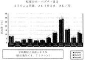


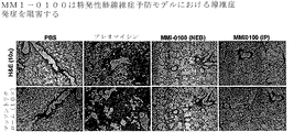


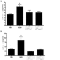


Classifications
-
- A—HUMAN NECESSITIES
- A61—MEDICAL OR VETERINARY SCIENCE; HYGIENE
- A61K—PREPARATIONS FOR MEDICAL, DENTAL OR TOILETRY PURPOSES
- A61K38/00—Medicinal preparations containing peptides
- A61K38/04—Peptides having up to 20 amino acids in a fully defined sequence; Derivatives thereof
- A61K38/08—Peptides having 5 to 11 amino acids
-
- A—HUMAN NECESSITIES
- A61—MEDICAL OR VETERINARY SCIENCE; HYGIENE
- A61K—PREPARATIONS FOR MEDICAL, DENTAL OR TOILETRY PURPOSES
- A61K38/00—Medicinal preparations containing peptides
- A61K38/005—Enzyme inhibitors
-
- A—HUMAN NECESSITIES
- A61—MEDICAL OR VETERINARY SCIENCE; HYGIENE
- A61K—PREPARATIONS FOR MEDICAL, DENTAL OR TOILETRY PURPOSES
- A61K39/00—Medicinal preparations containing antigens or antibodies
- A61K39/395—Antibodies; Immunoglobulins; Immune serum, e.g. antilymphocytic serum
- A61K39/39533—Antibodies; Immunoglobulins; Immune serum, e.g. antilymphocytic serum against materials from animals
- A61K39/39541—Antibodies; Immunoglobulins; Immune serum, e.g. antilymphocytic serum against materials from animals against normal tissues, cells
-
- A—HUMAN NECESSITIES
- A61—MEDICAL OR VETERINARY SCIENCE; HYGIENE
- A61K—PREPARATIONS FOR MEDICAL, DENTAL OR TOILETRY PURPOSES
- A61K9/00—Medicinal preparations characterised by special physical form
- A61K9/0012—Galenical forms characterised by the site of application
- A61K9/007—Pulmonary tract; Aromatherapy
- A61K9/0073—Sprays or powders for inhalation; Aerolised or nebulised preparations generated by other means than thermal energy
- A61K9/0075—Sprays or powders for inhalation; Aerolised or nebulised preparations generated by other means than thermal energy for inhalation via a dry powder inhaler [DPI], e.g. comprising micronized drug mixed with lactose carrier particles
-
- A—HUMAN NECESSITIES
- A61—MEDICAL OR VETERINARY SCIENCE; HYGIENE
- A61P—SPECIFIC THERAPEUTIC ACTIVITY OF CHEMICAL COMPOUNDS OR MEDICINAL PREPARATIONS
- A61P11/00—Drugs for disorders of the respiratory system
-
- A—HUMAN NECESSITIES
- A61—MEDICAL OR VETERINARY SCIENCE; HYGIENE
- A61P—SPECIFIC THERAPEUTIC ACTIVITY OF CHEMICAL COMPOUNDS OR MEDICINAL PREPARATIONS
- A61P29/00—Non-central analgesic, antipyretic or antiinflammatory agents, e.g. antirheumatic agents; Non-steroidal antiinflammatory drugs [NSAID]
-
- A—HUMAN NECESSITIES
- A61—MEDICAL OR VETERINARY SCIENCE; HYGIENE
- A61P—SPECIFIC THERAPEUTIC ACTIVITY OF CHEMICAL COMPOUNDS OR MEDICINAL PREPARATIONS
- A61P37/00—Drugs for immunological or allergic disorders
- A61P37/02—Immunomodulators
- A61P37/06—Immunosuppressants, e.g. drugs for graft rejection
-
- A—HUMAN NECESSITIES
- A61—MEDICAL OR VETERINARY SCIENCE; HYGIENE
- A61P—SPECIFIC THERAPEUTIC ACTIVITY OF CHEMICAL COMPOUNDS OR MEDICINAL PREPARATIONS
- A61P43/00—Drugs for specific purposes, not provided for in groups A61P1/00-A61P41/00
-
- C—CHEMISTRY; METALLURGY
- C07—ORGANIC CHEMISTRY
- C07K—PEPTIDES
- C07K16/00—Immunoglobulins [IGs], e.g. monoclonal or polyclonal antibodies
- C07K16/18—Immunoglobulins [IGs], e.g. monoclonal or polyclonal antibodies against material from animals or humans
- C07K16/28—Immunoglobulins [IGs], e.g. monoclonal or polyclonal antibodies against material from animals or humans against receptors, cell surface antigens or cell surface determinants
- C07K16/2884—Immunoglobulins [IGs], e.g. monoclonal or polyclonal antibodies against material from animals or humans against receptors, cell surface antigens or cell surface determinants against CD44
-
- A—HUMAN NECESSITIES
- A61—MEDICAL OR VETERINARY SCIENCE; HYGIENE
- A61K—PREPARATIONS FOR MEDICAL, DENTAL OR TOILETRY PURPOSES
- A61K39/00—Medicinal preparations containing antigens or antibodies
- A61K2039/505—Medicinal preparations containing antigens or antibodies comprising antibodies
-
- A—HUMAN NECESSITIES
- A61—MEDICAL OR VETERINARY SCIENCE; HYGIENE
- A61K—PREPARATIONS FOR MEDICAL, DENTAL OR TOILETRY PURPOSES
- A61K2300/00—Mixtures or combinations of active ingredients, wherein at least one active ingredient is fully defined in groups A61K31/00 - A61K41/00
-
- A—HUMAN NECESSITIES
- A61—MEDICAL OR VETERINARY SCIENCE; HYGIENE
- A61K—PREPARATIONS FOR MEDICAL, DENTAL OR TOILETRY PURPOSES
- A61K9/00—Medicinal preparations characterised by special physical form
- A61K9/0012—Galenical forms characterised by the site of application
- A61K9/007—Pulmonary tract; Aromatherapy
- A61K9/0073—Sprays or powders for inhalation; Aerolised or nebulised preparations generated by other means than thermal energy
- A61K9/0078—Sprays or powders for inhalation; Aerolised or nebulised preparations generated by other means than thermal energy for inhalation via a nebulizer such as a jet nebulizer, ultrasonic nebulizer, e.g. in the form of aqueous drug solutions or dispersions
-
- C—CHEMISTRY; METALLURGY
- C07—ORGANIC CHEMISTRY
- C07K—PEPTIDES
- C07K2317/00—Immunoglobulins specific features
- C07K2317/70—Immunoglobulins specific features characterized by effect upon binding to a cell or to an antigen
- C07K2317/76—Antagonist effect on antigen, e.g. neutralization or inhibition of binding
Description
関連出願の相互参照
本出願は、内容が全体として参照により組み入れられる米国特許仮出願第61/953,438号(2014年3月14日出願)の優先権の利益を主張するものである。
CROSS REFERENCE TO RELATED APPLICATIONS This application claims the benefit of priority to US Provisional Application No. 61 / 953,438, filed March 14, 2014, the contents of which are incorporated by reference in its entirety.
政府後援の表明
記載された本発明は、Moerae Matrix, LLCに認可された中小企業技術革新研究プログラム(Small Business Innovation Research (SBIR))、およびDr.Nobleに授与されたNHLBI PO1からの米国政府支援によってなされた。米国政府は、本発明において特定の権利を有する。
GOVERNMENT-SPONSORED STATEMENTS The invention described is described in the Small Business Innovation Research (SBIR) approved by Moerae Matrix, LLC. Made with US government support from NHLBI PO1 awarded to Noble. The United States Government has certain rights in the invention.
発明の分野
本発明は、細胞分子生物学、ポリペプチド、ならびに治療的使用方法の分野のものである。
FIELD OF THE INVENTION The present invention is in the field of cell molecular biology, polypeptides, and therapeutic uses.
背景
1.創傷治癒および線維症のメカニズム
用語「創傷治癒」は、身体組織のいずれかへの外傷、特に物理的手段によって生じた連続性の遮断を有するものを身体が修復する過程を指す。
創傷治癒応答は多くの場合、3つの別個の期(損傷期、炎症期および修復期)を有すると言われる。概して言えば、身体は、損傷に対して炎症応答を伴って応答し、炎症応答は、器官の健康および完全性の維持に不可欠である。しかしこの応答が誤って進行すると、組織の破壊を生じ得る。 The wound healing response is often said to have three distinct phases: the injury phase, the inflammatory phase, and the repair phase. Generally speaking, the body responds to injury with an inflammatory response, which is essential for maintaining organ health and integrity. However, incorrect progression of this response can result in tissue destruction.
I期:損傷
非限定的に自己免疫もしくはアレルギー反応、環境性微粒子、感染または機械的損傷をはじめとする因子によって引き起こされる損傷は多くの場合、正常な組織構造の破壊をもたらし、治癒応答を開始させる。損傷した上皮および内皮細胞は交換されて、それぞれバリア機能および完全性を維持し、血液の損失が予防されなければならない。内皮細胞に対する急性損傷は、炎症媒介物質を遊離させて抗フィブリン溶解性凝固カスケードを開始させ、損傷した血管を血小板および高フィブリン塊により一過性に閉塞する。例えば、特発性肺線維症(IPF)患者の肺ホモジネート、上皮細胞または気管支肺胞洗浄液は、慢性閉塞性肺疾患(COPD)および対照の患者と比較して、高レベルの血小板分化因子:X−ボックス−結合タンパク質−1を含んでおり、血塊形成応答が持続的に活性化されることが示唆される。加えて、トロンビン(フィブリノゲンをフィブリンに変換するのに必要なセリンプロテアーゼ)もまた、幾つかの肺線維症状態の肺および肺胞内の空間で容易に検出されるため、血液凝固経路の活性化がさらに確認される。トロンビンはまた、線維芽細胞を直接活性化して、増殖を増進し、コラーゲン生成筋線維芽細胞への線維芽細胞分化を促進できる。気道上皮、特に肺胞内肺細胞の損傷は、類似の抗フィブリン溶解性カスケードを惹起して、間質性浮腫、急性炎症領域および基底膜の上皮分離をもたらし得る。
Stage I: Injury Damage caused by factors including but not limited to autoimmune or allergic reactions, environmental particulates, infection or mechanical damage often results in the destruction of normal tissue structures and initiates a healing response Let it. Damaged epithelial and endothelial cells must be replaced to maintain barrier function and integrity, respectively, and to prevent blood loss. Acute damage to endothelial cells releases inflammatory mediators and initiates the anti-fibrinolytic coagulation cascade, transiently occluding damaged blood vessels with platelets and high fibrin clots. For example, lung homogenates, epithelial cells or bronchoalveolar lavage fluid of patients with idiopathic pulmonary fibrosis (IPF) have higher levels of platelet differentiation factor: X- compared to patients with chronic obstructive pulmonary disease (COPD) and controls. Contains box-binding protein-1, suggesting that the clot-forming response is persistently activated. In addition, thrombin, a serine protease necessary to convert fibrinogen to fibrin, is also easily detected in the lung and alveolar spaces of some pulmonary fibrosis states, thus activating the blood coagulation pathway. Is further confirmed. Thrombin can also directly activate fibroblasts to enhance proliferation and promote fibroblast differentiation into collagen-producing myofibroblasts. Damage to airway epithelia, especially intraalveolar lung cells, can trigger a similar antifibrinolytic cascade, leading to interstitial edema, acute inflammatory areas and epithelial separation of the basement membrane.
血小板動員、脱顆粒および血塊形成は、透過性を上昇させながら血管収縮期へ急速に進行し、血管外遊出(毛細血管からその周辺組織への白血球移動)および損傷部位への白血球の直接的動員を可能にする。実質組織の上皮および内皮の下に存在する細胞外マトリックスを形成する基底膜は、損傷組織への直接的接近を妨げる。この物理的バリアを破壊するために、マトリックスメタロプロテイナーゼ(MMP)とも称される亜鉛依存性エンドペプチダーゼが、1つまたは複数の細胞外マトリックス構成成分を切断し、損傷部位内外への細胞の遊出を可能にする。特にMMP−2(ゼラチナーゼA、N型コラゲナーゼ)およびMMP−9(ゼラチナーゼB、IV型コラゲナーゼ)は、基底膜の2種の重要な構成成分であるN型コラーゲンおよびゼラチンを切断する。近年の研究で、MMP−2およびMMP−9がアップレギュレートされることが見出されており、組織の破壊および再生過程が線維化状態に共通することが強調された。MMPの活性は、MMPの転写調節、前酵素調節、および特異的な組織阻害物質をはじめとする幾つかのメカニズムによって制御される。MMPと様々な阻害メカニズムとのバランスが炎症を調節し、治癒応答時に沈着するコラーゲンの正味量を決定し得る。 Platelet recruitment, degranulation and clot formation progress rapidly to vasoconstriction with increasing permeability, extravasation (leukocyte migration from capillaries to surrounding tissues) and direct recruitment of leukocytes to the site of injury Enable. The basement membrane, which forms the extracellular matrix underlying the epithelium and endothelium of parenchyma, prevents direct access to damaged tissue. To break this physical barrier, zinc-dependent endopeptidases, also called matrix metalloproteinases (MMPs), cleave one or more extracellular matrix components and emigrate cells into and out of the site of injury. Enable. In particular, MMP-2 (gelatinase A, N-type collagenase) and MMP-9 (gelatinase B, type IV collagenase) cleave N-type collagen and gelatin, two important components of the basement membrane. Recent studies have found that MMP-2 and MMP-9 are up-regulated, highlighting that the process of tissue destruction and regeneration is common to the fibrotic state. MMP activity is regulated by several mechanisms, including MMP transcriptional regulation, pre-enzyme regulation, and specific tissue inhibitors. The balance between MMPs and various inhibitory mechanisms may regulate inflammation and determine the net amount of collagen deposited during the healing response.
アレルギー性気道炎症モデル、ならびにMMP−2-/-、MMP−9-/-、およびMMP−2-/-MMP−9-/-ダブルノックアウトマウスによるリモデリングを用いた過去の研究によって、炎症組織から気腔へと炎症細胞がうまく排出および除去されるためにMMP−2およびMMP−9が必要であることが示された。これらのMMPの非存在下では、細胞は、肺の実質内に封入されて気腔へと移動できず、このことが致死性の窒息をもたらした。 Allergic airway inflammation models, as well as MMP-2 - / -, MMP -9 - / -, and MMP-2 - / - MMP- 9 - / - by previous studies using remodeling by double knockout mice, inflamed tissue Showed that MMP-2 and MMP-9 were required for successful efflux and clearance of inflammatory cells into the air space. In the absence of these MMPs, the cells were encapsulated within the parenchyma of the lung and could not move into the air space, resulting in lethal asphyxiation.
II期:炎症
組織損傷部位への接近がなされたら、ケモカイングラジエントによって炎症性細胞が動員される。好中球、好酸球、リンパ球およびマクロファージが、食細胞により浄化された細胞屑および壊死領域と共に急性炎症部位で観察される。
Stage II: Inflammation Once access to the site of tissue damage has been made, inflammatory cells are recruited by a chemokine gradient. Neutrophils, eosinophils, lymphocytes and macrophages are observed at the site of acute inflammation, along with phagocytic cleared cell debris and necrotic areas.
炎症性サイトカインおよびケモカインを提供する好酸球、好中球、リンパ球およびマクロファージの初期動員は、局所のTGF−βおよびIL−13蓄積に寄与し得る。最初の傷害および炎症細胞の波に続いて、炎症細胞の後期動員が、食作用、細胞屑浄化および過剰な細胞増殖の制御を支援でき、それらが一緒になって正常な治癒に寄与し得る。後期炎症は、抗線維症的役割を果たすことができ、創傷治癒応答をうまく消散するのに必要であり得る。例えば、食細胞性マクロファージに富む後期炎症プロファイルは、局所ケモカイン産生およびTGF−βを抑制するIL−10分泌性調節性T細胞に加えて、線維芽細胞浄化を支援しており、過剰な線維芽細胞活性化を予防し得る。 The initial recruitment of eosinophils, neutrophils, lymphocytes and macrophages that provide inflammatory cytokines and chemokines can contribute to local TGF-β and IL-13 accumulation. Following initial injury and a wave of inflammatory cells, late recruitment of inflammatory cells can assist in controlling phagocytosis, debris clearance and excessive cell growth, which together can contribute to normal healing. Late inflammation can play an antifibrotic role and may be necessary to successfully resolve the wound healing response. For example, a late inflammatory profile enriched in phagocytic macrophages, in addition to IL-10 secretory regulatory T cells that suppress local chemokine production and TGF-β, supports fibroblast purification and leads to excessive fibroblasts. Cell activation may be prevented.
傷害または原因物質の性質は多くの場合、後続の炎症反応の特徴を規定する。例えば、病原体関連分子パターン(PAMP)のような外因性刺激は、Toll様受容体およびNOD様受容体(炎症反応およびアポトーシス反応の調節において様々な機能を有する細胞質タンパク質)などの病原体認識受容体によって認識され、侵入した病原体に対する自然細胞の応答に影響を及ぼす。内因性の危険シグナルもまた局所の自然細胞に影響を及ぼして、炎症カスケードを調整し得る。 The nature of the injury or causative often defines the characteristics of the subsequent inflammatory response. For example, exogenous stimuli, such as pathogen-associated molecular patterns (PAMPs), are stimulated by pathogen-recognizing receptors, such as Toll-like and NOD-like receptors (cytoplasmic proteins with various functions in regulating inflammatory and apoptotic responses). It is recognized and affects the response of natural cells to invading pathogens. Intrinsic danger signals may also affect local natural cells, modulating the inflammatory cascade.
炎症反応の性質は、常在する組織細胞および後続の炎症細胞に劇的な影響を及ぼす。炎症細胞自体もまた、ケモカイン、サイトカインおよび増殖因子の分泌を通して更なる炎症を増大させる。多くのサイトカインが、様々な状態で活性化される特異的な遺伝子群と共に、創傷治癒および線維化応答の全体に関与する。例えば、喘息における慢性アレルギー性気道疾患は、一般的に2型ヘルパーT細胞(Th2)関連サイトカインプロファイル(非限定的にインターロイキン−4(IL−4)、インターロイキン−5(IL−5)、インターロイキン−6(IL−6)、インターロイキン−13(IL−13)、およびインターロイキン−9(IL−9)など)に関連するが、慢性閉塞性肺疾患および線維症性肺疾患(特発性肺線維症など)の患者は、より多くが炎症促進性サイトカインプロファイル(非限定的にインターロイキン−1アルファ(IL−1α)、インターロイキン−1ベータ(IL−1β)、インターロイキン−6(IL−6)、腫瘍壊死因子アルファ(TNF−α)、形質転換増殖因子ベータ(TGF−β)、および血小板由来増殖因子(PDGF)など)を提示する。これらのサイトカインの各々は顕著な線維化促進活性を示し、線維芽細胞、マクロファージおよび筋線維芽細胞の動員、活性化および増殖を介して作用することが示されている。 The nature of the inflammatory response dramatically affects resident tissue cells and subsequent inflammatory cells. The inflammatory cells themselves also increase further inflammation through secretion of chemokines, cytokines and growth factors. Many cytokines, along with specific genes that are activated in a variety of conditions, are involved in the overall wound healing and fibrotic response. For example, chronic allergic airway disease in asthma is generally T-helper cell type 2 (Th 2) associated cytokine profile (but not limited to Interleukin -4 (IL-4), interleukin--5 (IL-5) , Interleukin-6 (IL-6), interleukin-13 (IL-13), and interleukin-9 (IL-9), but are associated with chronic obstructive pulmonary disease and fibrotic lung disease ( Patients with idiopathic pulmonary fibrosis have more proinflammatory cytokine profiles (including but not limited to interleukin-1 alpha (IL-1α), interleukin-1 beta (IL-1β), interleukin-6 (IL-6), tumor necrosis factor alpha (TNF-α), transforming growth factor beta (TGF-β), and platelet-derived growth factor (P GF), etc.) to present. Each of these cytokines exhibits significant pro-fibrotic activity and has been shown to act through recruitment, activation and proliferation of fibroblasts, macrophages and myofibroblasts.
III期:組織の修復および収縮
創傷治癒の終期は、フィブリン(創傷部位を覆う血塊を形成する「網状組織」を形成するように重合する線維性タンパク質)に富む足場の形成によって誘導される協調的細胞再編成、創傷収縮、創傷閉鎖および創傷の再上皮形成からなる。創傷修復のこの期間に関与する過程を究明する莫大な数の研究が、皮膚創傷の研究およびインビトロ系から得られている。
Stage III: Tissue Repair and Contraction The end of wound healing is coordinated by the formation of a scaffold rich in fibrin, a fibrous protein that polymerizes to form a "network" forming a clot over the wound site It consists of cell rearrangement, wound contraction, wound closure and wound re-epithelialization. A vast number of studies have been derived from studies of skin wounds and in vitro systems to determine the processes involved in this period of wound repair.
筋線維芽細胞由来コラーゲンおよび平滑筋アクチン(α−SMA)は、マクロファージ、血小板、およびフィブリン骨格を形成する線維芽細胞由来フィブロネクチンと共に、仮の細胞外マトリックスを形成する。集合的にこれらの構造は、一般に顆粒化組織と称される。特発性肺線維症患者から単離された主要線維芽細胞または肺胞マクロファージは、対照の線維芽細胞よりも有意に多くのフィブロネクチンおよびα−SMAを産生し、これは線維芽細胞活性化が亢進した状態を示す。ステロイド処置を受けているIPF患者は、処置を受けていないIPF患者と同様に高レベルのマクロファージ由来フィブロネクチンを有することが報告された。したがって、ステロイド耐性IL−13媒介筋線維芽細胞分化と同様に、マクロファージ由来フィブロネクチン放出もまた、ステロイド処置に耐性があるように思われ、それがステロイド治療が無効であり得る別の原因を示している。動物モデルにより、フィブロネクチンのエキストラIII型ドメイン(EDA)が特異的に欠失したマウスは、それらの野生型対応動物と比較してブレオマイシン投与後の線維症発生が有意に低かったため、フィブロネクチンは肺線維症の発生に必要であるように思われる。 Myofibroblast-derived collagen and smooth muscle actin (α-SMA) together with macrophages, platelets, and fibroblast-derived fibronectin that form the fibrin skeleton form a tentative extracellular matrix. Collectively, these structures are commonly referred to as granulated tissue. Primary fibroblasts or alveolar macrophages isolated from patients with idiopathic pulmonary fibrosis produce significantly more fibronectin and α-SMA than control fibroblasts, which enhance fibroblast activation It shows the state where it was done. IPF patients receiving steroid treatment were reported to have high levels of macrophage-derived fibronectin as well as untreated IPF patients. Thus, like steroid-resistant IL-13-mediated myofibroblast differentiation, macrophage-derived fibronectin release also appeared to be resistant to steroid treatment, indicating another cause for which steroid therapy may be ineffective. I have. According to the animal model, mice in which the extra type III domain (EDA) of fibronectin was specifically deleted showed significantly lower fibrosis development after administration of bleomycin than their wild-type counterparts. Seems necessary for the development of the disease.
仮の細胞外マトリックスは、フィブロネクチンに加えて、糖タンパク質(PDGFなど)、グリコサミノグリカン(ヒアルロン酸など)、プロテオグリカンおよびエラスチンからなる。増殖因子およびTGF−β活性化線維芽細胞は、細胞外マトリックスネットワークに沿って遊走し、創傷を修復する。皮膚創傷内では、TGF−βはまた収縮応答を誘導し、コラーゲン線維の方向性を調節する。線維芽細胞から筋線維芽細胞への分化はまた、先に議論された通り、ストレス線維およびα−SMAの新たな発現を生じ、それらは両方とも筋線維芽細胞内で高い収縮活性を付与する。「フィブロネクサス」または「超成熟焦点接着(super mature focal adhesions)」と呼ばれる専用の部位での細胞外マトリックスへの筋線維芽細胞の付着は、一緒になって創傷を引張り、収縮期に病変サイズを減少させる。細胞マトリックスが堆積する程度および活性化筋線維芽細胞の量が、コラーゲン沈着量を決定する。この目的のために、メタロプロテイナーゼの組織阻害物質(TIMP)に対するマトリックスメタロプロテイナーゼ(MMP)のバランスおよびコラゲナーゼに対するコラーゲンのバランスが、この応答の間じゅう変化し、コラーゲンの正味の増加なしに、合成促進(pro−syntehsis)およびコラーゲン沈着増加からバランス制御に向けてシフトする。うまく創傷が治癒するためには、線維芽細胞がアポトーシスを受ける際にこの均衡が始まることが多く、炎症が収束し始め、顆粒化組織が後退して、高コラーゲン病変が残留する。炎症細胞、特にα−SMA陽性筋線維芽細胞の除去が、コラーゲン沈着の停止に必須である。興味深いことに、特発性肺線維症の患者では、アポトーシス促進分子およびFAS−シグル伝達分子のレベル上昇が観察されたにもかかわらず、細胞はアポトーシスシグナルに耐容であり、線維芽細胞の除去が遅延し得る。アポトーシスに対するこの相対的耐性は、潜在的にこの線維性疾患の原因であり得る。しかし、幾つかの研究ではまた、特発性肺線維症でコラーゲン分泌線維芽細胞および上皮細胞のアポトーシスの速度増加が観察されており、さらに別のバランスが線維芽細胞アポトーシスと線維芽細胞増殖のモニタリングを要求していることが示唆された。皮膚の研究から、創傷部位の再上皮形成が、バリア機能を再度確立し、封入された細胞の再編成を可能にする。コラーゲンマトリックス上で生育させたヒトもしくはラットの上皮細胞、またはインビボの気管の損傷を用いる、幾つかのインビトロおよびインビボモデルを使用して、細胞の遊走、増殖および細胞増大の重要なステージが同定された。露出した領域の端から上皮が復元すると共に、急速かつ動的な運動性および増殖が最初の創傷から数時間以内に生じる。加えて、上皮細胞のスライディングシートが損傷領域の上部を遊走し、創傷の被覆を支援し得る。血清由来形質転換増殖因子アルファ(TGF−α)、およびマトリックスメタロプロテイナーゼ−7(MMP−7)(それ自体はTIMP−1によって調節される)を含む幾つかの因子が、再上皮形成を調節することが示されている。 The provisional extracellular matrix is composed of glycoproteins (such as PDGF), glycosaminoglycans (such as hyaluronic acid), proteoglycans and elastin, in addition to fibronectin. Growth factors and TGF-β activated fibroblasts migrate along the extracellular matrix network to repair wounds. Within skin wounds, TGF-β also induces a contractile response and regulates the orientation of collagen fibers. Differentiation of fibroblasts into myofibroblasts also results in renewed expression of stress fibers and α-SMA, both of which confer high contractile activity in myofibroblasts, as discussed above. . Attachment of myofibroblasts to the extracellular matrix at a dedicated site called "fibronexus" or "supermature focal adhesions" together pulls the wound and reduces the size of the lesion during systole. Decrease. The extent to which the cell matrix is deposited and the amount of activated myofibroblasts determine the amount of collagen deposition. For this purpose, the balance of matrix metalloproteinases (MMPs) against tissue inhibitors of metalloproteinases (TIMPs) and the balance of collagen against collagenases change throughout this response, enhancing synthesis without a net increase in collagen. (Pro-synthesis) and shift from increased collagen deposition toward balance control. For successful wound healing, this balance often begins when fibroblasts undergo apoptosis, inflammation begins to converge, granulated tissue recedes, and high collagen lesions remain. Removal of inflammatory cells, especially α-SMA positive myofibroblasts, is essential for arrest of collagen deposition. Interestingly, in patients with idiopathic pulmonary fibrosis, cells were tolerant to apoptotic signals and fibroblast clearance was delayed, despite elevated levels of pro-apoptotic and FAS-siggle-transmitting molecules observed. I can do it. This relative resistance to apoptosis could potentially be responsible for this fibrotic disease. However, some studies have also observed increased rates of apoptosis of collagen-secreting fibroblasts and epithelial cells in idiopathic pulmonary fibrosis, and yet another balance is monitoring fibroblast apoptosis and fibroblast proliferation. It is suggested that the request. From skin studies, re-epithelialization of the wound site re-establishes barrier function and allows the reorganization of encapsulated cells. Using several in vitro and in vivo models using human or rat epithelial cells grown on collagen matrices or tracheal injury in vivo, key stages of cell migration, proliferation and cell expansion have been identified. Was. Rapid and dynamic motility and proliferation occur within hours of the first wound as the epithelium reconstructs from the edges of the exposed area. In addition, a sliding sheet of epithelial cells can migrate over the injured area and assist in covering the wound. Several factors regulate reepithelialization, including serum-derived transforming growth factor alpha (TGF-α), and matrix metalloproteinase-7 (MMP-7), which is itself regulated by TIMP-1 It has been shown.
総括すると、炎症の程度、血管形成、および細胞外マトリックス沈着の量は全て、最終的な線維症病変の発達に寄与する。したがって、線維芽細胞の活性化、増殖またはアポトーシスを妨害する治療的介入には、創傷修復の全ての期間の完全な理解および認識が必要である。これらの3つの期間は多くの場合、連続的に提示されるが、慢性または反復損傷時にはこれらの過程は並行して機能し、調節メカニズムを大いに必要とする(Wilson and Wynn, Mucosal Immunol., 2009, 3(2):103−121)。 Taken together, the degree of inflammation, angiogenesis, and the amount of extracellular matrix deposition all contribute to the development of the final fibrotic lesion. Therefore, therapeutic intervention to prevent fibroblast activation, proliferation or apoptosis requires a thorough understanding and awareness of all periods of wound repair. Although these three periods are often presented sequentially, during chronic or recurrent injury, these processes function in parallel and require a significant regulatory mechanism (Wilson and Wynn, Mucosal Immunol., 2009). , 3 (2): 103-121).
2.病理としての線維症
線維症は、器官または組織における過剰な線維性結合組織の形成または発達であり、正常または異常な/反応性の創傷治癒応答の結果として形成され、瘢痕を生じる。線維症は、例えば非限定的に、異常な細胞外マトリックスタンパク質の異常な沈着、線維芽細胞増殖の異常な促進、線維芽細胞集団の筋線維芽細胞集団への異常な分化誘発、筋線維芽細胞の細胞外マトリックスへの異常な付着促進、またはそれらの組み合わせを特徴とする。
2. Fibrosis as a Pathology Fibrosis is the formation or development of excess fibrous connective tissue in an organ or tissue, which is formed as a result of a normal or abnormal / reactive wound healing response, resulting in scarring. Fibrosis includes, but is not limited to, abnormal deposition of abnormal extracellular matrix proteins, abnormal promotion of fibroblast proliferation, abnormal induction of fibroblast population into myofibroblast population, myofibroblasts It is characterized by abnormal promotion of attachment of cells to the extracellular matrix, or a combination thereof.
炎症促進性媒介物質
蓄積しつつある証拠から、様々なリンホカイン、インターロイキン、およびケモカインを含む、サイトカインとして知られるポリペプチド媒介物質が、線維症におけるコラーゲン沈着の重要な刺激であることが示唆されている。サイトカインは、常在する組織細胞および動員された炎症細胞によって放出され、線維芽細胞の増殖およびコラーゲンを含む細胞外マトリックスタンパク質の合成増加を刺激すると考えられる。例えば、特発性肺線維症の発病における初期の特色は、肺胞上皮細胞および/または毛細血管細胞損傷である。これは、循環する免疫細胞、例えば、単球、好中球、リンパ球および好酸球の、肺への動員を促進する。その後、これらのエフェクター細胞は、常在する肺細胞、例えばマクロファージ、肺胞上皮細胞および内皮細胞と一緒に、サイトカインを放出し、サイトカインは、標的細胞、典型的には線維芽細胞を刺激して複製させ、コラーゲンの合成量を増加させる。細胞外マトリックスの分解もまた阻害され、線維化過程に寄与する可能性がある(Coker and Laurent, Eur Respir J, 1998,; 11:1218−1221)。
Pro-inflammatory mediators Accumulating evidence suggests that polypeptide mediators known as cytokines, including various lymphokines, interleukins, and chemokines, are important stimuli for collagen deposition in fibrosis. I have. Cytokines are released by resident tissue cells and recruited inflammatory cells and are thought to stimulate fibroblast proliferation and increased synthesis of extracellular matrix proteins, including collagen. For example, an early feature in the pathogenesis of idiopathic pulmonary fibrosis is alveolar epithelial cell and / or capillary cell damage. It promotes the recruitment of circulating immune cells, such as monocytes, neutrophils, lymphocytes and eosinophils, to the lung. These effector cells then release cytokines, along with resident lung cells, such as macrophages, alveolar epithelial cells and endothelial cells, which stimulate target cells, typically fibroblasts. It replicates and increases the amount of collagen synthesis. Extracellular matrix degradation is also inhibited and may contribute to the fibrosis process (Coker and Laurent, Eur Respir J, 1998, 11: 1218-1221).
非限定的に、形質転換増殖因子−β(TGF−β)、腫瘍壊死因子−α(TNF−α)、血小板由来増殖因子(PDGF)、インスリン様増殖因子−1(IGF−1)、エンドセリン−1(ET−1)ならびにインターロイキン類、インターロイキン−1(IL−1)、インターロイキン−6(IL−6)、インターロイキン−8(IL−8)およびインターロイキン−17(IL−17)を含む数多くのサイトカインが、線維症の発病において関連づけられている。正常T細胞活性化時に調節され、発現および分泌される(RANTES)因子を含むケモカイン白血球ケモアトラクタントもまた、重要な役割を果たすと考えられる。末梢血中での炎症促進性サイトカイン、例えばインターロイキン−8(IL−8)のレベル上昇は、細胞間接着分子−1(ICAM−1)および血管細胞接着分子−1(VCAM−1)などの関連する下流の接着分子(CAM)、マトリックスメタロプロテイナーゼ−7(MMP−7)などのマトリックスメタロプロテイナーゼ、ならびに末梢血中のS100カルシウム結合タンパク質A12(S100A12、カルグラヌリンCとしても公知)などのシグナル伝達分子のレベル上昇は、特発性肺線維症の患者で死亡率、肺移植なしの生存率および疾患進行と関連することが見出されている(Richardset al, Am J Respir Crit Care Med, 2012, 185: 67−76)。 Without limitation, transforming growth factor-β (TGF-β), tumor necrosis factor-α (TNF-α), platelet-derived growth factor (PDGF), insulin-like growth factor-1 (IGF-1), endothelin- 1 (ET-1) and interleukins, interleukin-1 (IL-1), interleukin-6 (IL-6), interleukin-8 (IL-8) and interleukin-17 (IL-17) Numerous cytokines have been implicated in the pathogenesis of fibrosis, including. Chemokine leukocyte chemoretractants, including factors that are regulated, expressed and secreted during normal T cell activation (RANTES), are also thought to play an important role. Elevated levels of pro-inflammatory cytokines, such as interleukin-8 (IL-8), in peripheral blood have been associated with increased levels of intercellular adhesion molecule-1 (ICAM-1) and vascular cell adhesion molecule-1 (VCAM-1). Related downstream adhesion molecules (CAMs), matrix metalloproteinases such as matrix metalloproteinase-7 (MMP-7), and signaling molecules such as S100 calcium binding protein A12 in peripheral blood (S100A12, also known as calgranulin C) Has been found to be associated with mortality, survival without lung transplantation and disease progression in patients with idiopathic pulmonary fibrosis (Richards et al, Am J Respir Crit Care Med, 2012, 185: 67-76).
TGF−βタンパク質ファミリーは、細胞外マトリックス沈着に対して強力な刺激作用を有し、実際に遺伝子移転による線維症誘発動物モデルの構築に用いられてきた。インビトロ試験では、潜在的前駆体として分泌されるTGF−β1は、線維芽細胞のプロコラーゲン遺伝子発現およびタンパク質合成を促進することが示されている。これらのデータは、他の哺乳動物のアイソフォームTGF−β2およびTGF−β3もまた、ヒト肺線維芽細胞のコラーゲン合成を刺激しインビトロでの分解を低下させることを示唆している。肺線維症の動物モデルでは、TGF−β1遺伝子発現の強化は、コラーゲン遺伝子発現およびタンパク質沈着の増加に時間的および空間的に関係する。TGF−β1抗体は、ネズミのブレオマイシン誘発性肺線維症でコラーゲン沈着を低下させ、ヒトの線維症の肺組織は、TGF−β1遺伝子およびタンパク質の発現強化を示す。 The TGF-β protein family has a strong stimulatory effect on extracellular matrix deposition, and has actually been used to construct a fibrosis-inducing animal model by gene transfer. In vitro studies have shown that TGF-β1, secreted as a potential precursor, promotes procollagen gene expression and protein synthesis in fibroblasts. These data suggest that other mammalian isoforms, TGF-β2 and TGF-β3, also stimulate collagen synthesis and reduce in vitro degradation of human lung fibroblasts. In animal models of pulmonary fibrosis, enhanced TGF-β1 gene expression is temporally and spatially related to increased collagen gene expression and protein deposition. TGF-β1 antibodies reduce collagen deposition in murine bleomycin-induced lung fibrosis, and human fibrotic lung tissue shows enhanced expression of TGF-β1 gene and protein.
TNF−αは、インビトロで線維芽細胞の複製およびコラーゲン合成を刺激でき、肺のTNF−α遺伝子発現は、マウスにブレオマイシンを投与した後で上昇する。可溶性TNF−α受容体は、ネズミモデルで肺線維症を減少させ、トランスジェニックマウスの肺のTNF−α過剰発現は、肺線維症を特徴とする。IPEまたはアスベスト症(アスベスト繊維の吸引および保持によって引き起こされる肺実質組織を冒す慢性炎症性および線維症性症状)の患者では、気管支肺胞洗浄液由来マクロファージが、対照と比較して増加した量のTNF−αを放出する。 TNF-α can stimulate fibroblast replication and collagen synthesis in vitro, and pulmonary TNF-α gene expression is increased after administration of bleomycin to mice. Soluble TNF-α receptor reduces pulmonary fibrosis in a murine model, and TNF-α overexpression in the lungs of transgenic mice is characterized by pulmonary fibrosis. In patients with IPE or asbestosis (a chronic inflammatory and fibrotic condition affecting lung parenchyma caused by asbestos fiber aspiration and retention), bronchoalveolar lavage fluid-derived macrophages have increased amounts of TNF compared to controls. -Release α.
エンドセリン(ET−1)もまた、線維化促進性サイトカインの基準を満たす。この分子は線維芽細胞の増殖および化学走性を促進し、プロコラーゲンの生成を刺激する。それは肺線維症の患者の肺に存在し、近年の報告では、ET−1受容体拮抗薬のボセンタンは、実験動物に投与された場合に肺線維症を軽減することが示唆されている。 Endothelin (ET-1) also meets the criteria for profibrotic cytokines. This molecule promotes fibroblast proliferation and chemotaxis and stimulates the production of procollagen. It is present in the lungs of patients with pulmonary fibrosis, and recent reports suggest that the ET-1 receptor antagonist bosentan reduces pulmonary fibrosis when administered to experimental animals.
未確認の筋線維芽細胞増殖/活性化および線維症病巣形成
線維芽細胞から筋線維芽細胞への分化は、創傷修復および線維症をはじめとする多くの症状における重要な事象であると長い間考えられてきた。例えば、筋線維芽細胞は活発な線維症領域で生じ、肺線維症では細胞外マトリックス(ECM)タンパク質の生成および沈着を担うことが報告されている(Liu, T. et al., Am J Respir Cell Mol Biol, 2007, 37:507−517)。
Unidentified myofibroblast proliferation / activation and fibrotic lesion formation Long-term belief that fibroblast to myofibroblast differentiation is a key event in many conditions, including wound repair and fibrosis I have been. For example, myofibroblasts occur in areas of active fibrosis, and are reported to be responsible for the production and deposition of extracellular matrix (ECM) proteins in pulmonary fibrosis (Liu, T. et al., Am J Respir. Cell Mol Biol, 2007, 37: 507-517).
特発性肺線維症の因果関係についての1つの仮説は、まだ未確認の刺激によって急性肺損傷の反復エピソードがもたらされることを示唆している。これらの損傷部位における創傷治癒は、最終的に線維症をもたらし、肺機能を喪失させる。線維症病巣、つまり特発性肺線維症の診断基準病巣は、間葉細胞の活発な複製および新しい細胞外マトリックスの旺盛な沈着を特色とする。そのような病巣は、肺胞上皮細胞損傷の典型であり、管腔内の血漿滲出および遠位気腔の破壊を伴う。創傷治癒と密接に関係する媒介物質、例えば形質転換増殖因子−β1(TGF−β1)および結合組織増殖因子もまた、これらの部位で発現される。この巣状性急性肺損傷および創傷修復の駆動力は、明らかではない。 One hypothesis about the consequences of idiopathic pulmonary fibrosis suggests that yet unidentified stimuli result in repeated episodes of acute lung injury. Wound healing at these injured sites eventually results in fibrosis and loss of lung function. Fibrotic lesions, diagnostic criteria for idiopathic pulmonary fibrosis, are characterized by active replication of mesenchymal cells and vigorous deposition of new extracellular matrix. Such lesions are typical of alveolar epithelial cell damage and involve intraluminal plasma exudation and destruction of the distal air space. Mediators closely related to wound healing, such as transforming growth factor-β1 (TGF-β1) and connective tissue growth factor, are also expressed at these sites. The driving force for this focal acute lung injury and wound repair is not clear.
3.線維症が役割を果たす疾患または状態
線維症は、非限定的に間質性肺疾患、例えば特発性肺線維症、急性肺損傷(ALI)、放射線誘発性線維症、および移植片拒絶を含む、多数の異質性疾患または症状に関連づけられている。
3. Diseases or conditions in which fibrosis plays a role Fibrosis includes, but is not limited to, interstitial lung diseases such as idiopathic pulmonary fibrosis, acute lung injury (ALI), radiation-induced fibrosis, and graft rejection, It has been linked to a number of heterogeneous diseases or conditions.
3.1.特発性肺線維症(IPF)
特発性肺線維症(IPE;原因不明の線維化肺胞炎(CFA)、または特発性線維化間質性肺炎としても公知)は、主として高齢者に発する病因不明の慢性進行性線維化間質性肺炎の特殊形態と定義されており、肺に限定され、かつ通常の間質性肺炎(UIP)の放射線学的および組織学的パターンと関連する(Raghu G. et al., Am J Respir Crit Care Med., 183(6):788−824, 2011;Thannickal, V. et al., Proc Am Thorac Soc., 3(4):350−356, 2006)。IPEは、肺間質の異常かつ過剰な線維症組織沈着を特徴とし得る。高分解能CT(HRCT)画像によれば、UIPは、牽引性気管支拡張症に関連することの多い網状混濁の存在を特徴とする。IPFが進行するにつれ、ハチの巣形状が、より一層際立ってくる(Neininger A. et al., J Biol Chem., 277(5):3065−8, 2002)。肺機能試験の多くは、限定的障害および一酸化炭素拡散能力の低下を明らかにする(Thomas, T. et al., J Neurochem., 105(5): 2039−52, 2008)。複数の研究が、特発性肺線維症(IPF)の患者におけるTNF−αおよびIL−6放出の顕著な増加を報告し(Zhang, Y, et al. J. Immunol. 150(9):4188−4196, 1993)、それは、IL−1βの発現レベルに起因するとされている(Kolb, M., et al. J. Clin. Invest, 107(12):1529−1536, 2001)。IPFの症状である息切れおよび咳の開始は、通常は潜行性であるが徐々に進行し、診断後5年以内に患者の70%が死亡する。この厳しい予後は、乳癌による年間死亡数に類似する(Raghu G. et al., Am J Respir Crit Care Med., 183(6):788−824, 2011)。
3.1. Idiopathic pulmonary fibrosis (IPF)
Idiopathic pulmonary fibrosis (IPE; also known as unexplained fibrosing alveolitis (CFA), or idiopathic fibrous interstitial pneumonia) is a chronic progressive fibrotic interstitial of unknown etiology that primarily occurs in the elderly It is defined as a specific form of pneumonia that is restricted to the lungs and is associated with the radiological and histological patterns of normal interstitial pneumonia (UIP) (Raghu G. et al., Am J Respir Crit) Care Med., 183 (6): 788-824, 2011; Thannickal, V. et al., Proc Am Thorac Soc., 3 (4): 350-356, 2006). IPE may be characterized by abnormal and excessive fibrotic tissue deposition of the lung interstitium. According to high-resolution CT (HRCT) images, UIP is characterized by the presence of reticular opacities often associated with traction bronchiectasis. As IPF progresses, the honeycomb shape becomes even more pronounced (Neininger A. et al., J Biol Chem., 277 (5): 3065-8, 2002). Many lung function tests reveal limited impairment and reduced ability to diffuse carbon monoxide (Thomas, T. et al., J Neurochem., 105 (5): 2039-52, 2008). Studies have reported significant increases in TNF-α and IL-6 release in patients with idiopathic pulmonary fibrosis (IPF) (Zhang, Y, et al. J. Immunol. 150 (9): 4188-). 4196, 1993), which has been attributed to the expression level of IL-1β (Kolb, M., et al. J. Clin. Invest, 107 (12): 1529-1536, 2001). The onset of shortness of breath and the onset of cough, symptoms of IPF, are usually insidious but progressive, with 70% of patients dying within 5 years after diagnosis. This severe prognosis is similar to the annual number of deaths from breast cancer (Raghu G. et al., Am J Respir Crit Care Med., 183 (6): 788-824, 2011).
IPFは、米国でほぼ130,000名の患者が罹患し、世界中で毎年50,000名の新たな患者が出現し、毎年40,000名が死亡する(Raghu G. et al., Am J Respir Crit Care Med., 183(6):788−824, 2011)。これらのデータは注目に値するが、近年の研究では、IPFが以前に考えられていたよりも5〜10倍多い可能性があり、それはおそらく有病率の増加または診断性能の向上のためであろうと報告された(Thannickal, V. et al., Proc Am Thorac Soc., 3(4):350−356, 2006)。肺移植はIPFの最も確実な治療法と見なされているが、肺移植後の5年生存は50%未満である。したがって、肺移植でさえIPFの「治癒」と考えることはできない。患者の身体的および情緒的な負担に加えて、IPFは、処置およびケアが極めて高価で、国家の健康維持費は、100,000名の患者毎に毎年28億ドルの範囲に及ぶ。 IPF affects nearly 130,000 patients in the United States, with 50,000 new cases emerging each year and 40,000 deaths each year worldwide (Raghu G. et al., Am J. Respir Crit Care Med., 183 (6): 788-824, 2011). Although these data are noteworthy, recent studies have shown that IPF can be 5-10 times more than previously thought, possibly due to increased prevalence or improved diagnostic performance. (Thannickal, V. et al., Proc Am Thorac Soc., 3 (4): 350-356, 2006). Lung transplantation is considered the definitive treatment for IPF, but 5-year survival after lung transplantation is less than 50%. Thus, even a lung transplant cannot be considered a “healing” of IPF. In addition to the physical and emotional burden on patients, IPF is extremely expensive to treat and care, and national health maintenance costs range from $ 2.8 billion annually for every 100,000 patients.
加えて、過去の研究では、環境性傷害の付加も特発性肺線維症の発病に重要であり得ることが示唆されている。ほとんどの報告された症例シリーズにおいて、特発性肺線維症のインデックス・ペイシェントの75%までが、現在または過去の喫煙者である。広範囲の疫学研究では、喫煙は特発性肺線維症と強く関連した。加えて、特発性肺線維症の炎症性特徴の多くが、基礎となる肺疾患よりも喫煙状況とより密接に関係する。したがって、喫煙は、特発性肺線維症の独立した危険因子であり得る。潜伏性ウイルス感染、特にヘルペスウイルスファミリーの感染もまた、特発性肺線維症と関連すると報告されている。 In addition, previous studies have suggested that the addition of environmental injury may also be important in the development of idiopathic pulmonary fibrosis. In most reported case series, up to 75% of index patients with idiopathic pulmonary fibrosis are current or past smokers. In extensive epidemiological studies, smoking was strongly associated with idiopathic pulmonary fibrosis. In addition, many of the inflammatory features of idiopathic pulmonary fibrosis are more closely related to smoking status than the underlying lung disease. Thus, smoking may be an independent risk factor for idiopathic pulmonary fibrosis. Latent viral infections, especially those of the herpesvirus family, have also been reported to be associated with idiopathic pulmonary fibrosis.
IPFについては肺移植を含む有効な公知の処置が存在しないため、新規な治療薬の開発が依然として緊急に求められている。瘢痕または線維症組織を生成する身体能力を遅延させる、または阻害することが可能な抗線維症療法、および肺での気体交換のための組織面積を増加させる肺の血管拡張剤をはじめとする、現在開発中の様々な治療手段が存在する。肺移植はさておき、潜在的なIPF治療には、コルチコステロイド、アザチオプリン、シクロホスファミド、抗凝固剤およびN−アセチルシステインが含まれる(Raghu G. et al., Am J Respir Crit Care Med., 183(6):788−824, 2011)。加えて、酸素療法および肺リハビリテーションなどの補助療法が日常的に用いられる。しかし、これらのいずれもが、IPF患者の長期生存に確実な影響をもたらさず、このことからIPFの治療選択のためのアンメットメディカルニーズがさらに強調される。例として挙げれば、混合臨床プログラムの結果にもかかわらず、InterMuneの経口小分子Esbriet(登録商標)(ピルフェニドン(pirfenadone))が、IPF患者のために欧州および日本での認可を受けた。したがって、Esbriet(登録商標)は、IPF処置のために特別に指定された最初の医薬となったが、あいまいな治験結果および薬物副作用のために、米国ではこの薬剤の有用性は懐疑的と見なされ、当時提出されたデータからはFDAの認可は得られなかった。したがって、米国では新薬適用を支持するためにその有効性を決定する大規模な第三相臨床試験が進行中である。 Due to the lack of effective known treatments for IPF, including lung transplantation, the development of new therapeutics is still urgently needed. Including anti-fibrotic therapies that can delay or inhibit the body's ability to produce scar or fibrotic tissue, and pulmonary vasodilators that increase tissue area for gas exchange in the lungs; There are various treatment modalities currently under development. Aside from lung transplantation, potential IPF treatments include corticosteroids, azathioprine, cyclophosphamide, anticoagulants and N-acetylcysteine (Raghu G. et al., Am J Respir Crit Care Med. 183 (6): 788-824, 2011). In addition, adjuvant therapies such as oxygen therapy and pulmonary rehabilitation are routinely used. However, none of these has a positive effect on the long-term survival of IPF patients, which further emphasizes the unmet medical need for IPF treatment options. By way of example, despite the results of the mixed clinical program, InterMune's oral small molecule Esbriet® (pirfenadone) has been approved in Europe and Japan for IPF patients. Thus, while Esbriet® became the first drug specifically designated for treatment of IPF, its usefulness in the United States was considered skeptical due to ambiguous clinical trial results and drug side effects. No data was available at the time, and no FDA approval was obtained. Therefore, a large phase III clinical trial is underway in the United States to determine its efficacy in support of a new drug application.
組織病理学的には、IPEは、線維芽細胞病巣における活性化筋線維芽細胞(または間葉細胞)の蓄積と説明できる(Thannickal, V. et al., Proc Am Thorac Soc., 3(4):350−356, 2006)。筋線維芽細胞の損なわれたアポトーシスは、組織の線維症に終わる持続的で調節不能な修復過程をもたらし得る。議論の余地はあるが、おそらく線維芽細胞の周期的な急性刺激により、炎症もまたIPFで重大な役割を果たす。これらの発見は、治療的介入のための潜在的標的を指し示している。 Histopathologically, IPE can be described as an accumulation of activated myofibroblasts (or mesenchymal cells) in fibroblast foci (Thannickal, V. et al., Proc Am Thorac Soc., 3 (4). ): 350-356, 2006). Impaired apoptosis of myofibroblasts can lead to a persistent and dysregulated repair process that results in tissue fibrosis. Although controversial, probably due to periodic acute stimulation of fibroblasts, inflammation also plays a significant role in IPF. These findings point to potential targets for therapeutic intervention.
3.1.1.特発性肺線維症(IPF)の発病
発病のメカニズムは完全には理解されていないが、現在受け入れられている理論的枠組では、肺胞上皮への損傷に続いて、正常な組織修復に関連する応答を引き起こす炎症促進性および線維増殖性媒介物質が大量放出されることが提案されている。明確でない理由により、これらの修復過程は消散せず、結果として進行性線維症が起こる(Selman M, et al., Ann Intern Med, 134(2):136−151, 2001;Noble, P. and Homer R., Clin Chest Med, 25(4):749−58, 2004;Strieter, R., Chest, 128 (5 Suppl 1):526S−532S, 2005)。
3.1.1. Onset of idiopathic pulmonary fibrosis (IPF)
Although the mechanisms of pathogenesis are not fully understood, the currently accepted theoretical framework suggests that following damage to the alveolar epithelium, proinflammatory and fibroproliferative triggering responses associated with normal tissue repair It has been proposed that mediators be released in large quantities. For unclear reasons, these repair processes do not resolve, resulting in progressive fibrosis (Selman M, et al., Ann Intern Med, 134 (2): 136-151, 2001; Noble, P. and Homer R., Clin Chest Med, 25 (4): 749-58, 2004; Strieter, R., Chest, 128 (5 Suppl 1): 526S-532S, 2005).
3.1.2.肺線維症のブレオマイシンマウスモデル
多数の動物モデルが存在し、有用であり得るが(例えば、TGF−βアデノウイルス形質導入モデルまたは放射線誘発性線維症モデル)、ブレオマイシンモデルは、詳しく記載されており、炎症後/線維化前/線維症予防段階で個々の薬剤またはプロテインキナーゼ阻害剤の有効性を実証するために今日使用されている最もよく特徴づけられたネズミモデルである(Vittal, R. et al., J Pharmacol Exp Ther., 321(1):35−44, 2007;Vittal, R. et al., Am J Pathol., 166(2):367−75, 2005;Hecker L. et al., Nat Med., 15(9):1077−81, 2009)。
3.1.2. Bleomycin Mouse Model of Pulmonary Fibrosis Although a number of animal models exist and may be useful (eg, the TGF-β adenovirus transduction model or radiation-induced fibrosis model), the bleomycin model has been described in detail, It is the best characterized murine model used today to demonstrate the efficacy of individual drugs or protein kinase inhibitors in the post-inflammatory / pre-fibrotic / fibrosis prevention phase (Vittal, R. et al. , J Pharmacol Exp Ther., 321 (1): 35-44, 2007; Vitttal, R. et al., Am J Pathol., 166 (2): 367-75, 2005; Hecker L. et al., Nat Med., 15 (9): 1077-81, 2009).
抗生物質のブレオマイシンは、最初ストレプトミセス・ベルチシラツス(Streptomyces verticillatus)から単離された(Umezawa, H. et al., Cancer 20: 891−895, 1967)。その後、この抗生物質は扁平上皮癌および皮膚腫瘍に対して有効であることが見出されたが(Umezawa, H., Fed Proc, 33: 2296−2302, 1974)、抗新生物薬としてのその有用性は、線維症をもたらす用量依存肺毒性により制限された(Muggia, F. et al., Cancer Treat Rev, 10: 221−243, 1983)。気管内経路によるブレオマイシンの送達(一般には供給源に応じて1.25−4U/kg)は、げっ歯類において1回の薬物注射が肺損傷およびその結果としての線維症をもたらすという利点を有する(Phan, S. et al., Am Rev Respir Dis 121: 501−506, 1980;Snider, G. et al., Am Rev Respir Dis. 117: 289−297, 1978;Thrall, R. et al., Am J Pathol, 95: 117−130, 1979)。げっ歯類への薬剤の気管内送達は、最初に肺胞上皮細胞への直接的損傷をもたらす。この事象に続いて、1週間以内に好中球性およびリンパ球性汎肺胞炎が生じる(Janick−Buckner, D. et al., Toxicol Appl Pharmacol., 100(3):465−73, 1989)。続いて、肺胞の炎症細胞が除去され、線維芽細胞の増殖が認められ、細胞外マトリックスが合成される(Schrier D. et al., Am Rev Respir Dis., 127(1):63−6,1983)。このモデルでの線維症の発生は、14日までに生化学的および組織学的に認められ、最大応答は一般的には21〜28日前後に認められる(Izbicki G. et al., Int J Exp Pathol., 83(3):111−9, 2002;Phan, S. et al., Chest., 83(5 Suppl):44 Suppl)−45S, 1983)。しかし、28日を過ぎると、ブレオマイシンへの応答は、より変動的である。最初の報告は、気管内に送達されたブレオマイシンが60〜90日間進行または持続する線維症を誘発し得ることを示唆するが(Thrall R. et al., Am J Pathol., 95(1):117−30, 1979;Goldstein R., et al., Am Rev Respir Dis., 120(1):67−73, 1979;Starcher B. et al., Am Rev Respir Dis., 117(2):299−305, 1978)、他の報告は、この期間を過ぎると緩解が始まる自己限定性応答を実証している(Thrall R. et al., Am J Pathol., 95(1):117−30, 1979;Phan, S. et al., Chest, 83(5 Suppl): 44S−45S, 1983;Lawson W. et al., Am J Pathol. 2005;167(5):1267−1277)。緩解を生じるこのモデルの性質はヒトの疾患を模倣していないが、モデルのこの態様は、これらの後期における線維症の緩解を研究する機会を提供する。
The antibiotic bleomycin was originally isolated from Streptomyces verticillatus (Umezawa, H. et al., Cancer 20: 891-895, 1967). This antibiotic was later found to be effective against squamous cell carcinoma and skin tumors (Umezawa, H., Fed Proc, 33: 2296-2302, 1974), but its use as an anti-neoplastic agent Utility was limited by dose-dependent pulmonary toxicity leading to fibrosis (Muggia, F. et al., Cancer Treat Rev, 10: 221-243, 1983). Delivery of bleomycin by the endotracheal route (generally 1.25-4 U / kg depending on the source) has the advantage that a single drug injection leads to lung injury and consequent fibrosis in rodents (Phan, S. et al., Am Rev Respir Dis 121: 501-506, 1980; Snider, G. et al., Am Rev Respir Dis. 117: 289-297, 1978; Thrall, R. et al. Am J Pathol, 95: 117-130, 1979). Intratracheal delivery of drugs to rodents initially results in direct damage to alveolar epithelial cells. This event is followed by neutrophilic and lymphocytic panalveolitis within one week (Janick-Buckner, D. et al., Toxicol Appl Pharmacol., 100 (3): 465-73, 1989). ). Subsequently, inflammatory cells in the alveoli are removed, fibroblast proliferation is observed, and extracellular matrix is synthesized (Schrier D. et al., Am Rev Respir Dis., 127 (1): 63-6). , 1983). The development of fibrosis in this model is seen biochemically and histologically by
3.2.急性肺損傷(ALI)
急性肺損傷(ALI)およびより重度の形態である急性呼吸窮迫症候群(ARDS)は、急性肺水腫および炎症から生じる急性呼吸不全症候群である。ALI/ARDSは、敗血症(肺性および非肺性)、肺炎(細菌性、ウイルス性および真菌性)、胃および口腔咽頭内容物の吸引、大きな外傷を含む様々な臨床障害、ならびに重度の急性膵炎、薬剤の過剰投与、および血液製剤を含む複数の他の臨床障害から全年齢の患者で発生する急性呼吸不全の原因である(Ware, L. and Matthay, M., N Engl J Med, 342:1334−1349, 2000)。ほとんどの患者は、陽圧による補助換気を必要とする。主要な生理学的異常は、重度の動脈低酸素血症、および肺のデッドスペース部分の急激な増加から派生する微弱換気の顕著な増加である。ALI/ARDSの患者は、バリアの透過性の増加から派生する肺の間質および気腔区画への液体の滲出から生じる高タンパク質性肺水腫を発症する。さらなる病理学的変化は、肺水腫に関与するメカニズムが複雑であり、水腫はALI/ARDSにおける病理生理学的事象の1つに過ぎないことを示す。1つの生理学的結果は、呼吸仕事量の増加をもたらす肺コンプライアンスの顕著な低下であり(Nuckton T. et al., N Engl J Med, 346:1281−1286, 2002)、これがほとんどの患者を支援するために補助換気が必要とされる理由の1つである。
3.2. Acute lung injury (ALI)
Acute lung injury (ALI) and the more severe form, acute respiratory distress syndrome (ARDS), are acute respiratory failure syndromes that result from acute pulmonary edema and inflammation. ALI / ARDS has various clinical disorders including sepsis (pulmonary and non-pulmonary), pneumonia (bacterial, viral and fungal), aspiration of gastric and oropharyngeal contents, major trauma, and severe acute pancreatitis Are responsible for acute respiratory failure occurring in patients of all ages from several other clinical disorders, including drug overdose, and blood products (Ware, L. and Matthay, M., N Engl J Med, 342: 1334-1349, 2000). Most patients require positive pressure assisted ventilation. The major physiological abnormalities are severe arterial hypoxemia and a marked increase in weak ventilation resulting from a sharp increase in the dead space portion of the lung. Patients with ALI / ARDS develop high protein pulmonary edema resulting from exudation of fluid into the interstitium and air compartment of the lung resulting from increased permeability of the barrier. Further pathological changes indicate that the mechanisms involved in pulmonary edema are complex and that edema is only one of the pathophysiological events in ALI / ARDS. One physiological consequence is a marked decrease in lung compliance resulting in increased respiratory work (Nuckton T. et al., N Engl J Med, 346: 1281-1286, 2002), which assists most patients. This is one of the reasons why supplemental ventilation is needed to perform.
ALIの主力処置である機械的換気(MV)は、換気装置関連肺損傷(VALI)を引き起こす呼吸系の種々の成分への厳しい機械的ストレスによって透過性に潜在的に寄与し、これを悪化させることが示唆された(Fan, E. et al., JAMA, 294:2889−2896, 2005; MacIntyre N., Chest, 128:561S−567, 2005)。近年の臨床試験は、高換気量(HVT)と比較して低換気量(LVT)で換気された患者で生存が有意に改善されることを実証した(The Acute Respiratory Distress Syndrome N. Ventilation with Lower Tidal Volumes as Compared with Traditional Tidal Volumes for Acute Lung Injury and the Acute Respiratory Distress Syndrome. N Engl J Med; 342:1301−1308, 2000)。おそらくより低い機械的ストレスを付与する、より低い換気量での換気以外に、VALIの病理生理学の機械学的理解はほとんど存在せず、指定される治療法も存在しない。 Mechanical ventilation (MV), the mainstay of ALI, potentially contributes to and exacerbates permeability due to severe mechanical stress on various components of the respiratory system that causes ventilator-associated lung injury (VALI) (Fan, E. et al., JAMA, 294: 2889-2896, 2005; MacIntyre N., Chest, 128: 561S-567, 2005). Recent clinical trials have demonstrated that survival is significantly improved in patients ventilated with low ventilation (LV T ) as compared to high ventilation (HV T ) (The Acute Respiratory Distinction Syndrome N. Ventilation). with Lower Tidal Volumes as Compared with Traditional Tidal Volumes for Acute Lung Injury and the Achievement Respiratory Registry, 1st ed. Other than ventilation at lower ventilation, which probably gives lower mechanical stress, there is little mechanistic understanding of the pathophysiology of VALI and there is no specified treatment.
高換気量(HVT)の機械的換気(MV)は、p38MAPキナーゼのリン酸化、MK2の活性化、およびHSPB1のリン酸化(アクチンをHSPB1から解離させ、重合してストレス線維を形成させる過程であり、これが最終的に細胞間の間隙を生じさせ血管透過性を高める)をもたらすことが示唆された。加えて、p38MAPキナーゼまたはその下流のエフェクターMK2の阻害は、HSPB1のリン酸化を妨げ、かつアクチンストレス線維の形成および細胞骨格の再編成を停止させることによって血管の透過性から防御することが示され、MK2の標的阻害は、急性肺損傷の処置のために潜在的な治療方策になり得ることが示唆された(Damarla, M. et al.,PLoS ONE, 4(2): E4600, 2009)。 High ventilation (HV T ) mechanical ventilation (MV) is associated with phosphorylation of p38 MAP kinase, activation of MK2, and phosphorylation of HSPB1 (dissociating actin from HSPB1 and polymerizing to form stress fibers. And this eventually resulted in the formation of intercellular spaces and increased vascular permeability). In addition, inhibition of p38 MAP kinase or its downstream effector MK2 has been shown to prevent phosphorylation of HSPB1 and protect against vascular permeability by stopping actin stress fiber formation and cytoskeletal rearrangement. It has been suggested that targeted inhibition of MK2 may be a potential therapeutic strategy for the treatment of acute lung injury (Damarla, M. et al., PLoSONE, 4 (2): E4600, 2009).
さらに、複数の研究から、肺線維症がALIから生じ得ることも示唆された。ALIは、完全に緩解するか、または持続的な血中低酸素(低酸素血症)および呼吸毎に肺が拡張する能力の低下(肺コンプライアンスの低下)を伴う線維症肺胞炎に進行する可能性がある。損傷誘発性肺線維症の病因は特発性肺線維症とは異なるが、両疾患は共通の病理学的メカニズム(即ち、線維芽細胞の肺気腔への浸潤)を共有することが示唆された(Tager et al., Nat. Med. 14: 45−54, 2008; Ley, K. and Zarbock, A., Nat. Med. 14: 20−21; 2008)。 In addition, studies have suggested that pulmonary fibrosis can result from ALI. ALI regresses completely or progresses to fibrotic alveolitis with persistent blood hypoxia (hypoxemia) and reduced ability of the lungs to expand with each breath (reduced lung compliance) there is a possibility. Although the etiology of injury-induced pulmonary fibrosis is different from idiopathic pulmonary fibrosis, both diseases have been suggested to share a common pathological mechanism (ie, fibroblast infiltration into the lung air space) (Tager et al., Nat. Med. 14: 45-54, 2008; Ley, K. and Zarbox, A., Nat. Med. 14: 20-21; 2008).
3.3.放射線誘発性線維症
線維症は、放射線療法による癌処置および偶発的被爆の両方に共通の後遺症である。放射線療法に続く線維症病変は、皮膚(Bentzen, S. et al., Radiother. Oncol. 15: 261−214, 1989;Brocheriou, C., et al., Br. J. Radiol. Suppl. 19: 101−108, 1986)、肺(Lopez Cardozo, B. et al., Int. J. Radiat. Oncol. Biol. Phys., 11: 907−914, 1985)、心臓(Fajardo, L. and Stewart, J., Lab. Invest., 29: 244−257, 1973)および肝臓(Ingold, J. et al., Am. J. Roentgenol., 93: 200−208, 1965)を含む多くの組織で記載されている。
3.3. Radiation-induced fibrosis Fibrosis is a sequela common to both cancer treatment with radiation therapy and accidental exposure. Fibrotic lesions following radiation therapy include skin (Bentzen, S. et al., Radiother. Oncol. 15: 261-214, 1989; Brocheriou, C., et al., Br. J. Radiol. Suppl. 19: 101-108, 1986), lung (Lopez Cardoso, B. et al., Int. J. Radiat. Oncol. Biol. Phys., 11: 907-914, 1985), heart (Fajardo, L. and Stewart). , Lab. Invest., 29: 244-257, 1973) and liver (Ingold, J. et al., Am. J. Roentgenol., 93: 200-208, 1965). I have.
肺(後期応答組織)では、2つの放射線傷害症候群、つまり放射線肺炎および肺線維症が生じ得る。肺炎は、放射線療法の完了後2〜3ヶ月に出現する。病理学的には、肺炎は、間質水腫、間質性および肺胞性炎症細胞の存在、ならびにII型肺細胞数の増加を特徴とする(Gross, N. et al., Radiat. Res., III: 143−50, 1981;Guerry−Force, M. et al., Radiat. Res. 114: 138−53, 1988)。肺炎では、組織への一次的損傷は、実質細胞の欠乏によって引き起こされる可能性が最も高い(Hendry, J., Radiat. Oncol. Vol. 4,2: 123−132, 1994;Rosiello, R. et al., Am. Rev. Respir. Dis., 148: 1671−1676, 1993;Travis, E. and Terry, N., Front. Radiat. Ther. Oncol., 23: 41−59, 1989)。 In the lungs (late response tissues), two radiation injury syndromes can occur: radiation pneumonia and pulmonary fibrosis. Pneumonia appears 2-3 months after completion of radiation therapy. Pathologically, pneumonia is characterized by interstitial edema, the presence of interstitial and alveolar inflammatory cells, and an increased number of type II pneumocytes (Gross, N. et al., Radiat. Res., III: 143-50, 1981; Guerry-Force, M. et al., Radiat. Res. 114: 138-53, 1988). In pneumonia, primary damage to tissues is most likely caused by parenchymal cell depletion (Hendry, J., Radiat. Oncol. Vol. 4, 2: 123-132, 1994; Rosiello, R. et. al., Am. Rev. Respir. Dis., 148: 1671-1676, 1993; Travis, E. and Terry, N., Front. Radiat. Ther. Oncol., 23: 41-59, 1989).
線維症性反応は、間質のコラーゲン沈着増加、血管壁の肥厚、および血管閉塞を特色とする(Vergava, J. et al., Int. J. Radiat. Oncol. Biol. Phys. 2: 723−732, 1987)。線維症病変の組織学的試験によって、線維症組織が、浸潤している炎症細胞、線維芽細胞、および多量の様々な細胞外マトリックス成分を含むことが明らかとなった。線維症組織では、間質コラーゲン、フィブロネクチン、およびプロテオグリカンの合成および沈着増進が記載され(Maasiha, P. et al., Int. J. Radiat. Oncol. Biol. Phys. 20: 973−980, 1991)、これは、線維芽細胞系の放射線誘発性モジュレーションの結果と解釈されている(Remy, J. et al., Radiat. Res. 125: 14−19, 1991)。 The fibrotic response is characterized by increased interstitial collagen deposition, thickening of the vessel wall, and vascular occlusion (Vergava, J. et al., Int. J. Radiat. Oncol. Biol. Phys. 2: 723-). 732, 1987). Histological examination of fibrotic lesions revealed that fibrotic tissue contains infiltrating inflammatory cells, fibroblasts, and large amounts of various extracellular matrix components. In fibrotic tissue, enhanced synthesis and deposition of interstitial collagen, fibronectin, and proteoglycans have been described (Maasiha, P. et al., Int. J. Radiat. Oncol. Biol. Phys. 20: 973-980, 1991). This has been interpreted as the result of radiation-induced modulation of fibroblast cell lines (Remy, J. et al., Radiat. Res. 125: 14-19, 1991).
特に肺の、放射線誘発性線維症は、線維性反応に関係する幾つかの細胞系の間での細胞性事象と分子性事象の相互作用によることが示唆された。放射線照射だけで、線維芽細胞/線維細胞細胞系の未熟な最終分化過程を誘導し、間質コラーゲン合成の数倍の増加を特徴とする、有糸分裂後線維細胞の蓄積増進をもたらすことができる。同時に、付随する実質細胞型、例えば肺胞マクロファージおよび肺胞II型肺細胞の放射線照射は、TGF−β1のような特異的サイトカインの即時合成を誘発し、その後、実質細胞と線維芽細胞系との相互作用を変える。TGF−β1は、線維性反応を担う主要なサイトカインの1つとして、前駆体線維芽細胞型の増大による線維芽細胞増殖および、前駆体線維芽細胞の有糸分裂後線維細胞への未成熟な最終分化を誘導する。これにより、前駆体線維芽細胞と有糸分裂後線維細胞との良好なバランスの細胞型比が撹乱されるため、有糸分裂後線維細胞が蓄積される。放射線照射に続く病理生理学的組織応答が、サイトカインおよび増殖因子によって媒介される多細胞性細胞系の相互作用の改変によって引き起こされ、良好なバランスの間質線維芽細胞/線維細胞細胞系の細胞型比の乱れを生じることが示唆された(Rodemann, H. and Bamberg, M., Radiotherapy and Oncology, 35, 83−90, 1995)。 Radiation-induced fibrosis, especially in the lungs, has been suggested to be due to the interaction of cellular and molecular events between several cell lines involved in the fibrotic response. Irradiation alone can induce an immature terminal differentiation process of fibroblast / fibrocyte cell lines, resulting in increased accumulation of postmitotic fibrocytes, characterized by a several-fold increase in stromal collagen synthesis. it can. At the same time, irradiation of the associated parenchymal cell types, such as alveolar macrophages and alveolar type II pneumocytes, triggers the immediate synthesis of specific cytokines such as TGF-β1, and subsequently the parenchymal cells and fibroblast cell lines Change the interaction. TGF-β1 is one of the major cytokines responsible for the fibrotic response, as fibroblast proliferation due to expansion of precursor fibroblast types and immature transformation of precursor fibroblasts into post-mitotic fibrocytes. Induce terminal differentiation. This disrupts the cell type ratio of a good balance between precursor fibroblasts and post-mitotic fibroblasts, accumulating post-mitotic fibroblasts. The pathophysiological tissue response following irradiation is caused by altered multicellular cell line interactions mediated by cytokines and growth factors, resulting in a well-balanced stromal fibroblast / fibrocyte cell line cell type It has been suggested to produce ratio disturbances (Rodemann, H. and Bamberg, M., Radiotherapy and Oncology, 35, 83-90, 1995).
3.4.移植片拒絶
移植は、細胞、組織または器官をある部位から別の部位へ移行させる作業である。器官系の機能不全は、ドナーから器官(例えば、腎臓、肝臓、心臓または膵臓)を移植することにより修正され得る。しかし、免疫系は依然として、日常的医療処置としての移植に対する最も恐るべきバリアであり、そのような器官の拒絶は、移植される器官における線維症表現型と多くの場合一致する。免疫系は、外来物質との闘いのために精緻で有効なメカニズムを発達させてきた。これらのメカニズムはまた、宿主の免疫系によって外来物と認識される、移植された器官の拒絶にも関与する。
3.4. Graft Rejection Transplantation is the process of moving cells, tissues or organs from one site to another. Organ system dysfunction can be corrected by transplanting an organ (eg, kidney, liver, heart or pancreas) from a donor. However, the immune system remains the most formidable barrier to transplantation as a routine medical procedure, and rejection of such organs is often consistent with a fibrotic phenotype in the transplanted organ. The immune system has developed sophisticated and effective mechanisms for combating foreign substances. These mechanisms also involve rejection of the transplanted organ, which is recognized as foreign by the host immune system.
移植片に対する免疫応答の程度は、移植された器官と宿主との間の遺伝的不一致性に一部依存する。異なる種のメンバー間の移植片である異種移植片は、最大の不一致性を有し、最大の免疫応答を誘引して急速な拒絶を受ける。身体のある部分から別の部分への移植片である自家移植片(例えば、皮膚移植片)は、外来組織ではなく、それゆえ拒絶を誘引しない。遺伝的に同一個体(例えば、モノ接合体双生児)間の移植片である同系移植片もまた、拒絶を受けない。 The extent of the immune response to the graft depends in part on the genetic mismatch between the transplanted organ and the host. Xenografts, which are grafts between members of different species, have the greatest mismatch and elicit the greatest immune response and undergo rapid rejection. Autografts (eg, skin grafts), which are grafts from one part of the body to another, are not foreign tissue and therefore do not induce rejection. Syngeneic transplants, which are transplants between genetically identical individuals (eg, monozygotic twins), also do not receive rejection.
同種移植片は、遺伝的に異なる同じ種のメンバー間の移植片である。これは、最も一般的な移植形態である。同種移植片が拒絶を受ける程度は、ドナーと宿主間での類似性または組織適合性の程度に一部依存する。 An allograft is a graft between members of the same species that are genetically different. This is the most common form of implantation. The degree to which an allograft is rejected depends in part on the degree of similarity or histocompatibility between the donor and the host.
応答の程度およびタイプもまた、移植片のタイプにより変動する。幾つかの部位、例えば眼および脳は、免疫学的に特権がある(即ち、それらはほとんどまたは全く免疫系細胞を有さず、不適合移植片にすら耐容し得る)。皮膚移植片は、最初は血管形成されず、したがって血液供給が発達するまで拒絶を発現しない。肺、心臓、腎臓および肝臓は血管が豊富な器官であり、多くの場合、宿主で激しい細胞性応答が生じ、免疫抑制療法を必要とする。 The extent and type of response also varies with the type of implant. Some sites, such as the eye and brain, are immunologically privileged (ie, they have little or no immune system cells and can tolerate even mismatched grafts). Skin grafts are not initially vascularized and therefore do not develop rejection until the blood supply has developed. The lungs, heart, kidneys and liver are vascular-rich organs, often producing a vigorous cellular response in the host, requiring immunosuppressive therapy.
狭窄性細気管支炎(CB)は、肺移植患者では閉塞性細気管支炎とも称され、主に膜性の呼吸細気管支の壁および連続組織で生じる炎症および線維症であり、それらの細気管支管腔の狭窄をもたらす。CBは、様々な背景で、最も多くは肺および心肺移植の合併症(通常は移植後最初の2年間に34〜39%の患者で罹患)および骨髄移植の合併症として見出されるが、関節リウマチでも、有毒物質(例えば二酸化窒素)の吸入後、特定薬剤(例えばペニシラミン)の摂取後および東アジアの野菜アメバシバの摂取後に、そして小児のアデノウイルス、インフルエンザA型、麻疹およびマイコプラズマ肺炎感染における希少な合併症として見出される。肺移植では、CBは後に死に至る唯一の最も重要な要因である。ある研究では、全体的死亡率は25%であった。しかし同時に、無症状でもっぱら経気管支生検によって診断された患者の87%が、疾患の緩解および安定化を有した。ベースラインからのFEV1の低下を利用して、移植患者におけるCBを臨床的に支援でき、閉塞性気管支炎症候群という用語が、この臨床的機能不全を示すために用いられ、等級づけシステムがそのために確立されて、現在文献で広範囲に用いられる。肺移植でCBを発症させる重要な危険因子には、アロ抗原依存性および非依存性メカニズムが含まれる。前者のグループでは、後期急性拒絶およびA遺伝子座におけるHLA不適合があり、後者には虚血/気道への再灌流傷害があり、それは移植手術およびサイトメガロウイルス感染からもたらされる(Schlesinger C. et al, Curr Opin Pulm. Med., 4(5): 288−93, 1998)。 Stenotic bronchiolitis (CB), also called obstructive bronchiolitis in lung transplant patients, is an inflammation and fibrosis that mainly occurs in the walls and lining of the membraneous respiratory bronchioles and their bronchioles This results in stenosis of the cavity. CB is found in a variety of contexts, most often as a complication of lung and heart-lung transplantation (usually affecting 34-39% of patients in the first 2 years after transplantation) and as a complication of bone marrow transplantation. However, rare after inhalation of toxic substances (eg, nitrogen dioxide), after ingestion of certain drugs (eg, penicillamine) and after consumption of East Asian vegetable Ameba Shiba, and in children with adenovirus, influenza A, measles and mycoplasma pneumonia infections Found as a complication. In lung transplantation, CB is the single most important factor leading to death later. In one study, overall mortality was 25%. At the same time, however, 87% of patients asymptomatic and exclusively diagnosed by transbronchial biopsy had remission and stabilization of the disease. The reduction in FEV1 from baseline can be used to clinically support CB in transplant patients, and the term obstructive bronchitis syndrome is used to indicate this clinical dysfunction, and a grading system And is now widely used in the literature. Important risk factors for developing CB in lung transplantation include alloantigen-dependent and -independent mechanisms. In the former group, there is late acute rejection and HLA mismatch at the A locus, and the latter has ischemia / reperfusion injury to the airways, which results from transplant surgery and cytomegalovirus infection (Schlesinger C. et al.). , Curr Opin Pulm. Med., 4 (5): 288-93, 1998).
拒絶のメカニズム
移植器官に対する免疫応答は、細胞性(リンパ球媒介)および液性(抗体媒介)メカニズムの両方からなる。他の細胞型もまた関与するが、移植片の拒絶ではT細胞が中心的である。拒絶反応は、感作期およびエフェクター期からなる。
Mechanism of rejection The immune response to the transplanted organ consists of both cellular (lymphocyte-mediated) and humoral (antibody-mediated) mechanisms. Although other cell types are also involved, T cells are central in graft rejection. Rejection consists of a sensitization phase and an effector phase.
感作期
この段階では、CD4およびCD8細胞が、それらのT細胞受容体を介して、外来移植片の細胞上で発現されるアロ抗原を認識する。抗原認識には2つのシグナルが必要である:第一は、T細胞受容体とMHC分子によって提示される抗原との相互作用によって提供され、第二は、T細胞/APC表面上での共刺激受容体/リガンド相互作用によって提供される。数多くの共刺激経路のうち、T細胞表面上のCD28とそのAPC表面リガンド、B7−1またはB7−2(それぞれ一般的にCD80またはCD86として公知)との相互作用が最も多く研究されている(Clarkson, M. and Sayegh, M., Transplantation; 80(5): 555−563, 2005)。加えて、細胞傷害性Tリンパ球関連抗原−4(CTLA4)もまた、これらのリガンドに結合し、阻害シグナルを提供する。他の共刺激分子としては、CD40およびそのリガンドCD40L(CD154)が挙げられる。典型的には、MHC分子のヘリックスがペプチド結合溝を形成し、正常な細胞タンパク質由来のペプチドによって占有される。胸腺のまたは中枢の耐容メカニズム(クローン欠失)および末梢の耐容メカニズム(例えばアネルギー)は、これらの自己ペプチドMHC複合体がT細胞によって認識されないことを確実にし、それによって自己免疫応答を予防する。
Sensitization phase At this stage, CD4 and CD8 cells recognize, via their T cell receptors, alloantigens expressed on cells of the foreign graft. Two signals are required for antigen recognition: first provided by the interaction of the T cell receptor with the antigen presented by the MHC molecule, and second, costimulation on the T cell / APC surface. Provided by receptor / ligand interaction. Of the many costimulatory pathways, the interaction between CD28 on the T cell surface and its APC surface ligand, B7-1 or B7-2 (commonly known as CD80 or CD86, respectively) has been studied the most ( Clarkson, M. and Sayegh, M., Transplantation; 80 (5): 555-563, 2005). In addition, cytotoxic T lymphocyte-associated antigen-4 (CTLA4) also binds these ligands and provides an inhibitory signal. Other costimulatory molecules include CD40 and its ligand CD40L (CD154). Typically, the helix of the MHC molecule forms a peptide binding groove and is occupied by peptides from normal cellular proteins. Thymic or central tolerance mechanisms (clonal deletion) and peripheral tolerance mechanisms (eg, anergy) ensure that these self-peptide MHC complexes are not recognized by T cells, thereby preventing an autoimmune response.
エフェクター期
アロ抗原依存性および非依存性因子が、エフェクターメカニズムに寄与する。最初に、非免疫学的「損傷応答」(虚血)は、非特異的炎症応答を誘導する。このために、接着分子、クラスIIMHC、ケモカインおよびサイトカインの発現がアップレギュレートされると、T細胞への抗原提示が増加する。それはまた、間接的なアロ認識経路を活性化し得るインタクトで可溶性のMHC分子の放出を促進する。活性化後に、CD4−陽性T細胞がマクロファージ媒介遅延型過敏(DTH)応答を開始し、抗体産生のためにB細胞への支援を提供する。
Effector phase Alloantigen-dependent and -independent factors contribute to the effector mechanism. First, a non-immunological "damage response" (ischemia) induces a non-specific inflammatory response. Because of this, up-regulation of the expression of adhesion molecules, class II MHC, chemokines and cytokines increases antigen presentation to T cells. It also facilitates the release of intact, soluble MHC molecules that can activate indirect allorecognition pathways. After activation, CD4-positive T cells initiate a macrophage-mediated delayed type hypersensitivity (DTH) response, providing assistance to B cells for antibody production.
移植後早期に、様々なT細胞およびT細胞由来サイトカイン、例えばIL−2およびIFN−γが、アップレギュレートされる。後には、RANTES(活性化時に調節され、正常T細胞により発現および分泌される)、IP−10、およびMCP−1のようなβ−ケモカインが発現され、これがアロ移植片の強度のマクロファージ浸潤を促進する。IL−6、TNF−α、誘発型一酸化窒素合成酵素(iNOS)および増殖因子もまたこの過程で役割を果たす。TGF−βおよびエンドセリンを含む増殖因子は、平滑筋増殖、内膜肥厚、間質線維症、および腎臓の場合には糸球体硬化症を引き起こす。 Early after transplantation, various T cells and T cell-derived cytokines, such as IL-2 and IFN-γ, are up-regulated. Later, β-chemokines, such as RANTES (regulated upon activation and expressed and secreted by normal T cells), IP-10, and MCP-1, are expressed, which enhance the intense macrophage infiltration of allografts. Facilitate. IL-6, TNF-α, inducible nitric oxide synthase (iNOS) and growth factors also play a role in this process. Growth factors, including TGF-β and endothelin, cause smooth muscle proliferation, intimal thickening, interstitial fibrosis, and, in the case of the kidney, glomerulosclerosis.
T細胞誘導性サイトカインおよびマクロファージによって活性化された内皮細胞は、クラスII MHC、接着分子および共刺激分子を発現する。これらは抗原を提示し、それによってより多くのT細胞を動員し、拒絶過程を増幅できる。CD8陽性T細胞は、「致死性ヒット」を送達することによって、あるいはアポトーシスを導入することによって細胞を介する細胞傷害反応を媒介する。 Endothelial cells activated by T cell-inducing cytokines and macrophages express class II MHC, adhesion and costimulatory molecules. They can present antigen and thereby recruit more T cells and amplify the rejection process. CD8 positive T cells mediate cell-mediated cytotoxicity by delivering "lethal hits" or by introducing apoptosis.
加えて、新たな研究では、器官移植の慢性移植拒絶に線維症過程が関与していることが示唆された。例えば、慢性肺アロ移植片拒絶が、アロ移植片内皮細胞誘導HIF−1αの相対的欠乏によって媒介され、移植器官の線維症性リモデリングをもたらすことが示された。(Wilkes, D., J Clin Invest., 121(6): 2155−2157, 2011; Jiang, X. et al., J Clin Invest., 121(6): 2336−2349, 2011)。 In addition, new studies suggest that the fibrosis process is involved in chronic transplant rejection of organ transplants. For example, chronic lung allograft rejection has been shown to be mediated by the relative deficiency of allograft endothelial cell-derived HIF-1α, leading to fibrotic remodeling of the transplanted organ. (Wikes, D., J Clin Invest., 121 (6): 2155-2157, 2011; Jiang, X. et al., J Clin Invest., 121 (6): 2336-2349, 2011).
3.5.慢性閉塞性肺疾患(COPD)
慢性閉塞性肺疾患(COPD)は、慢性閉塞性気管支炎、気腫、および/または慢性喘息の幾つかの組み合わせによる慢性で相対的に不可逆性の呼気気流機能不全によって代表される肺疾患の総称である。COPDは、疾患の一因となる喫煙を含む一連の環境性および遺伝性危険因子によって引き起こされる。
3.5. Chronic obstructive pulmonary disease (COPD)
Chronic obstructive pulmonary disease (COPD) is a general term for lung diseases represented by chronic, relatively irreversible expiratory dysfunction due to some combination of chronic obstructive bronchitis, emphysema, and / or chronic asthma. It is. COPD is caused by a range of environmental and genetic risk factors, including smoking, which contribute to the disease.
COPDの有病率は、世界全体で増しており、COPDは米国では4番目に高い死亡原因となった。米国では、この数十年で喫煙数が減少しているにもかかわらず、COPDの有病率およびCOPD関連死亡率は増加しており、さらに数年間は増加し続けると見積もられている。加えて、COPDは費用がかかり、中等度またはより重度のCOPDの患者で大まかに年に一度発生する急性増悪は、最も高価な要素を構成する。 The prevalence of COPD is increasing worldwide, and COPD was the fourth leading cause of death in the United States. In the United States, despite the decreasing number of smokings in recent decades, the prevalence of COPD and COPD-related mortality have increased and are estimated to continue to increase for several years. In addition, COPD is expensive, and acute exacerbations that occur roughly annually in patients with moderate or more severe COPD constitute the most expensive factor.
COPDでは、気流の閉塞が、肺での2つの非常に異なる病理生理学的過程のどちらかを基にして発生し得る:1)肺実質のタンパク分解および肺の弾性低下(気腫)をもたらす肺実質の炎症;および2)小気道の炎症、瘢痕形成および狭窄(「小気道疾患」)。個々の患者では、種々の遺伝的要因によって制御され得るこれらの過程の一方が優勢であり得るが、通常は両方が共存する。最終的にはこれらの過程の両方が機能障害の類似のパターン(呼気流の減少、過膨張およびガス交換異常)を生じる。 In COPD, airflow obstruction can occur based on either of two very different pathophysiological processes in the lungs: 1) lungs resulting in proteolysis of the lung parenchyma and reduced lung elasticity (emphysema) Parenchymal inflammation; and 2) inflammation, scarring and stenosis of the small airways ("small airway disease"). In an individual patient, one of these processes, which may be controlled by various genetic factors, may be predominant, but usually both coexist. Ultimately, both of these processes result in similar patterns of dysfunction (reduced expiratory flow, hyperinflation and gas exchange abnormalities).
COPDの初期段階では、COPD患者の肺で以下の症状が見出される:1)損傷エアロゾルによる気道上皮の裂け目、2)炎症性粘液滲出物の蓄積、3)炎症性免疫細胞による気道壁の浸潤、4)気道のリモデリング/気道壁の肥厚および管腔空間における侵食、ならびに5)気流に対する耐性の上昇。この初期段階の間、平滑筋収縮および過敏性もまた耐性を高めるが、この高められた耐性は、気管支拡張剤によって緩和される In the early stages of COPD, the following symptoms are found in the lungs of COPD patients: 1) tears in the airway epithelium due to damaged aerosol, 2) accumulation of inflammatory mucus exudates, 3) infiltration of airway walls by inflammatory immune cells, 4) airway remodeling / airway wall thickening and erosion in the luminal space, and 5) increased resistance to airflow. During this early phase, smooth muscle contraction and irritability also increase resistance, but this increased resistance is alleviated by bronchodilators
進行した段階では、COPD患者は、気道壁を取り巻く上皮下の非通気性区画内に線維性結合組織の堆積を特徴的に発生する。そのような細気管支周囲の線維症は、肺膨張により生じる気道口径の拡大を制限することによって、気道閉塞の固定に寄与する。 At an advanced stage, COPD patients characteristically develop a deposit of fibrous connective tissue within the subepithelial non-breathable compartment surrounding the airway wall. Such peribronchial fibrosis contributes to fixation of airway obstruction by limiting the expansion of airway caliber caused by lung inflation.
3.5.1.慢性気管支炎
慢性気管支炎は、痰を生じることが認識されている他の疾患の非存在下で連続する2年間のうちの少なくとも3ヶ月間、慢性の咳および痰の生成があることと定義される。慢性気管支炎では、疫学的に気管支上皮が、粘液分泌腺の肥大および杯細胞数の増加と共に慢性炎症を示す。線毛もまた破壊され、粘膜線毛エスカレーターの効率が大きく損なわれる。粘液の粘性および粘液生成が増加し、吐出が困難になる。粘液の貯留が、感染に対する感受性を高める。
3.5.1. Chronic bronchitis Chronic bronchitis is defined as having chronic cough and sputum production for at least three months out of two consecutive years in the absence of other diseases that are known to produce sputum. You. In chronic bronchitis, epidemiologically the bronchial epithelium exhibits chronic inflammation with hypertrophy of mucous glands and increased goblet cell numbers. Pili are also destroyed and the efficiency of mucociliary escalators is greatly impaired. The viscosity of the mucus and the production of the mucus increase, and the ejection becomes difficult. Mucus retention increases susceptibility to infection.
微視的には、炎症細胞による気道壁の浸潤が存在する。炎症に続いて、壁を肥厚させ、気道の狭窄をももたらす瘢痕化およびリモデリングが生じる。慢性気管支炎が進行すると、扁平上皮異形成(気道の内側を裏打ちする組織中の異常な変化)および線維症(気道壁の更なる肥厚および瘢痕化)が生じる。これらの変化の結果、気流が制限される。経時的に繰り返される感染および炎症が、気道壁への不可逆的な構造的損傷および瘢痕化をもたらし、より小さな抹消気道の狭窄および歪みにつながる。 Microscopically, there is infiltration of the airway wall by inflammatory cells. Subsequent to inflammation, scarring and remodeling occur that thickens the walls and also results in narrowing of the airways. As chronic bronchitis progresses, squamous dysplasia (abnormal changes in the tissue lining the airway) and fibrosis (further thickening and scarring of the airway wall) occur. As a result of these changes, airflow is restricted. Repeated infection and inflammation over time leads to irreversible structural damage and scarring to the airway walls, leading to smaller peripheral airway narrowing and distortion.
3.5.2.気腫
気腫は、その病理学的特色に関して定義され、終末細気管支に対し遠位の終末気腔の異常な拡長を特徴とし、それらの壁の破壊および肺弾性の低下を伴う。嚢胞(幅が1cmを超える水疱)が、気腫領域の直径が1cmを超える場合に過剰膨張の結果として発生し得る。異常な気腔の分布によって、2つの主要な気腫パターンに分類される:膨張、および細葉全体(特に肺の下半分)の破壊をもたらす汎細葉性(汎小葉性)気腫。細葉中心部(小葉中心部)気腫は、肺の上葉および下葉上方部分に影響が及ぶ呼気細気管支周囲の損傷を含む。さらに、特定形態の気腫が、線維症に関連することが知られている。
3.5.2. Emphysema Emphysema is defined in terms of its pathological features and is characterized by abnormal enlargement of the terminal air spaces distal to the terminal bronchioles, with destruction of their walls and reduced lung elasticity. Cysts (blisters greater than 1 cm in width) can occur as a result of hyperinflation when the diameter of the emphysema area exceeds 1 cm. The distribution of abnormal air spaces divides into two major emphysema patterns: pan-lobular (pan-lobular) emphysema which results in swelling and destruction of the entire lobule (particularly the lower half of the lung). Central lobule (central lobule) emphysema involves damage around the exhaled bronchiole that affects the upper and lower lobes of the lung. In addition, certain forms of emphysema are known to be associated with fibrosis.
気腫の破壊性過程は、主に喫煙と関係する。タバコの煙は刺激原であり、気道および肺胞の低度の炎症を生じる。タバコは4000を超える有害化学物質を含み、それらが肺の抗プロテアーゼとプロテアーゼとの間のバランスに影響を及ぼし、永続的な損傷を引き起こすことが知られている。炎症性細胞(マクロファージおよび好中球)は、肺組織の重要な成分であるエラスチンを破壊する、エラスターゼとして知られているタンパク分解酵素を生成する。 The destructive process of emphysema is mainly associated with smoking. Tobacco smoke is an irritant, causing a low degree of inflammation of the airways and alveoli. Tobacco contains over 4000 hazardous chemicals, which are known to affect the balance between lung antiproteases and proteases, causing permanent damage. Inflammatory cells (macrophages and neutrophils) produce a proteolytic enzyme known as elastase, which destroys elastin, a key component of lung tissue.
肺胞または肺の空気嚢は弾性組織を含み、肺内の気道の潜在力を支援および維持する。肺胞壁の破壊は、気道の開放の維持を支援するガイロープを緩めることによって小気道を狭窄させる。正常な吸気の際、横隔膜は下方に移動する一方で肋骨ケージは外方向に移動し、生じた陰圧により空気が肺内に流入する。呼気の際、肋骨ケージおよび横隔膜は弛緩するため、肺実質の弾性反動が、空気を上方および外方に押す。肺の弾性低下および肺胞のガイロープの喪失をもたらす肺実質の破壊により、小気道が崩壊し、空気の閉じ込めが起こり、肺の過剰膨張に至る。過剰膨張は横隔膜を平坦にし、これが有効収縮の低下および肺胞効率の減少をもたらし、これらは順次、更なる空気の閉じ込めに至る。経時的に、記載されたメカニズムが重度の気流閉塞をもたらし、次の吸気の前に肺が完全に萎むには不十分な吐出になる。 The alveoli or air sacs of the lungs contain elastic tissue to support and maintain the potential of the airways in the lungs. Destruction of the alveolar walls narrows the small airways by loosening the guy ropes that help keep the airways open. During normal inspiration, the rib cage moves outward while the diaphragm moves downward, and the resulting negative pressure causes air to flow into the lungs. During exhalation, the rib cage and the diaphragm relax, so the elastic recoil of the lung parenchyma pushes air upwards and outwards. The destruction of the lung parenchyma, resulting in reduced elasticity of the lungs and loss of alveolar gyrope, disrupts the small airways, causing air entrapment and leading to overinflation of the lungs. The hyperinflation flattens the diaphragm, which results in reduced effective contraction and reduced alveolar efficiency, which in turn leads to further air entrapment. Over time, the described mechanism results in severe airflow obstruction, resulting in insufficient exhalation of the lungs to completely wither before the next inspiration.
3.5.3.慢性喘息
喘息は、自然にまたは治療により逆転できる広範囲で様々な気道閉塞に至る、気道の慢性炎症状態と定義される。慢性喘息を示すいくらかの患者では、特に、喘息と診断されなかったかもしくは管理を誤ったために喘息が未処置である場合、または喘息が特に重度である場合に、この疾患は進行して、不可逆的な気道閉塞に至る。喘息の小児は、10人のうち1人が不可逆的な喘息を発症する可能性があるが、成人の喘息発病のリスクは4人に1人である。複数の研究で、小児および成人で彼らの喘息が適切に、特にコルチコステロイド療法により、処置されなければ、喘息は肺機能の不可逆的悪化に至る可能性があることが見出された。
3.5.3. Chronic Asthma Asthma is defined as a chronic inflammatory condition of the airways that leads to a wide variety of airway obstructions that can be reversed spontaneously or by treatment. In some patients with chronic asthma, the disease progresses and becomes irreversible, especially if the asthma is untreated because it has not been diagnosed or mismanaged, or if the asthma is particularly severe. Airway obstruction. Asthmatic children may develop irreversible asthma in one in ten people, but adults are at one in four risk of developing asthma. Studies have found that in children and adults their asthma can lead to irreversible deterioration of lung function unless properly treated, especially with corticosteroid therapy.
喘息の気道炎症は経時的に、平滑筋の増加、表面上皮の破壊、コラーゲン沈着の増加、および基底膜の肥厚により気道のリモデリングを生じ得る。 Over time, asthma airway inflammation can result in airway remodeling due to increased smooth muscle, destruction of the surface epithelium, increased collagen deposition, and thickening of the basement membrane.
3.6.他のタイプの線維症
他のタイプの線維症としては、膵臓および肺の嚢胞性線維症、注射線維症、心内膜心筋線維症、縦隔線維症、骨髄線維症、腹膜後線維症、および腎性全身性線維症が挙げられるが、これらに限定されない。
3.6. Other Types of Fibrosis Other types of fibrosis include cystic fibrosis of the pancreas and lung, injection fibrosis, endocardial myocardial fibrosis, mediastinal fibrosis, myelofibrosis, retroperitoneal fibrosis, and Examples include, but are not limited to, renal systemic fibrosis.
嚢胞性線維症(CF、mucovidosis、膵線維症)は、常染色体性劣性遺伝疾患である。それは、米国では最も一般的な致死性遺伝傷害の1つで、約30,000名が罹患し、白人人口で最も罹患率が高く3,300名の出生のうち1名に生じる。嚢胞性線維症に関係する遺伝子は、1989年に同定され、嚢胞性線維症膜貫通調節因子(CFTR)と称されるタンパク質をコードする。CFTRは、全身の外分泌上皮によって通常は発現され、塩素イオン、重炭酸イオンおよびグルタチオンの細胞内外への移動を調節する。嚢胞性線維症患者では、CFTR遺伝子の変異はCFTRタンパク質機能の変化または完全な喪失をもたらし、外分泌の浸透圧、pHおよびレドックス特性の欠損を生じる。肺では、CFは、気道を塞ぐ高粘性粘液分泌の存在によって症状発現する。他の外分泌器官、例えば汗腺では、CFは、閉塞性表現型によっては症状発現しないが、分泌物の異常な塩組成(したがってCF患者を検出する臨床的な汗の浸透圧試験)によって症状発現し得る。嚢胞性線維症患者の症状および死亡の主な原因は、進行性肺疾患である。気道の通過を妨げるCF粘液の高い粘性は、分泌物の浸透圧の異常から生じると考えられ、ならびに好中球と称される炎症細胞のサブセットに由来する大量のDNA、アクチン、プロテアーゼおよび前酸化酵素の存在から生じると考えられている。実際、CF肺疾患は、ウイルス性および細菌性病原体の両方に対する初期の過活動性好中球を介した炎症反応を特徴とする。CF肺のこの過剰炎症症候群は、幾つかの根拠を有し、その中でとりわけ、炎症促進性ケモカイン、主としてIL−8、と抗炎症性サイトカイン、主としてIL−10との不均衡が、主要な役割を果たすことが報告されている。Chmiel et al., Clin Rev Allergy Immunol. 3(1):5−27, 2002を参照されたい。複数の研究で、嚢胞性線維症患者の気管支肺胞洗浄液中のTNF−α、IL−6およびIL−1βのレベルは、健常対照の気管支肺胞洗浄液中のものより高いことが報告された(Bondfield, T. L., et al. Am. J. Resp. Crit. Care Med. 152(1):2111−2118, 1995)。 Cystic fibrosis (CF, mucovidosis, pancreatic fibrosis) is an autosomal recessive genetic disorder. It is one of the most common lethal genetic disorders in the United States, affecting approximately 30,000 people and occurs in one of 3,300 births, the most prevalent in the Caucasian population. A gene involved in cystic fibrosis was identified in 1989 and encodes a protein called cystic fibrosis transmembrane regulator (CFTR). CFTR is normally expressed by exocrine epithelia throughout the body and regulates the movement of chloride, bicarbonate and glutathione into and out of cells. In cystic fibrosis patients, mutations in the CFTR gene result in altered or complete loss of CFTR protein function, resulting in defects in exocrine osmolality, pH, and redox properties. In the lung, CF is manifested by the presence of highly viscous mucus secretions that block the airways. In other exocrine organs, such as the sweat glands, CF is not manifested by an obstructive phenotype, but is manifested by abnormal salt composition of secretions (and thus a clinical sweat osmotic test to detect CF patients). obtain. The leading cause of symptoms and death in cystic fibrosis patients is progressive lung disease. The high viscosity of CF mucus, which impedes passage through the airways, is thought to result from abnormalities in secretory osmotic pressure, as well as large amounts of DNA, actin, proteases and pre-oxidation from a subset of inflammatory cells called neutrophils It is thought to result from the presence of the enzyme. In fact, CF lung disease is characterized by an early, overactive neutrophil-mediated inflammatory response to both viral and bacterial pathogens. This hyperinflammatory syndrome of CF lung has several reasons, among which the imbalance between pro-inflammatory chemokines, mainly IL-8, and anti-inflammatory cytokines, mainly IL-10, is a major one. It has been reported to play a role. Chmiel et al. , Clin Rev Allergy Immunol. 3 (1): 5-27, 2002. Studies have reported that the levels of TNF-α, IL-6 and IL-1β in bronchoalveolar lavage fluid of cystic fibrosis patients are higher than in healthy controls. Bondfield, TL, et al. Am. J. Resp. Crit. Care Med. 152 (1): 2111-2118, 1995).
注射線維症(IF)は筋肉内注射の合併症であり、乳児および小児の四頭筋、三頭筋および臀部筋肉で生じることが多く、対象は、患部筋肉を完全に収縮させることができない。典型的には無痛であるが、進行性である。複数の研究で、糖タンパク質オステオポンチン(OPN)が組織のリモデリングで役割を果たすこと(Liaw, L., et al. J. Clin. Invest, 101(7):1469−1478, 1998)およびこの炎症性媒介物質がヒト単球でIL−1βのアップレギュレーションおよび付随するTNF−αおよびIL−6生成の増進を誘発することが報告された(Naldini, A., et al. J. Immunol. 177:4267−4270, 2006;Weber, G. F., and Cantor, H. Cytokine Growth Factor Reviews. 7(3):241−248, 1996)。 Injection fibrosis (IF) is a complication of intramuscular injection, often occurring in quadriceps, triceps and buttocks muscles of infants and children, and the subject is unable to completely contract the affected muscle. It is typically painless but progressive. Studies have shown that the glycoprotein osteopontin (OPN) plays a role in tissue remodeling (Liaw, L., et al. J. Clin. Invest, 101 (7): 1469-1478, 1998) and its inflammation. Sex mediators have been reported to induce IL-1β up-regulation and concomitant enhanced TNF-α and IL-6 production in human monocytes (Naldini, A., et al. J. Immunol. 177: Weber, GF, and Cantor, H. Cytokine Growth Factor Reviews. 7 (3): 241-248, 1996).
心内膜心筋疾患(好酸球増多症候群(HS))は、血中の好酸球数の持続的増加(好酸球1500個/mm3)を特徴とする疾患過程である。HSは、同時に多くの器官を冒す。複数の研究で、IL−1β、IL−16およびTNF−αがウイルス性心筋炎患者において高レベルで発現されることが報告された(Satoh, M., et al. Virchows Archiv. 427(5):503−509, 1996)。症状としては、心筋障害、皮膚病変、血栓塞栓症、肺疾患、神経障害、肝脾腫大症(肝臓および脾臓が同時に肥大する)および心室サイズの低下を挙げることができる。治療としては、コルチコステロイドを使用して好酸球レベルを低下させることを挙げることができる。 Endocardial myocardial disease (eosinophilia syndrome (HS)) is a disease process characterized by a sustained increase in the number of eosinophils in the blood (1500 eosinophils / mm 3 ). HS affects many organs at the same time. Studies have reported that IL-1β, IL-16 and TNF-α are expressed at high levels in patients with viral myocarditis (Satoh, M., et al. Virchows Archiv. 427 (5)). : 503-509, 1996). Symptoms can include myocardial damage, skin lesions, thromboembolism, lung disease, neuropathy, hepatosplenomegaly (liver and spleen enlarge simultaneously) and reduced ventricular size. Treatment may include using corticosteroids to lower eosinophil levels.
縦隔線維症(MF)は、主要な血管および気道を塞ぐ、リンパ節に集中する侵襲性石灰化線維症を特徴とする。MFは、ヒストプラズマ症の後期合併症である。線維症のネズミモデルにおける複数の研究で、IL−10およびTNF−αが有意に上昇することが報告された(Ebrahimi, B , et al. Am. J. Pathol. 158:2117−2125, 2001)。 Mediastinum fibrosis (MF) is characterized by invasive calcifying fibrosis, which focuses on the lymph nodes, blocking major blood vessels and airways. MF is a late complication of histoplasmosis. Studies in murine models of fibrosis have reported that IL-10 and TNF-α are significantly elevated (Ebrahim, B, et al. Am. J. Pathol. 158: 2117-2125, 2001). .
骨髄線維症(骨髄様異形成、慢性特発性骨髄線維症、原発性骨髄線維症)は、骨髄が線維化を受ける骨髄疾患である。骨髄線維症は、進行性の骨髄不全に至る。平均生存は5年であり、死亡原因は感染、出血、器官機能不全、門脈圧促進症、および白血病性形質転換を含む。ウイルス性骨髄線維症の動物モデルでTNF−αおよびIL−6レベルが上昇することが、報告されている(Bousse−Kerdiles, M., et al. Ann. Hematol. 78:434−444, 1999)。 Myelofibrosis (myelodysplasia, chronic idiopathic myelofibrosis, primary myelofibrosis) is a bone marrow disease in which the bone marrow undergoes fibrosis. Myelofibrosis leads to progressive bone marrow failure. Mean survival is 5 years, and causes of death include infection, bleeding, organ dysfunction, portal hypertension, and leukemic transformation. Elevated levels of TNF-α and IL-6 have been reported in animal models of viral myelofibrosis (Bousse-Kerdies, M., et al. Ann. Hematol. 78: 434-444, 1999). .
腹膜後線維症(オルモンド病)は、腹膜後腔における線維組織の増殖を特徴とする疾患である。腹膜後腔は、腎臓、大動脈、尿路、および他の構造を含む身体区画である。IL−1、IL−6およびTNF−αが腹膜後線維症の発病に重要な役割を有することが、報告されている(Demko, T., et al, J. Am. Soc. Nephrol. 8:684−688, 1997)。腹膜後線維症の症状としては、背下部の痛み、腎不全、高血圧、および深部静脈血栓症が挙げられるが、これらに限定されない。 Retroperitoneal fibrosis (Olmond disease) is a disease characterized by proliferation of fibrous tissue in the retroperitoneal cavity. The retroperitoneal cavity is the body compartment that contains the kidneys, aorta, urinary tract, and other structures. It has been reported that IL-1, IL-6 and TNF-α have important roles in the pathogenesis of retroperitoneal fibrosis (Demko, T., et al, J. Am. Soc. Nephrol. 8: 684-688, 1997). Symptoms of retroperitoneal fibrosis include, but are not limited to, lower back pain, renal failure, hypertension, and deep vein thrombosis.
腎性全身性線維症(NSF、腎性線維化皮膚障害)は、皮膚、関節、眼および内部器官の線維症を含む。NSFは、ガドリニウムへの暴露と関係し得る。患者は、線維症結節および斑を有する広域の皮膚硬化を発症する。運動範囲の制限を伴う屈曲拘縮もまた、生じ得る。NSFは、皮膚線維芽細胞および樹状突起細胞の増殖、肥厚コラーゲン束、弾性線維増加、およびムチンの沈着を示す。幾つかの報告で、炎症促進性状態は腎性全身性線維症を引き起こす素因を提供すること(Saxena, S., et al. Int. Urol. Nephrol. 40:715−724, 2008)、およびTNF−αレベルが腎性全身性線維症の動物モデルで上昇すること(Steger−Hartmann, T., et al. Exper. Tox. Pathol. 61(6): 537−552, 2009)が示唆されている。 Renal systemic fibrosis (NSF, renal fibrotic dermatopathy) includes fibrosis of the skin, joints, eyes and internal organs. NSF may be associated with gadolinium exposure. Patients develop extensive skin sclerosis with fibrotic nodules and plaques. Flexion contractures with limited range of motion can also occur. NSF exhibits skin fibroblast and dendritic cell proliferation, thickened collagen bundles, increased elastic fibers, and mucin deposition. In some reports, proinflammatory conditions provide a predisposition to cause renal systemic fibrosis (Saxena, S., et al. Int. Urol. Nephrol. 40: 715-724, 2008), and TNF. It has been suggested that -α levels are elevated in animal models of renal systemic fibrosis (Steger-Hartmann, T., et al. Exper. Tox. Pathol. 61 (6): 537-552, 2009). .
4.危険因子
4.1.主要な危険因子
4.1.1.喫煙
線維症性気道疾患に対して多数の危険因子が同定されているが(それらの幾つかはその因果関係において重要な役割を果たし得る)、タバコの煙は、COPDの第一の最も重要な原因である。喫煙するタバコの数が多いほど、線維症性気道疾患の発症リスクは高くなる。線維症性気道疾患を発症する人の大多数は喫煙者であり、彼らの肺機能は、非喫煙者の肺機能よりも急速に低下する。
4. Risk factors 4.1. Major risk factors 4.1.1. Although a number of risk factors have been identified for fibrotic airway disease (some of which may play an important role in its causality), tobacco smoke is the first and most important of COPD Responsible. The greater the number of cigarettes smoked, the higher the risk of developing fibrotic airway disease. The majority of people who develop fibrotic airway disease are smokers, and their lung function falls more rapidly than non-smokers.
最も効果的な介入は、好ましくは初期段階での、喫煙の停止である。喫煙を停止した喫煙者は、失われた肺機能を回復することはないが、低下率は、非喫煙者のそれに復帰し得る。初期段階における喫煙停止は、どれほど多くの試みが停止のために必要とされるかにかかわらず予後を改善する。線維症性気道疾患を発症させる個々の感受性は、喫煙との関係で変動する。およそ15%の喫煙者が臨床的に重要なCOPDを発症し、一方、およそ50%はいかなる症状も発症しない。肺機能の低下は漸進的であり、患者は短縮呼吸の症状に適合し得るか、または症状に気づかない場合があるため、疾患は通常、遅れて診断される。複数の研究で、1日あたりに喫煙するタバコの本数に応じて、喫煙者の24〜47%が気道閉塞を発症することが示された。受動喫煙への暴露は、この疾患に対する感受性を高める。 The most effective intervention is cessation of smoking, preferably at an early stage. Smokers who quit smoking do not restore lost lung function, but the rate of decline can return to that of nonsmokers. Smoking cessation in the early stages improves prognosis regardless of how many attempts are required for cessation. Individual susceptibility to developing fibrotic airway disease varies in relation to smoking. Approximately 15% of smokers develop clinically significant COPD, while approximately 50% do not develop any symptoms. The disease is usually diagnosed late, as the decline in lung function is gradual and the patient may adapt to symptoms of shortened breathing or may not notice the symptoms. Studies have shown that 24-47% of smokers develop airway obstruction, depending on the number of cigarettes smoked per day. Exposure to secondhand smoke increases susceptibility to the disease.
4.1.2.アルファ−1アンチトリプシン欠乏症
この希少な遺伝性症状は、肺における重要なアンチプロテアーゼ防御系の1つの完全な欠如をもたらす。それは、人口の1:4000が罹患する劣性障害である。アルファ−1アンチトリプシン欠乏症の患者は、20〜40歳の間の若年期に気腫を発症するリスクがあり、多くの場合この疾患の明確な家族歴を有する。この欠乏症および気腫を有する患者は、各親から1つの異常な遺伝子を受け継ぎ、いわば両親はこの遺伝子の保持者である。そのような両親は、血中に正常レベルの半分のアンチトリプシンを有し、それは、気腫の発症を防御するのに十分であり得る。同様に、アルファ−1アンチトリプシン欠乏症患者の小児は全て、1つの異常な遺伝子を保有するが、発症しないであろう。アルファ−1アンチトリプシン欠乏症の2つの一般的形態は、アルファ−1アンチトリプシンをコードする遺伝子の点突然変異から生じる。
4.1.2. Alpha-1 antitrypsin deficiency This rare genetic condition results in the complete lack of one of the key antiprotease defense systems in the lung. It is a recessive disorder that affects 1: 4000 of the population. Patients with alpha-1 antitrypsin deficiency are at risk of developing emphysema at an early age between the ages of 20 and 40 and often have a definite family history of the disease. Patients with this deficiency and emphysema inherit one abnormal gene from each parent, so to speak, the parents are carriers of this gene. Such parents have half normal levels of antitrypsin in the blood, which may be sufficient to protect against the development of emphysema. Similarly, all children with alpha-1 antitrypsin deficiency will carry one abnormal gene but will not develop it. Two common forms of alpha-1 antitrypsin deficiency result from point mutations in the gene encoding alpha-1 antitrypsin.
4.2.関連する危険因子
4.2.1.環境汚染
線維症性気道疾患が大気汚染によって増悪し得るという強力な証拠が存在するが、線維症性気道疾患の疫学における汚染の役割は、喫煙の役割と比較すると小さい。石炭および石油化石燃料の燃焼によって生成される重い粒子物質、炭素、および二酸化硫黄を含む大気汚染は、線維症性気道疾患の発症の重要な原因または補因子である。これらは、主として乗り物の排気ガスに由来し、光化学汚染物質、例えばオゾンは、非難されるべきである。特に女性にとって、発展途上国において、換気の悪い家庭での料理および暖房のために燃焼されるバイオマス燃料由来の室内の空気汚染は、線維症性気道疾患、例えばCOPDの重要な危険因子であり得る。
4.2. Related risk factors 4.2.1. Environmental Pollution Although there is strong evidence that fibrotic airway disease can be exacerbated by air pollution, the role of pollution in the epidemiology of fibrotic airway disease is small compared to the role of smoking. Air pollution, including heavy particulate matter, carbon, and sulfur dioxide produced by the burning of coal and petroleum fossil fuels, is an important cause or cofactor for the development of fibrotic airway disease. These are mainly derived from vehicle exhausts, and photochemical pollutants, such as ozone, should be blamed. For women, especially in developing countries, indoor air pollution from biomass fuels burned for cooking and heating in poorly ventilated homes can be a significant risk factor for fibrotic airway disease, such as COPD .
4.2.2.職業的因子
労働者が石炭、シリカおよび陽イオンに暴露される幾つかの職業、例えば鉱夫、織物職人およびセメント作業員は、線維症性気道疾患のリスク増大と関係する。1950年代以来、カドミウム、重金属、および溶接の煙への暴露は、気腫の原因と認識されている。
4.2.2. Occupational factors Some occupations in which workers are exposed to coal, silica and cations, such as miners, weavers and cement workers, are associated with an increased risk of fibrotic airway disease. Exposure to cadmium, heavy metals, and welding smoke has been recognized as a cause of emphysema since the 1950s.
埃っぽい職業の多くは、ガスまたは煙への暴露よりも危険であり、慢性気管支炎および気道閉塞性疾患の様々な形態の発症と関係している。造船所の溶接工およびコーキン工、ならびにセメントの埃に暴露される建設業作業員もまた、線維症性気道疾患の発症リスクの増大を有することが知られている。 Many dusty occupations are more dangerous than exposure to gas or smoke and are associated with the development of chronic bronchitis and various forms of airway obstructive disease. Shipyard welders and cokiners, and construction workers exposed to cement dust, are also known to have an increased risk of developing fibrotic airway disease.
4.2.3.小児の呼吸器感染
生後1年目の胸部感染、例えば肺炎および細気管支炎は、後の人生におけるCOPD発症の素因となり得る。これは、若年成人に肺の成長が終了するまで誕生時の呼吸器系の不完全な発育の結果であり得る。発育中の肺が損傷を受けると、最大の潜在的肺機能が達成されず、若年期にCOPDの症状を発症する。
4.2.3. Respiratory infections in children First-year-old breast infections, such as pneumonia and bronchiolitis, can predispose to COPD later in life. This may be the result of incomplete development of the respiratory system at birth until lung growth has terminated in young adults. When the developing lung is damaged, the maximum potential lung function is not achieved and the symptoms of COPD develop at an early age.
4.3.他の危険因子
肺線維症などの線維性気道疾患の因果において役割を果たし、そして/またはこの疾患の初期症状として働く他の危険因子としては、過敏症肺炎(最も多くは細菌、真菌、または動物生成物が混入した埃の吸引からもたらされる)、幾つかの典型的な結合組織疾患(例えば関節リウマチ、全身エリテマトーデス(SLE)および強皮症)、結合組織と関係する他の疾患(例えばサルコイドーシスおよびウェゲナー肉芽腫)、感染、特定の医薬(例えばアミオダロン、ブレオマイシン、ブスルファン、メトトレキサート、およびニトロフラントイン)、および胸部放射線療法が挙げられる。
4.3. Other Risk Factors Other risk factors that play a role in the etiology of fibrous airway diseases such as pulmonary fibrosis and / or serve as early symptoms of the disease include hypersensitivity pneumonia (most often bacteria, fungi, or animals). Product-contaminated dust aspiration), some typical connective tissue diseases (eg, rheumatoid arthritis, systemic lupus erythematosus (SLE) and scleroderma), and other diseases involving connective tissue (eg, sarcoidosis and Wegener's granulomas), infections, certain medications (eg, amiodarone, bleomycin, busulfan, methotrexate, and nitrofurantoin), and thoracic radiation therapy.
5.線維症性疾患または症状を処置する現行のおよび出現しつつある治療アプローチ
線維症性疾患の処置に現在用いられている治療薬は、参照により本明細書に組み入れられる、Datta et al., British Journal of Pharmacology, 163: 141−172, 2011に開示される。そのような治療薬の非限定的例としては、精製ウシV型コラーゲン(例えばIW−001;ImmuneWorks;United Therapeutics)、IL−3受容体拮抗薬(例えばQAX576;Novartis)、タンパク質チロシンキナーゼ阻害剤(例えばイマチニブ(Gleevec(登録商標));Craig Daniels/Novartis)、内皮受容体拮抗薬(例えばACT−064992(マシテンタン);Actelion)、二重エンドセリン受容体拮抗薬(例えばボセンタン(Tracleer(登録商標));Actelion)、プロスタサイクリン類似体(吸入イロプロスト、例えば(Ventavis(登録商標));Actelion)、抗CTGFモノクローナル抗体(例えばFG−3019)、エンドセリン受容体拮抗薬(A−選択性)(例えばアンブリセタン(Letairis(登録商標))、Gilead)、AB0024(Arresto)、リシルオキシダーゼ様2(LOXL2)モノクローナル抗体(例えばGS−6624(以前はAB0024);Gilead)、c−Jun N−末端キナーゼ(JNK)阻害剤(例えばCC−930;Celgene)、ピルフェニドン(例えばEsbriet(登録商標)(InterMune)、Pirespa(登録商標)(塩野義))、IFN−γ1b(例えばActimmune(登録商標);InterMune)、3つのTGF−βアイソフォーム全てに対する汎中和IgG4ヒト抗体(例えばGC1008;Genzyme)、TGF−β活性化阻害剤(例えばStromedix(STX−100))、組換えヒトペントラキシン−2タンパク質(rhPTX−2)(例えばPRM151;Promedior)、二重特異性IL4/IL13抗体(例えばSAR156597;Sanofi)、インテグリンαvβ6を標的とするヒト化モノクローナル抗体(BIBF 1120;Boehringer Ingelheim)、N−アセチルシステイン(Zambon SpA)、シルデナフィル(Viagra(登録商標)))、TNF拮抗薬(例えばエタネルセプト(Enbrel(登録商標));Pfizer)、グルココルチコイド(例えばプレドニゾン、ブデソニド、モメタゾンフランカルボン酸エステル、フルチカゾンプロピオン酸エステル、およびフルチカゾンフランカルボン酸エステル)、気管支拡張剤(例えばロイコトリエン変性剤(例えば(モンテルカスト(SINGUAIR(登録商標)))、抗コリン作動性気管支拡張剤(例えばイプラトロピウムブロミドおよびチオトロピウム)、短期作用性β2−作動薬(例えばイソエタリンメシレート(Bronkometer(登録商標))、アドレナリン、サルブタノール/アルブテロール、およびテルブタリン)、長期作用性β2−作動薬(例えばサルメテロール、フォルモテロール、インデカテロール(Onbrez(登録商標))、および非限定的にSYMBICORT(登録商標)(ブデソニドおよびフォルモテロールの両方を含む)を含む気管支拡張剤の組み合わせ、コルチコステロイド(例えばプレドニゾン、ブデソニド、モメタゾンフランカルボン酸エステル)、メチル化キサンチンおよびその誘導体(例えばカフェイン、アミノフィリン、IBMX、パラキサンチン、ペントキシフィリン、テオブロミン、およびテオフィリン)、好中球エラスターゼ阻害剤(例えばONO−5046、MR−889、L−694,458、CE−1037、GW−311616、およびTEI−8362、ならびに遷移状態阻害剤、例えばONO−6818、AE−3763、FK−706、ICI−200,880、ZD−0892およびZD−8321)、ホスホジエステラーゼ阻害剤(例えばロフルミラスト(DAXAS(登録商標);Daliresp(登録商標))、およびシロミラスト(Ariflo(登録商標)、SB−207499))が挙げられるが、これらに限定されない。
5. Current and emerging therapeutic approaches to treating fibrotic diseases or conditions Therapeutic agents currently used for treating fibrotic diseases are described in Datta et al. , British Journal of Pharmacology, 163: 141-172, 2011. Non-limiting examples of such therapeutic agents include purified bovine type V collagen (eg, IW-001; ImmunoWorks; United Therapeutics), IL-3 receptor antagonist (eg, QAX576; Novartis), protein tyrosine kinase inhibitor ( For example, imatinib (Gleevec®); Craig Daniels / Novartis), endothelial receptor antagonists (eg, ACT-064992 (macitentan); Actelion), dual endothelin receptor antagonists (eg, Bosentan (Tracleer®)) Actelion), prostacyclin analogs (inhaled iloprost such as (Ventavis®); Actelion), anti-CTGF monoclonal antibodies (eg FG- 019), endothelin receptor antagonists (A-selective) (eg, ambricetane (Letairis®), Gilead), AB0024 (Arresto), lysyl oxidase-like 2 (LOXL2) monoclonal antibodies (eg, GS-6624 (formerly AB0024); Gilead), c-Jun N-terminal kinase (JNK) inhibitors (e.g. CC-930; Celgene), pirfenidone (e.g. Esbriet (R) (InterMune), Pirespa (R) (Shiono)), IFN-γ1b (eg, Actimmune®; InterMune), a pan-neutralizing IgG4 human antibody to all three TGF-β isoforms (eg, GC1008; Genzyme), a TGF-β activation inhibitor ( For example, Stromedix (STX-100)), recombinant human pentraxin-2 protein (rhPTX-2) (for example, PRM151; Promedior), bispecific IL4 / IL13 antibody (for example, SAR156597; Sanofi), and integrin αvβ6 as targets. Humanized monoclonal antibodies (BIBF 1120; Boehringer Ingelheim), N-acetylcysteine (Zambon SpA), sildenafil (Viagra®), TNF antagonists (eg Etanercept®, Pfizer); Corticoids such as prednisone, budesonide, mometasone furoate, fluticasone propionate, and fluticasone furoate Stell), bronchodilators such as leukotriene denaturants (such as (Montelukast (SINGUAIR®)), anticholinergic bronchodilators such as ipratropium bromide and tiotropium), short-acting β2-agonists such as isoetaline Mesylate (Bronkometer®), adrenaline, salbutanol / albuterol, and terbutaline), long-acting β2-agonists (eg, salmeterol, formoterol, indecaterol (Onbrez®), and non-limiting A combination of bronchodilators, including SYMBICORT® (including both budesonide and formoterol), corticosteroids (eg, prednisone, budesonide, mometasone furoate) Stel), methylated xanthine and its derivatives (eg, caffeine, aminophylline, IBMX, paraxanthine, pentoxifylline, theobromine, and theophylline), neutrophil elastase inhibitors (eg, ONO-5046, MR-889, L-694) 458, CE-1037, GW-31616, and TEI-8362, and transition state inhibitors such as ONO-6818, AE-3763, FK-706, ICI-200,880, ZD-0892 and ZD-8321), Phosphodiesterase inhibitors such as, but not limited to, roflumilast (DAXAS®; Daliresp®), and cilomilast (Ariflo®, SB-207499).
5.1.キナーゼおよびリン酸化
キナーゼは、リン酸ドナー(通常はアデノシン−5’−三リン酸(ATP))から受容体基質へのホスホリル転移反応を触媒する遍在的酵素群である。全てのキナーゼは、本質的に同じホスホリル転移反応を触媒するが、それらは、その基質特異性、構造、およびそれらが参加する経路において著しい多様性を示す。全ての利用可能なキナーゼ配列(およそ60,000配列)の近年の分類は、キナーゼが、相同的な(共通の祖先に由来することを意味する)タンパク質の25のファミリーに群分けし得ることを示している。これらのキナーゼファミリーは、構造的折り畳みの類似性に基づいて12の折り畳み群に分類される。さらに、25ファミリーのうち22(全配列のおよそ98.8%)が、構造的折り畳みが既知である10の折り畳み群に属している。他の3つのファミリーのうち、リン酸キナーゼは別個の折り畳み群を形成し、残り2つのファミリーは両方とも膜内在キナーゼであり、最後の折り畳み群を含む。これらの折り畳み群は、最も広範囲に認められるタンパク質折り畳みの幾つか、例えばロスマン様折畳み(トポロジー順序β−α−β−α−βで2つのαヘリックスによって連結される3つ以上のパラレルβ鎖)、フェレドキシン様折畳み(シグネイチャーβαββαβ二次構造をその骨格に沿って有する一般的なα+βタンパク質)、TIMバレル折り畳み(ペプチド骨格に沿って交互に入れ替わる8つのαヘリックスおよび8つのパラレルβ鎖からなる保存されたタンパク質折り畳みを意味する)、およびアンチパラレルβバレル折り畳み(ベータバレルは、ツイストおよびコイルにより閉鎖構造を形成する大きなベータシートであり、第一の鎖が最後の鎖と水素結合する)を含むだけでなく、タンパク質構造の全ての主要なクラス(全てα、全てβ、α+β、α/β)を含む。折り畳み群の中で、各ファミリーのヌクレオチド結合ドメインのコアは同じ構造様式を有し、タンパク質コアのトポロジーは同一であるか、または環の並べ替えによって関連している。折り畳み群内のファミリー間の相同性は意味しない。
5.1. Kinases and phosphorylation Kinases are a group of ubiquitous enzymes that catalyze the phosphoryl transfer reaction from a phosphate donor (usually adenosine-5'-triphosphate (ATP)) to an acceptor substrate. Although all kinases catalyze essentially the same phosphoryl transfer reaction, they show significant diversity in their substrate specificities, structures, and pathways in which they participate. Recent classification of all available kinase sequences (approximately 60,000 sequences) suggests that kinases can be grouped into 25 families of homologous (meaning that they are from a common ancestor). Is shown. These kinase families are grouped into 12 fold groups based on structural fold similarities. In addition, 22 of the 25 families (approximately 98.8% of the total sequence) belong to 10 fold groups with known structural folds. Of the other three families, phosphate kinases form distinct fold groups, and the remaining two families are both integral membrane kinases, including the last fold group. These folds represent some of the most prevalent protein folds, such as Rossmann-like folds (three or more parallel β-strands connected by two α helices in a topology order β-α-β-α-β). , Ferredoxin-like fold (a common α + β protein with signature βαββαβ secondary structure along its backbone), TIM barrel fold (conserved consisting of eight α-helices alternating along the peptide backbone and eight parallel β-strands) Protein fold) and anti-parallel beta barrel fold (beta barrel is a large beta sheet that forms a closed structure with twists and coils, with the first strand hydrogen bonding to the last strand) But not all major classes of protein structure (all α, all β, α + β, α / β). Within the fold group, the core of the nucleotide binding domain of each family has the same structural pattern, and the topologies of the protein cores are identical or related by ring permutation. No homology between families within the fold group is implied.
グループI(23,124配列)キナーゼは、タンパク質S/T−Yキナーゼ、非典型プロテインキナーゼ、脂質キナーゼ、およびATPグラスプ酵素を含み、タンパク質S/T−Yキナーゼ、および非典型プロテインキナーゼファミリーをさらに含む(22.074配列)。これらのキナーゼとしては、コリンキナーゼ(EC2.7.1.32);プロテインキナーゼ(EC2.7.1.37);ホスホリラーゼキナーゼ(EC2.7.1.38);ホモセリンキナーゼ(EC2.7.1.39);I−ホスファチジルイノシトール4−キナーゼ(EC2.7.1.67);ストレプトマイシン6−キナーゼ(EC2.7.1.72);エタノールアミンキナーゼ(EC2.7.1.82);ストレプトマイシン3’−キナーゼ(EC2.7.1.87);カナマイシンキナーゼ(EC2.7.1.95);5−メチルチオリボースキナーゼ(EC2.7.1.100);ビオマイシンキナーゼ(EC2.7.1.103);[ヒドロキシメチルグルタリル−CoAレダクターゼ(NADPH2)]キナーゼ(EC2.7.1.109);タンパク質チロシンキナーゼ(EC2.7.1.112);[イソサイトレートデヒドロゲナーゼ(NADPH+)]キナーゼ(EC2.7.1.116);[ミオシン軽鎖]キナーゼ(EC2.7.1.117);ヒグロマイシン−Bキナーゼ(EC2.7.1.119);カルシウム/カルモジュリン依存プロテインキナーゼ(EC2.7.1.123);ロドプシンキナーゼ(EC2.7.1.125);[ベータ−アドレナリン作動性受容体]キナーゼ(EC2.7.1.126);[ミオシン重鎖]キナーゼ(EC2.7.1.129);[タウタンパク質]キナーゼ(EC2.7.1.135);マクロライド2’−キナーゼ(EC2.7.1.136);I−ホスファチジルイノシトール3−キナーゼ(EC2.7.1.137);[RNA−ポリメラーゼ]−サブユニットキナーゼ(EC2.7.1.141);ホスファチジルイノシトール−4,5−ビスホスフェート3−キナーゼ(EC2.7.1.153);およびホスファチジルイノシトール−4−ホスフェート3−キナーゼ(EC2.7.1.154)が挙げられる。グループIは、脂質キナーゼファミリー(321配列)をさらに含む。これらのキナーゼとしては、I−ホスファチジルイノシトール−4−ホスフェート5−キナーゼ(EC2.7.1.68);ID−ミオ−イノシトール−トリホスフェート3−キナーゼ(EC2.7.1.127);イノシトール−テトラキスホスフェート5−キナーゼ(EC2.7.1.140);I−ホスファチジルイノシトール−5−ホスフェート4−キナーゼ(EC2.7.1.149);I−ホスファチジルイノシトール−3−ホスフェート5−キナーゼ(EC2.7.1.150);イノシトール−ポリホスフェートマルチキナーゼ(EC2.7.1.151);およびイノシトール−ヘキサキホスフェートキナーゼ(EC2.7.1.421)が挙げられる。グループIは、ATP−グラスプキナーゼ(729配列)をさらに含み、このキナーゼとしてはイノシトール−テトラキホスフェートI−キナーゼ(EC2.7.1.134);ピルベート、ホスフェート二キナーゼ(EC2.7.9.1);およびピルベート、水ジキナーゼ(EC2.7.9.2)が挙げられる。
Group I (23,124 sequence) kinases include protein S / T-Y kinases, atypical protein kinases, lipid kinases, and ATP grasp enzymes, and further include the protein S / T-Y kinase and atypical protein kinase families. Included (22.0074 sequence). These kinases include choline kinase (EC 2.7.1.32); protein kinase (EC 2.7.1.37); phosphorylase kinase (EC 2.7.1.38); homoserine kinase (EC 2.7.1. 39); I-phosphatidylinositol 4-kinase (EC 2.7.1.67); streptomycin 6-kinase (EC 2.7.1.72); ethanolamine kinase (EC 2.7.1.82);
グループII(17,071配列)キナーゼは、ロスマン様キナーゼを含む。グループIIは、P−ループキナーゼファミリー(7,732配列)を含む。これらには、グルコノキナーゼ(EC2.7.1.12);ホスホリブロキナーゼ(EC2.7.1.19);チミジンキナーゼ(EC2.7.1.21);リボシルニコチンアミドキナーゼ(EC2.7.1.22);デホスホ−CoAキナーゼ(EC2.7.1.24);アデニリルスルフェートキナーゼ(EC2.7.1.25);パントテン酸キナーゼ(EC2.7.1.33);プロテインキナーゼ(細菌性)(EC2.7.1.37);ウリジンキナーゼ(EC2.7.1.48);シキメートキナーゼ(EC2.7.1.71);デオキシシチジンキナーゼ(EC2.7.1.74);デオキシアデノシンキナーゼ(EC2.7.1.76);ポリヌクレオチド5’−ヒドロキシル−キナーゼ(EC2.7.1.78);6−ホスフォフルクト−2−キナーゼ(EC2.7.1.105);デオキシグアノシンキナーゼ(EC2.7.1.113);テトラアシルジサッカリド4’−キナーゼ(EC2.7.1.130);デオキシヌクレオシドキナーゼ(EC2.7.1.145);アデノシルコビンアミドキナーゼ(EC2.7.1.156);ポリホスフェートキナーゼ(EC2.7.4.1);ホスホメバロネートキナーゼ(EC2.7.4.2):アデニレートキナーゼ(EC2.7.4.3);ヌクレオシド−ホスフェートキナーゼ(EC2.7.4.4);グアニレートキナーゼ(EC2.7.4.8);チミジレートキナーゼ(EC2.7.4.9);ヌクレオシド−トリホスフェート−アデニレートキナーゼ(EC2.7.4.10);(デオキシ)ヌクレオシド−ホスフェートキナーゼ(EC2.7.4.13);シチジレートキナーゼ(EC2.7.4.14);およびウリジレートキナーゼ(EC2.7.4.22)が挙げられる。グループIIは、ホスホエノールピルビン酸カルボキシキナーゼファミリー(815配列)をさらに含む。これらの酵素には、プロテインキナーゼ(HPrキナーゼ/ホスファターゼ)(EC2.7.1.37);ホスホエノールピルビン酸カルボキシキナーゼ(GTP)(EC.4.1.1.32);およびホスホエノールピルビン酸カルボキシキナーゼ(ATP)(EC4.1.1.49)が挙げられる。グループIIは、ホスホグリセレートキナーゼ(1,351配列)ファミリーをさらに含む。これらの酵素は、ホスホグリセレートキナーゼ(EC2.7.2.3)およびホスホグリセレートキナーゼ(GTP)(EC2.7.2.10)を含む。グループIIは、アスパルトキナーゼファミリー(2,171配列)をさらに含む。これらの酵素には、カルバメートキナーゼ(EC2.7.2.2);アスパルテートキナーゼ(EC2.7.2.4);アセチルグルタメートキナーゼ(EC2.7.2.81);グルタメート5−キナーゼ(EC2.7.2.1)およびウリジレートキナーゼ(EC2.7.4)が挙げられる。グループIIは、ホスホフルクトキナーゼ様キナーゼファミリー(1,998配列)をさらに含む。これらの酵素としては、6−ホスホフルクトキナーゼ(EC2.7.1.11);NAD(+)キナーゼ(EC2.7.1.23);I−ホスホフルクトキナーゼ(EC2.7.1.56);ジホスフェート−フルクトース−6−ホスフェートI−ホスホトランスフェラーゼ(EC2.7.1.90);スフィンガニンキナーゼ(EC2.7.1.91);ジアシルグリセロールキナーゼ(EC2.7.1.107);およびセラミドキナーゼ(EC2.7.1.138)が挙げられる。グループIIは、リボキナーゼ様ファミリー(2,722配列)をさらに含む。これらの酵素としては、グルコキナーゼ(EC2.7.1.2);ケトヘキソキナーゼ(EC2.7.1.3);フルクトキナーゼ(EC2.7.1.4);6−ホスホフルクトキナーゼ(EC2.7.1.11);リボキナーゼ(EC2.7.1.15);アデノシンキナーゼ(EC2.7.1.20);ピリドキサルキナーゼ(EC2.7.1.35);2−デヒドロ−デオキシグルコノキナーゼ(EC2.7.1.45);ヒドロキシメチルピリミジンキナーゼ(EC2.7.1.49);ヒドロキシエチルチアゾールキナーゼ(EC2.7.1.50);I−ホスホフルクトキナーゼ(EC2.7.1.56);イノシンキナーゼ(EC2.7.1.73);5−デヒドロ−2−デオキシグルコノキナーゼ(EC2.7.1.92);タガトース−6−ホスフェートキナーゼ(EC2.7.1.144);ADP−依存ホスホフルクトキナーゼ(EC2.7.1.146);ADP−依存グルコキナーゼ(EC2.7.1.147);およびホスホメチルピリミジンキナーゼ(EC2.7.4.7)が挙げられる。グループIIは、チアミンピロホスホキナーゼ(EC2.7.6.2)を含むチアミンピロホスホキナーゼファミリー(175配列)をさらに含む。グループIIは、グリセレートキナーゼ(EC2.7.1.31)を含むグリセレートキナーゼファミリー(107配列)をさらに含む。 Group II (17,071 sequence) kinases include Rosman-like kinases. Group II includes the P-loop kinase family (7,732 sequences). These include gluconokinase (EC 2.7.1.12); phosphoribulokinase (EC 2.7.1.19); thymidine kinase (EC 2.7.1.21); ribosylnicotinamide kinase (EC 2.7). 1.22); dephospho-CoA kinase (EC 2.7.1.24); adenylyl sulfate kinase (EC 2.7.1.25); pantothenate kinase (EC 2.7.1.33); protein kinase (Bacterial) (EC 2.7.1.37); uridine kinase (EC 2.7. 1.48); shikimate kinase (EC 2.7.1.71); deoxycytidine kinase (EC 2.7.1.74). ); Deoxyadenosine kinase (EC 2.7. 1.76); polynucleotide 5'-hydroxyl-kinase (EC 2.7. 1.78); Sfofructo-2-kinase (EC 2.7.1.105); deoxyguanosine kinase (EC 2.7.1.113); tetraacyl disaccharide 4'-kinase (EC 2.7.1.130); deoxynucleoside kinase (EC EC 2.7.1.145); adenosylcobinamide kinase (EC 2.7.1.156); polyphosphate kinase (EC 2.7.4.1); phosphomevalonate kinase (EC 2.7.4.2). ): Adenylate kinase (EC 2.7.4.3); nucleoside-phosphate kinase (EC 2.7.4.4); guanylate kinase (EC 2.7.4.8); thymidylate kinase (EC2) Nucleoside-triphosphate-adenylate kinase (EC 2.7.7.4.10); ) Nucleoside - phosphate kinase (EC2.7.4.13); cytidine di rates kinase (EC2.7.4.14); and Uri di rates kinase (EC2.7.4.22) can be mentioned. Group II further includes the phosphoenolpyruvate carboxykinase family (815 sequences). These enzymes include protein kinase (HPr kinase / phosphatase) (EC 2.7.1.37); phosphoenolpyruvate carboxykinase (GTP) (EC.4.1.1.32); and phosphoenol pyruvate. Carboxykinase (ATP) (EC 4.1.1.49). Group II further includes the phosphoglycerate kinase (1,351 sequence) family. These enzymes include phosphoglycerate kinase (EC 2.7.2.3) and phosphoglycerate kinase (GTP) (EC 2.7.2.10). Group II further includes the aspartokinase family (2,171 sequences). These enzymes include carbamate kinase (EC 2.7.2.2); aspartate kinase (EC 2.7.2.4); acetylglutamate kinase (EC 2.7.2.81); glutamate 5-kinase (EC2 .7.2.1) and uridylate kinase (EC 2.7.4). Group II further includes the phosphofructokinase-like kinase family (1,998 sequences). These enzymes include 6-phosphofructokinase (EC 2.7.1.11); NAD (+) kinase (EC 2.7.1.23); I-phosphofructokinase (EC 2.7.1.1. 56); diphosphate-fructose-6-phosphate I-phosphotransferase (EC 2.7. 1.90); sphinganine kinase (EC 2.7. 1.91); diacylglycerol kinase (EC 2.7. 1.107). ); And ceramide kinase (EC 2.7.1.138). Group II further includes the ribokinase-like family (2,722 sequences). These enzymes include glucokinase (EC 2.7.1.2); ketohexokinase (EC 2.7.1.3); fructokinase (EC 2.7.1.4); 6-phosphofructokinase (EC2.7.1.4). EC 2.7.1.11); ribokinase (EC 2.7.1.15); adenosine kinase (EC 2.7.1.10); pyridoxal kinase (EC 2.7.1.35); 2-dehydro -Deoxygluconokinase (EC 2.7. 1.45); hydroxymethylpyrimidine kinase (EC 2.7. 1.49); hydroxyethylthiazole kinase (EC 2.7. 1.50); I-phosphofructokinase ( EC 2.7.1.56); inosine kinase (EC 2.7.1.73); 5-dehydro-2-deoxygluconokinase (EC 2.7.1.192) Tagatose-6-phosphate kinase (EC 2.7.1.144); ADP-dependent phosphofructokinase (EC 2.7.1.146); ADP-dependent glucokinase (EC 2.7.1.147); Methylpyrimidine kinase (EC 2.7.4.7). Group II further includes the thiamine pyrophosphokinase family (175 sequences), including thiamine pyrophosphokinase (EC 2.7.6.2). Group II further includes the glycerate kinase family (107 sequences), including glycerate kinase (EC 2.7.1.31).
グループIIIキナーゼ(10,973配列)は、フェロドキシン様折り畳みキナーゼを含む。グループIIIは、ヌクレオシド−ジホスフェートキナーゼファミリー(923配列)をさらに含む。これらの酵素は、ヌクレオシド−ジホスフェートキナーゼ(EC2.7.4.6)を含む。グループIIIは、HPPKキナーゼファミリー(609配列)をさらに含む。これらの酵素は、2−アミノ−4−ヒドロキシ−6−ヒドロキシメチルジヒドロプテリジンピロホスホキナーゼ(EC2.7.6.3)を含む。グループIIIは、グアニドキナーゼファミリー(324配列)をさらに含む。これらの酵素としては、グアニドアセテートキナーゼ(EC2.7.3.1);クレアチンキナーゼ(EC2.7.3.2);アルギニンキナーゼ(EC2.7.3.3);およびロンブリシンキナーゼ(EC2.7.3.5)が挙げられる。グループIIIは、ヒスチジンキナーゼファミリー(9,117配列)をさらに含む。これらの酵素としては、プロテインキナーゼ(ヒスチジンキナーゼ)(EC2.7.1.37);[ピルベートデヒドロゲナーゼ(リポアミド)]キナーゼ(EC2.7.1.99);および[3−メチル−2−オキシブタノエートデヒドロゲナーゼ(リポアミド)]キナーゼ(EC2.7.1.115)が挙げられる。 Group III kinases (10,973 sequences) include ferrodoxin-like folded kinases. Group III further includes the nucleoside-diphosphate kinase family (923 sequences). These enzymes include nucleoside-diphosphate kinase (EC 2.7.4.6). Group III further includes the HPPK kinase family (609 sequences). These enzymes include 2-amino-4-hydroxy-6-hydroxymethyldihydropteridine pyrophosphokinase (EC 2.7.6.3). Group III further includes the guanido kinase family (324 sequences). These enzymes include guanide acetate kinase (EC 2.7.3.1); creatine kinase (EC 2.7.3.2); arginine kinase (EC 2.7.3.3); and rombricin kinase (EC2 7.3.5). Group III further includes the histidine kinase family (9,117 sequences). These enzymes include protein kinase (histidine kinase) (EC 2.7.1.37); [pyruvate dehydrogenase (lipoamide)] kinase (EC 2.7. 1.99); and [3-methyl-2-oxy] Butanoate dehydrogenase (lipoamide)] kinase (EC 2.7.1.115).
グループIVキナーゼ(2,768配列)は、リボヌクレアーゼH様キナーゼを含む。これらの酵素としては、ヘキソキナーゼ(EC2.7.1.1);グルコキナーゼ(EC2.7.1.2);フルクトキナーゼ(EC2.7.1.4);ラミュロキナーゼ(EC2.7.1.5);マンノキナーゼ(EC2.7.1.7);グルコノキナーゼ(EC2.7.1.12);L−リブロキナーゼ(EC2.7.1.16);キシルロキナーゼ(EC2.7.1.17);エリスリトールキナーゼ(EC2.7.1.27);グリセロールキナーゼ(EC2.7.1.30);パントテン酸キナーゼ(EC2.7.1.33);D−リブロキナーゼ(EC2.7.1.47);L−フコロキナーゼ(EC2.7.1.51);L−キシルロキナーゼ(EC2.7.1.53);アロースキナーゼ(EC2.7.1.55);2−デヒドロ−3−デオキシガラクトノキナーゼ(EC2.7.1.58);N−アセチルグルコサミンキナーゼ(EC2.7.1.59);N−アシルマンノサミンキナーゼ(EC2.7.1.60);ポリホスフェート−グルコースホスホトランスフェラーゼ(EC2.7.1.63);ベータ−グルコシドキナーゼ(EC2.7.1.85);アセテートキナーゼ(EC2.7.2.1);ブチレートキナーゼ(EC2.7.2.7);分枝鎖脂肪酸キナーゼ(EC2.7.2.14);およびプロピオン酸キナーゼ(EC2.7.2.15)が挙げられる。 Group IV kinases (2,768 sequences) include ribonuclease H-like kinases. These enzymes include hexokinase (EC 2.7.1.1); glucokinase (EC 2.7.1.2); fructokinase (EC 2.7.1.4); ramulokinase (EC 2.7. 1.5); Mannokinase (EC 2.7.1.7); Gluconokinase (EC 2.7.1.12); L-Librokinase (EC 2.7.1.16); Xylulokinase (EC2) 7.1.17); erythritol kinase (EC 2.7. 1.27); glycerol kinase (EC 2.7.1.30); pantothenate kinase (EC 2.7.1.33); D-librokinase (EC EC 2.7. 1.47); L-fucokinase (EC 2.7. 1.51); L-xylulokinase (EC 2.7. 1.53); allose kinase (EC 2.7. 1.55); -De Dro-3-deoxygalactonokinase (EC 2.7.1.58); N-acetylglucosamine kinase (EC 2.7.1.59); N-acylmannosamine kinase (EC 2.7.1.60); Polyphosphate-glucose phosphotransferase (EC 2.7.1.63); beta-glucoside kinase (EC 2.7.1.85); acetate kinase (EC 2.7.2.1); butyrate kinase (EC 2.7. 2.7); branched-chain fatty acid kinases (EC 2.7. 2.14); and propionate kinase (EC 2.7. 2.15).
グループVキナーゼ(1,119配列)は、TIMβ−バレルキナーゼを含む。これらの酵素は、ピルビン酸キナーゼ(EC2.7.1.40)を含む。 Group V kinases (1,119 sequences) include TIMβ-barrel kinase. These enzymes include pyruvate kinase (EC 2.7. 1.40).
グループVIキナーゼ(885配列)は、GHMPキナーゼを含む。これらの酵素としては、ガラクトキナーゼ(EC2.7.1.6);メバロネートキナーゼ(EC2.7.1.36);ホモセリンキナーゼ(EC2.7.1.39);L−アラビノキナーゼ(EC2.7.1.46);フコキナーゼ(EC2.7.1.52);シキメートキナーゼ(EC2.7.1.71);4−(シチジン5’−ジホスホ)−2−C−メチル−D−エリスリオキナーゼ(EC2.7.1.148);およびホスホメバロネートキナーゼ(EC2.7.4.2)が挙げられる。 Group VI kinases (885 sequences) include GHMP kinases. These enzymes include galactokinase (EC 2.7.1.6); mevalonate kinase (EC 2.7.1.36); homoserine kinase (EC 2.7.1.39); L-arabinokinase (EC EC 2.7. 1.46); Fucokinase (EC 2.7. 1.52); Shikimate kinase (EC 2.7. 1.71); 4- (Cytidine 5'-diphospho) -2-C-methyl-D -Erythriokinase (EC 2.7.1.148); and phosphomevalonate kinase (EC 2.7.4.2).
グループVIIキナーゼ(1,843配列)は、AIRシンターゼ様キナーゼを含む。これらの酵素としては、チアミン−ホスフェートキナーゼ(EC2.7.4.16)およびセレニド、水ジキナーゼ(EC2.7.9.3)が挙げられる。 Group VII kinases (1,843 sequences) include AIR synthase-like kinases. These enzymes include thiamine-phosphate kinase (EC 2.7.7.46) and selenide, water dikinase (EC 2.7.9.3).
グループVIIIキナーゼ(565配列)は、リボフラビンキナーゼ(565配列)を含む。これらの酵素は、リボフラビンキナーゼ(EC2.7.1.26)を含む。 Group VIII kinase (565 sequence) includes riboflavin kinase (565 sequence). These enzymes include riboflavin kinase (EC 2.7. 1.26).
グループIXキナーゼ(197配列)は、ジヒドロキシアセトンキナーゼを含む。これらの酵素は、グリセロンキナーゼ(EC2.7.1.29)を含む。 Group IX kinases (197 sequences) include dihydroxyacetone kinase. These enzymes include glycerone kinase (EC 2.7. 1.29).
グループXキナーゼ(148配列)は、推定グリセレートキナーゼを含む。これらの酵素は、グリセレートキナーゼ(EC2.7.1.31)を含む。 Group X kinases (148 sequences) include putative glycerate kinases. These enzymes include glycerate kinase (EC 2.7.1.31).
グループXIキナーゼ(446配列)は、ポリホスフェートキナーゼを含む。これらの酵素は、ポリホスフェートキナーゼ(EC2.7.4.1)を含む。 Group XI kinases (446 sequences) include polyphosphate kinases. These enzymes include polyphosphate kinase (EC 2.7.4.1).
グループXIIキナーゼ(263配列)は、膜内在キナーゼを含む。グループXIIは、ドリコールキナーゼファミリーを含む。これらの酵素は、ドリコールキナーゼ(EC2.7.1.108)を含む。グループXIIは、ウンデカプレノールキナーゼファミリーをさらに含む。これらの酵素は、ウンデカプレノールキナーゼ(EC2.7.1.66)を含む。 Group XII kinases (263 sequences) include integral membrane kinases. Group XII includes the dolichol kinase family. These enzymes include dolichol kinase (EC 2.7.1.108). Group XII further includes the undecaprenol kinase family. These enzymes include undecaprenol kinase (EC 2.7. 1.66).
キナーゼは多数の細胞代謝経路およびシグナル伝達経路で必須の役割を果たし、それらは、構造レベル、生化学レベルおよび細胞レベルで最もよく研究された酵素の1つである。全てのキナーゼが同じリン酸ドナー(ほとんどの場合ATP)を用い、見かけ上同じホスホリル転移反応を触媒するという事実にもかかわらず、それらは、その構造的折り畳みおよび基質認識メカニズムにおいて顕著な多様性を示す。これはおそらく、主にそれらの基質の構造および特性の極めて多様な性質のためである。 Kinases play essential roles in many cellular metabolic and signaling pathways, and they are one of the most studied enzymes at the structural, biochemical and cellular levels. Despite the fact that all kinases use the same phosphate donor (most often ATP) and catalyze the same apparent phosphoryl transfer reaction, they show significant diversity in their structural folding and substrate recognition mechanisms. Show. This is probably due to the very diverse nature of the structure and properties of their substrates.
5.1.1.有糸分裂促進プロテインキナーゼ活性化プロテインキナーゼ(MK2およびMK3)
MAPK活性化プロテインキナーゼ(MAP−KAPK)の異なる群が、有糸分裂促進プロテインキナーゼ(MAPK)の下流で規定されてきた。これらの酵素は、MAPKの直接的基質ではない標的タンパク質にシグナルを伝達し、それゆえMAPKカスケードによるリン酸化依存性シグナル伝達を多様な細胞機能に中継するように働く。これらの群の1つは、3つのMAPKAPK:即ち、MK2、MK3(3pKとしても公知)およびMK5(PRAKとも称される)によって形成される。有糸分裂促進プロテインキナーゼ活性化プロテインキナーゼ2(「MAPKAPK2」、「MAPKAP−K2」または「MK2」とも称される)は、セリン/トレオニン(Ser/Thr)プロテインキナーゼファミリーのキナーゼである。MK2は、MK3に対して高度に相同である(およそ75%のアミノ酸同一性)。MK2およびMK3のキナーゼドメインは、カルシウム/カルモジュリン依存性プロテインキナーゼ(CaMK)、ホスホリラーゼbキナーゼ、およびリボソームS6キナーゼ(RSK)アイソフォームのC−末端キナーゼドメイン(CTKD)と最も類似する(約35%〜40%同一)。mk2遺伝子は、2つの選択的にスプライスされる転写産物の370アミノ酸(MK2A)および400アミノ酸(MK2B)をコードする。mk3遺伝子は、382アミノ酸の1つの転写産物をコードする。MK2およびMK3タンパク質は高度に相同であるが、MK2Aはより短いC−末端領域を有する。MK2BのC−末端は、より短いMK2Aアイソフォームには存在しない、二つの部分に分かれる機能的な核局在化配列(NLS)(Lys−Lys−Xaa10−Lys−Arg−Arg−Lys−Lys;SEQ ID NO:23)を有しており、選択的スプライシングがMK2アイソフォームの細胞内局在化を決定することを示している。MK3は、類似の核局在化配列を有する。MK2BおよびMK3の両方で見出されるこの核局在化配列は、p38αおよびp38βと、MK2BおよびMK3との特異的な相互作用を媒介することが複数の研究によって示されたDドメイン(Leu−Leu−Lys−Arg−Arg−Lys−Lys;SEQ ID NO:24)を包含する。MK2BおよびMK3はまた、NLSおよびDドメインに対してN−末端に位置する機能的な核外移送シグナル(NES)を有する。MK2B中のNESは、刺激に続く核外移送(レプトマイシンBによって阻害され得る過程)を惹起するのに十分である。MK2およびMK3の触媒ドメインに対してN−末端の配列は、プロリンに富み、1つ(MK3)または2つ(MK2)の推定Src相同性3(SH3)ドメイン−結合部位を含み、複数の研究で、MK2がインビトロでc−Ab1のSH3ドメインへの結合を媒介することが示された。近年の研究は、このドメインがMK2媒介性の細胞遊走に関与することを示唆する。
5.1.1. Mitotic protein kinase-activated protein kinase (MK2 and MK3)
Different groups of MAPK-activated protein kinases (MAP-KAPK) have been defined downstream of mitogenic protein kinase (MAPK). These enzymes transduce signals to target proteins that are not direct substrates of MAPK, and thus serve to relay phosphorylation-dependent signaling by the MAPK cascade to a variety of cellular functions. One of these groups is formed by three MAPKAPKs: MK2, MK3 (also known as 3pK) and MK5 (also called PRAK). Mitogenic protein kinase-activated protein kinase 2 (also referred to as “MAPKAPK2”, “MAPKAP-K2” or “MK2”) is a kinase of the serine / threonine (Ser / Thr) protein kinase family. MK2 is highly homologous to MK3 (approximately 75% amino acid identity). The kinase domains of MK2 and MK3 are most similar to the C-terminal kinase domain (CTKD) of calcium / calmodulin-dependent protein kinase (CaMK), phosphorylase b kinase, and ribosomal S6 kinase (RSK) isoforms (about 35%- 40% identical). The mk2 gene encodes 370 amino acids (MK2A) and 400 amino acids (MK2B) of two alternatively spliced transcripts. The mk3 gene encodes a single transcript of 382 amino acids. MK2 and MK3 proteins are highly homologous, but MK2A has a shorter C-terminal region. C- terminus of MK2B is shorter MK2A not present in the isoforms, functional nuclear localization sequence divided into two parts (NLS) (Lys-Lys- Xaa 10 -Lys-Arg-Arg-Lys-Lys SEQ ID NO: 23), indicating that alternative splicing determines the intracellular localization of the MK2 isoform. MK3 has a similar nuclear localization sequence. This nuclear localization sequence, found in both MK2B and MK3, has been shown by several studies to mediate the specific interaction of p38α and p38β with MK2B and MK3 (Leu-Leu- Lys-Arg-Arg-Lys-Lys; SEQ ID NO: 24). MK2B and MK3 also have a functional nuclear export signal (NES) located N-terminal to the NLS and D domains. NES in MK2B is sufficient to trigger nuclear export following stimulation (a process that can be inhibited by leptomycin B). The sequence N-terminal to the catalytic domain of MK2 and MK3 is rich in proline and contains one (MK3) or two (MK2) putative Src homology 3 (SH3) domain-binding sites, and multiple studies Showed that MK2 mediates binding of c-Abl to the SH3 domain in vitro. Recent studies suggest that this domain is involved in MK2-mediated cell migration.
MK2BおよびMK3は主に休止細胞の核に存在し、一方でMK2Aは細胞質に存在する。MK2BおよびMK3の両方が、ストレス刺激時に染色体領域維持タンパク質(CRM1)依存性メカニズムを介して細胞質に急速に移送される。キナーゼの活性化ループ内のThr334のホスホミメティック突然変異はMK2Bの細胞質局在化を増進するため、MK2Bの核外移送はキナーゼ活性化によって媒介されるようである。理論に拘束されるわけではないが、MK2BおよびMK3は、構成的に活性なNLSおよびリン酸化で調節されるNESを含み得ると考えられる。 MK2B and MK3 are mainly located in the nucleus of resting cells, while MK2A is located in the cytoplasm. Both MK2B and MK3 are rapidly translocated to the cytoplasm during stress stimulation via a chromosomal region maintenance protein (CRM1) -dependent mechanism. Nuclear export of MK2B appears to be mediated by kinase activation, since a phosphomimetic mutation of Thr334 in the activation loop of the kinase enhances cytoplasmic localization of MK2B. Without being bound by theory, it is believed that MK2B and MK3 may contain constitutively active NLS and phosphorylation-regulated NES.
MK2およびMK3は普遍的に発現するようであるが、心臓、骨格筋および腎組織で主に発現する。 MK2 and MK3 appear to be ubiquitous, but are mainly expressed in heart, skeletal muscle and kidney tissue.
5.1.2.活性化
p38αおよびp38βの様々なアクチベーターが、MK2およびMK3活性を強力に刺激する。p38は、4つのプロリン指定部位(Thr25、Thr222、Ser272およびThr334)でMK2のインビトロおよびインビボリン酸化を媒介する。これらの部位のうち、Thr25のみがMK3で保存されていない。理論に拘束されるわけではないが、リン酸化Thr25の機能は不明であるが、2つのSH3ドメイン結合部位の間でのその位置は、それがタンパク質−タンパク質相互反応を調節する可能性があることを示唆している。MK2中のThr222(MK3ではThr201)は、キナーゼドメインの活性化ループ内に存在し、MK2およびMK3キナーゼ活性のために必須であることが示された。MK2中のThr334(MK3ではThr313)は、触媒ドメインに対してC−末端に位置し、キナーゼ活性に必須である。MK2の結晶構造が解明されており、理論に拘束されるわけではないが、Thr334のリン酸化がMK2の核内外移送のスイッチとして働き得ることを示唆する。Thr334のリン酸化はまた、触媒ドメインへのMK2のC−末端の結合を弱めるかまたは妨害して、NESを露出させて核外移送を促進できる。
5.1.2. Activation Various activators of p38α and p38β potently stimulate MK2 and MK3 activity. p38 mediates in vitro and in vivo phosphorylation of MK2 at four proline-specific sites (Thr25, Thr222, Ser272 and Thr334). Of these sites, only Thr25 is not conserved in MK3. Without being bound by theory, the function of phosphorylated Thr25 is unknown, but its position between the two SH3 domain binding sites suggests that it may regulate protein-protein interactions It suggests. Thr222 in MK2 (Thr201 for MK3) is present in the activation loop of the kinase domain and has been shown to be essential for MK2 and MK3 kinase activity. Thr334 in MK2 (Th313 in MK3) is located C-terminal to the catalytic domain and is essential for kinase activity. The crystal structure of MK2 has been elucidated and is not bound by theory, suggesting that phosphorylation of Thr334 may act as a switch for MK2 transnuclear export. Phosphorylation of Thr334 can also weaken or prevent the binding of the C-terminus of MK2 to the catalytic domain, exposing NES and promoting nuclear export.
p38は核内でMK2およびMK3を活性化できる一方で、実験的証拠から、MK2およびMK3の活性化および核外移送が、p38の安定化および局在化も指令するリン酸化依存性立体構造スイッチと連動すること、ならびにp38そのものの細胞内の位置はMK2およびおそらくMK3によって制御されると示唆されることが、複数の研究から示されている。さらに別の研究から、核のp38が、MK2のリン酸化および活性化に続きMK2との複合体中で細胞質へと移送されることが示された。p38レベルがMK2欠乏細胞中で低く、触媒的に不活性なMK2タンパク質の発現がp38レベルを回復させることを、複数の研究が示しているため、p38とMK2との間の相互作用はp38の安定化のために重要であり得る。 While p38 can activate MK2 and MK3 in the nucleus, experimental evidence shows that activation and export of MK2 and MK3 cause phosphorylation-dependent conformational switches that also direct stabilization and localization of p38. Studies have shown that it is associated with and suggests that the intracellular location of p38 itself is regulated by MK2 and possibly MK3. Yet another study showed that nuclear p38 was translocated to the cytoplasm in a complex with MK2 following phosphorylation and activation of MK2. Since studies have shown that p38 levels are low in MK2-deficient cells and that expression of catalytically inactive MK2 protein restores p38 levels, the interaction between p38 and MK2 is It can be important for stabilization.
5.1.3.基質および機能
さらに別の研究によって、小さな熱ショックタンパク質HSPB1(熱ショックタンパク質27またはHsp27としても公知)、リンパ球特異性タンパク質LSP−1、およびビメンチンがMK2によってリン酸化されることが示された。HSPB1は大きなオリゴマーを形成するため特に興味深く、それは分子シャペロンとして機能し、細胞を熱ショックおよび酸化ストレスから保護する可能性がある。リン酸化されると、HSPB1は大きなオリゴマーを形成するその能力を失い、アクチン重合を阻止できず、このことは、HSPB1のMK2媒介リン酸化が、アクチンの動力学の調節を目的とする恒常性維持機能に役立ち、さもなければストレス時に脱安定化されるであろうことを示唆している。
5.1.3. Substrates and Function Yet another study showed that the small heat shock protein HSPB1 (also known as heat shock protein 27 or Hsp27), the lymphocyte-specific protein LSP-1, and vimentin were phosphorylated by MK2. HSPB1 is particularly interesting because it forms large oligomers, which function as molecular chaperones and may protect cells from heat shock and oxidative stress. Upon phosphorylation, HSPB1 loses its ability to form large oligomers and cannot block actin polymerization, indicating that MK2-mediated phosphorylation of HSPB1 maintains homeostasis for the regulation of actin kinetics. Suggests that it will help function and otherwise be destabilized during stress.
MK3はまた、インビトロおよびインビボでHSPB1をリン酸化することが示されたが、ストレスの多い条件下でのその役割はまだ解明されていない。MK2は、多くの基質をMK3と共有する。両酵素は、同等の基質優先性を有し、ペプチド基質を類似の速度定数でリン酸化する。MK2による効率的なリン酸化に必要な最小の配列はHyd−Xaa−Arg−Xaa−Xaa−pSer/Thr(SEQ ID NO:25)であることが見出された(ここでHydは、大きな疎水性残基である)。 MK3 has also been shown to phosphorylate HSPB1 in vitro and in vivo, but its role under stressful conditions has not yet been elucidated. MK2 shares many substrates with MK3. Both enzymes have equal substrate preferences and phosphorylate peptide substrates with similar rate constants. The minimum sequence required for efficient phosphorylation by MK2 was found to be Hyd-Xaa-Arg-Xaa-Xaa-pSer / Thr (SEQ ID NO: 25), where Hyd is a large hydrophobic Sex residue).
実験的証拠は、サイトカイン生合成の調節および細胞遊走におけるp38の役割を支持している。マウスでのmk2遺伝子標的欠失は、p38が多くの類似のキナーゼの活性化を媒介するにもかかわらず、MK2がこれらのp38依存性の生物学的過程を担う重要なキナーゼと思われることを示唆している。MK2の損失は、(i)サイトカイン、例えば腫瘍壊死因子アルファ(TNF−α)、インターロイキン−6(IL−6)、およびガンマインターフェロン(IFN−γ))のリポ多糖類(LPS)誘導性の合成の欠如、ならびに(ii)マウス胚線維芽細胞、平滑筋細胞、および好中球の遊走における変化、をもたらす。 Experimental evidence supports a role for p38 in regulating cytokine biosynthesis and cell migration. The targeted deletion of the mk2 gene in mice suggests that MK2 appears to be a key kinase responsible for these p38-dependent biological processes, even though p38 mediates the activation of many similar kinases. Suggests. Loss of MK2 is caused by (i) lipopolysaccharide (LPS) -induced cytokines such as tumor necrosis factor alpha (TNF-α), interleukin-6 (IL-6), and gamma interferon (IFN-γ) Lack of synthesis, and (ii) alterations in mouse embryonic fibroblasts, smooth muscle cells, and neutrophil migration.
炎症応答におけるMK2の役割と一致して、MK2欠損マウスは、リステリア・モノサイトゲネス(Listeria monocytogenes)感染に対する感受性の上昇および巣状虚血に続く炎症媒介性神経細胞死の減少を示した。MK2欠損細胞中でp38タンパク質レベルはまた有意に低下するため、これらの表現型がMK2の損失のみによるものか否かを識別する必要が有った。これを達成するために、MK2変異体をMK2欠損細胞中で発現させ、その結果は、MK2の触媒活性はp38レベルの回復に必要ではないが、サイトカイン生合成の調節には要求されることを示した。 Consistent with a role for MK2 in the inflammatory response, MK2-deficient mice exhibited increased susceptibility to Listeria monocytogenes infection and reduced inflammatory-mediated neuronal death following focal ischemia. Since p38 protein levels were also significantly reduced in MK2-deficient cells, it was necessary to distinguish whether these phenotypes were solely due to loss of MK2. To achieve this, MK2 mutants are expressed in MK2-deficient cells, indicating that the catalytic activity of MK2 is not required for restoration of p38 levels, but is required for regulation of cytokine biosynthesis. Indicated.
MK2のノックアウトまたはノックダウン研究は、活性化MK2は、IL−6mRNAの高AU−3’非翻訳領域と相互作用するタンパク質のリン酸化を介してIL−6mRNAの安定性を強化することを強く裏づけた。特に、MK2は、hnRNPA0(IL−6 RNAを安定化するmRNA結合タンパク質)のリン酸化を主に担うことが示された。加えて、多様な炎症疾患を究明する幾つかのさらに別の研究から、炎症促進性サイトカイン、例えばIL−6、IL−1β、TNF−αおよびIL−8のレベルが、安定した慢性閉塞性肺疾患(COPD)の患者からの、または喫煙者の肺胞マクロファージからの誘発喀痰中で増加することが見出された(Keatings V. et al, Am J Resp Crit Care Med, 1996, 153:530−534;Lim, S. et al., J Respir Crit Care Med, 2000, 162:1355−1360)。インターロイキン−8(IL−8)およびインターロイキン−6(IL−6)などの炎症促進性サイトカインのレベル上昇、ならびに細胞間接着分子−1(ICAM−1)および血管細胞接着分子−1(VCAM−1)などの関連する下流の細胞接着分子(CAM)、マトリックスメタロプロテイナーゼ−7(MMP−7)などのマトリックスメタトプロテイナーゼ、およびS100カルシウム結合タンパク質A12(S100A12、カルグラヌリンCとしても公知)などのシグナル伝達分子の末梢血中のレベル上昇は、特発性肺線維症患者における死亡率、肺移植のない場合の生存、および疾患の進行と関係することが見出された(Richardset al., Am J Respir Crit Care Med, 2012, 185: 67−76;Richards, T. et al., Am J Respir Crit Care Med, 181: A1120, 2010;Moodley, Y. et al., Am J Respir Cell Mol Biol., 29(4): 490−498, 2003)。総括すれば、これらの研究から、MK2活性化によって誘発される炎症性サイトカインのレベル上昇が気道または肺組織疾患の病原に関与することを示し、気道または肺組織疾患、例えば特発性肺線維症および慢性閉塞性肺疾患(COPD)の処置のための抗サイトカイン療法の能力が示唆される(Chung, K., Eur Respir J, 2001, 18: Suppl. 34: 50−59)。 MK2 knockout or knockdown studies strongly confirm that activated MK2 enhances IL-6 mRNA stability through phosphorylation of proteins that interact with the high AU-3 'untranslated region of IL-6 mRNA. Was. In particular, MK2 was shown to be mainly responsible for phosphorylation of hnRNPA0 (an mRNA binding protein that stabilizes IL-6 RNA). In addition, several further studies investigating a variety of inflammatory diseases indicate that levels of pro-inflammatory cytokines such as IL-6, IL-1β, TNF-α and IL-8 are stable and chronic obstructive pulmonary It was found to be increased in induced sputum from patients with disease (COPD) or from alveolar macrophages of smokers (Keatings V. et al, Am J Resp Crit Care Med, 1996, 153: 530-). Lim, S. et al., J Respir Crit Care Med, 2000, 162: 1355-1360). Increased levels of pro-inflammatory cytokines such as interleukin-8 (IL-8) and interleukin-6 (IL-6), and intercellular adhesion molecule-1 (ICAM-1) and vascular cell adhesion molecule-1 (VCAM Related downstream cell adhesion molecules (CAMs) such as -1), matrix metatoproteinases such as matrix metalloproteinase-7 (MMP-7), and signals such as S100 calcium binding protein A12 (S100A12, also known as calgranulin C). Elevated levels of transmitting molecules in the peripheral blood have been found to be associated with mortality, survival without lung transplantation, and disease progression in patients with idiopathic pulmonary fibrosis (Richards et al., Am J Respir. Crit Care Med, 2012, 1 5: 67-76; Richards, T. et al., Am J Respir Crit Care Med, 181: A1120, 2010; Goodley, Y. et al., Am J Respir Cell Mol Biol .: 29- (4). 498, 2003). Taken together, these studies indicate that elevated levels of inflammatory cytokines induced by MK2 activation are involved in the pathogenesis of airway or lung tissue disease, including airway or lung tissue disease such as idiopathic pulmonary fibrosis and The ability of anti-cytokine therapy for the treatment of chronic obstructive pulmonary disease (COPD) is suggested (Chung, K., Eur Respir J, 2001, 18: Suppl. 34: 50-59).
5.1.4.mRNA翻訳の調節
MK2ノックアウトマウスまたはMK2欠損細胞を用いた過去の研究では、MK2がそのmRNAの翻訳速度を高めることによってTNF−α、IL−1およびIL−6を含む炎症性サイトカインの生成を増加させることが示された。TNF−αの転写、プロセシング、および放出における有意な減少は、MK2欠損マウスでは検出され得なかった。p38経路はmRNA安定性の調節に重要な役割を果たすことが知られており、MK2は、それによってp38がこの機能を媒介するおそらく標的である。MK2欠損マウスを利用する研究で、MK2の触媒活性がサイトカイン生成および遊走への影響に必要であることが示され、理論に拘束されるわけではないが、MK2は、mRNA安定性に関与する標的をリン酸化することが示唆される。これと一致して、MK2は、異種の核リボヌクレオタンパク質(hnRNP)A0、トリステトラプロリン、ポリ(A)−結合タンパク質PABP1、およびHuR(RNA結合タンパク質のelav(ドロソフィラ・メラノガスターでの胚致死性異常の視覚(embryonic−lethal abnormal visual in Drosophila melanogaster)ファミリーの遍在的に発現されるメンバー)に結合および/またはこれらをリン酸化することが示された。これらの基質は、その3’−非翻訳領域に高AU要素を含むmRNAに結合またはそれらと共精製されることが知られており、MK2がTNF−αなどの高AU mRNAの安定性を調節し得ることが示唆される。現在、MK3が類似の機能を果たすか否かは不明であるが、MK2欠損線維芽細胞のLPS処置はhnRNP A0のリン酸化を完全に消失させるため、MK3がMK2の欠損を補填し得ないことが示唆される。
5.1.4. Modulation of mRNA translation Previous studies with MK2 knockout mice or MK2-deficient cells have shown that MK2 increases the rate of translation of its mRNA, thereby increasing the production of inflammatory cytokines, including TNF-α, IL-1 and IL-6 It was shown to let. No significant decrease in TNF-α transcription, processing, and release could be detected in MK2-deficient mice. The p38 pathway is known to play an important role in regulating mRNA stability, and MK2 is likely a target by which p38 mediates this function. Studies utilizing MK2-deficient mice have shown that the catalytic activity of MK2 is required for its effects on cytokine production and migration, and without being bound by theory, MK2 is a target involved in mRNA stability. Is suggested to be phosphorylated. Consistent with this, MK2 is derived from heterologous nuclear ribonucleoprotein (hnRNP) A0, tristetraproline, poly (A) -binding protein PABP1, and HuR (RNA binding protein elav (embryonic killing in Drosophila melanogaster). These substrates have been shown to bind to and / or phosphorylate embryonic-lethal abnormal visual in Drosophila melanogaster family). It is known to bind or co-purify with mRNAs containing high AU elements in the untranslated region, suggesting that MK2 may regulate the stability of high AU mRNAs such as TNF-α. MK3 perform a similar function? Or is unknown, LPS treatment of MK2-deficient fibroblasts order to completely eliminate the phosphorylation of hnRNP A0, suggesting that MK3 is not supplemented with defects MK2.
MK3は、MK2と一緒に真核細胞伸長因子2(eEF2)キナーゼのリン酸化に加わる。eEF2キナーゼは、eEF2をリン酸化しこれを不活化する。eEF2活性は、翻訳時のmRNAの伸長に必須であり、Thr56上でのeEF2のリン酸化は、mRNA翻訳の終了をもたらす。Ser377上でのeEF2キナーゼのMK2およびMK3リン酸化は、これらの酵素がeEF2キナーゼ活性をモジュレートし、それによってmRNA翻訳伸長を調節し得ることを示唆している。 MK3, along with MK2, participates in eukaryotic elongation factor 2 (eEF2) kinase phosphorylation. eEF2 kinase phosphorylates and inactivates eEF2. eEF2 activity is essential for mRNA elongation during translation, and phosphorylation of eEF2 on Thr56 results in termination of mRNA translation. MK2 and MK3 phosphorylation of eEF2 kinase on Ser377 suggests that these enzymes may modulate eEF2 kinase activity and thereby regulate mRNA translation elongation.
5.1.5.MK2およびMK3による転写調節
核内MK2は、多くのMKと同様に、cAMP応答配列結合(CREB)、血清応答因子(SRF)、および転写因子ER81のリン酸化に寄与する。野生型細胞およびMK2欠損細胞の比較によって、MK2がストレスによって誘発される主要なSRFキナーゼであることが明らかになり、ストレス媒介性の即時初期応答におけるMK2の役割が示唆される。MK2およびMK3の両方が、インビボで塩基性ヘリックス−ループ−ヘリックス転写因子E47と相互作用し、インビトロでE47をリン酸化する。E47のMK2媒介リン酸化はE47の転写活性を抑制しそれによってE47依存性遺伝子発現を阻害することが見出されており、MK2およびMK3が組織特異性遺伝子発現および細胞分化を調節し得ることが示唆される。
5.1.5. Transcriptional regulation by MK2 and MK3 Nuclear MK2, like many MKs, contributes to cAMP response element binding (CREB), serum response factor (SRF), and phosphorylation of transcription factor ER81. Comparison of wild-type cells and MK2-deficient cells reveals that MK2 is the major stress-induced SRF kinase, suggesting a role for MK2 in the stress-mediated immediate early response. Both MK2 and MK3 interact with the basic helix-loop-helix transcription factor E47 in vivo and phosphorylate E47 in vitro. MK2-mediated phosphorylation of E47 has been found to suppress E47 transcriptional activity and thereby inhibit E47-dependent gene expression, suggesting that MK2 and MK3 may regulate tissue-specific gene expression and cell differentiation. It is suggested.
5.1.6.MK2およびMK3の他の標的
幾つかの生物学的過程におけるMK2およびMK3の多様な機能を反映して、幾つかの他のMK2およびMK3の基質もまた同定されている。足場となるタンパク質14−3−3ζは、生理学的なMK2基質である。複数の研究から、14−3−3ζがプロテインキナーゼ、ホスファターゼ、および転写因子を含む細胞シグナル伝達経路の多数の成分と相互作用することが示される。また別の研究では、Ser58上での14−3−3ζのMK2媒介リン酸化がその結合活性を損なうことが示され、MK2が、通常は14−3−3ζによって調節される幾つかのシグナル伝達分子の調節に影響を及ぼし得ることが示唆される。
5.1.6. Other Targets of MK2 and MK3 Reflecting the diverse functions of MK2 and MK3 in several biological processes, several other MK2 and MK3 substrates have also been identified. Scaffolding protein 14-3-3ζ is a physiological MK2 substrate. Studies have shown that 14-3-3ζ interacts with many components of cell signaling pathways, including protein kinases, phosphatases, and transcription factors. Yet another study has shown that MK2-mediated phosphorylation of 14-3-3ζ on Ser58 impairs its binding activity, and that MK2 regulates some signaling that is normally regulated by 14-3-3ζ. It is suggested that the regulation of the molecule may be affected.
さらに別の研究で、MK2がまた、Ser77上で7員のArp2およびArp3複合体のp16サブユニット(p16−Arc)と相互作用し、これをリン酸化することが示された。p16−Arcは細胞骨格アクチンの調節において役割を有し、MK2がこの過程に関与し得ることが示唆される。 Yet another study showed that MK2 also interacts with and phosphorylates the p16 subunit of the 7-member Arp2 and Arp3 complex (p16-Arc) on Ser77. p16-Arc has a role in the regulation of cytoskeletal actin, suggesting that MK2 may be involved in this process.
MK2およびMK3はまた、5−リポキシゲナーゼをリン酸化し得る。5−リポキシゲナーゼは、炎症媒介物質ロイコトリエンの形成における最初の過程を触媒する。チロシンヒドロキシラーゼ、グリコーゲンシンターゼ、およびAktもまた、MK2によってリン酸化されることが示された。最後に、MK2は、腫瘍サプレッサータンパク質ツベリンをSer1210上でリン酸化し、14−3−3ζのためのドッキング部位を生じる。通常ツベリンおよびハマルチンは機能的複合体を形成し、この複合体はmTOR依存性シグナル伝達と拮抗することによって細胞成長を負の方向に調節することから、MK2のp38媒介活性化は、ツベリンに結合する14−3−3ζを増加させることによって細胞成長を調節し得ることが示唆される。 MK2 and MK3 can also phosphorylate 5-lipoxygenase. 5-lipoxygenase catalyzes the first step in the formation of the inflammatory mediator leukotriene. Tyrosine hydroxylase, glycogen synthase, and Akt have also been shown to be phosphorylated by MK2. Finally, MK2 phosphorylates the tumor suppressor protein tuberin on Ser1210, creating a docking site for 14-3-3ζ. Usually p38-mediated activation of MK2 binds to tuberline, since tuberin and hamartin form a functional complex that negatively regulates cell growth by antagonizing mTOR-dependent signaling. It is suggested that increasing 14-3-3ζ could regulate cell growth.
5.2.キナーゼの阻害
真核細胞のプロテインキナーゼは、それらの触媒ドメインのおかげで関係する相同性タンパク質の最大のスーパーファミリーの1つを構成する。最も関係の深いプロテインキナーゼは、セリン/トレオニンリン酸化またはチロシンリン酸化のいずれかに特異的である。プロテインキナーゼは、細胞外刺激に対する細胞応答で不可欠の役割を果たす。したがって、プロテインキナーゼの刺激は、シグナル伝達系における最も一般的な活性化メカニズムの1つと考えられる。多くの基質が多数のプロテインキナーゼによってリン酸化を受けることが知られており、様々なプロテインキナーゼの触媒ドメインの一次配列に関するかなり多くの情報が発表されている。これらの配列は、ATP結合、触媒、および構造的完全性の維持に関与する多数の残基を共有する。ほとんどのプロテインキナーゼは、よく保存された30〜32kDaの触媒ドメインを有する。
5.2. Kinase Inhibition Eukaryotic protein kinases constitute one of the largest superfamilies of related homologous proteins due to their catalytic domain. The most relevant protein kinases are specific for either serine / threonine phosphorylation or tyrosine phosphorylation. Protein kinases play an essential role in the cellular response to extracellular stimuli. Therefore, protein kinase stimulation is considered to be one of the most common activation mechanisms in the signal transduction system. Many substrates are known to be phosphorylated by many protein kinases, and quite a lot of information has been published on the primary sequence of the catalytic domain of various protein kinases. These sequences share a number of residues involved in ATP binding, catalysis, and maintaining structural integrity. Most protein kinases have a well-conserved 30-32 kDa catalytic domain.
複数の研究で、プロテインキナーゼの調節要素の同定および利用が試みられた。これらの調節要素としては、阻害剤、抗体、および遮断ペプチドが挙げられる。 Studies have attempted to identify and utilize regulatory elements of protein kinases. These regulatory elements include inhibitors, antibodies, and blocking peptides.
5.2.1.阻害剤
酵素阻害剤は酵素と結合し、それによって酵素活性を低下させる分子である。阻害剤の結合は、基質の酵素活性部位への侵入を停止させ、そして/または酵素がその反応を触媒するのを妨害できる。阻害剤の結合は、不可逆性または可逆性である。不可逆的阻害剤は通常、酵素と反応し、酵素がもはやその反応を触媒できないようにその酵素を化学的に変化させる(例えば、酵素活性に必要とされる重要なアミノ酸残基を修飾することによって)。対照的に、可逆性阻害剤は非共有結合によって結合し、これらの阻害剤が酵素、酵素−基質複合体、またはその両方を結合するか否かに応じて異なるタイプの阻害が生成される。
5.2.1. Inhibitors An enzyme inhibitor is a molecule that binds to an enzyme and thereby reduces enzyme activity. Binding of the inhibitor can halt entry of the substrate into the enzyme active site and / or prevent the enzyme from catalyzing the reaction. The binding of the inhibitor is irreversible or reversible. Irreversible inhibitors usually react with an enzyme and chemically alter the enzyme so that the enzyme can no longer catalyze the reaction (eg, by modifying key amino acid residues required for enzyme activity). ). In contrast, reversible inhibitors bind non-covalently, producing different types of inhibition depending on whether these inhibitors bind an enzyme, an enzyme-substrate complex, or both.
酵素阻害剤は多くの場合、その特異性および能力によって評価される。本明細書で用いられる用語「特異性」は、阻害剤の選択的結合または他のタンパク質へのその結合の欠如を指す。本明細書で用いられる用語「能力」は、酵素の阻害に必要とされる阻害剤の濃度を示す、阻害剤の解離定数を指す。 Enzyme inhibitors are often evaluated for their specificity and ability. The term "specificity" as used herein refers to the selective binding of an inhibitor or the lack of its binding to other proteins. As used herein, the term "ability" refers to the dissociation constant of an inhibitor, which indicates the concentration of the inhibitor required for inhibition of the enzyme.
プロテインキナーゼの阻害剤は、プロテインキナーゼ活性調節のツールとして用いるために研究されてきた。阻害剤は、例えばサイクリン依存性(Cdk)キナーゼ、MAPキナーゼ、セリン/トレオニンキナーゼ、Srcファミリータンパク質チロシンキナーゼ、カルモジュリン(CaM)キナーゼ、カゼインキナーゼ、チェックポイントキナーゼ(Chkl)、グリコーゲンシンターゼキナーゼ3(GSK−3)、c−Jun N−末端キナーゼ(JNK)、有糸分裂促進プロテインキナーゼ1(MEK)、ミオシン軽鎖キナーゼ(MLCK)、プロテインキナーゼA、Akt(プロテインキナーゼB)、プロテインキナーゼC、プロテインキナーゼG、タンパク質チロシンキナーゼ、Rafキナーゼ、およびRhoキナーゼと共に使用するために研究されてきた。 Inhibitors of protein kinases have been studied for use as tools for regulating protein kinase activity. Inhibitors include, for example, cyclin dependent (Cdk) kinase, MAP kinase, serine / threonine kinase, Src family protein tyrosine kinase, calmodulin (CaM) kinase, casein kinase, checkpoint kinase (Chkl), glycogen synthase kinase 3 (GSK- 3), c-Jun N-terminal kinase (JNK), mitogenic protein kinase 1 (MEK), myosin light chain kinase (MLCK), protein kinase A, Akt (protein kinase B), protein kinase C, protein kinase G, has been studied for use with protein tyrosine kinases, Raf kinases, and Rho kinases.
5.2.2.遮断ペプチド
ペプチドは、2つ以上のアミノ酸の鎖で構成され、それによって鎖の中の1つのアミノ酸のカルボキシル基がペプチド結合を介して他のアミノ酸のアミノ基と連結された化合物である。ペプチドは、とりわけタンパク質の構造および機能の研究で用いられてきた。合成ペプチドは、とりわけタンパク質−ペプチド相互作用が生じる場所を知るプローブとして用いられる。阻害性ペプチドは、とりわけプロテインキナーゼ、癌タンパク質および他の障害の阻害に及ぼすペプチドの影響を調べるために臨床研究で用いられる。
5.2.2. Blocking Peptide A peptide is a compound composed of a chain of two or more amino acids, whereby the carboxyl group of one amino acid in the chain is linked to the amino group of another amino acid via a peptide bond. Peptides have been used, inter alia, in the study of protein structure and function. Synthetic peptides are used, inter alia, as probes to know where protein-peptide interactions occur. Inhibitory peptides are used in clinical studies to determine, inter alia, the effects of peptides on the inhibition of protein kinases, oncoproteins and other disorders.
幾つかの遮断ペプチドの使用が、研究されてきた。例えば、細胞外シグナル調節キナーゼ(ERK)である、MAPKプロテインキナーゼは、細胞の増殖および分化に必須である。MAPKの活性化はカスケードメカニズムを要求し、それによってMAPKが上流のMAPKK(MEK)によってリン酸化され、このMEKは今度は第三のキナーゼMAPKKK(MEKK)によってリン酸化される。ERK阻害性ペプチドは、ERKに結合することによってMEKデコイとして機能する。 The use of several blocking peptides has been studied. For example, MAPK protein kinase, an extracellular signal-regulated kinase (ERK), is essential for cell growth and differentiation. Activation of MAPK requires a cascade mechanism whereby MAPK is phosphorylated by upstream MAPKK (MEK), which in turn is phosphorylated by a third kinase, MAPKKK (MEKK). The ERK inhibitory peptide functions as a MEK decoy by binding to ERK.
他の遮断ペプチドには、オートカムチド−2関連阻害性ペプチド(AIP)が含まれる。この合成ペプチドは、Ca2+/カルモジュリン依存性プロテインキナーゼII(CaMKII)の高度に特異的で強力な阻害剤である。AIPは、CaMKIIのための高度に選択的なペプチド基質である、オートカムチド−2のリン酸化不能類似体である。AIPは、100nMのIC50(IC50は50%阻害を得るために必要な阻害剤の濃度である)でCaMKIIを阻害する。AIP阻害は、シンチド−2(CaMKIIペプチド基質)およびATPに関して非拮抗的であるが、オートカムチド−2に関しては拮抗的である。この阻害は、Ca2+/カルモジュリンの有無によって影響されない。CaMKII活性はAIP(1μM)によって完全に阻害されるが、PKA、PKCおよびCaMKIVは、影響されない。
Other blocking peptides include the autocamtide-2 related inhibitory peptide (AIP). This synthetic peptide is a highly specific and potent inhibitor of Ca 2+ / calmodulin-dependent protein kinase II (CaMKII). AIP is a non-phosphorylatable analog of autocamtide-2, a highly selective peptide substrate for CaMKII. AIP inhibits CaMKII with an
他の遮断ペプチドとしては、細胞分裂プロテインキナーゼ5(Cdk5)阻害ペプチド(CIP)が挙げられる。Cdk5は、微小管タンパク質タウがp25と結合する時、アルツハイマー病特異的ホスホ−エピトープにおいてこのタンパク質をリン酸化する。p25は、切り詰められたアクチベーターであり、これはアミロイドβペプチドへの暴露の際に生理学的Cdk5アクチベーターp35から生成される。CIPによるニューロンの感染の際に、CIPはp25/Cdk5活性を選択的に阻害し、皮質ニューロン中で異常なタウのリン酸化を抑制する。CIPが示すその特異性の理由は、完全には理解されていない。 Other blocking peptides include cell division protein kinase 5 (Cdk5) inhibitory peptide (CIP). Cdk5 phosphorylates the microtubule protein tau at the Alzheimer's disease-specific phospho-epitope when it binds to p25. p25 is a truncated activator, which is generated from the physiological Cdk5 activator p35 upon exposure to the amyloid β peptide. Upon infection of neurons by CIP, CIP selectively inhibits p25 / Cdk5 activity and suppresses abnormal tau phosphorylation in cortical neurons. The reasons for the specificity that CIP exhibits are not completely understood.
さらに別の遮断ペプチドが、細胞外調節キナーゼ2(ERK2)、ERK3、p38/HOG1、プロテインキナーゼC、カゼインキナーゼII、Ca2+/カルモジュリンキナーゼIV、カゼインキナーゼII、Cdk4、Cdk5、DNA依存性プロテインキナーゼ(DNA−PK)、セリン/トレオニン−プロテインキナーゼPAK3、ホスホイノシチド(PI)−3キナーゼ、PI−5キナーゼ、PSTAIRE(cdk高度保存配列)、リボソームS6キナーゼ、GSK−4、胚中心キナーゼ(GCK)、SAPK(ストレス活性化プロテインキナーゼ)、SEKI(ストレスシグナル伝達キナーゼ)、および巣状接着キナーゼ(FAK)について研究されてきた。 Yet another blocking peptide is extracellular regulatory kinase 2 (ERK2), ERK3, p38 / HOG1, protein kinase C, casein kinase II, Ca 2+ / calmodulin kinase IV, casein kinase II, Cdk4, Cdk5, DNA-dependent protein. Kinase (DNA-PK), serine / threonine-protein kinase PAK3, phosphoinositide (PI) -3 kinase, PI-5 kinase, PSTAIRE (cdk highly conserved sequence), ribosomal S6 kinase, GSK-4, germinal center kinase (GCK) , SAPK (stress-activated protein kinase), SEKI (stress signaling kinase), and focal adhesion kinase (FAK).
5.3.細胞貫通ペプチド(CPP)
細胞貫通ペプチド(CPP)は、哺乳動物細胞の形質膜を貫通し、膜を横断して多くのタイプおよび分子量の化合物を輸送し得るペプチドの一クラスである。これらの化合物には、タンパク質、DNA、コンジュゲートペプチド、オリゴヌクレオチドなどのエフェクター分子、およびリポソームなどの小粒子が含まれる。CPPが、他のタンパク質へと化学的に連結または融合された場合、生じる融合タンパク質は、なおも細胞に侵入できる。形質導入の厳密なメカニズムは不明であるが、これらのタンパク質の内在化は、受容体媒介またはトランスポーター媒介であるとは考えられていない。CPPは一般的に10〜16アミノ酸長であり、例えばアルギニンおよび/またはリジンに富むペプチドなど、それらの組成にしたがって群分けされ得る。
5.3. Cell penetrating peptide (CPP)
Cell penetrating peptides (CPPs) are a class of peptides that can penetrate the plasma membrane of mammalian cells and transport many types and molecular weight compounds across the membrane. These compounds include effector molecules such as proteins, DNA, conjugated peptides, oligonucleotides, and small particles such as liposomes. When a CPP is chemically linked or fused to another protein, the resulting fusion protein can still enter cells. The exact mechanism of transduction is unknown, but the internalization of these proteins is not considered to be receptor- or transporter-mediated. CPPs are generally 10-16 amino acids long and can be grouped according to their composition, for example, arginine and / or lysine-rich peptides.
細胞中にエフェクター分子を輸送できるCPPの使用は、それらが積荷分子の細胞内取り込みを促進することから、薬物の設計においてますます魅力的になった。これらの細胞貫通ペプチドは、それらの配列にしたがって両親媒性(極性または非極性末端の両方を有することを意味する)または陽イオン性(正味の正電荷原子を含むことを意味する、またはそれに関係する)として一般に分類され、巨大分子のための非侵襲性送達技術を提供する。CPPは多くの場合、「トロージャンペプチド」、「膜輸送配列」、「タンパク質形質導入ドメイン(PTD)」、または「細胞透過性タンパク質(CPP)」と称される。CPPはまた、新規なHSPB1キナーゼ阻害剤の細胞膜貫通を支援するために用いることができる(出願の内容が参照によりその全体が本明細書に組み入れられる、2008年1月10日出願の表題「Polypeptide Inhibitors of HSPB1 Kinase and Uses Therefor」の米国特許出願第11/972,459号、および2008年8月7日出願出願の表題「Kinase Inhibitors and Uses Thereof」の同第12/188,109号を参照)。 The use of CPPs, which can transport effector molecules into cells, has become increasingly attractive in drug design as they promote intracellular uptake of cargo molecules. These cell penetrating peptides may be amphiphilic (meaning having both polar or non-polar ends) or cationic (meaning containing or having a net positive charge atom) according to their sequence. To provide a non-invasive delivery technique for macromolecules. CPPs are often referred to as "trojan peptides", "membrane transport sequences", "protein transduction domains (PTDs)", or "cell permeable proteins (CPPs)". CPPs can also be used to support the cell membrane penetration of novel HSPB1 kinase inhibitors (the title "Polypeptide" filed Jan. 10, 2008, the contents of which application is incorporated herein by reference in its entirety. No. 11 / 972,459 to Inhibitors of HSPB1 Kinase and Uses Thefore, and U.S. Pat. .
5.3.1.ウイルスCPP含有タンパク質
形質導入特性を有するとして記載されるべき第一のタンパク質は、ウイルス起源のものであった。これらのタンパク質はなおも、最も一般的に許容されるCPP作用のモデルである。細胞貫通ペプチドのうち、非限定的にTATペプチドを含む高アルギニン細胞貫通ペプチドが、最も広範囲に研究されている(El−Sayed, A. et al., AAPSJ. 11, 13−22, 2009;Wender, P. et al., Adv. Drug Deliv. Rev. 60, 452−472, 2008)。
5.3.1. Viral CPP-containing protein The first protein to be described as having transduction properties was of viral origin. These proteins are still the most commonly accepted models of CPP action. Among cell penetrating peptides, high arginine cell penetrating peptides, including but not limited to the TAT peptide, have been most extensively studied (El-Sayed, A. et al., AAPSJ. 11, 13-22, 2009; Wender). , P. et al., Adv. Drug Deriv. Rev. 60, 452-472, 2008).
TAT(HIV−1トランスアクチベーター遺伝子産物)は、86アミノ酸のポリペプチドであり、組み込まれたHIV−1ゲノムの強力な転写因子として作用する。TATはウイルスゲノム上で作用し、潜伏感染細胞内でウイルス複製を刺激する。TATタンパク質の転座特性は、休止した感染細胞の活性化を可能にし、サイトカインを含む多くの細胞遺伝子を調節することによって、その後の感染のために非感染細胞のプライミングに関与できる。TATの最小CPPは、9アミノ酸のタンパク質配列RKKRRQRRR(TAT49−57;SEQ ID NO:20)である。TATのより長い断片を用いた研究で、120kDaまでの融合タンパク質の形質導入の成功が実証された。多数のTAT−CPPおよび合成TAT誘導体の付加が、膜における転座を媒介することが実証された。TAT CPP含有融合タンパク質が、癌を含む実験、デスタンパク質(death−protein)を細胞に輸送する実験、および神経変性障害の疾患モデルで治療部分として用いられた。 TAT (HIV-1 transactivator gene product) is a 86 amino acid polypeptide that acts as a strong transcription factor of the integrated HIV-1 genome. TAT acts on the viral genome and stimulates viral replication in latently infected cells. The translocation properties of the TAT protein allow for the activation of quiescent infected cells and can participate in the priming of uninfected cells for subsequent infection by regulating many cellular genes, including cytokines. The minimum CPP for TAT is the 9 amino acid protein sequence RKKRRQRRR (TAT49-57; SEQ ID NO: 20). Studies with longer fragments of TAT have demonstrated successful transduction of the fusion protein up to 120 kDa. It has been demonstrated that the addition of numerous TAT-CPPs and synthetic TAT derivatives mediates translocation in the membrane. TAT CPP-containing fusion proteins have been used as therapeutic moieties in experiments involving cancer, in delivering death-protein to cells, and in disease models of neurodegenerative disorders.
VP22は、HSVビリオンの構造部分である、HSV−1外皮タンパク質である。VP22は、受容体に依存せず転座でき、核中に蓄積される。VP22のこの特性によって、タンパク質はCPP含有ペプチドとして分類される。完全長のVP22を含む融合タンパク質は、形質膜を介して効率的に転座された。 VP22 is an HSV-1 coat protein that is a structural part of the HSV virion. VP22 can translocate independently of the receptor and accumulates in the nucleus. This property of VP22 classifies proteins as CPP-containing peptides. The fusion protein containing full-length VP22 translocated efficiently across the plasma membrane.
5.3.2.細胞間転座特性を有するホメオタンパク質
ホメオタンパク質は、形態学的過程に関与する高度に保存されたトランス活性化する転写因子である。それらは、60アミノ酸の特異的配列を介してDNAに結合する。このDNA結合ホメオドメインは、ホメオタンパク質の最も高度に保存された配列である。幾つかのホメオタンパク質がCPP様活性を示すことが記載されており、それらは、細胞型特異性なしにエネルギー非依存的なおよびエンドサイトーシス非依存的な様式で細胞膜を介して効率的に転座できる。
5.3.2. Homeoproteins with intercellular translocation properties Homeoproteins are highly conserved transactivating transcription factors involved in morphological processes. They bind to DNA via a specific sequence of 60 amino acids. This DNA binding homeodomain is the most highly conserved sequence of homeoproteins. Several homeoproteins have been described to exhibit CPP-like activity, which efficiently translocates across cell membranes in an energy-independent and endocytosis-independent manner without cell type specificity. I can sit.
アンテナペディアタンパク質(Antp)は、細胞膜を介して転座できるトランス活性化する因子であり、転座が可能な最小配列は、タンパク質のホメオドメイン(HD)の第三ヘリックスに対応する16アミノ酸ペプチドである。このヘリックスの内在化は4℃で起こり、この過程がエンドサイトーシス依存性ではないことを示唆する。AntpHDとの融合タンパク質として生成される100アミノ酸までのペプチドは、細胞膜を貫通する。 Antennapedia protein (Antp) is a transactivating factor that can translocate through the cell membrane. The minimum sequence that can translocate is a 16 amino acid peptide corresponding to the third helix of the homeodomain (HD) of the protein. is there. Internalization of this helix occurs at 4 ° C., suggesting that this process is not endocytosis dependent. Peptides of up to 100 amino acids produced as a fusion protein with AntpHD penetrate the cell membrane.
転座が可能な他のホメオドメインとしては、フシタラズ(Ftz)およびエングレイル(En)ホメオドメインが挙げられる。多くのホメオドメインが、高度に保存された第三のヘリックスを共有する。 Other homeodomains capable of translocation include the fushitaraz (Ftz) and engrail (En) homeodomains. Many homeodomains share a highly conserved third helix.
5.3.3.ヒトCPP
ヒトCPPは、ヒト患者への導入時に潜在的免疫原性問題を回避できる。CPS配列を有するペプチドとしては、Hoxa−5、Hox−A4、Hox−B5、Hox−B6、Hox−B7、HOX−D3、GAX、MOX−2、およびFtzCPPが挙げられる。これらのタンパク質は全て、AntpCPP中で見出される配列を共有する。他のCPPとしては、エネルギー−、受容体−およびエンドサイトーシス−非依存性転座が可能な、Islet−1、インターロイキン−1、腫瘍壊死因子、およびカポジ線維芽細胞増殖因子またはEGF−4シグナルペプチド由来の疎水性配列が挙げられる。未確定CPPには、線維芽細胞増殖因子(FGF)ファミリーのメンバーが含まれる。
5.3.3. Human CPP
Human CPP can avoid potential immunogenicity problems when introduced into human patients. Peptides having a CPS sequence include Hoxa-5, Hox-A4, Hox-B5, Hox-B6, Hox-B7, HOX-D3, GAX, MOX-2, and FtzCPP. All of these proteins share a sequence found in AntpCPP. Other CPPs include Islet-1, interleukin-1, tumor necrosis factor, and Kaposi fibroblast growth factor or EGF-4 capable of energy-, receptor- and endocytosis-independent translocation. Hydrophobic sequences derived from signal peptides. Undetermined CPPs include members of the fibroblast growth factor (FGF) family.
6.MK2阻害剤および線維症性疾患または症状の処置
p38MAPKの下流のセリン/トレオニンキナーゼ基質である、有糸分裂促進プロテインキナーゼ活性化プロテインキナーゼ2(MAPKAPK2またはMK2)は、瘢痕形成および線維症の合併症である多くの炎症性疾患に関係している(Lopes, L. et al., Biochem Biophys Res Commun., 382(3):535−9, 2009)。これらの疾患としては、癌、内膜過形成、器官線維症、腹部癒着、炎症性腸疾患、および慢性関節リウマチが挙げられるが、これらに限定されない。特発性肺線維症(IPE)に加えて、炎症および線維症を伴い肺に影響を及ぼす他の疾患としては、急性肺損傷(ALI)、臓器移植片拒絶(肺の移植片では、IPFの後期処置でもある)、敗血症に続発する臓器不全、急性肺不全、強皮症などの自己免疫障害、および慢性閉塞性肺疾患(COPD)が挙げられる。
6. MK2 Inhibitors and Treatment of Fibrotic Diseases or Symptoms A mitogenic protein kinase-activated protein kinase 2 (MAPKAPK2 or MK2), a serine / threonine kinase substrate downstream of p38 MAPK, is a complication of scar formation and fibrosis. (Lopes, L. et al., Biochem Biophys Res Commun., 382 (3): 535-9, 2009). These diseases include, but are not limited to, cancer, intimal hyperplasia, organ fibrosis, abdominal adhesions, inflammatory bowel disease, and rheumatoid arthritis. In addition to idiopathic pulmonary fibrosis (IPE), other diseases affecting the lung with inflammation and fibrosis include acute lung injury (ALI), organ graft rejection (lung late in IPF in lung grafts) Treatment), organ failure secondary to sepsis, acute lung failure, autoimmune disorders such as scleroderma, and chronic obstructive pulmonary disease (COPD).
線維症の発症には、炎症、線維芽細胞の増殖および動員が要求され、筋線維芽細胞表現型の細胞を生じることが公知である(Horowitz J. et al., Semin Respir Crit Care Med., 27(6):600−612, 2006)。MK2は、転写レベルおよび転写後レベルで遺伝子発現を制御し(Neininger A. et al., J Biol Chem. 2002;277(5):3065−8;Thomas T. et al., J Neurochem., 105(5): 2039−52, 2008;Johansen C. et al., J Immunol., 176(3):1431−8, 2006;Rousseau S. et al.,EMBO J. 21(23):6505−14, 2002)、ならびに細胞骨格の構造を制御することが示された(Lopes, L. et al., Biochem Biophys Res Commun., 382(3):535−9, 2009)。加えて、活性化MK2は炎症性サイトカインmRNAの翻訳および安定性を高めること、およびアクチンの再編成を引き起こすことが示され、さらにMK2の阻害は、炎症の低下(Ward, B. et al., J Surg Res., 169(1):e27−36, 2011)および筋線維芽細胞の分化(Lopes, L. et al., Biochem Biophys Res Commun., 382(3):535−9, 2009)と関連することが示された。 The onset of fibrosis requires inflammation, proliferation and recruitment of fibroblasts, and is known to produce cells of the myofibroblast phenotype (Horowitz J. et al., Semin Respir Crit Care Med., 27 (6): 600-612, 2006). MK2 regulates gene expression at the transcriptional and post-transcriptional levels (Neininger A. et al., J Biol Chem. 2002; 277 (5): 3065-8; Thomas T. et al., J Neurochem., 105). (5): 2039-52, 2008; Johansen C. et al., J Immunol., 176 (3): 1431-8, 2006; Rousesee S. et al., EMBO J. 21 (23): 6505-14. , 2002), and have been shown to regulate the structure of the cytoskeleton (Lopes, L. et al., Biochem Biophys Res Commun., 382 (3): 535-9, 2009). In addition, activated MK2 has been shown to increase translation and stability of inflammatory cytokine mRNA and to cause actin rearrangement, and inhibition of MK2 reduces inflammation (Ward, B. et al., J Surg Res., 169 (1): e27-36, 2011) and myofibroblast differentiation (Lopes, L. et al., Biochem Biophys Res Commun., 382 (3): 535-9, 2009). It was shown to be relevant.
総括すると、これらのデータは、MK2の阻害が、線維症性疾患または症状、例えば特発性肺線維症(IPF)、急性肺損傷(ALI)、および移植片拒絶を有する患者に治療的利益を提供し得ることを示唆している。これに関して、記載された本発明は、細胞貫通性のペプチド系MK2阻害剤を用いて炎症過程および線維症に介入するアプローチを提示する。 Taken together, these data indicate that inhibition of MK2 provides a therapeutic benefit to patients with fibrotic diseases or conditions, such as idiopathic pulmonary fibrosis (IPF), acute lung injury (ALI), and graft rejection Suggest that you can. In this regard, the invention described presents an approach to intervening in inflammatory processes and fibrosis using cell-penetrating peptide-based MK2 inhibitors.
発明の概要
一態様によれば、記載された本発明は、抗CD44抗体の治療量を含む抗体成分;およびアミノ酸配列YARAAARQARAKALARQLGVAA(SEQ ID NO:1)のMK2阻害剤(MK2i)ポリペプチドまたはアミノ酸配列KALARQLAVA(SEQ ID NO:8)のポリペプチド、アミノ酸配列KALARQLGVA(SEQ ID NO:9)のポリペプチドおよびアミノ酸配列KALARQLGVAA(SEQ ID NO:1)のポリペプチドから選択されるその治療ドメインと機能的に等しい少なくとも1つのペプチド、またはその機能的均等物の治療量、および薬学的に許容し得る担体を含むMK2阻害剤(MK2i)成分、を含む組成物を投与することを含む、肺移植後の肺移植片機能不全を低減する方法であって;組成物が肺移植片機能不全の少なくとも1つの病理生物学的作用を相乗的に減少させるのに効果的である、方法を提供する。一実施形態によれば、肺移植片機能不全は、通常の健常対照群の対象の組織における有糸分裂促進プロテインキナーゼ活性化プロテインキナーゼ2(MK2)の活性と比較して、組織内の有糸分裂促進プロテインキナーゼ活性化プロテインキナーゼ2(MK2)の異常な活性を特徴とする。別の実施形態によれば、肺移植片機能不全は、原発性の移植片機能不全である。別の実施形態によれば、肺移植片機能不全は、慢性の肺移植片機能不全である。別の実施形態によれば、組成物の抗CD44抗体成分の投与は、全身性である。別の実施形態によれば、MK2i成分の投与は、全身性であるかまたは吸入による。別の実施形態によれば、移植片機能不全は、炎症を特徴とする。別の実施形態によれば、組成物は、(i)TGFβ、CCL2、ヒアルロン酸、およびMMP9のうちの少なくとも1つをモジュレートすること;(ii)TNFα、IL6またはそれらの組み合わせの血漿レベルを低下させること;(iii)α平滑筋アクチンの発現を低減すること;(iiv)TGFβ誘導性の筋線維芽細胞活性化を予防すること;(v)線維芽細胞侵襲を阻害すること、またはそれらの組み合わせを行うことにおいて、対照と比較して効果的である。別の実施形態によれば、ポリペプチドYARAAARQARAKALARQLGVAA(SEQ ID NO:1)の機能的均等物は、アミノ酸配列FAKLAARLYRKALARQLGVAA(SEQ ID NO:3)のものである。別の実施形態によれば、ポリペプチドYARAAARQARAKALARQLGVAA(SEQ ID NO:1)の機能的均等物は、アミノ酸配列KAFAKLAARLYRKALARQLGVAA(SEQ ID NO:4)のものである。別の実施形態によれば、ポリペプチドYARAAARQARAKALARQLGVAA(SEQ ID NO:1)の機能的均等物は、アミノ酸配列YARAAARQARAKALARQLAVA(SEQ ID NO:5)のものである。別の実施形態によれば、ポリペプチドYARAAARQARAKALARQLGVAA(SEQ ID NO:1)の機能的均等物は、アミノ酸配列YARAAARQARAKALARQLGVA(SEQ ID NO:6)のものである。別の実施形態によれば、ポリペプチドYARAAARQARAKALARQLGVAA(SEQ ID NO:1)の機能的均等物は、アミノ酸配列HRRIKAWLKKIKALARQLGVAA(SEQ ID NO:7)のものである。別の実施形態によれば、担体は、制御放出性担体、遅延放出性担体、持続放出性担体、および長期放出性担体からなる群から選択される。別の実施形態によれば、医薬組成物のMK2i成分は、乾燥粉末の形態である。別の実施形態によれば、乾燥粉末は、空気力学的質量中央径(MMAD)が1〜5ミクロンの微粒子を含む。別の実施形態によれば、医薬組成物のMK2i成分の治療量の投与は、吸入装置による。別の実施形態によれば、吸入装置は、ネブライザーである。別の実施形態によれば、吸入装置は、計量吸入装置(MDI)である。別の実施形態によれば、吸入装置は、乾燥粉末吸入装置(DPI)である。別の実施形態によれば、吸入装置は、乾燥粉末ネブライザーである。
SUMMARY OF THE INVENTION According to one aspect, the present invention described provides an antibody component comprising a therapeutic amount of an anti-CD44 antibody; and a MK2 inhibitor (MK2i) polypeptide or amino acid sequence of the amino acid sequence YAARAAARQARAKALARQLGVAA (SEQ ID NO: 1). A therapeutic domain selected from a polypeptide of KALARQLAVA (SEQ ID NO: 8), a polypeptide of amino acid sequence KALARQLGVA (SEQ ID NO: 9) and a polypeptide of amino acid sequence KALARQLGVAA (SEQ ID NO: 1), and a functional domain thereof. Lung after lung transplantation, comprising administering a composition comprising a therapeutic amount of at least one peptide, or a functional equivalent thereof, and a MK2 inhibitor (MK2i) component comprising a pharmaceutically acceptable carrier. Reduce graft dysfunction A that method; composition is effective to synergistically reduce at least one pathological biological effects of lung graft failure, provides methods. According to one embodiment, lung graft dysfunction is indicative of mitosis in the tissue compared to the activity of mitogenic protein kinase-activated protein kinase 2 (MK2) in the tissue of a normal healthy control subject. Mitogenic protein kinase activation characterized by the abnormal activity of protein kinase 2 (MK2). According to another embodiment, the lung graft dysfunction is a primary graft dysfunction. According to another embodiment, the lung graft dysfunction is a chronic lung graft dysfunction. According to another embodiment, administration of the anti-CD44 antibody component of the composition is systemic. According to another embodiment, the administration of the MK2i component is systemic or by inhalation. According to another embodiment, the graft dysfunction is characterized by inflammation. According to another embodiment, the composition comprises: (i) modulating at least one of TGFβ, CCL2, hyaluronic acid, and MMP9; (ii) increasing the plasma level of TNFα, IL6 or a combination thereof. (Iii) reducing the expression of α-smooth muscle actin; (iv) preventing TGFβ-induced myofibroblast activation; (v) inhibiting fibroblast invasion, or Is more effective than the control in performing the combination of According to another embodiment, the functional equivalent of the polypeptide YARAAARQARAKARAQLGVAA (SEQ ID NO: 1) is of the amino acid sequence FAKLAARLYRKALARQLGVAA (SEQ ID NO: 3). According to another embodiment, the functional equivalent of the polypeptide YAARAARQARAKALARQLGVAA (SEQ ID NO: 1) is that of the amino acid sequence KAFAKLAARLYRKALARQLGVAA (SEQ ID NO: 4). According to another embodiment, the functional equivalent of the polypeptide YAARAARQARAKALARQLGVAA (SEQ ID NO: 1) is that of the amino acid sequence YAARAAARQARAKALARARQLAVA (SEQ ID NO: 5). According to another embodiment, the functional equivalent of the polypeptide YAARAARQARAKALARQLGVAA (SEQ ID NO: 1) is that of the amino acid sequence YAARAAARQARAKALARARQLGVA (SEQ ID NO: 6). According to another embodiment, the functional equivalent of the polypeptide YARAAARQARAKALARQLGVAA (SEQ ID NO: 1) is that of the amino acid sequence HRRIKAWLKKIKALARARLGLGVAA (SEQ ID NO: 7). According to another embodiment, the carrier is selected from the group consisting of a controlled release carrier, a delayed release carrier, a sustained release carrier, and an extended release carrier. According to another embodiment, the MK2i component of the pharmaceutical composition is in the form of a dry powder. According to another embodiment, the dry powder comprises microparticles having a mass median aerodynamic diameter (MMAD) of 1-5 microns. According to another embodiment, administration of the therapeutic amount of the MK2i component of the pharmaceutical composition is via an inhalation device. According to another embodiment, the inhalation device is a nebulizer. According to another embodiment, the inhalation device is a metered dose inhalation device (MDI). According to another embodiment, the inhaler is a dry powder inhaler (DPI). According to another embodiment, the inhalation device is a dry powder nebulizer.
別の態様によれば、記載された本発明は、対象の組織における異常な線維芽細胞増殖および細胞外マトリックス沈着を特徴とする重度の肺線維症を処置する方法であって、抗CD44抗体の治療量を含む抗体成分;アミノ酸配列YARAAARQARAKALARQLGVAA(SEQ ID NO:1)のMK2阻害剤(MK2i)ポリペプチドまたはアミノ酸配列KALARQLAVA(SEQ ID NO:8)のポリペプチド、アミノ酸配列KALARQLGVA(SEQ ID NO:9)のポリペプチドおよびアミノ酸配列KALARQLGVAA(SEQ ID NO:1)のポリペプチドから選択されるその治療ドメインと機能的に等しい少なくとも1つのペプチド、またはその機能的均等物の治療量、および薬学的に許容し得る担体を含むMK2阻害剤(MK2i)成分、を含む医薬組成物を対象に投与することを含み;組成物が対象の組織における線維芽細胞増殖および細胞外マトリックス沈着を相乗的に減少させるのに効果的である、方法を提供する。一実施形態によれば、重度の肺線維症は、特発性肺線維症である。別の実施形態によれば、重度の肺線維症は、ブレオマイシンの投与によって引き起こされる。別の実施形態によれば、重度の肺線維症は、組織中の炎症をさらに特徴とする。別の実施形態によれば、炎症は、腫瘍壊死因子−アルファ(TNF−α)、インターロイキン−6(IL−6)、およびインターロイキン−1β(IL−1β)からなる群から選択される少なくとも1種のサイトカインにより媒介される。別の実施形態によれば、組織における異常な線維芽細胞増殖および細胞外マトリックス沈着は、正常な健常対照群の対象の組織における有糸分裂促進プロテインキナーゼ活性化プロテインキナーゼ2(MK2)の活性と比較して、組織における有糸分裂促進プロテインキナーゼ活性化プロテインキナーゼ2(MK2)の異常な活性を特徴とする。別の実施形態によれば、重度の肺線維症は、正常な健常対照群の対象と比較して、肺間質における細胞外マトリックスタンパク質の異常な沈着、肺の線維芽細胞増殖の異常な促進、筋線維芽細胞分化の異常な誘発、および筋線維芽細胞の細胞外マトリックスへの付着の異常な促進からなる群から選択される少なくとも1つの病態を特徴とする。別の実施形態によれば、組成物の抗CD44抗体成分の投与は、全身性である。別の実施形態によれば、MK2i成分の投与は、全身性または吸入による。別の実施形態によれば、ポリペプチドYARAAARQARAKALARQLGVAA(SEQ ID NO:1)の機能的均等物は、アミノ酸配列FAKLAARLYRKALARQLGVAA(SEQ ID NO:3)のものである。別の実施形態によれば、ポリペプチドYARAAARQARAKALARQLGVAA(SEQ ID NO:1)の機能的均等物は、アミノ酸配列KAFAKLAARLYRKALARQLGVAA(SEQ ID NO:4)のものである。別の実施形態によれば、ポリペプチドYARAAARQARAKALARQLGVAA(SEQ ID NO:1)の機能的均等物は、アミノ酸配列YARAAARQARAKALARQLAVA(SEQ ID NO:5)のものである。別の実施形態によれば、ポリペプチドYARAAARQARAKALARQLGVAA(SEQ ID NO:1)の機能的均等物は、アミノ酸配列YARAAARQARAKALARQLGVA(SEQ ID NO:6)のものである。別の実施形態によれば、ポリペプチドYARAAARQARAKALARQLGVAA(SEQ ID NO:1)の機能的均等物は、アミノ酸配列HRRIKAWLKKIKALARQLGVAA(SEQ ID NO:7)のものである。別の実施形態によれば、担体は、制御放出性担体、遅延放出性担体、持続放出性担体、および長期放出性担体からなる群から選択される。別の実施形態によれば、医薬組成物のMK2i成分は、乾燥粉末の形態である。別の実施形態によれば、乾燥粉末は、空気力学的質量中央径(MMAD)が1〜5ミクロンの微粒子を含む。別の実施形態によれば、医薬組成物のMK2i成分の治療量の投与は、吸入装置による。別の実施形態によれば、吸入装置は、ネブライザーである。別の実施形態によれば、吸入装置は、計量吸入装置(MDI)である。別の実施形態によれば、吸入装置は、乾燥粉末吸入装置(DPI)である。別の実施形態によれば、吸入装置は、乾燥粉末ネブライザーである。 According to another aspect, the invention described is a method of treating severe pulmonary fibrosis characterized by abnormal fibroblast proliferation and extracellular matrix deposition in a tissue of a subject, the method comprising: Antibody component containing therapeutic amount; MK2 inhibitor (MK2i) polypeptide of amino acid sequence YARAAARQARAKALARQLGVAA (SEQ ID NO: 1) or polypeptide of amino acid sequence KALARQLAVA (SEQ ID NO: 8), amino acid sequence KALARQLGVA (SEQ ID NO: 9) ) And at least one peptide functionally equivalent to its therapeutic domain selected from the polypeptides of the amino acid sequence KALARQLGVAA (SEQ ID NO: 1), or a functional equivalent thereof, and a pharmaceutically acceptable Can Administering to the subject a pharmaceutical composition comprising an MK2 inhibitor (MK2i) component comprising a carrier; wherein the composition is effective to synergistically reduce fibroblast proliferation and extracellular matrix deposition in a tissue of the subject. Provide a method that is relevant. According to one embodiment, the severe pulmonary fibrosis is idiopathic pulmonary fibrosis. According to another embodiment, the severe pulmonary fibrosis is caused by administration of bleomycin. According to another embodiment, severe pulmonary fibrosis is further characterized by inflammation in the tissue. According to another embodiment, the inflammation is at least selected from the group consisting of tumor necrosis factor-alpha (TNF-α), interleukin-6 (IL-6), and interleukin-1β (IL-1β). It is mediated by one type of cytokine. According to another embodiment, abnormal fibroblast proliferation and extracellular matrix deposition in the tissue are associated with the activity of mitogenic protein kinase-activated protein kinase 2 (MK2) in the tissue of a normal healthy control subject. By comparison, it is characterized by aberrant activity of mitogen-activated protein kinase-activated protein kinase 2 (MK2) in tissues. According to another embodiment, severe pulmonary fibrosis is abnormal deposition of extracellular matrix proteins in lung stroma, abnormal promotion of lung fibroblast proliferation, as compared to normal healthy control subjects. , Aberrant induction of myofibroblast differentiation, and at least one condition selected from the group consisting of abnormal promotion of myofibroblast attachment to the extracellular matrix. According to another embodiment, administration of the anti-CD44 antibody component of the composition is systemic. According to another embodiment, administration of the MK2i component is systemic or by inhalation. According to another embodiment, the functional equivalent of the polypeptide YARAAARQARAKARAQLGVAA (SEQ ID NO: 1) is of the amino acid sequence FAKLAARLYRKALARQLGVAA (SEQ ID NO: 3). According to another embodiment, the functional equivalent of the polypeptide YARAAARQARAKARARQLGVAA (SEQ ID NO: 1) is that of the amino acid sequence KAFAKLAARLYRKALARQLGVAA (SEQ ID NO: 4). According to another embodiment, the functional equivalent of the polypeptide YAARAARQARAKALARQLGVAA (SEQ ID NO: 1) is that of the amino acid sequence YAARAAARQARAKALARARQLAVA (SEQ ID NO: 5). According to another embodiment, the functional equivalent of the polypeptide YAARAARQARAKALARQLGVAA (SEQ ID NO: 1) is that of the amino acid sequence YAARAAARQARAKALARARQLGVA (SEQ ID NO: 6). According to another embodiment, the functional equivalent of the polypeptide YARAAARQARAKALARQLGVAA (SEQ ID NO: 1) is that of the amino acid sequence HRRIKAWLKKIKALARARLGLGVAA (SEQ ID NO: 7). According to another embodiment, the carrier is selected from the group consisting of a controlled release carrier, a delayed release carrier, a sustained release carrier, and an extended release carrier. According to another embodiment, the MK2i component of the pharmaceutical composition is in the form of a dry powder. According to another embodiment, the dry powder comprises microparticles having a mass median aerodynamic diameter (MMAD) of 1-5 microns. According to another embodiment, administration of the therapeutic amount of the MK2i component of the pharmaceutical composition is via an inhalation device. According to another embodiment, the inhalation device is a nebulizer. According to another embodiment, the inhalation device is a metered dose inhalation device (MDI). According to another embodiment, the inhaler is a dry powder inhaler (DPI). According to another embodiment, the inhalation device is a dry powder nebulizer.
発明の詳細な説明
記載された本発明は、アミノ酸配列YARAAARQARAKALARQLGVAA(MMI−0100;SEQ ID NO:1)を有するポリペプチドまたはその機能的均等物を含む組成物の治療量を投与することを含む、それを必要とする対象における肺線維症を処置するための組成物および方法を提供する。
DETAILED DESCRIPTION OF THE INVENTION The described invention involves administering a therapeutic amount of a composition comprising a polypeptide having the amino acid sequence YARAAARQARAKALARQLGVAA (MMI-0100; SEQ ID NO: 1) or a functional equivalent thereof. Provided are compositions and methods for treating pulmonary fibrosis in a subject in need thereof.
用語解説
本明細書で用いられる用語「気道」は、それを通して空気が体に侵入し、体を離れる通路を指す。肺の気道は、空気が呼吸時に通過する呼吸管部分を含む。
Glossary As used herein, the term “airway” refers to a passage through which air enters and leaves the body. The airways of the lungs include the portion of the respiratory tract through which air passes during breathing.
本明細書で用いられる用語「気道の閉塞」は、気流の何らかの異常な低下を指す。気流に対する抵抗は、上気道から終末気管支までの気道の任意の場所で生じ得る。 As used herein, the term “airway obstruction” refers to any abnormal reduction in airflow. Resistance to airflow can occur anywhere in the airway from the upper airway to the terminal bronchi.
本明細書で用いられる用語「気道疾患」は、肺の内外へ酸素および他の気体を運搬する管(気道)を冒す疾患を指す。気道疾患としては、喘息、気腫および慢性気管支炎を含む慢性閉塞性肺疾患(COPD)が挙げられるが、これらに限定されない。 The term "airway disease" as used herein refers to a disease that affects the ducts (airways) that carry oxygen and other gases into and out of the lungs. Airway diseases include, but are not limited to, chronic obstructive pulmonary disease (COPD), including asthma, emphysema and chronic bronchitis.
本明細書で用いられる用語「抗体」は、例として、天然由来抗体および非天然由来抗体の両方を包含する。具体的には用語「抗体」は、ポリクローナル抗体およびモノクローナル抗体、ならびにそれらの断片を包含する。さらに用語「抗体」は、キメラ抗体および全合成の抗体、ならびにそれらの断片を包含する。 The term “antibody” as used herein includes, by way of example, both naturally occurring and non-naturally occurring antibodies. Specifically, the term "antibody" includes polyclonal and monoclonal antibodies, and fragments thereof. Further, the term "antibody" includes chimeric and fully synthetic antibodies, and fragments thereof.
抗体は、その分子がターゲット上の小さな化学基に相補性がある表面の小さな領域を有する血清タンパク質である。これらの相補性領域(抗体結合部位または抗原結合部位とも称される)は、抗体分子あたり少なくとも2つ、幾つかのタイプの抗体分子では10、8、または幾つかの種では12個もあり、抗原上のそれらの対応する相補性領域(抗原決定基またはエピトープ)と反応して多価抗原の複数の分子に連結して、共に格子構造を形成し得る。 Antibodies are serum proteins whose molecules have a small surface area that is complementary to a small chemical group on the target. There are at least two of these complementary regions (also referred to as antibody or antigen binding sites) per antibody molecule, 10, 8, or as many as 12, for some types of antibody molecules, They can react with their corresponding complementary regions (antigenic determinants or epitopes) on the antigen and link to multiple molecules of the multivalent antigen to form a lattice structure together.
抗体分子全体の基本的構造単位は、4つのポリペプチド鎖、2つの同一の軽(L)鎖(それぞれ約220のアミノ酸を含む)と2つの同一の重(H)鎖(それぞれが通常、約440のアミノ酸を含む)とからなる。2つの重鎖と2つの軽鎖とが、非共有結合と共有(ジスルフィド)結合との組み合わせによりまとまっている。この分子は、2つの同一のハーフで構成され、各ハーフが、軽鎖のN末端領域と重鎖のN末端領域とで構成される同一の抗原結合部位を有する。軽鎖および重鎖の両方は通常、抗原結合表面を形成するように協働する。 The basic structural units of an entire antibody molecule are four polypeptide chains, two identical light (L) chains (each containing about 220 amino acids) and two identical heavy (H) chains (each usually about 440 amino acids). Two heavy chains and two light chains are held together by a combination of non-covalent and covalent (disulfide) bonds. This molecule is composed of two identical halves, each half having the same antigen binding site composed of the N-terminal region of the light chain and the N-terminal region of the heavy chain. Both the light and heavy chains usually cooperate to form an antigen-binding surface.
ヒト抗体は、2種の軽鎖:κおよびλを示し、免疫グロブリンの個々の分子は、いずれか一方のみである。正常な血清において、この分子の60%はκ決定基を有し、30%がλを有することが見出されている。多くの他の種は2種の軽鎖を示すことが見出されているが、その割合は様々である。例えばマウスおよびラットでは、λ鎖が含まれるが合計でわずかな割合であり;イヌおよびネコでは、κ鎖が非常に低く;ウマはいずれのκ鎖をまったく有さないようであり;ウサギは、系統およびb遺伝子座のアロタイプに応じて5〜40%のλを有し;ニワトリの軽鎖は、κよりもλに対して相同がある。 Human antibodies exhibit two light chains: kappa and lambda, and each individual molecule of immunoglobulin is only one of them. In normal serum, 60% of this molecule has been found to have the kappa determinant and 30% have the lambda. Many other species have been found to exhibit two light chains, but in varying proportions. For example, in mice and rats, the λ chain is included but in a small percentage in total; in dogs and cats, the κ chain is very low; horses do not seem to have any κ chains; Chicken light chains are more homologous to λ than κ, depending on the lineage and the allotype of the b locus, 5-40%.
哺乳動物では、抗体には5つのクラス:IgA、IgD、IgE、IgG、およびIgMがあり、それぞれが固有のクラスの重鎖(α(IgA)、δ(IgD)、ε(IgE)、γ(IgG)およびμ(IgM))を有する。加えて、IgG免疫グロブリンには4つのサブクラス(IgG1、IgG2、IgG3、IgG4)があり、それぞれがγ1、γ2、γ3およびγ4重鎖を有する。その分泌された形態では、IgMは5つの四鎖単位で構成され、合計で10の抗原結合部位をもつ。各五量体が1コピーのJ鎖を含み、J鎖は2つの隣接するテール領域の間に共有結合で挿入されている。 In mammals, there are five classes of antibodies: IgA, IgD, IgE, IgG, and IgM, each of which is a unique class of heavy chain (α (IgA), δ (IgD), ε (IgE), γ ( IgG) and μ (IgM)). In addition, there are four subclasses of IgG immunoglobulin (IgG1, IgG2, IgG3, IgG4), each with γ1, γ2, γ3 and γ4 heavy chains. In its secreted form, IgM is composed of five four-chain units and has a total of ten antigen-binding sites. Each pentamer contains one copy of the J chain, which is covalently inserted between two adjacent tail regions.
5つの免疫グロブリンクラス全てが、電気泳動で広範囲の移動を示し相同性がない、という点で他の血清タンパク質とは異なる。この非相同性(個々のIgG分子は、例えば、互いに正味の電荷が異なる)は、免疫グロブリンの生来の特性である。 All five immunoglobulin classes differ from other serum proteins in that they show extensive migration on electrophoresis and no homology. This non-homology (the individual IgG molecules, for example, differ in their net charge from each other) is an innate characteristic of immunoglobulins.
ロックに鍵を適合させることになぞらえることが多い相補性の原理は、比較的弱い結合力(疎水性結合、水素結合、ファンデルワールス力、およびイオン性相互作用)を含み、2つの反応分子が互いに非常に密に接近し得る場合にのみ効果的に作用でき、実際に非常に近いので、一分子の突出する構成原子または原子群が、他の分子内のくぼみまたは凹所に適合できる。抗原−抗体相互作用は高度の特異性を示し、それは多くのレベルで示される。分子レベルに下げると、特異性とは、抗原への抗体の結合部位が、無関係な抗原の抗原決定基に全く似ていない相補性を有する、ということを意味する。2つの異なる抗原の抗原決定基がいくらかの構造類似性を有する場合は必ず、1つの決定基が他のものに対する幾つかの抗体の結合部位にある程度、適合する場合があり、この現象は交差反応を起こす。交差反応は、抗原−抗体反応の相補性または特異性を理解する上で非常に重要である。免疫学的特異性または相補性は、抗原中の少量の不純物/混入の検出を可能にする。 The principle of complementarity, often analogous to adapting a key to a lock, involves relatively weak binding forces (hydrophobic, hydrogen bonding, van der Waals forces, and ionic interactions) where two reactive molecules are It can work effectively only if it can be very close to each other, and in fact is so close that the protruding constituent atoms or atoms of one molecule can fit in depressions or depressions in other molecules. Antigen-antibody interactions show a high degree of specificity, which is exhibited at many levels. When reduced to the molecular level, specificity means that the binding site of the antibody to the antigen has complementarity that does not resemble the antigenic determinant of an irrelevant antigen. Whenever the antigenic determinants of two different antigens have some structural similarity, one determinant may, to some extent, fit the binding site of some antibodies to another, a phenomenon that is not cross-reactive. Cause Cross-reactivity is very important in understanding the complementarity or specificity of an antigen-antibody reaction. Immunological specificity or complementation allows detection of small amounts of impurities / contaminants in the antigen.
モノクローナル抗体(mAb)は、免疫されたドナーからのマウス脾臓細胞をマウス骨髄腫細胞株と融合して、選択培地中で生育する樹立されたマウスハイブリドーマクローンを生成することにより作製され得る。ハイブリドーマ細胞は、抗体分泌B細胞を骨髄腫細胞とインビトロ融合することによって得られる不死化ハイブリッド細胞である。インビトロ免疫化は、培養中の抗原特異性B細胞の一次活性化を指し、マウスモノクローナル抗体を作製する別の確立された手段である。 Monoclonal antibodies (mAbs) can be made by fusing mouse spleen cells from an immunized donor with a mouse myeloma cell line to generate established mouse hybridoma clones that grow in selective media. Hybridoma cells are immortalized hybrid cells obtained by in vitro fusion of antibody-secreting B cells with myeloma cells. In vitro immunization refers to the primary activation of antigen-specific B cells in culture, and is another established means of producing mouse monoclonal antibodies.
末梢血リンパ球由来の免疫グロブリン重(VH)鎖および軽(VλおよびVκ)鎖の可変遺伝子の多様なライブラリーを、ポリメラーゼ連鎖反応(PCR)増幅により増幅させることもできる。重鎖および軽鎖可変ドメインがポリペプチドスペーサにより連結されるポリペプチド一本鎖をコードする遺伝子(一本鎖FvまたはscFv)は、PCRを用いて重鎖と軽鎖のV遺伝子を無作為に組み合わせることにより作製できる。その後、繊維状バクテリオファージの先端での微量のコートタンパク質への融合によるこのファージの表面での提示のために、コンビナトリアルライブラリーをクローニングできる。 A diverse library of immunoglobulin heavy (VH) and light (Vλ and Vκ) chain variable genes from peripheral blood lymphocytes can also be amplified by polymerase chain reaction (PCR) amplification. A gene encoding a single polypeptide chain (single-chain Fv or scFv) in which the heavy and light chain variable domains are linked by a polypeptide spacer is obtained by randomizing the heavy and light chain V genes using PCR. It can be produced by combining them. The combinatorial library can then be cloned for display on the surface of this phage by fusion to a trace coat protein at the tip of the filamentous bacteriophage.
誘導選択(guided selection)の技術は、ヒト免疫グロブリンV遺伝子をげっ歯類免疫グロブリンV遺伝子と共にシャッフルすることに基づく。この方法は、(i)ヒトλ軽鎖のレパートリーを、目的の抗原と反応性のあるマウスモノクローナル抗体の重鎖可変領域(VH)ドメインと共にシャッフルすること;(ii)その抗原上の半ヒトFabを選択すること;(iii)第二のシャッフルにおいて、選択されたλ軽鎖遺伝子をヒト重鎖ライブラリーの「ドッキングドメイン」として使用して、ヒトλ軽鎖遺伝子を有するクローンFab断片を単離すること;(v)この遺伝子を含む哺乳動物細胞発現ベクターを用いる電気穿孔により、マウス骨髄腫細胞をトランスフェクトすること;および(vi)抗原と反応性のあるFabのV遺伝子を、マウス骨髄腫中で完全なIgG1のλ抗体分子として発現すること、を必要とする。Cloning and Expression of Human V−Genes Derived from Phage Display Libraries as Fully Assembled Human anti−TNF Alpha Monoclonal Antibodies by Mahler, SM, Marquis, CP, Brown, G, Roberts, A and Hoogenboom, HR in Immunotechnology 3(1): 31−43 (1997)。 The technique of guided selection is based on shuffling a human immunoglobulin V gene with a rodent immunoglobulin V gene. This method comprises: (i) shuffling the repertoire of human λ light chains with the heavy chain variable region (VH) domain of a mouse monoclonal antibody reactive with the antigen of interest; (ii) a semi-human Fab on that antigen (Iii) in a second shuffle, using the selected λ light chain gene as the “docking domain” of the human heavy chain library, isolating the cloned Fab fragment with the human λ light chain gene (V) transfecting mouse myeloma cells by electroporation with a mammalian cell expression vector containing the gene; and (vi) transferring the V gene of the Fab reactive with the antigen to the mouse myeloma. Expressed as an intact IgG1 λ antibody molecule. Cloning and Expression of Human V-Genes Derived from Phage Display Libraries as Fully Assembled Human anti-TNF Alpha Monoclonal Antibodies by Mahler, SM, Marquis, CP, Brown, G, Roberts, A and Hoogenboom, HR in Immunotechnology 3 (1): 31-43 (1997).
本明細書で用いられる用語「肺組織疾患」は、肺組織、例えば肺間質の構造を冒す疾患を指す。肺組織の瘢痕形成または炎症は、肺の完全な拡張を不能にする(「拘束性肺疾患」)。それはまた、肺が酸素を取り込み(酸素付加)、二酸化炭素を放出する能力を低下させる。肺組織疾患の例としては、特発性肺線維症(IPF)、急性肺損傷(ALI)、肺の放射線誘発性線維症、肺移植に伴う線維症症状が挙げられるが、これらに限定されない。サルコイドーシスは、腫脹(炎症)がリンパ節、肺、肝、眼、皮膚、または他の組織で生じる疾患である。 As used herein, the term "pulmonary tissue disease" refers to a disease that affects the structure of lung tissue, for example, the lung interstitium. Scarring or inflammation of the lung tissue renders the lung unable to completely expand ("restrictive lung disease"). It also reduces the ability of the lungs to take up oxygen (oxygenation) and release carbon dioxide. Examples of lung tissue disorders include, but are not limited to, idiopathic pulmonary fibrosis (IPF), acute lung injury (ALI), radiation-induced fibrosis of the lung, and fibrosis symptoms associated with lung transplantation. Sarcoidosis is a disease in which swelling (inflammation) occurs in the lymph nodes, lungs, liver, eyes, skin, or other tissues.
用語「肺間質」または「肺の間質」は、本明細書では互換的に用いられ、肺の気腔上皮と胸膜中皮との間に存在する領域を指す。マトリックスタンパク質の線維であるコラーゲンおよびエラスチンは、肺の間質の主要成分である。これらの線維の主要な機能は、換気時に構造的完全性を維持する、機械的な足場を形成することである。 The terms "pulmonary stroma" or "pulmonary stroma" are used interchangeably herein and refer to the area that exists between the airway epithelium and the pleural mesothelium of the lung. The matrix protein fibers collagen and elastin are the major components of the lung interstitium. The primary function of these fibers is to form a mechanical scaffold that maintains structural integrity during ventilation.
本明細書で用いられる用語「接近可能表面積」または「ASA」は、溶媒に暴露される生物分子の表面積を指す。本明細書で用いられる用語「溶媒の接近可能表面または「SAS」は、溶媒に接近できる所与の残基の表面積の割合%を指す。それは、残基の三次元構造におけるASAとその伸展させたペプチド確証(confirmation)の最大ASAとの比率として計算される。 As used herein, the term “accessible surface area” or “ASA” refers to the surface area of a biomolecule exposed to a solvent. As used herein, the term "solvent accessible surface or" SAS "refers to the percentage of the surface area of a given residue that is accessible to the solvent. It is calculated as the ratio of ASA in the three-dimensional structure of the residue to the maximum ASA of its extended peptide confirmation.
用語「アミノ酸残基」または「アミノ酸」または「残基」は、互換的に用いられ、非限定的に天然由来アミノ酸および天然由来アミノ酸と類似の様式で機能し得る天然のアミノ酸の公知の類似体を含む、タンパク質、ポリペプチド、またはペプチド中に取り込まれるアミノ酸を指す。アミノ酸は、L−またはD−アミノ酸であり得る。アミノ酸は合成アミノ酸で置換されてよく、ペプチドの半減期を延長させるように、ペプチドの能力を高めるように、またはペプチドの生物学的利用度を高めるように改変できる。 The terms "amino acid residue" or "amino acid" or "residue" are used interchangeably and include, but are not limited to, naturally occurring amino acids and known analogs of naturally occurring amino acids that can function in a manner similar to the naturally occurring amino acids. Refers to amino acids incorporated into a protein, polypeptide, or peptide, including: The amino acids can be L- or D-amino acids. Amino acids can be substituted with synthetic amino acids and can be modified to increase the half-life of the peptide, enhance the ability of the peptide, or increase the bioavailability of the peptide.
本明細書では主に、アミノ酸の一文字表記を用いる。当業者に周知の通り、そのような一文字表記は、以下の通りである。 In this specification, the one-letter code of amino acids is mainly used. As is well known to those skilled in the art, such one-letter notations are as follows.
Aはアラニン、Cはシステイン、Dはアスパラギン酸、Eはグルタミン酸、Fはフェニルアラニン、Gはグリシン、Hはヒスチジン、Iはイソロイシン、Kはリジン、Lはロイシン、Mはメチオニン、Nはアスパラギン、Pはプロリン、Qはグルタミン、Rはアルギニン、Sはセリン、Tはトレオニン、Vはバリン、Wはトリプトファン、Yはチロシンである。 A is alanine, C is cysteine, D is aspartic acid, E is glutamic acid, F is phenylalanine, G is glycine, H is histidine, I is isoleucine, K is lysine, L is leucine, M is methionine, N is asparagine, P Is proline, Q is glutamine, R is arginine, S is serine, T is threonine, V is valine, W is tryptophan, and Y is tyrosine.
以下に、互いに保存的置換であるアミノ酸の群を表す:1)アラニン(A)、セリン(S)、トレオニン(T);2)アスパラギン酸(D)、グルタミン酸(E);3)アスパラギン(N)、グルタミン(Q);4)アルギニン(R)、リジン(K);5)イソロイシン(I)、ロイシン(L)、メチオニン(M)、バリン(V);および6)フェニルアラニン(F)、チロシン(Y)、トリプトファン(W)。 The following represents a group of amino acids that are conservative substitutions: 1) alanine (A), serine (S), threonine (T); 2) aspartic acid (D), glutamic acid (E); 3) asparagine (N ), Glutamine (Q); 4) arginine (R), lysine (K); 5) isoleucine (I), leucine (L), methionine (M), valine (V); and 6) phenylalanine (F), tyrosine (Y), tryptophan (W).
本明細書で用いられる単数形「a」、「an」および「the」は、文脈が明瞭にその他を示さない限り、複数の対応物を含む。例えば、「a polypeptide」は、1つまたは複数のポリペプチドを意味する。 As used herein, the singular forms “a,” “an,” and “the” include plural referents unless the context clearly dictates otherwise. For example, "a polypeptide" means one or more polypeptides.
本明細書で用いられる用語「付加」は、ある配列への1つもしくは複数の塩基、または1つもしくは複数のアミノ酸の挿入を指す。 As used herein, the term "addition" refers to the insertion of one or more bases or one or more amino acids into a sequence.
本明細書で用いられる用語「投与する」は、分配、供給、適用、付与、配分または寄与することを指す。用語「投与すること」または「投与」は、本明細書では互換的に用いられ、インビボ投与、およびエクスビボで組織に直接投与することを包含する。一般に組成物は、全身で、経口、口腔、非経口、外用で、吸入もしくは吹送(即ち、口からまたは鼻から)により、または直腸からのいずれかで、従来の非毒性の医薬的に許容し得る担体、アジュバント、およびビヒクルを含む単位投与剤形として投与するか、あるいは局所的に、例えば非限定的に注射、インプラント、移植、局部塗布の手段によって、または非経口的に投与され得る。追加の投与は、例えば静脈内に、心膜内に、経口的に、インプラントにより、経粘膜的に、経皮的に、外用として、筋肉内に、皮下に、腹腔内に、髄腔内に、リンパ内に、病変内に、または硬膜外に実施され得る。投与は、例えば1回、複数回、および/または1つのまたは複数の長い期間にわたって実施され得る。 The term “administering” as used herein refers to dispensing, supplying, applying, granting, distributing or contributing. The terms "administering" or "administration" are used interchangeably herein and include in vivo administration and administration directly to a tissue ex vivo. In general, the compositions will be of conventional non-toxic pharmaceutically acceptable systemically, either orally, buccally, parenterally, topically, by inhalation or insufflation (ie, orally or nasally), or rectally. It may be administered as a unit dosage form, including the resulting carrier, adjuvant, and vehicle, or may be administered topically, for example and without limitation, by means of injection, implant, implant, topical application, or parenterally. Additional administration can be, for example, intravenously, intraperically, orally, by implant, transmucosally, transdermally, topically, intramuscularly, subcutaneously, intraperitoneally, intrathecally. May be performed intralymphatic, intralesional, or epidural. Administration can be performed, for example, once, multiple times, and / or over one or more long periods of time.
本明細書で用いられる用語「アレルギー反応」は、免疫系の過敏反応を指す。アレルギー反応は、アレルゲンとして知られる通常は無害な環境物質に対して生じ、これらの反応は、後天的で、予測可能であり、かつ急速である。アレルギー反応は、肥満細胞および好塩基球と称される特定の白血球の、IgEとして知られる抗体タイプによる過剰な活性化を特徴とし、結果として極度の炎症応答を生じる。一般的なアレルギー反応としては、湿疹、蕁麻疹,枯草熱、喘息発作、食物アレルギー、ならびにスズメバチおよびミツバチなどの刺咬昆虫の毒液に対する反応が挙げられる。 The term "allergic reaction" as used herein refers to a hypersensitivity reaction of the immune system. Allergic reactions occur to normally harmless environmental substances known as allergens, and these reactions are acquired, predictable, and rapid. Allergic reactions are characterized by excessive activation of mast cells and certain leukocytes, called basophils, by an antibody type known as IgE, resulting in an extreme inflammatory response. Common allergic reactions include eczema, hives, hay fever, asthma attacks, food allergies, and reactions to the venom of biting insects such as hornets and bees.
本明細書で用いられる用語「α−平滑筋アクチン」または「α−SMA」は、最初に血管平滑筋細胞で単離されたアクチンタンパク質、α−アクチン2(ACTA2;アクチンまたは大動脈平滑筋アクチンとしても公知)を指す。アクチンは、全ての真核細胞で発現される高度に保存されたタンパク質である。アクチンフィラメントは、細胞骨格の一部を形成し、細胞の形状および運動の調節に必須の役割を果たす。6つの別個のアクチンアイソタイプが、哺乳動物細胞で同定されている。各々は、分離された遺伝子によってコードされ、発育により調節される組織特異的な様式で発現される。アルファおよびベータ細胞質アクチンは、非常に様々な細胞で発現されるが、アルファ骨格アクチン、アルファ心アクチン、アルファ血管アクチン、およびガンマ腸管アクチンの発現は、特殊な筋肉細胞型により制限される。アルファ−平滑筋アクチンの遺伝子は、その発現が血管平滑筋細胞に比較的限定されるいくつかの遺伝子の1つであるが、それは現在、筋線維芽細胞形成のマーカーとして最も一般的に用いられている。アルファ平滑筋アクチンの発現はホルモンおよび細胞増殖によって調節され、腫瘍原性形質転換およびアテローム性硬化症を含む病的状態によって変化する。 As used herein, the term “α-smooth muscle actin” or “α-SMA” refers to the actin protein originally isolated in vascular smooth muscle cells, α-actin 2 (ACTA2; actin or aortic smooth muscle actin). Is also known). Actin is a highly conserved protein expressed in all eukaryotic cells. Actin filaments form part of the cytoskeleton and play an essential role in regulating cell shape and movement. Six distinct actin isotypes have been identified in mammalian cells. Each is encoded by an isolated gene and expressed in a tissue-specific manner regulated by development. Although alpha and beta cytoplasmic actin is expressed in a wide variety of cells, the expression of alpha skeletal actin, alpha cardiac actin, alpha vascular actin, and gamma intestinal actin is restricted by specialized muscle cell types. The alpha-smooth muscle actin gene is one of several genes whose expression is relatively restricted to vascular smooth muscle cells, but it is currently the most commonly used marker for myofibroblast formation. ing. Alpha smooth muscle actin expression is regulated by hormones and cell proliferation, and is altered by pathological conditions including oncogenic transformation and atherosclerosis.
本明細書で用いられる用語「肺胞」または「肺胞(複数形)」は、中空の形状を有する解剖学的構造を指す。肺胞は、肺で見出され、血液とガス交換する呼吸部位の球状突出である。肺胞は、幾つかのコラーゲンおよび弾性線維を含む。弾性線維は、肺胞が吸気時に空気で満たされると、肺胞を伸展させる。その後、肺胞は、呼気時に反発して、二酸化炭素に富む空気を吐き出す。 As used herein, the term “alveoli” or “alveoli (s)” refers to anatomical structures having a hollow shape. The alveoli are spherical protrusions at the respiratory site found in the lungs that exchange gas with blood. The alveoli contain some collagen and elastic fibers. The elastic fibers stretch the alveoli when they are filled with air during inspiration. The alveoli then repel during exhalation, exhaling carbon dioxide-rich air.
本明細書で用いられる用語「ブレオマイシン」は、細菌のストレプトマイセス・ベルチシルスによって産生される糖ペプチド抗生物質を指す。それは、DNA鎖の破断を誘発し、DNA鎖へのチミジン取り込みを阻害することによって機能する。ブレオマイシンの最も重篤な合併症は、肺線維症および肺機能障害である。 As used herein, the term "bleomycin" refers to a glycopeptide antibiotic produced by the bacterium Streptomyces verticillus. It functions by inducing DNA strand breaks and inhibiting thymidine incorporation into the DNA strand. The most serious complications of bleomycin are pulmonary fibrosis and lung dysfunction.
本明細書で用いられる用語「気管支肺胞洗浄」または「BAL」は、気管支鏡を口または鼻から肺に通し、液体を肺の小部分に噴出させ、その後、試験のために再回収する医療手順を指す。BALは、典型的には肺疾患の診断のために実施される。BALは、一般的には免疫系の問題を有する人の感染、換気装置を使用する人の肺炎、幾つかのタイプの肺癌、および肺の瘢痕形成(間質性肺疾患)の診断のために用いられる。BALは、上皮基底層液(ELF)の成分を採取するための、そして肺気道のタンパク質組成の決定のための最も一般的な手法であり、肺における細胞または病原体レベルの採取手段として免疫学研究で用いられることが多い。 As used herein, the term “bronchoalveolar lavage” or “BAL” refers to a medical procedure in which a bronchoscope is passed through the mouth or nose into the lungs, causing the liquid to squirt into a small portion of the lungs and then recollected for testing Refers to the procedure. BAL is typically performed for diagnosis of lung disease. BAL is commonly used for the diagnosis of infections in people with immune system problems, pneumonia in people using ventilators, some types of lung cancer, and lung scarring (interstitial lung disease). Used. BAL is the most common technique for collecting components of the epithelial basal layer fluid (ELF) and for determining the protein composition of the lung airways, and immunological studies as a means of collecting cell or pathogen levels in the lung. Often used in
本明細書で用いられる用語「担体」および「医薬担体」は、1つまたは複数の活性物質を対象へ送達するための医薬的に許容される不活性物質またはビヒクルを指し、多くの場合「賦形剤」と称される。この(医薬)担体は、処置される対象への投与に適切であるように、十分に純度が高く、十分に毒性が低くなければならない。この(医薬)担体はさらに、活性物質、例えば記載された本発明のポリペプチドの安定性および生物学的利用度を維持しなければならない。この(医薬)担体は、液体または固体であり得、所与の組成物の活性物質と他の成分を混合する際、所望の容量、粘稠度などを提供するために、投与を念頭に置く計画的な方法で選択される。この(医薬)担体は、結合剤(例えばプレゼラチン化トウモロコシデンプン、ポリビニルピロリドンまたはヒドロキシプロピルメチルセルロースなど)、充填剤(例えばラクトースおよび他の糖類、微晶質セルロース、ペクチン、ゼラチン、硫酸カルシウム、エチルセルロース、ポリアクリレート、リン酸水素カルシウムなど)、滑沢剤(例えばステアリン酸マグネシウム、タルク、シリカ、コロイド状二酸化ケイ素、ステアリン酸、ステアリン酸金属、水素添加植物油、トウモロコシデンプン、ポリエチレングリコール、安息香酸ナトリウム、酢酸ナトリウムなど)、崩壊剤(例えばデンプン、グリコール酸ナトリウムデンプンなど)、または湿潤剤(例えばラウリル硫酸ナトリウムなど)であり得るが、これらに限定されない。記載された本発明の組成物のための他の適切な(医薬)担体としては、水、食塩溶液、アルコール、ポリエチレングリコール、ゼラチン、アミロース、ステアリン酸マグネシウム、タルク、ケイ酸、粘性パラフィン、ヒドロキシメチルセルロース、ポリビニルピロリドンなどが挙げられるが、これらに限定されない。記載された本発明のポリペプチドの非経口投与用組成物は、無菌的水溶液、アルコールなどの一般的溶媒中の非水性溶液、または液状油性基剤中のポリペプチド溶液などの(医薬)担体を含んでよい。 The terms "carrier" and "pharmaceutical carrier" as used herein refer to a pharmaceutically acceptable inert substance or vehicle for delivering one or more active substances to a subject, often It is referred to as a "form." The (pharmaceutical) carrier must be sufficiently pure and sufficiently toxic to be suitable for administration to the subject to be treated. The (pharmaceutical) carrier must furthermore maintain the stability and bioavailability of the active substance, for example the described polypeptide of the invention. The (pharmaceutical) carrier can be liquid or solid, with administration in mind to provide the desired volume, consistency, etc., when mixing the active ingredients of the given composition with other ingredients. Selected in a planned manner. The (pharmaceutical) carrier includes a binder (eg, pregelatinized corn starch, polyvinylpyrrolidone or hydroxypropylmethylcellulose), a filler (eg, lactose and other sugars, microcrystalline cellulose, pectin, gelatin, calcium sulfate, ethylcellulose, Polyacrylates, calcium hydrogen phosphate, etc., lubricants (eg, magnesium stearate, talc, silica, colloidal silicon dioxide, stearic acid, metal stearate, hydrogenated vegetable oil, corn starch, polyethylene glycol, sodium benzoate, acetic acid Sodium, etc.), disintegrating agents such as starch, sodium starch glycolate, or wetting agents such as, but not limited to, sodium lauryl sulfate. Other suitable (pharmaceutical) carriers for the described compositions of the invention include water, saline, alcohol, polyethylene glycol, gelatin, amylose, magnesium stearate, talc, silicic acid, viscous paraffin, hydroxymethyl cellulose. , Polyvinylpyrrolidone and the like, but are not limited thereto. The described compositions for parenteral administration of a polypeptide of the invention include (pharmaceutical) carriers such as sterile aqueous solutions, non-aqueous solutions in common solvents such as alcohols, or polypeptide solutions in liquid oily bases. May include.
本明細書で用いられる用語「コラーゲン」は、哺乳動物の筋肉および結合組織で見出される天然由来タンパク質の群を指す。それは結合組織の主要成分であり、かつ哺乳動物の最も豊富なタンパク質であり、全身タンパク質含量の約25%〜35%を構成する。伸長したフィブリルの形態であるコラーゲンは、ほとんどが線維性組織、例えば腱、靭帯および皮膚中に見出され、角膜、軟骨、骨、血管、腸管、および椎間円板中でも豊富である。これまでのところ、29タイプのコラーゲンが同定されており、身体のコラーゲンの90%超が、I型(皮膚、腱、血管、靭帯、器官、骨)、II型(軟骨)、III型(小網(細網線維の主要成分))、およびIV型(細胞基底膜の土台を形成する)である。 The term "collagen" as used herein refers to a group of naturally occurring proteins found in mammalian muscle and connective tissue. It is a major component of connective tissue and the most abundant protein in mammals, making up about 25% to 35% of the total body protein content. Collagen, in the form of elongated fibrils, is found mostly in fibrous tissues, such as tendons, ligaments and skin, and is also abundant in the cornea, cartilage, bone, blood vessels, intestinal tract, and intervertebral disc. So far, 29 types of collagen have been identified, with over 90% of the body's collagen being type I (skin, tendon, blood vessels, ligaments, organs, bone), type II (cartilage), type III (small). Reticulum (the major component of reticulum fibers), and type IV (which forms the basis of the cell basement membrane).
本明細書で用いられる用語「症状」は、様々な健康状態を指し、任意の根底にあるメカニズム、障害、または損傷によって引き起こされる障害または疾患を包含することが意図される。 As used herein, the term "symptom" refers to various conditions of health and is intended to encompass a disorder or disease caused by any underlying mechanism, disorder, or injury.
用語「サイトカイン」は、他の細胞に様々な影響を有する細胞によって分泌される小さな可溶性タンパク質物質を指し、一般的には、非限定的にリンホカイン、インターロイキンおよびケモカインを含む多くのシグナル伝達分子を指すために用いられる。サイトカインは、増殖、発生、創傷治癒および免疫応答を含む多くの重要な生理学的機能を媒介する。サイトカインは、細胞膜に存在するそれらの細胞特異的受容体に結合し、この細胞内で別個のシグナル伝達カスケードを開始させることにより作用し、これは最終的に標的細胞中で生化学的変化および表現型変化をもたらす。一般的には、サイトカインは局所的に作用するが、幾つかは、ホルモンと類似の多形質発現性オートクリン作用、パラクリン作用、および内分泌作用と共に、全身的な免疫調節作用を有することが見出されている。サイトカインには、多くのインターロイキン、および幾つかの造血性増殖因子を含むI型サイトカイン;インターフェロンおよびインターロイキン−10を含むII型サイトカイン;TNF−αおよびリンホトキシンを含む腫瘍壊死因子(「TNF」)関連分子;インターロイキン−1(IL−1)を含む免疫グロブリンスーパーファミリーメンバー;ならびに極めて多様な免疫および炎症機能で重要な役割を果たす分子ファミリーである、ケモカインが含まれる。同じサイトカインが、細胞の状態に応じてこの細胞で異なる作用を有し得る。サイトカインは多くの場合、他のサイトカインの発現を調節しそのカスケードを惹起する。 The term "cytokine" refers to a small, soluble protein substance secreted by cells that have various effects on other cells, and generally includes many signaling molecules, including but not limited to lymphokines, interleukins and chemokines. Used to point. Cytokines mediate many important physiological functions, including proliferation, development, wound healing and immune responses. Cytokines act by binding to their cell-specific receptors present on the cell membrane and initiating a separate signaling cascade within this cell, which ultimately undergoes biochemical changes and expression in target cells. Brings a type change. In general, cytokines act locally, but some have been found to have systemic immunomodulatory effects along with pleiotropic autocrine, paracrine, and endocrine effects similar to hormones. Have been. Cytokines include many interleukins and type I cytokines, including some hematopoietic growth factors; type II cytokines, including interferons and interleukin-10; tumor necrosis factor ("TNF"), including TNF-α and lymphotoxin Related molecules; members of the immunoglobulin superfamily, including interleukin-1 (IL-1); and chemokines, a family of molecules that play important roles in a wide variety of immune and inflammatory functions. The same cytokine may have different effects on this cell depending on the state of the cell. Cytokines often regulate the expression of other cytokines and trigger their cascade.
本明細書で用いられる用語「疾患」または「障害」は、原因にかかわらず(遺伝性、環境性、食事性、感染性、外傷性、またはその他の原因)、健康障害または異常な機能状態を指す。障害としては、例えば炎症性および線維症性疾患、線維症、急性肺損傷、放射線誘発性線維症、移植片拒絶、慢性閉塞性肺疾患(COPD)、内毒素ショック、局所性炎症疾患、アテローム硬化症性心血管疾患、アルツハイマー病、腫瘍疾患、神経虚血、結合組織性および全身性自己免疫障害、関節リウマチ、クローン病、炎症性腸疾患、全身性エリテマトーデス(SLE)、ショーグレン症候群、強皮症、血管炎、内膜過形成、狭窄症、再狭窄症、アテローム性硬化症、平滑筋細胞腫瘍および転移、平滑筋痙攣、狭心症、プリンツメタル狭心症、虚血、卒中、徐脈、高血圧、心肥大、腎不全、卒中、肺高血圧、喘息、妊娠毒血症、早産、子癇前症、子癇、レイノー病またはレイノー現象、溶血性尿毒症、肛門裂傷、アカラシア、勃起不全、偏頭痛、平滑筋痙攣を伴う虚血性筋損傷、血管症、徐脈型不整脈、うっ血性心不全、昏倒下心筋層、肺高血圧、拡張期機能不全、グリオーシス(星状神経膠細胞の増殖、さらに中枢神経系の損傷領域における細胞外マトリックス(ECM)沈着を含むことがある)、慢性閉塞性肺疾患(即ち、気流の閉塞または制限を特徴とする呼吸管の疾患、慢性気管支炎、気腫、および慢性喘息を含むが、これらに限定されない)、骨減少症、内皮機能不全、炎症、変形性関節症、強直性脊椎炎、ギランバレー病、感染症、敗血症、内毒素ショック、乾癬、放射線腸炎、肝硬変、腸管線維症、肺線維症(特発性肺線維症を含む)、大腸炎、虫垂炎、胃炎、喉頭炎、髄膜炎、膵炎、耳炎、再灌流傷害、外傷性脳損傷、脊髄損傷、末梢神経症、多発性硬化症、アレルギー、心代謝性疾患、肥満、II型糖尿病、I型糖尿病、およびNASH/肝硬変を挙げることができるが、これらに限定されない。 As used herein, the term "disease" or "disorder" refers to a disorder of health or abnormal functional state, regardless of cause (hereditary, environmental, dietary, infectious, traumatic, or other causes). Point. Disorders include, for example, inflammatory and fibrotic diseases, fibrosis, acute lung injury, radiation-induced fibrosis, graft rejection, chronic obstructive pulmonary disease (COPD), endotoxin shock, local inflammatory disease, atherosclerosis Symptomatic cardiovascular disease, Alzheimer's disease, tumor disease, neuronal ischemia, connective tissue and systemic autoimmune disorders, rheumatoid arthritis, Crohn's disease, inflammatory bowel disease, systemic lupus erythematosus (SLE), Shogren's syndrome, scleroderma Disease, vasculitis, intimal hyperplasia, stenosis, restenosis, atherosclerosis, smooth muscle cell tumor and metastasis, smooth muscle spasm, angina, Prinz metal angina, ischemia, stroke, bradycardia Hypertension, cardiac hypertrophy, renal failure, stroke, pulmonary hypertension, asthma, pregnancy toxemia, preterm birth, preeclampsia, eclampsia, Raynaud's disease or Raynaud's phenomenon, hemolytic uremic disease, anal fissure, achalasia, erectile dysfunction, Headache, ischemic muscle injury with smooth muscle spasm, vasculopathy, bradyarrhythmia, congestive heart failure, falling myocardium, pulmonary hypertension, diastolic dysfunction, gliosis (proliferation of astrocytes, further central nervous system May include extracellular matrix (ECM) deposition in the injured area of the system), chronic obstructive pulmonary disease (ie, respiratory tract disease characterized by obstruction or restriction of airflow, chronic bronchitis, emphysema, and chronic Osteopenia, endothelial dysfunction, inflammation, osteoarthritis, ankylosing spondylitis, Guillain-Barre disease, infection, sepsis, endotoxin shock, psoriasis, radiation enteritis, cirrhosis , Intestinal fibrosis, pulmonary fibrosis (including idiopathic pulmonary fibrosis), colitis, appendicitis, gastritis, laryngitis, meningitis, pancreatitis, otitis, reperfusion injury, traumatic brain injury, spinal cord injury, peripheral Neurosis, multiple sclerosis, Allergy, cardiometabolic disorders, obesity, II diabetes, I type diabetes, and can be exemplified NASH / cirrhosis, but are not limited to.
本明細書で用いられる用語「ドメイン」は、特徴的な三次元構造および機能を有するタンパク質の一領域を指し、およびその直鎖状ペプチド鎖を折り畳むことによって形成されるその三次元構造を一緒に構成するタンパク質の三次元サブユニットのいずれかを指す。 As used herein, the term "domain" refers to a region of a protein having a characteristic three-dimensional structure and function, and together its three-dimensional structure formed by folding its linear peptide chain Refers to any of the three-dimensional subunits of the constituent protein.
本明細書で用いられる用語「治療ドメイン」(「TD」とも称される)は、ペプチドKALARQLGVAA(SEQ ID NO:2)、またはそのセグンメントと実質的同一性を有する、ペプチド、ペプチドセグメントもしくは変種、またはそれらの誘導体を指す。治療ドメインは、単独では一般的に哺乳動物細胞の形質膜を貫通できない。治療ドメインは、一旦、細胞内に入ると、キナーゼの特異的群のキナーゼ活性を阻害できる。 As used herein, the term "therapeutic domain" (also referred to as "TD") refers to the peptide, peptide, peptide segment or variant having substantial identity to the peptide KALARQLGVAA (SEQ ID NO: 2), or a segment thereof. Or a derivative thereof. Therapeutic domains alone generally cannot penetrate the plasma membrane of mammalian cells. The therapeutic domain, once inside the cell, can inhibit the kinase activity of a specific group of kinases.
本明細書で用いられる用語「細胞貫通ペプチド」(「CPP」、「タンパク質形質導入ドメイン」、「PTD」、「トロージャンペプチド」、「膜輸送配列」および「細胞透過性タンパク質」とも称される)は、一般的には哺乳動物細胞の形質膜を透過できるペプチドの一クラスを指す。それはまた、ペプチドYARAAARQARA(SEQ ID NO:11)、またはその機能的セグメントと実質的同一性を有するペプチド、ペプチドセグメント、もしくは変種またはそれらの誘導体を指し、またはSEQ ID NO:11と機能的に均等なペプチド、ペプチドセグメント、もしくは変種またはそれらの誘導体を指す。CPPは、一般的には10〜16アミノ酸長であり、哺乳動物細胞を貫通して多くのタイプおよび分子量の化合物を輸送できる。そのような化合物としては、エフェクター分子、例えばタンパク質、DNA、コンジュゲートペプチド、オリゴヌクレオチド、および小分子、例えばリポソームが挙げられるが、これらに限定されない。他のタンパク質に化学的に連結または融合したCPPは、なおも形質膜を貫通して細胞に侵入できる。 As used herein, the term "cell penetrating peptide" (also referred to as "CPP", "protein transduction domain", "PTD", "trojan peptide", "membrane transport sequence" and "cell permeable protein") Refers generally to a class of peptides that can penetrate the plasma membrane of mammalian cells. It also refers to peptide YARAAARQARA (SEQ ID NO: 11), or a peptide, peptide segment, or variant or derivative thereof having substantial identity to a functional segment thereof, or to a functional equivalent to SEQ ID NO: 11. , Peptides, peptide segments, or variants or derivatives thereof. CPPs are typically 10-16 amino acids long and are capable of transporting many types and molecular weight compounds through mammalian cells. Such compounds include, but are not limited to, effector molecules such as proteins, DNA, conjugated peptides, oligonucleotides, and small molecules such as liposomes. CPPs chemically linked or fused to other proteins can still penetrate the plasma membrane and enter cells.
本明細書で用いられる用語「細胞外マトリックス」は、それと共に細胞が特異的な細胞表面受容体を介して相互作用する、細胞外環境中の足場を指す。細胞外マトリックスは、線維性タンパク質とグリコサミノグリカン(GAG)とが組み合わされた網状組織を構成する。細胞外マトリックスで見出される線維性タンパク質の例としては、コラーゲン、エラスチン、フィブロネクチンおよびラミニンが挙げられるが、これらに限定されない。細胞外マトリックスで見出されるGAGの例としては、プロテオグリカン(例えば硫酸ヘパリン)、コンドロイチン硫酸、ケラチン硫酸、および非プロテオグリカン多糖類(例えばヒアルロン酸)が挙げられるが、これらに限定されない。用語「プロテオグリカン」は、1つまたは複数のグリコサミノグリカンが付着したコアタンパク質を含む糖タンパク質の一群を指す。細胞外マトリックスは、非限定的に、細胞の支持および固定の提供、ある組織と別の組織の隔離、および細胞内連絡の調節を含む多くの機能を提供する。 As used herein, the term “extracellular matrix” refers to a scaffold in the extracellular environment with which cells interact via specific cell surface receptors. The extracellular matrix constitutes a network of fibrous proteins and glycosaminoglycans (GAGs) combined. Examples of fibrous proteins found in the extracellular matrix include, but are not limited to, collagen, elastin, fibronectin, and laminin. Examples of GAGs found in the extracellular matrix include, but are not limited to, proteoglycans (eg, heparin sulfate), chondroitin sulfate, keratin sulfate, and non-proteoglycan polysaccharides (eg, hyaluronic acid). The term "proteoglycan" refers to a group of glycoproteins that include a core protein with one or more glycosaminoglycans attached. Extracellular matrices provide a number of functions, including but not limited to providing cell support and fixation, isolating one tissue from another, and regulating intracellular communication.
用語「機能的均等物」または「機能的に均等な」は、本明細書では互換的に用いられ、類似もしくは同一の作用または用途を有する物質、分子、ポリヌクレオチド、タンパク質、ペプチド、またはポリペプチドを指す。ポリペプチドYARAAARQARAKALARQLGVAA(SEQ ID NO:1)と機能的に均等なポリペプチドは、例えば、SEQ ID NO:1の発現ポリペプチドと実質的に類似または同一の生物学的活性、例えば阻害活性、動態パラメータ、塩阻害、補因子依存性活性、および/または機能単位のサイズを有し得る。 The terms “functional equivalent” or “functionally equivalent” are used interchangeably herein and refer to a substance, molecule, polynucleotide, protein, peptide, or polypeptide having similar or identical effects or uses. Point to. A polypeptide functionally equivalent to the polypeptide YARAAARQARAKALARQLGVAA (SEQ ID NO: 1) can be, for example, a biological activity substantially similar or identical to the expressed polypeptide of SEQ ID NO: 1, eg, inhibitory activity, kinetic parameters , Salt inhibition, cofactor-dependent activity, and / or functional unit size.
YARAAARQARAKALARQLGVAA(SEQ ID NO:1)と機能的に均等なポリペプチドの例としては、アミノ酸配列FAKLAARLYRKALARQLGVAA(SEQ ID NO:3)のポリペプチド;アミノ酸配列KAFAKLAARLYRKALARQLGVAA(SEQ ID NO:4)のポリペプチド;アミノ酸配列YARAAARQARAKALARQLAVA(SEQ ID NO:5)のポリペプチド;アミノ酸配列YARAAARQARAKALARQLGVA(SEQ ID NO:6)のポリペプチド;およびアミノ酸配列HRRIKAWLKKIKALARQLGVAA(SEQ ID NO:7)のポリペプチドが挙げられるが、これらに限定されない。 Examples of polypeptides that are functionally equivalent to YAARAAARQARAKARARQLGVAA (SEQ ID NO: 1) include polypeptides of amino acid sequence FAKLAARLYRKALARQLGVAA (SEQ ID NO: 3); polypeptides of amino acid sequence KAFAKLAARLYRKALARQLGVANOID; Polypeptides of the sequence YAARAARQARAKALARQLAVA (SEQ ID NO: 5); polypeptides of the amino acid sequence YAARAARQARAKALARQLGVA (SEQ ID NO: 6); and polypeptides of the amino acid sequence HRRIKAWLKIKALARAQLGVAA (SEQ ID NO: 7, but not limited to) .
本発明に記載のアミノ酸配列YARAAARQARAKALARQLGVAA(SEQ ID NO:1)のMMI−0100(YARAAARQARAKALARQLGVAA(SEQ ID NO:1))ペプチドは、治療有効性を高めるために、治療ドメイン(KALARQLGVAA;SEQ ID NO:2)に細胞貫通ペプチド(CPP;YARAAARQARA;SEQ ID NO:11)が動作可能に連結された融合タンパク質を含む。 The MMI-0100 (YARALAARQARAKALARQLLGVAA (SEQ ID NO: 1)) peptide of the amino acid sequence YAARAAARQARAKALARQLGVAA (SEQ ID NO: 1) according to the present invention is used to increase the therapeutic efficacy of the therapeutic domain (KALARQLGVAA; 2 SEQ ID NO: 2). ) Comprises a fusion protein in which a cell-penetrating peptide (CPP; YARAAARQARA; SEQ ID NO: 11) is operably linked.
ポリペプチドYARAAARQARAKALARQLGVAA(SEQ ID NO:1)の治療ドメイン(TD;KALARQLGVAA(SEQ ID NO:2))と機能的に均等のポリペプチドの例としては、アミノ酸配列KALARQLAVA(SEQ ID NO:8)のポリペプチド、アミノ酸配列KALARQLGVA(SEQ ID NO:9)のポリペプチド、およびアミノ酸配列KALARQLGVAA(SEQ ID NO:10)のポリペプチドが挙げられるが、これらに限定されない。 Examples of polypeptides that are functionally equivalent to the therapeutic domain (TD; KALARLGLGVAA (SEQ ID NO: 2)) of the polypeptide YAARAARQARAKARAQLGLGVAA (SEQ ID NO: 1) include the polypeptide of the amino acid sequence KALARQLAVA (SEQ ID NO: 8). Examples include, but are not limited to, peptides, polypeptides of the amino acid sequence KALARQLGVA (SEQ ID NO: 9), and polypeptides of the amino acid sequence KALARQLGVAA (SEQ ID NO: 10).
ポリペプチドYARAAARQARAKALARQLGVAA(SEQ ID NO:1)の細胞貫通ペプチド(CPP;YARAAARQARA;SEQ ID NO:11)と機能的に均等なポリペプチドの例としては、アミノ酸配列WLRRIKAWLRRIKA(SEQ ID NO:12)のポリペプチド、アミノ酸配列WLRRIKA(SEQ ID NO:13)のポリペプチド、アミノ酸配列YGRKKRRQRRR(SEQ ID NO:14)のポリペプチド、アミノ酸配列WLRRIKAWLRRI(SEQ ID NO:15)のポリペプチド、アミノ酸配列FAKLAARLYR(SEQ ID NO:16)のポリペプチド、アミノ酸配列KAFAKLAARLYR(SEQ ID NO:17)のポリペプチド、およびアミノ酸配列HRRIKAWLKKI(SEQ ID NO:18)のポリペプチドが挙げられるが、これらに限定されない。 Examples of polypeptides that are functionally equivalent to the cell-penetrating peptide (CPP; YAARAARQARA; SEQ ID NO: 11) of the polypeptide YAARAARQARAKALARQLGVAA (SEQ ID NO: 1) include those having the amino acid sequence WLRRIKAWLRRRIKA (SEQ ID NO: 12). Peptide, polypeptide of amino acid sequence WLRRRIKA (SEQ ID NO: 13), polypeptide of amino acid sequence YGRKKRRQRRR (SEQ ID NO: 14), polypeptide of amino acid sequence WLRRRIKAWLRRI (SEQ ID NO: 15), amino acid sequence FAKLARARLYR (SEQ ID No. 16), a polypeptide of amino acid sequence KAFAKLAARLYR (SEQ ID NO: 17), and Amino acid sequence HRRIKAWLKKI (SEQ ID NO: 18), but the polypeptide include, but are not limited to.
本明細書で用いられる用語「内因性」は、内部から生育もしくは発生するか、または内部で誘導されることを指す。 As used herein, the term “endogenous” refers to growing or developing from inside, or being derived internally.
本明細書で用いられる用語「内皮」は、血管の内部表面を裏打ちし、管腔内の循環血と血管壁の残りの部分との境界を形成する細胞の薄層を指す。内皮細胞は、心臓から最小の毛細血管まで全循環系を裏打ちする。これらの細胞は、血流の乱れを低下させ、液体をさらに遠くへ押し出す。 As used herein, the term "endothelium" refers to a thin layer of cells that lines the interior surface of a blood vessel and forms a boundary between circulating blood in the lumen and the rest of the vessel wall. Endothelial cells line the entire circulatory system from the heart to the smallest capillaries. These cells reduce turbulence in the bloodstream and push the fluid further.
本明細書で用いられる用語「好酸球」または「好酸球性顆粒球」は、脊椎動物における多細胞性寄生虫および特定の感染との闘争を担う白血球を指す。それらは、血中に遊走する前の骨髄での造血時に発生する顆粒球である。それらはまた、肥満細胞と共に、アレルギーおよび喘息に関係するメカニズムを制御する。活性化に続いて、好酸球は、(1)陽イオン性顆粒タンパク質の産生および脱顆粒によるそれらの放出、(2)反応性酸素種、例えばスーパーオキシド、過酸化物、および次亜臭素酸塩(次亜臭素酸;好酸球ペルオキシダーゼによって優先的に生成される)の生成、(3)ロイコトリエンおよびプロスタグランジンファミリーからのエイコサノイドなどの、脂質媒介物質の生成、(4)増殖因子、例えば形質転換増殖因子(TGF−β)、血管内皮増殖因子(VEGF)、および血小板由来増殖因子(PDGF)の生成、ならびに(5)サイトカイン、例えばIL−1、IL−2、IL−4、IL−5、IL−6、IL−8、IL−13、およびTNF−αの生成、を含む多様な機能を発揮する。 As used herein, the term "eosinophil" or "eosinophilic granulocyte" refers to multicellular parasites in vertebrates and leukocytes responsible for fighting certain infections. They are granulocytes that develop during hematopoiesis in the bone marrow before migrating into the blood. They also control the mechanisms involved in allergy and asthma, along with mast cells. Following activation, eosinophils undergo (1) the production of cationic granule proteins and their release by degranulation, (2) reactive oxygen species such as superoxide, peroxide, and hypobromite. Salt (hypobromite; preferentially produced by eosinophil peroxidase); (3) production of lipid mediators such as eicosanoids from the leukotriene and prostaglandin families; (4) growth factors, such as Production of transforming growth factor (TGF-β), vascular endothelial growth factor (VEGF), and platelet derived growth factor (PDGF), and (5) cytokines such as IL-1, IL-2, IL-4, IL- 5, perform various functions, including the production of IL-6, IL-8, IL-13, and TNF-α.
本明細書で用いられる用語「上皮」は、全身体の腔および構造表面に一列に並ぶ細胞で構成される組織を指す。上皮の基底表面は、その下にある結合組織に面し、この2つの層は、基底膜によって分離される。 As used herein, the term "epithelium" refers to a tissue composed of cells that line up in cavities and structural surfaces of the whole body. The basal surface of the epithelium faces the underlying connective tissue, and the two layers are separated by the basement membrane.
本明細書で用いられる「血管外遊出」は、毛細血管からそれらを取り巻く組織への血液細胞成分の移動(漏出)を指す。悪性癌転移の場合、それは、毛細血管から出て器官に侵入する癌細胞を指す。 "Extravasation," as used herein, refers to the movement (leakage) of blood cell components from capillaries into the surrounding tissue. In the case of malignant metastasis, it refers to cancer cells that exit the capillaries and invade organs.
本明細書で用いられる用語「滲出」は、それにより循環系から液体が血管壁を通過して病変または炎症領域中に侵入する過程を指す。血液滲出物は、幾つかまたは全ての血漿タンパク質、白血球、血小板および赤血球を含む。 As used herein, the term "exudation" refers to the process by which fluid from the circulatory system passes through the vessel wall and into the lesion or inflamed area. Blood exudates include some or all plasma proteins, white blood cells, platelets and red blood cells.
本明細書で用いられる用語「フィブリン」は、血液凝固に関与する線維性タンパク質を指す。それは、創傷部位を覆う止血の栓または血塊(血小板を組み込む)を形成する「網状組織」を形成するために重合される原線維タンパク質である。フィブリンは、シグナル伝達、血液凝固、血小板活性化、およびタンパク質重合に関与する。 As used herein, the term “fibrin” refers to fibrous proteins involved in blood clotting. It is a fibrillar protein that is polymerized to form a "network" that forms a hemostatic plug or clot (which incorporates platelets) over the wound site. Fibrin is involved in signal transduction, blood clotting, platelet activation, and protein polymerization.
本明細書で用いられる用語「線維芽細胞」は、非限定的にコラーゲンを含む細胞外マトリックスタンパク質を生成および分泌する、結合組織細胞を指す。結合組織で見出される最も一般的な細胞型である線維芽細胞は、創傷治癒に重要な役割を果たす。結合組織の他の細胞と同様に、線維芽細胞は、初期間葉(3つの胚葉層全てから誘導され胚内に存在する粗い結合組織のタイプ)に由来する。特定の状況では、上皮細胞は、線維芽細胞を生じ得る(上皮間葉転換と称される過程)。線維芽細胞および線維細胞は同じ細胞の2つの状態であり、維持および組織代謝に関わり、前者は活性化状態であり、後者は活性がより低い状態であり、両用語は場合により互換的に用いられる。 As used herein, the term "fibroblast" refers to a connective tissue cell that produces and secretes extracellular matrix proteins, including but not limited to collagen. Fibroblasts, the most common cell type found in connective tissue, play an important role in wound healing. Like other cells of connective tissue, fibroblasts are derived from the early mesenchyme, a type of coarse connective tissue derived from all three germ layers and present in the embryo. In certain situations, epithelial cells can give rise to fibroblasts (a process termed epithelial-mesenchymal transition). Fibroblasts and fibrocytes are two states of the same cell, involved in maintenance and tissue metabolism, the former being the activated state, the latter being the less active state, and both terms are sometimes used interchangeably. Can be
本明細書で用いられる用語「筋線維芽細胞」は、収縮特性および線維などの平滑筋の幾つかの特徴を有し、一時的にIII型コラーゲンを生成すると考えられている、創傷領域中の線維芽細胞を指す。筋線維芽細胞の発達に関しては多くの可能な方法が存在するが、筋線維芽細胞は、それらの分化において線維芽細胞と平滑筋細胞の間にある細胞である。肝臓、肺および腎臓のような多くの器官では、筋線維芽細胞は、主として線維症に関与する。創傷組織では、それらは、創傷の強化(細胞外コラーゲン線維の沈着による)に、続いて創傷の収縮(インテグリンにより媒介されるコラーゲン束への引き寄せによるコラーゲン線維の細胞内収縮および同時アラインメントによる)に関係する。 As used herein, the term "myofibroblasts" has several characteristics of smooth muscle, such as contractile properties and fibers, and is thought to temporarily produce type III collagen in the wound area. Refers to fibroblasts. Although there are many possible ways for the development of myofibroblasts, myofibroblasts are cells that lie between fibroblasts and smooth muscle cells in their differentiation. In many organs, such as the liver, lung and kidney, myofibroblasts are primarily involved in fibrosis. In wound tissues, they undergo wound strengthening (by extracellular collagen fiber deposition) followed by wound contraction (by intracellular contraction and simultaneous alignment of collagen fibers by integrin-mediated attraction to collagen bundles). Involved.
本明細書で用いられる用語「フィブロネクチン」は、膜貫通細胞表面マトリックス受容体タンパク質(「インテグリン」)に結合し、かつ細胞外マトリックス成分、例えばコラーゲン、フィブリンおよびヘパリン硫酸プロテオグリカン(例えば、シンデカン)に結合する、高分子量(約440kDa)の細胞外マトリックス糖タンパク質を指す。フィブロネクチンは、一対のジスルフィド結合によって連結される2つのほぼ同一のモノマーからなるダイマーとして存在する。フィブロネクチンには、複数のアイソフォームが存在する。血漿フィブロネクチンは、可溶性で血中および他の体液中で循環し、血液凝固、創傷治癒および食作用を強化すると考えられる。他のアイソフォームは、細胞表面に集積し、高度に不溶性のフィブロネクチン原線維として細胞外マトリックス中に沈着する。線維芽細胞の表面または表面近くで生成されるフィブロネクチン原線維は通常、隣接する細胞内アクチンストレス線維と整列して、これが分泌フィブロネクチン分子の原線維への組み立てを促進し、原線維の方向性に影響を与える。フィブロネクチンは、細胞接着、細胞生育、細胞遊走および細胞分化に主要な役割を果たし、創傷治癒および胚の発生などの過程に重要である。 As used herein, the term “fibronectin” binds to transmembrane cell surface matrix receptor proteins (“integrins”) and binds to extracellular matrix components such as collagen, fibrin and heparin sulfate proteoglycans (eg, syndecan) High molecular weight (about 440 kDa) extracellular matrix glycoprotein. Fibronectin exists as a dimer consisting of two nearly identical monomers linked by a pair of disulfide bonds. Fibronectin has several isoforms. Plasma fibronectin is soluble and circulates in the blood and other body fluids and is thought to enhance blood clotting, wound healing and phagocytosis. Other isoforms accumulate on the cell surface and deposit in the extracellular matrix as highly insoluble fibronectin fibrils. Fibronectin fibrils produced at or near the surface of fibroblasts usually align with adjacent intracellular actin stress fibers, which promotes the assembly of secreted fibronectin molecules into fibrils and directs the fibril orientation. Affect. Fibronectin plays a major role in cell adhesion, cell growth, cell migration and cell differentiation, and is important in processes such as wound healing and embryonic development.
本明細書で用いられる用語「線維症」は、ある部分の損傷もしくは炎症の結果としての、またはその血液供給の妨害の結果としての、器官または組織における過剰な線維性結合組織の形成または発達を指す。線維症は、瘢痕に至る正常な治癒応答の結果であるか、異常な反応過程であるか、または原因が不明もしくは解釈不能であり得る。 As used herein, the term "fibrosis" refers to the formation or development of excess fibrous connective tissue in an organ or tissue as a result of damage or inflammation of a part, or as a result of disruption of its blood supply. Point. Fibrosis can be the result of a normal healing response leading to scarring, an abnormal process of reaction, or of unknown or incomprehensible cause.
本明細書で用いられる用語「吸入」は、医薬が添加された蒸気を呼吸により吸い込む行為を指す。 As used herein, the term "inhalation" refers to the act of inhaling the vapor to which a drug has been added by respiration.
本明細書で用いられる用語「吹送」は、身体の腔または小室に加圧下で空気、気体、または粉末を送達する行為を指す。例えば鼻吹送は、加圧下で鼻から空気、気体または粉末を送達する行為に関する。 As used herein, the term "puff" refers to the act of delivering air, gas, or powder under pressure to a body cavity or chamber. For example, nasal insufflation relates to the act of delivering air, gas or powder from the nose under pressure.
本明細書で用いられる用語「吸入送達装置」は、液体または乾燥粉末エアロゾル配合剤から小滴またはエアロゾルを生成する機械/装置または成分を指し、例えば溶液、粉末などで薬物の肺投与を実現するために経口投与に用いられる。吸入送達装置の例としては、ネブライザー、計量吸入装置、および乾燥粉末吸入装置(DPI)が挙げられるが、これらに限定されない。 As used herein, the term "inhalation delivery device" refers to a machine / device or component that produces droplets or aerosols from liquid or dry powder aerosol formulations, for example, to achieve pulmonary administration of a drug in a solution, powder, etc. Used for oral administration. Examples of inhalation delivery devices include, but are not limited to, a nebulizer, a metered dose inhaler, and a dry powder inhaler (DPI).
本明細書で用いられる用語「ネブライザー」は、肺に吸引される霧の形態として液体医薬を投与するために用いられる装置を指す。 As used herein, the term "nebulizer" refers to a device used to administer a liquid medicament in the form of a mist that is inhaled into the lungs.
本明細書で用いられる用語「計量吸入装置」、「MDI」または「パッファー」は、特定量(「計量」)の医薬を患者の肺へ送達するために噴射剤薬を用いる、携帯式の加圧装置を指す。本明細書で用いられる用語「噴射剤」は、先細の分岐ノズルを通じ通常は気体の圧力によって物質を放出するために用いられる材料を指す。圧力は、圧縮気体、または化学反応によって生じる気体から得ることができる。排気材料は、気体、液体、プラズマであるか、または化学反応前では固体、液体もしくはゲルであり得る。加圧計量吸入装置で使用される噴射剤は液化ガスであり、伝統的にはクロロフルオロカーボン(CFC)でありヒドロフルオロアルカン(HFA)が増えつつある。適切な噴射剤としては、例えば、トリクロロフルオロメタン(噴射剤11とも称される)、ジクロロフルオロメタン(噴射剤12とも称される)、および1,2−ジクロロ−1,1,2,2−テトラフルオロエタン(噴射剤114とも称される)などのクロロフルオロカーボン(CFC)、ヒドロクロロフルオロカーボン、1,1,1,2−テトラフルオロエタン(噴射剤134a、HFC−134aまたはHFA−134aとも称される)および1,1,1,2,3,3,3−ヘプタフルオロプロパン(噴射剤227、HFC−227またはHFA−227とも称される)などのヒドロフルオロカーボン(HFC)、二酸化炭素、ジメチルエーテル、ブタン、プロパン、またはそれらの混合物が挙げられる。他の実施形態において、噴射剤としては、クロロフルオロカーボン、ヒドロクロロフルオロカーボン、ヒドロフルオロカーボン、またはそれらの混合物が挙げられる。他の実施形態において、ヒドロフルオロカーボンが噴射剤として用いられる。他の実施形態において、HFC−227および/またはHFC−134aが噴射剤として用いられる。 As used herein, the term "metered inhalation device", "MDI" or "puffer" refers to a portable dosage that uses a propellant drug to deliver a specified amount ("metered") of medicament to the lungs of a patient. Pressure device. As used herein, the term "propellant" refers to a material that is used to release a substance, usually by gas pressure, through a tapered branching nozzle. The pressure can be obtained from a compressed gas or a gas generated by a chemical reaction. The exhaust material can be a gas, liquid, plasma, or a solid, liquid or gel before a chemical reaction. The propellant used in pressurized metered dose inhalers is a liquefied gas, traditionally a chlorofluorocarbon (CFC) and an increasing number of hydrofluoroalkanes (HFA). Suitable propellants include, for example, trichlorofluoromethane (also referred to as propellant 11), dichlorofluoromethane (also referred to as propellant 12), and 1,2-dichloro-1,1,2,2-. Chlorofluorocarbons (CFC) such as tetrafluoroethane (also referred to as propellant 114), hydrochlorofluorocarbon, 1,1,1,2-tetrafluoroethane (also referred to as propellant 134a, HFC-134a or HFA-134a Hydrofluorocarbons (HFC), such as 1,1,1,2,3,3,3-heptafluoropropane (also referred to as propellant 227, HFC-227 or HFA-227), carbon dioxide, dimethyl ether, Butane, propane, or mixtures thereof. In other embodiments, propellants include chlorofluorocarbons, hydrochlorofluorocarbons, hydrofluorocarbons, or mixtures thereof. In another embodiment, a hydrofluorocarbon is used as a propellant. In another embodiment, HFC-227 and / or HFC-134a is used as a propellant.
本明細書で用いられる用語「乾燥粉末吸入装置」または「DPI」は、計量吸入装置と類似するが、薬物が粉末形態である装置を指す。患者は、完全に息を吐き出し、マウスピースに唇を置き、その後、迅速に粉末を吸い込む。乾燥粉末吸入装置では、MDIで必要なタイミングおよび調整を必要としない。 As used herein, the term "dry powder inhalation device" or "DPI" refers to a device similar to a metered dose inhalation device, but in which the drug is in powder form. The patient exhales completely, places the lips in the mouthpiece, and then quickly inhales the powder. Dry powder inhalers do not require the timing and adjustments required for MDI.
本明細書で用いられる用語「粒子」は、その内部または表面に本明細書に記載の組成物が含まれる極めて小さな成分(例えばナノ粒子、微粒子、または幾つかの例ではそれよりも大きい)を指す。 As used herein, the term “particle” refers to a very small component (eg, a nanoparticle, a microparticle, or, in some cases, larger), within or on which a composition described herein is contained. Point.
本明細書で用いられる用語「肺線維症」、「特発性肺線維症」、および「原因不明の線維形成性肺胞炎」は、正常な肺組織構造をリモデリングしてその機能を損なう、異常な線維芽細胞増殖および細胞外マトリックスタンパク質沈着を特徴とする間質性肺疾患の主要な成分を指す。特発性肺線維症の特質的病変は、線維芽細胞病巣である。これらの部位は、間葉細胞の活発な複製および新しい細胞外マトリックスの多量の沈着を特色とする。 As used herein, the terms "pulmonary fibrosis", "idiopathic pulmonary fibrosis", and "unexplained fibrogenic alveolitis" remodel normal lung histology to impair its function, Refers to a major component of interstitial lung disease characterized by abnormal fibroblast proliferation and extracellular matrix protein deposition. A characteristic lesion of idiopathic pulmonary fibrosis is a fibroblast focus. These sites are characterized by active replication of mesenchymal cells and massive deposition of new extracellular matrix.
本明細書で互換的に用いられる用語「線維症部位」または「線維症病巣」は、過度の線維性組織によって形成または発達した組織内の特異的位置を指す。 The terms “fibrotic site” or “fibrotic lesion”, used interchangeably herein, refer to a specific location in tissue that has been formed or developed by excess fibrous tissue.
本明細書で用いられる用語「融合タンパク質」は、本来のタンパク質またはポリペプチドの各々に由来する機能的特性を有する1つのポリペプチドまたはタンパク質を作製するために、複数のタンパク質ドメインまたはポリペプチドを結合して構築されるタンパク質またはポリペプチドを指す。融合タンパク質の作製は、組換えDNA技術を介して各タンパク質ドメインまたはポリペプチドをコードする2つの異なるヌクレオチド配列を動作可能に結合または連結し、それによって所望の融合タンパク質をコードする新規なポリヌクレオチド配列を作製することにより実現され得る。あるいは融合タンパク質は、所望のタンパク質ドメインを化学的に結合させることによって作製され得る。 As used herein, the term "fusion protein" refers to the joining of multiple protein domains or polypeptides to create a single polypeptide or protein having functional properties derived from each of the native proteins or polypeptides. Refers to proteins or polypeptides constructed by The production of fusion proteins involves operably linking or linking two different nucleotide sequences encoding each protein domain or polypeptide via recombinant DNA technology, thereby producing a novel polynucleotide sequence encoding the desired fusion protein Can be realized. Alternatively, fusion proteins can be made by chemically linking the desired protein domains.
本明細書で用いられる用語「特発性」は、自然に、または不確実なもしくは未知の原因から生じることを意味する。 As used herein, the term "idiopathic" means originating naturally or from an uncertain or unknown source.
本明細書で用いられる用語「炎症」は、それにより、脈管が形成されている組織が損傷に応答する生理学的過程を指す。例えば、参照により本明細書に組み入れられるFUNDAMENTAL IMMUNOLOGY, 4th Ed., William E. Paul, ed. Lippincott−Raven Publishers, Philadelphia (1999) at 1051−1053を参照されたい。炎症過程の間、解毒および修復に関与する細胞は、炎症媒介物質によって損傷部位に動員される。炎症は多くの場合、炎症部位における白血球、特に好中球(多形核細胞)の強い浸潤を特徴とする。これらの細胞は、血管壁または非損傷組織中で有害物質を放出することによって組織の損傷を促進する。従来、炎症は急性応答と慢性応答に分別されている。 As used herein, the term “inflammation” refers to the physiological process by which vascularized tissue responds to injury. See, for example, FUNDAMENTAL IMMUNOLOGY, 4th Ed., Which is incorporated herein by reference. , William E .; Paul, ed. See Lippincott-Raven Publishers, Philadelphia (1999) at 1051-1053. During the inflammatory process, cells involved in detoxification and repair are recruited to the site of injury by inflammatory mediators. Inflammation is often characterized by a strong infiltration of leukocytes, particularly neutrophils (polymorphonuclear cells), at the site of inflammation. These cells promote tissue damage by releasing harmful substances in the blood vessel wall or undamaged tissue. Traditionally, inflammation has been divided into acute and chronic responses.
本明細書で用いられる用語「急性炎症」は、急性損傷に対する急速で短期間の(数分から数日)比較的均一な応答を指し、液体、血漿タンパク質、および好中球系白血球の蓄積を特徴とする。急性炎症を引き起こす傷害性物質の例としては、病原体(例えば細菌、ウイルス、寄生虫)、外因性の(例えばアスベスト)または内因性の(例えば尿酸結晶、免疫複合体)供給源に由来する外来物質、および物理的(例えば熱傷)または化学的(例えば腐食性薬剤)因子が挙げられるが、これらに限定されない。 As used herein, the term "acute inflammation" refers to a rapid, short-term (minutes to days) relatively uniform response to acute injury, characterized by accumulation of fluids, plasma proteins, and neutrophil leukocytes. And Examples of damaging substances that cause acute inflammation include exogenous substances derived from pathogens (eg, bacteria, viruses, parasites), exogenous (eg, asbestos) or endogenous (eg, uric acid crystals, immune complexes) sources , And physical (eg, burns) or chemical (eg, corrosive agents) factors.
本明細書で用いられる用語「慢性炎症」は、より長期間の、終結が不明確および不定である炎症を指す。慢性炎症は、最初の炎症物質(例えば、喫煙)の不完全な除去により、または同じ場所で生じた複数の急性事象の結果として、急性炎症が持続する場合に引き継がれる。慢性炎症は、リンパ球およびマクロファージの流入ならびに線維芽細胞の生育を含み、長期化するまたは繰り返される炎症性活性を示す部位で組織の瘢痕形成をもたらし得る。 As used herein, the term "chronic inflammation" refers to inflammation that is longer term, with undefined and indeterminate termination. Chronic inflammation is inherited when acute inflammation persists, either due to incomplete removal of the original inflammatory substance (eg, smoking) or as a result of multiple acute events occurring in the same place. Chronic inflammation involves the influx of lymphocytes and macrophages and the growth of fibroblasts and can result in tissue scarring at sites that exhibit prolonged or repeated inflammatory activity.
本明細書で用いられる用語「炎症媒介物質」は、炎症および免疫過程の分子性媒介物質を指す。これらの可溶性で拡散性の分子は、組織損傷および感染部位でおよびより離れた部位の両方で局所的に作用する。幾つかの炎症性媒介物質は炎症過程によって活性化されるが、他の媒介物質は急性炎症に応答して、または他の可溶性炎症媒介物質により細胞性供給源から合成および/または放出され、さらに他の媒介物質は抗炎症特性を示す。炎症応答の炎症媒介物質の例としては、血漿プロテアーゼ、補体、キニン、凝固およびフィブリン溶解性タンパク質、脂質媒介物質、プロスタグランジン、ロイコトリエン、血小板活性化因子(PAF)、ペプチド、ホルモン(グルココルチコイドなどのステロイドホルモンを含む)、およびアミン、例えば非限定的にヒスタミン、セロトニン、および神経ペプチド、ならびに炎症促進性サイトカイン、例えば非限定的にインターロイキン−1−ベータ(IL−1β)、インターロイキン−6(IL−6)、インターロイキン−8(IL−8)、腫瘍壊死因子(TNF−α)、インターフェロン−ガンマ(IF−γ)インターロイキン−12(IL−12)、およびインターロイキン−17(IL−17)が挙げられるが、これらに限定されない。 As used herein, the term "inflammatory mediator" refers to a molecular mediator of inflammatory and immune processes. These soluble and diffusible molecules act locally at both sites of tissue damage and infection and at more distant sites. Some inflammatory mediators are activated by the inflammatory process, while other mediators are synthesized and / or released from cellular sources in response to acute inflammation or by other soluble inflammatory mediators, Other mediators exhibit anti-inflammatory properties. Examples of inflammatory mediators of the inflammatory response include plasma proteases, complement, kinin, coagulation and fibrinolytic proteins, lipid mediators, prostaglandins, leukotrienes, platelet activating factor (PAF), peptides, hormones (glucocorticoids). Steroid hormones, such as histamine, serotonin, and neuropeptides, and pro-inflammatory cytokines, such as, but not limited to, interleukin-1-beta (IL-1β), interleukin- 6 (IL-6), interleukin-8 (IL-8), tumor necrosis factor (TNF-α), interferon-gamma (IF-γ) interleukin-12 (IL-12), and interleukin-17 ( IL-17), but not limited thereto. .
炎症促進性媒介物質の中で、とりわけIL−1、IL−6、およびTNF−αは、急性期応答において肝細胞を活性化して、補体を活性化する急性期タンパク質を合成することが知られている。補体は、病原体と相互作用し食細胞による破壊のためにそれらを標識する、血漿タンパク質の1つの系である。補体タンパク質は、病原体により直接、または病原体結合抗体によって間接的に活性化され、病原体の表面で生じる反応カスケードをもたらし、様々なエフェクター機能を有する活性成分を生成する。IL−1、IL−6、およびTNF−αはまた骨髄内皮を活性化して好中球を動員し、内因性発熱因子として機能して体温を上昇させ、これが身体からの感染体排除を支援する。サイトカインの主要な作用は、視床下部に作用して体温調節を改変すること、筋肉および脂肪細胞に作用して筋肉および脂肪細胞の異化作用を刺激して体温を上昇させることである。高温では、細菌およびウイルスの複製は低減するが、獲得免疫系はより効率的に作動する。 Among pro-inflammatory mediators, IL-1, IL-6, and TNF-α, among others, are known to activate hepatocytes in the acute phase response to synthesize acute phase proteins that activate complement. Have been. Complement is one system of plasma proteins that interacts with pathogens and labels them for destruction by phagocytes. Complement proteins are activated directly by pathogens or indirectly by pathogen-binding antibodies, resulting in a reaction cascade that occurs on the surface of the pathogen, producing active ingredients with various effector functions. IL-1, IL-6, and TNF-α also activate bone marrow endothelium to recruit neutrophils, function as an endogenous pyrogenic factor and increase body temperature, which helps clear the body from infectious agents . The main effects of cytokines are to act on the hypothalamus to alter thermoregulation and to act on muscle and fat cells to stimulate muscle and fat cell catabolism to increase body temperature. At higher temperatures, bacterial and viral replication is reduced, but the acquired immune system operates more efficiently.
本明細書で用いられる用語「腫瘍壊死因子」は、抗原または感染に応答して白血球により生成されるサイトカインを指し、それらは腫瘍の壊死(死)を誘発し、広域の炎症促進作用を有する。腫瘍壊死因子はまた、脂質代謝、凝固、インスリン抵抗性、および血管を裏打ちする内皮細胞の機能に及ぼす影響を有する多機能性サイトカインである。 As used herein, the term "tumor necrosis factor" refers to an antigen or cytokine produced by leukocytes in response to infection, which induces tumor necrosis (death) and has a wide range of pro-inflammatory effects. Tumor necrosis factor is also a multifunctional cytokine with effects on lipid metabolism, coagulation, insulin resistance, and the function of endothelial cells lining blood vessels.
本明細書で用いられる用語「インターロイキン(IL)」は、白血球によって分泌され白血球上で作用することがまず初めに観察された、相同的に関係するタンパク質を指す。以来、インターロイキンは広範囲の体細胞によって生成されることが見出された。インターロイキンは、細胞生育、分化、および運動性を調節し、炎症などの免疫応答を刺激する。インターロイキンの例としては、インターロイキン−1(IL−1)、インターロイキン−1β(IL−1β)、インターロイキン−6(IL−6)、インターロイキン−8(IL−8)、インターロイキン−12(IL−12)、およびインターロイキン−17(IL−17)が挙げられる。 As used herein, the term "interleukin (IL)" refers to a homologously related protein secreted by leukocytes and initially observed to act on leukocytes. Since then, interleukins have been found to be produced by a wide range of somatic cells. Interleukins regulate cell growth, differentiation, and motility and stimulate immune responses such as inflammation. Examples of interleukins include interleukin-1 (IL-1), interleukin-1β (IL-1β), interleukin-6 (IL-6), interleukin-8 (IL-8), interleukin- 12 (IL-12), and interleukin-17 (IL-17).
用語「阻害すること」、「阻害する」または「阻害」は、過程の量または速度の低下、過程の完全な停止、またはその作用もしくは機能の低下、制限、もしくは遮断を指すために本明細書で用いられる。阻害は、物質の量、速度、作用機能、または過程の少なくとも5%、少なくとも10%、少なくとも15%、少なくとも20%、少なくとも25%、少なくとも30%、少なくとも40%、少なくとも45%、少なくとも50%、少なくとも55%、少なくとも60%、少なくとも65%、少なくとも70%、少なくとも75%、少なくとも80%、少なくとも85%、少なくとも90%、少なくとも95%、少なくとも98%、または少なくとも99%の低下または減少を含み得る。 The terms "inhibiting", "inhibiting" or "inhibition" are used herein to refer to a decrease in the amount or rate of a process, a complete cessation of a process, or a reduction, limitation, or blockade of its action or function. Used in Inhibition is at least 5%, at least 10%, at least 15%, at least 20%, at least 25%, at least 30%, at least 40%, at least 45%, at least 50% of the amount, rate, function, or process of a substance. At least 55%, at least 60%, at least 65%, at least 70%, at least 75%, at least 80%, at least 85%, at least 90%, at least 95%, at least 98%, or at least 99%. May be included.
本明細書で用いられる用語「阻害剤」は、第一の分子に結合しそれによって第一の分子の活性を低下させる、第二の分子を指す。酵素阻害剤は、酵素に結合しそれによって酵素活性を低下させる分子である。阻害剤の結合は、基質が酵素の活性部位に侵入するのを停止させ、および/または酵素がその反応を触媒するのを妨げ得る。阻害剤の結合は、可逆的または不可逆的である。不可逆的阻害剤は通常、酵素と反応して、例えば、酵素活性に必要な主要アミノ酸残基を修飾することによって、酵素を化学的に変化させる。これに対し可逆性阻害剤は非共有的に結合して、これらの阻害剤が酵素、酵素−基質複合体、またはその両方を結合するか否かに応じて、異なるタイプの阻害を生じる。酵素阻害剤は多くの場合、それらの特異性および能力によって評価される。 As used herein, the term “inhibitor” refers to a second molecule that binds to a first molecule, thereby reducing the activity of the first molecule. An enzyme inhibitor is a molecule that binds to an enzyme and thereby reduces enzyme activity. The binding of the inhibitor can stop the substrate from entering the active site of the enzyme and / or prevent the enzyme from catalyzing the reaction. Inhibitor binding is reversible or irreversible. Irreversible inhibitors usually react with the enzyme to chemically alter the enzyme, for example, by modifying key amino acid residues required for enzyme activity. In contrast, reversible inhibitors bind non-covalently, resulting in different types of inhibition, depending on whether these inhibitors bind enzymes, enzyme-substrate complexes, or both. Enzyme inhibitors are often evaluated for their specificity and ability.
本明細書で互換的に用いられる用語「MK2阻害性ペプチド」、「MK2阻害剤」、および「MK2i」は、MK2に結合し、それによりMK2の活性を低下させる分子を指す。幾つかの実施形態によれば、記載された本発明のMK2阻害性ペプチド(MK2阻害剤またはMK2iとも称される)としては、MMI−0100:YARAAARQARAKALARQLGVAA(SEQ ID NO:1)、MMI−0200:YARAAARQARAKALNRQLGVA(SEQ ID NO:19)、MMI−0300:FAKLAARLYRKALARQLGVAA(SEQ ID NO:3)、MMI−0400:KAFAKLAARLYRKALARQLGVAA(SEQ ID NO:4)およびMMI−0500:HRRIKAWLKKIKALARQLGVAA(SEQ ID NO:7)が挙げられるが、これらに限定されない。 The terms “MK2 inhibitory peptide”, “MK2 inhibitor”, and “MK2i”, used interchangeably herein, refer to a molecule that binds to MK2 and thereby reduces the activity of MK2. According to some embodiments, the described MK2 inhibitory peptides of the invention (also referred to as MK2 inhibitors or MK2i) include: MMI-0100: YAARAAARQARAKALARARQLGVAA (SEQ ID NO: 1), MMI-0200: YARAAARQARAKALNRQLGVA (SEQ ID NO: 19), MMI-0300: FAKLAARLYRKALARQLGVAA (SEQ ID NO: 3), MMI-0400: KAFAKLAARLYRKALARQLGVAIRQALGAKA However, it is not limited to these.
本明細書で用いられる用語「損傷」は、外的因子または力(物理的または化学的であり得る)によって引き起こされる、身体の構造または機能に対する傷害または損害を指す。 As used herein, the term "damage" refers to an injury or damage to the structure or function of the body caused by an external factor or force, which may be physical or chemical.
本明細書で用いられる用語「単離された」は、(1)その天然に起こる環境で見出されるように通常それに付随するかまたはそれと相互作用する成分を実質的にまたは本質的に含まない、非限定的に核酸、ペプチド、ポリペプチド、またはタンパク質などの材料を指す。用語「実質的に含まない」または「本質的に含まない」は、そのような成分が相当にもしくは顕著に含まれず、または約95%よりも多く、または約99%よりも多く排除されていることを指すために本明細書で用いられる。単離された材料は、その天然環境においてその材料と一緒に見出されない材料を場合により含み、または(2)材料がその天然の環境にある場合に、この材料は、組成物に対する人の意図的な介入によって合成で(非天然に)改変されており、そして/またはその環境で見出される材料にとって天然ではない細胞中のある場所(例えばゲノムまたは細胞内小器官)に配置されている。合成材料を得るための改変は、その天然状態にある材料に実施されるか、またはその天然状態から取り出された材料に実施される。例えば、天然由来の核酸は、それが由来する細胞内で実施される人間の介入手段によって、その核酸が改変されているか、または改変されたDNAから転写されるならば、単離された核酸になる。例えば、それぞれ参照により本明細書に組み入れられるEukaryotic Cells, Kmiec,米国特許第5,565,350号;In Vivo Homologous Sequence Targeting in Eukaryotic Cells;Zarling et al.,PCT/US93/03868号を参照されたい。同様に、天然由来の核酸(例えば、プロモーター)は、非天然由来の手段によって、その核酸にとって天然ではないゲノムの遺伝子座に導入されるならば、単離された核酸になる。本明細書に定義される「単離された」核酸はまた、「異種の」核酸と称される。 As used herein, the term "isolated" refers to (1) substantially or essentially free of components that normally accompany or interact with it as found in its naturally occurring environment; Refers to materials such as, but not limited to, nucleic acids, peptides, polypeptides, or proteins. The term "substantially free" or "essentially free" is substantially or significantly free of such components, or is excluded more than about 95%, or more than about 99%. Used herein to refer to The isolated material optionally comprises a material that is not found with the material in its natural environment, or (2) if the material is in its natural environment, the material may have a human intent for the composition. It has been modified synthetically (non-naturally) by direct intervention and / or is located at some location in the cell that is not native to the material found in its environment (eg, the genome or an organelle). Modifications to obtain a synthetic material are performed on the material in its natural state or on material removed from its natural state. For example, a naturally-occurring nucleic acid becomes an isolated nucleic acid if the nucleic acid has been modified, or is transcribed from the modified DNA, by human intervention performed in the cell from which it is derived. Become. See, e.g., Eukaryotic Cells, Kmiec, U.S. Patent No. 5,565,350; In Vivo Homologous Sequence Targeting in Eukaryotic Cells; Zalling et al., Each of which is incorporated herein by reference. , PCT / US93 / 03868. Similarly, a naturally-occurring nucleic acid (eg, a promoter) becomes an isolated nucleic acid if it is introduced by non-naturally occurring means into a genomic locus that is not native to the nucleic acid. An "isolated" nucleic acid as defined herein is also referred to as a "heterologous" nucleic acid.
本明細書で用いられる用語「キナーゼ」は、リン酸基を高エネルギードナー分子から特異的な標的分子または基質へ転移する酵素の一タイプを指す。高エネルギードナー群としては、ATPが挙げられるが、これに限定されない。 The term "kinase" as used herein refers to a type of enzyme that transfers a phosphate group from a high energy donor molecule to a specific target molecule or substrate. High energy donor groups include, but are not limited to, ATP.
本明細書で用いられる用語「白血球または白血球細胞(WBC))」は、免疫細胞の一タイプを指す。ほとんどの白血球は骨髄で作られ、血液およびリンパ組織中に見出される。白血球は、体が感染および他の疾患と戦うのを援助する。顆粒球、単球、およびリンパ球は白血球である。 As used herein, the term "white blood cells or white blood cells (WBC)" refers to a type of immune cell. Most leukocytes are made in the bone marrow and are found in blood and lymph tissue. Leukocytes help the body fight infections and other diseases. Granulocytes, monocytes, and lymphocytes are leukocytes.
本明細書で用いられる用語「リンパ球」は、疾患に対する身体の防御において大きな役割を果たす小さな白血球細胞(白血球)を指す。2つの主要なタイプの白血球:B細胞およびT細胞が存在する。B細胞は細菌および毒素を攻撃する抗体を作るが、T細胞はそれ自体が、ウイルスに接収された時、または癌性になった時、身体の細胞を攻撃する。リンパ球は、多くの他の型の細胞の機能的活性をモジュレートする生成物(リンホカイン)を産生し、多くの場合、慢性炎症部位に存在する。 As used herein, the term "lymphocyte" refers to small white blood cells (white blood cells) that play a major role in the body's defenses against disease. There are two main types of leukocytes: B cells and T cells. While B cells make antibodies that attack bacteria and toxins, T cells themselves attack cells of the body when seized by viruses or become cancerous. Lymphocytes produce products (lymphokines) that modulate the functional activity of many other types of cells and are often present at sites of chronic inflammation.
本明細書で用いられる用語「マクロファージ」は、微生物を取り囲んで死滅させ、死細胞を除去し、かつ他の免疫系細胞の作用を刺激する、白血球の一タイプを指す。病原体を消化した後、マクロファージは、この病原体の抗原(分子、最も多くは病原体の表面で見出されるタンパク質で、識別のために免疫系で利用される)を対応するヘルパーT細胞に提示する。提示は、それを細胞膜中に組み込み、MHCクラスII分子に付着させたものを示すことにより行われ、その表面に抗原を有しながらもこのマクロファージが病原体ではないことを他の白血球細胞に示す。最終的に、この抗原提示は、病原体の抗原に付着する抗体の生成をもたらし、マクロファージがそれらの細胞膜および食作用により病原体に接着するのを容易にする。 The term "macrophage" as used herein refers to a type of leukocyte that surrounds and kills microorganisms, removes dead cells, and stimulates the action of other immune system cells. After digesting the pathogen, the macrophages present their antigens (molecules, most often proteins found on the surface of the pathogen that are used by the immune system for identification) to the corresponding helper T cells. Presentation is made by showing it incorporated into the cell membrane and attached to an MHC class II molecule, indicating to other white blood cells that this macrophage is not a pathogen while having antigen on its surface. Ultimately, this antigen presentation results in the production of antibodies that attach to the pathogen's antigen, facilitating macrophages to adhere to the pathogen through their cell membranes and phagocytosis.
本明細書で用いられる用語「間葉細胞」または「間葉」は、3つの胚葉の全てから誘導される細胞を指し、それは結合組織、骨、軟骨、リンパ系、および循環器系に発達し得る。 As used herein, the term "mesenchymal cell" or "mesenchymal" refers to cells derived from all three germ layers, which develop into the connective tissue, bone, cartilage, lymphatic system, and circulatory system. obtain.
本明細書で用いられる用語「MK2キナーゼ」または「MK2」は、有糸分裂促進プロテインキナーゼ活性化プロテインキナーゼ2(「MAPKAPK2」、「MAPKAP−K2」、「MK2」とも称される)を指し、それはセリン/トレオニン(Ser/Thr)プロテインキナーゼファミリーの1種である。 As used herein, the term “MK2 kinase” or “MK2” refers to mitogenic protein kinase-activated protein kinase 2 (also referred to as “MAPKAPK2”, “MAPKAP-K2”, “MK2”), It is a member of the serine / threonine (Ser / Thr) protein kinase family.
本明細書で用いられる用語「空気力学的質量中央径」または「MMAD」は、空気力学的直径に関する、風媒粒子の質量分布の中央値を指す。MMADは、通常、幾何学的標準偏差(gまたはシグマg)を伴い、それは粒度分布の多様性を特徴づける。 The term “median aerodynamic diameter” or “MMAD” as used herein refers to the median mass distribution of airborne particles with respect to aerodynamic diameter. MMAD usually involves a geometric standard deviation (g or g sigma), which characterizes the diversity of the particle size distribution.
本明細書で用いられる用語「モジュレートする」は、特定の測定値または割合に調節する、改変する、順応させる、または適合させることを指す。 As used herein, the term "modulate" refers to adjusting, modifying, adapting, or adapting to a particular measurement or ratio.
本明細書で用いられる用語「単球」は、骨髄中で形成され血液を介して体内の組織へ移動し、そこでマクロファージになる免疫細胞の一タイプを指す。単球は白血球の一タイプおよび食細胞の一タイプである。 As used herein, the term "monocyte" refers to a type of immune cell that is formed in the bone marrow and travels through the blood to tissues in the body where it becomes a macrophage. Monocytes are a type of leukocyte and a type of phagocyte.
本明細書で用いられる用語「好中球」または「多形核好中球(PMS)」は、生来の免疫系の本質的部分を形成する、哺乳動物の最も豊富なタイプの白血球細胞を指す。それらは、好塩基球および好酸球と一緒に多形核細胞ファミリー(PMN)の部分を形成する。好中球は通常、血流中で見出される。特に細菌感染および幾つかの癌の結果としての、炎症の開始期(急性期)に、好中球は、炎症部位へ向かって遊走する炎症細胞の最初の応答物質の1つである。それらは、走化性(運動性細胞の誘導性移動)と呼ばれる過程でインターロイキン−8(IL−8)およびC5aなどの化学物質シグナルに従って、または部分的には化学物質の濃度勾配に沿って好ましいとみなされる環球条件に向かい、そして/または忌避物質を見出す周囲環境から遠ざかるように、血管を通り、その後、間質組織を通り遊走する。 As used herein, the term "neutrophils" or "polymorphonuclear neutrophils (PMS)" refers to the most abundant type of mammalian white blood cells that form an essential part of the innate immune system. . They together with basophils and eosinophils form part of the polymorphonuclear cell family (PMN). Neutrophils are usually found in the bloodstream. Neutrophils are one of the first responders of inflammatory cells to migrate to the site of inflammation, especially during the onset of inflammation (the acute phase), particularly as a result of bacterial infection and some cancers. They either follow a chemical signal, such as interleukin-8 (IL-8) and C5a, in a process called chemotaxis (induced migration of motile cells), or partially along a chemical concentration gradient. It migrates through blood vessels and then through stromal tissue, towards the deemed favorable condition of the globe and / or away from the surrounding environment to find repellents.
本明細書で用いられる用語「正常な健常対照群の対象」は、気道の症状もしくは他の臨床的エビデンスまたは肺組織疾患を有さない対象を指す。 As used herein, the term "normal healthy control subject" refers to a subject who does not have airway symptoms or other clinical evidence or lung tissue disease.
本明細書で用いられる用語「核酸」は、一本鎖または二本鎖形態のいずれかのデオキシリボヌクレオチドまたはリボヌクレオチドを指し、他に断りがなければ、天然由来ヌクレオチドと類似の様式で一本鎖核酸にハイブリダイズする、天然ヌクレオチドの本質的性質を有する公知の類似体(例えばペプチド核酸)も包含する。 As used herein, the term "nucleic acid" refers to deoxyribonucleotides or ribonucleotides, either in single-stranded or double-stranded form, and unless otherwise specified, single-stranded in a manner similar to naturally occurring nucleotides. Known analogs having the essential properties of natural nucleotides (eg, peptide nucleic acids) that hybridize to nucleic acids are also included.
用語「ヌクレオチド」は、複素環塩基、糖、および1つまたは複数のリン酸基からなる化合物を指すために本明細書で用いられる。最も一般的なヌクレオチドでは、塩基はプリンまたはピリミジンの誘導体であり、糖はペントースデオキシリボースまたはリボースである。ヌクレオチドは核酸のモノマーであり、3つ以上が一緒に結合して核酸を形成する。ヌクレオチドは、RNA、DNA、および非限定的にCoA、FAD、DMN、NAD、およびNADPを含むいくつかの補因子の構造単位である。プリンはアデニン(A)、およびグアニン(G)を含み、ピリミジンはシトシン(C)、チミン(T)、およびウラシル(U)を含む。 The term "nucleotide" is used herein to refer to a compound consisting of a heterocyclic base, a sugar, and one or more phosphate groups. In the most common nucleotides, the base is a derivative of a purine or pyrimidine and the sugar is pentose deoxyribose or ribose. Nucleotides are the monomers of nucleic acids, three or more of which bind together to form a nucleic acid. Nucleotides are the structural units of RNA, DNA, and several cofactors, including but not limited to CoA, FAD, DMN, NAD, and NADP. Purines include adenine (A) and guanine (G), and pyrimidines include cytosine (C), thymine (T), and uracil (U).
以下の用語は、2つ以上の核酸またはポリヌクレオチド間の配列関係を記述するために本明細書で用いられる:(a)「参照配列」、(b)「比較ウィンドウ」、(c)「配列同一性」、(d)「配列同一性%」、および(e)「実質的同一性」。 The following terms are used herein to describe the sequence relationship between two or more nucleic acids or polynucleotides: (a) a "reference sequence", (b) a "comparison window", (c) a "sequence". "Identity", (d) "% sequence identity", and (e) "substantial identity".
(a)用語「参照配列」は、配列比較の基準として用いられる配列を指す。参照配列は、例えば、完全長cDNAもしくは遺伝子配列のセグメント、または完全なcDNAもしくは遺伝子配列としての、特定した配列のサブセットまたは全体であり得る。 (A) The term "reference sequence" refers to a sequence used as a basis for sequence comparison. The reference sequence can be, for example, a segment of the full-length cDNA or gene sequence, or a subset or the entirety of the specified sequence, as a complete cDNA or gene sequence.
(b)用語「比較ウィンドウ」は、ポリヌクレオチド配列の連続した特定のセグメントを指し、ここでポリヌクレオチド配列が参照配列と比較されてもよく、比較ウィンドウ内のポリヌクレオチド配列の部分が、この2つの配列の最適なアラインメントのために参照配列(付加または欠失を含まない)と比較して付加または欠失(即ち、ギャップ)を含んでいてもよい。一般的に比較ウィンドウは、少なくとも20の連続ヌクレオチド長であり、場合によって少なくとも30連続ヌクレオチド長、少なくとも40連続ヌクレオチド長、少なくとも50連続ヌクレオチド長、少なくとも100連続ヌクレオチド長であるか、またはそれより長くてよい。ポリヌクレオチド配列にギャップを含ませることによる参照配列に対する高度な類似性を回避するために、典型的にはギャップペナルティーが導入されマッチする数から差し引かれることは、当業者に理解されよう。 (B) The term "comparison window" refers to a contiguous specific segment of a polynucleotide sequence, wherein the polynucleotide sequence may be compared to a reference sequence, wherein the portion of the polynucleotide sequence within the comparison window It may include additions or deletions (ie, gaps) as compared to a reference sequence (no additions or deletions) for optimal alignment of the two sequences. Generally, the comparison window is at least 20 contiguous nucleotides in length, optionally at least 30 contiguous nucleotides, at least 40 contiguous nucleotides, at least 50 contiguous nucleotides, at least 100 contiguous nucleotides, or longer. Good. It will be understood by those skilled in the art that gap penalties are typically introduced and subtracted from the number of matches to avoid a high degree of similarity to the reference sequence by including gaps in the polynucleotide sequence.
比較のための配列アラインメントの方法は、当該技術分野で周知である。比較のための配列の最適アラインメントは、Smith and Waterman, Adv. Appl. Math. 2:482(1981)の局所相同性アルゴリズムによって;Needleman and Wunsch, J. Mol. Biol. 48:443(1970)の相同アラインメントアルゴリズムによって;Pearson and Lipman, Proc. Natl. Acad. Sci. 85:2444(1988)の類似性のための検索方法によって;これらのアルゴリズムのコンピュータ化手段(非限定的にIntelligenetics, Mountain View, Calif.によるPC/遺伝子プログラム中のCLUSTAL;Wisconsin Genetics Software Package,Genetics Computer Group (GCG), 575 Science Dr., Madison, Wis., USA内のGAP、BESTFIT、BLAST、FASTA、およびTFASTAなど)によって実施され得、CLUSTALプログラムは、Higgins and Sharp, Gene 73:237−244 (1988);Higgins and Sharp, CABIOS 5:151−153 (1989);Corpet, et al., Nucleic Acids Research 16:10881−90 (1988);Huang, et al., Computer Applications in the Biosciences, 8:155−65 (1992)、およびPearson, et al., Methods in Molecular Biology, 24:307−331(1994)によって十分に記載されている。データベースの類似性検索に用いられ得るプログラムのBLASTファミリーとしては、ヌクレオチドデータベース配列に対するヌクレオチド照会配列のためのBLASTN;タンパク質データベース配列に対するヌクレオチド照会配列のためのBLASTX;タンパク質データベース配列に対するタンパク質照会配列のためのBLASTP;ヌクレオチドデータベース配列に対するタンパク質照会配列のためのTBLASTN;およびヌクレオチドデータベース配列に対するヌクレオチド照会配列のためのTBLASTXが挙げられる。Current Protocols in Molecular Biology, Chapter 19, Ausubel, et al., Eds., Greene Publishing and Wiley−Interscience, New York(1995)を参照されたい。
Methods for sequence alignment for comparison are well-known in the art. Optimal alignment of sequences for comparison can be found in Smith and Waterman, Adv. Appl. Math. 2: 482 (1981), by the local homology algorithm; Needleman and Wunsch, J. Am. Mol. Biol. 48: 443 (1970); by the search for similarity method of Pearson and Lipman, Proc. Natl. Acad. Sci. 85: 2444 (1988); by computerized means of these algorithms (including, but not limited to, CLUSTAL in PC / gene programs by Intelligenetics, Mountain View, Calif .; Wisconsin Genetics Software Package, Genesis). Computer Group (GCG), 575 Science Dr., Madison, Wis., USA, GAP, BESTFIT, BLAST, FASTA, and TFASTA, etc.), and the CLUSTAL program is Higgins and 2 Sharp 73: 2 Sharp 73: 2 Sharp 73: 2 Sharp 73 (1988); Higgins and Shar , CABIOS 5: 151-153 (1989); Corpet, et al. , Nucleic Acids Research 16: 10881-90 (1988); Huang, et al. , Computer Applications in the Biosciences, 8: 155-65 (1992), and Pearson, et al. , Methods in Molecular Biology, 24: 307-331 (1994). The BLAST family of programs that can be used for database similarity searches include: BLASTN for nucleotide query sequences against nucleotide database sequences; BLASTX for nucleotide query sequences against protein database sequences; BLASTP; TBLASTN for protein query sequences against nucleotide database sequences; and TBLASTX for nucleotide query sequences against nucleotide database sequences. Current Protocols in Molecular Biology,
他に断りがなければ、本明細書で提供される配列同一性/類似性の値は、規定値パラメータを利用するBLAST2.0一式のプログラムを用いて得られた値を指す。Altschul et al., Nucleic Acids Res. 25:3389−3402(1997)。BLAST分析を実施するソフトウェアは、例えばNational Center for Biotechnology−Informationから、公的に入手できる。このアルゴリズムは、照会配列内で長さWの短いワードを同定することにより高スコア配列ペア(HSP)を最初に同定することを含み、それらのペアはデータベース内の同じ長さのワードを用いてアラインメントを実施した時に、幾つかの正の値の閾値スコアTと一致または適合する。Tは、近傍ワードスコア閾値と称される(Altschul et al.上掲書)。これらの最初の近傍ワードヒットは、それらを含むより長いHSPを発見するために検索を開始するためのシードとして作用する。このワードヒットをその後、累積的アラインメントスコアが増加し得る限り、各配列に沿って両方向に伸長する。累積スコアは、ヌクレオチド配列については、パラメータM(マッチ残基ペアの報奨スコア;常に>0)およびN(ミスマッチ残基のペナルティースコア;常に<0)を用いて算出する。アミノ酸残基については、スコアリングマトリックスを用いて、累積スコアを算出する。各方向におけるワードヒットの伸長は、累進アライメントスコアが最大達成値から量Xだけ減少した時;累積スコアが、負のスコアを与える1つもしくは複数の残基アラインメントの累積のために0以下になった時;またはいずれかの配列の末端に達した時に、停止される。BLASTアルゴリズムパラメータW、T、およびXは、アラインメントの感度および速度を決定する。BLASTNプログラム(ヌクレオチド配列用)は、規定値として11のワード長(W)、10の期待値(E)、100のカットオフ、M=5、N=−4、および両方の鎖の比較を用いる。アミノ酸配列については、BLASTPプログラムは、規定値として3のワード長(W)、10の期待値(E)、およびBLOSUM62スコアリングマトリックスを用いる(Henikoff & Henikoff(1989) Proc. Natl. Acad. Sci. USA 89:10915を参照されたい)。 Unless otherwise specified, sequence identity / similarity values provided herein refer to values obtained using the BLAST 2.0 suite of programs utilizing default parameters. Altschul et al. , Nucleic Acids Res. 25: 3389-3402 (1997). Software for performing BLAST analysis is publicly available, for example, from the National Center for Biotechnology-Information. The algorithm involves first identifying high-scoring sequence pairs (HSPs) by identifying short words of length W in the query sequence, which pairs use words of the same length in the database. When the alignment is performed, it matches or matches some positive value threshold score T. T is referred to as the neighborhood word score threshold (Altschul et al. Supra). These first neighborhood word hits act as seeds for initiating searches to find longer HSPs containing them. This word hit is then extended in both directions along each sequence, as long as the cumulative alignment score can be increased. Cumulative scores are calculated for nucleotide sequences using the parameters M (reward score for matched residue pairs; always> 0) and N (penalty score for mismatched residues; always <0). For amino acid residues, a cumulative score is calculated using a scoring matrix. The word hit extension in each direction is when the progressive alignment score is reduced from the maximum attainment by an amount X; the cumulative score is less than or equal to zero due to the accumulation of one or more residue alignments giving a negative score. Is stopped; or when the end of any sequence is reached. The BLAST algorithm parameters W, T, and X determine the sensitivity and speed of the alignment. The BLASTN program (for nucleotide sequences) uses as defaults a wordlength (W) of 11, an expectation (E) of 10, a cutoff of 100, M = 5, N = -4, and a comparison of both strands . For amino acid sequences, the BLASTP program uses a word length (W) of 3 as an expected value, an expected value (E) of 10, and a BLOSUM62 scoring matrix (Henikoff & Henikoff (1989) Proc. Natl. Acad. Sci. ScL USA 89: 10915).
配列同一性%の計算に加えて、BLASTアルゴリズムはまた、2つの配列間の類似性の統計分析を実施する(例えば、Karlin Altschul, Proc. Natl. Acad. Sci. USA 90:5873−5787, 1993参照)。BLASTアルゴリズムによって提供される類似性の1つの尺度は最小合計確率(P(N))であり、それは2つのヌクレオチドまたはアミノ酸配列の間の一致が偶然によって生じる確率を示す。BLAST検索は、タンパク質がランダム配列としてモデル化され得ると仮定する。しかし、多くの現実のタンパク質は、非ランダム配列の領域を含み、それはホモポリマー域、短区間のリピート、または1つもしくは複数のアミノ酸に富む領域であり得る。そのような低複雑性領域は、このタンパク質の他の領域が完全に非類似であったとしても、無関係のタンパク質間でアラインメントされ得る。多くの低複雑性フィルタープログラムを用いて、そのような低複雑性アラインメントを減少させることができる。例えば、SEG(Wooten and Federhen, Comput. Chem., 17:149−163(1993))およびXNU(Claverie and States, Comput. Chem., 17:191−201(1993))低複雑性フィルターを、単独でまたは組み合わせて利用してもよい。 In addition to calculating percent sequence identity, the BLAST algorithm also performs a statistical analysis of the similarity between two sequences (eg, Karlin Altschul, Proc. Natl. Acad. Sci. USA 90: 5873-5787, 1993). reference). One measure of similarity provided by the BLAST algorithm is the minimum sum probability (P (N)), which indicates the probability that a match between two nucleotide or amino acid sequences will occur by chance. BLAST searches assume that proteins can be modeled as random sequences. However, many real proteins include regions of non-random sequence, which can be homopolymeric regions, short repeats, or regions rich in one or more amino acids. Such low complexity regions can be aligned between unrelated proteins, even if other regions of the protein are completely dissimilar. Many low complexity filter programs can be used to reduce such low complexity alignments. For example, SEG (Woten and Federhen, Comput. Chem., 17: 149-163 (1993)) and XNU (Claverie and States, Comput. Chem., 17: 191-201 (1993)) low complexity filters alone. Or in combination.
(c)2つの核酸またはポリペプチド配列の文脈における用語「配列同一性」または「同一性」は、特定の比較ウィンドウ全体で最大一致のアラインメントを実施した時に同一である2つの配列中の残基を指す。配列同一性%が、タンパク質に対して用いられる場合、同一ではない残基位置は、保存的アミノ酸置換によって、即ち、アミノ酸残基が類似する化学的特性(例えば荷電または疎水性)を有する他のアミノ酸で置換され、したがってその分子の機能的特性が変わらない場合、しばしば異なることが認識される。配列が保存的置換において異なる場合、配列同一性%を上方に調整して、この置換の保存的性質について修正できる。そのような保存的置換により異なる配列は、「配列類似性」または「類似性」を有すると言われる。この調整を実施する手段は、当業者に周知である。典型的にはこれは、保存的置換を完全ミスマッチではなく部分的ミスマッチとして評価し、それによって配列同一性%を高めることを含む。したがって、例えば同一アミノ酸が1のスコアを与えられ、非保存的置換が0のスコアを与えられる場合、保存的置換は、0と1の間のスコアが与えられる。保存的置換の評価は、例えばPC/GENEプログラム(Intelligenetics, Mountain View, Calif., USA)で履行されるように、例えばMeyersとMiller,Computer Applic. Biol. Sci., 4:11−17(1988)のアルゴリズムにしたがって計算される。 (C) The term “sequence identity” or “identity” in the context of two nucleic acid or polypeptide sequences refers to residues in two sequences that are identical when a maximum match alignment is performed over a particular comparison window Point to. When percent sequence identity is used for a protein, residue positions that are not identical may be conservative amino acid substitutions, i.e., other amino acid residues having similar chemical properties (e.g., charged or hydrophobic). It will be appreciated that if substituted with an amino acid, and thus does not change the functional properties of the molecule, it will often be different. If the sequences differ in conservative substitutions, the percent sequence identity can be adjusted upwards to correct for the conservative nature of the substitution. Sequences that differ by such conservative substitutions are said to have "sequence similarity" or "similarity." Means for making this adjustment are well-known to those skilled in the art. Typically, this involves evaluating conservative substitutions as partial rather than complete mismatches, thereby increasing the percent sequence identity. Thus, for example, where the same amino acid is given a score of 1 and a non-conservative substitution is given a score of 0, a conservative substitution is given a score between 0 and 1. Evaluation of conservative substitutions is performed, for example, as implemented in the PC / GENE program (Intelligenetics, Mountain View, Calif., USA), as described in, for example, Meyers and Miller, Computer Applic. Biol. Sci. , 4: 11-17 (1988).
(d)用語「配列同一性%」は、最適にアラインメントされた2つの配列を、比較ウィンドウ全体で比較することによって決定される値を意味するために本明細書で用いられ、ここで比較ウィンドウ内のポリヌクレオチド配列の部分は、2つの配列の最適なアラインメントのために参照配列(付加または欠失を含まない)と比較して付加または欠失(即ち、ギャップ)を含み得る。その%値は、同一の核酸塩基またはアミノ酸残基が両配列中に存在する位置の数を決定して一致する位置の数を得て、この一致した位置の数を比較ウィンドウ内の位置の総数で割算し、その結果を100倍して配列同一性の%値を得ることにより計算される。 (D) The term "percent sequence identity" is used herein to mean a value determined by comparing two optimally aligned sequences over a comparison window, where the comparison window The portion of the polynucleotide sequence within may include additions or deletions (ie, gaps) as compared to a reference sequence (not including the additions or deletions) for optimal alignment of the two sequences. The% value is determined by determining the number of positions where the same nucleobase or amino acid residue occurs in both sequences to obtain the number of matching positions, and subtracting the number of matching positions from the total number of positions in the comparison window. , And multiply the result by 100 to get the% value of sequence identity.
(e)ポリヌクレオチド配列の用語「実質的同一性」は、ポリヌクレオチドが、記載されたアラインメントプログラムの1つを用いて標準的なパラメータにより参照配列と比較して、少なくとも70%の配列同一性、少なくとも80%の配列同一性、少なくとも90%の配列同一性および少なくとも95%の配列同一性を有する配列を含むことを意味する。当業者であれば、これらの値を適宜調整して、2つのヌクレオチド配列によってコードされるタンパク質の対応する同一性を、コドンの縮重、アミノ酸類似性、リーディングフレームの位置などを考慮しながら決定できることを認識するであろう。これらの目的のためのアミノ酸配列の実質的同一性は、少なくとも60%、または少なくとも70%、少なくとも80%、少なくとも90%、または少なくとも95%の配列同一性を意味する。ヌクレオチド配列が実質的に同一であることのもう1つの指標は、ストリンジェントな条件下で2つの分子が互いにハイブリダイズするか否かということである。しかし、ストリンジェントな条件下で互いにハイブリダイズしない核酸は、それらがコードするポリペプチドが実質的に同一であれば、なお実質的に同一である。このようなことは、例えば核酸のコピーが、遺伝子コードによって許容される最大のコドン縮重を用いて作製された場合に、生じ得る。2つの核酸配列が実質的に同一であることの1つの指標は、第一の核酸がコードするポリペプチドが、第二の核酸によってコードされるポリペプチドと免疫学的に交差反応性であることである。 (E) The term "substantial identity" of a polynucleotide sequence means that the polynucleotide has at least 70% sequence identity compared to the reference sequence by standard parameters using one of the described alignment programs. , At least 80% sequence identity, at least 90% sequence identity and at least 95% sequence identity. The skilled artisan will adjust these values as appropriate to determine the corresponding identity of the proteins encoded by the two nucleotide sequences, taking into account codon degeneracy, amino acid similarity, reading frame position, etc. You will recognize what you can do. Substantial identity of amino acid sequences for these purposes means at least 60%, or at least 70%, at least 80%, at least 90%, or at least 95% sequence identity. Another indication that nucleotide sequences are substantially identical is whether the two molecules hybridize to each other under stringent conditions. However, nucleic acids that do not hybridize to each other under stringent conditions are still substantially identical if the polypeptides they encode are substantially identical. Such can occur, for example, when a copy of a nucleic acid is made using the maximum codon degeneracy permitted by the genetic code. One indication that two nucleic acid sequences are substantially identical is that the polypeptide encoded by the first nucleic acid is immunologically cross-reactive with the polypeptide encoded by the second nucleic acid It is.
本明細書で用いられる語句「動作可能に連結される」は、生じた融合タンパク質の各タンパク質ドメインまたはポリペプチドがその本来の機能を保持できるように、2つ以上のタンパク質ドメインまたはポリペプチドが、組換えDNA技術または化学反応により連結または合体された結合を指す。例えば、SEQ ID NO:1は、細胞貫通ペプチド(SEQ ID NO:11)を治療ドメイン(SEQ ID NO:2)と動作可能に連結して、SEQ ID NO:11の細胞貫通機能およびSEQ ID NO:2のキナーゼ阻害剤機能の両方を保有する融合ペプチドを作製することによって構築される。 As used herein, the phrase "operably linked" means that two or more protein domains or polypeptides are separated such that each protein domain or polypeptide of the resulting fusion protein can retain its native function. Refers to a linkage that has been joined or merged by recombinant DNA technology or a chemical reaction. For example, SEQ ID NO: 1 operably links a cell penetrating peptide (SEQ ID NO: 11) with a therapeutic domain (SEQ ID NO: 2), and the cell penetrating function of SEQ ID NO: 11 and SEQ ID NO: 11. : 2 by constructing a fusion peptide that possesses both kinase inhibitor functions.
本明細書で用いられる用語「実質」は、結合組織または血管と対比して、器官の本質的部分を構成する動物組織を指す。用語「実質の」は、器官の実質と関係があることを意味する。 As used herein, the term “parenchyma” refers to animal tissue that constitutes an essential part of an organ, as compared to connective tissue or blood vessels. The term "substantial" means related to the parenchyma of the organ.
本明細書で用いられる用語「非経口的」は、注射によって身体に導入すること(即ち、注射による投与)を指し、例えば皮下(即ち、皮下への注射)、筋肉内(即ち、筋肉内への注射)、静脈内(即ち、静脈内への注射)、硬膜下腔内(即ち、脊髄周囲間質内または脳のクモ膜下への注射)、胸骨内注射または輸液技術が挙げられ、さらに腹腔内注射または体腔(例えば、腹腔)への輸液が挙げられる。非経口的に投与される組成物は、針、例えば手術針、または他の身体アクセス用装置を用いて送達される。本明細書で用いられる用語「手術針」は、液体(即ち、流動できる)組成物を選択された解剖学的構造に送達するように適合させた任意のアクセス用装置を指す。注射可能な調製物、例えば滅菌注射用水性または油性懸濁物は、適切な分散剤または湿潤剤および懸濁剤を用いて公知の技術にしたがって配合され得る。 The term “parenteral” as used herein refers to introduction into the body by injection (ie, administration by injection), for example, subcutaneously (ie, subcutaneous injection), intramuscularly (ie, intramuscularly). Injection), intravenous (ie, intravenous injection), intradural (ie, intraspinal interstitium or subarachnoid injection of the brain), intrasternal injection or infusion techniques, Further examples include intraperitoneal injection or infusion into the body cavity (eg, the abdominal cavity). Parenterally administered compositions are delivered using a needle, eg, a surgical needle, or other device for physical access. The term “surgical needle” as used herein refers to any access device adapted to deliver a liquid (ie, flowable) composition to a selected anatomy. Injectable preparations, for example, sterile injectable aqueous or oleaginous suspensions may be formulated according to the known art using suitable dispersing or wetting agents and suspending agents.
本明細書で用いられる用語「微粒子」は、気体または液体中に懸濁された固体または流動物質の微粒子を指す。 As used herein, the term "particulate" refers to a solid or flowable particulate suspended in a gas or liquid.
本明細書で用いられるように用語「医薬的に許容し得る担体」は、その中において本発明の単離されたポリペプチドが安定で生物学的利用性を保持するような、医薬の投与に従来から使用可能な、実質的に非毒性の任意の担体を指す。医薬的に許容し得る担体は、処置される哺乳動物への投与に適するように十分に高純度でありかつ十分に低毒性でなければならない。さらにそれは、活性物質の安定性および生物学的利用度を維持しなければならない。医薬的に許容し得る担体は、液体または固体であってよく、予定された投与様式を念頭において、所与の組成物の活性物質および他の成分と一緒にされた時に所望の容量、粘稠度などを提供するように選択される。 As used herein, the term "pharmaceutically acceptable carrier" refers to the administration of a medicament, such that the isolated polypeptide of the present invention is stable and retains bioavailability. Refers to any substantially non-toxic carrier conventionally available. A pharmaceutically acceptable carrier must be sufficiently pure and sufficiently toxic to be suitable for administration to the mammal to be treated. Furthermore, it must maintain the stability and bioavailability of the active substance. Pharmaceutically acceptable carriers may be liquid or solid and have the desired volume, viscous, when combined with the active substances and other ingredients of a given composition, with the intended mode of administration in mind. The degree is selected to provide.
用語「医薬的に許容し得る塩」は、適切な医学的判定の範囲内で、ヒトおよびより下等な動物の組織と接触する使用に適し、過度の毒性、刺激性、アレルギー反応などを有さず、合理的な利益/リスク比に関して釣合いがとれたそれらの塩を意味する。 The term "pharmaceutically acceptable salt" is suitable for use in contact with human and lower animal tissues and, within the scope of sound medical judgment, has undue toxicity, irritation, allergic reactions and the like. Rather, it means those salts that are balanced for a reasonable benefit / risk ratio.
用語「ポリペプチド」、「ペプチド」および「タンパク質」は、アミノ酸残基のポリマーを指すために本明細書で互換的に用いられる。この用語は、天然由来のアミノ酸ポリマーだけでなく、1つまたは複数のアミノ酸残基が対応する天然由来のアミノ酸の人工化学的類似体であるアミノ酸ポリマーにも適用される。天然由来のアミノ酸のそのような類似体の本質的な性質は、タンパク質に取り込まれた場合に、そのタンパク質が、同じタンパク質であるが完全に天然由来アミノ酸からなるタンパク質に対して誘引された抗体に特異的に反応するということである。 The terms "polypeptide", "peptide" and "protein" are used interchangeably herein to refer to a polymer of amino acid residues. The term applies not only to naturally occurring amino acid polymers, but also to amino acid polymers in which one or more amino acid residues are an artificial chemical analog of the corresponding naturally occurring amino acid. The essential property of such analogs of naturally occurring amino acids is that when incorporated into a protein, the protein is attracted to antibodies that are directed against the same protein, but consisting entirely of naturally occurring amino acids. It means reacting specifically.
用語「ポリペプチド」および「タンパク質」はまた、本明細書ではそれらの最も広い意味で用いられ、サブユニットアミノ酸、アミノ酸類似体、またはペプチド模倣体の配列を指す。サブユニットは、特記される場合を除きペプチド結合によって連結される。本明細書に記載されたポリペプチドは、化学合成されるか、または組換え発現され得る。記載された本発明のポリペプチドはまた、化学合成され得る。周知の固相、液相技術、もしくはペプチド縮合技術、またはそれらの任意の組み合わせを用いて調製される合成ポリペプチドは、天然または非天然アミノ酸を包含し得る。ペプチド合成に用いられるアミノ酸は、Merrifield(1963, J. Am. Chem. Soc. 85:2149−2154)の本来の固相手順の標準的脱保護、中和、カップリングおよび洗浄プロトコルを用いる標準的なBoc(N−α−保護N−α−t−ブチルオキシカルボニル)アミノ酸樹脂、またはCarpinoおよびHan(1972, J. Org. Chem. 37:3403−3409)により最初に記載された塩基不安定性N−α−アミノ保護9−フルオレニルメトキシカルボニル(Fmoc)アミノ酸であり得る。FmocおよびBoc N−α−アミノ保護アミノ酸は両方とも、Sigma、Cambridge Research Biochemical、または当業者に熟知された他の化学品会社から入手できる。加えて、ポリペプチドは、当業者に熟知された他のN−α−保護基を用いて合成され得る。固相ペプチド合成は、当業者に熟知され、例えばStewart and Young, 1984, Solid Phase Synthesis, Second Edition, Pierce Chemical Co., Rockford, Ill.;Fields and Noble, 1990, Int. J. Pept. Protein Res. 35:161−214で提供される技術により、または自動合成装置を用いて、達成され得る。本発明のポリペプチドは、特殊な特性を伝えるために、D−アミノ酸(L−アミノ酸特異的プロテアーゼにインビボで耐性である)、D−およびL−アミノ酸の組み合わせ、および様々な「デザイナー」アミノ酸(例えばβ−メチルアミノ酸、C−α−メチルアミノ酸、およびN−α−メチルアミノ酸など)を含んでよい。合成アミノ酸には、リジンのためのオルニチン、およびロイシンまたはイソロイシンのためのノルロイシンが含まれる。加えて、ポリペプチドは、新規な特性を有するペプチドの調製のためにペプチド模倣結合(例えばエステル結合)を有してよい。例えば、還元ペプチド結合を取り込んだペプチドを作製できる。即ち、R1−CH2−NH−R2であり、式中R1およびR2はアミノ酸残基または配列である。還元ペプチド結合はジペプチドサブユニットとして導入できる。そのようなポリペプチドはプロテアーゼ活性に耐性であり、インビボで延長半減期を有する。したがって、これらの用語はまた、天然に存在するポリマーだけでなく、1つまたは複数のアミノ酸残基が、対応する天然に存在するアミノ酸の人工の化学的類似体であるアミノ酸ポリマーにも適用される。天然に存在するアミノ酸のそのような類似体の本質的な性質は、タンパク質に取り込まれた時、このタンパク質は、同じタンパク質であるが完全に天然に存在するアミノ酸からなるタンパク質に対して誘引された抗体に対して特異的に反応するということである。 The terms "polypeptide" and "protein" are also used herein in their broadest sense and refer to a subunit amino acid, amino acid analog, or peptidomimetic sequence. Subunits are linked by peptide bonds unless otherwise indicated. The polypeptides described herein can be chemically synthesized or recombinantly expressed. The described polypeptides of the invention can also be chemically synthesized. Synthetic polypeptides prepared using well-known solid, liquid, or peptide condensation techniques, or any combination thereof, can include natural or unnatural amino acids. The amino acids used for peptide synthesis are determined using standard deprotection, neutralization, coupling and washing protocols of the standard solid phase procedure of Merrifield (1963, J. Am. Chem. Soc. 85: 2149-2154). Boc (N-α-protected N-α-t-butyloxycarbonyl) amino acid resin, or the base instable N first described by Carpino and Han (1972, J. Org. Chem. 37: 3403-3409). -Α-amino protected 9-fluorenylmethoxycarbonyl (Fmoc) amino acid. Both Fmoc and Boc N-α-amino protected amino acids are available from Sigma, Cambridge Research Biochemical, or other chemical companies familiar to those skilled in the art. In addition, polypeptides can be synthesized with other N-α-protecting groups familiar to those skilled in the art. Solid phase peptide synthesis is well known to those skilled in the art and is described, for example, in Stewart and Young, 1984, Solid Phase Synthesis, Second Edition, Pierce Chemical Co. Rockford, Ill. Fields and Noble, 1990, Int. J. Pept. Protein Res. 35: 161-214 or using an automated synthesizer. The polypeptides of the present invention may comprise D-amino acids (which are resistant in vivo to L-amino acid specific proteases), combinations of D- and L-amino acids, and various "designer" amino acids ("designers") to convey special properties. For example, β-methyl amino acid, C-α-methyl amino acid, and N-α-methyl amino acid). Synthetic amino acids include ornithine for lysine and norleucine for leucine or isoleucine. In addition, polypeptides may have peptidomimetic linkages (eg, ester linkages) for the preparation of peptides with novel properties. For example, a peptide incorporating a reduced peptide bond can be produced. That is, R1-CH2-NH-R2, wherein R1 and R2 are amino acid residues or sequences. Reducing peptide bonds can be introduced as dipeptide subunits. Such polypeptides are resistant to protease activity and have an extended half-life in vivo. Thus, these terms also apply not only to naturally occurring polymers, but also to amino acid polymers in which one or more amino acid residues is an artificial chemical analog of the corresponding naturally occurring amino acid. . The essential property of such analogs of naturally occurring amino acids is that when incorporated into a protein, the protein is attracted to a protein consisting of the same protein but entirely of naturally occurring amino acids That is, it reacts specifically with the antibody.
用語「ポリペプチド」、「ペプチド」および「タンパク質」はまた、非限定的にグリコシル化、脂質結合、硫酸化、グルタミン酸残基のガンマ−カルボキシル化、ヒドロキシル化、およびADP−リボシル化を含む修飾を含む。周知であり先にも特記されているように、ポリペプチドは、完全に直鎖状ではないかもしれないことは理解されよう。例えばポリペプチドは、ユビキチン化の結果として分岐し、それらは一般に、天然のプロセッシング事象および自然には生じない人間の操作による事象など、翻訳後事象の結果として分岐を有する、または有さない環状であり得る。環状、分枝状および分枝環状のポリペプチドを、非翻訳天然過程および完全に合成的な方法によって合成できる。幾つかの実施形態において、ペプチドは、任意の長さまたはサイズである。 The terms "polypeptide", "peptide" and "protein" also refer to modifications including, but not limited to, glycosylation, lipid binding, sulphation, gamma-carboxylation of glutamate residues, hydroxylation, and ADP-ribosylation. Including. It will be appreciated that, as is well known and previously noted, polypeptides may not be completely linear. For example, polypeptides are branched as a result of ubiquitination, and they are generally cyclic, with or without branching as a result of post-translational events, such as natural processing events and non-naturally occurring human manipulation events. possible. Cyclic, branched and branched cyclic polypeptides can be synthesized by non-translated natural processes and completely synthetic methods. In some embodiments, the peptides are of any length or size.
本明細書で用いられる用語「前酵素」または「ザイモゲン」は、不活性な酵素前駆体を指す。ザイモゲンは、活性酵素となるために生化学的変化(例えば、活性部位を暴露する加水分解反応、または活性部位暴露のための立体構造の変化)を必要とする。生化学的変化は通常、前駆体酵素の特異的部分を切断して前駆体を活性化するリソゾームで生じる。活性化の際に遊離されるアミノ酸鎖は、活性化ペプチドと呼ばれる。 As used herein, the term “pre-enzyme” or “zymogen” refers to an inactive zymogen. Zymogens require biochemical changes to become active enzymes (eg, a hydrolysis reaction that exposes the active site, or a conformational change due to exposure of the active site). Biochemical changes usually occur in lysosomes that cleave specific parts of the precursor enzyme to activate the precursor. The amino acid chains released upon activation are called activation peptides.
本明細書で用いられる用語「増殖」は、単一細胞が同一の娘細胞へと持続的に分裂することによる細胞集団の増大を指す。 As used herein, the term "proliferation" refers to the expansion of a cell population by the continuous division of a single cell into the same daughter cell.
本明細書で用いられる用語「肺間質」は、肺の気嚢周囲の組織および空間を指す。 As used herein, the term "pulmonary stroma" refers to the tissues and spaces around the air sacs of the lung.
本明細書で用いられる用語「肺胞」は、中空の形態を有する解剖学的構造を指す。肺胞は、肺の呼吸帯中で肺胞管および肺胞房の先端に位置し、呼吸管の終末点を形成する。肺胞は、血液とのガス交換を行う呼吸部位の球状露頭部であり、哺乳動物の肺のみで見出される。肺胞膜は、ガス交換表面である。血液は、肺胞に放出するために身体の残りの部分から二酸化炭素を取り込み、肺胞内酸素は、肺胞血管中の血液により取り込まれて、体内の全細胞に輸送される。肺胞は、若干のコラーゲンおよび弾性線維を含む。弾性線維は、肺胞が吸気時に空気で満たされるとそれらを拡張させる。その後、肺胞は、呼気時に急激に復元し、二酸化炭素に富む空気を排出する。肺胞壁には、3つの主要な肺胞細胞型が存在する:(1)肺胞壁の構造を形成する肺胞扁平上皮細胞、(2)水の表面張力を低下させて膜を分離させ、それによってガス交換能力を高めるために、肺界面活性剤を分泌する大肺胞細胞、(3)細菌などの外来病原体を破壊するマクロファージ。 The term "alveoli" as used herein refers to anatomical structures having a hollow morphology. The alveoli are located at the tips of the alveoli and alveoli in the respiratory tract of the lung and form the end points of the respiratory tract. The alveoli are spherical outcrops at the respiratory site that exchange gas with blood and are found only in mammalian lungs. The alveolar membrane is a gas exchange surface. Blood takes up carbon dioxide from the rest of the body for release into the alveoli, and intra-alveolar oxygen is taken up by the blood in the alveoli vessels and transported to all cells in the body. The alveoli contain some collagen and elastic fibers. The elastic fibers cause the alveoli to expand when they are filled with air during inspiration. The alveoli then rapidly recover during exhalation and expel carbon dioxide-rich air. There are three major types of alveolar cell types in the alveolar wall: (1) alveolar squamous epithelial cells that form the structure of the alveolar wall, and (2) lowering the surface tension of water to separate membranes. Large alveolus cells that secrete pulmonary surfactant, thereby increasing gas exchange capacity, and (3) macrophages that destroy foreign pathogens such as bacteria.
本明細書で用いられる用語「ROC曲線」は、偽陽性率に対する真陽性率のプロットである、受信者操作特性曲線を指す。 As used herein, the term “ROC curve” refers to a receiver operating characteristic curve, which is a plot of the true positive rate versus the false positive rate.
用語「同様の」は、用語:類似する、匹敵する、または似る、と互換的に用いられ、共通の特質または特徴を有することを意味する。 The term “similar” is used interchangeably with the term: similar, comparable, or similar, and means having a common property or characteristic.
本明細書で用いられる用語「溶液」は、2つ以上の物質の均質な混合物を指す。それは必ずではないが多くの場合、液体である。溶液中では、溶質分子(または溶解された物質)は、溶媒分子間に均質に分布される。 The term “solution” as used herein refers to a homogeneous mixture of two or more substances. It is often, but not always, a liquid. In solution, solute molecules (or dissolved substances) are homogeneously distributed among the solvent molecules.
用語「可溶性の」または「溶解度」は、特定の液体(溶媒)に溶解され易い特性を指す。用語「不溶性の」は、特定の溶媒に最小のまたは限定的な可溶性を有する材料の特性を指す。溶液中では、溶質分子(または溶解された物質)は、溶媒分子間に均質に分布される。 The term “soluble” or “solubility” refers to a property that is readily soluble in a particular liquid (solvent). The term "insoluble" refers to the property of a material having minimal or limited solubility in a particular solvent. In solution, solute molecules (or dissolved substances) are homogeneously distributed among the solvent molecules.
本明細書で用いられる用語「ストレス線維」は、アクチンフィラメント、架橋タンパク質(2つ以上のフィラメントを互いに結合させるタンパク質)、およびミオシンIIモーターからなる、細胞内の高次構造を指す。アクチンは球状タンパク質(約43kDa)であり、重合して、互いに周囲を包みあう2つのプロトフィラメントを有する秩序だったフィラメント構造を形成し、単一の「アクチンフィラメント」(「ミクロフィラメント」とも称される)を形成する。ストレス線維中のミオシンモーターは移動して、アクチンフィラメントを互いに滑らせ、それにより線維が収縮し得る。収縮が力を発生するには、線維が何かに固着されなければならない。ストレス線維は、細胞膜に固着でき、さらに多くの場合この固着が発生する部位もまた、細胞外構造物(マトリックスまたは他の幾つかの基質)に結合される。これらの結合部位は、巣状接着と称される。多くのタンパク質が、適切な巣状接着の発生および維持に必要となる。これらの固定された外部基質に対する収縮は、ミオシンモーターならびにフィラメント増殖および再編成によって発生した力で細胞を遊走および再形成させるものである。 As used herein, the term "stress fiber" refers to the intracellular conformation consisting of actin filaments, cross-linked proteins (proteins that bind two or more filaments together), and myosin II motor. Actin is a globular protein (approximately 43 kDa) that polymerizes to form an ordered filament structure with two protofilaments wrapping around each other and forms a single "actin filament" (also called a "microfilament"). Is formed. The myosin motor in the stress fibers moves and causes the actin filaments to slide together, which can cause the fibers to contract. For the contraction to generate a force, the fibers must be anchored to something. Stress fibers can anchor to the cell membrane, and more often the site where this anchoring occurs is also bound to extracellular structures (the matrix or some other substrate). These binding sites are called nested adhesions. Many proteins are required for the development and maintenance of proper focal adhesions. Contractions on these immobilized external matrices cause cells to migrate and reform with the forces generated by the myosin motor and filament proliferation and rearrangement.
本明細書で用いられる用語「懸濁物」は、その中で微細に分割された化学種が別の化学種と混和される分散物(混合物)を指し、前者は、非常に微細に分割され混合されるので、それは急速に沈殿しない。日常で最も一般的な懸濁物は、液体中の固体の懸濁物である。 The term “suspension” as used herein refers to a dispersion (mixture) in which a finely divided species is mixed with another species, the former being a very finely divided species. As it is mixed, it does not precipitate quickly. The most common suspensions in daily life are solid suspensions in liquids.
用語「対象」または「個体」または「患者」は、非限定的にマウス、ラット、ネコ、ヤギ、ヒツジ、ウマ、ハムスター、フェレット、カモノハシ、ブタ、イヌ、モルモット、ウサギおよび霊長類、例えばサル、類人猿またはヒトを含む、哺乳動物起源の動物種の1種を指すために互換的に用いられる。 The term `` subject '' or `` individual '' or `` patient '' includes but is not limited to mice, rats, cats, goats, sheep, horses, hamsters, ferrets, platypus, pigs, dogs, guinea pigs, rabbits and primates, such as monkeys, It is used interchangeably to refer to one of the animal species of mammalian origin, including apes or humans.
本明細書で用いられる語句「そのような処置を必要とする対象」は、疾患、障害、症状、または病理学的過程を受ける患者を指す。幾つかの実施形態において、用語「そのような処置を必要とする対象」はまた、文脈およびこの語句の使用が他に指定されない限り、(i)本発明の少なくとも1つのポリペプチドを今後、投与される患者、(ii)本発明の少なくとも1つのポリペプチドを投与されている患者、または(iii)本発明の少なくとも1つのポリペプチドを既に投与された患者を指すために用いられる。 The phrase "subject in need of such treatment," as used herein, refers to a patient who has undergone a disease, disorder, condition, or pathological process. In some embodiments, the term “subject in need of such treatment” also includes (i) administering at least one polypeptide of the present invention, unless the context and use of the phrase indicate otherwise. Used to refer to a patient that has been administered, (ii) a patient who has been administered at least one polypeptide of the invention, or (iii) a patient who has been administered at least one polypeptide of the invention.
用語「置換」は、1つの塩基または複数の塩基がDNA配列中の別の1つの塩基または複数の塩基と交換される状況を指すために用いられる。置換は、同義的置換または非同義的置換であり得る。本明細書で用いられる「同義的置換」は、生成されるアミノ酸配列が修飾されないような、タンパク質をコードする遺伝子のエクソンにおける1つの塩基と別の塩基との置換を指す。本明細書で用いられる用語「非同義的置換」は、生成されるアミノ酸配列が修飾されるような、タンパク質をコードする遺伝子のエクソンにおける1つの塩基と別の塩基との置換を指す。 The term "substitution" is used to refer to a situation where one or more bases are replaced with another base or bases in a DNA sequence. Substitutions can be synonymous or non-synonymous substitutions. As used herein, "synonymous substitution" refers to a substitution of one base for another in an exon of a gene encoding a protein such that the resulting amino acid sequence is not modified. As used herein, the term "non-synonymous substitution" refers to a substitution of one base for another in an exon of a gene encoding a protein such that the resulting amino acid sequence is modified.
活性物質の用語「治療量」、「有効量」、または「医薬的有効量」は、意図する処置利益を提供するのに十分な量を指すために互換的に用いられる。例えば、記載された本発明のキナーゼ阻害組成物の「治療量」は、(1)少なくとも1つの線維症病巣を除去する、もしくはそのサイズを低下させるため;または(2)肺線維症患者の肺間質内のコラーゲンおよびフィブロネクチンを含む細胞外マトリックスの沈着速度を低下させるために十分な量を包含するが、これらに限定されない。この用語はまた、肺線維症患者の少なくとも1つの症状を抑制または軽減するのに十分な量を包含し、ここで症状は、酸素飽和、呼吸困難(呼吸が困難になること)、痰が排出されない咳(おそらく刺激または炎症により誘発され呼吸管から痰を除去しない、肺からの突発的な空気の排出を意味する)、太鼓撥指(指が球根状外観を呈し外観が損なわれること)、およびパチパチ音(吸入時に肺で生じるパチパチという音で、時にラ音または捻髪音と称される)を含む。 The terms "therapeutically effective amount," "effective amount," or "pharmaceutically effective amount" of an active agent are used interchangeably to refer to an amount sufficient to provide the intended treatment benefit. For example, a "therapeutic amount" of a kinase-inhibiting composition of the invention as described may be (1) to eliminate or reduce the size of at least one fibrotic lesion; or (2) to the lungs of a patient with pulmonary fibrosis. Includes, but is not limited to, amounts sufficient to reduce the rate of deposition of extracellular matrix, including collagen and fibronectin, in the stroma. The term also includes an amount sufficient to suppress or reduce at least one symptom of a patient with pulmonary fibrosis, wherein the symptoms include oxygen saturation, difficulty breathing (making breathing difficult), sputum emptying Not coughing (probably induced by irritation or inflammation and not removing sputum from the respiratory tract, implying sudden expulsion of air from the lungs), drum-plucking fingers (finger having bulbous appearance and impaired appearance), And crackling noise (a crackling sound that occurs in the lungs when inhaled, sometimes referred to as a rattle or crunch).
記載された本発明にしたがって用いられ得る活性物質の有効量は、一般に約0.001mg/kg体重〜約10g/kg体重の範囲内である。しかし投薬レベルは、損傷のタイプ、患者の年齢、体重、性別、医学的状態、症状の重度、投与経路および頻度、ならびに用いられる特定の活性物質を含む様々な因子に基づく。したがって投薬レジメンは広範囲に変動し得るが、臨床医が標準的な方法を用いて日常的にこれを決定できる。 Effective amounts of active agent that can be used in accordance with the described invention are generally in the range of about 0.001 mg / kg body weight to about 10 g / kg body weight. However, dosage levels are based on a variety of factors, including the type of injury, age, weight, sex, medical condition, severity of the symptoms, route and frequency of administration, and the particular active agent employed. Thus, the dosage regimen can vary widely, but clinicians can routinely determine this using standard methods.
用語「処置する」または「処置すること」は、疾患、状態または障害の停止、実質的阻害、その進行の緩徐化または逆行、状態の臨床的または美的症状の実質的緩和、疾患、状態、または障害の臨床的または美的症状の出現の実質的予防、および有害または不快な症状の防御を包含する。処置はさらに、以下の1つまたは複数の達成を指す:(a)障害の重度の軽減、(b)処置される障害(複数可)に特徴的な症状の発生の制限、(c)処置される障害(複数可)に特徴的な症状の増悪の制限、(d)過去に障害(複数可)を有した患者における障害(複数可)の再発の制限、および(e)過去に障害(複数可)に対して無症状であった患者での症状の再発の制限。 The term "treat" or "treating" refers to the cessation, substantial inhibition, slowing or reversal of the progress of a disease, condition or disorder, the substantial alleviation of a clinical or aesthetic symptom of a condition, a disease, condition, or Includes substantial prevention of the appearance of clinical or aesthetic symptoms of the disorder, and protection from harmful or unpleasant symptoms. Treatment further refers to one or more of the following achievements: (a) reducing the severity of the disorder, (b) limiting the occurrence of symptoms characteristic of the disorder (s) being treated, (c) treating Limiting the exacerbation of symptoms characteristic of the disorder (s), (d) limiting the recurrence of the disorder (s) in a patient having a previous disorder (s), and (e) Yes) Limitation of recurrence of symptoms in patients who were asymptomatic.
用語「変種」、「変異体」、および「誘導体」は、参照ヌクレオチドまたはポリペプチド配列に対して実質的な同一性を有するヌクレオチドまたはポリペプチド配列を指すために本明細書で用いられる。配列における相違は、配列または構造における、天然のまたは計画的な変化の結果であり得る。天然の変化は、特定の核酸配列に本質的な通常の複製または複写過程で生じ得る。計画的な変化は、特定の目的のために特別に設計され、配列に導入される。そのような特別な変化は、様々な変異誘発技術を用いてインビトロで実施され得る。特別に作製されたそのような配列変種は、本来の配列の「変異体」または「誘導体」と称することができる。 The terms “variant,” “variant,” and “derivative” are used herein to refer to a nucleotide or polypeptide sequence that has substantial identity to a reference nucleotide or polypeptide sequence. Differences in sequence can be the result of natural or deliberate changes in sequence or structure. Natural changes can occur in the course of normal replication or replication that is inherent in a particular nucleic acid sequence. Planned changes are specifically designed for a specific purpose and are introduced into the array. Such particular changes can be performed in vitro using various mutagenesis techniques. Such specially created sequence variants can be referred to as "variants" or "derivatives" of the original sequence.
当業者は、1つのまたは複数のアミノ酸置換、欠失、付加または交換(replacements)を有するが、SEQ ID NO:1と機能的に均等であるポリペプチドYARAAARQARAKALARQLGVAA(SEQ ID NO:1)のポリペプチド変種を生成できる。これらの変種としては、とりわけ:(a)1つまたは複数のアミノ酸残基が保存的または非保存的アミノ酸で置換された変種;(b)1つまたは複数のアミノ酸が付加された変種;(c)少なくとも1つのアミノ酸が置換基を含む変種;(d)ある種のアミノ酸残基が保存的または非保存的な位置で別の種の対応する残基と置換された変種;および(d)標的タンパク質が別のペプチドまたはポリペプチド、例えば融合パートナー、タンパク質タグ、または他の化学的成分と融合されて、標的タンパク質に有用な特性(例えば抗体のためのエピトープ)を付与し得る変種、を挙げることができる。非限定的に遺伝的技術(抑制、欠失、変異など)、化学的技術、および酵素的技術を含む、そのような変種を得る技術は、当業者には公知である。本明細書で用いられるように、用語「変異」は、親のタイプには見出されない新規な特徴もしくは特質の創出をもたらす生物の遺伝子もしくは染色体内のDNA配列の変化、またはそのような変化が、遺伝子をコードするDNAのヌクレオチド配列の改変を介して、もしくは染色体の物理的編成における変化を介して染色体内に生じる過程を指す。変異の3つのメカニズムは、置換(1つの塩基対と別の塩基対との交換)、付加(配列への1つまたは複数の塩基の挿入)、および欠失(1つまたは複数の塩基対の損失)を含む。 Those skilled in the art will appreciate that a polypeptide having one or more amino acid substitutions, deletions, additions, or replacements, but which is functionally equivalent to SEQ ID NO: 1 polypeptide YARAAARQARAKALARARQLGAVA (SEQ ID NO: 1) Can produce variants. These variants include, inter alia: (a) variants in which one or more amino acid residues have been replaced by conservative or non-conservative amino acids; (b) variants in which one or more amino acids have been added; A) a variant in which at least one amino acid comprises a substituent; (d) a variant in which one amino acid residue is replaced with a corresponding residue of another species at a conservative or non-conservative position; and (d) a target. Listed are variants in which a protein can be fused to another peptide or polypeptide, such as a fusion partner, protein tag, or other chemical moiety, to confer useful properties on the target protein (eg, an epitope for an antibody). Can be. Techniques for obtaining such variants, including but not limited to genetic techniques (suppression, deletion, mutation, etc.), chemical techniques, and enzymatic techniques, are known to those of skill in the art. As used herein, the term “mutation” refers to a change in the DNA sequence in a gene or chromosome of an organism or the alteration that results in the creation of a new feature or attribute not found in the parental type. A process that occurs within a chromosome through alteration of the nucleotide sequence of the DNA encoding the gene or through changes in the physical organization of the chromosome. The three mechanisms of mutation are substitution (replacement of one base pair with another), addition (insertion of one or more bases into the sequence), and deletion (replacement of one or more base pairs). Loss).
本明細書で用いられる用語「ビヒクル」は、薬物の使用を容易にする物質または薬物と混合される他の材料を指す。 The term "vehicle" as used herein refers to a substance that facilitates use of a drug or other material that is mixed with a drug.
本明細書で用いられる用語「創傷治癒」または「創傷修復」は一般に、外傷後に組織を修復する身体の自然な過程を指す。個体が傷ついた場合、傷害を修復するために、止血、炎症、増殖、およびリモデリングを含む、一連の複雑な生化学的事象が起こる。 The terms "wound healing" or "wound repair" as used herein generally refer to the body's natural process of repairing tissue after trauma. When an individual is injured, a complex series of biochemical events occurs, including hemostasis, inflammation, proliferation, and remodeling, to repair the injury.
I.組成物:異常な線維芽細胞増殖およびコラーゲン沈着を特徴とする疾患を予防または処置するための治療ペプチド
一態様によれば、記載された本発明は、対象の組織における異常な線維芽細胞増殖および細胞外マトリックスの沈着を特徴とする疾患、状態、または過程の処置で使用するための医薬組成物であって、
アミノ酸配列YARAAARQARAKALARQLGVAA(SEQ ID NO:1)のポリペプチドまたはその機能的均等物の治療量と、医薬的に許容し得るその担体と、を含み、
治療量が、対象の組織における線維芽細胞増殖および細胞外マトリックス沈着を減少させるのに効果的である、
組成物を提供する。
I. Compositions: Therapeutic Peptides for Preventing or Treating Diseases Characterized by Abnormal Fibroblast Proliferation and Collagen Deposition According to one aspect, the invention described provides for an abnormal fibroblast proliferation and tissue in a subject tissue. A pharmaceutical composition for use in treating a disease, condition or process characterized by the deposition of extracellular matrix,
A therapeutic amount of a polypeptide of the amino acid sequence YARAAARQARAKALARQLGVAA (SEQ ID NO: 1) or a functional equivalent thereof, and a pharmaceutically acceptable carrier thereof;
A therapeutic amount is effective to reduce fibroblast proliferation and extracellular matrix deposition in the tissue of interest;
A composition is provided.
一実施形態によれば、疾患または状態は、急性肺損傷(ALI)または急性呼吸窮迫症候群(ARDS)である。 According to one embodiment, the disease or condition is acute lung injury (ALI) or acute respiratory distress syndrome (ARDS).
別の実施形態によれば、疾患または状態は、放射線誘発性線維症である。 According to another embodiment, the disease or condition is radiation-induced fibrosis.
別の実施形態によれば、疾患または状態は、移植片拒絶である。 According to another embodiment, the disease or condition is graft rejection.
別の実施形態によれば、組織は、肺組織である。 According to another embodiment, the tissue is lung tissue.
別の実施形態によれば、疾患または状態は、間質性肺疾患である。 According to another embodiment, the disease or condition is an interstitial lung disease.
別の実施形態によれば、疾患または状態は、肺線維症である。 According to another embodiment, the disease or condition is pulmonary fibrosis.
別の実施形態によれば、肺線維症は、特発性肺線維症である。 According to another embodiment, the pulmonary fibrosis is idiopathic pulmonary fibrosis.
別の実施形態によれば、肺線維症は、ブレオマイシンの投与により生じる。 According to another embodiment, the pulmonary fibrosis is caused by administration of bleomycin.
別の実施形態によれば、肺線維症は、アレルギー反応、環境性微粒子の吸入、喫煙、細菌感染、ウイルス感染、対象の肺の機械的損傷、肺移植片拒絶、自己免疫障害、遺伝性疾患、またはそれらの組み合わせから生じる。 According to another embodiment, pulmonary fibrosis is an allergic reaction, inhalation of environmental particulates, smoking, bacterial infection, viral infection, mechanical damage to the lungs of the subject, lung graft rejection, autoimmune disorders, inherited diseases , Or a combination thereof.
別の実施形態によれば、疾患または状態は、組織の炎症をさらに特徴とする。 According to another embodiment, the disease or condition is further characterized by inflammation of the tissue.
別の実施形態によれば、炎症は、急性または慢性炎症である。 According to another embodiment, the inflammation is acute or chronic inflammation.
別の実施形態によれば、炎症は、腫瘍壊死因子−アルファ(TNF−α)、インターロイキン−6(IL−6)、およびインターロイキン−1β(IL−1β)からなる群から選択される少なくとも1つのサイトカインによって媒介される。 According to another embodiment, the inflammation is at least selected from the group consisting of tumor necrosis factor-alpha (TNF-α), interleukin-6 (IL-6), and interleukin-1β (IL-1β). It is mediated by one cytokine.
別の実施形態によれば、肺線維症は、正常な健常対照群の対象と比較して、肺間質における細胞外マトリックスタンパク質の異常な沈着、肺における線維芽細胞増殖の異常な促進、肺における筋線維芽細胞分化の異常な誘発、および筋線維芽細胞の細胞外マトリックスへの付着の異常な促進からなる群から選択される少なくとも1つの病態を特徴とする。 According to another embodiment, pulmonary fibrosis is characterized by abnormal deposition of extracellular matrix proteins in the lung interstitium, abnormal promotion of fibroblast proliferation in the lungs, lung disease, as compared to normal healthy control subjects. Characterized by at least one pathological condition selected from the group consisting of abnormal induction of myofibroblast differentiation and abnormal promotion of adhesion of myofibroblasts to extracellular matrix.
別の実施形態によれば、組織における異常な線維芽細胞増殖および細胞外マトリックス沈着は、正常な健常対照群の対象の組織における有糸分裂促進プロテインキナーゼ活性化プロテインキナーゼ2(MK2)の活性と比較して、組織における有糸分裂促進プロテインキナーゼ活性化プロテインキナーゼ2(MK2)の異常な活性を特徴とする。 According to another embodiment, abnormal fibroblast proliferation and extracellular matrix deposition in the tissue are associated with the activity of mitogenic protein kinase-activated protein kinase 2 (MK2) in the tissue of a normal healthy control subject. By comparison, it is characterized by aberrant activity of mitogen-activated protein kinase-activated protein kinase 2 (MK2) in tissues.
別の実施形態によれば、組織における異常な線維芽細胞増殖および細胞外マトリックス沈着は、正常な健常対照群の対象の組織における活性化有糸分裂促進プロテインキナーゼ活性化プロテインキナーゼ2(MK2)の量または分布と比較して、組織における活性化(リン酸化)有糸分裂促進プロテインキナーゼ活性化プロテインキナーゼ2(MK2)の異常な量または分布によって立証される。 According to another embodiment, aberrant fibroblast proliferation and extracellular matrix deposition in tissue is accompanied by activation of mitogen-activated protein kinase-activated protein kinase 2 (MK2) in tissue of normal healthy control subjects. Demonstrated by an abnormal amount or distribution of activated (phosphorylated) mitogenic protein kinase-activated protein kinase 2 (MK2) in the tissue compared to the amount or distribution.
別の実施形態によれば、医薬組成物は、本明細書の表1に列挙された群から選択されるキナーゼのキナーゼ活性を阻害する。 According to another embodiment, the pharmaceutical composition inhibits the kinase activity of a kinase selected from the group listed in Table 1 herein.
別の実施形態によれば、この阻害は、例えば、対象の組織における線維芽細胞増殖、細胞外マトリックス沈着、またはそれらの組み合わせを減少させるのに効果的であり得る。 According to another embodiment, the inhibition may be effective, for example, in reducing fibroblast proliferation, extracellular matrix deposition, or a combination thereof in the tissue of the subject.
別の実施形態によれば、この阻害は、例えば、正常な健常対照群の対象と比較して、肺間質における細胞外マトリックスタンパク質の異常な沈着、肺における線維芽細胞増殖の異常な促進、筋線維芽細胞分化の異常な誘発、および筋線維芽細胞の細胞外マトリックスへの付着の異常な促進からなる群から選択される少なくとも1つの病態を減少させるのに効果的であり得る。 According to another embodiment, the inhibition is, for example, abnormal deposition of extracellular matrix proteins in the lung stroma, abnormal promotion of fibroblast proliferation in the lungs, as compared to a normal healthy control subject, It may be effective to reduce at least one condition selected from the group consisting of abnormal induction of myofibroblast differentiation and abnormal promotion of myofibroblast attachment to the extracellular matrix.
別の実施形態によれば、MMI阻害剤のインビボにおける阻害プロファイルは、投薬量、投与経路、およびこれらの阻害剤に応答する細胞型に依存する。 According to another embodiment, the inhibition profile of the MMI inhibitor in vivo depends on the dosage, the route of administration, and the cell type that responds to these inhibitors.
別の実施形態によれば、医薬組成物は、キナーゼのキナーゼ活性の少なくとも50%を阻害する。別の実施形態によれば、医薬組成物は、キナーゼのキナーゼ活性の少なくとも65%を阻害する。別の実施形態によれば、医薬組成物は、キナーゼのキナーゼ活性の少なくとも75%を阻害する。別の実施形態によれば、医薬組成物は、キナーゼのキナーゼ活性の少なくとも80%を阻害する。別の実施形態によれば、医薬組成物は、キナーゼのキナーゼ活性の少なくとも85%を阻害する。別の実施形態によれば、医薬組成物は、キナーゼのキナーゼ活性の少なくとも90%を阻害する。別の実施形態によれば、医薬組成物は、キナーゼのキナーゼ活性の少なくとも95%を阻害する。 According to another embodiment, the pharmaceutical composition inhibits at least 50% of the kinase activity of the kinase. According to another embodiment, the pharmaceutical composition inhibits at least 65% of the kinase activity of the kinase. According to another embodiment, the pharmaceutical composition inhibits at least 75% of the kinase activity of the kinase. According to another embodiment, the pharmaceutical composition inhibits at least 80% of the kinase activity of the kinase. According to another embodiment, the pharmaceutical composition inhibits at least 85% of the kinase activity of the kinase. According to another embodiment, the pharmaceutical composition inhibits at least 90% of the kinase activity of the kinase. According to another embodiment, the pharmaceutical composition inhibits at least 95% of the kinase activity of the kinase.
幾つかの実施形態によれば、医薬組成物は、有糸分裂促進プロテインキナーゼ活性化プロテインキナーゼ2(MK2キナーゼ)のキナーゼ活性を阻害する。幾つかの他の実施形態によれば、医薬組成物は、MK2キナーゼのキナーゼ活性の少なくとも50%を阻害する。幾つかの他の実施形態によれば、医薬組成物は、MK2キナーゼのキナーゼ活性の少なくとも65%を阻害する。別の実施形態によれば、医薬組成物は、MK2キナーゼのキナーゼ活性の少なくとも75%を阻害する。別の実施形態によれば、医薬組成物は、MK2キナーゼのキナーゼ活性の少なくとも80%を阻害する。別の実施形態によれば、医薬組成物は、MK2キナーゼのキナーゼ活性の少なくとも85%を阻害する。別の実施形態によれば、医薬組成物は、MK2キナーゼのキナーゼ活性の少なくとも90%を阻害する。別の実施形態によれば、医薬組成物は、MK2キナーゼのキナーゼ活性の少なくとも95%を阻害する。 According to some embodiments, the pharmaceutical composition inhibits the kinase activity of mitogenic protein kinase-activated protein kinase 2 (MK2 kinase). According to some other embodiments, the pharmaceutical composition inhibits at least 50% of the kinase activity of MK2 kinase. According to some other embodiments, the pharmaceutical composition inhibits at least 65% of the kinase activity of MK2 kinase. According to another embodiment, the pharmaceutical composition inhibits at least 75% of the kinase activity of MK2 kinase. According to another embodiment, the pharmaceutical composition inhibits at least 80% of the kinase activity of MK2 kinase. According to another embodiment, the pharmaceutical composition inhibits at least 85% of the kinase activity of MK2 kinase. According to another embodiment, the pharmaceutical composition inhibits at least 90% of the kinase activity of MK2 kinase. According to another embodiment, the pharmaceutical composition inhibits at least 95% of the kinase activity of MK2 kinase.
別の実施形態によれば、医薬組成物は、有糸分裂促進プロテインキナーゼ活性化プロテインキナーゼ3(MK3キナーゼ)のキナーゼ活性を阻害する。別の実施形態によれば、医薬組成物は、MK3キナーゼのキナーゼ活性の少なくとも50%を阻害する。別の実施形態によれば、医薬組成物は、MK3キナーゼのキナーゼ活性の少なくとも65%を阻害する。別の実施形態によれば、医薬組成物は、MK3キナーゼのキナーゼ活性の少なくとも70%を阻害する。別の実施形態によれば、医薬組成物は、MK3キナーゼのキナーゼ活性の少なくとも75%を阻害する。別の実施形態によれば、医薬組成物は、MK3キナーゼのキナーゼ活性の少なくとも80%を阻害する。別の実施形態によれば、医薬組成物は、MK3キナーゼのキナーゼ活性の少なくとも85%を阻害する。別の実施形態によれば、医薬組成物は、MK3キナーゼのキナーゼ活性の少なくとも90%を阻害する。別の実施形態によれば、医薬組成物は、MK3キナーゼのキナーゼ活性の少なくとも95%を阻害する。 According to another embodiment, the pharmaceutical composition inhibits the kinase activity of mitogenic protein kinase-activated protein kinase 3 (MK3 kinase). According to another embodiment, the pharmaceutical composition inhibits at least 50% of the kinase activity of MK3 kinase. According to another embodiment, the pharmaceutical composition inhibits at least 65% of the kinase activity of MK3 kinase. According to another embodiment, the pharmaceutical composition inhibits at least 70% of the kinase activity of MK3 kinase. According to another embodiment, the pharmaceutical composition inhibits at least 75% of the kinase activity of MK3 kinase. According to another embodiment, the pharmaceutical composition inhibits at least 80% of the kinase activity of MK3 kinase. According to another embodiment, the pharmaceutical composition inhibits at least 85% of the kinase activity of MK3 kinase. According to another embodiment, the pharmaceutical composition inhibits at least 90% of the kinase activity of MK3 kinase. According to another embodiment, the pharmaceutical composition inhibits at least 95% of the kinase activity of MK3 kinase.
別の実施形態によれば、医薬組成物は、カルシウム/カルモジュリン依存性プロテインキナーゼI(CaMKI)のキナーゼ活性を阻害する。別の実施形態によれば、医薬組成物はさらに、Ca2+/カルモジュリン依存性プロテインキナーゼI(CaMKI)のキナーゼ活性の少なくとも50%を阻害する。別の実施形態によれば、医薬組成物はさらに、Ca2+/カルモジュリン依存性プロテインキナーゼI(CaMKI)のキナーゼ活性の少なくとも65%を阻害する。別の実施形態によれば、医薬組成物はさらに、Ca2+/カルモジュリン依存性プロテインキナーゼI(CaMKI)のキナーゼ活性の少なくとも70%を阻害する。別の実施形態によれば、医薬組成物はさらに、Ca2+/カルモジュリン依存性プロテインキナーゼI(CaMKI)のキナーゼ活性の少なくとも75%を阻害する。別の実施形態によれば、医薬組成物はさらに、Ca2+/カルモジュリン依存性プロテインキナーゼI(CaMKI)のキナーゼ活性の少なくとも80%を阻害する。別の実施形態によれば、医薬組成物はさらに、Ca2+/カルモジュリン依存性プロテインキナーゼI(CaMKI)のキナーゼ活性の少なくとも85%を阻害する。別の実施形態によれば、医薬組成物はさらに、Ca2+/カルモジュリン依存性プロテインキナーゼI(CaMKI)のキナーゼ活性の少なくとも90%を阻害する。別の実施形態によれば、医薬組成物はさらに、Ca2+/カルモジュリン依存性プロテインキナーゼI(CaMKI)のキナーゼ活性の少なくとも95%を阻害する。 According to another embodiment, the pharmaceutical composition inhibits the kinase activity of calcium / calmodulin-dependent protein kinase I (CaMKI). According to another embodiment, the pharmaceutical composition further inhibits at least 50% of the kinase activity of Ca 2+ / calmodulin-dependent protein kinase I (CaMKI). According to another embodiment, the pharmaceutical composition further inhibits at least 65% of the kinase activity of Ca 2+ / calmodulin-dependent protein kinase I (CaMKI). According to another embodiment, the pharmaceutical composition further inhibits at least 70% of the kinase activity of Ca 2+ / calmodulin-dependent protein kinase I (CaMKI). According to another embodiment, the pharmaceutical composition further inhibits at least 75% of the kinase activity of Ca 2+ / calmodulin-dependent protein kinase I (CaMKI). According to another embodiment, the pharmaceutical composition further inhibits at least 80% of the kinase activity of Ca 2+ / calmodulin-dependent protein kinase I (CaMKI). According to another embodiment, the pharmaceutical composition further inhibits at least 85% of the kinase activity of Ca 2+ / calmodulin-dependent protein kinase I (CaMKI). According to another embodiment, the pharmaceutical composition further inhibits at least 90% of the kinase activity of Ca 2+ / calmodulin-dependent protein kinase I (CaMKI). According to another embodiment, the pharmaceutical composition further inhibits at least 95% of the kinase activity of Ca 2+ / calmodulin-dependent protein kinase I (CaMKI).
別の実施形態によれば、医薬組成物は、BDNF/NT−3増殖因子受容体(TrkB)のキナーゼ活性を阻害する。別の実施形態によれば、医薬組成物はさらに、BDNF/NT−3増殖因子受容体(TrkB)のキナーゼ活性の少なくとも50%を阻害する。別の実施形態によれば、医薬組成物はさらに、BDNF/NT−3増殖因子受容体(TrkB)のキナーゼ活性の少なくとも65%を阻害する。別の実施形態によれば、医薬組成物はさらに、BDNF/NT−3増殖因子受容体(TrkB)のキナーゼ活性の少なくとも70%を阻害する。別の実施形態によれば、医薬組成物はさらに、BDNF/NT−3増殖因子受容体(TrkB)のキナーゼ活性の少なくとも75%を阻害する。 According to another embodiment, the pharmaceutical composition inhibits the kinase activity of BDNF / NT-3 growth factor receptor (TrkB). According to another embodiment, the pharmaceutical composition further inhibits at least 50% of the kinase activity of the BDNF / NT-3 growth factor receptor (TrkB). According to another embodiment, the pharmaceutical composition further inhibits at least 65% of the kinase activity of the BDNF / NT-3 growth factor receptor (TrkB). According to another embodiment, the pharmaceutical composition further inhibits at least 70% of the kinase activity of the BDNF / NT-3 growth factor receptor (TrkB). According to another embodiment, the pharmaceutical composition further inhibits at least 75% of the kinase activity of the BDNF / NT-3 growth factor receptor (TrkB).
別の実施形態によれば、医薬組成物は、有糸分裂促進プロテインキナーゼ活性化プロテインキナーゼ2(MK2)のキナーゼ活性および有糸分裂促進プロテインキナーゼ活性化プロテインキナーゼ3(MK3)のキナーゼ活性を阻害する。 According to another embodiment, the pharmaceutical composition inhibits the kinase activity of mitogenic protein kinase activated protein kinase 2 (MK2) and the kinase activity of mitogenic protein kinase activated protein kinase 3 (MK3) I do.
別の実施形態によれば、医薬組成物は、有糸分裂促進プロテインキナーゼ活性化プロテインキナーゼ2(MK2)のキナーゼ活性およびカルシウム/カルモジュリン依存性プロテインキナーゼI(CaMKI)のキナーゼ活性を阻害する。 According to another embodiment, the pharmaceutical composition inhibits the kinase activity of mitogenic protein kinase-activated protein kinase 2 (MK2) and the kinase activity of calcium / calmodulin-dependent protein kinase I (CaMKI).
別の実施形態によれば、医薬組成物は、有糸分裂促進プロテインキナーゼ活性化プロテインキナーゼ2(MK2)のキナーゼ活性およびBDNF/NT−3増殖因子受容体(TrkB)のキナーゼ活性を阻害する。 According to another embodiment, the pharmaceutical composition inhibits the kinase activity of mitogenic protein kinase-activated protein kinase 2 (MK2) and the kinase activity of BDNF / NT-3 growth factor receptor (TrkB).
別の実施形態によれば、医薬組成物は、有糸分裂促進プロテインキナーゼ活性化プロテインキナーゼ2(MK2)のキナーゼ活性、有糸分裂促進プロテインキナーゼ活性化プロテインキナーゼ3(MK3)のキナーゼ活性、カルシウム/カルモジュリン依存性プロテインキナーゼI(CaMKI)のキナーゼ活性、およびBDNF/NT−3増殖因子受容体(TrkB)のキナーゼ活性を阻害する。 According to another embodiment, the pharmaceutical composition comprises a mitogenic protein kinase-activated protein kinase 2 (MK2) kinase activity, a mitogenic protein kinase-activated protein kinase 3 (MK3) kinase activity, calcium Inhibits the kinase activity of / calmodulin-dependent protein kinase I (CaMKI) and the kinase activity of BDNF / NT-3 growth factor receptor (TrkB).
別の実施形態によれば、医薬組成物は、有糸分裂促進プロテインキナーゼ活性化プロテインキナーゼ2(MK2)のキナーゼ活性、カルシウム/カルモジュリン依存性プロテインキナーゼI(CaMKI)のキナーゼ活性、およびBDNF/NT−3増殖因子受容体(TrkB)のキナーゼ活性を阻害する。 According to another embodiment, the pharmaceutical composition comprises a mitogenic protein kinase-activated protein kinase 2 (MK2) kinase activity, a calcium / calmodulin-dependent protein kinase I (CaMKI) kinase activity, and a BDNF / NT -Inhibits the growth factor receptor (TrkB) kinase activity.
別の実施形態によれば、医薬組成物は、有糸分裂促進プロテインキナーゼ活性化プロテインキナーゼ2(MK2)のキナーゼ活性の少なくとも65%を阻害する。 According to another embodiment, the pharmaceutical composition inhibits at least 65% of the kinase activity of mitogenic protein kinase activated protein kinase 2 (MK2).
別の実施形態によれば、医薬組成物は、有糸分裂促進プロテインキナーゼ活性化プロテインキナーゼ3(MK3)のキナーゼ活性の少なくとも65%を阻害する。 According to another embodiment, the pharmaceutical composition inhibits at least 65% of the kinase activity of mitogenic protein kinase-activated protein kinase 3 (MK3).
別の実施形態によれば、医薬組成物は、カルシウム/カルモジュリン依存性プロテインキナーゼI(CaMKI)のキナーゼ活性の少なくとも65%を阻害する。 According to another embodiment, the pharmaceutical composition inhibits at least 65% of the kinase activity of calcium / calmodulin-dependent protein kinase I (CaMKI).
別の実施形態によれば、医薬組成物は、BDNF/NT−3増殖因子受容体(TrkB)のキナーゼ活性の少なくとも65%を阻害する。 According to another embodiment, the pharmaceutical composition inhibits at least 65% of the kinase activity of the BDNF / NT-3 growth factor receptor (TrkB).
別の実施形態によれば、医薬組成物は、有糸分裂促進プロテインキナーゼ活性化プロテインキナーゼ2(MK2)のキナーゼ活性の少なくとも65%および有糸分裂促進プロテインキナーゼ活性化プロテインキナーゼ3(MK3)のキナーゼ活性の少なくとも65%を阻害する。 According to another embodiment, the pharmaceutical composition comprises at least 65% of the kinase activity of mitogenic protein kinase activated protein kinase 2 (MK2) and mitogenic protein kinase activated protein kinase 3 (MK3). Inhibits at least 65% of the kinase activity.
別の実施形態によれば、医薬組成物は、有糸分裂促進プロテインキナーゼ活性化プロテインキナーゼ2(MK2)のキナーゼ活性の少なくとも65%およびカルシウム/カルモジュリン依存性プロテインキナーゼI(CaMKI)のキナーゼ活性の少なくとも65%を阻害する。 According to another embodiment, the pharmaceutical composition comprises at least 65% of the kinase activity of mitogenic protein kinase-activated protein kinase 2 (MK2) and the kinase activity of calcium / calmodulin-dependent protein kinase I (CaMKI). Inhibits at least 65%.
別の実施形態によれば、医薬組成物は、有糸分裂促進プロテインキナーゼ活性化プロテインキナーゼ2(MK2)のキナーゼ活性の少なくとも65%およびBDNF/NT−3増殖因子受容体(TrkB)のキナーゼ活性の少なくとも65%を阻害する。 According to another embodiment, the pharmaceutical composition comprises at least 65% of the kinase activity of mitogenic protein kinase activated protein kinase 2 (MK2) and the kinase activity of BDNF / NT-3 growth factor receptor (TrkB) Inhibit at least 65% of
別の実施形態によれば、医薬組成物は、有糸分裂促進プロテインキナーゼ活性化プロテインキナーゼ2(MK2)のキナーゼ活性の少なくとも65%、有糸分裂促進プロテインキナーゼ活性化プロテインキナーゼ3(MK3)のキナーゼ活性の少なくとも65%、カルシウム/カルモジュリン依存性プロテインキナーゼI(CaMKI)のキナーゼ活性の少なくとも65%、およびBDNF/NT−3増殖因子受容体(TrkB)のキナーゼ活性の少なくとも65%を阻害する。 According to another embodiment, the pharmaceutical composition comprises at least 65% of the kinase activity of mitogenic protein kinase-activated protein kinase 2 (MK2) and at least 65% of the kinase activity of mitogenic protein kinase-activated protein kinase 3 (MK3). Inhibits at least 65% of the kinase activity, at least 65% of the kinase activity of calcium / calmodulin-dependent protein kinase I (CaMKI), and at least 65% of the kinase activity of the BDNF / NT-3 growth factor receptor (TrkB).
別の実施形態によれば、医薬組成物は、MK2、MK3、CaMKI、TrkBの群から選択される少なくとも1つのキナーゼのキナーゼ活性を阻害し、本明細書の表1に列挙された残りの群からの1つまたは複数の他のキナーゼの活性を実質的に阻害しない。 According to another embodiment, the pharmaceutical composition inhibits the kinase activity of at least one kinase selected from the group of MK2, MK3, CaMKI, TrkB, and the remaining groups listed in Table 1 herein Does not substantially inhibit the activity of one or more other kinases.
幾つかの実施形態によれば、MMI阻害剤のインビボにおける阻害プロファイルは、投薬量、投与経路、およびこれらの阻害剤に応答する細胞型に依存する。 According to some embodiments, the in vivo inhibition profile of MMI inhibitors depends on dosage, route of administration, and cell type responding to these inhibitors.
そのような実施形態によれば、医薬組成物は、選択されるその他のキナーゼ(複数可)のキナーゼ活性の50%未満を阻害する。そのような実施形態によれば、医薬組成物は、選択されるその他のキナーゼ(複数可)のキナーゼ活性の65%未満を阻害する。別の実施形態によれば、医薬組成物は、選択されるその他のキナーゼ(複数可)のキナーゼ活性の50%未満を阻害する。別の実施形態によれば、医薬組成物は、選択されるその他のキナーゼ(複数可)のキナーゼ活性の40%未満を阻害する。別の実施形態によれば、医薬組成物は、選択されるその他のキナーゼ(複数可)のキナーゼ活性の20%未満を阻害する。別の実施形態によれば、医薬組成物は、選択されるその他のキナーゼ(複数可)のキナーゼ活性の15%未満を阻害する。別の実施形態によれば、医薬組成物は、選択されるその他のキナーゼ(複数可)のキナーゼ活性の10%未満を阻害する。別の実施形態によれば、医薬組成物は、選択されるその他のキナーゼ(複数可)のキナーゼ活性の5%未満を阻害する。別の実施形態によれば、医薬組成物は、選択されるその他のキナーゼのキナーゼ活性を上昇させる。 According to such embodiments, the pharmaceutical composition inhibits less than 50% of the kinase activity of the other kinase (s) selected. According to such embodiments, the pharmaceutical composition inhibits less than 65% of the kinase activity of the selected other kinase (s). According to another embodiment, the pharmaceutical composition inhibits less than 50% of the kinase activity of the selected other kinase (s). According to another embodiment, the pharmaceutical composition inhibits less than 40% of the kinase activity of the selected other kinase (s). According to another embodiment, the pharmaceutical composition inhibits less than 20% of the kinase activity of the selected other kinase (s). According to another embodiment, the pharmaceutical composition inhibits less than 15% of the kinase activity of the selected other kinase (s). According to another embodiment, the pharmaceutical composition inhibits less than 10% of the kinase activity of the selected other kinase (s). According to another embodiment, the pharmaceutical composition inhibits less than 5% of the kinase activity of the selected other kinase (s). According to another embodiment, the pharmaceutical composition increases the kinase activity of another selected kinase.
直前の段落の実施形態によれば、実質的に阻害されない1つまたは複数の選択されるその他のキナーゼは、Ca2+/カルモジュリン依存性プロテインキナーゼII(CaMKII、そのサブユニットCaMKIIδを含む)、プロトオンコジーンセリン/トレオニンプロテインキナーゼ(PIM−1)、細胞性肉腫(c−SRC)、脾臓チロシンキナーゼ、(SYK)、C−srcチロシンキナーゼ、およびインスリン様増殖因子1受容体(IGF−1R)の群から選択される。
According to the embodiment of the immediately preceding paragraph, the one or more other kinases that are not substantially inhibited are Ca 2+ / calmodulin-dependent protein kinase II (CaMKII, including its subunit CaMKIIδ), Group of oncogene serine / threonine protein kinase (PIM-1), cellular sarcoma (c-SRC), spleen tyrosine kinase, (SYK), C-src tyrosine kinase, and insulin-
幾つかの実施形態によれば、医薬組成物は、少なくとも1つのさらなる治療薬をさらに含む。 According to some embodiments, the pharmaceutical composition further comprises at least one additional therapeutic agent.
幾つかのそのような実施形態によれば、さらなる治療薬は、精製ウシV型コラーゲン(例えばIW−001;ImmuneWorks;United Therapeutics)、IL−3受容体拮抗薬(例えばQAX576;Novartis)、タンパク質チロシンキナーゼ阻害剤(例えばイマチニブ(Gleevec(登録商標));Craig Daniels/Novartis)、内皮受容体拮抗薬(例えばACT−064992(マシテンタン);Actelion)、二重エンドセリン受容体拮抗薬(例えばボセンタン(Tracleer(登録商標));Actelion)、プロスタサイクリン類似体(吸入イロプロスト、例えば(Ventavis(登録商標));Actelion)、抗CTGFモノクローナル抗体(例えばFG−3019)、エンドセリン受容体拮抗薬(A−選択性)(例えばアンブリセタン(Letairis(登録商標))、Gilead)、AB0024(Arresto)、リシルオキシダーゼ様2(LOXL2)モノクローナル抗体(例えばGS−6624(以前はAB0024);Gilead)、c−Jun N−末端キナーゼ(JNK)阻害剤(例えばCC−930;Celgene)、ピルフェニドン(例えばEsbriet(登録商標)(InterMune)、Pirespa(登録商標)(塩野義))、IFN−γ1b(例えばActimmune(登録商標);InterMune)、3つのTGF−βアイソフォーム全てに対する汎中和IgG4ヒト抗体(例えばGC1008;Genzyme)、TGF−β活性化阻害剤(例えばStromedix(STX−100))、組換えヒトペントラキシン−2タンパク質(rhPTX−2)(例えばPRM151;Promedior)、二重特異性IL4/IL13抗体(例えばSAR156597;Sanofi)、インテグリンαvβ6を標的とするヒト化モノクローナル抗体(BIBF 1120;Boehringer Ingelheim)、N−アセチルシステイン(Zambon SpA)、シルデナフィル(Viagra(登録商標)))、TNF拮抗薬(例えばエタネルセプト(Enbrel(登録商標));Pfizer)、グルココルチコイド(例えばプレドニゾン、ブデソニド、モメタゾンフランカルボン酸エステル、フルチカゾンプロピオン酸エステル、およびフルチカゾンフランカルボン酸エステル)、気管支拡張剤(例えばロイコトリエン変性剤(例えば(モンテルカスト(SINGUAIR(登録商標)))、抗コリン作動性気管支拡張剤(例えばイプラトロピウムブロミドおよびチオトロピウム)、短期作用性β2−作動薬(例えばイソエタリンメシレート(Bronkometer(登録商標))、アドレナリン、サルブタノール/アルブテロール、およびテルブタリン)、長期作用性β2−作動薬(例えばサルメテロール、フォルモテロール、インデカテロール(Onbrez(登録商標))、およびそれらの組み合わせからなる群から選択される。 According to some such embodiments, additional therapeutic agents include purified bovine type V collagen (eg, IW-001; ImmunoWorks; United Therapeutics), IL-3 receptor antagonist (eg, QAX576; Novartis), protein tyrosine. Kinase inhibitors (eg, imatinib (Gleevec®); Craig Daniels / Novartis), endothelial receptor antagonists (eg, ACT-064992 (macitentan); Actelion), dual endothelin receptor antagonists (eg, Bosentan (Tracleer ( Actelion), a prostacyclin analog (inhaled iloprost such as (Ventavis®); Actelion), an anti-CTGF monoclonal antibody (Eg, FG-3019), endothelin receptor antagonist (A-selective) (eg, ambrisetan (Letairis®), Gilead), AB0024 (Arresto), lysyl oxidase-like 2 (LOXL2) monoclonal antibody (eg, GS) -6624 (formerly AB0024); Gilead), c-Jun N-terminal kinase (JNK) inhibitors (e.g. CC-930; Celgene), pirfenidone (e.g. Esbriet (R) (InterMune), Pirespa (R) ( Yoshino Shiono)), IFN-γ1b (eg, Actimmune®; InterMune), a pan-neutralizing IgG4 human antibody against all three TGF-β isoforms (eg, GC1008; Genzyme), TGF Beta activation inhibitors (eg, Stromedix (STX-100)), recombinant human pentraxin-2 protein (rhPTX-2) (eg, PRM151; Promedior), bispecific IL4 / IL13 antibodies (eg, SAR156597; Sanofi) , A humanized monoclonal antibody that targets integrin αvβ6 (BIBF 1120; Boehringer Ingelheim), N-acetylcysteine (Zambon SpA), sildenafil (Viagra®), a TNF antagonist (eg, Etanercept®) ); Pfizer), glucocorticoids (eg, prednisone, budesonide, mometasone furoate, fluticasone propionate, and flutica) Bronchodilators (eg, leukotriene denaturants (eg, (Montelukast (SINGUAIR®))), anticholinergic bronchodilators (eg, ipratropium bromide and tiotropium), short-acting β2-agonists (E.g., isoethalin mesylate (Bronkometer (R)), adrenaline, salbutanol / albuterol, and terbutaline), long acting beta2-agonists (e.g., salmeterol, formoterol, indecaterol (Onbrez (R)), And combinations thereof.
幾つかの他の実施形態によれば、さらなる治療薬は、非限定的にロイコトリエン変性剤、抗コリン作動性気管支拡張剤、β2−作動薬、またはそれらの組み合わせを含む。 According to some other embodiments, the additional therapeutic agent includes, but is not limited to, a leukotriene denaturant, an anticholinergic bronchodilator, a β2-agonist, or a combination thereof.
幾つかの他の実施形態によれば、さらなる治療薬は、非限定的にプレドニゾン、ブデソニド、モメタゾン、ベクレメタゾンまたはそれらの組み合わせを含む、コルチコステロイドを含む。 According to some other embodiments, the additional therapeutic agent comprises a corticosteroid, including but not limited to prednisone, budesonide, mometasone, becremethasone or a combination thereof.
幾つかの他の実施形態によれば、さらなる治療薬は、抗炎症剤である。 According to some other embodiments, the additional therapeutic agent is an anti-inflammatory.
幾つかのそのような実施形態によれば、抗炎症剤は、非ステロイド系抗炎症剤である。本明細書で用いられる用語「非ステロイド系抗炎症剤」は、それらの作用がアスピリン様である大きな薬剤群を指し、イブプロフェン(Advil(登録商標))、ナプロキセンナトリウム(Aleve(登録商標))、およびアセトアミノフェン(Tylenol(登録商標))を含むが、これらに限定されない。記載された本発明の文脈において使用可能な非ステロイド系抗炎症剤のさらなる例としては、オキシカム、例えばピロキシカム、イソキシカム、テノキシカム、スドキシカム、およびCP−14,304;ジサルシド、ベノリレート、トリリセート、サファプリン、ソルプリン、ジフルニサールおよびフェンドサール;酢酸誘導体、例えばジクロフェナク、フェンクロフェナク、インドメタシン、スリンダク、トルメチン、イソキセパク、フロフェナク、チオピナク、ジドメタシン、アセマタシン、フェンチアザク、ゾメピラク、クリンダナク、オキセピナク、フェルビナク、およびケトロラク;フェナメート、例えばメフェナミク、メクロフェナミク、フルフェナミク、ニフルミク、およびトルフェナム酸;プロピオン酸誘導体、例えばベノキサプロフェン、フルルビプロフェン、ケトプロフェン、フェノプロフェン、フェンブフェン、インドプロフェン、ピルプロフェン、カルプロフェン、オキサプロジン、プラノプロフェン、ミロプロフェン、チオキサプロフェン、スプロフェン、アルミノプロフェン、およびチアプロフェニク;ピラゾール、例えばフェニルブタゾン、オキシフェンブタゾン、フェプラゾン、アザプロパゾン、およびトリメタゾンが挙げられるが、これらに限定されない。これらの非ステロイド系抗炎症剤の混合物、ならびにこれらの薬剤の皮膚学的に許容し得る塩およびエステルもまた利用できる。例えば、フルフェナム酸誘導体であるエトフェナメートは、外用での適用に特に有用である。 According to some such embodiments, the anti-inflammatory agent is a non-steroidal anti-inflammatory agent. The term “non-steroidal anti-inflammatory agent” as used herein refers to a large group of drugs whose effects are aspirin-like, ibuprofen (Advil®), naproxen sodium (Aleve®), And acetaminophen (Tylenol®). Further examples of non-steroidal anti-inflammatory agents that can be used in the context of the described invention include oxicams such as piroxicam, isoxicam, tenoxicam, sudoxicam, and CP-14,304; disarside, benolylate, trilysate, safapurine, Sorpurin, diflunisal and fendsar; acetic acid derivatives such as diclofenac, fenclofenac, indomethacin, sulindac, tolmetin, isoxepak, flofenac, thiopinac, zidomethacin, acematacin, fenchizak, zomepirac, clindanac, oxepinac, felbinac, felbinac, felbinac, felbinac, and felbinac Meclofenamic, flufenamic, niflumic, and tolfenamic acid; propionic acid derivatives such as Xaprofen, flurbiprofen, ketoprofen, fenoprofen, fenbufen, indoprofen, pirprofen, carprofen, oxaprofin, pranoprofen, miloprofen, thioxaprofen, suprofen, aluminoprofen, and thiaprofenic; pyrazole, for example Examples include, but are not limited to, phenylbutazone, oxyphenbutazone, feprazone, azapropazone, and trimetasone. Mixtures of these non-steroidal anti-inflammatory agents, as well as dermatologically acceptable salts and esters of these agents are also available. For example, etofenamate, a flufenamic acid derivative, is particularly useful for external applications.
別の実施形態によれば、非ステロイド系抗炎症剤は、形質転換増殖因子−β3(TGF−β3)、抗腫瘍壊死因子−アルファ(TNF−α)剤、またはそれらの組み合わせを含む。 According to another embodiment, the non-steroidal anti-inflammatory agent comprises a transforming growth factor-β3 (TGF-β3), anti-tumor necrosis factor-alpha (TNF-α) agent, or a combination thereof.
別の実施形態によれば、抗炎症剤はステロイド系抗炎症剤である。本明細書で用いられる用語「ステロイド系抗炎症剤」は、17炭素4環系を含む多数の化合物の任意の1つを指し、ステロール、様々なホルモン(同化作用促進性ステロイドとして)、およびグリコシドを含む。ステロイド系抗炎症薬の代表的な例としては、コルチコステロイド、例えばヒドロコルチゾン、ヒドロキシルトリアムシノロン、アルファ−メチルデキサメタゾン、デキサメタゾンホスフェート、ベクロメタゾンジプロピオン酸エステル、クロベタゾールバレレート、デソニド、デソキシメタゾン、デソキシコルチコステロンアセテート、デキサメタゾン、ジクロリゾン、ジフルコルトロンバレレート、フルアドレノロン、フルクロロロンアセトニド、フルメタゾンピバレート、フルオシノロンアセトニド、フルオシノニド、フルコルチンブチルエステル、フルオコルトロン、フルプレドニデン(フルプレドニリデン)アセテート、フルルアンドレノロン、ハルシノニド、ヒドロコルチゾンアセテート、ヒドロコルチゾンブチレート、メチルプレドニゾロン、トリアムシノロンアセトニド、コルチゾン、コルトドキソン、フルセトニド、フルドロコルチゾン、ジフルオロゾンジアセテート、フルルアドレノロン、フルドロコルチゾン、ジフロロゾンジアセテート、フルルアドレノロンアセトニド、メドリゾン、アムシナフェル、アンシナフィド、ベタメタゾンおよびそのエステルのバランス、クロロプレドニゾン、クロロプレドニゾンアセテート、クロコルテロン、クレシノロン、ジクロリゾン、ジフルルプレドネート、フルクロロニド、フルニソリド、フルオロメタロン、フルペロロン、フルプレドニゾロン、ヒドロコルチゾンバレレート、ヒドロコルチゾンシクロペンチルプロピオネート、ヒドロコルタメート、メプレドニゾン、パラメタゾン、プレドニゾロン、プレドニゾン、ベクロメタゾンジプロピオン酸エステル、トリアムシノロン、およびそれらの混合物が挙げられるが、これらに限定されない。 According to another embodiment, the anti-inflammatory is a steroidal anti-inflammatory. As used herein, the term “steroidal anti-inflammatory agent” refers to any one of a number of compounds that include a 17 carbon tetracyclic system, including sterols, various hormones (as anabolic steroids), and glycosides. including. Representative examples of steroidal anti-inflammatory drugs include corticosteroids such as hydrocortisone, hydroxyltriamcinolone, alpha-methyldexamethasone, dexamethasone phosphate, beclomethasone dipropionate, clobetasol valerate, desonide, desoxymethasone, desoxycorticosterone Acetate, dexamethasone, dichlorisone, diflucortron valerate, fluadrenolone, fluchlorolone acetonide, flumethasone pivalate, fluocinolone acetonide, fluocinonide, flucortin butyl ester, fluocortron, fluprednidene (fluprednidene) Acetate, flurandrenolone, halcinonide, hydrocortisone acetate, hydrocortisone butyrate, methyl Redonizolone, Triamcinolone acetonide, Cortisone, Cortodoxone, Flucetonide, Fludrocortisone, Difluorozone diacetate, Fluruadenolone, Fludrocortisone, Difluorosone diacetate, Fluruadrenolone acetonide, Medrisone, Amcinafell, Ancinafide, Betamethasone and the like Ester balance, chloroprednisone, chloroprednisone acetate, crocorterone, cresinolone, dichlorisone, difurulprednate, fluchloronide, flunisolide, fluoromethalone, fluperolone, fluprednisolone, hydrocortisone valerate, hydrocortisone cyclopentylpropionate, hydrocortamate, medicament Prednisone, paramethasone, prednisolone, prednisone Beclomethasone dipropionate ester, triamcinolone, and mixtures thereof, without limitation.
別の実施形態によれば、ステロイド系抗炎症剤は、プレドニゾン、ブデソニド、モメタゾン、ベクレメタゾン、およびそれらの組み合わせからなる群から選択される少なくとも1つのコルチコステロイドを含む。 According to another embodiment, the steroidal anti-inflammatory agent comprises at least one corticosteroid selected from the group consisting of prednisone, budesonide, mometasone, becremethasone, and combinations thereof.
別の実施形態によれば、さらなる治療薬は、キサンチンまたはキサンチン誘導体、例えばメチルキサンチンを含む。 According to another embodiment, the further therapeutic agent comprises xanthine or a xanthine derivative, such as methylxanthine.
別の実施形態によれば、さらなる治療薬は、好中球エラスターゼ阻害剤を含む。 According to another embodiment, the additional therapeutic agent comprises a neutrophil elastase inhibitor.
別の実施形態によれば、さらなる治療薬は、非限定的にICI 200355、ONO−5046、MR−889、L−694,458、CE−1037、GW−311616、TEI−8362、ONO−6818、AE−3763、FK−706、ICI−200,880、ZD−0892、ZD−8321、およびそれらの組み合わせを含む、少なくとも1つの好中球エステラーゼ阻害剤である。 According to another embodiment, the additional therapeutic agent includes, but is not limited to, ICI 200355, ONO-5046, MR-889, L-694,458, CE-1037, GW-31616, TEI-8362, ONO-6818, At least one neutrophil esterase inhibitor, including AE-3763, FK-706, ICI-200,880, ZD-0892, ZD-8321, and combinations thereof.
別の実施形態によれば、さらなる治療薬は、非限定的にホスホジエステラーゼ4阻害剤を含む少なくとも1つのホスホジエステラーゼ阻害剤を含む。ホスホジエステラーゼ4阻害剤の例としては、ロフルミラスト、シロミラストまたはそれらの組み合わせが挙げられるが、これらに限定されない。 According to another embodiment, the additional therapeutic agent comprises at least one phosphodiesterase inhibitor, including but not limited to a phosphodiesterase 4 inhibitor. Examples of a phosphodiesterase 4 inhibitor include, but are not limited to, roflumilast, cilomilast, or a combination thereof.
別の実施形態によれば、さらなる治療薬は、鎮痛剤である。幾つかの他の実施形態によれば、鎮痛剤は、意識を撹乱することまたは他の感覚の種類を改変することなく、疼痛閾値を上昇させることによって疼痛を緩和する。幾つかのそのような実施形態によれば、鎮痛剤は、非オピオイド鎮痛剤である。「非オピオイド鎮痛剤」は、疼痛を軽減するがオピオイド鎮痛剤ではない、天然のまたは合成の物質である。非オピオイド鎮痛剤の例としては、エトドラク、インドメタシン、スリンダク、トルメチン、ナブメトン、ピロキシカム、アセトアミノフェン、フェノプロフェン、フルルビプロフェン、イブプロフェン、ケトプロフェン、ナプロキセン、ナプロキセンナトリウム、オキサプロジン、アスピリン、トリサリチル酸コリンマグネシウム、ジフルニサル、メクロフェナム酸、メフェナム酸、およびフェニルブタゾンが挙げられるが、これらに限定されない。幾つかの他の実施形態によれば、鎮痛剤は、オピオイド鎮痛剤である。「オピオイド鎮痛剤」、「オピオイド」、または「催眠性鎮痛剤」は、中枢神経系でオピオイド受容体に結合し、アゴニスト作用を生じる天然のまたは合成の物質である。オピオイド鎮痛剤の例としては、コデイン、フェンタニル、ヒドロモルフォン、レボルファノール、メペリジン、メタドン、モルヒネ、オキシコドン、オキシモルフォン、プロポキシフェン、ブプレノルフィン、ブトルファノール、デゾシン、ナルブフィン、およびペンタゾシンが挙げられるが、これらに限定されない。 According to another embodiment, the further therapeutic agent is an analgesic. According to some other embodiments, the analgesic relieves pain by increasing pain thresholds without disrupting consciousness or altering other types of sensations. According to some such embodiments, the analgesic is a non-opioid analgesic. "Non-opioid analgesics" are natural or synthetic substances that reduce pain but are not opioid analgesics. Examples of non-opioid analgesics include etodolac, indomethacin, sulindac, tolmetin, nabumetone, piroxicam, acetaminophen, fenoprofen, flurbiprofen, ibuprofen, ketoprofen, naproxen, naproxen sodium, oxaprozine, aspirin, choline trisalicylate Examples include, but are not limited to, magnesium, diflunisal, meclofenamic acid, mefenamic acid, and phenylbutazone. According to some other embodiments, the analgesic is an opioid analgesic. An "opioid analgesic", "opioid", or "hypnotic analgesic" is a natural or synthetic substance that binds to an opioid receptor in the central nervous system and produces an agonistic effect. Examples of opioid analgesics include codeine, fentanyl, hydromorphone, levorphanol, meperidine, methadone, morphine, oxycodone, oxymorphone, propoxyphene, buprenorphine, butorphanol, dezocin, nalbuphine, and pentazocine. Not limited.
別の実施形態によれば、さらなる治療薬は、抗感染薬である。別の実施形態によれば、抗感染薬は、抗生物質である。本明細書で用いられる用語「抗生物質」は、主に感染症の処置に用いられる、細菌および他の微生物の生育を阻害するか、またはこれらを破壊する能力を有する一群の化学物質群のいずれかを意味する。抗生物質薬の例としては、ペニシリンG、メチシリン、ナフシリン、オキサシリン、クロキサシリン、ジクロキサシリン、アンピシリン、アモキシシリン、チカルシリン、カルベニシリン、メズロシリン、アズロシリン、ピペラシリン、イミペネム、アズトレオナム、セファロチン、セファクロル、セフォキシチン、セフロキシム、セフォニシド、セフメタゾール、セフォテタン、セフプロジル、ロラカルベフ、セフェタメト、セフォペラゾン、セフォタキシム、セフチゾキシム、セフトリアキソン、セフタジジム、セフェピム、セフィキシム、セフポドキシム、デフスロジン、フレロキサシン、ナリジクス酸、ノルフロキサシン、シプロフロキサシン、オフロキサシン、エノキサシン、ロメフロキサシン、シノキサシン、ドキシサイクリン、ミノサイクリン、テトラサイクリン、アミカシン、ゲンタマイシン、カナマイシン、ネチルミシン、トブラマイシン、ストレプトマイシン、アジスロマイシン、クラリスロマイシン、エリスロマイシン、エリスロマイシンエストレート、エリスロマイシンエチルコハク酸エステル、エリスロマイシングルコヘプトン酸塩、エリスロマイシンラクトビオン酸塩、エリスロマイシンステアリン酸塩、バンコマイシン、テイコプラニン、クロラムフェニコール、クリンダマイシン、トリメトプリム、スルファメトキサゾール、ニトロフラントイン、リファンピン、ムピロシン、メトロニダゾール、セファレキシン、ロキシスロマイシン、コアモキシクラブアネート(Co−amoxiclavuanate)、ペニシリンとタゾバクタムの組み合わせ、ならびにそれらの様々な塩、酸、塩基、および他の誘導体が挙げられるが、これらに限定されない。抗菌性抗生物質薬としては、ペニシリン、セファロスポリン、カルバセフェム、セファマイシン、カルバペネム、モノバクタム、アミノグリコシド、糖ペプチド、キノロン、テトラサイクリン、マクロライド、およびフルオロキノロンが挙げられるが、これらに限定されない。 According to another embodiment, the further therapeutic agent is an anti-infective. According to another embodiment, the anti-infective is an antibiotic. As used herein, the term "antibiotic" refers to any of a group of chemicals that are capable of inhibiting the growth or destroying bacteria and other microorganisms, primarily used in the treatment of infectious diseases. Or mean. Examples of antibiotics include penicillin G, methicillin, nafcillin, oxacillin, cloxacillin, dicloxacillin, ampicillin, amoxicillin, ticarcillin, carbenicillin, mezlocillin, azlocillin, piperacillin, imipenem, aztrefostin, cephalosephemin, cephalosephemin, cephalosephemin, cephalosephemin, cephalomethine , Cefotetan, cefprodil, loracarbef, cefetamet, cefoperazone, cefotaxime, ceftizoxime, ceftriaxone, ceftazidime, cefepime, cefixime, cefpodoxime, deflosin, fleroxacin, nalidixic acid, norfloxacin, ciprofloxacin, fluproxin, ciproflosin Minocycline, tetracycline, amikacin, gentamicin, kanamycin, netilmicin, tobramycin, streptomycin, azithromycin, clarithromycin, erythromycin, erythromycin esterate, erythromycin ethylsuccinate, erythromycin glucoheptonate, erythromycin lactobionate, erythromycin stearate , Vancomycin, teicoplanin, chloramphenicol, clindamycin, trimethoprim, sulfamethoxazole, nitrofurantoin, rifampin, mupirocin, metronidazole, cephalexin, roxithromycin, co-amoxiclavanate, penicillin And Tazobactam Allowed, as well as various salts, acids, bases, and other derivatives include, but are not limited to. Antimicrobial antibiotics include, but are not limited to, penicillin, cephalosporin, carbacephem, cephamycin, carbapenem, monobactam, aminoglycosides, glycopeptides, quinolones, tetracyclines, macrolides, and fluoroquinolones.
別の実施形態によれば、医薬組成物は、対象の肺に生じる炎症を阻害する。別の実施形態によれば、炎症は、急性炎症である。別の実施形態によれば、炎症は、慢性炎症である。別の実施形態によれば、炎症は、腫瘍壊死因子−アルファ(TNF−α)によって媒介される。別の実施形態によれば、炎症は、インターロイキン−6(IL−6)によって媒介される。別の実施形態によれば、炎症は、インターロイキン−1β(IL−1β)によって媒介される。 According to another embodiment, the pharmaceutical composition inhibits inflammation that occurs in the lungs of the subject. According to another embodiment, the inflammation is acute inflammation. According to another embodiment, the inflammation is a chronic inflammation. According to another embodiment, the inflammation is mediated by tumor necrosis factor-alpha (TNF-α). According to another embodiment, the inflammation is mediated by interleukin-6 (IL-6). According to another embodiment, the inflammation is mediated by interleukin-1β (IL-1β).
別の実施形態によれば、医薬組成物は、対照と比較して肺の腫瘍壊死因子−アルファ(TNF−α)の量をモジュレートする。別の実施形態によれば、医薬組成物は、対照と比較して肺のインターロイキン−6(IL−6)の量をモジュレートする。別の実施形態によれば、医薬組成物は、対照と比較して肺のインターロイキン−1β(IL−1β)の量をモジュレートする。 According to another embodiment, the pharmaceutical composition modulates the amount of lung tumor necrosis factor-alpha (TNF-α) as compared to a control. According to another embodiment, the pharmaceutical composition modulates the amount of interleukin-6 (IL-6) in the lung as compared to a control. According to another embodiment, the pharmaceutical composition modulates the amount of interleukin-1β (IL-1β) in the lung as compared to a control.
別の実施形態によれば、医薬組成物は、熱ショック27kDaタンパク質1(HSPB1)の活性を阻害する。別の実施形態によれば、医薬組成物によって阻害されるHSPB1の活性は、線維芽細胞増殖の異常な誘発である。別の実施形態によれば、医薬組成物によって阻害されるHSPB1の活性は、筋線維芽細胞の分化の異常な誘発である。別の実施形態によれば、医薬組成物によって阻害されるHSPB1の活性は、肺間質への細胞外マトリックスタンパク質の沈着である。別の実施形態によれば、細胞外マトリックスタンパク質は、コラーゲンである。別の実施形態によれば、医薬組成物によって阻害されるHSPB1の活性は、線維症巣形成の促進である。別の実施形態によれば、医薬組成物によって阻害されるHSPB1の活性は、筋線維芽細胞の収縮活性の増進である。別の実施形態によれば、医薬組成物によって阻害されるHSPB1の活性は、細胞外マトリックスへの筋線維芽細胞付着の促進である。 According to another embodiment, the pharmaceutical composition inhibits the activity of heat shock 27 kDa protein 1 (HSPB1). According to another embodiment, the activity of HSPB1 inhibited by the pharmaceutical composition is an abnormal induction of fibroblast proliferation. According to another embodiment, the activity of HSPB1 inhibited by the pharmaceutical composition is an abnormal induction of myofibroblast differentiation. According to another embodiment, the activity of HSPB1 inhibited by the pharmaceutical composition is the deposition of extracellular matrix proteins on the lung stroma. According to another embodiment, the extracellular matrix protein is collagen. According to another embodiment, the activity of HSPB1 inhibited by the pharmaceutical composition is the promotion of fibrosis formation. According to another embodiment, the activity of HSPB1 inhibited by the pharmaceutical composition is an increase in myofibroblast contractile activity. According to another embodiment, the activity of HSPB1 inhibited by the pharmaceutical composition is promotion of myofibroblast attachment to the extracellular matrix.
幾つかの実施形態によれば、ポリペプチドYARAAARQARAKALARQLGVAA(SEQ ID NO:1)の機能的均等物は、アミノ酸配列YARAAARQARAKALARQLGVAA(SEQ ID NO:1)と実質的な配列同一性を有する。 According to some embodiments, a functional equivalent of the polypeptide YARAAARQARAKALARQLGVAA (SEQ ID NO: 1) has substantial sequence identity to the amino acid sequence YARAAARQARAKALARARLGLGVAA (SEQ ID NO: 1).
別の実施形態によれば、ポリペプチドYARAAARQARAKALARQLGVAA(SEQ ID NO:1)の機能的均等物は、アミノ酸配列YARAAARQARAKALARQLGVAA(SEQ ID NO:1)と少なくとも70%の配列同一性を有する。別の実施形態によれば、ポリペプチドYARAAARQARAKALARQLGVAA(SEQ ID NO:1)の機能的均等物は、アミノ酸配列YARAAARQARAKALARQLGVAA(SEQ ID NO:1)と少なくとも80%の配列同一性を有する。別の実施形態によれば、ポリペプチドYARAAARQARAKALARQLGVAA(SEQ ID NO:1)の機能的均等物は、アミノ酸配列YARAAARQARAKALARQLGVAA(SEQ ID NO:1)と少なくとも90%の配列同一性を有する。別の実施形態によれば、ポリペプチドYARAAARQARAKALARQLGVAA(SEQ ID NO:1)の機能的均等物は、アミノ酸配列YARAAARQARAKALARQLGVAA(SEQ ID NO:1)と少なくとも95%の配列同一性を有する。 According to another embodiment, a functional equivalent of the polypeptide YARAAARQARAKALARQLGVAA (SEQ ID NO: 1) has at least 70% sequence identity with the amino acid sequence YAARAAARQARAKALARARLGVAA (SEQ ID NO: 1). According to another embodiment, a functional equivalent of the polypeptide YARAAARQARAKALARQLGVAA (SEQ ID NO: 1) has at least 80% sequence identity with the amino acid sequence YARAAAARQARAKALARARLGVAA (SEQ ID NO: 1). According to another embodiment, a functional equivalent of the polypeptide YARAAARQARAKALARQLGVAA (SEQ ID NO: 1) has at least 90% sequence identity with the amino acid sequence YARAAAARQARAKALARARQLGAVA (SEQ ID NO: 1). According to another embodiment, a functional equivalent of the polypeptide YARAAARQARAKALARQLGVAA (SEQ ID NO: 1) has at least 95% sequence identity with the amino acid sequence YARAAARQARAKALARARQLGVAA (SEQ ID NO: 1).
別の実施形態によれば、ポリペプチドYARAAARQARAKALARQLGVAA(SEQ ID NO:1)の機能的均等物は、アミノ酸配列FAKLAARLYRKALARQLGVAA(SEQ ID NO:3)のものである。 According to another embodiment, the functional equivalent of the polypeptide YARAAARQARAKARAQLGVAA (SEQ ID NO: 1) is of the amino acid sequence FAKLAARLYRKALARQLGVAA (SEQ ID NO: 3).
別の実施形態によれば、ポリペプチドYARAAARQARAKALARQLGVAA(SEQ ID NO:1)の機能的均等物は、アミノ酸配列KAFAKLAARLYRKALARQLGVAA(SEQ ID NO:4)のものである。 According to another embodiment, the functional equivalent of the polypeptide YARAAARQARAKARARQLGVAA (SEQ ID NO: 1) is that of the amino acid sequence KAFAKLAARLYRKALARQLGVAA (SEQ ID NO: 4).
別の実施形態によれば、ポリペプチドYARAAARQARAKALARQLGVAA(SEQ ID NO:1)の機能的均等物は、アミノ酸配列YARAAARQARAKALARQLAVA(SEQ ID NO:5)のものである。 According to another embodiment, the functional equivalent of the polypeptide YAARAARQARAKALARQLGVAA (SEQ ID NO: 1) is that of the amino acid sequence YAARAAARQARAKALARARQLAVA (SEQ ID NO: 5).
別の実施形態によれば、ポリペプチドYARAAARQARAKALARQLGVAA(SEQ ID NO:1)の機能的均等物は、アミノ酸配列YARAAARQARAKALARQLGVA(SEQ ID NO:6)のものである。 According to another embodiment, the functional equivalent of the polypeptide YAARAARQARAKALARQLGVAA (SEQ ID NO: 1) is that of the amino acid sequence YAARAAARQARAKALARARQLGVA (SEQ ID NO: 6).
別の実施形態によれば、ポリペプチドYARAAARQARAKALARQLGVAA(SEQ ID NO:1)の機能的均等物は、アミノ酸配列HRRIKAWLKKIKALARQLGVAA(SEQ ID NO:7)のものである。 According to another embodiment, the functional equivalent of the polypeptide YARAAARQARAKALARQLGVAA (SEQ ID NO: 1) is that of the amino acid sequence HRRIKAWLKKIKALARARLGLGVAA (SEQ ID NO: 7).
幾つかの他の実施形態によれば、ポリペプチドYARAAARQARAKALARQLGVAA(SEQ ID NO:1)の機能的均等物は、第二のポリペプチドと動作可能に連結された第一のポリペプチドを含む融合タンパク質であり、ここで第一のポリペプチドはアミノ酸配列YARAAARQARA(SEQ ID NO:11)のものであり、該第二のポリペプチドは、その配列がアミノ酸配列KALARQLGVAA(SEQ ID NO:2)と実質的同一性を有する治療ドメインを含む。 According to some other embodiments, the functional equivalent of the polypeptide YARAAARQARAKALARQLGVAA (SEQ ID NO: 1) is a fusion protein comprising the first polypeptide operably linked to a second polypeptide. And wherein the first polypeptide is of the amino acid sequence YARAAARQARA (SEQ ID NO: 11) and the second polypeptide is substantially identical in sequence to the amino acid sequence KALARQLLGVAA (SEQ ID NO: 2). Sexual therapeutic domains.
幾つかのそのような実施形態によれば、第二のポリペプチドは、アミノ酸配列KALARQLGVAA(SEQ ID NO:2)と少なくとも70%の配列同一性を有する。幾つかの他の実施形態によれば、第二のポリペプチドは、アミノ酸配列KALARQLGVAA(SEQ ID NO:2)と少なくとも80%の配列同一性を有する。幾つかの他の実施形態によれば、第二のポリペプチドは、アミノ酸配列KALARQLGVAA(SEQ ID NO:2)と少なくとも90%の配列同一性を有する。幾つかの他の実施形態によれば、第二のポリペプチドは、アミノ酸配列KALARQLGVAA(SEQ ID NO:2)と少なくとも95%の配列同一性を有する。 According to some such embodiments, the second polypeptide has at least 70% sequence identity with the amino acid sequence KALARQLGVAA (SEQ ID NO: 2). According to some other embodiments, the second polypeptide has at least 80% sequence identity with the amino acid sequence KALARQLGVAA (SEQ ID NO: 2). According to some other embodiments, the second polypeptide has at least 90% sequence identity with the amino acid sequence KALARQLGVAA (SEQ ID NO: 2). According to some other embodiments, the second polypeptide has at least 95% sequence identity with the amino acid sequence KALARQLGVAA (SEQ ID NO: 2).
幾つかの実施形態によれば、第二のポリペプチドは、アミノ酸配列KALARQLAVA(SEQ ID NO:8)のポリペプチドである。 According to some embodiments, the second polypeptide is a polypeptide of the amino acid sequence KALARQLAVA (SEQ ID NO: 8).
別の実施形態によれば、第二のポリペプチドは、アミノ酸配列KALARQLGVA(SEQ ID NO:9)のポリペプチドである。 According to another embodiment, the second polypeptide is a polypeptide of the amino acid sequence KALARQLGVA (SEQ ID NO: 9).
別の実施形態によれば、第二のポリペプチドは、アミノ酸配列KALARQLGVAA(SEQ ID NO:10)のポリペプチドである。 According to another embodiment, the second polypeptide is a polypeptide of the amino acid sequence KALARQLGVAA (SEQ ID NO: 10).
幾つかの他の実施形態によれば、ポリペプチドYARAAARQARAKALARQLGVAA(SEQ ID NO:1)の機能的均等物は、第二のポリペプチドと動作可能に連結された第一のポリペプチドを含む融合タンパク質であり、ここで第一のポリペプチドは、YARAAARQARA(SEQ ID NO:11)と機能的に均等の細胞貫通ペプチドを含み、第二のポリペプチドは、アミノ酸配列KALARQLGVAA(SEQ ID NO:2)のものである。 According to some other embodiments, the functional equivalent of the polypeptide YARAAARQARAKALARQLGVAA (SEQ ID NO: 1) is a fusion protein comprising the first polypeptide operably linked to a second polypeptide. Yes, wherein the first polypeptide comprises a cell penetrating peptide functionally equivalent to YARAAARQARA (SEQ ID NO: 11), and the second polypeptide is of the amino acid sequence KALARLGLGVAA (SEQ ID NO: 2) It is.
別の実施形態によれば、第一のポリペプチドは、アミノ酸配列WLRRIKAWLRRIKA(SEQ ID NO:12)のポリペプチドである。別の実施形態によれば、第一のポリペプチドは、アミノ酸配列WLRRIKA(SEQ ID NO:13)のポリペプチドである。別の実施形態によれば、第一のポリペプチドは、アミノ酸配列YGRKKRRQRRR(SEQ ID NO:14)のポリペプチドである。別の実施形態によれば、第一のポリペプチドは、アミノ酸配列WLRRIKAWLRRI(SEQ ID NO:15)のポリペプチドである。別の実施形態によれば、第一のポリペプチドは、アミノ酸配列FAKLAARLYR(SEQ ID NO:16)のポリペプチドである。別の実施形態によれば、第一のポリペプチドは、アミノ酸配列KAFAKLAARLYR(SEQ ID NO:17)のポリペプチドである。別の実施形態によれば、第一のポリペプチドは、アミノ酸配列HRRIKAWLKKI(SEQ ID NO:18)のポリペプチドである。 According to another embodiment, the first polypeptide is a polypeptide of the amino acid sequence WLRRIKAWLRRIKA (SEQ ID NO: 12). According to another embodiment, the first polypeptide is a polypeptide of the amino acid sequence WLRRIKA (SEQ ID NO: 13). According to another embodiment, the first polypeptide is a polypeptide of the amino acid sequence YGRKKRRQRRR (SEQ ID NO: 14). According to another embodiment, the first polypeptide is a polypeptide of the amino acid sequence WLRRIKAWLRRI (SEQ ID NO: 15). According to another embodiment, the first polypeptide is a polypeptide of the amino acid sequence FAKLAARLYR (SEQ ID NO: 16). According to another embodiment, the first polypeptide is a polypeptide of the amino acid sequence KAFAKLAARLYR (SEQ ID NO: 17). According to another embodiment, the first polypeptide is a polypeptide of the amino acid sequence HRRIKAWLKKI (SEQ ID NO: 18).
別の態様によれば、記載された本発明はまた、アミノ酸配列YARAAARQARAKALARQLGVAA(SEQ ID NO:1)と少なくとも70%のアミノ酸配列同一性を有するタンパク質配列をコードする単離された核酸を提供する。幾つかの実施形態によれば、単離された核酸は、アミノ酸配列YARAAARQARAKALARQLGVAA(SEQ ID NO:1)と少なくとも80%のアミノ酸配列同一性を有するタンパク質配列をコードする。幾つかの他の実施形態によれば、単離された核酸は、アミノ酸配列YARAAARQARAKALARQLGVAA(SEQ ID NO:1)と少なくとも90%のアミノ酸配列同一性を有するタンパク質配列をコードする。幾つかの他の実施形態によれば、単離された核酸は、アミノ酸配列YARAAARQARAKALARQLGVAA(SEQ ID NO:1)と少なくとも95%のアミノ酸配列同一性を有するタンパク質配列をコードする。 According to another aspect, the invention described also provides an isolated nucleic acid encoding a protein sequence having at least 70% amino acid sequence identity with the amino acid sequence YAARAAARQARAKALARQLGVAA (SEQ ID NO: 1). According to some embodiments, the isolated nucleic acid encodes a protein sequence having at least 80% amino acid sequence identity with the amino acid sequence YARAAARQARAKALARQLGVAA (SEQ ID NO: 1). According to some other embodiments, the isolated nucleic acid encodes a protein sequence having at least 90% amino acid sequence identity with the amino acid sequence YARAAARQARAKALARQLGVAA (SEQ ID NO: 1). According to some other embodiments, the isolated nucleic acid encodes a protein sequence having at least 95% amino acid sequence identity with the amino acid sequence YARAAARQARAKALARQLLGVAA (SEQ ID NO: 1).
幾つかの他の実施形態によれば、医薬組成物の治療用阻害剤ペプチドの治療量は、約0.000001mg/kg体重〜約100mg/kg体重の量である。別の実施形態によれば、医薬組成物の治療用阻害ペプチドの治療量は、約0.00001mg/kg体重〜約100mg/kg体重の量である。別の実施形態によれば、医薬組成物の治療用阻害ペプチドの治療量は、約0.0001mg/kg体重〜約100mg/kg体重の量である。別の実施形態によれば、医薬組成物の治療用阻害ペプチドの治療量は、約0.001mg/kg体重〜約10mg/kg体重の量である。別の実施形態によれば、医薬組成物の治療用阻害ペプチドの治療量は、約0.01mg/kg体重〜約10mg/kg体重の量である。別の実施形態によれば、医薬組成物の治療用阻害ペプチドの治療量は、約0.1mg/kg(または100μg/kg)体重〜約10mg/kg体重の量である。別の実施形態によれば、医薬組成物の治療用阻害ペプチドの治療量は、約1mg/kg体重〜約10mg/kg体重の量である。別の実施形態によれば、医薬組成物の治療用阻害ペプチドの治療量は、約10mg/kg体重〜約100mg/kg体重の量である。別の実施形態によれば、医薬組成物の治療用阻害ペプチドの治療量は、約2mg/kg体重〜約10mg/kg体重の量である。別の実施形態によれば、医薬組成物の治療用阻害ペプチドの治療量は、約3mg/kg体重〜約10mg/kg体重の量である。別の実施形態によれば、医薬組成物の治療用阻害ペプチドの治療量は、約4mg/kg体重〜約10mg/kg体重の量である。別の実施形態によれば、医薬組成物の治療用阻害ペプチドの治療量は、約5mg/kg体重〜約10mg/kg体重の量である。別の実施形態によれば、医薬組成物の治療用阻害ペプチドの治療量は、約60mg/kg体重〜約100mg/kg体重の量である。別の実施形態によれば、医薬組成物の治療用阻害ペプチドの治療量は、約70mg/kg体重〜約100mg/kg体重の量である。別の実施形態によれば、医薬組成物の治療用阻害ペプチドの治療量は、約80mg/kg体重〜約100mg/kg体重の量である。別の実施形態によれば、医薬組成物の治療用阻害ペプチドの治療量は、約90mg/kg体重〜約100mg/kg体重の量である。別の実施形態によれば、医薬組成物の治療用阻害剤ペプチドの治療量は、約0.000001mg/kg体重〜約90mg/kg体重の量である。別の実施形態によれば、医薬組成物の治療用阻害剤ペプチドの治療量は、約0.000001mg/kg体重〜約80mg/kg体重の量である。別の実施形態によれば、医薬組成物の治療用阻害剤ペプチドの治療量は、約0.000001mg/kg体重〜約70mg/kg体重の量である。別の実施形態によれば、医薬組成物の治療用阻害剤ペプチドの治療量は、約0.000001mg/kg体重〜約60mg/kg体重の量である。別の実施形態によれば、医薬組成物の治療用阻害剤ペプチドの治療量は、約0.000001mg/kg体重〜約50mg/kg体重の量である。別の実施形態によれば、医薬組成物の治療用阻害剤ペプチドの治療量は、約0.000001mg/kg体重〜約40mg/kg体重の量である。別の実施形態によれば、治療用阻害剤ペプチドの治療量は、約0.000001mg/kg体重〜約30mg/kg体重の量である。別の実施形態によれば、医薬組成物の治療用阻害剤ペプチドの治療量は、約0.000001mg/kg体重〜約20mg/kg体重の量である。別の実施形態によれば、医薬組成物の治療用阻害剤ペプチドの治療量は、約0.000001mg/kg体重〜約10mg/kg体重の量である。別の実施形態によれば、医薬組成物の治療用阻害剤ペプチドの治療量は、約0.000001mg/kg体重〜約1mg/kg体重の量である。別の実施形態によれば、医薬組成物の治療用阻害剤ペプチドの治療量は、約0.000001mg/kg体重〜約0.1mg/kg体重の量である。別の実施形態によれば、医薬組成物の治療用阻害剤ペプチドの治療量は、約0.000001mg/kg体重〜約0.1mg/kg体重の量である。別の実施形態によれば、医薬組成物の治療用阻害剤ペプチドの治療量は、約0.000001mg/kg体重〜約0.01mg/kg体重の量である。別の実施形態によれば、医薬組成物の治療用阻害剤ペプチドの治療量は、約0.000001mg/kg体重〜約0.001mg/kg体重の量である。別の実施形態によれば、医薬組成物の治療用阻害剤ペプチドの治療量は、約0.000001mg/kg体重〜約0.0001mg/kg体重の量である。別の実施形態によれば、医薬組成物の治療用阻害剤ペプチドの治療量は、約0.000001mg/kg体重〜約0.00001mg/kg体重の量である。 According to some other embodiments, the therapeutic amount of the therapeutic inhibitor peptide of the pharmaceutical composition is an amount from about 0.000001 mg / kg body weight to about 100 mg / kg body weight. According to another embodiment, the therapeutic amount of the therapeutic inhibitory peptide of the pharmaceutical composition is an amount from about 0.00001 mg / kg body weight to about 100 mg / kg body weight. According to another embodiment, the therapeutic amount of the therapeutic inhibitory peptide of the pharmaceutical composition is an amount from about 0.0001 mg / kg body weight to about 100 mg / kg body weight. According to another embodiment, the therapeutic amount of the therapeutic inhibitory peptide of the pharmaceutical composition is an amount from about 0.001 mg / kg body weight to about 10 mg / kg body weight. According to another embodiment, the therapeutic amount of the therapeutic inhibitory peptide of the pharmaceutical composition is an amount from about 0.01 mg / kg body weight to about 10 mg / kg body weight. According to another embodiment, the therapeutic amount of the therapeutic inhibitory peptide of the pharmaceutical composition is from about 0.1 mg / kg (or 100 μg / kg) body weight to about 10 mg / kg body weight. According to another embodiment, the therapeutic amount of the therapeutic inhibitory peptide of the pharmaceutical composition is an amount from about 1 mg / kg body weight to about 10 mg / kg body weight. According to another embodiment, the therapeutic amount of the therapeutic inhibitory peptide of the pharmaceutical composition is an amount from about 10 mg / kg body weight to about 100 mg / kg body weight. According to another embodiment, the therapeutic amount of the therapeutic inhibitory peptide of the pharmaceutical composition is an amount from about 2 mg / kg body weight to about 10 mg / kg body weight. According to another embodiment, the therapeutic amount of the therapeutic inhibitory peptide of the pharmaceutical composition is an amount from about 3 mg / kg body weight to about 10 mg / kg body weight. According to another embodiment, the therapeutic amount of the therapeutic inhibitory peptide of the pharmaceutical composition is an amount from about 4 mg / kg body weight to about 10 mg / kg body weight. According to another embodiment, the therapeutic amount of the therapeutic inhibitory peptide of the pharmaceutical composition is an amount from about 5 mg / kg body weight to about 10 mg / kg body weight. According to another embodiment, the therapeutic amount of the therapeutic inhibitory peptide of the pharmaceutical composition is an amount from about 60 mg / kg body weight to about 100 mg / kg body weight. According to another embodiment, the therapeutic amount of the therapeutic inhibitory peptide of the pharmaceutical composition is an amount from about 70 mg / kg body weight to about 100 mg / kg body weight. According to another embodiment, the therapeutic amount of the therapeutic inhibitory peptide of the pharmaceutical composition is from about 80 mg / kg body weight to about 100 mg / kg body weight. According to another embodiment, the therapeutic amount of the therapeutic inhibitory peptide of the pharmaceutical composition is an amount from about 90 mg / kg body weight to about 100 mg / kg body weight. According to another embodiment, the therapeutic amount of the therapeutic inhibitor peptide of the pharmaceutical composition is an amount from about 0.000001 mg / kg body weight to about 90 mg / kg body weight. According to another embodiment, the therapeutic amount of the therapeutic inhibitor peptide of the pharmaceutical composition is an amount from about 0.000001 mg / kg body weight to about 80 mg / kg body weight. According to another embodiment, the therapeutic amount of the therapeutic inhibitor peptide of the pharmaceutical composition is an amount from about 0.000001 mg / kg body weight to about 70 mg / kg body weight. According to another embodiment, the therapeutic amount of the therapeutic inhibitor peptide of the pharmaceutical composition is an amount from about 0.000001 mg / kg body weight to about 60 mg / kg body weight. According to another embodiment, the therapeutic amount of the therapeutic inhibitor peptide of the pharmaceutical composition is an amount from about 0.000001 mg / kg body weight to about 50 mg / kg body weight. According to another embodiment, the therapeutic amount of the therapeutic inhibitor peptide of the pharmaceutical composition is an amount from about 0.000001 mg / kg body weight to about 40 mg / kg body weight. According to another embodiment, the therapeutic amount of the therapeutic inhibitor peptide is an amount from about 0.000001 mg / kg body weight to about 30 mg / kg body weight. According to another embodiment, the therapeutic amount of the therapeutic inhibitor peptide of the pharmaceutical composition is an amount from about 0.000001 mg / kg body weight to about 20 mg / kg body weight. According to another embodiment, the therapeutic amount of the therapeutic inhibitor peptide of the pharmaceutical composition is an amount from about 0.000001 mg / kg body weight to about 10 mg / kg body weight. According to another embodiment, the therapeutic amount of the therapeutic inhibitor peptide of the pharmaceutical composition is an amount from about 0.000001 mg / kg body weight to about 1 mg / kg body weight. According to another embodiment, the therapeutic amount of the therapeutic inhibitor peptide of the pharmaceutical composition is an amount from about 0.000001 mg / kg body weight to about 0.1 mg / kg body weight. According to another embodiment, the therapeutic amount of the therapeutic inhibitor peptide of the pharmaceutical composition is an amount from about 0.000001 mg / kg body weight to about 0.1 mg / kg body weight. According to another embodiment, the therapeutic amount of the therapeutic inhibitor peptide of the pharmaceutical composition is an amount from about 0.000001 mg / kg body weight to about 0.01 mg / kg body weight. According to another embodiment, the therapeutic amount of the therapeutic inhibitor peptide of the pharmaceutical composition is an amount from about 0.000001 mg / kg body weight to about 0.001 mg / kg body weight. According to another embodiment, the therapeutic amount of the therapeutic inhibitor peptide of the pharmaceutical composition is an amount from about 0.000001 mg / kg body weight to about 0.0001 mg / kg body weight. According to another embodiment, the therapeutic amount of the therapeutic inhibitor peptide of the pharmaceutical composition is an amount from about 0.000001 mg / kg body weight to about 0.00001 mg / kg body weight.
幾つかの他の実施形態によれば、医薬組成物の治療用阻害剤ペプチドの治療用量は、1μg/kg/日〜25μg/kg/日の範囲内である。幾つかの他の実施形態によれば、医薬組成物の治療用阻害剤ペプチドの治療用量は、1μg/kg/日〜2μg/kg/日の範囲内である。幾つかの他の実施形態によれば、医薬組成物の治療用阻害剤ペプチドの治療用量は、2μg/kg/日〜3μg/kg/日の範囲内である。幾つかの他の実施形態によれば、医薬組成物の治療用阻害剤ペプチドの治療用量は、3μg/kg/日〜4μg/kg/日の範囲内である。幾つかの他の実施形態によれば、医薬組成物の治療用阻害剤ペプチドの治療用量は、4μg/kg/日〜5μg/kg/日の範囲内である。幾つかの他の実施形態によれば、医薬組成物の治療用阻害剤ペプチドの治療用量は、5μg/kg/日〜6μg/kg/日の範囲内である。幾つかの他の実施形態によれば、医薬組成物の治療用阻害剤ペプチドの治療用量は、6μg/kg/日〜7μg/kg/日の範囲内である。幾つかの他の実施形態によれば、医薬組成物の治療用阻害剤ペプチドの治療用量は、7μg/kg/日〜8μg/kg/日の範囲内である。幾つかの他の実施形態によれば、医薬組成物の治療用阻害剤ペプチドの治療用量は、8μg/kg/日〜9μg/kg/日の範囲内である。幾つかの他の実施形態によれば、医薬組成物の治療用阻害剤ペプチドの治療用量は、9μg/kg/日〜10μg/kg/日の範囲内である。幾つかの他の実施形態によれば、医薬組成物の治療用阻害剤ペプチドの治療用量は、1μg/kg/日〜5μg/kg/日の範囲内である。幾つかの他の実施形態によれば、医薬組成物の治療用阻害剤ペプチドの治療用量は、5μg/kg/日〜10μg/kg/日の範囲内である。幾つかの他の実施形態によれば、医薬組成物の治療用阻害剤ペプチドの治療用量は、10μg/kg/日〜15μg/kg/日の範囲内である。幾つかの他の実施形態によれば、医薬組成物の治療用阻害剤ペプチドの治療用量は、15μg/kg/日〜20μg/kg/日の範囲内である。幾つかの他の実施形態によれば、医薬組成物の治療用阻害剤ペプチドの治療用量は、25μg/kg/日〜30μg/kg/日の範囲内である。幾つかの他の実施形態によれば、医薬組成物の治療用阻害剤ペプチドの治療用量は、30μg/kg/日〜35μg/kg/日の範囲内である。幾つかの他の実施形態によれば、医薬組成物の治療用阻害剤ペプチドの治療用量は、35μg/kg/日〜40μg/kg/日の範囲内である。幾つかの他の実施形態によれば、医薬組成物の治療用阻害剤ペプチドの治療用量は、40μg/kg/日〜45μg/kg/日の範囲内である。幾つかの他の実施形態によれば、医薬組成物の治療用阻害剤ペプチドの治療用量は、45μg/kg/日〜50μg/kg/日の範囲内である。幾つかの他の実施形態によれば、医薬組成物の治療用阻害剤ペプチドの治療用量は、50μg/kg/日〜55μg/kg/日の範囲内である。幾つかの他の実施形態によれば、医薬組成物の治療用阻害剤ペプチドの治療用量は、55μg/kg/日〜60μg/kg/日の範囲内である。幾つかの他の実施形態によれば、医薬組成物の治療用阻害剤ペプチドの治療用量は、60μg/kg/日〜65μg/kg/日の範囲内である。幾つかの他の実施形態によれば、医薬組成物の治療用阻害剤ペプチドの治療用量は、65μg/kg/日〜70μg/kg/日の範囲内である。幾つかの他の実施形態によれば、医薬組成物の治療用阻害剤ペプチドの治療用量は、70μg/kg/日〜75μg/kg/日の範囲内である。幾つかの他の実施形態によれば、医薬組成物の治療用阻害剤ペプチドの治療用量は、80μg/kg/日〜85μg/kg/日の範囲内である。幾つかの他の実施形態によれば、医薬組成物の治療用阻害剤ペプチドの治療用量は、85μg/kg/日〜90μg/kg/日の範囲内である。幾つかの他の実施形態によれば、医薬組成物の治療用阻害剤ペプチドの治療用量は、90μg/kg/日〜95μg/kg/日の範囲内である。幾つかの他の実施形態によれば、医薬組成物の治療用阻害剤ペプチドの治療用量は、95μg/kg/日〜100μg/kg/日の範囲内である。 According to some other embodiments, the therapeutic dose of the therapeutic inhibitor peptide of the pharmaceutical composition is in the range of 1 μg / kg / day to 25 μg / kg / day. According to some other embodiments, the therapeutic dose of the therapeutic inhibitor peptide of the pharmaceutical composition is in the range of 1 μg / kg / day to 2 μg / kg / day. According to some other embodiments, the therapeutic dose of the therapeutic inhibitor peptide of the pharmaceutical composition is in the range of 2 μg / kg / day to 3 μg / kg / day. According to some other embodiments, the therapeutic dose of the therapeutic inhibitor peptide of the pharmaceutical composition is in the range of 3 μg / kg / day to 4 μg / kg / day. According to some other embodiments, the therapeutic dose of the therapeutic inhibitor peptide of the pharmaceutical composition is in the range of 4 μg / kg / day to 5 μg / kg / day. According to some other embodiments, the therapeutic dose of the therapeutic inhibitor peptide of the pharmaceutical composition is in the range of 5 μg / kg / day to 6 μg / kg / day. According to some other embodiments, the therapeutic dose of the therapeutic inhibitor peptide of the pharmaceutical composition is in the range of 6 μg / kg / day to 7 μg / kg / day. According to some other embodiments, the therapeutic dose of the therapeutic inhibitor peptide of the pharmaceutical composition is in the range of 7 μg / kg / day to 8 μg / kg / day. According to some other embodiments, the therapeutic dose of the therapeutic inhibitor peptide of the pharmaceutical composition is in the range of 8 μg / kg / day to 9 μg / kg / day. According to some other embodiments, the therapeutic dose of the therapeutic inhibitor peptide of the pharmaceutical composition is in the range of 9 μg / kg / day to 10 μg / kg / day. According to some other embodiments, the therapeutic dose of the therapeutic inhibitor peptide of the pharmaceutical composition is in the range of 1 μg / kg / day to 5 μg / kg / day. According to some other embodiments, the therapeutic dose of the therapeutic inhibitor peptide of the pharmaceutical composition is in the range of 5 μg / kg / day to 10 μg / kg / day. According to some other embodiments, the therapeutic dose of the therapeutic inhibitor peptide of the pharmaceutical composition is in the range of 10 μg / kg / day to 15 μg / kg / day. According to some other embodiments, the therapeutic dose of the therapeutic inhibitor peptide of the pharmaceutical composition is in the range of 15 μg / kg / day to 20 μg / kg / day. According to some other embodiments, the therapeutic dose of the therapeutic inhibitor peptide of the pharmaceutical composition is in the range of 25 μg / kg / day to 30 μg / kg / day. According to some other embodiments, the therapeutic dose of the therapeutic inhibitor peptide of the pharmaceutical composition is in the range of 30 μg / kg / day to 35 μg / kg / day. According to some other embodiments, the therapeutic dose of the therapeutic inhibitor peptide of the pharmaceutical composition is in the range of 35 μg / kg / day to 40 μg / kg / day. According to some other embodiments, the therapeutic dose of the therapeutic inhibitor peptide of the pharmaceutical composition is in the range of 40 μg / kg / day to 45 μg / kg / day. According to some other embodiments, the therapeutic dose of the therapeutic inhibitor peptide of the pharmaceutical composition is in the range of 45 μg / kg / day to 50 μg / kg / day. According to some other embodiments, the therapeutic dose of the therapeutic inhibitor peptide of the pharmaceutical composition is in the range of 50 μg / kg / day to 55 μg / kg / day. According to some other embodiments, the therapeutic dose of the therapeutic inhibitor peptide of the pharmaceutical composition is in the range of 55 μg / kg / day to 60 μg / kg / day. According to some other embodiments, the therapeutic dose of the therapeutic inhibitor peptide of the pharmaceutical composition is in the range of 60 μg / kg / day to 65 μg / kg / day. According to some other embodiments, the therapeutic dose of the therapeutic inhibitor peptide of the pharmaceutical composition is in the range of 65 μg / kg / day to 70 μg / kg / day. According to some other embodiments, the therapeutic dose of the therapeutic inhibitor peptide of the pharmaceutical composition is in the range of 70 μg / kg / day to 75 μg / kg / day. According to some other embodiments, the therapeutic dose of the therapeutic inhibitor peptide of the pharmaceutical composition is in the range of 80 μg / kg / day to 85 μg / kg / day. According to some other embodiments, the therapeutic dose of the therapeutic inhibitor peptide of the pharmaceutical composition is in the range of 85 μg / kg / day to 90 μg / kg / day. According to some other embodiments, the therapeutic dose of the therapeutic inhibitor peptide of the pharmaceutical composition is in the range of 90 μg / kg / day to 95 μg / kg / day. According to some other embodiments, the therapeutic dose of the therapeutic inhibitor peptide of the pharmaceutical composition is in the range of 95 μg / kg / day to 100 μg / kg / day.
別の実施形態によれば、医薬組成物の治療用阻害剤ペプチドの治療用量は、1μg/kg/日である。 According to another embodiment, the therapeutic dose of the therapeutic inhibitor peptide of the pharmaceutical composition is 1 μg / kg / day.
別の実施形態によれば、医薬組成物の治療用阻害剤ペプチドの治療用量は、2μg/kg/日である。 According to another embodiment, the therapeutic dose of the therapeutic inhibitor peptide of the pharmaceutical composition is 2 μg / kg / day.
別の実施形態によれば、医薬組成物の治療用阻害剤ペプチドの治療用量は、5μg/kg/日である。 According to another embodiment, the therapeutic dose of the therapeutic inhibitor peptide of the pharmaceutical composition is 5 μg / kg / day.
別の実施形態によれば、医薬組成物の治療用阻害剤ペプチドの治療用量は、10μg/kg/日である。 According to another embodiment, the therapeutic dose of the therapeutic inhibitor peptide of the pharmaceutical composition is 10 μg / kg / day.
幾つかの実施形態によれば、本発明のポリペプチドは、D−アミノ酸(L−アミノ酸特異性プロテアーゼに対しインビボで耐性を有する)、D−アミノ酸とL−アミノ酸の組み合わせ、ならびに特殊な特性を伝える様々な「デザイナー」アミノ酸(例えばβ−メチルアミノ酸、C−α−メチルアミノ酸、およびN−α−メチルアミノ酸など)を含む。合成アミノ酸置換の例として、リジンの代わりのオルニチン、およびロイシンまたはイソロイシンの代わりのノルロイシンが挙げられる。 According to some embodiments, the polypeptides of the invention have D-amino acids (which are resistant in vivo to L-amino acid specific proteases), combinations of D-amino acids and L-amino acids, and special properties. It includes various "designer" amino acids that convey (eg, β-methyl amino acids, C-α-methyl amino acids, and N-α-methyl amino acids). Examples of synthetic amino acid substitutions include ornithine for lysine and norleucine for leucine or isoleucine.
幾つかの実施形態によれば、ポリペプチドを他の化合物、例えばポリエチレングリコールまたはデキストランと連結して、インビボでの半減期の延長を促進し得る。そのような連結は、当業者には理解される通り共有結合または非共有結合であり得る。幾つかの他の実施形態によれば、ポリペプチドは、ミセル、例えばポリ(エチレングリコール)−ブロック−ポリ(ポリプロピレングリコール)、またはポリ(エチレングリコール)−ブロック−ポリラクチドで構成されたミセル中にカプセル化することができる。幾つかの他の実施形態によれば、ポリペプチドは、非限定的にポリ酢酸、ポリグリコリド、およびポリカプロラクトンを含む分解性ポリエステルで構成された分解性ナノ粒子またはミクロ粒子中に被包化され得る。 According to some embodiments, the polypeptide may be linked to another compound, such as polyethylene glycol or dextran, to promote an increased half-life in vivo. Such a linkage can be covalent or non-covalent, as will be appreciated by those skilled in the art. According to some other embodiments, the polypeptide is encapsulated in micelles, such as micelles composed of poly (ethylene glycol) -block-poly (polypropylene glycol), or poly (ethylene glycol) -block-polylactide. Can be According to some other embodiments, the polypeptide is encapsulated in degradable nanoparticles or microparticles composed of degradable polyesters, including but not limited to polyacetic acid, polyglycolide, and polycaprolactone. obtain.
別の実施形態によれば、ポリペプチドは、固体形態(顆粒、粉末または坐剤を含む)として、または液体形態(例えば、溶液、懸濁液またはエマルジョン)として調製できる。 According to another embodiment, the polypeptide can be prepared as a solid form (including granules, powders or suppositories) or as a liquid form (eg, solution, suspension or emulsion).
別の実施形態によれば、記載された本発明の組成物は、吸入または吹送(それぞれ口または鼻を通して)によって送達される分散性乾燥粉末の形態であり得る。乾燥粉末組成物は、開示が参照により本明細書に組み入れられる国際特許公開WO91/16038号および米国特許第6,921,527号に開示される、当該技術分野で公知の過程、例えば凍結乾燥およびジェットミル処理によって調製され得る。記載された本発明の組成物は、単位投与量処置を対象に提供するのに十分な量で適切な投薬容器に入れられる。投薬容器は、適切な吸入装置内にフィットするものであり、乾燥粉末組成物を気流中に分散させてエアロゾルを形成させてエアロゾルを形成させ、その後、そのようにして生成されたエアロゾルを、処置を必要とする対象のその後の吸入のために取り付けられたマウスピースを有するチャンバー内に捕捉させる。そのような投薬容器としては、気体(例えば、空気)の流れが容器に流れ込み乾燥粉末組成物を分散させる、着脱自在の部分を有するゼラチンまたはプラスチックカプセルなど、当該技術分野で公知の組成物を封入する任意の容器が挙げられる。そのような容器は、米国特許第4,227,522号、米国特許第4,192,309号、および米国特許第4,105,027号に示されたものが具体例である。適切な容器としては、Glaxoの商品名Ventolin(登録商標)Rotohalerの粉末吸入装置またはFisonの商品名Spinhaler(登録商標)の粉末吸入装置と共に使用されているものも挙げられる。優れた水分バリアを提供する別の適切な単位用量容器は、アルミホイルプラスチックラミネートから形成される。医薬を基剤とする粉末は、成型可能ホイルのくぼみの中に重量または容量により充填され、カバーホイル−プラスチックラミネートで密閉される。粉末吸入装置と共に使用されるそのような容器は、米国特許第4,778,054に記載されており、GlaxoのDiskhaler(登録商標)(米国特許第4,627,432号、同第4,811,731号および同第5,035,237号)と共に使用される。これらの参考文献のいずれも、全体が参照により本明細書に組み入れられる。 According to another embodiment, the described composition of the invention may be in the form of a dispersible dry powder delivered by inhalation or insufflation (through the mouth or nose, respectively). Dry powder compositions can be prepared by processes known in the art, such as lyophilization and lyophilization, as disclosed in International Patent Publication No. WO 91/16038 and US Pat. No. 6,921,527, the disclosures of which are incorporated herein by reference. It can be prepared by jet milling. The compositions of the invention described are packaged in a suitable dosage container in an amount sufficient to provide a unit dose treatment to the subject. The dosing container fits into a suitable inhalation device and disperses the dry powder composition in an air stream to form an aerosol to form an aerosol, and then treats the aerosol so generated with the aerosol. Are captured in a chamber with a mouthpiece attached for subsequent inhalation of the subject in need. Such dosing containers enclose compositions known in the art, such as gelatin or plastic capsules having a removable portion, through which a stream of gas (eg, air) flows into the container to disperse the dry powder composition. Arbitrary containers. Examples of such containers are those shown in U.S. Pat. No. 4,227,522, U.S. Pat. No. 4,192,309, and U.S. Pat. No. 4,105,027. Suitable containers also include those used with the powder inhaler of Glaxo trade name Ventolin® Rotohaler or the Fison trade name Spinhaler®. Another suitable unit dose container that provides an excellent moisture barrier is formed from an aluminum foil plastic laminate. The drug-based powder is filled by weight or volume into the cavity of the moldable foil and sealed with a cover foil-plastic laminate. Such containers for use with powder inhalers are described in U.S. Pat. No. 4,778,054 and Glaxo's Diskhaler.RTM. (U.S. Pat. Nos. 4,627,432; 4,811). , 731 and 5,035, 237). Each of these references is incorporated herein by reference in its entirety.
別の実施形態によれば、記載された本発明の組成物の担体としては、放出剤、例えば持続放出性または遅延放出性担体が挙げられる。そのような実施形態において、担体は、より効率的な投与、例えばより少ない頻度および/またはポリペプチド投薬量の削減、取り扱いの容易さの改善、および処置、予防または促進される疾患、障害、状態、症候群などに及ぼす影響の延長または遅延を提供するために、ポリペプチドを持続または遅延放出を可能にする任意の材料であり得る。そのような担体の非限定的な例としては、リポソーム、マイクロスポンジ、微小球、または天然ポリマーおよび合成ポリマーのマイクロカプセルなどが挙げられる。リポソームは、非限定的にコレステロール、ステアリルアミンまたはホスファチジルコリンを含む様々なリン脂質から形成され得る。 According to another embodiment, the carrier of the described composition of the invention includes a release agent, such as a sustained release or delayed release carrier. In such embodiments, the carrier may be more efficient for administration, eg, less frequent and / or reduced polypeptide dosage, improved ease of handling, and the disease, disorder, condition to be treated, prevented or promoted. Any material that allows for sustained or delayed release of the polypeptide to provide an extended or delayed effect on the syndrome, etc. Non-limiting examples of such carriers include liposomes, microsponges, microspheres, or microcapsules of natural and synthetic polymers. Liposomes can be formed from various phospholipids, including but not limited to cholesterol, stearylamine or phosphatidylcholine.
小ペプチドの合成および調製の方法は、当該技術分野で周知であり、例えば米国特許第5,352,461号、同第5,503,852号、同第6,071,497号、同第6,331,318号、同第6,428,771号、および米国特許出願公開第20060040953号に開示されている。米国特許第6,444,226号および同第6,652,885号には、水性懸濁液中のジケトピペラジンの微粒子の調製および提供が記載されており、ここでは活性薬剤を粒子に結合させるために、活性剤の溶液が添加される。これらの特許にはさらに、凍結乾燥により液体媒体を除去して、活性剤を含む微粒子を生成する方法が記載される。粒子への活性薬剤の結合を促進するためのそのような懸濁液の溶媒条件の変更が、米国特許出願第60/717,524号、同第11/532,063号および同第11/532,065号、米国特許第6,440,463号ならびに米国特許出願第11/210,709号および同第11/208,087号に開示される。これらの特許および特許出願の各々は、参照により本明細書に組み入れられる。 Methods for the synthesis and preparation of small peptides are well known in the art and are described, for example, in U.S. Patent Nos. 5,352,461; 5,503,852; 6,071,497; , 331,318, 6,428,771 and U.S. Patent Application Publication No. 20060040953. U.S. Patent Nos. 6,444,226 and 6,652,885 describe the preparation and provision of microparticles of diketopiperazine in an aqueous suspension, wherein an active agent is attached to the particles. To do so, a solution of the activator is added. These patents further describe a method of removing the liquid medium by lyophilization to produce microparticles containing the active agent. Altering the solvent conditions of such suspensions to promote the binding of the active agent to the particles has been described in U.S. Patent Application Nos. 60 / 717,524, 11 / 532,063 and 11/532. No. 6,065, U.S. Patent No. 6,440,463 and U.S. Patent Application Nos. 11 / 210,709 and 11 / 208,087. Each of these patents and patent applications is incorporated herein by reference.
幾つかの実施形態において、本発明のMMI−0100(YARAAARQARAKALARQLGVAA;SEQ ID NO:1)およびその機能的均等物は、例えば米国特許出願第No.11/678,046号(参照により本明細書に組み入れられる)に開示されている通り、噴霧乾燥の方法によって乾燥させることができる。 In some embodiments, the MMI-0100 of the present invention (YARAAARQARAKALARQLGVAA; SEQ ID NO: 1) and functional equivalents thereof are disclosed, for example, in US Patent Application No. It can be dried by a method of spray drying, as disclosed in 11 / 678,046, which is incorporated herein by reference.
さらに別の実施形態において、本発明のポリペプチドは、様々な溶液中で適用できる。適切な配合剤は、滅菌されており、十分量のポリペプチドを溶解し、提案された適用について有害でない。例えば、記載された本発明の組成物は、有効成分(複数可)が水性懸濁物の製造に適した賦形剤との混合物中に存在する、水性懸濁物として配合され得る。 In yet another embodiment, the polypeptides of the invention can be applied in various solutions. Suitable formulations are sterile, dissolve sufficient quantities of the polypeptide, and are not harmful for the proposed application. For example, the described compositions of the present invention can be formulated as an aqueous suspension, wherein the active ingredient (s) is present in a mixture with excipients suitable for the manufacture of aqueous suspensions.
そのような賦形剤としては、懸濁剤(例えばカルボキシメチルセルロースナトリウム、メチルセルロース、ヒドロキシプロピルメチルセルロース、アルギン酸ナトリウム、ポリビニルピロリドン、トラガカントゴム、およびアカシアゴム)、天然由来ホシファチドを含む分散剤または湿潤剤(例えばレシチン)、またはアルキレンオキシドと脂肪酸との縮合生成物(例えばステアリン酸ポリオキシエチレン)、またはエチレンオキシドと長鎖脂肪族アルコールとの縮合生成物(例えばヘプタデカエチル−エンオキシセタノール)、またはエチレンオキシドと、脂肪酸およびヘキシトールから得られる部分エステルとの縮合生成物(例えばモノオレイン酸ポリオキシエチレンソルビトール)、またはエチレンオキシドと、脂肪酸およびヘキシトール無水物から得られる部分エステルとの縮合生成物(例えばモノオレイン酸ポリエチレンソルビタン)が挙げられるが、これらに限定されない。 Such excipients include suspending agents (e.g., sodium carboxymethylcellulose, methylcellulose, hydroxypropylmethylcellulose, sodium alginate, polyvinylpyrrolidone, gum tragacanth, and gum acacia), dispersing or wetting agents containing naturally-occurring fociftide (e.g., lecithin) Or a condensation product of an alkylene oxide and a fatty acid (eg, polyoxyethylene stearate), or a condensation product of ethylene oxide with a long-chain aliphatic alcohol (eg, heptadecaethyl-enoxycetanol), or ethylene oxide and a fatty acid And condensation products with partial esters obtained from hexitol (eg polyoxyethylene sorbitol monooleate) or ethylene oxide with fatty acids and hexitol Condensation products of ethylene oxide with partial esters derived from Le anhydride (e.g., polyoxyethylene sorbitan monoleate) include, but are not limited to.
記載された本発明の組成物はまた、有効成分を植物油(例えばピーナツ油、オリーブ油、ゴマ油、ココナッツ油)中にまたは鉱物油(例えば流動パラフィン)中に懸濁させることによって油性懸濁物として配合され得る。油性懸濁物は、増粘剤(例えば蜜蝋、ハードパラフィンまたはセチルアルコール)を含み得る。 The compositions of the invention described are also formulated as oily suspensions by suspending the active ingredient in a vegetable oil (eg, peanut oil, olive oil, sesame oil, coconut oil) or in a mineral oil (eg, liquid paraffin). Can be done. The oily suspensions may contain a thickening agent, for example beeswax, hard paraffin or cetyl alcohol.
記載された本発明の組成物はまた、水を添加して水性懸濁物を調製するのに適した分散性粉末または顆粒の形態で配合され得る。そのような粉末および顆粒中の有効成分は、分散または湿潤剤、懸濁剤、および1つまたは複数の防腐剤との混合物として提供される。適切な分散または湿潤剤および懸濁剤は、既に先に挙げられたものが具体例である。さらなる賦形剤もまた、存在し得る。 The compositions of the invention described can also be formulated in the form of dispersible powders or granules suitable for preparing an aqueous suspension with the addition of water. The active ingredient in such powders and granules is provided as a mixture with a dispersing or wetting agent, suspending agent, and one or more preservatives. Suitable dispersing or wetting agents and suspending agents are those already mentioned above. Additional excipients may also be present.
幾つかの実施形態によれば、乾燥粉末は、噴霧乾燥工程によって製造される。 According to some embodiments, the dry powder is produced by a spray drying process.
幾つかの他の実施形態によれば、乾燥粉末は、微粒子化によって製造される。 According to some other embodiments, the dry powder is produced by micronization.
別の実施形態によれば、乾燥粉末は、1〜5ミクロンの空気力学的質量中央径(MMAD)を有する微粒子を含む。 According to another embodiment, the dry powder comprises microparticles having a mass median aerodynamic diameter (MMAD) of 1-5 microns.
別の実施形態によれば、乾燥粉末は、約2ミクロンの空気力学的質量中央径(MMAD)を有する微粒子を含む。 According to another embodiment, the dry powder comprises microparticles having a mass median aerodynamic diameter (MMAD) of about 2 microns.
別の実施形態によれば、医薬組成物は、例えば非限定的にネブライザー、計量吸入装置(MDI)、および乾燥粉末吸入装置(DPI)を含む吸入装置に包装される。 According to another embodiment, the pharmaceutical composition is packaged in an inhalation device including, but not limited to, a nebulizer, a metered dose inhaler (MDI), and a dry powder inhaler (DPI).
幾つかの他の実施形態によれば、医薬組成物は、ネブライザーを用いるエアロゾル化送達用液体である。幾つかのそのような実施形態によれば、医薬組成物の流速は、少なくとも0.3mL/分であり、医薬組成物は、2mm粒子として送達され最深部肺胞に分配される。 According to some other embodiments, the pharmaceutical composition is a liquid for aerosolized delivery using a nebulizer. According to some such embodiments, the flow rate of the pharmaceutical composition is at least 0.3 mL / min, and the pharmaceutical composition is delivered as 2 mm particles and distributed to the deepest alveoli.
記載された本発明の組成物はまた、エマルジョンの形態として存在し得る。エマルジョンは、2つの非混合性液体担体を組み合わせることによって調製される二相系であり、担体の一方は、他方の全体に均質分配され、最大コロイド粒子以上の直径を有する小球体からなる。小球体のサイズは重要であり、この系が最大の安定性を達成するようなものでなければならない。通常は、この2つの相の分離は、第三の物質(乳化剤)が組み込まれない限り起こらないであろう。したがって、基本的なエマルジョンは、少なくとも3つの成分(2つの非混合性液体担体および乳化剤)を有効成分と共に含む。ほとんどのエマルジョンは、非水相に水相を取り込む(またはその逆もある)。しかし、基本的に非水性であるエマルジョン、例えば非水性で非混合性の系のグリセリンおよびオリーブ油の陰イオン性および陽イオン性界面活性剤を調製することが可能である。したがって、本発明の組成物は、水中油エマルジョンの形態で存在し得る。油相は、植物油、例えばオリーブ油またはピーナッツ油、または鉱物油、例えば流動パラフィン、またはそれらの混合物であり得る。適切な乳化剤は、天然由来のゴム、例えばアカシアゴムまたはトラガカントゴム、天然由来のホスファチド、例えば大豆レシチン、ならびに脂肪酸およびヘキシトール無水物から得られるエステルまたは部分エステル、例えばモノオレイン酸ソルビタン、ならびに部分エステルとエチレンオキシドの縮合生成物、例えばモノオレイン酸ポリオキシエチレンソルビタンであり得る。 The compositions of the invention described may also be present in the form of an emulsion. Emulsions are two-phase systems prepared by combining two immiscible liquid carriers, one of which consists of small spheres homogeneously distributed throughout the other and having a diameter greater than or equal to the largest colloidal particles. The size of the globules is important and must be such that the system achieves maximum stability. Normally, the separation of the two phases will not occur unless a third substance (emulsifier) is incorporated. Thus, a basic emulsion comprises at least three components (two immiscible liquid carriers and an emulsifier) along with the active ingredients. Most emulsions incorporate an aqueous phase into a non-aqueous phase (or vice versa). However, it is possible to prepare emulsions that are essentially non-aqueous, for example anionic and cationic surfactants of glycerin and olive oil in a non-aqueous, immiscible system. Thus, the compositions of the present invention may be in the form of an oil-in-water emulsion. The oily phase can be a vegetable oil, for example olive oil or peanut oil, or a mineral oil, for example liquid paraffin, or a mixture thereof. Suitable emulsifiers include naturally occurring gums such as gum acacia or tragacanth, naturally occurring phosphatides such as soy lecithin, and esters or partial esters obtained from fatty acids and hexitol anhydrides, such as sorbitan monooleate, and partial esters and ethylene oxide. Of polyoxyethylene sorbitan monooleate.
幾つかの実施形態によれば、記載された本発明のポリペプチドは、化学的に合成される。周知の固相、液相技術、もしくはペプチド縮合技術、またはそれらの任意の組み合わせを用いて調製されるそのような合成ポリペプチドは、天然または非天然のアミノ酸を含んでよい。ペプチド合成に用いられるアミノ酸は、Merrifield(1963, J. Am. Chem. Soc. 85:2149−2154)の最初の固相方法の標準的脱保護、中和、カップリングおよび洗浄プロトコルを用いる標準的なBoc(N−α−保護N−α−t−ブチルオキシカルボニル)アミノ酸樹脂、またはCarpinoとHan(1972, J. Org. Chem. 37:3403−3409)により最初に記載された塩基不安定性N−α−アミノ保護9−フルオレニルメトキシカルボニル(Fmoc)アミノ酸であり得る。FmocおよびBoc N−α−アミノ保護アミノ酸は両方とも、Sigma、Cambridge Research Biochemical、または当業者に熟知された他の化学品会社から得ることができる。加えて、ポリペプチドは、当業者に周知の他のN−α−保護基を用いて合成できる。固相ペプチド合成は、当業者に熟知された技術によって達成でき、例えばそれぞれが全体として本明細書に組み入れられる、Stewart and Young, 1984, Solid Phase Synthesis, Second Edition, Pierce Chemical Co., Rockford, Ill.;Fields and Noble, 1990, Int. J. Pept. Protein Res. 35:161−214において、または自動合成装置を用いて、提供できる。 According to some embodiments, the described polypeptides of the invention are chemically synthesized. Such synthetic polypeptides prepared using well-known solid, liquid, or peptide condensation techniques, or any combination thereof, may include natural or unnatural amino acids. The amino acids used for peptide synthesis are standardized using the standard deprotection, neutralization, coupling and washing protocols of the first solid-phase method of Merrifield (1963, J. Am. Chem. Soc. 85: 2149-2154). Boc (N-α-protected N-α-t-butyloxycarbonyl) amino acid resin, or the base-labile N, first described by Carpino and Han (1972, J. Org. Chem. 37: 3403-3409). -Α-amino protected 9-fluorenylmethoxycarbonyl (Fmoc) amino acid. Both Fmoc and Boc N-α-amino protected amino acids can be obtained from Sigma, Cambridge Research Biochemical, or other chemical companies familiar to those skilled in the art. In addition, polypeptides can be synthesized with other N-α-protecting groups well known to those skilled in the art. Solid phase peptide synthesis can be accomplished by techniques familiar to those skilled in the art, for example, Stewart and Young, 1984, Solid Phase Synthesis, Second Edition, Pierce Chemical Co., each of which is incorporated herein in its entirety. Rockford, Ill. Fields and Noble, 1990, Int. J. Pept. Protein Res. 35: 161-214 or using an automated synthesizer.
II.異常な線維芽細胞増殖およびコラーゲン沈着を特徴とする疾患を予防または処置する方法
別の態様によれば、記載された本発明は、対象の組織における異常な線維芽細胞増殖および細胞外マトリックス沈着を特徴とする疾患、状態または過程を処置する方法であって、
アミノ酸配列YARAAARQARAKALARQLGVAA(SEQ ID NO:1)のポリペプチドまたはその機能的均等物の治療量と、医薬的に許容し得るその担体と、を含む医薬組成物を、対象に投与することを含み、
治療量が、対象の組織における線維芽細胞増殖および細胞外マトリックス沈着を減少させるのに効果的である、
方法を提供する。
II. Methods of Preventing or Treating Diseases Characterized by Abnormal Fibroblast Proliferation and Collagen Deposition According to another aspect, the present invention described provides for abnormal fibroblast proliferation and extracellular matrix deposition in a tissue of a subject. A method of treating a disease, condition or process characterized by:
Administering to a subject a pharmaceutical composition comprising a therapeutic amount of a polypeptide of the amino acid sequence YARAAARQARAKALARQLGVAA (SEQ ID NO: 1) or a functional equivalent thereof, and a pharmaceutically acceptable carrier thereof;
A therapeutic amount is effective to reduce fibroblast proliferation and extracellular matrix deposition in the tissue of interest;
Provide a way.
本方法の一実施形態によれば、疾患または状態は、急性肺損傷(ALI)または急性呼吸窮迫症候群(ARDS)である。 According to one embodiment of the method, the disease or condition is acute lung injury (ALI) or acute respiratory distress syndrome (ARDS).
別の実施形態によれば、疾患または状態は、放射線誘発性線維症である。 According to another embodiment, the disease or condition is radiation-induced fibrosis.
別の実施形態によれば、疾患または状態は、移植片拒絶である。 According to another embodiment, the disease or condition is graft rejection.
別の実施形態によれば、組織は肺組織である。 According to another embodiment, the tissue is lung tissue.
別の実施形態によれば、疾患または状態は、間質性肺疾患である。 According to another embodiment, the disease or condition is an interstitial lung disease.
別の実施形態によれば、疾患または状態は、肺線維症である。 According to another embodiment, the disease or condition is pulmonary fibrosis.
別の実施形態によれば、肺線維症は、特発性肺線維症である。 According to another embodiment, the pulmonary fibrosis is idiopathic pulmonary fibrosis.
別の実施形態によれば、肺線維症は、ブレオマイシンの投与により生じる。 According to another embodiment, the pulmonary fibrosis is caused by administration of bleomycin.
別の実施形態によれば、肺線維症は、アレルギー反応、環境性微粒子の吸入、喫煙、細菌感染、ウイルス感染、対象の肺の機械的損傷、肺移植片拒絶、自己免疫障害、遺伝性疾患、またはそれらの組み合わせから生じる。 According to another embodiment, pulmonary fibrosis is an allergic reaction, inhalation of environmental particulates, smoking, bacterial infection, viral infection, mechanical damage to the lungs of the subject, lung graft rejection, autoimmune disorders, inherited diseases , Or a combination thereof.
別の実施形態によれば、疾患または状態は、組織の炎症をさらに特徴とする。 According to another embodiment, the disease or condition is further characterized by inflammation of the tissue.
別の実施形態によれば、炎症は、急性または慢性炎症である。 According to another embodiment, the inflammation is acute or chronic inflammation.
別の実施形態によれば、炎症は、腫瘍壊死因子−アルファ(TNF−α)、インターロイキン−6(IL−6)、およびインターロイキン−1β(IL−1β)からなる群から選択される少なくとも1つのサイトカインによって媒介される。 According to another embodiment, the inflammation is at least selected from the group consisting of tumor necrosis factor-alpha (TNF-α), interleukin-6 (IL-6), and interleukin-1β (IL-1β). It is mediated by one cytokine.
別の実施形態によれば、肺線維症は、正常な健常対照群の対象と比較して、肺間質における細胞外マトリックスタンパク質の異常な沈着、肺における線維芽細胞増殖の異常な促進、肺における筋線維芽細胞分化の異常な誘発、および筋線維芽細胞の細胞外マトリックスへの付着の異常な促進からなる群から選択される少なくとも1つの病態を特徴とする。 According to another embodiment, pulmonary fibrosis is characterized by abnormal deposition of extracellular matrix proteins in the lung interstitium, abnormal promotion of fibroblast proliferation in the lungs, lung disease, as compared to normal healthy control subjects. Characterized by at least one pathological condition selected from the group consisting of abnormal induction of myofibroblast differentiation and abnormal promotion of adhesion of myofibroblasts to extracellular matrix.
別の実施形態によれば、組織における異常な線維芽細胞増殖および細胞外マトリックス沈着は、正常な健常対照群の対象の組織における有糸分裂促進プロテインキナーゼ活性化プロテインキナーゼ2(MK2)の活性と比較して、組織における有糸分裂促進プロテインキナーゼ活性化プロテインキナーゼ2(MK2)の異常な活性を特徴とする。 According to another embodiment, abnormal fibroblast proliferation and extracellular matrix deposition in the tissue are associated with the activity of mitogenic protein kinase-activated protein kinase 2 (MK2) in the tissue of a normal healthy control subject. By comparison, it is characterized by aberrant activity of mitogen-activated protein kinase-activated protein kinase 2 (MK2) in tissues.
別の実施形態によれば、組織における異常な線維芽細胞増殖および細胞外マトリックス沈着は、正常な健常対照群の対象の組織における活性化有糸分裂促進プロテインキナーゼ活性化プロテインキナーゼ2(MK2)の量または分布と比較して、組織における活性化(リン酸化)有糸分裂促進プロテインキナーゼ活性化プロテインキナーゼ2(MK2)の異常な量または分布によって立証される。 According to another embodiment, aberrant fibroblast proliferation and extracellular matrix deposition in tissue is accompanied by activation of mitogen-activated protein kinase-activated protein kinase 2 (MK2) in tissue of normal healthy control subjects. Demonstrated by an abnormal amount or distribution of activated (phosphorylated) mitogenic protein kinase-activated protein kinase 2 (MK2) in the tissue compared to the amount or distribution.
別の実施形態によれば、肺線維症は、正常な健常対照群の対象と比較して、肺間質における細胞外マトリックスタンパク質の異常な沈着、肺における線維芽細胞増殖の異常な促進、肺における線維芽細胞集団の筋線維芽細胞集団への異常な分化誘発、および筋線維芽細胞の細胞外マトリックスへの付着の異常な促進からなる群から選択される少なくとも1つの病態を特徴とする。 According to another embodiment, pulmonary fibrosis is characterized by abnormal deposition of extracellular matrix proteins in the lung interstitium, abnormal promotion of fibroblast proliferation in the lungs, lung disease, as compared to normal healthy control subjects. Characterized by at least one pathological condition selected from the group consisting of induction of abnormal differentiation of a fibroblast population into a myofibroblast population and abnormal promotion of adhesion of myofibroblasts to an extracellular matrix.
別の実施形態によれば、疾患または状態は、慢性閉塞性肺疾患(COPD)である。別の実施形態によれば、慢性閉塞性肺疾患(COPD)は、喫煙によって引き起こされる。別の実施形態によれば、慢性閉塞性肺疾患(COPD)は、環境性微粒子によって引き起こされる。別の実施形態によれば、慢性閉塞性肺疾患(COPD)は、アルファ−1アンチトリプシン欠乏症によって引き起こされる。別の実施形態によれば、慢性閉塞性肺疾患(COPD)は、小児の呼吸器感染によって引き起こされる。 According to another embodiment, the disease or condition is chronic obstructive pulmonary disease (COPD). According to another embodiment, the chronic obstructive pulmonary disease (COPD) is caused by smoking. According to another embodiment, chronic obstructive pulmonary disease (COPD) is caused by environmental particulates. According to another embodiment, chronic obstructive pulmonary disease (COPD) is caused by alpha-1 antitrypsin deficiency. According to another embodiment, chronic obstructive pulmonary disease (COPD) is caused by a respiratory infection in a child.
別の実施形態によれば、肺線維症は、正常な健常対照群の対象と比較して、対象の肺の熱ショック27kDaタンパク質1(HSPB1)の異常な活性を特徴とする。別の実施形態によれば、HSPB1の異常な活性は、正常な健常対照群の対象と比較して、対象の肺間質における細胞外マトリックスタンパク質の異常な沈着である。別の実施形態によれば、細胞外マトリックスタンパク質は、コラーゲンである。別の実施形態によれば、HSPB1の異常な活性は、正常な健常対照群の対象と比較して、肺における線維芽細胞増殖の異常な促進である。別の実施形態によれば、HSPB1の異常な活性は、正常な健常対照群の対象と比較して、肺における筋線維芽細胞分化の異常な誘発である。別の実施形態によれば、HSPB1の異常な活性は、正常な健常対照群の対象と比較して、肺における線維症巣形成の促進である。別の実施形態によれば、HSPB1の異常な活性は、正常な健常対照群の対象と比較して、肺における筋線維芽細胞の収縮活性の増進である。別の実施形態によれば、HSPB1の異常な活性は、正常な健常対照群の対象と比較して、肺における細胞外マトリックスへの筋線維芽細胞付着の異常な促進である。 According to another embodiment, pulmonary fibrosis is characterized by aberrant activity of heat shock 27 kDa protein 1 (HSPB1) in the lungs of the subject as compared to a normal healthy control subject. According to another embodiment, the abnormal activity of HSPB1 is abnormal deposition of extracellular matrix proteins in the lung stroma of the subject, as compared to a normal healthy control subject. According to another embodiment, the extracellular matrix protein is collagen. According to another embodiment, the abnormal activity of HSPB1 is an abnormal promotion of fibroblast proliferation in the lungs as compared to a normal healthy control subject. According to another embodiment, the abnormal activity of HSPB1 is an abnormal induction of myofibroblast differentiation in the lung as compared to a normal healthy control subject. According to another embodiment, the abnormal activity of HSPB1 is the promotion of fibrosis formation in the lung as compared to a normal healthy control subject. According to another embodiment, the abnormal activity of HSPB1 is an increase in the contractile activity of myofibroblasts in the lung as compared to a normal healthy control subject. According to another embodiment, the abnormal activity of HSPB1 is an abnormal promotion of myofibroblast adhesion to extracellular matrix in the lung, as compared to a normal healthy control subject.
別の実施形態によれば、医薬組成物は、対象の肺で発生する炎症を阻害する。別の実施形態によれば、炎症は、急性炎症である。別の実施形態によれば、炎症は、慢性炎症である。別の実施形態によれば、炎症は、腫瘍壊死因子−アルファ(TNF−α)によって媒介される。別の実施形態によれば、炎症は、インターロイキン−1β(IL−1β)によって媒介される。別の実施形態によれば、炎症は、インターロイキン−6(IL−6)によって媒介される。 According to another embodiment, the pharmaceutical composition inhibits inflammation that occurs in the lungs of the subject. According to another embodiment, the inflammation is acute inflammation. According to another embodiment, the inflammation is a chronic inflammation. According to another embodiment, the inflammation is mediated by tumor necrosis factor-alpha (TNF-α). According to another embodiment, the inflammation is mediated by interleukin-1β (IL-1β). According to another embodiment, the inflammation is mediated by interleukin-6 (IL-6).
別の実施形態によれば、医薬組成物は、未処置の対照と比較して、対象の肺の腫瘍壊死因子−アルファ(TNF−α)の量をモジュレートする。別の実施形態によれば、医薬組成物は、対照と比較して、対象の肺のインターロイキン−1β(IL−1β)の量をモジュレートする。別の実施形態によれば、医薬組成物は、対照と比較して、対象の肺のインターロイキン−6(IL−6)の量をモジュレートする。 According to another embodiment, the pharmaceutical composition modulates the amount of tumor necrosis factor-alpha (TNF-α) in the lungs of the subject as compared to an untreated control. According to another embodiment, the pharmaceutical composition modulates the amount of interleukin-1β (IL-1β) in the lungs of the subject as compared to a control. According to another embodiment, the pharmaceutical composition modulates the amount of interleukin-6 (IL-6) in the lungs of the subject as compared to a control.
別の実施形態によれば、医薬組成物は、正常な健常対照群の対象と比較して、対象の肺におけるHSPB1の異常な活性を阻害する。別の実施形態によれば、HSPB1の異常な活性は、正常な健常対照群の対象と比較して、肺間質における細胞外マトリックスタンパク質の異常な沈着である。別の実施形態によれば、細胞外マトリックスタンパク質は、コラーゲンである。別の実施形態によれば、HSPB1の異常な活性は、正常な健常対照群の対象と比較して、肺における線維芽細胞増殖の異常な促進である。別の実施形態によれば、HSPB1の異常な活性は、正常な健常対照群の対象と比較して、肺における線維芽細胞の筋線維芽細胞への分化の異常な誘発である。別の実施形態によれば、HSPB1の異常な活性は、正常な健常対照群の対象と比較して、線維症巣形成の異常な促進である。別の実施形態によれば、HSPB1の異常な活性は、正常な健常対照群の対象と比較して、筋線維芽細胞の収縮活性の異常な増進である。別の実施形態によれば、筋線維芽細胞の収縮活性は、正常な健常対照群の対象と比較して、アルファ平滑筋アクチン(α−SMA)のレベル上昇を特徴とする。別の実施形態によれば、筋線維芽細胞の収縮活性は、正常な健常対照群の対象と比較して、ストレス線維形成の増進を特徴とする。別の実施形態によれば、HSPB1の異常な活性は、正常な健常対照群の対象と比較して、細胞外マトリックスへの筋線維芽細胞付着の異常な促進である。 According to another embodiment, the pharmaceutical composition inhibits aberrant activity of HSPB1 in the lungs of a subject as compared to a normal healthy control subject. According to another embodiment, the abnormal activity of HSPB1 is an abnormal deposition of extracellular matrix proteins in the lung stroma as compared to normal healthy control subjects. According to another embodiment, the extracellular matrix protein is collagen. According to another embodiment, the abnormal activity of HSPB1 is an abnormal promotion of fibroblast proliferation in the lungs as compared to a normal healthy control subject. According to another embodiment, the abnormal activity of HSPB1 is an abnormal induction of differentiation of fibroblasts into myofibroblasts in the lung, as compared to a normal healthy control subject. According to another embodiment, the abnormal activity of HSPB1 is an abnormal promotion of fibrosis formation as compared to a normal healthy control subject. According to another embodiment, the abnormal activity of HSPB1 is an abnormal increase in the contractile activity of myofibroblasts as compared to a normal healthy control subject. According to another embodiment, the contractile activity of the myofibroblasts is characterized by elevated levels of alpha smooth muscle actin (α-SMA) as compared to normal healthy control subjects. According to another embodiment, the contractile activity of myofibroblasts is characterized by enhanced stress fibrogenesis as compared to a normal healthy control subject. According to another embodiment, the aberrant activity of HSPB1 is an abnormal promotion of myofibroblast attachment to the extracellular matrix as compared to a normal healthy control subject.
一実施形態によれば、医薬組成物は、有糸分裂促進プロテインキナーゼ活性化プロテインキナーゼ2(MK2キナーゼ)のキナーゼ活性を阻害する。別の実施形態によれば、医薬組成物は、MK2キナーゼのキナーゼ活性の少なくとも50%を阻害する。別の実施形態によれば、医薬組成物は、MK2キナーゼのキナーゼ活性の少なくとも65%を阻害する。別の実施形態によれば、医薬組成物は、MK2キナーゼのキナーゼ活性の少なくとも75%を阻害する。別の実施形態によれば、医薬組成物は、MK2キナーゼのキナーゼ活性の少なくとも80%を阻害する。別の実施形態によれば、医薬組成物は、MK2キナーゼのキナーゼ活性の少なくとも85%を阻害する。別の実施形態によれば、医薬組成物は、MK2キナーゼのキナーゼ活性の少なくとも90%を阻害する。別の実施形態によれば、医薬組成物は、MK2キナーゼのキナーゼ活性の少なくとも95%を阻害する。 According to one embodiment, the pharmaceutical composition inhibits the kinase activity of mitogenic protein kinase-activated protein kinase 2 (MK2 kinase). According to another embodiment, the pharmaceutical composition inhibits at least 50% of the kinase activity of MK2 kinase. According to another embodiment, the pharmaceutical composition inhibits at least 65% of the kinase activity of MK2 kinase. According to another embodiment, the pharmaceutical composition inhibits at least 75% of the kinase activity of MK2 kinase. According to another embodiment, the pharmaceutical composition inhibits at least 80% of the kinase activity of MK2 kinase. According to another embodiment, the pharmaceutical composition inhibits at least 85% of the kinase activity of MK2 kinase. According to another embodiment, the pharmaceutical composition inhibits at least 90% of the kinase activity of MK2 kinase. According to another embodiment, the pharmaceutical composition inhibits at least 95% of the kinase activity of MK2 kinase.
別の実施形態によれば、医薬組成物は、有糸分裂促進プロテインキナーゼ活性化プロテインキナーゼ3(MK3キナーゼ)のキナーゼ活性を阻害する。別の実施形態によれば、医薬組成物はさらに、MK3キナーゼのキナーゼ活性の少なくとも50%を阻害する。別の実施形態によれば、医薬組成物はさらに、MK3キナーゼのキナーゼ活性の少なくとも65%を阻害する。別の実施形態によれば、医薬組成物はさらに、MK3キナーゼのキナーゼ活性の少なくとも70%を阻害する。別の実施形態によれば、医薬組成物はさらに、MK3キナーゼのキナーゼ活性の少なくとも75%を阻害する。別の実施形態によれば、医薬組成物はさらに、MK3キナーゼのキナーゼ活性の少なくとも80%を阻害する。別の実施形態によれば、医薬組成物はさらに、MK3キナーゼのキナーゼ活性の少なくとも85%を阻害する。別の実施形態によれば、医薬組成物はさらに、MK3キナーゼのキナーゼ活性の少なくとも90%を阻害する。別の実施形態によれば、医薬組成物はさらに、MK3キナーゼのキナーゼ活性の少なくとも95%を阻害する。 According to another embodiment, the pharmaceutical composition inhibits the kinase activity of mitogenic protein kinase-activated protein kinase 3 (MK3 kinase). According to another embodiment, the pharmaceutical composition further inhibits at least 50% of the kinase activity of MK3 kinase. According to another embodiment, the pharmaceutical composition further inhibits at least 65% of the kinase activity of MK3 kinase. According to another embodiment, the pharmaceutical composition further inhibits at least 70% of the kinase activity of MK3 kinase. According to another embodiment, the pharmaceutical composition further inhibits at least 75% of the kinase activity of MK3 kinase. According to another embodiment, the pharmaceutical composition further inhibits at least 80% of the kinase activity of MK3 kinase. According to another embodiment, the pharmaceutical composition further inhibits at least 85% of the kinase activity of MK3 kinase. According to another embodiment, the pharmaceutical composition further inhibits at least 90% of the kinase activity of MK3 kinase. According to another embodiment, the pharmaceutical composition further inhibits at least 95% of the kinase activity of MK3 kinase.
別の実施形態によれば、医薬組成物は、カルシウム/カルモジュリン依存性プロテインキナーゼI(CaMKI)のキナーゼ活性を阻害する。別の実施形態によれば、医薬組成物はさらに、Ca2+/カルモジュリン依存性プロテインキナーゼI(CaMKI)のキナーゼ活性の少なくとも50%を阻害する。別の実施形態によれば、医薬組成物はさらに、Ca2+/カルモジュリン依存性プロテインキナーゼI(CaMKI)のキナーゼ活性の少なくとも65%を阻害する。別の実施形態によれば、医薬組成物はさらに、Ca2+/カルモジュリン依存性プロテインキナーゼI(CaMKI)のキナーゼ活性の少なくとも70%を阻害する。別の実施形態によれば、医薬組成物はさらに、Ca2+/カルモジュリン依存性プロテインキナーゼI(CaMKI)のキナーゼ活性の少なくとも75%を阻害する。別の実施形態によれば、医薬組成物はさらに、Ca2+/カルモジュリン依存性プロテインキナーゼI(CaMKI)のキナーゼ活性の少なくとも80%を阻害する。別の実施形態によれば、医薬組成物はさらに、Ca2+/カルモジュリン依存性プロテインキナーゼI(CaMKI)のキナーゼ活性の少なくとも85%を阻害する。別の実施形態によれば、医薬組成物はさらに、Ca2+/カルモジュリン依存性プロテインキナーゼI(CaMKI)のキナーゼ活性の少なくとも90%を阻害する。別の実施形態によれば、医薬組成物はさらに、Ca2+/カルモジュリン依存性プロテインキナーゼI(CaMKI)のキナーゼ活性の少なくとも95%を阻害する。 According to another embodiment, the pharmaceutical composition inhibits the kinase activity of calcium / calmodulin-dependent protein kinase I (CaMKI). According to another embodiment, the pharmaceutical composition further inhibits at least 50% of the kinase activity of Ca 2+ / calmodulin-dependent protein kinase I (CaMKI). According to another embodiment, the pharmaceutical composition further inhibits at least 65% of the kinase activity of Ca 2+ / calmodulin-dependent protein kinase I (CaMKI). According to another embodiment, the pharmaceutical composition further inhibits at least 70% of the kinase activity of Ca 2+ / calmodulin-dependent protein kinase I (CaMKI). According to another embodiment, the pharmaceutical composition further inhibits at least 75% of the kinase activity of Ca 2+ / calmodulin-dependent protein kinase I (CaMKI). According to another embodiment, the pharmaceutical composition further inhibits at least 80% of the kinase activity of Ca 2+ / calmodulin-dependent protein kinase I (CaMKI). According to another embodiment, the pharmaceutical composition further inhibits at least 85% of the kinase activity of Ca 2+ / calmodulin-dependent protein kinase I (CaMKI). According to another embodiment, the pharmaceutical composition further inhibits at least 90% of the kinase activity of Ca 2+ / calmodulin-dependent protein kinase I (CaMKI). According to another embodiment, the pharmaceutical composition further inhibits at least 95% of the kinase activity of Ca 2+ / calmodulin-dependent protein kinase I (CaMKI).
別の実施形態によれば、医薬組成物は、BDNF/NT−3増殖因子受容体(TrkB)のキナーゼ活性を阻害する。別の実施形態によれば、医薬組成物はさらに、BDNF/NT−3増殖因子受容体(TrkB)のキナーゼ活性の少なくとも50%を阻害する。別の実施形態によれば、医薬組成物はさらに、BDNF/NT−3増殖因子受容体(TrkB)のキナーゼ活性の少なくとも65%を阻害する。別の実施形態によれば、医薬組成物はさらに、BDNF/NT−3増殖因子受容体(TrkB)のキナーゼ活性の少なくとも70%を阻害する。別の実施形態によれば、医薬組成物はさらに、BDNF/NT−3増殖因子受容体(TrkB)のキナーゼ活性の少なくとも75%を阻害する。 According to another embodiment, the pharmaceutical composition inhibits the kinase activity of BDNF / NT-3 growth factor receptor (TrkB). According to another embodiment, the pharmaceutical composition further inhibits at least 50% of the kinase activity of the BDNF / NT-3 growth factor receptor (TrkB). According to another embodiment, the pharmaceutical composition further inhibits at least 65% of the kinase activity of the BDNF / NT-3 growth factor receptor (TrkB). According to another embodiment, the pharmaceutical composition further inhibits at least 70% of the kinase activity of the BDNF / NT-3 growth factor receptor (TrkB). According to another embodiment, the pharmaceutical composition further inhibits at least 75% of the kinase activity of the BDNF / NT-3 growth factor receptor (TrkB).
別の実施形態によれば、医薬組成物は、有糸分裂促進プロテインキナーゼ活性化プロテインキナーゼ2(MK2)のキナーゼ活性および有糸分裂促進プロテインキナーゼ活性化プロテインキナーゼ3(MK3)のキナーゼ活性を阻害する。 According to another embodiment, the pharmaceutical composition inhibits the kinase activity of mitogenic protein kinase activated protein kinase 2 (MK2) and the kinase activity of mitogenic protein kinase activated protein kinase 3 (MK3) I do.
別の実施形態によれば、医薬組成物は、有糸分裂促進プロテインキナーゼ活性化プロテインキナーゼ2(MK2)のキナーゼ活性およびカルシウム/カルモジュリン依存性プロテインキナーゼI(CaMKI)のキナーゼ活性を阻害する。 According to another embodiment, the pharmaceutical composition inhibits the kinase activity of mitogenic protein kinase-activated protein kinase 2 (MK2) and the kinase activity of calcium / calmodulin-dependent protein kinase I (CaMKI).
別の実施形態によれば、医薬組成物は、有糸分裂促進プロテインキナーゼ活性化プロテインキナーゼ2(MK2)のキナーゼ活性およびBDNF/NT−3増殖因子受容体(TrkB)のキナーゼ活性を阻害する。 According to another embodiment, the pharmaceutical composition inhibits the kinase activity of mitogenic protein kinase-activated protein kinase 2 (MK2) and the kinase activity of BDNF / NT-3 growth factor receptor (TrkB).
別の実施形態によれば、医薬組成物は、有糸分裂促進プロテインキナーゼ活性化プロテインキナーゼ2(MK2)のキナーゼ活性、有糸分裂促進プロテインキナーゼ活性化プロテインキナーゼ3(MK3)のキナーゼ活性、カルシウム/カルモジュリン依存性プロテインキナーゼI(CaMKI)のキナーゼ活性、およびBDNF/NT−3増殖因子受容体(TrkB)のキナーゼ活性を阻害する。 According to another embodiment, the pharmaceutical composition comprises a mitogenic protein kinase-activated protein kinase 2 (MK2) kinase activity, a mitogenic protein kinase-activated protein kinase 3 (MK3) kinase activity, calcium Inhibits the kinase activity of / calmodulin-dependent protein kinase I (CaMKI) and the kinase activity of BDNF / NT-3 growth factor receptor (TrkB).
別の実施形態によれば、医薬組成物は、有糸分裂促進プロテインキナーゼ活性化プロテインキナーゼ2(MK2)のキナーゼ活性、カルシウム/カルモジュリン依存性プロテインキナーゼI(CaMKI)のキナーゼ活性、およびBDNF/NT−3増殖因子受容体(TrkB)のキナーゼ活性を阻害する。 According to another embodiment, the pharmaceutical composition comprises a mitogenic protein kinase-activated protein kinase 2 (MK2) kinase activity, a calcium / calmodulin-dependent protein kinase I (CaMKI) kinase activity, and a BDNF / NT -Inhibits the growth factor receptor (TrkB) kinase activity.
別の実施形態によれば、医薬組成物は、有糸分裂促進プロテインキナーゼ活性化プロテインキナーゼ2(MK2)のキナーゼ活性の少なくとも65%を阻害する。 According to another embodiment, the pharmaceutical composition inhibits at least 65% of the kinase activity of mitogenic protein kinase activated protein kinase 2 (MK2).
別の実施形態によれば、医薬組成物は、有糸分裂促進プロテインキナーゼ活性化プロテインキナーゼ3(MK3)のキナーゼ活性の少なくとも65%を阻害する。 According to another embodiment, the pharmaceutical composition inhibits at least 65% of the kinase activity of mitogenic protein kinase-activated protein kinase 3 (MK3).
別の実施形態によれば、医薬組成物は、カルシウム/カルモジュリン依存性プロテインキナーゼI(CaMKI)のキナーゼ活性の少なくとも65%を阻害する。 According to another embodiment, the pharmaceutical composition inhibits at least 65% of the kinase activity of calcium / calmodulin-dependent protein kinase I (CaMKI).
別の実施形態によれば、医薬組成物は、BDNF/NT−3増殖因子受容体(TrkB)のキナーゼ活性の少なくとも65%を阻害する。 According to another embodiment, the pharmaceutical composition inhibits at least 65% of the kinase activity of the BDNF / NT-3 growth factor receptor (TrkB).
別の実施形態によれば、医薬組成物は、有糸分裂促進プロテインキナーゼ活性化プロテインキナーゼ2(MK2)のキナーゼ活性の少なくとも65%および有糸分裂促進プロテインキナーゼ活性化プロテインキナーゼ3(MK3)のキナーゼ活性の少なくとも65%を阻害する。 According to another embodiment, the pharmaceutical composition comprises at least 65% of the kinase activity of mitogenic protein kinase activated protein kinase 2 (MK2) and mitogenic protein kinase activated protein kinase 3 (MK3). Inhibits at least 65% of the kinase activity.
別の実施形態によれば、医薬組成物は、有糸分裂促進プロテインキナーゼ活性化プロテインキナーゼ2(MK2)のキナーゼ活性の少なくとも65%およびカルシウム/カルモジュリン依存性プロテインキナーゼI(CaMKI)のキナーゼ活性の少なくとも65%を阻害する。 According to another embodiment, the pharmaceutical composition comprises at least 65% of the kinase activity of mitogenic protein kinase-activated protein kinase 2 (MK2) and the kinase activity of calcium / calmodulin-dependent protein kinase I (CaMKI). Inhibits at least 65%.
別の実施形態によれば、医薬組成物は、有糸分裂促進プロテインキナーゼ活性化プロテインキナーゼ2(MK2)のキナーゼ活性の少なくとも65%およびBDNF/NT−3増殖因子受容体(TrkB)のキナーゼ活性の少なくとも65%を阻害する。 According to another embodiment, the pharmaceutical composition comprises at least 65% of the kinase activity of mitogenic protein kinase activated protein kinase 2 (MK2) and the kinase activity of BDNF / NT-3 growth factor receptor (TrkB) Inhibit at least 65% of
別の実施形態によれば、医薬組成物は、有糸分裂促進プロテインキナーゼ活性化プロテインキナーゼ2(MK2)のキナーゼ活性の少なくとも65%、有糸分裂促進プロテインキナーゼ活性化プロテインキナーゼ3(MK3)のキナーゼ活性の少なくとも65%、カルシウム/カルモジュリン依存性プロテインキナーゼI(CaMKI)のキナーゼ活性の少なくとも65%、およびBDNF/NT−3増殖因子受容体(TrkB)のキナーゼ活性の少なくとも65%を阻害する。 According to another embodiment, the pharmaceutical composition comprises at least 65% of the kinase activity of mitogenic protein kinase-activated protein kinase 2 (MK2) and at least 65% of the kinase activity of mitogenic protein kinase-activated protein kinase 3 (MK3). Inhibits at least 65% of the kinase activity, at least 65% of the kinase activity of calcium / calmodulin-dependent protein kinase I (CaMKI), and at least 65% of the kinase activity of the BDNF / NT-3 growth factor receptor (TrkB).
別の実施形態によれば、医薬組成物は、MK2、MK3、CaMKI、TrkBの群から選択される少なくとも1つのキナーゼのキナーゼ活性を阻害し、本明細書の表1に列挙された残りの群からの1つまたは複数の他のキナーゼの活性を実質的に阻害しない。 According to another embodiment, the pharmaceutical composition inhibits the kinase activity of at least one kinase selected from the group of MK2, MK3, CaMKI, TrkB, and the remaining groups listed in Table 1 herein Does not substantially inhibit the activity of one or more other kinases.
別の実施形態によれば、医薬組成物は、本明細書の表1に列挙された群から選択されるキナーゼのキナーゼ活性を阻害する。 According to another embodiment, the pharmaceutical composition inhibits the kinase activity of a kinase selected from the group listed in Table 1 herein.
別の実施形態によれば、この阻害は、例えば対象の組織における線維芽細胞増殖、細胞外マトリックス沈着、またはそれらの組み合わせを減少させるのに効果的であり得る。 According to another embodiment, the inhibition can be effective, for example, in reducing fibroblast proliferation, extracellular matrix deposition, or a combination thereof, in a tissue of the subject.
別の実施形態によれば、この阻害は、例えば、正常な健常対照群の対象と比較して、肺間質における細胞外マトリックスタンパク質の異常な沈着、肺における線維芽細胞増殖の異常な促進、筋線維芽細胞分化の異常な誘発、および筋線維芽細胞の細胞外マトリックスへの付着の異常な促進からなる群から選択される少なくとも1つの病態を減少させるのに効果的であり得る。 According to another embodiment, the inhibition is, for example, abnormal deposition of extracellular matrix proteins in the lung interstitium, abnormal promotion of fibroblast proliferation in the lung, compared to normal healthy control subjects, It may be effective to reduce at least one condition selected from the group consisting of abnormal induction of myofibroblast differentiation and abnormal promotion of myofibroblast attachment to the extracellular matrix.
幾つかの実施形態によれば、MMI阻害剤のインビボにおける阻害プロファイルは、投薬量、投与経路、およびこれらの阻害剤に応答する細胞型に依存する。 According to some embodiments, the in vivo inhibition profile of MMI inhibitors depends on dosage, route of administration, and cell type responding to these inhibitors.
そのような実施形態によれば、医薬組成物は、選択されるその他のキナーゼ(複数可)のキナーゼ活性の50%未満を阻害する。そのような実施形態によれば、医薬組成物は、選択されるその他のキナーゼ(複数可)のキナーゼ活性の65%未満を阻害する。別の実施形態によれば、医薬組成物は、選択されるその他のキナーゼ(複数可)のキナーゼ活性の50%未満を阻害する。別の実施形態によれば、医薬組成物は、選択されるその他のキナーゼ(複数可)のキナーゼ活性の40%未満を阻害する。別の実施形態によれば、医薬組成物は、選択されるその他のキナーゼ(複数可)のキナーゼ活性の20%未満を阻害する。別の実施形態によれば、医薬組成物は、選択されるその他のキナーゼ(複数可)のキナーゼ活性の15%未満を阻害する。別の実施形態によれば、医薬組成物は、選択されるその他のキナーゼ(複数可)のキナーゼ活性の10%未満を阻害する。別の実施形態によれば、医薬組成物は、選択されるその他のキナーゼ(複数可)のキナーゼ活性の5%未満を阻害する。別の実施形態によれば、医薬組成物は、選択されるその他のキナーゼのキナーゼ活性を上昇させる。 According to such embodiments, the pharmaceutical composition inhibits less than 50% of the kinase activity of the other kinase (s) selected. According to such embodiments, the pharmaceutical composition inhibits less than 65% of the kinase activity of the selected other kinase (s). According to another embodiment, the pharmaceutical composition inhibits less than 50% of the kinase activity of the selected other kinase (s). According to another embodiment, the pharmaceutical composition inhibits less than 40% of the kinase activity of the selected other kinase (s). According to another embodiment, the pharmaceutical composition inhibits less than 20% of the kinase activity of the selected other kinase (s). According to another embodiment, the pharmaceutical composition inhibits less than 15% of the kinase activity of the selected other kinase (s). According to another embodiment, the pharmaceutical composition inhibits less than 10% of the kinase activity of the selected other kinase (s). According to another embodiment, the pharmaceutical composition inhibits less than 5% of the kinase activity of the selected other kinase (s). According to another embodiment, the pharmaceutical composition increases the kinase activity of another selected kinase.
直前の段落の実施形態によれば、実質的に阻害されない1つまたは複数の選択されるその他のキナーゼは、Ca2+/カルモジュリン依存性プロテインキナーゼII(CaMKII、そのサブユニットCaMKIIδを含む)、プロトオンコジーンセリン/トレオニンプロテインキナーゼ(PIM−1)、細胞性肉腫(c−SRC)、脾臓チロシンキナーゼ、(SYK)、C−srcチロシンキナーゼ(CSK)、およびインスリン様増殖因子1受容体(IGF−1R)の群から選択される。
According to the embodiment of the immediately preceding paragraph, the one or more other kinases that are not substantially inhibited are Ca 2+ / calmodulin-dependent protein kinase II (CaMKII, including its subunit CaMKIIδ), Oncogene serine / threonine protein kinase (PIM-1), cellular sarcoma (c-SRC), spleen tyrosine kinase, (SYK), C-src tyrosine kinase (CSK), and insulin-
幾つかの実施形態によれば、医薬組成物は、さらなる治療薬をさらに含む。 According to some embodiments, the pharmaceutical composition further comprises an additional therapeutic agent.
幾つかのそのような実施形態によれば、さらなる治療薬は、精製ウシV型コラーゲン(例えばIW−001;ImmuneWorks;United Therapeutics)、IL−3受容体拮抗薬(例えばQAX576;Novartis)、タンパク質チロシンキナーゼ阻害剤(例えばイマチニブ(Gleevec(登録商標));Craig Daniels/Novartis)、内皮受容体拮抗薬(例えばACT−064992(マシテンタン);Actelion)、二重エンドセリン受容体拮抗薬(例えばボセンタン(Tracleer(登録商標));Actelion)、プロスタサイクリン類似体(吸入イロプロスト、例えば(Ventavis(登録商標));Actelion)、抗CTGFモノクローナル抗体(例えばFG−3019)、エンドセリン受容体拮抗薬(A−選択性)(例えばアンブリセタン(Letairis(登録商標))、Gilead)、AB0024(Arresto)、リシルオキシダーゼ様2(LOXL2)モノクローナル抗体(例えばGS−6624(以前はAB0024);Gilead)、c−Jun N−末端キナーゼ(JNK)阻害剤(例えばCC−930;Celgene)、ピルフェニドン(例えばEsbriet(登録商標)(InterMune)、Pirespa(登録商標)(塩野義))、IFN−γ1b(例えばActimmune(登録商標);InterMune)、3つのTGF−βアイソフォーム全てに対する汎中和IgG4ヒト抗体(例えばGC1008;Genzyme)、TGF−β活性化阻害剤(例えばStromedix(STX−100))、組換えヒトペントラキシン−2タンパク質(rhPTX−2)(例えばPRM151;Promedior)、二重特異性IL4/IL13抗体(例えばSAR156597;Sanofi)、インテグリンαvβ6を標的とするヒト化モノクローナル抗体(BIBF 1120;Boehringer Ingelheim)、N−アセチルシステイン(Zambon SpA)、シルデナフィル(Viagra(登録商標)))、TNF拮抗薬(例えばエタネルセプト(Enbrel(登録商標));Pfizer)、グルココルチコイド(例えばプレドニゾン、ブデソニド、モメタゾンフランカルボン酸エステル、フルチカゾンプロピオン酸エステルおよびフルチカゾンフランカルボン酸エステル)、気管支拡張剤(例えばロイコトリエン変性剤(例えば(モンテルカスト(SINGUAIR(登録商標)))、抗コリン作動性気管支拡張剤(例えばイプラトロピウムブロミドおよびチオトロピウム)、短期作用性β2−作動薬(例えばイソエタリンメシレート(Bronkometer(登録商標))、アドレナリン、サルブタノール/アルブテロール、およびテルブタリン)、長期作用性β2−作動薬(例えばサルメテロール、フォルモテロール、インデカテロール(Onbrez(登録商標))、およびそれらの組み合わせからなる群から選択される。 According to some such embodiments, additional therapeutic agents include purified bovine type V collagen (eg, IW-001; ImmunoWorks; United Therapeutics), IL-3 receptor antagonist (eg, QAX576; Novartis), protein tyrosine. Kinase inhibitors (eg, imatinib (Gleevec®); Craig Daniels / Novartis), endothelial receptor antagonists (eg, ACT-064992 (macitentan); Actelion), dual endothelin receptor antagonists (eg, Bosentan (Tracleer ( Actelion), a prostacyclin analog (inhaled iloprost such as (Ventavis®); Actelion), an anti-CTGF monoclonal antibody (Eg, FG-3019), endothelin receptor antagonist (A-selective) (eg, ambrisetan (Letairis®), Gilead), AB0024 (Arresto), lysyl oxidase-like 2 (LOXL2) monoclonal antibody (eg, GS) -6624 (formerly AB0024); Gilead), c-Jun N-terminal kinase (JNK) inhibitors (e.g. CC-930; Celgene), pirfenidone (e.g. Esbriet (R) (InterMune), Pirespa (R) ( Yoshino Shiono)), IFN-γ1b (eg, Actimmune®; InterMune), a pan-neutralizing IgG4 human antibody against all three TGF-β isoforms (eg, GC1008; Genzyme), TGF Beta activation inhibitors (eg, Stromedix (STX-100)), recombinant human pentraxin-2 protein (rhPTX-2) (eg, PRM151; Promedior), bispecific IL4 / IL13 antibodies (eg, SAR156597; Sanofi) , A humanized monoclonal antibody that targets integrin αvβ6 (BIBF 1120; Boehringer Ingelheim), N-acetylcysteine (Zambon SpA), sildenafil (Viagra®), a TNF antagonist (eg, Etanercept®) ); Pfizer), glucocorticoids (eg prednisone, budesonide, mometasone furoate, fluticasone propionate and fluticazo). Furan carboxylate esters), bronchodilators (eg, leukotriene denaturants (eg, (Montelukast (SINGUAIR®))), anticholinergic bronchodilators (eg, ipratropium bromide and tiotropium), short-acting β2-agonists ( For example, isoethalin mesylate (Bronkometer®), adrenaline, salbutanol / albuterol, and terbutaline), long-acting β2-agonists (eg, salmeterol, formoterol, indecaterol (Onbrez®), and It is selected from the group consisting of those combinations.
幾つかの他の実施形態によれば、さらなる治療薬は、非限定的にロイコトリエン変性剤、抗コリン作動性気管支拡張剤、β2−作動薬、またはそれらの組み合わせを含む気管支拡張剤を含む。 According to some other embodiments, additional therapeutic agents include bronchodilators, including but not limited to leukotriene denaturants, anticholinergic bronchodilators, β2-agonists, or combinations thereof.
別の実施形態によれば、さらなる治療薬は、非限定的にプレドニゾン、ブデソニド、モメタゾン、ベクレメタゾンまたはそれらの組み合わせを含むコルチコステロイドを含む。 According to another embodiment, additional therapeutic agents include corticosteroids including, but not limited to, prednisone, budesonide, mometasone, becremethasone, or combinations thereof.
別の実施形態によれば、さらなる治療薬は、抗炎症剤である。 According to another embodiment, the further therapeutic agent is an anti-inflammatory agent.
別の実施形態によれば、抗炎症剤は、非ステロイド系抗炎症剤である。非ステロイド系抗炎症剤の混合物、ならびにこれらの薬剤の皮膚学的に許容し得る塩およびエステルもまた利用できる。例えば、エトフェナメート、フルフェナム酸誘導体は、外用での適用に特に有用である。 According to another embodiment, the anti-inflammatory agent is a non-steroidal anti-inflammatory. Mixtures of non-steroidal anti-inflammatory drugs, as well as dermatologically acceptable salts and esters of these drugs are also available. For example, etofenamate, a flufenamic acid derivative, is particularly useful for external applications.
別の実施形態によれば、非ステロイド系抗炎症剤は、形質転換増殖因子−β3(TGF−β3)、抗腫瘍壊死因子−アルファ(TNF−α)剤、またはそれらの組み合わせを含む。 According to another embodiment, the non-steroidal anti-inflammatory agent comprises a transforming growth factor-β3 (TGF-β3), anti-tumor necrosis factor-alpha (TNF-α) agent, or a combination thereof.
別の実施形態によれば、抗炎症剤は、ステロイド系抗炎症剤である。別の実施形態によれば、ステロイド系抗炎症剤は、プレドニゾン、ブデソニド、モメタゾン、ベクレメタゾン、およびそれらの組み合わせからなる群から選択される少なくとも1つのコルチコステロイドを含む。 According to another embodiment, the anti-inflammatory is a steroidal anti-inflammatory. According to another embodiment, the steroidal anti-inflammatory agent comprises at least one corticosteroid selected from the group consisting of prednisone, budesonide, mometasone, becremethasone, and combinations thereof.
別の実施形態によれば、さらなる治療薬は、メチルキサンチンを含む。 According to another embodiment, the further therapeutic agent comprises methylxanthine.
別の実施形態によれば、さらなる治療薬は、好中球エラスターゼ阻害剤を含む。 According to another embodiment, the additional therapeutic agent comprises a neutrophil elastase inhibitor.
別の実施形態によれば、さらなる治療薬は、非限定的にICI 200355、ONO−5046、MR−889、L−694,458、CE−1037、GW−311616、TEI−8362、ONO−6818、AE−3763、FK−706、ICI−200,880、ZD−0892、ZD−8321、およびそれらの組み合わせを含む少なくとも1つの好中球エステラーゼ阻害剤である。 According to another embodiment, the additional therapeutic agent includes, but is not limited to, ICI 200355, ONO-5046, MR-889, L-694,458, CE-1037, GW-31616, TEI-8362, ONO-6818, At least one neutrophil esterase inhibitor, including AE-3763, FK-706, ICI-200,880, ZD-0892, ZD-8321, and combinations thereof.
別の実施形態によれば、さらなる治療薬は、非限定的にホスホジエステラーゼ4阻害剤を含む少なくとも1つのホスホジエステラーゼ阻害剤を含む。該ホスホジエステラーゼ4阻害剤の例としては、ロフルミラスト、シロミラストまたはそれらの組み合わせが挙げられるが、これらに限定されない。 According to another embodiment, the additional therapeutic agent comprises at least one phosphodiesterase inhibitor, including but not limited to a phosphodiesterase 4 inhibitor. Examples of the phosphodiesterase 4 inhibitor include, but are not limited to, roflumilast, cilomilast, or a combination thereof.
別の実施形態によれば、さらなる治療薬は、鎮痛剤である。幾つかのそのような実施形態によれば、鎮痛剤は、非オピオイド鎮痛剤である。幾つかの他の実施形態によれば、鎮痛剤は、オピオイド鎮痛剤である。 According to another embodiment, the further therapeutic agent is an analgesic. According to some such embodiments, the analgesic is a non-opioid analgesic. According to some other embodiments, the analgesic is an opioid analgesic.
別の実施形態によれば、さらなる治療薬は、抗感染薬である。別の実施形態によれば、抗感染薬は、抗生物質薬である。 According to another embodiment, the further therapeutic agent is an anti-infective. According to another embodiment, the anti-infective is an antibiotic.
幾つかの実施形態によれば、ポリペプチドYARAAARQARAKALARQLGVAA(SEQ ID NO:1)の機能的均等物は、アミノ酸配列YARAAARQARAKALARQLGVAA(SEQ ID NO:1)と実質的な配列同一性を有する。 According to some embodiments, a functional equivalent of the polypeptide YARAAARQARAKALARQLGVAA (SEQ ID NO: 1) has substantial sequence identity to the amino acid sequence YARAAARQARAKALARARLGLGVAA (SEQ ID NO: 1).
幾つかのそのような実施形態によれば、ポリペプチドYARAAARQARAKALARQLGVAA(SEQ ID NO:1)の機能的均等物は、アミノ酸配列YARAAARQARAKALARQLGVAA(SEQ ID NO:1)と少なくとも70%の配列同一性を有する。別の実施形態によれば、ポリペプチドYARAAARQARAKALARQLGVAA(SEQ ID NO:1)の機能的均等物は、アミノ酸配列YARAAARQARAKALARQLGVAA(SEQ ID NO:1)と少なくとも80%の配列同一性を有する。別の実施形態によれば、ポリペプチドYARAAARQARAKALARQLGVAA(SEQ ID NO:1)の機能的均等物は、アミノ酸配列YARAAARQARAKALARQLGVAA(SEQ ID NO:1)と少なくとも90%の配列同一性を有する。別の実施形態によれば、ポリペプチドYARAAARQARAKALARQLGVAA(SEQ ID NO:1)の機能的均等物は、アミノ酸配列YARAAARQARAKALARQLGVAA(SEQ ID NO:1)と少なくとも95%の配列同一性を有する。 According to some such embodiments, a functional equivalent of the polypeptide YARAAARQARAKALARQLGVAA (SEQ ID NO: 1) has at least 70% sequence identity with the amino acid sequence YARAAARQRQARAKALARQLGVAA (SEQ ID NO: 1). According to another embodiment, a functional equivalent of the polypeptide YARAAARQARAKALARQLGVAA (SEQ ID NO: 1) has at least 80% sequence identity with the amino acid sequence YARAAAARQARAKALARARLGVAA (SEQ ID NO: 1). According to another embodiment, a functional equivalent of the polypeptide YARAAARQARAKALARQLGVAA (SEQ ID NO: 1) has at least 90% sequence identity with the amino acid sequence YARAAAARQARAKALARARQLGAVA (SEQ ID NO: 1). According to another embodiment, a functional equivalent of the polypeptide YARAAARQARAKALARQLGVAA (SEQ ID NO: 1) has at least 95% sequence identity with the amino acid sequence YARAAARQARAKALARARQLGVAA (SEQ ID NO: 1).
別の実施形態によれば、ポリペプチドYARAAARQARAKALARQLGVAA(SEQ ID NO:1)の機能的均等物は、アミノ酸配列FAKLAARLYRKALARQLGVAA(SEQ ID NO:3)のものである。 According to another embodiment, the functional equivalent of the polypeptide YARAAARQARAKARAQLGVAA (SEQ ID NO: 1) is of the amino acid sequence FAKLAARLYRKALARQLGVAA (SEQ ID NO: 3).
別の実施形態によれば、ポリペプチドYARAAARQARAKALARQLGVAA(SEQ ID NO:1)の機能的均等物は、アミノ酸配列KAFAKLAARLYRKALARQLGVAA(SEQ ID NO:4)のものである。 According to another embodiment, the functional equivalent of the polypeptide YARAAARQARAKARARQLGVAA (SEQ ID NO: 1) is that of the amino acid sequence KAFAKLAARLYRKALARQLGVAA (SEQ ID NO: 4).
別の実施形態によれば、ポリペプチドYARAAARQARAKALARQLGVAA(SEQ ID NO:1)の機能的均等物は、アミノ酸配列YARAAARQARAKALARQLAVA(SEQ ID NO:5)のものである。 According to another embodiment, the functional equivalent of the polypeptide YAARAARQARAKALARQLGVAA (SEQ ID NO: 1) is that of the amino acid sequence YAARAAARQARAKALARARQLAVA (SEQ ID NO: 5).
別の実施形態によれば、ポリペプチドYARAAARQARAKALARQLGVAA(SEQ ID NO:1)の機能的均等物は、アミノ酸配列YARAAARQARAKALARQLGVA(SEQ ID NO:6)のものである。 According to another embodiment, the functional equivalent of the polypeptide YAARAARQARAKALARQLGVAA (SEQ ID NO: 1) is that of the amino acid sequence YAARAAARQARAKALARARQLGVA (SEQ ID NO: 6).
別の実施形態によれば、ポリペプチドYARAAARQARAKALARQLGVAA(SEQ ID NO:1)の機能的均等物は、アミノ酸配列HRRIKAWLKKIKALARQLGVAA(SEQ ID NO:7)のものである。 According to another embodiment, the functional equivalent of the polypeptide YARAAARQARAKALARQLGVAA (SEQ ID NO: 1) is that of the amino acid sequence HRRIKAWLKKIKALARARLGLGVAA (SEQ ID NO: 7).
幾つかの他の実施形態によれば、ポリペプチドYARAAARQARAKALARQLGVAA(SEQ ID NO:1)の機能的均等物は、第二のポリペプチドと動作可能に連結された第一のポリペプチドを含む融合タンパク質であり、ここで第一のポリペプチドはアミノ酸配列YARAAARQARA(SEQ ID NO:11)のものであり、第二のポリペプチドは、その配列がアミノ酸配列KALARQLGVAA(SEQ ID NO:2)と実質的同一性を有する治療ドメインを含む。 According to some other embodiments, the functional equivalent of the polypeptide YARAAARQARAKALARQLGVAA (SEQ ID NO: 1) is a fusion protein comprising the first polypeptide operably linked to a second polypeptide. And wherein the first polypeptide is of the amino acid sequence YARAAARQARA (SEQ ID NO: 11) and the second polypeptide is substantially identical in sequence to the amino acid sequence KALARQLLGVAA (SEQ ID NO: 2). A therapeutic domain having
別の実施形態によれば、第二のポリペプチドは、アミノ酸配列KALARQLGVAA(SEQ ID NO:2)と少なくとも70%の配列同一性を有する。幾つかの他の実施形態によれば、第二のポリペプチドは、アミノ酸配列KALARQLGVAA(SEQ ID NO:2)と少なくとも80%の配列同一性を有する。幾つかの他の実施形態によれば、第二のポリペプチドは、アミノ酸配列KALARQLGVAA(SEQ ID NO:2)と少なくとも90%の配列同一性を有する。幾つかの他の実施形態によれば、第二のポリペプチドは、アミノ酸配列KALARQLGVAA(SEQ ID NO:2)と少なくとも95%の配列同一性を有する。 According to another embodiment, the second polypeptide has at least 70% sequence identity with the amino acid sequence KALARQLGVAA (SEQ ID NO: 2). According to some other embodiments, the second polypeptide has at least 80% sequence identity with the amino acid sequence KALARQLGVAA (SEQ ID NO: 2). According to some other embodiments, the second polypeptide has at least 90% sequence identity with the amino acid sequence KALARQLGVAA (SEQ ID NO: 2). According to some other embodiments, the second polypeptide has at least 95% sequence identity with the amino acid sequence KALARQLGVAA (SEQ ID NO: 2).
別の実施形態によれば、第二のポリペプチドは、アミノ酸配列KALARQLAVA(SEQ ID NO:8)のポリペプチドである。 According to another embodiment, the second polypeptide is a polypeptide of the amino acid sequence KALARQLAVA (SEQ ID NO: 8).
別の実施形態によれば、第二のポリペプチドは、アミノ酸配列KALARQLGVA(SEQ ID NO:9)のポリペプチドである。 According to another embodiment, the second polypeptide is a polypeptide of the amino acid sequence KALARQLGVA (SEQ ID NO: 9).
別の実施形態によれば、第二のポリペプチドは、アミノ酸配列KALARQLGVAA(SEQ ID NO:10)のポリペプチドである。 According to another embodiment, the second polypeptide is a polypeptide of the amino acid sequence KALARQLGVAA (SEQ ID NO: 10).
幾つかの他の実施形態によれば、ポリペプチドYARAAARQARAKALARQLGVAA(SEQ ID NO:1)の機能的均等物は、第二のポリペプチドと動作可能に連結された第一のポリペプチドを含む融合タンパク質であり、ここで第一のポリペプチドは、YARAAARQARA(SEQ ID NO:11)と機能的に均等の細胞貫通ペプチドを含み、第二のポリペプチドは、アミノ酸配列KALARQLGVAA(SEQ ID NO:2)のものであり、医薬組成物は、有糸分裂促進プロテインキナーゼ活性化プロテインキナーゼ2(MK2)のキナーゼ活性の両方を阻害する。 According to some other embodiments, the functional equivalent of the polypeptide YARAAARQARAKALARQLGVAA (SEQ ID NO: 1) is a fusion protein comprising the first polypeptide operably linked to a second polypeptide. Yes, wherein the first polypeptide comprises a cell penetrating peptide functionally equivalent to YARAAARQARA (SEQ ID NO: 11), and the second polypeptide is of the amino acid sequence KALARLGLGVAA (SEQ ID NO: 2) Wherein the pharmaceutical composition inhibits both mitogenic protein kinase activated protein kinase 2 (MK2) kinase activity.
別の実施形態によれば、第一のポリペプチドは、アミノ酸配列WLRRIKAWLRRIKA(SEQ ID NO:12)のポリペプチドである。 According to another embodiment, the first polypeptide is a polypeptide of the amino acid sequence WLRRIKAWLRRIKA (SEQ ID NO: 12).
別の実施形態によれば、第一のポリペプチドは、アミノ酸配列WLRRIKA(SEQ ID NO:13)のポリペプチドである。 According to another embodiment, the first polypeptide is a polypeptide of the amino acid sequence WLRRIKA (SEQ ID NO: 13).
別の実施形態によれば、第一のポリペプチドは、アミノ酸配列YGRKKRRQRRR(SEQ ID NO:14)のポリペプチドである。 According to another embodiment, the first polypeptide is a polypeptide of the amino acid sequence YGRKKRRQRRR (SEQ ID NO: 14).
別の実施形態によれば、第一のポリペプチドは、アミノ酸配列WLRRIKAWLRRI(SEQ ID NO:15)のポリペプチドである。 According to another embodiment, the first polypeptide is a polypeptide of the amino acid sequence WLRRIKAWLRRI (SEQ ID NO: 15).
別の実施形態によれば、第一のポリペプチドは、アミノ酸配列FAKLAARLYR(SEQ ID NO:16)のポリペプチドである。幾つかのそのような実施形態によれば、第一のポリペプチドは、アミノ酸配列KAFAKLAARLYR(SEQ ID NO:17)のポリペプチドである。幾つかのそのような実施形態によれば、第一のポリペプチドは、アミノ酸配列HRRIKAWLKKI(SEQ ID NO:18)のポリペプチドである。 According to another embodiment, the first polypeptide is a polypeptide of the amino acid sequence FAKLAARLYR (SEQ ID NO: 16). According to some such embodiments, the first polypeptide is a polypeptide of the amino acid sequence KAFAKLAARLYR (SEQ ID NO: 17). According to some such embodiments, the first polypeptide is a polypeptide of the amino acid sequence HRRIKAWLKKI (SEQ ID NO: 18).
別の態様によれば、記載された本発明はまた、アミノ酸配列YARAAARQARAKALARQLGVAA(SEQ ID NO:1)と少なくとも70%のアミノ酸配列同一性を有するタンパク質配列をコードする単離された核酸を提供する。 According to another aspect, the invention described also provides an isolated nucleic acid encoding a protein sequence having at least 70% amino acid sequence identity with the amino acid sequence YAARAAARQARAKALARQLGVAA (SEQ ID NO: 1).
幾つかのそのような実施形態によれば、単離された核酸は、アミノ酸配列YARAAARQARAKALARQLGVAA(SEQ ID NO:1)と少なくとも80%のアミノ酸配列同一性を有するタンパク質配列をコードする。幾つかの他の実施形態によれば、単離された核酸は、アミノ酸配列YARAAARQARAKALARQLGVAA(SEQ ID NO:1)と少なくとも90%のアミノ酸配列同一性を有するタンパク質配列をコードする。幾つかの他の実施形態によれば、単離された核酸は、アミノ酸配列YARAAARQARAKALARQLGVAA(SEQ ID NO:1)と少なくとも95%のアミノ酸配列同一性を有するタンパク質配列をコードする。 According to some such embodiments, the isolated nucleic acid encodes a protein sequence having at least 80% amino acid sequence identity with the amino acid sequence YARAAARQARAKALARQLGVAA (SEQ ID NO: 1). According to some other embodiments, the isolated nucleic acid encodes a protein sequence having at least 90% amino acid sequence identity with the amino acid sequence YARAAARQARAKALARQLGVAA (SEQ ID NO: 1). According to some other embodiments, the isolated nucleic acid encodes a protein sequence having at least 95% amino acid sequence identity with the amino acid sequence YARAAARQARAKALARQLLGVAA (SEQ ID NO: 1).
別の実施形態によれば、投与ステップは、経口、口腔、非経口、外用として、吸入により、吹送により、または直腸のいずれかにより全身で、あるいは非限定的に注射、インプラント、移植、外用での適用、あるいは非経口的により局所で実施できる。追加の投与は、例えば静脈内に、経粘膜的に、経皮的に、筋肉内に、皮下に、気管内に(肺吸入を含む)、腹腔内に、硬膜下腔内に、リンパ内に、病変内に、または硬膜外に実施され得る。投与は、例えば1回、複数回、および/もしくは1つもしくは複数の長い期間にわたり個々の単位用量として、または複数の薬物および/もしくは物質の反復単位用量を含む治療レジメンの形態で実施され得る。 According to another embodiment, the administration step is oral, buccal, parenteral, topical, systemic, either by inhalation, insufflation, or rectum, or, without limitation, injection, implant, implantation, topical. Or parenterally and more topically. Additional administrations include, for example, intravenously, transmucosally, transdermally, intramuscularly, subcutaneously, intratracheally (including pulmonary inhalation), intraperitoneally, intradurally, intralymphally. Can be performed intralesionally or epidurally. Administration can be carried out, for example, as single unit doses, and / or as individual unit doses over one or more long periods of time, or in the form of a therapeutic regimen comprising multiple unit doses of multiple drugs and / or substances.
幾つかの他の実施形態によれば、投与ステップは、単一用量として1回実施される。幾つかの他の実施形態において、投与ステップは、1期間にわたって複数用量として実施される。幾つかのそのような実施形態によれば、1期間は、1日、1週間、1ヶ月、1年、またはその数倍である。幾つかの実施形態によれば、投与ステップは、少なくとも1週間の期間毎日実施される。幾つかの実施形態によれば、投与ステップは、少なくとも1ヶ月の期間毎週実施される。幾つかの実施形態によれば、投与ステップは、少なくとも2ヶ月の期間毎月実施される。別の実施形態によれば、投与ステップは、少なくとも1年の期間にわたって繰り返し実施される。別の実施形態によれば、投与ステップは、少なくとも毎月1回実施される。別の実施形態によれば、投与ステップは、毎週1回実施される。別の実施形態によれば、投与ステップは、1日1回実施される。 According to some other embodiments, the administering step is performed once as a single dose. In some other embodiments, the administering step is performed as multiple doses over a period of time. According to some such embodiments, one period is one day, one week, one month, one year, or multiples thereof. According to some embodiments, the administering step is performed daily for a period of at least one week. According to some embodiments, the administering step is performed weekly for a period of at least one month. According to some embodiments, the administering step is performed monthly for a period of at least two months. According to another embodiment, the administering step is performed repeatedly over a period of at least one year. According to another embodiment, the administering step is performed at least once a month. According to another embodiment, the administering step is performed once a week. According to another embodiment, the administering step is performed once a day.
幾つかの他の実施形態によれば、医薬組成物の治療量は、吸入装置を介して投与される。医薬組成物の投与に用いることができる吸入装置の例としては、ネブライザー、計量吸入装置(MDI)、乾燥粉末吸入装置(DPI)、および乾燥粉末ネブライザーが挙げられるが、これらに限定されない。 According to some other embodiments, the therapeutic amount of the pharmaceutical composition is administered via an inhalation device. Examples of inhalers that can be used to administer the pharmaceutical composition include, but are not limited to, a nebulizer, a metered dose inhaler (MDI), a dry powder inhaler (DPI), and a dry powder nebulizer.
別の実施形態によれば、乾燥粉末は、1〜5ミクロンの空気力学的質量中央径(MMAD)を有する微粒子を含む。別の実施形態によれば、乾燥粉末は、約2ミクロンの空気力学的質量中央径(MMAD)を有する微粒子を含む。 According to another embodiment, the dry powder comprises microparticles having a mass median aerodynamic diameter (MMAD) of 1-5 microns. According to another embodiment, the dry powder comprises microparticles having a mass median aerodynamic diameter (MMAD) of about 2 microns.
幾つかの他の実施形態によれば、医薬組成物の治療用阻害剤ペプチドの治療量は、約0.000001mg/kg体重〜約100mg/kg体重の量である。別の実施形態によれば、医薬組成物の治療用阻害ペプチドの治療量は、約0.00001mg/kg体重〜約100mg/kg体重の量である。別の実施形態によれば、医薬組成物の治療用阻害ペプチドの治療量は、約0.0001mg/kg体重〜約100mg/kg体重の量である。別の実施形態によれば、医薬組成物の治療用阻害ペプチドの治療量は、約0.001mg/kg体重〜約10mg/kg体重の量である。別の実施形態によれば、医薬組成物の治療用阻害ペプチドの治療量は、約0.01mg/kg体重〜約10mg/kg体重の量である。別の実施形態によれば、医薬組成物の治療用阻害ペプチドの治療量は、約0.1mg/kg(または100μg/kg)体重〜約10mg/kg体重の量である。別の実施形態によれば、医薬組成物の治療用阻害ペプチドの治療量は、約1mg/kg体重〜約10mg/kg体重の量である。別の実施形態によれば、医薬組成物の治療用阻害ペプチドの治療量は、約10mg/kg体重〜約100mg/kg体重の量である。別の実施形態によれば、医薬組成物の治療用阻害ペプチドの治療量は、約2mg/kg体重〜約10mg/kg体重の量である。別の実施形態によれば、医薬組成物の治療用阻害ペプチドの治療量は、約3mg/kg体重〜約10mg/kg体重の量である。別の実施形態によれば、医薬組成物の治療用阻害ペプチドの治療量は、約4mg/kg体重〜約10mg/kg体重の量である。別の実施形態によれば、医薬組成物の治療用阻害ペプチドの治療量は、約5mg/kg体重〜約10mg/kg体重の量である。別の実施形態によれば、医薬組成物の治療用阻害ペプチドの治療量は、約60mg/kg体重〜約100mg/kg体重の量である。別の実施形態によれば、医薬組成物の治療用阻害ペプチドの治療量は、約70mg/kg体重〜約100mg/kg体重の量である。別の実施形態によれば、医薬組成物の治療用阻害ペプチドの治療量は、約80mg/kg体重〜約100mg/kg体重の量である。別の実施形態によれば、医薬組成物の治療用阻害ペプチドの治療量は、約90mg/kg体重〜約100mg/kg体重の量である。別の実施形態によれば、医薬組成物の治療用阻害剤ペプチドの治療量は、約0.000001mg/kg体重〜約90mg/kg体重の量である。別の実施形態によれば、医薬組成物の治療用阻害剤ペプチドの治療量は、約0.000001mg/kg体重〜約80mg/kg体重の量である。別の実施形態によれば、医薬組成物の治療用阻害剤ペプチドの治療量は、約0.000001mg/kg体重〜約70mg/kg体重の量である。別の実施形態によれば、医薬組成物の治療用阻害剤ペプチドの治療量は、約0.000001mg/kg体重〜約60mg/kg体重の量である。別の実施形態によれば、医薬組成物の治療用阻害剤ペプチドの治療量は、約0.000001mg/kg体重〜約50mg/kg体重の量である。別の実施形態によれば、医薬組成物の治療用阻害剤ペプチドの治療量は、約0.000001mg/kg体重〜約40mg/kg体重の量である。別の実施形態によれば、治療用阻害剤ペプチドの治療量は、約0.000001mg/kg体重〜約30mg/kg体重の量である。別の実施形態によれば、医薬組成物の治療用阻害剤ペプチドの治療量は、約0.000001mg/kg体重〜約20mg/kg体重の量である。別の実施形態によれば、医薬組成物の治療用阻害剤ペプチドの治療量は、約0.000001mg/kg体重〜約10mg/kg体重の量である。別の実施形態によれば、医薬組成物の治療用阻害剤ペプチドの治療量は、約0.000001mg/kg体重〜約1mg/kg体重の量である。別の実施形態によれば、医薬組成物の治療用阻害剤ペプチドの治療量は、約0.000001mg/kg体重〜約0.1mg/kg体重の量である。別の実施形態によれば、医薬組成物の治療用阻害剤ペプチドの治療量は、約0.000001mg/kg体重〜約0.1mg/kg体重の量である。別の実施形態によれば、医薬組成物の治療用阻害剤ペプチドの治療量は、約0.000001mg/kg体重〜約0.01mg/kg体重の量である。別の実施形態によれば、医薬組成物の治療用阻害剤ペプチドの治療量は、約0.000001mg/kg体重〜約0.001mg/kg体重の量である。別の実施形態によれば、医薬組成物の治療用阻害剤ペプチドの治療量は、約0.000001mg/kg体重〜約0.0001mg/kg体重の量である。別の実施形態によれば、医薬組成物の治療用阻害剤ペプチドの治療量は、約0.000001mg/kg体重〜約0.00001mg/kg体重の量である。 According to some other embodiments, the therapeutic amount of the therapeutic inhibitor peptide of the pharmaceutical composition is an amount from about 0.000001 mg / kg body weight to about 100 mg / kg body weight. According to another embodiment, the therapeutic amount of the therapeutic inhibitory peptide of the pharmaceutical composition is an amount from about 0.00001 mg / kg body weight to about 100 mg / kg body weight. According to another embodiment, the therapeutic amount of the therapeutic inhibitory peptide of the pharmaceutical composition is an amount from about 0.0001 mg / kg body weight to about 100 mg / kg body weight. According to another embodiment, the therapeutic amount of the therapeutic inhibitory peptide of the pharmaceutical composition is an amount from about 0.001 mg / kg body weight to about 10 mg / kg body weight. According to another embodiment, the therapeutic amount of the therapeutic inhibitory peptide of the pharmaceutical composition is an amount from about 0.01 mg / kg body weight to about 10 mg / kg body weight. According to another embodiment, the therapeutic amount of the therapeutic inhibitory peptide of the pharmaceutical composition is from about 0.1 mg / kg (or 100 μg / kg) body weight to about 10 mg / kg body weight. According to another embodiment, the therapeutic amount of the therapeutic inhibitory peptide of the pharmaceutical composition is an amount from about 1 mg / kg body weight to about 10 mg / kg body weight. According to another embodiment, the therapeutic amount of the therapeutic inhibitory peptide of the pharmaceutical composition is an amount from about 10 mg / kg body weight to about 100 mg / kg body weight. According to another embodiment, the therapeutic amount of the therapeutic inhibitory peptide of the pharmaceutical composition is an amount from about 2 mg / kg body weight to about 10 mg / kg body weight. According to another embodiment, the therapeutic amount of the therapeutic inhibitory peptide of the pharmaceutical composition is an amount from about 3 mg / kg body weight to about 10 mg / kg body weight. According to another embodiment, the therapeutic amount of the therapeutic inhibitory peptide of the pharmaceutical composition is an amount from about 4 mg / kg body weight to about 10 mg / kg body weight. According to another embodiment, the therapeutic amount of the therapeutic inhibitory peptide of the pharmaceutical composition is an amount from about 5 mg / kg body weight to about 10 mg / kg body weight. According to another embodiment, the therapeutic amount of the therapeutic inhibitory peptide of the pharmaceutical composition is an amount from about 60 mg / kg body weight to about 100 mg / kg body weight. According to another embodiment, the therapeutic amount of the therapeutic inhibitory peptide of the pharmaceutical composition is an amount from about 70 mg / kg body weight to about 100 mg / kg body weight. According to another embodiment, the therapeutic amount of the therapeutic inhibitory peptide of the pharmaceutical composition is from about 80 mg / kg body weight to about 100 mg / kg body weight. According to another embodiment, the therapeutic amount of the therapeutic inhibitory peptide of the pharmaceutical composition is an amount from about 90 mg / kg body weight to about 100 mg / kg body weight. According to another embodiment, the therapeutic amount of the therapeutic inhibitor peptide of the pharmaceutical composition is an amount from about 0.000001 mg / kg body weight to about 90 mg / kg body weight. According to another embodiment, the therapeutic amount of the therapeutic inhibitor peptide of the pharmaceutical composition is an amount from about 0.000001 mg / kg body weight to about 80 mg / kg body weight. According to another embodiment, the therapeutic amount of the therapeutic inhibitor peptide of the pharmaceutical composition is an amount from about 0.000001 mg / kg body weight to about 70 mg / kg body weight. According to another embodiment, the therapeutic amount of the therapeutic inhibitor peptide of the pharmaceutical composition is an amount from about 0.000001 mg / kg body weight to about 60 mg / kg body weight. According to another embodiment, the therapeutic amount of the therapeutic inhibitor peptide of the pharmaceutical composition is an amount from about 0.000001 mg / kg body weight to about 50 mg / kg body weight. According to another embodiment, the therapeutic amount of the therapeutic inhibitor peptide of the pharmaceutical composition is an amount from about 0.000001 mg / kg body weight to about 40 mg / kg body weight. According to another embodiment, the therapeutic amount of the therapeutic inhibitor peptide is an amount from about 0.000001 mg / kg body weight to about 30 mg / kg body weight. According to another embodiment, the therapeutic amount of the therapeutic inhibitor peptide of the pharmaceutical composition is an amount from about 0.000001 mg / kg body weight to about 20 mg / kg body weight. According to another embodiment, the therapeutic amount of the therapeutic inhibitor peptide of the pharmaceutical composition is an amount from about 0.000001 mg / kg body weight to about 10 mg / kg body weight. According to another embodiment, the therapeutic amount of the therapeutic inhibitor peptide of the pharmaceutical composition is an amount from about 0.000001 mg / kg body weight to about 1 mg / kg body weight. According to another embodiment, the therapeutic amount of the therapeutic inhibitor peptide of the pharmaceutical composition is an amount from about 0.000001 mg / kg body weight to about 0.1 mg / kg body weight. According to another embodiment, the therapeutic amount of the therapeutic inhibitor peptide of the pharmaceutical composition is an amount from about 0.000001 mg / kg body weight to about 0.1 mg / kg body weight. According to another embodiment, the therapeutic amount of the therapeutic inhibitor peptide of the pharmaceutical composition is an amount from about 0.000001 mg / kg body weight to about 0.01 mg / kg body weight. According to another embodiment, the therapeutic amount of the therapeutic inhibitor peptide of the pharmaceutical composition is an amount from about 0.000001 mg / kg body weight to about 0.001 mg / kg body weight. According to another embodiment, the therapeutic amount of the therapeutic inhibitor peptide of the pharmaceutical composition is an amount from about 0.000001 mg / kg body weight to about 0.0001 mg / kg body weight. According to another embodiment, the therapeutic amount of the therapeutic inhibitor peptide of the pharmaceutical composition is an amount from about 0.000001 mg / kg body weight to about 0.00001 mg / kg body weight.
幾つかの他の実施形態によれば、医薬組成物の治療用阻害剤ペプチドの治療用量は、1μg/kg/日〜25μg/kg/日の範囲内である。幾つかの他の実施形態によれば、医薬組成物の治療用阻害剤ペプチドの治療用量は、1μg/kg/日〜2μg/kg/日の範囲内である。幾つかの他の実施形態によれば、医薬組成物の治療用阻害剤ペプチドの治療用量は、2μg/kg/日〜3μg/kg/日の範囲内である。幾つかの他の実施形態によれば、医薬組成物の治療用阻害剤ペプチドの治療用量は、3μg/kg/日〜4μg/kg/日の範囲内である。幾つかの他の実施形態によれば、医薬組成物の治療用阻害剤ペプチドの治療用量は、4μg/kg/日〜5μg/kg/日の範囲内である。幾つかの他の実施形態によれば、医薬組成物の治療用阻害剤ペプチドの治療用量は、5μg/kg/日〜6μg/kg/日の範囲内である。幾つかの他の実施形態によれば、医薬組成物の治療用阻害剤ペプチドの治療用量は、6μg/kg/日〜7μg/kg/日の範囲内である。幾つかの他の実施形態によれば、医薬組成物の治療用阻害剤ペプチドの治療用量は、7μg/kg/日〜8μg/kg/日の範囲内である。幾つかの他の実施形態によれば、医薬組成物の治療用阻害剤ペプチドの治療用量は、8μg/kg/日〜9μg/kg/日の範囲内である。幾つかの他の実施形態によれば、医薬組成物の治療用阻害剤ペプチドの治療用量は、9μg/kg/日〜10μg/kg/日の範囲内である。幾つかの他の実施形態によれば、医薬組成物の治療用阻害剤ペプチドの治療用量は、1μg/kg/日〜5μg/kg/日の範囲内である。幾つかの他の実施形態によれば、医薬組成物の治療用阻害剤ペプチドの治療用量は、5μg/kg/日〜10μg/kg/日の範囲内である。幾つかの他の実施形態によれば、医薬組成物の治療用阻害剤ペプチドの治療用量は、10μg/kg/日〜15μg/kg/日の範囲内である。幾つかの他の実施形態によれば、医薬組成物の治療用阻害剤ペプチドの治療用量は、15μg/kg/日〜20μg/kg/日の範囲内である。幾つかの他の実施形態によれば、医薬組成物の治療用阻害剤ペプチドの治療用量は、25μg/kg/日〜30μg/kg/日の範囲内である。幾つかの他の実施形態によれば、医薬組成物の治療用阻害剤ペプチドの治療用量は、30μg/kg/日〜35μg/kg/日の範囲内である。幾つかの他の実施形態によれば、医薬組成物の治療用阻害剤ペプチドの治療用量は、35μg/kg/日〜40μg/kg/日の範囲内である。幾つかの他の実施形態によれば、医薬組成物の治療用阻害剤ペプチドの治療用量は、40μg/kg/日〜45μg/kg/日の範囲内である。幾つかの他の実施形態によれば、医薬組成物の治療用阻害剤ペプチドの治療用量は、45μg/kg/日〜50μg/kg/日の範囲内である。幾つかの他の実施形態によれば、医薬組成物の治療用阻害剤ペプチドの治療用量は、50μg/kg/日〜55μg/kg/日の範囲内である。幾つかの他の実施形態によれば、医薬組成物の治療用阻害剤ペプチドの治療用量は、55μg/kg/日〜60μg/kg/日の範囲内である。幾つかの他の実施形態によれば、医薬組成物の治療用阻害剤ペプチドの治療用量は、60μg/kg/日〜65μg/kg/日の範囲内である。幾つかの他の実施形態によれば、医薬組成物の治療用阻害剤ペプチドの治療用量は、65μg/kg/日〜70μg/kg/日の範囲内である。幾つかの他の実施形態によれば、医薬組成物の治療用阻害剤ペプチドの治療用量は、70μg/kg/日〜75μg/kg/日の範囲内である。幾つかの他の実施形態によれば、医薬組成物の治療用阻害剤ペプチドの治療用量は、80μg/kg/日〜85μg/kg/日の範囲内である。幾つかの他の実施形態によれば、医薬組成物の治療用阻害剤ペプチドの治療用量は、85μg/kg/日〜90μg/kg/日の範囲内である。幾つかの他の実施形態によれば、医薬組成物の治療用阻害剤ペプチドの治療用量は、90μg/kg/日〜95μg/kg/日の範囲内である。幾つかの他の実施形態によれば、医薬組成物の治療用阻害剤ペプチドの治療用量は、95μg/kg/日〜100μg/kg/日の範囲内である。 According to some other embodiments, the therapeutic dose of the therapeutic inhibitor peptide of the pharmaceutical composition is in the range of 1 μg / kg / day to 25 μg / kg / day. According to some other embodiments, the therapeutic dose of the therapeutic inhibitor peptide of the pharmaceutical composition is in the range of 1 μg / kg / day to 2 μg / kg / day. According to some other embodiments, the therapeutic dose of the therapeutic inhibitor peptide of the pharmaceutical composition is in the range of 2 μg / kg / day to 3 μg / kg / day. According to some other embodiments, the therapeutic dose of the therapeutic inhibitor peptide of the pharmaceutical composition is in the range of 3 μg / kg / day to 4 μg / kg / day. According to some other embodiments, the therapeutic dose of the therapeutic inhibitor peptide of the pharmaceutical composition is in the range of 4 μg / kg / day to 5 μg / kg / day. According to some other embodiments, the therapeutic dose of the therapeutic inhibitor peptide of the pharmaceutical composition is in the range of 5 μg / kg / day to 6 μg / kg / day. According to some other embodiments, the therapeutic dose of the therapeutic inhibitor peptide of the pharmaceutical composition is in the range of 6 μg / kg / day to 7 μg / kg / day. According to some other embodiments, the therapeutic dose of the therapeutic inhibitor peptide of the pharmaceutical composition is in the range of 7 μg / kg / day to 8 μg / kg / day. According to some other embodiments, the therapeutic dose of the therapeutic inhibitor peptide of the pharmaceutical composition is in the range of 8 μg / kg / day to 9 μg / kg / day. According to some other embodiments, the therapeutic dose of the therapeutic inhibitor peptide of the pharmaceutical composition is in the range of 9 μg / kg / day to 10 μg / kg / day. According to some other embodiments, the therapeutic dose of the therapeutic inhibitor peptide of the pharmaceutical composition is in the range of 1 μg / kg / day to 5 μg / kg / day. According to some other embodiments, the therapeutic dose of the therapeutic inhibitor peptide of the pharmaceutical composition is in the range of 5 μg / kg / day to 10 μg / kg / day. According to some other embodiments, the therapeutic dose of the therapeutic inhibitor peptide of the pharmaceutical composition is in the range of 10 μg / kg / day to 15 μg / kg / day. According to some other embodiments, the therapeutic dose of the therapeutic inhibitor peptide of the pharmaceutical composition is in the range of 15 μg / kg / day to 20 μg / kg / day. According to some other embodiments, the therapeutic dose of the therapeutic inhibitor peptide of the pharmaceutical composition is in the range of 25 μg / kg / day to 30 μg / kg / day. According to some other embodiments, the therapeutic dose of the therapeutic inhibitor peptide of the pharmaceutical composition is in the range of 30 μg / kg / day to 35 μg / kg / day. According to some other embodiments, the therapeutic dose of the therapeutic inhibitor peptide of the pharmaceutical composition is in the range of 35 μg / kg / day to 40 μg / kg / day. According to some other embodiments, the therapeutic dose of the therapeutic inhibitor peptide of the pharmaceutical composition is in the range of 40 μg / kg / day to 45 μg / kg / day. According to some other embodiments, the therapeutic dose of the therapeutic inhibitor peptide of the pharmaceutical composition is in the range of 45 μg / kg / day to 50 μg / kg / day. According to some other embodiments, the therapeutic dose of the therapeutic inhibitor peptide of the pharmaceutical composition is in the range of 50 μg / kg / day to 55 μg / kg / day. According to some other embodiments, the therapeutic dose of the therapeutic inhibitor peptide of the pharmaceutical composition is in the range of 55 μg / kg / day to 60 μg / kg / day. According to some other embodiments, the therapeutic dose of the therapeutic inhibitor peptide of the pharmaceutical composition is in the range of 60 μg / kg / day to 65 μg / kg / day. According to some other embodiments, the therapeutic dose of the therapeutic inhibitor peptide of the pharmaceutical composition is in the range of 65 μg / kg / day to 70 μg / kg / day. According to some other embodiments, the therapeutic dose of the therapeutic inhibitor peptide of the pharmaceutical composition is in the range of 70 μg / kg / day to 75 μg / kg / day. According to some other embodiments, the therapeutic dose of the therapeutic inhibitor peptide of the pharmaceutical composition is in the range of 80 μg / kg / day to 85 μg / kg / day. According to some other embodiments, the therapeutic dose of the therapeutic inhibitor peptide of the pharmaceutical composition is in the range of 85 μg / kg / day to 90 μg / kg / day. According to some other embodiments, the therapeutic dose of the therapeutic inhibitor peptide of the pharmaceutical composition is in the range of 90 μg / kg / day to 95 μg / kg / day. According to some other embodiments, the therapeutic dose of the therapeutic inhibitor peptide of the pharmaceutical composition is in the range of 95 μg / kg / day to 100 μg / kg / day.
別の実施形態によれば、医薬組成物の治療用阻害剤ペプチドの治療用量は、1μg/kg/日である。 According to another embodiment, the therapeutic dose of the therapeutic inhibitor peptide of the pharmaceutical composition is 1 μg / kg / day.
別の実施形態によれば、医薬組成物の治療用阻害剤ペプチドの治療用量は、2μg/kg/日である。 According to another embodiment, the therapeutic dose of the therapeutic inhibitor peptide of the pharmaceutical composition is 2 μg / kg / day.
別の実施形態によれば、医薬組成物の治療用阻害剤ペプチドの治療用量は、5μg/kg/日である。 According to another embodiment, the therapeutic dose of the therapeutic inhibitor peptide of the pharmaceutical composition is 5 μg / kg / day.
別の実施形態によれば、医薬組成物の治療用阻害剤ペプチドの治療用量は、10μg/kg/日である。 According to another embodiment, the therapeutic dose of the therapeutic inhibitor peptide of the pharmaceutical composition is 10 μg / kg / day.
III.異常な線維芽細胞増殖およびコラーゲン沈着を特徴とする疾患を予防または処置する系
別の態様によれば、記載された本発明は、対象の組織における異常な線維芽細胞増殖および細胞外マトリックス沈着を特徴とする疾患、状態または過程を処置する系であって、
医薬組成物が、アミノ酸配列YARAAARQARAKALARQLGVAA(SEQ ID NO:1)のポリペプチドまたはその機能的均等物の治療量と、医薬的に許容し得るその担体と、を含み、
治療量が、対象の組織における線維芽細胞増殖および細胞外マトリックスの沈着を減少させるのに効果的である、
系を提供する。
III. Systems for Preventing or Treating Diseases Characterized by Abnormal Fibroblast Proliferation and Collagen Deposition According to another aspect, the invention described provides for abnormal fibroblast proliferation and extracellular matrix deposition in a tissue of a subject. A system for treating a disease, condition or process characterized by:
A pharmaceutical composition comprising a therapeutic amount of a polypeptide of the amino acid sequence YARAAARQARAKALARQLGVAA (SEQ ID NO: 1) or a functional equivalent thereof, and a pharmaceutically acceptable carrier thereof;
The therapeutic amount is effective to reduce fibroblast proliferation and extracellular matrix deposition in the tissue of interest;
Provide system.
方法の一実施形態によれば、疾患または状態は、急性肺損傷(ALI)または急性呼吸窮迫症候群(ARDS)である。 According to one embodiment of the method, the disease or condition is acute lung injury (ALI) or acute respiratory distress syndrome (ARDS).
別の実施形態によれば、疾患または状態は、放射線誘発性線維症である。 According to another embodiment, the disease or condition is radiation-induced fibrosis.
別の実施形態によれば、疾患または状態は、移植片拒絶である。 According to another embodiment, the disease or condition is graft rejection.
別の実施形態によれば、組織は、肺組織である。 According to another embodiment, the tissue is lung tissue.
別の実施形態によれば、疾患または状態は、間質性肺疾患である。 According to another embodiment, the disease or condition is an interstitial lung disease.
別の実施形態によれば、疾患または状態は、肺線維症である。 According to another embodiment, the disease or condition is pulmonary fibrosis.
別の実施形態によれば、肺線維症は、特発性肺線維症である。 According to another embodiment, the pulmonary fibrosis is idiopathic pulmonary fibrosis.
別の実施形態によれば、肺線維症は、ブレオマイシンの投与により生じる。 According to another embodiment, the pulmonary fibrosis is caused by administration of bleomycin.
別の実施形態によれば、肺線維症は、アレルギー反応、環境性微粒子の吸入、細菌感染、ウイルス感染、対象の肺の機械的損傷、肺移植片拒絶、自己免疫障害、遺伝性疾患、またはそれらの組み合わせから生じる。 According to another embodiment, the pulmonary fibrosis is an allergic reaction, inhalation of environmental particulates, bacterial infection, viral infection, mechanical damage to the lungs of the subject, lung graft rejection, autoimmune disorder, hereditary disease, or Arising from their combination.
別の実施形態によれば、疾患または状態は、組織での炎症をさらに特徴とする。 According to another embodiment, the disease or condition is further characterized by inflammation in the tissue.
別の実施形態によれば、炎症は、急性または慢性炎症である。 According to another embodiment, the inflammation is acute or chronic inflammation.
別の実施形態によれば、炎症は、腫瘍壊死因子−アルファ(TNF−α)、インターロイキン−6(IL−6)、およびインターロイキン−1β(IL−1β)からなる群から選択される少なくとも1つのサイトカインによって媒介される。 According to another embodiment, the inflammation is at least selected from the group consisting of tumor necrosis factor-alpha (TNF-α), interleukin-6 (IL-6), and interleukin-1β (IL-1β). It is mediated by one cytokine.
別の実施形態によれば、組織における異常な線維芽細胞増殖および細胞外マトリックス沈着は、正常な健常対照群の対象の組織における有糸分裂促進プロテインキナーゼ活性化プロテインキナーゼ2(MK2)の活性と比較して、組織における有糸分裂促進プロテインキナーゼ活性化プロテインキナーゼ2(MK2)の異常な活性を特徴とする。 According to another embodiment, abnormal fibroblast proliferation and extracellular matrix deposition in the tissue are associated with the activity of mitogenic protein kinase-activated protein kinase 2 (MK2) in the tissue of a normal healthy control subject. By comparison, it is characterized by aberrant activity of mitogen-activated protein kinase-activated protein kinase 2 (MK2) in tissues.
別の実施形態によれば、組織における異常な線維芽細胞増殖および細胞外マトリックス沈着は、正常な健常対照群の対象の組織における活性化有糸分裂促進プロテインキナーゼ活性化プロテインキナーゼ2(MK2)の量または分布と比較して、組織における活性化(リン酸化)有糸分裂促進プロテインキナーゼ活性化プロテインキナーゼ2(MK2)の異常な量または分布によって立証される。 According to another embodiment, aberrant fibroblast proliferation and extracellular matrix deposition in tissue is accompanied by activation of mitogen-activated protein kinase-activated protein kinase 2 (MK2) in tissue of normal healthy control subjects. Demonstrated by an abnormal amount or distribution of activated (phosphorylated) mitogenic protein kinase-activated protein kinase 2 (MK2) in the tissue compared to the amount or distribution.
別の実施形態によれば、肺線維症は、正常な健常対照群の対象と比較して、肺間質における細胞外マトリックスタンパク質の異常な沈着、肺の線維芽細胞増殖の異常な促進、肺の線維芽細胞集団の筋線維芽細胞集団への分化の異常な誘発、および筋線維芽細胞の細胞外マトリックスとの付着の異常な促進からなる群から選択される少なくとも1つの病態を特徴とする。 According to another embodiment, pulmonary fibrosis is characterized by abnormal deposition of extracellular matrix proteins in the lung interstitium, abnormal promotion of lung fibroblast proliferation, lung disease, as compared to a normal healthy control subject. Characterized by at least one condition selected from the group consisting of abnormal induction of differentiation of a fibroblast population into a myofibroblast population and abnormal promotion of adhesion of myofibroblasts to an extracellular matrix. .
別の実施形態によれば、医薬的に許容し得る担体としては、制御放出性担体、遅延放出性担体、持続放出性担体、および長時間放出性担体が挙げられるが、これらに限定されない。 According to another embodiment, pharmaceutically acceptable carriers include, but are not limited to, controlled release carriers, delayed release carriers, sustained release carriers, and extended release carriers.
別の実施形態によれば、吸入装置は、ネブライザーである。 According to another embodiment, the inhalation device is a nebulizer.
別の実施形態によれば、吸入装置は、計量吸入装置(MDI)である。 According to another embodiment, the inhalation device is a metered dose inhalation device (MDI).
別の実施形態によれば、吸入装置は、乾燥粉末吸入装置(DPI)である。 According to another embodiment, the inhaler is a dry powder inhaler (DPI).
別の実施形態によれば、吸入装置は、乾燥粉末ネブライザーである。 According to another embodiment, the inhalation device is a dry powder nebulizer.
別の実施形態によれば、医薬組成物は、乾燥粉末の形態である。 According to another embodiment, the pharmaceutical composition is in the form of a dry powder.
別の実施形態によれば、乾燥粉末は、1〜5ミクロンの空気力学的質量中央径(MMAD)を有する微粒子を含む。 According to another embodiment, the dry powder comprises microparticles having a mass median aerodynamic diameter (MMAD) of 1-5 microns.
別の実施形態によれば、乾燥粉末は、約2ミクロンの空気力学的質量中央径(MMAD)を有する微粒子を含む。 According to another embodiment, the dry powder comprises microparticles having a mass median aerodynamic diameter (MMAD) of about 2 microns.
幾つかの実施形態によれば、医薬組成物は、さらなる治療薬をさらに含む。 According to some embodiments, the pharmaceutical composition further comprises an additional therapeutic agent.
幾つかのそのような実施形態によれば、さらなる治療薬は、精製ウシV型コラーゲン(例えばIW−001;ImmuneWorks;United Therapeutics)、IL−3受容体拮抗薬(例えばQAX576;Novartis)、タンパク質チロシンキナーゼ阻害剤(例えばイマチニブ(Gleevec(登録商標));Craig Daniels/Novartis)、内皮受容体拮抗薬(例えばACT−064992(マシテンタン);Actelion)、二重エンドセリン受容体拮抗薬(例えばボセンタン(Tracleer(登録商標));Actelion)、プロスタサイクリン類似体(吸入イロプロスト、例えば(Ventavis(登録商標));Actelion)、抗CTGFモノクローナル抗体(例えばFG−3019)、エンドセリン受容体拮抗薬(A−選択性)(例えばアンブリセタン(Letairis(登録商標))、Gilead)、AB0024(Arresto)、リシルオキシダーゼ様2(LOXL2)モノクローナル抗体(例えばGS−6624(以前はAB0024);Gilead)、c−Jun N−末端キナーゼ(JNK)阻害剤(例えばCC−930;Celgene)、ピルフェニドン(例えばEsbriet(登録商標)(InterMune)、Pirespa(登録商標)(塩野義))、IFN−γ1b(例えばActimmune(登録商標);InterMune)、3つのTGF−βアイソフォーム全てに対する汎中和IgG4ヒト抗体(例えばGC1008;Genzyme)、TGF−β活性化阻害剤(例えばStromedix(STX−100))、組換えヒトペントラキシン−2タンパク質(rhPTX−2)(例えばPRM151;Promedior)、二重特異性IL4/IL13抗体(例えばSAR156597;Sanofi)、インテグリンαvβ6を標的とするヒト化モノクローナル抗体(BIBF 1120;Boehringer Ingelheim)、N−アセチルシステイン(Zambon SpA)、シルデナフィル(Viagra(登録商標)))、TNF拮抗薬(例えばエタネルセプト(Enbrel(登録商標));Pfizer)、グルココルチコイド(例えばプレドニゾン、ブデソニド、モメタゾンフランカルボン酸エステル、フルチカゾンプロピオン酸エステルおよびフルチカゾンフランカルボン酸エステル)、気管支拡張剤(例えばロイコトリエン変性剤(例えば(モンテルカスト(SINGUAIR(登録商標)))、抗コリン作動性気管支拡張剤(例えばイプラトロピウムブロミドおよびチオトロピウム)、短期作用性β2−作動薬(例えばイソエタリンメシレート(Bronkometer(登録商標))、アドレナリン、サルブタノール/アルブテロール、およびテルブタリン)、長期作用性β2−作動薬(例えばサルメテロール、フォルモテロール、インデカテロール(Onbrez(登録商標))、およびそれらの組み合わせからなる群から選択される。 According to some such embodiments, additional therapeutic agents include purified bovine type V collagen (eg, IW-001; ImmunoWorks; United Therapeutics), IL-3 receptor antagonist (eg, QAX576; Novartis), protein tyrosine. Kinase inhibitors (eg, imatinib (Gleevec®); Craig Daniels / Novartis), endothelial receptor antagonists (eg, ACT-064992 (macitentan); Actelion), dual endothelin receptor antagonists (eg, Bosentan (Tracleer ( Actelion), a prostacyclin analog (inhaled iloprost such as (Ventavis®); Actelion), an anti-CTGF monoclonal antibody (Eg, FG-3019), endothelin receptor antagonist (A-selective) (eg, ambrisetan (Letairis®), Gilead), AB0024 (Arresto), lysyl oxidase-like 2 (LOXL2) monoclonal antibody (eg, GS) -6624 (formerly AB0024); Gilead), c-Jun N-terminal kinase (JNK) inhibitors (e.g. CC-930; Celgene), pirfenidone (e.g. Esbriet (R) (InterMune), Pirespa (R) ( Yoshino Shiono)), IFN-γ1b (eg, Actimmune®; InterMune), a pan-neutralizing IgG4 human antibody against all three TGF-β isoforms (eg, GC1008; Genzyme), TGF Beta activation inhibitors (eg, Stromedix (STX-100)), recombinant human pentraxin-2 protein (rhPTX-2) (eg, PRM151; Promedior), bispecific IL4 / IL13 antibodies (eg, SAR156597; Sanofi) , A humanized monoclonal antibody that targets integrin αvβ6 (BIBF 1120; Boehringer Ingelheim), N-acetylcysteine (Zambon SpA), sildenafil (Viagra®), a TNF antagonist (eg, Etanercept®) ); Pfizer), glucocorticoids (eg prednisone, budesonide, mometasone furoate, fluticasone propionate and fluticazo). Furan carboxylate esters), bronchodilators (eg, leukotriene denaturants (eg, (Montelukast (SINGUAIR®))), anticholinergic bronchodilators (eg, ipratropium bromide and tiotropium), short-acting β2-agonists ( For example, isoethalin mesylate (Bronkometer®), adrenaline, salbutanol / albuterol, and terbutaline), long-acting β2-agonists (eg, salmeterol, formoterol, indecaterol (Onbrez®), and It is selected from the group consisting of those combinations.
幾つかの他の実施形態によれば、さらなる治療薬は、非限定的にロイコトリエン変性剤、抗コリン作動性気管支拡張剤、β2−作動薬、またはそれらの組み合わせを含む気管支拡張剤を含む。 According to some other embodiments, additional therapeutic agents include bronchodilators, including but not limited to leukotriene denaturants, anticholinergic bronchodilators, β2-agonists, or combinations thereof.
別の実施形態によれば、さらなる治療薬は、非限定的にプレドニゾン、ブデソニド、モメタゾン、ベクレメタゾン、またはそれらの組み合わせを含むコルチコステロイドを含む。 According to another embodiment, additional therapeutic agents include corticosteroids, including but not limited to prednisone, budesonide, mometasone, becremethasone, or a combination thereof.
幾つかのそのような実施形態によれば、さらなる治療薬は、非限定的にロイコトリエン変性剤、抗コリン作動性気管支拡張剤、β2−作動薬、またはそれらの組み合わせを含む気管支拡張剤を含む。 According to some such embodiments, additional therapeutic agents include bronchodilators including, but not limited to, leukotriene denaturants, anticholinergic bronchodilators, β2-agonists, or combinations thereof.
別の実施形態によれば、さらなる治療薬は、非限定的にプレドニゾン、ブデソニド、モメタゾン、ベクレメタゾン、またはそれらの組み合わせを含むコルチコステロイドを含む。 According to another embodiment, additional therapeutic agents include corticosteroids, including but not limited to prednisone, budesonide, mometasone, becremethasone, or a combination thereof.
別の実施形態によれば、さらなる治療薬は、抗炎症剤である。 According to another embodiment, the further therapeutic agent is an anti-inflammatory agent.
別の実施形態によれば、抗炎症剤は、非ステロイド系抗炎症剤である。非ステロイド系抗炎症剤の混合物、ならびにこれらの薬剤の皮膚学的に許容し得る塩およびエステルもまた利用できる。例えば、エトフェナメート、フルフェナム酸誘導体は、外用での適用に特に有用である。 According to another embodiment, the anti-inflammatory agent is a non-steroidal anti-inflammatory. Mixtures of non-steroidal anti-inflammatory drugs, as well as dermatologically acceptable salts and esters of these drugs are also available. For example, etofenamate, a flufenamic acid derivative, is particularly useful for external applications.
別の実施形態によれば、非ステロイド系抗炎症剤は、形質転換増殖因子−β3(TGF−β3)、抗腫瘍壊死因子−アルファ(TNF−α)剤、またはそれらの組み合わせを含む。 According to another embodiment, the non-steroidal anti-inflammatory agent comprises a transforming growth factor-β3 (TGF-β3), anti-tumor necrosis factor-alpha (TNF-α) agent, or a combination thereof.
別の実施形態によれば、抗炎症剤は、ステロイド系抗炎症剤である。別の実施形態によれば、ステロイド系抗炎症剤は、プレドニゾン、ブデソニド、モメタゾン、ベクレメタゾン、およびそれらの組み合わせからなる群から選択される少なくとも1つのコルチコステロイドを含む。 According to another embodiment, the anti-inflammatory is a steroidal anti-inflammatory. According to another embodiment, the steroidal anti-inflammatory agent comprises at least one corticosteroid selected from the group consisting of prednisone, budesonide, mometasone, becremethasone, and combinations thereof.
別の実施形態によれば、さらなる治療薬は、メチルキサンチンを含む。 According to another embodiment, the further therapeutic agent comprises methylxanthine.
別の実施形態によれば、さらなる治療薬は、好中球エラスターゼ阻害剤を含む。 According to another embodiment, the additional therapeutic agent comprises a neutrophil elastase inhibitor.
別の実施形態によれば、さらなる治療薬は、非限定的にICI 200355、ONO−5046、MR−889、L−694,458、CE−1037、GW−311616、TEI−8362、ONO−6818、AE−3763、FK−706、ICI−200,880、ZD−0892、ZD−8321、およびそれらの組み合わせを含む少なくとも1つの好中球エステラーゼ阻害剤である。 According to another embodiment, the additional therapeutic agent includes, but is not limited to, ICI 200355, ONO-5046, MR-889, L-694,458, CE-1037, GW-31616, TEI-8362, ONO-6818, At least one neutrophil esterase inhibitor, including AE-3763, FK-706, ICI-200,880, ZD-0892, ZD-8321, and combinations thereof.
別の実施形態によれば、さらなる治療薬は、非限定的にホスホジエステラーゼ4阻害剤を含む少なくとも1つのホスホジエステラーゼ阻害剤を含む。ホスホジエステラーゼ4阻害剤の例としては、ロフルミラスト、シロミラストまたはそれらの組み合わせが挙げられるが、これらに限定されない。 According to another embodiment, the additional therapeutic agent comprises at least one phosphodiesterase inhibitor, including but not limited to a phosphodiesterase 4 inhibitor. Examples of a phosphodiesterase 4 inhibitor include, but are not limited to, roflumilast, cilomilast, or a combination thereof.
別の実施形態によれば、さらなる治療薬は、鎮痛剤である。幾つかのそのような実施形態によれば、鎮痛剤は、非オピオイド鎮痛剤である。幾つかの他の実施形態によれば、鎮痛剤は、オピオイド鎮痛剤である。 According to another embodiment, the further therapeutic agent is an analgesic. According to some such embodiments, the analgesic is a non-opioid analgesic. According to some other embodiments, the analgesic is an opioid analgesic.
別の実施形態によれば、さらなる治療薬は、抗感染薬である。別の実施形態によれば、感染薬は、抗生物質である。 According to another embodiment, the further therapeutic agent is an anti-infective. According to another embodiment, the infectious agent is an antibiotic.
別の実施形態によれば、医薬組成物は対象の肺に生じる炎症を阻害する。別の実施形態によれば、炎症は、急性炎症である。別の実施形態によれば、炎症は、慢性炎症である。別の実施形態によれば、炎症は、高レベルの腫瘍壊死因子−アルファ(TNF−α)によって媒介される。別の実施形態によれば、炎症は、高レベルのインターロイキン−6(IL−6)によって媒介される。別の実施形態によれば、炎症は、高レベルのインターロイキン−1β(IL−1β)によって媒介される。 According to another embodiment, the pharmaceutical composition inhibits inflammation that occurs in the lungs of the subject. According to another embodiment, the inflammation is acute inflammation. According to another embodiment, the inflammation is a chronic inflammation. According to another embodiment, the inflammation is mediated by high levels of tumor necrosis factor-alpha (TNF-α). According to another embodiment, the inflammation is mediated by high levels of interleukin-6 (IL-6). According to another embodiment, the inflammation is mediated by high levels of interleukin-1β (IL-1β).
別の実施形態によれば、医薬組成物は、対照と比較して、肺の腫瘍壊死因子−アルファ(TNF−α)の量をモジュレートする。別の実施形態によれば、医薬組成物は、対照と比較して、肺のインターロイキン−6(IL−6)の量をモジュレートする。別の実施形態によれば、医薬組成物は、対照と比較して、肺のインターロイキン−1β(IL−1β)の量をモジュレートする。 According to another embodiment, the pharmaceutical composition modulates the amount of pulmonary tumor necrosis factor-alpha (TNF-α) as compared to a control. According to another embodiment, the pharmaceutical composition modulates the amount of interleukin-6 (IL-6) in the lung as compared to a control. According to another embodiment, the pharmaceutical composition modulates the amount of interleukin-1β (IL-1β) in the lung as compared to a control.
別の実施形態によれば、医薬組成物は、HSPB1の活性を阻害する。別の実施形態によれば、医薬組成物によって阻害されるHSPB1の活性は、線維芽細胞増殖の異常な誘発である。別の実施形態によれば、医薬組成物によって阻害されるHSPB1の活性は、線維芽細胞集団の筋線維芽細胞集団への分化の異常な誘発である。別の実施形態によれば、医薬組成物によって阻害されるHSPB1の活性は、肺間質への細胞外マトリックスタンパク質の沈着である。別の実施形態によれば、細胞外マトリックスタンパク質は、コラーゲンである。別の実施形態によれば、医薬組成物によって阻害されるHSPB1の活性は、線維症巣形成の促進である。別の実施形態によれば、医薬組成物によって阻害されるHSPB1の活性は、筋線維芽細胞の収縮活性の増進である。別の実施形態によれば、医薬組成物によって阻害されるHSPB1の活性は、細胞外マトリックスへの筋線維芽細胞付着の促進である。 According to another embodiment, the pharmaceutical composition inhibits the activity of HSPB1. According to another embodiment, the activity of HSPB1 inhibited by the pharmaceutical composition is an abnormal induction of fibroblast proliferation. According to another embodiment, the activity of HSPB1 inhibited by the pharmaceutical composition is an abnormal induction of differentiation of a fibroblast population into a myofibroblast population. According to another embodiment, the activity of HSPB1 inhibited by the pharmaceutical composition is the deposition of extracellular matrix proteins on the lung stroma. According to another embodiment, the extracellular matrix protein is collagen. According to another embodiment, the activity of HSPB1 inhibited by the pharmaceutical composition is the promotion of fibrosis formation. According to another embodiment, the activity of HSPB1 inhibited by the pharmaceutical composition is an increase in myofibroblast contractile activity. According to another embodiment, the activity of HSPB1 inhibited by the pharmaceutical composition is promotion of myofibroblast attachment to the extracellular matrix.
別の実施形態によれば、組織における異常な線維芽細胞増殖および細胞外マトリックス沈着は、正常な健常対照群の対象の組織における活性化有糸分裂促進プロテインキナーゼ活性化プロテインキナーゼ2(MK2)の量または分布と比較して、組織における活性化(リン酸化)有糸分裂促進プロテインキナーゼ活性化プロテインキナーゼ2(MK2)の異常な量または分布によって立証される。 According to another embodiment, aberrant fibroblast proliferation and extracellular matrix deposition in tissue is accompanied by activation of mitogen-activated protein kinase-activated protein kinase 2 (MK2) in tissue of normal healthy control subjects. Demonstrated by an abnormal amount or distribution of activated (phosphorylated) mitogenic protein kinase-activated protein kinase 2 (MK2) in the tissue compared to the amount or distribution.
別の実施形態によれば、医薬組成物は、本明細書の表1に列挙された群から選択されるキナーゼのキナーゼ活性を阻害する。 According to another embodiment, the pharmaceutical composition inhibits the kinase activity of a kinase selected from the group listed in Table 1 herein.
別の実施形態によれば、医薬組成物は、キナーゼのキナーゼ活性の少なくとも50%を阻害する。別の実施形態によれば、医薬組成物は、キナーゼのキナーゼ活性の少なくとも65%を阻害する。別の実施形態によれば、医薬組成物は、キナーゼのキナーゼ活性の少なくとも75%を阻害する。別の実施形態によれば、医薬組成物は、キナーゼのキナーゼ活性の少なくとも80%を阻害する。別の実施形態によれば、医薬組成物は、キナーゼのキナーゼ活性の少なくとも85%を阻害する。別の実施形態によれば、医薬組成物は、キナーゼのキナーゼ活性の少なくとも90%を阻害する。別の実施形態によれば、医薬組成物は、キナーゼのキナーゼ活性の少なくとも95%を阻害する。 According to another embodiment, the pharmaceutical composition inhibits at least 50% of the kinase activity of the kinase. According to another embodiment, the pharmaceutical composition inhibits at least 65% of the kinase activity of the kinase. According to another embodiment, the pharmaceutical composition inhibits at least 75% of the kinase activity of the kinase. According to another embodiment, the pharmaceutical composition inhibits at least 80% of the kinase activity of the kinase. According to another embodiment, the pharmaceutical composition inhibits at least 85% of the kinase activity of the kinase. According to another embodiment, the pharmaceutical composition inhibits at least 90% of the kinase activity of the kinase. According to another embodiment, the pharmaceutical composition inhibits at least 95% of the kinase activity of the kinase.
幾つかの実施形態によれば、MMI阻害剤のインビボにおける阻害プロファイルは、投薬量、投与経路、およびこれらの阻害剤に応答する細胞型に依存する。 According to some embodiments, the in vivo inhibition profile of MMI inhibitors depends on dosage, route of administration, and cell type responding to these inhibitors.
別の実施形態によれば、医薬組成物は、キナーゼのキナーゼ活性の少なくとも50%を阻害する。別の実施形態によれば、医薬組成物は、キナーゼのキナーゼ活性の少なくとも65%を阻害する。別の実施形態によれば、医薬組成物は、キナーゼのキナーゼ活性の少なくとも75%を阻害する。別の実施形態によれば、医薬組成物は、キナーゼのキナーゼ活性の少なくとも80%を阻害する。別の実施形態によれば、医薬組成物は、キナーゼのキナーゼ活性の少なくとも85%を阻害する。別の実施形態によれば、医薬組成物は、キナーゼのキナーゼ活性の少なくとも90%を阻害する。別の実施形態によれば、医薬組成物は、キナーゼのキナーゼ活性の少なくとも95%を阻害する。 According to another embodiment, the pharmaceutical composition inhibits at least 50% of the kinase activity of the kinase. According to another embodiment, the pharmaceutical composition inhibits at least 65% of the kinase activity of the kinase. According to another embodiment, the pharmaceutical composition inhibits at least 75% of the kinase activity of the kinase. According to another embodiment, the pharmaceutical composition inhibits at least 80% of the kinase activity of the kinase. According to another embodiment, the pharmaceutical composition inhibits at least 85% of the kinase activity of the kinase. According to another embodiment, the pharmaceutical composition inhibits at least 90% of the kinase activity of the kinase. According to another embodiment, the pharmaceutical composition inhibits at least 95% of the kinase activity of the kinase.
別の実施形態によれば、医薬組成物は、有糸分裂促進プロテインキナーゼ活性化プロテインキナーゼ2(MK2キナーゼ)のキナーゼ活性を阻害する。別の他の実施形態によれば、医薬組成物は、MK2キナーゼのキナーゼ活性の少なくとも50%を阻害する。別の他の実施形態によれば、医薬組成物は、MK2キナーゼのキナーゼ活性の少なくとも65%を阻害する。別の実施形態によれば、医薬組成物は、MK2キナーゼのキナーゼ活性の少なくとも75%を阻害する。別の実施形態によれば、医薬組成物は、MK2キナーゼのキナーゼ活性の少なくとも80%を阻害する。別の実施形態によれば、医薬組成物は、MK2キナーゼのキナーゼ活性の少なくとも85%を阻害する。別の実施形態によれば、医薬組成物は、MK2キナーゼのキナーゼ活性の少なくとも90%を阻害する。別の実施形態によれば、医薬組成物は、MK2キナーゼのキナーゼ活性の少なくとも95%を阻害する。 According to another embodiment, the pharmaceutical composition inhibits the kinase activity of mitogenic protein kinase-activated protein kinase 2 (MK2 kinase). According to another embodiment, the pharmaceutical composition inhibits at least 50% of the kinase activity of MK2 kinase. According to another embodiment, the pharmaceutical composition inhibits at least 65% of the kinase activity of MK2 kinase. According to another embodiment, the pharmaceutical composition inhibits at least 75% of the kinase activity of MK2 kinase. According to another embodiment, the pharmaceutical composition inhibits at least 80% of the kinase activity of MK2 kinase. According to another embodiment, the pharmaceutical composition inhibits at least 85% of the kinase activity of MK2 kinase. According to another embodiment, the pharmaceutical composition inhibits at least 90% of the kinase activity of MK2 kinase. According to another embodiment, the pharmaceutical composition inhibits at least 95% of the kinase activity of MK2 kinase.
別の実施形態によれば、医薬組成物は、有糸分裂促進プロテインキナーゼ活性化プロテインキナーゼ3(MK3キナーゼ)のキナーゼ活性を阻害する。別の実施形態によれば、医薬組成物はさらに、MK3キナーゼのキナーゼ活性の少なくとも50%を阻害する。別の実施形態によれば、医薬組成物はさらに、MK3キナーゼのキナーゼ活性の少なくとも65%を阻害する。別の実施形態によれば、医薬組成物はさらに、MK3キナーゼのキナーゼ活性の少なくとも70%を阻害する。別の実施形態によれば、医薬組成物はさらに、MK3キナーゼのキナーゼ活性の少なくとも75%を阻害する。別の実施形態によれば、医薬組成物はさらに、MK3キナーゼのキナーゼ活性の少なくとも80%を阻害する。別の実施形態によれば、医薬組成物はさらに、MK3キナーゼのキナーゼ活性の少なくとも85%を阻害する。別の実施形態によれば、医薬組成物はさらに、MK3キナーゼのキナーゼ活性の少なくとも90%を阻害する。別の実施形態によれば、医薬組成物はさらに、MK3キナーゼのキナーゼ活性の少なくとも95%を阻害する。 According to another embodiment, the pharmaceutical composition inhibits the kinase activity of mitogenic protein kinase-activated protein kinase 3 (MK3 kinase). According to another embodiment, the pharmaceutical composition further inhibits at least 50% of the kinase activity of MK3 kinase. According to another embodiment, the pharmaceutical composition further inhibits at least 65% of the kinase activity of MK3 kinase. According to another embodiment, the pharmaceutical composition further inhibits at least 70% of the kinase activity of MK3 kinase. According to another embodiment, the pharmaceutical composition further inhibits at least 75% of the kinase activity of MK3 kinase. According to another embodiment, the pharmaceutical composition further inhibits at least 80% of the kinase activity of MK3 kinase. According to another embodiment, the pharmaceutical composition further inhibits at least 85% of the kinase activity of MK3 kinase. According to another embodiment, the pharmaceutical composition further inhibits at least 90% of the kinase activity of MK3 kinase. According to another embodiment, the pharmaceutical composition further inhibits at least 95% of the kinase activity of MK3 kinase.
別の実施形態によれば、医薬組成物は、カルシウム/カルモジュリン依存性プロテインキナーゼI(CaMKI)のキナーゼ活性を阻害する。別の実施形態によれば、医薬組成物はさらに、Ca2+/カルモジュリン依存性プロテインキナーゼI(CaMKI)のキナーゼ活性の少なくとも50%を阻害する。別の実施形態によれば、医薬組成物はさらに、Ca2+/カルモジュリン依存性プロテインキナーゼI(CaMKI)のキナーゼ活性の少なくとも65%を阻害する。別の実施形態によれば、医薬組成物はさらに、Ca2+/カルモジュリン依存性プロテインキナーゼI(CaMKI)のキナーゼ活性の少なくとも70%を阻害する。別の実施形態によれば、医薬組成物はさらに、Ca2+/カルモジュリン依存性プロテインキナーゼI(CaMKI)のキナーゼ活性の少なくとも75%を阻害する。別の実施形態によれば、医薬組成物はさらに、Ca2+/カルモジュリン依存性プロテインキナーゼI(CaMKI)のキナーゼ活性の少なくとも80%を阻害する。別の実施形態によれば、医薬組成物はさらに、Ca2+/カルモジュリン依存性プロテインキナーゼI(CaMKI)のキナーゼ活性の少なくとも85%を阻害する。別の実施形態によれば、医薬組成物はさらに、Ca2+/カルモジュリン依存性プロテインキナーゼI(CaMKI)のキナーゼ活性の少なくとも90%を阻害する。別の実施形態によれば、医薬組成物はさらに、Ca2+/カルモジュリン依存性プロテインキナーゼI(CaMKI)のキナーゼ活性の少なくとも95%を阻害する。 According to another embodiment, the pharmaceutical composition inhibits the kinase activity of calcium / calmodulin-dependent protein kinase I (CaMKI). According to another embodiment, the pharmaceutical composition further inhibits at least 50% of the kinase activity of Ca 2+ / calmodulin-dependent protein kinase I (CaMKI). According to another embodiment, the pharmaceutical composition further inhibits at least 65% of the kinase activity of Ca 2+ / calmodulin-dependent protein kinase I (CaMKI). According to another embodiment, the pharmaceutical composition further inhibits at least 70% of the kinase activity of Ca 2+ / calmodulin-dependent protein kinase I (CaMKI). According to another embodiment, the pharmaceutical composition further inhibits at least 75% of the kinase activity of Ca 2+ / calmodulin-dependent protein kinase I (CaMKI). According to another embodiment, the pharmaceutical composition further inhibits at least 80% of the kinase activity of Ca 2+ / calmodulin-dependent protein kinase I (CaMKI). According to another embodiment, the pharmaceutical composition further inhibits at least 85% of the kinase activity of Ca 2+ / calmodulin-dependent protein kinase I (CaMKI). According to another embodiment, the pharmaceutical composition further inhibits at least 90% of the kinase activity of Ca 2+ / calmodulin-dependent protein kinase I (CaMKI). According to another embodiment, the pharmaceutical composition further inhibits at least 95% of the kinase activity of Ca 2+ / calmodulin-dependent protein kinase I (CaMKI).
別の実施形態によれば、医薬組成物は、BDNF/NT−3増殖因子受容体(TrkB)のキナーゼ活性を阻害する。別の実施形態によれば、医薬組成物はさらに、BDNF/NT−3増殖因子受容体(TrkB)のキナーゼ活性の少なくとも50%を阻害する。別の実施形態によれば、医薬組成物はさらに、BDNF/NT−3増殖因子受容体(TrkB)のキナーゼ活性の少なくとも65%を阻害する。別の実施形態によれば、医薬組成物はさらに、BDNF/NT−3増殖因子受容体(TrkB)のキナーゼ活性の少なくとも70%を阻害する。別の実施形態によれば、医薬組成物はさらに、BDNF/NT−3増殖因子受容体(TrkB)のキナーゼ活性の少なくとも75%を阻害する。 According to another embodiment, the pharmaceutical composition inhibits the kinase activity of BDNF / NT-3 growth factor receptor (TrkB). According to another embodiment, the pharmaceutical composition further inhibits at least 50% of the kinase activity of the BDNF / NT-3 growth factor receptor (TrkB). According to another embodiment, the pharmaceutical composition further inhibits at least 65% of the kinase activity of the BDNF / NT-3 growth factor receptor (TrkB). According to another embodiment, the pharmaceutical composition further inhibits at least 70% of the kinase activity of the BDNF / NT-3 growth factor receptor (TrkB). According to another embodiment, the pharmaceutical composition further inhibits at least 75% of the kinase activity of the BDNF / NT-3 growth factor receptor (TrkB).
別の実施形態によれば、医薬組成物は、有糸分裂促進プロテインキナーゼ活性化プロテインキナーゼ2(MK2)のキナーゼ活性および有糸分裂促進プロテインキナーゼ活性化プロテインキナーゼ3(MK3)のキナーゼ活性を阻害する。 According to another embodiment, the pharmaceutical composition inhibits the kinase activity of mitogenic protein kinase activated protein kinase 2 (MK2) and the kinase activity of mitogenic protein kinase activated protein kinase 3 (MK3) I do.
別の実施形態によれば、医薬組成物は、有糸分裂促進プロテインキナーゼ活性化プロテインキナーゼ2(MK2)のキナーゼ活性およびカルシウム/カルモジュリン依存性プロテインキナーゼI(CaMKI)のキナーゼ活性を阻害する。 According to another embodiment, the pharmaceutical composition inhibits the kinase activity of mitogenic protein kinase-activated protein kinase 2 (MK2) and the kinase activity of calcium / calmodulin-dependent protein kinase I (CaMKI).
別の実施形態によれば、医薬組成物は、有糸分裂促進プロテインキナーゼ活性化プロテインキナーゼ2(MK2)のキナーゼ活性およびBDNF/NT−3増殖因子受容体(TrkB)のキナーゼ活性を阻害する。 According to another embodiment, the pharmaceutical composition inhibits the kinase activity of mitogenic protein kinase-activated protein kinase 2 (MK2) and the kinase activity of BDNF / NT-3 growth factor receptor (TrkB).
別の実施形態によれば、医薬組成物は、有糸分裂促進プロテインキナーゼ活性化プロテインキナーゼ2(MK2)のキナーゼ活性、有糸分裂促進プロテインキナーゼ活性化プロテインキナーゼ3(MK3)のキナーゼ活性、カルシウム/カルモジュリン依存性プロテインキナーゼI(CaMKI)のキナーゼ活性、およびBDNF/NT−3増殖因子受容体(TrkB)のキナーゼ活性を阻害する。 According to another embodiment, the pharmaceutical composition comprises a mitogenic protein kinase-activated protein kinase 2 (MK2) kinase activity, a mitogenic protein kinase-activated protein kinase 3 (MK3) kinase activity, calcium Inhibits the kinase activity of / calmodulin-dependent protein kinase I (CaMKI) and the kinase activity of BDNF / NT-3 growth factor receptor (TrkB).
別の実施形態によれば、医薬組成物は、有糸分裂促進プロテインキナーゼ活性化プロテインキナーゼ2(MK2)のキナーゼ活性、カルシウム/カルモジュリン依存性プロテインキナーゼI(CaMKI)のキナーゼ活性、およびBDNF/NT−3増殖因子受容体(TrkB)のキナーゼ活性を阻害する。 According to another embodiment, the pharmaceutical composition comprises a mitogenic protein kinase-activated protein kinase 2 (MK2) kinase activity, a calcium / calmodulin-dependent protein kinase I (CaMKI) kinase activity, and a BDNF / NT -Inhibits the growth factor receptor (TrkB) kinase activity.
別の実施形態によれば、医薬組成物は、有糸分裂促進プロテインキナーゼ活性化プロテインキナーゼ2(MK2)のキナーゼ活性の少なくとも65%を阻害する。 According to another embodiment, the pharmaceutical composition inhibits at least 65% of the kinase activity of mitogenic protein kinase activated protein kinase 2 (MK2).
別の実施形態によれば、医薬組成物は、有糸分裂促進プロテインキナーゼ活性化プロテインキナーゼ3(MK3)のキナーゼ活性の少なくとも65%を阻害する。 According to another embodiment, the pharmaceutical composition inhibits at least 65% of the kinase activity of mitogenic protein kinase-activated protein kinase 3 (MK3).
別の実施形態によれば、医薬組成物は、カルシウム/カルモジュリン依存性プロテインキナーゼI(CaMKI)のキナーゼ活性の少なくとも65%を阻害する。 According to another embodiment, the pharmaceutical composition inhibits at least 65% of the kinase activity of calcium / calmodulin-dependent protein kinase I (CaMKI).
別の実施形態によれば、医薬組成物は、BDNF/NT−3増殖因子受容体(TrkB)のキナーゼ活性の少なくとも65%を阻害する。 According to another embodiment, the pharmaceutical composition inhibits at least 65% of the kinase activity of the BDNF / NT-3 growth factor receptor (TrkB).
別の実施形態によれば、医薬組成物は、有糸分裂促進プロテインキナーゼ活性化プロテインキナーゼ2(MK2)のキナーゼ活性の少なくとも65%および有糸分裂促進プロテインキナーゼ活性化プロテインキナーゼ3(MK3)のキナーゼ活性の少なくとも65%を阻害する。 According to another embodiment, the pharmaceutical composition comprises at least 65% of the kinase activity of mitogenic protein kinase activated protein kinase 2 (MK2) and mitogenic protein kinase activated protein kinase 3 (MK3). Inhibits at least 65% of the kinase activity.
別の実施形態によれば、医薬組成物は、有糸分裂促進プロテインキナーゼ活性化プロテインキナーゼ2(MK2)のキナーゼ活性の少なくとも65%およびカルシウム/カルモジュリン依存性プロテインキナーゼI(CaMKI)のキナーゼ活性の少なくとも65%を阻害する。 According to another embodiment, the pharmaceutical composition comprises at least 65% of the kinase activity of mitogenic protein kinase-activated protein kinase 2 (MK2) and the kinase activity of calcium / calmodulin-dependent protein kinase I (CaMKI). Inhibits at least 65%.
別の実施形態によれば、医薬組成物は、有糸分裂促進プロテインキナーゼ活性化プロテインキナーゼ2(MK2)のキナーゼ活性の少なくとも65%およびBDNF/NT−3増殖因子受容体(TrkB)のキナーゼ活性の少なくとも65%を阻害する。 According to another embodiment, the pharmaceutical composition comprises at least 65% of the kinase activity of mitogenic protein kinase activated protein kinase 2 (MK2) and the kinase activity of BDNF / NT-3 growth factor receptor (TrkB) Inhibit at least 65% of
別の実施形態によれば、医薬組成物は、有糸分裂促進プロテインキナーゼ活性化プロテインキナーゼ2(MK2)のキナーゼ活性の少なくとも65%、有糸分裂促進プロテインキナーゼ活性化プロテインキナーゼ3(MK3)のキナーゼ活性の少なくとも65%、カルシウム/カルモジュリン依存性プロテインキナーゼI(CaMKI)のキナーゼ活性の少なくとも65%、およびBDNF/NT−3増殖因子受容体(TrkB)のキナーゼ活性の少なくとも65%を阻害する。 According to another embodiment, the pharmaceutical composition comprises at least 65% of the kinase activity of mitogenic protein kinase-activated protein kinase 2 (MK2) and at least 65% of the kinase activity of mitogenic protein kinase-activated protein kinase 3 (MK3). Inhibits at least 65% of the kinase activity, at least 65% of the kinase activity of calcium / calmodulin-dependent protein kinase I (CaMKI), and at least 65% of the kinase activity of the BDNF / NT-3 growth factor receptor (TrkB).
別の実施形態によれば、医薬組成物は、MK2、MK3、CaMKI、TrkBの群から選択される少なくとも1つのキナーゼのキナーゼ活性を阻害し、本明細書の表1に列挙された残りの群からの1つまたは複数の他のキナーゼの活性を実質的に阻害しない。 According to another embodiment, the pharmaceutical composition inhibits the kinase activity of at least one kinase selected from the group of MK2, MK3, CaMKI, TrkB, and the remaining groups listed in Table 1 herein Does not substantially inhibit the activity of one or more other kinases.
別の実施形態によれば、医薬組成物は、本明細書の表1に列挙される群から選択されるキナーゼのキナーゼ活性を阻害する。 According to another embodiment, the pharmaceutical composition inhibits the kinase activity of a kinase selected from the group listed in Table 1 herein.
別の実施形態によれば、この阻害は、例えば対象の組織における線維芽細胞増殖、細胞外マトリックス沈着、またはそれらの組み合わせを減少させるのに効果的であり得る。 According to another embodiment, the inhibition can be effective, for example, in reducing fibroblast proliferation, extracellular matrix deposition, or a combination thereof, in a tissue of the subject.
別の実施形態によれば、この阻害は、例えば、正常な健常対照群の対象と比較して、肺間質における細胞外マトリックスタンパク質の異常な沈着、肺における線維芽細胞増殖の異常な促進、筋線維芽細胞分化の異常な誘発、および筋線維芽細胞の細胞外マトリックスへの付着の異常な促進からなる群から選択される少なくとも1つの病態を減少させるのに効果的であり得る。 According to another embodiment, the inhibition is, for example, abnormal deposition of extracellular matrix proteins in the lung interstitium, abnormal promotion of fibroblast proliferation in the lung, compared to normal healthy control subjects, It may be effective to reduce at least one condition selected from the group consisting of abnormal induction of myofibroblast differentiation and abnormal promotion of myofibroblast attachment to the extracellular matrix.
幾つかの実施形態によれば、MMI阻害剤のインビボにおける阻害プロファイルは、投薬量、投与経路、およびこれらの阻害剤に応答する細胞型に依存する。 According to some embodiments, the in vivo inhibition profile of MMI inhibitors depends on dosage, route of administration, and cell type responding to these inhibitors.
そのような実施形態によれば、医薬組成物は、選択されるその他のキナーゼ(複数可)のキナーゼ活性の50%未満を阻害する。そのような実施形態によれば、医薬組成物は、選択されるその他のキナーゼ(複数可)のキナーゼ活性の65%未満を阻害する。そのような実施形態によれば、医薬組成物は、選択されるその他のキナーゼ(複数可)のキナーゼ活性の50%未満を阻害する。別の実施形態によれば、医薬組成物は、選択されるその他のキナーゼ(複数可)のキナーゼ活性の40%未満を阻害する。別の実施形態によれば、医薬組成物は、選択されるその他のキナーゼ(複数可)のキナーゼ活性の20%未満を阻害する。別の実施形態によれば、医薬組成物は、選択されるその他のキナーゼ(複数可)のキナーゼ活性の15%未満を阻害する。別の実施形態によれば、医薬組成物は、選択されるその他のキナーゼ(複数可)のキナーゼ活性の10%未満を阻害する。別の実施形態によれば、医薬組成物は、選択されるその他のキナーゼ(複数可)のキナーゼ活性の5%未満を阻害する。別の実施形態によれば、医薬組成物は、選択されるその他のキナーゼのキナーゼ活性を上昇させる。 According to such embodiments, the pharmaceutical composition inhibits less than 50% of the kinase activity of the other kinase (s) selected. According to such embodiments, the pharmaceutical composition inhibits less than 65% of the kinase activity of the selected other kinase (s). According to such embodiments, the pharmaceutical composition inhibits less than 50% of the kinase activity of the other kinase (s) selected. According to another embodiment, the pharmaceutical composition inhibits less than 40% of the kinase activity of the selected other kinase (s). According to another embodiment, the pharmaceutical composition inhibits less than 20% of the kinase activity of the selected other kinase (s). According to another embodiment, the pharmaceutical composition inhibits less than 15% of the kinase activity of the selected other kinase (s). According to another embodiment, the pharmaceutical composition inhibits less than 10% of the kinase activity of the selected other kinase (s). According to another embodiment, the pharmaceutical composition inhibits less than 5% of the kinase activity of the selected other kinase (s). According to another embodiment, the pharmaceutical composition increases the kinase activity of another selected kinase.
直前の段落の実施形態によれば、実質的に阻害されない1つまたは複数の選択されるその他のキナーゼは、Ca2+/カルモジュリン依存性プロテインキナーゼII(CaMKII、そのサブユニットCaMKIIδを含む)、プロトオンコジーンセリン/トレオニンプロテインキナーゼ(PIM−1)、細胞性肉腫(c−SRC)、脾臓チロシンキナーゼ、(SYK)、C−srcチロシンキナーゼ、およびインスリン様増殖因子1受容体(IGF−1R)の群から選択される。
According to the embodiment of the immediately preceding paragraph, the one or more other kinases that are not substantially inhibited are Ca 2+ / calmodulin-dependent protein kinase II (CaMKII, including its subunit CaMKIIδ), Group of oncogene serine / threonine protein kinase (PIM-1), cellular sarcoma (c-SRC), spleen tyrosine kinase, (SYK), C-src tyrosine kinase, and insulin-
幾つかの実施形態によれば、ポリペプチドYARAAARQARAKALARQLGVAA(SEQ ID NO:1)の機能的均等物は、アミノ酸配列YARAAARQARAKALARQLGVAA(SEQ ID NO:1)と実質的な配列同一性を有する。 According to some embodiments, a functional equivalent of the polypeptide YARAAARQARAKALARQLGVAA (SEQ ID NO: 1) has substantial sequence identity to the amino acid sequence YARAAARQARAKALARARLGLGVAA (SEQ ID NO: 1).
別の実施形態によれば、ポリペプチドYARAAARQARAKALARQLGVAA(SEQ ID NO:1)の機能的均等物は、アミノ酸配列YARAAARQARAKALARQLGVAA(SEQ ID NO:1)と少なくとも70%の配列同一性を有する。別の実施形態によれば、ポリペプチドYARAAARQARAKALARQLGVAA(SEQ ID NO:1)の機能的均等物は、アミノ酸配列YARAAARQARAKALARQLGVAA(SEQ ID NO:1)と少なくとも80%の配列同一性を有する。別の実施形態によれば、ポリペプチドYARAAARQARAKALARQLGVAA(SEQ ID NO:1)の機能的均等物は、アミノ酸配列YARAAARQARAKALARQLGVAA(SEQ ID NO:1)と少なくとも90%の配列同一性を有する。別の実施形態によれば、ポリペプチドYARAAARQARAKALARQLGVAA(SEQ ID NO:1)の機能的均等物は、アミノ酸配列YARAAARQARAKALARQLGVAA(SEQ ID NO:1)と少なくとも95%の配列同一性を有する。 According to another embodiment, a functional equivalent of the polypeptide YARAAARQARAKALARQLGVAA (SEQ ID NO: 1) has at least 70% sequence identity with the amino acid sequence YAARAAARQARAKALARARLGVAA (SEQ ID NO: 1). According to another embodiment, a functional equivalent of the polypeptide YARAAARQARAKALARQLGVAA (SEQ ID NO: 1) has at least 80% sequence identity with the amino acid sequence YARAAAARQARAKALARARLGVAA (SEQ ID NO: 1). According to another embodiment, a functional equivalent of the polypeptide YARAAARQARAKALARQLGVAA (SEQ ID NO: 1) has at least 90% sequence identity with the amino acid sequence YARAAAARQARAKALARARQLGAVA (SEQ ID NO: 1). According to another embodiment, a functional equivalent of the polypeptide YARAAARQARAKALARQLGVAA (SEQ ID NO: 1) has at least 95% sequence identity with the amino acid sequence YARAAARQARAKALARARQLGVAA (SEQ ID NO: 1).
別の実施形態によれば、ポリペプチドYARAAARQARAKALARQLGVAA(SEQ ID NO:1)の機能的均等物は、アミノ酸配列FAKLAARLYRKALARQLGVAA(SEQ ID NO:3)のものである。 According to another embodiment, the functional equivalent of the polypeptide YARAAARQARAKARAQLGVAA (SEQ ID NO: 1) is of the amino acid sequence FAKLAARLYRKALARQLGVAA (SEQ ID NO: 3).
別の実施形態によれば、ポリペプチドYARAAARQARAKALARQLGVAA(SEQ ID NO:1)の機能的均等物は、アミノ酸配列KAFAKLAARLYRKALARQLGVAA(SEQ ID NO:4)のものである。 According to another embodiment, the functional equivalent of the polypeptide YARAAARQARAKARARQLGVAA (SEQ ID NO: 1) is that of the amino acid sequence KAFAKLAARLYRKALARQLGVAA (SEQ ID NO: 4).
別の実施形態によれば、ポリペプチドYARAAARQARAKALARQLGVAA(SEQ ID NO:1)の機能的均等物は、アミノ酸配列YARAAARQARAKALARQLAVA(SEQ ID NO:5)のものである。 According to another embodiment, the functional equivalent of the polypeptide YAARAARQARAKALARQLGVAA (SEQ ID NO: 1) is that of the amino acid sequence YAARAAARQARAKALARARQLAVA (SEQ ID NO: 5).
別の実施形態によれば、ポリペプチドYARAAARQARAKALARQLGVAA(SEQ ID NO:1)の機能的均等物は、アミノ酸配列YARAAARQARAKALARQLGVA(SEQ ID NO:6)のものである。 According to another embodiment, the functional equivalent of the polypeptide YAARAARQARAKALARQLGVAA (SEQ ID NO: 1) is that of the amino acid sequence YAARAAARQARAKALARARQLGVA (SEQ ID NO: 6).
別の実施形態によれば、ポリペプチドYARAAARQARAKALARQLGVAA(SEQ ID NO:1)の機能的均等物は、アミノ酸配列HRRIKAWLKKIKALARQLGVAA(SEQ ID NO:7)のものである。 According to another embodiment, the functional equivalent of the polypeptide YARAAARQARAKALARQLGVAA (SEQ ID NO: 1) is that of the amino acid sequence HRRIKAWLKKIKALARARLGLGVAA (SEQ ID NO: 7).
幾つかの他の実施形態によれば、ポリペプチドYARAAARQARAKALARQLGVAA(SEQ ID NO:1)の機能的均等物は、第二のポリペプチドと動作可能に連結された第一のポリペプチドを含む融合タンパク質であり、ここで第一のポリペプチドはアミノ酸配列YARAAARQARA(SEQ ID NO:11)のものであり、第二のポリペプチドは、その配列がアミノ酸配列KALARQLGVAA(SEQ ID NO:2)と実質的同一性を有する治療ドメインを含む。 According to some other embodiments, the functional equivalent of the polypeptide YARAAARQARAKALARQLGVAA (SEQ ID NO: 1) is a fusion protein comprising the first polypeptide operably linked to a second polypeptide. Wherein the first polypeptide is of the amino acid sequence YAARAAARQARA (SEQ ID NO: 11) and the second polypeptide is substantially identical in sequence to the amino acid sequence KALARQLGVAA (SEQ ID NO: 2). A therapeutic domain having
別の実施形態によれば、第二のポリペプチドは、アミノ酸配列KALARQLGVAA(SEQ ID NO:2)と少なくとも70%の配列同一性を有する。幾つかの他の実施形態によれば、第二のポリペプチドは、アミノ酸配列KALARQLGVAA(SEQ ID NO:2)と少なくとも80%の配列同一性を有する。幾つかの他の実施形態によれば該第二のポリペプチドは、アミノ酸配列KALARQLGVAA(SEQ ID NO:2)と少なくとも90%の配列同一性を有する。幾つかの他の実施形態によれば、第二のポリペプチドは、アミノ酸配列KALARQLGVAA(SEQ ID NO:2)と少なくとも95%の配列同一性を有する。 According to another embodiment, the second polypeptide has at least 70% sequence identity with the amino acid sequence KALARQLGVAA (SEQ ID NO: 2). According to some other embodiments, the second polypeptide has at least 80% sequence identity with the amino acid sequence KALARQLGVAA (SEQ ID NO: 2). According to some other embodiments, the second polypeptide has at least 90% sequence identity with the amino acid sequence KALARQLGVAA (SEQ ID NO: 2). According to some other embodiments, the second polypeptide has at least 95% sequence identity with the amino acid sequence KALARQLGVAA (SEQ ID NO: 2).
別の実施形態によれば、第二のポリペプチドは、アミノ酸配列KALARQLAVA(SEQ ID NO:8)のポリペプチドである。 According to another embodiment, the second polypeptide is a polypeptide of the amino acid sequence KALARQLAVA (SEQ ID NO: 8).
別の実施形態によれば、第二のポリペプチドは、アミノ酸配列KALARQLGVA(SEQ ID NO:9)のポリペプチドである。 According to another embodiment, the second polypeptide is a polypeptide of the amino acid sequence KALARQLGVA (SEQ ID NO: 9).
別の実施形態によれば、第二のポリペプチドは、アミノ酸配列KALARQLGVAA(SEQ ID NO:10)のポリペプチドである。 According to another embodiment, the second polypeptide is a polypeptide of the amino acid sequence KALARQLGVAA (SEQ ID NO: 10).
幾つかの他の実施形態によれば、ポリペプチドYARAAARQARAKALARQLGVAA(SEQ ID NO:1)の機能的均等物は、第二のポリペプチドと動作可能に連結された第一のポリペプチドを含む融合タンパク質であり、ここで第一のポリペプチドはYARAAARQARA(SEQ ID NO:11)と機能的に均等の細胞貫通ペプチドを含み、第二のポリペプチドはアミノ酸配列KALARQLGVAA(SEQ ID NO:2)のものである。 According to some other embodiments, the functional equivalent of the polypeptide YARAAARQARAKALARQLGVAA (SEQ ID NO: 1) is a fusion protein comprising the first polypeptide operably linked to a second polypeptide. Yes, wherein the first polypeptide comprises a cell penetrating peptide functionally equivalent to YARAAARQARA (SEQ ID NO: 11) and the second polypeptide is of the amino acid sequence KALARQLGVAA (SEQ ID NO: 2). .
さらに別の実施形態によれば、第一のポリペプチドは、アミノ酸配列WLRRIKAWLRRIKA(SEQ ID NO:12)のポリペプチドである。 According to yet another embodiment, the first polypeptide is a polypeptide of the amino acid sequence WLRRIKAWLRRIKA (SEQ ID NO: 12).
別の実施形態によれば、第一のポリペプチドは、アミノ酸配列WLRRIKA(SEQ ID NO:13)のポリペプチドである。 According to another embodiment, the first polypeptide is a polypeptide of the amino acid sequence WLRRIKA (SEQ ID NO: 13).
別の実施形態によれば、第一のポリペプチドは、アミノ酸配列YGRKKRRQRRR(SEQ ID NO:14)のポリペプチドである。 According to another embodiment, the first polypeptide is a polypeptide of the amino acid sequence YGRKKRRQRRR (SEQ ID NO: 14).
別の実施形態によれば、第一のポリペプチドは、アミノ酸配列WLRRIKAWLRRI(SEQ ID NO:15)のポリペプチドである。 According to another embodiment, the first polypeptide is a polypeptide of the amino acid sequence WLRRIKAWLRRI (SEQ ID NO: 15).
別の実施形態によれば、第一のポリペプチドは、アミノ酸配列FAKLAARLYR(SEQ ID NO:16)のポリペプチドである。 According to another embodiment, the first polypeptide is a polypeptide of the amino acid sequence FAKLAARLYR (SEQ ID NO: 16).
別の実施形態によれば、第一のポリペプチドは、アミノ酸配列KAFAKLAARLYR(SEQ ID NO:17)のポリペプチドである。 According to another embodiment, the first polypeptide is a polypeptide of the amino acid sequence KAFAKLAARLYR (SEQ ID NO: 17).
別の実施形態によれば、第一のポリペプチドは、アミノ酸配列HRRIKAWLKKI(SEQ ID NO:18)のポリペプチドである。 According to another embodiment, the first polypeptide is a polypeptide of the amino acid sequence HRRIKAWLKKI (SEQ ID NO: 18).
別の態様によれば、本発明はまた、アミノ酸配列YARAAARQARAKALARQLGVAA(SEQ ID NO:1)と少なくとも70%のアミノ酸配列同一性を有するタンパク質配列をコードする単離された核酸を提供する。幾つかのそのような実施形態によれば、単離された核酸は、アミノ酸配列YARAAARQARAKALARQLGVAA(SEQ ID NO:1)と少なくとも80%のアミノ酸配列同一性を有するタンパク質配列をコードする。幾つかのそのような実施形態によれば、単離された核酸は、アミノ酸配列YARAAARQARAKALARQLGVAA(SEQ ID NO:1)と少なくとも90%のアミノ酸配列同一性を有するタンパク質配列をコードする。幾つかのそのような実施形態によれば、単離された核酸は、アミノ酸配列YARAAARQARAKALARQLGVAA(SEQ ID NO:1)と少なくとも95%のアミノ酸配列同一性を有するタンパク質配列をコードする。 According to another aspect, the present invention also provides an isolated nucleic acid encoding a protein sequence having at least 70% amino acid sequence identity with the amino acid sequence YAARAAARQARAKALARQLGVAA (SEQ ID NO: 1). According to some such embodiments, the isolated nucleic acid encodes a protein sequence having at least 80% amino acid sequence identity with the amino acid sequence YARAAARQARAKALARQLGVAA (SEQ ID NO: 1). According to some such embodiments, the isolated nucleic acid encodes a protein sequence having at least 90% amino acid sequence identity with the amino acid sequence YAARAAARQARAKALARQLGVAA (SEQ ID NO: 1). According to some such embodiments, the isolated nucleic acid encodes a protein sequence having at least 95% amino acid sequence identity with the amino acid sequence YAARAAARQARAKALARQLGVAA (SEQ ID NO: 1).
幾つかの他の実施形態によれば、医薬組成物の治療用阻害剤ペプチドの治療量は、約0.000001mg/kg体重〜約100mg/kg体重の量である。別の実施形態によれば、医薬組成物の治療用阻害ペプチドの治療量は、約0.00001mg/kg体重〜約100mg/kg体重の量である。別の実施形態によれば、医薬組成物の治療用阻害ペプチドの治療量は、約0.0001mg/kg体重〜約100mg/kg体重の量である。別の実施形態によれば、医薬組成物の治療用阻害ペプチドの治療量は、約0.001mg/kg体重〜約10mg/kg体重の量である。別の実施形態によれば、医薬組成物の治療用阻害ペプチドの治療量は、約0.01mg/kg体重〜約10mg/kg体重の量である。別の実施形態によれば、医薬組成物の治療用阻害ペプチドの治療量は、約0.1mg/kg(または100μg/kg)体重〜約10mg/kg体重の量である。別の実施形態によれば、医薬組成物の治療用阻害ペプチドの治療量は、約1mg/kg体重〜約10mg/kg体重の量である。別の実施形態によれば、医薬組成物の治療用阻害ペプチドの治療量は、約10mg/kg体重〜約100mg/kg体重の量である。別の実施形態によれば、医薬組成物の治療用阻害ペプチドの治療量は、約2mg/kg体重〜約10mg/kg体重の量である。別の実施形態によれば、医薬組成物の治療用阻害ペプチドの治療量は、約3mg/kg体重〜約10mg/kg体重の量である。別の実施形態によれば、医薬組成物の治療用阻害ペプチドの治療量は、約4mg/kg体重〜約10mg/kg体重の量である。別の実施形態によれば、医薬組成物の治療用阻害ペプチドの治療量は、約5mg/kg体重〜約10mg/kg体重の量である。別の実施形態によれば、医薬組成物の治療用阻害ペプチドの治療量は、約60mg/kg体重〜約100mg/kg体重の量である。別の実施形態によれば、医薬組成物の治療用阻害ペプチドの治療量は、約70mg/kg体重〜約100mg/kg体重の量である。別の実施形態によれば、医薬組成物の治療用阻害ペプチドの治療量は、約80mg/kg体重〜約100mg/kg体重の量である。別の実施形態によれば、医薬組成物の治療用阻害ペプチドの治療量は、約90mg/kg体重〜約100mg/kg体重の量である。別の実施形態によれば、医薬組成物の治療用阻害剤ペプチドの治療量は、約0.000001mg/kg体重〜約90mg/kg体重の量である。別の実施形態によれば、医薬組成物の治療用阻害剤ペプチドの治療量は、約0.000001mg/kg体重〜約80mg/kg体重の量である。別の実施形態によれば、医薬組成物の治療用阻害剤ペプチドの治療量は、約0.000001mg/kg体重〜約70mg/kg体重の量である。別の実施形態によれば、医薬組成物の治療用阻害剤ペプチドの治療量は、約0.000001mg/kg体重〜約60mg/kg体重の量である。別の実施形態によれば、医薬組成物の治療用阻害剤ペプチドの治療量は、約0.000001mg/kg体重〜約50mg/kg体重の量である。別の実施形態によれば、医薬組成物の治療用阻害剤ペプチドの治療量は、約0.000001mg/kg体重〜約40mg/kg体重の量である。別の実施形態によれば、治療用阻害剤ペプチドの治療量は、約0.000001mg/kg体重〜約30mg/kg体重の量である。別の実施形態によれば、医薬組成物の治療用阻害剤ペプチドの治療量は、約0.000001mg/kg体重〜約20mg/kg体重の量である。別の実施形態によれば、医薬組成物の治療用阻害剤ペプチドの治療量は、約0.000001mg/kg体重〜約10mg/kg体重の量である。別の実施形態によれば、医薬組成物の治療用阻害剤ペプチドの治療量は、約0.000001mg/kg体重〜約1mg/kg体重の量である。別の実施形態によれば、医薬組成物の治療用阻害剤ペプチドの治療量は、約0.000001mg/kg体重〜約0.1mg/kg体重の量である。別の実施形態によれば、医薬組成物の治療用阻害剤ペプチドの治療量は、約0.000001mg/kg体重〜約0.1mg/kg体重の量である。別の実施形態によれば、医薬組成物の治療用阻害剤ペプチドの治療量は、約0.000001mg/kg体重〜約0.01mg/kg体重の量である。別の実施形態によれば、医薬組成物の治療用阻害剤ペプチドの治療量は、約0.000001mg/kg体重〜約0.001mg/kg体重の量である。別の実施形態によれば、医薬組成物の治療用阻害剤ペプチドの治療量は、約0.000001mg/kg体重〜約0.0001mg/kg体重の量である。別の実施形態によれば、医薬組成物の治療用阻害剤ペプチドの治療量は、約0.000001mg/kg体重〜約0.00001mg/kg体重の量である。 According to some other embodiments, the therapeutic amount of the therapeutic inhibitor peptide of the pharmaceutical composition is an amount from about 0.000001 mg / kg body weight to about 100 mg / kg body weight. According to another embodiment, the therapeutic amount of the therapeutic inhibitory peptide of the pharmaceutical composition is an amount from about 0.00001 mg / kg body weight to about 100 mg / kg body weight. According to another embodiment, the therapeutic amount of the therapeutic inhibitory peptide of the pharmaceutical composition is an amount from about 0.0001 mg / kg body weight to about 100 mg / kg body weight. According to another embodiment, the therapeutic amount of the therapeutic inhibitory peptide of the pharmaceutical composition is an amount from about 0.001 mg / kg body weight to about 10 mg / kg body weight. According to another embodiment, the therapeutic amount of the therapeutic inhibitory peptide of the pharmaceutical composition is an amount from about 0.01 mg / kg body weight to about 10 mg / kg body weight. According to another embodiment, the therapeutic amount of the therapeutic inhibitory peptide of the pharmaceutical composition is from about 0.1 mg / kg (or 100 μg / kg) body weight to about 10 mg / kg body weight. According to another embodiment, the therapeutic amount of the therapeutic inhibitory peptide of the pharmaceutical composition is an amount from about 1 mg / kg body weight to about 10 mg / kg body weight. According to another embodiment, the therapeutic amount of the therapeutic inhibitory peptide of the pharmaceutical composition is an amount from about 10 mg / kg body weight to about 100 mg / kg body weight. According to another embodiment, the therapeutic amount of the therapeutic inhibitory peptide of the pharmaceutical composition is an amount from about 2 mg / kg body weight to about 10 mg / kg body weight. According to another embodiment, the therapeutic amount of the therapeutic inhibitory peptide of the pharmaceutical composition is an amount from about 3 mg / kg body weight to about 10 mg / kg body weight. According to another embodiment, the therapeutic amount of the therapeutic inhibitory peptide of the pharmaceutical composition is an amount from about 4 mg / kg body weight to about 10 mg / kg body weight. According to another embodiment, the therapeutic amount of the therapeutic inhibitory peptide of the pharmaceutical composition is an amount from about 5 mg / kg body weight to about 10 mg / kg body weight. According to another embodiment, the therapeutic amount of the therapeutic inhibitory peptide of the pharmaceutical composition is an amount from about 60 mg / kg body weight to about 100 mg / kg body weight. According to another embodiment, the therapeutic amount of the therapeutic inhibitory peptide of the pharmaceutical composition is an amount from about 70 mg / kg body weight to about 100 mg / kg body weight. According to another embodiment, the therapeutic amount of the therapeutic inhibitory peptide of the pharmaceutical composition is from about 80 mg / kg body weight to about 100 mg / kg body weight. According to another embodiment, the therapeutic amount of the therapeutic inhibitory peptide of the pharmaceutical composition is an amount from about 90 mg / kg body weight to about 100 mg / kg body weight. According to another embodiment, the therapeutic amount of the therapeutic inhibitor peptide of the pharmaceutical composition is an amount from about 0.000001 mg / kg body weight to about 90 mg / kg body weight. According to another embodiment, the therapeutic amount of the therapeutic inhibitor peptide of the pharmaceutical composition is an amount from about 0.000001 mg / kg body weight to about 80 mg / kg body weight. According to another embodiment, the therapeutic amount of the therapeutic inhibitor peptide of the pharmaceutical composition is an amount from about 0.000001 mg / kg body weight to about 70 mg / kg body weight. According to another embodiment, the therapeutic amount of the therapeutic inhibitor peptide of the pharmaceutical composition is an amount from about 0.000001 mg / kg body weight to about 60 mg / kg body weight. According to another embodiment, the therapeutic amount of the therapeutic inhibitor peptide of the pharmaceutical composition is an amount from about 0.000001 mg / kg body weight to about 50 mg / kg body weight. According to another embodiment, the therapeutic amount of the therapeutic inhibitor peptide of the pharmaceutical composition is an amount from about 0.000001 mg / kg body weight to about 40 mg / kg body weight. According to another embodiment, the therapeutic amount of the therapeutic inhibitor peptide is an amount from about 0.000001 mg / kg body weight to about 30 mg / kg body weight. According to another embodiment, the therapeutic amount of the therapeutic inhibitor peptide of the pharmaceutical composition is an amount from about 0.000001 mg / kg body weight to about 20 mg / kg body weight. According to another embodiment, the therapeutic amount of the therapeutic inhibitor peptide of the pharmaceutical composition is an amount from about 0.000001 mg / kg body weight to about 10 mg / kg body weight. According to another embodiment, the therapeutic amount of the therapeutic inhibitor peptide of the pharmaceutical composition is an amount from about 0.000001 mg / kg body weight to about 1 mg / kg body weight. According to another embodiment, the therapeutic amount of the therapeutic inhibitor peptide of the pharmaceutical composition is an amount from about 0.000001 mg / kg body weight to about 0.1 mg / kg body weight. According to another embodiment, the therapeutic amount of the therapeutic inhibitor peptide of the pharmaceutical composition is an amount from about 0.000001 mg / kg body weight to about 0.1 mg / kg body weight. According to another embodiment, the therapeutic amount of the therapeutic inhibitor peptide of the pharmaceutical composition is an amount from about 0.000001 mg / kg body weight to about 0.01 mg / kg body weight. According to another embodiment, the therapeutic amount of the therapeutic inhibitor peptide of the pharmaceutical composition is an amount from about 0.000001 mg / kg body weight to about 0.001 mg / kg body weight. According to another embodiment, the therapeutic amount of the therapeutic inhibitor peptide of the pharmaceutical composition is an amount from about 0.000001 mg / kg body weight to about 0.0001 mg / kg body weight. According to another embodiment, the therapeutic amount of the therapeutic inhibitor peptide of the pharmaceutical composition is an amount from about 0.000001 mg / kg body weight to about 0.00001 mg / kg body weight.
幾つかの他の実施形態によれば、医薬組成物の治療用阻害剤ペプチドの治療用量は、1μg/kg/日〜25μg/kg/日の範囲内である。幾つかの他の実施形態によれば、医薬組成物の治療用阻害剤ペプチドの治療用量は、1μg/kg/日〜2μg/kg/日の範囲内である。幾つかの他の実施形態によれば、医薬組成物の治療用阻害剤ペプチドの治療用量は、2μg/kg/日〜3μg/kg/日の範囲内である。幾つかの他の実施形態によれば、医薬組成物の治療用阻害剤ペプチドの治療用量は、3μg/kg/日〜4μg/kg/日の範囲内である。幾つかの他の実施形態によれば、医薬組成物の治療用阻害剤ペプチドの治療用量は、4μg/kg/日〜5μg/kg/日の範囲内である。幾つかの他の実施形態によれば、医薬組成物の治療用阻害剤ペプチドの治療用量は、5μg/kg/日〜6μg/kg/日の範囲内である。幾つかの他の実施形態によれば、医薬組成物の治療用阻害剤ペプチドの治療用量は、6μg/kg/日〜7μg/kg/日の範囲内である。幾つかの他の実施形態によれば、医薬組成物の治療用阻害剤ペプチドの治療用量は、7μg/kg/日〜8μg/kg/日の範囲内である。幾つかの他の実施形態によれば、医薬組成物の治療用阻害剤ペプチドの治療用量は、8μg/kg/日〜9μg/kg/日の範囲内である。幾つかの他の実施形態によれば、医薬組成物の治療用阻害剤ペプチドの治療用量は、9μg/kg/日〜10μg/kg/日の範囲内である。幾つかの他の実施形態によれば、医薬組成物の治療用阻害剤ペプチドの治療用量は、1μg/kg/日〜5μg/kg/日の範囲内である。幾つかの他の実施形態によれば、医薬組成物の治療用阻害剤ペプチドの治療用量は、5μg/kg/日〜10μg/kg/日の範囲内である。幾つかの他の実施形態によれば、医薬組成物の治療用阻害剤ペプチドの治療用量は、10μg/kg/日〜15μg/kg/日の範囲内である。幾つかの他の実施形態によれば、医薬組成物の治療用阻害剤ペプチドの治療用量は、15μg/kg/日〜20μg/kg/日の範囲内である。幾つかの他の実施形態によれば、医薬組成物の治療用阻害剤ペプチドの治療用量は、25μg/kg/日〜30μg/kg/日の範囲内である。幾つかの他の実施形態によれば、医薬組成物の治療用阻害剤ペプチドの治療用量は、30μg/kg/日〜35μg/kg/日の範囲内である。幾つかの他の実施形態によれば、医薬組成物の治療用阻害剤ペプチドの治療用量は、35μg/kg/日〜40μg/kg/日の範囲内である。幾つかの他の実施形態によれば、医薬組成物の治療用阻害剤ペプチドの治療用量は、40μg/kg/日〜45μg/kg/日の範囲内である。幾つかの他の実施形態によれば、医薬組成物の治療用阻害剤ペプチドの治療用量は、45μg/kg/日〜50μg/kg/日の範囲内である。幾つかの他の実施形態によれば、医薬組成物の治療用阻害剤ペプチドの治療用量は、50μg/kg/日〜55μg/kg/日の範囲内である。幾つかの他の実施形態によれば、医薬組成物の治療用阻害剤ペプチドの治療用量は、55μg/kg/日〜60μg/kg/日の範囲内である。幾つかの他の実施形態によれば、医薬組成物の治療用阻害剤ペプチドの治療用量は、60μg/kg/日〜65μg/kg/日の範囲内である。幾つかの他の実施形態によれば、医薬組成物の治療用阻害剤ペプチドの治療用量は、65μg/kg/日〜70μg/kg/日の範囲内である。幾つかの他の実施形態によれば、医薬組成物の治療用阻害剤ペプチドの治療用量は、70μg/kg/日〜75μg/kg/日の範囲内である。幾つかの他の実施形態によれば、医薬組成物の治療用阻害剤ペプチドの治療用量は、80μg/kg/日〜85μg/kg/日の範囲内である。幾つかの他の実施形態によれば、医薬組成物の治療用阻害剤ペプチドの治療用量は、85μg/kg/日〜90μg/kg/日の範囲内である。幾つかの他の実施形態によれば、医薬組成物の治療用阻害剤ペプチドの治療用量は、90μg/kg/日〜95μg/kg/日の範囲内である。幾つかの他の実施形態によれば、医薬組成物の治療用阻害剤ペプチドの治療用量は、95μg/kg/日〜100μg/kg/日の範囲内である。 According to some other embodiments, the therapeutic dose of the therapeutic inhibitor peptide of the pharmaceutical composition is in the range of 1 μg / kg / day to 25 μg / kg / day. According to some other embodiments, the therapeutic dose of the therapeutic inhibitor peptide of the pharmaceutical composition is in the range of 1 μg / kg / day to 2 μg / kg / day. According to some other embodiments, the therapeutic dose of the therapeutic inhibitor peptide of the pharmaceutical composition is in the range of 2 μg / kg / day to 3 μg / kg / day. According to some other embodiments, the therapeutic dose of the therapeutic inhibitor peptide of the pharmaceutical composition is in the range of 3 μg / kg / day to 4 μg / kg / day. According to some other embodiments, the therapeutic dose of the therapeutic inhibitor peptide of the pharmaceutical composition is in the range of 4 μg / kg / day to 5 μg / kg / day. According to some other embodiments, the therapeutic dose of the therapeutic inhibitor peptide of the pharmaceutical composition is in the range of 5 μg / kg / day to 6 μg / kg / day. According to some other embodiments, the therapeutic dose of the therapeutic inhibitor peptide of the pharmaceutical composition is in the range of 6 μg / kg / day to 7 μg / kg / day. According to some other embodiments, the therapeutic dose of the therapeutic inhibitor peptide of the pharmaceutical composition is in the range of 7 μg / kg / day to 8 μg / kg / day. According to some other embodiments, the therapeutic dose of the therapeutic inhibitor peptide of the pharmaceutical composition is in the range of 8 μg / kg / day to 9 μg / kg / day. According to some other embodiments, the therapeutic dose of the therapeutic inhibitor peptide of the pharmaceutical composition is in the range of 9 μg / kg / day to 10 μg / kg / day. According to some other embodiments, the therapeutic dose of the therapeutic inhibitor peptide of the pharmaceutical composition is in the range of 1 μg / kg / day to 5 μg / kg / day. According to some other embodiments, the therapeutic dose of the therapeutic inhibitor peptide of the pharmaceutical composition is in the range of 5 μg / kg / day to 10 μg / kg / day. According to some other embodiments, the therapeutic dose of the therapeutic inhibitor peptide of the pharmaceutical composition is in the range of 10 μg / kg / day to 15 μg / kg / day. According to some other embodiments, the therapeutic dose of the therapeutic inhibitor peptide of the pharmaceutical composition is in the range of 15 μg / kg / day to 20 μg / kg / day. According to some other embodiments, the therapeutic dose of the therapeutic inhibitor peptide of the pharmaceutical composition is in the range of 25 μg / kg / day to 30 μg / kg / day. According to some other embodiments, the therapeutic dose of the therapeutic inhibitor peptide of the pharmaceutical composition is in the range of 30 μg / kg / day to 35 μg / kg / day. According to some other embodiments, the therapeutic dose of the therapeutic inhibitor peptide of the pharmaceutical composition is in the range of 35 μg / kg / day to 40 μg / kg / day. According to some other embodiments, the therapeutic dose of the therapeutic inhibitor peptide of the pharmaceutical composition is in the range of 40 μg / kg / day to 45 μg / kg / day. According to some other embodiments, the therapeutic dose of the therapeutic inhibitor peptide of the pharmaceutical composition is in the range of 45 μg / kg / day to 50 μg / kg / day. According to some other embodiments, the therapeutic dose of the therapeutic inhibitor peptide of the pharmaceutical composition is in the range of 50 μg / kg / day to 55 μg / kg / day. According to some other embodiments, the therapeutic dose of the therapeutic inhibitor peptide of the pharmaceutical composition is in the range of 55 μg / kg / day to 60 μg / kg / day. According to some other embodiments, the therapeutic dose of the therapeutic inhibitor peptide of the pharmaceutical composition is in the range of 60 μg / kg / day to 65 μg / kg / day. According to some other embodiments, the therapeutic dose of the therapeutic inhibitor peptide of the pharmaceutical composition is in the range of 65 μg / kg / day to 70 μg / kg / day. According to some other embodiments, the therapeutic dose of the therapeutic inhibitor peptide of the pharmaceutical composition is in the range of 70 μg / kg / day to 75 μg / kg / day. According to some other embodiments, the therapeutic dose of the therapeutic inhibitor peptide of the pharmaceutical composition is in the range of 80 μg / kg / day to 85 μg / kg / day. According to some other embodiments, the therapeutic dose of the therapeutic inhibitor peptide of the pharmaceutical composition is in the range of 85 μg / kg / day to 90 μg / kg / day. According to some other embodiments, the therapeutic dose of the therapeutic inhibitor peptide of the pharmaceutical composition is in the range of 90 μg / kg / day to 95 μg / kg / day. According to some other embodiments, the therapeutic dose of the therapeutic inhibitor peptide of the pharmaceutical composition is in the range of 95 μg / kg / day to 100 μg / kg / day.
別の実施形態によれば、医薬組成物の治療用阻害剤ペプチドの治療用量は、1μg/kg/日である。 According to another embodiment, the therapeutic dose of the therapeutic inhibitor peptide of the pharmaceutical composition is 1 μg / kg / day.
別の実施形態によれば、医薬組成物の治療用阻害剤ペプチドの治療用量は、2μg/kg/日である。 According to another embodiment, the therapeutic dose of the therapeutic inhibitor peptide of the pharmaceutical composition is 2 μg / kg / day.
別の実施形態によれば、医薬組成物の治療用阻害剤ペプチドの治療用量は、5μg/kg/日である。 According to another embodiment, the therapeutic dose of the therapeutic inhibitor peptide of the pharmaceutical composition is 5 μg / kg / day.
別の実施形態によれば、医薬組成物の治療用阻害剤ペプチドの治療用量は、10μg/kg/日である。 According to another embodiment, the therapeutic dose of the therapeutic inhibitor peptide of the pharmaceutical composition is 10 μg / kg / day.
本出願において他に断りがなければ、利用される技術は、分子クローニング: A Laboratory Manual (Sambrook, et al., 1989, Cold Spring Harbor Laboratory Press);遺伝子発現技術(Methods in Enzymology, Vol. 185, edited by D. Goeddel, 1991. Academic Press, San Diego, CA);Methods in Enzymologyの中の「Guide to Protein Purification」(M.P. Deutshcer, ed., (1990) Academic Press, Inc.);PCRプロトコル:A Guide to Methods and Applications (Innis, et al. 1990. Academic Press, San Diego, CA);動物細胞の培養:A Manual of Basic Technique, 2nd Ed. (R.I. Freshney. 1987. Liss, Inc. New York, NY);およびGene Transfer and Expression Protocols, pp. 109−128, ed. E.J. Murray, The Humana Press Inc., Clifton, N.J.など、複数の周知参考資料のいずれかに見出すことができ、それらの文献の全てが、参照により本明細書に組み入れられる。 Unless otherwise noted in the present application, the techniques utilized are molecular cloning: A Laboratory Manual (Sambrook, et al., 1989, Cold Spring Harbor Laboratory Press); Gene expression technology (Methods in Enzymology 18, Violet 18). Edited by D. Goeddel, 1991. Academic Press, San Diego, CA); "Guide to Protein Purification" (M. P. Deutsche, Deutsche, Canada) in Methods in Enzymology. Protocol: A Guide to Me hods and Applications (. Innis, et al 1990. Academic Press, San Diego, CA); animal cell culture: A Manual of Basic Technique, 2nd Ed. (RI Freshney. 1987. Liss, Inc. New York, NY); 109-128, ed. E. FIG. J. Murray, The Humana Press Inc. Clifton, N .; J. And the like can be found in any of a number of well-known references, all of which are incorporated herein by reference.
特に断りがなければ、本明細書で用いられる全ての技術用語および科学用語は、本発明が属する技術分野の当業者が一般的に理解する意味と同じ意味を有する。本明細書に記載された方法および材料と類似する、または均等ないずれの方法および材料もまた、本発明の実施または試験に用いることができるが、好ましい方法および材料をこれから記載する。本明細書に記載された全ての発行物は、参照により本明細書に組み入れられ、引用された発行物との関係で方法および/または材料を開示および記載する。 Unless defined otherwise, all technical and scientific terms used herein have the same meaning as commonly understood by one of ordinary skill in the art to which this invention belongs. Although any methods and materials similar or equivalent to those described herein can also be used in the practice or testing of the present invention, the preferred methods and materials are now described. All publications mentioned herein are incorporated herein by reference to disclose and describe the methods and / or materials in connection with the cited publication.
ある範囲の値が提供される場合、その範囲の上限と下限の間に介在する各値(文脈が明確に示さない限り下限の10分の1まで)、および任意の他の表記された値または表記範囲中に介在する値が、本発明に包含されることは理解されよう。これらより小さな範囲に独立して含まれ得るより小さな範囲の上限および下限もまた、本発明に包含されるが、ただし表記範囲中の特に排除された任意の限界を受ける。表記の範囲が一方または両方の限界を含む場合、それらの含まれる限界の一方または両方を排除した範囲もまた、本発明に含まれる。 Where a range of values is provided, each value intervening between the upper and lower limits of the range (up to one-tenth of the lower limit unless context clearly indicates otherwise) and any other stated value or It will be understood that values intervening in the stated ranges are encompassed by the present invention. The upper and lower limits of smaller ranges that can be independently included in these smaller ranges are also included in the invention, but are subject to any specifically excluded limits in the stated ranges. Where the stated range includes one or both of the limits, ranges excluding either or both of those included limits are also included in the invention.
本明細書および添付の特許請求の範囲で用いられる通り、単数形「a」、「an」および「the」は、文脈が明らかにその他を記載しない限り、複数の対応物を含むこともまた特記されなければならない。本明細書で用いられる全ての技術用語および科学用語は同じ意味を有する。 It is also noted that as used herein and in the appended claims, the singular forms "a," "an," and "the" include plural referents unless the context clearly dictates otherwise. It must be. All technical and scientific terms used herein have the same meaning.
本明細書で議論される発行物は、全体として参照により本明細書に組み入れられ、本出願の出願日前のそれらの開示内容を示すためにのみ提供される。それらのいずれも、記載された本発明が先行発明を理由にそのような発行物に先行しないということを容認するものと解釈されるべきではない。さらに、提供される発行物の日付は、実際の発行日とは異なることが有り、それは独立して確認されるべきであろう。 The publications discussed herein are incorporated herein by reference in their entirety and are provided solely for their disclosure prior to the filing date of the present application. None of them is to be construed as an admission that the invention described is not to precede any such publication by virtue of prior invention. Further, the dates of publication provided may be different from the actual publication dates which should be independently confirmed.
様々な変更を実施することが可能であり、本発明の真の主旨および範囲から逸脱することなく均等物を代用し得ることは、当業者には理解されるはずである。加えて、具体的な状況、材料、物質の組成、工程、工程ステップまたは複数のステップを、記載された本発明の目的、主旨および範囲に適合させるために、多くの改変が実施され得る。そのような全ての改変は、本明細書に添付された特許請求の範囲内に含まれる。 It should be understood by those skilled in the art that various modifications may be made and equivalents may be substituted without departing from the true spirit and scope of the invention. In addition, many modifications may be made to adapt a particular situation, material, composition of matter, process, process step or steps, to the objective, spirit and scope of the described invention. All such modifications are within the scope of the claims appended hereto.
実施例
以下の実施例は、記載された本発明の実施および使用の方法の完全な開示および記載を当業者に提供するために示されるものであり、本発明者らが本発明者らの発明とみなすものの範囲を制限しようとするものでもなく、また下記の実験が全てであること、あるいは実施された実験に過ぎないことを表すものでもない。使用する数値(例えば数、温度など)に関して正確さを確保するために努力を払ったが、若干の実験誤差および偏差が考慮されなければならない。他に断りがなければ、部および重量部であり、分子量は平均分子量であり、温度は摂氏であり、圧は大気圧または大気圧近辺である。
EXAMPLES The following examples are set forth to provide those of ordinary skill in the art with a complete disclosure and description of how to make and use the invention described. It is not intended to limit the scope of what may be considered, nor does it represent that the experiments described below are all or merely experiments performed. Efforts have been made to ensure accuracy with respect to numbers used (eg numbers, temperatures, etc.) but some experimental errors and deviations must be accounted for. Unless indicated otherwise, parts and parts by weight, molecular weight is average molecular weight, temperature is in degrees Celsius, and pressure is at or near atmospheric.
I.材料と方法
MMI−0100の創薬
MMI−0100(YARAAARQARAKALARQLGVAA;SEQ ID NO:1)の優良医薬品製造基準(GMP)による製造のために、機械的撹拌装置を備えた50Lのガラス製固相反応合成容器中に、Fmoc−Ala−Wang樹脂およそ1kgを移す。この樹脂をジメチルホルミド(DMF)中で2時間以上(NLT)膨潤させた後、DMFを排出する。その後、樹脂ビーズをDMFの連続すすぎにより洗浄する。N−末端保護基(即ち、Fmoc)を、DMF中の20%ピペリジンで処置することによって除去し(脱ブロックのステップ)、この樹脂をDMFで洗浄する。配列中の次のアミノ酸を、1−ヒドロキシベンゾトリアゾール(HOBt)およびジイソプロピルカルボジイミド(DIC)の存在下でカップリングさせる。一般に、合成スケールに対しFmoc−アミノ酸(Fmoc−AA)2.5〜3.5モル等量を、カップリングに用いる。Fmoc−AAをDMF中に溶解し、HOBtおよびDICの添加によって活性化させる。各カップリングの終了は、ニンヒドリン試験によってモニタリングする。カップリングが不完全である場合、同じアミノ酸による第二のカップリングを、対称無水物法を用いて実施する。一般に、合成スケールに対しFmoc−AA 3.0〜6.0モル等量をカップリングに用いる。Fmoc−AAをジクロロメタン(DCM)および最小容量のDMF中で溶解し、Fmoc−AA/DICモル比=1.0/0.5でDICを添加することにより活性化させる。完全なペプチド配列が完成したら、ペプチド樹脂をDMFおよびMeOHの連続洗浄で徹底してすすぐ。その後、この樹脂をNLT3時間、真空乾燥させる。全乾燥ペプチド樹脂の典型的な回収量はおよそ2800グラムであり、これは約65%のペプチド樹脂収率を表す。
I. Materials and Methods Drug Discovery of MMI-0100 For production according to Good Manufacturing Practice (GMP) of MMI-0100 (YARAAARQARAKALARLGLGVAA; SEQ ID NO: 1), 50 L glass solid phase reaction synthesis equipped with a mechanical stirrer. Transfer approximately 1 kg of Fmoc-Ala-Wang resin into the container. After swelling the resin in dimethylformamide (DMF) for 2 hours or more (NLT), the DMF is discharged. Thereafter, the resin beads are washed by a continuous rinse of DMF. The N-terminal protecting group (ie, Fmoc) is removed by treating with 20% piperidine in DMF (step of deblocking) and the resin is washed with DMF. The next amino acid in the sequence is coupled in the presence of 1-hydroxybenzotriazole (HOBt) and diisopropylcarbodiimide (DIC). Generally, 2.5-3.5 molar equivalents of Fmoc-amino acid (Fmoc-AA) relative to the synthesis scale is used for coupling. Fmoc-AA is dissolved in DMF and activated by the addition of HOBt and DIC. Completion of each coupling is monitored by the ninhydrin test. If the coupling is incomplete, a second coupling with the same amino acid is performed using the symmetrical anhydride method. Generally, 3.0-6.0 molar equivalents of Fmoc-AA relative to the synthesis scale are used for coupling. Fmoc-AA is dissolved in dichloromethane (DCM) and minimal volume of DMF and activated by adding DIC at a Fmoc-AA / DIC molar ratio = 1.0 / 0.5. Upon completion of the complete peptide sequence, the peptide resin is thoroughly rinsed with a continuous wash of DMF and MeOH. Thereafter, the resin is dried under vacuum for 3 hours of NLT. A typical recovery of total dry peptide resin is approximately 2800 grams, which represents a peptide resin yield of about 65%.
その後、磁気撹拌棒を備えた適切なサイズを有するガラス瓶中に、ペプチド樹脂およそ370〜500グラムを移す。このペプチド樹脂を入れたフラスコを氷/水浴中または冷蔵庫中で30分以内で冷却する。トリフルオロ酢酸(TFA)カクテル(95mL:2.5mL:2.5mLの比のTFA、TISおよび水の混合物)を氷/水浴中で30分以内で予備冷却する。樹脂1グラムにつきおよそ8〜12mLのTFA切断カクテルを、この容器に加える。ペプチド樹脂およびTFAカクテルが混合されたら直ちに、氷/水浴を取り外し、反応混合物を室温で2〜3時間撹拌する。その後、反応混合物を粗いガラスフィルターでろ過し、樹脂を0.5〜1.0mLのTFA/g樹脂/洗浄で2回洗浄する。ひとまとめにしたろ液を回収し、樹脂を廃棄する。その後、冷蔵庫で30分未満の時間、予備冷却されたエーテルにろ液を、エーテル10mLあたりろ液1mLの比率で添加し、切断ペプチドを沈殿させる。ペプチド−エーテル混合物を室温に30分以内で平衡化する。沈殿ペプチドを中等度ガラスフィルター上に回収する。少なくともフィルター上の全沈殿物を覆うのに十分な低温エーテルを用いて、沈殿物を3回徹底して洗浄する。その後、このエーテルを同じ中等度ガラスフィルターから溶出させる。粗ペプチドをプラスチック瓶に移し、機械式真空ポンプと連結させたデシケーターに入れ、12時間以内で乾燥させる。乾燥後、粗ペプチドを5±3℃で貯蔵する。全てのペプチド樹脂が切断されるまで、切断手順を繰り返し行う。全乾燥粗ペプチドの典型的なバッチ回収量は、およそ1250グラムであり、これはおよそ110%の切断収率を表す。 Thereafter, approximately 370-500 grams of the peptide resin is transferred into a suitably sized vial equipped with a magnetic stir bar. The flask containing the peptide resin is cooled in an ice / water bath or in a refrigerator within 30 minutes. Pre-cool a trifluoroacetic acid (TFA) cocktail (mixture of TFA, TIS and water in a ratio of 95 mL: 2.5 mL: 2.5 mL) in an ice / water bath within 30 minutes. Approximately 8-12 mL of the TFA cleavage cocktail per gram of resin is added to the container. As soon as the peptide resin and TFA cocktail are mixed, remove the ice / water bath and stir the reaction mixture at room temperature for 2-3 hours. Thereafter, the reaction mixture is filtered through a coarse glass filter and the resin is washed twice with 0.5-1.0 mL of TFA / g resin / wash. Collect the combined filtrate and discard the resin. The filtrate is then added to the pre-cooled ether for less than 30 minutes in a refrigerator at a ratio of 1 mL filtrate per 10 mL ether to precipitate the cleaved peptide. Equilibrate the peptide-ether mixture to room temperature within 30 minutes. The precipitated peptide is collected on a medium glass filter. The precipitate is thoroughly washed three times with cold ether sufficient to cover at least the entire precipitate on the filter. The ether is then eluted from the same medium glass filter. The crude peptide is transferred to a plastic bottle, placed in a desiccator connected to a mechanical vacuum pump and dried within 12 hours. After drying, the crude peptide is stored at 5 ± 3 ° C. The cleavage procedure is repeated until all peptide resins have been cleaved. A typical batch recovery of total dry crude peptide is approximately 1250 grams, which represents a cleavage yield of approximately 110%.
20mg/mLの最終粗ペプチド濃度でペプチドをHPLC緩衝液中に溶解させることによって、高速液体クロマトグラフィー(HPLC)精製のために切断からの粗ペプチドを調製する。ペプチド溶液を1μmのガラスフィルター膜でろ過し、C18逆相カラムにロードし、このカラムを分取HPLC系で操作する。カラムを洗浄し平衡化させる。直線勾配を用いて粗ペプチドをカラムから溶出させる。各粗ペプチド精製に続いて、Kromasil C18(5μm、100Å)4.6×250mmカラムを用いて分析HPLC系によって分画を分析する。最初の精製から生じた分画を、各分画のHPLC純度および不純度プロファイルに基づいてプールする。更なるプロセッシングまで、ペプチドプールを2〜8℃で貯蔵する。粗ペプチドの全てをHPLCカラムで精製し主なプールの純度基準に適合するまで、この工程を繰り返し行う。酢酸塩への塩交換を、HPLCで実施する。最終ペプチド溶液を0.22μmのフィルターでろ過し、トレー式凍結乾燥装置に置く。凍結乾燥サイクルを開始する前に、ペプチドを40℃にて720分以内で予備凍結する。凍結乾燥にはおよそ5日を要する。およそ50〜55%の最終収率が、精製および凍結乾燥ステップから得られる。 Prepare the crude peptide from cleavage for high performance liquid chromatography (HPLC) purification by dissolving the peptide in HPLC buffer at a final crude peptide concentration of 20 mg / mL. The peptide solution is filtered through a 1 μm glass filter membrane and loaded on a C18 reverse phase column, which is operated on a preparative HPLC system. Wash and equilibrate the column. The crude peptide is eluted from the column using a linear gradient. Following each crude peptide purification, the fractions are analyzed by an analytical HPLC system using a Kromasil C18 (5 μm, 100 °) 4.6 × 250 mm column. Fractions resulting from the initial purification are pooled based on the HPLC purity and impurity profile of each fraction. The peptide pool is stored at 2-8 ° C until further processing. This step is repeated until all of the crude peptide is purified on the HPLC column and meets the main pool purity standards. The salt exchange to acetate is performed by HPLC. The final peptide solution is filtered through a 0.22 μm filter and placed in a tray lyophilizer. Prior to initiating the lyophilization cycle, the peptides are pre-frozen at 720C within 40 minutes. Lyophilization takes approximately 5 days. A final yield of approximately 50-55% is obtained from the purification and lyophilization steps.
放射能測定によるIC50の決定
IC50値は、1/2対数稀釈の10点曲線から推定した。ペプチドをジメチルスルホキシド(DMSO)中に供給した。具体的には、ヒト組換えMK2(h)(5〜10mU)を、50mM 3−グリセロリン酸ナトリウム(pH=7.5)、0.1mM EGTA、30μM 基質ペプチド(KKLNRTLSVA(SEQ ID NO:21))、10mM酢酸マグネシウム、および90μM γ−33P−ATP(最終容量25μL)と共に室温で40分間インキュベートした。その後、反応を3%リン酸で停止させた。この混合物10μLをP30フィルターマットにスポットし、75mMリン酸で5分間3回、およびメタノールで1回洗浄した。最後にこの膜を乾燥させ、シンチレーションカウンターを用いた。ATPに対する見かけのKmの15μM以内のATP濃度を選択したが、それはHayessおよびBenndorf(Biochem Pharmacol, 1997, 53(9): 1239−47)が、彼らの本来の阻害剤ペプチド(即ち、ペプチドKKKALNRQLGVAA;SEQ ID NO:22)のメカニズムはATP結合と拮抗しないことを示したからである。
Determination of IC 50 by Radioactivity Measurement IC 50 values were estimated from 対 log-diluted 10-point curves. Peptides were supplied in dimethyl sulfoxide (DMSO). Specifically, human recombinant MK2 (h) (5 to 10 mU) was converted to 50 mM sodium 3-glycerophosphate (pH = 7.5), 0.1 mM EGTA, 30 μM substrate peptide (KKLNRTLSVA (SEQ ID NO: 21)). ), and incubated at room temperature for 40 minutes with magnesium 10mM acetate and 90μM γ- 33 P-ATP, (final volume 25 [mu] L). Thereafter, the reaction was stopped with 3% phosphoric acid. 10 μL of this mixture was spotted on a P30 filter mat and washed three times for 5 minutes with 75 mM phosphoric acid and once with methanol. Finally, the membrane was dried and used in a scintillation counter. An ATP concentration within 15 μM of the apparent Km for ATP was selected, which was determined by Hayes and Benndorf (Biochem Pharmacol, 1997, 53 (9): 1239-47), their native inhibitor peptide (ie, peptide KKKALNRQLGVAA; This is because the mechanism of SEQ ID NO: 22) has been shown not to antagonize ATP binding.
MMI−0100(YARAAARQARAKALARQLGVAA;SEQ ID NO:1)に対するIC50値の決定に加えて、266のヒトキナーゼに対する阻害活性を、MilliporeのIC50 Profiler Express service(Millipore, Billerica, MA)を用いて試験した。 In addition to determining the IC 50 value for MMI-0100 (YARAAARQARAKALARQLGVAA; SEQ ID NO: 1), the inhibitory activity against 266 human kinases was determined using the Millipore IC 50 Profiler Express service (tested using Millipore, Billerica, Mass.). .
特異性分析のために、ジメチルスルホキシド(DMSO)中に溶解したMMI−0100(YARAAARQARAKALARQLGVAA;SEQ ID NO:1)、MMI−0200(YARAAARQARAKALNRQLGVA;SEQ ID NO:19)、MMI−0300(FAKLAARLYRKALARQLGVAA;SEQ ID NO:3)、MMI−0400(KAFAKLAARLYRKALARQLGVAA;SEQ ID NO:4)、およびMMI−0500(HRRIKAWLKKIKALARQLGVAA;SEQ ID NO:7)の各々100μMを用いた。100μMの濃度を選択した理由は、この濃度がインビボ試験で接着形成を阻害したからであった(内容が参照によりその全体が本明細書に組み入れられる米国特許出願第12/582,516号(2009年10月20日出願)に開示される)。各キナーゼ活性測定は、二重測定で実施した。 For specificity analysis, MMI-0100 (YARAAARQARAKALARQLGVAA; SEQ ID NO: 1), MMI-0200 (YARAAARQARAKALNRQLGVA; SEQ ID NO: 19), MMI-0300 (FAKLALARQ) dissolved in dimethyl sulfoxide (DMSO). NO: 3), 100 μM of MMI-0400 (KAFAKLAARLYRKALARQLGVAA; SEQ ID NO: 4), and 100 μM of MMI-0500 (HRRIKAWLKKIKALARQLGVAA; SEQ ID NO: 7) were used. The concentration of 100 μM was chosen because this concentration inhibited adhesion formation in in vivo tests (US patent application Ser. No. 12 / 582,516, the contents of which are incorporated herein by reference in its entirety (2009). (October 20, 2010)). Each kinase activity measurement was performed in duplicate.
組織化学および免疫組織化学
マウスの肺線維症モデルを、C57BL/6マウスへの0.025Uブレオマイシン/PBSの気管内投与によって作製した。MMI−0100(YARAAARQARAKALARQLGVAA;SEQ ID NO:1)またはMMI−0200(YARAAARQARAKALNRQLGVA;SEQ ID NO:19)(1日あたり50μg/kg、75μg/kg、および100μg/kgの投薬量)を、ブレオマイシン傷害後7日目に開始するか(炎症後/線維化前の分析用;予防モデル)、またはブレオマイシン傷害後14日目に開始し(線維化後の分析用;治療モデル)、腹腔内または霧状化によりブレオマイシン送達後21日または28日まで毎日投与した。ブレオマイシン送達後21日目(予防モデル)または28日目(治療モデル)に、マウスの群をペントバルビタールナトリウム注射(120mg/kg)で殺処分し、胸腔を開いた。右の主気管支を結紮し、右肺を取り出した。気管にカニューレを挿入し、4%ホルムアルデヒドを21cmの水圧で左肺に灌流させた。その後、組織ブロックをパラフィンに包埋し、4mm切片を染色のために調製した。各動物の切片を細胞の視覚化のためにヘマトキシリンおよびエオジン(H&E)で染色するか、またはコラーゲン沈着を強調するためにマッソントリクローム染色で染色した。インキュベーション後に、切片を0.2%酢酸で洗浄し、95%アルコールに浸漬して脱水し、染色皿中でキシレン(3〜4回)により澄明化した。染色された切片を、ラベル付きガラススライドに有機マウント剤でマウントした。
Histochemistry and immunohistochemistry A mouse pulmonary fibrosis model was created by intratracheal administration of 0.025 U bleomycin / PBS to C57BL / 6 mice. MMI-0100 (YAARAARQARAKALARQLGVAA; SEQ ID NO: 1) or MMI-0200 (YAARAAARQARAKALNRQLGVA; SEQ ID NO: 19) (50 μg / kg, 75 μg / kg, and 100 μg / kg daily dose after injury) of bleomycin. Starting on day 7 (post-inflammation / pre-fibrosis analysis; prophylactic model) or starting on
II.結果
実施例1.MMI−0100(YARAAARQARAKALARQLGVAA;SEQ ID NO:1)のIC50および特異性
MK2阻害剤ペプチド(MMI−0100;YARAAARQARAKALARQLGVAA(SEQ ID NO:1))のIC50(1/2最大阻害濃度)値を、MilliporeのIC50 Profiler Express serviceを用いて決定した。この定量的アッセイは、所与の生物学的過程または過程の成分(即ち、酵素、細胞、または細胞受容体)の50%を阻害するためにどれほど多くの阻害剤が必要とされるか([IC50])を測定する。具体的には、これらのアッセイでは、キナーゼが阻害剤ペプチドによって阻害されない場合、正電荷の基質がATPからの放射性標識リン酸基でリン酸化される。その後、正電荷の基質は、負電荷のフィルター膜に引きつけられ、シンチレーションカウンターで定量され、100%の活性対照と比較される。
II. Results Example 1 MMI-0100 (YARAAARQARAKALARQLGVAA; SEQ ID NO: 1)
ATPに対して見かけのKmが15μM以内のATP濃度を選択したが、それは、Km近くのATP濃度によって、キナーゼが同じ相対量のリン酸化活性を有することができるためである。MMI−0100(YARAAARQARAKALARQLGVAA;SEQ ID NO:1)のIC50は、22μMと決定された。 ATP concentrations with an apparent Km for ATP within 15 μM were chosen because ATP concentrations near Km allow kinases to have the same relative amount of phosphorylation activity. The IC 50 of MMI-0100 (YARAAARQARAKARARQLGVAA; SEQ ID NO: 1) was determined to be 22 μM.
化合物のIC50を決定することに加えて、MK2阻害性ペプチドの特異性を、Milliporeのキナーゼプロファイリングサービスの試験で利用可能な266のヒトキナーゼ(表1)全ての活性を検査することによって評価した。分析のために、MMI−0100(YARAAARQARAKALARQLGVAA;SEQ ID NO:1)、MMI−0200(YARAAARQARAKALNRQLGVA;SEQ ID NO:19)、MMI−0300(FAKLAARLYRKALARQLGVAA;SEQ ID NO:3)、MMI−0400(KAFAKLAARLYRKALARQLGVAA;SEQ ID NO:4)、およびMMI−0500(HRRIKAWLKKIKALARQLGVAA;SEQ ID NO:7)によって65%を超えて阻害されるキナーゼを決定した。 In addition to determining the IC 50 of the compound, the specificity of the MK2 inhibitory peptide was assessed by examining the activity of all 266 human kinases (Table 1) available in the Millipore kinase profiling service test. . For analysis, MMI-0100 (YARAAARQARAKALARQLGVAA; SEQ ID NO: 1), MMI-0200 (YARAAARQARAKALNRQLGVA; SEQ ID NO: 19), MMI-0300 (FAKLAARLYRKALARQLGVAA; SEQ ID NO: 3), MMI-0400 (KAFAKLAARLYRKALARQLGVAA; The kinases that were inhibited by more than 65% by SEQ ID NO: 4) and MMI-0500 (HRRIKAWLKKIKALARQLGVAA; SEQ ID NO: 7) were determined.
表1に示す通り、100μMで、MK2阻害性ペプチドMMI−0100(SEQ ID NO:1)、MMI−0200(SEQ ID NO:19)、MMI−0300(SEQ ID NO:3)、MMI−0400(SEQ ID NO:4)、およびMMI−0500(SEQ ID NO:5)は特異的なキナーゼ群を阻害し、非常に限定されたオフターゲットキナーゼ阻害を示した。より具体的には、MK2阻害性ペプチドMMI−0100(SEQ ID NO:1)、MMI−0200(SEQ ID NO:19)、MMI−0300(SEQ ID NO:3)、MMI−0400(SEQ ID NO:4)、およびMMI−0500(SEQ ID NO:5)は、有糸分裂促進プロテインキナーゼ活性化プロテインキナーゼ2(MK2)、有糸分裂促進プロテインキナーゼ活性化プロテインキナーゼ3(MK3)、カルシウム/カルモジュリン依存性プロテインキナーゼI(CaMKI、セリン/トレオニン特異的プロテインキナーゼ)、およびBDNF/NT−3増殖因子受容体(TrkB、チロシンキナーゼ)のキナーゼ活性を、インビトロで65%よりも多く阻害した。

実施例2.MMI−0100(YARAAARQARAKALARQLGVAA;SEQ ID NO:1)およびその機能的均等物の配合剤
幾つかの実施形態によれば、MMI−0100(YARAAARQARAKALARQLGVAA;SEQ ID NO:1)およびその機能的均等物は、噴霧乾燥、微粒子化(例えばジェットミル処理)を介する凍結乾燥粉末として、または霧状化用の液体配合剤として配合される。
噴霧乾燥
幾つかの実施形態において、MMI−0100(YARAAARQARAKALARQLGVAA;SEQ ID NO:1)およびその機能的均等物の調製には、以下の因子を考慮して噴霧乾燥が利用される:
(a)タンパク質およびペプチドは変性しやすい(即ち、三次および時には二次構造が破壊されやすい);
(b)この変性は可逆性または不可逆性である可能性があり、様々な条件、例えば温度上昇、温度低下、極端なpH、溶媒の添加、圧力、および剪断変性(これは微粒子化に適用される)によって引き起こされ得る:
(c)変性タンパク質は、より低い活性であり、治療性がなく、時には完全に不活性である;
(d)噴霧乾燥は、これらの非晶質で大きな分子を特定の粒度分布を有する別個の球状粒子にして、プロセッシングパラメータによって制御でき;
噴霧乾燥粒子は、非常に球状でドーナツ形であり、典型的には中空であり、このことは5μmを超える粒子は依然として呼吸可能でありながら肺のクリアランスメカニズムに耐性を有し得ることを意味する;さらに
(e)噴霧乾燥は、賦形剤の有無にかかわらず、一般にタンパク質の安定性を改善する。
Spray Drying In some embodiments, the preparation of MMI-0100 (YARAAARQARAKALARQLGVAA; SEQ ID NO: 1) and functional equivalents thereof utilizes spray drying taking into account the following factors:
(A) proteins and peptides are susceptible to denaturation (ie, tertiary and sometimes secondary structures are disrupted);
(B) This denaturation can be reversible or irreversible and can be carried out under various conditions, such as elevated temperature, decreased temperature, extreme pH, addition of solvent, pressure, and shear denaturation (this applies to micronization). Can be caused by:
(C) denatured proteins are less active, non-therapeutic and sometimes completely inactive;
(D) spray drying can be controlled by processing parameters, turning these amorphous, large molecules into discrete spherical particles with a particular size distribution;
Spray-dried particles are very spherical, donut-shaped, and typically hollow, which means that particles larger than 5 μm can be resistant to the lung clearance mechanism while still being breathable. And (e) spray drying generally improves protein stability with or without excipients.
MMI−0100(YARAAARQARAKALARQLGVAA;SEQ ID NO:1)およびその機能的均等物の凍結乾燥配合剤を、噴霧乾燥工程との潜在的相乗作用性(例えば最適水分レベル、緩衝液濃度/pHの適合性、賦形剤選択など)について評価し、ペプチド安定性の保護を確認する。 The freeze-dried formulation of MMI-0100 (YARAAARQARAKALARQLGVAA; SEQ ID NO: 1) and its functional equivalents are potentially synergistic with the spray-drying process (eg, optimal moisture levels, buffer concentration / pH compatibility, (Excipient selection, etc.) to confirm protection of peptide stability.
最初の噴霧乾燥運転は、噴霧乾燥操作のための工程パラメータを規定する目的で共通承認許容基準を目標とする。吸入生成物については、粒子サイズは重要な基準と考えられる。該当する領域の肺胞沈着のために(タイプII)、1〜5ミクロンの空気力学的質量中央径(MMAD)が、肺胞間質における末梢沈着に適していると一般に認識される(参照により本明細書に組み入れられるHeyder, J. Proc Am Thorac Soc, 1(4): 315−320, 2004)。他の研究では、1〜3ミクロンのMMADが、噴霧乾燥工程に所望の開始目標粒子サイズであることが示唆される。MMI−0100(YARAAARQARAKALARQLGVAA;SEQ ID NO:1)の可能性が高い生体間質標的は肺胞領域であるため、約2ミクロンの範囲のMMADを最初はターゲットにして、肺胞空間への沈着を確保する。 The first spray drying operation targets a common acceptance criteria for defining process parameters for the spray drying operation. For inhaled products, particle size is considered an important criterion. For alveolar deposition in the area of interest (Type II), a mass median aerodynamic diameter (MMAD) of 1-5 microns is generally recognized as suitable for peripheral deposition in the alveolar stroma (by reference). Heider, J. Proc Am Thorac Soc, 1 (4): 315-320, 2004, which is incorporated herein. Other studies suggest that MMAD of 1-3 microns is the desired starting target particle size for the spray drying process. The likely biological interstitial target of MMI-0100 (YARAAARQARAKALARGLGVAA; SEQ ID NO: 1) is the alveolar region, so MMADs in the range of about 2 microns are initially targeted to achieve deposition in the alveolar space. Secure.
許容基準としては、(1)粒子サイズ(即ち、D90が約2μm);(2)水分レベル(即ち、水分は3%w/w未満);(3)粉末密度;および(4)表面外観(球状、粗面、トロイダル形)が挙げられるが、これらに限定されない。 Acceptance criteria include: (1) particle size (ie, D 90 is about 2 μm); (2) moisture level (ie, less than 3% w / w moisture); (3) powder density; and (4) surface appearance (Spherical, rough, toroidal) but are not limited thereto.
その後、工程設計実験を実施して、非限定的に(1)入口圧および乾燥温度;(2)原料濃度およびフェデレート(federate);および(3)ペプチド/賦形剤比(賦形剤は例えば緩衝塩および単糖類である)を含む噴霧乾燥工程のパラメータを最適化する。 Thereafter, process design experiments were performed to determine, without limitation, (1) inlet pressure and drying temperature; (2) feedstock concentration and federate; and (3) peptide / excipient ratio (excipients such as Parameters (including buffer salts and monosaccharides) are optimized.
実施例3.連続エアロゾル性能評価のためのMMI−0100(YARAAARQARAKALARQLGVAA;SEQ ID NO:1)のバッチの製造
先に記載された定義の工程パラメータで2〜3回の噴霧乾燥運転を実施し、エアロゾル性能評価用の材料を製造する。
噴霧乾燥粉末は、吸入装置、例えば非限定的にMicroDose吸入装置からの送達に十分に適する。MicroDoseは、混じりけのないブレンドおよび同時噴霧乾燥ブレンドの両ブレンドに関して、高放出用量、および高微粒子分画ならびにこの配合アプローチによる用量の両方を日常的に達成する。噴霧乾燥インスリンの例示的エアロゾル性能が、図1および2に示される。 Spray-dried powders are well suited for delivery from an inhalation device, such as, but not limited to, a MicroDose inhalation device. MicroDose routinely achieves both high release doses and high particulate fractions and doses with this blending approach, both for pure blends and for co-spray-dried blends. Exemplary aerosol performance of spray-dried insulin is shown in FIGS.
乾燥微粒子化は、肺送達用の小分子のための好ましい粉末製造法ではあるが、噴霧乾燥とは対照的に、ストレスの多い方法であり、高い剪断力を用いる。高剪断力の利用はタンパク質およびペプチドの破砕をもたらし得るため、乾燥微粒子化は大きな分子のために日常的には用いられない。加えて、用量サイズが小さい場合、流動性を改善して充填操作時の粉末の正確な測定を可能にするために、充填剤が必要である。主要な賦形剤、およびこの目的のために肺送達用に認可された唯一の賦形剤が、ラクトースであり、MMI−0100(YARAAARQARAKALARQLGVAA;SEQ ID NO:1)またはその機能的均等物との化学的適合性について試験する必要があるが、それはラクトースが特定のペプチドと不適合なためである。 Dry micronization is the preferred powder manufacturing method for small molecules for pulmonary delivery, but in contrast to spray drying, is a stressful method and uses high shear. Dry micronization is not routinely used for large molecules because the use of high shear forces can result in the disruption of proteins and peptides. In addition, for small dose sizes, fillers are needed to improve flowability and enable accurate measurement of the powder during the filling operation. The primary excipient, and the only excipient approved for pulmonary delivery for this purpose, is lactose, which is compatible with MMI-0100 (YARAAARQARAKALARQLGVAA; SEQ ID NO: 1) or a functional equivalent thereof. Testing for chemical compatibility is required because lactose is incompatible with certain peptides.
微粒子化工程は、かなり単純であり、当該技術分野で周知である。簡潔に述べると、MMI−0100(YARAAARQARAKALARQLGVAA;SEQ ID NO:1)およびその機能的均等物の凍結乾燥粉末を、予め決定した目標の粒度分布に達成するまで(即ち、MMAD、D10、D50、D90)粉砕段階に通す。この混じりけのない微粒子化粉末を、微粒子化後の活性を確保する能力について試験し、吸入装置からの送達のために最適化し、その化学的および物理的安定性を一次(熱封入ブリスター)パッケージで試験する。混じりけのない粉末をその後、目標に対し認可された肺ラクトースグレードの優先的な選択物とブレンドし、ブレンドの均質性について試験し、同じ吸入装置最適化試験および安定性試験を実施する。 The micronization process is fairly simple and is well known in the art. Briefly, lyophilized powders of MMI-0100 (YAARAARQARAKARARQLGVAA; SEQ ID NO: 1) and its functional equivalents are achieved until a predetermined target particle size distribution is achieved (ie, MMAD, D10, D50, D90). ) Pass through the grinding stage. This neat micronized powder was tested for its ability to ensure activity after micronization, optimized for delivery from inhalation devices, and its chemical and physical stability in a primary (heat-filled blister) package test. The neat powder is then blended with a preferred selection of pulmonary lactose grades approved for the target, tested for blend homogeneity, and subjected to the same inhalation device optimization and stability tests.
MicroDose乾燥粉末吸入装置(DPI)
幾つかの実施形態によれば、MMI−0100(YARAAARQARAKALARQLGVAA;SEQ ID NO:1)およびその機能的均等物は、乾燥粉末吸入装置(DPI)を用いて投与され得る。例えば、MicroDose乾燥粉末吸入装置(DPI)は、呼吸により駆動され患者の吸入流速および吸入体積とは無関係に高効率肺送達を達成する、「能動的」な圧駆動エアロゾル発生装置を有する。効率的な肺送達のために40〜60リットル/分(LPM)の規模の気流による強く効果的な吸入を必要とする「受動的」DPIとは異なり、MicroDose DPIは、わずか10LPMから90LPMまでの非常に広い範囲の流速にわたり、同等の性能で効率的な送達が可能なため、MicroDose DPIでは呼吸術は要求されない(図3および4の性能例を参照されたい)。
MicroDose dry powder inhaler (DPI)
According to some embodiments, MMI-0100 (YARAAARQARAKARARQLGVAA; SEQ ID NO: 1) and functional equivalents thereof can be administered using a dry powder inhaler (DPI). For example, the MicroDose dry powder inhaler (DPI) has an "active" pressure-driven aerosol generator that is breath-driven and achieves efficient pulmonary delivery independent of the patient's inhalation flow rate and volume. Unlike "passive" DPIs, which require a strong and effective inhalation with airflows on the order of 40-60 liters / minute (LPM) for efficient pulmonary delivery, MicroDose DPIs have only 10 LPM to 90 LPM. MicroDose DPI does not require respiration because of its efficient delivery over a very wide range of flow rates (see performance examples in FIGS. 3 and 4).
幾つかの実施形態によれば、MMI−0100(YARAAARQARAKALARQLGVAA;SEQ ID NO:1)およびその機能的均等物は、「乾燥粉末ネブライザー」(DPN)などのタイダル・ブレッシング法を利用して投与できる。DPNは、わずかに2リットル/分(LPM)を引き起こす吸入タイダル・ブレッシングに同調される乾燥粉末用量を送達し、予測されるピーク流は5〜15LPMの間でありタイダルボリュームはわずか30mLであり、これは成人IPF患者で予測される条件よりもはるかに困難な条件である。この新規なDPNは、成人での第二次臨床試験が成功のうちに完了し、その第一回目の試験は2011年11月に終了している。これらの結果は以下のURL:“clinicaltrials.gov/ct2/show/NCT01489306?spons=Microdose rank=1.”のワールドワイドウェブ(www)でインターネットを介してアクセスできる。 According to some embodiments, MMI-0100 (YARAAARQARAKARARQLGVAA; SEQ ID NO: 1) and functional equivalents thereof can be administered utilizing a tidal blessing method such as a "dry powder nebulizer" (DPN). DPN delivers a dry powder dose tuned to inhaled tidal blessing that causes only 2 liters / minute (LPM), with a predicted peak flow between 5-15 LPM and a tidal volume of only 30 mL; This is a much more difficult condition than would be expected in an adult IPF patient. The new DPN has been successfully completed in a second adult clinical trial, with the first trial ending in November 2011. These results are accessible via the Internet at the following URL: "clinicaltrials.gov/ct2/show/NCT01489306?spons=Microdose rank = 1."
MicroDose電子吸入システムは極めて柔軟性のある構成であり、上記の両配合剤様式を正確かつ効率的に送達でき、噴霧乾燥生成物に関して特に高い効率を有し、30を超える小分子および大分子に関してこの性能を示した。一次包装の噴霧乾燥インスリンは、例えば、少なくとも18ヶ月持続可能である。噴霧乾燥ペプチドおよび微粒子化小分子の両方の送達性能の例を、図5〜8に示す。 The MicroDose electron inhalation system is an extremely flexible configuration that can deliver both of the above formulation modes accurately and efficiently, has particularly high efficiencies for spray-dried products, and for more than 30 small and large molecules. This performance was demonstrated. The spray-dried insulin in the primary package can last, for example, at least 18 months. Examples of the delivery performance of both spray dried peptides and micronized small molecules are shown in FIGS.
肺膜に及ぼす乾燥粉末配合剤の影響(例えば感作)に関して、乾燥粉末送達は(特に粉末負荷が低い時(<4〜5mg))、それが活性分子の本質的な特性でない限り、肺膜に影響を及ぼしたりまたは過敏化(咳など)を引き起こしたりすることはほとんどないであろう(本発明者らは動物実験ではそれらを観察していない)。既に肺について承認され肺における優れた生体適合性を有する賦形剤が選択され、非常に小量(即ち、低いmg範囲)で存在する。例えば、少量のマンニトールは影響を与えることはほとんどない。 With respect to the effect of the dry powder formulation on the lung membrane (eg, sensitization), dry powder delivery (especially at low powder loads (<4-5 mg)), unless it is an essential property of the active molecule Will rarely affect or cause hypersensitivity (such as coughing) (we have not observed them in animal studies). Excipients already approved for the lung and having excellent biocompatibility in the lung have been selected and are present in very small amounts (ie, low mg range). For example, small amounts of mannitol have little effect.
液体の霧状化
あるいは、MMI−0100(YARAAARQARAKALARQLGVAA;SEQ ID NO:1)およびその機能的均等物は、液体の霧状化を介して送達され得る。過去の前臨床試験で、MMI−0100(YARAAARQARAKALARQLGVAA;SEQ ID NO:1)が、動物での使用に適合させたAeroGen(登録商標)ネブライザーシステムを介してげっ歯類に送達され得ることが示された。
Liquid Atomization Alternatively, MMI-0100 (YARAAARQARAKARARQLGVAA; SEQ ID NO: 1) and functional equivalents thereof can be delivered via liquid atomization. Past preclinical studies have shown that MMI-0100 (YAARAARQARAKALARQLGVAA; SEQ ID NO: 1) can be delivered to rodents via the AeroGen® nebulizer system adapted for use in animals. Was.
送達されて損傷された肺に有益な影響を与えるMMI−0100(YARAAARQARAKALARQLGVAA;SEQ ID NO:1)をの能力に特に対処するために、ブレオマイシン動物モデルの有効性実験において、MMI−0100(YARAAARQARAKALARQLGVAA;SEQ ID NO:1)の溶液を効果的にエアロゾル化させる。MMI−0100(YARAAARQARAKALARQLGVAA;SEQ ID NO:1)の肺局所投与は、Aerogen(登録商標)(www.aerogen.com)により設計および製造されたげっ歯類用ネブライザー装置により達成される。Aeroneb(登録商標)Lab Micropump Nebulizerは、前臨床エアロゾル研究および吸入試験での使用のための高効率エアロゾル化技術を用い、前臨床と臨床製品開発との間の貴重な連携を提供する。流速は>0.3ml/分であり、2mmサイズの粒子を送達して肺胞最深部に分布させるように設計される。霧状化MMI−0100(YARAAARQARAKALARQLGVAA;SEQ ID NO:1)の有効性および肺全体での細胞内取り込みが、ブレオマイシン肺線維症のマウスモデルで実証された(図16参照)。局所的な、臨床的に意義のある吸入投与は、MK2活性化の低減において従来の全身性注射と同程度に効果的である。 In order to specifically address the ability of MMI-0100 (YARALAARQARAKALARQLGVAAA; SEQ ID NO: 1) to be delivered and have a beneficial effect on injured lungs, MMI-0100 (YARAAARQARAKARALGLGVAA; The solution of SEQ ID NO: 1) is effectively aerosolized. Local pulmonary administration of MMI-0100 (YARAAARQARAKALARGLGVAA; SEQ ID NO: 1) is accomplished by a rodent nebulizer device designed and manufactured by Aerogen® (www.aerogen.com). Aeroneb® Lab Micropump Nebulizer provides a valuable link between preclinical and clinical product development, using highly efficient aerosolization technology for use in preclinical aerosol research and inhalation studies. The flow rate is> 0.3 ml / min and is designed to deliver 2 mm sized particles for distribution to the deepest alveoli. The efficacy of nebulized MMI-0100 (YARAAARQARAKALARQLGVAA; SEQ ID NO: 1) and cellular uptake throughout the lung was demonstrated in a mouse model of bleomycin pulmonary fibrosis (see Figure 16). Topical, clinically relevant inhaled administration is as effective as conventional systemic injection in reducing MK2 activation.
実施例4.活性化MK2のレベルは特発性肺線維症(IPF)患者の肺の線維症病変で増加する
有糸分裂促進プロテインキナーゼ(MAPK)活性化プロテインキナーゼ2(MK2)は、p38MAPK−αおよびβによってストレス時に活性化される。p38MAPKのこれらの2つのアイソフォームがMK2のカルボキシ末端の塩基性ドッキングモチーフに結合し、これが次にその調節部位をリン酸化する。活性化の結果、MK2は核から細胞質へ移送され、この区画へ活性p38MAPKを同時に輸送する。MK2はp38MAPKの局在化を安定化させ、分化、遊走およびサイトカイン生成に必須である(Kotlyarov, A., Mol Cell Biol. 22(13): 4827−4835, 2002)。
Embodiment 4. FIG. Activated MK2 Levels Are Increased in Pulmonary Fibrotic Lesions in Patients with Idiopathic Pulmonary Fibrosis (IPF) Mitotic Protein Kinase (MAPK) Activated Protein Kinase 2 (MK2) Sometimes activated. These two isoforms of p38MAPK bind to a basic docking motif at the carboxy terminus of MK2, which in turn phosphorylates its regulatory site. As a result of activation, MK2 is transported from the nucleus to the cytoplasm, simultaneously transporting active p38 MAPK to this compartment. MK2 stabilizes p38 MAPK localization and is essential for differentiation, migration and cytokine production (Kotlyarov, A., Mol Cell Biol. 22 (13): 4827-4835, 2002).
それゆえ、IPFに罹患した肺でp38MAPK−MK2シグナル伝達経路が活性化されるか否かを調べるために、正常者およびIPF患者から得た肺切片を、MK2の活性化型に対するホスホ特異的抗体(抗ホスホ−Thr334−MAPKAPK2)で染色した。DABを用いて正常肺およびIPF肺の組織を免疫染色し、核をヘマトキシリンで対比染色した。図9に示す通り、正常肺生検組織(左)と比較して、IPF患者由来の肺組織外移植片の線維症病巣の細胞で、活性化MK2の発現増加が観察された。これらの結果から、IPF患者の肺の線維症形成はp38MAPK−MK2シグナル伝達経路の異常な活性化を特徴とすることが示唆される。 Therefore, to determine whether the p38 MAPK-MK2 signaling pathway is activated in lungs afflicted with IPF, lung sections from normal and IPF patients were analyzed using phospho-specific antibodies to the activated form of MK2. (Anti-phospho-Thr 334 -MAPKAPK2). Normal and IPF lung tissues were immunostained with DAB and nuclei were counterstained with hematoxylin. As shown in FIG. 9, increased expression of activated MK2 was observed in cells of fibrotic foci of lung tissue explants derived from IPF patients as compared to normal lung biopsy tissue (left). These results suggest that pulmonary fibrosis formation in IPF patients is characterized by abnormal activation of the p38 MAPK-MK2 signaling pathway.
実施例5.霧状化して全身投与したMMI−0100(YARAAARQARAKALARQLGVAA;SEQ ID NO:1)はマウスをブレオマイシン誘発性肺線維症から防御する
特発性肺線維症(IPF)の診断基準の1つは、間葉細胞の活性化およびマトリックス(特にコラーゲン)の旺盛な沈着である。生じた肺でのコラーゲン蓄積は、組織学的技術および生化学的技術の両方によって、最も顕著にはヒドロキシプロリンの蓄積から測定できるが、ヒドロキシプロリンはほぼ全てが肺中のコラーゲンに由来し、したがって全肺コラーゲン量の代用物となる(Umezawa H. et al., Cancer, 20(5):891−895, 1967)。
したがって、肺線維症処置におけるMMI−0100(YARAAARQARAKALARQLGVAA;SEQ ID NO:1)の治療有効性を、ブレオマイシン誘発性肺線維症マウスモデルを用いて、予防段階または線維化前の段階(即ち、薬剤投与はブレオマイシン傷害後7日目に開始する;図10参照)中にMMI−0100(YARAAARQARAKALARQLGVAA;SEQ ID NO:1)ペプチドを全身的に(腹腔内)または局所的に(霧状化投薬により)送達することにより、およびブレオマイシンマウス中の線維症インデックスとしてコラーゲンレベルを測定することにより評価した。
Therefore, the therapeutic efficacy of MMI-0100 (YARAAARQARAKARARQLGVAA; SEQ ID NO: 1) in the treatment of pulmonary fibrosis was assessed using the bleomycin-induced pulmonary fibrosis mouse model using a prophylactic or pre-fibrotic phase (ie drug administration). Begins the
簡潔に述べると、マウスの肺線維症病変は、約0.025Uのブレオマイシン(PBSに溶解)をC57BL/6マウスの気管内に送達することによって誘発した。予防/線維化前期でのブレオマイシン傷害肺の処置におけるMMI−0100(YARAAARQARAKALARQLGVAA;SEQ ID NO:1)の有効性を試験するために、対照(PBS)またはMMI−0100(YARAAARQARAKALARQLGVAA;SEQ ID NO:1)を、腹腔内または霧状化を介してブレオマイシン送達後7日目(炎症が沈静化し線維症メカニズムが活性化された時)に開始しブレオマイシン送達後21日目まで(顕著な線維症が観察された時)毎日投与した(図10)。ブレオマイシン送達後21日目に、MMI−0100(YARAAARQARAKALARQLGVAA;SEQ ID NO:1)または対照(PBS)処置ブレオマイシンマウスから肺組織を単離し、固定し、パラフィン包埋し、染色のために切片を作製した。簡潔に述べると、ペントバルビタールナトリウム注射(120mg/kg)によりマウスを殺処分し、胸腔を開いた。右の主気管支を結紮し右肺を取り出した。気管にカニューレを挿入し、4%ホルムアルデヒドを21cmの水圧で左肺に灌流させた。その後、組織ブロックをパラフィンに包埋し、4mm切片を調製し、ヘマトキシリンおよびエオジン(H&E、病理学検査用)またはマッソントリクローム(コラーゲン染色用)で染色した。
Briefly, pulmonary fibrosis lesions in mice were induced by delivering about 0.025 U of bleomycin (dissolved in PBS) into the trachea of C57BL / 6 mice. To test the efficacy of MMI-0100 (YAARAARQARAKALARQLGVAA; SEQ ID NO: 1) in the treatment of bleomycin-injured lungs during prophylaxis / pre-fibrosis, control (PBS) or MMI-0100 (YARAAARQARAKALARQLGVAA; SEQ ID NO: 1). ) Commences on
図11に示す通り、PBS処置マウスからの肺切片は、正常な肺構造(NL)および気道(AW)を示した。対照的に、ブレオマイシンマウスからの肺切片(21日目)では、線維症病巣(FF)の形成を伴う狭窄気道(AW)が明瞭で(上のパネル:ヘマトキシリン&エオジン(H&E)染色)、肺組織ではコラーゲンの蓄積が増加し(下のパネルの矢印;マッソントリクローム染色)、これらはIPF患者に見出されるものを想起させた。しかし霧状化または腹腔内を介したMMI−0100(YARAAARQARAKALARQLGVAA;SEQ ID NO:1)の投与は、ブレオマイシンマウスの肺で線維症巣形成の進行を有意に低下させ(上のパネル、MMI−0100(NEB)およびMMI−0100(IP))、コラーゲン蓄積を低下させた(下のパネル、MMI−0100(NEB)およびMMI−0100(IP))。 As shown in FIG. 11, lung sections from PBS-treated mice showed normal lung structure (NL) and airways (AW). In contrast, lung sections from bleomycin mice (day 21) had clear stenotic airways (AW) with the formation of fibrotic lesions (FF) (upper panel: hematoxylin & eosin (H & E) staining) and lung Tissue increased collagen accumulation (arrows in lower panel; Masson trichrome staining), reminiscent of those found in IPF patients. However, administration of MMI-0100 (YARALAARQARAKALARQLLGVAA; SEQ ID NO: 1) via nebulization or intraperitoneally significantly reduced the progression of fibrosis formation in the lungs of bleomycin mice (upper panel, MMI-0100 (NEB) and MMI-0100 (IP)), reduced collagen accumulation (lower panel, MMI-0100 (NEB) and MMI-0100 (IP)).
次に、ブレオマイシン傷害マウスの肺での総コラーゲンレベル(図12)を、ヒドロキシプロリン濃度からコラーゲンに対する定常変換係数(7.5)を計算することによって定量的に分析した(参照により組み入れられるNeuman R. and Logan M, J Biol Chem., 186(2):549−56, 1950)。図12に示す通り、炎症後/線維化前の段階中のMMI−0100(YARAAARQARAKALARQLGVAA;SEQ ID NO:1)の霧状化投与(BLEO+NEBULIZED)および全身投与(BLEO+IP)は、ブレオマイシン対照と比較して、有意にコラーゲン沈着を低下させた。 Next, total collagen levels in the lungs of bleomycin-lesioned mice (FIG. 12) were quantitatively analyzed by calculating the steady-state conversion factor for collagen (7.5) from hydroxyproline concentration (Neuman® incorporated by reference). And Logan M, J Biol Chem., 186 (2): 549-56, 1950). As shown in FIG. 12, nebulized (BLEO + NEBULIZED) and systemic (BLEO + IP) administration of MMI-0100 (YARAAARQARAKALARQLGAVA; SEQ ID NO: 1) during the post-inflammatory / pre-fibrotic phase compared to the bleomycin control. , Significantly reduced collagen deposition.
実施例6.特発性肺線維症予防モデルにおけるMK2ペプチド阻害剤の用量反応データ
次に、コラーゲン沈着に及ぼすMK2ペプチド阻害剤の増加増加の影響を、特発性肺線維症のブレオマイシンマウスモデル(予防モデル)を用いてインビボで検査した。簡潔に述べると、C57BL/6マウスを0日目にブレオマイシン傷害に供した。7日目に開始し21日目まで連続で、25、50または75μg/kgのMMI−0100(YARAAARQARAKALARQLGVAA;SEQ ID NO:1)またはMMI−0200(YARAAARQARAKALNRQLGVA;SEQ ID NO:19)をマウスに、毎日腹腔内(IP)注射により投与した。図13に示す通り、マッソン青色トリクローム染色から、MMI−0100(YARAAARQARAKALARQLGVAA(SEQ ID NO:1))で処置されたブレオマイシン傷害マウスの肺におけるコラーゲンレベルの低下が明らかになり、MMI−0100(YARAAARQARAKALARQLGVAA;SEQ ID NO:1)が、ブレオマイシン傷害による肺線維症を用量依存的様式で防御し得ることが示唆された。これらのデータから、MMI−0100(YARAAARQARAKALARQLGVAA;SEQ ID NO:1)はより高用量であっても、線維症防止化合物としてのその能力を保持することが示唆される。
対照的に、MMI−0200(YARAAARQARAKALNRQLGVA;SEQ ID NO:19)によるブレオマイシン傷害マウスの処置は、試験された用量では肺のコラーゲン沈着を低下させずむしろ増加させた。しかしこの結果は、全てのMK2活性が失われ、線維症表現型の悪化を示した、MK2ノックアウトマウスおよびMK2−/−マウス胚線維芽細胞(MEF)を含む過去の研究と一致した(Liu et al., Am J Respir Cell Mol Biol, 37: 507−517, 2007)。 In contrast, treatment of bleomycin-lesioned mice with MMI-0200 (YARAAARQARAKALNRQLGVA; SEQ ID NO: 19) did not reduce, but rather increase, lung collagen deposition at the doses tested. However, this result is consistent with previous studies involving MK2 knockout mice and MK2 − / − mouse embryonic fibroblasts (MEFs), which lost all MK2 activity and showed a worse fibrosis phenotype (Liu et al., Am J Respir Cell Mol Biol, 37: 507-517, 2007).
理論に拘束されるわけではないが、これらの結果から、(1)記載された本発明のMK2阻害ペプチドが特異的なキナーゼ群に対して阻害活性範囲を示し得る;(2)MMI−0100(YARAAARQARAKALARQLGVAA;SEQ ID NO:1)およびMMI−0200(YARAAARQARAKALNRQLGVA;SEQ ID NO:19)は、MK2および他のキナーゼを分別的に阻害でき、このことは、適用される用量に応じて、この阻害活性範囲に寄与する;(3)筋線維芽細胞形成および/または遊走もまた、活性損傷ではなく線維症の修復期の部分であり得る;(4)したがって、特定レベルのMK2活性がその過程を起こすのに必要である(Liu et al., Am J Respir Cell Mol Biol, 37: 507−517, 2007)、ということが示唆される。 Without being bound by theory, from these results, (1) the described MK2 inhibitory peptides of the invention may exhibit a range of inhibitory activities against specific kinases; (2) MMI-0100 ( YARAAARQARAKARARQLGVAA; SEQ ID NO: 1) and MMI-0200 (YARAAARQARAKALNRQLGVA; SEQ ID NO: 19) can differentially inhibit MK2 and other kinases, depending on the dose applied and this inhibitory activity Contribute to the extent; (3) myofibroblast formation and / or migration may also be part of the repair phase of fibrosis but not active damage; (4) specific levels of MK2 activity thus trigger the process (Liu et al., Am J Respir Cell M ol Biol, 37: 507-517, 2007).
加えて、記載された本発明のMK2阻害ペプチドは、MK2の下流の標的HSPB1の基質結合部位から誘導された。したがってそれらは、HSPB1に対するMK2のキナーゼ活性を拮抗的に阻害し得る。理論に拘束されるわけではないが、線維症に及ぼすMMI−0200(YARAAARQARAKALNRQLGVA;SEQ ID NO:19)の分別的効果は、その配列の相違、HSPB1結合部位に対するその相同性、異なる標的タンパク質結合部位に対するMK2キナーゼ活性の分別的阻害、またはそれらの組み合わせに起因する可能性がある。 In addition, the described MK2 inhibitory peptides of the invention were derived from the substrate binding site of target HSPB1 downstream of MK2. Thus, they can competitively inhibit the kinase activity of MK2 on HSPB1. Without being bound by theory, the differential effect of MMI-0200 (YARAAARQARAKALNRQLGVA; SEQ ID NO: 19) on fibrosis is due to its sequence differences, its homology to the HSPB1 binding site, its different target protein binding sites. Due to differential inhibition of MK2 kinase activity, or a combination thereof.
実施例7.MMI−0100(YARAAARQARAKALARQLGVAA;SEQ ID NO:1)の投与は特発性肺線維症予防モデルで全身的なT細胞活性化を効率的に遮断する
近年の研究で、ブレオマイシン誘発性線維症におけるTリンパ球の重要な役割が強調された(Wilson, M. et al., The Journal of Experimental Medicine, 207(3): 535−552, 2010)。それゆえ、MMI−0100(YARAAARQARAKALARQLGVAA;SEQ ID NO:1)で処置したブレオマイシン損傷マウスにおける脾臓T細胞(全T細胞)の機能的役割を究明するために、自家混成リンパ球反応(MLR)を過去に報告された通り実施した(参照により本明細書に組み入れられるWilkes, D. et al., Journal of Leukocyte Biology, 64(5):578−586, 1998)。具体的には、C57BL/6精製抗原提示細胞のC57BL/6−Tリンパ球増殖誘発能力を、このアッセイで試験した。
C57BL/6マウスを0日目にブレオマイシン傷害に供した。7日目に、マウスに50μg/kg/日のMMI−0100(YARAAARQARAKALARQLGVAA;SEQ ID NO:1)を、腹腔内(IP)注射またはネブライザー(NEB)により21日目まで毎日投与した。脾臓T細胞を単離し、単独でもしくは自家抗原提示細胞(C57BL/6マウスのAPC)の存在下で培養するか、またはCD3(α−CD3)に対する抗体で48時間刺激した。その後、細胞を3Hチミジンで16時間放射性標識し、増殖速度について評価した。
C57BL / 6 mice were subjected to bleomycin injury on
図14に示す通り、T細胞単独は、処置にかかわらず非常に低い増殖能力を示した。しかし、T細胞を自家抗原提示細胞(即ち、C57BL/6マウスから単離されたAPC)と共培養した場合、増殖能力は対照マウスよりもブレオマイシン傷害マウスで有意に高かった。興味深いことに、抗原提示細胞の存在下で観察された、ブレオマイシン処置マウス由来T細胞の増殖は、MMI−0100(YARAAARQARAKALARQLGVAA;SEQ ID NO:1)の全身投与によって有意に減少したが、予測された通り、吸入方式の場合には有意に減少しなかった。これらのデータから、MMI−0100(YARAAARQARAKALARQLGVAA;SEQ ID NO:1)による脾臓T細胞活性化の抑制が示唆され、MK2阻害ペプチドが線維症を防御することが示される。 As shown in FIG. 14, T cells alone showed very low proliferative capacity regardless of treatment. However, when T cells were co-cultured with autologous antigen presenting cells (ie, APCs isolated from C57BL / 6 mice), the proliferative capacity was significantly higher in bleomycin-lesioned mice than in control mice. Interestingly, the proliferation of T cells from bleomycin-treated mice observed in the presence of antigen presenting cells was expected to be significantly reduced by systemic administration of MMI-0100 (YARAAARQARAKALARQLGVAA; SEQ ID NO: 1). As can be seen, in the case of the inhalation method, there was no significant decrease. These data suggest suppression of spleen T cell activation by MMI-0100 (YARAAARQARAKALARQLGVAA; SEQ ID NO: 1), and indicate that MK2 inhibitory peptides protect against fibrosis.
T細胞の生存能力もまた、これらの細胞をα−CD3(ポリクローナルT細胞アクチベーター)に対する抗体で刺激することによって確認された。α−CD3は、処置群に関係なく安定な細胞増殖を誘発した。ポリクローナルアクチベーターに対する増殖応答は、MMI−0100(YARAAARQARAKALARQLGVAA;SEQ ID NO:1)ペプチド阻害剤が脾臓T細胞の機能的特性に影響を与えないこと、およびこの特別な用量ではMMI−0100(YARAAARQARAKALARQLGVAA;SEQ ID NO:1)の投与に関して毒性が存在しないことを示唆している。加えて、霧状化MMI−0100(YARAAARQARAKALARQLGVAA;SEQ ID NO:1)に対する脾臓T細胞応答がないことから、このペプチド送達方式では全身分布はほとんど生じないことが示唆される。 T cell viability was also confirmed by stimulating these cells with an antibody against α-CD3 (a polyclonal T cell activator). α-CD3 induced stable cell proliferation regardless of treatment group. The proliferative response to the polyclonal activator is that the MMI-0100 (YARAAARQARAKALARQLGVAA; SEQ ID NO: 1) peptide inhibitor does not affect the functional properties of spleen T cells, and that at this particular dose, MMI-0100 (YARAAARQARAKARAQLLGVAA; No toxicity is suggested for administration of SEQ ID NO: 1). In addition, the lack of a spleen T cell response to nebulized MMI-0100 (YARAAARQARAKALARQLGVAA; SEQ ID NO: 1) suggests that this system of peptide delivery results in little systemic distribution.
実施例8.全身性または霧状化MMI−0100(YARAAARQARAKALARQLGVAA;SEQ ID NO:1)処置は線維症後の病期におけるブレオマイシン傷害肺を防御する
図10に示す通り、従来のブレオマイシンモデルは、文献では任意の介入の有効性を検査するために線維化前の病期で広く用いられてきた。MMI−0100(YARAAARQARAKALARQLGVAA;SEQ ID NO:1)の霧状化および全身投与は両方とも、ブレオマイシン誘発性線維症から肺を顕著に防御するため、ブレオマイシン傷害肺の処置におけるMMI−0100(YARAAARQARAKALARQLGVAA;SEQ ID NO:1)の全身(腹腔内)または局所(霧状化)投与の影響を、さらに線維症後の病期において、薬剤介入を14日目(肺が顕著に線維化される時点)に開始して試験した(図15)(参照により本明細書に組み入れられるPottier, N. et al., American Journal of Respiratory and Critical Care Medicine, 176(11): 1098−1107, 2007)。IPF患者の肺が診断時点で既に瘢痕化していることを考慮すると、このモデルで示される瘢痕化肺の救済は、臨床的に意義がある。
より具体的には、約0.25Uのブレオマイシン(PBSに溶解)をC57BL/6マウスに気管内送達することによって、肺の線維症病変を誘発した。線維症後の病期でのブレオマイシン傷害肺の処置におけるMMI−0100(YARAAARQARAKALARQLGVAA;SEQ ID NO:1)の有効性を調べるために、PBS(対照)またはMMI−0100(YARAAARQARAKALARQLGVAA;SEQ ID NO:1)を、マウスにブレオマイシン送達後14日目に開始してブレオマイシン送達後28日まで毎日、腹腔内または霧状化を介して投与した。ブレオマイシン送達後28日目に、MMI−0100(YARAAARQARAKALARQLGVAA;SEQ ID NO:1)または対照(PBS)で処置されたブレオマイシンマウスの肺組織を単離し、固定し、パラフィン包埋し、染色のために切片を作成した。マウスをペントバルビタールナトリウム注射(120mg/kg)により殺処分し、胸腔を開いた。右の主気管支を結紮し右肺を取り出した。気管にカニューレを挿入し、4%ホルムアルデヒドを21cmの水圧で左肺に灌流させた。その後、組織ブロックをパラフィンに包埋し、4mm切片を調製し、ヘマトキシリンおよびエオジン(H&E、病理学試験用)またはマッソントリクローム(コラーゲン染色用)で染色した。
More specifically, lung fibrosis lesions were induced by intratracheal delivery of about 0.25 U bleomycin (dissolved in PBS) to C57BL / 6 mice. To examine the efficacy of MMI-0100 (YARALAARQARAKALARQLGVAA; SEQ ID NO: 1) in treating bleomycin-injured lungs at the stage after fibrosis, PBS (control) or MMI-0100 (YARALAARQARAKALARLGLGVAA; SEQ ID NO: 1). ) Was administered to mice via intraperitoneal or nebulization daily starting on
図16に示す通り、薬剤の投与方式にかかわらず(即ち、腹腔内送達であれ、または肺への局所適用であれ)、MMI−0100(YARAAARQARAKALARQLGVAA;SEQ ID NO:1)処置は、重度の瘢痕化肺を「救済」した。組織学評価を用いて、肺構造(ヘマトキシリン&エオジン(H&E)染色、上パネル)およびコラーゲン分布(マッソン青色トリクローム染色、下パネル)を検査した。組織化学試験の結果から、ブレオマイシン傷害肺は重度に瘢痕化したが、MMI−0100(YARAAARQARAKALARQLGVAA;SEQ ID NO:1)処置マウスはより滑らかな肺実質を有することが示される。 As shown in Figure 16, regardless of the mode of drug administration (i.e., intraperitoneal delivery or topical application to the lungs), MMI-0100 (YARAAARQARAKALARQLLGVAA; SEQ ID NO: 1) treatment resulted in severe scarring. The lungs were "saved". Histological assessment was used to examine lung structure (hematoxylin & eosin (H & E) staining, upper panel) and collagen distribution (Masson blue trichrome stain, lower panel). The results of histochemical studies show that bleomycin-injured lungs were severely scarred, but MMI-0100 (YARAAARQARAKALARQLGVAA; SEQ ID NO: 1) treated mice had a smoother lung parenchyma.
次に、全左肺による標準的なヒドロキシプロリンアッセイを用いて、コラーゲン沈着を定量的に測定した。ブレオマイシン傷害後28日目にネズミ肺のヒドロキシプロリン濃度を分析することによって、総コラーゲン(可溶性および不溶性)沈着を評価した。MMI−0100(YARAAARQARAKALARQLGVAA(SEQ ID NO:1))は、ブレオマイシン傷害後14日目に開始して腹腔内注射(IP)またはネブライザー(NEB)によって50μg/kg/日の用量で投与された。
Next, collagen deposition was quantitatively measured using a standard hydroxyproline assay with the entire left lung. Total collagen (soluble and insoluble) deposition was assessed by analyzing rat
図17に示す通り、MMI−0100(YARAAARQARAKALARQLGVAA;SEQ ID NO:1)処置は、ブレオマイシン傷害後28日目およびMMI−0100(YARAAARQARAKALARQLGVAA;SEQ ID NO:1)処置開始時に、ベースラインと比較して、コラーゲン沈着の更なる進行を停止させた。これまでの文献は創薬において効果的予防を提示したが、IPF患者が診断された時には既存の線維症が存在するため、上記の事柄は注目に値する。これらの結果はまた、MMI−0100(YARAAARQARAKALARQLGVAA;SEQ ID NO:1)が、この疾患の更なる進行を効果的に停止または遅延させて生活の質を改善する能力を有すること、およびMMI−0100(YARAAARQARAKALARQLGVAA;SEQ ID NO:1)ペプチドは、より高用量および/またはより長い処置期間で使用されるならば、肺の組織学および生理学にさらに大きな改善をもたらし、線維症を減少させ得ることを示唆している。
As shown in FIG. 17, MMI-0100 (YAARAARQARAKALARQLGVAA; SEQ ID NO: 1) treatment was compared to baseline at
実施例9.MMI−0100(YARAAARQARAKALARQLGVAA;SEQ ID NO:1)の全身投与または局所投与は特発性肺線維症のブレオマイシンマウスモデルにおける活性化MK2の低下と相関する
先に議論された通り、MMI−0100(YARAAARQARAKALARQLGVAA;SEQ ID NO:1)およびその機能的均等物の肺における主要な標的の1つは、MK2キナーゼであり、それは罹患した肺で炎症および線維化応答を誘発する。それゆえ、MMI−0100(YARAAARQARAKALARQLGVAA;SEQ ID NO:1)のインビボにおける効果をさらに立証するために、活性化MK2(ホスホ−Thr334−MAPKAPK2)のレベルを未処置ブレオマイシン傷害マウスおよびMMI−0100(YARAAARQARAKALARQLGVAA;SEQ ID NO:1)処置マウスで検査した。
手短に述べると、C57BL/6マウスを0日目にブレオマイシン傷害に供した。14日目に、マウスに50μg/kgのMMI−0100(YARAAARQARAKALARQLGVAA;SEQ ID NO:1)を、ブレオマイシン傷害後28日まで毎日、腹腔内(IP)注射またはネブライザー(NEB)により投与した。ホルマリン固定された肺組織切片を、ホスホ−Thr334−MK2に対して免疫染色した。対照染色にはビオチン化二次IgG抗体を用いた。ストレプトアビジンコンジュゲート西洋ワサビペルオキシダーゼを、基質としての3,3’−ジアミノベンジデンと共に用い、核をヘマトキシリンで対比染色した。ブレオマイシンマウスは、未処置の場合には、ほとんどが特に顕著なコラーゲン沈着領域中で、活性化MK2の存在(暗色の結節)の目に見える増加を示したが、MMI−0100処置マウスは正常組織に類似の活性化MK2の存在を示し、そのような分布は気道周囲および血管領域中に集中していた。
Briefly, C57BL / 6 mice were subjected to bleomycin injury on
図18に示す通り、送達方式にかかわらず(即ち、全身投与であれ、または局所投与であれ)、対照と対比して、MMI−0100(YARAAARQARAKALARQLGVAA;SEQ ID NO:1)の霧状化または腹腔内投与は、ブレオマイシンマウスモデルでホスホ−Thr334−MAPKAPK2染色の減少と関連した。 As shown in Figure 18, regardless of the mode of delivery (i.e., whether administered systemically or locally), nebulization or intraperitoneal injection of MMI-0100 (YARAAARQARAKALARARLGLGVAA; SEQ ID NO: 1) relative to controls inner administration was associated with a decrease in phospho -Thr 334 -MAPKAPK2 stained with bleomycin mouse model.
実施例10.MMI−0100(YARAAARQARAKALARQLGVAA;SEQ ID NO:1)は特発性肺線維症治療モデルで炎症性サイトカインをダウンレギュレートする
MMI−0100(YARAAARQARAKALARQLGVAA;SEQ ID NO:1)が肺における線維症形成を阻害し得る1つの潜在的メカニズムは、炎症後サイトカインの局所濃度を低下させ、それによって単球動員充および肺マクロファージによる異常な細胞外リモデリング(例えばコラーゲン沈着の増加、細胞接着および遊走の増加、マトリックス分解の減少)を妨害することによる。この可能性を究明するために、炎症促進性サイトカインの生成を阻害するMMI−0100(YARAAARQARAKALARQLGVAA;SEQ ID NO:1)ペプチドの能力を、腹腔内または液状化によるMMI−0100(YARAAARQARAKALARQLGVAA;SEQ ID NO:1)での処置時にインターロイキン−6(IL−6)および腫瘍壊死因子−アルファ(TNF−α)レベルの変化を測定することによって検査した。
インターロイキン−6(IL−6)は多機能性サイトカインであり、その主要な作用には、免疫グロブリン合成の増進、T細胞の活性化、および急性期タンパク質合成のモジュレーションが含まれる。単球、マクロファージ、内皮細胞および線維芽細胞を含む多くの異なるタイプの細胞がIL−6を生成することが知られており、これらの細胞におけるIL−6遺伝子の発現は、様々な誘導物質によって調節される。インターロイキン−1β(IL−1β)および腫瘍壊死因子(TNF−α)は、IL−6遺伝子発現の公知の重要な2つの誘導物質である。他の誘導物質には、プロテインキナーゼCのアクチベーター、カルシウムイノフォアA23187、および細胞内サイクリックAMP(cAMP)レベルの上昇を引き起こす様々な薬剤が含まれる。 Interleukin-6 (IL-6) is a multifunctional cytokine whose major effects include enhancing immunoglobulin synthesis, activating T cells, and modulating acute phase protein synthesis. Many different types of cells are known to produce IL-6, including monocytes, macrophages, endothelial cells and fibroblasts, and the expression of the IL-6 gene in these cells is regulated by various inducers. Adjusted. Interleukin-1β (IL-1β) and tumor necrosis factor (TNF-α) are two important known inducers of IL-6 gene expression. Other inducers include activators of protein kinase C, the calcium inophore A23187, and various agents that cause elevated levels of intracellular cyclic AMP (cAMP).
腫瘍壊死因子(TNF;TNF−αとも称される)は全身性炎症に関与するサイトカインであり、急性期反応を刺激するサイトカイン群の一員である。複数の研究で、TNF−αが細胞内の3つの異なるシグナル伝達経路、即ち、1)NF−κB経路、2)MAPK経路および3)死のシグナル伝達経路、を介してIL−6の発現を誘発することが示された。 Tumor necrosis factor (TNF; also called TNF-α) is a cytokine involved in systemic inflammation and is a member of a group of cytokines that stimulate the acute phase response. In several studies, TNF-α regulates the expression of IL-6 via three different signaling pathways in cells: 1) the NF-κB pathway, 2) the MAPK pathway, and 3) the death signaling pathway. It has been shown to trigger.
図20に示す通り、腹腔内(BLEO+(IP))または霧状化(BLEO+MMI−0100(NEB))MMI−0100(YARAAARQARAKALARQLGVAA;SEQ ID NO:1)の投与は、特発性肺線維症のブレオマイシンマウスモデルでのTNF−α(A、上パネル)およびIL−6(B、下パネル)の両方の血漿レベルを有意に低下させた。 As shown in FIG. 20, administration of intraperitoneal (BLEO + (IP)) or nebulized (BLEO + MMI-0100 (NEB)) MMI-0100 (YARALAARQARAKARALARQLGVAA; SEQ ID NO: 1) is a bleomycin mouse with idiopathic pulmonary fibrosis Plasma levels of both TNF-α (A, upper panel) and IL-6 (B, lower panel) were significantly reduced in the model.
実施例11.MMI−0100(YARAAARQARAKALARQLGVAA;SEQ ID NO:1)の全身または局所投与は、ブレオマイシン傷害により顕著に瘢痕化されたネズミ肺での筋線維芽細胞活性化蓄積を効果的に遮断する
特発性肺線維症(IPF)の診断基準は、線維症病変の筋線維芽細胞の蓄積および多量のアルファ−平滑筋アクチン(α−SMA)(筋線維芽細胞活性化のマーカー)の発現である。さらに、活性化筋線維芽細胞は、肺実質の硬性および肺機能悪化を部分的に担う。
Embodiment 11 FIG. Systemic or topical administration of MMI-0100 (YARAAARQARAKARARQLGVAA; SEQ ID NO: 1) effectively blocks myofibroblast activation accumulation in murine lungs markedly scarred by bleomycin injury Idiopathic pulmonary fibrosis The diagnostic criteria for (IPF) are the accumulation of myofibroblasts in fibrotic lesions and the expression of large amounts of alpha-smooth muscle actin (α-SMA), a marker of myofibroblast activation. In addition, activated myofibroblasts are partly responsible for lung parenchyma stiffness and lung function deterioration.
それゆえ、ブレオマイシン傷害肺のα−SMAの発現レベルを、全身的に(腹腔内投与による)または局所的に(霧状化による)MMI−0100(YARAAARQARAKALARQLGVAA;SEQ ID NO:1)で処置されたブレオマイシン傷害マウスの肺で評価した。図21に示す通り、α−SMAのレベルは、未処置ブレオマイシン傷害肺のα−SMAのレベルと比較して、MMI−0100(YARAAARQARAKALARQLGVAA;SEQ ID NO:1)で処置された肺で有意に低減された。 Therefore, the expression levels of α-SMA in bleomycin-injured lungs were treated systemically (by intraperitoneal administration) or locally (by nebulization) with MMI-0100 (YARALAARQARAKALARQLGVAA; SEQ ID NO: 1). Evaluation was performed on lungs of bleomycin-injured mice. As shown in FIG. 21, the levels of α-SMA are significantly reduced in lungs treated with MMI-0100 (YARAAARQARAKALARQLLGVAA; SEQ ID NO: 1) compared to the levels of α-SMA in untreated bleomycin-injured lungs. Was done.
実施例12.インビトロのTGF−β1誘発性筋線維芽細胞活性化のモジュレートにおけるMMI−0100(YARAAARQARAKALARQLGVAA;SEQ ID NO:1)の用量応答試験
特発性肺線維症(IPF)の主要な診断基準は、極めて多量のマトリックスタンパク質(コラーゲン、フィブロネクチンおよびマトリックスメタロプロテイナーゼを含む)を分泌する活性化筋線維芽細胞と共に、非典型でアポトーシス性の上皮細胞の存在である(Horowitz, J and Thannickal, V., Treatments in Respiratory Medicine, 5(5):325−42, 2006)。正常な創傷治癒過程の下では、一時的足場として臨時のマトリックスが筋線維芽細胞によって形成される。この臨時のマトリックスの収縮が、その後の再上皮形成および最終的な創傷治癒をもたらす。しかし、活性化された筋線維芽細胞がアポトーシスに耐性である場合、生じた極めて多量のコラーゲン沈着は、マトリックスの安定化をもたらす(Tomasek, J. et al., Nature Reviews Molecular Cell Biology, 3(5): 349−63, 2002)。抑制されない筋線維芽細胞増殖、活性化およびアポトーシス耐性の最終結果は、コラーゲン沈着のために安定化したマトリックスを有する線維症病変であり、したがって最終的には肺構造のねじれをもたらす(Yamashita, C. et al., The American Journal of Pathology, 179(4): 1733−45, 2011)。
Embodiment 12 FIG. Dose-response study of MMI-0100 (YARALAARQARAKALARQLGVAA; SEQ ID NO: 1) in modulating TGF-β1-induced myofibroblast activation in vitro. The key diagnostic criteria for idiopathic pulmonary fibrosis (IPF) is very large. Are atypical and apoptotic epithelial cells, along with activated myofibroblasts that secrete matrix proteins (including collagen, fibronectin and matrix metalloproteinases) (Horowitz, J. and Thannickal, V., Treatments in Respiratory). Medicine, 5 (5): 325-42, 2006). Under normal wound healing processes, a temporary matrix is formed by myofibroblasts as a temporary scaffold. This extraordinary matrix contraction results in subsequent re-epithelialization and eventual wound healing. However, if the activated myofibroblasts are resistant to apoptosis, the resulting enormous amount of collagen deposition results in matrix stabilization (Tomasek, J. et al., Nature Reviews Molecular Cell Biology, 3 ( 5): 349-63, 2002). The end result of unrepressed myofibroblast proliferation, activation and resistance to apoptosis is a fibrotic lesion with a matrix stabilized for collagen deposition, thus ultimately resulting in a twisted lung structure (Yamashita, C Et al., The American Journal of Pathology, 179 (4): 1733-45, 2011).
それゆえ、線維芽細胞は瘢痕形成に関与する重要な細胞であるため、筋線維芽細胞活性化に及ぼすMMI−0100(YARAAARQARAKALARQLGVAA;SEQ ID NO:1)の影響を、TGF−βで処置されたヒト胎児肺線維芽細胞(IMR−90細胞)培養物中でα−平滑筋アクチン(α−SMA)およびフィブロネクチンのタンパク質レベルを調べることによって評価した。図22および23に示される通り、MMI−0100(YARAAARQARAKALARQLGVAA;SEQ ID NO:1)は、TGF−βによって誘導された筋線維芽細胞活性化を、α−平滑筋アクチン(α−SMA)(図22)およびフィブロネクチン(図23)の両レベルの低下によって示される通り、用量依存的様式で効果的に予防した。 Therefore, the effects of MMI-0100 (YARAAARQARAKALARQLGVAA; SEQ ID NO: 1) on myofibroblast activation were treated with TGF-β since fibroblasts are important cells involved in scar formation. Evaluation was performed by examining protein levels of α-smooth muscle actin (α-SMA) and fibronectin in human fetal lung fibroblast (IMR-90 cell) cultures. As shown in FIGS. 22 and 23, MMI-0100 (YARAAARQARAKALARQLGVAA; SEQ ID NO: 1) inhibited myofibroblast activation induced by TGF-β by α-smooth muscle actin (α-SMA) (FIG. 22) and fibronectin (Figure 23) were effectively prevented in a dose-dependent manner, as indicated by a decrease.
対照的に、MMI−0200(YARAAARQARAKALNRQLGVA;SEQ ID NO:19)は、筋線維芽細胞活性化マーカーであるα−平滑筋アクチン(α−SMA)(図21)およびフィブロネクチン(図23)のタンパク質レベルに変化がないことによって示される通り、検査された用量ではTGF−βを介した筋線維芽細胞活性化に影響を与えなかった。理論に拘束されるわけではないが、これらの結果から、(1)記載された本発明のMK2阻害性ペプチドが特定のキナーゼ群に対して阻害活性範囲を示し得る;(2)MMI−0100(SEQ ID NO:1)およびMMI−0200(SEQ ID NO:19)はMK2および他のキナーゼを分別的に阻害でき、それは、適用される用量に応じて、この阻害活性範囲に寄与する;(3)α−平滑筋アクチンを調節する代償性経路が存在する可能性がある;(4)筋線維芽細胞形成および/または遊走もまた、活性損傷ではなくむしろ線維症の修復期の部分であり得る;さらに(5)したがって特定レベルのMK2活性が、この過程を起こすのに必要である(Liu et al., Am J Respir Cell Mol Biol, 37: 507−517, 2007)、ということが示唆される。 In contrast, MMI-0200 (YAARAAARQARAKALNRQLGVA; SEQ ID NO: 19) is a protein level of the myofibroblast activation marker α-smooth muscle actin (α-SMA) (FIG. 21) and fibronectin (FIG. 23). Did not affect myofibroblast activation via TGF-β at the doses tested, as indicated by no change in TGF-β. Without being bound by theory, these results indicate that (1) the described MK2 inhibitory peptides of the present invention may exhibit a range of inhibitory activities for a particular group of kinases; (2) MMI-0100 ( (SEQ ID NO: 1) and MMI-020 (SEQ ID NO: 19) can differentially inhibit MK2 and other kinases, which contribute to this range of inhibitory activity, depending on the applied dose; (3 ) There may be compensatory pathways that regulate α-smooth muscle actin; (4) myofibroblast formation and / or migration may also be part of the repair phase of fibrosis rather than active damage And (5) therefore a certain level of MK2 activity is required to cause this process (Liu et al., Am J Respir Cell Mol Biol, 37: 507-517, 2007).
加えて、記載された本発明のMK2阻害性ペプチドは、MK2下流の標的HSPB1の基質結合部位から誘導された。したがって、それらは、HSPB1に対しMK2のキナーゼ活性を拮抗的に阻害できる。理論に拘束されるわけではないが、線維症に対するMMI−0200(SEQ ID NO:19)の分別的効果は、その配列相違、HSPB1結合部位に対するその相同性、および異なる標的タンパク質結合部位に対するMK2キナーゼ活性のその分別的阻害に起因する可能性がある。 In addition, the described MK2 inhibitory peptides of the invention were derived from the substrate binding site of the target HSPB1 downstream of MK2. Thus, they can antagonize the kinase activity of MK2 against HSPB1. Without being bound by theory, the differential effect of MMI-0200 on fibrosis (SEQ ID NO: 19) is due to its sequence differences, its homology to HSPB1 binding site, and MK2 kinase to different target protein binding sites. It may be due to its differential inhibition of activity.
実施例13.マウスモデルにおける重度肺線維症(SPF)、慢性肺移植片機能不全(CLAD)の発生を調節する際のCD44およびMK2の個別の、および協調的な役割の測定
重度肺線維症(SPF)および慢性肺移植片機能不全(CLAD)は、避け難い肺機能喪失および診断の3〜5年以内の死亡を特徴とする障害である。IPFは、SFPのうちで最も一般的かつ最も重度である。IPFに加えて、SPFは、結合組織疾患(強皮症、関節リウマチ)または慢性過敏性肺炎との関連性など、他の背景で生じ得る。
肺移植(LT)は、IPFにおける生存率を改善する唯一の治療である。LTは、終末期肺障害の治療選択であるが、他の全ての固形臓器移植片を超える発生率および重症度を有する拒絶を合併する(1〜6)。長期生存は、レシピエントがCLADに罹患しないか否かに依存する。CLADは、5年以内に50%を超えるLTレシピエント(LTR)が罹患し、一旦診断されると、50%を超える3年死亡率が告げられる(1〜6)。CLADは長期生存の主たる障害であるにもかかわらず、CLADの早期検出、予防、または処置の効果的手段は、現在のところ存在しない(1〜6)。 Lung transplantation (LT) is the only treatment that improves survival in IPF. LT is the treatment of choice for end-stage pulmonary injury but complicates rejection with an incidence and severity that exceeds all other solid organ transplants (1-6). Long-term survival depends on whether the recipient does not suffer from CLAD. CLAD affects more than 50% of LT recipients (LTRs) within 5 years and, once diagnosed, reports> 50% 3-year mortality (1-6). Although CLAD is a major obstacle to long-term survival, there is currently no effective means for early detection, prevention, or treatment of CLAD (1-6).
CLADは、正式には閉塞性細気管支炎症候群(BOS)と称され、肺移植後の主たる死亡原因である。CLADの異種性は、拘束性肺移植症候群(RAS)の紹介に伴って現在では十分に認識されているが、BOSは、依然としてCLADの最も一般的な形態であるとみなされている。 CLAD is formally called obstructive bronchiolitis syndrome (BOS) and is the leading cause of death after lung transplantation. Although the heterogeneity of CLAD is now well recognized with the introduction of restrictive lung transplantation syndrome (RAS), BOS is still considered to be the most common form of CLAD.
CLADは、移植片の気道/間質の線維化を起こす炎症から発展すると考えられる。SPFは、医学療法を施しても呼吸器不全から死に至る障害の群である。SPFの主因は、特発性肺線維症(IPF)である。IPFとCLADは、絶え間ない細胞外マトリックス(ECM)沈着および免疫抑制療法への応答の欠如という特色を共有する。加えて、SPFおよびCLADにおけるそれぞれ間質および細気管支の進行性の瘢痕化が、肺上皮とその下部の間葉の間のクロストーク異常の結果であるという、説得力のある証拠がある。 CLAD is thought to evolve from inflammation that causes airway / stromal fibrosis of the graft. SPF is a group of disorders that lead to respiratory failure to death even with medical therapy. The primary cause of SPF is idiopathic pulmonary fibrosis (IPF). IPF and CLAD share features of constant extracellular matrix (ECM) deposition and a lack of response to immunosuppressive therapy. In addition, there is compelling evidence that progressive scarring of the stroma and bronchiole in SPF and CLAD, respectively, is the result of abnormal crosstalk between the lung epithelium and the lower mesenchyme.
細胞外マトリックスのグリコサミノグリカンヒアルロナン(HA)は、SPFおよびCLADの両方において沈着される(7、8)。HAは、炎症および線維症の重要な調節因子である(9、10)。CD44はHAの主要な細胞表面受容体であり、HAエフェクター機能を調節する(7、11、12)。 The glycosaminoglycan hyaluronan (HA) of the extracellular matrix is deposited in both SPF and CLAD (7, 8). HA is an important regulator of inflammation and fibrosis (9, 10). CD44 is the major cell surface receptor for HA and regulates HA effector function (7,11,12).
CLADの予防または処置の標準的アプローチは、免疫抑制の増進を含む。不幸にもこれらの方策は、大部分が不成功に終わった(1〜6)。より近年になり、好中球増多症がCLADを進行させ得るという観察から、マクロライドでの処置を含む好中球増多症を低減させる方策に導かれた。これらの方策は、大部分が続いてCLADを発症する、ほとんどではないが一部のLTRで功を奏し得る。 Standard approaches for the prevention or treatment of CLAD include enhancing immunosuppression. Unfortunately, these strategies have largely been unsuccessful (1-6). More recently, the observation that neutrophilia can progress CLAD has led to strategies to reduce neutrophilia, including treatment with macrolides. These strategies may work with some, if not most, LTRs, most of which subsequently develop CLAD.
別の経路が存在するため、これらの経路をモジュレートする新しい方策が、LTRの転帰を改善する鍵を握る可能性がある。細胞外マトリックス沈着を標的とする抗線維症的アプローチが、SPFとCLADの両方に効果的になり得るか否か、特にそれらが炎症にも影響を及ぼすか否かは、不明である。進展へのさらなる重大な障壁は、臨床において安全な薬物によりTGF−β下流シグナル伝達経路を標的化できないことであった。これは、宿主防御および抗腫瘍サーベランスに及ぼすTGF−β中和の有害な影響に起因する可能性が高い。 As other pathways exist, new strategies to modulate these pathways may be key to improving LTR outcomes. It is unclear whether anti-fibrotic approaches targeting extracellular matrix deposition can be effective for both SPF and CLAD, especially if they also affect inflammation. A further significant barrier to progress was the inability to target the TGF-β downstream signaling pathway with clinically safe drugs. This is likely due to the deleterious effects of TGF-β neutralization on host defense and antitumor surveillance.
活性化MK2はまた、SPFおよびCLADの両方において顕著にアップレギュレートされる。MK2は、p38に遠位のTGF−β経路を標的化する(13、14)。加えてTGF−βはHA生成の強力な刺激因子であり、したがってMK2とCD44の両方を相乗的様式で標的化する薬物につながるアプローチが、宿主防御に負の影響を及ぼさずに抗線維症効果を実現し得る。 Activated MK2 is also significantly up-regulated in both SPF and CLAD. MK2 targets the TGF-β pathway distal to p38 (13, 14). In addition, TGF-β is a potent stimulator of HA production, and thus approaches that lead to drugs that target both MK2 and CD44 in a synergistic manner have antifibrotic effects without negatively affecting host defense. Can be realized.
ヒト疾患の重要な態様を再現し、ECMグリコサミノグリカンヒアルロナン(HA)の顕著な沈着の特徴を共有するSPFおよびCLADのマウスモデルが、開発された。肺線維芽細胞におけるHA発現は、マトリックスを生成して肺構造を破壊する能力を有する、浸潤性表現型の生成を促進する。HAの主要な細胞表面受容体が、接着分子CD44である。重度の肺線維症においてCD44を標的化すると、浸潤性線維芽細胞表現型が不能になり、疾患が抑制される。 Mouse models of SPF and CLAD have been developed that mimic important aspects of human disease and share the hallmarks of ECM glycosaminoglycan hyaluronan (HA) deposition. HA expression in lung fibroblasts promotes the generation of an invasive phenotype, which has the ability to generate matrices and destroy lung structures. The major cell surface receptor for HA is the adhesion molecule CD44. Targeting CD44 in severe pulmonary fibrosis disables the invasive fibroblast phenotype and suppresses the disease.
LTにおける疫学研究は、多くがCLADの危険因子として同定された肺移植片への様々な「傷害」(例えば、急性拒絶(AR)、感染)に的を絞ったものであった。これらの関連性は重要であるが、現実はより複雑である。多くのLTRは、傷害後に正常な肺機能を回復するが、その他は二度と回復し得ないか、または進行性の機能不全を有し得る。現在、所与の患者で負の転帰を予測する方法はない。さらに、現時点では、この過程を回復に向かわせることは言うまでもなく停止するために利用できる、信頼性のある治療的介入は存在しない。 Epidemiological studies in LT have focused on various "injuries" (e.g., acute rejection (AR), infection) to lung grafts, which were often identified as risk factors for CLAD. These relevances are important, but reality is more complex. Many LTRs restore normal pulmonary function after injury, while others can never be restored or have progressive dysfunction. Currently, there is no way to predict negative outcomes for a given patient. Moreover, at this time, there are no reliable therapeutic interventions available to stop this process, not to mention reversing it.
原発性移植片機能不全(PGD)は、LT後の移植片への最初の傷害である。PGDは、移植手順そのものの結果として起こり(例えば、虚血再灌流障害(IRI))、LT後72時間以内に低酸素血症およびX線写真での浸潤として臨床発現する。少なくとも軽度のPGDは、ほぼ全体に共通する(97%)。しかし、中度または重度のPGDは、LTRの最大30%に生じる(10、20〜23)。組織病理学的に、PGDの範囲は、軽度の急性肺傷害(ALI)から重度のびまん性肺胞傷害(DAD)にまで及ぶ。重度PGDに直接起因する死亡率は、40%にも上る場合がある(20)。PGDはまた、PGDの重度の上昇に伴うCLADリスクの上昇を含む長期LT転帰への「間接的」影響を有する(1〜6、20)。この関連性は、2000年7月〜2008年1月に移植されたLTコホートで調査された。このコホートには、279例のLTRが含まれ、174例(62%)は両側のLTR、105例(38%)は片側LTRであった。LT後72時間(T72)までに、患者は、期間全体を通して0または1級と等級づけられればPGDなし(n=152);最初の48時間以内が2級以上であったが72時間目に0または1級であれば一過性のPGD(n=76);または72時間目に2級以上であれば長期PGD(n=46)と分類された。CLADの非罹患についてのカプラン・マイヤー曲線を作成し、PGDカテゴリーにより層別した。LT後5年以内のCLAD発症率は、群間で著しく異なっており、長期PGDは最悪の事態を整えた(fairing)(p<0.05)(図示しない)。この知見を、CLAD前の死亡を競合リスクと見なす原因別回帰モデルを利用して拡大解釈した。このモデルでは、長期PGDは、CLADの強い危険因子であった(原因別HR4.19、95%CI2.05〜8.54、p<0.0001)。これらのデータから、LT後の長期転帰に関する周術期PGDの間接的結果が確認される。
Primary graft dysfunction (PGD) is the first injury to a graft after LT. PGD occurs as a result of the transplantation procedure itself (eg, ischemia-reperfusion injury (IRI)) and manifests clinically as hypoxemia and radiographic infiltration within 72 hours after LT. At least mild PGD is almost universal (97%). However, moderate or severe PGD occurs in up to 30% of the LTRs (10, 20-23). Histopathologically, the range of PGD ranges from mild acute lung injury (ALI) to severe diffuse alveolar injury (DAD). Mortality directly attributable to severe PGD can be as high as 40% (20). PGD also has "indirect" effects on long-term LT outcomes, including an increased risk of CLAD with increasing severity of PGD (1-6, 20). This association was investigated in an LT cohort transplanted from July 2000 to January 2008. This cohort included 279 LTRs, 174 (62%) with bilateral LTRs and 105 (38%) with unilateral LTRs. By 72 hours post-LT (T72), patients have no PGD if graded 0 or 1 throughout the period (n = 152); within the first 48 hours were
ALIは、肺での重度炎症に関連づけられる。本発明者らは、肺移植片中の高度の炎症環境が、任意の傷害による移植片機能不全に関してLTRのリスクを高くすると仮定している。LTRが遭遇する最初の傷害が虚血再灌流傷害(IRI)であり、ドナーの肺と宿主との相互作用により高度の炎症環境を有する患者は、PGDだけでなくCLADのリスクも有することになる。さらに、この高度の炎症環境は、一部には、CD44→CCR2/リガンド生物学的軸(ligand biological axis)、MMP−9、およびTGF−βを介する特異的炎症促進経路を誘導するHA断片により促進される。これにより、さらにHA断片が蓄積され、慢性炎症サイクルが恒久化されて、CLAD形態の移植片線維増殖症をもたらす。 ALI has been linked to severe inflammation in the lungs. We hypothesize that the highly inflammatory environment in lung grafts increases the risk of LTR for graft failure due to any injury. The first injury the LTR encounters is ischemia-reperfusion injury (IRI), and patients with a highly inflammatory environment due to the interaction of the donor lung with the host will be at risk for CLAD as well as PGD . In addition, this highly inflammatory environment is in part due to the CD44 → CCR2 / ligand biological axis, MMP-9, and HA fragments that induce specific proinflammatory pathways through TGF-β. Promoted. This accumulates additional HA fragments and perpetuates the chronic inflammatory cycle, leading to CLAD forms of graft fibrosis.
一実施形態によれば、ヒト疾患(UH2)の重要な要素を再現するSPFおよびCLADのマウスモデルで、ならびにSPFとCLADの両方を有する患者から単離された線維芽細胞において、CD44およびMK2を標的化することにより、SPFおよびCLADの両方の病理生物学的作用を低減する方法は、炎症および線維症の両方を媒介することが示されているCD44を標的とする全身性アプローチを、間断のない線維化(unremitting fibrosis)における相乗的な必須経路を標的とする重要なシグナル伝達経路(TGF−β)を標的化するMK2阻害剤(MK2i)を用いる吸入アプローチと組み合わせることを含む。これらの治療ターゲットは、主として単核食細胞、上皮細胞および線維芽細胞経路を含む。加えて、CD44およびMK2の両方は、間断のない線維化の極めて重要な特色であり得る、侵襲性線維芽細胞の攻撃的挙動を媒介するようである。 According to one embodiment, CD44 and MK2 are expressed in mouse models of SPF and CLAD that reproduce key components of human disease (UH2), and in fibroblasts isolated from patients with both SPF and CLAD. Methods of reducing the pathological effects of both SPF and CLAD by targeting have led to a systematic approach to targeting CD44 that has been shown to mediate both inflammation and fibrosis It involves combining with an inhalation approach using an MK2 inhibitor (MK2i) that targets a key signaling pathway (TGF-β) that targets a synergistic essential pathway in unreducing fibrosis. These therapeutic targets mainly include the mononuclear phagocyte, epithelial and fibroblast pathways. In addition, both CD44 and MK2 appear to mediate the aggressive behavior of invasive fibroblasts, which can be a crucial feature of uninterrupted fibrosis.
(1)HAの高濃度の気管支肺胞洗浄液(BALF)はPGDと関連づけられる
PGD HAと関連づけられるHAの高濃度のBALFは、ECMの主要成分である(10、21)。それは、典型的には炎症をダウンレギュレートする正常な生理学的条件下では、高分子量ポリマー(HMW−HA)として存在し得る。しかし、組織の痛苦/傷害の際に、HMW−HAは、低分子量種(LMW−HA)を蓄積させる動的変化を受ける場合もあり、それは慢性炎症を恒久化させ得る(12、23〜25)。PGDの際のこの過程を検証するために、肺移植片機能不全の生物学的メカニズムのIRB認可試験の下で、本発明者らの研究室に送付された上記のコホートからの、LT後最初の24時間以内に回収されたBALF標本を評価した。78のBALF試料(PGDなし20、一過性PGD36、および長期PGD14)を、分析に利用できた。LT後最初の24時間以内に測定されたBALF HA濃度は、長期PGDと有意に関連づけられた(図26)。本発明者らは、本明細書で用いられたELISAがLMW−HAとHMA−HAとを識別し得ないことを認識している。しかし今後提案される研究では、濃縮BALFの寒天ゲル電気泳動を利用して、高および低MW−HAを視覚化および半定量するであろう。それでもなおこれらの知見は、傷害の際にIRIがPGDに至る、高度の炎症環境を誘導するHAの潜在的役割を裏づけている。
(1) High bronchoalveolar lavage fluid (BALF) in HA is associated with PGD High BALF in HA associated with PGD HA is a major component of ECM (10, 21). It can be present as a high molecular weight polymer (HMW-HA) under normal physiological conditions that typically down regulates inflammation. However, upon tissue pain / injury, HMW-HA may undergo dynamic changes that accumulate low molecular weight species (LMW-HA), which can perpetuate chronic inflammation (12, 23-25) ). To verify this process during PGD, first post-LT from the above cohort sent to our lab under IRB-approved testing of the biological mechanisms of lung graft dysfunction BALF specimens collected within the last 24 hours were evaluated. 78 BALF samples (20 without PGD, transient PGD36, and long term PGD14) were available for analysis. BALF HA concentrations measured within the first 24 hours after LT were significantly associated with long-term PGD (FIG. 26). The present inventors have recognized that the ELISA used herein cannot distinguish between LMW-HA and HMA-HA. However, future proposed studies will utilize concentrated BALF agar gel electrophoresis to visualize and semi-quantify high and low MW-HA. Nevertheless, these findings support a potential role for HA in inducing a high inflammatory environment, with IRI leading to PGD upon injury.
CD44は、HAの主要な細胞表面結合タンパク質であり、このためPGD生検試料を、免疫組織化学検査(IHC)によりCD44発現について評価した。CD44は、気管支および肺胞上皮細胞、浸潤している単核細胞、肺胞マクロファージ(AM)、ならびに線維芽細胞上で発現される(図27)。総括すると、これらのデータから、HAとCD44との相互作用がPGDおよびCLADの病原性における重要な経路に相当し得ることが示唆される。 CD44 is the major cell surface binding protein of HA, so PGD biopsy samples were evaluated for CD44 expression by immunohistochemistry (IHC). CD44 is expressed on bronchial and alveolar epithelial cells, infiltrating mononuclear cells, alveolar macrophages (AM), and fibroblasts (FIG. 27). Taken together, these data suggest that the interaction of HA with CD44 may represent an important pathway in the pathogenesis of PGD and CLAD.
(2)BALF TGF−β濃度はMMP−9およびプロコラーゲンと相関し、CLAD発症と関連づけられる
HAとCD44との相互作用は、複数の細胞型による潜在性のTGF−β発現を増進することが報告されている(10、21、22)。したがって本発明者らは、Luminexにより、酸性化BALF中の活性TGF−βおよびプロコラーゲンのタンパク質濃度を評価し、HAで認められたものと同じ傾向を見出した(図示しない)。重要なことに、TGF−βは、プロコラーゲン(図示しない)およびMMP−9(図示しない)と相関した。PGD試料中のTGF−βの細胞供給源は、気管支上皮細胞、浸潤している単核細胞および肺胞マクロファージ(AM)であった(CIP 図3A&B)。受容体TGF−β R1は、主に線維芽細胞、浸潤している単核細胞、AMおよび気管支上皮細胞上で発現された(図28C、D、E)。術後0日目のBALF試料中のTGF−βのROC分析から、122pg/mlを超えるTGF−βが、CLAD発症に関して感受性=0.70、特異性=0.78を有し、AUCが0.75であることが実証され、バイオマーカーとしてのTGF−βの潜在的有用性が示唆された。122pg/mlを超えるBALF TGF−βを有し、PGDを伴う、または伴わない患者のいずれも、3年目にCLADを発症する機会が、122pg/ml未満のTGF−βを有する患者の45%と比較して、80%であった。TGF−βは、診断能力に加えて、一実施形態によれば、抗CD44および/またはMK2阻害剤処置による利益を最も受けるLTRを同定するバイオマーカーとしても有用であり得る。これらのデータから、TGF−β下流のシグナル伝達と拮抗し得る薬物を開発することの重要性が強調される。
(2) BALF TGF-β concentration correlates with MMP-9 and procollagen and is associated with CLAD development. Interaction of HA with CD44 may enhance latent TGF-β expression by multiple cell types. It has been reported (10, 21, 22). Therefore, the present inventors evaluated the protein concentration of active TGF-β and procollagen in acidified BALF by Luminex and found the same tendency as that observed in HA (not shown). Importantly, TGF-β correlated with procollagen (not shown) and MMP-9 (not shown). The cellular sources of TGF-β in the PGD samples were bronchial epithelial cells, infiltrating mononuclear cells and alveolar macrophages (AM) (CIP FIGS. 3A & B). The receptor TGF-β R1 was mainly expressed on fibroblasts, infiltrating mononuclear cells, AM and bronchial epithelial cells (FIGS. 28C, D, E). ROC analysis of TGF-β in BALF samples at
(3)BALF CCL2濃度はCLADの発症を予測する
末梢血から単離されLMW−HAで刺激された単球が、HMW−HAまたは非刺激と比較して、CCL2およびCCR2発現の著しい上昇を示すことが、予備的インビトロ試験で実証されている(図示しない)。これにより本発明者らは、術後0日目のBALF CCL2濃度を評価した。HAと同様に、CCL2の増加が、長期PGDと関連づけられた(図示しない)。CCL2の細胞供給源は、気管支上皮細胞、AMおよび浸潤している単核細胞であった(図29A&B)。CCL2の主要な受容体はCCR2であり、それは気道周辺および間質内の浸潤している単核細胞により発現された(図29C)。このことから、HAが、CLADが起こる領域である気道周辺および間質内でCCR2を発現する単核細胞の動員および滞留に重要な、CCR2/リガンド生物学的軸をアップレギュレートしていることが示唆される。重要なことに、メジアン(1.79ng/ml)よりも大きなBALF CCL2濃度は、メジアン未満のものの5年でのCLAD発症の機会42%に比較して、5年で78%のCLAD発症の機会を有した(図示しない)。まとめると、本発明者らのデータから、LTの24時間以内のHA、TGF−β、MMP−9(潜在性のTGF−βを活性TGF−βに変換することにより)、およびCCL2が、一部には、IRIの間に増進してPGDをもたらす高度の炎症環境を担うことが、示唆される。この炎症環境は、PGDからの回復によって「部分的に」解決するが、その持続はLTRをCLADのリスクにする。
(3) BALF CCL2 concentration predicts the onset of CLAD Monocytes isolated from peripheral blood and stimulated with LMW-HA show a marked increase in CCL2 and CCR2 expression compared to HMW-HA or unstimulated This has been demonstrated in preliminary in vitro tests (not shown). Thereby, the present inventors evaluated BALF CCL2 concentration on
(4)p−MK2は、PGDを有するヒトの上皮、単核細胞および線維芽細胞で発現される
本発明者らは、慢性炎症が、一部には、上皮、単核食細胞および線維芽細胞上でのHAとCD44との相互作用から起こるのではないかと疑っている。しかしHAはまた、CD168などの他のHA結合タンパク質と相互作用する(7、26、27)。重要なことに、CD168を介するHAのシグナル伝達経路は、CD44とは異なっており、p−38経路を含む(28)。p−38シグナル伝達の阻害は、線維芽細胞増殖障害への甚大な影響を有するが、そのインビボでの使用は、肝臓毒性により限定される。MK2はp−38の下流にあり、MK2阻害剤の使用は、肝毒性と関連づけられていない(29)。MK2経路はTGF−βを調節し、炎症および線維症に関係するとされている(29)。重要なことに、ヒトPGDの際に、本発明者らは、上皮、AM、浸潤している単核細胞および線維芽細胞がp−MK2の核および細胞質染色を有し(図示しない)、それがPGDおよびCLADの病原性において役割を担う重要な細胞でのこの経路の活性と一致することを見出した。さらに、ヒト生物学の複雑さを前提とすれば、CLADなどの疾患を予防/処置する最も効果的なアプローチは、複数の経路(例えば、HA−CD44および別のHA−CD168→MK2経路)の標的化を必要とし得る。
(4) p-MK2 is expressed in human epithelium, mononuclear cells and fibroblasts with PGD The present inventors have reported that chronic inflammation, in part, results in epithelial, mononuclear phagocytes and fibroblasts. We suspect that it may result from the interaction of HA and CD44 on cells. However, HA also interacts with other HA binding proteins such as CD168 (7, 26, 27). Importantly, the signaling pathway for HA through CD168 is distinct from CD44 and includes the p-38 pathway (28). Inhibition of p-38 signaling has profound effects on fibroblast proliferative disorders, but its in vivo use is limited by hepatotoxicity. MK2 is downstream of p-38, and the use of MK2 inhibitors has not been associated with hepatotoxicity (29). The MK2 pathway regulates TGF-β and has been implicated in inflammation and fibrosis (29). Importantly, during human PGD, we found that epithelium, AM, infiltrating mononuclear cells and fibroblasts had nuclear and cytoplasmic staining of p-MK2 (not shown), Was found to be consistent with the activity of this pathway in key cells that play a role in the pathogenesis of PGD and CLAD. Furthermore, given the complexity of human biology, the most effective approaches to preventing / treating diseases such as CLAD are multiple pathways (eg, the HA-CD44 and another HA-CD168 → MK2 pathway). It may require targeting.
(5)前臨床動物試験
動物試験に関しては、本発明者らの予備データの一部は、異所性気管移植片(HTT)モデル(C57BL/6(H−2b)マウス(アロ移植片)およびC57BL/6→C57BL/6マウス(イソ移植片)の上背部に皮下移植されたBALB/c(H−2d)の気管に見出される、最もストリンジェントなMHCクラスI/II不同性(disparate)コンビネーションミスマッチを利用する)で作成されている。
(5) Pre-clinical animal testing For animal testing, some of our preliminary data included an ectopic tracheal graft (HTT) model (C57BL / 6 (H-2 b ) mouse (allograft) and C57BL / 6 → C57BL / 6 mice to the upper back of the (iso graft) are found in the trachea of subcutaneously transplanted BALB / c (H-2 d ), the most stringent MHC class I / II dissimilarity (disparate ) Use combination mismatch).
ヒト肺アロインジャリー(alloinjury)の最も入手されるモデルは、ネズミ同所性左片側LT(mOLT)モデルである。未処置の場合、このモデルは、初期のびまん性肺移植片炎症/拒絶および後期の重度実質性線維症を発症する。終末期の拒絶を起こした肺は、BOCよりもCLADの拘束性肺移植症候群(RAS)表現型と非常に類似しているが、いくらかの閉塞性細気管支炎(OB)病変が生じる。 The most available model of human pulmonary alloinjury is the murine orthotopic left unilateral LT (mOLT) model. When untreated, this model develops early diffuse lung graft inflammation / rejection and late severe parenchymal fibrosis. End-stage rejected lungs are more similar to the restricted lung transplant syndrome (RAS) phenotype of CLAD than BOC, but produce some obstructive bronchiolitis (OB) lesions.
より顕著なOB病変(30)およびRAS様所見を有することが近年に記載されたmOLTモデル系統の組み合わせ(C57BL/10→C57BL/6)を用いれば、所見が確証されるであろう。 Using a combination of mOLT model strains (C57BL / 10 → C57BL / 6) recently described to have more pronounced OB lesions (30) and RAS-like findings would confirm the findings.
(a)拒絶の際の移植片からのHAS−2、CD44、CD168、CCL2、CCR2、MMP−9、TGF−β、およびMK2発現の増加
ヒトのデータに基づき、類似の生物学的作用が存在するか否かを決定するために、拒絶の動物モデルを探索した。HTTモデルのアロ移植片は最大の炎症および早期線維性閉塞を有し一方で同系対照は正常であるため、HTTモデルを用いて、14日目の時点を評価した。全移植片ホモジネートを用いて、qPCRにより同系対照に比較して、アロ移植片からのHAシンターゼ−2(HAS−2)(HAの生成を担う(31))、CD44、CD168、MMP−9、CCL2、CCR2、TGF−βおよびMK2の増加が見出された(図30)。7日目のmOLTアロ移植片モデルを用いる確証試験で、CD44が浸潤している単核細胞およびAMにより発現され、p−MK2がリンパ球、単核食細胞および上皮細胞中で見い出されることが実証された(図示しない)。
(A) Increased expression of HAS-2, CD44, CD168, CCL2, CCR2, MMP-9, TGF-β, and MK2 from grafts upon rejection Based on human data, similar biological effects exist An animal model of rejection was searched to determine whether to do so. Since the allograft in the HTT model had the greatest inflammation and early fibrotic obstruction while the syngeneic controls were normal, the HTT model was used to evaluate the time point on
(b)抗CD44 AbとMK2阻害剤(MK2i)との組み合わせは完全にミスマッチされたmOLTモデルにおける拒絶を著しく減少させる
これらの2つの経路がCLADに関連づけられることを示唆する本発明者らのヒトおよび動物モデルに基づいて、ドナー(−2、−1日目)およびレシピエント(−2、−1、0、1、2、3、4日目)に与えられる抗CD44 AbとMK2iとの組み合わせによるインビボ受動免疫を実施して、肺アロ移植片を5日目に、適切な対照処置マウスに比較して評価した。抗CD44+MK2i処置は、A(3/4)級(重度の血管周辺浸潤)からA(1/2)級(軽度の血管周辺浸潤)へと血管拒絶を劇的に減少させ、ならびにB2R級(上皮細胞損傷を有する高度の浸潤)からB0級(本質的な拒絶なし)へと気道拒絶を劇的に減少させ、拒絶の際のこれらの経路の重要性が確認された(図31)。本発明者らが知る限りにおいて、これは、潜在的にヒトで用いられ得る、他の同時使用される免疫抑制薬を用いずにこの攻撃的ネズミモデル系において拒絶を低減する、移行可能な治療を含む最初の試験であろう。
(B) Combination of anti-CD44 Ab with MK2 inhibitor (MK2i) significantly reduces rejection in a fully mismatched mOLT model Our humans suggest that these two pathways are linked to CLAD And combination of anti-CD44 Ab with MK2i given to donors (−2, −1 days) and recipients (−2, −1, 0, 1, 2, 3, 4 days) based on animal model In vivo passive immunization was performed and lung allografts were evaluated on
(c)CD44欠損マウスにおける肺線維症の減少
疾患の病原性におけるヒアルロナンシンターゼ2(HAS2)の役割を特徴づけるために、α−平滑筋アクチン(ASMA)発現細胞へヒトHAS2発現を標的化するトランスジェニックマウス株を作製した。ASMA−HAS2トランスジェニックマウスは正常に発達し、ベースラインにおいて太い気道および血管の周辺のHA増加を示す。ブレオマイシンでの傷害に続いて、これらのマウスは、傷害後の肺間質および肺胞空間の両方において高濃度のHAを蓄積させ、死亡率の著しい上昇を示す(7)。
(C) Reduction of pulmonary fibrosis in CD44-deficient mice To characterize the role of hyaluronan synthase 2 (HAS2) in the pathogenesis of disease, trans-targeting human HAS2 expression to α-smooth muscle actin (ASMA) expressing cells A transgenic mouse strain was produced. ASMA-HAS2 transgenic mice develop normally and show an increase in HA around large airways and blood vessels at baseline. Following injury with bleomycin, these mice accumulate high concentrations of HA in both the lung interstitium and the alveolar space following injury and show a marked increase in mortality (7).
ASMA−HAS2トランスジェニックマウスは、WTにおける線維化反応が漸減すると、複数の時点(28日間)で間断のない線維化および微視的ハニカム形成を示した。これらのトランスジェニックマウスにおける線維症の増加は、CD44の存在に依存した。ブレオマイシンモデルにおいてCD44欠損マウスと交配されたASMA−HAS2トランスジェニックマウスでは、肺線維症が減少しており(図32)、CD44が肺線維症において役割を有することが示唆される。 ASMA-HAS2 transgenic mice exhibited uninterrupted fibrosis and microscopic honeycomb formation at multiple time points (28 days) with a gradual decrease in the fibrotic response in the WT. The increase in fibrosis in these transgenic mice was dependent on the presence of CD44. ASMA-HAS2 transgenic mice crossed with CD44-deficient mice in the bleomycin model have reduced pulmonary fibrosis (FIG. 32), suggesting that CD44 has a role in pulmonary fibrosis.
(d)抗CD44 AbはASMA−HAS2トランスジェニックマウスにおいて肺線維症を減少させた
CD44は遍在的に発現される接着分子であり、細胞動員、遊走、および腫瘍細胞転移において重要な役割を有することが示されている(32)。CD44は主要なHA受容体であり、マクロファージが炎症部位からHAを浄化するために必要である(12)。CD44は、ブレオマイシンによる肺損傷後にアップレギュレートされた(7)。抗CD44中和Abでの2種の処置プロトコルを利用した:予防的に(Abをブレオマイシンの12時間前および5日後のいずれかに腹腔内に与えた)(図33A)または治療的に(ブレオマイシン傷害後7、14、および21日目)(図33B)。肺コラーゲン蓄積は、両方のレジメンでASMA−HAS2トランスジェニックマウスにおいて有意に減少した(図33)。
(D) Anti-CD44 Ab reduced pulmonary fibrosis in ASMA-HAS2 transgenic mice CD44 is a ubiquitously expressed adhesion molecule and has important roles in cell recruitment, migration, and tumor cell metastasis (32). CD44 is a major HA receptor and is required for macrophages to clear HA from sites of inflammation (12). CD44 was up-regulated after bleomycin-induced lung injury (7). Two treatment protocols with anti-CD44 neutralizing Ab were utilized: prophylactically (abs were given intraperitoneally either 12 hours before and 5 days after bleomycin) (FIG. 33A) or therapeutically (
(e)活性化MK2はIPFと非IPFとで差示的に発現される
IPFにおけるMK2の臨床関連性を確認するために、活性化されたMK2の発現を、活性化MK2に対する抗体を用いて、IPFおよび非IPF患者生検からの肺組織外移植片における免疫組織化学検査により分析した。IPFを有さない患者からの生検組織よりも高レベルの活性化MK2が、IPF肺で、具体的には線維芽細胞および上皮において発現された(33)。
(E) Activated MK2 is differentially expressed between IPF and non-IPF To confirm the clinical relevance of MK2 in IPF, expression of activated MK2 was determined by using an antibody against activated MK2. , Were analyzed by immunohistochemistry on lung explants from IPF and non-IPF patient biopsies. Higher levels of activated MK2 were expressed in IPF lungs, particularly in fibroblasts and epithelium, than in biopsy tissue from patients without IPF (33).
(f)肺線維症のブレオマイシンモデルにおける活性化MK2発現
先に記載された通り、ブレオマイシン傷害マウスのブレオマイシン損傷後の肺における、活性化MK2の一過性発現を検証した。PBSを注入されたマウスに比較して、ブレオマイシン傷害後7および14日目の活性化MK2の安定な発現が観察された(33)。
(F) Activated MK2 expression in bleomycin model of pulmonary fibrosis Transient expression of activated MK2 in the lung following bleomycin injury in bleomycin-injured mice was verified as described above. Stable expression of activated MK2 was observed 7 and 14 days after bleomycin injury compared to mice injected with PBS (33).
(g)アミノ酸配列YARAAARQARAKALARQLGVAA(SEQ ID NO:1)のポリペプチドMK2iの治療量は肺線維症を軽減する
先の実施例に記載された通り、肺線維症のこのブレオマイシンモデルを用いて、形成された線維症を処置する、または回復に向かわせることにおけるMK2阻害剤MMI−0100の毎日投与の有効性を検証した。C57B1/6マウスは、気管内ブレオマイシン注入を受け(0.025U;0日目)、その後MMI−0100(37.5μg/kg)を局所(吸入)または全身(腹腔内)経路のいずれかにより投与された。MMI−0100またはPBSは、ブレオマイシン傷害後14日目に開始して14日間毎日投与した。局所送達か全身送達かにかかわらず、線維症の進行は、コラーゲン沈着、筋線維芽細胞分化および活性化MK2発現により測定されるように、MMI−0100により阻害された(33)。
(G) Therapeutic amount of polypeptide MK2i of the amino acid sequence YARAAARQARAKALARGLGVAA (SEQ ID NO: 1) reduces pulmonary fibrosis As described in the previous example, using this bleomycin model of pulmonary fibrosis was formed. The efficacy of daily administration of the MK2 inhibitor MMI-0100 in treating or ameliorating fibrosis was verified. C57B1 / 6 mice receive an intratracheal bleomycin infusion (0.025 U; day 0) and then receive MMI-0100 (37.5 μg / kg) by either the local (inhalation) or systemic (intraperitoneal) route Was done. MMI-0100 or PBS was administered daily for 14 days starting on
実験計画
ネズミHTTモデルを用いて得られたこれらのデータから、気道のアロ移植片がHAS−2、CD44およびMK2、ならびに下流の媒介物質、CCR2/リガンド生物学的軸、MMP−9およびTGF−βの発現増加を有することが実証された。mOLTモデルは両方の経路(CD44およびMK2)の阻害に伴う拒絶の低減を示し、これらのカスケードが拒絶に関与し、ヒトにおけるCLADを予防/処置する際の治療選択に変換し得ることが示唆される。
Experimental Design From these data obtained using the murine HTT model, airway allografts show that HAS-2, CD44 and MK2, as well as downstream mediators, CCR2 / ligand biological axes, MMP-9 and TGF- It was demonstrated to have increased expression of β. The mOLT model shows reduced rejection associated with inhibition of both pathways (CD44 and MK2), suggesting that these cascades may be involved in rejection and translate into a therapeutic choice in preventing / treating CLAD in humans. You.
拒絶の際のHA−CD44経路の役割:本発明者らは、−2日目から開始して、3、7、14、および28日目に肺アロ移植片を採取するまで毎日、レシピエントのみ(後にドナーのみ、その後ドナーおよびレシピエント)において抗CD44 Abを用いてインビボ中和試験を実施する。LT(ドナー、レシピエント、または両方)の12時間前に薬物を開始することが効果的であり、つまりヒトの臨床領域に容易に転換し得ると期待して、異なる濃度および開始時間を試行する。実験はまた、CD44−/−マウス:BALB/c→CD44−/−C57BL/6、CD44−/−BALB/c→C57BL/6、CD44−/−BALB/c→CD44−/−C57BL/6で実施し、拒絶過程におけるドナーとレシピエントのCD44の役割を対比させて測定する(臨床薬送達ターゲットに重要)。肺が、肺の病理学者による病理学スコアリング(A&B等級づけ)、ならびに下流の媒介物質(HA、CD168、CCL2、CCR2、MMP−9、TGF−β、TGF−βR1、MK2/p−MK2)に加え、炎症の変化(アロ移植片消化に関するフローサイトメトリー)および線維増殖(コラーゲン、HPおよびαSMA染色)のために採取される。
Role of the HA-CD44 pathway in rejection: We started at day -2, every day until harvesting lung allografts on
拒絶の際のMK2iの役割:本発明者らは、吸入またはi.p.送達されるMK2iおよびMK2−/−マウスを用いて、先に概説されたものと類似の実験を実施する。mOLTモデルを用いる本発明者らの免疫組織化学試験ではp−MK2でのリンパ球染色が示されるため、本発明者らは、Rag1−/− s/p LTにおいてT細胞(およびT細胞亜集団)のAT実験(例えば、MK2i処置されたT細胞またはMK2−/−T細胞および亜集団の使用)を実施する。 The role of MK2i in rejection: we use inhalation or i. p. Experiments similar to those outlined above are performed with MK2i and MK2 − / − mice delivered. Because our immunohistochemical studies using the mOLT model show lymphocyte staining with p-MK2, we have identified that T cells (and T cell subpopulations) in Rag1-/-s / p LT ) AT experiments (eg, use of MK2i-treated T cells or MK2-/-T cells and subpopulations).
拒絶の際の抗CD44およびMK2iの役割:本発明者らは、併用療法およびノックアウトマウスの組み合わせにより、先に概説されたものと類似の実験を実施する。 Role of anti-CD44 and MK2i in rejection: We perform similar experiments as outlined above with combination therapy and knockout mice.
ASMA−HAS2マウスの線維症表現型を調節することにおける抗ヒト化CD44抗体の役割を決定する:本発明者らは、本発明者らの肺線維症マウスモデルにおいてヒト化抗CD44抗体の有効性をテストする。ASMA−HAS2+マウスおよびWT同腹子対照(8〜10週齢)を、1.25〜5U/kgのIT ブレオマイシンで処置し、抗CD44中和Abまたはアイソタイプ対照を腹腔内投与する。抗体をブレオマイシン傷害時と、21日目まで7日間隔で投与する。分析を28日目に実施する。加えて、治療試験を実施し、抗体をブレオマイシン処置後7日目と、28日目まで7日間隔で投与し、コラーゲン量の分析を35日目に実施する。測定項目には、トリクローム染色、ヒドロキシプロリン量、およびαSMA染色が含まれる。
Determining the role of anti-humanized CD44 antibodies in modulating the fibrotic phenotype of ASMA-HAS2 mice: we demonstrate the efficacy of humanized anti-CD44 antibodies in our lung fibrosis mouse model To test. ASMA-HAS2 + mice and WT littermate controls (8-10 weeks of age) are treated with 1.25-5 U / kg of IT bleomycin and given an anti-CD44 neutralizing Ab or isotype control intraperitoneally. Antibodies are administered at the time of bleomycin injury and at 7 day intervals until day 21. The analysis is performed on
ASMA−HAS2マウスの線維症表現型を調節する上でMK2阻害の役割を決定する: Determine the role of MK2 inhibition in regulating the fibrosis phenotype of ASMA-HAS2 mice:
ASMA−HAS2マウスおよびWTマウス(8〜10週齢)を、1.25〜5U/kgのIT ブレオマイシンで処置し、MK2iをip投与する。21日目まで毎日、ブレオマイシン傷害の際にMK2iを投与する。分析を28日目に実施する。加えて、治療試験を実施し、MK2iをブレオマイシン処置後7日目と、28日目まで毎日で投与し、コラーゲン量の分析を35日目に実施する。測定項目には、トリクローム染色、ヒドロキシプロリン量、αSMA染色、ならびにBALおよび肺組織中のHA量が含まれる。
ASMA-HAS2 and WT mice (8-10 weeks old) are treated with 1.25-5 U / kg of IT bleomycin and given MK2i ip. MK2i is administered daily during bleomycin injury until day 21. The analysis is performed on
肺線維症における抗CD44およびMK2iの役割:ASMA−HAS2+マウスおよびWT対照(8〜10週齢)を、1.25〜5U/kgのIT ブレオマイシンで処置し、抗CD44 AbおよびMK2阻害剤の組み合わせを投与する。測定項目には、21日目および28日目のトリクローム染色、ヒドロキシプロリン量、およびαSMA染色が含まれる。
Role of anti-CD44 and MK2i in pulmonary fibrosis: ASMA-HAS2 + mice and WT controls (8-10 weeks old) were treated with 1.25-5 U / kg of IT bleomycin and combined anti-CD44 Ab and MK2 inhibitor Is administered. The measurement items included the trichrome staining on
実施例14.侵襲性線維芽細胞の発達の調節ならびにIPFおよびCLADの患者から単離された線維芽細胞中のマトリックス生成の調節におけるCD44およびMK2の役割の決定
線維芽細胞は、組織のリモデリングを媒介する際の重要なエフェクター細胞である。組織損傷およびリモデリングの部位には、筋線維芽細胞の蓄積も存在する(34)。線維芽細胞がαSMAを発現し収縮機能を呈するために必須である、TGF−β(35、36)およびPDGF(37)などの媒介物質を定義するかなり多くの証拠が得られているが、αSMA発現細胞の制御が組織線維症の慢性化を調節するというインビボでの実証は存在しない。本発明者らは、癌転移モデルに基づき、進行性肺線維症の新規モデルを開発した。HAは、間葉細胞および様々な腫瘍細胞により生成され、その同族細胞表面受容体CD44との相互作用を通して腫瘍転移に寄与することが示唆されている(38、39)。本発明者らは、線維化した線維芽細胞が、進行性線維形成に必須である侵襲性表現型を獲得すること、およびCD44がその過程を調節する上で重大な役割を有すること、を仮定した。線維芽細胞が基底膜構成要素との複合体マトリックスであるMatrigelを自発的に侵襲する能力についてそれらを評価する、アッセイシステムを利用した。このアッセイは、癌細胞の転移能力を分析するために用いられてきた(40)。
(a)侵襲性表現型
本発明者らは、IPFの本質的特色が、マトリックス侵襲を促進する遺伝子プログラムを発達させた侵襲性線維芽細胞表現型の発生であると仮定した。重度で進行性かつ破壊性の線維症であるASMA−HAS2トランスジェニックマウスモデルから得られるデータが、ヒトの肺線維症に関連するか否かを決定するために、本発明者らは、IPFの患者から原発性肺線維芽細胞を単離して、それらの侵襲能力を分析した。本発明者らは、正常な肺組織から単離された線維芽細胞と比較して、IPFの線維芽細胞の侵襲能力が著しく高いことを見出した(図34)。
(A) Invasive phenotype The inventors hypothesized that an essential feature of IPF was the development of an invasive fibroblast phenotype that developed a genetic program to promote matrix invasion. To determine whether the data obtained from the ASMA-HAS2 transgenic mouse model, which is severe, progressive and destructive fibrosis, is related to pulmonary fibrosis in humans, we used IPF Primary lung fibroblasts were isolated from patients and analyzed for their invasive potential. The present inventors have found that the invasive ability of IPF fibroblasts is significantly higher than that of fibroblasts isolated from normal lung tissue (FIG. 34).
(b)抗CD44 Abは線維症を遮断する
抗CD44遮断抗体またはアイソタイプ適合対照を、肺損傷時に全身投与した。本発明者らは、肺コラーゲン蓄積が、抗CD44 Abの存在下でASMA−HAS2トランスジェニックマウスにおいて有意に減少したことを見出した(図35)。
(B) Anti-CD44 Ab blocks fibrosis Anti-CD44 blocking antibodies or isotype matched controls were administered systemically at the time of lung injury. We found that lung collagen accumulation was significantly reduced in ASMA-HAS2 transgenic mice in the presence of anti-CD44 Ab (FIG. 35).
(c)抗CD44 AbはIPF線維芽細胞侵襲を阻害する
本発明者らは、IPF線維芽細胞を、ヒトCD44(Hoffman LaRocheにより提供)を認識する抗CD44 Abで処置して、浸潤の著しい減少を実証した(図36)。これらのデータは、間断のない肺線維症が、HAおよびCD44によって調節されるマトリックス侵襲線維芽細胞表現型に依存することを示唆する。
(C) Anti-CD44 Ab inhibits IPF fibroblast invasion We treated IPF fibroblasts with an anti-CD44 Ab that recognizes human CD44 (provided by Hoffman LaRoche) to significantly reduce invasion (FIG. 36). These data suggest that uninterrupted pulmonary fibrosis depends on a matrix-invasive fibroblast phenotype regulated by HA and CD44.
(d)抗CD44 Abはマウス線維芽細胞侵襲を阻害する
線維芽細胞侵襲におけるCD44の役割を決定するために、本発明者らは、ブレオマイシン処置後のCD44ヌルマウスから単離された線維芽細胞の侵襲能力を調べ、侵襲能力の損傷を見出した(図示しない)。類似の結果が、ASMA−HAS2+/CD44−/−マウスから単離された線維芽細胞から見出された(図37A)。さらにブレオマイシンでチャレンジされた野生型C57Bl/6Jマウスから単離された線維芽細胞を抗CD44 Abで処置すると、線維芽細胞浸潤が鈍化した(図37B)。
(D) Anti-CD44 Ab inhibits mouse fibroblast invasion To determine the role of CD44 in fibroblast invasion, we determined that fibroblasts isolated from CD44 null mice after bleomycin treatment. The invasive ability was examined and found to be impaired (not shown). Similar results were found from fibroblasts isolated from ASMA-HAS2 + / CD44 − / − mice (FIG. 37A). Further treatment of fibroblasts isolated from wild-type C57B1 / 6J mice challenged with bleomycin with anti-CD44 Ab slowed fibroblast infiltration (FIG. 37B).
(e)MK2iはマウス線維芽細胞浸潤を阻害する
線維芽細胞侵襲におけるMK2の役割を決定するために、本発明者らは、WTまたはASMA−HAS2のいずれかからの線維芽細胞をMK2阻害剤(MK2iまたはMMI−0100)で処置した。MK2iは、8μmで線維芽細胞侵襲を有意に減少させた(図41)。
(E) MK2i inhibits mouse fibroblast infiltration To determine the role of MK2 in fibroblast invasion, we used fibroblasts from either WT or ASMA-HAS2 with MK2 inhibitors (MK2i or MMI-0100). MK2i significantly reduced fibroblast invasion at 8 μm (FIG. 41).
(f)MK2iはヒトIPF線維芽細胞侵襲を阻害する
MK2阻害剤(MK2i)は、IPF線維芽細胞の侵襲能力を阻害できた(図39)。
(F) MK2i inhibits human IPF fibroblast invasion MK2 inhibitor (MK2i) was able to inhibit the invasive ability of IPF fibroblasts (FIG. 39).
実験計画
A)浸潤性表現型におけるCD44の役割を決定する:ヒト線維芽細胞を、IPFおよびCLADの患者から単離し、Matrigel侵襲アッセイを実施する。IPF線維芽細胞は、本発明者らが既に所有しており(7、41)、より多くを、外科的肺生検時、または移植の際の肺外移植片のいずれかから得られる。CLAD患者からの線維芽細胞は、過去に記載された通り(42)BALFから得られ、およびCLADの再移植の際に肺外移植片から得られる。ヒト化抗CD44 Abおよびアイソタイプ対照Abを培養物に添加し(上のチャンバー)、線維芽細胞の侵襲を評価する。Abの様々な用量および処置のタイミングをテストして、最適なAb用量および期間を見出す。
Experimental Design A) Determine the role of CD44 in the invasive phenotype: Human fibroblasts are isolated from IPF and CLAD patients and a Matrigel invasion assay is performed. IPF fibroblasts are already owned by the inventors (7,41) and more are obtained either at the time of surgical lung biopsy or at the time of transplantation, at the time of transplantation. Fibroblasts from CLAD patients are obtained from BALF as previously described (42) and from lung explants upon CLAD reimplantation. Humanized anti-CD44 Ab and isotype control Ab are added to the culture (upper chamber) to assess fibroblast invasion. Various doses of Ab and timing of treatment are tested to find the optimal Ab dose and duration.
B)侵襲性表現型におけるMK2の役割を決定する:IPFおよびCLADの患者から単離された線維芽細胞を、Matrigel侵襲アッセイに供する。MK2iを培養物に添加し(上のチャンバー)、線維芽細胞の侵襲を評価する。阻害剤の様々な濃度および処置のタイミングをテストして、最適な用量および期間を見出す。 B) Determine the role of MK2 in the invasive phenotype: Fibroblasts isolated from IPF and CLAD patients are subjected to a Matrigel invasion assay. MK2i is added to the culture (upper chamber) and fibroblast invasion is assessed. Various concentrations of inhibitor and timing of treatment are tested to find the optimal dose and duration.
C)線維芽細胞侵襲における抗CD44およびMK2i:IPFおよびCLADの患者から単離された線維芽細胞を、Matrigel浸潤アッセイに供する。抗CD44 AbおよびMK2iの組み合わせを、線維芽細胞侵襲能力についてテストする。 C) Anti-CD44 and MK2i in fibroblast invasion: Fibroblasts isolated from patients with IPF and CLAD are subjected to a Matrigel invasion assay. The combination of anti-CD44 Ab and MK2i is tested for fibroblast invasive ability.
D)マトリックス生成におけるCD44の役割を決定する:IPFおよびCLADの患者から単離された線維芽細胞を、ヒト化抗CD44 Abおよびアイソタイプ対照Abで処置する。上清および細胞ペレットを回収して、HA ELISAによるHA生成、ELISAによるコラーゲン放出を測定し、コラーゲンおよびフィブロネクチンのタンパク質発現を、ウェスタンブロットによりアッセイする。 D) Determine the role of CD44 in matrix generation: Fibroblasts isolated from IPF and CLAD patients are treated with a humanized anti-CD44 Ab and an isotype control Ab. Supernatants and cell pellets are collected to measure HA production by HA ELISA, collagen release by ELISA, and assay for collagen and fibronectin protein expression by Western blot.
E)マトリックス生成におけるMK2の役割を決定する:IPFおよびCLADの患者から単離された線維芽細胞を、MK2iで処置する。上清および細胞ペレットを回収して、HA ELISAによるHA生成、ELISAによるコラーゲン放出を測定し、コラーゲンおよびフィブロネクチンのタンパク質発現を、ウェスタンブロットによりアッセイする。 E) Determine the role of MK2 in matrix generation: Fibroblasts isolated from IPF and CLAD patients are treated with MK2i. Supernatants and cell pellets are collected to measure HA production by HA ELISA, collagen release by ELISA, and assay for collagen and fibronectin protein expression by Western blot.
F)線維芽細胞侵襲における抗CD44およびMK2i:IPFおよびCLADの患者から単離された線維芽細胞を、抗CD44およびMK2iの組み合わせで処置する。上清および細胞ペレットを回収して、HA ELISAによるHA生成、ELISAによるコラーゲン放出、ならびにウェスタンブロットによるコラーゲンおよびフィブロネクチンのタンパク質発現を測定する。 F) Anti-CD44 and MK2i in fibroblast invasion: Fibroblasts isolated from IPF and CLAD patients are treated with a combination of anti-CD44 and MK2i. Supernatants and cell pellets are collected to measure HA production by HA ELISA, collagen release by ELISA, and collagen and fibronectin protein expression by Western blot.
実施例15.重度肺線維症およびCLADの両マウスモデルから単離されたAT2細胞、ならびにIPF患者からの原発性AT2細胞、ならびにCLAD患者からの気管支上皮細胞により、線維芽細胞の活性化を阻害する上でのMK2の役割を決定する
肺線維症が上皮間葉障害であることは、現在の定説で示唆されている(18)。上皮細胞が損傷されるとサイトカインおよび他の物質がBAL液に放出されることは、公知である。しかし、損傷された上皮細胞が線維芽細胞および筋線維芽細胞をどのようにして活性化するかについては、ほとんど知られていない。近年の研究から、損傷した上皮細胞が、線維芽細胞を活性化してマトリックスを生成する因子を産生し、進行性肺線維症に寄与し得る破壊的エフェクター機能を潜在的に生じることが示唆された(45)。損傷された腎臓上皮細胞は、TGF−βを含むエキソソームも放出して、線維芽細胞活性化を誘導し得る(46)。電子顕微鏡による初期の古典的な研究から、AEC1細胞は線維芽細胞との接触を有さないが、AEC2細胞は足突起を通して線維芽細胞と相互作用することが明らかとなった(47)。肺が損傷されると、より断続的な基底膜と関連して、上皮−間質細胞接触が増加したことから(47)、一過性の細胞接触の増加がAEC2の完全性および分化に関与する直接的な細胞間シグナルを反映すること、そしてこれが線維芽細胞の活性化のための一手段も提供し得ることが示唆された。損傷された上皮を「和らげること」または「滑らかにすること」ができる薬物を同定することが、この分野における大きな躍進となろう。
MK2は、TGF−β刺激により線維芽細胞において、そしてブレオマイシン傷害により肺において活性化され得る(33)。局所および全身のMK2阻害は、ブレオマイシン誘発性線維症を予防し、阻害性SMADおよびIL−1βのモジュレーションを通して、形成された線維症を軽減する(33)。MK2iは、筋線維芽細胞の分化および炎症促進性サイトカインの産生も阻害する(33)。 MK2 can be activated in fibroblasts by TGF-β stimulation and in lung by bleomycin injury (33). Local and systemic MK2 inhibition prevents bleomycin-induced fibrosis and reduces formed fibrosis through modulation of inhibitory SMAD and IL-1β (33). MK2i also inhibits myofibroblast differentiation and production of pro-inflammatory cytokines (33).
本発明者らは、MK2が損傷された上皮細胞による線維芽細胞の活性化に参加し得ると仮定し、マウスおよびヒトの両方においてこれを評価するためのツールを開発した。このことは、YARAAARQARAKALARQLGVAA(SEQ ID NO:1)が中皮細胞により取り込まれ、TFG−βにより誘導されるサイトカイン放出を阻害する、という近年のデータにより裏づけられる(16)。 We hypothesized that MK2 could participate in the activation of fibroblasts by damaged epithelial cells and developed tools to assess this in both mice and humans. This is supported by recent data that YARAAARQARAKARAQLGVAA (SEQ ID NO: 1) is taken up by mesothelial cells and inhibits TFG-β induced cytokine release (16).
(a)損傷された原発性AEC2細胞は間葉細胞を活性化する
損傷された原発性AEC2細胞が線維芽細胞を活性化し得るか否かを決定するために、共培養系が近年になり開発された。最初の実験は、トランスウェルアッセイシステムを用いて実施され、そこでは原発性AEC2細胞をブレオマイシン処置された/処置されていないマウスから単離して(7日目)トランスウェルの上のチャンバーに播種し、WTマウス由来の原発性肺線維芽細胞を下のチャンバーに播種した。損傷されたAEC2は、α−SMA、コラーゲンI、およびフィブロネクチンタンパク質の発現増加により測定されるように、線維芽細胞を活性化できた(図15)。これらのデータは、損傷された腎上皮細胞が、線維芽細胞を活性化するTGF−β含有エキソソームの生成数を増加させる、という近年の報告と一致する(46)。この活性化因子は、未知であるが、このアッセイシステムは、着実である。
(A) Damaged primary AEC2 cells activate mesenchymal cells Co-culture systems have recently been developed to determine whether damaged primary AEC2 cells can activate fibroblasts Was done. Initial experiments were performed using a transwell assay system, in which primary AEC2 cells were isolated from bleomycin-treated / untreated mice (day 7) and seeded in the chamber above the transwell. Primary lung fibroblasts from WT mice were seeded in the lower chamber. Injured AEC2 was able to activate fibroblasts as measured by increased expression of α-SMA, collagen I, and fibronectin proteins (FIG. 15). These data are consistent with recent reports that damaged renal epithelial cells increase the number of TGF-β containing exosomes that activate fibroblasts (46). The activator is unknown, but the assay system is robust.
(b)ヒトAEC2細胞の単離
マウスにおいてAEC2細胞が幹細胞であることが近年になり実証され、ヒトAEC2細胞を単離するフローソーティングアプローチが開発されており、それが利用されるであろう(46)。
(B) Isolation of human AEC2 cells In recent years, it has been demonstrated that AEC2 cells are stem cells in mice, and a flow sorting approach for isolating human AEC2 cells has been developed and will be utilized ( 46).
実験計画
トランスウェル共培養実験では、AEC2細胞を下のチャンバーの線維芽細胞を含むトランスウェル挿入物に播種する。MK2iを、上皮細胞と線維芽細胞の共培養の24時間後に、下のチャンバーに添加する。実験の終了時に、上のチャンバーを取り出し、上清および線維芽細胞を分析用に採取する。マトリックス分子(Col1、ヒアルロナンおよびFN)は、ウェスタンブロットおよび/またはELISAで測定し、筋線維芽細胞分化は、α−SMA免疫蛍光染色およびウェスタンブロットで評価する。
Experimental Design In a transwell co-culture experiment, AEC2 cells are seeded in a transwell insert containing fibroblasts in the lower chamber. MK2i is added to the lower chamber 24 hours after co-culture of epithelial cells and fibroblasts. At the end of the experiment, the upper chamber is removed and the supernatant and fibroblasts are collected for analysis. Matrix molecules (Col1, hyaluronan and FN) are measured by Western blot and / or ELISA, and myofibroblast differentiation is assessed by α-SMA immunofluorescence staining and Western blot.
A)ブレオマイシン傷害のAEC2細胞:初代AEC2細胞を、ブレオマイシン処置の7日後に、ブレオマイシンで損傷されたマウスの肺から単離し(48)、初代マウス肺線維芽細胞を、未処置の健常マウスから単離する。MK2iを、共培養の24時間後に下のチャンバーへ様々な濃度で添加する。線維芽細胞活性化に対するMK2iの阻害効果を、マトリックスタンパク質発現および筋線維芽細胞分化により評価する。
A) Bleomycin-injured AEC2 cells: Primary AEC2 cells were isolated from the lungs of bleomycin-damaged
B)CLADからの細気管支上皮細胞:初代細気管支上皮細胞を、同所性左片側LT(mOLT)モデルから精製し(48)、初代マウス肺線維芽細胞を、未処置の健常マウスから単離する。MK2iを共培養の24時間後に下のチャンバーに添加する。線維芽細胞活性化に対するMK2iの阻害効果を、マトリックスタンパク質発現および筋線維芽細胞分化により評価する。 B) Bronchiolar epithelial cells from CLAD: Primary bronchiolar epithelial cells were purified from an orthotopic left unilateral LT (mOLT) model (48) and primary mouse lung fibroblasts were isolated from untreated healthy mice. I do. MK2i is added to the lower chamber 24 hours after co-culture. The inhibitory effect of MK2i on fibroblast activation is assessed by matrix protein expression and myofibroblast differentiation.
C)IPF AEC2:初代ヒトAEC2細胞を、LTの際にIPF外移植の肺から単離する(48)。MK2iを、共培養の24時間後に下のチャンバーに添加する。線維芽細胞活性化に対するMK2iの阻害効果を、マトリックスタンパク質発現および筋線維芽細胞分化により評価する。 C) IPF AEC2: Primary human AEC2 cells are isolated from IPF explanted lung during LT (48). MK2i is added to the lower chamber 24 hours after co-culture. The inhibitory effect of MK2i on fibroblast activation is assessed by matrix protein expression and myofibroblast differentiation.
D)CLADからの細気管支上皮細胞:初代細気管支上皮細胞を精製し、トランスウェル挿入物に播種し、初代ヒト肺線維芽細胞を下のチャンバーに播種する。MK2iを、共培養の24時間後に下のチャンバーに添加する。線維芽細胞活性化に対するMK2iの阻害効果を、マトリックスタンパク質発現および筋線維芽細胞分化により評価する。
***
D) Bronchiolar epithelial cells from CLAD: Primary bronchiolar epithelial cells are purified, seeded in transwell inserts, and primary human lung fibroblasts are seeded in the lower chamber. MK2i is added to the lower chamber 24 hours after co-culture. The inhibitory effect of MK2i on fibroblast activation is assessed by matrix protein expression and myofibroblast differentiation.
***
記載された本発明は、その具体的な実施形態を参照しながら記載されたが、本発明の真の主旨および範囲を逸脱することなく様々な変更を実施し得ること、および均等物で代用し得ることは、当業者には理解されるはずである。加えて、記載された本発明の目的とする主旨および範囲に対して、多くの改変を実施して、個々の状況、材料、物質組成、工程、工程ステップまたは複数のステップを採用することが可能である。そのような改変の全てが、添付の特許請求の範囲に含まれるものとする。
本発明のまた別の態様は、以下のとおりであってもよい。
〔1〕a.抗CD44抗体の治療量を含む抗体成分と、
b.アミノ酸配列YARAAARQARAKALARQLGVAA(SEQ ID NO:1)のMK2阻害剤(MK2i)ポリペプチド、またはアミノ酸配列KALARQLAVA(SEQ ID NO:8)のポリペプチド、アミノ酸配列KALARQLGVA(SEQ ID NO:9)のポリペプチドおよびアミノ酸配列KALARQLGVAA(SEQ ID NO:1)のポリペプチドから選択される、その治療ドメインと機能的に等しい少なくとも1つのペプチド、またはその機能的均等物の治療量、および薬学的に許容し得る担体を含むMK2阻害剤(MK2i)成分と、
を含む組成物を投与することを含む、肺移植後の肺移植片機能不全を低減する方法であって、
前記組成物が、前記肺移植片機能不全の少なくとも1つの病理生物学的作用を相乗的に減少させるのに効果的である、方法。
〔2〕前記肺移植片機能不全が、正常な健常対照の対象の組織中の有糸分裂促進プロテインキナーゼ活性化プロテインキナーゼ2(MK2)の活性と比較して、組織中の有糸分裂促進プロテインキナーゼ活性化プロテインキナーゼ2(MK2)の異常な活性を特徴とする、前記〔1〕に記載の方法。
〔3〕前記肺移植片機能不全が、原発性の移植片機能不全である、前記〔1〕に記載の方法。
〔4〕前記肺移植片機能不全が、慢性の肺移植片機能不全である、前記〔1〕に記載の方法。
〔5〕前記組成物の抗CD44抗体成分の前記投与が、全身性である、前記〔1〕に記載の方法。
〔6〕前記MK2i成分の前記投与が、全身性であるか吸入による、前記〔1〕に記載の方法。
〔7〕前記移植片機能不全が、炎症を特徴とする、前記〔1〕に記載の方法。
〔8〕前記組成物が、(i)TGFβ、CCL2、ヒアルロン酸、およびMMP9のうちの少なくとも1つをモジュレートすること;(ii)TNFα、IL6またはそれらの組み合わせの血漿レベルを低下させること;(iii)α平滑筋アクチンの発現を低減すること;(iiv)TGFβ誘導性の筋線維芽細胞活性化を予防すること;(v)線維芽細胞侵襲を阻害すること、またはそれらの組み合わせを行うことにおいて、対照と比較して効果的である、前記〔1〕に記載の方法。
〔9〕ポリペプチドYARAAARQARAKALARQLGVAA(SEQ ID NO:1)の機能的均等物が、アミノ酸配列FAKLAARLYRKALARQLGVAA(SEQ ID NO:3)のものである、前記〔1〕に記載の方法。
〔10〕ポリペプチドYARAAARQARAKALARQLGVAA(SEQ ID NO:1)の機能的均等物が、アミノ酸配列KAFAKLAARLYRKALARQLGVAA(SEQ ID NO:4)のものである、前記〔1〕に記載の方法。
〔11〕ポリペプチドYARAAARQARAKALARQLGVAA(SEQ ID NO:1)の機能的均等物が、アミノ酸配列YARAAARQARAKALARQLAVA(SEQ ID NO:5)のものである、前記〔1〕に記載の方法。
〔12〕ポリペプチドYARAAARQARAKALARQLGVAA(SEQ ID NO:1)の機能的均等物が、アミノ酸配列YARAAARQARAKALARQLGVA(SEQ ID NO:6)のものである、前記〔1〕に記載の方法。
〔13〕ポリペプチドYARAAARQARAKALARQLGVAA(SEQ ID NO:1)の機能的均等物が、アミノ酸配列HRRIKAWLKKIKALARQLGVAA(SEQ ID NO:7)のものである、前記〔1〕に記載の方法。
〔14〕前記担体が、制御放出性担体、遅延放出性担体、持続放出性担体、および長期放出性担体からなる群から選択される、前記〔1〕に記載の方法。
〔15〕前記医薬組成物のMK2i成分が、乾燥粉末の形態である、前記〔1〕に記載の方法。
〔16〕前記乾燥粉末が、1〜5ミクロンの空気力学的質量中央径(MMAD)を有する微粒子を含む、前記〔15〕に記載の方法。
〔17〕前記医薬組成物のMK2i成分の治療量の前記投与が、吸入装置による、前記〔1〕に記載の方法。
〔18〕前記吸入装置が、ネブライザーである、前記〔17〕に記載の方法。
〔19〕前記吸入装置が、計量吸入装置(MDI)である、前記〔17〕に記載の方法。
〔20〕前記吸入装置が、乾燥粉末吸入装置(DPI)である、前記〔17〕に記載の方法。
〔21〕前記吸入装置が、乾燥粉末ネブライザーである、前記〔17〕に記載の方法。
〔22〕対象の組織中の異常な線維芽細胞増殖および細胞外マトリックス沈着を特徴とする重度の肺線維症を処置するための方法であって、
a.抗CD44抗体の治療量を含む抗体成分と、
b.アミノ酸配列YARAAARQARAKALARQLGVAA(SEQ ID NO:1)のMK2阻害剤(MK2i)ポリペプチド、またはアミノ酸配列KALARQLAVA(SEQ ID NO:8)のポリペプチド、アミノ酸配列KALARQLGVA(SEQ ID NO:9)のポリペプチドおよびアミノ酸配列KALARQLGVAA(SEQ ID NO:1)のポリペプチドから選択される、その治療ドメインと機能的に等しい少なくとも1つのペプチド、またはその機能的均等物の治療量、および薬学的に許容し得る担体を含むMK2阻害剤(MK2i)成分と、
を含む医薬組成物を前記対象に投与することを含み、
前記組成物が、前記対象の組織中の線維芽細胞増殖および細胞外マトリックス沈着を相乗的に減少させるのに効果的である、方法。
〔23〕前記重度の肺線維症が、特発性肺線維症である、前記〔22〕に記載の方法。
〔24〕前記重度の肺線維症が、ブレオマイシンの投与によって引き起こされる、前記〔22〕に記載の方法。
〔25〕前記重度の肺線維症が、前記組織の炎症をさらに特徴とする、前記〔22〕に記載の方法。
〔26〕前記炎症が、腫瘍壊死因子−アルファ(TNF−α)、インターロイキン−6(IL−6)、およびインターロイキン−1β(IL−1β)からなる群から選択される少なくとも1種のサイトカインにより媒介される、前記〔25〕に記載の方法。
〔27〕前記組織中の前記異常な線維芽細胞増殖および細胞外マトリックス沈着が、正常な健常対照の対象の組織中の有糸分裂促進プロテインキナーゼ活性化プロテインキナーゼ2(MK2)の活性と比較して、組織中の有糸分裂促進プロテインキナーゼ活性化プロテインキナーゼ2(MK2)の異常な活性を特徴とする、前記〔22〕に記載の方法。
〔28〕前記重度の肺線維症が、正常な健常対照の対象と比較して、肺間質における細胞外マトリックスタンパク質の異常な沈着、肺の線維芽細胞増殖の異常な促進、筋線維芽細胞分化の異常な誘発、および筋線維芽細胞の細胞外マトリックスへの付着の異常な促進からなる群から選択される少なくとも1つの病態を特徴とする、前記〔22〕に記載の方法。
〔29〕前記組成物の抗CD44抗体成分の前記投与が、全身性である、前記〔22〕に記載の方法。
〔30〕前記MK2i成分の前記投与が、全身性または吸入による、前記〔22〕に記載の方法。
〔31〕ポリペプチドYARAAARQARAKALARQLGVAA(SEQ ID NO:1)の機能的均等物が、アミノ酸配列FAKLAARLYRKALARQLGVAA(SEQ ID NO:3)のものである、前記〔22〕に記載の方法。
〔32〕ポリペプチドYARAAARQARAKALARQLGVAA(SEQ ID NO:1)の機能的均等物が、アミノ酸配列KAFAKLAARLYRKALARQLGVAA(SEQ ID NO:4)のものである、前記〔22〕に記載の方法。
〔33〕ポリペプチドYARAAARQARAKALARQLGVAA(SEQ ID NO:1)の機能的均等物が、アミノ酸配列YARAAARQARAKALARQLAVA(SEQ ID NO:5)のものである、前記〔22〕に記載の方法。
〔34〕ポリペプチドYARAAARQARAKALARQLGVAA(SEQ ID NO:1)の機能的均等物が、アミノ酸配列YARAAARQARAKALARQLGVA(SEQ ID NO:6)のものである、前記〔22〕に記載の方法。
〔35〕ポリペプチドYARAAARQARAKALARQLGVAA(SEQ ID NO:1)の機能的均等物が、アミノ酸配列HRRIKAWLKKIKALARQLGVAA(SEQ ID NO:7)のものである、前記〔22〕に記載の方法。
〔36〕前記担体が、制御放出性担体、遅延放出性担体、持続放出性担体、および長期放出性担体からなる群から選択される、前記〔22〕に記載の方法。
〔37〕前記医薬組成物のMK2i成分が、乾燥粉末の形態である、前記〔11〕に記載の方法。
〔38〕前記乾燥粉末が、1〜5ミクロンの空気力学的質量中央径(MMAD)を有する微粒子を含む、前記〔37〕に記載の方法。
〔39〕前記医薬組成物のMK2i成分の治療量の前記投与が、吸入装置による、前記〔22〕に記載の方法。
〔40〕前記吸入装置が、ネブライザーである、前記〔39〕に記載の方法。
〔41〕前記吸入装置が、計量吸入装置(MDI)である、前記〔39〕に記載の方法。
〔42〕前記吸入装置が、乾燥粉末吸入装置(DPI)である、前記〔39〕に記載の方法。
〔43〕前記吸入装置が、乾燥粉末ネブライザーである、前記〔39〕に記載の方法。
***
Although the invention described has been described with reference to specific embodiments thereof, it will be understood that various changes can be made without departing from the true spirit and scope of the invention and that equivalents are substituted. The gain should be understood by those skilled in the art. In addition, many modifications may be made to adapt a particular situation, material, composition of matter, process, process step or steps, to the objective and scope of the invention described. It is. All such modifications are intended to be included within the scope of the appended claims.
Still another embodiment of the present invention may be as follows.
[1] a. An antibody component comprising a therapeutic amount of an anti-CD44 antibody;
b. MK2 inhibitor (MK2i) polypeptide of amino acid sequence YAARAARQARAKALARQLGVAA (SEQ ID NO: 1) or polypeptide of amino acid sequence KALARQLAVA (SEQ ID NO: 8), polypeptide of amino acid sequence KALARQLGVA (SEQ ID NO: 9) and amino acid A therapeutic amount of at least one peptide functionally equivalent to its therapeutic domain, or a functional equivalent thereof, selected from a polypeptide of the sequence KALARQLGVAA (SEQ ID NO: 1), and a pharmaceutically acceptable carrier. An MK2 inhibitor (MK2i) component,
A method of reducing lung graft dysfunction after lung transplantation, comprising administering a composition comprising:
The method wherein the composition is effective to synergistically reduce at least one pathological effect of the lung graft dysfunction.
[2] The lung graft dysfunction is caused by mitogenic protein kinase-activating protein kinase 2 (MK2) activity in the tissue of a normal healthy control subject. The method according to the above [1], which is characterized by abnormal activity of kinase-activated protein kinase 2 (MK2).
[3] The method of the above-mentioned [1], wherein the lung graft dysfunction is a primary graft dysfunction.
[4] The method of the above-mentioned [1], wherein the lung graft dysfunction is chronic lung graft dysfunction.
[5] The method according to [1], wherein the administration of the anti-CD44 antibody component of the composition is systemic.
[6] The method of [1], wherein the administration of the MK2i component is systemic or by inhalation.
[7] The method of the above-mentioned [1], wherein the graft dysfunction is characterized by inflammation.
[8] the composition modulates (i) at least one of TGFβ, CCL2, hyaluronic acid, and MMP9; (ii) reduces plasma levels of TNFα, IL6 or a combination thereof; (Iii) reducing α-smooth muscle actin expression; (iv) preventing TGFβ-induced myofibroblast activation; (v) inhibiting fibroblast invasion, or a combination thereof. The method according to the above [1], which is more effective than the control.
[9] The method of the above-mentioned [1], wherein the functional equivalent of the polypeptide YARAAARQARAKALARGLGVAA (SEQ ID NO: 1) is that of the amino acid sequence FAKLAARLYRKALARQLLGVAA (SEQ ID NO: 3).
[10] The method of the above-mentioned [1], wherein the functional equivalent of the polypeptide YAARAARQARAKALARGLGVAA (SEQ ID NO: 1) is that of the amino acid sequence KAFAKLAARLYRKALARQLGVAA (SEQ ID NO: 4).
[11] The method of the above-mentioned [1], wherein the functional equivalent of the polypeptide YAARAAARQARAKALARGLGVAA (SEQ ID NO: 1) is that of the amino acid sequence YAARAAARQARAKALARARQLAVA (SEQ ID NO: 5).
[12] The method of the above-mentioned [1], wherein the functional equivalent of the polypeptide YARAAARQARAKALARGLGVAA (SEQ ID NO: 1) is that of the amino acid sequence YAARAAARQARAKALARARLGLGVA (SEQ ID NO: 6).
[13] The method of the above-mentioned [1], wherein the functional equivalent of the polypeptide YARAAARQARAKALARGLGVAA (SEQ ID NO: 1) is that of the amino acid sequence HRRIKAWLKKKIKALARQLGVAA (SEQ ID NO: 7).
[14] The method according to [1], wherein the carrier is selected from the group consisting of a controlled release carrier, a delayed release carrier, a sustained release carrier, and a long term release carrier.
[15] The method of the above-mentioned [1], wherein the MK2i component of the pharmaceutical composition is in the form of a dry powder.
[16] The method according to [15], wherein the dry powder contains fine particles having a mass median aerodynamic diameter (MMAD) of 1 to 5 microns.
[17] The method according to [1], wherein the administration of the therapeutic amount of the MK2i component of the pharmaceutical composition is performed by an inhaler.
[18] The method according to [17], wherein the inhaler is a nebulizer.
[19] The method according to the above [17], wherein the inhaler is a metering inhaler (MDI).
[20] The method according to [17], wherein the inhaler is a dry powder inhaler (DPI).
[21] The method according to [17], wherein the inhaler is a dry powder nebulizer.
[22] A method for treating severe pulmonary fibrosis characterized by abnormal fibroblast proliferation and extracellular matrix deposition in a tissue of a subject,
a. An antibody component comprising a therapeutic amount of an anti-CD44 antibody;
b. MK2 inhibitor (MK2i) polypeptide of amino acid sequence YAARAARQARAKALARQLGVAA (SEQ ID NO: 1) or polypeptide of amino acid sequence KALARQLAVA (SEQ ID NO: 8), polypeptide of amino acid sequence KALARQLGVA (SEQ ID NO: 9) and amino acid A therapeutic amount of at least one peptide functionally equivalent to its therapeutic domain, or a functional equivalent thereof, selected from a polypeptide of the sequence KALARQLGVAA (SEQ ID NO: 1), and a pharmaceutically acceptable carrier. An MK2 inhibitor (MK2i) component,
Administering to the subject a pharmaceutical composition comprising:
The method wherein the composition is effective to synergistically reduce fibroblast proliferation and extracellular matrix deposition in the subject's tissue.
[23] The method of the above-mentioned [22], wherein the severe pulmonary fibrosis is idiopathic pulmonary fibrosis.
[24] The method of [22], wherein the severe pulmonary fibrosis is caused by administration of bleomycin.
[25] The method of [22], wherein the severe pulmonary fibrosis is further characterized by inflammation of the tissue.
[26] at least one cytokine selected from the group consisting of tumor necrosis factor-alpha (TNF-α), interleukin-6 (IL-6), and interleukin-1β (IL-1β); The method according to the above [25], which is mediated by:
[27] The abnormal fibroblast proliferation and extracellular matrix deposition in the tissue are compared to the activity of mitogen-activated protein kinase-activated protein kinase 2 (MK2) in the tissue of a normal healthy control subject. The method of the above-mentioned [22], wherein the abnormal activity of mitogen-activated protein kinase-activated protein kinase 2 (MK2) in the tissue is characterized.
[28] The severe pulmonary fibrosis has abnormal deposition of extracellular matrix protein in the lung interstitium, abnormal promotion of lung fibroblast proliferation, myofibroblasts, as compared to a normal healthy control subject. The method of the above-mentioned [22], which is characterized by at least one pathological condition selected from the group consisting of abnormal induction of differentiation and abnormal promotion of adhesion of myofibroblasts to extracellular matrix.
[29] The method of [22], wherein the administration of the anti-CD44 antibody component of the composition is systemic.
[30] The method of [22], wherein the administration of the MK2i component is systemic or by inhalation.
[31] The method of the above-mentioned [22], wherein the functional equivalent of the polypeptide YARAAARQARAKALARGLGVAA (SEQ ID NO: 1) is that of the amino acid sequence FAKLAARLYRKALARQLLGVAA (SEQ ID NO: 3).
[32] The method of the above-mentioned [22], wherein the functional equivalent of the polypeptide YAARAARQARAKALARGLGVAA (SEQ ID NO: 1) is that of the amino acid sequence KAFAKLAARLYRKALARQLGVAA (SEQ ID NO: 4).
[33] The method of the above-mentioned [22], wherein the functional equivalent of the polypeptide YAARAARQARAKALARGLGVAA (SEQ ID NO: 1) is that of the amino acid sequence YAARAAARQARAKALARARQLAVA (SEQ ID NO: 5).
[34] The method of the above-mentioned [22], wherein the functional equivalent of the polypeptide YARAAARQARAKALARGLGVAA (SEQ ID NO: 1) is that of the amino acid sequence YAARAAARQARAKALARARLGLGVA (SEQ ID NO: 6).
[35] The method of the above-mentioned [22], wherein the functional equivalent of the polypeptide YARAAARQARAKALARGLGVAA (SEQ ID NO: 1) is that of the amino acid sequence HRRIKAWLKKKIKALARQLGVAA (SEQ ID NO: 7).
[36] The method according to [22], wherein the carrier is selected from the group consisting of a controlled release carrier, a delayed release carrier, a sustained release carrier, and a long term release carrier.
[37] The method of the above-mentioned [11], wherein the MK2i component of the pharmaceutical composition is in the form of a dry powder.
[38] The method of the above-mentioned [37], wherein the dry powder contains fine particles having a mass median aerodynamic diameter (MMAD) of 1 to 5 microns.
[39] The method according to [22], wherein the administration of the therapeutic amount of the MK2i component of the pharmaceutical composition is performed by an inhaler.
[40] The method according to [39], wherein the inhaler is a nebulizer.
[41] The method according to the above [39], wherein the inhaler is a metering inhaler (MDI).
[42] The method according to [39], wherein the inhaler is a dry powder inhaler (DPI).
[43] The method of the above-mentioned [39], wherein the inhaler is a dry powder nebulizer.
***
実施例13〜15で引用された参考資料:
1. Belperio, J. A., and J. B. Orens. 2010. Evolving concepts and controversies in lung transplantation. Semin Respir Crit Care Med 31: 97−98.
2. Belperio, J. A., S. S. Weigt, M. C. Fishbein, and J. P. Lynch, 3rd. 2009. Chronic lung allograft rejection: mechanisms and therapy. Proc Am Thorac Soc 6: 108−121.
3. Lynch, J. P., 3rd, R. Saggar, S. S. Weigt, D. J. Ross, and J. A. Belperio. 2006. Overview of lung transplantation and criteria for selection of candidates. Semin Respir Crit Care Med 27: 441−469.
4. Weigt, S. S., W. D. Wallace, A. Derhovanessian, R. Saggar, R. Saggar, J. P. Lynch, and J. A. Belperio. 2010. Chronic allograft rejection: epidemiology, diagnosis, pathogenesis, and treatment. Semin Respir Crit Care Med 31: 189−207.
5. Belperio, J. A., and J. D. Christie. 2013. Lung transplantation. Semin Respir Crit Care Med 34: 273−274.
6. Weigt, S. S., A. DerHovanessian, W. D. Wallace, J. P. Lynch, 3rd, and J. A. Belperio. 2013. Bronchiolitis obliterans syndrome: the Achilles’ heel of lung transplantation. Semin Respir Crit Care Med 34: 336−351.
7. Li, Y., D. Jiang, J. Liang, E. B. Meltzer, A. Gray, R. Miura, L. Wogensen, Y. Yamaguchi, and P. W. Noble. 2011. Severe lung fibrosis requires an invasive fibroblast phenotype regulated by hyaluronan and CD44. J Exp Med 208: 1459−1471.
8. Tesar, B. M., D. Jiang, J. Liang, S. M. Palmer, P. W. Noble, and D. R. Goldstein. 2006. The role of hyaluronan degradation products as innate alloimmune agonists. Am J Transplant 6: 2622−2635.
9. Jiang, D., J. Liang, and P. Noble. 2011. Hyaluronan as an Immune Regulator in Human Diseases. Physiol Rev 91: 221−264.
10. Jiang, D., J. Liang, and P. W. Noble. 2007. Hyaluronan in tissue injury and repair. Annu Rev Cell Dev Biol 23: 435−461.
11. DeGrendele, H. C., P. Estess, and M. H. Siegelman. 1997. Requirement for CD44 in activated T cell extravasation into an inflammatory site. Science 278: 672−675.
12. Teder, P., R. W. Vandivier, D. Jiang, J. Liang, L. Cohn, E. Pure, P. M. Henson, and P. W. Noble. 2002. Resolution of lung inflammation by CD44. Science 296: 155−158.
13. Rane, M. J., P. Y. Coxon, D. W. Powell, R. Webster, J. B. Klein, W. Pierce, P. Ping, and K. R. McLesh. 2001. p38 Kinase−dependent MAPKAPK−2 activation functions as 3−phosphoinositide−dependent kinase−2 for Akt in human neutrophils. J Biol Chem 276: 3517−3523.
14. Ronkina, N., A. Kotlyarov, and M. Gaestel. 2008. MK2 and MK3−−a pair of isoenzymes? Front Biosci 13: 5511−5521.
15. Muto, A., A. Panitch, N. Kim, K. Park, P. Komalavilas, C. M. Brophy, and A. Dardik. 2012. Inhibition of Mitogen Activated Protein Kinase Activated Protein Kinase II with MMI−0100 reduces intimal hyperplasia ex vivo and in vivo. Vascular pharmacology 56: 47−55.
16. Ward, B. C., S. Kavalukas, J. Brugnano, A. Barbul, and A. Panitch. 2011. Peptide inhibitors of MK2 show promise for inhibition of abdominal adhesions. The Journal of surgical research 169: e27−36.
17. Lopes, L. B., E. J. Furnish, P. Komalavilas, C. R. Flynn, P. Ashby, A. Hansen, D. P. Ly, G. P. Yang, M. T. Longaker, A. Panitch, and C. M. Brophy. 2009. Cell permeant peptide analogues of the small heat shock protein, HSP20, reduce TGF−beta1−induced CTGF expression in keloid fibroblasts. J Invest Dermatol 129: 590−598.
18. Selman, M., V. J. Thannickal, A. Pardo, D. A. Zisman, F. J. Martinez, and J. P. Lynch, 3rd. 2004. Idiopathic pulmonary fibrosis: pathogenesis and therapeutic approaches. Drugs 64: 405−430.
19. Noble, P. W., C. E. Barkauskas, and D. Jiang. 2012. Pulmonary fibrosis: patterns and perpetrators. J Clin Invest 122: 2756−2762.
20. Huang, H. J., R. D. Yusen, B. F. Meyers, M. J. Walter, T. Mohanakumar, G. A. Patterson, E. P. Trulock, and R. R. Hachem. 2008. Late primary graft dysfunction after lung transplantation and bronchiolitis obliterans syndrome. Am J Transplant 8: 2454−2462.
21. Jiang, D., J. Liang, and P. W. Noble. 2011. Hyaluronan as an immune regulator in human diseases. Physiol Rev 91: 221−264.
22. Jiang, D., J. Liang, and P. W. Noble. 2010. Regulation of non−infectious lung injury, inflammation, and repair by the extracellular matrix glycosaminoglycan hyaluronan. Anat Rec (Hoboken) 293: 982−985.
23. Aytekin, M., S. A. Comhair, C. de la Motte, S. K. Bandyopadhyay, C. F. Farver, V. C. Hascall, S. C. Erzurum, and R. A. Dweik. 2008. High levels of hyaluronan in idiopathic pulmonary arterial hypertension. Am J Physiol Lung Cell Mol Physiol 295: L789−799.
24. Noble, P. W. 2002. Hyaluronan and its catabolic products in tissue injury and repair. Matrix Biol 21: 25−29.
25. Noble, P. W., and D. Jiang. 2006. Matrix regulation of lung injury, inflammation, and repair: the role of innate immunity. Proc Am Thorac Soc 3: 401−404.
26. Garantziotis, S., Z. Li, E. N. Potts, K. Kimata, L. Zhuo, D. L. Morgan, R. C. Savani, P. W. Noble, W. M. Foster, D. A. Schwartz, and J. W. Hollingsworth. 2009. Hyaluronan mediates ozone−induced airway hyperresponsiveness in mice. J Biol Chem 284: 11309−11317.
27. Belperio, J. A., M. D. Burdick, M. P. Keane, Y. Y. Xue, J. P. Lynch, 3rd, B. L. Daugherty, S. L. Kunkel, and R. M. Strieter. 2000. The role of the CC chemokine, RANTES, in acute lung allograft rejection. J Immunol 165: 461−472.
28. Belperio, J. A., B. DiGiovine, M. P. Keane, M. D. Burdick, Y. Ying Xue, D. J. Ross, J. P. Lynch, 3rd, S. L. Kunkel, and R. M. Strieter. 2002. Interleukin−1 receptor antagonist as a biomarker for bronchiolitis obliterans syndrome in lung transplant recipients. Transplantation 73: 591−599.
29. Fyhrquist, N., S. Matikainen, and A. Lauerma. 2010. MK2 signaling: lessons on tissue specificity in modulation of inflammation. J Invest Dermatol 130: 342−344.
30. Belperio, J. A., M. P. Keane, M. D. Burdick, J. P. Lynch, 3rd, Y. Y. Xue, A. Berlin, D. J. Ross, S. L. Kunkel, I. F. Charo, and R. M. Strieter. 2001. Critical role for the chemokine MCP−1/CCR2 in the pathogenesis of bronchiolitis obliterans syndrome. J Clin Invest 108: 547−556.
31. Calve, S., J. Isaac, J. P. Gumucio, and C. L. Mendias. 2012. Hyaluronic acid, HAS1, and HAS2 are significantly upregulated during muscle hypertrophy. Am J Physiol Cell Physiol 303: C577−588.
32. Arch, R., K. Wirth, M. Hofmann, H. Ponta, S. Matzku, P. Herrlich, and M. Zoller. 1992. Participation in normal immune responses of a metastasis−inducing splice variant of CD44. Science 257: 682−685.
33. Vittal, R., A. Fisher, H. Gu, E. A. Mickler, A. Panitch, C. Lander, O. W. Cummings, G. E. Sandusky, and D. S. Wilkes. 2013. Peptide−mediated Inhibition of MK2 Ameliorates Bleomycin−Induced Pulmonary Fibrosis. Am J Respir Cell Mol Biol 49: 47−57.
34. Hinz, B., S. H. Phan, V. J. Thannickal, A. Galli, M. L. Bochaton−Piallat, and G. Gabbiani. 2007. The myofibroblast: one function, multiple origins. Am J Pathol 170: 1807−1816.
35. Liu, X., R. J. Kelm, Jr., and A. R. Strauch. 2009. TGF{beta}1−mediated Activation of the Smooth Muscle {alpha}−Actin Gene in Human Pulmonary Myofibroblasts Is Inhibited by TNF{alpha} via MEK1−dependent Induction of the Egr−1 Transcriptional Repressor. Mol Biol Cell 20: 2174−2185.
36. Wicks, J., H. M. Haitchi, S. T. Holgate, D. E. Davies, and R. M. Powell. 2006. Enhanced upregulation of smooth muscle related transcripts by TGF beta2 in asthmatic (myo)fibroblasts. Thorax 61: 313−319.
37. Bostrom, H., K. Willetts, M. Pekny, P. Leveen, P. Lindahl, H. Hedstrand, M. Pekna, M. Hellstrom, S. Gebre−Medhin, M. Schalling, M. Nilsson, S. Kurland, J. Tornell, J. K. Heath, and C. Betsholtz. 1996. PDGF−A signaling is a critical event in lung alveolar myofibroblast development and alveogenesis. Cell 85: 863−873.
38. Aziz, K. A., K. J. Till, M. Zuzel, and J. C. Cawley. 2000. Involvement of CD44−hyaluronan interaction in malignant cell homing and fibronectin synthesis in hairy cell leukemia. Blood 96: 3161−3167.
39. Toole, B. P. 2004. Hyaluronan: from extracellular glue to pericellular cue. Nat Rev Cancer 4: 528−539.
40. Qian, F., D. L. Vaux, and I. L. Weissman. 1994. Expression of the integrin alpha 4 beta 1 on melanoma cells can inhibit the invasive stage of metastasis formation. Cell 77: 335−347.
41. Meltzer, E. B., W. T. Barry, T. A. D’Amico, R. D. Davis, S. S. Lin, M. W. Onaitis, L. D. Morrison, T. A. Sporn, M. P. Steele, and P. W. Noble. 2011. Bayesian probit regression model for the diagnosis of pulmonary fibrosis: proof−of−principle. BMC Med Genomics 4: 70.
42. Pollock, K., L. Albares, C. Wendt, and A. Hubel. 2013. Isolation of fibroblasts and epithelial cells in bronchoalveolar lavage (BAL). Exp Lung Res 39: 146−154.
43. Trujillo, G., A. Meneghin, K. R. Flaherty, L. M. Sholl, J. L. Myers, E. A. Kazerooni, B. H. Gross, S. R. Oak, A. L. Coelho, H. Evanoff, E. Day, G. B. Toews, A. D. Joshi, M. A. Schaller, B. Waters, G. Jarai, J. Westwick, S. L. Kunkel, F. J. Martinez, and C. M. Hogaboam. 2010. TLR9 differentiates rapidly from slowly progressing forms of idiopathic pulmonary fibrosis. Sci Transl Med 2: 57ra82.
44. Pierce, E. M., K. Carpenter, C. Jakubzick, S. L. Kunkel, K. R. Flaherty, F. J. Martinez, and C. M. Hogaboam. 2007. Therapeutic targeting of CC ligand 21 or CC chemokine receptor 7 abrogates pulmonary fibrosis induced by the adoptive transfer of human pulmonary fibroblasts to immunodeficient mice. Am J Pathol 170: 1152−1164.
45. Yang, J., S. E. Wheeler, M. Velikoff, K. R. Kleavel and, M. J. Lafemina, J. A. Frank, H. A. Chapman, P. J. Christensen, and K. K. Kim. 2013. Activated Alveolar Epithelial Cells Initiate Fibrosis through Secretion of Mesenchymal Proteins. Am J Pathol.
46. Borges, F. T., S. A. Melo, B. C. Ozdemir, N. Kato, I. Revuelta, C. A. Miller, V. H. Gattone, 2nd, V. S. Lebleu, and R. Kalluri. 2013. TGF−beta1−Containing Exosomes from Injured Epithelial Cells Activate Fibroblasts to Initiate Tissue Regenerative Responses and Fibrosis. J Am Soc Nephrol 24: 385−392.
47. Adamson, I. Y., C. Hedgecock, and D. H. Bowden. 1990. Epithelial cell−fibroblast interactions in lung injury and repair. Am J Pathol 137: 385−392.
48. Chen, H., K. Matsumoto, B. L. Brockway, C. R. Rackley, J. Liang, J. H. Lee, D. Jiang, P. W. Noble, S. H. Randell, C. F. Kim, and B. R. Stripp. 2012. Airway epithelial progenitors are region specific and show differential responses to bleomycin−induced lung injury. Stem Cells 30: 1948−1960.
49. Belperio, J. A., M. P. Keane, M. D. Burdick, B. Gomperts, Y. Y. Xue, K. Hong, J. Mestas, A. Ardehali, B. Mehrad, R. Saggar, J. P. Lynch, D. J. Ross, and R. M. Strieter. 2005. Role of CXCR2/CXCR2 ligands in vascular remodeling during bronchiolitis obliterans syndrome. J Clin Invest 115: 1150−1162.
50. Belperio, J. A., M. P. Keane, M. D. Burdick, J. P. Lynch, 3rd, Y. Y. Xue, K. Li, D. J. Ross, and R. M. Strieter. 2002. Critical role for CXCR3 chemokine biology in the pathogenesis of bronchiolitis obliterans syndrome. J Immunol 169: 1037−1049.
51. Belperio, J. A., M. P. Keane, M. D. Burdick, J. P. Lynch, 3rd, D. A. Zisman, Y. Y. Xue, K. Li, A. Ardehali, D. J. Ross, and R. M. Strieter. 2003. Role of CXCL9/CXCR3 chemokine biology during pathogenesis of acute lung allograft rejection. J Immunol 171: 4844−4852.
52. Gregson, A. L., A. Hoji, V. Palchevskiy, S. Hu, S. S. Weigt, E. Liao, A. Derhovanessian, R. Saggar, S. Song, R. Elashoff, O. O. Yang, and J. A. Belperio. Protection against bronchiolitis obliterans syndrome is associated with allograft CCR7+ CD45RA− T regulatory cells. PLoS One 5: e11354.
53. Gregson, A. L., A. Hoji, R. Saggar, D. J. Ross, B. M. Kubak, B. D. Jamieson, S. S. Weigt, J. P. Lynch, 3rd, A. Ardehali, J. A. Belperio, and O. O. Yang. 2008. Bronchoalveolar immunologic profile of acute human lung transplant allograft rejection. Transplantation 85: 1056−1059.
54. Gregson, A. L., X. Wang, S. S. Weigt, V. Palchevskiy, J. P. Lynch, 3rd, D. J. Ross, B. M. Kubak, R. Saggar, M. C. Fishbein, A. Ardehali, G. Li, R. Elashoff, and J. A. Belperio. Interaction between Pseudomonas and CXC chemokines increases risk of bronchiolitis obliterans syndrome and death in lung transplantation. Am J Respir Crit Care Med 187: 518−526.
55. Keane, M. P., B. N. Gomperts, S. Weigt, Y. Y. Xue, M. D. Burdick, H. Nakamura, D. A. Zisman, A. Ardehali, R. Saggar, J. P. Lynch, 3rd, C. Hogaboam, S. L. Kunkel, N. W. Lukacs, D. J. Ross, M. J. Grusby, R. M. Strieter, and J. A. Belperio. 2007. IL−13 is pivotal in the fibro−obliterative process of bronchiolitis obliterans syndrome. J Immunol 178: 511−519.
56. Shino, M. Y., S. S. Weigt, N. Li, V. Palchevskiy, A. Derhovanessian, R. Saggar, D. M. Sayah, A. L. Gregson, M. C. Fishbein, A. Ardehali, D. J. Ross, J. P. Lynch, 3rd, R. M. Elashoff, and J. A. Belperio. CXCR3 Ligands Are Associated with the Continuum of Diffuse Alveolar Damage to Chronic Lung Allograft Dysfunction. Am J Respir Crit Care Med.
57. Weigt, S. S., C. A. Copeland, A. Derhovanessian, M. Y. Shino, W. A. Davis, L. D. Snyder, A. L. Gregson, R. Saggar, J. P. Lynch, 3rd, D. J. Ross, A. Ardehali, R. M. Elashoff, S. M. Palmer, and J. A. Belperio. Colonization with small conidia Aspergillus species is associated with bronchiolitis obliterans syndrome: a two−center validation study. Am J Transplant 13: 919−927.
58. Weigt, S. S., A. Derhovanessian, E. Liao, S. Hu, A. L. Gregson, B. M. Kubak, R. Saggar, V. Plachevskiy, M. C. Fishbein, J. P. Lynch, 3rd, A. Ardehali, D. J. Ross, H. J. Wang, R. M. Elashoff, and J. A. Belperio. CXCR3 chemokine ligands during respiratory viral infections predict lung allograft dysfunction. Am J Transplant 12: 477−484.
59. Weigt, S. S., R. M. Elashoff, C. Huang, A. Ardehali, A. L. Gregson, B. Kubak, M. C. Fishbein, R. Saggar, M. P. Keane, J. P. Lynch, 3rd, D. A. Zisman, D. J. Ross, and J. A. Belperio. 2009. Aspergillus colonization of the lung allograft is a risk factor for bronchiolitis obliterans syndrome. Am J Transplant 9: 1903−1911.
60. Weigt, S. S., R. M. Elashoff, M. P. Keane, R. M. Strieter, B. N. Gomperts, Y. Y. Xue, A. Ardehali, A. L. Gregson, B. Kubak, M. C. Fishbein, R. Saggar, D. J. Ross, J. P. Lynch, 3rd, D. A. Zisman, and J. A. Belperio. 2008. Altered levels of CC chemokines during pulmonary CMV predict BOS and mortality post−lung transplantation. Am J Transplant 8: 1512−1522.
61. Vittal, R., J. C. Horowitz, B. B. Moore, H. Zhang, F. J. Martinez, G. B. Toews, T. J. Standiford, and V. J. Thannickal. 2005. Modulation of prosurvival signaling in fibroblasts by a protein kinase inhibitor protects against fibrotic tissue injury. Am J Pathol 166: 367−375.
62. Vittal, R., H. Zhang, M. K. Han, B. B. Moore, J. C. Horowitz, and V. J. Thannickal. 2007. Effects of the protein kinase inhibitor, imatinib mesylate, on epithelial/mesenchymal phenotypes: implications for treatment of fibrotic diseases. J Pharmacol Exp Ther 321: 35−44.
63. Hecker, L., R. Vittal, T. Jones, R. Jagirdar, T. R. Luckhardt, J. C. Horowitz, S. Pennathur, F. J. Martinez, and V. J. Thannickal. 2009. NADPH oxidase−4 mediates myofibroblast activation and fibrogenic responses to lung injury. Nat Med 15: 1077−1081.
64. Hayashi, H., S. Abdollah, Y. Qiu, J. Cai, Y. Y. Xu, B. W. Grinnell, M. A. Richardson, J. N. Topper, M. A. Gimbrone, Jr., J. L. Wrana, and D. Falb. 1997. The MAD−related protein Smad7 associates with the TGFbeta receptor and functions as an antagonist of TGFbeta signaling. Cell 89: 1165−1173.
65. Warshamana, G. S., D. A. Pociask, K. J. Fisher, J. Y. Liu, P. J. Sime, and A. R. Brody. 2002. Titration of non−replicating adenovirus as a vector for transducing active TGF−beta1 gene expression causing inflammation and fibrogenesis in the lungs of C57BL/6 mice. Int J Exp Pathol 83: 183−201.
66. Sandrini, A., and A. R. Glanville. 2009. The controversial role of surveillance bronchoscopy after lung transplantation. Curr Opin Organ Transplant 14: 494−498.
References cited in Examples 13 to 15:
1. Belperio, J .; A. , And J. et al. B. Orens. 2010. Evolving concepts and controversies in lung translation. Semin Respir Crit Care Med 31: 97-98.
2. Belperio, J .; A. , S.M. S. Weigt, M .; C. Fishbein, and J.M. P. Lynch, 3rd. 2009. Chronic long allograft rejection: mechanisms and therapy. Proc Am Thorac Soc 6: 108-121.
3. Lynch, J .; P. , 3rd, R.A. Sagar, S.M. S. Weigt, D.W. J. Ross, and J.M. A. Belperio. 2006. Overview of lung translation and criteria for selection of candidates. Semin Respir Crit Care Med 27: 441-469.
4. Weigt, S.M. S. , W.S. D. Wallace, A .; Derhovanessian, R.A. Saggar, R.A. Sagar, J .; P. Lynch, and J.M. A. Belperio. 2010. Chronic allograft rejection: epidemiology, diagnosis, pathogenesis, and treatment. Semin Respir Crit Care Med 31: 189-207.
5. Belperio, J .; A. , And J. et al. D. Christie. 2013. Lung translation. Semin Respir Crit Care Med 34: 273-274.
6. Weigt, S.M. S. A. DerHovanesian, W.C. D. Wallace, J.M. P. Lynch, 3rd, and J.M. A. Belperio. 2013. Bronchiolitis obliterans syndrome: the Achilles' HEEL OF LONG TRANSPLANTATION. Semin Respir Crit Care Med 34: 336-351.
7. Li, Y. , D. Jiang, J.A. Liang, E .; B. Meltzer, A .; Gray, R.A. Miura, L .; Wogensen, Y .; Yamaguchi, and P.M. W. Noble. 2011. Severe lung fibrosis requires an invasive fibroblast phenotype regulated by hyaluronan and CD44. J Exp Med 208: 1459-1471.
8. Tesar, B.S. M. , D. Jiang, J.A. Liang, S.M. M. Palmer, P .; W. Noble, and D. R. Goldstein. 2006. The role of hyaluronan degradation products as innate alloyimmunists. Am J Transplant 6: 2622-2635.
9. Jiang, D.S. , J. et al. Liang, and P.S. Noble. 2011. Hyaluronan as an Immunoregulator in Human Diseases. Physiol Rev 91: 221-264.
10. Jiang, D.S. , J. et al. Liang, and P.S. W. Noble. 2007. Hyaluronan in tissue injury and repair. Annu Rev Cell Dev Biol 23: 435-461.
11. DeGrendel, H .; C. , P .; Estess, and M.S. H. Siegelman. 1997. Requirements for CD44 in activated T cell extravasation into an infrastructure site. Science 278: 672-675.
12. See Teder, P .; , R .; W. Vandivier, D.M. Jiang, J.A. Liang, L .; Cohn, E .; Pure, P .; M. Henson, and P.M. W. Noble. 2002. Resolution of lung inflammation by CD44. Science 296: 155-158.
13. Rane, M .; J. , P .; Y. Coxon, D.S. W. Powell, R .; Webster, J.M. B. Klein, W.C. Pierce, P .; Ping, and K. R. McLesh. 2001. p38 Kinase-dependent MAPKAPK-2 activation functions as 3-phosphoinoside-dependent kinase-2 for Aktin in human neurophils. J Biol Chem 276: 3517-3523.
14. Ronkina, N .; A. Kotlyarov, and M.S. Gaestel. 2008. MK2 and MK3--a pair of isoenzymes? Front Biosci 13: 5511-5521.
15. Mut, A .; A. Panitch, N.M. Kim, K .; Park, P .; Komalavilas, C.I. M. Brophy, and A. Dardik. 2012. Inhibition of Mitogen Activated Protein Kinase Activated Protein Kinase II with MMI-0100 reduces initial hyperplasia ex vivo and in vivo. Vascular Pharmacology 56: 47-55.
16. Ward, B .; C. , S.M. Kavalukas, J .; Brugnano, A .; Barbul, and A. Panitch. 2011. Peptide inhibitors of MK2 show promise for inhibition of abdominal adhesions. The Journal of surgical research 169: e27-36.
17. Loops, L .; B. , E .; J. Furnish, P .; Komalavilas, C.I. R. Flynn, P .; Ashby, A .; Hansen, D.S. P. Ly, G. P. Yang, M .; T. Longaker, A .; Panitch, and C.E. M. Brophy. 2009. Cell permeant peptides analogs of the small heat shock protein, HSP20, reduce TGF-beta1-induced CTGF expression in keloid fibroblast. J Invest Dermatol 129: 590-598.
18. Selman, M .; , V. et al. J. Thannickal, A .; Pardo, D .; A. Zisman, F .; J. Martinez, and J.M. P. Lynch, 3rd. 2004. Idiopathic pulmonary fibrosis: pathogenesis and therapeutic aproaches. Drugs 64: 405-430.
19. Noble, P .; W. , C .; E. FIG. Barkauskas, and D.M. Jiang. 2012. Pulmonary fibrosis: patterns and perpetrators. J Clin Invest 122: 2756-2762.
20. Huang, H .; J. , R .; D. Yusen, B.S. F. Meyers, M .; J. Walter, T.W. Mohanakumar, G .; A. Patterson, E .; P. Trurock, and R.S. R. Hachem. 2008. Late primary graft dysfunction after length translation and bronchiolitis obliterans syndrome. Am J Transplant 8: 2454-2462.
21. Jiang, D.S. , J. et al. Liang, and P.S. W. Noble. 2011. Hyaluronan as an immune regulator in human diseases. Physiol Rev 91: 221-264.
22. Jiang, D.S. , J. et al. Liang, and P.S. W. Noble. 2010. Regulation of non-infectious lung infusion, inflammation, and repair by the extracellular matrix glycosaminoglycan hyaluronan. Anat Rec (Hoboken) 293: 982-985.
23. Aytekin, M.A. , S.M. A. Comhair, C.I. de la Motte, S.M. K. Bandyopadhyay, C.I. F. Farver, V .; C. Hascall, S.M. C. Erzurum, and R.S. A. Dweik. 2008. High levels of hyaluronan in idiopathic pulmonary arterial hypertension. Am J Physiol Lung Cell Mol Physiol 295: L789-799.
24. Noble, P .; W. 2002. Hyaluronan and it's catabolic products in tissue injury and repair. Matrix Biol 21: 25-29.
25. Noble, P .; W. , And D.S. Jiang. 2006. Matrix regulation of luggage infusion, inflammation, and repair: the role of innate immunity. Proc Am Thorac Soc 3: 401-404.
26. Garantziotis, S.M. , Z .; Li, E .; N. Potts, K.C. Kimata, L .; Zhuo, D .; L. Morgan, R .; C. Savani, P .; W. Noble, W.C. M. Foster, D.S. A. Schwartz, and J.M. W. Hollingsworth. 2009. Hyaluronan mediates ozone-induced airway hyperresponsiveness in mice. J Biol Chem 284: 11309-11317.
27. Belperio, J .; A. , M .; D. Burdick, M .; P. Keane, Y .; Y. Xue, J.M. P. Lynch, 3rd, B.C. L. Daugherty, S.M. L. Kunkel, and R.S. M. Streeter. 2000. The role of the CC chemokine, RANTES, in Lung allograph rejection. J Immunol 165: 461-472.
28. Belperio, J .; A. , B. DiGiovine, M.A. P. Keane, M.C. D. Burdick, Y .; Ying Xue, D.S. J. Ross, J.M. P. Lynch, 3rd, S.M. L. Kunkel, and R.S. M. Streeter. 2002. Interleukin-1 receptor antagonist as a biomarker for bronchiolitis obliterans syndrome in lung transplant recipients. Transplantation 73: 591-599.
29. Fyhrquist, N.M. , S.M. Matikainen, and A.M. Lauerma. 2010. MK2 signaling: lessons on tissue specificity in modulation of information. J Invest Dermatol 130: 342-344.
30. Belperio, J .; A. , M .; P. Keane, M.C. D. Burdick, J .; P. Lynch, 3rd, Y. Y. Xue, A .; Berlin, D.C. J. Ross, S.M. L. Kunkel, I .; F. Charo, and R.S. M. Streeter. 2001. Critical role for the chemokine MCP-1 / CCR2 in the pathogenesis of bronchiolitis obliterans syndrome. J Clin Invest 108: 547-556.
31. Calve, S.M. , J. et al. Isaac, J .; P. Gumucio, and C.E. L. Mendias. 2012. Hyaluronic acid, HAS1, and HAS2 are significant upregulated durable muscle hypertrophy. Am J Physiol Cell Physiol 303: C577-588.
32. Arch, R .; , K .; Wirth, M.W. Hofmann, H .; Ponta, S.M. Matzku, P .; Herrrich, and M.S. Zoller. 1992. Participation in normal immune responses of a metastasis-inducing splice variant of CD44. Science 257: 682-685.
33. Vital, R.A. A. Fisher, H .; Gu, E .; A. Mickler, A .; Panitch, C.I. Lander, O .; W. Cummings, G .; E. FIG. Sandusky, and D.S. S. Wilkes. 2013. Peptide-mediated Inhibition of MK2 Ameliorates Bleomycin-Induced Pulmonary Fibrosis. Am J Respir Cell Mol Biol 49: 47-57.
34. Hinz, B .; , S.M. H. Phan, V .; J. Thannickal, A .; Galli, M .; L. Bochaton-Piallat, and G.W. Gabbiani. 2007. The myofibroblast: one function, multiple origins. Am J Pathol 170: 1807-1816.
35. Liu, X. , R .; J. Kelm, Jr. , And A .; R. Strauch. 2009. TGF {beta} 1-mediated Activation of the Smooth Muscle {alpha} -Actin Gene in Human Pulmonary MyofibroblastsIsintegrated by TNF Mol Biol Cell 20: 2174-2185.
36. Wicks, J.W. , H .; M. Haitchi, S.M. T. Holgate, D.C. E. FIG. Davies, and R.M. M. Powell. 2006. Enhanced upregulation of smooth muscle related transscripts by TGF beta2 in asthmatic (myo) fibroblasts. Thorax 61: 313-319.
37. Bostrom, H .; , K .; Willetts, M.W. Pekny, P .; Levenen, P .; Lindahl, H .; Hedstrand, M .; Pekna, M .; Hellstrom, S.M. Gebre-Medhin, M .; Schalling, M .; Nilsson, S.M. Kurland, J .; Tornell, J .; K. Heath, and C.W. Betsholtz. 1996. PDGF-A signaling is a critical event in lung alveollar myofibroblast development and alveogenesis. Cell 85: 863-873.
38. Aziz, K .; A. , K .; J. Till, M .; Zuzel, and J.M. C. Cawley. 2000. Involvement of CD44-hyaluronan interaction in marignant cell homing and fibronectin synthesis in hairy cell leukemia. Blood 96: 3161-3167.
39. Toole, B .; P. 2004. Hyaluronan: from extracellular glue to peripheralcell. Nat Rev Cancer 4: 528-539.
40. Qian, F.S. , D. L. Vaux, and I. L. Weissman. 1994. Expression of the integration alpha 4
41. Meltzer, E .; B. , W.S. T. Barry, T .; A. D'Amico, R.A. D. Davis, S.M. S. Lin, M .; W. Onaitis, L .; D. Morrison, T.W. A. Sporn, M .; P. Steele, and P.S. W. Noble. 2011. Bayesian provider regression model for the diagnosis of pulmonary fibrosis: proof-of-principle. BMC Med Genomics 4: 70.
42. Pollock, K.C. , L .; Albares, C .; Wendt, and A. Hubel. 2013. Isolation of fibroblasts and epithelial cells in bronchoalveolar lavage (BAL). Exp Lung Res 39: 146-154.
43. Trujillo, G .; A. Meneghin, K.M. R. Flaherty, L .; M. Sholl, J.A. L. Myers, E.A. A. Kazerooni, B .; H. Gross, S.M. R. Oak, A .; L. Coelho, H .; Evanoff, E .; Day, G. B. Toews, A. D. Joshi, M .; A. Schaller, B .; Waters, G .; Jarai, J .; Westwick, S.M. L. Kunkel, F .; J. Martinez, and C.E. M. Hogaboam. 2010. TLR9 differentiatedates rapidly from slow forming forms of idiopathic pulmonary fibrosis. Sci Trans Med 2: 57ra82.
44. Pierce, E.A. M. , K .; Carpenter, C.I. Jakubzick, S.M. L. Kunkel, K .; R. Flaherty, F .; J. Martinez, and C.E. M. Hogaboam. 2007. Therapeutic targeting of CC ligand 21 or
45. Yang, J.M. , S.M. E. FIG. Wheeler, M.W. Velikoff, K.M. R. Kleavel and, M .; J. Lafemina, J .; A. Frank, H.C. A. Chapman, P .; J. Christensen, and K.S. K. Kim. 2013. Activated Alveolar Epithelial Cells Initiate Fibrosis through Through Secretion of Mesenchymal Proteins. Am J Pathol.
46. Borges, F.S. T. , S.M. A. Melo, B .; C. Ozdemir, N.M. Kato, I .; Revuelta, C.I. A. Miller, V .; H. Gattone, 2nd, V.A. S. Lebleu, and R.S. Kalluri. 2013. TGF-beta1-Containing Exosomes from Injected Epithelial Cells Activated Fibroblasts to Initiate Tissue Regenerates Responsibles and Fibers. J Am Soc Nephrol 24: 385-392.
47. Adamson, I .; Y. , C .; Hedgecock, and D.M. H. Bowden. 1990. Epithelial cell-fibroblast interactions in lung injurry and repair. Am J Pathol 137: 385-392.
48. Chen, H.C. , K .; Matsumoto, B .; L. Brookway, C.I. R. Rackley, J .; Liang, J .; H. Lee, D.A. Jiang, P .; W. Noble, S.M. H. Randell, C .; F. Kim, and B.S. R. Stripp. 2012. Airway epithelial progenitors are region specific and show differential responses to bleomycin-induced lung injection. Stem Cells 30: 1948-1960.
49. Belperio, J .; A. , M .; P. Keane, M.C. D. Burdick, B .; Gomberts, Y .; Y. Xue, K .; Hong, J.M. Mestas, A .; Ardehari, B .; Mehrad, R .; Sagar, J .; P. Lynch, D.M. J. Ross, and R.S. M. Streeter. 2005. Role of CXCR2 / CXCR2 ligands in vascular modeling durable bronchiolitis obliterans syndrome. J Clin Invest 115: 1150-1162.
50. Belperio, J .; A. , M .; P. Keane, M.C. D. Burdick, J .; P. Lynch, 3rd, Y. Y. Xue, K .; Li, D .; J. Ross, and R.S. M. Streeter. 2002. Critical role for CXCR3 chemokine biology in the pathogenesis of bronchiolitis obliterans syndrome. J Immunol 169: 1037-1049.
51. Belperio, J .; A. , M .; P. Keane, M.C. D. Burdick, J .; P. Lynch, 3rd, D.C. A. Zisman, Y .; Y. Xue, K .; Li, A .; Ardehari, D.A. J. Ross, and R.S. M. Streeter. 2003. Role of CXCL9 / CXCR3 chemistry biology dur- ing pathogenesis of active lung allograft rejection. J Immunol 171: 4844-4852.
52. Gregson, A .; L. A. Hoji, V .; Palchevskiy, S.M. Hu, S.M. S. Weigt, E .; Liao, A .; Derhovanessian, R.A. Sagar, S.M. Song, R .; Elashoff, O .; O. Yang, and J.M. A. Belperio. Protection against bronchiolitis obliterans syndrome is associated with allograft CCR7 + CD45RA- T regulatory cells. PLoS One 5: e11354.
53. Gregson, A .; L. A. Hoji, R .; Saggar, D.A. J. Ross, B.C. M. Kubak, B .; D. Jamieson, S.M. S. Weigt, J.A. P. Lynch, 3rd, A. Ardehari, J .; A. Belperio, and O.M. O. Yang. 2008. Bronchoalveolar immunological profile of accurate human lung transplant allograft rejection. Transplantation 85: 1056-1059.
54. Gregson, A .; L. , X. Wang, S.M. S. Weigt, V .; Palchevskiy, J.A. P. Lynch, 3rd, D.C. J. Ross, B.C. M. Kubak, R .; Saggar, M .; C. Fishbein, A .; Ardehari, G .; Li, R .; Elashoff, and J.M. A. Belperio. Interaction between Pseudomonas and CXC chemokines concernses risk of bronchiolitis obliterans syndrome and death in lung transplantation. Am J Respir Crit Care Med 187: 518-526.
55. Keane, M.C. P. , B. N. Gomberts, S.M. Weigt, Y .; Y. Xue, M .; D. Burdick, H .; Nakamura, D.A. A. Zisman, A .; Ardehari, R .; Sagar, J .; P. Lynch, 3rd, C.I. Hogaboam, S.M. L. Kunkel, N .; W. Lukacs, D.C. J. Ross, M .; J. Grusby, R .; M. Streeter, and J.M. A. Belperio. 2007. IL-13 is pivotal in the fibro-obliterative process of bronchiolitis obliterans syndrome. J Immunol 178: 511-519.
56. Shino, M .; Y. , S.M. S. Weigt, N.W. Li, V .; Palmevskiy, A .; Derhovanessian, R.A. Saggar, D.A. M. Sayah, A .; L. Gregson, M .; C. Fishbein, A .; Ardehari, D.A. J. Ross, J.M. P. Lynch, 3rd, R.A. M. Elashoff, and J.M. A. Belperio. CXCR3 Lights Are Associated with the Continuum of Diffuse Alveolar Damage to Chronic Lung Allograph Diffusion. Am J Respir Crit Care Med.
57. Weigt, S.M. S. , C .; A. Copeland, A.C. Derhovanessian, M .; Y. Shino, W.C. A. Davis, L .; D. Snyder, A .; L. Gregson, R .; Sagar, J .; P. Lynch, 3rd, D.C. J. Ross, A .; Ardehari, R .; M. Elashoff, S.M. M. Palmer, and J.M. A. Belperio. Colonization with small conidia Aspergillus species is associated with bronchiolitis obliterans syndrome: a-to-center validation study. Am J Transplant 13: 919-927.
58. Weigt, S.M. S. A. Derhovanesian, E .; Liao, S.M. Hu, A. L. Gregson, B .; M. Kubak, R .; Sagar, V .; Placheskiy, M .; C. Fishbein, J .; P. Lynch, 3rd, A. Ardehari, D.A. J. Ross, H .; J. Wang, R .; M. Elashoff, and J.M. A. Belperio. CXCR3 chemokine ligands dur- ing respiratory vital effects prediction lung allograph dysfunction. Am J Transplant 12: 477-484.
59. Weigt, S.M. S. , R .; M. Elashoff, C.E. Huang, A .; Ardehari, A .; L. Gregson, B .; Kubak, M .; C. Fishbein, R.A. Saggar, M .; P. Keane, J.M. P. Lynch, 3rd, D.C. A. Zisman, D .; J. Ross, and J.M. A. Belperio. 2009. Aspergillus colonization of the lung allograph is a risk factor for bronchiolitis obliterans syndrome. Am J Transplant 9: 1903-1911.
60. Weigt, S.M. S. , R .; M. Elashoff, M .; P. Keane, R.A. M. Streeter, B .; N. Gomberts, Y .; Y. Xue, A .; Ardehari, A .; L. Gregson, B .; Kubak, M .; C. Fishbein, R.A. Saggar, D.A. J. Ross, J.M. P. Lynch, 3rd, D.C. A. Zisman, and J.M. A. Belperio. 2008. Altered levels of CC chemokines dur- ing pulmonary CMV predict BOS and molarity post-lung transplantation. Am J Transplant 8: 1512-1522.
61. Vital, R.A. , J. et al. C. Horowitz, B .; B. Moore, H .; Zhang, F .; J. Martinez, G .; B. Toews, T .; J. Standiford, and V.W. J. Thannickal. 2005. Modulation of prosurvival signaling in fibroblasts by a protein kinase inhibitor inhibitor protects aagainst fibrous tissue inquiry. Am J Pathol 166: 367-375.
62. Vital, R.A. , H .; Zhang, M .; K. Han, B .; B. Moore, J .; C. Horowitz, and V. J. Thannickal. 2007. Effects of the protein kinase inhibitor, imatinib mesylate, on epithelial / mesenchymal phenotypes: implications for treatment of fibres. J Pharmacol Exp Ther 321: 35-44.
63. Hecker, L.A. , R .; Vital, T.M. Jones, R.A. Jadirdar, T .; R. Luckhardt, J.A. C. Horowitz, S.M. Pennathur, F .; J. Martinez, and V.M. J. Thannickal. 2009. NADPH oxidase-4 mediates myofibroblast activation and fibrogenic responses to lung infusion. Nat Med 15: 1077-1081.
64. Hayashi, H .; , S.M. Abdollah, Y .; Qiu, J. et al. Cai, Y .; Y. Xu, B .; W. Grinnell, M .; A. Richardson, J.M. N. Topper, M .; A. Gimbrone, Jr. , J. et al. L. Wranna, and D.M. Falb. 1997. The MAD-related protein Smad7 associates with the TGFbeta receptor and functions as an antagonist of TGFbeta signaling. Cell 89: 1165-1173.
65. Warshamana, G .; S. , D. A. Pociask, K.C. J. Fisher, J.M. Y. Liu, P .; J. Sim, and A. R. Brody. 2002. Titration of non-replicating adenovirus as a vector for transducing active TGF-beta1 gene expression causing inflation optics genesis ing genesis ing Int J Exp Pathol 83: 183-201.
66. Sandrini, A .; , And A .; R. Glanville. 2009. The controversial role of surveillance bronchoscopy after lung translation. Curr Opin Organ Transplant 14: 494-498.
Claims (14)
b.アミノ酸配列YARAAARQARAKALARQLGVAA(SEQ ID NO:1)のMK2阻害剤(MK2i)ポリペプチドの治療量、および薬学的に許容し得る担体を含むMK2阻害剤(MK2i)成分と、
を含む、肺移植後の肺移植片機能不全の処置に使用するための医薬の製造における医薬組成物の使用であって、
前記組成物が、前記肺移植片機能不全の少なくとも1つの病理生物学的作用を他の同時使用される免疫抑制薬なしに減少させるのに効果的である、使用。 a. An antibody component comprising a therapeutic amount of an anti-CD44 antibody;
b. A therapeutic amount of an MK2 inhibitor (MK2i) polypeptide of the amino acid sequence YARAAARQARAKALARQLGVAA (SEQ ID NO: 1), and a MK2 inhibitor (MK2i) component comprising a pharmaceutically acceptable carrier;
Using a pharmaceutical composition in the manufacture of a medicament for use in treating lung graft dysfunction after lung transplantation, comprising:
The use wherein the composition is effective to reduce at least one pathological effect of the lung graft dysfunction without other co-used immunosuppressants .
b.アミノ酸配列YARAAARQARAKALARQLGVAA(SEQ ID NO:1)のMK2阻害剤(MK2i)ポリペプチドの治療量、および薬学的に許容し得る担体を含むMK2阻害剤(MK2i)成分と、
を含む、肺移植後の肺移植片機能不全の処置に使用するための医薬組成物であって、
前記組成物が、前記肺移植片機能不全の少なくとも1つの病理生物学的作用を他の同時使用される免疫抑制薬なしに減少させるのに効果的である、医薬組成物。 a. An antibody component comprising a therapeutic amount of an anti-CD44 antibody;
b. A therapeutic amount of an MK2 inhibitor (MK2i) polypeptide of the amino acid sequence YARAAARQARAKALARQLGVAA (SEQ ID NO: 1), and a MK2 inhibitor (MK2i) component comprising a pharmaceutically acceptable carrier;
A pharmaceutical composition for use in treating lung graft dysfunction after lung transplantation, comprising:
A pharmaceutical composition, wherein the composition is effective to reduce at least one pathological effect of the lung graft dysfunction without other co-used immunosuppressants .
Applications Claiming Priority (3)
| Application Number | Priority Date | Filing Date | Title |
|---|---|---|---|
| US201461953438P | 2014-03-14 | 2014-03-14 | |
| US61/953,438 | 2014-03-14 | ||
| PCT/US2015/020263 WO2015138784A2 (en) | 2014-03-14 | 2015-03-12 | Compositions and methods for preventing or treating chronic lung allograft dysfunction (clad) and idiopathic pulmonary fibrosis (ipf) |
Publications (3)
| Publication Number | Publication Date |
|---|---|
| JP2017509633A JP2017509633A (en) | 2017-04-06 |
| JP2017509633A5 JP2017509633A5 (en) | 2018-04-19 |
| JP6655547B2 true JP6655547B2 (en) | 2020-02-26 |
Family
ID=54067793
Family Applications (1)
| Application Number | Title | Priority Date | Filing Date |
|---|---|---|---|
| JP2016557276A Expired - Fee Related JP6655547B2 (en) | 2014-03-14 | 2015-03-12 | Compositions and methods for preventing or treating chronic lung graft dysfunction (CLAD) and idiopathic pulmonary fibrosis (IPF) |
Country Status (12)
| Country | Link |
|---|---|
| US (1) | US20150258192A1 (en) |
| EP (1) | EP3116543A4 (en) |
| JP (1) | JP6655547B2 (en) |
| KR (1) | KR20160124236A (en) |
| CN (1) | CN106456758A (en) |
| AU (1) | AU2015229251A1 (en) |
| BR (1) | BR112016021000A2 (en) |
| CA (1) | CA2942517A1 (en) |
| MX (1) | MX2016011852A (en) |
| RU (1) | RU2016140332A (en) |
| SG (1) | SG11201607627RA (en) |
| WO (1) | WO2015138784A2 (en) |
Families Citing this family (6)
| Publication number | Priority date | Publication date | Assignee | Title |
|---|---|---|---|---|
| EP3352792B1 (en) * | 2015-09-25 | 2020-08-26 | Cedars-Sinai Medical Center | Targeting fibroblast invasion for pulmonary fibrosis |
| EP3692073A4 (en) | 2017-10-03 | 2021-05-26 | Cedars-Sinai Medical Center | Methods for targeting the immune checkpoint pd1 pathway for treating pulmonary fibrosis |
| WO2020237587A1 (en) * | 2019-05-30 | 2020-12-03 | National Institute Of Biological Sciences, Beijing | Animal model of idiopathic pulmonary fibrosis, its construction method and use |
| KR20230044446A (en) * | 2020-07-24 | 2023-04-04 | 브리스톨-마이어스 스큅 컴퍼니 | Methods of treatment of acute respiratory disorders |
| CN112575074B (en) * | 2020-10-09 | 2022-12-27 | 中山大学 | Application of Nestin gene and protein in treating organ fibrosis |
| WO2022147172A1 (en) * | 2020-12-30 | 2022-07-07 | Briotech, Inc. | Methods and compositions of aqueous hypobromous acid for the treatment and prevention of inflammatory conditions |
Family Cites Families (5)
| Publication number | Priority date | Publication date | Assignee | Title |
|---|---|---|---|---|
| US20070280930A1 (en) * | 2004-03-17 | 2007-12-06 | Kasper Mathias Antoon Rouschop | Cd44-Targeting for Reducing/Preventing Ischemia-Reperfusion-Injury |
| US7723336B2 (en) * | 2005-09-22 | 2010-05-25 | Bristol-Myers Squibb Company | Fused heterocyclic compounds useful as kinase modulators |
| US20080199426A1 (en) * | 2007-01-11 | 2008-08-21 | Sukhatme Vikas P | Methods and compositions for the treatment and diagnosis of vascular inflammatory disorders or endothelial cell disorders |
| US9642888B2 (en) * | 2011-04-12 | 2017-05-09 | Moerae Matrix, Inc. | Compositions and methods for preventing or treating diseases, conditions, or processes characterized by aberrant fibroblast proliferation and extracellular matrix deposition |
| US10232039B2 (en) * | 2011-04-12 | 2019-03-19 | Duke University | Compositions and methods for the treatment of tissue fibrosis |
-
2015
- 2015-03-12 MX MX2016011852A patent/MX2016011852A/en unknown
- 2015-03-12 AU AU2015229251A patent/AU2015229251A1/en not_active Abandoned
- 2015-03-12 WO PCT/US2015/020263 patent/WO2015138784A2/en active Application Filing
- 2015-03-12 SG SG11201607627RA patent/SG11201607627RA/en unknown
- 2015-03-12 EP EP15761995.8A patent/EP3116543A4/en not_active Withdrawn
- 2015-03-12 KR KR1020167028395A patent/KR20160124236A/en not_active Application Discontinuation
- 2015-03-12 JP JP2016557276A patent/JP6655547B2/en not_active Expired - Fee Related
- 2015-03-12 US US14/656,492 patent/US20150258192A1/en not_active Abandoned
- 2015-03-12 BR BR112016021000A patent/BR112016021000A2/en not_active Application Discontinuation
- 2015-03-12 CA CA2942517A patent/CA2942517A1/en not_active Abandoned
- 2015-03-12 RU RU2016140332A patent/RU2016140332A/en not_active Application Discontinuation
- 2015-03-12 CN CN201580025482.9A patent/CN106456758A/en active Pending
Also Published As
| Publication number | Publication date |
|---|---|
| EP3116543A2 (en) | 2017-01-18 |
| SG11201607627RA (en) | 2016-10-28 |
| KR20160124236A (en) | 2016-10-26 |
| EP3116543A4 (en) | 2017-09-06 |
| CN106456758A (en) | 2017-02-22 |
| BR112016021000A2 (en) | 2017-10-03 |
| CA2942517A1 (en) | 2015-09-17 |
| US20150258192A1 (en) | 2015-09-17 |
| RU2016140332A3 (en) | 2018-10-11 |
| RU2016140332A (en) | 2018-04-16 |
| WO2015138784A3 (en) | 2015-10-29 |
| AU2015229251A1 (en) | 2016-09-22 |
| JP2017509633A (en) | 2017-04-06 |
| MX2016011852A (en) | 2017-05-09 |
| WO2015138784A2 (en) | 2015-09-17 |
Similar Documents
| Publication | Publication Date | Title |
|---|---|---|
| JP6031510B2 (en) | Compositions and methods for preventing or treating diseases, symptoms or processes characterized by abnormal fibroblast proliferation and extracellular matrix deposition | |
| JP6655547B2 (en) | Compositions and methods for preventing or treating chronic lung graft dysfunction (CLAD) and idiopathic pulmonary fibrosis (IPF) | |
| US10500249B2 (en) | Compositions and methods for preventing or treating diseases, conditions, or processes characterized by aberrant fibroblast proliferation and extracellular matrix deposition | |
| US10562947B2 (en) | Compositions and methods for preventing or treating diseases, conditions or processes characterized by aberrant fibroblast proliferation and extracellular matrix deposition | |
| NZ616672B2 (en) | Compositions and methods for preventing or treating diseases, conditions, or processes characterized by aberrant fibroblast proliferation and extracellular matrix deposition |
Legal Events
| Date | Code | Title | Description |
|---|---|---|---|
| A521 | Request for written amendment filed |
Free format text: JAPANESE INTERMEDIATE CODE: A523 Effective date: 20180312 |
|
| A621 | Written request for application examination |
Free format text: JAPANESE INTERMEDIATE CODE: A621 Effective date: 20180312 |
|
| A131 | Notification of reasons for refusal |
Free format text: JAPANESE INTERMEDIATE CODE: A131 Effective date: 20190207 |
|
| A601 | Written request for extension of time |
Free format text: JAPANESE INTERMEDIATE CODE: A601 Effective date: 20190507 |
|
| A521 | Request for written amendment filed |
Free format text: JAPANESE INTERMEDIATE CODE: A523 Effective date: 20190807 |
|
| TRDD | Decision of grant or rejection written | ||
| A01 | Written decision to grant a patent or to grant a registration (utility model) |
Free format text: JAPANESE INTERMEDIATE CODE: A01 Effective date: 20191205 |
|
| A601 | Written request for extension of time |
Free format text: JAPANESE INTERMEDIATE CODE: A601 Effective date: 20200106 |
|
| A61 | First payment of annual fees (during grant procedure) |
Free format text: JAPANESE INTERMEDIATE CODE: A61 Effective date: 20200203 |
|
| R150 | Certificate of patent or registration of utility model |
Ref document number: 6655547 Country of ref document: JP Free format text: JAPANESE INTERMEDIATE CODE: R150 |
|
| LAPS | Cancellation because of no payment of annual fees |