JP6606171B2 - Intravascular device with reinforced fast exchange port and associated system - Google Patents
Intravascular device with reinforced fast exchange port and associated system Download PDFInfo
- Publication number
- JP6606171B2 JP6606171B2 JP2017508629A JP2017508629A JP6606171B2 JP 6606171 B2 JP6606171 B2 JP 6606171B2 JP 2017508629 A JP2017508629 A JP 2017508629A JP 2017508629 A JP2017508629 A JP 2017508629A JP 6606171 B2 JP6606171 B2 JP 6606171B2
- Authority
- JP
- Japan
- Prior art keywords
- rapid exchange
- catheter body
- catheter
- main catheter
- exchange port
- Prior art date
- Legal status (The legal status is an assumption and is not a legal conclusion. Google has not performed a legal analysis and makes no representation as to the accuracy of the status listed.)
- Active
Links
- 238000003384 imaging method Methods 0.000 claims description 35
- 230000003014 reinforcing effect Effects 0.000 claims description 16
- 230000002787 reinforcement Effects 0.000 claims description 12
- 238000012014 optical coherence tomography Methods 0.000 claims description 9
- 238000012545 processing Methods 0.000 claims description 9
- 238000000034 method Methods 0.000 claims description 6
- 239000004033 plastic Substances 0.000 claims description 6
- 238000002604 ultrasonography Methods 0.000 claims description 6
- 229910052751 metal Inorganic materials 0.000 claims description 5
- 239000002184 metal Substances 0.000 claims description 5
- 238000004891 communication Methods 0.000 claims description 4
- 229910001092 metal group alloy Inorganic materials 0.000 claims description 4
- 230000008569 process Effects 0.000 claims description 2
- 238000002608 intravascular ultrasound Methods 0.000 description 17
- 238000005452 bending Methods 0.000 description 8
- 239000000463 material Substances 0.000 description 4
- 208000012287 Prolapse Diseases 0.000 description 3
- FAPWRFPIFSIZLT-UHFFFAOYSA-M Sodium chloride Chemical compound [Na+].[Cl-] FAPWRFPIFSIZLT-UHFFFAOYSA-M 0.000 description 3
- 239000012530 fluid Substances 0.000 description 3
- 239000011780 sodium chloride Substances 0.000 description 3
- 210000004351 coronary vessel Anatomy 0.000 description 2
- 230000008878 coupling Effects 0.000 description 2
- 238000010168 coupling process Methods 0.000 description 2
- 238000005859 coupling reaction Methods 0.000 description 2
- 230000006870 function Effects 0.000 description 2
- 238000012986 modification Methods 0.000 description 2
- 230000004048 modification Effects 0.000 description 2
- 230000007704 transition Effects 0.000 description 2
- 239000004696 Poly ether ether ketone Substances 0.000 description 1
- 229920002614 Polyether block amide Polymers 0.000 description 1
- 239000004642 Polyimide Substances 0.000 description 1
- 210000001367 artery Anatomy 0.000 description 1
- JUPQTSLXMOCDHR-UHFFFAOYSA-N benzene-1,4-diol;bis(4-fluorophenyl)methanone Chemical compound OC1=CC=C(O)C=C1.C1=CC(F)=CC=C1C(=O)C1=CC=C(F)C=C1 JUPQTSLXMOCDHR-UHFFFAOYSA-N 0.000 description 1
- 230000008859 change Effects 0.000 description 1
- 239000002131 composite material Substances 0.000 description 1
- 238000010276 construction Methods 0.000 description 1
- 238000013461 design Methods 0.000 description 1
- 238000010586 diagram Methods 0.000 description 1
- 238000011010 flushing procedure Methods 0.000 description 1
- 208000014674 injury Diseases 0.000 description 1
- 238000013152 interventional procedure Methods 0.000 description 1
- 239000000314 lubricant Substances 0.000 description 1
- 150000002739 metals Chemical class 0.000 description 1
- 230000005012 migration Effects 0.000 description 1
- 238000013508 migration Methods 0.000 description 1
- HLXZNVUGXRDIFK-UHFFFAOYSA-N nickel titanium Chemical compound [Ti].[Ti].[Ti].[Ti].[Ti].[Ti].[Ti].[Ti].[Ti].[Ti].[Ti].[Ni].[Ni].[Ni].[Ni].[Ni].[Ni].[Ni].[Ni].[Ni].[Ni].[Ni].[Ni].[Ni].[Ni] HLXZNVUGXRDIFK-UHFFFAOYSA-N 0.000 description 1
- 229910001000 nickel titanium Inorganic materials 0.000 description 1
- 229920001778 nylon Polymers 0.000 description 1
- 239000013307 optical fiber Substances 0.000 description 1
- 238000012634 optical imaging Methods 0.000 description 1
- 229920002530 polyetherether ketone Polymers 0.000 description 1
- 229920001721 polyimide Polymers 0.000 description 1
- 230000001737 promoting effect Effects 0.000 description 1
- 239000010935 stainless steel Substances 0.000 description 1
- 229910001220 stainless steel Inorganic materials 0.000 description 1
- 238000006467 substitution reaction Methods 0.000 description 1
- CCEKAJIANROZEO-UHFFFAOYSA-N sulfluramid Chemical group CCNS(=O)(=O)C(F)(F)C(F)(F)C(F)(F)C(F)(F)C(F)(F)C(F)(F)C(F)(F)C(F)(F)F CCEKAJIANROZEO-UHFFFAOYSA-N 0.000 description 1
- 230000008733 trauma Effects 0.000 description 1
- 230000002792 vascular Effects 0.000 description 1
Images


Classifications
-
- A—HUMAN NECESSITIES
- A61—MEDICAL OR VETERINARY SCIENCE; HYGIENE
- A61B—DIAGNOSIS; SURGERY; IDENTIFICATION
- A61B8/00—Diagnosis using ultrasonic, sonic or infrasonic waves
- A61B8/44—Constructional features of the ultrasonic, sonic or infrasonic diagnostic device
- A61B8/4444—Constructional features of the ultrasonic, sonic or infrasonic diagnostic device related to the probe
- A61B8/445—Details of catheter construction
-
- A—HUMAN NECESSITIES
- A61—MEDICAL OR VETERINARY SCIENCE; HYGIENE
- A61B—DIAGNOSIS; SURGERY; IDENTIFICATION
- A61B5/00—Measuring for diagnostic purposes; Identification of persons
- A61B5/0059—Measuring for diagnostic purposes; Identification of persons using light, e.g. diagnosis by transillumination, diascopy, fluorescence
- A61B5/0062—Arrangements for scanning
- A61B5/0066—Optical coherence imaging
-
- A—HUMAN NECESSITIES
- A61—MEDICAL OR VETERINARY SCIENCE; HYGIENE
- A61B—DIAGNOSIS; SURGERY; IDENTIFICATION
- A61B5/00—Measuring for diagnostic purposes; Identification of persons
- A61B5/0059—Measuring for diagnostic purposes; Identification of persons using light, e.g. diagnosis by transillumination, diascopy, fluorescence
- A61B5/0082—Measuring for diagnostic purposes; Identification of persons using light, e.g. diagnosis by transillumination, diascopy, fluorescence adapted for particular medical purposes
- A61B5/0084—Measuring for diagnostic purposes; Identification of persons using light, e.g. diagnosis by transillumination, diascopy, fluorescence adapted for particular medical purposes for introduction into the body, e.g. by catheters
-
- A—HUMAN NECESSITIES
- A61—MEDICAL OR VETERINARY SCIENCE; HYGIENE
- A61B—DIAGNOSIS; SURGERY; IDENTIFICATION
- A61B5/00—Measuring for diagnostic purposes; Identification of persons
- A61B5/68—Arrangements of detecting, measuring or recording means, e.g. sensors, in relation to patient
- A61B5/6846—Arrangements of detecting, measuring or recording means, e.g. sensors, in relation to patient specially adapted to be brought in contact with an internal body part, i.e. invasive
- A61B5/6847—Arrangements of detecting, measuring or recording means, e.g. sensors, in relation to patient specially adapted to be brought in contact with an internal body part, i.e. invasive mounted on an invasive device
- A61B5/6851—Guide wires
-
- A—HUMAN NECESSITIES
- A61—MEDICAL OR VETERINARY SCIENCE; HYGIENE
- A61B—DIAGNOSIS; SURGERY; IDENTIFICATION
- A61B8/00—Diagnosis using ultrasonic, sonic or infrasonic waves
- A61B8/08—Detecting organic movements or changes, e.g. tumours, cysts, swellings
- A61B8/0891—Detecting organic movements or changes, e.g. tumours, cysts, swellings for diagnosis of blood vessels
-
- A—HUMAN NECESSITIES
- A61—MEDICAL OR VETERINARY SCIENCE; HYGIENE
- A61B—DIAGNOSIS; SURGERY; IDENTIFICATION
- A61B8/00—Diagnosis using ultrasonic, sonic or infrasonic waves
- A61B8/12—Diagnosis using ultrasonic, sonic or infrasonic waves in body cavities or body tracts, e.g. by using catheters
-
- A—HUMAN NECESSITIES
- A61—MEDICAL OR VETERINARY SCIENCE; HYGIENE
- A61B—DIAGNOSIS; SURGERY; IDENTIFICATION
- A61B8/00—Diagnosis using ultrasonic, sonic or infrasonic waves
- A61B8/13—Tomography
- A61B8/14—Echo-tomography
-
- A—HUMAN NECESSITIES
- A61—MEDICAL OR VETERINARY SCIENCE; HYGIENE
- A61B—DIAGNOSIS; SURGERY; IDENTIFICATION
- A61B8/00—Diagnosis using ultrasonic, sonic or infrasonic waves
- A61B8/44—Constructional features of the ultrasonic, sonic or infrasonic diagnostic device
- A61B8/4444—Constructional features of the ultrasonic, sonic or infrasonic diagnostic device related to the probe
-
- A—HUMAN NECESSITIES
- A61—MEDICAL OR VETERINARY SCIENCE; HYGIENE
- A61B—DIAGNOSIS; SURGERY; IDENTIFICATION
- A61B8/00—Diagnosis using ultrasonic, sonic or infrasonic waves
- A61B8/44—Constructional features of the ultrasonic, sonic or infrasonic diagnostic device
- A61B8/4483—Constructional features of the ultrasonic, sonic or infrasonic diagnostic device characterised by features of the ultrasound transducer
- A61B8/4494—Constructional features of the ultrasonic, sonic or infrasonic diagnostic device characterised by features of the ultrasound transducer characterised by the arrangement of the transducer elements
-
- A—HUMAN NECESSITIES
- A61—MEDICAL OR VETERINARY SCIENCE; HYGIENE
- A61B—DIAGNOSIS; SURGERY; IDENTIFICATION
- A61B8/00—Diagnosis using ultrasonic, sonic or infrasonic waves
- A61B8/46—Ultrasonic, sonic or infrasonic diagnostic devices with special arrangements for interfacing with the operator or the patient
- A61B8/461—Displaying means of special interest
-
- A—HUMAN NECESSITIES
- A61—MEDICAL OR VETERINARY SCIENCE; HYGIENE
- A61B—DIAGNOSIS; SURGERY; IDENTIFICATION
- A61B8/00—Diagnosis using ultrasonic, sonic or infrasonic waves
- A61B8/52—Devices using data or image processing specially adapted for diagnosis using ultrasonic, sonic or infrasonic waves
-
- A—HUMAN NECESSITIES
- A61—MEDICAL OR VETERINARY SCIENCE; HYGIENE
- A61M—DEVICES FOR INTRODUCING MEDIA INTO, OR ONTO, THE BODY; DEVICES FOR TRANSDUCING BODY MEDIA OR FOR TAKING MEDIA FROM THE BODY; DEVICES FOR PRODUCING OR ENDING SLEEP OR STUPOR
- A61M25/00—Catheters; Hollow probes
- A61M25/01—Introducing, guiding, advancing, emplacing or holding catheters
- A61M2025/0177—Introducing, guiding, advancing, emplacing or holding catheters having external means for receiving guide wires, wires or stiffening members, e.g. loops, clamps or lateral tubes
Landscapes
- Health & Medical Sciences (AREA)
- Life Sciences & Earth Sciences (AREA)
- Engineering & Computer Science (AREA)
- Medical Informatics (AREA)
- Surgery (AREA)
- Pathology (AREA)
- Veterinary Medicine (AREA)
- Biophysics (AREA)
- Biomedical Technology (AREA)
- Heart & Thoracic Surgery (AREA)
- Physics & Mathematics (AREA)
- Molecular Biology (AREA)
- Public Health (AREA)
- Animal Behavior & Ethology (AREA)
- General Health & Medical Sciences (AREA)
- Nuclear Medicine, Radiotherapy & Molecular Imaging (AREA)
- Radiology & Medical Imaging (AREA)
- Vascular Medicine (AREA)
- Gynecology & Obstetrics (AREA)
- Computer Vision & Pattern Recognition (AREA)
- Ultra Sonic Daignosis Equipment (AREA)
- Endoscopes (AREA)
Description
本開示は概して血管内デバイスに、特にガイドワイヤを受けるためのラピッドエクスチェンジ(rapid‐exchange)ポートを持つカテーテルに関する。 The present disclosure generally relates to a catheter having a rapid-exchange port for receiving a guidewire in an intravascular device.
長いラピッドエクスチェンジエンゲージメント(例えば20cmを超える)を持つ従来のラピッドエクスチェンジインターベンションカテーテル(例えばバルーンカテーテル)において、カテーテル上のガイドワイヤエントリポートはインターベンション術の間中ずっとガイドカテーテル内部にとどまる。ガイドカテーテルはガイドワイヤがインターベンションカテーテルに接近し平行なままであるように制限し、ガイドワイヤ若しくはインターベンションカテーテルのいずれかが顕著に曲がることを防止する。従って、ガイドワイヤが前進するとき、ラピッドエクスチェンジ内腔内の動きに対する抵抗に遭遇するときにガイドワイヤが曲がる可能性はほとんどない。同様に、カテーテルがガイドワイヤの上を前進するとき、これはガイドカテーテルの存在により座屈/脱出から制限される。 In conventional rapid exchange interventional catheters (eg, balloon catheters) with long rapid exchange engagement (eg, greater than 20 cm), the guidewire entry port on the catheter remains inside the guide catheter throughout the interventional procedure. The guide catheter restricts the guidewire to remain in close proximity to the interventional catheter and prevents either the guidewire or the interventional catheter from bending significantly. Thus, when the guide wire is advanced, there is little possibility that the guide wire will bend when it encounters resistance to movement within the rapid exchange lumen. Similarly, when the catheter is advanced over the guidewire, this is limited from buckling / escape by the presence of the guide catheter.
回転IVUSカテーテル及び所定の他のインターベンション製品(OCTカテーテルを含む)は典型的にはカテーテルの先端において比較的短いラピッドエクスチェンジセグメントを組み込む。回転IVUSカテーテルは冠動脈の遠位セグメントにおけるイメージングを可能にするために非常に短いラピッドエクスチェンジ先端を要する。この短いエンゲージメント長(典型的には〜25mm)は理想的ではなく、カテーテル脱出、及びカテーテルとガイドワイヤの絡み合いのリスクを増し、一方若しくは両方のデバイスへの損傷を、或いは脱出した及び/又は絡まったデバイスが除去される際に患者の動脈への外傷さえもたらす可能性がある。しかしながら、この短いエンゲージメント長は、ガイドワイヤがカテーテルに入る場所の近位でイメージングコアが終了しなければならないので、冠循環の遠位部におけるイメージングのために必要である。ガイドワイヤがガイドカテーテルの中に戻ってラピッドエクスチェンジポートに係合するとしたら、イメージングコアはガイドカテーテルを越えて前進することができない。 Rotating IVUS catheters and certain other intervention products (including OCT catheters) typically incorporate a relatively short rapid exchange segment at the catheter tip. A rotating IVUS catheter requires a very short rapid exchange tip to allow imaging in the distal segment of the coronary artery. This short engagement length (typically ˜25 mm) is not ideal and increases the risk of catheter prolapse and entanglement of the catheter and guide wire, damage to one or both devices, or prolapse and / or entanglement. As the device is removed, it can even cause trauma to the patient's artery. However, this short engagement length is necessary for imaging at the distal part of the coronary circulation because the imaging core must terminate proximal to where the guidewire enters the catheter. If the guidewire returns into the guide catheter and engages the rapid exchange port, the imaging core cannot be advanced beyond the guide catheter.
この短いエンゲージメント長は回転IVUSカテーテルに必須であるが、ガイドカテーテルの外側及び自己動脈の中へラピッドエクスチェンジエントリポートを動かす点で脆弱性があり、ここでは顕著により大きな内腔空間と柔軟性がある。ラピッドエクスチェンジ内腔を通るガイドワイヤの動きに何らかの抵抗がある場合、二つの問題シナリオのいずれかが生じ得る:(1)ガイドワイヤの前進はガイドワイヤを座屈若しくは脱出させ、エントリポートにおいて身動きが取れなくなる、又は(2)カテーテルの前進はラピッドエクスチェンジエントリポートによって作られる脆弱スポットにおいてカテーテルを座屈させ得る。 This short engagement length is essential for rotating IVUS catheters, but is vulnerable in moving the rapid exchange entry port outside the guide catheter and into the self-arteries, where there is significantly more lumen space and flexibility . If there is any resistance to movement of the guidewire through the rapid exchange lumen, one of two problem scenarios can occur: (1) Advancement of the guidewire causes the guidewire to buckle or escape, causing movement at the entry port Or (2) advancement of the catheter may cause the catheter to buckle at the fragile spot created by the rapid exchange entry port.
従って、改良されたラピッドエクスチェンジポートを含む血管内デバイス、システム及び方法の必要性が残る。 Accordingly, there remains a need for intravascular devices, systems and methods that include improved rapid exchange ports.
本開示の実施形態は改良されたラピッドエクスチェンジポートを持つ血管内デバイスを対象とする。 Embodiments of the present disclosure are directed to intravascular devices having improved rapid exchange ports.
一部の実施例において、以下を含む血管内イメージングデバイスが提供される:メインカテーテルボディ;メインカテーテルボディの内腔内に位置する回転イメージング要素;メインカテーテルボディからのびる遠位部であって、遠位部はガイドワイヤ内腔と連通するラピッドエクスチェンジポートを持ち、ラピッドエクスチェンジポートとガイドワイヤ内腔はガイドワイヤを受けるようなサイズと形状である、遠位部;及びラピッドエクスチェンジポートに隣接して位置する少なくとも一つの補強要素。一部の場合において、少なくとも一つの補強要素はラピッドエクスチェンジポートの近位位置からラピッドエクスチェンジポートの遠位位置へのびる。少なくとも一つの補強要素はワイヤ、ロッド、テーパロッド、チューブ、U字形トラフ、及び/又はスプリングコイルであり得、金属、金属合金、及び/又はプラスチックで形成され得る。さらに、一部の場合においてガイドワイヤ内腔はメインカテーテルボディの中心軸からオフセットされ、ただし平行にのびる。 In some embodiments, an intravascular imaging device is provided that includes: a main catheter body; a rotating imaging element located within the lumen of the main catheter body; a distal portion extending from the main catheter body, The distal portion has a rapid exchange port in communication with the guidewire lumen, the rapid exchange port and guidewire lumen being sized and shaped to receive the guidewire; and located adjacent to the rapid exchange port At least one reinforcing element. In some cases, the at least one reinforcing element extends from a proximal position of the rapid exchange port to a distal position of the rapid exchange port. The at least one reinforcing element can be a wire, rod, tapered rod, tube, U-shaped trough, and / or spring coil, and can be formed of metal, metal alloy, and / or plastic. Further, in some cases, the guidewire lumen is offset from the central axis of the main catheter body but extends in parallel.
関連システム及び方法も提供される。例えば、一部の実施例において処理システムが血管内イメージングデバイスと通信する。処理システムは血管内イメージングデバイスによって取得されるデータを処理するように構成され得る。さらに、システムは血管内イメージングデバイスの遠位部とインターフェースし、処理システムと通信するように構成される患者インターフェースモジュールを含み得る。血管内イメージングデバイスによって取得される情報を視覚化するために処理システムと通信するディスプレイが利用され得る。 Related systems and methods are also provided. For example, in some embodiments, a processing system communicates with an intravascular imaging device. The processing system may be configured to process data acquired by the intravascular imaging device. Further, the system can include a patient interface module configured to interface with the distal portion of the intravascular imaging device and to communicate with the processing system. A display in communication with the processing system may be utilized to visualize the information acquired by the intravascular imaging device.
本開示の追加の態様、特徴及び利点は以下の詳細な説明から明らかとなる。 Additional aspects, features and advantages of the present disclosure will become apparent from the following detailed description.
本開示の実施形態例は添付の図面を参照して説明される。 Exemplary embodiments of the present disclosure will be described with reference to the accompanying drawings.
本開示の原理の理解を促進する目的で、図面に例示される実施形態について言及し、それを説明するために特定言語が使用される。とはいえ本開示の範囲への限定が意図されないことが理解される。記載のデバイス、システム、及び方法、並びに本開示の原理のいかなる追加の応用へのいかなる変更及び追加の修正も、本開示が関連する分野の当業者に通常想到され得る通り、完全に考慮され本開示内に含まれる。特に、一実施形態に関して記載される特徴、構成要素、及び/又はステップは、本開示の他の実施形態に関して記載される特徴、構成要素、及び/又はステップと組み合わされ得ることが完全に考慮される。しかしながら簡潔にする目的で、これらの組み合わせの多数の繰り返しは個別に記載されない。 For the purpose of promoting an understanding of the principles of the disclosure, reference will be made to the embodiments illustrated in the drawings and specific language will be used to describe them. It will be understood, however, that no limitation to the scope of the disclosure is intended. Any changes and additional modifications to the described devices, systems, and methods, as well as any additional applications of the principles of the present disclosure, will be fully considered and considered as would normally occur to those skilled in the art to which this disclosure pertains. Included in the disclosure. In particular, it is fully contemplated that features, components, and / or steps described with respect to one embodiment can be combined with features, components, and / or steps described with respect to other embodiments of the present disclosure. The However, for the sake of brevity, numerous repetitions of these combinations are not individually described.
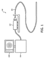
図1を参照すると、その中に本開示の一実施形態にかかるIVUSイメージングシステム100が示される。本開示の一部の実施形態において、IVUSイメージングシステム100は回転IVUSイメージングシステムである。その点について、回転IVUSイメージングシステムの主要構成要素は回転IVUSカテーテル102、患者インターフェースモジュール(PIM)104、IVUSコンソール若しくは処理システム106、及びIVUSコンソール106によって生成されるIVUS画像を表示するモニタ108である。
Referring to FIG. 1, an
図2を参照すると、その中に本開示の一実施形態にかかる回転IVUSカテーテル102の略部分断面斜視図が示される。その点について、図2は回転IVUSカテーテル102の構成に関する追加の詳細を示す。多くの点で、このカテーテルは、米国特許番号8,104,479に記載の、Volcano Corporationから入手可能なRevolution(登録商標)カテーテル、又はその各々が引用により全体として本明細書に組み込まれる米国特許番号5,243,988及び5,546,948に開示のものなど、従来の回転IVUSカテーテルと同様である。その点について、回転IVUSカテーテル102はイメージングコア110と外側カテーテル/シースアセンブリ112を含む。イメージングコア110は、図1のPIM104に電気的及び機械的結合を提供する回転インターフェース114によって近位端において終了する柔軟なドライブシャフトを含む。イメージングコア110の柔軟なドライブシャフトの遠位端は超音波トランスデューサと、一部の場合においては関連回路を含むハウジング116に結合される。他の実施形態において、IVUSカテーテルは光学コヒーレンストモグラフィ(OCT)カテーテルであり、かかる実施形態においてハウジング116は光ファイバと光学イメージング要素(例えば鏡、プリズム、スキャナなど)など、OCTカテーテルの対応する部分を含み得ることが理解される。
Referring to FIG. 2, shown therein is a schematic partial cross-sectional perspective view of a rotating
カテーテル/シースアセンブリ112は、回転インターフェースを支持し、カテーテルアセンブリの回転要素と非回転要素の間にベアリング面と流体シールを提供するハブ118を含む。ハブ118はルアーロックフラッシュポート120を含み、それを通して生理食塩水が注入され、カテーテルの使用時に空気を洗い流し、超音波適合性流体でシースの内腔を満たす。空気は超音波を容易に伝導しないので、生理食塩水若しくは他の同様のフラッシュが典型的に必要とされる。生理食塩水は回転ドライブシャフトに生体適合性の潤滑剤も提供する。ハブ118はテレスコープ122に結合され、これはカテーテル102の遠位部の音響的に透過性の窓124内のトランスデューサハウジングの軸方向運動を促進するよう、カテーテル/シースアセンブリ112を長くする若しくは短くすることを可能にする入れ子管状要素とスライド流体シールを含む。一部の実施形態において、窓124は最小減衰、反射若しくは屈折を伴ってトランスデューサと血管組織の間で超音波を容易に伝導する材料から製造される薄壁プラスチック管から成る。カテーテル/シースアセンブリ112の近位シャフト126はテレスコープ122と窓124の間のセグメントをブリッジし、滑らかな内腔と最適剛性を提供する材料若しくは複合材料から成るが、超音波を伝導する必要はない。
The catheter /
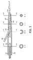
回転IVUSカテーテル102はガイドワイヤを受けるように構成される遠位部130をも含む。特に、図3に図示の通り、遠位部130は、図示の通りガイドワイヤ200がポート132を通って、内腔136に沿って、ポート134から外へのびることができるように、カテーテルの側壁における開口若しくはポート132、カテーテル102の遠位端における開口若しくはポート134、及びポート132と134の間にのびる内腔136を含むラピッドエクスチェンジ構成を含む。その点について、ガイドワイヤ200は患者内の所望の場所へカテーテル102の前進をガイドするために利用され得る。図3の図示の実施形態において、内腔136はドライブシャフト110及び/又はカテーテル102の近位部の中心軸と実質的に同軸にのびる。しかしながら、以下図7及び8の文脈で記載される通り、一部の場合において遠位部130の内腔136はドライブシャフト110及び/又はカテーテル102の近位部の中心軸に対してオフセットされる。
The rotating
図3はカテーテル102の長さに沿って様々な位置におけるカテーテルボディの断面プロファイルも図示する。図示の通り、切断線A‐A、C‐C、及びD‐Dにおいて、カテーテルボディは完全な円形断面プロファイルを持ち、一方ラピッドエクスチェンジポート132が位置する切断線B‐Bにおいて、カテーテルボディは概してU字形断面プロファイルを持つ。結果として、カテーテル102は切断線B‐Bにおいて、特にガイドワイヤの面内で曲がりやすくなり得る。例えば、図4はかかる脱出位置におけるカテーテル102の遠位部130の断面側面図を示す。これは懸念される故障モードであり、ガイドワイヤ200の前進若しくはカテーテル102の後退は交換ジョイント138においてガイドワイヤ200を曲げ、及び/又はカテーテル102を曲げさせ得る。理想的には、ガイドワイヤ200はラピッドエクスチェンジ内腔136を通って単純にスライドし、ガイドワイヤ200とカテーテル102は実質的に互いに同軸及び/又は平行なままであり得る。しかしながら、一旦この故障モードが起こり始めると、カテーテル102は交換ジョイント138において塑性的に変形し、持続的な屈曲をとる可能性があり、内腔136を圧迫してカテーテルに対するガイドワイヤの動きにさらなる抵抗を生じる可能性があるので、この故障モードは持続する可能性がある。加えて、ガイドワイヤ200が変形する可能性があり、ここでこれは脱出し、ガイドワイヤ200が前進するにつれて若しくはカテーテル102が後退するにつれて、ガイドワイヤ200を同じ場所において壊れやすくする。
FIG. 3 also illustrates the cross-sectional profile of the catheter body at various locations along the length of the
本開示の実施形態は、ラピッドエクスチェンジポート132の場所においてカテーテルボディを補強して、デバイスを脱出しやすくし得る、カテーテルボディの断面プロファイルにおける変化に起因してその場所において起こり得る曲げ剛性における突然の移行を回避することによって、これらの問題に対処する。その点について、典型的にはカテーテル102の側面のラピッドエクスチェンジポート132によって形成されるノッチ若しくは開口が脆弱スポットと曲げ剛性の急な不連続性を作り出し、それがその場所においてカテーテルを脱出しやすくする。カテーテル脱出は、これがガイドワイヤ200をつかむ若しくはつまむといった形で、ガイドワイヤ内腔136を壊す可能性もあり、問題をさらに深刻にする。結果として、本開示の実施形態はガイドワイヤ結束とカテーテル脱出を軽減しながら同時に患者の安全を改良することによって、カテーテル102のラピッドエクスチェンジ機能を改良する。
Embodiments of the present disclosure can reinforce the catheter body at the location of the
ラピッドエクスチェンジエントリポート132を定めるカテーテルボディの側面のノッチ若しくは開口の存在によって作られるカテーテル102における脆弱セグメントを補強するために、一つ以上の補強要素が利用され得る。補強要素は交換ジョイント138において脆弱セグメントをブリッジするようにカテーテルのボディの内部に位置する金属若しくはプラスチック構造であり得る。その点について、補強要素はラピッドエクスチェンジジョイント138にわたって所望の補強を実現するために材料、サイズ、形状、配向において多くのバリエーションをとり得る。例えば、適切な材料は金属及び金属合金(ステンレス鋼、ニチノールなど)、並びに任意の比較的剛性のプラスチック(PEEK、Nylon(登録商標)、高デュロメータPebax(登録商標)、ポリイミドなど)を含む。さらに、補強要素は任意の数の構造形状(ワイヤ、ロッド、テーパロッド、チューブ、U字形トラフ、スプリングコイルなど)をとることができ、対称及び非対称配向の両方でカテーテルボディ内に配置され得る。一部の実施例において、補強要素は、近位カテーテル管と遠位カテーテル先端の両方への結合を促進する特徴を組み込みながら、ラピッドエクスチェンジポート132付近で最適機械特性を維持するように設計されるカスタムモデル部品であり得る。一般に、補強された交換ジョイント138はラピッドエクスチェンジエントリポート132の直近位のカテーテル102の特性をポート132の直遠位の特性にマッチさせる(又は近位から遠位へ円滑に移行する)ように曲げ剛性を維持するべきである。従って、補強要素は、曲げ剛性の不連続性をカテーテル102に沿って他の場所に導入すること、及び/又は冠動脈内の急カーブ周辺でカテーテルをナビゲートすることを困難にすることを回避するために、過剰に剛性であるべきではない。
One or more reinforcing elements may be utilized to reinforce the fragile segment in the
図5及び6を参照すると、その中に本開示にかかる補強ラピッドエクスチェンジ構成を含むカテーテルの実施形態例が示される。例えば、図5はラピッドエクスチェンジエントリポート132に隣接してカテーテルボディ内に位置する補強要素のペア142を持つイメージングカテーテルの遠位部140の一実施形態を例示する。特に、図示の通り補強要素142はラピッドエクスチェンジエントリポート132の直近位位置からラピッドエクスチェンジエントリポート132の遠位位置へカテーテルボディの長さに沿って長手方向にのびる。さらに、切断線B‐Bに沿った断面図に図示の通り、補強要素142はラピッドエクスチェンジエントリポート132の隣でカテーテルボディの両側に互いに対向して位置する。このように、補強要素142は上述の通りラピッドエクスチェンジエントリポート132にわたってカテーテルボディに補強と支持を提供する。
Referring to FIGS. 5 and 6, an example embodiment of a catheter is shown that includes therein a reinforced rapid exchange configuration according to the present disclosure. For example, FIG. 5 illustrates one embodiment of the
図6はラピッドエクスチェンジエントリポート132に隣接してカテーテルボディ内に位置する補強要素152を持つイメージングカテーテルの遠位部150の別の実施形態を例示する。特に、図示の通り補強要素152はラピッドエクスチェンジエントリポート132の直近位位置からラピッドエクスチェンジエントリポート132の遠位位置へカテーテルボディの長さに沿って長手方向にのびる。さらに、切断線B‐Bに沿った断面図に図示の通り補強要素152はラピッドエクスチェンジエントリポート132の直下にカテーテルボディ内で位置する。このように、補強要素152は上述の通りラピッドエクスチェンジエントリポート132にわたってカテーテルボディに補強と支持を提供する。
FIG. 6 illustrates another embodiment of the
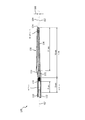
図7及び8を参照すると、その中に本開示にかかるオフセットラピッドエクスチェンジ構成を含むカテーテルの実施形態例が示される。その点について、図7及び8の各々は遠位部130がカテーテルの近位部に対してオフセットされる光学コヒーレンストモグラフィ(OCT)カテーテルを例示する。例えば、図7は遠位部130の内腔136がドライブシャフト110及び/又はカテーテル160の近位部の中心軸162に対してオフセットされるように遠位部130がオフセットされるOCTカテーテル160を示す。図示の通り、遠位部130の中心軸164及び/又は内腔136はカテーテル160の近位部の中心軸162に対して距離166だけオフセットされる。一部の場合において、距離166は約0.25mm(0.010")と約0.75mm(0.030")の間である。その点について、一部の実施形態において遠位部130はおよそ0.5mmの外径とおよそ0.35mmの内径を持ち、一方カテーテル160のメインボディはおよそ1.0mmの外径を持つ。しかしながら、本開示の概念は他のサイズを持つカテーテルに等しく適用可能であることが理解される。遠位部130とカテーテル160のメインボディの間のオフセットは、遠位端部130の中心軸164がメインカテーテルボディの中心軸162と同軸にのびる従来の設計と比較して、ガイドワイヤ200が、ラピッドエクスチェンジエントリポート132からあらわれるカテーテルシャフトと平行により近いままである(すなわち曲げが少ない)ことを可能にする。従って、本開示のかかる実施形態は患者の安全を同時に改善しながらガイドワイヤ結束とカテーテル脱出を軽減することによってカテーテル102のラピッドエクスチェンジ機能も改良する。
Referring to FIGS. 7 and 8, there is shown an example embodiment of a catheter including therein an offset rapid exchange configuration according to the present disclosure. In that regard, each of FIGS. 7 and 8 illustrates an optical coherence tomography (OCT) catheter in which the
しかしながら、図7の実施形態においても、ラピッドエクスチェンジエントリポート132の場所で一つの軸において削減された曲げモーメントが残る。その点について、遠位端の管状構造は遠位端へ向かって細くなるのにつれて漸減する剛性を提供し、一方近位シャフトは、特にシャフトを崩壊とねじれから防止するために柔軟なドライブシャフトが遠位場所にあるとき、その長さに沿って一定の曲げ剛性を維持する。しかしながら、ラピッドエクスチェンジポート132の場所において、近位シャフトの円筒形若しくは管状断面はU字形へと移行し、ここでカテーテルの側面においてノッチ若しくは開口が形成され、そして先端の長さに沿って管状断面に戻る。結果として、本開示の一部の実施形態はさらに改良されたラピッドエクスチェンジ構造を提供するためにオフセットラピッドエクスチェンジ配置を上記補強要素と組み合わせる。図8がかかる実施形態を例示する。図8に図示の通り、遠位部130の内腔136がドライブシャフト110及び/又はカテーテル170の近位部の中心軸162に対してオフセットされるようにメインカテーテルボディに対してオフセットされる遠位部130を含むOCTイメージングカテーテルが提供され、少なくとも一つの補強要素172がラピッドエクスチェンジポート132に隣接してカテーテルボディ内にのびる。特に、一部の場合において補強要素172は、カテーテル170の長さに沿って概して一定の曲げ剛性若しくは曲げ剛性において概して一定の変化を提供するよう、ラピッドエクスチェンジポート132の近位からラピッドエクスチェンジポート132の遠位へのびる。
However, the embodiment of FIG. 7 also leaves a reduced bending moment in one axis at the location of the rapid
当業者は、上記装置、システム、及び方法が様々な方法で修正され得ることも認識するだろう。従って、当業者は本開示によって包含される実施形態が上記特定の実施形態例に限定されないことを認識するだろう。その点について、実施形態例が図示され記載されているが、広範な修正、変更及び置換が上記開示において考慮される。上記へのかかる変更は本開示の範囲から逸脱することなくなされ得ることが理解される。従って、添付の請求項は本開示と一致する方法で広く解釈されることが認識される。 Those skilled in the art will also recognize that the above devices, systems, and methods can be modified in various ways. Accordingly, those skilled in the art will recognize that the embodiments encompassed by the present disclosure are not limited to the specific example embodiments described above. In that regard, although example embodiments are shown and described, a wide range of modifications, changes and substitutions are contemplated in the above disclosure. It will be understood that such changes to the above may be made without departing from the scope of the present disclosure. Accordingly, it will be appreciated that the appended claims are construed broadly in a manner consistent with the present disclosure.
Claims (15)
メインカテーテルボディと、
前記メインカテーテルボディの内腔内に位置する回転イメージング要素と、
前記メインカテーテルボディからのびる遠位部であって、ガイドワイヤ内腔と連通するラピッドエクスチェンジポートを持ち、当該ラピッドエクスチェンジポートとガイドワイヤ内腔はガイドワイヤを受けるようなサイズと形状である、遠位部と、
前記ラピッドエクスチェンジポートに隣接して前記メインカテーテルボディの側壁内に位置する少なくとも一つのU字形トラフ補強要素と
を有する、血管内イメージングデバイス。 An intravascular imaging device,
A main catheter body;
A rotating imaging element located within the lumen of the main catheter body;
A distal portion extending from the main catheter body, having a rapid exchange port communicating with the guidewire lumen, the rapid exchange port and the guidewire lumen being sized and shaped to receive the guidewire And
An intravascular imaging device having at least one U-shaped trough reinforcement element located in a sidewall of the main catheter body adjacent to the rapid exchange port.
血管内イメージングデバイスであって、
メインカテーテルボディと、
前記メインカテーテルボディの内腔内に位置する回転イメージング要素と、
前記メインカテーテルボディからのびる遠位部であって、ガイドワイヤ内腔と連通するラピッドエクスチェンジポートを持ち、当該ラピッドエクスチェンジポートとガイドワイヤ内腔がガイドワイヤを受けるようなサイズと形状である、遠位部と、
前記ラピッドエクスチェンジポートに隣接して前記メインカテーテルボディの側壁内に位置する少なくとも一つのU字形トラフ補強要素と
を含む血管内イメージングデバイスと、
前記血管内イメージングデバイスと通信する処理システムであって、前記血管内イメージングデバイスによって取得されるデータを処理するように構成される処理システムと
を有する、血管内イメージングシステム。 An intravascular imaging system,
An intravascular imaging device,
A main catheter body;
A rotating imaging element located within the lumen of the main catheter body;
A distal portion extending from the main catheter body, having a rapid exchange port communicating with the guidewire lumen, and having a size and shape such that the rapid exchange port and the guidewire lumen receive the guidewire And
An intravascular imaging device comprising: at least one U-shaped trough reinforcement element located in a side wall of the main catheter body adjacent to the rapid exchange port;
An intravascular imaging system comprising: a processing system in communication with the intravascular imaging device, the processing system configured to process data acquired by the intravascular imaging device.
Applications Claiming Priority (3)
| Application Number | Priority Date | Filing Date | Title |
|---|---|---|---|
| US201462042998P | 2014-08-28 | 2014-08-28 | |
| US62/042,998 | 2014-08-28 | ||
| PCT/IB2015/056382 WO2016030803A2 (en) | 2014-08-28 | 2015-08-24 | Intravascular devices having reinforced rapid-exchange ports and associated systems and methods |
Publications (3)
| Publication Number | Publication Date |
|---|---|
| JP2017529138A JP2017529138A (en) | 2017-10-05 |
| JP2017529138A5 JP2017529138A5 (en) | 2018-10-04 |
| JP6606171B2 true JP6606171B2 (en) | 2019-11-13 |
Family
ID=54238477
Family Applications (1)
| Application Number | Title | Priority Date | Filing Date |
|---|---|---|---|
| JP2017508629A Active JP6606171B2 (en) | 2014-08-28 | 2015-08-24 | Intravascular device with reinforced fast exchange port and associated system |
Country Status (5)
| Country | Link |
|---|---|
| US (2) | US11020089B2 (en) |
| EP (1) | EP3185780B1 (en) |
| JP (1) | JP6606171B2 (en) |
| CN (1) | CN106793997B (en) |
| WO (1) | WO2016030812A2 (en) |
Families Citing this family (16)
| Publication number | Priority date | Publication date | Assignee | Title |
|---|---|---|---|---|
| US8465686B2 (en) * | 2008-12-19 | 2013-06-18 | Volcano Corporation | Method of manufacturing a rotational intravascular ultrasound probe |
| ES2948135T3 (en) | 2015-06-24 | 2023-08-31 | Univ Michigan Regents | Histotripsy therapy systems for the treatment of brain tissue |
| JP6526925B2 (en) * | 2016-03-30 | 2019-06-05 | コーニンクレッカ フィリップス エヌ ヴェKoninklijke Philips N.V. | Imaging assembly for intravascular imaging device and related devices, systems and methods |
| WO2018065425A1 (en) * | 2016-10-03 | 2018-04-12 | Koninklijke Philips N.V. | Intra-cardiac echocardiography interposer |
| US11583252B2 (en) * | 2017-08-11 | 2023-02-21 | Arizona Board Of Regents On Behalf Of Arizona State University | Miniature transducer device and related methods |
| WO2019133798A1 (en) * | 2017-12-29 | 2019-07-04 | Acist Medical Systems, Inc. | Catheter extension |
| CN108670307B (en) * | 2018-06-17 | 2024-05-17 | 深圳北芯生命科技股份有限公司 | Intravascular ultrasound catheter, system and method of assembling same |
| JP7193299B2 (en) * | 2018-10-15 | 2022-12-20 | テルモ株式会社 | diagnostic imaging catheter |
| US11813484B2 (en) | 2018-11-28 | 2023-11-14 | Histosonics, Inc. | Histotripsy systems and methods |
| WO2020203423A1 (en) * | 2019-03-29 | 2020-10-08 | テルモ株式会社 | Image diagnosis catheter |
| WO2020229358A1 (en) * | 2019-05-13 | 2020-11-19 | Koninklijke Philips N.V. | Ultrasound probe handle |
| CN110743772B (en) * | 2019-09-27 | 2021-08-03 | 苏州佳世达电通有限公司 | Ultrasonic probe and method for manufacturing ultrasonic probe |
| EP4096782A4 (en) | 2020-01-28 | 2024-02-14 | The Regents Of The University Of Michigan | Systems and methods for histotripsy immunosensitization |
| CN112826536A (en) * | 2021-02-09 | 2021-05-25 | 深圳市赛禾医疗技术有限公司 | Intravascular ultrasonic imaging catheter and system |
| CN113397602B (en) * | 2021-05-21 | 2022-10-14 | 深圳市赛禾医疗技术有限公司 | Intracardiac three-dimensional ultrasonic imaging catheter and system and cardiac three-dimensional model construction method |
| US20240075252A1 (en) * | 2022-09-01 | 2024-03-07 | Becton, Dickinson And Company | Instrument Delivery Device with Linearly Compressible Housing |
Family Cites Families (31)
| Publication number | Priority date | Publication date | Assignee | Title |
|---|---|---|---|---|
| JPS6022961A (en) | 1983-07-15 | 1985-02-05 | Hitachi Chem Co Ltd | Method and device for coating and curing |
| US5243988A (en) | 1991-03-13 | 1993-09-14 | Scimed Life Systems, Inc. | Intravascular imaging apparatus and methods for use and manufacture |
| EP0626823B1 (en) | 1992-02-21 | 2000-04-19 | Boston Scientific Limited | Ultrasound imaging guidewire |
| US5313950A (en) * | 1992-02-25 | 1994-05-24 | Fujitsu Limited | Ultrasonic probe |
| US5263932A (en) | 1992-04-09 | 1993-11-23 | Jang G David | Bailout catheter for fixed wire angioplasty |
| US5314408A (en) | 1992-11-13 | 1994-05-24 | Cardiovascular Imaging Systems, Inc. | Expandable member for a catheter system |
| US5443457A (en) | 1994-02-24 | 1995-08-22 | Cardiovascular Imaging Systems, Incorporated | Tracking tip for a short lumen rapid exchange catheter |
| AU2001289196B2 (en) | 2000-12-01 | 2004-09-30 | The Cleveland Clinic Foundation | Miniature ultrasound transducer |
| JP4323191B2 (en) | 2003-03-12 | 2009-09-02 | テルモ株式会社 | catheter |
| JP4332706B2 (en) | 2003-05-06 | 2009-09-16 | 株式会社日立メディコ | Ultrasonic probe |
| WO2005072391A2 (en) | 2004-01-29 | 2005-08-11 | Ekos Corporation | Small vessel ultrasound catheter |
| US8104479B2 (en) * | 2005-06-23 | 2012-01-31 | Volcano Corporation | Pleated bag for interventional pullback systems |
| JP2008079909A (en) * | 2006-09-28 | 2008-04-10 | Fujifilm Corp | Ultrasonic probe and ultrasonic imaging apparatus |
| WO2009009799A1 (en) * | 2007-07-12 | 2009-01-15 | Volcano Corporation | Catheter for in vivo imaging |
| US20100262014A1 (en) * | 2007-12-03 | 2010-10-14 | Yongli Huang | Ultrasound Scanner Built with Capacitive Micromachined Ultrasonic Transducers (CMUTS) |
| US8167809B2 (en) * | 2007-12-20 | 2012-05-01 | Silicon Valley Medical Instruments, Inc. | Imaging probe housing with fluid flushing |
| US8465686B2 (en) * | 2008-12-19 | 2013-06-18 | Volcano Corporation | Method of manufacturing a rotational intravascular ultrasound probe |
| JP2012050706A (en) | 2010-09-01 | 2012-03-15 | Terumo Corp | Protective cover |
| EP2629827B1 (en) * | 2010-10-20 | 2015-12-30 | Stryker Corporation | Stent delivery catheter with rapid exchange capabilities |
| US9237880B2 (en) * | 2011-03-17 | 2016-01-19 | Koninklijke Philips N.V. | Composite acoustic backing with high thermal conductivity for ultrasound transducer array |
| JP5919862B2 (en) * | 2012-02-13 | 2016-05-18 | ニプロ株式会社 | Balloon catheter |
| JP2015515917A (en) | 2012-05-11 | 2015-06-04 | ヴォルカノ コーポレイションVolcano Corporation | Circuit architecture and electrical interface for a rotating intravascular ultrasound (IVUS) device |
| CA2873399A1 (en) * | 2012-05-11 | 2013-11-14 | Volcano Corporation | Ultrasound catheter for imaging and blood flow measurement |
| US9492140B2 (en) * | 2012-06-12 | 2016-11-15 | Volcano Corporation | Devices, systems, and methods for forward looking imaging |
| WO2014077871A2 (en) | 2012-11-19 | 2014-05-22 | Lightlab Imaging, Inc. | Interface devices, systems and methods for multimodal probes |
| EP2934311B1 (en) * | 2012-12-20 | 2020-04-15 | Volcano Corporation | Smooth transition catheters |
| CA2895821A1 (en) * | 2012-12-21 | 2014-06-26 | Volcano Corporation | Focused rotational ivus transducer using single crystal composite material |
| US9307952B2 (en) * | 2012-12-21 | 2016-04-12 | Volcano Corporation | Method for focusing miniature ultrasound transducers |
| US10398413B2 (en) * | 2012-12-21 | 2019-09-03 | Volcano Corporation | Method for multi-frequency imaging and composite image display using high-bandwidth transducer outputs |
| US20140187959A1 (en) * | 2012-12-31 | 2014-07-03 | Volcano Corporation | Ultrasound Transducers and Methods of Manufacturing Same |
| EP3116408B1 (en) * | 2014-03-12 | 2018-12-19 | Cibiem, Inc. | Ultrasound ablation catheter |
-
2015
- 2015-08-24 JP JP2017508629A patent/JP6606171B2/en active Active
- 2015-08-25 WO PCT/IB2015/056419 patent/WO2016030812A2/en active Application Filing
- 2015-08-25 EP EP15771266.2A patent/EP3185780B1/en active Active
- 2015-08-25 CN CN201580046816.0A patent/CN106793997B/en not_active Expired - Fee Related
- 2015-08-27 US US14/837,829 patent/US11020089B2/en active Active
-
2021
- 2021-05-26 US US17/331,404 patent/US20210282746A1/en not_active Abandoned
Also Published As
| Publication number | Publication date |
|---|---|
| EP3185780A2 (en) | 2017-07-05 |
| WO2016030812A3 (en) | 2016-04-28 |
| US20160058414A1 (en) | 2016-03-03 |
| JP2017529138A (en) | 2017-10-05 |
| EP3185780B1 (en) | 2022-10-19 |
| CN106793997A (en) | 2017-05-31 |
| WO2016030812A2 (en) | 2016-03-03 |
| US11020089B2 (en) | 2021-06-01 |
| US20210282746A1 (en) | 2021-09-16 |
| CN106793997B (en) | 2021-06-04 |
Similar Documents
| Publication | Publication Date | Title |
|---|---|---|
| JP6606171B2 (en) | Intravascular device with reinforced fast exchange port and associated system | |
| US20220160329A1 (en) | Intravascular devices having reinforced rapid-exchange ports and associated systems and methods | |
| JP2020039921A (en) | Rotational catheter with extended catheter body drive shaft support | |
| US10709312B2 (en) | Transitional region having cuts and a skive for an imaging catheter | |
| EP2931130B1 (en) | Rotational sensing catheter with self-supporting drive shaft section | |
| JP5581139B2 (en) | catheter | |
| US20140163421A1 (en) | Reinforced Catheter Transition With Flexible Tip Portion | |
| US10383521B2 (en) | Non-cylindrical hypotubes | |
| WO2004093655A2 (en) | Helical guidewire | |
| CN114053552A (en) | Optimized catheter sheath for Rx catheter | |
| JP2012217588A (en) | Medical device | |
| JP2012223207A (en) | Balloon catheter | |
| JP5743283B2 (en) | Guide wire | |
| US20240050768A1 (en) | Shape-Sensing Systems with Vibration-Assisted Torsion Management and Methods Thereof | |
| JP6400586B2 (en) | Guide wire |
Legal Events
| Date | Code | Title | Description |
|---|---|---|---|
| A521 | Request for written amendment filed |
Free format text: JAPANESE INTERMEDIATE CODE: A523 Effective date: 20180823 |
|
| A621 | Written request for application examination |
Free format text: JAPANESE INTERMEDIATE CODE: A621 Effective date: 20180823 |
|
| A977 | Report on retrieval |
Free format text: JAPANESE INTERMEDIATE CODE: A971007 Effective date: 20190426 |
|
| A131 | Notification of reasons for refusal |
Free format text: JAPANESE INTERMEDIATE CODE: A131 Effective date: 20190509 |
|
| A521 | Request for written amendment filed |
Free format text: JAPANESE INTERMEDIATE CODE: A523 Effective date: 20190805 |
|
| TRDD | Decision of grant or rejection written | ||
| A01 | Written decision to grant a patent or to grant a registration (utility model) |
Free format text: JAPANESE INTERMEDIATE CODE: A01 Effective date: 20191008 |
|
| A61 | First payment of annual fees (during grant procedure) |
Free format text: JAPANESE INTERMEDIATE CODE: A61 Effective date: 20191017 |
|
| R150 | Certificate of patent or registration of utility model |
Ref document number: 6606171 Country of ref document: JP Free format text: JAPANESE INTERMEDIATE CODE: R150 |
|
| S531 | Written request for registration of change of domicile |
Free format text: JAPANESE INTERMEDIATE CODE: R313531 |
|
| S533 | Written request for registration of change of name |
Free format text: JAPANESE INTERMEDIATE CODE: R313533 |
|
| R350 | Written notification of registration of transfer |
Free format text: JAPANESE INTERMEDIATE CODE: R350 |
|
| R250 | Receipt of annual fees |
Free format text: JAPANESE INTERMEDIATE CODE: R250 |
|
| R250 | Receipt of annual fees |
Free format text: JAPANESE INTERMEDIATE CODE: R250 |