JP6588978B2 - Apparatus, system and method for automatic detection of human orientation and / or position - Google Patents
Apparatus, system and method for automatic detection of human orientation and / or position Download PDFInfo
- Publication number
- JP6588978B2 JP6588978B2 JP2017523482A JP2017523482A JP6588978B2 JP 6588978 B2 JP6588978 B2 JP 6588978B2 JP 2017523482 A JP2017523482 A JP 2017523482A JP 2017523482 A JP2017523482 A JP 2017523482A JP 6588978 B2 JP6588978 B2 JP 6588978B2
- Authority
- JP
- Japan
- Prior art keywords
- motion
- image
- person
- boundary
- image data
- Prior art date
- Legal status (The legal status is an assumption and is not a legal conclusion. Google has not performed a legal analysis and makes no representation as to the accuracy of the status listed.)
- Active
Links
Images


Classifications
-
- A—HUMAN NECESSITIES
- A61—MEDICAL OR VETERINARY SCIENCE; HYGIENE
- A61B—DIAGNOSIS; SURGERY; IDENTIFICATION
- A61B5/00—Measuring for diagnostic purposes; Identification of persons
- A61B5/0002—Remote monitoring of patients using telemetry, e.g. transmission of vital signals via a communication network
- A61B5/0015—Remote monitoring of patients using telemetry, e.g. transmission of vital signals via a communication network characterised by features of the telemetry system
- A61B5/002—Monitoring the patient using a local or closed circuit, e.g. in a room or building
-
- A—HUMAN NECESSITIES
- A61—MEDICAL OR VETERINARY SCIENCE; HYGIENE
- A61B—DIAGNOSIS; SURGERY; IDENTIFICATION
- A61B5/00—Measuring for diagnostic purposes; Identification of persons
- A61B5/0059—Measuring for diagnostic purposes; Identification of persons using light, e.g. diagnosis by transillumination, diascopy, fluorescence
- A61B5/0077—Devices for viewing the surface of the body, e.g. camera, magnifying lens
-
- A—HUMAN NECESSITIES
- A61—MEDICAL OR VETERINARY SCIENCE; HYGIENE
- A61B—DIAGNOSIS; SURGERY; IDENTIFICATION
- A61B5/00—Measuring for diagnostic purposes; Identification of persons
- A61B5/103—Detecting, measuring or recording devices for testing the shape, pattern, colour, size or movement of the body or parts thereof, for diagnostic purposes
- A61B5/11—Measuring movement of the entire body or parts thereof, e.g. head or hand tremor, mobility of a limb
- A61B5/1113—Local tracking of patients, e.g. in a hospital or private home
-
- A—HUMAN NECESSITIES
- A61—MEDICAL OR VETERINARY SCIENCE; HYGIENE
- A61B—DIAGNOSIS; SURGERY; IDENTIFICATION
- A61B5/00—Measuring for diagnostic purposes; Identification of persons
- A61B5/103—Detecting, measuring or recording devices for testing the shape, pattern, colour, size or movement of the body or parts thereof, for diagnostic purposes
- A61B5/11—Measuring movement of the entire body or parts thereof, e.g. head or hand tremor, mobility of a limb
- A61B5/1126—Measuring movement of the entire body or parts thereof, e.g. head or hand tremor, mobility of a limb using a particular sensing technique
- A61B5/1128—Measuring movement of the entire body or parts thereof, e.g. head or hand tremor, mobility of a limb using a particular sensing technique using image analysis
-
- G—PHYSICS
- G06—COMPUTING; CALCULATING OR COUNTING
- G06F—ELECTRIC DIGITAL DATA PROCESSING
- G06F18/00—Pattern recognition
- G06F18/20—Analysing
- G06F18/23—Clustering techniques
-
- G—PHYSICS
- G06—COMPUTING; CALCULATING OR COUNTING
- G06F—ELECTRIC DIGITAL DATA PROCESSING
- G06F18/00—Pattern recognition
- G06F18/20—Analysing
- G06F18/24—Classification techniques
- G06F18/243—Classification techniques relating to the number of classes
- G06F18/2431—Multiple classes
-
- G—PHYSICS
- G06—COMPUTING; CALCULATING OR COUNTING
- G06T—IMAGE DATA PROCESSING OR GENERATION, IN GENERAL
- G06T7/00—Image analysis
- G06T7/10—Segmentation; Edge detection
- G06T7/13—Edge detection
-
- G—PHYSICS
- G06—COMPUTING; CALCULATING OR COUNTING
- G06T—IMAGE DATA PROCESSING OR GENERATION, IN GENERAL
- G06T7/00—Image analysis
- G06T7/20—Analysis of motion
- G06T7/215—Motion-based segmentation
-
- G—PHYSICS
- G06—COMPUTING; CALCULATING OR COUNTING
- G06T—IMAGE DATA PROCESSING OR GENERATION, IN GENERAL
- G06T7/00—Image analysis
- G06T7/20—Analysis of motion
- G06T7/246—Analysis of motion using feature-based methods, e.g. the tracking of corners or segments
-
- G—PHYSICS
- G06—COMPUTING; CALCULATING OR COUNTING
- G06T—IMAGE DATA PROCESSING OR GENERATION, IN GENERAL
- G06T7/00—Image analysis
- G06T7/70—Determining position or orientation of objects or cameras
- G06T7/73—Determining position or orientation of objects or cameras using feature-based methods
-
- G—PHYSICS
- G06—COMPUTING; CALCULATING OR COUNTING
- G06V—IMAGE OR VIDEO RECOGNITION OR UNDERSTANDING
- G06V40/00—Recognition of biometric, human-related or animal-related patterns in image or video data
- G06V40/20—Movements or behaviour, e.g. gesture recognition
-
- A—HUMAN NECESSITIES
- A61—MEDICAL OR VETERINARY SCIENCE; HYGIENE
- A61B—DIAGNOSIS; SURGERY; IDENTIFICATION
- A61B2576/00—Medical imaging apparatus involving image processing or analysis
-
- A—HUMAN NECESSITIES
- A61—MEDICAL OR VETERINARY SCIENCE; HYGIENE
- A61B—DIAGNOSIS; SURGERY; IDENTIFICATION
- A61B5/00—Measuring for diagnostic purposes; Identification of persons
- A61B5/0033—Features or image-related aspects of imaging apparatus classified in A61B5/00, e.g. for MRI, optical tomography or impedance tomography apparatus; arrangements of imaging apparatus in a room
- A61B5/0037—Performing a preliminary scan, e.g. a prescan for identifying a region of interest
-
- G—PHYSICS
- G06—COMPUTING; CALCULATING OR COUNTING
- G06T—IMAGE DATA PROCESSING OR GENERATION, IN GENERAL
- G06T2207/00—Indexing scheme for image analysis or image enhancement
- G06T2207/10—Image acquisition modality
- G06T2207/10016—Video; Image sequence
-
- G—PHYSICS
- G06—COMPUTING; CALCULATING OR COUNTING
- G06T—IMAGE DATA PROCESSING OR GENERATION, IN GENERAL
- G06T2207/00—Indexing scheme for image analysis or image enhancement
- G06T2207/30—Subject of image; Context of image processing
- G06T2207/30196—Human being; Person
-
- G—PHYSICS
- G16—INFORMATION AND COMMUNICATION TECHNOLOGY [ICT] SPECIALLY ADAPTED FOR SPECIFIC APPLICATION FIELDS
- G16H—HEALTHCARE INFORMATICS, i.e. INFORMATION AND COMMUNICATION TECHNOLOGY [ICT] SPECIALLY ADAPTED FOR THE HANDLING OR PROCESSING OF MEDICAL OR HEALTHCARE DATA
- G16H30/00—ICT specially adapted for the handling or processing of medical images
- G16H30/40—ICT specially adapted for the handling or processing of medical images for processing medical images, e.g. editing
Landscapes
- Health & Medical Sciences (AREA)
- Engineering & Computer Science (AREA)
- Life Sciences & Earth Sciences (AREA)
- Physics & Mathematics (AREA)
- Computer Vision & Pattern Recognition (AREA)
- Theoretical Computer Science (AREA)
- General Health & Medical Sciences (AREA)
- Heart & Thoracic Surgery (AREA)
- Biomedical Technology (AREA)
- Medical Informatics (AREA)
- Molecular Biology (AREA)
- Surgery (AREA)
- Animal Behavior & Ethology (AREA)
- Pathology (AREA)
- Public Health (AREA)
- Veterinary Medicine (AREA)
- Biophysics (AREA)
- General Physics & Mathematics (AREA)
- Oral & Maxillofacial Surgery (AREA)
- Physiology (AREA)
- Dentistry (AREA)
- Multimedia (AREA)
- Data Mining & Analysis (AREA)
- Nuclear Medicine, Radiotherapy & Molecular Imaging (AREA)
- Radiology & Medical Imaging (AREA)
- Computer Networks & Wireless Communication (AREA)
- Bioinformatics & Computational Biology (AREA)
- Artificial Intelligence (AREA)
- Bioinformatics & Cheminformatics (AREA)
- Evolutionary Biology (AREA)
- Evolutionary Computation (AREA)
- General Engineering & Computer Science (AREA)
- Human Computer Interaction (AREA)
- Social Psychology (AREA)
- Psychiatry (AREA)
- Measurement Of The Respiration, Hearing Ability, Form, And Blood Characteristics Of Living Organisms (AREA)
- Image Analysis (AREA)
- Measuring And Recording Apparatus For Diagnosis (AREA)
Description
本発明は、人の向き及び/又は位置の自動検出のための装置、システム、及び方法に関する。 The present invention relates to an apparatus, system, and method for automatic detection of human orientation and / or position.
患者監視の分野では、体動監視によって患者の臨床条件に関する情報を提供することができる。ベッドからの起床/転落、医療機器(例えば気管内挿入管又は栄養管)の引き抜き、譫妄、てんかん発作などの場合の空中つかみや脚の反復運動などの病気固有の動きなど、危険な状況を医療スタッフに伝達することができる。 In the field of patient monitoring, body movement monitoring can provide information about the patient's clinical conditions. Medical care for dangerous situations such as getting up / falling from bed, pulling out medical devices (eg, endotracheal or feeding tube), delirium, seizures in the case of epileptic seizures, and disease-specific movements such as repeated leg movements Can communicate to staff.
筋肉運動挙動の変化は、譫妄の中核特徴の1つである。全体的な活動水準の変化(例えば機能低下サブタイプに関する活動水準の低下)の他に、譫妄状態の患者は、空中つかみ、皮膚むしりやベッドシーツむしり、脚の落ち着かない動きなどの異常な動きも示す。体動分析及び体動分類は譫妄検出のために非常に重要なものになる可能性がある。 Changes in muscle movement behavior are one of the core features of delirium. In addition to changes in overall activity levels (eg, decreased activity levels related to reduced function subtypes), delirium patients may also experience abnormal movements such as grabbing in the air, skin and bed sheets, and leg movements Show. Body motion analysis and body motion classification can be very important for delirium detection.
以前の研究では、手首装着加速度計技法を使用して、譫妄による活動水準の変化を分析した。この身体装着型手首センサは、患者にとって邪魔になるか又は混乱させるものである。より重要なことに、これは他の身体部分によって行われた動きを捕捉せず、「皮膚つまみ」、「空中つかみ」などの動きについてより高水準の解釈の可能性も提供しない。連続的で自動式のビデオ監視は、これらの制限を超える機会を提供すると考えられている。 In previous studies, wrist-worn accelerometer techniques were used to analyze changes in activity levels due to delirium. This body-worn wrist sensor is either disturbing or confusing for the patient. More importantly, it does not capture movements made by other body parts, nor does it provide a higher level of interpretation possibilities for movements such as “skin knob”, “grasping air”. Continuous and automated video surveillance is believed to provide an opportunity to exceed these limits.
患者の動きを認識/分類するために、自然な動き及び異常な動きの特徴が画像/ビデオから抽出され、分類器に供給される。患者監視のための特徴抽出は一般に全身について包括的に実行される。更に、特定の動きはしばしば特定の身体部分によって行われるので、体動分類は身体部分情報から利益を得る(例えば左から右に連続的に頭を動かすことは異常なことであり、その一方で、摂食中の反復した手の動きは異常ではない)。従って、身体部分ごとに特徴を抽出できる場合、分類結果は大幅に改善される。 In order to recognize / classify patient motion, natural and abnormal motion features are extracted from the image / video and fed to a classifier. Feature extraction for patient monitoring is generally performed globally for the whole body. In addition, body movement classification benefits from body part information (eg moving head continuously from left to right is unusual, while certain movements are often performed by specific body parts, while , Repeated hand movements during feeding are not unusual). Therefore, if the features can be extracted for each body part, the classification result is greatly improved.
生命徴候を監視するという目的でビデオカメラによる患者監視を実行する場合、胸部領域(呼吸を監視するため)又は顔面領域(心拍数を監視するため)は重要な情報である。 When performing patient monitoring with a video camera for the purpose of monitoring vital signs, the chest area (to monitor respiration) or the facial area (to monitor heart rate) is important information.
従って、体動分析及び生命徴候監視のどちらについても、画像内の患者関心領域(ROI)及び主要身体部分の位置に関する情報は極めて重大である。これは、例えば病院内の患者だけでなく、高齢者介護施設又は自宅にいる高齢者のように一般に監視しなければならないすべての人、小児又は保育器内の新生児にも当てはまることである。 Thus, for both body movement analysis and vital signs monitoring, information regarding the region of interest (ROI) and the location of the main body part in the image is crucial. This applies not only to patients in hospitals, but also to all persons who have to be monitored generally, such as elderly people in elderly care facilities or homes, children or incubators of newborns.
多くの場合、病院内では、患者の筋肉運動挙動は大部分は、医療スタッフが患者を巡回する時に又は時にはチェックリストを使用することによって観察されるだけである。過去の巡回と現在の巡回との筋肉運動挙動の変化の検出は、しばしば、医療スタッフが気付くのは困難である。このタイプの検査では、筋肉運動挙動の変化によって明らかになる病気の発症などの重大問題や患者の体動によって誘発される重大状況の検出に無視できない遅れがもたらされる。 In many cases, in a hospital, the patient's muscle movement behavior is mostly observed only when medical staff visit the patient or sometimes by using a checklist. Detection of changes in muscle movement behavior between past and current visits is often difficult for medical staff to notice. This type of test results in a non-negligible delay in the detection of serious problems such as the onset of illness manifested by changes in muscle movement behavior and critical situations induced by patient movement.
患者の筋肉運動挙動を監視するためにビデオカメラ以外のセンサが文献では提案されているが、これらはしばしば特定の出来事(例えばベッドからの患者の転落)を検出することに特化されている。ビデオデータは情報が豊富であり、例えば患者の顔面、手を検出し、動きを分析し、物体とのやりとりを分析し、一般的な挙動を認識する可能性がある。従って、ビデオセンサは、患者によって行われる種々のタイプの動きを自動的に分析し認識する機会を提供する。 Sensors other than video cameras have been proposed in the literature to monitor a patient's muscle movement behavior, but these are often specialized to detect specific events (eg, patient fall from bed). Video data is rich in information, for example, detecting the patient's face and hands, analyzing movement, analyzing interactions with objects, and recognizing general behavior. Thus, the video sensor provides an opportunity to automatically analyze and recognize various types of movements made by the patient.
自動ビデオベースの患者監視は比較的新しい主題であり、開発されたツールはその初期段階にある。ビデオ分析方法は病院の動的側面に対処しなければならない。これらは、ベッドの角度及びベッドの背もたれの傾斜の変化、患者の一部分を遮る人又はTV画面のような物体、ベッド内で横たわっている患者の種々の姿勢、患者の身体部分を覆う毛布、医療職員及び訪問者の出入りなど、場面変動になり得るものである。これらの難題は、患者監視のために典型的な身体セグメント化方法及び身体部分の識別を含めることを困難にしている。毛布の存在は横たわっている患者に人間モデルを適合させることを困難にしており、場面変動は身体部分セグメント化のための現在のビデオ分析方法を制限している(エッジ/勾配分析、輝度値分析、及び物体検出など)。 Automated video-based patient monitoring is a relatively new subject and the developed tools are in their early stages. Video analysis methods must address the dynamic aspects of hospitals. These include changes in bed angle and bed back tilt, objects such as a person or TV screen obstructing a part of the patient, various postures of the patient lying in the bed, blankets covering the patient's body part, medical The situation may change, such as the entry and exit of staff and visitors. These challenges make it difficult to include typical body segmentation methods and body part identification for patient monitoring. The presence of the blanket makes it difficult to fit the human model to the lying patient, and scene variations limit current video analysis methods for body part segmentation (edge / gradient analysis, luminance value analysis). And object detection).
SHYAMSUNDER R他による「11 Compression of Patient Monitoring Video Using Motion Segmentation Technique」(JOURNAL OF MEDICAL SYSTEMS、KLUWER ACADEMIC PUBLISHERS−PLENUM PUBLISHERS、NE.、第31巻、第2号、2007年3月21日)は、バイナリマスクを使用してビデオ内の静止部分と可動部分の分離のための動作セグメント化技法を開示している。 SHAMAMUNDER R et al., “11 Compression of PATENT MONITORING VIDEO USING MOTION SEGMENTATION NUMBER VOL. 21 JUNRNAL OF MEDICAL SYSTEM PUL. Disclosed is a motion segmentation technique for separation of stationary and moving parts in a video using a mask.
NZ534482Aは、患者内の動揺のレベルを客観的に決定するための方法及びシステムを開示している。この方法は、患者の身体の定義済み関心領域の物理的動きの自動監視及び/又は動揺による他の臨床事象を描出する監視エキスパートシステム(例えば動揺による心拍数の大きいスパイクによる心房細動)を伴う。監視に関連する生理学的信号について信号処理が実行され、処理された信号内の変化により患者の動揺のレベルを定量化することができる。 NZ534482A discloses a method and system for objectively determining the level of sway in a patient. This method involves automatic monitoring of the physical movement of a defined region of interest of the patient's body and / or a monitoring expert system (eg, atrial fibrillation due to high heart rate spikes due to sway) that depicts other clinical events due to sway. . Signal processing is performed on the physiological signals associated with the monitoring, and changes in the processed signals can quantify the level of patient motion.
WO2012/164453A1は、共通の寝具類を占有している2人以上の被験者の動き及び呼吸を監視するための方法及び装置を開示している。この方法は、光センサにより寝具類を画像化するステップと、当該光センサから受け取った連続画像間又は複数フレーム分離れている複数画像間の対応する画像ブロックの局所変位を示す動作ベクトルを生成することにより動作推定を実行するステップと、動作ベクトルの空間的及び時間的相関関係を測定することにより動作クラスタを計算するステップと、対応する被験者に対するそれぞれの動作クラスタの割り当てにより計算した動作クラスタをセグメント化するステップであって、対応する被験者に対する動作クラスタの割り当てが相互間の動作クラスタの空間的及び/又は時間的類似性と以前のセグメント化結果とに基づくステップとを含む。 WO 2012/164453 A1 discloses a method and apparatus for monitoring movement and breathing of two or more subjects occupying common bedding. The method images a bedding with an optical sensor and generates motion vectors indicating local displacements of corresponding image blocks between consecutive images received from the optical sensor or between multiple images separated by multiple frames. Segmentation of motion clusters calculated by assigning each motion cluster to a corresponding subject, and performing motion estimation by measuring the spatial and temporal correlations of motion vectors. And assigning motion clusters to corresponding subjects includes steps based on spatial and / or temporal similarity of motion clusters between each other and previous segmentation results.
本発明の目的は、人の向き及び/又は位置の頑強な自動検出を可能にする装置、システム、及び方法を提供することである。 It is an object of the present invention to provide an apparatus, system, and method that enables robust automatic detection of a person's orientation and / or position.
本発明の第1の態様では、
− 画像データが経時的な一連の画像フレームを含む人の画像データを得るための画像データインターフェース(image data interface)と、
− 当該画像データ内の動作を検出するための動作検出器(motion detector)と、
− 頻繁に発生する動作を示す画像領域を表す動作ホットスポット(motion hotspot)を識別するための動作強度検出器(motion intensity detector)と、
− 識別された動作ホットスポットに基づいて当該人の少なくとも一部の向き及び/又は位置を検出するための人検出器(person detector)と、
を含む、人の向き及び/又は位置の自動検出のための装置が提示される。
In the first aspect of the present invention,
An image data interface for obtaining image data of a person whose image data includes a series of image frames over time;
A motion detector for detecting motion in the image data;
-A motion intensity detector for identifying motion hotspots representing image areas showing frequently occurring motions;
A person detector for detecting the orientation and / or position of at least a portion of the person based on the identified operational hot spot;
An apparatus for automatic detection of human orientation and / or position is presented.
本発明の追加の一態様では、人の向き及び/又は位置の自動検出のための対応する方法が提示される。 In an additional aspect of the present invention, a corresponding method for automatic detection of human orientation and / or position is presented.
本発明の更に追加の一態様では、
− 経時的な一連の画像フレームを含む人の画像データを取得するための画像化ユニット(imaging unit)と、
− 取得した画像データに基づいて当該人の少なくとも一部の向き及び/又は位置の自動検出のための本明細書に開示されている装置と、
− 当該人の少なくとも一部の検出された向き及び/又は位置に関連する情報を発行するための出力インターフェースと、
を含む、人の向き及び/又は位置の自動検出のためのシステムが提示される。
In a further aspect of the invention,
An imaging unit for obtaining human image data including a series of image frames over time;
An apparatus disclosed herein for automatic detection of the orientation and / or position of at least a portion of the person based on acquired image data;
An output interface for issuing information relating to the detected orientation and / or position of at least a part of the person;
A system for automatic detection of human orientation and / or position is presented.
本発明の更に追加の複数態様では、当該コンピュータプログラムがコンピュータ上で実行された時に本明細書に開示されている方法のステップをコンピュータに実行させるためのプログラムコード手段を含むコンピュータプログラム並びにプロセッサによって実行された時に本明細書に開示されている方法を実行させるコンピュータプログラム製品をそこに記憶する非一時的コンピュータ可読記録媒体が提供される。 In yet additional aspects of the present invention, a computer program comprising program code means for causing a computer to execute the steps of the methods disclosed herein when the computer program is executed on a computer, as well as executed by a processor. Provided is a non-transitory computer readable recording medium having stored thereon a computer program product that, when executed, causes the method disclosed herein to be performed.
本発明の好ましい諸実施形態は従属クレームに定義されている。請求されている方法、システム、コンピュータプログラム、及び媒体は、従属クレームに定義されているように請求されている装置と同様及び/又は同一の好ましい諸実施形態を有することを理解されたい。 Preferred embodiments of the invention are defined in the dependent claims. It is to be understood that the claimed methods, systems, computer programs, and media have preferred embodiments that are similar to and / or identical to the claimed device as defined in the dependent claims.
本発明は、当該画像データ内で、即ち、一連の画像フレーム内で動作ホットスポットを識別し、人の少なくとも一部の向き及び/又は位置を決定するためにこのような動作ホットスポットを使用するという概念に基づくものである。これにより、動作ホットスポットは、頻繁に発生する動作を示す画像領域、即ち、多くの動作が検出されている画像領域として理解しなければならない。例えば、人がベッドに横になっており、連続的に自分の頭を振る場合、頭部領域はこのような動作ホットスポットとして検出されるであろう。このような動作ホットスポットは一般に複数の画像フレーム(最高数千個の画像フレーム)を考慮に入れた結果として得られる。 The present invention uses such operational hot spots to identify operational hot spots within the image data, ie, within a series of image frames, and to determine the orientation and / or position of at least a portion of a person. It is based on the concept of Thus, the operation hot spot must be understood as an image region showing a frequently occurring operation, that is, an image region in which many operations are detected. For example, if a person is lying on the bed and continuously shakes his / her head, the head area will be detected as such an operating hot spot. Such operational hot spots are generally obtained as a result of taking into account a plurality of image frames (up to several thousand image frames).
このような動作ホットスポットの使用により、シナリオの種類、アプリケーション、及び監視の特定の事情次第で(例えば他の人が視野内におり、その結果、画像データ内に示される場合、人がどのくらい動くかなど)、大まかな身体輪郭などの人の関心領域(ROI)又は頭、腕、脚などの人の少なくとも1つ以上の特徴的な身体部分をかなり正確に決定することができる。 Depending on the type of scenario, application, and specific circumstances of the surveillance, such movement hotspots can be used (for example, how much a person moves when others are in the field of view and as a result shown in the image data) Or the like), a person's region of interest (ROI) such as a rough body contour or at least one or more characteristic body parts of the person such as the head, arms, legs, etc. can be determined fairly accurately.
本発明は、好ましくは、ベッド又は長椅子に横になっている、椅子に座っている、じっと立っているなど、人が安静姿勢にある場合に適用可能である。しかしながら、人が経時的に同じ画像領域内に存在する限り、本発明はその他の状況にも適用することができる。多かれ少なかれ同じ領域内に留まっている限り、人は動いたり、立ったり、座ったり、横になったりすることができる。その場合、この領域内の動作ホットスポットは患者の動きに対応し、ROIを計算することができる。 The present invention is preferably applicable when a person is in a resting position, such as lying on a bed or chaise longue, sitting on a chair or standing still. However, as long as a person is present in the same image area over time, the present invention can be applied to other situations. As long as you stay in more or less the same area, people can move, stand, sit and lie down. In that case, the operating hot spots in this region correspond to patient movement and the ROI can be calculated.
提案されているビデオベースの患者監視システムは比較的安価で目立たないものである。これは、患者の全身活動だけでなく、特定の身体部分による動きも連続的かつ自動的に監視することができる。高度なコンピュータビジョン技術により、特定の動作を識別することができる。 The proposed video-based patient monitoring system is relatively inexpensive and unobtrusive. It can continuously and automatically monitor not only the patient's whole body activity, but also movements by specific body parts. Advanced computer vision technology can identify specific actions.
好ましい諸実施形態では、以下に説明するように、人の上下左右の境界のうちの1つ以上を決定することができる。更に、頻繁に発生する動作を含む領域を識別する動作画像(motion image)の蓄積を動作ホットスポットの検出に使用することができる。この蓄積は動作検出又は動作推定に基づくことができる。更に、頭部位置の計算は、人のROIの外側の領域と人のROI自体との間の頻繁なやりとりの位置に基づいて得られる。提案されている通りに処理する前に画像データの潜在的な圧縮によって引き起こされる潜在的な圧縮アーチファクトを考慮しても、提案されている装置、システム、及び方法は頑強なものであり、看護師の動きと患者の動きとを区別することができる。 In preferred embodiments, one or more of the top, bottom, left, and right boundaries of a person can be determined, as described below. Furthermore, the accumulation of motion images that identify regions containing frequently occurring motions can be used to detect motion hot spots. This accumulation can be based on motion detection or motion estimation. Furthermore, the calculation of the head position is obtained based on the location of frequent exchanges between the region outside the person's ROI and the person's ROI itself. The proposed device, system, and method are robust, considering the potential compression artifacts caused by the potential compression of the image data before processing as proposed, and nurses And patient movements can be distinguished.
他の好ましい実施形態では、当該動作検出器は、1つの画像フレームから他の画像フレームまでの画像データ内で検出された動作を表す動作画像を決定するように構成され、当該動作強度検出器は、当該動作画像を蓄積することにより動作ホットスポットを識別するように構成される。動作画像は、これにより、例えば動作ベクトル場画像又は検出された動作ピクセルを含む画像になり、これは、例えばある場面の画像フレームの時系列において動作を検出するための従来の動作推定アルゴリズム又はその他の知られている方法によって得ることができる。次に、このような動作画像について得られた蓄積画像(複数も可)は、大量の動作及び/又は強い動作(動作ホットスポットと見なされるもの)を有する画像領域並びに少ない動作を含むか又は動作を全く含まないその他の画像領域を直接的に示す。これは、動作ホットスポットを決定するための実用的で信頼できるやり方を提供するものである。 In another preferred embodiment, the motion detector is configured to determine a motion image representing motion detected in image data from one image frame to another image frame, the motion intensity detector The operation hot spot is identified by accumulating the operation image. The motion image thereby becomes, for example, a motion vector field image or an image containing detected motion pixels, which may be a conventional motion estimation algorithm or other for detecting motion in a time series of image frames of a scene, for example. Can be obtained by known methods. Secondly, the stored image (s) obtained for such a motion image may or may not include image regions with a large amount of motion and / or strong motion (those considered motion hot spots) and fewer motions. Other image regions that do not contain any are directly shown. This provides a practical and reliable way to determine the operating hot spot.
好ましくは、当該動作強度検出器は、2つ以上の動作画像内の動作ピクセル又は動作ベクトル情報(例えばベクトル長)のバイナリ発生(binary occurrence)を合計することにより1つ以上の蓄積画像を得るために、動作画像、特に所定の期間にわたる動作画像を蓄積するように構成される。この所定の期間は一般に、その範囲内で人の動きを予想できる期間である。人がICU内のようにより座りがちである場合、この期間は人が比較的活動的である場合より長い時間フレームになる。従って、所定の期間は数分に過ぎない場合もあれば、1時間又は数時間が使用される場合もある。 Preferably, the motion intensity detector obtains one or more stored images by summing binary occurrences of motion pixels or motion vector information (eg, vector length) in two or more motion images. In addition, it is configured to store motion images, particularly motion images over a predetermined period. This predetermined period is generally a period during which a person's movement can be predicted within the range. If a person tends to sit more like in an ICU, this period will be a longer time frame than if the person is relatively active. Thus, the predetermined period may be only a few minutes, or one hour or several hours may be used.
好ましい一実施形態では、当該動作強度検出器は、サイズしきい値を超えるサイズ又は1つ以上の蓄積画像内の最大サイズを有し、動作しきい値を超える動作又は最も強い動作を示す画像領域を動作ホットスポットとして識別するように構成される。このようなしきい値は、例えば種々のシナリオ及びタイプの動作による以前の実験に基づいて、前もって設定される。このようなしきい値は、最も頻繁に発生する体動領域を検出するために使用される。これは、固定したやり方で、又は(ほとんど動きが発生しなかった期間に対する偏りを除外するためにいくつかの異なる期間から)1人の人について測定された最大体動発生のパーセンテージとして設定することができる。また、しきい値は、ユースケースごとに経験的に決定することもできる。 In a preferred embodiment, the motion intensity detector has an image area that has a size that exceeds a size threshold or a maximum size in one or more stored images and that exhibits an action that exceeds or is the strongest action. Is configured to identify as an operational hot spot. Such a threshold is set in advance, for example based on previous experiments with various scenarios and types of operation. Such a threshold value is used to detect a body movement region that occurs most frequently. This should be set in a fixed manner or as a percentage of the maximum body movement measured for one person (from several different periods to exclude bias to periods where little movement has occurred) Can do. The threshold value can also be determined empirically for each use case.
他の実施形態では、当該人検出器は、所定の期間にわたり最も強い動作を示す動作ホットスポットを、画像内の人の第1の境界、特に上部境界として識別するように構成される。上部境界は人の頭部領域であるが、必ずそうであるわけではない。好ましい一適用例では、例えば人が病院内の患者用ベッドなどで横になっている姿勢である時に、当該人の側面から画像が得られる。この適用例では、画像内の上部境界は人の左側又は右側のいずれかに対応するが、これが画像内の人の上部境界であるからである。動作が少ないホットスポットより上では、人は存在すると想定されない。その場合、例えばある時間内に1回当該領域内で看護師が移動することによって動作が引き起こされるであろう。その他の適用例では、人が垂直の姿勢にあるか又は異なる位置から画像が取得される可能性がある。従って、人が他の向きを有する場合、提案されているステップを適用する前に画像がまず回転される。 In other embodiments, the human detector is configured to identify an operating hot spot that exhibits the strongest operation over a predetermined period of time as a first boundary, particularly an upper boundary, of the person in the image. The upper boundary is the human head region, but it is not necessarily so. In one preferred application, for example, when a person is lying on a patient bed in a hospital, an image is obtained from the side of the person. In this application, the upper boundary in the image corresponds to either the left side or the right side of the person because it is the upper boundary of the person in the image. Above a hot spot with low activity, no one is assumed to exist. In that case, for example, the action would be triggered by the movement of the nurse within the area once in a certain time. In other applications, the person may be in a vertical posture or images may be acquired from different positions. Thus, if a person has another orientation, the image is first rotated before applying the proposed steps.
有利なことに、当該人検出器は、第1の境界の反対側の画像のエッジに向かって第1の境界から最も遠くに位置する動作ホットスポットを、画像内の人の第2の境界、特に下部境界として識別するように構成される。これは、人が配置されている領域を識別するのに更に役に立つ。 Advantageously, the human detector detects an operating hot spot located farthest from the first boundary towards the edge of the image opposite the first boundary, the second boundary of the person in the image, Specifically configured to identify as the lower boundary. This is further useful for identifying the area where the person is located.
更に、一実施形態では、当該人検出器は、特に人の縦軸に対して実質的に平行な方向に1つ以上の蓄積された画像内のエッジ強度分析により、画像内の人の第3の境界及び/又は第4の境界、特に左境界及び/又は右境界を検出するように構成される。これは、当該人検出器が、いくつかの蓄積された画像内で検出された(強い)エッジを好ましくは画像列(image row)ごとに個別にクラスタ化し、それぞれ左右のエッジの最大クラスタをそれぞれの境界として識別するように構成される一実施形態によって得られる。好ましい一実施形態では、動作ホットスポットを含む動作画像についてある種のエッジ強度分析が実行される。従って、例えば左から右に画像を通過する際に、当該人に属する強い動作ホットスポットが始まる時が来る。そのエッジが強いので、これは第1の(例えば左)境界を示す(「以前の値の平均のk倍を超える」という説明によって理解しなければならない)。 Further, in one embodiment, the human detector may detect a third person in the image by edge intensity analysis in one or more accumulated images, particularly in a direction substantially parallel to the human longitudinal axis. And / or a fourth boundary, in particular a left boundary and / or a right boundary. This is because the human detector clusters the detected (strong) edges in several accumulated images, preferably individually for each image row, each with a maximum cluster of left and right edges respectively. Obtained by one embodiment configured to identify as a boundary. In a preferred embodiment, some type of edge strength analysis is performed on the motion image that includes the motion hot spot. Thus, for example, when passing an image from left to right, the time comes when a strong operating hot spot belonging to the person begins. Since the edge is strong, this represents the first (eg left) boundary (must be understood by the explanation “more than k times the average of the previous values”).
ベッドの隣の画像内で看護師が動いたので何らかのエッジが発生することになるが、これらはそれほど強くない。このエッジ分析が1つの画像列だけでなく、上部境界と下部境界との間のすべての列について実行されると、強いエッジ位置を含むピクセル座標が返される。その結果得られる左境界は、強いエッジ位置を含む最大ピクセル座標が見つかったピクセル座標に設定される。 Some edges will occur because the nurse moved in the image next to the bed, but these are not so strong. When this edge analysis is performed on all columns between the upper and lower boundaries, not just one image column, pixel coordinates containing strong edge positions are returned. The resulting left boundary is set to the pixel coordinate where the maximum pixel coordinate containing the strong edge position was found.
他の好ましい実施形態では、当該人検出器は、識別された動作ホットスポットに基づいて1人以上の追加の人又は少なくともその一部の存在、向き、及び/又は位置を検出し、当該人の少なくとも一部の向き及び/又は位置の検出時にこの検出の結果を使用するように構成される。従って、例えば検出された動作の量、領域、強度、パターンなどに基づいて、一方の当該患者と、他方の看護師、医師、又は訪問者とを区別することが可能である。従って、当該人検出器は、1人以上の追加の人又は少なくともその一部の存在、向き、及び/又は位置の検出時に及び/又は当該人の少なくとも一部の向き及び/又は位置において、当該1人以上の追加の人の動作の強度、位置、パターン、及び/又は持続時間を使用するように構成される。 In other preferred embodiments, the person detector detects the presence, orientation, and / or position of one or more additional persons or at least a portion thereof based on the identified operational hot spots, The result of this detection is configured to be used when detecting at least some orientation and / or position. Thus, for example, based on the amount, area, intensity, pattern, etc. of detected motion, it is possible to distinguish one patient from the other nurse, doctor, or visitor. Accordingly, the human detector may be adapted to detect the presence, orientation and / or position of one or more additional persons or at least a part thereof and / or in the orientation and / or position of at least a part of the person. It is configured to use the intensity, location, pattern, and / or duration of one or more additional person movements.
更に、一実施形態では、当該人検出器は、検出された第1及び/又は第2の境界並びに当該人の知られている又は平均の身体比率(body proportion)に基づいて、当該人の身体部分の位置を識別するように構成される。監視された人は特定のクラスに分類され、それぞれのクラスには典型的な(平均)身体比率が割り当てられ、当該身体比率はその後、当該人の位置及び/又は向きの推定時に使用される。これは推定の正確さを更に改善することになる。 Further, in one embodiment, the person detector is configured to detect the person's body based on the detected first and / or second boundaries and the person's known or average body ratio. It is configured to identify the position of the part. Monitored persons are classified into specific classes, and each class is assigned a typical (average) body ratio, which is then used when estimating the person's position and / or orientation. This will further improve the accuracy of the estimation.
提案されている装置は、定期的に、連続的に、又は随時検出された当該人の少なくとも一部の検出された向き及び/又は位置に基づいて経時的な当該人又は当該人の1つ以上の身体部分の動きを分析するための分析ユニット(analysis unit)と、検出された動きを自然な動きと異常な動きに分類し、病気重大度スコア推定を決定し、及び/又は分析された動きが情報信号を送出するための所定の基準を超えている場合に情報信号を送出するための評価ユニット(evaluation unit)とを更に含む。上記の説明のように、譫妄、パーキンソン、てんかん、不穏下肢などに苦しんでいる患者など、特定の病気に苦しんでいる患者は病気に典型的な動きを示す。従って、この実施形態は、潜在的に初期段階でこのような病気を検出するために或いは特定の看護又は監視を必要とする病気に典型的な活動を患者が示す時期を検出するために使用することができる。特に、譫妄又はてんかん発作の場合の空中つかみなど、病気及び/又は障害に関連する異常な動きは、患者の健康状態を判定する時に認識し考慮に入れることができる。この体動識別は追加の利点も提供することができる。例えば、医療機器を引き抜くこと又は介助なしにベッドから起床することなど、何らかの特別な又は危険な動きが検出された場合、このシステムは医療スタッフに警告メッセージを送信することができる。 The proposed device is based on the detected orientation and / or position of at least a portion of the person detected periodically, continuously or from time to time, or the person or one or more of the person over time. An analysis unit for analyzing the movement of the body part of the human body, classifying the detected movement into natural and abnormal movement, determining a disease severity score estimate and / or analyzing the movement Further includes an evaluation unit for sending the information signal when a predetermined criterion for sending the information signal is exceeded. As explained above, patients suffering from certain illnesses, such as those suffering from delirium, Parkinson's, epilepsy, restless legs, etc., exhibit typical behaviors in the illness. Thus, this embodiment is used to detect such illnesses potentially at an early stage or to detect when a patient exhibits activity typical of illnesses that require specific nursing or monitoring. be able to. In particular, abnormal movements associated with illnesses and / or disorders, such as air grip in the case of delirium or epileptic seizures, can be recognized and taken into account when determining a patient's health status. This body motion identification can also provide additional benefits. The system can send a warning message to the medical staff if any special or dangerous movement is detected, for example, pulling out the medical device or getting up from the bed without assistance.
一実施形態では、患者の動きの様々なビデオ分析に基づいて病気重大度スコアが推定され、これは患者の臨床条件を決定するために臨床スタッフをサポートする。これは、自動的及び連続的なやり方で機能し、より効果的なビデオベースの監視方法が得られる。 In one embodiment, a disease severity score is estimated based on various video analyzes of patient movement, which supports clinical staff to determine the patient's clinical condition. This works in an automatic and continuous manner, resulting in a more effective video-based surveillance method.
当該人の画像データの画像取得のために、ビデオカメラなどの画像化ユニット(例えばウェブカメラ又は監視カメラ)は、連続的に、定期的に、又は随時、経時的な画像フレームを得る。監視されている人の少なくとも一部の検出された向き及び/又は位置を出力するために、サーバ、コンピュータ、ディスプレイ、スマートフォン、又は何らかのその他のエンティティにデータを送信するためのインターフェースなどの出力インターフェースが使用され、この送信は、例えばLAN、Wi−Fi、UMTS、直接配線を介して有線又は無線方式で或いは何らかの有用なやり方で行われる。 For image acquisition of the person's image data, an imaging unit such as a video camera (eg, a webcam or surveillance camera) obtains image frames over time, periodically, or from time to time. An output interface, such as an interface for sending data to a server, computer, display, smartphone, or some other entity to output the detected orientation and / or position of at least a portion of the person being monitored Used, this transmission is done in a wired or wireless manner or in some useful manner, eg via LAN, Wi-Fi, UMTS, direct wiring.
本発明の上記その他の諸態様は、以下に記載されている実施形態(複数も可)により明らかになり、それに関連して解明される。 These and other aspects of the invention are apparent from and will be elucidated with respect to the embodiment (s) described below.
図1は、人110の向き及び/又は位置の自動検出のための本発明によるシステム100及び装置2の一般的なレイアウトの概略図を示している。システム100は、人110、この例ではベッド120の中の患者、即ち、安静姿勢にある人110の画像データを取得するための画像化ユニット1を含む。当該画像データは経時的な一連の画像フレームを含む。本発明による装置2は、取得された画像データに基づいて人110の少なくとも一部の向き及び/又は位置の自動検出のために提供される。出力インターフェース3は、人110の少なくとも一部の検出された向き及び/又は位置に関連する情報を発行するために提供される。
FIG. 1 shows a schematic diagram of a general layout of a
画像化ユニット1は、例えば1つ以上の(より頑強な検出及び追跡のための)カメラを含む。例えば、部屋の天井又は壁に取り付けられた1つ以上のビデオカメラが使用される。画像化ユニット1は、例えば、病院の病室内、特に患者のベッド120の領域内の視野を捕捉する。
The imaging unit 1 includes, for example, one or more cameras (for more robust detection and tracking). For example, one or more video cameras mounted on the ceiling or wall of the room are used. The imaging unit 1 captures, for example, a field of view in a hospital room, in particular in the region of the patient's
装置2は、実質的に、1つ以上のカメラから画像情報を受け取り、その情報を分析して人110の少なくとも一部の向き及び/又は位置を検出する処理ユニット(例えばプロセッサ又はコンピュータ)を含むか又はそれによって実現される。
Apparatus 2 substantially includes a processing unit (eg, a processor or computer) that receives image information from one or more cameras and analyzes the information to detect the orientation and / or position of at least a portion of
出力インターフェース3は一般に、人110の少なくとも一部の検出された向き及び/又は位置に関連する情報を発行するように構成され、前もって構成される場合もあれば、ユーザによって変更される場合もある。例えば、出力インターフェースは、音声ベースの情報、視覚ベースの情報、及び/又はテキストベースの情報を発行するように構成される。これは、1種類以上の警報を、例えば臨床スタッフに提供するディスプレイ30及び/又は通信ユニット31を含む。従って、出力インターフェース3は、例えば、スマートフォン、ページャ、腕時計などの身体装着型装置などの携帯型ユーザ装置の一部である場合もあれば、ナースステーションのコンピュータ、ワークステーション、又はモニターなどの据え置き型装置の一部である場合もある。また、ディスプレイ30は、ユーザ(例えば臨床スタッフ)が状況の重大性を判断できるように画像化ユニット1によって取得された1つ以上の画像を表示するためにも使用される。また、警報及び検出された事象の緊急性又は重要性を判断するようにユーザをサポートするために、最後の時間、例えば最後の数時間におけるビデオアクチグラフ統計(人の動きに関する統計)の要約も分析され表示される。
The output interface 3 is generally configured to issue information related to the detected orientation and / or position of at least a portion of the
装置2の一実施形態の概略図は図2に示されている。特に装置2は、特に画像化ユニット1から人110の画像データを得るための画像データインターフェース20を含む。動作検出器21は、例えば従来の動作推定又は動作検出アルゴリズムにより、当該画像データ内の動作を検出するために提供される。動作強度検出器22は、数フレーム分の期間内で頻繁に発生する動作を示す画像領域を表す動作ホットスポットを識別するために提供される。識別された動作ホットスポットに基づいて、人検出器23は人110の少なくとも一部の向き及び/又は位置を検出する。
A schematic diagram of one embodiment of the device 2 is shown in FIG. In particular, the device 2 includes an
図3は、本発明による方法200の第1の実施形態のフローチャートを示している。模範的な実施形態として、例えば図1に示されているように病院のベッド内の患者について考慮する。
FIG. 3 shows a flowchart of a first embodiment of the
第1のステップS10では、得られた画像データ、特に画像化ユニット1の視野から経時的に得られた一連の画像フレームに基づいて、動作推定(又は動作検出)が最初に実行される。動作ベクトルは、患者110が行う動きに関する情報を得るために有用である。動作領域(moving area)の方向、速度、加速度、及びサイズは、動作ベクトルから推論できるいくつかのパラメータに過ぎない。この情報は、患者の動きを認識し、それを自然な動きと異常な動き(その場合、スタッフに対して通知又は警告が発せられる)に分類する際に大いに役に立つ。
In a first step S10, motion estimation (or motion detection) is first performed on the basis of the obtained image data, in particular a series of image frames obtained over time from the field of view of the imaging unit 1. The motion vectors are useful for obtaining information regarding the movements performed by the
画像データ内で検出された動作を表す模範的な動作画像300は図4に描写されている。患者110(この例では患者の脚)の多くの動作が識別されている領域310は、当該動作画像300では、動作が全く識別されていないか又は少ない動作が識別されている他の領域とは異なるコード化が行われる(例えば異なる色、グレー値などによる)。
An
次のステップS12、S14では、蓄積された動作画像(本明細書では蓄積画像ともいう)が計算される。これは、画像スタックの全域で動作ピクセル又は動作ベクトル情報(例えばベクトル長)のバイナリ発生、即ち、特定の期間にわたるいくつかの動作画像301〜306(ステップS12でスタックされたもの)を合計すること(ステップS14)によって達成される。これは図5に概略的に示されている。 In the next steps S12 and S14, accumulated motion images (also referred to as accumulated images in this specification) are calculated. This is a binary generation of motion pixels or motion vector information (eg vector length) across the image stack, ie summing several motion images 301-306 (stacked in step S12) over a specific period. This is achieved by (Step S14). This is shown schematically in FIG.
蓄積された動作画像の模範的な出力画像(即ち、蓄積画像)400、401、402、403は図6に示されており、同図では、大きい動作を含む領域Hは第1のスタイル(例えば第1の色又はグレー値)でコード化され、小さい動作を含む領域Lは第1のスタイルとは異なる第2のスタイル(例えば第2の色又はグレー値)でコード化される。 Exemplary output images (ie, stored images) 400, 401, 402, 403 of the accumulated motion image are shown in FIG. 6, in which the region H that contains the large motion is a first style (eg, Region L, which is coded with a first color or gray value) and contains a small motion, is coded with a second style different from the first style (eg second color or gray value).
動作ホットスポットは、特に大きい動作のブロブのみを考慮に入れ、(例えば画像内の動作強度に基づいて)適応しきい値で蓄積された動作画像のしきい値処理することにより、ステップS16で計算される。患者が鎮静剤を投与されていない場合、看護師/訪問者は画像内に短い時間だけ存在するので、動作のほとんどは患者から発生することになる。これは例えば図6Dに見ることができ、同図では、動作ホットスポットH1は患者の頭部の動きから発生する動作のほとんどを示し、ホットスポットH2は患者の上の看護師の動きを示す。 The motion hot spot is calculated in step S16 by taking into account only the particularly large motion blobs and thresholding the motion image accumulated with an adaptive threshold (eg based on motion intensity in the image). Is done. If the patient is not receiving sedation, most of the movement will occur from the patient because the nurse / visitor is only present in the image for a short time. This can be seen, for example, in FIG. 6D, where motion hot spot H1 shows most of the motion resulting from movement of the patient's head and hot spot H2 shows the movement of the nurse on the patient.
画像内の患者境界の模範的な計算に関する以下の説明のため、画像フレームはある程度側面から患者を描写しており、即ち、患者は実質的に水平方向に描写されているものと想定する。しかしながら、これは必須ではない。例えば、患者は垂直方向又は傾斜方向に描写される場合もあり、それに応じて画像は更に処理する前に回転される。 For the following description of an exemplary calculation of patient boundaries in an image, it is assumed that the image frame depicts the patient from some side, i.e., the patient is depicted substantially horizontally. However, this is not essential. For example, the patient may be depicted in a vertical or tilted direction, and the image is accordingly rotated before further processing.
一時的な第1の(この例では上部)患者境界は、検出された動作ホットスポットの上部限界として蓄積された動作画像からステップS18で計算される。例えば、今回上から下に画像を通過する場合、列ごとに画像が分析される。一実施形態では、列ごとにピクセル値が1回は強く1回は弱く低域濾波される。次に、その結果得られる強く低域濾波された信号のグローバル最大値が計算される(患者の最も強いホットスポットに対応する可能性がある)。次に、その結果得られる弱く低域濾波された信号において最小値が検出される(図6Dの看護師及び患者の一部のように、高域ピークは強いホットスポットに対応する)。次に、ステップS12で計算された最大値の直前の最小値(ステップS14で計算されたもの)がサーチされる。最後に、ステップS16で計算された最小値の位置の平均(すべての列にわたるもの)は一時的な上部患者境界に対応する。 A temporary first (upper in this example) patient boundary is calculated in step S18 from the motion image stored as the upper limit of the detected motion hot spot. For example, when an image is passed from top to bottom this time, the image is analyzed for each column. In one embodiment, the pixel values for each column are low pass filtered once and once weak. The global maximum of the resulting strongly low-pass filtered signal is then calculated (which may correspond to the patient's strongest hot spot). Next, a minimum is detected in the resulting weakly low-pass filtered signal (as in the nurse and part of the patient in FIG. 6D, the high frequency peak corresponds to a strong hot spot). Next, the minimum value immediately before the maximum value calculated in step S12 (the one calculated in step S14) is searched. Finally, the average of the minimum position calculated in step S16 (over all columns) corresponds to a temporary upper patient boundary.
次のステップS20では、質量中心が上部患者境界より下にある状態で、より小さい動作のブロブ(依然として大きいが、初期動作ブロブ要件ほど大きくない)で動作ホットスポットが追加される。種々の強度のホットスポットが存在する。最も強いものは、患者が最も大きく動いた画像領域に属する。その周りには、同じくその患者に属するより弱いホットスポットが存在する。これらのより弱いホットスポットのいくつかは、計算された上部境界の外側に伸びる患者の身体部分に属する。ホットスポットの質量中心が一時的な上部境界より下にある限り、それはその患者に属すると想定される。 In a next step S20, an operational hot spot is added with a smaller motion blob (still large but not as large as the initial motion blob requirement) with the center of mass below the upper patient boundary. There are hot spots of various intensities. The strongest belongs to the image area where the patient moved the most. Around that is a weaker hot spot that also belongs to the patient. Some of these weaker hot spots belong to the patient's body part that extends outside the calculated upper boundary. As long as the center of mass of the hot spot is below the temporary upper boundary, it is assumed to belong to the patient.
検出された動作ホットスポットについて新たに形成された上部境界に基づいて、最終的な上部患者境界がステップS22で計算される。一時的な上部患者境界より上に伸びるより弱いホットスポットの上部境界は最終的な上部境界として選択される。この最終的な上部患者境界410は例えば図6Dに示されている。
Based on the newly formed upper boundary for the detected operational hot spot, the final upper patient boundary is calculated in step S22. The upper boundary of the weaker hot spot that extends above the temporary upper patient boundary is selected as the final upper boundary. This final
第2の(この例では下部)患者境界420は、最下部の最も強いホットスポットH3に基づいてステップS24で計算される。上部患者境界410と下部患者境界420との間でいくつかのより小さい動作ホットスポットが(上記の説明と同様に)ステップS26で追加される。
A second (lower in this example) patient boundary 420 is calculated in step S24 based on the strongest hot spot H3 at the bottom. Several smaller operating hot spots are added at step S26 between the
ステップS28で患者ROIの左境界430及び右境界440を計算するために、一実施形態では以下のステップが実行される。いくつかの又はそれぞれの分析済みの蓄積された動作画像について、左(又は右)からの第1のサンプル(即ち、起点)のx座標が前の値の平均のk倍を超える時に、そのx座標が記憶される(左(又は右)からのすべてのサンプルが最後に現在のサンプルになる)。蓄積された動作画像全域の同様の起点のクラスタが計算される。最も大きいクラスタは左(又は右)起点を示す。しかしながら、左境界又は右境界を示すために上記で計算されたエッジ強度は異なるやり方で計算することもできる。
In order to calculate the
図11は、画像内の行全体(x軸)に関する動作ホットスポット強度に対応するピクセル値(y軸)を示すグラフを示している。左側の第1の小さい最大値は看護師の動作ホットスポットに対応し、第2のより大きい最大値は患者のホットスポットに対応する。換言すれば、第1の矢印(左から)は看護師が随時存在した領域を示す。第2の矢印は患者が強いエッジから開始した場合を示す。境界410、420、430、440によって囲まれた(この例では長方形の)患者ROI450は図6Dに示されている。
FIG. 11 shows a graph showing pixel values (y-axis) corresponding to operating hot spot intensities for the entire row (x-axis) in the image. The first small maximum on the left corresponds to the nurse's operating hotspot, and the second larger maximum corresponds to the patient's hotspot. In other words, the first arrow (from the left) indicates the area where the nurse was present at any time. The second arrow indicates when the patient starts from a strong edge. A patient ROI 450 (rectangular in this example) surrounded by
更に、ステップS30では、上部患者境界410より上及び下部患者境界420より下の看護師の位置の加重平均を使用して、頭部の位置(画像内の左又は右)を決定する。例えば、患者ROI450より上の動作ホットスポットH2は、看護師/訪問者による多くの動きが存在したことを示す。この動きの増加は、加重平均計算において対応する位置に関する重みとして取られる。患者の頭部側面近くで多くの動きが存在することにより(図6Dの右側)、頭部位置はROI450の右側にあると推定される。
Further, in step S30, a weighted average of the nurse positions above the
更に、頭部位置が分かっている場合、ROI450全体は、平均身体比率に応じて頭部、体幹、及び脚などの大雑把な身体部分に分割することができる(例えば、一番右側の15%は頭部に割り当てられ、中央の35%は体幹に割り当てられ、下部50%は脚に割り当てられる)。
Furthermore, if the head position is known, the
図6A〜図6Cは、患者ROIが推定された蓄積画像400、401、402の追加の例を示している。図6Aは30分にわたる動作ピクセルの蓄積の結果を示している。訪問者(及び/又は患者)は訪問者/看護師領域Vによって示されるように30分のビデオシーケンスにおいて存在し、患者領域Pによって示される患者は主に自分の身体の中央及び下半身を動かしている。図6Bは30分にわたる動作ピクセルの蓄積の結果を示しており、そこで患者は主に自分の上半身を動かしている。図6Cは30分にわたる動作ピクセルの蓄積の結果を示しており、そこで患者は主に自分の頭部を動かしている。看護師は患者の上半身の近くで動いて/作業して/やりとりしていた。
6A-6C show additional examples of stored
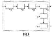
図7は本発明による装置2’の第2の実施形態の概略図を示している。図2に示されている装置2の第1の実施形態の諸要素の他に、この装置は、定期的に、連続的に、又は随時検出された人110の少なくとも一部の検出された向き及び/又は位置に基づいて人110又は人110の1つ以上の身体部分の動きを経時的に分析するための分析ユニット24と、検出された動きを自然な動きと異常な動きに分類し、病気重大度スコア推定を決定し、及び/又は分析された動きが情報信号を送出するための所定の基準を超えている場合に情報信号を送出するための評価ユニット25とを含む。
FIG. 7 shows a schematic view of a second embodiment of a device 2 'according to the invention. In addition to the elements of the first embodiment of the device 2 shown in FIG. 2, the device is a detected orientation of at least a portion of a
分析ユニット24は特に患者の全身又は身体部分の活動分析を実行する。筋肉運動情報、例えば夜間の患者の脚の平均活動値は動作推定結果から計算される。データは24時間にわたり連続的に入手可能である。評価ユニット25は特に体動分類を実行する。患者にとって危険な状況になり得る病気固有の動き又は異常な動き(パターン)が識別される。更に、病気重大度スコア推定が実行され、これは、活動分析及び体動分類からの出力を考慮に入れることにより患者の状態に関するスコアを臨床スタッフに提供することになる。
The
病院環境では、ビデオ分析を複雑にするいくつかの要因がある。例えば、病室内の訪問者又は臨床スタッフの存在はビデオ画像内の患者の一部分を曖昧にするか又は患者の一部分と重なり合う。この場合、患者のみによる動きのセグメント化が必要である。従って、すべての種類のこのような実用上の問題を解決するために、より高性能の要素をシステムに含める必要がある。提案されている方法300の対応する実施形態の一例は図8に描写されているフローチャートに示されている。この実施形態では、処理は2つの並列トラックと見なすことができる。一方は動作推定を実行することであり、他方は動作検出に基づくものである。しかしながら、動作推定又は動作検出を使用するための制限は全くなく、患者の動作を検出できる方法であればどのような方法でも使用することができる。
In a hospital environment, there are several factors that complicate video analysis. For example, the presence of a visitor or clinical staff in a hospital room obscures or overlaps a portion of the patient in the video image. In this case, motion segmentation by the patient alone is necessary. Therefore, to solve all kinds of such practical problems, higher performance elements need to be included in the system. An example of a corresponding embodiment of the proposed
ステップS100では、受信した画像データに基づいて動作検出が実行される。病院環境及び/又はビデオシステムにおける実用上の問題、例えばビデオ圧縮アーチファクト又は最適な照明条件ではないことにより、ビデオ品質はあまり良くない可能性がある。このため、動作推定は小さい動き、例えば患者の呼吸動作を検出するのに苦労する可能性がある。この状況では、代わりのものとして動作検出を使用することが提案される。 In step S100, motion detection is performed based on the received image data. Due to practical problems in hospital environments and / or video systems, such as video compression artifacts or not optimal lighting conditions, video quality may not be very good. For this reason, motion estimation can be difficult to detect small movements, for example, the patient's breathing motion. In this situation, it is proposed to use motion detection as an alternative.
いくつかの動作検出方法が使用可能である。例えば、フレーム差分方法は、2つの連続フレーム間の絶対差の合計(SAD)を計算することにより動きを検出する。もう1つの広く使用される手法は相関係数法である。提案されている装置及び方法では、特定の方法に関する特別な要件は全くないが、選択はビデオ品質、例えば圧縮フォーマットに依存する可能性がある。 Several motion detection methods can be used. For example, the frame difference method detects motion by calculating the sum of absolute differences (SAD) between two consecutive frames. Another widely used technique is the correlation coefficient method. In the proposed apparatus and method, there are no special requirements for a particular method, but the choice may depend on the video quality, eg the compression format.
身体部分のセグメント化を実行するステップS102では患者の胸部領域を検出するために使用される動作検出(ステップS100)からの出力が使用されることが図8から分かるが、これについては以下により詳細に説明する。 It can be seen from FIG. 8 that the output from motion detection (step S100) used to detect the patient's chest region is used in step S102 for performing segmentation of the body part, as will be described in more detail below. Explained.
ステップS104では動作推定が実行される。動作推定(ME)は患者による動きを捕捉するためのオプションの1つである。得られた動作ベクトル場は、動作位置、強度、及び方向の指示を提供する。提案されている装置及び方法では、A. Heinrich、C. Bartels、R.J. van der Vleuten、C.N. Cordes、G. de HaanによるOptimization of hierarchical 3DRS motion estimators for picture rate conversion(IEEE Journal of Selected Topics in Signal Processing、第5巻、第2号、262〜274ページ、2011年3月)の開示内容に基づくMEアルゴリズムが解決策として使用され、これは計算上の複雑さを低く維持しながら正確な動作ベクトルを提供することができる。 In step S104, motion estimation is executed. Motion estimation (ME) is one option for capturing patient motion. The resulting motion vector field provides an indication of motion position, intensity, and direction. In the proposed apparatus and method, A. Heinrich, C.I. Bartels, R.A. J. et al. van der Vleuten, C.I. N. Cordes, G.M. De Haan's Optimization of Hierarchical 3DRS motion Estimators for picture rate conversion (based on IEEE Journal of Selected Topics 2nd, 5th volume, 27th volume, 4th volume, 4th volume) Used as a strategy, which can provide accurate motion vectors while keeping computational complexity low.
ステップS106では患者身体検出が実行され、ステップS108では看護師/訪問者セグメント化が実行される。病院環境における動的側面の1つは、病室内、従ってビデオ画像内に臨床スタッフ及び訪問者がしばしば存在することである。患者による活動のみが関心があり、分析する必要があるので、他の人の動きは除外しなければならない。それぞれの動作を差別化するために、患者身体検出及び看護師/訪問者セグメント化を実行することが提案される。 In step S106, patient body detection is performed, and in step S108, nurse / visitor segmentation is performed. One of the dynamic aspects in the hospital environment is that clinical staff and visitors are often present in the hospital room and thus in the video image. Since only patient activity is of interest and needs to be analyzed, other people's movements must be excluded. In order to differentiate each action, it is proposed to perform patient body detection and nurse / visit segmentation.
患者身体検出のステップS106は、関心領域(ROI)、即ち、患者身体領域をサーチするために使用される。これは、本発明による装置及び方法の第1の実施形態に関して上記で説明したように行うことができる。主な概念は、ある期間にわたり動作推定モジュールから得られた動作ベクトルを蓄積することである。患者以外の人もビデオ内に存在する可能性があるので、優勢な動作が患者によるものになるように選択された蓄積時間は十分長いものでなければならない。蓄積されたベクトルの一例は図6Dに示されており、これは、患者が主に自分の身体の中央部分及び下半身部分を動かしており、看護師もシーケンス(ホットスポットH2によって表される)内に存在する30分のビデオから得られたものである。蓄積時間を増加し、何らかの後処理を実行することにより、看護師による動作ベクトルは除去される。 The patient body detection step S106 is used to search for a region of interest (ROI), i.e. a patient body region. This can be done as described above with respect to the first embodiment of the apparatus and method according to the invention. The main concept is to accumulate motion vectors obtained from the motion estimation module over a period of time. Since people other than the patient may also be present in the video, the accumulation time selected so that the dominant action is due to the patient must be long enough. An example of the accumulated vector is shown in FIG. 6D, which shows that the patient is moving mainly in the middle and lower body part of her body and the nurse is also in the sequence (represented by hotspot H2). From 30 minutes of video. By increasing the accumulation time and performing some post-processing, the nurse motion vectors are removed.
患者身体領域が決定されている場合、次に看護師/訪問者をセグメント化することが可能である。提案されている方法は、動作履歴情報を保持し、それぞれの発生源を追跡することにより色々な人による動作を差別化することである。これを行うことにより、動作履歴画像を患者履歴画像と看護師/他の人の履歴画像に分離することができる。ここでは看護師/訪問者の画像領域を検出するために、他の方法も適用することができる。 If the patient body area has been determined, the nurse / visitor can then be segmented. The proposed method is to differentiate motions by different people by keeping motion history information and tracking each source. By doing this, the action history image can be separated into a patient history image and a nurse / other person's history image. Other methods can also be applied here to detect nurse / visitor image areas.
ステップS102では、身体部分セグメント化が実行され、これは第1の実施形態に関して上記で説明したように行われる。身体部分セグメント化ステップは、患者の種々の身体部分を識別するために使用される。頭部、体幹、及び脚などの患者の身体部分は互いに差別化することができる。その結果として、身体部分ごとに動作分析を実行することができ、これは患者の筋肉運動挙動に関するより詳細な動作情報を提供する。更に、種々の身体部分の識別に基づいて、それぞれの身体部分の位置及び向き並びに患者全体の位置及び向きを決定することができる。 In step S102, body part segmentation is performed, which is performed as described above with respect to the first embodiment. The body part segmentation step is used to identify various body parts of the patient. Patient body parts such as the head, trunk, and legs can be differentiated from each other. As a result, motion analysis can be performed for each body part, which provides more detailed motion information regarding the patient's muscle movement behavior. Further, based on the identification of the various body parts, the position and orientation of each body part and the position and orientation of the entire patient can be determined.
例えば、患者の脚による動きに関心がある場合、臨床スタッフは過去24時間にわたるこの身体部分の平均活動水準を調べることができる。この活動分析の他に、体動分類もこのセグメント化情報から大いに恩恵を受けることになる。身体部分位置がすでに分かっている場合、特定の身体部分によって行われた特定の動きパターンを識別することははるかに容易である。 For example, if the patient is interested in movement with the patient's legs, the clinical staff can examine the average activity level of this body part over the past 24 hours. In addition to this activity analysis, body movement classification will also benefit greatly from this segmentation information. If the body part location is already known, it is much easier to identify a particular movement pattern performed by a particular body part.
身体部分をセグメント化するための種々の手法を適用することができる。単純な方法は、全身領域を比率数でいくつかの部分に分割することであろう。例えば、頭部、体幹、及び脚の比率はそれぞれ15%、30%、55%に設定される。更に、胸部検出に基づくより高性能の方法を使用することができる。その概念は、呼吸動作を検出することにより胸部領域をまず決定することであり、残りの頭部及び脚部は後で分割される。これは図9に描写されており、図9Aはいくつかの身体部分が強調表示されている取得画像を示しており、図9Bは同じ身体部分の動きが強調表示されている対応する蓄積画像を示している。 Various techniques for segmenting body parts can be applied. A simple method would be to divide the whole body region into several parts by a ratio number. For example, the ratio of the head, the trunk, and the legs is set to 15%, 30%, and 55%, respectively. In addition, more sophisticated methods based on chest detection can be used. The concept is to first determine the chest region by detecting breathing motion, and the remaining head and legs are later divided. This is depicted in FIG. 9, where FIG. 9A shows an acquired image with several body parts highlighted, and FIG. 9B shows a corresponding accumulated image with the same body part movement highlighted. Show.
ステップS110では、活動分析が実行される。患者の動きを分析するための2つの要素のうちの1つである体動分類の他に、身体部分/全身活動水準分析が行われる。これは、患者活動の概要を提供するものであり、患者の臨床条件を決定するための基準の1つとして使用することができる。図10Aは、身体部分セグメント化によって導出された脚510、体幹520、及び頭部530という3つの領域が示されている、患者の画像500の一例を示している。図10Bは種々の活動水準信号を示しており、活動水準信号600は、手首装着装置(例えば「Actiwatch」という本出願人の装置)などの活動監視装置から得られ、全身(信号610)、体幹(620)、脚(630)、及び頭部(640)のビデオアクチグラフによって得られた活動水準信号と比較される。
In step S110, activity analysis is performed. In addition to body motion classification, one of two elements for analyzing patient movement, body part / whole body activity level analysis is performed. This provides an overview of patient activity and can be used as one of the criteria for determining patient clinical conditions. FIG. 10A shows an example of a
その利点は図10BのT1によって示される期間において明らかに観察され、例えば脚及び頭部による動作については、例えばセンサが患者に装着されていないので、その部分では活動監視装置からの信号(信号600を参照)が全く入手可能ではなく、依然として信号はビデオによって検出することができる。ビデオ及びビデオアクチグラフ信号(複数も可)の目視検査により、活動監視装置が全く装着されていない他方の腕の動きも正しく検出され、それが体幹活動信号620に反映されていることが示されている。期間T2は、ビデオシステムが活動監視装置で測定された動きも十分検出できることを示している。
The advantage is clearly observed in the period indicated by T1 in FIG. 10B, for example for the movements by the legs and head, for example because the sensor is not worn on the patient, in that part the signal from the activity monitoring device (signal Is not available at all, and the signal can still be detected by video. Visual inspection of the video and video actigraph signal (s) indicates that the movement of the other arm without any activity monitoring device is correctly detected and reflected in the
ここでは、例えば選択された期間における1つの特定の身体部分に関する活動値の平均又は変動、活動の頻度など、種々の活動統計が計算される。この情報は、譫妄、てんかん、パーキンソン、下肢静止不能症などの特定の臨床条件を示すことができる筋肉運動変質の変化が発生しているかどうかを区別するために使用することができる。加えて、これらの活動水準は分類特徴として使用できるので、この筋肉運動分析は体動分類にも貢献する。 Here, various activity statistics are calculated, such as, for example, the average or variation of activity values for one particular body part in a selected time period, the frequency of activities, etc. This information can be used to distinguish whether changes in muscular motor alterations have occurred that can indicate specific clinical conditions such as delirium, epilepsy, Parkinson's, restless legs. In addition, since these activity levels can be used as classification features, this muscle movement analysis also contributes to body movement classification.
ステップS112では、体動分類が実行される。身体活動分析と比較して、動作分類は患者の動きについてより高水準の解釈を提供する。これは、自然な動きと異常な動きとを差別化することができ、特定の動き(パターン)を更に識別することができる。例えば、譫妄の場合、空中つかみ又は皮膚むしりなどの動きは典型的な動きである。これらの特定の動作の検出は患者の譫妄判定に直接貢献することになる。その他の適用例では、パーキンソン又はてんかん発作を示す震えが検出される。この体動認識から得られるその他の利点もある。これは、結果的に危険な状況になり得る何らかの動きを検出するために使用することもできる。例えば、患者がベッドから起床しようとしているか又はベッドから転落しそうな場合、気管内挿入管又は栄養管などが引き抜かれる。提案されている方法及び装置のこの実施形態は医療スタッフに警告メッセージを自動的に送信することができる。 In step S112, body motion classification is executed. Compared to physical activity analysis, motion classification provides a higher level interpretation of patient movement. This can differentiate between natural and abnormal movements and can further identify specific movements (patterns). For example, in the case of delirium, movements such as air grip or skin peeling are typical movements. Detection of these specific movements will directly contribute to the patient's delirium determination. In other applications, tremors that indicate Parkinson or epileptic seizures are detected. There are other benefits derived from this body motion recognition. This can also be used to detect any movement that can result in a dangerous situation. For example, if the patient is getting up from the bed or is about to fall from the bed, the endotracheal tube or feeding tube is withdrawn. This embodiment of the proposed method and apparatus can automatically send a warning message to medical staff.
分類方法については、この問題に適した任意のタイプの分類器を使用することができる。動作方向、速度、加速度、及び動作領域のサイズなどの動作特徴は動作ベクトルから直接推論することができる。その他の特徴例としては、動きの持続時間及び頻度/反復性を含む。特定の動きが通常は特定の身体部分によって行われることを考慮すると、分類器は身体部分情報から利益を得る。従って、一実施形態では、身体部分ごとに動作特徴を抽出することが提案される。 For the classification method, any type of classifier suitable for this problem can be used. Motion features such as motion direction, speed, acceleration, and motion region size can be inferred directly from motion vectors. Other example features include motion duration and frequency / repeatability. Considering that specific movements are usually performed by specific body parts, the classifier benefits from body part information. Thus, in one embodiment, it is proposed to extract motion features for each body part.
更に、病気重大度スコア推定が行われる。このステップは病気の重大度に関するスコアを提供する。これは、ビデオ分析のデータを組み合わせることにより患者の健康状態を推定する。図7に示されている装置の提案されている実施形態では、出力はビデオアクチグラフ及び体動分類(即ち、関心がある場合に臨床条件に関連する特定の動きの検出)に基づくものである。しかしながら、これはビデオに限定されず、入手可能であれば、その他の補足情報をすべてこのステップに供給することができ、最終的な決定のための考慮に入れることができる。これは、他のセンサモダリティによって測定されたデータ、臨床試験/スクリーニング、又は臨床スタッフの観察である可能性がある。 In addition, disease severity score estimation is performed. This step provides a score for the severity of the disease. It estimates the patient's health status by combining data from video analysis. In the proposed embodiment of the device shown in FIG. 7, the output is based on video actigraphs and body movement classification (ie, detection of specific movements related to clinical conditions when interested). . However, this is not limited to video, and all other supplemental information can be provided to this step, if available, and can be taken into account for the final decision. This may be data measured by other sensor modalities, clinical trials / screening, or observations of clinical staff.
関心のある臨床条件を示す臨床条件の変化が検出された時に、又はベッドから起床しようとしているなど、患者にとって危険なものになり得る動きが検出された時に、警報が発せられる。スコアが事前設定のしきい値を超えると、医療スタッフは自動的に警告メッセージを受信する。臨床スタッフは自分が関心のある警報や、どの病気/障害を検出しなければならないか又はどの病気/障害の優先順位が最も高いかを設定することができる。 An alert is triggered when a change in clinical condition indicative of the clinical condition of interest is detected, or when a movement is detected that can be dangerous to the patient, such as trying to wake up from the bed. If the score exceeds a preset threshold, the medical staff automatically receives a warning message. The clinical staff can set the alarms of interest and which illness / disorders must be detected or which illness / disorder has the highest priority.
上記で説明した要素及びステップは上記の通りに限定されるわけではない。より良い性能を得るために、より高性能の要素及びステップが追加され、その他の組み合わせが行われる。 The elements and steps described above are not limited as described above. Higher performance elements and steps are added and other combinations are made to obtain better performance.
患者以外の人がビデオ内にしばしば存在するので、時には障害物のために患者の動きをカメラによって捕捉することができない。患者のみが見えるように(例えば、患者の上に)カメラを設置することが可能ではない場合、異なる観察方向から患者を画像化するために病室内に第2のカメラが設置される。この場合、より良い観察条件を得るために、カメラの同期及びカメラの選択など、余分なビデオ処理が必要になる可能性がある。 Because people other than the patient are often present in the video, sometimes the patient's movement cannot be captured by the camera due to obstacles. If it is not possible to install a camera so that only the patient is visible (eg, on the patient), a second camera is installed in the patient room to image the patient from different viewing directions. In this case, extra video processing such as camera synchronization and camera selection may be required to obtain better viewing conditions.
他の実施形態では、測定が開始された時又は状況が大きく変化した時に病院スタッフから入力を取得することにより、画像内の患者の位置及び患者の向きに関する事前知識が使用される。 In other embodiments, prior knowledge about the patient's position and patient orientation in the image is used by obtaining input from hospital staff when the measurement is initiated or when the situation changes significantly.
ベッドの位置又はベッドの角度は患者の位置が変更された結果としてまた身体部分セグメント化の結果として変更することができる。その場合、患者身体検出モジュールを更新しなければならない。追加の一実施形態として、ベッドの位置又はベッドの角度の変化を知らせるために自動ベッド検出を実現することができる。 The bed position or bed angle can be changed as a result of changing the patient's position and as a result of body segmentation. In that case, the patient body detection module must be updated. As an additional embodiment, automatic bed detection can be implemented to signal changes in bed position or bed angle.
要約すると、本発明は、特に自動連続ビデオ体動分析が実行される病院内の種々の病棟(例えばICU、急患治療設定、一般病棟、老人病棟)又は看護センター、高齢者介護施設、NICU、家庭における病院内の患者監視のために、様々な領域で適用可能である。主な適用分野は、譫妄もしくは例えば譫妄に関連する異常な動き/挙動を伴うその他の病気の早期発見、ICUにおける自動ビデオ監視が適用される場合、家庭、高齢者介護施設、高齢者看護センターにおける介護者への警告を引き起こすための(健康又は不健康な)高齢者の監視である。 In summary, the present invention particularly relates to various hospital wards (eg, ICU, emergency care settings, general wards, geriatric wards) or nursing centers, elderly care facilities, NICUs, homes where automatic continuous video motion analysis is performed. It can be applied in various areas for patient monitoring in hospitals. The main areas of application are early detection of delirium or other diseases with abnormal movements / behaviours associated with delirium, for example, when automatic video surveillance in ICU is applied, in homes, elderly care facilities, elderly care centers It is the monitoring of the elderly (healthy or unhealthy) to alert the caregiver.
本発明は図面及び上記の説明において詳細に例示され記載されているが、このような例示及び説明は制限的ではなく例証となるもの又は模範的なものと見なすべきであり、本発明は開示されている諸実施形態に限定されない。開示されている諸実施形態以外の変形例は、図面、開示内容、及び特許請求の範囲の検討により、請求されている発明を実践する際に当業者が理解し実行できるものである。 While the invention has been illustrated and described in detail in the drawings and foregoing description, such illustration and description are to be considered illustrative or exemplary and not restrictive; the invention is not disclosed; It is not limited to the embodiments. Variations other than the disclosed embodiments can be understood and carried out by those skilled in the art in practicing the claimed invention by studying the drawings, the disclosure, and the claims.
特許請求の範囲では、「含む(comprising)」という単語はその他の要素又はステップを除外せず、「a」又は「an」という不定冠詞は複数であることを除外しない。単一の要素又はその他のユニットは、特許請求の範囲に列挙されている幾つかの項目の機能を遂行する。特定の手段が互いに異なる従属クレームに列挙されているという単なる事実は、これらの手段の組み合わせを有利に使用できないことを示すものではない。 In the claims, the word “comprising” does not exclude other elements or steps, and the indefinite article “a” or “an” does not exclude a plurality. A single element or other unit performs the functions of several items recited in the claims. The mere fact that certain measures are recited in mutually different dependent claims does not indicate that a combination of these measured cannot be used to advantage.
コンピュータプログラムは、その他のハードウェアとともに又はその一部として供給される光学記憶媒体又はソリッドステート媒体などの適切な非一時的媒体上で記憶及び/又は配布されるが、インターネット或いはその他の有線又は無線通信システムなどを介してその他の形式で配布される場合もある。 Computer programs may be stored and / or distributed on suitable non-transitory media such as optical storage media or solid state media supplied with or as part of other hardware, but may be internet or other wired or wireless It may be distributed in other formats via a communication system.
特許請求の範囲内の参照符号は範囲を限定するものと解釈してはならない。
Any reference signs in the claims should not be construed as limiting the scope.
Claims (16)
経時的な一連の画像フレームを含む人の画像データを得る、画像データインターフェースと、
前記画像データ内の動作を検出する、動作検出器と、
頻繁に発生する動作を含む前記画像フレームの領域を示す動作ホットスポットを識別する、動作強度検出器と、
所定の期間にわたり最も大きい動作を示す前記動作ホットスポットに基づいて、人の第1の境界を識別する、人検出器と、
を含む、装置。 A device for automatic detection of the orientation and / or position of a person, the device comprising:
An image data interface for obtaining human image data including a series of image frames over time;
A motion detector for detecting motion in the image data;
A motion intensity detector that identifies motion hot spots that indicate regions of the image frame that include frequently occurring motion;
A human detector that identifies a first boundary of the person based on the operational hot spot exhibiting the greatest movement over a predetermined period of time;
Including the device.
経時的な一連の画像フレームを含む人の画像データを得る、画像データインターフェースと、
前記画像データ内の動作を検出し、1つの画像フレームから他の画像フレームまでの前記画像データ内で検出された動作を表す動作画像を決定する、動作検出器と、
頻繁に発生する動作を含む前記画像フレームの領域を示す動作ホットスポットを識別し、2つ以上の動作画像内の動作ピクセル又は動作ベクトル情報のバイナリ発生を合計することにより1つ以上の蓄積画像を得るために、所定の期間にわたる動作画像を蓄積する、動作強度検出器と、
識別された前記動作ホットスポットに基づいて、人の少なくとも一部の向き及び/又は位置を検出する、人検出器と、を含み、
前記人検出器は、所定の期間にわたり最も大きい動作を示す前記動作ホットスポットに基づいて、人の境界を識別する、装置。 A device for automatic detection of the orientation and / or position of a person, the device comprising:
An image data interface for obtaining human image data including a series of image frames over time;
An action detector for detecting an action in the image data and determining an action image representing the action detected in the image data from one image frame to another image frame;
One or more stored images are identified by identifying motion hot spots that indicate regions of the image frame that contain frequently occurring motion and summing the binary generation of motion pixel or motion vector information in two or more motion images A motion intensity detector that accumulates motion images over a predetermined period of time to obtain,
A human detector that detects an orientation and / or position of at least a portion of the person based on the identified operational hot spot ;
The apparatus wherein the human detector identifies a human boundary based on the operational hot spot that exhibits the greatest motion over a predetermined period of time .
検出された前記動きを自然な動きと異常な動きとに分類し、病気重大度スコア推定を決定し、及び/又は分析された前記動きが情報信号を送出するための所定の基準を超えている場合に前記情報信号を送出する、評価ユニットと、を更に含む、請求項1に記載の装置。 Analyzing the movement of a person over time or the movement of one or more body parts of a person based on the detected orientation and / or position of at least a portion of the person detected periodically, continuously or from time to time The analysis unit,
Classify the detected motion into natural and abnormal motion, determine a disease severity score estimate, and / or the analyzed motion exceeds a predetermined criterion for sending an information signal The apparatus of claim 1, further comprising an evaluation unit that sends the information signal in case.
経時的な一連の画像フレームを含む人の画像データを取得して前記画像データインターフェースに提供する、1つ以上のビデオカメラと、
取得した前記画像データに基づいて人の少なくとも一部の向き及び/又は位置の自動検出のための請求項1に記載の装置と、
人の少なくとも一部の検出された前記向き及び/又は位置に関連する情報を出力する、出力インターフェースと、
を含む、システム。 A system for automatic detection of a person's orientation and / or position, the system comprising:
One or more video cameras that acquire and provide to the image data interface human image data including a series of image frames over time;
The apparatus of claim 1 for automatic detection of the orientation and / or position of at least a portion of a person based on the acquired image data;
An output interface for outputting information related to the detected orientation and / or position of at least a portion of a person;
Including the system.
経時的な一連の画像フレームを含む人の画像データを得るステップと、
前記画像フレーム内の動作推定を含む、前記画像データ内の動作を検出するステップと、
前記画像データ内の1つの画像フレームから他の画像フレームまでの動作を表す動作画像を決定して蓄積するステップと、
大きな動作の領域を考慮に入れて、前記蓄積された動作画像を適応しきい値でしきい値処理することにより、所定の期間にわたり最も大きい動作を示す画像領域を表す動作ホットスポットを識別するステップと、
識別された最も大きい動作を示す前記動作ホットスポットに基づいて、人の境界を識別するステップと、
を含む、方法。 A method for automatic detection of a person's orientation and / or position, comprising:
Obtaining human image data including a series of image frames over time;
Detecting motion in the image data, including motion estimation in the image frame;
Determining and storing an action image representing an action from one image frame to another image frame in the image data;
Taking into account a large area of motion and thresholding the accumulated motion image with an adaptive threshold to identify a motion hot spot representing an image region that exhibits the greatest motion over a predetermined period of time When,
Identifying a person's boundary based on the action hotspot indicating the largest action identified;
Including a method.
人の一連のビデオフレームを含む人の画像データを生成する、1つ以上のビデオカメラと、
前記1つ以上のビデオカメラから前記画像データを受信し、
前記1つ以上のビデオカメラからの前記画像データ内の動作を検出し、
複数のフレームにわたり画像データを蓄積することにより、前記1つ以上のビデオカメラからの前記画像データ内の、頻繁に発生する動作を含む領域を示す動作ホットスポットを識別し、
所定の期間にわたり最も大きい動作を示す前記動作ホットスポットに基づいて、人の縦軸に対して実質的に平行な方向に1つ以上の蓄積された画像内のエッジ強度分析により、画像内の人の左境界及び/又は右境界を検出する
ためにプログラムされた1つ以上のコンピュータプロセッサと、
検出された前記人の左境界及び/又は右境界に関する情報を出力するユーザインターフェースと、
を含む、装置。 A device for automatic detection of the orientation and / or position of a person, the device comprising:
One or more video cameras that generate human image data including a series of human video frames;
Receiving the image data from the one or more video cameras;
Detecting movement in the image data from the one or more video cameras;
Identifying operational hotspots indicating regions of the image data from the one or more video cameras that contain frequently occurring operations by accumulating image data over a plurality of frames;
Based on the motion hotspot that exhibits the greatest motion over a predetermined period of time , the person in the image is analyzed by edge strength analysis in one or more accumulated images in a direction substantially parallel to the human longitudinal axis. One or more computer processors programmed to detect a left boundary and / or a right boundary of
A user interface that outputs information about the detected left and / or right boundary of the person;
Including the device.
Applications Claiming Priority (3)
| Application Number | Priority Date | Filing Date | Title |
|---|---|---|---|
| EP14191395 | 2014-11-03 | ||
| EP14191395.4 | 2014-11-03 | ||
| PCT/EP2015/075535 WO2016071314A1 (en) | 2014-11-03 | 2015-11-03 | Device, system and method for automated detection of orientation and/or location of a person |
Publications (3)
| Publication Number | Publication Date |
|---|---|
| JP2017536880A JP2017536880A (en) | 2017-12-14 |
| JP2017536880A5 JP2017536880A5 (en) | 2019-03-07 |
| JP6588978B2 true JP6588978B2 (en) | 2019-10-09 |
Family
ID=51893849
Family Applications (1)
| Application Number | Title | Priority Date | Filing Date |
|---|---|---|---|
| JP2017523482A Active JP6588978B2 (en) | 2014-11-03 | 2015-11-03 | Apparatus, system and method for automatic detection of human orientation and / or position |
Country Status (5)
| Country | Link |
|---|---|
| US (1) | US10121062B2 (en) |
| EP (1) | EP3214996B1 (en) |
| JP (1) | JP6588978B2 (en) |
| CN (1) | CN107072548B (en) |
| WO (1) | WO2016071314A1 (en) |
Families Citing this family (14)
| Publication number | Priority date | Publication date | Assignee | Title |
|---|---|---|---|---|
| US10813572B2 (en) * | 2015-12-11 | 2020-10-27 | Electronic Caregiver, Inc. | Intelligent system for multi-function electronic caregiving to facilitate advanced health diagnosis, health monitoring, fall and injury prediction, health maintenance and support, and emergency response |
| US10956484B1 (en) | 2016-03-11 | 2021-03-23 | Gracenote, Inc. | Method to differentiate and classify fingerprints using fingerprint neighborhood analysis |
| EP3432772B1 (en) * | 2016-03-22 | 2020-07-29 | Koninklijke Philips N.V. | Using visual context to timely trigger measuring physiological parameters |
| KR102118336B1 (en) * | 2016-05-20 | 2020-06-04 | 전자부품연구원 | System for analysis of behavior semantic in service area and method thereof |
| JP6406371B2 (en) * | 2017-03-02 | 2018-10-17 | オムロン株式会社 | Watch support system and control method thereof |
| TW201904265A (en) * | 2017-03-31 | 2019-01-16 | 加拿大商艾維吉隆股份有限公司 | Abnormal motion detection method and system |
| JP7000702B2 (en) * | 2017-04-27 | 2022-01-19 | コニカミノルタ株式会社 | Health management system and health management method |
| DE102017006529A1 (en) | 2017-07-11 | 2019-01-17 | Drägerwerk AG & Co. KGaA | A method, apparatus and computer program for capturing optical image data of a patient environment and for detecting a patient examination |
| JP6870514B2 (en) * | 2017-07-14 | 2021-05-12 | オムロン株式会社 | Watching support system and its control method |
| EP3496037A1 (en) * | 2017-12-06 | 2019-06-12 | Koninklijke Philips N.V. | Device, system and method for detecting body movement of a patient |
| US11683578B2 (en) * | 2018-01-29 | 2023-06-20 | Nec Corporation | Extraction of target person from image |
| US12009083B2 (en) | 2020-11-16 | 2024-06-11 | Electronic Caregiver, Inc. | Remote physical therapy and assessment of patients |
| EP4176809A1 (en) * | 2021-11-08 | 2023-05-10 | Koninklijke Philips N.V. | Device, system and method for monitoring a subject |
| CN116602664B (en) * | 2023-07-17 | 2023-09-22 | 青岛市胶州中心医院 | Comprehensive diagnosis and treatment nursing system for neurosurgery patients |
Family Cites Families (32)
| Publication number | Priority date | Publication date | Assignee | Title |
|---|---|---|---|---|
| DE69124777T2 (en) * | 1990-11-30 | 1997-06-26 | Canon Kk | Device for the detection of the motion vector |
| US5930379A (en) | 1997-06-16 | 1999-07-27 | Digital Equipment Corporation | Method for detecting human body motion in frames of a video sequence |
| US6049281A (en) * | 1998-09-29 | 2000-04-11 | Osterweil; Josef | Method and apparatus for monitoring movements of an individual |
| JP4596201B2 (en) * | 2001-02-01 | 2010-12-08 | ソニー株式会社 | Image processing apparatus and method, and recording medium |
| US20030058111A1 (en) * | 2001-09-27 | 2003-03-27 | Koninklijke Philips Electronics N.V. | Computer vision based elderly care monitoring system |
| US9311540B2 (en) * | 2003-12-12 | 2016-04-12 | Careview Communications, Inc. | System and method for predicting patient falls |
| US8675059B2 (en) * | 2010-07-29 | 2014-03-18 | Careview Communications, Inc. | System and method for using a video monitoring system to prevent and manage decubitus ulcers in patients |
| US7477285B1 (en) * | 2003-12-12 | 2009-01-13 | Careview Communication, Inc. | Non-intrusive data transmission network for use in an enterprise facility and method for implementing |
| NZ534482A (en) | 2004-08-02 | 2007-05-31 | Canterprise Ltd | A method of assaying agitation |
| KR20070107722A (en) * | 2005-01-28 | 2007-11-07 | 유클리드 디스커버리스, 엘엘씨 | Apparatus and method for processing video data |
| CN200972689Y (en) * | 2006-10-27 | 2007-11-07 | 刘军民 | Intelligent vedio identification monitoring alarm system |
| CN100561503C (en) * | 2007-12-28 | 2009-11-18 | 北京中星微电子有限公司 | A kind of people's face canthus and corners of the mouth location and method and the device followed the tracks of |
| US9579047B2 (en) * | 2013-03-15 | 2017-02-28 | Careview Communications, Inc. | Systems and methods for dynamically identifying a patient support surface and patient monitoring |
| CN101765025A (en) * | 2008-12-23 | 2010-06-30 | 北京中星微电子有限公司 | System for abnormal detection of surveillance camera and method thereof |
| CN101751678B (en) * | 2009-12-16 | 2012-01-25 | 北京智安邦科技有限公司 | Method and device for detecting violent crowd movement |
| CN101916447B (en) * | 2010-07-29 | 2012-08-15 | 江苏大学 | Robust motion target detecting and tracking image processing system |
| US9785744B2 (en) * | 2010-09-14 | 2017-10-10 | General Electric Company | System and method for protocol adherence |
| US9204823B2 (en) * | 2010-09-23 | 2015-12-08 | Stryker Corporation | Video monitoring system |
| JP5682203B2 (en) * | 2010-09-29 | 2015-03-11 | オムロンヘルスケア株式会社 | Safety nursing system and method for controlling safety nursing system |
| RU2604701C2 (en) | 2011-05-30 | 2016-12-10 | Конинклейке Филипс Н.В. | Method and apparatus for monitoring movement and breathing of multiple subjects in a common bed |
| RU2623304C2 (en) | 2011-10-07 | 2017-06-23 | Конинклейке Филипс Н.В. | Monitoring system for patient monitoring and delirium detection |
| US8743200B2 (en) * | 2012-01-16 | 2014-06-03 | Hipass Design Llc | Activity monitor |
| US9295390B2 (en) * | 2012-03-02 | 2016-03-29 | Hill-Rom Services, Inc. | Facial recognition based monitoring systems and methods |
| US9375142B2 (en) * | 2012-03-15 | 2016-06-28 | Siemens Aktiengesellschaft | Learning patient monitoring and intervention system |
| CN103428407B (en) * | 2012-05-25 | 2017-08-25 | 信帧机器人技术(北京)有限公司 | A kind of method for detecting fought in video |
| CN102799873B (en) * | 2012-07-23 | 2014-12-10 | 青岛科技大学 | Human body abnormal behavior recognition method |
| CN102880444B (en) * | 2012-08-24 | 2016-03-09 | 浙江捷尚视觉科技股份有限公司 | A kind of detection method of fighting based on the analysis of stereoscopic vision sports ground |
| CN102867172B (en) * | 2012-08-27 | 2016-12-21 | Tcl集团股份有限公司 | A kind of human-eye positioning method, system and electronic equipment |
| JP6115335B2 (en) * | 2013-06-10 | 2017-04-19 | ノーリツプレシジョン株式会社 | Information processing apparatus, information processing method, and program |
| JP6557226B2 (en) | 2013-07-22 | 2019-08-07 | コーニンクレッカ フィリップス エヌ ヴェKoninklijke Philips N.V. | Monitoring system and monitoring method |
| CN103517042B (en) * | 2013-10-17 | 2016-06-29 | 吉林大学 | A kind of nursing house old man's hazardous act monitoring method |
| CN106415654A (en) * | 2014-02-18 | 2017-02-15 | 诺日士精密株式会社 | Information processing device, information processing method, and program |
-
2015
- 2015-11-02 US US14/930,212 patent/US10121062B2/en active Active
- 2015-11-03 JP JP2017523482A patent/JP6588978B2/en active Active
- 2015-11-03 WO PCT/EP2015/075535 patent/WO2016071314A1/en active Application Filing
- 2015-11-03 CN CN201580059761.7A patent/CN107072548B/en active Active
- 2015-11-03 EP EP15788420.6A patent/EP3214996B1/en active Active
Also Published As
| Publication number | Publication date |
|---|---|
| JP2017536880A (en) | 2017-12-14 |
| CN107072548A (en) | 2017-08-18 |
| CN107072548B (en) | 2020-09-01 |
| EP3214996A1 (en) | 2017-09-13 |
| WO2016071314A1 (en) | 2016-05-12 |
| US10121062B2 (en) | 2018-11-06 |
| US20160125620A1 (en) | 2016-05-05 |
| EP3214996B1 (en) | 2024-01-10 |
Similar Documents
| Publication | Publication Date | Title |
|---|---|---|
| JP6588978B2 (en) | Apparatus, system and method for automatic detection of human orientation and / or position | |
| JP6053802B2 (en) | A monitoring system that monitors patients and detects patient delirium | |
| EP2533693B1 (en) | Method and apparatus for processing a cyclic physiological signal | |
| US9504426B2 (en) | Using an adaptive band-pass filter to compensate for motion induced artifacts in a physiological signal extracted from video | |
| US11116423B2 (en) | Patient monitoring system and method | |
| US20150245787A1 (en) | Real-time video processing for respiratory function analysis | |
| US9687189B2 (en) | Automatic visual remote assessment of movement symptoms in people with parkinson's disease for MDS-UPDRS finger tapping task | |
| JP6822328B2 (en) | Watching support system and its control method | |
| JP6692987B2 (en) | Devices, Systems, and Methods for Patient Monitoring to Predict and Prevent Bed Fall | |
| CN109891516A (en) | Equipment, system and method for patient-monitoring to predict and prevent bed from falling | |
| JP6952299B2 (en) | Sleep depth judgment system, sleep depth judgment device and sleep depth judgment method | |
| JP2019535052A (en) | Device, system, and method for patient monitoring to predict and prevent bed fall | |
| Heinrich et al. | Body movement analysis during sleep based on video motion estimation | |
| US20220044046A1 (en) | Device, system and method for object recognition | |
| Heinrich et al. | Video based actigraphy and breathing monitoring from the bedside table of shared beds | |
| US11354809B2 (en) | Device, system and method for detecting body movement of a patient | |
| US20210275089A1 (en) | Deriving information about a person's sleep and wake states from a sequence of video frames | |
| Inoue et al. | Bed exit action detection based on patient posture with long short-term memory | |
| Gong et al. | Vitals are Vital! Visual Vital Metric Detection for Multiple Subjects |
Legal Events
| Date | Code | Title | Description |
|---|---|---|---|
| A521 | Request for written amendment filed |
Free format text: JAPANESE INTERMEDIATE CODE: A523 Effective date: 20180830 |
|
| A621 | Written request for application examination |
Free format text: JAPANESE INTERMEDIATE CODE: A621 Effective date: 20180830 |
|
| A871 | Explanation of circumstances concerning accelerated examination |
Free format text: JAPANESE INTERMEDIATE CODE: A871 Effective date: 20180830 |
|
| A975 | Report on accelerated examination |
Free format text: JAPANESE INTERMEDIATE CODE: A971005 Effective date: 20181017 |
|
| A131 | Notification of reasons for refusal |
Free format text: JAPANESE INTERMEDIATE CODE: A131 Effective date: 20181030 |
|
| A524 | Written submission of copy of amendment under article 19 pct |
Free format text: JAPANESE INTERMEDIATE CODE: A524 Effective date: 20190122 |
|
| A02 | Decision of refusal |
Free format text: JAPANESE INTERMEDIATE CODE: A02 Effective date: 20190409 |
|
| A521 | Request for written amendment filed |
Free format text: JAPANESE INTERMEDIATE CODE: A523 Effective date: 20190618 |
|
| A911 | Transfer to examiner for re-examination before appeal (zenchi) |
Free format text: JAPANESE INTERMEDIATE CODE: A911 Effective date: 20190625 |
|
| TRDD | Decision of grant or rejection written | ||
| A01 | Written decision to grant a patent or to grant a registration (utility model) |
Free format text: JAPANESE INTERMEDIATE CODE: A01 Effective date: 20190819 |
|
| A61 | First payment of annual fees (during grant procedure) |
Free format text: JAPANESE INTERMEDIATE CODE: A61 Effective date: 20190913 |
|
| R150 | Certificate of patent or registration of utility model |
Ref document number: 6588978 Country of ref document: JP Free format text: JAPANESE INTERMEDIATE CODE: R150 |
|
| R250 | Receipt of annual fees |
Free format text: JAPANESE INTERMEDIATE CODE: R250 |
|
| R250 | Receipt of annual fees |
Free format text: JAPANESE INTERMEDIATE CODE: R250 |