JP6567425B2 - Anti-leukocyte adhesion to alleviate potential adverse events caused by CD3-specific binding domains - Google Patents
Anti-leukocyte adhesion to alleviate potential adverse events caused by CD3-specific binding domains Download PDFInfo
- Publication number
- JP6567425B2 JP6567425B2 JP2015556506A JP2015556506A JP6567425B2 JP 6567425 B2 JP6567425 B2 JP 6567425B2 JP 2015556506 A JP2015556506 A JP 2015556506A JP 2015556506 A JP2015556506 A JP 2015556506A JP 6567425 B2 JP6567425 B2 JP 6567425B2
- Authority
- JP
- Japan
- Prior art keywords
- cells
- cell
- endothelial
- amg
- blinatumomab
- Prior art date
- Legal status (The legal status is an assumption and is not a legal conclusion. Google has not performed a legal analysis and makes no representation as to the accuracy of the status listed.)
- Active
Links
Images


Classifications
-
- C—CHEMISTRY; METALLURGY
- C07—ORGANIC CHEMISTRY
- C07K—PEPTIDES
- C07K16/00—Immunoglobulins [IGs], e.g. monoclonal or polyclonal antibodies
- C07K16/18—Immunoglobulins [IGs], e.g. monoclonal or polyclonal antibodies against material from animals or humans
- C07K16/28—Immunoglobulins [IGs], e.g. monoclonal or polyclonal antibodies against material from animals or humans against receptors, cell surface antigens or cell surface determinants
- C07K16/2803—Immunoglobulins [IGs], e.g. monoclonal or polyclonal antibodies against material from animals or humans against receptors, cell surface antigens or cell surface determinants against the immunoglobulin superfamily
- C07K16/2809—Immunoglobulins [IGs], e.g. monoclonal or polyclonal antibodies against material from animals or humans against receptors, cell surface antigens or cell surface determinants against the immunoglobulin superfamily against the T-cell receptor (TcR)-CD3 complex
-
- A—HUMAN NECESSITIES
- A61—MEDICAL OR VETERINARY SCIENCE; HYGIENE
- A61K—PREPARATIONS FOR MEDICAL, DENTAL OR TOILETRY PURPOSES
- A61K31/00—Medicinal preparations containing organic active ingredients
- A61K31/095—Sulfur, selenium, or tellurium compounds, e.g. thiols
- A61K31/10—Sulfides; Sulfoxides; Sulfones
-
- A—HUMAN NECESSITIES
- A61—MEDICAL OR VETERINARY SCIENCE; HYGIENE
- A61K—PREPARATIONS FOR MEDICAL, DENTAL OR TOILETRY PURPOSES
- A61K31/00—Medicinal preparations containing organic active ingredients
- A61K31/65—Tetracyclines
-
- A—HUMAN NECESSITIES
- A61—MEDICAL OR VETERINARY SCIENCE; HYGIENE
- A61K—PREPARATIONS FOR MEDICAL, DENTAL OR TOILETRY PURPOSES
- A61K31/00—Medicinal preparations containing organic active ingredients
- A61K31/70—Carbohydrates; Sugars; Derivatives thereof
- A61K31/715—Polysaccharides, i.e. having more than five saccharide radicals attached to each other by glycosidic linkages; Derivatives thereof, e.g. ethers, esters
- A61K31/737—Sulfated polysaccharides, e.g. chondroitin sulfate, dermatan sulfate
-
- A—HUMAN NECESSITIES
- A61—MEDICAL OR VETERINARY SCIENCE; HYGIENE
- A61K—PREPARATIONS FOR MEDICAL, DENTAL OR TOILETRY PURPOSES
- A61K39/00—Medicinal preparations containing antigens or antibodies
- A61K39/395—Antibodies; Immunoglobulins; Immune serum, e.g. antilymphocytic serum
- A61K39/39533—Antibodies; Immunoglobulins; Immune serum, e.g. antilymphocytic serum against materials from animals
- A61K39/3955—Antibodies; Immunoglobulins; Immune serum, e.g. antilymphocytic serum against materials from animals against proteinaceous materials, e.g. enzymes, hormones, lymphokines
-
- A—HUMAN NECESSITIES
- A61—MEDICAL OR VETERINARY SCIENCE; HYGIENE
- A61P—SPECIFIC THERAPEUTIC ACTIVITY OF CHEMICAL COMPOUNDS OR MEDICINAL PREPARATIONS
- A61P25/00—Drugs for disorders of the nervous system
-
- A—HUMAN NECESSITIES
- A61—MEDICAL OR VETERINARY SCIENCE; HYGIENE
- A61P—SPECIFIC THERAPEUTIC ACTIVITY OF CHEMICAL COMPOUNDS OR MEDICINAL PREPARATIONS
- A61P25/00—Drugs for disorders of the nervous system
- A61P25/28—Drugs for disorders of the nervous system for treating neurodegenerative disorders of the central nervous system, e.g. nootropic agents, cognition enhancers, drugs for treating Alzheimer's disease or other forms of dementia
-
- A—HUMAN NECESSITIES
- A61—MEDICAL OR VETERINARY SCIENCE; HYGIENE
- A61P—SPECIFIC THERAPEUTIC ACTIVITY OF CHEMICAL COMPOUNDS OR MEDICINAL PREPARATIONS
- A61P35/00—Antineoplastic agents
-
- A—HUMAN NECESSITIES
- A61—MEDICAL OR VETERINARY SCIENCE; HYGIENE
- A61P—SPECIFIC THERAPEUTIC ACTIVITY OF CHEMICAL COMPOUNDS OR MEDICINAL PREPARATIONS
- A61P39/00—General protective or antinoxious agents
- A61P39/02—Antidotes
-
- A—HUMAN NECESSITIES
- A61—MEDICAL OR VETERINARY SCIENCE; HYGIENE
- A61P—SPECIFIC THERAPEUTIC ACTIVITY OF CHEMICAL COMPOUNDS OR MEDICINAL PREPARATIONS
- A61P43/00—Drugs for specific purposes, not provided for in groups A61P1/00-A61P41/00
-
- C—CHEMISTRY; METALLURGY
- C07—ORGANIC CHEMISTRY
- C07K—PEPTIDES
- C07K16/00—Immunoglobulins [IGs], e.g. monoclonal or polyclonal antibodies
- C07K16/18—Immunoglobulins [IGs], e.g. monoclonal or polyclonal antibodies against material from animals or humans
-
- G—PHYSICS
- G01—MEASURING; TESTING
- G01N—INVESTIGATING OR ANALYSING MATERIALS BY DETERMINING THEIR CHEMICAL OR PHYSICAL PROPERTIES
- G01N33/00—Investigating or analysing materials by specific methods not covered by groups G01N1/00 - G01N31/00
- G01N33/48—Biological material, e.g. blood, urine; Haemocytometers
- G01N33/50—Chemical analysis of biological material, e.g. blood, urine; Testing involving biospecific ligand binding methods; Immunological testing
- G01N33/5005—Chemical analysis of biological material, e.g. blood, urine; Testing involving biospecific ligand binding methods; Immunological testing involving human or animal cells
- G01N33/5008—Chemical analysis of biological material, e.g. blood, urine; Testing involving biospecific ligand binding methods; Immunological testing involving human or animal cells for testing or evaluating the effect of chemical or biological compounds, e.g. drugs, cosmetics
-
- C—CHEMISTRY; METALLURGY
- C07—ORGANIC CHEMISTRY
- C07K—PEPTIDES
- C07K2317/00—Immunoglobulins specific features
- C07K2317/70—Immunoglobulins specific features characterized by effect upon binding to a cell or to an antigen
-
- G—PHYSICS
- G01—MEASURING; TESTING
- G01N—INVESTIGATING OR ANALYSING MATERIALS BY DETERMINING THEIR CHEMICAL OR PHYSICAL PROPERTIES
- G01N2500/00—Screening for compounds of potential therapeutic value
- G01N2500/04—Screening involving studying the effect of compounds C directly on molecule A (e.g. C are potential ligands for a receptor A, or potential substrates for an enzyme A)
Description
発明の分野
本発明は本質的に、患者における標的細胞に対するT細胞の再指向化を含む治療によって引き起こされる臨床的有害事象を予防する、および/または改善する、および/または処置する方法において使用するための、哺乳動物T細胞の哺乳動物内皮細胞への結合を減少させるかまたは阻害する化合物に関する。標的細胞に対するT細胞の再指向化を含む治療によって引き起こされる臨床的有害事象を有するかまたはそのリスクがある患者を処置する方法もまた企図される。さらに、本発明は、これらの化合物のいずれかまたはそれらの組み合わせ、CD3特異的結合ドメインまたはキメラ抗原受容体 (CAR) をコードする核酸、および該化合物または該組み合わせが、標的細胞に対するT細胞の再指向化を含む治療によって引き起こされる臨床的有害事象の予防または改善のために使用されることを示す封入されたラベルまたは添付文書を含むキットに関する。
FIELD OF THE INVENTION The present invention is essentially used in a method for preventing and / or ameliorating and / or treating clinical adverse events caused by a therapy involving redirecting T cells to target cells in a patient. Therefore, it relates to compounds that reduce or inhibit the binding of mammalian T cells to mammalian endothelial cells. Also contemplated are methods of treating patients who have or are at risk for clinical adverse events caused by a therapy that includes redirecting T cells to target cells. Furthermore, the present invention provides for any of these compounds or combinations thereof, a nucleic acid encoding a CD3-specific binding domain or chimeric antigen receptor (CAR), and the compound or the combination can be used to regenerate T cells against target cells. It relates to a kit comprising an encapsulated label or package insert indicating that it is used for the prevention or amelioration of clinical adverse events caused by treatments including directed.
本明細書の本文全体を通して、いくつかの文書が引用されている。本明細書において引用された文書(すべての特許、特許出願、科学出版物、製造業者の仕様書、説明書等を含む)の各々は、前記または下記を問わず、全体として参照により本明細書に組み入れられる。参照により組み入れられる事柄が本明細書と矛盾するかまたは相反する範囲では、本明細書が任意のこのような事柄に優先する。本明細書中のいかなるものも、本発明が先行発明のせいでそのような開示に先行する権利を与えられないと承認するものとして解釈されるべきではない。 Several documents are cited throughout the text of this specification. Each of the documents cited herein (including all patents, patent applications, scientific publications, manufacturer's specifications, instructions, etc.) is hereby incorporated by reference in its entirety, whether as described above or below. Is incorporated into. To the extent the matters incorporated by reference contradict or contradict the present specification, the present specification will supersede any such matters. Nothing in this specification should be construed as an admission that the invention is not entitled to antedate such disclosure by virtue of prior invention.
発明の背景
2012年に、B細胞悪性腫瘍は、米国で新たに診断された癌のおよそ5%を占めた。急性リンパ性白血病 (ALL)、慢性リンパ性白血病 (CLL)、およびB細胞非ホジキンリンパ腫 (B-NHL) の年齢調整発生率はそれぞれ、1.6人、4.2人、および16.5人/男女100,000人/年であった(Howlader N, Noone AM, Krapcho M, Neyman N, Aminou R, Altekruse SF, Kosary CL, Ruhl J, Tatalovich Z, Cho H, Mariotto A, Eisner MP, Lewis DR, Chen HS, Feuer EJ, Cronin KA (eds). SEER Cancer Statistics Review, 1975-2009 (Vintage 2009 Populations), National Cancer Institute. Bethesda, MD、http://seer.cancer.gov/csr/1975_2009_pops09/、2011年11月SEERデータ提出に基づく、2012年にSEERウェブサイトに掲示された(非特許文献1))。繰り返される集中的な標準治療にもかかわらず、B細胞悪性腫瘍は、治療に対して不応性となるかまたは治療後に再発する可能性があり、治癒できないままである場合が多い。したがって、これらの患者集団における転帰を改善するための革新的な処置様式に対する高い医学的必要性が存在する。
Background of the Invention
In 2012, B-cell malignancies accounted for approximately 5% of newly diagnosed cancers in the United States. Age-adjusted incidence rates for acute lymphocytic leukemia (ALL), chronic lymphocytic leukemia (CLL), and B-cell non-Hodgkin lymphoma (B-NHL) are 1.6, 4.2, and 16.5 / 100,000 men and women / year, respectively. (Howlader N, Noone AM, Krapcho M, Neyman N, Aminou R, Altekruse SF, Kosary CL, Ruhl J, Tatalovich Z, Cho H, Mariotto A, Eisner MP, Lewis DR, Chen HS, Feuer EJ, Cronin KA (eds). SEER Cancer Statistics Review, 1975-2009 (Vintage 2009 Populations), National Cancer Institute. Bethesda, MD, http://seer.cancer.gov/csr/1975_2009_pops09/, submitted SEER data in November 2011 Posted on the SEER website in 2012 (non-patent document 1)). Despite repeated intensive standard treatments, B-cell malignancies often become refractory to treatment or may recur after treatment and remain unhealing. Thus, there is a high medical need for innovative treatment modalities to improve outcomes in these patient populations.
抗体ベースの癌治療は、有効に作用するためには、癌細胞の表面に堅固に結合した標的抗原を必要とする。表面標的に結合することにより、抗体は、直接的に、またはそれが二重特異性抗体である場合には例えば細胞傷害性T細胞を動員することによって間接的に、致死的シグナルを癌細胞に送達する。理想的な処置シナリオでは、標的抗原はすべての癌細胞上に豊富に存在しかつ到達可能であり、正常細胞上には存在しないか、または保護されているか、またははるかに少ない。このような状況は、規定量の抗体ベースの治療剤が癌細胞を効果的に攻撃するが、正常細胞を温存する治療域のための基礎を提供する。 Antibody-based cancer therapies require a target antigen that is tightly bound to the surface of the cancer cells in order to work effectively. By binding to the surface target, the antibody can direct a lethal signal to the cancer cell, either directly or indirectly, for example by recruiting cytotoxic T cells if it is a bispecific antibody. Deliver. In an ideal treatment scenario, the target antigen is abundant and reachable on all cancer cells, is not present on normal cells, is protected, or is much less. Such a situation provides a basis for a therapeutic window in which a defined amount of antibody-based therapeutic agent effectively attacks cancer cells, but preserves normal cells.
モノクローナル抗体は、約20年前に標準的化学療法に初めて加えられたが、いまだこの組み合わせがB細胞悪性腫瘍において完全に治癒的であることは立証されていない。近年、二重特異性単鎖抗体を用いた新規治療アプローチが臨床試験に入り、有望な初期成果を示した。T細胞を再指向化する二重特異性抗体などの多重特異性抗体は、癌標的細胞の処置にとって特に興味深い。T細胞の再指向化は、T細胞が、T細胞のクロノタイプ天然抗原受容体特異性とは異なる抗原受容体特異性を備えていること、すなわち再指向化T細胞が、例えば該癌標的細胞を認識する結合ドメインを含むことを含む。これは例えば、T細胞が関与する二機能性もしくは多機能性の抗体もしくは抗体誘導体、例えばとりわけCD3特異的結合ドメインを含む二重特異性抗体などによって、またはCD19を認識するキメラ抗原受容体 (CAR) などのCARによるT細胞の形質導入によって(Knochenderfer et al., Nature Reviews 2013; Clinical Oncology;「Treating B-cell cancer with T cells expressing anti-CD19 chimeric antigen receptors」(非特許文献2)を参照されたい)、達成され得る。B細胞上のCD19またはCD20およびT細胞上のCD3を標的化する二重特異性抗体は、B細胞悪性腫瘍の処置にとって特に興味深い。ブリナツモマブ(場合によりAMG 103またはMT103としても示される)は、単一のポリペプチド鎖に組み立てられた4つの免疫グロブリン可変ドメインからなる組換えCD19xCD3二重特異性scFv抗体である。可変領域のうちの2つは、大部分の正常および悪性B細胞上に発現している細胞表面抗原であるCD19に対する結合部位を形成する。その他の2つの可変ドメインは、T細胞上のT細胞受容体複合体の一部であるCD3に対する結合部位を形成する。正常または悪性B細胞上のCD19に結合し、それと同時にCD3を介してT細胞を動員することにより、ブリナツモマブは細胞溶解性シナプスの形成を誘導し (Offner et al. Mol. Immunol. 2006; 43:763-71(非特許文献3))、それによって、結合したB細胞の根絶を引き起こす。ブリナツモマブは、身体の細胞傷害性T細胞を複数のB腫瘍細胞に対してポリクローン様式で再指向化するように設計されている。
Monoclonal antibodies were first added to standard chemotherapy about 20 years ago, but this combination has not yet been proven to be completely curative in B-cell malignancies. In recent years, new therapeutic approaches using bispecific single chain antibodies have entered clinical trials and have shown promising initial results. Multispecific antibodies, such as bispecific antibodies that redirect T cells, are of particular interest for the treatment of cancer target cells. T cell redirection means that the T cell has an antigen receptor specificity different from that of the T cell clonotype natural antigen receptor, i.e., the redirected T cell is, for example, the cancer target cell Including a binding domain that recognizes. This can be done, for example, by a bifunctional or multifunctional antibody or antibody derivative involving T cells, such as a bispecific antibody containing a CD3 specific binding domain, among others, or a chimeric antigen receptor that recognizes CD19 (CAR ), Etc. (Knochenderfer et al., Nature Reviews 2013; Clinical Oncology; "Treating B-cell cancer with T cells expressing anti-CD19 chimeric antigen receptors" (Non-patent Document 2)) Wish) can be achieved. Bispecific antibodies that target CD19 or CD20 on B cells and CD3 on T cells are of particular interest for the treatment of B cell malignancies. Blinatumomab (sometimes also indicated as
B-NHL (Bargou et al. Science. 2008; 321:974-7(非特許文献4)) およびB-前駆体ALL (Topp et al. J Clin Oncol. 2011; 29:2493-8(非特許文献5)) の両方において、ブリナツモマブの安全性および有効性を評価する様々な臨床試験が行われた。これらの試験により、一般的に二重特異性単鎖抗体形式の、および特にブリナツモマブの治療可能性が高いという概念の臨床的証明が確立され、そのさらなる開発がB-NHL、ALL、およびCLLにおいて検証された。 B-NHL (Bargou et al. Science. 2008; 321: 974-7 (non-patent document 4)) and B-precursor ALL (Topp et al. J Clin Oncol. 2011; 29: 2493-8 (non-patent document) In both 5)), various clinical trials were conducted to evaluate the safety and efficacy of blinatumomab. These studies established clinical proof of the concept of a generally bispecific single chain antibody format, and in particular the high therapeutic potential of blinatumomab, and its further development in B-NHL, ALL, and CLL Verified.
抗体は、多くの障害、特に癌の処置における効果的な手段であるが、その投与は必ずしも副作用がないわけではない。臨床試験における、場合により「有害作用」またはより頻繁に「有害事象」としても示される(場合により「AE」としても示される)「副作用」は、例えばCD3特異的結合ドメインを含む多重特異性抗体またはより好ましくは二重特異性抗体による、再指向化T細胞を用いた患者の処置における薬物療法に起因する、有害でかつ望ましくない作用である。有害作用は、患者の健康状態の可逆的または非可逆的な変化を引き起こし得る。有害作用は、有害であり、潜在的にはさらに生命を脅かし得るため、それを回避することが非常に望ましい。 Antibodies are an effective means in the treatment of many disorders, especially cancer, but their administration is not necessarily free of side effects. “Side effects” in clinical trials, sometimes indicated as “adverse effects” or more frequently as “adverse events” (sometimes also indicated as “AEs”) are multispecific antibodies, eg comprising a CD3-specific binding domain Or, more preferably, a detrimental and undesirable effect resulting from drug therapy in the treatment of patients with redirected T cells by bispecific antibodies. Adverse effects can cause reversible or irreversible changes in patient health. Adverse effects are detrimental and potentially life threatening, so it is highly desirable to avoid them.
しかしながら、神経学的症状を引き起こすことのない、患者における標的細胞に対するT細胞の再指向化を含む治療(例えば、CD19xCD3二重特異性単鎖抗体ベースの治療)を設計することは難しく、または別の言い方をすれば、患者の忍容性が増加した、すなわちCNS AEなどの有害な副作用が減少したまたはさらにはない、そのような治療、例えばCD19xCD3二重特異性単鎖抗体ベースの治療を提供することが望ましい。CNS AEに起因する処置の中断を回避し、ひいては患者がその処置から十分に恩恵を受けることができる程度まで、CNS AEを緩和することが特に望ましい。 However, it is difficult to design a therapy that includes T cell redirection to target cells in a patient that does not cause neurological symptoms (eg, a CD19xCD3 bispecific single chain antibody-based therapy), or otherwise In other words, providing such treatments, such as CD19xCD3 bispecific single chain antibody-based treatments, that have increased patient tolerance, ie reduced or even no adverse side effects such as CNS AE It is desirable to do. It is particularly desirable to avoid interruption of treatment due to CNS AE and thus alleviate CNS AE to the extent that the patient can fully benefit from the treatment.
したがって、典型的に再指向化T細胞に基づく治療(CD19xCD3二重特異性単鎖抗体を使用する治療など)に伴って起こる上記の副作用を軽減するかまたはさらには回避する手段および方法を提供する強い必要性が、当技術分野において存在する。 Accordingly, means and methods are provided that reduce or even avoid the above-mentioned side effects that typically occur with redirected T cell-based therapies (such as those using CD19xCD3 bispecific single chain antibodies). There is a strong need in the art.
本発明はこの必要性に取り組むものであり、したがって技術的課題に対する解決策として、患者における標的細胞に対するT細胞の再指向化を含む治療によって引き起こされる臨床的有害事象を予防する、および/または改善する、および/または処置する方法において使用するための、哺乳動物T細胞の哺乳動物内皮細胞への結合を減少させるかまたは阻害する化合物を提供する。 The present invention addresses this need and thus prevents and / or ameliorates clinical adverse events caused by therapies including redirecting T cells to target cells in patients as a solution to the technical challenge. Provided are compounds that reduce or inhibit binding of mammalian T cells to mammalian endothelial cells for use in a method of and / or treating.
本発明のさらなる態様は、本明細書において特徴付けられ、記載され、また特許請求の範囲においても反映される。 Additional aspects of the invention are characterized and described herein and are also reflected in the claims.
本発明は、患者における標的細胞に対するT細胞の再指向化を含む治療によって引き起こされる臨床的有害事象を予防する、および/または改善する、および/または処置する方法において使用するための、哺乳動物T細胞の哺乳動物内皮細胞への結合を減少させるかまたは阻害する化合物を提供する。 The present invention relates to a mammalian T for use in a method for preventing and / or ameliorating and / or treating clinical adverse events caused by a therapy comprising redirecting T cells to target cells in a patient. Compounds are provided that reduce or inhibit the binding of cells to mammalian endothelial cells.
1つの態様において、前記化合物は、
(a) T細胞接着分子に結合することができ、
(b) T細胞接着分子の結合部位を遮断することができ、および/または
(c) T細胞接着分子の発現を阻害するかまたは減少させ、
かつ、患者における標的細胞に対するT細胞の再指向化を含む治療によって引き起こされる臨床的有害事象を予防する、および/または改善する、および/または処置する方法において使用するための、哺乳動物T細胞の哺乳動物内皮細胞への結合を減少させるかまたは阻害する化合物である。
In one embodiment, the compound is
(a) can bind to T cell adhesion molecules,
(b) can block the binding site of the T cell adhesion molecule, and / or
(c) inhibit or reduce the expression of T cell adhesion molecules;
And for use in a method of preventing and / or ameliorating and / or treating clinical adverse events caused by a therapy comprising redirecting T cells to target cells in a patient A compound that reduces or inhibits binding to mammalian endothelial cells.
さらなる態様において、前記化合物は、
(a) 内皮接着分子に結合することができ、
(b) 内皮接着分子の結合部位を遮断することができ、および/または
(c) 内皮接着分子の発現を阻害するかまたは減少させ、
かつ、患者における標的細胞に対するT細胞の再指向化を含む治療によって引き起こされる臨床的有害事象を予防する、および/または改善する、および/または処置する方法において使用するための、哺乳動物T細胞の哺乳動物内皮細胞への結合を減少させるかまたは阻害する化合物である。
In a further embodiment, the compound is
(a) can bind to endothelial adhesion molecules,
(b) can block the binding site of the endothelial adhesion molecule and / or
(c) inhibit or reduce the expression of endothelial adhesion molecules,
And for use in a method of preventing and / or ameliorating and / or treating clinical adverse events caused by a therapy comprising redirecting T cells to target cells in a patient A compound that reduces or inhibits binding to mammalian endothelial cells.
本発明はまた、患者における標的細胞に対するT細胞の再指向化を含む治療によって引き起こされる臨床的有害事象を予防する、および/または改善する、および/または処置する方法において使用するための化合物を同定する方法であって、
(a) 該化合物を哺乳動物T細胞、哺乳動物内皮細胞、T細胞接着分子、および/または内皮接着分子と接触させること;ならびに
(b) 該化合物が、
(i) 哺乳動物T細胞の哺乳動物内皮細胞への結合を減少させるかもしくは阻害するかどうか;
(ii) T細胞接着分子に結合することができるかどうか、
(iii) T細胞接着分子の結合部位を遮断することができるかどうか、
(iv) T細胞接着分子の発現を阻害するかもしくは減少させるかどうか、
(v) 内皮接着分子に結合することができるかどうか、
(vi) 内皮接着分子の結合部位を遮断することができるかどうか、および/または
(vii) 内皮接着分子の発現を阻害するかもしくは減少させるかどうか、
を評価すること
を含む方法を提供する。
The present invention also identifies compounds for use in a method of preventing and / or ameliorating and / or treating clinical adverse events caused by a therapy that includes redirecting T cells to target cells in a patient. A way to
(a) contacting the compound with mammalian T cells, mammalian endothelial cells, T cell adhesion molecules, and / or endothelial adhesion molecules; and
(b) the compound is
(i) whether to reduce or inhibit the binding of mammalian T cells to mammalian endothelial cells;
(ii) whether it can bind to T cell adhesion molecules,
(iii) whether it can block the binding site of T cell adhesion molecule,
(iv) whether to inhibit or reduce the expression of T cell adhesion molecules,
(v) whether it can bind to endothelial adhesion molecules,
(vi) whether the binding site of the endothelial adhesion molecule can be blocked, and / or
(vii) whether to inhibit or reduce the expression of endothelial adhesion molecules,
A method is provided that includes evaluating.
1つの態様において、患者における標的細胞に対するT細胞の再指向化を含む前記治療は、CD3結合ドメインを含むことが想定される。 In one embodiment, it is envisioned that the treatment comprising redirecting T cells to target cells in a patient comprises a CD3 binding domain.
別の態様において、患者における標的細胞に対するT細胞の再指向化を含む前記治療は、キメラ抗原受容体 (CAR) を有する遺伝子操作されたT細胞を含むことが想定される。 In another embodiment, it is envisioned that the treatment comprising redirecting T cells to target cells in a patient comprises genetically engineered T cells having a chimeric antigen receptor (CAR).
本発明に従って使用される化合物が、患者における標的細胞に対するT細胞の再指向化を含む前記治療の最初の投薬、再曝露(すなわち、例えばその後の投与段階などの再投薬)、および/または投与量の増加(例えば抗体を投与する場合には一般的な習慣である)の前に、および/またはそれと同時に投与されることもまた想定される。 The compound used in accordance with the present invention may be a first dose, re-exposure (ie, a re-dose, eg, a subsequent dosing stage) and / or dose of said treatment that includes redirecting T cells to target cells in a patient It is also envisaged that it will be administered before and / or simultaneously with an increase in (for example, a common practice when administering antibodies).
本発明はさらに、CD3結合ドメインと共に、本発明に従って定義または同定された化合物を含むキットを提供する。 The present invention further provides a kit comprising a compound defined or identified according to the present invention together with a CD3 binding domain.
本発明はまた、キメラ抗原受容体 (CAR) をコードする核酸と共に、本発明に従って定義または同定された化合物を含むキットを提供する。 The invention also provides a kit comprising a compound defined or identified according to the invention together with a nucleic acid encoding a chimeric antigen receptor (CAR).
CD3結合ドメインと共に、本発明に従って定義または同定された化合物を含む薬学的組成物。 A pharmaceutical composition comprising a compound defined or identified according to the present invention together with a CD3 binding domain.
本発明はさらに、患者における標的細胞に対するT細胞の再指向化の方法において使用するためのCD3結合ドメインに関し、この場合、該患者は本明細書において定義される化合物を含む治療を受ける。 The invention further relates to a CD3 binding domain for use in a method of redirecting T cells to a target cell in a patient, wherein the patient receives treatment comprising a compound as defined herein.
1つの態様において、本発明は、患者における標的細胞に対するT細胞の再指向化の方法において使用するためのキメラ抗原受容体 (CAR) をコードする核酸に関し、この場合、該患者は本明細書において定義される化合物を含む治療を受ける。 In one embodiment, the invention relates to a nucleic acid encoding a chimeric antigen receptor (CAR) for use in a method of redirecting T cells to a target cell in a patient, wherein the patient is herein referred to Receive treatment containing a defined compound.
1つの態様において、前記臨床的有害事象は神経学的有害事象を含む。 In one embodiment, the clinical adverse event comprises a neurological adverse event.
さらなる態様において、前記神経学的有害事象は、(i) 失見当識/錯乱および/または喚語困難/失語症を含む認知障害、(ii) 発作、(iii) 運動性振戦、運動失調、構音障害、および筆記困難を含む、一部は (i) または (ii) の任意の前駆期として観察される小脳症状のうちの1つまたは複数である。 In a further embodiment, said neurological adverse event is (i) cognitive impairment including disorientation / confusion and / or difficulty in speech / aphasia, (ii) stroke, (iii) motor tremor, ataxia, dysarthria And some are one or more of the cerebellar symptoms observed as any progenitor of (i) or (ii), including writing difficulties.
本発明との関係において、前記CD3結合ドメインが二重特異性単鎖抗体であることが特に想定される。 In the context of the present invention, it is specifically envisaged that the CD3 binding domain is a bispecific single chain antibody.
前記二重特異性単鎖抗体は、本発明のさらなる態様において、B細胞に特異的である、好ましくはCD19、CD22、CD20、またはCD79aなどのB細胞リンパ球上に見出され得るCDマーカー、好ましくはCD19に特異的である結合ドメインを含む。 The bispecific single chain antibody, in a further embodiment of the invention, is specific for B cells, preferably a CD marker that can be found on B cell lymphocytes such as CD19, CD22, CD20 or CD79a, Preferably it contains a binding domain that is specific for CD19.
1つの態様において、前記二重特異性単鎖抗体はCD19 x CD3またはCD20 x CD3二重特異性単鎖抗体である。 In one embodiment, the bispecific single chain antibody is a CD19 x CD3 or CD20 x CD3 bispecific single chain antibody.
本発明のさらなる態様において、前記キメラ抗原受容体 (CAR) は、B細胞に特異的である、好ましくはCD19、CD22、CD20、またはCD79aなどのB細胞リンパ球上に見出され得るCDマーカー、好ましくはCD19に特異的である結合ドメインを含む。 In a further embodiment of the invention, said chimeric antigen receptor (CAR) is specific for B cells, preferably a CD marker that can be found on B cell lymphocytes such as CD19, CD22, CD20 or CD79a, Preferably it contains a binding domain that is specific for CD19.
本発明の特に好ましい態様において、前記CD19 x CD3二重特異性単鎖抗体はブリナツモマブ(場合によりMT103またはAMG103としても示される)である。 In a particularly preferred embodiment of the invention, the CD19 x CD3 bispecific single chain antibody is blinatumomab (also sometimes designated as MT103 or AMG103).
本発明のさらなる好ましい態様において、前記患者は、B/T細胞比が1:5未満であること、またはB細胞数が約50個未満のB細胞/μl末梢血であることを特徴とする。 In a further preferred embodiment of the invention, said patient is characterized in that the B / T cell ratio is less than 1: 5, or B cells / μl peripheral blood with a B cell count of less than about 50.
本発明との関係において使用される化合物は、本明細書において開示される化合物より選択されることが想定される。 It is envisioned that the compounds used in the context of the present invention are selected from the compounds disclosed herein.
本発明はまた、患者における標的細胞に対するT細胞の再指向化を含む治療によって引き起こされる臨床的有害事象を予防する、改善する、および/または処置するための方法であって、本明細書において定義される化合物の治療有効量を投与することを含む方法に関する。 The present invention is also a method for preventing, ameliorating and / or treating clinical adverse events caused by a therapy comprising redirecting T cells to target cells in a patient, as defined herein To a method comprising administering a therapeutically effective amount of the compound.
本発明の1つの態様において、哺乳動物内皮細胞へのその結合が本明細書において定義される化合物によって減少するかまたは阻害される前記哺乳動物T細胞は、再指向化哺乳動物T細胞である。 In one embodiment of the invention, said mammalian T cell whose binding to mammalian endothelial cells is reduced or inhibited by a compound as defined herein is a redirected mammalian T cell.
本明細書に記載される、本発明との関係において言及される哺乳動物内皮細胞は、大血管または毛細血管から単離されることもまた企図される。 It is also contemplated that the mammalian endothelial cells described herein in connection with the present invention are isolated from large blood vessels or capillaries.
1つの態様において、前記哺乳動物内皮細胞は、ヒト臍帯静脈内皮細胞 (HUVEC) またはヒト脳微小血管内皮細胞 (HBMEC) より選択され、HBMECが好ましい。 In one embodiment, the mammalian endothelial cells are selected from human umbilical vein endothelial cells (HUVEC) or human brain microvascular endothelial cells (HBMEC), preferably HBMEC.
T細胞接着分子は、本発明との関係においては、インテグリン(α4-インテグリン;αL-β2-インテグリン、αL-インテグリン、β7-インテグリンなど)、セレクチン(L-セレクチンなど)、および/またはCD44より選択される。 In the context of the present invention, T cell adhesion molecules are selected from integrins (α4-integrin; αL-β2-integrin, αL-integrin, β7-integrin, etc.), selectins (L-selectin, etc.) and / or CD44. Is done.
内皮接着分子は、本発明との関係においては、セレクチン(E-セレクチンまたはP-セレクチンなど);細胞接着分子CAM(ICAM-1、MAdCAM、VCAM-1など)、および/またはPAR-1より選択される。 In the context of the present invention, the endothelial adhesion molecule is selected from selectins (such as E-selectin or P-selectin); cell adhesion molecules CAM (such as ICAM-1, MAdCAM, VCAM-1) and / or PAR-1 Is done.
本発明との関係においては、前記患者が哺乳動物、好ましくは霊長類、および最も好ましくはヒトであることもまた想定される。
[本発明1001]
患者における標的細胞に対するT細胞の再指向化を含む治療によって引き起こされる臨床的有害事象を予防する、および/または改善する、および/または処置する方法において使用するための、哺乳動物T細胞の哺乳動物内皮細胞への結合を減少させるかまたは阻害する化合物。
[本発明1002]
患者における標的細胞に対するT細胞の再指向化を含む治療によって引き起こされる臨床的有害事象を予防する、および/または改善する、および/または処置する方法において使用するための、
(a) T細胞接着分子に結合することができ、
(b) T細胞接着分子の結合部位を遮断することができ、および/または
(c) T細胞接着分子の発現を阻害するかまたは減少させ、
かつ哺乳動物T細胞の哺乳動物内皮細胞への結合を減少させるかまたは阻害する化合物。
[本発明1003]
患者における標的細胞に対するT細胞の再指向化を含む治療によって引き起こされる臨床的有害事象を予防する、および/または改善する、および/または処置する方法において使用するための、
(a) 内皮接着分子に結合することができ、
(b) 内皮接着分子の結合部位を遮断することができ、および/または
(c) 内皮接着分子の発現を阻害するかまたは減少させ、
かつ哺乳動物T細胞の哺乳動物内皮細胞への結合を減少させるかまたは阻害する化合物。
[本発明1004]
患者における標的細胞に対するT細胞の再指向化を含む治療によって引き起こされる臨床的有害事象を予防する、および/または改善する、および/または処置する方法において使用するための化合物を同定する方法であって、
(a) 該化合物を哺乳動物T細胞、哺乳動物内皮細胞、T細胞接着分子、および/または内皮接着分子と接触させること;ならびに
(b) 該化合物が、
(i) 哺乳動物T細胞の哺乳動物内皮細胞への結合を減少させるかもしくは阻害するかどうか、
(ii) T細胞接着分子に結合することができるかどうか、
(iii) T細胞接着分子の結合部位を遮断することができるかどうか、
(iv) T細胞接着分子の発現を阻害するかもしくは減少させるかどうか、
(v) 内皮接着分子に結合することができるかどうか、
(vi) 内皮接着分子の結合部位を遮断することができるかどうか、および/または
(vii) 内皮接着分子の発現を阻害するかもしくは減少させるかどうか
を評価すること
を含む方法。
[本発明1005]
患者における標的細胞に対するT細胞の再指向化を含む治療が、CD3結合ドメインを含む、本発明1001〜1004のいずれかの使用のための化合物。
[本発明1006]
患者における標的細胞に対するT細胞の再指向化を含む治療が、キメラ抗原受容体 (CAR) を有する遺伝子操作されたT細胞を含む、本発明1001〜1005のいずれかの使用のための化合物。
[本発明1007]
患者における標的細胞に対するT細胞の再指向化を含む治療の最初の投薬、再曝露、および/または増加の前に/と同時に投与される、本発明1001〜1006のいずれかの使用のための化合物。
[本発明1008]
CD3結合ドメインと共に、前記本発明のいずれかにおいて定義または同定された化合物を含むキット。
[本発明1009]
キメラ抗原受容体 (CAR) をコードする核酸と共に、前記本発明のいずれかにおいて定義または同定された化合物を含むキット。
[本発明1010]
CD3結合ドメインと共に、前記本発明のいずれかにおいて定義または同定された化合物を含む薬学的組成物。
[本発明1011]
患者が、前記本発明のいずれかにおいて定義された化合物を含む治療に供される、患者における標的細胞に対するT細胞の再指向化の方法において使用するためのCD3結合ドメイン。
[本発明1012]
患者が、前記本発明のいずれかにおいて定義された化合物を含む治療に供される、患者における標的細胞に対するT細胞の再指向化の方法において使用するための、キメラ抗原受容体 (CAR) をコードする核酸。
[本発明1013]
臨床的有害事象が神経学的有害事象を含む、前記本発明のいずれかのもの。
[本発明1014]
神経学的有害事象が、(i) 失見当識/錯乱および/または喚語困難/失語症を含む認知障害、(ii) 発作、(iii) 運動性振戦、運動失調、構音障害、および筆記困難を含む、一部は (i) または (ii) の任意の前駆期として観察される小脳症状のうちの1つまたは複数である、前記本発明のいずれかのもの。
[本発明1015]
CD3結合ドメインが二重特異性単鎖抗体である、前記本発明のいずれかのもの。
[本発明1016]
二重特異性単鎖抗体がCD19 x CD3二重特異性単鎖抗体である、前記本発明のいずれかのもの。
[本発明1017]
キメラ抗原受容体 (CAR) がCD19結合ドメインを含む、前記本発明のいずれかのもの。
[本発明1018]
CD19 x CD3二重特異性単鎖抗体がMT103/AMG103である、前記本発明のいずれかのもの。
[本発明1019]
患者が、B/T細胞比が1:5未満であること、またはB細胞数が約50個未満のB細胞/μl末梢血であることを特徴とする、前記本発明のいずれかのもの。
[本発明1020]
化合物がPPS、ミノサイクリン、またはナタリズマブである、前記本発明のいずれかのもの。
[本発明1021]
患者における標的細胞に対するT細胞の再指向化を含む治療によって引き起こされる臨床的有害事象を予防する、改善する、および/または処置するための方法であって、前記本発明のいずれかにおいて定義された化合物の治療有効量を投与することを含む、方法。
[本発明1022]
哺乳動物T細胞が再指向化哺乳動物T細胞である、前記本発明のいずれかのもの。
[本発明1023]
哺乳動物内皮細胞が大血管または毛細血管から単離される、前記本発明のいずれかのもの。
[本発明1024]
ヒト臍帯静脈内皮細胞 (HUVEC) またはヒト脳微小血管内皮細胞 (HBMEC) より選択される、本発明1023の哺乳動物内皮細胞。
[本発明1025]
T細胞接着分子が、インテグリン(α4-インテグリン;αL-β2-インテグリン、αL-インテグリン、β7-インテグリンなど)、セレクチン(L-セレクチンなど)、CD44、CD162、および/またはSrcファミリーキナーゼより選択される、前記本発明のいずれかの化合物。
[本発明1026]
内皮接着分子が、セレクチン(E-セレクチンまたはP-セレクチンなど);細胞接着分子CAM(ICAM-1、MAdCAM、VCAM-1など)、および/またはPAR-1より選択される、前記本発明のいずれかの化合物。
[本発明1027]
患者が哺乳動物、好ましくは霊長類、最も好ましくはヒトである、前記本発明のいずれかのもの。
In the context of the present invention, it is also envisaged that the patient is a mammal, preferably a primate, and most preferably a human.
[Invention 1001]
Mammal of mammalian T cells for use in a method for preventing and / or ameliorating and / or treating clinical adverse events caused by a therapy involving redirecting T cells to target cells in a patient A compound that reduces or inhibits binding to endothelial cells.
[Invention 1002]
For use in a method of preventing and / or ameliorating and / or treating a clinical adverse event caused by a therapy comprising redirecting T cells to target cells in a patient,
(a) can bind to T cell adhesion molecules,
(b) can block the binding site of the T cell adhesion molecule, and / or
(c) inhibit or reduce the expression of T cell adhesion molecules;
And a compound that reduces or inhibits the binding of mammalian T cells to mammalian endothelial cells.
[Invention 1003]
For use in a method of preventing and / or ameliorating and / or treating a clinical adverse event caused by a therapy comprising redirecting T cells to target cells in a patient,
(a) can bind to endothelial adhesion molecules,
(b) can block the binding site of the endothelial adhesion molecule and / or
(c) inhibit or reduce the expression of endothelial adhesion molecules,
And a compound that reduces or inhibits the binding of mammalian T cells to mammalian endothelial cells.
[Invention 1004]
A method for identifying a compound for use in a method for preventing and / or ameliorating and / or treating a clinical adverse event caused by a therapy comprising redirecting T cells to target cells in a patient, ,
(a) contacting the compound with mammalian T cells, mammalian endothelial cells, T cell adhesion molecules, and / or endothelial adhesion molecules; and
(b) the compound is
(i) whether to reduce or inhibit the binding of mammalian T cells to mammalian endothelial cells;
(ii) whether it can bind to T cell adhesion molecules,
(iii) whether it can block the binding site of T cell adhesion molecule,
(iv) whether to inhibit or reduce the expression of T cell adhesion molecules,
(v) whether it can bind to endothelial adhesion molecules,
(vi) whether the binding site of the endothelial adhesion molecule can be blocked, and / or
(vii) whether to inhibit or reduce the expression of endothelial adhesion molecules
To evaluate
Including methods.
[Invention 1005]
A compound for use according to any of the invention 1001-1004, wherein the treatment comprising redirecting T cells to target cells in a patient comprises a CD3 binding domain.
[Invention 1006]
A compound for use according to any of the invention 1001 to 1005, wherein the therapy comprising redirecting T cells to target cells in a patient comprises genetically engineered T cells having a chimeric antigen receptor (CAR).
[Invention 1007]
Compound for use of any of the present invention 1001-1006 administered prior to / simultaneously with initial dosing, re-exposure, and / or augmentation of treatment comprising redirecting T cells to target cells in a patient .
[Invention 1008]
A kit comprising a compound defined or identified in any of the foregoing inventions together with a CD3 binding domain.
[Invention 1009]
A kit comprising a nucleic acid encoding a chimeric antigen receptor (CAR) and a compound defined or identified in any of the foregoing inventions.
[Invention 1010]
A pharmaceutical composition comprising a compound as defined or identified in any of the foregoing inventions together with a CD3 binding domain.
[Invention 1011]
A CD3 binding domain for use in a method of redirecting T cells against a target cell in a patient, wherein the patient is subjected to treatment comprising a compound as defined in any of the foregoing inventions.
[Invention 1012]
Encoding a chimeric antigen receptor (CAR) for use in a method of redirecting T cells against a target cell in a patient, wherein the patient is subjected to a therapy comprising a compound as defined in any of the invention above Nucleic acid.
[Invention 1013]
Any of the foregoing inventions, wherein the clinical adverse event comprises a neurological adverse event.
[Invention 1014]
Neurological adverse events include (i) cognitive impairment including disorientation / confusion and / or difficulty speaking / aphasia, (ii) seizures, (iii) motor tremor, ataxia, dysarthria, and writing difficulties A part of any of the foregoing inventions, wherein the part is one or more of the cerebellar symptoms observed as any progenitor of (i) or (ii).
[Invention 1015]
Any of the foregoing inventions wherein the CD3 binding domain is a bispecific single chain antibody.
[Invention 1016]
Any of the foregoing inventions wherein the bispecific single chain antibody is a CD19 x CD3 bispecific single chain antibody.
[Invention 1017]
Any of the foregoing inventions, wherein the chimeric antigen receptor (CAR) comprises a CD19 binding domain.
[Invention 1018]
Any of the foregoing inventions wherein the CD19 x CD3 bispecific single chain antibody is MT103 / AMG103.
[Invention 1019]
Any of the foregoing invention, wherein the patient is a B / T cell ratio of less than 1: 5 or B cells / μl peripheral blood with a B cell count of less than about 50.
[Invention 1020]
Any of the foregoing inventions wherein the compound is PPS, minocycline, or natalizumab.
[Invention 1021]
A method for preventing, ameliorating and / or treating a clinical adverse event caused by a therapy comprising redirecting T cells to target cells in a patient, as defined in any of the foregoing inventions Administering a therapeutically effective amount of the compound.
[Invention 1022]
Any of the foregoing inventions wherein the mammalian T cell is a redirected mammalian T cell.
[Invention 1023]
Any of the foregoing inventions wherein mammalian endothelial cells are isolated from large blood vessels or capillaries.
[Invention 1024]
The mammalian endothelial cell of the present invention 1023, selected from human umbilical vein endothelial cells (HUVEC) or human brain microvascular endothelial cells (HBMEC).
[Invention 1025]
T cell adhesion molecule is selected from integrins (α4-integrin; αL-β2-integrin, αL-integrin, β7-integrin etc.), selectins (eg L-selectin etc.), CD44, CD162 and / or Src family kinases And any compound of the invention.
[Invention 1026]
Any of the foregoing invention, wherein the endothelial adhesion molecule is selected from a selectin (such as E-selectin or P-selectin); a cell adhesion molecule CAM (such as ICAM-1, MAdCAM, VCAM-1), and / or PAR-1. Some compounds.
[Invention 1027]
Any of the foregoing inventions wherein the patient is a mammal, preferably a primate, most preferably a human.
発明の詳細な説明
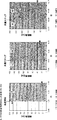
B-NHL (Bargou et al. Science. 2008; 321:974-7) およびB-前駆体ALL (Topp et al. J Clin Oncol. 2011; 29:2493-8) の両方において、ブリナツモマブの安全性および有効性を評価する様々な臨床試験が行われた。B-NHLでは、0.005 mg/m2/日ほどの低い用量を4週間にわたって持続静脈内 (civ) 注入により投与すると、末梢血中のBリンパ腫細胞が完全かつ持続的に除去された。0.015 mg/m2/日という用量レベルにおいて、部分寛解および完全寛解が最初に観察され、0.06 mg/m2/日という用量レベルで処置されたB-NHL患者の大部分が、実質的な腫瘍の退縮を経験した。ブリナツモマブはまた、この適応症において骨髄および肝臓から悪性B細胞を排除した。B-前駆体ALLでは、微小残存病変および再発性または難治性疾患の両方を有する患者は、0.015 mg/m2/日という用量レベルで4週間にわたってciv注入により処置された場合に、血液学的完全寛解を達成した。これらの試験により、一般的に二重特異性単鎖抗体形式の、および特にブリナツモマブの治療可能性が高いという概念の臨床的証明が確立され、そのさらなる開発がB-NHL、ALL、およびCLLにおいて検証された。B-NHLおよびB-前駆体ALLにおけるこれらの試験を通して、いくつかの薬力学的マーカーが評価された。選択された一般的特徴を以下に記載する:T細胞動力学は、用量レベルまたは循環B細胞の存在にかかわらず、非常に際立ったプロファイルを示した。それは、注入の開始後および任意の用量ステップ後の迅速な再分布によって特徴付けられ、すなわち最初の6〜12時間以内に循環T細胞は即座に消失し、それに続いて次の2〜7日間の間に再出現し、ここで初期の高いB細胞数はT細胞再出現の減速した動力学と相関した(図1A)。この過程は、絶対的血清濃度というよりは、むしろブリナツモマブの任意の顕著な用量変化によって誘発されるようであった。加えて、T細胞上のLFA-1に対する可溶性ICAM-1-Fc融合タンパク質の結合を解析することにより、T細胞接着性を処置第1週目を通して測定した。LFA-1高次構造は、注入の開始前の低親和性状態から、注入の開始後および任意の用量ステップ後の中等度の親和性状態に移行した;ICAM-1に対する結合親和性の増加は48時間以内にピークに達し、その後5日以内にベースラインに戻った(図1B)。この知見はT細胞再分布と一致し、再分布過程におけるブリナツモマブ誘導性の内皮に対するT細胞接着の概念を支持した。
Detailed Description of the Invention
In both B-NHL (Bargou et al. Science. 2008; 321: 974-7) and B-precursor ALL (Topp et al. J Clin Oncol. 2011; 29: 2493-8) the safety of blinatumomab and Various clinical trials were conducted to assess efficacy. For B-NHL, doses as low as 0.005 mg / m 2 / day were administered by continuous intravenous (civ) infusion over 4 weeks, resulting in complete and sustained removal of B lymphoma cells in peripheral blood. Partial and complete remissions were initially observed at a dose level of 0.015 mg / m 2 / day, and the majority of B-NHL patients treated at a dose level of 0.06 mg / m 2 / day had substantial tumors. Experienced a regression. Blinatumomab also eliminated malignant B cells from the bone marrow and liver in this indication. For B-precursor ALL, patients with both minimal residual disease and relapsed or refractory disease are hematologic when treated by civ infusion at a dose level of 0.015 mg / m 2 / day for 4 weeks. Complete remission was achieved. These studies established clinical proof of the concept of a generally bispecific single chain antibody format, and in particular the high therapeutic potential of blinatumomab, and its further development in B-NHL, ALL, and CLL Verified. Through these studies on B-NHL and B-precursor ALL, several pharmacodynamic markers were evaluated. Selected general features are described below: T cell kinetics showed a very distinct profile regardless of dose level or presence of circulating B cells. It is characterized by rapid redistribution after the start of infusion and after any dose step, i.e., circulating T cells disappear immediately within the first 6-12 hours, followed by the next 2-7 days. Reappeared in between, where early high B cell counts correlated with slowed kinetics of T cell reappearance (FIG. 1A). This process appeared to be triggered by any significant dose change of blinatumomab rather than absolute serum concentrations. In addition, by analyzing the binding of soluble ICAM-1-F c fusion protein to LFA-1 on T cells was measured through the first week treating T cell adhesion. The LFA-1 conformation transitioned from a low affinity state prior to the start of the injection to a moderate affinity state after the start of the injection and after any dose step; increased binding affinity for ICAM-1 Peaks were reached within 48 hours and then returned to baseline within 5 days (Figure 1B). This finding was consistent with T cell redistribution and supported the concept of T cell adhesion to the blinatumomab-induced endothelium during the redistribution process.
アンジオポエチン-2 (Ang-2) は内皮活性化(またはさらには内皮ストレス)の血清マーカーであり、これは血管裏打ち内皮細胞の細胞質内の、いわゆるバイベル・パラーデ小体中に貯蔵されている。ひとたび内皮細胞が活性化されると、小胞が細胞膜と融合し、予め形成されたAng-2を血清中に放出する。加えて、小胞結合性の接着分子、例えばP-セレクチンは、それによって細胞表面上に出現し、したがって血管裏打ち内皮細胞の接着性はさらに増加する。Ang-2血清濃度の動力学は、LFA-1媒介性のT細胞接着性の増加と類似しており、T細胞再分布中にピークに達し、処置第1週目以内に低下してベースラインに戻った。最大Ang-2血清濃度はしっかりとしたT細胞消失と一致し、この知見から、T細胞再分布の根底にある機構としての、血管裏打ち内皮細胞(したがって活性化された)に対する明白なT細胞接着がさらに示された(図1C)。 Angiopoietin-2 (Ang-2) is a serum marker of endothelial activation (or even endothelial stress), which is stored in the so-called Bibel-Parade body in the cytoplasm of vascular-lined endothelial cells. Once the endothelial cells are activated, the vesicles fuse with the cell membrane and release preformed Ang-2 into the serum. In addition, vesicle-binding adhesion molecules, such as P-selectin, thereby appear on the cell surface, thus further increasing the adhesion of vascular-lined endothelial cells. Ang-2 serum concentration kinetics are similar to LFA-1-mediated increase in T cell adhesion, peaked during T cell redistribution, and decreased to baseline within the first week of treatment. Returned to. Maximum Ang-2 serum concentration is consistent with firm T cell loss, and this finding reveals clear T cell adhesion to vascular-lined endothelial cells (and thus activated) as a mechanism underlying T cell redistribution Was further shown (FIG. 1C).
ブリナツモマブが直接関与しないものの、単球はT細胞が示すのと同様の再分布動力学を示した。この知見は、ブリナツモマブの誘導により内皮にT細胞が結合し、内皮細胞の活性化が生じ、これにより次に付加的な接着分子が上方制御され、それによって他の単核細胞および血小板の幅広い結合が可能になる(すなわち、注入の開始後および任意の用量ステップ後に、循環血小板の数もまた減少した)という概念をさらに支持した。しかしながら、活性化された(例えば、活性化内皮細胞への接着による)単球は、循環中に戻る前に下層組織中に血管外移動する傾向があったため、末梢血中の単球数の回復は長引いたのかもしれない(図1D)。 Monocytes showed similar redistribution kinetics to that shown by T cells, although blinatumomab was not directly involved. This finding suggests that the induction of blinatumomab causes T cells to bind to the endothelium, resulting in endothelial cell activation, which in turn upregulates additional adhesion molecules, thereby broadly binding other mononuclear cells and platelets. Further supported the concept that (ie, after the start of the infusion and after any dose step, the number of circulating platelets was also reduced). However, activated monocytes (eg, due to adhesion to activated endothelial cells) tended to migrate extravascularly into the underlying tissue before returning to circulation, thus restoring the number of monocytes in peripheral blood May have been prolonged (Figure 1D).
上記に基づき、本発明者らは、とりわけ、再指向化哺乳動物T細胞(例えば、ブリナツモマブによって再指向化されるT細胞)によって引き起こされる中枢神経系有害事象 (CNS AE) をもたらし得る、可能な多段階病理学的機序の仮説を立てた。この仮説を図2において説明する。 Based on the above, the inventors can inter alia lead to central nervous system adverse events (CNS AE) caused by redirected mammalian T cells (eg, T cells redirected by blinatumomab) The hypothesis of multi-stage pathological mechanism was established. This hypothesis is illustrated in FIG.
図2A:ブリナツモマブの注入の開始または段階的用量増加により、血管内皮へのT細胞接着が増加する。図2B:接着性T細胞は内皮を活性化し、血管外遊走し始める。活性化された内皮細胞は、他の末梢血白血球、例えば単球を誘引し、これは次に一過性の神経炎症および血液CSF関門の撹乱を引き起こす。 Figure 2A: Initiation of Blinatumomab infusion or escalating dose increases T cell adhesion to vascular endothelium. Figure 2B: Adherent T cells activate the endothelium and begin to extravasate. Activated endothelial cells attract other peripheral blood leukocytes, such as monocytes, which in turn cause transient neuroinflammation and disruption of the blood CSF barrier.
上記仮説を立証するため、本発明者らは、ブリナツモマブを、P-セレクチンの小分子阻害剤であるヘパリン類似物質ペントサンポリ硫酸 (PPS)(Hopfner et al. J Pharm Pharmacol. 2003; 55:697-706) と同時に投与する同時薬物療法スキームを考案した。ブリナツモマブの注入の開始の前および後、ならびにブリナツモマブの任意の段階的用量増加の前および後に、患者にPPSを一過性に注入することにより、第一相臨床試験において抗白血球接着を成功裏に試験した。P-セレクチンは、内皮細胞に対する白血球接着の第1段階を媒介することが知られており、髄膜微小血管を介した軟膜腔および髄膜への循環白血球の血管外遊走において特定の重要な役割を果たすようである (Kivisakk et al. Proc Natl Acad Sci U S A. 2003; 100:8389-94)。
To prove the above hypothesis, the inventors have identified Blinatumomab as a heparin analog pentosan polysulfate (PPS), a small molecule inhibitor of P-selectin (Hopfner et al. J Pharm Pharmacol. 2003; 55: 697- 706) A concomitant pharmacotherapy scheme was devised. Successful anti-leukocyte adhesion in
3名の患者は、ブリナツモマブの5μg/m2/日での注入の開始期、および処置第1週目後の60μg/m2/日への用量ステップ期の両方において、特に静脈内PPS注入を受けた(さらなる詳細については実施例の項もまた参照されたい)。末梢血中のB:T細胞比が低いために、CNS AEを発症するリスクが高かったにもかかわらず(PCT/EP2010/066207において開示された、根底にある理論的根拠を参照されたい)、これら3名の患者のうちの誰も、神経学的有害作用により、ブリナツモマブによる処置を中断する必要がなかった。したがって、本発明者らが大変驚いたことには、再指向化T細胞の各内皮細胞への接着を減少させることにより、これらの患者において予測されたCNS AEを緩和することが実際に可能であった。3名の患者のうちの2名は、ブリナツモマブによる8週間の処置後に完全寛解を達成した;患者1名は、4週間の処置後に安定疾患を有していた。さらに、潜在的CNS AEの緩和のために静脈内PPSを受けた3名の患者は全員、ブリナツモマブの注入の開始または段階的用量増加に際して、T細胞再分布動力学の遅延を示した(CD19陽性標的細胞の非存在下において)(図3)。先に記載されたように、PPSとの同時薬物療法なしでブリナツモマブを受けた患者は一貫して、注入の開始または段階的用量増加の45分後に既に、末梢血中のT細胞数の迅速な減少を示した(図3A、B、およびCに示されるように)。対照的に、注入の開始および用量ステップ期の両方においてPPSによる同時薬物療法を受けた全3名の患者では、注入の開始または段階的用量増加の45分後に、各ベースライン値と比較してT細胞数の減少は認められなかった(図3D、E、およびF)。2つの症例(DおよびE)では、45分の時点でT細胞数が増加さえしていた。1つの症例 (F) では、2時間の時点でT細胞数の増加がなお観察された。末梢血中のT細胞数の減少は、ブリナツモマブの注入の開始または段階的用量増加の2時間後よりも前には検出されなかったため、再分布過程およびひいては根底にある血管内皮への白血球接着は、静脈内PPSによる介入により明らかに遅くなった。 Three patients, onset of infusion at 5 [mu] g / m 2 / day Burinatsumomabu, and in both dose step-life of the treatment to the first week after the 60 [mu] g / m 2 / day, particularly intravenous PPS infusion (See also the Examples section for further details). Despite the high risk of developing CNS AE due to the low B: T cell ratio in peripheral blood (see the underlying rationale disclosed in PCT / EP2010 / 066207) None of these three patients had to discontinue treatment with blinatumomab due to neurological adverse effects. Therefore, we were very surprised that it was actually possible to alleviate the predicted CNS AE in these patients by reducing the adhesion of redirected T cells to each endothelial cell. there were. Two of the three patients achieved complete remission after 8 weeks of treatment with blinatumomab; 1 patient had stable disease after 4 weeks of treatment. In addition, all three patients who received intravenous PPS to alleviate potential CNS AEs showed a delay in T cell redistribution kinetics at the start of blinatumomab infusion or escalating dose (CD19 positive) In the absence of target cells) (Figure 3). As previously described, patients who received blinatumomab without concurrent medication with PPS consistently had a rapid increase in the number of T cells in the peripheral blood, already 45 minutes after the start of infusion or escalating dose. There was a decrease (as shown in FIGS. 3A, B, and C). In contrast, all three patients who received concurrent drug therapy with PPS both at the start of the infusion and at the dose step, compared to each baseline value 45 minutes after the start of infusion or escalation dose There was no decrease in T cell numbers (FIGS. 3D, E, and F). In two cases (D and E), the number of T cells even increased at 45 minutes. In one case (F), an increase in T cell count was still observed at 2 hours. No decrease in the number of T cells in the peripheral blood was detected prior to the start of infusion of blinatumomab or 2 hours after the gradual dose increase, so leukocyte adhesion to the redistribution process and hence the underlying vascular endothelium was Obviously slowed by intervention with intravenous PPS.
したがって、患者において、PPSの予知されるおよび意図される作用機序のための薬力学的マーカー(すなわち、T細胞再分布動力学の遅延)が同定された。PPSによる同時薬物療法を受けた患者の臨床経過は、これらのバイオマーカーの観察、およびまた中枢神経系有害事象の病理学的機序に関する現在の仮説の予測と一致する。したがって、血管内皮への白血球接着を妨げること(すなわち、抗白血球接着)は、ブリナツモマブ (AMG 103) などの、患者における標的細胞に対するT細胞の再指向化を含む治療によって引き起こされるCNS AEなどの副作用を妨げるかまたは改善するための、作用機序に基づいた介入アプローチである。 Therefore, pharmacodynamic markers (ie, delay in T cell redistribution kinetics) for the prognostic and intended mechanism of action of PPS have been identified in patients. The clinical course of patients receiving concurrent medication with PPS is consistent with observations of these biomarkers, and also with current hypothetical predictions regarding the pathological mechanisms of central nervous system adverse events. Therefore, interfering with leukocyte adhesion to the vascular endothelium (ie anti-leukocyte adhesion) is a side effect such as CNS AE caused by treatments that include T cell redirection to target cells in patients such as blinatumomab (AMG 103). It is an interventional approach based on the mechanism of action to prevent or ameliorate.
再指向化(ヒト)T細胞の各(ヒト)内皮細胞への最初の結合が、実際に上記のCNS AEの原因であることをさらに調べかつ確認するため、本発明者らはさらに、インビトロ系において流体力学的流動条件下で、(ヒト脳)微小血管内皮細胞上での/への再指向化(ヒト)T細胞および他の白血球のローリング、繋留、および接着をシミュレートする試験系(「流動系」)を確立した。この試験系のさらなる詳細は、実験設定を極めてかつ十分に詳細に説明している添付の実施例から導き出すことができる(実施例の項を参照されたい)。この流動系において、細胞のローリング速度および接着細胞の数は、当業者によって容易に測定され得る。内皮細胞上での/への末梢血細胞の多段階のローリングおよび接着過程のうちの任意の(分子)段階(すなわち、必要要件)の妨害は、細胞のローリング速度および接着細胞の数の両方に影響を及ぼすことが予測される。 In order to further investigate and confirm that the initial binding of redirected (human) T cells to each (human) endothelial cell is indeed responsible for the above-mentioned CNS AE, we further developed an in vitro system. A test system that simulates the rolling, tethering, and adhesion of (human) T cells and other leukocytes on / to (human brain) microvascular endothelial cells under hydrodynamic flow conditions in Established a fluid system ”). Further details of this test system can be derived from the accompanying examples which describe the experimental setup in great detail and sufficient (see the Examples section). In this flow system, the cell rolling rate and the number of adherent cells can be easily measured by those skilled in the art. Interference of any (molecular) stage (ie, requirement) of multistage rolling and adhesion processes of peripheral blood cells on / to endothelial cells affects both cell rolling rate and number of adherent cells Is expected to affect.
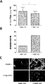
流動系にCD3特異的結合ドメイン、特にブリナツモマブを添加すると(それによって再指向化T細胞を生じる)、ヒト脳微小血管内皮細胞 (HBMEC) 上でのT細胞ローリング速度は迅速かつ有意に低下した。同時に、HBMECは、その細胞表面上の接着分子P-セレクチン、ICAM-1、およびVCAM-1の上方制御によって示されるように、これらの再指向化T細胞によって活性化された(図6Bを参照されたい)。しかしながら、ブリナツモマブなしでT細胞を添加すると(非再指向化T細胞を生じる)、前述の内皮接着分子の発現は影響を受けなかった(図6Bを参照されたい)。これらの知見は、患者において見られ、かつ本明細書において詳述されるT細胞再分布と類似しており、かつ明白に対応する。さらに、流動系にP-セレクチン遮断剤PPSを添加すると(すなわち、HBMECのPPSとのプレインキュベーション)、HBMEC上でのT細胞ローリング速度のブリナツモマブ誘導性の低下が効率的に阻止され得た。流動系へのブリナツモマブの添加後に実証されたように、PPSとのプレインキュベーションは、任意のブリナツモマブ誘導性効果を中和することができ、T細胞ローリング速度を、流動系にブリナツモマブを添加しない場合にT細胞ローリング速度について観察されたレベルに匹敵するレベルにまで戻すことができた。加えて、HBMECは、PPSが流動系中に存在する場合、P-セレクチンの細胞表面発現の減少によって示されるように、あまり活性化されなかった。これらの知見は、哺乳動物T細胞の哺乳動物内皮細胞への結合を減少させるかまたは阻害する化合物(P-セレクチン遮断剤PPSなど)の添加が実際に、内皮細胞へのT細胞接着を減少させる/遅延させることができ、それによって、患者における標的細胞に対するT細胞の再指向化を含む治療によって引き起こされる臨床的有害事象を予防する、改善する、および/または処置することができるという臨床的知見を支持する。 Addition of a CD3-specific binding domain, particularly blinatumomab, to the flow system (which results in redirected T cells) rapidly and significantly reduced the rate of T cell rolling on human brain microvascular endothelial cells (HBMEC). At the same time, HBMEC was activated by these redirected T cells as shown by the up-regulation of adhesion molecules P-selectin, ICAM-1 and VCAM-1 on its cell surface (see Figure 6B) I want to be) However, when T cells were added without blinatumomab (resulting in non-redirected T cells), the expression of the aforementioned endothelial adhesion molecules was not affected (see FIG. 6B). These findings are similar to and clearly correspond to the T cell redistribution found in patients and detailed herein. Furthermore, when the P-selectin blocking agent PPS was added to the flow system (ie, preincubation with HBMEC PPS), the blinatumomab-induced decrease in T cell rolling rate on HBMEC could be effectively prevented. As demonstrated after the addition of blinatumomab to the flow system, preincubation with PPS can neutralize any blinatumomab-inducing effect, and the T cell rolling rate can be increased when no blinatumomab is added to the flow system. We were able to return to a level comparable to that observed for T cell rolling rate. In addition, HBMEC was less activated when PPS was present in the flow system, as shown by a decrease in cell surface expression of P-selectin. These findings indicate that the addition of compounds that reduce or inhibit the binding of mammalian T cells to mammalian endothelial cells (such as the P-selectin blocking agent PPS) actually reduces T cell adhesion to endothelial cells Clinical finding that can / can be delayed, thereby preventing, ameliorating and / or treating clinical adverse events caused by therapies, including redirecting T cells to target cells in the patient Support.
したがって、確立された流動系は、流体力学的流動条件下で、例えばブリナツモマブなどのCD3特異的結合ドメインを用いて達成され得る、標的細胞に対するT細胞の再指向化を含む薬物で処置された患者においてT細胞再分布中に観察されるような、血管裏打ち内皮細胞上での/への再指向化T細胞のとりわけT細胞ローリングおよび接着を正確に模倣する。特に、患者へのPPSの注入および流動系へのPPSの添加が、それぞれT細胞再分布およびT細胞ローリング速度に及ぼす効果は、PPSが内皮細胞へのT細胞接着を明らかに妨げるという点で、非常に匹敵する。CNS AEの病理学的機序に関する現在の仮説によると、この妨害(すなわち、哺乳動物T細胞および特に再指向化T細胞のような白血球の哺乳動物内皮細胞に対する結合の減少または阻害である抗白血球接着)は、患者への標的細胞に対するT細胞の再指向化(例えば、CD3特異的結合ドメイン、または本明細書の他所で説明されるCARによって達成され得る)を含む薬物の投与によって引き起こされる、CNS AEなどの任意の有害副作用を処置し得る、予防し得る、または改善し得る。さらに、本明細書において例証される、確立された流動系は、患者における標的細胞に対するT細胞の再指向化を含む治療によって引き起こされる臨床的有害事象を予防する、および/または改善する、および/または処置する方法において使用するための化合物を同定する方法において、当業者が用いることができる、および当業者によって使用され得る。例証される流動系はしたがって、CD3特異的結合ドメインによって引き起こされるCNS AEの予防または改善のために、例えば該CD3特異的結合ドメインによる患者に処置の前に、それと同時に、および/またはその後に投与され得る、抗接着特性を有する化合物(特に、哺乳動物T細胞の哺乳動物内皮細胞への結合を減少させるかまたは阻害する化合物)を同定/定義するのに適している。言い換えると、流動系において、白血球、特にT細胞、および最も好ましくは再指向化T細胞に対する任意の抗接着効果(すなわち、例えば、流動系へのPPSの添加に関して観察された効果に匹敵する、血管裏打ち内皮細胞上でのT細胞ローリング速度のCD3特異的結合ドメイン誘導性の低下の復帰)を示す化合物は、CD3特異的結合ドメインによる処置の前に、それと同時に、および/またはその後に投与された場合に、該CD3特異的結合ドメインによる患者の処置によって引き起こされる任意のCNS AEを予防するかまたは改善することが予測される。このような化合物の同定、改変、および/または確認は、それが十分に確立された方法論を使用するという理由で、当業者にとって簡潔な課題である。 Thus, an established flow system can be achieved under hydrodynamic flow conditions using a CD3-specific binding domain such as blinatumomab, for example, a patient treated with a drug that includes T cell redirection to a target cell Accurately mimic T cell rolling and adhesion of redirected T cells on / to vascular-lined endothelial cells, as observed during T cell redistribution. In particular, the effect of injecting PPS into the patient and adding PPS to the flow system on T cell redistribution and T cell rolling rate, respectively, is that PPS clearly interferes with T cell adhesion to endothelial cells. Very comparable. According to the current hypothesis on the pathological mechanism of CNS AE, this interference (ie, anti-leukocyte which is a reduction or inhibition of binding of leukocytes such as mammalian T cells and especially redirected T cells to mammalian endothelial cells) Adhesion) is caused by administration of a drug comprising T cell redirecting to a target cell to a patient (e.g., can be accomplished by a CD3-specific binding domain, or CAR as described elsewhere herein), Any adverse side effects such as CNS AE can be treated, prevented or ameliorated. In addition, the established flow system illustrated herein prevents and / or ameliorates clinical adverse events caused by treatments including redirecting T cells to target cells in a patient, and / or Or, can be used by and can be used by one of ordinary skill in the art for identifying compounds for use in the methods of treatment. The illustrated flow system is therefore administered prior to, simultaneously with, and / or after treatment, eg, to a patient with the CD3-specific binding domain, for prevention or amelioration of CNS AE caused by the CD3-specific binding domain Suitable for identifying / defining compounds that have anti-adhesive properties, in particular compounds that reduce or inhibit the binding of mammalian T cells to mammalian endothelial cells. In other words, in the flow system, any anti-adhesion effect on leukocytes, especially T cells, and most preferably redirected T cells (i.e., blood vessels comparable to the effect observed with the addition of PPS to the flow system, for example) Compounds exhibiting a reversal of CD3-specific binding domain-induced decrease in T cell rolling rate on the lined endothelial cells were administered prior to, simultaneously with and / or after treatment with the CD3-specific binding domain In some cases, it is expected to prevent or ameliorate any CNS AE caused by treatment of the patient with the CD3-specific binding domain. The identification, modification, and / or confirmation of such compounds is a concise task for those skilled in the art because it uses well-established methodologies.
したがって本発明者らは、本明細書において同定された技術的問題を解決するための道を開いた。 The inventors have therefore opened the way for solving the technical problems identified herein.
定義:
本明細書で用いられる場合、「1つの (a)」、「1つの (an)」、および「その」という単数形は、文脈上明白に別の意味を示していない限り、複数の指示対象も含むことに留意しなければならない。したがって、例えば、「1つの試薬」への言及は、そのような種々の試薬の1つまたは複数を含み、「その方法」への言及は、本明細書に記載される方法に対して修正または置換され得る、当業者に公知の等価な段階および方法への言及を含む。
Definition:
As used herein, the singular forms “a”, “an”, and “the” include plural referents unless the context clearly indicates otherwise. It should be noted that also includes: Thus, for example, reference to “a reagent” includes one or more of such various reagents, and reference to “the method” is a modification or amendment to the methods described herein. Includes references to equivalent steps and methods known to those skilled in the art that may be substituted.
別段の指示がない限り、一連の要素を先行する「少なくとも」という用語は、その一連の中のあらゆる要素を指すと理解されるべきである。当業者は、本明細書に記載される本発明の特定の態様に対する多くの等価物を認識するか、または通常の実験だけを用いてこれらを確認することができるであろう。このような等価物は、本発明によって包含されることが意図される。 Unless otherwise indicated, the term “at least” preceding a series of elements should be understood to refer to every element in the series. Those skilled in the art will recognize many equivalents to the specific embodiments of the invention described herein or may be able to ascertain these using only routine experimentation. Such equivalents are intended to be encompassed by the present invention.
「および/または」という用語は、本明細書で用いられる場合には常に、「および」、「または」、および該用語によってつながれた要素のすべてまたは任意の他の組み合わせ」の意味を含む。 The term “and / or” whenever used herein includes the meaning of “and”, “or” and all or any other combination of the elements linked by the term.
本明細書で用いられる「約」または「およそ」という用語は、所与の値または範囲の±20%以内、好ましくは±15%以内、より好ましくは±10%以内、および最も好ましくは±5%以内を意味する。 As used herein, the term “about” or “approximately” is used within ± 20%, preferably within ± 15%, more preferably within ± 10%, and most preferably ± 5% of a given value or range. Means within%.
以下の本明細書および特許請求の範囲を通して、文脈上明白に別の意味が要求されない限り、「含む (comprise)」という語、ならびに「含む (comprises)」および「含む (comprising)」などの変化形は、記載の整数もしくは段階または整数もしくは段階の群を含むことを意味するが、任意の他の整数もしくは段階または整数もしくは段階の群を除外することを意味するものではないことが理解されよう。本明細書で用いられる「含む (comprising)」という用語は、「含有する」もしくは「含む (including)」という用語、または場合によっては、本明細書で用いられる「有する」という用語と置き換えられ得る。 Throughout the following specification and claims, the word “comprise” and variations such as “comprises” and “comprising”, unless the context clearly indicates otherwise. It will be understood that the form is meant to include the stated integers or steps or groups of integers or steps, but is not meant to exclude any other integers or steps or groups of integers or steps. . As used herein, the term “comprising” may be replaced with the term “comprising” or “including”, or in some cases, the term “comprising” as used herein. .
本明細書で用いられる「からなる」は、特許請求の範囲の要素に規定されていないいかなる要素、段階、または成分も除外する。本明細書で用いられる場合、「から本質的になる」は、特許請求の範囲の基礎的でかつ新規な特徴に実質的に影響を及ぼさない材料または段階を除外しない。 As used herein, “consisting of” excludes any element, step, or ingredient not specified in the claim element. As used herein, “consisting essentially of” does not exclude materials or steps that do not materially affect the basic and novel characteristics of the claim.
本明細書における各場合において、「含む」、「から本質的になる」、および「からなる」という用語のいずれも、他の2つの用語のいずれかと置き換えられ得る。 In each case herein, any of the terms “comprising”, “consisting essentially of”, and “consisting of” may be replaced with either of the other two terms.
本発明は、1つの態様において、患者における標的細胞に対するT細胞の再指向化を含む治療によって引き起こされる臨床的有害事象を予防する、および/または改善する、および/または処置する方法において使用するための、哺乳動物T細胞の哺乳動物内皮細胞への結合を減少させるかまたは阻害する化合物に関する。 The present invention, in one embodiment, for use in a method of preventing and / or ameliorating and / or treating a clinical adverse event caused by a therapy comprising redirecting T cells to target cells in a patient. Relates to compounds that reduce or inhibit the binding of mammalian T cells to mammalian endothelial cells.
哺乳動物T細胞の「結合」という用語は、典型的に、通常は白血球血管外遊走として示される過程である、血液系からの白血球の移動を特徴とする周知の連続的段階のいずれかを含むことが理解されるべきである。前述の段階は、「白血球のローリング」(白血球は内皮細胞に緩く付着するが、血流と共になお引っ張られ、その結果として内皮細胞の表面上で白血球のローリング運動が起こる);「白血球の繋留」(場合によっては堅固な付着として示される‐白血球は内皮細胞に堅固に付着するが、この相互作用のための受容体は、ローリング過程に関与するものとは異なる)、および「血管外遊出段階」(場合によっては血管外移動としても示される‐前もって主に球状白血球は内皮上に広がり、内皮バリアを通して能動的に血管外移動する)を含む。本発明の化合物は、これらの段階のすべて、またはこれらの段階のうちのいくつかのみ、またはこれらの段階のうちの1つのみに影響を及ぼす。 The term “binding” of mammalian T cells typically includes any of the well-known sequential stages characterized by the migration of leukocytes from the blood system, a process usually indicated as leukocyte extravasation. It should be understood. The aforementioned stage is “white blood cell rolling” (the white blood cells are loosely attached to the endothelial cells, but are still pulled along with the blood flow, resulting in a white blood cell rolling motion on the surface of the endothelial cells); (In some cases shown as tight attachment-leukocytes adhere firmly to endothelial cells, but the receptors for this interaction are different from those involved in the rolling process), and "extravasation stage" (Sometimes also indicated as extravascular movement—predominantly spherical leukocytes spread on the endothelium and actively extravasate through the endothelial barrier in advance). The compounds of the invention affect all of these stages, only some of these stages, or only one of these stages.
本発明に適用される「化合物」(すなわち、哺乳動物T細胞の哺乳動物内皮細胞への結合を減少させるかまたは阻害する化合物)は、言わば、例えば、T細胞側に、すなわちT細胞接着分子に、または内皮細胞側に、すなわち内皮細胞接着分子に作用する化合物である。しかしながら、化合物はまた、T細胞側と内皮細胞側の両方に作用する化合物であってもよい。該化合物は、本発明に適用された場合、哺乳動物T細胞の哺乳動物内皮細胞への結合を減少させるかまたは阻害する。したがって、本発明の化合物は、T細胞もしくは内皮細胞またはその両方に対して抗接着効果をもたらす化合物である。例えば、化合物はT細胞接着分子および内皮細胞接着分子の両方に作用し得るか、または2つもしくはそれ以上の化合物の混合物が適用され、1つがT細胞接着分子に作用し、さらなる1つ(複数可)が内皮細胞接着分子に作用する。抗接着効果は好ましくは、化合物が、哺乳動物T細胞の哺乳動物内皮細胞への結合を減少させるかまたは阻害することを含む。本発明の好ましい化合物を表1に示す。 A “compound” (ie, a compound that reduces or inhibits the binding of mammalian T cells to mammalian endothelial cells) as applied to the present invention is, for example, on the T cell side, ie, on a T cell adhesion molecule. Or a compound that acts on the endothelial cell side, that is, an endothelial cell adhesion molecule. However, the compound may also be a compound that acts on both the T cell side and the endothelial cell side. The compounds, when applied to the present invention, reduce or inhibit the binding of mammalian T cells to mammalian endothelial cells. Accordingly, the compounds of the present invention are compounds that provide an anti-adhesive effect on T cells or endothelial cells or both. For example, a compound can act on both T cell adhesion molecules and endothelial cell adhesion molecules, or a mixture of two or more compounds can be applied, one acting on a T cell adhesion molecule and an additional one (s) Yes) acts on endothelial cell adhesion molecules. The anti-adhesion effect preferably comprises the compound reducing or inhibiting the binding of mammalian T cells to mammalian endothelial cells. Preferred compounds of the present invention are shown in Table 1.
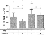
本発明の化合物、すなわち哺乳動物T細胞の哺乳動物内皮細胞への結合を減少させるかまたは阻害する化合物は、+10 ng/ml AMG 103の存在下で、HBMEC上の平均T細胞ローリング速度 ± SD(μm/秒)を約30、40、50、60、70、80、90、100%、またはさらにはそれ以上上昇させることができることもまた想定される(実施例2を参照されたい‐流動系に10 ng/mlのブリナツモマブを添加してから45分後に観察される平均T細胞ローリング速度のブリナツモマブ誘導性の低下に及ぼすPPSの影響を、長期条件において評価した。AMG 103は、HBMEC上での平均T細胞ローリング速度 ± SDを430 ± 92μm/秒 (-AMG 103) から281 ± 96μm/秒 (+AMG 103) まで有意に低下させたのに対して、流動系にPPSをさらに添加すると、この低下は、AMG 103の非存在下でも観察されたような(すなわち、-AMG 103、+PPS;442 ± 156μm/秒)、483 ± 157μm/秒という平均T細胞ローリング速度 ± SDにまで戻った。
Compounds of the invention, ie compounds that reduce or inhibit the binding of mammalian T cells to mammalian endothelial cells, mean T cell rolling rate on HBMEC ± SD in the presence of +10 ng /
T細胞は、接着分子、すなわちT細胞接着分子および内皮細胞接着分子を介して内皮細胞と相互作用することが知られている。T細胞接着分子および内皮細胞接着分子はいずれも、インテグリン、セレクチンのファミリー、および免疫グロブリンG (IgG) スーパーファミリーに属する。後者は、免疫グロブリンドメインを有することを特徴とする。内皮細胞の表面上に存在するヒアルロン酸の受容体としてのCD44もまた、T細胞接着分子として見なされる。 T cells are known to interact with endothelial cells through adhesion molecules, namely T cell adhesion molecules and endothelial cell adhesion molecules. Both T cell adhesion molecules and endothelial cell adhesion molecules belong to the integrin, selectin family, and immunoglobulin G (IgG) superfamily. The latter is characterized by having an immunoglobulin domain. CD44 as a receptor for hyaluronic acid present on the surface of endothelial cells is also regarded as a T cell adhesion molecule.
したがって、T細胞の内皮細胞への結合を減少させるかまたは阻害する好ましい化合物は、それぞれインテグリンアンタゴニスト、セレクチンアンタゴニスト、Igスーパーファミリー細胞接着分子アンタゴニスト、またはCD44アンタゴニストである。したがって、本発明との関係において適用される化合物は、好ましくはそれぞれインテグリンアンタゴニスト、セレクチンアンタゴニスト、Igスーパーファミリー細胞接着分子アンタゴニスト、またはCD44アンタゴニストである。これらのアンタゴニストのいずれかは、本発明との関係において、哺乳動物T細胞の哺乳動物内皮細胞への結合を減少させるかまたは阻害することができる。 Accordingly, preferred compounds that reduce or inhibit T cell binding to endothelial cells are integrin antagonists, selectin antagonists, Ig superfamily cell adhesion molecule antagonists, or CD44 antagonists, respectively. Accordingly, the compounds applied in the context of the present invention are preferably integrin antagonists, selectin antagonists, Ig superfamily cell adhesion molecule antagonists, or CD44 antagonists, respectively. Any of these antagonists can reduce or inhibit the binding of mammalian T cells to mammalian endothelial cells in the context of the present invention.
インテグリンアンタゴニストは、当技術分野において一般的に公知である;例えば、Curley et al. (1999), Cellular and Molecular Life Science 56, 427-441を参照されたい。セレクチンアンタゴニストは、当技術分野において公知である;例えば、Lefer (2010), Ann. Rev. Pharmacol Toxicol 40 283-294を参照されたい。CD44アンタゴニストは、当技術分野において公知である;例えば、Hirota-Takahata (2007), J. Antiobiotics 60, 633-639を参照されたい。同じことが、IgGスーパーファミリー細胞接着分子アンタゴニストについても当てはまる。
Integrin antagonists are generally known in the art; see, for example, Curley et al. (1999), Cellular and Molecular Life Science 56, 427-441. Selectin antagonists are known in the art; see, for example, Lefer (2010), Ann.
T細胞の内皮細胞への結合を減少させるかまたは阻害する化合物は、好ましくはインテグリンアンタゴニスト、セレクチンアンタゴニスト、Igスーパーファミリー細胞接着分子アンタゴニスト、またはCD44アンタゴニストであり、好ましくは、本明細書において、特に実施例2において記載されているように、フローチャンバーアッセイにそのような潜在的アンタゴニストを適用することによって試験および/または同定することができ、これによりアンタゴニストは好ましくは、上記のように、+10 ng/ml AMG 103の存在下で、HBMEC上の平均T細胞ローリング速度 ± SD(μm/秒)を約30、40、50、60、70、80、90、100%、またはさらにはそれ以上上昇させる。
The compound that reduces or inhibits the binding of T cells to endothelial cells is preferably an integrin antagonist, a selectin antagonist, an Ig superfamily cell adhesion molecule antagonist, or a CD44 antagonist, preferably as implemented herein. As described in Example 2, such potential antagonists can be tested and / or identified by applying them to a flow chamber assay, whereby the antagonist is preferably +10 ng as described above. Increase the average T cell rolling rate ± SD (μm / sec) on HBMEC by approximately 30, 40, 50, 60, 70, 80, 90, 100%, or even more in the presence of /
上記のようにT細胞側に作用する化合物は、
(a) T細胞接着分子に結合することができる、
(b) T細胞接着分子の結合部位を遮断することができる、および/または
(c) T細胞接着分子の発現を阻害するかまたは減少させる、
ことを特徴とする。
As mentioned above, the compound acting on the T cell side is
(a) can bind to T cell adhesion molecules,
(b) can block the binding site of the T cell adhesion molecule, and / or
(c) inhibits or reduces the expression of T cell adhesion molecules,
It is characterized by that.
「T細胞接着分子」とは、T細胞の表面上に存在し、かつ内皮細胞などの他の細胞に対するT細胞の接着において機能するかまたはその接着に役割を果たす分子であり、後者が本発明との関係において好ましい。典型的に、T細胞接着分子は内皮細胞の接着分子と相互作用する。したがって内皮細胞の接着分子は、T細胞接着分子のリガンドと見なされ得る。T細胞接着分子と内皮細胞の表面上に存在するリガントとの間の相互作用は、通常は、T細胞接着分子の結合部位(すなわちリガンド結合部位)とその指定されたリガンド、すなわち内皮細胞の接着分子との間で起こる。 A “T cell adhesion molecule” is a molecule that is present on the surface of a T cell and functions in or plays a role in adhesion of T cells to other cells such as endothelial cells. It is preferable in relation to Typically, T cell adhesion molecules interact with endothelial cell adhesion molecules. Thus, endothelial cell adhesion molecules can be regarded as ligands for T cell adhesion molecules. The interaction between a T cell adhesion molecule and a ligand present on the surface of an endothelial cell usually involves the adhesion of the T cell adhesion molecule binding site (ie, the ligand binding site) and its designated ligand, ie, endothelial cells. It happens between molecules.
本明細書に記載されるT細胞接着分子は、好ましくは免疫グロブリンスーパーファミリードメインを有する。このような接着分子は好ましくは、好ましくはRGD結合ドメインを有するインテグリン、例えば、α4-インテグリン、αL-β2-インテグリン、αL-インテグリン、β7-インテグリンなど;セレクチン、例えばL-セレクチンなど、またはCD44である。T細胞は、インテグリンおよび/またはセレクチンおよび/またはCD44を使用して、血管および/またはリンパ節中にまたはそこから移動して、次いで例えば他の組織、軟膜腔または血管周囲腔中に移動する。CD44は主にヒアルロン酸の受容体として働き、これは血管を裏打ちする内皮細胞の表面上に存在する。 The T cell adhesion molecules described herein preferably have an immunoglobulin superfamily domain. Such an adhesion molecule is preferably an integrin preferably having an RGD binding domain, such as α4-integrin, αL-β2-integrin, αL-integrin, β7-integrin, etc .; selectins, such as L-selectin, or CD44 is there. T cells migrate into or out of blood vessels and / or lymph nodes using integrins and / or selectins and / or CD44 and then migrate, for example, into other tissues, the pial space or the perivascular space. CD44 acts primarily as a receptor for hyaluronic acid, which is present on the surface of endothelial cells lining the blood vessels.
本発明の化合物は、1つの態様において、T細胞接着分子に結合することができることを特徴とする。したがって、化合物はT細胞接着分子と結合し、ひいては好ましくはこれを脱落させ、その結果としてT細胞接着分子は減少し、好ましくはもはや内皮接着分子と相互作用することができない。化合物がT細胞接着分子に結合した結果として、T細胞は少なくとも減少し、好ましくはもはや内皮接着分子と相互作用することができない。T細胞接着分子に結合する好ましい化合物は、別の分子に結合することができる分子、例えばリポカリン変異タンパク質、または抗体、好ましくはモノクローナル抗体などである。好ましくは、結合分子は、T細胞接着分子に特異的に標的化され得る。抗体はこのような基準を満たしているため、T細胞接着分子に結合する好ましい化合物は、抗体、好ましくはナタリズマブ、エファリズマブ、またはエトロリズマブなどのモノクローナル抗体である。 In one embodiment, the compounds of the invention are characterized by being able to bind to T cell adhesion molecules. Thus, the compound binds to the T cell adhesion molecule and thus preferably sheds it, resulting in a decrease in the T cell adhesion molecule, preferably no longer interacting with the endothelial adhesion molecule. As a result of the compound binding to the T cell adhesion molecule, the T cells are at least reduced and preferably no longer interact with the endothelial adhesion molecule. A preferred compound that binds to a T cell adhesion molecule is a molecule that can bind to another molecule, such as a lipocalin mutein, or an antibody, preferably a monoclonal antibody. Preferably, the binding molecule can be specifically targeted to a T cell adhesion molecule. Since antibodies meet such criteria, preferred compounds that bind to T cell adhesion molecules are antibodies, preferably monoclonal antibodies such as natalizumab, efalizumab, or etrolizumab.
本発明の化合物は、加えてまたはその代わりに、T細胞接着分子の結合部位を遮断することができることを特徴とする。「遮断」とは、T細胞接着分子の結合部位が、内皮細胞上のそのリガンドと、好ましくはそのリガンドの結合部位と相互するのを妨げることを意味する。化合物がT細胞接着分子の結合部位を遮断した結果として、T細胞は少なくとも減少し、好ましくはもはや内皮接着分子と相互作用することができない。T細胞接着分子の結合部位の非限定的な例は、ICAM-1、ICAM-2、ICAM-3、VCAM-1、MadCAM、GlyCAM、CD31 (PECAM-1)、CD62P(P-セレクチン)、CD62E(E-セレクチン)、CD62L、フィブリノーゲン、およびコンドロイチンに対する結合部位である。 The compounds of the invention are characterized in that they can additionally or alternatively block the binding site of T cell adhesion molecules. “Blocking” means preventing the binding site of a T cell adhesion molecule from interacting with its ligand on endothelial cells, preferably with the binding site of that ligand. As a result of the compound blocking the binding site of the T cell adhesion molecule, T cells are at least reduced and preferably no longer interact with the endothelial adhesion molecule. Non-limiting examples of T cell adhesion molecule binding sites include ICAM-1, ICAM-2, ICAM-3, VCAM-1, MadCAM, GlyCAM, CD31 (PECAM-1), CD62P (P-selectin), CD62E Binding site for (E-selectin), CD62L, fibrinogen, and chondroitin.
T細胞接着分子の結合部位を遮断する好ましい化合物は、内皮細胞の細胞接着分子の可溶性断片であり、該可溶性断片は好ましくは、T細胞へのシグナルの伝達などの生理学的効果をもたらすことなくT細胞接着分子の結合部位に結合するように改変される。 Preferred compounds that block the binding site of T cell adhesion molecules are soluble fragments of endothelial cell adhesion molecules, which preferably do not produce physiological effects such as transmission of signals to T cells. It is modified to bind to the binding site of the cell adhesion molecule.
T細胞接着分子の結合部位を遮断する好ましい化合物は、T細胞接着分子上、例えばそれぞれVLA-4および/またはLPAM-1上のICAM-1、ICAM-2、ICAM-3、VCAM-1、MadCAM、GlyCAM、CD31、CD62P、CD62E、CD62L、フィブリノーゲン、および/またはコンドロイチン結合部位を遮断する抗体、好ましくはモノクローナル抗体である。このような抗体はナタリズマブであり、したがってこれは本発明におい適用される好ましい化合物である。本発明に適用される化合物として同様に好ましいのは、T細胞接着分子上のICAM-1結合部位を遮断する抗体、好ましくはモノクローナル抗体である。このような抗体はエファリズマブであり、したがってこれは本発明に適用される好ましい化合物である。 Preferred compounds that block the binding site of the T cell adhesion molecule are ICAM-1, ICAM-2, ICAM-3, VCAM-1, MadCAM on the T cell adhesion molecule, eg, VLA-4 and / or LPAM-1, respectively. , GlyCAM, CD31, CD62P, CD62E, CD62L, fibrinogen, and / or an antibody that blocks the chondroitin binding site, preferably a monoclonal antibody. Such an antibody is natalizumab and is therefore a preferred compound applied in the present invention. Also preferred as a compound applied to the present invention is an antibody, preferably a monoclonal antibody, that blocks the ICAM-1 binding site on the T cell adhesion molecule. Such an antibody is efalizumab and is therefore a preferred compound applied in the present invention.
本発明に適用される化合物としてさらに好ましいのは、T細胞接着分子上のVCAM-1および/またはMadCAM結合部位を遮断する抗体、好ましくはモノクローナル抗体である。このような抗体はエトロリズマブであり、したがってこれは本発明に適用される好ましい化合物である。 Further preferred as compounds applied in the present invention are antibodies, preferably monoclonal antibodies, that block the VCAM-1 and / or MadCAM binding sites on T cell adhesion molecules. Such an antibody is etrolizumab and is therefore a preferred compound applied in the present invention.
別の好ましい化合物は、T細胞接着分子の結合部位を遮断する小分子であるAJM300である。 Another preferred compound is AJM300, a small molecule that blocks the binding site of T cell adhesion molecules.
別の好ましい化合物は、T細胞接着分子上のICAM-1、ICAM-2、および/またはICAM-3結合部位を遮断する小分子であるSAR 1118である。 Another preferred compound is SAR 1118, a small molecule that blocks ICAM-1, ICAM-2, and / or ICAM-3 binding sites on T cell adhesion molecules.
別のさらなる好ましい化合物は、インテグリンαLβ2および/またはαMβ2のアンタゴニストとして働く小分子であるBOL-303225-Aである。 Another further preferred compound is BOL-303225-A, a small molecule that acts as an antagonist of integrin αLβ2 and / or αMβ2.
別の好ましい化合物は、キレート剤、好ましくはカルシウムなどの二価陽イオンのキレート剤である。好ましいキレート剤は、エチレンジアミン四酢酸 (EDTA) である。 Another preferred compound is a chelating agent, preferably a chelating agent of a divalent cation such as calcium. A preferred chelating agent is ethylenediaminetetraacetic acid (EDTA).
ヒアルロン酸 (HA) またはコンドロイチン硫酸は、いずれもCD44上のHAおよびE-セレクチン結合部位を遮断し、それによってT細胞上のCD44と内皮細胞との間の相互作用を遮断することが知られているため、さらなる好ましい化合物である。 Hyaluronic acid (HA) or chondroitin sulfate are both known to block the HA and E-selectin binding sites on CD44, thereby blocking the interaction between CD44 on endothelial cells and endothelial cells Therefore, it is a further preferred compound.
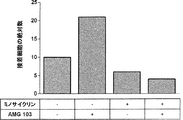
本発明の化合物は、加えてまたはその代わりに、T細胞接着分子の発現を阻害するかまたは減少させることを特徴とする。このような化合物は、遺伝子発現を抑制する、転写、スプライシング、または翻訳を妨げるなど、T細胞接着分子をコードする遺伝子の転写および/または翻訳を含む発現に作用し得る。これは例えば、当技術分野で公知の手段および方法により、RNA干渉によって達成され得る。しかしながら、LFA-1発現を減少させるミノサイクリン、L-セレクチン発現を減少させる(アセチル-)サリチル酸、CD44発現を減少させるアスチルビンまたはフラボノイドのような、T細胞接着分子の発現を減少させる他の化合物も公知である(本明細書における表1もまた参照されたい)。これらの化合物は、本発明との関係において適用される好ましい化合物であり、ミノサイクリンがより好ましい。 The compounds of the invention are additionally or alternatively characterized by inhibiting or reducing the expression of T cell adhesion molecules. Such compounds can affect expression, including transcription and / or translation of genes encoding T cell adhesion molecules, such as suppressing gene expression, preventing transcription, splicing, or translation. This can be achieved, for example, by RNA interference by means and methods known in the art. However, other compounds that reduce expression of T cell adhesion molecules are also known, such as minocycline that decreases LFA-1 expression, (acetyl-) salicylic acid that decreases L-selectin expression, astilbine or flavonoids that decrease CD44 expression (See also Table 1 herein). These compounds are preferred compounds applied in the context of the present invention, with minocycline being more preferred.
内皮細胞側に作用する化合物は、
(a) 内皮接着分子に結合することができる、
(b) 内皮接着分子の結合部位を遮断することができる、および/または
(c) 内皮接着分子の発現を阻害するかまたは減少させる、
ことを特徴とする。
Compounds acting on the endothelial cell side are
(a) can bind to endothelial adhesion molecules,
(b) can block the binding site of endothelial adhesion molecules, and / or
(c) inhibit or reduce the expression of endothelial adhesion molecules,
It is characterized by that.
「内皮細胞接着分子」とは、内皮細胞の表面上に存在し、かつ白血球、特にT細胞または単球などの他の細胞に対する内皮細胞の接着において機能するかまたはその接着に役割を果たす分子である。典型的に、内皮細胞の接着分子はT細胞接着分子と相互作用する。したがってT細胞の接着分子は、内皮細胞接着分子のリガンドと見なされ得る。内皮細胞接着分子とT細胞の表面上に存在するリガントとの間の相互作用は、通常は、内皮細胞接着分子の結合部位(すなわちリガンド結合部位)とその指定されたリガンド、すなわちT細胞の接着分子との間で起こる。 An “endothelial cell adhesion molecule” is a molecule that is present on the surface of endothelial cells and functions or plays a role in adhesion of endothelial cells to other cells such as leukocytes, particularly T cells or monocytes. is there. Typically, endothelial cell adhesion molecules interact with T cell adhesion molecules. Thus, T cell adhesion molecules can be regarded as ligands for endothelial cell adhesion molecules. The interaction between the endothelial cell adhesion molecule and the ligand present on the surface of the T cell usually involves the binding of the endothelial cell adhesion molecule (ie, the ligand binding site) and its designated ligand, ie, the T cell. It happens between molecules.
本明細書に記載される内皮細胞接着分子は、好ましくは免疫グロブリンスーパーファミリードメインを有する。このような接着分子は好ましくは、好ましくはRGD結合ドメインを有するインテグリン、例えばICAM-1、ICAM-2、ICAM-3、VCAM-1、GlycAM-1、またはMadCAMである。インテグリンおよび/またはセレクチンを介して、内皮細胞は、例えばT細胞と情報交換および相互作用し、その結果T細胞は最終的に上記のように血管外遊走し、移動する。 The endothelial cell adhesion molecules described herein preferably have an immunoglobulin superfamily domain. Such an adhesion molecule is preferably an integrin, preferably having an RGD binding domain, such as ICAM-1, ICAM-2, ICAM-3, VCAM-1, GlycAM-1, or MadCAM. Via integrins and / or selectins, endothelial cells, for example, exchange information and interact with T cells, so that they eventually migrate and migrate as described above.
本発明の化合物は、1つの態様において、内皮接着分子に結合することができることを特徴とする。したがって、化合物は内皮接着分子と結合し、ひいては好ましくはこれを脱落させ、その結果として特にT細胞の接着は減少し、好ましくは内皮細胞はもはやT細胞接着分子と相互作用することができない。化合物が内皮細胞接着分子に結合した結果として、内皮細胞は少なくとも減少し、好ましくはもはやT細胞接着分子と相互作用することができない。内皮接着分子に結合する好ましい化合物は、別の分子に結合することができる分子、例えばリポカリン変異タンパク質、または抗体、好ましくはモノクローナル抗体などである。好ましくは、結合分子は、内皮接着分子に特異的に標的化され得る。抗体はこのような基準を満たしているため、内皮接着分子に結合する好ましい化合物は、抗体、好ましくはPF-00547659などのモノクローナル抗体である。 The compounds of the invention are characterized in one embodiment in that they can bind to endothelial adhesion molecules. Thus, the compound binds to the endothelial adhesion molecule, and thus preferably sheds, resulting in particularly reduced T cell adhesion, and preferably the endothelial cells can no longer interact with the T cell adhesion molecule. As a result of the compound binding to the endothelial cell adhesion molecule, the endothelial cells are at least reduced and preferably no longer interact with the T cell adhesion molecule. Preferred compounds that bind to an endothelial adhesion molecule are molecules that can bind to another molecule, such as a lipocalin mutein, or an antibody, preferably a monoclonal antibody. Preferably, the binding molecule can be specifically targeted to an endothelial adhesion molecule. Since antibodies meet such criteria, preferred compounds that bind to endothelial adhesion molecules are antibodies, preferably monoclonal antibodies such as PF-00547659.
本発明の化合物は、加えてまたはその代わりに、内皮接着分子の結合部位を遮断することができることを特徴とする。「遮断」とは、内皮接着分子の結合部位が、T細胞上のそのリガンドと、好ましくはそのリガンドの結合部位と相互するのを妨げることを意味する。化合物が内皮接着分子の結合部位を遮断した結果として、内皮細胞は少なくとも減少し、好ましくはもはやT細胞接着分子と相互作用することができない。内皮細胞接着分子の結合部位の非限定的な例は、α4-インテグリン、αL-β2-インテグリン、αL-インテグリン、β7-インテグリンに対する結合部位である。 The compounds of the invention are characterized in that they can additionally or alternatively block the binding site of endothelial adhesion molecules. “Blocking” means preventing the binding site of the endothelial adhesion molecule from interacting with its ligand on the T cell, preferably with the binding site of the ligand. As a result of the compound blocking the binding site of the endothelial adhesion molecule, the endothelial cells are at least reduced and preferably no longer interact with the T cell adhesion molecule. Non-limiting examples of binding sites for endothelial cell adhesion molecules are binding sites for α4-integrin, αL-β2-integrin, αL-integrin, β7-integrin.
内皮細胞接着分子の結合部位を遮断する好ましい化合物は、T細胞の細胞接着分子の可溶性断片であり、該可溶性断片は好ましくは、T細胞へのシグナルの伝達などの生理学的効果をもたらすことなく内皮細胞接着分子の結合部位に結合するように改変される。 A preferred compound that blocks the binding site of endothelial cell adhesion molecules is a soluble fragment of the cell adhesion molecule of T cells, preferably the soluble fragment without causing a physiological effect such as transmission of signals to T cells. It is modified to bind to the binding site of the cell adhesion molecule.
内皮細胞接着分子の結合部位を遮断する好ましい化合物は、内皮細胞接着分子上のα4-インテグリン、αL-β2-インテグリン、αL-インテグリン、β7-インテグリン結合部位を遮断する抗体、好ましくはモノクローナル抗体である。 Preferred compounds that block endothelial cell adhesion molecule binding sites are antibodies, preferably monoclonal antibodies, that block α4-integrin, αL-β2-integrin, αL-integrin, β7-integrin binding sites on endothelial cell adhesion molecules. .
本発明に適用される化合物として同様に好ましいのは、ICAM-1、ICAM-2、ICAM-3、VCAM-1、GlycAM-1、MadCAM、またはPECAM-1などの内皮細胞接着分子と結合する抗体、好ましくはモノクローナル抗体である Also preferred as compounds applied to the present invention are antibodies that bind to endothelial cell adhesion molecules such as ICAM-1, ICAM-2, ICAM-3, VCAM-1, GlycAM-1, MadCAM, or PECAM-1. Preferably a monoclonal antibody
本発明に適用される化合物としてさらに好ましいのは、P-セレクチン (CD62P) と結合する抗体、好ましくはモノクローナル抗体である。そのような抗体はインクラクマブである。 Further preferred as a compound applied to the present invention is an antibody that binds to P-selectin (CD62P), preferably a monoclonal antibody. Such an antibody is inclumab.
別の好ましい化合物は、内皮細胞接着分子の結合部位を遮断する、好ましくは低いpM濃度のトロンビンである。 Another preferred compound is thrombin, preferably a low pM concentration, that blocks the binding site of endothelial cell adhesion molecules.
さらなる好ましい化合物は、内皮細胞接着分子上のPSGL-1結合部位を遮断するペントサンポリ硫酸 (PPS) である。 A further preferred compound is pentosan polysulfate (PPS) which blocks the PSGL-1 binding site on endothelial cell adhesion molecules.
本発明の化合物は、加えてまたはその代わりに、内皮細胞接着分子の発現を阻害するかまたは減少させることを特徴とする。このような化合物は、遺伝子発現を抑制する、転写、スプライシング、または翻訳を妨げるなど、内皮細胞接着分子をコードする遺伝子の転写および/または翻訳を含む発現に作用し得る。これは例えば、当技術分野で公知の手段および方法により、RNA干渉によって達成され得る。しかしながら、VCAM-1の発現を減少させる小分子であるロスバスタチンのような、内皮細胞接着分子の発現を減少させる他の化合物も公知である。 The compounds of the invention are additionally or alternatively characterized by inhibiting or reducing the expression of endothelial cell adhesion molecules. Such compounds can affect expression, including transcription and / or translation of genes encoding endothelial cell adhesion molecules, such as suppressing gene expression, preventing transcription, splicing, or translation. This can be achieved, for example, by RNA interference by means and methods known in the art. However, other compounds that reduce the expression of endothelial cell adhesion molecules are also known, such as rosuvastatin, a small molecule that decreases the expression of VCAM-1.
本発明はまた、患者における標的細胞に対するT細胞の再指向化を含む治療によって引き起こされる臨床的有害事象を予防する、および/または改善する、および/または処置する方法において使用するための化合物を同定する方法であって、
(a) 該化合物を哺乳動物T細胞、哺乳動物内皮細胞、T細胞接着分子、および/または内皮接着分子と接触させること;ならびに
(b) 該化合物が、
(i) 哺乳動物T細胞の哺乳動物内皮細胞への結合を減少させるかもしくは阻害するかどうか、
(ii) T細胞接着分子に結合することができるかどうか、
(iii) T細胞接着分子の結合部位を遮断することができるかどうか、
(iv) T細胞接着分子の発現を阻害するかもしくは減少させるかどうか、
(v) 内皮接着分子に結合することができるかどうか、
(vi) 内皮接着分子の結合部位を遮断することができるかどうか、および/または
(vii) 内皮接着分子の発現を阻害するかもしくは減少させるかどうか、
を評価すること
を含む方法に関する。
The present invention also identifies compounds for use in a method of preventing and / or ameliorating and / or treating clinical adverse events caused by a therapy that includes redirecting T cells to target cells in a patient. A way to
(a) contacting the compound with mammalian T cells, mammalian endothelial cells, T cell adhesion molecules, and / or endothelial adhesion molecules; and
(b) the compound is
(i) whether to reduce or inhibit the binding of mammalian T cells to mammalian endothelial cells;
(ii) whether it can bind to T cell adhesion molecules,
(iii) whether it can block the binding site of T cell adhesion molecule,
(iv) whether to inhibit or reduce the expression of T cell adhesion molecules,
(v) whether it can bind to endothelial adhesion molecules,
(vi) whether the binding site of the endothelial adhesion molecule can be blocked, and / or
(vii) whether to inhibit or reduce the expression of endothelial adhesion molecules,
Relates to a method comprising evaluating
この目的のために、例えば本明細書において開示される流動系を使用することが意図される。患者における標的細胞に対するT細胞の再指向化を含む治療によって引き起こされる臨床的有害事象を予防する、および/または改善する、および/または処置する方法において使用するための化合物を同定するために使用され得る、可能な他のインビトロアッセイの非排他的リストを以下に示す:
・血管外移動アッセイ(Rohnelt, Hoch et al. 1997、Ding, Xiong et al. 2000)
・固定化接着分子を用いる接着アッセイ(Gerli, Gresele et al. 2001、Valignat, Theodoly et al. 2013)
・静止条件下で内皮細胞およびT細胞を用いる接着アッセイ (Mobley and Shimizu 2001)
・フローサイトメトリーアッセイにおけるT細胞上の接着分子に対するそのような化合物の妨害 (Bucolo, Maltese et al. 2008)
For this purpose, it is contemplated to use, for example, the fluid systems disclosed herein. Used to identify compounds for use in methods of preventing and / or ameliorating and / or treating clinical adverse events caused by therapy including redirecting T cells to target cells in a patient The following is a non-exclusive list of possible other in vitro assays that can be obtained:
Extravasation assay (Rohnelt, Hoch et al. 1997, Ding, Xiong et al. 2000)
・ Adhesion assay using immobilized adhesion molecules (Gerli, Gresele et al. 2001, Valignat, Theodoly et al. 2013)
・ Adhesion assay using endothelial cells and T cells under quiescent conditions (Mobley and Shimizu 2001)
Interference of such compounds against adhesion molecules on T cells in flow cytometric assays (Bucolo, Maltese et al. 2008)
Bucolo, C., A. Maltese, F. Maugeri, K. W. Ward, M. Baiula, A. Sparta and S. Spampinato (2008). 「New coumarin-based anti-inflammatory drug: putative antagonist of the integrins alphaLbeta2 and alphaMbeta2.」 J Pharm Pharmacol 60(11): 1473-1479.
Ding, Z., K. Xiong and T. B. Issekutz (2000). 「Regulation of chemokine-induced transendothelial migration of T lymphocytes by endothelial activation: differential effects on naive and memory T cells.」 J Leukoc Biol 67(6): 825-833.
Gerli, R., P. Gresele, O. Bistoni, C. Paolucci, L. Lanfrancone, S. Fiorucci, C. Muscat and V. Costantini (2001). 「Salicylates inhibit T cell adhesion on endothelium under nonstatic conditions: induction of L-selectin shedding by a tyrosine kinase-dependent mechanism.」 J Immunol 166(2): 832-840.
Mobley, J. L. and Y. Shimizu (2001). 「Measurement of cellular adhesion under static conditions.」 Curr Protoc Immunol Chapter 7: Unit 7 28.
Rohnelt, R. K., G. Hoch, Y. Reiss and B. Engelhardt (1997). 「Immunosurveillance modelled in vitro: naive and memory T cells spontaneously migrate across unstimulated microvascular endothelium.」 Int Immunol 9(3): 435-450.
Valignat, M. P., O. Theodoly, A. Gucciardi, N. Hogg and A. C. Lellouch (2013). 「T lymphocytes orient against the direction of fluid flow during LFA-1-mediated migration.」 Biophvs J 104(2): 322-331.
Bucolo, C., A. Maltese, F. Maugeri, KW Ward, M. Baiula, A. Sparta and S. Spampinato (2008) .``New coumarin-based anti-inflammatory drug: putative antagonist of the integrins alphaLbeta2 and alphaMbeta2. J Pharm Pharmacol 60 (11): 1473-1479.
Ding, Z., K. Xiong and TB Issekutz (2000). "Regulation of chemokine-induced transendothelial migration of T lymphocytes by endothelial activation: differential effects on naive and memory T cells." J Leukoc Biol 67 (6): 825- 833.
Gerli, R., P. Gresele, O. Bistoni, C. Paolucci, L. Lanfrancone, S. Fiorucci, C. Muscat and V. Costantini (2001). “Salicylates inhibit T cell adhesion on endothelium under nonstatic conditions: induction of L-selectin shedding by a tyrosine kinase-dependent mechanism. '' J Immunol 166 (2): 832-840.
Mobley, JL and Y. Shimizu (2001). “Measurement of cellular adhesion under static conditions.” Curr Protoc Immunol Chapter 7:
Rohnelt, RK, G. Hoch, Y. Reiss and B. Engelhardt (1997). “Immunosurveillance modeled in vitro: naive and memory T cells spontaneously migrate across unstimulated microvascular endothelium.” Int Immunol 9 (3): 435-450.
Valignat, MP, O. Theodoly, A. Gucciardi, N. Hogg and AC Lellouch (2013). "T lymphocytes orient against the direction of fluid flow during LFA-1-mediated migration." Biophvs J 104 (2): 322- 331.
本明細書において表1に例証されているように、白血球および内皮細胞上の多くの標的分子ならびに対応する妨害化合物が、同定されており、現在開発中であり、またはヒトでの使用に既に認可されている。したがって、例えばCARによるT細胞形質導入、またはCD3特異的結合ドメインを含む化合物、好ましくはブリナツモマブによるT細胞動員を介した、標的細胞に対するT細胞の再指向化を含む治療によって引き起こされるCNS AEの予防または改善のために、標的細胞に対するT細胞の再指向化を含む該(各)治療による患者の処置の前に、それと同時に、および/またはその後に投与するための、本明細書において定義される流動系において、白血球およびより好ましくはT細胞上で抗接着効果を示す任意の既存のまたは今後の化合物を、その作用機序にかかわらず本発明において包含することが想定される。 As illustrated herein in Table 1, many target molecules and corresponding interfering compounds on leukocytes and endothelial cells have been identified, are currently in development, or are already approved for use in humans Has been. Thus, prevention of CNS AE caused by treatment including T cell redirection to target cells, eg via T cell transduction by CAR, or T cell mobilization by a compound containing a CD3-specific binding domain, preferably Blinatumomab Or, as defined herein, for administration prior to, contemporaneously and / or after treatment of a patient with said (each) therapy, including redirection of T cells to target cells, for improvement It is envisioned that any existing or future compound that exhibits an anti-adhesive effect on leukocytes and, more preferably, T cells, in the flow system is encompassed in the present invention, regardless of its mechanism of action.
したがって、別の態様において、本発明は、記載される流動系において、白血球のローリングおよび接着に対して抗接着効果を示す任意の化合物に関する。該化合物またはそのような化合物の任意の組み合わせの流動系への添加は、例えばCARによるT細胞形質導入、またはCD3特異的結合ドメインを含む化合物によるT細胞動員を介した、標的細胞に対するT細胞の再指向化によって誘導される白血球接着、好ましくはブリナツモマブ誘導性の血管裏打ち内皮細胞に対するT細胞接着を中和することが予測され、例えばT細胞ローリング速度は、流動系にブリナツモマブを添加しない場合のレベルに匹敵するレベルに戻る。本発明はさらに、例えばCARによるT細胞形質導入、またはCD3特異的結合ドメインを含む化合物によるT細胞動員を介した、標的細胞に対するT細胞の再指向化を含む治療によって引き起こされるCNS AEの予防または改善のための、該処置の前の、それと同時の、および/またはその後の、該化合物の1つまたは複数と、標的細胞に対するT細胞の再指向化を含む該(各)治療との併用に関する。言い換えると、抗接着特性を有する化合物と、例えばCARによるT細胞形質導入、またはCD3特異的結合ドメインを含む化合物によるT細胞動員を介した、標的細胞に対するT細胞の再指向化を含む治療との任意の併用療法は、本発明によって包含されることが想定される。さらに言い換えると、必要要件として、例えばCARによるT細胞形質導入、またはCD3特異的結合ドメインを含む化合物によるT細胞動員を介した、標的細胞に対するT細胞の再指向化を含む治療によって引き起こされるCNS AEの予防または改善のために、標的細胞に対するT細胞の再指向化を含む該(各)治療による患者の処置の前に、それと同時に、および/またはその後に、該化合物のいずれかまたはそれらの任意の組み合わせを投与することが想定される。 Accordingly, in another aspect, the invention relates to any compound that exhibits an anti-adhesive effect on leukocyte rolling and adhesion in the described flow system. Addition of the compound or any combination of such compounds to the flow system can be achieved by, for example, T cell transduction with a target cell via T cell transduction by CAR or T cell mobilization by a compound containing a CD3-specific binding domain. It is expected to neutralize leukocyte adhesion induced by redirecting, preferably T cell adhesion to blinatumomab-induced vascular lining endothelial cells, e.g. T cell rolling rate is the level when no blinatumomab is added to the flow system Return to a level comparable to. The invention further provides for the prevention or prevention of CNS AE caused by therapy including T cell redirection to target cells, eg, via T cell transduction by CAR, or T cell mobilization by a compound containing a CD3-specific binding domain. For the improvement, prior to, concomitant with and / or subsequent to the treatment, the combination of one or more of the compounds with the (each) therapy comprising T cell redirection to target cells . In other words, a compound that has anti-adhesive properties and a therapy that includes T cell redirection to a target cell, for example, via T cell transduction by CAR, or T cell mobilization by a compound that contains a CD3-specific binding domain. Any combination therapy is envisaged by the present invention. In other words, CNS AEs caused by treatments including, for example, T cell transduction by CAR, or T cell redirection to target cells via T cell mobilization by compounds containing CD3-specific binding domains Any of the compounds or any of them before, simultaneously with and / or after treatment of the patient with the (each) therapy comprising the redirection of T cells to target cells It is envisaged to administer a combination of
本発明との関係において、「抗白血球接着」とは、内皮細胞、好ましくは血管裏打ち内皮細胞上での白血球のローリング、該細胞への白血球の結合、接着、該細胞を介した白血球の血管外移動、または該細胞との白血球の相互作用を妨げる、最小化する、減少させる、影響を及ぼす、緩和する、または変更するための、任意の予防的および/または介入的手段、方法、および/または手順と定義される。加えて、そのような手段、方法、および/もしくは手順、または白血球に及ぼす上記の効果のいずれかの発揮に含まれる化合物は、本発明により「抗白血球接着効果を有する化合物」または「抗接着特性を有する化合物」または「本発明の化合物」または「哺乳動物T細胞の哺乳動物内皮細胞への結合を減少させるかまたは阻害する化合物」と定義される。 In the context of the present invention, “anti-leukocyte adhesion” refers to the rolling of leukocytes on endothelial cells, preferably vascular-lined endothelial cells, binding of leukocytes to the cells, adhesion, and extravasation of leukocytes via the cells. Any prophylactic and / or interventional means, method, and / or for preventing, minimizing, reducing, influencing, mitigating, or altering migration or leukocyte interaction with the cell Defined as a procedure. In addition, compounds included in such means, methods, and / or procedures, or exerting any of the above effects on leukocytes, may be used according to the present invention as “compounds with anti-leukocyte adhesion effects” or “anti-adhesive properties”. Or a compound of the invention or a compound that reduces or inhibits the binding of mammalian T cells to mammalian endothelial cells.
現在の仮説によると、血管内皮への白血球接着は、処置の中断を必要とする、CNS AEなどの重篤な有害作用を誘導するための必要条件である。したがって、CNS AEなどのAEに対する可能な緩和アプローチは、本発明との関係においては、各薬物、すなわち、内皮細胞活性化およびCNSへの白血球血管外遊走の原因である、患者における標的細胞に対するT細胞の再指向化を含む治療、例えばブリナツモマブの注入の開始および段階的用量増加後の、再指向化T細胞および任意にまた他の白血球(すなわち、血管外遊走する哺乳動物細胞の最も顕著な型であるTリンパ球、ナチュラルキラー (NK) 細胞、好中球、顆粒球、および単球)の内皮接着を減少させることを目的とした抗白血球接着である(本明細書における考察、および添付の実施例から導き出される結果を参照されたい)。「哺乳動物T細胞」という用語は、「再指向化T細胞」を含むかまたはそれからなる、というのは、本明細書において説明されるように、これらの再指向化T細胞はAEの原因であるからである。したがって、本発明の好ましい態様において、該「哺乳動物T細胞」は「再指向化哺乳動物T細胞」である。T細胞の再指向化は、T細胞が、典型的にT細胞のクロノタイプ天然抗原受容体特異性とは異なる、標的細胞を認識する抗原受容体特異性を備えていることを含む。これは例えば、T細胞受容体、好ましくはCD3に特異的に結合することができる特異的結合ドメインを含む、T細胞が関与する二機能性または多機能性の抗体または抗体誘導体によって達成され得る。そのような再指向化T細胞は、キメラ抗原受容体 (CAR)、例えばCD19を認識するCARによるT細胞の形質導入によって生成されることもまた可能である(および本発明との関係において明確に想定される)(例示として、Knochenderfer et al., Nature Reviews 2013; Clinical Oncology;「Treating B-cell cancer with T cells expressing anti-CD19 chimeric antigen receptors」を参照されたい)。したがって「再指向化T細胞」は、T細胞受容体(好ましくはCD3)に特異的に結合することができる特異的結合ドメインを含む二機能性もしくは多機能性の抗体もしくは抗体誘導体と接触したT細胞、またはキメラ抗原受容体を発現するように遺伝子操作されたT細胞(すなわちT細胞CAR‐参照により本明細書に組み入れられるWO2007/131092を参照されたい)を含む。 According to the current hypothesis, leukocyte adhesion to the vascular endothelium is a prerequisite for inducing serious adverse effects such as CNS AE that require discontinuation of treatment. Thus, a possible mitigation approach for AEs such as CNS AE, in the context of the present invention, is the T for target cells in the patient responsible for each drug, ie, endothelial cell activation and leukocyte extravasation to the CNS. The most prominent type of redirected T cells and optionally also other leukocytes (i.e., mammalian cells that migrate extravascularly) after the initiation of cell redirection, e.g., infusion of blinatumomab and escalating doses Anti-leukocyte adhesion aimed at reducing endothelial adhesion of T lymphocytes, natural killer (NK) cells, neutrophils, granulocytes, and monocytes) (discussed herein, and attached) See the results derived from the examples). The term “mammalian T cells” includes or consists of “redirected T cells” because, as described herein, these redirected T cells are responsible for AEs. Because there is. Accordingly, in a preferred embodiment of the invention, the “mammalian T cell” is a “redirected mammalian T cell”. T cell redirecting involves the T cell having an antigen receptor specificity that recognizes the target cell, which is typically different from the clonotype natural antigen receptor specificity of the T cell. This can be achieved, for example, by a bifunctional or multifunctional antibody or antibody derivative involving T cells, which contains a specific binding domain that can specifically bind to a T cell receptor, preferably CD3. Such redirected T cells can also be generated by transduction of T cells with a chimeric antigen receptor (CAR), eg, a CAR that recognizes CD19 (and clearly in the context of the present invention). (For example, see Knochenderfer et al., Nature Reviews 2013; Clinical Oncology; see “Treating B-cell cancer with T cells expressing anti-CD19 chimeric antigen receptors”). Thus, a “redirected T cell” is a T in contact with a bifunctional or multifunctional antibody or antibody derivative that contains a specific binding domain that can specifically bind to a T cell receptor (preferably CD3). Cells, or T cells that have been genetically engineered to express a chimeric antigen receptor (ie, see T2007 CAR-see WO2007 / 131092 incorporated herein by reference).
特定の好ましい態様において、前記再指向化T細胞は、ブリナツモマブ (AMG103) と接触した(が結合している)ヒトT細胞である。 In certain preferred embodiments, the redirected T cells are human T cells that have been contacted with (bound to) blinatumomab (AMG103).
「標的細胞に対するT細胞の再指向化を含む治療」とは、「再指向化T細胞」の出現および/または存在を特徴とする、薬物などの治療として理解されるべきであり、すなわち治療は、そういうものとして再指向化T細胞を、例えば、キメラ抗原受容体CARを有する遺伝子操作されたT細胞(任意に薬学的組成物として製剤化される)、および/または本明細書において定義されるCD3特異的結合ドメインを含む、好ましくはB細胞に特異的である結合ドメインと共にCD3特異的結合ドメインを含む、より好ましくはCD19、CD22、CD20、もしくはCD79aなどのB細胞リンパ球上に見出され得るCDマーカー、好ましくはCD19に特異的である結合ドメインと共にCD3特異的結合ドメインを含む薬物によって例証される治療の過程において出現する再指向化T細胞を含むか、またはそのような細胞からなる。より好ましい態様において、標的細胞に対するT細胞の再指向化を含む該治療は、二重特異性CD3 X CD19抗体による治療であり、および最も好ましい態様においては、標的細胞に対するT細胞の再指向化を含む該治療は、ブリナツモマブによる治療である。 “Treatment involving the redirection of T cells to target cells” should be understood as a treatment such as a drug characterized by the appearance and / or presence of “redirected T cells”, ie As such, redirected T cells, eg, genetically engineered T cells (optionally formulated as a pharmaceutical composition) having the chimeric antigen receptor CAR, and / or as defined herein It is found on B cell lymphocytes such as CD19, CD22, CD20, or CD79a, including a CD3 specific binding domain, preferably a CD3 specific binding domain together with a binding domain that is specific for B cells. Contain redirected T cells that appear in the course of therapy exemplified by a drug that contains a CD3 specific binding domain together with a CD marker, preferably a binding domain that is specific for CD19. It consists of such cells. In a more preferred embodiment, the treatment comprising redirecting T cells to target cells is treatment with a bispecific CD3 X CD19 antibody, and in a most preferred embodiment, redirecting T cells to target cells. The treatment comprising is treatment with blinatumomab.
ブリナツモマブによる前記治療は、5〜10μg/m2/日またはより高い用量、例えば15、45、または60μg/m2/日などの投与を包含することもまた想定される。上記のCD-3特異的ドメインは、本明細書の他所で極めて詳細に説明される。 It is also envisioned that the treatment with blinatumomab includes administration of 5-10 μg / m 2 / day or higher doses, such as 15, 45, or 60 μg / m 2 / day. The above CD-3 specific domains are described in great detail elsewhere herein.
キメラ抗原受容体 (CAR) は、抗原認識部分およびT細胞活性化ドメインを含む融合タンパク質である。B細胞悪性腫瘍の処置に関して、例えばCD3ζに連結されたCD19特異的結合ドメインからなるCD19 CARが、B CLL (Porter et al. N Engl J Med. 2011; 365:725-33) およびB ALL (Grupp et al. N Engl J Med. 2013) の臨床試験において記載されている。CD19xCD3二重特異性単鎖抗体の注入で観察されたように、CD19 CAR形質導入T細胞の患者への養子移入も、正常および悪性B細胞の迅速かつ持続的な根絶をもたらした。CD19 CAR T細胞療法に伴う共通の有害事象には、サイトカイン放出症候群およびリンパ球減少症が含まれたが、CNS AEの症例もまた報告された。したがって、血管裏打ち内皮への/を介したCD19 CAR T細胞の接着および血管外移動の妨害もまた、CD19 CAR T細胞誘導性CNS AEの予防および/または改善に有用なアプローチである。注目すべきは、他のB細胞特異的抗原(例えばCD20)を標的化するCAR T細胞による処置もまた、このようなCAR T細胞によって引き起こされるCNS AEの予防および/または改善のために、抗接着特性を有する化合物との同時薬物療法から恩恵を受けることが想定される。 A chimeric antigen receptor (CAR) is a fusion protein comprising an antigen recognition moiety and a T cell activation domain. For the treatment of B-cell malignancies, for example, CD19 CAR consisting of a CD19-specific binding domain linked to CD3ζ is B CLL (Porter et al. N Engl J Med. 2011; 365: 725-33) and B ALL (Grupp et al. N Engl J Med. 2013). Adoptive transfer of CD19 CAR transduced T cells to patients also resulted in rapid and sustained eradication of normal and malignant B cells, as observed with CD19xCD3 bispecific single chain antibody injections. Common adverse events associated with CD19 CAR T cell therapy included cytokine release syndrome and lymphopenia, although cases of CNS AE were also reported. Thus, blocking CD19 CAR T cell adhesion and extravasation to / via vascular lining endothelium is also a useful approach for the prevention and / or improvement of CD19 CAR T cell induced CNS AE. Of note, treatment with CAR T cells that target other B cell specific antigens (eg, CD20) is also anti-antigenic to prevent and / or ameliorate CNS AE caused by such CAR T cells. It is envisaged to benefit from simultaneous drug therapy with a compound having adhesive properties.
本明細書で用いられる「キメラ抗原受容体 (CAR)」は、B細胞に特異的である、好ましくはCD19、CD22、CD20、またはCD79aなどのB細胞リンパ球上に見出され得るCDマーカー、好ましくはCD19に特異的である結合ドメインを含む。キメラ抗原受容体CARを発現するように遺伝子操作されたT細胞(T細胞CAR)は、WO2007/131092において例証されている。その一方で、また、T細胞CARを含む治療が、臨床的有害事象、および特にCNS AEを誘発することが知られている。 As used herein, a “chimeric antigen receptor (CAR)” is a CD marker that is specific for B cells, preferably found on B cell lymphocytes such as CD19, CD22, CD20, or CD79a, Preferably it contains a binding domain that is specific for CD19. T cells genetically engineered to express the chimeric antigen receptor CAR (T cell CAR) are illustrated in WO2007 / 131092. On the other hand, it is also known that treatments involving T-cell CAR induce clinical adverse events, and in particular CNS AE.
「標的細胞」という用語は、具体的には限定されず、好ましくは癌標的細胞(特に、それらを攻撃できるようにする適切な標的を発現する癌細胞)を指す。Bリンパ腫細胞がより好ましく、CD19陽性B細胞(Bリンパ腫細胞)が最も好ましい。 The term “target cell” is not specifically limited and preferably refers to cancer target cells, particularly cancer cells that express an appropriate target that allows them to attack. B lymphoma cells are more preferred, and CD19 positive B cells (B lymphoma cells) are most preferred.
「哺乳動物」という用語は、マウス、ラット、イヌ、ウマ、ラクダ、霊長類等を含むがこれらに限定されず、霊長類が好ましく、およびヒトが最も好ましい。 The term “mammal” includes but is not limited to mice, rats, dogs, horses, camels, primates and the like, primates are preferred and humans are most preferred.
哺乳動物の「内皮細胞」は、大血管または毛細血管から単離され得る。したがって「内皮細胞」という用語は、新たに単離された内皮細胞(例えばHUVEC)、様々な製造業者(例えばPromoCell)から市販されている内皮細胞、および内皮細胞株を含むが、内皮細胞株はあまり好ましくない。ヒト内皮細胞が好ましい。ヒト臍帯静脈内皮細胞 (HUVEC) およびヒト脳微小血管内皮細胞 (HBMEC) が特に好ましく、HBMECが最も好ましい。 Mammalian “endothelial cells” can be isolated from large blood vessels or capillaries. Thus, the term “endothelial cell” includes newly isolated endothelial cells (eg, HUVEC), endothelial cells commercially available from various manufacturers (eg, PromoCell), and endothelial cell lines, Not very good. Human endothelial cells are preferred. Human umbilical vein endothelial cells (HUVEC) and human brain microvascular endothelial cells (HBMEC) are particularly preferred, and HBMEC is most preferred.
有害事象は、有害事象共通用語基準 (CTCAE) に従って5段階に分類され得る。グレード1は軽度のAEに関連し、グレード2は中等度のAEに関連し、グレード3は重度のAEに関連し、グレード4は生命を脅かすまたは身体に障害を引き起こすAEに関連し、グレード5はAEに関連した死亡を意味する。これらのAEはすべて本発明の枠組みの中で企図され、「臨床的有害事象」もしくは「有害作用」という用語または本明細書で用いられる関連用語によって含まれる。
Adverse events can be classified into five levels according to the Common Terminology for Adverse Events (CTCAE).
患者における標的細胞に対するT細胞の再指向化を含む治療によって引き起こされる、本明細書で用いられる「臨床的有害事象」という用語は、特に神経学的有害事象を含む。該神経学的有害事象は、場合により「神経学的症状」または「神経学的有害作用」または「中枢神経系有害事象 (CNS AE)」としても示され、これには、疼痛、頭痛、筋力低下/筋肉協調運動障害、平衡障害、発語障害/損傷、肉体的障害/異常、浮動性めまい、運動失調、失行症、振戦、失語症、言語障害、錯乱、失見当識、幻覚、小脳症状、脳症、発作、(大発作)痙攣などのヒト患者の状態が含まれるが、これらに限定されない。具体的には、例えばCARによるT細胞形質導入、またはCD3特異的結合ドメインを含む化合物によるT細胞動員を介した、標的細胞に対するT細胞の再指向化を含む治療による処置中に観察される神経学的症状には、例えば混乱および失見当識が含まれる。本明細書で用いられる「錯乱」は、時間、場所、および個人の身元により世の中において自身を正しく位置付ける能力である見当識の喪失、ならびに多くの場合には、過去の出来事を正しく思い出すまたは新しい事柄を学ぶ能力である記憶の喪失を指す。患者は通常は集中するのが困難であり、思考は不鮮明でかつはっきりしないだけでなく、多くの場合に著しく遅くなる。神経学的症状を有する患者はまた、記憶喪失になる。頻繁に、錯乱は、人および/もしくは場所を認識する、または時間および日にちを理解する能力の喪失を招く。失見当識の感覚は錯乱では一般的であり、意思決定能力が損なわれる。神経学的症状はさらに、不鮮明な発語および/または喚語困難を含む。この障害は、言語の表現および理解ならびに読み書きを損ない得る。加えて、ある患者では、回転性めまいおよび浮動性めまいが神経学的症状に伴って起こり得る。 As used herein, the term “clinical adverse event” caused by a therapy that includes redirecting T cells to target cells in a patient specifically includes neurological adverse events. The neurological adverse events are also sometimes referred to as “neurological symptoms” or “neurologic adverse effects” or “central nervous system adverse events (CNS AE)”, including pain, headache, muscle strength Decline / muscular coordination disorder, balance disorder, speech disorder / injury, physical disorder / abnormality, dizziness, ataxia, ataxia, tremor, aphasia, speech disorder, confusion, disorientation, hallucination, cerebellum Includes, but is not limited to, human patient conditions such as symptoms, encephalopathy, seizures, (major seizures) convulsions. Specifically, nerves observed during treatment with therapy, including T cell redirection to target cells, eg, via T cell transduction by CAR, or T cell mobilization by a compound containing a CD3-specific binding domain Symptoms include, for example, confusion and disorientation. “Confusion” as used herein refers to the loss of orientation, the ability to correctly position itself in the world by time, place, and personal identity, and in many cases, correctly recalling past events or new things. It refers to the loss of memory, the ability to learn. Patients are usually difficult to concentrate and thoughts are not only unclear and unclear, but are often significantly slower. Patients with neurological symptoms also experience memory loss. Frequently, confusion results in a loss of ability to recognize people and / or places or understand time and date. The sense of disorientation is common in confusion and impairs decision-making ability. Neurological symptoms further include blurred speech and / or difficulty speaking. This obstacle can impair language expression and comprehension and reading and writing. In addition, in some patients, rotational and dizziness can occur with neurological symptoms.
「臨床的有害事象」という用語は、好ましくは、(i) 失見当識/錯乱および/または喚語困難/失語症を含む認知障害、(ii) 発作、(iii) 運動性振戦、運動失調、構音障害、および筆記困難を含む、一部は (i) または (ii) の任意の前駆期として観察される小脳症状のうちの1つまたは複数を特徴とする(しかしこれらに限定されない)。さらなる神経学的有害事象は、失行症または幻覚である。喚語困難は、本発明との関係において好ましい。特に臨床的に重要であるのは、患者における標的細胞に対するT細胞の再指向化を含む治療(好ましくは、ブリナツモマブもしくは他のCD3結合薬物またはCARによる治療)による処置の中断を引き起こす臨床的有害作用であり、それは処置された患者がその処置から十分に恩恵を受けることができないためである。 The term “clinical adverse event” is preferably: (i) cognitive impairment including disorientation / confusion and / or difficulty speaking / aphasia, (ii) stroke, (iii) motor tremor, ataxia, articulation Some are characterized (but not limited to) one or more of the cerebellar symptoms observed as any progenitor of (i) or (ii), including disorders, and difficulty writing. Further neurological adverse events are apraxia or hallucinations. Speaking difficulty is preferred in the context of the present invention. Of particular clinical importance are clinical adverse effects that cause treatment interruptions in patients with treatments that preferably redirect T cells to target cells (preferably, treatment with blinatumomab or other CD3-binding drugs or CAR) This is because the treated patient cannot fully benefit from the treatment.
「患者」は、哺乳動物患者、好ましくは霊長類、最も好ましくはヒトである。 A “patient” is a mammalian patient, preferably a primate, most preferably a human.
好ましい態様において、患者は、悪性CD19陽性B細胞を含むことが疑われる/推測されるか、または既に含んでいる。後者の場合、該患者はそのような細胞を含むと既に診断されている。悪性CD19陽性B細胞は、リンパ腫および/または白血病を発症しているおよび/またはそれらに罹患している患者に存在する。 In a preferred embodiment, the patient is suspected / suspected or already contains malignant CD19 positive B cells. In the latter case, the patient has already been diagnosed as containing such cells. Malignant CD19 positive B cells are present in patients who develop and / or suffer from lymphoma and / or leukemia.
本発明はまた、患者における標的細胞に対するT細胞の再指向化の方法において使用するためのキメラ抗原受容体 (CAR) をコードする核酸に関し、この場合、該患者は、「哺乳動物T細胞の哺乳動物内皮細胞への結合を減少させるかまたは阻害する化合物」を含む治療を受ける。したがって核酸配列には、これに限定されるわけではないが、T細胞において所望のCARの発現を可能にするベクター等が含まれる(例えば、参照により本明細書に含まれるWO2007/131092を参照されたい)。 The present invention also relates to a nucleic acid encoding a chimeric antigen receptor (CAR) for use in a method of redirecting T cells to a target cell in a patient, wherein the patient is a “mammalian T cell mammal. A treatment comprising “a compound that reduces or inhibits binding to animal endothelial cells”. Thus, nucleic acid sequences include, but are not limited to, vectors and the like that allow expression of the desired CAR in T cells (see, eg, WO2007 / 131092 included herein by reference). Wanna)
疑義を回避するため、すべての定義等を含む本発明の開示は、これらの態様が、使用用化合物の態様、または処置方法の態様、または化合物態様、キット態様、組成物態様、使用態様、方法態様等として立案されているかどうかにかかわらず、本発明の一部を形成する(すなわち、本発明の要旨と関連しており、したがって本発明の状況に該当する)すべての態様に十分に適用可能であることが、本明細書によって強調される。したがって、すべての定義および態様を使用することができ、これらは本明細書において開示されるすべての態様に適合し得る。したがって本発明はまた、患者における標的細胞に対するT細胞の再指向化を含む治療によって引き起こされる臨床的有害事象を予防する、改善する、および/または処置するための方法であって、哺乳動物T細胞の哺乳動物内皮細胞への結合を減少させるかまたは阻害する化合物の治療有効量を投与することを含む方法に関する。「治療有効量」という用語は、患者における標的細胞に対するT細胞の再指向化を含む治療によって引き起こされる臨床的有害事象の処置、改善、または予防を提供する(すなわち、「治療有効性」を提供する)、哺乳動物T細胞の哺乳動物内皮細胞への結合を減少させるかまたは阻害する化合物の量を指すことが意図される。 For the avoidance of doubt, the disclosure of the present invention, including all definitions, etc., is that these aspects are aspects of a compound for use, or a method of treatment, or a compound aspect, kit aspect, composition aspect, use aspect, method. Regardless of whether it is designed as an aspect, etc., it is fully applicable to all aspects that form part of the present invention (ie, are relevant to the gist of the present invention and therefore fall within the context of the present invention) This is emphasized by the present specification. Thus, all definitions and embodiments can be used and they can be adapted to all embodiments disclosed herein. Accordingly, the present invention is also a method for preventing, ameliorating and / or treating clinical adverse events caused by a therapy comprising redirecting T cells to target cells in a patient, comprising mammalian T cells Administering a therapeutically effective amount of a compound that reduces or inhibits the binding of to a mammalian endothelial cell. The term “therapeutically effective amount” provides for the treatment, amelioration, or prevention of a clinical adverse event caused by therapy, including redirecting T cells to target cells in a patient (ie, providing “therapeutic efficacy” Is intended to refer to the amount of the compound that reduces or inhibits the binding of mammalian T cells to mammalian endothelial cells.
以下では、CD3特異的結合ドメインによって引き起こされる中枢神経系有害事象 (CNS AE) の病因学、ならびにそのようなCNS AEの予防および改善として抗白血球接着を使用することの理論的根拠をさらに説明することが意図される。 The following further explains the etiology of central nervous system adverse events (CNS AEs) caused by CD3-specific binding domains and the rationale for using anti-leukocyte adhesion as a prevention and amelioration of such CNS AEs Is intended.
処置の中断を必要とするCNS AEは、現在、一過性の髄膜または血管周囲の神経炎症を引き起こす多段階病理学的機序の仮説によって最もよく説明されている。 CNS AEs requiring treatment interruption are currently best described by the hypothesis of a multi-stage pathological mechanism that causes transient meninges or perivascular neuroinflammation.
ブリナツモマブの注入の開始および任意の段階的用量増加は、末梢血T細胞と、NK細胞および単球などの他の末梢血白血球の、血管裏打ち内皮細胞への迅速な接着を誘導する。この過程は、循環(すなわち末梢血)標的細胞、すなわち正常および/または悪性CD19陽性B細胞の存在にかかわらず起こる。さらに、血管内皮へのT細胞接着の結果としてのT細胞再分布は、少なくとも部分的にはブリナツモマブ用量とは無関係であるようである、というのは、試験した0.5μg/m2/日という最低用量でさえ、それが観察されたからである。したがって、T細胞接着性の一過性増加の結果としてのT細胞再分布は、より高用量でのより高い絶対的曝露というよりは、むしろブリナツモマブ投与の開始時および用量増加中の曝露変化自体によって誘発されると考えられる。末梢血白血球の内皮接着はCNSの血管においても起こる可能性が非常に高く、この場合、特に髄膜微小血管が、末梢血からの炎症細胞の最初の侵入部位として、および神経炎症現象の潜在的開始点として示唆されている。 Initiation of blinatumomab infusion and any incremental dose increase induces rapid adhesion of peripheral blood T cells and other peripheral blood leukocytes such as NK cells and monocytes to vascular-lined endothelial cells. This process occurs regardless of the presence of circulating (ie peripheral blood) target cells, ie normal and / or malignant CD19 positive B cells. Furthermore, T cell redistribution as a result of T cell adhesion to the vascular endothelium appears to be at least partially independent of the blinatumomab dose, since the lowest tested 0.5 μg / m 2 / day This is because even doses were observed. Thus, T cell redistribution as a result of transient increases in T cell adhesion is due to exposure changes themselves at the start of and administration of blinatumomab rather than higher absolute exposure at higher doses. It is thought to be triggered. Endothelial adhesion of peripheral blood leukocytes is very likely to occur in the blood vessels of the CNS, where meningeal microvessels, especially as the first site of entry of inflammatory cells from peripheral blood and the potential for neuroinflammatory events Suggested as a starting point.
本明細書においてより詳細に記載されるように、3つの重要な知見がこの仮定を支持する:(1) 既に45分以内での血液細胞数の迅速な低下、およびその後の数日以内の血液細胞数の回復を含む、任意の患者における注入の開始または段階的用量増加後の循環T細胞および他の白血球の再分布;(2) T細胞上のLFA-1に対する可溶性ICAM-1-Fc融合タンパク質の結合増加として測定される、血管裏打ち内皮細胞への循環T細胞の接着性の一過性増加;ならびに (3) 末梢血における、内皮細胞の活性化の特異的マーカーを表すアンジオポエチン-2 (Ang-2) の一過性増加 (Fiedler and Augustin. Trends Immunol. 2006; 27:552-8)。 As described in more detail herein, three important findings support this hypothesis: (1) Rapid drop in blood cell count already within 45 minutes, and blood within the next few days Redistribution of circulating T cells and other leukocytes after initiation of infusion or escalating dose in any patient, including cell number recovery; (2) soluble ICAM-1-F c for LFA-1 on T cells A transient increase in the adhesion of circulating T cells to vascular-lined endothelial cells, measured as increased binding of the fusion protein; and (3) Angiopoietin-2, which represents a specific marker of endothelial cell activation in peripheral blood (Ang-2) transient increase (Fiedler and Augustin. Trends Immunol. 2006; 27: 552-8).
白血球が髄膜微小血管および後毛細管細静脈に接着した後、これらの細胞の一部は、それぞれ軟膜腔および血管周囲腔中に血管外遊走すると考えられる(図2AおよびB)。著明なT細胞血管外遊走が存在しない場合でさえ、血管裏打ち内皮細胞への白血球接着はそれ自体が、CNS血管の漏出、および発作などの神経学的症状を誘導し得る (Fabene et al. Nat Med. 2008; 14:1377-83)。 After leukocytes adhere to meningeal microvessels and posterior capillary venules, some of these cells are thought to extravasate into the pial and perivascular spaces, respectively (FIGS. 2A and B). Even in the absence of marked T-cell extravasation, leukocyte adhesion to vascular-lined endothelial cells can itself induce neurological symptoms such as CNS vascular leakage and seizures (Fabene et al. Nat Med. 2008; 14: 1377-83).
ポリ硫酸ペントサン、硫酸水素キシラン、ポリ硫酸キシランとしても知られているペントサンポリ硫酸(PPS;C14H26O21S4)は、半合成的に生成されたヘパリン様の高分子炭水化物誘導体であり、化学的かつ構造的にグリコサミノグリカンと類似している。これは白色で無臭の粉末であり、わずかに吸湿性で、pH 6では50%まで水に溶解する。これは4000〜6000ダルトンの分子量を有する。
Pentosan polysulfate (PPS; C 14 H 26 O 21 S 4 ), also known as polysulfate pentosan, hydrogen sulfate xylan, and polysulfate xylan, is a semisyntheticly produced heparin-like polymeric carbohydrate derivative. It is chemically and structurally similar to glycosaminoglycans. This is a white, odorless powder, slightly hygroscopic and soluble in water up to 50% at
PPSは、例えばOrtho-McNeil Pharmaceutical, Inc.によりElmiron(登録商標)という名称で販売されており、有痛性膀胱症候群としても知られている間質性膀胱炎の処置に関して米国FDAにより認可された、これまで唯一の経口薬である。この状態を処置するために、PPSは経口投与されるが、代わりにこれを静脈内投与することもできる。 PPS is sold by, for example, Ortho-McNeil Pharmaceutical, Inc. under the name Elmiron® and approved by the US FDA for the treatment of interstitial cystitis, also known as painful bladder syndrome The only oral drug ever. To treat this condition, PPS is administered orally, but it can alternatively be administered intravenously.
「ペントサンポリ硫酸(またはPPS)」という用語は、半合成的に生成されたヘパリン様の高分子炭水化物誘導体を包含する。

本発明の意味では、ペントサンポリ硫酸は、通常は2位および3位で硫酸化されており、時折2位において4-O-メチル-α-D-グルクロン酸-2,3-O-硫酸で置換されているβ1→4結合キシロースの線状ポリマーの混合物である。したがって、PPSはまた、β1→4-D-キシラン-2,3-ビス(硫酸水素)と称され得る。 In the sense of the present invention, pentosan polysulfate is usually sulphated at the 2nd and 3rd positions, sometimes with 4-O-methyl-α-D-glucuronic acid-2,3-O-sulfate at the 2nd position. It is a mixture of substituted linear polymers of β1 → 4 linked xylose. Thus, PPS can also be referred to as β1 → 4-D-xylan-2,3-bis (hydrogen sulfate).
一例として、特にPPSなどの、半合成的に生成されるヘパリン様の高分子炭水化物誘導体は、例えば以下の通りに生成が可能である(入手可能である):その多糖骨格であるキシランは、例えばブナの木の樹皮または他の植物源から抽出され、次いでクロロスルホン酸または塩化スルフリルなどの硫酸化剤および酸で処理される。硫酸化の後、PPSは通常は水酸化ナトリウムで処理して、本発明の好ましい塩であるナトリウム塩にする。特にPPSなどの、半合成的に生成されるヘパリン様の高分子炭水化物誘導体の生成工程は、例えばUS 2,689,848またはUS 2010/105889において開示されている。 As an example, semi-syntheticly produced heparin-like macromolecular carbohydrate derivatives, such as PPS in particular, can be produced (available), for example as follows: Extracted from beech tree bark or other plant sources and then treated with a sulfating agent and acid, such as chlorosulfonic acid or sulfuryl chloride. After sulfation, PPS is usually treated with sodium hydroxide to the sodium salt, which is the preferred salt of the present invention. Processes for the production of semisynthetically produced heparin-like macromolecular carbohydrate derivatives, such as in particular PPS, are disclosed, for example, in US 2,689,848 or US 2010/105889.
本発明との関係において、PPSは好ましくは患者に経口投与され、さらにより好ましくは静脈内投与される。典型的な用量は、100、150、200、または300 mgの1日当たり1〜3回の投与であり、最大量は600 mg/日である。典型的に、一日用量は、100〜600 mg、例えば、100、150、200、250、300、350、400、450、500、550、もしくは600 mg、またはさらにそれ以上である。例えば、100 mg PPSを3〜6回投与することができる。同様に、200 mg PPSを2〜3回投与することができる。あるいは、300 mg PPSを2回投与することもできる。あるいは、例えば100〜600 mg、例えば100、150、200、250、300、350、400、450、500、550、または600 mgなどの量のPPSを、例えばperfusorを使用して注入により24時間かけて投与することもできる。後者の場合、例えば100、200、または300 mgの量のPPSのボーラス注入を行ってから、患者における標的細胞に対するT細胞の再指向化を含む治療を行い、その後100、200、または300 mg/日のPPSを24、48、または72時間かけて投与してもよい。 In the context of the present invention, PPS is preferably administered orally to the patient, even more preferably administered intravenously. A typical dose is 100, 150, 200, or 300 mg administered 1-3 times per day with a maximum dose of 600 mg / day. Typically, the daily dose is 100-600 mg, such as 100, 150, 200, 250, 300, 350, 400, 450, 500, 550, or 600 mg, or even more. For example, 100 mg PPS can be administered 3 to 6 times. Similarly, 200 mg PPS can be administered 2-3 times. Alternatively, 300 mg PPS can be administered twice. Alternatively, an amount of PPS, such as 100-600 mg, e.g. 100, 150, 200, 250, 300, 350, 400, 450, 500, 550, or 600 mg, is injected over 24 hours, e.g. using a perfusor. Can also be administered. In the latter case, for example, a bolus infusion of PPS in an amount of 100, 200, or 300 mg is given, followed by treatment that includes redirecting T cells to target cells in the patient, followed by 100, 200, or 300 mg / Daily PPS may be administered over 24, 48, or 72 hours.
PPSは、本明細書に記載されるように、患者における標的細胞に対するT細胞の再指向化を含む治療の投与の前に(例えば、ボーラス注入として予防的に)、それと同時に、またはその後に投与することができる、有利には、患者における標的細胞に対するT細胞の再指向化を含む治療が繰り返されるかまたは増加される時点で(例えば、注入の開始または任意の用量ステップ時に)、PPSは患者に投与され得る。本発明において言及される患者に投与された特に好ましいPPSは、一般的に公知でありかつ市販されているペントサンポリ硫酸SP54(登録商標)(例えば、bene Arzneimittel GmbH製)である。 The PPS is administered prior to (eg, prophylactically as a bolus infusion), at the same time, or after administration of a therapy that includes redirecting T cells to target cells in a patient, as described herein. Advantageously, the PPS can be transferred to the patient at a time when treatment, including redirecting T cells to target cells in the patient, is repeated or increased (eg, at the start of infusion or at any dose step). Can be administered. A particularly preferred PPS administered to the patient referred to in the present invention is generally known and commercially available pentosan polysulfate SP54® (eg, from bene Arzneimittel GmbH).
PPSは別として、公知の抗接着特性を有する他の臨床的に利用可能な化合物は、本明細書に記載されるミノサイクリンおよびナタリズマブである。P-セレクチンは、内皮細胞への白血球接着の最初の段階(すなわちローリング)を媒介するが、その後の段階はインテグリンによって媒介される。特に、T細胞上のLFA-1およびVLA-4と内皮細胞上のそれぞれICAM-1およびVCAM-1との相互作用は、内皮細胞への白血球接着のこの第2段階において重要な役割を果たす。したがって、LFA-1、VLA-4、およびICAM-1はインテグリンのファミリーおよびIgGスーパーファミリーに属するため、インテグリンアンタゴニストおよびIgGスーパーファミリーアンタゴニストは、本発明の好ましい化合物である。 Apart from PPS, other clinically available compounds with known anti-adhesion properties are minocycline and natalizumab described herein. P-selectin mediates the initial stage (ie rolling) of leukocyte adhesion to endothelial cells, while subsequent stages are mediated by integrins. In particular, the interaction of LFA-1 and VLA-4 on T cells with ICAM-1 and VCAM-1 on endothelial cells, respectively, plays an important role in this second stage of leukocyte adhesion to endothelial cells. Thus, since LFA-1, VLA-4, and ICAM-1 belong to the integrin family and IgG superfamily, integrin antagonists and IgG superfamily antagonists are preferred compounds of the invention.
LFA-1の小分子阻害剤として、テトラサイクリンがヒトにおける臨床的使用に利用可能である。テトラサイクリンファミリーの中で、ミノサイクリンはLFA-1の最もよく特徴付けられた阻害剤である (Nikodemova et al. J Neuroimmunol. 2010; 219:33-7)。ミノサイクリンは全テトラサイクリンの中で最も親油性が高く、経口投与後にほぼ100%の生物学的利用能を有する。ミノサイクリンはまた、およそ24時間という最も長いインビボ半減期を有し、これは長期にわたる不断の血清曝露の必要条件である。さらに、ミニサイクリンはCNSに最も良好に浸透し、そのためCNS障害の処置に特に適している。臨床治験において、ミノサイクリンは、多発性硬化症において神経炎症と対抗すること (Zhang et al. Can J Neurol Sci. 2008; 35:185-91)、および急性虚血性脳卒中患者において神経学的予後を改善すること (Lampl et al. Neurology. 2007; 69:1404-10) が示された。脳卒中患者における神経保護剤としてのミノサイクリンに関する最近の用量設定試験において、数日間の、経口での700 mgに相当するivでの10 mg/kgまでの一日用量が、安全でかつ十分に許容されることが判明した (Fagan et al. Stroke. 2010; 41:2283-7)。作用機序的には、ミノサイクリンは、T細胞上のLFA-1の発現を下方制御すると共に、LFA-1のICAM-1に対する堅固な結合に必要であるCa2+およびMg2+などの陽イオンのキレート剤として働く。したがって、上記のように、ミノサイクリンは本発明の好ましい化合物である。ナタリズマブは、多発性硬化症の処置に認可された抗体である。これはT細胞上のVLA-4に結合し、それによって内皮細胞上のVCAM-1とのその相互作用を遮断する。結果として、T細胞接着、および特に脳への血管外遊走は減少する。したがって、ナタリツマブは、本発明の別の好ましい化合物である。 As a small molecule inhibitor of LFA-1, tetracycline is available for clinical use in humans. Within the tetracycline family, minocycline is the best characterized inhibitor of LFA-1 (Nikodemova et al. J Neuroimmunol. 2010; 219: 33-7). Minocycline is the most lipophilic of all tetracyclines and has approximately 100% bioavailability after oral administration. Minocycline also has the longest in vivo half-life of approximately 24 hours, which is a requirement for prolonged and constant serum exposure. Moreover, minicyclins penetrate the CNS best and are therefore particularly suitable for the treatment of CNS disorders. In clinical trials, minocycline counters neuroinflammation in multiple sclerosis (Zhang et al. Can J Neurol Sci. 2008; 35: 185-91) and improves neurological outcome in patients with acute ischemic stroke (Lampl et al. Neurology. 2007; 69: 1404-10) was shown. In a recent dose-setting study for minocycline as a neuroprotective agent in stroke patients, daily doses of up to 10 mg / kg iv, equivalent to 700 mg orally for several days, are safe and well tolerated. (Fagan et al. Stroke. 2010; 41: 2283-7). In terms of mechanism of action, minocycline downregulates LFA-1 expression on T cells and is positive for Ca2 + and Mg2 +, which are required for tight binding of LFA-1 to ICAM-1. Acts as an ion chelator. Thus, as noted above, minocycline is a preferred compound of the present invention. Natalizumab is an antibody approved for the treatment of multiple sclerosis. This binds to VLA-4 on T cells, thereby blocking its interaction with VCAM-1 on endothelial cells. As a result, T cell adhesion and especially extravasation to the brain is reduced. Thus, natalitumab is another preferred compound of the present invention.
ヒトにおける臨床的使用に利用可能であるかまたは利用可能となり得る、提唱される抗接着特性を有する他の化合物の非排他的なリストを、表1に提供する。これらの化合物はそれぞれ、本発明の好ましい化合物であり、ミノサイクリンおよびナタリズマブがより好ましい。 A non-exclusive list of other compounds with proposed anti-adhesion properties that are available or may be available for clinical use in humans is provided in Table 1. Each of these compounds is a preferred compound of the present invention, with minocycline and natalizumab being more preferred.
本発明に関連して、場合により本明細書において「CD3結合ドメイン」としても示される「CD3特異的結合ドメイン」は、フレームワーク/フレームワーク領域、およびCD3抗原と特異的に相互作用することができる「抗原結合部位」または「抗原相互作用部位」を含む結合ドメインを特徴付ける。該結合/相互作用はまた、「特異的認識」を定義すると理解される。「特異的に相互作用する/相互作用している」という用語は、本発明に従って、結合ドメインが、CD3抗原、好ましくはCD3ε抗原、およびより好ましくはヒトCD3ε抗原のエピトープに結合することができることを意味する。 In the context of the present invention, a “CD3-specific binding domain”, sometimes also referred to herein as a “CD3-binding domain”, can specifically interact with framework / framework regions and CD3 antigens. Characterize a binding domain comprising an “antigen-binding site” or “antigen-interaction site”. The binding / interaction is also understood to define “specific recognition”. The term “specifically interacting / interacting” means that, according to the present invention, the binding domain is capable of binding to an epitope of a CD3 antigen, preferably a CD3ε antigen, and more preferably a human CD3ε antigen. means.
本明細書で用いられる場合、「CD3」は、T細胞受容体複合体の一部として発現される分子を示し、先行技術において典型的にこれに帰する意味を有する。ヒトでは、CD3は、個別に、または独立して組み合わされた形態で、すべての公知のCD3サブユニット、例えば、CD3ε、CD3δ、CD3γ、およびCD3ζを包含する。ヒトCD3ε抗原は、GenBankアクセッション番号NM_000733に示されている。 As used herein, “CD3” refers to a molecule expressed as part of a T cell receptor complex and has the meaning typically attributed to it in the prior art. In humans, CD3 includes all known CD3 subunits, eg, CD3ε, CD3δ, CD3γ, and CD3ζ, either individually or in an independently combined form. Human CD3ε antigen is shown in GenBank accession number NM_000733.
「フレームワーク(領域)」という用語は、抗原結合部位のための足場を含む。例えば、そのような足場は、プロテインA、特にそのZドメイン(アフィボディ(affibody))、ImmE7(免疫タンパク質)、BPTI/APPI(クニッツドメイン)、Ras結合タンパク質AF-6(PDZドメイン)、カリブドトキシン(サソリ毒)、CTLA-4、Min-23(ノッチン)、リポカリン (anticalin)、ネオカルチノスタチン、フィブロネクチンドメイン、アンキリンコンセンサス反復ドメイン、またはチオレドキシンによって提供され得る(Skerra. Curr Opin Biotechnol. 2007; 18:295-304;Hosse et al. Protein Sci. 2006; 15:14-27;Nicaise et al. Protein Sci. 2004; 13:1882-91;Nygren and Uhlen. Curr Opin Struct Biol. 1997; 7:463-9)。 The term “framework” includes a scaffold for the antigen binding site. For example, such scaffolds include protein A, in particular its Z domain (affibody), ImmE7 (immunoprotein), BPTI / APPI (Kunitz domain), Ras binding protein AF-6 (PDZ domain), Caribbean toxin (Scorpion venom), CTLA-4, Min-23 (notchton), lipocalin (anticalin), neocalcinostatin, fibronectin domain, ankyrin consensus repeat domain, or thioredoxin (Skerra. Curr Opin Biotechnol. 2007; 18 : 295-304; Hosse et al. Protein Sci. 2006; 15: 14-27; Nicaise et al. Protein Sci. 2004; 13: 1882-91; Nygren and Uhlen. Curr Opin Struct Biol. 1997; 7: 463- 9).
本発明との関係において、好ましいフレームワークは、抗体の可変領域内で、多様性のより高い(すなわち超可変の)相補性決定領域 (CDR) の間に存在する、抗体可変領域の当技術分野で認識されている部分である。そのようなフレームワーク領域は、典型的にフレームワーク1〜4(FR1、FR2、FR3、およびFR4)と称され、抗原結合表面を形成するように、三次元空間で6つのCDR(3つは重鎖由来であり、かつ3つは軽鎖由来である)を提示するための足場を提供する。 In the context of the present invention, a preferred framework is the art of antibody variable regions that reside between the more diverse (ie, hypervariable) complementarity determining regions (CDRs) within the variable region of an antibody. This is a recognized part. Such framework regions are typically referred to as frameworks 1-4 (FR1, FR2, FR3, and FR4) and have six CDRs (three are in three dimensions) to form an antigen binding surface. Provides a scaffold for presenting (derived from the heavy chain and three from the light chain).
本発明に従ったCD3特異的結合ドメインの好ましい例は、抗体である。CD3特異的結合ドメインは、モノクローナルもしくはポリクローナル抗体であってよく、またはモノクローナルもしくはポリクローナル抗体に由来するものであってもよい。「抗体」という用語は、結合特異性をなお保持しているそれらの誘導体または機能的断片を含む。抗体を生成するための技法は、当技術分野で周知であり、例えば、Harlow and Lane 「Antibodies, A Laboratory Manual」, Cold Spring Harbor Laboratory Press, 1988、およびHarlow and Lane 「Using Antibodies: A Laboratory Manual」, Cold Spring Harbor Laboratory Press, 1999に記載されている。「抗体」という用語はまた、異なるクラス(すなわち、IgA、IgG、IgM、IgD、およびIgE)およびサブクラス(IgG1、IgG2等など)の免疫グロブリン (Ig) を含む。「抗体」という用語の定義はまた、キメラ、単鎖、脱免疫化、およびヒト化抗体、ならびにとりわけFab断片のような抗体断片の態様も含む。抗体断片または誘導体はさらに、F(ab')2、Fv、scFv断片、または単一ドメイン抗体、単一可変ドメイン抗体、もしくは他のV領域もしくはドメインとは独立して抗原もしくはエピトープに特異的に結合する、VHもしくはVLであってよい唯一の可変ドメインを含む免疫グロブリン単一可変ドメインを含む;例えば、上記で引用されたHarlow and Lane (1988) および (1999) を参照されたい。そのような免疫グロブリン単一可変ドメインは、単離された抗体単一可変ドメインポリペプチドのみならず、抗体単一可変ドメインポリペプチド配列の1つまたは複数の単量体を含むより大きなポリペプチドも含む。 A preferred example of a CD3-specific binding domain according to the present invention is an antibody. The CD3-specific binding domain may be a monoclonal or polyclonal antibody or may be derived from a monoclonal or polyclonal antibody. The term “antibody” includes derivatives or functional fragments thereof that still retain binding specificity. Techniques for generating antibodies are well known in the art, for example, Harlow and Lane "Antibodies, A Laboratory Manual", Cold Spring Harbor Laboratory Press, 1988, and Harlow and Lane "Using Antibodies: A Laboratory Manual" , Cold Spring Harbor Laboratory Press, 1999. The term “antibody” also includes different classes (ie, IgA, IgG, IgM, IgD, and IgE) and subclasses (such as IgG1, IgG2, etc.) of immunoglobulins (Ig). The definition of the term “antibody” also includes embodiments of chimeric, single chain, deimmunized, and humanized antibodies, and particularly antibody fragments such as Fab fragments. Antibody fragments or derivatives may further be specific for antigens or epitopes independent of F (ab ′) 2 , Fv, scFv fragments, or single domain antibodies, single variable domain antibodies, or other V regions or domains. It contains an immunoglobulin single variable domain that contains a single variable domain that may be VH or VL; see eg Harlow and Lane (1988) and (1999) cited above. Such immunoglobulin single variable domains include not only isolated antibody single variable domain polypeptides, but also larger polypeptides comprising one or more monomers of an antibody single variable domain polypeptide sequence. Including.
二重特異性抗体形式が好ましい;しかしながら、他の多重特異性抗体形式(三重特異性、四重特異性等)は排除されない。前記CD3結合ドメインは、二重特異性単鎖抗体中に、またはそのような抗体によって含まれることが好ましい。該二重特異性単鎖抗体は、本発明の別の好ましい態様において、B細胞に特異的である、好ましくはCD19、CD22、CD20、またはCD79aなどのB細胞リンパ球上に見出され得るCDマーカー、好ましくはCD19に特異的である結合ドメインをさらに含む。特に好ましい態様において、該二重特異性単鎖抗体は、CD19 x CD3またはCD20 x CD3二重特異性単鎖抗体である。さらにより好ましい態様において、該CD19 x CD3二重特異性単鎖抗体はブリナツモマブ (MT103/AMG103) である。本発明のさらなる好ましい態様において、該CD19 x CD3二重特異性単鎖抗体は、ヒトCD3εのエピトープに結合することができる第1結合ドメイン、およびヒトCD19に結合することができる第2結合ドメインを含む。ヒトCD抗原は、公的に利用可能なデータベースから容易に導出が可能である。ヒトCD19抗原は、例えばGenBankアクセッション番号AAA69966に示されている。 Bispecific antibody formats are preferred; however, other multispecific antibody formats (trispecificity, tetraspecificity, etc.) are not excluded. Said CD3 binding domain is preferably contained in or by a bispecific single chain antibody. The bispecific single chain antibody is, in another preferred embodiment of the invention, specific for B cells, preferably a CD that can be found on B cell lymphocytes such as CD19, CD22, CD20, or CD79a. It further comprises a binding domain that is specific for a marker, preferably CD19. In a particularly preferred embodiment, the bispecific single chain antibody is a CD19 x CD3 or CD20 x CD3 bispecific single chain antibody. In an even more preferred embodiment, the CD19 x CD3 bispecific single chain antibody is blinatumomab (MT103 / AMG103). In a further preferred embodiment of the invention, the CD19 x CD3 bispecific single chain antibody comprises a first binding domain capable of binding to an epitope of human CD3ε and a second binding domain capable of binding to human CD19. Including. Human CD antigens can be easily derived from publicly available databases. Human CD19 antigen is shown, for example, in GenBank accession number AAA69966.
その変種、断片、同等物等も含めた、本明細書において開示される特異的CD19xCD3二重特異性単鎖抗体はすべて、本発明の特に好ましいCD19xCD3二重特異性単鎖抗体である。 All of the specific CD19xCD3 bispecific single chain antibodies disclosed herein, including variants, fragments, equivalents, etc. thereof are particularly preferred CD19xCD3 bispecific single chain antibodies of the invention.
本明細書で用いられる場合、「CD19xCD3二重特異性単鎖抗体」は、2つの結合ドメインを含む単一のポリペプチド鎖を示す。そのような二重特異性単鎖抗体は、本発明の方法/投与計画との関係において好ましい。各結合ドメインは、少なくとも1つの抗体重鎖由来可変領域(「VHまたはH領域」)を含み、第1結合ドメインのVH領域はCD3εに特異的に結合し、第2結合ドメインのVH領域はCD19に特異的に結合する。2つの結合ドメインは任意に、短いポリペプチドスペーサーによって互いに連結されている。ポリペプチドスペーサーの非限定的な例は、Gly-Gly-Gly-Gly-Ser (G-G-G-G-S)(SEQ ID NO: 23) およびその反復である。各結合ドメインは、1つの抗体軽鎖由来可変領域(「VLまたはL領域」)を付加的に含んでもよく、第1結合ドメインおよび第2結合ドメインの各々の中のVH領域とVL領域は、例えばEP 623679 B1において開示され、特許請求されている型のポリペプチドリンカー、しかしいずれの場合にも、第1結合ドメインのVH領域とVL領域および第2結合ドメインのVH領域とVL領域が、一緒になって各第1結合ドメインおよび第2結合ドメインに特異的に結合できるように互いに対形成することを可能にするのに十分に長いポリペプチドリンカーを介して互いに連結されている。そのようなCD19xCD3二重特異性単鎖抗体は、WO 99/54440およびWO 2004/106381において極めて詳細に記載されている。
As used herein, “CD19xCD3 bispecific single chain antibody” refers to a single polypeptide chain comprising two binding domains. Such bispecific single chain antibodies are preferred in the context of the method / dosing regimen of the present invention. Each binding domain includes at least one antibody heavy chain derived variable region (“VH or H region”), wherein the VH region of the first binding domain specifically binds to CD3ε and the VH region of the second binding domain is CD19. Bind specifically. The two binding domains are optionally linked to each other by a short polypeptide spacer. Non-limiting examples of polypeptide spacers are Gly-Gly-Gly-Gly-Ser (GGGGS) (SEQ ID NO: 23) and repeats thereof. Each binding domain may additionally comprise one antibody light chain-derived variable region (“VL or L region”), wherein the VH and VL regions in each of the first binding domain and the second binding domain are: For example, a polypeptide linker of the type disclosed and claimed in EP 623679 B1, but in each case the VH and VL regions of the first binding domain and the VH and VL regions of the second binding domain are joined together. Are linked to each other via a polypeptide linker that is long enough to allow them to pair with each other so that they can specifically bind to each first and second binding domain. Such CD19xCD3 bispecific single chain antibodies are described in great detail in WO 99/54440 and WO 2004/106381.
好ましくは、本発明の方法/投与計画に適用される二重特異性単鎖抗体は、(a) VL(CD19)-VH(CD19)-VH(CD3)-VL(CD3) というドメイン配置を有する。しかしながら、本発明の方法は、
(b) VH(CD19)-VL(CD19)-VH(CD3)-VL(CD3)、
(c) VL(CD19)-VH(CD19)-VL(CD3)-VH(CD3)、
(d) VH(CD19)-VL(CD19)-VL(CD3)-VH(CD3)
(e) VL(CD3)-VH(CD3)-VH(CD19)-VL(CD19)
(f) VH(CD3)-VL(CD3)-VH(CD19)-VL(CD19)
(g) VL(CD3)-VH(CD3)-VL(CD19)-VH(CD19)、または
(h) VH(CD3)-VL(CD3)-VL(CD19)-VH(CD19)
などの他のドメイン配置のCD19xCD3二重特異性単鎖抗体を用いて実施することができることもまた想定される。
Preferably, the bispecific single chain antibody applied to the method / dosing schedule of the present invention has the domain configuration of (a) VL (CD19) -VH (CD19) -VH (CD3) -VL (CD3) . However, the method of the present invention
(b) VH (CD19) -VL (CD19) -VH (CD3) -VL (CD3),
(c) VL (CD19) -VH (CD19) -VL (CD3) -VH (CD3),
(d) VH (CD19) -VL (CD19) -VL (CD3) -VH (CD3)
(e) VL (CD3) -VH (CD3) -VH (CD19) -VL (CD19)
(f) VH (CD3) -VL (CD3) -VH (CD19) -VL (CD19)
(g) VL (CD3) -VH (CD3) -VL (CD19) -VH (CD19), or
(h) VH (CD3) -VL (CD3) -VL (CD19) -VH (CD19)
It is also envisioned that other domain configurations such as CD19xCD3 bispecific single chain antibodies can be performed.
本発明の方法に適用される好ましいCD19xCD3二重特異性単鎖抗体は、
(a) SEQ ID NO: 24
![]()
![]()
![]()
![]()
(b) SEQ ID NO: 14
![]()
![]()
![]()
(c) SEQ ID NO: 25
![]()
![]()
![]()
![]()
(d) SEQ ID NO: 20
![]()
![]()
![]()
を含む。
Preferred CD19xCD3 bispecific single chain antibodies applied to the method of the present invention are:
(a) SEQ ID NO: 24
![]()
![]()
![]()
![]()
(b) SEQ ID NO: 14
![]()
![]()
![]()
(c) SEQ ID NO: 25
![]()
![]()
![]()
![]()
(d) SEQ ID NO: 20
![]()
![]()
![]()
including.
本発明の方法に適用されるCD19xCD3二重特異性単鎖抗体は、重鎖および軽鎖のCD3 CDRを含むことがより好ましい。さらにより好ましくは、本発明の方法に適用されるCD19xCD3二重特異単鎖抗体は、重鎖および軽鎖のCD3 CDRならびに重鎖および軽鎖のCD19 CDRを含む。 More preferably, the CD19xCD3 bispecific single chain antibody applied to the method of the invention comprises heavy and light chain CD3 CDRs. Even more preferably, the CD19xCD3 bispecific single chain antibody applied to the method of the invention comprises heavy and light chain CD3 CDRs and heavy and light chain CD19 CDRs.
本明細書でいうCDRは、Kabat番号付けシステムに従う。Kabat番号付けスキームは、抗体の残基を一貫した様式で番号付けするために広く採用されている基準である (Kabat et al., Sequences of Proteins of Immunological Interest, 1991)。 CDR as used herein follows the Kabat numbering system. The Kabat numbering scheme is a widely adopted standard for numbering antibody residues in a consistent manner (Kabat et al., Sequences of Proteins of Immunological Interest, 1991).
あるいは、本発明の方法に適用されるCD19xCD3二重特異性単鎖抗体は、
(a) SEQ ID NO: 3に示されるCD19可変重鎖(ヌクレオチド配列はSEQ ID NO: 4に示される)、および/または
(b) SEQ ID NO: 5に示されるCD19可変軽鎖(ヌクレオチド配列はSEQ ID NO: 6に示される)、および/または
(c) SEQ ID NO: 7に示されるCD3可変重鎖(ヌクレオチド配列はSEQ ID NO: 8に示される)、および/または
(d) SEQ ID NO: 9に示されるCD3可変軽鎖(ヌクレオチド配列はSEQ ID NO: 10に示される)
を含むことが好ましい。
Alternatively, the CD19xCD3 bispecific single chain antibody applied to the method of the present invention is:
(a) the CD19 variable heavy chain shown in SEQ ID NO: 3 (nucleotide sequence shown in SEQ ID NO: 4), and / or
(b) the CD19 variable light chain shown in SEQ ID NO: 5 (nucleotide sequence shown in SEQ ID NO: 6), and / or
(c) the CD3 variable heavy chain shown in SEQ ID NO: 7 (nucleotide sequence shown in SEQ ID NO: 8), and / or
(d) CD3 variable light chain shown in SEQ ID NO: 9 (nucleotide sequence shown in SEQ ID NO: 10)
It is preferable to contain.
より好ましくは、本発明の方法に適用されるCD19xCD3二重特異性単鎖抗体は、CD19可変重鎖および軽鎖ならびに/またはCD3可変重鎖および軽鎖を含む。さらにより好ましくは、本発明の方法に適用されるCD19xCD3二重特異性単鎖抗体は、CD19可変重鎖および軽鎖ならびにCD3可変重鎖および軽鎖を含む More preferably, the CD19xCD3 bispecific single chain antibody applied to the methods of the invention comprises a CD19 variable heavy chain and a light chain and / or a CD3 variable heavy chain and a light chain. Even more preferably, the CD19xCD3 bispecific single chain antibody applied to the method of the invention comprises a CD19 variable heavy chain and a light chain and a CD3 variable heavy chain and light chain
別の選択肢としては、CD19xCD3二重特異性単鎖抗体が、
(a) SEQ ID NO: 1に示されるアミノ酸配列、
(b) SEQ ID NO: 2に示される核酸配列によってコードされるアミノ酸配列、
(c) (b) の核酸配列と少なくとも70%、80%、90%、95%、または99%の同一性を有する核酸配列によってコードされるアミノ酸配列であって、CD3およびCD19に特異的に結合することができるアミノ酸配列、ならびに
(d) (b) のヌクレオチド配列に対して遺伝暗号の結果として縮重している核酸配列によってコードされるアミノ酸配列であって、CD3およびCD19に特異的に結合することができるアミノ酸配列、
からなる群より選択されるアミノ酸配列を含むこともまた好ましい。
Another option is to use CD19xCD3 bispecific single chain antibodies,
(a) the amino acid sequence shown in SEQ ID NO: 1,
(b) an amino acid sequence encoded by the nucleic acid sequence shown in SEQ ID NO: 2,
(c) an amino acid sequence encoded by a nucleic acid sequence having at least 70%, 80%, 90%, 95%, or 99% identity with the nucleic acid sequence of (b), which is specific for CD3 and CD19 An amino acid sequence capable of binding, and
(d) an amino acid sequence encoded by a nucleic acid sequence that is degenerate as a result of the genetic code with respect to the nucleotide sequence of (b), the amino acid sequence capable of specifically binding to CD3 and CD19,
It is also preferred to include an amino acid sequence selected from the group consisting of:
配列同一性は、アミノ酸配列全体にわたって決定されると理解されるべきである。配列アライメントのためには、例えば、GCGソフトウェアパッケージ(Genetics Computer Group、575 Science Drive, Madison, Wisconsin, USA 53711)に含まれるGapまたはBestFitというプログラムを使用することができる(Needleman and Wunsch. J Mol Biol. 1970; 48:443-53;Smith and Waterman. Adv Appl Math. 1981; 2:482-9)。本明細書に記載されるCD19xCD3二重特異性単鎖抗体(好ましくはブリナツモマブ)のアミノ酸配列と、例えば70%、80%、90%、95%、96%、97%、98%、または99%の配列同一性を有するアミノ酸配列を決定および同定することは、当業者にとって通常の方法である。例えば、Crickのゆらぎ仮説によれば、アンチコドンの5'塩基は、他の2つの塩基ほど空間的な制限を受けず、それゆえ非標準的な塩基対形成を有し得る。言い換えると、コドントリプレット内の第3の位置は、この第3の位置において異なる2つのトリプレットが同じアミノ酸残基をコードし得るように変化してもよい。該仮説は、当業者に周知である(例えば、http://en.wikipedia.org/wiki/Wobble_Hypothesis;Crick. J Mol Biol. 1966; 19:548-55を参照されたい)。さらに、本明細書に記載されるCD19xCD3二重特異性単鎖抗体のヌクレオチド配列またはアミノ酸配列と、例えば70%、80%、90%、95%、96%、97%、98%、または99%の配列同一性を有するアミノ酸配列などの細胞傷害活性を決定することは、当業者にとって通常の手段である。CD19xCD3二重特異性単鎖抗体、または本明細書に記載されるCD19xCD3二重特異性単鎖抗体のアミノ酸配列と、例えば70%、80%、90%、95%、96%、97%、98%、または99%の配列同一性を有する抗体構築物の細胞傷害活性は、例えばWO 99/54440において説明されている方法によって決定することができる。 It should be understood that sequence identity is determined throughout the amino acid sequence. For sequence alignment, for example, the program Gap or BestFit included in the GCG software package (Genetics Computer Group, 575 Science Drive, Madison, Wisconsin, USA 53711) can be used (Needleman and Wunsch. J Mol Biol 1970; 48: 443-53; Smith and Waterman. Adv Appl Math. 1981; 2: 482-9). The amino acid sequence of the CD19xCD3 bispecific single chain antibody (preferably blinatumomab) described herein, eg, 70%, 80%, 90%, 95%, 96%, 97%, 98%, or 99% It is common practice for those skilled in the art to determine and identify amino acid sequences having the same sequence identity. For example, according to Crick's fluctuation hypothesis, the 5 ′ base of the anticodon is not as spatially restricted as the other two bases and therefore may have non-standard base pairing. In other words, the third position within the codon triplet may change such that two different triplets in this third position can encode the same amino acid residue. The hypothesis is well known to those skilled in the art (see for example http://en.wikipedia.org/wiki/Wobble_Hypothesis; Crick. J Mol Biol. 1966; 19: 548-55). Further, the nucleotide sequence or amino acid sequence of a CD19xCD3 bispecific single chain antibody described herein, for example, 70%, 80%, 90%, 95%, 96%, 97%, 98%, or 99% It is a normal means for those skilled in the art to determine the cytotoxic activity such as an amino acid sequence having the following sequence identity. CD19xCD3 bispecific single chain antibody, or the amino acid sequence of a CD19xCD3 bispecific single chain antibody described herein, e.g., 70%, 80%, 90%, 95%, 96%, 97%, 98 The cytotoxic activity of antibody constructs with% or 99% sequence identity can be determined, for example, by the method described in WO 99/54440.
前記CD19xCD3二重特異性単鎖抗体は、SEQ ID NO: 1に示されるアミノ酸配列を有することが特に好ましい。同様に特に好ましいのは、WO 99/54440に記載されているCD19xCD3二重特異性単鎖抗体、およびWO 2004/106381に記載されているCD19xCD3二重特異性単鎖抗体である。ブリナツモマブ(またはAMG 103もしくはMT103)が最も好ましい。
It is particularly preferred that the CD19xCD3 bispecific single chain antibody has the amino acid sequence shown in SEQ ID NO: 1. Also particularly preferred are the CD19xCD3 bispecific single chain antibody described in WO 99/54440 and the CD19xCD3 bispecific single chain antibody described in WO 2004/106381. Blinatumomab (or
本発明はさらに、CD19xCD3二重特異性単鎖抗体、ならびに/または
(a) ヒト患者における悪性のCD19陽性細胞、好ましくはリンパ球、さらにより好ましくはB細胞を処置するため、および/もしくは
(b) ヒト患者にCD19xCD3二重特異性単鎖抗体を投与するための
方法に関し、この場合、抗接着特性を有する化合物は、該CD19xCD3二重特異性単鎖抗体によって引き起こされるCNS AEの予防または改善のために、該CD19xCD3二重特異性単鎖抗体によるヒト患者の処置の前に、それと同時に、またはその後に投与されるべきである。
The invention further provides a CD19xCD3 bispecific single chain antibody, and / or
(a) to treat malignant CD19 positive cells, preferably lymphocytes, even more preferably B cells in a human patient, and / or
(b) relates to a method for administering a CD19xCD3 bispecific single chain antibody to a human patient, wherein the compound having anti-adhesive properties prevents CNS AE caused by the CD19xCD3 bispecific single chain antibody or For improvement, it should be administered before, simultaneously with, or after treatment of human patients with the CD19xCD3 bispecific single chain antibody.
患者における標的細胞に対するT細胞の再指向化を含む治療によって引き起こされる臨床的有害事象を予防する、および/または改善する、および/または処置する方法において使用するための、哺乳動物T細胞の哺乳動物内皮細胞への結合を減少させるかまたは阻害する化合物。 Mammal of mammalian T cells for use in a method for preventing and / or ameliorating and / or treating clinical adverse events caused by a therapy involving redirecting T cells to target cells in a patient A compound that reduces or inhibits binding to endothelial cells.
患者における標的細胞に対するT細胞の再指向化を含む治療(好ましくはCD3特異的結合ドメイン、より好ましくはブリナツモマブ)、抗接着特性を有する化合物(哺乳動物T細胞の哺乳動物内皮細胞への結合を減少させるかまたは阻害する化合物)、および/または前述の成分のいずれかもしくは組み合わせを含む薬学的組成物の投与は、好ましくは静脈内投与である。これはボーラス注入としてまたは持続(連続)静脈内 (civ) 注入によって投与することができ、継続投与が好ましい。継続投与とは、本質的に中断のない投与を指す。「本質的に中断のない」は、通常は流動の中断または空間的拡張のない継続投与を含む。一例として、WO 2007/068354は治療計画を開示しており、これはそれを参照することにより本明細書に明確に含まれる。本発明との関係において想定される他の治療計画は、PCT/EP2010/066207に開示されている。 Treatment (preferably a CD3-specific binding domain, more preferably blinatumomab), a compound with anti-adhesive properties (reducing binding of mammalian T cells to mammalian endothelial cells), including redirecting T cells to target cells in the patient The administration of the pharmaceutical composition comprising a compound that causes or inhibits) and / or any or a combination of the aforementioned components is preferably intravenous administration. This can be administered as a bolus injection or by continuous (continuous) intravenous (civ) infusion, with continuous administration being preferred. Continuous administration refers to administration that is essentially uninterrupted. “Essentially uninterrupted” usually includes continuous administration without disruption of flow or spatial expansion. As an example, WO 2007/068354 discloses a treatment plan, which is expressly included herein by reference. Other treatment regimes envisaged in the context of the present invention are disclosed in PCT / EP2010 / 066207.
患者は、B/T細胞比が1:5未満であること(PCT/EP2010/066207を参照されたい)、および/またはB細胞数が約50個未満のB細胞/μl末梢血であることを特徴とすることもまた想定される。PCT/EP2010/066207に極めて詳細に開示されているように、CD3特異的結合ドメイン、特にCD19xCD3二重特異性単鎖抗体の患者への投与は、該患者が1:5未満のB:T細胞比を特徴とする場合に、頻繁に神経学的症状を伴う。しかしながら、抗接着特性を有する化合物(本発明において定義される)を用いた同時薬物療法による、CD3特異的結合ドメインによって引き起こされるこれらの神経学的有害作用の本明細書において開示される予防または改善は、1:5以上のB:T細胞比を特徴とする患者にも適用可能である(PCT/EP2010/066207を参照されたい)。 Patients should have a B / T cell ratio of less than 1: 5 (see PCT / EP2010 / 066207) and / or B cells / μl peripheral blood with less than about 50 B cells. It is also envisaged to be a feature. As disclosed in greater detail in PCT / EP2010 / 066207, administration of a CD3 specific binding domain, particularly a CD19xCD3 bispecific single chain antibody, to a patient results in a B: T cell in which the patient is less than 1: 5 When characterized by a ratio, it frequently accompanies neurological symptoms. However, the prevention or amelioration disclosed herein of these neurological adverse effects caused by CD3-specific binding domains by concurrent drug therapy with compounds having anti-adhesive properties (as defined in the present invention) Is also applicable to patients characterized by a B: T cell ratio of 1: 5 or greater (see PCT / EP2010 / 066207).
本発明はまた、「化合物」(すなわち、本明細書において定義される、哺乳動物T細胞の哺乳動物内皮細胞への結合を減少させるかまたは阻害する化合物)、および/または「標的細胞に対するT細胞の再指向化を含む治療」(本明細書においてまた、好ましくはCD3特異的結合ドメイン、および最も好ましくはブリナツモマブと定義される)、ならびに該化合物が、患者における標的細胞に対するT細胞の再指向化を含む治療によって引き起こされる臨床的有害事象を予防する、および/または改善する、および/または処置する方法において使用するために用いられるべきことを示す説明書、ラベル、および/または印を含む(薬学的)キットまたはパッケージに関する。加えてまたはその代わりに、該説明書、ラベル、および/または印は、標的細胞に対するT細胞の再指向化を含む治療が、AE、特にCNS AEを引き起こし得ること、およびしたがって哺乳動物T細胞の哺乳動物内皮細胞への結合を減少させるかまたは阻害する化合物でこれらの作用を緩和することが推奨される、想定される、または必要であることを示す。該化合物、および/または標的細胞に対するT細胞の再指向化を含む該治療は、好ましくは1つの密封されたキットまたはパッケージ内に包装される。このキットまたはパッケージは、その内容物を患者に投与するための手段、ならびに/または治療薬の注入のために通常用いられる緩衝液、バイアル、テフロンバッグ、もしくは輸液バッグをさらに含むこともまた想定される。したがって「手段」は、シリンジ、皮下針、カニューレ、カテーテル、静脈内投与用の輸液バッグ、静脈注射の媒体、バイアル、緩衝液、安定剤、当業者が本発明の各投与物および注入物を調製するのを助ける書面での説明書等からなる群より選択される、1つまたは複数の物品を含む。 The invention also includes “compounds” (ie, compounds that reduce or inhibit the binding of mammalian T cells to mammalian endothelial cells as defined herein), and / or “T cells to target cells”. A treatment comprising redirection of "(also preferably defined herein as a CD3-specific binding domain, and most preferably blinatumomab), and the compound redirects T cells to target cells in a patient Instructions, labels, and / or indicia indicating that it should be used for use in a method of preventing and / or ameliorating and / or treating clinical adverse events caused by a therapy comprising A) relates to a kit or package. In addition or in the alternative, the instructions, labels, and / or indicia indicate that a treatment that includes redirecting T cells to target cells can cause AEs, particularly CNS AEs, and thus for mammalian T cells. Indicates that it is recommended, envisaged or necessary to mitigate these effects with compounds that reduce or inhibit binding to mammalian endothelial cells. The treatment comprising the compound and / or T cell redirection to target cells is preferably packaged in one sealed kit or package. It is also envisioned that the kit or package further comprises a means for administering its contents to the patient, and / or a buffer, vial, teflon bag, or infusion bag that is typically used for infusion of the therapeutic agent. The Thus, "means" include syringes, hypodermic needles, cannulas, catheters, infusion bags for intravenous administration, intravenous media, vials, buffers, stabilizers, and those skilled in the art prepare each dosage and infusion of the present invention. Includes one or more items selected from the group consisting of written instructions, etc. that help to do so.
上記の(薬学的)キットまたはパッケージは、キメラ抗原受容体 (CAR) をコードする核酸もまた含み得る。 Said (pharmaceutical) kit or package may also comprise a nucleic acid encoding a chimeric antigen receptor (CAR).
(表1)ヒトにおける臨床的使用に利用可能であるかまたは利用可能となり得る、公知のまたは提唱される抗接着特性を有する化合物
(表2)新たに単離されたヒトT細胞の特徴
本発明の化合物は、好ましくは非グルココルチコイド系化合物である、すなわちグルココルチコイドは好ましくは排除される。「グルココルチコイド」という用語は、グルココルチコイド受容体に、好ましくは特異的に結合する化合物を意味する。該用語は、コルチゾン、コルチゾール(ヒドロコルチゾン)、クロプレドノール、プレドニゾン、プレドニゾロン、メチルプレドニゾロン、デフラザコート、フルオコルトロン、トリアムシノロン、デキサメタゾン、ベタメタゾン (beatamethasone)、コルチバゾール、パラメタゾン、および/またはフルチカゾンからなる群より選択され、その薬学的に許容される誘導体も含めた化合物を含む。 The compounds of the present invention are preferably non-glucocorticoid compounds, ie glucocorticoids are preferably excluded. The term “glucocorticoid” means a compound that binds, preferably specifically, to a glucocorticoid receptor. The term is selected from the group consisting of cortisone, cortisol (hydrocortisone), clopredonol, prednisone, prednisolone, methylprednisolone, deflazacote, fluocortron, triamcinolone, dexamethasone, beatamethasone, cortibazole, parameterzone, and / or fluticasone. And includes compounds including pharmaceutically acceptable derivatives thereof.
以下の実施例は、本発明を説明するものである。これらの実施例は、本発明の範囲を限定するものと解釈されるべきではない。実施例は、説明の目的のために含まれる。 The following examples illustrate the invention. These examples should not be construed to limit the scope of the invention. Examples are included for illustrative purposes.
実施例1:CNS AEのためにブリナツモマブ処置の中断のリスクが高い患者におけるPPSを用いた抗白血球接着によるCNS AEの緩和の成功
ブリナツモマブとPPSの同時薬物療法の投与計画
第1相臨床試験において、3名の患者を5μg/m2/日の初期用量のブリナツモマブで1週間処置し、その後60μg/m2/日まで用量を増加して、さらに3〜7週間処置した。併用PPSは、ブリナツモマブの注入開始および用量増加の3時間 ± 30分前に100 mgボーラスiv注入として投与し、その後ブリナツモマブの注入開始および用量増加後の48時間にわたり300 mg/日で灌流した。
Example 1: Successful mitigation of CNS AE by anti-leukocyte adhesion using PPS in patients at high risk of interruption of blinatumomab treatment for CNS AE In a
患者109-036(図3Eもまた参照されたい)
患者109-036は、コーカサス人男性、62歳、体重96.8 kg、伸長178 cmであり、濾胞性リンパ腫グレードI/IIを示した。以前の治療は、CHOP(02/12〜03/04)、Dexa BEAM (03/05)、シクロホスファミド (03/08)、および放射線療法と、その後の自己幹細胞移植 (03/08) を含んだ。該患者は、ブリナツモマブのciv注入の57日後に完全寛解 (CR) を達成した。この患者は、B:T細胞比が低いためにCNS AEを発症するリスクが高かったが(PCT/EP2010/066207を参照されたい)、CNS AEに起因する処置の中断は必要なかった。
Patient 109-036 (see also Figure 3E)
Patient 109-036, Caucasian male, age 62, weighing 96.8 kg, stretching 178 cm, showed follicular lymphoma grade I / II. Previous treatment included CHOP (02 / 12-03 / 04), Dexa BEAM (03/05), cyclophosphamide (03/08), and radiation therapy followed by autologous stem cell transplantation (03/08). Inclusive. The patient achieved complete remission (CR) 57 days after civ infusion of blinatumomab. This patient was at high risk of developing CNS AE due to the low B: T cell ratio (see PCT / EP2010 / 066207), but discontinuation of treatment due to CNS AE was not necessary.
患者109-040(図3Fもまた参照されたい)
患者109-040は、コーカサス人男性、51歳、体重94.0 kg、伸長180 cmであり、リンパ形質細胞性 (Lymphoplasmocytic) リンパ腫(モルブス・ワルデンストレーム (Morbus Waldenstrom))を示した。以前の治療は、CVP(04/12〜05/03)、Leukeran(05/10〜05/12)、リツキシマブ(06/05〜06/07)、R-CHOP(06/07〜06/10)、Dexa BEAM (06/10)、BEAM (06/12)、および放射線療法と、その後の自己幹細胞移植 (06/12) を含んだ。さらなる事前の治療は、リツキシマブ + ベンダムスチン(09/04〜09/12)およびリツキシマブ(09/04〜09/12)を含んだ。該患者は、ブリナツモマブのciv注入の30日後に安定疾患 (SD) を達成した。この患者は、B:T細胞比が低いためにCNS AEを発症するリスクが高かったが(PCT/EP2010/066207を参照されたい)、CNS AEに起因する処置の中断は必要なかった。
Patient 109-040 (see also Figure 3F)
Patient 109-040, Caucasian male, 51 years old, weighing 94.0 kg, elongating 180 cm, presented with lymphoplasmocytic lymphoma (Morbus Waldenstrom). Previous treatments were CVP (04 / 12-05 / 03), Leukeran (05 / 10-05 / 12), Rituximab (06 / 05-06 / 07), R-CHOP (06 / 07-06 / 06) , Dexa BEAM (06/10), BEAM (06/12), and radiation therapy followed by autologous stem cell transplantation (06/12). Further prior treatments included rituximab + bendamustine (09 / 04-09 / 12) and rituximab (09 / 04-09 / 12). The patient achieved stable disease (SD) 30 days after civ infusion of blinatumomab. This patient was at high risk of developing CNS AE due to the low B: T cell ratio (see PCT / EP2010 / 066207), but discontinuation of treatment due to CNS AE was not necessary.
患者109-042(図3Dもまた参照されたい)
患者109-042は、コーカサス人女性、49歳、体重92.0 kg、伸長169 cmであり、濾胞性リンパ腫グレードI/IIを示した。以前の治療は、R-CHOP(09/11〜10/02)およびリツキシマブ(10/03〜10/04)を含んだ。該患者は、ブリナツモマブのciv注入の56日後に完全寛解 (CR) を達成した。この患者は、B:T細胞比が低いためにCNS AEを発症するリスクが高かったが(PCT/EP2010/066207を参照されたい)、CNS AEに起因する処置の中断は必要なかった。
Patient 109-042 (see also Figure 3D)
Patient 109-042, Caucasian female, 49 years old, weighing 92.0 kg, elongating 169 cm, showed follicular lymphoma grade I / II. Previous treatments included R-CHOP (09 / 11-10 / 02) and rituximab (10 / 03-10 / 04). The patient achieved complete remission (CR) 56 days after civ infusion of blinatumomab. This patient was at high risk of developing CNS AE due to the low B: T cell ratio (see PCT / EP2010 / 066207), but discontinuation of treatment due to CNS AE was not necessary.
実施例2:ブリナツモマブまたは他のCD3特異的結合薬によって誘発されるT細胞/内皮細胞相互作用を模倣するインビトロフローチャンバー系
内皮細胞の培養
ヒト脳微小血管内皮細胞 (HBMEC)(#1000、Sciencell Research Laboratories)またはヒト臍帯静脈内皮細胞 (HUVEC)(#C-12200、Promocell)を、内皮細胞モデルとして使用した。凍結保存された継代1代目のHBMEC(> 5x10⁵個細胞/ml)を、最初に、製造業者の説明書に従って、フィブロネクチンコーティングされたNunclon処理75 cm2細胞培養フラスコ(#156499、Nunc)で、Heraeus Cytoperm 2 (Thermo Scientific) 内で37℃および5% CO2にて培養した。HBMECの継代培養は、HEPES緩衝平衡塩類溶液(#C-40000、Promocell)、トリプシン-EDTA溶液 (0.04% / 0.03%)(#C-41000、Promocell)、およびトリプシン中和溶液(TNS、#C-41100、Promocell)からなるDetach Kit(#C-41200、Promocell)を用いて行った。簡潔に説明すると、HBMEC層から培地を吸引し、細胞を2 mlのHEPES緩衝平衡塩類溶液で洗浄した。2 mlのトリプシン-EDTA溶液を添加して、室温で1〜5分間置くことにより、HBMECをフラスコの底から剥離した。トリプシン-EDTA溶液の不活化は、2 mlのトリプシン中和溶液を細胞懸濁液に添加することによって行った。細胞をHeraeus Megafuge 40 (Thermo Scientific) で300 gにて4分間遠心分離し、ゼラチンコーティングされた75 cm2細胞培養フラスコ(#L7230、Biochrom)当たり5x10⁵個細胞という細胞密度で播種した。ローリングおよび接着実験のためのHBMECは、Nu-Serum IV (10%)(#355505、BD Biosciences)、透析済みFBS (10%)(#SZ0115、Biochrom)、MEM-ビタミン (1 %)(#K0373、Biochrom)、L-グルタミン (1 %)(#K0283、Biochrom)、ピルビン酸ナトリウム (1 %)(#L0473、Biochrom)、ヘパリン (10 U/ml)(#L6510、Biochrom)、および上皮細胞増殖因子ECGS (30μg/ml)(#02-102、Millipore)を補充したRPMI 1640培地(#FG1215、Biochrom)中で培養した。HUVECの培養は、ゼラチンコーティングされた75 cm2細胞培養フラスコを用いて、透析済みFBS (10%)(#SZ0115、Biochrom)を補充した内皮細胞増殖培地(#C-22010、Promocell)中で行った。HUVECの継代培養はDetach Kit(#C-40000、Promocell)を用いて行い、細胞は75 cm2細胞培養フラスコ当たり4.0〜7.5x10⁵個細胞という細胞密度で播種した。
Example 2: Culture of in vitro flow chamber endothelial cells that mimic T cell / endothelial cell interactions induced by blinatumomab or other CD3-specific binding agents Human brain microvascular endothelial cells (HBMEC) (# 1000, Sciencell Research Laboratories) or human umbilical vein endothelial cells (HUVEC) (# C-12200, Promocell) were used as endothelial cell models. The
血液からのヒト末梢血単核細胞 (PBMC) およびT細胞の単離
血液からのヒトPBMCの単離は、他所に記載されているように、改良密度勾配遠心分離によって行った。簡潔に説明すると、新たに採取しヘパリン処理した15〜20 mlの血液を、Biocoll(#L6115、Biochrom)含有Leukosepチューブ(#227290、Greiner bio-one)に移し、Hettich Rotanta 460 RS Type 5606 (Hettich Laborapparate) で1066 gにて15分間遠心分離した。血漿画分を除去した後、PBMC含有相を新しいチューブに移し、FACS緩衝液(D-PBS #L1820、5% FBS #SO115、Biochrom)で2回洗浄した。溶解緩衝液(8.29 g/l NH4Cl、1.00 g/l KHC03、0.037 g/l EDTA)を用いて、赤血球溶解を室温で5分間行った。それに続く非標識 (untouched) T細胞の精製には、ヒトPan T細胞単離キットII(#130-091-156、Miltenyi Biotec)を製造業者の説明書に従って使用した。
Isolation of human peripheral blood mononuclear cells (PBMC) and T cells from blood. Isolation of human PBMC from blood was performed by modified density gradient centrifugation as described elsewhere. Briefly, 15-20 ml of freshly collected and heparinized blood is transferred to a Leukosep tube (# 227290, Greiner bio-one) containing Biocoll (# L6115, Biochrom), and the Hettich Rotanta 460 RS Type 5606 (Hettich (Laborapparate) and centrifuged at 1066 g for 15 minutes. After removing the plasma fraction, the PBMC-containing phase was transferred to a new tube and washed twice with FACS buffer (D-PBS # L1820, 5% FBS # SO115, Biochrom). Erythrocyte lysis was performed for 5 minutes at room temperature using lysis buffer (8.29 g / l NH 4 Cl, 1.00 g / l KHC0 3 , 0.037 g / l EDTA). For subsequent purification of untouched T cells, the human Pan T cell isolation kit II (# 130-091-156, Miltenyi Biotec) was used according to the manufacturer's instructions.
単離されたヒトT細胞のフローサイトメトリーによる特徴付け
単離されたT細胞を、フローサイトメトリーにより、接着分子および活性化マーカーの表面発現について特徴付けた。FACS染色および洗浄の手順は、冷FACS緩衝液(5% FBSを含むD-PBS)中で4℃で行った。表面に露出した接着分子を解析するため、3x10⁵個のT細胞を、100μlのFACS緩衝液中で、抗CD11a-APC (1:10)(#550852、BD Biosciences)、抗CD49d-PE (1:10)(#560972、BD Biosciences)、および抗CD162-PerCP-eFluor710 (1:20)(#46-1629、eBioscience)で染色した。T細胞の活性化の可能性は、3x10⁵個のT細胞を、抗CD69-PE (1:40)(#555531、BD Biosciences)、抗CD25-APC (1:40)(#340907、BD Biosciences)、および抗HLA-DR-FITC (1:40)(#555811、BD Biosciences)で染色することによりモニターした。さらに、T細胞亜集団を特徴付けるために、抗CD3-V450 (1:40)(#560365、BD Biosciences)、抗CD4-APC-Cy7 (1:40)(#341115、BD Biosciences)、および抗CD8-V500 (1:40)(#560774、BD Biosciences)を両方の染色に含めた。3x10⁵個のT細胞のDAPI (1 μg/ml)(#A1001.0010、Applichem)による別々の染色により、細胞生存度のモニタリングを可能にした。30分間の染色後に、細胞をFACS緩衝液で2回洗浄した。染色された試料をFACSCanto II装置 (BD Biosciences) でのFACS解析に供し、FACSDivaソフトウェア (BD Biosciences) を用いて統計解析を行った。10,000イベントを記録し、CD4、CD8、CD11a、CD49d、CD162、CD69、CD25、およびHLA-DR陽性細胞を、CD3陽性細胞に占める割合として表した。
Characterization of isolated human T cells by flow cytometry Isolated T cells were characterized for surface expression of adhesion molecules and activation markers by flow cytometry. FACS staining and washing procedures were performed at 4 ° C. in cold FACS buffer (D-PBS with 5% FBS). To analyze the adhesion molecules exposed on the surface, 3x10 5 T cells were anti-CD11a-APC (1:10) (# 550852, BD Biosciences), anti-CD49d-PE (1) in 100 μl FACS buffer. : 10) (# 560972, BD Biosciences), and anti-CD162-PerCP-eFluor710 (1:20) (# 46-1629, eBioscience). The possibility of T cell activation is that 3x10 5 T cells are treated with anti-CD69-PE (1:40) (# 555531, BD Biosciences), anti-CD25-APC (1:40) (# 340907, BD Biosciences). ), And anti-HLA-DR-FITC (1:40) (# 555811, BD Biosciences). In addition, to characterize T cell subpopulations, anti-CD3-V450 (1:40) (# 560365, BD Biosciences), anti-CD4-APC-Cy7 (1:40) (# 341115, BD Biosciences), and anti-CD8 -V500 (1:40) (# 560774, BD Biosciences) was included in both stains. Separate viability of 3 × 10 5 T cells with DAPI (1 μg / ml) (# A1001.0010, Applichem) allowed monitoring of cell viability. After 30 minutes of staining, the cells were washed twice with FACS buffer. Stained samples were subjected to FACS analysis on a FACSCanto II apparatus (BD Biosciences) and statistical analysis was performed using FACSDiva software (BD Biosciences). 10,000 events were recorded, and CD4, CD8, CD11a, CD49d, CD162, CD69, CD25, and HLA-DR positive cells were expressed as a percentage of CD3 positive cells.
ibidi Pump Systemを用いた流動条件下での内皮細胞の培養
流動条件下での内皮細胞の培養のため、ならびに内皮細胞上でヒトT細胞の接着およびローリングアッセイを行うために、ibidi Pump System(#10902、ibidi)を使用した。このシステムはibidi Pump(#10905、ibidi)およびibidi Fluidic Unit(#10903、ibidi)からなり、これらは既定のチャネル高を有する付属のスライド内で培地の一方向流動を生じるように共に働く。このシステムは、ibidi Pump Controlソフトウェア(#10908、ノート型パソコン + ソフトウェア、ibidi)によって制御される。流動条件下でHBMECまたはHUVECを培養するために、HBMECまたはHUVECを、製造業者の説明書に従って、2.5x10⁶個細胞/mlの細胞密度でそれぞれマイクロスライドI 0.4 Luer Collagen IV(#80172、ibidi)またはマイクロスライドI 0.4 Luer ibiTreat(#80176、ibidi)に播種し、灌流セット「黄色/緑色」(#10964、ibidi)を用いて、それぞれ5または10 dyn/cm2の壁剪断応力下で48時間培養した。単一の流体ユニットを用いて2枚以上のマイクロスライドI 0.4 Luerを培養するために、最大4枚までのマイクロスライドをマイクロスライド用のSerial Connector(#10830、ibidi)と互いに接続した。
Culturing endothelial cells under flow conditions using the ibidi Pump System To culture endothelial cells under flow conditions and to perform human T cell adhesion and rolling assays on endothelial cells, the ibidi Pump System (# 10902, ibidi). The system consists of an ibidi Pump (# 10905, ibidi) and an ibidi Fluidic Unit (# 10903, ibidi), which work together to produce a unidirectional flow of media in an attached slide with a predetermined channel height. The system is controlled by ibidi Pump Control software (# 10908, laptop + software, ibidi). To culture HBMEC or HUVEC under flow conditions, HBMEC or HUVEC was microslide I 0.4 Luer Collagen IV (# 80172, ibidi), respectively, at a cell density of 2.5 × 10 6 cells / ml according to the manufacturer's instructions. Or seeded on microslide I 0.4 Luer ibiTreat (# 80176, ibidi) and using perfusion set “yellow / green” (# 10964, ibidi) for 48 hours under wall shear stress of 5 or 10 dyn / cm 2 respectively Cultured. In order to culture two or more microslide I 0.4 Luer using a single fluid unit, up to four microslides were connected to each other with a serial connector for microslide (# 10830, ibidi).
HBMECとペントサンポリ硫酸SP54(登録商標)(100 mg/ml注射液、bene Arzneimittel GmbH)との付加的なプレインキュベーションのために、任意の実験の24時間前にPPSをHBMEC細胞培養液に添加した (200 μg/ml)。HBMECのヒスタミン予備刺激は、ローリングおよび接着実験の前に、HBMEC細胞培養液中に10-5 Mヒスタミン(#H7125-1G、Sigma-Aldrich)を用いて37℃で30分間行った。 For additional preincubation of HBMEC with pentosan polysulfate SP54® (100 mg / ml injection, bene Arzneimittel GmbH), PPS was added to the HBMEC cell culture medium 24 hours prior to any experiment (200 μg / ml). HBMEC histamine pre-stimulation was performed at 37 ° C. for 30 minutes with 10 −5 M histamine (# H7125-1G, Sigma-Aldrich) in HBMEC cell culture prior to rolling and adhesion experiments.
ヒトT細胞の、抗白血球接着を媒介する化合物とのインキュベーション
AMG 103媒介性の接着作用をさらに妨害するために、ローリングおよび接着実験の前に、T細胞を、抗白血球接着を潜在的に媒介する化合物と共にプレインキュベートした。よって、Tysabri(ナタリズマブ、20 mg/ml溶液、Elan Pharma International Ltd.)またはMinocin(ミノサイクリン、100 mg/バイアル、Triax Pharmaceuticals)をT細胞懸濁液に添加し、37℃でインキュベートした。
Incubation of human T cells with compounds that mediate anti-leukocyte adhesion
To further prevent AMG 103-mediated adhesion, T cells were preincubated with compounds that potentially mediate anti-leukocyte adhesion prior to rolling and adhesion experiments. Therefore, Tysabri (natalizumab, 20 mg / ml solution, Elan Pharma International Ltd.) or Minocin (minocycline, 100 mg / vial, Triax Pharmaceuticals) was added to the T cell suspension and incubated at 37 ° C.
流動条件下でのヒトT細胞とHBMECまたはHUVECとの相互作用:ローリングおよび接着実験のためのアッセイ設定
上記のような新たに単離したヒトT細胞および流動培養したHBMECまたはHUVECを用いて、既定の流体力学的流動条件下での実験を行い、倒立顕微鏡Ti-E(#MEA53100、Nikon)、デジタルカメラOrca Flash 2.8(#C-11440-10C、Hamamatsu)、NIS-Elements ARソフトウェア バージョン3.22.00および4.10.03(#MQS31200および#MQS31100、Nikon)、ibidi Pump System(#10902、ibidi)、細胞培養インキュベーターGalaxy 14S(#C014S-120-0000、Eppendorf)、Heating System 8(#10925、ibidi)、ならびにC02ガスインキュベーションユニットI(#10920、ibidi)からなる顕微鏡システムを用いて解析した。顕微鏡Ti-Eには、同焦点変動の補正を可能にするTI-ND6-PFS Perfect Focus System(#MEP59390、Nikon)を装備した。ローリングおよび接着実験は、10 x対物レンズ(CF1 PlanFluor DL-10 X位相、#MRH20101、Nikon)でモニターした。Heating System 8およびC02ガスインキュベーションユニットIは、任意の実験の少なくとも3時間前に作動させ、ステージトップインキュベーターを37℃および5% CO2になるよう予め温めた。RPMI 1640培地(#FG1215、Biochrom)もまた、37℃および5% CO2で予め温めた。ローリングおよび接着実験のため、流動培養したHBMECを伴うマイクロスライドI 0.4 Collagen IV、または流動培養したHUVECを伴うマイクロスライドI 0.4 ibiTreatを灌流セットから外し、PPS (200μg/ml) を含むかまたはPPSを含まない予め温めたRPMI 1640培地でリンスし、顕微鏡下のステージトップインキュベーターのマイクロスライド差し込み口に設置した。6x10⁶個の新たに単離されたヒトT細胞を、PC Vチューブ(#347759、Nunc)中で300 gにて4分間遠心分離した。T細胞を、PPS (200μg/ml) を含むかまたはPPSを含まないRPMI 1640培地中に、最終T細胞密度が1x10⁶個細胞/mlとなるように再懸濁した。これらの細胞は、AMG 103 (10 ng/ml) を細胞懸濁液に添加するかもしくは添加せずにローリング実験に直接使用するか、またはPC Vチューブ中でAMG 103の存在下もしくは非存在下において37℃で45分間プレインキュベートした。このように調製されたT細胞懸濁液を、流体ユニットおよびマイクロスライドの両方に接続した灌流セット「白色」(#10963、ibidi)に満たした。1 dyn/cm2の剪断応力におけるHBMECまたはHUVEC上でのT細胞のローリングおよび接着実験について、2つの異なる設定を適用した:
1. 45秒間の内皮細胞上でのT細胞のローリング(短期条件)
2. 45〜120分間の内皮細胞上でのT細胞のローリング(長期条件、流体ユニットを細胞培養インキュベーター内に配置)
Interaction of human T cells with HBMEC or HUVEC under flow conditions: Assay setup for rolling and adhesion experiments Pre-defined using freshly isolated human T cells and fluidized HBMEC or HUVEC as described above Experiments were carried out under the hydrodynamic flow conditions, and the inverted microscope Ti-E (# MEA53100, Nikon), digital camera Orca Flash 2.8 (# C-11440-10C, Hamamatsu), NIS-Elements AR software version 3.22.00 And 4.10.03 (# MQS31200 and # MQS31100, Nikon), ibidi Pump System (# 10902, ibidi), cell culture incubator Galaxy 14S (# C014S-120-0000, Eppendorf), Heating System 8 (# 10925, ibidi), and it was analyzed using a microscope system comprising a C0 2 gas incubation unit I (# 10920, ibidi). The microscope Ti-E is equipped with the TI-ND6-PFS Perfect Focus System (# MEP59390, Nikon) that enables correction of the same focal variation. Rolling and adhesion experiments were monitored with a 10 × objective (CF1 PlanFluor DL-10 X phase, # MRH20101, Nikon).
1. Rolling T cells on endothelial cells for 45 seconds (short term conditions)
2. Rolling of T cells on endothelial cells for 45-120 minutes (long term conditions, fluid unit placed in cell culture incubator)
流動条件下でのヒトT細胞とHBMECまたはHUVECとの相互作用:データの取得および解析
順にNIS-Elementsソフトウェア3.22.00によって制御されるデジタルカメラOrca Flash 2.8を用いて画像を取得することにより、内皮細胞とのT細胞相互作用をモニターした。個々の時点について微速度撮影の45秒の獲得(遅延なし)を行い、結果として1920x1440の解像度で最大45フレーム/秒を得た。このように記録された45秒間の画像シーケンスを、NIS-Elements AR 4.10.03の自動追跡モジュールに供するか、またはNIS-Elements AR 3.22.00を用いた手動追跡に供した。作成された追跡データをMicrosoft Excelにエクスポートし、各追跡対象の平均進行方位 (heading)、平均速度、および経路長などのパラメータにフィルターをかけることによって、さらに解析した。その後、フィルター処理した全細胞の平均ローリング速度 ± 標準偏差を算出した。手動追跡を用いた場合には、10〜40個のT細胞を選択し、手動で追跡し、平均ローリング速度 ± 標準偏差を決定した。
Interaction of human T cells with HBMEC or HUVEC under flow conditions: Data acquisition and analysis Endothelial by acquiring images using the digital camera Orca Flash 2.8, which in turn is controlled by NIS-Elements software 3.22.00 T cell interaction with the cells was monitored. Time-lapse 45 seconds (no delay) were acquired for each time point, resulting in a maximum of 45 frames / second at 1920x1440 resolution. The 45-second image sequence thus recorded was subjected to NIS-Elements AR 4.10.03 auto-tracking module or to manual tracking using NIS-Elements AR 3.22.00. The generated tracking data was exported to Microsoft Excel and further analyzed by filtering parameters such as average heading, average speed, and path length for each tracked object. Thereafter, the average rolling speed ± standard deviation of all the filtered cells was calculated. When using manual tracking, 10-40 T cells were selected and manually tracked to determine the average rolling rate ± standard deviation.
HBMECまたはHUVECの免疫蛍光染色
流動条件下におけるHBMECまたはHUVEC上でのT細胞のローリングおよび接着後、150μlの4%パラホルムアルデヒド溶液(#P-6148、Sigma-Aldrich)を用いて、内皮細胞を4℃で30分間固定した。マイクロスライドを150μlのD-PBSでリンスし、免疫蛍光染色に供した。HBMECを最初に、150μlのアビジンブロッキング試薬(#PHA-70871、試薬1、Dianova)で室温にて10分間ブロッキングし、150μlのD-PBSで洗浄し、その後150μlのビオチンブロッキング試薬(#PHA-70871、試薬2、Dianova)と共にインキュベートした。その後の染色手順はすべて、5% FBSを含む150μlのD-PBS中で室温にて暗下で行い、洗浄段階は150μlのD-PBS中で行った。マイクロスライドを5μg/mlポリクローナルウサギ抗ヒトVCAM-1(#106777、abcam)と共に1時間インキュベートした。洗浄後、20μg/mlヤギ抗ウサギIgG-DyLight350を添加して1時間おき、その後洗浄し、15μg/mlマウス抗ヒトP-セレクチンIgG1(#BBA30、R&D Systems)と共に2時間インキュベートした。洗浄後、ヤギ抗マウスIgG-Alexa488 (1:100)(#A10680、Invitrogen)を添加して1時間おいた。ICAM-1染色は、10 μg/mlポリクローナルウサギ抗ヒトICAM-1ビオチン(#AB7815、abcam)を用いて1時間行い、その後洗浄し、ストレプトアビジン-Cy3 (1:100)(#016-160-084、Dianova)と共に1時間インキュベートした。PFA固定HUVEC上のICAM-1およびP-セレクチンの細胞表面染色は、HBMECについて記載される通りに行った。最後に、HBMECまたはHUVECを、UV光、およびPH-2位相モジュール(#MEH41200、Nikon)を備えたCFI Plan Apochromat DM 20 x λ対物レンズ(#MRD30205、Nikon)を用いる顕微鏡解析に供した。VCAM-1染色はCFL EPI-FL Filter Block UV-2A(#MBE41200、Nikon)でモニターし、P-セレクチン染色はCFL EPI-FL Filter Block GFP-B(#MBE44740、Nikon)でモニターし、ICAM-1染色は、EPI-FL Filter Block Cy3(#MXU96213、Nikon)でモニターした。画像取得は、NIS-Elementsソフトウェアを用いて行った。
Immunofluorescent staining of HBMEC or HUVEC After rolling and adhering T cells on HBMEC or HUVEC under flow conditions, 150 μl of 4% paraformaldehyde solution (# P-6148, Sigma-Aldrich) was used to Fix at 30 ° C. for 30 minutes. Microslides were rinsed with 150 μl D-PBS and subjected to immunofluorescence staining. HBMEC is first blocked with 150 μl avidin blocking reagent (# PHA-70871,
統計データ解析
いくつのグループのデータを比較するのかに応じて、独立t検定、またはチューキーの事後検定と組み合わせた一元配置ANOVAを用いて、Prism (GraphPad Software) で統計学的有意性を解析した。P値 < 0.05を統計学的に有意と見なした。
Statistical data analysis Depending on how many groups of data were compared, statistical significance was analyzed with Prism (GraphPad Software) using an independent t-test or one-way ANOVA combined with Tukey's post-test. A P value <0.05 was considered statistically significant.
10 ng/mlのブリナツモマブと共に45分間プレインキュベートしたT細胞 (+AMG 103) は、陰性対照(-AMG 103;283 ± 82μm/秒)と比較した場合に、短期条件においてHUVEC上で、237 ± 45μm/秒という有意に低下した平均ローリング速度± SDを示した。同時に、10 ng/mlのブリナツモマブとのプレインキュベーションにより、HUVECに堅固に接着しているT細胞の数は2.6倍増加した。加えて、対応するT細胞を10 ng/mlのブリナツモマブと共にプレインキュベートした場合、未処理のT細胞にのみ接触したHUVECでの基礎発現レベルと比較して、接着分子ICAM-1およびP-セレクチンの表面発現の増加によって示されるようなHUVEC活性化が観察された。これらの知見は、ブリナツモマブとインキュベートしたT細胞のHUVECとの/への相互作用の増加および堅固な接着と、それによって次にこれらの細胞が活性化され、接着性が増加することを実証した(図5)。
T cells pre-incubated with 10 ng / ml blinatumomab for 45 min (+ AMG 103) were 237 ± 45 μm on HUVEC in short-term conditions when compared to the negative control (−
HBMECを用いた場合、長期条件において、10 ng/mlのブリナツモマブの存在下では (+AMG 103) 陰性対照(-AMG 103;323 ± 78μm/秒)と比較して、平均T細胞ローリング速度 ± SDは209 ± 40μm/秒まで有意に低下した。この低下は、流動系にブリナツモマブを添加してから45分後に観察され、ブリナツモマブの添加直後(0分)には、T細胞ローリング速度の低下は検出され得なかった。ブリナツモマブで刺激されたT細胞と相互作用したHBMECは、T細胞ローリン後に、未処理のT細胞にのみ接触したHUVECでの基礎発現レベルと比較して、接着分子ICAM-1、P-セレクチン、およびVCAM-1の表面発現の増加を示した。これらの知見は、臨床試験におけるT細胞再分布の時間経過と時間的に(すなわち45分後)相関する、HBMECとのT細胞相互作用のブリナツモマブ誘導性の増加、および同時に起こるHBMEC接着性の増加を実証した(図6)。
When using HBMEC, in the presence of 10 ng / ml blinatumomab in long-term conditions (+ AMG 103) average T cell rolling rate ± SD compared to negative control (-
流動系に10 ng/mlのブリナツモマブを添加してから45分後に観察される平均T細胞ローリング速度のブリナツモマブ誘導性の低下に及ぼすPPSの影響を、長期条件において評価した。AMG 103は、HBMEC上での平均T細胞ローリング速度 ± SDを430 ± 92μm/秒 (-AMG 103) から281 ± 96μm/秒 (+AMG 103) まで有意に低下させたのに対して、流動系にPPSをさらに添加すると、この低下は、AMG 103の非存在下でも観察されたような(すなわち、-AMG 103、+PPS;442 ± 156μm/秒)、483 ± 157μm/秒という平均T細胞ローリング速度 ± SDにまで戻った。HBMEC上でのP-セレクチンの細胞表面発現は、ブリナツモマブを流動系に添加した場合に増加したのに対し、PPSをさらに添加すると(すなわち、HBMECとPPSとのプレインキュベーション)、より少ない増加をもたらした。したがって、PPSによるHBMECとのT細胞相互作用の防止はまた、HBMECの活性化および接着性のT細胞媒介性の増加を減少させた(図7)。
The effect of PPS on the blinatumomab-induced decrease in mean T cell rolling rate observed 45 minutes after addition of 10 ng / ml blinatumomab to the flow system was evaluated under long-term conditions.
ヒスタミンで予備刺激したHBMECと組み合わせてPPSについて上記で詳述されたように、この流動系を同様に使用して、AMG 103誘導性の非刺激HBMECとのT細胞相互作用に対するPPSの妨害を解析した。 This flow system is also used as described in detail above for PPS in combination with histamine-prestimulated HBMEC to analyze PPS interference to T cell interactions with AMG 103-induced unstimulated HBMEC. did.
平均T細胞ローリング速度のブリナツモマブ誘導性の低下に及ぼすPPSの影響を、長期条件下で評価した。AMG 103は、10 ng/mlブリナツモマブを流動系に添加してから40分後に、HBMEC上での平均T細胞ローリング速度 ± SDを399 ± 153μm/秒 (-AMG 103) から263 ± 66μm/秒 (+AMG 103) まで有意に低下させたのに対して、この実験にPPSをさらに添加すると、この低下は、AMG 103の非存在下でも観察されたような(すなわち、-AMG 103、+PPS;514 ± 159μm/秒)、465 ± 116μm/秒という平均T細胞ローリング速度 ± SDにまで戻った。したがって、PPSは、内皮細胞とのT細胞相互作用のブリナツモマブ誘導性の増加を妨げた(図8)。
The effect of PPS on the blinatumomab-induced decrease in average T cell rolling rate was evaluated under long-term conditions.
ヒスタミンで予備刺激したHBMECと組み合わせてPPSについて上記で詳述されたように、この流動系を同様に使用して、AMG 103誘導性の非刺激HBMECとのT細胞相互作用に対するナタリズマブの妨害を解析した。この目的のために、ローリングおよび接着実験の前に、新たに単離されたT細胞を、RPMI 1640培地中の1μg/mlナタリズマブと共に37℃で10分間インキュベートした。 This flow system is also used as described in detail above for PPS in combination with histamine prestimulated HBMEC to analyze natalizumab's interference with TMG interactions with AMG 103-induced unstimulated HBMEC did. For this purpose, freshly isolated T cells were incubated with 1 μg / ml natalizumab in RPMI 1640 medium for 10 minutes at 37 ° C. prior to rolling and adhesion experiments.
平均T細胞ローリング速度のブリナツモマブ誘導性の低下に及ぼすナタリズマブの影響を、長期条件下で評価した。AMG 103は、10 ng/mlブリナツモマブを流動系に添加してから40分後に、HBMEC上での平均T細胞ローリング速度 ± SDを482 ± 149μm/秒 (-AMG 103) から359 ± 102μm/秒 (+AMG 103) まで有意に低下させたのに対して、この実験にナタリズマブをさらに添加すると、この低下は、AMG 103の非存在下でも観察されたような(すなわち、-AMG 103、+ナタリズマブ;445 ± 81μm/秒)、444 ± 110μm/秒という平均T細胞ローリング速度 ± SDにまで戻った。したがって、ナタリズマブは、内皮細胞とのT細胞相互作用のブリナツモマブ誘導性の増加を妨げた(図9)。
The effect of natalizumab on blinatumomab-induced reduction in average T cell rolling rate was evaluated under long-term conditions. For
ヒスタミンで予備刺激したHBMECと組み合わせてPPSについて上記で詳述されたように、この流動系を同様に使用して、AMG 103誘導性の非刺激HUVECとのT細胞相互作用に対するミノサイクリンの妨害を解析した。この目的のために、ローリングおよび接着実験の前に、新たに単離されたT細胞を、ダルベッコPBS中の50μg/mlミノサイクリンと共に37℃で2時間インキュベートした。 This flow system is also used as described in detail above for PPS in combination with histamine-prestimulated HBMEC to analyze minocycline interference with TMG interactions with AMG 103-induced unstimulated HUVECs. did. For this purpose, freshly isolated T cells were incubated with 50 μg / ml minocycline in Dulbecco's PBS for 2 hours at 37 ° C. before rolling and adhesion experiments.
平均T細胞ローリング速度のブリナツモマブ誘導性の低下に及ぼすミノサイクリンの影響を、長期条件下で評価した。AMG 103は、10 ng/mlブリナツモマブを流動系に添加してから40分後に、HUVEC上での平均T細胞ローリング速度 ± SDを169 ± 49μm/秒 (-AMG 103) から127 ± 41μm/秒 (+AMG 103) まで有意に低下させたのに対して、この実験にミノサイクリンをさらに添加すると、この低下は、AMG 103の非存在下でも観察されたような(すなわち、-AMG 103、+ミノサイクリン;233 ± 29μm/秒)、217 ± 92μm/秒という平均T細胞ローリング速度 ± SDにまで戻った(図10)。
The effect of minocycline on the blinatumomab-induced decrease in average T cell rolling rate was evaluated under long-term conditions.
さらなる別の実験において、HUVECに堅固に接着しているT細胞の絶対数のブリナツモマブ誘導性の増加に及ぼすミノサイクリンの影響を、長期条件下で評価した。10 ng/mlブリナツモマブを流動系に添加してから40分後の接着T細胞の数は、T細胞のみ (-AMG 103) と比較してブリナツモマブの存在下において (+AMG 103) 有意に増加した(2.1倍)。この実験にミノサイクリンをさらに添加すると(+AMG 103、+ミノサイクリン)、接着T細胞の数の増加は、ブリナツモマブの非存在下で観察されたレベル(-AMG 103;±ミノサイクリン)に匹敵するかまたはさらにより低いレベルにまで戻った(図11)。
In yet another experiment, the effect of minocycline on the blinatumomab-induced increase in the absolute number of T cells firmly attached to HUVEC was evaluated under long-term conditions. The number of
したがって、ミノサイクリンは、内皮細胞とのT細胞相互作用のブリナツモマブ誘導性の増加を妨げた Thus, minocycline prevented blinatumomab-induced increase in T cell interaction with endothelial cells
ヒスタミンで予備刺激したHBMECと組み合わせてPPSについて上記で詳述されたように、この流動系を同様に使用して、AMG 103誘導性の非刺激HBMECとのT細胞相互作用に対する抗ICAM-1抗体の妨害を解析した。この目的のために、ローリングおよび接着実験の前に、HBMECを10μg/mlマウス抗ヒトICAM-1抗体(#MAB2146Z、Millipore)と共に37℃で30分間インキュベートした。 This flow system was similarly used as detailed above for PPS in combination with histamine-prestimulated HBMEC, using anti-ICAM-1 antibodies to T cell interactions with AMG 103-induced unstimulated HBMEC The disturbance of was analyzed. To this end, HBMEC were incubated with 10 μg / ml mouse anti-human ICAM-1 antibody (# MAB2146Z, Millipore) at 37 ° C. for 30 minutes prior to rolling and adhesion experiments.
平均T細胞ローリング速度のブリナツモマブ誘導性の低下に及ぼす抗ICAM-1抗体の影響を、長期条件下で評価した。AMG 103は、10 ng/mlブリナツモマブを流動系に添加してから30分後に、HBMEC上での平均T細胞ローリング速度 ± SDを407 ± 90μm/秒 (-AMG 103) から335 ± 48μm/秒 (+AMG 103) まで有意に低下させたのに対して、この実験に抗ICAM-1抗体をさらに添加すると、この低下は、AMG 103の非存在下でも観察されたような(すなわち、-AMG 103、+抗ICAM-1 Ab;378 ± 64μm/秒)、416 ± 97μm/秒という平均T細胞ローリング速度 ± SDにまで戻った。したがって、抗ICAM-1抗体は、内皮細胞とのT細胞相互作用のブリナツモマブ誘導性の増加を妨げた(図12)。
The effect of anti-ICAM-1 antibody on the blinatumomab-induced decrease in average T cell rolling rate was evaluated under long-term conditions.
ヒスタミンで予備刺激したHBMECと組み合わせてPPSについて上記で詳述されたように、この流動系を同様に使用して、AMG 103誘導性の非刺激HBMECとのT細胞相互作用に対する抗P-セレクチン抗体の妨害を解析した。この目的のために、ローリングおよび接着実験の前に、HBMECを10μg/mlマウス抗ヒトP-セレクチン抗体(#MAB2154、Millipore)と共に37℃で30分間インキュベートした。 This flow system is also used as detailed above for PPS in combination with histamine-prestimulated HBMEC, using anti-P-selectin antibodies against TMG interactions with AMG 103-induced unstimulated HBMEC The disturbance of was analyzed. For this purpose, HBMEC were incubated with 10 μg / ml mouse anti-human P-selectin antibody (# MAB2154, Millipore) for 30 minutes at 37 ° C. prior to rolling and adhesion experiments.
平均T細胞ローリング速度のブリナツモマブ誘導性の低下に及ぼす抗P-セレクチン抗体の影響を、長期条件下で評価した。AMG 103は、10 ng/mlブリナツモマブを流動系に添加してから40分後に、HBMEC上での平均T細胞ローリング速度 ± SDを449 ± 90μm/秒 (-AMG 103) から370 ± 55μm/秒 (+AMG 103) まで有意に低下させたのに対して、この実験に抗P-セレクチン抗体をさらに添加すると、この低下は、AMG 103の非存在下でも観察されたような(すなわち、-AMG 103、+抗P-セレクチンAb;494 ± 105μm/秒)、440 ± 95μm/秒という平均T細胞ローリング速度 ± SDにまで戻った。したがって、抗P-セレクチン抗体は、内皮細胞とのT細胞相互作用のブリナツモマブ誘導性の増加を妨げた(図13)。
The effect of anti-P-selectin antibodies on the reduction of mean T cell rolling rate induced by blinatumomab was evaluated under long-term conditions.
ヒスタミンで予備刺激したHBMECと組み合わせてPPSについて上記で詳述されたように、この流動系を同様に使用して、AMG 103誘導性の非刺激HBMECとのT細胞相互作用に対する抗CD11a抗体の妨害を解析した。この目的のために、ローリングおよび接着実験の前に、新たに単離されたT細胞を、RPMI 1640培地中の5μg/mlマウス抗ヒトCD11a抗体(#217640、Calbiochem)と共に37℃で10分間インキュベートした。 As detailed above for PPS in combination with HBMEC pre-stimulated with histamine, this flow system was also used to interfere with anti-CD11a antibody against T cell interaction with AMG 103-induced unstimulated HBMEC. Was analyzed. To this end, freshly isolated T cells are incubated with 5 μg / ml mouse anti-human CD11a antibody (# 217640, Calbiochem) in RPMI 1640 medium for 10 minutes at 37 ° C. prior to rolling and adhesion experiments did.
平均T細胞ローリング速度のブリナツモマブ誘導性の低下に及ぼす抗CD11a抗体の影響を、長期条件下で評価した。AMG 103は、10 ng/mlブリナツモマブを流動系に添加してから10分後に、HBMEC上での平均T細胞ローリング速度 ± SDを304 ± 40μm/秒 (-AMG 103) から214 ± 44μm/秒 (+AMG 103) まで有意に低下させたのに対して、この実験に抗CD11a抗体をさらに添加すると、この低下は、AMG 103の非存在下でも観察されたような(すなわち、-AMG 103、+抗CD11a Ab;278 ± 48μm/秒)、261 ± 56μm/秒という平均T細胞ローリング速度 ± SDにまで戻った。したがって、抗CD11a抗体は、内皮細胞とのT細胞相互作用のブリナツモマブ誘導性の増加を妨げた(図14)。
The effect of anti-CD11a antibody on the blinatumomab-induced decrease in average T cell rolling rate was evaluated under long-term conditions. For
ヒスタミンで予備刺激したHBMECと組み合わせてPPSについて上記で詳述されたように、この流動系を同様に使用して、AMG 103誘導性の非刺激HBMECとのT細胞相互作用に対する抗CD162抗体の妨害を解析した。この目的のために、ローリングおよび接着実験の前に、新たに単離されたT細胞を、RPMI 1640培地中の10μg/mlマウス抗ヒトCD162抗体(#ab78188、abcam)と共に37℃で10分間インキュベートした。あるいは、T細胞を抗CD162抗体と共にインキュベートする代わりに、ローリングおよび接着実験の前に、T細胞をRPMI 1640培地中の10μg/mlマウスアイソタイプ対照抗体(IgG1、κ;MOPC-21;#M5284、Sigma-Aldrich)と共に37℃で10分間インキュベートした。平均T細胞ローリング速度のブリナツモマブ誘導性の低下に及ぼす抗CD162抗体の影響を、長期条件下で評価した。AMG 103は、10 ng/mlブリナツモマブを流動系に添加してから45分後に、HBMEC上での平均T細胞ローリング速度 ± SDを539 ± 149μm/秒 (-AMG 103) から428 ± 89μm/秒 (+AMG 103) まで有意に低下させたのに対して、この実験に抗CD162抗体をさらに添加すると、この低下は、AMG 103の非存在下でも観察されたような(すなわち、-AMG 103、+抗CD162 Ab;528 ± 170μm/秒)、637 ± 191μm/秒という平均T細胞ローリング速度 ± SDにまで戻った。対照的に、AMG 103の存在下でのアイソタイプ対照抗体の添加は、258 ± 65μm/秒という平均T細胞ローリング速度 ± SDをもたらし(+AMG 103、+マウスIgG1、κ)、抗CD162抗体について観察されたように、平均T細胞ローリング速度のAMG103誘導性の低下を復帰させることはなかった。したがって、抗CD162抗体は、CD162のP-セレクチンへの結合を選択的に妨害することによって、内皮細胞とのT細胞相互作用のブリナツモマブ誘導性の増加を特異的に妨げた。この防止は、非特異的なマウスアイソタイプ対照抗体によって誘導され得なかった(図15)。
As detailed above for PPS in combination with HBMEC pre-stimulated with histamine, this flow system was also used to interfere with anti-CD162 antibody against T cell interaction with AMG 103-induced unstimulated HBMEC. Was analyzed. For this purpose, freshly isolated T cells are incubated with 10 μg / ml mouse anti-human CD162 antibody (# ab78188, abcam) in RPMI 1640 medium for 10 minutes at 37 ° C. before rolling and adhesion experiments did. Alternatively, instead of incubating T cells with anti-CD162 antibody, prior to rolling and adhesion experiments, T cells were treated with 10 μg / ml mouse isotype control antibody (IgG1, κ; MOPC-21; # M5284, Sigma in RPMI 1640 medium). -Aldrich) at 37 ° C. for 10 minutes. The effect of anti-CD162 antibody on the blinatumomab-induced decrease in average T cell rolling rate was evaluated under long-term conditions. For
ヒスタミンで予備刺激したHBMECと組み合わせてPPSについて上記で詳述されたように、この流動系を同様に使用して、AMG 103誘導性の非刺激HBMECとのT細胞相互作用に対する組換えP-セレクチンの妨害を解析した。この目的のために、ローリングおよび接着実験の前に、新たに単離されたT細胞を、RPMI 1640培地中の5μg/ml組換えヒトP-セレクチン(#ADP3-050、R&D Systems)と共に37℃で15分間インキュベートした。 This flow system was similarly used as detailed above for PPS in combination with histamine-prestimulated HBMEC, using recombinant P-selectin for T cell interaction with AMG 103-induced unstimulated HBMEC The disturbance of was analyzed. For this purpose, prior to rolling and adhesion experiments, freshly isolated T cells were incubated at 37 ° C. with 5 μg / ml recombinant human P-selectin (# ADP3-050, R & D Systems) in RPMI 1640 medium. Incubated for 15 minutes.
平均T細胞ローリング速度のブリナツモマブ誘導性の低下に及ぼす組換えP-セレクチンの影響を、長期条件下で評価した。AMG 103は、10 ng/mlブリナツモマブを流動系に添加してから45分後に、HBMEC上での平均T細胞ローリング速度 ± SDを386 ± 77μm/秒 (-AMG 103) から340 ± 68μm/秒 (+AMG 103) まで有意に低下させたのに対して、この実験に組換えP-セレクチンをさらに添加すると、この低下は、AMG 103の非存在下でも観察されたような(すなわち、-AMG 103、+rec. P-セレクチン;384 ± 70μm/秒)、402 ± 93μm/秒という平均T細胞ローリング速度 ± SDにまで戻った。したがって、組換えP-セレクチンは、内皮細胞とのT細胞相互作用のブリナツモマブ誘導性の増加を妨げた(図16)。
The effect of recombinant P-selectin on the reduction of mean T cell rolling rate induced by blinatumomab was evaluated under long-term conditions.
実施例3:CNS AEを緩和するための抗白血球接着の原理に従う同時薬物療法の用量およびスケジュール
ヒトでの使用のためのブリナツモマブ処置との静脈内PPS同時薬物療法の投与スケジュール
患者は、ブリナツモマブの注入開始および任意の用量ステップの3時間 ± 30分前に、PPSの100 mgボーラス注入を受ける。ボーラス注入の直後から、PPSの静脈内投与をperfusorにより300 mg/日で72時間にわたって継続する。
Example 3: Dose and schedule of concurrent pharmacotherapy according to the principle of anti-leukocyte adhesion to relieve CNS AE Administration schedule of intravenous PPS concurrent pharmacotherapy with blinatumomab treatment for use in humans Patients are infused with blinatumomab Receive a 100 mg bolus infusion of
ヒトでの使用のためのブリナツモマブ処置との経口PPS同時薬物療法の投与スケジュール
患者は、ブリナツモマブの注入開始および任意の用量ステップの7日前に開始して、1日当たり300 mg(例えば、3x100 mg)を3回として、900 mg PPSの経口投与を毎日受ける。900 mg PPSの毎日の経口投与は、ブリナツモマブの注入開始および任意の用量ステップ後の72時間にわたって継続する。
Administration schedule of oral PPS concurrent pharmacotherapy with blinatumomab treatment for human use Patients should start 300 days per day (eg 3x100 mg) starting 7 days prior to the start of infusion of blinatumomab and any dose step Three times daily oral administration of 900 mg PPS. Daily oral administration of 900 mg PPS continues for 72 hours after the start of infusion of Blinatumomab and any dose steps.
ヒトでの使用のためのブリナツモマブ処置との静脈内ミノサイクリン同時薬物療法の投与スケジュール
患者は、ブリナツモマブの注入開始および任意の用量ステップの48時間、24時間、および3時間 ± 30分前、ならびに24時間、48時間、および72時間後に、短期静脈内注入により10 mg/kgミノサイクリンを受ける。
Intravenous minocycline concomitant pharmacotherapy schedule with blinatumomab treatment for human use 48 hours, 24 hours, and 3 hours ± 30 minutes before and 24 hours prior to infusion of blinatumomab and any dose step 48 hours, and 72 hours later, receive 10 mg / kg minocycline by short-term intravenous infusion.
ヒトでの使用のためのブリナツモマブ処置との経口ミノサイクリン同時薬物療法の投与スケジュール
患者は、ブリナツモマブの注入開始および任意の用量ステップの48時間、24時間、および3時間 ± 30分前、ならびに24時間、48時間、および72時間後に、700 mgミノサイクリンの経口投与を1日に1回受ける。
Schedule of oral minocycline concomitant pharmacotherapy with blinatumomab treatment for human usePatients will be 48 hours, 24 hours, and 3 hours ± 30 minutes prior to the start of infusion of blinatumomab and any dose step, and 24 hours, After 48 hours and 72 hours, 700 mg minocycline is orally administered once a day.
ヒトでの使用のためのブリナツモマブ処置との静脈内ナタリズマブ同時薬物療法の投与スケジュール
患者は、ブリナツモマブの注入開始および任意の用量ステップの24時間前に、短期静脈内注入により300 mgナタリズマブを受ける。
Administration schedule of intravenous natalizumab concurrent pharmacotherapy with blinatumomab treatment for human use Patients receive 300 mg natalizumab by short-term intravenous infusion 24 hours prior to the start of infusion of blinatumomab and any dose step.
実施例4:組換え接着分子を用いたインビトロフローチャンバー系
組換えタンパク質のコーティング
組換えタンパク質(rhICAM-1 #ADP4-050、rhVCAM-1 #ADP5-050、またはrhP-セレクチン #ADP3-050、R&D Systems)を、製造業者の説明書に従ってddH20中に溶解した。ローリング実験の前に、組換えタンパク質を、Ca2+およびMg2+を含むダルベッコPBS(#L1815、Biochrom)で希釈し、マイクロスライドVI 0.4 Luer ibiTreat(#80606、ibidi)上に4℃で一晩コーティングした。使用前に、スライドをダルベッコPBSで3回洗浄し、記載がある場合には、PBS中の20% FBS(#10270、Gibco)で室温にて2時間ブロッキングした。ブロッキング溶液を、補充物を含まないRPMI 1640培地(#FG1215、Biochrom)と交換した。
Example 4: In vitro flow chamber based recombinant protein coating using recombinant adhesion molecules Recombinant protein (rhICAM-1 # ADP4-050, rhVCAM-1 # ADP5-050, or rhP-selectin # ADP3-050, R & D the Systems), was dissolved in
ヒトT細胞の単離および培養
血液からのヒト末梢血単核細胞 (PBMC) およびT細胞の単離および特徴付けは、実施例2(p.48、1〜31)に記載された通りに行った。
Isolation and culture of human T cells Isolation and characterization of human peripheral blood mononuclear cells (PBMC) and T cells from blood were performed as described in Example 2 (p.48, 1-31). It was.
Jurkat E6.1 T細胞(#88042803、European Collection of Cell Cultures)は、10% FBS(#10270、Gibco)およびペニシリン/ストレプトマイシン(#A2213、Biochrom)を含むRPMI 1640培地(#FG1215、Biochrom)中で37℃および5% CO2において増殖させた。 Jurkat E6.1 T cells (# 88042803, European Collection of Cell Cultures) in RPMI 1640 medium (# FG1215, Biochrom) containing 10% FBS (# 10270, Gibco) and penicillin / streptomycin (# A2213, Biochrom) Grow at 37 ° C. and 5% CO 2 .
新たに単離されたT細胞を用いたローリング実験のため、実験の開始(t = 0分;10 ng/mlブリナツモマブ (AMG 103)の添加)時に、これらの細胞を1x10⁶個細胞/mlの最終濃度となるように、補充物を含まないRPMI 1640培地(#FG1215、Biochrom)中に再懸濁し、37℃および5% CO2で35分間インキュベートした。次に細胞を、流体ユニットおよびマイクロスライドの両方に接続した灌流セット「white」(#10963、ibidi)に満たし、45秒間の画像シーケンスを、実施例2(p. 50、18〜23)に記載された通りにt = 40分、45分、および50分の時点で記録した。 For rolling experiments with freshly isolated T cells, at the start of the experiment (t = 0 min; addition of 10 ng / ml blinatumomab (AMG 103)), these cells were transferred to 1x10 6 cells / ml Resuspended in RPMI 1640 medium without supplements (# FG1215, Biochrom) to final concentration and incubated at 37 ° C. and 5% CO 2 for 35 minutes. The cells are then filled into a perfusion set “white” (# 10963, ibidi) connected to both the fluid unit and the microslide, and a 45 second image sequence is described in Example 2 (p. 50, 18-23). Recorded at t = 40 min, 45 min, and 50 min as done.
Jurkat E6.1 T細胞を用いたローリング実験のため、これらの細胞を1.5〜3x10⁵個細胞/mlの最終濃度となるように、補充物を含まないRPMI 1640培地(#FG1215、Biochrom)中に再懸濁した。Jurkat E6.1 T細胞は、RPMI 1640培地中の0.5% FBSにおいて一晩かけて、または補充物を含まないRPMI 1640培地中で40分間かけて飢餓状態にしてから、10 ng/mlブリナツモマブ (AMG 103) を添加した(t = 0分)。細胞を37℃および5% CO2で35分間インキュベートし、次いで、流体ユニットおよびマイクロスライドの両方に接続した灌流セット「白色」(#10963、ibidi)に満たした。45秒間の画像シーケンスを、実施例2(p. 50、18〜23)に記載された通りにt = 40分、45分、および50分の時点で記録した。 For rolling experiments with Jurkat E6.1 T cells, these cells were placed in RPMI 1640 medium (# FG1215, Biochrom) without supplements to a final concentration of 1.5-3x10 5 cells / ml. Resuspended. Jurkat E6.1 T cells were starved overnight in 0.5% FBS in RPMI 1640 medium or in RPMI 1640 medium without supplement for 40 minutes and then 10 ng / ml blinatumomab (AMG 103) was added (t = 0 min). Cells were incubated for 35 minutes at 37 ° C. and 5% CO 2 and then filled to the perfusion set “white” (# 10963, ibidi) connected to both the fluid unit and the microslide. A 45 second image sequence was recorded at t = 40 minutes, 45 minutes, and 50 minutes as described in Example 2 (p. 50, 18-23).
ヒトT細胞と、T細胞シグナル伝達の阻害剤とのインキュベーション
AMG 103誘導性の接着効果を妨害するため、T細胞を、T細胞受容体複合体からのシグナル伝達を特異的に遮断するSrcキナーゼ阻害剤と共にプレインキュベートした。この目的のために、阻害剤PP2(#529576、Merck、15μΜ)またはその媒体対照DMSO(#D2650、Sigma-Aldrich)をT細胞懸濁液(RPMI 1640培地中の1x10⁶個細胞/ml)に添加し、次いで37℃および5% CO2で40分間インキュベートしてから、10 ng/mlブリナツモマブ (AMG 103) を添加した(t = 0分)。
Incubation of human T cells with inhibitors of T cell signaling
To prevent AMG 103-induced adhesion effects, T cells were preincubated with Src kinase inhibitors that specifically block signaling from the T cell receptor complex. For this purpose, the inhibitor PP2 (# 529576, Merck, 15 μΜ) or its vehicle control DMSO (# D2650, Sigma-Aldrich) is added to a T cell suspension (1 × 10 6 cells / ml in RPMI 1640 medium). And then incubated for 40 min at 37 ° C. and 5% CO 2 before adding 10 ng / ml blinatumomab (AMG 103) (t = 0 min).
流動条件下でのヒトT細胞と組換え接着分子との相互作用:ローリング実験のためのアッセイ設定、データの取得および解析
上記のような新たに単離したヒトT細胞またはJurkat E6.1 T細胞、および様々な組換えタンパク質をコーティングしたマイクロスライドVI 0.4 Luer ibiTreat(#80606、ibidi)を用いて、既定の流体力学的流動条件下での実験を行った。アッセイ設定は、実施例2(p. 49、23〜37)に詳細に記載された。T細胞ローリング実験は、1.1 dyn/cm2の剪断応力で行った。組換えタンパク質とのT細胞相互作用を上記のように記録し、実施例2(p. 50、23〜30)に詳細に記載された通りにデータを解析した。
Interaction of human T cells with recombinant adhesion molecules under flow conditions: assay setup for rolling experiments, data acquisition and analysis Freshly isolated human T cells or Jurkat E6.1 T cells as described above , And microslide VI 0.4 Luer ibiTreat (# 80606, ibidi) coated with various recombinant proteins, and experiments were performed under predetermined hydrodynamic flow conditions. The assay setup was described in detail in Example 2 (p. 49, 23-37). T cell rolling experiments were performed with a shear stress of 1.1 dyn / cm 2 . T cell interactions with the recombinant protein were recorded as described above and the data analyzed as described in detail in Example 2 (p. 50, 23-30).
統計データ解析
いくつのグループのデータを比較するのかに応じて、独立t検定、またはチューキーの事後検定と組み合わせた一元配置ANOVAを用いて、Prism (GraphPad Software) で統計学的有意性を解析した。P値 < 0.05を統計学的に有意と見なした。値はすべて、平均値 ± SDとして示す。
Statistical data analysis Depending on how many groups of data were compared, statistical significance was analyzed with Prism (GraphPad Software) using an independent t-test or one-way ANOVA combined with Tukey's post-test. A P value <0.05 was considered statistically significant. All values are shown as mean ± SD.
10 ng/mlブリナツモマブと共にインキュベートした、新たに単離されたT細胞 (+AMG 103) は、陰性対照 (-AMG) と比較した場合に、半短期条件下で、t = 45分の時点で組換えヒトICAM-1(12.5μg/mlでコーティング、ブロッキングあり;-AMG 103:383 ± 144μm/秒 対 +AMG 103:314 ± 79μm/秒)上で、組換えヒトVCAM-1(10μg/mlでコーティング、ブロッキングあり;-AMG 103:418 ± 136μm/秒 対 +AMG 103:353 ± 72μm/秒)上で、およびt = 50分の時点で組換えヒトP-セレクチン(20μg/mlでコーティング、ブロッキングあり;-AMG 103:402 ± 115μm/秒 対 +AMG 103:293 ± 73μm/秒)上で、有意に低下した平均T細胞ローリング速度 ± SDを示した(図17A〜C)。 Freshly isolated T cells incubated with 10 ng / ml blinatumomab (+ AMG 103) were assembled at t = 45 min under semi-short term conditions when compared to the negative control (-AMG). Recombinant human ICAM-1 (coated at 12.5 μg / ml, with blocking; -AMG 103: 383 ± 144 μm / sec vs. + AMG 103: 314 ± 79 μm / sec) on recombinant human VCAM-1 (at 10 μg / ml) Coated with blocking; -AMG 103: 418 ± 136 μm / sec vs. + AMG 103: 353 ± 72 μm / sec) and at t = 50 min, recombinant human P-selectin (coated at 20 μg / ml, blocking) Yes; -AMG 103: 402 ± 115 μm / sec vs. + AMG 103: 293 ± 73 μm / sec) showed significantly reduced mean T cell rolling rate ± SD (FIGS. 17A-C).
10 ng/mlブリナツモマブと共にインキュベートしたJurkat E6.1 T細胞 (+AMG 103) は、陰性対照 (-AMG) と比較した場合に、半短期条件下で、t = 45分の時点で組換えヒトICAM-1 / VCAM-1(それぞれ12.5および10μg/mlでコーティング;-AMG 103:640 ± 91μm/秒 対 +AMG 103:518 ± 98μm/秒)上で、および組換えヒトP-セレクチン(20μg/mlでコーティング;-AMG 103:500 ± 81μm/秒 対 +AMG 103:413 ± 104μm/秒)上で、有意に低下した平均T細胞ローリング速度 ± SDを示した(図17D、E)。 Jurkat E6.1 T cells (+ AMG 103) incubated with 10 ng / ml blinatumomab are recombinant human ICAM at t = 45 min under semi-short term conditions when compared to the negative control (-AMG) -1 / VCAM-1 (coated at 12.5 and 10 μg / ml respectively; -AMG 103: 640 ± 91 μm / sec vs. + AMG 103: 518 ± 98 μm / sec) and recombinant human P-selectin (20 μg / ml Coating; -AMG 103: 500 ± 81 μm / sec vs. + AMG 103: 413 ± 104 μm / sec) showed significantly reduced mean T cell rolling rate ± SD (FIGS. 17D, E).
したがって、ブリナツモマブはまた、新たに単離されたT細胞および一般的なT細胞株の両方について、組換えヒト接着分子とのT細胞相互作用を増加させる。 Thus, blinatumomab also increases T cell interaction with recombinant human adhesion molecules for both newly isolated T cells and common T cell lines.
平均T細胞ローリング速度のブリナツモマブ誘導性の低下に及ぼすSrcキナーゼ阻害剤PP2の影響を、半短期条件下で評価した。媒体対照DMSOの存在下におけるAMG 103は、10 ng/mlブリナツモマブの添加後t = 40分の時点で、VCAM-1コーティングマイクロスライド(20μg/mlでコーティング)上での平均T細胞ローリング速度 ± SDを259 ± 35μm/秒 (-AMG 103) から205 ± 45μm/秒 (+AMG 103) まで有意に低下させたのに対して、この実験における15μM PP2のプレインキュベーションおよび存在は、この低下を256 ± 50μm/秒という平均T細胞ローリング速度 ± SDにまで復帰させた。AMG 103の非存在下におけるPP2処理は、267 ± 66μm/秒という匹敵する平均T細胞ローリング速度 ± SDをもたらした(-AMG103、+PP2)。したがって、Srcキナーゼ阻害剤PP2は、組換えヒトVCAM-1とのT細胞相互作のブリナツモマブ誘導性の増加を妨げた(図18)。
The effect of the Src kinase inhibitor PP2 on the blinatumomab-induced decrease in average T cell rolling rate was evaluated under semi-short term conditions.
Claims (12)
Priority Applications (1)
| Application Number | Priority Date | Filing Date | Title |
|---|---|---|---|
| JP2019098999A JP6933684B2 (en) | 2013-02-08 | 2019-05-28 | Anti-leukocyte adhesion to alleviate potential adverse events caused by the CD3-specific binding domain |
Applications Claiming Priority (5)
| Application Number | Priority Date | Filing Date | Title |
|---|---|---|---|
| US201361762718P | 2013-02-08 | 2013-02-08 | |
| US61/762,718 | 2013-02-08 | ||
| US201361811526P | 2013-04-12 | 2013-04-12 | |
| US61/811,526 | 2013-04-12 | ||
| PCT/EP2014/052406 WO2014122251A2 (en) | 2013-02-08 | 2014-02-07 | Anti-leukocyte adhesion for the mitigation of potential adverse events caused by cd3-specific binding domains |
Related Child Applications (1)
| Application Number | Title | Priority Date | Filing Date |
|---|---|---|---|
| JP2019098999A Division JP6933684B2 (en) | 2013-02-08 | 2019-05-28 | Anti-leukocyte adhesion to alleviate potential adverse events caused by the CD3-specific binding domain |
Publications (2)
| Publication Number | Publication Date |
|---|---|
| JP2016514090A JP2016514090A (en) | 2016-05-19 |
| JP6567425B2 true JP6567425B2 (en) | 2019-08-28 |
Family
ID=50231117
Family Applications (2)
| Application Number | Title | Priority Date | Filing Date |
|---|---|---|---|
| JP2015556506A Active JP6567425B2 (en) | 2013-02-08 | 2014-02-07 | Anti-leukocyte adhesion to alleviate potential adverse events caused by CD3-specific binding domains |
| JP2019098999A Active JP6933684B2 (en) | 2013-02-08 | 2019-05-28 | Anti-leukocyte adhesion to alleviate potential adverse events caused by the CD3-specific binding domain |
Family Applications After (1)
| Application Number | Title | Priority Date | Filing Date |
|---|---|---|---|
| JP2019098999A Active JP6933684B2 (en) | 2013-02-08 | 2019-05-28 | Anti-leukocyte adhesion to alleviate potential adverse events caused by the CD3-specific binding domain |
Country Status (13)
| Country | Link |
|---|---|
| US (2) | US9688760B2 (en) |
| EP (2) | EP3760210A1 (en) |
| JP (2) | JP6567425B2 (en) |
| AR (1) | AR094740A1 (en) |
| AU (2) | AU2014214020B2 (en) |
| CA (1) | CA2898032C (en) |
| ES (1) | ES2811764T3 (en) |
| HK (1) | HK1216996A1 (en) |
| JO (1) | JO3529B1 (en) |
| MX (2) | MX2019012421A (en) |
| TW (2) | TWI704920B (en) |
| UY (1) | UY35313A (en) |
| WO (1) | WO2014122251A2 (en) |
Cited By (1)
| Publication number | Priority date | Publication date | Assignee | Title |
|---|---|---|---|---|
| JP2019178142A (en) * | 2013-02-08 | 2019-10-17 | アムゲン リサーチ (ミュンヘン) ゲーエムベーハーAMGEN Research(Munich)GmbH | Anti-leukocyte adhesion for mitigation of potential adverse events caused by cd3-specific binding domains |
Families Citing this family (49)
| Publication number | Priority date | Publication date | Assignee | Title |
|---|---|---|---|---|
| WO2012146394A1 (en) * | 2011-04-28 | 2012-11-01 | Amgen Research (Munich) Gmbh | Dosage regimen for administering a cd19xcd3 bispecific antibody to patients at risk for potential adverse effects |
| EP2747781B1 (en) | 2011-08-23 | 2017-11-15 | Roche Glycart AG | Bispecific antibodies specific for t-cell activating antigens and a tumor antigen and methods of use |
| WO2014056783A1 (en) | 2012-10-08 | 2014-04-17 | Roche Glycart Ag | Fc-free antibodies comprising two fab-fragments and methods of use |
| US9486475B2 (en) * | 2013-02-08 | 2016-11-08 | Amgen Research (Munich) Gmbh | PPS for the prevention of potential adverse effects caused by CD3 specific binding domains |
| MX2015010350A (en) | 2013-02-26 | 2015-10-29 | Roche Glycart Ag | Bispecific t cell activating antigen binding molecules. |
| CA2891493C (en) | 2013-02-26 | 2022-03-15 | Roche Glycart Ag | Bispecific t cell activating antigen binding molecules |
| US10101247B2 (en) * | 2013-07-19 | 2018-10-16 | Rarecyte, Inc. | Solution and method for adhering suspension components to a substrate |
| RS60443B1 (en) | 2013-12-17 | 2020-07-31 | Genentech Inc | Anti-cd3 antibodies and methods of use |
| ES2961565T3 (en) | 2014-05-30 | 2024-03-12 | Amgen Res Munich Gmbh | Risk Stratification of Patients With B-Precursor Acute Lymphoblastic Leukemia |
| PE20211533A1 (en) | 2014-08-04 | 2021-08-16 | Hoffmann La Roche | BISPECIFIC MOLECULES OF UNON TO ANTIGEN ACTIVATING T-CELLS |
| TW201625690A (en) | 2014-09-12 | 2016-07-16 | 建南德克公司 | Anti-CLL-1 antibodies and immunoconjugates |
| DK3221355T3 (en) | 2014-11-20 | 2020-12-07 | Hoffmann La Roche | Combination therapy with T cell activating bispecific antigen binding molecules CD3 and folate receptor 1 (FolR1) as well as PD-1 axis binding antagonists |
| US10501545B2 (en) | 2015-06-16 | 2019-12-10 | Genentech, Inc. | Anti-CLL-1 antibodies and methods of use |
| UA124615C2 (en) | 2015-06-16 | 2021-10-20 | Дженентек, Інк. | Humanized and affinity matured antibodies to fcrh5 and methods of use |
| EP3340995A4 (en) * | 2015-08-28 | 2019-04-03 | The Trustees Of The University Of Pennsylvania | Methods and compositions for cells expressing a chimeric intracellular signaling molecule |
| CN106554414B (en) * | 2015-09-18 | 2019-04-23 | 上海科济制药有限公司 | Anti- CD19 human antibody and the immune effector cell for targeting CD19 |
| AR106188A1 (en) | 2015-10-01 | 2017-12-20 | Hoffmann La Roche | ANTI-CD19 HUMANIZED HUMAN ANTIBODIES AND METHODS OF USE |
| AR106201A1 (en) | 2015-10-02 | 2017-12-20 | Hoffmann La Roche | BISPECIFIC MOLECULES OF ANTIGEN BINDING TO T-CELL ACTIVATORS |
| US10101248B1 (en) * | 2015-12-02 | 2018-10-16 | Rarecyte, Inc. | Solution and method for adhering suspension components to a substrate |
| KR20180085740A (en) | 2015-12-09 | 2018-07-27 | 에프. 호프만-라 로슈 아게 | Type II anti-CD20 antibodies to reduce the formation of anti-drug antibodies |
| IL259588B2 (en) | 2016-01-08 | 2023-09-01 | Hoffmann La Roche | Methods of treating cea-positive cancers using pd-1 axis binding antagonists and anti-cea/anti-cd3 bispecific antibodies |
| ES2947230T3 (en) | 2016-03-22 | 2023-08-03 | Hoffmann La Roche | Bispecific molecules for protease-activated T lymphocytes |
| EP3677587A4 (en) | 2016-08-31 | 2021-04-14 | Oji Holdings Corporation | Production method for acidic xylooligosaccharide, and acidic xylooligosaccharide |
| JP6225321B1 (en) | 2016-08-31 | 2017-11-08 | 王子ホールディングス株式会社 | Method for producing polysulfate pentosan |
| JP7022123B2 (en) | 2016-09-30 | 2022-02-17 | エフ・ホフマン-ラ・ロシュ・アクチェンゲゼルシャフト | Bispecific antibody against CD3 |
| WO2018093821A1 (en) | 2016-11-15 | 2018-05-24 | Genentech, Inc. | Dosing for treatment with anti-cd20/anti-cd3 bispecific antibodies |
| EP3579870A4 (en) | 2017-02-07 | 2020-12-30 | Seattle Children's Hospital (DBA Seattle Children's Research Institute) | Phospholipid ether (ple) car t cell tumor targeting (ctct) agents |
| CN110582288A (en) | 2017-02-28 | 2019-12-17 | 恩多塞特公司 | Compositions and methods for CAR T cell therapy |
| JP6281659B1 (en) | 2017-02-28 | 2018-02-21 | 王子ホールディングス株式会社 | Polysulfate pentosan, pharmaceutical composition and anticoagulant |
| US11344578B2 (en) | 2017-04-19 | 2022-05-31 | Board Of Regents, The University Of Texas System | Immune cells expressing engineered antigen receptors |
| MX2019014445A (en) | 2017-05-31 | 2020-02-10 | Oji Holdings Corp | Moisturizing topical preparation. |
| BR112020004787A2 (en) | 2017-09-12 | 2020-09-24 | Oji Holdings Corporation | pentosan polysulfate, pharmaceutical composition, anticoagulant, and method for producing pentosan polysulfate |
| AU2018336791A1 (en) | 2017-09-19 | 2020-03-12 | Massachusetts Institute Of Technology | Compositions for chimeric antigen receptor T cell therapy and uses thereof |
| AU2018387999B2 (en) | 2017-12-20 | 2021-06-17 | Oji Holdings Corporation | Pentosan polysulfate and medicine containing pentosan polysulfate |
| CN112292138A (en) | 2018-01-22 | 2021-01-29 | 西雅图儿童医院(Dba西雅图儿童研究所) | Methods of use of CAR T cells |
| AU2019218959A1 (en) | 2018-02-08 | 2020-09-03 | Genentech, Inc. | Bispecific antigen-binding molecules and methods of use |
| MX2021003435A (en) | 2018-09-28 | 2021-06-15 | Massachusetts Inst Technology | Collagen-localized immunomodulatory molecules and methods thereof. |
| KR102503349B1 (en) | 2019-05-14 | 2023-02-23 | 프로벤션 바이오, 인코포레이티드 | Methods and compositions for preventing type 1 diabetes |
| US11642409B2 (en) | 2019-06-26 | 2023-05-09 | Massachusetts Insttute of Technology | Immunomodulatory fusion protein-metal hydroxide complexes and methods thereof |
| WO2021061648A1 (en) | 2019-09-23 | 2021-04-01 | Massachusetts Institute Of Technology | Methods and compositions for stimulation of endogenous t cell responses |
| CR20230210A (en) | 2019-12-13 | 2023-06-14 | Genentech Inc | Anti-ly6g6d antibodies and methods of use |
| CN115485295A (en) | 2020-03-10 | 2022-12-16 | 麻省理工学院 | Compositions and methods for immunotherapy of NPM1 c-positive cancers |
| US20230149460A1 (en) | 2020-03-10 | 2023-05-18 | Massachusetts Institute Of Technology | Methods for generating engineered memory-like nk cells and compositions thereof |
| WO2021221783A1 (en) | 2020-05-01 | 2021-11-04 | Massachusetts Institute Of Technology | Methods for identifying chimeric antigen receptor-targeting ligands and uses thereof |
| US20210338833A1 (en) | 2020-05-01 | 2021-11-04 | Massachusetts Institute Of Technology | Chimeric antigen receptor-targeting ligands and uses thereof |
| US11780920B2 (en) | 2020-06-19 | 2023-10-10 | Hoffmann-La Roche Inc. | Antibodies binding to CD3 and CD19 |
| CN112521513B (en) * | 2020-12-15 | 2021-08-24 | 青岛西凯生物技术有限公司 | Chimeric Antigen Receptor (CAR) targeting CD19 and application thereof |
| CN113637087A (en) * | 2021-07-09 | 2021-11-12 | 上海易慕峰生物科技有限公司 | Chimeric receptor and cell for improving safety of cell immunotherapy |
| WO2023081715A1 (en) | 2021-11-03 | 2023-05-11 | Viracta Therapeutics, Inc. | Combination of car t-cell therapy with btk inhibitors and methods of use thereof |
Family Cites Families (28)
| Publication number | Priority date | Publication date | Assignee | Title |
|---|---|---|---|---|
| US2689848A (en) | 1951-02-06 | 1954-09-21 | Wander Ag Dr A | Salts of sulfuric acid esters of xylan |
| EP0623679B1 (en) | 1987-05-21 | 2003-06-25 | Micromet AG | Targeted multifunctional proteins |
| US5605938A (en) | 1991-05-31 | 1997-02-25 | Gliatech, Inc. | Methods and compositions for inhibition of cell invasion and fibrosis using dextran sulfate |
| ATE161192T1 (en) * | 1992-08-21 | 1998-01-15 | Genentech Inc | METHOD FOR TREATING A DISORDER MEDIATED BY LFA-1 |
| ES2203141T3 (en) | 1998-04-21 | 2004-04-01 | Micromet Ag | POLYPEPTIDES CD19 X CD3 SPECIFICS AND ITS USE. |
| AU2004242846A1 (en) | 2003-05-31 | 2004-12-09 | Micromet Ag | Pharmaceutical compositions comprising bispecific anti-CD3, anti-CD19 antibody constructs for the treatment of B-cell related disorders |
| CA2478458A1 (en) | 2004-08-20 | 2006-02-20 | Michael Panzara | Treatment of pediatric multiple sclerosis |
| RU2008129080A (en) | 2005-12-16 | 2010-01-27 | Микромет Аг (De) | MEANS AND METHODS FOR TREATING TUMOR DISEASES |
| AU2007248019B2 (en) | 2006-05-03 | 2012-10-11 | Government Of The United States Of America, Represented By The Secretary, Department Of Health And Human Services | Chimeric T cell receptors and related materials and methods of use |
| EP2037961B1 (en) * | 2006-06-14 | 2015-11-11 | MacroGenics, Inc. | Methods for the treatment of autoimmune disorders using monoclonal antibodies with reduced toxicity |
| US20100105889A1 (en) | 2007-03-06 | 2010-04-29 | Pandurang Balwant Deshpande | Process for the preparation of pentosan polysulfate or salts thereof |
| US20100183615A1 (en) | 2007-04-03 | 2010-07-22 | Micromet Ag | Cross-species-specific bispecific binders |
| EP4059964A1 (en) | 2007-04-03 | 2022-09-21 | Amgen Research (Munich) GmbH | Cross-species-specific binding domain |
| JP2010524851A (en) * | 2007-04-03 | 2010-07-22 | マイクロメット アーゲー | Species-specific binding domains |
| EP2178370A4 (en) * | 2007-07-17 | 2011-01-12 | Zalicus Inc | Combinations for the treatment of b-cell proliferative disorders |
| TW200920381A (en) * | 2007-07-17 | 2009-05-16 | Combinatorx Inc | Treatments of B-cell proliferative disorders |
| EP2188281A1 (en) | 2007-08-10 | 2010-05-26 | Y's Therapeutics Co, Ltd. | Modulation of immune responses by administration of roxithromycin or its derivative |
| US8026343B2 (en) | 2008-08-08 | 2011-09-27 | Dinona Inc. | Antibody modulating the differentiation and function of dendritic cells via binding intercellular adhesion molecule-1 and use thereof |
| CA2738565C (en) * | 2008-10-01 | 2023-10-10 | Micromet Ag | Cross-species-specific psmaxcd3 bispecific single chain antibody |
| CN107227335A (en) * | 2009-10-27 | 2017-10-03 | 安进研发(慕尼黑)股份有限公司 | Dosage regimen for applying CD19xCD3 bispecific antibodies |
| WO2012055961A1 (en) * | 2010-10-27 | 2012-05-03 | Micromet Gmbh | Means and methods for treating dlbcl |
| JP6023717B2 (en) * | 2010-11-10 | 2016-11-09 | アムジェン リサーチ (ミュニック) ゲゼルシャフト ミット ベシュレンクテル ハフツング | Prevention of adverse effects caused by CD3-specific binding domains |
| KR20140004174A (en) * | 2011-01-18 | 2014-01-10 | 더 트러스티스 오브 더 유니버시티 오브 펜실바니아 | Compositions and methods for treating cancer |
| WO2012146394A1 (en) * | 2011-04-28 | 2012-11-01 | Amgen Research (Munich) Gmbh | Dosage regimen for administering a cd19xcd3 bispecific antibody to patients at risk for potential adverse effects |
| US9682143B2 (en) * | 2012-08-14 | 2017-06-20 | Ibc Pharmaceuticals, Inc. | Combination therapy for inducing immune response to disease |
| CN102875685B (en) | 2012-09-29 | 2013-12-25 | 郑骏年 | Chimeric antigen receptor hFVIIL-CD8-OX40-CD3zeta and application thereof |
| JO3529B1 (en) * | 2013-02-08 | 2020-07-05 | Amgen Res Munich Gmbh | Anti-leukocyte adhesion for the mitigation of potential adverse events caused by CD3-specific binding domains |
| US9486475B2 (en) * | 2013-02-08 | 2016-11-08 | Amgen Research (Munich) Gmbh | PPS for the prevention of potential adverse effects caused by CD3 specific binding domains |
-
2014
- 2014-02-06 JO JOP/2014/0028A patent/JO3529B1/en active
- 2014-02-07 MX MX2019012421A patent/MX2019012421A/en unknown
- 2014-02-07 EP EP20182831.6A patent/EP3760210A1/en active Pending
- 2014-02-07 TW TW103104162A patent/TWI704920B/en active
- 2014-02-07 ES ES14707953T patent/ES2811764T3/en active Active
- 2014-02-07 UY UY0001035313A patent/UY35313A/en unknown
- 2014-02-07 EP EP14707953.7A patent/EP2953630B1/en active Active
- 2014-02-07 TW TW107140068A patent/TW201929865A/en unknown
- 2014-02-07 CA CA2898032A patent/CA2898032C/en active Active
- 2014-02-07 JP JP2015556506A patent/JP6567425B2/en active Active
- 2014-02-07 MX MX2015009922A patent/MX368842B/en active IP Right Grant
- 2014-02-07 AU AU2014214020A patent/AU2014214020B2/en active Active
- 2014-02-07 WO PCT/EP2014/052406 patent/WO2014122251A2/en active Application Filing
- 2014-02-07 AR ARP140100416A patent/AR094740A1/en unknown
- 2014-02-08 US US14/176,091 patent/US9688760B2/en active Active
-
2016
- 2016-05-03 HK HK16105015.2A patent/HK1216996A1/en unknown
-
2017
- 2017-05-24 US US15/604,341 patent/US11084876B2/en active Active
-
2019
- 2019-02-07 AU AU2019200841A patent/AU2019200841B2/en active Active
- 2019-05-28 JP JP2019098999A patent/JP6933684B2/en active Active
Cited By (1)
| Publication number | Priority date | Publication date | Assignee | Title |
|---|---|---|---|---|
| JP2019178142A (en) * | 2013-02-08 | 2019-10-17 | アムゲン リサーチ (ミュンヘン) ゲーエムベーハーAMGEN Research(Munich)GmbH | Anti-leukocyte adhesion for mitigation of potential adverse events caused by cd3-specific binding domains |
Also Published As
| Publication number | Publication date |
|---|---|
| EP3760210A1 (en) | 2021-01-06 |
| US9688760B2 (en) | 2017-06-27 |
| JP6933684B2 (en) | 2021-09-08 |
| US11084876B2 (en) | 2021-08-10 |
| AU2014214020A1 (en) | 2015-07-30 |
| AU2019200841A1 (en) | 2019-02-28 |
| US20140227272A1 (en) | 2014-08-14 |
| AU2019200841B2 (en) | 2020-08-13 |
| US20170327581A1 (en) | 2017-11-16 |
| MX2015009922A (en) | 2015-09-25 |
| EP2953630A2 (en) | 2015-12-16 |
| JO3529B1 (en) | 2020-07-05 |
| UY35313A (en) | 2014-08-29 |
| WO2014122251A2 (en) | 2014-08-14 |
| TW201442714A (en) | 2014-11-16 |
| CA2898032A1 (en) | 2014-08-14 |
| EP2953630B1 (en) | 2020-07-01 |
| AU2014214020B2 (en) | 2018-11-15 |
| CA2898032C (en) | 2021-06-15 |
| JP2016514090A (en) | 2016-05-19 |
| JP2019178142A (en) | 2019-10-17 |
| ES2811764T3 (en) | 2021-03-15 |
| MX368842B (en) | 2019-10-18 |
| TW201929865A (en) | 2019-08-01 |
| WO2014122251A3 (en) | 2014-10-02 |
| MX2019012421A (en) | 2022-10-13 |
| AR094740A1 (en) | 2015-08-26 |
| TWI704920B (en) | 2020-09-21 |
| HK1216996A1 (en) | 2016-12-16 |
Similar Documents
| Publication | Publication Date | Title |
|---|---|---|
| JP6933684B2 (en) | Anti-leukocyte adhesion to alleviate potential adverse events caused by the CD3-specific binding domain | |
| US11579142B2 (en) | Dosage regimen for administering a CD19xCD3 bispecific antibody to patients at risk for potential adverse effects | |
| JP6709215B2 (en) | Anti-TIM-3 antibody | |
| JP6866294B2 (en) | Anti-PD-L1 antibody | |
| US10662243B2 (en) | Dosage regimen for administering a CD19XCD3 bispecific antibody | |
| JP2019517773A (en) | Anti-LAG-3 antibody | |
| EP3746119A1 (en) | Methods for treating cancer or infection using a combination of an anti-pd-1 antibody, an anti-lag3 antibody, and an anti-tigit antibody | |
| JP2022115879A (en) | Antibody that binds to cd39, and use of the same | |
| KR20220005627A (en) | Methods and antibodies for modulation of immunoresponse | |
| US20230192845A1 (en) | Cd96-binding agents as immunomodulators | |
| US20220275091A1 (en) | Combination of tim-4 antagonist and pd-1 antagonist and methods of use | |
| WO2019195452A1 (en) | Anti-cd27 antibodies and uses thereof | |
| CA3177717A1 (en) | Compositions and methods for treating cancer | |
| EP3116910A1 (en) | Combination treatment for multiple sclerosis | |
| WO2018213467A1 (en) | Manabodies and methods of using | |
| US20230125234A1 (en) | Anti cd44-ctla4 bispecific antibodies | |
| WO2023163956A2 (en) | Kir3dl3 inhibitors and immune cell activating agents |
Legal Events
| Date | Code | Title | Description |
|---|---|---|---|
| A521 | Request for written amendment filed |
Free format text: JAPANESE INTERMEDIATE CODE: A523 Effective date: 20160202 |
|
| A621 | Written request for application examination |
Free format text: JAPANESE INTERMEDIATE CODE: A621 Effective date: 20170124 |
|
| A131 | Notification of reasons for refusal |
Free format text: JAPANESE INTERMEDIATE CODE: A131 Effective date: 20170830 |
|
| A601 | Written request for extension of time |
Free format text: JAPANESE INTERMEDIATE CODE: A601 Effective date: 20171122 |
|
| A601 | Written request for extension of time |
Free format text: JAPANESE INTERMEDIATE CODE: A601 Effective date: 20180122 |
|
| A521 | Request for written amendment filed |
Free format text: JAPANESE INTERMEDIATE CODE: A523 Effective date: 20180216 |
|
| A131 | Notification of reasons for refusal |
Free format text: JAPANESE INTERMEDIATE CODE: A131 Effective date: 20180516 |
|
| A601 | Written request for extension of time |
Free format text: JAPANESE INTERMEDIATE CODE: A601 Effective date: 20180814 |
|
| A521 | Request for written amendment filed |
Free format text: JAPANESE INTERMEDIATE CODE: A523 Effective date: 20181011 |
|
| A02 | Decision of refusal |
Free format text: JAPANESE INTERMEDIATE CODE: A02 Effective date: 20190130 |
|
| A521 | Request for written amendment filed |
Free format text: JAPANESE INTERMEDIATE CODE: A523 Effective date: 20190528 |
|
| A521 | Request for written amendment filed |
Free format text: JAPANESE INTERMEDIATE CODE: A821 Effective date: 20190529 |
|
| A521 | Request for written amendment filed |
Free format text: JAPANESE INTERMEDIATE CODE: A523 Effective date: 20190621 |
|
| A911 | Transfer to examiner for re-examination before appeal (zenchi) |
Free format text: JAPANESE INTERMEDIATE CODE: A911 Effective date: 20190624 |
|
| TRDD | Decision of grant or rejection written | ||
| A01 | Written decision to grant a patent or to grant a registration (utility model) |
Free format text: JAPANESE INTERMEDIATE CODE: A01 Effective date: 20190729 |
|
| A61 | First payment of annual fees (during grant procedure) |
Free format text: JAPANESE INTERMEDIATE CODE: A61 Effective date: 20190731 |
|
| R150 | Certificate of patent or registration of utility model |
Ref document number: 6567425 Country of ref document: JP Free format text: JAPANESE INTERMEDIATE CODE: R150 |
|
| R250 | Receipt of annual fees |
Free format text: JAPANESE INTERMEDIATE CODE: R250 |