JP6564018B2 - Radiation image lung segmentation technology and bone attenuation technology - Google Patents
Radiation image lung segmentation technology and bone attenuation technology Download PDFInfo
- Publication number
- JP6564018B2 JP6564018B2 JP2017505051A JP2017505051A JP6564018B2 JP 6564018 B2 JP6564018 B2 JP 6564018B2 JP 2017505051 A JP2017505051 A JP 2017505051A JP 2017505051 A JP2017505051 A JP 2017505051A JP 6564018 B2 JP6564018 B2 JP 6564018B2
- Authority
- JP
- Japan
- Prior art keywords
- lung field
- boundary
- image
- roi
- region
- Prior art date
- Legal status (The legal status is an assumption and is not a legal conclusion. Google has not performed a legal analysis and makes no representation as to the accuracy of the status listed.)
- Active
Links
- 210000004072 lung Anatomy 0.000 title claims description 245
- 210000000988 bone and bone Anatomy 0.000 title claims description 66
- 230000005855 radiation Effects 0.000 title claims description 63
- 230000011218 segmentation Effects 0.000 title claims description 24
- 238000005516 engineering process Methods 0.000 title description 5
- 238000000034 method Methods 0.000 claims description 100
- 210000003109 clavicle Anatomy 0.000 claims description 36
- 230000002238 attenuated effect Effects 0.000 claims description 14
- 210000000746 body region Anatomy 0.000 claims description 13
- 238000003708 edge detection Methods 0.000 claims description 13
- 238000007670 refining Methods 0.000 claims description 4
- 238000009795 derivation Methods 0.000 claims 2
- 238000005259 measurement Methods 0.000 description 52
- 238000001514 detection method Methods 0.000 description 26
- 238000012545 processing Methods 0.000 description 21
- 210000000038 chest Anatomy 0.000 description 14
- 238000010586 diagram Methods 0.000 description 12
- 230000008569 process Effects 0.000 description 11
- 230000008859 change Effects 0.000 description 8
- 230000015654 memory Effects 0.000 description 8
- 238000009966 trimming Methods 0.000 description 7
- 238000009499 grossing Methods 0.000 description 6
- 238000003860 storage Methods 0.000 description 5
- 239000011159 matrix material Substances 0.000 description 4
- 230000002159 abnormal effect Effects 0.000 description 3
- 238000004458 analytical method Methods 0.000 description 3
- 238000003745 diagnosis Methods 0.000 description 3
- 238000013442 quality metrics Methods 0.000 description 3
- 238000012360 testing method Methods 0.000 description 3
- 230000005856 abnormality Effects 0.000 description 2
- 238000004364 calculation method Methods 0.000 description 2
- 238000004891 communication Methods 0.000 description 2
- 230000003247 decreasing effect Effects 0.000 description 2
- 238000002059 diagnostic imaging Methods 0.000 description 2
- 230000000694 effects Effects 0.000 description 2
- 238000000105 evaporative light scattering detection Methods 0.000 description 2
- 244000037666 field crops Species 0.000 description 2
- 230000003902 lesion Effects 0.000 description 2
- 238000012986 modification Methods 0.000 description 2
- 230000004048 modification Effects 0.000 description 2
- 238000005070 sampling Methods 0.000 description 2
- 238000006467 substitution reaction Methods 0.000 description 2
- 238000012549 training Methods 0.000 description 2
- 206010065687 Bone loss Diseases 0.000 description 1
- 208000004434 Calcinosis Diseases 0.000 description 1
- 206010014561 Emphysema Diseases 0.000 description 1
- 206010058467 Lung neoplasm malignant Diseases 0.000 description 1
- 210000003484 anatomy Anatomy 0.000 description 1
- 210000004204 blood vessel Anatomy 0.000 description 1
- 210000000481 breast Anatomy 0.000 description 1
- 238000006243 chemical reaction Methods 0.000 description 1
- 238000010276 construction Methods 0.000 description 1
- 230000008094 contradictory effect Effects 0.000 description 1
- 230000008878 coupling Effects 0.000 description 1
- 238000010168 coupling process Methods 0.000 description 1
- 238000005859 coupling reaction Methods 0.000 description 1
- 230000001186 cumulative effect Effects 0.000 description 1
- 238000005520 cutting process Methods 0.000 description 1
- 201000010099 disease Diseases 0.000 description 1
- 208000037265 diseases, disorders, signs and symptoms Diseases 0.000 description 1
- 238000011156 evaluation Methods 0.000 description 1
- 238000001914 filtration Methods 0.000 description 1
- 230000006870 function Effects 0.000 description 1
- 238000003709 image segmentation Methods 0.000 description 1
- 238000003706 image smoothing Methods 0.000 description 1
- 230000010354 integration Effects 0.000 description 1
- 230000000670 limiting effect Effects 0.000 description 1
- 238000012417 linear regression Methods 0.000 description 1
- 201000005202 lung cancer Diseases 0.000 description 1
- 208000020816 lung neoplasm Diseases 0.000 description 1
- 238000004519 manufacturing process Methods 0.000 description 1
- 230000000873 masking effect Effects 0.000 description 1
- 230000007246 mechanism Effects 0.000 description 1
- 230000000877 morphologic effect Effects 0.000 description 1
- 230000003287 optical effect Effects 0.000 description 1
- 230000002093 peripheral effect Effects 0.000 description 1
- 238000007781 pre-processing Methods 0.000 description 1
- 230000000717 retained effect Effects 0.000 description 1
- 210000004872 soft tissue Anatomy 0.000 description 1
- 239000007787 solid Substances 0.000 description 1
- 230000003068 static effect Effects 0.000 description 1
- 230000001629 suppression Effects 0.000 description 1
- 230000001360 synchronised effect Effects 0.000 description 1
- 210000001519 tissue Anatomy 0.000 description 1
Images
Classifications
-
- A—HUMAN NECESSITIES
- A61—MEDICAL OR VETERINARY SCIENCE; HYGIENE
- A61B—DIAGNOSIS; SURGERY; IDENTIFICATION
- A61B6/00—Apparatus or devices for radiation diagnosis; Apparatus or devices for radiation diagnosis combined with radiation therapy equipment
- A61B6/52—Devices using data or image processing specially adapted for radiation diagnosis
- A61B6/5211—Devices using data or image processing specially adapted for radiation diagnosis involving processing of medical diagnostic data
-
- G—PHYSICS
- G06—COMPUTING; CALCULATING OR COUNTING
- G06T—IMAGE DATA PROCESSING OR GENERATION, IN GENERAL
- G06T3/00—Geometric image transformations in the plane of the image
- G06T3/18—Image warping, e.g. rearranging pixels individually
-
- G—PHYSICS
- G06—COMPUTING; CALCULATING OR COUNTING
- G06T—IMAGE DATA PROCESSING OR GENERATION, IN GENERAL
- G06T7/00—Image analysis
- G06T7/0002—Inspection of images, e.g. flaw detection
- G06T7/0012—Biomedical image inspection
-
- G—PHYSICS
- G06—COMPUTING; CALCULATING OR COUNTING
- G06T—IMAGE DATA PROCESSING OR GENERATION, IN GENERAL
- G06T7/00—Image analysis
- G06T7/10—Segmentation; Edge detection
- G06T7/12—Edge-based segmentation
-
- G—PHYSICS
- G06—COMPUTING; CALCULATING OR COUNTING
- G06T—IMAGE DATA PROCESSING OR GENERATION, IN GENERAL
- G06T7/00—Image analysis
- G06T7/70—Determining position or orientation of objects or cameras
- G06T7/73—Determining position or orientation of objects or cameras using feature-based methods
- G06T7/75—Determining position or orientation of objects or cameras using feature-based methods involving models
-
- G—PHYSICS
- G16—INFORMATION AND COMMUNICATION TECHNOLOGY [ICT] SPECIALLY ADAPTED FOR SPECIFIC APPLICATION FIELDS
- G16H—HEALTHCARE INFORMATICS, i.e. INFORMATION AND COMMUNICATION TECHNOLOGY [ICT] SPECIALLY ADAPTED FOR THE HANDLING OR PROCESSING OF MEDICAL OR HEALTHCARE DATA
- G16H50/00—ICT specially adapted for medical diagnosis, medical simulation or medical data mining; ICT specially adapted for detecting, monitoring or modelling epidemics or pandemics
- G16H50/20—ICT specially adapted for medical diagnosis, medical simulation or medical data mining; ICT specially adapted for detecting, monitoring or modelling epidemics or pandemics for computer-aided diagnosis, e.g. based on medical expert systems
-
- G—PHYSICS
- G06—COMPUTING; CALCULATING OR COUNTING
- G06T—IMAGE DATA PROCESSING OR GENERATION, IN GENERAL
- G06T2207/00—Indexing scheme for image analysis or image enhancement
- G06T2207/10—Image acquisition modality
- G06T2207/10116—X-ray image
-
- G—PHYSICS
- G06—COMPUTING; CALCULATING OR COUNTING
- G06T—IMAGE DATA PROCESSING OR GENERATION, IN GENERAL
- G06T2207/00—Indexing scheme for image analysis or image enhancement
- G06T2207/20—Special algorithmic details
- G06T2207/20036—Morphological image processing
-
- G—PHYSICS
- G06—COMPUTING; CALCULATING OR COUNTING
- G06T—IMAGE DATA PROCESSING OR GENERATION, IN GENERAL
- G06T2207/00—Indexing scheme for image analysis or image enhancement
- G06T2207/20—Special algorithmic details
- G06T2207/20076—Probabilistic image processing
-
- G—PHYSICS
- G06—COMPUTING; CALCULATING OR COUNTING
- G06T—IMAGE DATA PROCESSING OR GENERATION, IN GENERAL
- G06T2207/00—Indexing scheme for image analysis or image enhancement
- G06T2207/20—Special algorithmic details
- G06T2207/20112—Image segmentation details
- G06T2207/20116—Active contour; Active surface; Snakes
-
- G—PHYSICS
- G06—COMPUTING; CALCULATING OR COUNTING
- G06T—IMAGE DATA PROCESSING OR GENERATION, IN GENERAL
- G06T2207/00—Indexing scheme for image analysis or image enhancement
- G06T2207/30—Subject of image; Context of image processing
- G06T2207/30004—Biomedical image processing
- G06T2207/30008—Bone
-
- G—PHYSICS
- G06—COMPUTING; CALCULATING OR COUNTING
- G06T—IMAGE DATA PROCESSING OR GENERATION, IN GENERAL
- G06T2207/00—Indexing scheme for image analysis or image enhancement
- G06T2207/30—Subject of image; Context of image processing
- G06T2207/30004—Biomedical image processing
- G06T2207/30061—Lung
- G06T2207/30064—Lung nodule
-
- G—PHYSICS
- G06—COMPUTING; CALCULATING OR COUNTING
- G06T—IMAGE DATA PROCESSING OR GENERATION, IN GENERAL
- G06T5/00—Image enhancement or restoration
- G06T5/70—Denoising; Smoothing
-
- G—PHYSICS
- G06—COMPUTING; CALCULATING OR COUNTING
- G06V—IMAGE OR VIDEO RECOGNITION OR UNDERSTANDING
- G06V2201/00—Indexing scheme relating to image or video recognition or understanding
- G06V2201/03—Recognition of patterns in medical or anatomical images
- G06V2201/031—Recognition of patterns in medical or anatomical images of internal organs
Landscapes
- Engineering & Computer Science (AREA)
- Health & Medical Sciences (AREA)
- Physics & Mathematics (AREA)
- Computer Vision & Pattern Recognition (AREA)
- Medical Informatics (AREA)
- Theoretical Computer Science (AREA)
- General Physics & Mathematics (AREA)
- Life Sciences & Earth Sciences (AREA)
- General Health & Medical Sciences (AREA)
- Radiology & Medical Imaging (AREA)
- Nuclear Medicine, Radiotherapy & Molecular Imaging (AREA)
- Public Health (AREA)
- Biomedical Technology (AREA)
- Pathology (AREA)
- Quality & Reliability (AREA)
- Biophysics (AREA)
- Molecular Biology (AREA)
- High Energy & Nuclear Physics (AREA)
- Veterinary Medicine (AREA)
- Optics & Photonics (AREA)
- Animal Behavior & Ethology (AREA)
- Heart & Thoracic Surgery (AREA)
- Surgery (AREA)
- Primary Health Care (AREA)
- Databases & Information Systems (AREA)
- Data Mining & Analysis (AREA)
- Epidemiology (AREA)
- Apparatus For Radiation Diagnosis (AREA)
- Image Processing (AREA)
Description
本願は、2014年4月8日出願の米国仮特許出願第61/976,971号「Lung Segmentation and Bone Suppression Techniques for radiographic Images」の優先権を主張する。前記出願は、参照により、全体が再現されるように本願に組み込まれる。 This application claims the priority of US Provisional Patent Application No. 61 / 976,971 “Lung Segmentation and Bone Suppression Techniques for Radiographic Images” filed Apr. 8, 2014. Said application is hereby incorporated by reference in its entirety to be reproduced.
本発明は、医用画像撮影に関し、特定の実施形態では、放射線画像の肺野セグメンテーション技術及び骨減弱技術に関する。 The present invention relates to medical imaging, and in particular embodiments relates to lung image segmentation techniques and bone attenuation techniques for radiographic images.
肺野セグメンテーションは、レントゲン写真等の放射線画像の肺野分析に関連する様々なタスクに用いられる。肺野セグメンテーションは、例えば、肺気腫の診断によく使用され、管、裂溝、小葉、小結節等、他の肺野構造物や異形のセグメンテーションの前処理工程でもある。骨減弱も、診断に先立って、放射線画像に対してよく実施される。 Lung field segmentation is used for various tasks related to lung field analysis of radiographic images, such as radiographs. Lung field segmentation, for example, is often used in the diagnosis of emphysema, and is also a pre-processing step for segmentation of other lung field structures and variants such as ducts, fissures, lobule, nodules. Bone attenuation is also often performed on radiographic images prior to diagnosis.
そのため、放射線画像において肺野セグメンテーションや骨減弱を正確に行うための技術が望まれている。 Therefore, a technique for accurately performing lung field segmentation and bone loss in a radiographic image is desired.
ここに開示される放射線画像の肺野セグメンテーション技術及び骨減弱技術の実施形態によって、技術的効果が一般に達成される。 Technical effects are generally achieved by the embodiments of the radiation image lung field segmentation technique and bone attenuation technique disclosed herein.
一実施形態によれば、肺野セグメンテーション方法が提供される。本例では、前記方法は、放射線画像を受信する工程と、前記放射線画像内で関心領域(ROI)境界を特定する工程と、前記ROI境界に応じて肺野境界を特定する工程と、前記肺野境界をマージして、セグメント化された肺野構造物を生成する工程と、を備える。 According to one embodiment, a lung field segmentation method is provided. In this example, the method includes receiving a radiographic image, identifying a region of interest (ROI) boundary in the radiographic image, identifying a lung field boundary according to the ROI boundary, and the lung Merging field boundaries to generate segmented lung field structures.
他の実施形態によれば、骨減弱方法が提供される。本例では、前記方法は、放射線画像を受信する工程と、前記放射線画像内で骨を検出する工程と、を備える。前記検出された骨は、鎖骨、後方肋骨及び前方肋骨のうちの何れか又は何れかの組み合わせを含む。前記方法は、前記放射線画像内の前記検出された骨を減弱して骨減弱画像を生成する工程を更に備える。 According to another embodiment, a bone attenuation method is provided. In this example, the method includes receiving a radiographic image and detecting bone in the radiographic image. The detected bone includes any one or a combination of clavicle, posterior rib and anterior rib. The method further comprises the step of attenuating the detected bone in the radiographic image to generate a bone attenuation image.
更に他の実施形態によれば、骨減弱方法が提供される。本例では、前記方法は、放射線画像を受信する工程と、前記放射線画像に肺野セグメンテーションを行って、セグメント化された肺野画像を生成する工程と、前記セグメント化された肺野画像内で鎖骨を検出する工程と、前記セグメント化された肺野画像内で肋骨を検出する工程と、前記検出された骨を前記放射線画像から減弱して骨減弱画像を生成する工程と、を備える。 According to yet another embodiment, a bone attenuation method is provided. In this example, the method includes receiving a radiographic image, performing lung field segmentation on the radiographic image to generate a segmented lung field image, and within the segmented lung field image. Detecting a clavicle; detecting a rib in the segmented lung field image; and attenuating the detected bone from the radiographic image to generate a bone-attenuated image.
本開示内容及びその効果のより完全な理解のため、添付の図面と共に以下の説明を参照する。 For a more complete understanding of the present disclosure and the effects thereof, reference is made to the following description, taken in conjunction with the accompanying drawings, in which:
以下、本開示の実施形態の製造及び使用について詳細に説明する。なお、ここに開示のコンセプトは多種多様な特定の状況において具現化することができ、ここで説明される特定の実施形態は単なる例示であって、請求の範囲を限定するものではない。また、添付の請求の範囲に記載のような本開示の趣旨及び範囲から逸脱しない範囲で、様々な変更、置き換え及び交換等が可能である。 Hereinafter, the manufacture and use of the embodiments of the present disclosure will be described in detail. It should be noted that the concepts disclosed herein may be embodied in a wide variety of specific situations, and the specific embodiments described herein are merely exemplary and do not limit the scope of the claims. In addition, various changes, replacements, replacements, and the like are possible without departing from the spirit and scope of the present disclosure as described in the appended claims.
本開示の側面によれば、人間の胸部のレントゲン写真の診断において用いられる肺野セグメンテーション技術の実施形態が提供される。肺野セグメンテーション技術の実施形態は、入力画像から疑似境界画素を除去して保護領域を取得し、解剖学及び/又は画像に基づく情報を用いて、保護領域から大まかな肺野関心領域を算出する。その後、関心領域内の肺野の輪郭/境界を特定し、リファイン(洗練)し、マージして、セグメント化された肺野構造物等の肺野セグメンテーション結果を生成する。以下、これら及びその他の側面について、より詳細に説明する。 According to aspects of the present disclosure, embodiments of lung field segmentation techniques used in the diagnosis of a human chest radiograph are provided. Embodiments of lung field segmentation techniques remove pseudo-boundary pixels from an input image to obtain a protected region, and use anatomy and / or image-based information to calculate a rough lung field region of interest from the protected region . Subsequently, lung field contours / boundaries within the region of interest are identified, refined, and merged to produce lung field segmentation results, such as segmented lung field structures. Hereinafter, these and other aspects will be described in more detail.
図1は、肺野セグメンテーション方法の一実施形態である方法100を示す。図に示すように、方法100はステップ110から始まり、入力画像を取得する。入力画像は、例えば、人間の胸部の放射線画像である。次に、方法100はステップ120に進み、疑似境界画素を除去して保護領域を取得する。保護領域は、例えば、関連する属性によって主に構成されている。疑似境界画素は、とりわけ、濃度が異なる組織間の境界(例えば、骨と軟部組織の境界)に位置する偽画素値に対応し、肺野セグメンテーション中及び/又はその後に、肺野構造物(例えば、小結節等)の誤分類を引き起こす場合がある。その後、方法100はステップ130に進み、保護領域内で関心領域を特定する。関心領域は、主な肺野構造物を取り囲む大まかな領域又は(例えば、縦線及び横線で形成された)グリッドに対応する。次に、方法100はステップ140に進み、肺野の輪郭の初期設定を行う。肺野の輪郭は、肺野の略図を描く肺野境界に対応する。当該境界は曲線である。その後、方法100はステップ150、ステップ160へと進み、肺野の輪郭をリファインし、マージして、肺野セグメンテーション結果を生成する。以下、各ステップを実行する技術について、より詳細に説明する。
FIG. 1 shows a
技術の実施形態は、放射線画像から疑似画素を少なくとも一部除去することで、保護領域を取得する。疑似画素は、例えば、放射線画像に描かれたオブジェクト(例えば、人間の胸部)を表現しない非典型的な画素値を有する画素を含む。また、疑似画素は、放射線画像内の濃度が異なる領域間の境界上に位置する非典型的な画素値を有する疑似境界画素も含む。 Embodiments of the technology obtain the protected area by removing at least some of the pseudo pixels from the radiation image. The pseudo pixel includes, for example, a pixel having an atypical pixel value that does not represent an object (for example, a human breast) drawn in a radiation image. The pseudo pixel also includes a pseudo boundary pixel having an atypical pixel value located on a boundary between regions having different densities in the radiographic image.
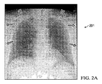
疑似境界画素は、画像境界に沿った濃色境界帯を含む。図2Aは、濃色境界帯を含む放射線画像201を示す。濃色境界帯は矢印で示されている。本開示の側面によれば、濃色境界帯を除去するように放射線画像をトリミングする技術が提供される。図2Bは、放射線画像201から多くの濃色境界帯を除去したトリミング済み出力画像202を示す。濃色境界帯は、放射線画像の端から検索することで特定できる。放射線画像の端は、例えば、放射線画像の上端、下端、左端及び右端である。いくつかの実施形態では、上記検索を、特定の方向に放射線画像の面積の三分の一まで行う。帯に沿った低輝度画素の割合が閾値を超えた場合に、濃色帯と見なされる。一例では、帯に沿った画素の少なくとも60%が低輝度値を有する場合に、濃色帯と見なされる。画像境界から少なくとも閾値距離離れた位置で検出された帯を除去するようにトリミングすることで、トリミング済み出力画像を取得する。一実施形態では、画像境界から最も離れた位置にある濃色帯を除去するようにトリミングする。
The pseudo boundary pixel includes a dark boundary band along the image boundary. FIG. 2A shows a
疑似境界画素は、高変化境界も含む。図3Aは、高変化境界を含む放射線画像301を示す。高変化境界は矢印で示されている。本開示の側面によれば、高変化境界を除去するように放射線画像をトリミングする技術が提供される。図2Bは、放射線画像301から多くの高変化境界を除去したトリミング済み出力画像302を示す。高変化境界を切り取るため、放射線画像を複数の重複ブロックに分割し、各重複ブロックについて変化量を算出する。閾値に基づいて、画像境界に沿った高変化ブロック候補を特定する。続いて、選択された高変化ブロックに基づく各境界に沿って放射線画像をトリミングすることで、トリミング済み出力画像を取得する。
The pseudo boundary pixel also includes a high change boundary. FIG. 3A shows a
疑似境界画素は、線状構造物も含む。図4Aは、線状構造物を含む放射線画像401を示す。線状構造物は矢印で示されている。本開示の側面によれば、線状構造物を除去するように放射線画像をトリミングする技術が提供される。図4Bは、放射線画像401から多くの線状構造物を除去したトリミング済み出力画像402を示す。線状構造物を除去するため、(例えば、)等方性ガウス(Gaussian)フィルタを用いて放射線画像を平滑化する。放射線画像の平滑化は、他の技術を用いて行われてもよい。次に、平滑化済み画像の各境界方向と交差して一次微分を算出し、算出された微分に基づいて線状構造物を特定する。一例では、算出された一次微分に基づいて、画像境界から5°以内の線状構造物を検出する。線状構造物特定後、当該線状構造物を除去するように放射線画像をトリミングすることで、トリミング済み出力画像を取得する。
The pseudo boundary pixel also includes a linear structure. FIG. 4A shows a
放射線画像を処理して疑似画素を除去した後、この処理済み画像に基づいて、大まかな関心領域(ROI)を生成する。関心領域は、肺野領域の上部、下部、左側、右側及び中央に対応する5つの制御点を有する。本開示においては、これら制御点を、ROI境界と称する。例えば、上部制御点を上部ROI境界、下部制御点を下部ROI境界、左側制御点を左側ROI境界、右側制御点を右側ROI境界、中央制御点を中央ROI境界、と称する。図5は、ROI境界510〜516(破線)と大まかな肺野境界520、521(実線)が特定された放射線画像500を示す。ROI境界510〜516は、上部ROI境界510、下部ROI境界512、右側ROI境界514、左側ROI境界515及び中央ROI境界516を含む。
After processing the radiographic image to remove pseudo-pixels, a rough region of interest (ROI) is generated based on the processed image. The region of interest has five control points corresponding to the upper, lower, left, right and center of the lung field region. In the present disclosure, these control points are referred to as ROI boundaries. For example, the upper control point is referred to as an upper ROI boundary, the lower control point as a lower ROI boundary, the left control point as a left ROI boundary, the right control point as a right ROI boundary, and the central control point as a central ROI boundary. FIG. 5 shows a
ROI境界を特定する技術の実施形態は、患者の胴体を表す胴体領域を特定し隔離することから始まる。胴体領域は、当該胴体領域を表す画素を特定し、胴体領域外の画素を消去することで隔離される。胴体領域を表す画素を特定するために、以下の工程を用いる:(1)放射線画像内で、低輝度画素(例えば、閾値未満の画素輝度値を有する画素)を特定する工程;(2)特定された低輝度画素について、平均画素値と中央画素値を算出する工程;(3)算出された平均画素値よりも低い輝度値を有する第1の画素グループを特定する工程;(3)算出された中央画素値よりも高い値を有する第2の画素グループを特定する工程;(4)第1と第2の画素グループに連結成分分析を行い、最大塊部を取得する工程;(5)連結成分処理を用いて最大塊部内の穴を埋めることにより、胴体領域を取得する工程。連結成分分析を行うことで、最大塊部は維持され、小さい疑似領域は除去される。最大塊部は胴体領域に対応し、最大塊部内の埋められた穴は、肺野の低濃度領域に対応する。 An embodiment of a technique for identifying ROI boundaries begins with identifying and isolating a torso region representing the patient's torso. The body region is isolated by specifying a pixel representing the body region and erasing pixels outside the body region. The following steps are used to identify the pixels representing the fuselage area: (1) identifying low luminance pixels (eg, pixels having pixel luminance values below a threshold) in the radiographic image; (2) identification A step of calculating an average pixel value and a center pixel value for the low luminance pixels; (3) a step of specifying a first pixel group having a luminance value lower than the calculated average pixel value; (3) Identifying a second pixel group having a value higher than the central pixel value; (4) performing a connected component analysis on the first and second pixel groups to obtain a maximum block; (5) coupling The process of acquiring a trunk | drum area | region by filling the hole in the largest lump part using component processing. By performing a connected component analysis, the maximum mass is maintained and small pseudo-regions are removed. The largest mass corresponds to the torso region, and the filled hole in the largest mass corresponds to the low concentration region of the lung field.
図6Aは、放射線画像601を示し、図6Bは、放射線画像601における低輝度画素の平均値未満の輝度値を有する画素の領域625が特定された放射線画像602を示す。
6A shows a
図6Cは、疑似画素領域630、肺野の低輝度領域632及び最大塊部領域635が特定された放射線画像603を示す。図6Dは、疑似画素領域630及び胴体領域645が特定された放射線画像604を示す。胴体領域645は、最大塊部領域635内の穴を埋めることによって生成されたものである。前記穴は、肺野の低輝度領域632に対応する
FIG. 6C shows a
ROI境界の特定における次の工程は、中央制御点、左側制御点及び右側制御点の特定である。中央制御点、左側制御点及び右側制御点を見つけるため、胴体領域内の各画像列の輝度値を合計して水平方向輝度投影を算出する。そして、(例えば、等方性ガウスフィルタを用いて、)水平方向輝度投影を平滑化し、当該水平方向輝度投影の極大値と極小値を算出する。図7は、放射線画像604の胴体領域645に対応する、平滑化された水平方向輝度投影ベクトルのグラフ700を示す。グラフ700は、平滑化された水平方向輝度投影ベクトル上の極大値711〜713及び極小値701、702を示す。中央制御点は、極大値711〜713に応じて選択される。例えば、中央制御点は、水平方向輝度投影ベクトルの長さの三分の一より大きい、最小の極大値として選択される。他の例としては、中央制御点は、水平方向輝度投影ベクトルの長さの三分の二より小さい、最大の極大値として選択される。外側である左側と右側の制御点は、投影ベクトルの左右の最大値に基づいて選択される。いくつかの実施形態では、極小値701、702は、肺野中心に対応する。
The next step in identifying the ROI boundary is identifying the central control point, the left control point, and the right control point. In order to find the central control point, the left control point, and the right control point, the luminance value of each image sequence in the body region is summed to calculate the horizontal luminance projection. Then, the horizontal luminance projection is smoothed (for example, using an isotropic Gaussian filter), and the maximum and minimum values of the horizontal luminance projection are calculated. FIG. 7 shows a
ROI境界の特定における次の工程は、上部制御点の特定である。上部制御点を特定するため、胴体領域の上部サブ領域について、垂直方向に輝度投影を算出する。一実施形態では、上部サブ領域は、原画像の最上部三分の一、例えば、画像の全行のうち上部に位置する三分の一の行を有する。図8は、放射線画像604の最上部三分の一に対応するトリミング済み放射線画像800を示す。図9は、トリミング済み放射線画像800に対応する、平滑化された垂直方向輝度投影ベクトルのグラフ900を示す。グラフ900は、平滑化された垂直方向輝度投影ベクトルの極大値911を示す。
The next step in identifying the ROI boundary is identifying the upper control point. In order to identify the upper control point, a luminance projection is calculated in the vertical direction for the upper sub-region of the body region. In one embodiment, the upper sub-region has the top third of the original image, eg, the upper third of the entire image row. FIG. 8 shows a trimmed
ROI境界の特定における次の工程は、右肺野と左肺野の下部制御点の特定である。これら右肺野と左肺野の下部制御点を、各々、右下部ROI境界、左下部ROI境界と称する。右下部ROI境界は、胴体領域の右側サブ領域で特定される。右側サブ領域は、胴体領域の、上部ROI境界よりも下であって、かつ、中央ROI境界と右側ROI境界の間である部分を有する。例えば、患者の右肺野の下部制御点は、上部制御点、左側制御点及び中央制御点を用いて放射線画像をトリミングし、このトリミング済み画像について水平方向輝度投影の二次微分を算出し、算出された水平方向輝度投影の最後の極大値に応じて下部制御点を算出することにより得られる。図10は、上部制御点、左側制御点、右側制御点及び中央制御点を用いて放射線画像604をトリミングして得られた放射線画像1000を示す。図11は、トリミング済み放射線画像1000に対応する、平滑化された水平方向輝度投影ベクトルのグラフ1100を示す。グラフ1100は、平滑化された水平方向輝度投影ベクトルの極大値1111を示す。左下部ROI境界は、胴体領域の左側サブ領域で特定される。左側サブ領域は、胴体領域の、上部ROI境界よりも下であって、かつ、中央ROI境界と左側ROI境界の間である部分を有する。例えば、患者の左肺野の下部制御点は、上部制御点、右側制御点及び中央制御点を用いて原画像をトリミングし、このトリミング済み画像について水平方向輝度投影の二次微分を算出し、算出された水平方向輝度投影の最後の極大値に応じて下部制御点を算出することにより特定される。
The next step in identifying the ROI boundary is identifying the lower control points of the right and left lung fields. The lower control points of the right lung field and the left lung field are referred to as a lower right ROI boundary and a lower left ROI boundary, respectively. The lower right ROI boundary is specified in the right sub-region of the body region. The right sub-region has a portion of the fuselage region that is below the upper ROI boundary and between the central ROI boundary and the right ROI boundary. For example, the lower control point of the patient's right lung field crops the radiation image using the upper control point, the left control point, and the central control point, and calculates the second derivative of the horizontal luminance projection for this trimmed image, It is obtained by calculating the lower control point according to the last maximum value of the calculated horizontal luminance projection. FIG. 10 shows a
関心領域を見つけ次第、外側肺野境界を特定することができる。外側肺野境界は、中部ボーダー、上部ボーダー及び下部ボーダーの三つの部分に分割される。図12Aは、外側肺野境界の大まかな推定が表現された放射線画像1200を示し、図12Bは、外側肺野境界1216を示す。外側肺野境界の大まかな推定により、上部ボーダー1210(長鎖線)、中部ボーダー1212(実線)及び下部ボーダー1214(短鎖線)が生成される。中部ボーダー1212は、外側肺野領域に直線フィッティング処理を用いることで推定できる。外側肺野領域は、制御点(例えば、ROI境界)及び肺野中心を用いて設定される。なお、外側肺野領域は、ROI境界から閾値水平距離の領域を有する。閾値距離は、各肺野について、ROI境界と肺野中心の座標を用いて算出される。様々な向きの直線を、設定された領域の二次微分に当てはめ(フィッティング)、最もフィットする直線を外側肺野境界の中部ボーダー1212として選択する。上部ボーダー1210は、上部制御点と中部ボーダー1212で定義される上部肺野領域に直線フィッティング処理を用いて設定される。下部ボーダー1214は、中部ボーダー1212の下半分で定義される下部肺野領域に直線フィッティング処理を用いて設定される。そして、図12Bに示すように、上部ボーダー1210と、中部ボーダー1212と、下部ボーダー1214と、をマージすることにより、外側肺野境界1216が形成される。上部ボーダー1210と、中部ボーダー1212と、下部ボーダー1214と、をマージする技術の一つとしては、大まかに推定された外側肺野境界の行に、ガウス最小二乗法アルゴリズムを適用することが挙げられる。
Once the region of interest is found, the outer lung field boundary can be identified. The outer lung field boundary is divided into three parts: a middle border, an upper border, and a lower border. FIG. 12A shows a radiographic image 1200 representing a rough estimate of the outer lung field boundary, and FIG. 12B shows the outer
外側肺野境界と関心領域を特定後、肺野の輪郭を初期設定することができる。図13は、放射線画像1301、1302を示す。放射線画像1301は、ROI境界510〜516と肺野境界1216を含む。肺野境界は、ROI境界と肺野境界の初期設定に用いられる画像特徴から生成できる。放射線画像1302は、初期肺野境界1310〜1321を含む。図に示すように、初期肺野境界1310〜1321は、上部境界、下部境界、外側境界、内側境界に分けることができ、これらは比較的大きな尺度で初期設定される。正中線1316は、小さい尺度で画像の一次微分から計算されるゼロクロス点を算出して、中央制御点に最も近いゼロクロス点のサブセットに直線をフィッティングすることで得られる。ゼロクロス点は、水平方向に算出される。
After identifying the outer lung field boundary and the region of interest, the lung field contour can be initialized. FIG. 13 shows
次に、画像の二次微分から水平方向に計算されるゼロクロス点を算出する。当業者であれば、或る方向(又は長いベクトル)に算出される微分(例えば、二次微分)は方向微分(例えば、二次方向微分)であると理解するであろう。図14Aは、放射線画像1301のゼロクロス点が描かれたゼロクロス点の図1401を示す。正中線1420と右側ROI境界1410及び正中線1420と左側ROI境界1430の各々の正中線1420寄り半分に存在するエッジ候補を除去することで、図14Bに示すゼロクロス点の図1402が得られる。このように、エッジ候補は、正中線1420よりも右側ROI境界1410や左側ROI境界1430に近い水平方向ゼロクロス点を含む。外側肺野境界は、大まかな肺野境界1440、1450に最も近い(例えば、閾値距離内の)エッジ候補のセットとして選択され、図14Cの図1403に示すように、最終的な外側肺野境界の候補1460、1470が推定される。
Next, a zero cross point calculated in the horizontal direction from the second derivative of the image is calculated. One skilled in the art will understand that a derivative (eg, a second derivative) calculated in a certain direction (or long vector) is a directional derivative (eg, a second directional derivative). FIG. 14A shows a zero cross point diagram 1401 in which the zero cross point of the
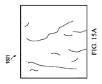
次に、初期内側肺野境界を特定する。初期内側肺野境界を特定するため、画像の二次微分から計算されるゼロクロス点を右肺野について水平方向に算出し、傾斜ゼロクロス点を左肺野について算出する。傾斜ゼロクロス点は、水平面と垂直面の間の角度、例えば、55°等で算出される。図15Aは、55°でのゼロクロス点を示す。初期マスク1520は、図15Bに示すように、左内側肺野サブ領域の一次微分を用いて(例えば、小さい尺度で)算出される。左内側肺野サブ領域は、左肺野の内側半分を描いた領域、例えば、左側ROI境界と正中線の中ほどの領域である。 Next, the initial medial lung field boundary is identified. In order to specify the initial inner lung field boundary, the zero cross point calculated from the second derivative of the image is calculated in the horizontal direction for the right lung field, and the tilted zero cross point is calculated for the left lung field. The tilt zero cross point is calculated by an angle between a horizontal plane and a vertical plane, for example, 55 °. FIG. 15A shows the zero cross point at 55 °. The initial mask 1520 is calculated (eg, on a small scale) using the first derivative of the left medial lung field subregion, as shown in FIG. 15B. The left inner lung field sub-region is a region depicting the inner half of the left lung field, for example, a region between the left ROI boundary and the midline.
マスク内の連結エッジ画素を結合することで、図15Cに示すような初期左内側肺野境界1530が生成される。同様の方法を用いて、右内側境界を検出する。例えば、右内側肺野境界は、右内側肺野サブ領域の一次微分に応じて右マスク領域を算出し、右マスク領域内の水平方向ゼロクロス点を右エッジ画素として特定し、右エッジ画素に直線フィッティングを行うことで形成される。
By combining the connected edge pixels in the mask, an initial left inner
その後、初期上部境界を特定する。初期上部肺野境界は、肺野の上部(例えば、上部ROI境界から閾値距離内)に位置し、かつ、上に凸の構造を有するエッジ候補のセットとして選択される。この上方凸構造の長さ及び角度は、適切な上部肺野境界が選ばれるようなものになっている。図16Aは、放射線画像1601を示し、図16Bは、放射線画像1601についてのエッジ候補が描かれた放射線画像1602を示す。図16Cは、放射線画像1602に描かれたエッジ候補の中から選択された、初期上部肺野境界が描かれた放射線画像1603を示す。
Then, the initial upper boundary is identified. The initial upper lung field boundary is selected as a set of edge candidates that are located above the lung field (eg, within a threshold distance from the upper ROI boundary) and have an upwardly convex structure. The length and angle of this upwardly convex structure is such that an appropriate upper lung field boundary is selected. FIG. 16A shows a
次に、初期下部肺野境界(又は下部肺野境界)を特定する。初期下部肺野境界は、肺野の下部(例えば、下部ROI境界から閾値距離内)に位置し、かつ、上に凸の構造を有するエッジ候補のセットとして選択される。幅の閾値及び二次微分の大きさの閾値を用いて、肺野境界をエッジ候補の中から最終的に選択する。図17Aは、放射線画像1601に描かれた肺野の下部についてのエッジ候補が描かれた放射線画像1701を示す。図17Bは、選択された下部肺野境界が描かれた放射線画像1702を示す。
Next, the initial lower lung field boundary (or lower lung field boundary) is specified. The initial lower lung field boundary is selected as a set of edge candidates that are located below the lung field (for example, within a threshold distance from the lower ROI boundary) and have an upwardly convex structure. The lung field boundary is finally selected from the edge candidates using the width threshold and the second derivative magnitude threshold. FIG. 17A shows a
初期設定後、肺野の輪郭/境界をリファインする。いくつかの実施形態では、マージして最終的な肺野セグメンテーションアウトプットを生成する前に、上部、下部、右側及び左側の初期設定を個別にリファインする。 After initial setting, refine the outline / boundary of the lung field. In some embodiments, the top, bottom, right and left side default settings are refined separately before merging to produce the final lung field segmentation output.
肺野の輪郭のリファインは、外側肺野境界のリファインから始まる。初期外側肺野境界上の各点について、所定幅の輝度プロファイルを法線方向に構築する。この輝度プロファイルを検索し、最も近い内側肺野エッジを特定する。初期外側肺野点に対応する全ての点を、内側肺野境界に対応するようにリファインする。図18Aは、初期設定済み肺野境界が描かれた放射線画像1801を示す。図18Bは、リファイン済み肺野境界が描かれた放射線画像1802を示す。図18Cは、マージ済み肺野セグメンテーションが描かれた放射線画像1803を示す。
The refinement of the lung field outline begins with the refinement of the outer lung field boundary. For each point on the initial outer lung field boundary, a luminance profile with a predetermined width is constructed in the normal direction. This luminance profile is searched to identify the nearest inner lung field edge. Refine all points corresponding to the initial outer lung field points to correspond to the inner lung field boundary. FIG. 18A shows a
図19Aは、初期外側肺野境界1910と調整済み外側肺野境界1920が描かれた放射線画像1901を示す。初期外側境界1910を輝度プロファイル上の最も近い内側エッジに基づいて調整することで、調整済み外側肺野境界1920が得られる。そして、調整済み外側肺野境界1920を平滑化することで、図19Bが示す放射線画像1902に描かれているような、最終的なリファイン済み外側肺野境界1930が得られる。
FIG. 19A shows a
肺野の輪郭をリファインする技術の実施形態は、内側肺野境界のリファインに進む。図20Aは、内側肺野境界のエッジ候補の図2001を示す。いくつかの実施形態では、キャニー(Canny)法を複数の尺度で用いて、内側肺野境界のエッジ候補を検出する。図20Bは、初期内側肺野エッジ2010とリファイン済み内側肺野境界2020が描かれた放射線画像2002を示す。いくつかの実施形態では、リファイン済み内側肺野境界2020は、初期内側肺野エッジ2010に最も近く、かつ、最も高い二次微分値を有するエッジ候補を選択することによって得られる。
Embodiments of techniques for refining lung field contours proceed to refine the inner lung field boundary. FIG. 20A shows a diagram 2001 of edge candidates for the inner lung field boundary. In some embodiments, the Canny method is used on multiple scales to detect edge candidates at the inner lung field boundary. FIG. 20B shows a
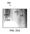
続いて、肺野の輪郭のリファインは、上部肺野境界と下部肺野境界のリファインに進む。外側肺野境界のリファインに用いられる処理を、上部肺野境界と下部肺野境界のリファインにも用いてもよい。図21Aは、初期上部肺野境界2110とリファイン済み上部肺野境界2120が描かれた放射線画像2101を示す。図21Bは、平滑化済み上部肺野境界2130が描かれた放射線画像2102を示す。図22Aは、初期下部肺野境界2210とリファイン済み下部肺野境界2220が描かれた放射線画像2201を示す。図22Bは、平滑化済み下部肺野境界2230が描かれた放射線画像2202を示す。左下部肺野境界については、平滑化に加え、リファイン済み左外側肺野境界の最大列値まで延伸させる。肺野境界がリファインされたら、リファイン済み境界をマージして、肺野セグメンテーション結果を生成する。
Subsequently, the refinement of the contour of the lung field proceeds to refinement of the upper lung field boundary and the lower lung field boundary. The processing used for the refinement of the outer lung field boundary may be used for the refinement of the upper lung field boundary and the lower lung field boundary. FIG. 21A shows a
いくつかの実施形態では、肺野セグメンテーションの後、骨減弱を行う。図23は、骨検出段階と骨減弱段階を有する骨減弱モジュール2300を示す。骨減弱技術の実施形態は、明るいオブジェクトとデバイスを検出し、その後、放射線画像内の鎖骨、後方肋骨及び前方肋骨を検出し減弱する。
In some embodiments, bone attenuation is performed after lung field segmentation. FIG. 23 shows a
骨減弱技術の実施形態は、デバイスと明るいオブジェクトが減弱されないよう、これらを抽出することから始まる。明るいオブジェクトを抽出することにより、骨信号推定中にエラーが発生することを防ぐ。具体的には、人工物は、急なエッジを有するアーチファクトを生成し、例えば、異常な病変(例えば、肺癌)をマスクしたり、異常な病変を減弱させたりして、骨信号の推定を妨げる場合がある。デバイスは、高コントラストな医療機器(例えば、ペースメーカー、カテーテル、マーカー等)を含み、明るいオブジェクトは、小葉、小結節、その他のオブジェクトを含む。骨減弱技術の実施形態は、その後、肺野背景トレンドを除去する。例えば、肺野背景トレンドは、肺野表面に(例えば、二次以上の)多項式をフィッティングして、原画像から当該フィット表面を減算することで除去される。図24Aは、肺野プロファイル除去前の放射線画像2401を示す。図24Bは、肺野プロファイル除去後の放射線画像2402を示す。図24Cは、肺野プロファイル除去前の放射線画像2401のプロファイル2410と肺野プロファイル除去後の放射線画像2402のプロファイル2420が描かれたグラフ2403を示す。
Embodiments of bone attenuation techniques begin with extracting devices and bright objects so that they are not attenuated. Extracting bright objects prevents errors from occurring during bone signal estimation. Specifically, artifacts produce artifacts with steep edges, for example, masking abnormal lesions (eg, lung cancer) or attenuating abnormal lesions, hindering bone signal estimation There is a case. Devices include high-contrast medical devices (eg, pacemakers, catheters, markers, etc.), and bright objects include leaflets, nodules, and other objects. Embodiments of bone attenuation techniques then remove the lung field background trend. For example, the lung field background trend is removed by fitting a polynomial (for example, second order or higher) to the lung field surface and subtracting the fit surface from the original image. FIG. 24A shows a
骨減弱技術の実施形態は、肺野プロファイルが除去され次第、高コントラスト領域を見つける。図25Aは、ピーク(円)が描かれた放射線画像2501を示す。ピーク及びこれに対応するコントラストは、事前に算出された画像から抽出される。領域拡張技術を用いて、算出されたピークを、図25Bが示す放射線画像2502に描かれているように更新する。図25Cは、複数の尺度でオブジェクトの幅、面積、円形度、コントラスト等の特徴を用いて選択された明るい領域の図2503を示す。
Embodiments of bone attenuation techniques find high contrast regions as soon as the lung field profile is removed. FIG. 25A shows a
本開示の側面によれば、鎖骨検出技術が提供される。一般に、鎖骨は肺野領域内で最も目立つ骨である。 According to an aspect of the present disclosure, a clavicle detection technique is provided. In general, the clavicle is the most prominent bone in the lung field region.
鎖骨検出技術のいくつかの実施形態は、放射線画像にワーピングを行って、より真っ直ぐな鎖骨が描かれたワーピング放射線画像を取得する。例えば、鎖骨は、元の放射線画像よりもワーピング放射線画像において、より小さい曲率で描かれる。鎖骨検出技術は、その後、ワーピング放射線画像にエッジ検出を行って、鎖骨を検出する。 Some embodiments of the clavicle detection technique warp the radiographic image to obtain a warped radiographic image depicting a more straight clavicle. For example, the clavicle is drawn with a smaller curvature in the warped radiographic image than the original radiographic image. The clavicle detection technique then detects the clavicle by performing edge detection on the warped radiation image.
鎖骨検出技術のいくつかの実施形態は、鎖骨検出の正確性向上のため、放射線画像のワーピングを繰り返す。例えば、鎖骨検出技術の実施形態は、放射線画像内の鎖骨から角度マップを生成し、当該角度マップに応じて放射線画像にワーピングを行って、第1のワーピング放射線画像を取得する。鎖骨は、放射線画像よりも第1のワーピング放射線画像において、より小さい曲率で描かれる。鎖骨検出技術の実施形態は、その後、第1のワーピング放射線画像に描かれた鎖骨に応じて角度マップを更新し、更新された角度マップに応じて放射線画像に再度ワーピングを行って、第2のワーピング放射線画像を取得する。鎖骨は、第1のワーピング放射線画像よりも第2のワーピング放射線画像において、より小さい曲率で描かれる。鎖骨検出技術の実施形態は、その後、第2のワーピング放射線画像にエッジ検出を行って、鎖骨を検出する。いくつかの実施形態では、鎖骨検出の正確性の更なる向上のため、画像のワーピングを更に繰り返す。 Some embodiments of the clavicle detection technique repeat the warping of the radiographic image to improve the accuracy of clavicle detection. For example, the embodiment of the clavicle detection technique generates an angle map from the clavicle in the radiographic image, warps the radiographic image according to the angle map, and acquires the first warped radiographic image. The clavicle is drawn with a smaller curvature in the first warping radiographic image than in the radiographic image. Embodiments of the clavicle detection technique then update the angle map according to the clavicle depicted in the first warping radiation image, warp the radiation image again according to the updated angle map, and the second A warping radiation image is acquired. The clavicle is drawn with a smaller curvature in the second warping radiographic image than in the first warping radiographic image. Embodiments of the clavicle detection technique then perform edge detection on the second warping radiation image to detect the clavicle. In some embodiments, the image warping is further repeated to further improve the accuracy of clavicle detection.
鎖骨検出技術の他の実施形態は、トレーニングデータセットの鎖骨の上部エッジ及び下部エッジに印を付け、印を付けたエッジから角度測定値を取得することで、演繹的角度マップを作成する。角度を補間し、ローパスフィルタ処理して、前記演繹的角度マップを生成する。そして、鎖骨検出技術の実施形態は、この演繹的角度マップを用いて、画像にワーピングを行う。ワーピングは、角度マップに従う輪郭に沿って画像をサンプリングすることにより達成され、これにより、鎖骨が実質的に水平な画像が得られる。次に、以下のようにして、角度マップを更新する;まず、角度に関連付けられた制約事項を用いてエッジ検出を行う;そして、角度測定値をフィルタ処理して、演繹的角度マップのための更新画像を生成する。更新画像にワーピングを行って画像空間に戻し、演繹的角度マップに追加して、鎖骨が検出されるようにする。鎖骨を検出するため、更新された角度マップを用いて各肺野にワーピングを行い、鎖骨が原画像内よりも実質的により水平に描かれた更新画像を取得する。そして、様々な制約事項を用いて、生成された画像にエッジ検出を行って、有効なエッジセグメントを限定する。そして、様々なエッジセグメントを結合して、鎖骨のエッジ候補を形成する。そして、両側の鎖骨のエッジ候補のマッチングを行って、様々な制約事項を満たす最適な鎖骨対を選択する。検出されたら、後方肋骨の減弱に用いられる技術と同様の技術を用いて、鎖骨を減弱することができる。前記技術については、以下で詳細に説明する。 Another embodiment of the clavicle detection technique creates an a priori angle map by marking the upper and lower edges of the clavicle in the training data set and obtaining angle measurements from the marked edges. The angle is interpolated and low pass filtered to generate the deductive angle map. The embodiment of the clavicle detection technique then warps the image using this a priori angle map. Warping is accomplished by sampling the image along a contour that follows an angle map, resulting in an image in which the clavicle is substantially horizontal. Next, update the angle map as follows; first, perform edge detection using the constraints associated with the angle; and filter the angle measurements to create the deductive angle map. An updated image is generated. The updated image is warped back to the image space and added to the a priori angle map so that the clavicle is detected. In order to detect the clavicle, the updated angle map is used to warp each lung field to obtain an updated image in which the clavicle is drawn substantially more horizontally than in the original image. Then, using various restrictions, edge detection is performed on the generated image to limit effective edge segments. Various edge segments are then combined to form clavicle edge candidates. Then, matching of the clavicle edge candidates on both sides is performed to select an optimal clavicle pair that satisfies various constraints. Once detected, the clavicle can be attenuated using techniques similar to those used for posterior rib attenuation. The technique will be described in detail below.
本開示の側面によれば、前方肋骨と後方肋骨を含む肋骨の検出技術及び減弱技術が提供される。肋骨検出技術の一実施形態は、放射線画像内の各肋骨について角度マップを生成し、対応する角度マップに応じて、肋骨が描かれた放射線画像の部分にワーピングを行って、複数のワーピングサブ画像を取得する。肋骨は、放射線画像よりもワーピングサブ画像において、より小さい曲率で描かれる。その後、肋骨検出技術は、ワーピングサブ画像にエッジ検出を行って、肋骨を検出する。 According to an aspect of the present disclosure, a rib detection technique and an attenuation technique including an anterior rib and a posterior rib are provided. One embodiment of the rib detection technique generates an angle map for each rib in the radiographic image, warps a portion of the radiographic image on which the rib is drawn according to the corresponding angle map, and a plurality of warping sub-images. To get. The ribs are drawn with a smaller curvature in the warping sub-image than the radiographic image. Thereafter, the rib detection technique detects the ribs by performing edge detection on the warping sub-image.
本開示の側面によれば、後方肋骨の検出技術及び減弱技術が提供される。一実施形態では、まず、特定の角度マップを用いて後方肋骨にワーピングを行って後方肋骨を真っ直ぐにし、次に、肋骨位置を近似する技術を用いて肋骨を検出する。具体的には、以下の処理を用いて、各肋骨について角度マップを算出する。まず、実空間における角度測定値を生成する。これは、画像にローパスフィルタ演算を行い、その後、エッジ検出と閾値フィルタ処理を行うことにより達成される。行列からなる複数の点(例えば、8行×5列=40点)で構成されるグリッドを形成し、検出されたエッジ上の最も近い点について角度測定値を算出する。その後、角度測定値を演繹的角度マップに基づいて制約事項に晒し、制約事項を満たす角度測定値のみが保持されるように、(例えば、演繹的角度マップによって定義される)範囲外の角度測定値を除去する。その後、補間技術を用いて、保持された角度測定値から、各肺野について角度マップを作成する。そして、角度マップに従う輪郭に沿って、画像をサンプリングする。これにより、肋骨が実質的に水平な画像が得られる。そして、本開示によって提供される技術の実施形態を用いて、角度測定値を再度算出し、当該角度測定値を再度制約事項に晒して、有効な角度測定値を得る。この有効な角度測定値を角度マップに加え、その結果をフィルタ処理して、平滑化済み角度マップを作成する。 According to an aspect of the present disclosure, a posterior rib detection technique and an attenuation technique are provided. In one embodiment, the posterior rib is first warped using a specific angle map to straighten the posterior rib, and then the rib is detected using a technique that approximates the rib position. Specifically, an angle map is calculated for each rib using the following processing. First, an angle measurement value in real space is generated. This is achieved by performing a low pass filter operation on the image and then performing edge detection and threshold filtering. A grid composed of a plurality of points (for example, 8 rows × 5 columns = 40 points) formed from a matrix is formed, and an angle measurement value is calculated for the closest point on the detected edge. The angle measurements are then exposed to constraints based on the a priori angle map, and out-of-range angle measurements (eg, defined by the a priori angle map) so that only angle measurements that satisfy the constraints are retained. Remove the value. Thereafter, an angle map is created for each lung field from the stored angle measurements using an interpolation technique. Then, the image is sampled along the contour according to the angle map. Thereby, an image in which the ribs are substantially horizontal is obtained. Then, using the embodiment of the technology provided by the present disclosure, the angle measurement value is recalculated, and the angle measurement value is again exposed to the constraints to obtain an effective angle measurement value. This valid angle measurement is added to the angle map and the result is filtered to create a smoothed angle map.
平滑化済み角度マップ取得後、外側肺野エッジ付近の角度が、最も近い肺野境界に沿うように、当該角度マップを修正する。その後、近似技術の実施形態を用いて、近似肋骨位置を推定する。近似技術の実施形態は、角度マップに従う輪郭に沿って画像がサンプリングされるように、角度マップに基づいて画像をワーピングすることから始まる。これにより、肋骨が実質的に水平な画像が得られる。そして、近似技術の実施形態は、画像をいくつかの尺度で平滑化して異なるサイズの構造物を見つけ、この特定された構造物から測定値を取得する。測定値は、多様に分類される。例えば、測定値は、肋骨構造物の上部エッジ、下部エッジ、隆起部又は窪み部に関連付けられる。上部エッジや下部エッジと関連付けられる測定値は、勾配の方向における画像勾配の極大値を見つけることで得られる。隆起部や窪み部と関連付けられる測定値は、画像内の窪みの極値(例えば、極大値及び/又は極小値)を見つけることで得られる。 After obtaining the smoothed angle map, the angle map is corrected so that the angle near the outer lung field edge is along the nearest lung field boundary. Thereafter, the approximate rib position is estimated using an embodiment of the approximate technique. The approximation technique embodiment begins with warping the image based on the angle map such that the image is sampled along a contour that follows the angle map. Thereby, an image in which the ribs are substantially horizontal is obtained. An embodiment of the approximation technique then smoothes the image on several scales to find different sized structures and obtains measurements from the identified structures. Measurements are classified in various ways. For example, the measured value is associated with the upper edge, the lower edge, the ridge or the depression of the rib structure. Measurements associated with the upper and lower edges are obtained by finding the maximum value of the image gradient in the gradient direction. Measurements associated with ridges and depressions are obtained by finding depression extremes (eg, local maxima and / or local minima) in the image.
近似技術の実施形態は、その後、取得した測定値の位置に応じて当該測定値を累積し、肋骨の存在しない位置よりも存在する位置の方が高い値を有する最終的な累積画像を取得する。各測定値は、垂直方向に広がって位置誤差の原因となりうる。そこで、分類/種類に応じて移動させる。例えば、上部エッジと関連付けられる測定値は、下方に肋骨幅の二分の一移動させ、下部エッジと関連付けられる測定値は、上方に肋骨幅の二分の一移動させる。いくつかの実施形態では、隆起部や窪み部と関連付けられる測定値は、移動させない。各測定値の寄与は、種類によって重み付けされる。一例では、上部/下部エッジと関連付けられる測定値の寄与は、隆起部や窪み部と関連付けられる測定値よりも重く重み付けされる。いくつかの実施形態では、窪み部と関連付けられる測定値には負の重み付けが割り当てられ、肋骨が存在しないことを示す。累積画像は、肋骨の位置を示す水平帯を有する。水平帯はセグメント化され、画像を横断する水平方向のいくつかの位置において、垂直位置が測定される。 The embodiment of the approximation technique then accumulates the measurement values according to the position of the acquired measurement values, and obtains a final accumulated image having a higher value at the position where the position is present than at the position where the rib is not present. . Each measurement value can spread in the vertical direction and cause a position error. Therefore, it is moved according to the classification / type. For example, the measurement associated with the upper edge is moved downward by one half of the rib width, and the measurement associated with the lower edge is moved upward by one half of the rib width. In some embodiments, measurements associated with ridges and depressions are not moved. The contribution of each measurement is weighted by type. In one example, the measurement contribution associated with the upper / lower edge is more heavily weighted than the measurement associated with the ridge or depression. In some embodiments, the measurement associated with the depression is assigned a negative weight, indicating that no ribs are present. The cumulative image has a horizontal band indicating the position of the rib. The horizontal band is segmented and the vertical position is measured at several horizontal positions across the image.
続いて、肋骨の検出位置を胸郭座標系にマッピングする。座標系は、各肺野について、三つの線、すなわち、肺野の内縁に沿った縦線、肺野の真上に位置する横線及び肺野の外縁に沿った側線に基づいて作成される。横線は、縦線と側線の基準線となる。座標系作成後、胸郭モデルを抽出する。胸郭座標系は、胸郭内で肋骨の位置を検出するフレームワークとなる。胸郭モデルは、肺野の内側に沿う点を肺野の前側に沿う点にマッチさせる。まず、角度マップを用いて、これらの点はマッチングされ、その後、マッチした点の位置は、より肋骨位置測定値に合うように調整される。これらの点には、無矛盾の肋骨間距離を維持するよう制約が加えられる。マッチした点の位置が決定次第、肋骨の距離を検討して、胸郭と矛盾する測定値を除去し、また、直接的に検出されなかった肋骨の存在を推定する。 Subsequently, the rib detection position is mapped to the rib cage coordinate system. A coordinate system is created for each lung field based on three lines: a vertical line along the inner edge of the lung field, a horizontal line located directly above the lung field, and a side line along the outer edge of the lung field. The horizontal line serves as a reference line for the vertical line and the side line. After creating the coordinate system, extract the rib cage model. The rib cage coordinate system is a framework for detecting the position of the ribs within the rib cage. The thorax model matches a point along the inside of the lung field with a point along the front side of the lung field. First, these points are matched using an angle map, and then the position of the matched point is adjusted to better match the rib position measurement. These points are constrained to maintain consistent rib distances. As soon as the location of the matched point is determined, the distance of the ribs is examined to remove measurements that contradict the rib cage and to estimate the presence of ribs that were not directly detected.
そして、事前に算出した近似肋骨位置をヒントに、正確な肋骨位置を算出する。近似肋骨位置は、各肋骨の中心線の位置の推定を反映し、胸郭座標系の内縁から外縁まで延びている。そして、各近似肋骨位置について、近似肋骨位置に合わせた非等間隔なグリッドをサンプリングして、原画像からサブ画像を作成する。1画素間隔で近似肋骨位置線に沿ってサンプリングすることで、グリッドの中心行を作成する。そして、中心行の各点から1画素インクリメントで肋骨に垂直な方向に進むことで、中心行の上下の行を作成する。グリッドが、推定肋骨間隔又は2cmの何れか大きいほうまで中心行の上下に拡張するまで、当該グリッドに行を追加する。生成された各サブ画像は、少なくとも部分的に、放射線画像に対して水平方向及び垂直方向に中心となるようにワーピングされた、一の肋骨を含む。 Then, an accurate rib position is calculated using the approximate rib position calculated in advance as a hint. The approximate rib position reflects an estimate of the position of the centerline of each rib and extends from the inner edge to the outer edge of the rib cage coordinate system. Then, for each approximate rib position, a non-uniform grid aligned with the approximate rib position is sampled to create a sub-image from the original image. By sampling along the approximate rib position line at intervals of one pixel, the center row of the grid is created. Then, by proceeding in the direction perpendicular to the ribs by incrementing one pixel from each point of the central row, upper and lower rows of the central row are created. Add rows to the grid until the grid expands above and below the center row to the greater of the estimated rib spacing or 2 cm, whichever is greater. Each generated sub-image includes a rib that is warped at least partially centered horizontally and vertically with respect to the radiographic image.
その後、エッジ測定値を収集する。エッジ測定値を収集するため、まず、非対称ガウス技術を用いてサブ画像を平滑化し、垂直構造物を目立たなくさせる。そして、勾配の大きさの極大値を特定して、エッジ点を見つける。勾配の大きさが閾値(例えば、第三十分位数)未満の場合、又は、エッジに沿った向きが水平面からの閾値角度(例えば、30°)を超える場合、当該エッジ点は除去する。残りのエッジ点のうち、同じような向きの隣接エッジ点を連結し、エッジ測定値とする。要求される最小長より短い測定値は破棄し、上部エッジ測定値と下部エッジ測定値は分ける。 Then, edge measurements are collected. To collect edge measurements, the sub-image is first smoothed using an asymmetric Gaussian technique to make the vertical structures less noticeable. Then, an edge point is found by specifying the maximum value of the gradient magnitude. When the magnitude of the gradient is less than a threshold (for example, the third decile), or when the direction along the edge exceeds the threshold angle (for example, 30 °) from the horizontal plane, the edge point is removed. Among the remaining edge points, adjacent edge points having the same direction are connected to obtain an edge measurement value. Discards measurements that are shorter than the required minimum length and separates the upper and lower edge measurements.
エッジ測定値収集後、エッジ測定値を組み合わせて、エッジ測定値組み合わせ候補を生成する。いくつかの実施形態では、全てのエッジ測定値組み合わせを検討する。エッジ間の空間関係に基づいて、一の肋骨を表現するには矛盾する組み合わせは除去する。例えば、同じ種類のエッジ(例えば、上部エッジ、下部エッジ等)で、水平方向に閾値長さ(例えば、長さの三分の一)より多く重なる組み合わせは除去する。他の例としては、同じ種類のエッジで、水平方向に近くに端点を有する組み合わせは除去する。更に他の例としては、上部エッジ測定値と下部エッジ測定値で、水平方向に重なるが、一の肋骨のエッジを表すには離れすぎている又は近すぎる組み合わせも除去する。残るエッジ測定値組み合わせ数が閾値(例えば、256等)より多い場合、閾値以下になるまで、残る組み合わせのうち最も短い測定値を含む組み合わせを除去する。 After collecting the edge measurement values, the edge measurement values are combined to generate an edge measurement value combination candidate. In some embodiments, all edge measurement combinations are considered. Based on the spatial relationship between the edges, contradictory combinations are removed to represent one rib. For example, combinations that overlap more than the threshold length (eg, one third of the length) in the horizontal direction at the same type of edge (eg, upper edge, lower edge, etc.) are removed. As another example, combinations having edges close to the horizontal direction with the same type of edge are removed. As yet another example, combinations of top edge measurements and bottom edge measurements that overlap horizontally but are too far apart or too close to represent one rib edge are also removed. When the number of remaining edge measurement value combinations is larger than a threshold value (for example, 256 or the like), the combination including the shortest measurement value is removed from the remaining combinations until the number is less than the threshold value.
その後、技術の実施形態は、個々の肋骨モデルを構築する。肋骨モデルは、残った測定値組み合わせから作成される。肋骨モデルは、サブ画像を横断する連続した道であり、対応するエッジ測定値組み合わせのソースである肋骨の、真のエッジを推定するためのものである。各エッジ測定値組み合わせに沿った、サブ画像を横断する変化のなだらかな道を作成するため、Rauch−Tung−Striebelスムーサーを適用する。スムーサーの状態空間モデルは、肋骨の中心(行)位置と、画像列を横断する幅のみ追跡する。プロセスばらつきは非常に高く初期設定され、エッジ測定値に対する適度にタイトな適合を可能にしている。肋骨モデル算出後、特定の制約事項を満たさないモデルは破棄する。制約事項を満たさないモデルには、二次微分値の高い領域を有するモデル、近似肋骨幅よりも閾値距離を超える幅を有するモデルが含まれる。 Thereafter, embodiments of the technology build individual rib models. The rib model is created from the remaining measurement combinations. The rib model is a continuous path across the sub-image and is intended to estimate the true edge of the rib that is the source of the corresponding edge measurement combination. To create a smooth path of change across the sub-image along each edge measurement combination, a Rauch-Tung-Stribel smoother is applied. The smoother state space model tracks only the center (row) position of the ribs and the width across the image sequence. The process variation is very high and is initially set to allow a reasonably tight fit to the edge measurement. After calculating the rib model, discard the model that does not satisfy the specific restrictions. Models that do not satisfy the restrictions include models having a region with a high second-order differential value, and models having a width exceeding the threshold distance than the approximate rib width.
次に、肋骨モデルをカバレッジメトリックに応じて配列する。カバレッジメトリックは、サブ画像内の列の数に対応し、上部エッジ測定値や下部エッジ測定値は含まれるが、同じ種類の他の測定値と重なってはいない。高カバレッジのモデルは、低カバレッジのモデルに比べて、検出されたエッジと一致する傾向が高いことから、信頼度が高いとみなされている。モデルの座標は、原画像の座標系に再変換される。これにより、各サブ画像内で肋骨を真っ直ぐにするために行われた変換は取り消しとなる。その結果は、原画像内で肋骨候補の輪郭を描く多角形である。個々の肋骨モデル構築のアウトプットは、各肋骨近似値についての、選択された肋骨候補と、順序付けされた補欠肋骨候補セットである。 Next, the rib model is arranged according to the coverage metric. The coverage metric corresponds to the number of columns in the sub-image and includes upper edge measurements and lower edge measurements, but does not overlap with other measurements of the same type. The high coverage model is considered to be more reliable than the low coverage model because it tends to match the detected edges. The model coordinates are reconverted to the coordinate system of the original image. This cancels the conversion performed to straighten the ribs in each sub-image. The result is a polygon that outlines the rib candidates in the original image. The output of individual rib model construction is a selected rib candidate and an ordered set of prosthetic ribs for each rib approximation.
個々の肋骨を構築後、前記技術の実施形態は、グローバルに無矛盾な肋骨モデルセットを選択する。グローバルに無矛盾な肋骨モデルセットの選択には、検出された全ての肋骨からの情報が用いられ、胸郭として矛盾のない解釈を提供する肋骨候補セットが選択される。肋骨候補セットが胸郭の期待値に一致するか否か判定するため、グローバルモデルは、前記期待値と関連付けられた制約事項セットと共に用いられる。例えば、グローバルモデルの制約事項セットは、肋骨の幅と肋骨の行の位置の線形非減少関係に対応する。また、制約事項セットは、肋骨間隔、すなわち、上部肋骨の行の位置に基づく下部肋骨と上部肋骨の間隔の線形非減少関係にも対応する。前記モデルは、左肺野と右肺野について、無矛盾な制約事項/関係を維持する。 After building the individual ribs, the technique embodiment selects a globally consistent rib model set. To select a globally consistent rib model set, information from all detected ribs is used, and a rib candidate set that provides a consistent interpretation as a rib cage is selected. In order to determine whether the rib candidate set matches the expected value of the rib cage, a global model is used with a set of restrictions associated with the expected value. For example, the global model constraint set corresponds to a linear non-decreasing relationship between rib width and rib row position. The constraint set also corresponds to the rib spacing, ie, a linear non-decreasing relationship between the lower rib and upper rib spacing based on the row position of the upper rib. The model maintains consistent constraints / relationships for the left and right lung fields.
肋骨候補セットの品質は、候補のパラメーター(例えば、肋骨の間隔/幅の値)とモデルのパラメーターの差を測定することで判定される。これは、ロバストな線形回帰技術を用いて品質メトリックを得ることにより達成される。品質メトリックは、測定値と線形モデルの予想値の重み付き平均二乗誤差である。肋骨の幅と間隔のモデルは、最適な肋骨候補を用いてフィットティングできる。モデルの肋骨の幅の予想値を用いて、全ての肋骨の肋骨候補をフィルタ処理し、極端な外れ値を取り除く。最適な肋骨候補の何れかが取り除かれ、最適な補欠肋骨が取って代わった場合、肋骨の幅と間隔のモデルを更新する。 The quality of the rib candidate set is determined by measuring the difference between the candidate parameters (eg, rib spacing / width values) and the model parameters. This is achieved by obtaining a quality metric using a robust linear regression technique. The quality metric is a weighted mean square error between the measured value and the expected value of the linear model. The rib width and spacing model can be fitted using the optimal rib candidates. Using the predicted rib width of the model, filter all rib rib candidates and remove extreme outliers. If any of the optimal rib candidates are removed and the optimal prosthetic rib is replaced, the rib width and spacing model is updated.
最適な肋骨候補を示す多角形と他の近辺の肋骨の重なり量を(もしあれば)算出する。候補対の間に有意な重なりがある場合、当該対の各要素の各補欠肋骨を胸郭に代入し、各候補セットの品質を記録する。全補欠を検討後、最高の品質スコアを有する候補組み合わせを記録し、当該組み合わせを新たな最適な候補セットとして使用する。これらの代入テストの結果、最適な肋骨候補セットが変わった場合、これに伴い、肋骨の幅と間隔のモデルを更新する。この処理、すなわち、一以上の肋骨のありそうにない構成(上では、重なりについて)を特定するテストを行い、引き続き、補欠肋骨候補の代入と品質メトリックの評価を行うという処理は、他の基準について繰り返されうる。これには、個々の肋骨候補の幅の統計値や、肋骨間隔を超えて算出される統計値が含まれる。最適な候補セットが安定するまで又はタイミング要件により更なる検索が制限されるまで、テスト一式を繰り返すことができる。この検索処理は、最終的に、正確な肋骨の位置を特定する最適な候補セットを提供する。正確な肋骨位置の特定後、後方肋骨を減弱する。 The overlap amount (if any) of the polygon representing the optimal rib candidate and the ribs in the vicinity is calculated. If there is a significant overlap between candidate pairs, each prosthetic rib of each element of the pair is substituted into the rib cage and the quality of each candidate set is recorded. After considering all prostheses, the candidate combination with the highest quality score is recorded and used as the new optimal candidate set. If the optimal rib candidate set changes as a result of these substitution tests, the rib width and interval model is updated accordingly. This process, that is, a test that identifies an unlikely configuration of one or more ribs (above, for overlap), followed by substitution of prosthetic rib candidates and evaluation of quality metrics, Can be repeated. This includes statistical values of individual rib candidate widths and statistical values calculated beyond the rib interval. The test suite can be repeated until the optimal candidate set is stable or until further search is limited by timing requirements. This search process ultimately provides an optimal candidate set that identifies the exact rib location. After the exact rib location is determined, the posterior rib is attenuated.
本開示の側面によれば、放射線画像から検出された骨を除去/減弱する骨減弱技術が提供される。骨は、骨毎に減弱される。骨減弱技術の一実施形態は、検出された骨のうち、放射線画像から減弱する一の骨を選択する工程と、選択された骨が描かれた矩形サブ画像を特定する工程と、矩形サブ画像から、肺野領域の外に位置する選択された骨のエッジを切り取る工程と、を含む。前記技術の実施形態は、矩形サブ画像を画像処理して選択された骨の曲率を低減する工程と、処理された矩形サブ画像から骨プロファイルを推定する工程と、放射線画像から骨プロファイルを減弱する工程と、を更に含む。骨プロファイルは、次のようにして推定される:処理された矩形サブ画像から背景信号を除去して背景除去画像を生成する;背景除去画像に描かれた選択された骨の上部エッジプロファイルと下部エッジプロファイルを特定する;上部エッジプロファイルと下部エッジプロファイルに応じて骨プロファイルを推定する。なお、骨エッジ信号の推定も、処理された矩形サブ画像又は背景除去画像を用いて行われる。骨エッジは、骨の他の部分に比べて比較的高いコントラストを有するため、骨エッジ信号の推定は、骨信号(例えば、骨プロファイル)の推定とは別に行われる。一実施形態では、骨エッジ信号の推定は、方向サブサンプルサブ画像に対して行われる。方向サブサンプルサブ画像は、画像(例えば、処理された矩形サブ画像)を、骨に垂直な方向よりも骨に平行な方向に、より高い周波数でサブサンプリングすることで得られる。骨エッジ信号の推定は、形態学的処理(例えば、min−max処理)と画像平滑化(例えば、ガウス平滑化)を更に含み、骨以外の構造物、例えば、異常疾患や、血管等の他の正常な構造物を除去する可能性を低下させる。例えば、方向サブサンプルサブ画像を形態学的に処理して、処理済み方向サブサンプルサブ画像を取得する。そして、処理済み方向サブサンプルサブ画像を平滑化して、平滑化方向サブサンプルサブ画像を取得する。平滑化中、骨エッジ付近の領域に対し、より小さいシグマ値を用いて、当該骨エッジ付近の領域を平滑化する。シグマ値は、ローパスフィルタの分散に対応しており、そのため、骨エッジ付近に対し、より小さいシグマ値(例えば、よりタイトな分散)を用いることで、骨エッジ信号のより正確な近似、例えば、よりシャープなエッジを提供する。 According to an aspect of the present disclosure, a bone attenuation technique is provided that removes / attenuates bone detected from a radiographic image. Bone is attenuated for each bone. One embodiment of the bone attenuation technique includes a step of selecting one of the detected bones to be attenuated from the radiation image, a step of identifying a rectangular sub-image on which the selected bone is drawn, and a rectangular sub-image Cutting out selected bone edges located outside the lung field region. Embodiments of the technique include image processing the rectangular sub-image to reduce the curvature of the selected bone, estimating a bone profile from the processed rectangular sub-image, and attenuating the bone profile from the radiographic image. A process. The bone profile is estimated as follows: remove the background signal from the processed rectangular sub-image to generate a background-removed image; upper and lower edge profiles of the selected bone drawn in the background-removed image Identify the edge profile; estimate the bone profile according to the upper and lower edge profiles. Note that the estimation of the bone edge signal is also performed using the processed rectangular sub-image or background-removed image. Since the bone edge has a relatively high contrast compared to other parts of the bone, the estimation of the bone edge signal is performed separately from the estimation of the bone signal (eg, bone profile). In one embodiment, bone edge signal estimation is performed on the directional subsample sub-image. A direction subsample subimage is obtained by subsampling an image (eg, a processed rectangular subimage) at a higher frequency in a direction parallel to the bone than in a direction perpendicular to the bone. The estimation of the bone edge signal further includes morphological processing (for example, min-max processing) and image smoothing (for example, Gaussian smoothing), and other structures such as abnormal diseases, blood vessels, etc. Reduces the possibility of removing normal structures. For example, the directional subsample subimage is morphologically processed to obtain a processed directional subsample subimage. Then, the processed direction sub-sample sub-image is smoothed to obtain a smoothed direction sub-sample sub-image. During smoothing, the region near the bone edge is smoothed using a smaller sigma value for the region near the bone edge. The sigma value corresponds to the variance of the low-pass filter, so using a smaller sigma value (eg, a tighter variance) near the bone edge allows a more accurate approximation of the bone edge signal, eg, Provides a sharper edge.
本開示の側面によれば、肋骨減弱技術が更に提供される。肋骨減弱技術の実施形態は、両肺野の肋骨に対し順次実行される。最上部又は最下部の肋骨を減弱し、当該肋骨が除去された出力画像が、次の肋骨(例えば、除去された肋骨の上又は下の肋骨)を除去するための入力として渡される。全ての肋骨が減弱されるまで、これを繰り返す。いつ減弱される肋骨の入力画像であっても、それ以前の肋骨が全て除去された画像である。この処理は、上から下へ、又は、その逆に向って、実行される。左肺野と右肺野の肋骨の減弱は、別々に行われてもよい。 According to an aspect of the present disclosure, a rib attenuation technique is further provided. Embodiments of the rib attenuation technique are performed sequentially on the ribs of both lung fields. The output image with the top or bottom ribs attenuated and the ribs removed is passed as input to remove the next rib (eg, above or below the removed ribs). Repeat until all ribs are weakened. Even if the input image of the rib is attenuated at any time, it is an image from which all previous ribs have been removed. This process is performed from top to bottom or vice versa. The attenuation of the ribs in the left and right lung fields may be performed separately.
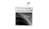
後方肋骨減弱技術の実施形態の第一の工程は、肋骨サブ画像を作成することである。例えば、肋骨サブ画像は、減弱される骨周辺の矩形サブ画像であり、フル解像度画像から切り取られたものである。肋骨減弱は、通常、一回に一つの肋骨に対して行われるため、上記は、処理時間を短縮する。肋骨サブ画像作成後、後方肋骨減弱技術の実施形態は、肋骨エッジのリファインに進む。肋骨検出は一般的にサブサンプル画像に対して行われるため、元の画像解像度でのリファインは、減弱処理中にアーチファクトが導入されることを防ぐ。このリファインは、各点で肋骨エッジに垂直な輝度プロファイルを算出し、当該垂直なプロファイルに沿って一次微分に基づいてエッジをリファインすることで実行される。リファインされた肋骨エッジは、ノイズによって導入された不連続を除去するために平滑化される。図26は、検出された肋骨の放射線画像2600を示し、検出された肋骨エッジ2610とリファインされた肋骨エッジ2620が描かれている。肋骨エッジをリファインした後、技術の実施形態は、肺野領域の外の部分を切り取って(クリッピング)、肺野領域内にない肋骨の領域を除去する。なお、クリッピングは、主に(又は排他的に)、肺野領域内に存在する肋骨の部分の減弱に関係する実装の複雑性を低減する。この理由により、肺野領域内に収まるよう、肋骨のエッジの両端は切り取られる。図27は、検出された肋骨エッジが肺野領域の外で切り取られた放射線画像2700を示す。技術の実施形態は、その後、肋骨を真っ直ぐにする。これを達成するため、肋骨の上部エッジと下部エッジをリサンプリングし、当該二つのリサンプリングされたエッジ間に、中心線を構築する。そして、縦横のインデックスからなる補間マトリクスが、中心線に垂直に作成される。マトリクスの各列は、上部肋骨エッジ上の点で始まり、下部肋骨エッジ上の点で終わる、補間された縦横のインデックスを表す。マトリクスを用いて、真っ直ぐにした(水平な)肋骨の画像を作成する。図28は、真っ直ぐにした肋骨が描かれた放射線画像2800を示す。
The first step of the posterior rib attenuation technique embodiment is to create a rib sub-image. For example, the rib sub-image is a rectangular sub-image around the bone to be attenuated and is cut from a full resolution image. Since rib attenuation is usually performed on one rib at a time, the above shortens the processing time. After the rib sub-image creation, the posterior rib attenuation technique embodiment proceeds to refine the rib edge. Since rib detection is typically performed on sub-sample images, refinement at the original image resolution prevents the introduction of artifacts during the attenuation process. This refinement is performed by calculating a luminance profile perpendicular to the rib edge at each point and refining the edge based on the first derivative along the perpendicular profile. The refined rib edges are smoothed to remove discontinuities introduced by noise. FIG. 26 shows a detected
肋骨を真っ直ぐにした後、肋骨プロファイルの推定を行って、真っ直ぐにした肋骨の画像から背景信号を除去する。これは、真の骨プロファイルの推定に有益であり、また、背景信号の減弱を防ぐ。上部エッジより上の点と下部エッジより下の点を用いて、これらの点の間の領域を補間する。この補間された領域に対して、ガウス最小二乗フィッティングを実行する。その後、真っ直ぐにした肋骨の画像から背景画像を減算することで、背景画像を取り除く。 After the ribs are straightened, the rib profile is estimated and the background signal is removed from the straight rib image. This is useful for estimating the true bone profile and prevents attenuation of the background signal. Interpolate the area between these points using points above the top edge and points below the bottom edge. Gaussian least square fitting is performed on the interpolated region. Then, the background image is removed by subtracting the background image from the straight rib image.
肋骨の上部エッジ及び下部エッジは、肋骨の残りの部分に比べ、比較的高い輝度を有する。そこで、エッジプロファイルを推定し、背景除去画像から減算することで、減弱処理中にアーチファクトが導入するのを防ぐ。そして、前記画像にmin−max演算を行い、その後、平滑化を行うことで、異常(小結節等)や他の構造物が除去されるのを防ぐ。続いて、エッジプロファイルを前記画像に加えて戻し、当該画像を変換して原画像空間に戻す。その後、サブ画像から推定肋骨プロファイルを減算して、当該サブ画像を、入力画像内の元のトリミング済みサブ画像と置き換える。これにより、肋骨減弱画像が得られる。残りの肋骨も同様に減弱する。 The upper and lower edges of the ribs have a relatively high brightness compared to the rest of the ribs. Therefore, the edge profile is estimated and subtracted from the background-removed image to prevent the introduction of artifacts during the attenuation process. Then, min-max calculation is performed on the image, and then smoothing is performed to prevent the removal of abnormalities (such as nodules) and other structures. Subsequently, an edge profile is added back to the image and the image is converted back to the original image space. Thereafter, the estimated rib profile is subtracted from the sub-image to replace the sub-image with the original trimmed sub-image in the input image. Thereby, a rib attenuation image is obtained. The remaining ribs are similarly attenuated.
本開示の側面によれば、前方肋骨検出技術が提供される。前方肋骨の検出は、後方肋骨のワーピングに用いられる方法と同様の方法で、演繹的マップを用いて前方肋骨をワーピングし、ケース毎の角度マップを算出することから始まる。その後、前記画像をローパスフィルタ処理し、ローパスフィルタ処理された画像にエッジ検出メカニズムを適用して、角度測定値を生成する。角度測定値は制約事項に晒され、前方肋骨の有効な角度範囲外の測定値は取り除かれる。角度範囲は、トレーニング画像セットを用いて演繹的角度マップを構築することによって算出される。有効な角度測定値は補間及びローパスフィルタ処理され、更新画像が形成される。この画像にワーピングを行って実画像空間に戻し、当該画像を演繹的角度マップに加える。この角度マップは、その後の前方肋骨検出演算に用いられる。 According to an aspect of the present disclosure, an anterior rib detection technique is provided. The detection of the anterior rib starts by warping the anterior rib using a deductive map and calculating an angle map for each case in a manner similar to the method used for warping the posterior rib. The image is then low pass filtered and an edge detection mechanism is applied to the low pass filtered image to generate an angle measurement. Angular measurements are subject to restrictions and measurements outside the effective angular range of the anterior rib are removed. The angle range is calculated by building a deductive angle map using the training image set. Valid angle measurements are interpolated and low pass filtered to form an updated image. The image is warped back to the real image space and the image is added to the deductive angle map. This angle map is used for the subsequent anterior rib detection calculation.
本開示の側面によれば、前方肋骨の位置を近似する技術が提供される。前方肋骨の近似肋骨位置を検出する技術は、前方肋骨の胸郭座標系が、各肺野を上部領域と下部領域に分けることを除いて、後方肋骨の近似肋骨位置を検出するために用いられる技術と同様である。下部領域における前方肋骨の位置は、後方肋骨の位置を近似するために用いられる技術と同様の技術を用いて近似される。上部領域における前方肋骨の位置は、中線と上部線の間の位置を、これら二つの線の交点を基準点として用いてマッチングすることで、近似される。前方肋骨の正確な肋骨位置を特定する技術は、後方肋骨の正確な肋骨位置を特定するために用いられる技術と同様である。 According to an aspect of the present disclosure, a technique for approximating the position of the anterior rib is provided. The technique for detecting the approximate rib position of the anterior rib is a technique used to detect the approximate rib position of the posterior rib, except that the rib cage of the anterior rib divides each lung field into an upper region and a lower region. It is the same. The position of the anterior rib in the lower region is approximated using a technique similar to that used to approximate the position of the posterior rib. The position of the anterior rib in the upper region is approximated by matching the position between the midline and the upper line using the intersection of these two lines as a reference point. The technique for identifying the exact rib position of the anterior rib is similar to the technique used to identify the exact rib position of the posterior rib.
前方肋骨の減弱技術は、後方肋骨の減弱に用いられる技術と同様である。前方肋骨の減弱は、肺野エッジにおけるアーチファクト導入防止のため、胸郭に限定される。次の式を用いて、肺野マスク外の平滑化済み画像を、肺野マスク内の骨減弱画像に加えて戻す:Out=Orig−SLM*(Orig−Bone Suppressed)。図29Aは、放射線画像2901を示し、図29Bは、放射線画像2901の最終的な骨減弱画像である放射線画像2902を示す。
The anterior rib attenuation technique is similar to the technique used for posterior rib attenuation. Anterior rib attenuation is limited to the rib cage to prevent the introduction of artifacts at the lung edge. The smoothed image outside the lung field mask is added back to the bone attenuation image in the lung field mask using the following formula: Out = Orig-SLM * (Orig-Bone Suppressed). FIG. 29A shows a
骨減弱画像に対し、小結節検出を行うことができる。小結節検出技術は、2012年10月30日出願の米国特許出願第13/695,369号「Spiculated Malignant Mass Detection and Classification in Radiographic Images」に記載の腫瘤検出技術と同様である。本文献は、参照により、全体が再現されるように本願に組み込まれる。 Nodule detection can be performed on the bone-attenuated image. The nodule detection technique is the same as the mass detection technique described in US Patent Application No. 13 / 695,369, “Spliced Alignment Mass Detection and Classification in Radiographic Images” filed on October 30, 2012. This document is incorporated herein by reference so that it can be reproduced in its entirety.
図30は、ここに開示された装置や方法の実施に用いられる処理システムのブロック図を示す。装置は、図に示す構成要素の全て又は一部を利用してもよく、集積レベルは、装置によって異なる。また、装置は、各構成要素を複数有してもよい。例えば、複数の処理装置、複数のプロセッサー、複数のメモリー、複数の送信機、複数の受信機等である。処理システムは、スピーカー、マイク、マウス、タッチパネル、キーパッド、キーボード、プリンター、ディスプレイ等のうち、一以上の出力/入力装置を備える処理装置を有する。処理装置は、中央演算処理装置(CPU)、メモリー、大容量記憶装置、ビデオアダプター、I/Oインターフェースを有し、これらはバスに接続している。 FIG. 30 shows a block diagram of a processing system used to implement the apparatus and method disclosed herein. The device may utilize all or some of the components shown in the figure, and the level of integration varies from device to device. The apparatus may have a plurality of each component. For example, a plurality of processing devices, a plurality of processors, a plurality of memories, a plurality of transmitters, a plurality of receivers, and the like. The processing system includes a processing device including one or more output / input devices among a speaker, a microphone, a mouse, a touch panel, a keypad, a keyboard, a printer, a display, and the like. The processing device includes a central processing unit (CPU), a memory, a mass storage device, a video adapter, and an I / O interface, which are connected to a bus.
バスは、メモリーバス又はメモリーコントローラー、周辺機器用バス、ビデオバス等のあらゆるタイプのバスアーキテクチャーのうちの一以上である。CPUは、あらゆるタイプの電子データプロセッサーを有しうる。メモリーは、スタティック・ランダム・アクセス・メモリー(SRAM)、ダイナミック・ランダム・アクセス・メモリー(DRAM)、シンクロナスDRAM(SDRAM)、リード・オンリー・メモリー(ROM)、これらの組み合せ等の、あらゆるタイプのシステムメモリーを有しうる。一実施形態では、メモリーは、起動時に使用されるROMと、プログラム実行中に使用される、プログラム及びデータを記憶するDRAMを有する。 The bus is one or more of all types of bus architectures, such as a memory bus or memory controller, a peripheral bus, a video bus, and the like. The CPU may have any type of electronic data processor. Memory can be any type of static random access memory (SRAM), dynamic random access memory (DRAM), synchronous DRAM (SDRAM), read only memory (ROM), or any combination of these. Can have system memory. In one embodiment, the memory includes a ROM used at startup and a DRAM that stores programs and data used during program execution.
大容量記憶装置は、データ、プログラム及びその他の情報を記憶し、かつ、当該データ、プログラム及び他の情報がバスを介してアクセス可能なように構成された、あらゆるタイプの記憶装置を有しうる。大容量記憶装置は、例えば、ソリッドステートドライブ、ハードディスクドライブ、磁気ディスクドライブ、光学ディスクドライブ等のうちの一以上を有する。 A mass storage device may have any type of storage device configured to store data, programs and other information and to be accessible over the bus. . The mass storage device includes, for example, one or more of a solid state drive, a hard disk drive, a magnetic disk drive, an optical disk drive, and the like.
ビデオアダプター及びI/Oインターフェースは、外部の入力装置及び出力装置を処理装置に接続するためのインターフェースである。図に示すように、入力装置及び出力装置の例には、ビデオアダプターに接続されたディスプレイと、I/Oインターフェースに接続されたマウス/キーボード/プリンターと、が含まれる。その他の装置を処理装置に接続してもよく、追加の又はより少ない数のインターフェースカードを利用してもよい。例えば、ユニバーサル・シリアル・バス(USB)(図示無し)等のシリアルインターフェースを、プリンターのインターフェースとして用いてもよい。 The video adapter and the I / O interface are interfaces for connecting an external input device and output device to the processing device. As shown in the figure, examples of input devices and output devices include a display connected to a video adapter and a mouse / keyboard / printer connected to an I / O interface. Other devices may be connected to the processing device and additional or fewer interface cards may be utilized. For example, a serial interface such as a universal serial bus (USB) (not shown) may be used as the printer interface.
また、処理装置は、ノードや異なるネットワークにアクセスするため、一以上のネットワークインターフェースを有する。例としては、イーサネット(登録商標)ケーブルなどの有線リンク及び/又は無線リンクが挙げられる。処理装置は、ネットワークインターフェースにより、ネットワークを介して遠隔装置と通信可能となる。例えば、ネットワークインターフェースは、一以上の送信機/送信用アンテナと一以上の受信機/受信用アンテナを介して、無線通信を可能にする。一実施形態では、処理装置は、ローカルエリアネットワークや広域ネットワークに接続され、他の処理装置等の遠隔装置、インターネット、遠隔記憶施設等との間でデータ処理や通信を行う。 The processing apparatus also has one or more network interfaces to access nodes and different networks. Examples include a wired link and / or a wireless link such as an Ethernet cable. The processing device can communicate with the remote device via the network by the network interface. For example, the network interface enables wireless communication via one or more transmitter / transmit antennas and one or more receiver / receive antennas. In one embodiment, the processing device is connected to a local area network or a wide area network, and performs data processing and communication with a remote device such as another processing device, the Internet, a remote storage facility, or the like.
次の文献は、本願の主題に関係する。各文献は、参照により、その全体が本願に組み込まれる:2013年1月3日出願の米国特許出願第13/808,229号「Marking System for Computer−Aided Detection of Breast Abnormalities」;2012年10月30日出願の米国特許出願第13/695,351号「Probability Density Function Estimation」;2012年10月30日出願の米国特許出願第13/695,347号「Microcalcification Detection Classification in Radiographic Images」;2011年6月24日出願の米国特許出願第13/168,614号「Breast Skin Line Detection in Radiographic Images」;2011年6月24日出願の米国特許出願第13/168,588号「Breast Segmentation in Radiographic Images」;2004年11月23日出願の米国特許出願第10/996,595号「CAD Medical Imaging System, Components, and Method of Operation」。 The following documents relate to the subject matter of the present application. Each document is incorporated herein by reference in its entirety: US patent application Ser. No. 13 / 808,229 filed Jan. 3, 2013 “Marking System for Computer-Aided Detection of Blast Abnormalities”; October 2012 US Patent Application No. 13 / 695,351 “Probability Density Function Estimate” filed on 30th; US Patent Application No. 13 / 695,347 filed on October 30, 2012 “Microcalcification Detection Classification in Radiology 11”; US patent application Ser. No. 13 / 168,614 filed Jun. 24, “Break Skin” Line Detection in Radiographic Images "; U.S. Patent Application No. 13 / 168,588, filed June 24, 2011;" Break Segmentation in Radiographic Images "; U.S. Patent Application No. 10 / 996,595, filed November 23, 2004. Issue “CAD Medical Imaging System, Components, and Method of Operation”.
実施形態を参照して本発明を説明してきたが、これらは限定を意味するものではない。本明細書を参照すれば、当業者には、実施形態の様々な変形例や組み合わせ及び本発明の他の実施形態は明らかであろう。よって、添付の請求の範囲は、あらゆる変形例や実施形態を包含するものである。 Although the invention has been described with reference to exemplary embodiments, these are not meant to be limiting. Various modifications and combinations of the embodiments and other embodiments of the invention will be apparent to those skilled in the art from this specification. Accordingly, the appended claims encompass all modifications and embodiments.
Claims (17)
前記プロセッサーにより前記放射線画像内で関心領域(ROI)境界を特定する工程と、
前記プロセッサーにより前記ROI境界に応じて肺野境界を特定する工程と、
前記プロセッサーにより前記肺野境界をマージして、セグメント化された肺野構造物を生成する工程と、
を備える肺野セグメンテーション方法であって、
前記ROI境界に応じて肺野境界を特定する工程は、
前記ROI境界によって定義される外側肺野領域、上部肺野領域及び下部肺野領域にエッジ検出を行って、外側肺野境界を算出する工程と、
内側肺野サブ領域にエッジ検出を行って、内側肺野境界を算出する工程と、を備え、
前記外側肺野境界を算出する工程は、
前記ROI境界と肺野中心に応じて近似される前記外側肺野領域に多項式フィッティングを行って、大まかな中部肺野ボーダーを算出する工程と、
上部ROI境界と前記大まかな中部肺野ボーダーに応じて近似される前記上部肺野領域に多項式フィッティングを行って、大まかな上部肺野ボーダーを算出する工程と、
前記大まかな中部肺野ボーダーの下部分によって定義される前記下部肺野領域に多項式フィッティングを行って、大まかな下部肺野ボーダーを算出する工程と、
前記大まかな中部肺野ボーダーと、前記大まかな上部肺野ボーダーと、前記大まかな下部肺野ボーダーと、をマージして、大まかな外側肺野境界を形成する工程と、を備えることを特徴とする。 Receiving a radiographic image by a processor;
Identifying a region of interest (ROI) boundary in the radiographic image by the processor;
Identifying a lung field boundary according to the ROI boundary by the processor;
Merging the lung field boundaries with the processor to generate segmented lung field structures;
A lung field segmentation method comprising :
The step of identifying a lung field boundary according to the ROI boundary includes:
Performing edge detection on the outer lung field region, upper lung field region, and lower lung field region defined by the ROI boundary, and calculating an outer lung field boundary;
Performing edge detection on the inner lung field sub-region and calculating the inner lung field boundary, and
The step of calculating the outer lung field boundary includes:
Performing a polynomial fitting on the outer lung field region approximated according to the ROI boundary and lung field center to calculate a rough middle lung field border;
Performing a polynomial fitting on the upper lung field region approximated according to an upper ROI boundary and the rough middle lung field border to calculate a rough upper lung field border;
Performing a polynomial fitting to the lower lung field region defined by the lower portion of the rough middle lung field border to calculate a rough lower lung field border;
Merging the rough middle lung field border, the rough upper lung field border, and the rough lower lung field border to form a rough outer lung field border, and To do .
前記放射線画像に描かれた患者の胴体を表す胴体領域を特定する工程と、
前記胴体領域内の画素輝度値に応じて前記ROI境界を特定する工程と、を備え、
前記ROI境界は、左側ROI境界と、右側ROI境界と、上部ROI境界と、一以上の下部ROI境界と、中央ROI境界と、を含むことを特徴とする請求項1に記載の方法。 Identifying the ROI boundary in the radiation image comprises:
Identifying a torso region representing the torso of a patient depicted in the radiographic image;
Identifying the ROI boundary according to a pixel luminance value in the body region,
The method of claim 1, wherein the ROI boundary includes a left ROI boundary, a right ROI boundary, an upper ROI boundary, one or more lower ROI boundaries, and a central ROI boundary.
前記胴体領域内の各列の画素の画素輝度値を合計して、第1の水平方向輝度投影ベクトルを算出する工程と、
前記第1の水平方向輝度投影ベクトルに応じて、前記左側ROI境界と、前記右側ROI境界と、前記中央ROI境界と、を特定する工程と、
前記胴体領域の上部サブ領域内の各行の画素の画素輝度値を合計して、垂直方向輝度投影ベクトルを算出する工程と、
前記垂直方向輝度投影ベクトルに応じて、前記上部ROI境界を特定する工程と、を備え、
前記胴体領域内の画素輝度値に応じて前記ROI境界を特定する工程は、
前記胴体領域の、前記上部ROI境界より下であって、かつ、前記中央ROI境界と前記右側ROI境界の間である部分を含む右側サブ領域内の輝度値を合計して、第2の水平方向輝度投影ベクトルを算出する工程と、
前記第2の水平方向輝度投影ベクトルに応じて右下部ROI境界を特定する工程と、
前記胴体領域の、前記上部ROI境界より下であって、かつ、前記中央ROI境界と前記左側ROI境界の間である部分を含む左側サブ領域内の輝度値を合計して、第3の水平方向輝度投影ベクトルを算出する工程と、
前記第3の水平方向輝度投影ベクトルに応じて左下部ROI境界を特定する工程と、を備えることを特徴とする請求項3に記載の方法。 The step of identifying the ROI boundary according to the pixel luminance value in the body region includes:
Summing the pixel luminance values of the pixels in each column in the fuselage region to calculate a first horizontal luminance projection vector;
Identifying the left ROI boundary, the right ROI boundary, and the central ROI boundary according to the first horizontal luminance projection vector;
Summing pixel luminance values of pixels in each row in the upper sub-region of the body region to calculate a vertical luminance projection vector;
Identifying the upper ROI boundary according to the vertical luminance projection vector,
The step of identifying the ROI boundary according to the pixel luminance value in the body region includes:
Summing up the luminance values in the right sub-region including the portion of the fuselage region below the upper ROI boundary and between the central ROI boundary and the right ROI boundary, to obtain a second horizontal direction Calculating a luminance projection vector;
Identifying a lower right ROI boundary according to the second horizontal luminance projection vector;
The luminance values in the left sub-region including the portion of the fuselage region below the upper ROI boundary and between the central ROI boundary and the left ROI boundary are summed to obtain a third horizontal direction. Calculating a luminance projection vector;
The method of claim 3, comprising identifying a lower left ROI boundary in response to the third horizontal luminance projection vector.
前記放射線画像の一次微分からゼロクロス点を算出する工程と、
中央制御点から閾値距離内のゼロクロス点を含む前記ゼロクロス点のサブセットに直線フィッティングを行って正中線を生成する工程と、
前記放射線画像の前記二次方向微分から水平方向ゼロクロス点を算出する工程と、
前記正中線と、一以上の前記ROI境界と、前記大まかな肺野境界と、に基づいて、前記水平方向ゼロクロス点からエッジ候補のサブセットを特定する工程と、
前記エッジ候補のサブセットに直線フィッティングを行って微細な外側肺野境界を生成する工程と、を更に備えることを特徴とする請求項1に記載の方法。 The step of calculating the outer lung field boundary includes:
Calculating a zero cross point from the first derivative of the radiation image;
Performing a straight line fitting to a subset of the zero cross points including a zero cross point within a threshold distance from a central control point to generate a midline;
Calculating a horizontal zero-cross point from the second-order differential of the radiation image;
Identifying a subset of edge candidates from the horizontal zero-cross point based on the midline, one or more of the ROI boundaries, and the rough lung field boundary;
The method of claim 1 , further comprising: performing a line fitting on the subset of edge candidates to generate a fine outer lung field boundary.
前記放射線画像の二次方向微分からの水平方向ゼロクロス点に応じて、右内側肺野境界を形成する工程と、
前記放射線画像の前記二次方向微分からの傾斜ゼロクロス点に応じて、左内側肺野境界を形成する工程と、を備え、
前記傾斜ゼロクロス点は、水平面と垂直面の間の角度でとられることを特徴とする請求項1に記載の方法。 Calculating the inner lung field boundary comprises:
Forming a right medial lung field boundary according to a horizontal zero-cross point from a second-order derivation of the radiation image;
Forming a left medial lung field boundary in response to a slope zero-cross point from the second-order derivation of the radiation image, and
The method of claim 1 , wherein the tilted zero cross point is taken at an angle between a horizontal plane and a vertical plane.
左内側肺野サブ領域の一次微分に応じて左マスク領域を算出する工程と、
前記左マスク領域内の前記傾斜ゼロクロス点の中から左エッジ画素を特定する工程と、
前記左エッジ画素に直線フィッティングを行って前記左内側肺野境界を形成する工程と、を備え、
前記水平方向ゼロクロス点に応じて右内側肺野境界を形成する工程は、
右内側肺野サブ領域の一次微分に応じて右マスク領域を算出する工程と、
前記右マスク領域内の前記水平方向ゼロクロス点の中から右エッジ画素を特定する工程と、
前記右エッジ画素に直線フィッティングを行って前記右内側肺野境界を形成する工程と、を備えることを特徴とする請求項6に記載の方法。 Forming a left medial lung field boundary in response to the tilted zero-cross point,
Calculating a left mask region according to a first derivative of the left medial lung field subregion;
Identifying a left edge pixel from among the tilted zero cross points in the left mask region;
Performing a straight line fitting on the left edge pixel to form the left medial lung field boundary, and
Forming a right medial lung field boundary in response to the horizontal zero-cross point,
Calculating a right mask region according to a first derivative of the right medial lung field subregion;
Identifying a right edge pixel from among the horizontal zero-cross points in the right mask region;
7. The method of claim 6 , comprising performing a straight line fitting on the right edge pixel to form the right medial lung field boundary.
前記上部ROI境界から閾値距離内で、上に凸な構造を有するエッジ候補を特定して、上部肺野境界を算出する工程と、
前記一以上の下部ROI境界から閾値距離内で、上に凸な構造を有するエッジ候補を特定して、下部肺野境界を算出する工程と、を更に備えることを特徴とする請求項1に記載の方法。 The step of identifying a lung field boundary according to the ROI boundary includes:
Identifying an edge candidate having an upwardly convex structure within a threshold distance from the upper ROI boundary, and calculating an upper lung field boundary;
Within the threshold distance from the one or more lower ROI boundaries, to identify the possible edge having a convex structure above, according to claim 1, further comprising the step of calculating the lower lung boundary, the the method of.
前記外側肺野境界、前記内側肺野境界、前記上部肺野境界及び前記下部肺野境界をリファインする工程と、
前記リファインされた境界をマージして、前記セグメント化された肺野構造物を生成する工程と、を備えることを特徴とする請求項8に記載の方法。 Merging the lung field boundaries to generate a segmented lung field structure comprises:
Refining the outer lung field boundary, the inner lung field boundary, the upper lung field boundary and the lower lung field boundary;
9. The method of claim 8 , comprising merging the refined boundaries to produce the segmented lung field structure.
前記プロセッサーにより関心領域(ROI)境界に応じて肺野境界を特定することにより前記放射線画像に肺野セグメンテーションを行って、セグメント化された肺野画像を生成する工程と、
前記プロセッサーにより前記セグメント化された肺野画像内で鎖骨を検出する工程と、
前記プロセッサーにより前記セグメント化された肺野画像内で肋骨を検出する工程と、
前記プロセッサーにより前記検出された骨を前記放射線画像から減弱して骨減弱画像を生成する工程と、
を備える骨減弱方法であって、
前記ROI境界に応じて肺野境界を特定する工程は、
前記ROI境界によって定義される外側肺野領域、上部肺野領域及び下部肺野領域にエッジ検出を行って、外側肺野境界を算出する工程と、
内側肺野サブ領域にエッジ検出を行って、内側肺野境界を算出する工程と、を備え、
前記外側肺野境界を算出する工程は、
前記ROI境界と肺野中心に応じて近似される前記外側肺野領域に多項式フィッティングを行って、大まかな中部肺野ボーダーを算出する工程と、
上部ROI境界と前記大まかな中部肺野ボーダーに応じて近似される前記上部肺野領域に多項式フィッティングを行って、大まかな上部肺野ボーダーを算出する工程と、
前記大まかな中部肺野ボーダーの下部分によって定義される前記下部肺野領域に多項式フィッティングを行って、大まかな下部肺野ボーダーを算出する工程と、
前記大まかな中部肺野ボーダーと、前記大まかな上部肺野ボーダーと、前記大まかな下部肺野ボーダーと、をマージして、大まかな外側肺野境界を形成する工程と、を備えることを特徴とする。 Receiving a radiographic image by a processor;
Performing a lung field segmentation on the radiological image by identifying a lung field boundary according to a region of interest (ROI) boundary by the processor to generate a segmented lung field image;
Detecting clavicles in the segmented lung field images by the processor;
Detecting ribs in the segmented lung field image by the processor;
Attenuating the detected bone by the processor from the radiographic image to generate a bone-attenuated image;
A bone attenuation method comprising :
The step of identifying a lung field boundary according to the ROI boundary includes:
Performing edge detection on the outer lung field region, upper lung field region, and lower lung field region defined by the ROI boundary, and calculating an outer lung field boundary;
Performing edge detection on the inner lung field sub-region and calculating the inner lung field boundary, and
The step of calculating the outer lung field boundary includes:
Performing a polynomial fitting on the outer lung field region approximated according to the ROI boundary and lung field center to calculate a rough middle lung field border;
Performing a polynomial fitting on the upper lung field region approximated according to an upper ROI boundary and the rough middle lung field border to calculate a rough upper lung field border;
Performing a polynomial fitting to the lower lung field region defined by the lower portion of the rough middle lung field border to calculate a rough lower lung field border;
Merging the rough middle lung field border, the rough upper lung field border, and the rough lower lung field border to form a rough outer lung field border, and To do .
前記プロセッサーにより前記放射線画像内で関心領域(ROI)境界を特定する工程と、
前記プロセッサーにより前記肺野境界をマージしてセグメント化された肺野画像を生成する工程と、を備えることを特徴とする請求項10に記載の方法。 Performing lung field segmentation on the radiation image to generate a segmented lung field image comprises:
Identifying a region of interest (ROI) boundary in the radiographic image by the processor ;
The method of claim 10, characterized in that it comprises the the steps of generating a lung field image, which is segmented by merging the lung field bounded by front Symbol processor.
前記セグメント化された肺野画像にワーピングを行ってワーピング画像を生成する工程と、
前記ワーピング画像にエッジ検出を行って前記鎖骨を検出する工程と、を備え、
前記鎖骨は、前記セグメント化された肺野画像よりも前記ワーピング画像において、より小さい曲率で描かれることを特徴とする請求項10に記載の方法。 Detecting the clavicle in the segmented lung field image comprises:
Warping the segmented lung field image to generate a warped image;
Performing edge detection on the warped image to detect the clavicle,
The method of claim 10 , wherein the clavicle is drawn with a smaller curvature in the warped image than the segmented lung field image.
前記放射線画像内の複数の肋骨について複数の異なる角度マップを生成する工程と、
前記肋骨が描かれた前記放射線画像の部分に、前記複数の角度マップのうち対応する角度マップに応じてワーピングを行って、複数のワーピングサブ画像を生成する工程と、
前記複数のワーピングサブ画像にエッジ検出を行って前記肋骨を検出する工程と、を備え、
前記複数の肋骨は、前記放射線画像よりも前記複数のワーピングサブ画像において、より小さい曲率で描かれることを特徴とする請求項10に記載の方法。 Detecting ribs in the segmented lung field image comprises:
Generating a plurality of different angle maps for a plurality of ribs in the radiographic image;
Performing warping on the portion of the radiographic image in which the ribs are drawn according to a corresponding angle map among the plurality of angle maps, and generating a plurality of warping sub-images;
Performing edge detection on the plurality of warping sub-images to detect the ribs, and
The method of claim 10 , wherein the plurality of ribs are drawn with a smaller curvature in the plurality of warping sub-images than the radiation image.
前記検出された骨を骨毎に減弱する工程を備えることを特徴とする請求項10に記載の方法。 Attenuating the detected bone from the radiation image to generate a bone-attenuated image,
The method of claim 10 , comprising attenuating the detected bone for each bone.
Applications Claiming Priority (3)
| Application Number | Priority Date | Filing Date | Title |
|---|---|---|---|
| US201461976971P | 2014-04-08 | 2014-04-08 | |
| US61/976,971 | 2014-04-08 | ||
| PCT/US2015/023924 WO2015157067A1 (en) | 2014-04-08 | 2015-04-01 | Lung segmentation and bone suppression techniques for radiographic images |
Publications (3)
| Publication Number | Publication Date |
|---|---|
| JP2017510427A JP2017510427A (en) | 2017-04-13 |
| JP2017510427A5 JP2017510427A5 (en) | 2018-06-07 |
| JP6564018B2 true JP6564018B2 (en) | 2019-08-21 |
Family
ID=54288279
Family Applications (1)
| Application Number | Title | Priority Date | Filing Date |
|---|---|---|---|
| JP2017505051A Active JP6564018B2 (en) | 2014-04-08 | 2015-04-01 | Radiation image lung segmentation technology and bone attenuation technology |
Country Status (5)
| Country | Link |
|---|---|
| US (1) | US10445855B2 (en) |
| EP (1) | EP3128919B1 (en) |
| JP (1) | JP6564018B2 (en) |
| CN (1) | CN106163404B (en) |
| WO (1) | WO2015157067A1 (en) |
Families Citing this family (17)
| Publication number | Priority date | Publication date | Assignee | Title |
|---|---|---|---|---|
| US9672600B2 (en) * | 2012-11-19 | 2017-06-06 | Carestream Health, Inc. | Clavicle suppression in radiographic images |
| JP2019536531A (en) | 2016-11-08 | 2019-12-19 | コーニンクレッカ フィリップス エヌ ヴェKoninklijke Philips N.V. | Apparatus for detecting opacity in X-ray images |
| US20180293727A1 (en) | 2017-04-10 | 2018-10-11 | Siemens Healthcare Gmbh | Rib unfolding from magnetic resonance images |
| US10199359B1 (en) | 2017-08-04 | 2019-02-05 | Sandisk Technologies Llc | Three-dimensional memory device employing direct source contact and hole current detection and method of making the same |
| US20190130561A1 (en) * | 2017-10-27 | 2019-05-02 | Konica Minolta Laboratory U.S.A., Inc. | Medical image processing apparatus |
| US10643313B2 (en) * | 2018-01-19 | 2020-05-05 | Bae Systems Information And Electronic Systems Integration Inc. | Methods for image denoising and deblurring |
| EP3543791A1 (en) * | 2018-03-23 | 2019-09-25 | ASML Netherlands B.V. | Method of metrology and associated apparatuses |
| JP7183563B2 (en) | 2018-04-11 | 2022-12-06 | コニカミノルタ株式会社 | Radiographic image display device and radiography system |
| EP3785222B1 (en) * | 2018-05-30 | 2024-04-17 | Shanghai United Imaging Healthcare Co., Ltd. | Systems and methods for image processing |
| CA3113538A1 (en) * | 2018-09-24 | 2020-04-02 | Huawei Technologies Co., Ltd. | Image processing device and method for performing quality optimized deblocking |
| EP3639751A1 (en) * | 2018-10-15 | 2020-04-22 | Koninklijke Philips N.V. | Systems and methods for guiding the acquisition of an ultrasound image |
| JP6897656B2 (en) | 2018-11-22 | 2021-07-07 | コニカミノルタ株式会社 | Image display control system, image display system, image analyzer, image display control program and image display control method |
| JP7480997B2 (en) | 2020-08-17 | 2024-05-10 | 国立大学法人旭川医科大学 | Image processing device, image processing method, and program |
| CN112529818B (en) * | 2020-12-25 | 2022-03-29 | 万里云医疗信息科技(北京)有限公司 | Bone shadow inhibition method, device, equipment and storage medium based on neural network |
| KR102480389B1 (en) * | 2021-03-11 | 2022-12-23 | 주식회사 뷰웍스 | Method and apparatus for bone suppression in X-ray Image |
| CN114663359A (en) * | 2022-03-01 | 2022-06-24 | 牧原食品股份有限公司 | Method for detecting intramuscular fat content of pig and related product |
| CN117078686B (en) * | 2023-10-17 | 2024-01-23 | 万里云医疗信息科技(北京)有限公司 | Method, apparatus and storage medium for constructing samples for training bone suppression model |
Family Cites Families (15)
| Publication number | Priority date | Publication date | Assignee | Title |
|---|---|---|---|---|
| JP2892677B2 (en) * | 1989-04-17 | 1999-05-17 | 株式会社日立製作所 | Image processing method |
| US5638458A (en) * | 1993-11-30 | 1997-06-10 | Arch Development Corporation | Automated method and system for the detection of gross abnormalities and asymmetries in chest images |
| US5790690A (en) * | 1995-04-25 | 1998-08-04 | Arch Development Corporation | Computer-aided method for automated image feature analysis and diagnosis of medical images |
| US6282307B1 (en) * | 1998-02-23 | 2001-08-28 | Arch Development Corporation | Method and system for the automated delineation of lung regions and costophrenic angles in chest radiographs |
| JP2004188202A (en) * | 2002-12-10 | 2004-07-08 | Eastman Kodak Co | Automatic analysis method of digital radiograph of chest part |
| US7221787B2 (en) * | 2002-12-10 | 2007-05-22 | Eastman Kodak Company | Method for automated analysis of digital chest radiographs |
| US7545965B2 (en) * | 2003-11-10 | 2009-06-09 | The University Of Chicago | Image modification and detection using massive training artificial neural networks (MTANN) |
| US20050152589A1 (en) | 2003-11-24 | 2005-07-14 | Vucomp, Inc. | CAD medical imaging system, components, and method of operation |
| US20090060372A1 (en) * | 2007-08-27 | 2009-03-05 | Riverain Medical Group, Llc | Object removal from images |
| US8233692B2 (en) | 2008-02-27 | 2012-07-31 | Siemens Computer Aided Diagnosis Ltd. | Method of suppressing obscuring features in an image |
| US8675933B2 (en) | 2010-04-30 | 2014-03-18 | Vucomp, Inc. | Breast segmentation in radiographic images |
| WO2011137410A1 (en) | 2010-04-30 | 2011-11-03 | Vucomp, Inc. | Spiculated malignant mass detection and classification in radiographic image |
| US9256799B2 (en) | 2010-07-07 | 2016-02-09 | Vucomp, Inc. | Marking system for computer-aided detection of breast abnormalities |
| US8553957B2 (en) * | 2010-12-22 | 2013-10-08 | Samsung Electronics Co., Ltd. | Method of processing image and image processing apparatus using the method |
| US8913817B2 (en) * | 2011-10-28 | 2014-12-16 | Carestream Health, Inc. | Rib suppression in radiographic images |
-
2015
- 2015-04-01 US US15/302,846 patent/US10445855B2/en active Active
- 2015-04-01 JP JP2017505051A patent/JP6564018B2/en active Active
- 2015-04-01 CN CN201580018405.0A patent/CN106163404B/en active Active
- 2015-04-01 EP EP15776656.9A patent/EP3128919B1/en active Active
- 2015-04-01 WO PCT/US2015/023924 patent/WO2015157067A1/en active Application Filing
Also Published As
| Publication number | Publication date |
|---|---|
| CN106163404A (en) | 2016-11-23 |
| EP3128919A1 (en) | 2017-02-15 |
| EP3128919A4 (en) | 2018-06-20 |
| CN106163404B (en) | 2019-04-23 |
| EP3128919B1 (en) | 2020-12-02 |
| WO2015157067A1 (en) | 2015-10-15 |
| US10445855B2 (en) | 2019-10-15 |
| JP2017510427A (en) | 2017-04-13 |
| US20170032535A1 (en) | 2017-02-02 |
Similar Documents
| Publication | Publication Date | Title |
|---|---|---|
| JP6564018B2 (en) | Radiation image lung segmentation technology and bone attenuation technology | |
| CN111325739B (en) | Method and device for detecting lung focus and training method of image detection model | |
| JP5643304B2 (en) | Computer-aided lung nodule detection system and method and chest image segmentation system and method in chest tomosynthesis imaging | |
| US9536316B2 (en) | Apparatus and method for lesion segmentation and detection in medical images | |
| KR101899866B1 (en) | Apparatus and method for detecting error of lesion contour, apparatus and method for correcting error of lesion contour and, apparatus for insecting error of lesion contour | |
| US8913817B2 (en) | Rib suppression in radiographic images | |
| US9269139B2 (en) | Rib suppression in radiographic images | |
| US20090129679A1 (en) | Image processing apparatus, image processing method, and computer-readable medium | |
| CN110176010B (en) | Image detection method, device, equipment and storage medium | |
| US9269165B2 (en) | Rib enhancement in radiographic images | |
| US20150154765A1 (en) | Tomosynthesis reconstruction with rib suppression | |
| US9547906B2 (en) | System and method for data driven editing of rib unfolding | |
| KR102156533B1 (en) | Image processing apparatus, image processing method, and storage medium | |
| JP5510619B1 (en) | Image processing device | |
| WO2018088055A1 (en) | Image processing device, image processing method, image processing system, and program | |
| US9672600B2 (en) | Clavicle suppression in radiographic images | |
| CN109389577B (en) | X-ray image processing method and system, and computer storage medium | |
| KR101685821B1 (en) | Method and System for Body and ROI Segmentation for Chest X-ray Images | |
| JP2004188202A (en) | Automatic analysis method of digital radiograph of chest part | |
| JP5383486B2 (en) | Method and apparatus for operating a device for cerebral hemorrhage segmentation | |
| US8165375B2 (en) | Method and system for registering CT data sets | |
| CN115511811A (en) | Coronary artery calcification score calculation method and device, electronic equipment and storage medium | |
| EP3494544B1 (en) | Robust pulmonary lobe segmentation | |
| JP2006175036A (en) | Rib shape estimating apparatus, rib profile estimating method, and its program | |
| JP4252744B2 (en) | Abnormal shadow detection device |
Legal Events
| Date | Code | Title | Description |
|---|---|---|---|
| A621 | Written request for application examination |
Free format text: JAPANESE INTERMEDIATE CODE: A621 Effective date: 20180329 |
|
| A521 | Request for written amendment filed |
Free format text: JAPANESE INTERMEDIATE CODE: A523 Effective date: 20180420 |
|
| A977 | Report on retrieval |
Free format text: JAPANESE INTERMEDIATE CODE: A971007 Effective date: 20180912 |
|
| A131 | Notification of reasons for refusal |
Free format text: JAPANESE INTERMEDIATE CODE: A131 Effective date: 20181016 |
|
| A521 | Request for written amendment filed |
Free format text: JAPANESE INTERMEDIATE CODE: A523 Effective date: 20190115 |
|
| TRDD | Decision of grant or rejection written | ||
| A01 | Written decision to grant a patent or to grant a registration (utility model) |
Free format text: JAPANESE INTERMEDIATE CODE: A01 Effective date: 20190702 |
|
| A61 | First payment of annual fees (during grant procedure) |
Free format text: JAPANESE INTERMEDIATE CODE: A61 Effective date: 20190725 |
|
| R150 | Certificate of patent or registration of utility model |
Ref document number: 6564018 Country of ref document: JP Free format text: JAPANESE INTERMEDIATE CODE: R150 |
|
| R250 | Receipt of annual fees |
Free format text: JAPANESE INTERMEDIATE CODE: R250 |
|
| R250 | Receipt of annual fees |
Free format text: JAPANESE INTERMEDIATE CODE: R250 |
|
| R250 | Receipt of annual fees |
Free format text: JAPANESE INTERMEDIATE CODE: R250 |
|
| R250 | Receipt of annual fees |
Free format text: JAPANESE INTERMEDIATE CODE: R250 |


