JP2019536531A - Apparatus for detecting opacity in X-ray images - Google Patents
Apparatus for detecting opacity in X-ray images Download PDFInfo
- Publication number
- JP2019536531A JP2019536531A JP2019523631A JP2019523631A JP2019536531A JP 2019536531 A JP2019536531 A JP 2019536531A JP 2019523631 A JP2019523631 A JP 2019523631A JP 2019523631 A JP2019523631 A JP 2019523631A JP 2019536531 A JP2019536531 A JP 2019536531A
- Authority
- JP
- Japan
- Prior art keywords
- interest
- region
- model
- ray image
- intensity
- Prior art date
- Legal status (The legal status is an assumption and is not a legal conclusion. Google has not performed a legal analysis and makes no representation as to the accuracy of the status listed.)
- Pending
Links
- 238000001514 detection method Methods 0.000 claims abstract description 27
- 238000000034 method Methods 0.000 claims description 45
- 230000005856 abnormality Effects 0.000 claims description 38
- 238000012545 processing Methods 0.000 claims description 38
- 238000004458 analytical method Methods 0.000 claims description 18
- 210000000988 bone and bone Anatomy 0.000 claims description 18
- 238000004590 computer program Methods 0.000 claims description 18
- 210000004072 lung Anatomy 0.000 description 45
- 201000008827 tuberculosis Diseases 0.000 description 21
- 210000000038 chest Anatomy 0.000 description 18
- 230000002159 abnormal effect Effects 0.000 description 9
- 238000007781 pre-processing Methods 0.000 description 9
- 238000012549 training Methods 0.000 description 8
- 208000019693 Lung disease Diseases 0.000 description 7
- 238000010606 normalization Methods 0.000 description 7
- 238000013459 approach Methods 0.000 description 5
- 238000010586 diagram Methods 0.000 description 4
- 238000011156 evaluation Methods 0.000 description 4
- 238000003384 imaging method Methods 0.000 description 4
- 230000011218 segmentation Effects 0.000 description 4
- 230000001629 suppression Effects 0.000 description 4
- 230000002238 attenuated effect Effects 0.000 description 3
- 230000008901 benefit Effects 0.000 description 3
- 238000012512 characterization method Methods 0.000 description 3
- 238000004891 communication Methods 0.000 description 3
- 230000001419 dependent effect Effects 0.000 description 3
- 238000002405 diagnostic procedure Methods 0.000 description 3
- 238000007689 inspection Methods 0.000 description 3
- 230000005855 radiation Effects 0.000 description 3
- 239000007787 solid Substances 0.000 description 3
- 208000024891 symptom Diseases 0.000 description 3
- 238000012360 testing method Methods 0.000 description 3
- 238000010521 absorption reaction Methods 0.000 description 2
- 238000003745 diagnosis Methods 0.000 description 2
- 201000010099 disease Diseases 0.000 description 2
- 208000037265 diseases, disorders, signs and symptoms Diseases 0.000 description 2
- 230000006870 function Effects 0.000 description 2
- 230000003902 lesion Effects 0.000 description 2
- 230000005823 lung abnormality Effects 0.000 description 2
- 210000001147 pulmonary artery Anatomy 0.000 description 2
- 238000002601 radiography Methods 0.000 description 2
- 210000001519 tissue Anatomy 0.000 description 2
- 230000003936 working memory Effects 0.000 description 2
- 206010002329 Aneurysm Diseases 0.000 description 1
- 208000002693 Multiple Abnormalities Diseases 0.000 description 1
- 206010036790 Productive cough Diseases 0.000 description 1
- 238000009825 accumulation Methods 0.000 description 1
- 230000009471 action Effects 0.000 description 1
- 239000003242 anti bacterial agent Substances 0.000 description 1
- 229940088710 antibiotic agent Drugs 0.000 description 1
- 239000012472 biological sample Substances 0.000 description 1
- 230000005540 biological transmission Effects 0.000 description 1
- 210000000481 breast Anatomy 0.000 description 1
- 230000008859 change Effects 0.000 description 1
- 239000003086 colorant Substances 0.000 description 1
- 238000010835 comparative analysis Methods 0.000 description 1
- 230000001066 destructive effect Effects 0.000 description 1
- 238000002059 diagnostic imaging Methods 0.000 description 1
- 229940079593 drug Drugs 0.000 description 1
- 239000003814 drug Substances 0.000 description 1
- 230000000694 effects Effects 0.000 description 1
- 238000005516 engineering process Methods 0.000 description 1
- 239000012530 fluid Substances 0.000 description 1
- 230000002068 genetic effect Effects 0.000 description 1
- 230000005802 health problem Effects 0.000 description 1
- 238000010191 image analysis Methods 0.000 description 1
- 208000015181 infectious disease Diseases 0.000 description 1
- 238000002697 interventional radiology Methods 0.000 description 1
- 238000011835 investigation Methods 0.000 description 1
- 238000010801 machine learning Methods 0.000 description 1
- 238000009607 mammography Methods 0.000 description 1
- 238000005259 measurement Methods 0.000 description 1
- 238000009659 non-destructive testing Methods 0.000 description 1
- 230000003287 optical effect Effects 0.000 description 1
- 230000001766 physiological effect Effects 0.000 description 1
- 210000004224 pleura Anatomy 0.000 description 1
- 238000012805 post-processing Methods 0.000 description 1
- 208000024794 sputum Diseases 0.000 description 1
- 210000003802 sputum Anatomy 0.000 description 1
- 238000013179 statistical model Methods 0.000 description 1
- 239000013589 supplement Substances 0.000 description 1
- 238000001356 surgical procedure Methods 0.000 description 1
- 230000002195 synergetic effect Effects 0.000 description 1
- 230000001225 therapeutic effect Effects 0.000 description 1
- 230000000007 visual effect Effects 0.000 description 1
- 238000012800 visualization Methods 0.000 description 1
Images


Classifications
-
- A—HUMAN NECESSITIES
- A61—MEDICAL OR VETERINARY SCIENCE; HYGIENE
- A61B—DIAGNOSIS; SURGERY; IDENTIFICATION
- A61B6/00—Apparatus or devices for radiation diagnosis; Apparatus or devices for radiation diagnosis combined with radiation therapy equipment
- A61B6/52—Devices using data or image processing specially adapted for radiation diagnosis
- A61B6/5205—Devices using data or image processing specially adapted for radiation diagnosis involving processing of raw data to produce diagnostic data
-
- G—PHYSICS
- G06—COMPUTING; CALCULATING OR COUNTING
- G06T—IMAGE DATA PROCESSING OR GENERATION, IN GENERAL
- G06T7/00—Image analysis
- G06T7/0002—Inspection of images, e.g. flaw detection
- G06T7/0012—Biomedical image inspection
- G06T7/0014—Biomedical image inspection using an image reference approach
-
- G—PHYSICS
- G16—INFORMATION AND COMMUNICATION TECHNOLOGY [ICT] SPECIALLY ADAPTED FOR SPECIFIC APPLICATION FIELDS
- G16H—HEALTHCARE INFORMATICS, i.e. INFORMATION AND COMMUNICATION TECHNOLOGY [ICT] SPECIALLY ADAPTED FOR THE HANDLING OR PROCESSING OF MEDICAL OR HEALTHCARE DATA
- G16H30/00—ICT specially adapted for the handling or processing of medical images
- G16H30/40—ICT specially adapted for the handling or processing of medical images for processing medical images, e.g. editing
-
- A—HUMAN NECESSITIES
- A61—MEDICAL OR VETERINARY SCIENCE; HYGIENE
- A61B—DIAGNOSIS; SURGERY; IDENTIFICATION
- A61B6/00—Apparatus or devices for radiation diagnosis; Apparatus or devices for radiation diagnosis combined with radiation therapy equipment
- A61B6/46—Arrangements for interfacing with the operator or the patient
- A61B6/461—Displaying means of special interest
- A61B6/463—Displaying means of special interest characterised by displaying multiple images or images and diagnostic data on one display
-
- A—HUMAN NECESSITIES
- A61—MEDICAL OR VETERINARY SCIENCE; HYGIENE
- A61B—DIAGNOSIS; SURGERY; IDENTIFICATION
- A61B6/00—Apparatus or devices for radiation diagnosis; Apparatus or devices for radiation diagnosis combined with radiation therapy equipment
- A61B6/48—Diagnostic techniques
-
- G—PHYSICS
- G06—COMPUTING; CALCULATING OR COUNTING
- G06T—IMAGE DATA PROCESSING OR GENERATION, IN GENERAL
- G06T2207/00—Indexing scheme for image analysis or image enhancement
- G06T2207/10—Image acquisition modality
- G06T2207/10116—X-ray image
-
- G—PHYSICS
- G06—COMPUTING; CALCULATING OR COUNTING
- G06T—IMAGE DATA PROCESSING OR GENERATION, IN GENERAL
- G06T2207/00—Indexing scheme for image analysis or image enhancement
- G06T2207/20—Special algorithmic details
- G06T2207/20112—Image segmentation details
- G06T2207/20128—Atlas-based segmentation
-
- G—PHYSICS
- G06—COMPUTING; CALCULATING OR COUNTING
- G06T—IMAGE DATA PROCESSING OR GENERATION, IN GENERAL
- G06T2207/00—Indexing scheme for image analysis or image enhancement
- G06T2207/30—Subject of image; Context of image processing
- G06T2207/30004—Biomedical image processing
- G06T2207/30061—Lung
Landscapes
- Engineering & Computer Science (AREA)
- Health & Medical Sciences (AREA)
- Life Sciences & Earth Sciences (AREA)
- Medical Informatics (AREA)
- General Health & Medical Sciences (AREA)
- Nuclear Medicine, Radiotherapy & Molecular Imaging (AREA)
- Radiology & Medical Imaging (AREA)
- Physics & Mathematics (AREA)
- Public Health (AREA)
- Computer Vision & Pattern Recognition (AREA)
- Molecular Biology (AREA)
- Biophysics (AREA)
- Surgery (AREA)
- Animal Behavior & Ethology (AREA)
- Optics & Photonics (AREA)
- Pathology (AREA)
- Biomedical Technology (AREA)
- Heart & Thoracic Surgery (AREA)
- Veterinary Medicine (AREA)
- High Energy & Nuclear Physics (AREA)
- General Physics & Mathematics (AREA)
- Quality & Reliability (AREA)
- Theoretical Computer Science (AREA)
- Epidemiology (AREA)
- Primary Health Care (AREA)
- Apparatus For Radiation Diagnosis (AREA)
- Image Analysis (AREA)
- Analysing Materials By The Use Of Radiation (AREA)
Abstract
本発明は、X線画像内の不透明度を検出する装置に関する。分析される身体部分の関心領域の分析X線画像を提供すること(210)が説明されている。正常な関心領域のモデルが提供され(220)、このモデルは、関心領域の複数のX線画像に基づく。分析される身体部分の関心領域内で、少なくとも1つの異常が検出され(230)、この検出は、関心領域の分析X線画像と、正常な関心領域のモデルとを比較することを有する。少なくとも1つの異常に関する情報が出力される(240)。The present invention relates to an apparatus for detecting opacity in an X-ray image. Providing (210) an analytical X-ray image of a region of interest of a body part to be analyzed is described. A model of a normal region of interest is provided (220), the model being based on a plurality of x-ray images of the region of interest. At least one anomaly is detected in the region of interest of the body part being analyzed (230), the detection comprising comparing the analyzed x-ray image of the region of interest with a model of the normal region of interest. Information regarding at least one anomaly is output (240).
Description
本発明は、X線画像内の不透明度を検出する装置、X線画像内の不透明度を検出するシステム、及びX線画像内の不透明度を検出する方法、並びにコンピュータプログラム要素及びコンピュータ可読媒体に関する。 The present invention relates to an apparatus for detecting opacity in an X-ray image, a system for detecting opacity in an X-ray image, a method for detecting opacity in an X-ray image, and a computer program element and a computer-readable medium. .
他の疾患とは違って、抗生物質による結核(TB:tuberculosis)の治療は(多くのケースで)安価で非常に効果的である。しかし、かかる成功が予測される治療上の方策が存在するにもかかわらず、TBは、世界の多くの地域で依然として深刻な健康問題である。この主な理由は、医療へのアクセスの欠如と、それに伴う、治療の成功が実現可能な時間枠内での患者の識別及び診断の遅延である。 Unlike other diseases, treatment of tuberculosis (TB) with antibiotics is cheap (in many cases) and very effective. However, despite the therapeutic strategies that are expected to be successful, TB remains a serious health problem in many parts of the world. The main reason for this is the lack of access to medical care and the consequent delay in patient identification and diagnosis within a time frame in which successful treatment is feasible.
US2013/044927A1には、画像データ内の異常の存在を検出する方法が記載されている。この方法は、対象者の画像を表す1組の画像データを取得するステップと、複数の参照する対象者から得られた正常の画像データの組を表す統計アトラスを取得するステップと、画像データを統計アトラスと比較するステップと、画像データと統計アトラスとの間の差の程度を判断することによって、異常の存在を判別するステップとを有する。 US 2013/044927 A1 describes a method for detecting the presence of an abnormality in image data. The method comprises: obtaining a set of image data representing an image of a subject; obtaining a statistical atlas representing a set of normal image data obtained from a plurality of referenced subjects; Comparing with a statistical atlas, and determining the presence of an abnormality by determining the degree of difference between the image data and the statistical atlas.
胸部X線撮影は、迅速に症状を識別する際に重要な役割を果たし、危険に晒されている、又は疑わしい症状を有する個人を迅速に検査するため、さらに、活性と疑われるどのようなTBの所見でも比較的詳細な性質の特徴づけを得るための、潜在的方法を提供する。ここでも、TBとの闘いにおける潜在的に決定的な技術の効果的な展開におけるいくつかの重要な問題は、多くの発展途上国における医療資源へのアクセスに関連する。胸部X線に関して、この問題は、撮像ハードウェア自体へのアクセスの面だけでなく、撮像検査を行うための、そして特に胸部X線写真の読み取りを行うための、訓練されたスタッフの利用可能性の面でも現れることが多い。画像の解釈はしばしば、十分な専門知識を有しないスタッフによって行われるが、そのようなスタッフは、たとえば自動的にこうした画像を分析するソフトウェアツールを使って、安価で使い易い補助から恩恵を受けることが可能である。 Chest radiography plays an important role in quickly identifying symptoms, to quickly examine individuals at risk or with suspected symptoms, and to further identify any TB suspected to be active. Also provide a potential way to obtain relatively detailed characterization of properties. Again, some key issues in the effective deployment of potentially critical technologies in the fight against TB relate to access to medical resources in many developing countries. With regard to chest x-rays, this problem is not only in terms of access to the imaging hardware itself, but also the availability of trained staff to perform imaging examinations, and especially to perform chest x-ray readings Often appear in terms of. Interpretation of images is often performed by staff without sufficient expertise, but such staff may benefit from inexpensive and easy-to-use aids, for example, using software tools that automatically analyze such images. Is possible.
したがって、世界の大部分でよく訓練された要員が不足しているため、胸部X線写真内のTB検出及び特徴づけの作業において医療スタッフを支援するためのアルゴリズムが極めて望ましい。世界の先進国では、かかるアルゴリズムはまた、臨床医の時間を解放し、彼らの訓練を助け、診断プロセスを補助する。既存の方法では、TBを検出するために、形状及び質感の記述子などの非常に複雑な画像の特徴を使用する。 Therefore, an algorithm to assist medical staff in the task of TB detection and characterization in chest radiographs is highly desirable due to the lack of well-trained personnel in much of the world. In developed countries of the world, such algorithms also free up clinicians' time, help their training and assist in the diagnostic process. Existing methods use very complex image features, such as shape and texture descriptors, to detect TB.
TBに関するX線写真の評価を補助するという上記の要件は、他の肺疾患、及び乳房X線像の評価にも当てはまる。たとえば非破壊検査で使用されるX線画像、及びセキュリティ目的で、たとえば空港で荷物を走査するために使用される画像の評価を改善するニーズも存在する。 The above requirement to assist in the evaluation of radiographs for TB also applies to the evaluation of other lung diseases and mammograms. There is also a need to improve the evaluation of x-ray images used, for example, in non-destructive inspection, and for security purposes, for example, for scanning luggage at airports.
X線画像を解釈するための改善された技術を有することは、有利なことである。 It would be advantageous to have improved techniques for interpreting x-ray images.
本発明の目的は、独立請求項の主題によって解決され、さらなる実施形態は、従属請求項に組み込まれる。以下に説明する本発明の態様はまた、X線画像内の不透明度を検出する装置、X線画像内の不透明度を検出するシステム、X線画像内の不透明度を検出する方法、並びにコンピュータプログラム要素及びコンピュータ可読媒体に適用されることに留意されたい。 The object of the invention is solved by the subject matter of the independent claims, further embodiments being incorporated in the dependent claims. Aspects of the present invention described below also include an apparatus for detecting opacity in an X-ray image, a system for detecting opacity in an X-ray image, a method for detecting opacity in an X-ray image, and a computer program Note that it applies to elements and computer readable media.
第1の態様によれば、臨床X線画像の不透明度を検出する装置が提供され、以下を備える。
−入力ユニット、
−処理ユニット、及び
−出力ユニット。
According to a first aspect, there is provided an apparatus for detecting opacity of a clinical X-ray image, comprising:
Input unit,
A processing unit, and an output unit.
入力ユニットは、処理ユニットに、分析される身体部分の関心領域の分析X線画像を供給するように構成される。入力ユニットはまた、処理ユニットに、正常な関心領域のモデルを供給するように構成され、このモデルは、正常であり且つ異常を罹患していない関心領域の複数のX線画像に基づくものであり、このモデルは、正常で健康な母集団に関する統計情報を含む。処理ユニットは、分析される身体部分の関心領域の中の少なくとも1つの異常を検出するように構成される。この検出は、関心領域の分析X線画像と、正常な関心領域のモデルとの間の比較を含む。この検出はまた、分析X線画像内の少なくともいくらかの骨関連画像の抑制を含む。出力ユニットは、少なくとも1つの異常に関する情報を出力するように構成される。 The input unit is configured to provide the processing unit with an analysis X-ray image of the region of interest of the body part to be analyzed. The input unit is also configured to provide the processing unit with a model of the normal region of interest, the model being based on a plurality of x-ray images of the normal and non-abnormal region of interest. , This model contains statistics on a normal and healthy population. The processing unit is configured to detect at least one anomaly in the region of interest of the body part being analyzed. This detection involves a comparison between an analytical X-ray image of the region of interest and a model of the normal region of interest. This detection also includes suppression of at least some of the bone-related images in the analytical x-ray image. The output unit is configured to output information regarding at least one abnormality.
言い換えれば、正常な、且つ異常を罹患していない関心領域(たとえば、肺)のX線画像が、正常な関心領域のモデルを構築するために使用される。このモデルは健康な母集団の広い選択からのデータに基づくことができ、かかる正常で健康な母集団に関する統計情報を有する正常モデルを形成する。かかる統計情報は、たとえば、ガウスモデル化の枠組み、ポアソンモデル化の枠組み、又は他の任意の好適なモデル化の枠組み内に取り込まれ得る。肺野全体の主な構成要素モデルは、肺の全体的な特性を説明する、別のモデル化の選択肢である。たとえば患者の肺の異常は、次いでその人の肺のX線画像とモデルとの間を比較することによって判断することができる。 In other words, an X-ray image of a normal and unaffected region of interest (eg, lungs) is used to build a model of the normal region of interest. This model can be based on data from a wide selection of healthy populations and forms a normal model with statistics on such normal and healthy populations. Such statistics may be captured, for example, within a Gaussian modeling framework, a Poisson modeling framework, or any other suitable modeling framework. The primary component model of the entire lung field is another modeling option that describes the overall characteristics of the lung. For example, a patient's lung abnormalities can then be determined by comparing an X-ray image of the person's lungs with a model.
このようにして、関心領域内の異常は自動的に検出することができ、高度な訓練や機械学習のアルゴリズムは不要である。したがって、提供するものの有用性は、結核などの病気の検出のための技術にアクセスできないことが多い地域社会の断面に提供されるものである。関心領域内に異常があるかどうかを確実に示すことができるということはまた、異常がないという確実な指示を提供することができることも意味し、これはまた患者が、さらなる調査分析にまわされる必要がないという確実な指示を行うことができるので有用である。 In this way, anomalies in the region of interest can be detected automatically, without the need for advanced training or machine learning algorithms. Thus, the usefulness of the offering is provided to a community profile that often lacks access to techniques for the detection of diseases such as tuberculosis. Being able to reliably indicate whether there is an abnormality in the area of interest also means that it can provide a solid indication that there is no abnormality, which will also allow the patient to be further investigated and analyzed This is useful because it can give a certain indication that it is not necessary.
異常の検出は、X線画像と、異常を示さない正常のX線画像から生成されたモデルとの比較に基づいているので、この装置は、生理学的に妥当で直感的な異常の指示を提供する。このようにして臨床医は、それから正常モデルが構築され得る、異常を示していない母集団のX線画像の選択肢から選ぶことができる。次いでこのモデルを使用して、患者がたとえば肺に異常をもつかどうかを、患者の肺のX線と正常モデルとを比較することによって、判断することができる。正確且つ簡単な、異常の予測が提供される。 Because the detection of anomalies is based on a comparison of the X-ray image with a model generated from a normal X-ray image showing no anomalies, the device provides a physiologically relevant and intuitive indication of the anomaly I do. In this way, the clinician can choose from a selection of x-ray images of the population that are not abnormal, from which a normal model can be built. This model can then be used to determine if the patient has, for example, lung abnormalities, by comparing the x-ray of the patient's lungs to a normal model. An accurate and simple anomaly prediction is provided.
一例において、関心領域の分析X線画像と正常な関心領域のモデルとの間の比較は、分析X線画像の関心領域内の少なくとも1つの強度と、それに対応するモデルの正常な関心領域内の少なくとも1つの強度との間の、少なくとも1つの偏差を判断するように処理ユニットが構成されることを含む。 In one example, the comparison between the analyzed x-ray image of the region of interest and the model of the normal region of interest is performed by comparing at least one intensity in the region of interest of the analyzed x-ray image with the corresponding model in the normal region of interest of the model. The processing unit is configured to determine at least one deviation from at least one intensity.
したがって、複数の正常のX線画像に基づいて生成されたモデル内の各位置における強度は、画像にわたって予測される正常の強度に関する統計情報を示す。取得された画像の強度と、肺などの正常な関心領域に関する統計情報を含むモデル内の強度との間の差は、患者の肺の取得された画像に基づいて、患者の肺内に1つ又は複数の異常がある(又はない)かどうかを判断するために使用することができる。したがって、患者に異常があるという確実な指示を行うことができ、また同様に、患者が(肺などの)身体部分に異常がないという確実な指示を行うことができる。 Therefore, the intensity at each position in the model generated based on the plurality of normal X-ray images indicates statistical information about the normal intensity predicted over the image. The difference between the intensity of the acquired image and the intensity in the model containing statistical information about a normal region of interest, such as the lungs, is one in the patient's lung based on the acquired image of the patient's lung. Or it can be used to determine if there are (or not) multiple abnormalities. Thus, a reliable indication that the patient has an abnormality can be given, and similarly, a reliable indication that the patient has no abnormality in a body part (such as a lung) can be given.
このようにして、異常を判断するための、直接的で解釈が容易な手段が提供される。なぜならこの手段は、画像内の強度、並びに取得されたX線と統計的に正常な人(モデル化されたもの)について予測されるものとの間の強度の差に基づくからである。したがってこの装置は、異常が存在するかどうかを判断するために、X線を、それが見えるべきものの描写と比較することによって、熟練した臨床医ができることを望んでいるものを模倣している。 In this way, a direct and easy-to-interpret means for determining an abnormality is provided. This is because this measure is based on the intensity in the image, as well as the intensity difference between the acquired x-rays and what is expected for a statistically normal person (modeled). Thus, the device mimics what a skilled clinician wants to be able to do by comparing x-rays with a depiction of what it should see to determine if an abnormality is present.
一例において、モデルデータは、関心領域の複数のX線画像内の対応する強度に基づく、少なくとも1つの平均強度を含み、モデルデータは、関心領域の複数のX線画像内の対応する強度に基づく、少なくとも1つの強度の標準偏差を含む。関心領域の分析X線画像と正常な関心領域のモデルとの間の比較は、分析X線画像の関心領域内の少なくとも1つの強度値と、モデルの正常な関心領域内の少なくとも1つの平均強度値と、モデルの正常な関心領域内の少なくとも1つの強度の標準偏差とに基づく。 In one example, the model data includes at least one average intensity based on a corresponding intensity in the plurality of X-ray images of the region of interest, and the model data is based on a corresponding intensity in the plurality of X-ray images of the region of interest. , At least one intensity standard deviation. The comparison between the analytical X-ray image of the region of interest and the model of the normal region of interest is performed by comparing at least one intensity value in the region of interest of the analytical X-ray image with at least one average intensity in the normal region of interest of the model. Values and standard deviations of at least one intensity within the normal region of interest of the model.
このようにして、モデル内の平均強度及び標準偏差を考慮することによって、取得されたX線画像内の特徴が異常を構成するかどうかに関して、統計的に有意な判断を下すことができる。 In this way, by taking into account the average intensity and standard deviation in the model, a statistically significant decision can be made as to whether the features in the acquired X-ray image constitute anomalies.
平均値及び標準偏差値を含む、かかる正常モデルは、比較的少ないパラメータを有し、適用するのは概念的に非常に簡単であり、またこのモデルからの結果の解釈は、直感的且つ生理学的に妥当である。 Such normal models, including the mean and standard deviation values, have relatively few parameters, are conceptually very simple to apply, and the interpretation of the results from this model is intuitive and physiological. Is appropriate.
一例において、関心領域の分析X線画像と正常な関心領域のモデルとの間の比較は、関心領域の分析X線画像内のある空間位置での強度と、正常な関心領域のモデル内の対応する空間位置での平均強度との間の差を判断するように処理ユニットが構成されることを含み、この差と、正常な関心領域のモデル内の対応する空間位置における強度の標準偏差との間の比を判定するように処理ユニットが構成されることを含む。 In one example, the comparison between the analyzed X-ray image of the region of interest and the model of the normal region of interest is based on the intensity at a spatial location in the analyzed X-ray image of the region of interest and the correspondence in the model of the normal region of interest. A processing unit is configured to determine a difference between the average intensity at the spatial location and the standard deviation of the intensity at the corresponding spatial location in the model of the normal region of interest. The processing unit is configured to determine a ratio between.
このようにして、強い不透明性を有し、したがって関連する高い画像強度を有する、肺血管の木の根(肺門)などの関心領域の特定の部分に関連するX線画像の部分は、その領域で比較的高い強度レベル及び比較的大きい標準偏差を有するモデルをもたらすことになる。その領域における平均モデルの強度と分析された強度との間の差と、対応する空間位置における強度の標準偏差との間の比を提供することは、事実上正規化されたスコア値が提供されることを意味する。これは、その位置での標準からの差の程度を判定するが、スコアを画像の他の部分と比較する手段も提供する。このようにして、たとえば、異常の領域が描出され得る。 In this way, the portion of the X-ray image that is associated with a particular part of the region of interest, such as the root of the tree of the pulmonary vessels (hilar), which has a strong opacity and therefore has a high associated image intensity, is compared in that region This will result in a model with very high intensity levels and a relatively large standard deviation. Providing the ratio between the difference between the intensity of the average model in the region and the analyzed intensity and the standard deviation of the intensity at the corresponding spatial location provides a virtually normalized score value. That means. This determines the degree of difference from the norm at that location, but also provides a means to compare the score with other parts of the image. In this way, for example, an abnormal region can be depicted.
また、健康な母集団の場合でも、関連する大きな強度レベルの変動を有するX線画像の領域は、その位置での分析された強度と比較すると、比較的大きな標準偏差で正規化され、その結果、異常の偽陽性指示が減少することになる。 Also, even in the case of a healthy population, regions of the X-ray image that have significant associated intensity level fluctuations are normalized with a relatively large standard deviation when compared to the analyzed intensity at that location, resulting in False positive indications of abnormalities will be reduced.
一例において、分析される身体部分の関心領域内の少なくとも1つの異常の検出は、分析X線画像の関心領域内の少なくとも1つの強度、及びそれに対応するモデルの正常な関心領域内の少なくとも1つの強度に基づく、少なくとも1つのスコアを判断するように処理ユニットが構成されることを含む。 In one example, the detection of at least one anomaly in the region of interest of the body part being analyzed comprises detecting at least one intensity in the region of interest of the analyzed X-ray image and at least one intensity in the normal region of interest of the corresponding model. The processing unit is configured to determine at least one score based on the intensity.
このようにして、スコアを使用して、異常があるかどうかを示し、異常が存在する場所を識別し、さらには判定された異常の程度を描出することもできる。 In this way, the score can be used to indicate whether there is an anomaly, identify where the anomaly exists, and even depict the extent of the anomaly determined.
一例において、分析される身体部分の関心領域内で少なくとも1つの異常が検出されたことを示すためにスコアが使用される。 In one example, the score is used to indicate that at least one abnormality has been detected in the region of interest of the body part being analyzed.
このようにして、分析された画像が、身体部分の領域内に異常を有する人に関するものであるという、単純なイエス/ノーの指示を提供することができる。次いで、その人は、さらなる診断検査にまわされてもよく、且つ/又は分析された画像が、臨床医によって検討され、次の行動方針が決定されてもよい。 In this way, a simple yes / no indication can be provided that the analyzed image is for a person having an abnormality in the region of the body part. The person may then be passed on to further diagnostic tests and / or the analyzed images may be reviewed by a clinician to determine a next course of action.
言い換えれば、少なくとも1つの異常をマークすることができる。 In other words, at least one abnormality can be marked.
一例において、処理ユニットは、少なくとも1つのスコアに基づいて、分析X線画像の関心領域の少なくとも1つの領域を描出するように構成される。 In one example, the processing unit is configured to render at least one region of the region of interest of the analysis X-ray image based on the at least one score.
言い換えれば、たとえばゼロのスコアは、分析された画像内のある位置において正常の強度レベルに関係し、2のスコアは、予測されたものから2標準偏差離れた強度レベルに関係するなど、スコアを使用して、統計的に有意な正常からの偏差に関する情報を提供することができる。次いで、ある一定レベルを上回るスコアを有する範囲である、0.2、0.3、0.4、・・・1.0、1.2、1.3、・・・1.9、2.0、2.1、・・・2.6、2.7、・・などを特定することができる。これは、異常がどこにあるかを特定するだけでなく、その程度を特定し、また、高スコアを有する孤立した1つのピクセル、2つのピクセル、3つのピクセルなどの統計的アーティファクトが、統計的変動に関係するものであり、異常の証拠ではないと判断することができる。これにより、異常の概要を示すことができる。 In other words, a score of zero, for example, relates to a normal intensity level at some location in the analyzed image, and a score of two relates to an intensity level two standard deviations away from the predicted one. It can be used to provide information about a statistically significant deviation from normal. .., 1.0, 1.2, 1.3,... 1.9,. ., 2.6, 2.7,... Can be specified. This not only identifies where the anomaly is, but also the extent, and statistical artifacts such as isolated one pixel, two pixels, three pixels, etc., with high scores, And it is determined that it is not evidence of abnormality. Thereby, an outline of the abnormality can be shown.
一例において、分析された身体部分の関心領域内の少なくとも1つの異常の検出は、分析X線画像内の少なくともいくつかの骨関連画像を抑制するように処理ユニットが構成されることを含む。 In one example, detecting at least one anomaly in the analyzed region of interest of the body part includes configuring the processing unit to suppress at least some bone-related images in the analyzed x-ray image.
言い換えれば、肺野などの関心領域を覆う骨は、検出することが求められている異常に関連するものよりも、強い不透明性を課すことがある。骨の領域が識別され、その骨に関して判断された不透明度が、取得された画像から実質的に減算され、骨が存在しなかった場合に取得されたと予測される画像がもたらされることになる。これにより、異常検出が容易になる。 In other words, bone covering a region of interest, such as a lung field, may impose greater opacity than that associated with the abnormality sought to be detected. The region of the bone is identified and the opacity determined for the bone is substantially subtracted from the acquired image, resulting in an image that would have been acquired if the bone had not been present. This facilitates abnormality detection.
一例において、正常領域のモデルの基となる関心領域の複数のX線画像は、少なくともいくつかの骨関連画像が抑制されたものである。 In one example, the plurality of X-ray images of the region of interest on which the model of the normal region is based have at least some bone-related images suppressed.
一例において、分析された身体部分の関心領域内の少なくとも1つの異常の検出は、分析X線画像を強度正規化するように処理ユニットが構成されることを含む。 In one example, detecting at least one anomaly in the region of interest of the analyzed body part comprises configuring the processing unit to intensity normalize the analyzed x-ray image.
これにより、患者の曝露強度、曝露期間、及び曝露サイズを考慮に入れることができる。 This allows for taking into account the patient's exposure intensity, exposure duration, and exposure size.
一例において、正常な領域のモデルの基となる関心領域の複数のX線画像は、強度正規化されている。 In one example, the plurality of X-ray images of the region of interest on which the model of the normal region is based are intensity-normalized.
一例において、分析された身体部分の関心領域内の少なくとも1つの異常の検出は、分析X線画像の関心領域の、モデルの正常な関心領域への位置合せを含む。 In one example, detecting at least one anomaly in the region of interest of the analyzed body part includes aligning the region of interest of the analyzed X-ray image with a normal region of interest of the model.
言い換えれば、分析されている画像は、関心領域内の(たとえば肺野内の)位置が、解剖学的に対応するモデル内の位置と位置合せされるように、モデルを使って空間的に正規化される。 In other words, the image being analyzed is spatially normalized using the model such that the position in the region of interest (eg, in the lung field) is aligned with the anatomically corresponding position in the model. Is done.
第2の態様によれば、X線画像内の不透明度を検出するシステムが提供され、このシステムは以下を備える。
−少なくとも1つの画像取得装置、及び
−第1の態様による、臨床X線画像内の不透明度を検出する装置。
According to a second aspect, there is provided a system for detecting opacity in an X-ray image, the system comprising:
-At least one image acquisition device, and-device for detecting opacity in a clinical x-ray image according to the first aspect.
少なくとも1つの画像取得装置は、分析X線画像を提供するように構成される。出力ユニットは、少なくとも1つの異常に関する情報を含む、分析X線画像を出力するように構成される。 At least one image acquisition device is configured to provide an analytical X-ray image. The output unit is configured to output an analysis X-ray image including information regarding at least one abnormality.
第3の態様によれば、X線画像内の不透明度を検出する自動化された方法が提供され、この方法は以下を有する。
a)分析される身体部分の関心領域の分析X線画像を提供するステップ、
b)正常な関心領域のモデルを提供するステップであって、このモデルは、正常であり且つ異常を罹患していない関心領域の複数のX線画像に基づくものであり、このモデルは、正常で健康な母集団に関する統計情報を含む、ステップ、
c)分析される身体部分の関心領域内の少なくとも1つの異常を検出するステップであって、この検出するステップは、関心領域の分析X線画像と、正常な関心領域のモデルとを比較するステップと、分析X線画像内の少なくともいくつかの骨関連画像を抑制するステップとを有する、検出するステップ、及び
d)少なくとも1つの異常に関する情報を出力するステップ。
According to a third aspect, there is provided an automated method for detecting opacity in an X-ray image, the method comprising:
a) providing an analytical X-ray image of the region of interest of the body part to be analyzed;
b) providing a model of a normal region of interest, wherein the model is based on a plurality of x-ray images of the region of interest that is normal and does not suffer from abnormalities; Steps that include statistics about a healthy population
c) detecting at least one anomaly in the region of interest of the body part to be analyzed, wherein the detecting comprises comparing the analyzed X-ray image of the region of interest with a model of the normal region of interest. Detecting and suppressing at least some bone-related images in the analytical X-ray image; and d) outputting information regarding the at least one abnormality.
別の態様によれば、前述の装置を制御するコンピュータプログラム要素が提供され、このコンピュータプログラム要素は、処理ユニットによって実行されると、前述の方法のステップを実行するように適応されている。 According to another aspect, there is provided a computer program element for controlling said apparatus, said computer program element being adapted to perform the steps of said method when executed by a processing unit.
別の態様によれば、前述の格納されたコンピュータ要素を備える、コンピュータ可読媒体が提供される。 According to another aspect, there is provided a computer readable medium comprising the above stored computer element.
前述の態様のいずれかによって提供される利益は、他の態様のすべてに等しく適用され、逆もまた同様であるので有利である。 Advantageously, the benefits provided by any of the foregoing aspects apply equally to all of the other aspects, and vice versa.
前述の態様及び例は、以下に記載される実施形態から明らかとなり、またそれを参照しながら説明される。 The foregoing aspects and examples will be apparent from and elucidated with reference to the embodiments described hereinafter.
以下の図面を参照して、以下で例示的な実施形態を説明することにする。 Exemplary embodiments will be described below with reference to the following drawings.
図1は、X線画像内の不透明度を検出する装置10の一例を示す。装置10は、入力ユニット20、処理ユニット30、及び出力ユニット40を備える。入力ユニット20は、有線又は無線通信を介して、処理ユニット30に、分析される身体部分の関心領域の分析X線画像を供給するように構成される。入力ユニット20はまた、有線又は無線通信を介して、処理ユニット30に、正常な関心領域のモデルを供給するように構成される。このモデルは、関心領域の複数のX線画像に基づく。処理ユニット30は、分析される身体部分の関心領域内の少なくとも1つの異常を検出するように構成される。この検出は、関心領域の分析X線画像と、正常な関心領域のモデルとの間の比較を含む。出力ユニット40は、少なくとも1つの異常に関する情報を出力するように構成される。
FIG. 1 shows an example of an
一例において、分析画像は、X線写真又は減衰X線画像である。一例において、分析画像は、暗視野像である。一例において、分析画像は、位相差画像である。一例において、複数の画像は、X線写真又は減衰X線画像を含む。一例において、複数の画像は、暗視野画像を含む。一例において、複数の画像は、位相差画像を含む。 In one example, the analysis image is a radiograph or an attenuated x-ray image. In one example, the analysis image is a dark field image. In one example, the analysis image is a phase difference image. In one example, the plurality of images include radiographs or attenuated x-ray images. In one example, the plurality of images include dark field images. In one example, the plurality of images include phase difference images.
一例において、関心領域は、肺に関係する。 In one example, the region of interest relates to the lung.
一例において、少なくとも1つの異常は、1つ又は複数の肺疾患に関係する。一例において、少なくとも1つの異常は、結核に関係する。言い換えれば、結核などの肺疾患が、少なくとも1つの異常をもたらした可能性がある。 In one example, the at least one abnormality is associated with one or more lung diseases. In one example, at least one abnormality is associated with tuberculosis. In other words, a lung disease such as tuberculosis may have caused at least one abnormality.
一例によれば、関心領域の分析X線画像と正常な関心領域のモデルとの間の比較は、分析X線画像の関心領域内の少なくとも1つの強度と、それに対応するモデルの正常な関心領域内の少なくとも1つの強度との間の、少なくとも1つの偏差を判断するように処理ユニットが構成されることを含む。 According to one example, the comparison between the analyzed X-ray image of the region of interest and the model of the normal region of interest comprises comparing at least one intensity in the region of interest of the analyzed X-ray image with the corresponding normal region of interest of the model. The processing unit is configured to determine at least one deviation between at least one intensity within.
一例によれば、モデルデータは、関心領域の複数のX線画像内の対応する強度に基づく、少なくとも1つの平均強度を含む。モデルデータはまた、関心領域の複数のX線画像内の対応する強度に基づく、少なくとも1つの強度の標準偏差を含む。そして、関心領域の分析X線画像と正常な関心領域のモデルとの間の比較は、分析X線画像の関心領域内の少なくとも1つの強度値と、モデルの正常な関心領域内の少なくとも1つの平均強度と、モデルの正常な関心領域内の少なくとも1つの強度の標準偏差とに基づく。 According to one example, the model data includes at least one average intensity based on corresponding intensities in the plurality of X-ray images of the region of interest. The model data also includes at least one standard deviation of intensities based on corresponding intensities in the plurality of X-ray images of the region of interest. Then, the comparison between the analyzed X-ray image of the region of interest and the model of the normal region of interest comprises comparing at least one intensity value in the region of interest of the analyzed X-ray image with at least one intensity value in the normal region of interest of the model. Based on the average intensity and the standard deviation of at least one intensity within the normal region of interest of the model.
一例によれば、関心領域の分析X線画像と正常な関心領域のモデルとの間の比較は、関心領域の分析X線画像内の空間位置での強度と、正常な関心領域のモデル内の対応する空間位置での平均強度との間の差を判定するように処理ユニットが構成されることを含む。その比較はまた、この差と、正常な関心領域のモデル内の対応する空間位置における強度の標準偏差との間の比を判定するように処理ユニットが構成されることを含む。 According to one example, the comparison between the analyzed X-ray image of the region of interest and the model of the normal region of interest is based on the intensity at the spatial location in the analyzed X-ray image of the region of interest and the model of the normal region of interest. The processing unit is configured to determine a difference between the average intensity at the corresponding spatial location. The comparison also includes configuring the processing unit to determine a ratio between the difference and a standard deviation of the intensity at a corresponding spatial location in the model of the normal region of interest.
一例によれば、分析される身体部分の関心領域内の少なくとも1つの異常の検出は、分析X線画像の関心領域内の少なくとも1つの強度、及びそれに対応するモデルの正常な関心領域内の少なくとも1つの強度に基づく、少なくとも1つのスコアを判定するように処理ユニットが構成されることを含む。 According to one example, the detection of at least one anomaly in the region of interest of the body part being analyzed comprises detecting at least one intensity in the region of interest of the analyzed X-ray image and a corresponding at least one in the normal region of interest of the model. The processing unit is configured to determine at least one score based on the one intensity.
一例によれば、分析される身体部分の関心領域内で少なくとも1つの異常が検出されたことを示すためにスコアが使用される。 According to one example, the score is used to indicate that at least one abnormality has been detected in the region of interest of the body part being analyzed.
一例において、指示は、異常をマークするための色の使用を含む。一例において、使用されている特定の色を、スコアに結びつけることができる。このようにして、単純な色分けを使用して、異常の重大度を示すのを補助することができる。たとえば、閾値をわずかだけ上回るスコアについては、黄色を使用して異常を示すことができ、一方、閾値を大幅に上回るスコアについては、明るい赤色を使用して異常をマークすることができる。このようにして、異常があることを示し、且つその位置を示すための簡単な手段が提供されるだけでなく、異常の起こり得る重大度を示すための簡単な手段が提供される。このようにして、この分野の未熟練者は、異常があると示された症状を優先することができる。 In one example, the instructions include the use of colors to mark the anomaly. In one example, the particular color being used can be tied to a score. In this way, simple color coding can be used to help indicate the severity of the anomaly. For example, for a score just above the threshold, yellow can be used to indicate anomaly, while for a score significantly above the threshold, a bright red can be used to mark the anomaly. In this way, not only is a simple means provided to indicate and locate the anomaly, but also a simple means to indicate the likely severity of the anomaly. In this way, unskilled persons in the field can prioritize the symptoms indicated to be abnormal.
一例によれば、処理ユニットは、少なくとも1つのスコアに基づいて、分析X線画像の関心領域の少なくとも1つの領域を描出するように構成される。 According to one example, the processing unit is configured to render at least one region of the region of interest of the analysis X-ray image based on the at least one score.
一例によれば、分析された身体部分の関心領域内の少なくとも1つの異常の検出は、分析X線画像内の少なくともいくつかの骨関連画像を抑制するように処理ユニットが構成されることを含む。 According to one example, detecting at least one anomaly in the analyzed region of interest of the body part includes configuring the processing unit to suppress at least some bone-related images in the analyzed x-ray image. .
一例によれば、正常な領域のモデルの基となる、関心領域の複数のX線画像は、少なくともいくつかの骨関連画像が抑制されている。 According to one example, the plurality of X-ray images of the region of interest, on which the model of the normal region is based, have at least some bone-related images suppressed.
一例によれば、分析された身体部分の関心領域内の少なくとも1つの異常の検出は、分析X線画像を強度正規化するように処理ユニットが構成されることを含む。 According to one example, detecting at least one anomaly in the analyzed region of interest of the body part comprises configuring the processing unit to intensity normalize the analyzed X-ray image.
一例によれば、正常な領域のモデルの基となる、関心領域の複数のX線画像は、強度正規化されている。 According to one example, the plurality of X-ray images of the region of interest, which are the basis for the model of the normal region, are intensity-normalized.
一例によれば、分析された身体部分の関心領域内の少なくとも1つの異常の検出は、分析X線画像の関心領域の、モデルの正常な関心領域への位置合せを含む。 According to one example, detecting at least one anomaly in the region of interest of the analyzed body part includes aligning the region of interest of the analyzed x-ray image with a normal region of interest of the model.
一例において、正常領域のモデルの基となる、関心領域の複数のX線画像は、互いに位置合せされている。 In one example, a plurality of X-ray images of a region of interest, on which a model of the normal region is based, are aligned with each other.
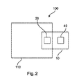
図2は、X線画像内の不透明度を検出するシステム100の一例を示す。システム100は、図1に関して説明されたように、少なくとも1つの画像取得装置110、及びX線画像内の不透明度を検出する装置10を備える。少なくとも1つの画像取得装置110は、有線又は無線通信を介して、分析X線画像を提供するように構成される。その提供は、入力ユニット20に対してなされ得る。出力ユニット40は、少なくとも1つの異常に関する情報を含む、分析X線画像を出力するように構成される。
FIG. 2 shows an example of a
一例において、入力ユニット20は、少なくとも1つの取得装置110の画像取得装置である。
In one example,
一例において、少なくとも1つの画像取得装置は、関心領域の複数のX線画像を取得するように構成される。 In one example, at least one image acquisition device is configured to acquire a plurality of X-ray images of a region of interest.
一例において、少なくとも1つの画像取得装置は、格子ベースの微分位相差撮像装置及び暗視野X線撮像装置を備える。一例において、少なくとも1つの画像取得装置は、干渉計構成部を備える。 In one example, at least one image acquisition device comprises a grating-based differential phase contrast imager and a dark field X-ray imager. In one example, at least one image acquisition device comprises an interferometer component.
一例において、少なくとも1つの画像取得装置は、X線撮像装置を備える。たとえば、この装置は、断層撮影構成部又はCT構成部であり得る。 In one example, at least one image acquisition device comprises an X-ray imaging device. For example, the device may be a tomographic component or a CT component.
一例において、少なくとも1つの画像取得装置は標準的なX線撮影装置であり、放射線の透過強度が、放射線が物体を通過するときの減衰に関する情報を提供する。 In one example, the at least one image acquisition device is a standard radiography device, and the transmission intensity of the radiation provides information regarding the attenuation of the radiation as it passes through the object.
一例において、少なくとも1つの画像取得装置は、微分位相差撮像(DPCI:Differential phase contrast imaging)モードで動作することができる。 In one example, the at least one image acquisition device can operate in a Differential Phase Contrast Imaging (DPCI) mode.
一例において、少なくとも1つの画像取得装置は、検査領域内の物体の有無にかかわらず、X線の強度(強度)値の検出に関する減衰画像を生成する。 In one example, at least one image acquisition device generates an attenuated image related to detection of an X-ray intensity (intensity) value regardless of the presence or absence of an object in the inspection area.
一例において、少なくとも1つの画像取得装置は、検査領域内の物体の有無にかかわらず、X線の位相の検出に関する、位相差(又は微分位相)画像を生成する。一例において、少なくとも1つの画像取得装置は、検査領域内の物体の有無にかかわらず、X線の縞の鮮明度の検出に関する、暗視野(又はデコヒーレンス)画像を生成する。 In one example, the at least one image acquisition device generates a phase difference (or differential phase) image for X-ray phase detection, with or without an object in the examination region. In one example, at least one image acquisition device generates a dark-field (or decoherence) image for detecting the sharpness of X-ray stripes, with or without an object in the inspection area.
一例において、出力ユニットは、吸収(又は減衰)画像を出力する。一例において、出力ユニットは、位相差(又は微分位相)画像を出力する。一例において、出力ユニットは、暗視野画像を出力する。 In one example, the output unit outputs an absorption (or attenuation) image. In one example, the output unit outputs a phase difference (or differential phase) image. In one example, the output unit outputs a dark field image.
一例において、出力ユニットは、視覚表示装置などのモニタ上又は複数の別個のモニタ上にデータを出力する。たとえば、減衰画像、位相差画像、及び暗視野画像をモニタ上に提示することができる。 In one example, the output unit outputs data on a monitor, such as a visual display, or on a plurality of separate monitors. For example, an attenuation image, a phase contrast image, and a dark field image can be presented on a monitor.
一例において、このシステムには、病院などの臨床環境において有用な用途がある。一例において、このシステムは、乳房X線撮影法、診断放射線医学、及び患者の医療検査のための介入的放射線医学において、肺疾患の検出に使用することができる。 In one example, the system has a useful application in a clinical environment such as a hospital. In one example, the system can be used for detection of lung disease in mammography, diagnostic radiology, and interventional radiology for medical examination of patients.
一例において、このシステムには、産業上の環境、たとえば非破壊試験(たとえば生物学的試料及び非生物学的試料の組成、構造及び/又は品質に関する分析)、並びにセキュリティ走査(たとえば空港での荷物の走査)において、有用な用途がある。 In one example, the system includes an industrial environment, such as non-destructive testing (eg, analysis of the composition, structure and / or quality of biological and non-biological samples), and security scanning (eg, airport luggage). Scanning) has a useful application.
図3は、X線画像内の不透明度を検出するための方法200を、その基本的なステップにおいて示す。この方法は、
提供するステップ210、またステップa)と称するステップにおいて、分析される身体部分の関心領域の分析X線画像を提供するステップと、
提供するステップ220、またステップb)と称するステップにおいて、正常な関心領域のモデルを提供するステップであって、このモデルは、関心領域の複数のX線画像に基づくものである、ステップと、
検出するステップ230、またステップc)と称するステップにおいて、分析される身体部分の関心領域内の少なくとも1つの異常を検出するステップであって、関心領域の分析X線画像と、正常な関心領域のモデルとを比較するステップを有する、ステップと、
出力するステップ240、またd)と称するステップにおいて、少なくとも1つの異常に関する情報を出力するステップと
を有する。
FIG. 3 shows a
Providing 210, also referred to as step a), providing an analytical X-ray image of the region of interest of the body part being analyzed;
Providing 220, also referred to as step b), providing a model of a normal region of interest, wherein the model is based on a plurality of X-ray images of the region of interest.
Detecting 230, also referred to as step c), detecting at least one anomaly in the region of interest of the body part being analyzed, comprising analyzing an X-ray image of the region of interest and a normal region of interest. Comparing the model with a step;
Outputting
ステップa)において、この提供するステップは、入力ユニット20から処理ユニット30への提供であり得る。
ステップb)において、この提供するステップは、入力ユニットから処理ユニットへの提供であり得る。
ステップc)において、この検出するステップは、処理ユニットによって実行され得る。
ステップd)において、この出力するステップは、出力ユニットによって実行され得る。
In step a), the providing step may be providing from the
In step b), the providing step may be providing from the input unit to the processing unit.
In step c), this detecting step may be performed by the processing unit.
In step d), this outputting step may be performed by the output unit.
一例において、ステップc)は、分析X線画像の関心領域内の少なくとも1つの強度と、モデルの正常な関心領域内の、対応する少なくとも1つの強度との間の、少なくとも1つの偏差を判定するステップを有する。 In one example, step c) determines at least one deviation between at least one intensity in the region of interest of the analytical X-ray image and a corresponding at least one intensity in the normal region of interest of the model. With steps.
一例において、ステップb)で提供されるモデルは、関心領域の複数のX線画像内の対応する強度に基づく、少なくとも1つの平均強度を含むモデルデータを有し、モデルデータは、関心領域の複数のX線画像内の対応する強度に基づく、少なくとも1つの強度の標準偏差を含む。そして、ステップc)において、関心領域の分析X線画像と正常な関心領域のモデルとの間の比較は、分析X線画像の関心領域内の少なくとも1つの強度値、モデルの正常な関心領域内の少なくとも1つの平均強度値、及びモデルの正常な関心領域内の少なくとも1つの強度の標準偏差に基づくことができる。 In one example, the model provided in step b) comprises model data comprising at least one average intensity based on corresponding intensities in a plurality of X-ray images of the region of interest, wherein the model data comprises a plurality of the region of interest. At least one standard deviation of the intensities based on the corresponding intensities in the X-ray image of. Then, in step c), a comparison between the analyzed X-ray image of the region of interest and the model of the normal region of interest is made by comparing at least one intensity value in the region of interest of the analyzed X-ray image with the normal region of interest of the model. And a standard deviation of at least one intensity within the normal region of interest of the model.
一例において、ステップc)は、関心領域の分析X線画像内の空間位置での強度と、正常の関心領域のモデル内の対応する空間位置での平均強度との間の差を判定するステップを有し、またこの差と、正常な関心領域のモデル内の対応する空間位置における強度の標準偏差との間の比を判定するステップを有する。 In one example, step c) comprises determining the difference between the intensity at the spatial location in the analytical X-ray image of the region of interest and the average intensity at the corresponding spatial location in the model of the normal region of interest. And determining the ratio between the difference and the standard deviation of the intensity at the corresponding spatial location in the model of the normal region of interest.
一例において、ステップc)は、分析X線画像の関心領域内の少なくとも1つの強度、及びそれに対応するモデルの正常な関心領域内の少なくとも1つの強度に基づく、少なくとも1つのスコアを判定するステップを有する。 In one example, step c) comprises determining at least one score based on at least one intensity in the region of interest of the analytical X-ray image and at least one intensity in the normal region of interest of the corresponding model. Have.
一例において、分析される身体部分の関心領域内で少なくとも1つの異常が検出されたことを示すためにスコアが使用される。 In one example, the score is used to indicate that at least one abnormality has been detected in the region of interest of the body part being analyzed.
一例において、ステップc)は、少なくとも1つのスコアに基づいて、分析X線画像の関心領域の少なくとも1つの領域を示すステップを有する。 In one example, step c) comprises indicating at least one region of interest of the analysis X-ray image based on the at least one score.
一例において、ステップc)は、分析X線画像内の、少なくともいくらかの骨関連画像を抑制するステップを有する。 In one example, step c) comprises suppressing at least some bone-related images in the analytical X-ray image.
一例において、ステップc)は、分析X線画像の少なくとも1つの強度を正規化するステップを有する。 In one example, step c) comprises normalizing at least one intensity of the analytical X-ray image.
一例において、ステップc)は、分析X線画像の関心領域を、モデルの正常な関心領域に位置合せするステップを有する。 In one example, step c) comprises aligning the region of interest of the analytical X-ray image with the normal region of interest of the model.
X線画像内の不透明度を検出する装置、システム、及び方法の例を、次いで、図4〜図7と共により詳細に説明することにする。 Examples of devices, systems, and methods for detecting opacity in X-ray images will then be described in more detail in conjunction with FIGS.
質感の特徴(van Ginneken、Bram等による論文「結核罹患率調査のための胸部X線写真の自動スコアづけ:組合せ手法」肺画像分析に関する第5回国際ワークショップ2013年会報を参照のこと)及び不透明部の形状の特徴を含む、様々な画像の特徴の自動測定に基づく、胸部X線写真内の結核病変の自動検出のための、いくつかの既存の代替手法が提案されてきた。肺野の形状もまた、TB検出のベースとして提案されており(van Ginneken等による論文、及びJaeger、Stefan等による論文「組合せ肺マスクを使用したX線写真内の結核の検出」生体医工学会(EMBC:Engineering in Medicine and Biology Society)2012年IEEE年次国際会議、IEEE、2012年を参照のこと)、これによって、肋膜に隣接する不透明部は、実質病変の存在に対する間接的な指標をもたらす。しかし、様々な技術的な機能に基づいたコンピュータ支援意思決定システムの結果をユーザが検証するのは困難である。分析結果の直感的な指標は、結果におけるユーザの信頼を確立するために重要であり、したがって、これはかかる手法にとって大きな制約となる。さらに、こうしたアルゴリズムの訓練は通常、多数の注釈付き画像を必要とする。また、特化して訓練されたアルゴリズムは、ノイズ、いくつかの種類の画像アーティファクト、及び画像後処理パラメータのような外部パラメータに対して、非常に影響を受けやすいことがわかる。したがって、訓練セットから外れた条件下で得られた様々な画像の組からの画像に対して、実世界の状況で必要とされるように、訓練されたアルゴリズムを適用することは簡単ではない。 Characteristics of texture (see the article "Automatic Scoring of Chest Radiographs for Investigation of Tuberculosis Incidence: Combined Method" by van Ginkenen, Bram, et al., The 5th International Workshop on Lung Image Analysis 2013) Several existing alternatives have been proposed for the automatic detection of tuberculosis lesions in chest radiographs based on the automatic measurement of various image features, including opaque shape features. The shape of the lung field has also been proposed as a basis for TB detection (see the paper by van Ginken et al. And the paper by Jaeger, Stefan et al. "Detection of tuberculosis in radiographs using combined lung masks" (See Engineering in Medicine and Biology Society, 2012, IEEE International Conference, IEEE, 2012), whereby the opacity adjacent to the pleura provides an indirect indication for the presence of parenchymal lesions. . However, it is difficult for a user to verify the results of a computer-aided decision system based on various technical functions. Intuitive indicators of the results of the analysis are important for establishing user confidence in the results, and this is a major constraint for such an approach. Further, training of such algorithms typically requires a large number of annotated images. Also, it can be seen that specialized trained algorithms are very sensitive to external parameters such as noise, some types of image artifacts, and image post-processing parameters. Therefore, it is not easy to apply a trained algorithm to images from different sets of images obtained under conditions outside the training set, as required in real-world situations.
本明細書で説明される、X線画像内の不透明度を検出する装置、システム、及び方法は、図1〜図3に関して前述したように、また以下でより詳細に論じられるように、こうした問題に対処する。 The devices, systems, and methods described herein for detecting opacity in x-ray images address such issues as described above with respect to FIGS. 1-3 and as discussed in more detail below. To deal with.
肺内のTBの放射線学的徴候は、感染前に肺実質が存在していた位置での、体液又は線維組織の局所的蓄積によって引き起こされる。肺組織の生理学的特性のこの変化により、(X線の吸収の増加による)局所的な不透明部が、胸部X線画像内に存在する。本明細書に記載の装置によって提供される、肺野に対応するX線写真の領域内の不透明度の自動検出は、胸部X線写真内の結核を自動的に検出するための、非常に直接的且つ生理学的に妥当な手法である。 Radiological signs of TB in the lungs are caused by the local accumulation of fluid or fibrous tissue at the location where the lung parenchyma was present prior to infection. Due to this change in the physiological properties of lung tissue, localized opacity (due to increased x-ray absorption) is present in the chest x-ray image. The automatic detection of opacity in the area of the radiograph corresponding to the lung field, provided by the device described herein, is very direct for automatically detecting tuberculosis in the chest radiograph. This is a technically and physiologically appropriate approach.
これを達成するように、異常に関連するかかる不透明度を識別できるようにするために、いくつかの厄介な問題を考慮に入れなければならない。X線画像内の不透明度を検出する装置、システム、及び方法によって対処される厄介な問題は以下を含む。
i.肺野で覆われた骨も、結核によって引き起こされるものよりも強い不透明度を課す。
ii.様々な取得プロトコル(露出)及び患者の特性(体重/サイズ)に応じて、肺野における画像強度は、結核によって引き起こされる変動に起因するよりも著しく大きく変わり、それによって画像間の定量的比較が妨げられる。
iii.肺血管の木の根(肺門)は、強い不透明性を課す。
To accomplish this, some complications must be taken into account in order to be able to identify such opacity associated with anomalies. The complications addressed by devices, systems, and methods for detecting opacity in X-ray images include:
i. Bone covered by lung fields also imposes greater opacity than that caused by tuberculosis.
ii. Depending on the different acquisition protocols (exposure) and patient characteristics (weight / size), the image intensity in the lung field varies significantly more than due to the variations caused by tuberculosis, which makes the quantitative comparison between the images Hindered.
iii. The tree roots of the pulmonary vessels (hilar) impose strong opacity.
同様の問題は、他の肺疾患の検出において、たとえば乳房X線画像の分析においても生じる。 Similar problems occur in the detection of other lung diseases, for example in the analysis of mammograms.
図4は、本明細書に記載のX線画像内の不透明度を検出する装置、システム、及び方法によって提供される、X線画像内の不透明度を検出するための詳細な作業の流れの一例を示す。胸部X線写真などのX線画像内の不透明度の自動検出及び特徴づけのための手法が提供される。胸部X線写真のケースに対するこの手法の4つの主な要素を、以下のようにまとめることができる。
1.胸部X線写真の前処理
2.胸部X線写真と正常モデルとの比較
3.胸部X線写真内の異常の検出、及び
4.胸部X線写真から検出された異常の分類
FIG. 4 is an example of a detailed workflow for detecting opacity in an X-ray image provided by the apparatus, system, and method for detecting opacity in an X-ray image described herein. Is shown. Techniques are provided for automatic detection and characterization of opacity in X-ray images, such as chest radiographs. The four main elements of this approach for a chest radiograph case can be summarized as follows.
1. 1. Pre-processing of
本質的にこの手法は、胸部X線写真を互いに直接比較することを可能にし、それによって方法は、「目に見えない」画像を多数の「知られた」画像と比較することを可能にする。画像の選択は専門家によって実行され、それにより、何らかの事前定義された包含基準の組に従って評価され得る。そしてこの画像「データベース」から得られる情報は、このデータベース内で符号化された予測値からの差を検出するための比較分析のベースを形成する。 Essentially, this technique allows chest radiographs to be compared directly to each other, thereby allowing the method to compare an "invisible" image to a number of "known" images. . The selection of images is performed by an expert, and can be evaluated according to some set of predefined inclusion criteria. The information obtained from this image "database" then forms the basis of a comparative analysis for detecting differences from the predicted values coded in this database.
図4に示される詳細な作業の流れの特定のステップについて、ここでより詳細に説明する。 The specific steps of the detailed workflow shown in FIG. 4 will now be described in more detail.
前処理
上記で概要を示した厄介な問題(i及びii)を考慮するために、各画像について前処理が実行される。以下の前処理ステップが適用される。
1.肺野のセグメント化
2.骨の抑制
3.肺野の強度正規化、及び
4.肺野の空間正規化。
Preprocessing In order to take into account the troublesome problems (i and ii) outlined above, preprocessing is performed on each image. The following pre-processing steps apply.
1. 1. Segmentation of
図5は、一症例からの、前処理の様々な段階での結果を示す。こうしたステップの後で、肺野内の強度を定量分析することができる。肺野の外側の信号は無視され、肺野の内側に重ねられた肋骨構造は抑制され、露出又は患者の体重の影響は、強度の正規化ステップにおいて補償される。空間正規化(又は位置合せ)はまた、肺野の解剖学的に対応する位置を位置合せする。 FIG. 5 shows the results from one case at various stages of pre-processing. After these steps, the intensity in the lung field can be quantitatively analyzed. Signals outside the lung field are ignored, rib structures superposed inside the lung field are suppressed, and the effects of exposure or patient weight are compensated for in the intensity normalization step. Spatial normalization (or alignment) also aligns anatomically corresponding positions of the lung fields.
正常モデル
統計モデルは、アトラス内の任意の位置において、強度分布(平均値av_i(x)及び標準偏差stddev(x))を記述している、正常のケース(専門家によって画像内で有意な放射線学的所見が観察されなかったことを意味する、放射線学的正常と定義される)の収集から構築される。このモデルは、平均値及び標準偏差に基づいて、正常に対するアトラス空間内での予測強度の信頼区間を提供する(一般的なアトラス空間内での一組の正常な対象者の肺野内の、強度分布のモデル化を示す、図6を参照のこと−平均及び標準偏差をもつ、ガウスモデルが使用される)。平均的な肺野内に暗い領域(中央上部)があり、より明るい領域(肺底、外側帯、肺門)がある。予測強度の標準偏差もまた、アトラス空間にわたって空間的に変化する。たとえば、変動は、肺門内及び肺底内でより大きくなるが、外側帯ではそうではない。平均及び標準偏差を含むこの正常モデルは、比較的少数のパラメータを有し、適用するのが概念的に非常に簡単である。訓練に必要なデータセットはごく少数であり、訓練ケースの特定の選択に適合し過ぎるリスクは低い。
Normal Model The statistical model describes the normal case (significant radiation in the image by an expert) describing the intensity distribution (mean av_i (x) and standard deviation stddev (x)) at any location in the atlas. (Defined as radiological normality, meaning no radiological findings were observed). This model provides confidence intervals for the predicted intensities in the atlas space relative to normals based on the mean and standard deviation (intensities in the lung field of a set of normal subjects in the general atlas space). (See FIG. 6 showing the modeling of the distribution-a Gaussian model with mean and standard deviation is used). There are dark areas (upper middle) in the average lung field and lighter areas (basilar, lateral zone, hilum). The standard deviation of the predicted intensities also varies spatially over the atlas space. For example, the variability is greater in the hilum and in the lung floor, but not in the outer band. This normal model, including the mean and standard deviation, has a relatively small number of parameters and is conceptually very simple to apply. The dataset required for training is very small and the risk of overfitting a particular choice of training case is low.
異常検出
たとえば対象者からの肺野で取得され、分析X線画像であると考えられ得る画像は、以下によって分析される。
1.画像の前処理、及び
2.任意の異常画像位置の検出。
Abnormality Detection An image acquired, for example, in the lung field from a subject and which may be considered an analytical X-ray image is analyzed by:
1. 1. pre-processing of the image, and Detection of any abnormal image position.
肺野内の任意の位置における信頼区間からの偏差(xにおけるzスコア)が検出される。

結果を図7に示す。ここで、高いzスコアを有する異常画像領域は、左側画像上の中実のオーバレイによって示され、右側画像上の異常領域を示す輪郭として示される。画像強度だけでは、マーク付けされる(又は検出される)べき領域として、決定的ではないことが理解されよう。たとえば、正常モデル内の標準偏差もまたこの領域で大きく、したがって、(標準偏差が分母となる)zスコアがより小さくなるので、肺門の領域は明るいが、マーク付けされない。こうして、この手法はまた、前述の第3の厄介な問題(iii)にも対処する。 FIG. 7 shows the results. Here, the abnormal image region having a high z-score is indicated by a solid overlay on the left image, and is indicated as a contour indicating the abnormal region on the right image. It will be appreciated that image intensity alone is not critical as an area to be marked (or detected). For example, the hilar region is bright but not marked because the standard deviation in the normal model is also large in this region, and thus the z-score (where the standard deviation is the denominator) is smaller. Thus, this approach also addresses the third complication (iii) discussed above.
分類
総合的な異常スコアZは、z(x)>sとなる全ての位置xを数え、これを肺のサイズを占める肺野内の位置の数に正規化することによって、肺野全体に関して計算される。Zは、特定の胸部X線写真の異常に関する適切な判断基準を提供する。
Classification The overall anomaly score Z is calculated for the entire lung field by counting all positions x such that z (x)> s and normalizing this to the number of positions in the lung field that occupy the size of the lung. You. Z provides appropriate criteria for certain chest x-ray abnormalities.
要約
前に詳細に説明してきた、モデル構築、異常検出、及び評価の特定のステップに関するさらなる詳細を含む簡単な概要を、以下に提供する。前に概説した前処理は、この目的のための重要な前提条件のステップであり得る。ある状況では、前処理は、分析に含まれるすべての画像(モデルを構築するための訓練段階の間の参照画像、及び検出中の目に見えない画像の両方)に、同じやり方で適用される。ここで、これらのステップは、下記にさらに詳しく述べられている。
−肺野のセグメント化は、D.Barthel及びJ.von Bergによる論文である「ディジタル胸部X線写真上の堅牢な自動肺野セグメント化」CARS国際学術誌、4(補遺1):326〜327頁、2009年に記載された方法によって実施される。セグメント化の他の知られた方法を使用することができる。
−骨の抑制もまた、たとえば以下の情報源に記載される、知られた方法によって実施され得る。von Berg及びNeitzelによる国際特許出願公開WO2011/077334「X線写真内の骨の抑制」、Jens von Berg、Stewart Young、Heike Carolus、Robin Wolz、Axel Saalbach、Alberto Hidalgo、Ana Gimenez、及びTomas Franquetによる「肺瘤検出を改善する、新規の骨抑制方法」コンピュータ支援放射線医学・外科学協会の国際学術誌、1〜15頁、2015年、並びにvon Berg、Levrier、Carolus、Young、Saalbach、Laurent、及びFlorentによる「X線写真内の胸郭の分解」出版されたISBI2016の会報。
−強度正規化は、たとえば強度範囲の7.5%で、肺野の暗い部分など、肺野の強度分位数qを判定することによって実施され得る。したがって、正規化は、画像からqを減算することを意味する(そして負の画像強度にしないために定数を加算する)。これは、複雑な質感及び形状の特徴分析に基づく他の方法とは対照的に、非常に簡単な方法である。
−空間正規化は、離散的な一組のステップポイントから作成された肺野の輪郭に基づいて実行することができる。平均的な肺野モデルは、訓練セットに基づいて構築される。これにより、「アトラス」(基準)空間の定義が確立される。次いで、元の画像を変形させて、肺と、モデルの肺とを位置合わせすることにより、すべての画像を、この「アトラス」空間と空間的に位置合せすることができる。Rueckert、Daniel等による論文「自由形状変形を利用した非剛体位置合せ:乳房MR画像への応用」IEEE Transactions on Medical Imaging、1999年18[8]、712〜721頁に記載される、Bスプライン法もまた適用され得る。k最近傍補間などの他の手法が適用され得る。
SUMMARY A brief overview is provided below, including further details regarding specific steps of model building, anomaly detection, and evaluation, which have been described in detail above. The pre-processing outlined above may be an important prerequisite step for this purpose. In some situations, preprocessing is applied in the same way to all images involved in the analysis (both reference images during the training phase to build the model and invisible images during detection). . Here, these steps are described in more detail below.
Segmentation of the lung field Barthel and J.M. von Berg, "A Robust Automatic Lung Segmentation on Digital Chest Radiographs", CARS International Academic Journal, 4 (Supplement 1): 326-327, 2009. Other known methods of segmentation can be used.
-Bone suppression can also be performed by known methods, for example as described in the following sources: International Patent Application Publication No. WO 2011/077334 by Bonn Berg and Neitzel, "Bone Suppression in X-Ray Photography", Jens von Berg, Stewart Young, Heike Carolus, Robin Wolz, Axel Saalbach, Alberto Hilmar, and Alberto Higanz, by Alberto Nazica, Alberto Hidden A Novel Method of Bone Suppression to Improve Aneurysm Detection "International Journal of Computer Aided Radiology and Surgery, 1-15, 2015, and von Berg, Leverier, Carolos, Young, Saalbach, Laurent, and Florent Bulletin of the Thorax in Radiographs, published by ISBI 2016.
-Intensity normalization can be performed by determining the intensity quantile q of the lung field, for example at 7.5% of the intensity range, such as the dark part of the lung field. Therefore, normalization means subtracting q from the image (and adding a constant to avoid negative image intensities). This is a very simple method, in contrast to other methods based on complex texture and shape feature analysis.
-Spatial normalization can be performed based on lung field contours created from a discrete set of step points. An average lung field model is built based on the training set. This establishes the definition of the "atlas" (reference) space. All images can then be spatially aligned with this "atlas" space by deforming the original image and aligning the lungs with the model lungs. B-spline method described in a paper by Rueckert, Daniel et al., "Non-rigid Registration Using Free-form Deformation: Application to Breast MR Images", IEEE Transactions on Medical Imaging, 18 [8], 1999, pp. 712-721. May also be applied. Other approaches such as k-nearest neighbor interpolation may be applied.
したがって、本明細書に記載のX線画像内の不透明度を検出する装置、システム及び方法を、異常である確率が高い画像を自動的に分類するために適用することができる。これは、データベースから関心のある症状を選択する自動ステップとして作用することができる。それはまた、結核のような肺疾患に罹患する一定のリスクを有する患者を同定するためにも使用され得る。検査のシナリオでは、こうした患者は次いで、痰検査又は遺伝子検査のような他の手段を用いて、さらなる診断検査を受けることができる。また、図7にあるような視覚化を、結核のような肺疾患の診断を担当する人に提示して、この人が診断により自信を持つようになる補助をすることができる。 Accordingly, the devices, systems and methods for detecting opacity in X-ray images described herein can be applied to automatically classify images with a high probability of being abnormal. This can act as an automatic step of selecting the condition of interest from the database. It can also be used to identify patients with a certain risk of developing a lung disease such as tuberculosis. In the testing scenario, such patients can then undergo further diagnostic tests using other means such as sputum testing or genetic testing. Also, the visualization as shown in FIG. 7 can be presented to a person in charge of diagnosing a pulmonary disease such as tuberculosis to help this person become more confident in the diagnosis.
別の例示的な実施形態では、適切なシステムである前述の実施形態のうちの1つによる方法の、方法ステップを実行するように構成されていることを特徴とする、コンピュータプログラム又はコンピュータプログラム要素が提供される。 In another exemplary embodiment, a computer program or a computer program element characterized in that it is adapted to perform the method steps of a method according to one of the preceding embodiments, which is a suitable system Is provided.
したがって、コンピュータプログラム要素はコンピュータ装置に格納され、これもまた実施形態の一部である。このコンピュータ処理ユニットは、前述の方法のステップを実行する、又は実行を誘導するように構成される。さらに、前述の装置の構成要素を動作させるように構成される。コンピュータ処理ユニットは、自動的に動作するように、且つ/又はユーザの命令を実行するように構成され得る。コンピュータプログラムは、データプロセッサの作業メモリにロードされる。したがって、データプロセッサは、前述の実施形態のうちの1つによる方法を実行するように装備される。 Thus, the computer program elements are stored on a computing device, which is also part of an embodiment. The computer processing unit is configured to perform or direct the execution of the method steps described above. Further, it is configured to operate the components of the device described above. The computer processing unit may be configured to operate automatically and / or to execute user instructions. The computer program is loaded into the working memory of the data processor. Therefore, the data processor is equipped to perform the method according to one of the embodiments described above.
本発明のこの例示的な実施形態は、ずっと最初から本発明を使用するコンピュータプログラムと、更新によって既存のプログラムを、発明を使用するプログラムに変えるコンピュータプログラムとの両方を包含する。 This exemplary embodiment of the invention encompasses both computer programs that use the invention from the very beginning, and computer programs that update existing programs into programs that use the invention by updating.
さらに、コンピュータプログラム要素は、前述の方法の例示的実施形態の手順を満たすために必要なすべてのステップを提供することができる。 Further, the computer program element may provide all steps necessary to fulfill the procedures of the exemplary embodiment of the method described above.
本発明のさらなる例示的な実施形態によれば、CD−ROMなどのコンピュータ可読媒体が提示され、このコンピュータ可読媒体は、そこに格納されるコンピュータプログラム要素を有し、このコンピュータプログラム要素は、前節で説明されている。 According to a further exemplary embodiment of the present invention, a computer readable medium, such as a CD-ROM, is presented, the computer readable medium having a computer program element stored thereon, the computer program element comprising It is explained in.
コンピュータプログラムは、他のハードウェアと共に、又は他のハードウェアの一部として供給される、光記憶媒体又は固体媒体などの好適な媒体にて格納及び/又は配布されるが、インターネット又は他の有線若しくは無線の電気通信システムを介するなどして、他の形態でも配布され得る。 The computer program is stored and / or distributed on a suitable medium, such as an optical storage medium or a solid state medium, supplied with or as part of the other hardware, but not over the Internet or other wired Or, it may be distributed in other forms, such as via a wireless telecommunications system.
しかし、コンピュータプログラムはまた、ワールドワイドウェブのようなネットワークを介して提供され、かかるネットワークからデータプロセッサの作業メモリへダウンロードすることができる。本発明のさらなる例示的実施形態によれば、コンピュータプログラム要素をダウンロードで入手可能にするための媒体が提供され、コンピュータプログラム要素は、本発明の前述の実施形態のうちの1つによる方法を実行するように構成される。 However, the computer program is also provided over a network such as the World Wide Web and can be downloaded from such a network to the working memory of the data processor. According to a further exemplary embodiment of the present invention, there is provided a medium for making a computer program element available for download, wherein the computer program element performs a method according to one of the aforementioned embodiments of the present invention. It is configured to
本発明の実施形態は、様々な主題を参照して説明されていることに留意されたい。特にある実施形態は、方法のタイプの請求項に関して説明されているが、一方他の実施形態は、装置のタイプの請求項に関して説明されている。しかし当業者は、特に断りのない限り、1つのタイプの主題に属する特徴の任意の組合せに加え、相異なる主題に関連する特徴間の任意の組合せもまた、本出願と共に開示されていると考えられることを、前述及び以下の説明から知るであろう。しかし、すべての特徴を組み合わせて、特徴の単なる足し算を上回る相乗効果を提供することができる。 Note that embodiments of the invention have been described with reference to various subjects. In particular, certain embodiments are described with reference to method type claims, while other embodiments are described with reference to device type claims. However, those skilled in the art will recognize that, unless otherwise noted, any combination of features belonging to one type of subject, as well as any combination between features relating to different subjects, is also disclosed with the present application. Will be understood from the foregoing and following description. However, all features can be combined to provide a synergistic effect that goes beyond the mere addition of features.
本発明について、図面及び前述の記載で詳細に示し、また説明してきたが、かかる図及び説明は、例示的又は代表的であり、限定的ではないと考えられるべきである。本発明は、開示された実施形態に限定されない。開示された実施形態に対する他の変形形態は、特許請求される発明を実施する中で、図面、開示、及び従属請求項の研究から、当業者によって理解され達成され得る。 While the invention has been illustrated and described in detail in the drawings and foregoing description, such figures and description are to be considered illustrative or representative and not restrictive; The invention is not limited to the disclosed embodiments. Other variations to the disclosed embodiments can be understood and effected by those skilled in the art in practicing the claimed invention, from a study of the drawings, the disclosure, and the dependent claims.
特許請求の範囲で、用語「comprising」は、他の要素又はステップを除外するものではなく、また不定冠詞「a」又は「an」は複数を除外するものではない。単一のプロセッサ又は単一の他の装置は、特許請求の範囲に列挙された複数の項目の機能を果たす。特定の手段が、互いに相異なる従属請求項に列挙されているという単なる事実は、これらの手段の組合せが有利に使用され得ないことを示すものではない。特許請求の範囲内のいかなる参照符号も、範囲を限定するものと解釈されるべきではない。 In the claims, the term "comprising" does not exclude other elements or steps, and the indefinite article "a" or "an" does not exclude a plurality. A single processor or a single other device fulfills the functions of several items recited in the claims. The mere fact that certain measures are recited in mutually different dependent claims does not indicate that a combination of these measures cannot be used to advantage. Any reference signs in the claims shall not be construed as limiting the scope.
Claims (14)
前記入力ユニットは、前記処理ユニットに、分析される身体部分の関心領域の分析X線画像を供給し、
前記入力ユニットは、前記処理ユニットに、正常であり且つ異常を罹患していない関心領域の複数のX線画像に基づき、正常で健康な母集団に関する統計情報を含む、正常な関心領域のモデルを供給し、
前記処理ユニットは、前記分析される身体部分の関心領域の中の少なくとも1つの異常を検出し、当該検出は、前記関心領域の分析X線画像と、前記正常な関心領域のモデルとの間の比較を含み、当該検出は、前記分析X線画像内の少なくともいくらかの骨関連画像の抑制を含み、
前記出力ユニットは、少なくとも1つの異常に関する情報を出力する、装置。 An apparatus for detecting opacity of a clinical X-ray image, the apparatus including an input unit, a processing unit, and an output unit,
The input unit supplies the processing unit with an analytical X-ray image of a region of interest of the body part to be analyzed;
The input unit includes a processing unit configured to generate a model of a normal region of interest including statistical information about a normal and healthy population based on a plurality of X-ray images of the region of interest that is normal and does not suffer from abnormality. Supply,
The processing unit detects at least one anomaly in the region of interest of the body part to be analyzed, wherein the detection is performed between the analyzed X-ray image of the region of interest and the model of the normal region of interest. Comprising comparing, wherein said detecting comprises suppressing at least some bone-related images in said analytical X-ray image;
The apparatus wherein the output unit outputs information regarding at least one abnormality.
少なくとも1つの画像取得装置、及び
請求項1乃至11の何れか一項に記載の臨床X線画像内の不透明度を検出する装置を備え、
前記少なくとも1つの画像取得装置は、分析X線画像を提供し、
前記出力ユニットは、少なくとも1つの異常に関する情報を含む分析X線画像を出力する、システム。 A system for detecting opacity in an X-ray image, said system comprising:
At least one image acquisition device, and a device for detecting opacity in a clinical X-ray image according to any one of claims 1 to 11,
The at least one image acquisition device provides an analytical X-ray image;
The system, wherein the output unit outputs an analysis X-ray image including information on at least one abnormality.
a)分析される身体部分の関心領域の分析X線画像を提供するステップ、
b)正常な関心領域のモデルを提供するステップであって、前記モデルは、正常であり且つ異常を罹患していない関心領域の複数のX線画像に基づくものであり、前記モデルは、正常で健康な母集団に関する統計情報を含む、ステップ、
c)前記分析される身体部分の関心領域内の少なくとも1つの異常を検出するステップであって、当該検出するステップは、前記関心領域の前記分析X線画像と、前記正常な関心領域のモデルとを比較するステップと、前記分析X線画像内の少なくともいくつかの骨関連画像を抑制するステップとを有する、検出するステップ、及び
d)前記少なくとも1つの異常に関する情報を出力するステップ、
を有する、方法。 An automated method for detecting opacity in an X-ray image, the method comprising:
a) providing an analytical X-ray image of the region of interest of the body part to be analyzed;
b) providing a model of a normal region of interest, wherein the model is based on a plurality of x-ray images of the region of interest that is normal and does not suffer from abnormalities; Steps that include statistics about a healthy population
c) detecting at least one anomaly in the region of interest of the body part to be analyzed, the detecting comprising: analyzing the X-ray image of the region of interest; and a model of the normal region of interest. Detecting and suppressing at least some bone-related images in the analytical X-ray image; and d) outputting information about the at least one abnormality;
A method comprising:
Applications Claiming Priority (3)
| Application Number | Priority Date | Filing Date | Title |
|---|---|---|---|
| EP16197665 | 2016-11-08 | ||
| EP16197665.9 | 2016-11-08 | ||
| PCT/EP2017/077376 WO2018086893A1 (en) | 2016-11-08 | 2017-10-26 | Apparatus for the detection of opacities in x-ray images |
Publications (2)
| Publication Number | Publication Date |
|---|---|
| JP2019536531A true JP2019536531A (en) | 2019-12-19 |
| JP2019536531A5 JP2019536531A5 (en) | 2020-12-03 |
Family
ID=57256137
Family Applications (1)
| Application Number | Title | Priority Date | Filing Date |
|---|---|---|---|
| JP2019523631A Pending JP2019536531A (en) | 2016-11-08 | 2017-10-26 | Apparatus for detecting opacity in X-ray images |
Country Status (5)
| Country | Link |
|---|---|
| US (1) | US11058383B2 (en) |
| EP (1) | EP3539079A1 (en) |
| JP (1) | JP2019536531A (en) |
| CN (1) | CN109937433B (en) |
| WO (1) | WO2018086893A1 (en) |
Cited By (1)
| Publication number | Priority date | Publication date | Assignee | Title |
|---|---|---|---|---|
| JP7515709B2 (en) | 2020-09-21 | 2024-07-12 | コーニンクレッカ フィリップス エヌ ヴェ | Dark-field X-ray image data information processing |
Families Citing this family (4)
| Publication number | Priority date | Publication date | Assignee | Title |
|---|---|---|---|---|
| EP3622891A1 (en) * | 2018-09-13 | 2020-03-18 | Koninklijke Philips N.V. | Calculation device for determining ventilation defects |
| JP7023254B2 (en) * | 2019-03-27 | 2022-02-21 | 富士フイルム株式会社 | Shooting support equipment, methods and programs |
| EP3866107A1 (en) * | 2020-02-14 | 2021-08-18 | Koninklijke Philips N.V. | Model-based image segmentation |
| CN111815557A (en) * | 2020-06-04 | 2020-10-23 | 上海联影智能医疗科技有限公司 | Image analysis method, device, equipment and storage medium |
Citations (7)
| Publication number | Priority date | Publication date | Assignee | Title |
|---|---|---|---|---|
| JPH09147082A (en) * | 1995-11-20 | 1997-06-06 | Sumitomo Heavy Ind Ltd | Image diagnosis supporting device |
| JP2004041694A (en) * | 2002-05-13 | 2004-02-12 | Fuji Photo Film Co Ltd | Image generation device and program, image selecting device, image outputting device and image providing service system |
| JP2005073122A (en) * | 2003-08-27 | 2005-03-17 | Fuji Photo Film Co Ltd | Image processing method, apparatus and program |
| JP2005514975A (en) * | 2002-01-07 | 2005-05-26 | マルチ−ディメンショナル イメージング,インコーポレイテッド | Multi-modality device for dynamic anatomical, physiological and molecular imaging |
| JP2005198887A (en) * | 2004-01-16 | 2005-07-28 | Fuji Photo Film Co Ltd | Method, apparatus and program for detecting anatomical structure, structure removal picture generation apparatus, and abnormal shadow detector |
| WO2007138979A1 (en) * | 2006-05-25 | 2007-12-06 | Hitachi Medical Corporation | X-ray ct apparatus |
| JP2010012176A (en) * | 2008-07-07 | 2010-01-21 | Hamamatsu Photonics Kk | Brain disease diagnosis system |
Family Cites Families (13)
| Publication number | Priority date | Publication date | Assignee | Title |
|---|---|---|---|---|
| US7660453B2 (en) | 2000-10-11 | 2010-02-09 | Imaging Therapeutics, Inc. | Methods and devices for analysis of x-ray images |
| US20040175034A1 (en) | 2001-06-20 | 2004-09-09 | Rafael Wiemker | Method for segmentation of digital images |
| US7747052B2 (en) | 2005-11-29 | 2010-06-29 | Siemens Medical Solutions Usa, Inc. | System and method for detecting solid components of ground glass nodules in pulmonary computed tomography images |
| FR2904882B1 (en) | 2006-08-11 | 2008-11-14 | Gen Electric | METHOD OF PROCESSING RADIOLOGICAL IMAGES FOR DETECTION OF OPACITIES |
| US20090052763A1 (en) | 2007-06-04 | 2009-02-26 | Mausumi Acharyya | Characterization of lung nodules |
| US8238635B2 (en) * | 2008-03-21 | 2012-08-07 | General Electric Company | Method and system for identifying defects in radiographic image data corresponding to a scanned object |
| WO2010013300A1 (en) * | 2008-07-28 | 2010-02-04 | 日本メジフィジックス株式会社 | Technique for detecting cranial nerve disease |
| US8731255B2 (en) | 2008-11-05 | 2014-05-20 | University Of Louisville Research Foundation, Inc. | Computer aided diagnostic system incorporating lung segmentation and registration |
| CN102053963B (en) * | 2009-10-28 | 2012-08-08 | 沈阳工业大学 | Retrieval method of chest X-ray image |
| JP5749735B2 (en) | 2009-12-22 | 2015-07-15 | コーニンクレッカ フィリップス エヌ ヴェ | Bone suppression in X-ray radiographs |
| WO2011135484A1 (en) * | 2010-04-26 | 2011-11-03 | Koninklijke Philips Electronics N.V. | Device and method for providing subtraction images |
| US20130044927A1 (en) | 2011-08-15 | 2013-02-21 | Ian Poole | Image processing method and system |
| EP3128919B1 (en) | 2014-04-08 | 2020-12-02 | iCAD, Inc. | Lung segmentation and bone suppression techniques for radiographic images |
-
2017
- 2017-10-26 EP EP17791661.6A patent/EP3539079A1/en active Pending
- 2017-10-26 CN CN201780069080.8A patent/CN109937433B/en active Active
- 2017-10-26 WO PCT/EP2017/077376 patent/WO2018086893A1/en unknown
- 2017-10-26 US US16/347,825 patent/US11058383B2/en active Active
- 2017-10-26 JP JP2019523631A patent/JP2019536531A/en active Pending
Patent Citations (7)
| Publication number | Priority date | Publication date | Assignee | Title |
|---|---|---|---|---|
| JPH09147082A (en) * | 1995-11-20 | 1997-06-06 | Sumitomo Heavy Ind Ltd | Image diagnosis supporting device |
| JP2005514975A (en) * | 2002-01-07 | 2005-05-26 | マルチ−ディメンショナル イメージング,インコーポレイテッド | Multi-modality device for dynamic anatomical, physiological and molecular imaging |
| JP2004041694A (en) * | 2002-05-13 | 2004-02-12 | Fuji Photo Film Co Ltd | Image generation device and program, image selecting device, image outputting device and image providing service system |
| JP2005073122A (en) * | 2003-08-27 | 2005-03-17 | Fuji Photo Film Co Ltd | Image processing method, apparatus and program |
| JP2005198887A (en) * | 2004-01-16 | 2005-07-28 | Fuji Photo Film Co Ltd | Method, apparatus and program for detecting anatomical structure, structure removal picture generation apparatus, and abnormal shadow detector |
| WO2007138979A1 (en) * | 2006-05-25 | 2007-12-06 | Hitachi Medical Corporation | X-ray ct apparatus |
| JP2010012176A (en) * | 2008-07-07 | 2010-01-21 | Hamamatsu Photonics Kk | Brain disease diagnosis system |
Cited By (1)
| Publication number | Priority date | Publication date | Assignee | Title |
|---|---|---|---|---|
| JP7515709B2 (en) | 2020-09-21 | 2024-07-12 | コーニンクレッカ フィリップス エヌ ヴェ | Dark-field X-ray image data information processing |
Also Published As
| Publication number | Publication date |
|---|---|
| WO2018086893A1 (en) | 2018-05-17 |
| CN109937433A (en) | 2019-06-25 |
| EP3539079A1 (en) | 2019-09-18 |
| CN109937433B (en) | 2024-01-16 |
| US20190254618A1 (en) | 2019-08-22 |
| US11058383B2 (en) | 2021-07-13 |
Similar Documents
| Publication | Publication Date | Title |
|---|---|---|
| JP4909377B2 (en) | Image processing apparatus, control method therefor, and computer program | |
| JP2019536531A (en) | Apparatus for detecting opacity in X-ray images | |
| JP5197029B2 (en) | Medical image processing device | |
| US8897533B2 (en) | Medical image processing apparatus | |
| JP6175071B2 (en) | Chest image processing and display | |
| US7236619B2 (en) | System and method for computer-aided detection and characterization of diffuse lung disease | |
| JP5676269B2 (en) | Image analysis of brain image data | |
| JP5631339B2 (en) | Image processing apparatus, image processing method, ophthalmic apparatus, ophthalmic system, and computer program | |
| JP2002521896A (en) | Computerized lung nodule detection by energy difference method using soft tissue chest image and standard chest image | |
| JP2018117883A (en) | Learning device, identification device, identification method and program | |
| US20230172451A1 (en) | Medical image visualization apparatus and method for diagnosis of aorta | |
| EP4183344A1 (en) | Device and method for supporting chest medical image reading | |
| Chang et al. | Development of a deep learning-based algorithm for the automatic detection and quantification of aortic valve calcium | |
| Iwao et al. | Integrated lung field segmentation of injured region with anatomical structure analysis by failure–recovery algorithm from chest CT images | |
| KR101162599B1 (en) | An automatic detection method of Cardiac Cardiomegaly through chest radiograph analyses and the recording medium thereof | |
| CN108694999A (en) | The flexible use of Clinical Decision Support Systems | |
| JP2006340835A (en) | Displaying method for abnormal shadow candidate, and medical image processing system | |
| Smith et al. | Detection of fracture and quantitative assessment of displacement measures in pelvic X-RAY images | |
| JP6275157B2 (en) | Quantifying tissue surface roughness based on image data and determining the presence of disease based on it | |
| CN114742836A (en) | Medical image processing method and device and computer equipment | |
| Caban et al. | Monitoring pulmonary fibrosis by fusing clinical, physiological, and computed tomography features | |
| Mesanovic et al. | Application of lung segmentation algorithm to disease quantification from CT images | |
| Hamad et al. | Infusion extraction and measurement on CT images based on computer vision and neural network | |
| EP4213735B1 (en) | Processing dark-field x-ray image data information | |
| reza Pourreza | Lung Segmentation and Rib Suppression for Enhancing Nodule Detection in Chest Radiographs using spatial Gabor filter |
Legal Events
| Date | Code | Title | Description |
|---|---|---|---|
| A521 | Request for written amendment filed |
Free format text: JAPANESE INTERMEDIATE CODE: A523 Effective date: 20201022 |
|
| A621 | Written request for application examination |
Free format text: JAPANESE INTERMEDIATE CODE: A621 Effective date: 20201022 |
|
| A977 | Report on retrieval |
Free format text: JAPANESE INTERMEDIATE CODE: A971007 Effective date: 20210818 |
|
| A131 | Notification of reasons for refusal |
Free format text: JAPANESE INTERMEDIATE CODE: A131 Effective date: 20210913 |
|
| A601 | Written request for extension of time |
Free format text: JAPANESE INTERMEDIATE CODE: A601 Effective date: 20211203 |
|
| A521 | Request for written amendment filed |
Free format text: JAPANESE INTERMEDIATE CODE: A523 Effective date: 20220311 |
|
| A131 | Notification of reasons for refusal |
Free format text: JAPANESE INTERMEDIATE CODE: A131 Effective date: 20220729 |
|
| A601 | Written request for extension of time |
Free format text: JAPANESE INTERMEDIATE CODE: A601 Effective date: 20221020 |
|
| A521 | Request for written amendment filed |
Free format text: JAPANESE INTERMEDIATE CODE: A523 Effective date: 20230126 |
|
| A02 | Decision of refusal |
Free format text: JAPANESE INTERMEDIATE CODE: A02 Effective date: 20230508 |