JP6552487B2 - Methods for culturing cancer stem cells - Google Patents
Methods for culturing cancer stem cells Download PDFInfo
- Publication number
- JP6552487B2 JP6552487B2 JP2016521251A JP2016521251A JP6552487B2 JP 6552487 B2 JP6552487 B2 JP 6552487B2 JP 2016521251 A JP2016521251 A JP 2016521251A JP 2016521251 A JP2016521251 A JP 2016521251A JP 6552487 B2 JP6552487 B2 JP 6552487B2
- Authority
- JP
- Japan
- Prior art keywords
- glycosaminoglycan
- cancer stem
- cells
- kpa
- gel
- Prior art date
- Legal status (The legal status is an assumption and is not a legal conclusion. Google has not performed a legal analysis and makes no representation as to the accuracy of the status listed.)
- Expired - Fee Related
Links
Images


Classifications
-
- C—CHEMISTRY; METALLURGY
- C12—BIOCHEMISTRY; BEER; SPIRITS; WINE; VINEGAR; MICROBIOLOGY; ENZYMOLOGY; MUTATION OR GENETIC ENGINEERING
- C12N—MICROORGANISMS OR ENZYMES; COMPOSITIONS THEREOF; PROPAGATING, PRESERVING, OR MAINTAINING MICROORGANISMS; MUTATION OR GENETIC ENGINEERING; CULTURE MEDIA
- C12N5/00—Undifferentiated human, animal or plant cells, e.g. cell lines; Tissues; Cultivation or maintenance thereof; Culture media therefor
- C12N5/06—Animal cells or tissues; Human cells or tissues
- C12N5/0602—Vertebrate cells
- C12N5/0693—Tumour cells; Cancer cells
- C12N5/0695—Stem cells; Progenitor cells; Precursor cells
-
- G—PHYSICS
- G01—MEASURING; TESTING
- G01N—INVESTIGATING OR ANALYSING MATERIALS BY DETERMINING THEIR CHEMICAL OR PHYSICAL PROPERTIES
- G01N33/00—Investigating or analysing materials by specific methods not covered by groups G01N1/00 - G01N31/00
- G01N33/48—Biological material, e.g. blood, urine; Haemocytometers
- G01N33/50—Chemical analysis of biological material, e.g. blood, urine; Testing involving biospecific ligand binding methods; Immunological testing
- G01N33/5005—Chemical analysis of biological material, e.g. blood, urine; Testing involving biospecific ligand binding methods; Immunological testing involving human or animal cells
- G01N33/5008—Chemical analysis of biological material, e.g. blood, urine; Testing involving biospecific ligand binding methods; Immunological testing involving human or animal cells for testing or evaluating the effect of chemical or biological compounds, e.g. drugs, cosmetics
- G01N33/5011—Chemical analysis of biological material, e.g. blood, urine; Testing involving biospecific ligand binding methods; Immunological testing involving human or animal cells for testing or evaluating the effect of chemical or biological compounds, e.g. drugs, cosmetics for testing antineoplastic activity
-
- C—CHEMISTRY; METALLURGY
- C12—BIOCHEMISTRY; BEER; SPIRITS; WINE; VINEGAR; MICROBIOLOGY; ENZYMOLOGY; MUTATION OR GENETIC ENGINEERING
- C12N—MICROORGANISMS OR ENZYMES; COMPOSITIONS THEREOF; PROPAGATING, PRESERVING, OR MAINTAINING MICROORGANISMS; MUTATION OR GENETIC ENGINEERING; CULTURE MEDIA
- C12N2503/00—Use of cells in diagnostics
- C12N2503/02—Drug screening
-
- C—CHEMISTRY; METALLURGY
- C12—BIOCHEMISTRY; BEER; SPIRITS; WINE; VINEGAR; MICROBIOLOGY; ENZYMOLOGY; MUTATION OR GENETIC ENGINEERING
- C12N—MICROORGANISMS OR ENZYMES; COMPOSITIONS THEREOF; PROPAGATING, PRESERVING, OR MAINTAINING MICROORGANISMS; MUTATION OR GENETIC ENGINEERING; CULTURE MEDIA
- C12N2533/00—Supports or coatings for cell culture, characterised by material
- C12N2533/70—Polysaccharides
- C12N2533/80—Hyaluronan
Description
本発明は全般的に、癌幹細胞集団を培養するための方法に関する。本発明はまた、細胞培養基質としてのゲルの使用にも関する。 The present invention relates generally to methods for culturing cancer stem cell populations. The invention also relates to the use of the gel as a cell culture substrate.
背景
癌幹細胞(CSC)は、特定の癌において見出される全ての細胞タイプを生じることができるという意味において通常の幹細胞と類似している癌細胞の亜集団(腫瘍または血液の癌において見出される)である。癌幹細胞は、自己再生、癌の増殖、または様々なタイプの癌細胞への分化などの特徴を含む。そのような細胞は、腫瘍において別個の集団として存続することができ、新しい腫瘍の形成による癌の再発および転移を引き起こすと考えられる。
Background Cancer stem cells (CSCs) are a subpopulation of cancer cells (found in tumors or hematological cancers) that are similar to normal stem cells in the sense that they can produce all cell types found in a particular cancer. is there. Cancer stem cells include features such as self-renewal, cancer growth, or differentiation into various types of cancer cells. Such cells can survive as a separate population in the tumor and are thought to cause cancer recurrence and metastasis due to the formation of new tumors.
癌治療における重要な難題の1つは、CSCの撲滅であろう。癌患者の予後不良および高い死亡率は、自己再生、転移、化学療法および放射線療法抵抗性を含むCSCの特性によって引き起こされると考えられる。従来の抗癌剤は、大部分の腫瘍細胞を殺すことができるものの、CSCを最終的に撲滅することはできない。生き残ったCSCは、増殖して、新しい腫瘍細胞を再生し、このことが患者の予後不良および再発に密接に関連している。CSCを撲滅するためのおよび新規抗癌剤をスクリーニングするための有効な方法を開発するため、種々のモノクローナル抗体によって標識された癌細胞の蛍光活性化細胞ソーティング(FACS)によってCSCを同定および特徴付けすることに、多くの努力が費やされている。CD44は、最も一般的に研究されているCSCマーカーの1つである。CD44high/CD24low、CD44high/CD133high、CD44high/アルデヒドデヒドロゲナーゼ1ファミリーメンバーA1(ALDH1A1)highおよびCD44high/上皮細胞接着分子(EpCAM)highを有する癌細胞の亜集団は、化学療法抵抗性および腫瘍形成性を示した。しかし、そのような集団は、培養中では一般的に小さく不安定であり、そのため標準的なハイスループット細胞生存率アッセイを行うことは難しい。実際に、FACSソーティングによって調製したCSCに富む集団は、インビトロ培養時に急速に減少した。
One of the key challenges in cancer treatment will be the eradication of CSCs. Poor prognosis and high mortality in cancer patients are thought to be caused by the characteristics of CSC, including self-renewal, metastasis, chemotherapy and radiation therapy resistance. Conventional anti-cancer agents can kill most tumor cells, but they can not eventually eradicate CSCs. Surviving CSCs proliferate and regenerate new tumor cells, which are closely associated with poor patient prognosis and recurrence. Identify and characterize CSCs by fluorescence activated cell sorting (FACS) of cancer cells labeled with various monoclonal antibodies to develop effective methods to eradicate CSCs and to screen novel anticancer agents In addition, much effort is spent. CD44 is one of the most commonly studied CSC markers. A subpopulation of cancer cells with CD44 high / CD24 low , CD44 high / CD133 high , CD44 high /
上記の短所の1つまたは複数を克服するか、または少なくとも改善する、癌幹細胞を培養する方法を提供する必要がある。 There is a need to provide a method of culturing cancer stem cells that overcomes or at least ameliorates one or more of the above disadvantages.
癌幹細胞の選択、維持、および増殖を支持することができる細胞培養基質を提供する必要がある。 There is a need to provide cell culture substrates that can support the selection, maintenance, and growth of cancer stem cells.
概要
第一の局面により、細胞培養基質上でまたは細胞培養基質中に癌幹細胞を導入する段階を含む、癌幹細胞集団を培養するための方法であって、細胞培養基質がグリコサミノグリカンと置換フェンアルキルアミンとの複合体を含むゲルの形状である、方法が提供される。
A method for culturing a cancer stem cell population according to a first aspect, comprising introducing cancer stem cells onto or into a cell culture substrate, wherein the cell culture substrate replaces a glycosaminoglycan A method is provided which is in the form of a gel comprising a complex with a phenalkylamine.
有利なことに、グリコサミノグリカンと相互作用するマーカーを含む癌幹細胞がゲル上で選択的に培養されうる。マーカーの発現レベルは、ゲル上の癌細胞集団の増殖を制御しうる。 Advantageously, cancer stem cells containing markers that interact with glycosaminoglycans can be selectively cultured on the gel. The expression level of the marker can control the growth of the cancer cell population on the gel.
癌幹細胞は、癌細胞株に存在しうることから、癌幹細胞を培養するための方法はまた、マーカーの高い発現を有する癌幹細胞に富む癌細胞株を培養するために用いることもできる。したがって、1つの態様において、癌幹細胞集団を培養するための方法は、細胞培養基質上または細胞培養基質中に癌細胞株を導入する段階を含む、癌細胞株を培養するための方法であって、細胞培養基質が、グリコサミノグリカンと置換フェンアルキルアミンとの複合体を含むゲルの形状である、方法を含みうる。 Because cancer stem cells can be present in cancer cell lines, methods for culturing cancer stem cells can also be used to culture cancer cell lines rich in cancer stem cells with high expression of markers. Thus, in one embodiment, a method for culturing a cancer stem cell population comprises the step of introducing a cancer cell line on or in a cell culture substrate, the method for culturing a cancer cell line comprising: The cell culture substrate can comprise a method in the form of a gel comprising a complex of glycosaminoglycan and a substituted phenalkylamine.
癌幹細胞または癌細胞株の生育および維持を促進するために、ゲルの剛性または架橋密度を変化させてもよい。ゲルの剛性または架橋密度はまた、選択された化学療法剤に対する癌幹細胞または癌細胞株の化学療法抵抗性にも影響を及ぼしうる。 The stiffness or crosslink density of the gel may be altered to promote the growth and maintenance of the cancer stem cells or cancer cell lines. The stiffness or crosslinking density of the gel can also affect the chemoresistance of the cancer stem cells or cancer cell lines to the selected chemotherapeutic agent.
第二の局面により、(a)複数の癌細胞株を、グリコサミノグリカンと置換フェンアルキルアミンとの複合体を含むゲルの形状である細胞培養基質に供する段階、および(b)癌幹細胞を細胞培養基質と相互作用させて、それによって複数の癌細胞株から癌幹細胞を分離する段階を含む、複数の癌細胞株から癌幹細胞集団を選択的に分離するための方法が提供される。 According to a second aspect, (a) subjecting a plurality of cancer cell lines to a cell culture substrate in the form of a gel comprising a complex of glycosaminoglycan and a substituted phenalkylamine, and (b) cancer stem cells A method is provided for selectively separating a cancer stem cell population from a plurality of cancer cell lines, comprising interacting with a cell culture substrate thereby separating the cancer stem cells from a plurality of cancer cell lines.
第三の局面により、細胞培養基質上でまたは細胞培養基質中で癌幹細胞を培養する段階を含む、癌幹細胞集団に対する薬物をスクリーニングする方法であって、細胞培養基質が、グリコサミノグリカンと置換フェンアルキルアミンとの複合体を含むゲルの形状であり、ゲルが100 kPaに等しいかまたはそれ未満である剛性を有する、方法が提供される。 According to a third aspect, a method for screening a drug against a cancer stem cell population comprising culturing cancer stem cells on or in a cell culture substrate, wherein the cell culture substrate replaces a glycosaminoglycan A method is provided in the form of a gel comprising a complex with phenalkylamine, the gel having a rigidity equal to or less than 100 kPa.
第四の局面により、グリコサミノグリカンと置換フェンアルキルアミンとの複合体を含むゲルの、細胞培養基質としての使用が提供される。 According to a fourth aspect, there is provided the use of a gel comprising a complex of glycosaminoglycan and a substituted phenalkylamine as a cell culture substrate.
第五の局面により、細胞培養基質上または細胞培養基質中で癌幹細胞を培養する、グリコサミノグリカンと置換フェンアルキルアミンとの複合体を含むゲルの形状である細胞培養基質の、抗癌剤をスクリーニングするための使用が提供される。
[本発明1001]
細胞培養基質上または細胞培養基質中に癌幹細胞を導入する段階を含む、癌幹細胞集団を培養するための方法であって、該細胞培養基質が、グリコサミノグリカンと置換フェンアルキルアミンとの複合体を含むゲルの形状である、方法。
[本発明1002]
前記細胞培養基質上で前記癌幹細胞をインキュベートする段階を含む、本発明1001の方法。
[本発明1003]
前記細胞培養基質に付着していない癌細胞を除去する段階を含む、本発明1002の方法。
[本発明1004]
前記ゲルの貯蔵弾性率を30から100,000 Paの範囲の値から選択する段階を含む、前記本発明のいずれかの方法。
[本発明1005]
前記ゲルの剛性を0.1から100 Paの範囲の値から選択する段階を含む、前記本発明のいずれかの方法。
[本発明1006]
前記グリコサミノグリカンを非硫酸化グリコサミノグリカンとして選択する段階を含む、前記本発明のいずれかの方法。
[本発明1007]
前記非硫酸化グリコサミノグリカンをヒアルロン酸として選択する段階を含む、本発明1006の方法。
[本発明1008]
置換フェンアルキルアミンを、フェンメチルアミン、フェンエチルアミン、フェンプロピルアミン、フェンブチルアミン、およびフェンペンチルアミンからなる群より選択する段階を含む、前記本発明のいずれかの方法。
[本発明1009]
前記置換フェンエチルアミンをチラミンとして選択する段階を含む、本発明1008の方法。
[本発明1010]
前記チラミンをメタチラミンまたはパラチラミンとして選択する段階を含む、本発明1009の方法。
[本発明1011]
前記複合体の置換度を1から20の範囲の値から選択する段階を含む、前記本発明のいずれかの方法。
[本発明1012]
前記癌幹細胞が、前記グリコサミノグリカンと相互作用するマーカーを含む、前記本発明のいずれかの方法。
[本発明1013]
前記マーカーが前記グリコサミノグリカンの受容体である、本発明1012の方法。
[本発明1014]
前記マーカーが、CD44、HAを介する運動性受容体(RHAMM)、および細胞内接着分子-1(ICAM-1)からなる群より選択される、本発明1012または1013の方法。
[本発明1015]
前記細胞培養基質の剛性が100 kPa未満であるかまたはそれに等しい場合に、前記癌幹細胞が、抗癌剤に対して抵抗性となる、本発明1005から1014のいずれかの方法。
[本発明1016]
前記癌幹細胞が、前記抗癌剤の存在下で少なくとも70%の生存率を有する、本発明1015の方法。
[本発明1017]
前記抗癌剤をシスプラチンまたはドキソルビシンから選択する段階を含む、本発明1015または1016の方法。
[本発明1018]
a.グリコサミノグリカンと置換フェンアルキルアミンとの複合体を含むゲルの形状である細胞培養基質に、複数の癌細胞を供する段階;および
b.癌幹細胞を前記細胞培養基質と相互作用させて、それによって前記複数の癌細胞から前記癌幹細胞を分離する段階
を含む、複数の癌細胞から癌幹細胞集団を選択的に分離するための方法。
[本発明1019]
段階(b)が、受容体-リガンド結合を介して前記細胞培養基質の前記グリコサミノグリカンに前記癌幹細胞を結合させる段階を含む、本発明1018の方法。
[本発明1020]
前記癌幹細胞が、前記グリコサミノグリカンの受容体であるマーカーを含む、本発明1019の方法。
[本発明1021]
細胞培養基質上または細胞培養基質中で癌幹細胞を培養する段階を含む、癌幹細胞集団に対する薬物をスクリーニングする方法であって、該細胞培養基質が、グリコサミノグリカンと置換フェンアルキルアミンとの複合体を含むゲルの形状であり、前記ゲルが100 kPaに等しいまたはそれ未満である剛性を有する、方法。
[本発明1022]
グリコサミノグリカンと置換フェンアルキルアミンとの複合体を含むゲルの、細胞培養基質としての使用。
[本発明1023]
前記細胞培養基質が、癌幹細胞集団に対して選択的である、本発明1022の使用。
[本発明1024]
前記癌幹細胞が、受容体-リガンド結合を介して前記グリコサミノグリカンと相互作用するマーカーを発現する、本発明1022または1023の使用。
[本発明1025]
細胞培養基質上または細胞培養基質中で癌幹細胞を培養する、グリコサミノグリカンと置換フェンアルキルアミンとの複合体を含むゲルの形状である細胞培養基質の、抗癌剤をスクリーニングするための使用。
According to a fifth aspect, the present invention relates to screening of an anticancer drug for a cell culture substrate in the form of a gel comprising a complex of glycosaminoglycan and a substituted phenalkylamine, wherein cancer stem cells are cultured on or in a cell culture substrate. Use to do is provided.
[Invention 1001]
A method for culturing a cancer stem cell population comprising introducing cancer stem cells on or in a cell culture substrate, wherein the cell culture substrate comprises a complex of glycosaminoglycan and a substituted phenalkylamine. A method in the form of a gel comprising a body.
[Invention 1002]
The method of the
[Invention 1003]
The method of the invention 1002 comprising removing cancer cells not attached to the cell culture substrate.
[Invention 1004]
Any of the preceding methods comprising selecting the storage modulus of said gel from a value in the range of 30 to 100,000 Pa.
[Invention 1005]
Any of the preceding methods comprising selecting the gel stiffness from values in the range of 0.1 to 100 Pa.
[Invention 1006]
Any of the preceding methods comprising selecting the glycosaminoglycan as non-sulfated glycosaminoglycan.
[Invention 1007]
The method of the invention 1006 comprising selecting the non-sulfated glycosaminoglycan as hyaluronic acid.
[Invention 1008]
Any of the preceding methods comprising selecting the substituted phenalkylamine from the group consisting of phenmethylamine, phenethylamine, phenpropylamine, phenbutylamine, and phenpentylamine.
[Invention 1009]
The method of the invention 1008 comprising selecting the substituted phenethylamine as tyramine.
[Invention 1010]
The method of the invention 1009 comprising selecting the tyramine as metatyramine or paratyramine.
[Invention 1011]
The method of any of the foregoing inventions comprising selecting the degree of substitution of said complex from a value in the range of 1 to 20.
[Invention 1012]
Any of the preceding methods wherein the cancer stem cells comprise a marker that interacts with the glycosaminoglycan.
[Invention 1013]
The method of the invention 1012 wherein the marker is a receptor for the glycosaminoglycan.
[Invention 1014]
The method of the invention 1012 or 1013 wherein the marker is selected from the group consisting of CD44, a motile receptor via HA (RHAMM), and intracellular adhesion molecule-1 (ICAM-1).
[Invention 1015]
The method of any of inventions 1005 to 1014, wherein said cancer stem cells become resistant to an anti-cancer agent if the stiffness of said cell culture substrate is less than or equal to 100 kPa.
[Invention 1016]
The method of the invention 1015 wherein said cancer stem cells have a survival rate of at least 70% in the presence of said anticancer agent.
[Invention 1017]
The method of the invention 1015 or 1016 comprising selecting the anti-cancer agent from cisplatin or doxorubicin.
[Invention 1018]
a. Providing a plurality of cancer cells to a cell culture substrate in the form of a gel comprising a complex of glycosaminoglycan and a substituted phenalkylamine; and
b. Interacting cancer stem cells with the cell culture substrate, thereby separating the cancer stem cells from the plurality of cancer cells
A method for selectively separating a cancer stem cell population from a plurality of cancer cells, comprising
[Invention 1019]
The method of 1018 of this invention, wherein step (b) comprises binding the cancer stem cells to the glycosaminoglycan of the cell culture substrate via receptor-ligand binding.
[Invention 1020]
The method of the invention 1019 wherein the cancer stem cells comprise a marker that is a receptor for the glycosaminoglycan.
[Invention 1021]
A method of screening a drug for a cancer stem cell population comprising culturing cancer stem cells on or in a cell culture substrate, wherein the cell culture substrate comprises a complex of glycosaminoglycan and a substituted phenalkylamine. A method, in the form of a gel comprising a body, said gel having a stiffness equal to or less than 100 kPa.
[Invention 1022]
Use of a gel comprising a complex of glycosaminoglycan and substituted phenalkylamine as a cell culture substrate.
[Invention 1023]
The use of invention 1022 wherein the cell culture substrate is selective for a cancer stem cell population.
[Invention 1024]
Use of the present invention 1022 or 1023, wherein the cancer stem cells express a marker that interacts with the glycosaminoglycan via receptor-ligand binding.
[Invention 1025]
Use of a cell culture substrate in the form of a gel comprising a complex of glycosaminoglycan and a substituted phenalkylamine, for culturing cancer stem cells on or in a cell culture substrate, for screening an anticancer agent.
添付の図面は、開示の態様を例示し、開示の態様の原理を説明するために役立つ。しかし、図面は、単なる説明を目的として作成されており、本発明の限界の定義として作成されるのではないと理解される。
定義
本明細書において用いられる以下の単語および用語は、表記された意味を有する。
Definitions The following words and terms used herein have the indicated meaning.
用語「癌幹細胞マーカー」または「マーカー」は、癌幹細胞を単離および同定するために用いることができる遺伝子およびそのタンパク質産物を意味すると広く解釈されるべきである。 The term "cancer stem cell marker" or "marker" should be interpreted broadly to mean genes and protein products thereof that can be used to isolate and identify cancer stem cells.
用語「選択」または「選択的に」ならびにその文法上の変化形は、様々なタイプの癌細胞、癌幹細胞、または癌細胞株の複数または混合物からの、所望のまたは標的タイプの癌細胞または癌幹細胞の単離を意味すると広く解釈されるべきである。 The term "select" or "selectively" as well as grammatical variants thereof is a desired or targeted type of cancer cell or cancer from multiple types or mixtures of various types of cancer cells, cancer stem cells, or cancer cell lines It should be broadly interpreted to mean the isolation of stem cells.
単語「実質的に」は、「全く」を除外せず、例えばYを「実質的に含まない」組成物は、Yを全く含まなくてもよい。必要であれば、単語「実質的に」を、本発明の定義から削除してもよい。 The word “substantially” does not exclude “no”, for example, a composition “substantially free” of Y may not contain any Y. If desired, the word "substantially" may be removed from the definition of the invention.
特に明記されていない限り、用語「含む(comprising)」および「含む(comprise)」、ならびにその文法上の変化形は、それらが記述された要素を含むが、追加の記述されていない要素もまた含めることを許可するように、「無制限の」または「包括的」語法を表すと意図される。 Unless stated otherwise, the terms "comprising" and "comprises" and their grammatical variations include the elements in which they are described, but also additional undescribed elements. It is intended to represent “unlimited” or “inclusive” terminology to allow inclusion.
本明細書において用いられる用語「約」は、製剤の成分の濃度の文脈において、典型的には記載の値の±5%を意味し、より典型的には記載の値の±4%、より典型的には記載の値の±3%、より典型的には記載の値の±2%、さらにより典型的には記載の値の±1%、およびさらにより典型的には記載の値の±0.5%を意味する。 The term "about" as used herein typically means ± 5% of the stated value, more typically ± 4% of the stated value, in the context of the concentration of the components of the formulation. Typically ± 3% of the stated value, more typically ± 2% of the stated value, even more typically ± 1% of the stated value, and even more typically the stated value It means ± 0.5%.
本開示を通して、ある態様は、範囲の形式で開示されうる。範囲の形式での記述は、単に利便性および簡潔性のためであり、開示の範囲の適用範囲に対する不変の制限として解釈すべきではないと理解すべきである。したがって、範囲の記述は、具体的に開示された全ての可能な小範囲ならびにその範囲内の個々の数値を有すると見なされるべきである。例えば、1〜6などの範囲の記述は、1〜3、1〜4、1〜5、2〜4、2〜6、3〜6等などの具体的に開示された小範囲、ならびにその範囲内の個々の数値、例えば1、2、3、4、5、および6を有すると見なされるべきである。このことは、範囲の幅によらず当てはまる。
Certain aspects may be disclosed in a range format throughout the present disclosure. It should be understood that the description in the form of a range is merely for convenience and brevity and should not be construed as an invariant limitation to the scope of the scope of the disclosure. Accordingly, the description of a range should be considered to have specifically disclosed all the possible subranges as well as individual numerical values within that range. For example, the description in the range of 1 to 6 etc. specifically refers to the small range specifically disclosed such as 1 to 3, 1 to 4, 1 to 5, 2 to 4, 2 It should be considered to have individual numerical values within,
ある態様はまた、本明細書において広く一般的に記述されうる。一般的な開示に含まれるより狭い種および亜属群のそれぞれも同様に、本開示の一部を形成する。本開示は、削除された材料が本明細書に具体的に記述されているか否かによらず、属から任意の主題を除去する但し書きまたは否定による限定を伴う、態様の全般的説明を含む。 Certain embodiments may also be described broadly and generically herein. Each of the narrower species and subgeneric groups included in the general disclosure likewise form part of the present disclosure. The present disclosure includes a general description of embodiments with proviso or denial limitations that remove any subject matter from the genus, regardless of whether the deleted material is specifically described herein.
態様の詳細な開示
癌幹細胞集団を培養するための方法の例示的で非制限的な態様を以下に開示する。
Detailed Disclosure of Embodiments Exemplary, non-limiting embodiments of methods for culturing cancer stem cell populations are disclosed below.
本方法は、グリコサミノグリカンと置換フェンアルキルアミンとの複合体を含むゲルの形状である細胞培養基質上または細胞培養基質中に、癌幹細胞を導入する段階を含む。 The method comprises the step of introducing cancer stem cells on or into a cell culture substrate which is in the form of a gel comprising a complex of glycosaminoglycan and substituted phenalkylamines.
癌幹細胞は、マーカー(癌幹細胞マーカーとしても知られる)を含みうる。 Cancer stem cells can include a marker (also known as a cancer stem cell marker).
癌幹細胞は、癌細胞株中に存在しうることから、癌幹細胞を培養するための方法はまた、癌幹細胞マーカーの高い発現を有する癌幹細胞に富む癌細胞株を培養するために用いることもできる。したがって、1つの態様において、癌幹細胞集団を培養するための方法は、細胞培養基質上または細胞培養基質中に癌細胞株を導入する段階を含む、癌細胞株を培養するための方法であって、細胞培養基質がグリコサミノグリカンと置換フェンアルキルアミンとの複合体を含むゲルの形状である、方法を含みうる。 Because cancer stem cells can be present in cancer cell lines, methods for culturing cancer stem cells can also be used to culture cancer cell lines rich in cancer stem cells with high expression of cancer stem cell markers . Thus, in one embodiment, a method for culturing a cancer stem cell population comprises the step of introducing a cancer cell line on or in a cell culture substrate, the method for culturing a cancer cell line comprising: The method can comprise a method wherein the cell culture substrate is in the form of a gel comprising a complex of glycosaminoglycan and a substituted phenalkylamine.
癌幹細胞マーカーの発現レベルは、ゲル上の癌細胞集団の増殖を制御しうる。癌幹細胞マーカーの発現レベルは、単一細胞における癌幹細胞マーカー分子の数として定義される。発現レベルは、比較される細胞と比較した場合の、癌幹細胞マーカーの相対的発現レベルの平均値として表記されうる。相対的発現レベルの平均値は、フローサイトメトリー分析(FACS)によって測定されうる。細胞(または細胞株)が、癌幹細胞マーカーの「高い」発現レベルを有するか否かを決定する場合、癌幹細胞マーカーの相対的発現レベルの平均値は、比較される細胞と比較した場合に20超の値でありうる。癌幹細胞マーカーの相対的発現レベルの平均値は、比較される細胞と比較した場合に、30超、40超、50超、60超、70超、80超、90超、100超、110超、または150超でありうる。発現レベルはまた、癌幹細胞マーカーのmRNA発現レベルを決定することによっても得られうる。例として、比較される細胞は、BT-474(ATCC(登録商標) HTB-20(商標)、Manassas, Virginia of the United States of AmericaのATCCから入手)でありうる。 Expression levels of cancer stem cell markers can control the growth of cancer cell populations on gels. Expression levels of cancer stem cell markers are defined as the number of cancer stem cell marker molecules in a single cell. The expression level can be expressed as an average of the relative expression levels of cancer stem cell markers as compared to the cells to be compared. Mean values of relative expression levels can be measured by flow cytometry analysis (FACS). When determining whether a cell (or cell line) has a "high" expression level of a cancer stem cell marker, the average relative expression level of the cancer stem cell marker is 20 compared to the compared cells. It can be a super value. The average relative expression level of the cancer stem cell marker is more than 30, 40, 50, 60, 70, 80, 80, 90, 100, 110, as compared to the compared cells. Or it can be over 150. Expression levels may also be obtained by determining mRNA expression levels of cancer stem cell markers. By way of example, the cell to be compared may be BT-474 (ATCC® HTB-20TM, obtained from ATCC of Manassas, Virginia of the United States of America).
癌幹細胞は、細胞培養基質上でインキュベートされうる。インキュベーション条件は、湿潤大気中で、約36℃から約37℃の温度で、約5分から約24時間の期間でありうる。例として、インキュベーション条件は、5%二酸化炭素を含む37℃の湿潤大気中で1時間でありうる。 Cancer stem cells can be incubated on a cell culture substrate. Incubation conditions can be in a humid atmosphere at a temperature of about 36 ° C. to about 37 ° C. for a period of about 5 minutes to about 24 hours. As an example, the incubation conditions may be 1 hour in a 37 ° C. humidified atmosphere containing 5% carbon dioxide.
インキュベーション後、細胞培養基質に付着していないいかなる癌細胞も除去されうる。非付着細胞は、適した緩衝液によって細胞を複数回洗浄することによって除去されうる。細胞はリン酸緩衝生理食塩水(PBS)などの例示的な緩衝液によって1から5回、または3回洗浄されうる。細胞を洗浄することによって、CD44、EpCAM、および他のマーカーなどのマーカーを高度に発現する細胞が得られうる。 After incubation, any cancerous cells not attached to the cell culture substrate can be removed. Non-adherent cells can be removed by washing the cells multiple times with a suitable buffer. The cells can be washed 1 to 5 times or 3 times with an exemplary buffer such as phosphate buffered saline (PBS). By washing the cells, cells that highly express markers such as CD44, EpCAM, and other markers can be obtained.
癌幹細胞マーカーは、ゲル中に存在するグリコサミノグリカンと相互作用しうる。癌幹細胞マーカーは、グリコサミノグリカンの受容体でありうる。マーカーは、CD44、HAを介する運動性受容体(RHAMM)、および細胞内接着分子-1(ICAM-1)からなる群より選択されうる。 Cancer stem cell markers can interact with glycosaminoglycans present in the gel. The cancer stem cell marker may be a glycosaminoglycan receptor. The marker may be selected from the group consisting of CD44, a motile receptor via HA (RHAMM), and intracellular adhesion molecule-1 (ICAM-1).
ゲル上で培養する場合、癌幹細胞の生育および維持は、Nanog、Sox-2、またはEpCAMのmRNA発現によって決定されうる。Nanogに関して、癌幹細胞は、ポリスチレン細胞培養プレート対照上で生育させた同じ細胞の発現レベルの少なくとも2倍のmRNA発現レベルを有しうる。Sox-2に関して、癌幹細胞は、ポリスチレン細胞培養プレート対照上で生育させた同じ細胞の発現レベルの少なくとも1.25倍のmRNA発現レベルを有しうる。EpCAMに関して、癌幹細胞は、ポリスチレン細胞培養プレート対照上で生育させた同じ細胞の発現レベルの少なくとも2.25倍のmRNA発現レベルを有しうる。 When cultured on gels, the growth and maintenance of cancer stem cells can be determined by mRNA expression of Nanog, Sox-2, or EpCAM. With respect to Nanog, cancer stem cells can have an mRNA expression level that is at least twice that of the same cells grown on a polystyrene cell culture plate control. For Sox-2, cancer stem cells may have mRNA expression levels at least 1.25 times that of the same cells grown on polystyrene cell culture plate controls. For EpCAM, cancer stem cells may have mRNA expression levels at least 2.25 times the expression levels of the same cells grown on polystyrene cell culture plate controls.
ゲルの剛性または架橋密度は、ゲル上で培養することができる癌幹細胞(または癌細胞)の選択に対する代替のまたは追加の制御として作用しうる。ゲルの剛性または架橋密度はまた、ゲル上で選択的に生育する癌幹細胞(または癌細胞)の生育および/または維持を制御しうる。ゲルの剛性または架橋密度はまた、選択された化学療法剤に対する癌幹細胞(または癌細胞)の化学療法抵抗性に影響を及ぼしうる。 The stiffness or crosslink density of the gel can act as an alternative or additional control over the selection of cancer stem cells (or cancer cells) that can be cultured on the gel. The stiffness or crosslink density of the gel can also control the growth and / or maintenance of cancer stem cells (or cancer cells) that selectively grow on the gel. The stiffness or crosslink density of the gel can also affect the chemoresistance of the cancer stem cells (or cancer cells) to the selected chemotherapeutic agent.
ゲルの剛性は、約0.1 kPa〜約100 kPa、約0.1 kPa〜約1 kPa、約0.1 kPa〜約2 kPa、約0.1 kPa〜約3 kPa、約0.1 kPa〜約4 kPa、約0.1 kPa〜約5 kPa、約0.1 kPa〜約6 kPa、約0.1 kPa〜約7 kPa、約0.1 kPa〜約8 kPa、約0.1 kPa〜約9 kPa、約0.1 kPa〜約20 kPa、約0.1 kPa〜約30 kPa、約0.1 kPa〜約40 kPa、約0.1 kPa〜約50 kPa、約0.1 kPa〜約60 kPa、約0.1 kPa〜約70 kPa、約0.1 kPa〜約80 kPa、約0.1 kPa〜約90 kPa、約1 kPa〜約10 kPa、約2 kPa〜約10 kPa、約3 kPa〜約10 kPa、約4 kPa〜約10 kPa、約5 kPa〜約10 kPa、約6 kPa〜約10 kPa、約7 kPa〜約10 kPa、約8 kPa〜約10 kPa、約9 kPa〜約10 kPa、約10 kPa〜約100 kPa、約20 kPa〜約100 kPa、約30 kPa〜約100 kPa、約40 kPa〜約100 kPa、約50 kPa〜約100 kPa、約60 kPa〜約100 kPa、約70 kPa〜約100 kPa、約80 kPa〜約100 kPa、約90 kPa〜約100 kPa、または約5 kPa〜10 kPaの範囲から選択される値でありうる。ゲルの剛性は、約0.1 kPa、0.2 kPa、0.4 kPa、0.5 kPa、1.0 kPa、2.5 kPa、または4.0 kPaでありうる。 The gel rigidity is about 0.1 kPa to about 100 kPa, about 0.1 kPa to about 1 kPa, about 0.1 kPa to about 2 kPa, about 0.1 kPa to about 3 kPa, about 0.1 kPa to about 4 kPa, about 0.1 kPa to about 5 kPa, approximately 0.1 kPa to approximately 6 kPa, approximately 0.1 kPa to approximately 7 kPa, approximately 0.1 kPa to approximately 8 kPa, approximately 0.1 kPa to approximately 9 kPa, approximately 0.1 kPa to approximately 20 kPa, approximately 0.1 kPa to approximately 30 kPa About 0.1 kPa to about 40 kPa, about 0.1 kPa to about 50 kPa, about 0.1 kPa to about 60 kPa, about 0.1 kPa to about 70 kPa, about 0.1 kPa to about 80 kPa, about 0.1 kPa to about 90 kPa, about 1 kPa to about 10 kPa, about 2 kPa to about 10 kPa, about 3 kPa to about 10 kPa, about 4 kPa to about 10 kPa, about 5 kPa to about 10 kPa, about 6 kPa to about 10 kPa, about 7 kPa To about 10 kPa, about 8 kPa to about 10 kPa, about 9 kPa to about 10 kPa, about 10 kPa to about 100 kPa, about 20 kPa to about 100 kPa, about 30 kPa to about 100 kPa, about 40 kPa to about 100 kPa, about 50 kPa to about 100 kPa, about 60 kPa to about 100 kPa, about 70 kPa to about 100 kPa, about 80 kPa to about 100 kPa, about 90 kPa to about 100 kPa, or about 5 kPa to about 10 kPa It can be a value selected from the range. The stiffness of the gel may be about 0.1 kPa, 0.2 kPa, 0.4 kPa, 0.5 kPa, 1.0 kPa, 2.5 kPa, or 4.0 kPa.
ゲルの架橋密度は、約1×10⁻⁶〜約1×10⁻³ mol/cm3、1×10-5〜約1×10-3 mol/cm3、1×10-4〜約1×10-3 mol/cm3、1×10-6〜約1×10-5 mol/cm3、1×10-6〜約1×10-4 mol/cm3の範囲から選択される値でありうる。 The crosslink density of the gel is about 1 × 10 −6 to about 1 × 10 −3 mol / cm 3 , 1 × 10 −5 to about 1 × 10 −3 mol / cm 3 , 1 × 10 −4 to about 1 × 10 -3 mol / cm 3 , 1 × 10 -6 to about 1 × 10 -5 mol / cm 3 , 1 × 10 -6 to about 1 × 10 -4 mol / cm 3 sell.
ゲルの貯蔵弾性率は、約30〜約100,000 Pa、約30〜約1,000 Pa、約30〜約10,000 Pa、約30〜約50,000 Pa、約50,000〜約100,000 Pa、約1,000〜約10,000 Pa、または約10,000〜約100,000 Paの範囲から選択される値でありうる。 The storage modulus of the gel is about 30 to about 100,000 Pa, about 30 to about 1,000 Pa, about 30 to about 10,000 Pa, about 30 to about 50,000 Pa, about 50,000 to about 100,000 Pa, about 1,000 to about 10,000 Pa, or The value may be selected from the range of about 10,000 to about 100,000 Pa.
ゲルはハイドロゲルでありうる。ゲルまたはハイドロゲルは、グリコサミノグリカンと置換フェンアルキルアミンとの複合体でありうる。グリコサミノグリカンは、ヒアルロン酸(HA)などの非硫酸化グリコサミノグリカンでありうる。置換フェンアルキルアミンは、置換フェンメチルアミン、フェンエチルアミン、フェンプロピルアミン、フェンブチルアミン、またはフェンペンチルアミンでありうる。 The gel can be a hydrogel. The gel or hydrogel can be a complex of a glycosaminoglycan and a substituted phenalkylamine. The glycosaminoglycan may be a non-sulfated glycosaminoglycan such as hyaluronic acid (HA). The substituted phenalkylamine can be a substituted phenmethylamine, phenethylamine, phenpropylamine, phenbutylamine, or phenpentylamine.
フェンアルキルアミンがフェンエチルアミンである場合、フェンエチルアミンは、メタチラミンまたはパラチラミンなどのチラミンでありうる。ゲルは、酵素的に架橋されたゲルでありうる。ゲルは、過酸化水素などの触媒および西洋ワサビペルオキシダーゼによって触媒されるチラミン部分の酸化的カップリングを用いて形成されうるHA-チラミン複合体で構成されうる。 When the phenalkylamine is phenethylamine, the phenethylamine can be a tyramine such as metatyramine or paratyramine. The gel may be an enzymatically crosslinked gel. The gel may be composed of a HA-tyramine complex which can be formed using a catalyst such as hydrogen peroxide and oxidative coupling of tyramine moieties catalyzed by horseradish peroxidase.
ゲルにおいて、置換度(グリコサミノグリカンの反復単位100個あたりの置換フェンアルキルアミン分子の数として定義される)は、約1〜約20、約1〜約5、約1〜約10、約1〜約15、約5〜約20、約10〜約20、または約15〜約20の範囲から選択される値でありうる。置換度は、約6でありうる。 In the gel, the degree of substitution (defined as the number of substituted phenalkylamine molecules per 100 repeating units of glycosaminoglycan) is about 1 to about 20, about 1 to about 5, about 1 to about 10, about It may be a value selected from the range of 1 to about 15, about 5 to about 20, about 10 to about 20, or about 15 to about 20. The degree of substitution can be about 6.
細胞培養基質またはゲルの剛性が、100 kPaまたは1.0 kPa未満であるか、またはそれに等しい場合、癌幹細胞は、抗癌剤に対して抵抗性となりうる。細胞は、抗癌剤の存在下で少なくとも70%の生存率を有しうる。抗癌剤はシスプラチンまたはドキソルビシンでありうる。 If the stiffness of the cell culture substrate or gel is less than or equal to 100 kPa or 1.0 kPa, cancer stem cells may be resistant to anti-cancer agents. The cells may have a survival rate of at least 70% in the presence of an anti-cancer agent. The anticancer agent may be cisplatin or doxorubicin.
以下の段階を含む、複数の癌細胞から癌幹細胞集団を選択的に分離するための方法もまた提供される:
a.複数の癌細胞を、グリコサミノグリカンと置換フェンアルキルアミンとの複合体を含むゲルの形状である細胞培養基質に供する段階;および
b.癌幹細胞を細胞培養基質と相互作用させて、それによって複数の癌細胞から癌幹細胞を分離する段階。
A method for selectively separating a cancer stem cell population from a plurality of cancer cells comprising the following steps is also provided:
a. Subjecting a plurality of cancer cells to a cell culture substrate in the form of a gel comprising a complex of glycosaminoglycan and a substituted phenalkylamine; and
b. Interacting a cancer stem cell with a cell culture substrate, thereby separating the cancer stem cell from a plurality of cancer cells.
癌幹細胞と細胞培養基質とを相互作用させるために、段階(b)は、受容体-リガンド結合を介して癌幹細胞を細胞培養基質のグリコサミノグリカンに結合させる段階を含みうる。受容体-リガンド結合が起こるためには、癌幹細胞は、グリコサミノグリカンの受容体であるマーカーを含む。 In order to allow the cancer stem cells to interact with the cell culture substrate, step (b) may comprise binding the cancer stem cells to the cell culture substrate glycosaminoglycan via a receptor-ligand bond. In order for receptor-ligand binding to occur, cancer stem cells contain markers that are receptors for glycosaminoglycans.
癌幹細胞マーカーの発現レベルもまた、癌幹細胞(またはそのような癌幹細胞を含む癌細胞株)のグリコサミノグリカンとの結合能に影響を及ぼしうる。 The expression levels of cancer stem cell markers may also affect the ability of cancer stem cells (or cancer cell lines including such cancer stem cells) to bind to glycosaminoglycans.
細胞培養基質上または細胞培養基質中で前記癌幹細胞を培養する段階を含む、癌幹細胞集団に対する薬物をスクリーニングする方法であって、前記細胞培養基質が、グリコサミノグリカンと置換フェンアルキルアミンとの複合体を含むゲルの形状であり、前記ゲルが、100 kPaに等しいかまたはそれ未満である剛性を有する、方法もまた提供される。剛性は、1.0 kPa、0.5 kPa、0.4 kPa、0.2 kPa、または0.1 kPaに等しいか、またはそれ未満でありうる。 A method for screening a drug against a cancer stem cell population, comprising culturing the cancer stem cells on or in a cell culture substrate, wherein the cell culture substrate comprises a glycosaminoglycan and a substituted phenalkylamine. Also provided is a method in the form of a gel comprising a composite, wherein the gel has a stiffness that is less than or equal to 100 kPa. The stiffness can be equal to or less than 1.0 kPa, 0.5 kPa, 0.4 kPa, 0.2 kPa, or 0.1 kPa.
グリコサミノグリカンと置換フェンアルキルアミンとの複合体を含むゲルの、細胞培養基質としての使用もまた提供される。 Also provided is the use of a gel comprising a complex of glycosaminoglycan and a substituted phenalkylamine as a cell culture substrate.
細胞培養基質は、癌幹細胞集団に関して選択的でありうる。癌幹細胞は、受容体-リガンド結合を介してグリコサミノグリカンと相互作用しうるマーカーを発現しうる。 Cell culture substrates may be selective for cancer stem cell populations. Cancer stem cells can express markers that can interact with glycosaminoglycans through receptor-ligand binding.
細胞培養基質上または細胞培養基質中で癌幹細胞を培養する、グリコサミノグリカンと置換フェンアルキルアミンとの複合体を含むゲルの形状の細胞培養基質の、抗癌剤をスクリーニングするための使用もまた提供される。 Also provided is the use of a cell culture substrate in the form of a gel containing a complex of glycosaminoglycan and substituted phenalkylamine for culturing cancer stem cells on or in a cell culture substrate for screening anticancer agents. Is done.
スクリーニングされる抗癌剤は、特に制限はなく、シスプラチンまたはドキソルビシンならびに他の任意の抗癌剤を含みうる。 The anticancer agent to be screened is not particularly limited, and may include cisplatin or doxorubicin as well as any other anticancer agent.
図面の詳細な説明
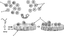
図1を参照すると、癌幹細胞マーカー4を含む癌細胞集団の選択および培養を支持するためのゲル2を示す概略図が提供される。
DETAILED DESCRIPTION OF THE DRAWINGS Referring to FIG. 1, there is provided a schematic diagram showing a
図1において、HA-チラミン複合体で構成されるゲル2は、過酸化水素(H2O2)および西洋ワサビペルオキシダーゼ(HRP)によって触媒されるチラミン部分の酸化的カップリングを用いて形成された。複数の癌細胞(4、6)をゲル2に曝露すると、癌幹細胞マーカーの高い発現レベルを有する細胞4のみがゲル2に接着する。癌幹細胞マーカーの高い発現レベルを有しない細胞6は、ゲル2に接着しない。したがって、ゲル2の存在下で複数の癌細胞を培養することにより、低発現癌細胞6から高発現癌細胞4を選択および分離することが可能となる。
In FIG. 1,
本発明の非制限的な例を、具体的実施例を参照してより詳細に説明するが、これらの実施例は本発明の範囲をいかなるようにも制限すると解釈してはならない。 Non-limiting examples of the invention will be described in more detail with reference to specific examples, which should not be construed as limiting the scope of the invention in any way.
ヒアルロン酸ナトリウム(HA)(Mw=90 kDa、密度=1.05 g/cm3)は、JNC Corporation(Tokyo, Japan)から寄贈された。塩酸チラミン(Tyr・HCl)、N-ヒドロキシスクシンイミド(NHS)、1-エチル-3-(3-ジメチルアミノプロピル)-カルボジイミド塩酸塩(EDC・HCl)、ウシ精巣由来のヒアルロニダーゼ(400〜1,000単位/mg)、およびシスプラチンは全て、Sigma-Aldrich(Minnesota of the United States of America)から購入した。西洋ワサビペルオキシダーゼ(HRP、100単位/mg)は、Wako Pure Chemical Industries(Osaka, Japan)から購入した。過酸化水素(H2O2)は、Lancasterから得た。ドキソルビシンはBoryung pharmaceutical(Seoul, South Korea)から得た。AlamarBlue、CyQUANT(登録商標)細胞増殖アッセイキット、TRIzol(登録商標)およびTaqman(登録商標)遺伝子発現マスターミクスはそれぞれ、Life Technologies(Singapore)から提供された。ヒトCD44に対するマウスモノクローナル抗体およびFITCコンジュゲートラット抗マウスIgG2a二次抗体は、GeneTex(Hsinchu, Taiwan)から得た。ラットモノクローナル抗-CD44抗体(Hermes-1)は、Abcam(Cambridge, United Kingdom)から購入した。トリプシン-EDTA(0.025%)およびペニシリン/ストレプトマイシンは、PAN Biotech GmbH(Aidenbach, Germany)から購入した。熱不活化ウシ胎児血清(FBS)は、GE Healthcare(Buckinghamshire, United Kingdom)から購入した。リン酸緩衝生理食塩水(PBS、150 mM、pH 7.3)、ダルベッコ改変イーグル培地(DMEM)およびRPMI-1640培地は全て、Biopolis(Singapore)の培地調製施設によって供給された。 Sodium hyaluronate (HA) (Mw = 90 kDa, density = 1.05 g / cm 3 ) was donated by JNC Corporation (Tokyo, Japan). Tyramine hydrochloride (Tyr · HCl), N-hydroxysuccinimide (NHS), 1-ethyl-3- (3-dimethylaminopropyl) -carbodiimide hydrochloride (EDC · HCl), hyaluronidase derived from bovine testis (400 to 1,000 units / mg) and cisplatin were all purchased from Sigma-Aldrich (Minnesota of the United States of America). Horseradish peroxidase (HRP, 100 units / mg) was purchased from Wako Pure Chemical Industries (Osaka, Japan). Hydrogen peroxide (H 2 O 2 ) was obtained from Lancaster. Doxorubicin was obtained from Boryung pharmaceutical (Seoul, South Korea). AlamarBlue, CyQUANT® cell proliferation assay kit, TRIzol® and Taqman® gene expression master mix were provided by Life Technologies (Singapore), respectively. Mouse monoclonal antibody against human CD44 and FITC-conjugated rat anti-mouse IgG2a secondary antibody were obtained from GeneTex (Hsinchu, Taiwan). Rat monoclonal anti-CD44 antibody (Hermes-1) was purchased from Abcam (Cambridge, United Kingdom). Trypsin-EDTA (0.025%) and penicillin / streptomycin were purchased from PAN Biotech GmbH (Aidenbach, Germany). Heat inactivated fetal bovine serum (FBS) was purchased from GE Healthcare (Buckinghamshire, United Kingdom). Phosphate buffered saline (PBS, 150 mM, pH 7.3), Dulbecco's modified Eagle's medium (DMEM) and RPMI-1640 medium were all supplied by the media preparation facility of Biopolis (Singapore).
MDA-MB-231、MCF-7、HCC1937、およびBT-474乳癌細胞株は、American Type Culture Collection(Manassas, Virginia of the United States of America)から購入した。MDA-MB-231、HCC1937、およびBT-474細胞は、10%FBSおよび1%ペニシリン/ストレプトマイシンを含むRPMI-1640培地において生育させた。MCF-7細胞は、10%FBSおよび1%ペニシリン/ストレプトマイシンを含むDMEMにおいて生育させた。細胞株は全て、ポリスチレン組織培養フラスコにおいて培養し、80%コンフルエントに達すると継代した。 MDA-MB-231, MCF-7, HCC1937, and BT-474 breast cancer cell lines were purchased from the American Type Culture Collection (Manassas, Virginia of the United States of America). MDA-MB-231, HCC1937, and BT-474 cells were grown in RPMI-1640 medium containing 10% FBS and 1% penicillin / streptomycin. MCF-7 cells were grown in DMEM containing 10% FBS and 1% penicillin / streptomycin. All cell lines were cultured in polystyrene tissue culture flasks and passaged when they reached 80% confluence.
統計分析:データは全て、平均値±標準偏差(SD)として表記した。特に明記されていない限り、値の差は、SigmaStatソフトウェア(Systat Software, Inc.)を用いてStudentのt-検定を用いて評価した。p<0.05は、統計学的に有意であると見なされた。実験は全て、1試料あたり3回ずつ行った。 Statistical analysis: All data are expressed as mean ± standard deviation (SD). Unless otherwise stated, differences in values were evaluated using Student's t-test using SigmaStat software (Systat Software, Inc.). p <0.05 was considered to be statistically significant. All experiments were performed 3 times per sample.
実施例1
HA-チラミンゲルの合成
HA-チラミン複合体で構成される酵素的に架橋されたHAゲルは、H2O2およびHRPによって触媒されるチラミン部分の酸化的カップリングを用いて形成された。本明細書において、HRPおよび異なる濃度のH2O2(0.22 mM〜1.15 mMの範囲のH2O2)の溶液をHA-Tyr複合体(PBS中で3%w/v、175μl)に添加した。混合物の全量が300μlとなるように、適当量のPBSを混合物に加えた。HA-Tyr複合体およびHRPの最終濃度はそれぞれ、1.75%(w/v)および0.125単位/mlであった。混合物を直ちにボルテックスミキサーで撹拌した。
Example 1
Synthesis of HA-tyramine gel
Enzymatically cross-linked HA gels composed of HA-tyramine complexes were formed using oxidative coupling of tyramine moieties catalyzed by H 2 O 2 and HRP. Herein, a solution of HRP and different concentrations of H 2 O 2 (in the range 0.22 mM to 1.15 mM H 2 O 2 ) is added to the HA-Tyr complex (3% w / v in PBS, 175 μl) did. An appropriate amount of PBS was added to the mixture so that the total volume of the mixture was 300 μl. The final concentrations of HA-Tyr conjugate and HRP were 1.75% (w / v) and 0.125 unit / ml, respectively. The mixture was immediately stirred with a vortex mixer.
置換度(HA反復単位100個あたりのチラミン分子の数)は、1H NMRによって決定したところ、6であった。 The degree of substitution (number of tyramine molecules per 100 HA repeat units) was 6, as determined by 1 H NMR.
ゲルの剛性およびゲル化速度は、H2O2およびHRP濃度によってそれぞれ独立して調整することができる。 Gel stiffness and gelation rate can be independently adjusted by H 2 O 2 and HRP concentrations, respectively.
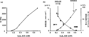
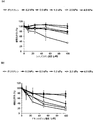
ゲルの流体力学的測定は、HAAKE Rheoscope 1レオメーター(Karlsruhe, Germany)によって、直径3.5 cmおよびコーン角0.949°のコーンプレートジオメトリを用いて行った。測定は、37℃で1%の一定の変形および周波数1 Hzの動的振動モードで行った。測定時の試料の滑り落ちを防止するために、粗面のガラス底プレートを用いた。上記のように調製したゲル混合物250μlをレオメーターの底部プレートに入れた。上部コーンを0.025 mmの測定ギャップまで下げて、実験の際の溶媒の蒸発を防止するため、シリコンオイルの層を注意深くコーン周囲に適用した。流体力学的測定は、貯蔵弾性率(G')が平衡に達するまで続行した。
The hydrodynamic measurement of the gel was performed with a
図2(a)に認められるように、HA-TyrゲルのG'は、H2O2濃度によって良好に制御され、HA-Tyr複合体の水溶液(1.75%(w/v))を利用する場合、70〜4,000 Paの範囲であった。H2O2濃度が0.22 mMから1.15 mMに増加するとG'が増加したことから、H2O2濃度が増加すると、より高い架橋密度が達成されることを示唆した。 As can be seen in Figure 2 (a), G 'of the HA-Tyr gel is well controlled by H 2 O 2 concentration and utilizes an aqueous solution of HA-Tyr complex (1.75% (w / v)) In the case of 70-4,000 Pa. Since the concentration of H 2 O 2 was increased G 'Increasing from 0.22 mM to 1.15 mM, when the concentration of H 2 O 2 increased, suggesting that higher crosslink density is achieved.
HA-Tyrハイドロゲルの架橋点間平均分子量(Mc)および架橋密度(ve)を決定した。貯蔵弾性率(G')の測定結果から、架橋点間平均分子量(Mc)および架橋密度(ve)を、ゴム弾性理論によって計算した。図2(b)に示されるように、HA-TyrハイドロゲルのMcはH2O2濃度が増加すると減少したが、HA-Tyrハイドロゲルのveは、H2O2濃度が増加すると増加した。 Average molecular weight between crosslinking points of HA-Tyr hydrogels of (Mc) and crosslink density (v e) was determined. From the measurement results of the storage elastic modulus (G ′), the average molecular weight (Mc) between the crosslinking points and the crosslinking density (v e ) were calculated by the rubber elasticity theory. As shown in FIG. 2 (b), although the M c the HA-Tyr hydrogels and reduced the concentration of H 2 O 2 increases, v e of HA-Tyr hydrogels, the concentration of H 2 O 2 is increased Increased.
癌細胞の接着(実施例2)、増殖(実施例3)、維持(実施例4)、HA-CD44相互作用の阻害(実施例5)、および化学療法抵抗性(実施例6)を評価するために、様々な剛性(必要に応じて、0.1、0.2、0.5、1.0、2.5、および4.0 kPa)の複数のゲルを用いた。以下の実施例から明らかであるように、HA-Tyrハイドロゲル上の乳癌細胞の接着は、ハイドロゲルの剛性によって強力に調節され、CD44-HA相互作用に依存した。HA-Tyrハイドロゲルは、丸い形状の細胞形態学、低い細胞増殖およびコロニー形成の維持において、ポリスチレンと比較して異なる培養環境を提供した。HA-Tyrハイドロゲルは、CD44変種イソ型、Sox-2およびALDH1A1 mRNA発現レベルをより良好に増強することから、ポリスチレンより優れていた。ハイドロゲルの力学的特性(ハイドロゲルの成分および剛性などの)ならびにCD44変種イソ型の発現レベルは、癌細胞の細胞接着、増殖、および悪性度に影響を及ぼす要因である。HA-Tyrハイドロゲルの剛性を制御することは、乳癌細胞の悪性度を変化させるための単純かつ有効な手段である。 Cancer cell adhesion (Example 2), proliferation (Example 3), maintenance (Example 4), inhibition of HA-CD44 interaction (Example 5), and chemoresistance (Example 6) are evaluated. For this purpose, multiple gels of varying stiffness (0.1, 0.2, 0.5, 1.0, 2.5, and 4.0 kPa as required) were used. As evident from the following examples, adhesion of breast cancer cells on HA-Tyr hydrogel was strongly regulated by the stiffness of the hydrogel and was dependent on CD44-HA interaction. The HA-Tyr hydrogel provided a different culture environment as compared to polystyrene in maintaining round shape cell morphology, low cell growth and colony formation. The HA-Tyr hydrogel was superior to polystyrene because it better enhanced the CD44 variant isoform, Sox-2 and ALDH1A1 mRNA expression levels. The mechanical properties of the hydrogel (such as the composition and stiffness of the hydrogel) and the expression level of the CD44 variant isoforms are factors that affect cell adhesion, proliferation, and malignancy of cancer cells. Controlling the stiffness of HA-Tyr hydrogel is a simple and effective means for changing the malignancy of breast cancer cells.
実施例2
癌細胞の接着
MDA-MB-231、MCF-7、HCC1937、およびBT-474などの4つのタイプの乳癌細胞株を調べた。これらの癌細胞株におけるCD44陽性細胞集団を試験するために、CD44の表面発現のフローサイトメトリー分析(FACS)を行った。
Example 2
Cancer cell adhesion
Four types of breast cancer cell lines were examined, including MDA-MB-231, MCF-7, HCC1937, and BT-474. To test the CD44 positive cell population in these cancer cell lines, flow cytometric analysis (FACS) of CD44 surface expression was performed.
FACSにおいて、細胞をPBSで洗浄し、トリプシン-EDTAを用いて収集した。剥離させた細胞を、2%FBSを含むPBSによって洗浄して、氷中で抗CD44抗体と共に30分間インキュベートした。2%FBSを含むPBSによって洗浄後、フルオレセインイソチオシアネート(FITC)にコンジュゲートさせた二次抗体を、製造元が推奨する濃度で細胞浮遊液に添加して、氷中の暗所で30分間インキュベートした。細胞を2回洗浄して、2%FBSを含むPBS 0.5 mlに浮遊させた。BD LSRIIフローサイトメトリーアナライザ(BD Biosciences, of New Jersey of the United States of America)を用いて、細胞を分析およびソーティングした。 In FACS, cells were washed with PBS and harvested using trypsin-EDTA. The detached cells were washed with PBS containing 2% FBS and incubated with anti-CD44 antibody in ice for 30 minutes. After washing with PBS containing 2% FBS, a secondary antibody conjugated to fluorescein isothiocyanate (FITC) was added to the cell suspension at a concentration recommended by the manufacturer and incubated for 30 minutes in the dark on ice . The cells were washed twice and suspended in 0.5 ml PBS containing 2% FBS. Cells were analyzed and sorted using a BD LSRII flow cytometry analyzer (BD Biosciences, of New Jersey of the United States of America).
図3に示すように、FACSを用いてMDA-MB-231(図3(a))、MCF-7(図3(b))、およびBT-474(図3(c))においてそれぞれ、99.5%、44.2%、および0.5%のCD44陽性細胞が検出されたことが認められうる。MDA-MB-231、HCT116、およびMCF-7細胞のCD44タンパク質発現レベル(平均値)は、BT-474細胞の発現レベルより103、12、および5倍高かった。 As shown in Fig. 3, using FACS, MDA-MB-231 (Fig. 3 (a)), MCF-7 (Fig. 3 (b)), and BT-474 (Fig. 3 (c)) were each 99.5 It can be seen that%, 44.2%, and 0.5% of CD44 positive cells were detected. The CD44 protein expression levels (mean value) of MDA-MB-231, HCT116 and MCF-7 cells were 103, 12 and 5 times higher than the expression levels of BT-474 cells.
さらに、MDA-MB-231、MCF-7、HCC1937間の比較では、FACSを用いた検出により、MDA-MB-231細胞ではCD44タンパク質が高レベルで発現され、HCC1937細胞では中レベルで発現され、MCF-7細胞では低レベルで発現されることが認められうる(図4(a))。CD44発現レベルは、単一の細胞におけるCD44分子の数として定義される。これらの乳癌細胞株におけるCD44の様々な発現レベルを、細胞表面上のタンパク質発現レベル(図4(a))ならびにmRNA発現レベル(図4(b))に基づいて評価した。MDA-MB-231およびHCC1937細胞におけるCD44のmRNA発現レベルは、MCF-7細胞の発現レベルより8.5および4.5倍高かった。これらの結果は、図4(a)に示されるようにCD44タンパク質発現レベルの結果と密接に関連した。 Furthermore, in comparison between MDA-MB-231, MCF-7 and HCC1937, CD44 protein is expressed at high level in MDA-MB-231 cells and at medium level in HCC1937 cells by detection using FACS. It can be observed that low levels are expressed in MCF-7 cells (FIG. 4 (a)). CD44 expression levels are defined as the number of CD44 molecules in a single cell. Various expression levels of CD44 in these breast cancer cell lines were assessed based on protein expression levels on cell surface (FIG. 4 (a)) as well as mRNA expression levels (FIG. 4 (b)). The mRNA expression level of CD44 in MDA-MB-231 and HCC1937 cells was 8.5 and 4.5 times higher than that in MCF-7 cells. These results were closely related to the CD44 protein expression level results as shown in FIG. 4 (a).
次に、HA-Tyrゲルの表面に対するこれらの細胞株の癌細胞接着を調べた。異なる剛性(0.1、0.2、0.5、1.0、2.5、および4.0 kPa、必要であれば)を有するHA-Tyrゲル(250μl)を24ウェルプレートにおいて調製した。ゲルを一晩沈降させた。次に、ゲルをPBSで3回洗浄した後、培養培地で1回洗浄した。培養培地中で細胞密度1.0×10⁵個/mlの乳癌細胞250μlをゲル上に播種した。 Next, cancer cell adhesion of these cell lines to the surface of HA-Tyr gel was examined. HA-Tyr gels (250 μl) with different stiffness (0.1, 0.2, 0.5, 1.0, 2.5, and 4.0 kPa, if necessary) were prepared in 24-well plates. The gel was allowed to settle overnight. Next, the gel was washed 3 times with PBS and then once with culture medium. 250 μl of breast cancer cells having a cell density of 1.0 × 10 5 cells / ml were seeded on a gel in a culture medium.
プレートをインキュベーター(5%CO2を含む37℃の湿潤大気中)に、1から9時間の範囲の適切な期間戻した。選択された間隔で、非付着細胞を有する培地を吸引して、細胞をPBSによって3回洗浄した。ゲルを有しない細胞培養プレートを比較のために用いた。 The plate was returned to the incubator (in a 37 ° C. humidified atmosphere containing 5% CO 2 ) for an appropriate period of time ranging from 1 to 9 hours. At selected intervals, the medium with non-adherent cells was aspirated and the cells were washed 3 times with PBS. Cell culture plates without gel were used for comparison.
MDA-MB-231、MCF-7、およびHCC1937に関して、CyQUANT(登録商標)細胞増殖アッセイキットを用いてDNAの定量を行って、付着細胞数を評価した。推奨される製造元のアッセイプロトコールに従った。HA-Tyrハイドロゲル上およびポリスチレン上の細胞を、ヒアルロニダーゼ(1,000単位/ml)およびトリプシン-EDTAと共にそれぞれインキュベートすることによって収集した。細胞沈降物をPBSで2回洗浄して、凍結融解サイクルによって溶解した。次に、試料をCyQUANT作業溶液200μlに溶解した。公知の密度の細胞浮遊液と共に試料溶液を蛍光測定することによって、細胞数を、検量線に基づいて決定した。蛍光測定は、480および520nmでの励起および放射波長でマイクロプレートリーダーを用いて行った。 DNA quantification was performed for MDA-MB-231, MCF-7, and HCC1 937 using the CyQUANT® Cell Proliferation Assay Kit to assess the number of adherent cells. The recommended manufacturer's assay protocol was followed. Cells on HA-Tyr hydrogel and on polystyrene were harvested by incubation with hyaluronidase (1,000 units / ml) and trypsin-EDTA, respectively. The cell pellet was washed twice with PBS and lysed by freeze-thaw cycles. The samples were then dissolved in 200 μl of CyQUANT working solution. Cell number was determined based on a standard curve by fluorescence measurement of the sample solution with cell suspensions of known density. Fluorescence measurements were performed using a microplate reader at excitation and emission wavelengths at 480 and 520 nm.
BT-474に関して、ゲルに付着した細胞を、10%Alamar Blue色素を含む培養培地中で37℃で4時間インキュベートした。Alamar Blue色素の蛍光測定をそれぞれ545および590 nmでの励起および放射波長でのマイクロプレートリーダーを用いて行った。次に、Alamar Blue溶液100μlを96ウェルプレートに移して、蛍光強度を測定した。Alamar Blue色素の蛍光強度は、本試験における付着細胞数であると見なされた。 For BT-474, cells attached to the gel were incubated for 4 hours at 37 ° C. in culture medium containing 10% Alamar Blue dye. Fluorescence measurement of Alamar Blue dye was performed using a microplate reader at excitation and emission wavelengths at 545 and 590 nm, respectively. Next, 100 μl of Alamar Blue solution was transferred to a 96-well plate to measure the fluorescence intensity. The fluorescence intensity of Alamar Blue dye was considered to be the number of adherent cells in this study.
図5は、HA-Tyrゲル表面上の細胞接着を示す。有意な数のMDA-MB-231細胞がHA-Tyrゲル表面に付着した(図5(c))。付着したMDA-MB-231細胞数は、HA-Tyrハイドロゲルの剛性に依存し、付着細胞数はハイドロゲルの剛性が減少すると増加した。図5(a)および図5(b)も同様に、ハイドロゲルの剛性が減少すると、MCF-7およびHCC1937の細胞接着がそれぞれ増加したことを示す。図5(a)〜図5(c)は、HA-Tyrハイドロゲル上の付着細胞数が、CD44発現レベルに密接に関連するように思われることを示した(図4)。HA-Tyrハイドロゲル上での乳癌細胞の接着は、CD44の発現レベルが増加すると増加した。これらの結果は、HA-Tyrハイドロゲル上での細胞接着が、HAとCD44との相互作用に依存していたことを強く示唆している。 FIG. 5 shows cell adhesion on HA-Tyr gel surface. A significant number of MDA-MB-231 cells attached to the HA-Tyr gel surface (FIG. 5 (c)). The number of adherent MDA-MB-231 cells was dependent on the stiffness of the HA-Tyr hydrogel, and the number of adherent cells increased as the stiffness of the hydrogel decreased. Similarly, FIGS. 5 (a) and 5 (b) show that decreasing the stiffness of the hydrogel increased cell adhesion of MCF-7 and HCC 1937, respectively. Figures 5 (a) -5 (c) showed that the number of adherent cells on HA-Tyr hydrogel appeared to be closely related to CD44 expression levels (Figure 4). Breast cancer cell adhesion on HA-Tyr hydrogels increased with increasing CD44 expression levels. These results strongly suggest that cell adhesion on HA-Tyr hydrogel was dependent on the interaction between HA and CD44.
これに対し、HA-Tyrゲルに付着したBT-474細胞はごく少数であった(図5(d))。これらの結果は、CD44発現レベルおよび/またはハイドロゲルの剛性が、ハイドロゲルへの癌細胞の接着に影響を及ぼす要因であることを示している。一般的に、CD44の発現レベルがより高く、および/またはハイドロゲルの剛性がより低ければ、ハイドロゲルに対する細胞接着の程度はより大きくなる。 In contrast, only a few BT-474 cells attached to the HA-Tyr gel (FIG. 5 (d)). These results indicate that the CD44 expression level and / or the stiffness of the hydrogel are factors that influence the adhesion of cancer cells to the hydrogel. Generally, the higher the expression level of CD44 and / or the lower the stiffness of the hydrogel, the greater the degree of cell adhesion to the hydrogel.
実施例3
癌細胞の増殖
付着したMDA-MB-231細胞の細胞形態学を決定するために、IX71倒立顕微鏡を備えたOlympus顕微鏡カメラ(Tokyo, Japan)を用いて位相差顕微鏡画像を得た。図6(a)を参照して、付着したMDA-MB-231細胞は全て、ゲルの剛性によらず丸いが、ポリスチレン培養フラスコ上では、他の細胞の多くが伸展していることが観察された。
Example 3
In order to determine the cell morphology of cancer cells grown adherent MDA-MB-231 cells, phase contrast microscopy images were obtained using an Olympus microscope camera (Tokyo, Japan) equipped with an IX71 inverted microscope. Referring to FIG. 6 (a), all attached MDA-MB-231 cells were round regardless of gel rigidity, but it was observed that many other cells were stretched on polystyrene culture flasks. It was.
細胞伸展面積を、Image-Pro Plus画像分析ソフトウェア(Maryland of the United States of America)を用いて分析した。細胞伸展面積を図6(b)に示し、HA-Tyrゲル上では細胞伸展面積が、ポリスチレン上より有意に小さいが、様々な剛性の様々なHA-Tyrゲル上での細胞伸展に有意差はなかったことが明らかに示された。これらの結果は、CD44とHA-Tyrゲルの間の相互作用を介した細胞接着が、丸い形状のMDA-MB-231細胞を維持しうることを示した。 Cell extension area was analyzed using Image-Pro Plus image analysis software (Maryland of the United States of America). The cell extension area is shown in Fig. 6 (b). Although the cell extension area on the HA-Tyr gel is significantly smaller than that on polystyrene, there is a significant difference in cell extension on various HA-Tyr gels of various stiffnesses. Clearly it was not. These results indicated that cell adhesion via the interaction between CD44 and HA-Tyr gel could maintain round-shaped MDA-MB-231 cells.
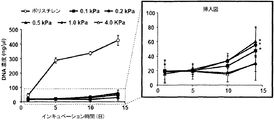
様々なゲル(0.1、0.2、0.5、1.0、および4.0 kPa)における細胞増殖を評価するために、培養培地中で細胞密度1.0×10⁵個/mlのMDA-MB-231細胞250μlを、ゲル上に播種した。ゲルを有しない細胞培養ウェルプレートを比較として用いた。細胞を、5%CO2を含む37℃の湿潤大気中でインキュベートした。培養培地を3日毎に交換した。ハイドロゲル上での細胞増殖を評価するために、上記のCyQUANT(登録商標)細胞増殖アッセイキットを用いて蛍光測定を行った。 To assess cell growth in various gels (0.1, 0.2, 0.5, 1.0, and 4.0 kPa), 250 μl of MDA-MB-231 cells at a cell density of 1.0 × 10 5 cells / ml in culture medium Sowing. Cell culture well plates without gel were used as a comparison. The cells were incubated in a humidified atmosphere at 37 ° C. containing 5% CO 2 . The culture medium was changed every 3 days. Fluorescence measurements were performed using the CyQUANT® Cell Proliferation Assay Kit described above to assess cell proliferation on the hydrogel.
図7に示すように、HA-Tyrゲル上で培養したMDA-MB-231細胞の生育速度は、培養プレート上で培養した細胞よりかなり低かった。剛性0.1 kPa、0.2 kPa、および0.5 kPaを有する軟らかいHA-Tyrゲルのみが、13日間の間に細胞生育を示したが、他の剛性のHA-Tyrゲルでは細胞生育は観察されなかった。 As shown in FIG. 7, the growth rate of MDA-MB-231 cells cultured on HA-Tyr gel was much lower than the cells cultured on culture plates. Only soft HA-Tyr gels with stiffness of 0.1 kPa, 0.2 kPa, and 0.5 kPa showed cell growth during 13 days, but no cell growth was observed with other rigid HA-Tyr gels.
HA-Tyrゲルの細胞付着および増殖を含むこれらの結果は、培養プレートの結果とは明らかに異なった。このことは、MDA-MB-231細胞において発現されたCD44が、HA-Tyrゲル中のHA鎖と相互作用したことを示唆する。 These results, including cell attachment and growth of HA-Tyr gels, were clearly different from the culture plate results. This suggests that CD44 expressed in MDA-MB-231 cells interacted with the HA chain in HA-Tyr gel.
HA-Tyrハイドロゲル上でのMDA-MB-231乳癌細胞の挙動をさらに調べるために、HA-Tyrハイドロゲル上でのコロニー形成を評価した(図8)。丸い形状のMDA-MB-231細胞は、14日後にHA-Tyrハイドロゲル上でコロニーを形成したが(図8a、I〜V、XII)、ポリスチレン上ではコロニーは観察されなかった。HA-Tyrハイドロゲルの剛性が減少するとコロニーサイズは増加した。 To further investigate the behavior of MDA-MB-231 breast cancer cells on HA-Tyr hydrogel, colony formation on HA-Tyr hydrogel was assessed (FIG. 8). Round shaped MDA-MB-231 cells formed colonies on HA-Tyr hydrogel after 14 days (Fig. 8a, IV, XII), but no colonies were observed on polystyrene. The colony size increased as the stiffness of the HA-Tyr hydrogel decreased.
実施例4
HA-Tyrハイドロゲルによる癌幹細胞マーカーの発現レベルの増強
HA-Tyrゲル(剛性0.2、0.5、1.0、および4.0 kPa)上での培養13日後のMDA-MB-231細胞におけるNanog、Sox-2、およびEpCAMのmRNA発現レベルを調べた。さらに、HA-Tyrハイドロゲル(剛性0.1、0.2、0.5、1.0、および4.0 kPa)上でのMDA-MB-231細胞におけるCD44(CD44標準)、CD44v3-10(CD44変種イソ型)、CD44v8-10(CD44変種イソ型)、Sox-2、EpCAM、およびALDHD1A1遺伝子の転写レベルもまた調べた。
Example 4
Enhancement of cancer stem cell marker expression level by HA-Tyr hydrogel
The mRNA expression levels of Nanog, Sox-2, and EpCAM in MDA-MB-231 cells after 13 days of culture on HA-Tyr gels (stiffness 0.2, 0.5, 1.0, and 4.0 kPa) were examined. In addition, CD44 (CD44 standard), CD44v3-10 (CD44 variant isoforms), CD44v8-10 in MDA-MB-231 cells on HA-Tyr hydrogels (stiffness 0.1, 0.2, 0.5, 1.0 and 4.0 kPa) Transcript levels of (CD44 variant isoforms), Sox-2, EpCAM, and ALDHD1A1 genes were also examined.
本明細書において、培養培地中で細胞密度1.0×10⁵個/mlのMDA-MB-231細胞250μlをゲル上に播種した。ゲルを含まない細胞培養ウェルプレートを比較として用いた。細胞を、5%CO2を含む37℃の湿潤大気中でインキュベートした。培養培地を3日毎に交換した。 Herein, 250 μl of MDA-MB-231 cells having a cell density of 1.0 × 10 5 cells / ml were seeded on a gel in a culture medium. Cell culture well plates without gel were used as a comparison. The cells were incubated in a humidified atmosphere at 37 ° C. containing 5% CO 2 . The culture medium was changed every 3 days.
様々なHA-Tyrハイドロゲル上のMDA-MB-231細胞を、その後のRNA抽出のために、ヒアルロニダーゼ(1,000単位/ml)およびトリプシン-EDTAによって処理することによって収集した。MDA-MB-231細胞の総RNAを、培養13日後にTRIzol(Life technologies, Singapore)を用いて抽出した。cDNAを調製するために、総RNAを、RT緩衝液、ランダムヘキサマープライマー、デオキシヌクレオチド三リン酸(dNTP)混合物、RNアーゼ阻害剤、および逆転写酵素を含むRT反応混合物(Thermo Scientific, China)と共にインキュベートした後に、DNアーゼ処置を行った。 MDA-MB-231 cells on various HA-Tyr hydrogels were harvested by treatment with hyaluronidase (1,000 units / ml) and trypsin-EDTA for subsequent RNA extraction. Total RNA of MDA-MB-231 cells was extracted using TRIzol (Life technologies, Singapore) after 13 days of culture. RT reaction mixture (RT Thermo Scientific, China) containing total RNA, RT buffer, random hexamer primers, deoxynucleotide triphosphate (dNTP) mixture, RNase inhibitor, and reverse transcriptase to prepare cDNA After incubation with, DNase treatment was performed.
リアルタイム定量的PCRをiQ5マルチカラーRT PCR検出システム(Bio-Rad laboratories, Singapore)を用いて行った。反応混合物(Life technologies, Singapore)は、TaqMan PCRマスターミクス10μL、各プライマー1.0μL、cDNA 2.0μ L、および蒸留水7.0 μLを含んだ。用いた様々なプライマーは、Hs01075861_m1(総CD44)、Hs01081473_m1(CD44s)、Hs01081480_m1(CD44v3 -v10)、Hs01081475_m1(CD44v8-v10))、Nanog(Hs02387400_s1)、Sox-2(Hs01053049_s1)、EpCAM(Hs00901885_m1)、およびALDH1A1(Hs00946916_m1)であった。PCRのために用いた温度プロファイルは、95℃で20秒、95℃で3秒および60℃で20秒を40サイクルであった。△-△Ct法を用いるその後の全ての計算において、1試料あたり3回ずつの測定の平均閾値サイクル(Ct)値を用い、結果を内因性の参照遺伝子(GAPDH)に対して標準化して、無処置対照(ポリスチレンプレート)と比較した遺伝子発現の変化倍率として表記する。 Real-time quantitative PCR was performed using the iQ5 multicolor RT PCR detection system (Bio-Rad laboratories, Singapore). The reaction mixture (Life technologies, Singapore) contained 10 μL of TaqMan PCR master mix, 1.0 μL of each primer, 2.0 μL of cDNA, and 7.0 μL of distilled water. The various primers used were Hs01085861_m1 (total CD44), Hs01081473_m1 (CD44s), Hs01081480_m1 (CD44v3-v10), Hs01081475_m1 (CD44v8-v10), Nanog (Hs02387400_s1) and Sox-2 And ALDH1A1 (Hs00946916_m1). The temperature profile used for PCR was 40 cycles of 95 ° C. for 20 seconds, 95 ° C. for 3 seconds and 60 ° C. for 20 seconds. In all subsequent calculations using the △-△ Ct method, the average threshold cycle (Ct) value of 3 measurements per sample was used, and the results were normalized to the endogenous reference gene (GAPDH), Expressed as fold change in gene expression compared to untreated control (polystyrene plate).
図9に示すように、0.2 kPaの剛性を有するHA-TyrゲルでのNanog、Sox-2、およびEpCAMのmRNA発現レベルは、他の培養プレートの発現レベルより有意に高いことが観察された。他のゲルは、Sox-2を除き、有意に高いmRNA発現レベルを示した。NanogおよびSox-2は、stemnessマーカーであることが知られており、それらの転写因子は、抗アポトーシスタンパク質発現および化学療法抵抗性を誘導する。さらに、CSCマーカーの1つであるESAの発現が増強されたことは、HA-Tyrゲル上に接着したMDA-MB-231細胞がCSCであることを示した。 As shown in FIG. 9, it was observed that the mRNA expression levels of Nanog, Sox-2, and EpCAM in HA-Tyr gels with a stiffness of 0.2 kPa were significantly higher than the expression levels of other culture plates. Other gels, with the exception of Sox-2, showed significantly higher mRNA expression levels. Nanog and Sox-2 are known to be stemness markers, and their transcription factors induce anti-apoptotic protein expression and chemoresistance. Furthermore, the enhanced expression of ESA, one of the CSC markers, indicated that MDA-MB-231 cells adhered on HA-Tyr gel are CSCs.
図10に示されるように、HA-Tyrハイドロゲル上でのCD44v3-10およびCD44v8-10のmRNA発現レベルは、ポリスチレン上の発現レベルより有意に高かったが、CD44の発現レベルは、ポリスチレン上の発現レベルとほぼ同じであった。興味深いことに、CD44v3-10およびCD44v8-10の増強された発現レベルは、HA-Tyrハイドロゲルの剛性が増加すると増加した(図10a)。これらの結果は、HA-Tyrハイドロゲルがハイドロゲル剛性依存的にCD44v3-10およびCD44v8-10の発現レベルを増強することができるが、CD44発現レベルは増強しないことを示している。それゆえ、結果から、HA-Tyrハイドロゲルが、乳癌細胞の悪性の特性を誘導することが強く示唆される。 As shown in FIG. 10, mRNA expression levels of CD44v3-10 and CD44v8-10 on HA-Tyr hydrogel were significantly higher than expression levels on polystyrene, but expression levels of CD44 were on polystyrene The expression level was almost the same. Interestingly, the enhanced expression levels of CD44v3-10 and CD44v8-10 increased as the stiffness of the HA-Tyr hydrogel increased (Figure 10a). These results indicate that HA-Tyr hydrogel can enhance the expression level of CD44v3-10 and CD44v8-10 in a hydrogel stiffness dependent manner, but not the expression level of CD44. Therefore, the results strongly suggest that HA-Tyr hydrogel induces the malignant character of breast cancer cells.
さらに、HA-Tyrハイドロゲル上でのSox-2およびALDH1A1のmRNA発現レベルもまた、ポリスチレン上の発現レベルと比較してHA-Tyrハイドロゲル上で有意に増強された(図10b)。ALDH1A1の発現レベルは、HA-Tyrハイドロゲルのハイドロゲル剛性とは無関係であった。この結果は、CD44-HA相互作用が、剛性非依存的にALDH1A1発現を増強することを示唆した。Sox-2の場合、mRNA発現レベルは、0.2および0.5 kPa HA-Tyrハイドロゲルにおいて有意に増加した。さらに、CSCマーカーの1つであるALDH1A1発現が増強されたことは、HA-Tyrハイドロゲル上に接着したMDA-MB-231細胞がCSC特性を誘導されることを示した。 Furthermore, the mRNA expression levels of Sox-2 and ALDH1A1 on HA-Tyr hydrogels were also significantly enhanced on HA-Tyr hydrogels compared to expression levels on polystyrene (FIG. 10 b). The expression level of ALDH1A1 was independent of the hydrogel stiffness of HA-Tyr hydrogel. This result suggested that CD44-HA interaction enhances ALDH1A1 expression in a stiffness-independent manner. In the case of Sox-2, mRNA expression levels were significantly increased in 0.2 and 0.5 kPa HA-Tyr hydrogels. Furthermore, enhanced expression of ALDH1A1, which is one of the CSC markers, indicated that MDA-MB-231 cells adhered on HA-Tyr hydrogels were induced with CSC characteristics.
したがって、HAに基づく細胞培養基質は、CSCの選択、培養、および維持にとって有用でありうることが示される。 Thus, it is shown that HA-based cell culture substrates can be useful for CSC selection, culture, and maintenance.
実施例5
抗CD44抗体を用いてHA-Tyrハイドロゲル上でのMDA-MB-231細胞接着の阻害を評価した。MDA-MB-231細胞をPBSによって洗浄して、トリプシン-EDTAを用いて細胞培養フラスコから収集した。剥離した細胞を、FBSを含まないRPMI-1640培地によって洗浄して、30 μg/ml抗CD44抗体と共に氷中で1時間インキュベートした。抗CD44抗体を有しない細胞浮遊液を比較として用いた。FBSを含まないRPMI-1640培地によって洗浄後、培養培地中で細胞密度1.0×105個/mlのMDA-MB-231細胞250μlを、上記と同様の方法で24ウェルプレートで調製した0.1 kPa HA-Tyrハイドロゲル上に播種した。細胞を、5%CO2を含む37℃の湿潤大気中で1時間インキュベートした。その後、非付着細胞を有する培地を吸引して、ウェルをPBSによって3回洗浄した。HA-Tyrハイドロゲル上の付着細胞数を評価するために、上記のCyQUANT(登録商標)細胞増殖アッセイキットを用いて、蛍光測定を行った。
Example 5
The inhibition of MDA-MB-231 cell adhesion on HA-Tyr hydrogel was evaluated using an anti-CD44 antibody. MDA-MB-231 cells were washed with PBS and harvested from cell culture flasks using trypsin-EDTA. The detached cells were washed with RPMI-1640 medium without FBS and incubated with 30 μg / ml anti-CD44 antibody for 1 hour on ice. Cell suspension without anti-CD44 antibody was used as a comparison. After washing with RPMI-1640 medium without FBS, 250 μl of MDA-MB-231 cells with a cell density of 1.0 × 10 5 cells / ml in a culture medium was prepared in a 24-well plate in the same manner as described above. -Seeded on Tyr hydrogel. Cells were incubated for 1 hour in a humidified atmosphere at 37 ° C. containing 5% CO 2 . The medium with non-adherent cells was then aspirated and the wells were washed 3 times with PBS. In order to evaluate the number of adherent cells on the HA-Tyr hydrogel, fluorescence measurement was performed using the CyQUANT (registered trademark) cell proliferation assay kit described above.
図11に示されるように、抗CD44抗体による前処置により、無処置対照と比較して0.1 kPa HA-Tyrハイドロゲルに付着した細胞の百分率が劇的に減少した。したがって、HA-Tyrハイドロゲル上の細胞接着は、ほとんどがHA鎖とCD44受容体との相互作用によって媒介されたことが認められうる。 As shown in FIG. 11, pretreatment with anti-CD44 antibody dramatically reduced the percentage of cells attached to the 0.1 kPa HA-Tyr hydrogel compared to untreated controls. Therefore, it can be seen that cell adhesion on HA-Tyr hydrogel was mostly mediated by the interaction of HA chain with CD44 receptor.
実施例6
癌細胞の選択および維持
癌細胞の選択を評価するために、HA-Tyr(0.1、0.2、0.5、1.0、および4.0 kPa)上に付着したMDA-MB-231細胞におけるCD44s、CD44v3-10、CD44v8-10、EpCAM、およびALHD1A1遺伝子の転写レベルを分析した。培養培地中で細胞密度1.0×10⁵個/mlの乳癌細胞250μlをハイドロゲル上に播種した。細胞を、5%CO2を含む37℃の湿潤大気中で1時間インキュベートした後、PBS 500μlによって3回洗浄して、非付着細胞を除去した。本実施例では、実施例4と比較して、ここで追加の洗浄段階を用いた。HA-Tyrハイドロゲルおよびポリスチレン上の細胞をそれぞれ、ヒアルロニダーゼ(1,000単位/ml)およびトリプシン-EDTAと共にインキュベートすることによって収集した。細胞沈降物をPBSによって2回洗浄して、mRNA発現レベルの測定のために用いた。
Example 6
Selection of cancer cells and maintenance To evaluate the selection of cancer cells, CD44s, CD44v3-10, CD44v8 in MDA-MB-231 cells attached on HA-Tyr (0.1, 0.2, 0.5, 1.0, and 4.0 kPa) Transcript levels of -10, EpCAM, and ALHD1A1 gene were analyzed. 250 μl of breast cancer cells with a cell density of 1.0 × 10 5 cells / ml in culture medium were seeded on the hydrogel. The cells were incubated for 1 hour in a 37 ° C. humidified atmosphere containing 5% CO 2 and then washed 3 times with 500 μl of PBS to remove non-adherent cells. In this example, in comparison with Example 4, an additional washing step was used here. Cells on HA-Tyr hydrogel and polystyrene were collected by incubating with hyaluronidase (1,000 units / ml) and trypsin-EDTA, respectively. The cell pellet was washed twice with PBS and used to measure mRNA expression levels.
4 kPa HA-Tyrハイドロゲル上でのCD44s、CD44v3-10、CD44v8-10、EpCAM、およびALHD1A1などのCSCマーカーのmRNA発現レベルは、ポリスチレン上より有意に高かった(図12(a))。特に、CD44v8-10レベルは、たとえ軟らかい(0.1および0.2 kPa)HA-Tyrハイドロゲル上でもポリスチレン上のレベルより有意に高かった。CD44v8-10は、活性酸素種(ROS)に対する抵抗性の増強により乳癌の転移との相関が十分に研究されている。EpCAMおよびALDH1A1もまた、CSC特性と相関することが周知である。それゆえ、これらの結果は、調節可能な剛性を有するHA-Tyrハイドロゲルが、CSC細胞の選択にとって有用でありうるという仮説を支持する。さらに、4 kPa HA-Tyrハイドロゲル上のCD44v8-10のmRNA発現レベルは、14日後でも維持された(図12(b))。0.1および0.2 kPa HA-Tyrハイドロゲルの場合、CD44v8-10のmRNA発現レベルは、14日後では有意に増強された。これらの結果は、選択された癌細胞の特性が、HA-Tyrハイドロゲル上で維持されたのみならず、増強されたことを示している。さらに、追加の洗浄段階を用いることによって、CD44、EpCAMおよび他のマーカーを高度に発現する細胞が得られた。 The mRNA expression levels of CSC markers such as CD44s, CD44v3-10, CD44v8-10, EpCAM, and ALHD1A1 on 4 kPa HA-Tyr hydrogel were significantly higher than on polystyrene (FIG. 12 (a)). In particular, CD44v8-10 levels were significantly higher than those on polystyrene even on soft (0.1 and 0.2 kPa) HA-Tyr hydrogels. CD44v8-10 has been well studied for its correlation with breast cancer metastasis by enhancing resistance to reactive oxygen species (ROS). EpCAM and ALDH1A1 are also known to correlate with CSC properties. Therefore, these results support the hypothesis that HA-Tyr hydrogels with adjustable stiffness may be useful for CSC cell selection. In addition, the mRNA expression level of CD44v8-10 on 4 kPa HA-Tyr hydrogel was maintained even after 14 days (FIG. 12 (b)). In the case of 0.1 and 0.2 kPa HA-Tyr hydrogel, the mRNA expression level of CD44v8-10 was significantly enhanced after 14 days. These results indicate that the properties of the selected cancer cells were enhanced as well as maintained on the HA-Tyr hydrogel. In addition, additional washing steps were used to obtain cells highly expressing CD44, EpCAM and other markers.
実施例7
癌細胞の化学療法抵抗性
化学療法抵抗性を評価するために、HA-Tyrゲル上のMDA-MB-231の細胞生存率を、シスプラチンおよびドキソルビシンなどの従来の抗癌剤の存在下または非存在下で測定した。本明細書において、培養培地中で5.2×10³個/mlの細胞密度のMDA-MB-231細胞250μlを、ゲル(0.2、0.5、1.0、2.5、および4.0 kPa)に播種した。培養培地を3日毎に交換した。細胞播種の10日後、10〜100μMシスプラチンまたはドキソルビシンを、ゲル上の培養細胞に添加して、48時間インキュベートした。次に、細胞をPBSによって洗浄して、上記のようにAlamarBlue細胞生存率アッセイを用いて蛍光強度を測定した。抗癌剤の存在下での細胞生存率を無処置対照(0μM)に対して標準化した。
Example 7
To evaluate chemotherapy resistance of cancer cells, cell viability of MDA-MB-231 on HA-Tyr gel, in the presence or absence of conventional anticancer agents such as cisplatin and doxorubicin It was measured. Herein, 250 μl of MDA-MB-231 cells with a cell density of 5.2 × 10 3 cells / ml in culture medium were seeded on gels (0.2, 0.5, 1.0, 2.5, and 4.0 kPa). The culture medium was changed every 3 days. Ten days after cell seeding, 10-100 μM cisplatin or doxorubicin was added to the cultured cells on the gel and incubated for 48 hours. Cells were then washed with PBS and fluorescence intensity was measured using the AlamarBlue cell viability assay as described above. Cell viability in the presence of anticancer agents was normalized to an untreated control (0 μM).
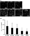
シスプラチンの存在下では、培養プレート上のMDA-MB-231細胞の生存率は、用量依存的に減少した(図13(a)を参照されたい)。これに対し、HA-Tyrゲルは、細胞培養プレートと比較して試験濃度範囲ではるかにより高い細胞生存率を示した。特に、0.5および1 kPaのHA-Tyrの場合、細胞生存率は、100 mMシスプラチンであってもほぼ100%であった。 In the presence of cisplatin, the viability of MDA-MB-231 cells on culture plates decreased in a dose-dependent manner (see FIG. 13 (a)). In contrast, HA-Tyr gels showed much higher cell viability in the tested concentration range compared to cell culture plates. In particular, in the case of 0.5 and 1 kPa HA-Tyr, the cell viability was almost 100% even with 100 mM cisplatin.
類似の傾向が、ドキソルビシンの存在下で観察された(図13(b))。0.2および0.5 kPaゲルでは、100μMドキソルビシンの存在下であっても、およそ80%の生存率が維持されたが、細胞培養プレートおよびHA-Tyrゲル(2.5および4.0 kPa)ではそれぞれ、およそ10および20%の細胞生存率が観察された。これらの結果は、HA-Tyr、特に軟らかいゲルが、化学療法抵抗性を有意に誘導することを示した。 A similar tendency was observed in the presence of doxorubicin (Figure 13 (b)). At 0.2 and 0.5 kPa gels, approximately 80% viability was maintained even in the presence of 100 μM doxorubicin, but for cell culture plates and HA-Tyr gels (2.5 and 4.0 kPa), approximately 10 and 20 respectively. % Cell viability was observed. These results indicated that HA-Tyr, especially the soft gel, significantly induces chemoresistance.
用途
開示の方法は、所望の癌幹細胞または癌幹細胞を含む癌細胞株を培養するために細胞培養用途において用いることができる。
Uses The disclosed methods can be used in cell culture applications to culture desired cancer stem cells or cancer cell lines comprising cancer stem cells.
開示の方法は、様々な癌細胞の複数または混合物から所望の癌幹細胞(または癌細胞株)を選択的に分離しそうすることによって単離するために用いられうる。 The disclosed method can be used to selectively isolate desired cancer stem cells (or cancer cell lines) from a plurality or mixture of various cancer cells.
開示のゲルは、所望の癌幹細胞の選択および生育を支持するために用いられうる。開示のゲルは、癌幹細胞と共に、癌幹細胞がそれに対して化学療法抵抗性である抗癌剤、または癌幹細胞に対して治療効果を有する抗癌剤を決定するための、癌治療に関する薬物スクリーニングプラットフォームとして用いられうる。 The disclosed gels can be used to support the selection and growth of desired cancer stem cells. The disclosed gel can be used as a drug screening platform for cancer treatment to determine, in conjunction with cancer stem cells, anticancer agents against which the cancer stem cells are chemoresistant or anticancer agents having a therapeutic effect on the cancer stem cells. .
開示の方法およびゲルは、受容体-リガンド結合によって所望のマーカーを含む癌幹細胞を選択的に単離するために用いられうる。 The disclosed methods and gels can be used to selectively isolate cancer stem cells containing a desired marker by receptor-ligand binding.
本発明の様々な他の改変および適応が、前述の開示を読むことによって当業者に明らかとなるが、それらも本発明の精神および適用範囲に含まれ、そのような全ての改変および適応が添付の特許請求の範囲の適用範囲に含まれると意図されることは明白であろう。 Various other modifications and adaptations of the present invention will become apparent to those skilled in the art upon reading the foregoing disclosure, but they are also included within the spirit and scope of the present invention, and all such modifications and adaptations are appended. It will be clear that it is intended to be included within the scope of the appended claims.
Claims (15)
b.癌幹細胞を前記細胞培養基質と相互作用させて、それによって前記複数の癌細胞から前記癌幹細胞を分離する段階
を含む、複数の癌細胞から癌幹細胞集団を選択的に分離するための方法であって、段階bが、受容体-リガンド結合を介して該細胞培養基質の該グリコサミノグリカンに該癌幹細胞を結合させる段階を含み、該グリコサミノグリカンがヒアルロン酸である、方法。 a. Providing a plurality of cancer cells to a cell culture substrate in the form of a gel comprising a complex of glycosaminoglycan and a substituted phenalkylamine, wherein the gel is rigid in the range of 0.1 kPa to 4 kPa Having a stage; and
b. A method for selectively separating a cancer stem cell population from a plurality of cancer cells comprising the step of interacting cancer stem cells with the cell culture substrate, thereby separating the cancer stem cells from the plurality of cancer cells. And b) attaching the cancer stem cells to the glycosaminoglycan of the cell culture substrate via receptor-ligand binding, wherein the glycosaminoglycan is hyaluronic acid.
Applications Claiming Priority (3)
| Application Number | Priority Date | Filing Date | Title |
|---|---|---|---|
| SG201304704-8 | 2013-06-18 | ||
| SG2013047048 | 2013-06-18 | ||
| PCT/SG2014/000290 WO2014204406A1 (en) | 2013-06-18 | 2014-06-18 | Method of culturing cancer stem cells |
Publications (3)
| Publication Number | Publication Date |
|---|---|
| JP2016523079A JP2016523079A (en) | 2016-08-08 |
| JP2016523079A5 JP2016523079A5 (en) | 2017-06-15 |
| JP6552487B2 true JP6552487B2 (en) | 2019-07-31 |
Family
ID=52104999
Family Applications (1)
| Application Number | Title | Priority Date | Filing Date |
|---|---|---|---|
| JP2016521251A Expired - Fee Related JP6552487B2 (en) | 2013-06-18 | 2014-06-18 | Methods for culturing cancer stem cells |
Country Status (6)
| Country | Link |
|---|---|
| US (1) | US20160145580A1 (en) |
| EP (1) | EP3011016B1 (en) |
| JP (1) | JP6552487B2 (en) |
| CN (1) | CN105452450B (en) |
| SG (1) | SG11201510462QA (en) |
| WO (1) | WO2014204406A1 (en) |
Families Citing this family (3)
| Publication number | Priority date | Publication date | Assignee | Title |
|---|---|---|---|---|
| JP7115749B2 (en) * | 2017-02-20 | 2022-08-09 | 国立大学法人北海道大学 | Method for producing cancer stem cells |
| JPWO2018168955A1 (en) * | 2017-03-15 | 2020-01-16 | メカノジェニック株式会社 | Evaluation of cell response reflecting the characteristics of cells in vivo |
| WO2019226120A1 (en) * | 2018-05-23 | 2019-11-28 | Agency For Science, Technology And Research | A tumour cell culture system and a method of preparing a tumour cell culture system |
Family Cites Families (12)
| Publication number | Priority date | Publication date | Assignee | Title |
|---|---|---|---|---|
| AU2005265326B2 (en) * | 2004-07-09 | 2010-07-15 | The Cleveland Clinic Foundation | Hydroxyphenyl cross-linked macromolecular network and applications thereof |
| JP2007297360A (en) * | 2006-05-08 | 2007-11-15 | Ihara Suisan Kk | Hydrogel, production method thereof and use thereof |
| DK2173859T3 (en) * | 2007-06-29 | 2016-08-22 | Makoto Funaki | Soft-gel systems for the modulation of stem cell development |
| EP2479260B1 (en) * | 2008-03-17 | 2016-01-06 | Agency For Science, Technology And Research | Microcarriers for stem cell culture |
| US8691206B2 (en) * | 2008-05-06 | 2014-04-08 | Agency For Science, Technology And Research | Formation of hydrogel in the presence of peroxidase and low concentration of hydrogen peroxide |
| WO2009148405A1 (en) * | 2008-06-05 | 2009-12-10 | Agency For Science, Technology And Research | Formation of hydrogel in the presence of peroxidase and low concentration of hydrogen peroxide |
| GB201111244D0 (en) * | 2011-06-30 | 2011-08-17 | Konink Nl Akademie Van Wetenschappen Knaw | Culture media for stem cells |
| SG168430A1 (en) * | 2009-07-22 | 2011-02-28 | Agency Science Tech & Res | Molecular signature of human lung cancer initiating cells |
| JP5843758B2 (en) * | 2009-05-29 | 2016-01-13 | エージェンシー フォー サイエンス, テクノロジー アンド リサーチ | Flavonoid hydrogel |
| US20120301441A1 (en) * | 2009-11-11 | 2012-11-29 | Hermanus Bernardus Johannes Karperien | Dextran-hyaluronic acid based hydrogels |
| EP2498824B1 (en) * | 2009-11-11 | 2016-04-20 | University of Twente, Institute for Biomedical Technology and Technical Medicine (MIRA) | Hydrogels based on polymers of dextran tyramine and tyramine conjugates of natural polymers |
| US8853162B2 (en) * | 2011-05-11 | 2014-10-07 | Agency For Science, Technology And Research | Interpenetrating polymer network comprising fibrin |
-
2014
- 2014-06-18 US US14/900,080 patent/US20160145580A1/en not_active Abandoned
- 2014-06-18 JP JP2016521251A patent/JP6552487B2/en not_active Expired - Fee Related
- 2014-06-18 WO PCT/SG2014/000290 patent/WO2014204406A1/en active Application Filing
- 2014-06-18 EP EP14814177.3A patent/EP3011016B1/en active Active
- 2014-06-18 SG SG11201510462QA patent/SG11201510462QA/en unknown
- 2014-06-18 CN CN201480045085.3A patent/CN105452450B/en not_active Expired - Fee Related
Also Published As
| Publication number | Publication date |
|---|---|
| SG11201510462QA (en) | 2016-01-28 |
| EP3011016A1 (en) | 2016-04-27 |
| WO2014204406A1 (en) | 2014-12-24 |
| JP2016523079A (en) | 2016-08-08 |
| US20160145580A1 (en) | 2016-05-26 |
| EP3011016A4 (en) | 2016-11-30 |
| CN105452450A (en) | 2016-03-30 |
| EP3011016B1 (en) | 2019-08-07 |
| CN105452450B (en) | 2020-07-07 |
Similar Documents
| Publication | Publication Date | Title |
|---|---|---|
| Wang et al. | Acquisition of epithelial–mesenchymal transition phenotype and cancer stem cell-like properties in cisplatin-resistant lung cancer cells through AKT/β-catenin/Snail signaling pathway | |
| Inder et al. | Cavin-1/PTRF alters prostate cancer cell-derived extracellular vesicle content and internalization to attenuate extracellular vesicle-mediated osteoclastogenesis and osteoblast proliferation | |
| Liang et al. | MicroRNA-223 delivered by platelet-derived microvesicles promotes lung cancer cell invasion via targeting tumor suppressor EPB41L3 | |
| Yang et al. | Effect of CD44 binding peptide conjugated to an engineered inert matrix on maintenance of breast cancer stem cells and tumorsphere formation | |
| Whipple et al. | Vimentin filaments support extension of tubulin-based microtentacles in detached breast tumor cells | |
| Yoshida et al. | Clinical and functional significance of intracellular and extracellular microRNA-25-3p in osteosarcoma | |
| Fan et al. | MicroRNA-101-3p reverses gemcitabine resistance by inhibition of ribonucleotide reductase M1 in pancreatic cancer | |
| Li et al. | Inhibition of KLF4 by statins reverses adriamycin-induced metastasis and cancer stemness in osteosarcoma cells | |
| Gong et al. | Retracted: Knockdown of long non‐coding RNA MEG3 protects H9c2 cells from hypoxia‐induced injury by targeting microRNA‐183 | |
| US20190085298A1 (en) | Three Dimensional Matrix for Cancer Stem Cells | |
| Jiang et al. | miR‐365 regulates liver cancer stem cells via RAC1 pathway | |
| Peng et al. | Let‐7c restores radiosensitivity and chemosensitivity and impairs stemness in oral cancer cells through inhibiting interleukin‐8 | |
| Feng et al. | Substrate stiffness influences the outcome of antitumor drug screening in vitro | |
| Mina et al. | The role of shear stress and altered tissue properties on endothelial to mesenchymal transformation and tumor-endothelial cell interaction | |
| JP6552487B2 (en) | Methods for culturing cancer stem cells | |
| Sharma et al. | Sonic hedgehog pathway activation regulates cervical cancer stem cell characteristics during epithelial to mesenchymal transition | |
| WO2011149013A1 (en) | Method for evaluation of sensitivity of cancer-tissue-derived cell mass or aggregated cancer cell mass to medicinal agent or radioactive ray | |
| Song et al. | Annexin A7 and its binding protein galectin-3 influence mouse hepatocellular carcinoma cell line in vitro | |
| Cui et al. | MiR-483 suppresses cell proliferation and promotes cell apoptosis by targeting SOX3 in breast cancer | |
| Nikpour et al. | MTDH/AEG-1 contributes to central features of the neoplastic phenotype in bladder cancer | |
| Zhou et al. | Periodontitis‐compromised dental pulp stem cells secrete extracellular vesicles carrying miRNA‐378a promote local angiogenesis by targeting Sufu to activate the Hedgehog/Gli1 signalling | |
| Huang et al. | IMP3 promotes TNBC stem cell property through miRNA-34a regulation. | |
| Fei et al. | Notch-Hes pathway mediates the impaired osteogenic differentiation of bone marrow mesenchymal stromal cells from myelodysplastic syndromes patients through the down-regulation of Runx2 | |
| Tan et al. | LncRNA ASB16-AS1 promotes proliferation and inhibits apoptosis of non small cell lung cancer cells by activating the Wnt/β catenin signaling pathway | |
| Gao et al. | Water bath hyperthermia reduces stemness of colon cancer cells |
Legal Events
| Date | Code | Title | Description |
|---|---|---|---|
| A521 | Request for written amendment filed |
Free format text: JAPANESE INTERMEDIATE CODE: A523 Effective date: 20170426 |
|
| A621 | Written request for application examination |
Free format text: JAPANESE INTERMEDIATE CODE: A621 Effective date: 20170426 |
|
| A977 | Report on retrieval |
Free format text: JAPANESE INTERMEDIATE CODE: A971007 Effective date: 20180221 |
|
| A131 | Notification of reasons for refusal |
Free format text: JAPANESE INTERMEDIATE CODE: A131 Effective date: 20180301 |
|
| A521 | Request for written amendment filed |
Free format text: JAPANESE INTERMEDIATE CODE: A523 Effective date: 20180531 |
|
| A131 | Notification of reasons for refusal |
Free format text: JAPANESE INTERMEDIATE CODE: A131 Effective date: 20181029 |
|
| A521 | Request for written amendment filed |
Free format text: JAPANESE INTERMEDIATE CODE: A523 Effective date: 20190117 |
|
| TRDD | Decision of grant or rejection written | ||
| A01 | Written decision to grant a patent or to grant a registration (utility model) |
Free format text: JAPANESE INTERMEDIATE CODE: A01 Effective date: 20190612 |
|
| A61 | First payment of annual fees (during grant procedure) |
Free format text: JAPANESE INTERMEDIATE CODE: A61 Effective date: 20190702 |
|
| R150 | Certificate of patent or registration of utility model |
Ref document number: 6552487 Country of ref document: JP Free format text: JAPANESE INTERMEDIATE CODE: R150 |
|
| LAPS | Cancellation because of no payment of annual fees |