JP6533272B2 - Health and disease status monitoring using clonotype profiles - Google Patents
Health and disease status monitoring using clonotype profiles Download PDFInfo
- Publication number
- JP6533272B2 JP6533272B2 JP2017219812A JP2017219812A JP6533272B2 JP 6533272 B2 JP6533272 B2 JP 6533272B2 JP 2017219812 A JP2017219812 A JP 2017219812A JP 2017219812 A JP2017219812 A JP 2017219812A JP 6533272 B2 JP6533272 B2 JP 6533272B2
- Authority
- JP
- Japan
- Prior art keywords
- disease
- clonotype
- cells
- clonotypes
- sample
- Prior art date
- Legal status (The legal status is an assumption and is not a legal conclusion. Google has not performed a legal analysis and makes no representation as to the accuracy of the status listed.)
- Active
Links
Images


Classifications
-
- C—CHEMISTRY; METALLURGY
- C12—BIOCHEMISTRY; BEER; SPIRITS; WINE; VINEGAR; MICROBIOLOGY; ENZYMOLOGY; MUTATION OR GENETIC ENGINEERING
- C12Q—MEASURING OR TESTING PROCESSES INVOLVING ENZYMES, NUCLEIC ACIDS OR MICROORGANISMS; COMPOSITIONS OR TEST PAPERS THEREFOR; PROCESSES OF PREPARING SUCH COMPOSITIONS; CONDITION-RESPONSIVE CONTROL IN MICROBIOLOGICAL OR ENZYMOLOGICAL PROCESSES
- C12Q1/00—Measuring or testing processes involving enzymes, nucleic acids or microorganisms; Compositions therefor; Processes of preparing such compositions
- C12Q1/68—Measuring or testing processes involving enzymes, nucleic acids or microorganisms; Compositions therefor; Processes of preparing such compositions involving nucleic acids
- C12Q1/6876—Nucleic acid products used in the analysis of nucleic acids, e.g. primers or probes
- C12Q1/6883—Nucleic acid products used in the analysis of nucleic acids, e.g. primers or probes for diseases caused by alterations of genetic material
- C12Q1/6886—Nucleic acid products used in the analysis of nucleic acids, e.g. primers or probes for diseases caused by alterations of genetic material for cancer
-
- G—PHYSICS
- G01—MEASURING; TESTING
- G01N—INVESTIGATING OR ANALYSING MATERIALS BY DETERMINING THEIR CHEMICAL OR PHYSICAL PROPERTIES
- G01N33/00—Investigating or analysing materials by specific methods not covered by groups G01N1/00 - G01N31/00
- G01N33/48—Biological material, e.g. blood, urine; Haemocytometers
- G01N33/50—Chemical analysis of biological material, e.g. blood, urine; Testing involving biospecific ligand binding methods; Immunological testing
- G01N33/5005—Chemical analysis of biological material, e.g. blood, urine; Testing involving biospecific ligand binding methods; Immunological testing involving human or animal cells
- G01N33/5091—Chemical analysis of biological material, e.g. blood, urine; Testing involving biospecific ligand binding methods; Immunological testing involving human or animal cells for testing the pathological state of an organism
-
- C—CHEMISTRY; METALLURGY
- C12—BIOCHEMISTRY; BEER; SPIRITS; WINE; VINEGAR; MICROBIOLOGY; ENZYMOLOGY; MUTATION OR GENETIC ENGINEERING
- C12Q—MEASURING OR TESTING PROCESSES INVOLVING ENZYMES, NUCLEIC ACIDS OR MICROORGANISMS; COMPOSITIONS OR TEST PAPERS THEREFOR; PROCESSES OF PREPARING SUCH COMPOSITIONS; CONDITION-RESPONSIVE CONTROL IN MICROBIOLOGICAL OR ENZYMOLOGICAL PROCESSES
- C12Q1/00—Measuring or testing processes involving enzymes, nucleic acids or microorganisms; Compositions therefor; Processes of preparing such compositions
- C12Q1/68—Measuring or testing processes involving enzymes, nucleic acids or microorganisms; Compositions therefor; Processes of preparing such compositions involving nucleic acids
- C12Q1/6869—Methods for sequencing
-
- C—CHEMISTRY; METALLURGY
- C12—BIOCHEMISTRY; BEER; SPIRITS; WINE; VINEGAR; MICROBIOLOGY; ENZYMOLOGY; MUTATION OR GENETIC ENGINEERING
- C12Q—MEASURING OR TESTING PROCESSES INVOLVING ENZYMES, NUCLEIC ACIDS OR MICROORGANISMS; COMPOSITIONS OR TEST PAPERS THEREFOR; PROCESSES OF PREPARING SUCH COMPOSITIONS; CONDITION-RESPONSIVE CONTROL IN MICROBIOLOGICAL OR ENZYMOLOGICAL PROCESSES
- C12Q1/00—Measuring or testing processes involving enzymes, nucleic acids or microorganisms; Compositions therefor; Processes of preparing such compositions
- C12Q1/68—Measuring or testing processes involving enzymes, nucleic acids or microorganisms; Compositions therefor; Processes of preparing such compositions involving nucleic acids
- C12Q1/6876—Nucleic acid products used in the analysis of nucleic acids, e.g. primers or probes
- C12Q1/6881—Nucleic acid products used in the analysis of nucleic acids, e.g. primers or probes for tissue or cell typing, e.g. human leukocyte antigen [HLA] probes
-
- C—CHEMISTRY; METALLURGY
- C12—BIOCHEMISTRY; BEER; SPIRITS; WINE; VINEGAR; MICROBIOLOGY; ENZYMOLOGY; MUTATION OR GENETIC ENGINEERING
- C12Q—MEASURING OR TESTING PROCESSES INVOLVING ENZYMES, NUCLEIC ACIDS OR MICROORGANISMS; COMPOSITIONS OR TEST PAPERS THEREFOR; PROCESSES OF PREPARING SUCH COMPOSITIONS; CONDITION-RESPONSIVE CONTROL IN MICROBIOLOGICAL OR ENZYMOLOGICAL PROCESSES
- C12Q1/00—Measuring or testing processes involving enzymes, nucleic acids or microorganisms; Compositions therefor; Processes of preparing such compositions
- C12Q1/68—Measuring or testing processes involving enzymes, nucleic acids or microorganisms; Compositions therefor; Processes of preparing such compositions involving nucleic acids
- C12Q1/6876—Nucleic acid products used in the analysis of nucleic acids, e.g. primers or probes
- C12Q1/6888—Nucleic acid products used in the analysis of nucleic acids, e.g. primers or probes for detection or identification of organisms
-
- G—PHYSICS
- G01—MEASURING; TESTING
- G01N—INVESTIGATING OR ANALYSING MATERIALS BY DETERMINING THEIR CHEMICAL OR PHYSICAL PROPERTIES
- G01N33/00—Investigating or analysing materials by specific methods not covered by groups G01N1/00 - G01N31/00
- G01N33/48—Biological material, e.g. blood, urine; Haemocytometers
- G01N33/50—Chemical analysis of biological material, e.g. blood, urine; Testing involving biospecific ligand binding methods; Immunological testing
- G01N33/5005—Chemical analysis of biological material, e.g. blood, urine; Testing involving biospecific ligand binding methods; Immunological testing involving human or animal cells
- G01N33/5008—Chemical analysis of biological material, e.g. blood, urine; Testing involving biospecific ligand binding methods; Immunological testing involving human or animal cells for testing or evaluating the effect of chemical or biological compounds, e.g. drugs, cosmetics
- G01N33/5044—Chemical analysis of biological material, e.g. blood, urine; Testing involving biospecific ligand binding methods; Immunological testing involving human or animal cells for testing or evaluating the effect of chemical or biological compounds, e.g. drugs, cosmetics involving specific cell types
- G01N33/5047—Cells of the immune system
- G01N33/505—Cells of the immune system involving T-cells
-
- G—PHYSICS
- G01—MEASURING; TESTING
- G01N—INVESTIGATING OR ANALYSING MATERIALS BY DETERMINING THEIR CHEMICAL OR PHYSICAL PROPERTIES
- G01N33/00—Investigating or analysing materials by specific methods not covered by groups G01N1/00 - G01N31/00
- G01N33/48—Biological material, e.g. blood, urine; Haemocytometers
- G01N33/50—Chemical analysis of biological material, e.g. blood, urine; Testing involving biospecific ligand binding methods; Immunological testing
- G01N33/5005—Chemical analysis of biological material, e.g. blood, urine; Testing involving biospecific ligand binding methods; Immunological testing involving human or animal cells
- G01N33/5008—Chemical analysis of biological material, e.g. blood, urine; Testing involving biospecific ligand binding methods; Immunological testing involving human or animal cells for testing or evaluating the effect of chemical or biological compounds, e.g. drugs, cosmetics
- G01N33/5044—Chemical analysis of biological material, e.g. blood, urine; Testing involving biospecific ligand binding methods; Immunological testing involving human or animal cells for testing or evaluating the effect of chemical or biological compounds, e.g. drugs, cosmetics involving specific cell types
- G01N33/5047—Cells of the immune system
- G01N33/5052—Cells of the immune system involving B-cells
-
- G—PHYSICS
- G01—MEASURING; TESTING
- G01N—INVESTIGATING OR ANALYSING MATERIALS BY DETERMINING THEIR CHEMICAL OR PHYSICAL PROPERTIES
- G01N33/00—Investigating or analysing materials by specific methods not covered by groups G01N1/00 - G01N31/00
- G01N33/48—Biological material, e.g. blood, urine; Haemocytometers
- G01N33/50—Chemical analysis of biological material, e.g. blood, urine; Testing involving biospecific ligand binding methods; Immunological testing
- G01N33/68—Chemical analysis of biological material, e.g. blood, urine; Testing involving biospecific ligand binding methods; Immunological testing involving proteins, peptides or amino acids
- G01N33/6854—Immunoglobulins
-
- C—CHEMISTRY; METALLURGY
- C12—BIOCHEMISTRY; BEER; SPIRITS; WINE; VINEGAR; MICROBIOLOGY; ENZYMOLOGY; MUTATION OR GENETIC ENGINEERING
- C12Q—MEASURING OR TESTING PROCESSES INVOLVING ENZYMES, NUCLEIC ACIDS OR MICROORGANISMS; COMPOSITIONS OR TEST PAPERS THEREFOR; PROCESSES OF PREPARING SUCH COMPOSITIONS; CONDITION-RESPONSIVE CONTROL IN MICROBIOLOGICAL OR ENZYMOLOGICAL PROCESSES
- C12Q2600/00—Oligonucleotides characterized by their use
- C12Q2600/156—Polymorphic or mutational markers
-
- C—CHEMISTRY; METALLURGY
- C12—BIOCHEMISTRY; BEER; SPIRITS; WINE; VINEGAR; MICROBIOLOGY; ENZYMOLOGY; MUTATION OR GENETIC ENGINEERING
- C12Q—MEASURING OR TESTING PROCESSES INVOLVING ENZYMES, NUCLEIC ACIDS OR MICROORGANISMS; COMPOSITIONS OR TEST PAPERS THEREFOR; PROCESSES OF PREPARING SUCH COMPOSITIONS; CONDITION-RESPONSIVE CONTROL IN MICROBIOLOGICAL OR ENZYMOLOGICAL PROCESSES
- C12Q2600/00—Oligonucleotides characterized by their use
- C12Q2600/158—Expression markers
Description
本発明は、一般的には、分子測定による個体の健康状態および疾患状態のモニタリング、およびより具体的には、高スループットDNA配列決定を用いて免疫系分子のプロファイルを測定することによる、個体の健康状態および疾患状態のモニタリングに関する。 The present invention generally relates to monitoring the health and disease status of an individual by molecular measurements, and more specifically, by measuring the profile of immune system molecules using high throughput DNA sequencing. It relates to health and disease monitoring.
発明の背景
体液性(またはB細胞媒介性)応答および細胞傷害性(またはT細胞媒介性)応答を含む適応免疫系は、それぞれの標的上の特異的な分子特徴を攻撃するように進化してきた。特定の標的に対して1つの応答が生じると、宿主にその「記憶」が提供されて、次に同じ標的が出現した場合に、より強力な応答を開始する能力が宿主に付与される。通常、いかなるタンパク質または多糖も、標的上の特異的な分子特徴またはエピトープを認識する適応免疫応答細胞のいくつかのサブセットまたはそれらの産物の標的となり得る。
BACKGROUND OF THE INVENTION Adaptive immune systems, including humoral (or B cell-mediated) and cytotoxic (or T cell-mediated) responses, have evolved to attack specific molecular features on their respective targets . When one response to a particular target occurs, the host is provided with its "memory" and then given the ability to initiate a stronger response when the same target emerges. In general, any protein or polysaccharide can be targeted by some subset of adaptive immune response cells or products thereof that recognize specific molecular features or epitopes on the target.
自己免疫疾患は、自己標的に対する適応免疫系のいくつかの成分による認識を含むため、そのような疾患の診断および予後予測を支援するために適応免疫系の局面が調べられてきた。標準的な免疫技法を用いて、循環自己抗体を探すことにより体液性免疫系が調べられてきた。いくつかの疾患に関して、抗核、抗dsDNA、およびリウマチ因子のような自己抗体が同定された。これらの抗体はそれ自体病原性がないと考えられ、またこれらの抗体が体内で認識する標的はインビトロで試験される標的と必ずしも同じではないが、これらの抗体のレベルを測定することは診断に役立ち、場合によっては何らかの予後予測および治療の意義を有する。 Because autoimmune diseases involve the recognition of some components of the adaptive immune system against self targets, aspects of the adaptive immune system have been examined to support the diagnosis and prognosis of such diseases. The humoral immune system has been investigated by searching for circulating autoantibodies using standard immunization techniques. For several diseases, autoantibodies such as antinucleus, anti-dsDNA, and rheumatoid factor have been identified. Although these antibodies are not considered to be pathogenic per se, and the targets they recognize in the body are not necessarily the same as those tested in vitro, it is diagnostic to measure the levels of these antibodies Useful and in some cases have some prognostic and therapeutic significance.
自己免疫疾患およびリンパ系疾患における適応免疫系を研究するための別の方法論は、適応免疫細胞の多様性の解析に基づいている。適応免疫細胞が活性化されると、それらのクローン増殖が生じる。このクローン増殖の証拠は、通常、血液RNAまたはDNAからの、抗原認識領域をコードする核酸配列の部分の増幅によって得られる。例えば、T細胞受容体のβ鎖(抗体重鎖と類似している)の特定のVセグメントを有する配列を増幅するためのPCRプライマーを用いて、特定のVセグメントに結合しているJセグメントまたはJおよびDセグメントを増幅する。多様な細胞集団が存在する場合には、わずかにサイズの異なるアンプリコンの分布を有する断片が増幅されると予測されるが、クローン増殖では特定のサイズが濃縮され、よってゲル上でバンドのようにより強く可視化される。「スペクトラタイピング」と称される技法では、VセグメントのそれぞれをJおよびDセグメントと共に増幅して、これらのアンプリコンのいずれかがクローン増殖を示すかどうかを評価する。 Another methodology for studying the adaptive immune system in autoimmune and lymphoid diseases is based on the analysis of the diversity of adaptive immune cells. Activation of adaptive immune cells results in their clonal expansion. Evidence for this clonal expansion is usually obtained by amplification of portions of the nucleic acid sequence encoding the antigen recognition region from blood RNA or DNA. For example, using a PCR primer to amplify a sequence having a particular V segment of the T cell receptor beta chain (similar to an antibody heavy chain), a J segment attached to a particular V segment or Amplify J and D segments. In the presence of diverse cell populations, fragments with a slightly different distribution of amplicons are expected to be amplified, but in clonal expansion specific sizes are enriched and thus like bands on gels Is more strongly visualized. In a technique called "spectral typing", each of the V segments is amplified with the J and D segments to assess whether any of these amplicons show clonal expansion.
スペクトラタイピングアプローチの1つの問題は、多くの別個の配列が同じ長さを有し得、そのため識別できないということである。したがって、劇的なクローン増殖のみがスペクトラタイピングによって識別され得る。自己免疫疾患および自己免疫疾患状態、ならびに免疫系が中心的役割を果たすその他の疾患を診断する、およびそれらの予後予測を支援する方法を改善する必要がある。 One problem with the spectral typing approach is that many separate sequences can have the same length and therefore can not be identified. Thus, only dramatic clonal expansion can be identified by spectral typing. There is a need to diagnose autoimmune diseases and autoimmune disease states, as well as other diseases in which the immune system plays a central role, and to improve methods to support their prognosis.
免疫系をプロファイルする上でのさらなる特異性は、ヒトの健康に及ぼすその影響をよりよく予測できるようにするのに大変有用であるものの、疾患過程に関与する特異的T細胞およびB細胞の特定の配列が以前に観察されていない場合でさえ、それらの同定を可能にする方法が開発されたならば、より優れた有用性がもたらされる。免疫系の膨大な多様性は、潜在的に有用な細胞の莫大な蓄えを免疫系に提供するばかりでなく、予測目的にこのレパートリーを使用しようと試みる研究者に課題を提示する。ある抗原を標的とする任意の単一の配列は、所与の個体において疾患過程に関与し得るおよび/または相関し得る膨大な数のうちの1つである。所与の個体における多くの細胞のうちのいずれが疾患過程に関与しているのかを同定する方法は、ヒトの健康にとって非常に価値がある。 The additional specificity in profiling the immune system, while very useful in making it possible to better predict its impact on human health, is the identification of specific T cells and B cells involved in the disease process. Even if the sequence of (1) has not been previously observed, better methods are brought about if methods are developed which allow their identification. The vast diversity of the immune system not only provides the immune system with a vast reserve of potentially useful cells, but also presents challenges for researchers attempting to use this repertoire for prediction purposes. Any single sequence targeting an antigen is one of a huge number that may be involved in and / or correlated with the disease process in a given individual. Methods of identifying which of the many cells in a given individual are involved in the disease process are of great value to human health.
免疫細胞プロファイリングはまた、癌の診断および管理においても有用性を有する。癌の治療は、治療に対する応答の評価および疾患の再発のモニタリングを含む場合が多い。応答および癌再発をモニターするための最も一般的な方法論は、放射線学的評価および血液バイオマーカーである。例えば、結腸癌を含む複数の疾患では、癌再発をモニターするためにCTスキャンが頻繁に用いられる。同様に、PSAおよびCEAのようなタンパク質バイオマーカーは、前立腺癌および結腸癌を追跡するために用いられる血液バイオマーカーである。特異的ゲノム再編成は、癌細胞の追跡に用いるための別の魅力的な標的をもたらす。例えば、慢性骨髄性白血病(CML)患者の大多数に存在するBCR-ABL転座は、その疾患の状態を評価するための分析物として出現した。該転座が白血病細胞に特異的であること、およびそれがPCR技術によってアッセイされやすいことから、CML患者をモニターするために現在日常的に用いられる、特異性および感度の高い検査の作製が可能になった。 Immune cell profiling also has utility in the diagnosis and management of cancer. Treatment of cancer often involves assessing the response to treatment and monitoring the recurrence of the disease. The most common methodologies for monitoring response and cancer recurrence are radiologic assessment and blood biomarkers. For example, in multiple diseases, including colon cancer, CT scans are frequently used to monitor cancer recurrence. Similarly, protein biomarkers such as PSA and CEA are blood biomarkers used to track prostate and colon cancer. Specific genomic rearrangements provide another attractive target for use in tracking cancer cells. For example, the BCR-ABL translocation present in the majority of chronic myelogenous leukemia (CML) patients has emerged as an analyte to assess the status of the disease. The specificity of the translocation to leukemia cells and its susceptibility to being assayed by PCR technology allows for the generation of specific and sensitive tests that are currently routinely used to monitor CML patients Became.
免疫細胞(またはクロノタイプ)プロファイリングを用いて、リンパ系新生物のマーカーを作出することができる。リンパ球系細胞系譜の癌は、癌性細胞へと形質転換を起こした細胞の発生段階を反映する場合が多い臨床疾患の不均一なセットである。急性骨髄性白血病(ALL)は、未熟リンパ球で起こる場合が最も多い。一方、多発性骨髄腫(MM)は、抗体を産生するように分化した形質細胞で起こる。同様に、異なるタイプのリンパ腫は、異なる細胞発生段階を反映する場合が多い。これらの疾患は異なる年齢層で起こり、異なる予後および死亡率を有し、別個の投与計画によって治療され得る。 Immune cell (or clonotype) profiling can be used to generate markers of lymphoid neoplasms. Lymphoid cell lineage cancers are a heterogeneous set of clinical diseases that often reflect the developmental stage of cells transformed into cancerous cells. Acute myeloid leukemia (ALL) occurs most often in immature lymphocytes. On the other hand, multiple myeloma (MM) occurs in plasma cells that have been differentiated to produce antibodies. Similarly, different types of lymphoma often reflect different stages of cell development. These diseases occur at different age groups, have different prognosis and mortality rates, and can be treated by separate dosing regimens.
これらの疾患は、しばしば化学療法、放射線療法、および/または骨髄移植によって治療される。次に、特定の臨床的状況に応じた異なる方法によって、疾患の再発がモニターされる。これらの方法には、標準的な血球計算および形態を用いた血液および/または骨髄の評価、細胞表面マーカーを用いるフローサイトメトリー(FCM)、タンパク質電気泳動、ならびにPCRおよびFISHのような分子学的技法が含まれる。加えて、リンパ系癌のいくつかの再発をモニターするために、CTスキャンおよびPETスキャンのような放射線学的検査が頻繁に使用される。これらの方法は、侵襲性(骨髄)、コスト、および放射線のリスク、ならびに/または感度の欠如に見舞われる。 These diseases are often treated by chemotherapy, radiation therapy, and / or bone marrow transplantation. The recurrence of the disease is then monitored by different methods depending on the particular clinical situation. These methods include evaluation of blood and / or bone marrow using standard blood counts and forms, flow cytometry (FCM) using cell surface markers, protein electrophoresis, and molecular biology such as PCR and FISH. Includes techniques. In addition, radiological examinations such as CT and PET scans are frequently used to monitor some recurrences of lymphoid cancers. These methods suffer from invasiveness (bone marrow), cost, and the risk of radiation, and / or lack of sensitivity.
リンパ系新生物の一部には、高感度様式でPCRによって検出可能な、癌細胞に特異的ないくつかの分子マーカーが存在する。例えば、ALL患者の一部にはBCR-ABLが存在し、これは腫瘍の再発をモニターするためのマーカーとして役立ち得る。残念ながら、患者の大部分については、再発の高感度でかつ特異的な検出に使用され得るそのようなマーカーは存在しない。予後予測目的に有用である微小残存病変(MRD)を検出するために、FCMを使用することができる。多色フロー活性化細胞ソーティング(FACS)を用いるこの技法において、癌細胞は、それが有する特定の細胞表面マーカーによって同定され得る。専門家の管理下でのこの技法の感度は<10-4(正常細胞10,000個中癌細胞1個)に限定され、ある時点で存在したマーカーも後には消失する可能性がある。したがって、FCMは一般的には、血液試料において早期再発を検出するのに有用ではない。 In some lymphoid neoplasms, there are several molecular markers specific for cancer cells that can be detected by PCR in a sensitive manner. For example, in some ALL patients, BCR-ABL is present, which can serve as a marker for monitoring tumor recurrence. Unfortunately, for the majority of patients there is no such marker that can be used for sensitive and specific detection of relapse. FCM can be used to detect minimal residual disease (MRD), which is useful for prognostic purposes. In this technique using multicolor flow activated cell sorting (FACS), cancer cells can be identified by the particular cell surface markers they possess. The sensitivity of this technique under the supervision of a specialist is limited to <10 -4 (one out of 10,000 normal cells and one cancer cell), and the marker present at a certain point may also disappear later. Thus, FCM is generally not useful for detecting early recurrence in blood samples.
PCRは、特異的配列を検出するための高感度な方法論を提供し、癌細胞のB細胞受容体(BCR)またはT細胞受容体(TCR)における特定の再編成を検出するために用いられている。この技法は、リンパ球におけるB細胞またはT細胞受容体が、様々なリンパ球に対して特有の配列を生じる不完全な組換え事象の後に創出されるという事実を利用している。例えば、TCRはTCRα鎖およびTCRβ鎖からなる。TCRαは、いくつかの異なるV領域のうちの1つをいくつかのJ領域のうちの1つに連結する組換えによって創出される。同様に、TCRβは、直列型の1つのV、D、およびJセグメントを生じる組換えによって創出される。いずれの場合も、組換えは完全でない場合が多く、いくつかの塩基が生殖系列セグメント配列から欠失され得、他の塩基(N塩基およびP塩基と称される)が付加され得る。VセグメントとJセグメントの間の配列は、NDN領域と称される。 PCR provides a sensitive methodology for detecting specific sequences, and is used to detect specific rearrangements in the B cell receptor (BCR) or T cell receptor (TCR) of cancer cells. There is. This technique takes advantage of the fact that B-cell or T-cell receptors in lymphocytes are created after an incomplete recombination event giving rise to unique sequences for various lymphocytes. For example, the TCR consists of a TCR alpha chain and a TCR beta chain. TCRα is created by recombination linking one of several different V regions to one of several J regions. Similarly, TCRβ is created by recombination resulting in one V, D, and J segment in tandem. In any case, recombination is often not complete, some bases can be deleted from germline segment sequences, and other bases (referred to as N bases and P bases) can be added. The arrangement between the V and J segments is called the NDN region.
次に、これらの配列は、これらのリンパ球およびそれらの子孫のタグとして役立ち得る。これらの組換え事象は、最終的に悪性となる細胞においても起こるため、B細胞およびT細胞受容体の特有の配列は、癌細胞を検出するためのタグとして役立ち得る。タグ配列は患者特異的であり、実際に、これはクローン進化のために同じ患者でも変化する場合がある。ある患者について白血病細胞からT細胞またはB細胞受容体の配列を規定するには、通常、白血病性クローンが高度に濃縮されている診断用白血病試料が用いられる。例えば、診断用試料からT細胞および/またはB細胞受容体DNAを増幅し、この産物を、サイズに基づいてDNAを分離し得るゲル上で泳動する(「スペクトラタイピング」と称される場合がある);またはあるいはヘテロ二本鎖解析を行うことができる。観察されるサイズ分布の歪みの程度が大きいことによって、単クローン増殖が示され、これは次に、歪んだ分離ピーク由来の試料を配列決定することによって確認することができる。そのようなその後の配列決定を行わずに、そのような歪みが単クローン起源を有するのかまたは多クローン起源を有するのかを判定することは難しい場合が多い。例えば、Van Dongen et al、米国特許出願公開第2006/0234234号(特許文献1)。 These sequences can then serve as tags for these lymphocytes and their progeny. Because these recombination events also occur in cells that eventually become malignant, the unique sequences of B cell and T cell receptors can serve as tags for detecting cancer cells. The tag sequence is patient specific and in fact, it may change even in the same patient due to clonal evolution. In order to define the sequence of T cell or B cell receptors from leukemia cells for a patient, diagnostic leukemia samples are usually used in which leukemic clones are highly enriched. For example, T cell and / or B cell receptor DNA is amplified from a diagnostic sample, and this product is run on a gel that can separate DNA based on size (sometimes referred to as "spectra typing" Or; alternatively, heteroduplex analysis can be performed. The large degree of distortion of the observed size distribution indicates monoclonal growth, which can then be confirmed by sequencing the sample from the distorted separated peaks. It is often difficult to determine whether such distortions have monoclonal or polyclonal origin without such subsequent sequencing. For example, Van Dongen et al, U.S. Patent Application Publication No. 2006/0234234.
ひとたび配列タグが同定されたならば、Taqmanプローブを使用するリアルタイムPCRを用いて、その配列のレベルをモニターすることができる。NDN領域は通常、PCRプライマーおよび検出オリゴヌクレオチドを包含するほど十分に長くはない。したがって典型的には、VおよびJ領域に相補的なPCRプライマー、ならびに白血病性クローンのNDN塩基の一部を含むTaqmanプローブが用いられる。プライマーは、全レパートリーのうちの一部のみを増幅するため、特異性のうちのいくつかを提供する。特定のクロノタイプに対する特異性は、Taqmanプローブのハイブリダイゼーションによって提供される。したがって、アッセイ感度は通常、プライマー対(Taqmanプローブを伴うまたは伴わない)が特異性を提供する典型的なPCR(例えば、BCR-ABL)ほど良くはない。感度は、いくつかの配列に関しては10-5ほどの高さであり得るが、NDN領域の少なくとも一部に配列が相補的であるTaqmanプローブによって提供されるハイブリダイゼーション特異性に応じて、顕著に悪くなり得ることが示された。いくつかのプローブが低感度であることを考えると、このアッセイ法は、特定の患者における再編成のいずれについても役立つわけではないと考えられる。クローン進化の問題も以前に提起されており、低レベルの白血病を検出する可能性はさらに低くなる。加えて、この技法は厄介であり、患者特異的Taqmanプローブ、および標準物質として使用される鋳型の作製を必要とする。これらの患者特異的標準物質は、患者試料が試験される各時点で使用される必要がある。患者間の感度の不一致、厄介な性質、およびアッセイ用の適切な対照を得ることのロジスティックな問題により、その使用が大きく限定される。したがって、リンパ系新生物患者における再発モニタリングに使用され得るマーカーを作出する必要がある。いくつかの態様においては、本明細書に開示される本発明により、免疫細胞配列決定を用いて、非常に一般的で、高感度で、かつ特異的なマーカーのセットを開発して、リンパ系癌患者を管理することが可能になる。 Once a sequence tag has been identified, real-time PCR using Taqman probes can be used to monitor the level of that sequence. The NDN region is usually not long enough to include PCR primers and detection oligonucleotides. Thus, typically, PCR primers complementary to the V and J regions, and Taqman probes comprising part of the NDN bases of leukemic clones are used. Primers provide some of the specificity because they amplify only a portion of the entire repertoire. Specificity for specific clonotypes is provided by hybridization of Taqman probes. Thus, assay sensitivity is usually not as good as typical PCR (eg, BCR-ABL) where primer pairs (with or without Taqman probes) provide specificity. The sensitivity may be as high as 10 -5 for some sequences, but depending on the hybridization specificity provided by the Taqman probe, which is complementary in sequence to at least part of the NDN region. It has been shown that it can be worse. Given the low sensitivity of some probes, this assay may not be useful for any of the rearrangements in a particular patient. The issue of clonal evolution has also been raised previously, and the probability of detecting low levels of leukemia is even less likely. In addition, this technique is cumbersome and requires the creation of patient specific Taqman probes and templates used as standards. These patient specific standards need to be used each time a patient sample is tested. Mismatches in sensitivity between patients, the nasty nature, and logistical problems of obtaining appropriate controls for the assay greatly limit its use. Thus, there is a need to create markers that can be used for recurrence monitoring in lymphoid neoplasm patients. In some embodiments, according to the invention disclosed herein, immune cell sequencing is used to develop a set of very general, sensitive and specific markers to It becomes possible to manage cancer patients.
現在の技法よりもより高感度でかつ包括的であって、個別化された試薬を製造する必要がなく一般的に適用可能である、個体のクロノタイププロファイルを評価するために利用可能なアッセイ法が存在すれば、それは、特に自己免疫およびリンパ系癌の分野を含む多くの分野にとって有利である。 An assay that can be used to assess an individual's clonotype profile that is more sensitive and comprehensive than current techniques and that is generally applicable without the need to produce individualized reagents If present, it is advantageous for many fields, including in particular the fields of autoimmune and lymphoid cancers.
本発明は、免疫レパートリーの配列ベースのプロファイルまたはクロノタイププロファイルを用いて、疾患状態または非疾患状態を検出およびモニターするための方法に関する。本発明は多くの実行および適用において例証されるが、そのうちのいくつかを以下におよび本明細書を通して要約する。 The present invention relates to methods for detecting and monitoring diseased or non-disease states using sequence-based profiles or clonotype profiles of immune repertoires. The invention is exemplified in many implementations and applications, some of which are summarized below and throughout the specification.
1つの局面において、本発明は、(a)疾患関連組織中のリンパ球の試料(該試料は該疾患関連組織に由来するクロノタイプのレパートリーを含む)からクロノタイププロファイルを決定することにより、疾患と相関する1つまたは複数の患者特異的クロノタイプを同定する段階;(b) 該疾患と相関する1つまたは複数の患者特異的クロノタイプの存在、非存在、および/またはレベルを同定するために、末梢血細胞の試料(そのような末梢血試料はクロノタイプのレパートリーを含む)からクロノタイププロファイルを決定する段階;ならびに(c) 患者における該疾患または状態をモニターするために段階(b)を繰り返す段階を含む、疾患をモニターする方法に関する。1つの態様において、同定する段階は、同一患者における非疾患関連組織中のリンパ球の試料からクロノタイププロファイルを決定することと、そのようなクロノタイププロファイルを該疾患関連組織由来のクロノタイププロファイルと比較して、1つまたは複数の患者特異的クロノタイプを同定することとをさらに含む。モニターされ得る疾患には、リンパ球増殖性障害、固形腫瘍、感染症、および自己免疫疾患が含まれるが、これらに限定されない。レパートリーのサイズは特定の適用に応じて大きく異なり得るが、1つの態様において、レパートリーは、0.01パーセントまたはそれ以上の頻度で存在する、個体由来の試料におけるすべてのクロノタイプを、99パーセントの確率で含む。他の態様において、レパートリーは、0.001パーセントまたはそれ以上の頻度で存在する、個体由来の試料におけるすべてのクロノタイプを、99パーセントの確率で含む。 In one aspect, the present invention provides a disease by determining a clonotype profile from a sample of lymphocytes in a disease associated tissue, wherein the sample comprises a repertoire of clonotypes derived from the disease associated tissue. Identifying one or more patient specific clonotypes that correlate with; (b) to identify the presence, absence, and / or level of one or more patient specific clonotypes that correlate with the disease Determining the clonotype profile from a sample of peripheral blood cells (such peripheral blood samples including the repertoire of clonotypes); and (c) monitoring step (b) to monitor the disease or condition in the patient. It relates to a method of monitoring a disease, comprising the steps of repeating. In one embodiment, the step of identifying comprises determining a clonotype profile from a sample of lymphocytes in non-disease associated tissue in the same patient, and combining such clonotype profile with a clonotype profile derived from said disease associated tissue. And C. comparing and identifying one or more patient specific clonotypes. Diseases that may be monitored include, but are not limited to lymphoproliferative disorders, solid tumors, infections, and autoimmune diseases. Although the size of the repertoire can vary widely depending on the particular application, in one embodiment, the repertoire is present at a frequency of 0.01 percent or more, with a probability of 99 percent of all clonotypes in a sample from an individual Including. In another embodiment, the repertoire comprises all clonotypes in a sample from an individual, present with a frequency of 0.001 percent or more, with a probability of 99 percent.
別の局面において、本発明は、(a)疾患と相関する1つまたは複数の患者特異的クロノタイプを同定するために、該疾患に罹患している個体のリンパ球の試料に由来するクロノタイププロファイルを決定し、かつ同一個体からの、該疾患と関連した細胞表面マーカーに基づいて濃縮されたリンパ球の試料に由来するクロノタイププロファイルを決定する段階(該試料はそれぞれ、濃縮および非濃縮リンパ球集団のクロノタイプのレパートリーを含む);(b)末梢血細胞の試料(そのような試料は規定量を有し、かつそのクロノタイプのレパートリーを含む)に由来するクロノタイププロファイルにおける1つまたは複数の患者特異的クロノタイプのそれぞれのレベルを決定する段階;ならびに(c) 患者における該疾患または状態をモニターするために段階(b)を繰り返す段階を含む、患者における疾患をモニターする方法に関する。 In another aspect, the invention relates to (a) a clonotype derived from a sample of lymphocytes of an individual suffering from the disease, in order to identify one or more patient specific clonotypes correlated with the disease. Determining a profile and determining a clonotype profile from a sample of lymphocytes enriched from the same individual based on cell surface markers associated with the disease (the samples are respectively enriched and not enriched One or more of a clonotype profile derived from a sample of peripheral blood cells (such sample having a defined amount and including a repertoire of that clonotype); Determining the level of each of the patient specific clonotypes of the patient; and (c) to monitor the disease or condition in the patient Comprising repeating floor (b), relates to a method for monitoring the disease in a patient.
上記方法のさらなる態様において、末梢血細胞の試料からレパートリーを決定する各段階は、1つまたは複数の患者特異的クロノタイプとして、1つまたは複数の患者特異的クロノタイプの系統発生的クロノタイプである、以前に記録されていないいかなるクロノタイプも含めることをさらに含む。疾患がリンパ球増殖性障害である場合には、そのような態様の1つまたは複数の患者特異的クロノタイプは、さらなる癌関連変異、および本発明の方法によって容易に同定されるV領域置換(以下により詳細に記載する)などの遺伝子再編成をさらに含み得る。 In a further embodiment of the above method, each step of determining the repertoire from a sample of peripheral blood cells is a phylogenetic clonotype of one or more patient specific clonotypes as one or more patient specific clonotypes. Further include the inclusion of any clonotype not previously recorded. If the disease is a lymphoproliferative disorder, one or more patient specific clonotypes of such an aspect may be further cancer associated mutations and V region substitutions readily identified by the methods of the invention ( It may further comprise genetic rearrangements, such as those described in more detail below.
別の局面において、本発明はさらに、(i) 個体からT細胞および/またはB細胞を含む試料を採取する段階;(ii)該細胞のゲノムDNAに由来する空間的に単離された個々の分子を配列決定する段階であって、そのような空間的に単離された個々の分子は、該試料中のリンパ球数に対応するクロノタイプ数を含む、段階;(iii)該細胞のRNAに由来する空間的に単離された個々の分子を配列決定する段階であって、そのような空間的に単離された個々の分子は、該試料のリンパ球におけるその発現レベルに対応するクロノタイプ数を含む、段階;ならびに(iv) 各クロノタイプについて、該細胞のゲノムDNAに由来する単離された個々の分子から決定された数と、該細胞のRNAに由来する単離された個々の分子から決定された数とを比較することにより、該試料のリンパ球におけるクロノタイプ発現レベルを決定する段階を含む、試料におけるリンパ球数およびクロノタイプ発現レベルを同時に測定する方法を提供する。 In another aspect, the present invention further comprises (i) collecting a sample containing T cells and / or B cells from an individual; (ii) spatially isolated individual cells derived from the genomic DNA of said cells Sequencing the molecules, wherein such spatially isolated individual molecules comprise a clonotype number corresponding to the number of lymphocytes in the sample; (iii) RNA of the cell Sequencing the spatially isolated individual molecules derived therefrom, such spatially isolated individual molecules corresponding to their expression levels in the lymphocytes of the sample. (Iv) for each clonotype, the number determined from the isolated individual molecules derived from the genomic DNA of the cell, and the isolated individual derived from the RNA of the cell; By comparing the number determined from the molecule of Comprising determining the clonotypic expression level in Pas sphere, provides a method of measuring lymphocyte count and the clonotypic expression level at the same time in the sample.
本発明は、いくつかある利点の中でも特に、疾患状態または健康状態と相関するクロノタイプをはるかに高い感度で測定するための配列ベースの方法を提供することによって、先行技術におけるいくつかの欠陥を克服する。本発明はさらに、個別化されたまたは患者特異的な試薬を製造する必要のない、いかなる患者にも適用可能な一般形式で、そのようなアッセイ法を提供する。このような進歩は、自己免疫およびリンパ系癌の分野において特に有用な適用を有する。後者の分野において、本発明はさらに、非常に低レベルの疾患相関クロノタイプばかりでなく、以前の方法論では検出を免れる修飾を受けたクロノタイプをも検出および追跡することができるアッセイおよびモニタリング方法を提供する。この後者の特徴は、例えばリンパ系癌において微小残存病変をモニターする上で、極めて大きな価値がある。 The present invention addresses several deficiencies in the prior art by providing, among other advantages, sequence-based methods for measuring clonotypes with much higher sensitivity that correlate with disease or health conditions. Overcome. The invention further provides such an assay in a general format applicable to any patient which does not require the production of personalized or patient specific reagents. Such advancements have particularly useful applications in the field of autoimmune and lymphoid cancers. In the latter field, the present invention further provides assays and monitoring methods that can detect and track not only very low levels of disease-correlated clonotypes, but also modified clonotypes that escape detection in previous methodologies. provide. This latter feature is of great value, for example, in monitoring minimal residual disease in lymphoid cancers.
[本発明1001]
以下の段階を含む、患者における疾患をモニターする方法:
(a)疾患関連組織中のリンパ球の試料からクロノタイププロファイルを決定することにより、疾患と相関する1つまたは複数の患者特異的クロノタイプを同定する段階であって、該試料は、該疾患関連組織に由来するクロノタイプのレパートリーを含む、段階;ならびに
(b)該疾患と相関する1つまたは複数の患者特異的クロノタイプの存在、非存在、および/またはレベルを同定するために、末梢血細胞の試料からクロノタイププロファイルを決定する段階であって、そのような末梢血試料は、クロノタイプのレパートリーを含む、段階。
[本発明1002]
患者における前記疾患または状態をモニターするために前記段階(b)を繰り返す段階をさらに含む、本発明1001の方法。
[本発明1003]
同定する段階が、同一患者における非疾患関連組織中のリンパ球の試料からクロノタイププロファイルを決定することと、そのようなクロノタイププロファイルを前記疾患関連組織由来のクロノタイププロファイルと比較して、1つまたは複数の患者特異的クロノタイプを同定することとをさらに含む、本発明1001の方法。
[本発明1004]
疾患関連組織が前記疾患のピーク状態に由来する組織であり、かつ非疾患関連組織が前記疾患の非ピーク状態に由来する組織である、本発明1003の方法。
[本発明1005]
疾患が自己免疫疾患である、本発明1004の方法。
[本発明1006]
自己免疫疾患が全身性エリテマトーデスであり、かつ疾患関連組織が、腎臓、皮膚、末梢血、もしくは骨髄であるか;自己免疫疾患が多発性硬化症であり、かつ疾患関連組織が脳脊髄液であるか;または自己免疫疾患が関節リウマチであり、かつ疾患関連組織が滑液である、本発明1005の方法。
[本発明1007]
患者における前記疾患または状態をモニターするために前記段階(b)を繰り返す段階をさらに含む、本発明1005の方法。
[本発明1008]
疾患がリンパ腫であり、かつ疾患関連組織が骨髄もしくはリンパ系組織であるか;または疾患が白血病であり、かつ疾患関連組織が骨髄もしくは末梢血である、本発明1001の方法。
[本発明1009]
患者における前記疾患または状態をモニターするために前記段階(b)を繰り返す段階をさらに含む、本発明1008の方法。
[本発明1010]
同定する段階が、同一患者における非疾患関連組織中のリンパ球の試料からクロノタイププロファイルを決定することと、そのようなクロノタイププロファイルを前記疾患関連組織由来のクロノタイププロファイルと比較して、1つまたは複数の患者特異的クロノタイプを同定することとをさらに含む、本発明1008の方法。
[本発明1011]
同定する段階が、同一患者における非疾患関連組織中のリンパ球の試料からクロノタイププロファイルを決定することと、そのようなクロノタイププロファイルを前記疾患関連組織由来のクロノタイププロファイルと比較して、1つまたは複数の患者特異的クロノタイプを同定することとをさらに含む、本発明1001の方法。
[本発明1012]
患者における前記疾患または状態をモニターするために前記段階(b)を繰り返す段階をさらに含む、本発明1011の方法。
[本発明1013]
1つまたは複数の患者特異的クロノタイプが、前記疾患関連組織において頻度が前記非疾患関連組織とは異なることによって;または前記疾患関連組織においてIgHベースのクロノタイプにおける体細胞超変異の程度が前記非疾患関連組織とは異なることによって;または前記疾患関連組織においてV領域およびJ領域の使用の頻度が前記非疾患関連組織とは異なることによって;または前記疾患関連組織においてNDN領域の平均長が前記非疾患関連組織とは異なることによって;または前記疾患関連組織においてBCRおよび/もしくはTCR発現のレベルが前記非疾患関連組織とは異なることによって;または前記疾患関連組織において同一もしくは類似のタンパク質をコードするクロノタイプの数が前記非疾患関連組織とは異なることによって、同定される、本発明1011の方法。
[本発明1014]
患者における前記疾患または状態をモニターするために前記段階(b)を繰り返す段階をさらに含む、本発明1013の方法。
[本発明1015]
1つまたは複数の患者特異的クロノタイプが、前記疾患関連組織において頻度が前記非疾患関連組織よりも高いことによって同定される、本発明1013の方法。
[本発明1016]
1つまたは複数の患者特異的クロノタイプが、前記疾患関連組織においてV領域およびJ領域の使用の頻度が前記非疾患関連組織とは異なることによって同定される、本発明1013の方法。
[本発明1017]
1つまたは複数の患者特異的クロノタイプが、前記疾患関連組織においてNDN領域の平均長が前記非疾患関連組織とは異なることによって同定される、本発明1013の方法。
[本発明1018]
1つまたは複数の患者特異的クロノタイプが、前記疾患関連組織においてBCRおよび/またはTCR発現のレベルが前記非疾患関連組織とは異なることによって同定される、本発明1013の方法。
[本発明1019]
1つまたは複数の患者特異的クロノタイプが、決定されたクロノタイプを、前記疾患と相関することがわかっているクロノタイプと突き合わせることによって、またはV領域およびJ領域の使用の頻度を、前記疾患と相関することがわかっているV領域およびJ領域の使用の頻度と突き合わせることによって、同定される、本発明1001の方法。
[本発明1020]
突き合わせることが、前記決定されたクロノタイプによってコードされるアミノ酸配列と、前記疾患と相関することがわかっているクロノタイプによってコードされるアミノ酸配列またはその実質的に同一なバリアントとの間の同一性を見出すことを含む、本発明1019の方法。
[本発明1021]
突き合わせることが、前記決定されたクロノタイプと、前記疾患と相関することがわかっているクロノタイプの核酸配列またはその実質的に同一なバリアントとの間の同一性を見出すことを含む、本発明1019の方法。
[本発明1022]
突き合わせることが、前記決定されたクロノタイプと、前記疾患と相関することがわかっているクロノタイプの核酸配列またはその実質的に同一なバリアントとの間の同一性を見出すことを含む、本発明1019の方法。
[本発明1023]
レパートリーのそれぞれが、0.01パーセントまたはそれ以上の頻度で存在するすべてのクロノタイプを99パーセントの確率で含む、本発明1001の方法。
[本発明1024]
レパートリーのそれぞれが少なくとも104種のクロノタイプの試料を含む、本発明1001の方法。
[本発明1025]
レパートリーのそれぞれが少なくとも105種のクロノタイプの試料を含む、本発明1001の方法。
[本発明1026]
疾患が、リンパ球増殖性障害、固形腫瘍、感染症、および自己免疫疾患からなる群より選択される、本発明1001の方法。
[本発明1027]
血液細胞の試料からレパートリーを決定する段階が、1つまたは複数の患者特異的クロノタイプとして、1つまたは複数の患者特異的クロノタイプの以前に記録されていないいかなる系統発生的クロノタイプも含めることをさらに含む、本発明1026の方法。
[本発明1028]
系統発生的クロノタイプが、
前記1つまたは複数の患者特異的クロノタイプのいずれかと90パーセント同一である、以前に記録されていないクロノタイプ
を含む、本発明1026の方法。
[本発明1029]
系統発生的クロノタイプが、
体細胞超変異によって前記1つまたは複数の患者特異的クロノタイプと関連している、以前に記録されていないクロノタイプ
を含む、本発明1026の方法。
[本発明1030]
疾患がリンパ球増殖障害であり、かつ系統発生的クロノタイプが、
体細胞再編成によって前記1つまたは複数の患者特異的クロノタイプと関連している、以前に記録されていないクロノタイプ
を含む、本発明1026の方法。
[本発明1031]
体細胞再編成がVH置換を含む、本発明1030の方法。
[本発明1032]
体細胞再編成が、
前記1つまたは複数の患者特異的クロノタイプのいずれかと同様に変異したV領域およびJ領域を有するが、そのような患者特異的クロノタイプと異なるNDN領域を有するクロノタイプ
を構成する、本発明1030の方法。
[本発明1033]
同定する段階が、同一患者における非疾患関連組織中のリンパ球の試料からクロノタイププロファイルを決定することと、そのようなクロノタイププロファイルを前記疾患関連組織由来のクロノタイププロファイルと比較して、1つまたは複数の患者特異的クロノタイプを同定することとをさらに含む、本発明1027の方法。
[本発明1034]
疾患がリンパ腫または白血病であり、かつ決定する段階の段階(b)が、前記末梢血由来の試料の前記クロノタイププロファイルからクローン性の尺度を計算することをさらに含む、本発明1027の方法。
[本発明1035]
以下の段階を含む、患者における疾患をモニターする方法:
(a)疾患と相関する1つまたは複数の患者特異的クロノタイプを同定するために、該疾患に罹患している個体のリンパ球の試料に由来するクロノタイププロファイルと、同一個体からの、該疾患と関連した細胞表面マーカーに基づいて濃縮されたリンパ球の試料に由来するクロノタイププロファイルとを比較する段階;および
(b)末梢血細胞の試料に由来するクロノタイププロファイルにおける1つまたは複数の患者特異的クロノタイプのそれぞれのレベルを決定する段階であって、そのような試料は、そのクロノタイプのレパートリーを含む、段階。
[本発明1036]
血液細胞の試料からレパートリーを決定する段階が、1つまたは複数の患者特異的クロノタイプとして、1つまたは複数の患者特異的クロノタイプの以前に記録されていないいかなる系統発生的クロノタイプも含めることをさらに含む、本発明1035の方法。
[本発明1037]
疾患がリンパ球増殖性障害である、本発明1035の方法。
[本発明1038]
前記疾患または状態をモニターするために前記段階(b)を繰り返す段階をさらに含む方法であって、前記レパートリーのそれぞれが、0.01パーセントまたはそれ以上の頻度で存在するすべてのクロノタイプを99パーセントの確率で含む、本発明1035の方法。
[本発明1039]
前記疾患または状態をモニターするために前記段階(b)を繰り返す段階をさらに含む方法であって、前記レパートリーのそれぞれが、少なくとも104種のクロノタイプの試料を含む、本発明1035の方法。
[本発明1040]
疾患が自己免疫疾患である、本発明1035の方法。
[本発明1041]
血液細胞の試料からレパートリーを決定する段階が、1つまたは複数の患者特異的クロノタイプとして、1つまたは複数の患者特異的クロノタイプの以前に記録されていないいかなる系統発生的クロノタイプも含めることをさらに含む、本発明1040の方法。
[本発明1042]
前記疾患または状態をモニターするために前記段階(b)を繰り返す段階をさらに含む方法であって、前記レパートリーのそれぞれが、0.01パーセントまたはそれ以上の頻度で存在するすべてのクロノタイプを99パーセントの確率で含む、本発明1040の方法。
[本発明1043]
前記疾患または状態をモニターするために前記段階(b)を繰り返す段階をさらに含む方法であって、前記レパートリーのそれぞれが、少なくとも104種のクロノタイプの試料を含む、本発明1040の方法。
[本発明1044]
自己免疫疾患が全身性エリテマトーデスである、本発明1040の方法。
[本発明1045]
以下の段階を含む、試料におけるリンパ球数およびクロノタイプ発現レベルを同時に測定する方法:
個体からT細胞および/またはB細胞を含む試料を採取する段階;
該細胞のゲノムDNAに由来する空間的に単離された個々の分子を配列決定する段階であって、そのような空間的に単離された個々の分子は、該試料中のリンパ球数に対応するクロノタイプ数を含む、段階;
該細胞のRNAに由来する空間的に単離された個々の分子を配列決定する段階であって、そのような空間的に単離された個々の分子は、該試料のリンパ球におけるその発現レベルに対応するクロノタイプ数を含む、段階;ならびに
各クロノタイプについて、該細胞のゲノムDNAに由来する単離された個々の分子から決定された数と、該細胞のRNAに由来する単離された個々の分子から決定された数とを比較することにより、該試料のリンパ球におけるクロノタイプ発現レベルを決定する段階。
[本発明1046]
決定する段階が、既知量の内部標準を前記ゲノムDNAに添加することにより、前記試料中のリンパ球数を決定することをさらに含む、本発明1045の方法。
[本発明1047]
試料が、規定量を有する末梢血試料であり、かつ決定する段階が、該試料中のリンパ球の濃度を決定することを含む、本発明1045の方法。
[本発明1048]
発現レベルを決定する段階が、クロノタイプのそれぞれについて、RNAに由来する単離された個々の分子から決定された前記クロノタイプ数を、ゲノムDNAに由来する単離された個々の分子から決定された前記クロノタイプ数で除算する段階を含む、本発明1045の方法。
[本発明1049]
RNAに由来する空間的に単離された個々の分子を配列決定する段階が、該空間的に単離された個々の分子のそれぞれを、そのRNA起源を示す第1の標識で標識することを含み、かつゲノムDNAに由来する空間的に単離された個々の分子を配列決定する段階が、該空間的に単離された個々の分子のそれぞれを、そのゲノムDNA起源を示す第2の標識で標識することを含み、第1の標識は第2の標識と区別できるものである、本発明1045の方法。
[本発明1050]
試料が、クロノタイプのレパートリーを構成する、リンパ球数に対応するクロノタイプ数のレパートリーを含む、本発明1045の方法。
[本発明1051]
試料がB細胞を含む、本発明1050の方法。
[本発明1052]
試料がT細胞を含む、本発明1050の方法。
[本発明1053]
以下の段階を含む、試料におけるリンパ球数およびリンパ球クローン性を同時に測定する方法:
個体からT細胞および/またはB細胞を含む試料を採取する段階;
該細胞の核酸に由来する空間的に単離された個々の分子を配列決定する段階であって、該空間的に単離された個々の分子は、該試料中のリンパ球数に対応するクロノタイプ数を含む、段階;
空間的に単離された個々の分子の数からリンパ球数を決定する段階;
クロノタイププロファイルおよびそれに基づいたクローン性の尺度を作成するために、空間的に単離された個々の分子における異なる配列の存在量を決定する段階。
[本発明1054]
前記数を決定する段階が、既知量の内部標準を前記ゲノムDNAに添加することにより、前記試料中のリンパ球数を決定することをさらに含む、本発明1053の方法。
[本発明1055]
試料が、規定量を有する末梢血試料であり、かつ前記数を決定する段階が、該試料中のリンパ球の濃度を決定することを含む、本発明1054の方法。
[本発明1056]
試料が、クロノタイプのレパートリーを構成する、リンパ球数に対応するクロノタイプ数のレパートリーを含む、本発明1053の方法。
[本発明1057]
試料がB細胞を含む、本発明1056の方法。
[本発明1058]
試料がT細胞を含む、本発明1056の方法。
[本発明1059]
疾患がリンパ腫であり、かつ疾患関連組織が骨髄もしくはリンパ系組織であるか;または疾患が白血病であり、かつ疾患関連組織が骨髄もしくは末梢血である、本発明1053の方法。
[本発明1060]
以下の段階を含む、個体のワクチン接種に対する応答性をモニターする方法:
ワクチン応答性リンパ球の試料を得るために、ワクチン接種後に個体の末梢血からリンパ球の試料を濃縮する段階;
ワクチン応答と相関する1つまたは複数の患者特異的クロノタイプを同定するために、ワクチン応答性リンパ球の試料からクロノタイププロファイルを決定する段階;
該ワクチン接種に対する該個体の応答性をモニターするために、1つまたは複数の後続の時点において採取された末梢血細胞の試料に由来するクロノタイププロファイルにおける1つまたは複数の患者特異的クロノタイプのそれぞれのレベルを決定する段階。
[本発明1061]
以下の段階を含む、患者におけるリンパ球増殖障害をモニターする方法:
(a)骨髄の試料由来のリンパ球からクロノタイププロファイルを決定することにより、リンパ球増殖障害と相関する1つまたは複数の患者特異的クロノタイプを同定する段階であって、該試料は、クロノタイプのレパートリーを含む、段階;ならびに
(b) 該疾患と相関する1つまたは複数の患者特異的クロノタイプの存在、非存在、および/またはレベルを同定するために、後の時点で、骨髄または末梢血細胞の試料からクロノタイププロファイルを決定する段階であって、そのような後の骨髄試料または末梢血試料は、クロノタイプのレパートリーを含む、段階。
本発明の、上記で特徴づけられたこれらの局面およびその他の局面は、説明がなされる多くの実行および適用において例証されるが、そのうちのいくつかを図面で示し、添付の特許請求の範囲において特徴づける。しかしながら、上記の概要は、本発明のそれぞれ説明がなされる態様またはすべての実行を記載することを意図するものではない。
[Invention 1001]
Methods of monitoring disease in a patient, including the following steps:
(a) identifying one or more patient specific clonotypes correlated with the disease by determining the clonotype profile from a sample of lymphocytes in the disease associated tissue, said sample comprising Stages comprising a repertoire of clonotypes derived from relevant tissues;
(b) determining a clonotype profile from a sample of peripheral blood cells to identify the presence, absence and / or level of one or more patient specific clonotypes correlated with the disease, Such peripheral blood samples include a clonotype repertoire, step.
[Invention 1002]
The method of the invention 1001 further comprising repeating the step (b) to monitor the disease or condition in a patient.
[Invention 1003]
The step of identifying determines a clonotype profile from a sample of lymphocytes in non-disease associated tissue in the same patient, and comparing such clonotype profile to the clonotype profile from said disease associated
[Invention 1004]
The method of the invention 1003 wherein the disease associated tissue is a tissue derived from the peak condition of the disease and the non disease associated tissue is a tissue derived from the non peak condition of the disease.
[Invention 1005]
The method of the invention 1004, wherein the disease is an autoimmune disease.
[Invention 1006]
Whether the autoimmune disease is systemic lupus erythematosus and the disease associated tissue is kidney, skin, peripheral blood or bone marrow; the autoimmune disease is multiple sclerosis and the disease associated tissue is cerebrospinal fluid Or the method of the invention 1005, wherein the autoimmune disease is rheumatoid arthritis and the disease associated tissue is synovial fluid.
[Invention 1007]
The method of the invention 1005, further comprising repeating step (b) to monitor the disease or condition in a patient.
[Invention 1008]
The method of the invention 1001, wherein the disease is lymphoma and the disease associated tissue is bone marrow or lymphoid tissue; or the disease is leukemia and the disease associated tissue is bone marrow or peripheral blood.
[Invention 1009]
The method of invention 1008, further comprising repeating step (b) to monitor the disease or condition in a patient.
[Invention 1010]
The step of identifying determines a clonotype profile from a sample of lymphocytes in non-disease associated tissue in the same patient, and comparing such clonotype profile to the clonotype profile from said disease associated
[Invention 1011]
The step of identifying determines a clonotype profile from a sample of lymphocytes in non-disease associated tissue in the same patient, and comparing such clonotype profile to the clonotype profile from said disease associated
[Invention 1012]
The method of the invention 1011 further comprising repeating said step (b) to monitor said disease or condition in a patient.
[Invention 1013]
One or more patient specific clonotypes by having a frequency different in said disease associated tissue from said non disease associated tissue; or the degree of somatic hypermutation in IgH based clonotypes in said disease associated tissue is said By being different from the non-disease associated tissue; or by the frequency of use of the V region and the J region being different from the non-disease associated tissue in the disease associated tissue; or By differing from non-disease associated tissue; or by differing in the level of BCR and / or TCR expression in said disease associated tissue from said non-disease associated tissue; or encoding the same or similar protein in said disease associated tissue Identified by the difference in the number of clonotypes from the non-diseased tissue The method of the present invention 1011.
[Invention 1014]
The method of the invention 1013 further comprising the step of repeating step (b) to monitor the disease or condition in a patient.
[Invention 1015]
The method of the invention 1013 wherein one or more patient specific clonotypes are identified by the frequency being higher in said disease associated tissue than said non disease associated tissue.
[Invention 1016]
The method of the invention 1013 wherein one or more patient specific clonotypes are identified by the frequency of use of V and J regions in said disease associated tissue being different than said non disease associated tissue.
[Invention 1017]
The method of the invention 1013 wherein one or more patient specific clonotypes are identified by the average length of the NDN region in the disease associated tissue being different from the non disease associated tissue.
[Invention 1018]
The method of the invention 1013 wherein one or more patient specific clonotypes are identified by the level of BCR and / or TCR expression in said disease associated tissue being different than said non disease associated tissue.
[Invention 1019]
By matching the determined clonotype with one or more patient specific clonotypes to a clonotype known to correlate with the disease, or the frequency of use of the V and J regions, The method of the invention 1001 identified by matching the frequency of use of V and J regions known to correlate with disease.
[Invention 1020]
The identity between the amino acid sequence encoded by the clonotype determined above and the amino acid sequence encoded by the clonotype which is known to be correlated with the disease or a substantially identical variant thereof The method of the invention 1019 comprising finding a sex.
[Invention 1021]
The invention, wherein matching comprises finding the identity between said determined clonotype and a clonotype nucleic acid sequence known to be correlated with said disease or a substantially identical variant thereof. 1019 ways.
[Invention 1022]
The invention, wherein matching comprises finding the identity between said determined clonotype and a clonotype nucleic acid sequence known to be correlated with said disease or a substantially identical variant thereof. 1019 ways.
[Invention 1023]
The method of the invention 1001, wherein each of the repertoires comprises with 99 percent probability all clonotypes present at a frequency of 0.01 percent or more.
[Invention 1024]
The method of the invention 1001, wherein each of the repertoire comprises at least 10 4 clonotyped samples.
[Invention 1025]
The method of the invention 1001, wherein each of the repertoire comprises at least 105 chronotype samples.
[Invention 1026]
The method of the invention 1001, wherein the disease is selected from the group consisting of lymphoproliferative disorders, solid tumors, infections and autoimmune diseases.
[Invention 1027]
Determining the repertoire from the sample of blood cells to include, as the one or more patient-specific clonotypes, any phylogenetic clonotype not previously recorded of the one or more patient-specific clonotypes The method of the invention 1026 further comprising
[Invention 1028]
The phylogenetic chronotype is
The method of the invention 1026 comprising a previously unrecorded clonotype that is 90 percent identical to any of the one or more patient-specific clonotypes.
[Invention 1029]
The phylogenetic chronotype is
The method of the invention 1026 comprising a previously unrecorded clonotype associated with the one or more patient specific clonotypes by somatic hypermutation.
[Invention 1030]
The disease is lymphoproliferative disorder and phylogenetic clonotype
The method of the invention 1026 comprising a previously unrecorded clonotype associated with the one or more patient-specific clonotypes by somatic cell rearrangement.
[Invention 1031]
The method of the invention 1030 wherein the somatic cell rearrangement comprises a VH substitution.
[Invention 1032]
Somatic cell reorganization
Invention 1030 comprising a clonotype having a V region and a J region mutated as any of the one or more patient specific clonotypes, but having a different NDN region from such patient specific clonotypes. the method of.
[Invention 1033]
The step of identifying determines a clonotype profile from a sample of lymphocytes in non-disease associated tissue in the same patient, and comparing such clonotype profile to the clonotype profile from said disease associated
[Invention 1034]
The method of the invention 1027, wherein the disease is lymphoma or leukemia, and wherein the determining step (b) further comprises calculating a measure of clonality from the clonotype profile of the peripheral blood-derived sample.
[Invention 1035]
Methods of monitoring disease in a patient, including the following steps:
(a) A clonotype profile derived from a sample of lymphocytes of an individual suffering from the disease, and from the same individual, to identify one or more patient specific clonotypes correlated with the disease. Comparing with a clonotype profile derived from a sample of lymphocytes enriched based on cell surface markers associated with the disease; and
(b) determining the level of each of the one or more patient specific clonotypes in the clonotype profile derived from the sample of peripheral blood cells, such sample comprising a repertoire of that clonotype Stage.
[Invention 1036]
Determining the repertoire from the sample of blood cells to include, as the one or more patient-specific clonotypes, any phylogenetic clonotype not previously recorded of the one or more patient-specific clonotypes The method of the invention 1035 further comprising
[Invention 1037]
The method of the invention 1035, wherein the disease is a lymphoproliferative disorder.
[Invention 1038]
The method further comprising repeating step (b) to monitor the disease or condition, wherein each of the repertoire has a 99 percent probability of having all clonotypes present with a frequency of 0.01 percent or more The method of the invention 1035 comprising
[Invention 1039]
The method of invention 1035, further comprising the step of repeating step (b) to monitor the disease or condition, wherein each of the repertoire comprises at least 10 4 clonotyped samples.
[Invention 1040]
The method of the invention 1035, wherein the disease is an autoimmune disease.
[Invention 1041]
Determining the repertoire from the sample of blood cells to include, as the one or more patient-specific clonotypes, any phylogenetic clonotype not previously recorded of the one or more patient-specific clonotypes The method of the invention 1040 further comprising
[Invention 1042]
The method further comprising repeating step (b) to monitor the disease or condition, wherein each of the repertoire has a 99 percent probability of having all clonotypes present with a frequency of 0.01 percent or more The method of the invention 1040 comprising
[Invention 1043]
The method of invention 1040, further comprising the step of repeating step (b) to monitor the disease or condition, each of the repertoire comprising at least 10 4 clonotyped samples.
[Invention 1044]
The method of the invention 1040 wherein the autoimmune disease is systemic lupus erythematosus.
[Invention 1045]
A method of simultaneously measuring lymphocyte counts and clonotype expression levels in a sample comprising the following steps:
Collecting a sample containing T cells and / or B cells from an individual;
Sequencing the spatially isolated individual molecules derived from the genomic DNA of the cell, such spatially isolated individual molecules being the number of lymphocytes in the sample Phase, including the corresponding number of chronotypes;
Sequencing the spatially isolated individual molecules derived from the RNA of the cell, such spatially isolated individual molecules having their expression levels in the lymphocytes of the sample A step comprising: the number of clonotypes corresponding to A, and for each clonotype, a number determined from the isolated individual molecules derived from the genomic DNA of the cell and an isolated from the RNA of the cell Determining clonotype expression levels in the lymphocytes of the sample by comparing the numbers determined from the individual molecules.
[Invention 1046]
The method of the invention 1045, wherein the determining step further comprises determining the number of lymphocytes in the sample by adding a known amount of an internal standard to the genomic DNA.
[Invention 1047]
The method of the invention 1045, wherein the sample is a peripheral blood sample having a defined amount, and wherein determining comprises determining the concentration of lymphocytes in the sample.
[Invention 1048]
The step of determining the expression level is, for each of the clonotypes, the number of said clonotypes determined from the isolated individual molecules derived from RNA, from the isolated individual molecules derived from genomic DNA The method of the invention 1045 comprising dividing by the number of clonotypes.
[Invention 1049]
Sequencing the spatially isolated individual molecules derived from RNA, labeling each of the spatially isolated individual molecules with a first label indicative of its RNA origin The step of sequencing the spatially isolated individual molecules comprising and derived from genomic DNA is a second label indicating each of the spatially isolated individual molecules as to their genomic DNA origin. The method of the invention 1045, comprising labeling with, wherein the first label is distinguishable from the second label.
[Invention 1050]
The method of the invention 1045, wherein the sample comprises a repertoire of a clonotype number corresponding to the number of lymphocytes, which constitutes a clonotype repertoire.
[Invention 1051]
The method of the invention 1050 wherein the sample comprises B cells.
[Invention 1052]
The method of the invention 1050 wherein the sample comprises T cells.
[Invention 1053]
A method of simultaneously determining lymphocyte count and lymphocyte clonality in a sample comprising the following steps:
Collecting a sample containing T cells and / or B cells from an individual;
Sequencing the spatially isolated individual molecules derived from the nucleic acid of the cell, wherein the spatially isolated individual molecules correspond to chronocells corresponding to the number of lymphocytes in the sample. Stages, including type number;
Determining the number of lymphocytes from the number of spatially isolated individual molecules;
Determining the abundance of different sequences in the spatially isolated individual molecules in order to generate clonotype profiles and a measure of clonality based thereon.
[Invention 1054]
The method of the invention 1053, the step of determining the number further comprising determining the number of lymphocytes in the sample by adding a known amount of an internal standard to the genomic DNA.
[Invention 1055]
The method of the invention 1054, wherein the sample is a peripheral blood sample having a defined amount, and said determining the number comprises determining the concentration of lymphocytes in said sample.
[Invention 1056]
The method of the invention 1053, wherein the sample comprises a repertoire of a number of chronotypes corresponding to the number of lymphocytes, which constitutes a chronotype repertoire.
[Invention 1057]
The method of the invention 1056 wherein the sample comprises B cells.
[Invention 1058]
The method of the invention 1056 wherein the sample comprises T cells.
[Invention 1059]
The method of the invention 1053, wherein the disease is lymphoma and the disease associated tissue is bone marrow or lymphoid tissue; or the disease is leukemia and the disease associated tissue is bone marrow or peripheral blood.
[Invention 1060]
A method of monitoring an individual's responsiveness to vaccination, comprising the following steps:
Enriching a sample of lymphocytes from the peripheral blood of the individual after vaccination to obtain a sample of vaccine responsive lymphocytes;
Determining a clonotype profile from a sample of vaccine responsive lymphocytes to identify one or more patient specific clonotypes that correlate with vaccine response;
Each of one or more patient specific clonotypes in a clonotype profile derived from a sample of peripheral blood cells taken at one or more subsequent time points to monitor the individual's responsiveness to the vaccination. To determine the level of
[Invention 1061]
Methods of monitoring lymphoproliferative disorder in a patient, comprising the following steps:
(a) identifying one or more patient-specific clonotypes correlated with a lymphoproliferative disorder by determining clonotype profiles from lymphocytes from a sample of bone marrow, said sample comprising Stages, including types of repertoire;
(b) at a later time point, clonotype profiles from samples of bone marrow or peripheral blood cells to identify the presence, absence and / or level of one or more patient specific clonotypes that correlate with the disease A step of determining, wherein such subsequent bone marrow or peripheral blood sample comprises a clonotype repertoire.
These and other aspects characterized above of the present invention are illustrated in a number of implementations and applications to be described, some of which are shown in the drawings and in the appended claims. Characterize. However, the above summary is not intended to describe each illustrated embodiment or every implementation of the present invention.
本発明の新規な特徴は、添付の特許請求の範囲に詳細に記載されている。本発明の特徴および利点のより良い理解は、本発明の原理が利用される例示的な態様を記載している以下の詳細な説明、および添付の図面を参照することによって得られる。
発明の詳細な説明
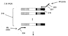
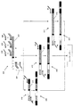
本発明の1つの局面は、リンパ球の集団におけるTCRまたはBCR再編成のレベルを評価するために、次世代シークエンシング技術を使用する。これらのシークエンシング技術は、妥当なコストで、試料から100万またはそれ以上のリードを得ることができる。1/1,000,000またはそれよりも低い頻度で存在するクロノタイプでさえ、これらの技術を用いて特異的様式で検出することができる。血液または骨髄DNA由来の試料から、遺伝子または転写物の特定部分の配列の異なる型をすべて増幅するためのマルチプレックス増幅が達成され得る。例えば、IgH配列を増幅するために、公知のVセグメントおよびアレルすべてに相補的ないくつかのプライマーを、Jセグメントおよびアレルすべてに相補的ないくつかのプライマーと共に使用することができる。図1Aは、試料のクロノタイプをプロファイするために、あるクラスのDNAシーケンサー(例えば、以下に記載するような合成によるSolexa配列決定(Solexa sequencing-by-synthesis))を使用する態様に関して、そのような方法の段階を説明している。B細胞またはT細胞を含む試料を採取し(100)、その後DNAまたはRNAを抽出し、クロノタイプを優先的に増幅し、後続の増幅および配列決定のための末端配列を付着させる反応において増幅する(102)。増幅されたクロノタイプの個々の分子を、ガラス表面などの固体表面上にランダムに分配するが(104)、これは個々の分子のクローン集団(またはポロニー)を生成するための二次インサイチュー増幅を可能にするように構成されている(106)。次に、例えば合成による配列決定技法(sequencing-by-synthesis)を用いて各ポロニーの分子を配列決定し(108)、その後その配列の型および存在量を一覧にして、クロノタイププロファイル(110)または同等にレパートリープロファイルを形成する。本方法は、異なる配列間の増幅のバイアスがほとんどない状態で行うことができる。TCRβ遺伝子およびIgH遺伝子由来のRNAは、異なるVプライマーの効率に差がわずかしかない状態で増幅することができ、それによってDNAから同じことを行う見込みが確証される。このスキームは、低レベルのTCRおよび/またはBCR再編成を検出するためのリアルタイムリードに関する問題を改善することができる。
Detailed Description of the Invention One aspect of the present invention uses next-generation sequencing technology to assess the level of TCR or BCR rearrangements in a population of lymphocytes. These sequencing techniques can obtain 1 million or more leads from samples at reasonable cost. Even clonotypes present at 1 / 1,000,000 or less can be detected in a specific manner using these techniques. Multiplex amplification can be achieved to amplify all different types of sequences of particular portions of genes or transcripts from samples from blood or bone marrow DNA. For example, to amplify an IgH sequence, some primers complementary to all known V segments and alleles can be used with some primers complementary to all J segments and alleles. FIG. 1A relates to an embodiment using a class of DNA sequencer (eg, Solexa sequencing-by-synthesis) as described below to profile the clonotype of a sample. Describes the steps of the method. A sample containing B cells or T cells is taken (100) and then DNA or RNA is extracted, clonotypes are preferentially amplified and amplified in a reaction to attach terminal sequences for subsequent amplification and sequencing. (102). The individual molecules of the amplified clonotype are randomly distributed on a solid surface such as a glass surface (104), but this is a secondary in situ amplification to generate a clonal population (or polony) of the individual molecules Configured to allow (106). Next, the molecules of each polony are sequenced 108 using, for example, sequencing-by-synthesis techniques, and then the type and abundance of the sequences are listed to obtain a clonotype profile (110). Or equivalently form a repertoire profile. The method can be performed with little amplification bias between the different sequences. The RNA from the TCRβ and IgH genes can be amplified with little difference in the efficiency of the different V primers, thereby confirming the prospect of doing the same from DNA. This scheme can ameliorate the problems with real time reads to detect low levels of TCR and / or BCR rearrangements.
感度は、計数統計(すなわち、感度は、細胞および配列決定試料のサイズの増加によって上昇する)および同等の増幅(すなわち、様々な配列を有するクロノタイプは、以下に示すように、PCRなどのマルチプレックス増幅反応において有意なバイアスなしに増幅され得る)によって決定される。感度は最終的には計数統計によって限定されるため、さらなる感度を得るためには、単純により多くの細胞(すなわち、より大きな試料)およびより多くの配列決定リードを得ればよい。十分な配列決定リードがある場合には、感度は試料中のリンパ球数によって限定される。対照的に、リアルタイムPCRアッセイの感度はバックグラウンドによって限定される。さらに、患者の特異的クローンは、診断用の白血病またはリンパ腫試料を配列決定することによって決定され得る。ひとたびクロノタイプが決定されたならば、後続の時点で試料においてそのレベルを決定することができる。いくつかの好ましい態様においては、患者特異的なプローブもしくはプライマー、または標準物質として実行させるための患者特異的鋳型の使用の必要性がない。代わりに、患者特異的クローンは、各患者の関連配列に関するデータを保存することによって追跡され、同一のアッセイ法がすべての患者に当てはまる。 The sensitivity is the counting statistic (ie sensitivity increases with the increase in the size of the cells and sequencing samples) and the equivalent amplification (ie clonotypes with different sequences are shown below: Can be amplified without significant bias in the plex amplification reaction. Because sensitivity is ultimately limited by counting statistics, one simply needs to obtain more cells (ie, larger samples) and more sequencing reads to gain additional sensitivity. If there is sufficient sequencing lead, the sensitivity is limited by the number of lymphocytes in the sample. In contrast, the sensitivity of real-time PCR assays is limited by background. In addition, specific clones of patients can be determined by sequencing diagnostic leukemia or lymphoma samples. Once the clonotype has been determined, its level can be determined in the sample at subsequent times. In some preferred embodiments, there is no need for patient specific probes or primers, or the use of patient specific templates for implementation as standards. Instead, patient specific clones are followed by storing data on the relevant sequences of each patient, and the same assay applies to all patients.
概して、本発明のいくつかの態様は、免疫系をプロファイルするために適応免疫細胞のレパートリーをモニターするという課題に、核酸配列決定技法を適用するための方法を含む。作成された免疫系のプロファイルは、疾患および障害の診断、ならびに疾患および障害の状態の診断に使用することができる。提供する本発明の免疫プロファイリングの方法は、疾患および障害をモニターするのに、ならびに疾患および障害の治療を評価するのに用いることができる。提供する本発明の方法が適用され得る疾患および障害には、全身性エリテマトーデス(SLE)、多発性硬化症(MS)、関節リウマチ(RA)、および強直性脊椎炎(AS)を含む自己免疫疾患が含まれる。提供する本発明の方法は、移植片拒絶反応および免疫老化の診断、モニタリング、および治療に適用することができる。さらに、提供する本発明の免疫プロファイリングの方法は、癌および感染症を含む、免疫系に関連するその他の疾患を診断する、モニターする、および治療するのに用いることができる。 In general, some aspects of the invention include methods for applying nucleic acid sequencing techniques to the task of monitoring the repertoire of adaptive immune cells to profile the immune system. The profile of the immune system generated can be used for the diagnosis of diseases and disorders as well as for the diagnosis of diseases and disorders. The provided methods of immune profiling of the invention can be used to monitor diseases and disorders as well as to evaluate treatments for diseases and disorders. Diseases and disorders to which the provided methods of the invention may be applied include autoimmune diseases including systemic lupus erythematosus (SLE), multiple sclerosis (MS), rheumatoid arthritis (RA), and ankylosing spondylitis (AS). Is included. The provided methods of the invention can be applied to the diagnosis, monitoring and treatment of graft rejection and immune aging. In addition, the provided methods of immune profiling of the present invention can be used to diagnose, monitor and treat other diseases associated with the immune system, including cancer and infections.
個々の増幅分子を配列決定することで、異なる配列を識別することができ、したがって配列決定はクローン増殖における量的変化を検出するための感度を有する。概して、提供する本発明の1つの態様において、T細胞および/またはB細胞における組換えDNA配列のプロファイルを決定するための方法を提供する。本方法は、対象から試料を単離する段階、1ラウンドまたは複数ラウンドの核酸増幅、個々の核酸を空間的に単離する段階、および核酸を配列決定する段階を含む段階を含み得る。核酸はDNAまたはRNAであってよい。T細胞および/またはB細胞における組換えDNA配列はクロノタイプと称され得る。 By sequencing individual amplified molecules, different sequences can be identified and thus sequencing has the sensitivity to detect quantitative changes in clonal expansion. In general, in one aspect of the invention provided, a method is provided for determining the profile of recombinant DNA sequences in T cells and / or B cells. The method may include the steps of isolating the sample from the subject, one or more rounds of nucleic acid amplification, spatially isolating the individual nucleic acids, and sequencing the nucleic acids. The nucleic acid may be DNA or RNA. Recombinant DNA sequences in T cells and / or B cells can be termed clonotypes.
1つの局面において、対象または個体における1つまたは複数の相関クロノタイプを決定するための方法を提供する。別の局面において、疾患を有する対象に由来する任意の試料における1つまたは複数の相関クロノタイプを予測することができるアルゴリズムを開発するための方法を提供する。別の局面において、対象に由来する任意の試料における1つまたは複数の相関クロノタイプを予測することができるアルゴリズムを用いて、個体の1つまたは複数の相関クロノタイプを発見するための方法を提供する。別の局面において、疾患活動性スコアを算出するアルゴリズムを作成するための方法を提供する。別の局面において、個体の疾患状態をモニターするための方法を提供する。 In one aspect, a method is provided for determining one or more correlated clonotypes in a subject or individual. In another aspect, a method is provided for developing an algorithm that can predict one or more correlated clonotypes in any sample derived from a subject having a disease. In another aspect, there is provided a method for discovering one or more correlated clonotypes of an individual using an algorithm capable of predicting one or more correlated clonotypes in any sample derived from a subject Do. In another aspect, a method is provided for creating an algorithm for calculating a disease activity score. In another aspect, a method is provided for monitoring an individual's disease state.
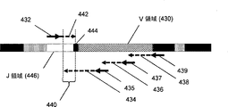
T細胞およびB細胞レパートリープロファイリングは、炎症局面を有する疾患にとって価値があると考えられる。この炎症は、自己免疫および/または過敏性反応に起因する場合が多い。これらの疾患には、循環器疾患、アルツハイマー病、および子癇前症が含まれる。炎症は、肥満症および糖尿病を含む異常な代謝状態とも関連づけられている。その他の炎症関連疾患も存在する。本発明の1つの局面では、鋳型の入れ子セット(nested set)を作製するための複数の順方向プライマーまたは複数の逆方向プライマーを用いるPCRによって、組換えB細胞核酸のセグメントを増幅する(図4Aおよび4Bならびに以下のそれらの説明を参照されたい)。そのようなセットからの鋳型を表面上でさらに増幅して、別々のアンプリコンを形成することができる(例えば、cBot装置、Illumina、San Diego, CAを用いるブリッジPCRによる)。同じ入れ子セットからの鋳型は、それらの共通末端において作成された配列リードによって互いに関連づけられ得る。鋳型の入れ子セットのお陰で、エラー率の比較的高い配列決定化学を用いて、配列の全長にわたって高い平均品質スコアを同時に維持しつつ、そうでない場合に可能である長さよりも長い配列を解析することが可能になる。入れ子セットにより、V領域が体細胞超変異に供された場合でさえ、V領域から少なくとも1つの配列リードが得られることが確実になる。1つの態様では、IgH分子のような可変性の高い核酸を解析するために、以下よりも高いエラー率を有する配列決定化学を使用することができる:配列リードの0.2パーセントが、1〜50位に少なくとも1つのエラーを含む;配列リードの0.2〜1.0パーセントが、51〜75位に少なくとも1つのエラーを含む;配列リードの0.5〜1.5パーセントが、76〜100位に少なくとも1つのエラーを含む;および配列リードの1〜5パーセントが、101〜125位に少なくとも1つのエラーを含む。別の態様において、配列決定プライマー結合部位は、伸長される場合にそれらが一連の配列リードを生じるように位置し、ここで、最後のものを除く各配列リードは、直接隣接する下流のプライマー結合部位および/または配列リードと重複し、それによって、単一の長い配列リードを生成するために単一の長い鋳型が使用される場合に可能である品質スコアよりも高い品質スコアを有する、連続した配列範囲が提供される。 T cell and B cell repertoire profiling is considered valuable for diseases with an inflammatory phase. This inflammation is often attributable to autoimmune and / or hypersensitivity reactions. These diseases include cardiovascular disease, Alzheimer's disease, and pre-eclampsia. Inflammation is also linked to abnormal metabolic conditions including obesity and diabetes. Other inflammation related diseases also exist. In one aspect of the invention, segments of recombinant B cell nucleic acid are amplified by PCR using multiple forward or multiple reverse primers to create a nested set of templates (FIG. 4A). And 4B and their descriptions below). Templates from such a set can be further amplified on the surface to form separate amplicons (eg, by bridge PCR using cBot device, Illumina, San Diego, CA). Templates from the same nested set can be related to one another by sequence reads made at their common ends. Thanks to the nested set of templates, relatively high error rate sequencing chemistry is used to analyze longer sequences than would otherwise be possible while simultaneously maintaining a high average quality score over the entire length of the sequence It becomes possible. Nesting sets ensure that at least one sequence read is obtained from the V region, even when the V region is subjected to somatic hypermutation. In one aspect, sequencing chemistry with higher error rates than the following can be used to analyze highly variable nucleic acids such as IgH molecules: 0.2 percent of the sequence reads are at positions 1-50 Contains at least one error; 0.2 to 1.0 percent of the sequence reads include at least one error at positions 51-75; 0.5 to 1.5 percent of the sequence reads include at least one error at 76 to 100; And 1 to 5 percent of the sequence reads contain at least one error at position 101-125. In another embodiment, the sequencing primer binding sites are positioned such that, when extended, they produce a series of sequence reads, wherein each sequence read except the last is directly adjacent downstream primer binding Consecutive with the site and / or sequence reads, thereby having a quality score higher than the quality score possible when a single long template is used to generate a single long sequence read An array range is provided.
1. さらなる局面および態様
本発明のさらなる局面および態様は以下のものを含む:T細胞および/またはB細胞における組換えDNA配列のプロファイルを決定するための方法であって、T細胞および/またはB細胞を含む対象由来の試料を採取する段階、該細胞からゲノムDNAの個々の分子を空間的に単離する段階;ゲノムDNAの空間的に単離された個々の該分子を配列決定する段階、ならびに該試料由来の異なる配列についてそれらのレベルを決定して、組換えDNA配列のプロファイルを作成する段階を含む前記方法を提供する。T細胞および/またはB細胞における組換えDNA配列のプロファイルを決定するための方法であって、T細胞および/またはB細胞を含む対象由来の試料を採取する段階、該細胞からゲノムDNAの個々の分子を空間的に単離する段階、ゲノムDNAの個々の該分子を増幅する段階、該増幅DNAを配列決定する段階、ならびに該試料由来の異なる配列についてそれらのレベルを決定して、組換えDNA配列のプロファイルを作成する段階を含む前記方法を提供する。T細胞および/またはB細胞における組換えDNA配列のプロファイルを決定するための方法であって、T細胞および/またはB細胞を含む対象由来の試料を採取する段階、該細胞からゲノムDNAを増幅する段階、該増幅DNAの個々の分子を空間的に単離する段階、増幅DNAの空間的に単離された個々の該分子を配列決定する段階、ならびに該試料由来の異なる配列についてそれらのレベルを決定して、組換えDNA配列のプロファイルを作成する段階を含む前記方法を提供する。T細胞および/またはB細胞における組換えDNA配列のプロファイルを決定するための方法であって、T細胞および/またはB細胞を含む対象由来の試料を採取する段階、該細胞からゲノムDNAを増幅する段階、該増幅DNAの個々の分子を空間的に単離する段階、該増幅DNA分子を再増幅する段階、該再増幅DNA分子を配列決定する段階、ならびに該試料由来の異なる配列についてそれらのレベルを決定して、組換えDNA配列のプロファイルを作成する段階を含む前記方法を提供する。T細胞および/またはB細胞における組換えDNAの配列のプロファイルを決定するための方法であって、T細胞および/またはB細胞を含む対象由来の試料を採取する段階、該細胞からRNAを逆転写してcDNAを形成する段階、該cDNAの個々の分子を空間的に単離する段階、任意で、cDNAの空間的に単離された個々の該分子を再増幅する段階、該cDNAおよび/または再増幅cDNAを配列決定する段階、ならびに該試料由来の異なる配列についてそれらのレベルを決定して、組換えDNA配列のプロファイルを作成する段階を含む前記方法を提供する。T細胞および/またはB細胞における組換えDNA配列のプロファイルを決定するための方法であって、T細胞および/またはB細胞を含む対象由来の試料を採取する段階、該試料中の個々の細胞を空間的に単離する段階、該細胞から核酸の個々の分子を配列決定する段階、ならびに該試料由来の異なる配列についてそれらのレベルを決定して、組換えDNA配列のプロファイルを作成する段階を含む前記方法を提供する。1つの態様において、前記の増幅および/または再増幅段階は、PCR、マルチプレックスPCR、TMA、NASBA、またはLAMPを含む。別の態様において、空間的に単離する前記段階は、固体支持体上で前記DNAもしくはcDNAを二次元に分離すること、ミセルを有する溶液中で前記DNAもしくはcDNAを三次元に分離すること、または微小反応チャンバーを用いて分子を分離することを含む。別の態様において、前記の増幅および/または再増幅段階は、サブクローニングしたDNAもしくはcDNAを保有する細菌の増殖、スライド上でのDNAもしくはcDNAの増幅、またはビーズ上でのDNAもしくはcDNAの増幅による。別の態様において、前記の配列決定段階はジデオキシ配列決定を含む。別の態様において、前記の配列決定段階は、可逆的終結標識ヌクレオチドを用いた合成による配列決定を含む。別の態様において、前記の配列決定段階は、ヌクレオチド取り込み時のピロリン酸放出の検出を含む。別の態様において、前記の配列決定段階は、標識オリゴヌクレオチドプローブのライブラリーへのアレル特異的ハイブリダイゼーションを含む。別の態様において、前記の配列決定段階は、標識オリゴヌクレオチドプローブのライブラリーへのアレル特異的ハイブリダイゼーションと、それに続く該プローブの連結とを用いた合成による配列決定を含む。別の態様において、前記の配列決定段階は、重合工程中の標識ヌクレオチドの取り込みのリアルタイムモニタリングを含む。別の態様において、前記の組換えDNA配列は、T細胞受容体遺伝子および/または免疫グロブリン遺伝子を含む。別の態様において、前記の配列決定段階は、免疫グロブリン遺伝子および/またはT細胞受容体遺伝子の完全クローン配列のサブセットを配列決定することを含む。別の態様において、完全クローン配列の前記サブセットは、免疫グロブリン遺伝子もしくはT細胞受容体遺伝子のV-D結合部、D-J結合部、免疫グロブリン遺伝子もしくはT細胞受容体遺伝子の完全な可変領域、抗原認識領域、または相補性決定領域3(CDR3)を含む。別の態様において、前記T細胞受容体遺伝子はT細胞受容体β遺伝子を含む。別の態様において、前記免疫グロブリン遺伝子は免疫グロブリン重鎖遺伝子を含む。別の態様において、前記の増幅または再増幅段階は、Vセグメントと相補的な複数のプライマー、およびCセグメントと相補的な1つのプライマーを含む。別の態様において、前記の増幅または再増幅段階は、Vセグメントと相補的な複数のプライマー、およびCセグメントと相補的な複数のプライマーを含む。別の態様において、Vセグメントと相補的な前記の複数のプライマーは、各Vセグメントに対する少なくとも3種の異なるプライマーを含み、Cセグメントと相補的な複数のプライマーは、少なくとも1種、少なくとも2種、少なくとも3種、少なくとも4種、少なくとも5種、または少なくとも6種のプライマーを含む。別の態様において、前記T細胞またはB細胞は、全T細胞およびB細胞のサブセットである。別の態様において、T細胞の該サブセットは、CD4+、CD8+細胞、またはCD27高細胞である。別の態様において、前記試料は、少なくとも100,000個、少なくとも500,000個、少なくとも750,000個、または少なくとも1,000,000個のT細胞を含む。別の態様において、前記の配列決定段階は、1回の実行当たり少なくとも1000件のリード、1回の実行当たり少なくとも10,000件のリード、1回の実行当たり少なくとも100,000件のリード、または1回の実行当たり少なくとも1,000,000件のリードを含む。別の態様において、前記の配列決定段階は、1回のリード当たり約30 bp、約40 bp、約50 bp、約60 bp、約70 bp、約80 bp、約90 bp、約100 bp、約110 bp、または約120 bpを作成することを含む。別の態様では、対象が自己免疫疾患のフレア(flare)状態にある時点で、前記試料を採取する。別の態様では、全身性エリテマトーデスを有するかまたはそれを有する疑いのある対象から前記試料を採取する。別の局面では、対象における1つまたは複数の相関クロノタイプを決定するための方法であって、疾患の第1状態に関連する少なくとも1つの対象由来の試料から空間的に単離された個々の分子を核酸配列決定することにより、1つまたは複数のクロノタイププロファイルを作成する段階、およびその1つまたは複数のクロノタイププロファイルに基づいて、対象における1つまたは複数の相関クロノタイプを決定する段階を含む前記方法を提供する。1つの態様において、前記の少なくとも1つの試料は、疾患に罹患した組織に由来する。別の態様において、前記の1つまたは複数の相関クロノタイプの決定は、少なくとも2つの試料からのクロノタイププロファイルを比較する段階を含む。別の態様において、疾患の第1状態は疾患のピーク状態である。別の態様において、前記の1つまたは複数の相関クロノタイプは、疾患のピーク状態において存在する。別の態様において、前記の1つまたは複数の相関クロノタイプは、疾患のピーク状態において存在しない。別の態様において、前記の1つまたは複数の相関クロノタイプは、疾患のピーク状態において高い。別の態様において、前記の1つまたは複数の相関クロノタイプは、疾患のピーク状態において低い。別の態様において、前記試料はT細胞および/またはB細胞を含む。別の態様において、該T細胞および/またはB細胞は、T細胞および/またはB細胞のサブセットを含む。別の態様において、T細胞および/またはB細胞の該サブセットは、マーカーとの相互作用によって濃縮される。別の態様において、該マーカーは、T細胞および/またはB細胞のサブセット上の細胞表面マーカーである。別の態様において、T細胞および/またはB細胞の該サブセットは、疾患において特異的に存在する抗原と相互作用する。別の態様において、疾患は全身性エリテマトーデスまたは多発性硬化症である。別の局面において、疾患を有する対象に由来する任意の試料における1つまたは複数の相関クロノタイプを予測することができるアルゴリズムを開発するための方法であって、a) 疾患に関連する試料のセットから複数のクロノタイププロファイルを作成する段階、b) その試料のセットから1つまたは複数の相関クロノタイプを同定する段階、c) b)において同定された1つまたは複数の相関クロノタイプからの配列パラメータおよび/または機能的データを用いて、疾患を有する対象に由来する任意の試料における相関クロノタイプを予測することができるアルゴリズムを開発する段階を含む前記方法を提供する。1つの態様では、疾患に罹患した1つまたは複数の組織から試料のセットを採取する。別の態様において、前記の1つまたは複数の相関クロノタイプの同定は、少なくとも2つの試料からのクロノタイププロファイルを比較する段階を含む。別の態様において、前記機能的データは、T細胞および/もしくはB細胞表面上のマーカーの結合能、またはT細胞もしくはB細胞による抗原との相互作用を含む。別の態様において、前記配列パラメータは、核酸配列および予測されるアミノ酸配列を含む。別の態様において、試料は、疾患のピーク段階にある1つまたは複数の個体に由来する。別の態様において、前記の1つまたは複数の相関クロノタイプは、疾患のピーク状態において存在する。別の態様において、前記の1つまたは複数の相関クロノタイプは、疾患のピーク状態において高レベルである。別の態様において、前記の1つまたは複数の相関クロノタイプは、疾患のピーク状態において低レベルである。別の態様において、1つまたは複数の相関クロノタイプは、疾患のピーク状態において存在しない。別の態様において、疾患は全身性エリテマトーデスまたは多発性硬化症である。別の態様において、個体の1つまたは複数の相関クロノタイプを発見するための方法であって、個体由来の試料からのクロノタイププロファイルをアルゴリズムに入力する段階、およびアルゴリズムを用いて、個体の1つまたは複数の相関クロノタイプを決定する段階を含む前記方法を提供する。1つの態様において、アルゴリズムは、a) 疾患に関連する試料のセットから複数のクロノタイププロファイルを作成する段階、b) その試料のセットから1つまたは複数の相関クロノタイプを同定する段階、c) b)において同定された1つまたは複数の相関クロノタイプからの配列パラメータおよび/または機能的データを用いて、疾患を有する対象に由来する任意の試料における相関クロノタイプを予測することができるアルゴリズムを開発する段階によって開発された、疾患を有する対象に由来する任意の試料における1つまたは複数の相関クロノタイプを予測することができるアルゴリズムである。1つの態様では、疾患のピーク状態において前記試料を採取する。別の態様では、疾患に罹患した組織から試料を採取する。別の局面において、疾患活動性スコアを算出するアルゴリズムを作成するための方法であって、因子のセットを用いて相関クロノタイプのレベルを疾患活動性スコアに統合するアルゴリズムを開発する段階、疾患活動性スコアを、疾患状態に関する臨床データと比較する段階、および臨床データと疾患活動性スコアとの相関を最大化するために因子を最適化する段階を含む前記方法を提供する。1つの態様において、個体の疾患状態をモニターするための方法であって、
a) 個体由来の試料からクロノタイププロファイルを決定する段階、b) 因子のセットを用いて相関クロノタイプのレベルを疾患活動性スコアに統合するアルゴリズムを開発する段階と、疾患活動性スコアを、疾患状態に関する臨床データと比較する段階と、臨床データと疾患活動性スコアとの相関を最大化するために因子を最適化する段階とによって作成される、疾患活動性を算出するアルゴリズムに、a)からのクロノタイププロファイル情報を入力する段階、ならびにc) 疾患活動性スコアを算出するアルゴリズムを用いて、個体の疾患状態を予測するスコアを作成する段階を含む前記方法を提供する。別の態様において、個体の疾患状態をモニターするための方法は、個体における1つまたは複数の相関クロノタイプを決定する段階、および1つまたは複数の相関クロノタイプの情報をアルゴリズムに入力する段階をさらに含む。別の態様において、個体における1つまたは複数の相関クロノタイプを決定する前記段階は、a) 疾患の第1状態に関連する少なくとも1つの対象由来の試料から空間的に単離された個々の分子を核酸配列決定することにより、1つまたは複数のクロノタイププロファイルを作成することと、b) その1つまたは複数のクロノタイププロファイルに基づいて、対象における1つまたは複数のより相関するクロノタイプを決定することとを含む。別の態様において、個体における1つまたは複数の相関クロノタイプを決定する前記段階は、a) i) 疾患に関連する試料のセットから複数のクロノタイププロファイルを作成する段階、ii) その試料のセットから1つまたは複数の相関クロノタイプを同定する段階、iii) ii)において同定された1つまたは複数の相関クロノタイプからの配列パラメータおよび/または機能的データを用いて、疾患を有する対象に由来する任意の試料における相関クロノタイプを予測することができるアルゴリズムを開発する段階によって開発された、1つまたは複数の相関クロノタイプを予測することができるアルゴリズムに、個体由来の試料からのクロノタイププロファイルを入力すること、ならびにc) 1つまたは複数の相関クロノタイプを予測することができるアルゴリズムを用いて、個体の1つまたは複数の相関クロノタイプを決定することを含む。別の態様において、疾患は全身性エリテマトーデスまたは多発性硬化症である。別の局面において、1つまたは複数の相関T細胞またはB細胞クロノタイプを決定する方法であって、a) 対象由来の細胞の試料を少なくとも2つの試料に分割する段階、b) 対象由来の細胞の試料のうちの一方から個々に空間的に単離された分子を核酸配列決定することにより、1つまたは複数のクロノタイププロファイルを作成する段階、c) 該細胞の少なくとも1つの分子パラメータに基づいて、この対象由来のもう一方の細胞の試料を濃縮する段階、d) 該対象の濃縮試料から個々に空間的に単離された分子を核酸配列決定することにより、1つまたは複数のクロノタイププロファイルを作成する段階、およびe) 試料におけるその存在量が濃縮試料と非濃縮試料との間で変化したクロノタイプに基づいて、少なくとも1つのクロノタイプを同定する段階を含む前記方法を提供する。別の態様において、分子パラメータは細胞表面マーカーである。別の態様において、濃縮は、固相固定化親和性マーカーを用いて細胞を捕捉することによって行われる。別の態様において、固体表面はビーズのセットである。別の態様において、固体表面はカラムである。別の態様において、マーカーは蛍光部分を用いて標識される。別の態様において、濃縮は、蛍光標識を用いてフローサイトメトリーによって達成される。別の態様において、細胞はBリンパ球であり、濃縮は、B細胞受容体と結合する抗原を用いて行われる。別の態様において、濃縮は、抗原が固定化されている固体表面上での捕捉を介して行われる。別の態様においては、Bリンパ球を標識するために抗原が用いられ、濃縮は、この標識を用いてフローサイトメトリーを使用して達成される。別の態様において、細胞はTリンパ球であり、濃縮は、特異的抗原に反応するT細胞を標識して、フローサイトメトリーを用いて濃縮することを可能にする方法を用いて行われる。別の態様において、T細胞は細胞内サイトカイン染色法を用いて標識される。別の態様において、T細胞はサイトカイン捕捉法を用いて標識される。別の態様において、分子パラメータは、病原体に特異的である少なくとも1つの抗原と結合することができるB細胞受容体である。別の態様において、分子パラメータは、病原体に特異的である少なくとも1つの抗原と結合することができえるT細胞受容体である。別の態様において、試料は、第1の時点で病原体に曝露された患者から採取される。別の局面において、病原体に対する免疫反応と相関する、個体におけるクロノタイプのセットを決定するための方法であって、a) 対象由来の細胞の試料を少なくとも2つの試料に分割する段階、b) 対象由来の一方の細胞の試料から個々に空間的に単離された分子を核酸配列決定することにより、1つまたは複数のクロノタイププロファイルを作成する段階、c) 該病原体由来の少なくとも1つの抗原と結合する細胞の能力に基づいて、この対象由来のもう一方の細胞の試料を濃縮する段階、d) 該対象の濃縮試料から個々に空間的に単離された分子を核酸配列決定することにより、1つまたは複数のクロノタイププロファイルを作成する段階、およびe) 試料におけるその存在量が濃縮試料と非濃縮試料との間で変化したクロノタイプに基づいて、少なくとも1つの相関クロノタイプを同定する段階を含む前記方法を提供する。別の局面において、腫瘍に対する免疫反応と相関する、個体におけるクロノタイプのセットを決定するための方法であって、a) 対象由来の細胞の試料を少なくとも2つの試料に分割する段階、b) 対象由来の一方の細胞の試料から個々に空間的に単離された分子を核酸配列決定することにより、1つまたは複数のクロノタイププロファイルを作成する段階、c) 腫瘍中に存在する少なくとも1つの自己抗原と結合する細胞の能力に基づいて、この対象由来のもう一方の細胞の試料を濃縮する段階、d) 該対象の濃縮試料から個々に空間的に単離された分子を核酸配列決定することにより、1つまたは複数のクロノタイププロファイルを作成する段階、およびe) 試料におけるその存在量が濃縮試料と非濃縮試料との間で変化したクロノタイプに基づいて、少なくとも1つの相関クロノタイプを同定する段階を含む前記方法を提供する。別の態様においては、個体が腫瘍を有するリスクを評価するために、相関クロノタイプのレベルが用いられる。別の態様において、抗原は、その個体において既に生じた腫瘍中に存在していることがわかっており、腫瘍の再発のリスクを評価するために、相関クロノタイプが用いられる。別の態様において、抗原は、他の個体における腫瘍中に存在することがわかっており、以前に腫瘍が検出されたことのない患者において癌のリスクを評価するために、クロノタイプが用いられる。別の局面において、臓器に対する損傷によって血流中に放出された物質に対する免疫反応と相関する、個体におけるクロノタイプのセットを決定するための方法であって、a) 対象由来の細胞の試料を少なくとも2つの試料に分割する段階、b) 対象由来の一方の細胞の試料から個々に空間的に単離された分子を核酸配列決定することにより、1つまたは複数のクロノタイププロファイルを作成する段階、c) 損傷した臓器中に存在する少なくとも1つの自己抗原と結合する細胞の能力に基づいて、この対象由来のもう一方の細胞の試料を濃縮する段階、d) 該対象の濃縮試料から個々に空間的に単離された分子を核酸配列決定することにより、1つまたは複数のクロノタイププロファイルを作成する段階、およびe) 試料におけるその存在量が濃縮試料と非濃縮試料との間で変化したクロノタイプに基づいて、少なくとも1つの相関クロノタイプを同定する段階を含む前記方法を提供する。別の態様においては、個体が臓器損傷を有するリスクを評価するために、相関クロノタイプのレベルが用いられる。別の局面において、治療薬に対する免疫反応と相関する、個体におけるクロノタイプのセットを決定するための方法であって、a) 対象由来の細胞の試料を少なくとも2つの試料に分割する段階、b) 対象由来の一方の細胞の試料から個々に空間的に単離された分子を核酸配列決定することにより、1つまたは複数のクロノタイププロファイルを作成する段階、c) 治療薬中に含まれる少なくとも1つの抗原と結合する細胞の能力に基づいて、この対象由来のもう一方の細胞の試料を濃縮する段階、d) 該対象の濃縮試料から個々に空間的に単離された分子を核酸配列決定することにより、1つまたは複数のクロノタイププロファイルを作成する段階、およびe) 試料におけるその存在量が濃縮試料と非濃縮試料との間で変化したクロノタイプに基づいて、少なくとも1つの相関クロノタイプを同定する段階を含む前記方法を提供する。別の態様においては、個体が治療薬に対して過敏性を示しているリスクを評価するために、相関クロノタイプのレベルが用いられる。別の局面において、動脈プラークに対する免疫反応と相関する、個体におけるクロノタイプのセットを決定するための方法であって、a) 対象由来の細胞の試料を少なくとも2つの試料に分割する段階、b) 対象由来の一方の細胞の試料から個々に空間的に単離された分子を核酸配列決定することにより、1つまたは複数のクロノタイププロファイルを作成する段階、c) 動脈プラーク中に存在する少なくとも1つの抗原と結合する細胞の能力に基づいて、この対象由来のもう一方の細胞の試料を濃縮する段階、d) 該対象の濃縮試料から個々に空間的に単離された分子を核酸配列決定することにより、1つまたは複数のクロノタイププロファイルを作成する段階、およびe) 試料におけるその存在量が濃縮試料と非濃縮試料との間で変化したクロノタイプに基づいて、少なくとも1つの相関クロノタイプを同定する段階を含む前記方法を提供する。別の態様においては、個体が循環器疾患を有するリスクを評価するために、相関クロノタイプのレベルが用いられる。別の態様においては、動脈プラークが不安定であるリスクを評価するために、相関クロノタイプのレベルが用いられる。別の局面において、リンパ系新生物に関与する細胞中に見出される配列識別子を決定するための方法であって、a) 癌性細胞が存在することがわかっている罹患個体から細胞の試料を採取する段階、b) 該試料中の細胞から個々に空間的に単離された分子を核酸配列決定することにより、少なくとも1つの免疫細胞ゲノム再編成に関連している1つまたは複数のクロノタイププロファイルを作成する段階、c) 腫瘍と関連したクロノタイプの配列として配列識別子を同定する段階を含む前記方法を記載する。別の態様において、試料は患者の骨髄に由来する。別の態様において、試料は患者の血液に由来する。別の態様において、試料は固形リンパ系腫瘍の生検標本に由来する。別の態様において、免疫細胞ゲノム再編成は、B細胞におけるIgHのVDJ再編成である。別の態様において、免疫細胞ゲノム再編成は、B細胞におけるIgHのDJ再編成である。別の態様において、免疫細胞ゲノム再編成は、B細胞におけるIgKのVJ再編成である。別の態様において、免疫細胞ゲノム再編成は、B細胞におけるIgLのVJ再編成である。別の態様において、免疫細胞ゲノム再編成は、T細胞におけるTCRβのVDJ再編成である。別の態様において、免疫細胞ゲノム再編成は、T細胞におけるTCRβのDJ再編成である。別の態様において、免疫細胞ゲノム再編成は、T細胞におけるTCRαのVJ再編成である。別の態様において、免疫細胞ゲノム再編成は、T細胞におけるTCRλのVJ再編成である。別の態様において、免疫細胞ゲノム再編成は、T細胞におけるTCRδのVDJ再編成である。別の態様において、免疫細胞ゲノム再編成は、T細胞におけるTCRδのVD再編成である。別の態様において、免疫細胞ゲノム再編成は、ゲノムの別の領域に対するIgHのJセグメントの転座である。別の態様において、免疫細胞ゲノム再編成は、ゲノムの別の領域に対する任意のJセグメントの転座である。別の態様において、腫瘍関連クロノタイプの同定はクロノタイプ頻度によって行われる。別の
態様において、腫瘍関連クロノタイプの同定はクロノタイプ頻度によって行われる。別の態様において、腫瘍関連クロノタイプの同定は系譜間(cross lineage)再編成の検出によって行われる。別の態様において、腫瘍関連クロノタイプの同定は、非機能的再編成を同定することによって行われる。別の態様において、腫瘍関連クロノタイプの同定は、細胞クロノタイプを、腫瘍と関連した少なくとも1つの分子マーカーと関連づけることによって行われる。別の局面において、その腫瘍が、関与する第1の時点において特有の配列識別子と関連づけられた個体内に循環しているリンパ系腫瘍細胞のレベルを決定する方法であって、a) 患者から細胞の試料を採取する段階、b) 該試料中の細胞から個々に空間的に単離された分子を核酸配列決定することにより、少なくとも1つの免疫細胞ゲノム再編成に関連している1つまたは複数のクロノタイププロファイルを作成する段階、c) 配列識別子と関連したクロノタイプのレベルから腫瘍細胞のレベルを決定する段階を含む前記方法を記載する。別の局面において、その腫瘍が、関与する第1の時点において特有の配列識別子と関連づけられた個体内に循環しているリンパ系腫瘍細胞のレベルを決定する方法であって、a) 患者から細胞の試料を採取する段階、b) 少なくとも1つの分子マーカーに基づいて該細胞を濃縮する段階、c) 該試料中の細胞から個々に空間的に単離された分子を核酸配列決定することにより、少なくとも1つの免疫細胞ゲノム再編成に関連している1つまたは複数のクロノタイププロファイルを作成する段階、d) 配列識別子と関連したクロノタイプのレベルから腫瘍細胞のレベルを決定する段階を含む前記方法を記載する。別の態様において、試料は血液試料である。別の態様において、試料は骨髄試料である。別の態様において、試料はリンパ液試料である。別の態様において、試料は組織試料である。別の態様において、細胞は蛍光標識され、フローサイトメトリーを用いて濃縮される。別の態様において、細胞は固体支持体への結合を介して濃縮される。別の態様において、クロノタイプは、特有の配列識別子を含むクロノタイプと定義される。別の態様において、クロノタイプは、配列識別子に対する変異および再編成によって生じた可能性が高いクロノタイプと決定される。別の態様において、循環している腫瘍細胞のレベルおよび/または循環している腫瘍細胞のレベルの変化は、臨床的腫瘍再発を有するリスクと相関するスコアを生成するためのアルゴリズムにおいて用いられる。別の態様において、循環している腫瘍細胞のレベルおよび/または循環している腫瘍細胞のレベルの変化は、治療法を決定するために用いられる。
1. Further Aspects and Embodiments
Further aspects and embodiments of the invention include the following: A method for determining the profile of recombinant DNA sequences in T cells and / or B cells, from a subject comprising T cells and / or B cells Taking samples, spatially isolating individual molecules of genomic DNA from said cells; sequencing individually isolated said spatially separated molecules of genomic DNA, and different from said samples The method is provided comprising the steps of determining their level of sequence and generating a profile of the recombinant DNA sequence. A method for determining the profile of a recombinant DNA sequence in T cells and / or B cells, wherein a sample from a subject comprising T cells and / or B cells is taken, individual genomic DNA from said cells Spatially isolating the molecules, amplifying each of the individual molecules of genomic DNA, sequencing the amplified DNA, and determining their levels for different sequences from the sample to obtain recombinant DNA The method is provided comprising the step of creating a profile of the sequence. A method for determining the profile of a recombinant DNA sequence in T cells and / or B cells, collecting a sample from a subject comprising T cells and / or B cells, amplifying genomic DNA from said cells The steps of spatially isolating individual molecules of the amplified DNA, sequencing the spatially isolated individual molecules of the amplified DNA, and their levels for different sequences from the sample The method is provided comprising the steps of determining and generating a profile of the recombinant DNA sequence. A method for determining the profile of a recombinant DNA sequence in T cells and / or B cells, collecting a sample from a subject comprising T cells and / or B cells, amplifying genomic DNA from said cells The steps of spatially isolating individual molecules of the amplified DNA, re-amplifying the amplified DNA molecules, sequencing the re-amplified DNA molecules, and their levels for different sequences from the sample And determining a profile of the recombinant DNA sequence. A method for determining the profile of the sequence of recombinant DNA in T cells and / or B cells, collecting a sample from a subject comprising T cells and / or B cells, reverse transcribing RNA from said cells Forming the cDNA, spatially isolating the individual molecules of the cDNA, optionally re-amplifying the spatially isolated individual molecules of the cDNA, the cDNA and / or re- The method is provided comprising the steps of sequencing the amplified cDNA, as well as determining their levels for different sequences from the sample to generate a profile of the recombinant DNA sequence. A method for determining a profile of a recombinant DNA sequence in T cells and / or B cells, wherein a sample from a subject comprising T cells and / or B cells is taken, individual cells in said sample Spatially isolating, sequencing individual molecules of nucleic acid from the cell, and determining their levels for different sequences from the sample to generate a profile of recombinant DNA sequences The method is provided. In one embodiment, the amplification and / or reamplification step comprises PCR, multiplex PCR, TMA, NASBA, or LAMP. In another embodiment, the step of spatially isolating comprises two-dimensionally separating the DNA or cDNA on a solid support, three-dimensionally separating the DNA or cDNA in a solution having micelles, Or separating molecules using a microreaction chamber. In another embodiment, the amplification and / or reamplification step is by growth of bacteria carrying subcloned DNA or cDNA, amplification of DNA or cDNA on a slide, or amplification of DNA or cDNA on a bead. In another embodiment, the sequencing step comprises dideoxy sequencing. In another embodiment, the sequencing step comprises sequencing by synthesis using reversible termination labeled nucleotides. In another embodiment, the sequencing step comprises detection of pyrophosphate release upon nucleotide incorporation. In another embodiment, the sequencing step comprises allele specific hybridization to a library of labeled oligonucleotide probes. In another embodiment, the sequencing step comprises sequencing by synthesis using allele specific hybridization to a library of labeled oligonucleotide probes, followed by ligation of the probes. In another embodiment, the sequencing step comprises real time monitoring of incorporation of labeled nucleotides during the polymerization step. In another embodiment, the recombinant DNA sequence comprises a T cell receptor gene and / or an immunoglobulin gene. In another embodiment, the sequencing step comprises sequencing a subset of fully cloned sequences of immunoglobulin genes and / or T cell receptor genes. In another embodiment, said subset of fully cloned sequences comprises: VD binding portion of an immunoglobulin gene or T cell receptor gene, DJ binding portion, complete variable region of an immunoglobulin gene or T cell receptor gene, an antigen recognition region, Or complementarity determining region 3 (CDR3). In another embodiment, the T cell receptor gene comprises a T cell receptor beta gene. In another embodiment, the immunoglobulin gene comprises an immunoglobulin heavy chain gene. In another embodiment, the amplification or reamplification step comprises a plurality of primers complementary to the V segment and one primer complementary to the C segment. In another embodiment, the amplification or reamplification step comprises a plurality of primers complementary to the V segment and a plurality of primers complementary to the C segment. In another embodiment, the plurality of primers complementary to the V segment comprises at least three different primers for each V segment, and the plurality of primers complementary to the C segment is at least one, at least two, At least three, at least four, at least five, or at least six primers are included. In another embodiment, the T cells or B cells are a subset of all T cells and B cells. In another embodiment, the subset of T cells is a CD4 +, a CD8 + cell, or a CD27 High It is a cell. In another embodiment, the sample comprises at least 100,000, at least 500,000, at least 750,000, or at least 1,000,000 T cells. In another embodiment, the sequencing step comprises at least 1000 reads per run, at least 10,000 reads per run, at least 100,000 reads per run, or a run. Includes at least 1,000,000 leads per match. In another embodiment, the sequencing step comprises: about 30 bp, about 40 bp, about 50 bp, about 60 bp, about 70 bp, about 80 bp, about 90 bp, about 100 bp, about 100 bp, per read. Including making 110 bp, or about 120 bp. In another aspect, the sample is collected when the subject is in a flare state of an autoimmune disease. In another aspect, the sample is taken from a subject who has or is suspected of having systemic lupus erythematosus. In another aspect, a method for determining one or more correlated clonotypes in a subject, the individual spatially isolated from a sample from at least one subject associated with a first state of disease Creating one or more clonotype profiles by nucleic acid sequencing the molecule, and determining one or more correlated clonotypes in the subject based on the one or more clonotype profiles Providing the method. In one embodiment, the at least one sample is from a tissue afflicted with a disease. In another embodiment, the determination of the one or more correlated clonotypes comprises comparing clonotype profiles from at least two samples. In another embodiment, the first condition of disease is a peak condition of disease. In another embodiment, the one or more correlated clonotypes are present at peak conditions of the disease. In another embodiment, said one or more correlated clonotypes are absent at peak disease status. In another aspect, the one or more correlated clonotypes are high at the peak condition of the disease. In another embodiment, the one or more correlated clonotypes are low at peak disease status. In another embodiment, the sample comprises T cells and / or B cells. In another embodiment, the T cells and / or B cells comprise a subset of T cells and / or B cells. In another embodiment, the subset of T cells and / or B cells is enriched by interaction with a marker. In another embodiment, the marker is a cell surface marker on a subset of T cells and / or B cells. In another embodiment, the subset of T cells and / or B cells interact with an antigen that is specifically present in a disease. In another embodiment, the disease is systemic lupus erythematosus or multiple sclerosis. In another aspect, a method for developing an algorithm capable of predicting one or more correlated clonotypes in any sample from a subject having a disease, comprising: a) a set of samples associated with a disease Creating a plurality of clonotype profiles from b) identifying one or more correlated clonotypes from the set of samples; c) sequences from one or more correlated clonotypes identified in b) Providing said method comprising the step of developing an algorithm capable of predicting correlated clonotypes in any sample derived from a subject having a disease using parameters and / or functional data. In one aspect, a set of samples is taken from one or more tissues affected by the disease. In another embodiment, the identification of the one or more correlated clonotypes comprises comparing clonotype profiles from at least two samples. In another embodiment, the functional data comprises the binding ability of a marker on the surface of T cells and / or B cells, or the interaction of T cells or B cells with an antigen. In another embodiment, the sequence parameters include nucleic acid sequence and predicted amino acid sequence. In another embodiment, the sample is from one or more individuals at peak stage of the disease. In another embodiment, the one or more correlated clonotypes are present at peak conditions of the disease. In another embodiment, the one or more correlated clonotypes are at high levels at peak conditions of the disease. In another embodiment, the one or more correlated clonotypes are at low levels at peak conditions of the disease. In another embodiment, one or more correlated clonotypes are absent at the peak condition of the disease. In another embodiment, the disease is systemic lupus erythematosus or multiple sclerosis. In another aspect, a method for discovering one or more correlated clonotypes of an individual comprising inputting into the algorithm a clonotype profile from a sample from the individual, and using the algorithm one of the individual The method is provided comprising determining one or more correlated clonotypes. In one embodiment, the algorithm: a) creating a plurality of clonotype profiles from the set of samples associated with the disease, b) identifying one or more correlated clonotypes from the set of samples, c) An algorithm capable of predicting correlated clonotypes in any sample derived from a subject with disease using sequence parameters and / or functional data from one or more correlated clonotypes identified in b) An algorithm developed by the developing step that can predict one or more correlated clonotypes in any sample derived from a subject with a disease. In one aspect, the sample is taken at peak disease status. In another aspect, a sample is taken from the tissue afflicted with the disease. In another aspect, a method for creating an algorithm for calculating a disease activity score, wherein developing an algorithm that integrates the level of correlated clonotypes into a disease activity score using a set of factors, the disease activity The method is provided comprising the steps of comparing the sex score with clinical data on the disease state, and optimizing the factor to maximize the correlation between the clinical data and the disease activity score. In one embodiment, a method for monitoring the disease state of an individual comprising
a) determining the clonotype profile from samples from the individual, b) developing an algorithm to integrate the level of correlated clonotypes into the disease activity score using a set of factors, the disease activity score, the disease activity score An algorithm to calculate disease activity, created by comparing the clinical data about the condition and optimizing the factors to maximize the correlation between the clinical data and the disease activity score, from a) to a) The method comprises the steps of: inputting clonotype profile information of c) and c) generating a score for predicting a disease state of an individual using an algorithm for calculating a disease activity score. In another embodiment, a method for monitoring an individual's disease state comprises determining one or more correlated clonotypes in the individual, and inputting one or more correlated clonotype information into the algorithm. Further include. In another embodiment, said determining of one or more correlated clonotypes in an individual comprises: a) individual molecules spatially isolated from a sample from at least one subject associated with a first state of disease Creating one or more clonotype profiles by nucleic acid sequencing, and b) one or more more correlated clonotypes in the subject based on the one or more clonotype profiles. And determining. In another embodiment, said determining of one or more correlated clonotypes in the individual comprises: a) i) creating a plurality of clonotype profiles from a set of samples associated with the disease, ii) the set of samples Identifying one or more correlated clonotypes therefrom, iii) derived from a subject having the disease using sequence parameters and / or functional data from one or more correlated clonotypes identified in ii) A clonotype profile from a sample derived from an individual is developed into an algorithm capable of predicting one or more correlated clonotypes developed by developing an algorithm capable of predicting the correlated clonotype in any sample Argo, and c) an algo that can predict one or more correlated clonotypes Using prism includes determining one or more interrelated clonotype individuals. In another embodiment, the disease is systemic lupus erythematosus or multiple sclerosis. In another aspect, a method of determining one or more correlated T cell or B cell clonotypes comprising: a) dividing a sample of cells from a subject into at least two samples, b) cells from a subject Creating one or more clonotype profiles by nucleic acid sequencing of spatially isolated molecules individually from one of the samples of c. C) based on at least one molecular parameter of the cell Concentrating the sample of the other cell from the subject, d) one or more clonotypes by nucleic acid sequencing the molecules spatially isolated individually from the concentrated sample of the subject Creating a profile, and e) identifying at least one clonotype based on the clonotype whose abundance in the sample has varied between concentrated and non-concentrated samples It said method comprising providing. In another embodiment, the molecular parameter is a cell surface marker. In another embodiment, enrichment is performed by capturing cells using solid phase immobilized affinity markers. In another embodiment, the solid surface is a set of beads. In another embodiment, the solid surface is a column. In another embodiment, the marker is labeled with a fluorescent moiety. In another embodiment, concentration is achieved by flow cytometry using a fluorescent label. In another embodiment, the cells are B lymphocytes and enrichment is performed with an antigen that binds to a B cell receptor. In another embodiment, concentration is performed via capture on a solid surface on which the antigen is immobilized. In another embodiment, an antigen is used to label B lymphocytes, and enrichment is achieved using flow cytometry using this label. In another embodiment, the cells are T lymphocytes and enrichment is performed using a method that allows T cells that respond to a specific antigen to be labeled and enriched using flow cytometry. In another embodiment, T cells are labeled using intracellular cytokine staining. In another embodiment, T cells are labeled using a cytokine capture method. In another embodiment, the molecular parameter is a B cell receptor capable of binding to at least one antigen that is specific for a pathogen. In another embodiment, the molecular parameter is a T cell receptor capable of binding to at least one antigen that is specific for a pathogen. In another embodiment, a sample is taken from a patient exposed to a pathogen at a first time point. In another aspect, a method for determining a set of clonotypes in an individual that correlates with an immune response to a pathogen, comprising: a) dividing a sample of cells from the subject into at least two samples, b) the subject Creating one or more clonotype profiles by nucleic acid sequencing of molecules spatially isolated individually from a sample of one of the cells from c. C) at least one antigen from the pathogen Enriching a sample of another cell from the subject based on the ability of the cell to bind, d) by nucleic acid sequencing molecules individually spatially isolated from the enriched sample of the subject, Creating one or more clonotype profiles, and e) less based on the clonotypes whose abundance in the sample has changed between concentrated and non-concentrated samples The method is provided comprising the step of identifying at least one correlated clonotype. In another aspect, a method for determining a set of clonotypes in an individual that correlates with an immune response to a tumor, comprising: a) dividing a sample of cells from a subject into at least two samples, b) a subject Creating one or more clonotype profiles by nucleic acid sequencing of molecules spatially isolated individually from a sample of one cell from which c) at least one autologous present in the tumor Enriching a sample of another cell from the subject based on the ability of the cell to bind the antigen, d) nucleic acid sequencing the molecules spatially isolated individually from the enriched sample of the subject To create one or more clonotype profiles, and e) based on the clonotypes whose abundance in the sample has changed between concentrated and non-concentrated samples. The method is provided comprising the step of identifying at least one correlated clonotype. In another aspect, levels of correlated clonotypes are used to assess the risk that an individual has a tumor. In another embodiment, the antigen is known to be present in a tumor that has already occurred in the individual, and correlated clonotypes are used to assess the risk of tumor recurrence. In another embodiment, antigens are known to be present in tumors in other individuals, and clonotypes are used to assess the risk of cancer in patients where no tumors have been previously detected. In another aspect, a method for determining a set of clonotypes in an individual that correlates with an immune response to a substance released into the bloodstream by damage to an organ, comprising: a) at least a sample of cells from the subject Dividing into two samples, b) creating one or more clonotype profiles by nucleic acid sequencing of molecules spatially isolated individually from a sample of one cell from the subject, c) enriching the sample of the other cells from this subject based on the ability of the cells to bind to at least one autoantigen present in the damaged organ, d) individually space from the enriched sample of the subject Producing one or more clonotype profiles by nucleic acid sequencing of the genetically isolated molecule, and e) its abundance in the sample is different from that of the concentrated sample The method is provided comprising the step of identifying at least one correlated clonotype based on clonotypes changed with the concentrated sample. In another embodiment, levels of correlated clonotypes are used to assess the risk that an individual has organ damage. In another aspect, a method for determining a set of clonotypes in an individual that correlates with an immune response to a therapeutic agent, comprising: a) dividing a sample of cells from the subject into at least two samples, b) Creating one or more clonotype profiles by nucleic acid sequencing of molecules spatially isolated individually from a sample of one cell from the subject, c) at least one contained in the therapeutic agent Enriching a sample of another cell from the subject based on the ability of the cell to bind one antigen, d) nucleic acid sequencing the molecules spatially isolated individually from the enriched sample of the subject By creating one or more clonotype profiles, and e) based on the clonotype whose abundance in the sample has changed between concentrated and non-concentrated samples. The method is provided comprising the step of identifying at least one correlated clonotype. In another embodiment, levels of correlated clonotypes are used to assess the risk that an individual is hypersensitive to a therapeutic agent. In another aspect, a method for determining a set of clonotypes in an individual that correlates with an immune response to arterial plaque, comprising: a) dividing a sample of cells from the subject into at least two samples, b) Creating one or more clonotype profiles by nucleic acid sequencing of molecules spatially isolated individually from a sample of one cell from the subject, c) at least one present in an arterial plaque Enriching a sample of another cell from the subject based on the ability of the cell to bind one antigen, d) nucleic acid sequencing the molecules spatially isolated individually from the enriched sample of the subject By creating one or more clonotype profiles, and e) into a clonotype whose abundance in the sample has changed between concentrated and non-concentrated samples. There is provided the method, based on which it comprises the steps of identifying at least one correlated clonotype. In another aspect, levels of correlated clonotypes are used to assess the risk that an individual has cardiovascular disease. In another aspect, levels of correlated clonotypes are used to assess the risk of arterial plaque instability. In another aspect, a method for determining a sequence identifier found in cells involved in lymphoid neoplasia, a) collecting a sample of cells from a diseased individual known to have cancerous cells present And b) one or more clonotype profiles associated with at least one immune cell genome rearrangement by nucleic acid sequencing of molecules spatially isolated from cells in said sample individually. And c) identifying said method comprising identifying a sequence identifier as a clonotype sequence associated with a tumor. In another embodiment, the sample is from the bone marrow of a patient. In another embodiment, the sample is derived from the blood of a patient. In another embodiment, the sample is from a biopsy of solid lymphoid tumors. In another embodiment, the immune cell genome rearrangement is a VDJ rearrangement of IgH in B cells. In another embodiment, the immune cell genome rearrangement is a DJ rearrangement of IgH in B cells. In another embodiment, the immune cell genome rearrangement is a VJ rearrangement of IgK in B cells. In another embodiment, the immune cell genome rearrangement is a VJ rearrangement of IgL in B cells. In another embodiment, the immune cell genome rearrangement is a VDJ rearrangement of TCRβ in T cells. In another embodiment, the immune cell genome rearrangement is a DJ rearrangement of TCRβ in T cells. In another embodiment, the immune cell genome rearrangement is a VJ rearrangement of TCRα in T cells. In another embodiment, the immune cell genome rearrangement is a VJ rearrangement of TCR λ in T cells. In another embodiment, the immune cell genome rearrangement is a VDJ rearrangement of TCRδ in T cells. In another embodiment, the immune cell genome rearrangement is a VD rearrangement of TCRδ in T cells. In another embodiment, the immune cell genome rearrangement is translocation of the IgH J segment to another region of the genome. In another embodiment, the immune cell genome rearrangement is a translocation of any J segment to another region of the genome. In another embodiment, identification of tumor associated clonotypes is performed by clonotype frequency. another
In embodiments, identification of tumor associated clonotypes is performed by clonotype frequency. In another embodiment, identification of tumor associated clonotypes is performed by detection of cross lineage rearrangements. In another embodiment, identification of tumor associated clonotypes is performed by identifying non-functional rearrangements. In another embodiment, identification of a tumor associated clonotype is performed by associating a cell clonotype with at least one molecular marker associated with a tumor. In another aspect, the invention relates to a method of determining the level of lymphoid tumor cells circulating in an individual whose tumor is associated with a unique sequence identifier at a first time point involved: Collecting a sample of b) b) one or more associated with at least one immune cell genome rearrangement by nucleic acid sequencing of molecules spatially isolated individually from cells in the sample And c) determining the level of tumor cells from the level of clonotype associated with the sequence identifier. In another aspect, the invention relates to a method of determining the level of lymphoid tumor cells circulating in an individual whose tumor is associated with a unique sequence identifier at a first time point involved: Taking a sample of b) b) enriching the cells based on at least one molecular marker; c) by nucleic acid sequencing molecules isolated spatially from cells in the sample, Creating one or more clonotype profiles associated with at least one immune cell genome rearrangement, d) determining the level of tumor cells from the level of clonotypes associated with the sequence identifier Write down. In another embodiment, the sample is a blood sample. In another embodiment, the sample is a bone marrow sample. In another embodiment, the sample is a lymphatic fluid sample. In another embodiment, the sample is a tissue sample. In another embodiment, the cells are fluorescently labeled and concentrated using flow cytometry. In another embodiment, the cells are enriched via attachment to a solid support. In another aspect, a clonotype is defined as a clonotype comprising a unique sequence identifier. In another embodiment, the clonotype is determined to be a clonotype likely to be caused by mutations and rearrangements to the sequence identifier. In another embodiment, changes in circulating tumor cell levels and / or circulating tumor cell levels are used in an algorithm to generate a score that correlates with the risk of having a clinical tumor recurrence. In another embodiment, changes in circulating tumor cell levels and / or circulating tumor cell levels are used to determine therapy.
II. クロノタイププロファイルを決定する方法
本発明の方法を用いて、対象由来の試料における組換えDNA配列またはクロノタイプのプロファイルを作成することができる。1つの態様において、T細胞および/またはB細胞における組換えDNA配列のプロファイルを決定するための方法であって、T細胞および/またはB細胞を含む対象由来の試料を採取する段階、該細胞からゲノムDNAの個々の分子を単離する段階、ゲノムDNAの単離された個々の分子を配列決定する段階、ならびに試料由来の異なる配列についてそれらのレベルを決定して、組換えDNA配列のプロファイルを作成する段階を含む前記方法を提供する。
II. Methods of Determining Clonotype Profiles The methods of the invention can be used to generate profiles of recombinant DNA sequences or clonotypes in a sample from a subject. In one embodiment, a method for determining a profile of a recombinant DNA sequence in T cells and / or B cells, wherein a sample from a subject comprising T cells and / or B cells is collected, Isolating individual molecules of genomic DNA, sequencing isolated individual molecules of genomic DNA, and determining their levels for different sequences from a sample to profile recombinant DNA sequences Providing said method comprising the step of making.
別の態様において、T細胞および/またはB細胞における組換えDNA配列のプロファイルを決定するための方法であって、T細胞および/またはB細胞を含む対象由来の試料を採取する段階、細胞からゲノムDNAの個々の分子を単離する段階、ゲノムDNAの個々の分子を増幅する段階、増幅DNAを配列決定する段階、ならびに試料由来の異なる配列についてそれらのレベルを決定して、組換えDNA配列のプロファイルを作成する段階を含む前記方法を提供する。 In another embodiment, a method for determining a profile of a recombinant DNA sequence in T cells and / or B cells, which comprises collecting a sample from a subject comprising T cells and / or B cells, genome from cells Isolating individual molecules of DNA, amplifying individual molecules of genomic DNA, sequencing amplified DNA, and determining their levels for the different sequences from the sample to determine the recombinant DNA sequence Providing said method comprising the step of creating a profile.
別の態様において、T細胞および/またはB細胞における組換えDNA配列のプロファイルを決定するための方法であって、T細胞および/またはB細胞を含む対象由来の試料を採取する段階、細胞からゲノムDNAを増幅する段階、増幅DNAの個々の分子を単離する段階、増幅DNAの単離された個々の分子を配列決定する段階、ならびに試料由来の異なる配列についてそれらのレベルを決定して、組換えDNA配列のプロファイルを作成する段階を含む前記方法を提供する。 In another embodiment, a method for determining a profile of a recombinant DNA sequence in T cells and / or B cells, which comprises collecting a sample from a subject comprising T cells and / or B cells, genome from cells Amplifying DNA, isolating individual molecules of amplified DNA, sequencing isolated individual molecules of amplified DNA, and determining their levels for different sequences from the sample The method is provided comprising the steps of generating a profile of the replacement DNA sequence.
別の態様において、T細胞および/またはB細胞における組換えDNA配列のプロファイルを決定するための方法であって、T細胞および/またはB細胞を含む対象由来の試料を採取する段階、細胞からゲノムDNAを増幅する段階、増幅DNAの個々の分子を単離する段階、増幅DNA分子を再増幅する段階、再増幅DNA分子を配列決定する段階、ならびに試料由来の異なる配列についてそれらのレベルを決定して、組換えDNA配列のプロファイルを作成する段階を含む前記方法を提供する。 In another embodiment, a method for determining a profile of a recombinant DNA sequence in T cells and / or B cells, which comprises collecting a sample from a subject comprising T cells and / or B cells, genome from cells Amplifying DNA, isolating individual molecules of amplified DNA, reamplifying amplified DNA molecules, sequencing reamplified DNA molecules, and determining their levels for different sequences from the sample The method is provided, which comprises the step of creating a profile of the recombinant DNA sequence.
別の態様において、T細胞および/またはB細胞における組換えDNAの配列のプロファイルを決定するための方法であって、T細胞および/またはB細胞を含む対象由来の試料を採取する段階、該試料からRNAを単離する段階、該細胞からRNAを逆転写してcDNAを形成する段階、該cDNAの個々の分子を単離する段階、任意で、該cDNAを再増幅する段階、該cDNAまたは再増幅DNAの単離された個々の該分子を配列決定する段階、ならびに該試料由来の異なる配列についてそれらのレベルを決定して、組換えDNA配列のプロファイルを作成する段階を含む前記方法を提供する。 In another embodiment, a method for determining the profile of the sequence of recombinant DNA in T cells and / or B cells, which comprises collecting a sample from a subject comprising T cells and / or B cells, said sample Isolating RNA from the product, reverse transcribing RNA from the cell to form cDNA, isolating individual molecules of the cDNA, optionally reamplifying the cDNA, the cDNA or reamplifying The method is provided comprising the steps of sequencing the isolated individual molecules of DNA as well as determining their levels for different sequences from the sample to generate a profile of recombinant DNA sequences.
別の態様において、T細胞および/またはB細胞における組換えDNAの配列のプロファイルを決定するための方法であって、T細胞および/またはB細胞を含む対象由来の試料を採取する段階、該試料からRNAの個々の分子を単離する段階、RNAの個々の分子を配列決定する段階、ならびに該試料由来の異なる配列についてそれらのレベルを決定して、組換えDNA配列のプロファイルを作成する段階を含む前記方法を提供する。 In another embodiment, a method for determining the profile of the sequence of recombinant DNA in T cells and / or B cells, which comprises collecting a sample from a subject comprising T cells and / or B cells, said sample Isolating individual molecules of RNA from Sequencing, sequencing individual molecules of RNA, and determining their levels for different sequences from the sample to generate a profile of recombinant DNA sequences. Providing the said method.
対象および試料
提供される発明の方法は、対象または個体(例えば、患者)由来の試料を使用することができる。対象は、患者、例えば自己免疫疾患の患者であり得る。対象は、感染病または癌、例えば白血病またはリンパ腫の患者であり得る。対象は、哺乳動物、例えばヒトであり得る。対象は、男性または女性であり得る。対象は、幼児、小児または成人であり得る。いくつかの態様において、対象は、すでに生存していない。いくつかの態様において、対象は、生存している。対象は、生物兵器に被曝した個体であり得る。
Subjects and Samples The provided methods of the invention can use samples from a subject or an individual (eg, a patient). The subject may be a patient, for example a patient with an autoimmune disease. The subject may be a patient with an infectious disease or cancer, such as leukemia or lymphoma. The subject may be a mammal, for example a human. The subject may be male or female. The subject may be an infant, a child or an adult. In some embodiments, the subject is not alive already. In some embodiments, the subject is alive. The subject may be an individual who has been exposed to biological weapons.
対象はまた、非ヒト動物であり得る。非ヒト動物は、家庭用ペットまたは家畜であり得る。非ヒト動物は、イヌ、ネコ、ウシ、ウマ、ヤギまたはブタであり得る。非ヒト動物は、クローン動物であり得る。非ヒト動物は、医薬の製造に関係するものであり得る。 The subject may also be a non-human animal. The non-human animals can be domestic pets or livestock. The non-human animals can be dogs, cats, cows, horses, goats or pigs. Non-human animals can be clonal animals. Non-human animals may be involved in the manufacture of a medicament.
提供される発明の方法において使用される試料は、例えば、胎児を取り囲む羊水、房水、胆液、血液および血漿、耳垢(耳のあか)、カウパー液または尿道球腺液、乳糜、糜粥、女性射精液、間質液、リンパ液、月経液、母乳、粘液(鼻水および痰を含む)、胸膜液、膿汁、唾液、皮脂(皮膚の脂)、精液、血清、汗、涙、尿、膣潤滑液、嘔吐物、水分、糞便、脳および脊髄を取り囲む脳脊髄液を含む体内液、骨関節を取り囲む滑液、細胞内部の液体である細胞内液ならびに眼球内の液体である硝子体液を含む、対象由来の体液を含み得る。1つの態様において、試料は、血液試料である。血液試料は、約0.1、0.2、0.3、0.4、0.5、0.6、0.7、0.8、0.9、1.0、1.5、2.0、2.5、3.0、3.5、4.0、4.5または5.0 mLであり得る。試料は、対象が多発性硬化症を有する場合は脳脊髄液(CSF)、対象が関節リウマチを有する場合は滑液、そして対象が全身性ループスを有する場合は皮膚(またはその他の器官)の生検であり得る。1つの態様において、クロノタイプは病理を反映している可能性が最も高い入手可能な体液/組織から同定し、その後に異なる体液、例えば血液由来のクロノタイプレベルをモニターすることができる。試料はまた、生物学的物質が溶解された溶媒を含み得る。試料は、疾患が活動停止期にある時点で分析され得る。試料は、疾患が活動期にある時点で分析され得る。試料は、疾患が活動停止期にある時点で取得され得る。試料は、疾患が活動期にある時点で取得され得る。試料は、医療提供者、例えば医師、医師アシスタント、看護師、獣医、皮膚専門医、リウマチ専門医、歯科医、救急医療隊員または外科医により取得され得る。試料は、研究技術者により取得され得る。試料は、対象により提供され得る。試料は、匿名で提供され得る。試料は、郵便を通じて提供され得る。試料は、法執行機関によりまたは捜査当局により提供され得る。対象から2以上の試料が取得され得る。 The samples used in the methods of the invention provided are, for example, amniotic fluid, aqueous humor, bile fluid, bile, blood and plasma, earwax (ear wash), cowper solution or urethral gland fluid, chyle, vaginal fluid, surrounding the fetus. Female ejaculation fluid, interstitial fluid, lymph fluid, menstrual fluid, breast milk, mucus (including runny nose and sputum), pleural fluid, pus, saliva, sebum (skin fat), semen, serum, sweat, tears, urine, vaginal lubrication Fluids, vomits, fluids, feces, body fluids including cerebrospinal fluid surrounding the brain and spinal cord, synovial fluid surrounding bone joints, intracellular fluid that is an internal cell fluid, and vitreous humor that is an intraocular fluid, It may include body fluid from a subject. In one embodiment, the sample is a blood sample. The blood sample may be about 0.1, 0.2, 0.3, 0.4, 0.5, 0.6, 0.7, 0.8, 0.9, 1.0, 1.5, 2.0, 2.5, 3.0, 3.5, 4.0, 4.5 or 5.0 mL. The sample may be cerebrospinal fluid (CSF) if the subject has multiple sclerosis, synovial fluid if the subject has rheumatoid arthritis, and skin (or other organs) if the subject has systemic lupus It may be an examination. In one embodiment, clonotypes can be identified from available body fluids / tissues most likely reflecting pathology, followed by monitoring clonotype levels from different body fluids, such as blood. The sample may also include a solvent in which the biological substance is dissolved. Samples can be analyzed at the time the disease is at rest. Samples can be analyzed at the time the disease is active. Samples can be obtained when the disease is at rest. Samples can be obtained at the time the disease is active. The sample may be obtained by a health care provider, such as a physician, physician assistant, nurse, veterinarian, dermatologist, rheumatologist, dentist, paramedic, or surgeon. The sample may be obtained by a research engineer. The sample may be provided by the subject. The sample may be provided anonymously. The sample may be provided through the mail. Samples may be provided by law enforcement agencies or by law enforcement agencies. Two or more samples may be obtained from the subject.
試料は、生検、例えば皮膚生検であり得る。生検は、例えば、脳、肝臓、肺、心臓、結腸、腎臓または骨髄由来であり得る。当業者に使用されている任意の生検技術を、対象から試料を単離するのに使用することができる。例えば、生検は、全身麻酔が使用される切開生検であり得る。生検は、切開生検の場合よりも切り口が小さい非切開生検であり得る。生検は、組織の一部が取り出されるコア生検または切除生検であり得る。生検は、病変部全体を除去する試みが為される摘出生検であり得る。生検は、組織または体液の試料が針によって取り出される細針吸引生検であり得る。 The sample may be a biopsy, for example a skin biopsy. The biopsy may be from, for example, brain, liver, lung, heart, colon, kidney or bone marrow. Any biopsy technique used by one of skill in the art can be used to isolate the sample from the subject. For example, the biopsy may be an open biopsy where general anesthesia is used. The biopsy may be a non-incisional biopsy with a smaller incision than in the case of an incisional biopsy. The biopsy may be a core biopsy or a resected biopsy in which a portion of the tissue is removed. The biopsy may be an excised biopsy in which an attempt is made to remove the entire lesion. The biopsy may be a fine needle aspiration biopsy in which a sample of tissue or fluid is removed by a needle.
試料は、対象が残した身体物質から取得され得る。そのような遺棄された物質は、ヒトの排泄物を含み得る。遺棄された物質はまた、脱落した皮膚細胞、血液、歯または毛髪を含み得る。 The sample may be obtained from body material left by the subject. Such derelict substances may include human waste. Discarded material may also include shed skin cells, blood, teeth or hair.
試料は、免疫細胞を含み得る、例えば、免疫細胞はT細胞および/またはB細胞を含み得る。T細胞(Tリンパ球)は、例えば、T細胞受容体を発現する細胞を含む。T細胞は、ヘルパーT細胞(エフェクターT細胞またはTh細胞)、細胞傷害性T細胞(CTL)、メモリーT細胞および制御性T細胞を含む。試料は、いくつかの用途(例えば、関連するT細胞を定義するための較正試験)では単一細胞を、またはより一般的には、少なくとも1,000、少なくとも10,000、少なくとも100,000、少なくとも250,000、少なくとも500,000、少なくとも750,000もしくは少なくとも1,000,000のT細胞を含むものであり得る。 The sample may comprise immune cells, eg, immune cells may comprise T cells and / or B cells. T cells (T lymphocytes) include, for example, cells that express T cell receptors. T cells include helper T cells (effector T cells or Th cells), cytotoxic T cells (CTL), memory T cells and regulatory T cells. The sample may be a single cell, or more generally, at least 1,000, at least 10,000, at least 100,000, at least 100,000, at least 250,000, at least 500,000, for some applications (eg, calibration tests to define related T cells). It may contain at least 750,000 or at least 1,000,000 T cells.
B細胞は、例えば、血漿B細胞、メモリーB細胞、B1細胞、B2細胞、辺縁帯B細胞および濾胞性B細胞を含む。B細胞は、免疫グロブリン(抗体、B細胞受容体)を発現することができる。試料は、いくつかの用途(例えば、関連するB細胞を定義するための較正試験)では単一細胞を、またはより一般的には、少なくとも1,000、少なくとも10,000、少なくとも100,000、少なくとも250,000、少なくとも500,000、少なくとも750,000もしくは少なくとも1,000,000のB細胞を含むものであり得る。 B cells include, for example, plasma B cells, memory B cells, B1 cells, B2 cells, marginal zone B cells and follicular B cells. B cells can express immunoglobulins (antibodies, B cell receptors). The sample may be a single cell or, more generally, at least 1,000, at least 10,000, at least 100,000, at least 100,000, at least 250,000, at least 500,000, in some applications (eg, calibration tests to define related B cells). It may contain at least 750,000 or at least 1,000,000 B cells.
試料は、核酸、例えばDNA(例えばゲノムDNAもしくはミトコンドリアDNA)またはRNA(例えばメッセンジャーRNAもしくはマイクロRNA)を含み得る。核酸は、無細胞性DNAまたはRNA、例えば循環系から抽出されたそれらであり得る。Vlassov et al, Curr. Mol. Med., 10: 142-165 (2010); Swarup et al, FEBS Lett., 581; 795-799 (2007)。提供される発明の方法において、分析され得る対象由来のRNAまたはDNAの量は、例えば、いくつかの用途(例えば較正試験)では単一細胞という少量、ならびに6pg〜60ugのDNAおよびおよそ1pg〜10ugのRNAの範囲に換算される1千万の細胞またはそれ以上という多量を含む。 The sample may comprise nucleic acid, eg DNA (eg genomic DNA or mitochondrial DNA) or RNA (eg messenger RNA or microRNA). The nucleic acids may be cell free DNA or RNA, such as those extracted from the circulatory system. Vlassov et al, Curr. MoI. Med., 10: 142-165 (2010); Swarup et al, FEBS Lett., 581; 795-799 (2007). In the methods of the invention provided, the amount of RNA or DNA from the subject that can be analyzed is, for example, as small as a single cell, and for some applications (e.g. calibration tests), and 6 pg to 60 ug of DNA and approximately 1 pg to 10 ug Contains as much as 10 million cells or more, converted to a range of RNAs of
以下(定義)でより十分な議論がなされているように、リンパ球の試料は、個別のクロノタイプを有する実質的にすべてのT細胞またはB細胞がその中で提示され、それによって(この用語の本明細書における意味での)レパートリーが形成されるのに十分大きなものである。1つの態様においては、0.001パーセントまたはそれ以上の頻度で存在するその集団のすべてのクロノタイプを99パーセントの確率で含む試料が採取される。別の態様においては、0.0001パーセントまたはそれ以上の頻度で存在するその集団のすべてのクロノタイプを99パーセントの確率で含む試料が採取される。1つの態様において、B細胞またはT細胞の試料は少なくとも五十万の細胞を含み、別の態様においてはそのような試料は少なくとも百万の細胞を含む。 As discussed more fully in the following (definition), a sample of lymphocytes is presented therein with substantially all T cells or B cells with distinct clonotypes, whereby (In the sense of the present specification) the repertoire is large enough to be formed. In one embodiment, a sample is collected containing 99 percent of all clonotypes of the population present at a frequency of 0.001 percent or more. In another embodiment, a sample is collected containing 99 percent of all clonotypes of the population present at a frequency of 0.0001 percent or more. In one embodiment, the sample of B cells or T cells comprises at least five hundred thousand cells, and in another embodiment such sample comprises at least one million cells.
試料を採取する供給源物質が十分でない場合、例えば臨床研究試料等の場合、その物質からDNAが、非偏向技術、例えば総ゲノム増幅(WGA)、多置換増幅(MDA);または同様の技術、例えばHawkins et al, Curr. Opin. Biotech., 13: 65-67 (2002); Dean et al, Genome Research, 11: 1095-1099 (2001); Wang et al, Nucleic Acids Research, 32: e76 (2004); Hosono et al, Genome Research, 13: 954-964 (2003)の技術等により増幅され得る。 If the source material from which the sample is taken is not sufficient, for example in the case of a clinical research sample etc., DNA from the material may be unbiased techniques such as total genome amplification (WGA), multiple displacement amplification (MDA); or similar techniques, For example, Hawkins et al, Curr. Opin. Biotech., 13: 65-67 (2002); Dean et al, Genome Research, 11: 1095-1099 (2001); Wang et al, Nucleic Acids Research, 32: e76 (2004) Hosono et al, Genome Research, 13: 954-964 (2003).
血液試料は、特にリンパ系新生物、例えばリンパ腫、白血病等のモニタリングにおいて特に注目され、そしてこれは、従来技術、例えばInnis et al 編, PCR Protocols(Academic Press, 1990)等の技術を用いて取得され得る。例えば、白血球は、従来技術、例えばRosetteSepキット(Stem Cell Technologies, Vancouver, Canada)を用いて血液試料から分離され得る。血液試料は、100μLから10 mLの範囲の容量であり得;1つの局面において、血液試料の容量は、200 100μLから2 mLの範囲である。DNAおよび/またはRNAは、その後、本発明の方法において使用するために、そのような血液試料から、従来技術、例えば、DNeasy Blood & Tissueキット(Qiagen, Valencia, CA)を用いて抽出され得る。任意で、白血球のサブセット、例えばリンパ球がさらに、従来技術、例えば蛍光活性化細胞ソーティング(FACS)(Becton Dickinson, San Jose, CA)、磁気活性化細胞ソーティング(MACS)(Miltenyi Biotec, Auburn, CA)等を用いて単離され得る。 Blood samples are of particular interest, particularly in the monitoring of lymphoid neoplasms, such as lymphomas, leukemias, etc., and are obtained using techniques such as those of the prior art, eg, Innis et al ed., PCR Protocols (Academic Press, 1990). It can be done. For example, white blood cells can be separated from blood samples using conventional techniques such as the RosetteSep kit (Stem Cell Technologies, Vancouver, Canada). The blood sample can be in the range of 100 μL to 10 mL; in one aspect, the volume of the blood sample is in the range of 200 100 μL to 2 mL. DNA and / or RNA can then be extracted from such blood samples for use in the methods of the invention, using conventional techniques such as, for example, the DNeasy Blood & Tissue kit (Qiagen, Valencia, CA). Optionally, a subset of white blood cells, such as lymphocytes, is also used in the prior art, such as fluorescence activated cell sorting (FACS) (Becton Dickinson, San Jose, Calif.), Magnetically activated cell sorting (MACS) (Miltenyi Biotec, Auburn, Calif.) Etc.).
他の態様では、細胞のサブセットの試料由来の核酸が分析される。例えば細胞表面マーカーを使用することにより細胞を分離する方法を使用することができる。例えば、細胞は、細胞ソーティングフローサイトメトリー、フローソーティング、蛍光活性化細胞ソーティング(FACS)、ビーズベースの分離、例えば磁気細胞ソーティング(MACS;例えば、抗体をコートした磁気粒子を用いるもの)、サイズベースの分離(例えば、ふるい、障害物のアレイもしくはフィルター)、マイクロ流体装置によるソーティング、抗体ベースの分離、沈降、親和性吸着、親和性抽出または密度勾配遠心分離により単離することができる。細胞は、レーザーキャプチャーマイクロダイセクションにより精製することができる。ソーティングは、細胞のサイズ、形態または細胞内もしくは細胞外マーカーに基づくものであり得る。腫瘍細胞を単離またはソーティングする方法は、例えば、Nagrath S. et al. (2007) Nature 450: 1235-1239;米国特許第6008002号、同第7232653号および同第7332288号;PCT公開第WO2008157220A1号;ならびに米国特許出願第US20080138805A1号および同第US20090186065号;ならびにRosenberg R. et al. (2002) Cytometry 49: 150-158に記載されており、これらの各々はその全体が参照により本明細書に組み入れられる。 In other embodiments, nucleic acids from a sample of a subset of cells are analyzed. For example, methods of separating cells by using cell surface markers can be used. For example, cells can be cell sorting flow cytometry, flow sorting, fluorescence activated cell sorting (FACS), bead based separation, eg magnetic cell sorting (MACS; eg using antibody coated magnetic particles), size based (Eg, sieves, obstacle arrays or filters), sorting with microfluidic devices, antibody based separation, sedimentation, affinity adsorption, affinity extraction or density gradient centrifugation. Cells can be purified by laser capture microdissection. Sorting may be based on cell size, morphology or intracellular or extracellular markers. Methods for isolating or sorting tumor cells are described, for example, in Nagrath S. et al. (2007) Nature 450: 1235-1239; US Pat. Nos. 6,800,002, 7,232,653 and 7,332,288; PCT Publication No. WO2008157220A1. And Rosenberg R. et al. (2002) Cytometry 49: 150-158, each of which is incorporated herein by reference in its entirety; and US Patent Applications US20080138805A1 and US20090180665; Be
細胞のサブセットは、T細胞および/またはB細胞のサブセットであり得る。T細胞のサブセットは、CD4+、CD8+またはCD27high細胞であり得る。多種多様なT細胞およびB細胞のサブセットを標識および/または分離するための抗体カクテルが、Quest Diagnostic(San Juan Capistrano, CA);Dako(Denmark)等の業者から市販されている。以下は、疾患に関連するサブセットに利用できるキットの例である(ここにその抗体の抗原特異性が列挙される):前駆Bリンパ芽球性白血病/リンパ腫(CD19、CD79a(細胞質性)、CD20、CD10、TDt、HLADR、CD34、IgM(細胞質性));びまん性大細胞型B細胞リンパ腫(CD20、CD19、CD22、CD79a、CD30);濾胞性リンパ腫(CD20、CD10、CD10、BCL2、BCL6);マントル細胞白血病(CD19、CD20、CD5、CD23-、BCL1)等。 The subset of cells may be a subset of T cells and / or B cells. The subset of T cells may be CD4 +, CD8 + or CD27high cells. Antibody cocktails for labeling and / or separating a wide variety of T cell and B cell subsets are commercially available from vendors such as Quest Diagnostic (San Juan Capistrano, CA); Dako (Denmark). The following is an example of a kit that can be used for a subset associated with disease (where the antigen specificity of the antibody is listed): precursor B lymphoblastic leukemia / lymphoma (CD19, CD79a (cytoplasmic), CD20 CD10, TDt, HLA DR, CD34, IgM (cytoplasmic)); Diffuse large B cell lymphoma (CD20, CD19, CD22, CD79a, CD30); Follicular lymphoma (CD20, CD10, CD10, BCL2, BCL6) Mantle cell leukemia (CD19, CD20, CD5, CD23-, BCL1) and the like.
蛍光活性化細胞ソーティング(FACS)は、細胞を選別するために光散乱および蛍光特性を利用する。蛍光性は、例えば蛍光色素に結合された核酸プローブまたは抗体を用いることで、細胞に付与することができる。細胞懸濁物は、流動する液体の流れを形成し得る。この細胞の流れは1滴あたりおよそ1個の細胞を含む滴を形成する。流れから滴が形成される前に、各細胞の蛍光特性が測定される。蛍光強度の測定の前に、帯電リングに電荷が付与され、滴にはその滴が流れからはじき出されるように反対の電荷が与えられる。荷電した滴は、滴をそれらの電荷に基づき異なる容器へと向かわせる2つの高電圧偏向板を通過する。電荷は流れに対して直接的に付与することができ、ブレイクオフする電荷はその流れと同じ符号の電荷を保持する。流れはその後、滴がブレイクオフした後に中性に戻される。 Fluorescence activated cell sorting (FACS) utilizes light scattering and fluorescence properties to sort cells. Fluorescence can be imparted to cells, for example, by using a nucleic acid probe or antibody conjugated to a fluorescent dye. The cell suspension can form a flowing liquid stream. This cell flow forms drops containing approximately one cell per drop. Before the drop is formed from the flow, the fluorescence properties of each cell are measured. Prior to the measurement of the fluorescence intensity, charge is applied to the charge ring and the drop is given an opposite charge so that the drop is repelled from the flow. The charged drops pass through two high voltage deflectors which direct the drops to different containers based on their charge. The charge can be applied directly to the flow, and the breaking off charge retains the charge of the same sign as the flow. The flow is then returned to neutrality after the drop breaks off.
直接的または間接的な免疫蛍光を、FACSにおいて使用することができる。直接的免疫蛍光では、抗体が蛍光色素に直接的に連結される。間接的免疫蛍光では、一次抗体は標識されず、二次抗体が蛍光色素に連結される。 Direct or indirect immunofluorescence can be used in FACS. In direct immunofluorescence, the antibody is directly linked to a fluorescent dye. In indirect immunofluorescence, the primary antibody is not labeled and the secondary antibody is linked to a fluorochrome.
識別性のある組換えは各個体の適応免疫細胞のDNAおよびそれらに関連するRNA転写物に存在するので、RNAまたはDNAのいずれかを、提供される発明の方法において配列決定することができる。T細胞受容体もしくは免疫グロブリン分子またはそれらの一部分をコードするT細胞またはB細胞由来の組換え配列は、クロノタイプと称される。DNAまたはRNAは、T細胞受容体(TCR)遺伝子または抗体をコードする免疫グロブリン(Ig)遺伝子由来の配列に対応するものであり得る。例えば、DNAおよびRNAは、TCRのα、β、γまたはδ鎖をコードする配列に対応するものであり得る。多数派のT細胞では、TCRは、α鎖およびβ鎖からなるヘテロ二量体である。TCRα鎖は、VJ組換えにより生じ、β鎖受容体はV(D)J組換えにより生じる。TCRβ鎖に関して、ヒトには48種のVセグメント、2種のDセグメントおよび13種のJセグメントが存在する。2つの接合部の各々ではいくつかの塩基が欠失または付加され得る(NおよびPヌクレオチドと呼ばれる)。少数派のT細胞では、TCRは、γおよびδデルタ鎖からなる。TCRγ鎖は、VJ組換えにより生じ、TCRδ鎖はV(D)J組換えにより生じる(Kenneth Murphy, Paul Travers, and Mark Walport, Janeway's Immunology 7th edition, Garland Science, 2007, その全体が参照により本明細書に組み入れられる)。 Since discriminatory recombination is present in the DNA of each individual's adaptive immune cells and the RNA transcripts associated with them, either RNA or DNA can be sequenced in the provided inventive method. Recombinant sequences derived from T cells or B cells which encode T cell receptors or immunoglobulin molecules or parts thereof are called clonotypes. The DNA or RNA may correspond to a sequence derived from an immunoglobulin (Ig) gene encoding a T cell receptor (TCR) gene or antibody. For example, DNA and RNA may correspond to sequences encoding the α, β, γ or δ chains of the TCR. In the majority of T cells, the TCR is a heterodimer consisting of an alpha chain and a beta chain. The TCRα chain is generated by VJ recombination and the β chain receptor is generated by V (D) J recombination. For the TCR β chain, there are 48 V segments, 2 D segments and 13 J segments in humans. Several bases can be deleted or added at each of the two junctions (referred to as N and P nucleotides). In minority T cells, the TCR consists of gamma and delta delta chains. The TCRγ chain is generated by VJ recombination, and the TCRδ chain is generated by V (D) J recombination (Kenneth Murphy, Paul Travers, and Mark Walport, Janeway's Immunology 7th edition, Garland Science, 2007, which is incorporated herein by reference in its entirety). Will be included in the
本発明の方法において分析されるDNAおよびRNAは、定常領域(α、δ、ε、γまたはμ)を有する重鎖免疫グロブリン(IgH)または定常領域λまたはκを有する軽鎖免疫グロブリン(IgKまたはIgL)をコードする配列に対応するものであり得る。各抗体は、2つの同一の軽鎖および2つの同一の重鎖を有する。各鎖は、定常(C)領域および可変領域から構成される。重鎖に関して、可変領域は、可変(V)、多様(D)および結合(J)セグメントから構成される。これらのセグメントの各タイプをコードするいくつかの別個の配列がゲノム中に存在する。B細胞の発達の間に特定のVDJ組換えイベントが起こり、これはその細胞が特定の重鎖を生成することを示すものである。軽鎖における多様性は、D領域がなくVJ組換えのみがある点を除いて、同様の様式で生じる。組換え部位の付近では体細胞変異がしばしば起こり、それによっていくつかのヌクレオチドが付加または欠失し、これがB細胞によって生成される重鎖および軽鎖の多様性をさらに増大させる。B細胞により生成される抗体で生じ得る多様性は、異なる重鎖と軽鎖の掛け算である。重鎖および軽鎖の可変領域は、抗原認識(または結合)領域または部位を形成するのに寄与する。この多様性に、あるエピトープに対して特異的な応答が惹起された後に起こり得る体細胞超変異が加わる。 The DNA and RNA to be analyzed in the method of the present invention may be heavy chain immunoglobulins (IgH) with constant regions (α, δ, ε, γ or μ) or light chain immunoglobulins (IgK or with constant regions λ or κ). It may correspond to the sequence encoding IgL). Each antibody has two identical light chains and two identical heavy chains. Each chain is composed of a constant (C) region and a variable region. For heavy chains, the variable region is comprised of variable (V), diverse (D) and linked (J) segments. Several distinct sequences encoding each type of these segments are present in the genome. During the development of B cells, certain VDJ recombination events occur, which indicates that the cells produce certain heavy chains. Diversity in the light chain occurs in a similar manner, except that there is no D region and only VJ recombination. Somatic mutations often occur near recombination sites, whereby some nucleotides are added or deleted, which further increases the diversity of heavy and light chains produced by B cells. The diversity that can be generated by antibodies generated by B cells is the multiplication of different heavy and light chains. The heavy and light chain variable regions contribute to forming an antigen recognition (or binding) region or site. Added to this diversity are somatic hypermutations that can occur after a specific response is elicited to an epitope.
上記のように、本発明にしたがい、プライマーは、リンパ球から抽出された組換え核酸のサブセットのアンプリコンを生成するよう選択され得る。そのようなサブセットは、本明細書において、「体細胞再編成領域(somatically rearranged regions)」と称される場合がある。体細胞再編成領域は、発達段階のまたは十分に発達したリンパ球由来の核酸を含み得、ここで発達段階のリンパ球は、免疫遺伝子の再編成が完了しておらず、完全なV(D)J領域を有する分子が形成されていない細胞である。不完全な体細胞再編成領域の例には、不完全なIgH分子(例えば、D-J領域のみを含む分子)不完全なTCRδ分子(例えば、D-J領域のみを含む分子)および不活性なIgK(例えば、Kde-V領域を含むもの)が含まれる。不完全な再編成はまた、十分に発達した免疫細胞においても、第1染色体で有効な再編成が行われた細胞における第2染色体に関連して見出される。
As noted above, in accordance with the present invention, primers can be selected to generate an amplicon of a subset of recombinant nucleic acids extracted from lymphocytes. Such subsets may be referred to herein as "somatically rearranged regions". The somatic rearrangement region may contain developmental or fully developed nucleic acid from lymphocytes, where developmental lymphocytes have not completed the rearrangement of the immune gene and complete V (D (D). ) A cell in which a molecule having a J region is not formed. Examples of defective somatic rearrangement regions include defective IgH molecules (eg, molecules containing only the DJ region), incomplete TCRδ molecules (eg, molecules containing only the DJ region) and inactive IgK (eg, , Including the Kde-V region). Incomplete rearrangements are also found in well-developed immune cells relative to
試料量の管理および細胞数の概算
細胞の十分なサンプリングは、レパートリーデータの解釈における重要な局面であり、これについては以下の「クロノタイプ」および「レパートリー」の定義の中でさらに解説されている。例えば、1,000細胞からの開始は、どのくらいの配列リードが得られるかによらず、アッセイが検知できる最低限の頻度をもたらす。したがって、本発明の1つの局面は、免疫受容体分子の投入数を定量する方法の開発である。これは、TCRβおよびIgH配列に関して実施された。いずれの場合でも、すべての異なる配列を増幅することができるプライマーセットが使用される。コピーの絶対数を取得するために、複数のプライマーを用いるリアルタイムPCRが、既知数の免疫受容体コピーを有する標準と共に実施される。マウスワクチン接種の実施例に関連するリアルタイムPCRデータの例が、図9に示されている。このリアルタイムPCR測定は、後で配列決定される増幅反応物において実施することができるし、または同一試料の別アリコートに対して実施することもできる。DNAの場合、再編成された免疫受容体分子の絶対数は、簡単に細胞数に変換することができる(2倍以内、いくつかの細胞は、評価対象の特定の免疫受容体の再編成コピーを2つ有し、その他は1つ有するため)。cDNAの場合、リアルタイム試料において測定される再編成分子の総数が、同一試料の別の増幅反応において使用されるこれらの分子の総数を定義するために推定され得る。さらに、この方法は、単位RNA量(およそ1μg)あたりの再編成免疫受容体分子の数を定義するために、RNAの総量を決定する方法と組み合わされ、それによってcDNA合成の比効率(specific efficiency)が推測され得る。cDNAの総量が測定される場合、cDNA合成の効率を考慮する必要はない。細胞数も既知である場合、細胞あたりの再編成免疫受容体コピー数を算出することができる。細胞数が既知でない場合、特定型の細胞は通常同程度のRNA量を生成することをふまえて、それは、総RNAから概算することができる。したがって、1μgあたりの再編成免疫受容体分子のコピーから、細胞あたりのこれらの分子の数を概算することができる。
Sample volume management and approximate sampling of cell numbers is an important aspect in the interpretation of repertoire data, which is further explained in the definition of "chronotype" and "repertoire" below. . For example, starting from 1,000 cells results in the lowest frequency that the assay can detect, regardless of how many sequence reads are obtained. Thus, one aspect of the present invention is the development of a method to quantify the number of inputs of immunoreceptor molecules. This was performed on the TCRβ and IgH sequences. In any case, a primer set is used which can amplify all different sequences. In order to obtain the absolute number of copies, real-time PCR using multiple primers is performed with a standard having a known number of immunoreceptor copies. An example of real-time PCR data associated with the mouse vaccination example is shown in FIG. This real-time PCR measurement can be performed on an amplification reaction that will be sequenced later, or can be performed on another aliquot of the same sample. In the case of DNA, the absolute number of rearranged immune receptor molecules can be easily converted to cell numbers (within 2 fold, some cells have rearranged copies of the specific immune receptor to be assessed) Because there are two and the other one). In the case of cDNA, the total number of rearranged molecules measured in a real-time sample can be estimated to define the total number of these molecules used in another amplification reaction of the same sample. Furthermore, this method is combined with the method of determining the total amount of RNA to define the number of rearranged immune receptor molecules per unit amount of RNA (approximately 1 μg), whereby the specific efficiency of cDNA synthesis is achieved. ) Can be guessed. If the total amount of cDNA is measured, it is not necessary to consider the efficiency of cDNA synthesis. If the number of cells is also known, the number of rearranged immune receptor copies per cell can be calculated. If the number of cells is not known, it can be estimated from total RNA, given that specific types of cells usually produce comparable amounts of RNA. Thus, from the copies of rearranged immune receptor molecules per μg, the number of these molecules per cell can be estimated.
配列決定用に処理される反応とは別個にリアルタイムPCRを行う欠点の1つは、異なる酵素、投入DNA、およびその他の条件が利用され得るため、リアルタイムPCRにおいて他の反応と異なる阻害効果が生じ得ることである。リアルタイムPCR産物を配列決定用に処理することで、この問題は改善される。しかし、リアルタイムPCRを使用しての低コピー数は、コピー数の少なさもしくは阻害効果またはその反応におけるその他の最適でない条件のいずれかに起因するものであり得る。 One of the disadvantages of performing real-time PCR separately from reactions processed for sequencing is that different enzymes, input DNA, and other conditions can be used, resulting in different inhibitory effects in real-time PCR than other reactions. It is to get. Processing the real-time PCR product for sequencing ameliorates this problem. However, low copy numbers using real-time PCR may be due to either low copy number or copy effect or other non-optimal conditions in the reaction.
利用できる別のアプローチは、未知の量の試料由来のcDNAまたはゲノムDNAに、既知の量の既知の配列を有する特別な免疫受容体再編成分子、すなわち、既知の量の1つまたは複数の内部標準を添加することである。同一試料中の残りの配列について得られる分子に対する既知の添加された配列について得られる分子の相対数を計数することによって、初期cDNA試料中の再編成免疫受容体分子の数を概算することができる。(そのような分子計数技術は周知である、例えば、参照により本明細書に組み入れられるBrenner et al, 米国特許第7,537,897号)。添加した特別な配列の配列決定から得られるデータは、同時にリアルタイムPCR較正も使用される場合、異なる可能性を見出すのに使用することができる。DNA(またはcDNA)中の再編成免疫受容体のコピー数が低いと、残りの試料配列についての分子数に対するスパイクされた配列についての分子数の比が高くなる。他方、リアルタイムPCRにより測定される低コピー数が反応の非効率性に起因する場合、その比は高くならない。 Another approach that can be used is to use special amounts of known immunoreceptor rearrangement molecules, ie, known amounts of one or more internals, with known amounts of known sequences in cDNA or genomic DNA from unknown amounts of samples It is to add a standard. The number of rearranged immune receptor molecules in the initial cDNA sample can be estimated by counting the relative number of obtained molecules for the known added sequences to the obtained molecules for the remaining sequences in the same sample. . (Such molecular counting techniques are well known, eg, Brenner et al, US Pat. No. 7,537,897, which is incorporated herein by reference). The data obtained from the sequencing of the added special sequences can be used to find different possibilities if real time PCR calibration is also used at the same time. The low copy number of the rearranged immunoreceptor in DNA (or cDNA) results in a high ratio of the number of molecules for the spiked sequence to the number of molecules for the remaining sample sequences. On the other hand, if the low copy number measured by real-time PCR is due to the inefficiency of the reaction, the ratio will not be high.
1つの局面において、本発明は、クロノタイプの発現を細胞レベルで測定する方法を提供する。すなわち、上記のように、クロノタイプは、リンパ球を計数するのに使用され得;したがって、ゲノムDNA由来のクロノタイプおよびRNA由来の同一クロノタイプを測定することにより、細胞ベースのクロノタイプ発現が決定され得る。1つの試料においてリンパ球数とクロノタイプ発現レベルを同時に測定する方法は、(a)個体からT細胞および/またはB細胞を含む試料を採取する段階;(b)該細胞のゲノムDNA由来の空間的に単離された個々の分子を配列決定する段階であって、そのような空間的に単離された個々の分子は試料中のリンパ球数に対応するクロノタイプ数を含む、段階;(c)該細胞のRNA由来の空間的に単離された個々の分子を配列決定する段階であって、そのような空間的に単離された個々の分子は試料のリンパ球におけるそれらの発現レベルに対応するクロノタイプ数を含む、段階;ならびに(d)クロノタイプごとに、該細胞のゲノムDNA由来の単離された個々の分子から決定された数と、該細胞のRNA由来の単離された個々の分子から決定された数とを比較することによって、試料のリンパ球におけるクロノタイプ発現レベルを決定する段階を含み得る。ゲノムDNAおよびRNAは、市販のキット、例えばAllPrep DNA/RNA Mini Kit(Qiagen GmbH, Germany)を使用することで、同一試料から容易に抽出される。上記のように、1つの態様において、決定する段階はさらに、ゲノムDNAに既知量の内部標準を添加することによって試料中のリンパ球数を決定することを包含する。別の態様において、例えば試料が末梢血の場合、試料は、その試料中のリンパ球の濃度を決定することができる程度の定められた容量を有する。典型的には、そのような定められた容量は、1 mLから50 mLの範囲であり、より多くの場合は、1 mLから10 mLの範囲である。別の態様においては、ゲノムDNAおよびRNA由来の同じクロノタイプの数が、RNA由来の単離された個々の分子から決定されたクロノタイプ数をゲノムDNA由来の単離された個々の分子から決定されたクロノタイプ数で単純に除算することによって、比較される。そのような2セットのクロノタイプは、標識、特に試料調製プロセスの間に付加されるオリゴヌクレオチドタグの使用により、同じ配列決定作業において容易に識別される。Solexaベースの配列決定では、そのような標識は、異なる試料を識別するのに使用されるタグを用いて、(例えば)DNAかRNAかを示すようタグに単一ヌクレオチドを添加することによって、または単純に各患者試料が2つのタグ、1つはゲノムDNAフラクション用、1つはRNAフラクション用、で標識されるように追加のタグを使用することによって、導入され得る。したがって、RNA由来の空間的に単離された個々の分子を配列決定する段階は、空間的に単離された個々の分子の各々を、そのRNA供給源を示す第1の標識で標識することを包含し得、ゲノムDNA由来の空間的に単離された個々の分子を配列決定する段階は、空間的に単離された個々の分子の各々を、そのゲノムDNA供給源を示す第2の標識で標識することを包含し得、その際、第1の標識は第2の標識と区別できるものである。1つの態様において、そのような標識は、配列決定によって同定される特異なオリゴヌクレオチドタグである。 In one aspect, the present invention provides a method for measuring clonotype expression at the cellular level. That is, as described above, clonotypes can be used to count lymphocytes; thus, cell-based clonotype expression can be obtained by measuring clonotypes derived from genomic DNA and the same clonotype derived from RNA. It can be determined. The method of simultaneously measuring the number of lymphocytes and the clonotype expression level in one sample comprises: (a) collecting a sample containing T cells and / or B cells from an individual; (b) a space derived from genomic DNA of the cells Sequencing the individually isolated individual molecules, such spatially isolated individual molecules comprising a clonotype number corresponding to the number of lymphocytes in the sample; c) sequencing the spatially isolated individual molecules derived from the RNA of the cell, such spatially isolated individual molecules being their expression levels in the lymphocytes of the sample And (d) each clonotype is a number determined from isolated individual molecules derived from the genomic DNA of the cell and isolated from the RNA of the cell. Compare the numbers determined from the individual molecules By determining the clonotype expression level in the lymphocytes of the sample. Genomic DNA and RNA are easily extracted from the same sample using a commercially available kit, such as AllPrep DNA / RNA Mini Kit (Qiagen GmbH, Germany). As mentioned above, in one embodiment, the determining step further comprises determining the number of lymphocytes in the sample by adding a known amount of an internal standard to the genomic DNA. In another embodiment, for example when the sample is peripheral blood, the sample has a defined volume such that the concentration of lymphocytes in the sample can be determined. Typically, such defined volumes range from 1 mL to 50 mL, more often from 1 mL to 10 mL. In another embodiment, the number of the same clonotype from genomic DNA and RNA is determined from the isolated individual molecule derived from genomic DNA, the number of clonotype determined from the isolated individual molecule derived from RNA The comparison is made simply by dividing by the number of clonotypes. Two such sets of clonotypes are easily identified in the same sequencing task by the use of labels, particularly oligonucleotide tags, which are added during the sample preparation process. In Solexa-based sequencing, such labels may be used by adding a single nucleotide to the tag to indicate (for example) DNA or RNA, using the tag used to identify different samples, or It can be introduced by simply using additional tags so that each patient sample is labeled with two tags, one for genomic DNA fractions and one for RNA fractions. Thus, the step of sequencing the spatially isolated individual molecules from RNA comprises labeling each of the spatially isolated individual molecules with a first label indicative of the RNA source. The step of sequencing the spatially isolated individual molecules derived from genomic DNA may be a second source of each of the spatially isolated individual molecules as a source of genomic DNA. Labeling may be included, wherein the first label is distinguishable from the second label. In one embodiment, such a label is a unique oligonucleotide tag identified by sequencing.
同様に、本発明は、リンパ球数およびクローン性(clonality)を同時に(すなわち、単一試料における測定に基づき)提供するのに使用され得る。そのような態様は、以下の段階により実施され得る:(a)個体からT細胞および/またはB細胞を含む試料を採取する段階;(b)該細胞の核酸由来の空間的に単離された個々の分子を配列決定する段階であって、そのような空間的に単離された個々の分子は試料中のリンパ球数に対応するクロノタイプ数を含む、段階;(c)空間的に単離された個々の分子の数からリンパ球数を決定する段階;(d)空間的に単離された個々の分子における異なる配列の存在量を決定し、それに基づきクロノタイププロファイルおよびクローン性の尺度を作成する段階。リンパ球の核酸は、ゲノムDNAおよび/またはRNAであり得るが;好ましくは、核酸はゲノムDNAである。上記と同様、1つの態様において、数を決定する段階はさらに、ゲノムDNAに既知量の内部標準を添加することによって試料中のリンパ球数を決定することを包含する。同様に、試料が末梢血試料の場合、それは、その試料中のリンパ球の濃度を決定できる程度の定められた容量を有する。上記のいくつかの態様においては、B細胞のみが用いられ、他の態様においては、T細胞のみが用いられる。 Similarly, the invention can be used to provide lymphocyte counts and clonality simultaneously (ie, based on measurements in a single sample). Such an embodiment may be practiced by the following steps: (a) obtaining a sample comprising T cells and / or B cells from an individual; (b) spatially isolated from the nucleic acid of said cells Sequencing the individual molecules, such spatially isolated individual molecules comprising a clonotype number corresponding to the number of lymphocytes in the sample; (c) spatially single Determining the number of lymphocytes from the number of individual molecules released; (d) determining the abundance of different sequences in the spatially isolated individual molecules, on the basis of which the clonotype profile and the measure of clonality Stage of creating The nucleic acid of lymphocytes may be genomic DNA and / or RNA; preferably, the nucleic acid is genomic DNA. As above, in one embodiment, the step of determining the number further comprises determining the number of lymphocytes in the sample by adding a known amount of an internal standard to the genomic DNA. Similarly, if the sample is a peripheral blood sample, it has a defined volume such that the concentration of lymphocytes in the sample can be determined. In some embodiments above, only B cells are used, and in other embodiments only T cells are used.
核酸集団の増幅
下記のように、標的核酸集団のアンプリコンは、様々な増幅技術により生成され得る。本発明の1つの局面においては、マルチプレックスPCRが、核酸の混合物、特に組換え免疫分子、例えばT細胞受容体、B細胞受容体またはそれらの一部分を含む混合物のメンバーを増幅するのに使用される。そのような免疫分子のマルチプレックスPCRを実施するための手引きは、参照により組み入れられる以下の参考文献において見出される:Morley、米国特許第5,296,351号;Gorski、米国特許第5,837,447号;Dau、米国特許第6,087,096号;Von Dongen et al、米国特許公開2006/0234234;欧州特許公報EP 1544308B1等。上記の参考文献には、「スペクトルタイピング(spectratyping)」と呼ばれる技術が記載されており、これは、免疫分子の集団をマルチプレックスPCRにより増幅し、その後に得られたアンプリコンの配列を物理的に、例えば電気泳動により分離して、優勢なサイズクラスが存在するかどうかを決定するというものである。そのようなクラスは、優勢なリンパ球のクローン集団を示すものであり、これはさらに、疾患状態の指標となる。スペクトルタイピングにおいては、ほとんどまたは全く交差反応性を示さない(すなわち、他のプライマーの結合部位にアニールしない)プライマーを選択することが重要であり;そうでなければ、そのアンプリコンにおいてサイズクラスが誤提示され得る。本発明においては、その集団の核酸が均一に増幅される限り、本発明において分析するのは増幅された核酸の配列であってそれらのサイズではないため、プライマーの交差反応性は許容される。以下でより十分な記載がなされているように、1つの局面において、個々の核酸分子を空間的に単離する段階は、事前に選択した体細胞再編成領域またはその一部分(すなわち、標的配列)の一次マルチプレックス増幅を、各々が標的配列に非相補的な尾部を有する順方向および逆方向プライマーを用いて実施し、そのメンバー配列が各末端にさらなる操作を可能にする共通配列を有する第1のアンプリコンを生成することによって達成される。例えば、そのような共通末端は、単一の順方向プライマーおよび単一の逆方向プライマーを複数のそれらに代えて使用する連続増幅のためのまたは固相表面上での個々の分子のブリッジ増幅のためのプライマー結合部位等を含み得る。そのような共通末端は、上記のような1回の増幅で付加されることもあり、またはそれらは、長鎖プライマー(例えば、50〜70塩基またはそれ以上)の混合物の製造および利用上の品質管理に関する難題を回避するために2工程手順で付加されることもある。そのような2工程プロセス(以下により十分な記載があり、かつ図4A〜4Bに図示されている)における一次増幅は、第1のアンプリコンの配列の末端に順方向および逆方向プライマー結合部位のみを提供するようプライマーの尾部の長さが制限されることを除いて、上記のようにして実施される。二次増幅は、その後、これらのプライマー結合部位に特異的な二次増幅プライマーを用いて実施され、第2のアンプリコンの末端にさらなる配列が付加される。二次増幅プライマーは、標的配列に非相補的な尾部を有し、この部分が第2のアンプリコンの末端を形成し、かつ第2のアンプリコンのクロノタイプの配列決定に関連して利用され得る。1つの態様において、そのような付加される配列は、配列リードを生成するためのプライマー結合部位、および空間的に単離された個々の分子のクローン集団を生成するため、例えばSolexaベースの配列決定が使用される場合に、固相表面上でブリッジPCRを実施するためのプライマー結合部位を含み得る。この後者のアプローチにおいては、第2のアンプリコン由来の配列の試料が、その試料の配列にアニールすることができる相補的オリゴヌクレオチドを付加された固相表面上に配置され、その後に、鋳型のクローン集団が形成されるまでプライマー伸長、変性、アニールのサイクルが実施される。好ましくは、試料のサイズは、(i)それが当初試料中のクロノタイプを効果的に提示するよう、および(ii)固相表面上のクローン集団の密度がクロノタイプの明確な配列決定を実現する範囲内となるよう選択される。
Amplification of Nucleic Acid Populations As described below, amplicons of target nucleic acid populations can be generated by various amplification techniques. In one aspect of the invention, multiplex PCR is used to amplify a mixture of nucleic acids, in particular members of a mixture comprising recombinant immune molecules, such as T cell receptors, B cell receptors or parts thereof. Ru. Guidance for performing multiplex PCR of such immune molecules is found in the following references, which are incorporated by reference: Morley, US Pat. No. 5,296,351; Gorski, US Pat. No. 5,837,447; Dau, US Pat. 6, 087, 096; Von Dongen et al, U.S. Patent Publication 2006/0234234; European Patent Publication EP 1544308 B1 et al. The above references describe a technique called "spectratyping", which involves amplifying a population of immune molecules by multiplex PCR and subsequently physicalizing the sequences of the amplicons obtained. For example, separation by electrophoresis to determine whether a predominant size class exists. Such a class represents a clonal population of predominant lymphocytes, which is further indicative of the disease state. In spectral typing, it is important to select a primer that exhibits little or no cross-reactivity (ie does not anneal to the binding sites of other primers); otherwise, the size class in the amplicon is incorrect. It can be presented. In the present invention, as long as the nucleic acids in the population are uniformly amplified, the cross reactivity of the primers is acceptable because it is the sequences of the amplified nucleic acids that are analyzed in the present invention and not their size. As described more fully below, in one aspect the step of spatially isolating individual nucleic acid molecules comprises preselected somatic rearrangement regions or portions thereof (ie, target sequences) Primary multiplex amplification is performed using forward and reverse primers, each with a tail that is non-complementary to the target sequence, the member sequence of which has a consensus sequence allowing further manipulation at each end This is accomplished by generating an amplicon of For example, such common ends may be used for sequential amplification using a single forward primer and a single reverse primer instead of multiple ones or for bridge amplification of individual molecules on a solid surface. For example, may be included. Such common ends may be added in a single amplification as described above, or they are of a quality for the preparation and use of mixtures of long chain primers (
試料が当初試料を代理するのに十分な細胞を確実に含んでいることに加えて、マルチプレックスPCR反応により生成されるアンプリコンがその反応において細胞の代理をするものであることが重要である。このことを達成するため、その反応においてあらゆる細胞から増幅が起こるようにプライマー条件を選択すべきである。 In addition to ensuring that the sample initially contains enough cells to substitute for the sample, it is important that the amplicon generated by the multiplex PCR reaction is to substitute for cells in that reaction. . In order to achieve this, the primer conditions should be chosen such that amplification occurs from any cell in the reaction.
TCRもしくはBCR配列またはその一部分は、C領域にアニールする少なくとも1つのプライマーおよび1つまたは複数のVセグメントにアニールすることができる1つまたは複数のプライマーを用いるマルチプレックス反応において核酸から増幅することができる(図2A〜2Bおよび図4A〜4Bに図示され、かつ以下でより十分な議論がなされている)。マルチプレックス反応においてVセグメントにアニールするプライマーの数は、例えば、少なくとも1、2、3、4、5、6、7、8、9、10、11、12、13、14、15、16、17、18、19、20、21、22、23、24、25、26、27、28、29、30、31、32、33、34、35、36、37、38、39、40、41、42、43、44、45、46、47、48、49、50、51、52、53、54、55、56、57、58、59、60、61、62、63、64、65、66、67、68、69、70、71、72、73、74、75、76、77、78、79または80であり得る。マルチプレックス反応においてVセグメントにアニールするプライマーの数は、例えば10〜60、20〜50、30〜50、40〜50、20〜40、30〜40または35〜40であり得る。プライマーは、異なるVセグメントにアニールするものであり得る。IgH遺伝子に関しては、Vセグメント内に体細胞変異の可能性があるため、各Vセグメントにアニールする複数のプライマー;例えば、1つのVセグメントあたり1、2、3、4または5つのプライマーが、使用され得る。マルチプレックス反応においてCセグメントにアニールするプライマーの数は、例えば、少なくとも1、2、3、4、5、6、7、8、9、10、11、12、13、14または15を含み得る。マルチプレックス反応においてCセグメントにアニールするプライマーの数は、1〜10、2〜9、3〜8、4〜7、3〜8または3〜6であり得る。TCRまたは免疫グロブリン遺伝子の増幅は、実施例3および/または実施例4に記載されるようにして行うことができる。 The TCR or BCR sequence or a portion thereof may be amplified from the nucleic acid in a multiplex reaction using at least one primer that anneals to the C region and one or more primers that can anneal to one or more V segments (As illustrated in FIGS. 2A-2B and FIGS. 4A-4B and discussed more fully below). The number of primers that anneal to the V segment in the multiplex reaction is, for example, at least 1, 2, 3, 4, 5, 6, 7, 8, 9, 10, 11, 12, 13, 14, 15, 16, 17 18, 19, 20, 21, 22, 23, 24, 25, 26, 27, 28, 29, 30, 31, 32, 33, 34, 35, 36, 37, 38, 39, 40, 41, 42 , 43, 44, 45, 46, 47, 48, 49, 50, 51, 52, 53, 54, 55, 56, 57, 58, 59, 60, 61, 62, 63, 64, 65, 66, 67 , 68, 69, 70, 71, 72, 73, 74, 75, 76, 77, 78, 79 or 80. The number of primers that anneal to the V segment in a multiplex reaction may be, for example, 10-60, 20-50, 30-50, 40-50, 20-40, 30-40 or 35-40. The primers may anneal to different V segments. For the IgH gene, multiple primers that anneal to each V segment are possible because of possible somatic mutations within the V segment; for example, 1, 2, 3, 4 or 5 primers per V segment are used It can be done. The number of primers that anneal to the C segment in a multiplex reaction can include, for example, at least 1, 2, 3, 4, 5, 6, 7, 8, 9, 10, 11, 12, 13, 14 or 15. The number of primers that anneal to the C segment in a multiplex reaction may be 1-10, 2-9, 3-8, 4-7, 3-8 or 3-6. Amplification of TCR or immunoglobulin genes can be performed as described in Example 3 and / or Example 4.
増幅される領域は、全クローン配列、またはV-D接合部、免疫グロブリンもしくはT細胞受容体遺伝子のD-J接合部、免疫グロブリンもしくはT細胞受容体遺伝子の全可変領域、抗原認識領域またはCDR、例えば相補性決定領域3(CDR3)を含むクローン配列のサブセットを含み得る。 The region to be amplified may be the entire clone sequence, or the VD junction, the DJ junction of an immunoglobulin or T cell receptor gene, the entire variable region of an immunoglobulin or T cell receptor gene, an antigen recognition region or CDRs, eg complementarity It may comprise a subset of cloned sequences, including the decision region 3 (CDR3).
TCRまたは免疫グロブリン配列は、一次および二次増幅工程を用いて増幅することができる。異なる増幅工程の各々は、異なるプライマーを含み得る。異なるプライマーは、その免疫遺伝子配列に当初存在しなかった配列を導入することができる。例えば、増幅手順は、標的配列の末端に、マルチプレックス増幅からシングルプレックス増幅への移行のための新しいプライマー結合部位を付加することができるし、または、増幅手順は、増幅されたTCRもしくは免疫グロブリン配列の5'および/または3'末端に1つもしくは複数のタグを付加することができる(図3A〜3Cに図示されている)。タグは、増幅されたDNAのその後の配列決定を容易にする配列であり得る。タグは、増幅された配列の固相支持体への結合を容易にする配列であり得る。 TCR or immunoglobulin sequences can be amplified using primary and secondary amplification steps. Each of the different amplification steps may comprise different primers. Different primers can introduce sequences not originally present in the immune gene sequence. For example, the amplification procedure can add a new primer binding site for the transition from multiplex amplification to singleplex amplification at the end of the target sequence, or the amplification procedure can be amplified TCR or immunoglobulin One or more tags can be added to the 5 'and / or 3' ends of the sequences (illustrated in FIGS. 3A-3C). The tag may be a sequence that facilitates subsequent sequencing of the amplified DNA. The tag may be a sequence that facilitates attachment of the amplified sequence to a solid support.
他の増幅方法は、V領域においていかなるプライマーも利用しないものであり得る。代わりに、特定のプライマーをCセグメントから使用し、汎用プライマーを他の側(5')に置くことができる。汎用プライマーは、十分に記載されているストランドスイッチング法を含む様々な方法を通じて、cDNA合成時に添加することができる。同様に、汎用プライマーは、ライゲーションを含む様々な方法を通じて、cDNAの作製後に追加することができる。 Other amplification methods may be those that do not utilize any primers in the V region. Alternatively, specific primers can be used from the C segment and general purpose primers can be placed on the other side (5 '). General purpose primers can be added at the time of cDNA synthesis through a variety of methods, including the well described strand switching method. Similarly, universal primers can be added after cDNA production through a variety of methods including ligation.
提供される発明の方法において使用することができる他の核酸増幅手段は、例えば、逆転写PCR、リアルタイムPCR、定量リアルタイムPCR、デジタルPCR(dPCR)、デジタルエマルジョンPCR(dePCR)、クローンPCR、増幅断片長多型PCR(AFLP PCR)、アレル特異的PCR、アッセンブリーPCR、非対称PCR(選択されたストランドに対して大過剰のプライマーが使用される)、コロニーPCR、ヘリカーゼ依存増幅(HDA)、ホットスタートPCR、インバースPCR(IPCR)、インサイチューPCR、ロングPCR(約5キロ塩基以上のDNAの伸長)、マルチプレックスPCR、ネステッドPCR(2以上のプライマー対を使用する)、シングルセルPCR、タッチダウンPCR、ループ媒介等温PCR(loop-mediated isothermal PCR; LAMP)および核酸配列に基づく増幅(nucleic acid sequence based amplification; NASBA)を含む。他の増幅スキームは、リガーゼ連鎖反応、分枝DNA増幅、ローリングサークル増幅、サークル・サークル増幅(Circle to Circle Amplification)、SPIA増幅、捕捉およびライゲーションによる標的増幅(Target Amplification by Capture and Ligation; TACL)およびRACE増幅を含む。 Other nucleic acid amplification means that can be used in the provided inventive method are, for example, reverse transcription PCR, real time PCR, quantitative real time PCR, digital PCR (dPCR), digital emulsion PCR (dePCR), clone PCR, amplified fragments Long polymorphism PCR (AFLP PCR), allele specific PCR, assembly PCR, asymmetric PCR (a large excess of primers is used for the selected strand), colony PCR, helicase dependent amplification (HDA), hot start PCR , Inverse PCR (IPCR), in situ PCR, long PCR (extension of DNA of about 5 kilobases or more), multiplex PCR, nested PCR (using two or more primer pairs), single cell PCR, touch down PCR, Loop-mediated isothermal PCR (LAMP) and nucleic acid sequence based amplification (NASBA). Other amplification schemes include ligase chain reaction, branched DNA amplification, rolling circle amplification, circle to circle amplification, SPIA amplification, target amplification by capture and ligation (TACL) and Includes RACE amplification.
試料中のRNAの情報は、逆転写を使用することによってcDNAに変換することができる。従来的なプロトコールにしたがい、逆転写反応においてポリAプライマー、ランダムプライマーおよび/または遺伝子特異的プライマーを使用することができる。 The information of RNA in the sample can be converted to cDNA by using reverse transcription. Following conventional protocols, polyA primers, random primers and / or gene specific primers can be used in the reverse transcription reaction.
ゲノムからのDNA増幅(またはRNAの逆転写によるcDNA形式での核酸増幅)の後、個々の核酸分子は、単離され、任意で再増幅され、次いで個別に配列決定され得る。例示的な増幅プロトコールは、参照により組み入れられるvan Dongen et al, Leukemia, 17: 2257-2317 (2003)またはvan Dongen et al、米国特許公開2006/0234234に見出され得る。簡潔に説明すると、例示的なプロトコールは以下のようなものである:反応緩衝液: ABI Buffer IIまたはABI Gold Buffer(Life Technologies, San Diego, CA);50μLの最終反応容量;100 ngの試料DNA;10 pmolの各プライマー(以下に記載されるように増幅のバランスをとるために調整される);終濃度200μMのdNTP;終濃度1.5 mMのMgCl2(標的配列およびポリメラーゼに依存して最適化される);Taqポリメラーゼ(1〜2U/チューブ);サイクル条件:95℃での予備活性化7分間;60℃でのアニール;サイクル時間:30秒間の変性;30秒間のアニール;30秒間の伸長。
After DNA amplification from the genome (or nucleic acid amplification in cDNA form by reverse transcription of RNA), individual nucleic acid molecules can be isolated, optionally re-amplified, and then sequenced individually. Exemplary amplification protocols can be found in van Dongen et al, Leukemia, 17: 2257-2317 (2003) or van Dongen et al, US Patent Publication 2006/0234234, which is incorporated by reference. Briefly, an exemplary protocol is as follows: Reaction buffer: ABI Buffer II or ABI Gold Buffer (Life Technologies, San Diego, CA); 50 μL final reaction volume; 100 ng
本発明の方法における増幅に使用することができるポリメラーゼは市販されており、例えば、Taqポリメラーゼ、AccuPrimeポリメラーゼまたはPfuを含む。使用するポリメラーゼの選択は、忠実性と効率性のどちらが好ましいかに基づくものであり得る。 Polymerases that can be used for amplification in the methods of the invention are commercially available and include, for example, Taq polymerase, AccuPrime polymerase or Pfu. The choice of polymerase used can be based on whether fidelity or efficiency is preferred.
核酸をプールから単離する方法は、固相基材(例えば、ガラススライド)上での分子の二次元的な空間分離、ミセル内の溶液中での(例えばこれは分子をビーズ等の固相表面に固定してまたはしないで油エマルジョンを使用することで達成することができる)または例えばマイクロ流体もしくはナノ流体チップ上のマイクロ反応チャンバーを用いる分子の三次元的な空間分離を含む。希釈は、単一の分子が所定の容量、空間的領域、ビーズまたは反応チャンバー中に平均して存在することを確かめるのに使用することができる。そのような個々の核酸分子の単離方法の手引きは、以下の参考文献において見出される:Sambrook, Molecular Cloning: A Laboratory Manual (Cold Spring Harbor Laboratory Press, 2001s); Shendure et al, Science, 309: 1728-1732 (補足資料を含む) (2005); 米国特許第6,300,070号;Bentley et al, Nature, 456: 53-59 (補足資料を含む) (2008); 米国特許第7,323,305号; Matsubara et al, Biosensors & Bioelectronics, 20: 1482-1490 (2005): 米国特許第6,753,147号等。 Methods of isolating nucleic acids from pools include two-dimensional spatial separation of molecules on a solid support (eg, a glass slide), in solution in micelles (eg, this is a solid phase such as a bead). This can be achieved by using an oil emulsion, with or without being fixed to the surface) or three-dimensional spatial separation of molecules, for example using a micro reaction chamber on a microfluidic or nanofluidic chip. Dilution can be used to confirm that a single molecule is present on average in a given volume, spatial area, bead or reaction chamber. Guidance on how to isolate such individual nucleic acid molecules can be found in the following references: Sambrook, Molecular Cloning: A Laboratory Manual (Cold Spring Harbor Laboratory Press, 2001s); Shendure et al, Science, 309: 1728 No. 6,300,070; Bentley et al, Nature, 456: 53-59 (including supplementary materials) (2008); U.S. Patent No. 7,323,305; Matsubara et al, Biosensors. & Bioelectronics, 20: 1482-1490 (2005): U.S. Patent No. 6,753,147 and the like.
リアルタイムPCR、ピコグリーン染色、ナノ流体電気泳動(例えば、LabChip)またはUV吸収測定は、初期工程で増幅可能な物質の関数的な量を判断するのに使用することができる。 Real time PCR, picogreen staining, nanofluid electrophoresis (eg, LabChip) or UV absorption measurements can be used to determine functional quantities of material that can be amplified in the initial step.
核酸の再増幅方法は、核酸で形質転換された単離されたコロニーの細菌成長、スライド上での増幅(例えば、PCRコロニー(ポロニー))およびビーズ上での増幅(例えば、エマルジョンPCRにおけるもの)を含む。核酸の増幅と再増幅に同じ方法を使用することができ、また、核酸の増幅と再増幅で異なる方法を使用することができる。 Nucleic acid re-amplification methods include bacterial growth of isolated colonies transformed with nucleic acid, amplification on slides (e.g. PCR colonies (polony)) and amplification on beads (e.g. in emulsion PCR) including. The same method can be used for nucleic acid amplification and reamplification, and different methods can be used for nucleic acid amplification and reamplification.
特定の態様において、サブクローニング工程は、増幅またはライゲーション工程を通じて共通プライマーをDNAまたはRNAに付加させる工程を包含する。このプライマーは、その後、クローンを増幅するのに、および、配列決定のためのプライマーのハイブリダイゼーションにおける認識配列として使用される(例えば、図2A〜2Bおよび4A〜4Bに図示され、かつ以下でより十分に議論されている)。 In certain embodiments, the subcloning step comprises adding a common primer to DNA or RNA through an amplification or ligation step. This primer is then used to amplify the clone and as a recognition sequence in the hybridization of the primer for sequencing (eg illustrated in FIGS. 2A-2B and 4A-4B and below) Fully discussed).
1つの局面において、マルチプレックス増幅は、出発集団における配列の相対量が増幅集団またはアンプリコンにおけるそれと実質的に同一となるように実行される。すなわち、マルチプレックス増幅は、試料集団のメンバー配列間の増幅バイアスが最小となるように実行される。1つの態様において、そのような相対量は、アンプリコンにおける各相対量が出発試料におけるその値の5倍以内である場合、実質的に同一とされる。別の態様において、そのような相対量は、アンプリコンにおける各相対量が出発試料におけるその値の2倍以内である場合、実質的に同一とされる。以下でより十分な議論がなされているように、PCRにおける増幅バイアスは、任意の試料においてバイアスのない増幅を提供するPCRプライマーセットが既定のレパートリーのために選択され得る従来的な技術を用いて検出および補正され得る。 In one aspect, multiplex amplification is performed such that the relative amount of sequence in the starting population is substantially identical to that in the amplified population or amplicon. That is, multiplex amplification is performed such that the amplification bias between the member sequences of the sample population is minimized. In one embodiment, such relative amounts are substantially identical if each relative amount in the amplicon is within 5 times its value in the starting sample. In another embodiment, such relative amounts are substantially identical if each relative amount in the amplicon is within twice of its value in the starting sample. As discussed more fully below, amplification bias in PCR is performed using conventional techniques by which PCR primer sets providing bias-free amplification in any sample can be selected for a given repertoire It can be detected and corrected.
TCRまたはBCR配列に基づく多くのレパートリーに関して、マルチプレックス増幅は、任意で、すべてのVセグメントを使用する。その反応は、異なるVセグメントプライマーにより増幅される配列の相対量を維持する増幅となるよう最適化される。プライマーのいくつかは関連するものであり、したがってプライマーの多くは「クロストーク」し、それと完全には一致しない鋳型を増幅し得る。その条件は、どのプライマーがそれを増幅するかによらず各鋳型が同様の様式で増幅され得るように最適化される。換言すると、2つの鋳型が存在する場合、1,000倍増幅後には両方の鋳型がおよそ1,000倍増幅され得、鋳型の一方において増幅産物の半分がクロストークのために異なるプライマーを保持していることは重要とならない。その後の配列決定データの分析において、プライマー配列は分析から除外され、したがって鋳型が均等増幅されている限りどのプライマーが増幅に使用されたかは重要とならない。 For many repertoires based on TCR or BCR sequences, multiplex amplification optionally uses all V segments. The reaction is optimized to be an amplification that maintains the relative amounts of sequences amplified by the different V segment primers. Some of the primers are related, so many of the primers can "cross talk" and amplify templates that do not match exactly. The conditions are optimized so that each template can be amplified in a similar manner regardless of which primer amplifies it. In other words, if two templates are present, after 1,000-fold amplification, both templates can be amplified approximately 1,000-fold, and half of the amplification products at one of the templates carry different primers due to cross-talk. Not important. In subsequent analysis of the sequencing data, the primer sequences are excluded from the analysis, so it does not matter which primer was used for amplification as long as the template is evenly amplified.
各鋳型の量はmRNAから生成されたcDNA集団においては未知であるため、標準のセットがクロノタイプのcDNA集団のシングルプレックスPCRを用いて生成され得る。これを、TCRβクロノタイプのレパートリーにおいて実施した。(実施例3のプライマーを個別の反応において使用する)34のそのようなPCRの各々における生産物は、1つのVプライマーを含む複数の配列を含んでいた。異なる生産物を慎重に定量し、同一濃度の標準セットを作製した。全34プライマーのプールを使用し、このプライマーのプールおよび鋳型として各標準配列を使用して34のリアルタイムPCRを実施した。理想的には、バイアスがかからず、34の標準のすべてが等しい効率のリアルタイムPCR増幅を示す。そのことは、クロストークの発生によりどのプライマーが増幅を実行したかが不明確になった場合でさえも、各配列が等しく増幅されることを示唆するものである。この最適化は、実際に増幅試料に組み込まれるプライマーに関係なく等しい増幅を行うという目的に適っている。プライマープールの総濃度が高くなると、増幅効率の増加から予想されるようにダイナミックレンジが大きく低下する。さらに、平均よりも効率的に増幅するであろうと思われる鋳型については、プール中のそれらの完全一致プライマーの濃度を低くした。逆に、非効率的に増幅する鋳型については、それらの完全一致プライマーを増やした。この最適化は、すべての鋳型が平均増幅の2倍以内で増幅することを実証した。 Because the amount of each template is unknown in the cDNA population generated from mRNA, a standard set can be generated using singleplex PCR of clonotyped cDNA populations. This was carried out in the TCR beta clonotype repertoire. The product of each of such 34 PCRs (using the primers of Example 3 in separate reactions) contained multiple sequences comprising one V primer. The different products were carefully quantified to produce a standard set of identical concentrations. A pool of all 34 primers was used, and 34 real-time PCRs were performed using the pool of primers and each standard sequence as a template. Ideally, no bias, all 34 standards show real-time PCR amplification of equal efficiency. This suggests that each sequence is equally amplified, even if it is unclear which primer did the amplification due to the occurrence of cross-talk. This optimization is suitable for the purpose of performing equal amplification regardless of the primers actually incorporated into the amplification sample. As the total concentration of the primer pool is increased, the dynamic range is greatly reduced as expected from the increase in amplification efficiency. In addition, for templates that appeared to amplify more efficiently than the average, the concentration of their perfect match primers in the pool was lowered. Conversely, for templates that amplify inefficiently, their perfect match primers were increased. This optimization demonstrated that all templates amplified within 2 times of the average amplification.
増幅のバイアスはまた、第1または一次段階において少数の増幅サイクルを標的配列と非相補的な尾部を有するプライマーを用いて実施する2段階増幅(図2A-2Bに図示されている)を実施することによっても回避され得る。この尾部は、一次アンプリコンの配列の末端に付加されるプライマー結合部位を含み、そのような部位は、1つのみの順方向プライマーおよび1つのみの逆方向プライマーを用いる第2段階の増幅において使用され、それによって増幅のバイアスの主たる原因が除かれる。好ましくは、一次PCRは、異なるプライマーによる示差的増幅を最小限にするよう十分少ないサイクル数(例えば、5〜10)を有する。二次増幅は一対のプライマーを用いて実行され、したがって示差的増幅の問題は非常に小さい。一次PCRの1パーセントが直接二次PCRに利用される。2つの増幅間で使用される35サイクル(100倍希釈工程のない場合の約28サイクルに相当)は、サイクルの内訳によらずに:一次で1サイクル、二次で34サイクルであろうが、一次で25、二次で10であろうが、堅調な増幅を示すのに十分であった。理論的には一次PCRにおいて1サイクルのみ実施すれば増幅のバイアスが小さくなるかもしれないが、それ以外にも考慮すべきことがある。この1つの局面は、提示(representation)である。これは、出発投入量が最終的に得られるリード数に対して過剰でない場合に影響する。例えば、1,000,000のリードが得られ、1,000,000の投入分子で開始する場合、100,000の分子からの提示のみを二次増幅に移すのでは、当初試料中の異なる種の相対量の概算の精度が下がる。2つの段階の間での100倍の希釈は、一次PCR増幅が100より有意に多い分子を生成していない限り、その提示が縮小することを意味する。これは、最小で8サイクル(256倍)であるが余裕をもって10サイクル(約1,000倍)が使用され得ることを示している。その代案は、一次PCRの1%超を二次に利用することであるが、一次PCRにおいて使用されるプライマー濃度は高いため、これらのプライマーが増幅において干渉し配列間の増幅バイアスを悪化させないことを確実にするために、大きな希釈係数が使用され得る。別の代案は、精製または酵素工程を追加して一次PCR由来のプライマーを排除し、その希釈率を小さくすることである。この実施例では、一次PCRは10サイクル、二次は25サイクルとした。 The amplification bias also performs a two-step amplification (illustrated in FIGS. 2A-2B), which performs a small number of amplification cycles in the first or first step with a primer having a tail that is non-complementary to the target sequence. It can also be circumvented. This tail contains a primer binding site added to the end of the sequence of the primary amplicon, such site being in the second step of amplification using only one forward primer and only one reverse primer. It is used, thereby eliminating the main cause of amplification bias. Preferably, the primary PCR has a sufficiently low number of cycles (eg, 5 to 10) to minimize differential amplification with different primers. Secondary amplification is performed using a pair of primers, so the problem of differential amplification is very small. One percent of the primary PCR is directly utilized for the secondary PCR. The 35 cycles used between the two amplifications (equivalent to about 28 cycles without the 100-fold dilution step) will be 1 cycle first and 34 cycles second, regardless of the breakdown of the cycles: 25 for primary, 10 for secondary, but sufficient to show robust amplification. Theoretically, only one cycle in the primary PCR may reduce amplification bias, but there are other considerations. One aspect of this is representation. This affects the case where the starting input amount is not excessive to the number of leads finally obtained. For example, if 1,000,000 reads are obtained and starting with 1,000,000 input molecules, transferring only the presentation from 100,000 molecules to secondary amplification reduces the accuracy of the estimate of the relative amounts of the different species in the original sample. A 100-fold dilution between the two steps means that the presentation is reduced unless the primary PCR amplification has generated significantly more than 100 molecules. This indicates that a minimum of 8 cycles (256 times) but 10 cycles (about 1,000 times) can be used with a margin. The alternative is to use more than 1% of the primary PCR secondarily, but because the primer concentrations used in the primary PCR are high, these primers do not interfere in amplification and degrade the amplification bias between the sequences A large dilution factor may be used to ensure that Another alternative is to add purification or enzyme steps to eliminate primers from the primary PCR and reduce its dilution rate. In this example, the primary PCR was 10 cycles and the secondary 25 cycles.
マルチプレックスPCRの再現性は、実施例2の試験プライマーセットを用いて例証されているように、次の通りにして評価され得る。2つの一次PCR反応を、試験プライマーセット、例えば(実施例2の)TCRβプライマープールおよびCプライマー、ならびに鋳型としての1つのcDNA試料を用いて実施する。増幅された各鋳型における相対量を、リアルタイムPCRを用いて評価する。2つの増幅産物の各々を鋳型として用い、34の異なるリアルタイムPCR反応を、各反応でCプライマーおよびVプライマーのうちの1つを用いて実施した。図5に示されるデータは、2つの試料に対してすべてのVプライマーを使用するリアルタイムPCRにより決定された相対量が、高い再現性を有していたことを実証しており、このことは、このマルチプレックス増幅が高い再現性を有することを示している。鋳型として1つのマルチプレックス増幅産物を用いるリアルタイムPCR増幅の各々のサイクル数(Ct値)がX軸に示され、鋳型として第2のマルチプレックス増幅産物を用いた場合がY軸に示されている。 Reproducibility of multiplex PCR can be assessed as follows, as exemplified using the test primer set of Example 2. Two primary PCR reactions are performed using test primer sets, such as the TCRβ primer pool and C primer (of Example 2) and one cDNA sample as a template. The relative amount of each amplified template is assessed using real time PCR. Using each of the two amplification products as a template, 34 different real time PCR reactions were performed with one of C and V primers in each reaction. The data shown in FIG. 5 demonstrate that the relative amounts determined by real-time PCR using all V primers for the two samples were highly reproducible, which is It shows that this multiplex amplification has high reproducibility. The cycle number (Ct value) of each real-time PCR amplification using one multiplex amplification product as a template is shown on the x-axis and the case using the second multiplex amplification product as a template is shown on the y-axis .
プライマーセットの増幅バイアスの量は、以下の手順を用いて評価され得、これは実施例2のプライマーセットを用いて例証されている。(上記の)試験プライマーセットを使用して鋳型としてのcDNA(例えば、リンパ球から抽出したmRNAから得られたもの)を増幅する。34の異なるプライマーの各々(とCセグメントプライマー)により増幅された鋳型の量を、リアルタイムPCRを用いて決定し、そしてその量を、同じプライマーとこのcDNAとを用いて増幅された量と比較する。増幅産物とcDNAにおいて内部配列間の相対量が同じ場合ですらクロストークが起こっている可能性があるので、増幅における有意差のみがこの読み出しを用いて検出され得る。この可能性は、多くの出発cDNA配列の内部セグメントを増幅するプライマーのコレクションを合成することによって試験され得る。例えば、Cセグメントプライマーと共に使用した場合に上記のVセグメントプライマーより内側の配列を増幅することができる12のオリゴを設計した。増幅バイアスが最小である場合、これらの内部配列の濃度は、出発cDNAと増幅産物との間でほとんど変化しないはずである。この実施例のデータは、図6に示されている。同実施例では、(実施例2の)TCRβプライマーのプールとCプライマーとを用いるマルチプレックス増幅の鋳型としてcDNA試料を使用した。Cプライマーおよび下流内部プライマーを、マルチプレックス増幅由来の鋳型物質の初期増幅に使用した。同様に、リアルタイムPCRを使用して、cDNA中のこれらと同一の配列の相対量を評価した。マルチプレックス増幅が何らかの有意なバイアスを生じた場合、増幅物質における相対量は、cDNAにおけるそれと大きく異なるものになる。図6に見られるように、高い相関性が確認され、これはマルチプレックス増幅における最小の増幅バイアスを示すものである。内部プライマーを用いるリアルタイムPCR増幅の各々におけるサイクル数(Ct値)ならびに鋳型としてのcDNAおよびマルチプレックス増幅産物が、それぞれ、X軸およびY軸に示されている。 The amount of amplification bias of the primer set can be evaluated using the following procedure, which is exemplified using the primer set of Example 2. The test primer set (as described above) is used to amplify cDNA as a template (eg, obtained from mRNA extracted from lymphocytes). The amount of template amplified by each of the 34 different primers (and the C segment primer) is determined using real-time PCR, and the amount is compared to the amount amplified with the same primer and this cDNA. . Only significant differences in amplification can be detected using this readout, as cross-talk may occur even if the relative amounts between internal sequences in the amplification product and cDNA are the same. This possibility can be tested by synthesizing a collection of primers which amplify internal segments of many of the starting cDNA sequences. For example, 12 oligos were designed that could amplify sequences internal to the V segment primer described above when used with the C segment primer. If amplification bias is minimal, the concentration of these internal sequences should change little between the starting cDNA and the amplification product. The data for this example is shown in FIG. In the same example, a cDNA sample was used as a template for multiplex amplification using a pool of TCRβ primers (of Example 2) and a C primer. The C primer and the downstream internal primer were used for initial amplification of template material from multiplex amplification. Similarly, real time PCR was used to assess the relative amounts of these identical sequences in cDNA. If multiplex amplification results in any significant bias, the relative amounts in the amplification material will be very different from those in cDNA. As seen in FIG. 6, high correlation is confirmed, which is indicative of the minimum amplification bias in multiplex amplification. The cycle number (Ct value) in each of the real time PCR amplifications with internal primers and the cDNA and multiplex amplification products as templates are shown on the X and Y axes, respectively.
この初期増幅は、DNAまたはRNA(例えば、cDNAに変換後)から実施することができる。 This initial amplification can be performed from DNA or RNA (eg, after conversion to cDNA).
核酸集団の配列決定
本発明の方法では、任意の高スループット核酸配列決定技術を使用することができる。DNA配列決定技術は、標識されたターミネーターまたはプライマーおよびスラブまたはキャピラリー中でのゲル分離を用いるジデオキシ配列決定反応(サンガー法)、可逆的に停止される標識ヌクレオチドを用いる合成による配列決定、パイロシークエンシング、454配列決定、標識オリゴヌクレオチドプローブのライブラリーに対するアレル特異的ハイブリダイゼーション、標識クローンのライブラリーに対するアレル特異的ハイブリダイゼーションおよびその後のライゲーションを用いる合成による配列決定、重合工程内での標識ヌクレオチドの組み込みのリアルタイムモニタリング、ポロニーシークエンシング、ならびにSOLiD配列決定を含む。分離された分子の配列決定は、最近になって、ポリメラーゼまたはリガーゼを用いる連続的または単回の伸長反応によって、およびプローブのライブラリーを用いる単回または連続的なディファレンシャルハイブリダイゼーションによって実証されている。これらの反応は、多くのクローン配列に対して並列で実施され、現在の商業利用においては1億超の配列の並列化が実現している。したがってこれらの配列決定アプローチは、T細胞受容体(TCR)および/またはB細胞受容体(BCR)のレパートリーの研究に使用することができる。本発明の1つの局面においては、個々の分子を固相表面上で空間的に単離し、その表面上で配列決定を並列で行う工程を包含する高スループットの配列決定法が使用される。そのような固相表面は、非多孔性表面(例えば、Solexa配列決定におけるようなもの、例えばBentley et al, Nature,456: 53-59 (2008)、またはComplete Genomics配列決定、例えばDrmanac et al, Science, 327: 78-81 (2010))、ビーズまたは粒子に結合された鋳型を含み得るウェルのアレイ(例えば、454と共に用いるもの、例えばMargulies et al, Nature, 437: 376-380 (2005)、またはIon Torrent配列決定、米国特許公開2010/0137143もしくは2010/0304982)、微細加工膜(例えば、SMRT配列決定と共に用いるもの、例えばEid et al, Science, 323: 133-138 (2009))、またはビーズアレイ(SOLiD配列決定またはポロニーシークエンシングと共に用いるもの、例えばKim et al, Science, 316: 1481-1414 (2007))を含み得る。別の局面において、そのような方法は、単離された分子を、それらを固相表面上で空間的に単離する前または後のいずれかに増幅する工程を含む。先行増幅は、エマルジョンベースの増幅、例えばエマルジョンPCR、またはローリングサークル増幅を含み得る。特に関心対象となるものは、参照により組み入れられる、Bentley et al(前出)および製造元の説明書(例えば、TruSeq(商標)Sample Preparation Kit and Data Sheet, Illumina, Inc., San Diego, CA, 2010);さらに以下の参考文献:米国特許第6,090,592号;同第6,300,070号;同第7,115,400号;およびEP0972081B1に記載されるような、個々の鋳型分子を固相表面上で空間的に単離し、その後にそれらをブリッジPCRにより並列で増幅して個別のクローン集団またはクラスターを形成し、次いで配列決定する、Solexaベースの配列決定である。1つの態様において、固相表面上に配置され増幅される個々の分子は、1 cm2あたり少なくとも105クラスターの密度;または1 cm2あたり少なくとも5 x 10⁵の密度;または1 cm2あたり少なくとも106クラスターの密度のクラスターを形成する。1つの態様においては、比較的高いエラー率を有する配列決定化学が使用される。そのような態様において、そのような化学によりもたらされる平均品質スコアは、配列リード長の単調に下降する関数である。1つの態様において、そのような下降は、配列リードの0.5パーセントが1〜75位に少なくとも1つのエラーを有し;配列リードの1パーセントが76〜100位に少なくとも1つのエラーを有し;そして配列リードの2パーセントが101〜125位に少なくとも1つのエラーを有することに相当する。
Sequencing of Nucleic Acid Populations Any high throughput nucleic acid sequencing technique can be used in the methods of the invention. DNA sequencing technology includes labeled terminators or primers and dideoxy sequencing reactions (Sanger method) using gel separation in slab or capillary, sequencing by synthesis using labeled nucleotides that are reversibly stopped, pyrosequencing 454 sequencing, allele specific hybridization to a library of labeled oligonucleotide probes, sequencing by synthesis using allele specific hybridization to a library of labeled clones and subsequent ligation, incorporation of labeled nucleotides within the polymerization step Real-time monitoring of polony sequencing, as well as SOLiD sequencing. Sequencing of isolated molecules has recently been demonstrated by sequential or single extension reactions with polymerases or ligases, and by single or sequential differential hybridization with a library of probes . These reactions are performed in parallel on many cloned sequences, with over 100 million sequences being parallelized in current commercial applications. Thus, these sequencing approaches can be used to study the repertoire of T cell receptors (TCRs) and / or B cell receptors (BCRs). In one aspect of the invention, a high throughput sequencing method is used that involves spatially isolating individual molecules on a solid surface and performing sequencing on the surface in parallel. Such solid phase surfaces may be non-porous surfaces (eg as in Solexa sequencing, eg Bentley et al, Nature, 456: 53-59 (2008), or Complete Genomics sequencing, eg Drmanac et al, Science, 327: 78-81 (2010)), an array of wells that may contain templates coupled to beads or particles (eg, for use with 454, eg, Margulies et al, Nature, 437: 376-380 (2005), Or Ion Torrent sequencing, US Patent Publication 2010/0137143 or 2010/0304982), microfabricated membranes (eg for use with SMRT sequencing, eg Eid et al, Science, 323: 133-138 (2009)), or beads Arrays (for use with SOLiD sequencing or polony sequencing, such as Kim et al, Science, 316: 1481-1414 (2007)) may be included. In another aspect, such methods comprise the step of amplifying the isolated molecules either before or after spatially isolating them on a solid surface. Preceding amplification may include emulsion based amplification, such as emulsion PCR, or rolling circle amplification. Of particular interest are Bentley et al (supra) and manufacturer's instructions (eg TruSeq Sample Preparation Kit and Data Sheet, Illumina, Inc., San Diego, CA, 2010), which is incorporated by reference. The following further references: US Pat. Nos. 6,090,592; 6,300,070; 7,115,400; and EP 0972081 B1, individual template molecules are spatially isolated on the solid surface, and then They are amplified in parallel by bridge PCR to form individual clonal populations or clusters, and then sequenced, Solexa based sequencing. In one embodiment, the individual molecules are arranged on a solid surface amplification, 1 density of cm 2 per at least 10 5 clusters; least or per 1 cm 2; or 1 density of cm 2 per at least 5 x 10 5 Form a cluster with a density of 10 6 clusters. In one aspect, sequencing chemistry with relatively high error rates is used. In such embodiments, the average quality score provided by such chemistry is a monotonically decreasing function of sequence read length. In one embodiment, such a descent is that 0.5 percent of the sequence reads have at least one error at positions 1-75; 1 percent of the sequence reads have at least one error at positions 76-100; Two percent of the sequence reads correspond to having at least one error at position 101-125.
1つの局面において、個体由来の試料ごとに、本発明の方法において使用される配列決定技術は、1回あたり少なくとも1000のクロノタイプの配列を生成し;別の局面において、そのような技術は、1回あたり少なくとも10,000のクロノタイプの配列を生成し;別の局面において、そのような技術は、1回あたり少なくとも100,000のクロノタイプの配列を生成し;別の局面において、そのような技術は、1回あたり少なくとも500,000のクロノタイプの配列を生成し;そして別の局面において、そのような技術は、1回あたり少なくとも1,000,000のクロノタイプの配列を生成する。さらに別の局面において、そのような技術は、1つの個体試料につき、1回あたり100,000から1,000,000の間のクロノタイプの配列を生成する。 In one aspect, for each sample from an individual, the sequencing technology used in the methods of the invention produces at least 1000 clonotype sequences per round; in another aspect, such technology comprises At least 10,000 clonotype sequences are generated at one time; in another aspect, such techniques generate at least 100,000 clonotype sequences at one time; in another aspect, such techniques include: At least 500,000 clonotype sequences are generated at one time; and in another aspect, such techniques produce at least 1,000,000 clonotype sequences per time. In yet another aspect, such techniques generate between 100,000 and 1,000,000 clonotype sequences per dose per individual sample.
提供される発明の方法において使用される配列決定技術は、1リードあたり約30 bp、約40 bp、約50 bp、約60 bp、約70 bp、約80 bp、約90 bp、約100 bp、約110 bp、約120 bp、1リードあたり約150 bp、約200 bp、約250 bp、約300 bp、約350 bp、約400 bp、約450 bp、約500 bp、約550 bpまたは約600 bpを生成することができる。 The sequencing techniques used in the methods of the invention provided are: about 30 bp, about 40 bp, about 50 bp, about 60 bp, about 70 bp, about 80 bp, about 90 bp, about 100 bp, per read. About 110 bp, about 120 bp, about 150 bp per read, about 200 bp, about 250 bp, about 300 bp, about 350 bp, about 400 bp, about 400 bp, about 450 bp, about 500 bp, about 550 bp or about 600 bp Can be generated.
提供される発明の方法において使用される配列決定技術は、1リードあたり少なくとも30、40、50、60、70、80、90、100、110、120、150、200、250、300、350、400、450、500、550または600 bpを生成することができる。1つの局面において、個体の配列ベースのクロノタイププロファイルは、以下の段階を用いて取得される:(a)個体のT細胞および/またはB細胞から核酸試料を採取する段階;(b)そのような核酸試料に由来する個々の分子を空間的に単離する段階であって、個々の分子が、試料中の核酸から各々作製されかつ体細胞再編成領域またはその一部分を各々含む鋳型の入れ子セットを含み、各入れ子セットが、同じ方向に各々延びかつ該入れ子セットが作製された核酸上の異なる位置から各々始まる複数の配列リードを生成することができる、段階;(c)該空間的に単離された個々の分子を配列決定する段階;ならびに(d)クロノタイププロファイルを作成するために、該核酸試料由来の核酸分子における異なる配列の存在量を決定する段階。1つの態様において、配列決定する段階は、入れ子セットの各々について複数の配列リードを生成することを包含する。別の態様において、体細胞再編成領域の各々は、V領域およびJ領域を含み、複数の配列リードの各々は、V領域内の異なる位置から始まりその連結したJ領域の方向へと延びる。別の態様において、配列決定する段階は、空間的に単離された個々の分子の各々を双方向に配列決定し、少なくとも1つの順方向配列リードおよび少なくとも1つの逆方向配列リードを生成することを包含する。後者の態様に関してさらに、順方向配列リードの少なくとも1つと逆方向配列リードの少なくとも1つとが重複領域を有し、そのような配列リード間の逆相補関係によってそのような重複領域の塩基が決定されるようにする。さらに別の態様において、体細胞再編成領域の各々は、V領域およびJ領域を含み、配列決定する段階はさらに、その順方向配列リードの1つまたは複数と、J領域内の位置から始まりその関連するV領域の方向へと延びる少なくとも1つの逆方向配列リードとから、個々の核酸分子の各々の配列を決定することを包含する。別の態様において、個々の分子は、完全IgH分子、不完全IgH分子、完全IgK完全、IgK不活性分子、TCRβ分子、TCRγ分子、完全TCRδ分子および不完全TCRδ分子からなる群より選択される核酸を含む。別の態様において、配列決定する段階は、単調に下降する品質スコアを有する配列リードを生成することを包含する。後者の態様に関してさらに、単調に下降する品質スコアは、その配列リードが以下よりも高いエラー率を有するものである:配列リードの0.2パーセントが塩基位置1〜50に少なくとも1つのエラーを含み、配列リードの0.2から1.0パーセントが51〜75位に少なくとも1つのエラーを含み、配列リードの0.5から1.5パーセントが76〜100位に少なくとも1つのエラーを含む。
The sequencing techniques used in the provided inventive methods are at least 30, 40, 50, 60, 70, 80, 90, 100, 110, 120, 150, 200, 250, 300, 350, 400 per read. , 450, 500, 550 or 600 bp can be generated. In one aspect, the sequence-based clonotype profile of an individual is obtained using the following steps: (a) collecting a nucleic acid sample from T cells and / or B cells of the individual; (b) such Spatially isolating individual molecules derived from the nucleic acid sample, wherein the individual molecules are each produced from the nucleic acid in the sample and a nested set of templates each comprising a somatic cell rearrangement region or part thereof And wherein each nested set is capable of producing a plurality of sequence reads each extending in the same direction and starting from different positions on the nucleic acid for which the nested set was made; (c) said spatially single Sequencing the separated individual molecules; and (d) determining the abundance of different sequences in the nucleic acid molecule from the nucleic acid sample to generate a clonotype profile. In one embodiment, sequencing comprises generating a plurality of sequence reads for each of the nested sets. In another embodiment, each of the somatic cell rearrangement regions comprises a V region and a J region, and each of the plurality of sequence leads starts at a different position within the V region and extends in the direction of the linked J region. In another embodiment, sequencing comprises bi-directionally sequencing each of the spatially isolated individual molecules to generate at least one forward sequence read and at least one reverse sequence read. Includes Further with regard to the latter embodiment, at least one of the forward sequence reads and at least one of the reverse sequence reads have overlapping regions, and the reverse complementarity between such sequence reads determines the bases of such overlapping regions. To make In yet another embodiment, each of the somatic cell rearrangement regions comprises a V region and a J region, and the step of sequencing further comprises one or more of its forward sequence reads and its location within the J region. Determining the sequence of each of the individual nucleic acid molecules from at least one reverse sequence read extending in the direction of the relevant V region. In another embodiment, the individual molecules are nucleic acids selected from the group consisting of complete IgH molecules, incomplete IgH molecules, complete IgK complete, IgK inactive molecules, TCRβ molecules, TCRγ molecules, complete TCRδ molecules and incomplete TCRδ molecules including. In another aspect, sequencing comprises generating a sequence read with a monotonically decreasing quality score. Further with respect to the latter embodiment, the monotonically decreasing quality score is such that the sequence reads have a higher error rate than: 0.2 percent of the sequence reads contain at least one error at
以下のレパートリーの定義で示されているように、免疫グロブリンまたはT細胞受容体遺伝子の異なる既定の領域を、配列決定することができる。いくつかの態様において、その可変領域の全長配列を配列決定し、クロノタイプを同定および定量することができる。 Different predetermined regions of immunoglobulin or T cell receptor genes can be sequenced, as indicated in the definition of the following repertoire. In some embodiments, the full-length sequence of the variable region can be sequenced to identify and quantify clonotypes.
全長クローン配列の特有のサブセットを、配列決定することができる。いくつかの態様において、VDおよびDJ接合部を含むヌクレオチドが配列決定され、クロノタイプが個別に同定および定量される。他の態様において、配列決定できるフラグメントは、全長可変領域である。さらに別の態様において、抗原認識領域または相補性決定領域3(CDR3)が配列決定される。全長CDR3または全長可変領域を含むフラグメントを増幅し、V、DおよびJセグメントの一部分を含むCDR3を配列決定することができる。 Unique subsets of full-length clone sequences can be sequenced. In some embodiments, the nucleotides comprising the VD and DJ junctions are sequenced and clonotypes are individually identified and quantified. In another embodiment, the fragment that can be sequenced is a full length variable region. In yet another embodiment, the antigen recognition or complementarity determining region 3 (CDR3) is sequenced. Fragments containing full-length CDR3s or full-length variable regions can be amplified and CDR3s containing portions of V, D and J segments can be sequenced.
1つの態様においては、CDR3のみが増幅および配列決定される。CDR3の増幅および配列決定は、1つまたは複数のVセグメント配列に特異的なプライマー(およびCセグメント内の、アンプリコンの反対側に対する1つまたは複数のプライマー)を使用することによって達成することができる。Vセグメントの各々に対するプライマーを1つまたは複数の増幅反応において使用することで、配列の全レパートリーの増幅を行うことができる。この配列レパートリーは、その後に混合し、分離に供し、増幅を行いまたは行わずに、記載される配列決定技術のいずれかを用いて配列決定することができる。様々なVプライマーによる増幅を別個のチューブにおいて行う場合、異なるVセグメントを保持する分子の数を、PCRの飽和により「標準化」することができる。例えば、1つの特定のVセグメントが1または複数のクローン増殖をしており、そのためその提示が他のセグメントよりも多くなっている場合でも、各セグメントに対するPCR反応は、飽和するまでまたはその付近まで進めることができるので、この情報は消去または削減され得る。各Vセグメントがどのくらい存在するのかを定量するのには、リアルタイムPCRを使用することができる。全長CDR3を配列決定することもできるし、または配列CDR3のサブセットを配列決定することもできる。 In one embodiment, only CDR3 is amplified and sequenced. Amplification and sequencing of CDR3 can be accomplished by using primers specific for one or more V segment sequences (and one or more primers in the C segment to the opposite side of the amplicon) it can. Amplification of the entire repertoire of sequences can be performed using primers for each of the V segments in one or more amplification reactions. This sequence repertoire can then be mixed, subjected to separation, and either amplified or not, and sequenced using any of the described sequencing techniques. When amplification with different V primers is performed in separate tubes, the number of molecules carrying different V segments can be "normalized" by PCR saturation. For example, even if one particular V segment is carrying one or more clonal expansions so that its presentation is higher than the other segments, the PCR reaction for each segment is up to or near saturation This information can be erased or reduced as it can proceed. Real-time PCR can be used to quantify how much each V segment is present. The full length CDR3 can be sequenced or a subset of the sequence CDR3 can be sequenced.
1つの態様においては、クロノタイプのサブセットのみが分析される。これは、クロノタイプのサブセットに特異的なプライマー、例えばVセグメントに特異的なプライマーを用いて増幅することにより達成することができる。特有のクロノタイプは、全連結性(full connectivity)を提供する長い連続的なリードを配列決定することによって同定することができる。いくつかの態様において、関心対象の配列が数種存在する場合、1つの接合部のみをまたぐ短いリード長から、特定のクロノタイプ固有ではなく複数のクロノタイプ間で共有されている縮重タグを生成することができる。例えば、V/J接合部をまたぐ配列決定は、Dセグメントが何であれ、同じV/Jを有するすべての配列を1つのクロノタイプとしてひとまとめにすることができる。すべてのセグメントの全連結性に関する情報は、例えば、同じVおよびJセグメントを共有しているかもしれないが異なるDセグメントに連結されている配列の識別を可能にする。 In one aspect, only a subset of clonotypes is analyzed. This can be achieved by amplification with a primer specific for a subset of clonotypes, for example a primer specific for a V segment. Specific clonotypes can be identified by sequencing long continuous reads that provide full connectivity. In some embodiments, where there are several sequences of interest, short lead lengths across only one junction result in degenerate tags that are not unique to a particular clonotype but shared among multiple clonotypes. Can be generated. For example, sequencing across the V / J junction can group all sequences with the same V / J into one clonotype, whatever the D segment. The information on the overall connectivity of all segments allows, for example, identification of sequences that may share the same V and J segments but are linked to different D segments.
配列データからのクロノタイプ決定
本発明の1つの局面において、クロノタイプの配列(IgH、TCRα、TCRβ、TCRγ、TCRδおよび/またはIgLκ(IgK)由来のものを含むがこれらに限定されない)は、1つまたは複数の配列リードからの情報を、例えば選択された鎖のV(D)J領域に沿って組み合わせることにより決定され得る。別の局面において、クロノタイプの配列は、複数の配列リードからの情報を組み合わせることにより決定される。(本明細書において使用される場合、「配列リード(sequence read)」は、配列決定技術により生成されたデータ列であり、ヌクレオチドの配列はそこから決定される。典型的には、配列リードは、プライマーを鋳型核酸に沿って伸長する、例えばDNAポリメラーゼまたはDNAリガーゼによって伸長することによって生成される。データは、そのような伸長に関連するシグナル、例えば光学的、化学的(例えばpH変化)または電気シグナルを記録することによって生成される。そのような複数の配列リードは、センス鎖に沿う1つまたは複数の配列リード(すなわち、「順方向」配列リード)およびその相補鎖に沿う1つまたは複数の配列リード(すなわち、「逆方向」配列リード)を含み得る。複数の配列リードが同一鎖に沿って生成される場合は、最初に、配列リードの異なる位置に対する選択されたプライマーを用いて試料分子を増幅することにより、別個の鋳型が生成される。このコンセプトは、図4Aに図示されており、そこではプライマー(404、406および408)が、1回の反応でアンプリコン(それぞれ410、412および414)を生成するのに使用されている。そのような増幅は、同一反応においてまたは別個の反応において実施され得る。1つの局面において、PCRが使用される場合は、別個の鋳型を生成するために別個の増幅反応が使用され、これらはその後組み合わされ、同一鎖に沿う複数の配列リードを生成するのに使用される。この後者のアプローチは、複数の鋳型の均等増幅を実現するためにプライマー濃度(および/またはその他の反応パラメータ)のバランスをとる必要がない点で好ましい(本明細書において「バランスのとれた増幅」または「非バイアス増幅」と称されることがある)。別個の反応による鋳型の生成については、図4B〜4Cに図示されている。その中で、IgHを含む試料(400)が3等分され(472、474および476)、これらがJ領域プライマー(401)およびV領域プライマー(それぞれ404、406および408)を用いる別個のPCRに添加され、アンプリコン(それぞれ420、422および424)が生成されている。後者のアンプリコンはその後、P5およびP7プライマーを用いる二次PCR(480)において組み合わされ(478)、ブリッジPCRおよびIllumina GAシーケンサーまたは同等の機器における配列決定のための鋳型(482)が調製される。
Chronotyping from Sequence Data In one aspect of the invention, the sequences of clonotypes (including but not limited to those from IgH, TCRα, TCRβ, TCRγ, TCRδ and / or IgLL (IgK)) are The information from one or more sequence reads may be determined, for example, by combining along the V (D) J region of the selected strand. In another aspect, clonotype sequences are determined by combining information from multiple sequence reads. (As used herein, “sequence read” is a data sequence generated by sequencing techniques from which the sequence of nucleotides is determined. Typically, a sequence read is The primers are extended along the template nucleic acid, eg, generated by extension by DNA polymerase or DNA ligase Data are signals related to such extension, eg optical, chemical (eg pH change) or Such multiple sequence reads are generated by recording an electrical signal, such as one or more sequence reads along the sense strand (ie, "forward" sequence reads) and one or more along its complementary strand It may include multiple sequence reads (ie, "reverse" sequence reads), where multiple sequence reads are generated along the same strand: First, a separate template is generated by amplifying the sample molecules with selected primers for different positions of the sequence read, the concept is illustrated in FIG. 406 and 408) are used to generate amplicons (410, 412 and 414, respectively) in a single reaction, such amplification can be performed in the same reaction or in separate reactions. In one aspect, when PCR is used, separate amplification reactions are used to generate separate templates, which are then combined and used to generate multiple sequence reads along the same strand This latter approach does not require the balancing of primer concentrations (and / or other reaction parameters) to achieve equal amplification of multiple templates Preferred (sometimes referred to herein as "balanced amplification" or "unbiased amplification") The generation of templates from separate reactions is illustrated in Figures 4B-4C, among which And the samples containing IgH (400) are divided into three equal parts (472, 474 and 476), which are added to separate PCRs using J region primers (401) and V region primers (404, 406 and 408 respectively) Amplicons (420, 422 and 424, respectively) have been generated, the latter amplicons are then combined (478) in secondary PCR (480) with P5 and P7 primers, bridge PCR and Illumina GA sequencer or A template (482) is prepared for sequencing on an equivalent instrument.
本発明の配列リードは、様々な長さを有するものであり、それは一部、使用される配列決定技術に依存する。例えば、いくつかの技術では、その実施の中でいくつかのトレードオフ、例えば、(i)鋳型あたりの配列リードの数と長さおよび(ii)配列決定作業の費用と時間、が発生し得る。1つの態様において、配列リードは、20から400ヌクレオチドの範囲であり;別の態様において、配列リードは、30から200ヌクレオチドの範囲であり;さらに別の態様において、配列リードは、30から120ヌクレオチドの範囲である。1つの態様においては、1から4の配列リードが、各クロノタイプの配列の決定のために生成され;別の態様においては、2から4の配列リードが、各クロノタイプの配列の決定のために生成され;そして別の態様においては、2から3の配列リードが、各クロノタイプの配列の決定のために生成される。上記の態様において、示されている数は、異なる個体由来の試料を同定するのに使用される配列リードを除いたものである。以下に記載される態様において使用される様々な配列リードの長さもまた、そのリードによって捕捉することが求められる情報により変化し得;例えば、配列リードの出発位置および長さは、NDN領域の長さおよびそのヌクレオチド配列を提供するよう設計され得;したがって全NDN領域に及ぶ配列リードが選択される。他の局面において、1つまたは複数の配列リードは、Dおよび/またはNDN領域を含む。 The sequence reads of the present invention have various lengths, which depend in part on the sequencing technology used. For example, some techniques may result in some tradeoffs in their implementation, such as (i) the number and length of sequence reads per template and (ii) the cost and time of the sequencing task. . In one embodiment, the sequence read is in the range of 20 to 400 nucleotides; in another embodiment, the sequence read is in the range of 30 to 200 nucleotides; in yet another embodiment, the sequence read is in the range of 30 to 120 nucleotides. Range. In one embodiment, 1 to 4 sequence reads are generated for the determination of the sequence of each clonotype; in another embodiment, 2 to 4 sequence reads for the determination of the sequence of each clonotype And in another embodiment, two to three sequence reads are generated for determination of the sequence of each clonotype. In the above embodiments, the numbers shown are exclusive of the sequence reads used to identify samples from different individuals. The lengths of the various sequence reads used in the embodiments described below may also vary with the information sought to be captured by the reads; for example, the starting position and length of the sequence reads may be the length of the NDN region. And its nucleotide sequence can be designed; thus, sequence reads that span the entire NDN region are selected. In other aspects, one or more sequence reads include D and / or NDN regions.
本発明の別の局面において、クロノタイプの配列は、一部は、配列リードを1つまたは複数のV領域参照配列および1つまたは複数のJ領域参照配列とアラインすることにより、および、一部は、例えば、高可変性のNDN領域に関しては、参照配列とのアラインメントを用いない塩基決定により、決定される。様々なアラインメントアルゴリズムが、配列リードおよび参照配列に適用され得る。例えば、アラインメント法を選択する手引きは、参照により組み入れられるBatzoglou, Briefings in Bioinformatics, 6: 6-22 (2005)から入手できる。1つの局面において、(以下でより十分な記載がなされている)VリードまたはCリードがVおよびJ領域参照配列に対してアラインされる場合、ツリー検索アルゴリズムが使用される、例えば、Cormen et al, Introduction to Algorithms, Third Edition (The MIT Press, 2009)。VおよびJ参照配列のコドン構造は、以下でより十分な記載がなされているように、アラインメントプロセスにおいて、配列決定のエラーを除くためおよび/または得られるアラインメントにおける信頼性レベルを決定するために、使用され得る。別の局面において、少なくとも1つの順方向リードの末端および少なくとも1つの逆方向リードの末端は、重複領域(例えば、図3Bでは308)で重複しており、そのためそれらのリードの塩基は、相互に対して逆相補関係にある。したがって、例えば、重複領域における順方向リードが「5'-acgttgc」である場合、同じ重複領域内の、逆相補関係にある逆方向リードは「5'-gcaacgt」である。1つの局面において、そのような重複領域内の塩基は、少なくとも一部、そのような逆相補関係から決定される。すなわち、予想される重複領域における塩基コール(または関連する品質スコア)の尤度は、もしそれが2つの配列リード間の逆相補関係を保存しているまたはそれと符合している場合、高くなるのである。1つの局面において、(図3Bに図示される)TCRβおよびIgH鎖のクロノタイプは、そのJ領域から始まりその関連するV領域の方向へと延びる少なくとも1つの配列リード(本明細書において「Cリード」(304)と称される)およびそのV領域から始まりその連結したJ領域の方向へと延びる少なくとも1つの配列リード(本明細書において「Vリード」(306)と称される)により決定される。重複領域(308)は、図3Bに示されるようにNDN領域(315)を含んでいる場合といない場合がある。重複領域(308)は、全体がJ領域内にある、全体がNDN領域内にある、全体がV領域内にある場合もあるし、または、それはJ領域-NDN領域の境界もしくはV領域-NDN領域の境界またはそのような境界の両方を含む場合がある(図3Bに図示されている)。典型的には、そのような配列リードは、合成による配列決定反応においてポリメラーゼにより配列決定プライマー、例えば図3Bの(302)および(310)を伸長することによって生成される、例えば、Metzger, Nature Reviews Genetics, 11: 31-46 (2010); Fuller et al, Nature Biotechnology, 27: 1013-1023 (2009)。プライマー(302)および(310)の結合部位は、それらが配列リードの初期のアラインメントおよび分析のための出発点または投錨点を提供することができるよう、予め決定されている。1つの態様において、Cリードは、例えば図3Bおよび3Cに図示されているように、それがTCRβまたはIgH鎖のDおよび/またはNDN領域を網羅し、かつ隣接するV領域の一部を含むように位置決めされる。1つの局面において、V領域におけるVリードとCリードの重複は、これらのリードを互いにアラインするのに使用される。他の態様においては、そのような配列リードのアラインメントは必要ではなく、例えば、TCRβ鎖では、Vリードがクロノタイプの特定のV領域を同定するのに十分に長いというだけであり得る。この後者の局面は、図3Cに図示されている。配列リード(330)は、V領域を同定するのに使用され、これは別の配列リードと重複しているまたはしておらず、そして別の配列リード(332)は、NDN領域を横断するものであり、その配列を決定するのに使用される。配列リード(332)の、V領域へと延びる部分(334)は、配列リード(332)の配列情報を配列リード(330)のそれと関連付け、クロノタイプを決定するのに使用される。いくつかの配列決定法、例えばSolexa配列決定法のような塩基単位(base-by-base)のアプローチでは、分析における配列決定サイクルの数を最小限にすることによって、配列決定に要する時間および試薬の費用が削減される。任意で、図3Bに示されるように、アンプリコン(300)は、異なる生物学的試料、例えば異なる患者に由来するクロノタイプを区別するための試料タグ(312)を含むように生成される。試料タグ(312)は、プライマーをプライマー結合領域(316)にアニールさせ、それを伸長させて(314)、タグ(312)を横断する配列リードを生成し、そこから試料タグ(312)をデコードすることによって同定され得る。 In another aspect of the invention, the clonotyped sequence is, in part, by aligning the sequence lead with one or more V region reference sequences and one or more J region reference sequences, and in part. Is determined, for example, by base determination without using an alignment with a reference sequence with respect to the highly variable NDN region. Various alignment algorithms can be applied to the sequence reads and reference sequences. For example, guidance on selecting an alignment method is available from Batzoglou, Briefings in Bioinformatics, 6: 6-22 (2005), which is incorporated by reference. In one aspect, a tree search algorithm is used where V-reads or C-leads (described more fully below) are aligned to V and J region reference sequences, eg, Cormen et al. , Introduction to Algorithms, Third Edition (The MIT Press, 2009). The codon structure of the V and J reference sequences, as described more fully below, to eliminate sequencing errors in the alignment process and / or to determine the confidence level in the alignment obtained. It can be used. In another aspect, the ends of the at least one forward lead and the ends of the at least one reverse lead overlap in the overlap region (eg, 308 in FIG. 3B), so the bases of those leads are mutually It is in reverse complement to the other. Thus, for example, when the forward read in the overlap region is "5'-acgttgc", the reverse read in reverse complement relationship within the same overlap region is "5'-gcaacgt". In one aspect, the bases in such overlapping regions are determined at least in part from such reverse complementarity. That is, the likelihood of a base call (or related quality score) in the expected overlap region will be higher if it preserves or matches the reverse complementarity between the two sequence reads. is there. In one aspect, the clonotypes of TCR β and Ig H chains (illustrated in FIG. 3B) start with at least one sequence lead starting from its J region and extending in the direction of its associated V region (herein “C lead And at least one alignment lead (referred to herein as a "V lead" (306)), which starts from the V region and extends in the direction of the linked J region. Ru. The overlap area (308) may or may not include the NDN area (315) as shown in FIG. 3B. The overlap region (308) may be entirely within the J region, entirely within the NDN region, entirely within the V region, or it may be a J region-NDN region border or a V region-NDN It may include area boundaries or both such boundaries (shown in FIG. 3B). Typically, such sequence reads are generated by extending a sequencing primer, such as (302) and (310) in FIG. 3B, by a polymerase in a sequencing reaction by synthesis, eg, Metzger, Nature Reviews Genetics, 11: 31-46 (2010); Fuller et al, Nature Biotechnology, 27: 1013-1023 (2009). The binding sites of primers (302) and (310) are predetermined so that they can provide a starting point or point of attack for initial alignment and analysis of sequence reads. In one embodiment, the C lead is such that it covers the D and / or NDN regions of the TCRβ or Ig H chain and includes a portion of the adjacent V region, as illustrated for example in FIGS. 3B and 3C. Is positioned at In one aspect, the overlap of V and C leads in the V region is used to align these leads to one another. In other embodiments, alignment of such sequence reads is not necessary, for example, in the TCR beta chain, the V lead may only be long enough to identify a particular V region of the clonotype. This latter aspect is illustrated in FIG. 3C. A sequence read (330) is used to identify the V region, which may or may not overlap with another sequence read, and another sequence read (332) crosses the NDN region. And is used to determine its sequence. The portion (334) of the sequence lead (332) extending into the V region is used to associate the sequence information of the sequence lead (332) with that of the sequence lead (330) to determine the clonotype. In some sequencing methods, such as base-by-base approaches such as Solexa sequencing, the time and reagents required for sequencing by minimizing the number of sequencing cycles in the analysis. Costs are reduced. Optionally, as shown in FIG. 3B, an amplicon (300) is generated to include different biological samples, eg, sample tags (312) to distinguish clonotypes from different patients. The sample tag (312) anneals the primer to the primer binding region (316) and extends it (314) to generate a sequence read across the tag (312) from which the sample tag (312) is decoded Can be identified by
IgH鎖は、少なくとも2つの要因から、TCRβ鎖よりも分析が難しい:i)体細胞変異の存在がマッピングまたはアラインメントをより困難にしている、およびii)NDN領域が大きいため、多くの場合CリードにVセグメントの一部分をマッピングすることができない。本発明の1つの局面において、この問題は、Vリードを生成するために、V領域に沿う異なる位置に配置される複数のプライマーセットを使用することによって、好ましくは、プライマー結合部位が重複せず間隔を空けて配置され、そして少なくとも1つのプライマー結合部位がNDN領域に隣接するように、例えば、1つの態様ではV-NDN接合部から5から50塩基、または別の態様ではV-NDN接合部から10から50塩基となるよう、複数のプライマーセットを使用することによって解消される。複数のプライマーセットの冗長性は、体細胞変異による影響を受ける結合部位を有する1つまたは2つのプライマーの不具合によるクロノタイプの検出の失敗の危険を最小限にする。さらに、NDN領域に隣接するプライマー結合部位が少なくとも1つ存在することにより、VリードがCリードと重複する可能性が高くなり、したがってCリードの長さが効果的に伸長される。これにより、すべてのサイズのNDN領域を網羅しかつVおよびJ領域の実質的に全体をそのNDN領域の両方の側にマッピングすることもできる連続配列を生成することが可能となる。そのようなスキームを実施する態様は、図4Aおよび4Dに図示されている。図4Aにおいて、IgH鎖を含む試料(400)は、単一セットのJ領域プライマー(401)および複数(示されているのは3)セットのV領域(402)プライマー(404、406、408)を用いて鎖を増幅し、すべてが同一のNDN領域を含みかつV領域(402)の段階的に大きくなる部分(411、413、415)を含む異なる長さを有する複数の入れ子アンプリコン(例えば410、412、416)を生成することにより、各鎖につき複数のアンプリコンを生成することによって配列決定される。入れ子セットのメンバーは、それら各々のNDN、Jおよび/またはC領域の同一性(実質的同一性)を確認することにより、配列決定後にひとつにグループ化され得、それによって、リード長および/または配列決定品質が限定される他の配列決定プラットフォームの場合よりも長いV(D)Jセグメントの再編成が実現される。1つの態様において、複数のプライマーセットは、2から5の範囲の数であり得る。別の態様において、複数は2〜3であり;さらに別の態様では複数は3である。複数のプライマーの濃度および位置は、様々変化し得る。V領域プライマーの濃度は、同一の場合もそうでない場合もある。1つの態様において、NDN領域に最も近いプライマーは、例えばNDN領域を含むアンプリコンが得られるアンプリコンにおいて提示されることを確実にするために、その複数の中の他のプライマーよりも高い濃度を有する。NDN領域(444)に隣接する1つまたは複数のプライマー(例えば、図4Bでは435および437)は、J領域プライマー(432)によって生成される配列リード(442)と重複する1つまたは複数の配列リード(例えば、434および436)を生成するのに使用され得、それによって重複領域(440)における塩基コールの質が改善される。複数のプライマーからの配列リードは、隣接する下流のプライマー結合部位および/または隣接する下流の配列リードと重複している場合もそうでない場合もある。1つの態様においては、NDN領域に近接する配列リード(例えば、436および438)が、クロノタイプに関連する特定のV領域を同定するのに使用され得る。そのような複数のプライマーは、プライマー結合部位の1つが免疫グロブリンの発達時に超変異している場合に増幅が不完全または不成功となる可能性を低下させる。それはまた、V領域の超変異により導入された多様性がクロノタイプ配列に捕捉される可能性を高める。二次PCRは、配列決定のための入れ子アンプリコンを調製するために、例えば図示されているようなP5(401)およびP7(404、406、408)プライマーを用いる増幅によりアンプリコン(420、422および424)を生成することによって実施され得、それらは固相表面上に単一分子として配分され得、さらにブリッジPCRまたは同様の技術により増幅される。
The Ig heavy chain is more difficult to analyze than the TCR beta chain due to at least two factors: i) the presence of somatic mutations making mapping or alignment more difficult, and ii) often C lead because the NDN region is large Can not map part of V segment to. In one aspect of the invention, the problem is that by using multiple primer sets placed at different positions along the V region to generate a V lead, preferably the primer binding sites do not overlap For example, in one
(特に、IgH鎖の)NDN領域における塩基コールは、図4Cに図示されているように、隣接するJおよびV領域のコドン構造を使用することによって改善することができる。(本明細書において使用される場合、「コドン構造」は、NDN領域の外側のTCRまたはBCR転写物または遺伝子のセグメント、例えばV領域、J領域等の天然のリーディングフレームのコドンを意味する。)図4Bのアンプリコンの拡大図であるアンプリコン(450)は、上側には、Cリード(442)および隣接するVリード(434)の相対位置が、下側には、V領域(430)およびJ領域(446)のそれぞれのコドン構造(452および454)が、示されている。本発明のこの局面によれば、コドン構造(452および454)が従来的なVおよびJ参照配列に対するアラインメントにより同定された後、NDN領域(456)の塩基は、配列リード(434)および(442)を用いて、1度に1塩基ずつ、J領域(446)からV領域(430)に向かっておよび反対のV領域(430)からJ領域(446)に向かって移動しつつコール(または同定)される。通常の生物学的条件下では、V領域からNDN領域を通ってJ領域までのインフレームコドンを有する組換えTCRまたはIgH配列のみがタンパク質として発現される。すなわち、体細胞により生成されるバリアントのうち、発現されるのは、そのJ領域およびV領域のコドンフレームが互いに対してインフレームでありかつNDN領域を通じてインフレームの状態にあるもののみである。(ここでは、VおよびJ領域の正確なフレームは、参照配列から決定される。)フレーム外(out-of-frame)配列が1つまたは複数の低品質の塩基コールに基づき同定される場合、その対応するクロノタイプは、再評価のためまたは潜在的な疾患関連異常としてフラグを立てられる。同定された配列がインフレームでありかつ高品質の塩基コールに基づいている場合、そこにはその対応するクロノタイプが正確にコールされている高い信頼性がある。したがって、1つの局面において、本発明は、双方向配列リードからV(D)Jベースのクロノタイプを決定する方法であって、(a)J領域から始まりNDN領域へと延びる少なくとも1つのJ領域配列リードおよびV領域から始まりNDN領域へと延びる少なくとも1つのV領域配列リードを、そのJ領域配列リードおよびV領域配列リードが重複領域で重複しておりかつJ領域およびV領域の各々がコドン構造を有するように、生成する段階;(b)NDN領域へと延びるJ領域のコドン構造がNDN領域へと延びるV領域のコドン構造に対してインフレームであるかどうかを決定する段階を包含する前記方法を包含する。さらなる態様において、生成する段階は、V領域から始まり、NDN領域を通ってJ領域まで延びる少なくとも1つのV領域配列リードを、J領域配列リードおよびV領域配列リードが重複領域で重複するように生成することを包含する。 Base calls in the NDN region (of, in particular, the IgH chain) can be improved by using the codon structures of the adjacent J and V regions, as illustrated in FIG. 4C. (As used herein, "codon structure" refers to the codons of the natural reading frame such as V region, J region, etc., segments of TCR or BCR transcripts or genes outside the NDN region). The amplicon (450), which is an expanded view of the amplicon in FIG. 4B, has the relative position of the C lead (442) and the adjacent V lead (434) on the upper side, and the V region (430) and the lower side. The respective codon structures (452 and 454) of the J region (446) are shown. According to this aspect of the invention, after the codon structures (452 and 454) are identified by alignment to the conventional V and J reference sequences, the bases of the NDN region (456) are sequence reads (434) and (442). ), Call (or identify) one base at a time, moving from J region (446) to V region (430) and from opposite V region (430) to J region (446) ). Under normal biological conditions, only recombinant TCR or IgH sequences with in-frame codons from the V region through the NDN region to the J region are expressed as proteins. That is, among the variants produced by somatic cells, only those in which the codon frames of the J and V regions are in frame with respect to each other and in frame through the NDN region are expressed. (Here, the exact frames of the V and J regions are determined from the reference sequences.) If out-of-frame sequences are identified based on one or more poor quality base calls: The corresponding clonotype is flagged for reassessment or as a potential disease-related abnormality. If the sequence identified is in-frame and based on high quality base calls, there is a high degree of confidence that the corresponding clonotype is correctly called. Thus, in one aspect, the invention is a method of determining a V (D) J-based clonotype from a bi-directional arrayed lead, comprising: (a) at least one J-region starting from the J-region and extending to the NDN-region The sequence read and at least one V region sequence read starting from the V region and extending to the NDN region, the J region sequence read and the V region sequence read overlap in the overlap region and each of the J region and V region has a codon structure (B) determining whether the codon structure of the J region extending into the NDN region is in-frame with the codon structure of the V region extending into the NDN region; Include methods. In a further aspect, the generating step generates at least one V-region sequence read starting from the V-region and extending through the NDN region to the J-region such that the J-region sequence read and the V-region sequence read overlap in the overlap region To include.
配列リードの分析。配列リードからクロノタイプへのコアレス(coalesce)。配列リードデータからのクロノタイプの構築は、一部、そのようなデータを生成するのに使用された配列決定法に依存しており、これは、方法が異なると、期待リード長およびデータ品質も異なるためである。1つのアプローチにおいて、分析用の配列リードデータの生成にSolexaシーケンサーが使用される。1つの態様において、少なくとも百万の鋳型分子を生成し、任意の増幅後に対応する百万またはそれ以上の鋳型分子のクローン集団(またはクラスター)を生成し得る、少なくとも0.5〜1.0 x 10⁶のリンパ球を提供する試料が取得される。Solexaアプローチを含む最高スループットの配列決定アプローチにおいては、各鋳型配列が配列決定の精度を向上させる大きな冗長性の下で決定されるように、そのようなクラスターレベルの過剰サンプリングが望ましい。Solexaベースでの実施において、好ましくは、各々個別の鋳型配列は10回またはそれ以上決定される。期待リード長およびデータ品質が異なる他の配列決定アプローチにおいては、同等の配列決定精度のために異なる冗長性レベルが使用され得る。当業者は、上記のパラメータ、例えば試料サイズ、冗長性等が、具体的用途に関連する選択事項であることを理解している。 Analysis of sequence reads. Coales from arrayed lead to clonotype (coalesce). The construction of clonotypes from sequence read data is, in part, dependent on the sequencing method used to generate such data, which also differs in expected lead length and data quality. It is because it is different. In one approach, Solexa sequencers are used to generate sequence read data for analysis. In one embodiment, at least 0.5 to 1.0 x 10 6 lymphocytes capable of producing at least one million template molecules and producing clonal populations (or clusters) of the corresponding one million or more template molecules after any amplification. A sample providing a sphere is obtained. In the highest throughput sequencing approaches, including the Solexa approach, such cluster level oversampling is desirable, as each template sequence is determined under a large redundancy which improves the accuracy of the sequencing. In a Solexa-based implementation, preferably, each individual template sequence is determined ten or more times. In other sequencing approaches that differ in expected read length and data quality, different redundancy levels may be used for equivalent sequencing accuracy. Those skilled in the art understand that the parameters described above, eg, sample size, redundancy etc., are a matter of choice related to the specific application.
所定の試料のリードセットからその個別のクロノタイプを導くことおよび各クロノタイプのリード数を記録することは、配列決定技術がエラーフリーであるならば、些細な計算上の問題である。しかし、配列決定エラーが存在する場合、各クロノタイプは、真のクロノタイプ配列に関して異なる数のエラーを有するリードの「クラウド」に包囲される。我々が配列空間内でそのクロノタイプから離れるほど周囲のクラウド、すなわちクラウドが密度を低下させるとした場合、そのようなエラーの数が多いほど密度は低くなる。様々なアルゴリズムが、配列リードをクロノタイプに変換するのに利用できる。1つの局面において、配列リードのコアレスは、3つの因子に依存する:関心対象の2つのクロノタイプの各々において得られる配列の数;相違する塩基の数;および不一致の位置における配列決定の品質。期待エラー率およびエラーの2項分布に基づく尤度比が評価される。例えば、一方が150のリードを有し、もう一方が2のリードを有し、それらの間には配列決定品質の乏しい領域内に1つの相違がある、2つのクロノタイプは、それらが配列決定エラーにより生成された可能性があるため、コアレスされる可能性がある。他方、一方が100のリードを有し、もう一方が50のリードを有し、それらの間に2つの相違がある2つのクロノタイプは、それらが配列決定エラーにより生成された可能性は低いとみなされるため、コアレスされない。本発明の1つの態様においては、以下に記載されるアルゴリズムが、配列リードからクロノタイプを決定するのに使用され得る。 Deriving that individual clonotype from a given sample lead set and recording the number of leads of each clonotype is a minor computational problem if the sequencing technique is error free. However, if there is a sequencing error, each clonotype is surrounded by a "cloud" of leads with different numbers of errors with respect to the true clonotype alignment. If we assume that the surrounding cloud, i.e., the cloud, decreases in density as we move away from the clonotype in the array space, the greater the number of such errors, the lower the density. Various algorithms are available to convert sequence leads into clonotypes. In one aspect, the coreless of the sequence read is dependent on three factors: the number of sequences obtained in each of the two clonotypes of interest; the number of bases which differ; and the quality of the sequencing at the position of the mismatch. Likelihood ratios based on the expected error rate and the binomial distribution of errors are evaluated. For example, for two clonotypes, two clonotypes are sequenced, one with 150 reads and the other with 2 reads and there is one difference between them in areas of poor sequencing quality. It may be coreless as it may have been generated by an error. On the other hand, two clonotypes, one with 100 leads and the other with 50 leads and two differences between them, are less likely to have been generated by a sequencing error Not considered coreless. In one aspect of the invention, the algorithm described below can be used to determine clonotypes from sequence leads.
この各クロノタイプを包囲するリードのクラウドは、2項分布および一塩基エラーの確率についての単純モデルを用いてモデル化することができる。この後者のエラーモデルは、VおよびJセグメントのマッピングからまたはクロノタイプ発見アルゴリズム自体から自己無撞着(self-consistency)または収斂(convergence)を通じて推測することができる。モデルは、(配列Xに関して)リードカウントC2およびE個のエラーを有する所定の「クラウド」配列Yが、完全なリードカウントC1を有する真のクロノタイプ配列Xの一部分である確率に関して、Xが配列空間のこの領域における唯一の真のクロノタイプであるというヌル(null)モデルの下で、構築される。判定は、パラメータC1、C2およびEにしたがい配列YをクロノタイプXにコアレスするかしないかに関して為される。任意の所定のC1およびEについて、配列Yをコアレスする判定のための最大値C2が事前に算出される。C2の最大値は、YがクロノタイプXの一部分であるというヌル仮説の下でYをコアレスしない確率が、配列Xの近隣にエラーEを有するすべての可能性のある配列Yにわたって積分した後に一定値P未満となるように、選択される。値Pは、アルゴリズムの挙動を制御し、コアレスを多少寛容的にする。 The cloud of leads surrounding each clonotype can be modeled using a simple model for the binomial distribution and the probability of single base errors. This latter error model can be deduced from self-consistency or convergence from the mapping of V and J segments or from the clonotype finding algorithm itself. The model arranges X with respect to the probability that a given "cloud" array Y with read count C2 and E errors (with respect to array X) is part of a true clonotype array X with complete read count C1. Constructed under a null model that is the only true clonotype in this region of space. A determination is made as to whether or not the array Y is coalesced to the clonotype X according to the parameters C1, C2 and E. For any given C1 and E, the maximum value C2 for determination of coreless array Y is calculated in advance. The maximum value of C2 is constant after integrating over all possible arrays Y with error E in the neighborhood of array X, with the probability of not coalescing Y under the null hypothesis that Y is part of clonotype X It is selected to be less than the value P. The value P controls the behavior of the algorithm and makes coreless somewhat more forgiving.
配列Yが、そのリードカウントがクロノタイプXにコアレスするための閾値C2を上回っているために、クロノタイプXにコアレスされない場合、それは、別個のクロノタイプをシード(seed)するための候補となる。アルゴリズムはまた、この(Xに非依存的であるとみなされた)配列Yに「より近い」任意のその他の配列Y2、Y3等がXに集約されないようにする。この「近さ(nearness)」のコンセプトは、YおよびXに関するエラーカウントとXおよびYの絶対リードカウントの両方を含む、すなわち、それは、クロノタイプXの周囲のエラー配列のクラウドに関する上記のモデルと同じ様式でモデル化される。このようにして、「クラウド」配列は、それらが2以上のクロノタイプの「近く」にあった場合に、それらの正確なクロノタイプに適切に帰属化させることができる。 If array Y is not coreless to chronotype X because its lead count is above the threshold C2 for coreless to chronotype X, it is a candidate for seeding distinct clonotypes . The algorithm also ensures that any other sequences Y2, Y3, etc. that are "closer" to this Y (considered to be independent of X) are not aggregated into X. The concept of this "nearness" includes both the error counts for Y and X and the absolute read counts for X and Y, ie it is with the above model for a cloud of error arrays around chronotype X Modeled in the same manner. In this way, "cloud" arrangements can be properly attributed to their correct clonotype if they are "near" to two or more clonotypes.
アルゴリズムは、最高のリードカウントを有する配列Xから開始することにより、トップダウン式に進行する。この配列は、第1のクロノタイプをシードする。隣接する配列は、それらのカウントが事前に算出された閾値(上記参照)を下回る場合にこのクロノタイプにコアレスされるか、またはそれらが閾値を上回るもしくはコアレスされなかった別の配列に「近い」場合に放置されるかのいずれかである。最大エラーカウント内のすべての隣接する配列を検索した後、リードをクロノタイプXにコアレスするプロセスが終了する。そのリードおよびそれにコアレスされたすべてのリードが報告され、他のクロノタイプを生成するのに利用できるリードのリストから除去される。続いて、次の最高のリードカウントを有する配列に移行する。隣接するリードが上記のようにしてこのクロノタイプにコアレスされ、そしてこのプロセスは、所定の閾値を上回るリードカウントを有する配列がそれ以上存在しなくなるまで、例えば1カウント超のすべての配列がクロノタイプのシードとして使用されるまで、継続される。 The algorithm proceeds top down by starting with the array X with the highest read count. This arrangement seeds the first clonotype. Adjacent sequences are coalesced to this clonotype if their counts fall below a pre-calculated threshold (see above), or "close" to another sequence where they were not thresholded or not. It is either left unchecked. After searching all adjacent sequences within the maximum error count, the process of coreless reading the lead to chronotype X ends. The lead and all leads corelessed to it are reported and removed from the list of leads available to generate other clonotypes. Then move on to the next array with the highest read count. Adjacent leads are coreless to this clonotype as described above, and this process is such that all sequences, eg, more than one count, are clonotyped until there are no more sequences with read counts above a predetermined threshold. Continue until it is used as a seed for
上記のアルゴリズムの別の態様においては、候補配列Yを既存のクロノタイプXにコアレスするかどうかを決定するために、関連する配列リードの品質スコアを考慮するさらなる試験が追加され得る。配列YとXが相違する場合、配列Yについての平均品質スコア(配列Yを有するすべてのリードの平均)が、決定される。その平均スコアが既定の値を上回る場合、その差がコアレスされるべきでない真に異なるクロノタイプを示している可能性が高く、平均スコアがそのような既定の値を下回る場合、配列Yは配列決定エラーによるものでありしたがってXにコアレスされるべきである可能性が高い。 In another aspect of the above algorithm, additional tests can be added to take into account the quality score of the relevant sequence reads to determine if the candidate sequence Y should be coreless to the existing clonotype X. If the sequences Y and X differ, then the average quality score for the sequence Y (average of all reads with sequence Y) is determined. If the average score is higher than the predetermined value, the difference is likely to indicate a truly different clonotype that should not be coreless, and if the average score is lower than such predetermined value, array Y is an array It is likely that it is due to a decision error and thus should be coreless to X.
配列ツリー。リードをクロノタイプにコアレスする上記のアルゴリズムは、いくつかの入力配列XからE個未満のエラーを有するすべての配列を発見する効率的な方法があることに依存している。この問題は、配列ツリーを用いて解決される。このツリーの実装には、ツリーのノードが一文字のDNAであることに制限されないという点で、いくつかの珍しい特徴がある。ノードは、適宜、長い配列を有することができる。これにより、コンピュータメモリのより効率的な使用が可能となる。 Array tree. The above algorithm for coreless reed leads to clonotypes relies on having an efficient way to find all sequences with fewer than E errors from some input sequences X. This problem is solved using sequence trees. The implementation of this tree has some unusual features in that it is not restricted to the nodes of the tree being single-letter DNA. Nodes can have long arrays, as appropriate. This allows more efficient use of computer memory.
所定の試料のリードのすべてが、配列ツリーに配置される。各リーフ(leaf)ノードは、その関連するリードに対するポインターを持っている。それは、ツリーをリーフからルート(root)ノードに後方に横断することにより得られる特有の配列に対応している。第1の配列は、リードの全配列を含む1つのルートノードおよび1つのリーフノードを有する簡易ツリーに配置される。次に、配列が1つずつ追加される。追加された各配列のために、そのリードと既存のツリーの間の共通配列の最終点に新しいブランチ(branch)が形成されるか、またはツリーがすでにその配列を含んでいる場合はそのリードを既存のリーフノードに追加するかのいずれかが行われる。 All of the leads of a given sample are placed in the sequence tree. Each leaf node has a pointer to its associated lead. It corresponds to the unique arrangement obtained by traversing the tree backwards from leaf to root node. The first array is arranged in a simplified tree with one root node and one leaf node that contains the entire array of reads. Next, arrays are added one by one. For each array added, a new branch is formed at the end of the common array between that lead and the existing tree, or if the tree already contains that array Either adding to an existing leaf node is performed.
すべてのリードがツリーに配置されれば、以下の目的でツリーを容易に使用することができるようになる:1. 最高のリードカウント:リードカウントによるリーフノードの分別により、最多のリードを有するリーフノード(すなわち、配列)を発見することが可能となる。2. 隣接するリーフの発見:任意の配列について、この配列に関してX個未満のエラーを有するツリーを通るすべてのパス(path)が検索可能である。パスはルートから始まり、このパスをツリーに沿って進行する別個のパスに枝分かれさせる。ツリーに沿って進みつつ各パスの最新のエラーカウントが通知される。エラーカウントが最大許容エラーを超えた場合、そのパスは終結する。このようにして、ツリーの大部分が可能な限り早く切り落とされる。これは、任意の所定の配列からX個のエラー内のすべてのパス(すべてのリーフ)を発見する効率的な方法である。 Once all the leads are placed in the tree, the tree can be easily used for the following purposes: 1. Highest read count: The leaf node with read count allows the leaf with the highest number of leads It is possible to discover nodes (ie, arrays). 2. Finding adjacent leaves: For any sequence, all paths through the tree with less than X errors for this sequence are searchable. The path starts at the root and branches this path into separate paths that travel along the tree. The latest error count for each pass is notified as you move along the tree. If the error count exceeds the maximum allowable error, the path is closed. In this way, most of the tree is cut off as soon as possible. This is an efficient way to find all paths (all leaves) in X errors from any given sequence.
体細胞超変異。1つの態様において、体細胞超変異を起こしたIgHベースのクロノタイプは、以下のようにして決定される。体細胞変異は、(関連セグメントの、通常はV、JまたはCの)対応する参照配列の塩基と異なり、かつ統計的に有意な数のリードに存在する、配列決定された塩基と定義される。1つの態様においては、Cリードが、マッピングされたJセグメントに関する体細胞変異を発見するのに使用され得、同様に、Vセグメントに関してはVリードが使用され得る。JまたはVセグメントに直接マッピングされたかまたはNDN境界までのクロノタイプ伸長物の内側であったかのいずれかのCおよびVリードのみが使用される。このようにして、NDN領域は回避され、以前にクロノタイプの決定に使用された同一の「配列情報」が、変異の発見に使用されることはない(実際は異なる組換えNDN領域であるにすぎないのに誤って変異ヌクレオチドとして分類されるのを回避するため)。セグメントタイプごとに、マッピングされたセグメント(優性のアレル)がスキャホールドとして使用され、リードのマッピング段階でこのアレルにマッピングされたすべてのリードが考慮される。少なくとも1つのリードがマッピングされている参照配列の各位置が、体細胞変異について分析される。1つの態様において、非参照塩基を有効な変異として受諾する基準は、以下のものを含む:1)所定の変異塩基を有する少なくともN個のリード、2)少なくとも所定の分数N/Mのリード(Mはこの塩基位置にマッピングされたリードの総数である);および3)2項分布、変異塩基におけるN個のリードの平均Qスコアおよび非変異塩基を有するリードの数(M-N)に基づく統計的な切り捨て。好ましくは、上記のパラメータは、単位クロノタイプあたりの変異の誤発見率が1000中1未満、より好ましくは10000中1未満となるよう選択される。 Somatic hypermutation. In one embodiment, somatic hypermutated IgH-based clonotypes are determined as follows. Somatic mutation is defined as a sequenced base that is different from the corresponding reference sequence base (of the relevant segment, usually V, J or C) and is present in a statistically significant number of reads . In one embodiment, a C lead can be used to discover somatic mutations for the mapped J segment, and similarly, a V lead can be used for the V segment. Only C and V leads are used, either mapped directly to the J or V segment or inside the clonotype extension to the NDN boundary. In this way, the NDN region is avoided, and the same "sequence information" previously used to determine clonotypes is not used to detect mutations (in fact only different recombinant NDN regions To avoid being mistakenly classified as a mutated nucleotide). For each segment type, the mapped segment (dominant allele) is used as a scaffold, and all leads mapped to this allele are considered in the lead mapping stage. Each position of the reference sequence to which at least one lead is mapped is analyzed for somatic mutations. In one embodiment, the criteria for accepting non-reference bases as effective mutations include: 1) at least N leads with a given variant base, 2) at least a given fraction N / M lead ( M is the total number of reads mapped to this base position); and 3) statistical based on binomial distribution, average Q score of N reads on mutated bases and number of reads with non-mutated bases (MN) Cut off. Preferably, the above parameters are selected such that the rate of false detection of mutation per unit clonotype is less than 1 in 1000, more preferably less than 1 in 10000.
系統発生的クロノタイプ(クラン)。いくつかの疾患、例えば、リンパ球増殖性障害を含む癌において、単一のリンパ球前駆体は、継続的な体細胞超変異によりまたは疾患に関連する体細胞変異、例えば塩基の置換、異常な再編成等により、各々が若干異なるTCRまたはBCR、したがって異なるクロノタイプを保持および/または発現する多くの関連する子孫リンパ球を発生させ得る。そのようなクロノタイプを産生する細胞は、本明細書において、系統発生的クローン(phylogenic clone)と称され、そのような関連クローンのセットは、本明細書において「クラン(clan)」と称される。同様に、系統発生的クローンのクロノタイプは、系統発生的クロノタイプと称され、系統発生的クロノタイプのセットは、クロノタイプのクランと称され得る。1つの局面において、本発明の方法は、個々のクロノタイプの頻度ではなく、クロノタイプのクランの頻度(すなわち、そのクランの構成要素の系統発生的クロノタイプの頻度の和)をモニターする段階を包含する。(「1つまたは複数の患者特異的クロノタイプ」という表現は、クランのコンセプトを包含する。)系統発生的クロノタイプは、親クロノタイプとの関連性に関する1または複数の測定により同定され得る。1つの態様において、系統発生的クロノタイプは、以下でより十分な記載がなされているように、パーセント相同性により同一クランにグループ化され得る。別の態様において、系統発生的クロノタイプは、V領域、J領域および/またはNDN領域の共通した使用により同定される。例えば、クランは、共通のJおよびND領域を有するが異なるV領域を有するクロノタイプにより定義され得(「VH置換」と称されることがある);またはそれは、同一のVおよびJ領域を有する(それら各々の参照配列から塩基置換によって同じように変異している)が異なるNDN領域を有するクロノタイプにより定義され得;またはそれは、1〜10塩基もしくは1〜5塩基もしくは1〜3塩基の、1つもしくは複数の挿入および/もしくは欠失を起こし、クランメンバーを生成したクロノタイプにより定義され得る。別の態様において、クロノタイプは、それらが以下の基準を満たす場合に、同一のクランに割り当てられる:i)それらが同一のVおよびJ参照セグメントにマッピングされ、そのマッピングがクロノタイプ配列の同一の相対位置で行われていること、およびii)それらのNDN領域が実質的に同一であること。クランのメンバーシップにおける「実質的」は、NDN領域におけるいくつかの小さな違いが、この領域において体細胞変異が起こり得ることを踏まえて許容されることを意味する。好ましくは、1つの態様において、NDN領域における変異を誤ってコールすることを回避するため、塩基置換が癌関連変異として受諾されるかどうかは、そのクランのNDN領域のサイズに直接的に依存する。例えば、方法は、クランのNDN配列の長さがmヌクレオチドまたはそれ以上、例えば9ヌクレオチドまたはそれ以上の場合であって、それが癌関連変異としてクランNDN配列と1塩基の違いを有するときに、クロノタイプをクランメンバーとして受諾し得、それ以外は受諾されず、またはクランのNDN配列の長さがnヌクレオチドまたはそれ以上、例えば20ヌクレオチドまたはそれ以上の場合であって、それが癌関連変異としてクランNDN配列と2塩基の違いを有するときに、クロノタイプをクランメンバーとして受諾し得、それ以外は受諾されない。別の態様において、クランのメンバーは、以下の基準を用いて決定される:(a)Vリードが同一のV領域にマッピングされること、(b)Cリードが同一のJ領域にマッピングされること、(c)NDN領域が(上記の意味で)実質的に同一であること、および(d)V-NDNの境界とJ-NDNの境界の間のNDN領域の位置が同一であること(または、Dの下流側の塩基付加の数とDの上流側の塩基付加の数が同一であること、と同等である)。本明細書において使用される場合、「Cリード」という用語は、C領域(RNA試料を使用する場合)またはJ領域(DNA試料を使用する場合)のいずれかにアニールする配列決定プライマーから生成されるリードを表し得る。他箇所で説明されているように、これは、C領域が転写後スプライシングプロセスによってJ領域と連結されるためである。 Phylogenetic clonotype (clan). In cancers including several diseases, such as lymphoproliferative disorders, a single lymphocyte precursor may be a somatic mutation associated with continuous somatic hypermutation or disease related, such as base substitution, aberrant Rearrangement etc. can generate many related progeny lymphocytes, each of which retains and / or expresses a slightly different TCR or BCR and thus different clonotypes. Cells that produce such clonotypes are referred to herein as phylogenetic clones, and a set of such related clones is referred to herein as "clan". Ru. Similarly, phylogenetic clonotypes of phylogenetic clones may be referred to as phylogenetic clonotypes, and a set of phylogenetic clonotypes may be referred to as clonotype clans. In one aspect, the method of the present invention monitors the frequency of a clonotype clan (ie, the sum of the phylogenetic clonotype frequencies of components of that clan) rather than the frequency of individual clonotypes. Include. (The expression "one or more patient-specific clonotypes" includes the concept of clans.) Phylogenetic clonotypes can be identified by one or more measurements related to the parent clonotype. In one embodiment, phylogenetic clonotypes can be grouped into the same clan by percent homology, as described more fully below. In another embodiment, phylogenetic clonotypes are identified by the common use of V regions, J regions and / or NDN regions. For example, a clan may be defined by a clonotype having common J and ND regions but having different V regions (sometimes referred to as "VH substitution"); or it has identical V and J regions (Similarly mutated by base substitution from their respective reference sequences) may be defined by clonotypes with different NDN regions; or it may be 1-10 bases or 1-5 bases or 1-3 bases It may be defined by the clonotype that has produced one or more insertions and / or deletions and generated clan members. In another embodiment, clonotypes are assigned to the same clan if they meet the following criteria: i) they are mapped to the same V and J reference segment, and the mapping is the same of the clonotype sequences Being done in relative position, and ii) their NDN regions being substantially identical. "Substantial" in clan membership means that some minor differences in the NDN region are tolerated given that somatic mutations can occur in this region. Preferably, in one embodiment, it is directly dependent on the size of the clan's NDN region whether a base substitution is accepted as a cancer-related mutation, in order to avoid calling a mutation in the NDN region by mistake. . For example, if the length of the clan's NDN sequence is m nucleotides or more, eg, 9 nucleotides or more, and it has a single base difference with the clan NDN sequence as a cancer-related mutation, A clonotype may be accepted as a clan member, otherwise not accepted or if the length of the clan's NDN sequence is n nucleotides or more, eg 20 nucleotides or more, as a cancer-associated mutation A clonotype can be accepted as a clan member when it has a clan NDN sequence and a difference of 2 bases, otherwise it is not accepted. In another embodiment, the members of the clan are determined using the following criteria: (a) V-leads are mapped to the same V-region, (b) C-leads are mapped to the same J-region (C) the NDN regions are substantially identical (in the above sense), and (d) the positions of the NDN regions between the V-NDN boundary and the J-NDN boundary are identical ( Or equivalent to the number of base additions downstream of D being equal to the number of base additions upstream of D). As used herein, the term "C lead" is generated from a sequencing primer that anneals to either the C region (if using an RNA sample) or the J region (if using a DNA sample) Can represent a lead. As explained elsewhere, this is because the C region is linked to the J region by a post-transcriptional splicing process.
単一試料の系統発生的クロノタイプはクランにグループ化され、異なる時点で取得された連続性のある試料由来のクランが互いに比較され得る。特に、本発明の1つの局面において、疾患、例えばリンパ系新生物と相関するクロノタイプを含むクランが、各時点の各試料から決定されたクロノタイプの中から同定される。各時点の相関クロノタイプのセット(またはクラン)は、疾患状態を決定するために、その直前の試料のそれと比較される、例えば、連続性のあるクランにおいて特定のクロノタイプの頻度が増加または減少しているか、集団研究またはデータベースから相関性があることが既知である新たな相関クロノタイプが見られるかどうか等を決定することにより、比較される。決定される状態は、寛解の継続、再発の初期段階、さらなるクローン進化の証拠等であり得る。 The phylogenetic clonotypes of a single sample can be grouped into clans and clans from samples with continuity obtained at different times can be compared to one another. In particular, in one aspect of the invention, a clan comprising a disease, eg, a clonotype that correlates with lymphoid neoplasms, is identified among the clonotypes determined from each sample at each time point. The set (or clan) of correlated clonotypes at each time point is compared to that of the sample immediately before it to determine the disease state, eg, increase or decrease in frequency of a particular clonotype in the clan with continuity The comparison is made by determining if there is a new correlation clonotype that is known to be correlated from population studies or databases. The condition to be determined may be continuation of remission, early stages of relapse, evidence of further clonal evolution, etc.
アイソタイプの使用。さらなる局面において、本発明は、アイソタイプの使用情報を含むクロノタイププロファイルを提供する。図3Bに図示されているように、IgHまたはTCRβベースのクロノタイプがRNAから決定される場合は、転写後スプライシングがC領域をJ領域に連結させている。1つの局面において、Cリード(例えば、304)を生成するのに使用される配列決定プライマーは、J領域(309)との接合部にあるC領域(307)の既定のプライマー結合部位(302)にアニールされる。プライマー結合部位(302)が、Cリード(304)がC領域(307)の部分(305)を含むよう選択される場合、C領域(307)の正体(identity)が決定され得、これにより、合成されたBCRのアイソタイプが決定される。1つの態様において、プライマー結合部位(302)は、Cリード(304)がC領域(307)の少なくとも6つのヌクレオチドを含むよう選択され;別の態様において、プライマー結合部位(302)は、Cリード(304)がC領域(307)の少なくとも8つのヌクレオチドを含むよう選択される。この態様にしたがって決定される各クロノタイプは、その対応するC領域の部分(305)由来の配列情報を含み、そのような配列情報からその対応するアイソタイプが決定される。本発明の1つの局面において、相関クロノタイプは、それらが最初に決定された際には第1のアイソタイプを有するが、それらがモニターされている間に別のタイプのアイソタイプに切り換わることがある。この態様は、そのような切り換えを、異なるアイソタイプに対応する部分(305)の配列を除いて相関クロノタイプと同一の配列を有する以前に記録されていないクロノタイプに着目することによって、検出することができる。 Use of isotype. In a further aspect, the invention provides a clonotype profile comprising usage information of the isotype. As illustrated in FIG. 3B, post-transcriptional splicing has linked the C region to the J region when IgH or TCRβ based clonotypes are determined from RNA. In one aspect, the sequencing primer used to generate the C lead (eg, 304) is a predetermined primer binding site (302) of the C region (307) at the junction with the J region (309). Annealed to If the primer binding site (302) is chosen such that the C lead (304) contains a portion (305) of the C region (307), the identity of the C region (307) can be determined, thereby The isotype of the synthesized BCR is determined. In one embodiment, the primer binding site (302) is selected such that the C lead (304) comprises at least six nucleotides of the C region (307); in another aspect, the primer binding site (302) is a C lead. It is chosen such that (304) contains at least 8 nucleotides of C region (307). Each clonotype determined according to this embodiment includes sequence information from the portion (305) of its corresponding C region, and from such sequence information its corresponding isotype is determined. In one aspect of the invention, correlated clonotypes have the first isotype when they are initially determined, but may switch to another type of isotype while they are being monitored . This embodiment detects such switching by focusing on previously unrecorded clonotypes having the same sequence as the correlated clonotype except for the sequence of the portion (305) corresponding to the different isotypes. Can.
PCRのエラーは、PCRの早い段階のサイクルで変異したいくつかの塩基に集中すると考えられる。配列決定のエラーは、全体として無作為であるにもかかわらず、そのエラーがある程度の体系的バイアスを有している可能性がある場合、多くの塩基に分散すると考えられる。いくつかの塩基は、およそ5%(平均の5倍)の高い比率の配列決定エラーを有するであろうと推測される。これらの推測の下では、配列決定エラーが支配的なエラーのタイプとなる。PCRのエラーと、高度に関連するクロノタイプの発生とを区別することは、分析において重要となる。2つまたはそれ以上の高度に関連するクロノタイプが存在することを決定することには生物学的意義があるため、そのようなコールを生成するために保存的なアプローチが採用される。高い信頼性(およそ99.9%)で2以上のクロノタイプが存在することを確認するために、少数派のクロノタイプの十分な検出が考慮される。100コピー/1,000,000で存在するクロノタイプの例では、少数派のバリアントが、独立したクロノタイプとして指定されるよう、14回またはそれ以上検出される。同様に、1,000コピー/1,000,000で存在するクロノタイプにおいては、少数派のバリアントが、独立したクロノタイプとして指定されるよう、74回またはそれ以上検出され得る。このアルゴリズムは、各々の配列決定された塩基により得られる塩基品質スコアを使用することによって強化することができる。品質スコアとエラー率の間の関係が上記のように確認されれば、すべての塩基に対して保存的な5%のエラー率を使用することに代えて、品質スコアを、独立したクロノタイプをコールするために存在する必要のあるリード数を決定するのに使用することができる。すべてのリードにおける特定塩基の品質スコアの中央値を使用することができる、またはより厳密に言えば、エラーである尤度は、各リードにおける特定塩基の品質スコアに基づき計算することができ、そしてその確率は、その塩基の可能性のある配列決定エラーの数を概算するために組み合わせることができる(独立と仮定して)。結果的に、配列決定エラー仮説を拒絶する閾値は、異なる品質スコアを有する異なる塩基ごとに異なる。例えば、1,000,000あたり1,000コピー存在するクロノタイプでは、少数派のバリアントは、エラーの確率が0.01および0.05として、それぞれ22および74回検出される場合、独立と指定される。 PCR errors are thought to be focused on some of the mutated bases in the early cycles of PCR. Although errors in sequencing are random as a whole, they are considered to be distributed over many bases if the errors may have some systematic bias. It is speculated that some bases will have a high proportion of sequencing errors on the order of 5% (5 times the average). Under these guesses, sequencing errors are the dominant type of error. It is important in the analysis to distinguish between PCR errors and the occurrence of highly relevant clonotypes. Because of the biological significance of determining the presence of two or more highly associated clonotypes, a conservative approach is employed to generate such calls. To ensure that there is more than one clonotype with high reliability (approximately 99.9%), sufficient detection of minority clonotypes is considered. In the example of a clonotype present at 100 copies / 1,000,000, minor variants are detected 14 or more times as designated as independent clonotypes. Similarly, in a clonotype present at 1,000 copies / 1,000,000, minor variants can be detected 74 or more times as designated as independent clonotypes. This algorithm can be enhanced by using the base quality score obtained by each sequenced base. If the relationship between quality score and error rate is confirmed as above, instead of using the conservative 5% error rate for all bases, quality score, independent clonotype It can be used to determine the number of leads that need to be present to make a call. The median quality score of a particular base in all leads can be used, or more precisely speaking, the likelihood of being an error can be calculated based on the quality score of a particular base in each lead, and The probabilities can be combined to approximate the number of potential sequencing errors for that base (assuming independence). As a result, the threshold for rejecting the sequencing error hypothesis is different for different bases with different quality scores. For example, in a clonotype with 1,000 copies per 1,000,000, minor variants are designated as independent if the probability of error is detected 22 and 74 times as 0.01 and 0.05, respectively.
III. 相関クロノタイプと医学的アルゴリズム
本発明は、その存在、非存在、および/またはレベルが疾患状態と相関するクロノタイプを同定する方法およびそのような情報を使用して診断または予後の判定を行う方法を提供する。1つの局面において、他の医学情報、例えば非TCRまたは非BCR遺伝子の発現レベル、生理学的状態等と組み合わされ得るクロノタイププロファイルからの情報は、患者または医療提供者に対して、アルゴリズムの形式で;すなわち、試験および/または検査の結果を評価し、(i)行動指針を決定するまたは健康状態もしくは疾患状態に関する判定を行うかまたは(ii)一連の判定を、行動指針または健康状態もしくは疾患状態に関する判定を導くフローチャートまたは同様の意思決定構造にしたがい行うかのいずれかの、1つまたは複数の工程のセットの形式で、提示される。本発明のアルゴリズムは、フォーマットに関して多様であり得る。例えば、アルゴリズムは、単に、あるクロノタイプまたはクロノタイプのサブセットがクロノタイププロファイルにおける既定の比率を超過した場合またはモニタリング測定間で比が既定の割合以上に増加した場合に、患者が薬物により処置されるべきであることを示唆するのみであり得る。さらに簡易なものでは、アルゴリズムは、疾患状態と1つもしくは複数のクロノタイプのレベルおよび/または1つもしくは複数のクロノタイプによりコードされるTCRもしくはBCRの機能との間に肯定的な相関性が存在することを示すのみであり得る。より複雑なアルゴリズムは、1つまたは複数のクロノタイププロファイル由来の情報に加えて、患者の生理学的情報を含むものであり得る。例えば、複合的な障害、例えばいくつかの自己免疫障害に関して、クロノタイププロファイルの情報は、アルゴリズムにおいて、他の患者データ、例えば以前の処置計画、徴候、例えば発疹、関節炎、特定遺伝子の発現等の存在、非存在、または強度と組み合わされ得る。本発明の1つの局面において、リンパ球障害のモニタリングと共に使用されるアルゴリズムは、試料(例えば、血液試料)のクロノタイププロファイルにおけるクロノタイプ(および/または進化的に関連するクロノタイプ)の比がそれを超えた場合に疾患の再発または処置に対する抵抗を示す、既定の少数値を提供する。そのようなアルゴリズムは、TCRまたはBCRのクローン性の従来的な測定からなるまたは含むものであり得る。別の局面において、自己免疫障害のモニタリングと共に使用されるアルゴリズムは、1つまたは複数の既定の抗原に特異的なTCRまたはBCRをコードするクロノタイプのクロノタイププロファイルにおける比率が、それぞれ、その値を超えた場合に自己免疫のフレアの発生を示す、1つまたは複数の既定の少数値を提供する。
III. Correlated Clonotypes and Medical Algorithms The present invention identifies methods of identifying clonotypes whose presence, absence, and / or level correlates with a disease state, and uses such information to determine diagnosis or prognosis. Provide a way to do it. In one aspect, information from clonotype profiles that may be combined with other medical information, such as expression levels of non-TCR or non-BCR genes, physiological conditions, etc., in the form of an algorithm to the patient or healthcare provider That is, assess the results of the test and / or test and (i) determine behavioral guidance or make a determination regarding health or disease status or (ii) a series of determinations, behavioral or health or disease status Are presented in the form of a set of one or more steps, either according to a flow chart or similar decision-making structure that leads to the determination regarding. The algorithm of the present invention may vary in format. For example, the algorithm simply treats the patient with the drug if a certain clonotype or subset of clonotypes exceeds a predefined ratio in the clonotype profile, or if the ratio increases between monitoring measurements by more than a predefined ratio. It may only suggest that it should be. Even more simply, the algorithm may positively correlate the disease state with the level of one or more clonotypes and / or the function of the TCR or BCR encoded by one or more clonotypes. It may only indicate that it is present. More complex algorithms may include the patient's physiological information in addition to the information from one or more clonotype profiles. For example, for complex disorders, such as some autoimmune disorders, information on clonotype profiles may be used in algorithms such as other patient data, such as previous treatment plans, symptoms such as rash, arthritis, expression of specific genes, etc. It may be combined with the presence, absence or strength. In one aspect of the present invention, an algorithm used in conjunction with monitoring of lymphocyte disorders is that the ratio of clonotypes (and / or evolutionarily related clonotypes) in the clonotype profile of the sample (eg, blood sample) is that Provide a predetermined minority value that indicates relapse or resistance to treatment of the disease if Such an algorithm may consist of or include conventional measurements of clonality of TCR or BCR. In another aspect, the algorithm used in conjunction with the monitoring of autoimmune disorders is such that the ratio in the clonotype profiles of clonotypes encoding TCRs or BCRs specific for one or more predetermined antigens, respectively, is its value Provides one or more predetermined decimal values that indicate the occurrence of an autoimmune flare when exceeded.
A. 相関クロノタイプ 対 非相関クロノタイプ
本発明の方法は、a)相関クロノタイプ(そのレベルが疾患と相関するクロノタイプであり得る)とb)非相関クロノタイプ(そのレベルが疾患と相関しないクロノタイプであり得る)を区別する手段を提供する。1つの態様において、相関クロノタイプは、疾患との肯定的または否定的のいずれかの相関性を示し得る。別の態様において、疾患のピーク状態で存在するが疾患の非ピーク状態では存在しないクロノタイプが、相関クロノタイプであり得る(疾患との肯定的な相関性)。別の態様において、疾患の非ピーク状態よりも疾患のピーク状態(または段階)において豊富な(すなわち、高い分子レベルで存在する)クロノタイプが、相関クロノタイプであり得る(疾患との肯定的な相関性)。別の態様において、疾患のピーク状態で非存在であるが疾患の非ピーク状態では存在するクロノタイプが、相関クロノタイプであり得る(疾患との否定的な相関性)。別の態様において、疾患の非ピーク状態よりも疾患のピーク状態において少量となるクロノタイプが、相関クロノタイプであり得る(疾患との否定的な相関性)。別の態様において、個人における相関クロノタイプは、アルゴリズムによって決定される。
A. Correlated versus uncorrelated clonotypes The methods of the present invention a) Correlated clonotypes (which may be clonotypes whose levels correlate with disease) and b) uncorrelated clonotypes whose levels do not correlate with disease Provide a means to distinguish between In one embodiment, the correlated clonotype can show either positive or negative correlation with the disease. In another embodiment, a clonotype that is present at the peak condition of the disease but not present at the non-peak condition of the disease may be a correlated clonotype (positive correlation with the disease). In another embodiment, clonotypes that are enriched (i.e. are present at high molecular levels) at the peak state (or stage) of the disease than at the non-peak state of the disease may be a correlated clonotype (positive with disease) Correlation). In another aspect, clonotypes that are absent at the peak condition of the disease but present at the non-peak condition of the disease may be correlated clonotypes (negative correlation with the disease). In another embodiment, the clonotype that becomes smaller at the peak condition of the disease than at the non-peak condition of the disease may be a correlated clonotype (negative correlation with the disease). In another aspect, correlated clonotypes in an individual are determined by an algorithm.
B. 集団研究を用いず較正試験を用いる、相関クロノタイプと非相関クロノタイプの発見
本発明の1つの態様において、相関クロノタイプは、疾患状態に対する関連性を有するいくつかの試料中に存在するクロノタイプに注目することによって同定される。この試料は、疾患のピーク状態の試料由来の血液(例えば、急性フレア期のMSまたはループス患者由来の血液試料)であり得、またはそれは、その個体における疾患、例えば炎症または腫瘍に関係するTおよびB細胞が濃縮されている、疾患に罹患したまたは疾患に関連する組織由来のものであり得る。これらの組織の例は、腎炎を有するループス患者の腎臓生検、フレア期のMS患者の脳脊髄液(CSF)、関節リウマチ患者の滑液または癌患者由来の腫瘍試料であり得る。これらの試料すべてにおいて、組織は、疾患に関連する関連性のあるTおよびB細胞を含んでいる可能性がある(原因物質であるとは限らないが)。この方法が疾患に関連するクロノタイプを同定するのに使用される場合、それらは、それらが検出された試料を提供した個体に関連するにとどまるであろうことに留意されたい。そのため、この方法を、疾患を有する任意の所定の個体において相関クロノタイプを同定するのに使用するためには、特別な較正試験が必要となる。すなわち、1つの局面において、相関クロノタイプは、疾患に直接的に罹患しているまたはそれに関連している組織(本明細書において「疾患関連組織」と称されることがある)から採取された試料からクロノタイププロファイルを生成することによって、発見または決定される。さらなる局面において、そのような決定はさらに、疾患に罹患していないまたはそれに関連していない組織(本明細書において「非疾患関連組織」と称されることがある)から採取された試料からクロノタイププロファイルを生成する段階、その後に前者と後者のクロノタイププロファイルを比較し、相関クロノタイプを、高レベルの、低レベルの、または機能的に異なる、例えば特定の抗原に特異的なTCRまたはBCRをコードするクロノタイプとして同定する段階を包含する。1つの局面において、そのような決定は、非罹患または非疾患関連組織のクロノタイププロファイルにおける同じクロノタイプよりも高い頻度で罹患または疾患関連組織由来のクロノタイププロファイルに存在するクロノタイプを同定することによって、行われる。
B. Finding Correlated and Uncorrelated Chronotypes Using Calibration Tests Without Population Studies In one aspect of the present invention, correlated clonotypes are present in several samples that have relevance to the disease state. It is identified by focusing on the clonotype. The sample may be blood from a peak disease sample (eg, a blood sample from an acute flare MS or lupus patient), or it may be associated with a disease, such as inflammation or tumor T in the individual. It may be from a tissue which is enriched in B cells, afflicted with or associated with a disease. Examples of these tissues may be kidney biopsy of lupus patients with nephritis, cerebrospinal fluid (CSF) of MS patients with flare, synovial fluid of rheumatoid arthritis patients or tumor samples from cancer patients. In all these samples, the tissue may contain relevant T and B cells associated with the disease (although not necessarily the causative agent). It should be noted that if this method is used to identify clonotypes associated with a disease, they will remain associated with the individual who provided the sample from which they were detected. Therefore, special calibration tests are required to use this method to identify correlated clonotypes in any given individual with disease. That is, in one aspect, correlated clonotypes are obtained from tissues directly affected by or associated with a disease (sometimes referred to herein as "disease associated tissues"). It is discovered or determined by generating a clonotype profile from the sample. In a further aspect, such a determination further comprises a chrono from a sample taken from a tissue not afflicted with or associated with a disease (sometimes referred to herein as a "non-disease associated tissue"). The step of generating a type profile followed by comparison of the former and latter clonotype profiles, the correlated clonotypes are high level, low level or functionally different, eg TCR or BCR specific for a specific antigen Identifying as a clonotype encoding. In one aspect, such a determination is to identify clonotypes present in clonotype profiles derived from diseased or disease associated tissue at a higher frequency than the same clonotype in clonotype profiles of unaffected or non-diseased tissue Is done by
1つの態様においては、対象において1つまたは複数の相関クロノタイプを決定する方法が提供される。この方法は、a)対象由来の少なくとも1つの試料由来の個々の空間的に単離された分子の核酸配列決定を行うことによって1つまたは複数のクロノタイププロファイルを生成する段階であって、少なくとも1つの試料が第1の疾患状態に関連する、段階、およびb)1つまたは複数のクロノタイププロファイルに基づき対象における1つまたは複数の相関クロノタイプを決定する段階を包含し得る。 In one aspect, a method is provided for determining one or more correlated clonotypes in a subject. The method comprises the steps of: a) generating one or more clonotype profiles by performing nucleic acid sequencing of individual spatially isolated molecules from at least one sample from the subject, the method comprising And b) determining one or more correlated clonotypes in the subject based on the one or more clonotype profiles, wherein one sample is associated with the first disease state.
1つの態様において、少なくとも1つの試料は、疾患に罹患した組織由来のものである。別の態様において、1つまたは複数の相関クロノタイプの決定は、少なくとも2つの試料由来のクロノタイププロファイルを比較する段階を包含する。別の態様において、第1の疾患状態は、疾患のピーク状態である。別の態様において、1つまたは複数の相関クロノタイプは、疾患のピーク状態に存在する。別の態様において、1つまたは複数の相関クロノタイプは、疾患のピーク状態に存在しない。別の態様において、1つまたは複数の相関クロノタイプは、疾患のピーク状態において高くなる。別の態様において、1つまたは複数の相関クロノタイプは、疾患のピーク状態において低くなる。別の態様において、試料は、T細胞および/またはB細胞を含む。別の態様において、T細胞および/またはB細胞は、T細胞および/またはB細胞のサブセットを含む。別の態様において、T細胞および/またはB細胞のサブセットは、マーカーとの相互作用により濃縮される。別の態様において、マーカーは、T細胞および/またはB細胞のサブセット上の細胞表面マーカーである。別の態様において、T細胞および/またはB細胞のサブセットは、疾患に特異的に存在する抗原と相互作用する。例えば、リンパ球増殖障害、例えばリンパ腫の場合、較正試料が、上記のような濃縮により、リンパ系組織から、障害により生じた病変部、例えば転移性病変部から、または障害に間接的に罹患している組織から、取得され得る。リンパ系新生物については、疾患関連リンパ球を免疫フェノタイピングおよび濃縮するための様々な利用可能な手引きおよびの市販のキット、例えば"U.S.-Canadian consensus recommendations on the immunophenotypic analysis of haematologic neoplasia by flow cytometry", Cytometry, 30: 214-263 (1997); MultiMix(商標) Antibody Panels for Immunophenotyping Leukemia and Lymphoma by Flow Cytometry (Dako, Denmark)等が存在する。リンパ系組織は、リンパ節、脾臓、扁桃腺、アデノイド、胸腺等を含む。 In one embodiment, at least one sample is from a tissue afflicted with a disease. In another embodiment, the determination of the one or more correlated clonotypes comprises comparing clonotype profiles from at least two samples. In another embodiment, the first disease state is a peak state of disease. In another embodiment, one or more correlated clonotypes are present at the peak of the disease. In another embodiment, one or more correlated clonotypes are absent at the peak condition of the disease. In another embodiment, one or more correlated clonotypes are elevated at peak conditions of disease. In another embodiment, one or more correlated clonotypes are low at peak conditions of disease. In another embodiment, the sample comprises T cells and / or B cells. In another embodiment, the T cells and / or B cells comprise a subset of T cells and / or B cells. In another embodiment, the subsets of T cells and / or B cells are enriched by interaction with a marker. In another embodiment, the marker is a cell surface marker on a subset of T cells and / or B cells. In another embodiment, the subset of T cells and / or B cells interact with the antigen specifically present in the disease. For example, in the case of lymphoproliferative disorders such as lymphomas, the calibration sample may, by enrichment as described above, indirectly from lymphatic tissue, from a lesion caused by the disorder, eg from a metastatic lesion, or indirectly. Can be obtained from the For lymphoid neoplasms, various available guides and commercially available kits for immunophenotyping and enrichment of disease-related lymphocytes, such as "US-Canadian Consensus recommendations on the immunophenotypic analysis of haematologic neoplasia by flow cytometry". , Cytometry, 30: 214-263 (1997); MultiMix (TM) Antibody Panels for Immunophenotyping Leukemia and Lymphoma by Flow Cytometry (Dako, Denmark), etc. exist. Lymphoid tissues include lymph nodes, spleen, tonsils, adenoids, thymus and the like.
1つの態様において、疾患は、自己免疫疾患である。別の態様において、自己免疫疾患は、全身性エリテマトーデス、多発性硬化症、関節リウマチまたは強直性脊椎炎である。 In one embodiment, the disease is an autoimmune disease. In another embodiment, the autoimmune disease is systemic lupus erythematosus, multiple sclerosis, rheumatoid arthritis or ankylosing spondylitis.
いくつかの態様において、相関クロノタイプは、疾患状態以外の状態に対する関連性を有するいくつかの試料中に存在するクロノタイプに注目することによって同定される。これらの状態は、非疾患原因抗原に対する暴露、例えば特定域の花粉に対する非症候性アレルギー反応を含み得る。そのような態様は、個体が最近その抗原を含む地理環境に戻ったかどうかを特定するのに使用することができる。この状態は、バイオテロ物質の工業処理または製造もしくは生産に関連する抗原への暴露を含む。 In some embodiments, correlated clonotypes are identified by noting the clonotypes present in several samples that have relevance to conditions other than the disease state. These conditions may include exposure to non-disease causing antigens, such as non-syndromic allergic reactions to specific areas of pollen. Such embodiments can be used to determine if an individual has recently returned to the geographical environment that contains the antigen. This condition includes exposure to antigens associated with industrial processing or production or production of bioterrorism agents.
C. 集団研究を用いる、相関クロノタイプおよび非相関クロノタイプの発見
1つの態様においては、集団研究を用いて相関クロノタイプを同定する方法が提供される。集団研究の有用性は、個体において確認された既知の疾患状態の結末と相関するクロノタイプに関する個別の情報を、そのような相関クロノタイプを較正試験を必要とせずにすべての将来の対象において同定できるように生成できる点にある。相関クロノタイプの個別のセットの知見は、将来の対象において相関するであろう可能性のあるクロノタイプの特性(パラメータ)に関するルールを抽出するのに使用することができる。そのような態様は、以下の段階により実行される:(a)疾患に罹患しているまたはそれに関連している組織由来の試料のセットの各々についてクロノタイププロファイルを生成する段階;(b)非罹患組織由来の試料中の同一クロノタイプと比較して高レベルまたは低レベルで存在するまたは非罹患組織由来の試料中のクロノタイプと機能的に異なるクロノタイプを決定する段階。本明細書において使用される場合、1つの局面において、クロノタイプに関する「機能的に異なる」は、それによってコードされるTCRまたはBCRが異なる抗原、タンパク質または複合体に対して他よりも特異的であることを意味する。任意で、上記の態様はさらに、上記の段階(a)および/もしくは(b)において決定されたクロノタイプの配列情報からまたは機能データから任意の試料において相関クロノタイプを予測するための、すなわち、新たに測定されるクロノタイプが監視下の疾患に特異的な抗原、タンパク質または複合体に特異的なTCRまたはBCRをコードすることを決定するためのアルゴリズムを開発する段階を包含し得る。
C. Discovery of correlated and uncorrelated clonotypes using population studies
In one aspect, a method is provided for identifying correlated clonotypes using population studies. The utility of population studies identifies individual information on clonotypes that correlate with the end of known disease states identified in the individual, identifying such correlated clonotypes in all future subjects without the need for calibration testing The point is that it can be generated as it can. The findings of the individual set of correlated clonotypes can be used to extract rules on the properties (parameters) of clonotypes that may be correlated in future objects. Such an embodiment is carried out by the following steps: (a) generating a clonotype profile for each of the set of samples from a tissue suffering from or associated with a disease; (b) non- Determining a clonotype functionally different from a clonotype in a sample from a non-diseased tissue which is present at high or low levels as compared to the same clonotype in a sample from the affected tissue. As used herein, in one aspect, "functionally different" with respect to the clonotype is more specific for the antigen, protein or complex to which the TCR or BCR encoded thereby is different than the other It means that there is. Optionally, the above aspect is further for predicting correlated clonotype in any sample from clonotype sequence information determined in step (a) and / or (b) above or from functional data, ie It may involve developing an algorithm to determine that the newly measured clonotype encodes a TCR or BCR specific for the disease, specific for the disease, or protein or complex being monitored.
上記と関連して、1つまたは複数の患者特異的なクロノタイプは、1つまたは複数の最初の測定で決定されたクロノタイプ(「決定されたクロノタイプ」)と、集団研究、データベース等を通じて利用可能となり得るその疾患と相関することが既知のクロノタイプを照合することによって同定され得る。1つの局面において、そのようなクロノタイプを照合する段階は、決定されたクロノタイプによりコードされるアミノ酸配列と、疾患と相関することが既知のクロノタイプによりコードされるアミノ酸配列または後者のクロノタイプの実質的に同一のバリアントとの間の同一性を発見することを包含する。本明細書において使用される場合、「実質的に同一のバリアント」は、1つの局面において、比較または照合された配列が、核酸配列であろうとアミノ酸配列であろうと、少なくとも80パーセント同一または少なくとも90パーセント同一であることを意味する。別の局面において、実質的に同一のバリアントは、5またはそれ未満の塩基またはアミノ酸の付加、欠失および/または置換による相違を意味する。別の局面において、そのようなクロノタイプを照合する段階は、決定されたクロノタイプと、疾患と相関することが既知のクロノタイプまたは後者のクロノタイプの実質的に同一のバリアントの核酸配列との間の同一性を発見することを包含する。さらに別の局面において、そのようなクロノタイプを照合する段階は、決定されたクロノタイプと、疾患に相関することが既知のクロノタイプまたは後者のクロノタイプの実質的に同一のバリアントの核酸配列との間の同一性を発見することを包含する。 In connection with the above, one or more patient-specific clonotypes may be a clonotype determined on one or more of the first measurements ("determined clonotype"), and through population studies, databases, etc. It can be identified by matching clonotypes known to correlate with the disease that may be available. In one aspect, the step of matching such clonotypes with the amino acid sequence encoded by the determined clonotype, the amino acid sequence encoded by the clonotype known to be correlated with the disease, or the latter clonotype And D. discovering identity between substantially identical variants of. As used herein, a "substantially identical variant" is, in one aspect, at least 80 percent identical or at least 90% identical whether the sequence being compared or matched is a nucleic acid sequence or an amino acid sequence. It means that it is percent identical. In another aspect, substantially identical variants refer to differences due to the addition, deletion and / or substitution of 5 or less bases or amino acids. In another aspect, the step of matching such clonotypes consists of the determined clonotype and a nucleic acid sequence of a clonotype known to be correlated with the disease or a substantially identical variant of the latter clonotype. It involves finding out the identity between them. In yet another aspect, the step of matching such clonotypes is with the determined clonotype and with a nucleic acid sequence of a clonotype known to be correlated with the disease or a substantially identical variant of the latter clonotype. Discovering identity between
1つの態様において、提供される発明は、疾患を有する患者および任意で健常な対照由来の試料の研究において、異なる時点で、疾患を有する患者の場合は、臨床データにより特徴付けられる疾患経過の異なる(かつ既知の)状態において、免疫細胞レパートリーを配列決定することによって相関および非相関クロノタイプを同定する段階を含む方法を包含する。疾患は、例えば、自己免疫疾患であり得る。そのレベルが疾患の異なる状態の尺度と相関しているクロノタイプは、すべての個体において、疾患と相関しないであろう配列と区別される、疾患と相関するであろう配列の大セットの正体を予測するアルゴリズムを開発するのに使用することができる。較正試験の場合と異なり、相関配列は、発見研究で確認される必要はなく、これらの配列に基づき予測することができる。例えば、相関配列は、発見研究において同定されたクロノタイプのDNA配列と同じアミノ酸配列をコードするTCR遺伝子のDNA配列であり得る。さらに、1つまたは複数の相関クロノタイプを予測することができるアルゴリズムは、任意の個体由来の試料においてクロノタイプを同定するのに使用することができ、特定個体に専用のものではなく、その個体に存在するクロノタイプに関する事前の知識がなくとも新規の試料において相関クロノタイプを予測することができる。 In one embodiment, the provided invention relates to the study of samples from patients with disease and optionally healthy controls, at different time points, in the case of patients with disease, different disease courses characterized by clinical data In a (and known) state, a method is included which comprises identifying correlated and uncorrelated clonotypes by sequencing the immune cell repertoire. The disease may be, for example, an autoimmune disease. Clonotypes whose levels are correlated with a measure of the different states of the disease are, in all individuals, a large set of sequence identities that will be correlated with the disease, as distinguished from sequences that will not correlate with the disease. It can be used to develop prediction algorithms. Unlike in the case of calibration tests, correlated sequences do not have to be confirmed in discovery studies and can be predicted based on these sequences. For example, the correlation sequence may be the DNA sequence of a TCR gene encoding the same amino acid sequence as the clonotype DNA sequence identified in the discovery study. Furthermore, an algorithm capable of predicting one or more correlated clonotypes can be used to identify clonotypes in samples from any individual, and is not dedicated to a particular individual, but rather the individual Correlated clonotypes can be predicted in new samples without prior knowledge of the clonotypes present in.
1つの局面においては、疾患を有する対象由来の任意の試料において1つまたは複数の相関クロノタイプを予測するアルゴリズムを開発する方法であって、a)試料のセットから複数のクロノタイププロファイルを生成する段階であって、試料は疾患と関連している、段階、b)試料のセットから1つまたは複数の相関クロノタイプを同定する段階、c)b)で同定された1つまたは複数の相関クロノタイプの配列パラメータおよび/または機能データを使用し、疾患を有する対象由来の任意の試料において相関クロノタイプを予測することができるアルゴリズムを開発する段階を包含する前記方法が提供される。 In one aspect, a method of developing an algorithm for predicting one or more correlated clonotypes in any sample from a subject having a disease comprising: a) generating multiple clonotype profiles from a set of samples A step wherein the sample is associated with the disease, b) identifying one or more correlated clonotypes from the set of samples, c) one or more correlated chronos identified in b) b) Provided is a method as described above comprising developing an algorithm capable of predicting correlated clonotypes in any sample from a subject having a disease using types of sequence parameters and / or functional data.
1つの態様において、試料のセットは、疾患に罹患した1つまたは複数の組織から採取される。 In one embodiment, a set of samples is taken from one or more tissues affected by the disease.
別の態様において、1つまたは複数の相関クロノタイプを同定する段階は、少なくとも2つの試料由来のクロノタイププロファイルを比較することを包含する。別の態様において、機能データは、T細胞および/もしくはB細胞中のマーカーの結合能またはT細胞もしくはB細胞による抗原との相互作用を含む。別の態様において、配列パラメータは、核酸配列および予測されるアミノ酸配列を含む。別の態様において、試料は、疾患のピーク段階にある1または複数の個体由来である。別の態様において、1つまたは複数の相関クロノタイプは、疾患のピーク状態に存在する。別の態様において、1つまたは複数の相関クロノタイプは、疾患のピーク状態で高レベルである。別の態様において、1つまたは複数の相関クロノタイプは、疾患のピーク状態で低レベルである。別の態様において、1つまたは複数の相関クロノタイプは、疾患のピーク状態に存在しない。 In another embodiment, identifying one or more correlated clonotypes comprises comparing clonotype profiles from at least two samples. In another embodiment, the functional data comprises the binding ability of a marker in T cells and / or B cells or interaction with antigens by T cells or B cells. In another embodiment, the sequence parameters include nucleic acid sequence and predicted amino acid sequence. In another embodiment, the sample is from one or more individuals at peak stage of the disease. In another embodiment, one or more correlated clonotypes are present at the peak of the disease. In another embodiment, the one or more correlated clonotypes are at high levels at the peak of disease. In another embodiment, one or more correlated clonotypes are at low levels at peak conditions of disease. In another embodiment, one or more correlated clonotypes are absent at the peak condition of the disease.
1つの態様において、疾患は、自己免疫疾患である。別の態様において、自己免疫疾患は、全身性エリテマトーデス、多発性硬化症、関節リウマチまたは強直性脊椎炎である。 In one embodiment, the disease is an autoimmune disease. In another embodiment, the autoimmune disease is systemic lupus erythematosus, multiple sclerosis, rheumatoid arthritis or ankylosing spondylitis.
別の局面においては、個体において1つまたは複数の相関クロノタイプを発見する方法であって、a)個体由来の試料由来のクロノタイププロファイルをアルゴリズムに入力する段階、およびb)アルゴリズムを使用して、個体における1つまたは複数の相関クロノタイプを決定する段階を包含する前記方法が提供される。このアルゴリズムは、a)疾患に関連する試料のセットから複数のクロノタイププロファイルを生成する段階、b)試料のセットから1つまたは複数の相関クロノタイプを同定する段階、ならびにc)b)において同定された1つまたは複数の相関クロノタイプの配列パラメータおよび/または機能データを使用して、疾患を有する対象由来の任意の試料において相関クロノタイプを予測することができるアルゴリズムを開発する段階によって開発されたアルゴリズムであり得る。 In another aspect, a method of finding one or more correlated clonotypes in an individual, the method comprising: a) inputting into the algorithm a clonotype profile from a sample from the individual; and b) using the algorithm Provided is the above method, comprising the step of determining one or more correlated clonotypes in the individual. The algorithm identifies in a) generating a plurality of clonotype profiles from the set of samples associated with the disease, b) identifying one or more correlated clonotypes from the set of samples, and c) b) Was developed by developing an algorithm that can predict correlated clonotypes in any sample from a subject with disease using sequence parameters and / or functional data of one or more correlated clonotypes Algorithm.
いくつかの態様において、相関クロノタイプは、疾患状態以外の状態に対する関連性を有する抗原に暴露された集団に存在する同定されたクロノタイプである。この状態は、非疾患原因抗原に対する暴露、例えば特定域の花粉に対する非症候性アレルギー反応を含み得る。そのような態様は、個体が最近その抗原を含む地理環境を訪問したかどうかを特定するのに使用することができる。この状態は、バイオテロ物質の工業処理または製造もしくは生産に関連する抗原への暴露を含み得る。 In some embodiments, a correlated clonotype is an identified clonotype that exists in a population exposed to an antigen that has an association with a condition other than a disease condition. This condition may include exposure to non-disease causing antigens, such as non-syndromic allergic reactions to specific areas of pollen. Such embodiments can be used to determine if an individual has recently visited the geographic environment containing that antigen. This condition can include exposure to antigens associated with industrial processing or production or production of bioterrorism agents.
D. 較正試験を集団研究と組み合わせて用いる、相関クロノタイプおよび非相関クロノタイプの発見
本発明の1つの態様において、相関クロノタイプは、較正試験を集団研究と組み合わせて用いることによって同定される。この態様において、集団研究は、任意の試料においてクロノタイプを予測するアルゴリズムを導くものではなく、それは、アルゴリズムが、個別の較正クロノタイププロファイルが生成された対象由来の任意の試料において相関クロノタイプを予測するようにするものである。この例は、最初の血液試験を臨床的フレア状態の間に行いこれをアルゴリズムの較正に使用した後に疾患の任意の段階の血液試料から測定されたクロノタイププロファイルに基づきループス患者において相関クロノタイプを予測するアルゴリズムの開発であり得る。したがって、この態様において、相関クロノタイプは、(a)疾患に関連しているまたは疾患に罹患している組織由来の試料のセットからクロノタイププロファイルを生成し、レベルおよび/または機能のいずれかにより疾患と関連するクロノタイプのセットを同定し、かつそのようなレベルおよび/または機能と疾患状態との間の関係を同定する段階;(b)第1の疾患状態の組織由来の試料のクロノタイププロファイルを測定する段階;(c)段階(a)の関係から相関クロノタイプを決定する段階、において同定され得る。別の態様において、相関クロノタイプは:(a)疾患に関連しているまたは疾患に罹患している組織由来の試料のセットからクロノタイププロファイルを生成し、レベルおよび/または機能のいずれかにより疾患と関連するクロノタイプのセットを同定し、かつそのようなレベルおよび/または機能と疾患状態との間の関係を同定する段階;(b)関連する疾患段階にある新しい対象における較正クロノタイププロファイルを、ピーク段階においてまたは疾患に罹患している組織からまたは機能的に特徴付けられた状態において、測定する段階;(c)段階(a)の関係から相関クロノタイプを決定する段階、において同定され得る。
D. Discovery of Correlated and Uncorrelated Chronotypes Using Calibration Tests in Combination with Population Studies In one aspect of the present invention, correlated clonotypes are identified by using calibration tests in combination with population studies. In this aspect, population studies do not lead to an algorithm that predicts clonotypes in any sample, which causes the correlated clonotypes in any sample from the subject for which individual calibration clonotype profiles were generated. It makes it to predict. This example performs the first blood test during a clinical flare condition and then uses it to calibrate the algorithm and then correlates clonotypes in lupus patients based on clonotype profiles measured from blood samples at any stage of disease It may be the development of an algorithm to predict. Thus, in this embodiment, the correlated clonotype produces (a) a clonotype profile from a set of samples from a tissue associated with or afflicted with a disease, either by level and / or function. Identifying the set of clonotypes associated with the disease, and identifying the relationship between such levels and / or function and the disease state; (b) the clonotype of a sample from a tissue of the first disease state It may be identified in the step of measuring the profile; (c) determining the correlated clonotype from the relationship of step (a). In another embodiment, the correlated clonotype: (a) generates a clonotype profile from a set of samples from a tissue associated with or afflicted with a disease, the disease either by level and / or function Identifying a set of clonotypes associated with the subject and identifying a relationship between such levels and / or function and disease state; (b) calibrating the clonotype profile in the new subject at the associated disease stage Measuring from the peak stage or from the tissue afflicted with the disease or in a functionally characterized condition; (c) determining the correlated clonotype from the relationship of step (a) .
この態様において、提供される発明は、疾患を有する患者および任意で健常な対照由来の試料の研究において、異なる時点で、ならびに疾患を有する患者の場合は、臨床データにより特徴付けられる疾患経過の異なる(かつ既知の)状態において、免疫細胞レパートリーを配列決定することによって相関および非相関クロノタイプを同定する方法を包含する。第1の状態において、第2の状態と異なる頻度(またはレベル)で見出されるクロノタイプは、その後、第1の疾患状態にある各個体のレパートリー中で見出されるどの配列が、その個体における疾患と相関しないものと区別されて、各個体における後の状態の疾患と相関するのかを予測するアルゴリズムを開発するのに使用される。較正試験単独の場合と異なり、相関配列は、疾患状態間で異なることが見出される全配列のサブセットであり得る。相関クロノタイプは、較正試料では見出されないが、将来の試料に出現する場合は、アルゴリズムに基づき相関することが予測される可能性もある。例として、較正試料において見出されるクロノタイプと同じアミノ酸配列をコードするクロノタイプは、集団研究から得られるアルゴリズムに基づき相関クロノタイプであると予測され得る。先の態様と異なり、このアルゴリズムは、将来特定の疾患状態での相関クロノタイプが予測される個体において生成されたクロノタイププロファイルである較正クロノタイププロファイルに基づき相関クロノタイプを予測するよう開発される。この態様において、このアルゴリズムは、特定の較正クロノタイププロファイルが測定されるまで、特定の個体において相関クロノタイプを生成するのに使用することができない。この較正プロファイルが特定対象において測定された後は、その後のすべての相関クロノタイプを、その個体におけるクロノタイププロファイルの測定に基づき予測することができる。 In this aspect, the invention provided provides for the study of samples from patients with disease and optionally healthy controls, at different times, and in the case of patients with disease, with different disease courses characterized by clinical data. Included are methods of identifying correlated and uncorrelated clonotypes by sequencing immune cell repertoire in (and known) conditions. In the first state, clonotypes found at a frequency (or level) different from the second state are subsequently determined by which sequence found in the repertoire of each individual in the first disease state is the disease in that individual and Distinguish from being uncorrelated, it is used to develop an algorithm that predicts whether it correlates with the disease of later states in each individual. Unlike the calibration test alone, the correlated sequences may be a subset of the total sequences found to differ between disease states. Correlated clonotypes are not found in calibration samples, but may appear to be correlated based on an algorithm if they appear in future samples. As an example, a clonotype encoding the same amino acid sequence as the clonotype found in the calibration sample may be predicted to be a correlated clonotype based on an algorithm obtained from population studies. Unlike the previous embodiment, this algorithm is developed to predict correlation clonotypes based on a calibrated clonotype profile, which is a clonotype profile generated in an individual whose correlation clonotype in the particular disease state is predicted in the future. . In this aspect, this algorithm can not be used to generate correlated clonotypes in a particular individual until a particular calibration clonotype profile is measured. Once this calibration profile has been measured in a particular subject, all subsequent correlated clonotypes can be predicted based on the measurement of clonotype profiles in the individual.
別の局面においては、個体における1つまたは複数の相関クロノタイプを発見する方法であって、a)個体由来の試料由来のクロノタイププロファイルをアルゴリズムに入力する段階、およびb)アルゴリズムを使用して、個体における1つまたは複数の相関クロノタイプを決定する段階を包含する前記方法が提供される。このアルゴリズムは、a)疾患に関連する試料のセットから複数のクロノタイププロファイルを生成する段階、b)試料のセットから1つまたは複数の相関クロノタイプを同定する段階、ならびにc)b)において同定された1つまたは複数の相関クロノタイプの配列パラメータおよび/または機能データを使用して、疾患を有する対象由来の任意の試料において相関クロノタイプを予測することができるアルゴリズムを開発する段階によって開発されたアルゴリズムであり得る。1つの態様において、試料は、疾患のピーク状態で採取される。別の態様において、試料は、疾患に罹患した組織から採取される。 In another aspect, a method of finding one or more correlated clonotypes in an individual, the method comprising: a) inputting into the algorithm a clonotype profile from a sample from the individual; and b) using the algorithm Provided is the above method, comprising the step of determining one or more correlated clonotypes in the individual. The algorithm identifies in a) generating a plurality of clonotype profiles from the set of samples associated with the disease, b) identifying one or more correlated clonotypes from the set of samples, and c) b) Was developed by developing an algorithm that can predict correlated clonotypes in any sample from a subject with disease using sequence parameters and / or functional data of one or more correlated clonotypes Algorithm. In one embodiment, a sample is taken at peak disease. In another embodiment, a sample is taken from the tissue afflicted with the disease.
いくつかの態様において、較正試験を集団研究と組み合わせて用いるクロノタイプの相関化および非相関化は、疾患状態以外の状態に対する関連性を有する抗原に暴露された集団に存在するクロノタイプに関して実施される。この状態は、非疾患原因抗原に対する暴露、例えば特定域の花粉に対する非症候性アレルギー反応を含み得る。そのような態様は、個体が最近その抗原を含む地理環境を訪問したかどうかを特定するのに使用することができる。この状態は、バイオテロ物質の工業処理または製造もしくは生産に関連する抗原への暴露を含み得る。 In some embodiments, correlation and decorrelation of clonotypes using a calibration test in combination with population studies is performed on clonotypes present in populations exposed to antigens having a relevance to conditions other than disease states. Ru. This condition may include exposure to non-disease causing antigens, such as non-syndromic allergic reactions to specific areas of pollen. Such embodiments can be used to determine if an individual has recently visited the geographic environment containing that antigen. This condition can include exposure to antigens associated with industrial processing or production or production of bioterrorism agents.
E1. 相関クロノタイプを予測するのに使用することができる配列関連パラメータ
集団研究を実施するために、トレーニングセットを使用し、相関クロノタイプとそうでないものを区別することができる様々なパラメータを試験することによって、相関クロノタイプの特性を理解することができる。これらのパラメータは、使用される配列または特定のV、DおよびJセグメントを含む。1つの態様において、特定の疾患に関するクロノタイプが関連エピトープを認識する可能性がある、したがって配列類似性を有しているかもしれない場合、特定のVセグメントがいくつかの疾患と相関する可能性がより高いことが示される。さらなる態様に含まれる他のパラメータは、同定される体細胞超変異の程度ならびにあるエピソードのピーク時のクロノタイプのレベルおよび疾患が相対的に活動停止状態にある場合のそのレベルを含む。相関クロノタイプを予測し得る他のパラメータは、非限定的に、1)VまたはJ領域、VJの組み合わせ、DJ領域中の短配列を含む配列モチーフ;2)クロノタイプの配列長;3)絶対レベル(百万分子あたりのクローン数)またはランクレベルを含むクロノタイプのレベル;4)他のクロノタイプに対するアミノ酸および核酸配列の類似性:静的変化(同一アミノ酸をコードするヌクレオチドの相違)を有するものまたは保存的アミノ酸変化を有するものを含む、他の関連性の高いクロノタイプの頻度;5)BCRに関して、クロノタイプにおける体細胞変異のレベルおよび/または体細胞変異によっていくつかの生殖系クロノタイプと相違する別個のクロノタイプの数;6)その関連タンパク質が類似の三次元構造を有するクロノタイプを含む。
E1. Sequence related parameters that can be used to predict correlated clonotypes. To perform population studies, use a training set to test various parameters that can distinguish correlated clonotypes from non-correlated types. By doing this, you can understand the characteristics of the correlated chronotype. These parameters include the sequences used or particular V, D and J segments. In one embodiment, if the clonotype for a particular disease may recognize related epitopes, and thus may have sequence similarity, then the possibility of a particular V segment correlating with several diseases Is shown to be higher. Other parameters included in further embodiments include the degree of somatic hypermutation identified and the level of clonotype at the peak of an episode and the level when the disease is relatively inactive. Other parameters that can predict correlated clonotypes include, but are not limited to: 1) combination of V or J regions, VJ, sequence motifs including short sequences in DJ regions; 2) sequence length of clonotypes; 3) absolute Level (number of clones per million molecules) or level of clonotype including rank level; 4) Similarity of amino acid and nucleic acid sequences to other clonotypes: with static variation (nucleotide difference encoding the same amino acid) Frequency of other highly relevant clonotypes, including those with or without conservative amino acid changes; 5) with respect to BCR, levels of somatic mutations in clonotypes and / or several germline clonotypes due to somatic mutations The number of distinct clonotypes that differ from: 6) including clonotypes whose related proteins have similar three-dimensional structures.
E2. 抗原に特異的な抗体をコードするクロノタイプのデータベース
この相関クロノタイプは、1つまたは複数の抗原の1つまたは複数のエピトープに特異的な免疫グロブリンまたはTCRをコードし得る。したがって、本発明の1つの局面において、相関クロノタイプは、測定されたクロノタイプと、1つまたは複数の選択された抗原に対する実質的にすべての可能性のあるクロノタイプを含むデータベース(すなわち、「抗原特異的クロノタイプデータベース」)のエントリーを比較することによって決定され得る。そのようなデータベースは、関心対象の抗原もしくはエピトープに対する特異性を有する抗体を産生するリンパ球の抗体コード配列の選択された領域を配列決定することによって構築され得、またはそのようなデータベースは、それらの表面に抗体もしくはそのフラグメントを発現および提示するファージを用いる結合実験を実施することによって蓄積され得る。後者のプロセスは、Niro et al, Nucleic Acids Research, 38(9): e110 (2010)に記載されるようにして容易に実施される。簡潔に言うと、1つの局面において、この方法は、以下の段階を包含する:(a)関心対象の抗原、例えばHCVコアタンパク質を固相支持体に結合する、(b)ファージにコードされる抗体のライブラリーを、抗体結合条件下で抗原に暴露し、それによってファージコード抗体のあるフラクションが結合された抗原に結合し、別のフラクションが非結合状態のまま維持されるようにする、および(c)結合するファージコード抗体を回収および配列決定し、相関クロノタイプのデータベースのエントリーを生成する。結合したファージコード抗体は、上記のような高スループットDNA配列決定技術を用いて配列決定するのが良い。1つの態様において、この方法のクロノタイプは、化合物に結合する単鎖可変フラグメント(scFv)をコードする。異なるストリンジェンシーの抗体結合条件が使用され得る。結合したファージから決定された核酸配列は、一覧にされ、適当な抗原特異的クロノタイプデータベースに追加され得る。
E2. Database of clonotypes encoding antibodies specific for the antigen This correlated clonotype may encode an immunoglobulin or TCR specific for one or more epitopes of one or more antigens. Thus, in one aspect of the present invention, a correlated clonotype is a database comprising measured clonotypes and substantially all potential clonotypes for one or more selected antigens (ie, " It can be determined by comparing the entries of the antigen-specific clonotype database "). Such databases can be constructed by sequencing selected regions of antibody coding sequences of lymphocytes producing antibodies with specificity for the antigen or epitope of interest, or such databases can be Can be accumulated by performing binding experiments using phage that express and display the antibody or fragment thereof on the surface of The latter process is easily performed as described in Niro et al, Nucleic Acids Research, 38 (9): e110 (2010). Briefly, in one aspect, the method comprises the steps of: (a) binding an antigen of interest, eg, HCV core protein, to a solid support, (b) encoded by a phage Exposing the library of antibodies to the antigen under antibody binding conditions, such that one fraction of the phage-encoding antibody binds to the bound antigen and the other fraction remains unbound. (C) Recover and sequence bound phage-encoding antibodies to generate entries of the correlated clonotype database. Bound phage-encoding antibodies may be sequenced using high throughput DNA sequencing techniques as described above. In one embodiment, the clonotype of this method encodes a single chain variable fragment (scFv) that binds to the compound. Different stringency antibody binding conditions can be used. Nucleic acid sequences determined from bound phage can be listed and added to an appropriate antigen specific clonotype database.
F. 相関クロノタイプの決定を改善する機能データ
さらなる態様は、相関クロノタイプの同定を補助するために機能データを使用するものである。例えば、相関クロノタイプを含む細胞において濃縮されている特定のマーカーを含むT細胞および/またはB細胞は、FACSまたはMACS等の標準法を通じて捕捉することができる。別の態様において、マーカーは、細胞表面マーカーである。別の態様において、病理に関連する抗原または罹患した組織に対するT細胞および/またはB細胞の反応性は、クロノタイプの病理学的関連性の良い証拠となる。
F. Functional Data to Improve the Determination of Correlated Chronotypes A further aspect is the use of functional data to aid in the identification of correlated clonotypes. For example, T cells and / or B cells containing specific markers that are enriched in cells containing correlated clonotypes can be captured through standard methods such as FACS or MACS. In another embodiment, the marker is a cell surface marker. In another embodiment, the reactivity of T cells and / or B cells to antigens associated with pathology or diseased tissue provides good evidence of clonotype pathological relevance.
別の態様において、候補クロノタイプの配列は、合成し、完全TCRまたはBCRの形態にし、そして関連する反応性について評価することができる。あるいは、異なる配列の増幅フラグメントを、ファージ、リボソームまたはRNAディスプレイ技術への投入物として使用することができる。これらの技術は、関連する反応性を有する配列を選択することができる。それらの配列決定の結果を選択の前後で比較することにより、その反応性を有する、したがって病理的である可能性があるクローンを同定することができる。別の態様において、特定のディスプレイ技術(例えば、ファージ、リボソームまたはRNAディスプレイ)は、アレイ形式で使用することができる。TCRまたはBCR由来の個々の配列(例えば、CDR3配列)を保持する個々の分子(またはこれらの個々の分子の増幅産物)は、ファージ、リボソームまたはRNAのいずれかとしてアレイ化することができる。次いで、特定の抗原が、それらに結合するペプチドをコードする配列を同定するために研究され得る。疾患に関連する抗原に結合するペプチドは、病理的である可能性がある。 In another embodiment, sequences of candidate clonotypes can be synthesized, in the form of complete TCRs or BCRs, and assessed for related reactivity. Alternatively, amplified fragments of different sequences can be used as input to phage, ribosome or RNA display technology. These techniques can select sequences with related reactivities. By comparing their sequencing results before and after selection, it is possible to identify clones that have the reactivity and thus may be pathological. In another embodiment, certain display technologies (eg, phage, ribosome or RNA display) can be used in an array format. Individual molecules (or amplification products of these individual molecules) carrying individual sequences (eg, CDR3 sequences) from TCR or BCR can be arrayed as either phage, ribosomes or RNA. Specific antigens can then be studied to identify sequences encoding peptides that bind them. Peptides that bind to antigens associated with disease may be pathological.
G. Immune Loadアルゴリズムの生成
アルゴリズムは、相関および非相関クロノタイプのレベルの関数によって与えられる値またはスコアである、Immune Loadを算出するのに使用することができる。Immune Loadは、臨床的な判定を下すのに使用することができる。実験(例えば、第1の疾患状態にある対象由来の試料および第2の疾患状態にある対象由来の試料を含む実験)からのデータを用いて、相関および非相関クロノタイプのレベルに関する情報を単一のスコア(Immune Load)にまとめるアルゴリズムを開発することができる。このアルゴリズムのパラメータは、その後、Immune Loadと臨床データとの間の相関性が最大となるよう調節することができる。例えば、臨床データは、疾患重症度の臨床的尺度(例えば、多発性硬化症患者のMRIにおける病変部の程度)であり得る。したがって、1つの態様において、Immune Loadは、(a)相関クロノタイプのレベルを単一の疾患活動スコアにまとめる因数のセットを使用するアルゴリズムを開発する段階;(b)段階(a)で生成されたスコアと疾患状態に関する臨床データとを比較する段階;および(c)臨床データと疾患活動スコアとの間の相関性が最大となるよう因数を最適化する段階、により算出され得る。
G. Generation of the Immune Load Algorithm The algorithm can be used to calculate Immune Load, which is a value or score given by a function of the level of correlated and uncorrelated clonotypes. Immune Load can be used to make clinical decisions. Using data from an experiment (eg, an experiment involving a sample from a subject in a first disease state and a sample from a subject in a second disease state), information about levels of correlated and uncorrelated clonotypes can be An algorithm can be developed that combines them into one score (Immune Load). The parameters of this algorithm can then be adjusted to maximize the correlation between Immune Load and clinical data. For example, the clinical data may be a clinical measure of disease severity (eg, extent of lesions in MRI of multiple sclerosis patients). Thus, in one embodiment, the Immune Load is: (a) developing an algorithm using a set of factors that group the levels of correlated clonotypes into a single disease activity score; (b) generated in step (a) It may be calculated by comparing the score with the clinical data on the disease state; and (c) optimizing the factor to maximize the correlation between the clinical data and the disease activity score.
Immune Loadアルゴリズムを生成するのに使用される相関クロノタイプは、上記のように、較正試験、集団研究、または較正試験と集団研究を用いて生成することができる。 The correlated clonotypes used to generate the Immune Load algorithm can be generated using calibration tests, population studies, or calibration tests and population studies, as described above.
相関クロノタイプをまとめる上で考慮され得るいくつかの因数は、相関クロノタイプの数、それらのレベル、それらの変化率(速度)および速度の変化率(加速度)である。他の評価される因数は、エピソードピーク時および疾患活動停止状態時のクロノタイプのレベルを含む。 Some factors that may be considered in organizing correlated clonotypes are the number of correlated clonotypes, their levels, their rate of change (velocity) and rate of change of rate (acceleration). Other factors to be assessed include clonotype levels at episode peak and disease arrest status.
1つの態様において、生成されるImmune Loadは、自己免疫疾患に関連する。そのようなLoadは、AutoImm Loadと称され得る。 In one embodiment, the generated Immune Load is associated with an autoimmune disease. Such Load may be referred to as AutoImm Load.
1つの局面においては、疾患活動スコアを算出するアルゴリズムを生成する方法であって、a)相関クロノタイプのレベルを疾患活動スコアにまとめる因数のセットを使用するアルゴリズムを開発する段階;b)疾患活動スコアと疾患状態に関する臨床データとを比較する段階、およびc)臨床データと疾患活動スコアとの間の相関性が最大となるよう因数を最適化する段階を包含する前記方法が提供される。 In one aspect, a method of generating an algorithm for calculating a disease activity score comprising: a) developing an algorithm using a set of factors that brings together levels of correlated clonotypes into the disease activity score; b) disease activity The method is provided comprising the steps of comparing the score with clinical data on disease status, and c) optimizing the factor to maximize the correlation between the clinical data and the disease activity score.
H. Loadアルゴリズムを用いる疾患のモニタリング
1. 校正試験を用いない疾患のモニタリング
本発明の1つの態様において、クロノタイプおよびImmune Loadアルゴリズムは、集団研究を用いて決定される。Immune Loadは、最初に個々の患者について較正することを要さず、直接的に使用することができる。この試験は、患者が任意の疾患状態にあるときに実施することができる。この試験は、上記のようにして開発されたアルゴリズムに基づき特定の相関および非相関クロノタイプを生成するのに使用することができる。Immune Loadは、その後、集団研究において生成された第2のアルゴリズムを用いて算出することができる。このスコアは、その後、臨床的に使用することができる。1つの態様において、モニタリング試験は、以下の段階により、較正試験を使用せずに実施され得る:(a)患者をモニターしている時点で患者のクロノタイプを測定する段階;および(b)発見アルゴリズム試験によりおよびモニタリング試験からのデータにより予測される相関性のあるクロノタイプを使用し、モニタリングアルゴリズムを用いて患者の疾患状態を反映するスコアを生成する段階。
Monitoring disease using the H. Load algorithm
1. Monitoring Disease Without Calibration Testing In one aspect of the invention, clonotype and Immune Load algorithms are determined using population studies. Immune Load does not require initial calibration for individual patients and can be used directly. This test can be performed when the patient is in any disease state. This test can be used to generate specific correlated and uncorrelated clonotypes based on the algorithm developed as described above. Immune Load can then be calculated using a second algorithm generated in a population study. This score can then be used clinically. In one embodiment, the monitoring test may be performed without the calibration test by the following steps: (a) measuring the patient's clonotype at the time of monitoring the patient; and (b) finding Using correlated clonotypes predicted by algorithm tests and by data from monitoring tests, using a monitoring algorithm to generate a score reflecting the patient's disease state.
別の局面においては、個体の疾患状態をモニターする方法であって、a)対象由来の試料からクロノタイププロファイルを決定する段階、b)a)のクロノタイププロファイル情報をアルゴリズムに入力する段階、およびc)アルゴリズムを使用して個体の疾患状態を反映するスコアを生成する段階を包含する前記方法が提供される。このアルゴリズムは、a)相関クロノタイプのレベルを疾患活動スコアにまとめる因数のセットを使用するアルゴリズムを開発する段階、b)疾患活動スコアと疾患状態に関する臨床データとを比較する段階、およびc)臨床データと疾患活動スコアとの間の相関性が最大となるよう因数を最適化する段階、により生成されるアルゴリズムであり得る。 In another aspect, a method of monitoring a disease state of an individual, comprising: a) determining a clonotype profile from a sample from the subject; b) inputting clonotype profile information of a) into the algorithm; c) providing said method comprising the step of generating a score reflecting the individual's disease state using an algorithm. The algorithm comprises: a) developing an algorithm using a set of factors to group the levels of correlated clonotypes into a disease activity score, b) comparing the disease activity score with clinical data on the disease state, and c) clinical Optimizing the factor to maximize the correlation between the data and the disease activity score may be an algorithm generated by:
2. 較正試験を用いる疾患のモニタリング
提供される発明の1つの態様において、相関クロノタイプおよびImmune Loadアルゴリズムは、較正試験または校正試験と集団研究を用いて決定される。Immune Loadは、最初に較正試験を実施することにより、臨床的に使用することができる。この試験は、患者がImmune Loadアルゴリズムにおいて使用される相関および非相関クロノタイプを生成した研究において使用された第1の状態と類似の状態にあるときに実施され得る。例えば、この状態は、これがImmune Loadを導く方法である場合、自己免疫疾患のフレア状態であり得る。この較正試験は、その後、後続の疾患モニタリング試験において使用される特定の相関および非相関クロノタイプを生成するのに使用することができる。この患者の処置における後の時点において、患者に対して別の試験を行い、そしてImmune Loadを、発見研究において生成されるアルゴリズムおよび患者の個別較正試験において生成されたクロノタイプレベルのリストを用いて算出することができる。このImmune Loadスコアは、その後、臨床的に使用することができる。1つの態様において、較正試験を用いるモニタリング試験は、以下の段階を包含する:(a)クロノタイププロファイルを決定するために、疾患状態1にある患者を試験する段階;(b)後の時点(患者をモニターしている時点)で患者のクロノタイプを測定する段階;(c)モニタリングアルゴリズムを使用し、較正試験由来の疾患状態1のクロノタイププロファイルおよび後の時点の試験由来の情報から疾患状態を反映する疾患スコアを生成する段階。
2. Monitoring Disease Using Calibration Tests In one aspect of the provided invention, correlated clonotypes and Immune Load algorithms are determined using calibration tests or calibration tests and population studies. Immune Load can be used clinically by first performing a calibration test. This test may be performed when the patient is in a state similar to the first state used in studies that generated correlated and uncorrelated clonotypes used in the Immune Load algorithm. For example, this condition may be a flare condition of autoimmune disease if this is a method leading to Immune Load. This calibration test can then be used to generate specific correlated and uncorrelated clonotypes used in subsequent disease monitoring tests. At a later point in the treatment of this patient, another test is performed on the patient, and Immune Load is used with the algorithm generated in the discovery study and the list of clonotype levels generated in the patient's individual calibration test. It can be calculated. This Immune Load score can then be used clinically. In one embodiment, the monitoring test using a calibration test comprises the following steps: (a) testing a patient in
別の局面においては、個体の疾患状態をモニターする方法であって、a)対象由来の試料からクロノタイププロファイルを決定する段階、b)a)のクロノタイププロファイル情報をアルゴリズムに入力する段階、およびc)アルゴリズムを使用して個体の疾患状態を予測するスコアを生成する段階を包含する前記方法が提供される。このアルゴリズムは、a)相関クロノタイプのレベルを疾患活動スコアにまとめる因数のセットを使用するアルゴリズムを開発する段階、b)疾患活動スコアと疾患状態に関する臨床データを比較する段階、およびc)臨床データと疾患活動スコアの間の相関性が最大となるよう因数を最適化する段階、により生成されるアルゴリズムであり得る。別の態様において、この方法はさらに、提供される発明の方法のいずれかにより個体において1つまたは複数の相関クロノタイプを決定する段階および1つまたは複数の相関クロノタイプの情報をアルゴリズムに入力する段階を包含し得る。 In another aspect, a method of monitoring a disease state of an individual, comprising: a) determining a clonotype profile from a sample from the subject; b) inputting clonotype profile information of a) into the algorithm; c) providing said method comprising generating a score predicting an individual's disease state using an algorithm. The algorithm comprises: a) developing an algorithm using a set of factors to group the levels of correlated clonotypes into disease activity scores, b) comparing clinical data on disease activity scores and disease status, and c) clinical data The algorithm may be generated by optimizing the factor to maximize the correlation between and the disease activity score. In another embodiment, the method further comprises the steps of determining one or more correlated clonotypes in the individual by any of the provided inventive methods and inputting information of the one or more correlated clonotypes into the algorithm It may include steps.
1つの態様において、疾患は、自己免疫疾患である。別の態様において、自己免疫疾患は、全身性エリテマトーデス、多発性硬化症、関節リウマチまたは強直性脊椎炎である。 In one embodiment, the disease is an autoimmune disease. In another embodiment, the autoimmune disease is systemic lupus erythematosus, multiple sclerosis, rheumatoid arthritis or ankylosing spondylitis.
3. Immune Loadの使用に関連する他の因数
同一のImmune Loadは、異なる患者の異なる事項を意味する場合がある。1例として、患者の完全な臨床像を考慮する必要がある。試験の全体像から、臨床的判定を行う上でImmune Loadのレベルに加えて速度(Immune Loadの経時的な変化率)および加速度(速度の経時的な変化率)が考慮され得る。例えば、AutoImm Loadスコアが増加している(高い速度である)場合、それは自己免疫疾患における初期フレアを予測するものであり得る。
3. Other Factors Related to Use of Immune Load The same Immune Load may mean different things for different patients. As an example, the complete clinical picture of the patient needs to be considered. From the perspective of the study, in addition to the level of Immune Load, velocity (rate of change of Immune Load over time) and acceleration (rate of change of velocity over time) can be taken into account in making clinical decisions. For example, if the AutoImm Load score is increasing (it is a high rate), it may be predictive of initial flare in autoimmune disease.
Loadスコア、例えばAutoImm Loadスコアに組み込むことができるさらなる試験は、例えば、赤血球沈降速度(ESR)、C反応性タンパク質(CRP)レベル、抗dsDNA、その他の自己抗体価、補体レベル、尿タンパク質レベル、尿タンパク質/クレアチニン比、クレアチニンレベル、血中尿素窒素(BUN)レベル、血小板レベル、WBCカウント、ヘマトクリット(Hct)、Hb、検尿結果を含む。組み込むことができるSLE関連の他の試験は、例えば、CD27レベル、CD27++細胞レベル、INF応答遺伝子(Baechler, EC et al. (2003) Proc. Natl. Acad. Sci. 100:2610-2615)およびケモカインスコア(Bauer JW et al. (2009) Arthritis Rheum. 60:3098-3107)を含む。ループスに関連しない他の試験は、例えば、甲状腺刺激ホルモン(TSH)試験、トリヨードチロニン(T3)試験、チロキシン(T4)試験、肝機能試験(LFT)、他の自己抗体、カルプロテクチン試験、ラクトフェリン試験および滑液分析を含む。さらなる試験は、例えばMRI、CTスキャン、X線および超音波を含む画像化試験を含み得る。 Additional tests that can be incorporated into the Load score, eg, the AutoImm Load score, include, for example, erythrocyte sedimentation rate (ESR), C-reactive protein (CRP) levels, anti-dsDNA, other autoantibody titers, complement levels, urine protein levels Urinary protein / creatinine ratio, creatinine level, blood urea nitrogen (BUN) level, platelet level, WBC count, hematocrit (Hct), Hb, urinalysis result. Other tests related to SLE that can be incorporated include, for example, CD27 levels, CD27 ++ cell levels, INF responsive genes (Baechler, EC et al. (2003) Proc. Natl. Acad. Sci. 100: 2610-2615) and chemokines The score (Bauer JW et al. (2009) Arthritis Rheum. 60: 3098-3107) is included. Other tests not related to lupus include, for example, thyroid stimulating hormone (TSH) test, triiodothyronine (T3) test, thyroxine (T4) test, liver function test (LFT), other autoantibodies, calprotectin test , Lactoferrin test and synovial fluid analysis. Further tests may include imaging tests including, for example, MRI, CT scan, x-ray and ultrasound.
I. 較正工程の一部としての部分細胞濃縮と組み合わせて配列決定技術を用いる、相関クロノタイプの発見
血液または組織中の細胞を細胞マーカーに基づき分離するのに使用することができる技術はいくつか存在する。これらは、固相分離、例えば特異的な親和性試薬、例えば抗体が固定されたビーズまたはカラムを含む。液相分離は、フローサイトメトリー等の技術を用いて達成することができ、これは、選択された細胞マーカーに特異的に結合する標識試薬を、そのように標識された細胞を特異的な蛍光マーカーを使用して分離することができるゲート流動装置を運用するのに使用するものである。白血球フェレーシスは、血液から白血球集団を濃縮するのに使用され得る別の液相分離技術、例えば、Shelat, Am. J. Med. 123: 777-784 (2010);米国特許第5,846,928号等、であり、その後に細胞表面マーカーによりさらなる濃縮が実行され得る。
I. Discovery of Correlated Clonotypes Using Sequencing Technology in Combination with Partial Cell Enrichment as Part of a Calibration Process Several techniques that can be used to separate cells in blood or tissues based on cell markers Exists. These include solid phase separations, eg beads or columns on which specific affinity reagents such as antibodies are immobilized. Liquid phase separation can be achieved using techniques such as flow cytometry, which use a labeling reagent that specifically binds to the selected cell marker, specifically fluorescence the cells so labeled. It is used to operate a gated flow device that can be separated using markers. Leukocyte ferresis may be used in other liquid phase separation techniques that may be used to concentrate the white blood cell population from blood, such as, for example, Shelat, Am. J. Med. 123: 777-784 (2010); After that, further enrichment can be performed with cell surface markers.
疾患負荷(disease load)を測定するために相関クロノタイプを予測および/またはこれらのクロノタイプのレベルを測定するようアルゴリズムを較正する際にTおよび/またはB細胞のサブセットをプロファイルすることが有利な場合がある。これは、様々な表面または内部マーカーに対して、上記の方法を用いて実施することができる。そのような方法を使用する上での1つの困難さは、所定のマーカーに対する選択性が所定のマーカーに対して完全なものにならないという事実にある。結果として、濃縮は選択された細胞の純粋な集団をもたらさない可能性がある。細胞の分離を行わずに細胞サブセットの濃縮を達成する別の方法は、関心対象の細胞のサブセットを選択的に成長させることである。例えば、T細胞は、抗原を用いてインビトロで活性化させることができ、活性化された細胞は、それらの濃縮に至る数まで分裂および増加させることができる。 It is advantageous to profile subsets of T and / or B cells in calibrating the algorithm to predict correlated clonotypes and / or measure the level of these clonotypes in order to measure disease load There is a case. This can be done using the methods described above for various surface or internal markers. One difficulty in using such methods lies in the fact that the selectivity for a given marker is not complete for a given marker. As a result, enrichment may not result in a pure population of selected cells. Another way to achieve enrichment of cell subsets without separating the cells is to selectively grow a subset of cells of interest. For example, T cells can be activated in vitro with an antigen, and activated cells can divide and multiply to a number that leads to their enrichment.
本発明の1つの態様において、Tおよび/またはB細胞集団のクロノタイププロファイルは、少なくとも1つの細胞マーカーについての濃縮の前後の両方で、個々の空間的に単離された分子を配列決定することによって生成される。その後、この2つのプロファイルは、濃縮のために選択されたマーカーを保持する細胞に関連するクロノタイプを同定するため、どのクロノタイプが濃縮細胞集団と未濃縮細胞集団との間で頻度を有意に変化させたかを決定するよう比較される。この同定を達成するのに配列決定法を使用する利点は、十分な配列決定深度を用いればクロノタイプの頻度を非常によく測定することができるため、濃縮が比較的不十分な場合でさえもクロノタイプを同定できる点にある。これにより、純度を達成するために設計された費用のかかる多段階濃縮が必要なくなり、様々な濃縮法を慣用的にかつ少ない費用で使用することができるようになる。 In one embodiment of the invention, the clonotype profile of the T and / or B cell population is to sequence individual spatially isolated molecules both before and after enrichment for at least one cell marker. Generated by The two profiles are then used to identify clonotypes associated with the cells that carry the marker selected for enrichment, so that which clonotypes have significant frequency between enriched and non-enriched cell populations. It is compared to determine if it has changed. The advantage of using sequencing to achieve this identification is that the frequency of clonotypes can be measured very well with sufficient sequencing depth, so even in cases of relatively poor enrichment The point is that the clonotype can be identified. This eliminates the need for expensive multi-step concentration designed to achieve purity, and allows various concentration methods to be used routinely and at low cost.
1つの態様において、そのような細胞濃縮前後のTおよびB細胞受容体の配列決定は、どのクロノタイプが相関するかを決定するために、較正アルゴリズムの一部として使用することができる。この態様において、細胞は第1の疾患状態にある疾患に関連するマーカーについての濃縮の前後に配列決定される。この第1の疾患状態の例は、疾患のピーク状態における血液試料、罹患した組織の試料、疾患のピーク状態にある罹患した組織の試料等であり得る。クロノタイプは細胞マーカーを含むおよび含まない細胞フラクションから得られ、これがアルゴリズムへの入力として使用され得、そしてその個体において相関クロノタイプを決定するのに使用される。 In one embodiment, sequencing of T and B cell receptors before and after such cell enrichment can be used as part of a calibration algorithm to determine which clonotypes correlate. In this embodiment, the cells are sequenced before and after enrichment for a marker associated with a disease in a first disease state. An example of this first disease state may be a blood sample at peak disease, a sample of diseased tissue, a sample of diseased tissue at peak disease, etc. Clonotypes are obtained from cellular fractions with and without cellular markers, which can be used as input to the algorithm and used to determine correlated clonotypes in the individual.
別の態様において、濃縮は、第1の疾患状態の間に収集された試料に対してなされるだけでなく、同一個体由来のその後の試料に対しても行われる。このようにして、相関クロノタイプが、任意の1時点の細胞のサブフラクションの中で決定される。 In another embodiment, the enrichment is not only performed on the sample collected during the first disease state, but also on subsequent samples from the same individual. In this way, correlated clonotypes are determined in subfractions of cells at any one time point.
別の態様において、細胞マーカーは、特定の関連するクロノタイプの頻度だけでなくそれらの機能的状態も評価するために、配列決定と組み合わせて使用される。この態様において、細胞マーカーは、相関クロノタイプの頻度により得られる疾患状態の予測を改善するため、相関クロノタイプの正体以上の情報を提供する。特定のマーカーによる濃縮の前後で配列決定することにより、特定のクロノタイプの頻度が決定される。さらに、別の細胞マーカーと共にこの特定のクロノタイプを含む細胞の頻度およびフラクションが決定される。ある2名の患者は、いくつかの臨床状態に関連するクロノタイプの頻度については同じであるが、これらのクロノタイプを含む細胞における特定の細胞マーカー(例えば、活性化マーカー)の頻度は異なることが、考えられる。これらの患者は、関連するクロノタイプの頻度が同一であるにもかかわらず、異なる疾患活動性を有し得る。 In another embodiment, cell markers are used in combination with sequencing to assess not only the frequency of particular associated clonotypes but also their functional status. In this aspect, the cell marker provides more information on the identity of the correlated clonotype to improve the prediction of the disease state obtained by the frequency of the correlated clonotype. By sequencing before and after enrichment with a particular marker, the frequency of a particular clonotype is determined. In addition, the frequency and fraction of cells containing this particular clonotype along with other cell markers are determined. Some two patients have the same frequency of clonotypes associated with several clinical conditions, but different frequencies of specific cell markers (eg, activation markers) in cells containing these clonotypes Is considered. These patients may have different disease activities despite the same frequency of related clonotypes.
細胞マーカーは、細胞活性化のマーカーであり得る。一般に、マーカーは、疾患と相関することが既知であるTおよび/またはB細胞の集団を用いて遺伝子の発現を測定することによって決定することができる。これらのマーカーは、細胞表面マーカーまたは細胞内で発現される細胞であり得る。 Cell markers may be markers of cell activation. In general, markers can be determined by measuring gene expression using a population of T and / or B cells that are known to correlate with the disease. These markers may be cell surface markers or cells expressed in cells.
本発明の1つの態様においては、特定の疾患に関連することが既知の抗原に対して親和性を有することが示される細胞が濃縮される。これを実施する方法はいくつか存在する。 In one aspect of the invention, cells that are shown to have affinity for an antigen known to be associated with a particular disease are enriched. There are several ways to do this.
別の態様において、関心対象の細胞は、特定の抗原と相互作用するB細胞である。この場合、B細胞は、この特定の抗原に結合するB細胞受容体配列を有して存在している。したがってこれらのB細胞受容体を、抗原特異的試薬を用いて濃縮できる細胞表面マーカーとして使用することができる。1つの態様において、抗原が固定されたビーズまたはカラムが、この抗原に特異的なB細胞受容体を発現する細胞を濃縮するのに使用することができる。別の態様において、抗原は、適当なB細胞受容体を発現する細胞の親和性を向上させるために、多量体、例えば四量体とされる。別の態様において、これらの細胞は、蛍光標識された抗原試薬を用いて標識することができる。これらの細胞は、その後、その蛍光標識に基づき選別するフローサイトメトリーを用いて濃縮することができる。このプロセスは、フローサイトメトリー法においてB細胞の他のマーカーと組み合わせて実施することができる。別の態様において、蛍光抗原試薬は、適当なB細胞受容体を発現する細胞の親和性を向上させるために、多量体とされる。 In another embodiment, the cell of interest is a B cell that interacts with a particular antigen. In this case, B cells are present with B cell receptor sequences that bind to this particular antigen. Thus, these B cell receptors can be used as cell surface markers that can be enriched using antigen specific reagents. In one embodiment, beads or columns on which antigens are immobilized can be used to concentrate cells expressing B cell receptors specific for the antigens. In another embodiment, the antigen is a multimer, such as a tetramer, to improve the affinity of cells expressing the appropriate B cell receptor. In another embodiment, these cells can be labeled with a fluorescently labeled antigen reagent. These cells can then be enriched using flow cytometry to sort based on its fluorescent label. This process can be performed in combination with other markers of B cells in flow cytometry. In another embodiment, the fluorescent antigen reagent is multimerized to improve the affinity of cells expressing the appropriate B cell receptor.
本発明の1つの局面は、異なるクロノタイプと特定の抗原との相互作用の強度を定義できることである。抗原との相互作用によるクロノタイプの濃縮の程度は、その相互作用の強度の尺度を提供する。結果として、従来的な抗体の「力価」レベルに代わる、より詳細な情報を得ることができる。具体的には、異なる結合力(avidity)を有する異なるクロノタイプの頻度を決定することができる。1つの態様において、そのような濃縮において使用される抗原は、単一分子種である。別の態様において、抗原は、疾患に関連する抗原の複雑な混合物である。抗原は、ある細胞型または細胞型の混合物であり得る。 One aspect of the invention is to be able to define the strength of interaction of different clonotypes with a particular antigen. The degree of concentration of clonotype by interaction with antigen provides a measure of the strength of the interaction. As a result, more detailed information can be obtained to replace the conventional antibody "titer" levels. In particular, the frequency of different clonotypes with different avidities can be determined. In one embodiment, the antigen used in such enrichment is a single molecular species. In another embodiment, the antigen is a complex mixture of antigens associated with a disease. The antigen may be a cell type or a mixture of cell types.
別の態様において、関心対象の細胞は、MHC分子を通じて特定の抗原と相互作用するT細胞である。1つの態様において、MHC分子と複合体化されたペプチドが、関連する細胞を捕捉するのに使用される。これまでに、MHC-ペプチド複合体の四量体をこの目的で使用することに成功している。別の態様において、抗原提示ができる細胞およびT細胞を含む血液または関連組織が抗原と共にインキュベートされ、それによってペプチドがT細胞に対して提示される。これらの抗原との結合を通じて活性化される細胞は、その後、いくつかの活性化の特徴によって濃縮することができる。潜在的には、細胞増殖、白血球遊走、細胞傷害性および/または活性化マーカーの発現等の任意の活性化の特徴を利用することができる。例えば、活性化された細胞は増殖し、それらは分裂することができ、濃縮される。同様に、活性化された細胞は、それらを捕捉するのに使用できるいくつかのマーカーを発現し得る。これらのマーカーは、表面マーカーまたはサイトカイン、例えばINFγ、IL-2もしくはIL-17等のいくつかの内部マーカーであり得る。表面マーカーを発現する細胞は、様々な技術、例えばFACSまたはその表面マーカーに対する抗体でコートされたビーズを用いて容易に捕捉することができる。細胞内マーカー、特にサイトカインを発現する細胞を捕捉する技術も開発されている。1つの技術は、細胞内サイトカイン染色と呼ばれるものである。この方法では、対象の免疫プロセスに特異的なサイトカインがT細胞内にトラップされ、この細胞がその後に透過処理され、それによってこれらの特異的なサイトカインが蛍光抗体を用いて標識できるようにされる。これらの標識細胞はその後、フローサイトメトリー法を用いて濃縮することができる。別の方法であるサイトカイン捕捉は、二特異性を有するハイブリッド抗体を使用する。その特異性の一方は、(MHC分子等の)全T細胞におけるいくつかの共通マーカーに対するものであり、他方は関心対象のサイトカイン、例えばINFγ、IL-2またはIl-17に対するものである。共通特異性は全T細胞の表面に抗体を付着させ、その後にT細胞から放出されるサイトカインが、該細胞に付着した抗体により捕捉される。その後、関連するサイトカインに対する蛍光抗体を使用し、FACSを用いて関連する細胞を捕捉することができる。 In another embodiment, the cell of interest is a T cell that interacts with a particular antigen through an MHC molecule. In one embodiment, peptides complexed with MHC molecules are used to capture relevant cells. So far, tetramers of MHC-peptide complexes have been successfully used for this purpose. In another embodiment, blood or related tissues comprising cells capable of antigen presentation and T cells are incubated with the antigen, whereby the peptide is presented to T cells. Cells activated through binding to these antigens can then be enriched by several activation features. Potentially, any activation feature can be exploited, such as cell proliferation, leukocyte migration, cytotoxicity and / or expression of activation markers. For example, activated cells proliferate, they can divide and be enriched. Similarly, activated cells can express several markers that can be used to capture them. These markers may be surface markers or some internal markers such as cytokines, eg IFNγ, IL-2 or IL-17. Cells expressing surface markers can be readily captured using various techniques, such as FACS or beads coated with antibodies to the surface markers. Techniques have also been developed to capture cells that express intracellular markers, in particular cytokines. One technique is called intracellular cytokine staining. In this method, cytokines specific for the immune process of interest are trapped in T cells, which are subsequently permeabilized, allowing these specific cytokines to be labeled with a fluorescent antibody. . These labeled cells can then be enriched using flow cytometry. Another method, cytokine capture, uses a bispecific hybrid antibody. One of its specificities is for some common markers in all T cells (such as MHC molecules) and the other is for cytokines of interest, such as IFNγ, IL-2 or 11-17. The common specificity is that the antibody is attached to the surface of all T cells, and then the cytokines released from T cells are captured by the antibody attached to the cells. Thereafter, fluorescent antibodies against related cytokines can be used to capture related cells using FACS.
本発明の1つの局面は、異なるクロノタイプと特定の抗原との相互作用の強度を定義できることである。抗原との相互作用によるクロノタイプの濃縮の程度は、その相互作用の強度の尺度を提供する。したがって、異なる結合力を有する異なるクロノタイプの頻度を決定することができる。 One aspect of the invention is to be able to define the strength of interaction of different clonotypes with a particular antigen. The degree of concentration of clonotype by interaction with antigen provides a measure of the strength of the interaction. Thus, the frequency of different clonotypes with different cohesion can be determined.
1つの態様において、そのような濃縮において使用される抗原は、単一分子種である。別の態様において、抗原は、疾患に関連する抗原の複雑な混合物である。抗原の複雑な混合物は、ある細胞型または細胞型の混合物であり得る。 In one embodiment, the antigen used in such enrichment is a single molecular species. In another embodiment, the antigen is a complex mixture of antigens associated with a disease. The complex mixture of antigens can be a mixture of cell types or cell types.
J. 潜伏感染の再発の検出のための抗原の濃縮
感染疾患においては、病原体の存在または非存在の測定だけでなく、この病原体に対する免疫応答の測定およびモニターもまた、しばしば使用される。そのため、特定の病原体抗原に対して免疫系により惹起された抗体の測定は、従来的な臨床実務における1つの方法論である。しかし、そのような、抗体により測定される特定の病原体抗原に対する免疫応答は、その抗原に対する免疫応答の包括的な知見を与えるものではない。測定される抗体は、各々が疾患状態に関して若干異なる情報を保持している可能性がある若干異なる抗体を各々発現する多くの異なるB細胞クローンの生産物であり得る。さらに、これらの抗原に対するT細胞応答は、全く測定されない。
J. Enrichment of Antigen for Detection of Recurrence of Latent Infection In infectious disease, not only measurement of the presence or absence of the pathogen, but also measurement and monitoring of the immune response to this pathogen are often used. As such, the measurement of antibodies raised by the immune system against specific pathogen antigens is one methodology in conventional clinical practice. However, such an immune response to a particular pathogen antigen measured by an antibody does not provide comprehensive knowledge of the immune response to that antigen. The antibody to be measured may be the product of many different B cell clones, each expressing slightly different antibodies which may each hold slightly different information about the disease state. Furthermore, T cell responses to these antigens are not measured at all.
病原性感染に対する患者の免疫応答は、本発明の中で開示される方法を用いることで、極めて包括的にプロファイルすることができる。1つの態様においては、B細胞応答が、感染に対する抗体応答に関連するB細胞クロノタイプを解明するため、病原体に感染した個体において、疾患経過の1時点におけるB細胞濃縮を用いて包括的に測定され得る。これを達成するために、病原体に対する免疫応答に関与するB細胞が、対象の病原体中に存在する抗原を用いて濃縮を実施することによって同定される。これらの抗原は、単一抗原種、別個の抗原種のセットまたは病原体由来の全細胞を含む病原体由来の抗原の複雑な混合物であり得る。そのような抗原は、その後、固相表面に固定されるかまたは蛍光標識され、そしてビーズベースの結合プロトコール、カラムベースの結合プロトコールまたは抗原が蛍光標識されている場合はフローサイトメトリー法のいずれかを用いて濃縮が行われる。患者由来の細胞は、B細胞受容体から個々のDNAまたはRNA分子を二次元的に分離し、個々の分子を配列決定することによって濃縮の前後でプロファイルされ、BCRクロノタイププロファイルが形成される。2つのクロノタイププロファイル間で有意な頻度シフトを示すクロノタイプの配列は、その後、抗原に対する免疫応答を担うクロノタイプの候補とされる。任意で、どのクロノタイプがこの特定の免疫応答に関連している可能性があるかの予測を改善するためのさらなるアルゴリズムを開発することができる。これらのアルゴリズムは、配列パラメータ、例えば頻度、配列長、アミノ酸配列類似性、体細胞超変異により生成される配列を含む他の類似するクロノタイプ配列に対する類似性等を使用することができる。 The patient's immune response to pathogenic infections can be very comprehensively profiled using the methods disclosed in the present invention. In one embodiment, B cell responses are comprehensively measured using B cell enrichment at one point in the disease course in individuals infected with the pathogen to elucidate B cell clonotypes associated with antibody responses to infection. It can be done. To achieve this, B cells involved in the immune response to the pathogen are identified by performing enrichment with an antigen present in the pathogen of interest. These antigens can be complex mixtures of antigens from pathogens including single antigen species, sets of distinct antigen species or whole cells from pathogens. Such antigens are then either immobilized on a solid surface or fluorescently labeled, and either bead-based binding protocols, column-based binding protocols or flow cytometry if the antigens are fluorescently labeled Concentration is performed using Patients-derived cells are profiled two-dimensionally from individual B-cell receptors and individual molecules are sequenced before and after enrichment to form a BCR clonotype profile. Clonotype sequences that show significant frequency shifts between the two clonotype profiles are then made candidates for clonotypes responsible for the immune response to the antigen. Optionally, further algorithms can be developed to improve the prediction of which clonotypes may be associated with this particular immune response. These algorithms can use sequence parameters such as frequency, sequence length, amino acid sequence similarity, similarity to other similar clonotype sequences including sequences generated by somatic hypermutation, and the like.
好ましい態様において、抗原の捕捉は、関連するB細胞を同定する1つの較正の時点で行われ、すべてのB細胞が濃縮なしにプロファイルされるその後のプロファイリングの時点では行われない。 In a preferred embodiment, capture of the antigen is performed at one calibration time to identify relevant B cells and not at subsequent profiling where all B cells are profiled without enrichment.
別の態様においては、T細胞応答が測定される。これを達成するために、病原体に対する免疫応答に関与するT細胞が、対象の病原体中に存在する抗原を用いて濃縮を実施することによって同定される。これらの抗原は、単一抗原種、別個の抗原種のセットまたは病原体由来の全細胞を含む病原体由来の抗原の複雑な混合物であり得る。1つの態様において、MHC-抗原複合体の四量体が、T細胞を蛍光標識するのに使用される。別の態様において、そのような抗原は、少なくとも第1の時点の患者の血液に添加され、そして抗原性ペプチドがこの個体の抗原提示細胞により提示されるようインキュベートされる。これらの態様の両方においては、その後、血液試料由来の個々の空間的に単離されたRNAまたはDNA分子が、MHC-抗原複合体の四量体、内部サイトカイン染色法またはサイトカイン捕捉およびFACSソーティングのいずれかを用いてこれらのT細胞の濃縮の前後にプロファイルされる。2つのクロノタイププロファイル間で有意な頻度シフトを示すT細胞クロノタイプの配列は、その後、抗原に対する免疫応答を担うクロノタイプの候補とされる。任意で、どのクロノタイプがこの特定の免疫応答に関連している可能性があるかの予測を改善するためのさらなるアルゴリズムを開発することができる。これらのアルゴリズムは、配列パラメータ、例えば頻度、配列長、アミノ酸配列類似性、他の類似するクロノタイプ配列に対する類似性等を使用することができる。 In another embodiment, T cell responses are measured. To achieve this, T cells involved in the immune response to the pathogen are identified by performing enrichment with an antigen present in the pathogen of interest. These antigens can be complex mixtures of antigens from pathogens including single antigen species, sets of distinct antigen species or whole cells from pathogens. In one embodiment, tetramers of MHC-antigen complexes are used to fluorescently label T cells. In another embodiment, such an antigen is added to the blood of a patient at at least a first time point and incubated such that the antigenic peptide is presented by the antigen presenting cells of the individual. In both of these embodiments, individual spatially isolated RNA or DNA molecules derived from blood samples are then either tetrameric of MHC-antigen complexes, internal cytokine staining or cytokine capture and FACS sorting. Both are profiled before and after enrichment of these T cells. Sequences of T cell clonotypes that show significant frequency shifts between the two clonotype profiles are then made candidates for clonotypes responsible for the immune response to the antigen. Optionally, further algorithms can be developed to improve the prediction of which clonotypes may be associated with this particular immune response. These algorithms can use sequence parameters such as frequency, sequence length, amino acid sequence similarity, similarity to other similar clonotype sequences, etc.
好ましい態様において、抗原の捕捉は、関連するB細胞を同定する1つの較正の時点で行われ、すべてのB細胞が濃縮なしにプロファイルされるその後のプロファイリングの時点では行われない。 In a preferred embodiment, capture of the antigen is performed at one calibration time to identify relevant B cells and not at subsequent profiling where all B cells are profiled without enrichment.
IV. 疾患状態の決定
免疫系はヒトの健康にとって非常に重要であるため、免疫応答を測定する能力は医学において広い応用性がある。本発明は、基礎疾患状態が免疫系によって媒介される場合に、これを理解するために免疫系を使用する能力を教示する。これによって、免疫プロファイルを使用する非常に強力な一連の診断的および予後予測的適用は、多種多様な臨床転帰のリスクを伝え、医師をより効率的に介入させることが可能になる。
IV. Determining the disease state As the immune system is so important to human health, the ability to measure immune responses has broad applicability in medicine. The present invention teaches the ability to use the immune system to understand the underlying disease state when it is mediated by the immune system. This allows a very powerful series of diagnostic and prognostic applications that use immune profiles to convey the risk of a wide variety of clinical outcomes and allow physicians to intervene more efficiently.
A. 自己免疫疾患の治療における免疫プロファイリングの有用性
対象における自己免疫疾患を診断および治療するために、提供する本発明の方法を用いることができる。自己免疫疾患は、通常の過程を回避して、自己免疫を付与し、身体の組織上の何らかの標的を攻撃する適応免疫細胞を含む。自己免疫疾患には、例えば、急性散在性脳脊髄炎、アジソン病、強直性脊椎炎、抗リン脂質抗体症候群、自己免疫性溶血性貧血、自己免疫性肝炎、自己免疫性内耳疾患、ベーチェット病、水疱性類天疱瘡、セリアック病、シャーガス病、慢性閉塞性肺疾患、皮膚筋炎、1型糖尿病、グッドパスチャー症候群、グレーブス病、ギラン・バレー症候群、橋本甲状腺炎、化膿性汗腺炎、特発性血小板減少性紫斑病、間質性膀胱炎、多発性硬化症、重症筋無力症、神経性筋強直症、尋常性天疱瘡、悪性貧血、多発性筋炎、原発性胆汁性肝硬変、関節リウマチ、強皮症、全身性エリテマトーデス、シェーグレン症候群、および血管炎症候群が含まれる。これらの自己免疫疾患の段階は、提供する本発明の方法を用いて診断することができる。自己免疫疾患の段階に基づいて、対象に治療を示唆することができる。
A. Utility of Immune Profiling in the Treatment of Autoimmune Diseases The methods of the invention provided can be used to diagnose and treat autoimmune diseases in a subject. Autoimmune diseases include adaptive immune cells that evade normal processes, confer autoimmunity, and attack some target on body tissues. Autoimmune diseases include, for example, acute disseminated encephalomyelitis, Addison's disease, ankylosing spondylitis, antiphospholipid antibody syndrome, autoimmune hemolytic anemia, autoimmune hepatitis, autoimmune inner ear disease, Behcet's disease, Bullous pemphigoid, celiac disease, Chagas disease, chronic obstructive pulmonary disease, dermatomyositis,
自己免疫疾患を有するか、または自己免疫疾患を有する疑いのある対象に関する臨床情報を用いて、疾患状態(またはAutoImm load)を決定することができる。臨床情報を用いて、疾患状態と相関するクロノタイププロファイルのパターンを同定することができる。臨床情報には、例えば、身長、体重、眼色、年齢、性別、民族、血圧、LDLコレステロールレベル、HDLコレステロールレベル、家族歴、および分子マーカー情報が含まれ得る。 Clinical information on a subject having or suspected of having an autoimmune disease can be used to determine the disease state (or AutoImm load). Clinical information can be used to identify patterns of clonotype profiles that correlate with disease state. Clinical information may include, for example, height, weight, eye color, age, gender, ethnicity, blood pressure, LDL cholesterol levels, HDL cholesterol levels, family history, and molecular marker information.
臨床情報には、1つまたは複数の自己免疫疾患の症状が含まれ得る。自己免疫性肝炎に関して、症状には、疲労、肝肥大、黄疸、掻痒、皮疹、関節痛、腹部不快感、クモ状血管腫、嘔気、嘔吐、食欲不振、暗色尿、蒼白、または灰色便が含まれ得る。皮膚筋炎(DM)に関して、症状には、筋力低下、嚥下障害、筋肉痛、疲労、体重減少、および微熱に伴うかまたはこれらに先行する発疹(顔、首、肩、上胸部、肘、膝、指関節、および背中における斑状の青紫色の変色)が含まれ得る。グレーブス病に関して、症状には、エネルギー消費の増加による体重減少、食欲、心拍数、および血圧の上昇、ならびに振戦、神経過敏、および発汗が含まれ得る。橋本甲状腺炎に関して、症状には、精神機能および身体的低下、寒がり、体重増加、皮膚の荒れ、甲状腺腫が含まれ得る。混合性結合組織病(MCTD)に関して、症状には、全身性エリテマトーデス(SLE)、強皮症、および多発性筋炎の特徴が含まれ得る。水疱性類天疱瘡(BP)に関して、症状には、弱掻痒性のみみず腫れから重篤な水疱および感染まで、口腔または食道の水疱が含まれ得る。天疱瘡に関して、症状には、皮膚および粘膜の水疱形成が含まれ得る。悪性貧血に関して、症状には、息切れ、疲労、蒼白、頻拍、食欲不振、下痢、手および足の刺痛およびしびれ、口の痛み、ならびに不安定歩行が含まれ得る。多発性筋炎(PM)に関して、症状には筋力低下、嚥下障害、および筋肉痛が含まれ得る。原発性胆汁性肝硬変(PBC)に関して、症状には疲労および掻痒が含まれ得る。強皮症(全身性硬化症)に関して、症状には、指または手の腫脹およびむくみ、皮膚の肥厚、指の皮膚潰瘍、手の関節硬直、疼痛、咽頭痛、および下痢が含まれ得る。シェーグレン症候群に関して、症状には、眼および口の乾燥、頸腺腫脹、嚥下または会話困難、味覚または嗅覚異常、口渇、および舌潰瘍が含まれ得る。全身性エリテマトーデス(SLE)に関して、症状には、発熱、体重減少、脱毛、口および鼻の痛み、倦怠感、疲労、精神疾患の発作および症状、RAに類似した関節炎、鼻および頬における蝶形紅斑、手および足の冷え症が含まれ得る。血管炎症候群、例えばウェゲナー肉芽腫症、特発性半月体形成性糸球体腎炎(ICGN)、顕微鏡的多発動脈炎(MPA)、肺腎症候群(PRS)に関して、症状には、疲労、脱力感、発熱、関節痛、腹痛、腎障害、および神経障害が含まれ得る。臨床情報は、1つの時点または複数の時点における1つまたは複数の対象に由来し得る。 Clinical information may include symptoms of one or more autoimmune diseases. With respect to autoimmune hepatitis, symptoms include fatigue, liver hypertrophy, jaundice, pruritus, rash, joint pain, abdominal discomfort, arachnoid hemangioma, nausea, vomiting, loss of appetite, dark urine, pale white, or gray stools It can be done. With respect to dermatomyositis (DM), symptoms include: muscle weakness, dysphagia, muscle pain, fatigue, weight loss, and rash associated with or preceding low-grade fever (face, neck, shoulder, upper chest, elbow, knee, Digital joints, and mottled bluish purple discoloration on the back) can be included. For Graves' disease, symptoms may include weight loss due to increased energy expenditure, increased appetite, heart rate, and blood pressure, as well as tremor, irritability, and sweating. With respect to Hashimoto's thyroiditis, symptoms may include mental and physical depression, cold, weight gain, skin roughening, goiter. For mixed connective tissue disease (MCTD), symptoms may include features of systemic lupus erythematosus (SLE), scleroderma, and polymyositis. With respect to bullous pemphigoid (BP), symptoms may include buccal pruritus swelling to severe blisters and infections, oral or esophageal blisters. With regard to pemphigus, symptoms may include blister formation of the skin and mucous membranes. With respect to malignant anemia, symptoms may include shortness of breath, fatigue, pallor, tachycardia, loss of appetite, diarrhea, tingling and numbness in the hands and feet, sore mouth, and unstable walking. For polymyositis (PM), symptoms may include muscle weakness, dysphagia, and myalgia. For primary biliary cirrhosis (PBC), symptoms may include fatigue and pruritus. For scleroderma (systemic sclerosis), symptoms may include swelling and swelling of the fingers or hands, thickening of the skin, skin ulcers of the fingers, joint stiffness of the hands, pain, sore throat, and diarrhea. For Sjogren's syndrome, symptoms may include eye and mouth dryness, cervical gland swelling, swallowing or difficulty speaking, taste or smell abnormalities, thirst, and tongue ulcers. With respect to systemic lupus erythematosus (SLE), symptoms include fever, weight loss, hair loss, mouth and nose pain, fatigue, fatigue, seizures and symptoms of mental illness, arthritis similar to RA, butterfly shaped erythema in nose and cheek May include colds of the hands and feet. Symptoms of fatigue, weakness, fever may be relevant for vasculitis syndromes, such as Wegener's granulomatosis, idiopathic crescentic glomerulonephritis (ICGN), microscopic polyarteritis (MPA), and pulmonary kidney syndrome (PRS). Arthralgia, abdominal pain, kidney injury, and neuropathy may be included. Clinical information can be derived from one or more subjects at one or more time points.
臨床情報は、自己免疫疾患を有する対象の、対象が受けた1つまたは複数の治療に対する応答に関する情報を含み得る。 The clinical information may include information regarding the subject's response to one or more treatments received by the subject having the autoimmune disease.
以下に、AutoImm Loadの臨床的有用性を特定の自己免疫疾患に関して論じる。本発明の別の態様は、より優れた感度および特異度を有する試験を可能にするために、免疫プロファイリング試験と、これらの疾患において疾患活動性を検出するために既に用いられている他のマーカーの併用を意図する。AutoImm Loadの計算において、または疾患状態を決定するために、他の分子識別子またはマーカーを用いることができる。分子識別子には、核酸、タンパク質、炭水化物、および脂質、ならびに核酸またはタンパク質の発現プロファイルが含まれ得る。分子識別子は、ヒトまたは非ヒト起源(例えば、細菌)のものであってよい。識別子またはマーカーは、例えば、比較ゲノムハイブリダイゼーション(CGH)、染色体マイクロアレイ解析(CMA)、発現プロファイリング、DNAマイクロアレイ、高密度オリゴヌクレオチドマイクロアレイ、全ゲノムRNA発現アレイ、ペプチドマイクロアレイ、酵素結合免疫吸着測定法(ELISA)、ゲノム配列決定、コピー数(CNV)解析、小ヌクレオチド多型(SNP)解析、免疫組織化学的検査、インサイチューハイブリダイゼーション、蛍光インサイチューハイブリダイゼーション(FISH)、PCR、ウェスタンブロッティング、サザンブロッティング、SDS-PAGE、ゲル電気泳動、およびノーザンブロッティングを含む技法によって測定することができる。 The clinical utility of AutoImm Load is discussed below for specific autoimmune diseases. Another aspect of the present invention is the immunoprofiling test and other markers already used to detect disease activity in these diseases, in order to enable tests with better sensitivity and specificity. Intended for combined use with Other molecular identifiers or markers can be used in the calculation of AutoImm Load or to determine the disease state. Molecular identifiers can include nucleic acids, proteins, carbohydrates, and lipids, and nucleic acid or protein expression profiles. Molecular identifiers may be of human or non-human origin (eg, bacteria). The identifier or the marker may be, for example, comparative genomic hybridization (CGH), chromosome microarray analysis (CMA), expression profiling, DNA microarray, high density oligonucleotide microarray, whole genome RNA expression array, peptide microarray, enzyme-linked immunosorbent assay ( ELISA), genome sequencing, copy number (CNV) analysis, small nucleotide polymorphism (SNP) analysis, immunohistochemistry, in situ hybridization, fluorescence in situ hybridization (FISH), PCR, western blotting, Southern blotting, SDS -It can be measured by techniques including PAGE, gel electrophoresis, and northern blotting.
全身性エリテマトーデスに関して、マーカーには、赤血球沈降速度(ESR)のレベル、C反応性タンパク質(CRP)レベル、抗ds DNA、他の自己抗体価、補体レベル、尿タンパクレベル、尿タンパク/クレアチニン比、クレアチニンレベル、血中尿素窒素(BUN)レベル、血小板レベル、WBC数、ヘマトクリット(Hct)、Hb、および検尿結果が含まれ得る。組み込むことができる、例えばSLEに関連する他の検査には、例えば、CD27レベル、CD27++細胞レベル、INF応答遺伝子、およびケモカインスコアが含まれる。 For systemic lupus erythematosus, markers include erythrocyte sedimentation rate (ESR) levels, C-reactive protein (CRP) levels, anti-ds DNA, other autoantibody titers, complement levels, urine protein levels, urine protein / creatinine ratio Creatinine levels, blood urea nitrogen (BUN) levels, platelet levels, WBC counts, hematocrit (Hct), Hb, and urinalysis results may be included. Other tests that can be incorporated, eg, related to SLE, include, eg, CD27 levels, CD27 ++ cell levels, INF response genes, and chemokine scores.
1. 全身性エリテマトーデス(SLE)
全身性エリテマトーデス(SLEまたはループス)の状態または段階を決定するために、提供する本発明の方法を用いることができる。SLEは、若年成人(主に女性)が罹患することの多い重篤な自己免疫状態である。これは、皮膚、関節、腎臓、肺、心臓、および中枢神経系を含む多くの器官を侵し、頻繁に障害および場合によっては死を引き起こし得る炎症過程によって特徴づけられる。本疾患は、フレア期およびそれに続く寛解の休止期を特徴とする、非常に予測不可能な経過をたどる。それにもかかわらず、SLEと診断された患者はリウマチ専門医による診察を定期的に受け、様々な深刻な薬物による治療を受ける。これらの薬物には、プレドニゾンなどのステロイド剤、およびCellcept(ミコフェノール酸モフェチル)などの他の免疫抑制剤が含まれる。これらの薬物は器官の損傷を軽減し得るものの、それらは感染症および不妊のリスクを含む重大な副作用を含む。症状のいくつか(例えば、疼痛および疲労)に関して信頼性がないこと、および疾患経過が予測できないことから、薬物用量を目的に合わせることは難しく、結果として一部の患者には過剰治療を招き、他の患者には過少治療を招くことになる。結果として、SLEの治療は、臨床医に重要な臨床的課題をもたらす。
1. Systemic lupus erythematosus (SLE)
The methods of the invention provided can be used to determine the status or stage of systemic lupus erythematosus (SLE or lupus). SLE is a severe autoimmune condition that often affects young adults (mainly women). It is characterized by an inflammatory process that affects many organs, including the skin, joints, kidneys, lungs, heart, and central nervous system, and can frequently cause damage and in some cases death. The disease follows a highly unpredictable course characterized by a flare period followed by rest periods of remission. Nevertheless, patients diagnosed with SLE regularly receive consultation with a rheumatologist and receive treatment with a variety of serious drugs. These drugs include steroids such as prednisone and other immunosuppressants such as Cellcept (mycophenolate mofetil). Although these drugs can reduce organ damage, they do have significant side effects including risks of infection and infertility. The unreliability of some of the symptoms (eg, pain and fatigue) and the unpredictability of the disease course make it difficult to tailor drug doses, which results in some patients being over-treated, Other patients will be undertreated. As a result, the treatment of SLE presents significant clinical challenges to clinicians.
SLEの活動性を評価するために臨床医が用いることのできるいくつかの標準的方法が存在する。疾患の臨床症状を観察することによって、疾患の状態を測定することができる。これらの方法には、検査結果(例えば、尿タンパク/クレアチニン比、抗ds DNA抗体、および血球数)に加えて、徴候(例えば、皮疹)および症状(例えば、関節痛および疲労)の評価が含まれる。しかしながら、これらの臨床マーカーは疾患状態の遅行指標であってよく、したがって患者は、治療の数週間または数カ月後に初めて反応し得る。さらに、症例によっては、症状を正確に評価することは困難であり得る(例えば、疼痛および疲労)。炎症の他のマーカー、例えば抗ds DNA抗体、補体レベル(例えば、C3)、C反応性タンパク質(CRP)、および赤血球沈降速度(ESR)は通常、特異度および/または感度が欠如している。腎生検などの侵襲的な方法は、日常的な使用には実用的でない。結果として臨床医は、疾患状態の完全な測定なしに、患者の検査をかなり頻繁に行う。臨床症状および臨床検査評価は、全身性エリテマトーデス疾患活動性指標(SLEDAI)および医師による全般的評価(PGA)などの測定に組み込まれる。これらの測定は臨床診療において日常的には行われず、いくつかの臨床的状況において不十分である場合が多い。 There are several standard methods that clinicians can use to assess the activity of SLE. By observing the clinical symptoms of the disease, the condition of the disease can be measured. These methods include evaluation of signs (eg, rash) and symptoms (eg, arthralgia and fatigue) in addition to test results (eg, urine protein / creatinine ratio, anti-ds DNA antibody, and blood cell counts) Be However, these clinical markers may be a late indicator of disease state, so that patients may only respond after weeks or months of treatment. Furthermore, in some cases it may be difficult to assess symptoms accurately (eg, pain and fatigue). Other markers of inflammation, such as anti-ds DNA antibodies, complement levels (eg, C3), C-reactive protein (CRP), and erythrocyte sedimentation rate (ESR) usually lack specificity and / or sensitivity . Invasive methods such as kidney biopsy are not practical for routine use. As a result, the clinician performs a patient's examination quite often, without complete measurement of the disease state. Clinical symptoms and laboratory evaluations are incorporated into measurements such as the systemic lupus erythematosus disease activity index (SLEDAI) and the physician's general assessment (PGA). These measurements are not routinely performed in clinical practice and are often inadequate in some clinical situations.
本発明の一態様においては、疾患状態によって異なる免疫プロファイルを表すクロノタイプが同定される。次に、同定された免疫プロファイルを患者の現在の免疫プロファイルと比較することによって疾患状態を追跡する。疾患はループスであり得る。疾患状態は炎症期および非炎症期であり得る。この免疫プロファイルは炎症状態の早期指標として使用してもよい。これにより治療法を決定してもよい。 In one aspect of the invention, clonotypes are identified that exhibit different immune profiles depending on the disease state. The disease state is then followed by comparing the identified immune profile to the patient's current immune profile. The disease may be lupus. The disease state may be inflammatory and non-inflammatory. This immune profile may be used as an early indicator of inflammatory conditions. This may determine the treatment.
SLEの治療介入を行う際のAutolmm Loadの利用の具体例を、Autolmm Loadを決定する具体的な実用化研究と併せて実施例の項でさらに詳細に考察する。 Specific examples of the use of Autolmm Load in performing SLE therapeutic interventions are discussed in more detail in the Examples section in conjunction with specific commercialization studies to determine Autolmm Load.
一局面においては、SLE相関クロノタイプは自己抗原に特異的な抗体に関連している。従って、個体が全身性エリテマトーデスを有する可能性を決定する方法は、(a)個体のB細胞の試料(そのクロノタイプのレパートリーを含む)からクロノタイププロファイルを決定する段階;および(b)そのプロファイルのクロノタイプを抗原特異的クロノタイプデータベースのクロノタイプと比較して、クロノタイプの一致レベルを決定する段階を含み、それによって全身性エリテマトーデスの可能性を決定する。ここで、抗原特異的クロノタイプデータベースは、二本鎖DNA、マロンジアルデヒド、4-ヒドロキシノネナール、スーパーオキシドジスムターゼ、ニトロチロシン、カルジオリピン、リボソームPタンパク質、リン脂質、核内低分子リボヌクレオタンパク質のコアタンパク質(Smith抗原)、ヒストン、U1核内低分子リボヌクレオタンパク質、I型トポイソメラーゼ、セントロメアタンパク質、SS-Aリボヌクレオタンパク質、SS-Bリボヌクレオタンパク質およびヒスチジン-tRNAリガーゼからなる群より選択される、1つまたは複数の抗原に特異的なヒト免疫グロブリン鎖の実質的に全てのクロノタイプを含む。 In one aspect, the SLE correlated clonotype is associated with an antibody specific for an autoantigen. Thus, a method of determining the likelihood that an individual has systemic lupus erythematosus comprises: (a) determining a clonotype profile from a sample of B cells of the individual (including the repertoire of its clonotype); and (b) that profile The clonotype is compared with the clonotype of the antigen specific clonotype database to determine the level of clonotype agreement, thereby determining the potential for systemic lupus erythematosus. Here, the antigen-specific clonotype database is a core of double-stranded DNA, malondialdehyde, 4-hydroxynonenal, superoxide dismutase, nitrotyrosine, cardiolipin, ribosomal P protein, phospholipid, nuclear small ribonucleoprotein A protein (Smith antigen), histone, U1 small nuclear ribonucleoprotein, type I topoisomerase, centromere protein, SS-A ribonucleoprotein, SS-B ribonucleoprotein and histidine-tRNA ligase; It includes substantially all clonotypes of human immunoglobulin chains specific for one or more antigens.
2. 多発性硬化症(MS)
提供する本発明の方法はまた、多発性硬化症(MS)の状態または段階を決定するために用いることもできる。MSは、脳および脊髄(中枢神経系)を侵す疾患である。それぞれの発作の部位および重症度が異なり得るため、症状は様々である。症状発現は数日間、数週間、または数カ月間持続し得る。これらの症状発現は、症状が軽減されるかまたは現れない(寛解)期間と交互に起こる。この疾患は再発する(return)(再発する(relapse))のが一般的である。しかしながら、疾患は寛解期なしに悪化し続ける場合もある。
2. Multiple sclerosis (MS)
The provided methods of the invention can also be used to determine the status or stage of multiple sclerosis (MS). MS is a disease that affects the brain and spinal cord (central nervous system). Symptoms vary, as the site and severity of each stroke may be different. Symptoms may last for days, weeks or months. These manifestations alternate with periods of remission or absence of symptoms. It is common for this disease to return (relapse). However, the disease may continue to deteriorate without remission.
脳または脊髄のいかなる部位の神経も損傷を受け得るため、多発性硬化症患者は身体の多くの部位に症状を有し得る。筋肉症状には、例えば、平衡感覚の喪失、任意の部位におけるしびれまたは知覚異常、筋痙攣による疼痛、上下肢の痛み、上下肢の運動障害、歩行障害、協調および微細運動の障害、不明瞭または理解不能発語、1つまたは複数の上下肢における振戦、筋肉群の制御不能な痙攣(筋痙縮)、ならびに1つまたは複数の上下肢における脱力が含まれる。 Multiple sclerosis patients may have symptoms in many parts of the body, as nerves anywhere in the brain or spinal cord may be damaged. Muscle symptoms include, for example, loss of balance, numbness or paresthesia at any site, pain due to muscle spasms, pain in the upper and lower limbs, movement disorder in the upper and lower limbs, gait disorder, coordination and fine motor impairment, indistinct or unclear Incomprehensible speech, tremor in one or more upper and lower extremities, uncontrollable spasms of muscle groups (muscle spasms), and weakness in one or more upper and lower extremities.
眼症状には、例えば、複視、眼の不快感、制御不能な急速眼球運動、および視力喪失(通常は一度に一方の眼に起こる)が含まれる。 Ocular symptoms include, for example, diplopia, eye discomfort, uncontrollable rapid eye movement, and loss of vision (which usually occurs in one eye at a time).
その他の脳および神経症状には、例えば、注意持続時間の低下、判断力の低下、記憶力の低下、鬱または悲しみの感情、眩暈および平衡障害、顔面痛、聴力喪失、ならびに疲労が含まれる。 Other brain and neurological symptoms include, for example, diminished attention duration, diminished judgment, diminished memory, feelings of depression or sadness, dizziness and balance disorders, facial pain, loss of hearing, and fatigue.
腸および膀胱症状には、例えば、便秘、排尿開始困難、頻尿、便漏出、強い尿意切迫、および尿漏出(失禁)が含まれる。 Intestinal and bladder symptoms include, for example, constipation, difficulty in initiating urination, frequent urination, fecal leakage, intense urgency, and urinary leakage (incontinence).
現時点で、多発性硬化症に対する公知の治療法はない。しかしながら、疾患を遅らせる治療法が存在する。治療の目的は、症状を抑制し、患者が正常な生活の質を維持するのを助けることである。 There is currently no known cure for multiple sclerosis. However, treatments exist to delay the disease. The purpose of treatment is to control the symptoms and help the patient maintain a normal quality of life.
多発性硬化症の進行を遅らせるために用いられる薬物には、例えば、インターフェロン(Avonex、Betaseron、またはRebif)、モノクローナル抗体(Tysabri)、酢酸グラチラマー(Copaxone)、ミトキサントロン(Novantrone)、メトトレキサート、アザチオプリン(Imuran)、シクロホスファミド(Cytoxan)、およびナタリズマブ(Tysabri)を含む、免疫系の制御を助けるための免疫調節剤が含まれ得る。発作の重症度を軽減するために、ステロイド剤を用いることができる。 Drugs used to slow the progression of multiple sclerosis include, for example, interferon (Avonex, Betaseron or Rebif), monoclonal antibodies (Tysabri), glatiramer acetate (Copaxone), mitoxantrone (Novantronone), methotrexate, azathioprine. Immunomodulators can be included to help control the immune system, including (Imuran), cyclophosphamide (Cytoxan), and natalizumab (Tysabri). Steroids can be used to reduce the severity of the seizure.
症状を抑制するための薬物には、例えば、例えばLioresal(バクロフェン)、チザニジン(Zanaflex)、またはベンゾジアゼピンなどの筋痙攣を軽減するための薬物、排尿障害を軽減するためのコリン作動性薬物、気分もしくは行動症状のための抗鬱剤、および疲労のためのアマンタジンが含まれ得る。 Drugs for controlling the symptoms include, for example, drugs for reducing muscle spasms, such as, for example, Lioresal (baclofen), tizanidine (Zanaflex), or benzodiazepines, cholinergic drugs for reducing dysuria, mood or Antidepressants for behavioral symptoms and amantadine for fatigue may be included.
MSは男性よりも女性に多く発症する。この障害は最も一般的には20〜40歳で始まるが、いかなる年齢においても見られ得る。MSは進行性疾患であり、神経損傷(神経変性)が時間の経過に伴い悪化することを意味している。MSが悪化する速さは人によって異なる。身体自身の免疫細胞が神経系を攻撃した場合に、炎症が起こる。炎症の症状発現の反復は、脳および脊髄の任意の部位に沿って起こり得る。MSの家族歴を有する人、およびMSの発生率がより高い地理的地域に居住している人は、本疾患のリスクがより高い。 MS occurs more often in women than men. This disorder most commonly starts at the age of 20-40, but can be seen at any age. MS is a progressive disease, which means that nerve damage (neurodegeneration) gets worse over time. The rate at which MS worsens varies from person to person. Inflammation occurs when the body's own immune cells attack the nervous system. Repeated manifestations of inflammation can occur along any part of the brain and spinal cord. People with a family history of MS and people living in geographic areas with a higher incidence of MS are at higher risk for this disease.
MSの症状は、多くの他の神経系障害の症状を模倣し得る。本疾患は、他の状態を除外することによって診断される。再発寛解型と称されるMSの形態を有する人は、症状が軽減されるかまたは現れない期間によって分離される、少なくとも2回の発作の病歴を有し得る。医療提供者は、2回の異なる時点で、中枢神経系の2つの異なる部位の機能に低下(異常反射など)が認められる場合に、MSを疑ってよい。神経学的診察によって、身体の1つの部位における、または身体の多くの部位に広がる神経機能の低下が示され得る。 The symptoms of MS can mimic the symptoms of many other nervous system disorders. The disease is diagnosed by excluding other conditions. A person with a form of MS referred to as relapsing-remitting may have a history of at least two seizures separated by periods when symptoms are alleviated or absent. The health care provider may suspect MS if a decline in function (such as an anomalous reflex) is observed in two different parts of the central nervous system at two different times. Neurological examination can indicate a decrease in neural function that spreads to one part of the body or to many parts of the body.
多発性硬化症を診断するための検査には、例えば、CSFオリゴクローナルバンド形成を含む脳脊髄液検査、頭部MRIスキャン、腰椎穿刺(脊椎穿刺)、神経機能検査(誘発電位検査)、および脊椎MRIが含まれる。 Tests to diagnose multiple sclerosis include, for example, cerebrospinal fluid test including CSF oligoclonal banding, head MRI scan, lumbar puncture (spine puncture), nerve function test (evoked potential test), and spine MRI is included.
他の自己免疫疾患と同様に、MSは、急性フレアおよび寛解期を伴う予測不可能な経過をたどる。治療法が増加しており、それぞれ重篤なもの(体重増加および鬱)から生命を脅かすもの(汎血球減少症およびPML感染)まで様々な副作用を伴い、異なる患者において有効性が様々であり、かつ高コストである。同時に、MS疾患活動性の高度に正確でかつ特異的な日常検査が欠如していることで、効果的に投与する治療法の課題は複雑化する。臨床症状発現は、治療を行わなくても長期(早期疾患では最大数年まで)に分離され得る。加えて、利用可能な薬物は再発の可能性を下げるが、それらを完全に防ぐわけではない。したがって、疾患活動性は評価が困難であり、よって、再発の回数または重症度の軽減を測定することによる、特定の治療法が所与の患者において有効性を示しているかどうかを測定するために用いられ得る疾患活動性の不適切な短期的尺度が存在する。疾患活動性をモニターするために利用可能な唯一の他の検査は、ガドリニウムなどの造影剤の助けを借りて明らかになる病変の状態を追跡するための脳MRIである。しかしながら、そのような画像法は脳損傷の統合図を提供するにすぎず、特異性および時間分解能を欠いている。1年よりも短い時間尺度で疾患経過を追跡するためにMRI画像法を使用する試みは、コスト、特異性の欠如、および過度の造影剤曝露の危険性を考えると実用的でない。結果として、患者は、医師が投薬および/または付加療法の切り換えの修正をできるようにする有効なフィードバックなしに、長期にわたって多大な費用をかけて治療を受ける場合が多い。 As with other autoimmune diseases, MS has an unpredictable course with acute flare and remission. Treatments are increasing, with various side effects ranging from serious (weight gain and depression) to life threatening (pancytopenia and PML infection), with varying efficacy in different patients, And expensive. At the same time, the lack of highly accurate and specific routine testing of MS disease activity complicates the task of effectively administering therapies. Clinical manifestations can be isolated in the long term (up to several years for early disease) without treatment. In addition, available drugs reduce the likelihood of relapse but do not prevent them completely. Thus, disease activity is difficult to assess, and thus to determine whether a particular treatment is effective in a given patient, by measuring the reduction in the number or severity of relapses. There are inadequate short-term measures of disease activity that can be used. The only other test available to monitor disease activity is brain MRI to track the condition of the lesion as it becomes evident with the aid of contrast agents such as gadolinium. However, such imaging only provides an integrated view of brain injury and lacks specificity and temporal resolution. Attempts to use MRI imaging to track disease progression on timescales shorter than one year are impractical given the cost, lack of specificity, and the risk of excessive contrast agent exposure. As a result, patients are often treated at high cost over the long term without effective feedback allowing the physician to correct switching between dosing and / or additional therapy.
本発明の一態様においては、疾患状態によって異なる免疫プロファイルを表すクロノタイプが同定される。次に、同定された免疫プロファイルを患者の現在の免疫プロファイルと比較することによって疾患状態を追跡する。疾患はMSであり得る。疾患状態は寛解期および活動期であり得る。この免疫プロファイルは寛解期または非寛解期の早期指標として使用してもよい。これにより治療法を決定してもよい。 In one aspect of the invention, clonotypes are identified that exhibit different immune profiles depending on the disease state. The disease state is then followed by comparing the identified immune profile to the patient's current immune profile. The disease may be MS. The disease state may be in remission and active. This immune profile may be used as an early indicator of remission or non-remission. This may determine the treatment.
3. 関節リウマチ(RA)
関節リウマチ患者の疾患状態を測定するために、本方法を用いることができる。関節リウマチ(RA)は、多くの組織および器官を侵し得るが主に関節を攻撃して、関節軟骨の破壊および関節の強直へと進行する場合の多い炎症性滑膜炎を引き起こす慢性全身性炎症障害である。関節リウマチはまた、肺、心膜、胸膜、および強膜における広汎性炎症、ならびにまた最も一般的には皮下の皮下組織における結節性病変を生じ得る。関節リウマチの原因は不明であるが、自己免疫がその慢性化および進行において重要な役割を果たす。
3. Rheumatoid arthritis (RA)
This method can be used to measure the disease state of rheumatoid arthritis patients. Rheumatoid arthritis (RA) is a chronic systemic inflammation that can attack many tissues and organs but mainly attacks the joints, causing inflammatory cartilage damage and often progressing to joint rigidity. It is an obstacle. Rheumatoid arthritis can also produce diffuse inflammation in the lung, pericardium, pleura, and sclera, and also nodular lesions in the subcutaneous tissue, most commonly subcutaneous. Although the cause of rheumatoid arthritis is unknown, autoimmunity plays an important role in its chronicity and progression.
世界人口の約1%が関節リウマチに罹患しており、女性の方が男性の3倍多い。発症は40〜50歳において最も頻繁であるが、いかなる年齢の人も罹患し得る。これは身体障害性でかつ痛みを伴う状態であってよく、この状態は機能および運動性の実質的な喪失を招き得る。RAは主に症状および徴候で診断されるが、血液検査(特に、リウマチ因子と称される検査)およびX線によっても診断され得る。診断および長期管理は、典型的には、関節および結合組織の疾患の専門家であるリウマチ専門医によって行われる。 About 1% of the world population suffers from rheumatoid arthritis, and women are three times more likely than men. Onset is most frequent at the age of 40 to 50 but can affect people of any age. This may be a disabling and painful condition, which can lead to a substantial loss of function and mobility. RA is mainly diagnosed with symptoms and signs, but it can also be diagnosed by blood tests (especially tests called rheumatoid factor) and x-rays. Diagnosis and long-term management are typically performed by a rheumatologist who is a specialist in joint and connective tissue diseases.
様々な治療が利用可能である。非薬理学的治療には、理学療法、整形術、および作業療法が含まれる。症状を抑制するために、ステロイド剤を含む鎮痛薬(痛み止め)および抗炎症薬を用いることができる一方、根底にある免疫過程を阻害または停止する、および長期的損傷を防ぐために、疾患修飾性抗リウマチ薬(DMARD)を用いることができる。最近では、生物製剤の新しい群によって治療選択肢が増加した。 Various treatments are available. Non-pharmacologic treatments include physical therapy, orthopedics and occupational therapy. Analgesics (pain curatives) and anti-inflammatory drugs, including steroids, can be used to control the symptoms, while disease modifying to inhibit or arrest the underlying immune process and to prevent long-term damage Anti-rheumatic drugs (DMARDs) can be used. Recently, a new group of biologics has increased treatment options.
RAが臨床的に疑われる場合には、リウマチ因子(RF、特異的抗体)の存在に関する検査など、免疫学的検査を行うことができる。RFが陰性であってもRAは除外されず、この関節炎は血清陰性と称される。これは患者の約15%に当てはまる。疾病の1年目は、リウマチ因子は陰性である可能性が高く、一部の個体は時間に伴い血清陽性状態へと転換していく。RFは、例えばシェーグレン症候群といった他の疾病および健常人集団のおよそ10%においても認められ、よってこの検査は非常に特異的というわけではない。 If RA is clinically suspected, an immunological test can be performed, such as a test for the presence of rheumatoid factor (RF, specific antibodies). Negative RF does not exclude RA, and this arthritis is termed seronegative. This applies to about 15% of patients. In the first year of the disease, rheumatoid factor is likely to be negative and some individuals convert to a seropositive state over time. RF is also found in approximately 10% of other diseased and healthy populations, such as Sjogren's syndrome, so this test is not very specific.
このように特異度が低いために、いわゆる抗シトルリン化タンパク質抗体(ACPA)の存在について試験する新たな血清学的検査が開発された。RFと同様に、これらの検査も全RA症例のある割合(67%)においてのみ陽性であるが、RAが存在しなければめったに陽性とはならず、特異度は約95%となる。RFと同様に、臨床疾患の発症の前でさえ、ACPAが多くの症例において存在するという証拠が存在する。 Because of this low degree of specificity, new serological tests have been developed which test for the presence of so-called anti-citrullinated protein antibodies (ACPA). Like RF, these tests are also positive only in a proportion (67%) of all RA cases, but rarely become positive without RA, resulting in a specificity of about 95%. Similar to RF, there is evidence that ACPA is present in many cases, even before the onset of clinical disease.
ACPAに関する最も一般的な検査は、抗CCP(環状シトルリン化ペプチド)検査および抗MCVアッセイ(変異シトルリン化ビメンチンに対する抗体)である。最近になって、RAの早期発見のための血清学的ポイント・オブ・ケア検査(POCT)が開発された。このアッセイは、関節リウマチの診断のためにリウマチ因子と抗MCVの検出を併用し、感度72%および特異度99.7%を示す。 The most common tests for ACPA are the anti-CCP (cyclic citrullinated peptide) test and the anti-MCV assay (antibodies against mutated citrullinated vimentin). More recently, serological point of care testing (POCT) has been developed for the early detection of RA. This assay combines the detection of rheumatoid factor and anti-MCV for the diagnosis of rheumatoid arthritis and shows a sensitivity of 72% and a specificity of 99.7%.
同様に、エリテマトーデスなどの関節炎の他の原因を考慮に入れるために、いくつかの他の血液検査を行うことができる。赤血球沈降速度(ESR)、C反応性タンパク質、全血球数、腎機能、肝臓酵素、およびその他の免疫学的検査(例えば、抗核抗体/ANA)は、すべてこの段階で行われる。フェリチンレベルの上昇は、模倣RAであるヘモクロマトーシスを明らかにすることができ、または血清陰性で通常若年性のリウマチの異型であるスティル病の徴候であり得る。 Similarly, several other blood tests can be performed to take into account other causes of arthritis such as lupus erythematosus. Erythrocyte sedimentation rate (ESR), C-reactive protein, total blood count, kidney function, liver enzymes, and other immunological tests (eg, antinuclear antibody / ANA) are all performed at this stage. Elevated ferritin levels can reveal hemochromatosis, which is a mimic RA, or may be a sign of Stilegativity, a variant of the usually juvenile rheumatoid arthritis that is seronegative.
疾患修飾性抗リウマチ薬(DMARD)という用語は、元々はESRならびにヘモグロビンおよび自己抗体レベルなどの生物学的尺度に影響を及ぼす薬物を意味したが、現在では通常、骨および軟骨への損傷率を軽減する薬物を意味するために用いられる。DMARDは、永続的な症状寛解をもたらし、かつ進行を遅らせるかまたは停止することが見出された。そのような損傷は通常は不可逆的であるため、このことは意義深い。抗炎症薬および鎮痛薬は疼痛および硬直を改善するが、関節損傷を妨げず、また疾患の進行を遅らせることもない。 The term disease-modifying anti-rheumatic drug (DMARD) originally meant drugs that affect ESR and biological measures such as hemoglobin and autoantibody levels, but now usually the rate of damage to bone and cartilage Used to mean a drug that relieves. DMARDs have been found to provide permanent symptom remission and to slow or stop progression. This is significant as such damage is usually irreversible. Anti-inflammatory and analgesic drugs improve pain and stiffness but do not prevent joint damage and do not delay the progression of the disease.
関節に対する永久的な損傷が疾患のごく初期に起こるこという認識が、リウマチ専門医の中で増加している。以前は、単に抗炎症薬で治療を開始し、臨床的におよびX線を用いて進行を評価するというのが一般的であった。関節損傷が起こり始めるという証拠が存在したならば、次により強力なDMARDを処方する。超音波およびMRIは、関節を画像化するより感度の高い方法であり、関節損傷が以前に考えられていたよりもより多くの患者においてはるかに早く起こることを実証した。正常なX線写真を有する人は、X線が実証することのできなかった、超音波によって検出可能な侵食を有する場合が多い。現在の目的は、損傷が起こる前に治療をすることである。 The recognition that permanent damage to joints occurs very early in the disease is increasing among rheumatologists. In the past, it was common to simply start treatment with anti-inflammatory drugs and assess progression clinically and using x-rays. If there is evidence that joint damage starts to occur, then prescribe more potent DMARDs. Ultrasound and MRI are more sensitive methods of imaging the joints and demonstrated that joint damage occurs much more quickly in more patients than previously thought. People with normal radiographs often have ultrasound detectable erosion that the X-rays could not demonstrate. The current goal is to treat before the injury occurs.
DMARDを早く開始することが構造的関節損傷を防ぐのに有益であることの他の理由が存在し得る。疾患の最も初期の段階から、関節には、様々な正のフィードバックループを含み得る方法で相互にシグナルを送りあう免疫系の細胞が浸潤している(単回副腎皮質ステロイド注射が長期にわたり特定の関節における滑膜炎を食い止め得ることが長い間観察されてきた)。効果的なDMARD(メトトレキサートなど)でこの過程をできるだけ早く中断することで、その後何年間もの間、RAからの転帰が改善するようである。症状の発症後わずか2、3カ月間治療が遅れることで、長期的により悪い転帰が起こり得る。したがって、患者が治療に最も反応し、得るものが最大である早期関節炎において、最も効果的な治療法を確立することに相当な関心がもたれている。 There may be other reasons why starting DMARD early may be beneficial in preventing structural joint damage. From the earliest stages of the disease, the joints are infiltrated with cells of the immune system that mutually signal each other in a manner that may include various positive feedback loops (a single corticosteroid injection is It has long been observed that synovitis in joints can be arrested). Interrupting this process as soon as possible with an effective DMARD (such as methotrexate) appears to improve the outcome from RA for many years thereafter. Delaying treatment for only a few months after the onset of symptoms can lead to a worse outcome in the long run. Thus, there is considerable interest in establishing the most effective treatment in early arthritis, where the patient is most responsive to treatment and the ones who gain the most.
関節炎を治療するために用いられる従来の低分子量薬には、例えば化学合成DMARD:アザチオプリン、シクロスポリン(シクロスポリンA)、D-ペニシラミン、金塩、ヒドロキシクロロキン、レフルノミド、メトトレキサート(MTX)、ミノサイクリン、およびスルファサラジン(SSZ)が含まれる。細胞毒性薬にはシクロホスファミドが含まれる。 Conventional low molecular weight drugs used to treat arthritis include, for example, chemically synthesized DMARDs: azathioprine, cyclosporin (cyclosporin A), D-penicillamine, gold salts, hydroxychloroquine, leflunomide, methotrexate (MTX), minocycline, and sulfasalazine. (SSZ) is included. Cytotoxic drugs include cyclophosphamide.
最もよく見られる有害事象は、肝臓および骨髄毒性(MTX、SSZ、レフルノミド、アザチオプリン、金化合物、D-ペニシラミン)、腎毒性(シクロスポリンA、非経口金塩、D-ペニシラミン)、間質性肺炎(MTX)、アレルギー性皮膚反応(金化合物、SSZ)、自己免疫(D-ペニシラミン、SSZ、ミノサイクリン)、および感染症(アザチオプリン、シクロスポリンA)に関する。ヒドロキシクロロキンは眼毒性を引き起こす恐れがあるが、これは稀であり、またヒドロキシクロロキンは骨髄にも肝臓にも影響を及ぼさないため、これは毒性の最も少ないDMARDと見なされる場合が多い。残念ながらヒドロキシクロロキンはそれほど強力ではなく、それ自体で症状を抑制するには通常不十分である。 The most common adverse events are liver and bone marrow toxicity (MTX, SSZ, leflunomide, azathioprine, gold compounds, D-penicillamine), nephrotoxicity (cyclosporin A, parenteral gold salts, D-penicillamine), interstitial pneumonia ( MTX), allergic skin reactions (gold compounds, SSZ), autoimmunity (D-penicillamine, SSZ, minocycline), and infections (azathioprine, cyclosporin A). Hydroxychloroquine can cause ocular toxicity, which is rare, and because hydroxychloroquine does not affect the bone marrow or liver, it is often regarded as the least toxic DMARD. Unfortunately, hydroxychloroquine is not so potent and is usually not sufficient to control the condition itself.
遺伝子工学により生物剤(生物製剤)が産生され得、これには例えば、腫瘍壊死因子α(TNFα)遮断薬‐エタネルセプト(Enbrel)、インフリキシマブ(Remicade)、アダリムマブ(Humira)、インターロイキン1(IL-1)遮断薬‐アナキンラ(Kineret)、B細胞に対するモノクローナル抗体‐リツキシマブ(Rituxan)、T細胞共刺激遮断薬‐アバタセプト(Orencia)、インターロイキン6(IL-6)遮断薬‐トシリズマブ(抗IL-6受容体抗体)(RoActemra、Actemra)が含まれる。 Genetic engineering can produce a biological agent (biological agent), such as tumor necrosis factor alpha (TNF alpha) blocker-etanercept (Enbrel), infliximab (Remicade), adalimumab (Humira), interleukin 1 (IL-) 1) Blocker-anakinra (Kineret), monoclonal antibody against B cells-Rituximab (Rituxan), T cell costimulatory blocker-abatacept (Orencia), interleukin 6 (IL-6) blocker-tocilizumab (anti-IL-6) Receptor antibodies) (RoActemra, Actemra) are included.
抗炎症薬には、例えば、グルココルチコイド、非ステロイド性抗炎症薬(NSAID、大部分は鎮痛薬としても作用する)が含まれる。鎮痛薬には、例えば、パラセタモール(米国およびカナダではアセトアミノフェン)、アヘン剤、ジプロクアロン、および局所リドカインが含まれる。 Anti-inflammatory agents include, for example, glucocorticoids, non-steroidal anti-inflammatory drugs (NSAIDs, which also mostly act as analgesics). Analgesics include, for example, paracetamol (acetaminophen in the United States and Canada), opiates, diproquarone, and topical lidocaine.
RA治療の課題は、本疾患が、困難な障害を引き起こし得る長期慢性疾患であり、それに対してそれぞれが重大な欠点を有する広範な治療が存在するという事実にある。DMARDの多くは、重症感染症、癌、またはさらには自己免疫疾患のリスク増加を含む重大な副作用に患者を曝す。さらに、生物学的に誘導された薬物は非常に高価であり、患者は頻繁に注射を受けることもある。 The problem of RA treatment lies in the fact that the disease is a long-term chronic disease that can cause difficult disorders, for which there is a wide range of treatments, each with serious drawbacks. Many of the DMARDs expose patients to serious side effects, including increased risk of severe infections, cancer, or even autoimmune disease. Furthermore, biologically derived drugs are very expensive and patients may receive frequent injections.
患者に治療を開始する医師は、多くの可能な選択肢に直面する。患者が治療を開始したならば、臨床所見自体が現れる前に、選択されたその治療に患者が反応しているかどうかを理解するために、迅速なフィードバックを得ることが望ましい。画像法は感度が悪くかつ高価であり、CRPなどの多くの血液マーカーは十分な感度を欠いている。医師による疾患状態の迅速な決定を可能にする試験によって、医師は治療を、患者をさらなる関節損傷から救い、また利用可能な高価な治療法をより効果的に用いたより効果的な治療に、素早く調整できるようになる。 Doctors who start treating patients face many possible options. Once the patient has begun treatment, it may be desirable to obtain quick feedback to understand if the patient is responding to the treatment selected before the clinical findings themselves appear. Imaging methods are insensitive and expensive, and many blood markers such as CRP lack sufficient sensitivity. Tests that allow physicians to quickly determine disease status help physicians rescue patients from further joint damage and more quickly for more effective treatment using more effective treatments available. I will be able to adjust.
治療を開始してから急性フレアを全く経験したことのない患者は、実際には、臨床的にそれ自体現れていない、関節に対する進行中の炎症性損傷をなお経験している恐れがある。医師によるこの状態とバックグラウンドの識別を可能にする試験によって、進行中の関節損傷が経験されていない状態に患者を近づけようと試みるために、治療を調整できるようになる。 Patients who have never experienced acute flare since the start of treatment may, in fact, still have ongoing inflammatory damage to the joints that has not manifested itself clinically. A test that allows the physician to distinguish this condition from the background will allow the treatment to be adjusted in order to try to bring the patient closer to a state where ongoing joint damage has not been experienced.
RA患者の管理においてどのようにAutoImm Loadを用いることができるかの具体例を、本文書の実施例の項においてより詳細に記載する。 An example of how AutoImm Load can be used in the management of RA patients is described in more detail in the Examples section of this document.
本発明の一態様においては、疾患状態によって異なる免疫プロファイルを表すクロノタイプが同定される。次に、同定された免疫プロファイルを患者の現在の免疫プロファイルと比較することによって疾患状態を追跡する。疾患はRAである。疾患状態は重度炎症期およびベースライン期であり得るが、これらに限定されない。これらの免疫プロファイルを使用して治療法が決定される。 In one aspect of the invention, clonotypes are identified that exhibit different immune profiles depending on the disease state. The disease state is then followed by comparing the identified immune profile to the patient's current immune profile. The disease is RA. The disease state may be, but is not limited to, the severe inflammatory phase and the baseline phase. The therapy is determined using these immune profiles.
4. 強直性脊椎炎
強直性脊椎炎の疾患活動性を検出するために、本方法を用いることができる。強直性脊椎炎(AS、ギリシャ語からankylos、曲がった;spondylos、脊椎)は、以前は脊椎関節炎の一形態であるベヒテレフ病、ベヒテレフ症候群、およびマリー・シュトリュンペル病として知られていたもので、慢性炎症性関節炎でありかつ自己免疫疾患である。これは主に脊椎の関節および骨盤の仙腸関節を侵し、脊椎の最終的な融合を引き起こす。これは、強い遺伝的素因を伴う脊椎関節症の群の1メンバーである。完全な融合により、竹様脊椎として知られる状態である、脊椎の完全な強直が生じる。
4. Ankylosing Spondylitis This method can be used to detect disease activity in ankylosing spondylitis. Ankylosing spondylitis (AS, Greek to ankylos, bent; spyndylos, spine) was formerly known as Bechterev's disease, Bechterev's syndrome, and Marie-Strümpel's disease, a form of spondyloarthritis, which is chronic It is an inflammatory arthritis and an autoimmune disease. It mainly affects the joints of the spine and the sacroiliac joint of the pelvis, causing a final fusion of the spine. It is a member of the group of spondyloarthritis with strong genetic predisposition. Complete fusion results in complete rigidity of the spine, a condition known as the bamboo-like spine.
典型的な患者は18〜30歳の若年男性であり、疾患の症状が最初に出現する時点で、脊椎の下部または場合によっては脊椎全体に慢性疼痛および硬直を伴い、仙腸関節からの臀部のいずれか一方または大腿後部への関連痛を伴う場合も多い。約3:1の比率で男性が女性よりも多く罹患し、本疾患は通常、女性よりも男性においてより痛みを伴う経過をたどる。症例の40%において、強直性脊椎炎は、充血、眼痛、視力喪失、飛蚊症、および羞明を引き起こす眼の炎症(虹彩毛様体炎およびブドウ膜炎)を伴う。よく見られる別の症状は、全身の疲労および場合により嘔気である。それほど多くはないが、大動脈炎、肺尖部線維症、および仙骨神経根鞘の拡張症も起こり得る。すべての血清反応陰性脊椎関節症と同様に、爪の剥がれ(爪甲離床症)が起こり得る。 A typical patient is a young male aged 18 to 30 years, with chronic pain and stiffness in the lower or even entire spine at the first appearance of disease symptoms, with hips from the sacroiliac joint It is often accompanied by associated pain to either one or the back of the thigh. Males are affected more often than females at a ratio of about 3: 1, and the disease usually follows a more painful course in males than females. In 40% of cases, ankylosing spondylitis is associated with eye inflammation (iricoiditis and uveitis) that causes hyperemia, eye pain, loss of vision, flying mosquitoes, and photophobia. Another common symptom is general fatigue and sometimes nausea. Less frequently, aortitis, apical fibrosis of the lung, and extension of the sacral nerve root sheath can also occur. As with all seronegative spondyloarthritis, nail flaking (onychorrhoea) can occur.
ASを診断するための直接的な検査は存在しない。特徴的な脊椎変化および仙腸関節炎を示す臨床検査および脊椎のX線検査が、主な診断ツールである。X線診断の欠点は、単純フィルムX線においてX線で明らかな変化が起こるまでに、ASの徴候および症状は通常8〜10年もの長い期間にわたって確立されてきたという点であり、このことは、適切な治療を導入できるまでに10年もの遅れがあるということを意味する。早期診断のための選択肢は、仙腸関節の断層撮影および磁気共鳴画像化であるが、これらの検査の信頼性はいまだ明らかではない。ショーバーテストは、検査中に行われる腰椎の屈曲の有用な臨床測定である。 There is no direct test to diagnose AS. Clinical examination showing characteristic spinal changes and sacroiliac arthritis and X-ray examination of the spine are the main diagnostic tools. The disadvantage of x-ray diagnosis is that the signs and symptoms of AS have usually been established for as long as 8 to 10 years, until obvious changes in x-rays in plain film x-rays have occurred, This means that there is a 10-year delay before we can introduce an appropriate treatment. Options for early diagnosis are tomographic and magnetic resonance imaging of the sacroiliac joint, but the reliability of these tests is not yet clear. The Schober test is a useful clinical measure of lumbar spine flexion performed during an examination.
急性炎症期中、AS患者は場合によって、C反応性タンパク質(CRP)の血中濃度の上昇および赤血球沈降速度(ESR)の上昇を示すが、CRPおよびESR率が上昇せずにASを有する人が多くの存在し、よって正常なCRPおよびESR結果は必ずしも、人が実際に有する炎症の量と一致するとは限らない。場合によって、ASを有する人は正常レベルの結果を有するが、体内に相当量の炎症を抱えている。 During the acute inflammatory phase, patients with AS sometimes show elevated blood levels of C-reactive protein (CRP) and elevated erythrocyte sedimentation rate (ESR), but those with AS without elevated CRP and ESR rates Many existing and thus normal CRP and ESR results are not always consistent with the amount of inflammation that one actually has. In some cases, people with AS have normal levels of outcome but have significant amounts of inflammation in the body.
強直性脊椎炎(AS、ギリシャ語からankylos、曲がった;spondylos、脊椎)は、以前は脊椎関節炎の一形態であるベヒテレフ病、ベヒテレフ症候群、およびマリー・シュトリュンペル病として知られていたもので、慢性炎症性関節炎でありかつ自己免疫疾患である。これは主に脊椎の関節および骨盤の仙腸関節を侵し、脊椎の最終的な融合を引き起こす。 Ankylosing spondylitis (AS, Greek to ankylos, bent; spyndylos, spine) was formerly known as Bechterev's disease, Bechterev's syndrome, and Marie-Strümpel's disease, a form of spondyloarthritis, which is chronic It is an inflammatory arthritis and an autoimmune disease. It mainly affects the joints of the spine and the sacroiliac joint of the pelvis, causing a final fusion of the spine.
これは、強い遺伝的素因を伴う脊椎関節症の群の1メンバーである。完全な融合により、竹様脊椎として知られる状態である、脊椎の完全な強直が生じる。 It is a member of the group of spondyloarthritis with strong genetic predisposition. Complete fusion results in complete rigidity of the spine, a condition known as the bamboo-like spine.
強直性脊椎炎を治療するために用いられる3つの主要な型の薬物が存在する:1) 炎症および疼痛を軽減する、イブプロフェン、フェニルブタゾン、インドメタシン、ナプロキセン、およびCOX-2阻害薬などのNSAIDを含む抗炎症薬。オピオイド鎮痛薬もまた、特に持続放出製剤において、ASに罹患している人に通常見られる慢性疼痛の型を緩和するのに非常に効果的であることが臨床的証拠によって判明している。2) 免疫抑制を通じて免疫系応答を低下させるために用いられる、シクロスポリン、メトトレキサート、スルファサラジン、および副腎皮質ステロイドなどのDMARD。3) エタネルセプト、インフリキシマブ、およびアダリムマブなどのTNFα遮断薬(アンタゴニスト)(生物製剤としても知られる)は、他の自己免疫疾患と同様に、ASの治療に適応され、ASにおける有効な免疫抑制剤である。 There are three major types of drugs used to treat ankylosing spondylitis: 1) NSAIDs such as ibuprofen, phenylbutazone, indomethacin, naproxen, and COX-2 inhibitors that reduce inflammation and pain Anti-inflammatory drugs. Opioid analgesics have also been found by clinical evidence to be very effective in alleviating the type of chronic pain normally found in people suffering from AS, especially in sustained release formulations. 2) DMARDs, such as cyclosporin, methotrexate, sulfasalazine, and corticosteroids, used to reduce the immune system response through immunosuppression. 3) TNF alpha blockers (antagonists) (also known as biologics) such as etanercept, infliximab and adalimumab, as well as other autoimmune diseases, are indicated for the treatment of AS and are effective immunosuppressants in AS is there.
TNFα遮断薬は、臨床症例の大多数においてASの進行を遅らせ、多くの患者の炎症および疼痛を取り除くわけではないが、多くの患者でそれらが著しく軽減されることを助ける、最も有望な治療であることが示された。それらはまた、関節の関節炎のみならず、ASに付随する脊椎関節炎の治療においても非常に効果的であることが示された。多くの場合に高コストであることのほかに、欠点は、これらの薬物が感染症のリスクを高めるという事実である。このため、TNFα遮断薬のいずれのプロトコールも、治療開始前の(マントゥーまたはヒーフのような)結核の検査を含む。反復感染、反復する咽頭痛でさえ起こった場合には、関与する免疫抑制のために治療を一時中断し得る。TNF薬物治療を受けている人は、ウイルス(風邪またはインフルエンザなど)を保有しているかもしくはその可能性がある、または細菌もしくは真菌感染症を有する可能性のある他人への曝露を制限するように助言される。 TNFα blockers are the most promising treatment that slows the progression of AS in the majority of clinical cases and does not eliminate inflammation and pain in many patients, but helps them be significantly alleviated in many patients It was shown to be. They have also been shown to be very effective in the treatment of spondyloarthritis associated with AS, as well as joint arthritis. Besides being expensive in many cases, the drawback is the fact that these drugs increase the risk of infection. For this reason, any protocol for TNFα blockers involves the examination of tuberculosis (such as manto or heave) prior to the start of treatment. If repeated infections, even recurring sore throats occur, treatment may be suspended due to the immunosuppression involved. To limit exposure to others who have or may have a virus (such as cold or flu) or who may have a bacterial or fungal infection, who are receiving TNF drug treatment Be advised.
ASは、健常人集団でよく見られる症状を生じる。例えば、激しい背部痛を訴える患者はASフレアをきたしている必要はなく、むしろ単に日常的な背部痛を有している可能性がある。医師は、疾患の状態への非常に正確な見解をもたずに、潜在的に重度の副作用を伴う高価な薬物を用いて、これらの症状を治療するかどうかに関して決断を迫られる。CRPおよびESRは、疾患状態の非常に正確な見解を提供するわけではない。同時に、未治療疾患の経過は衰弱性の長期脊椎損傷を招き得る。この状況は困難な臨床的課題をもたらし、著しい過剰治療が用いられる。疾患活動性を反映する客観的尺度を利用できることは、AS患者の管理に非常に役立ち得る。 AS produces symptoms commonly found in healthy population. For example, a patient who complains of severe back pain does not have to have AS flare, but rather may simply have routine back pain. Physicians are forced to decide whether to treat these symptoms with expensive drugs with potentially severe side effects, without having a very accurate view of the disease state. CRP and ESR do not provide a very accurate view of the disease state. At the same time, the course of untreated disease can lead to debilitating long-term spinal injuries. This situation presents a difficult clinical task and significant overtreatment is used. The availability of objective measures that reflect disease activity can be very helpful in the management of AS patients.
本発明の一態様においては、疾患状態によって異なる免疫プロファイルを表すクロノタイプが同定される。次に、同定された免疫プロファイルを患者の現在の免疫プロファイルと比較することによって疾患状態を追跡する。疾患はASである。疾患状態は重度炎症期およびベースライン期であり得るが、これらに限定されない。これらの免疫プロファイルを使用して治療法が決定される。 In one aspect of the invention, clonotypes are identified that exhibit different immune profiles depending on the disease state. The disease state is then followed by comparing the identified immune profile to the patient's current immune profile. The disease is AS. The disease state may be, but is not limited to, the severe inflammatory phase and the baseline phase. The therapy is determined using these immune profiles.
B. 癌の検出における免疫プロファイリングの有用性
癌のリスクを測定するために、これらの方法を用いることができる。癌は先進工業国における死亡原因の第1位になった。したがって、癌の治療方法は必要性が高い。新規低分子薬および腫瘍を標的化する抗体の開発を含む、癌治療の多くのアプローチが試みられている。
B. Utility of Immune Profiling in Cancer Detection These methods can be used to measure the risk of cancer. Cancer has become the leading cause of death in industrialized countries. Therefore, there is a great need for methods of treating cancer. Many approaches to cancer therapy have been attempted, including the development of novel small molecule drugs and antibodies that target tumors.
提唱されている方法のセットは免疫療法である。腫瘍監視は、免疫系の細胞の機能の1つである。免疫系によって認識される腫瘍抗原のカテゴリーがいくつか存在する。第1のカテゴリーは、腫瘍における体細胞変異(点変異または転座)によって新たに生じる抗原からなる。別のカテゴリーは、MHC分子を発現しない雄性生殖細胞においてのみ発現するタンパク質に由来する抗原からなる。多くの腫瘍における遺伝子発現の調節不全によって、これらの抗原のいくつかは発現できるようになり得る。第3のカテゴリーは、特定の組織でのみ発現するタンパク質に由来する抗原を含む。第4のカテゴリーは、腫瘍組織において顕著に過剰発現する抗原を含む。最後に第5のカテゴリーは、異常な翻訳後修飾によって生じる抗原を含む。 The proposed set of methods is immunotherapy. Tumor surveillance is one of the functions of cells of the immune system. There are several categories of tumor antigens that are recognized by the immune system. The first category consists of antigens newly generated by somatic mutations (point mutations or translocations) in tumors. Another category consists of antigens derived from proteins expressed only in male germ cells that do not express MHC molecules. Dysregulation of gene expression in many tumors may allow some of these antigens to be expressed. The third category includes antigens derived from proteins that are expressed only in specific tissues. The fourth category includes antigens that are significantly overexpressed in tumor tissue. Finally, the fifth category includes antigens resulting from aberrant post-translational modifications.
腫瘍の特性の1つは、免疫系による効果的な排除を回避するそれらの能力である。腫瘍において獲得された新たな変異によって、腫瘍は平衡相(腫瘍は完全には排除されないが、その増殖が調べられている)から、免疫系による効果的な制御なしに腫瘍が増殖する回避相に移行できるようになると考えられている。免疫系を回避するために腫瘍が使用する多くの機構が存在する。これらの機構には、特異的抗原性ペプチド、またはT細胞を活性化し得る共刺激分子の欠如が含まれる。他の機構には、T細胞を阻害する因子の腫瘍による分泌、および腫瘍をリンパ球から分離する物理的障壁を構築することによる腫瘍誘導性の特権部位の構築が含まれる。癌を治療するための戦略として、腫瘍とより良く戦うように免疫系を誘導することが、多種多様な方法で研究され、試験されている。1つのアプローチは養子T細胞治療である。このアプローチは、腫瘍に浸潤する、および/または特定の腫瘍抗原に対して反応する細胞の単離を通して、腫瘍抗原を標的化するT細胞を同定することに焦点を置いている。これらのT細胞は、IL-2および/または抗原提示細胞を使用するような、それらの有効性を高める条件において、インビトロで増殖させることができる。次に、拡大した細胞を患者の血液に注入して戻す。別のアプローチは、腫瘍特異的TCRを含むレトロウイルスの使用である。これらのレトロウイルスを患者において特殊な細胞に注入することができるが、この細胞は後にレトロウイルスを分泌し、レトロウイルスはT細胞に感染できるようになり、その後T細胞は腫瘍特異的TCRの発現を開始する。最後に一般的なアプローチはワクチン接種の使用である。この治療アプローチの前提は、腫瘍抗原の1つまたは複数で患者を免疫すると、腫瘍と戦う免疫系の能力が促進されるということである。免疫化は、カルメット・ゲラン桿菌(BCG)のようなアジュバントを使用して行われる場合が多い。このアプローチは、HPV-16およびHPV-18によって誘導される子宮頸癌を予防する能力によって明白であるように、ウイルス誘発性癌の予防において成功している。しかしながら、これは他の腫瘍の治療ではそれほど成功していない。 One of the properties of tumors is their ability to avoid effective clearance by the immune system. A new mutation acquired in the tumor, from the equilibrium phase of the tumor (the tumor is not completely eliminated but its growth is being investigated), into the escape phase where the tumor grows without effective control by the immune system It is believed that they will be able to migrate. There are many mechanisms that tumors use to evade the immune system. These mechanisms include the absence of specific antigenic peptides or costimulatory molecules that can activate T cells. Other mechanisms include tumor secretion of factors that inhibit T cells, and construction of a tumor-inducing privileged site by constructing a physical barrier that separates the tumor from the lymphocytes. As a strategy to treat cancer, inducing the immune system to better fight the tumor has been studied and tested in a wide variety of ways. One approach is adoptive T cell therapy. This approach focuses on identifying T cells that target tumor antigens through the isolation of cells that infiltrate the tumor and / or respond to particular tumor antigens. These T cells can be grown in vitro under conditions that enhance their effectiveness, such as using IL-2 and / or antigen presenting cells. The expanded cells are then infused back into the patient's blood. Another approach is the use of retroviruses, including tumor specific TCRs. Although these retroviruses can be injected into specialized cells in patients, these cells later secrete retroviruses, which allow them to infect T cells, after which T cells express tumor-specific TCRs. To start. Finally a common approach is the use of vaccination. The premise of this therapeutic approach is that immunizing a patient with one or more tumor antigens promotes the ability of the immune system to fight the tumor. Immunization is often performed using an adjuvant such as Bacillus Calmette-Guerin (BCG). This approach is successful in preventing virus-induced cancer, as evidenced by its ability to prevent cervical cancer induced by HPV-16 and HPV-18. However, this has not been very successful in the treatment of other tumors.
癌による死亡率の改善の多くは、例えば乳癌および子宮頸癌の死亡率の低下をもたらす、より優れた早期発見法が利用できることによって起こった。腫瘍の変異性のために、その早期治療は、それらが後に検出される場合よりもはるかに効果的である。伝統的に、癌検出マーカーの探索は通常、癌において高発現し、かつ正常組織において低レベルであるかまたは存在しないマーカーの探索を含む。これによって、PSAのようないくつかの腫瘍マーカーが同定された。癌の早期発見に関する1つの問題は、バイオマーカーの検出が最も困難である場合、すなわち腫瘍が非常に小さい場合に、癌検出の最大の価値が生まれるということである。したがって、小さな腫瘍を有する患者と有さない患者を識別し得る効果的な癌検出バイオマーカーを得るためには、腫瘍と正常組織とのサイズの大きな違いに起因して、腫瘍と正常組織との間で発現がとてつもなく異なっている必要がある。さらに、非侵襲的技法を用いて検出ができるように、マーカーは血液またはその他の体液に効率的に「溢流する」必要がある。 Much of the improvement in cancer mortality stems from the availability of better early detection methods that lead, for example, to lower breast and cervical cancer mortality. Because of the variability of the tumor, its early treatment is far more effective than if they are later detected. Traditionally, the search for cancer detection markers usually involves the search for markers that are highly expressed in cancer and low or absent in normal tissue. This identified several tumor markers such as PSA. One problem with early detection of cancer is that the greatest value of cancer detection comes from when the detection of biomarkers is the most difficult, ie when the tumor is very small. Thus, to obtain an effective cancer detection biomarker that can distinguish between patients with and without small tumors, due to the large size difference between tumor and normal tissue, tumor and normal tissue The expression needs to be tremendously different between them. In addition, markers need to be effectively "overflowed" into blood or other bodily fluids so that they can be detected using non-invasive techniques.
本発明は、免疫細胞応答を用いた癌検出の新規機構を教示する。この観点から、検出は、腫瘍自体により産生されるマーカーの検出によって達成されるのではなく、腫瘍に対する免疫系応答によって達成される。具体的には、TCRおよび/またはBCRのプロファイルが、身体が腫瘍への応答を開始しているか否かの洞察を提供し得る。これは、現在のバイオマーカーに関する問題のいくつかを改善し得る。第一に、免疫応答は、検出するのがより容易となり得る増幅シグナルである。第二に、リンパ球は血液を規則的に通過し、よって関連バイオマーカーは、従来の腫瘍バイオマーカーよりも末梢血において容易に存在し、検出可能であると考えられる。最後に、正常組織によって生成される「バックグラウンド」バイオマーカー物質の問題が大きく軽減される。T細胞および/またはB細胞の多様性が大きいことから、特にDNA配列決定の高スループット法が最近利用可能であることで、感度および特異度の高い関連バイオマーカーを検出する方法が提供される。癌を検出するために癌に対する免疫系応答を使用するアプローチは、免疫療法の裏付けによって本分野に築かれた基礎を活用する。しかしながら、2つの適用のリスクはおそらく全く異なる。癌に対する免疫応答をその検出に用いることは、腫瘍の治療において特定のクロノタイプが効果的であることを必要とせず、むしろそれが腫瘍に対する免疫応答と関連していることを必要とする。 The present invention teaches a novel mechanism of cancer detection using an immune cell response. In this respect, the detection is not achieved by the detection of the markers produced by the tumor itself, but by the immune system response to the tumor. Specifically, profiles of TCR and / or BCR can provide insights as to whether the body has initiated a response to a tumor. This may improve some of the problems with current biomarkers. First, the immune response is an amplified signal that can be easier to detect. Second, lymphocytes pass regularly through the blood, so the relevant biomarker is considered to be more easily present and detectable in peripheral blood than conventional tumor biomarkers. Finally, the problem of "background" biomarker material produced by normal tissue is greatly reduced. The large diversity of T cells and / or B cells, in particular the recent availability of high throughput methods of DNA sequencing, provide a way to detect relevant biomarkers with high sensitivity and specificity. The approach of using the immune system response to cancer to detect cancer takes advantage of the foundations established in the field by the backing of immunotherapy. However, the risks of the two applications are probably quite different. Using the immune response against cancer for its detection does not require that the particular clonotype be effective in treating the tumor, but rather that it is associated with an immune response against the tumor.
本発明の別の態様は、より優れた感度および特異度を有する試験を可能にするために、免疫プロファイリング試験と、癌の検出のために既に用いられている他のマーカーの併用を意図する。Loadアルゴリズムの計算において、または疾患状態を決定するために、他の分子識別子またはマーカーを用いることができる。分子識別子には、核酸、タンパク質、炭水化物、および脂質、ならびに核酸またはタンパク質の発現プロファイルが含まれ得る。分子識別子は、ヒトまたは非ヒト起源(例えば、細菌)のものであってよい。識別子またはマーカーは、例えば、比較ゲノムハイブリダイゼーション(CGH)、染色体マイクロアレイ解析(CMA)、発現プロファイリング、DNAマイクロアレイ、高密度オリゴヌクレオチドマイクロアレイ、全ゲノムRNA発現アレイ、ペプチドマイクロアレイ、酵素結合免疫吸着測定法(ELISA)、ゲノム配列決定、コピー数(CNV)解析、小ヌクレオチド多型(SNP)解析、免疫組織化学的検査、インサイチューハイブリダイゼーション、蛍光インサイチューハイブリダイゼーション(FISH)、PCR、ウェスタンブロッティング、サザンブロッティング、SDS-PAGE、ゲル電気泳動、およびノーザンブロッティングを含む技法によって測定することができる。 Another aspect of the invention contemplates the combination of immunoprofiling tests with other markers already used for the detection of cancer, in order to enable tests with better sensitivity and specificity. Other molecular identifiers or markers can be used in the calculation of the Load algorithm or to determine the disease state. Molecular identifiers can include nucleic acids, proteins, carbohydrates, and lipids, and nucleic acid or protein expression profiles. Molecular identifiers may be of human or non-human origin (eg, bacteria). The identifier or the marker may be, for example, comparative genomic hybridization (CGH), chromosome microarray analysis (CMA), expression profiling, DNA microarray, high density oligonucleotide microarray, whole genome RNA expression array, peptide microarray, enzyme-linked immunosorbent assay ( ELISA), genome sequencing, copy number (CNV) analysis, small nucleotide polymorphism (SNP) analysis, immunohistochemistry, in situ hybridization, fluorescence in situ hybridization (FISH), PCR, western blotting, Southern blotting, SDS -It can be measured by techniques including PAGE, gel electrophoresis, and northern blotting.
C. 移植医療における免疫プロファイリングの有用性
移植臓器の免疫拒絶反応を検出するために、これらの方法を用いることができる。臓器移植は医療の不可欠な部分になっており、米国では年間25,000件を超える固形臓器(腎臓、肝臓、心臓、膵臓、および肺)移植および15,000件を超える骨髄移植が行われている。これらは一般に、三次医療センターで行われる複雑な手技である。移植拒絶反応のリスクを最小限に抑えるために、患者は多くの場合に長期にわたって免疫抑制の状態に置かれ、癌および感染症のリスクに曝される。さらに、多くの移植片は急性的にまたは移植の何年も後に拒絶される。これらの問題にもかかわらず、臓器不全患者にはほかに選択肢がほとんどないため、臓器移植は依然として不可欠な治療様式である。
C. Utility of Immunometric Profiling in Transplant Medicine These methods can be used to detect immune rejection in transplanted organs. Organ transplantation has become an integral part of medicine, with more than 25,000 solid organ (kidney, liver, heart, pancreas, and lung) transplants and more than 15,000 bone marrow transplants performed annually in the United States. These are generally complex procedures performed at tertiary care centers. In order to minimize the risk of transplant rejection, patients are often under long-term immunosuppression and exposed to the risk of cancer and infection. In addition, many grafts are rejected acutely or many years after transplantation. Despite these problems, organ transplantation remains an essential treatment modality as there are few other options for patients with organ failure.
固形臓器移植拒絶反応は、主に移植臓器に対する適応免疫系の応答によって起こる。これは、移植片内に、宿主の免疫系によって認識される同種抗原が存在するためである。拒絶反応は3つの異なる段階で起こり得る。1つ目は移植の数分以内に起こる超急性期であり、予め形成された抗体が移植片に対する応答を開始する。2つめは、移植後の最初の数週間または数カ月以内に起こる急性拒絶反応である。最後は、移植の何年も後に起こり得る慢性拒絶反応である。これらのリスクを考えて、ドナーとレシピエントの間の免疫原性の違いを最小限に抑えるよう注意が払われている。例えば、超急性拒絶反応のリスクは、ドナーとレシピエントのABO亜型を一致させた場合、およびドナーとレシピエントを交差適合(レシピエントが、ドナーの白血球と反応する抗体を有するかどうかを判定する)について検査した場合に、大幅に低下する。急性拒絶反応を軽減するためには、同様に主要組織適合性(MHC)に関する注意深い適合が行われる。しかしながら、MHC分子が非常に多型的であることを考えると、完全な一致の同定を見出すことは非常に難しい。一卵性双生児は完全なMHC適合を有する。同様に、兄弟姉妹の1/4は完全なMHC一致を有すると予測される。臨床検査において検出された同じアレルを有する非血縁個体は、日常的臨床診療において検査されないその他の多型部位のために、違いがある場合が多い。しかしながら、兄弟姉妹からの完全なMHC適合があったとしても、副組織適合抗原が存在するために拒絶反応の重大なリスクはなお存在し、実際に急性拒絶反応は非常によく見られ、移植片の半分超で起こっている。 Solid organ transplant rejection is mainly caused by the response of the adaptive immune system to the transplanted organ. This is because in the graft there is an alloantigen which is recognized by the host's immune system. Rejection can occur in three different stages. The first is the hyperacute phase that occurs within minutes of transplantation, where preformed antibodies initiate a response to the graft. The second is acute rejection that occurs within the first few weeks or months after transplantation. The last is chronic rejection that can occur many years after transplantation. Given these risks, care is taken to minimize the differences in immunogenicity between donors and recipients. For example, the risk of hyperacute rejection is determined by matching donor and recipient ABO subtypes, and cross-matching donor and recipient (determining whether the recipient has an antibody that reacts with donor leukocytes If you check for), it will drop significantly. Careful adaptation of major histocompatibility (MHC) is likewise performed to reduce acute rejection. However, given the very polymorphic nature of the MHC molecules, finding a perfect match is very difficult. Identical twins have full MHC match. Similarly, 1⁄4 of siblings are predicted to have perfect MHC identity. Unrelated individuals with the same allele detected in clinical examination often have differences due to other polymorphic sites not examined in routine clinical practice. However, even with complete MHC match from siblings, there is still a significant risk of rejection due to the presence of minor histocompatibility antigens, and in fact acute rejection is very common, More than half of the
MHC遺伝子座のより積極的な検査および同定、ならびに副組織適合抗原の適合によって、移植片拒絶反応および場合によっては生存率が著しく改善されると推測する人もいる可能性がある。これは事実かもしれないが、利用可能なドナー臓器の数が限定されているために、より積極的な検査によって各患者に用いられるべき適切な移植片の同定が大幅に遅れる恐れがあるという理由で、この課題は非実用的である。したがって、移植分野で起こった進展の多くは、拒絶反応を防ぐおよび治療するために、免疫抑制剤を使用することにあった。現在、この目的のために、アザチオプリン、副腎皮質ステロイド、シクロスポリン、タクロリムス、ミコフェノール酸、シロリムス、ムロモナブ-CD3、モノクローナル抗CD25抗体、モノクローナル抗CD20抗体、およびカルシニューリン阻害薬を含む多くの薬物が使用されている。 One might speculate that graft rejection and in some cases survival may be significantly improved by more aggressive testing and identification of MHC loci, and adaptation of minor histocompatibility antigens. While this may be the case, the limited number of available donor organs may significantly delay the identification of appropriate grafts to be used for each patient by more aggressive testing. So, this task is impractical. Thus, much of the progress that has occurred in the field of transplantation has been in the use of immunosuppressants to prevent and treat rejection. Currently, many drugs are used for this purpose, including azathioprine, corticosteroids, cyclosporin, tacrolimus, mycophenolic acid, sirolimus, muromonab-CD3, monoclonal anti-CD25 antibodies, monoclonal anti-CD20 antibodies, and calcineurin inhibitors. ing.
骨髄移植は、白血病およびリンパ腫の治療において最も頻繁に用いられる。典型的にレシピエントは、移植の前に腫瘍量を減少させるために放射線および/または化学療法の積極的なレジメンを受ける。移植片対宿主病(GVHD)と称される逆の拒絶反応において、ドナー由来の成熟T細胞は宿主組織のいくつかを攻撃し得る。これは多くの場合、発疹、下痢、および肝疾患により明らかになる。MHCの注意深い適合により、この問題を改善することができるが、取り除くことはできない。1つの解決法は、最終的にGVHDの原因となる成熟T細胞をインビトロにおいてドナー骨髄から枯渇させることである。これに関連する1つの問題は、GVHDを引き起こすのと同じ現象が、ドナーT細胞が残存する癌細胞を攻撃する移植片対白血病効果を介する骨髄移植の治療効果の一部に関与し得ることである。加えて、ドナーT細胞の枯渇は、患者を免疫不全となるリスクに曝し得る。したがって、これらのアプローチを検討する場合には、リスクと利益のバランスをとらなければならない。したがって患者は、GVHDを予防するおよび治療するために、免疫抑制剤による治療を受ける場合が多い。 Bone marrow transplantation is most often used in the treatment of leukemia and lymphoma. Typically, recipients receive an aggressive regimen of radiation and / or chemotherapy to reduce tumor burden prior to transplantation. In the reverse rejection reaction, termed graft versus host disease (GVHD), mature T cells from the donor can attack some of the host tissue. This is often manifested by rash, diarrhea and liver disease. Careful adaptation of the MHC can ameliorate this problem but can not eliminate it. One solution is to deplete the mature T cells that are ultimately responsible for GVHD from the donor bone marrow in vitro. One problem associated with this is that the same phenomenon that causes GVHD may be part of the therapeutic effect of bone marrow transplantation via graft versus leukemia effects where donor T cells attack residual cancer cells. is there. In addition, depletion of donor T cells can expose the patient to the risk of becoming immunodeficient. Therefore, when considering these approaches, one must balance risk and benefits. Thus, patients are often treated with immunosuppressive drugs to prevent and treat GVHD.
骨髄、まして固形臓器の移植の現在の管理は、強力な免疫抑制剤による治療に大きく依存している。しかしながら、これらの薬物が重大なリスクを有することを考えて、これらはリスクと利益のバランスをとる様式で用いられる。しかしながら、特定の時点における特定の患者に対するリスクが十分に理解されないことを考慮して、患者は、平均的な患者に関してリスクと利益のバランスがとられた用量を用いて治療を受ける。将来の拒絶反応事象を予測し得る試験は、患者がそれらを必要とする適切な時点で患者に治療を合わせるのに潜在的に非常に役立ち得る。これによって、拒絶反応および願わくは移植片生着の割合が改善されつつ、一部の患者に対する免疫抑制用量が減少し得る。 The current management of bone marrow, especially solid organ transplantation, relies heavily on treatment with potent immunosuppressants. However, given that these drugs have significant risks, they are used in a manner that balances risks and benefits. However, given that the risk to a particular patient at a particular point in time is not well understood, patients are treated with a dose that balances risk and benefit for the average patient. Tests that can predict future rejection events can potentially be very useful in bringing patients into treatment at the appropriate time they need them. This may reduce the immunosuppressive dose for some patients while improving rejection and hopefully the rate of graft survival.
本発明の別の態様は、より優れた感度および特異度を有する試験を可能にするために、免疫プロファイリング試験と、移植拒絶反応の検出のために既に用いられている他のマーカーの併用を意図する。Loadアルゴリズムの計算において、または疾患状態を決定するために、他の分子識別子またはマーカーを用いることができる。分子識別子には、核酸、タンパク質、炭水化物、および脂質、ならびに核酸またはタンパク質の発現プロファイルが含まれ得る。分子識別子は、ヒトまたは非ヒト起源(例えば、細菌)のものであってよい。識別子またはマーカーは、例えば、比較ゲノムハイブリダイゼーション(CGH)、染色体マイクロアレイ解析(CMA)、発現プロファイリング、DNAマイクロアレイ、高密度オリゴヌクレオチドマイクロアレイ、全ゲノムRNA発現アレイ、ペプチドマイクロアレイ、酵素結合免疫吸着測定法(ELISA)、ゲノム配列決定、コピー数(CNV)解析、小ヌクレオチド多型(SNP)解析、免疫組織化学的検査、インサイチューハイブリダイゼーション、蛍光インサイチューハイブリダイゼーション(FISH)、PCR、ウェスタンブロッティング、サザンブロッティング、SDS-PAGE、ゲル電気泳動、およびノーザンブロッティングを含む技法によって測定することができる。 Another aspect of the present invention contemplates the combination of an immunoprofiling test with other markers already used for the detection of transplant rejection, in order to allow tests with better sensitivity and specificity. Do. Other molecular identifiers or markers can be used in the calculation of the Load algorithm or to determine the disease state. Molecular identifiers can include nucleic acids, proteins, carbohydrates, and lipids, and nucleic acid or protein expression profiles. Molecular identifiers may be of human or non-human origin (eg, bacteria). The identifier or the marker may be, for example, comparative genomic hybridization (CGH), chromosome microarray analysis (CMA), expression profiling, DNA microarray, high density oligonucleotide microarray, whole genome RNA expression array, peptide microarray, enzyme-linked immunosorbent assay ( ELISA), genome sequencing, copy number (CNV) analysis, small nucleotide polymorphism (SNP) analysis, immunohistochemistry, in situ hybridization, fluorescence in situ hybridization (FISH), PCR, western blotting, Southern blotting, SDS -It can be measured by techniques including PAGE, gel electrophoresis, and northern blotting.
D. 感染症の治療における免疫プロファイリングの有用性
これらの方法は、特に感染症が活動状態および潜伏状態で存在し得る場合の、感染症の治療を導く上で有用性を有する。過去一世紀にわたる感染症の治療に対する抗生物質の出現は、平均余命に大きな影響を及ぼした。過去10年の間、分子診断技法は、感染症の診断および管理において急増する役割を果たしてきた。核酸増幅によって提供される優れた感度および特異度により、これらの技法の適用はその数が増加し得た。適用の多くは、感染性病原体の存在または非存在の診断的評価に用いられる。例えば、性感染症の検査は多くの場合、核酸増幅技法を使用する分子検査によって行われる。別の一連の適用は、感染性病原体が既に診断された患者における感染の「量」の評価を含む。この例は、エイズと既に診断された患者におけるHIVウイルス量の評価である。この検査は、医師が患者の疾患がいずれの状態であるかを判定するのを助け、ひいては用いられる治療レジメンの有効性についての助言を提供し得る。
D. Utility of Immune Profiling in the Treatment of Infectious Diseases These methods have utility in guiding the treatment of infections, particularly where the infection may be present in active and latent conditions. The emergence of antibiotics for the treatment of infections over the past century has had a major impact on life expectancy. For the past decade, molecular diagnostic techniques have played a growing role in the diagnosis and management of infections. The superior sensitivity and specificity provided by nucleic acid amplification could increase the number of applications of these techniques. Many of the applications are used for diagnostic evaluation of the presence or absence of infectious agents. For example, testing for sexually transmitted infections is often performed by molecular testing using nucleic acid amplification techniques. Another series of applications involves the assessment of the "amount" of infection in a patient whose infectious pathogen has already been diagnosed. An example of this is the assessment of HIV viral load in patients already diagnosed with AIDS. This test may help the physician determine what the patient's disease is and thus may provide advice on the efficacy of the treatment regimen used.
場合によっては、感染性病原体のみならず、感染性病原体に対する免疫応答のレベルを考慮することは役立つ。感染に対する免疫応答が臨床診療において日常的に用いられる一例は、B型肝炎においてである。B型肝炎検査の1つの局面は、核酸増幅アッセイによるB型肝炎抗原の検出を通して感染性病原体を検出することに依存する。加えて、日常的な臨床診療では、B型肝炎ウイルスを標的化する様々な抗体の存在に関して検査することが一般的である。抗HBc IgMの存在は通常は急性感染設定において起こり、抗HBc IgGの出現は感染が慢性であることを示す。同様に、抗HBs抗体の出現は感染の除去を示唆する。 In some cases it is helpful to consider the level of immune response against infectious agents as well as infectious agents. One example where the immune response to infection is routinely used in clinical practice is in hepatitis B. One aspect of the hepatitis B test relies on detecting infectious pathogens through the detection of hepatitis B antigen by nucleic acid amplification assays. In addition, in routine clinical practice, it is common to test for the presence of various antibodies that target hepatitis B virus. The presence of anti-HBc IgM usually occurs in the acute infection setting, and the appearance of anti-HBc IgG indicates that the infection is chronic. Similarly, the emergence of anti-HBs antibodies suggests the removal of the infection.
本発明の1つの態様では、感染に対する免疫応答を評価する値を、分子検査の感度および特異度と共に利用する。これは、感染性病原体が体内に潜伏したままである慢性である感染症にとって特に有用である。感染に対する免疫応答を評価するために、TCRおよび/またはBCRのプロファイルを用いることができる。配列決定を用いてTCRおよび/またはBCRのプロファイルを得ることができ、高感度および高特異度を有する特定のクロノタイプの検出が可能になる。疾患と相関する特異的クロノタイプを決定するために、いくつかのアプローチが考え出される。 In one aspect of the invention, values to assess the immune response to infection are utilized along with the sensitivity and specificity of molecular testing. This is particularly useful for infections that are chronic where the infectious agent remains latent in the body. Profiles of TCR and / or BCR can be used to assess the immune response to infection. Sequencing can be used to obtain TCR and / or BCR profiles, allowing detection of specific clonotypes with high sensitivity and high specificity. Several approaches are devised to determine specific clonotypes that correlate with disease.
本発明の別の態様は、より優れた感度および特異度を有する試験を可能にするために、免疫プロファイリング試験と、感染性病原体の検出のために既に用いられている他のマーカーの併用を意図する。Loadアルゴリズムの計算において、または疾患状態を決定するために、他の分子識別子またはマーカーを用いることができる。分子識別子には、核酸、タンパク質、炭水化物、および脂質、ならびに核酸またはタンパク質の発現プロファイルが含まれ得る。分子識別子は、ヒトまたは非ヒト起源(例えば、細菌)のものであってよい。識別子またはマーカーは、例えば、比較ゲノムハイブリダイゼーション(CGH)、染色体マイクロアレイ解析(CMA)、発現プロファイリング、DNAマイクロアレイ、高密度オリゴヌクレオチドマイクロアレイ、全ゲノムRNA発現アレイ、ペプチドマイクロアレイ、酵素結合免疫吸着測定法(ELISA)、ゲノム配列決定、コピー数(CNV)解析、小ヌクレオチド多型(SNP)解析、免疫組織化学的検査、インサイチューハイブリダイゼーション、蛍光インサイチューハイブリダイゼーション(FISH)、PCR、ウェスタンブロッティング、サザンブロッティング、SDS-PAGE、ゲル電気泳動、およびノーザンブロッティングを含む技法によって測定することができる。 Another aspect of the present invention contemplates the combination of immunoprofiling tests with other markers already used for the detection of infectious pathogens to enable tests with better sensitivity and specificity. Do. Other molecular identifiers or markers can be used in the calculation of the Load algorithm or to determine the disease state. Molecular identifiers can include nucleic acids, proteins, carbohydrates, and lipids, and nucleic acid or protein expression profiles. Molecular identifiers may be of human or non-human origin (eg, bacteria). The identifier or the marker may be, for example, comparative genomic hybridization (CGH), chromosome microarray analysis (CMA), expression profiling, DNA microarray, high density oligonucleotide microarray, whole genome RNA expression array, peptide microarray, enzyme-linked immunosorbent assay ( ELISA), genome sequencing, copy number (CNV) analysis, small nucleotide polymorphism (SNP) analysis, immunohistochemistry, in situ hybridization, fluorescence in situ hybridization (FISH), PCR, western blotting, Southern blotting, SDS -It can be measured by techniques including PAGE, gel electrophoresis, and northern blotting.
E. 高齢患者の治療における免疫プロファイリングの有用性
これらの方法は、高齢者における免疫系の状態をモニターする上で有用性を有する。高齢者は、感染に応答する能力、およびワクチンに対して効果的な応答を生じる能力(Weinberger et al., 2008)に影響する、免疫老化と称される免疫系の衰えを被る。これは、高齢者では肺炎による死亡率が高いこと(Office for National Statistics, 2005)、ならびにクロストリジウム・ディフィシル(Clostridium difficile)およびメチシリン耐性黄色ブドウ球菌(Staphylococcus aureus)などの院内感染への高齢者の感受性(Health Protection Agency, 2008)から明らかである。さらに、免疫系の能力の低下は、高齢者における癌罹患率の増加を説明すると考えられている。加えて、免疫老化は、アルツハイマー病および心疾患のような、炎症過程の重要な構成要素を伴う高齢者がかかる他の主要な疾患に寄与し得る。老年医学科の医師がワクチン接種、感染症の積極的治療、および入院に関する臨床決定を下す際に、どの個体がこれらの致命的な転帰のリスクが最も高いかを予測する能力は、老年医学科の医師にとって有用である。
E. Utility of Immune Profiling in the Treatment of Elderly Patients These methods have utility in monitoring the status of the immune system in the elderly. The elderly suffer from a decline in the immune system, termed immune senescence, which affects the ability to respond to infection and the ability to generate an effective response to the vaccine (Weinberger et al., 2008). This is a high mortality rate due to pneumonia in the elderly (Office for National Statistics, 2005), and susceptibility of the elderly to nosocomial infections such as Clostridium difficile and methicillin resistant Staphylococcus aureus (Staphylococcus aureus) (Health Protection Agency, 2008). Furthermore, the diminished immune system capacity is thought to account for the increased incidence of cancer in the elderly. In addition, immune aging can contribute to other major diseases such as Alzheimer's disease and heart disease, such as the elderly with key components of the inflammatory process. The ability to predict which individuals are most at risk for these fatal outcomes when geriatric physicians make clinical decisions about vaccination, active treatment of infections, and hospitalization is the geriatric medicine department. Useful for doctors.
先天性免疫系および適応免疫系の多くの局面は、免疫老化で変化する。T細胞は応答性を失い、マクロファージでは抗原提示能が低下し、かつサイトカイン分泌が変化し、ナチュラルキラー細胞では毒性が低下し、濾胞樹状細胞は抗原を効率的に提示することができず、また好中球は食作用能を失う。ナイーブT細胞およびB細胞のより小さなプールが存在し、記憶およびエフェクタープールは増加し、それによってT細胞およびB細胞レパートリーの多様性が減少し、ひいては新たな抗原に対して応答する適応免疫系の能力が低下する。特に、サイトメガロウイルス(CMV)と関連するT細胞レパートリーが大幅に増加し、全T細胞レパートリーの45%もがそれに向けられる。これらの増殖が100歳以上の人ではそれほど顕著でないことが指摘されている。 Many aspects of the innate and adaptive immune systems are altered by immune senescence. T cells lose responsiveness, antigen presentation ability is reduced in macrophages, and cytokine secretion is altered, toxicity is reduced in natural killer cells, and follicular dendritic cells can not efficiently present antigens. Neutrophils also lose their phagocytic ability. There is a smaller pool of naive T cells and B cells, and memory and effector pools are increased, thereby decreasing the diversity of T cell and B cell repertoire and thus responding to new antigens of the immune system. The ability is reduced. In particular, the T cell repertoire associated with cytomegalovirus (CMV) is significantly increased, with as much as 45% of the total T cell repertoire directed at it. It has been pointed out that these proliferations are less pronounced in people over 100 years of age.
研究から、免疫マーカーが高齢者の生存期間を予測し得ることが示唆された。B細胞レパートリーの多様性の程度は、少なくとも1つの集団において高齢者の生存期間を予測することが示された。TCRおよびBCR多様性におけるこれらの全体的な違いは、臨床転帰を予測することが示されたにもかかわらず、これらのマーカーは特異性を欠いている。レパートリーデータ―をより深く解析することによって、顕著により高い予測精度が提供され得る。例えば、CMVに応答性であるものの増殖は、他の増殖とは異なる意義を有し得る。 Studies have suggested that immune markers can predict survival in the elderly. The degree of diversity of the B cell repertoire has been shown to predict the survival time of the elderly in at least one population. Although these global differences in TCR and BCR diversity have been shown to predict clinical outcome, these markers lack specificity. By analyzing the repertoire data more deeply, significantly higher prediction accuracy can be provided. For example, growth of those responsive to CMV may have different significance than other growths.
本発明の1つの態様では、数年間にわたり病歴を追跡されている高齢患者の縦断的コホートから、末梢血に見出されるT細胞およびB細胞に由来するRNAを回収することができる。これらのコホートのそれぞれにおいて、それらの病歴のいくつかの時点で、TCRαおよびTCRβ遺伝子ならびにIgH、IgK、およびIgL遺伝子を増幅することができる。長期生存期間を有する患者のプロファイルを、短期生存期間を有する患者と比較する。最初に、多様性の全体的な大きさを得ることができる。これは、同定された異なるクロノタイプの数のみならず、それらの多様性も含む。例えば、V、D、Jセグメントの使用は2つの群で同じであるか、または1つの群はその使用がより制限されているか? 例えば、2つの試料は同じ数の独立したクロノタイプを有し得るが、2つの試料の一方のクロノタイプはVセグメントの多くを網羅していない。この試料は、クロノタイプがすべてのVセグメントに分布している他方の試料と比較して、新たな抗原に対する応答において多用途性が低いと予測するのが理論的である。 In one aspect of the invention, RNA derived from T cells and B cells found in peripheral blood can be recovered from a longitudinal cohort of elderly patients whose medical history has been tracked for several years. In each of these cohorts, the TCRα and TCRβ genes as well as the IgH, IgK and IgL genes can be amplified at several points in their medical history. The profile of patients with long survival is compared to those with short survival. First, we can get the overall size of the diversity. This includes not only the number of different clonotypes identified, but also their diversity. For example, is the use of V, D, J segments the same in two groups, or is one group more restricted in its use? For example, while two samples may have the same number of independent clonotypes, one clonotype of the two samples does not cover much of the V segment. It is theoretical that this sample is expected to be less versatile in response to the new antigen as compared to the other sample in which the clonotype is distributed in all V segments.
全体的な多様性に加えて、長期生存期間を有した患者において増殖したクロノタイプが、短期生存期間を有した患者におけるクロノタイプと比較して、いくつかの配列パラメータに基づいて識別され得るかどうかを判定する。このアプローチは、特定の抗原に応答するクロノタイプを調べることによって補われ得る。例えば、入手可能な証拠を考えると、CMV応答性クロノタイプの同定は予測力を有し得る。高齢患者のセットおよび健常患者から、発見研究においてCMV反応性であるT細胞クロノタイプの捕捉を行うことができる。これらのクロノタイプの配列を調べて、それらを他のクロノタイプと識別するパラメータを同定することができる。上記の縦断的コホートと共にCMVクロノタイプのこの予測アルゴリズムを用いて、この情報を付加することが、長期生存する患者を長期生存しない患者から予測する能力を高めるかどうかを評価することができる。 In addition to the overall diversity, can clonotypes grown in patients with long-term survival be identified based on several sequence parameters compared to clonotypes in patients with short-term survival? Determine if. This approach can be supplemented by examining clonotypes that respond to particular antigens. For example, given the available evidence, identification of CMV responsive clonotypes can be predictive. From a set of elderly patients and healthy patients, capture of T cell clonotypes that are CMV reactive in discovery studies can be performed. The sequences of these clonotypes can be examined to identify parameters that distinguish them from other clonotypes. With this prediction algorithm of CMV clonotypes in conjunction with the longitudinal cohort described above, it can be assessed whether the addition of this information enhances the ability to predict long-term surviving patients from long-term non-surviving patients.
本発明の別の態様は、より優れた感度および特異度を有する試験を可能にするために、免疫プロファイリング試験と、高齢者集団における健康の検出のために既に用いられている他のマーカーの併用を意図する。Loadアルゴリズムの計算において、または疾患状態を決定するために、他の分子識別子またはマーカーを用いることができる。分子識別子には、核酸、タンパク質、炭水化物、および脂質、ならびに核酸またはタンパク質の発現プロファイルが含まれ得る。分子識別子は、ヒトまたは非ヒト起源(例えば、細菌)のものであってよい。識別子またはマーカーは、例えば、比較ゲノムハイブリダイゼーション(CGH)、染色体マイクロアレイ解析(CMA)、発現プロファイリング、DNAマイクロアレイ、高密度オリゴヌクレオチドマイクロアレイ、全ゲノムRNA発現アレイ、ペプチドマイクロアレイ、酵素結合免疫吸着測定法(ELISA)、ゲノム配列決定、コピー数(CNV)解析、小ヌクレオチド多型(SNP)解析、免疫組織化学的検査、インサイチューハイブリダイゼーション、蛍光インサイチューハイブリダイゼーション(FISH)、PCR、ウェスタンブロッティング、サザンブロッティング、SDS-PAGE、ゲル電気泳動、およびノーザンブロッティングを含む技法によって測定することができる。 Another aspect of the present invention is the combination of an immunoprofiling test with other markers already used for the detection of health in the elderly population to enable tests with better sensitivity and specificity. Intended. Other molecular identifiers or markers can be used in the calculation of the Load algorithm or to determine the disease state. Molecular identifiers can include nucleic acids, proteins, carbohydrates, and lipids, and nucleic acid or protein expression profiles. Molecular identifiers may be of human or non-human origin (eg, bacteria). The identifier or the marker may be, for example, comparative genomic hybridization (CGH), chromosome microarray analysis (CMA), expression profiling, DNA microarray, high density oligonucleotide microarray, whole genome RNA expression array, peptide microarray, enzyme-linked immunosorbent assay ( ELISA), genome sequencing, copy number (CNV) analysis, small nucleotide polymorphism (SNP) analysis, immunohistochemistry, in situ hybridization, fluorescence in situ hybridization (FISH), PCR, western blotting, Southern blotting, SDS -It can be measured by techniques including PAGE, gel electrophoresis, and northern blotting.
F. ワクチンの投与における免疫プロファイリングの有用性
これらの方法は、ワクチンの投与における有用性を有する。ワクチン接種の使用は、多くの生物の感染率の大幅な低下をもたらした。米国で年間30,000件を超える死亡例があり、健康に重大な影響を与え続けている1つの感染症はインフルエンザである。株が急速に変異するため、インフルエンザワクチン接種は毎年行わなければならない。本疾患の重症後遺症の大部分は高齢者で起こる。残念なことに、高齢者は免疫老化を被り、ワクチン接種に対する応答が不十分となる場合が多い。
F. Utility of Immune Profiling in the Administration of Vaccines These methods have utility in the administration of vaccines. The use of vaccination has resulted in a significant reduction in the infection rate of many organisms. There are more than 30,000 deaths annually in the United States, and one infection that continues to have serious health consequences is influenza. Because the strains mutate rapidly, influenza vaccination should be done annually. Most of the severe sequelae of the disease occur in the elderly. Unfortunately, the elderly suffer from immune senescence and often have inadequate response to vaccination.
ワクチン接種に対して応答する患者と応答しない患者を識別するために、発見研究を行うことが必要である。この集団において、インフルエンザワクチン接種を受けインフルエンザ転帰(すなわち、患者が後に感染から防御されたかまたはされなかったか)が判明している患者のコホートについて、ワクチン接種の前および(1つまたは複数の設定時間における)後の血液試料を利用することができる。これらの試料からTCRおよび/またはBCR配列を得ることができる。各患者においてワクチン接種後に濃縮されるクロノタイプを決定する。相関クロノタイプと他のクロノタイプを識別するために、次に、ワクチン接種に応答した患者において濃縮されたクロノタイプを、対照セットのクロノタイプ(例えば、同じ患者セットにおけるクロノタイプの残り)と比較する。次に、これらのクロノタイプを予測するためのアルゴリズムを用いて、ワクチン接種に応答しなかった患者間で相関クロノタイプを予測する。応答しなかった患者は、応答したが低レベルであった患者と同じ型のクロノタイプを生じる可能性がある。あるいは、非応答者は異なるクラスのクロノタイプを生じる可能性がある。非応答者において同定される相関クロノタイプの数によって、これら2つの可能性が識別され得る。 It is necessary to conduct a discovery study to identify those who respond to vaccination and those who do not. In this population, before and / or at one or more set times for vaccination, for a cohort of patients who have been vaccinated and whose influenza outcome (ie, the patient was later protected from infection or not) is known Later blood samples can be used. TCR and / or BCR sequences can be obtained from these samples. Determine the clonotype to be concentrated after vaccination in each patient. The clonotype concentrated in the patient who responded to the vaccination is then compared to the clonotype of the control set (e.g., the rest of the clonotype in the same patient set) to identify correlated clonotypes and other clonotypes. Do. Next, an algorithm for predicting these clonotypes is used to predict correlated clonotypes among patients who did not respond to vaccination. Patients who did not respond may develop the same type of clonotype as those who responded but at lower levels. Alternatively, non-responders may produce different classes of clonotypes. These two possibilities can be distinguished by the number of correlated clonotypes identified in non-responders.
別の態様においては、ワクチン接種に対する個体の応答性は、最初に、ワクチン接種後の個体からリンパ球の試料を得て、ワクチンに反応するリンパ球を単離することによりモニターされる。このような単離は、B細胞の場合、磁気ビーズなどの固体支持体に結合している捕捉部分としてワクチン由来のまたはワクチンと関連する抗原物質を使用した従来のアフィニティー精製を用いて容易に実施される。T細胞の単離は、また、従来の手段、例えば、米国特許第7,776,562号;第7,125,964号;第5,635,363号など(参照により組み入れられる)を用いて実施してもよい。リンパ球の単離試料からクロノタイププロファイルを作成して、相関するクロノタイプのセットを得る。次の段階で、個体から末梢血試料を得て、クロノタイププロファイルを作成する。後続の試料において相関するクロノタイプの頻度変化率をモニターして、個体のワクチン接種に対する応答性を決定する。そのようなワクチン接種に対する応答性をモニターする方法は、(a)ワクチン接種後、個体の末梢血からリンパ球試料を濃縮して、ワクチン応答性リンパ球の試料を得る段階;(b)ワクチン応答性リンパ球の試料からクロノタイププロファイルを決定して、ワクチン応答と相関した1つまたは複数の患者特異的クロノタイプを同定する段階;および(c)1つまたは複数の後続の時点で得られた末梢血細胞の試料に由来するクロノタイププロファイルにおける1つまたは複数の患者特異的クロノタイプの各々のレベルを決定して、個体のワクチン接種に対する応答性をモニターする段階を用いて実施してもよい。一態様においては、応答性は、その後に測定されたクロノタイププロファイルにおける1つまたは複数の患者特異的クロノタイプの量または頻度の増加により決定される。 In another embodiment, the individual's responsiveness to vaccination is monitored by first obtaining a sample of lymphocytes from the individual after vaccination and isolating lymphocytes that respond to the vaccine. Such isolation is readily performed in the case of B cells using conventional affinity purification using antigenic material derived from or associated with the vaccine as a capture moiety attached to a solid support such as magnetic beads. Be done. Isolation of T cells may also be performed using conventional means, such as, for example, US Pat. Nos. 7,776,562; 7,125,964; 5,635,363 etc. (incorporated by reference). Clonotype profiles are generated from isolated samples of lymphocytes to obtain a set of correlated clonotypes. At the next stage, peripheral blood samples are obtained from the individual and a clonotype profile is generated. The rate of correlation of clorotypes correlated in subsequent samples is monitored to determine the individual's responsiveness to vaccination. A method of monitoring responsiveness to such vaccinations is: (a) after vaccination, enriching a lymphocyte sample from peripheral blood of the individual to obtain a sample of vaccine responsive lymphocytes; (b) a vaccine response Determining clonotype profiles from samples of neoplastic lymphocytes to identify one or more patient specific clonotypes correlated with vaccine response; and (c) obtained at one or more subsequent time points The level of each of the one or more patient specific clonotypes in the clonotype profile derived from the sample of peripheral blood cells may be determined to perform monitoring of the individual's responsiveness to vaccination. In one aspect, responsiveness is determined by an increase in the amount or frequency of one or more patient specific clonotypes in a clonotype profile subsequently measured.
次に、同定された相関クロノタイプを用いて、免疫の可能性を予測するためのスコアを作成するためのアルゴリズムを構築する。ワクチン応答者および非応答者のプロファイルからのデータを使用して、このアルゴリズムを作成する。次にこのアルゴリズムを用いて、免疫後に得られた試料に由来する予測された相関クロノタイプを使用して、次の患者において免疫の可能性を予測することができる。予測は、発見研究においてやはり作成されていた別のアルゴリズムの適用を通して行われる。これは任意に、相関クロノタイプの検索を、免疫後に濃縮されたものに限定するための前較正からのデータによって支援され(または置き換えられ)得る。 The identified correlated clonotypes are then used to construct an algorithm to generate a score to predict the likelihood of immunity. Data from vaccine responder and non-responder profiles are used to create this algorithm. This algorithm can then be used to predict the likelihood of immunity in the next patient using predicted correlation clonotypes derived from samples obtained after immunization. The prediction is made through the application of another algorithm that was also created in discovery research. This can optionally be aided (or replaced) by data from pre-calibration to limit the search for correlated clonotypes to those enriched after immunization.
本発明の別の態様は、より優れた感度および特異度を有する試験を可能にするために、免疫プロファイリング試験と、ワクチン接種に対する応答の検出のために既に用いられている他のマーカーの併用を意図する。Loadアルゴリズムの計算において、または疾患状態を決定するために、他の分子識別子またはマーカーを用いることができる。分子識別子には、核酸、タンパク質、炭水化物、および脂質、ならびに核酸またはタンパク質の発現プロファイルが含まれ得る。分子識別子は、ヒトまたは非ヒト起源(例えば、細菌)のものであってよい。識別子またはマーカーは、例えば、比較ゲノムハイブリダイゼーション(CGH)、染色体マイクロアレイ解析(CMA)、発現プロファイリング、DNAマイクロアレイ、高密度オリゴヌクレオチドマイクロアレイ、全ゲノムRNA発現アレイ、ペプチドマイクロアレイ、酵素結合免疫吸着測定法(ELISA)、ゲノム配列決定、コピー数(CNV)解析、小ヌクレオチド多型(SNP)解析、免疫組織化学的検査、インサイチューハイブリダイゼーション、蛍光インサイチューハイブリダイゼーション(FISH)、PCR、ウェスタンブロッティング、サザンブロッティング、SDS-PAGE、ゲル電気泳動、およびノーザンブロッティングを含む技法によって測定することができる。 Another aspect of the present invention is the combination of an immunoprofiling test with other markers already used for the detection of response to vaccination, in order to enable tests with better sensitivity and specificity. Intended. Other molecular identifiers or markers can be used in the calculation of the Load algorithm or to determine the disease state. Molecular identifiers can include nucleic acids, proteins, carbohydrates, and lipids, and nucleic acid or protein expression profiles. Molecular identifiers may be of human or non-human origin (eg, bacteria). The identifier or the marker may be, for example, comparative genomic hybridization (CGH), chromosome microarray analysis (CMA), expression profiling, DNA microarray, high density oligonucleotide microarray, whole genome RNA expression array, peptide microarray, enzyme-linked immunosorbent assay ( ELISA), genome sequencing, copy number (CNV) analysis, small nucleotide polymorphism (SNP) analysis, immunohistochemistry, in situ hybridization, fluorescence in situ hybridization (FISH), PCR, western blotting, Southern blotting, SDS -It can be measured by techniques including PAGE, gel electrophoresis, and northern blotting.
G. 免疫過敏症(アレルギー)のモニタリングにおける免疫プロファイリングの有用性
適応免疫系は、病原体に関連する抗原に応答するように進化してきた。自己免疫疾患の場合と同様に、免疫系は場合により誤った標的を有し得る。自己免疫疾患では免疫系が自己抗原を標的するのに対して、過敏症反応では、免疫系は薬物、埃、および食物のような無害な刺激に対して応答を開始する。過敏症は非常によく見られ、米国人口の50%もが環境刺激に対するアレルギーを有し、これは機構によって引き起こされる。過敏症は4つの型に分類される。I型過敏症は即時型過敏症であり、IgEによって媒介される。II型は、細胞表面結合型抗原に結合するIgG抗体に起因する場合が多い。例えば、細胞の表面に結合する無害な薬物によって、たまたま抗薬物IgGを有する患者では、細胞がこれらの抗体の標的となり得る。III型は、組織上の抗原抗体複合体の沈着によって起こる。これは例えば、抗原の量が多く、結果として効率的に除去され得ず、代わりに血管壁に沈着する小さな免疫複合体が生じる場合に起こる。IV型過敏症は、T細胞によって媒介される遅延型過敏症である。I型およびIV型が、ヒトの健康に対して最も高い影響を及ぼす。
G. Utility of Immune Profiling in Monitoring Immune Hypersensitivity (Allergy) The adaptive immune system has evolved to respond to antigens associated with pathogens. As in the case of autoimmune diseases, the immune system can sometimes have false targets. In autoimmune diseases the immune system targets autoantigens, whereas in hypersensitivity reactions the immune system initiates responses to innocuous stimuli like drugs, dust and food. Hypersensitivity is very common, as many as 50% of the US population have allergies to environmental stimuli, which are caused by mechanisms. Hypersensitivity is classified into four types. Type I hypersensitivity is immediate hypersensitivity and is mediated by IgE. Type II is often attributed to IgG antibodies that bind to cell surface bound antigens. For example, innocuous drugs that bind to the surface of cells may allow cells to be targeted by these antibodies in patients who happen to have anti-drug IgG. Type III results from the deposition of antigen-antibody complexes on tissues. This occurs, for example, when the amount of antigen is high and as a result can not be removed efficiently, but instead results in small immune complexes which deposit on the vessel wall. Type IV hypersensitivity is T-cell mediated delayed hypersensitivity. Types I and IV have the highest impact on human health.
I型過敏症反応では、患者は、無害な抗原(アレルゲン)に対して、それに対するIgE抗体を産生することにより感作される。後にそのアレルゲンに曝露されると、肥満細胞および好塩基球などのIgE結合細胞の活性化が誘導される。ひとたびこれらの細胞が活性化されると、蓄えられた化学物質を分泌し、かつサイトカイン、ロイコトリエン、およびプロスタグランジンを合成することによる炎症過程の誘導を通して、アレルギー反応が起こる。アレルゲンの用量および侵入経路により、アレルギー性鼻炎の症状からアナフィラキシーにおける生命を脅かす循環虚脱にまで及び得るアレルギー反応の大きさが決まる。多くの場合、急性I型反応の後に、結果として生じた病的過程の多くにおいて役割を果たす別の後期が続く。Tヘルパー細胞および他の炎症細胞が動員される後期は、本質的にIV型過敏症反応である。いくつかのI型アレルギー反応には、季節性鼻結膜炎(花粉症)、食物アレルギー、薬剤性アナフィラキシー、アトピー性皮膚炎(湿疹)、および喘息が含まれる。これらは、有病率の上昇を伴う非常によく見られる状態であり、著しいコストならびに罹患率および死亡率をもたらす。例えば喘息は、米国人口の約7%が罹患し、年間約4,000件の死亡例を引き起こす慢性疾患である。これらの疾患のいくつかは、いくつかの関連局面を有する。例えば、アトピー性皮膚炎患者は、喘息を有するリスクが著しく増加している。食物アレルギーは嘔吐および下痢を引き起こし得るが、またかなりの数の患者においてアナフィラキシーをもたらし得る‐米国では、30,000件の症例で年間約200件の死亡例が生じる。アレルギー性鼻炎の症状を引き起こす、鼻の粘膜下肥満細胞を活性化する同じアレルゲンのいくつかはまた、下気道の肥満細胞を活性化し、喘息の典型的な症状である気管支狭窄を引き起こし得る。いくつかのIV型過敏症反応は、接触性皮膚炎(例えば、ツタウルシ)、慢性鼻炎、慢性喘息、およびセリアック病である。セリアック病は、非IgE媒介性の食物アレルギーによって起こる慢性疾患である。これは、コムギおよび他の食物中に存在する成分であるグルテンに対するアレルギー反応によって起こる小腸の疾患である。95%を超えるセリアック病患者は、特異的なMHCクラスIIアレル、HLA-DQ2を有する。 In type I hypersensitivity reactions, patients are sensitized to harmless antigens (allergens) by producing IgE antibodies thereto. Later exposure to the allergen induces activation of IgE-binding cells such as mast cells and basophils. Once these cells are activated, allergic reactions occur through the induction of inflammatory processes by secreting stored chemicals and synthesizing cytokines, leukotrienes, and prostaglandins. The dose of the allergen and the route of entry determine the magnitude of the allergic reaction which can range from symptoms of allergic rhinitis to life-threatening circulatory collapse in anaphylaxis. In many cases, the acute type I response is followed by another late stage which plays a role in many of the resulting pathological processes. The later stages when T helper cells and other inflammatory cells are recruited are essentially type IV hypersensitivity reactions. Some type I allergic reactions include seasonal rhinoconjunctivitis (hay fever), food allergies, drug-induced anaphylaxis, atopic dermatitis (eczema), and asthma. These are very common conditions with increased prevalence, leading to significant costs and morbidity and mortality. For example, asthma is a chronic disease that affects about 7% of the U.S. population and causes about 4,000 deaths annually. Some of these diseases have several relevant aspects. For example, patients with atopic dermatitis have a significantly increased risk of having asthma. Food allergies can cause vomiting and diarrhea, but can also lead to anaphylaxis in a significant number of patients-in the U.S. 30,000 cases result in approximately 200 deaths annually. Some of the same allergens that activate nasal submucosal mast cells that cause symptoms of allergic rhinitis can also activate mast cells in the lower airways and cause bronchial stenosis, a typical symptom of asthma. Some type IV hypersensitivity reactions are contact dermatitis (eg, poison ivy), chronic rhinitis, chronic asthma, and celiac disease. Celiac disease is a chronic disease caused by non-IgE mediated food allergies. It is a disease of the small intestine caused by an allergic reaction to gluten, a component present in wheat and other foods. More than 95% of celiac patients have a specific MHC class II allele, HLA-DQ2.
過敏症反応の治療は異なるが、それらは多くの場合に、急性治療および長期的な管理または予防の2つの局面を有した。これらの状態のいくつかは生命を脅かすものであり得(アナフィラキシーおよび急性喘息)、速やかな医学的処置を含む。長期的管理は一般に、特異的アレルゲンを回避しようと試みることを含む。これは、アレルゲンが明らかに同定され得る場合(例えば、ナッツに対するアレルギー)に効果的であると考えられるが、花粉または埃のようにアレルゲンが環境中に広く存在する場合にはこれは困難であり得る。したがって、薬物による長期的な治療はこれらの疾患のいくつか(例えば、喘息およびアレルギー性鼻炎)に利用されることが多い。治療管理の有効性のレベルは、患者がアレルゲンに再度曝露される場合に最終的に試験される。したがって、一部の患者は過剰治療または過少治療に供され得る。疾患活動性、および患者が過敏症反応を開始する傾向の程度を評価する試験が利用できることが理想的である。そのような試験によって、個々の患者の必要性に対して治療を合わせることが可能になる。 Although the treatment of hypersensitivity reactions are different, they often had two aspects: acute treatment and long-term management or prevention. Some of these conditions can be life threatening (anaphylactic and acute asthma) and include rapid medical treatment. Long-term management generally involves attempting to avoid specific allergens. This is considered to be effective if the allergen can be clearly identified (e.g. allergy to nuts), but this is difficult if the allergen is widely present in the environment, such as pollen or dust. obtain. Thus, long-term drug treatment is often used for some of these diseases (eg, asthma and allergic rhinitis). The level of efficacy of treatment management is ultimately tested when the patient is re-exposed to the allergen. Thus, some patients may be subjected to over or under treatment. Ideally, tests are available to assess the degree of disease activity and the tendency of the patient to initiate a hypersensitivity reaction. Such a study makes it possible to tailor the treatment to the needs of the individual patient.
H. リンパ系新生物の検出
本発明の一局面は、リンパ系癌における特定のTCRまたはBCR再編成のレベルを評価するために次世代シークエンシング技術を利用する。これらのシークエンシング技術により、それぞれ空間的に単離された100万個以上のTCRまたはBCR分子から、適正なコストで、配列リードを得ることができる。また、1/1,000,000以下で存在している配列もこれらの技術を用いて特定の手法で検出することができるため、特定のTCRまたはBCR再編成と関連する癌細胞をそのレベルで検出することができる。ある特定の遺伝子について全ての異なる種類の配列を増幅するマルチプレックス増幅は血液または骨髄DNAから行うことができる。例えば、IgH配列を増幅させるために、全ての公知のVセグメントおよびアレルに相補的ないくつかのプライマーと、全てのJセグメントおよびアレルに相補的ないくつかのプライマーを共に使用することができる。異なる配列間でわずかな増幅バイアスしか生じないことが重要である。本発明者らは、様々なVプライマーの性能のわずかな違いのみによりRNAからTCRβおよびIgH遺伝子を増幅することができるため、DNAから同様に実施することが可能か検証することができ、これによって、TCRまたはBCRが発現されない場合でも癌細胞の評価が可能になることを明らかにした。
H. Detection of Lymphoid Neoplasms One aspect of the present invention utilizes next-generation sequencing technology to assess the level of specific TCR or BCR rearrangements in lymphoid cancers. With these sequencing techniques, sequence reads can be obtained at the appropriate cost from over one million TCR or BCR molecules, each spatially isolated. In addition, sequences present at less than 1 / 1,000,000 can also be detected in a specific way using these techniques, so detecting cancer cells associated with specific TCR or BCR rearrangements at that level it can. Multiplex amplification to amplify all different types of sequences for a particular gene can be performed from blood or bone marrow DNA. For example, to amplify an IgH sequence, several primers complementary to all known V segments and alleles can be used together with several primers complementary to all J segments and alleles. It is important that only slight amplification biases occur between the different sequences. We are able to amplify TCRβ and IgH genes from RNA with only minor differences in the performance of the various V primers, so we can verify whether it could be done from DNA as well, It has become clear that cancer cells can be evaluated even when TCR or BCR is not expressed.
本発明において、感度は、異なるクロノタイプの増幅における任意のバイアスを考慮して、それぞれ空間的に単離された分子の計数統計により決定される。従って、同じ目的で開発されたリアルタイムPCRアッセイと比較した場合、このアプローチではより高感度が得られ、腫瘍細胞のTCRまたはBCR配列によって感度の違いが小さいことが予想される。さらに、より高感度を得るために、単に配列のリードを増やしてもよい。配列決定コストが低下し続けているので、本発明者らは所定のコストで感度が向上し続けるであろうと予想している。十分な配列のリードと同時に、感度は試料中のリンパ球数により制限される。対照的に、リアルタイムPCRアッセイの感度は、ほぼ実質的に非特異的増幅および任意のプローブのハイブリダイゼーションにより生じるバックグラウンドにより制限される。 In the present invention, sensitivity is determined by counting statistics of each spatially isolated molecule, taking into account any bias in amplification of different clonotypes. Thus, this approach is more sensitive when compared to the real-time PCR assay developed for the same purpose, and it is expected that differences in sensitivity due to tumor cell TCR or BCR sequences will be small. Furthermore, one may simply increase the reads of the sequence to obtain higher sensitivity. As sequencing costs continue to fall, we expect that sensitivity will continue to improve at a given cost. At the same time as sufficient sequence reads, sensitivity is limited by the number of lymphocytes in the sample. In contrast, the sensitivity of the real-time PCR assay is limited by the background substantially resulting from nonspecific amplification and hybridization of any probe.
本発明を使用してリンパ系癌をモニターするために、診断時の白血病またはリンパ腫試料を配列決定することにより患者の特異的クローンを決定することができる。すなわち、疾患関連組織由来の試料からクロノタイププロファイルを作成することにより患者特異的クロノタイプを決定する。ここで、疾患応答性リンパ球は濃縮状態で見出される。クロノタイププロファイルを決定した後、疾患と関連しない組織由来の試料のクロノタイププロファイルと比較することにより疾患関連クロノタイプのレベルを決定することができる。その後、疾患関連クロノタイプのレベルは、疾患過程における後続の時点での、その患者由来の試料のクロノタイププロファイルで決定される。好ましくは、そのような後続の試料は末梢血などの簡便に入手できる組織から採取される。血液試料中の細胞を使用してもよく、あるいは、無細胞血漿由来のDNAまたはRNAを使用してもよい。現在の技術において必要な患者特異的プローブまたは標準として実行される患者特異的鋳型を利用する必要はない。本発明のこの態様においては、同定される患者特異的クロノタイプは、その後、完全配列レパートリーを得て、得られた配列と各患者の関連している配列に関する保存データとの一致に基づいて関連している相関クロノタイプを測定して情報を得ることができる。リンパ腫の疾患関連組織には、リンパ組織、骨髄、末梢血などを含むことができる。 In order to monitor lymphoid cancers using the present invention, specific clones of patients can be determined by sequencing leukemia or lymphoma samples at diagnosis. That is, patient-specific clonotypes are determined by creating clonotype profiles from samples from disease associated tissues. Here, disease-responsive lymphocytes are found in an enriched state. Once the clonotype profile has been determined, the level of disease-related clonotype can be determined by comparison to the clonotype profile of samples from tissue not associated with the disease. The level of disease-related clonotype is then determined in the clonotype profile of the sample from that patient at a later point in the disease process. Preferably, such subsequent samples are taken from a conveniently available tissue such as peripheral blood. Cells in a blood sample may be used, or DNA or RNA from cell free plasma may be used. There is no need to utilize the patient specific probes required in current technology or patient specific templates implemented as standards. In this aspect of the invention, the patient specific clonotypes identified are then obtained based on the correspondence between the obtained sequences and the stored data on the relevant sequences of each patient, obtaining a complete sequence repertoire. Information can be obtained by measuring the correlative clonotype being used. Disease-related tissues of lymphoma can include lymphoid tissues, bone marrow, peripheral blood, and the like.
癌クロノタイプの同定。癌をモニターするための配列決定方法を使用するために、各々の個体について癌クロノタイプを定義することが重要である。第二の試験(リンパ系新生物と診断された患者の再発および予後予測適用)は、癌クロノタイプを同定することで非常に容易になることが多い。例えば、白血病患者の診断時の血液または骨髄試料では、一般的に、癌クロノタイプが試料中で最も多いクロノタイプとして示される。他の第二の試験の場合(例えば、いくつかのリンパ腫試料からの生検)では、癌クローンはそれほど高いレベルで存在しないこともある。癌を攻撃するものを含む様々な抗原に反応する他のクロノタイプは腫瘍を有するものでもよく、高頻度で見出される。クロノタイプのレベルだけで癌クロノタイプを決定することが十分でない場合、他の基準を用いることができる。癌クロノタイプを同定するために、後述するいくつかの方法を用いることができる。 Identification of cancer clonotypes. In order to use sequencing methods to monitor cancer, it is important to define a cancer clonotype for each individual. The second test (relapse and prognostic application for patients diagnosed with lymphoid neoplasms) is often greatly facilitated by identifying cancer clonotypes. For example, in blood or bone marrow samples at the time of diagnosis of leukemia patients, the cancer clonotype is generally indicated as the most clonotype in the sample. In other second trials (eg, biopsies from several lymphoma samples), cancer clones may not be present at much higher levels. Other clonotypes that respond to various antigens, including those that attack cancer, may have tumors and are frequently found. If the level of clonotype alone is not sufficient to determine a cancer clonotype, other criteria can be used. Several methods described below can be used to identify cancer clonotypes.
系譜間再編成。いくつかの種類の他の珍しい再編成がいくつかの癌においてよく見られるため、これらを腫瘍と関連づけるために使用することができる。例えば、B細胞におけるT細胞受容体(α、β、γおよび/またはδ)またはT細胞におけるB細胞受容体(IgH、IgKおよび/またはIgL)のような系譜間再編成が一般的であり、特にALLにおいて見られる。系譜間再編成の存在はクロノタイプの悪性起源を裏付ける可能性が高い。 Lineage reorganization. Since some other rare rearrangements are common in some cancers, they can be used to associate with tumors. For example, lineage rearrangements such as T cell receptors (α, β, γ and / or δ) in B cells or B cell receptors (IgH, IgK and / or IgL) in T cells are common, It is especially found in ALL. The existence of a lineage reorganization is likely to support the malignant origin of the clonotype.
特定の細胞型の配列決定を用いた系譜間再編成の配列決定。あるいは、細胞表面の特定の抗原の存在を利用する標準的な方法(例えば、磁気ビーズおよびFACS)により1種類の細胞を単離することができる。系譜間再編成の存在を評価するために配列決定を行うことができる。例えば、B細胞を単離して、TRCβを配列決定することができる。濃縮された特定のTCRβ配列の存在は癌と合致する。その濃縮の前後で配列決定を実施することができ、濃縮なしのレベルおよび濃縮の程度を決定することができる。 Sequencing of lineage rearrangements using specific cell type sequencing. Alternatively, single cells can be isolated by standard methods (eg, magnetic beads and FACS) that exploit the presence of specific antigens on the cell surface. Sequencing can be performed to assess the presence of lineage reorganization. For example, B cells can be isolated and TRC beta can be sequenced. The presence of specific TCRβ sequences enriched is consistent with cancer. Sequencing can be performed before and after the concentration, and the level without concentration and the degree of concentration can be determined.
不活性免疫受容体。悪性細胞を他のものと区別するために有用な別の特徴は、非悪性細胞が活性な免疫細胞受容体を有する必要があるという点である。リンパ球は抗原との反応で増殖し、高レベルに達しうる。そのため、正常(非癌)クロノタイプで高レベルに達することは、活性な免疫受容体が関わっている。機能的再編成を含有する細胞内の第二のアレルの非機能的再編成により、非機能的再編成が機能的再編成と同じ細胞で見出されうるため、生検において高レベルの非機能配列の同定は癌を同定するために十分ではない。正常細胞で起こる可能性が低いが腫瘍細胞では非機能的再編成を発現し続けることができるため、RNAの使用はこの点を明確にすることができるが、一般的により確定的な方法が有用である。癌において非機能的配列を区別することができるさらなる特徴がある。例えば、いくつかの未成熟癌、例えば、ALLは再編成IgHのみを有し、IgKまたはIgLを有さないことが多い。このパターンは、癌の非存在下では高頻度に達する可能性が低い。これらの非機能的再編成は、下記の技術を用いて評価することができる。 Inactive immune receptor. Another feature that is useful to distinguish malignant cells from others is that non-malignant cells need to have active immune cell receptors. Lymphocytes can proliferate in response to antigen and reach high levels. Therefore, reaching high levels in normal (non-cancerous) clonotypes involves active immune receptors. Due to the nonfunctional rearrangement of the second allele in cells containing functional rearrangements, high levels of nonfunctionality in the biopsy as nonfunctional rearrangements can be found in the same cells as functional rearrangements Sequence identification is not sufficient to identify cancer. The use of RNA can clarify this point, as it is less likely to occur in normal cells but can continue to express non-functional rearrangements in tumor cells, but generally more definitive methods are useful It is. There are additional features that can distinguish non-functional sequences in cancer. For example, some immature cancers, such as ALL, have only rearranged IgH and often do not have IgK or IgL. This pattern is unlikely to reach high frequency in the absence of cancer. These non-functional reorganizations can be assessed using the following techniques.
段階希釈試料の統計学的関連性。身体的関連性に代わる手段は、上述したようなマッピング関連性を探すことである。この場合、1つの遺伝子(例えば、IgH)のみが試験され、非機能的配列が何と関連しているか、すなわち、同じ細胞内でIgHの他のアレルの配列が何かが問題となる。第二の非機能的アレルと関連している高頻度の非機能的アレルは、癌により高頻度に達することと合致する。 Statistical relevance of serially diluted samples. An alternative to physical relevance is to look for mapping relevance as described above. In this case, only one gene (e.g. IgH) is tested and what is associated with what non-functional sequences are related, i.e. the sequences of other alleles of IgH in the same cell. The high frequency of nonfunctional alleles associated with the second nonfunctional allele is consistent with the high frequency of cancer.
特定の細胞種の配列決定。このパターンは、また、1つのマーカーを有する細胞を捕捉して、他のマーカーの配列を評価することにより同定することができる。例えば、FCMを使用して、IgKおよびIgL陰性である細胞を捕捉することができる。FCM濃縮の前後でIgHを配列決定することで、この集団で増加したクローンを同定することができる。IgKおよびIgL陰性である細胞は高頻度に達すると期待できず、それらの存在はALLのような未成熟癌と合致する。 Sequencing of specific cell types. This pattern can also be identified by capturing cells with one marker and evaluating the sequences of the other markers. For example, FCM can be used to capture cells that are IgK and IgL negative. Sequencing IgH before and after FCM enrichment can identify clones that have increased in this population. Cells that are IgK and IgL negative can not be expected to reach high frequency, and their presence is consistent with immature cancers such as ALL.
不活性化体細胞超変異。代わりとなるパターンは、クロノタイプが体細胞超変異を受けたB細胞リンパ腫で見出すことができる。これらのクローンのいくつかは、クローン間変動性を有することができ、その中で癌細胞は異なる変異を有するいくつかのクローンを含んでいる。得られたクローンのいくつかは、おそらく不活性化変異を有することができる。正常な抗原誘導体細胞超変異の場合、不活性化体細胞超変異を有するクローンが選択され増殖する可能性が低い。このようなクローンの存在は癌と合致する。 Inactivating somatic hypermutation. Alternative patterns can be found in B cell lymphomas in which the clonotype has undergone somatic hypermutation. Some of these clones can have inter-clonal variability, in which cancer cells contain several clones with different mutations. Some of the clones obtained may possibly have inactivating mutations. In the case of normal antigen derivative cell hypermutation, clones having inactivated somatic hypermutation are unlikely to be selected and proliferated. The presence of such a clone is consistent with cancer.
癌マーカーを有する細胞フラクションの配列決定。癌マーカーが公知である場合、このマーカーを使用して癌細胞を濃縮するためにFCMを実施することができる。その濃縮の前後に免疫受容体レパートリーの配列決定を実施することができる。濃縮されたクロノタイプは、癌クロノタイプと関連している可能性が高い。例えば、FACSを用いてリンパ腫細胞を濃縮し、腫瘍と関連している特定のマーカー(表面マーカーが最も都合がよい)を有する細胞を単離することができる。濃縮の前後でBCRを配列決定することで、濃縮されたクロノタイプ、ひいては癌クロノタイプを容易に同定することができる。 Sequencing of cell fractions with cancer markers. Where a cancer marker is known, FCM can be performed to enrich for cancer cells using this marker. Sequencing of the immunoreceptor repertoire can be performed before and after its enrichment. Enriched clonotypes are likely to be associated with cancer clonotypes. For example, FACS can be used to enrich lymphoma cells and isolate cells with specific markers (surface markers are most convenient) associated with the tumor. By sequencing BCR before and after enrichment, enriched clonotypes and thus cancer clonotypes can be easily identified.
あるいは、マーカーの関連性をDNAまたはRNAレベルで評価することができる。これは、上述したようなPCR関連付けまたは段階希釈細胞での統計学的関連を含む、いくつかの手段により達成することができる。多くのマーカーは癌細胞において過剰発現しているが、正常細胞ではある程度でしか存在しないため、これらのマーカーの関連を定量することでアッセイの性能を向上させることができる。これを説明するために、免疫受容体遺伝子、癌マーカーおよび対照遺伝子の3つの遺伝子を用いて連鎖PCR(linked PCR)を行うことができる。免疫受容体遺伝子は他の2つの遺伝子のいずれかと関連し得、受容体再編成と癌マーカーの関連から生じた関連分子フラクションはこの癌マーカー発現レベルの指標となりうる。 Alternatively, the relevance of the markers can be assessed at the DNA or RNA level. This can be achieved by several means, including PCR association as described above or statistical association with serially diluted cells. Since many markers are overexpressed in cancer cells but only to a certain extent in normal cells, quantifying the association of these markers can improve the performance of the assay. In order to explain this, linked PCR can be performed using three genes: immunoreceptor gene, cancer marker and control gene. The immune receptor gene can be associated with either of the other two genes, and the related molecular fraction resulting from the association of receptor rearrangement and cancer marker can be indicative of this cancer marker expression level.
転座の検出。癌になった細胞のマーカーとして作用する他に、IgHはリンパ系新生物における2つの病的転座パターンの1つであることが多い。一例は、IgHのJセグメントがサイクリンD1(CCND1)遺伝子に近接して置かれ、結果としてその過剰発現を生じるt(11:14)である。BCL1-IgHと呼ばれるこの再編成は、マントル細胞リンパ腫の60〜70%ならびに他のリンパ系新生物(例えば、多発性骨髄腫の20%)で生じる。別の例は、IgHのJセグメントがBCL2に近接して置かれ、結果としてその過剰発現を生じるt(14:18)である。この再編成は、濾胞性リンパ腫の最大90%および大細胞型B細胞リンパ腫の20%で生じる。これらの再編成は、一般的に、細胞遺伝学的方法、サザンブロット法またはFISHにより同定される。フィラデルフィア染色体を検出するためのBCR-ABLから明らかなように、PCRは再編成を非常に高い感度および特異性で同定する可能性を有している。リンパ腫と関連する転座を評価するために様々なPCR技術が使用されており、最近導入されたリアルタイムPCR(例えば、BCL2-IgH用)がおそらく最先端である。BCR-ABLよりも検出の感度および特異性が低くなるいくつかの特徴がBCL1-IgHおよびBCL2-IgHにはある。第一に、BCR-ABLとは対照的に、BCL1-IgHおよびBCL2-IgHは融合タンパク質を生成することがなく、そして、予測可能な分子構造を生成するスプライシングイベントがない。代わりに、切断点が大きい領域に及ぶ可能性がある。プライマーと約40%のBCL1-IgHの組み合わせを用いて、BCL2-IgHの最大88%を検出可能な共通の切断点がある。これにより転座を有するいくつかの患者を見逃す結果となる。第二に、これらの再編成は癌ではない正常な個体に存在しうる。例えば、BLC2-IgHの転座は、正常な個体の大部分で、約4%超のBCL2-IgHの移動を伴って、>1/25Kの頻度で、約10-5のレベルで見出された。BCL2-IgHの頻度は年齢の増加と共に高くなる。また、ヒトによって異なるレベルの「バックグラウンド」転座を有しうることが仮定される。おそらく、正常な試料におけるこの転座の存在は腫瘍発生が多段プロセスであること、およびBCL2-IgHが腫瘍を発生させるのに十分でないという事実に起因している。この低レベルのバックグラウンドの存在は検出感度を制限している。 Translocation detection. In addition to serving as a marker for cells that have become cancer, IgH is often one of two pathological translocation patterns in lymphoid neoplasms. An example is t (11:14) in which the J segment of IgH is placed in close proximity to the cyclin D1 (CCND1) gene, resulting in its overexpression. This rearrangement, termed BCL1-IgH, occurs in 60-70% of mantle cell lymphoma as well as in other lymphoid neoplasms (eg, 20% of multiple myeloma). Another example is t (14: 18) where the J segment of IgH is placed in close proximity to BCL2 resulting in its overexpression. This rearrangement occurs in up to 90% of follicular lymphomas and 20% of large cell B cell lymphomas. These rearrangements are generally identified by cytogenetic methods, Southern blotting or FISH. As evident from BCR-ABL to detect Philadelphia chromosomes, PCR has the potential to identify rearrangements with very high sensitivity and specificity. Various PCR techniques have been used to assess translocations associated with lymphomas, with recently introduced real-time PCR (eg, for BCL2-IgH) likely to be at the forefront. BCL1-IgH and BCL2-IgH have several features that make them less sensitive and specific for detection than BCR-ABL. First, in contrast to BCR-ABL, BCL1-IgH and BCL2-IgH do not produce fusion proteins and there are no splicing events that produce predictable molecular structures. Instead, the breakpoint may span a large area. Using a combination of primers and about 40% BCL1-IgH, there is a common breakpoint that can detect up to 88% of BCL2-IgH. This results in missing some patients with translocations. Second, these rearrangements may be present in normal individuals who are not cancerous. For example, BLC2-IgH translocations are found at levels of about 10 -5 at frequencies> 1/25 K, with migration of BCL 2-IgH greater than about 4% in the majority of normal individuals. The The frequency of BCL2-IgH increases with increasing age. It is also hypothesized that different humans may have different levels of "background" translocations. Presumably, the presence of this translocation in normal samples is due to the fact that tumorigenesis is a multistep process and the fact that BCL2-IgH is not sufficient to generate tumors. The presence of this low level of background limits the detection sensitivity.
全てのJセグメントに相補的なJプライマーおよびBCL1またはBCL2転座切断点の上流の領域に相補的なプライマーのプールを用いた増幅を配列決定することができる。これにより、これらの転座およびこれらが現れる癌細胞の高感度な検出方法を得ることができる。第一に、個々の単離分子の大規模シークエンシング(例えば、100Kまたは100万回のリード)は、他の遺伝子座の増幅のバックグラウンドにおいて、少量の細胞から適切な配列を検出することができる。さらに、正常な個体におけるバックグラウンド転座の問題が、リアルタイムPCRが抱える問題を改善することができる。少なくともいくつかの場合においては、バックグラウンド転座がクローナルではなくむしろ同じ患者に繰り返し出現することが明らかになっている。配列決定を用いて、異なる転座イベントを区別して、独立した転座イベントの頻度を得ることができる。異なる転座の切断点が別々である可能性が高いので、転座イベントを互いに区別することができる。あるいはまたは追加的に、転座を用いてPCRとBまたはT細胞受容体遺伝子の関連付けを行って、転座の固有のバーコードを得ることができる。関連付けは、また、上述した段階希釈試料を用いて統計学的に行うことができる。 Amplification can be sequenced using a J primer complementary to all J segments and a pool of primers complementary to the region upstream of the BCL1 or BCL2 translocation breakpoint. This makes it possible to obtain highly sensitive detection methods for these translocations and the cancer cells in which they appear. First, large-scale sequencing of individual isolated molecules (eg, 100 K or 1 million reads) can detect appropriate sequences from small amounts of cells in the background of amplification of other loci it can. Furthermore, the problem of background translocation in normal individuals can improve the problems that real-time PCR has. In at least some cases, it has been found that background translocations are not clonal but rather repeatedly occur in the same patient. Sequencing can be used to distinguish between different translocation events to obtain the frequency of independent translocation events. Translocation events can be distinguished from one another because the breakpoints of different translocations are likely to be separate. Alternatively or additionally, translocation can be used to associate PCR with the B or T cell receptor gene to obtain a unique barcode of the translocation. Association can also be performed statistically using serially diluted samples as described above.
頻度が増加する点を検出するための転座レベルの連続モニタリングは、早期の癌の検出ならびに再発の検出に有益である。後者の場合、患者に関連する特定の切断点を診断時生検から同定してもよい。配列決定を用いて転座をその切断点により区別すること、従って、バックグラウンドを癌と区別するこの概念は、IgHが関与する他の転座(例えば、t(8:14))またはリンパ系新生物もしくは他の癌における全ての他の転座に拡張することができる。 Continuous monitoring of translocation levels to detect points of increased frequency is useful for detecting early cancers as well as detecting relapses. In the latter case, particular breakpoints associated with the patient may be identified from the biopsy at diagnosis. Using sequencing to distinguish translocations by their breakpoints, thus the concept of distinguishing background from cancer, is that other translocations involving IgH (eg, t (8:14)) or the lymphoid system It can be extended to all other translocations in neoplasms or other cancers.
癌細胞のレベルおよび癌の可能性の変化。残存する腫瘍細胞の存在を示す配列の存在だけでは、それ自体、臨床的再発を予測できない可能性がある。例えば、腫瘍レベルの定常状態は、腫瘍細胞の増殖と腫瘍に対する免疫反応の強さの平衡を保つことで達成されうる。クロノタイプの絶対レベルの他に、その変化率が再発の可能性を予測する上で有益となりうることが期待される。例えば、それぞれ、各々の患者特異的癌クロノタイプのレベルXを有する2人の患者を考慮する。患者の1人の前回の試験のレベルが常にXであり、もう1人の患者の前回の試験のレベルがかなり低い場合、第2の患者の再発の可能性は第1の患者の場合より高い。 Changes in cancer cell levels and cancer potential. The presence of sequences that indicate the presence of residual tumor cells alone may not predict clinical recurrence itself. For example, steady state at the tumor level can be achieved by balancing the growth of tumor cells with the strength of the immune response to the tumor. In addition to the absolute level of the clonotype, it is expected that the rate of change may be useful in predicting the likelihood of recurrence. For example, consider two patients, each having a level X of each patient specific cancer clonotype. If the level of the previous trial of one of the patients is always X and the level of the previous trial of the other patient is quite low, the likelihood of recurrence of the second patient is higher than that of the first patient .
同様に、再発の可能性を予測するために、癌関連クロノタイプを含有する細胞の状態に関する追加のデータを使用することができる。例えば、特定のマーカー(表面または非表面)の存在は、細胞の機能的状態、従って、再発の可能性の指標となりうる。関連するマーカーを用いた細胞の捕捉の前後で配列決定することで、関連するマーカーを有する癌クロノタイプを有する細胞のフラクションを決定することができる。同様に、再発の可能性(例えば、細胞増殖に関連するいくつかの遺伝子の発現)に関連するいくつかのマーカーをRNAレベルで評価することができる。これは、上述したような連鎖PCRを含むいくつかの方法によるか、または細胞希釈により統計学的に行うことができる。最後に、腫瘍細胞中のRNAに特異的な免疫受容体のレベルが機能的結果を有し、且つ、再発の可能性との関連性を示しうることが可能である。このレベルは、免疫受容体再編成または対照遺伝子2に関連しうる対照遺伝子1で連鎖PCRを行うことによって評価することができる。2つの産物の関連するフラクションは、細胞中のRNAの相対量の指標となりうる。別の方法には、免疫受容体再編成のRNAレベルとDNAレベルを比較することが含まれる。DNA中の癌特異的クロノタイプの頻度から、癌特異的クロノタイプの相対レベルが同定される。次に、同じクロノタイプの頻度をRNAから評価することができ、そして、RNAおよびDNA中の相対頻度を続いて評価することができる。この相対頻度の変化は再発の可能性の変化の指標となりうる。
Similarly, additional data on the status of cells containing cancer-associated clonotypes can be used to predict the likelihood of recurrence. For example, the presence of a particular marker (surface or non-surface) can be an indicator of the functional state of the cell and thus the likelihood of recurrence. By sequencing before and after capture of the cells with the relevant marker, it is possible to determine the fraction of cells with the cancer clonotype that carry the relevant marker. Similarly, several markers related to the likelihood of recurrence (eg, expression of several genes associated with cell proliferation) can be assessed at the RNA level. This can be done statistically by several methods including linked PCR as described above, or by cell dilution. Finally, it is possible that the level of immunoreceptors specific for RNA in tumor cells may have functional consequences and be associated with the possibility of recurrence. This level can be assessed by performing linked PCR with
リンパ系癌に対する免疫反応。癌クロノタイプおよびその可能性のある子孫のモニタリングの他に、腫瘍に対する免疫応答を評価することもできる。腫瘍に対する応答を増大させる可能性のあるクロノタイプを同定することができる。例えば、診断時リンパ節生検の生検において濃縮されたBまたはT細胞クロノタイプは、腫瘍に対して免疫応答を生じうる。いくつかの腫瘍抗原と相互作用するT細胞を同定するためのさらなる機能的試験を行うことができる。これは特異的抗原であってもよく、あるいは腫瘍細胞それ自体であってもよい。例えば、抗原または腫瘍細胞での刺激の前後にTCRを配列決定することで、刺激後にこれらが濃縮されることにより関連するT細胞を同定することができる。その後の患者からの血液試料中のこれらのT細胞のレベルは、再発の予測に有益でありうる。例えば、上記の癌クロノタイプの特定のレベルXが安定して検出された場合を考える。これは腫瘍成長と腫瘍に対する免疫応答の平衡の結果である可能性が高い。ある時点で腫瘍に対する免疫応答が低下した場合、腫瘍が再発すると予測することができる。腫瘍を攻撃することが可能であると決定されたT細胞クロノタイプのレベルを決定することにより腫瘍に対する免疫応答を定量化することができる。 Immune response to lymphoid cancers. In addition to monitoring cancer clonotypes and their potential offspring, the immune response to the tumor can also be assessed. Clonotypes can be identified that may increase the response to the tumor. For example, B or T cell clonotypes enriched in biopsy of lymph node biopsies at diagnosis may generate an immune response to the tumor. Additional functional tests can be performed to identify T cells that interact with several tumor antigens. This may be a specific antigen or it may be the tumor cell itself. For example, sequencing TCRs before and after stimulation with antigen or tumor cells can identify related T cells by their enrichment after stimulation. The levels of these T cells in blood samples from subsequent patients may be useful for predicting relapse. For example, consider the case where the specific level X of the above-mentioned cancer clonotype is stably detected. This is likely to be the result of a balance between tumor growth and the immune response to the tumor. If the immune response to the tumor declines at some point, it can be predicted that the tumor will recur. The immune response to the tumor can be quantified by determining the level of T cell clonotypes determined to be capable of attacking the tumor.
他の細胞マーカーを用いた配列決定の統合。癌特異的クロノタイプの検出は上述したような免疫受容体再編成の配列決定により行うことができる。癌細胞と関連するマーカー(表面または非表面)の存在を、後で配列決定される細胞亜集団を捕捉するために使用することができる。マーカー特異的捕捉の使用と捕捉前後での配列決定を組み合わせて、さらなる情報を得ることができる。第一に、濃縮された、従って、上述したような癌特異的クロノタイプである可能性が高いクロノタイプを同定するために、この組み合わせを使用することができる。さらに、癌特異的マーカーを有する細胞の配列決定はより高い感度をもたらすことができる。完全なマーカーに加えて、何百万の他の細胞において、癌特異的クロノタイプを検出するために、わずかなリードのみが必要とされる。ほとんどのマーカーは完全に機能することなく、それらの捕捉により大きなバックグラウンド(即ち、非癌細胞)が生成する。しかし、これらのマーカーで癌細胞を濃縮することにより、少ない配列リードで等価な感度を得るか、または濃縮なしで同数の配列リードを行うより良好な感度を得ることができる。例えば、100万回の配列リードでは、癌特異的マーカーで捕捉された約l00万個の細胞を評価することができる。これは、捕捉前により多くの細胞が存在していたこと、従って、良好な感度に相当する。最後に、マーカーの使用は、腫瘍生物学および予後予測に関連する機能的局面を提供することができる。腫瘍細胞は様々な試料の血液中にある程度のレベルで存在していることができるが、その細胞上の機能的マーカーを用いて、高い再発可能性を示す試料と低い再発可能性を予測する試料を区別することができる。例えば、関連するマーカーによる捕捉の前後に試料を配列決定することができ、特異的マーカーとのクロノタイプ配列のパーセンテージを評価することができる。癌特異的クロノタイプの総レベルが同じであるが、関連するマーカーを有するこれらの細胞のフラクションが異なる2つの試料では、異なる再発可能性を有すると予測することができる。 Integration of sequencing with other cell markers. Detection of cancer specific clonotypes can be performed by sequencing of immunoreceptor rearrangements as described above. The presence of markers (surface or non-surface) associated with cancer cells can be used to capture cell subpopulations that will be sequenced later. The use of marker specific capture and sequencing before and after capture can be combined to obtain further information. First, this combination can be used to identify clonotypes that are likely to be enriched and, therefore, cancer specific clonotypes as described above. In addition, sequencing of cells with cancer specific markers can result in higher sensitivity. In addition to the complete marker, only a few leads are required to detect cancer specific clonotypes in millions of other cells. Most markers do not function completely and their capture produces a large background (ie, non-cancer cells). However, by enriching cancer cells with these markers, one can obtain equivalent sensitivity with fewer sequence reads or better sensitivity than performing the same number of sequence reads without enrichment. For example, one million sequence reads can evaluate approximately 100 million cells captured with cancer specific markers. This corresponds to the fact that more cells were present before capture, and thus a good sensitivity. Finally, the use of markers can provide functional aspects related to tumor biology and prognosis. Tumor cells can be present at some level in the blood of various samples, but using functional markers on the cells, samples showing high relapse possibility and samples that predict low relapse probability Can be distinguished. For example, the sample can be sequenced before and after capture by the relevant marker, and the percentage of clonotype sequence with the specific marker can be assessed. Two samples with the same total level of cancer specific clonotype but different fractions of these cells with the relevant markers can be expected to have different recurrence possibilities.
系統発生的クローン(クラン)のクローン進化および検出ならびに他の癌関連変異
上記のように、一局面においては、本発明の方法は、個体のクロノタイプよりむしろクロノタイプのクランのレベルをモニターする。これはクローン進化の現象のためである(例えば、Campbell et al, Proc. Natl. Acad. Sci., 105: 13081-13086 (2008); Gerlinger et al, Br. J. Cancer, 103: 1139-1143 (2010))。診断時の試料に存在するクローンの配列は、疾患の再発時に採取されるような後続の試料の配列と全く同じように残存していない可能性がある。従って、配列が診断時の試料の配列と一致する正確なクロノタイプ配列に従っている場合、再発の検出は失敗する。そのような進化したクローンは配列決定により容易に検出され、同定される。例えば、多くの進化したクローンがV領域置換(VH置換と呼ばれる)により明らかになる。これらの種類の進化したクローンは、プライマーが間違ったVセグメントをターゲットにするためリアルタイムPCR技術では見逃される。しかし、進化したクローンにおいてD-J結合が無傷であることから、本発明においてそれぞれ空間的に単離された分子の配列決定を用いてこれを検出および同定することができる。さらに、これらの関連するクロノタイプが診断時の試料においてかなりの頻度で存在することはそのクロノタイプが関連する可能性を増加させる。同様に、免疫受容体配列における体細胞超変異の発生は、リアルタイムPCRプローブ検出を妨害する恐れがあるが、配列読み出しに適合した適切なアルゴリズム(上記で開示したような)は、依然として、クロノタイプを進化クロノタイプとして認識することができる。例えば、VまたはJセグメントにおける体細胞超変異を認識することができる。これは、クロノタイプを最も近接している生殖細胞のVおよびJ配列に対してマッピングすることにより行われる。生殖細胞配列との違いは体細胞超変異に起因しうる。従って、VまたはJセグメントにおいて体細胞超変異により進化したクロノタイプを容易に検出および同定することができる。NDN領域における体細胞超変異を予測することができる。残存するDセグメントが認識およびマッピングするのに十分に長い場合、その中の任意の体細胞変異を容易に認識することができる。N+P塩基(または、マッピング可能でないDセグメント)における体細胞超変異は、これらの配列が癌性クロノタイプの子孫でない可能性がある新たな組換え細胞において改変されうるため確実に認識することができない。しかし、体細胞変異による可能性が高い塩基変化を同定するためのアルゴリズムが容易に構築される。例えば、元のクローンと同じVおよびJセグメントを有し、且つ、NDN領域が1塩基違うクロノタイプは、体細胞の組換えにより生じた可能性が高い。この可能性は、この特定のクロノタイプが体細胞超変異を受けたものとして同定されるため、VおよびJセグメントにおいて他の体細胞超変異が存在する場合に増加されうる。従って、クロノタイプが元のクロノタイプからの体細胞超変異により生じた可能性をいくつかのパラメータ:NDN領域における数の違い、NDN領域の長さ、ならびにVおよび/またはJセグメントにおける他の体細胞超変異の存在を用いて計算することができる。
Clonal Evolution and Detection of Phylogenetic Clones (Clans) and Other Cancer-Related Mutations As noted above, in one aspect, the methods of the invention monitor the levels of clonotype clans rather than individual clonotypes. This is due to the phenomenon of clonal evolution (e.g., Campbell et al, Proc. Natl. Acad. Sci., 105: 13081-13086 (2008); Gerlinger et al, Br. J. Cancer, 103: 1139-1143). (2010)). The sequence of the clone present in the diagnostic sample may not be identical to the sequence of the subsequent sample as it is collected at disease recurrence. Thus, if the sequence follows the correct clonotype sequence that matches the sequence of the diagnostic sample, detection of the relapse will fail. Such evolved clones are easily detected and identified by sequencing. For example, many evolved clones are revealed by V region substitution (referred to as VH substitution). These types of evolved clones are missed by real-time PCR technology as the primers target the wrong V segment. However, because DJ binding is intact in evolved clones, it can be detected and identified using sequencing of each spatially isolated molecule in the present invention. Furthermore, the fact that these relevant clonotypes are present frequently in the sample at diagnosis increases the possibility that the clonotype is relevant. Similarly, although the occurrence of somatic hypermutation in the immunoreceptor sequence may interfere with real-time PCR probe detection, the appropriate algorithm adapted to the sequence readout (as disclosed above) is still clonotyped. Can be recognized as an evolutionary clonotype. For example, somatic hypermutations in V or J segments can be recognized. This is done by mapping the clonotype to the V and J sequences of the closest germ cells. Differences from germline sequences may be due to somatic hypermutation. Thus, clonotypes evolved by somatic hypermutation in the V or J segment can be easily detected and identified. Somatic hypermutations in the NDN region can be predicted. If the remaining D segment is long enough to recognize and map, any somatic mutations therein can be readily recognized. Somatic hypermutations in N + P bases (or non-mappable D segments) can not be reliably recognized as these sequences can be altered in new recombinant cells that may not be progeny of a cancerous clonotype . However, an algorithm is easily constructed to identify base changes likely to be caused by somatic mutation. For example, clonotypes that have the same V and J segments as the original clone and differ by one base in the NDN region are likely to be generated by somatic cell recombination. This possibility can be increased if other somatic hypermutations are present in the V and J segments as this particular clonotype is identified as having undergone somatic hypermutation. Thus, several parameters may indicate that the clonotype was caused by somatic hypermutation from the original clonotype: differences in the number in the NDN region, the length of the NDN region, and other bodies in the V and / or J segments. The presence of cellular hypermutation can be used to calculate.
クローン進化のデータから情報を得ることができる。例えば、主要クローンが進化したクローンである場合(以前に存在していない、従って、以前に記録されていない)、これは腫瘍が潜在的な選択的優位性をもつ新しい遺伝子変化を獲得したことの指標となる。これは、免疫細胞受容体における特定の変化が選択的優位性の原因であるというわけではなく、むしろこれらがそのためのマーカーとなることができる。クロノタイプが進化した腫瘍は、異なる予後を潜在的に伴うことができる。本発明の一局面においては、リンパ系新生物、例えば、白血病などの疾患の患者特異的バイオマーカーとして使用されるクロノタイプには、モニターされているクロノタイプの体細胞変異体である、以前に記録されていないクロノタイプが含まれる。別の局面においては、以前に記録されていないクロノタイプのいずれかが、患者特異的バイオマーカーとして作用する既存のクロノタイプまたはクロノタイプの群と少なくとも90%相同であるならば、そのような相同クロノタイプは、将来モニターされるクロノタイプの群と共に、またはその中に加えられる。すなわち、リンパ系新生物において1つまたは複数の患者特異的クロノタイプが同定され、疾患の定期的なモニタリング(例えば、低侵襲的に採取した血液試料で測定を行うことにより)に用いられる場合、そして、そのような測定の1つの過程において、現在のクロノタイプセットの体細胞変異である新たな(以前に記録されていない)クロノタイプが検出される場合、それはその後の測定のためにモニターされる患者特異的クロノタイプのセットに加えられる。一態様においては、そのような以前に記録されていないクロノタイプが、現在のセットのメンバーと少なくとも90%相同である場合、それは患者で実施される次の試験のための患者特異的クロノタイプバイオマーカーのセットに加えられる。すなわち、そのような以前に記録されていないクロノタイプは、由来する現在のクロノタイプセットのメンバーのクランに含まれる(クロノタイプデータの上記分析に基づいて)。別の態様においては、そのような包含は、以前に記録されていないクロノタイプが現在のセットのメンバーと少なくとも95%相同である場合に実施される。別の態様においては、そのような包含は、以前に記録されていないクロノタイプが現在のセットのメンバーと少なくとも98%相同である場合に実施される。 Information can be obtained from clonal evolution data. For example, if the major clone is an evolved clone (previously not present, and thus not previously recorded), this means that the tumor has acquired a new genetic change with potential selective advantage. It becomes an index. This is not to say that certain changes in immune cell receptors are responsible for the selective advantage, but rather they can be markers for it. Tumors in which clonotypes have evolved can potentially be associated with different prognosis. In one aspect of the invention, the clonotype used as a patient specific biomarker of a disease, such as a lymphoid neoplasm, eg leukemia, is a somatic variant of the clonotype being monitored, Includes unrecorded clonotypes. In another aspect, if any previously unrecorded clonotypes are at least 90% homologous to an existing clonotype or group of clonotypes acting as a patient specific biomarker, such homology The clonotype is added with or in the group of clonotypes to be monitored in the future. That is, if one or more patient-specific clonotypes in lymphoid neoplasms are identified and used for routine monitoring of the disease (for example, by performing measurements on blood samples collected at low invasiveness), And, in the course of one such measurement, if a new (not previously recorded) clonotype is detected which is a somatic mutation of the current clonotype set, it is monitored for subsequent measurement. Added to the set of patient-specific clonotypes. In one aspect, if such previously unrecorded clonotype is at least 90% homologous to a member of the current set, it is a patient-specific clonotype bio for the next study to be performed on the patient. Added to the set of markers. That is, such previously unrecorded clonotypes are included in the clan of members of the current clonotype set from which they were derived (based on the above analysis of clonotype data). In another embodiment, such inclusion is performed if the previously unrecorded clonotype is at least 95% homologous to a member of the current set. In another embodiment, such inclusion is performed if the previously unrecorded clonotype is at least 98% homologous to a member of the current set.
NDN領域を置換するが、VおよびVセグメントをそれらの蓄積した変異と一緒に保存するプロセスを介して細胞が進化することも可能である。これらの変異が独立して生じる可能性を小さくするのに十分な数の変異を含有するのであれば、このような細胞は、共通のVおよびJセグメントを同定することにより、以前に記録されていない癌クロノタイプとして同定することができる。NDN領域が以前に配列決定されたクローンと同様のサイズであるとさらなる制約となりうる。 It is also possible for cells to evolve through a process that replaces the NDN region but preserves the V and V segments together with their accumulated mutations. Such cells have been previously recorded by identifying common V and J segments, provided they contain a sufficient number of mutations to reduce the likelihood of these mutations occurring independently. Not can be identified as a cancer clonotype. An additional constraint may be that the NDN region is similar in size to the previously sequenced clones.
十分な数の細胞の評価
アッセイの感度は、増幅反応で使用される核酸鋳型を生成する細胞数により制限される。一般的に、各細胞に約6pgのDNAが存在する。そのため、1/1000,000の感度を有するためには、約6μgのDNAを使用する必要がある。しかし、末梢血ではB細胞は細胞のごく一部にしかすぎないため、末梢血由来の約6μgのDNAは、約100,000個のB細胞しか有さない。より高い感度を得るために、より多いDNA量を使用することができる。より多いDNAが使用される場合、DNAと共に精製される阻害剤の効果が大きくなり、試料間の変動が生じるおそれがあることが1つの問題となりうる。より純粋な細胞集団を得ることでこの問題を改善することができる。末梢血単核細胞(PBMC)は臨床現場で得られることが多い。PBMC由来の約6μgのDNAは、約250,000〜300,000個のB細胞を有することができる。また、使用するDNA 1μgあたりより多くのB細胞を得るために、B細胞を特異的に捕捉することができる。
The sensitivity of a sufficient number of cell evaluation assays is limited by the number of cells producing the nucleic acid template used in the amplification reaction. Generally, about 6 pg of DNA is present in each cell. Therefore, in order to have a sensitivity of 1/10000, it is necessary to use about 6 μg of DNA. However, since peripheral blood has only a small fraction of B cells, about 6 μg of DNA derived from peripheral blood has only about 100,000 B cells. Higher amounts of DNA can be used to obtain higher sensitivity. When more DNA is used, one of the problems may be that the effect of the inhibitor purified with the DNA is greater and that sample-to-sample variability may occur. This problem can be ameliorated by obtaining a purer cell population. Peripheral blood mononuclear cells (PBMCs) are often obtained in the clinical setting. About 6 μg of DNA from PBMC can have about 250,000 to 300,000 B cells. Also, B cells can be specifically captured in order to obtain more B cells per μg of DNA used.
2つ以上の免疫受容体再編成を追跡調査して、感度を最大にし、クローン進化の問題を改善することができる。従って、3つの再編成を追跡調査する場合、それらの中から有用な細胞を分割することは各再編成の分析の感度を低下させる。従って、分割の前に再編成の3つの遺伝子座全てが増幅されるようにDNA(またはRNA)を増幅することでこの問題が改善される。全ゲノム増幅法が以前から用いられており、分割の前に3つの遺伝子座の増幅を達成するために、これを用いることができる。あるいは、1つの反応において特定の遺伝子座の増幅を同じ課題を達成するために用いることができる。この場合、各再編成を別々に増幅するために後で分割してもよい。1つの免疫受容体再編成だけを評価するために用いる場合、特定の免疫受容体再編成の増幅の前に全ゲノム増幅を行うことがまた有益である。例えば、IgHの評価は体細胞超変異により困難になることが多く、しばしば複数のプライマーセットを使用することが望まれる。この場合、異なるプライマーセットを用いた異なる反応間で投入核酸を分割する前に全ゲノム増幅を行うことは、癌特異的クロノタイプを検出するための感度を必ずしも改善しない。この場合、異なる(例えば、3つ)反応により有用な投入核酸の全レパートリーが評価されるため、全ゲノム増幅の利点はない(他の疑問についてDNAを保存することを除く)。しかし、これはプライマーに相補的な配列で体細胞超変異が起こる場合には当てはまらない。例えば、癌特異的クロノタイプを表す1つのDNA分子のみが存在する場合、3つの反応の1つに進めると考えられる。体細胞超変異はこの特異的クロノタイプが増幅されるのを妨害する。一方で、全ゲノム増幅は、癌特異的配列の最初の単一分子が増幅されることを防ぐため、従って、3本のチューブ全てに存在する。そのため、癌特異的クロノタイプの頻度が遺伝子座増幅のための投入鋳型において増加しないにもかかわらず、3本のチューブ全てに存在することが有利である。全ゲノム増幅の代わりに、ロングランPCRのようなアプローチを用いるか、もしくは3つの全プライマーセットからのプライマーを用いた遺伝子座特異的増幅または予備増幅を行うことができる。 Two or more immune receptor rearrangements can be followed to maximize sensitivity and ameliorate the problem of clonal evolution. Thus, when following three rearrangements, splitting useful cells among them reduces the sensitivity of analysis of each rearrangement. Thus, amplifying the DNA (or RNA) such that all three loci of rearrangement are amplified prior to resolution ameliorates this problem. Whole genome amplification methods have been used previously and can be used to achieve amplification of three loci prior to splitting. Alternatively, amplification of specific loci in one reaction can be used to accomplish the same task. In this case, it may be split later to amplify each reorganization separately. When used to assess only one immunoreceptor rearrangement, it is also beneficial to perform whole genome amplification prior to amplification of a particular immunoreceptor rearrangement. For example, evaluation of IgH is often difficult due to somatic hypermutation, and it is often desirable to use multiple primer sets. In this case, performing whole genome amplification prior to splitting the input nucleic acid between different reactions using different primer sets does not necessarily improve the sensitivity for detecting cancer specific clonotypes. In this case, there is no advantage of whole genome amplification as the different repertoires of input nucleic acid are assessed by different (eg, three) reactions, except for the preservation of DNA for other questions. However, this is not the case when somatic hypermutation occurs with sequences complementary to the primers. For example, if only one DNA molecule representing a cancer specific clonotype is present, it is considered to proceed to one of three reactions. Somatic hypermutation prevents this specific clonotype from being amplified. On the other hand, whole genome amplification is present in all three tubes, thus preventing the first single molecule of cancer specific sequences to be amplified. Therefore, it is advantageous to be present in all three tubes despite the fact that the frequency of cancer specific clonotypes does not increase in the input template for locus amplification. Instead of whole genome amplification, locus specific amplification or preamplification can be performed using approaches such as long run PCR or using primers from all three primer sets.
リンパ系新生物のスクリーニング。最初の診断の後、上記方法を患者のモニタリングに適用する。しかし、本発明を、また、癌スクリーニングに適用する。原発性癌のスクリーニングが死亡率の減少に大きく影響している。リンパ系新生物の早期検出がこれらの癌の生存率を大きく改善することができる。少なくとも急性および慢性リンパ性白血病においては、特定の癌クロノタイプを最終診断の何年も前に検出することができることが知られている。また、リンパ腫のクロノタイプを現在の診断方法を用いた想定よりも早く検出することが可能である。本発明を用いて上述したように免疫細胞受容体を配列決定することにより、スクリーニングのため(すなわち、原発性腫瘍が生じる前)の癌クロノタイプの検出を行うことができる。癌クロノタイプは患者ごとに固有である可能性が高く、そして、各患者においてスクリーニングされる配列が事前に知られていないことが明らかである。しかし、上記方法の多くは、癌と関連する可能性の高いクロノタイプを同定するために、癌であるとまだ診断されていない患者からの血液で用いることができ、これらのレベルおよびこれらのレベルの変化を用いて、患者が臨床的癌を発症するリスクを評価することができる。 Screening for lymphoid neoplasms. After the first diagnosis, the above method is applied to patient monitoring. However, the invention also applies to cancer screening. Screening for primary cancers has a major impact on the reduction in mortality. Early detection of lymphoid neoplasms can greatly improve the survival of these cancers. It is known that certain cancer clonotypes can be detected many years before final diagnosis, at least in acute and chronic lymphocytic leukemia. It is also possible to detect lymphoma clonotypes earlier than expected using current diagnostic methods. Sequencing of immune cell receptors as described above using the present invention allows detection of cancer clonotypes for screening (i.e., before primary tumors arise). Cancer clonotypes are likely to be unique to each patient, and it is clear that the sequences screened in each patient are not known in advance. However, many of the above methods can be used with blood from patients not yet diagnosed with cancer to identify clonotypes likely to be associated with cancer, and these levels and these levels Changes in can be used to assess the risk of a patient developing a clinical cancer.
リンパ系新生物の種類。提供される本発明の方法を、リンパ系新生物、例えば、リンパ腫または白血病をモニターするために使用することができる。成熟B細胞腫瘍には、例えば、慢性リンパ性白血病/小リンパ球性リンパ腫、B細胞前リンパ球性白血病、リンパ形質細胞性リンパ腫(ヴァルデンストレームマクログロブリン血症など)、脾性辺縁帯リンパ腫、形質細胞腫瘍(形質細胞性骨髄腫、形質細胞腫、モノクローナル免役グロブリン沈着症および重鎖病)、節外性辺縁帯B細胞リンパ腫(MALTリンパ腫)、節性辺縁帯B細胞リンパ腫(NMZL)、濾胞性リンパ腫、マントル細胞リンパ腫、びまん性大細胞型B細胞リンパ腫、縦隔(胸腺)大細胞型B細胞リンパ腫、血管内大細胞型B細胞リンパ腫、原発性滲出性リンパ腫およびバーキットリンパ腫/白血病を挙げることができる。 Type of lymphoid neoplasm. The provided methods of the invention can be used to monitor lymphoid neoplasia, eg, lymphomas or leukemias. Mature B-cell tumors include, for example, chronic lymphocytic leukemia / small lymphocytic lymphoma, B-cell prolymphocytic leukemia, lymph plasma cell lymphoma (such as vardenstrom macroglobulinemia), splenic marginal zone lymphoma , Plasma cell tumor (plasma cell myeloma, plasma cell tumor, monoclonal immunoglobulin deposition disease and heavy chain disease), extranodal marginal zone B cell lymphoma (MALT lymphoma), nodal marginal zone B cell lymphoma (NMZL ), Follicular lymphoma, mantle cell lymphoma, diffuse large B cell lymphoma, mediastinal (thymus) large cell B cell lymphoma, intravascular large cell B cell lymphoma, primary effusion lymphoma and Burkitt's lymphoma / We can mention leukemia.
成熟T細胞腫瘍には、例えば、T細胞前リンパ球性白血病、T細胞大型顆粒リンパ球性白血病、成人T細胞白血病/リンパ腫、節外性T細胞白血病(鼻型)、腸症型T細胞リンパ腫、肝脾T細胞リンパ腫、菌状息肉腫/セザリー症候群、皮膚原発CD30陽性T細胞リンパ球増殖障害、皮膚原発未分化大細胞リンパ腫、リンパ腫様丘疹症、血管免疫芽球性T細胞リンパ腫、末梢T細胞リンパ腫(非特定)または未分化大細胞リンパ腫を挙げることができる。 Mature T cell tumors include, for example, T cell prolymphocytic leukemia, T cell large granular lymphocytic leukemia, adult T cell leukemia / lymphoma, extranodal T cell leukemia (nasal type), enteropathic T cell lymphoma , Hepatosplenic T cell lymphoma, Mycosis fungosarcoma / Sezary syndrome, Dermal CD30 positive T cell lymphocyte proliferation disorder, Dermal primary anaplastic large cell lymphoma, Lymphoid papulosis, Hemangioimmunoblastic T cell lymphoma, Peripheral T Cell lymphoma (not specified) or anaplastic large cell lymphoma can be mentioned.
提供される本発明の方法を、急性白血病または慢性白血病をモニターするために使用することができる。白血病は、急性リンパ芽球性白血病(ALL)(例えば、前駆B細胞急性リンパ芽球性白血病、前駆T細胞急性リンパ芽球性白血病、バーキット白血病および急性混合型白血病);慢性リンパ性白血病(CLL)(例えば、B細胞前リンパ球性白血病);急性骨髄性白血病(AML)(例えば、急性前骨髄球性白血病、急性骨髄芽球性白血病および急性巨核芽球性白血病);慢性骨髄性白血病(CML)(例えば、慢性単球性白血病);ヘアリー細胞白血病;T細胞前リンパ球性白血病(T-PLL)または大型顆粒リンパ球性白血病であり得る。 The provided methods of the invention can be used to monitor acute leukemia or chronic leukemia. Leukemia is acute lymphoblastic leukemia (ALL) (eg, precursor B cell acute lymphoblastic leukemia, precursor T cell acute lymphoblastic leukemia, Burkitt leukemia and acute mixed leukemia); chronic lymphocytic leukemia ( CLL) (eg, B-cell prolymphocytic leukemia); acute myelogenous leukemia (AML) (eg, acute promyelocytic leukemia, acute myeloblastic leukemia and acute megakaryoblastic leukemia); chronic myelogenous leukemia (CML) (eg, chronic monocytic leukemia); hairy cell leukemia; can be T cell prolymphocytic leukemia (T-PLL) or large granular lymphocytic leukemia.
I. 癌のスクリーニング
臨床症状を確認するために、抗原特異的Tおよび/またはB細胞プロファイリングを使用することができる別の状況の例は、癌スクリーニングにおいて特異的癌自己抗原を使用することである。癌細胞はしばしば、抗原性であり、且つ、免疫応答を誘発する異常分子を産生する。患者が癌を発症する可能性を評価するために、そのような分子を血液または体液で直接スクリーニングすることができる。しかし、癌が早期である場合、これらが処置可能であり無症状である可能性が最も高い場合に、これらの抗原が血液または他の体液で極めて低濃度で見出されるという点がこれらの方法の感度の制限因子であることが証明されている。しかし、これらの抗原分子は免疫応答を誘発することができるので、これは腫瘍成長を制御するには不十分でありうるが、早期癌を検出する手段として測定可能な十分な力を有しうる。例えば、肺および乳癌細胞に特異的な抗原に対する抗体が検出されており、この抗体を捕捉および検出するための抗原自体を用いて、これらの癌をスクリーニングするための手段としてこの抗体を使用することができる(M.Nesterova et al. Biochimica et Biophysica. Acta 2006: 1762: 398-403)。上述したように、これらのアッセイは異なる抗体クローンを識別する能力が欠けており、腫瘍抗原に対する任意の潜在的なT細胞免疫応答を見逃す。上述の方法は、癌細胞に存在することが知られている抗原に結合するTCRまたはBCRを発現する、T細胞および/またはB細胞の両方を濃縮するために使用することができる。特定の個体においてこれらの抗原に対する免疫応答の一部である可能性が高いTまたはB細胞を同定するために、本明細書に記載の発明を用いて、抗原特異的T/BCR濃縮の前後にクロノタイプの頻度を得ることができる。
I. Screening for Cancer An example of another situation where antigen-specific T and / or B cell profiling can be used to confirm a clinical condition is to use specific cancer autoantigens in cancer screening . Cancer cells are often antigenic and produce aberrant molecules that elicit an immune response. Such molecules can be screened directly with blood or body fluids to assess the likelihood that a patient will develop cancer. However, if the cancer is early, then these antigens are found in extremely low concentrations in blood or other bodily fluids, when they are most likely to be treatable and asymptomatic. It has proven to be a limiting factor of sensitivity. However, as these antigenic molecules can elicit an immune response, this may not be sufficient to control tumor growth, but may have a measurable power as a means of detecting early stage cancer. . For example, antibodies against antigens specific for lung and breast cancer cells have been detected, and using this antibody as a tool to screen for these cancers using the antigen itself to capture and detect this antibody. (M. Nesterova et al. Biochimica et Biophysica. Acta 2006: 1762: 398-403). As mentioned above, these assays lack the ability to distinguish different antibody clones and miss any potential T cell immune response to tumor antigens. The methods described above can be used to enrich both T cells and / or B cells expressing TCRs or BCRs that bind to antigens known to be present in cancer cells. The invention described herein is used to identify T or B cells that are likely to be part of the immune response to these antigens in specific individuals, before and after antigen-specific T / BCR enrichment. The frequency of the clonotype can be obtained.
本発明を臨床的に実施することができるいくつかの方法がある。第一の態様においては、深刻な腫瘍を発症するリスクがあると評価されている個体から血液を採取し、癌抗原に特異的なセットに結合している細胞を濃縮する前後に完全なTおよびB細胞クロノタイププロファイルがプロファイリングされる。これらの抗原は、単一の抗原種、抗原のセットまたは抗原の複合混合物であってもよく、複数の腫瘍の単一の腫瘍からの物質の全混合物であってもよい。これらの抗原には、p53、c-myc、NY-ESO-1、BRCA1、BRCA2、HER2、MUC1、CAGE、Sox2、GBU4-5、アネキシン1、cox-2が挙げられる。核酸およびアミノ酸配列パラメータ、長さ、セグメントの使用などの他のパラメータに加えて、濃縮の前後での頻度の有意な変化により、これらの抗原に対する免疫応答と関連する可能性が高いクローンを同定することができる。癌を発症するリスクは、この種の単一の時点測定に基づいて、または癌のリスクを計算するためにこれらのクロノタイプの頻度変化を用いる少なくとも第2の時点から決定することができる。濃縮を第1の時点で行って関連する抗原特異的クロノタイプを同定し、リスクスコアを計算するために濃縮が行われない次の時点でこれらの関連する抗原特異的クロノタイプを定量化することができる。
There are several ways in which the present invention can be practiced clinically. In the first embodiment, blood is collected from an individual who is evaluated to be at risk of developing a severe tumor, and complete T and C before and after enrichment of cells bound to the cancer antigen-specific set. B cell clonotype profiles are profiled. These antigens may be a single antigenic species, a set of antigens or a complex mixture of antigens, or an entire mixture of substances from a single tumor of multiple tumors. These antigens include p53, c-myc, NY-ESO-1, BRCA1, BRCA2, HER2, MUC1, CAGE, Sox2, GBU4-5,
また、抗原特異的濃縮から関連するクロノタイプを予測するためのアルゴリズムを作成するために集団研究を使用することができる。この集団研究では、公知の癌リスクを有する2以上の個体を得て、本発明の技術を用いて抗原特異的クロノタイプを同定する。癌リスクを有することが知られていない新たな個体において、この集団を使用して、疾患と関連する抗原特異的クロノタイプを予測するために使用することができるアルゴリズムを作成する。 Also, population studies can be used to generate an algorithm to predict relevant clonotypes from antigen specific enrichments. In this population study, two or more individuals with known cancer risks are obtained and antigen specific clonotypes are identified using the techniques of the present invention. This population is used in new individuals not known to have cancer risk to create an algorithm that can be used to predict antigen specific clonotypes associated with the disease.
J. 薬物有害反応
薬物治療の効果はその有害作用により均衡が保たれていることが多い。大部分の薬物有害反応(ADR)は正確に予測可能であり、且つ、用量依存的である。その他のADRは特異的であり、これらの多くは免疫機構により引き起こされる。これらのADRのいくつかの素因は特定のHLA遺伝子型に関連している。患者がADRを起こす傾向があるかどうかを投薬の前に知っておくことが理想である。FDAは最近、HIV薬のアバカビルのラベルに、アレルHLA-B* 5701を有する患者が薬物過敏反応を起こす傾向があるので、治療を開始する前にアレルHLA-B* 5701の試験を行うことの提言を加えた。患者がADRを起こす傾向があるかを知ることが不可能である場合、血液試験を用いて症状のいずれかが現れる前に、ADRの兆候を検出することが望ましい。免疫関連ADRを診断する方法がいくつかある。患者を薬物に曝露させて薬物アレルギーを再現するインビボ方法(皮膚試験、皮内、パッチ試験および薬物誘発試験)がいくつかある。インビトロ方法には、好塩基球活性化、薬物特異的IgEおよび薬物特異的リンパ球刺激試験を評価することが挙げられる。薬物特異的リンパ球活性化試験の様々な変形型が、リンパ球活性化の様々な特性を評価するために使用される。これらには、リンパ球刺激、リンパ球遊走、リンパ球毒性およびリンパ球芽球化試験が挙げられる。その試験のいくつかの変形型には、CD69のような活性化マーカーまたは放出されたサイトカインのレベルを評価することが挙げられる。一般的に、これらの方法の全ては、すでにアレルギーを有する患者を診断するために使用されるが、過敏反応を予測するためには使用されない。さらなる問題が様々な技術を制限している。例えば、いくつかのインビボ試験では、患者が重大なアレルギー反応のリスクを伴う場合がある。好塩基球活性化は関連する抗原に対する特異性が乏しく、薬物特異的IgEはIgEが関与するこれらのアレルギー型(例えば、溶血性貧血およびアナフィラキシー)のみに関係している。従って、薬物を投与する前または症状が現れる前のいずれかに、ADRを予測することができる方法が必要とされている。Tおよび/またはB細胞レパートリーを評価することでそのような試験を作成することができる。薬物と相互作用するクロノタイプを同定するために、いくつかのインビトロ方法を使用することができる。例えば、薬物と相互作用するクロノタイプを同定するために、特定の薬物を用いてリンパ球刺激試験を行うことができる。
J. Drug adverse reactions The effects of drug treatments are often balanced by their adverse effects. Most adverse drug reactions (ADRs) are accurately predictable and dose dependent. Other ADRs are specific and many of these are triggered by immune mechanisms. Several predisposing factors for these ADRs are associated with specific HLA genotypes. It is ideal to know prior to dosing if the patient is prone to ADR. The FDA has recently been testing the allele HLA-B * 5701 before starting treatment, as patients with the allele HLA-B * 5701 are prone to drug hypersensitivity on the label of the HIV drug abacavir. Added recommendations. If it is not possible to know if a patient is prone to ADR, it is desirable to detect the signs of ADR before any of the symptoms appear using a blood test. There are several ways to diagnose immune related ADR. There are several in vivo methods (skin testing, intradermal, patch testing and drug induced testing) in which patients are exposed to drugs to replicate drug allergy. In vitro methods include evaluating basophil activation, drug specific IgE and drug specific lymphocyte stimulation tests. Different variants of the drug specific lymphocyte activation test are used to evaluate different properties of lymphocyte activation. These include lymphocyte stimulation, lymphocyte migration, lymphocyte toxicity and lymphocyte blastogenesis tests. Some variants of the test include assessing the levels of activation markers such as CD69 or released cytokines. Generally, all of these methods are used to diagnose patients who already have allergies, but not to predict hypersensitivity reactions. Further problems are limiting various technologies. For example, in some in vivo tests, patients may be at risk for serious allergic reactions. Basophil activation is less specific for related antigens, and drug-specific IgE is only relevant to those allergic types in which IgE is involved (eg, hemolytic anemia and anaphylaxis). Thus, there is a need for a method that can predict ADR either before administering the drug or before symptoms appear. Such tests can be generated by assessing the T and / or B cell repertoire. Several in vitro methods can be used to identify clonotypes that interact with drugs. For example, lymphocyte stimulation tests can be performed with specific drugs to identify clonotypes that interact with drugs.
K. 組織損傷検出
血液および体液および/または組織中の分子マーカーを使用することで、潜在的に損傷した器官についての重要な情報を提供することができ、これにより疾患診断および治療介入を提示することができることが示された。そのようなマーカーの一例は、心臓疾患の診断の一環として、血液中のタンパク質トロポニンを検出することである。トロポニンは、心臓組織の細胞内に多く含まれる心臓組織に高特異的な分子であり、健常な心臓を有する個体では血液循環中に極めて低濃度で見出される。しかし、心臓疾患を発症した場合、細胞死およびアポトーシスによりトロポニンおよび他の分子が血流へ流出し、この場合、ELISAアッセイを用いた高感度検出により心臓疾患と明らかに関連しているトロポニンの濃度上昇を明示することができる。
K. Tissue Damage Detection Molecular markers in blood and fluid and / or tissues can be used to provide important information about potentially damaged organs, thereby presenting disease diagnosis and therapeutic intervention It was shown that it could be done. One example of such a marker is to detect the protein troponin in the blood as part of a diagnosis of heart disease. Troponin is a molecule that is highly specific to cardiac tissue that is abundant in cells of cardiac tissue, and is found in very low concentrations in the blood circulation in individuals with a healthy heart. However, when developing heart disease, cell death and apoptosis cause troponin and other molecules to flow out into the bloodstream, in which case the concentration of troponin clearly associated with heart disease by sensitive detection using an ELISA assay The rise can be made explicit.
このパラダイムは、同様の損傷を受けている可能性のある他の組織に容易に拡張することができるが、そのような技術は、診断的に関連のある情報を提供するために所与の組織に十分に特異的なマーカーおよび器官損傷をもたらす可能性のある疾患過程の臨床的に有用な時点で情報を提供するために組織損傷の早期に十分に豊富に存在するマーカーの両方を同定するために、研究者の能力により制限されている。 Although this paradigm can be easily extended to other tissues that may be similarly damaged, such techniques can be used to provide a diagnostically relevant information to a given tissue. To identify both sufficiently specific markers of E. coli and markers that are sufficiently abundant early in the tissue injury to provide information at clinically useful points in the disease process that may result in organ damage In addition, it is limited by the ability of the researcher.
ヒトの細胞に存在する表面マーカーは免疫反応を生じないが、細胞内の内部含有分子は血流内に放出される場合に免疫原性でありうることが明らかになっている。癌の自己抗原を検出する場合と同様に、このような器官損傷に関連する自己抗原は、これらの抗原と反応する免疫細胞を検出することで、トロポニンの場合に行われる種類の直接抗原検出による想定よりも、より高感度に直接的に検出されうる。 Although surface markers present on human cells do not generate an immune response, it has been found that intracellularly contained molecules can be immunogenic when released into the bloodstream. As in the detection of cancer autoantigens, autoantigens associated with such organ damage can be detected by the type of direct antigen detection performed in the case of troponin by detecting immune cells reactive with these antigens It can be detected directly with higher sensitivity than expected.
従って、本発明は、個体の器官損傷のレベルについての診断知識を得るために使用することができる。この態様においては、上述の方法を用いて、特定の種類のヒト組織内に見出される分子に特異的な抗原と反応するTおよび/またはB細胞を濃縮する。クロノタイプがこれらの抗原と反応する可能性が高いかを確認するために、濃縮の前後のTおよび/またはB細胞クロノタイプの頻度の変化を使用することができる。この方法は、配列パラメータを使用する配列アルゴリズムとこの方法を組み合わせて、これらの濃縮されたクロノタイプのうちどれがこれらの抗原と最も反応する可能性が高いかを確認することができる。また、公知の器官損傷を有する、抗原特異的な相関クロノタイプが経験的に同定されている2以上の個体を含む集団研究を用いて、アルゴリズム情報を提供し、これを用いて後続の個体においてこれらの相関クロノタイプと関連することが多い配列特性を同定することにより、これらの予測を改良することができる。 Thus, the invention can be used to gain diagnostic knowledge about the level of organ damage in an individual. In this aspect, the methods described above are used to concentrate T and / or B cells that react with antigens specific to molecules found in specific types of human tissue. Changes in the frequency of T and / or B cell clonotypes before and after concentration can be used to ascertain whether clonotypes are likely to react with these antigens. This method can be combined with a sequence algorithm using sequence parameters to determine which of these enriched clonotypes are most likely to react with these antigens. Also, using population studies involving two or more individuals with known organ damage and for which antigen-specific correlated clonotypes have been identified empirically, algorithm information is provided and used in subsequent individuals These predictions can be improved by identifying sequence features often associated with these correlated clonotypes.
抗原特異的クロノタイプの相関を、相関するクロノタイプを同定および予測するために少なくとも第1の時点で行うことができる。続いて、次の時点で採取した血液または体液を、抗原特異的濃縮を用いてまたは用いずにプロファイリングして、これらの相関するクロノタイプのレベルを測定することができ、これを使用して、その時点でのその個体の特定の器官に対する損傷の程度に相関する器官損傷スコアを作成することができる。これらのクロノタイプのレベルを使用して、これらのレベルの経時変化が確立できるのと同様に、このスコアを確立することができる。 Correlation of antigen specific clonotypes can be performed at at least a first time point to identify and predict correlated clonotypes. Subsequently, blood or fluid collected at the next time point can be profiled with or without antigen specific enrichment to measure the levels of these correlated clonotypes, which are used An organ injury score can be generated that correlates with the degree of injury to a particular organ of the individual at that time. The levels of these clonotypes can be used to establish this score as well as the time course of these levels can be established.
特定の組織に対する抗原をこの態様において使用することができる。組織は、心臓、肺、肝臓、腸、膵臓、食道、胃、腎臓、神経、精巣、卵巣、前立腺、胸腺、胎盤、子宮などであり得る。これらの組織の各々に対する抗原は、これらの組織で特異的に発現することが知られている遺伝子産物の選択セットであり得る。これらの特異的に発現された遺伝子産物は、これらの器官と他の器官との間に異なる遺伝子発現が生じた結果と考えられる。抗原は、単一の抗原、抗原のセットまたは対象の組織由来の全ての細胞からの物質も含む物質の複合混合物であってもよい。 Antigens to specific tissues can be used in this embodiment. The tissue may be heart, lung, liver, intestine, pancreas, esophagus, stomach, kidney, nerve, testis, ovary, prostate, thymus, placenta, uterus and the like. The antigen for each of these tissues may be a selected set of gene products known to be expressed specifically in these tissues. These differentially expressed gene products are believed to be the result of differential gene expression between these organs and other organs. The antigen may be a single antigen, a set of antigens or a complex mixture of substances also including substances from all cells from the tissue of interest.
L. 特定域抗原への曝露の同定
本発明の一態様においては、記載の方法は、特定域抗原に基づいて特定の地理的位置に関連する免疫プロファイルのデータベースを作成するために使用される。これらの抗原は特定域の花粉であってもよいが、これらに限定されない。これらの抗原は季節的成分を有することができる。地理的位置に関する免疫プロファイルが作成されたとき、対象はデータベースと比較した現在の免疫プロファイルを有する。このような比較は、対象が最近、特定域抗原に曝露したかどうかを決定するために使用される。一態様においては、この方法は、対象が被験位置にいたかどうかを試験するために使用される。別の態様においては、この方法は、対象がいた場所について事前に疑いがない場合に、対象がいた可能性が高い位置を同定するために使用される。従って、1つまたは複数の抗原への個体の曝露を決定するための本発明の方法は、(a)個体のB細胞および/またはT細胞の試料(そのクロノタイプのレパートリーを含む)からクロノタイププロファイルを決定する段階;および(b)そのプロファイルのクロノタイプを抗原特異的クロノタイプデータベースのクロノタイプと比較して、クロノタイプの一致レベルを決定する段階を含み、それによって、抗原に曝露されたレベルを決定することができる。ここで、抗原特異的クロノタイプデータベースは、1つまたは複数の抗原に特異的なヒトTCRおよび/または免疫グロブリン鎖の実質的に全てのクロノタイプを含む。一態様においては、1つまたは複数の抗原は病原体抗原からなる。別の態様においては、病原体はウイルスである。別の態様においては、そのようなウイルスは、インフルエンザウイルス、天然痘ウイルス、C型肝炎ウイルス、コロナウイルス、デングウイルスまたはレンチウイルスである。一態様においては、そのような抗原特異的クロノタイプデータベースは、ヒトTCRβおよびIgH鎖の実質的に全てのCDR3領域からなるクロノタイプからなる。
L. Identification of Exposure to Specific Area Antigens In one aspect of the present invention, the methods described are used to generate a database of immune profiles associated with specific geographic locations based on specific area antigens. These antigens may be pollen of a specific area, but are not limited thereto. These antigens can have seasonal components. When an immune profile is generated for geographic location, the subject has a current immune profile compared to a database. Such comparisons are used to determine if the subject has recently been exposed to a specific area antigen. In one aspect, this method is used to test whether the subject was at the test location. In another aspect, this method is used to identify locations that are likely to have been targeted if there was no prior doubt as to where the subject was. Thus, the method of the invention for determining an individual's exposure to one or more antigens comprises: (a) isolating a B cell and / or T cell sample of the individual (including its repertoire of clonotypes) from a clonotype Determining the profile; and (b) comparing the clonotype of the profile to the clonotype of the antigen specific clonotype database to determine the level of matching of the clonotype, whereby exposed to the antigen You can decide the level. Here, the antigen specific clonotype database contains substantially all clonotypes of human TCR and / or immunoglobulin chains specific for one or more antigens. In one aspect, the one or more antigens consist of pathogen antigens. In another embodiment, the pathogen is a virus. In another aspect, such virus is influenza virus, smallpox virus, hepatitis C virus, coronavirus, dengue virus or lentivirus. In one aspect, such an antigen specific clonotype database consists of clonotypes consisting of substantially all of the CDR3 regions of human TCR beta and Ig H chains.
M. バイオテロ関連抗原への曝露の同定
本発明の一態様においては、記載の方法は、バイオテロ関連化合物の産生に関連する可能性が高い抗原と関連する免疫プロファイルのデータベースを作成するために使用される。これらの抗原は兵器化可能なウイルスベクターであってもよいが、これらに限定されない。免疫プロファイルが作成されたとき、対象はデータベースと比較した現在の免疫プロファイルを有する。このような比較は、対象が最近、バイオテロ関連化合物に曝露したかどうかを決定するために使用される。一態様においては、この方法は、対象が特定の被験化合物に曝露したかどうかを試験するために使用される。別の態様においては、この方法は、どの化合物が予期されるかについて事前に疑いがない場合に、対象が可能性のある化合物リストに曝露した可能性が高いかどうかを同定するために使用される。一態様においては、試験される免疫プロファイルは、生物学的作用物質が見出された後に作成される。例えば、バイオテロ攻撃の疑いのある個体が関係当局により同定される。対象から試料を得て、上述の方法を用いて免疫プロファイルを得る。このプロファイルを多くの試料プロファイルを含むデータと統計学的に比較する。試料プロファイルには、特定のバイオテロ関連抗原を表す免疫プロファイルが含まれる。試料プロファイルには、特定の地理的位置に1年の特定の時点にのみ存在する特定の抗原または抗原の組み合わせを表す免疫プロファイルが含まれる。対象の免疫プロファイルとこのデータベースを比較することにより、対象が特定の地理的位置に特定の期間いたこと、そして、対象が特定のバイオテロ関連抗原に曝露したことの証拠が得られる。この証拠はさらなる調査および審査過程に使用される。
M. Identification of Exposure to Bioterrorism-Related Antigens In one aspect of the present invention, the methods described are used to generate a database of immune profiles associated with antigens likely to be associated with the production of bioterrorism-related compounds. Ru. These antigens may be, but are not limited to, weaponizable viral vectors. When an immune profile is generated, the subject has a current immune profile compared to the database. Such comparisons are used to determine whether a subject has recently been exposed to a bioterrorism related compound. In one aspect, this method is used to test whether a subject has been exposed to a particular test compound. In another embodiment, this method is used to identify whether a subject is likely to be exposed to the list of potential compounds if there is no prior doubt as to which compound is expected. Ru. In one aspect, the immune profile to be tested is generated after the biological agent is found. For example, individuals suspected of having a bioterrorism attack are identified by the relevant authorities. A sample is obtained from the subject and an immune profile is obtained using the methods described above. This profile is compared statistically with data containing many sample profiles. The sample profile includes an immune profile that represents a particular bioterrorism related antigen. The sample profile includes an immune profile that represents a particular antigen or combination of antigens present only at a particular point in time of a year at a particular geographical location. Comparison of the subject's immune profile with this database provides evidence that the subject has been at a particular geographic location for a particular period of time and that the subject has been exposed to a particular bioterrorism related antigen. This evidence is used for further investigation and review processes.
キット
本明細書に記載の方法の商品化において、特定の体細胞再編成領域またはその一部を増幅するためのキットが特に有用である。そのようなキットは、配列分析のためのクロノタイプの試料を調製するために、所定の体細胞再編成領域またはその一部を増幅するための1または2段階PCR(上述したような)を実施するためのキットであってもよい。キットは、一般的に、1つまたは複数の試薬(非限定的に、核酸プライマーなど)を容器(非限定的に、バイアル、チューブまたはボトルなど)に入れて、これを市販に適したパッケージ(非限定的に、箱、密封袋、ブリスターパックまたはカートンなど)に入れたものである。
Kits For commercialization of the methods described herein, kits for amplifying particular somatic cell rearrangement regions or portions thereof are particularly useful. Such kits perform one or two-step PCR (as described above) to amplify a given somatic cell rearrangement region or portion thereof to prepare clonotyped samples for sequence analysis. It may be a kit for The kit generally contains one or more reagents (such as, but not limited to, a nucleic acid primer) in a container (such as, but not limited to, a vial, a tube or a bottle), which is a commercially suitable package (such as Non-limitingly, in boxes, sealed bags, blister packs or cartons etc.).
パッケージは、一般的には、包装されている試薬が患者の組織試料からクロノタイププロファイルを作成する方法で使用することができることを表示する、ラベルまたは添付文書を含む。本明細書で使用されるように、「包装材料」は、非限定的に、容器、バイアル、チューブ、ボトル、袋、ブリスターパック、ラベル、タグ、指示書および添付文書を含む、キット内で試薬を分配するためのパッケージングに使用される任意の物品を含む。そのようなキットの一例は、上述したような患者のT細胞または患者の末梢血リンパ球または患者の骨髄から抽出したDNAまたはRNAからTCRβ配列をチューブ1本で増幅させるために必要な試薬を含む。そのようなキットの別の例は、上述したような患者のB細胞または患者の末梢血リンパ球または患者の骨髄から抽出したDNAまたはRNAからIgH配列を複数のチューブで増幅させるために必要な試薬を含む。後者の例において、必要な試薬には、上述したような鋳型の入れ子セットを生成するための複数のプライマーセットが含まれる。一般的に、上記の複数とは2または3または4である。後者の例の場合、一態様においては、3つのプライマーセットが提供される。より具体的には、以下の3つのプライマーセットが提供される。セット1には、表5の順方向プライマーおよび表8の逆方向プライマーが含まれ、セット2には、表6の順方向プライマーおよび表8の逆方向プライマーが含まれ、セット3には、表7の順方向プライマーおよび表8の逆方向プライマーが含まれる。別の例においては、キットは、1つまたは複数のPCRプライマーセットを含む上記試薬およびTaqポリメラーゼなどの耐熱性DNAポリメラーゼ、そしてRNAから配列を増幅する場合は逆転写酵素を含む。プライマーは、上述したような患者試料中の個々のクロノタイプ配列をバランスよく増幅させる量で存在しうる。本発明の一局面においては、プライマーの量はクロノタイプを確実にバランスよく増幅させる量で提供される。そのようなバランスのとれたマルチプレックスPCRは当業者に周知であり、これは、非限定的に、個々のプライマーのアニーリング速度を増大または低下させるために、反応中のプライマー濃度を調節すること、ならびに/または対象領域内のプライマーの位置および長さを選択することを含む。一態様においては、プライマーの量は、PCRにおけるその濃度が、各プライマーがそのプライマーの結合部位にアニーリングする比率が実質的に同一であるように選択される。別の態様においては、プライマーの量は、試料中の各配列が、無作為試料のクロノタイプの平均増幅量の2倍以内の量に増幅されるように選択される。さらに別の態様においては、そのような無作為試料は、少なくとも100種のクロノタイプを含有する。
The package generally includes a label or package insert that indicates that the packaged reagent can be used in a method of creating a clonotype profile from a patient's tissue sample. As used herein, “packaging material” includes, without limitation, containers, vials, tubes, bottles, bags, blister packs, labels, tags, instructions, and package insert reagents Including any articles used in packaging for dispensing. An example of such a kit comprises the reagents necessary to amplify a TCRβ sequence from a patient's T cells or patient's peripheral blood lymphocytes or DNA or RNA extracted from the patient's bone marrow as described above in a single tube . Another example of such a kit is the reagents required to amplify IgH sequences in multiple tubes from patient B cells or patient peripheral blood lymphocytes or DNA or RNA extracted from patient bone marrow as described above. including. In the latter example, the necessary reagents include multiple primer sets to generate nested sets of templates as described above. Generally, the above is 2 or 3 or 4. In the latter case, in one aspect, three primer sets are provided. More specifically, the following three primer sets are provided.
耐熱性DNAポリメラーゼおよび転写酵素は様々な製造業者から市販されている。キット内の追加材料は、適切な反応チューブまたはバイアル、バリア組成物(一般的には、場合によりマグネシウムを含むワックスビーズ);必要な緩衝液およびdNTPなどの試薬を含む、PCR実施用の反応混合物(濃縮されていることが多い、例えば、2×、5×、10×または20×);ヌクレアーゼまたはRNaseを含まない水;RNase阻害剤;対照核酸(すなわち、内部標準など)および/またはマルチプレックスPCR反応で使用することができる任意の追加の緩衝液、化合物、補因子、イオン成分、タンパク質および酵素、ポリマーなどを含むことができる。 Thermostable DNA polymerases and transcripts are commercially available from various manufacturers. Additional materials in the kit include appropriate reaction tubes or vials, barrier compositions (generally, wax beads optionally containing magnesium); reaction mixtures for performing PCR, including necessary buffers and reagents such as dNTPs (Usually concentrated, eg 2 ×, 5 ×, 10 × or 20 ×); water without nuclease or RNase; RNase inhibitor; control nucleic acid (ie internal standard etc) and / or multiplex It can include any additional buffers, compounds, cofactors, ionic components, proteins and enzymes, polymers, etc. that can be used in the PCR reaction.
キットの成分は市販可能な方法で梱包される。例えば、PCRプライマーおよび/または逆転写酵素は、アッセイを構築する際のフレキシビリティを高めるために個別に梱包してもよく、あるいは、使い易さを高め、且つ、コンタミネーションを低減させるために一緒に梱包してもよい。同様に、緩衝液、塩および補因子を個別にまたは一緒に梱包することができる。キットは、また、組織試料からの核酸の手動または自動抽出に適する試薬および機械部品を含んでもよい。これらの試薬は当業者に公知であり、一般的には、自由に設計してもよい。例えば、自動プロセスの一態様においては、組織は、キットに付属の適切な溶解溶液中で超音波破砕される。 The components of the kit are packaged in a commercially available manner. For example, PCR primers and / or reverse transcriptases may be packaged separately to increase flexibility in constructing the assay, or together to increase ease of use and reduce contamination. You may pack it. Similarly, buffers, salts and cofactors can be packaged individually or together. The kit may also include reagents and mechanical components suitable for manual or automated extraction of nucleic acids from tissue samples. These reagents are known to the person skilled in the art and can generally be designed freely. For example, in one aspect of the automated process, the tissue is sonicated in an appropriate lysis solution provided with the kit.
実施例1
自己免疫疾患の状態の決定
脳脊髄液(CSF)および血液の試料を多発性硬化症の発現ピークを有する患者から採取する。CSFおよび血液からCD4+細胞を単離し、T細胞受容体β遺伝子のCDR3をPCRにより増幅させる。増幅させた断片をさらに増幅させ、Solexa配列決定のためのブリッジ増幅プライマー結合部位および配列決定プライマー結合部位を加える。T細胞受容体β遺伝子の可変領域を配列決定してクロノタイプを同定する。配列情報は、患者のクロノタイププロファイルを作成するために使用される。
Example 1
Determination of the Status of Autoimmune Disease Samples of cerebrospinal fluid (CSF) and blood are taken from patients with multiple sclerosis onset peaks. CD4 + cells are isolated from CSF and blood and CDR3 of T cell receptor β gene is amplified by PCR. The amplified fragment is further amplified to add bridge amplification primer binding sites and sequencing primer binding sites for Solexa sequencing. The variable regions of the T cell receptor beta gene are sequenced to identify clonotypes. Sequence information is used to create a clonotype profile for the patient.
該患者が多発性硬化症の比較的非活動状態であるときに別の血液試料を採取する。上記と同じ手順を繰り返して、クロノタイププロファイルを作成する。ピークの症状発現時に高く、且つ、非活動性状態において有意に減少するものとして、病的クロノタイプが同定される。後の状態の該患者から別の血液試料を採取する。この時点で、T細胞受容体β遺伝子CDR3領域の一つの断片のみを増幅し、次に配列決定する。このサブセットは病的クロノタイプを含む。様々なクロノタイプのレベルを測定して、該患者の疾患状態を評価する。 Another blood sample is taken when the patient is relatively inactive in multiple sclerosis. Repeat the same steps as above to create a clonotype profile. Pathogenic clonotypes are identified as being high at peak episodes and decreasing significantly in the inactive state. Another blood sample is taken from the patient in a later state. At this point, only one fragment of the T cell receptor beta gene CDR3 region is amplified and then sequenced. This subset includes pathological clonotypes. Levels of various clonotypes are measured to assess the patient's disease status.
実施例2
TCRβレパートリー解析:増幅および配列決定の戦略
この実施例において、TCRβ鎖を解析する。解析は、TCRβ配列の増幅、配列決定および解析を含む。1つのプライマー
![]()
TCRβ Repertoire Analysis: Amplification and Sequencing Strategies In this example, the TCRβ chain is analyzed. Analysis includes amplification, sequencing and analysis of TCRβ sequences. One primer
![]()
34種のVプライマーを、Van Dongen等の米国特許公報2006/0234234(参照により本明細書に組み入れられる)の元のプライマーセットから改変する。 Thirty-four V primers are modified from the original primer set of Van Dongen et al., US Patent Publication 2006/0234234 (incorporated herein by reference).
(表1)様々なVファミリーに相補的なプライマー配列

Illuminaゲノムアナライザーを使用して、上記プライマーにより生成されたアンプリコンを配列決定する。図2A〜2Bに記載のようにメッセンジャーRNA転写物(200)の2段階増幅を実施する。第1段階は上記プライマーを用い、第2段階にはブリッジ増幅および配列決定用の共通プライマーを加える。図2Aに示すように、3'末端がJ/C結合部(204)から16塩基であり、Cβ1(203)およびCβ2の2つのアレルに完全に相補的である20bpプライマー(202)を片側に使用して、一次PCRを実施する。RNA転写物(200)のV領域(206)において、異なるV領域配列に相補的なプライマー配列を含有するプライマーセット(212)が提供される(一態様においては34種)。プライマーセット(212)は、また、P7プライマー(220)に特異的なプライマー結合部位(218)を有するアンプリコン(216)を生成する非相補的テール(214)を含有する。従来のマルチプレックスPCRの後、二次増幅のためのmRNA転写物の多様なJ(D)V領域(206、208および210)および共通プライマー結合部位(203および218)を含有するアンプリコン(216)が生成し、ブリッジPCRによるクラスター形成のための試料タグ(221)およびプライマー(220および222)を加える。二次PCRにおいて、鋳型の同じ側で、J/C結合部に最も近い10塩基の配列をその3'末端に有し、J/C結合部から15〜31位の17bpの配列が続き、P5配列(224)がそれに続くプライマー(図2Bの222、本明細書において「C10-17-P5」と呼ぶ)を使用する。P5は、Solexa配列決定でのブリッジPCRによるクラスター形成において役割を果たす。(C10-17-P5プライマー(222)が一次PCRから生成した鋳型にアニーリングすると、J/C結合部に最も近い10塩基およびJ/C結合部から15〜31位の塩基の配列にプライマーがハイブリダイズするために、鋳型に4bpループ(11〜14位)が生じる。11〜14位のループ形成により、Cβ1またはCβ2を有する鋳型の差次的増幅が排除される。次に、J/C結合部に最も近い10塩基およびJ/C結合部から15〜31位の塩基の配列に相補的なプライマー(このプライマーをC'と呼ぶ)を用いて、配列決定を行う。全ての増幅された物質が、クラスター形成において効率的に使用することができる無傷の末端を有するようにするために、C10-17-P5プライマーをHPLC精製することができる。)
The amplicon generated by the above primers is sequenced using an Illumina genome analyzer. Two-step amplification of messenger RNA transcripts (200) is performed as described in FIGS. 2A-2B. The first step uses the above primers and the second step adds a common primer for bridge amplification and sequencing. As shown in FIG. 2A, a 20 bp primer (202) which is 16 bases from the J / C junction (204) at the 3 'end and is completely complementary to two alleles of Cβ1 (203) and Cβ2 is flanked on one side Use to perform primary PCR. In the V region (206) of the RNA transcript (200), a primer set (212) containing primer sequences complementary to different V region sequences is provided (in one embodiment 34). Primer set (212) also contains a non-complementary tail (214) that produces an amplicon (216) with a primer binding site (218) specific for P7 primer (220). After conventional multiplex PCR, an amplicon (216) containing various J (D) V regions (206, 208 and 210) and common primer binding sites (203 and 218) of mRNA transcripts for secondary amplification ) And add sample tag (221) and primers (220 and 222) for cluster formation by bridge PCR. In the secondary PCR, on the same side of the template, it has a 10 base sequence closest to the J / C junction at its 3 'end, followed by a 17
図2Aにおいて、Vプライマー(212)のオーバーハングの長さは14bpが好ましい。一次PCRはより短いオーバーハング(214)により支援される。あるいは、二次PCRのためには、二次PCRがこの配列からプライミングされるので、一次PCRではVプライマーのオーバーハングはできるだけ長いものが使用される。効率的な二次PCRを支援するオーバーハング(214)の最小サイズを調査した。2bp段階で10〜30のオーバーハングサイズを有する二系列Vプライマー(2つの異なるVセグメント用)を作製した。適切な合成配列を用いて、その系列の各プライマーにより一次PCRを実施し、増幅されたもの全てを示すためにゲル電気泳動を実施した。二次PCR増幅の効率を測定するために、様々な一次PCR反応からのPCR産物を鋳型として用い、Read2-タグ1-P7およびRead2-タグ2-P7をプライマーとして用いて、SYBRグリーンリアルタイムPCRを実施した。全4系列のリアルタイムデータ(2つの異なるVセグメントを用いた2回の一次PCRおよび2つの異なるタグを含む異なるプライマーを用いた2回の二次PCR)を用いて一貫性のある特徴が現れた。10〜14bpサイズのオーバーハングで効率が改善した。しかし、14bpを超えるオーバーハングでは効率はほとんど改善しなかった。プライマーが高濃度であるために、その融解温度よりもはるかに高い温度で14bpが鋳型をプライミングするのに十分となるため、オーバーハングが14bpほど小さくなった場合に効率は高いままであった。同時に、鋳型は全てがcDNAなのではなく、全ての分子が14bpのオーバーハングを有する複雑度の低いPCR産物であったため、特異性が維持された。 In FIG. 2A, the overhang length of the V primer (212) is preferably 14 bp. Primary PCR is supported by shorter overhangs (214). Alternatively, for secondary PCR, as the secondary PCR is primed from this sequence, the primary PCR will use the longest possible V primer overhang. The minimum size of the overhang (214) to support efficient secondary PCR was investigated. Two series V primers (for two different V segments) with 10-30 overhang sizes in 2 bp steps were generated. Primary PCR was performed with each primer in the series using the appropriate synthetic sequences and gel electrophoresis was performed to show all amplified. To measure the efficiency of secondary PCR amplification, SYBR green real-time PCR using PCR products from various primary PCR reactions as templates and Read2-tag1-P7 and Read2-tag2-P7 as primers Carried out. Consistent features appeared using a total of 4 real-time data (2 primary PCR with 2 different V segments and 2 secondary PCR with different primers containing 2 different tags) . The efficiency was improved by the 10-14 bp size overhang. However, there was little improvement in efficiency with overhangs greater than 14 bp. The efficiency remained high when the overhang was reduced by as much as 14 bp, as the high concentration of the primer allows 14 bp to be sufficient to prime the template at temperatures well above its melting temperature. At the same time, specificity was maintained because the template was not all cDNA and all molecules were low complexity PCR products with 14 bp overhangs.
図2Aに示されるように、一次PCRは、RNA鋳型(200)のV領域(206)にアニーリングし、共通の14bpのオーバーハングを5'テールに含有する34種の異なるVプライマー(212)を使用する。14bpは、Illumina配列プライマーの1つ(Read2プライマーと呼ぶ)の部分配列である。同じ側における二次増幅プライマー(220)は、P7配列、タグ(221)およびRead2プライマー配列(223)を含む(このプライマーはRead2_タグX_P7と呼ぶ)。P7配列はクラスター形成に使用される。Read2プライマーおよびその相補体は、それぞれVセグメントおよびタグの配列決定に用いられる。1〜96番のタグを有する96個のこれらのプライマーセットを作製する(下記参照)。全ての増幅された物質が、クラスター形成で効率的に使用することができる無傷の末端を有するようにするため、これらのプライマーはHPLC精製される。
As shown in FIG. 2A, primary PCR anneals to the V region (206) of the RNA template (200) and contains 34 different V primers (212) containing a common 14 bp overhang in the 5 'tail. use. 14 bp is a partial sequence of one of the Illumina sequence primers (referred to as
上記のように、二次プライマーであるC-10-17-P5(222、図2B)は一次PCRで生成した鋳型に隔たりのある相同性を有する。このプライマーを用いた増幅の効率を検証した。CsegP5と呼ばれるC-10-17-P5の代わりのプライマーは、一次CプライマーおよびP5を有する5'テールに完全な相同性を有する。一次PCR鋳型の増幅においてC-10-17-P5およびCsegP5を用いた効率は、リアルタイムPCRを実施することにより比較した。数回の繰り返しで、C-10-17-P5プライマーを用いたPCRは、CsegP5プライマーを用いたPCRと比較して効率にほとんど違いが認められなかった。 As mentioned above, the secondary primer C-10-17-P5 (222, FIG. 2B) has distant homology to the template generated by primary PCR. The efficiency of amplification with this primer was verified. An alternative primer for C-10-17-P5, called CsegP5, has complete homology to the 5 'tail with the primary C primer and P5. The efficiencies with C-10-17-P5 and CsegP5 in the amplification of primary PCR templates were compared by performing real-time PCR. After several repetitions, PCR using the C-10-17-P5 primer showed almost no difference in efficiency as compared to PCR using the CsegP5 primer.
図2A〜2Bに示される2段階増幅から得られたアンプリコン(300)は、図3Aで示されるようなIlluminaシーケンサーで一般的に用いられる構造を有する。分子の最も外側の部位にアニーリングする2種のプライマー、Illuminaプライマー
![]()
![]()
![]()
![]()
![]()
![]()
同じ配列決定レーンで実行される異なる試料を識別するための6bpの配列タグのセットを設計した。ここで、各々のタグは、セット中の他の全てのタグと少なくとも2つの違いで異なる。2つの違いにより、仮に配列決定エラーが存在する場合でも、誤った試料へのリードの誤割り当てが妨げられる。また、タグが許容するギャップ、従って、配列決定による1つの欠失または挿入エラーを比較するために行われるアラインメントにより、リードは誤った試料に割り当てられることはない。タグを選択する際のさらなる特徴は、単一塩基の連続(4つのAまたはTおよび3つのGまたはC)を制限すること、ならびにIlluminaプライマーとの類似性がないことである。全部で143種のタグを作製し、その内の96種を使用する。 A set of 6 bp sequence tags was designed to identify different samples to be run in the same sequencing lane. Here, each tag is different from all other tags in the set by at least two differences. The two differences prevent misallocation of leads to the wrong sample, even in the presence of sequencing errors. Also, the leads are not assigned to the wrong sample due to the gaps that the tag allows, and thus the alignment performed to compare one deletion or insertion error due to sequencing. An additional feature in selecting tags is to limit the single base sequence (4 A or T and 3 G or C), and lack of similarity with the Illumina primer. A total of 143 tags are made, 96 of which are used.
TCRβの配列決定。プールしたオリゴおよび鋳型として1つのcDNA試料を用いた6回のマルチプレックス増幅を用いた。各増幅の内の3回はAccuprimeを用いて、他の3回はハイフィデリティーTaqを用いて行った。各酵素を用いた2回の増幅は、初期RNA 500ngに相当するcDNAを使用し、各酵素を用いた1回の増幅は10分の1のcDNAを使用した。6回の反応のそれぞれについて、一次および二次PCRを実施し、Illuminaプラットフォームおよび上記のスキームを用いて増幅した物質を配列決定した。両側から100bpの配列が得られた。下記と同じ概念を用いてデータの一次解析を行った。 Sequencing of TCRβ. Six multiplex amplifications with pooled oligos and one cDNA sample as template were used. Three of each amplification were performed using Accuprime and the other three using high fidelity Taq. Two rounds of amplification with each enzyme used cDNA corresponding to 500 ng of the initial RNA, and one round of amplification with each enzyme used 10 times less cDNA. Primary and secondary PCRs were performed for each of the six reactions and the amplified material was sequenced using the Illumina platform and the above scheme. A 100 bp sequence was obtained from both sides. Primary analysis of data was performed using the same concept as below.
アッセイの再現性を評価するために、2連の実験においてクロノタイプのレベルが一貫しているかどうかを判定した。図5A〜5Cに示されるように、同じ酵素および出発投入cDNA量を用いた場合に高い相関が得られる(2つの比較はそれぞれ、r2=0.944を有した)。異なる酵素を用いた場合は相関が悪化し(4つの可能な組み合わせについて相関中央値r2=0.931)、より少ない投入cDNA(50ngのRNAのみに相当する)を増幅させるために2つの酵素を用いた場合には相関はただわずかに減少した(r2=0.924)。 To assess the reproducibility of the assay, it was determined whether the levels of clonotypes were consistent in duplicate experiments. As shown in FIGS. 5A-5C, high correlations are obtained when using the same enzymes and starting input cDNA amounts (the two comparisons each had r2 = 0.944). The correlation is worse when different enzymes are used (correlate median r2 = 0.931 for four possible combinations) and two enzymes were used to amplify less input cDNA (corresponding to only 50 ng of RNA) In the case, the correlation only decreased slightly (r2 = 0.924).
図5A〜5Cにおいて、各試料の同一配列を同定した。次に、配列エラーに対応するために、配列の一次解析の項に記載の一般的なアプローチを用いて、いくつかのクロノタイプをコアレスさせてより大きなクロノタイプを生成した。次に、各試料においてクロノタイプのカウントを計算した。クロノタイプの一部(図示しない)は1つの試料中に存在するが、別の試料中には存在しなかった。これは、クロノタイプを、1つの試料中に存在するが他の試料中には存在しない別のクロノタイプとコアレスさせるアルゴリズムに起因する可能性が高い。次に、試料におけるクロノタイプの頻度を、そのカウント数をその試料で得られたリード総数で割ったものとして計算する。例えば、1,000,000のリードを有する試料におけるクロノタイプに関して1,000カウントが認められる場合、その頻度は0.1%と計算される。図7Aは、Accuprimeおよび投入鋳型として500ngのRNAに相当するcDNAを用いた2つの2連の試料における、各クロノタイプの頻度のlog10を示す。これらの2連間の相関(r2)は0.944である。図7Bは、投入鋳型として500ngのRNAに相当するcDNAおよびAccuprime(X軸)またはハイフィデリティーTaq(Y軸)を用いた、各クロノタイプの頻度のlog10を示す。相関中央値r2=0.931を有する、この組み合わせでの4つの比較が存在する。図に示した1つはr2=0.929を有する。図7Cは、投入鋳型として50ngのRNAに相当するcDNAおよびAccuprime(X軸)またはハイフィデリティーTaq(Y軸)を用いた、各クロノタイプの頻度のlog10を示す。認められた相関はr2=0.924である。 In Figures 5A-5C, identical sequences of each sample were identified. Next, to address sequence errors, several clonotypes were coalesced to generate larger clonotypes, using the general approach described in the section on primary analysis of sequences. Next, counts of clonotypes were calculated for each sample. Some of the clonotypes (not shown) were present in one sample but not in another. This is likely due to the algorithm that makes the clonotype coreless with another clonotype that is present in one sample but not in another sample. The frequency of clonotypes in a sample is then calculated as its count number divided by the total number of leads obtained for that sample. For example, if 1,000 counts are observed for clonotypes in a sample with 1,000,000 leads, the frequency is calculated to be 0.1%. FIG. 7A shows the log 10 of the frequency of each clonotype in two duplicate samples using Accuprime and cDNA corresponding to 500 ng of RNA as input template. The correlation (r 2 ) between these two stations is 0.944. FIG. 7B shows log 10 of the frequency of each clonotype using cDNA corresponding to 500 ng of RNA as input template and Accuprime (X axis) or High fidelity Taq (Y axis). There are four comparisons with this combination, with a correlation median r 2 = 0.931. One shown in the figure has r 2 = 0.929. FIG. 7C shows the log 10 of the frequency of each clonotype using cDNA corresponding to 50 ng of RNA as input template and Accuprime (X axis) or high fidelity Taq (Y axis). The observed correlation is r2 = 0.924.
実施例3
IgHレパートリー解析:増幅および配列決定の戦略
この実施例では、IgH分子のV領域を増幅させるために3種のプライマーを使用する。好ましくは、プライマーは、最大頻度の体細胞変異を有するCDRを回避する領域に存在する。3つの異なる増幅反応を実施する。各反応において、Vセグメントの各々は3種のプライマーの1つにより増幅され、全て同じCセグメントプライマーを使用する。別々の反応の各々においてプライマーはV-D結合部からほぼ同じ距離であり、異なる反応のプライマーに関しては距離が異なっており、それにより3つの反応のプライマーがVセグメントに沿って間隔を置いて配置される。Vセグメントの最後の位置を0とすると、プライマーの第1セット(フレームA)はおよそ-255の位置に3'末端を有し、第2セット(フレームB)はおよそ-160の位置に3'末端を有し、第3セット(フレームC)はおよそ-30の位置に3'末端を有する。いくつかのVセグメント間の相同性を考慮すると、48種のVセグメント全ておよび多くの公知のアレル(国際免疫遺伝学情報システム<<http://imgt.cines.fr/>>)を増幅させるために、A、BおよびCフレームそれぞれに23種、33種および32種のプライマーが必要である。プライマーのリストを表2、3および4に示す。
Example 3
IgH Repertoire Analysis: Amplification and Sequencing Strategies In this example, three primers are used to amplify the V region of an IgH molecule. Preferably, the primers are in a region that avoids the CDRs with the most frequent somatic mutations. Three different amplification reactions are performed. In each reaction, each of the V segments is amplified by one of three primers, all using the same C segment primer. The primers in each of the separate reactions are approximately the same distance from the VD binding site, and the distances are different for primers of different reactions, whereby the primers of the three reactions are spaced along the V segment . Assuming that the last position of the V segment is 0, the first set of primers (frame A) has a 3 'end at approximately -255 and the second set (frame B) is at approximately -160. The third set (frame C) has a 3 'end at about the -30 position. Given the homology between several V segments, we amplify all 48 V segments and many known alleles (International Immunogenetics Information System << http://imgt.cines.fr/ >>) For the A, B and C frames, respectively, 23, 33 and 32 primers are required. The list of primers is shown in Tables 2, 3 and 4.
(表2)フレームAのプライマー
(表3)フレームBのプライマー
(表4)フレームCのプライマー
Cセグメント側では、それらの間で1塩基の違いを有する2つの配列
![]()
![]()
V側では、各Vプライマーにおいて同じ5'の14bpオーバーハングを使用する。二次PCRでは、V側において同じRead2-タグX-P7プライマーを用いる。C側では、TCRβ増幅で使用した戦略と類似の戦略を用いて、異なるIgGセグメントおよびそれらの公知のアレル間の変異を回避する。プライマー配列
![]()
![]()
鋳型としてcDNAを用いて、3つのフレームに相当するプライマーの3つのプールを用いたマルチプレックスPCRを実施した。一次および二次PCRの後、生成物をアガロースゲルで泳動した。3つのプールから適切な相当するサイズの単一バンドを得た。 Using cDNA as a template, multiplex PCR was performed with three pools of primers corresponding to three frames. After primary and secondary PCR, the product was run on an agarose gel. A single band of appropriate size was obtained from the three pools.
一態様においては、1つの試料由来の3つの異なる反応物を等モル比で混合し、配列決定に供する。上述したような2つのIlluminaプライマーを用いて両方向から配列決定を行う。両側から100bpを配列決定する。D+Jセグメントを包含する最大の生殖系列配列は、TCRよりもBCRで約30bp長い。従って、結合部におけるヌクレオチドの除去および付加(NヌクレオチドおよびPヌクレオチド)の最終結果から、IgHおよびTCRβについて同様の分布が生じるとすると、Cセグメント後の平均90bp、最大で120bpの配列がVセグメントの3'に到達するのに十分である。従って、ほとんどの場合、Cプライマーからの配列はVセグメントに到達するのに十分である。Illuminaアダプターの1つからの配列決定により、用いられたVセグメントならびにVセグメント内の体細胞超変異が同定される。配列が3つの増幅反応のいずれに由来するかに応じて、Vセグメントの異なる部分が配列決定される。異なる増幅反応に由来する異なるリードから、BCRの完全配列をアラインすることができる。完全なCDR3配列を示す一方の末端からの配列決定反応により、異なるリードの正確なアラインメントが非常に容易になる。
In one embodiment, three different reactants from one sample are mixed in equimolar ratio and subjected to sequencing. The sequencing is done in both directions using two Illumina primers as described above.
実施例4
SLE患者試料におけるTCRおよびIgHレパートリー解析
第一に、患者において疾患活動性と相関するクロノタイプが存在するかどうかを試験する。第二に、疾患と相関するクロノタイプと相関しないクロノタイプを識別する一連の配列特徴および/または細胞表面マーカーを明らかにする。第三に、短期(例えば、3カ月)転帰との相関など、クロノタイプ解析が臨床的に有用な情報を提供する程度を測定する。
Example 4
TCR and IgH Repertoire Analysis in SLE Patient Samples First, we test if there is a clonotype that correlates with disease activity in the patient. Second, it reveals a series of sequence features and / or cell surface markers that identify clonotypes that do not correlate with clonotypes that correlate with disease. Third, measure the extent to which clonotype analysis provides clinically useful information, such as correlation with short-term (eg, 3 months) outcomes.
1. 疾患と相関するクロノタイプの存在
2つの主要な課題が存在する:相関クロノタイプを同定すること、およびそれらのレベルから疾患活動性を測定すること。臨床設定において、各患者について2段階でこれらの課題を行うことができる:
1) 較正試験を行い、特定の患者について相関クロノタイプの独自性を決定することができる。これは、相関クロノタイプレベルが最高レベルに達し得る症状発現のピーク時の各患者について、IgHおよびTCRβ RNA(または単一細胞に由来する連結されたTCRα-TCRβ配列)を配列決定することにより行うことができる。
2) モニタリング試験を行い、較正試験後の時点で相関クロノタイプのレベルを測定することができる。これは、IgHおよびTCRβ RNAを配列決定し、同じ患者の較正試料において同定された特異的相関クロノタイプのレベルを測定することにより行うことができる。相関クロノタイプのレベルを用いて、これらの時点の疾患活動性を計算する。
1. The presence of clonotypes that correlate with disease
Two major challenges exist: identifying correlated clonotypes, and measuring disease activity from their levels. In a clinical setting, these tasks can be performed in two steps for each patient:
1) A calibration test can be performed to determine the identity of the correlated clonotype for a particular patient. This is done by sequencing IgH and TCR.beta. RNA (or linked TCR.alpha.-TCR.beta. Sequences from a single cell) for each patient at the peak of symptom expression at which correlated clonotype levels can reach the highest level. be able to.
2) A monitoring test can be conducted to measure the level of the correlated clonotype at the time after the calibration test. This can be done by sequencing IgH and TCRβ RNA and measuring the levels of specific correlated clonotypes identified in the same patient's calibration sample. The level of correlated clonotype is used to calculate disease activity at these time points.
上記のような増幅、配列決定、および一次解析開発を使用して、患者試料を評価する。具体的には、1年間の経過観察期間およびこの期間中の連続的な血液試料を有する全身性エリテマトーデス(SLE)患者のセットを評価する。これらの患者は、1年間にわたり3カ月ごとにJohns Hopkins Medical SchoolのDr. Michele Petriによる診察を受け、全身性エリテマトーデス疾患活動性指標(SLEDAI)、医師による全般的評価(PGA)、ならびにC3(補体3)および抗ds DNAレベルを含む複数の臨床検査を含む疾患活動性の臨床的尺度が、全患者のすべての来診について利用可能である。患者に投与される薬物には、プレドニゾン、プラキニル、NSAID、NSAIDType、アセチルサリチル酸(ASA)用量、プラビックス、利尿薬、ACE阻害薬またはアンジオテンシン受容体遮断薬(ARB)、Caチャネル遮断薬、Triam、およびsolumedrolが含まれる。経過観察中に少なくとも1度、SLEDAIでの3点の変化またはPGAでの1点の変化によって規定される疾患活動性の顕著な変化を有した患者を調べる。全体として、これらの基準に当てはまる患者181名(全部で815例の血液試料を伴う)が存在する。これらの全血液試料に由来するRNAを、上記のプライマーを用いたマルチプレックスPCRに供して、IgHおよびTCRβ中のCDR3を包含する配列を増幅する。全増幅物質を(100万件のリードまで)配列決定し、異なるクロノタイプの存在量を測定する。 Patient samples are evaluated using amplification, sequencing and primary analysis development as described above. Specifically, a set of systemic lupus erythematosus (SLE) patients with a one-year follow-up period and continuous blood samples during this period is evaluated. These patients are referred to by Dr. Michele Petri at Johns Hopkins Medical School for one year every year, and have a systemic lupus erythematosus disease activity index (SLEDAI), a physician's general assessment (PGA), and C3 (complementary) Clinical measures of disease activity, including multiple laboratory tests including body 3) and anti-ds DNA levels, are available for all visits of all patients. Drugs administered to patients include prednisone, praquinil, NSAID, NSAIDType, acetylsalicylic acid (ASA) doses, pravis, diuretics, ACE inhibitors or angiotensin receptor blockers (ARBs), Ca channel blockers, Triam, and Includes solumedrol. Patients who have had a significant change in disease activity defined by at least one change in SLEDAI or one change in PGA during follow up are examined. Overall, there are 181 patients meeting these criteria (with a total of 815 blood samples). The RNA from these whole blood samples is subjected to multiplex PCR using the primers described above to amplify sequences including CDR3 in IgH and TCRβ. The entire amplification material is sequenced (up to 1 million reads) and the abundance of different clonotypes is determined.
臨床データ、配列決定を用いて、そのレベルが疾患活動性と相関するクロノタイプと相関しないクロノタイプを識別する特徴を同定する。次に、血液IgHおよびTCRβプロファイルを用いて疾患活動性を決定するアルゴリズムを開発する。 Clinical data, sequencing, are used to identify features that identify clonotypes whose levels do not correlate with clonotypes that correlate with disease activity. Next, we develop an algorithm to determine disease activity using blood IgH and TCRβ profiles.
2. 相関クロノタイプの特徴の同定
疾患に関連するクロノタイプは疾患活動性が高い時点で増加すると予測される。しかしながら、疾患活動性が高い時点で濃縮されたすべてのクロノタイプが、必ずしも疾患と相関するわけではない。例えば、特定の患者では、疾患活動性が高い時点で濃縮されたクロノタイプが10種存在し得るが、5種のみが疾患と相関する。これらの関連クロノタイプを同定するために、疾患と明らかに相関するクロノタイプのサブセット、および疾患と明らかに相関しない別のセットを調べる。クロノタイプのこれら2つのクラスを識別する特徴を調べる。
2. Identification of Correlative Clonotype Characteristics Clonotypes associated with disease are expected to increase at times of high disease activity. However, all clonotypes enriched at high disease activity do not necessarily correlate with disease. For example, in certain patients, there may be 10 clonotypes enriched at the time of high disease activity, but only 5 are correlated with the disease. To identify these relevant clonotypes, we examine a subset of clonotypes that are clearly correlated with the disease, and another set that is not clearly correlated with the disease. We look at the features that identify these two classes of clonotypes.
この実験設計において、患者は全員、1年の経過観察中に疾患活動性の顕著な変化を少なくとも1回有する。各患者において疾患活動性のピーク時に得られたIgHおよびTCRクロノタイプを解析する。最も高いレベルを有するクロノタイプの中から、相関および非相関クロノタイプのセットを選択する。したがって、第1段階は、高レベルであるクロノタイプを明らかにすることである。解析に入れるクロノタイプを選択するための特定の基準は、クロノタイプの頻度順位とクロノタイプのレベル(100万個当たりのクロノタイプリードの数)の組み合わせ、およびクロノタイプが低頻度クロノタイプの分布に属さないという証拠を含む。 In this experimental design, all patients have at least one significant change in disease activity during 1-year follow-up. The IgH and TCR clonotypes obtained at the peak of disease activity in each patient are analyzed. Among the clonotypes with the highest level, select the set of correlated and uncorrelated clonotypes. Thus, the first step is to identify the clonotype that is high level. The specific criteria for selecting the chronotypes to be included in the analysis are the combination of chronotype frequency order and chronotype level (number of chronotype leads per million), and distribution of low-frequency chronotypes. Including evidence that they do not belong to
高度蔓延クロノタイプ(HPC)と称される、各患者試料に由来するクロノタイプのこのセットをさらに解析する。これらクロノタイプのそれぞれのレベルと臨床的尺度との相関を評価する。SLEDAIスコアとクロノタイプレベルとの相関を計算する。各患者について、SLEDAIと各HPCのレベルとの相関を評価するために用いることができる調査点が4〜5つ存在する。得られたこれらの相関の分布を調べる。HPCの大部分はSLEDAIと低い相関を有することが予測される。高い相関の末端において、ランダムに生じると予測されるものを超えるものが存在するかどうかを調べる。例えば、4つおよび5つのデータ点を用いると、相関レベル(r2)の約2.5%および約0.6%が偶然に>0.9となることが予測される。r2>0.9を有するHPCがより高い比率であることにより、疾患と相関するクロノタイプの存在が示される。相関クロノタイプの数とランダム期待値との比較に加えて、ある患者によるSLEDAIスコアと別の患者による個々のHPCのレベルとの相関を算出する並べ替え解析を行う。この並べ替えから作成される相関の分布を、「バックグラウンド」相関として用いることができる。(その妥当性を確実にするために、異なる患者間のSLEDAI間に相関がほとんどないことを確認する)。例えばr2>0.9といった高い相関の末端における過剰な相関により、疾患と相関するクロノタイプの存在が示される。相関クロノタイプのセットとして、最も高い相関クロノタイプを選択する。偶然に設定閾値よりも高い相関を有するHPCの数は公知であるため(ランダムの仮定を用いた計算によるか、または上記の並べ替え解析を通じて)、10%の誤発見率を有する、すなわち相関クロノタイプセットの10%が偶然に相関するような方法で、相関クロノタイプを規定するための閾値を設定することができる。SLEDAIスコアと相関をほとんど有さないHPCのセットを選択する。それらは非相関クロノタイプのセットとして役立つ。これら2セットのクロノタイプをさらに解析して、それらを識別し得る特徴を同定することができる。次に新たな試料中でこれらの特徴を探して、これらの試料において疾患活動性と相関する可能性が高いクロノタイプを同定することができる。次いでこれらのクロノタイプの血液レベルを追跡して、疾患活動性を決定することができる。 This set of clonotypes derived from each patient sample, referred to as advanced infestation clonotypes (HPC), is further analyzed. The correlation between the level of each of these clonotypes and the clinical scale is assessed. Calculate the correlation between SLEDAI score and clonotype level. For each patient, there are four to five survey points that can be used to assess the correlation between SLEDAI and each HPC level. Examine the distribution of these correlations obtained. Most of the HPCs are expected to have low correlation with SLEDAI. At the end of the high correlation, we check if there is more than what is expected to occur randomly. For example, using four and five data points, it is predicted that about 2.5% and about 0.6% of the correlation level (r 2 ) happens to be> 0.9. Higher proportions of HPC with r 2 > 0.9 indicate the presence of clonotypes that correlate with disease. In addition to comparing the number of correlated clonotypes with random expectations, a reordering analysis is performed to calculate the correlation between the SLEDAI score by one patient and the level of individual HPCs by another. The distribution of correlations created from this reordering can be used as a "background" correlation. (Verify that there is little correlation between SLEDAI between different patients to ensure its relevance). Excessive correlation at the end of the high correlation, eg r 2 > 0.9, indicates the presence of a clonotype which correlates with the disease. Select the highest correlated chronotype as the set of correlated chronotypes. Since the number of HPCs that happen to have a correlation higher than the set threshold is known (by calculation using a random assumption or through the permutation analysis above), it has a 10% false discovery rate, ie a correlated chrono. A threshold can be set to define the correlation clonotype in such a way that 10% of the type set is correlated by chance. Select the set of HPCs that have little correlation with SLEDAI score. They serve as a set of uncorrelated chronotypes. These two sets of clonotypes can be further analyzed to identify features that can distinguish them. These characteristics can then be searched in new samples to identify clonotypes that are likely to correlate with disease activity in these samples. The blood levels of these clonotypes can then be followed to determine disease activity.
疾患活動性が変化する前にクロノタイプレベルが変化し得るという前提から、1つの厄介な問題が生じる。したがって、SLEDAIと高度に相関するHPCのみを調べようと試みることにより、SLEDAIよりも早く変化する臨床的に有用なクロノタイプが排除され得るという可能性がある。改良SLEDAI(MSLEDAI)スコアと相関するクロノタイプの別のセットを選択する。MSLEDAIは、顕著な変化の直前の調査点を除いて、すべての調査点においてSLEDAIと同様である。そのようなデータ点では、MSLEDAIスコアはその点と次の調査点のSLEDAIスコア間の平均値である。SLEDAIの前に変化するクロノタイプは、SLEDAIよりもMSLEDAIとより良好な相関を示す可能性が高い。ランダムまたは並べ替えにより作成された期待値によって予測されるよりも過剰な、MSLEDAIと高い相関を有するHPCの数を計算することは、情報価値がある。 One complication arises from the premise that clonotype levels can change before disease activity changes. Thus, there is a possibility that clinically useful clonotypes that change faster than SLEDAI may be eliminated by attempting to look at only HPCs that are highly correlated with SLEDAI. Choose another set of chronotypes that correlate with the improved SLEDAI (MSLEDAI) score. MSLEDAI is similar to SLEDAI at all survey points, except for the survey point immediately before a significant change. For such data points, the MSLEDAI score is the average between that point and the SLEDAI score for the next survey point. Chronotypes that change before SLEDAI are likely to show better correlation with MSLEDAI than SLEDAI. It is worthwhile to calculate the number of HPCs that have a high correlation with MSLEDAI, which is more than expected by the expected value created by random or reordering.
次に、相関クロノタイプと相関しないクロノタイプを識別する特徴を同定する。SLEDAIまたはMSLEDAIと相関するクロノタイプに関して厳密な様式で解析を行う。いずれの場合にも、目的は、特徴のこれらセットがこの分類を正確に反復して、次の試料セットにおいて相関クロノタイプの同定を可能にすることである。各患者は相関クロノタイプの特有のセットを有すると予測されるが、トレーニング研究を設計して、(高疾患活動性時の)較正試料から相関クロノタイプを予測する規則を作成する。2つの一般的な型のパラメータを試験することができる:配列決定データ自体から得られるもの、および追加実験を使用し得るもの。追加実験は、異なる細胞表面マーカーまたは他のマーカーを有する異なる細胞の評価を含み得る。以下は、調べられるパラメータのいくつかの型である:
1) 配列モチーフ:モチーフは、相関するクロノタイプと関連する特異的なVもしくはJ領域、組み合わせVJ、またはDJ領域内の短い配列であってよい。
2) クロノタイプのサイズ。
3) レベル:絶対レベル(100万個当たりのリードの数)または順位レベル。
4) 他のクロノタイプとの類似性:サイレント変化(同じアミノ酸をコードするヌクレオチドの違い)を有するもの、または保存的アミノ酸変化を有するもののような、他の高度に関連したクロノタイプの存在。
5) BCRに関して、クロノタイプにおける体細胞変異のレベル、および/またはいくつかの生殖系列クロノタイプと体細胞変異によって異なる別個のクロノタイプの数。
Next, features that identify clonotypes that do not correlate with correlated clonotypes are identified. Analyze in a rigorous manner on chronotypes that correlate with SLEDAI or MSLEDAI. In each case, the aim is that these sets of features correctly repeat this classification to allow identification of correlated clonotypes in the next set of samples. Each patient is predicted to have a unique set of correlated clonotypes, but a training study is designed to create rules that predict correlated clonotypes from calibration samples (during high disease activity). Two general types of parameters can be tested: those obtained from the sequencing data itself, and those that can use additional experiments. Additional experiments may include the evaluation of different cells with different cell surface markers or other markers. The following are some types of parameters to be examined:
1) Sequence motif: The motif may be a short sequence within a specific V or J region associated with the correlated clonotype, a combination VJ, or a DJ region.
2) Chrono-type size.
3) Level: Absolute level (number of leads per million) or rank level.
4) Similarity with other clonotypes: the presence of other highly related clonotypes, such as those with a silent change (nucleotide differences encoding the same amino acid) or those with conservative amino acid changes.
5) For BCR, the level of somatic mutations in clonotypes, and / or the number of distinct clonotypes that differ due to several germline clonotypes and somatic mutations.
これらのパラメータをそれぞれ、相関クロノタイプとの関連について個々に調べる。0.05(多重試験に関して未修正)の閾値を設定して、相関クロノタイプの予測に寄与する可能性が低い因子を排除する。多重パラメータであるため、多くの試験を行って、偶然に複数の正の結果が生じる。しかしながら、この段階の主要目的は、パラメータにフィルターをかけてより小さなセットにすることである。次に正のパラメータのセットを用いて、2セットのクロノタイプを分類するためのアルゴリズムを作成する。2セットのクロノタイプを分類するために異なるパラメータを用いた機械学習アルゴリズムを使用する。過剰適合のリスクを最小限に抑えるために、交差検定技法を使用する。このアルゴリズムを用いて、各クロノタイプは、それが相関クロノタイプである可能性に相当するスコアを得る。次に、それよりも上に相関的として、およびそれよりも下に非相関的としてクロノタイプを分類するための閾値を設定する。交差検定技法により、分類の精度を推定することができる;例えば、クロノタイプを同等の群に入れ、1つの群を除いたすべてのクロノタイプをアルゴリズムに用いる。次に最終群(試験群)中のクロノタイプを、残りのクロノタイプを用いて得られたアルゴリズムを使用して分類する。これを群の数と同じ回数繰り返し、各繰り返しにおいて、1つを除くすべての群をトレーニングに使用し、1つの群を分類する。異なる繰り返しにおける異なる分類の平均精度から、アルゴリズムの精度を推定することができる。これらすべての繰り返しにおいて、厳密なアルゴリズムはわずかに異なることに留意すべきである。分類の精度は、最終的なアルゴリズムではなく、1つを除くすべてのクロノタイプからのトレーニングデータを用いて作成された一連の関連アルゴリズムに関するものであるため、分類の精度は推定である。 Each of these parameters is individually examined for association with the correlated clonotype. A threshold of 0.05 (uncorrected for multiple tests) is set to exclude factors that are unlikely to contribute to the prediction of the correlated clonotype. Because of the multiple parameters, many tests are done to accidentally produce multiple positive results. However, the main purpose of this stage is to filter the parameters into smaller sets. The positive parameter set is then used to create an algorithm to classify the two sets of clonotypes. We use a machine learning algorithm with different parameters to classify the two sets of clonotypes. Use cross-validation techniques to minimize the risk of overfitting. Using this algorithm, each clonotype obtains a score corresponding to the possibility that it is a correlated clonotype. Next, a threshold is set to classify the clonotype as correlated above and below uncorrelated. Cross-validation techniques can be used to estimate classification accuracy; for example, clonotypes are put into equal groups and all clonotypes except one group are used in the algorithm. The clonotypes in the final group (test group) are then classified using the algorithm obtained with the remaining clonotypes. This is repeated as many times as the number of groups, and in each iteration, all groups except one are used for training and one group is classified. The accuracy of the algorithm can be estimated from the average accuracy of different classifications in different iterations. It should be noted that in all these iterations the exact algorithm is slightly different. The classification accuracy is an estimate, as the classification accuracy is not the final algorithm, but rather a series of related algorithms created using training data from all but one clonotype.
最終的に、2つの異なる相関クロノタイプセットにおいてトレーニングされた2つのアルゴリズムが作成される:一方はSLEDAIとの相関であり、他方はMSLEDAIとの相関である。たとえトレーニングセット中のクロノタイプが異なっているとしても、これらのクロノタイプが実際に2つの異なる集団に由来するかどうかに応じて、結果として得られるアルゴリズムは非常に異なる場合もあれば、そうでない場合もある。アルゴリズムを比較する。加えて、これらのアルゴリズムを用いて、最初にトレーニングセット中に存在しなかった相関クロノタイプを同定する。2つのアルゴリズムにおいて同定されたクロノタイプを比較するが、2つのトレーニングセット中の最初のクロノタイプが同じ集団に由来したのであれば、同定されたクロノタイプは非常に類似している可能性が高い。アルゴリズムの結果が非常に類似していた場合を除き、両方のアルゴリズムを実行して、ループス疾患活動性を測定するための相関クロノタイプを同定する。 Finally, two algorithms trained in two different correlated chronotype sets are created: one with SLEDAI and the other with MSLEDAI. Even if the clonotypes in the training set are different, the resulting algorithm may be very different, depending on whether these clonotypes actually come from two different populations or not In some cases. Compare the algorithms. In addition, these algorithms are used to first identify correlated clonotypes that were not present in the training set. The clonotypes identified in the two algorithms are compared, but if the first clonotypes in the two training sets are from the same population, the identified clonotypes are likely to be very similar. . Both algorithms are performed to identify correlated clonotypes for measuring lupus disease activity, except where the results of the algorithms are very similar.
疾患と相関するクロノタイプを同定する際の配列決定の力に、他の実験アプローチを付加することができる。相関クロノタイプは、いくつかの細胞表面マーカーまたは他のマーカーを有する細胞内に濃縮され得る。例えば、高レベルのCD27を有するB細胞が活動性ループス患者において知られており、よって細胞のCD27集団中に相関クロノタイプが濃縮され得る可能性がある。これが真実であると裏付けられれば、高レベルのCD27を有する細胞の濃縮を行うことにより、相関クロノタイプの予測を改善することができる。具体的には、血液試料中の全B細胞に由来する、およびCD27が高いB細胞に由来するIgH配列で配列決定反応を行うことができる。相関クロノタイプは、全血液試料中よりも、CD27が高い集団中により高頻度で存在すると予測される。 Other experimental approaches can be added to the power of sequencing in identifying clonotypes that correlate with disease. Correlated clonotypes can be enriched in cells with some cell surface markers or other markers. For example, B cells with high levels of CD27 are known in patients with active lupus, so that correlated clonotypes may be enriched in the CD27 population of cells. If this is confirmed to be true, then enrichment of cells with high levels of CD27 can improve the prediction of correlated clonotypes. Specifically, sequencing reactions can be performed with IgH sequences derived from whole B cells in a blood sample and from CD27 high B cells. Correlated clonotypes are expected to be present more frequently in populations with high CD27 than in whole blood samples.
3. ループス疾患活動性を決定するためのIgHおよびTCRβプロファイルの使用
上記の項では、相関クロノタイプの特徴を同定するためのクロノタイプに基づく解析を記載した。加えて、その解析では、全HPCの一部のみを用いて、クロノタイプを相関的または非相関的として明らかに指定した。この項では、AutoImm(AI)スコアと称される疾患活動性の尺度を計算することを目的とした、患者レベルでの解析を記載する。上記の項を通して開発されたアルゴリズムを適用して、全HPCの中から相関クロノタイプを同定する。これらの相関HPCのレベルを測定する。相関クロノタイプのレベルを、TCRクロノタイプの総数に対して、および疾患と相関しないと予測されるHPCに対して正規化することができる。異なる時点におけるこれらの相関クロノタイプのレベルを用いて、これらの異なる時点のAIスコアを計算する。
3. Use of IgH and TCRβ Profiles to Determine Lupus Disease Activity The section above describes a clonotype based analysis to identify features of correlated clonotypes. In addition, in the analysis, clonotypes were explicitly designated as correlated or uncorrelated using only a fraction of the total HPC. This section describes analysis at the patient level aimed at calculating a measure of disease activity called the AutoImm (AI) score. The algorithm developed through the above section is applied to identify correlated clonotypes among all HPCs. Measure the level of these correlated HPCs. The levels of correlated clonotypes can be normalized to the total number of TCR clonotypes and to HPCs that are not predicted to correlate with disease. The levels of these correlated clonotypes at different time points are used to calculate the AI score of these different time points.
2つ以上の相関クロノタイプを有する患者では、これらの異なるクロノタイプのレベルに関する情報を組み合わせる。加えて、IgHおよびTCRβクロノタイプからのデータを組み込む。組み合わせを作成するために異なるアルゴリズムを試みる。例えば、相関クロノタイプレベルの平均値、中央値、合計、および最も高い相関クロノタイプレベルを検討する。クロノタイプレベルは、その単純なリニアリードカウント(linear read count)、その対数、またはいくつかの他の換算であってよい。それは潜在的に、相関クロノタイプと非相関クロノタイプの違いであってよい。さらに、荷重平均の方法を使用することもできる。荷重は、クロノタイプが相関している可能性に基づき得る。 For patients with more than one correlated clonotype, combine information on the levels of these different clonotypes. In addition, data from IgH and TCRβ clonotypes are incorporated. Try different algorithms to create combinations. For example, consider the mean, median, sum, and highest correlated clonotype levels of correlated clonotype levels. The clonotype level may be its simple linear read count, its logarithm, or some other reduction. It can potentially be the difference between correlated and uncorrelated clonotypes. In addition, a weighted average method can also be used. The load may be based on the possibility that the clonotype is correlated.
どのモデルが最適であるかを評価するために、すべてのモデルを評価して、AIスコアとSLEDAIスコアとの間で最も高い相関を生じるものを同定する。この解析のために、全患者による全調査点から得られた全データにわたり、SLEDAIスコアとAIスコアの相関を行う。過剰適合の程度を推定および改善するために、交差検定技法を使用する。測定された相関のレベルは、AIスコアとSLEDAIスコアの「横断的」関係を反映する。SLEDAIに加えて、C3および抗ds DNA抗体レベル、ならびに腎臓徴候を有する患者については尿タンパク/血清クレアチニン、および血清学的障害を有する患者については血球数のような、他の臨床的尺度との相関を調べる。相関は、患者の高疾患活動性および低疾患活動性への分類に起因してよく、必ずしも患者内でAIがSLEDAIスコアと相関していることの反映とは限らない。これを実証するために、「縦断的」評価を行う。 In order to evaluate which model is optimal, all models are evaluated to identify the one that yields the highest correlation between the AI score and the SLEDAI score. For this analysis, SLEDAI score is correlated with AI score over all data obtained from all survey points by all patients. Cross-validation techniques are used to estimate and improve the degree of overfitting. The level of correlation measured reflects the "cross-cutting" relationship between AI and SLEDAI scores. In addition to SLEDAI, C3 and anti-ds DNA antibody levels, and urine protein / serum creatinine for patients with renal symptoms and with other clinical measures such as blood counts for patients with serological disorders Examine the correlation. The correlation may be due to the classification of the patient into high disease activity and low disease activity, not necessarily a reflection of the correlation of AI with the SLEDAI score in the patient. In order to demonstrate this, we perform a "longitudinal" assessment.
4. 縦断的解析
縦断的解析では、2つの一般的疑問を評価する:ある調査点のAIスコアは同時点の疾患活動性を予測するか、およびある調査点のAIスコアは、例えば3カ月後の次の調査点といった、後の時点の疾患活動性を予測するか。
4. Longitudinal analysis Longitudinal analysis assesses two general questions: does an AI score at one survey point predict disease activity at the same point, and an AI score at one survey point, for example, after 3 months Do you anticipate disease activity at a later time, such as the next survey point?
同じ調査点におけるAIスコアとSLEDAIスコアの関係を2つの方法で評価する。最初に各患者においてAIとSLEDAIとの相関を算出し、次に患者相関レベル平均値および中央値を計算する。上記の横断的解析で認められた相関が、高疾患活動性患者と低疾患活動性患者の分類に起因し、個々の患者内の疾患活動性の変化に起因しないのであれば、個々の患者における縦断的相関は低い可能性がある。患者相関レベル中央値が高いことにより、AIが、個々の患者レベルでSLEDAIスコアを反映することが示唆される。AIスコアとSLEDAIスコアとの相関に加えて、AIと、C3および抗ds DNA抗体レベル、ならびに腎臓徴候を有する患者については尿タンパク/血清クレアチニン、および血清学的障害を有する患者については血球数のような、他の関連尺度との相関を評価する。 The relationship between AI score and SLEDAI score at the same survey point is evaluated in two ways. The correlation between AI and SLEDAI is first calculated in each patient, and then the patient correlation level mean and median are calculated. In the individual patients, if the correlation found in the above cross-sectional analysis is due to the classification of patients with high and low disease activity and not due to changes in disease activity within the individual patients Longitudinal correlation may be low. The high median patient correlation level suggests that AI reflects the SLEDAI score at the individual patient level. In addition to correlating AI scores with SLEDAI scores, AI, C3 and anti-ds DNA antibody levels, and urine protein / serum creatinine for patients with renal symptoms, and blood cell counts for patients with serological disorders. Assess the correlation with other relevant measures.
AIスコアが個々の患者において疾患活動性変化を測定する能力を実証するための別の方法は、同じ患者における高疾患活動性と低疾患活動性の状態の識別におけるその精度を測定することによる。181名の患者それぞれについて、SLEDAIが最高レベルである点(高疾患活動性点に換えてHDAPと称される)および最低レベルである点(低疾患活動性点に換えてLDAPと称される)の2つの調査点を選択する。全HDAPのAIの分布と全LDAPのAIの分布を比較し、それらが異なるp値を計算する。加えて、各患者においてHDAPにおけるAIがLDAPよりも高い頻度を評価する。個々の患者においてAIが疾患活動性と共に変化しない場合には、わずか50%の割合で、HDAPにおけるAIがLDAPにおけるAIよりも高いことが予測される。HDAPにおけるAIがLDAPにおけるAIよりも有意な違い(すなわち、起こり得るAIの変動を上回る)で高い割合を測定する、別の解析を行う。AIの変動を測定するために、全患者による全調査点を使用し、SLEDAI値の異なる瓶におけるAIの標準偏差(および相対標準偏差)を計算することができる。これにより全患者にわたる相対標準偏差(AI-RSDall)がもたらされるが、この値はSLEDAIに依存する場合もあれば、そうでない場合もある(すなわち、AI-RSDallは異なるSLEDAI値において異なり得る)。HDAPにおけるAIが、AI-RSDallの特定数(例えば、2)の差をつけて、LDAPにおけるAIよりも高い患者の割合を計算することができる。何名かの患者において計算されたAIがSLEDAスコアから予測されるよりも一貫して高い(または低い)という、いくらかの系統的バイアスが存在し得る。したがって、AI-RSDallは、患者内のAIの固有の変動と、類似のSLEDAIを有する患者に関するAIの系統的な違いの組み合わせである。AIの固有の変動は、患者内の類似のSLEDAI値(<2点の差)を有する調査点間でAIスコアの標準偏差(および相対標準偏差)を算出することにより、患者内で計算することができる。相対標準偏差の全患者間の中央値を計算することができる(AI-RSDpt-med)。次に、HDAPにおけるAIが、AI-RSDpt-medの特定数(例えば、2)の差をつけて、LDAPにおけるAIよりも高い患者の割合を評価することができる。 Another way to demonstrate the ability of the AI score to measure disease activity changes in individual patients is by measuring its accuracy in the identification of conditions of high and low disease activity in the same patient. For each of the 181 patients, SLEDAI has the highest level (referred to as HDAP in place of high disease activity points) and the lowest level (referred to as LDAP in place of low disease activity points). Choose two survey points. Compare the distribution of all HDAP AIs with the distribution of all LDAP AIs and calculate different p-values. In addition, AI in HDAP is assessed more frequently than LDAP in each patient. If the AI does not change with disease activity in individual patients, it is expected that AI in HDAP will be higher than AI in LDAP at a rate of only 50%. Another analysis is performed in which the AI in HDAP measures a high percentage with a significant difference (ie, over possible AI variations) than the AI in LDAP. To measure the variability of the AI, all survey points by all patients can be used to calculate the standard deviation (and relative standard deviation) of the AI in different bottles of SLEDAI values. While this results in a relative standard deviation (AI-RSDall) across all patients, this value may or may not depend on SLEDAI (ie, AI-RSDall may differ in different SLEDAI values). The AI in HDAP can calculate the percentage of patients higher than the AI in LDAP with a specific number of AI-RSDall (eg, 2). There may be some systematic bias that the AI calculated in some patients is consistently higher (or lower) than expected from the SLEDA score. Thus, AI-RSDall is a combination of the inherent variation of AI within a patient and the systematic differences of AI for patients with similar SLEDAI. Inherent variability of AI should be calculated within the patient by calculating the standard deviation (and relative standard deviation) of the AI score between survey points with similar SLEDAI values (<2 points difference) within the patient Can. Median among all patients of relative standard deviation can be calculated (AI-RSDpt-med). Next, AIs in HDAP can add a specific number (eg, 2) of AI-RSDpt-med to assess the percentage of patients higher than AIs in LDAP.
AIが実際に個々の患者内でSLEDAIと共に変動することが実証された後、AIが3カ月後の次の調査点におけるSLEDAIを予測し得るかどうかを評価する。そのことを評価するために、時点0のAIスコアと+3カ月の時点のSLEDAIとの間の相関レベルを定量することができる。患者レベルで相関を計算することができ、次に患者相関中央値を得ることができる。AIが近い将来の疾患活動性を予測する能力を実証するための別の方法は、3カ月先の疾患活動性の予測におけるAIの感度および特異度を評価することである。臨床的に、現在の管理で良好である患者とそうでない患者を識別することができる。特定の時点の患者の状態を2つのクラスのうちの1つに分類する:管理不良(PC)、3カ月中に高疾患活動性(SLEDAI>6点)および/またはフレア(SLEDAI増加3点)を有する患者が含まれる、ならびに管理良好(GC)、3カ月中に低または中程度疾患活動性(SLEDAI<6)および/または疾患活動性の有意な軽減(SLEDAI減少3点)を有する患者が含まれる。次にAIの異なる閾値を用いて、分類感度を評価することができ、また特異度を得ることができる。3カ月早く患者の状態(PCまたはGC)を予測する際のAIの性能を記載するROC曲線を作成することができる。この試験によって得られた性能を、SLEDAI、抗ds DNA、およびC3レベルを含む標準的な臨床的尺度の性能と比較する。 After it has been demonstrated that AI actually fluctuates with SLEDAI in individual patients, we will evaluate whether AI can predict SLEDAI at the next survey point after 3 months. To assess that, the level of correlation between the AI score at time 0 and SLEDAI at time +3 months can be quantified. The correlation can be calculated at the patient level and then the patient correlation median can be obtained. Another way to demonstrate the ability of an AI to predict disease activity in the near future is to assess the sensitivity and specificity of the AI in predicting disease activity three months ahead. Clinically, we can identify patients who are good at current management and those who are not. Classify the patient's condition at a particular point in time into one of two classes: poor control (PC), high disease activity (SLEDAI> 6 points) and / or flare (3 points increase in SLEDAI) within 3 months Patients with well-being (GC), patients with low or moderate disease activity (SLEDAI <6) and / or significant reduction in disease activity (3 points SLEDAI reduction) within 3 months included. Different thresholds of AI can then be used to assess classification sensitivity and to obtain specificity. An ROC curve can be generated that describes the AI's performance in predicting patient status (PC or GC) as early as 3 months. The performance obtained by this test is compared to the performance of standard clinical measures including SLEDAI, anti-ds DNA, and C3 levels.
AIが3カ月後のSLEDAIスコアの変化を予測する能力を評価するための解析もまた行う。全患者の全調査点からのデータを用いて、AIスコアとSLEDAIスコアの関係をプロットして、上記で論じたように「横断的」相関レベルを同定することができる。これにより、同じ調査点におけるSLEDAIとAIの関係が決まる。この関係は、AIスコアが与えられる場合にSLEDAIスコアの予測を可能にする(逆もまた同様)式と適合する。AIがフレアを予測するのであれば、ある調査点1におけるSLEDAIの変化に先行して、時点0でAIが変化する。したがってフレアが時点0と1の間で起こる場合、時点0のAIスコア(AImeasと称される)は、調査点0のSLEDAIが与えられる場合に予測されるスコア(AIexpと称される)よりも高い。一方、調査点0と調査点1の間で疾患活動性に変化がない場合には、時点0のAIスコアは、調査点0のSLEDAIが与えられる場合に予測されるスコアと非常に類似している。AImeasとAIexpの差をAImeasで除算することにより、相対的AI変化(Rel-AI-diff)を計算することができる。Rel-AI-diffの異なる閾値を用いて、3カ月後のSLEDAIの有意な変化を予測する上でのAIの感度および特異度を評価することができる。閾値は二方向性であってよく、特定の調査点におけるRel-AI-diffが特定閾値よりも高い場合にはフレアが予測され、同様にそれが特定閾値の負数よりも低い場合には、SLEDAIの有意な減少が予測される。一方、調査点におけるRel-AI-diffが閾値とその負数の間にある場合には、疾患活動性の有意な変化は予測されない。Rel-AI-diffの多くの異なる閾値を用いて、感度と偽陽性の取引を示すROC曲線を作成することができる。SLEDAI、抗ds DNA、およびC3レベルを含む標準的な臨床的尺度を用いて、同様のROC曲線を作成することができる。
An analysis is also performed to evaluate the ability of the AI to predict changes in SLEDAI scores after 3 months. Data from all survey points for all patients can be used to plot the relationship between AI and SLEDAI scores to identify "cross-cutting" correlation levels as discussed above. This determines the relationship between SLEDAI and AI at the same survey point. This relationship is compatible with the equation that allows prediction of the SLEDAI score (and vice versa) given the AI score. If AI predicts flare, then AI changes at time 0 prior to the change in SLEDAI at one
異なるSLEDAI値においてAIの変動が異なる場合には、上記の解析を精緻化する。上記の項では、AI-RSDallおよびAI-RSDpt-medの計算を記載し、それらが異なるSLEDAI値において変化するかどうかの評価について言及した。それらが変化する場合には、上記の通りにROC解析を行うことができるが、Rel-AI-diffの異なる閾値を用いる代わりに、AI-RSDallおよびAI-RSDpt-medの異なる閾値を使用する。この試験によって得られた性能と、SLEDAI、抗ds DNA、およびC3レベルを含む標準的な臨床的尺度の性能を比較する。 If the variation of AI is different at different SLEDAI values, refine the above analysis. In the above section, the calculations of AI-RSDall and AI-RSDpt-med were described and mentioned the evaluation of whether they change in different SLEDAI values. If they change, ROC analysis can be performed as described above, but instead of using different thresholds of Rel-AI-diff, different thresholds of AI-RSDall and AI-RSDpt-med are used. The performance obtained by this test is compared to the performance of standard clinical measures including SLEDAI, anti-ds DNA, and C3 levels.
上記の解析では、時点0のAIスコアから時点1のSLEDAIを予測する試みを行う。時点0の絶対レベルに加えて、時点-1から0までのAIの変化は、時点1のSLEDAIの予測において情報価値がある可能性が高い。例えば、調査点-1においてX-1というAIスコアを有する患者がいて、時点0においてAIスコアが、X-1よりもかなり高い新たな値X0まで増加すると考えてみる。この患者は、AIが調査点-1および0においてX0で安定している患者よりも、時点1におけるフレアの可能性がより高い。AIの変化または速度のこの概念を取り入れて、改良AI(MAI)スコアを作成する。時点0のMAIを作成するには、時点-1および時点0のAIスコアが必要であり、よって患者ごとに1つのデータ点はそれに付随するMAIを有さない。MAIを得るために、AIの算出に速度を取り込むための特定の式を最適化する。この最適化は、MAIと3カ月後のSLEDAIとの相関の最大化によって行うことができる。交差検定設計を用いて、過剰適合の程度を評価および制御する。全試料のデータ点について相関を行うことができるだけでなく、患者レベルでも相関を行うことができ、全患者間の相関中央値を評価することができる。後者のアプローチにより、低すぎるまたは高すぎるAIスコアの系統的バイアスを有する何名かの患者の問題が改善される。MAIを用いて、AIについて言及したのと同じ型のROC解析を行い、3カ月後のSLEDAIを予測するその能力を評価することができる。まず、AIについて記載したのと同様に解析を行って、時点0のMAIが時点1のPC状態とGC状態を識別する能力を示すことができる。加えて、時点0と1の間の有意な疾患活動性変化(SLEDAI変化3点)を予測する時点0のMAIの能力を評価するために、AIについて記載したのと類似の解析を行うことができる。この後者の解析では、Rel-AI-diff、AI-RSDall、またはAI-RSDpt-medの異なる閾値を用いることができる。MAIの性能をAIの性能と比較して、速度因子の付加が有用であるかどうかを判定する。
The above analysis attempts to predict SLEDAI at
記載した研究の1つの厄介な問題は、研究の経過観察期間中に異なる患者に対して治療の変更が行われることである。これは、疾患活動性の予測を複雑にする可能性が高い。例えば、時点0において同じAIスコアを有する患者が2名いて、それらの患者のうち一方が同時点で薬物を減量したと考えてみる。この患者が時点1で疾患活動性の上昇を有する可能性は、時点0で薬物を変更しなかった患者よりも高い可能性がある。これは、AIの性能の過小評価を招く可能性が高い。これを緩和する1つの方法は、顕著な薬物変更を伴う点をすべて研究から排除することである。別の方法は、患者が薬物を変更したかどうかを含めるようにAIスコアを修正し、薬物修正AIを作成することである。このようにして、2名の患者による上記の例において、薬物変更を伴う一方の患者はより高い薬物修正AIを有する。
One complication of the described studies is that treatment changes are made to different patients during the follow-up period of the study. This is likely to complicate the prediction of disease activity. For example, consider that there are two patients with the same AI score at time point 0, and one of those patients loses drug at the same time. The likelihood that this patient has increased disease activity at
5. 他の予測マーカーとの統合
疾患活動性マーカーの予測能を最大化することができる。したがって、他のマーカーと統合したTCR/BCRレパートリー情報の予測能を試験する。これらのマーカーには、抗ds DNAおよびC3レベルのような、診療所で用いられる標準的なマーカーが含まれる。これには、公表されている他のマーカーもまた含まれる。例えば一連のケモカインは、用いられる患者と同じ患者セットを使用して、いくらかの予測能を有することが既に示されている。この一連のものにより、TCRおよびBCRレパートリーの予測能が増大するかどうかを評価する。第1段階は、AIスコアとさらなる尺度を統合して、拡大AI(EAI)スコアを作成することである。統合を行うための様々な方法を評価することができ、EAIと3カ月後のSLEDAIとの相関の最大化により、これを最適化することができる。交差検定設計を用いて、過剰適合の程度を評価および制御する。EAIを用いて、GCとPCを識別する、および疾患活動性の変化を予測するその能力によって、3カ月後の疾患活動性を予測する能力を評価する。疾患活動性および疾患活動性の変化の測定における性能を、上記のようにROC解析を通して表すことができる。
5. Integration with other predictive markers The ability to predict disease activity markers can be maximized. Therefore, the ability to predict TCR / BCR repertoire information integrated with other markers is tested. These markers include standard markers used in the clinic, such as anti-ds DNA and C3 levels. This also includes other markers that have been published. For example, a series of chemokines have already been shown to have some predictability, using the same patient set as the patient used. This series assesses whether the predictive power of the TCR and BCR repertoire is increased. The first step is to combine the AI score with the additional measure to create an Extended AI (EAI) score. Various methods for performing the integration can be evaluated, which can be optimized by maximizing the correlation between EAI and SLEDAI after 3 months. Cross-validation designs are used to assess and control the degree of overfitting. EAI is used to assess the ability to predict disease activity after 3 months by identifying GC and PC and its ability to predict changes in disease activity. Performance in measuring disease activity and changes in disease activity can be expressed through ROC analysis as described above.
6. 検証
試験する変数の数は、試料数と比較して多い。これは、最初は有望であるが、後の研究において検証され得ない結果を伴う過剰適合に結びつき得る。過剰適合のある程度の限度を得るために、トレーニングにおいて交差検定アプローチを用いる。しかしながら、独立した試料セットにおける検証は後の研究に関与する。これはこの提唱の要素ではないが、この指標は臨床的に適用可能であり得る。上記で得られたデータを用いて、AI、MAI、またはEAIが検証されるかどうか、および関心対象の尺度を計算するための特定の方法を決定することができる。検証には1つの特定のアルゴリズムを使用する。加えて、1つまたは複数の特定の終点を規定する。3カ月後のGCとPCを識別する能力においてAIの感度および特異度を評価して、AIが疾患活動性を予測する能力を評価することができる。別の例では、特定のRel-AI-diff閾値を用いて、3カ月内の有意な疾患活動性変化を予測するAIの感度および特異度を評価することができる。
6. Verification The number of variables to be tested is large compared to the number of samples. This can lead to overfitting with results that are initially promising but can not be verified in later studies. Use a cross-validation approach in training to obtain some degree of overfitting. However, validation on independent sample sets will be involved in later studies. Although this is not an element of this proposition, this indicator may be clinically applicable. The data obtained above can be used to determine whether AI, MAI, or EAI is verified, and the particular method to calculate the measure of interest. One specific algorithm is used for verification. In addition, one or more specific endpoints are defined. The sensitivity and specificity of AI in the ability to discriminate GC and PC after 3 months can be assessed to assess the ability of AI to predict disease activity. In another example, specific Rel-AI-diff thresholds can be used to assess the sensitivity and specificity of AI to predict significant disease activity changes within 3 months.
実施例5
薬物治療に対するSLE患者の応答の測定
提供する本発明の方法を用いて、薬物治療に対するSLE患者の応答を測定する。重篤な副作用を伴う高価な薬物を投与されているSLE患者がその薬物に応答しているかどうかの判定は、患者ケアにおいて、およびまたそのようなケアの管理を費用効率的にする上で役割を果たす。疾患活動性の多くの臨床的指標は治療に対して、不正確に、かつ最長数カ月という時間差の後に応答する。この期間に、疾患は進行する可能性があり、副作用によって治療に複雑さが加わる可能性がある。薬物応答を即座に理解することにより、患者はより効果的な治療により迅速に切り替えることができるようになる。
Example 5
Measuring the Response of SLE Patients to Drug Treatment The methods of the present invention provided are used to measure the response of SLE patients to drug treatment. Determining if SLE patients who are receiving expensive drugs with serious side effects are responding to them has a role in patient care and also in making cost-effective management of such care Play. Many clinical indicators of disease activity respond to treatment incorrectly, and after a time lag of up to several months. During this time, the disease can progress and side effects can add complexity to the treatment. An immediate understanding of drug response allows patients to switch more quickly to more effective treatments.
本実施例において、ループスと以前に診断された35歳のアフリカ系アメリカ人女性は、かかりつけのリウマチ専門医を受診する。患者の疾患状態は、C3、抗ds DNA抗体レベル、血球数、および検尿を含む臨床検査に加えて、総合的臨床的評価を通して、3カ月ごとに評価を受ける。ある来診時に、患者は皮膚病変および疲労を訴え、検尿からタンパク尿および/または細胞性円柱の証拠が示される。リウマチ専門医はこの患者に、腎臓の炎症状態を評価するために腎生検を受けるよう腎臓専門医を紹介し、腎機能の障害の程度を評価するために、血清クレアチニンおよび24時間尿タンパク/クレアチニン比を指示する。腎生検からびまん性ループス腎炎の証拠が示され、尿タンパク/クレアチニン検査からネフローゼ症候群の証拠が明らかになる(尿タンパク/クレアチニン比3.6)。この情報に基づいて、急性ループス腎炎との診断が下され、患者は薬物治療の過程を開始する。この時点では、選択され得る可能な薬物がいくつか存在する。重症例では場合により、メトトレキサート、アザチオプリン(Imuran)、シクロホスファミド(cytoxan)などの薬物が処方されるが、多くの場合にミコフェノール酸モフェチル(Cellcept)などの免疫調節剤が用いられる。リツキシマブ(Rituxan)もまた、第2選択または第3選択として用いられる場合がある。急性症状を抑えるために、これらの薬物の1つが、プレドニゾンまたはメチルプレドニゾロンなどの全身性ステロイド剤と併用される場合が多い。ここでは、60 mgのプレドニゾンと共に、1日当たり150 mgのミコフェノール酸モフェチルが処方される。長期的な骨粗鬆症、高血糖、体重増加、および他のクッシング症候群のリスクを含むステロイド剤の多くの副作用を考慮して、臨床像が許容する場合には、患者のプレドニゾン用量を約6週間かけて漸減する。 In this example, a 35-year-old African American female who has previously been diagnosed with lupus consults a family rheumatologist. The patient's disease status is assessed every 3 months through a comprehensive clinical assessment, in addition to clinical examinations including C3, anti-ds DNA antibody levels, blood counts and urinalysis. At one visit, the patient complains of skin lesions and fatigue, and urinalysis shows evidence of proteinuria and / or cellular cast. Rheumatologists refer the patient to a renal biopsy to receive a kidney biopsy to assess the condition of the kidney and to assess the degree of impairment of renal function, serum creatinine and 24-hour urine protein / creatinine ratio To indicate. Renal biopsy shows evidence of diffuse lupus nephritis and urine protein / creatinine examination reveals evidence of nephrotic syndrome (urine protein / creatinine ratio 3.6). Based on this information, a diagnosis of acute lupus nephritis is made and the patient starts the drug treatment process. At this point there are several possible drugs that can be selected. In severe cases, drugs such as methotrexate, azathioprine (Imuran), cyclophosphamide (cytoxan) are sometimes prescribed, but in many cases, immunomodulators such as mycophenolate mofetil (Cellcept) are used. Rituximab (Rituxan) may also be used as a second or third choice. One of these drugs is often used in combination with systemic steroids such as prednisone or methylprednisolone to reduce acute symptoms. Here, 150 mg of mycophenolate mofetil per day is prescribed, along with 60 mg of prednisone. Given the many side effects of steroids, including long-term osteoporosis, hyperglycemia, weight gain, and other risks of Cushing's syndrome, take the patient's prednisone dose for about 6 weeks, if the clinical picture allows it Decrease gradually.
判断する最初の疑問は、患者が治療に応答しているかどうか、および結果としてステロイド剤の用量を適切に減量することができるかということである。したがって、この期間中、患者の血清クレアチニンならびに尿タンパクおよびクレアチニンを追跡して、患者が薬物に応答していることを確実にする。頻繁に腎生検を行い、炎症性損傷が回復しているかどうかを検出することができる;しかしながら、腎生検の日常的な使用はあまりにも大きなリスクを伴い、侵襲性が高すぎて実用的ではない。炎症状態を評価するために用いられている血液に基づく現在のマーカーは、信頼するには基礎疾患との良好な相関が十分ではないという点で、この決定を下すにはあまり役に立たず、高用量のステロイド剤に付随する副作用増強のリスクに曝すことになる。血清および尿機能マーカーは、炎症状態の改善を検出するのにいくらか遅れを有する可能性があり、よってこれらのマーカーが決定的な変化を示す前にステロイド剤が漸減され、ひいては腎フレアの期間が長引く可能性がある。より感度の高いマーカーによって情報提供されるより緩徐な漸減は、これらの症例において、フレア期間を短縮して、腎組織に対するさらなる損傷を防ぐことができたはずである。ステロイド剤のおよそ10 mgの維持用量への減量後、患者は、尿中タンパク質のレベルの持続的な上昇、および2という高い尿タンパク/クレアチニン比を示す可能性があり、医師はここでCellceptから別の薬物に切り替えるかどうかを決定しなければならない。これに対する賛成論は腎機能の喪失の継続的な証拠であるが、炎症腎臓状態の正確な尺度はなく、疾患自体は寛解期にあり、それにもかかわらずタンパク尿のこのような持続的なレベルを生じるいくらかのレベルの不可逆的な腎臓損傷をもたらしたのかどうかを知ることは難しいと考えられる。ここで再度、血液に基づく既存のマーカーは情報価値が不十分であり、さらなる腎生検は実用的でない。この決定は、疾患状態の血液に基づく正確な測定によって大いに支援される。 The first question to determine is whether the patient is responding to treatment and, as a result, can the dose of the steroid be properly reduced. Thus, during this period, the patient's serum creatinine and urine protein and creatinine are followed to ensure that the patient is responding to the drug. Frequent kidney biopsies can be performed to detect if inflammatory damage has recovered; however, routine use of kidney biopsies is too risky and too invasive to be practical is not. The current blood-based markers that are used to assess the inflammatory condition are less helpful in making this decision in that they do not have a good correlation with the underlying disease to be reliable, and do not use high doses. Exposure to the side effects associated with steroids. Serum and urinary function markers may have some delay in detecting an improvement in the inflammatory condition, so steroids are tapered before these markers show a critical change, thus the duration of the renal flare It may be prolonged. The slower tapering informed by the more sensitive markers should have been able to reduce the flare period and prevent further damage to the kidney tissue in these cases. After reducing the steroid drug to a maintenance dose of approximately 10 mg, the patient may show a sustained rise in levels of urinary protein, and a high urinary protein / creatinine ratio of 2, where the physician You have to decide whether to switch to another drug. Although the pros for this is continuous evidence of loss of kidney function, there is no accurate measure of inflammatory kidney status, the disease itself is in remission and nevertheless such sustained levels of proteinuria It may be difficult to know if it has resulted in some level of irreversible kidney damage resulting. Here again, existing markers based on blood are not informative enough and further kidney biopsy is impractical. This determination is greatly aided by an accurate blood-based measurement of the disease state.
AutoImm Loadは、単独でまたは疾患活動性の他のマーカーと併用して疾患活動性を測定することにより、治療に対する応答を評価するのにこの状況において非常に役立つ。上記の研究を用いて、AutoImm Loadのためのアルゴリズムを開発する。較正試験を用いて、AutoImm Loadを算出するために用いられる相関クロノタイプを測定する。この較正試験は、例えば治療の開始時のようなピーク疾患活動性の時点の患者に由来する血液を用いて行う。血液を用いて、または代わりに罹患組織(例えば、腎生検標本または皮膚生検標本)を用いて、較正試験を実施する。治療に対する応答を評価すべき後の時点で、血液試料を採取し、較正試験と共に用いて、AutoImm Loadを測定する。これを用いて、治療決定を下す。相関クロノタイプが集団研究に由来する場合には、較正試験の必要はなく、治療決定の情報を提供するためにAutoImm Loadを測定するには、治療に対する応答を評価すべき時点の血液検査で十分である。 AutoImm Load is very useful in this context to assess response to treatment by measuring disease activity, alone or in combination with other markers of disease activity. Using the above study, develop an algorithm for AutoImm Load. The calibration test is used to measure the correlation clonotype used to calculate AutoImm Load. This calibration test is performed, for example, using blood from patients at peak disease activity, such as at the beginning of treatment. Calibration tests are performed using blood or, alternatively, using diseased tissue (eg, kidney biopsy specimens or skin biopsy specimens). At a later time when response to treatment should be assessed, blood samples are taken and used in conjunction with the calibration test to measure AutoImm Load. Use this to make a treatment decision. If the correlating clonotype is from a population study, there is no need for a calibration test and blood tests at the time point where response to treatment should be evaluated is sufficient to measure AutoImm Load to provide information on treatment decisions It is.
実施例6
SLE患者に対する治療を漸減または中止するのに適した時点の決定
提供する本発明の方法を用いて、SLE患者に対する治療を漸減または中止するのに適した時点を決定することができる。疾患活動性の臨床的尺度によって示され得る時間差に加えて、さらなる困難はこれらの測定の感度の欠如にある。治療があまりにも早く漸減される場合、無症候性疾患はそれでもなお疾患の再度のフレアを招き得る。この結果として、免疫抑制治療の過程は典型的に、平均的な患者にとって再度のフレアのリスクが低いことを確実にするために、平均的な患者に必要な期間よりもはるかに長く、さらになお分布の末尾にとって十分長くあり得る期間行われる。したがって、副作用およびコストをもたらす顕著な過剰治療が大部分の患者において行われ、一方で一部の患者の過少治療が行われて、潜在的に予防可能な再度のフレアが起こる。再度のフレアのリスクを予測する、無症候性活動性を測定し得る方法により、設計による過剰治療に依存する代わりにそのような尺度に基づいて、治療を漸減できるようになる。
Example 6
Determining a suitable point in time for tapering out or stopping treatment for a SLE patient The method of the present invention may be used to determine a suitable point in time for tapering out or stopping treatment for a SLE patient. In addition to the time difference that can be indicated by clinical measures of disease activity, a further difficulty lies in the lack of sensitivity of these measurements. If the treatment is tapered too soon, asymptomatic disease can still lead to another flare of disease. As a result of this, the process of immunosuppressive treatment is typically much longer than the time required for the average patient, to ensure that the risk for flare again for the average patient is low. It takes place for a period that may be long enough for the end of the distribution. Thus, significant overtreatment that results in side effects and costs is performed in most patients, while undertreatment of some patients is performed, resulting in another potentially preventable flare. The ability to measure asymptomatic activity, which predicts the risk of flare again, allows treatment to be tapered based on such measures instead of relying on design overtreatment.
本実施例において、実施例7による患者はプレドニゾンおよびミコフェノール酸モフェチルを6カ月間使用し、尿タンパク/クレアチニン比が0.5のレベルまで回復する。このレベルは健常個体において予測される基線レベルを依然として超えているが、このレベルが、可逆的でないいくらかの腎臓損傷によるものではないということは明らかではない。炎症の他の臨床的尺度は正常であり、患者は他の症状を何ら報告していない。同時に、患者は、薬物に対して起こり得る副作用として中程度のレベルの嘔気および体重上昇を経験しており、この薬物は加えて重篤な長期副作用がある。医師は困難な決断に直面する:腎臓炎症の再発およびさらなる長期の不可逆的腎損損傷の可能性をもたらし得る、Cellceptおよび/またはステロイド剤をあまりにも早く漸減することの不安と、薬物に起因して起こり得る有害反応をはかりにかける。ここで再度、腎生検を行う必要のない疾患状態の明白な評価は、この決定を下す上で役割を果たす。ステロイド剤の度重なる試行を通して、ステロイド剤を減量する試みが推奨され、これによって同じ臨床的ジレンマの再発が起こる。実際に、患者が寛解期にあり、かつ患者がステロイド剤および免疫調節剤を使用している場合には常に、この問題が生じる。 In this example, patients according to Example 7 use prednisone and mycophenolate mofetil for 6 months and recover the urine protein / creatinine ratio to a level of 0.5. Although this level is still above the expected baseline level in healthy individuals, it is not clear that this level is not due to some non-reversible kidney damage. Other clinical measures of inflammation are normal and patients have not reported any other symptoms. At the same time, patients are experiencing moderate levels of nausea and weight gain as possible side effects to the drug, which additionally has serious long-term side effects. Physicians face difficult decisions: Anxiety and medications that cause Cellcept and / or steroids to taper too quickly, which can result in recurrence of renal inflammation and the possibility of further long-term irreversible renal damage injury Measure any adverse reactions that may occur. Here again, an unambiguous assessment of the disease state that does not require renal biopsy plays a role in making this determination. Through repeated trials of steroids, an attempt to reduce steroids is recommended, which causes the same clinical dilemma relapse. In fact, this problem occurs whenever the patient is in remission and the patient is using steroids and immunomodulators.
AutoImm Loadは、単独でまたは疾患活動性の他のマーカーと併用して疾患活動性を測定することにより、治療を漸減するかどうかを評価するのにこの状況において非常に役立つ。上記の研究を用いて、AutoImm Loadのためのアルゴリズムを開発する。較正試験を用いて、AutoImm Loadを算出するために用いられる相関クロノタイプを測定する。この較正試験は、例えば治療の開始時のようなピーク疾患活動性の時点の患者に由来する血液を用いて行う。血液を用いて、または代わりに罹患組織(例えば、腎生検標本または皮膚生検標本)を用いて、較正試験を実施することができる。疾患活動性のレベルを評価すべき後の時点で、血液試料を採取し、較正試験と共に用いて、AutoImm Loadを測定することができる。これを用いて、治療決定を下し、および患者が任意の検出可能な疾患活動性を有するかどうかを評価する。相関クロノタイプが集団研究に由来する場合には、較正試験の必要はなく、治療決定の情報を提供するためにAutoImm Loadを測定するには、治療に対する応答を評価すべき時点の血液検査で十分である。 AutoImm Load, by measuring disease activity alone or in combination with other markers of disease activity, is very useful in this context to assess whether to diminish treatment. Using the above study, develop an algorithm for AutoImm Load. The calibration test is used to measure the correlation clonotype used to calculate AutoImm Load. This calibration test is performed, for example, using blood from patients at peak disease activity, such as at the beginning of treatment. The calibration test can be performed using blood or, alternatively, using diseased tissue (eg, a kidney biopsy specimen or a skin biopsy specimen). At a later time when levels of disease activity are to be assessed, blood samples can be taken and used in conjunction with a calibration test to measure AutoImm Load. This is used to make treatment decisions and to assess whether the patient has any detectable disease activity. If the correlating clonotype is from a population study, there is no need for a calibration test and blood tests at the time point where response to treatment should be evaluated is sufficient to measure AutoImm Load to provide information on treatment decisions It is.
実施例7
SLE患者におけるフレアの予測
SLE患者の治療における1つの課題は、何の兆候もなくフレアが起こり、そのため疾患を予防的に治療しようという医師の努力が打ち砕かれるという事実によって表される。フレアが起こるのを待ってから治療を開始することで、患者は潜在的に破壊的な臨床症状に曝され、高額でかつ不便な入院が必要となりかねず、それ自体副作用を伴う積極的治療介入を必要としつつ、起こるべき長期の器官損傷が生じ得る。なおさらに望ましいパラダイムは、無症候期にフレアが検出され、その時に先を見越して著しい苦痛を取り除く治療が患者に施され得、結果として入院費用が安くなり、最終的に患者にとってより良好な長期予後が可能となる治療パラダイムである。
Example 7
Prediction of flare in SLE patients
One challenge in the treatment of SLE patients is represented by the fact that flares occur without any signs, so that the physician's effort to treat the disease prophylactically is broken. Waiting for flares to occur before starting the treatment exposes the patient to potentially destructive clinical symptoms and may require expensive and inconvenient hospitalization, which itself is a positive therapeutic intervention with side effects Long-term organ damage that may occur can occur while requiring An even more desirable paradigm is that flares can be detected in the asymptomatic phase, at which time patients can be treated to proactively remove significant pain, resulting in lower hospital costs and ultimately better long-term benefits for patients It is a therapeutic paradigm that can make a prognosis.
実施例7による患者は上記の急性フレアから回復しつつあり、プラキニルおよび低用量5 mgのプレドニゾンを除くすべての治療を漸減されている。それにもかかわらず、この患者は別の炎症症状発現を有するリスクが依然として高い。結果として、この患者は、患者の臨床症状および臨床検査の追跡を継続するリウマチ専門医にかかり続ける。残念なことに、患者がフレアおよびそれ自体の連続した反復の臨床症状を実際に呈すまで、これらの症状および検査から差し迫ったフレアの初期兆候は提供されない。その後1〜3カ月以内に臨床的に検出可能な段階に達し得るフレアの明白な徴候を検出するために、上昇する無症候性活動性の高度に特異的なマーカーを、患者の日常的な臨床的評価に含めることができる。治療をより早く開始することで、フレアの重症度は軽減される可能性があり、また現在の場合よりも器官損傷の期間が短いか、または使用ステロイド剤の少ない治療の達成が可能となり得る。
Patients according to Example 7 are recovering from the acute flares described above, and all treatments except plaquinil and
AutoImm Loadは、単独でまたは疾患活動性の他のマーカーと併用して疾患活動性を測定することにより、初期フレアの可能性を評価するのにこの状況において非常に役立つ。このスコア自体を、またはこのスコアの増加率(速度)もしくは加速度を用いて、フレアへの進行の可能性を評価することができる。上記の研究を用いて、AutoImm Loadのためのアルゴリズムを開発することができる。較正試験を用いて、AutoImm Loadを算出するために用いられる相関クロノタイプを測定することができる。この較正試験は、例えば治療の開始時のようなピーク疾患活動性の時点の患者に由来する血液を用いて行うことができる。血液を用いて、または代わりに罹患組織(例えば、腎生検標本または皮膚生検標本)を用いて、較正試験を実施することができる。治療に対する応答を評価すべき後の時点で、血液試料を採取し、較正試験と共に用いて、AutoImm Loadを測定することができる。これを用いて、治療決定を下すことができる。相関クロノタイプが集団研究に由来する場合には、較正試験の必要はなく、治療決定の情報を提供するためにAutoImm Loadを測定するには、フレアリスクを評価すべき時点の血液検査で十分である。 AutoImm Load is very useful in this situation to assess the likelihood of initial flare by measuring disease activity alone or in combination with other markers of disease activity. This score itself, or the rate of increase (velocity) or acceleration of this score can be used to assess the likelihood of progression to flare. The above studies can be used to develop an algorithm for AutoImm Load. A calibration test can be used to measure the correlation clonotype used to calculate AutoImm Load. This calibration test can be performed, for example, using blood from a patient at peak disease activity, such as at the beginning of treatment. The calibration test can be performed using blood or, alternatively, using diseased tissue (eg, a kidney biopsy specimen or a skin biopsy specimen). At a later time when response to treatment is to be assessed, blood samples can be taken and used in conjunction with the calibration test to measure AutoImm Load. This can be used to make treatment decisions. If the correlating clonotype is from a population study, there is no need for a calibration test and blood tests at the time the flare risk should be assessed is sufficient to measure AutoImm Load to provide information on treatment decisions. is there.
実施例8
SLE患者の自覚症状を評価するための客観的尺度
SLEは多くの器官を侵し、健常集団において非常によく見られるものを含む多くの潜在的症状を生じる。例えば、SLE患者が頭痛を訴える場合、頭痛はCNSループスの徴候である可能性があり、または一般的頭痛に起因し得る。同様に、SLE患者がある期間にわたり疲労悪化を訴えた場合、この疲労悪化は患者の疾患の憎悪に起因する可能性があり、または鬱もしくは他の原因に起因し得る。疾患活動性を反映する客観的尺度を利用できることは、SLE患者の管理に非常に役立ち得る。
Example 8
Objective scale to assess subjective symptoms of SLE patients
SLE affects many organs and produces many potential symptoms including those very commonly found in healthy populations. For example, if an SLE patient complains of a headache, the headache may be a sign of CNS lupus or may be due to a general headache. Similarly, if the SLE patient complains of fatigue deterioration over a period of time, this fatigue deterioration may be due to the patient's aggravation of the disease or may be due to depression or other causes. The availability of objective measures that reflect disease activity can be very useful for the management of SLE patients.
実施例7による患者は、リウマチ専門医に頭痛、疲労、および集中力の欠如の主訴を示す。患者の頭痛は再発性であり、Motrin治療によって一時的に良くなるにすぎない。患者のSLEはその他の点では良好に管理されている。患者の生活における関連する心理社会的ストレス要因には、患者が離婚を経験中であることが含まれる。医師は、SLEに非特異的であり、一般集団においてよく見られる症状を伴うSLE患者に直面する場合に、ジレンマに陥る。患者はCNSループスを患っているのか? または患者は、鬱のような、患者の症状の他の一般的な原因に苦しんでいる可能性があるのか? 現在の臨床検査は、現時点でこれらの可能性を識別するために信頼すべき感度および特異度を欠いている。SLE疾患活動性を測定するための信頼性のある試験を日常的に使用して、2つの可能性の識別を支援することができる。 The patient according to Example 7 presents a rheumatologist with chief complaints of headache, fatigue, and lack of concentration. The patient's headache is recurrent and only improves temporarily with Motrin treatment. The patient's SLE is otherwise well managed. Relevant psychosocial stressors in a patient's life include that the patient is experiencing divorce. Physicians fall into a dilemma when faced with SLE patients who are nonspecific for SLE and with symptoms commonly found in the general population. Is the patient suffering from CNS lupus? Or may the patient suffer from other common causes of the patient's condition, such as depression? Current clinical tests currently lack reliable sensitivity and specificity to identify these possibilities. Reliable tests to measure SLE disease activity can be routinely used to help identify two possibilities.
AutoImm Loadは、単独でまたは疾患活動性の他のマーカーと併用して、疾患活動性を客観的に評価するのにこの状況において非常に役立つ。上記の研究を用いて、AutoImm Loadのためのアルゴリズムを開発する。較正試験を用いて、AutoImm Loadを算出するために用いられる相関クロノタイプを測定する。この較正試験は、例えば治療の開始時のようなピーク疾患活動性の時点の患者に由来する血液を用いて行う。血液を用いて、または代わりに罹患組織(例えば、腎生検標本または皮膚生検標本)を用いて、較正試験を実施する。客観的疾患活動性を評価すべき後の時点で、血液試料を採取し、較正試験と共に用いて、AutoImm Loadを測定することができる。これを用いて、治療決定を下す。相関クロノタイプが集団研究に由来する場合には、較正試験の必要はなく、治療決定の情報を提供するためにAutoImm Loadを測定するには、客観的疾患活動性を評価すべき時点の血液検査で十分である。 AutoImm Load, either alone or in combination with other markers of disease activity, is very useful in this situation to objectively assess disease activity. Using the above study, develop an algorithm for AutoImm Load. The calibration test is used to measure the correlation clonotype used to calculate AutoImm Load. This calibration test is performed, for example, using blood from patients at peak disease activity, such as at the beginning of treatment. Calibration tests are performed using blood or, alternatively, using diseased tissue (eg, kidney biopsy specimens or skin biopsy specimens). At a later time when objective disease activity is to be assessed, blood samples can be taken and used in conjunction with a calibration test to measure AutoImm Load. Use this to make a treatment decision. There is no need for calibration testing if the correlating clonotype is from a population study, and to measure AutoImm Load to provide information on treatment decisions, a blood test of the time point at which objective disease activity should be assessed Is enough.
実施例9
MS患者の薬物治療に対する応答の測定
上記のように、MS治療における主要課題の1つは、患者が薬物治療にいかに良好に応答するか、および患者が薬物治療に応答するかどうかを測定することである。進行期および後期疾患中には、疾患によって起こった身体障害の程度を測定する総合障害度評価スコア(EDSS)などの臨床的評価が存在する。しかしながら、これらの評価は早期疾患または再発/寛解型疾患では有用ではない。疾患進行を評価するために再発周囲の臨床的パラメータを用いることができるが、患者は再発間に数年を費やすことができ、その期間中に臨床的評価から証拠はほとんど収集され得ないため、これらは荒くかつ遅行的な指標である。最後に、脳病変を調べるために、ガドリニウム増強MRIのような脳画像診断を用いることができる。MS患者は典型的に、年1回の頻度でMRIなどを受ける。しかしながら、そのような画像は特異性を欠いている。さらに、総合的な脳損傷の尺度として、それらは現在の疾患活動性の優れた尺度ではなく、病歴および脳に及ぼすその影響を反映する。
Example 9
Measuring Response to Drug Treatment for MS Patients As noted above, one of the major challenges in MS treatment is to measure how well the patient responds to drug treatment and whether the patient responds to drug treatment It is. During advanced and late stage disease, there are clinical assessments such as the integrated disability rating score (EDSS) that measure the degree of disability caused by the disease. However, these assessments are not useful for early disease or relapsing / remitting disease. Although clinical parameters around relapse can be used to assess disease progression, patients can spend several years between relapses, during which time little evidence can be collected from the clinical assessment. These are rough and lagging indicators. Finally, brain imaging such as gadolinium-enhanced MRI can be used to examine brain lesions. MS patients typically undergo MRI etc. on an annual basis. However, such images lack specificity. Furthermore, as a measure of overall brain damage, they are not a good measure of current disease activity, but reflect the history and its impact on the brain.
MSの現在の臨床治療パラダイムが、再発寛解型疾患と診断された患者は、進行性疾患の発症を遅らせるために持続的な治療を受けるべきであるというものであることは事実である一方で、MSを治療するための承認薬のレパートリーが増加することで、生物学的フィードバックの欠如がますます問題となる。MS治療における実質的投資が実を結び始めているため、MSを治療するための承認薬の上記リストは長くなり続けている。これらの薬物はそれぞれ重篤な副作用があり、投与するのに非常に高価であり、1年の治療につき30,000ドル〜100,000ドルを要する。良好に管理がなされない患者は、衰弱性であり、入院および長期ケアを含む高額な医療介入をもたらす進行性疾患により早く移行する。したがって、患者は、治療初期に最適な治療を受けることが良しとされ得る。 While it is true that the current clinical treatment paradigm for MS is that patients diagnosed with relapsing-remitting disease should receive continuous treatment to delay the onset of progressive disease. With the increasing repertoire of approved drugs to treat MS, the lack of biological feedback becomes increasingly problematic. The above list of approved drugs for treating MS continues to grow as the substantial investment in MS treatment is beginning to bear fruit. Each of these drugs has serious side effects, is very expensive to administer, and costs $ 30,000 to $ 100,000 per year of treatment. Patients who are not well managed are debilitating and transition quickly to progressive disease resulting in expensive medical interventions including hospitalization and long-term care. Thus, the patient may be better off receiving an optimal treatment early in the treatment.
臨床的有用性の例
患者プロファイル:30歳の女性が、痛みを伴う単眼視力障害で来院する。患者は、神経学的評価、およびクローンT細胞が存在するかどうかを評価するために用いられる脳脊髄液を採取するための腰椎穿刺を受ける。患者はまた、脳MRIを指示される。これらの検査に基づいて、MSとの診断が下される。患者は、1日おきに自己皮下投与すべき、1回の注射当たり250 mcgのBetaseronを処方される。6カ月後の経過観察来院時に、患者は鬱および体重増加を訴える。さらなる神経学的事象は医師に報告されていない。医師は現在、臨床的ジレンマに直面している。医師は、行われていた通りに治療を維持すべきか? 新たな治療を使用すべきか? 医師は、コストが発生し、かつ患者をさらなる造影剤曝露に曝すMRIを指示すべきか? 医師は、次回予定されているMRIから新たな病変が示されるまで待つべきか? 医師は、フレアが再発するかどうかを認めるまで待つべきか? これらの決断はすべて、疾患が活動性であるかまたはそうでないかの明白な尺度から恩恵を受ける。
Example of Clinical Utility Patient Profile: A 30-year-old woman visits with painful monocular vision disorder. The patient receives a neurological evaluation and lumbar puncture to collect cerebrospinal fluid which will be used to assess whether clonal T cells are present. The patient is also instructed on brain MRI. Based on these tests, a diagnosis with MS is made. Patients are prescribed 250 mcg Betaseron per injection, which should be administered subcutaneously every other day. At the follow-up visit, six months later, the patient complains of depression and weight gain. No further neurological events have been reported to the physician. Physicians are currently facing clinical dilemmas. Should the doctor maintain the treatment as it was done? Should I use a new treatment? Should the physician incur costs and direct MRI to expose the patient to additional contrast agent exposure? Should the physician wait until the next scheduled MRI shows a new lesion? Should the physician wait until it sees if the flare recurs? All these decisions benefit from a clear measure of whether the disease is active or not.
AutoImm Loadは、単独でまたは疾患活動性の他のマーカーと併用して疾患活動性を測定することにより、治療に対する応答を評価するのにこの状況において非常に役立つ。上記の研究を用いて、AutoImm Loadのためのアルゴリズムを開発する。較正試験を用いて、AutoImm Loadを算出するために用いられる相関クロノタイプを測定する。この較正試験は、例えば治療の開始時のようなピーク疾患活動性の時点の患者に由来する血液を用いて行う。血液を用いて、または代わりに罹患組織(例えば、CSF)を用いて、較正試験を実施することができる。治療に対する応答を評価すべき後の時点で、血液試料を採取し、較正試験と共に用いて、AutoImm Loadを測定することができる。これを用いて、治療決定を下すことができる。相関クロノタイプが集団研究に由来する場合には、較正試験の必要はなく、治療決定の情報を提供するためにAutoImm Loadを測定するには、治療に対する応答を評価すべき時点の血液検査で十分である。 AutoImm Load is very useful in this context to assess response to treatment by measuring disease activity, alone or in combination with other markers of disease activity. Using the above study, develop an algorithm for AutoImm Load. The calibration test is used to measure the correlation clonotype used to calculate AutoImm Load. This calibration test is performed, for example, using blood from patients at peak disease activity, such as at the beginning of treatment. Calibration tests can be performed using blood, or alternatively using diseased tissue (eg, CSF). At a later time when response to treatment is to be assessed, blood samples can be taken and used in conjunction with the calibration test to measure AutoImm Load. This can be used to make treatment decisions. If the correlating clonotype is from a population study, there is no need for a calibration test and blood tests at the time point where response to treatment should be evaluated is sufficient to measure AutoImm Load to provide information on treatment decisions It is.
実施例10
MSフレアの予測
すべての自己免疫疾患と同様に、フレアの改善が治療の主要目的である。フレアは患者を衰弱させ、かつ治療するのに高価であるばかりでなく、各フレアが長期の非可逆的疾患進行に寄与するということがますます信じられている。初期フレアを管理するために、IVメチルプレドニゾロンまたは経口プレドニゾンなどのいくつかの治療を用いることができる。そのような薬物は重大な副作用があり、したがって活動性フレアの証拠なしには処方されない。より短くかつ損傷の少ないフレアを引き起こし得るこの種の先を見越したフレア治療の情報を提供するために、その後の臨床的フレアと相関した無症候性活動性の上昇の尺度を用いることができる。加えて、非常に重篤でかつ致命的な副作用のリスクを伴う、フレアの軽減に関して高い臨床的有効性を実証する治療が存在する。1つのそのような薬物は、臨床転帰の改善をもたらし、かつPMLなどの致死的脳感染症のリスクを増大させることが示されているTysabriである。これらのリスクにより、そのような薬物の価値は、他の薬物がもはや進行の制御を提供しない場合の最終治療にまで下がり、また長期的治療としてのこれらの薬物の価値は制限された。そのような薬物は、致死的副作用のリスクを最小限に抑えつつ急性フレア期を制御するよう、ステロイド剤と同様の様式で使用することができるため、フレア状態が初期である時点で予測ができる試験は、そのような薬物の有用性を増大させることができる。
Example 10
As with all autoimmune disease predictions for MS flare, improvement of flare is a major goal of treatment. Not only are flares debilitating and expensive to treat patients, it is increasingly believed that each flare contributes to long-term irreversible disease progression. Several treatments, such as IV methylprednisolone or oral prednisone, can be used to manage the initial flare. Such drugs have serious side effects and therefore can not be prescribed without evidence of activity flare. A measure of elevated asymptomatic activity correlated with subsequent clinical flares can be used to provide information on this type of proactive flare treatment that can cause shorter and less damaging flares. In addition, treatments exist that demonstrate high clinical efficacy in terms of flare reduction with the risk of very serious and fatal side effects. One such drug is Tysabri that has been shown to lead to improved clinical outcome and increased risk of fatal brain infections such as PML. These risks have reduced the value of such drugs to the ultimate treatment when the other drugs no longer provide control of progression, and also limited the value of these drugs as long-term treatments. Such drugs can be used in a similar manner to steroids to control the acute flare period while minimizing the risk of lethal side effects, so that the flare condition can be predicted at an early stage Tests can increase the utility of such drugs.
臨床的有用性の例
実施例11による患者はBetaseronを3年間使用しており、1週間持続する臨床的フレアを報告する。年末の患者のMRIから、顕著な新たな病変が示される(複数の分離した様々なサイズの卵形の垂直方向T2WおよびFLAIR高信号病変(斑)、脳梁-中隔界面を含む、両側性の脳室周囲および皮質下白質領域を侵している、T1W画像における等低信号およびT2W画像における高信号の出現)。医師は、次の12カ月の過程にわたり患者がフレアのリスクが高いことを懸念する。臨床的ジレンマが起こる。医師は、付加的な治療を介入するために、さらなる臨床症状を待つか? 医師は治療を切り替えるべきか? もしそうするのであれば、copaxoneなどの別のクラスの注射剤を使用すべきか、またはTysabriなどの新たなクラスの治療を使用すべきか? ステロイド剤を処方すべきか? これらの臨床決定を下すのを助けるために、無症候性疾患活動性をモニターし、疾患が増大する時期、およびフレアが起こる可能性が高い時期を示し得る試験を用いることができる。
Example of Clinical Utility The patient according to Example 11 has been using Betaseron for 3 years and reports a clinical flare lasting one week. Significant new lesions are shown from year-end MRI of the patient (bilateral, including multiple isolated, variable-sized oval shaped vertical T2W and FLAIR hyperintense lesions (plaques), corpus callosum interface) Low signal in T1W images and appearance of high signals in T2W images affecting the periventricular and subcortical white matter regions of Physicians are concerned that patients are at high risk of flare over the next 12 months. Clinical dilemmas occur. Do physicians wait for additional clinical symptoms to intervene in additional treatments? Should the doctor switch treatment? If so, should we use another class of injections such as copaxone or should we use a new class of treatments such as Tysabri? Should you prescribe steroids? To help make these clinical decisions, asymptomatic disease activity can be monitored and tests can be used that can indicate when the disease increases and when flare is likely to occur.
AutoImm Loadは、単独でまたは疾患活動性の他のマーカーと併用して疾患活動性を測定することにより、フレアのリスクを評価するのにこの状況において非常に役立つ。本発明に記載した研究を用いて、AutoImm Loadのためのアルゴリズムを開発することができる。較正試験を用いて、AutoImm Loadを算出するために用いられる相関クロノタイプを測定することができる。この較正試験は、例えば治療の開始時のようなピーク疾患活動性の時点の患者に由来する血液を用いて行うことができる。血液を用いて、または代わりに罹患組織(例えば、CSF)を用いて、較正試験を実施することができる。フレアのリスクを評価すべき後の時点で、血液試料を採取し、較正試験と共に用いて、AutoImm Loadを測定することができる。これを用いて、治療決定を下すことができる。相関クロノタイプが集団研究に由来する場合には、較正試験の必要はなく、治療決定の情報を提供するためにAutoImm Loadを測定するには、フレアリスクを評価すべき時点の血液検査で十分である。 AutoImm Load is very useful in this situation to assess the risk of flare by measuring disease activity alone or in combination with other markers of disease activity. The studies described in the present invention can be used to develop an algorithm for AutoImm Load. A calibration test can be used to measure the correlation clonotype used to calculate AutoImm Load. This calibration test can be performed, for example, using blood from a patient at peak disease activity, such as at the beginning of treatment. Calibration tests can be performed using blood, or alternatively using diseased tissue (eg, CSF). At a later time the risk of flare should be assessed, blood samples can be taken and used in conjunction with the calibration test to measure AutoImm Load. This can be used to make treatment decisions. If the correlating clonotype is from a population study, there is no need for a calibration test and blood tests at the time the flare risk should be assessed is sufficient to measure AutoImm Load to provide information on treatment decisions. is there.
実施例11
MSの治療遵守のモニタリング
疾患の早期には臨床症状は比較的稀であるため、患者と患者の医師との接触はそれほど頻繁ではない。同時に、処方されている治療は、痛みのある反応および副作用を引き起こし得る自己注射を伴い、患者にとって高価でありかつ不便である。結果として、かなりの程度の治療レジメンの不遵守が認められ、患者と医師との接触が日常的ではないために、この不遵守を医師がモニターすることは難しい。無症候性疾患の状態を測定することができる試験により、医師および患者の両方が、基礎疾患がどれほど良好に制御されているかを日常的に見ることが可能になる。そのような方法は、治療を続行するようにHIV患者を効果的に動機づける点で、そのような患者において非常に効果的であることが判明している。3カ月ごとに行われる血液検査により、医師が、患者を診察し、疾患の状態を測定することが可能になる。
Example 11
Monitoring of compliance with treatment for MS Because clinical symptoms are relatively rare early in the disease, contact between the patient and the patient's physician is less frequent. At the same time, the prescribed treatment involves self-injection that can cause painful reactions and side effects, which is expensive and inconvenient for the patient. As a result, it is difficult for a physician to monitor this non-compliance, as there is a significant degree of non-compliance with the treatment regimen and contact between the patient and the physician is not routine. Tests that can measure the status of asymptomatic disease allow both the physician and the patient to routinely see how well the underlying disease is controlled. Such methods have proven to be very effective in such patients in that they effectively motivate HIV patients to continue treatment. Blood tests performed every three months allow the physician to examine the patient and measure the status of the disease.
AutoImm Loadは、単独でまたは疾患活動性の他のマーカーと併用して疾患活動性を測定することにより、治療の遵守を評価するのにこの状況において非常に役立つ。本明細書に記載する研究を用いて、AutoImm Loadのためのアルゴリズムを開発する。較正試験を用いて、AutoImm Loadを算出するために用いられる相関クロノタイプを測定する。この較正試験は、例えば治療の開始時のようなピーク疾患活動性の時点の患者に由来する血液を用いて行う。血液を用いて、または代わりに罹患組織(例えば、CSF)を用いて、較正試験を実施することができる。治療の遵守を評価すべき後の時点で、血液試料を採取し、較正試験と共に用いて、AutoImm Loadを測定する。これを用いて、治療決定を下し、および患者をより良好な遵守により良く導く。相関クロノタイプが集団研究に由来する場合には、較正試験の必要はなく、治療決定の情報を提供するためにAutoImm Loadを測定するには、治療の遵守を評価すべき時点の血液検査で十分である。 AutoImm Load, by measuring disease activity alone or in combination with other markers of disease activity, is very useful in this context to assess treatment compliance. The studies described herein are used to develop an algorithm for AutoImm Load. The calibration test is used to measure the correlation clonotype used to calculate AutoImm Load. This calibration test is performed, for example, using blood from patients at peak disease activity, such as at the beginning of treatment. Calibration tests can be performed using blood, or alternatively using diseased tissue (eg, CSF). At a later time to assess compliance with treatment, blood samples are taken and used in conjunction with the calibration test to measure AutoImm Load. This is used to make treatment decisions and to better guide the patient to better compliance. If the correlated clonotype is from a population study, there is no need for calibration testing, and blood testing at the time point where treatment compliance should be assessed is sufficient to measure AutoImm Load to provide information on treatment decisions. It is.
実施例12
マウスTCRβおよびIgH配列の増幅
ヒトに関して開発したのと同様の、マウスTCRβおよびIgHの増幅および配列決定スキームを開発する。異なる配列の増幅効率の違いを最小限に抑えるための同様の方法、ならびに添加物(spike)および5' RACE技法を用いた上記と同様の検証技法を適用する。cDNAの最小投入量を、ヒト試料について記載したのと同様の方法論において決定する。マウスとヒトの増幅スキームにおける1つの違いは、マウスにおけるTCRβの2つのCセグメントが、J/C結合部に最も近い50 bp中に多型を全く有さないという点である。したがってスキームでは、第1段階増幅用のプライマーを25〜50位に配置し、第2段階増幅に関してはプライマーを1〜25位に配置し、このプライマーは、後者のプライマーがP5配列を含むように5'尾部を有する。異なる配列によって特異性が向上し、これは、多型のために塩基が「ループアウト」する必要がないことを除いて、ヒトにおいて用いられる戦略と類似している。
Example 12
Amplification of Murine TCRβ and IgH Sequences We develop amplification and sequencing schemes for mouse TCRβ and IgH similar to those developed for humans. Similar methods to minimize differences in amplification efficiency of different sequences, and verification techniques similar to those described above using spike and 5 'RACE techniques are applied. The minimal input of cDNA is determined in the same methodology as described for human samples. One difference in mouse and human amplification schemes is that the two C segments of TCRβ in mouse have no polymorphism in the 50 bp closest to the J / C junction. Thus, in the scheme, a primer for the first step amplification is placed at
実施例13
マウス配列データの一次解析
マウスデータの解析に使用する解析枠組みは、ヒトデータに関して上記したものと類似している。1つの違いは、マウス試料をヒト試料よりも浅く配列決定する点である。マウス由来の血液試料は100μlであると予測される。100μlの血液中には約100 K個のリンパ球が存在し、よって100 K件よりもはるかに高い深さまで配列決定することによって、精度が著しく向上することはない。したがって、各マウス試料について100 K件のリードのみを得る。リード数がヒトよりもマウスで少ないにもかかわらず、マウス総血中リンパ球のより多くのフラクションを試料採取する。総マウスリンパ球数は、ヒトのものよりも3桁超少ないと予測される。同様に、100μlの血液は、10 mlのヒト血液を用いて得られる試料採取(0.2%)と比較した場合に、マウス血液中のリンパ球のより良好な試料採取(約10%)を提供する。
Example 13
Primary Analysis of Mouse Sequence Data The analysis framework used to analyze mouse data is similar to that described above for human data. One difference is that mouse samples are sequenced shallower than human samples. Blood samples from mice are expected to be 100 μl. There are about 100 K lymphocytes in 100 μl of blood, so sequencing to a depth much higher than 100 K does not significantly improve accuracy. Thus, only 100 K leads are obtained for each mouse sample. Despite the lower number of leads in mice than in humans, more fractions of mouse total blood lymphocytes are sampled. The total mouse lymphocyte count is predicted to be more than three orders of magnitude less than that of humans. Similarly, 100 μl of blood provides better sampling (approximately 10%) of lymphocytes in mouse blood when compared to sampling (0.2%) obtained with 10 ml of human blood .
実施例14
マウスSLEモデルにおけるIgHおよびTCRレパートリー解析
SLEのマウスモデルを用いて、TCR/BCRレパートリーと疾患活動性の関係を研究する。マウスモデルは、NZM2410由来のsle1およびsle3遺伝子座を有するB6である。これらのB6.sle1.sle3(BSS)マウスは、自然発症様式でSLE様腎炎を発症する。3つの型のコホートを研究する。すべての調査点について、血液BUN、クレアチニン、および抗核自己抗体、尿タンパク、およびクレアチニンレベルを得る。血液TCR/BCRレパートリーから作成されたスコアが、腎疾患のこれらの測定された指標と良好に相関するかどうかを判定する。第1コホートは記載したヒトコホートと類似しており、この場合、腎機能評価を伴って縦断的血液試料を採取する。具体的には、BSSマウス7匹を8カ月目まで月単位で追跡する。最終的に、これらのマウスを屠殺し、血液に加えて脾臓および腎組織を解析する。対照として、B6マウス5匹を同様の様式で評価する。第2コホートは横断的であり、この場合、動物の異なるコホートを特定の時点で屠殺し、脾臓、腎臓、および血液試料をその時点で解析する。具体的には、BSSマウス5匹を毎月屠殺し、血液、脾臓、および腎組織を解析する。対照として、B6対照マウス2匹を同じ様式で評価する。最後に、第3コホートを疾患の発症後にステロイド剤で処置し、その後定期的に、腎炎評価および血液試料を得る。具体的には、4カ月齢の時点で、疾患を有するマウス20匹をステロイド剤で処置した後、次の4カ月間週2回の頻度で、TCR/BCRレパートリー解析および腎機能評価のために血液を採取する。対照としてBSSマウス5匹をプラセボで処置し、同様の様式で追跡する。すべての調査点(すなわち、異なる時点、および同じ時点の異なる組織)から、TCRおよびBCRレパートリー解析を行う。解析は、上記の通り、2段階PCR、配列決定処理、および一次データ解析を含む。
Example 14
IgH and TCR repertoire analysis in mouse SLE model
The mouse model of SLE is used to study the relationship between TCR / BCR repertoire and disease activity. The mouse model is B6 with the sle1 and sle3 loci from NZM2410. These B6.sle1.sle3 (BSS) mice develop SLE-like nephritis in a spontaneous fashion. Study three types of cohorts. Blood BUN, creatinine, and antinuclear autoantibodies, urine protein, and creatinine levels are obtained for all survey points. Determine if the score generated from the blood TCR / BCR repertoire correlates well with these measured indicators of renal disease. The first cohort is similar to the described human cohort, in which case longitudinal blood samples are taken with a renal function assessment. Specifically, seven BSS mice are tracked monthly until the eighth month. Finally, these mice are sacrificed and added to the blood to analyze spleen and kidney tissue. As a control, 5 B6 mice are evaluated in the same manner. The second cohort is cross-sectional, where different cohorts of animals are sacrificed at specific time points and spleen, kidney and blood samples are analyzed at that time. Specifically, 5 BSS mice are sacrificed monthly and blood, spleen and kidney tissues are analyzed. As a control, two B6 control mice are evaluated in the same manner. Finally, the third cohort is treated with steroids after the onset of the disease, and chronic nephritis assessment and blood samples are obtained thereafter. Specifically, at the age of 4 months, after treating 20 mice with disease with steroids, twice a week for 4 months for TCR / BCR repertoire analysis and renal function evaluation. Take blood. As a control, 5 BSS mice are treated with placebo and followed in the same manner. Perform TCR and BCR repertoire analysis from all survey points (ie different tissues at different time points and different time points). Analysis includes two-step PCR, sequencing, and primary data analysis, as described above.
実施例15
マウスSLEと相関するクロノタイプの同定および動態
最初に、腎機能と相関するクロノタイプのセットを同定する。腎機能の尺度として、尿タンパク/クレアチニン比、血清クレアチニン、またはBUNレベルを使用することができる。第1および第3コホートにおいて、各HPCクロノタイプの血液レベルと3つの尺度のそれぞれとの相関を評価することができる。ヒトにおいて記載したのと同様の様式で、ランダム期待値(または並べ替え検定)を超えて1つ、2つ、または全3つの腎機能尺度との高い相関を有するクロノタイプの数の大きな増加が存在するかどうかを評価することができる。ランダム期待値を考慮して、その閾値を上回る相関レベルを有するクロノタイプの10%のみが、偶然に認められる相関レベルを有すると予測される(10%偽発見)相関閾値を選択する。これらのクロノタイプに注目し、このセットを「相関クロノタイプ」と定義する。
Example 15
Identification and Dynamics of Clonotypes Correlating with Mouse SLE First, a set of clonotypes correlating with renal function is identified. Urine protein / creatinine ratio, serum creatinine, or BUN levels can be used as a measure of renal function. In the first and third cohorts, the correlation between blood levels of each HPC clonotype and each of the three measures can be assessed. In a manner similar to that described in humans, there is a large increase in the number of clonotypes with high correlation with one, two, or all three kidney function measures beyond the random expectation (or permutation test) It can be evaluated whether it exists. Taking into account the random expectation value, select a correlation threshold that is predicted to only have a correlation level that is to be found by chance (10% false discovery) only clonotypes with correlation levels above that threshold. Focusing on these clonotypes, this set is defined as a "correlated clonotype".
相関クロノタイプを同定するためのこの統計的方法に加えて、クロノタイプは、腎組織中の特定のクロノタイプの濃縮という「機能的」方法によって、疾患に関連して同定することも可能である。機能的方法により、疾患に関連している可能性のあるクロノタイプのセットをコホート2において同定することができ、これらは機能的同定相関クロノタイプと称される。相関クロノタイプの「統計的」定義と「機能的」定義との重複の程度を評価することができる。コホート1および3は、最終時点で採取された腎臓試料を有する。これらの腎臓試料中に濃縮されたクロノタイプが血液中に存在し、腎機能と高い相関を有するクロノタイプの中にあるかどうかを評価することができる。
In addition to this statistical method for identifying correlated clonotypes, clonotypes can also be identified in the context of disease by the "functional" method of enrichment of specific clonotypes in renal tissue . By functional methods, a set of clonotypes that may be associated with the disease can be identified in
次に、(統計的および機能的に同定された)相関クロノタイプの動態を評価することができる。例えば、コホート2からのデータを用いて、3つの区画:腎臓、血液、および脾臓において、それらのレベルの上昇または低下(もしあれば)の時間的経過を評価する。
The dynamics of the correlated (statistically and functionally identified) clonotype can then be evaluated. For example, data from
統計的に同定される相関クロノタイプでは、相関クロノタイプのサブセットは腎機能とのそれらの相関によって同定される。腎機能データを知らずに、相関クロノタイプを同定することができる。換言すれば、相関クロノタイプと疾患と無関係のクロノタイプを識別する特徴を理解することができる。そうするために、腎機能と低い相関を有するクロノタイプのセットを対照非相関クロノタイプとして同定する。 In statistically identified correlated clonotypes, subsets of correlated clonotypes are identified by their correlation with renal function. Correlated clonotypes can be identified without knowledge of renal function data. In other words, it is possible to understand the features that identify correlated clonotypes and clonotypes that are unrelated to disease. To do so, a set of clonotypes with low correlation with renal function is identified as a control uncorrelated clonotype.
疾患と相関するクロノタイプの特徴。相関的および非相関的という2セットのクロノタイプの同定後、これら2組を識別する特徴を探索する。統計的および機能的に同定された相関クロノタイプを用いて、個別解析および複合解析を行う。例えばクロノタイプのレベル、特定の配列モチーフの存在、および他の関連クロノタイプの配列など、ヒトにおいて調べたのと同じ種類の特徴を評価する。ヒト研究について記載した通り、過剰適合の大きなリスクが存在し、よって、交差検定技法、または別個のトレーニングおよび試験セットを使用する必要がある。 Chronotype features that correlate with disease. After identification of two sets of clonotypes, correlated and uncorrelated, we look for features that identify these two sets. Individual and complex analyzes are performed using statistically and functionally identified correlated clonotypes. The same types of features as evaluated in humans are assessed, such as, for example, clonotype levels, the presence of specific sequence motifs, and the sequences of other related clonotypes. As described for human studies, there is a large risk of overfitting, thus requiring the use of cross-validation techniques, or separate training and test sets.
マウス実験の1つの有用性は、相関クロノタイプが細胞の特定のサブセット中に濃縮されるかどうかの評価を可能にする細胞が利用できることである。相関クロノタイプがいくつかの細胞サブタイプ中に濃縮されるかどうかを調べる;リンパ球の完全なセット、および相関クロノタイプが濃縮される特定のサブタイプから配列決定を行うことができ、濃縮のこの基準を、相関クロノタイプと他の疾患非関連クロノタイプを識別するための追加の特徴として使用することができる。どの細胞サブタイプクロノタイプが濃縮されるのかを知るために、1対のアプローチをとる:仮説駆動および仮説なし。1つ目は、試料のセットにおいて、T細胞またはB細胞上の多くの候補表面マーカーを試すというものである。例えば、1つの候補は、活性化T細胞を選択するためのT細胞上のCD69である。B細胞に関しては、研究から、活動性SLEにおけるCD27高細胞の増加が示されており、したがってこれは、相関クロノタイプの濃縮を有し得る細胞のマーカーの優れた候補である。これらの実験のそれぞれにおいて、特定の細胞型をFACSによって精製する。次に、リンパ球の完全な補完物に由来するcDNAについて、および異なる試料の収集物からFACSによって精製されたリンパ球に由来するcDNAについて、配列決定反応を行う。相関クロノタイプおよび非相関クロノタイプの2つのセットが、FACS精製サブセットと比較して、リンパ球の完全な補完物中に異なる割合で存在するかどうかを評価する。大きな違いを有するマーカーは、相関クロノタイプを同定するのに有用であり得る。これらのマーカーを有する細胞のサブセット中のクロノタイプの濃縮を、相関クロノタイプを検出するための配列パラメータに加えて使用する。 One utility of mouse experiments is the availability of cells that allow assessment of whether correlated clonotypes are enriched in specific subsets of cells. Investigate whether correlated clonotypes are enriched in several cell subtypes; complete set of lymphocytes, and specific subtypes where correlated clonotypes are enriched, can be sequenced and concentrated This criterion can be used as an additional feature to identify correlated and other non disease related clonotypes. Take a pairwise approach to know which cell subtype clonotypes are enriched: hypothesis driven and no hypothesis. The first is to try many candidate surface markers on T cells or B cells in a set of samples. For example, one candidate is CD69 on T cells to select for activated T cells. For the B cell, research has been shown an increase in CD27 high cells in active SLE, therefore it is an excellent candidate for a marker of cells that may have a concentration of correlation clonotypes. In each of these experiments, specific cell types are purified by FACS. Next, sequencing reactions are performed on cDNAs derived from complete complement of lymphocytes and on cDNAs derived from lymphocytes purified by FACS from collections of different samples. Two sets of correlated and uncorrelated clonotypes are evaluated as to whether they are present in different proportions in the complete complement of lymphocytes, as compared to FACS purified subsets. Markers with large differences may be useful in identifying correlated clonotypes. Enrichment of clonotypes in subsets of cells with these markers is used in addition to sequence parameters to detect correlated clonotypes.
仮説なしアプローチでは、相関クロノタイプを有する細胞と他の細胞とで差次的に発現するマーカーを探索する。特定のTCRクロノタイプが疾患と明らかに相関している2、3の例を選択し、同じVセグメントを有するクロノタイプが大部分であることを表す、そのクロノタイプが高度に濃縮される例を選択する。特定のVセグメントに対する抗体(全Vセグメントに対する抗体が市販されている)を用いてFACSを行い、相関クロノタイプを保有する細胞が高度に濃縮されている集団を選択する。これらの細胞からRNAを調製することができ、アレイ実験を行うことにより、全遺伝子の発現を調べることができる。対照として、リンパ球由来の全RNA、および/または別の非関連Vセグメントを保有するFACS精製細胞に由来するRNAを用いることができる。相関クロノタイプを有するFACS精製Vセグメントから得られた試料と対照を最大限に識別するマーカーを探索することができる。2つの集団を識別する表面マーカー(表面タンパク質を用いてFACSを行うことがはるかに簡便だからである)を含むマーカーを見出すことができる。何匹かのマウスの試料から一貫したRNAマーカーが認められる場合には、これをタンパク質レベルで検証する。同じ試料を用いて、FACSアッセイにおいてマーカータンパク質に対する抗体を使用して、マーカータンパク質を保有する細胞を精製する。2種類以上のマーカーを試験して、それらのうちの1つが検証される機会を高めてもよい。精製細胞に由来するTCRおよび/またはBCRを配列決定する。RNAの結果がタンパク質レベルで保たれる場合には、相関クロノタイプが細胞の精製サブセット中に濃縮されるはずである。RNAの結果がタンパク質レベルでなお保たれることを検証した後、この結果を他の試料において検証する。アレイ解析に供さなかった試料を、マーカータンパク質に対する抗体を用いたFACS解析に供する。精製細胞のTCRおよび/またはBCRを配列決定する。相関クロノタイプが、特定のマーカーに対する抗体を用いて精製された細胞中に濃縮されるかどうかを評価する。これにより、相関クロノタイプの同定におけるマーカーの有用性が検証される。 The hypothesis-free approach looks for markers that are differentially expressed in cells with correlated clonotypes and other cells. Choose a few examples in which a particular TCR clonotype is clearly correlated with the disease and show that the clonotype with the same V segment is the majority, an example where that clonotype is highly enriched select. FACS is performed using antibodies against specific V segments (antibodies against all V segments are commercially available) to select a population that is highly enriched in cells bearing the correlated clonotype. RNA can be prepared from these cells, and by performing array experiments, expression of all genes can be examined. As controls, total RNA from lymphocytes and / or RNA from FACS purified cells carrying another non-related V segment can be used. Markers can be sought to maximally discriminate samples and controls obtained from FACS purified V-segments with correlated clonotypes. Markers can be found that include surface markers that distinguish between the two populations (as it is much easier to perform FACS with surface proteins). If consistent RNA markers are observed from several mouse samples, this is verified at the protein level. The same sample is used to purify cells bearing the marker protein using antibodies against the marker protein in a FACS assay. Two or more markers may be tested to enhance the chance that one of them is verified. The TCRs and / or BCRs derived from purified cells are sequenced. If the RNA results are kept at the protein level, correlated clonotypes should be enriched in the purified subset of cells. After verifying that the RNA results are still retained at the protein level, the results are verified in other samples. Samples not subjected to array analysis are subjected to FACS analysis using an antibody against a marker protein. The TCR and / or BCR of purified cells are sequenced. It is assessed whether correlated clonotypes are enriched in cells purified using antibodies to specific markers. This validates the usefulness of the marker in the identification of correlated clonotypes.
実施例16
疾患活動性を測定するためのIgHおよびTCRβレパートリーの使用
上記による相関クロノタイプのためのアルゴリズムを適用して、コホート1および3の全試料において、それらの配列および/マーカーによって相関クロノタイプを同定することができる。各患者における相関クロノタイプのレベルを用いて、腎機能の尺度と相関するAIスコアを作成することができる。上記のように、過剰適合リスクが存在し、交差検定技法ならびに/または別個のトレーニングおよび試験セットを使用する必要がある。AIと腎機能尺度との相関を、横断的様式(全マウスのすべての調査点)で評価することができる。AIスコアが個々のマウスにおいて変化するかどうかという疑問もまた、腎機能が変化する場合に評価することができる。これは、ヒトにおいて記載したのと同様の様式で、同じ動物における高い腎機能と低い腎機能からのAIを比較することによって評価することができる。
Example 16
Use of the IgH and TCRβ repertoire to measure disease activity The algorithm for correlating clonotypes according to the above is applied to identify the correlated clonotypes by their sequences and / or markers in all samples of
実施例17
結腸癌患者における転移性再発のモニタリング
治療可能な段階で検出される多くの癌は、患者に対する転移性腫瘍再発の継続的なリスクをなお伴う。そのような再発は、後期にかつ治療不可能な段階で検出される場合が多く、患者に致命的であり得る。そのような状況の一例は、再発結腸癌である。ますます積極的になる結腸癌スクリーニングプログラムにもかかわらず、結腸癌は米国において最もよく見られる悪性腫瘍の1つである。1年におよそ150,000名の患者が、重篤であるが治療可能な病期(病期IIおよび病期III)の結腸癌と診断される。これらの患者は、腫瘍切除とその後の化学療法の過程による治療を受ける。これらの治療は一般的に有効であるが、それにもかかわらず、これらの患者が、治療後何年かのうちに原発腫瘍の転移性再発を有するというかなりの可能性がある。例えば、病期IIIの患者の50%は手術の5年以内に再発を有する。これらの再発は(例えば、結腸または肝臓における)孤発性である場合もあれば、多発性である場合もある。いずれの場合にも、しかし特にそれらが孤発性である場合に、早期でのそれらの検出は、成功的治療(手術および/または化学療法)の機会を最大にするのに役割を果たし得る。
Example 17
Monitoring Metastatic Recurrence in Colon Cancer Patients Many cancers that are detected at treatable stages still carry a continuing risk of metastatic tumor recurrence for the patient. Such relapses are often detected late and in an untreatable stage and can be fatal to the patient. One example of such a situation is recurrent colon cancer. Despite the increasingly aggressive colon cancer screening program, colon cancer is one of the most common malignancies in the United States. Approximately 150,000 patients per year are diagnosed with severe but treatable stage (stage II and stage III) colon cancer. These patients are treated by tumor resection and subsequent chemotherapy. Although these treatments are generally effective, there is nevertheless the considerable potential that these patients will have metastatic recurrence of the primary tumor several years after treatment. For example, 50% of stage III patients have relapses within 5 years of surgery. These recurrences may be sporadic (eg, in the colon or liver) or may be multiple. In any case, but especially when they are sporadic, their detection early can play a role in maximizing the chance of successful treatment (surgery and / or chemotherapy).
現在、治療後監視で用いられる2つの検査が存在する。腹部および胸部のCTスキャンは、これらの画像上で可視的な腫瘍を同定するために用いられる。典型的に、これらのスキャンは、治療後最初の5年間は6〜12カ月の間隔で行われる。これらのスキャンは早期悪性腫瘍を明らかにすることができるが、これらの臨床的有効性は議論されている。これらのスキャンの欠点には、これらが患者を相当量の放射線に曝露し、それ自体がさらなる腫瘍および著しい費用を招き得ることが含まれる。血液に基づく別の検査は、いくらかの価値があることが示されている:CEA検査。この抗体検査は、一部の結腸腫瘍に特異的である血清中のタンパク質のレベルを測定する。CEA検査の欠点は、その感度の欠如である(CTスキャンが陽性である患者の<60%が、CEA検査で陽性である)。 Currently, there are two tests used in post-treatment monitoring. Abdominal and thoracic CT scans are used to identify visible tumors on these images. Typically, these scans are performed at intervals of 6 to 12 months for the first 5 years after treatment. Although these scans can reveal early malignancies, their clinical efficacy is discussed. Disadvantages of these scans include that they expose the patient to significant amounts of radiation which itself can incur additional tumors and significant costs. Another blood-based test has been shown to be of some value: the CEA test. This antibody test measures the level of protein in serum that is specific for some colon tumors. The disadvantage of the CEA test is its lack of sensitivity (<60% of patients with positive CT scan are positive with CEA test).
本発明のこの態様では、切除された原発腫瘍から得られたリンパ球を用いて、早期癌再発の血液に基づく検査に感度を付加するために使用することができる免疫プロファイルを開発する。切除腫瘍中に見出されるリンパ球のTCR(および/またはBCR)を、増幅し配列決定することができる。腫瘍試料中に濃縮されるクロノタイプは、腫瘍に対する免疫応答に関連する可能性が高い。患者からのその後の採血液を用いて、これらのクロノタイプのレベルを評価することができる。これらのクロノタイプのレベルの上昇は、腫瘍再発に対する免疫応答を示し得る。この場合、免疫応答の検出は、腫瘍マーカー自体の検出よりも感度が高い可能性がある。 In this aspect of the invention, lymphocytes obtained from resected primary tumors are used to develop an immune profile that can be used to add sensitivity to blood-based testing of early cancer recurrence. The TCR (and / or BCR) of lymphocytes found in resected tumors can be amplified and sequenced. Clonotypes concentrated in tumor samples are likely to be associated with the immune response to the tumor. Subsequent blood draw from the patient can be used to assess the level of these clonotypes. Elevated levels of these clonotypes may indicate an immune response to tumor recurrence. In this case, detection of the immune response may be more sensitive than detection of the tumor marker itself.
較正試験を用いた癌再発の検出のための発見研究。発見研究を行って、血液TCR(および/またはBCR)のプロファイルが与えられる場合に、再発の検出の可能性を決定する。転帰が判明している患者の切除腫瘍試料および経過観察血液試料の試料を、この研究に用いる。これらの全試料からTCR(および/またはBCR)を配列決定する。相関クロノタイプの候補は、腫瘍試料からのTCR(および/またはBCR)データ中に存在するものである。このトレーニング研究において判明している転帰を考慮すれば、標準的な交差検定技法を用いて、異なるクロノタイプのレベルが与えられる場合にスコア(再発リスク)を作成するモデルが考案される。したがって、切除腫瘍(較正点)におけるクロノタイプの測定、および再発の監視中の後の時点における同じ患者の血液中に見出されるクロノタイプからのデータによって、新たな患者においてこの再発スコアを算出する。腫瘍データを使用することで、この解析で考慮する血液中に存在するクロノタイプの数が大幅に減少し得る。 Discovery study for detection of cancer recurrence using calibration test. Discovery studies are performed to determine the likelihood of detecting relapse, given a blood TCR (and / or BCR) profile. Samples of resected tumor and follow-up blood samples of patients of known outcome are used for this study. The TCR (and / or BCR) is sequenced from all these samples. Correlated clonotype candidates are those present in TCR (and / or BCR) data from tumor samples. Given the outcomes found in this training study, a model is developed that uses standard cross-validation techniques to generate scores (risk of recurrence) given different clonotype levels. Therefore, this recurrence score is calculated in the new patient by measurement of the clonotype in the resected tumor (calibration point) and data from the clonotype found in the blood of the same patient at a later time point during the monitoring of the recurrence. Using tumor data can significantly reduce the number of clonotypes present in the blood considered in this analysis.
較正試験および集団研究を用いた癌再発の検出のための発見研究。腫瘍標本中に濃縮されるすべてのクロノタイプが、腫瘍に対する免疫応答に関連するとは限らない可能性が高い。有利な炎症状態のために局所的に拡大したリンパ球がいくらか存在する可能性がある。本発明の別の態様では、同じ試料を用いて発見研究を行うが、この研究は「相関」クロノタイプと「非相関」クロノタイプを識別するパラメータを同定するために用いられる。これらのパラメータには、1) 配列モチーフ:モチーフは、相関するクロノタイプと関連する特異的なVもしくはJ領域、組み合わせVJ、またはDJ領域内の短い配列であってよい;2) クロノタイプのサイズ;3) レベル:絶対レベル(100万個当たりのリードの数)または順位レベル;4) 他のクロノタイプとの類似性:サイレント変化(同じアミノ酸をコードするヌクレオチドの違い)を有するもの、または保存的アミノ酸変化を有するもののような、他の高度に関連したクロノタイプの存在;5) BCRに関して、クロノタイプにおける体細胞変異のレベル、および/またはいくつかの生殖系列クロノタイプと体細胞変異によって異なる別個のクロノタイプの数。6) 特定のマーカーを保有する細胞の存在が含まれ得る。次にこの研究により、所与の腫瘍試料中に存在するクロノタイプの特定のセットが与えられる場合に、血液中でどのクロノタイプが癌再発と関連している可能性が高いかを予測し得るアルゴリズムがもたらされる。次にこれらのクロノタイプを用いて、上記と同じ様式で再発リスクのスコアを開発する。 Discovery studies for detection of cancer recurrence using calibration tests and population studies. It is likely that not all clonotypes concentrated in tumor specimens are related to the immune response to the tumor. There may be some locally expanded lymphocytes due to favorable inflammatory conditions. In another aspect of the invention, the same sample is used to conduct discovery studies, but this study is used to identify parameters that distinguish between "correlated" and "uncorrelated" clonotypes. These parameters include: 1) sequence motif: the motif may be a short sequence within the specific V or J region associated with the correlated clonotype, a combination VJ, or DJ region; 2) the size of the clonotype 3) Level: absolute level (number of reads per million) or rank level; 4) Similarity with other clonotypes: silent change (difference in nucleotides encoding the same amino acid), or preservation The presence of other highly associated clonotypes, such as those with specific amino acid changes; 5) with respect to BCR, differ by the level of somatic mutation in the clonotype and / or by some germline clonotypes and somatic mutations Number of distinct clonotypes. 6) It may include the presence of cells carrying certain markers. This study can then predict which clonotypes in blood are likely to be associated with cancer recurrence given the particular set of clonotypes present in a given tumor sample An algorithm is provided. These clonotypes are then used to develop a relapse risk score in the same manner as described above.
集団研究を用いた癌再発の検出のための発見研究。本発明の別の態様では、切除腫瘍において測定されたクロノタイプを用いて、まだ見られていない試料における相関クロノタイプを予測するモデルを作成する。このモデルはまた、上記と類似した様式で再発リスクスコアを作成するために用いることもできる。このモデルでは、再発監視を受けている新たな患者において切除癌組織中のクロノタイプを測定する必要はなく、所与の血液試料におけるクロノタイプを単に測定することによって再発リスクを評価することができる。 Discovery research for detection of cancer recurrence using population studies. In another aspect of the present invention, the measured clonotypes in resected tumors are used to create a model that predicts correlated clonotypes in samples that have not yet been seen. This model can also be used to generate a recurrence risk score in a manner similar to that described above. In this model, it is not necessary to measure clonotypes in resected cancer tissue in new patients undergoing recurrence monitoring, but risk of recurrence can be assessed simply by measuring clonotypes in a given blood sample .
集団研究を用いた原発性結腸癌の検出のための発見研究。延長として、同じ方法論を用いて原発癌の検出を達成する。原発癌であるため、関連クロノタイプを濃縮するために用いることができる切除された腫瘍が存在しない。しかしながら、腫瘍切除データが存在する場合でさえ、関連クロノタイプを同定し、癌検出の可能性のスコアを作成するためには、付加的な配列および他のパラメータを使用する必要がある。したがって、拡張して、アルゴリズムが十分に予測的である場合には、切除腫瘍からのデータなしで血液(または他の体液)から癌を検出することができる。本発明のこの態様では、原発癌の診断に先立って、患者由来の血液試料を用いた発見研究が利用できることが必要である。上記と類似の様式で、パラメータ(配列およびその他)を同定して、腫瘍に対する免疫系応答に相関するクロノタイプを予測することができる。次にモデルを用いて、結腸癌の進行リスクを予測する癌リスクスコアを作成することができる。次にこのアルゴリズムを新たな患者の血液試料に適用して、原発性結腸癌のリスクを測定することができる。 Discovery studies for detection of primary colon cancer using population studies. As an extension, the same methodology is used to achieve primary cancer detection. Being a primary cancer, there is no resected tumor that can be used to enrich for relevant clonotypes. However, even in the presence of tumor ablation data, additional sequences and other parameters need to be used to identify relevant clonotypes and generate a cancer detection probability score. Thus, by extension, cancer can be detected from blood (or other bodily fluids) without data from resected tumors if the algorithm is sufficiently predictive. In this aspect of the invention, prior to the diagnosis of the primary cancer, it is necessary that a discovery study using blood samples from the patient be available. In a manner similar to that described above, parameters (sequence and others) can be identified to predict clonotypes that correlate with the immune system response to a tumor. The model can then be used to generate a cancer risk score that predicts the risk of developing colon cancer. This algorithm can then be applied to blood samples of new patients to determine the risk of primary colon cancer.
実施例18
心臓移植患者における拒絶反応のモニタリング
臓器の供給が非常に限られているため、心臓移植は比較的珍しい手技である。世界中で毎年3,500件の心臓移植が行われている。各手技は非常に高価であり、使用される臓器は値段のつけようがない。結果として、これらの臓器を受け取る患者には極めて積極的に治療がなされる。免疫抑制剤による介入が有効であり得る時点で、提供された臓器に対する免疫反応の状態を測定する目的で、患者は臓器の炎症を測定するために定期的な心臓生検を受ける。これらの検査に基づいて、免疫抑制剤の積極的な過程が行われ得る。これらの手技にはいくつかの限界がある。侵襲的な外科的手技であるため、それらは患者に対するリスクがある。さらにそれらは高価であり、低頻度の間隔でのみ行われ得る。一連の試験遺伝子11種の発現のプロファイリングに基づいた血液に基づく検査(Allomap)は、臓器拒絶反応の検出において非常に感度が高いことが示されているが、生検の代替として用いるには十分な感度が欠如しており、むしろいつ生検を行うべきかを決定するために用いられる。本発明の1つの態様では、TCR(および/またはBCR)プロファイルを用いて、「拒絶反応」の状態を評価し、特定の時間枠内で拒絶反応の可能性を予測する拒絶リスクスコアを作成する。発見研究を行って、血液TCR(および/またはBCR)のプロファイルが与えられる場合に、拒絶反応の可能性を決定することができることが考えられる。診療所でこれを用いて、用いられている免疫抑制療法の情報を与えることができる。
Example 18
Heart transplantation is a relatively uncommon procedure as the supply of organs for monitoring rejection in heart transplant patients is very limited. There are 3,500 heart transplants annually worldwide. Each procedure is very expensive and the organs used are not priced. As a result, patients receiving these organs are treated very aggressively. At the time when the immunosuppressive agent intervention may be effective, the patient will undergo regular cardiac biopsy to measure the inflammation of the organ, in order to determine the status of the immune response to the provided organ. Based on these tests, an active process of immunosuppressive drugs can be performed. These techniques have some limitations. Because they are invasive surgical procedures, they are at risk to the patient. Furthermore, they are expensive and can only be performed at low frequency intervals. A blood-based test (Allomap) based on profiling the expression of a series of 11 test genes has been shown to be very sensitive in the detection of organ rejection, but it is sufficient to be used as a biopsy substitute Lack of sensitivity, rather it is used to decide when to perform a biopsy. In one aspect of the invention, a TCR (and / or BCR) profile is used to assess the status of "rejection" and create a rejection risk score that predicts the likelihood of rejection within a specific time frame. . It is contemplated that discovery studies can be performed to determine the likelihood of rejection given a blood TCR (and / or BCR) profile. This can be used at the clinic to provide information on the immunosuppressive therapy being used.
集団研究を用いた相関クロノタイプの発見。本発明のこの態様では、臨床転帰が判明している、血液試料のある移植後患者の集団を使用する。これらの全試料からTCR(および/またはBCR)を配列決定し、個々のクロノタイプと拒絶反応転帰との相関を使用して、相関クロノタイプと非相関クロノタイプを識別する。続いて、それら2つのクラスのクロノタイプを識別するパラメータを導き出す。これらのパラメータには、1) 配列モチーフ:モチーフは、相関するクロノタイプと関連する特異的なVもしくはJ領域、組み合わせVJ、またはDJ領域内の短い配列であってよい;2) クロノタイプのサイズ;3) レベル:絶対レベル(100万個当たりのリードの数)または順位レベル;4) 他のクロノタイプとの類似性:サイレント変化(同じアミノ酸をコードするヌクレオチドの違い)を有するもの、または保存的アミノ酸変化を有するもののような、他の高度に関連したクロノタイプの存在;5) BCRに関して、クロノタイプにおける体細胞変異のレベル、および/またはいくつかの生殖系列クロノタイプと体細胞変異によって異なる別個のクロノタイプの数。6) 特定のマーカーを保有する細胞の存在が含まれ得る。研究試料が移植片の生検試料を有する場合、特にそれが活発な拒絶反応の状態にある場合には、相関および非相関クロノタイプを規定するための代替のまたは追加の方法が生じる。その時点では、相関クロノタイプの大幅な濃縮が存在することが予測される。これらと他のクロノタイプを識別するためのパラメータを、上記の通りに同定する。 Discovery of correlated clonotypes using population studies. In this aspect of the invention, a population of post-transplant patients with blood samples whose clinical outcome is known is used. The TCRs (and / or BCRs) are sequenced from all these samples and correlations between individual clonotypes and rejection outcomes are used to distinguish between correlated and uncorrelated clonotypes. Subsequently, parameters are identified that identify these two classes of clonotypes. These parameters include: 1) sequence motif: the motif may be a short sequence within the specific V or J region associated with the correlated clonotype, a combination VJ, or DJ region; 2) the size of the clonotype 3) Level: absolute level (number of reads per million) or rank level; 4) Similarity with other clonotypes: silent change (difference in nucleotides encoding the same amino acid), or preservation The presence of other highly associated clonotypes, such as those with specific amino acid changes; 5) with respect to BCR, differ by the level of somatic mutation in the clonotype and / or by some germline clonotypes and somatic mutations Number of distinct clonotypes. 6) It may include the presence of cells carrying certain markers. If the study sample has a biopsy of the graft, particularly if it is in a state of active rejection, alternative or additional methods for defining correlated and uncorrelated clonotypes will result. At that point, it is expected that there will be a significant concentration of correlated clonotypes. Parameters for identifying these and other clonotypes are identified as described above.
次に血液試料からのプロファイルデータを用いて、拒絶反応の可能性を予測する。このトレーニング研究において判明している転帰を考慮すれば、標準的な交差検定技法を用いて、異なるクロノタイプのレベルが与えられる場合に拒絶反応リスクスコアを作成するモデルを考案することができる。特定の時点でのTCR(および/またはBCR)の新たな血液試料におけるプロファイルが与えられる場合に、拒絶反応の可能性に関連する拒絶反応リスクスコアを作成することができる。 Profile data from blood samples are then used to predict the likelihood of rejection. Given the outcomes found in this training study, standard cross-validation techniques can be used to devise a model that produces a rejection risk score given different clonotype levels. Given a profile in a new blood sample of TCR (and / or BCR) at a particular time point, a rejection risk score can be generated that is related to the likelihood of rejection.
較正試験を用いた相関クロノタイプの発見。別の態様では、各患者に対して較正試験を用いて、相関クロノタイプを同定する方法を実行する。本方法は、移植後に採取された最初の生検試料を含む。移植後移植片の生検材料が存在することで、生検試料からTCRを解析して、この試料中に蔓延しているクロノタイプによって定義される相関クロノタイプを同定する可能性が提供される。次にクロノタイプのこのセットを血液中で追跡し、拒絶反応の可能性についてスコアを作成する。拒絶反応の可能性を概算する拒絶反応リスクスコアを作成するために、利用可能な臨床データおよび相関クロノタイプのレベルを使用する、上記したものと類似の発見研究を通して、拒絶反応リスクスコアを作成するためのアルゴリズムを導き出す。 Finding correlated clonotypes using calibration tests. In another aspect, a calibration test is used on each patient to implement a method of identifying correlated clonotypes. The method comprises the first biopsy sample taken after transplantation. The presence of a biopsy of the graft post-implant provides the possibility to analyze the TCR from the biopsy sample and identify the correlated clonotype defined by the clonotype that is spreading in this sample . This set of clonotypes is then followed in the blood and a score is generated for possible rejection. Create a rejection risk score through a discovery study similar to that described above, using available clinical data and correlated clonotype levels to generate a rejection risk score that approximates the likelihood of rejection Derive an algorithm for
この態様では、移植後の最初の生検に由来する材料を用いて特定の較正試験を行うが、さらなる生検は血液試料の使用によって置き換えることができ、そのクロノタイプをこの較正試験と共に用いて、拒絶反応リスクスコアを測定することができる。 In this embodiment, the material from the first post-transplantation biopsy is used to perform a specific calibration test, but additional biopsies can be replaced by the use of a blood sample, the clonotype used with this calibration test , Rejection risk score can be measured.
移植片生検標本に加えて、別の較正点として、移植前の血液試料が役立つ。この試料中に蔓延しているクロノタイプは、拒絶反応に関連している可能性は低く、むしろ患者が遭遇した以前の抗原の履歴を表している。したがって移植後の血液試料を考慮する場合には、クロノタイプの決定において、移植前に存在したクロノタイプを差し引くことができる。次にこれらのクロノタイプを用いて、拒絶反応リスクのモデルを作成する。 In addition to graft biopsy specimens, pre-implant blood samples serve as another calibration point. Clonotypes prevalent in this sample are less likely to be associated with rejection, but rather represent a history of previous antigens encountered by the patient. Therefore, when considering blood samples after transplantation, clonotypes present before transplantation can be subtracted in determination of clonotypes. These clonotypes are then used to model rejection risk.
本態様では、2つの較正試験を使用することができる:1つは移植前のもの、および1つは移植後の生検によるもの。次にこれらの較正を血液検査に由来するクロノタイプと共に用いて、拒絶反応リスクを測定することができる。 In this embodiment, two calibration tests can be used: one before transplantation and one by biopsy after transplantation. These calibrations can then be used in conjunction with clonotypes derived from blood tests to measure the risk of rejection.
較正試験および集団研究を用いた相関クロノタイプの発見。別の態様では、上記のアプローチの併用により相関クロノタイプの同定を達成することができる。具体的には、これは、相関クロノタイプを予測するためのアルゴリズムを作成するための集団研究を用いることによって達成することができる。加えて、これは、移植片生検標本および/または移植前血液試料を使用する、同じ患者からの較正データを通して達成することができる。より好ましい態様では、両アプローチ:集団構築アルゴリズムおよび個々の較正を使用して、相関クロノタイプを最も正確に同定する。次に、トレーニングセットとしての集団研究の使用を通して、これらのクロノタイプのレベルを用いて拒絶反応リスクスコアを作成して、拒絶反応の可能性を予測する。 Finding correlated clonotypes using calibration tests and population studies. In another aspect, identification of correlated clonotypes can be achieved by a combination of the above approaches. Specifically, this can be achieved by using population studies to create an algorithm for predicting correlated clonotypes. In addition, this can be achieved through calibration data from the same patient using graft biopsy specimens and / or pre-implant blood samples. In a more preferred embodiment, both approaches: population construction algorithms and individual calibrations are used to most accurately identify correlated clonotypes. These clonotype levels are then used to generate a rejection risk score to predict the likelihood of rejection through the use of population studies as a training set.
本態様では、2つの較正試験を使用することができる:1つは移植前のもの、および1つは移植後の生検によるもの。次にこれらの較正を血液検査に由来するクロノタイプと共に用いて、拒絶反応リスクを測定することができる。 In this embodiment, two calibration tests can be used: one before transplantation and one by biopsy after transplantation. These calibrations can then be used in conjunction with clonotypes derived from blood tests to measure the risk of rejection.
相関クロノタイプを予測するためのアルゴリズムを作成するための集団研究という同じ概念を用いて、非常に類似した様式で、GVHDの予測を行うことができる。また、移植前のドナー試料から「負」の較正を作成することができる。アルゴリズムおよび較正の両方を用いたアプローチは、相関クロノタイプをより良好に予測する可能性が高い。相関クロノタイプのレベルが与えられる場合に、GVHDの可能性のスコアを計算するためのアルゴリズムを、上記のような様式で集団研究を用いて作成することができる。次いで、患者の次のセットにおいてGVHDの可能性を予測するために、このアルゴリズムを使用することができる。 The prediction of GVHD can be done in a very similar manner, using the same concept of population studies to create an algorithm for predicting correlated clonotypes. Also, a "negative" calibration can be created from the donor sample prior to transplantation. An approach that uses both algorithm and calibration is more likely to predict correlation clonotype better. Given the level of correlated clonotypes, an algorithm for calculating the likelihood score of GVHD can be generated using population studies in the manner described above. This algorithm can then be used to predict the likelihood of GVHD in the next set of patients.
実施例19
ナタリズマブで治療したMS患者におけるPML感染症のモニタリング
本発明の1つの態様は、MS患者における無症候性進行性多巣性白質脳症(PML)を検出するために、TCRおよび/またはBCRプロファイルを使用する。PMLは、ミエリンを合成するオリゴデンドロサイトの死滅を介して急速進行性脱髄性疾患を引き起こす場合の多い、重症でありかつ多くの場合に致命的な疾患である。これは、集団の大部分に潜伏期として存在するJCウイルスによって起こる。免疫抑制集団(例えば、エイズ)の一部では、ウイルスが再活性化されて、この重症疾患の発症を招く。加えて、移植後患者のような、薬物の使用を通して免疫抑制されている一部の患者もまた、PMLを発症し得る。いくつかの特定の薬物は、特定の患者集団におけるPMLのリスクと関連づけられている。例えば、ナタリズマブ(Tysabri)は、多発性硬化症(MS)患者の中でのPMLの10症例を超える発症と関連づけられ、しばらくの間販売が中止された。ナタリズマブは、多発性硬化症用の他のFDA認可薬物よりも効果的であることが十分に認められるが、その使用はPML発症の恐れによって制限されている。PMLが疑われたら、患者における薬物の濃度を下げるために、プラスマフェレーシスを行うことができる。MSとPMLの症状間の重複は、場合によってPMLの検出を遅らせ得る。無症候性PMLの早期検出が緊急に必要とされる。
Example 19
Monitoring PML Infection in MS Patients Treated With Natalizumab One aspect of the present invention uses TCR and / or BCR profiles to detect asymptomatic progressive multifocal leukoencephalopathy (PML) in MS patients Do. PML is a severe, often fatal disease that often causes rapidly progressive demyelinating diseases through the death of oligodendrocytes that synthesize myelin. This is caused by the JC virus, which exists as a latent period in the majority of the population. In some immunosuppressed populations (e.g., AIDS), the virus is reactivated leading to the onset of this serious disease. In addition, some patients who are immunosuppressed through the use of drugs, such as post-transplant patients, can also develop PML. Several specific drugs have been linked to the risk of PML in specific patient populations. For example, natalizumab (Tysabri) has been associated with the onset of more than 10 cases of PML in multiple sclerosis (MS) patients and has been discontinued for some time. While it is well accepted that natalizumab is more effective than other FDA approved drugs for multiple sclerosis, its use is limited by the fear of developing PML. If PML is suspected, plasmapheresis can be performed to lower the concentration of drug in the patient. Overlap between MS and PML symptoms can sometimes delay PML detection. Early detection of asymptomatic PML is urgently needed.
一部の患者がPMLを発症した集団による血液試料から、これらのクロノタイプを識別することができる。この集団を用いて、PMLの後の発症と相関するクロノタイプを同定することができる。これらのクロノタイプが使用できることで、これらと他のクロノタイプを識別するパラメータを同定するためのアルゴリズムを作成することができる。 These clonotypes can be identified from blood samples by populations where some patients have developed PML. This population can be used to identify clonotypes that correlate with subsequent onset of PML. The availability of these clonotypes allows one to create an algorithm to identify parameters that identify these and other clonotypes.
集団研究を用いた相関クロノタイプの発見。この場合には、アルゴリズムを作成して、PMLの出現に関連するクロノタイプを予測する。アルゴリズムは、疾患と相関すると見なされるクロノタイプのセットにおいてトレーニングすることができる。本発明のこの態様では、その一部が次にPMLを発症する、JCウイルスによる潜伏感染を有する患者の集団による、発見研究における血液(または他の体液)試料を使用することができる。これらの全試料からTCR(および/またはBCR)を配列決定することができ、個々のクロノタイプと感染病原体再活性化転帰との相関を使用して、相関クロノタイプと非相関クロノタイプを識別することができる。それら2つのクラスのクロノタイプを識別するパラメータを同定することができる。これらのパラメータには、1) 配列モチーフ:モチーフは、相関するクロノタイプと関連する特異的なVもしくはJ領域、組み合わせVJ、またはDJ領域内の短い配列であってよい;2) クロノタイプのサイズ;3) レベル:絶対レベル(100万個当たりのリードの数)または順位レベル;4) 他のクロノタイプとの類似性:サイレント変化(同じアミノ酸をコードするヌクレオチドの違い)を有するもの、または保存的アミノ酸変化を有するもののような、他の高度に関連したクロノタイプの存在;5) BCRに関して、クロノタイプにおける体細胞変異のレベル、および/またはいくつかの生殖系列クロノタイプと体細胞変異によって異なる別個のクロノタイプの数。6) 特定のマーカーを保有する細胞の存在が含まれ得る。同じ感染病原体に対する免疫応答を開始している患者のセットから、相関および非相関クロノタイプを規定するための代替のまたは追加の方法が生じる。これらの患者において濃縮されたクロノタイプ(特に、免疫応答前よりも顕著に高いレベルにあるクロノタイプ)を相関的と見なすことができ、それらと他のクロノタイプを識別するパラメータを同定することができる。 Discovery of correlated clonotypes using population studies. In this case, an algorithm is created to predict the clonotype associated with the emergence of PML. The algorithm can be trained on a set of clonotypes that are considered to be correlated with the disease. In this aspect of the invention, a blood (or other bodily fluid) sample in a discovery study can be used by a population of patients having a latent infection with JC virus, a portion of which subsequently develops PML. The TCR (and / or BCR) can be sequenced from all these samples, and correlations between individual clonotypes and infectious agent reactivation outcome are used to identify correlated and non-correlated clonotypes. be able to. Parameters can be identified that identify these two classes of clonotypes. These parameters include: 1) sequence motif: the motif may be a short sequence within the specific V or J region associated with the correlated clonotype, a combination VJ, or DJ region; 2) the size of the clonotype 3) Level: absolute level (number of reads per million) or rank level; 4) Similarity with other clonotypes: silent change (difference in nucleotides encoding the same amino acid), or preservation The presence of other highly associated clonotypes, such as those with specific amino acid changes; 5) with respect to BCR, differ by the level of somatic mutation in the clonotype and / or by some germline clonotypes and somatic mutations Number of distinct clonotypes. 6) It may include the presence of cells carrying certain markers. From the set of patients initiating an immune response to the same infectious agent, alternative or additional methods for defining correlated and uncorrelated clonotypes arise. The clonotypes concentrated in these patients (in particular those that are at significantly higher levels than before the immune response) can be considered as correlated, identifying them and other clonalotype identifying parameters it can.
同様に、活動性PMLを有する患者の試料から、またはインビトロ研究から相関クロノタイプを同定して、JCウイルス抗原に応答するクロノタイプを同定することもできる。応答するクロノタイプは、健常であるかまたは感染病原体に感染している可能性のある1名または複数名の対象に由来してよい。これらのクロノタイプを相関的と見なすことができ、それらと他のクロノタイプを識別するパラメータを同定することができる。 Similarly, correlated clonotypes can be identified from samples of patients with active PML or from in vitro studies to identify clonotypes that respond to JC virus antigens. The responding clonotype may be from one or more subjects who may be healthy or may be infected with an infectious agent. These clonotypes can be considered as correlated, and parameters identifying them and other clonotypes can be identified.
次に発見研究における試料からのプロファイルデータを用いて、再活性化の可能性を予測する。このトレーニング研究において判明している転帰を考慮すれば、標準的な交差検定技法を用いて、異なるクロノタイプのレベルが与えられる場合にPMLリスクスコアを作成するモデルを考案することができる。そのようにして、特定の時点でのTCR(および/またはBCR)の血液試料におけるプロファイルが与えられる場合に、再活性化の可能性に関連するスコアを作成することができる。このアルゴリズムを新たな患者からのデータと共に使用して、患者の相関クロノタイプを予測すること、および再活性化の可能性に関するPMLリスクスコアを作成することができる。 Next, profile data from samples in discovery studies are used to predict reactivation potential. Given the outcomes found in this training study, standard cross-validation techniques can be used to devise a model that produces a PML risk score given different clonotype levels. As such, when given a profile in a blood sample of TCR (and / or BCR) at a particular point in time, a score can be generated that relates to the possibility of reactivation. This algorithm can be used with data from new patients to predict patient correlated clonotypes and to generate PML risk scores for reactivation potential.
非常によく似た様式で、他の感染関連転帰を調べることができる。例えば、潜伏感染の再活性化に加えて、感染の排除を評価することもできる。さらに、TCRおよび/またはBCRレパートリーが与えられる場合に、特定の感染病原体に対する免疫を有する可能性を評価することができる。 Other infection-related outcomes can be examined in a very similar manner. For example, in addition to reactivation of latent infections, the clearance of infections can also be assessed. Furthermore, given the TCR and / or BCR repertoire, the possibility of having immunity to a particular infectious agent can be assessed.
実施例20
潜伏感染の再活性化のモニタリング
別の態様では、後に潜伏および再活性化が続く急性感染期を有する感染症をモニターするために、TCRおよびBCRプロファイリングを使用することができる。そのような疾患の例には、B型肝炎およびC型肝炎ならびにヘルペスウイルスが含まれる。早期での感染症の予測が望ましい。
Example 20
Monitoring Reactivation of Latent Infection In another aspect, TCR and BCR profiling can be used to monitor infections with an acute infection phase followed by latency and reactivation. Examples of such diseases include hepatitis B and C and herpes virus. Early prediction of infection is desirable.
較正試験を用いた相関クロノタイプの発見。別の態様では、各患者に対して較正試験を用いて、相関クロノタイプを同定する方法を実行することができる。患者が感染病原体に対して免疫応答を開始した前の時点の、同じ患者に由来する生体試料の存在は、相関クロノタイプを同定するのに役立ち得る。次にクロノタイプのこのセットを血液中で追跡することができ、再活性化の可能性について再活性化リスクスコアを作成する。再活性化の可能性を概算する再活性化リスクスコアを作成するために、利用可能な臨床データおよび相関クロノタイプのカウントを使用する、上記したものと類似の発見研究を通して、スコアを作成するためのアルゴリズムを導き出す。このスコアを使用するために、急性感染期中に臨床診療において新たな患者から採取された試料。このデータを潜伏期中に採取する後の試料と共に使用して、臨床目的で再活性化リスクを測定する。 Finding correlated clonotypes using calibration tests. In another aspect, a calibration test may be used on each patient to implement a method of identifying correlated clonotypes. The presence of a biological sample from the same patient at a time point before the patient has developed an immune response against the infectious agent can help to identify the correlated clonotype. This set of clonotypes can then be traced in the blood and a reactivation risk score is generated for the possibility of reactivation. To generate a score through a discovery study similar to those described above, using available clinical data and correlated clonotype counts to generate a reactivation risk score that approximates the likelihood of reactivation Derive the algorithm of Samples taken from new patients in clinical practice during the acute infection phase to use this score. This data is used in conjunction with later samples taken during the incubation period to measure reactivation risk for clinical purposes.
較正試験および集団研究を用いた相関クロノタイプの発見。別の態様では、上記のアプローチの併用により相関クロノタイプの同定を達成することができる。具体的には、これは、相関クロノタイプを予測するためのアルゴリズムを作成するための集団研究を用いることによって達成することができる。相関クロノタイプは、感染症の転帰が判明している患者、および/もしくは感染病原体に対する積極的な免疫応答を有する患者のセットの集団研究から、ならびに/または感染病原体に応答するクロノタイプを同定するためのインビトロ実験から得ることができる。加えて、これは、関連感染病原体に対する積極的な免疫応答の時点でのより古いデータ点を使用する、同じ患者からの較正データを通して達成することができる。より好ましい態様では、両アプローチ:集団構築アルゴリズムおよび個々の較正を使用して、相関クロノタイプを最も正確に同定する。次に、トレーニングセットとしての集団研究の使用を通して、これらのクロノタイプのレベルを用いて再活性化リスクスコアを作成して、再活性化の可能性を予測する。このスコアを使用するために、急性感染期中に診療所において新たな患者から採取された試料をプロファイルする。このデータを潜伏期中に採取する後の試料と共に使用して、臨床目的で再活性化リスクを測定する。感染病原体の排除およびそれに対する免疫を調べるために、類似の構造を使用することができる。 Finding correlated clonotypes using calibration tests and population studies. In another aspect, identification of correlated clonotypes can be achieved by a combination of the above approaches. Specifically, this can be achieved by using population studies to create an algorithm for predicting correlated clonotypes. Correlated clonotypes identify clonotypes in response to infectious pathogens and / or from population studies of patients with known infectious disease outcomes and / or sets of patients with a positive immune response to infectious pathogens Can be obtained from in vitro experiments. In addition, this can be achieved through calibration data from the same patient using older data points at the time of a positive immune response to related infectious agents. In a more preferred embodiment, both approaches: population construction algorithms and individual calibrations are used to most accurately identify correlated clonotypes. The levels of these clonotypes are then used to generate reactivation risk scores to predict the possibility of reactivation through the use of population studies as a training set. To use this score, profile samples taken from fresh patients at the clinic during the acute infection phase. This data is used in conjunction with later samples taken during the incubation period to measure reactivation risk for clinical purposes. Similar structures can be used to test for clearance of infectious agents and immunity against it.
実施例21
免疫療法中のアレルギー反応のモニタリング
アレルギー性鼻炎は、米国人口の約11%が罹患しているよく見られる状態である。これは典型的に、花粉または埃に対するアレルギーである。曝露を排除することは難しく、これは油断のない努力を伴う。慢性鼻炎に用いられる最も一般的な治療は、充血除去薬、抗ヒスタミン薬、および鼻用ステロイド剤である。重症例では、免疫療法が行われる。免疫療法の目的は、患者を脱感作することである。まず多くの潜在的アレルゲンによる負荷を行って、患者が反応する特異的アレルゲンを同定する。次に、維持用量が達成されるまで、数カ月〜数年の期間にわたって漸増量のアレルゲンを患者に注射し、その後数年間治療を継続する。典型的に、患者は3〜6カ月以内に症状の改善を感じることができ、それは12〜18カ月ほどと遅くなる場合もあるが、大部分の患者は治療から恩恵を受けることがないか、または再発する。緩徐な用量増加の1つの理由は、十分に脱感作される前に患者が高用量のアレルゲンを投与される場合のアナフィラキシーのリスクである。
Example 21
Monitoring allergic reactions during immunotherapy allergic rhinitis is a common condition affecting approximately 11% of the US population. It is typically an allergy to pollen or dust. It is difficult to eliminate the exposure, which involves a tireless effort. The most common treatments used for chronic rhinitis are decongestants, antihistamines, and nasal steroids. In severe cases, immunotherapy is performed. The purpose of immunotherapy is to desensitize the patient. First, load with many potential allergens to identify specific allergens to which the patient responds. Patients are then injected with increasing amounts of allergen over a period of months to years until maintenance doses are achieved, and treatment is continued for years thereafter. Typically, patients can experience improvement in symptoms within 3 to 6 months, which can be as late as 12 to 18 months, but most patients do not benefit from treatment, Or relapse. One reason for the slow dose increase is the risk of anaphylaxis if the patient is given a high dose of allergen before being fully desensitized.
本発明の1つの態様では、TCR(および/またはBCR)プロファイルを使用して、アレルギー性鼻炎における疾患の状態を評価し、患者が関連アレルゲンに曝露された場合にアレルギー反応を開始する傾向がどれほどあるのかを予測するアレルギースコアを作成する。発見研究を行って、血液TCR(および/またはBCR)のプロファイルが与えられる場合に、アレルギー反応の可能性を決定することができることが考えられる。免疫療法処置の調整において、これを使用することができる。考えられ得る臨床決定は、治療が無効であると見なされる場合にそれを中断すること、注射レジメンを継続すること、または維持用量により速く到達するように治療を加速させることであってよい。 In one aspect of the present invention, a TCR (and / or BCR) profile is used to assess the status of disease in allergic rhinitis and to determine how prone it is to start an allergic reaction when the patient is exposed to the relevant allergens. Create an allergy score to predict if there is one. It is contemplated that discovery studies can be performed to determine the likelihood of allergic reactions given a blood TCR (and / or BCR) profile. This can be used in the adjustment of immunotherapeutic treatment. A possible clinical decision may be to discontinue treatment if deemed ineffective, to continue the injection regimen, or to accelerate treatment to reach a maintenance dose more quickly.
集団研究を用いた相関クロノタイプの発見。本発明のこの態様では、臨床転帰が判明している、血液試料のある、免疫療法中のアレルギー性鼻炎患者の集団を使用することができる。これらの全試料からTCR(および/またはBCR)を配列決定することができ、個々のクロノタイプとアレルギー転帰との相関を使用して、相関クロノタイプと非相関クロノタイプを識別することができる。続いて、それら2つのクラスのクロノタイプを識別するパラメータを導き出すことができる。これらのパラメータには、1) 配列モチーフ:モチーフは、相関するクロノタイプと関連する特異的なVもしくはJ領域、組み合わせVJ、またはDJ領域内の短い配列であってよい;2) クロノタイプのサイズ;3) レベル:絶対レベル(100万個当たりのリードの数)または順位レベル;4) 他のクロノタイプとの類似性:サイレント変化(同じアミノ酸をコードするヌクレオチドの違い)を有するもの、または保存的アミノ酸変化を有するもののような、他の高度に関連したクロノタイプの存在;5) BCRに関して、クロノタイプにおける体細胞変異のレベル、および/またはいくつかの生殖系列クロノタイプと体細胞変異によって異なる別個のクロノタイプの数。6) 特定のマーカーを保有する細胞の存在が含まれ得る。相関および非相関クロノタイプを規定するための代替のまたは追加の方法は、特異的アレルゲンに対して陽性である患者由来の陽性アレルギー検査材料の生検標本を使用する。アレルゲンの注射部位において、相関クロノタイプの大幅な濃縮が存在することが予測される。これらと他のクロノタイプを識別するためのパラメータを、先述の通りに同定することができる。 Discovery of correlated clonotypes using population studies. In this aspect of the invention, it is possible to use a population of patients with allergic rhinitis on immunotherapy who have blood samples and for whom clinical outcomes are known. The TCR (and / or BCR) can be sequenced from all these samples, and correlations between individual clonotypes and allergic outcome can be used to distinguish between correlated and uncorrelated clonotypes. Subsequently, parameters can be derived which identify these two classes of clonotypes. These parameters include: 1) sequence motif: the motif may be a short sequence within the specific V or J region associated with the correlated clonotype, a combination VJ, or DJ region; 2) the size of the clonotype 3) Level: absolute level (number of reads per million) or rank level; 4) Similarity with other clonotypes: silent change (difference in nucleotides encoding the same amino acid), or preservation The presence of other highly associated clonotypes, such as those with specific amino acid changes; 5) with respect to BCR, differ by the level of somatic mutation in the clonotype and / or by some germline clonotypes and somatic mutations Number of distinct clonotypes. 6) It may include the presence of cells carrying certain markers. An alternative or additional method to define correlated and uncorrelated clonotypes uses biopsy specimens of positive allergy test material from patients who are positive for specific allergens. At the site of injection of the allergen, it is predicted that there is a significant concentration of correlated clonotypes. Parameters for identifying these and other clonotypes can be identified as described above.
次に血液試料からのプロファイルデータを用いて、アレルギー状態を予測する。このトレーニング研究において判明している転帰を考慮すれば、標準的な交差検定技法を用いて、異なるクロノタイプのレベルが与えられる場合にアレルギースコアを作成するモデルを考案することができる。特定の時点でのTCR(および/またはBCR)の新たな血液試料におけるプロファイルが与えられる場合に、アレルギースコアを作成して、この患者がアレルギー反応を開始する傾向の程度を評価することができる。 Profile data from blood samples are then used to predict allergic status. Given the outcomes found in this training study, standard cross-validation techniques can be used to devise a model that produces an allergy score given different clonotype levels. Given a profile in a new blood sample of TCR (and / or BCR) at a particular point in time, an allergy score can be generated to assess the degree to which this patient has a tendency to develop an allergic reaction.
較正試験を用いた相関クロノタイプの発見。別の態様では、各患者に対して較正試験を用いて、相関クロノタイプを同定する方法を実行することができる。本方法は、患者から採取された、陽性アレルゲン反応を有する部位からの生検試料を含む。これは、患者が反応している特異的アレルゲンを決定するために行われた最初のアレルギー検査に由来してよく、または任意のさらなる治療注射の部位からの試料であってもよい。いくらかの症状発現が広がっている症例では、適切なクロノタイプが追跡されていることを確実にするために、これを2回以上行うことができる。これらの生検試料由来のTCRおよび/またはBCRを用いて、この試料中に蔓延しているクロノタイプによって定義される相関クロノタイプを同定することができる。次にクロノタイプのこのセットを血液中で追跡することができ、アレルギー反応の可能性についてスコアを作成する。アレルギー状態を評価するアレルギースコアを作成するために、利用可能な臨床データおよび相関クロノタイプのレベルを使用する、上記したものと類似の発見研究を通して、アレルギースコアを作成するためのアルゴリズムを導き出す。 Finding correlated clonotypes using calibration tests. In another aspect, a calibration test may be used on each patient to implement a method of identifying correlated clonotypes. The method comprises a biopsy sample taken from a patient from a site having a positive allergen response. This may be from the initial allergy test performed to determine the specific allergen to which the patient is responding, or may be a sample from the site of any further therapeutic injection. In cases where some episodes have spread, this can be done more than once to ensure that the appropriate clonotype is being followed. TCRs and / or BCRs from these biopsy samples can be used to identify correlated clonotypes defined by clonotypes that are prevalent in the sample. This set of clonotypes can then be traced in the blood, and a score is generated for potential allergic reactions. An algorithm for generating an allergy score is derived through a discovery study similar to that described above, using available clinical data and correlated clonotype levels to generate an allergy score to evaluate allergic status.
較正試験および集団研究を用いた相関クロノタイプの発見。別の態様では、上記のアプローチの併用により相関クロノタイプの同定を達成することができる。具体的には、これは、相関クロノタイプを予測するためのアルゴリズムを作成するための集団研究を用いることによって達成することができる。加えて、これは、陽性アレルギー反応を有する部位からの生検標本を使用する、同じ患者からの較正データを通して達成することができる。より好ましい態様では、両アプローチ:集団構築アルゴリズムおよび個々の較正を使用して、相関クロノタイプを最も正確に同定する。次に、トレーニングセットとしての集団研究の使用を通して、これらのクロノタイプのレベルを用いてアレルギースコアを作成して、アレルギーの状態を予測する。 Finding correlated clonotypes using calibration tests and population studies. In another aspect, identification of correlated clonotypes can be achieved by a combination of the above approaches. Specifically, this can be achieved by using population studies to create an algorithm for predicting correlated clonotypes. In addition, this can be achieved through calibration data from the same patient using biopsy specimens from sites with positive allergic reactions. In a more preferred embodiment, both approaches: population construction algorithms and individual calibrations are used to most accurately identify correlated clonotypes. Then, through the use of population studies as a training set, the levels of these clonotypes are used to generate an allergy score to predict the state of allergy.
実施例22
ゲノムDNAからのIgH配列の増幅
本実施例では、ゲノムDNAからのIgH配列の増幅を記載する。(1) ゲノムDNAにおけるクロノタイプのレベルは細胞数に容易に変換され得る、および(2) リンパ系新生物によっては、関連する免疫受容体再編成に関してRNAが発現されない場合があるという理由で、このような増幅は有利である。
Example 22
Amplification of IgH Sequences from Genomic DNA In this example, amplification of IgH sequences from genomic DNA is described. (1) The level of clonotypes in genomic DNA can be easily converted to cell numbers, and (2) some lymphoid neoplasms may not express RNA for related immunoreceptor rearrangements. Such amplification is advantageous.
免疫受容体再編成の増幅は、リンパ系新生物の検出にとって重要である。B細胞新生物はT細胞腫瘍よりも多く見られ、IgHは、B細胞新生物において最もよく見られる再編成された免疫受容体である。体細胞超変異のために、ゲノムDNAからのIgHの増幅の信頼性は、各Vセグメントに対する複数のプライマーを用いて増幅することによって増大し得るが、差次的な増幅のリスクが存在する。ゲノムDNAからの増幅においては、cDNAからの増幅において使用したのと同じVプライマーを使用した。各Vセグメントを、3つの異なる反応において3つのプライマー群(Vセグメントの3つの別個の領域:A、B、およびCにおける)によって増幅する(それぞれ表5〜7)(図4Aを参照されたい)。 Amplification of immune receptor rearrangements is important for the detection of lymphoid neoplasms. B cell neoplasms are more common than T cell tumors, and IgH is the most common rearranged immune receptor in B cell neoplasms. Because of somatic hypermutation, the reliability of amplification of IgH from genomic DNA can be increased by amplification with multiple primers for each V segment, but there is a risk of differential amplification. In amplification from genomic DNA, the same V primers used in amplification from cDNA were used. Each V segment is amplified by three primer groups (in three distinct regions of the V segment: A, B and C) in three different reactions (Tables 5-7 respectively) (see FIG. 4A) .
(表5)反応AのためのヒトIgH Vセグメントプライマー
(プライマーはすべて、それらの5'末端に付加された共通の14 bp(AGATCGGAAGAGCA)(SEQ ID NO 165)を有する)
(All primers have a common 14 bp (AGATCGGAAGAGCA) (SEQ ID NO 165) added to their 5 'end)
(表6)反応BのためのヒトIgH Vセグメントプライマー
(プライマーはすべて、それらの5'末端に付加された共通の14 bp(AGATCGGAAGAGCA)(SEQ ID NO 165)を有する)
(All primers have a common 14 bp (AGATCGGAAGAGCA) (SEQ ID NO 165) added to their 5 'end)
(表7)反応CのためのヒトIgH Vセグメントプライマー
(プライマーはすべて、それらの5'末端に付加された共通の14 bp(AGATCGGAAGAGCA)(SEQ ID NO 165)を有する)
(All primers have a common 14 bp (AGATCGGAAGAGCA) (SEQ ID NO 165) added to their 5 'end)
ゲノムDNAからのIgH配列の増幅には、cDNAからのその増幅とはいくつかの違いがある。スプライシングによってCセグメントがVDJ領域に付着するため、cDNAからの増幅にはCセグメントの配列を使用することができるが、ゲノムDNAではそうはいかない。Cセグメントを使用することで、一次増幅および二次増幅において2つの別個のプライマーを使用することが可能となり、特異性が増大する。ゲノムDNAからの増幅について、本発明者らは、J配列に相補的なプライマーを使用することを選択した(表8)。 The amplification of IgH sequences from genomic DNA has some differences from its amplification from cDNA. Because the C segment is attached to the VDJ region by splicing, the sequence of the C segment can be used for amplification from cDNA, but not with genomic DNA. The use of the C segment allows the use of two separate primers in primary and secondary amplification, which increases the specificity. For amplification from genomic DNA, we chose to use a primer complementary to the J sequence (Table 8).
(表8)ヒトIgH Jセグメントプライマー*
これらのプライマーはエキソン-イントロン境界にまたがり、使用される4つのプライマーは、IMGTデータベースに記載されている異なるJセグメントおよびアレルを増幅する。第2段階のプライマーは、ゲノム配列に相補的な配列を全く含まない。 These primers span exon-intron boundaries and the four primers used amplify different J segments and alleles described in the IMGT database. The second stage primers do not contain any sequence complementary to the genomic sequence.
IgG定常領域に相補的な定常領域プライマーよりもJプライマーを使用することによって、他のクラス(IgM、IgD、IgA、およびIgE)の評価も可能になる。 The use of the J primer rather than the constant region primer complementary to the IgG constant region also allows the evaluation of other classes (IgM, IgD, IgA and IgE).
cDNAの場合、Jプライマーを使用するかまたは定常領域プライマーを使用するかの選択が存在する。クロノタイプに関する情報をその特定のクラスに結びつけるために、いくつかの定常領域プライマーを用いて、すべてのクラスを増幅し、Jセグメントに入る前の定常領域の一部を配列決定することができる。シークエンシング技術の多くの配列リードは短く、これを達成することは難しい。市販されている現在のプラットフォームの1つ(454 Roche)は、より長いリードを有するが、これは他のプラットフォームよりも処理能力が低い。これらの技術がさらに発展するにつれて、この選択肢も可能になる。現在の短いリード(<100 bp)を用いる場合、ゲノムDNAアッセイに関する本発明者らの研究から、JプライミングアプローチおよびCプライミングアプローチの両方を使用して、cDNAからの増幅を行うことができることが示唆される。本発明者らは、cDNAからの、Jプライマーを用いる増幅を実行することができる。しかしながら、これらのプライマーのエキソンセグメントが、cDNAからの特異的増幅のためには短すぎる場合があることを考慮すると、潜在的には、第1段階のPCRは、異なるクラスのすべてを包含する定常領域プライマーのセット(および本発明者らが実証した反対側のVセグメントプライマー)を用いて行うことができる。次に、複雑性の低い鋳型を使用する第2段階のPCRに対して高い特異性を有するのに十分長いJプライマーを用いて、第2段階のPCRを行うことができる。次に、産物を配列決定する。上記のように、IgGについて実証したスキームと比較した不利点は、J配列における体細胞変異が増幅を阻害し得るという点である。利点は、各クロノタイプのクラスに関する情報が十分に決定されていなくても、異なるクラスすべてが評価されるという点である。潜在的には、IgG、IgM、IgD、IgA、またはIgEのクラス特異的な増幅を行い、全プライマーの後にJプライマーを用いることによって得られる全体像と比較することができる。例えば、IgG増幅から得られたクロノタイププロファイルを、全プライマーの後にJプライマーを用いた場合のクロノタイププロファイルと比較することができる。その差はおそらく、Jプライマーにおける体細胞変異(IgGプライマーを用いる反応において容易に同定され得る)および他のクラスのクロノタイプに起因し、次にこれを定量することができる。 In the case of cDNA, there is a choice between using a J primer or using a constant region primer. To link information on clonotypes to their particular classes, several constant region primers can be used to amplify all classes and to sequence part of the constant region prior to entry into the J segment. Many sequence reads of sequencing technology are short and difficult to achieve. One of the current platforms on the market (454 Roche) has a longer lead, which has less processing power than the other platforms. As these technologies develop further, this option also becomes possible. When using current short reads (<100 bp), our studies of genomic DNA assays suggest that amplification from cDNA can be performed using both J and C priming approaches Be done. We can perform amplification from cDNA with J primers. However, considering that the exon segments of these primers may be too short for specific amplification from cDNA, potentially, the first stage PCR contains all the different classes of constant It can be done with a set of region primers (and the V-segment primers on the opposite side we demonstrated). The second-step PCR can then be performed using a J primer long enough to have high specificity for the second-step PCR using low complexity templates. The product is then sequenced. As noted above, a disadvantage compared to the scheme demonstrated for IgG is that somatic mutations in the J sequence may inhibit amplification. The advantage is that all the different classes are evaluated, even if the information on each clonotype class is not well determined. Potentially, class specific amplification of IgG, IgM, IgD, IgA, or IgE can be performed and compared to the overall picture obtained by using all primers followed by J primers. For example, the clonotype profile obtained from IgG amplification can be compared to the clonotype profile with J primer after all primers. The difference is probably due to somatic mutations in the J primers (which can be easily identified in reactions with IgG primers) and other classes of clonotypes, which can then be quantified.
cDNAにおいてJプライマーを使用することにより、cDNAの結果とゲノムDNAの結果を直接比較することが可能になる。これは、クロノタイプレベルでの発現レベル情報を提供し、実際に機能的関連性を有し得る。本発明の1つの局面は、同一の血液試料またはその他の生体試料に由来するcDNAとゲノムDNAのクロノタイププロファイルを比較することによって、細胞当たりの著しい高発現または低発現を示唆する異なる頻度を有するクロノタイプが同定されることである。この機能的情報を使用して、クロノタイプが疾患と相関する可能性が高いか否かを予測することができる。加えて、疾患と相関するクロノタイプの細胞当たりの発現レベルを用いて、疾患活動性または疾患転帰の可能性を判断することができる。例えば、2名の個体において、相関クロノタイプに関するcDNAアッセイで同様のレベルが得られても、細胞当たりのクロノタイプ発現レベル(ゲノムDNAクロノタイププロファイリングとの比較によって決定される)が異なる場合には、このことは、これらの患者が異なる疾患活動性を有することを示し得る。 The use of J primers in cDNA makes it possible to directly compare the results of cDNA with the results of genomic DNA. This provides expression level information at the clonotype level and may actually have functional relevance. One aspect of the invention is having different frequencies that suggest significant high or low expression per cell by comparing the clonotype profiles of genomic DNA with cDNA derived from the same blood sample or other biological sample The clonotype is to be identified. This functional information can be used to predict whether clonotypes are likely to correlate with disease. In addition, the level of expression per cell of clonotypes that correlates with disease can be used to determine the likelihood of disease activity or disease outcome. For example, if similar levels are obtained in a cDNA assay for correlated clonotypes in two individuals, but the level of clonotype expression per cell (as determined by comparison with genomic DNA clonotype profiling) is different. This may indicate that these patients have different disease activities.
第2段階のPCRは、増幅に必要な配列を付着させることである。第2段階で用いられるプライマーを表9に記載する。 The second step PCR is to attach the sequences necessary for amplification. The primers used in the second step are described in Table 9.
(表9)共通プライマー*
上記のプライマー、および実質的に類似しているその他の配列を用いて、増幅が可能である。図8A〜8Bはそのような増幅の例を示し、これは、20μlの投入ゲノムDNA中の少なくともゲノムDNA 50〜2,000μgの範囲で成功した。 Amplification is possible using the primers described above, and other sequences that are substantially similar. 8A-8B show an example of such amplification, which succeeded at least in the range of 50-2,000 μg of genomic DNA in 20 μl of input genomic DNA.
アッセイは、ダイナミックレンジの大きいDNAに対応する必要がある。生検試料は大量の物質を含んでいない場合があるが、腫瘍が大幅に濃縮されている可能性が高いことを考慮すると、大量の出発物質は必要ではない。その一方で、100万個の細胞は、約6μgのゲノムDNAを有する。100万個のB細胞を含むPBMCは、約20μgのゲノムDNAを有する可能性が高い。100万個のB細胞を評価できるようにするには、3つのPCR反応のそれぞれにおいて約6.6μgのゲノムDNAを使用する。プライマーの1つに相補的な配列中に体細胞変異が存在する場合、この例では約660K個のB細胞のみが調べられることに留意されたい。これは、アッセイが50〜10,000 ngの範囲にわたって機能する場合に有用である。アッセイは、20μl中、50〜2,000 ngのDNAの範囲で機能することが実証された。反応を100μlまでスケールアップすることにより、10μgのDNAを使用することができる。 The assay needs to be compatible with large dynamic range DNA. Biopsy samples may not contain large amounts of material, but large amounts of starting material are not necessary, given that tumors are likely to be heavily concentrated. On the other hand, one million cells have about 6 μg of genomic DNA. PBMC containing one million B cells are likely to have about 20 μg of genomic DNA. To allow one million B cells to be evaluated, approximately 6.6 μg of genomic DNA is used in each of the three PCR reactions. It should be noted that if somatic mutations are present in the sequence complementary to one of the primers, only about 660 K B cells are examined in this example. This is useful when the assay works over the range of 50-10,000 ng. The assay was demonstrated to work in the range of 50-2,000 ng DNA in 20 μl. By scaling up the reaction to 100 μl, 10 μg of DNA can be used.
実施例23
急性リンパ芽球性白血病(ALL)のモニタリング
微小残存病変(MRD)は、小児ALLの層別化のための重要な予後因子である。MRDは典型的には、導入療法の数週間後に骨髄において検査される。より感度の高い白血病細胞の検出によって、血中での癌再発のモニタリングが可能になる。
Example 23
Monitoring minimal residual disease (MRD) of acute lymphoblastic leukemia (ALL) is an important prognostic factor for stratification of childhood ALL. MRD is typically examined in the bone marrow several weeks after induction therapy. More sensitive detection of leukemia cells allows monitoring of cancer recurrence in the blood.
白血病細胞を感度よく検出するために、血液における、腫瘍クロノタイプのレベルを評価するためのクロノタイププロファイリングが用いられる。 In order to detect leukemia cells sensitively, clonotype profiling is used to assess the level of tumor clonotype in blood.
白血病負荷の高い試料を調べることによって、検定が同定される。白血病細胞は典型的には、診断用試料(血液または骨髄)中に高頻度で存在する。診断用試料は、いくつかの再編成に関して配列決定される場合が多い。 A test is identified by examining a sample with a high leukemia burden. Leukemia cells are typically present in high frequency in diagnostic samples (blood or bone marrow). Diagnostic samples are often sequenced for several rearrangements.
腫瘍がB細胞である場合には、完全に再編成されたIgH、部分的にD-Jが再編成されたIgH、Kde再編成を含むIgKを評価することができる。 If the tumor is a B cell, a fully rearranged IgH, a partially DJ rearranged IgH, an IgK comprising a Kde rearrangement can be assessed.
系譜間再編成が頻繁に起こるが、最も頻度が高いのは、部分的に再編成された(V-D)または(D-D) TCRβである。T細胞について、高頻度再編成はTCRαおよびTCRβで起こり、より低い頻度でTCRαで起こる。これらの異なる再編成についてクロノタイプレパートリーを配列決定することにより、腫瘍中に存在する特定の腫瘍再編成が同定される。次に、この特異的配列の血中レベルをモニターすることができる。 Although lineage line rearrangements occur frequently, the most frequent are partially rearranged (VD) or (DD) TCRβ. For T cells, frequent rearrangements occur at TCRα and TCRβ and less frequently at TCRα. Sequencing clonotype repertoires for these different rearrangements identifies the specific tumor rearrangements present in the tumor. Next, blood levels of this specific sequence can be monitored.
モニタリング試験は、関連する再編成型のみを含み得る。例えば、診断用試料中で同定される腫瘍再編成がIgHおよびIgKである場合には、後の血液試料において、IgHおよびIgKを増幅し、配列決定する。これらの試料からの約100万個までのB細胞に由来するDNAを用いて、IgHおよびIgKを増幅することができ、約100万個までまたはそれ以上の配列決定リードを得ることができ、これは、B細胞100万個中の1個のアッセイ感度〜白血球1,000万個中の1個の感度をもたらす。この非常に高い感度のお陰で、白血病細胞は明らかな再発の前に有意に検出される可能性が高い。 Monitoring tests may include only relevant reorganizations. For example, if the tumor rearrangements identified in the diagnostic sample are IgH and IgK, IgH and IgK are amplified and sequenced in later blood samples. DNA from up to about 1 million B cells from these samples can be used to amplify IgH and IgK, and up to about 1 million or more sequencing reads can be obtained, Provides an assay sensitivity of 1 in 1 million B cells to a sensitivity of 1 in 10 million white blood cells. Because of this very high sensitivity, leukemic cells are likely to be detected significantly prior to any apparent relapse.
ALLにおいて、クローン進化が記載されている。これは、V置換またはその他の機構を介して起こり得る。進化を検出するために、本発明者らは、診断用試料中に存在するこれらに関連したクロノタイプを同定する。例えば、同じD-J接合部を有するが、異なるVを有するクロノタイプが同定される。診断用試料中にかなりの頻度でこれらの関連クロノタイプが存在することによって、このクロノタイプの関連性の可能性が高まる。2つ以上の再編成(この例では、腫瘍IgHおよびIgK)を追跡することによって、この問題を改善することもできる。 In ALL, clonal evolution is described. This can occur through V substitution or other mechanisms. In order to detect the evolution, we identify the clonotypes associated with them present in the diagnostic sample. For example, clonotypes having the same DJ junction but different V are identified. The presence of these relevant clonotypes in the diagnostic sample quite often increases the possibility of the association of this clonotype. This problem can also be ameliorated by following two or more rearrangements (in this example, the tumors IgH and IgK).
白血病細胞の単なる存在は、再発を予測するのに必ずしも十分でないと考えられる。転帰がわかっている患者の長期的な血液試料を用いて、発見研究が行われる。本発明者らは、これらの試料において、白血病クロノタイプの単なる存在が、その後しばらくして再発を予測するのに十分であるかどうかを評価する。加えて、本発明者らは、再発の予測因子としてこれらのクロノタイプの頻度の変化を評価する。クロノタイプ頻度に加えて、白血病細胞上のマーカーも再発を示し得る。この関連マーカーを用いて細胞を濃縮する前および濃縮した後に、配列決定を行うことができる。したがって、そのクロノタイプの全頻度が決定される。加えて、関連マーカーを有するこれらの細胞の割合が決定されて、再発のリスクのより正確な推定が可能になる。 The mere presence of leukemic cells is not considered sufficient to predict relapse. Discovery studies are performed using long-term blood samples of patients with known outcome. We assess in these samples whether the mere presence of leukemic clonotypes is sufficient to predict relapse shortly thereafter. In addition, we assess the change in frequency of these clonotypes as a predictor of relapse. In addition to clonotype frequency, markers on leukemia cells may also show recurrence. Sequencing can be performed before and after enrichment of cells with this relevant marker. Thus, the overall frequency of the clonotype is determined. In addition, the proportion of these cells with the relevant marker is determined to allow a more accurate estimation of the risk of relapse.
上記に従って、1つの局面において、本発明は、クロノタイププロファイル測定に基づいたMRDのアッセイ法を提供する。このようなアッセイ法は、(i) 進化したクローンを含む、疾患と関連する患者特異的クロノタイプの存在および存在量をモニターするため、(ii) そのクロノタイプの由来元であるリンパ球の数を提供するため、および(iii) クローン性の尺度(すなわち、プロファイルが1種または数種のクロノタイプに「歪む」程度の尺度)を提供するための、クロノタイププロファイルの使用を含む。1つの局面においては、(i)および(ii)の値を同時に提示するアッセイ法が提供され;別の局面においては、患者のクロノタイププロファイルの単一の測定から3つの量をすべて同時に提示するアッセイ法が提供される。 In accordance with the above, in one aspect, the present invention provides an assay of MRD based on clonotype profile measurements. Such assays include (i) to monitor the presence and abundance of patient-specific clonotypes associated with disease, including evolved clones, (ii) the number of lymphocytes from which the clonotype is derived. And (iii) use of a clonotype profile to provide a measure of clonality (ie, a measure of the degree to which the profile “distorts” one or several clonotypes). In one aspect, an assay is provided that simultaneously presents the values of (i) and (ii); in another aspect, all three quantities are presented simultaneously from a single measurement of the patient's clonotype profile. An assay is provided.
実施例24
固形臓器の移植片拒絶反応のモニタリング
固形臓器移植片の拒絶反応は、直接的提示および間接的提示という2つの別個の経路を介して起こり得る。直接的経路は、移植片と共に伝達されるドナーの抗原提示細胞を使用する。T細胞受容体は、この場合ドナーHLAを認識している。その一方で間接的経路は、その後しばらくして起こる。この場合、レシピエントHLAによってドナーペプチドがT細胞に提示される。
Example 24
Solid Organ Graft Rejection Monitoring Solid organ graft rejection can occur via two distinct routes: direct presentation and indirect presentation. The direct pathway uses donor antigen-presenting cells that are delivered with the graft. The T cell receptor in this case recognizes the donor HLA. On the other hand, the indirect path takes place shortly thereafter. In this case, the donor peptide is presented to T cells by the recipient HLA.
移植された臓器の生検からの試料を用いて、移植片拒絶反応に関連するT(またはB)細胞受容体を検定するための関連配列を同定することができる。移植された臓器の生検標本において濃縮されたクロノタイプを、拒絶反応時の血液と比較して、拒絶反応に関連するクロノタイプを同定することができる。次に、血中のこれらクロノタイプのレベルをモニターして、拒絶反応の状態を予測する。 Samples from biopsies of transplanted organs can be used to identify relevant sequences for assaying T (or B) cell receptors associated with graft rejection. Clonotypes enriched in biopsy specimens of transplanted organs can be compared to blood at the time of rejection to identify clonotypes associated with rejection. The levels of these clonotypes in the blood are then monitored to predict the state of rejection.
抗原特異的な検定もまた行われる。直接的経路による拒絶反応に関連するクロノタイプを同定するために、ドナーリンパ球に照射し、これをレシピエントPBMCと混合する。ドナーリンパ球を認識し得るレシピエントPBMCが活性化される。これらの活性化細胞の単離は、いくつかの技法のいずれかによって行われる。例えば、これらの細胞は、細胞内サイトカイン染色技法またはサイトカイン捕捉技法により、サイトカイン放出に基づいて単離される。単離の代わりに(またはこれに加えて)、細胞をインビトロで複製させる。単離された(および/または複製された細胞)の配列を、活性化前の配列と比較することにより、ドナーPBMCと相互作用するクロノタイプが同定される。抗原非依存的活性化の潜在的バックグラウンドを減算するために、抗原を添加せずにT細胞活性化の同様の手順を行い、その後配列決定することもまた行われる。次に、移植片拒絶反応に関連するクロノタイプの血中レベルをモニターして、直接的経路の拒絶反応活性を評価する。次に、全体的な多様性の尺度を用いて、直接的経路による拒絶反応のレベルをモニターする。 Antigen specific assays are also performed. In order to identify clonotypes associated with rejection by the direct route, donor lymphocytes are irradiated and mixed with recipient PBMCs. Recipient PBMCs capable of recognizing donor lymphocytes are activated. Isolation of these activated cells is done by any of several techniques. For example, these cells are isolated based on cytokine release by intracellular cytokine staining techniques or cytokine capture techniques. Instead of (or in addition to) isolation, cells are replicated in vitro. Comparison of isolated (and / or replicated cells) sequences to pre-activation sequences identifies clonotypes that interact with donor PBMCs. A similar procedure of T cell activation without addition of antigen, followed by sequencing, is also performed to subtract the potential background of antigen independent activation. Next, blood levels of clonotypes associated with graft rejection are monitored to assess the direct pathway rejection activity. The level of rejection by the direct pathway is then monitored using a global diversity measure.
間接的経路による拒絶反応に関連したクロノタイプを同定するためには、ドナー抗原は、レシピエントHLAと関連して提示される必要がある。ドナーHLAは重要な抗原である場合が多いため、レシピエントHLAとの関連でドナーHLA由来のペプチドをレシピエントT細胞に対して提示し得るレシピエントの抗原提示細胞と共に、ドナーHLAをインキュベートする。上記と同様の様式で、これらの細胞を単離および複製して、間接的経路においてドナーHLAと相互作用するクロノタイプを同定する。あるいは、抗原非依存的活性化の潜在的バックグラウンドを減算するために、抗原を添加せずにT細胞活性化の同様の手順を行い、その後配列決定することが行われる。あるいは、HLAのみならずドナー細胞が抗原の供給源として用いられる。ドナー細胞は、レシピエントの抗原提示細胞が、レシピエントHLAとの関連でドナー抗原を提示しやすい様式で調製される。これは、数サイクルの凍結および融解を用いた溶解を含むいくつかの別法によって、または抗原レシピエント提示細胞に添加する前の超音波処理によって行われる。次に、上記のように単離および/または複製された細胞を配列決定することにより、レシピエントHLAとの関連におけるこれらの抗原によって活性化されるクロノタイプが同定される。あるいは、抗原非依存的活性化の潜在的バックグラウンドを減算するために、抗原を添加せずにT細胞活性化の同様の手順を行い、その後配列決定することが行われる。ひとたびこれらのクロノタイプが同定されたならば、それらの血中レベルをモニターして、間接的経路の拒絶反応活性を評価する。 In order to identify clonotypes associated with rejection by the indirect pathway, donor antigens need to be presented in conjunction with recipient HLA. Since donor HLA is often an important antigen, donor HLA is incubated with recipient's antigen-presenting cells capable of presenting donor HLA-derived peptides to recipient T cells in relation to recipient HLA. These cells are isolated and replicated in a similar manner as described above to identify clonotypes that interact with the donor HLA in an indirect pathway. Alternatively, a similar procedure of T cell activation without addition of antigen is performed followed by sequencing to subtract the potential background of antigen independent activation. Alternatively, not only HLA but also donor cells are used as a source of antigen. Donor cells are prepared in a manner such that the recipient's antigen presenting cells are likely to present donor antigen in the context of the recipient HLA. This is done by several alternatives, including several cycles of freezing and thawing, or by sonication prior to addition to the antigen recipient presenting cells. Next, sequencing isolated and / or replicated cells as described above identifies clonotypes activated by these antigens in the context of the recipient HLA. Alternatively, a similar procedure of T cell activation without addition of antigen is performed followed by sequencing to subtract the potential background of antigen independent activation. Once these clonotypes have been identified, their blood levels are monitored to assess the indirect pathway rejection activity.
実施例25
癌の再発
癌の再発は、腫瘍に対する免疫応答の検出によって検出される。癌の再発を検出するために、腫瘍に関連するT細胞およびB細胞クロノタイプのレベルが用いられる。関連するT細胞およびB細胞の血中レベル(または血液中で得られる関連クロノタイプcDNAの頻度)の上昇が検出され、免疫系による腫瘍再発の認識が示される。
Example 25
Recurrence of recurrent cancer of the cancer is detected by the detection of an immune response against the tumor. To detect cancer recurrence, levels of T cell and B cell clonotypes associated with tumors are used. An increase in blood levels of related T cells and B cells (or the frequency of related clonotype cDNA obtained in blood) is detected, indicating the recognition of tumor recurrence by the immune system.
これらのレベルの低下も検出され、腫瘍が免疫系を回避するのに成功したこと、およびひいては癌再発の発生が示される。 Decreases in these levels are also detected, indicating that the tumor has successfully bypassed the immune system and thus the occurrence of cancer recurrence.
関連クロノタイプを含む細胞上の細胞マーカー変化が検出され、関連クロノタイプの頻度に変化のない腫瘍再発が示される。これらの後者の症例は、免疫細胞を無効にするかまたは反応不顕性にする、免疫細胞に及ぼす腫瘍の影響の反映であり得る。 Cell marker changes on cells containing the relevant clonotype are detected, indicating a tumor recurrence with no change in the frequency of the relevant clonotype. These latter cases may be a reflection of the influence of the tumor on immune cells, rendering the immune cells ineffective or insensitive.
個体における関連クロノタイプを決定するために、独自の腫瘍の試料、および集団研究において開発されたアルゴリズムが用いられる。あるいは、腫瘍細胞と相互作用するクロノタイプを規定するために、腫瘍特異的抗原が用いられる。例えば、いくつかの腫瘍特異的抗原と相互作用するB細胞またはT細胞を捕捉し、この濃縮の前および後に配列決定して、該特異的抗原と相互作用する特異的クロノタイプを決定する。 Unique tumor samples and algorithms developed in population studies are used to determine the relevant clonotypes in an individual. Alternatively, tumor specific antigens are used to define clonotypes that interact with tumor cells. For example, B cells or T cells that interact with several tumor specific antigens are captured and sequenced before and after this enrichment to determine specific clonotypes that interact with the specific antigens.
患者の試料において、特定の腫瘍特異的抗原と相互作用する特異的クロノタイプを決定するために、上記のような技法(例えば、四量体結合、細胞内サイトカイン染色、またはサイトカイン捕捉)を用いてインビトロ実験が行われる。ひとたびこれらのクロノタイプが規定されたならば、他の血液試料においてそれらのレベルをモニターする。これらのクロノタイプのレベルの変化は、腫瘍の再発を示す。 Using techniques such as those described above (eg, tetramer binding, intracellular cytokine staining, or cytokine capture) to determine specific clonotypes that interact with specific tumor specific antigens in patient samples In vitro experiments are performed. Once these clonotypes are defined, their levels are monitored in other blood samples. Changes in the levels of these clonotypes indicate a recurrence of the tumor.
腫瘍と相互作用するクロノタイプを決定するために、インビトロアッセイを用いて、個体における関連クロノタイプが同定される。凍結および融解の反復サイクルまたは超音波処理を用いて、腫瘍細胞を溶解する。この調製物を、自己の抗原提示細胞(または抗原提示細胞およびT細胞を含む自己のPBMC)に添加する。この混合物を自己のT細胞に添加し、上記のように、単離および/または複製されたT細胞を配列決定し、これを非濃縮物質の配列と比較することにより、抗原によって活性化されるクロノタイプを同定する。抗原非依存的活性化の潜在的バックグラウンドを任意に減算するために、抗原を添加せずにT細胞活性化の同様の手順を行い、その後配列決定を行う。ひとたび関連クロノタイプが決定されたならば、再発の可能性を評価するために、血中のそれらのレベルをモニターする。 In order to determine clonotypes that interact with the tumor, in vitro assays are used to identify related clonotypes in an individual. Tumor cells are lysed using repeated cycles of freezing and thawing or sonication. This preparation is added to autologous antigen presenting cells (or autologous PBMC containing antigen presenting cells and T cells). This mixture is added to autologous T cells and activated as described above by sequencing isolated and / or replicated T cells and comparing it to the sequence of the non-enriched material. Identify the clonotype. To randomly subtract the potential background of antigen-independent activation, a similar procedure of T cell activation is performed without the addition of antigen followed by sequencing. Once relevant clonotypes have been determined, their levels in the blood are monitored to assess the likelihood of recurrence.
癌が、抗原提示し得る細胞のものである場合には、腫瘍細胞は抗原提示細胞として役立ち得るため、腫瘍細胞は任意に溶解しなくてよい。症例によっては抗原を提示し得るB細胞腫瘍であるリンパ腫は、抗原提示細胞として役立つ。その抗原提示能を向上させるために、腫瘍細胞をインビトロで任意に活性化させる。次に、これらの腫瘍を自己のT細胞(またはPBMC)と混合する。濃縮の前および後にT細胞クロノタイプを配列決定することにより、癌関連クロノタイプが同定される。次に、血液においてこれらのクロノタイプのレベルをモニターして、再発のリスクを決定する。 If the cancer is of a cell capable of antigen presentation, then the tumor cells may serve as antigen presenting cells, so the tumor cells may optionally not be lysed. Lymphoma, which is a B cell tumor that can present antigen in some cases, serves as antigen presenting cells. Tumor cells are optionally activated in vitro to improve their antigen presenting ability. These tumors are then mixed with autologous T cells (or PBMCs). Sequencing T cell clonotypes before and after enrichment identifies cancer associated clonotypes. The levels of these clonotypes in blood are then monitored to determine the risk of recurrence.
免疫系を回避し得る癌細胞を検出するために、例えば特異的表面マーカーを有するような特定の細胞型におけるクロノタイプのレベルがモニターされる。同レベルの関連クロノタイプを有する2名の患者を考えた場合、該クロノタイプを含む細胞中に含まれるマーカーによっては、一方の患者がもう一方の患者と比較して再発の可能性が高い場合がある。この情報を得るために、特定のマーカーによる細胞の濃縮の前および後に配列決定を行うことができる。その結果、特定のマーカーを有するクロノタイプ細胞の総数ならびに割合および数を測定することができる。 In order to detect cancerous cells that can evade the immune system, the level of clonotypes in particular cell types, eg, with specific surface markers, is monitored. If two patients with the same level of related clonotype are considered, depending on the markers contained in the cells containing the clonotype, one patient is more likely to relapse than the other. There is. To obtain this information, sequencing can be performed before and after enrichment of cells with specific markers. As a result, the total number and proportion and number of clonotyped cells having a specific marker can be measured.
実施例26
C型肝炎感染症のモニタリング
C型肝炎の急性感染症は、約15%の症例において感染を除去し得る免疫応答を伴う場合が多い。感染を除去する能力は、特定のHLA遺伝子型と関連していることが示されている。症例の大多数においては、ウイルスは除去されず、慢性感染症が起こる。この慢性感染の期間中に、ウイルスは免疫応答を回避することができ、これがおそらくは結果として生じる肝損傷の多くの原因となる。この疾患の最も有効な治療法はインターフェロンである。この治療法により、ウイルスは、免疫応答の活性化を介して少なくとも部分的に殺傷される。したがって、免疫応答のモニタリングは、本疾患の過程における様々な状態において役立ち得る。急性期中には、免疫反応の程度を評価することは、ウイルスを除去する可能性が高い人を予測するのに有用であり得る。慢性期中には、免疫応答のレベルの測定は、肝炎の程度の指標を提供し得る。最終的に、インターフェロン治療中の免疫反応の評価は、治療が有効であるかどうかの早期指標を提供し得る。免疫反応の評価は、上記のように、配列決定によるT細胞およびB細胞レパートリーの測定によって行うことができる。
Example 26
Monitoring of hepatitis C infection
An acute infection with hepatitis C is often accompanied by an immune response that can clear the infection in about 15% of cases. The ability to clear the infection has been shown to be associated with specific HLA genotypes. In the majority of cases, the virus is not removed and chronic infections occur. During this chronic infection, the virus can evade the immune response, which is probably responsible for many of the resulting liver damage. The most effective treatment for this disease is interferon. By this therapy, the virus is at least partially killed via activation of the immune response. Thus, monitoring of the immune response can be helpful in various conditions in the course of the disease. During the acute phase, assessing the extent of the immune response may be useful to predict who is likely to eliminate the virus. During the chronic phase, measurement of the level of immune response can provide an indication of the extent of hepatitis. Finally, evaluation of the immune response during interferon therapy can provide an early indicator of whether the treatment is effective. Assessment of immune response can be performed by measurement of T cell and B cell repertoire by sequencing, as described above.
各個体におけるC型肝炎に関連するクロノタイプの同定は、いくつかの方法によって行われる。C型肝炎抗原は、個々のペプチド、ペプチドの混合物、タンパク質、または完全なウイルスとして用いられる。抗原によって活性化された細胞におけるクロノタイプが、残りの細胞におけるそれらのレベルと比較して濃縮されているという証拠により、抗原と相互作用するT細胞および/またはB細胞が同定される。加えて、治療の過程において、これらの患者について肝生検が任意に行われる。これによって、C型肝炎関連クロノタイプを同定するためのさらなるまたは別の手段が提供される。血液と比較して肝臓において有意により濃縮されたクロノタイプは、少なくとも肝臓における炎症過程に関して関連している可能性が高い。したがって、肝炎活動性を評価するために、後の時点の血中のこれらのレベルがモニターされる。最終的に、患者の集団における発見研究は、C型肝炎に関連する配列またはモチーフのセットを示し得る。この集団研究において、関連クロノタイプは、本疾患とのそれらの相関または肝生検標本における濃縮によって同定され、他からこれらのクロノタイプを区別するためのアルゴリズムが見出される。基準の一部には、クロノタイプ頻度、順位、複数のクロノタイプの配列類似性、または配列モチーフ、およびいくつかの細胞マーカーの存在が含まれる。 Identification of the clonotype associated with hepatitis C in each individual is performed by several methods. Hepatitis C antigens are used as individual peptides, mixtures of peptides, proteins or whole viruses. T cells and / or B cells that interact with the antigen are identified by the evidence that clonotypes in cells activated by the antigen are enriched compared to their levels in the remaining cells. In addition, liver biopsies are optionally performed on these patients in the course of treatment. This provides a further or alternative means to identify hepatitis C related clonotypes. Clonotypes that are significantly more concentrated in the liver compared to blood are likely to be related at least to the inflammatory process in the liver. Therefore, these levels in the blood at later time points are monitored to assess hepatitis activity. Finally, discovery studies in a population of patients may show a set of sequences or motifs related to hepatitis C. In this population study, relevant clonotypes are identified by their correlation with the disease or enrichment in liver biopsy specimens, and an algorithm is found to distinguish these clonotypes from others. Some of the criteria include clonotype frequency, rank, sequence similarity of multiple clonotypes, or sequence motifs, and the presence of some cell markers.
層別化方法として、HLAタイピングが用いられる。特異的モチーフは、特定のHLA型との関連においてのみ予測される。 HLA typing is used as a stratification method. Specific motifs are predicted only in the context of specific HLA types.
ウイルス感染細胞は、時として免疫系を回避し得る。したがって、例えば特異的表面マーカーを有するような特定の細胞型におけるクロノタイプのレベルのモニタリングが行われる。同レベルの関連クロノタイプを有する2名の患者を考えた場合、該クロノタイプを含む細胞中に含まれるマーカーによっては、一方の患者が他方の患者よりもより厳格な応答を開始している。例えば、インターフェロン治療の効果は、クロノタイプ細胞の質的および量的変化であってよい。したがって、クロノタイプのレベルを得ること、およびそれらが特異的細胞マーカーを有するかどうかを明確にすることが重要である。この情報を得るために、特定のマーカーによる細胞の濃縮の前および後に配列決定を行う。その結果、特定のマーカーを有するクロノタイプ細胞の総数ならびに割合および数を測定することができる。 Virus-infected cells can sometimes evade the immune system. Thus, monitoring of the level of clonotypes in particular cell types, eg, having specific surface markers, is performed. Given two patients with the same level of associated clonotype, depending on the markers contained in the cells containing the clonotype, one patient initiates a more severe response than the other. For example, the effect of interferon treatment may be qualitative and quantitative changes in clonotype cells. Thus, it is important to obtain clonotype levels and to determine if they have specific cell markers. In order to obtain this information, sequencing is performed before and after enrichment of the cells with specific markers. As a result, the total number and proportion and number of clonotyped cells having a specific marker can be measured.
実施例27
薬物過敏症
集団研究を用いて、特定薬物の過敏症に関連するクロノタイプの同定が行われる。これらの研究において、ADRと相関するクロノタイプが同定され、頻度、順位、治療前後の相対変化、複数のクロノタイプの配列類似性、配列モチーフ、および細胞マーカーの存在のような異なる基準によって、それらをその他のクロノタイプと区別する特徴が同定される。配列モチーフはHLA依存的であってよく、異なるモチーフは、対応する異なるHLA配列に関連すると決定される。
Example 27
Drug hypersensitivity population studies are used to identify clonotypes associated with hypersensitivity of certain drugs. In these studies, clonotypes that correlate with ADR have been identified, based on different criteria such as frequency, rank, relative changes before and after treatment, sequence similarity of multiple clonotypes, sequence motifs, and the presence of cell markers. Are distinguished from other clonotypes. The sequence motifs may be HLA dependent, and different motifs are determined to be associated with corresponding different HLA sequences.
薬物過敏症関連クロノタイプを同定するための別の方法は、抗原との相互作用による。薬物および/またはその代謝物が、それと相互作用するB細胞を捕捉するために用いられる。同様に、最適にはT細胞を添加する前またはそれと当時に、薬物またはその代謝物を自己の抗原提示細胞と共にインキュベートする。上記の方法のいくつかを用いて活性化T細胞を単離または複製して、抗原濃縮細胞を取得する。次に、これらの抗原濃縮細胞を配列決定し、非濃縮細胞と比較してこれらの細胞において濃縮されたクロノタイプを、薬物相互作用と関連するものとして同定する。 Another way to identify drug hypersensitivity related clonotypes is by interaction with the antigen. Drugs and / or their metabolites are used to capture B cells that interact with it. Similarly, the drug or its metabolite is incubated with its own antigen presenting cells, optimally before or at the same time as adding T cells. Activated T cells are isolated or replicated using some of the above methods to obtain antigen enriched cells. These antigen-enriched cells are then sequenced and clonotypes enriched in these cells compared to non-enriched cells are identified as being associated with drug interactions.
抗原非依存的活性化の潜在的バックグラウンドを減算するために、抗原を添加せずにT細胞活性化の同様の手順を行い、その後配列決定を行う。 A similar procedure of T cell activation is performed without addition of antigen to subtract the potential background of antigen-independent activation, followed by sequencing.
この較正試験を、薬物を服用する前または後に行う。薬物を服用する前および後の血液試料、ならびにクロノタイプのレベルのインビボ上昇を、関連クロノタイプを規定するためのさらなる基準として使用する。ひとたびこれらのクロノタイプが同定されたならば、それらをモニターして、薬物過敏症の可能性を予測する。本発明は、単に関連クロノタイプを規定するために、薬物によるリンパ球活性化を使用し、配列決定により、その後の試料においてこれらのクロノタイプの血中レベルをモニターすることができ、ADRを予測するための高感度でかつ特異的な方法が作成される。用いられる薬物は、小分子、または抗体などの生物製剤である。同様に、薬物代謝物または代謝物の組み合わせを用いて、上記のようにそれと相互作用するT細胞を同定する。代謝物は、化学合成によって作製されるか、または生体試料から精製される。例えば、薬物を生物に導入し、アッセイにおいて使用するために薬物代謝物を精製する。代謝物はまた、インビトロで細胞によって薬物を処理することによっても得られる。 This calibration test is performed before or after taking the drug. Blood samples before and after taking the drug, and in vivo elevation of clonotype levels are used as further criteria to define relevant clonotypes. Once these clonotypes are identified, they are monitored to predict the potential for drug hypersensitivity. The present invention uses drug-induced lymphocyte activation simply to define relevant clonotypes, and sequencing can monitor the blood levels of these clonotypes in subsequent samples and predict ADR Sensitive and specific methods to create The drugs used are small molecules or biologics such as antibodies. Similarly, drug metabolites or combinations of metabolites are used to identify T cells that interact with it as described above. Metabolites are produced by chemical synthesis or purified from biological samples. For example, drugs are introduced into the organism and drug metabolites are purified for use in the assay. Metabolites are also obtained by treating drugs with cells in vitro.
特異的表面マーカーを有するような特定の細胞型におけるクロノタイプのレベルがモニターされる。薬物の導入後の、活性化マーカーを保有する細胞におけるクロノタイプレベルの上昇は、活性化マーカーが存在しない場合よりも、薬物過敏症をさらに示す。この情報を得るために、特定のマーカーによる細胞の濃縮の前および後に配列決定を行う。その結果、特定のマーカーを有するクロノタイプ細胞の総数ならびに割合および数が測定される。 The levels of clonotypes in specific cell types that have specific surface markers are monitored. Elevated clonotype levels in cells carrying activation markers after drug introduction further indicate drug hypersensitivity than in the absence of activation markers. In order to obtain this information, sequencing is performed before and after enrichment of the cells with specific markers. As a result, the total number and proportion and number of clonotyped cells having a specific marker are measured.
ADRは、薬物を服用せずに予測される。薬物と相互作用するクロノタイプが高レベルであること、または薬物に対して強力な応答を開始する可能性の高い配列モチーフが存在することを検出して、患者に薬剤を投与せずにADRを予測する。 ADR is predicted without taking any drug. ADR can be detected without administration to the patient by detecting high levels of clonotypes that interact with the drug or the presence of sequence motifs likely to initiate a strong response to the drug. Predict.
特異体質による免疫関連ADRを伴う薬物に特有の類似の免疫応答が同定される。これらの薬物は、該薬物またはそれらの代謝物と相互作用する高頻度クロノタイプを有し得る。ごく少数の患者において投与後に、免疫関連ADRを有する可能性の高い薬物が同定される。 Similar immune responses specific to drugs with immune related ADR due to idiosyncratic nature are identified. These drugs can have high frequency clonotypes that interact with the drug or their metabolites. After administration in a very small number of patients, drugs likely to have an immune-related ADR are identified.
ADR誘発薬物の無症候性応答は、薬剤の投与後に、薬物(および/またはその代謝物)に特異的なクロノタイプの増加を決定することによって検出される。 The asymptomatic response of the ADR inducing drug is detected by determining the increase in clonotype specific for the drug (and / or its metabolite) after administration of the drug.
免疫関連ADRを有する可能性が高い薬物は、少数の患者において投与後に、ADRに対応するクロノタイプの増加を追跡することによって同定される。 Drugs likely to have an immune-related ADR are identified by following the increase in clonotype corresponding to ADR after administration in a small number of patients.
実施例28
頸動脈血管疾患におけるリスク層別化のための方法
プラークの形成および安定性に関与する炎症が、患者において検出される。プラーク不安定化のリスクを示すために、血管炎症に特異的な免疫応答が用いられる。ICAにおける免疫反応に関連する特異的クロノタイプを同定するために、ICAにおける免疫反応に関連する特異的抗原(変性または酸化LDLおよび熱ショックタンパク質を含む)が使用される。上記と同様の手順を用いて、特異的抗原と相互作用するT細胞またはB細胞クロノタイプを同定する。同定されたクロノタイプのレベルをモニターして、ICAプラーク不安定化のリスクを評価する。
Example 28
Methods for Risk Stratification in Carotid Artery Vascular Disease Inflammation involved in plaque formation and stability is detected in patients. An immune response specific to vascular inflammation is used to indicate the risk of plaque destabilization. In order to identify specific clonotypes associated with immune response in ICA, specific antigens associated with immune response in ICA, including denatured or oxidized LDL and heat shock proteins, are used. A procedure similar to that described above is used to identify T cell or B cell clonotypes that interact with specific antigens. Monitor the levels of identified clonotypes to assess the risk of ICA plaque destabilization.
ICAプラーク不安定化に関連するクロノタイプはまた、関連クロノタイプを他のものから区別する特徴を同定する集団研究から作成されるアルゴリズムを用いて同定される。これらの関連クロノタイプは、集団研究において、プラーク不安定化とのそれらの相関によって、または血液と比較してICAプラーク(例えば、頸動脈内膜剥離術から得られる)におけるそれらの有意な濃縮によって同定される。配列モチーフは、対応する別個のHLA遺伝子型に特異的であり得る。次に、開発されたアルゴリズムを用いて、他の患者におけるICAプラーク不安定化関連クロノタイプを予測する。 Clonotypes associated with ICA plaque destabilization are also identified using an algorithm generated from population studies that identify features that distinguish related clonotypes from others. These relevant clonotypes are in population studies, by their correlation with plaque destabilization, or by their significant enrichment in ICA plaques (eg obtained from carotid endarterectomy) compared to blood. It is identified. Sequence motifs may be specific to corresponding distinct HLA genotypes. The developed algorithm is then used to predict ICA plaque destabilization related clonotypes in other patients.
実施例29
EAEマウスにおけるTCRレパートリー解析
市販のプロトコール、例えばHooke Laboratories(Lawrence, MA)を用いて、SLJ株のマウス10匹を、完全フロイントアジュバント(CFA)と共にペプチド139-151で処理した。これらのマウスのうち8匹が、多発性硬化症のマウスモデルである実験的自己免疫性脳炎(EAE)を発症し、残りの2匹は発症しなかった。加えて、同じ株のマウス2匹をCFAのみで処理した。各マウスについて、注射後の61日間にわたって毎日、疾患スコアを得た。スコア範囲は0〜5であった。注射前およびその後の特定の日に、血液試料を採取した。全体として、各マウスから11点の血液試料が得られ、62日目または63日目にマウスを屠殺し、脾臓、リンパ節、および脊髄を採取した。血液と組織は、ただちにそれぞれ動物血液保護RNA試薬およびRNAlater中に保存した。Qiagen動物保護血液抽出キットを用いて、血液試料からRNAを抽出し、組織試料は機械的にホモジナイズし、RNA Qiagen Plusミニキットを用いてRNAを調製した。Vilo cDNA合成キット(Life technologies)を用いて、各試料からcDNAを生成した。表10中のプライマーを用いて、試料のそれぞれからTCRβレパートリーを増幅した。
Example 29
TCR Repertoire Analysis in EAE Mice Ten mice of the SLJ strain were treated with peptide 139-151 with complete Freund's adjuvant (CFA) using a commercially available protocol such as Hooke Laboratories (Lawrence, Mass.). Eight of these mice developed experimental autoimmune encephalitis (EAE), a mouse model of multiple sclerosis, and the remaining two did not. In addition, two mice of the same strain were treated with CFA alone. Disease scores were obtained daily for 61 days post injection for each mouse. The score range was 0-5. Blood samples were collected prior to injection and on specific days thereafter. In total, 11 blood samples were obtained from each mouse, and mice were sacrificed on day 62 or 63, and spleen, lymph node and spinal cord were collected. Blood and tissues were immediately stored in animal hemoprotective RNA reagents and RNAlater, respectively. RNA was extracted from blood samples using the Qiagen animal protection blood extraction kit, tissue samples were mechanically homogenized, and RNA was prepared using the RNA Qiagen Plus mini kit. CDNA was generated from each sample using the Vilo cDNA synthesis kit (Life technologies). The primers in Table 10 were used to amplify the TCRβ repertoire from each of the samples.
(表10)マウスVセグメントプライマー*
表7中のプライマーを用いて、試料のそれぞれにおいて二次PCRを行った。第1段階PCRのアンプリコンの完全なセットを増幅するが、配列データの混合物中で個々の試料が同定されることを後に可能にし得る個々の配列タグもまた含む1対のプライマーで、各試料を増幅した。すべての試料に対して1対のプライマーを用いて、三次PCRを行う(表7)。三次PCRの目的は、末端配列の完全性を確実にするためであった。PCR産物の構造を図2A〜2Bおよび図3Aに示す。プール中の各試料が、第2段階PCR中に取り込まれた特有のタグのうちの1つによって指標が付されるように、これらのPCR反応物を64個で1セットとしてプールした。次に、これらのプールに由来する分子を変性させ、増幅産物の末端配列とハイブリダイズするオリゴヌクレオチドを含むフローセルとのハイブリダイゼーションを介して、固体表面上で二次元に分離した。次に、ハイブリダイズした分子をブリッジ増幅によって増幅して、各クラスターが、PCRプールからの単一分子の増幅の結果である、それぞれおよそ1,000個の分子を含むように、二次元表面上でクラスターを形成させた。次に、これらの分子それぞれの2本の鎖のうちの一方を切断して放出させる方法を使用して、一本鎖鋳型を残した。次に、配列決定プライマーをクラスターにハイブリダイズさせた。次に、以下を含む配列決定の反復ラウンドを行った:各ヌクレオチドに相補的な伸長産物の活性部位に対して単一ヌクレオチドの取り込みが起こるような、各核酸塩基の4つの蛍光標識化学終結ヌクレオチドおよびポリメラーゼおよび緩衝液の導入;洗浄段階、どのクラスターがどの蛍光色素を取り込んだかを測定するための表面の蛍光スキャン、その後のサイクルを可能にするための、蛍光標識と共に取り込まれた塩基から終結分子を放出させる切断化学物質の導入;洗浄段階。これらの段階を約100回反復して、各クラスターの配列を明らかにした(リード1)。次に、合成された鎖を変性によって除去した。次に、二次プライマーを導入し、6塩基タグを読み取るために配列決定過程を反復した。次に、合成された鎖を変性によって除去した。次に、元の鋳型鎖を固定化表面プライマーに対してハイブリダイズし直し、このプライマーを伸長させて、二本鎖クラスターを再形成させた。この時点で、元の鎖を切断し、元の一本鎖クラスターの相補体から形成されたクラスターをもたらした。三次配列決定プライマーを導入すると、これはこの鎖にハイブリダイズし、配列決定を約60サイクル反復して、アンプリコンの逆鎖から配列を得た(リード2)。結果として得られた配列は、タグ配列に基づいて選別した後に、試料当たり約100,000〜200,000件のリードをもたらした。 Secondary PCR was performed on each of the samples using the primers in Table 7. Each sample with a pair of primers that amplify the complete set of first step PCR amplicons but also include individual sequence tags that can later allow individual samples to be identified in a mixture of sequence data Was amplified. Tertiary PCR is performed on all samples using one pair of primers (Table 7). The purpose of the tertiary PCR was to ensure the integrity of the terminal sequence. The structure of the PCR product is shown in FIGS. 2A-2B and 3A. These PCR reactions were pooled as one set of 64 so that each sample in the pool was indexed by one of the unique tags incorporated during the second stage PCR. Next, molecules from these pools were denatured and separated in two dimensions on a solid surface through hybridization with flow cells containing oligonucleotides that hybridized to the terminal sequences of the amplification products. The hybridized molecules are then amplified by bridge amplification such that each cluster contains approximately 1,000 molecules each, the result of amplification of a single molecule from the PCR pool, on the two-dimensional surface Formed. Next, a method was used to cleave and release one of the two strands of each of these molecules, leaving a single-stranded template. Next, sequencing primers were hybridized to the clusters. Next, repeated rounds of sequencing were performed, including: four fluorescently labeled chemically terminated nucleotides of each nucleobase such that a single nucleotide incorporation occurs for the active site of the extension product complementary to each nucleotide. And introduction of polymerase and buffer; washing step, fluorescence scanning of the surface to determine which clusters incorporated which fluorochrome, subsequent termination molecules from bases incorporated with fluorescent labels to allow subsequent cycling Introduction of cleaving chemicals to release; wash stage. These steps were repeated about 100 times to reveal the sequence of each cluster (Lead 1). The synthesized strand was then removed by denaturation. Next, a secondary primer was introduced and the sequencing process repeated to read the 6 base tag. The synthesized strand was then removed by denaturation. The original template strand was then rehybridized to the immobilized surface primer and this primer was extended to re-form double stranded clusters. At this point, the original strand was cut, resulting in a cluster formed from the complement of the original single stranded cluster. When a tertiary sequencing primer was introduced, it hybridized to this strand and sequencing was repeated about 60 cycles to obtain the sequence from the reverse strand of the amplicon (Lead 2). The resulting sequences resulted in approximately 100,000 to 200,000 reads per sample after sorting based on the tag sequences.
得られた配列を、最初にVセグメントおよびJセグメントの特定の領域にマッピングした。具体的には、リード1の最初の27 bpを、異なるマウスJセグメントの最後の27 bpにマッピングした。同様に、リード2の最初の配列を、使用したプライマーの配列にマッピングした。配列がプライマーの1つにマッピングされたならば、そのプライマーの後のリード2の配列を異なるマウスVセグメントにマッピングする。リード1はVセグメントに到達すると予測されるため、本発明者らはまた、フレームおよびアミノ酸配列を決定するために、それをマッピングすることを試みた。リード1の81〜95位は、リード2がマッピングされる特定のVセグメントの最も3'側の60 bpにマッピングされた。配列がJセグメントおよびVセグメントのいずれに対しても実質的な同一性を有さない場合には、それを排除する。さらに、JおよびVマッピングをリード1において伸長させる。各クラスターについて特定のJおよびVがマッピングされるため、本発明者らは、もともとマッピングされた配列の外側の塩基が、マッピングされたVおよびJの配列と一致するかどうかを評価することができる。これらの配列は、鋳型なしの複製によって生じた塩基(N塩基)またはDセグメントに到達した場合に、一致しなくなる。次に、リード配列と、もともとマッピングされたまたは伸長された領域におけるVまたはJ配列との差をエラーによるものと見なし、「補正」する。
The resulting sequences were initially mapped to specific regions of V and J segments. Specifically, the first 27 bp of
次に、28〜80位に関して同一の配列を有するリードを、クロノタイプと見なす。次に、配列が非常に類似しているクロノタイプを、それらが独立したクロノタイプである可能性が高いのか、またはPCRおよび/もしくは配列決定のエラーに起因して分離された1つのクロノタイプである可能性が高いのかについて評価する。本発明者らは、2つのクロノタイプが1つのクロノタイプにコアレスされるのか否かを決定するために、それらの間の差の数およびその位置におけるエラーの可能性を考慮して、2つのクロノタイプの頻度を組み入れるアルゴリズムを考案した。2つのクロノタイプのうちの一方が非常に高頻度であり、かつ他方が稀である場合、およびリードの末端(概してより多くのエラーを有する)に向かって1塩基の差しかない場合には、2つのクロノタイプはエラーの結果である可能性が高く、よってそれらはコアレスさせる。その一方で、2つのクロノタイプが同様の頻度で存在し、かつそれらの間に3つの差があることは、これら2つのクロノタイプが真に独立しており、コアレスされないことを意味する。次に、各TCR□クロノタイプの頻度を計算する。
Next, a lead having the same sequence at
一般クロノタイプが12匹のマウスすべてに存在した。このクロノタイプは、CFAに反応するクロノタイプの特徴を有した。これは注射前または5日目のマウスのいずれにおいても検出不可能であったが、後の時点で高頻度にまで劇的に増加した。マウス間における、および単一のマウスにおける複数のヌクレオチド配列は、このクロノタイプの同じアミノ酸配列をコードした。全体として、1匹のマウスについて3/10未満の時点で認められたクロノタイプを排除した後(時点番号8は、マウスのうちの数匹において異常な特徴があっため、すべてのマウスから排除した)、このアミノ酸配列を有するクロノタイプの観察が23件あり(そのうちの19件は平均頻度>10-4で存在する)、12匹のマウスにおいて別個のヌクレオチド配列10個(19種の高頻度クロノタイプについては別個の配列8個)が得られた(表10を参照されたい)。加えて、やはり注射前に低くかつ注射の10日後に高いという同じパターンを有するさらなる関連クロノタイプ(1アミノ酸のみの差を有する)が存在する。これにより、CFAに応答するクロノタイプの圧倒的な証拠が提供される。
A general clonotype was present in all 12 mice. This clonotype had the characteristics of a clonotype responsive to CFA. This was undetectable in either pre-injection or
(表11)CFAに応答するマウスTCRβ一般クロノタイプ*
*log10平均頻度は、特定のマウスの全10時点の平均頻度を示す。
(Table 11) Mouse TCRβ General Chronotype * Responding to CFA
* log 10 mean frequency indicates the mean frequency of all 10 time points of a particular mouse.
本発明の好ましい態様を本明細書に示し説明してきたが、そのような態様が一例として提供されるに過ぎないことは当業者には明白であろう。本発明から逸脱することなく、多数の変形、変更、および置き換えが当業者に想起されるであろう。本発明を実施する際に、本明細書に記載された本発明の態様の様々な代替が使用され得ることを理解されたい。添付の特許請求の範囲が本発明の範囲を規定し、これら特許請求の範囲の範囲内にある方法および構造ならびにそれらの等価物がそれによって包含されることが意図される。 While preferred embodiments of the present invention have been shown and described herein, it will be apparent to those skilled in the art that such embodiments are provided by way of example only. Many modifications, variations and substitutions will occur to those skilled in the art without departing from the invention. It should be understood that various alternatives to the aspects of the invention described herein may be used in practicing the invention. It is intended that the following claims define the scope of the present invention and that methods and structures within the scope of these claims and their equivalents be covered thereby.
上記の例に記載される一般クロノタイプは、さらなるマウスのTCRレパートリーを調べることによって確認された。具体的には、PLP処理マウス11匹および偽処理マウス3匹におけるこのクロノタイプの存在を評価した。このクロノタイプは12/14のマウスに存在した。加えて、上記の例において言及した関連クロノタイプ(1アミノ酸の差を有する)もまた、繰り返し認められた。1アミノ酸の差を有するこのクローンについて、15件の観察(同じ動物または異なる動物において異なるヌクレオチド配列)があった。ワクチン接種前の動物のいずれにおいても、このクロノタイプは認められなかった。頻度のパターンもまた、1セット目の動物において見られたものと非常に類似していた。これにより、同定された一般クロノタイプが実際に、フロイントアジュバントで処理したマウスの大部分に共通していることが確認された。 The general clonotype described in the above example was confirmed by examining the TCR repertoire of additional mice. Specifically, the presence of this clonotype in 11 PLP-treated mice and 3 sham-treated mice was assessed. This clonotype was present on 12/14 mice. In addition, the relevant clonotypes (with a difference of one amino acid) mentioned in the above example were also repeatedly noted. For this clone with one amino acid difference, there were 15 observations (different nucleotide sequences in the same or different animals). This clonotype was not observed in any of the pre-vaccinated animals. The pattern of frequency was also very similar to that seen in the first set of animals. This confirmed that the identified common clonotypes were in fact common to most of Freund's adjuvant-treated mice.
罹患組織の使用は、疾患に関連するクロノタイプを決定するのに役立つ。上記のEAEモデルにおいて、疾患活動性と相関するクロノタイプが見出された。しかしながら、そのクロノタイプが、ワクチン接種またはPLPペプチドのアジュバントの局面に特異的であったのかどうかは、容易に明らかではない。偽処理試料中の該クロノタイプの有無が、2つの可能性を区別するための方法であった。以前に開示されたように、罹患組織を用いて、本疾患に関連するクロノタイプを決定することができる。この場合、脊髄試料が利用できる。同時に採取された3つの他の試料(脾臓、リンパ節、および血液)のそれぞれよりも脊髄において少なくとも3だけ大きい頻度を有したクロノタイプを、データから選別した。次に、疾患スコアと相関したクロノタイプを調べた。2回以上出現したクロノタイプを同定した。クロノタイプの配列は、LYCTCSALGGSSYEQYF(配列A)(SEQ ID NO: 194)であった。この配列を探したところ、本疾患を有する8匹のマウスすべてにおいて同定された。2匹のマウスはPLPで処理したものの疾患を発症せず、その配列は2匹の偽処理マウスでは検出されなかった。連続した血液試料における該クロノタイプの頻度のパターンは、マウス間で異なった。しかしながら、すべてのマウスにおいて、ワクチン接種前にこのクロノタイプは存在しなかった。加えて、1匹のマウスを除くすべてのマウスにおいて、同じ時点の他の組織よりも脊髄において高頻度であるというパターンが実証された。 The use of diseased tissue helps to determine the clonotype associated with the disease. In the EAE model described above, clonotypes have been found that correlate with disease activity. However, it is not readily apparent whether the clonotype was specific for vaccination or the adjuvant aspect of PLP peptide. The presence or absence of the clonotype in the mock-treated sample was the method to distinguish the two possibilities. As previously disclosed, diseased tissue can be used to determine the clonotype associated with the disease. In this case, spinal cord samples are available. Clonotypes that had a frequency of at least 3 greater in the spinal cord than each of the three other samples (spleen, lymph nodes and blood) taken simultaneously were screened from the data. Next, clonotypes correlated with disease scores were examined. The clonotype which appeared twice or more was identified. The sequence of the clonotype was LYCTCSALGGSSYEQYF (sequence A) (SEQ ID NO: 194). When this sequence was searched, it was identified in all 8 mice having this disease. Two mice treated with PLP did not develop disease and their sequences were not detected in two sham-treated mice. The pattern of frequency of the clonotype in consecutive blood samples was different between mice. However, in all mice, this clonotype did not exist prior to vaccination. In addition, in all mice except one, a pattern was demonstrated that was more frequent in the spinal cord than other tissues at the same time point.
過剰適合のリスクを考慮して、マウスの独立したセットを調べて、これらの知見を確認した。さらなるマウスにおける配列Aの存在を調べた。具体的には、以下の状況において配列Aの存在を調べた:EAEを発症したさらなるPLP処理マウス8匹、EAEを発症しなかったPLP処理マウス3匹、および偽処理マウス3匹。11匹のPLP処理マウスのうち、このクロノタイプは8匹のマウスに存在した。このクロノタイプを有さなかった3匹のうち2匹が、疾患を発症しなかった。このクロノタイプは、3匹の偽処理マウスにおいて見出されず、これは、ワクチン接種前の時点のPLP処理マウスのいずれにおいても見出されなかった。加えて、全動物において、同じ時点の他の組織よりも脊髄において高頻度であるというパターンが実証された。全試料(最初の発見および後の確認)において、PLPで処理した21匹の動物のうち、18匹がこのクローンを有するが、ワクチン接種前のものはいずれもこのクローンを有さない。加えて、5匹の偽処理マウスはいずれも配列Aを有さない。これにより、本疾患に特異的である一般EAEクロノタイプの強力な証拠が提供される。 Given the risk of overfitting, an independent set of mice was examined to confirm these findings. The presence of sequence A in additional mice was examined. Specifically, the presence of sequence A was examined in the following situations: eight additional PLP-treated mice that developed EAE, three PLP-treated mice that did not develop EAE, and three mock-treated mice. Of the 11 PLP treated mice, this clonotype was present in 8 mice. Two out of three animals that did not have this clonotype did not develop the disease. This clonotype was not found in 3 sham-treated mice, which was not found in any of the PLP-treated mice at the pre-vaccination time point. In addition, in all animals a pattern was demonstrated that was more frequent in the spinal cord than other tissues at the same time point. Of the 21 animals treated with PLP in all the samples (first discovery and later confirmation), 18 have this clone, but none of them prior to vaccination have this clone. In addition, none of the five sham-treated mice have sequence A. This provides strong evidence of a general EAE clonotype that is specific to the disease.
疾患と相関し、脊髄において濃縮されるさらなるクロノタイプは、互いに非常に類似した配列を有するようである。これらの準個人的クロノタイプは、さらなる疾患特異的クロノタイプである。これらは、脊髄において濃縮され、かつ疾患と相関するクロノタイプの間の共有配列を決定するためのモチーフ発見アルゴリズムの使用によって、より包括的に同定され得る。 The additional clonotypes correlated with the disease and concentrated in the spinal cord appear to have very similar sequences to one another. These quasi-personal clonotypes are further disease-specific clonotypes. These can be identified more comprehensively by using motif discovery algorithms to determine shared sequences among clonotypes that are enriched in the spinal cord and correlated with disease.
実施例30
IgHクロノタイププロファイリングにおいて同定される体細胞超変異
表5に記載のIgH Vセグメントプライマー、およびやはり上記で開示されるIgG定常配列に相補的なプライマーを用いて、3セットの増幅を行った。健常試料7例、多発性硬化症患者からの試料7例、およびSLEを有する試料4例に由来するcDNAを、増幅用の鋳型として使用した。各試料について特有のタグを導入した第2段階PCRの後に、産物を空間的に単離し、配列決定に供した。
Example 30
Three sets of amplification were performed using the IgH V segment primers listed in Table 5, somatic hypermutations identified in IgH clonotype profiling , and also primers complementary to the IgG constant sequences disclosed above. Seven healthy samples, seven samples from multiple sclerosis patients, and cDNA from four samples with SLE were used as templates for amplification. The products were spatially isolated and subjected to sequencing after a second step PCR in which a unique tag was introduced for each sample.
次に、配列を個々のVセグメントおよびJセグメントにマッピングし、上記で開示される方法論を用いてクロノタイプに構築した。本発明者らは、これらの試料において、体細胞超変異の結果である可能性がある、高頻度の高度に近縁のクロノタイプの証拠を探求した。表11のデータは、多発性硬化症患者のうちの1名における著しい例を示す。この例では、別個のクロノタイプ12種が同定された。これら12種のヌクレオチドクロノタイプは、高度に近縁の3つのアミノ酸配列をコードする。アミノ酸配列のうちの2つは非常に高頻度であり(>1%)、1つの保存的アミノ酸(リジン対アルギニン)だけが異なる。 The sequences were then mapped to individual V and J segments and constructed into clonotypes using the methodology disclosed above. We sought evidence in these samples for a high frequency of highly related clonotypes that may be the result of somatic hypermutation. The data in Table 11 show a striking example in one of the multiple sclerosis patients. In this example, 12 distinct clonotypes were identified. These twelve nucleotide clonotypes encode three highly related amino acid sequences. Two of the amino acid sequences are very frequent (> 1%) and differ by only one conserved amino acid (lysine vs. arginine).
(表12)多発性硬化症患者における関連クロノタイプのリスト*
実施例31
免疫プロファイリングの法医学的使用
T細胞および/またはB細胞受容体のクロノタイププロファイルは、ヒトおよび動物の識別に使用することができる。クロノタイププロファイルのこれらのとてつもない多様性は、個体の非常に特有な特徴を提供する。これは、図10のプロファイルによって例証される。この例では、2名の異なる個体の血液から抽出したmRNAから、逆転写PCRによってTCRβ配列を増幅した。V領域に相補的なプライマーを表13に列挙する。第2段階増幅のプライマーは、実施例2のものと同じである。産物を配列決定し、各クロノタイプ頻度の頻度を決定した。図10において見られ得るように、個体からのクロノタイプの大部分は異なり、一方の個体(試料122)のクロノタイプはほぼ例外なくX軸に沿って位置し、他方の個体(試料140)のクロノタイプはほぼ例外なくY軸に沿って位置した。約25種のクロノタイプのみが、軸から外れたデータポイントによって示されるように、共有されるようである。個体間のクロノタイプの使用のこの相違は、Warren et al, Genome Research, Epub (24 Feb 2011)によって確証されている。本発明の1つの局面において、クロノタイプの使用におけるこの相違は、以下の段階を含む、個体を識別するための方法を提供する:(a) 第1試料のクロノタイププロファイルを得る段階、(b) 第2試料のクロノタイププロファイルを得る段階、および(c) クロノタイプの使用が重複する程度を測定することにより、第1試料と第2試料が同じ個体に由来するかどうかを判定する段階。
Example 31
Forensic use of immune profiling
The clonotype profile of T cell and / or B cell receptors can be used for human and animal identification. These tremendous diversity of clonotype profiles provide the very distinctive characteristics of an individual. This is illustrated by the profile of FIG. In this example, the TCRβ sequence was amplified by reverse transcription PCR from mRNA extracted from the blood of two different individuals. Primers complementary to the V region are listed in Table 13. The primers for the second step amplification are the same as in Example 2. The products were sequenced to determine the frequency of each clonotype frequency. As can be seen in FIG. 10, most of the clonotypes from individuals are different, and the clonotype of one individual (sample 122) is located almost exclusively along the X-axis and of the other individual (sample 140) The clonotype was located along the Y axis almost without exception. Only about 25 clonotypes appear to be shared, as indicated by the off-axis data points. This difference in the use of clonotypes between individuals is confirmed by Warren et al, Genome Research, Epub (24 Feb 2011). In one aspect of the invention, this difference in the use of clonotypes provides a method for identifying an individual, comprising the steps of: (a) obtaining a clonotype profile of the first sample, (b) B) obtaining a clonotype profile of the second sample, and (c) determining whether the first sample and the second sample are from the same individual by measuring the degree to which the use of the clonotype overlaps.
(表13)ヒトTCRβ Vセグメントプライマー
>10-4の頻度である、第1試料(試料122)由来の血液中の342種のクロノタイプのうち、1種のみが第2個体(試料140)において検出される。相反的に、試料144において>10-4の頻度で存在する505種のクロノタイプのうち、3種が試料122において検出される。測定の不規則変動の対照として、同じ505種のクロノタイプのうち、504種のクロノタイプが反復増幅試料中に存在した。これにより、潜在的に極めて特異的な識別子となる完全なクロノタイププロファイルの可能性が実証される。当然ながら、これらのプロファイルの性質は、新たな免疫反応によって新たなクロノタイプがその範囲に付加されるために、プロファイルが時間内に安定していないというものである。しかしながら、これらの過程によって、これらのプロファイルが非常に急速に変化するわけではない。個々のクロノタイプの正確な頻度は時間内に変化するが、測定可能な頻度で存在するクロノタイプのセットは、第2個体において見出されるものよりもはるかに多い可能性が高いことが考えられ得る。アルゴリズムを開発して、2つの標本が同じ個体に由来することを決定するために必要である同一クロノタイプの割合および数を規定することができる。さらに、この多様性は、マイクロサテライト多様性とは対照的に、非常に多くの活性機能を有する遺伝子において認められるため、識別されるドナー試料から、対応する試料を得る必要なしに、潜在的に関連する識別情報を抽出することが可能である。この個体の健康状態、そのワクチン接種歴などに関する情報が、クロノタイプ情報から測定可能である。
Of the 342 clonotypes in the blood from the first sample (sample 122), which are> 10 -4 , only one is detected in the second individual (sample 140). Reciprocally, three of the 505 clonotypes present at a frequency of> 10 -4 in sample 144 are detected in
いくつかの特定の態様例を参照して本発明を説明してきたが、本発明の精神および範囲から逸脱することなく、それらに対して多くの変更がなされ得ることを、当業者は認識するであろう。本発明は、上記のものに加えて、様々なセンサー実現およびその他の主題に適用可能である。 While the invention has been described with reference to several specific embodiment examples, those skilled in the art will recognize that many modifications can be made thereto without departing from the spirit and scope of the invention. I will. The invention is applicable to various sensor implementations and other subjects in addition to those described above.
定義
本明細書において他に具体的に規定されない限り、本明細書で用いられる核酸化学、生化学、遺伝学、および分子生物学の用語および記号は、当分野における標準的な論文および教科書、例えば、Kornberg and Baker, DNA Replication, Second Edition (W.H. Freeman, New York, 1992);Lehninger, Biochemistry, Second Edition (Worth Publishers, New York, 1975);Strachan and Read, Human Molecular Genetics, Second Edition (Wiley-Liss, New York, 1999);Abbas et al, Cellular and Molecular Immunology, 6th edition (Saunders, 2007)の用語および記号に従っている。
Definitions Unless otherwise specifically defined herein, the terms and symbols of nucleic acid chemistry, biochemistry, genetics, and molecular biology used herein refer to standard articles and texts in the art, eg, , Kornberg and Baker, DNA Replication, Second Edition (WH Freeman, New York, 1992); Lehninger, Biochemistry, Second Edition (Worth Publishers, New York, 1975); Strachan and Read, Human Molecular Genetics, Second Edition (Wiley-Liss) , New York, 1999); follow Abbas et al, Cellular and Molecular Immunology , 6 th edition (Saunders, 2007) of the terms and symbols.
「アンプリコン」とは、ポリヌクレオチド増幅反応の産物;すなわち、一本鎖または二本鎖であってよく、1つまたは複数の出発配列から複製されるポリヌクレオチドのクローン集団を意味する。1つもしくは複数の出発配列は、同じ配列の1つもしくは複数のコピーであってもよいし、またはそれらは異なる配列の混合物であってもよい。好ましくは、アンプリコンは、単一の出発配列の増幅によって形成される。アンプリコンは、その産物が1つまたは複数の出発核酸または標的核酸の複製物を含む、様々な増幅反応によって生成され得る。1つの局面において、アンプリコンを生成する増幅反応は、ヌクレオチドまたはオリゴヌクレオチドのいずれかである反応物の塩基対形成が、反応産物の創出に必要な相補体を鋳型ポリヌクレオチドにおいて有するという点で、「鋳型駆動型」である。1つの局面において、鋳型駆動型反応は、核酸ポリメラーゼによるプライマー伸長、または核酸リガーゼによるオリゴヌクレオチド連結である。このような反応には、参照により本明細書に組み入れられる以下の参考文献:Mullisら、米国特許第4,683,195号;第4,965,188号;第4,683,202号;第4,800,159号(PCR);Gelfandら、米国特許第5,210,015号(「taqman」プローブによるリアルタイムPCR);Wittwerら、米国特許第6,174,670号;Kacianら、米国特許第5,399,491号(「NASBA」);Lizardi、米国特許第5,854,033号;Aonoら、特開平4-262799(ローリングサークル増幅)などに開示されているポリメラーゼ鎖反応(PCR)、線状ポリメラーゼ反応、核酸配列ベースの増幅(NASBA)、ローリングサークル増幅などが含まれるが、これらに限定されない。1つの局面において、本発明のアンプリコンはPCRによって生成される。増幅反応の進行に伴った反応産物の測定を可能にする検出化学が利用できるのであれば、増幅反応は「リアルタイム」増幅であってよく、例えば、以下に記載される「リアルタイムPCR」、またはLeon et al, Nucleic Acids Reseqrch, 26: 2150-2155 (1988)、および同様の参考文献に記載されているような「リアルタイムNASBA」であってよい。本明細書で用いられる「増幅すること」という用語は、増幅反応を行うことを意味する。「反応混合物」とは、反応を行うために必要な反応物をすべて含む溶液を意味し、この反応物には、反応中にpHを選択されたレベルに維持するための緩衝剤、塩、補因子、スカベンジャーなどが含まれ得るが、これらに限定されない。 "Amplicon" refers to the product of a polynucleotide amplification reaction; ie, a clonal population of polynucleotides which may be single stranded or double stranded and replicated from one or more starting sequences. The one or more starting sequences may be one or more copies of the same sequence, or they may be a mixture of different sequences. Preferably, an amplicon is formed by amplification of a single starting sequence. Amplicons can be generated by various amplification reactions, the product of which comprises one or more copies of the starting or target nucleic acid. In one aspect, an amplification reaction that generates an amplicon is that in that the base pairing of the reaction, which is either a nucleotide or an oligonucleotide, has the complement necessary for the creation of a reaction product in the template polynucleotide: "Mold driven". In one aspect, the template-driven reaction is primer extension with a nucleic acid polymerase or oligonucleotide ligation with a nucleic acid ligase. Such reactions include the following references, which are incorporated herein by reference: Mullis et al., US Pat. Nos. 4,683,195; 4,965,188; 4,683,202; 4,800,159 (PCR); Gelfand et al., US Pat. U.S. Pat. No. 5,210,015 ("taqman" probe real-time PCR); Wittwer et al., U.S. Patent No. 6,174,670; Kacian et al., U.S. Patent No. 5,399,491 ("NASBA"); Lizardi, U.S. Patent No. 5,854,033; Examples include, but are not limited to, polymerase chain reaction (PCR), linear polymerase reaction, nucleic acid sequence based amplification (NASBA), rolling circle amplification and the like as disclosed in US Pat. In one aspect, the amplicon of the invention is generated by PCR. The amplification reaction may be "real-time" amplification, provided that detection chemistry is available which allows measurement of reaction products as the amplification reaction proceeds, eg "real-time PCR" as described below, or Leon et al, Nucleic Acids Reseqrch, 26: 2150-2155 (1988), and similar references as "Real-time NASBA". The term "amplifying" as used herein means performing an amplification reaction. By "reaction mixture" is meant a solution containing all the reactants necessary to carry out the reaction, which buffer, salts, cofactors to maintain the pH at a selected level during the reaction It may include, but is not limited to, factors, scavengers, and the like.
本明細書で用いられる「クローン性」とは、レパートリーのクロノタイプの間のクロノタイプ存在量の分布が1種または数種のクロノタイプに歪む程度の尺度を意味する。大まかに言うと、クローン性はクロノタイプ多様性の逆の尺度である。多くの尺度または統計学が、本発明によるクローン性尺度に対して使用され得る、種と存在量の関係を説明する生態学、例えば、Chapters 17 & 18, in Pielou, An Introduction to Mathematical Ecology, (Wiley-Interscience, 1969)から利用できる。1つの局面において、本発明と共に用いられるクローン性尺度はクロノタイププロファイル(すなわち、検出される別個のクロノタイプの数およびそれらの存在量)に依存し、したがって、クロノタイププロファイルを測定した後、そこからクローン性を計算して、1つの数字をもたらすことができる。1つのクローン性尺度はシンプソンの尺度であり、これは単純に、ランダムに選び出された2つのクロノタイプが同じである確率である。その他のクローン性尺度には、情報ベースの尺度、およびPielou(上記)に開示されているマッキントッシュの多様性指数が含まれる。
As used herein, "clonal" means a measure of the extent to which the distribution of clonotype abundances among repertoire clonotypes distorts into one or several clonotypes. Roughly speaking, clonality is the inverse measure of clonotype diversity. Many scales or statistics can be used for the clonality scale according to the invention, an ecology that explains the relationship between species and abundance, eg
「クロノタイプ」とは、T細胞受容体(TCR)もしくはB細胞受容体(BCR)またはその一部をコードする、T細胞またはB細胞の組換えヌクレオチド配列を意味する。1つの局面において、個体のリンパ球集団の別個のクロノタイプすべての収集物は、そのような集団のレパートリーである、例えば、Arstila et al, Science, 286: 958-961 (1999);Yassai et al, Immunogenetics, 61: 493-502 (2009);Kedzierska et al, Mol. Immunol., 45(3): 607-618 (2008)など。本発明で用いられる「クロノタイププロファイル」または「レパートリープロファイル」とは、レパートリーのクロノタイプおよびそれらの相対存在量の実質的にすべてを含めた、T細胞および/またはB細胞の試料(そのような細胞を含む末梢血試料など)のクロノタイプの集計である。「クロノタイププロファイル」、「レパートリープロファイル」、および「レパートリー」は、本発明において互換的に用いられる。(すなわち、以下にさらに詳述する「レパートリー」という用語は、リンパ球の試料から測定されるレパートリーを意味する)。本発明の1つの局面において、クロノタイプは、免疫グロブリン重鎖(IgH)またはTCRβ鎖の一部を含む。本発明の他の局面において、クロノタイプは、免疫グロブリン軽鎖もしくはTCRα鎖またはそれらの一部などのその他の組換え分子に基づき得る。 By "chronotype" is meant a recombinant nucleotide sequence of T cells or B cells that encodes a T cell receptor (TCR) or a B cell receptor (BCR) or a portion thereof. In one aspect, the collection of all distinct clonotypes of a lymphocyte population of an individual is a repertoire of such population, eg, Arstila et al, Science, 286: 958-961 (1999); Yassai et al. , Immunogenetics, 61: 493-502 (2009); Kedzierska et al, Mol. Immunol., 45 (3): 607-618 (2008) and the like. The “chronotype profile” or “repertoire profile” as used in the present invention refers to a sample of T cells and / or B cells (including such as substantially all of the repertoire clonotype and their relative abundance) A summary of clonotypes of peripheral blood samples, including cells. "Chronotype profile", "repertoire profile", and "repertoire" are used interchangeably in the present invention. (Ie, the term "repertoire", described in more detail below, means a repertoire measured from a sample of lymphocytes). In one aspect of the invention, the clonotype comprises a portion of an immunoglobulin heavy chain (IgH) or TCR beta chain. In other aspects of the invention, clonotypes can be based on other recombinant molecules such as immunoglobulin light chains or TCR alpha chains or parts thereof.
「相補性決定領域」(CDR)とは、免疫グロブリン(すなわち、抗体)またはT細胞受容体の領域を意味するものであり、この領域において該分子は抗原の高次構造を補完し、それによって該分子の特異性を決定し、特異的抗原と接触する。T細胞受容体および免疫グロブリンはそれぞれ、3つのCDRを有する:CDR1およびCDR2は可変(V)ドメイン中に見出され、CDR3は、Vの一部、多様部(D)(重鎖のみ)および結合部(J)のすべて、ならびに定常(C)ドメインの一部を含む。 By "complementarity determining region" (CDR) is meant an immunoglobulin (i.e., an antibody) or a region of a T cell receptor in which the molecule complements the conformation of the antigen, thereby The specificity of the molecule is determined and contacted with a specific antigen. The T cell receptor and the immunoglobulin each have three CDRs: CDR1 and CDR2 are found in the variable (V) domain, CDR3 is a part of V, variant (D) (heavy chain only) and Includes all of the binding moieties (J) as well as part of the constant (C) domain.
「内部標準」とは、試料中の標的ポリヌクレオチドの絶対的もしくは相対的な定量を可能にするために、同じ増幅反応において1つまたは複数の標的ポリヌクレオチドとして増幅される核酸配列を意味する。内部標準は、内因性であっても外因性であってもよい。すなわち、内部標準は試料中に天然に存在してもよいし、または増幅前に試料中に添加されてもよい。1つの局面では、較正を提供するために、複数の外因性内部標準配列を一連の所定の濃度で反応混合物に添加することができ、この較正に対して標的アンプリコンを比較して、試料中のその対応する標的ポリヌクレオチドの量を決定することができる。外因性内部標準の数、配列、長さ、およびその他の特徴の選択は、当業者にとって日常的な設計上の選択である。好ましくは、本明細書において「参照配列」とも称される内因性の内部標準とは、一定でかつ細胞周期非依存的な転写レベルを示す最低限にしか調節されない遺伝子に対応する、試料にとって天然の配列である、例えば、Selvey et al, Mol. Cell Probes, 15: 307-311 (2001)。例示的な参照配列には、以下の遺伝子からの配列が含まれるが、これらに限定されない:GAPDH、β2ミクログロブリン、18SリボソームRNA、およびβアクチン(また、上記のSelvey et alを参照されたい)。 By "internal standard" is meant a nucleic acid sequence that is amplified as one or more target polynucleotides in the same amplification reaction to allow absolute or relative quantitation of target polynucleotides in a sample. The internal standard may be endogenous or exogenous. That is, the internal standard may be naturally present in the sample or may be added to the sample prior to amplification. In one aspect, a plurality of exogenous internal control sequences can be added to the reaction mixture at a series of predetermined concentrations to provide a calibration, to which target amplicons are compared for this calibration in the sample. The amount of its corresponding target polynucleotide can be determined. Selection of the number, sequence, length, and other features of the exogenous internal standard is a routine design choice for one skilled in the art. Preferably, an endogenous internal standard, also referred to herein as a "reference sequence", is native to the sample corresponding to a minimally regulated gene exhibiting consistent and cell cycle independent transcription levels. For example, Selvey et al, Mol. Cell Probes, 15: 307-311 (2001). Exemplary reference sequences include, but are sequences from the following genes, but are not limited to: GAPDH, beta 2 microglobulin, 18S ribosomal RNA, and beta-actin (also see Selvey et al above ).
「キット」とは、本発明の方法を行うための物質または試薬を運搬するための任意の運搬システムを指す。反応アッセイとの関連において、このような運搬システムには、ある位置から別の位置への反応試薬(例えば、適切な容器中のプライマー、酵素など)および/または支持物質(例えば、緩衝液、アッセイを行うための書面による説明書など)の貯蔵、輸送、または運搬を可能にするシステムが含まれる。例えば、キットは、関連の反応試薬および/または支持物質を含む1つまたは複数の封入物(例えば、箱)を含む。このような内容物は、共にまたは別々に、意図される受け手へと運搬され得る。例えば、第1容器はアッセイにおいて使用するための酵素を含んでよく、第2容器はプライマーを含む。 "Kit" refers to any delivery system for delivering substances or reagents for performing the methods of the invention. In the context of reaction assays, such delivery systems include reaction reagents (eg, primers, enzymes, etc. in appropriate containers) and / or support materials (eg, buffers, assays, etc.) from one location to another. Systems that allow storage, transport, or transport of written instructions, etc., to For example, the kit includes one or more inclusions (e.g., a box) containing relevant reaction reagents and / or support materials. Such contents may be delivered to the intended recipient together or separately. For example, the first container may contain an enzyme for use in the assay, and the second container contains a primer.
「リンパ系新生物」とは、悪性または非悪性であってよいリンパ球の異常な増殖を意味する。リンパ系癌とは、悪性のリンパ系新生物である。リンパ系新生物は、濾胞性リンパ腫、慢性リンパ性白血病(CLL)、急性リンパ性白血病(ALL)、ヘアリー細胞白血病、リンパ腫、多発性骨髄腫、移植後のリンパ球増殖障害、マントル細胞リンパ腫(MCL)、びまん性大細胞型B細胞リンパ腫(DLBCL)、T細胞リンパ腫などを含むがこれらに限定されないリンパ球増殖障害の結果であるか、またはそれに付随する、例えば、Jaffe et al, Blood, 112: 4384-4399 (2008);Swerdlow et al, WHO Classification of Tumours of Haematopoietic and Lymphoid Tissues (e. 4th) (IARC Press, 2008)。 By "lymphoid neoplasm" is meant abnormal growth of lymphocytes which may be malignant or non-malignant. Lymphoid cancer is a malignant lymphoid neoplasm. Lymphoid neoplasms include follicular lymphoma, chronic lymphocytic leukemia (CLL), acute lymphocytic leukemia (ALL), hairy cell leukemia, lymphoma, multiple myeloma, post-transplant lymphoproliferative disorder, mantle cell lymphoma (MCL ), A result of or associated with a lymphoproliferative disorder, including but not limited to, diffuse large B-cell lymphoma (DLBCL), T-cell lymphoma, etc., eg, Jaffe et al, Blood, 112: 4384-4399 (2008); Swerdlow et al, WHO Classification of Tumors of Haematopoietic and Lymphoid Tissues (e. 4 th ) (IARC Press, 2008).
「微小残存病変」とは、治療後に残存する癌細胞を意味する。本用語は、リンパ腫および白血病の治療と関連して用いられる頻度が最も高い。 "Minor residual disease" means cancer cells remaining after treatment. The term is most often used in connection with the treatment of lymphomas and leukemias.
参照配列と別の配列(「比較配列」)との比較に関して用いられる「相同性の割合」、「同一性の割合」、または同様の用語は、この2つの配列の間の最適な整列において、比較配列が、表示される割合に等しいサブユニット位置の数において参照配列と同一であることを意味し、このサブユニットは、ポリヌクレオチド比較に関してはヌクレオチドであり、またはポリペプチド比較に関してはアミノ酸である。本明細書で用いられる、比較される配列の「最適な整列」とは、サブユニット間の一致を最大にし、かつ整列の構築において使用されるギャップの数を最小にする整列である。同一性の割合は、Needleman and Wunsch, J. Mol. Biol., 48: 443-453 (1970)("GAP" program of Wisconsin Sequence Analysis Package, Genetics Computer Group, Madison, WI)などによって記載されているようなアルゴリズムの商用の実行によって決定され得る。整列を構築し、同一性の割合または類似性の他の尺度を算出するための、当技術分野におけるその他のソフトウェアパッケージには、Smith and Waterman, Advances in Applied Mathematics, 2: 482-489 (1981) (Wisconsin Sequence Analysis Package, Genetics Computer Group, Madison, WI)のアルゴリズムに基づく「BestFit」プログラムが含まれる。言い換えれば、例えば、参照ヌクレオチド配列と少なくとも95パーセント同一であるヌクレオチド配列を有するポリヌクレオチドを得るためには、参照配列中のヌクレオチドの5パーセントまでが欠失されるか、もしくは別のヌクレオチドで置換されてよく、または参照配列中の全ヌクレオチド数の5パーセントまでのヌクレオチド数が参照配列中に挿入されてよい。 The "percent homology", "percent identity", or similar terms used in connection with the comparison of a reference sequence to another sequence ("comparison sequence"), in an optimal alignment between the two sequences, Means that the comparison sequence is identical to the reference sequence in the number of subunit positions equal to the proportion displayed, this subunit being a nucleotide for polynucleotide comparison or an amino acid for polypeptide comparison . As used herein, an "optimal alignment" of the compared sequences is an alignment that maximizes the match between the subunits and minimizes the number of gaps used in constructing the alignment. The percentage of identity is described by, eg, Needleman and Wunsch, J. Mol. Biol., 48: 443-453 (1970) ("GAP" program of Wisconsin Sequence Analysis Package, Genetics Computer Group, Madison, WI), etc. It may be determined by commercial implementation of such an algorithm. Other software packages in the art for constructing alignments and calculating percent identity or other measures of similarity include Smith and Waterman, Advances in Applied Mathematics, 2: 482-489 (1981). It includes a "BestFit" program based on the algorithm of (Wisconsin Sequence Analysis Package, Genetics Computer Group, Madison, WI). In other words, for example, to obtain a polynucleotide having a nucleotide sequence at least 95 percent identical to the reference nucleotide sequence, up to 5 percent of the nucleotides in the reference sequence are deleted or substituted with another nucleotide Or a number of nucleotides up to 5 percent of the total number of nucleotides in the reference sequence may be inserted into the reference sequence.
「ポリメラーゼ連鎖反応」または「PCR」とは、DNAの相補鎖の同時プライマー伸長による、特定のDNA配列のインビトロ増幅のための反応を意味する。言い換えると、PCRは、プライマー結合部位が隣接する標的核酸の複数のコピーまたは複製物を作製するための反応であり、このような反応は以下の段階の1回または複数回の反復を含む:(i) 標的核酸を変性させる段階、(ii) プライマーをプライマー結合部位にアニーリングさせる段階、および(iii) ヌクレオシド三リン酸の存在下で核酸ポリメラーゼによりプライマーを伸長させる段階。通常、反応は、サーマルサイクラー装置において、各段階に最適化された異なる温度の間で繰り返される。特定の温度、各段階における持続時間、および段階間の変化速度は、例えば参考文献:McPherson et al, editors, PCR: A Practical Approach and PCR2: A Practical Approach (IRL Press, Oxford、それぞれ1991および1995)によって例示される、当業者に周知の多くの要因に依存する。例えば、Taq DNAポリメラーゼを用いる従来のPCRでは、>90℃の温度で二本鎖標的核酸が変性され得、50〜75℃の範囲の温度でプライマーがアニーリングされ得、そして72〜78℃の範囲の温度でプライマーが伸長され得る。「PCR」という用語は、RT-PCR、リアルタイムPCR、ネステッドPCR、定量的PCR、マルチプレックスPCRなどを含むがこれらに限定されない、該反応の派生形態を包含する。反応容量は、数百ナノリットル、例えば200 nL〜数百μL、例えば200μLの範囲である。「逆転写PCR」または「RT-PCR」とは、標的RNAを相補的な一本鎖DNAへと変換する逆転写反応によって先行され、次いで増幅されるPCRを意味する、例えば、参照により本明細書に組み入れられる、Tecottら、米国特許第5,168,038号。「リアルタイムPCR」とは、反応の進行と共に反応産物、すなわちアンプリコンの量がモニターされるPCRを意味する。主に反応産物のモニタリングに用いられる検出化学が異なる、リアルタイムPCRの多くの形態が存在する、例えば、参照により本明細書に組み入れられる、Gelfandら、米国特許第5,210,015号(「taqman」);Wittwerら、米国特許第6,174,670号および第6,569,627号(インターカレート色素);Tyagiら、米国特許第5,925,517号(分子ビーコン)。リアルタイムPCRのための検出化学は、同様に参照により本明細書に組み入れられる、Mackay et al, Nucleic Acids Research, 30: 1292-1305 (2002)において概説されている。「ネステッドPCR」とは、一次PCRのアンプリコンが、プライマーの新たなセットを用いる二次PCRのための試料となる2段階PCRを意味し、そのセットのうちの少なくとも一方は一次アンプリコンの内部の位置に結合する。ネステッド増幅反応に関する、本明細書で用いられる「初期プライマー」とは、一次アンプリコンを作製するために用いられるプライマーを意味し、「二次プライマー」とは、二次または入れ子アンプリコンを作製するために用いられる1つまたは複数のプライマーを意味する。「マルチプレックスPCR」とは、複数の標的配列(または単一の標的配列および1つまたは複数の参照配列)が同じ反応混合物中で同時に行われるPCRを意味する、例えば、Bernard et al, Anal. Biochem., 273: 221-228 (1999)(2色リアルタイムPCR)。通常、増幅される各配列について、プライマーの別個のセットが用いられる。典型的には、マルチプレックスPCRにおける標的配列の数は、2〜50、または2〜40、または2〜30の範囲である。「定量的PCR」とは、試料または標本中の1つまたは複数の特定の標的配列の存在量を測定するために設計されたPCRを意味する。定量的PCRは、このような標的配列の絶対的な定量および相対的な定量の両方を含む。定量的な測定は、標的配列と別々にまたは共にアッセイされ得る1つまたは複数の参照配列または内部標準を用いてなされる。参照配列は、試料または標本にとって内因性であっても外因性であってもよく、後者の場合には、1つまたは複数の競合鋳型を含み得る。典型的な内因性の参照配列には、以下の遺伝子の転写物のセグメントが含まれる:βアクチン、GAPDH、β2ミクログロブリン、リボソームRNAなど。定量的PCRのための技法は、参照により組み入れられる以下の参考文献において例証されるように、当業者に周知である:Freeman et al, Biotechniques, 26: 112-126 (1999);Becker-Andre et al, Nucleic Acids Research, 17: 9437-9447 (1989);Zimmerman et al, Biotechniques, 21: 268-279 (1996);Diviacco et al, Gene, 122: 3013-3020 (1992);Becker-Andre et al, Nucleic Acids Research, 17: 9437-9446 (1989)など。 "Polymerase chain reaction" or "PCR" means a reaction for in vitro amplification of a specific DNA sequence by simultaneous primer extension of complementary strands of DNA. In other words, PCR is a reaction for producing multiple copies or replicates of target nucleic acid flanked by primer binding sites, such reaction comprising one or more repetitions of the following steps: ( i) denaturing the target nucleic acid, (ii) annealing the primer to the primer binding site, and (iii) extending the primer with nucleic acid polymerase in the presence of nucleoside triphosphates. Usually, the reaction is repeated in the thermal cycler apparatus between different temperatures optimized for each stage. The specific temperature, the duration of each step, and the rate of change between steps are described, for example, in the reference: McPherson et al, editors, PCR: A Practical Approach and PCR 2: A Practical Approach (IRL Press, Oxford, 1991 and 1995, respectively) It depends on many factors well known to the person skilled in the art, exemplified by For example, in conventional PCR using Taq DNA polymerase, double-stranded target nucleic acids can be denatured at temperatures> 90 ° C, primers can be annealed at temperatures in the range of 50-75 ° C, and ranges of 72-78 ° C. The primer can be extended at a temperature of The term "PCR" encompasses derivative forms of the reaction including but not limited to RT-PCR, real-time PCR, nested PCR, quantitative PCR, multiplex PCR and the like. The reaction volume is in the range of several hundred nanoliters, for example 200 nL to several hundred μL, for example 200 μL. By "reverse transcription PCR" or "RT-PCR" is meant a PCR that is preceded and then amplified by a reverse transcription reaction that converts target RNA into complementary single-stranded DNA, eg, by reference. Et al., US Pat. No. 5,168,038. "Real-time PCR" refers to PCR in which the amount of reaction product, ie, amplicon, is monitored as the reaction proceeds. There are many forms of real-time PCR, with different detection chemistries used primarily for reaction product monitoring, eg, Gelfand et al., US Pat. No. 5,210,015 ("taqman"); Wittwer, incorporated herein by reference. Et al., U.S. Patent Nos. 6,174,670 and 6,569,627 (intercalated dyes); Tyagi et al., U.S. Patent No. 5,925,517 (molecular beacons). Detection chemistry for real-time PCR is outlined in Mackay et al, Nucleic Acids Research, 30: 1292-1305 (2002), which is also incorporated herein by reference. By "nested PCR" is meant a two-step PCR in which the primary PCR amplicon is a sample for secondary PCR using a new set of primers, at least one of which is internal to the primary amplicon Combine in the position of. "Early primer" as used herein, in reference to a nested amplification reaction, means a primer used to make a primary amplicon, "secondary primer" makes a secondary or nested amplicon Means one or more primers used for "Multiplex PCR" refers to PCR in which multiple target sequences (or a single target sequence and one or more reference sequences) are performed simultaneously in the same reaction mixture, see, eg, Bernard et al, Anal. Biochem., 273: 221-228 (1999) (two-color real time PCR). Usually, a separate set of primers is used for each sequence to be amplified. Typically, the number of target sequences in multiplex PCR is in the range of 2-50, or 2-40, or 2-30. By "quantitative PCR" is meant a PCR designed to measure the abundance of one or more specific target sequences in a sample or specimen. Quantitative PCR involves both absolute and relative quantification of such target sequences. Quantitative measurements are made using one or more reference sequences or internal standards that can be assayed separately or together with the target sequence. The reference sequence may be endogenous or exogenous to the sample or specimen, and in the latter case may comprise one or more competitor templates. Typical endogenous reference sequences include segments of transcripts of the following genes: beta actin, GAPDH, beta 2 microglobulin, ribosomal RNA and the like. Techniques for quantitative PCR are well known to those skilled in the art, as exemplified in the following references, which are incorporated by reference: Freeman et al, Biotechniques, 26: 112-126 (1999); Becker-Andre et al. al, Nucleic Acids Research, 17: 9437-9447 (1989); Zimmerman et al, Biotechniques, 21: 268-279 (1996); Diviacco et al, Gene, 122: 3013-3020 (1992); Becker-Andre et al. , Nucleic Acids Research, 17: 9437-9446 (1989).
「プライマー」とは、ポリヌクレオチド鋳型との二重鎖の形成に際して、核酸合成の開始点として働くことができ、伸長された二重鎖が形成されるように、鋳型に沿ってその3'末端から伸長され得る、天然または合成のいずれかのオリゴヌクレオチドを意味する。プライマーの伸長は、通常、DNAポリメラーゼまたはRNAポリメラーゼなどの核酸ポリメラーゼを用いて行われる。伸長過程において付加されるヌクレオチドの配列は、鋳型ポリヌクレオチドの配列によって決定される。通常、プライマーはDNAポリメラーゼによって伸長される。プライマーは通常、14〜40ヌクレオチドの範囲、または18〜36ヌクレオチドの範囲の長さを有する。プライマーは、様々な核酸増幅反応、例えば、単一のプライマーを用いる線状増幅反応、または2つもしくはそれ以上のプライマーを用いるポリメラーゼ連鎖反応において用いられる。特定の適用に関してプライマーの長さおよび配列を選択するための指針は、参照により組み入れられる以下の参考文献によって明らかなように、当業者に周知である:Dieffenbach, editor, PCR Primer: A Laboratory Manual, 2nd Edition (Cold Spring Harbor Press, New York, 2003)。 A "primer" can serve as a point of initiation of nucleic acid synthesis upon formation of a duplex with a polynucleotide template, and along its template at its 3 'end, such that an extended duplex is formed. By means of either natural or synthetic oligonucleotides that can be extended from Primer extension is usually performed using a nucleic acid polymerase such as DNA polymerase or RNA polymerase. The sequence of nucleotides added in the extension process is determined by the sequence of the template polynucleotide. Usually, primers are extended by DNA polymerase. Primers usually have a length in the range of 14-40 nucleotides, or in the range of 18-36 nucleotides. Primers are used in various nucleic acid amplification reactions, such as linear amplification reactions using a single primer, or polymerase chain reactions using two or more primers. Guidance for selecting primer lengths and sequences for specific applications are well known to those skilled in the art, as evidenced by the following references, which are incorporated by reference: Dieffenbach, editor, PCR Primer: A Laboratory Manual, 2 nd Edition (Cold Spring Harbor Press , New York, 2003).
「品質スコア」とは、特定の配列位置における塩基の割り当てが正しい確率の尺度である。異なる配列決定化学、検出系、塩基コールアルゴリズムなどの結果としてコールされる塩基に関するような特定の状況について、品質スコアを算出するための様々な方法が当業者に周知である。一般的に、品質スコア値は、正しい塩基コールの確率に単調に関連している。例えば、10という品質スコアまたはQは、塩基が正しくコールされる可能性が90パーセントあることを意味し得、20というQは、塩基が正しくコールされる可能性が99パーセントあることを意味し得、以下同様である。いくつかの配列決定プラットフォーム、特に合成による配列決定化学を用いる配列決定プラットフォームでは、平均品質スコアは配列リード長に応じて低下し、その結果、配列リードの始めの品質スコアは配列リードの終わりの品質スコアよりも高く、そのような低下は、不完全な伸長、繰り返し伸長、鋳型の減少、ポリメラーゼの減少、キャップ形成の障害、脱保護の障害などのような現象に起因する。 The "quality score" is a measure of the probability that the base assignment at a particular sequence position is correct. Various methods for calculating quality scores are well known to those skilled in the art for particular situations, such as with respect to bases that are called as a result of different sequencing chemistries, detection systems, base call algorithms etc. In general, quality score values are monotonically related to the probability of a correct base call. For example, a quality score of 10 or Q may mean that there is a 90 percent chance that a base will be called correctly, and a Q of 20 may mean that there is a 99 percent chance that a base will be called correctly. , And so on. On some sequencing platforms, particularly sequencing platforms using synthetic sequencing chemistry, the average quality score decreases with the sequence read length, so that the quality score at the beginning of the sequence read is the quality at the end of the sequence read Higher than the score, such a reduction is due to phenomena such as incomplete extension, repeat extension, reduced template, reduced polymerase, impaired cap formation, impaired deprotection etc.
「レパートリー」または「免疫レパートリー」とは、個体のリンパ球集団中の、T細胞受容体(TCR)もしくはB細胞受容体(BCR)またはそれらの断片それぞれをコードする別個の組換えヌクレオチド配列のセットを意味し、該セットのヌクレオチド配列は、該集団のリンパ球の実質的にすべてについて、別個のリンパ球またはそれらのクローン亜集団と1対1の対応を有する。1つの局面において、レパートリーが決定されるリンパ球集団は、1つまたは複数の血液試料などの1つまたは複数の組織試料から取り出される。レパートリーのメンバーヌクレオチド配列は、本明細書において「クロノタイプ」と称される。1つの局面において、レパートリーのクロノタイプは、TCRまたはBCRの発達中に体細胞組換えを起こしたT細胞またはB細胞集団に共通する核酸の任意のセグメントを含み、これには正常なまたは異常な(例えば、癌と関連する)その前駆体分子が含まれ、以下のもの:免疫グロブリン重鎖(IgH)またはそのサブセット(例えば、IgH可変領域、CDR3領域など)、不完全なIgH分子、免疫グロブリン軽鎖またはそのサブセット(例えば、可変領域、CDR領域など)、T細胞受容体α鎖またはそのサブセット、T細胞受容体β鎖またはそのサブセット(例えば、可変領域、CDR3、V(D)J領域など)、CDR(TCRもしくはBCRのいずれかのCDR1、CDR2、もしくはCDR3、またはそのようなCDRの組み合わせを含む)、TCRまたはBCRのいずれかのV(D)J領域、IgH可変領域の超変異領域などのうちのいずれかが含まれるがこれらに限定されない。1つの局面において、レパートリーのクロノタイプを規定する核酸セグメントは、それらの多様性(すなわち、セット中の別個の核酸配列の数)が十分に大きく、結果として個体中の実質的にすべてのT細胞もしくはB細胞またはそのクローンがそのようなレパートリーの特有の核酸配列を保有するように、選択される。すなわち、本発明に従って、実施者は、T細胞またはB細胞の集団の完全な多様性を反映しない、TCRまたはBCRをコードする組換え核酸の特定のセグメントまたは領域を、クロノタイプを規定するために選択してもよい;しかしながら、好ましくは、クロノタイプは、それらの由来元であるT細胞および/またはB細胞の集団の多様性を反映するように規定される。すなわち、好ましくは、試料のそれぞれ異なるクローンは異なるクロノタイプを有する。(当然ながら、いくつかの適用においては、白血病またはリンパ腫患者由来の試料の場合のように、プロファイル内には1つまたは複数の特定のクロノタイプの複数のコピーが存在する)。本発明の他の局面において、レパートリーに相当するリンパ球の集団は、循環B細胞であってよく、または循環T細胞であってよく、またはCD4+ T細胞、もしくはCD8+ T細胞、もしくは細胞表面マーカーによって規定されるその他の亜集団などを含むがこれらに限定されない、前述の集団のいずれかの亜集団であってよい。そのような亜集団は、特定の組織、例えば骨髄もしくはリンパ節などから試料を採取することによって、または1つもしくは複数の細胞表面マーカー、サイズ、形態などに基づいて試料(末梢血など)から細胞を選別もしくは濃縮することによって得られ得る。さらなる他の局面において、レパートリーに相当するリンパ球の集団は、腫瘍組織、感染組織などの罹患組織に由来し得る。1つの態様において、ヒトTCRβ鎖またはその断片を含むレパートリーは、0.1×106個〜1.8×106個の範囲、または0.5×106個〜1.5×106個の範囲、または0.8×106個〜1.2×106個の範囲の数の別個のヌクレオチド配列を含む。別の態様において、ヒトIgH鎖またはその断片を含むレパートリーは、0.1×106個〜1.8×106個の範囲、または0.5×106個〜1.5×106個の範囲、または0.8×106個〜1.2×106個の範囲の数の別個のヌクレオチド配列を含む。特定の態様において、本発明のレパートリーは、IgH鎖のV(D)J領域の実質的にすべてのセグメントをコードするヌクレオチド配列のセットを含む。1つの局面において、本明細書で用いられる「実質的にすべての」とは、0.001パーセントもしくはそれ以上の相対存在量を有するすべてのセグメントを意味し;または別の局面において、本明細書で用いられる「実質的にすべての」とは、0.0001パーセントもしくはそれ以上の相対存在量を有するすべてのセグメントを意味する。別の特定の態様において、本発明のレパートリーは、TCRβ鎖のV(D)J領域の実質的にすべてのセグメントをコードするヌクレオチド配列のセットを含む。別の態様において、本発明のレパートリーは、25〜200ヌクレオチドの範囲の長さを有し、かつTCRβ鎖のV、D、およびJ領域のセグメントを含むヌクレオチド配列のセットを含む。別の態様において、本発明のレパートリーは、25〜200ヌクレオチドの範囲の長さを有し、かつIgH鎖のV、D、およびJ領域のセグメントを含むヌクレオチド配列のセットを含む。別の態様において、本発明のレパートリーは、別個のIgH鎖を発現するリンパ球の数と実質的に等しい数の別個のヌクレオチド配列を含む。別の態様において、本発明のレパートリーは、別個のTCRβ鎖を発現するリンパ球の数と実質的に等しい数の別個のヌクレオチド配列を含む。さらなる別の態様において、「実質的に等しい」とは、ヌクレオチド配列のレパートリーが、0.001パーセントまたはそれ以上の頻度の個体の集団のすべてのリンパ球によって保有または発現されるIgHもしくはTCRβまたはその一部をコードするヌクレオチド配列を99パーセントの確率で含むことを意味する。さらなる別の態様において、「実質的に等しい」とは、ヌクレオチド配列のレパートリーが、0.0001パーセントまたはそれ以上の頻度で存在するすべてのリンパ球によって保有または発現されるIgHもしくはTCRβまたはその一部をコードするヌクレオチド配列を99パーセントの確率で含むことを意味する。前述の2文に記載されるクロノタイプのセットは、本明細書において、IgHおよび/またはTCRβ配列の「完全なレパートリー」を示すと見なされる場合がある。上記のように、クロノタイププロファイル(またはレパートリープロファイル)を測定または作成する場合には、そのようなプロファイルが、特定の適用に対してレパートリーのかなり正確な表示を提供するように、十分に大きなリンパ球の試料を入手する。1つの局面において、特に1〜10 mLの末梢血試料から得られる場合、105〜107個のリンパ球を含む試料が用いられる。 The "repertoire" or "immune repertoire" refers to a set of separate recombinant nucleotide sequences encoding T cell receptor (TCR) or B cell receptor (BCR) or fragments thereof, respectively, in a lymphocyte population of an individual. The nucleotide sequences of the set have a one-to-one correspondence with distinct lymphocytes or their clonal subpopulations for substantially all of the lymphocytes of the population. In one aspect, the lymphocyte population whose repertoire is to be determined is removed from one or more tissue samples, such as one or more blood samples. The member nucleotide sequences of the repertoire are referred to herein as "chronotypes". In one aspect, the repertoire clonotype comprises any segment of nucleic acid common to T cell or B cell populations that has undergone somatic recombination during TCR or BCR development, including normal or abnormal The precursor molecules thereof (eg associated with cancer), including: immunoglobulin heavy chains (IgH) or subsets thereof (eg IgH variable regions, CDR3 regions etc.), defective IgH molecules, immunoglobulins Light chain or a subset thereof (eg, variable region, CDR region, etc.), T cell receptor α chain or a subset thereof, T cell receptor β chain or a subset thereof (eg, variable region, CDR3, V (D) J region etc.) ), CDRs (including either CDR1 or CDR2 or CDR3 of either TCR or BCR, or a combination of such CDRs), V (D) J region of either TCR or BCR, hypermutated region of IgH variable region Including any of the It is but is not limited thereto. In one aspect, the nucleic acid segments defining the repertoire clonotype are sufficiently large in their diversity (ie, the number of distinct nucleic acid sequences in the set) resulting in substantially all T cells in the individual Alternatively, B cells or their clones are selected to carry the unique nucleic acid sequences of such repertoire. That is, in accordance with the present invention, the practitioner is required to define clonotypes of specific segments or regions of recombinant nucleic acid encoding TCR or BCR that do not reflect the complete diversity of T cell or B cell populations. However, preferably, clonotypes are defined to reflect the diversity of the population of T cells and / or B cells from which they are derived. That is, preferably, different clones of the sample have different clonotypes. (Of course, in some applications, multiple copies of one or more specific clonotypes are present in the profile, as in the case of samples from leukemia or lymphoma patients). In another aspect of the invention, the population of lymphocytes corresponding to the repertoire may be circulating B cells, or may be circulating T cells, or by CD4 + T cells, or CD8 + T cells, or cell surface markers. It may be any subpopulation of the aforementioned population, including but not limited to the other subpopulations defined and the like. Such subpopulations can be obtained by taking samples from specific tissues, such as bone marrow or lymph nodes, or from samples (such as peripheral blood) based on one or more cell surface markers, size, morphology, etc. Can be obtained by screening or concentration. In yet another aspect, the population of lymphocytes corresponding to the repertoire can be derived from diseased tissue such as tumor tissue, infected tissue and the like. In one embodiment, a repertoire comprising human TCR β chain or fragments thereof has a range of 0.1 × 10 6 to 1.8 × 10 6, or a range of 0.5 × 10 6 to 1.5 × 10 6 , or 0.8 × 10 6 It contains a number of discrete nucleotide sequences ranging from one to 1.2 × 10 6 . In another embodiment, a repertoire comprising human Ig H chain or fragments thereof has a range of 0.1 × 10 6 to 1.8 × 10 6, or a range of 0.5 × 10 6 to 1.5 × 10 6 , or 0.8 × 10 6 It contains a number of discrete nucleotide sequences ranging from one to 1.2 × 10 6 . In a particular embodiment, the repertoire of the invention comprises a set of nucleotide sequences encoding substantially all segments of the V (D) J region of an Ig heavy chain. In one aspect, "substantially all" as used herein refers to all segments having a relative abundance of 0.001 percent or greater; or in another aspect, as used herein By "substantially all" is meant all segments having a relative abundance of 0.0001 percent or more. In another particular embodiment, the repertoire of the invention comprises a set of nucleotide sequences encoding substantially all segments of the V (D) J region of the TCR beta chain. In another embodiment, the repertoire of the invention comprises a set of nucleotide sequences having a length in the range of 25 to 200 nucleotides and comprising segments of the V, D and J regions of the TCR beta chain. In another embodiment, the repertoire of the invention comprises a set of nucleotide sequences having a length in the range of 25 to 200 nucleotides and comprising segments of the V, D and J regions of an Ig heavy chain. In another embodiment, the repertoire of the invention comprises a number of distinct nucleotide sequences substantially equal to the number of lymphocytes expressing distinct Ig H chains. In another embodiment, the repertoire of the invention comprises a number of distinct nucleotide sequences substantially equal to the number of lymphocytes expressing distinct TCR beta chains. In yet another embodiment, “substantially equal” refers to an IgH or TCRβ or portion thereof in which the repertoire of nucleotide sequences is carried or expressed by all lymphocytes of a population of individuals having a frequency of 0.001 percent or more. It means that it contains the nucleotide sequence which codes with a probability of 99 percent. In yet another embodiment, “substantially equal” means that the repertoire of nucleotide sequences encodes an IgH or TCRβ or portion thereof carried or expressed by all lymphocytes present at a frequency of 0.0001 percent or more. It means that the nucleotide sequence is contained with a probability of 99%. The set of clonotypes described in the preceding two sentences may be considered herein to indicate a "perfect repertoire" of IgH and / or TCRβ sequences. As noted above, when measuring or creating clonotype profiles (or repertoire profiles), such profiles may be sufficiently large to provide a fairly accurate representation of the repertoire for a particular application. Obtain a sample of spheres. In one aspect, particularly when obtained from 1 to 10 mL of peripheral blood sample, a sample comprising 10 5 to 10 7 lymphocytes is used.
「配列タグ」(または「タグ」)とは、ポリヌクレオチドまたは鋳型に付着させ、反応において該ポリヌクレオチドまたは鋳型を同定および/または追跡するために用いられるオリゴヌクレオチドを意味する。場合により本明細書において「タグ化ポリヌクレオチド」または「タグ化鋳型」または「タグ-ポリヌクレオチド複合物」などと称される線状複合物を形成させるために、オリゴヌクレオチドタグをポリヌクレオチドもしくは鋳型の3'末端もしくは5'末端に付着させることができ、またはこれをそのようなポリヌクレオチド鋳型の内部に挿入することができる。オリゴヌクレオチドタグは、大きさおよび組成が大きく異なってよく;以下の参考文献は、特定の態様に適したオリゴヌクレオチドタグのセットを選択するための指針を提供する:Brenner、米国特許第5,635,400号;Brenner et al, Proc. Natl. Acad. Sci., 97: 1665-1670 (2000);Churchら、欧州特許出願公開第0 303 459号;Shoemaker et al, Nature Genetics, 14: 450-456 (1996);Morrisら、欧州特許出願公開第0799897A1号;Wallace、米国特許第5,981,179号など。オリゴヌクレオチドタグの長さおよび組成は大きく異なってよく、特定の長さおよび/または組成の選択は、例えばハイブリダイゼーション反応によるか、もしくは配列決定のように酵素反応によるなど、リードを生成するためにタグがどのように用いられるか;それらが例えば蛍光色素などによって標識されるかどうか;ポリヌクレオチドのセットなどを明確に同定するために必要な、識別可能なオリゴヌクレオチドタグの数、および例えばクロスハイブリダイゼーションもしくは配列決定エラーによる誤同定がないことなど、信頼できる同定を確実にするために、セットのタグがどれだけ異なるべきかを非限定的に含む、いくつかの要因に依存する。1つの局面において、オリゴヌクレオチドタグはそれぞれ、それぞれ2〜36ヌクレオチド、または4〜30ヌクレオチド、または8〜20ヌクレオチド、または6〜10ヌクレオチドの範囲内の長さを有し得る。1つの局面においては、セットの各オリゴヌクレオチドタグが、同じセットの他のすべてのタグのヌクレオチド配列と少なくとも2塩基だけ異なる特有のヌクレオチド配列を有する、タグのセットが用いられ;別の局面においては、セットの各タグの配列が、同じセットの他のすべてのタグの配列と少なくとも3塩基だけ異なる、タグのセットが用いられる。 By "sequence tag" (or "tag") is meant an oligonucleotide attached to a polynucleotide or template and used to identify and / or track the polynucleotide or template in a reaction. The oligonucleotide tag may be a polynucleotide or a template to form a linear complex, optionally referred to herein as a "tagged polynucleotide" or "tagged template" or "tag-polynucleotide complex", etc. Can be attached to the 3 'or 5' end of or can be inserted inside such a polynucleotide template. Oligonucleotide tags may vary widely in size and composition; the following references provide guidance for selecting a set of oligonucleotide tags suitable for particular embodiments: Brenner, US Pat. No. 5,635,400; Brenner et al, Proc. Natl. Acad. Sci., 97: 1665-1670 (2000); Church et al., European Patent Application Publication No. 0 303 459; Shoemaker et al, Nature Genetics, 14: 450-456 (1996). Morris et al., European Patent Application Publication No. 0799897 Al; Wallace, U.S. Patent No. 5,981,179 and the like. The length and composition of the oligonucleotide tags can vary widely, and the selection of a particular length and / or composition can be made to generate a lead, for example by a hybridization reaction or by an enzymatic reaction such as sequencing. How the tags are used; whether they are labeled eg with a fluorescent dye etc; the number of distinguishable oligonucleotide tags needed to clearly identify the set of polynucleotides etc, and eg It depends on several factors, including, but not limited to, how different the tags in the set should differ to ensure reliable identification, such as no misidentification due to hybridization or sequencing errors. In one aspect, the oligonucleotide tags can each have a length within the range of 2-36 nucleotides, or 4-30 nucleotides, or 8-20 nucleotides, or 6-10 nucleotides, respectively. In one aspect, a set of tags is used in which each oligonucleotide tag of the set has a unique nucleotide sequence that differs by at least 2 bases from the nucleotide sequences of all other tags of the same set; in another aspect, A set of tags is used in which the sequence of each tag in the set differs from that of all other tags in the same set by at least three bases.
Claims (10)
(a)前記患者から得られた試料のT細胞および/またはB細胞における組換えDNA配列のプロファイルを決定するステップであって、次世代シークエンシングを用いて前記プロファイルが決定される、ステップと、
(b)前記プロファイルにおいて前記定義された癌クロノタイプの頻度を決定するステップと、
を含む方法。 A method for monitoring lymphoid neoplasms in a patient having a defined cancer clonotype,
(A) determining a profile of recombinant DNA sequences in T cells and / or B cells of a sample obtained from said patient, wherein said profile is determined using next generation sequencing,
(B) determining the frequency of said cancer clonotype defined in said profile;
Method including.
(a)前記定義された癌クロノタイプと前記定義された癌クロノタイプから進化したクロノタイプとの間で異なっているNDN領域におけるヌクレオチド塩基配列の差の数と、
(b)NDN領域の長さと、
(c)Vセグメントおよび/またはJセグメントにおける他の体細胞超変異の存在と、
に基づいて算出される、請求項5に記載の方法。 A clonotype evolved from a previously defined cancer clonotype by somatic hypermutation is
(A) the number of nucleotide sequence differences in the NDN region which differ between the defined cancer clonotype and the clonotype evolved from the defined cancer clonotype;
(B) the length of the NDN region,
(C) the presence of other somatic hypermutations in the V and / or J segments,
The method according to claim 5, which is calculated based on
Said recombinant DNA sequences are VDJ rearrangement of IgH, DJ rearrangement of IgH, VJ rearrangement of IgK, VJ rearrangement of IgL, VDJ rearrangement of TCRβ, DJ rearrangement of TCRβ, VJ rearrangement of TCRα, TCR λ The method according to any one of claims 1 to 9, comprising a genomic rearrangement selected from the group consisting of: VJ rearrangement of V, VDJ rearrangement of TCRδ, and VD rearrangement of TCRδ.
Applications Claiming Priority (6)
| Application Number | Priority Date | Filing Date | Title |
|---|---|---|---|
| US33217510P | 2010-05-06 | 2010-05-06 | |
| US61/332,175 | 2010-05-06 | ||
| US45574310P | 2010-10-25 | 2010-10-25 | |
| US61/455,743 | 2010-10-25 | ||
| US201161446822P | 2011-02-25 | 2011-02-25 | |
| US61/446,822 | 2011-02-25 |
Related Parent Applications (1)
| Application Number | Title | Priority Date | Filing Date |
|---|---|---|---|
| JP2017112502A Division JP6246971B2 (en) | 2010-05-06 | 2017-06-07 | Health and disease status monitoring using chronotype profiles |
Publications (2)
| Publication Number | Publication Date |
|---|---|
| JP2018068300A JP2018068300A (en) | 2018-05-10 |
| JP6533272B2 true JP6533272B2 (en) | 2019-06-19 |
Family
ID=44903937
Family Applications (4)
| Application Number | Title | Priority Date | Filing Date |
|---|---|---|---|
| JP2013509047A Active JP6085249B2 (en) | 2010-05-06 | 2011-05-04 | Sequence analysis of complex amplicons |
| JP2013509046A Active JP6158080B2 (en) | 2010-05-06 | 2011-05-04 | Health and disease status monitoring using chronotype profiles |
| JP2017112502A Active JP6246971B2 (en) | 2010-05-06 | 2017-06-07 | Health and disease status monitoring using chronotype profiles |
| JP2017219812A Active JP6533272B2 (en) | 2010-05-06 | 2017-11-15 | Health and disease status monitoring using clonotype profiles |
Family Applications Before (3)
| Application Number | Title | Priority Date | Filing Date |
|---|---|---|---|
| JP2013509047A Active JP6085249B2 (en) | 2010-05-06 | 2011-05-04 | Sequence analysis of complex amplicons |
| JP2013509046A Active JP6158080B2 (en) | 2010-05-06 | 2011-05-04 | Health and disease status monitoring using chronotype profiles |
| JP2017112502A Active JP6246971B2 (en) | 2010-05-06 | 2017-06-07 | Health and disease status monitoring using chronotype profiles |
Country Status (9)
| Country | Link |
|---|---|
| EP (4) | EP4219759A3 (en) |
| JP (4) | JP6085249B2 (en) |
| CN (2) | CN103097888B (en) |
| AU (1) | AU2011249041B2 (en) |
| CA (1) | CA2798431C (en) |
| DK (2) | DK3144673T3 (en) |
| ES (2) | ES2593614T3 (en) |
| SG (2) | SG185128A1 (en) |
| WO (2) | WO2011139372A1 (en) |
Families Citing this family (66)
| Publication number | Priority date | Publication date | Assignee | Title |
|---|---|---|---|---|
| US9394567B2 (en) | 2008-11-07 | 2016-07-19 | Adaptive Biotechnologies Corporation | Detection and quantification of sample contamination in immune repertoire analysis |
| US8748103B2 (en) | 2008-11-07 | 2014-06-10 | Sequenta, Inc. | Monitoring health and disease status using clonotype profiles |
| JP5798926B2 (en) | 2008-11-07 | 2015-10-21 | シーケンタ インコーポレイテッド | How to monitor disease states by sequence analysis |
| US9528160B2 (en) | 2008-11-07 | 2016-12-27 | Adaptive Biotechnolgies Corp. | Rare clonotypes and uses thereof |
| US9365901B2 (en) | 2008-11-07 | 2016-06-14 | Adaptive Biotechnologies Corp. | Monitoring immunoglobulin heavy chain evolution in B-cell acute lymphoblastic leukemia |
| US8628927B2 (en) | 2008-11-07 | 2014-01-14 | Sequenta, Inc. | Monitoring health and disease status using clonotype profiles |
| US9506119B2 (en) | 2008-11-07 | 2016-11-29 | Adaptive Biotechnologies Corp. | Method of sequence determination using sequence tags |
| US8691510B2 (en) | 2008-11-07 | 2014-04-08 | Sequenta, Inc. | Sequence analysis of complex amplicons |
| EP2387627B1 (en) | 2009-01-15 | 2016-03-30 | Adaptive Biotechnologies Corporation | Adaptive immunity profiling and methods for generation of monoclonal antibodies |
| CA2765949C (en) | 2009-06-25 | 2016-03-29 | Fred Hutchinson Cancer Research Center | Method of measuring adaptive immunity |
| US9043160B1 (en) | 2009-11-09 | 2015-05-26 | Sequenta, Inc. | Method of determining clonotypes and clonotype profiles |
| EP2652155B1 (en) | 2010-12-16 | 2016-11-16 | Gigagen, Inc. | Methods for massively parallel analysis of nucleic acids in single cells |
| EP3564395A1 (en) | 2010-12-30 | 2019-11-06 | Foundation Medicine, Inc. | Optimization of multigene analysis of tumor samples |
| WO2012097374A1 (en) * | 2011-01-14 | 2012-07-19 | Cb Biotechnologies, Inc. | Immunodiversity assessment method and its use |
| US10385475B2 (en) | 2011-09-12 | 2019-08-20 | Adaptive Biotechnologies Corp. | Random array sequencing of low-complexity libraries |
| CA2853088C (en) | 2011-10-21 | 2018-03-13 | Adaptive Biotechnologies Corporation | Quantification of adaptive immune cell genomes in a complex mixture of cells |
| EP3904536A1 (en) * | 2011-12-09 | 2021-11-03 | Adaptive Biotechnologies Corporation | Diagnosis of lymphoid malignancies and minimal residual disease detection |
| US9499865B2 (en) * | 2011-12-13 | 2016-11-22 | Adaptive Biotechnologies Corp. | Detection and measurement of tissue-infiltrating lymphocytes |
| EP2794925A4 (en) * | 2011-12-20 | 2015-09-30 | Sequenta Inc | Monitoring transformation of follicular lymphoma to diffuse large b-cell lymphoma by immune repertoire analysis |
| EP3372694A1 (en) | 2012-03-05 | 2018-09-12 | Adaptive Biotechnologies Corporation | Determining paired immune receptor chains from frequency matched subunits |
| AU2013246050B2 (en) * | 2012-04-13 | 2017-03-16 | Adaptive Biotechnologies Corp. | Detection and quantitation of sample contamination in immune repertoire analysis |
| CN107586832B (en) | 2012-05-08 | 2021-03-30 | 适应生物技术公司 | Compositions and methods for measuring and calibrating amplification bias in multiplex PCR reactions |
| WO2013181428A2 (en) * | 2012-05-31 | 2013-12-05 | Sequenta, Inc. | Predicting relapse of chronic lymphocytic leukemia patients treated by allogeneic stem cell transplantation |
| JP2015519909A (en) * | 2012-06-15 | 2015-07-16 | アダプティブ バイオテクノロジーズ コーポレイション | Uniquely tagged rearranged adaptive immune receptor genes in a complex gene set |
| JP2015524282A (en) * | 2012-08-10 | 2015-08-24 | シーケンタ インコーポレイテッド | Sensitive mutation detection using sequence tags |
| ES2660027T3 (en) | 2012-10-01 | 2018-03-20 | Adaptive Biotechnologies Corporation | Evaluation of immunocompetence by the diversity of adaptive immunity receptors and clonal characterization |
| EP2909344A4 (en) * | 2012-10-19 | 2016-06-29 | Adaptive Biotechnologies Corp | Monitoring clonotypes of plasma cell proliferative disorders in peripheral blood |
| WO2014066184A1 (en) * | 2012-10-22 | 2014-05-01 | Sequenta, Inc. | Monitoring mantle cell lymphoma clonotypes in peripheral blood after immunotransplant |
| JP6460343B2 (en) * | 2013-02-11 | 2019-01-30 | アイレパートリー インコーポレイテッド | Method for assessing the immune repertoire |
| EP2959020B1 (en) * | 2013-02-22 | 2019-09-18 | Adaptive Biotechnologies Corporation | Method to select rare clonotypes |
| CN105452483B (en) * | 2013-03-15 | 2019-01-11 | 适应生物技术公司 | The rearrangement adaptive immunity acceptor gene of unique tag in complicated gene sets |
| US10119134B2 (en) | 2013-03-15 | 2018-11-06 | Abvitro Llc | Single cell bar-coding for antibody discovery |
| US9708657B2 (en) | 2013-07-01 | 2017-07-18 | Adaptive Biotechnologies Corp. | Method for generating clonotype profiles using sequence tags |
| EP3572510B1 (en) | 2013-11-21 | 2022-09-21 | Repertoire Genesis Incorporation | T cell receptor and b cell receptor repertoire analysis system, and use of same in treatment and diagnosis |
| US20170292149A1 (en) | 2014-03-05 | 2017-10-12 | Adaptive Biotechnologies Corporation | Methods using randomer-containing synthetic molecules |
| US10066265B2 (en) | 2014-04-01 | 2018-09-04 | Adaptive Biotechnologies Corp. | Determining antigen-specific t-cells |
| ES2777529T3 (en) | 2014-04-17 | 2020-08-05 | Adaptive Biotechnologies Corp | Quantification of adaptive immune cell genomes in a complex mixture of cells |
| US10202640B2 (en) | 2014-05-07 | 2019-02-12 | The Board Of Trustees Of The Leland Stanford Junior University | Single cell analysis of T cells using high-throughput multiplex amplification and deep sequencing |
| ES2727656T3 (en) | 2014-09-15 | 2019-10-17 | Abvitro Llc | High performance sequencing of nucleotide banks |
| EP3212790B1 (en) | 2014-10-29 | 2020-03-25 | Adaptive Biotechnologies Corp. | Highly-multiplexed simultaneous detection of nucleic acids encoding paired adaptive immune receptor heterodimers from many samples |
| US10246701B2 (en) | 2014-11-14 | 2019-04-02 | Adaptive Biotechnologies Corp. | Multiplexed digital quantitation of rearranged lymphoid receptors in a complex mixture |
| US11066705B2 (en) | 2014-11-25 | 2021-07-20 | Adaptive Biotechnologies Corporation | Characterization of adaptive immune response to vaccination or infection using immune repertoire sequencing |
| JP6905934B2 (en) * | 2014-12-05 | 2021-07-21 | ファウンデーション・メディシン・インコーポレイテッド | Multiple gene analysis of tumor samples |
| AU2016222788B2 (en) | 2015-02-24 | 2022-03-31 | Adaptive Biotechnologies Corp. | Methods for diagnosing infectious disease and determining HLA status using immune repertoire sequencing |
| EP3277294A4 (en) | 2015-04-01 | 2018-11-14 | Adaptive Biotechnologies Corp. | Method of identifying human compatible t cell receptors specific for an antigenic target |
| US9422547B1 (en) | 2015-06-09 | 2016-08-23 | Gigagen, Inc. | Recombinant fusion proteins and libraries from immune cell repertoires |
| CN106557536B (en) * | 2015-09-30 | 2021-12-21 | 松下知识产权经营株式会社 | Control method |
| US10130658B2 (en) * | 2015-12-18 | 2018-11-20 | Provectus Pharmatech, Inc. | Method of ex vivo enhancement of immune cell activity for cancer immunotherapy with a small molecule ablative compound |
| US10428325B1 (en) | 2016-09-21 | 2019-10-01 | Adaptive Biotechnologies Corporation | Identification of antigen-specific B cell receptors |
| CN107955831A (en) * | 2016-10-13 | 2018-04-24 | 深圳华大基因研究院 | The label and lymphocyte quantitative detecting method quantitatively detected for lymphocyte |
| GB201618485D0 (en) * | 2016-11-02 | 2016-12-14 | Ucl Business Plc | Method of detecting tumour recurrence |
| RU2674994C2 (en) * | 2016-12-29 | 2018-12-14 | Федеральное государственное бюджетное учреждение науки институт биоорганической химии им. академиков М.М. Шемякина и Ю.А. Овчинникова Российской академии наук (ИБХ РАН) | Method for determining markers of presence of tumor t-lymphoblasts |
| EP3676399B1 (en) | 2017-09-01 | 2022-05-11 | Life Technologies Corporation | Compositions and methods for immune repertoire sequencing |
| WO2019073965A1 (en) | 2017-10-10 | 2019-04-18 | 国立大学法人広島大学 | TECHNIQUE FOR CREATING ANTIGEN-SPECIFIC REGULATORY T CELLS (Treg) IN WHICH EFFECTOR T CELL (Teff) ANTIGEN RECEPTORS ARE USED |
| JP7207661B2 (en) * | 2017-10-10 | 2023-01-18 | 国立大学法人広島大学 | Technology for complete replacement of T-cell receptors using platinum TALENs |
| US11254980B1 (en) | 2017-11-29 | 2022-02-22 | Adaptive Biotechnologies Corporation | Methods of profiling targeted polynucleotides while mitigating sequencing depth requirements |
| WO2019196088A1 (en) * | 2018-04-13 | 2019-10-17 | Syz Cell Therapy Co. | Methods of obtaining tumor-specific t cell receptors |
| EP3807636B1 (en) * | 2018-06-15 | 2024-01-31 | F. Hoffmann-La Roche AG | A system for identification of antigens recognized by t cell receptors expressed on tumor infiltrating lymphocytes |
| CN109002689B (en) * | 2018-07-23 | 2020-10-09 | 西安交通大学医学院第一附属医院 | T cell data processing method and device |
| WO2020102721A1 (en) * | 2018-11-16 | 2020-05-22 | Rapa Therapeutics, Llc | Immune monitoring of neuro-inflammatory amyotrophic lateral sclerosis (als) |
| EP3941491A4 (en) | 2019-03-21 | 2023-03-29 | Gigamune, Inc. | Engineered cells expressing anti-viral t cell receptors and methods of use thereof |
| CN110988364A (en) * | 2019-12-31 | 2020-04-10 | 天津美瑞特医疗科技有限公司 | Method for detecting GVHD (GVHD-associated cytokine) after transplantation by using flow cytometry and detection kit |
| CN111808195A (en) * | 2020-06-30 | 2020-10-23 | 中国科学院心理研究所 | Method for obtaining B cell antibody gene of anti-N-methyl-D-aspartate receptor encephalitis and research on immune repertoire thereof |
| CN111524548B (en) * | 2020-07-03 | 2020-10-23 | 至本医疗科技(上海)有限公司 | Method, computing device, and computer storage medium for detecting IGH reordering |
| BR112023019195A2 (en) | 2021-03-24 | 2023-10-17 | Genentech Inc | EFFICIENT EDITING OF THE TCR GENE IN T LYMPHOCYTES |
| CN113637741B (en) * | 2021-09-29 | 2023-12-01 | 成都二十三魔方生物科技有限公司 | Early-onset white hair genetic risk gene detection kit, early-onset white hair genetic risk assessment system and early-onset white hair genetic risk assessment method |
Family Cites Families (51)
| Publication number | Priority date | Publication date | Assignee | Title |
|---|---|---|---|---|
| US4683195A (en) | 1986-01-30 | 1987-07-28 | Cetus Corporation | Process for amplifying, detecting, and/or-cloning nucleic acid sequences |
| US4965188A (en) | 1986-08-22 | 1990-10-23 | Cetus Corporation | Process for amplifying, detecting, and/or cloning nucleic acid sequences using a thermostable enzyme |
| US4683202A (en) | 1985-03-28 | 1987-07-28 | Cetus Corporation | Process for amplifying nucleic acid sequences |
| US4800159A (en) | 1986-02-07 | 1989-01-24 | Cetus Corporation | Process for amplifying, detecting, and/or cloning nucleic acid sequences |
| US4942124A (en) | 1987-08-11 | 1990-07-17 | President And Fellows Of Harvard College | Multiplex sequencing |
| US5168038A (en) | 1988-06-17 | 1992-12-01 | The Board Of Trustees Of The Leland Stanford Junior University | In situ transcription in cells and tissues |
| JP2781438B2 (en) | 1988-10-20 | 1998-07-30 | モーリィ,アリグザンダー,アラン | Diagnostic method of monoclonality in leukemia and lymphoma |
| CA2020958C (en) | 1989-07-11 | 2005-01-11 | Daniel L. Kacian | Nucleic acid sequence amplification methods |
| US5210015A (en) | 1990-08-06 | 1993-05-11 | Hoffman-La Roche Inc. | Homogeneous assay system using the nuclease activity of a nucleic acid polymerase |
| JP3080178B2 (en) | 1991-02-18 | 2000-08-21 | 東洋紡績株式会社 | Method for amplifying nucleic acid sequence and reagent kit therefor |
| US5981179A (en) | 1991-11-14 | 1999-11-09 | Digene Diagnostics, Inc. | Continuous amplification reaction |
| US5837447A (en) | 1992-04-15 | 1998-11-17 | Blood Center Research Foundation, Inc., The | Monitoring an immune response by analysis of amplified immunoglobulin or T-cell-receptor nucleic acid |
| US5925517A (en) | 1993-11-12 | 1999-07-20 | The Public Health Research Institute Of The City Of New York, Inc. | Detectably labeled dual conformation oligonucleotide probes, assays and kits |
| US6090592A (en) | 1994-08-03 | 2000-07-18 | Mosaic Technologies, Inc. | Method for performing amplification of nucleic acid on supports |
| US5604097A (en) | 1994-10-13 | 1997-02-18 | Spectragen, Inc. | Methods for sorting polynucleotides using oligonucleotide tags |
| US5635363A (en) | 1995-02-28 | 1997-06-03 | The Board Of Trustees Of The Leland Stanford Junior University | Compositions and methods for the detection, quantitation and purification of antigen-specific T cells |
| US6087096A (en) | 1995-11-13 | 2000-07-11 | Dau; Peter C. | Method of intrafamily fragment analysis of the T cell receptor α and β chain CDR3 regions |
| US5854033A (en) | 1995-11-21 | 1998-12-29 | Yale University | Rolling circle replication reporter systems |
| US20020076725A1 (en) * | 1996-03-13 | 2002-06-20 | Tomoko Toyosaki-Maeda | Human t cell clone specific for rheumatoid arthritis |
| US6458530B1 (en) | 1996-04-04 | 2002-10-01 | Affymetrix Inc. | Selecting tag nucleic acids |
| EP0910668A4 (en) | 1996-06-03 | 2003-02-19 | Univ Alberta | Methods for detection of rearranged dna |
| DE69735313T2 (en) | 1996-06-04 | 2006-11-02 | University Of Utah Research Foundation, Salt Lake City | Fluorescence donor-acceptor pair |
| US5846928A (en) | 1996-08-01 | 1998-12-08 | Pasken Products Co., Ltd. | Method for treating cancer patients |
| ES2277358T3 (en) | 1996-09-06 | 2007-07-01 | Ortho-Mcneil Pharmaceutical, Inc. | PURIFICATION OF ANTIGEN T SPECIFIC CELLS. |
| GB9626815D0 (en) * | 1996-12-23 | 1997-02-12 | Cemu Bioteknik Ab | Method of sequencing DNA |
| EP3034626A1 (en) | 1997-04-01 | 2016-06-22 | Illumina Cambridge Limited | Method of nucleic acid sequencing |
| US6008002A (en) | 1997-09-29 | 1999-12-28 | Bodey; Bela | Immunomagnetic detection and isolation of cancer cells |
| EP1062515B1 (en) | 1998-02-12 | 2009-11-25 | Immunivest Corporation | Methods and reagents for the rapid and efficient isolation of circulating cancer cells |
| DE19833738A1 (en) | 1998-07-27 | 2000-02-03 | Michael Giesing | Process for isolating cancer cells from cell-containing body fluids and kits for performing this process |
| AR021833A1 (en) | 1998-09-30 | 2002-08-07 | Applied Research Systems | METHODS OF AMPLIFICATION AND SEQUENCING OF NUCLEIC ACID |
| US6300070B1 (en) | 1999-06-04 | 2001-10-09 | Mosaic Technologies, Inc. | Solid phase methods for amplifying multiple nucleic acids |
| US6440706B1 (en) | 1999-08-02 | 2002-08-27 | Johns Hopkins University | Digital amplification |
| EP1227321A1 (en) | 2000-12-28 | 2002-07-31 | Institut für Bioanalytik GmbH | Reversible MHC multimer staining for functional purification of antigen-specific T cells |
| GB0128153D0 (en) | 2001-11-23 | 2002-01-16 | Bayer Ag | Profiling of the immune gene repertoire |
| DE60334471D1 (en) | 2002-10-11 | 2010-11-18 | Univ Erasmus | NUCLEIC ACID AMPLIFICATION PRIMERS IN PCR-BASED CLONALITY STUDIES |
| ES2342665T3 (en) | 2003-01-29 | 2010-07-12 | 454 Corporation | SEQUENCING FROM TWO EXTREME. |
| AU2003902299A0 (en) | 2003-05-13 | 2003-05-29 | Flinders Medical Centre | A method of analysing a marker nucleic acid molecule |
| DE60326052D1 (en) | 2003-12-15 | 2009-03-19 | Pasteur Institut | Determination of the repertoire of B lymphocyte populations |
| EP1784646B1 (en) | 2004-08-11 | 2012-06-13 | Albert Einstein College Of Medicine Of Yeshiva University | Method for identifying metastasis in motile cells |
| US8426146B2 (en) * | 2005-06-03 | 2013-04-23 | University of Pittsburgh—of the Commonwealth System of Higher Education | Multiparameteric method for assessing immune system status |
| US7993821B2 (en) | 2005-08-11 | 2011-08-09 | University Of Washington | Methods and apparatus for the isolation and enrichment of circulating tumor cells |
| US7375211B2 (en) * | 2005-11-18 | 2008-05-20 | Kou Zhong C | Method for detection and quantification of T-cell receptor Vβ repertoire |
| WO2007087312A2 (en) | 2006-01-23 | 2007-08-02 | Population Genetics Technologies Ltd. | Molecular counting |
| CN101225441B (en) * | 2007-12-05 | 2010-12-01 | 浙江大学 | Method for detecting genetic constitution of clone-specific T lymphocyte TCR BV CDR3 |
| US11051733B2 (en) | 2008-01-18 | 2021-07-06 | Wake Forest University Health Sciences | Isolating and purifying cells for therapy |
| EP2088205A1 (en) * | 2008-02-11 | 2009-08-12 | Institut National De La Sante Et De La Recherche Medicale (Inserm) | PSMB10: A diagnosis marker and therapeutic target of chronic rejection. |
| US20090226975A1 (en) * | 2008-03-10 | 2009-09-10 | Illumina, Inc. | Constant cluster seeding |
| US20100137143A1 (en) | 2008-10-22 | 2010-06-03 | Ion Torrent Systems Incorporated | Methods and apparatus for measuring analytes |
| JP5798926B2 (en) | 2008-11-07 | 2015-10-21 | シーケンタ インコーポレイテッド | How to monitor disease states by sequence analysis |
| US8574835B2 (en) | 2009-05-29 | 2013-11-05 | Life Technologies Corporation | Scaffolded nucleic acid polymer particles and methods of making and using |
| WO2011106738A2 (en) | 2010-02-25 | 2011-09-01 | Fred Hutchinson Cancer Research Center | Use of tcr clonotypes as biomarkers for disease |
-
2011
- 2011-05-04 CA CA2798431A patent/CA2798431C/en active Active
- 2011-05-04 DK DK16183402.3T patent/DK3144673T3/en active
- 2011-05-04 CN CN201180033142.2A patent/CN103097888B/en active Active
- 2011-05-04 CN CN201510054401.XA patent/CN104673899A/en active Pending
- 2011-05-04 EP EP23155863.6A patent/EP4219759A3/en active Pending
- 2011-05-04 JP JP2013509047A patent/JP6085249B2/en active Active
- 2011-05-04 ES ES11777704.5T patent/ES2593614T3/en active Active
- 2011-05-04 WO PCT/US2011/000792 patent/WO2011139372A1/en active Application Filing
- 2011-05-04 EP EP16183402.3A patent/EP3144673B1/en active Active
- 2011-05-04 AU AU2011249041A patent/AU2011249041B2/en active Active
- 2011-05-04 WO PCT/US2011/000791 patent/WO2011139371A1/en active Application Filing
- 2011-05-04 SG SG2012081527A patent/SG185128A1/en unknown
- 2011-05-04 EP EP11777704.5A patent/EP2567226B1/en active Active
- 2011-05-04 JP JP2013509046A patent/JP6158080B2/en active Active
- 2011-05-04 SG SG10201503534RA patent/SG10201503534RA/en unknown
- 2011-05-04 DK DK11777704.5T patent/DK2567226T3/en active
- 2011-05-04 EP EP18201137.9A patent/EP3456847A1/en active Pending
- 2011-05-04 ES ES16183402T patent/ES2740802T3/en active Active
-
2017
- 2017-06-07 JP JP2017112502A patent/JP6246971B2/en active Active
- 2017-11-15 JP JP2017219812A patent/JP6533272B2/en active Active
Also Published As
| Publication number | Publication date |
|---|---|
| WO2011139371A1 (en) | 2011-11-10 |
| CA2798431C (en) | 2018-10-23 |
| EP4219759A2 (en) | 2023-08-02 |
| DK2567226T3 (en) | 2016-10-10 |
| AU2011249041A1 (en) | 2012-11-22 |
| JP2017205112A (en) | 2017-11-24 |
| ES2740802T3 (en) | 2020-02-06 |
| JP6085249B2 (en) | 2017-02-22 |
| CA2798431A1 (en) | 2011-11-10 |
| WO2011139372A1 (en) | 2011-11-10 |
| SG185128A1 (en) | 2012-12-28 |
| EP2567226A1 (en) | 2013-03-13 |
| DK3144673T3 (en) | 2019-09-09 |
| EP3144673B1 (en) | 2019-07-10 |
| EP4219759A3 (en) | 2023-09-06 |
| SG10201503534RA (en) | 2015-06-29 |
| ES2593614T3 (en) | 2016-12-12 |
| JP6246971B2 (en) | 2017-12-13 |
| EP2567226A4 (en) | 2013-08-28 |
| JP6158080B2 (en) | 2017-07-05 |
| CN104673899A (en) | 2015-06-03 |
| CN103097888B (en) | 2016-03-09 |
| AU2011249041B2 (en) | 2014-10-30 |
| CN103097888A (en) | 2013-05-08 |
| JP2018068300A (en) | 2018-05-10 |
| EP3144673A1 (en) | 2017-03-22 |
| JP2013524848A (en) | 2013-06-20 |
| EP2567226B1 (en) | 2016-08-10 |
| EP3456847A1 (en) | 2019-03-20 |
| JP2013524849A (en) | 2013-06-20 |
Similar Documents
| Publication | Publication Date | Title |
|---|---|---|
| JP6533272B2 (en) | Health and disease status monitoring using clonotype profiles | |
| US11021757B2 (en) | Monitoring health and disease status using clonotype profiles | |
| US20220127675A1 (en) | Monitoring Health and Disease Status Using Clonotype Profiles | |
| US10519511B2 (en) | Monitoring health and disease status using clonotype profiles | |
| US9512487B2 (en) | Monitoring health and disease status using clonotype profiles | |
| AU2015200383A1 (en) | Monitoring health and disease status using clonotype profiles |
Legal Events
| Date | Code | Title | Description |
|---|---|---|---|
| A131 | Notification of reasons for refusal |
Free format text: JAPANESE INTERMEDIATE CODE: A131 Effective date: 20181023 |
|
| A521 | Request for written amendment filed |
Free format text: JAPANESE INTERMEDIATE CODE: A523 Effective date: 20190122 |
|
| TRDD | Decision of grant or rejection written | ||
| A01 | Written decision to grant a patent or to grant a registration (utility model) |
Free format text: JAPANESE INTERMEDIATE CODE: A01 Effective date: 20190514 |
|
| A61 | First payment of annual fees (during grant procedure) |
Free format text: JAPANESE INTERMEDIATE CODE: A61 Effective date: 20190523 |
|
| R150 | Certificate of patent or registration of utility model |
Ref document number: 6533272 Country of ref document: JP Free format text: JAPANESE INTERMEDIATE CODE: R150 |
|
| R250 | Receipt of annual fees |
Free format text: JAPANESE INTERMEDIATE CODE: R250 |
|
| R250 | Receipt of annual fees |
Free format text: JAPANESE INTERMEDIATE CODE: R250 |