JP5798926B2 - How to monitor disease states by sequence analysis - Google Patents
How to monitor disease states by sequence analysis Download PDFInfo
- Publication number
- JP5798926B2 JP5798926B2 JP2011535569A JP2011535569A JP5798926B2 JP 5798926 B2 JP5798926 B2 JP 5798926B2 JP 2011535569 A JP2011535569 A JP 2011535569A JP 2011535569 A JP2011535569 A JP 2011535569A JP 5798926 B2 JP5798926 B2 JP 5798926B2
- Authority
- JP
- Japan
- Prior art keywords
- cells
- disease
- clonal
- correlated
- sample
- Prior art date
- Legal status (The legal status is an assumption and is not a legal conclusion. Google has not performed a legal analysis and makes no representation as to the accuracy of the status listed.)
- Active
Links
- 208000037265 diseases, disorders, signs and symptoms Diseases 0.000 title description 250
- 201000010099 disease Diseases 0.000 title description 244
- 238000012300 Sequence Analysis Methods 0.000 title description 2
- 238000000034 method Methods 0.000 claims description 281
- 230000003321 amplification Effects 0.000 claims description 170
- 238000003199 nucleic acid amplification method Methods 0.000 claims description 170
- 238000012163 sequencing technique Methods 0.000 claims description 159
- 210000001744 T-lymphocyte Anatomy 0.000 claims description 119
- 210000003719 b-lymphocyte Anatomy 0.000 claims description 106
- 210000004369 blood Anatomy 0.000 claims description 103
- 239000008280 blood Substances 0.000 claims description 103
- 238000003752 polymerase chain reaction Methods 0.000 claims description 95
- 108091008874 T cell receptors Proteins 0.000 claims description 79
- 102000016266 T-Cell Antigen Receptors Human genes 0.000 claims description 75
- 150000007523 nucleic acids Chemical class 0.000 claims description 63
- 102000039446 nucleic acids Human genes 0.000 claims description 57
- 108020004707 nucleic acids Proteins 0.000 claims description 57
- 108091008875 B cell receptors Proteins 0.000 claims description 54
- 239000002773 nucleotide Substances 0.000 claims description 45
- 125000003729 nucleotide group Chemical group 0.000 claims description 45
- 238000012544 monitoring process Methods 0.000 claims description 33
- 108010047041 Complementarity Determining Regions Proteins 0.000 claims description 31
- 108700005091 Immunoglobulin Genes Proteins 0.000 claims description 16
- 108700042075 T-Cell Receptor Genes Proteins 0.000 claims description 15
- 230000015572 biosynthetic process Effects 0.000 claims description 13
- 230000002441 reversible effect Effects 0.000 claims description 6
- 238000003786 synthesis reaction Methods 0.000 claims description 3
- 101100112922 Candida albicans CDR3 gene Proteins 0.000 claims 4
- 230000002596 correlated effect Effects 0.000 description 291
- 239000000523 sample Substances 0.000 description 192
- 238000012360 testing method Methods 0.000 description 164
- 238000004422 calculation algorithm Methods 0.000 description 148
- 210000004027 cell Anatomy 0.000 description 140
- 238000011282 treatment Methods 0.000 description 129
- 230000009266 disease activity Effects 0.000 description 115
- 108020004414 DNA Proteins 0.000 description 106
- 206010028980 Neoplasm Diseases 0.000 description 94
- 239000002299 complementary DNA Substances 0.000 description 64
- 239000003814 drug Substances 0.000 description 64
- 229940079593 drug Drugs 0.000 description 62
- 208000024891 symptom Diseases 0.000 description 59
- 238000004458 analytical method Methods 0.000 description 57
- 201000006417 multiple sclerosis Diseases 0.000 description 56
- 201000000596 systemic lupus erythematosus Diseases 0.000 description 55
- 238000006243 chemical reaction Methods 0.000 description 54
- 108091032973 (ribonucleotides)n+m Proteins 0.000 description 53
- 108090000623 proteins and genes Proteins 0.000 description 47
- 230000000295 complement effect Effects 0.000 description 46
- 239000000427 antigen Substances 0.000 description 42
- 108091007433 antigens Proteins 0.000 description 42
- 102000036639 antigens Human genes 0.000 description 42
- 101100395211 Trichoderma harzianum his3 gene Proteins 0.000 description 41
- 108020004511 Recombinant DNA Proteins 0.000 description 40
- 102000004169 proteins and genes Human genes 0.000 description 40
- 208000015181 infectious disease Diseases 0.000 description 39
- 208000023275 Autoimmune disease Diseases 0.000 description 38
- DDRJAANPRJIHGJ-UHFFFAOYSA-N creatinine Chemical compound CN1CC(=O)NC1=N DDRJAANPRJIHGJ-UHFFFAOYSA-N 0.000 description 38
- 238000001514 detection method Methods 0.000 description 37
- 201000011510 cancer Diseases 0.000 description 36
- 235000018102 proteins Nutrition 0.000 description 34
- 210000001519 tissue Anatomy 0.000 description 34
- 238000001574 biopsy Methods 0.000 description 32
- 238000013459 approach Methods 0.000 description 30
- 241000282414 Homo sapiens Species 0.000 description 28
- 239000012634 fragment Substances 0.000 description 28
- 210000000987 immune system Anatomy 0.000 description 28
- 230000008859 change Effects 0.000 description 27
- 206010039073 rheumatoid arthritis Diseases 0.000 description 27
- 206010020751 Hypersensitivity Diseases 0.000 description 26
- 239000003550 marker Substances 0.000 description 26
- 230000004044 response Effects 0.000 description 25
- 230000035945 sensitivity Effects 0.000 description 25
- 230000000694 effects Effects 0.000 description 24
- 206010069754 Acquired gene mutation Diseases 0.000 description 21
- 230000001965 increasing effect Effects 0.000 description 21
- 206010036807 progressive multifocal leukoencephalopathy Diseases 0.000 description 21
- 230000037439 somatic mutation Effects 0.000 description 21
- 239000011324 bead Substances 0.000 description 20
- 229940109239 creatinine Drugs 0.000 description 19
- 238000002054 transplantation Methods 0.000 description 19
- 150000001413 amino acids Chemical group 0.000 description 18
- 230000006378 damage Effects 0.000 description 18
- 230000014509 gene expression Effects 0.000 description 18
- 230000028993 immune response Effects 0.000 description 18
- 230000004054 inflammatory process Effects 0.000 description 18
- 210000000056 organ Anatomy 0.000 description 18
- 230000008569 process Effects 0.000 description 18
- 150000003431 steroids Chemical class 0.000 description 18
- 210000002700 urine Anatomy 0.000 description 18
- 239000013566 allergen Substances 0.000 description 17
- 230000007774 longterm Effects 0.000 description 17
- 230000002829 reductive effect Effects 0.000 description 17
- 238000012549 training Methods 0.000 description 17
- 108700028369 Alleles Proteins 0.000 description 16
- 241000699666 Mus <mouse, genus> Species 0.000 description 16
- 108091028043 Nucleic acid sequence Proteins 0.000 description 16
- 230000001154 acute effect Effects 0.000 description 16
- 206010016256 fatigue Diseases 0.000 description 16
- 238000009396 hybridization Methods 0.000 description 16
- 239000000463 material Substances 0.000 description 16
- 108091093088 Amplicon Proteins 0.000 description 15
- 241000894006 Bacteria Species 0.000 description 15
- 206010061218 Inflammation Diseases 0.000 description 15
- 108091034117 Oligonucleotide Proteins 0.000 description 15
- 230000003907 kidney function Effects 0.000 description 15
- 230000007420 reactivation Effects 0.000 description 15
- 238000003753 real-time PCR Methods 0.000 description 15
- 210000001503 joint Anatomy 0.000 description 14
- 210000004698 lymphocyte Anatomy 0.000 description 14
- 238000002595 magnetic resonance imaging Methods 0.000 description 14
- 239000007787 solid Substances 0.000 description 14
- 108010074051 C-Reactive Protein Proteins 0.000 description 13
- 102100032752 C-reactive protein Human genes 0.000 description 13
- 102100027207 CD27 antigen Human genes 0.000 description 13
- 101000914511 Homo sapiens CD27 antigen Proteins 0.000 description 13
- 208000002193 Pain Diseases 0.000 description 13
- 238000009534 blood test Methods 0.000 description 13
- 210000001175 cerebrospinal fluid Anatomy 0.000 description 13
- 210000003734 kidney Anatomy 0.000 description 13
- 102000053602 DNA Human genes 0.000 description 12
- 238000002965 ELISA Methods 0.000 description 12
- 241000699670 Mus sp. Species 0.000 description 12
- 208000026935 allergic disease Diseases 0.000 description 12
- 230000000875 corresponding effect Effects 0.000 description 12
- 238000001943 fluorescence-activated cell sorting Methods 0.000 description 12
- 238000007901 in situ hybridization Methods 0.000 description 12
- 238000011862 kidney biopsy Methods 0.000 description 12
- 238000002493 microarray Methods 0.000 description 12
- 230000006798 recombination Effects 0.000 description 12
- 238000005215 recombination Methods 0.000 description 12
- 238000002255 vaccination Methods 0.000 description 12
- 206010002556 Ankylosing Spondylitis Diseases 0.000 description 11
- 102000004190 Enzymes Human genes 0.000 description 11
- 108090000790 Enzymes Proteins 0.000 description 11
- 108010002162 IgK Proteins 0.000 description 11
- 230000007815 allergy Effects 0.000 description 11
- 238000003745 diagnosis Methods 0.000 description 11
- 210000002865 immune cell Anatomy 0.000 description 11
- 239000012678 infectious agent Substances 0.000 description 11
- 230000036407 pain Effects 0.000 description 11
- 108090000765 processed proteins & peptides Proteins 0.000 description 11
- 210000005006 adaptive immune system Anatomy 0.000 description 10
- 210000004556 brain Anatomy 0.000 description 10
- 238000002790 cross-validation Methods 0.000 description 10
- 210000004602 germ cell Anatomy 0.000 description 10
- 230000036541 health Effects 0.000 description 10
- 206010025135 lupus erythematosus Diseases 0.000 description 10
- 241000282412 Homo Species 0.000 description 9
- 108060003951 Immunoglobulin Proteins 0.000 description 9
- 206010003246 arthritis Diseases 0.000 description 9
- 230000008901 benefit Effects 0.000 description 9
- 238000004364 calculation method Methods 0.000 description 9
- 208000029742 colonic neoplasm Diseases 0.000 description 9
- 102000018358 immunoglobulin Human genes 0.000 description 9
- 238000010348 incorporation Methods 0.000 description 9
- 230000000392 somatic effect Effects 0.000 description 9
- 206010009944 Colon cancer Diseases 0.000 description 8
- 229940123907 Disease modifying antirheumatic drug Drugs 0.000 description 8
- 208000009329 Graft vs Host Disease Diseases 0.000 description 8
- FBOZXECLQNJBKD-ZDUSSCGKSA-N L-methotrexate Chemical compound C=1N=C2N=C(N)N=C(N)C2=NC=1CN(C)C1=CC=C(C(=O)N[C@@H](CCC(O)=O)C(O)=O)C=C1 FBOZXECLQNJBKD-ZDUSSCGKSA-N 0.000 description 8
- LMEKQMALGUDUQG-UHFFFAOYSA-N azathioprine Chemical compound CN1C=NC([N+]([O-])=O)=C1SC1=NC=NC2=C1NC=N2 LMEKQMALGUDUQG-UHFFFAOYSA-N 0.000 description 8
- 239000002988 disease modifying antirheumatic drug Substances 0.000 description 8
- 238000009826 distribution Methods 0.000 description 8
- 238000002474 experimental method Methods 0.000 description 8
- 238000001502 gel electrophoresis Methods 0.000 description 8
- 208000024908 graft versus host disease Diseases 0.000 description 8
- 210000002216 heart Anatomy 0.000 description 8
- 238000002347 injection Methods 0.000 description 8
- 239000007924 injection Substances 0.000 description 8
- 229960000485 methotrexate Drugs 0.000 description 8
- 244000052769 pathogen Species 0.000 description 8
- 239000012071 phase Substances 0.000 description 8
- XOFYZVNMUHMLCC-ZPOLXVRWSA-N prednisone Chemical compound O=C1C=C[C@]2(C)[C@H]3C(=O)C[C@](C)([C@@](CC4)(O)C(=O)CO)[C@@H]4[C@@H]3CCC2=C1 XOFYZVNMUHMLCC-ZPOLXVRWSA-N 0.000 description 8
- 229960004618 prednisone Drugs 0.000 description 8
- 210000002966 serum Anatomy 0.000 description 8
- 230000004083 survival effect Effects 0.000 description 8
- 101100263837 Bovine ephemeral fever virus (strain BB7721) beta gene Proteins 0.000 description 7
- 101100316840 Enterobacteria phage P4 Beta gene Proteins 0.000 description 7
- 229920002153 Hydroxypropyl cellulose Polymers 0.000 description 7
- 238000003556 assay Methods 0.000 description 7
- 230000007423 decrease Effects 0.000 description 7
- 238000011161 development Methods 0.000 description 7
- 230000018109 developmental process Effects 0.000 description 7
- 239000007850 fluorescent dye Substances 0.000 description 7
- 230000012010 growth Effects 0.000 description 7
- 235000010977 hydroxypropyl cellulose Nutrition 0.000 description 7
- 230000003053 immunization Effects 0.000 description 7
- 239000003018 immunosuppressive agent Substances 0.000 description 7
- 238000009169 immunotherapy Methods 0.000 description 7
- 230000003902 lesion Effects 0.000 description 7
- 238000005259 measurement Methods 0.000 description 7
- 230000007246 mechanism Effects 0.000 description 7
- 230000035772 mutation Effects 0.000 description 7
- 238000011160 research Methods 0.000 description 7
- 238000000926 separation method Methods 0.000 description 7
- 208000035473 Communicable disease Diseases 0.000 description 6
- PMATZTZNYRCHOR-CGLBZJNRSA-N Cyclosporin A Chemical compound CC[C@@H]1NC(=O)[C@H]([C@H](O)[C@H](C)C\C=C\C)N(C)C(=O)[C@H](C(C)C)N(C)C(=O)[C@H](CC(C)C)N(C)C(=O)[C@H](CC(C)C)N(C)C(=O)[C@@H](C)NC(=O)[C@H](C)NC(=O)[C@H](CC(C)C)N(C)C(=O)[C@H](C(C)C)NC(=O)[C@H](CC(C)C)N(C)C(=O)CN(C)C1=O PMATZTZNYRCHOR-CGLBZJNRSA-N 0.000 description 6
- 108010036949 Cyclosporine Proteins 0.000 description 6
- 241000701022 Cytomegalovirus Species 0.000 description 6
- 238000000018 DNA microarray Methods 0.000 description 6
- UETNIIAIRMUTSM-UHFFFAOYSA-N Jacareubin Natural products CC1(C)OC2=CC3Oc4c(O)c(O)ccc4C(=O)C3C(=C2C=C1)O UETNIIAIRMUTSM-UHFFFAOYSA-N 0.000 description 6
- 238000000636 Northern blotting Methods 0.000 description 6
- 108091005461 Nucleic proteins Proteins 0.000 description 6
- 206010039085 Rhinitis allergic Diseases 0.000 description 6
- 238000002105 Southern blotting Methods 0.000 description 6
- 201000010105 allergic rhinitis Diseases 0.000 description 6
- 208000010668 atopic eczema Diseases 0.000 description 6
- 229960002170 azathioprine Drugs 0.000 description 6
- 239000000090 biomarker Substances 0.000 description 6
- 210000001124 body fluid Anatomy 0.000 description 6
- 150000001720 carbohydrates Chemical class 0.000 description 6
- 210000000349 chromosome Anatomy 0.000 description 6
- 230000001684 chronic effect Effects 0.000 description 6
- 229960001265 ciclosporin Drugs 0.000 description 6
- 230000000052 comparative effect Effects 0.000 description 6
- 230000034994 death Effects 0.000 description 6
- 231100000517 death Toxicity 0.000 description 6
- 238000010790 dilution Methods 0.000 description 6
- 239000012895 dilution Substances 0.000 description 6
- 238000010195 expression analysis Methods 0.000 description 6
- 230000006870 function Effects 0.000 description 6
- 238000012268 genome sequencing Methods 0.000 description 6
- 238000003384 imaging method Methods 0.000 description 6
- 238000002649 immunization Methods 0.000 description 6
- 238000003364 immunohistochemistry Methods 0.000 description 6
- 230000006872 improvement Effects 0.000 description 6
- 230000002757 inflammatory effect Effects 0.000 description 6
- 230000003993 interaction Effects 0.000 description 6
- 150000002632 lipids Chemical class 0.000 description 6
- 210000004185 liver Anatomy 0.000 description 6
- 230000001404 mediated effect Effects 0.000 description 6
- 238000010208 microarray analysis Methods 0.000 description 6
- 238000002264 polyacrylamide gel electrophoresis Methods 0.000 description 6
- 238000004062 sedimentation Methods 0.000 description 6
- 239000000243 solution Substances 0.000 description 6
- 201000005671 spondyloarthropathy Diseases 0.000 description 6
- 229960001940 sulfasalazine Drugs 0.000 description 6
- NCEXYHBECQHGNR-QZQOTICOSA-N sulfasalazine Chemical compound C1=C(O)C(C(=O)O)=CC(\N=N\C=2C=CC(=CC=2)S(=O)(=O)NC=2N=CC=CC=2)=C1 NCEXYHBECQHGNR-QZQOTICOSA-N 0.000 description 6
- NCEXYHBECQHGNR-UHFFFAOYSA-N sulfasalazine Natural products C1=C(O)C(C(=O)O)=CC(N=NC=2C=CC(=CC=2)S(=O)(=O)NC=2N=CC=CC=2)=C1 NCEXYHBECQHGNR-UHFFFAOYSA-N 0.000 description 6
- 230000001225 therapeutic effect Effects 0.000 description 6
- 238000001262 western blot Methods 0.000 description 6
- 206010002198 Anaphylactic reaction Diseases 0.000 description 5
- CMSMOCZEIVJLDB-UHFFFAOYSA-N Cyclophosphamide Chemical compound ClCCN(CCCl)P1(=O)NCCCO1 CMSMOCZEIVJLDB-UHFFFAOYSA-N 0.000 description 5
- 206010019233 Headaches Diseases 0.000 description 5
- 108020005187 Oligonucleotide Probes Proteins 0.000 description 5
- 108700042077 T-Cell Receptor beta Genes Proteins 0.000 description 5
- 206010052779 Transplant rejections Diseases 0.000 description 5
- 108060008682 Tumor Necrosis Factor Proteins 0.000 description 5
- 102000000852 Tumor Necrosis Factor-alpha Human genes 0.000 description 5
- 230000003044 adaptive effect Effects 0.000 description 5
- 230000000172 allergic effect Effects 0.000 description 5
- 208000030961 allergic reaction Diseases 0.000 description 5
- 230000036783 anaphylactic response Effects 0.000 description 5
- 208000003455 anaphylaxis Diseases 0.000 description 5
- 208000006673 asthma Diseases 0.000 description 5
- 238000004820 blood count Methods 0.000 description 5
- 210000001185 bone marrow Anatomy 0.000 description 5
- 239000002458 cell surface marker Substances 0.000 description 5
- 238000010586 diagram Methods 0.000 description 5
- 208000037765 diseases and disorders Diseases 0.000 description 5
- 208000035475 disorder Diseases 0.000 description 5
- 210000003743 erythrocyte Anatomy 0.000 description 5
- 231100000869 headache Toxicity 0.000 description 5
- 229940125721 immunosuppressive agent Drugs 0.000 description 5
- 230000002458 infectious effect Effects 0.000 description 5
- 206010022000 influenza Diseases 0.000 description 5
- 239000007788 liquid Substances 0.000 description 5
- 238000007403 mPCR Methods 0.000 description 5
- 239000002751 oligonucleotide probe Substances 0.000 description 5
- 238000005457 optimization Methods 0.000 description 5
- 230000009467 reduction Effects 0.000 description 5
- 238000010839 reverse transcription Methods 0.000 description 5
- 210000003491 skin Anatomy 0.000 description 5
- 238000007390 skin biopsy Methods 0.000 description 5
- 210000000278 spinal cord Anatomy 0.000 description 5
- 208000011580 syndromic disease Diseases 0.000 description 5
- 238000002560 therapeutic procedure Methods 0.000 description 5
- 229940079023 tysabri Drugs 0.000 description 5
- 239000013598 vector Substances 0.000 description 5
- 230000004584 weight gain Effects 0.000 description 5
- 235000019786 weight gain Nutrition 0.000 description 5
- 208000015943 Coeliac disease Diseases 0.000 description 4
- 206010010904 Convulsion Diseases 0.000 description 4
- VVNCNSJFMMFHPL-VKHMYHEASA-N D-penicillamine Chemical compound CC(C)(S)[C@@H](N)C(O)=O VVNCNSJFMMFHPL-VKHMYHEASA-N 0.000 description 4
- 238000001712 DNA sequencing Methods 0.000 description 4
- 108010014303 DNA-directed DNA polymerase Proteins 0.000 description 4
- 102000016928 DNA-directed DNA polymerase Human genes 0.000 description 4
- 206010012735 Diarrhoea Diseases 0.000 description 4
- 206010061818 Disease progression Diseases 0.000 description 4
- 208000010201 Exanthema Diseases 0.000 description 4
- 241000700605 Viruses Species 0.000 description 4
- 229940035676 analgesics Drugs 0.000 description 4
- 239000000730 antalgic agent Substances 0.000 description 4
- 229940124599 anti-inflammatory drug Drugs 0.000 description 4
- 230000005784 autoimmunity Effects 0.000 description 4
- 229960000074 biopharmaceutical Drugs 0.000 description 4
- 238000010276 construction Methods 0.000 description 4
- 230000005750 disease progression Effects 0.000 description 4
- 239000000839 emulsion Substances 0.000 description 4
- 238000011156 evaluation Methods 0.000 description 4
- 201000005884 exanthem Diseases 0.000 description 4
- 239000012530 fluid Substances 0.000 description 4
- 230000004927 fusion Effects 0.000 description 4
- 208000002672 hepatitis B Diseases 0.000 description 4
- 210000003630 histaminocyte Anatomy 0.000 description 4
- 229960004171 hydroxychloroquine Drugs 0.000 description 4
- XXSMGPRMXLTPCZ-UHFFFAOYSA-N hydroxychloroquine Chemical compound ClC1=CC=C2C(NC(C)CCCN(CCO)CC)=CC=NC2=C1 XXSMGPRMXLTPCZ-UHFFFAOYSA-N 0.000 description 4
- 230000009610 hypersensitivity Effects 0.000 description 4
- 238000011493 immune profiling Methods 0.000 description 4
- 230000000899 immune system response Effects 0.000 description 4
- 230000001976 improved effect Effects 0.000 description 4
- 238000000338 in vitro Methods 0.000 description 4
- 230000000977 initiatory effect Effects 0.000 description 4
- 230000002427 irreversible effect Effects 0.000 description 4
- 210000003141 lower extremity Anatomy 0.000 description 4
- 210000004072 lung Anatomy 0.000 description 4
- HPNSFSBZBAHARI-UHFFFAOYSA-N micophenolic acid Natural products OC1=C(CC=C(C)CCC(O)=O)C(OC)=C(C)C2=C1C(=O)OC2 HPNSFSBZBAHARI-UHFFFAOYSA-N 0.000 description 4
- RTGDFNSFWBGLEC-SYZQJQIISA-N mycophenolate mofetil Chemical compound COC1=C(C)C=2COC(=O)C=2C(O)=C1C\C=C(/C)CCC(=O)OCCN1CCOCC1 RTGDFNSFWBGLEC-SYZQJQIISA-N 0.000 description 4
- 229960005027 natalizumab Drugs 0.000 description 4
- 210000005036 nerve Anatomy 0.000 description 4
- 229940021182 non-steroidal anti-inflammatory drug Drugs 0.000 description 4
- 230000001717 pathogenic effect Effects 0.000 description 4
- 230000001575 pathological effect Effects 0.000 description 4
- 239000013610 patient sample Substances 0.000 description 4
- 229960001639 penicillamine Drugs 0.000 description 4
- 208000005987 polymyositis Diseases 0.000 description 4
- 102000004196 processed proteins & peptides Human genes 0.000 description 4
- 206010037844 rash Diseases 0.000 description 4
- 210000005084 renal tissue Anatomy 0.000 description 4
- 210000000952 spleen Anatomy 0.000 description 4
- 230000008961 swelling Effects 0.000 description 4
- 210000001179 synovial fluid Anatomy 0.000 description 4
- 230000009897 systematic effect Effects 0.000 description 4
- 238000012176 true single molecule sequencing Methods 0.000 description 4
- 208000027930 type IV hypersensitivity disease Diseases 0.000 description 4
- 210000001364 upper extremity Anatomy 0.000 description 4
- 238000002562 urinalysis Methods 0.000 description 4
- 229960005486 vaccine Drugs 0.000 description 4
- RZVAJINKPMORJF-UHFFFAOYSA-N Acetaminophen Chemical compound CC(=O)NC1=CC=C(O)C=C1 RZVAJINKPMORJF-UHFFFAOYSA-N 0.000 description 3
- 208000006820 Arthralgia Diseases 0.000 description 3
- 208000034048 Asymptomatic disease Diseases 0.000 description 3
- 208000008439 Biliary Liver Cirrhosis Diseases 0.000 description 3
- 208000033222 Biliary cirrhosis primary Diseases 0.000 description 3
- 108010012236 Chemokines Proteins 0.000 description 3
- 102000019034 Chemokines Human genes 0.000 description 3
- 208000017667 Chronic Disease Diseases 0.000 description 3
- 229930105110 Cyclosporin A Natural products 0.000 description 3
- 208000004262 Food Hypersensitivity Diseases 0.000 description 3
- 108010072051 Glatiramer Acetate Proteins 0.000 description 3
- 102100034343 Integrase Human genes 0.000 description 3
- 108010005714 Interferon beta-1b Proteins 0.000 description 3
- 241000701460 JC polyomavirus Species 0.000 description 3
- 208000032420 Latent Infection Diseases 0.000 description 3
- FQISKWAFAHGMGT-SGJOWKDISA-M Methylprednisolone sodium succinate Chemical compound [Na+].C([C@@]12C)=CC(=O)C=C1[C@@H](C)C[C@@H]1[C@@H]2[C@@H](O)C[C@]2(C)[C@@](O)(C(=O)COC(=O)CCC([O-])=O)CC[C@H]21 FQISKWAFAHGMGT-SGJOWKDISA-M 0.000 description 3
- 206010028813 Nausea Diseases 0.000 description 3
- 102100030569 Nuclear receptor corepressor 2 Human genes 0.000 description 3
- 101710153660 Nuclear receptor corepressor 2 Proteins 0.000 description 3
- 206010034277 Pemphigoid Diseases 0.000 description 3
- 208000012654 Primary biliary cholangitis Diseases 0.000 description 3
- 206010037660 Pyrexia Diseases 0.000 description 3
- 108010092799 RNA-directed DNA polymerase Proteins 0.000 description 3
- 206010039710 Scleroderma Diseases 0.000 description 3
- 208000021386 Sjogren Syndrome Diseases 0.000 description 3
- 108700042076 T-Cell Receptor alpha Genes Proteins 0.000 description 3
- 108010006785 Taq Polymerase Proteins 0.000 description 3
- 206010053613 Type IV hypersensitivity reaction Diseases 0.000 description 3
- 206010047700 Vomiting Diseases 0.000 description 3
- 230000001133 acceleration Effects 0.000 description 3
- 230000032683 aging Effects 0.000 description 3
- 230000003460 anti-nuclear Effects 0.000 description 3
- 208000010216 atopic IgE responsiveness Diseases 0.000 description 3
- 230000001580 bacterial effect Effects 0.000 description 3
- 229940021459 betaseron Drugs 0.000 description 3
- 230000036765 blood level Effects 0.000 description 3
- 239000010839 body fluid Substances 0.000 description 3
- 208000000594 bullous pemphigoid Diseases 0.000 description 3
- 229940107810 cellcept Drugs 0.000 description 3
- 210000003169 central nervous system Anatomy 0.000 description 3
- 238000002512 chemotherapy Methods 0.000 description 3
- 238000002591 computed tomography Methods 0.000 description 3
- 239000003246 corticosteroid Substances 0.000 description 3
- 229960004397 cyclophosphamide Drugs 0.000 description 3
- 229930182912 cyclosporin Natural products 0.000 description 3
- 201000001981 dermatomyositis Diseases 0.000 description 3
- 238000013461 design Methods 0.000 description 3
- XPPKVPWEQAFLFU-UHFFFAOYSA-J diphosphate(4-) Chemical compound [O-]P([O-])(=O)OP([O-])([O-])=O XPPKVPWEQAFLFU-UHFFFAOYSA-J 0.000 description 3
- 235000011180 diphosphates Nutrition 0.000 description 3
- 239000000428 dust Substances 0.000 description 3
- 230000008030 elimination Effects 0.000 description 3
- 238000003379 elimination reaction Methods 0.000 description 3
- 238000005516 engineering process Methods 0.000 description 3
- 235000020932 food allergy Nutrition 0.000 description 3
- 238000010166 immunofluorescence Methods 0.000 description 3
- 230000002998 immunogenetic effect Effects 0.000 description 3
- 239000002955 immunomodulating agent Substances 0.000 description 3
- 229940121354 immunomodulator Drugs 0.000 description 3
- 230000001506 immunosuppresive effect Effects 0.000 description 3
- 230000004968 inflammatory condition Effects 0.000 description 3
- 210000005007 innate immune system Anatomy 0.000 description 3
- 230000010354 integration Effects 0.000 description 3
- 238000009533 lab test Methods 0.000 description 3
- 210000000265 leukocyte Anatomy 0.000 description 3
- 238000011866 long-term treatment Methods 0.000 description 3
- 238000012423 maintenance Methods 0.000 description 3
- 206010061289 metastatic neoplasm Diseases 0.000 description 3
- 229960004866 mycophenolate mofetil Drugs 0.000 description 3
- 230000008693 nausea Effects 0.000 description 3
- 201000008383 nephritis Diseases 0.000 description 3
- 229910052757 nitrogen Inorganic materials 0.000 description 3
- 239000000041 non-steroidal anti-inflammatory agent Substances 0.000 description 3
- 230000008816 organ damage Effects 0.000 description 3
- 238000006116 polymerization reaction Methods 0.000 description 3
- 230000037452 priming Effects 0.000 description 3
- 238000004393 prognosis Methods 0.000 description 3
- 208000037821 progressive disease Diseases 0.000 description 3
- 238000012175 pyrosequencing Methods 0.000 description 3
- 230000009257 reactivity Effects 0.000 description 3
- 230000000306 recurrent effect Effects 0.000 description 3
- 229960004641 rituximab Drugs 0.000 description 3
- 210000003131 sacroiliac joint Anatomy 0.000 description 3
- 230000000405 serological effect Effects 0.000 description 3
- 230000009885 systemic effect Effects 0.000 description 3
- 238000002604 ultrasonography Methods 0.000 description 3
- 241001430294 unidentified retrovirus Species 0.000 description 3
- 238000012795 verification Methods 0.000 description 3
- 230000008673 vomiting Effects 0.000 description 3
- 208000016261 weight loss Diseases 0.000 description 3
- 230000004580 weight loss Effects 0.000 description 3
- 208000030507 AIDS Diseases 0.000 description 2
- 208000008822 Ankylosis Diseases 0.000 description 2
- 102000006306 Antigen Receptors Human genes 0.000 description 2
- 108010083359 Antigen Receptors Proteins 0.000 description 2
- BSYNRYMUTXBXSQ-UHFFFAOYSA-N Aspirin Chemical compound CC(=O)OC1=CC=CC=C1C(O)=O BSYNRYMUTXBXSQ-UHFFFAOYSA-N 0.000 description 2
- 206010003827 Autoimmune hepatitis Diseases 0.000 description 2
- 230000003844 B-cell-activation Effects 0.000 description 2
- 208000008035 Back Pain Diseases 0.000 description 2
- KPYSYYIEGFHWSV-UHFFFAOYSA-N Baclofen Chemical compound OC(=O)CC(CN)C1=CC=C(Cl)C=C1 KPYSYYIEGFHWSV-UHFFFAOYSA-N 0.000 description 2
- 208000023328 Basedow disease Diseases 0.000 description 2
- 201000004569 Blindness Diseases 0.000 description 2
- 206010008342 Cervix carcinoma Diseases 0.000 description 2
- 208000000094 Chronic Pain Diseases 0.000 description 2
- 102000004127 Cytokines Human genes 0.000 description 2
- 108090000695 Cytokines Proteins 0.000 description 2
- XUIIKFGFIJCVMT-GFCCVEGCSA-N D-thyroxine Chemical compound IC1=CC(C[C@@H](N)C(O)=O)=CC(I)=C1OC1=CC(I)=C(O)C(I)=C1 XUIIKFGFIJCVMT-GFCCVEGCSA-N 0.000 description 2
- 230000004544 DNA amplification Effects 0.000 description 2
- 208000019505 Deglutition disease Diseases 0.000 description 2
- 206010012438 Dermatitis atopic Diseases 0.000 description 2
- 108010008165 Etanercept Proteins 0.000 description 2
- 229910052688 Gadolinium Inorganic materials 0.000 description 2
- 208000034826 Genetic Predisposition to Disease Diseases 0.000 description 2
- 208000015023 Graves' disease Diseases 0.000 description 2
- 208000030836 Hashimoto thyroiditis Diseases 0.000 description 2
- 208000004044 Hypesthesia Diseases 0.000 description 2
- HEFNNWSXXWATRW-UHFFFAOYSA-N Ibuprofen Chemical compound CC(C)CC1=CC=C(C(C)C(O)=O)C=C1 HEFNNWSXXWATRW-UHFFFAOYSA-N 0.000 description 2
- 208000001718 Immediate Hypersensitivity Diseases 0.000 description 2
- 108010019476 Immunoglobulin Heavy Chains Proteins 0.000 description 2
- 108010005716 Interferon beta-1a Proteins 0.000 description 2
- 102000004889 Interleukin-6 Human genes 0.000 description 2
- 108090001005 Interleukin-6 Proteins 0.000 description 2
- 206010023230 Joint stiffness Diseases 0.000 description 2
- 208000005777 Lupus Nephritis Diseases 0.000 description 2
- 241001465754 Metazoa Species 0.000 description 2
- 208000003250 Mixed connective tissue disease Diseases 0.000 description 2
- 208000016285 Movement disease Diseases 0.000 description 2
- 208000007101 Muscle Cramp Diseases 0.000 description 2
- 208000000112 Myalgia Diseases 0.000 description 2
- 208000028389 Nerve injury Diseases 0.000 description 2
- 238000012408 PCR amplification Methods 0.000 description 2
- 206010034568 Peripheral coldness Diseases 0.000 description 2
- 208000031845 Pernicious anaemia Diseases 0.000 description 2
- 201000007100 Pharyngitis Diseases 0.000 description 2
- 206010036790 Productive cough Diseases 0.000 description 2
- 208000003251 Pruritus Diseases 0.000 description 2
- 108020004682 Single-Stranded DNA Proteins 0.000 description 2
- 208000005392 Spasm Diseases 0.000 description 2
- 208000006045 Spondylarthropathies Diseases 0.000 description 2
- 206010044565 Tremor Diseases 0.000 description 2
- 206010045240 Type I hypersensitivity Diseases 0.000 description 2
- 208000006105 Uterine Cervical Neoplasms Diseases 0.000 description 2
- 206010047115 Vasculitis Diseases 0.000 description 2
- PNNCWTXUWKENPE-UHFFFAOYSA-N [N].NC(N)=O Chemical compound [N].NC(N)=O PNNCWTXUWKENPE-UHFFFAOYSA-N 0.000 description 2
- 229960001138 acetylsalicylic acid Drugs 0.000 description 2
- 229960002964 adalimumab Drugs 0.000 description 2
- 230000006978 adaptation Effects 0.000 description 2
- 230000033289 adaptive immune response Effects 0.000 description 2
- 230000009824 affinity maturation Effects 0.000 description 2
- 239000002333 angiotensin II receptor antagonist Substances 0.000 description 2
- 229940125364 angiotensin receptor blocker Drugs 0.000 description 2
- 230000030741 antigen processing and presentation Effects 0.000 description 2
- 230000004596 appetite loss Effects 0.000 description 2
- 210000001742 aqueous humor Anatomy 0.000 description 2
- 201000008937 atopic dermatitis Diseases 0.000 description 2
- 210000003651 basophil Anatomy 0.000 description 2
- 230000009286 beneficial effect Effects 0.000 description 2
- 210000000988 bone and bone Anatomy 0.000 description 2
- 238000010322 bone marrow transplantation Methods 0.000 description 2
- 230000006931 brain damage Effects 0.000 description 2
- 231100000874 brain damage Toxicity 0.000 description 2
- 208000029028 brain injury Diseases 0.000 description 2
- 230000001413 cellular effect Effects 0.000 description 2
- 208000011235 central nervous system lupus Diseases 0.000 description 2
- 210000002939 cerumen Anatomy 0.000 description 2
- 201000010881 cervical cancer Diseases 0.000 description 2
- 210000000038 chest Anatomy 0.000 description 2
- HVYWMOMLDIMFJA-DPAQBDIFSA-N cholesterol Chemical compound C1C=C2C[C@@H](O)CC[C@]2(C)[C@@H]2[C@@H]1[C@@H]1CC[C@H]([C@H](C)CCCC(C)C)[C@@]1(C)CC2 HVYWMOMLDIMFJA-DPAQBDIFSA-N 0.000 description 2
- 201000009151 chronic rhinitis Diseases 0.000 description 2
- 230000009668 clonal growth Effects 0.000 description 2
- 210000001072 colon Anatomy 0.000 description 2
- 239000002872 contrast media Substances 0.000 description 2
- 229940038717 copaxone Drugs 0.000 description 2
- 229960001334 corticosteroids Drugs 0.000 description 2
- 230000000139 costimulatory effect Effects 0.000 description 2
- 210000001151 cytotoxic T lymphocyte Anatomy 0.000 description 2
- 230000003247 decreasing effect Effects 0.000 description 2
- 238000012217 deletion Methods 0.000 description 2
- 230000037430 deletion Effects 0.000 description 2
- 238000007847 digital PCR Methods 0.000 description 2
- 208000037771 disease arising from reactivation of latent virus Diseases 0.000 description 2
- 239000000975 dye Substances 0.000 description 2
- 230000007613 environmental effect Effects 0.000 description 2
- 229960000403 etanercept Drugs 0.000 description 2
- 230000005669 field effect Effects 0.000 description 2
- 235000013305 food Nutrition 0.000 description 2
- 238000007672 fourth generation sequencing Methods 0.000 description 2
- UIWYJDYFSGRHKR-UHFFFAOYSA-N gadolinium atom Chemical compound [Gd] UIWYJDYFSGRHKR-UHFFFAOYSA-N 0.000 description 2
- 239000000499 gel Substances 0.000 description 2
- 239000011521 glass Substances 0.000 description 2
- 150000002344 gold compounds Chemical class 0.000 description 2
- 210000002443 helper t lymphocyte Anatomy 0.000 description 2
- 238000005534 hematocrit Methods 0.000 description 2
- 238000004128 high performance liquid chromatography Methods 0.000 description 2
- 208000034783 hypoesthesia Diseases 0.000 description 2
- 230000001900 immune effect Effects 0.000 description 2
- 230000036039 immunity Effects 0.000 description 2
- 229960003444 immunosuppressant agent Drugs 0.000 description 2
- 229940073062 imuran Drugs 0.000 description 2
- 238000011534 incubation Methods 0.000 description 2
- CGIGDMFJXJATDK-UHFFFAOYSA-N indomethacin Chemical compound CC1=C(CC(O)=O)C2=CC(OC)=CC=C2N1C(=O)C1=CC=C(Cl)C=C1 CGIGDMFJXJATDK-UHFFFAOYSA-N 0.000 description 2
- 230000006749 inflammatory damage Effects 0.000 description 2
- 229960000598 infliximab Drugs 0.000 description 2
- 229940100601 interleukin-6 Drugs 0.000 description 2
- 210000002977 intracellular fluid Anatomy 0.000 description 2
- 238000007852 inverse PCR Methods 0.000 description 2
- 238000002955 isolation Methods 0.000 description 2
- 229960000681 leflunomide Drugs 0.000 description 2
- VHOGYURTWQBHIL-UHFFFAOYSA-N leflunomide Chemical compound O1N=CC(C(=O)NC=2C=CC(=CC=2)C(F)(F)F)=C1C VHOGYURTWQBHIL-UHFFFAOYSA-N 0.000 description 2
- 208000032839 leukemia Diseases 0.000 description 2
- 238000010234 longitudinal analysis Methods 0.000 description 2
- 208000019017 loss of appetite Diseases 0.000 description 2
- 235000021266 loss of appetite Nutrition 0.000 description 2
- 238000009593 lumbar puncture Methods 0.000 description 2
- 235000019689 luncheon sausage Nutrition 0.000 description 2
- 210000002540 macrophage Anatomy 0.000 description 2
- 238000002826 magnetic-activated cell sorting Methods 0.000 description 2
- 230000036210 malignancy Effects 0.000 description 2
- 238000004519 manufacturing process Methods 0.000 description 2
- 238000002844 melting Methods 0.000 description 2
- 230000008018 melting Effects 0.000 description 2
- 230000015654 memory Effects 0.000 description 2
- 230000001394 metastastic effect Effects 0.000 description 2
- 229960004584 methylprednisolone Drugs 0.000 description 2
- 239000000693 micelle Substances 0.000 description 2
- 206010063344 microscopic polyangiitis Diseases 0.000 description 2
- 230000027939 micturition Effects 0.000 description 2
- DYKFCLLONBREIL-KVUCHLLUSA-N minocycline Chemical compound C([C@H]1C2)C3=C(N(C)C)C=CC(O)=C3C(=O)C1=C(O)[C@@]1(O)[C@@H]2[C@H](N(C)C)C(O)=C(C(N)=O)C1=O DYKFCLLONBREIL-KVUCHLLUSA-N 0.000 description 2
- 229960004023 minocycline Drugs 0.000 description 2
- KKZJGLLVHKMTCM-UHFFFAOYSA-N mitoxantrone Chemical compound O=C1C2=C(O)C=CC(O)=C2C(=O)C2=C1C(NCCNCCO)=CC=C2NCCNCCO KKZJGLLVHKMTCM-UHFFFAOYSA-N 0.000 description 2
- 239000000203 mixture Substances 0.000 description 2
- 230000004048 modification Effects 0.000 description 2
- 238000012986 modification Methods 0.000 description 2
- 238000010172 mouse model Methods 0.000 description 2
- 208000013465 muscle pain Diseases 0.000 description 2
- 210000000822 natural killer cell Anatomy 0.000 description 2
- 230000008764 nerve damage Effects 0.000 description 2
- 230000000926 neurological effect Effects 0.000 description 2
- 210000000440 neutrophil Anatomy 0.000 description 2
- 231100000862 numbness Toxicity 0.000 description 2
- 229960005489 paracetamol Drugs 0.000 description 2
- 230000036961 partial effect Effects 0.000 description 2
- 210000004197 pelvis Anatomy 0.000 description 2
- 210000005259 peripheral blood Anatomy 0.000 description 2
- 239000011886 peripheral blood Substances 0.000 description 2
- 210000002381 plasma Anatomy 0.000 description 2
- 238000012123 point-of-care testing Methods 0.000 description 2
- 229920001184 polypeptide Polymers 0.000 description 2
- 230000002035 prolonged effect Effects 0.000 description 2
- 201000001474 proteinuria Diseases 0.000 description 2
- 230000005855 radiation Effects 0.000 description 2
- 108020003175 receptors Proteins 0.000 description 2
- 102000005962 receptors Human genes 0.000 description 2
- 238000002271 resection Methods 0.000 description 2
- 206010039083 rhinitis Diseases 0.000 description 2
- 238000005070 sampling Methods 0.000 description 2
- 238000012216 screening Methods 0.000 description 2
- 230000028327 secretion Effects 0.000 description 2
- 231100000046 skin rash Toxicity 0.000 description 2
- 239000007790 solid phase Substances 0.000 description 2
- 210000003802 sputum Anatomy 0.000 description 2
- 208000024794 sputum Diseases 0.000 description 2
- 238000010561 standard procedure Methods 0.000 description 2
- 239000000126 substance Substances 0.000 description 2
- 238000001356 surgical procedure Methods 0.000 description 2
- 230000002459 sustained effect Effects 0.000 description 2
- 201000004595 synovitis Diseases 0.000 description 2
- 230000009258 tissue cross reactivity Effects 0.000 description 2
- XFYDIVBRZNQMJC-UHFFFAOYSA-N tizanidine Chemical compound ClC=1C=CC2=NSN=C2C=1NC1=NCCN1 XFYDIVBRZNQMJC-UHFFFAOYSA-N 0.000 description 2
- 238000011269 treatment regimen Methods 0.000 description 2
- 230000005951 type IV hypersensitivity Effects 0.000 description 2
- 230000002485 urinary effect Effects 0.000 description 2
- 230000004393 visual impairment Effects 0.000 description 2
- VKZRWSNIWNFCIQ-WDSKDSINSA-N (2s)-2-[2-[[(1s)-1,2-dicarboxyethyl]amino]ethylamino]butanedioic acid Chemical compound OC(=O)C[C@@H](C(O)=O)NCCN[C@H](C(O)=O)CC(O)=O VKZRWSNIWNFCIQ-WDSKDSINSA-N 0.000 description 1
- 239000005541 ACE inhibitor Substances 0.000 description 1
- 208000004998 Abdominal Pain Diseases 0.000 description 1
- 208000000187 Abnormal Reflex Diseases 0.000 description 1
- 206010000171 Abnormal reflexes Diseases 0.000 description 1
- 208000026872 Addison Disease Diseases 0.000 description 1
- 206010067484 Adverse reaction Diseases 0.000 description 1
- 208000035285 Allergic Seasonal Rhinitis Diseases 0.000 description 1
- 201000004384 Alopecia Diseases 0.000 description 1
- 208000024827 Alzheimer disease Diseases 0.000 description 1
- 206010001935 American trypanosomiasis Diseases 0.000 description 1
- 206010002383 Angina Pectoris Diseases 0.000 description 1
- 208000003343 Antiphospholipid Syndrome Diseases 0.000 description 1
- 208000019901 Anxiety disease Diseases 0.000 description 1
- 206010002921 Aortitis Diseases 0.000 description 1
- 239000005552 B01AC04 - Clopidogrel Substances 0.000 description 1
- 208000035143 Bacterial infection Diseases 0.000 description 1
- 208000012639 Balance disease Diseases 0.000 description 1
- 208000009137 Behcet syndrome Diseases 0.000 description 1
- 206010051779 Bone marrow toxicity Diseases 0.000 description 1
- 102100026008 Breakpoint cluster region protein Human genes 0.000 description 1
- 206010006187 Breast cancer Diseases 0.000 description 1
- 208000026310 Breast neoplasm Diseases 0.000 description 1
- 206010051290 Central nervous system lesion Diseases 0.000 description 1
- 206010050337 Cerumen impaction Diseases 0.000 description 1
- 208000024699 Chagas disease Diseases 0.000 description 1
- 208000006545 Chronic Obstructive Pulmonary Disease Diseases 0.000 description 1
- 206010009192 Circulatory collapse Diseases 0.000 description 1
- 241000193163 Clostridioides difficile Species 0.000 description 1
- 206010010741 Conjunctivitis Diseases 0.000 description 1
- 108091035707 Consensus sequence Proteins 0.000 description 1
- 206010010774 Constipation Diseases 0.000 description 1
- 206010011409 Cross infection Diseases 0.000 description 1
- 208000014311 Cushing syndrome Diseases 0.000 description 1
- IGXWBGJHJZYPQS-SSDOTTSWSA-N D-Luciferin Chemical compound OC(=O)[C@H]1CSC(C=2SC3=CC=C(O)C=C3N=2)=N1 IGXWBGJHJZYPQS-SSDOTTSWSA-N 0.000 description 1
- -1 DNA (eg Chemical class 0.000 description 1
- 206010011878 Deafness Diseases 0.000 description 1
- CYCGRDQQIOGCKX-UHFFFAOYSA-N Dehydro-luciferin Natural products OC(=O)C1=CSC(C=2SC3=CC(O)=CC=C3N=2)=N1 CYCGRDQQIOGCKX-UHFFFAOYSA-N 0.000 description 1
- 208000006313 Delayed Hypersensitivity Diseases 0.000 description 1
- 208000016192 Demyelinating disease Diseases 0.000 description 1
- 201000004624 Dermatitis Diseases 0.000 description 1
- 206010012434 Dermatitis allergic Diseases 0.000 description 1
- 206010012442 Dermatitis contact Diseases 0.000 description 1
- 208000003164 Diplopia Diseases 0.000 description 1
- 241000255925 Diptera Species 0.000 description 1
- 208000003556 Dry Eye Syndromes Diseases 0.000 description 1
- 206010013774 Dry eye Diseases 0.000 description 1
- 208000000059 Dyspnea Diseases 0.000 description 1
- 206010013975 Dyspnoeas Diseases 0.000 description 1
- 102100025137 Early activation antigen CD69 Human genes 0.000 description 1
- 206010015150 Erythema Diseases 0.000 description 1
- 206010015943 Eye inflammation Diseases 0.000 description 1
- 206010015958 Eye pain Diseases 0.000 description 1
- 229940124602 FDA-approved drug Drugs 0.000 description 1
- 206010016059 Facial pain Diseases 0.000 description 1
- 206010053172 Fatal outcomes Diseases 0.000 description 1
- 102000008857 Ferritin Human genes 0.000 description 1
- 108050000784 Ferritin Proteins 0.000 description 1
- 238000008416 Ferritin Methods 0.000 description 1
- 206010016654 Fibrosis Diseases 0.000 description 1
- 206010016717 Fistula Diseases 0.000 description 1
- BJGNCJDXODQBOB-UHFFFAOYSA-N Fivefly Luciferin Natural products OC(=O)C1CSC(C=2SC3=CC(O)=CC=C3N=2)=N1 BJGNCJDXODQBOB-UHFFFAOYSA-N 0.000 description 1
- 238000012413 Fluorescence activated cell sorting analysis Methods 0.000 description 1
- 206010017533 Fungal infection Diseases 0.000 description 1
- 206010017577 Gait disturbance Diseases 0.000 description 1
- 206010018364 Glomerulonephritis Diseases 0.000 description 1
- 108010068370 Glutens Proteins 0.000 description 1
- 206010018498 Goitre Diseases 0.000 description 1
- 208000024869 Goodpasture syndrome Diseases 0.000 description 1
- 206010072579 Granulomatosis with polyangiitis Diseases 0.000 description 1
- 208000035895 Guillain-Barré syndrome Diseases 0.000 description 1
- 208000018565 Hemochromatosis Diseases 0.000 description 1
- 102000001554 Hemoglobins Human genes 0.000 description 1
- 108010054147 Hemoglobins Proteins 0.000 description 1
- 208000035186 Hemolytic Autoimmune Anemia Diseases 0.000 description 1
- 241000711549 Hepacivirus C Species 0.000 description 1
- 241000700721 Hepatitis B virus Species 0.000 description 1
- 208000005176 Hepatitis C Diseases 0.000 description 1
- 101000934374 Homo sapiens Early activation antigen CD69 Proteins 0.000 description 1
- 241000341655 Human papillomavirus type 16 Species 0.000 description 1
- 206010020565 Hyperaemia Diseases 0.000 description 1
- 208000008454 Hyperhidrosis Diseases 0.000 description 1
- 206010020772 Hypertension Diseases 0.000 description 1
- 206010020880 Hypertrophy Diseases 0.000 description 1
- 206010061598 Immunodeficiency Diseases 0.000 description 1
- 206010062016 Immunosuppression Diseases 0.000 description 1
- 238000012404 In vitro experiment Methods 0.000 description 1
- 206010021639 Incontinence Diseases 0.000 description 1
- 108010050904 Interferons Proteins 0.000 description 1
- 102000014150 Interferons Human genes 0.000 description 1
- 108010002352 Interleukin-1 Proteins 0.000 description 1
- 102000000589 Interleukin-1 Human genes 0.000 description 1
- 102000051628 Interleukin-1 receptor antagonist Human genes 0.000 description 1
- 108700021006 Interleukin-1 receptor antagonist Proteins 0.000 description 1
- 108010002350 Interleukin-2 Proteins 0.000 description 1
- 208000005615 Interstitial Cystitis Diseases 0.000 description 1
- 208000029523 Interstitial Lung disease Diseases 0.000 description 1
- 206010022941 Iridocyclitis Diseases 0.000 description 1
- 206010022998 Irritability Diseases 0.000 description 1
- 206010023126 Jaundice Diseases 0.000 description 1
- 208000012659 Joint disease Diseases 0.000 description 1
- 206010059176 Juvenile idiopathic arthritis Diseases 0.000 description 1
- 125000002066 L-histidyl group Chemical group [H]N1C([H])=NC(C([H])([H])[C@](C(=O)[*])([H])N([H])[H])=C1[H] 0.000 description 1
- 238000008214 LDL Cholesterol Methods 0.000 description 1
- 108010063045 Lactoferrin Proteins 0.000 description 1
- 102100032241 Lactotransferrin Human genes 0.000 description 1
- 102000001109 Leukocyte L1 Antigen Complex Human genes 0.000 description 1
- 108010069316 Leukocyte L1 Antigen Complex Proteins 0.000 description 1
- NNJVILVZKWQKPM-UHFFFAOYSA-N Lidocaine Chemical compound CCN(CC)CC(=O)NC1=C(C)C=CC=C1C NNJVILVZKWQKPM-UHFFFAOYSA-N 0.000 description 1
- 102000003960 Ligases Human genes 0.000 description 1
- 108090000364 Ligases Proteins 0.000 description 1
- 108060001084 Luciferase Proteins 0.000 description 1
- 239000005089 Luciferase Substances 0.000 description 1
- DDWFXDSYGUXRAY-UHFFFAOYSA-N Luciferin Natural products CCc1c(C)c(CC2NC(=O)C(=C2C=C)C)[nH]c1Cc3[nH]c4C(=C5/NC(CC(=O)O)C(C)C5CC(=O)O)CC(=O)c4c3C DDWFXDSYGUXRAY-UHFFFAOYSA-N 0.000 description 1
- 206010025323 Lymphomas Diseases 0.000 description 1
- 208000030289 Lymphoproliferative disease Diseases 0.000 description 1
- 102000043131 MHC class II family Human genes 0.000 description 1
- 108091054438 MHC class II family Proteins 0.000 description 1
- 241000124008 Mammalia Species 0.000 description 1
- 102000018697 Membrane Proteins Human genes 0.000 description 1
- 108010052285 Membrane Proteins Proteins 0.000 description 1
- 108060004795 Methyltransferase Proteins 0.000 description 1
- 108700011259 MicroRNAs Proteins 0.000 description 1
- 206010049567 Miller Fisher syndrome Diseases 0.000 description 1
- 108020005196 Mitochondrial DNA Proteins 0.000 description 1
- 208000008238 Muscle Spasticity Diseases 0.000 description 1
- 208000010428 Muscle Weakness Diseases 0.000 description 1
- 206010028372 Muscular weakness Diseases 0.000 description 1
- 208000031888 Mycoses Diseases 0.000 description 1
- 102000006386 Myelin Proteins Human genes 0.000 description 1
- 108010083674 Myelin Proteins Proteins 0.000 description 1
- 108091007491 NSP3 Papain-like protease domains Proteins 0.000 description 1
- CMWTZPSULFXXJA-UHFFFAOYSA-N Naproxen Natural products C1=C(C(C)C(O)=O)C=CC2=CC(OC)=CC=C21 CMWTZPSULFXXJA-UHFFFAOYSA-N 0.000 description 1
- 206010029155 Nephropathy toxic Diseases 0.000 description 1
- 206010029164 Nephrotic syndrome Diseases 0.000 description 1
- 208000012902 Nervous system disease Diseases 0.000 description 1
- 206010060860 Neurological symptom Diseases 0.000 description 1
- 108020004711 Nucleic Acid Probes Proteins 0.000 description 1
- 206010061137 Ocular toxicity Diseases 0.000 description 1
- 206010031009 Oral pain Diseases 0.000 description 1
- 206010053159 Organ failure Diseases 0.000 description 1
- 206010068319 Oropharyngeal pain Diseases 0.000 description 1
- 208000001132 Osteoporosis Diseases 0.000 description 1
- 206010033546 Pallor Diseases 0.000 description 1
- 206010033661 Pancytopenia Diseases 0.000 description 1
- 208000031481 Pathologic Constriction Diseases 0.000 description 1
- 208000037273 Pathologic Processes Diseases 0.000 description 1
- 201000011152 Pemphigus Diseases 0.000 description 1
- 241000721454 Pemphigus Species 0.000 description 1
- 206010034960 Photophobia Diseases 0.000 description 1
- ZYFVNVRFVHJEIU-UHFFFAOYSA-N PicoGreen Chemical compound CN(C)CCCN(CCCN(C)C)C1=CC(=CC2=[N+](C3=CC=CC=C3S2)C)C2=CC=CC=C2N1C1=CC=CC=C1 ZYFVNVRFVHJEIU-UHFFFAOYSA-N 0.000 description 1
- 208000002151 Pleural effusion Diseases 0.000 description 1
- 206010035664 Pneumonia Diseases 0.000 description 1
- 238000011529 RT qPCR Methods 0.000 description 1
- 208000035415 Reinfection Diseases 0.000 description 1
- 206010062237 Renal impairment Diseases 0.000 description 1
- 206010061481 Renal injury Diseases 0.000 description 1
- 241000219061 Rheum Species 0.000 description 1
- 208000025747 Rheumatic disease Diseases 0.000 description 1
- 206010039101 Rhinorrhoea Diseases 0.000 description 1
- 206010040021 Sensory abnormalities Diseases 0.000 description 1
- 208000019802 Sexually transmitted disease Diseases 0.000 description 1
- 208000032023 Signs and Symptoms Diseases 0.000 description 1
- 206010040943 Skin Ulcer Diseases 0.000 description 1
- 206010040867 Skin hypertrophy Diseases 0.000 description 1
- 206010041519 Spider naevus Diseases 0.000 description 1
- 208000020339 Spinal injury Diseases 0.000 description 1
- 201000002661 Spondylitis Diseases 0.000 description 1
- 241000191967 Staphylococcus aureus Species 0.000 description 1
- 108010090804 Streptavidin Proteins 0.000 description 1
- 101000874347 Streptococcus agalactiae IgA FC receptor Proteins 0.000 description 1
- 208000006011 Stroke Diseases 0.000 description 1
- 102000004523 Sulfate Adenylyltransferase Human genes 0.000 description 1
- 108010022348 Sulfate adenylyltransferase Proteins 0.000 description 1
- 201000009594 Systemic Scleroderma Diseases 0.000 description 1
- 206010042953 Systemic sclerosis Diseases 0.000 description 1
- 230000006044 T cell activation Effects 0.000 description 1
- 230000024932 T cell mediated immunity Effects 0.000 description 1
- 208000001871 Tachycardia Diseases 0.000 description 1
- QJJXYPPXXYFBGM-LFZNUXCKSA-N Tacrolimus Chemical compound C1C[C@@H](O)[C@H](OC)C[C@@H]1\C=C(/C)[C@@H]1[C@H](C)[C@@H](O)CC(=O)[C@H](CC=C)/C=C(C)/C[C@H](C)C[C@H](OC)[C@H]([C@H](C[C@H]2C)OC)O[C@@]2(O)C(=O)C(=O)N2CCCC[C@H]2C(=O)O1 QJJXYPPXXYFBGM-LFZNUXCKSA-N 0.000 description 1
- AUYYCJSJGJYCDS-LBPRGKRZSA-N Thyrolar Chemical compound IC1=CC(C[C@H](N)C(O)=O)=CC(I)=C1OC1=CC=C(O)C(I)=C1 AUYYCJSJGJYCDS-LBPRGKRZSA-N 0.000 description 1
- 102000011923 Thyrotropin Human genes 0.000 description 1
- 108010061174 Thyrotropin Proteins 0.000 description 1
- 206010044245 Toxic optic neuropathy Diseases 0.000 description 1
- 241000159243 Toxicodendron radicans Species 0.000 description 1
- 241000209140 Triticum Species 0.000 description 1
- 235000021307 Triticum Nutrition 0.000 description 1
- 241000223109 Trypanosoma cruzi Species 0.000 description 1
- 102100040247 Tumor necrosis factor Human genes 0.000 description 1
- 206010067584 Type 1 diabetes mellitus Diseases 0.000 description 1
- 208000025865 Ulcer Diseases 0.000 description 1
- 206010046851 Uveitis Diseases 0.000 description 1
- 102000013127 Vimentin Human genes 0.000 description 1
- 108010065472 Vimentin Proteins 0.000 description 1
- 235000005811 Viola adunca Nutrition 0.000 description 1
- 240000009038 Viola odorata Species 0.000 description 1
- 235000013487 Viola odorata Nutrition 0.000 description 1
- 235000002254 Viola papilionacea Nutrition 0.000 description 1
- 208000021017 Weight Gain Diseases 0.000 description 1
- JLCPHMBAVCMARE-UHFFFAOYSA-N [3-[[3-[[3-[[3-[[3-[[3-[[3-[[3-[[3-[[3-[[3-[[5-(2-amino-6-oxo-1H-purin-9-yl)-3-[[3-[[3-[[3-[[3-[[3-[[5-(2-amino-6-oxo-1H-purin-9-yl)-3-[[5-(2-amino-6-oxo-1H-purin-9-yl)-3-hydroxyoxolan-2-yl]methoxy-hydroxyphosphoryl]oxyoxolan-2-yl]methoxy-hydroxyphosphoryl]oxy-5-(5-methyl-2,4-dioxopyrimidin-1-yl)oxolan-2-yl]methoxy-hydroxyphosphoryl]oxy-5-(6-aminopurin-9-yl)oxolan-2-yl]methoxy-hydroxyphosphoryl]oxy-5-(6-aminopurin-9-yl)oxolan-2-yl]methoxy-hydroxyphosphoryl]oxy-5-(6-aminopurin-9-yl)oxolan-2-yl]methoxy-hydroxyphosphoryl]oxy-5-(6-aminopurin-9-yl)oxolan-2-yl]methoxy-hydroxyphosphoryl]oxyoxolan-2-yl]methoxy-hydroxyphosphoryl]oxy-5-(5-methyl-2,4-dioxopyrimidin-1-yl)oxolan-2-yl]methoxy-hydroxyphosphoryl]oxy-5-(4-amino-2-oxopyrimidin-1-yl)oxolan-2-yl]methoxy-hydroxyphosphoryl]oxy-5-(5-methyl-2,4-dioxopyrimidin-1-yl)oxolan-2-yl]methoxy-hydroxyphosphoryl]oxy-5-(5-methyl-2,4-dioxopyrimidin-1-yl)oxolan-2-yl]methoxy-hydroxyphosphoryl]oxy-5-(6-aminopurin-9-yl)oxolan-2-yl]methoxy-hydroxyphosphoryl]oxy-5-(6-aminopurin-9-yl)oxolan-2-yl]methoxy-hydroxyphosphoryl]oxy-5-(4-amino-2-oxopyrimidin-1-yl)oxolan-2-yl]methoxy-hydroxyphosphoryl]oxy-5-(4-amino-2-oxopyrimidin-1-yl)oxolan-2-yl]methoxy-hydroxyphosphoryl]oxy-5-(4-amino-2-oxopyrimidin-1-yl)oxolan-2-yl]methoxy-hydroxyphosphoryl]oxy-5-(6-aminopurin-9-yl)oxolan-2-yl]methoxy-hydroxyphosphoryl]oxy-5-(4-amino-2-oxopyrimidin-1-yl)oxolan-2-yl]methyl [5-(6-aminopurin-9-yl)-2-(hydroxymethyl)oxolan-3-yl] hydrogen phosphate Polymers Cc1cn(C2CC(OP(O)(=O)OCC3OC(CC3OP(O)(=O)OCC3OC(CC3O)n3cnc4c3nc(N)[nH]c4=O)n3cnc4c3nc(N)[nH]c4=O)C(COP(O)(=O)OC3CC(OC3COP(O)(=O)OC3CC(OC3COP(O)(=O)OC3CC(OC3COP(O)(=O)OC3CC(OC3COP(O)(=O)OC3CC(OC3COP(O)(=O)OC3CC(OC3COP(O)(=O)OC3CC(OC3COP(O)(=O)OC3CC(OC3COP(O)(=O)OC3CC(OC3COP(O)(=O)OC3CC(OC3COP(O)(=O)OC3CC(OC3COP(O)(=O)OC3CC(OC3COP(O)(=O)OC3CC(OC3COP(O)(=O)OC3CC(OC3COP(O)(=O)OC3CC(OC3COP(O)(=O)OC3CC(OC3COP(O)(=O)OC3CC(OC3CO)n3cnc4c(N)ncnc34)n3ccc(N)nc3=O)n3cnc4c(N)ncnc34)n3ccc(N)nc3=O)n3ccc(N)nc3=O)n3ccc(N)nc3=O)n3cnc4c(N)ncnc34)n3cnc4c(N)ncnc34)n3cc(C)c(=O)[nH]c3=O)n3cc(C)c(=O)[nH]c3=O)n3ccc(N)nc3=O)n3cc(C)c(=O)[nH]c3=O)n3cnc4c3nc(N)[nH]c4=O)n3cnc4c(N)ncnc34)n3cnc4c(N)ncnc34)n3cnc4c(N)ncnc34)n3cnc4c(N)ncnc34)O2)c(=O)[nH]c1=O JLCPHMBAVCMARE-UHFFFAOYSA-N 0.000 description 1
- 206010000059 abdominal discomfort Diseases 0.000 description 1
- 230000003187 abdominal effect Effects 0.000 description 1
- 230000002159 abnormal effect Effects 0.000 description 1
- 230000005856 abnormality Effects 0.000 description 1
- 238000010521 absorption reaction Methods 0.000 description 1
- FHEAIOHRHQGZPC-KIWGSFCNSA-N acetic acid;(2s)-2-amino-3-(4-hydroxyphenyl)propanoic acid;(2s)-2-aminopentanedioic acid;(2s)-2-aminopropanoic acid;(2s)-2,6-diaminohexanoic acid Chemical compound CC(O)=O.C[C@H](N)C(O)=O.NCCCC[C@H](N)C(O)=O.OC(=O)[C@@H](N)CCC(O)=O.OC(=O)[C@@H](N)CC1=CC=C(O)C=C1 FHEAIOHRHQGZPC-KIWGSFCNSA-N 0.000 description 1
- 229940119059 actemra Drugs 0.000 description 1
- 230000004913 activation Effects 0.000 description 1
- 208000024716 acute asthma Diseases 0.000 description 1
- 208000002552 acute disseminated encephalomyelitis Diseases 0.000 description 1
- 208000005652 acute fatty liver of pregnancy Diseases 0.000 description 1
- 208000038016 acute inflammation Diseases 0.000 description 1
- 230000006022 acute inflammation Effects 0.000 description 1
- 238000011374 additional therapy Methods 0.000 description 1
- IRLPACMLTUPBCL-FCIPNVEPSA-N adenosine-5'-phosphosulfate Chemical compound C1=NC=2C(N)=NC=NC=2N1[C@@H]1O[C@@H](CO[P@](O)(=O)OS(O)(=O)=O)[C@H](O)[C@H]1O IRLPACMLTUPBCL-FCIPNVEPSA-N 0.000 description 1
- 150000003838 adenosines Chemical class 0.000 description 1
- 239000002671 adjuvant Substances 0.000 description 1
- 230000002411 adverse Effects 0.000 description 1
- 230000006838 adverse reaction Effects 0.000 description 1
- 239000011543 agarose gel Substances 0.000 description 1
- 238000000246 agarose gel electrophoresis Methods 0.000 description 1
- 238000007844 allele-specific PCR Methods 0.000 description 1
- 230000000961 alloantigen Effects 0.000 description 1
- 102000006707 alpha-beta T-Cell Antigen Receptors Human genes 0.000 description 1
- 108010087408 alpha-beta T-Cell Antigen Receptors Proteins 0.000 description 1
- DKNWSYNQZKUICI-UHFFFAOYSA-N amantadine Chemical compound C1C(C2)CC3CC2CC1(N)C3 DKNWSYNQZKUICI-UHFFFAOYSA-N 0.000 description 1
- 229960003805 amantadine Drugs 0.000 description 1
- 210000004381 amniotic fluid Anatomy 0.000 description 1
- 229960004238 anakinra Drugs 0.000 description 1
- 229940044094 angiotensin-converting-enzyme inhibitor Drugs 0.000 description 1
- 239000005557 antagonist Substances 0.000 description 1
- 201000004612 anterior uveitis Diseases 0.000 description 1
- 239000003242 anti bacterial agent Substances 0.000 description 1
- 230000001760 anti-analgesic effect Effects 0.000 description 1
- 230000001430 anti-depressive effect Effects 0.000 description 1
- 230000003110 anti-inflammatory effect Effects 0.000 description 1
- 229940088710 antibiotic agent Drugs 0.000 description 1
- 239000000935 antidepressant agent Substances 0.000 description 1
- 229940005513 antidepressants Drugs 0.000 description 1
- 210000000612 antigen-presenting cell Anatomy 0.000 description 1
- 230000000890 antigenic effect Effects 0.000 description 1
- 229940125715 antihistaminic agent Drugs 0.000 description 1
- 239000000739 antihistaminic agent Substances 0.000 description 1
- 239000003435 antirheumatic agent Substances 0.000 description 1
- 230000036506 anxiety Effects 0.000 description 1
- 230000036528 appetite Effects 0.000 description 1
- 235000019789 appetite Nutrition 0.000 description 1
- 238000000149 argon plasma sintering Methods 0.000 description 1
- 238000003491 array Methods 0.000 description 1
- 210000001188 articular cartilage Anatomy 0.000 description 1
- 238000007845 assembly PCR Methods 0.000 description 1
- 238000007846 asymmetric PCR Methods 0.000 description 1
- 230000001363 autoimmune Effects 0.000 description 1
- 201000000448 autoimmune hemolytic anemia Diseases 0.000 description 1
- 208000027625 autoimmune inner ear disease Diseases 0.000 description 1
- 201000003710 autoimmune thrombocytopenic purpura Diseases 0.000 description 1
- 229940003504 avonex Drugs 0.000 description 1
- 229960000794 baclofen Drugs 0.000 description 1
- 208000022362 bacterial infectious disease Diseases 0.000 description 1
- 230000004888 barrier function Effects 0.000 description 1
- 208000013404 behavioral symptom Diseases 0.000 description 1
- 229940049706 benzodiazepine Drugs 0.000 description 1
- 150000001557 benzodiazepines Chemical class 0.000 description 1
- 230000002146 bilateral effect Effects 0.000 description 1
- 210000000941 bile Anatomy 0.000 description 1
- 239000012472 biological sample Substances 0.000 description 1
- 229960002685 biotin Drugs 0.000 description 1
- 239000011616 biotin Substances 0.000 description 1
- 230000036772 blood pressure Effects 0.000 description 1
- 210000000476 body water Anatomy 0.000 description 1
- 231100000366 bone marrow toxicity Toxicity 0.000 description 1
- 210000000481 breast Anatomy 0.000 description 1
- 238000010804 cDNA synthesis Methods 0.000 description 1
- 229940046731 calcineurin inhibitors Drugs 0.000 description 1
- 210000000845 cartilage Anatomy 0.000 description 1
- 230000011712 cell development Effects 0.000 description 1
- 239000006285 cell suspension Substances 0.000 description 1
- 238000002659 cell therapy Methods 0.000 description 1
- 239000003795 chemical substances by application Substances 0.000 description 1
- 229940127243 cholinergic drug Drugs 0.000 description 1
- 208000023819 chronic asthma Diseases 0.000 description 1
- 208000025302 chronic primary adrenal insufficiency Diseases 0.000 description 1
- 231100000749 chronicity Toxicity 0.000 description 1
- 238000003776 cleavage reaction Methods 0.000 description 1
- 238000011509 clonal analysis Methods 0.000 description 1
- 238000010367 cloning Methods 0.000 description 1
- GKTWGGQPFAXNFI-HNNXBMFYSA-N clopidogrel Chemical compound C1([C@H](N2CC=3C=CSC=3CC2)C(=O)OC)=CC=CC=C1Cl GKTWGGQPFAXNFI-HNNXBMFYSA-N 0.000 description 1
- 230000024203 complement activation Effects 0.000 description 1
- 238000012790 confirmation Methods 0.000 description 1
- 208000018631 connective tissue disease Diseases 0.000 description 1
- 238000007596 consolidation process Methods 0.000 description 1
- 208000010247 contact dermatitis Diseases 0.000 description 1
- 230000036461 convulsion Effects 0.000 description 1
- 229940111134 coxibs Drugs 0.000 description 1
- 108010061103 cyclic citrullinated peptide Proteins 0.000 description 1
- 239000003255 cyclooxygenase 2 inhibitor Substances 0.000 description 1
- 238000004163 cytometry Methods 0.000 description 1
- 231100000433 cytotoxic Toxicity 0.000 description 1
- 230000001472 cytotoxic effect Effects 0.000 description 1
- 238000007405 data analysis Methods 0.000 description 1
- 239000000850 decongestant Substances 0.000 description 1
- 229940124581 decongestants Drugs 0.000 description 1
- 230000006735 deficit Effects 0.000 description 1
- 230000003111 delayed effect Effects 0.000 description 1
- 238000004925 denaturation Methods 0.000 description 1
- 230000036425 denaturation Effects 0.000 description 1
- 210000004443 dendritic cell Anatomy 0.000 description 1
- 238000000432 density-gradient centrifugation Methods 0.000 description 1
- 230000001419 dependent effect Effects 0.000 description 1
- 230000008021 deposition Effects 0.000 description 1
- 230000001066 destructive effect Effects 0.000 description 1
- 238000007435 diagnostic evaluation Methods 0.000 description 1
- 238000012631 diagnostic technique Methods 0.000 description 1
- JXSJBGJIGXNWCI-UHFFFAOYSA-N diethyl 2-[(dimethoxyphosphorothioyl)thio]succinate Chemical compound CCOC(=O)CC(SP(=S)(OC)OC)C(=O)OCC JXSJBGJIGXNWCI-UHFFFAOYSA-N 0.000 description 1
- 230000029087 digestion Effects 0.000 description 1
- 239000000539 dimer Substances 0.000 description 1
- 238000002845 discoloration Methods 0.000 description 1
- 239000002934 diuretic Substances 0.000 description 1
- 229940030606 diuretics Drugs 0.000 description 1
- 208000002173 dizziness Diseases 0.000 description 1
- 208000029444 double vision Diseases 0.000 description 1
- 206010013781 dry mouth Diseases 0.000 description 1
- 230000008482 dysregulation Effects 0.000 description 1
- 206010013990 dysuria Diseases 0.000 description 1
- 238000013399 early diagnosis Methods 0.000 description 1
- 235000013399 edible fruits Nutrition 0.000 description 1
- 239000012636 effector Substances 0.000 description 1
- 210000003162 effector t lymphocyte Anatomy 0.000 description 1
- 210000001513 elbow Anatomy 0.000 description 1
- 238000001962 electrophoresis Methods 0.000 description 1
- 229940073621 enbrel Drugs 0.000 description 1
- 230000002255 enzymatic effect Effects 0.000 description 1
- 210000003979 eosinophil Anatomy 0.000 description 1
- 230000003628 erosive effect Effects 0.000 description 1
- 231100000321 erythema Toxicity 0.000 description 1
- 230000000763 evoking effect Effects 0.000 description 1
- 238000013401 experimental design Methods 0.000 description 1
- 210000003722 extracellular fluid Anatomy 0.000 description 1
- 238000000605 extraction Methods 0.000 description 1
- 230000002349 favourable effect Effects 0.000 description 1
- 210000003608 fece Anatomy 0.000 description 1
- 210000003754 fetus Anatomy 0.000 description 1
- 230000004761 fibrosis Effects 0.000 description 1
- 210000001145 finger joint Anatomy 0.000 description 1
- LIYGYAHYXQDGEP-UHFFFAOYSA-N firefly oxyluciferin Natural products Oc1csc(n1)-c1nc2ccc(O)cc2s1 LIYGYAHYXQDGEP-UHFFFAOYSA-N 0.000 description 1
- 230000003890 fistula Effects 0.000 description 1
- 238000000684 flow cytometry Methods 0.000 description 1
- 230000009187 flying Effects 0.000 description 1
- 230000003325 follicular Effects 0.000 description 1
- 210000000285 follicular dendritic cell Anatomy 0.000 description 1
- 238000009472 formulation Methods 0.000 description 1
- 230000005021 gait Effects 0.000 description 1
- 238000002695 general anesthesia Methods 0.000 description 1
- 230000009395 genetic defect Effects 0.000 description 1
- 238000010353 genetic engineering Methods 0.000 description 1
- 229960003776 glatiramer acetate Drugs 0.000 description 1
- 239000003862 glucocorticoid Substances 0.000 description 1
- 235000021312 gluten Nutrition 0.000 description 1
- 150000004676 glycans Chemical class 0.000 description 1
- 201000003872 goiter Diseases 0.000 description 1
- 150000002343 gold Chemical class 0.000 description 1
- 208000024963 hair loss Diseases 0.000 description 1
- 230000003676 hair loss Effects 0.000 description 1
- 210000002478 hand joint Anatomy 0.000 description 1
- 210000003128 head Anatomy 0.000 description 1
- 230000010370 hearing loss Effects 0.000 description 1
- 231100000888 hearing loss Toxicity 0.000 description 1
- 208000016354 hearing loss disease Diseases 0.000 description 1
- 208000019622 heart disease Diseases 0.000 description 1
- 231100000304 hepatotoxicity Toxicity 0.000 description 1
- 239000000833 heterodimer Substances 0.000 description 1
- 238000007849 hot-start PCR Methods 0.000 description 1
- 235000020256 human milk Nutrition 0.000 description 1
- 210000004251 human milk Anatomy 0.000 description 1
- 229940048921 humira Drugs 0.000 description 1
- 230000028996 humoral immune response Effects 0.000 description 1
- 201000001421 hyperglycemia Diseases 0.000 description 1
- 229960001680 ibuprofen Drugs 0.000 description 1
- 238000007654 immersion Methods 0.000 description 1
- 230000006450 immune cell response Effects 0.000 description 1
- 230000007124 immune defense Effects 0.000 description 1
- 230000008629 immune suppression Effects 0.000 description 1
- 230000005847 immunogenicity Effects 0.000 description 1
- 229940072221 immunoglobulins Drugs 0.000 description 1
- 230000002584 immunomodulator Effects 0.000 description 1
- 238000002650 immunosuppressive therapy Methods 0.000 description 1
- 238000007850 in situ PCR Methods 0.000 description 1
- 238000011273 incision biopsy Methods 0.000 description 1
- 229960000905 indomethacin Drugs 0.000 description 1
- 230000006698 induction Effects 0.000 description 1
- 230000001939 inductive effect Effects 0.000 description 1
- 208000000509 infertility Diseases 0.000 description 1
- 230000036512 infertility Effects 0.000 description 1
- 231100000535 infertility Toxicity 0.000 description 1
- 210000004969 inflammatory cell Anatomy 0.000 description 1
- 230000015788 innate immune response Effects 0.000 description 1
- 238000003780 insertion Methods 0.000 description 1
- 230000037431 insertion Effects 0.000 description 1
- 229940047124 interferons Drugs 0.000 description 1
- 208000028774 intestinal disease Diseases 0.000 description 1
- 230000000968 intestinal effect Effects 0.000 description 1
- 230000003834 intracellular effect Effects 0.000 description 1
- 238000012977 invasive surgical procedure Methods 0.000 description 1
- 238000011835 investigation Methods 0.000 description 1
- 150000002500 ions Chemical class 0.000 description 1
- 230000000366 juvenile effect Effects 0.000 description 1
- 201000002215 juvenile rheumatoid arthritis Diseases 0.000 description 1
- 208000017169 kidney disease Diseases 0.000 description 1
- 229940054136 kineret Drugs 0.000 description 1
- 210000003127 knee Anatomy 0.000 description 1
- CSSYQJWUGATIHM-IKGCZBKSSA-N l-phenylalanyl-l-lysyl-l-cysteinyl-l-arginyl-l-arginyl-l-tryptophyl-l-glutaminyl-l-tryptophyl-l-arginyl-l-methionyl-l-lysyl-l-lysyl-l-leucylglycyl-l-alanyl-l-prolyl-l-seryl-l-isoleucyl-l-threonyl-l-cysteinyl-l-valyl-l-arginyl-l-arginyl-l-alanyl-l-phenylal Chemical compound C([C@H](N)C(=O)N[C@@H](CCCCN)C(=O)N[C@@H](CS)C(=O)N[C@@H](CCCNC(N)=N)C(=O)N[C@@H](CCCNC(N)=N)C(=O)N[C@@H](CC=1C2=CC=CC=C2NC=1)C(=O)N[C@@H](CCC(N)=O)C(=O)N[C@@H](CC=1C2=CC=CC=C2NC=1)C(=O)N[C@@H](CCCNC(N)=N)C(=O)N[C@@H](CCSC)C(=O)N[C@@H](CCCCN)C(=O)N[C@@H](CCCCN)C(=O)N[C@@H](CC(C)C)C(=O)NCC(=O)N[C@@H](C)C(=O)N1CCC[C@H]1C(=O)N[C@@H](CO)C(=O)N[C@@H]([C@@H](C)CC)C(=O)N[C@@H]([C@@H](C)O)C(=O)N[C@@H](CS)C(=O)N[C@@H](C(C)C)C(=O)N[C@@H](CCCNC(N)=N)C(=O)N[C@@H](CCCNC(N)=N)C(=O)N[C@@H](C)C(=O)N[C@@H](CC=1C=CC=CC=1)C(O)=O)C1=CC=CC=C1 CSSYQJWUGATIHM-IKGCZBKSSA-N 0.000 description 1
- 229940078795 lactoferrin Drugs 0.000 description 1
- 235000021242 lactoferrin Nutrition 0.000 description 1
- 238000000370 laser capture micro-dissection Methods 0.000 description 1
- 230000002045 lasting effect Effects 0.000 description 1
- 231100000518 lethal Toxicity 0.000 description 1
- 230000001665 lethal effect Effects 0.000 description 1
- 150000002617 leukotrienes Chemical class 0.000 description 1
- 229960004194 lidocaine Drugs 0.000 description 1
- 238000007834 ligase chain reaction Methods 0.000 description 1
- 229940063721 lioresal Drugs 0.000 description 1
- 208000019423 liver disease Diseases 0.000 description 1
- 238000007449 liver function test Methods 0.000 description 1
- 208000018883 loss of balance Diseases 0.000 description 1
- 238000005461 lubrication Methods 0.000 description 1
- 230000001926 lymphatic effect Effects 0.000 description 1
- 238000010801 machine learning Methods 0.000 description 1
- 238000007898 magnetic cell sorting Methods 0.000 description 1
- 239000006249 magnetic particle Substances 0.000 description 1
- 206010025482 malaise Diseases 0.000 description 1
- 210000003794 male germ cell Anatomy 0.000 description 1
- 210000003826 marginal zone b cell Anatomy 0.000 description 1
- 210000001806 memory b lymphocyte Anatomy 0.000 description 1
- 210000003071 memory t lymphocyte Anatomy 0.000 description 1
- 230000002175 menstrual effect Effects 0.000 description 1
- 230000003340 mental effect Effects 0.000 description 1
- 108020004999 messenger RNA Proteins 0.000 description 1
- 229910052751 metal Inorganic materials 0.000 description 1
- 239000002184 metal Substances 0.000 description 1
- 239000002679 microRNA Substances 0.000 description 1
- 230000003278 mimic effect Effects 0.000 description 1
- 229960001156 mitoxantrone Drugs 0.000 description 1
- 230000037230 mobility Effects 0.000 description 1
- 239000003147 molecular marker Substances 0.000 description 1
- 210000001616 monocyte Anatomy 0.000 description 1
- 230000036651 mood Effects 0.000 description 1
- 229940072709 motrin Drugs 0.000 description 1
- 210000003097 mucus Anatomy 0.000 description 1
- 229960003816 muromonab-cd3 Drugs 0.000 description 1
- 210000003205 muscle Anatomy 0.000 description 1
- 208000016334 muscle symptom Diseases 0.000 description 1
- 206010028417 myasthenia gravis Diseases 0.000 description 1
- 229960000951 mycophenolic acid Drugs 0.000 description 1
- HPNSFSBZBAHARI-RUDMXATFSA-N mycophenolic acid Chemical compound OC1=C(C\C=C(/C)CCC(O)=O)C(OC)=C(C)C2=C1C(=O)OC2 HPNSFSBZBAHARI-RUDMXATFSA-N 0.000 description 1
- 210000005012 myelin Anatomy 0.000 description 1
- 210000003007 myelin sheath Anatomy 0.000 description 1
- 229960002009 naproxen Drugs 0.000 description 1
- CMWTZPSULFXXJA-VIFPVBQESA-N naproxen Chemical compound C1=C([C@H](C)C(O)=O)C=CC2=CC(OC)=CC=C21 CMWTZPSULFXXJA-VIFPVBQESA-N 0.000 description 1
- 208000010753 nasal discharge Diseases 0.000 description 1
- 231100000417 nephrotoxicity Toxicity 0.000 description 1
- 230000007694 nephrotoxicity Effects 0.000 description 1
- 210000000653 nervous system Anatomy 0.000 description 1
- 238000007857 nested PCR Methods 0.000 description 1
- 230000004770 neurodegeneration Effects 0.000 description 1
- 238000002610 neuroimaging Methods 0.000 description 1
- 238000010984 neurological examination Methods 0.000 description 1
- 230000007658 neurological function Effects 0.000 description 1
- 230000002232 neuromuscular Effects 0.000 description 1
- 210000002569 neuron Anatomy 0.000 description 1
- 230000007823 neuropathy Effects 0.000 description 1
- 201000001119 neuropathy Diseases 0.000 description 1
- 230000007935 neutral effect Effects 0.000 description 1
- 238000011457 non-pharmacological treatment Methods 0.000 description 1
- 239000002853 nucleic acid probe Substances 0.000 description 1
- 230000005257 nucleotidylation Effects 0.000 description 1
- 238000001584 occupational therapy Methods 0.000 description 1
- 231100000327 ocular toxicity Toxicity 0.000 description 1
- 210000004248 oligodendroglia Anatomy 0.000 description 1
- 229940127240 opiate Drugs 0.000 description 1
- 239000000014 opioid analgesic Substances 0.000 description 1
- 229940005483 opioid analgesics Drugs 0.000 description 1
- 238000011369 optimal treatment Methods 0.000 description 1
- 230000000399 orthopedic effect Effects 0.000 description 1
- JJVOROULKOMTKG-UHFFFAOYSA-N oxidized Photinus luciferin Chemical compound S1C2=CC(O)=CC=C2N=C1C1=NC(=O)CS1 JJVOROULKOMTKG-UHFFFAOYSA-N 0.000 description 1
- 229940094443 oxytocics prostaglandins Drugs 0.000 description 1
- 230000000242 pagocytic effect Effects 0.000 description 1
- 210000000496 pancreas Anatomy 0.000 description 1
- 206010033675 panniculitis Diseases 0.000 description 1
- 208000035824 paresthesia Diseases 0.000 description 1
- 230000009054 pathological process Effects 0.000 description 1
- 230000007170 pathology Effects 0.000 description 1
- 201000001976 pemphigus vulgaris Diseases 0.000 description 1
- 210000003516 pericardium Anatomy 0.000 description 1
- 230000000737 periodic effect Effects 0.000 description 1
- 208000033808 peripheral neuropathy Diseases 0.000 description 1
- 238000001558 permutation test Methods 0.000 description 1
- 229960002895 phenylbutazone Drugs 0.000 description 1
- VYMDGNCVAMGZFE-UHFFFAOYSA-N phenylbutazonum Chemical compound O=C1C(CCCC)C(=O)N(C=2C=CC=CC=2)N1C1=CC=CC=C1 VYMDGNCVAMGZFE-UHFFFAOYSA-N 0.000 description 1
- 238000000554 physical therapy Methods 0.000 description 1
- 239000000902 placebo Substances 0.000 description 1
- 229940068196 placebo Drugs 0.000 description 1
- 238000002616 plasmapheresis Methods 0.000 description 1
- 229940020573 plavix Drugs 0.000 description 1
- 210000004224 pleura Anatomy 0.000 description 1
- 102000054765 polymorphisms of proteins Human genes 0.000 description 1
- 229920001282 polysaccharide Polymers 0.000 description 1
- 239000005017 polysaccharide Substances 0.000 description 1
- 230000004793 poor memory Effects 0.000 description 1
- 231100000857 poor renal function Toxicity 0.000 description 1
- 239000011148 porous material Substances 0.000 description 1
- 230000004481 post-translational protein modification Effects 0.000 description 1
- 230000003389 potentiating effect Effects 0.000 description 1
- 230000002265 prevention Effects 0.000 description 1
- 201000009395 primary hyperaldosteronism Diseases 0.000 description 1
- 238000012545 processing Methods 0.000 description 1
- 230000000750 progressive effect Effects 0.000 description 1
- 150000003180 prostaglandins Chemical class 0.000 description 1
- 230000001681 protective effect Effects 0.000 description 1
- 230000001823 pruritic effect Effects 0.000 description 1
- 208000020016 psychiatric disease Diseases 0.000 description 1
- 230000002685 pulmonary effect Effects 0.000 description 1
- 238000000746 purification Methods 0.000 description 1
- 210000004915 pus Anatomy 0.000 description 1
- 238000001959 radiotherapy Methods 0.000 description 1
- ZAHRKKWIAAJSAO-UHFFFAOYSA-N rapamycin Natural products COCC(O)C(=C/C(C)C(=O)CC(OC(=O)C1CCCCN1C(=O)C(=O)C2(O)OC(CC(OC)C(=CC=CC=CC(C)CC(C)C(=O)C)C)CCC2C)C(C)CC3CCC(O)C(C3)OC)C ZAHRKKWIAAJSAO-UHFFFAOYSA-N 0.000 description 1
- 230000004461 rapid eye movement Effects 0.000 description 1
- 229940038850 rebif Drugs 0.000 description 1
- 210000003289 regulatory T cell Anatomy 0.000 description 1
- 229940116176 remicade Drugs 0.000 description 1
- 210000002345 respiratory system Anatomy 0.000 description 1
- 230000004043 responsiveness Effects 0.000 description 1
- 230000000284 resting effect Effects 0.000 description 1
- 108091008146 restriction endonucleases Proteins 0.000 description 1
- 230000000717 retained effect Effects 0.000 description 1
- 238000003757 reverse transcription PCR Methods 0.000 description 1
- 230000000552 rheumatic effect Effects 0.000 description 1
- 210000003705 ribosome Anatomy 0.000 description 1
- 238000005096 rolling process Methods 0.000 description 1
- 210000004918 root sheath Anatomy 0.000 description 1
- 208000002203 sacroiliac arthritis Diseases 0.000 description 1
- 210000003296 saliva Anatomy 0.000 description 1
- 230000007017 scission Effects 0.000 description 1
- 210000003786 sclera Anatomy 0.000 description 1
- 230000001932 seasonal effect Effects 0.000 description 1
- 210000002374 sebum Anatomy 0.000 description 1
- 230000003248 secreting effect Effects 0.000 description 1
- 210000000582 semen Anatomy 0.000 description 1
- 238000009589 serological test Methods 0.000 description 1
- 206010040560 shock Diseases 0.000 description 1
- 208000013220 shortness of breath Diseases 0.000 description 1
- 238000007860 single-cell PCR Methods 0.000 description 1
- 229960002930 sirolimus Drugs 0.000 description 1
- QFJCIRLUMZQUOT-HPLJOQBZSA-N sirolimus Chemical compound C1C[C@@H](O)[C@H](OC)C[C@@H]1C[C@@H](C)[C@H]1OC(=O)[C@@H]2CCCCN2C(=O)C(=O)[C@](O)(O2)[C@H](C)CC[C@H]2C[C@H](OC)/C(C)=C/C=C/C=C/[C@@H](C)C[C@@H](C)C(=O)[C@H](OC)[C@H](O)/C(C)=C/[C@@H](C)C(=O)C1 QFJCIRLUMZQUOT-HPLJOQBZSA-N 0.000 description 1
- 206010040882 skin lesion Diseases 0.000 description 1
- 231100000444 skin lesion Toxicity 0.000 description 1
- 231100000019 skin ulcer Toxicity 0.000 description 1
- 229940126586 small molecule drug Drugs 0.000 description 1
- SDKPSXWGRWWLKR-UHFFFAOYSA-M sodium;9,10-dioxoanthracene-1-sulfonate Chemical compound [Na+].O=C1C2=CC=CC=C2C(=O)C2=C1C=CC=C2S(=O)(=O)[O-] SDKPSXWGRWWLKR-UHFFFAOYSA-M 0.000 description 1
- 229940087854 solu-medrol Drugs 0.000 description 1
- 238000001179 sorption measurement Methods 0.000 description 1
- 241000894007 species Species 0.000 description 1
- 230000002269 spontaneous effect Effects 0.000 description 1
- 238000010186 staining Methods 0.000 description 1
- 239000007858 starting material Substances 0.000 description 1
- 238000007619 statistical method Methods 0.000 description 1
- 230000036262 stenosis Effects 0.000 description 1
- 208000037804 stenosis Diseases 0.000 description 1
- 230000003637 steroidlike Effects 0.000 description 1
- 230000002739 subcortical effect Effects 0.000 description 1
- 238000007920 subcutaneous administration Methods 0.000 description 1
- 210000004304 subcutaneous tissue Anatomy 0.000 description 1
- 238000006467 substitution reaction Methods 0.000 description 1
- 239000000758 substrate Substances 0.000 description 1
- 230000008093 supporting effect Effects 0.000 description 1
- 238000013268 sustained release Methods 0.000 description 1
- 239000012730 sustained-release form Substances 0.000 description 1
- 230000009747 swallowing Effects 0.000 description 1
- 210000004243 sweat Anatomy 0.000 description 1
- 230000035900 sweating Effects 0.000 description 1
- 230000002194 synthesizing effect Effects 0.000 description 1
- 229940037128 systemic glucocorticoids Drugs 0.000 description 1
- 230000006794 tachycardia Effects 0.000 description 1
- 229960001967 tacrolimus Drugs 0.000 description 1
- QJJXYPPXXYFBGM-SHYZHZOCSA-N tacrolimus Natural products CO[C@H]1C[C@H](CC[C@@H]1O)C=C(C)[C@H]2OC(=O)[C@H]3CCCCN3C(=O)C(=O)[C@@]4(O)O[C@@H]([C@H](C[C@H]4C)OC)[C@@H](C[C@H](C)CC(=C[C@@H](CC=C)C(=O)C[C@H](O)[C@H]2C)C)OC QJJXYPPXXYFBGM-SHYZHZOCSA-N 0.000 description 1
- 210000001138 tear Anatomy 0.000 description 1
- 230000002123 temporal effect Effects 0.000 description 1
- 229960000874 thyrotropin Drugs 0.000 description 1
- 230000001748 thyrotropin Effects 0.000 description 1
- 229940034208 thyroxine Drugs 0.000 description 1
- XUIIKFGFIJCVMT-UHFFFAOYSA-N thyroxine-binding globulin Natural products IC1=CC(CC([NH3+])C([O-])=O)=CC(I)=C1OC1=CC(I)=C(O)C(I)=C1 XUIIKFGFIJCVMT-UHFFFAOYSA-N 0.000 description 1
- 229960000488 tizanidine Drugs 0.000 description 1
- 229960003989 tocilizumab Drugs 0.000 description 1
- 238000003325 tomography Methods 0.000 description 1
- 230000000699 topical effect Effects 0.000 description 1
- 238000007862 touchdown PCR Methods 0.000 description 1
- 231100000331 toxic Toxicity 0.000 description 1
- 230000002588 toxic effect Effects 0.000 description 1
- 230000001988 toxicity Effects 0.000 description 1
- 231100000419 toxicity Toxicity 0.000 description 1
- 238000013518 transcription Methods 0.000 description 1
- 230000035897 transcription Effects 0.000 description 1
- 230000009466 transformation Effects 0.000 description 1
- 230000001131 transforming effect Effects 0.000 description 1
- 230000005945 translocation Effects 0.000 description 1
- 238000011277 treatment modality Methods 0.000 description 1
- YNDXUCZADRHECN-JNQJZLCISA-N triamcinolone acetonide Chemical compound C1CC2=CC(=O)C=C[C@]2(C)[C@]2(F)[C@@H]1[C@@H]1C[C@H]3OC(C)(C)O[C@@]3(C(=O)CO)[C@@]1(C)C[C@@H]2O YNDXUCZADRHECN-JNQJZLCISA-N 0.000 description 1
- 238000009966 trimming Methods 0.000 description 1
- 235000011178 triphosphate Nutrition 0.000 description 1
- 239000001226 triphosphate Substances 0.000 description 1
- 125000002264 triphosphate group Chemical class [H]OP(=O)(O[H])OP(=O)(O[H])OP(=O)(O[H])O* 0.000 description 1
- 201000008827 tuberculosis Diseases 0.000 description 1
- 239000000107 tumor biomarker Substances 0.000 description 1
- 210000004881 tumor cell Anatomy 0.000 description 1
- 239000000439 tumor marker Substances 0.000 description 1
- 230000009959 type I hypersensitivity Effects 0.000 description 1
- 231100000397 ulcer Toxicity 0.000 description 1
- 241001529453 unidentified herpesvirus Species 0.000 description 1
- 210000000689 upper leg Anatomy 0.000 description 1
- 238000010200 validation analysis Methods 0.000 description 1
- 210000005048 vimentin Anatomy 0.000 description 1
- 230000003612 virological effect Effects 0.000 description 1
- XLYOFNOQVPJJNP-UHFFFAOYSA-N water Substances O XLYOFNOQVPJJNP-UHFFFAOYSA-N 0.000 description 1
- 239000002569 water oil cream Substances 0.000 description 1
- 230000003313 weakening effect Effects 0.000 description 1
- 210000004885 white matter Anatomy 0.000 description 1
- 229940000119 zanaflex Drugs 0.000 description 1
Images


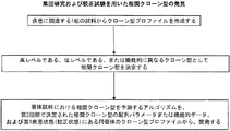


Classifications
-
- C—CHEMISTRY; METALLURGY
- C12—BIOCHEMISTRY; BEER; SPIRITS; WINE; VINEGAR; MICROBIOLOGY; ENZYMOLOGY; MUTATION OR GENETIC ENGINEERING
- C12N—MICROORGANISMS OR ENZYMES; COMPOSITIONS THEREOF; PROPAGATING, PRESERVING, OR MAINTAINING MICROORGANISMS; MUTATION OR GENETIC ENGINEERING; CULTURE MEDIA
- C12N15/00—Mutation or genetic engineering; DNA or RNA concerning genetic engineering, vectors, e.g. plasmids, or their isolation, preparation or purification; Use of hosts therefor
- C12N15/09—Recombinant DNA-technology
- C12N15/10—Processes for the isolation, preparation or purification of DNA or RNA
- C12N15/1034—Isolating an individual clone by screening libraries
- C12N15/1072—Differential gene expression library synthesis, e.g. subtracted libraries, differential screening
-
- C—CHEMISTRY; METALLURGY
- C12—BIOCHEMISTRY; BEER; SPIRITS; WINE; VINEGAR; MICROBIOLOGY; ENZYMOLOGY; MUTATION OR GENETIC ENGINEERING
- C12N—MICROORGANISMS OR ENZYMES; COMPOSITIONS THEREOF; PROPAGATING, PRESERVING, OR MAINTAINING MICROORGANISMS; MUTATION OR GENETIC ENGINEERING; CULTURE MEDIA
- C12N15/00—Mutation or genetic engineering; DNA or RNA concerning genetic engineering, vectors, e.g. plasmids, or their isolation, preparation or purification; Use of hosts therefor
- C12N15/09—Recombinant DNA-technology
- C12N15/11—DNA or RNA fragments; Modified forms thereof; Non-coding nucleic acids having a biological activity
-
- C—CHEMISTRY; METALLURGY
- C12—BIOCHEMISTRY; BEER; SPIRITS; WINE; VINEGAR; MICROBIOLOGY; ENZYMOLOGY; MUTATION OR GENETIC ENGINEERING
- C12Q—MEASURING OR TESTING PROCESSES INVOLVING ENZYMES, NUCLEIC ACIDS OR MICROORGANISMS; COMPOSITIONS OR TEST PAPERS THEREFOR; PROCESSES OF PREPARING SUCH COMPOSITIONS; CONDITION-RESPONSIVE CONTROL IN MICROBIOLOGICAL OR ENZYMOLOGICAL PROCESSES
- C12Q1/00—Measuring or testing processes involving enzymes, nucleic acids or microorganisms; Compositions therefor; Processes of preparing such compositions
- C12Q1/68—Measuring or testing processes involving enzymes, nucleic acids or microorganisms; Compositions therefor; Processes of preparing such compositions involving nucleic acids
- C12Q1/6809—Methods for determination or identification of nucleic acids involving differential detection
-
- C—CHEMISTRY; METALLURGY
- C12—BIOCHEMISTRY; BEER; SPIRITS; WINE; VINEGAR; MICROBIOLOGY; ENZYMOLOGY; MUTATION OR GENETIC ENGINEERING
- C12Q—MEASURING OR TESTING PROCESSES INVOLVING ENZYMES, NUCLEIC ACIDS OR MICROORGANISMS; COMPOSITIONS OR TEST PAPERS THEREFOR; PROCESSES OF PREPARING SUCH COMPOSITIONS; CONDITION-RESPONSIVE CONTROL IN MICROBIOLOGICAL OR ENZYMOLOGICAL PROCESSES
- C12Q1/00—Measuring or testing processes involving enzymes, nucleic acids or microorganisms; Compositions therefor; Processes of preparing such compositions
- C12Q1/68—Measuring or testing processes involving enzymes, nucleic acids or microorganisms; Compositions therefor; Processes of preparing such compositions involving nucleic acids
- C12Q1/6813—Hybridisation assays
- C12Q1/6827—Hybridisation assays for detection of mutation or polymorphism
-
- C—CHEMISTRY; METALLURGY
- C12—BIOCHEMISTRY; BEER; SPIRITS; WINE; VINEGAR; MICROBIOLOGY; ENZYMOLOGY; MUTATION OR GENETIC ENGINEERING
- C12Q—MEASURING OR TESTING PROCESSES INVOLVING ENZYMES, NUCLEIC ACIDS OR MICROORGANISMS; COMPOSITIONS OR TEST PAPERS THEREFOR; PROCESSES OF PREPARING SUCH COMPOSITIONS; CONDITION-RESPONSIVE CONTROL IN MICROBIOLOGICAL OR ENZYMOLOGICAL PROCESSES
- C12Q1/00—Measuring or testing processes involving enzymes, nucleic acids or microorganisms; Compositions therefor; Processes of preparing such compositions
- C12Q1/68—Measuring or testing processes involving enzymes, nucleic acids or microorganisms; Compositions therefor; Processes of preparing such compositions involving nucleic acids
- C12Q1/6869—Methods for sequencing
-
- C—CHEMISTRY; METALLURGY
- C12—BIOCHEMISTRY; BEER; SPIRITS; WINE; VINEGAR; MICROBIOLOGY; ENZYMOLOGY; MUTATION OR GENETIC ENGINEERING
- C12Q—MEASURING OR TESTING PROCESSES INVOLVING ENZYMES, NUCLEIC ACIDS OR MICROORGANISMS; COMPOSITIONS OR TEST PAPERS THEREFOR; PROCESSES OF PREPARING SUCH COMPOSITIONS; CONDITION-RESPONSIVE CONTROL IN MICROBIOLOGICAL OR ENZYMOLOGICAL PROCESSES
- C12Q1/00—Measuring or testing processes involving enzymes, nucleic acids or microorganisms; Compositions therefor; Processes of preparing such compositions
- C12Q1/68—Measuring or testing processes involving enzymes, nucleic acids or microorganisms; Compositions therefor; Processes of preparing such compositions involving nucleic acids
- C12Q1/6876—Nucleic acid products used in the analysis of nucleic acids, e.g. primers or probes
- C12Q1/6881—Nucleic acid products used in the analysis of nucleic acids, e.g. primers or probes for tissue or cell typing, e.g. human leukocyte antigen [HLA] probes
-
- C—CHEMISTRY; METALLURGY
- C12—BIOCHEMISTRY; BEER; SPIRITS; WINE; VINEGAR; MICROBIOLOGY; ENZYMOLOGY; MUTATION OR GENETIC ENGINEERING
- C12Q—MEASURING OR TESTING PROCESSES INVOLVING ENZYMES, NUCLEIC ACIDS OR MICROORGANISMS; COMPOSITIONS OR TEST PAPERS THEREFOR; PROCESSES OF PREPARING SUCH COMPOSITIONS; CONDITION-RESPONSIVE CONTROL IN MICROBIOLOGICAL OR ENZYMOLOGICAL PROCESSES
- C12Q1/00—Measuring or testing processes involving enzymes, nucleic acids or microorganisms; Compositions therefor; Processes of preparing such compositions
- C12Q1/68—Measuring or testing processes involving enzymes, nucleic acids or microorganisms; Compositions therefor; Processes of preparing such compositions involving nucleic acids
- C12Q1/6876—Nucleic acid products used in the analysis of nucleic acids, e.g. primers or probes
- C12Q1/6883—Nucleic acid products used in the analysis of nucleic acids, e.g. primers or probes for diseases caused by alterations of genetic material
- C12Q1/6886—Nucleic acid products used in the analysis of nucleic acids, e.g. primers or probes for diseases caused by alterations of genetic material for cancer
-
- C—CHEMISTRY; METALLURGY
- C12—BIOCHEMISTRY; BEER; SPIRITS; WINE; VINEGAR; MICROBIOLOGY; ENZYMOLOGY; MUTATION OR GENETIC ENGINEERING
- C12Q—MEASURING OR TESTING PROCESSES INVOLVING ENZYMES, NUCLEIC ACIDS OR MICROORGANISMS; COMPOSITIONS OR TEST PAPERS THEREFOR; PROCESSES OF PREPARING SUCH COMPOSITIONS; CONDITION-RESPONSIVE CONTROL IN MICROBIOLOGICAL OR ENZYMOLOGICAL PROCESSES
- C12Q2600/00—Oligonucleotides characterized by their use
- C12Q2600/106—Pharmacogenomics, i.e. genetic variability in individual responses to drugs and drug metabolism
-
- C—CHEMISTRY; METALLURGY
- C12—BIOCHEMISTRY; BEER; SPIRITS; WINE; VINEGAR; MICROBIOLOGY; ENZYMOLOGY; MUTATION OR GENETIC ENGINEERING
- C12Q—MEASURING OR TESTING PROCESSES INVOLVING ENZYMES, NUCLEIC ACIDS OR MICROORGANISMS; COMPOSITIONS OR TEST PAPERS THEREFOR; PROCESSES OF PREPARING SUCH COMPOSITIONS; CONDITION-RESPONSIVE CONTROL IN MICROBIOLOGICAL OR ENZYMOLOGICAL PROCESSES
- C12Q2600/00—Oligonucleotides characterized by their use
- C12Q2600/118—Prognosis of disease development
-
- C—CHEMISTRY; METALLURGY
- C12—BIOCHEMISTRY; BEER; SPIRITS; WINE; VINEGAR; MICROBIOLOGY; ENZYMOLOGY; MUTATION OR GENETIC ENGINEERING
- C12Q—MEASURING OR TESTING PROCESSES INVOLVING ENZYMES, NUCLEIC ACIDS OR MICROORGANISMS; COMPOSITIONS OR TEST PAPERS THEREFOR; PROCESSES OF PREPARING SUCH COMPOSITIONS; CONDITION-RESPONSIVE CONTROL IN MICROBIOLOGICAL OR ENZYMOLOGICAL PROCESSES
- C12Q2600/00—Oligonucleotides characterized by their use
- C12Q2600/156—Polymorphic or mutational markers
-
- C—CHEMISTRY; METALLURGY
- C12—BIOCHEMISTRY; BEER; SPIRITS; WINE; VINEGAR; MICROBIOLOGY; ENZYMOLOGY; MUTATION OR GENETIC ENGINEERING
- C12Q—MEASURING OR TESTING PROCESSES INVOLVING ENZYMES, NUCLEIC ACIDS OR MICROORGANISMS; COMPOSITIONS OR TEST PAPERS THEREFOR; PROCESSES OF PREPARING SUCH COMPOSITIONS; CONDITION-RESPONSIVE CONTROL IN MICROBIOLOGICAL OR ENZYMOLOGICAL PROCESSES
- C12Q2600/00—Oligonucleotides characterized by their use
- C12Q2600/158—Expression markers
-
- C—CHEMISTRY; METALLURGY
- C12—BIOCHEMISTRY; BEER; SPIRITS; WINE; VINEGAR; MICROBIOLOGY; ENZYMOLOGY; MUTATION OR GENETIC ENGINEERING
- C12Q—MEASURING OR TESTING PROCESSES INVOLVING ENZYMES, NUCLEIC ACIDS OR MICROORGANISMS; COMPOSITIONS OR TEST PAPERS THEREFOR; PROCESSES OF PREPARING SUCH COMPOSITIONS; CONDITION-RESPONSIVE CONTROL IN MICROBIOLOGICAL OR ENZYMOLOGICAL PROCESSES
- C12Q2600/00—Oligonucleotides characterized by their use
- C12Q2600/16—Primer sets for multiplex assays
-
- Y—GENERAL TAGGING OF NEW TECHNOLOGICAL DEVELOPMENTS; GENERAL TAGGING OF CROSS-SECTIONAL TECHNOLOGIES SPANNING OVER SEVERAL SECTIONS OF THE IPC; TECHNICAL SUBJECTS COVERED BY FORMER USPC CROSS-REFERENCE ART COLLECTIONS [XRACs] AND DIGESTS
- Y02—TECHNOLOGIES OR APPLICATIONS FOR MITIGATION OR ADAPTATION AGAINST CLIMATE CHANGE
- Y02A—TECHNOLOGIES FOR ADAPTATION TO CLIMATE CHANGE
- Y02A90/00—Technologies having an indirect contribution to adaptation to climate change
- Y02A90/10—Information and communication technologies [ICT] supporting adaptation to climate change, e.g. for weather forecasting or climate simulation
Description
相互参照
本出願は2008年11月7日に出願した米国特許出願第61/112,693号の恩典を主張し、これは全体として参照により本明細書に組み入れられる。
This application claims the benefit of US Patent Application No. 61 / 112,693, filed Nov. 7, 2008, which is hereby incorporated by reference in its entirety.
発明の背景
免疫系は、先天性免疫系および適応免疫系を含む。先天性免疫系は、外来病原体を認識するために一般的方法を使用する細胞および機構を含む。先天性免疫に関与する細胞には、好中球、ナチュラルキラー細胞、マクロファージ、単球、好塩基球、好酸球、肥満細胞、および樹状細胞が含まれる。これらの細胞は、食作用、および侵入病原体を殺傷する多くの化学物質の放出を行う。加えて、これらの細胞は、補体カスケードおよび炎症を含む先天性免疫防御機構に関与する。最終的に、これらの細胞の一部は、適応免疫系において役割を果たす抗原提示過程に関与する。
BACKGROUND OF THE INVENTION The immune system includes the innate and adaptive immune systems. The innate immune system includes cells and mechanisms that use common methods to recognize foreign pathogens. Cells involved in innate immunity include neutrophils, natural killer cells, macrophages, monocytes, basophils, eosinophils, mast cells, and dendritic cells. These cells phagocytose and release many chemicals that kill invading pathogens. In addition, these cells are involved in innate immune defense mechanisms, including the complement cascade and inflammation. Ultimately, some of these cells are involved in the antigen presentation process that plays a role in the adaptive immune system.
適応免疫系は、それらの標的上の特異的特徴を攻撃するように進化してきた。特定の標的に対して1つの反応が生じると、宿主にその「記憶」が提供されて、次にそれが出現した場合に、宿主はより強力な反応を開始する。通常、いかなるタンパク質または多糖も、標的細胞上の特異的エピトープを認識する適応免疫応答細胞のいくつかのサブセットまたはそれらの産物の標的となり得る。適応免疫応答は、体液性免疫応答および細胞性免疫応答の2つの型に分類され、B細胞およびT細胞がそれぞれこれらの応答において特異的役割を果たす。 The adaptive immune system has evolved to attack specific features on their targets. When one reaction occurs for a particular target, the host is given that “memory” and the next time it appears, the host initiates a stronger reaction. In general, any protein or polysaccharide can be the target of several subsets of adaptive immune response cells or their products that recognize specific epitopes on the target cells. Adaptive immune responses fall into two types, humoral immune responses and cellular immune responses, with B cells and T cells each playing a specific role in these responses.
自己免疫疾患は、自己標的に対する適応免疫系のいくつかの要素の認識を含むため、診断および予後診断を支援するために適応免疫系の局面が調べられてきた。標準的な免疫技法を用いて、循環自己抗体を探すことにより体液性免疫系が調べられてきた。いくつかの疾患に関して、抗核、抗dsDNA、およびリウマチ因子のような自己抗体が同定された。これらの抗体はそれ自体病原性がないと考えられ、またそれらが体内で認識する標的はインビトロで試験される標的と必ずしも同じではないが、それらのレベルを測定することは診断に役立ち、場合によっては何らかの予後診断および治療の意義を有する。 Because autoimmune diseases involve the recognition of several elements of the adaptive immune system against self targets, aspects of the adaptive immune system have been investigated to support diagnosis and prognosis. The humoral immune system has been examined by searching for circulating autoantibodies using standard immunization techniques. For several diseases, autoantibodies such as antinuclear, anti-dsDNA, and rheumatoid factors have been identified. These antibodies are not considered pathogenic per se, and the targets they recognize in the body are not necessarily the same as the targets tested in vitro, but measuring their levels is useful for diagnosis and in some cases Has some prognostic and therapeutic significance.
自己免疫疾患における適応免疫系を研究するための別の方法論は、適応免疫細胞の多様性の解析に基づいている。適応免疫細胞が活性化されると、それらのクローン増殖が生じる。このクローン増殖の証拠は通常、血液RNAまたはDNAからの、抗原認識領域をコードする核酸配列の部分の増幅によって達成される。例えば、T細胞受容体のβ鎖(抗体重鎖と類似している)の特定のVセグメントを有する配列を増幅するためのPCRプライマーを用いて、特定のVセグメントに結合しているJセグメントまたはJおよびDセグメントを増幅する。多様な細胞集団が存在する場合には、わずかにサイズの異なる単位複製配列の分布を有する断片が増幅されると予測されるが、クローン増殖では特定のサイズが濃縮され、よってゲル上でバンドのようにより強く可視化される。スペクトラタイピングと称される技法では、VセグメントのそれぞれをJおよびDセグメントと共に増幅して、これらの単位複製配列のいずれかがクローン増殖を示すかどうかを評価する。 Another methodology for studying the adaptive immune system in autoimmune diseases is based on an analysis of adaptive immune cell diversity. When adaptive immune cells are activated, their clonal expansion occurs. This evidence of clonal growth is usually achieved by amplification of a portion of the nucleic acid sequence encoding the antigen recognition region from blood RNA or DNA. For example, using a PCR primer to amplify a sequence with a specific V segment of the T cell receptor β chain (similar to an antibody heavy chain), a J segment bound to a specific V segment or Amplify J and D segments. In the presence of diverse cell populations, fragments with a slightly different size of amplicon distribution are expected to be amplified, but clonal expansion enriches for specific sizes and thus bands on the gel. So that it is visualized more strongly. In a technique referred to as spectrotyping, each of the V segments is amplified with J and D segments to assess whether any of these amplicons exhibit clonal expansion.
スペクトラタイピングアプローチの1つの問題は、多くの異なる配列が同じ長さを有し得、そのため識別できないということである。したがって、劇的なクローン増殖のみがこの技法によって識別され得る。自己免疫疾患および自己免疫疾患状態、ならびに免疫系が中心的役割を果たすその他の疾患を診断する、およびそれらの予後診断を支援する方法を改善する必要がある。 One problem with the spectrotyping approach is that many different sequences can have the same length and therefore cannot be distinguished. Thus, only dramatic clonal expansion can be distinguished by this technique. There is a need to improve autoimmune diseases and autoimmune disease states, as well as other diseases in which the immune system plays a central role, and how to support their prognosis.
免疫系をプロファイルする上でのさらなる特異性は、ヒトの健康に及ぼすその影響をよりよく予測できるようにするのに大変有用であるものの、疾患過程に関与する特異的T細胞およびB細胞の特定の配列が以前に観察されていない場合でさえ、それらの同定を可能にする方法が開発されたならば、より優れた有用性がもたらされる。免疫系の膨大な多様性は、潜在的に有用な細胞の莫大な蓄えを免疫系に提供するばかりでなく、予測目的にこのレパートリーを使用しようと試みる課題を研究者に提示する。ある抗原を標的とする任意の単一の配列は、所与の個体において疾患過程に関与し得るおよび/または相関し得る膨大な数のうちの1つである。所与の個体における多くの細胞のいずれが疾患過程に関与しているのかを同定する方法は、ヒトの健康にとって非常に価値がある。 Although more specific in profiling the immune system is very useful in making it possible to better predict its impact on human health, the identification of specific T and B cells involved in the disease process Even if the sequences have not been observed previously, if a method was developed that would allow their identification, greater utility would result. The vast diversity of the immune system not only provides the immune system with a vast reserve of potentially useful cells, but also presents researchers with the challenge of trying to use this repertoire for predictive purposes. Any single sequence that targets an antigen is one of a vast number that can be involved and / or correlated in a disease process in a given individual. Methods to identify which of the many cells in a given individual are involved in the disease process are of great value for human health.
1つの局面において、T細胞および/またはB細胞における組換えDNA配列のプロファイルを決定するための方法であって、T細胞および/またはB細胞を含む対象由来の試料を採取する段階、該細胞からゲノムDNAの個々の分子を空間的に単離する段階、ゲノムDNAの空間的に単離された個々の該分子を配列決定する段階、ならびに該試料からの異なる配列についてそれらのレベルを測定して、組換えDNA配列のプロファイルを作成する段階を含む前記方法を提供する。 In one aspect, a method for determining a profile of a recombinant DNA sequence in T cells and / or B cells, wherein a sample from a subject comprising T cells and / or B cells is collected, from the cells Spatially isolating individual molecules of genomic DNA, sequencing the spatially isolated individual molecules of genomic DNA, and measuring their levels for different sequences from the sample Providing a method of generating a profile of a recombinant DNA sequence.
別の局面において、T細胞および/またはB細胞における組換えDNA配列のプロファイルを決定するための方法であって、T細胞および/またはB細胞を含む対象由来の試料を採取する段階、該細胞からゲノムDNAの個々の分子を空間的に単離する段階、ゲノムDNAの個々の該分子を増幅する段階、該増幅DNAを配列決定する段階、ならびに該試料からの異なる配列についてそれらのレベルを測定して、組換えDNA配列のプロファイルを作成する段階を含む前記方法を提供する。 In another aspect, a method for determining a profile of a recombinant DNA sequence in T cells and / or B cells, wherein a sample from a subject comprising T cells and / or B cells is collected, from the cells Spatially isolating individual molecules of genomic DNA, amplifying individual molecules of genomic DNA, sequencing the amplified DNA, and measuring their levels for different sequences from the sample And providing the method comprising the step of creating a profile of the recombinant DNA sequence.
別の局面において、T細胞および/またはB細胞における組換えDNA配列のプロファイルを決定するための方法であって、T細胞および/またはB細胞を含む対象由来の試料を採取する段階、該細胞からゲノムDNAを増幅する段階、該増幅DNAの個々の分子を空間的に単離する段階、増幅DNAの空間的に単離された個々の該分子を配列決定する段階、ならびに該試料からの異なる配列についてそれらのレベルを測定して、組換えDNA配列のプロファイルを作成する段階を含む前記方法を提供する。 In another aspect, a method for determining a profile of a recombinant DNA sequence in T cells and / or B cells, wherein a sample from a subject comprising T cells and / or B cells is collected, from the cells Amplifying genomic DNA; spatially isolating individual molecules of the amplified DNA; sequencing the spatially isolated individual molecules of amplified DNA; and different sequences from the sample The method is provided comprising the step of measuring their levels for generating a profile of the recombinant DNA sequence.
別の局面において、T細胞および/またはB細胞における組換えDNA配列のプロファイルを決定するための方法であって、T細胞および/またはB細胞を含む対象由来の試料を採取する段階、該細胞からゲノムDNAを増幅する段階、該増幅DNAの個々の分子を空間的に単離する段階、該増幅DNA分子を再増幅する段階、該再増幅DNA分子を配列決定する段階、ならびに該試料からの異なる配列についてそれらのレベルを測定して、組換えDNA配列のプロファイルを作成する段階を含む前記方法を提供する。 In another aspect, a method for determining a profile of a recombinant DNA sequence in T cells and / or B cells, wherein a sample from a subject comprising T cells and / or B cells is collected, from the cells Amplifying genomic DNA; spatially isolating individual molecules of the amplified DNA; re-amplifying the amplified DNA molecule; sequencing the re-amplified DNA molecule; and differing from the sample Measuring the levels of the sequences and providing a profile of the recombinant DNA sequence is provided.
別の局面において、T細胞および/またはB細胞における組換えDNAの配列のプロファイルを決定するための方法であって、T細胞および/またはB細胞を含む対象由来の試料を採取する段階、該細胞からRNAを逆転写してcDNAを形成する段階、該cDNAの個々の分子を空間的に単離する段階、任意で、cDNAの空間的に単離された個々の該分子を再増幅する段階、該cDNAおよび/または再増幅cDNAを配列決定する段階、ならびに該試料からの異なる配列についてそれらのレベルを測定して、組換えDNA配列のプロファイルを作成する段階を含む前記方法を提供する。 In another aspect, a method for determining a sequence profile of recombinant DNA in T cells and / or B cells, wherein a sample from a subject comprising T cells and / or B cells is collected, Reverse-trancribing RNA from to form cDNA, spatially isolating individual molecules of the cDNA, and optionally re-amplifying the individual spatially isolated molecules of cDNA; The method is provided comprising sequencing cDNA and / or reamplified cDNA, and measuring the levels of different sequences from the sample to create a profile of the recombinant DNA sequence.
別の局面において、T細胞および/またはB細胞における組換えDNA配列のプロファイルを決定するための方法であって、T細胞および/またはB細胞を含む対象由来の試料を採取する段階、該試料中の個々の細胞を空間的に単離する段階、該細胞から核酸の個々の分子を配列決定する段階、ならびに該試料からの異なる配列についてそれらのレベルを測定して、組換えDNA配列のプロファイルを作成する段階を含む前記方法を提供する。 In another aspect, a method for determining a profile of a recombinant DNA sequence in T cells and / or B cells, wherein a sample from a subject comprising T cells and / or B cells is collected, in the sample Isolating individual cells of the cells spatially, sequencing individual molecules of nucleic acid from the cells, and measuring their levels for different sequences from the sample to determine the profile of the recombinant DNA sequence The method is provided including the step of creating.
1つの態様において、前記の増幅および/または再増幅段階は、PCR、多重PCR、TMA、NASBA、またはLAMPを含む。別の態様において、前記の空間的に単離する段階は、細菌を形質転換するために用いられるベクターに前記DNAもしくはcDNAをサブクローニングすること、固体支持体上で前記DNAもしくはcDNAを二次元に分離すること、ミセルを有する溶液中で前記DNAもしくはcDNAを三次元に分離すること、または微小反応チャンバーを用いて分子を分離することを含む。別の態様において、前記の増幅および/または再増幅段階は、サブクローニングしたDNAもしくはcDNAを保有する細菌の増殖、スライド上でのDNAもしくはcDNAの増幅、またはビーズ上でのDNAもしくはcDNAの増幅による。 In one embodiment, the amplification and / or reamplification step comprises PCR, multiplex PCR, TMA, NASBA, or LAMP. In another embodiment, the spatially isolating step comprises subcloning the DNA or cDNA into a vector used to transform bacteria, separating the DNA or cDNA in two dimensions on a solid support. Separating the DNA or cDNA three-dimensionally in a solution having micelles, or separating the molecules using a micro reaction chamber. In another embodiment, the amplification and / or reamplification step is by growth of bacteria carrying subcloned DNA or cDNA, amplification of DNA or cDNA on a slide, or amplification of DNA or cDNA on beads.
別の態様において、前記の配列決定段階はジデオキシ配列決定を含む。別の態様において、前記の配列決定段階は、可逆的終結標識ヌクレオチドを用いた合成による配列決定を含む。別の態様において、前記の配列決定段階は、ヌクレオチド取り込み時のピロリン酸放出の検出を含む。別の態様において、前記の配列決定段階は、標識オリゴヌクレオチドプローブのライブラリーへのアレル特異的ハイブリダイゼーションを含む。別の態様において、前記の配列決定段階は、標識オリゴヌクレオチドプローブのライブラリーへのアレル特異的ハイブリダイゼーションと、それに続く該プローブの連結とを用いた合成による配列決定を含む。別の態様において、前記の配列決定段階は、重合工程中の標識ヌクレオチドの取り込みのリアルタイムモニタリングを含む。 In another embodiment, the sequencing step comprises dideoxy sequencing. In another embodiment, the sequencing step comprises synthetic sequencing using reversible termination labeled nucleotides. In another embodiment, the sequencing step comprises detection of pyrophosphate release upon nucleotide incorporation. In another embodiment, the sequencing step comprises allele specific hybridization to a library of labeled oligonucleotide probes. In another embodiment, the sequencing step comprises synthetic sequencing using allele specific hybridization of a labeled oligonucleotide probe to a library followed by ligation of the probe. In another embodiment, the sequencing step includes real-time monitoring of the incorporation of labeled nucleotides during the polymerization process.
別の態様において、前記の組換えDNA配列は、T細胞受容体遺伝子および/または免疫グロブリン遺伝子を含む。別の態様において、前記の配列決定段階は、免疫グロブリン遺伝子および/またはT細胞受容体遺伝子の完全クローン配列のサブセットを配列決定することを含む。 In another embodiment, the recombinant DNA sequence comprises a T cell receptor gene and / or an immunoglobulin gene. In another embodiment, the sequencing step comprises sequencing a subset of the complete clonal sequence of immunoglobulin genes and / or T cell receptor genes.
別の態様において、完全クローン配列の前記サブセットは、免疫グロブリン遺伝子もしくはT細胞受容体遺伝子のV-D結合部、D-J結合部、免疫グロブリン遺伝子もしくはT細胞受容体遺伝子の完全な可変領域、抗原認識領域、または相補性決定領域3(CDR3)を含む。別の態様において、前記T細胞受容体遺伝子はT細胞受容体β遺伝子を含む。別の態様において、前記免疫グロブリン遺伝子は免疫グロブリン重鎖遺伝子を含む。別の態様において、前記の増幅または再増幅段階は、Vセグメントと相補的な複数のプライマー、およびCセグメントと相補的な1つのプライマーを含む。別の態様において、前記の増幅または再増幅段階は、Vセグメントと相補的な複数のプライマー、およびCセグメントと相補的な複数のプライマーを含む。 In another embodiment, the subset of complete clonal sequences comprises an immunoglobulin gene or a T cell receptor gene VD binding portion, a DJ binding portion, an immunoglobulin gene or T cell receptor gene complete variable region, an antigen recognition region, Alternatively, it includes complementarity determining region 3 (CDR3). In another embodiment, the T cell receptor gene comprises a T cell receptor β gene. In another embodiment, the immunoglobulin gene comprises an immunoglobulin heavy chain gene. In another embodiment, the amplification or reamplification step comprises a plurality of primers complementary to the V segment and a primer complementary to the C segment. In another embodiment, the amplification or reamplification step comprises a plurality of primers complementary to the V segment and a plurality of primers complementary to the C segment.
別の態様において、Vセグメントと相補的な前記の複数のプライマーは、各Vセグメントに対する少なくとも3種の異なるプライマーを含み、Cセグメントと相補的な複数のプライマーは、少なくとも1種、少なくとも2種、少なくとも3種、少なくとも4種、少なくとも5種、または少なくとも6種のプライマーを含む。 In another embodiment, the plurality of primers complementary to the V segment comprises at least three different primers for each V segment, and the plurality of primers complementary to the C segment is at least one, at least two, It includes at least 3, at least 4, at least 5, or at least 6 primers.
別の態様において、前記T細胞またはB細胞は、全T細胞およびB細胞のサブセットである。別の態様において、T細胞の該サブセットは、CD4+、CD8+細胞、またはCD27高細胞である。別の態様において、前記試料は、少なくとも100,000個、少なくとも500,000個、少なくとも750,000個、または少なくとも1,000,000個のT細胞を含む。 In another embodiment, the T cell or B cell is a subset of total T cells and B cells. In another embodiment, the subset of T cells are CD4 +, CD8 + cells, or CD27 high cells. In another embodiment, the sample comprises at least 100,000, at least 500,000, at least 750,000, or at least 1,000,000 T cells.
別の態様において、前記の配列決定段階は、1回の実行当たり少なくとも1000件の読み取り、1回の実行当たり少なくとも10,000件の読み取り、1回の実行当たり少なくとも100,000件の読み取り、または1回の実行当たり少なくとも1,000,000件の読み取りを含む。別の態様において、前記の配列決定段階は、1回の読み取り当たり約30 bp、約40 bp、約50 bp、約60 bp、約70 bp、約80 bp、約90 bp、約100 bp、約110 bp、または約120 bpを作成することを含む。 In another embodiment, the sequencing step comprises at least 1000 reads per run, at least 10,000 reads per run, at least 100,000 reads per run, or a single run Includes at least 1,000,000 readings per. In another embodiment, the sequencing step comprises about 30 bp, about 40 bp, about 50 bp, about 60 bp, about 70 bp, about 80 bp, about 90 bp, about 100 bp, about 100 bp per reading. Including creating 110 bp, or about 120 bp.
別の態様では、対象が自己免疫疾患の再燃(flare)状態にある時点で、前記試料を採取する。別の態様では、全身性エリテマトーデスを有するかまたはそれを有する疑いのある対象から前記試料を採取する。 In another embodiment, the sample is taken when the subject is in a flare state of an autoimmune disease. In another embodiment, the sample is taken from a subject with or suspected of having systemic lupus erythematosus.
別の局面では、対象における1つまたは複数の相関クローン型を決定するための方法であって、疾患の第1状態に関連する少なくとも1つの対象由来の試料から空間的に単離された個々の分子を核酸配列決定することにより、1つまたは複数のクローン型プロファイルを作成する段階、およびその1つまたは複数のクローン型プロファイルに基づいて、対象における1つまたは複数の相関クローン型を決定する段階を含む前記方法を提供する。 In another aspect, a method for determining one or more correlated clonal types in a subject, each of which is spatially isolated from a sample from at least one subject associated with a first condition of disease. Creating one or more clonal profiles by nucleic acid sequencing the molecule, and determining one or more correlated clonal forms in the subject based on the one or more clonal profiles A method comprising the steps of:
1つの態様において、前記の少なくとも1つの試料は、疾患に罹患した組織に由来する。別の態様において、前記の1つまたは複数の相関クローン型の決定は、少なくとも2つの試料からのクローン型プロファイルを比較する段階を含む。 In one embodiment, the at least one sample is derived from a diseased tissue. In another embodiment, said determining one or more correlated clonal types comprises comparing clonal profile from at least two samples.
別の態様において、疾患の第1状態は疾患のピーク状態である。別の態様において、前記の1つまたは複数の相関クローン型は、疾患のピーク状態において存在する。別の態様において、前記の1つまたは複数の相関クローン型は、疾患のピーク状態において存在しない。別の態様において、前記の1つまたは複数の相関クローン型は、疾患のピーク状態において高い。別の態様において、前記の1つまたは複数の相関クローン型は、疾患のピーク状態において低い。 In another embodiment, the first condition of the disease is a peak condition of the disease. In another embodiment, the one or more correlated clonal types are present in a peak state of the disease. In another embodiment, the one or more correlated clonal types are absent in peak disease states. In another embodiment, the one or more correlated clonal types are elevated at the peak state of the disease. In another embodiment, the one or more correlated clonal types are low in peak disease states.
別の態様において、前記試料はT細胞および/またはB細胞を含む。別の態様において、該T細胞および/またはB細胞は、T細胞および/またはB細胞のサブセットを含む。別の態様において、T細胞および/またはB細胞の該サブセットは、マーカーとの相互作用によって濃縮される。別の態様において、該マーカーは、T細胞および/またはB細胞のサブセット上の細胞表面マーカーである。別の態様において、T細胞および/またはB細胞の該サブセットは、疾患において特異的に存在する抗原と相互作用する。 In another embodiment, the sample comprises T cells and / or B cells. In another embodiment, the T cells and / or B cells comprise a subset of T cells and / or B cells. In another embodiment, the subset of T cells and / or B cells is enriched by interaction with a marker. In another embodiment, the marker is a cell surface marker on a subset of T cells and / or B cells. In another embodiment, the subset of T cells and / or B cells interacts with an antigen that is specifically present in the disease.
別の態様において、疾患は全身性エリテマトーデスまたは多発性硬化症である。 In another embodiment, the disease is systemic lupus erythematosus or multiple sclerosis.
別の局面において、疾患を有する対象に由来する任意の試料における1つまたは複数の相関クローン型を予測し得るアルゴリズムを開発するための方法であって、a) 疾患に関連する1組の試料から複数のクローン型プロファイルを作成する段階、b) その1組の試料から1つまたは複数の相関クローン型を同定する段階、c) b)において同定された1つまたは複数の相関クローン型からの配列パラメータおよび/または機能的データを用いて、疾患を有する対象に由来する任意の試料における相関クローン型を予測し得るアルゴリズムを開発する段階を含む前記方法を提供する。 In another aspect, a method for developing an algorithm capable of predicting one or more correlated clonal types in any sample from a subject having a disease comprising: a) from a set of samples associated with a disease Creating a plurality of clonal profiles, b) identifying one or more correlated clonal forms from the set of samples, c) sequences from one or more correlated clonal forms identified in b) The method is provided comprising the step of developing an algorithm that can predict correlated clonal types in any sample from a subject having a disease using parameters and / or functional data.
1つの態様では、疾患に罹患した1つまたは複数の組織から1組の試料を採取する。 In one embodiment, a set of samples is taken from one or more tissues affected by a disease.
別の態様において、前記の1つまたは複数の相関クローン型の同定は、少なくとも2つの試料からのクローン型プロファイルを比較する段階を含む。 In another embodiment, the identification of the one or more correlated clonal types comprises comparing clonal type profiles from at least two samples.
別の態様において、前記機能的データは、T細胞および/もしくはB細胞表面上のマーカーの結合能、またはT細胞もしくはB細胞による抗原との相互作用を含む。 In another embodiment, the functional data includes the ability of markers on the surface of T cells and / or B cells, or interaction with antigens by T cells or B cells.
別の態様において、前記配列パラメータは、核酸配列および予測されるアミノ酸配列を含む。 In another embodiment, the sequence parameter comprises a nucleic acid sequence and a predicted amino acid sequence.
別の態様において、試料は、疾患のピーク段階にある1つまたは複数の個体に由来する。別の態様において、前記の1つまたは複数の相関クローン型は、疾患のピーク状態において存在する。別の態様において、前記の1つまたは複数の相関クローン型は、疾患のピーク状態において高レベルである。別の態様において、前記の1つまたは複数の相関クローン型は、疾患のピーク状態において低レベルである。別の態様において、1つまたは複数の相関クローン型は、疾患のピーク状態において存在しない。別の態様において、疾患は全身性エリテマトーデスまたは多発性硬化症である。 In another embodiment, the sample is from one or more individuals who are at the peak stage of the disease. In another embodiment, the one or more correlated clonal types are present in a peak state of the disease. In another embodiment, the one or more correlated clonal types are at a high level in a peak state of the disease. In another embodiment, the one or more correlated clonal types are at a low level at the peak state of the disease. In another embodiment, one or more correlated clonal types are not present in the peak state of the disease. In another embodiment, the disease is systemic lupus erythematosus or multiple sclerosis.
別の態様において、個体の1つまたは複数の相関クローン型を発見するための方法であって、個体由来の試料からのクローン型プロファイルをアルゴリズムに入力する段階、およびアルゴリズムを用いて、個体の1つまたは複数の相関クローン型を決定する段階を含む前記方法を提供する。1つの態様において、アルゴリズムは、a) 疾患に関連する1組の試料から複数のクローン型プロファイルを作成する段階、b) その1組の試料から1つまたは複数の相関クローン型を同定する段階、c) b)において同定された1つまたは複数の相関クローン型からの配列パラメータおよび/または機能的データを用いて、疾患を有する対象に由来する任意の試料における相関クローン型を予測し得るアルゴリズムを開発する段階によって開発された、疾患を有する対象に由来する任意の試料における1つまたは複数の相関クローン型を予測し得るアルゴリズムである。 In another embodiment, a method for discovering one or more correlated clonal types of an individual comprising inputting a clonal profile from a sample from the individual into an algorithm, and using the algorithm, The method is provided comprising determining one or more correlated clonal types. In one embodiment, the algorithm comprises: a) creating a plurality of clonal profiles from a set of samples associated with a disease; b) identifying one or more correlated clonal types from the set of samples; c) an algorithm capable of predicting correlated clone types in any sample from a subject having disease using sequence parameters and / or functional data from one or more correlated clone types identified in b) It is an algorithm that can predict one or more correlated clonal types in any sample from a diseased subject, developed by the developing stage.
1つの態様では、疾患のピーク状態において前記試料を採取する。別の態様では、疾患に罹患した組織から試料を採取する。 In one embodiment, the sample is taken at peak disease conditions. In another embodiment, a sample is taken from tissue affected by the disease.
別の局面において、疾患活動性スコアを算出するアルゴリズムを作成するための方法であって、1組の因子を用いて相関クローン型のレベルを疾患活動性スコアに統合するアルゴリズムを開発する段階、疾患活動性スコアを、疾患状態に関する臨床データと比較する段階、および臨床データと疾患活動性スコアとの相関を最大化するために因子を最適化する段階を含む前記方法を提供する。 In another aspect, a method for creating an algorithm for calculating a disease activity score, comprising developing an algorithm that integrates correlated clonal levels into a disease activity score using a set of factors, disease The method is provided comprising: comparing an activity score with clinical data regarding a disease state; and optimizing factors to maximize the correlation between the clinical data and the disease activity score.
1つの態様において、個体の疾患状態をモニターするための方法であって、a) 個体由来の試料からのクローン型プロファイルを決定する段階、b) 1組の因子を用いて相関クローン型のレベルを疾患活動性スコアに統合するアルゴリズムを開発する段階と、疾患活動性スコアを、疾患状態に関する臨床データと比較する段階と、臨床データと疾患活動性スコアとの相関を最大化するために因子を最適化する段階とによって作成される、疾患活動性を算出するアルゴリズムに、a)からのクローン型プロファイル情報を入力する段階、ならびにc) 疾患活動性スコアを算出するアルゴリズムを用いて、個体の疾患状態を予測するスコアを作成する段階を含む前記方法を提供する。 In one embodiment, a method for monitoring an individual's disease state comprising: a) determining a clonal profile from a sample from the individual, b) using a set of factors to determine the level of correlated clonal types. Optimizing factors to maximize the correlation between clinical data and disease activity score, developing an algorithm that integrates into the disease activity score, comparing disease activity score to clinical data on disease status The step of inputting the clonal profile information from a) into the algorithm for calculating the disease activity, and c) the algorithm for calculating the disease activity score. The method includes the step of creating a score that predicts.
別の態様において、個体の疾患状態をモニターするための方法は、個体における1つまたは複数の相関クローン型を決定する段階、および1つまたは複数の相関クローン型の情報をアルゴリズムに入力する段階をさらに含む。 In another embodiment, a method for monitoring a disease state of an individual comprises determining one or more correlated clonal types in the individual and inputting information of the one or more correlated clonal types into an algorithm. In addition.
別の態様において、前記の個体における1つまたは複数の相関クローン型を決定する段階は、a) 疾患の第1状態に関連する少なくとも1つの対象由来の試料から空間的に単離された個々の分子を核酸配列決定することにより、1つまたは複数のクローン型プロファイルを作成すること、およびb) その1つまたは複数のクローン型プロファイルに基づいて、対象における1つまたは複数の相関クローン型を決定することを含む。 In another embodiment, determining one or more correlated clonal types in said individual comprises: a) individual isolated spatially from a sample from at least one subject associated with the first condition of the disease Create one or more clonal profiles by nucleic acid sequencing the molecule, and b) determine one or more correlated clonal forms in the subject based on the one or more clonal profiles Including doing.
別の態様において、前記の個体における1つまたは複数の相関クローン型を決定する段階は、a) i) 疾患に関連する1組の試料から複数のクローン型プロファイルを作成する段階、ii) その1組の試料から1つまたは複数の相関クローン型を同定する段階、iii) ii)において同定された1つまたは複数の相関クローン型からの配列パラメータおよび/または機能的データを用いて、疾患を有する対象に由来する任意の試料における相関クローン型を予測し得るアルゴリズムを開発する段階によって開発された、1つまたは複数のクローン型を予測し得るアルゴリズムに、個体由来の試料からのクローン型プロファイルを入力すること、ならびにc) 1つまたは複数のクローン型を予測し得るアルゴリズムを用いて、個体の1つまたは複数の相関クローン型を決定することを含む。
In another embodiment, determining one or more correlated clonal types in said individual comprises: a) i) generating a plurality of clonal profiles from a set of samples associated with the disease, ii)
別の態様において、疾患は全身性エリテマトーデスまたは多発性硬化症である。
[本発明1001]
以下の段階を含む、T細胞および/またはB細胞における組換えDNA配列のプロファイルを決定するための方法:
(a) T細胞および/またはB細胞を含む対象由来の試料を採取する段階;
(b) 該細胞からゲノムDNAの個々の分子を空間的に単離する段階;
(c) ゲノムDNAの空間的に単離された個々の該分子を配列決定する段階;
(d) 該試料からの異なる配列についてそれらのレベルを測定して、組換えDNA配列のプロファイルを作成する段階。
[本発明1002]
以下の段階を含む、T細胞および/またはB細胞における組換えDNA配列のプロファイルを決定するための方法:
(a) T細胞および/またはB細胞を含む対象由来の試料を採取する段階;
(b) 該細胞からゲノムDNAの個々の分子を空間的に単離する段階;
(c) ゲノムDNAの個々の該分子を増幅する段階;
(d) 該増幅DNAを配列決定する段階;ならびに
(e) 該試料からの異なる配列についてそれらのレベルを測定して、組換えDNA配列のプロファイルを作成する段階。
[本発明1003]
以下の段階を含む、T細胞および/またはB細胞における組換えDNA配列のプロファイルを決定するための方法:
(a) T細胞および/またはB細胞を含む対象由来の試料を採取する段階;
(b) 該細胞からゲノムDNAを増幅する段階;
(c) 該増幅DNAの個々の分子を空間的に単離する段階;
(d) 増幅DNAの空間的に単離された個々の該分子を配列決定する段階;ならびに
(e) 該試料からの異なる配列についてそれらのレベルを測定して、組換えDNA配列のプロファイルを作成する段階。
[本発明1004]
以下の段階を含む、T細胞および/またはB細胞における組換えDNA配列のプロファイルを決定するための方法:
(a) T細胞および/またはB細胞を含む対象由来の試料を採取する段階;
(b) 該細胞からゲノムDNAを増幅する段階;
(c) 該増幅DNAの個々の分子を空間的に単離する段階;
(d) 該増幅DNA分子を再増幅する段階;
(e) 該再増幅DNA分子を配列決定する段階;ならびに
(f) 該試料からの異なる配列についてそれらのレベルを測定して、組換えDNA配列のプロファイルを作成する段階。
[本発明1005]
以下の段階を含む、T細胞および/またはB細胞における組換えDNAの配列のプロファイルを決定するための方法:
(a) T細胞および/またはB細胞を含む対象由来の試料を採取する段階;
(b) 該細胞由来のRNAを逆転写してcDNAを形成する段階;
(c) 該cDNAの個々の分子を空間的に単離する段階;
(d) 任意で、cDNAの空間的に単離された個々の該分子を再増幅する段階;
(e) 該cDNAおよび/または再増幅cDNAを配列決定する段階;ならびに
(f) 該試料からの異なる配列についてそれらのレベルを測定して、組換えDNA配列のプロファイルを作成する段階。
[本発明1006]
以下の段階を含む、T細胞および/またはB細胞における組換えDNA配列のプロファイルを決定するための方法:
(a) T細胞および/またはB細胞を含む対象由来の試料を採取する段階;
(b) 該試料中の個々の細胞を空間的に単離する段階;
(c) 該細胞由来の核酸の個々の分子を配列決定する段階;ならびに
(d) 該試料からの異なる配列についてそれらのレベルを測定して、組換えDNA配列のプロファイルを作成する段階。
[本発明1007]
増幅および/または再増幅段階が、PCR、多重PCR、TMA、NASBA、またはLAMPを含む、本発明1001〜1005のいずれかの方法。
[本発明1008]
空間的に単離する段階が、細菌を形質転換するために用いられるベクターに前記DNAもしくはcDNAをサブクローニングすること、固体支持体上で前記DNAもしくはcDNAを二次元に分離すること、ミセルを有する溶液中で前記DNAもしくはcDNAを三次元に分離すること、または微小反応チャンバーを用いて分子を分離することを含む、本発明1001〜1005のいずれかの方法。
[本発明1009]
増幅および/または再増幅段階が、サブクローニングしたDNAもしくはcDNAを保有する細菌の増殖、スライド上でのDNAもしくはcDNAの増幅、またはビーズ上でのDNAもしくはcDNAの増幅によるものである、本発明1001〜1005のいずれかの方法。
[本発明1010]
配列決定段階がジデオキシ配列決定を含む、本発明1001〜1006のいずれかの方法。
[本発明1011]
配列決定段階が、可逆的終結標識ヌクレオチドを用いた合成による配列決定を含む、本発明1001〜1006のいずれかの方法。
[本発明1012]
配列決定段階が、ヌクレオチド取り込み時のピロリン酸放出の検出を含む、本発明1001〜1006のいずれかの方法。
[本発明1013]
配列決定段階が、標識オリゴヌクレオチドプローブのライブラリーへのアレル特異的ハイブリダイゼーションを含む、本発明1001〜1006のいずれかの方法。
[本発明1014]
配列決定段階が、標識オリゴヌクレオチドプローブのライブラリーへのアレル特異的ハイブリダイゼーションとそれに続く該プローブの連結とを用いた合成による配列決定を含む、本発明1001〜1006のいずれかの方法。
[本発明1015]
配列決定段階が、重合工程中の標識ヌクレオチドの取り込みのリアルタイムモニタリングを含む、本発明1001〜1006のいずれかの方法。
[本発明1016]
組換えDNA配列がT細胞受容体遺伝子および/または免疫グロブリン遺伝子を含む、本発明1001〜1006のいずれかの方法。
[本発明1017]
配列決定段階が、免疫グロブリン遺伝子および/またはT細胞受容体遺伝子の完全クローン配列のサブセットを配列決定することを含む、本発明1001〜1006のいずれかの方法。
[本発明1018]
完全クローン配列のサブセットが、免疫グロブリン遺伝子もしくはT細胞受容体遺伝子のV-D結合部、D-J結合部、免疫グロブリン遺伝子もしくはT細胞受容体遺伝子の完全な可変領域、抗原認識領域、または相補性決定領域3(CDR3)を含む、本発明1017の方法。
[本発明1019]
T細胞受容体遺伝子がT細胞受容体β遺伝子を含む、本発明1017の方法。
[本発明1020]
免疫グロブリン遺伝子が免疫グロブリン重鎖遺伝子を含む、本発明1017の方法。
[本発明1021]
増幅または再増幅段階が、Vセグメントと相補的な複数のプライマー、およびCセグメントと相補的な1つのプライマーを含む、本発明1001〜1006のいずれかの方法。
[本発明1022]
増幅または再増幅段階が、Vセグメントと相補的な複数のプライマー、およびCセグメントと相補的な複数のプライマーを含む、本発明1001〜1006のいずれかの方法。
[本発明1023]
Vセグメントと相補的な複数のプライマーが、各Vセグメントに対する少なくとも3種の異なるプライマーを含み、Cセグメントと相補的な複数のプライマーが、少なくとも1種、少なくとも2種、少なくとも3種、少なくとも4種、少なくとも5種、または少なくとも6種のプライマーを含む、本発明1022の方法。
[本発明1024]
T細胞および/またはB細胞が全T細胞および/またはB細胞のサブセットである、本発明1001〜1006のいずれかの方法。
[本発明1025]
T細胞のサブセットがCD4+細胞、CD8+細胞、またはCD27 高 細胞である、本発明1024の方法。
[本発明1026]
試料が、少なくとも100,000個、少なくとも500,000個、少なくとも750,000個、または少なくとも1,000,000個のT細胞を含む、本発明1001〜1006のいずれかの方法。
[本発明1027]
配列決定段階が、1回の実行当たり少なくとも1000件の読み取り、1回の実行当たり少なくとも10,000件の読み取り、1回の実行当たり少なくとも100,000件の読み取り、または1回の実行当たり少なくとも1,000,000件の読み取りを含む、本発明1001〜1006のいずれかの方法。
[本発明1028]
配列決定段階が、1回の読み取り当たり約30 bp、約40 bp、約50 bp、約60 bp、約70 bp、約80 bp、約90 bp、約100 bp、約110 bp、または約120 bpを作成することを含む、本発明1001〜1006のいずれかの方法。
[本発明1029]
対象が自己免疫疾患の再燃(flare)状態にある時点で試料を採取する、本発明1001〜1006のいずれかの方法。
[本発明1030]
全身性エリテマトーデスを有するかまたはそれを有する疑いのある対象から試料を採取する、本発明1001〜1006のいずれかの方法。
[本発明1031]
以下の段階を含む、対象における1つまたは複数の相関クローン型を決定するための方法:
(a) 疾患の第1状態に関連する少なくとも1つの対象由来の試料から空間的に単離された個々の分子を核酸配列決定することにより、1つまたは複数のクローン型プロファイルを作成する段階、および
(b) その1つまたは複数のクローン型プロファイルに基づいて、対象における1つまたは複数の相関クローン型を決定する段階。
[本発明1032]
少なくとも1つの試料が、疾患に罹患した組織に由来する、本発明1031の方法。
[本発明1033]
1つまたは複数の相関クローン型の決定が、少なくとも2つの試料からのクローン型プロファイルを比較することを含む、本発明1031の方法。
[本発明1034]
疾患の第1状態が、疾患のピーク状態である、本発明1031の方法。
[本発明1035]
1つまたは複数の相関クローン型が、疾患のピーク状態において存在する、本発明1034の方法。
[本発明1036]
1つまたは複数の相関クローン型が、疾患のピーク状態において存在しない、本発明1034の方法。
[本発明1037]
1つまたは複数の相関クローン型が、疾患のピーク状態において高い、本発明1034の方法。
[本発明1038]
1つまたは複数の相関クローン型が、疾患のピーク状態において低い、本発明1034の方法。
[本発明1039]
試料がT細胞および/またはB細胞を含む、本発明1031の方法。
[本発明1040]
T細胞および/またはB細胞がT細胞および/またはB細胞のサブセットを含む、本発明1039の方法。
[本発明1041]
T細胞および/またはB細胞のサブセットが、マーカーとの相互作用によって濃縮される、本発明1040の方法。
[本発明1042]
マーカーが、T細胞および/またはB細胞のサブセット上の細胞表面マーカーである、本発明1041の方法。
[本発明1043]
T細胞および/またはB細胞のサブセットが、疾患において特異的に存在する抗原と相互作用する、本発明1039の方法。
[本発明1044]
疾患が全身性エリテマトーデスまたは多発性硬化症である、本発明1031の方法。
[本発明1045]
以下の段階を含む、疾患を有する対象に由来する任意の試料における1つまたは複数の相関クローン型を予測し得るアルゴリズムを開発するための方法:
(a) 疾患に関連する1組の試料から複数のクローン型プロファイルを作成する段階、
(b) その1組の試料から1つまたは複数の相関クローン型を同定する段階、
(c) (b)において同定された1つまたは複数の相関クローン型からの配列パラメータおよび/または機能的データを用いて、疾患を有する対象に由来する任意の試料における相関クローン型を予測し得るアルゴリズムを開発する段階。
[本発明1046]
疾患に罹患した1つまたは複数の組織から1組の試料を採取する、本発明1045の方法。
[本発明1047]
1つまたは複数の相関クローン型の同定が、少なくとも2つの試料からのクローン型プロファイルを比較することを含む、本発明1045の方法。
[本発明1048]
機能的データが、T細胞および/もしくはB細胞表面上のマーカーの結合能、またはT細胞もしくはB細胞による抗原との相互作用を含む、本発明1045の方法。
[本発明1049]
配列パラメータが、核酸配列および予測されるアミノ酸配列を含む、本発明1045の方法。
[本発明1050]
試料が、疾患のピーク状態にある1つまたは複数の個体に由来する、本発明1045の方法。
[本発明1051]
1つまたは複数の相関クローン型が、疾患のピーク状態において存在する、本発明1050の方法。
[本発明1052]
1つまたは複数の相関クローン型が、疾患のピーク状態において高レベルである、本発明1050の方法。
[本発明1053]
1つまたは複数の相関クローン型が、疾患のピーク状態において低レベルである、本発明1050の方法。
[本発明1054]
1つまたは複数の相関クローン型が、疾患のピーク状態において存在しない、本発明1050の方法。
[本発明1055]
疾患が全身性エリテマトーデスまたは多発性硬化症である、本発明1045の方法。
[本発明1056]
以下の段階を含む、個体の1つまたは複数の相関クローン型を発見するための方法:
(a) 個体由来の試料からのクローン型プロファイルを、本発明1045のアルゴリズムに入力する段階、および
(b) アルゴリズムを用いて、個体の1つまたは複数の相関クローン型を決定する段階。
[本発明1057]
疾患のピーク状態において試料を採取する、本発明1056の方法。
[本発明1058]
疾患に罹患した組織から試料を採取する、本発明1056の方法。
[本発明1059]
以下の段階を含む、疾患活動性スコアを算出するアルゴリズムを作成するための方法:
(a) 1組の因子を用いて相関クローン型のレベルを疾患活動性スコアに統合するアルゴリズムを開発する段階、
(b) 疾患活動性スコアを、疾患状態に関する臨床データと比較する段階、および
(c) 臨床データと疾患活動性スコアとの相関を最大化するために因子を最適化する段階。
[本発明1060]
以下の段階を含む、個体の疾患状態をモニターするための方法:
(a) 個体由来の試料からのクローン型プロファイルを決定する段階、
(b) 本発明1059において作成された疾患活動性を算出するアルゴリズムに、(a)からのクローン型プロファイル情報を入力する段階、および
(c) 疾患活動性スコアを算出するアルゴリズムを用いて、個体の疾患状態を予測するスコアを作成する段階。
[本発明1061]
個体における1つまたは複数の相関クローン型を決定する段階、および1つまたは複数の相関クローン型の情報をアルゴリズムに入力する段階をさらに含む、本発明1060の方法。
[本発明1062]
個体における1つまたは複数の相関クローン型を決定する段階が、
(a) 疾患の第1状態に関連する少なくとも1つの対象由来の試料から空間的に単離された個々の分子を核酸配列決定することにより、1つまたは複数のクローン型プロファイルを作成すること、および
(b) その1つまたは複数のクローン型プロファイルに基づいて、対象における1つまたは複数の相関クローン型を決定すること
を含む、本発明1061の方法。
[本発明1063]
個体における1つまたは複数の相関クローン型を決定する段階が、
(a) (i) 疾患に関連する1組の試料から複数のクローン型プロファイルを作成する段階と、
(ii) その1組の試料から1つまたは複数の相関クローン型を同定する段階と、
(iii) (ii)において同定された1つまたは複数の相関クローン型からの配列パラメータおよび/または機能的データを用いて、疾患を有する対象に由来する任意の試料における相関クローン型を予測し得るアルゴリズムを開発する段階と
によって開発された、1つまたは複数のクローン型を予測し得るアルゴリズムに、個体由来の試料からのクローン型プロファイルを入力すること、ならびに
(b) 1つまたは複数のクローン型を予測し得るアルゴリズムを用いて、個体の1つまたは複数の相関クローン型を決定すること
を含む、本発明1061の方法。
[本発明1064]
疾患が全身性エリテマトーデスまたは多発性硬化症である、本発明1061の方法。
In another embodiment, the disease is systemic lupus erythematosus or multiple sclerosis.
[Invention 1001]
A method for determining the profile of a recombinant DNA sequence in T cells and / or B cells comprising the following steps:
(a) collecting a sample from a subject containing T cells and / or B cells;
(b) spatially isolating individual molecules of genomic DNA from the cells;
(c) sequencing each spatially isolated individual molecule of genomic DNA;
(d) measuring the levels of different sequences from the sample to create a profile of the recombinant DNA sequence.
[Invention 1002]
A method for determining the profile of a recombinant DNA sequence in T cells and / or B cells comprising the following steps:
(a) collecting a sample from a subject containing T cells and / or B cells;
(b) spatially isolating individual molecules of genomic DNA from the cells;
(c) amplifying individual molecules of genomic DNA;
(d) sequencing the amplified DNA; and
(e) measuring the levels of different sequences from the sample to create a profile of the recombinant DNA sequence.
[Invention 1003]
A method for determining the profile of a recombinant DNA sequence in T cells and / or B cells comprising the following steps:
(a) collecting a sample from a subject containing T cells and / or B cells;
(b) amplifying genomic DNA from the cells;
(c) spatially isolating individual molecules of the amplified DNA;
(d) sequencing each spatially isolated individual molecule of amplified DNA; and
(e) measuring the levels of different sequences from the sample to create a profile of the recombinant DNA sequence.
[Invention 1004]
A method for determining the profile of a recombinant DNA sequence in T cells and / or B cells comprising the following steps:
(a) collecting a sample from a subject containing T cells and / or B cells;
(b) amplifying genomic DNA from the cells;
(c) spatially isolating individual molecules of the amplified DNA;
(d) re-amplifying the amplified DNA molecule;
(e) sequencing the reamplified DNA molecule; and
(f) measuring the levels of different sequences from the sample to create a profile of the recombinant DNA sequence.
[Invention 1005]
A method for determining the sequence profile of recombinant DNA in T cells and / or B cells, comprising the following steps:
(a) collecting a sample from a subject containing T cells and / or B cells;
(b) reverse transcription of the cell-derived RNA to form cDNA;
(c) spatially isolating individual molecules of the cDNA;
(d) optionally re-amplifying each spatially isolated individual molecule of cDNA;
(e) sequencing the cDNA and / or the reamplified cDNA; and
(f) measuring the levels of different sequences from the sample to create a profile of the recombinant DNA sequence.
[Invention 1006]
A method for determining the profile of a recombinant DNA sequence in T cells and / or B cells comprising the following steps:
(a) collecting a sample from a subject containing T cells and / or B cells;
(b) spatially isolating individual cells in the sample;
(c) sequencing individual molecules of nucleic acid from the cell; and
(d) measuring the levels of different sequences from the sample to create a profile of the recombinant DNA sequence.
[Invention 1007]
The method of any of the invention 1001-1005, wherein the amplification and / or reamplification step comprises PCR, multiplex PCR, TMA, NASBA, or LAMP.
[Invention 1008]
Spatial isolation includes subcloning the DNA or cDNA into a vector used to transform bacteria, separating the DNA or cDNA two-dimensionally on a solid support, and a solution having micelles A method according to any one of the inventions 1001 to 1005, comprising separating the DNA or cDNA in three dimensions in, or separating molecules using a micro reaction chamber.
[Invention 1009]
Amplification and / or reamplification step is from the growth of bacteria carrying subcloned DNA or cDNA, amplification of DNA or cDNA on a slide, or amplification of DNA or cDNA on beads 1001- Either way of 1005.
[Invention 1010]
The method of any of the invention 1001-1006, wherein the sequencing step comprises dideoxy sequencing.
[Invention 1011]
The method of any of the invention 1001-1006, wherein the sequencing step comprises synthetic sequencing with reversibly terminated labeled nucleotides.
[Invention 1012]
The method of any of the invention 1001-1006, wherein the sequencing step comprises detection of pyrophosphate release upon nucleotide incorporation.
[Invention 1013]
The method of any of the present invention 1001-1006, wherein the sequencing step comprises allele specific hybridization to a library of labeled oligonucleotide probes.
[Invention 1014]
The method of any of the invention 1001-1006, wherein the sequencing step comprises sequencing by synthesis using allele specific hybridization of a labeled oligonucleotide probe to a library followed by ligation of the probe.
[Invention 1015]
The method of any of the present invention 1001-1006, wherein the sequencing step comprises real time monitoring of the incorporation of labeled nucleotides during the polymerization process.
[Invention 1016]
The method of any one of the inventions 1001 to 1006, wherein the recombinant DNA sequence comprises a T cell receptor gene and / or an immunoglobulin gene.
[Invention 1017]
The method of any of the present invention 1001-1006, wherein the sequencing step comprises sequencing a subset of the complete clonal sequence of the immunoglobulin gene and / or T cell receptor gene.
[Invention 1018]
A subset of the complete clone sequence is the immunoglobulin gene or T cell receptor gene VD junction, DJ junction, immunoglobulin gene or T cell receptor gene complete variable region, antigen recognition region, or
[Invention 1019]
The method of the present invention 1017, wherein the T cell receptor gene comprises a T cell receptor β gene.
[Invention 1020]
The method of the present invention 1017, wherein the immunoglobulin gene comprises an immunoglobulin heavy chain gene.
[Invention 1021]
The method of any of the invention 1001-1006, wherein the amplification or reamplification step comprises a plurality of primers complementary to the V segment and one primer complementary to the C segment.
[Invention 1022]
The method of any of the invention 1001-1006, wherein the amplification or reamplification step comprises a plurality of primers complementary to the V segment and a plurality of primers complementary to the C segment.
[Invention 1023]
The plurality of primers complementary to the V segment includes at least three different primers for each V segment, and the plurality of primers complementary to the C segment is at least one, at least two, at least three, at least four The method of the present invention 1022, comprising at least 5 or at least 6 primers.
[Invention 1024]
The method of any of the invention 1001-1006, wherein the T cells and / or B cells are a subset of total T cells and / or B cells.
[Invention 1025]
The method of 1024 of the invention, wherein the subset of T cells is CD4 + cells, CD8 + cells, or CD27 high cells.
[Invention 1026]
The method of any of the present invention 1001-1006, wherein the sample comprises at least 100,000, at least 500,000, at least 750,000, or at least 1,000,000 T cells.
[Invention 1027]
Sequencing stage has at least 1000 reads per run, at least 10,000 reads per run, at least 100,000 reads per run, or at least 1,000,000 reads per run A method according to any of the inventions 1001 to 1006.
[Invention 1028]
Sequencing step is about 30 bp, about 40 bp, about 50 bp, about 60 bp, about 70 bp, about 80 bp, about 90 bp, about 100 bp, about 110 bp, or about 120 bp per reading A method according to any of the inventions 1001 to 1006, comprising:
[Invention 1029]
The method of any of the inventions 1001-1006, wherein a sample is taken when the subject is in a flare state of an autoimmune disease.
[Invention 1030]
The method of any of the present invention 1001-1006, wherein a sample is taken from a subject having or suspected of having systemic lupus erythematosus.
[Invention 1031]
A method for determining one or more correlated clone types in a subject comprising the following steps:
(a) creating one or more clonal profiles by nucleic acid sequencing individual molecules spatially isolated from a sample from at least one subject associated with the first condition of the disease; and
(b) determining one or more correlated clonal types in the subject based on the one or more clonal profile.
[Invention 1032]
The method of the present invention 1031 wherein at least one sample is derived from a diseased tissue.
[Invention 1033]
The method of the present invention 1031, wherein determining one or more correlated clonal types comprises comparing clonal profile from at least two samples.
[Invention 1034]
The method of the invention 1031 wherein the first condition of the disease is a peak condition of the disease.
[Invention 1035]
The method of the present invention 1034 wherein one or more correlated clonal types are present in a peak state of the disease.
[Invention 1036]
The method of the present invention 1034 wherein one or more correlated clonal types are absent in peak disease states.
[Invention 1037]
The method of 1034 of the present invention, wherein the one or more correlated clonal types are elevated at the peak state of the disease.
[Invention 1038]
The method of 1034 of this invention wherein one or more correlated clonal types are low in peak disease states.
[Invention 1039]
The method of the present invention 1031, wherein the sample comprises T cells and / or B cells.
[Invention 1040]
The method of the present invention 1039, wherein the T cells and / or B cells comprise a subset of T cells and / or B cells.
[Invention 1041]
The method of the present invention 1040, wherein a subset of T cells and / or B cells are enriched by interaction with a marker.
[Invention 1042]
The method of 1041 of this invention wherein the marker is a cell surface marker on a subset of T cells and / or B cells.
[Invention 1043]
The method of 1039 of the present invention wherein a subset of T cells and / or B cells interact with an antigen that is specifically present in the disease.
[Invention 1044]
The method of 1031 of this invention wherein the disease is systemic lupus erythematosus or multiple sclerosis.
[Invention 1045]
A method for developing an algorithm capable of predicting one or more correlated clonal types in any sample from a subject having a disease, comprising the following steps:
(a) creating multiple clonal profiles from a set of samples associated with the disease;
(b) identifying one or more correlated clone types from the set of samples;
(c) Sequence parameters and / or functional data from one or more correlated clone types identified in (b) can be used to predict correlated clone types in any sample from a subject with disease The stage of developing the algorithm.
[Invention 1046]
The method of the present invention 1045, wherein a set of samples is taken from one or more tissues affected by the disease.
[Invention 1047]
The method of the present invention 1045, wherein identifying one or more correlated clonal types comprises comparing clonal profiles from at least two samples.
[Invention 1048]
The method of the present invention 1045, wherein the functional data comprises the ability of a marker on the surface of T cells and / or B cells to bind, or interact with antigens by T cells or B cells.
[Invention 1049]
The method of the present invention 1045, wherein the sequence parameters comprise the nucleic acid sequence and the predicted amino acid sequence.
[Invention 1050]
The method of the present invention 1045, wherein the sample is derived from one or more individuals at peak disease.
[Invention 1051]
The method of 1050 of the present invention, wherein one or more correlated clonal types are present in a peak state of the disease.
[Invention 1052]
The method of 1050 of the present invention, wherein the one or more correlated clonal types are at a high level in a peak state of the disease.
[Invention 1053]
The method of 1050 of the present invention, wherein the one or more correlated clonal types are at a low level in a peak state of the disease.
[Invention 1054]
The method of 1050 of the present invention, wherein one or more correlated clonal types are absent in peak disease states.
[Invention 1055]
The method of the present invention 1045 wherein the disease is systemic lupus erythematosus or multiple sclerosis.
[Invention 1056]
A method for discovering one or more correlated clone types of an individual comprising the following steps:
(a) inputting a clonal profile from a sample from an individual into the algorithm of the present invention 1045; and
(b) determining one or more correlated clonal types of the individual using an algorithm;
[Invention 1057]
The method of the present invention 1056, wherein a sample is taken at the peak state of the disease.
[Invention 1058]
The method of the present invention 1056, wherein a sample is taken from a diseased tissue.
[Invention 1059]
A method for creating an algorithm for calculating a disease activity score, including the following steps:
(a) developing an algorithm that integrates correlated clonal levels into a disease activity score using a set of factors;
(b) comparing the disease activity score with clinical data on disease status; and
(c) optimizing factors to maximize the correlation between clinical data and disease activity scores.
[Invention 1060]
A method for monitoring the disease state of an individual comprising the following steps:
(a) determining a clonal profile from an individual-derived sample;
(b) inputting the clone-type profile information from (a) to the algorithm for calculating disease activity created in the present invention 1059; and
(c) creating a score for predicting the disease state of the individual using an algorithm for calculating a disease activity score;
[Invention 1061]
The method of 1060, wherein the method further comprises determining one or more correlated clonal types in the individual and inputting information of the one or more correlated clonal types into an algorithm.
[Invention 1062]
Determining one or more correlated clonal types in the individual,
(a) creating one or more clonal profiles by nucleic acid sequencing individual molecules spatially isolated from a sample from at least one subject associated with the first condition of the disease; and
(b) determining one or more correlated clone types in a subject based on the one or more clone type profiles;
The method of the present invention 1061 comprising:
[Invention 1063]
Determining one or more correlated clonal types in the individual,
(a) (i) creating multiple clonal profiles from a set of samples associated with the disease;
(ii) identifying one or more correlated clone types from the set of samples;
(iii) Sequence parameters and / or functional data from one or more correlated clone types identified in (ii) can be used to predict correlated clone types in any sample from a subject with disease Developing the algorithm and
Inputting a clonal profile from a sample from an individual into an algorithm that can predict one or more clonal types developed by
(b) determining one or more correlated clonal types of an individual using an algorithm that can predict one or more clonal types;
The method of the present invention 1061 comprising:
[Invention 1064]
The method of 1061 of this invention wherein the disease is systemic lupus erythematosus or multiple sclerosis.
参照による組込み
本明細書において引用した出版物、特許、および特許出願はすべて、個々の出版物、特許、また特許出願が詳細にかつ個別に参照により組み入れられることが示されるのと同程度に、参照により本明細書に組み入れられる。
INCORPORATION BY REFERENCE All publications, patents, and patent applications cited herein are to the same extent as if each individual publication, patent, or patent application was shown to be incorporated in detail and individually by reference, Which is incorporated herein by reference.
本発明の新規な特徴は、添付の特許請求の範囲に詳細に記載されている。本発明の特徴および利点のより良い理解は、本発明の原理が利用される例示的な態様を記載している以下の詳細な説明、および添付の図面を参照することによって得られるであろう。
発明の詳細な説明
概要
概して、提供する本発明は、免疫系をプロファイルするために適応免疫細胞のレパートリーをモニターするという課題に、核酸配列決定技法を適用する方法を含む。作成された免疫系のプロファイルは、疾患および障害の診断、ならびに疾患および障害の状態の診断に用いることができる。提供する本発明の免疫プロファイリングの方法は、疾患および障害をモニターするのに、ならびに疾患および障害の治療を評価するのに用いることができる。提供する本発明の方法を適用することができるこれらの疾患および障害には、全身性エリテマトーデス(SLE)、多発性硬化症(MS)、関節リウマチ(RA)、および強直性脊椎炎を含む自己免疫疾患が含まれる。提供する本発明の方法は、移植片拒絶反応および免疫老化の診断、モニタリング、および治療に適用することができる。さらに、提供する本発明の免疫プロファイリングの方法は、癌および感染症を含む、免疫系に関連するその他の疾患を診断する、モニターする、および治療するのに用いることができる。
Detailed Description of the Invention
In general, the provided invention includes a method of applying nucleic acid sequencing techniques to the problem of monitoring the repertoire of adaptive immune cells to profile the immune system. The created immune system profile can be used for diagnosis of diseases and disorders, as well as for the diagnosis of conditions of diseases and disorders. The provided immunoprofiling methods of the present invention can be used to monitor diseases and disorders and to evaluate treatments of diseases and disorders. These diseases and disorders to which the methods of the invention provided can be applied include autoimmunity including systemic lupus erythematosus (SLE), multiple sclerosis (MS), rheumatoid arthritis (RA), and ankylosing spondylitis Disease included. The provided methods of the invention can be applied to the diagnosis, monitoring, and treatment of graft rejection and immunosenescence. Furthermore, the provided immunoprofiling methods of the present invention can be used to diagnose, monitor and treat other diseases associated with the immune system, including cancer and infections.
個々の増幅分子を配列決定することで、異なる配列を識別することができ、したがって配列決定はクローン増殖における量的変化を検出するための感度を有する。概して、提供する本発明の1つの態様において、T細胞および/またはB細胞における組換えDNA配列のプロファイルを決定する方法を提供する。本方法は、対象から試料を単離する段階、1ラウンドまたは複数ラウンドの核酸増幅、個々の核酸を空間的に単離する段階、および核酸を配列決定する段階を含む段階を含み得る。核酸はDNAまたはRNAであってよい。T細胞および/またはB細胞における組換えDNA配列はクローン型と称され得る。 By sequencing individual amplified molecules, different sequences can be distinguished, thus sequencing has the sensitivity to detect quantitative changes in clonal growth. In general, in one embodiment of the provided invention, a method is provided for determining the profile of a recombinant DNA sequence in T cells and / or B cells. The method can include the steps of isolating a sample from the subject, one or more rounds of nucleic acid amplification, spatially isolating individual nucleic acids, and sequencing the nucleic acids. The nucleic acid may be DNA or RNA. Recombinant DNA sequences in T cells and / or B cells can be referred to as clonal forms.
1つの局面において、対象または個体における1つまたは複数の相関クローン型を決定する方法を提供する。別の局面において、疾患を有する対象に由来する任意の試料における1つまたは複数の相関クローン型を予測し得るアルゴリズムを開発する方法を提供する。別の局面において、対象に由来する任意の試料における1つまたは複数の相対的クローン型を予測し得るアルゴリズムを用いて、個体の1つまたは複数の相関クローン型を発見する方法を提供する。別の局面において、疾患活動性スコアを算出するアルゴリズムを作成する方法を提供する。別の局面において、個体の疾患状態をモニターする方法を提供する。 In one aspect, a method for determining one or more correlated clonal types in a subject or individual is provided. In another aspect, a method is provided for developing an algorithm that can predict one or more correlated clonal types in any sample from a subject having a disease. In another aspect, a method is provided for finding one or more correlated clonal types of an individual using an algorithm that can predict one or more relative clonal types in any sample derived from a subject. In another aspect, a method for creating an algorithm for calculating a disease activity score is provided. In another aspect, a method for monitoring an individual's disease state is provided.
I. クローン型プロファイルを決定する方法
A. 概要
提供する本発明の方法を用いて、対象由来の試料における組換えDNA配列またはクローン型のプロファイルを作成することができる。
I. How to determine clone profile
A. Overview The provided method of the invention can be used to create a recombinant DNA sequence or clonal profile in a sample from a subject.
1つの態様において、T細胞および/またはB細胞における組換えDNA配列のプロファイルを決定するための方法であって、T細胞および/またはB細胞を含む対象由来の試料を採取する段階、該細胞からゲノムDNAの個々の分子を単離する段階、ゲノムDNAの単離された個々の分子を配列決定する段階、ならびに試料からの異なる配列についてそれらのレベルを測定して、組換えDNA配列のプロファイルを作成する段階を含む前記方法を提供する。 In one embodiment, a method for determining the profile of a recombinant DNA sequence in T cells and / or B cells, wherein a sample from a subject comprising T cells and / or B cells is collected, from the cells Isolating individual molecules of genomic DNA, sequencing isolated individual molecules of genomic DNA, and measuring their levels for different sequences from a sample to determine the profile of the recombinant DNA sequence The method is provided including the step of creating.
別の態様において、T細胞および/またはB細胞における組換えDNA配列のプロファイルを決定するための方法であって、T細胞および/またはB細胞を含む対象由来の試料を採取する段階、細胞からゲノムDNAの個々の分子を単離する段階、ゲノムDNAの個々の分子を増幅する段階、増幅DNAを配列決定する段階、ならびに試料からの異なる配列についてそれらのレベルを測定して、組換えDNA配列のプロファイルを作成する段階を含む前記方法を提供する。 In another embodiment, a method for determining a profile of a recombinant DNA sequence in T cells and / or B cells, wherein a sample from a subject comprising T cells and / or B cells is taken, the genome from the cells Isolating individual molecules of DNA, amplifying individual molecules of genomic DNA, sequencing amplified DNA, and measuring their levels for different sequences from a sample The method is provided including the step of creating a profile.
別の態様において、T細胞および/またはB細胞における組換えDNA配列のプロファイルを決定するための方法であって、T細胞および/またはB細胞を含む対象由来の試料を採取する段階、細胞からゲノムDNAを増幅する段階、増幅DNAの個々の分子を単離する段階、増幅DNAの単離された個々の分子を配列決定する段階、ならびに試料からの異なる配列についてそれらのレベルを測定して、組換えDNA配列のプロファイルを作成する段階を含む前記方法を提供する。 In another embodiment, a method for determining a profile of a recombinant DNA sequence in T cells and / or B cells, wherein a sample from a subject comprising T cells and / or B cells is taken, the genome from the cells Amplifying DNA, isolating individual molecules of amplified DNA, sequencing isolated individual molecules of amplified DNA, and measuring their levels for different sequences from a sample Providing the method comprising the step of creating a profile of the replacement DNA sequence.
別の態様において、T細胞および/またはB細胞における組換えDNA配列のプロファイルを決定するための方法であって、T細胞および/またはB細胞を含む対象由来の試料を採取する段階、細胞からゲノムDNAを増幅する段階、増幅DNAの個々の分子を単離する段階、増幅DNA分子を再増幅する段階、再増幅DNA分子を配列決定する段階、ならびに試料からの異なる配列についてそれらのレベルを測定して、組換えDNA配列のプロファイルを作成する段階を含む前記方法を提供する。 In another embodiment, a method for determining a profile of a recombinant DNA sequence in T cells and / or B cells, wherein a sample from a subject comprising T cells and / or B cells is taken, the genome from the cells Amplify DNA, isolate individual molecules of amplified DNA, re-amplify amplified DNA molecules, sequence re-amplified DNA molecules, and measure their levels for different sequences from a sample And providing the method comprising the step of creating a profile of the recombinant DNA sequence.
別の態様において、T細胞および/またはB細胞における組換えDNAの配列のプロファイルを決定するための方法であって、T細胞および/またはB細胞を含む対象由来の試料を採取する段階、該試料からRNAを単離する段階、該細胞からRNAを逆転写してcDNAを形成する段階、該cDNAの個々の分子を単離する段階、任意で、該cDNAを再増幅する段階、該cDNAまたは再増幅DNAの単離された個々の該分子を配列決定する段階、ならびに該試料からの異なる配列についてそれらのレベルを測定して、組換えDNA配列のプロファイルを作成する段階を含む前記方法を提供する。 In another embodiment, a method for determining a sequence profile of recombinant DNA in T cells and / or B cells, wherein a sample from a subject comprising T cells and / or B cells is collected, Isolating RNA from the cell, reverse transcription of RNA from the cell to form cDNA, isolating individual molecules of the cDNA, optionally re-amplifying the cDNA, the cDNA or reamplification The method is provided comprising sequencing the individual isolated molecules of DNA, and measuring their levels for different sequences from the sample to create a profile of the recombinant DNA sequence.
別の態様において、T細胞および/またはB細胞における組換えDNAの配列のプロファイルを決定するための方法であって、T細胞および/またはB細胞を含む対象由来の試料を採取する段階、該試料からRNAの個々の分子を単離する段階、RNAの個々の分子を配列決定する段階、ならびに該試料からの異なる配列についてそれらのレベルを測定して、組換えDNA配列のプロファイルを作成する段階を含む前記方法を提供する。 In another embodiment, a method for determining a sequence profile of recombinant DNA in T cells and / or B cells, wherein a sample from a subject comprising T cells and / or B cells is collected, Isolating individual molecules of RNA from, sequencing individual molecules of RNA, and measuring their levels for different sequences from the sample to create a profile of the recombinant DNA sequence Including said method.
B. 対象および試料
1. 対象
提供する本発明の方法は、対象または個体(例えば、患者)由来の試料を使用することができる。対象は、例えば自己免疫疾患患者などの患者であってよい。対象は、感染症患者または癌患者であってよい。対象は、例えばヒトなどの哺乳動物であってよい。対象は男性または女性であってよい。対象は、乳児、小児、または成人であってよい。
B. Subjects and samples
1. A subject- provided method of the invention can use a sample from a subject or an individual (eg, a patient). The subject may be a patient, such as an autoimmune disease patient. The subject may be an infectious disease patient or a cancer patient. The subject may be a mammal such as a human. The subject may be male or female. The subject can be an infant, child, or adult.
2. 試料
提供する本発明の方法において用いられる試料には、例えば、胎児を取り囲む羊水、房水、胆汁、血液および血漿、耳垢(cerumen)(耳垢(earwax))、カウパー液または射精前液、乳糜、糜粥、腟液(female ejaculate)、間質液、リンパ液、月経血、母乳、粘液(鼻汁および痰を含む)、胸水、膿汁、唾液、皮脂(sebum)(皮脂(skin oil))、精液、血清、汗、涙、尿、腟液(vaginal lubrication)、嘔吐物、分泌液(water)、糞便を含む対象由来の体液、脳および脊髄を取り囲む脳脊髄液、骨関節を取り囲む滑液、細胞内の液体である細胞内液、および眼球内の液体である硝子体液を含む体内液が含まれ得る。1つの態様において、試料は血液試料である。血液試料は、約0.1、0.2、0.3、0.4、0.5、0.6、0.7、0.8、0.9、1.0、1.5、2.0、2.5、3.0、3.5、4.0、4.5、または5.0 mLであってよい。対象が多発性硬化症を有する場合、試料は脳脊髄液(CSF)であってよく、対象が関節リウマチを有する場合、試料は滑液であってよく、および対象が全身性ループスを有する場合、試料は皮膚(またはその他の器官)生検標本であってよい。1つの態様では、病状を反映する可能性が最も高い利用可能な体液/組織からクローン型を同定し、その後、例えば血液のような別の体液から該クローン型のレベルをモニターすることができる。
2. Samples provided in the method of the present invention to provide samples include, for example, amniotic fluid surrounding the fetus, aqueous humor, bile, blood and plasma, cerumen (earwax), cowper's solution or pre-ejaculatory solution, Milky fistula, sputum, female ejaculate, interstitial fluid, lymphatic fluid, menstrual blood, breast milk, mucus (including nasal discharge and sputum), pleural effusion, pus, saliva, sebum (skin oil), Semen, serum, sweat, tears, urine, vaginal lubrication, vomiting, water, body fluids from subjects including feces, cerebrospinal fluid surrounding the brain and spinal cord, synovial fluid surrounding the bone joint, Intracellular fluids that are intracellular fluids and bodily fluids including vitreous fluid that is intraocular fluids can be included. In one embodiment, the sample is a blood sample. The blood sample may be about 0.1, 0.2, 0.3, 0.4, 0.5, 0.6, 0.7, 0.8, 0.9, 1.0, 1.5, 2.0, 2.5, 3.0, 3.5, 4.0, 4.5, or 5.0 mL. If the subject has multiple sclerosis, the sample may be cerebrospinal fluid (CSF), if the subject has rheumatoid arthritis, the sample may be synovial fluid, and if the subject has systemic lupus, The sample may be a skin (or other organ) biopsy specimen. In one embodiment, the clonal type can be identified from available bodily fluids / tissues most likely to reflect the pathology, and then the level of the clonal type can be monitored from another bodily fluid such as blood.
疾患が非活動性である時点で、試料を解析することができる。 Samples can be analyzed when the disease is inactive.
試料は、医療提供者、例えば内科医、医師助手、看護師、獣医師、皮膚科医、リウマチ専門医、歯科医、救急医療士、または外科医によって採取され得る。試料は、研究技術者によって採取され得る。対象から2つ以上の試料を採取することができる。 The sample may be collected by a health care provider, such as a physician, physician assistant, nurse, veterinarian, dermatologist, rheumatologist, dentist, emergency care physician, or surgeon. The sample can be taken by a research technician. Two or more samples can be taken from the subject.
試料は、例えば皮膚生検標本といった生検標本であってよい。生検標本は、例えば脳、肝臓、肺、心臓、結腸、腎臓、または骨髄に由来し得る。対象から試料を単離するために、当業者によって用いられる任意の生検技法を用いることができる。例えば、生検は、全身麻酔を用いた開放生検であってよい。生検は、開放生検よりも小さな切開を行う閉鎖生検であってよい。生検は、組織の一部を除去するコア生検または切開生検であってよい。生検は、病変全体を除去することを試みる切除生検であってよい。生検は、針を用いて組織または液体の試料を採取する細針吸引生検であってよい。 The sample may be a biopsy specimen such as a skin biopsy specimen. The biopsy specimen can be derived, for example, from the brain, liver, lung, heart, colon, kidney, or bone marrow. Any biopsy technique used by those skilled in the art can be used to isolate a sample from a subject. For example, the biopsy may be an open biopsy using general anesthesia. The biopsy may be a closed biopsy that makes a smaller incision than an open biopsy. The biopsy may be a core biopsy or an incision biopsy that removes a portion of the tissue. The biopsy may be an excision biopsy that attempts to remove the entire lesion. The biopsy may be a fine needle aspiration biopsy where a tissue or fluid sample is taken using a needle.
試料は免疫細胞を含み得る。免疫細胞には、T細胞および/またはB細胞が含まれ得る。T細胞(Tリンパ球)には、例えばT細胞受容体を発現する細胞が含まれる。T細胞には、ヘルパーT細胞(エフェクターT細胞またはTh細胞)、細胞傷害性T細胞(CTL)、記憶T細胞、および調節性T細胞が含まれる。試料は、いくつかの適用(例えば、関連T細胞を規定するための較正試験)においては単一細胞を含んでよく、またはより一般的には少なくとも1,000個、少なくとも10,000個、少なくとも100,000個、少なくとも250,000個、少なくとも500,000個、少なくとも750,000個、もしくは少なくとも1,000,000個のT細胞を含んでよい。 The sample can contain immune cells. Immune cells can include T cells and / or B cells. T cells (T lymphocytes) include, for example, cells that express T cell receptors. T cells include helper T cells (effector T cells or Th cells), cytotoxic T cells (CTL), memory T cells, and regulatory T cells. The sample may comprise a single cell in some applications (e.g., calibration tests to define relevant T cells), or more commonly at least 1,000, at least 10,000, at least 100,000, at least It may contain 250,000, at least 500,000, at least 750,000, or at least 1,000,000 T cells.
B細胞には、例えば形質B細胞、記憶B細胞、B1細胞、B2細胞、辺縁帯B細胞、および濾胞性B細胞が含まれる。B細胞は免疫グロブリン(抗体、B細胞受容体)を発現し得る。試料は、いくつかの適用(例えば、関連B細胞を規定するための較正試験)においては単一細胞を含んでよく、またはより一般的には少なくとも1,000個、少なくとも10,000個、少なくとも100,000個、少なくとも250,000個、少なくとも500,000個、少なくとも750,000個、もしくは少なくとも1,000,000個のB細胞を含んでよい。 B cells include, for example, plasma B cells, memory B cells, B1 cells, B2 cells, marginal zone B cells, and follicular B cells. B cells can express immunoglobulins (antibodies, B cell receptors). The sample may comprise a single cell in some applications (e.g., calibration tests to define relevant B cells), or more commonly at least 1,000, at least 10,000, at least 100,000, at least It may contain 250,000, at least 500,000, at least 750,000, or at least 1,000,000 B cells.
試料は、例えばDNA(例えば、ゲノムDNAまたはミトコンドリアDNA)またはRNA(例えば、メッセンジャーRNAまたはミクロRNA)といった核酸を含み得る。核酸は、無細胞DNAまたはRNAであってよい。提供する本発明の方法において、解析することができる対象由来のRNAまたはDNAの量は、例えば、いくつかの適用(例えば、較正試験)においては単一細胞程度を含み、また6 pg〜60 ugのDNAおよびおよそ1 pg〜10 ugのRNAの範囲に換算される1千万個ほどの細胞またはそれ以上を含む。 The sample can include nucleic acids such as DNA (eg, genomic or mitochondrial DNA) or RNA (eg, messenger RNA or microRNA). The nucleic acid may be cell-free DNA or RNA. In the methods of the invention provided, the amount of RNA or DNA from a subject that can be analyzed includes, for example, on the order of a single cell in some applications (eg, calibration tests) and 6 pg to 60 ug Of about 10 million cells or more converted to a range of approximately 1 pg to 10 ug RNA of DNA.
C. 核酸を単離する、増幅する、および再増幅するための手段
1. TCR遺伝子およびBCR遺伝子の特徴
それぞれ個々の適応免疫細胞のDNAおよびそれらの関連RNA転写産物中に特定できる組換えが存在するため、提供する本発明の方法においてRNAまたはDNAのいずれかを配列決定することができる。T細胞またはB細胞由来の組換え配列は、クローン型とも称され得る。DNAまたはRNAは、T細胞受容体(TCR)遺伝子、または抗体をコードする免疫グロブリン(Ig)遺伝子の配列に相当し得る。例えば、DNAおよびRNAは、TCRのα、β、γ、またはδ鎖をコードする配列に相当し得る。大多数のT細胞において、TCRはα鎖およびβ鎖からなるヘテロ二量体である。TCRα鎖はVJ組換えによって生成され、β鎖受容体はV(D)J組換えによって生成される。TCRβ鎖に関して、ヒトではVセグメント48個、Dセグメント2個、およびJセグメント13個が存在する。2つの結合部のそれぞれにおいて、いくつかの塩基が欠失され、また他の塩基が付加され得る(NヌクレオチドおよびPヌクレオチドと称される)。少数のT細胞では、TCRはγ鎖およびδデルタ鎖からなる。TCRγ鎖はVJ組換えによって生成され、TCRδ鎖はV(D)J組換えによって生成される(Kenneth Murphy, Paul Travers, and Mark Walport, Janeway's Immunology 7th edition, Garland Science, 2007、全体として参照により本明細書に組み入れられる)。
C. Means for isolating, amplifying, and reamplifying nucleic acids
1. Characteristics of TCR and BCR genes, respectively, because there are recombinations that can be identified in the DNA of their respective adaptive immune cells and their associated RNA transcripts, so that either RNA or DNA is sequenced in the provided method of the invention Can be determined. Recombinant sequences from T cells or B cells can also be referred to as clonal forms. The DNA or RNA may correspond to a T cell receptor (TCR) gene, or a sequence of an immunoglobulin (Ig) gene encoding an antibody. For example, DNA and RNA can correspond to sequences encoding the α, β, γ, or δ chain of TCR. In the majority of T cells, the TCR is a heterodimer composed of α and β chains. The TCR α chain is produced by VJ recombination, and the β chain receptor is produced by V (D) J recombination. Regarding the TCR β chain, there are 48 V segments, 2 D segments, and 13 J segments in humans. At each of the two junctions, some bases can be deleted and other bases can be added (referred to as N and P nucleotides). In a small number of T cells, the TCR consists of a gamma chain and a delta delta chain. The TCR γ chain is generated by VJ recombination, and the TCR δ chain is generated by V (D) J recombination (Kenneth Murphy, Paul Travers, and Mark Walport, Janeway's Immunology 7th edition, Garland Science, 2007, as a whole. Incorporated in the description).
提供する本発明の方法において解析するDNAおよびRNAは、定常領域(α、δ、ε、γ、またはμ)を有する重鎖免疫グロブリン(IgH)、または定常領域λもしくはκを有する軽鎖免疫グロブリン(IgKまたはIgL)をコードする配列に相当し得る。各抗体は、同一の軽鎖2つおよび同一の重鎖2つを有する。各鎖は、定常領域(C)および可変領域から構成される。重鎖の場合、可変領域は可変(V)、多様性(D)、および連結(J)セグメントから構成される。これらのセグメントの各型をコードするいくつかの異なる配列がゲノム中に存在する。B細胞の発達中に特異的なVDJ組換え事象が起こり、その細胞は特異的な重鎖を生成するようになる。軽鎖における多様性は、D領域が存在しないためにVJ組換えしか存在しないことを除いて、同様の様式で生成される。組換え部位の近傍で体細胞突然変異が起こり、いくつかのヌクレオチドが付加または欠失され、B細胞によって生成される重鎖および軽鎖の多様性がさらに増加する。B細胞によって生成される抗体の考えられ得る多様性は、次いで、異なる重鎖と軽鎖の産物である。重鎖および軽鎖の可変領域は、抗原認識(または結合)領域または部位を形成するのに寄与する。いくつかのエピトープに対して特異的な応答が開始された後に起こり得る体細胞超突然変異の過程が、この多様性に付加される。この過程では、特異的エピトープを認識し得るB細胞において突然変異が起こり、特異的エピトープにさらに強力に結合し得る、抗体におけるより大きな多様性が生じる。これらの因子がすべて、B細胞によって生成される抗体の大きな多様性に寄与する。何十億もの、およびおそらくは1兆を超える異なる抗体が生成され得る。T細胞の多様性を生じるための基本前提は、B細胞によって抗体を生成するための基本前提と類似している。T細胞およびB細胞活性化の要素は、外来エピトープに対するそれらの結合である。特定の細胞が活性化されると、同じ型の細胞がさらに産生されて、クローン増殖が生じる。 DNA and RNA to be analyzed in the provided method of the present invention is a heavy chain immunoglobulin (IgH) having a constant region (α, δ, ε, γ, or μ), or a light chain immunoglobulin having a constant region λ or κ. It may correspond to a sequence encoding (IgK or IgL). Each antibody has two identical light chains and two identical heavy chains. Each chain is composed of a constant region (C) and a variable region. For the heavy chain, the variable region is composed of variable (V), diversity (D), and linking (J) segments. There are several different sequences in the genome that encode each type of these segments. A specific VDJ recombination event occurs during B cell development and the cell produces a specific heavy chain. Diversity in the light chain is generated in a similar manner, except that there is only VJ recombination because there is no D region. Somatic mutations occur near the recombination site and several nucleotides are added or deleted, further increasing the diversity of heavy and light chains produced by B cells. The possible diversity of antibodies produced by B cells is then the product of different heavy and light chains. The variable regions of the heavy and light chains contribute to form an antigen recognition (or binding) region or site. Adding to this diversity is the process of somatic hypermutation that can occur after specific responses to some epitopes have been initiated. This process results in mutations in B cells that can recognize specific epitopes, resulting in greater diversity in antibodies that can bind more specifically to specific epitopes. All of these factors contribute to the great diversity of antibodies produced by B cells. Billions of billions, and possibly more than a trillion different antibodies can be produced. The basic premise for generating T cell diversity is similar to the basic premise for generating antibodies by B cells. The element of T cell and B cell activation is their binding to foreign epitopes. When a particular cell is activated, more cells of the same type are produced, resulting in clonal expansion.
相補性決定領域(CDR)または超可変領域とは、抗原を補完し得る抗原受容体(例えば、T細胞受容体および免疫グロブリン)の可変ドメイン内の配列である。各抗原受容体の鎖は、3つのCDR(CDR1、CDR2、およびCDR3)を含む。T細胞を構成する2つのポリペプチド(αおよびβ)および免疫グロブリンを構成する2つのポリペプチド(IgHおよびIgKまたはIgL)は、3つのCDRの形成に寄与する。 A complementarity determining region (CDR) or hypervariable region is a sequence within the variable domain of an antigen receptor (eg, T cell receptor and immunoglobulin) that can complement an antigen. Each antigen receptor chain contains three CDRs (CDR1, CDR2, and CDR3). The two polypeptides that make up T cells (α and β) and the two polypeptides that make up immunoglobulin (IgH and IgK or IgL) contribute to the formation of three CDRs.
TCRβによってコードされるCDR1およびCDR2の部分は、47個の機能的Vセグメントのうちの1つの中に存在する。CDRの多様性の大部分はCDR3に見出され、その多様性はTリンパ球の発達中に体細胞組換え事象によって生成される。 The portion of CDR1 and CDR2 encoded by TCRβ is present in one of 47 functional V segments. The majority of CDR diversity is found in CDR3, which is generated by somatic recombination events during T lymphocyte development.
個体間および個体内に、BCRの大きな多様性が存在する。BCRは、抗体の重鎖および軽鎖をコードする2つの遺伝子IgHおよびIgK(またはIgL)から構成される。抗原およびMHC分子と結合する3つの相補性決定領域(CDR)配列が、IgHおよびIgK(またはIgL)の中で最も大きな多様性を有する。IgHによってコードされるCDR1およびCDR2の部分は、44個の機能的Vセグメントのうちの1つの中に存在する。ナイーブB細胞における多様性の大部分は、Bリンパ球の発達中の体細胞組換え事象を通したCDR3の生成において出現する。組換えにより、V、D、およびJセグメントのそれぞれを1つ有する分子が生成され得る。ヒトでは、Vセグメント44個、Dセグメント27個、およびJセグメント6個が存在する;したがって、理論的に7,000を超える組み合わせの可能性がある。ごく一部のBCR(〜5%)において、2つのDセグメントが見出される。さらに、2つの結合部のそれぞれにおいて、いくつかの塩基が欠失され、また他の塩基が付加され得て(NヌクレオチドおよびPヌクレオチドと称される)、大きな度合いの多様性が生じる。B細胞活性化の後、体細胞超突然変異を介した親和性成熟の過程が起こる。この過程において、活性化B細胞の子孫細胞は、CDR領域内により高い突然変異濃度を有して、遺伝子全体にわたって特有の体細胞突然変異を蓄積し、抗原に対してより高い親和性を有する抗体の生成をもたらす。したがって、体細胞超突然変異後の配列を増幅するために、多重プライマーを使用することができる。体細胞超突然変異に加えて、活性化B細胞はアイソタイプスイッチの過程を経る。同じ可変セグメントを有する抗体が、定常セグメントに応じて異なる形態(アイソタイプ)を有し得る。すべてのナイーブB細胞がIgM(またはIgD)を発現するのに対して、活性化B細胞は主にIgGを発現し、IgM、IgA、およびIgEもまた発現する。IgM(および/またはIgD)からIgG、IgA、またはIgEへのこの発現スイッチは組換え事象を介して起こり、1つの細胞が特定のアイソタイプの産生を専門にするようになる。IgM、IgD、およびIgEそれぞれに対しては1つのセグメントが存在し、IgAに対しては2つのセグメントが存在し、IgGに対しては4つのセグメントが存在する。 There is a great diversity of BCRs between and within individuals. BCR is composed of two genes, IgH and IgK (or IgL), encoding the heavy and light chains of the antibody. The three complementarity determining region (CDR) sequences that bind antigen and MHC molecules have the greatest diversity among IgH and IgK (or IgL). The portion of CDR1 and CDR2 encoded by IgH is present in one of 44 functional V segments. Most of the diversity in naïve B cells appears in the production of CDR3 through somatic recombination events during the development of B lymphocytes. Recombination can generate molecules with one each of the V, D, and J segments. In humans, there are 44 V segments, 27 D segments, and 6 J segments; thus, there are theoretically more than 7,000 possible combinations. In a small portion of BCR (~ 5%), two D segments are found. In addition, at each of the two junctions, some bases can be deleted and other bases can be added (referred to as N and P nucleotides), resulting in a large degree of diversity. After B cell activation, a process of affinity maturation occurs through somatic hypermutation. In this process, progeny cells of activated B cells have higher mutation concentrations in the CDR region, accumulate unique somatic mutations throughout the gene, and antibodies with higher affinity for antigen Resulting in the generation of. Thus, multiple primers can be used to amplify the sequence after somatic hypermutation. In addition to somatic hypermutation, activated B cells undergo an isotype switch process. Antibodies with the same variable segment can have different forms (isotypes) depending on the constant segment. All naive B cells express IgM (or IgD), whereas activated B cells mainly express IgG and also IgM, IgA, and IgE. This expression switch from IgM (and / or IgD) to IgG, IgA, or IgE occurs via a recombination event, allowing one cell to specialize in the production of a particular isotype. There is one segment for each of IgM, IgD, and IgE, two segments for IgA, and four segments for IgG.
2. 増幅反応
ポリメラーゼ連鎖反応(PCR)を用いて、細胞の収集物から関連領域を増幅することができる。転写媒介増幅法(TMA)を用いて、標的核酸からRNA単位複製配列を生成することができる。細胞はそれぞれ独自の特徴を保有するため、各細胞に由来する核酸を個別に解析することができる。
2. Amplification reaction The polymerase chain reaction (PCR) can be used to amplify the relevant region from a collection of cells. Transcription-mediated amplification (TMA) can be used to generate RNA amplicons from target nucleic acids. Since each cell has its own characteristics, nucleic acids derived from each cell can be individually analyzed.
C領域にアニールする少なくとも1つのプライマー、および1つまたは複数のVセグメントにアニールし得る1つまたは複数のプライマーを用いた多重反応において、TCRβまたは免疫グロブリン配列を核酸から増幅することができる(図2および図4)。多重反応においてVセグメントにアニールするプライマーの数は、例えば少なくとも1、2、3、4、5、6、7、8、9、10、11、12、13、14、15、16、17、18、19、20、21、22、23、24、25、26、27、28、29、30、31、32、33、34、35、36、37、38、39、40、41、42、43、44、45、46、47、48、49、50、51、52、53、54、55、56、57、58、59、60、61, 62、63、64、65、66、67、68、69、70、71、72、73、74、75、76、77、78、79、または80であってよい。多重反応においてVセグメントにアニールするプライマーの数は、例えば10〜60、20〜50、30〜50、40〜50、20〜40、30〜40、または35〜40であってよい。プライマーは異なるVセグメントにアニールし得る。IgH遺伝子に関しては、Vセグメントにおける体細胞突然変異の可能性のために、例えば1つのVセグメント当たり1つ、2つ、3つ、4つ、または5つのプライマーといったように、各Vセグメントにアニールする複数のプライマーを使用することができる。多重反応においてCセグメントにアニールするプライマーの数には、例えば少なくとも1、2、3、4、5、6、7、8、9、10、11、12、13、14、または15が含まれ得る。多重反応においてCセグメントにアニールするプライマーの数は、1〜10、2〜9、3〜8、4〜7、3〜8、または3〜6であってよい。TCRまたは免疫グロブリン遺伝子の増幅は、実施例3および/または実施例4に記載した通りに行うことができる。 TCRβ or immunoglobulin sequences can be amplified from nucleic acids in a multiplex reaction with at least one primer that anneals to the C region and one or more primers that can anneal to one or more V segments (Figure 2 and Figure 4). The number of primers that anneal to the V segment in a multiplex reaction is, for example, at least 1, 2, 3, 4, 5, 6, 7, 8, 9, 10, 11, 12, 13, 14, 15, 16, 17, 18 , 19,20,21,22,23,24,25,26,27,28,29,30,31,32,33,34,35,36,37,38,39,40,41,42,43 44, 45, 46, 47, 48, 49, 50, 51, 52, 53, 54, 55, 56, 57, 58, 59, 60, 61, 62, 63, 64, 65, 66, 67, 68 69, 70, 71, 72, 73, 74, 75, 76, 77, 78, 79, or 80. The number of primers that anneal to the V segment in a multiplex reaction may be, for example, 10-60, 20-50, 30-50, 40-50, 20-40, 30-40, or 35-40. Primers can anneal to different V segments. For IgH genes, anneal to each V segment due to possible somatic mutations in the V segment, for example 1, 2, 3, 4, or 5 primers per V segment Multiple primers can be used. The number of primers that anneal to the C segment in a multiplex reaction can include, for example, at least 1, 2, 3, 4, 5, 6, 7, 8, 9, 10, 11, 12, 13, 14, or 15. . The number of primers that anneal to the C segment in a multiplex reaction may be 1-10, 2-9, 3-8, 4-7, 3-8, or 3-6. Amplification of the TCR or immunoglobulin gene can be performed as described in Example 3 and / or Example 4.
増幅すべき領域には、完全クローン配列、または免疫グロブリン遺伝子もしくはT細胞受容体遺伝子のV-D結合部、D-J結合部、免疫グロブリン遺伝子もしくはT細胞受容体遺伝子の完全な可変領域、抗原認識領域、もしくは例えば相補性決定領域3(CDR3)といったCDRを含むクローン配列のサブセットが含まれ得る。 The region to be amplified can be a complete clone sequence, or a VD binding portion of an immunoglobulin gene or T cell receptor gene, a DJ binding portion, a complete variable region of an immunoglobulin gene or T cell receptor gene, an antigen recognition region, or For example, a subset of clone sequences comprising CDRs such as complementarity determining region 3 (CDR3) may be included.
一次および二次増幅段階を用いて、TCRまたは免疫グロブリン配列を増幅することができる。異なる増幅段階はそれぞれ、異なるプライマーを含み得る。異なるプライマーは、免疫遺伝子配列中に本来存在しない配列を導入し得る。例えば、増幅手順は、増幅されたTCRまたは免疫グロブリン配列の5'および/または3'末端に1つまたは複数のタグを付加し得る(図3)。タグは、増幅DNAのその後の配列決定を容易にする配列であってよい。タグは、固体支持体への増幅配列の結合を容易にする配列であってよい。 Primary and secondary amplification steps can be used to amplify TCR or immunoglobulin sequences. Each different amplification step may comprise a different primer. Different primers can introduce sequences that are not naturally present in the immune gene sequence. For example, the amplification procedure may add one or more tags to the 5 ′ and / or 3 ′ ends of the amplified TCR or immunoglobulin sequence (FIG. 3). The tag may be a sequence that facilitates subsequent sequencing of the amplified DNA. The tag may be a sequence that facilitates binding of the amplified sequence to the solid support.
増幅のための他の方法は、V領域におけるいかなるプライマーも使用しなくてよい。代わりに、Cセグメント由来の特異的プライマーを使用することができ、反対側(5')に汎用プライマーを配置することができる。汎用プライマーは、十分に記載されている鎖スイッチの方法を含む様々な方法を介して、cDNA合成中に付加することができる。同様に、汎用プライマーは、連結を含む様々な方法を介してcDNA作製後に付加することができる。 Other methods for amplification may not use any primers in the V region. Alternatively, specific primers from the C segment can be used, and universal primers can be placed on the opposite side (5 ′). Universal primers can be added during cDNA synthesis via a variety of methods, including well-described strand switch methods. Similarly, universal primers can be added after cDNA generation through a variety of methods including ligation.
提供する本発明の方法において用いることができる、核酸を増幅する他の手段には、例えば、逆転写PCR、リアルタイムPCR、定量的リアルタイムPCR、デジタルPCR(dPCR)、デジタルエマルジョンPCR(dePCR)、クローンPCR、増幅断片長多型PCR(AFLP PCR)、アレル特異的PCR、アセンブリーPCR、非対称PCR(選択した鎖に対して非常に過剰なプライマーを使用する)、コロニーPCR、ヘリカーゼ依存性増幅(HDA)、ホットスタートPCR、逆PCR(IPCR)、インサイチューPCR、ロングPCR(約5キロベースよりも大きなDNAの伸長)、多重PCR、ネステッドPCR(2対以上のプライマーを使用する)、単一細胞PCR、タッチダウンPCR、ループ媒介等温PCR(LAMP)、および核酸配列ベース増幅(NASBA)が含まれる。他の増幅スキームには、リガーゼ連鎖反応、分枝DNA増幅、ローリングサークル増幅、サークル-サークル増幅、SPIA増幅、捕捉および連結による標的増幅(TACL)増幅、およびRACE増幅が含まれる。 Other means of amplifying nucleic acids that can be used in the provided methods of the invention include, for example, reverse transcription PCR, real-time PCR, quantitative real-time PCR, digital PCR (dPCR), digital emulsion PCR (dePCR), clones PCR, amplified fragment length polymorphism PCR (AFLP PCR), allele-specific PCR, assembly PCR, asymmetric PCR (uses a very excess primer for the selected strand), colony PCR, helicase-dependent amplification (HDA) , Hot start PCR, inverse PCR (IPCR), in situ PCR, long PCR (extension of DNA larger than about 5 kilobases), multiplex PCR, nested PCR (using two or more pairs of primers), single cell PCR , Touchdown PCR, loop-mediated isothermal PCR (LAMP), and nucleic acid sequence-based amplification (NASBA). Other amplification schemes include ligase chain reaction, branched DNA amplification, rolling circle amplification, circle-circle amplification, SPIA amplification, target amplification by capture and ligation (TACL) amplification, and RACE amplification.
試料中のRNAにおける情報を、逆転写を用いることによってcDNAに変換することができる。逆転写反応では、ポリAプライマー、ランダムプライマー、および/または遺伝子特異的プライマーを用いることができる。 Information in the RNA in the sample can be converted to cDNA by using reverse transcription. In reverse transcription reactions, poly A primers, random primers, and / or gene specific primers can be used.
ゲノムからDNAを増幅した後(またはRNAを逆転写することによって、cDNAの形態で核酸を増幅した後)、個々の核酸分子を単離し、任意で再増幅し、次に個々に配列決定することができる。 After amplifying DNA from the genome (or amplifying the nucleic acid in the form of cDNA by reverse-transcription of RNA), individual nucleic acid molecules are isolated, optionally re-amplified, and then sequenced individually Can do.
提供する本発明の方法において増幅に用いることができるポリメラーゼには、例えばTaqポリメラーゼ、AccuPrimeポリメラーゼ、またはPfuが含まれる。使用すべきポリメラーゼの選択は、忠実度または効率が好ましいかどうかに基づき得る。 Polymerases that can be used for amplification in the provided methods of the invention include, for example, Taq polymerase, AccuPrime polymerase, or Pfu. The choice of polymerase to use can be based on whether fidelity or efficiency is preferred.
1つの態様では、試料中の個々の細胞を単離する。各単離細胞に由来する2つまたはそれ以上の配列を共に連結することができる。例えば、個々の細胞に由来するTCRαおよびTCRβ遺伝子またはIgHおよびIgK遺伝子からの配列を、例えば増幅スキーム(図9〜13)または連結スキームによって連結することができる。単離細胞の連結されたTCRαおよびTCRβ配列またはIgHおよびIgK配列を、任意に再増幅することができる。連結された増幅産物を、増幅後に任意に再度プールすることができる。 In one embodiment, individual cells in the sample are isolated. Two or more sequences from each isolated cell can be linked together. For example, sequences from TCRα and TCRβ genes or IgH and IgK genes derived from individual cells can be ligated, eg, by an amplification scheme (FIGS. 9-13) or a ligation scheme. The ligated TCRα and TCRβ sequences or IgH and IgK sequences of isolated cells can optionally be reamplified. Ligated amplification products can optionally be pooled again after amplification.
3. 個々の核酸を単離する手段
プールから核酸を単離する方法には、核酸をDNAベクターにサブクローニングし、細菌を形質転換すること(細菌クローニング)、固体基質(例えば、スライドガラス)上での二次元での分子の空間的分離、ミセルを有する溶液中での三次元での核酸の空間的分離(例えば、ビーズなどの固体表面上に分子を固定化するかまたは固定化せずに、油エマルジョンを用いて達成され得る)、または例えばマイクロ流体もしくはナノ流体チップといった微小反応チャンバーを用いることが含まれる。所与の容積、空間領域、ビーズ、または反応チャンバー中に平均して単一の分子が存在することを確実にするために、希釈を用いることができる。
3. Means for isolating individual nucleic acids Methods for isolating nucleic acids from pools include subcloning nucleic acids into DNA vectors, transforming bacteria (bacterial cloning), on solid substrates (e.g., glass slides). Spatial separation of molecules in two dimensions, spatial separation of nucleic acids in three dimensions in micellar solutions (e.g., with or without immobilization of molecules on a solid surface such as beads, Using an oil emulsion), or using a microreaction chamber such as a microfluidic or nanofluidic chip. Dilution can be used to ensure that on average a single molecule is present in a given volume, spatial region, bead, or reaction chamber.
増幅可能な物質の機能的な量を判断するために、初期段階でリアルタイムPCR、ピコグリーン染色、ナノ流体電気泳動(例えば、LabChip)、またはUV吸収測定を用いることができる。 Real-time PCR, picogreen staining, nanofluid electrophoresis (eg, LabChip), or UV absorption measurement can be used at an early stage to determine the functional amount of amplifiable material.
核酸を再増幅する方法には、核酸で形質転換して単離されたコロニーの細菌増殖、スライド上での増幅(例えば、PCRコロニー(ポロニー))、およびビーズ上での増幅が含まれる。核酸を増幅および再増幅するために同じ方法を用いることができ、または核酸を増幅および再増幅するために異なる方法を用いることもできる。 Methods for reamplifying nucleic acids include bacterial growth of colonies isolated by transformation with nucleic acids, amplification on slides (eg, PCR colonies (polony)), and amplification on beads. The same method can be used to amplify and reamplify the nucleic acid, or different methods can be used to amplify and reamplify the nucleic acid.
特定の態様において、サブクローニング段階は、増幅または連結段階を介してDNAまたはRNAに共通プライマーを付着させることを含む。このプライマーは次に、クローンを増幅するために、および配列決定用のプライマーのハイブリダイゼーションの認識配列として用いられる(図2)。 In certain embodiments, the subcloning step includes attaching a common primer to DNA or RNA via an amplification or ligation step. This primer is then used to amplify the clone and as a recognition sequence for primer hybridization for sequencing (FIG. 2).
他の態様では、細胞のサブセットから核酸を解析する。例えば細胞表面マーカーを用いることによって細胞を分離する方法を使用することができる。例えば、細胞選別フローサイトメトリー、流動選別、蛍光活性化細胞選別(FACS)、磁気細胞選別(MACS;例えば、抗体コーティング磁気粒子を使用する)などのビーズに基づく分離、サイズに基づく分離(例えば、篩、障害物のアレイ、フィルター)、マイクロ流体デバイスでの選別、抗体に基づく分離、沈降、親和性吸着、親和性抽出、または密度勾配遠心分離により、細胞を単離することができる。レーザーキャプチャーマイクロダイセクションにより、細胞を精製することができる。選別は、細胞のサイズ、形態、または細胞内もしくは細胞外マーカーに基づき得る。腫瘍細胞を単離または選別する方法は、例えば、Nagrath S. et al. (2007) Nature 450:1235-1239;米国特許第6008002号、第7232653号、および第7332288号;PCT公開第WO2008157220A1号;ならびに米国特許出願第US20080138805A1号および第US20090186065号;ならびにRosenberg R. et al. (2002) Cytometry 49:150-158に記載されており、これらはそれぞれ全体として参照により本明細書に組み入れられる。 In other embodiments, nucleic acids are analyzed from a subset of cells. For example, a method of separating cells by using a cell surface marker can be used. For example, bead-based separation, such as cell sorting flow cytometry, flow sorting, fluorescence activated cell sorting (FACS), magnetic cell sorting (MACS; for example using antibody-coated magnetic particles), size-based separation (e.g. Cells can be isolated by screening on sieves, arrays of obstacles, filters), microfluidic devices, antibody-based separation, sedimentation, affinity adsorption, affinity extraction, or density gradient centrifugation. Cells can be purified by laser capture microdissection. Sorting can be based on cell size, morphology, or intracellular or extracellular markers. Methods for isolating or sorting tumor cells include, for example, Nagrath S. et al. (2007) Nature 450: 1235-1239; US Pat. Nos. 6008002, 7232653, and 7332288; PCT Publication No. WO2008157220A1; And US Patent Applications US20080138805A1 and US20090186065; and Rosenberg R. et al. (2002) Cytometry 49: 150-158, each of which is incorporated herein by reference in its entirety.
細胞のサブセットは、T細胞および/またはB細胞のサブセットであってよい。T細胞のサブセットは、CD4+、CD8+、またはCD27高細胞であってよい。 The subset of cells may be a subset of T cells and / or B cells. The subset of T cells may be CD4 +, CD8 +, or CD27 high cells.
蛍光活性化細胞選別(FACS)は、細胞を選別するために光散乱および蛍光特性を使用する。例えば蛍光色素と結合している核酸プローブまたは抗体を用いて、細胞に蛍光特性を付与することができる。細胞懸濁液は、流動液体の液流を形成し得る。細胞の液流は、1液滴当たり細胞およそ1個を含む液滴を形成する。液流が液滴を形成する前に、各細胞の蛍光特性を測定する。蛍光強度を測定する前に帯電リングを荷電し、液流から液滴が離れた時点で逆の電荷を液滴に加える。荷電した液滴は2枚の高電圧偏向板を通過し、偏向板は液滴をそれらの電荷に基づいて異なる容器に差し向ける。液流に電荷を直接加えることができ、離れた液滴は液流と同じ符号の電荷を保持する。その後液滴は、液滴が離れた後に中性に戻る。 Fluorescence activated cell sorting (FACS) uses light scattering and fluorescence properties to sort cells. For example, fluorescence characteristics can be imparted to cells using a nucleic acid probe or antibody bound to a fluorescent dye. The cell suspension may form a liquid stream. The cell stream forms droplets containing approximately one cell per droplet. Before the liquid stream forms droplets, the fluorescence characteristics of each cell are measured. The charging ring is charged before measuring the fluorescence intensity, and the opposite charge is applied to the droplet when the droplet leaves the liquid stream. The charged droplets pass through two high voltage deflection plates that direct the droplets to different containers based on their charge. Charge can be applied directly to the liquid stream, and the separated droplets retain the same sign of charge as the liquid stream. The droplet then returns to neutral after the droplet leaves.
FACSでは、直接または間接免疫蛍光法を用いることができる。直接免疫蛍光法では、抗体が蛍光色素に直接結合している。間接免疫蛍光法では、一次抗体は標識されておらず、二次抗体が蛍光色素に結合している。 In FACS, direct or indirect immunofluorescence can be used. In direct immunofluorescence, the antibody is directly bound to a fluorescent dye. In indirect immunofluorescence, the primary antibody is not labeled and the secondary antibody is bound to a fluorescent dye.
1つの態様では、試料から個々の細胞を単離することができる。細胞内の2つ以上の遺伝子からの配列情報を共に連結することができる。例えば、試料は自己免疫疾患患者に由来してよく、試料から空間的に単離された細胞に由来するTCRαおよびTCRβ遺伝子からの配列情報を、例えば増幅スキームまたは連結スキームによって物理的に連結することができる。連結されたTCRαおよびTCRβ配列を、任意に増幅する、および/または他の細胞由来の連結配列と共にプールすることができる。連結される配列は、あるいはIgHとIgKまたはIgHとIgLの配列であってもよい。 In one embodiment, individual cells can be isolated from the sample. Sequence information from two or more genes in a cell can be linked together. For example, the sample may be derived from an autoimmune disease patient and the sequence information from the TCRα and TCRβ genes derived from cells spatially isolated from the sample is physically linked, for example by an amplification scheme or a linking scheme. Can do. The ligated TCRα and TCRβ sequences can optionally be amplified and / or pooled with ligated sequences from other cells. The sequences to be linked may alternatively be IgH and IgK or IgH and IgL sequences.
C. 配列決定技法
提供する本発明の方法において、当業者に公知である、核酸を配列決定するための任意の技法を用いることができる。DNA配列決定技法には、標識ターミネーターまたはプライマーおよび平板またはキャピラリーでのゲル分離を用いた古典的ジデオキシ配列決定反応(サンガー法)、可逆的終結標識ヌクレオチドを用いた合成による配列決定、ピロシーケンシング、454配列決定、標識オリゴヌクレオチドプローブのライブラリーへのアレル特異的ハイブリダイゼーション、標識クローンのライブラリーへのアレル特異的ハイブリダイゼーションとそれに続く連結とを用いた合成による配列決定、重合工程中の標識ヌクレオチドの取り込みのリアルタイムモニタリング、ポロニー配列決定、およびSOLiD配列決定が含まれる。分離された分子の配列決定は、ポリメラーゼもしくはリガーゼを用いた連続的なまたは単一の伸長反応によって、およびプローブのライブラリーとの単一のまたは連続的な差次的ハイブリダイゼーションによって、ごく最近実証された。これらの反応は、1億を超える配列を並行して行う最新の商業的応用における実証を含め、多くのクローン配列において並行して行われた。したがって、これらの配列決定アプローチを、T細胞受容体(TCR)および/またはB細胞受容体(BCR)のレパートリーを調べるために用いることができる。
C. Sequencing Techniques Any technique for sequencing nucleic acids known to those of skill in the art can be used in the methods of the invention provided. DNA sequencing techniques include classical dideoxy sequencing reactions (Sanger method) using labeled terminators or primers and gel separation on plates or capillaries, synthetic sequencing using reversibly terminated labeled nucleotides, pyrosequencing, 454 sequencing, allele-specific hybridization of labeled oligonucleotide probes to a library, sequencing by synthesis using allele-specific hybridization of labeled clones to a library and subsequent ligation, labeled nucleotides during the polymerization process Real-time monitoring of uptake, polony sequencing, and SOLiD sequencing. Sequencing of isolated molecules has only recently been demonstrated by sequential or single extension reactions with polymerases or ligases, and by single or sequential differential hybridization with a library of probes It was done. These reactions were performed in parallel on many clonal sequences, including demonstrations in modern commercial applications that performed over 100 million sequences in parallel. Thus, these sequencing approaches can be used to examine the repertoire of T cell receptors (TCR) and / or B cell receptors (BCR).
提供する本発明の方法において用いられる配列決定技法は、1回の実行当たり少なくとも1000件の読み取り、1回の実行当たり少なくとも10,000件の読み取り、1回の実行当たり少なくとも100,000件の読み取り、1回の実行当たり少なくとも500,000件の読み取り、または1回の実行当たり少なくとも1,000,000件の読み取りを作成し得る。 The sequencing techniques used in the methods of the invention provided are at least 1000 reads per run, at least 10,000 reads per run, at least 100,000 reads per run, At least 500,000 reads per run, or at least 1,000,000 reads per run may be created.
提供する本発明の方法において用いられる配列決定技法は、1回の読み取り当たり約30 bp、約40 bp、約50 bp、約60 bp、約70 bp、約80 bp、約90 bp、約100 bp、約110 bp、約120 bp、1回の読み取り当たり約150 bp、約200 bp、約250 bp、約300 bp、約350 bp、約400 bp、約450 bp、約500 bp、約550 bp、または約600 bpを作成し得る。 The sequencing techniques used in the methods of the invention provided are about 30 bp, about 40 bp, about 50 bp, about 60 bp, about 70 bp, about 80 bp, about 90 bp, about 100 bp per reading. , About 110 bp, about 120 bp, about 150 bp, about 200 bp, about 250 bp, about 300 bp, about 350 bp, about 400 bp, about 450 bp, about 500 bp, about 550 bp, Or about 600 bp can be made.
提供する本発明の方法において用いられる配列決定技法は、1回の読み取り当たり約30、40、50、60、70、80、90、100、110、120、150、200、250、300、350、400、450、500、550、または600 bpを作成し得る。 The sequencing techniques used in the methods of the invention provided are about 30, 40, 50, 60, 70, 80, 90, 100, 110, 120, 150, 200, 250, 300, 350, per reading. 400, 450, 500, 550, or 600 bp can be created.
1. True Single Molecule配列決定
提供する本発明の方法において用いられ得る配列決定技法には、例えば、Helicos True Single Molecule配列決定(tSMS)(Harris T.D. et al. (2008) Science 320:106-109)が含まれる。tSMS技法では、DNA試料をおよそ100〜200ヌクレオチドの鎖に切断し、各DNA鎖の3'末端にポリA配列を付加する。蛍光標識アデノシンヌクレオチドの付加により、各鎖を標識する。次に、フローセル表面に固定化された何百万ものオリゴT捕捉部位を含むフローセルに対して、このDNA鎖をハイブリダイズさせる。鋳型は、鋳型約1億個/cm2の密度であってよい。次にフローセルを例えばHeliScope(商標)シーケンサーなどの装置に取り込むと、レーザーがフローセルの表面を照射し、各鋳型の位置が明らかになる。CCDカメラが、フローセル表面上の鋳型の位置を位置づけ得る。次に鋳型の蛍光標識を切断し、洗浄除去する。DNAポリメラーゼおよび蛍光標識ヌクレオチドを導入することにより、配列決定反応が開始する。オリゴT核酸はプライマーとして働く。ポリメラーゼは、鋳型の指示する様式でプライマーに標識ヌクレオチドを取り込む。ポリメラーゼおよび取り込まれなかったヌクレオチドを除去する。フローセル表面を画像化することにより、蛍光標識ヌクレオチドの取り込みを指示した鋳型を検出する。画像化した後、切断段階により蛍光標識を除去し、所望の読み取り長が達成されるまで、他の蛍光標識ヌクレオチドを用いてこの過程を繰り返す。各ヌクレオチド付加段階と共に配列情報を回収する。
1. Sequencing techniques that can be used in the methods of the invention that provide True Single Molecule sequencing include, for example, Helicos True Single Molecule Sequencing (tSMS) (Harris TD et al. (2008) Science 320: 106-109). Is included. In the tSMS technique, a DNA sample is cut into strands of approximately 100-200 nucleotides and a poly A sequence is added to the 3 ′ end of each DNA strand. Each strand is labeled by the addition of fluorescently labeled adenosine nucleotides. The DNA strand is then hybridized to a flow cell containing millions of oligo T capture sites immobilized on the flow cell surface. The mold may have a density of about 100 million molds / cm 2 . Next, when the flow cell is taken into an apparatus such as a HeliScope (trademark) sequencer, the laser irradiates the surface of the flow cell, and the position of each mold becomes clear. A CCD camera can position the position of the mold on the flow cell surface. Next, the fluorescent label of the template is cut and washed away. The sequencing reaction is initiated by the introduction of DNA polymerase and fluorescently labeled nucleotides. Oligo T nucleic acid serves as a primer. The polymerase incorporates labeled nucleotides into the primer in the manner indicated by the template. Remove polymerase and unincorporated nucleotides. By imaging the surface of the flow cell, a template instructed to incorporate fluorescently labeled nucleotides is detected. After imaging, the fluorescent label is removed by a cleavage step and the process is repeated with other fluorescently labeled nucleotides until the desired read length is achieved. Sequence information is collected with each nucleotide addition step.
2. 454配列決定
提供する本発明の方法において用いられ得るDNA配列決定技法の別の例は、454配列決定(Roche)(Margulies, M et al. 2005, Nature, 437, 376-380)である。454配列決定は2段階を含む。第1段階では、DNAをおよそ300〜800塩基対の断片に剪断し、断片を平滑末端化する。次に、断片の末端にオリゴヌクレオチドアダプターを連結させる。アダプターは、断片の増幅および配列決定用のプライマーとして働く。例えば5'-ビオチンタグを含むアダプターBを用いて、DNA捕捉ビーズ、例えばストレプトアビジンコーティングビーズに断片を付着させることができる。ビーズに付着している断片を、油-水エマルジョンの液滴内でPCR増幅する。結果は、各ビーズ上のクローン増幅されたDNA断片の複数コピーである。第2段階では、このビーズをウェル(ピコリットルサイズ)中に捕捉する。各DNA断片においてピロシーケンシングを並行して行う。1つまたは複数のヌクレオチドを添加すると光シグナルが生じ、これを配列決定装置においてCCDカメラで記録する。シグナル強度は、取り込まれたヌクレオチドの数に比例する。
2. Another example of a DNA sequencing technique that can be used in the method of the invention that provides 454 sequencing is 454 sequencing (Roche) (Margulies, M et al. 2005, Nature, 437, 376-380). . 454 sequencing involves two steps. In the first step, the DNA is sheared into approximately 300-800 base pair fragments and the fragments are blunt ended. Next, an oligonucleotide adapter is ligated to the end of the fragment. The adapter serves as a primer for fragment amplification and sequencing. For example, adapter B containing a 5′-biotin tag can be used to attach a fragment to a DNA capture bead, such as a streptavidin-coated bead. Fragments attached to the beads are PCR amplified in oil-water emulsion droplets. The result is multiple copies of the clonal amplified DNA fragment on each bead. In the second stage, the beads are captured in a well (picoliter size). Pyrosequencing is performed in parallel on each DNA fragment. The addition of one or more nucleotides generates a light signal that is recorded with a CCD camera in a sequencer. The signal intensity is proportional to the number of incorporated nucleotides.
ピロシーケンシングは、ヌクレオチドの付加に際して放出されるピロリン酸(PPi)を使用する。PPiは、アデノシン5'ホスホ硫酸の存在下で、ATPスルフリラーゼによりATPに変換される。ルシフェラーゼはATPを使用してルシフェリンをオキシルシフェリンに変換し、この反応によって、検出および解析される光が生じる。
Pyrosequencing uses pyrophosphate (PPi) that is released upon addition of nucleotides. PPi is converted to ATP by ATP sulfurylase in the presence of
3. SOLiD配列決定
提供する本発明の方法において用いられ得るDNA配列決定技法の別の例は、SOLiD技術(Applied Biosystems)である。SOLiD配列決定では、ゲノムDNAを断片に剪断し、断片の5'および3'末端にアダプターを付着させて、断片ライブラリーを作製する。あるいは、断片の5'および3'末端にアダプターを連結し、断片を環状化し、環状化断片を消化して内部アダプターを生成し、得られた断片の5'および3'末端にアダプターを付着させてメイトペアライブラリーを生成することによって、内部アダプターを導入することができる。次に、ビーズ、プライマー、鋳型、およびPCR成分を含むマイクロリアクター中で、クローンビーズ集団を調製する。PCR後、鋳型を変性させ、伸長した鋳型を伴うビーズを分離するようビーズを濃縮する。選択されたビーズ上の鋳型を、スライドガラスへの結合を可能にする3'修飾に供する。
3. Another example of a DNA sequencing technique that can be used in the methods of the invention that provide SOLiD sequencing is SOLiD technology (Applied Biosystems). In SOLiD sequencing, genomic DNA is sheared into fragments and adapters are attached to the 5 'and 3' ends of the fragments to create a fragment library. Alternatively, an adapter is ligated to the 5 'and 3' ends of the fragment, the fragment is circularized, the circularized fragment is digested to generate an internal adapter, and the adapter is attached to the 5 'and 3' ends of the resulting fragment. An internal adapter can be introduced by generating a mate pair library. Next, a clonal bead population is prepared in a microreactor containing beads, primers, templates, and PCR components. After PCR, the template is denatured and the beads are concentrated to separate the beads with the extended template. The template on the selected bead is subjected to a 3 ′ modification that allows binding to the glass slide.
特異的フルオロフォアによって同定される中央決定塩基(または塩基対)を伴う部分的ランダムオリゴヌクレオチドの連続的なハイブリダイゼーションおよび連結によって、配列が決定され得る。色を記録した後、連結されたオリゴヌクレオチドを切断および除去し、次にこの過程を繰り返す。 The sequence can be determined by sequential hybridization and ligation of partially random oligonucleotides with a centrally determined base (or base pair) identified by a specific fluorophore. After recording the color, the ligated oligonucleotide is cleaved and removed, and the process is then repeated.
4. SOLEXA配列決定
提供する本発明の方法において用いられ得る配列決定技法の別の例は、SOLEXA技術(Illumina)である。SOLEXA配列決定は、折りたたみPCRおよびアンカープライマーを用いた固体表面上でのDNAの増幅に基づいている。ゲノムDNAを断片化し、断片の5'および3'末端にアダプターを付加する。フローセルチャネルの表面に付着しているDNA断片を伸長させ、架橋増幅する。断片は二本鎖になり、この二本鎖分子を変性させる。固相増幅およびそれに続く変性を複数サイクル行うことで、フローセルの各チャネルにおける、同じ鋳型のおよそ1,000コピーの一本鎖DNA分子のクラスターが数百万個生成され得る。プライマー、DNAポリメラーゼ、および4種のフルオロフォア標識された可逆的終結ヌクレオチドを用いて、連続的配列決定を行う。ヌクレオチドの取り込み後、レーザーを用いてフルオロフォアを励起させ、画像を取り込み、第1塩基の独自性を記録する。それぞれ取り込まれた塩基から3'ターミネーターおよびフルオロフォアを除去し、取り込み、検出、および同定段階を繰り返す。
4. SOLEXA Sequencing Another example of a sequencing technique that can be used in the methods of the present invention that provides is SOLEXA technology (Illumina). SOLEXA sequencing is based on amplification of DNA on a solid surface using folding PCR and anchor primers. Genomic DNA is fragmented and adapters are added to the 5 'and 3' ends of the fragment. DNA fragments attached to the surface of the flow cell channel are elongated and cross-linked. The fragment becomes double stranded and denatures this double stranded molecule. By performing multiple cycles of solid phase amplification and subsequent denaturation, millions of clusters of approximately 1,000 copies of single-stranded DNA molecules of the same template in each channel of the flow cell can be generated. Sequential sequencing is performed using a primer, DNA polymerase, and four fluorophore-labeled reversible termination nucleotides. After nucleotide incorporation, the fluorophore is excited using a laser to capture the image and record the uniqueness of the first base. Remove the 3 ′ terminator and fluorophore from each incorporated base and repeat the incorporation, detection, and identification steps.
5. SMRT配列決定
提供する本発明の方法において用いられ得る配列決定技法の別の例には、Pacific Biosciencesの1分子リアルタイム(SMRT(商標))技術が含まれる。SMRTでは、4種のDNA塩基はそれぞれ4種の異なる蛍光色素の1つに付着している。これらの色素はホスホ結合している。単一のDNAポリメラーゼを1分子の鋳型一本鎖DNAと共に、ゼロモード導波路(ZMW)の底に固定化する。ZMWとは、(マイクロ秒で)ZMWの外側に迅速に拡散する蛍光ヌクレオチドのバックグラウンドに対して、DNAポリメラーゼによる単一ヌクレオチドの取り込みの観察を可能にする限局構造である。ヌクレオチドを伸長鎖に取り込むには、数ミリ秒を要する。この間に、蛍光標識が励起され、蛍光シグナルを生成し、蛍光タグが切断される。色素に対応する蛍光の検出により、どの塩基が取り込まれたが示される。この過程を繰り返す。
5. Another example of a sequencing technique that can be used in the methods of the invention that provide SMRT sequencing includes Pacific Biosciences' single molecule real-time (SMRT ™) technology. In SMRT, each of the four DNA bases is attached to one of four different fluorescent dyes. These dyes are phospho-bonded. A single DNA polymerase is immobilized on the bottom of a zero-mode waveguide (ZMW) with one molecule of template single-stranded DNA. ZMW is a localized structure that allows observation of single nucleotide incorporation by DNA polymerase against a background of fluorescent nucleotides that diffuse rapidly (in microseconds) outside of ZMW. It takes several milliseconds to incorporate nucleotides into the extended strand. During this time, the fluorescent label is excited, producing a fluorescent signal, and the fluorescent tag is cleaved. Detection of fluorescence corresponding to the dye indicates which base has been incorporated. Repeat this process.
6. ナノポア配列決定
提供する本発明の方法において用いられ得る配列決定技法の別の例は、ナノポア配列決定(Soni GV and Meller A. (2007) Clin Chem 53: 1996-2001)である。ナノポアとは、直径1ナノメートル程度の細孔である。伝導性流体中でのナノポアの浸漬、およびそれを介した電位の印加により、ナノポアを通したイオンの伝導に起因してわずかな電流が生じる。流れる電流の量は、ナノポアのサイズに感受性がある。DNA分子がナノポアを通過する際、DNA分子上の各ヌクレオチドは異なる程度にナノポアを塞ぐ。したがって、DNA分子がナノポアを通過する際にナノポアを通過する電流の変化は、DNA配列の読み取りを表す。
6. Another example of a sequencing technique that can be used in the methods of the invention that provide nanopore sequencing is nanopore sequencing (Soni GV and Meller A. (2007) Clin Chem 53: 1996-2001). Nanopores are pores with a diameter of about 1 nanometer. The immersion of the nanopore in a conductive fluid and the application of a potential through it results in a small current due to the conduction of ions through the nanopore. The amount of current flowing is sensitive to the size of the nanopore. As the DNA molecule passes through the nanopore, each nucleotide on the DNA molecule plugs the nanopore to a different extent. Thus, a change in current passing through the nanopore as the DNA molecule passes through the nanopore represents a reading of the DNA sequence.
7. 化学感受性電界効果トランジスタアレイ配列決定
提供する本発明の方法において用いられ得る配列決定技法の別の例は、(例えば、米国特許出願公開第20090026082号に記載されているように)DNAを配列決定するために化学感受性電界効果トランジスタ(chemFET)アレイを使用することを含む。この技法の一例では、DNA分子を反応チャンバーに入れることができ、ポリメラーゼに結合している配列決定プライマーに鋳型分子をハイブリダイズさせることができる。配列決定プライマーの3'末端における新たな核酸鎖への1つまたは複数の三リン酸の取り込みを、chemFETによって電流の変化により検出することができる。アレイは複数のchemFETセンサーを有し得る。別の例では、単一の核酸をビーズに付着させることができ、ビーズ上で核酸を増幅することができ、各チャンバーがchemFETセンサーを有しているchemFETアレイ上の個々の反応チャンバーに個々のビーズを移すことができ、核酸を配列決定することができる。
7. Another example of a sequencing technique that can be used in the methods of the invention that provide chemosensitive field effect transistor array sequencing is to sequence DNA (eg, as described in US 20090026082). Using a chemosensitive field effect transistor (chemFET) array to determine. In one example of this technique, a DNA molecule can be placed in a reaction chamber and a template molecule can be hybridized to a sequencing primer that is bound to a polymerase. Incorporation of one or more triphosphates into a new nucleic acid strand at the 3 ′ end of the sequencing primer can be detected by a change in current by chemFET. The array can have multiple chemFET sensors. In another example, a single nucleic acid can be attached to a bead, the nucleic acid can be amplified on the bead, and each chamber has an individual reaction chamber on a chemFET array that has a chemFET sensor. Beads can be transferred and nucleic acids can be sequenced.
8. 電子顕微鏡を用いた配列決定
提供する本発明の方法において用いられ得る配列決定技法の別の例は、電子顕微鏡を使用することを含む(Moudrianakis E.N. and Beer M. Proc Natl Acad Sci U S A. 1965 Mar;53:564-71)。この技法の一例では、電子顕微鏡を用いて識別可能な金属標識を使用して、個々のDNA分子を標識する。次に、これらの分子を平面上に広げ、電子顕微鏡を用いて画像化して配列を測定する。
8. Another example of a sequencing technique that can be used in the method of the present invention that provides sequencing using an electron microscope includes the use of an electron microscope (Moudrianakis EN and Beer M. Proc Natl Acad Sci US A. 1965 Mar; 53: 564-71). In one example of this technique, individual DNA molecules are labeled using a metal label that can be identified using an electron microscope. Next, these molecules are spread on a plane and imaged using an electron microscope to measure the sequence.
本明細書に記載した配列決定技法のいずれか1つを、提供する本発明の方法において用いることができる。 Any one of the sequencing techniques described herein can be used in the provided methods of the invention.
D. TCRおよびBCRレパートリーを配列決定する方法
単一分子から生じるか、または単一分子由来の増幅物から生じる配列を読むことができる。固体表面上で、または水/油エマルジョン内の微小な区画内で、何百万という単一分子の独立した増幅を並行して行うことができる。各区画内に1分子が入るように、配列決定すべきDNA試料を十分に希釈する、および/または分散させることができる。この希釈に続いてDNA増幅を行って、元のDNA配列のコピーを作製し、すべて同じ配列を有する分子の「クラスター」を生成することができる。次に、これらのクラスターを配列決定することができる。1回の実行で、何百万もの読み取りが作成され得る。増幅配列の所与の鎖の5'末端から開始する配列が作成され得る、および/または相補的配列の5'末端から開始する配列が作成され得る。好ましい態様では、両鎖から配列が作成される、すなわち対をなす末端読み取り(paired end read)が作成される。
D. Methods for Sequencing TCR and BCR Repertoires Sequences arising from single molecules or from amplifieds derived from a single molecule can be read. Millions of independent amplification of single molecules can be performed in parallel on a solid surface or in a micro compartment within a water / oil emulsion. The DNA sample to be sequenced can be sufficiently diluted and / or dispersed so that there is one molecule in each compartment. This dilution can be followed by DNA amplification to make a copy of the original DNA sequence, producing a “cluster” of molecules all having the same sequence. These clusters can then be sequenced. Millions of readings can be created in a single run. Sequences can be created starting from the 5 ′ end of a given strand of amplified sequence and / or sequences starting from the 5 ′ end of complementary sequences can be created. In a preferred embodiment, a sequence is made from both strands, ie a paired end read is made.
次に、元のDNA配列内の特定の配列の蔓延を、どれだけのクラスターがその配列を保有するかを数えることによって測定することができる。元の試料中に配列がより蔓延していれば、より多くの区画およびより多くのクラスターがその特定の配列を含むことになる。 The prevalence of a particular sequence within the original DNA sequence can then be measured by counting how many clusters possess that sequence. If the sequence is more prevalent in the original sample, more compartments and more clusters will contain that particular sequence.
測定されたDNA配列の頻度が元の試料におけるDNA配列の頻度と一致することを確実にするための方法を、増幅スキームにおいて用いることができる。その方法は、PCRプライマー濃度が各サイクルにおいて各ハイブリダイゼーション反応を飽和まで駆動するのに十分高いことを確実にする段階、異なる配列の差次的増幅を最小限に抑えるように個々のプライマー濃度を調整する段階等を含む。 A method can be used in the amplification scheme to ensure that the frequency of the measured DNA sequence matches the frequency of the DNA sequence in the original sample. The method ensures that the PCR primer concentration is high enough to drive each hybridization reaction to saturation in each cycle, with individual primer concentrations to minimize differential amplification of different sequences. Including the stage of adjustment.
アルゴリズムを用いて、シーケンサーによって作成されたどの配列がDNA配列に由来するのかを判定することができる。増幅によって、および/または配列決定によって導入されたエラーを含む、個々に測定された配列(読み取り)を互いに対して補ってもよい。アルゴリズムを用いて読み取りを組み合わせて、出発材料におけるDNA配列の頻度をより正確に決定することができる。 An algorithm can be used to determine which sequence created by the sequencer is derived from the DNA sequence. Individually measured sequences (reads) may be supplemented relative to each other, including errors introduced by amplification and / or sequencing. Algorithms can be used to combine readings to more accurately determine the frequency of DNA sequences in the starting material.
DNAまたはRNAを含む血液試料から元々増幅されたIgHおよび/またはTCRβの100万件の配列決定読み取りまたはそれ以上を得ることができる。特定のIgHまたはTCRβ配列の読み取りの数は、血液試料における特定のクローン型の頻度に関連する。したがって、クローン型のそれぞれの量を決定することができる。特定の患者の病原性クローン型が判明している場合には、この配列決定アプローチを通してそれらのレベルを正確に測定することができる。 One million sequencing reads or more of originally amplified IgH and / or TCRβ can be obtained from blood samples containing DNA or RNA. The number of reads of a particular IgH or TCRβ sequence is related to the frequency of a particular clonal type in the blood sample. Thus, the amount of each of the clonal types can be determined. If the pathogenic clonal type of a particular patient is known, their level can be accurately measured through this sequencing approach.
提供する本発明の特定の態様では、1つまたは複数の対象に由来する免疫細胞のTCRおよびBCR領域に由来する、ゲノムDNAまたは逆転写RNAの表現を含むDNA分子の収集物を抽出し、対象における配列の存在および頻度を検出できるように、上記の配列決定技法の1つまたは複数を用いて、各分子が配列決定され得るような方法で任意に増幅する。 In certain aspects of the invention provided, a collection of DNA molecules comprising a representation of genomic DNA or reverse transcribed RNA derived from TCR and BCR regions of immune cells from one or more subjects is extracted and the subject Is optionally amplified in such a way that each molecule can be sequenced using one or more of the sequencing techniques described above so that the presence and frequency of the sequence in can be detected.
免疫グロブリン遺伝子またはT細胞受容体遺伝子の様々な領域を配列決定することができる。いくつかの態様では、可変領域の完全配列を配列決定して、クローン型を同定および定量することができる。 Various regions of the immunoglobulin gene or T cell receptor gene can be sequenced. In some embodiments, the complete sequence of the variable region can be sequenced to identify and quantify clonal types.
完全クローン配列の特有のサブセットを配列決定することができる。いくつかの態様では、VDおよびDJ結合部を含むヌクレオチドを配列決定して、クローン型を一意的に同定および定量する。他の態様において、配列決定され得る断片は完全な可変領域である。さらに別の態様では、抗原認識領域または相補性決定領域3(CDR3)を配列決定する。完全なCDR3または完全な可変領域を含む断片を増幅して、V、D、およびJセグメントの部分を含むCDR3の配列決定を可能にすることができる。 A unique subset of the complete clonal sequence can be sequenced. In some embodiments, nucleotides including VD and DJ junctions are sequenced to uniquely identify and quantify clonal types. In other embodiments, the fragment that can be sequenced is a complete variable region. In yet another embodiment, the antigen recognition region or complementarity determining region 3 (CDR3) is sequenced. Fragments containing the complete CDR3 or complete variable region can be amplified to allow sequencing of the CDR3 containing portions of the V, D, and J segments.
免疫グロブリン遺伝子またはT細胞受容体遺伝子を配列決定するために、増幅産物上の1つまたは複数のタグを用いることができる。配列決定反応において、タグにアニールする1つまたは複数のプライマーを用いることができる。増幅分子の異なる部分を個別の反応で配列決定することができ、配列決定の結果をつなぎ合わせて、分子の部分的なまたは完全な配列を作成することができる。 One or more tags on the amplification product can be used to sequence an immunoglobulin gene or a T cell receptor gene. One or more primers that anneal to the tag can be used in the sequencing reaction. Different parts of the amplified molecule can be sequenced in separate reactions and the results of sequencing can be stitched together to create a partial or complete sequence of the molecule.
1つの態様では、CDR3のみを増幅および配列決定する。CDR3の増幅および配列決定は、1つまたは複数のVセグメント配列に特異的なプライマー(および、Cセグメント内の単位複製配列の反対側における1つまたは複数のプライマー)を用いることによって達成することができる。Vセグメントのそれぞれのプライマーを1つまたは複数の増幅反応において使用して、配列の完全なレパートリーの増幅をもたらすことができる。配列のこのレパートリーを次に混合し、増幅を伴うかまたは伴わずに分離に供し、記載した配列決定技法のいずれかを用いて配列決定することができる。様々なVプライマーによる増幅を個別のチューブで行う場合、異なるVセグメントを保有する分子の数はPCR飽和のために「正規化」され得る。例えば、仮に1つの特定のVセグメントが1種または数種のクローン増殖を有して、その表現が他のセグメントよりも多くもたらされるとした場合、各セグメントのPCR反応は飽和または飽和近くまで駆動され得るために、この情報は消去されるかまたは減少する可能性がある。各Vセグメントがどれほど存在するかを定量するために、リアルタイムPCRを用いることができる。完全なCDR3を配列決定することができ、または配列CDR3のサブセットを配列決定することもできる。 In one embodiment, only CDR3 is amplified and sequenced. CDR3 amplification and sequencing can be accomplished by using primers specific for one or more V segment sequences (and one or more primers on the opposite side of the amplicon in the C segment). it can. Each primer of the V segment can be used in one or more amplification reactions to provide amplification of the complete repertoire of sequences. This repertoire of sequences can then be mixed and subjected to separation with or without amplification and sequenced using any of the described sequencing techniques. When amplification with different V primers is performed in separate tubes, the number of molecules carrying different V segments can be “normalized” for PCR saturation. For example, if one particular V segment has one or several clonal expansions and its expression is more likely than other segments, the PCR reaction for each segment is driven to or near saturation This information can be erased or reduced in order to be able to be done. Real-time PCR can be used to quantify how much each V segment is present. The complete CDR3 can be sequenced, or a subset of the sequence CDR3 can be sequenced.
1つの態様では、クローン型のサブセットのみを解析する。これは、クローン型のサブセットに特異的なプライマー、例えばVセグメントに特異的なプライマーで増幅することによって達成することができる。完全な結合性を提供する長い連続した読み取りを伴う配列決定によって、特有のクローン型を同定することができる。いくつかの態様において、関心対象の配列がいくつか存在する場合には、結合部の1つのみにわたる短い読み取り長が、特定のクローン型に特有ではなく、複数のクローン型の中で共有される縮重タグを作製し得る。例えば、V/J結合部にわたる配列決定は、Dセグメントにかかわらず同じV/Jを有する全配列を1つのクローン型としてひとまとめにすることができる。全セグメントの完全な結合性に関する情報により、例えば同じVおよびJセグメントを共有し得るが、異なるDセグメントに結合している配列を識別することが可能になる。 In one embodiment, only a clonal subset is analyzed. This can be achieved by amplifying with a primer specific for a clonal subset, eg, a primer specific for a V segment. Unique clonal types can be identified by sequencing with long consecutive reads that provide complete binding. In some embodiments, if there are several sequences of interest, a short read length over only one of the junctions is not unique to a particular clonal type and is shared among multiple clonal types Degenerate tags can be made. For example, sequencing across the V / J junction can group all sequences with the same V / J regardless of the D segment as one clonal form. Information about the complete connectivity of all segments makes it possible, for example, to identify sequences that may share the same V and J segments but are bound to different D segments.
VおよびDのみが存在する場合にも(例えば、抗体の軽鎖またはTCRのαサブユニット)、同様の解析を行うことができる。TCRおよびBCRの完全な多様性は、両方のサブユニットを統合している。しかしながら、両サブユニットの配列において解析を行うことが可能である。 Similar analyzes can be performed when only V and D are present (eg, antibody light chain or TCR α subunit). The full diversity of TCR and BCR integrates both subunits. However, analysis can be performed on the sequences of both subunits.
クローン型プロファイルを作成する際には、配列決定によって、および/または増幅によって生じるエラーを考慮することができる。例えば、実施例5を参照されたい。 In creating a clonal profile, errors caused by sequencing and / or by amplification can be taken into account. See, for example, Example 5.
最初の増幅は、DNAまたはRNA(例えば、cDNAに変換後)から行うことができる。 Initial amplification can be performed from DNA or RNA (eg, after conversion to cDNA).
II. 相関クローン型、疾患活動性スコアを決定する方法、およびその一方または両方を決定するためのアルゴリズム
A. 相関クローン型 対 非相関クローン型
T細胞およびB細胞受容体配列の膨大なレパートリーのために、特定のヒト健康転帰と相関する個々の細胞を見出す上で課題が生じる。多くの場合、関心対象となるクローン型の配列は、調べる個体に特有である。本発明の方法は、a) 相関クローン型(そのレベルが疾患と相関するクローン型であってよい)とb) 非相関クローン型(そのレベルが疾患と相関しないクローン型であってよい)を識別するための手段を提供する。1つの態様において、相関クローン型は、疾患と正または負の相関のいずれかを示し得る。別の態様において、疾患のピーク状態において存在するが、疾患の非ピーク状態では存在しないクローン型は、相関クローン型であり得る(疾患と正の相関)。別の態様において、疾患の非ピーク状態よりも疾患のピーク状態(または段階)においてより豊富な(すなわち、より高レベルの分子で存在する)クローン型は、相関クローン型であり得る(疾患と正の相関)。別の態様において、疾患のピーク状態において存在しないが、疾患の非ピーク中に存在するクローン型は、相関クローン型であり得る(疾患と負の相関)。別の態様において、疾患の非ピーク状態よりもピーク状態においてより少ないクローン型は、相関クローン型であり得る(疾患と負の相関)。別の態様において、個体の相関クローン型はアルゴリズムによって決定される。
II. Correlated clone types, methods for determining disease activity scores, and algorithms for determining one or both
A. Correlated clone type vs. uncorrelated clone type
The vast repertoire of T cell and B cell receptor sequences creates challenges in finding individual cells that correlate with specific human health outcomes. In many cases, the clonal sequence of interest is unique to the individual being examined. The method of the present invention identifies a) a correlated clonal type (which may be a clonal type whose level correlates with a disease) and b) an uncorrelated clonal type (which may be a clonal type whose level does not correlate with a disease). Provide a means for In one embodiment, a correlated clonal type can exhibit either a positive or negative correlation with the disease. In another embodiment, a clonal form that is present in the peak state of the disease but not in the non-peak state of the disease may be a correlated clonal type (positive correlation with the disease). In another embodiment, a clonal form that is more abundant (i.e., present at a higher level of molecules) in the peak state (or stage) of the disease than the non-peak state of the disease can be a correlated clonal type (positive for the disease). Correlation). In another embodiment, a clonal type that is not present in the peak state of the disease but is present during the non-peak of the disease may be a correlated clonal type (negatively correlated with the disease). In another embodiment, the less clonal type in the peak state than the non-peak state of the disease can be a correlated clonal type (negative correlation with the disease). In another embodiment, the correlated clonal type of the individual is determined by an algorithm.
B. 集団研究を行わずに較正試験を用いた、相関および非相関クローン型の発見
本発明のこの態様では、疾患状態と関連性があるいくつかの試料中に存在するクローン型を調べることによって、相関クローン型を同定する(例えば、図14を参照されたい)。この状態は、疾患のピーク状態にある試料由来の血液(例えば、急性再燃中のMSまたはループス患者由来の血液試料)、またはその個体の疾患に関与するT細胞およびB細胞が濃縮されたと推測される罹患組織であってよい。これらの組織の例は、腎臓炎症を有するループス患者の腎臓生検標本、再燃中のMS患者におけるCSF、関節リウマチ患者の滑液、または癌患者由来の腫瘍試料であってよい。これらの例のすべてにおいて、組織は、(必ずしも原因因子であるわけではないが)疾患に関連する関連T細胞およびB細胞を含む可能性が高い。この方法を用いて疾患に関連するクローン型を同定する場合、クローン型が、その試料中にそれらが検出された個体にのみ関連することは、特筆すべきことである。結果として、疾患を有する任意の所与の個体において相関クローン型を同定するのにこの方法を用いるためには、特定の較正試験が必要となる。
B. Discovery of correlated and uncorrelated clonal types using calibration tests without population studies In this aspect of the invention, by examining the clonal types present in several samples associated with a disease state Identify correlated clonal types (see, eg, FIG. 14). This condition is presumed to be enriched in blood from samples in peak disease states (e.g. blood samples from MS or lupus patients undergoing acute relapse), or T and B cells involved in the individual's disease. Affected tissue. Examples of these tissues may be a lupus patient kidney biopsy specimen with renal inflammation, a CSF in a relapsed MS patient, a rheumatoid arthritis patient synovial fluid, or a tumor sample from a cancer patient. In all of these examples, the tissue is likely to contain related T cells and B cells associated with the disease (although not necessarily the causative factor). When identifying clonal types associated with disease using this method, it is noteworthy that clonal types are only associated with the individual in which they were detected in the sample. As a result, specific calibration tests are required to use this method to identify correlated clonal types in any given individual with disease.
1つの態様において、対象における1つまたは複数の相関クローン型を決定する方法を提供する。本方法は、a) 疾患の第1状態に関連する少なくとも1つの対象由来の試料から空間的に単離された個々の分子を核酸配列決定することにより、1つまたは複数のクローン型プロファイルを作成する段階、およびb) その1つまたは複数のクローン型プロファイルに基づいて、対象における1つまたは複数の相関クローン型を決定する段階を含み得る。 In one embodiment, a method for determining one or more correlated clonal types in a subject is provided. The method a) creates one or more clonal profiles by nucleic acid sequencing individual molecules spatially isolated from a sample from at least one subject associated with a first state of disease And b) determining one or more correlated clonal types in the subject based on the one or more clonal profile.
1つの態様において、少なくとも1つの試料は、疾患に罹患した組織に由来する。別の態様において、前記の1つまたは複数の相関クローン型の決定は、少なくとも2つの試料からのクローン型プロファイルを比較する段階を含む。別の態様において、疾患の第1状態は疾患のピーク状態である。別の態様において、1つまたは複数の相関クローン型は、疾患のピーク状態において存在する。別の態様において、1つまたは複数の相関クローン型は、疾患のピーク状態において存在しない。別の態様において、1つまたは複数の相関クローン型は、疾患のピーク状態において高い。別の態様において、1つまたは複数の相関クローン型は、疾患のピーク状態において低い。別の態様において、試料はT細胞および/またはB細胞を含む。別の態様において、T細胞および/またはB細胞は、T細胞および/またはB細胞のサブセットを含む。別の態様において、T細胞および/またはB細胞のサブセットは、マーカーとの相互作用によって濃縮される。別の態様において、マーカーは、T細胞および/またはB細胞のサブセット上の細胞表面マーカーである。別の態様において、T細胞および/またはB細胞のサブセットは、疾患において特異的に存在する抗原と相互作用する。 In one embodiment, at least one sample is derived from a diseased tissue. In another embodiment, said determining one or more correlated clonal types comprises comparing clonal profile from at least two samples. In another embodiment, the first condition of the disease is a peak condition of the disease. In another embodiment, one or more correlated clonal types are present at the peak state of the disease. In another embodiment, one or more correlated clonal types are not present in the peak state of the disease. In another embodiment, the one or more correlated clonal types are elevated at the peak state of the disease. In another embodiment, the one or more correlated clonal types are low at the peak state of the disease. In another embodiment, the sample comprises T cells and / or B cells. In another embodiment, the T cells and / or B cells comprise a subset of T cells and / or B cells. In another embodiment, the subset of T cells and / or B cells is enriched by interaction with the marker. In another embodiment, the marker is a cell surface marker on a subset of T cells and / or B cells. In another embodiment, the subset of T cells and / or B cells interacts with an antigen that is specifically present in the disease.
1つの態様において、疾患は自己免疫疾患である。別の態様において、自己免疫疾患は、全身性エリテマトーデス、多発性硬化症、関節リウマチ、または強直性脊椎炎である。 In one embodiment, the disease is an autoimmune disease. In another embodiment, the autoimmune disease is systemic lupus erythematosus, multiple sclerosis, rheumatoid arthritis, or ankylosing spondylitis.
C. 集団研究を用いた、相関および非相関クローン型の発見
1つの態様において、集団研究を用いて相関クローン型を同定する方法を提供する(例えば、図15を参照されたい)。集団研究の有用性は、疾患状態転帰が判明している個体において確認された相関クローン型に関する特異的情報を一般化して、較正試験を行う必要なしに、そのような相関クローン型が将来の対象すべてにおいて同定され得るようにすることを可能にする点である。特定の1組の相関クローン型の知識を用いて、将来の対象において相関するであろうクローン型の適した特質(パラメータ)に関する規則を抽出することができる。
C. Finding correlated and uncorrelated clones using population studies
In one embodiment, a method for identifying correlated clonal types using population studies is provided (see, eg, FIG. 15). The usefulness of population studies is that such correlated clones can be targeted in the future without the need to generalize specific information about the correlated clones identified in individuals with known disease state outcomes and to perform calibration studies. It is a point that makes it possible to be identified in all. Using the knowledge of a particular set of correlated clone types, it is possible to extract rules about the appropriate characteristics (parameters) of the clone types that will correlate in future subjects.
1つの態様において、提供する本発明は、疾患を有する患者および任意には健常対照に由来する、異なる時点における、および疾患を有する患者の場合には、臨床データによって特徴づけられる疾患経過の異なる(かつ判明している)状態における試料の研究において、免疫細胞レパートリーを配列決定することによって、相関および非相関クローン型を同定する段階を含む方法を包含する。疾患は、例えば自己免疫疾患であってよい。そのレベルが、これらの異なる状態にある疾患の尺度と相関するクローン型を用いて、全個体において疾患と相関しないものを除外して、疾患と相関するより大きな1組の配列の独自性を予測するアルゴリズムを開発することができる。較正試験の場合とは異なり、相関配列は発見研究において存在する必要はなく、これらの配列に基づいて予測することができる。例えば、相関配列は、発見研究において同定されたクローン型のDNA配列と同じアミノ酸配列をコードするTCR遺伝子DNA配列であってよい。さらに、1つまたは複数の相関クローン型を予測し得るアルゴリズムを用いて、任意の個体に由来する試料における、かつ所与の個体に決して特有ではないクローン型を同定することができ、したがって、その個体に存在するクローン型の予備知識をもたずに、新たな試料において相関クローン型を予測することが可能になる。 In one embodiment, the provided invention provides different disease courses characterized by clinical data, at different time points, and in the case of patients with disease, from patients with disease and optionally healthy controls. In the study of samples in the (and known) state, the method includes identifying correlated and uncorrelated clonal types by sequencing the immune cell repertoire. The disease may be, for example, an autoimmune disease. Predict the uniqueness of a larger set of sequences that correlate with disease, using clones whose levels correlate with measures of disease in these different states, excluding those that do not correlate with disease in all individuals An algorithm can be developed. Unlike calibration tests, correlated sequences need not be present in discovery studies and can be predicted based on these sequences. For example, the correlation sequence may be a TCR gene DNA sequence that encodes the same amino acid sequence as a clonal DNA sequence identified in a discovery study. In addition, an algorithm that can predict one or more correlated clonal types can be used to identify clonal types in a sample from any individual and never unique to a given individual, and therefore It is possible to predict a correlated clone type in a new sample without having prior knowledge of the clone type existing in the individual.
1つの局面において、疾患を有する対象に由来する任意の試料における1つまたは複数のクローン型を予測するアルゴリズムを開発するための方法であって、a) 疾患に関連する1組の試料から複数のクローン型プロファイルを作成する段階、b) その1組の試料から1つまたは複数の相関クローン型を同定する段階、c) b)において同定された1つまたは複数の相関クローン型からの配列パラメータおよび/または機能的データを用いて、疾患を有する対象に由来する任意の試料における相関クローン型を予測し得るアルゴリズムを開発する段階を含む前記方法を提供する。 In one aspect, a method for developing an algorithm that predicts one or more clonal types in any sample from a subject having a disease, comprising: a) a plurality of from a set of samples associated with the disease Creating a clonal profile, b) identifying one or more correlated clonal types from the set of samples, c) sequence parameters from the one or more correlated clonal types identified in b) and The method is provided comprising the step of developing an algorithm capable of predicting a correlated clonal type in any sample from a subject with disease using functional data.
1つの態様では、疾患に罹患した1つまたは複数の組織から1組の試料を採取する。 In one embodiment, a set of samples is taken from one or more tissues affected by a disease.
別の態様において、1つまたは複数の相関クローン型を同定する段階は、少なくとも2つの試料からのクローン型プロファイルを比較することを含む。別の態様において、機能的データは、T細胞および/もしくはB細胞におけるマーカーの結合能、またはT細胞もしくはB細胞による抗原との相互作用を含む。別の態様において、前記配列パラメータは、核酸配列および予測されるアミノ酸配列を含む。別の態様において、試料は、疾患のピーク段階にある1つまたは複数の個体に由来する。別の態様において、前記の1つまたは複数の相関クローン型は、疾患のピーク状態において存在する。別の態様において、前記の1つまたは複数の相関クローン型は、疾患のピーク状態において高レベルである。別の態様において、1つまたは複数の相関クローン型は、疾患のピーク状態において低レベルである。別の態様において、1つまたは複数の相関クローン型は、疾患のピーク状態において存在しない。 In another embodiment, identifying one or more correlated clonal types comprises comparing clonal profile from at least two samples. In another embodiment, the functional data includes the ability of the marker to bind in T cells and / or B cells, or interaction with antigens by T cells or B cells. In another embodiment, the sequence parameter comprises a nucleic acid sequence and a predicted amino acid sequence. In another embodiment, the sample is from one or more individuals who are at the peak stage of the disease. In another embodiment, the one or more correlated clonal types are present in a peak state of the disease. In another embodiment, the one or more correlated clonal types are at a high level in a peak state of the disease. In another embodiment, the one or more correlated clonal types are at a low level at the peak state of the disease. In another embodiment, one or more correlated clonal types are not present in the peak state of the disease.
1つの態様において、疾患は自己免疫疾患である。別の態様において、自己免疫疾患は、全身性エリテマトーデス、多発性硬化症、関節リウマチ、または強直性脊椎炎である。 In one embodiment, the disease is an autoimmune disease. In another embodiment, the autoimmune disease is systemic lupus erythematosus, multiple sclerosis, rheumatoid arthritis, or ankylosing spondylitis.
別の態様において、個体の1つまたは複数の相関クローン型を発見するための方法であって、a) 個体由来の試料からのクローン型プロファイルをアルゴリズムに入力する段階、およびb) アルゴリズムを用いて、個体の1つまたは複数の相関クローン型を決定する段階を含む前記方法を提供する。アルゴリズムは、a) 疾患に関連する1組の試料から複数のクローン型プロファイルを作成する段階、b) その1組の試料から1つまたは複数の相関クローン型を同定する段階、c) b)において同定された1つまたは複数の相関クローン型からの配列パラメータおよび/または機能的データを用いて、疾患を有する対象に由来する任意の試料における相関クローン型を予測し得るアルゴリズムを開発する段階によって開発されたアルゴリズムであってよい。 In another embodiment, a method for discovering one or more correlated clonal types of an individual comprising: a) inputting a clonal profile from a sample from the individual into an algorithm; and b) using the algorithm Determining the one or more correlated clonal types of the individual. The algorithm consists of: a) creating multiple clonal profiles from a set of samples associated with the disease, b) identifying one or more correlated clonal types from the set of samples, c) in b) Developed by developing an algorithm that can use sequence parameters and / or functional data from one or more identified correlated clone types to predict correlated clone types in any sample from a diseased subject Algorithm.
D. 較正試験と集団研究を併用した、相関および非相関クローン型の発見
本発明の1つの態様では、較正試験と集団研究を併用することによって、相関クローン型を同定する(例えば、図17を参照されたい)。本態様では、集団研究は、任意の試料におけるクローン型の予測を可能にするアルゴリズムをもたらすのではなく、集団研究によって、特定の較正クローン型プロファイルが作成された対象に由来する任意の試料における相関クローン型を予測するためのアルゴリズムの開発が可能になる(例えば、図16を参照されたい)。この例は、アルゴリズムを較正するために用いられた臨床的再燃状態中の血液検査を最初に受けた後の疾患の任意の段階における血液試料から測定されたクローン型プロファイルに基づいて、ループス患者における相関クローン型を予測するアルゴリズムの開発であってよい。
D. Discovery of correlated and uncorrelated clonal types using calibration tests and population studies In one embodiment of the invention, correlated clonal types are identified by combining calibration tests and population studies (e.g., FIG. 17 See) In this aspect, population studies do not provide algorithms that allow prediction of clonal types in any sample, but correlations in any sample from subjects for which a specific calibrated clonal profile has been generated by population studies. It will be possible to develop algorithms for predicting clonal types (see, eg, FIG. 16). This example is based on a clonal profile measured from a blood sample at any stage of the disease after first undergoing a blood test during the clinical relapse that was used to calibrate the algorithm. It may be the development of an algorithm for predicting a correlated clone type.
本態様において、提供する本発明は、疾患患者および任意には健常対照に由来する、異なる時点における、および疾患を有する患者の場合には、臨床データによって特徴づけられる疾患経過の異なる(かつ判明している)状態における試料の研究において、免疫細胞レパートリーを配列決定することによって、相関および非相関クローン型を同定する方法を包含する。次に、第1状態と第2状態において異なる頻度(またはレベル)で見出されるクローン型を用いて、第1疾患状態における各個体のレパートリー中に見出される配列のいずれが、その個体において疾患と相関しないものを除外して、各個体において後の状態における疾患と相関するかを予測するアルゴリズムを開発する。較正試験のみの場合とは異なり、相関配列は、疾患状態間で異なることが見出された全配列のサブセットであってよい。相関クローン型は較正試料中に見出されず、それらが将来の試料中に出現する場合に、アルゴリズムに基づいて相関的であると予測されることも可能である。例として、較正試料中に見出されるクローン型と同じアミノ酸配列をコードするクローン型は、集団研究から得られるアルゴリズムに基づいて、相関クローン型であると予測され得る。前の態様とは異なり、特定の疾患状態にある、相関クローン型を予測すべき個体において作成されたクローン型プロファイルである較正クローン型プロファイルに基づいて、相関クローン型を予測するためのアルゴリズムを開発する。本態様では、特定の較正クローン型プロファイルが測定されて初めて、特定の個体における相関クローン型を作成するために、アルゴリズムを用いることができる。特定の対象においてこの較正プロファイルが測定された後、その個体におけるクローン型プロファイルの測定に基づいて、その後のすべての相関クローン型が予測され得る。 In this embodiment, the provided invention provides for different (and proven) disease courses characterized by clinical data, at different time points, and in the case of patients with disease, from disease patients and optionally healthy controls. In the study of samples in the state of (i.e.), include methods of identifying correlated and uncorrelated clonal types by sequencing the immune cell repertoire. Next, using clone types found at different frequencies (or levels) in the first and second states, any of the sequences found in each individual's repertoire in the first disease state correlates with the disease in that individual. An algorithm is developed to predict whether each individual correlates with a disease in a later state, excluding those not. Unlike the calibration test alone, the correlation sequence may be a subset of the total sequence found to differ between disease states. Correlated clone types are not found in the calibration sample and can also be predicted to be correlated based on the algorithm if they appear in future samples. As an example, a clone type that encodes the same amino acid sequence as a clone type found in a calibration sample can be predicted to be a correlated clone type based on algorithms obtained from population studies. Unlike previous aspects, developed an algorithm for predicting correlated clones based on a calibrated clone profile, which is a cloned profile created in individuals who are to predict correlated clones in a particular disease state To do. In this aspect, an algorithm can be used to create a correlated clone type in a specific individual only after a specific calibration clone type profile is measured. After this calibration profile is measured in a particular subject, all subsequent correlated clonal types can be predicted based on the measurement of the clonal profile in that individual.
別の態様において、個体の1つまたは複数の相関クローン型を発見するための方法であって、a) 個体由来の試料からのクローン型プロファイルをアルゴリズムに入力する段階、およびb) アルゴリズムを用いて、個体の1つまたは複数の相関クローン型を決定する段階を含む前記方法を提供する。アルゴリズムは、a) 疾患に関連する1組の試料から複数のクローン型プロファイルを作成する段階、b) その1組の試料から1つまたは複数の相関クローン型を同定する段階、c) b)において同定された1つまたは複数の相関クローン型からの配列パラメータおよび/または機能的データを用いて、疾患を有する対象に由来する任意の試料における相関クローン型を予測し得るアルゴリズムを開発する段階によって開発されたアルゴリズムであってよい。1つの態様では、疾患のピーク状態において試料を採取する。別の態様では、疾患に罹患した組織から試料を採取する。 In another embodiment, a method for discovering one or more correlated clonal types of an individual comprising: a) inputting a clonal profile from a sample from the individual into an algorithm; and b) using the algorithm Determining the one or more correlated clonal types of the individual. The algorithm consists of: a) creating multiple clonal profiles from a set of samples associated with the disease, b) identifying one or more correlated clonal types from the set of samples, c) in b) Developed by developing an algorithm that can use sequence parameters and / or functional data from one or more identified correlated clone types to predict correlated clone types in any sample from a diseased subject Algorithm. In one embodiment, the sample is taken at the peak state of the disease. In another embodiment, a sample is taken from tissue affected by the disease.
E. 相関クローン型を予測するために用いられ得る配列関連パラメータ
集団研究を行うために、訓練セットを用いて、相関クローン型と相関しないクローン型を識別し得る様々なパラメータを試験することにより、相関クローン型の特徴を理解することができる。これらのパラメータには、用いられる配列または特異的なV、D、およびJセグメントが含まれる。1つの態様では、特定の疾患のクローン型が関連エピトープを認識する可能性が高く、よって配列類似性を有し得る場合のように、特異的なVセグメントがいくつかの疾患と相関する可能性がより高いことが示される。さらなる態様に含まれる他のパラメータには、同定される体細胞超突然変異の程度、ならびに症状発現のピーク時におけるクローン型のレベルおよび疾患が比較的非活動性である場合のそのレベルが含まれる。相関クローン型を予測し得る他のパラメータには、非限定的に、1) VまたはJ領域、組み合わせVJ、DJ領域内の短い配列を含む配列モチーフ;2) クローン型の配列長;3) 絶対レベル(分子100万個当たりのクローン数)または順位レベルを含むクローン型のレベル;4) 他のクローン型とのアミノ酸および核酸配列類似性:サイレント変化を有するもの(同じアミノ酸をコードするヌクレオチドの違い)または保存的アミノ酸変化を有するものを含む、他の高度に関連したクローン型の頻度;5) BCRに関して、クローン型における体細胞突然変異のレベル、および/またはいくつかの生殖系列クローン型と体細胞突然変異によって異なる別個のクローン型の数;6) その関連タンパク質が類似の3次元構造を有するクローン型が含まれる。
E. To perform a sequence-related parameter population study that can be used to predict correlated clone types , the training set is used to test various parameters that can identify clone types that do not correlate with correlated clone types, Can understand the characteristics of correlated clones. These parameters include the sequences used or specific V, D, and J segments. In one embodiment, a specific V segment may correlate with some diseases, such as when a clonal form of a particular disease is likely to recognize a related epitope and thus may have sequence similarity. Is shown to be higher. Other parameters included in further embodiments include the degree of somatic hypermutation identified, and the level of the clonal type at the peak of manifestation and its level when the disease is relatively inactive . Other parameters that can predict a correlated clonal type include, but are not limited to: 1) a sequence motif that includes a short sequence in the V or J region, combined VJ, DJ region; 2) the clonal sequence length; 3) absolute Level of clones including levels (number of clones per million molecules) or ranking level; 4) Amino acid and nucleic acid sequence similarity with other clone types: those with silent changes (nucleotide differences encoding the same amino acid) ) Or the frequency of other highly related clonal forms, including those with conservative amino acid changes; 5) with respect to BCR, the level of somatic mutations in the clonal form, and / or some germline clonal forms and bodies Number of distinct clonal types that differ due to cellular mutations; 6) Clone types whose related proteins have a similar three-dimensional structure are included.
F. 相関クローン型の決定を精緻化するための機能的データ
さらなる態様は、相関クローン型の同定を支援するために機能的データを使用する。例えば、相関クローン型を含む細胞において濃縮されるある種のマーカーを含むT細胞および/またはB細胞を、FACSまたはMACSのような標準的な方法を通じて捕捉することができる。別の態様において、マーカーは細胞表面マーカーである。別の態様において、病理に関連する抗原または罹患組織に対するT細胞および/またはB細胞反応性は、クローン型の病理学的関連性の優れた証拠である。
F. Functional data to refine the determination of correlated clone types A further embodiment uses functional data to assist in identifying correlated clone types. For example, T cells and / or B cells containing certain markers that are enriched in cells containing correlated clone types can be captured through standard methods such as FACS or MACS. In another embodiment, the marker is a cell surface marker. In another embodiment, T cell and / or B cell reactivity to pathologically related antigens or diseased tissue is excellent evidence of a clonal type of pathological relevance.
別の態様では、候補クローン型の配列を合成し、それを完全なTCRまたはBCRの状況に置き、関連する反応性を評価することができる。あるいは、様々な配列の増幅断片を、ファージ、リボソーム、またはRNAディスプレイ技法へのインプットとして用いることができる。これらの技法は、関連する反応性を有する配列を選択することができる。選択前後の配列決定結果を比較することで、反応性を有し、よって病的である可能性が高いクローンを同定することができる。別の態様では、特定のディスプレイ技法(例えば、ファージ、リボソーム、またはRNAディスプレイ)をアレイ形式で用いることができる。TCRまたはBCRに由来する個々の配列(例えば、CDR3配列)を保有する個々の分子(またはこれら個々の分子の増幅物)を、ファージ、リボソーム、またはRNAのいずれかとして配置することができる。次に、特異的抗原を調べて、それらと結合するペプチドをコードする配列を同定することができる。疾患に関連するペプチド結合抗原は、病的である可能性が高い。 In another embodiment, candidate clonal sequences can be synthesized and placed in a complete TCR or BCR context to assess associated reactivity. Alternatively, amplified fragments of various sequences can be used as input to phage, ribosome, or RNA display techniques. These techniques can select sequences with relevant reactivity. By comparing the sequencing results before and after selection, clones that are reactive and therefore likely to be pathological can be identified. In another embodiment, specific display techniques (eg, phage, ribosome, or RNA display) can be used in an array format. Individual molecules (or amplifications of these individual molecules) carrying individual sequences derived from TCR or BCR (eg, CDR3 sequences) can be arranged as either phage, ribosomes, or RNA. Next, specific antigens can be examined to identify sequences encoding peptides that bind to them. Peptide binding antigens associated with the disease are likely to be pathological.
G. Immune Loadアルゴリズムの作成
アルゴリズムを用いて、Immune Loadを計算することができる(例えば、図18を参照されたい)。Immune Loadを用いて、臨床決定を下すことができる。実験(例えば、疾患の第1状態にある対象に由来する試料、および疾患の第2状態にある対象に由来する試料を含む実験)からのデータを用いて、相関および非相関クローン型のレベルに関する情報を単一のスコア(Immune Load)に統合するアルゴリズムを開発することができる。次に、Immune Loadと臨床データとの相関を最大化するために、このアルゴリズムのパラメータを調整することができる。例えば、臨床データは、疾患重症度の臨床的尺度(例えば、多発性硬化症患者に関するMRIにおける病変の程度)であってよい。
G. Immune Load algorithm can be used to calculate Immune Load (see, eg, FIG. 18). Clinical decisions can be made using Immune Load. Using data from experiments (e.g., experiments involving samples from subjects in the first state of disease, and samples from subjects in the second state of disease), the level of correlated and uncorrelated clonal types Algorithms that integrate information into a single score (Immune Load) can be developed. The algorithm parameters can then be adjusted to maximize the correlation between Immune Load and clinical data. For example, the clinical data may be a clinical measure of disease severity (eg, the extent of lesions on MRI for multiple sclerosis patients).
Immune Loadアルゴリズムの作成において用いられる相関クローン型は、上記のように較正試験、集合研究、または較正試験および集合研究を用いて作成することができる。 Correlated clone types used in the creation of the Immune Load algorithm can be created using calibration tests, set studies, or calibration tests and set studies as described above.
相関クローン型の統合において考慮され得る因子のいくつかは、相関クローン型の数、それらのレベル、それらの変化率(速度)、および速度の変化率(加速度)である。評価すべき他の因子には、症状発現ピーク時および非活動性疾患状態時におけるクローン型のレベルが含まれる。 Some of the factors that can be considered in the integration of correlated clone types are the number of correlated clone types, their levels, their rate of change (velocity), and rate of change of velocity (acceleration). Other factors to be evaluated include clonal levels at peak symptoms and inactive disease states.
1つの態様において、作成されるImmune Loadは自己免疫疾患に関連する。そのようなLoadは、AutoImm Loadと称され得る。 In one embodiment, the Immune Load created is associated with an autoimmune disease. Such a Load may be referred to as an AutoImm Load.
1つの局面において、疾患活動性スコアを算出するアルゴリズムを作成するための方法であって、a) 1組の因子を用いて相関クローン型のレベルを疾患活動性スコアに統合するアルゴリズムを開発する段階、b) 疾患活動性スコアを、疾患状態に関する臨床データと比較する段階、およびc) 臨床データと疾患活動性スコアとの相関を最大化するために因子を最適化する段階を含む前記方法を提供する。 In one aspect, a method for creating an algorithm for calculating a disease activity score comprising: a) developing an algorithm that integrates correlated clonal levels into a disease activity score using a set of factors Providing the method comprising the steps of: b) comparing the disease activity score with clinical data relating to the disease state; and c) optimizing factors to maximize the correlation between the clinical data and the disease activity score To do.
H. Loadアルゴリズムを用いた疾患のモニタリング
1. 較正試験なしでの疾患のモニタリング
本発明の1つの態様では、集団研究を用いて、クローン型およびImmune Loadアルゴリズムを決定する(例えば、図19を参照されたい)。最初に個々の患者を較正する必要なしに、Immune Loadを直接用いることができる。患者が任意の疾患状態にある場合に、この試験を行うことができる。この試験を用いて、上記で開発したアルゴリズムに基づいて、特異的な相関および非相関クローン型を作成することができる。次に、集団研究において作成された第2のアルゴリズムを用いて、Immune Loadを算出することができる。次に、このスコアを臨床的に用いることができる。
Disease monitoring using the H. Load algorithm
1. Monitoring disease without calibration test In one embodiment of the invention, population studies are used to determine clonal type and Immune Load algorithm (see, eg, FIG. 19). Immune Load can be used directly without having to first calibrate individual patients. This test can be performed when the patient is in any disease state. This test can be used to create specific correlated and uncorrelated clones based on the algorithm developed above. Next, Immune Load can be calculated using the second algorithm created in the group study. This score can then be used clinically.
別の局面において、個体の疾患状態をモニターするための方法であって、a) 対象由来の試料からのクローン型プロファイルを決定する段階、b) a)からのクローン型プロファイル情報をアルゴリズムに入力する段階、およびc) アルゴリズムを用いて、個体の疾患状態を予測するスコアを作成する段階を含む前記方法を提供する。アルゴリズムは、a) 1組の因子を用いて相関クローン型のレベルを疾患活動性スコアに統合するアルゴリズムを開発する段階、b) 疾患活動性スコアを、疾患状態に関する臨床データと比較する段階、およびc) 臨床データと疾患活動性スコアとの相関を最大化するために因子を最適化する段階によって作成されたアルゴリズムであってよい。 In another aspect, a method for monitoring a disease state of an individual comprising: a) determining a clonal profile from a sample from a subject; b) inputting clonal profile information from a) into an algorithm And c) using the algorithm to generate a score that predicts the disease state of the individual. The algorithm includes: a) developing an algorithm that uses a set of factors to integrate the levels of correlated clones into a disease activity score; b) comparing the disease activity score with clinical data regarding the disease state; and c) may be an algorithm created by optimizing factors to maximize the correlation between clinical data and disease activity scores.
2. 較正試験を用いた疾患のモニタリング
提供する本発明の1つの態様では、較正試験、または較正試験および集団研究を用いて、クローン型およびImmune Loadアルゴリズムを決定する(例えば、図20を参照されたい)。最初に較正試験を行うことによって、診療所においてImmune Loadを用いることができる。患者が、Immune Loadアルゴリズムにおいて用いられる相関および非相関クローン型を作成した研究において用いられた第1状態と類似した状態にある場合に、この試験を行うことができる。例えば、自己免疫疾患の再燃状態が、Immune Loadアルゴリズムが導き出された方法であるならば、この状態はそれであってよい。次に、この較正試験を用いて、その後の疾患モニタリング試験において用いるべき特異的な相関および非相関クローン型を作成することができる。この患者の治療における後の時点で、患者に対して別の試験を行い、発見研究において作成されたアルゴリズム、およびこの患者の特異的較正試験において作成されたクローン型レベルのリストを用いて、Immune Loadを算出することができる。次に、このImmune Loadスコアを臨床的に用いることができる。
2. In one embodiment of the invention that provides for monitoring disease using calibration tests, the clonal type and Immune Load algorithm are determined using calibration tests, or calibration tests and population studies (see, e.g., FIG. 20). Wanna). Immune Load can be used in the clinic by first performing a calibration test. This test can be performed if the patient is in a state similar to the first state used in the study that created the correlated and uncorrelated clones used in the Immune Load algorithm. For example, if the relapse state of an autoimmune disease is the method from which the Immune Load algorithm was derived, this state may be that. This calibration test can then be used to create specific correlated and uncorrelated clonal types to be used in subsequent disease monitoring tests. At a later point in the treatment of this patient, another test is performed on the patient, using the algorithm created in the discovery study and the list of clonal levels created in this patient's specific calibration test. Load can be calculated. This Immune Load score can then be used clinically.
別の局面において、個体の疾患状態をモニターするための方法であって、a) 対象由来の試料からのクローン型プロファイルを決定する段階、b) a)からのクローン型プロファイル情報をアルゴリズムに入力する段階、およびc) アルゴリズムを用いて、個体の疾患状態を予測するスコアを作成する段階を含む前記方法を提供する。アルゴリズムは、a) 1組の因子を用いて相関クローン型のレベルを疾患活動性スコアに統合するアルゴリズムを開発する段階、b) 疾患活動性スコアを、疾患状態に関する臨床データと比較する段階、およびc) 臨床データと疾患活動性スコアとの相関を最大化するために因子を最適化する段階によって作成されたアルゴリズムであってよい。別の態様において、本方法は、提供する本発明の方法のいずれかによって、個体における1つまたは複数の相関クローン型を決定する段階、および1つまたは複数の相関クローン型の情報をアルゴリズムに入力する段階をさらに含み得る。 In another aspect, a method for monitoring a disease state of an individual comprising: a) determining a clonal profile from a sample from a subject; b) inputting clonal profile information from a) into an algorithm And c) using the algorithm to generate a score that predicts the disease state of the individual. The algorithm includes: a) developing an algorithm that uses a set of factors to integrate the levels of correlated clones into a disease activity score; b) comparing the disease activity score with clinical data regarding the disease state; and c) may be an algorithm created by optimizing factors to maximize the correlation between clinical data and disease activity scores. In another embodiment, the method comprises determining one or more correlated clonal types in the individual by any of the provided methods of the present invention, and inputting one or more correlated clonal type information into the algorithm The method may further include the step of:
1つの態様において、疾患は自己免疫疾患である。別の態様において、自己免疫疾患は、全身性エリテマトーデス、多発性硬化症、関節リウマチ、または強直性脊椎炎である。 In one embodiment, the disease is an autoimmune disease. In another embodiment, the autoimmune disease is systemic lupus erythematosus, multiple sclerosis, rheumatoid arthritis, or ankylosing spondylitis.
3. Immune Loadの使用に関連する他の因子
同じImmune Loadでも、異なる患者に対しては異なることを意味し得る。一例としては、患者の完全な臨床像を考慮する必要がある。試験の観点から、臨床決定を下す際には、Immune Loadのレベルに加えて、速度(経時的なImmune Loadの変化率)および加速(経時的な速度の変化率)を考慮する場合がある。例えば、AutoImm Loadスコアが増加している場合(高速度)、これは自己免疫疾患における初期の再燃を予測し得る。
3. Other factors associated with the use of Immune Load The same Immune Load may mean different for different patients. As an example, it is necessary to consider the complete clinical picture of the patient. From a study point of view, when making a clinical decision, in addition to the level of Immune Load, speed (rate of change of Imune Load over time) and acceleration (rate of change of speed over time) may be considered. For example, if the AutoImm Load score is increasing (high speed), this can predict early relapse in autoimmune disease.
Loadスコア、例えばAutoImm Loadスコアに組み込むことができる付加的な検査には、例えば、赤血球沈降速度(ESR)、C反応性タンパク質(CRP)レベル、抗ds DNA、他の自己抗体価、補体レベル、尿タンパクレベル、尿タンパク/クレアチニン比、クレアチニンレベル、血中尿素窒素(BUN)レベル、血小板レベル、WBC数、ヘマトクリット(Hct)、Hb、検尿結果が含まれる。組み込むことができる、SLEに関連する他の検査には、例えば、CD27レベル、CD27++細胞レベル、INF応答遺伝子(Baechler, EC et al. (2003) Proc. Natl. Acad. Sci. 100: 2610-2615)、およびケモカインスコア(Bauer JW et al. (2009) Arthritis Rheum. 60:3098-3107)が含まれる。ループスに関連しない他の検査には、例えば、甲状腺刺激ホルモン(TSH)検査、トリヨードチロニン(T3)検査、チロキシン(T4)検査、肝機能検査(LFT)、他の自己抗体、カルプロテクチン検査、ラクトフェリン検査、および滑液分析が含まれる。さらなる検査には、例えばMRI、CTスキャン、X線、および超音波を含む画像検査が含まれ得る。 Additional tests that can be incorporated into the Load score, such as the AutoImm Load score, include, for example, erythrocyte sedimentation rate (ESR), C-reactive protein (CRP) levels, anti-ds DNA, other autoantibody titers, complement levels Urine protein level, urine protein / creatinine ratio, creatinine level, blood urea nitrogen (BUN) level, platelet level, WBC count, hematocrit (Hct), Hb, urinalysis results. Other tests associated with SLE that can be incorporated include, for example, CD27 levels, CD27 ++ cell levels, INF response genes (Baechler, EC et al. (2003) Proc. Natl. Acad. Sci. 100: 2610-2615 ), And chemokine scores (Bauer JW et al. (2009) Arthritis Rheum. 60: 3098-3107). Other tests not related to lupus include, for example, thyrotropin (TSH) test, triiodothyronine (T3) test, thyroxine (T4) test, liver function test (LFT), other autoantibodies, calprotectin Includes testing, lactoferrin testing, and synovial fluid analysis. Further examination may include imaging examinations including, for example, MRI, CT scans, X-rays, and ultrasound.
III. 疾患状態の決定
免疫系はヒトの健康にとって非常に重要であるため、免疫応答を測定する能力は医学において広い応用性がある。本発明は、基礎疾患状態が免疫系によって媒介される場合に、これを理解するために免疫系を使用する能力を教示する。これによって、免疫プロファイルを使用する非常に強力な一連の診断的および予後診断的適用は、多種多様な臨床転帰のリスクを伝え、医師をより効率的に介入させることが可能になる。
III. Determination of disease state Since the immune system is very important for human health, the ability to measure immune responses has wide applicability in medicine. The present invention teaches the ability to use the immune system to understand when the underlying disease state is mediated by the immune system. This allows a very powerful series of diagnostic and prognostic applications that use immune profiles to communicate the risks of a wide variety of clinical outcomes and allow physicians to intervene more efficiently.
A. 自己免疫疾患の治療における免疫プロファイリングの有用性
対象における自己免疫疾患を診断および治療するために、提供する本発明の方法を用いることができる。自己免疫疾患は、通常の過程を回避して、自己免疫を付与し、身体の組織上の何らかの標的を攻撃する適応免疫細胞を含む。自己免疫疾患には、例えば、急性散在性脳脊髄炎、アジソン病、強直性脊椎炎、抗リン脂質抗体症候群、自己免疫性溶血性貧血、自己免疫性肝炎、自己免疫性内耳疾患、ベーチェット病、水疱性類天疱瘡、セリアック病、シャーガス病、慢性閉塞性肺疾患、皮膚筋炎、1型糖尿病、グッドパスチャー症候群、グレーブス病、ギラン・バレー症候群、橋本甲状腺炎、化膿性汗腺炎、特発性血小板減少性紫斑病、間質性膀胱炎、多発性硬化症、重症筋無力症、神経性筋強直症、尋常性天疱瘡、悪性貧血、多発性筋炎、原発性胆汁性肝硬変、関節リウマチ、強皮症、全身性エリテマトーデス、シェーグレン症候群、および血管炎症候群が含まれる。これらの自己免疫疾患の段階は、提供する本発明の方法を用いて診断することができる。自己免疫疾患の段階に基づいて、対象に治療を示唆することができる。
A. Usefulness of immune profiling in the treatment of autoimmune diseases The provided methods of the invention can be used to diagnose and treat autoimmune diseases in a subject. Autoimmune diseases include adaptive immune cells that evade normal processes, confer autoimmunity, and attack any target on the body's tissues. Autoimmune diseases include, for example, acute disseminated encephalomyelitis, Addison's disease, ankylosing spondylitis, antiphospholipid antibody syndrome, autoimmune hemolytic anemia, autoimmune hepatitis, autoimmune inner ear disease, Behcet's disease, Bullous pemphigoid, celiac disease, chagas disease, chronic obstructive pulmonary disease, dermatomyositis,
自己免疫疾患を有するか、または自己免疫疾患を有する疑いのある対象に関する臨床情報を用いて、疾患状態(またはAutoImm load)を決定することができる。臨床情報を用いて、疾患状態と相関するクローン型プロファイルのパターンを同定することができる。臨床情報には、例えば、身長、体重、眼色、年齢、性別、民族、血圧、LDLコレステロールレベル、HDLコレステロールレベル、家族歴、および分子マーカー情報が含まれ得る。 Clinical information about a subject having or suspected of having an autoimmune disease can be used to determine the disease state (or AutoImm load). Clinical information can be used to identify patterns of clonal profiles that correlate with disease states. Clinical information can include, for example, height, weight, eye color, age, sex, ethnicity, blood pressure, LDL cholesterol level, HDL cholesterol level, family history, and molecular marker information.
臨床情報には、1つまたは複数の自己免疫疾患の症状が含まれ得る。自己免疫性肝炎に関して、症状には、疲労、肝肥大、黄疸、掻痒、皮疹、関節痛、腹部不快感、クモ状血管腫、嘔気、嘔吐、食欲不振、暗色尿、蒼白、または灰色便が含まれ得る。皮膚筋炎(DM)に関して、症状には、筋力低下、嚥下障害、筋肉痛、疲労、体重減少、および微熱に伴うかまたはこれらに先行する発疹(顔、首、肩、上胸部、肘、膝、指関節、および背中における斑状の青紫色の変色)が含まれ得る。グレーブス病に関して、症状には、エネルギー消費の増加による体重減少、食欲、心拍数、および血圧の上昇、ならびに振戦、神経過敏、および発汗が含まれ得る。橋本甲状腺炎に関して、症状には、精神機能および身体的低下、寒がり、体重増加、皮膚の荒れ、甲状腺腫が含まれ得る。混合性結合組織病(MCTD)に関して、症状には、全身性エリテマトーデス(SLE)、強皮症、および多発性筋炎の特徴が含まれ得る。水疱性類天疱瘡(BP)に関して、症状には、弱掻痒性のみみず腫れから重篤な水疱および感染まで、口腔または食道の水疱が含まれ得る。天疱瘡に関して、症状には、皮膚および粘膜の水疱形成が含まれ得る。悪性貧血に関して、症状には、息切れ、疲労、蒼白、頻拍、食欲不振、下痢、手および足の刺痛およびしびれ、口の痛み、ならびに不安定歩行が含まれ得る。多発性筋炎(PM)に関して、症状には筋力低下、嚥下障害、および筋肉痛が含まれ得る。原発性胆汁性肝硬変(PBC)に関して、症状には疲労および掻痒が含まれ得る。強皮症(全身性硬化症)に関して、症状には、指または手の腫脹およびむくみ、皮膚の肥厚、指の皮膚潰瘍、手の関節硬直、疼痛、咽頭痛、および下痢が含まれ得る。シェーグレン症候群に関して、症状には、眼および口の乾燥、頸腺腫脹、嚥下または会話困難、味覚または嗅覚異常、口渇、および舌潰瘍が含まれ得る。全身性エリテマトーデス(SLE)に関して、症状には、発熱、体重減少、脱毛、口および鼻の痛み、倦怠感、疲労、精神疾患の発作および症状、RAに類似した関節炎、鼻および頬における蝶形紅斑、手および足の冷え症が含まれ得る。血管炎症候群、例えばウェゲナー肉芽腫症、特発性半月体形成性糸球体腎炎(ICGN)、顕微鏡的多発動脈炎(MPA)、肺腎症候群(PRS)に関して、症状には、疲労、脱力感、発熱、関節痛、腹痛、腎障害、および神経障害が含まれ得る。臨床情報は、1つの時点または複数の時点における1つまたは複数の対象に由来し得る。 The clinical information can include symptoms of one or more autoimmune diseases. For autoimmune hepatitis, symptoms include fatigue, hypertrophy, jaundice, pruritus, skin rash, joint pain, abdominal discomfort, spider angioma, nausea, vomiting, loss of appetite, dark urine, pale white, or gray stool Can be. For dermatomyositis (DM), symptoms include muscle weakness, dysphagia, muscle pain, fatigue, weight loss, and a rash that accompanies or precedes slight fever (face, neck, shoulders, upper chest, elbows, knees, Finger joints and patchy blue-violet discoloration on the back). With respect to Graves' disease, symptoms can include weight loss due to increased energy expenditure, appetite, heart rate, and increased blood pressure, and tremor, irritability, and sweating. With respect to Hashimoto's thyroiditis, symptoms may include mental function and physical decline, chills, weight gain, rough skin, and goiter. With regard to mixed connective tissue disease (MCTD), symptoms may include features of systemic lupus erythematosus (SLE), scleroderma, and polymyositis. With respect to bullous pemphigoid (BP), symptoms may include oral or esophageal blisters, from mild pruritic worm swelling to severe blisters and infection. With respect to pemphigus, symptoms can include skin and mucosal blistering. With regard to pernicious anemia, symptoms may include shortness of breath, fatigue, pallor, tachycardia, loss of appetite, diarrhea, hand and foot tingling and numbness, mouth pain, and unstable gait. With respect to polymyositis (PM), symptoms can include weakness, dysphagia, and muscle pain. With respect to primary biliary cirrhosis (PBC), symptoms can include fatigue and pruritus. With respect to scleroderma (systemic sclerosis), symptoms may include swelling or swelling of the fingers or hands, skin thickening, finger skin ulcers, hand joint stiffness, pain, sore throat, and diarrhea. With respect to Sjogren's syndrome, symptoms can include dry eyes and mouth, cervical swelling, difficulty swallowing or speaking, taste or olfactory abnormalities, dry mouth, and tongue ulcers. For systemic lupus erythematosus (SLE), symptoms include fever, weight loss, hair loss, mouth and nose pain, malaise, fatigue, stroke and symptoms of mental illness, arthritis similar to RA, butterfly erythema in the nose and cheeks Cold hands and feet can be included. With respect to vasculitis syndromes such as Wegener's granulomatosis, idiopathic crescent glomerulonephritis (ICGN), microscopic polyarteritis (MPA), and pulmonary kidney syndrome (PRS), symptoms include fatigue, weakness, fever Joint pain, abdominal pain, kidney damage, and neuropathy. The clinical information can be from one or more subjects at one time point or multiple time points.
臨床情報は、自己免疫疾患を有する対象の、対象が受けた1つまたは複数の治療に対する反応に関する情報を含み得る。 The clinical information can include information regarding the subject's response to one or more treatments received by the subject.
以下に、AutoImm Loadの臨床的有用性を特定の自己免疫疾患に関して論じる。本発明の別の態様は、より優れた感度および特異度を有する試験を可能にするために、免疫プロファイリング試験と、これらの疾患において疾患活動性を検出するために既に用いられている他のマーカーの併用を意図する。AutoImm Loadの計算において、または疾患状態を決定するために、他の分子識別子またはマーカーを用いることができる。分子識別子には、核酸、タンパク質、炭水化物、および脂質、ならびに核酸またはタンパク質の発現プロファイルが含まれ得る。分子識別子は、ヒトまたは非ヒト起源(例えば、細菌)のものであってよい。識別子またはマーカーは、例えば、比較ゲノムハイブリダイゼーション(CGH)、染色体マイクロアレイ解析(CMA)、発現プロファイリング、DNAマイクロアレイ、高密度オリゴヌクレオチドマイクロアレイ、全ゲノムRNA発現アレイ、ペプチドマイクロアレイ、酵素結合免疫吸着測定法(ELISA)、ゲノム配列決定、コピー数(CNV)解析、小ヌクレオチド多型(SNP)解析、免疫組織化学的検査、インサイチューハイブリダイゼーション、蛍光インサイチューハイブリダイゼーション(FISH)、PCR、ウェスタンブロッティング、サザンブロッティング、SDS-PAGE、ゲル電気泳動、およびノーザンブロッティングを含む技法によって測定することができる。 In the following, the clinical utility of AutoImm Load will be discussed with respect to specific autoimmune diseases. Another aspect of the present invention is the use of immunoprofiling tests and other markers already used to detect disease activity in these diseases to allow for tests with greater sensitivity and specificity. Intended for combined use. Other molecular identifiers or markers can be used in the calculation of AutoImm Load or to determine disease state. Molecular identifiers can include nucleic acid, protein, carbohydrate, and lipid, and nucleic acid or protein expression profiles. The molecular identifier may be of human or non-human origin (eg, bacteria). The identifier or marker can be, for example, comparative genomic hybridization (CGH), chromosome microarray analysis (CMA), expression profiling, DNA microarray, high density oligonucleotide microarray, whole genome RNA expression array, peptide microarray, enzyme-linked immunosorbent assay ( ELISA), genome sequencing, copy number (CNV) analysis, small nucleotide polymorphism (SNP) analysis, immunohistochemistry, in situ hybridization, fluorescence in situ hybridization (FISH), PCR, Western blotting, Southern blotting, SDS -Can be measured by techniques including PAGE, gel electrophoresis, and Northern blotting.
全身性エリテマトーデスに関して、マーカーには、赤血球沈降速度(ESR)のレベル、C反応性タンパク質(CRP)レベル、抗ds DNA、他の自己抗体価、補体レベル、尿タンパクレベル、尿タンパク/クレアチニン比、クレアチニンレベル、血中尿素窒素(BUN)レベル、血小板レベル、WBC数、ヘマトクリット(Hct)、Hb、および検尿結果が含まれ得る。組み込むことができる、例えばSLEに関連する他の検査には、例えば、CD27レベル、CD27++細胞レベル、INF応答遺伝子、およびケモカインスコアが含まれる。 For systemic lupus erythematosus, markers include erythrocyte sedimentation rate (ESR) level, C-reactive protein (CRP) level, anti-ds DNA, other autoantibody titers, complement level, urine protein level, urine protein / creatinine ratio Creatinine levels, blood urea nitrogen (BUN) levels, platelet levels, WBC counts, hematocrit (Hct), Hb, and urinalysis results. Other tests that can be incorporated, such as associated with SLE, include, for example, CD27 levels, CD27 ++ cell levels, INF response genes, and chemokine scores.
1. 全身性エリテマトーデス(SLE)
全身性エリテマトーデス(SLEまたはループス)の状態または段階を決定するために、提供する本発明の方法を用いることができる。SLEは、若年成人(主に女性)が罹患することの多い重篤な自己免疫病態である。これは、皮膚、関節、腎臓、肺、心臓、および中枢神経系を含む多くの器官を侵し、頻繁に障害および場合によっては死を引き起こし得る炎症過程によって特徴づけられる。本疾患は、再燃期およびそれに続く寛解の休止期を特徴とする、非常に予測不可能な経過をたどる。それにもかかわらず、SLEと診断された患者はリウマチ専門医による診察を定期的に受け、様々な深刻な薬物による治療を受ける。これらの薬物には、プレドニゾンなどのステロイド剤、およびCellcept(ミコフェノール酸モフェチル)などの他の免疫抑制剤が含まれる。これらの薬物は器官の損傷を軽減し得るものの、それらは感染症および不妊のリスクを含む重大な副作用を含む。症状のいくつか(例えば、疼痛および疲労)に関して信頼性がないこと、および疾患経過が予測できないことから、薬物用量を目的に合わせることは難しく、結果として一部の患者には過剰治療を招き、他の患者には過少治療を招くことになる。結果として、SLEの治療は、臨床医に重要な臨床的課題をもたらす。
1. Systemic lupus erythematosus (SLE)
The provided method of the invention can be used to determine the status or stage of systemic lupus erythematosus (SLE or lupus). SLE is a serious autoimmune condition that often affects young adults (mainly women). It is characterized by an inflammatory process that affects many organs, including the skin, joints, kidneys, lungs, heart, and central nervous system, and can often cause damage and possibly death. The disease follows a very unpredictable course, characterized by a relapse period followed by a resting period of remission. Nevertheless, patients diagnosed with SLE are regularly examined by a rheumatologist and treated with various serious drugs. These drugs include steroidal agents such as prednisone and other immunosuppressive agents such as Cellcept (mycophenolate mofetil). While these drugs can reduce organ damage, they contain serious side effects including risk of infection and infertility. Because of the unreliability of some of the symptoms (e.g. pain and fatigue) and the failure to predict the disease course, it is difficult to tailor the drug dose, resulting in overtreatment for some patients, Other patients will be undertreated. As a result, treatment of SLE poses an important clinical challenge for clinicians.
SLEの活動性を評価するために臨床医が用いることのできるいくつかの標準的方法が存在する。疾患の臨床症状を観察することによって、疾患の状態を測定することができる。これらの方法には、検査結果(例えば、尿タンパク/クレアチニン比、抗ds DNA抗体、および血球数)に加えて、徴候(例えば、皮疹)および症状(例えば、関節痛および疲労)の評価が含まれる。しかしながら、これらの臨床マーカーは疾患状態の遅行指標であってよく、したがって患者は、治療の数週間または数カ月後に初めて反応し得る。さらに、症例によっては、症状を正確に評価することは困難であり得る(例えば、疼痛および疲労)。炎症の他のマーカー、例えば抗ds DNA抗体、補体レベル(例えば、C3)、C反応性タンパク質(CRP)、および赤血球沈降速度(ESR)は通常、特異度および/または感度が欠如している。腎生検などの侵襲的な方法は、日常的な使用には実用的でない。結果として臨床医は、疾患状態の完全な測定なしに、患者の検査をかなり頻繁に行う。臨床症状および臨床検査評価は、全身性エリテマトーデス疾患活動性指標(SLEDAI)および医師による全般的評価(PGA)などの測定に組み込まれる。これらの測定は臨床診療において日常的には行われず、いくつかの臨床的状況において不十分である場合が多い。 There are several standard methods that clinicians can use to assess SLE activity. By observing clinical symptoms of the disease, the state of the disease can be measured. These methods include assessment of signs (e.g. skin rash) and symptoms (e.g. joint pain and fatigue) in addition to test results (e.g. urine protein / creatinine ratio, anti-ds DNA antibody, and blood count). It is. However, these clinical markers may be a lag indicator of disease state, so patients can respond for the first time after weeks or months of treatment. Furthermore, in some cases, it can be difficult to accurately assess symptoms (eg, pain and fatigue). Other markers of inflammation, such as anti-ds DNA antibodies, complement levels (e.g., C3), C-reactive protein (CRP), and erythrocyte sedimentation rate (ESR) are usually lacking in specificity and / or sensitivity . Invasive methods such as kidney biopsy are not practical for daily use. As a result, clinicians perform patient examinations fairly frequently without a complete measurement of the disease state. Clinical symptoms and laboratory assessments are incorporated into measurements such as systemic lupus erythematosus disease activity index (SLEDAI) and physician general assessment (PGA). These measurements are not routinely performed in clinical practice and are often inadequate in some clinical situations.
SLEの治療的介入を行う際のAutoImm Loadの有用性の具体例を、AutoImm Loadを決定する特定の実現研究と共に実施例の項においてより詳細に考察する。 Specific examples of the usefulness of AutoImm Load in conducting SLE therapeutic interventions are discussed in more detail in the Examples section, along with specific implementation studies that determine AutoImm Load.
2. 多発性硬化症(MS)
提供する本発明の方法はまた、多発性硬化症(MS)の状態または段階を決定するために用いることもできる。MSは、脳および脊髄(中枢神経系)を侵す疾患である。それぞれの発作の部位および重症度が異なり得るため、症状は様々である。症状発現は数日間、数週間、または数カ月間持続し得る。これらの症状発現は、症状が軽減されるかまたは現れない(寛解)期間と交互に起こる。この疾患は再発する(return)(再発する(relapse))のが一般的である。しかしながら、疾患は寛解期なしに悪化し続ける場合もある。
2. Multiple sclerosis (MS)
The provided methods of the invention can also be used to determine the status or stage of multiple sclerosis (MS). MS is a disease affecting the brain and spinal cord (central nervous system). Symptoms vary because the site and severity of each seizure can vary. Symptoms can persist for days, weeks, or months. These manifestations alternate with periods of symptom relief or absence (remission). The disease typically recurs (relapses). However, the disease may continue to worsen without a period of remission.
脳または脊髄のいかなる部位の神経も損傷を受け得るため、多発性硬化症患者は身体の多くの部位に症状を有し得る。筋肉症状には、例えば、平衡感覚の喪失、任意の部位におけるしびれまたは知覚異常、筋痙攣による疼痛、上下肢の痛み、上下肢の運動障害、歩行障害、協調および微細運動の障害、不明瞭または理解不能発語、1つまたは複数の上下肢における振戦、筋肉群の制御不能な痙攣(筋痙縮)、ならびに1つまたは複数の上下肢における脱力が含まれる。 Because nerves in any part of the brain or spinal cord can be damaged, patients with multiple sclerosis can have symptoms in many parts of the body. Muscle symptoms include, for example, loss of balance, numbness or sensory abnormalities at any site, pain due to muscle spasms, upper and lower limb pain, upper and lower limb movement disorders, gait disturbance, coordination and fine movement disorders, obscure or Incomprehensible speech, tremor in one or more upper and lower limbs, uncontrollable convulsions of muscle groups (muscle spasticity), and weakness in one or more upper and lower limbs.
眼症状には、例えば、複視、眼の不快感、制御不能な急速眼球運動、および視力喪失(通常は一度に一方の眼に起こる)が含まれる。 Ocular symptoms include, for example, double vision, eye discomfort, uncontrollable rapid eye movements, and vision loss (usually occurring in one eye at a time).
その他の脳および神経症状には、例えば、注意持続時間の低下、判断力の低下、記憶力の低下、鬱または悲しみの感情、眩暈および平衡障害、顔面痛、聴力喪失、ならびに疲労が含まれる。 Other brain and neurological symptoms include, for example, reduced attention duration, poor judgment, poor memory, feelings of depression or sadness, dizziness and balance disorder, facial pain, hearing loss, and fatigue.
腸および膀胱症状には、例えば、便秘、排尿開始困難、頻尿、便漏出、強い尿意切迫、および尿漏出(失禁)が含まれる。 Intestinal and bladder symptoms include, for example, constipation, difficulty starting urination, frequent urination, stool leakage, strong urgency, and urinary leakage (incontinence).
現時点で、多発性硬化症に対する公知の治療法はない。しかしながら、疾患を遅らせる治療法が存在する。治療の目的は、症状を抑制し、患者が正常な生活の質を維持するのを助けることである。 At present, there is no known cure for multiple sclerosis. However, there are treatments that delay the disease. The purpose of treatment is to suppress symptoms and help the patient maintain a normal quality of life.
多発性硬化症の進行を遅らせるために用いられる薬物には、例えば、インターフェロン(Avonex、Betaseron、またはRebif)、モノクローナル抗体(Tysabri)、酢酸グラチラマー(Copaxone)、ミトキサントロン(Novantrone)、メトトレキサート、アザチオプリン(Imuran)、シクロホスファミド(Cytoxan)、およびナタリズマブ(Tysabri)を含む、免疫系の制御を助けるための免疫調節剤が含まれ得る。発作の重症度を軽減するために、ステロイド剤を用いることができる。 Drugs used to slow the progression of multiple sclerosis include, for example, interferons (Avonex, Betaseron, or Rebif), monoclonal antibodies (Tysabri), glatiramer acetate (Copaxone), mitoxantrone (Novantrone), methotrexate, azathioprine Immunomodulators to help control the immune system can be included, including (Imuran), cyclophosphamide (Cytoxan), and natalizumab (Tysabri). Steroids can be used to reduce the severity of seizures.
症状を抑制するための薬物には、例えば、例えばLioresal(バクロフェン)、チザニジン(Zanaflex)、またはベンゾジアゼピンなどの筋痙攣を軽減するための薬物、排尿障害を軽減するためのコリン作動性薬物、気分もしくは行動症状のための抗鬱剤、および疲労のためのアマンタジンが含まれ得る。 Drugs for suppressing symptoms include, for example, drugs for reducing muscle spasms such as Lioresal (baclofen), tizanidine (Zanaflex), or benzodiazepines, cholinergic drugs for reducing dysuria, mood or An antidepressant for behavioral symptoms and amantadine for fatigue may be included.
MSは男性よりも女性に多く発症する。この障害は最も一般的には20〜40歳で始まるが、いかなる年齢においても見られ得る。MSは、神経細胞を取り囲む保護被覆であるミエリン鞘への損傷によって起こる。この神経被覆が損傷を受けると、神経インパルスは減速するかまたは停止する。MSは進行性疾患であり、神経損傷(神経変性)が時間の経過に伴い悪化することを意味している。MSが悪化する速さは人によって異なる。神経損傷は炎症によって引き起こされる。身体自身の免疫細胞が神経系を攻撃した場合に、炎症が起こる。炎症の症状発現の反復は、脳および脊髄の任意の部位に沿って起こり得る。何が炎症を誘発するのかは、研究者にもわかっていない。最も一般的な理論は、ウイルスもしくは遺伝的欠陥、またはそれらの組み合わせを指摘している。MSは、他の地域よりも北欧、北米、オーストラリア南部、およびニュージーランドにおいて起こる可能性が高い。地理学的研究から、環境因子が関与している可能性があることが示される。MSの家族歴を有する人、およびMSの発生率がより高い地理的地域に居住している人は、本疾患のリスクがより高い。 MS affects more women than men. This disorder most commonly begins at 20-40 years of age, but can be seen at any age. MS is caused by damage to the myelin sheath, a protective covering that surrounds nerve cells. When this nerve covering is damaged, the nerve impulses slow down or stop. MS is a progressive disease, meaning that nerve damage (neurodegeneration) gets worse over time. The rate at which MS worsens varies from person to person. Nerve damage is caused by inflammation. Inflammation occurs when the body's own immune cells attack the nervous system. Repeated manifestations of inflammation can occur along any part of the brain and spinal cord. Researchers do not know what causes inflammation. The most common theory points to viruses or genetic defects, or combinations thereof. MS is more likely to occur in Northern Europe, North America, southern Australia, and New Zealand than in other regions. Geographical studies indicate that environmental factors may be involved. Persons with a family history of MS and those who reside in geographic areas with a higher incidence of MS are at higher risk for the disease.
MSの症状は、多くの他の神経系障害の症状を模倣し得る。本疾患は、他の病態を除外することによって診断される。再発寛解型と称されるMSの形態を有する人は、症状が軽減されるかまたは現れない期間によって分離される、少なくとも2回の発作の病歴を有し得る。医療提供者は、2回の異なる時点で、中枢神経系の2つの異なる部位の機能に低下(異常反射など)が認められる場合に、MSを疑ってよい。神経学的診察によって、身体の1つの部位における、または身体の多くの部位に広がる神経機能の低下が示され得る。 The symptoms of MS can mimic the symptoms of many other nervous system disorders. The disease is diagnosed by excluding other conditions. A person with a form of MS called relapsing-remitting form may have a history of at least two seizures separated by a period when symptoms are reduced or do not appear. A health care provider may suspect MS if there is a decrease in function (such as abnormal reflexes) in two different parts of the central nervous system at two different times. A neurological examination may indicate a decrease in nerve function at one part of the body or at many parts of the body.
多発性硬化症を診断するための検査には、例えば、CSFオリゴクローナルバンド形成を含む脳脊髄液検査、頭部MRIスキャン、腰椎穿刺(脊椎穿刺)、神経機能検査(誘発電位検査)、および脊椎MRIが含まれる。 Tests for diagnosing multiple sclerosis include, for example, cerebrospinal fluid tests including CSF oligoclonal band formation, head MRI scans, lumbar puncture (spine puncture), neurological function tests (evoked potential test), and spinal cord Includes MRI.
他の自己免疫疾患と同様に、MSは、急性再燃および寛解期を伴う予測不可能な経過をたどる。治療法が増加しており、それぞれ重篤なもの(体重増加および鬱)から生命を脅かすもの(汎血球減少症およびPML感染)まで様々な副作用を伴い、異なる患者において有効性が様々であり、かつ高コストである。同時に、MS疾患活動性の高度に正確でかつ特異的な日常検査が欠如していることで、効果的に投与する治療法の課題は複雑化する。臨床症状発現は、治療を行わなくても長期(早期疾患では最大数年まで)に分離され得る。加えて、利用可能な薬物は再発の可能性を下げるが、それらを完全に防ぐわけではない。したがって、疾患活動性は評価が困難であり、よって、再発の回数または重症度の軽減を測定することによる、特定の治療法が所与の患者において有効性を示しているかどうかを測定するために用いられ得る疾患活動性の不適切な短期的尺度が存在する。疾患活動性をモニターするために利用可能な唯一の他の検査は、ガドリニウムなどの造影剤の助けを借りて明らかになる病変の状態を追跡するための脳MRIである。しかしながら、そのような画像法は脳損傷の統合図を提供するにすぎず、特異性および時間分解能を欠いている。1年よりも短い時間尺度で疾患経過を追跡するためにMRI画像法を使用する試みは、コスト、特異性の欠如、および過度の造影剤曝露の危険性を考えると実用的でない。結果として、患者は、医師が投薬および/または付加療法の切り換えの修正をできるようにする有効なフィードバックなしに、長期にわたって多大な費用をかけて治療を受ける場合が多い。 Like other autoimmune diseases, MS follows an unpredictable course with acute relapse and remission. There is an increasing number of treatments, each with various side effects, ranging from severe (weight gain and depression) to life threatening (pancytopenia and PML infection), with varying efficacy in different patients, And high cost. At the same time, the lack of highly accurate and specific routine testing for MS disease activity complicates the challenge of effective administration. Clinical manifestations can be separated in the long term (up to several years for early disease) without treatment. In addition, available drugs reduce the likelihood of recurrence but do not completely prevent them. Therefore, disease activity is difficult to assess and thus to determine whether a particular treatment has shown efficacy in a given patient by measuring the number of recurrences or reduction in severity There are inadequate short-term measures of disease activity that can be used. The only other test available to monitor disease activity is brain MRI to track the status of lesions revealed with the help of contrast agents such as gadolinium. However, such imaging methods only provide an integrated view of brain damage and lack specificity and temporal resolution. Attempts to use MRI imaging to track disease progression on a time scale shorter than one year are impractical given the cost, lack of specificity, and risk of excessive contrast agent exposure. As a result, patients are often treated at great expense over the long term without effective feedback that allows the physician to modify medication and / or additional therapy switching.
3. 関節リウマチ(RA)
関節リウマチ患者の疾患状態を測定するために、本方法を用いることができる。関節リウマチ(RA)は、多くの組織および器官を侵し得るが主に関節を攻撃して、関節軟骨の破壊および関節の強直へと進行する場合の多い炎症性滑膜炎を引き起こす慢性全身性炎症障害である。関節リウマチはまた、肺、心膜、胸膜、および強膜における広汎性炎症、ならびにまた最も一般的には皮下の皮下組織における結節性病変を生じ得る。関節リウマチの原因は不明であるが、自己免疫がその慢性化および進行において重要な役割を果たす。
3. Rheumatoid arthritis (RA)
The method can be used to measure the disease state of rheumatoid arthritis patients. Rheumatoid arthritis (RA) can invade many tissues and organs, but primarily attacks the joints, causing chronic systemic inflammation that often causes inflammatory synovitis that progresses to destruction of articular cartilage and joint stiffness It is an obstacle. Rheumatoid arthritis can also cause diffuse inflammation in the lungs, pericardium, pleura, and sclera, and also most commonly nodular lesions in subcutaneous subcutaneous tissue. The cause of rheumatoid arthritis is unknown, but autoimmunity plays an important role in its chronicity and progression.
世界人口の約1%が関節リウマチに罹患しており、女性の方が男性の3倍多い。発症は40〜50歳において最も頻繁であるが、いかなる年齢の人も罹患し得る。これは身体障害性でかつ痛みを伴う病態であってよく、この病態は機能および運動性の実質的な喪失を招き得る。RAは主に症状および徴候で診断されるが、血液検査(特に、リウマチ因子と称される検査)およびX線によっても診断され得る。診断および長期管理は、典型的には、関節および結合組織の疾患の専門家であるリウマチ専門医によって行われる。 About 1% of the world's population has rheumatoid arthritis, with three times more women than men. Onset is most frequent in ages 40-50, but people of any age can be affected. This may be a disabling and painful condition that can lead to a substantial loss of function and mobility. RA is mainly diagnosed by symptoms and signs, but can also be diagnosed by blood tests (especially tests called rheumatoid factors) and x-rays. Diagnosis and long-term management are typically performed by rheumatologists who are specialists in joint and connective tissue diseases.
様々な治療が利用可能である。非薬理学的治療には、理学療法、整形術、および作業療法が含まれる。症状を抑制するために、ステロイド剤を含む鎮痛薬(痛み止め)および抗炎症薬を用いることができる一方、根底にある免疫過程を阻害または停止する、および長期的損傷を防ぐために、疾患修飾性抗リウマチ薬(DMARD)を用いることができる。最近では、生物製剤の新しい群によって治療選択肢が増加した。 Various treatments are available. Non-pharmacological treatment includes physical therapy, orthopedics, and occupational therapy. Analgesics (painkillers) and anti-inflammatory drugs, including steroids, can be used to suppress symptoms, while disease-modifying to inhibit or stop the underlying immune process and prevent long-term damage Anti-rheumatic drugs (DMARD) can be used. Recently, treatment options have increased with a new group of biologics.
RAが臨床的に疑われる場合には、リウマチ因子(RF、特異的抗体)の存在に関する検査など、免疫学的検査を行うことができる。RFが陰性であってもRAは除外されず、この関節炎は血清陰性と称される。これは患者の約15%に当てはまる。疾病の1年目は、リウマチ因子は陰性である可能性が高く、一部の個体は時間に伴い血清陽性状態へと転換していく。RFは、例えばシェーグレン症候群といった他の疾病および健常人集団のおよそ10%においても認められ、よってこの検査は非常に特異的というわけではない。 If RA is clinically suspected, immunological tests, such as tests for the presence of rheumatoid factor (RF, specific antibodies) can be performed. Even if RF is negative, RA is not excluded and this arthritis is called seronegative. This is true for about 15% of patients. In the first year of the disease, rheumatoid factor is likely negative, and some individuals turn to seropositive over time. RF is also found in approximately 10% of other disease and healthy populations, such as Sjogren's syndrome, so this test is not very specific.
このように特異度が低いために、いわゆる抗シトルリン化タンパク質抗体(ACPA)の存在について試験する新たな血清学的検査が開発された。RFと同様に、これらの検査も全RA症例のある割合(67%)においてのみ陽性であるが、RAが存在しなければめったに陽性とはならず、特異度は約95%となる。RFと同様に、臨床疾患の発症の前でさえ、ACPAが多くの症例において存在するという証拠が存在する。 Because of this low specificity, a new serological test has been developed that tests for the presence of so-called anti-citrullinated protein antibodies (ACPA). Like RF, these tests are positive only in a certain proportion (67%) of all RA cases, but rarely become positive if RA is not present, with a specificity of about 95%. Similar to RF, there is evidence that ACPA is present in many cases even before the onset of clinical disease.
ACPAに関する最も一般的な検査は、抗CCP(環状シトルリン化ペプチド)検査および抗MCVアッセイ(変異シトルリン化ビメンチンに対する抗体)である。最近になって、RAの早期発見のための血清学的ポイント・オブ・ケア検査(POCT)が開発された。このアッセイは、関節リウマチの診断のためにリウマチ因子と抗MCVの検出を併用し、感度72%および特異度99.7%を示す。 The most common tests for ACPA are the anti-CCP (cyclic citrullinated peptide) test and the anti-MCV assay (antibodies against mutant citrullinated vimentin). More recently, a serological point of care test (POCT) has been developed for the early detection of RA. This assay combines the detection of rheumatoid factors and anti-MCV for the diagnosis of rheumatoid arthritis and exhibits a sensitivity of 72% and a specificity of 99.7%.
同様に、エリテマトーデスなどの関節炎の他の原因を考慮に入れるために、いくつかの他の血液検査を行うことができる。赤血球沈降速度(ESR)、C反応性タンパク質、全血球数、腎機能、肝臓酵素、およびその他の免疫学的検査(例えば、抗核抗体/ANA)は、すべてこの段階で行われる。フェリチンレベルの上昇は、模倣RAであるヘモクロマトーシスを明らかにすることができ、または血清陰性で通常若年性のリウマチの異型であるスティル病の徴候であり得る。 Similarly, several other blood tests can be performed to take into account other causes of arthritis such as lupus erythematosus. Erythrocyte sedimentation rate (ESR), C-reactive protein, complete blood count, renal function, liver enzymes, and other immunological tests (eg, antinuclear antibodies / ANA) are all performed at this stage. Elevated ferritin levels can reveal hemochromatosis, a mimetic RA, or can be a sign of Still's disease, a seronegative, usually juvenile rheumatic variant.
疾患修飾性抗リウマチ薬(DMARD)という用語は、元々はESRならびにヘモグロビンおよび自己抗体レベルなどの生物学的尺度に影響を及ぼす薬物を意味したが、現在では通常、骨および軟骨への損傷率を軽減する薬物を意味するために用いられる。DMARDは、永続的な症状寛解をもたらし、かつ進行を遅らせるかまたは停止することが見出された。そのような損傷は通常は不可逆的であるため、このことは意義深い。抗炎症薬および鎮痛薬は疼痛および硬直を改善するが、関節損傷を妨げず、また疾患の進行を遅らせることもない。 The term disease-modifying anti-rheumatic drug (DMARD) originally meant a drug that affects biological measures such as ESR and hemoglobin and autoantibody levels, but now it usually reduces the rate of damage to bone and cartilage. Used to mean a drug to alleviate. DMARD has been found to provide permanent symptomatic relief and slow or stop progression. This is significant because such damage is usually irreversible. Anti-inflammatory and analgesics improve pain and stiffness, but do not interfere with joint damage and do not slow disease progression.
関節に対する永久的な損傷が疾患のごく初期に起こるこという認識が、リウマチ専門医の中で増加している。以前は、単に抗炎症薬で治療を開始し、臨床的におよびX線を用いて進行を評価するというのが一般的であった。関節損傷が起こり始めるという証拠が存在したならば、次により強力なDMARDを処方する。超音波およびMRIは、関節を画像化するより感度の高い方法であり、関節損傷が以前に考えられていたよりもより多くの患者においてはるかに早く起こることを実証した。正常なX線写真を有する人は、X線が実証することのできなかった、超音波によって検出可能な侵食を有する場合が多い。現在の目的は、損傷が起こる前に治療をすることである。 There is increasing recognition among rheumatologists that permanent damage to the joints occurs very early in the disease. In the past, it was common to simply start treatment with anti-inflammatory drugs and assess progression clinically and using x-rays. If there is evidence that joint damage begins to occur, then prescribe a stronger DMARD. Ultrasound and MRI are more sensitive methods to image joints, demonstrating that joint damage occurs much faster in more patients than previously thought. Persons with normal radiographs often have erosion detectable by ultrasound that X-rays could not be demonstrated. The current goal is to treat before damage occurs.
DMARDを早く開始することが構造的関節損傷を防ぐのに有益であることの他の理由が存在し得る。疾患の最も初期の段階から、関節には、様々な正のフィードバックループを含み得る方法で相互にシグナルを送りあう免疫系の細胞が浸潤している(単回副腎皮質ステロイド注射が長期にわたり特定の関節における滑膜炎を食い止め得ることが長い間観察されてきた)。効果的なDMARD(メトトレキサートなど)でこの過程をできるだけ早く中断することで、その後何年間もの間、RAからの転帰が改善するようである。症状の発症後わずか2、3カ月間治療が遅れることで、長期的により悪い転帰が起こり得る。したがって、患者が治療に最も反応し、得るものが最大である早期関節炎において、最も効果的な治療法を確立することに相当な関心がもたれている。 There may be other reasons that early initiation of DMARD is beneficial in preventing structural joint damage. From the earliest stages of the disease, the joints have been infiltrated with cells of the immune system that send signals to each other in a way that can include various positive feedback loops (single corticosteroid injections have been identified over time for certain It has long been observed that it can stop synovitis in the joints). Suspending this process as soon as possible with an effective DMARD (such as methotrexate) seems to improve the outcome from RA for years to come. Delayed treatment for only a few months after the onset of symptoms can have worse long-term outcomes. Accordingly, there is considerable interest in establishing the most effective treatments in early arthritis where patients are most responsive to treatment and gained the most.
関節炎を治療するために用いられる従来の低分子量薬には、例えば化学合成DMARD:アザチオプリン、シクロスポリン(シクロスポリンA)、D-ペニシラミン、金塩、ヒドロキシクロロキン、レフルノミド、メトトレキサート(MTX)、ミノサイクリン、およびスルファサラジン(SSZ)が含まれる。細胞毒性薬にはシクロホスファミドが含まれる。 Traditional low molecular weight drugs used to treat arthritis include, for example, chemically synthesized DMARDs: azathioprine, cyclosporine (cyclosporin A), D-penicillamine, gold salts, hydroxychloroquine, leflunomide, methotrexate (MTX), minocycline, and sulfasalazine (SSZ) is included. Cytotoxic drugs include cyclophosphamide.
最もよく見られる有害事象は、肝臓および骨髄毒性(MTX、SSZ、レフルノミド、アザチオプリン、金化合物、D-ペニシラミン)、腎毒性(シクロスポリンA、非経口金塩、D-ペニシラミン)、間質性肺炎(MTX)、アレルギー性皮膚反応(金化合物、SSZ)、自己免疫(D-ペニシラミン、SSZ、ミノサイクリン)、および感染症(アザチオプリン、シクロスポリンA)に関する。ヒドロキシクロロキンは眼毒性を引き起こす恐れがあるが、これは稀であり、またヒドロキシクロロキンは骨髄にも肝臓にも影響を及ぼさないため、これは毒性の最も少ないDMARDと見なされる場合が多い。残念ながらヒドロキシクロロキンはそれほど強力ではなく、それ自体で症状を抑制するには通常不十分である。 The most common adverse events are liver and bone marrow toxicity (MTX, SSZ, leflunomide, azathioprine, gold compounds, D-penicillamine), nephrotoxicity (cyclosporin A, parenteral gold salt, D-penicillamine), interstitial pneumonia ( MTX), allergic skin reactions (gold compounds, SSZ), autoimmunity (D-penicillamine, SSZ, minocycline), and infectious diseases (azathioprine, cyclosporin A). Hydroxychloroquine can cause ocular toxicity, but this is rare, and since hydroxychloroquine does not affect the bone marrow or liver, it is often considered the least toxic DMARD. Unfortunately, hydroxychloroquine is not very powerful and is usually insufficient in itself to suppress symptoms.
遺伝子工学により生物剤(生物製剤)が産生され得、これには例えば、腫瘍壊死因子α(TNFα)遮断薬‐エタネルセプト(Enbrel)、インフリキシマブ(Remicade)、アダリムマブ(Humira)、インターロイキン1(IL-1)遮断薬‐アナキンラ(Kineret)、B細胞に対するモノクローナル抗体‐リツキシマブ(Rituxan)、T細胞共刺激遮断薬‐アバタセプト(Orencia)、インターロイキン6(IL-6)遮断薬‐トシリズマブ(抗IL-6受容体抗体)(RoActemra、Actemra)が含まれる。 Biologics (biologics) can be produced by genetic engineering, including, for example, tumor necrosis factor alpha (TNFα) blocker-etanercept (Enbrel), infliximab (Remicade), adalimumab (Humira), interleukin 1 (IL- 1) Blocker-Anakinra (Kineret), monoclonal antibody against B cells-Rituximab (Rituxan), T cell costimulatory blocker-Oratacia, interleukin 6 (IL-6) blocker-Tocilizumab (anti-IL-6) Receptor antibodies) (RoActemra, Actemra).
抗炎症薬には、例えば、グルココルチコイド、非ステロイド性抗炎症薬(NSAID、大部分は鎮痛薬としても作用する)が含まれる。鎮痛薬には、例えば、パラセタモール(米国およびカナダではアセトアミノフェン)、アヘン剤、ジプロクアロン、および局所リドカインが含まれる。 Anti-inflammatory drugs include, for example, glucocorticoids, non-steroidal anti-inflammatory drugs (NSAIDs, most of which also act as analgesics). Analgesics include, for example, paracetamol (acetaminophen in the United States and Canada), opiates, diploqualone, and topical lidocaine.
RA治療の課題は、本疾患が、困難な障害を引き起こし得る長期慢性疾患であり、それに対してそれぞれが重大な欠点を有する広範な治療が存在するという事実にある。DMARDの多くは、重症感染症、癌、またはさらには自己免疫疾患のリスク増加を含む重大な副作用に患者を曝す。さらに、生物学的に誘導された薬物は非常に高価であり、患者は頻繁に注射を受ける。 The challenge of RA treatment lies in the fact that the disease is a long-term chronic disease that can cause difficult disorders, for which there are a wide range of treatments each with significant drawbacks. Many DMARDs expose patients to serious side effects, including increased risk of severe infections, cancer, or even autoimmune diseases. Furthermore, biologically derived drugs are very expensive and patients receive frequent injections.
患者に治療を開始する医師は、多くの可能な選択肢に直面する。患者が治療を開始したならば、臨床所見自体が現れる前に、選択されたその治療に患者が反応しているかどうかを理解するために、迅速なフィードバックを得ることが望ましい。画像法は感度が悪くかつ高価であり、CRPなどの多くの血液マーカーは十分な感度を欠いている。医師による疾患状態の迅速な決定を可能にする試験によって、医師は治療を、患者をさらなる関節損傷から救い、また利用可能な高価な治療法をより効果的に用いたより効果的な治療に、素早く調整できるようになる。 Physicians initiating treatment for patients face many possible options. Once a patient has begun treatment, it is desirable to obtain immediate feedback to understand whether the patient is responding to the chosen treatment before the clinical findings themselves appear. Imaging methods are insensitive and expensive, and many blood markers such as CRP lack sufficient sensitivity. Tests that allow physicians to quickly determine disease status allow physicians to quickly and quickly save treatment from patients with further joint damage and more effectively using expensive treatments available. You can adjust it.
治療を開始してから急性再燃を全く経験したことのない患者は、実際には、臨床的にそれ自体現れていない、関節に対する進行中の炎症性損傷をなお経験している恐れがある。医師によるこの状態とバックグラウンドの識別を可能にする試験によって、進行中の関節損傷が経験されていない状態に患者を近づけようと試みるために、治療を調整できるようになる。 Patients who have never experienced an acute flare after starting treatment may actually still experience ongoing inflammatory damage to the joint that has not manifested itself clinically. Tests that allow the physician to distinguish this condition from the background allow the treatment to be adjusted to attempt to bring the patient closer to the condition where no ongoing joint damage has been experienced.
MS患者の管理においてどのようにAutoImm Loadを用いることができるかの具体例を、本文書の実施例の項においてより詳細に記載する。 A specific example of how AutoImm Load can be used in the management of MS patients is described in more detail in the Examples section of this document.
4. 強直性脊椎炎
強直性脊椎炎の疾患活動性を検出するために、本方法を用いることができる。強直性脊椎炎(AS、ギリシャ語からankylos、曲がった;spondylos、脊椎)は、以前は脊椎関節炎の一形態であるベヒテレフ病、ベヒテレフ症候群、およびマリー・シュトリュンペル病として知られていたもので、慢性炎症性関節炎でありかつ自己免疫疾患である。これは主に脊椎の関節および骨盤の仙腸関節を侵し、脊椎の最終的な融合を引き起こす。これは、強い遺伝的素因を伴う脊椎関節症の群の1メンバーである。完全な融合により、竹様脊椎として知られる状態である、脊椎の完全な強直が生じる。
4. Ankylosing spondylitis The method can be used to detect disease activity of ankylosing spondylitis. Ankylosing spondylitis (AS, Greek from ankylos, bent; spondylos, spine), formerly known as Bechteref's disease, Bechterev's syndrome, and Marie Strumpel's disease, a form of spondyloarthritis, is chronic It is inflammatory arthritis and an autoimmune disease. This primarily affects the spinal joint and the sacroiliac joint of the pelvis, causing the final fusion of the spine. This is a member of the group of spondyloarthropathy with a strong genetic predisposition. Complete fusion results in complete stiffness of the spine, a condition known as the bamboo-like spine.
典型的な患者は18〜30歳の若年男性であり、疾患の症状が最初に出現する時点で、脊椎の下部または場合によっては脊椎全体に慢性疼痛および硬直を伴い、仙腸関節からの臀部のいずれか一方または大腿後部への関連痛を伴う場合も多い。約3:1の比率で男性が女性よりも多く罹患し、本疾患は通常、女性よりも男性においてより痛みを伴う経過をたどる。症例の40%において、強直性脊椎炎は、充血、眼痛、視力喪失、飛蚊症、および羞明を引き起こす眼の炎症(虹彩毛様体炎およびブドウ膜炎)を伴う。よく見られる別の症状は、全身の疲労および場合により嘔気である。それほど多くはないが、大動脈炎、肺尖部線維症、および仙骨神経根鞘の拡張症も起こり得る。すべての血清反応陰性脊椎関節症と同様に、爪の剥がれ(爪甲離床症)が起こり得る。 A typical patient is a young man aged 18-30 years, with chronic pain and stiffness in the lower part of the spine or, in some cases, the entire spine, at the time of first manifestation of the disease. Often with associated pain in either or the back of the thigh. Males are affected more than women at a ratio of about 3: 1 and the disease usually follows a more painful course in men than women. In 40% of cases, ankylosing spondylitis is accompanied by eye inflammation (iridocyclitis and uveitis) that causes hyperemia, eye pain, vision loss, flying mosquito disease, and photophobia. Another common symptom is general fatigue and sometimes nausea. Less commonly, aortitis, apical fibrosis, and sacral root sheath dilatation can also occur. As with all seronegative spondyloarthropathy, nail detachment (nail plate detachment) can occur.
ASを診断するための直接的な検査は存在しない。特徴的な脊椎変化および仙腸関節炎を示す臨床検査および脊椎のX線検査が、主な診断ツールである。X線診断の欠点は、単純フィルムX線においてX線で明らかな変化が起こるまでに、ASの徴候および症状は通常8〜10年もの長い期間にわたって確立されてきたという点であり、このことは、適切な治療を導入できるまでに10年もの遅れがあるということを意味する。早期診断のための選択肢は、仙腸関節の断層撮影および磁気共鳴画像化であるが、これらの検査の信頼性はいまだ明らかではない。ショーバーテストは、検査中に行われる腰椎の屈曲の有用な臨床測定である。 There is no direct test to diagnose AS. Laboratory tests and spinal X-ray examinations showing characteristic spinal changes and sacroiliac arthritis are the main diagnostic tools. The disadvantage of X-ray diagnostics is that signs and symptoms of AS have usually been established for as long as 8 to 10 years before obvious changes in X-rays occur in simple film X-rays. This means that there is a 10 year delay before appropriate treatment can be introduced. Options for early diagnosis are tomography and magnetic resonance imaging of the sacroiliac joint, but the reliability of these tests is not yet clear. The Showbar test is a useful clinical measure of lumbar flexion performed during the examination.
急性炎症期中、AS患者は場合によって、C反応性タンパク質(CRP)の血中濃度の上昇および赤血球沈降速度(ESR)の上昇を示すが、CRPおよびESR率が上昇せずにASを有する人が多くの存在し、よって正常なCRPおよびESR結果は必ずしも、人が実際に有する炎症の量と一致するとは限らない。場合によって、ASを有する人は正常レベルの結果を有するが、体内に相当量の炎症を抱えている。 During the acute inflammation phase, AS patients sometimes show elevated blood levels of C-reactive protein (CRP) and elevated erythrocyte sedimentation rate (ESR), but those with AS without increasing CRP and ESR rates Many exist and thus normal CRP and ESR results do not necessarily match the amount of inflammation that a person actually has. In some cases, people with AS have normal levels of results, but have a significant amount of inflammation in the body.
強直性脊椎炎(AS、ギリシャ語からankylos、曲がった;spondylos、脊椎)は、以前は脊椎関節炎の一形態であるベヒテレフ病、ベヒテレフ症候群、およびマリー・シュトリュンペル病として知られていたもので、慢性炎症性関節炎でありかつ自己免疫疾患である。これは主に脊椎の関節および骨盤の仙腸関節を侵し、脊椎の最終的な融合を引き起こす。 Ankylosing spondylitis (AS, Greek from ankylos, bent; spondylos, spine), formerly known as Bechteref's disease, Bechterev's syndrome, and Marie Strumpel's disease, a form of spondyloarthritis, is chronic It is inflammatory arthritis and an autoimmune disease. This primarily affects the spinal joint and the sacroiliac joint of the pelvis, causing the final fusion of the spine.
これは、強い遺伝的素因を伴う脊椎関節症の群の1メンバーである。完全な融合により、竹様脊椎として知られる状態である、脊椎の完全な強直が生じる。 This is a member of the group of spondyloarthropathy with a strong genetic predisposition. Complete fusion results in complete stiffness of the spine, a condition known as the bamboo-like spine.
強直性脊椎炎を治療するために用いられる3つの主要な型の薬物が存在する:1) 炎症および疼痛を軽減する、イブプロフェン、フェニルブタゾン、インドメタシン、ナプロキセン、およびCOX-2阻害薬などのNSAIDを含む抗炎症薬。オピオイド鎮痛薬もまた、特に持続放出製剤において、ASに罹患している人に通常見られる慢性疼痛の型を緩和するのに非常に効果的であることが臨床的証拠によって判明している。2) 免疫抑制を通じて免疫系反応を低下させるために用いられる、シクロスポリン、メトトレキサート、スルファサラジン、および副腎皮質ステロイドなどのDMARD。3) エタネルセプト、インフリキシマブ、およびアダリムマブなどのTNFα遮断薬(アンタゴニスト)(生物製剤としても知られる)は、他の自己免疫疾患と同様に、ASの治療に適応され、ASにおける有効な免疫抑制剤である。 There are three main types of drugs used to treat ankylosing spondylitis: 1) NSAIDs such as ibuprofen, phenylbutazone, indomethacin, naproxen, and COX-2 inhibitors that reduce inflammation and pain Including anti-inflammatory drugs. Clinical evidence has shown that opioid analgesics are also very effective in alleviating the types of chronic pain commonly seen in people with AS, particularly in sustained release formulations. 2) DMARDs such as cyclosporine, methotrexate, sulfasalazine, and corticosteroids used to reduce immune system response through immunosuppression. 3) TNFα blockers (antagonists) such as etanercept, infliximab, and adalimumab (also known as biologics), as well as other autoimmune diseases, are indicated for the treatment of AS and are effective immunosuppressants in AS. is there.
TNFα遮断薬は、臨床症例の大多数においてASの進行を遅らせ、多くの患者の炎症および疼痛を取り除くわけではないが、多くの患者でそれらが著しく軽減されることを助ける、最も有望な治療であることが示された。それらはまた、関節の関節炎のみならず、ASに付随する脊椎関節炎の治療においても非常に効果的であることが示された。多くの場合に高コストであることのほかに、欠点は、これらの薬物が感染症のリスクを高めるという事実である。このため、TNFα遮断薬のいずれのプロトコールも、治療開始前の(マントゥーまたはヒーフのような)結核の検査を含む。反復感染、反復する咽頭痛でさえ起こった場合には、関与する免疫抑制のために治療を一時中断し得る。TNF薬物治療を受けている人は、ウイルス(風邪またはインフルエンザなど)を保有しているかもしくはその可能性がある、または細菌もしくは真菌感染症を有する可能性のある他人への曝露を制限するように助言される。 TNFα blockers are the most promising treatment that slows the progression of AS in the majority of clinical cases and does not remove inflammation and pain in many patients, but helps them to be significantly reduced in many patients. It was shown that there is. They have also been shown to be very effective in treating spondyloarthritis associated with AS as well as joint arthritis. Besides being costly in many cases, the drawback is the fact that these drugs increase the risk of infection. For this reason, any protocol for TNFα blockers involves the examination of tuberculosis (such as mantu or heif) before the start of treatment. If recurrent infections, even recurrent sore throats, occur, treatment can be suspended due to the immune suppression involved. Persons receiving TNF medication should limit exposure to others who have or may have a virus (such as a cold or flu) or may have a bacterial or fungal infection Advised.
ASは、健常人集団でよく見られる症状を生じる。例えば、激しい背部痛を訴える患者はAS再燃をきたしている必要はなく、むしろ単に日常的な背部痛を有している可能性がある。医師は、疾患の状態への非常に正確な見解をもたずに、潜在的に重度の副作用を伴う高価な薬物を用いて、これらの症状を治療するかどうかに関して決断を迫られる。CRPおよびESRは、疾患状態の非常に正確な見解を提供するわけではない。同時に、未治療疾患の経過は衰弱性の長期脊椎損傷を招き得る。この状況は困難な臨床的課題をもたらし、著しい過剰治療が用いられる。疾患活動性を反映する客観的尺度を利用できることは、AS患者の管理に非常に役立ち得る。 AS causes symptoms that are common in the healthy population. For example, a patient complaining of severe back pain does not need to have AS relapse, but may simply have routine back pain. Physicians are forced to make decisions about whether to treat these symptoms with expensive drugs with potentially severe side effects without having a very accurate view of the disease state. CRP and ESR do not provide a very accurate view of the disease state. At the same time, the course of untreated disease can lead to debilitating long-term spinal injury. This situation presents a difficult clinical challenge, and significant overtreatment is used. The availability of objective measures that reflect disease activity can be very helpful in the management of AS patients.
B. 癌の検出における免疫プロファイリングの有用性
癌のリスクを測定するために、これらの方法を用いることができる。癌は先進工業国における死亡原因の第1位になった。したがって、癌の治療方法は必要性が高い。新規低分子薬および腫瘍を標的化する抗体の開発を含む、癌治療の多くのアプローチが試みられている。
B. Usefulness of immunoprofiling in cancer detection These methods can be used to measure cancer risk. Cancer has become the leading cause of death in industrialized countries. Therefore, there is a great need for cancer treatment methods. Many approaches to cancer treatment have been attempted, including the development of new small molecule drugs and antibodies that target tumors.
提唱されている1組の方法は免疫療法である。腫瘍監視は、免疫系の細胞の機能の1つである。免疫系によって認識される腫瘍抗原のカテゴリーがいくつか存在する。第1のカテゴリーは、腫瘍における体細胞突然変異(点突然変異または転座)によって新たに生じる抗原からなる。別のカテゴリーは、MHC分子を発現しない雄性生殖細胞においてのみ発現するタンパク質に由来する抗原からなる。多くの腫瘍における遺伝子発現の調節不全によって、これらの抗原のいくつかは発現できるようになり得る。第3のカテゴリーは、特定の組織でのみ発現するタンパク質に由来する抗原を含む。第4のカテゴリーは、腫瘍組織において顕著に過剰発現する抗原を含む。最後に第5のカテゴリーは、異常な翻訳後修飾によって生じる抗原を含む。 One set of methods that has been proposed is immunotherapy. Tumor monitoring is one of the functions of cells of the immune system. There are several categories of tumor antigens recognized by the immune system. The first category consists of antigens newly generated by somatic mutations (point mutations or translocations) in the tumor. Another category consists of antigens derived from proteins that are expressed only in male germ cells that do not express MHC molecules. Dysregulation of gene expression in many tumors can allow some of these antigens to be expressed. The third category includes antigens derived from proteins that are expressed only in specific tissues. The fourth category includes antigens that are significantly overexpressed in tumor tissue. Finally, the fifth category includes antigens resulting from abnormal post-translational modifications.
腫瘍の特性の1つは、免疫系による効果的な排除を回避するそれらの能力である。腫瘍において獲得された新たな突然変異によって、腫瘍は平衡相(腫瘍は完全には排除されないが、その増殖が調べられている)から、免疫系による効果的な制御なしに腫瘍が増殖する回避相に移行できるようになると考えられている。免疫系を回避するために腫瘍が使用する多くの機構が存在する。これらの機構には、特異的抗原性ペプチド、またはT細胞を活性化し得る共刺激分子の欠如が含まれる。他の機構には、T細胞を阻害する因子の腫瘍による分泌、および腫瘍をリンパ球から分離する物理的障壁を構築することによる腫瘍誘導性の特権部位の構築が含まれる。癌を治療するための戦略として、腫瘍とより良く戦うように免疫系を誘導することが、多種多様な方法で研究され、試験されている。1つのアプローチは養子T細胞治療である。このアプローチは、腫瘍に浸潤する、および/または特定の腫瘍抗原に対して反応する細胞の単離を通して、腫瘍抗原を標的化するT細胞を同定することに焦点を置いている。これらのT細胞は、IL-2および/または抗原提示細胞を使用するような、それらの有効性を高める条件において、インビトロで増殖させることができる。次に、拡大した細胞を患者の血液に注入して戻す。別のアプローチは、腫瘍特異的TCRを含むレトロウイルスの使用である。これらのレトロウイルスを患者において特殊な細胞に注入することができるが、この細胞は後にレトロウイルスを分泌し、レトロウイルスはT細胞に感染できるようになり、その後T細胞は腫瘍特異的TCRの発現を開始する。最後に一般的なアプローチはワクチン接種の使用である。この治療アプローチの前提は、腫瘍抗原の1つまたは複数で患者を免疫すると、腫瘍と戦う免疫系の能力が促進されるということである。免疫化は、カルメット・ゲラン桿菌(BCG)のようなアジュバントを使用して行われる場合が多い。このアプローチは、HPV-16およびHPV-18によって誘導される子宮頸癌を予防する能力によって明白であるように、ウイルス誘発性癌の予防において成功している。しかしながら、これは他の腫瘍の治療ではそれほど成功していない。 One of the characteristics of tumors is their ability to avoid effective elimination by the immune system. New mutations acquired in the tumor cause the tumor to escape from the equilibrium phase (the tumor is not completely eliminated, but its growth is being investigated), and the avoidance phase in which the tumor grows without effective control by the immune system It is believed that it will be possible to migrate to There are many mechanisms that tumors use to circumvent the immune system. These mechanisms include the lack of specific antigenic peptides or costimulatory molecules that can activate T cells. Other mechanisms include the secretion of factors that inhibit T cells by the tumor and the construction of tumor-induced privileged sites by building a physical barrier that separates the tumor from the lymphocytes. As a strategy for treating cancer, inducing the immune system to better fight tumors has been studied and tested in a wide variety of ways. One approach is adoptive T cell therapy. This approach focuses on identifying T cells that target tumor antigens through the isolation of cells that infiltrate the tumor and / or respond to specific tumor antigens. These T cells can be grown in vitro in conditions that increase their effectiveness, such as using IL-2 and / or antigen presenting cells. The expanded cells are then injected back into the patient's blood. Another approach is the use of retroviruses that contain tumor-specific TCRs. These retroviruses can be injected into special cells in patients that later secrete retroviruses, which can then infect T cells, which then express tumor-specific TCR To start. Finally, a common approach is the use of vaccination. The premise of this therapeutic approach is that immunizing a patient with one or more of the tumor antigens promotes the immune system's ability to fight the tumor. Immunization is often performed using an adjuvant such as Bacille Calmette Guerin (BCG). This approach has been successful in preventing virus-induced cancers, as evidenced by the ability to prevent cervical cancer induced by HPV-16 and HPV-18. However, this has not been very successful in treating other tumors.
癌による死亡率の改善の多くは、例えば乳癌および子宮頸癌の死亡率の低下をもたらす、より優れた早期発見法が利用できることによって起こった。腫瘍の変異性のために、その早期治療は、それらが後に検出される場合よりもはるかに効果的である。伝統的に、癌検出マーカーの探索は通常、癌において高発現し、かつ正常組織において低レベルであるかまたは存在しないマーカーの探索を含む。これによって、PSAのようないくつかの腫瘍マーカーが同定された。癌の早期発見に関する1つの問題は、バイオマーカーの検出が最も困難である場合、すなわち腫瘍が非常に小さい場合に、癌検出の最大の価値が生まれるということである。したがって、小さな腫瘍を有する患者と有さない患者を識別し得る効果的な癌検出バイオマーカーを得るためには、腫瘍と正常組織とのサイズの大きな違いに起因して、腫瘍と正常組織との間で発現がとてつもなく異なっている必要がある。さらに、非侵襲的技法を用いて検出ができるように、マーカーは血液またはその他の体液に効率的に「溢流する」必要がある。 Much of the improvement in cancer mortality has occurred due to the availability of better early detection methods that lead to reduced mortality, for example in breast and cervical cancer. Due to the variability of the tumor, its early treatment is much more effective than if they were later detected. Traditionally, searching for cancer detection markers usually involves searching for markers that are highly expressed in cancer and low or absent in normal tissues. This identified several tumor markers such as PSA. One problem with early detection of cancer is that the greatest value of cancer detection is created when biomarkers are most difficult to detect, ie when the tumor is very small. Therefore, in order to obtain an effective cancer detection biomarker that can distinguish between patients with small tumors and those without, tumors and normal tissues are The expression must be very different between them. Furthermore, the marker needs to “overflow” the blood or other body fluid efficiently so that it can be detected using non-invasive techniques.
本発明は、免疫細胞応答を用いた癌検出の新規機構を教示する。この観点から、検出は、腫瘍自体により産生されるマーカーの検出によって達成されるのではなく、腫瘍に対する免疫系応答によって達成される。具体的には、TCRおよび/またはBCRのプロファイルが、身体が腫瘍への応答を開始しているか否かの洞察を提供し得る。これは、現在のバイオマーカーに関する問題のいくつかを改善し得る。第一に、免疫応答は、検出するのがより容易となり得る増幅シグナルである。第二に、リンパ球は血液を規則的に通過し、よって関連バイオマーカーは、従来の腫瘍バイオマーカーよりも末梢血において容易に存在し、検出可能であると考えられる。最後に、正常組織によって生成される「バックグラウンド」バイオマーカー物質の問題が大きく軽減される。T細胞および/またはB細胞の多様性が大きいことから、特にDNA配列決定のハイスループット法が最近利用可能であることで、感度および特異度の高い関連バイオマーカーを検出する方法が提供される。癌を検出するために癌に対する免疫系応答を使用するアプローチは、免疫療法の裏付けによって本分野に築かれた基礎を活用する。しかしながら、2つの適用のリスクはおそらく全く異なる。癌に対する免疫応答をその検出に用いることは、腫瘍の治療において特定のクローン型が効果的であることを必要とせず、むしろそれが腫瘍に対する免疫応答と関連していることを必要とする。 The present invention teaches a novel mechanism for cancer detection using immune cell responses. From this point of view, detection is not achieved by detection of markers produced by the tumor itself, but by an immune system response to the tumor. Specifically, TCR and / or BCR profiles may provide insight into whether the body has begun to respond to a tumor. This can ameliorate some of the problems with current biomarkers. First, the immune response is an amplified signal that can be easier to detect. Second, lymphocytes regularly pass through the blood, so the relevant biomarkers are more likely to be present and detectable in peripheral blood than conventional tumor biomarkers. Finally, the problem of “background” biomarker material produced by normal tissue is greatly reduced. Due to the great diversity of T cells and / or B cells, the recent availability of high-throughput methods of DNA sequencing, in particular, provides a method for detecting relevant biomarkers with high sensitivity and specificity. Approaches that use the immune system response to cancer to detect cancer take advantage of the foundations built in the field with the support of immunotherapy. However, the risks of the two applications are probably quite different. Using an immune response against cancer for its detection does not require that the particular clonal type be effective in treating the tumor, but rather that it is associated with an immune response against the tumor.
本発明の別の態様は、より優れた感度および特異度を有する試験を可能にするために、免疫プロファイリング試験と、癌の検出のために既に用いられている他のマーカーの併用を意図する。Loadアルゴリズムの計算において、または疾患状態を決定するために、他の分子識別子またはマーカーを用いることができる。分子識別子には、核酸、タンパク質、炭水化物、および脂質、ならびに核酸またはタンパク質の発現プロファイルが含まれ得る。分子識別子は、ヒトまたは非ヒト起源(例えば、細菌)のものであってよい。識別子またはマーカーは、例えば、比較ゲノムハイブリダイゼーション(CGH)、染色体マイクロアレイ解析(CMA)、発現プロファイリング、DNAマイクロアレイ、高密度オリゴヌクレオチドマイクロアレイ、全ゲノムRNA発現アレイ、ペプチドマイクロアレイ、酵素結合免疫吸着測定法(ELISA)、ゲノム配列決定、コピー数(CNV)解析、小ヌクレオチド多型(SNP)解析、免疫組織化学的検査、インサイチューハイブリダイゼーション、蛍光インサイチューハイブリダイゼーション(FISH)、PCR、ウェスタンブロッティング、サザンブロッティング、SDS-PAGE、ゲル電気泳動、およびノーザンブロッティングを含む技法によって測定することができる。 Another aspect of the invention contemplates a combination of immunoprofiling tests and other markers already used for cancer detection to allow tests with greater sensitivity and specificity. Other molecular identifiers or markers can be used in the calculation of the Load algorithm or to determine disease state. Molecular identifiers can include nucleic acid, protein, carbohydrate, and lipid, and nucleic acid or protein expression profiles. The molecular identifier may be of human or non-human origin (eg, bacteria). The identifier or marker can be, for example, comparative genomic hybridization (CGH), chromosome microarray analysis (CMA), expression profiling, DNA microarray, high density oligonucleotide microarray, whole genome RNA expression array, peptide microarray, enzyme-linked immunosorbent assay ( ELISA), genome sequencing, copy number (CNV) analysis, small nucleotide polymorphism (SNP) analysis, immunohistochemistry, in situ hybridization, fluorescence in situ hybridization (FISH), PCR, Western blotting, Southern blotting, SDS -Can be measured by techniques including PAGE, gel electrophoresis, and Northern blotting.
C. 移植医療における免疫プロファイリングの有用性
移植臓器の免疫拒絶反応を検出するために、これらの方法を用いることができる。臓器移植は医療の不可欠な部分になっており、米国では年間25,000件を超える固形臓器(腎臓、肝臓、心臓、膵臓、および肺)移植および15,000件を超える骨髄移植が行われている。これらは一般に、三次医療センターで行われる複雑な手技である。移植拒絶反応のリスクを最小限に抑えるために、患者は多くの場合に長期にわたって免疫抑制の状態に置かれ、癌および感染症のリスクに曝される。さらに、多くの移植片は急性的にまたは移植の何年も後に拒絶される。これらの問題にもかかわらず、臓器不全患者にはほかに選択肢がほとんどないため、臓器移植は依然として不可欠な治療様式である。
C. Usefulness of immune profiling in transplantation medicine These methods can be used to detect immune rejection of transplanted organs. Organ transplantation has become an integral part of medical care, with more than 25,000 solid organ (kidney, liver, heart, pancreas, and lung) transplants per year and more than 15,000 bone marrow transplants in the United States. These are generally complex procedures performed at tertiary medical centers. To minimize the risk of transplant rejection, patients are often placed in an immunosuppressed state for extended periods of time and are at risk for cancer and infection. Furthermore, many grafts are rejected acutely or years after transplantation. Despite these problems, organ transplantation remains an indispensable treatment modality because there are few other options for patients with organ failure.
固形臓器移植拒絶反応は、主に移植臓器に対する適応免疫系の応答によって起こる。これは、移植片内に、宿主の免疫系によって認識される同種抗原が存在するためである。拒絶反応は3つの異なる段階で起こり得る。1つ目は移植の数分以内に起こる超急性期であり、予め形成された抗体が移植片に対する応答を開始する。2つめは、移植後の最初の数週間または数カ月以内に起こる急性拒絶反応である。最後は、移植の何年も後に起こり得る慢性拒絶反応である。これらのリスクを考えて、ドナーとレシピエントの間の免疫原性の違いを最小限に抑えるよう注意が払われている。例えば、超急性拒絶反応のリスクは、ドナーとレシピエントのABO亜型を一致させた場合、およびドナーとレシピエントを交差適合(レシピエントが、ドナーの白血球と反応する抗体を有するかどうかを判定する)について検査した場合に、大幅に低下する。急性拒絶反応を軽減するためには、同様に主要組織適合性(MHC)に関する注意深い適合が行われる。しかしながら、MHC分子が非常に多型的であることを考えると、完全な一致の同定を見出すことは非常に難しい。一卵性双生児は完全なMHC適合を有する。同様に、兄弟姉妹の1/4は完全なMHC一致を有すると予測される。臨床検査において検出された同じアレルを有する非血縁個体は、日常的臨床診療において検査されないその他の多型部位のために、違いがある場合が多い。しかしながら、兄弟姉妹からの完全なMHC適合があったとしても、副組織適合抗原が存在するために拒絶反応の重大なリスクはなお存在し、実際に急性拒絶反応は非常によく見られ、移植片の半分超で起こっている。 Solid organ transplant rejection is mainly caused by the response of the adaptive immune system to the transplanted organ. This is because alloantigens recognized by the host immune system are present in the graft. Rejection can occur in three different stages. The first is a hyperacute phase that occurs within minutes of transplantation, where preformed antibodies initiate a response to the graft. The second is acute rejection that occurs within the first weeks or months after transplantation. The last is chronic rejection that can occur many years after transplantation. Given these risks, care is taken to minimize differences in immunogenicity between donors and recipients. For example, the risk of hyperacute rejection is when the donor and recipient ABO subtypes are matched, and the donor and recipient are cross-matched (determining whether the recipient has antibodies that react with donor leukocytes) When it is inspected for (Yes), it will drop significantly. Careful adaptations regarding major histocompatibility (MHC) are likewise made to reduce acute rejection. However, given the very polymorphic nature of MHC molecules, it is very difficult to find an exact match. Monozygotic twins are fully MHC compatible. Similarly, a quarter of siblings are expected to have perfect MHC agreement. Unrelated individuals with the same allele detected in clinical examination often differ due to other polymorphic sites not examined in routine clinical practice. However, even if there is a complete MHC match from siblings, there is still a significant risk of rejection due to the presence of minor histocompatibility antigens, and in fact acute rejection is very common and the graft Is happening in over half of.
MHC遺伝子座のより積極的な検査および同定、ならびに副組織適合抗原の適合によって、移植片拒絶反応および場合によっては生存率が著しく改善されると推測する人もいる可能性がある。これは事実かもしれないが、利用可能なドナー臓器の数が限定されているために、より積極的な検査によって各患者に用いられるべき適切な移植片の同定が大幅に遅れる恐れがあるという理由で、この課題は非実用的である。したがって、移植分野で起こった進展の多くは、拒絶反応を防ぐおよび治療するために、免疫抑制剤を使用することにあった。現在、この目的のために、アザチオプリン、副腎皮質ステロイド、シクロスポリン、タクロリムス、ミコフェノール酸、シロリムス、ムロモナブ-CD3、モノクローナル抗CD25抗体、モノクローナル抗CD20抗体、およびカルシニューリン阻害薬を含む多くの薬物が使用されている。 Some may speculate that more aggressive testing and identification of MHC loci and matching of subhistocompatible antigens may significantly improve graft rejection and in some cases survival. This may be true, but because the limited number of donor organs available, the more aggressive testing may greatly delay the identification of the appropriate graft to be used for each patient This issue is impractical. Thus, much of the progress that has taken place in the transplantation field has been to use immunosuppressive agents to prevent and treat rejection. Currently, many drugs are used for this purpose, including azathioprine, corticosteroids, cyclosporine, tacrolimus, mycophenolic acid, sirolimus, muromonab-CD3, monoclonal anti-CD25 antibodies, monoclonal anti-CD20 antibodies, and calcineurin inhibitors. ing.
骨髄移植は、白血病およびリンパ腫の治療において最も頻繁に用いられる。典型的にレシピエントは、移植の前に腫瘍量を減少させるために放射線および/または化学療法の積極的なレジメンを受ける。移植片対宿主病(GVHD)と称される逆の拒絶反応において、ドナー由来の成熟T細胞は宿主組織のいくつかを攻撃し得る。これは多くの場合、発疹、下痢、および肝疾患により明らかになる。MHCの注意深い適合により、この問題を改善することができるが、取り除くことはできない。1つの解決法は、最終的にGVHDの原因となる成熟T細胞をインビトロにおいてドナー骨髄から枯渇させることである。これに関連する1つの問題は、GVHDを引き起こすのと同じ現象が、ドナーT細胞が残存する癌細胞を攻撃する移植片対白血病効果を介する骨髄移植の治療効果の一部に関与し得ることである。加えて、ドナーT細胞の枯渇は、患者を免疫不全となるリスクに曝し得る。したがって、これらのアプローチを検討する場合には、リスクと利益のバランスをとらなければならない。したがって患者は、GVHDを予防するおよび治療するために、免疫抑制剤による治療を受ける場合が多い。 Bone marrow transplantation is most frequently used in the treatment of leukemia and lymphoma. Typically, the recipient receives an active regimen of radiation and / or chemotherapy to reduce tumor burden prior to transplantation. In the reverse rejection, termed graft versus host disease (GVHD), donor-derived mature T cells can attack some of the host tissue. This is often manifested by rashes, diarrhea, and liver disease. Careful adaptation of MHC can ameliorate this problem, but it cannot be removed. One solution is to deplete mature T cells that ultimately cause GVHD from the donor bone marrow in vitro. One problem associated with this is that the same phenomenon that causes GVHD may be partly responsible for the therapeutic effect of bone marrow transplantation via the graft-versus-leukemia effect where donor T cells attack the remaining cancer cells. is there. In addition, depletion of donor T cells can expose the patient to the risk of becoming immunocompromised. Therefore, when considering these approaches, risk and benefit must be balanced. Thus, patients are often treated with immunosuppressive agents to prevent and treat GVHD.
骨髄、まして固形臓器の移植の現在の管理は、強力な免疫抑制剤による治療に大きく依存している。しかしながら、これらの薬物が重大なリスクを有することを考えて、これらはリスクと利益のバランスをとる様式で用いられる。しかしながら、特定の時点における特定の患者に対するリスクが十分に理解されないことを考慮して、患者は、平均的な患者に関してリスクと利益のバランスがとられた用量を用いて治療を受ける。将来の拒絶反応事象を予測し得る試験は、患者がそれらを必要とする適切な時点で患者に治療を合わせるのに潜在的に非常に役立ち得る。これによって、拒絶反応および願わくは移植片生着の割合が改善されつつ、一部の患者に対する免疫抑制用量が減少し得る。 The current management of bone marrow and even solid organ transplantation relies heavily on treatment with potent immunosuppressants. However, given that these drugs carry significant risks, they are used in a manner that balances risk and benefit. However, given that the risk for a particular patient at a particular time is not fully understood, the patient is treated using a dose that balances risk and benefit for the average patient. Tests that can predict future rejection events can potentially be very helpful in tailoring treatment to patients at the appropriate time they need them. This can reduce the immunosuppressive dose for some patients while improving the rejection rate and preferably the rate of graft survival.
本発明の別の態様は、より優れた感度および特異度を有する試験を可能にするために、免疫プロファイリング試験と、移植拒絶反応の検出のために既に用いられている他のマーカーの併用を意図する。Loadアルゴリズムの計算において、または疾患状態を決定するために、他の分子識別子またはマーカーを用いることができる。分子識別子には、核酸、タンパク質、炭水化物、および脂質、ならびに核酸またはタンパク質の発現プロファイルが含まれ得る。分子識別子は、ヒトまたは非ヒト起源(例えば、細菌)のものであってよい。識別子またはマーカーは、例えば、比較ゲノムハイブリダイゼーション(CGH)、染色体マイクロアレイ解析(CMA)、発現プロファイリング、DNAマイクロアレイ、高密度オリゴヌクレオチドマイクロアレイ、全ゲノムRNA発現アレイ、ペプチドマイクロアレイ、酵素結合免疫吸着測定法(ELISA)、ゲノム配列決定、コピー数(CNV)解析、小ヌクレオチド多型(SNP)解析、免疫組織化学的検査、インサイチューハイブリダイゼーション、蛍光インサイチューハイブリダイゼーション(FISH)、PCR、ウェスタンブロッティング、サザンブロッティング、SDS-PAGE、ゲル電気泳動、およびノーザンブロッティングを含む技法によって測定することができる。 Another aspect of the invention contemplates the combination of immunoprofiling tests with other markers already used for the detection of transplant rejection in order to allow tests with better sensitivity and specificity. To do. Other molecular identifiers or markers can be used in the calculation of the Load algorithm or to determine disease state. Molecular identifiers can include nucleic acid, protein, carbohydrate, and lipid, and nucleic acid or protein expression profiles. The molecular identifier may be of human or non-human origin (eg, bacteria). The identifier or marker can be, for example, comparative genomic hybridization (CGH), chromosome microarray analysis (CMA), expression profiling, DNA microarray, high density oligonucleotide microarray, whole genome RNA expression array, peptide microarray, enzyme-linked immunosorbent assay ( ELISA), genome sequencing, copy number (CNV) analysis, small nucleotide polymorphism (SNP) analysis, immunohistochemistry, in situ hybridization, fluorescence in situ hybridization (FISH), PCR, Western blotting, Southern blotting, SDS -Can be measured by techniques including PAGE, gel electrophoresis, and Northern blotting.
D. 感染症の治療における免疫プロファイリングの有用性
これらの方法は、特に感染症が活動状態および潜伏状態で存在し得る場合の、感染症の治療を導く上で有用性を有する。過去一世紀にわたる感染症の治療に対する抗生物質の出現は、平均余命に大きな影響を及ぼした。過去10年の間、分子診断技法は、感染症の診断および管理において急増する役割を果たしてきた。核酸増幅によって提供される優れた感度および特異度により、これらの技法の適用はその数が増加し得た。適用の多くは、感染性病原体の存在または非存在の診断的評価に用いられる。例えば、性感染症の検査は多くの場合、核酸増幅技法を使用する分子検査によって行われる。別の一連の適用は、感染性病原体が既に診断された患者における感染の「量」の評価を含む。この例は、エイズと既に診断された患者におけるHIVウイルス量の評価である。この検査は、医師が患者の疾患がいずれの状態であるかを判定するのを助け、ひいては用いられる治療レジメンの有効性についての助言を提供し得る。
D. Usefulness of immunoprofiling in the treatment of infections These methods have utility in guiding the treatment of infections, particularly when infections can exist in active and latent states. The advent of antibiotics for the treatment of infectious diseases over the past century has had a major impact on life expectancy. During the past decade, molecular diagnostic techniques have played a surge in the diagnosis and management of infectious diseases. Due to the superior sensitivity and specificity provided by nucleic acid amplification, the application of these techniques could increase in number. Many of the applications are used for diagnostic evaluation of the presence or absence of infectious pathogens. For example, testing for sexually transmitted diseases is often done by molecular testing using nucleic acid amplification techniques. Another series of applications involves the assessment of the “amount” of infection in patients who have already been diagnosed with an infectious agent. An example of this is the assessment of HIV viral load in patients already diagnosed with AIDS. This test may help the physician determine what condition the patient's disease is and thus provide advice on the effectiveness of the treatment regimen used.
場合によっては、感染性病原体のみならず、感染性病原体に対する免疫応答のレベルを考慮することは役立つ。感染に対する免疫応答が臨床診療において日常的に用いられる一例は、B型肝炎においてである。B型肝炎検査の1つの局面は、核酸増幅アッセイによるB型肝炎抗原の検出を通して感染性病原体を検出することに依存する。加えて、日常的な臨床診療では、B型肝炎ウイルスを標的化する様々な抗体の存在に関して検査することが一般的である。抗HBc IgMの存在は通常は急性感染設定において起こり、抗HBc IgGの出現は感染が慢性であることを示す。同様に、抗HBs抗体の出現は感染の除去を示唆する。 In some cases, it is helpful to consider the level of immune response against infectious pathogens as well as infectious pathogens. One example where an immune response to infection is routinely used in clinical practice is in hepatitis B. One aspect of the hepatitis B test relies on detecting infectious pathogens through detection of hepatitis B antigen by a nucleic acid amplification assay. In addition, in routine clinical practice, it is common to test for the presence of various antibodies that target hepatitis B virus. The presence of anti-HBc IgM usually occurs in an acute infection setting, and the appearance of anti-HBc IgG indicates that the infection is chronic. Similarly, the appearance of anti-HBs antibodies suggests removal of infection.
本発明の1つの態様では、感染に対する免疫応答を評価する値を、分子検査の感度および特異度と共に利用する。これは、感染性病原体が体内に潜伏したままである慢性である感染症にとって特に有用である。感染に対する免疫応答を評価するために、TCRおよび/またはBCRのプロファイルを用いることができる。配列決定を用いてTCRおよび/またはBCRのプロファイルを得ることができ、高感度および高特異度を有する特定のクローン型の検出が可能になる。疾患と相関する特異的クローン型を決定するために、いくつかのアプローチが考え出される。 In one embodiment of the invention, values that assess immune responses to infection are utilized along with the sensitivity and specificity of molecular testing. This is particularly useful for infections that are chronic in which infectious pathogens remain hidden in the body. TCR and / or BCR profiles can be used to assess immune response to infection. Sequencing can be used to obtain TCR and / or BCR profiles, allowing detection of specific clonal types with high sensitivity and specificity. Several approaches have been devised to determine specific clonal types that correlate with disease.
本発明の別の態様は、より優れた感度および特異度を有する試験を可能にするために、免疫プロファイリング試験と、感染性病原体の検出のために既に用いられている他のマーカーの併用を意図する。Loadアルゴリズムの計算において、または疾患状態を決定するために、他の分子識別子またはマーカーを用いることができる。分子識別子には、核酸、タンパク質、炭水化物、および脂質、ならびに核酸またはタンパク質の発現プロファイルが含まれ得る。分子識別子は、ヒトまたは非ヒト起源(例えば、細菌)のものであってよい。識別子またはマーカーは、例えば、比較ゲノムハイブリダイゼーション(CGH)、染色体マイクロアレイ解析(CMA)、発現プロファイリング、DNAマイクロアレイ、高密度オリゴヌクレオチドマイクロアレイ、全ゲノムRNA発現アレイ、ペプチドマイクロアレイ、酵素結合免疫吸着測定法(ELISA)、ゲノム配列決定、コピー数(CNV)解析、小ヌクレオチド多型(SNP)解析、免疫組織化学的検査、インサイチューハイブリダイゼーション、蛍光インサイチューハイブリダイゼーション(FISH)、PCR、ウェスタンブロッティング、サザンブロッティング、SDS-PAGE、ゲル電気泳動、およびノーザンブロッティングを含む技法によって測定することができる。 Another aspect of the present invention contemplates the combination of immunoprofiling tests with other markers already used for the detection of infectious agents to allow for tests with greater sensitivity and specificity. To do. Other molecular identifiers or markers can be used in the calculation of the Load algorithm or to determine disease state. Molecular identifiers can include nucleic acid, protein, carbohydrate, and lipid, and nucleic acid or protein expression profiles. The molecular identifier may be of human or non-human origin (eg, bacteria). The identifier or marker can be, for example, comparative genomic hybridization (CGH), chromosome microarray analysis (CMA), expression profiling, DNA microarray, high density oligonucleotide microarray, whole genome RNA expression array, peptide microarray, enzyme-linked immunosorbent assay ( ELISA), genome sequencing, copy number (CNV) analysis, small nucleotide polymorphism (SNP) analysis, immunohistochemistry, in situ hybridization, fluorescence in situ hybridization (FISH), PCR, Western blotting, Southern blotting, SDS -Can be measured by techniques including PAGE, gel electrophoresis, and Northern blotting.
E. 高齢患者の治療における免疫プロファイリングの有用性
これらの方法は、高齢者における免疫系の状態をモニターする上で有用性を有する。高齢者は、感染に応答する能力、およびワクチンに対して効果的な応答を生じる能力(Weinberger et al., 2008)に影響する、免疫老化と称される免疫系の衰えを被る。これは、高齢者では肺炎による死亡率が高いこと(Office for National Statistics, 2005)、ならびにクロストリジウム・ディフィシル(Clostridium difficile)およびメチシリン耐性黄色ブドウ球菌(Staphylococcus aureus)などの院内感染への高齢者の感受性(Health Protection Agency, 2008)から明らかである。さらに、免疫系の能力の低下は、高齢者における癌罹患率の増加を説明すると考えられている。加えて、免疫老化は、アルツハイマー病および心疾患のような、炎症過程の重要な構成要素を伴う高齢者がかかる他の主要な疾患に寄与し得る。老年医学科の医師がワクチン接種、感染症の積極的治療、および入院に関する臨床決定を下す際に、どの個体がこれらの致命的な転帰のリスクが最も高いかを予測する能力は、老年医学科の医師にとって有用である。
E. Usefulness of immune profiling in the treatment of elderly patients These methods have utility in monitoring the status of the immune system in the elderly. Older people suffer from a weakening of the immune system called immune aging, which affects the ability to respond to infection and the ability to produce an effective response to the vaccine (Weinberger et al., 2008). This is due to the high mortality from pneumonia in the elderly (Office for National Statistics, 2005) and the susceptibility of the elderly to nosocomial infections such as Clostridium difficile and Staphylococcus aureus. (Health Protection Agency, 2008). Furthermore, a decline in immune system capacity is believed to explain the increased incidence of cancer in the elderly. In addition, immunosenescence can contribute to other major diseases such as older adults with important components of the inflammatory process, such as Alzheimer's disease and heart disease. The ability of geriatric physicians to predict which individuals are most at risk for these fatal outcomes as they make clinical decisions regarding vaccination, active treatment of infections, and hospitalization is Useful for doctors.
先天性免疫系および適応免疫系の多くの局面は、免疫老化で変化する。T細胞は応答性を失い、マクロファージでは抗原提示能が低下し、かつサイトカイン分泌が変化し、ナチュラルキラー細胞では毒性が低下し、濾胞樹状細胞は抗原を効率的に提示することができず、また好中球は食作用能を失う。ナイーブT細胞およびB細胞のより小さなプールが存在し、記憶およびエフェクタープールは増加し、それによってT細胞およびB細胞レパートリーの多様性が減少し、ひいては新たな抗原に対して応答する適応免疫系の能力が低下する。特に、サイトメガロウイルス(CMV)と関連するT細胞レパートリーが大幅に増加し、全T細胞レパートリーの45%もがそれに向けられる。これらの増殖が100歳以上の人ではそれほど顕著でないことが指摘されている。 Many aspects of the innate and adaptive immune systems change with immune aging. T cells lose responsiveness, macrophages have reduced antigen presentation capacity, cytokine secretion changes, natural killer cells have reduced toxicity, follicular dendritic cells cannot efficiently present antigens, Neutrophils also lose phagocytic ability. There is a smaller pool of naive T and B cells, increasing memory and effector pools, thereby reducing the diversity of T and B cell repertoires, and thus the adaptive immune system that responds to new antigens. Ability is reduced. In particular, the T cell repertoire associated with cytomegalovirus (CMV) is greatly increased, with 45% of the total T cell repertoire being directed to it. It has been pointed out that these growths are not so pronounced in people over 100 years of age.
研究から、免疫マーカーが高齢者の生存期間を予測し得ることが示唆された。B細胞レパートリーの多様性の程度は、少なくとも1つの集団において高齢者の生存期間を予測することが示された。TCRおよびBCR多様性におけるこれらの全体的な違いは、臨床転帰を予測することが示されたにもかかわらず、これらのマーカーは特異性を欠いている。レパートリーデータ―をより深く解析することによって、顕著により高い予測精度が提供され得る。例えば、CMVに応答性であるものの増殖は、他の増殖とは異なる意義を有し得る。 Studies have suggested that immune markers can predict survival in older adults. The degree of diversity of the B cell repertoire has been shown to predict the survival of the elderly in at least one population. Despite these overall differences in TCR and BCR diversity have been shown to predict clinical outcome, these markers lack specificity. A deeper analysis of the repertoire data can provide significantly higher prediction accuracy. For example, the growth of those that are responsive to CMV may have different significance than other growths.
本発明の1つの態様では、数年間にわたり病歴を追跡されている高齢患者の縦断的コホートから、末梢血に見出されるT細胞およびB細胞に由来するRNAを回収することができる。これらのコホートのそれぞれにおいて、それらの病歴のいくつかの時点で、TCRαおよびTCRβ遺伝子ならびにIgH、IgK、およびIgL遺伝子を増幅することができる。長期生存期間を有する患者のプロファイルを、短期生存期間を有する患者と比較する。最初に、多様性の全体的な大きさを得ることができる。これは、同定された異なるクローン型の数のみならず、それらの多様性も含む。例えば、V、D、Jセグメント使用は2つの群で同じであるか、または1つの群はその使用がより制限されているか? 例えば、2つの試料は同じ数の独立したクローン型を有し得るが、2つの試料の一方のクローン型はVセグメントの多くを網羅していない。この試料は、クローン型がすべてのVセグメントに分布している他方の試料と比較して、新たな抗原に対する応答において多用途性が低いと予測するのが理論的である。 In one embodiment of the invention, RNA derived from T cells and B cells found in peripheral blood can be recovered from a longitudinal cohort of elderly patients whose history has been followed for several years. In each of these cohorts, the TCRα and TCRβ genes and the IgH, IgK, and IgL genes can be amplified at some point in their medical history. The profile of patients with long-term survival is compared to patients with short-term survival. First, the overall magnitude of diversity can be obtained. This includes not only the number of different clonal types identified, but also their diversity. For example, is V, D, J segment usage the same in the two groups, or is one group more restricted in its use? For example, two samples can have the same number of independent clone types, but one clone type of the two samples does not cover many of the V segments. This sample is theoretically expected to be less versatile in response to a new antigen compared to the other sample where the clonal type is distributed across all V segments.
全体的な多様性に加えて、長期生存期間を有した患者において増殖したクローン型が、短期生存期間を有した患者におけるクローン型と比較して、いくつかの配列パラメータに基づいて識別され得るかどうかを判定する。このアプローチは、特定の抗原に応答するクローン型を調べることによって補われ得る。例えば、入手可能な証拠を考えると、CMV応答性クローン型の同定は予測力を有し得る。1組の高齢患者および健常患者から、発見研究においてCMV反応性であるT細胞クローン型の捕捉を行うことができる。これらのクローン型の配列を調べて、それらを他のクローン型と識別するパラメータを同定することができる。上記の縦断的コホートと共にCMVクローン型のこの予測アルゴリズムを用いて、この情報を付加することが、長期生存する患者を長期生存しない患者から予測する能力を高めるかどうかを評価することができる。 In addition to overall diversity, can clonal types grown in patients with long-term survival be distinguished based on several sequence parameters compared to clonal types in patients with short-term survival? Determine if. This approach can be supplemented by examining clonal types that respond to specific antigens. For example, given available evidence, identification of CMV-responsive clonal types can be predictive. From a set of elderly and healthy patients, it is possible to capture T cell clone types that are CMV responsive in discovery studies. The sequence of these clonal types can be examined to identify parameters that distinguish them from other clonal types. Using this prediction algorithm of the CMV clone type along with the longitudinal cohort described above, it can be assessed whether adding this information enhances the ability to predict long-lived patients from patients who do not survive long-term.
本発明の別の態様は、より優れた感度および特異度を有する試験を可能にするために、免疫プロファイリング試験と、高齢者集団における健康の検出のために既に用いられている他のマーカーの併用を意図する。Loadアルゴリズムの計算において、または疾患状態を決定するために、他の分子識別子またはマーカーを用いることができる。分子識別子には、核酸、タンパク質、炭水化物、および脂質、ならびに核酸またはタンパク質の発現プロファイルが含まれ得る。分子識別子は、ヒトまたは非ヒト起源(例えば、細菌)のものであってよい。識別子またはマーカーは、例えば、比較ゲノムハイブリダイゼーション(CGH)、染色体マイクロアレイ解析(CMA)、発現プロファイリング、DNAマイクロアレイ、高密度オリゴヌクレオチドマイクロアレイ、全ゲノムRNA発現アレイ、ペプチドマイクロアレイ、酵素結合免疫吸着測定法(ELISA)、ゲノム配列決定、コピー数(CNV)解析、小ヌクレオチド多型(SNP)解析、免疫組織化学的検査、インサイチューハイブリダイゼーション、蛍光インサイチューハイブリダイゼーション(FISH)、PCR、ウェスタンブロッティング、サザンブロッティング、SDS-PAGE、ゲル電気泳動、およびノーザンブロッティングを含む技法によって測定することができる。 Another aspect of the present invention is the combination of an immunoprofiling test and other markers already used for health detection in the elderly population to enable tests with better sensitivity and specificity. Intended. Other molecular identifiers or markers can be used in the calculation of the Load algorithm or to determine disease state. Molecular identifiers can include nucleic acid, protein, carbohydrate, and lipid, and nucleic acid or protein expression profiles. The molecular identifier may be of human or non-human origin (eg, bacteria). The identifier or marker can be, for example, comparative genomic hybridization (CGH), chromosome microarray analysis (CMA), expression profiling, DNA microarray, high density oligonucleotide microarray, whole genome RNA expression array, peptide microarray, enzyme-linked immunosorbent assay ( ELISA), genome sequencing, copy number (CNV) analysis, small nucleotide polymorphism (SNP) analysis, immunohistochemistry, in situ hybridization, fluorescence in situ hybridization (FISH), PCR, Western blotting, Southern blotting, SDS -Can be measured by techniques including PAGE, gel electrophoresis, and Northern blotting.
F. ワクチンの投与における免疫プロファイリングの有用性
これらの方法は、ワクチンの投与における有用性を有する。ワクチン接種の使用は、多くの生物の感染率の大幅な低下をもたらした。米国で年間30,000件を超える死亡例があり、健康に重大な影響を与え続けている1つの感染症はインフルエンザである。株が急速に突然変異するため、インフルエンザワクチン接種は毎年行わなければならない。本疾患の重症後遺症の大部分は高齢者で起こる。残念なことに、高齢者は免疫老化を被り、ワクチン接種に対する応答が不十分となる場合が多い。
F. Utility of immune profiling in vaccine administration These methods have utility in vaccine administration. The use of vaccination has resulted in a significant reduction in the infection rate of many organisms. There is more than 30,000 deaths per year in the United States, and one infectious disease that continues to have a significant health impact is influenza. Because the strain mutates rapidly, influenza vaccination must be done annually. Most of the severe sequelae of the disease occur in the elderly. Unfortunately, older people often suffer from immune aging and poor response to vaccination.
ワクチン接種に対して応答する患者と応答しない患者を識別するために、発見研究を行うことが必要である。この集団において、インフルエンザワクチン接種を受けインフルエンザ転帰(すなわち、患者が後に感染から防御されたかまたはされなかったか)が判明している患者のコホートについて、ワクチン接種の前および(1つまたは複数の設定時間における)後の血液試料を利用することができる。これらの試料からTCRおよび/またはBCR配列を得ることができる。各患者においてワクチン接種後に濃縮されるクローン型を決定する。相関クローン型と他のクローン型を識別するために、次に、ワクチン接種に応答した患者において濃縮されたクローン型を、対照セットのクローン型(例えば、同じ1組の患者におけるクローン型の残り)と比較する。次に、これらのクローン型を予測するためのアルゴリズムを用いて、ワクチン接種に応答しなかった患者間で相関クローン型を予測する。応答しなかった患者は、応答したが低レベルであった患者と同じ型のクローン型を生じる可能性がある。あるいは、非応答者は異なるクラスのクローン型を生じる可能性がある。非応答者において同定される相関クローン型の数によって、これら2つの可能性が識別され得る。 It is necessary to conduct discovery studies to distinguish between patients who respond to vaccination and those who do not. In this population, prior to vaccination and (one or more set times) for a cohort of patients who received influenza vaccination and had known influenza outcome (i.e. whether the patient was later protected or not protected from infection) Later blood samples can be used. TCR and / or BCR sequences can be obtained from these samples. The clonal type enriched after vaccination in each patient is determined. In order to distinguish correlated and other clonal types, the clonal type enriched in patients who responded to the vaccination was then used as a control set of clonal types (e.g. the rest of the clonal type in the same set of patients). Compare with An algorithm for predicting these clonal types is then used to predict correlated clonal types among patients who did not respond to vaccination. Patients who do not respond may produce the same type of clonal type as patients who respond but have low levels. Alternatively, non-responders can produce different classes of clonal forms. These two possibilities can be distinguished by the number of correlated clonal types identified in non-responders.
次に、同定された相関クローン型を用いて、免疫の可能性を予測するためのスコアを作成するためのアルゴリズムを構築する。ワクチン応答者および非応答者のプロファイルからのデータを使用して、このアルゴリズムを作成する。次にこのアルゴリズムを用いて、免疫後に得られた試料に由来する予測された相関クローン型を使用して、次の患者において免疫の可能性を予測することができる。予測は、発見研究においてやはり作成されていた別のアルゴリズムの適用を通して行われる。これは任意に、相関クローン型の検索を、免疫後に濃縮されたものに限定するための前較正からのデータによって支援され(または置き換えられ)得る。 Next, an algorithm for creating a score for predicting the possibility of immunization is constructed using the identified correlated clone type. Data from vaccine responder and non-responder profiles are used to create this algorithm. This algorithm can then be used to predict the likelihood of immunization in the next patient using the predicted correlated clonal type derived from the sample obtained after immunization. Prediction is done through the application of another algorithm that was also created in discovery studies. This can optionally be supported (or replaced) by data from a pre-calibration to limit the search for correlated clone types to those enriched after immunization.
本発明の別の態様は、より優れた感度および特異度を有する試験を可能にするために、免疫プロファイリング試験と、ワクチン接種に対する応答の検出のために既に用いられている他のマーカーの併用を意図する。Loadアルゴリズムの計算において、または疾患状態を決定するために、他の分子識別子またはマーカーを用いることができる。分子識別子には、核酸、タンパク質、炭水化物、および脂質、ならびに核酸またはタンパク質の発現プロファイルが含まれ得る。分子識別子は、ヒトまたは非ヒト起源(例えば、細菌)のものであってよい。識別子またはマーカーは、例えば、比較ゲノムハイブリダイゼーション(CGH)、染色体マイクロアレイ解析(CMA)、発現プロファイリング、DNAマイクロアレイ、高密度オリゴヌクレオチドマイクロアレイ、全ゲノムRNA発現アレイ、ペプチドマイクロアレイ、酵素結合免疫吸着測定法(ELISA)、ゲノム配列決定、コピー数(CNV)解析、小ヌクレオチド多型(SNP)解析、免疫組織化学的検査、インサイチューハイブリダイゼーション、蛍光インサイチューハイブリダイゼーション(FISH)、PCR、ウェスタンブロッティング、サザンブロッティング、SDS-PAGE、ゲル電気泳動、およびノーザンブロッティングを含む技法によって測定することができる。 Another aspect of the present invention is the combination of an immunoprofiling test and other markers already used for detection of response to vaccination to allow tests with greater sensitivity and specificity. Intended. Other molecular identifiers or markers can be used in the calculation of the Load algorithm or to determine disease state. Molecular identifiers can include nucleic acid, protein, carbohydrate, and lipid, and nucleic acid or protein expression profiles. The molecular identifier may be of human or non-human origin (eg, bacteria). The identifier or marker can be, for example, comparative genomic hybridization (CGH), chromosome microarray analysis (CMA), expression profiling, DNA microarray, high density oligonucleotide microarray, whole genome RNA expression array, peptide microarray, enzyme-linked immunosorbent assay ( ELISA), genome sequencing, copy number (CNV) analysis, small nucleotide polymorphism (SNP) analysis, immunohistochemistry, in situ hybridization, fluorescence in situ hybridization (FISH), PCR, Western blotting, Southern blotting, SDS -Can be measured by techniques including PAGE, gel electrophoresis, and Northern blotting.
G. 免疫過敏症(アレルギー)のモニタリングにおける免疫プロファイリングの有用性
適応免疫系は、病原体に関連する抗原に応答するように進化してきた。自己免疫疾患の場合と同様に、免疫系は場合により誤った標的を有し得る。自己免疫疾患では免疫系が自己抗原を標的するのに対して、過敏症反応では、免疫系は薬物、埃、および食物のような無害な刺激に対して応答を開始する。過敏症は非常によく見られ、米国人口の50%もが環境刺激に対するアレルギーを有し、これは機構によって引き起こされる。過敏症は4つの型に分類される。I型過敏症は即時型過敏症であり、IgEによって媒介される。II型は、細胞表面結合型抗原に結合するIgG抗体に起因する場合が多い。例えば、細胞の表面に結合する無害な薬物によって、たまたま抗薬物IgGを有する患者では、細胞がこれらの抗体の標的となり得る。III型は、組織上の抗原抗体複合体の沈着によって起こる。これは例えば、抗原の量が多く、結果として効率的に除去され得ず、代わりに血管壁に沈着する小さな免疫複合体が生じる場合に起こる。IV型過敏症は、T細胞によって媒介される遅延型過敏症である。I型およびIV型が、ヒトの健康に対して最も高い影響を及ぼす。
G. Usefulness of Immunoprofiling in Monitoring Hypersensitivity (Allergy) The adaptive immune system has evolved to respond to pathogen-associated antigens. As with autoimmune diseases, the immune system can sometimes have the wrong target. In autoimmune diseases, the immune system targets self antigens, whereas in hypersensitivity reactions, the immune system initiates responses to innocuous stimuli such as drugs, dust, and food. Hypersensitivity is very common and as much as 50% of the US population has allergies to environmental stimuli, which is caused by mechanisms. Hypersensitivity is divided into four types. Type I hypersensitivity is immediate hypersensitivity and is mediated by IgE. Type II is often attributed to IgG antibodies that bind to cell surface-bound antigens. For example, in patients with an anti-drug IgG that happens to be due to harmless drugs that bind to the surface of the cells, the cells can be targeted by these antibodies. Type III occurs by the deposition of antigen-antibody complexes on the tissue. This occurs, for example, when the amount of antigen is high, resulting in small immune complexes that cannot be efficiently removed and instead deposit on the vessel wall. Type IV hypersensitivity is delayed type hypersensitivity mediated by T cells. Types I and IV have the highest impact on human health.
I型過敏症反応では、患者は、無害な抗原(アレルゲン)に対して、それに対するIgE抗体を産生することにより感作される。後にそのアレルゲンに曝露されると、肥満細胞および好塩基球などのIgE結合細胞の活性化が誘導される。ひとたびこれらの細胞が活性化されると、蓄えられた化学物質を分泌し、かつサイトカイン、ロイコトリエン、およびプロスタグランジンを合成することによる炎症過程の誘導を通して、アレルギー反応が起こる。アレルゲンの用量および侵入経路により、アレルギー性鼻炎の症状からアナフィラキシーにおける生命を脅かす循環虚脱にまで及び得るアレルギー反応の大きさが決まる。多くの場合、急性I型反応の後に、結果として生じた病的過程の多くにおいて役割を果たす別の後期が続く。Tヘルパー細胞および他の炎症細胞が動員される後期は、本質的にIV型過敏症反応である。いくつかのI型アレルギー反応には、季節性鼻結膜炎(花粉症)、食物アレルギー、薬剤性アナフィラキシー、アトピー性皮膚炎(湿疹)、および喘息が含まれる。これらは、有病率の上昇を伴う非常によく見られる病態であり、著しいコストならびに罹患率および死亡率をもたらす。例えば喘息は、米国人口の〜7%が罹患し、年間〜4,000件の死亡例を引き起こす慢性疾患である。これらの疾患のいくつかは、いくつかの関連局面を有する。例えば、アトピー性皮膚炎患者は、喘息を有するリスクが著しく増加している。食物アレルギーは嘔吐および下痢を引き起こし得るが、またかなりの数の患者においてアナフィラキシーをもたらし得る‐米国では、30,000件の症例で年間〜200件の死亡例が生じる。アレルギー性鼻炎の症状を引き起こす、鼻の粘膜下肥満細胞を活性化する同じアレルゲンのいくつかはまた、下気道の肥満細胞を活性化し、喘息の典型的な症状である気管支狭窄を引き起こし得る。いくつかのIV型過敏症反応は、接触性皮膚炎(例えば、ツタウルシ)、慢性鼻炎、慢性喘息、およびセリアック病である。セリアック病は、非IgE媒介性の食物アレルギーによって起こる慢性疾患である。これは、コムギおよび他の食物中に存在する成分であるグルテンに対するアレルギー反応によって起こる小腸の疾患である。95%を超えるセリアック病患者は、特異的なMHCクラスIIアレル、HLA-DQ2を有する。 In a type I hypersensitivity reaction, a patient is sensitized by producing IgE antibodies against an innocuous antigen (allergen). Subsequent exposure to the allergen induces activation of IgE-binding cells such as mast cells and basophils. Once these cells are activated, allergic reactions occur through the induction of inflammatory processes by secreting stored chemicals and synthesizing cytokines, leukotrienes, and prostaglandins. The allergen dose and route of entry determines the magnitude of the allergic reaction that can range from symptoms of allergic rhinitis to life-threatening circulatory collapse in anaphylaxis. Often, an acute type I reaction is followed by another late stage that plays a role in many of the resulting pathological processes. The late stage when T helper cells and other inflammatory cells are recruited is essentially a type IV hypersensitivity reaction. Some type I allergic reactions include seasonal rhinoconjunctivitis (hay fever), food allergies, drug anaphylaxis, atopic dermatitis (eczema), and asthma. These are very common conditions with increased prevalence, resulting in significant costs and morbidity and mortality. For example, asthma is a chronic disease that affects ~ 7% of the US population and causes ~ 4,000 deaths per year. Some of these diseases have several related aspects. For example, patients with atopic dermatitis have a significantly increased risk of having asthma. Food allergies can cause vomiting and diarrhea, but can also cause anaphylaxis in a significant number of patients-in the United States, there are 30,000 cases and ~ 200 deaths per year. Some of the same allergens that activate nasal submucosal mast cells that cause symptoms of allergic rhinitis can also activate mast cells in the lower respiratory tract and cause bronchial stenosis, a typical symptom of asthma. Some type IV hypersensitivity reactions are contact dermatitis (eg, poison ivy), chronic rhinitis, chronic asthma, and celiac disease. Celiac disease is a chronic disease caused by non-IgE-mediated food allergies. This is a small intestinal disease caused by an allergic reaction to gluten, a component present in wheat and other foods. More than 95% of celiac disease patients have a specific MHC class II allele, HLA-DQ2.
過敏症反応の治療は異なるが、それらは多くの場合に、急性治療および長期的な管理または予防の2つの局面を有した。これらの病態のいくつかは生命を脅かすものであり得(アナフィラキシーおよび急性喘息)、速やかな医学的処置を含む。長期的管理は一般に、特異的アレルゲンを回避しようと試みることを含む。これは、アレルゲンが明らかに同定され得る場合(例えば、ナッツに対するアレルギー)に効果的であると考えられるが、花粉または埃のようにアレルゲンが環境中に広く存在する場合にはこれは困難であり得る。したがって、薬物による長期的な治療はこれらの疾患のいくつか(例えば、喘息およびアレルギー性鼻炎)に利用されることが多い。治療管理の有効性のレベルは、患者がアレルゲンに再度曝露される場合に最終的に試験される。したがって、一部の患者は過剰治療または過少治療に供され得る。疾患活動性、および患者が過敏症反応を開始する傾向の程度を評価する試験が利用できることが理想的である。そのような試験によって、個々の患者の必要性に対して治療を合わせることが可能になる。 Although the treatment of hypersensitivity reactions is different, they often have two aspects: acute treatment and long-term management or prevention. Some of these conditions can be life threatening (anaphylaxis and acute asthma) and include prompt medical treatment. Long-term management generally involves attempting to avoid specific allergens. This is considered effective when allergens can be clearly identified (e.g. allergies to nuts), but this is difficult when allergens are widely present in the environment, such as pollen or dust. obtain. Therefore, long-term treatment with drugs is often used for some of these diseases (eg, asthma and allergic rhinitis). The level of effectiveness of treatment management is ultimately tested when the patient is re-exposed to the allergen. Thus, some patients may be subjected to overtreatment or undertreatment. Ideally, a test is available that assesses disease activity and the extent of the patient's tendency to initiate a hypersensitivity response. Such a test allows tailoring of therapy to the needs of individual patients.
実施例1
自己免疫疾患を有する対象における組換えDNAの配列決定
自己免疫疾患患者から血液試料を採取する。抗体コーティング磁気ビーズを用いて、血液試料からCD4+細胞およびCD8+細胞を単離する。PCRを用いて、T細胞受容体β遺伝子の完全な可変領域を増幅する。増幅断片をベクターにサブクローニングし、細菌に形質転換してDNA断片を単離する。細菌を増殖させてDNAを増幅し、ジデオキシ配列決定を用いてT細胞受容体β遺伝子の可変領域を配列決定して、クローン型を同定する。配列決定情報を用いて該患者のクローン型プロファイルを作成する。同様の方法を図1に示す。
Example 1
Sequencing of recombinant DNA in subjects with autoimmune disease Blood samples are collected from patients with autoimmune disease. Antibody-coated magnetic beads are used to isolate CD4 + and CD8 + cells from blood samples. PCR is used to amplify the complete variable region of the T cell receptor β gene. The amplified fragment is subcloned into a vector and transformed into bacteria to isolate the DNA fragment. Bacteria are grown to amplify the DNA, and the variable region of the T cell receptor β gene is sequenced using dideoxy sequencing to identify the clonal type. Sequencing information is used to create a clonal profile of the patient. A similar method is shown in FIG.
実施例2
自己免疫疾患の状態の決定
多発性硬化症の症状発現ピークを有する患者から、脳脊髄液(CSF)および血液の試料を採取する。CSFおよび血液からCD4+細胞を単離し、T細胞受容体β遺伝子のCDR3をPCRにより増幅する。増幅断片をベクターにサブクローニングし、細菌に形質転換してDNA断片を単離する。細菌を増殖させてDNAを増幅し、ジデオキシ配列決定を用いてT細胞受容体β遺伝子の可変領域を配列決定して、クローン型を同定する。配列決定情報を用いて、該患者のクローン型プロファイルを作成する。
Example 2
Determination of autoimmune disease status Cerebrospinal fluid (CSF) and blood samples are collected from patients with peak symptom manifestations of multiple sclerosis. CD4 + cells are isolated from CSF and blood and CDR3 of the T cell receptor β gene is amplified by PCR. The amplified fragment is subcloned into a vector and transformed into bacteria to isolate the DNA fragment. Bacteria are grown to amplify the DNA, and the variable region of the T cell receptor β gene is sequenced using dideoxy sequencing to identify the clonal type. The sequencing information is used to create a clonal profile of the patient.
前記患者が多発性硬化症の比較的非活動性状態にある時点で、別の血液試料を採取する。上記と同じ手順を繰り返して、クローン型プロファイルを作成する。ピークの症状発現時に高く、かつ非活動性状態において有意に減少するものとして、病的クローン型を同定する。後の状態において、該患者から別の血液試料を採取する。この時点で、T細胞受容体β遺伝子CDR3領域の断片のみを増幅し、次に配列決定する。このサブセットは病的クローン型を含む。様々なクローン型のレベルを測定して、該患者の疾患状態を評価する。 Another blood sample is taken when the patient is in a relatively inactive state of multiple sclerosis. Repeat the same procedure as above to create a clone profile. Pathogenic clonal types are identified as being high at peak onset and significantly reduced in inactive states. In a later state, another blood sample is taken from the patient. At this point, only a fragment of the T cell receptor β gene CDR3 region is amplified and then sequenced. This subset includes pathological clonal types. The level of various clonal types is measured to assess the patient's disease state.
実施例3
TCRβレパートリー解析:増幅および配列決定の戦略
TCRレパートリーの増幅を調べるため、TCRβ鎖を解析する。解析は、TCRβ配列の増幅、配列決定、および解析を含む。1つのプライマー
![]()
TCRβ repertoire analysis: strategies for amplification and sequencing
To examine the amplification of the TCR repertoire, the TCR β chain is analyzed. Analysis includes amplification, sequencing, and analysis of the TCRβ sequence. 1 primer
![]()
全48種のVセグメント、および国際免疫遺伝学(ImMunoGeneTics)情報システム(http://imgt.cines.fr/)によって定義されるそれらの全公表アレルを増幅するために、34種のVプライマーを、BIOMED-2グループによって発表された元のプライマーセットから修正する。 In order to amplify all 48 V segments and all their published alleles as defined by the International Immunogenetics (ImMunoGeneTics) Information System (http://imgt.cines.fr/), 34 V primers were used. Revise from the original primer set published by the BIOMED-2 group.
BIOMED-2プライマーは、リンパ球増殖性疾患におけるクローン性を同定するために多重で用いられている。 BIOMED-2 primers have been used in multiplex to identify clonality in lymphoproliferative diseases.
(表1)様々なVファミリーに相補的なプライマー配列

増幅へのプライマーの使用を、34種の合成配列を用いて試験した。合成配列は、片側にオリゴヌクレオチドのうち1つの配列を含み、反対側にCプライマーの相補体を含んだ。2つのプライマー間に、制限酵素部位Cla Iに相当する6 bpが存在した。全合成配列を適切なプライマーを用いて増幅し、Cla I消化により、増幅産物が合成配列の増幅の結果であり、プライマー二量体の形成によるものではないことが実証された。 The use of primers for amplification was tested using 34 synthetic sequences. The synthetic sequence contained one of the oligonucleotide sequences on one side and the complement of the C primer on the other side. There was 6 bp corresponding to the restriction enzyme site Cla I between the two primers. The total synthetic sequence was amplified using appropriate primers and Cla I digestion demonstrated that the amplification product was the result of amplification of the synthetic sequence and not due to the formation of primer dimers.
Illumina Genome Analyzerは、最適な配列決定プラットフォームである。各レーンにおいて、〜1,500万件の読み取りを行うことができる。1レーン当たりヒト試料12例およびマウス試料96例が実行され、1つの試料の読み取りを別のものと識別するために配列タグが用いられる。図2に図示するように、2段階の増幅スクリーニングを行うことができる。図2Aに示すように、一次PCRは、3'末端がJ/C結合部から16塩基であり、Cβ1およびCβ2の2つのアレルに完全に相補的である20 bpプライマーを片側において使用する。二次PCRでは、鋳型の同じ側において、J/C結合部に最も近い10塩基の配列をその3'末端において有し、J/C結合部から15〜31位の配列を有する17 bpがそれに続き、P5配列がそれに続くプライマーを使用する。このプライマーはC10-17-P5と称される。P5はクラスター形成において役割を果たす。一次PCRから作製された鋳型にC10-17-P5プライマーがアニールすると、J/C結合部に最も近い10塩基およびJ/C結合部から15〜31位の塩基の配列にプライマーがハイブリダイズするために、鋳型において4 bpループ(11〜14位)が生じる。11〜14位のループ形成により、Cβ1またはCβ2を保有する鋳型の差次的増幅が排除される。最終的に、J/C結合部に最も近い10塩基およびJ/C結合部から15〜31位の塩基の配列に相補的なプライマー(このプライマーはC'と称される)を用いて、配列決定を行う。すべての増幅物質が、クラスター形成において効率的に使用され得る無傷の末端を有することを確実にするために、C10-17-P5プライマーをHPLC精製することができる。 Illumina Genome Analyzer is the optimal sequencing platform. Up to 15 million readings can be taken in each lane. Twelve human samples and 96 mouse samples are run per lane, and sequence tags are used to distinguish one sample reading from another. As illustrated in FIG. 2, two-stage amplification screening can be performed. As shown in FIG. 2A, primary PCR uses a 20 bp primer on one side that is 16 bases from the J / C junction at the 3 ′ end and is completely complementary to the two alleles of Cβ1 and Cβ2. In secondary PCR, on the same side of the template, the 10-base sequence closest to the J / C junction is at its 3 'end, and 17 bp with the sequence 15-31 from the J / C junction is Then use the primer followed by the P5 sequence. This primer is referred to as C10-17-P5. P5 plays a role in cluster formation. When the C10-17-P5 primer anneals to the template created from the primary PCR, the primer hybridizes to the 10 bases closest to the J / C junction and the 15-31 base sequences from the J / C junction. In addition, a 4 bp loop (positions 11-14) occurs in the template. Loop formation at positions 11-14 eliminates differential amplification of templates carrying Cβ1 or Cβ2. Finally, using a primer complementary to the sequence of the 10 bases closest to the J / C junction and the bases 15-31 from the J / C junction (this primer is called C ') Make a decision. To ensure that all amplification materials have intact ends that can be used efficiently in cluster formation, the C10-17-P5 primer can be HPLC purified.
図2Bにおいて、Vプライマー上のオーバーハングの長さは14 bpであると示される。一次PCRは、より短いオーバーハングによって支援され得る。一方で二次PCRのためには、二次PCRがこの配列からプライミングされるために、一次PCRで使用するVプライマーにおけるオーバーハングは可能な限り長いことが有利であり得る。二次PCRにおいてプライミングが非常に非効率的であると、最終データの表示に限界が生じる恐れがある。 In FIG. 2B, the length of the overhang on the V primer is shown to be 14 bp. Primary PCR can be aided by shorter overhangs. On the other hand, for secondary PCR, since the secondary PCR is primed from this sequence, it may be advantageous that the overhang in the V primer used in the primary PCR is as long as possible. If priming is very inefficient in secondary PCR, the final data display may be limited.
効率的な二次PCRを支持するオーバーハングの最小サイズを調べた。2 bp段階で10〜30のオーバーハングサイズを有する、(2つの異なるVセグメント用の)2系列のVプライマーを作製した。適切な合成配列を使用して、それらの系列における各プライマーを用いて一次PCRを行い、増幅されたものすべてを示すためにゲル電気泳動を行った。二次PCR増幅の効率を測定するために、異なる一次PCR反応によるPCR産物を鋳型として用いて、ならびにRead2-タグ1-P7およびRead2-タグ2-P7をプライマーとして用いて、SYBRグリーンリアルタイムPCRを行った。全4系列のリアルタイムデータ(2つの異なるVセグメントでの2件の一次PCR、および2つの異なるタグを含む異なるプライマーを用いた2件の二次PCR)を用いて、一貫した状況が明らかになった。10〜14 bpのオーバーハングサイズにおいて、効率の改善が認められた。しかしながら、14 bpを超えるオーバーハングでは、効率の改善はほとんど認められなかった。プライマーが高濃度であるために、その融解温度よりもはるかに高い温度で14 bpが鋳型をプライミングするのに十分となるため、オーバーハングが14 bpほど小さくなった場合に効率は高いままであった。同時に、鋳型はすべてがcDNAなのではなく、全分子が14 bpオーバーハングを有する複雑度の低いPCR産物であったため、特異性が維持された。 The minimum overhang size supporting efficient secondary PCR was examined. Two series of V primers (for two different V segments) with 10-30 overhang size at 2 bp steps were generated. Using appropriate synthetic sequences, primary PCR was performed with each primer in these series and gel electrophoresis was performed to show all amplified. To measure the efficiency of secondary PCR amplification, SYBR green real-time PCR was performed using PCR products from different primary PCR reactions as templates and Read2-tag 1-P7 and Read2-tag 2-P7 as primers. went. All four series of real-time data (2 primary PCRs on 2 different V segments and 2 secondary PCRs with different primers containing 2 different tags) reveal a consistent situation It was. Improvements in efficiency were observed for 10-14 bp overhang sizes. However, with overhangs exceeding 14 bp, little improvement in efficiency was observed. Due to the high concentration of the primer, 14 bp is sufficient to prime the template at a temperature much higher than its melting temperature, so efficiency remains high when the overhang is as small as 14 bp. It was. At the same time, specificity was maintained because the template was not all cDNA, but was a low complexity PCR product with all molecules having 14 bp overhangs.
図2Bに図示するように、一次PCRは、鋳型の反対側にアニールし、5'尾部上に共通の14 bpオーバーハングを含む34種の異なるVプライマーを使用する。14 bpは、Illumina配列決定プライマーの1つ(Read 2プライマーと称される)の部分配列である。同じ側における二次増幅プライマーは、P7配列、タグ、およびRead 2プライマー配列を含む(このプライマーはRead2_タグX_P7と称される)。P7配列はクラスター形成に用いられる。Read 2およびその相補体は、それぞれVセグメントおよびタグの配列決定に用いられる。1〜96番のタグを伴う一連の96種のこれらプライマーを作製した(以下を参照されたい)。すべての増幅物質が、クラスター形成において効率的に使用され得る無傷の末端を有することを確実にするために、これらのプライマーをHPLC精製することができる。
As illustrated in FIG. 2B, the primary PCR uses 34 different V primers that anneal to the opposite side of the template and contain a common 14 bp overhang on the 5 ′ tail. 14 bp is a partial sequence of one of the Illumina sequencing primers (referred to as
上記のように、二次プライマーであるC-10-17-P5(図2A)は、一次PCRで作製された鋳型と中断された相同性を有する。このプライマーを使用する増幅の効率を検証した。CsegP5と称されるC-10-17-P5に代わる代替プライマーは、一次Cプライマー、およびP5を保有する5'尾部と完全な相補性を有する。一次PCR鋳型の増幅におけるC-10-17-P5およびCsegP5の使用の効率を、リアルタイムPCRを行うことにより比較した。数回の反復において、C-10-17-P5プライマーを使用するPCRが、CsegP5プライマーを使用するPCRと比較して効率にほとんど差がないことが見出された。 As described above, the secondary primer, C-10-17-P5 (FIG. 2A), has an interrupted homology with the template generated by the primary PCR. The efficiency of amplification using this primer was verified. An alternative primer to C-10-17-P5, referred to as CsegP5, has full complementarity with the primary C primer and the 5 ′ tail carrying P5. The efficiency of using C-10-17-P5 and CsegP5 in amplification of the primary PCR template was compared by performing real-time PCR. In several iterations, it was found that PCR using the C-10-17-P5 primer had little difference in efficiency compared to PCR using the CsegP5 primer.
図2に図示した2段階増幅によって生じる分子は、図3に示すようなIlluminaシーケンサーで典型的に用いられる構造を有する。分子の最も外側の部分にアニールする2つのプライマー、IlluminaプライマーP5
![]()
P7
![]()
![]()
![]()
![]()
P7
![]()
![]()
![]()
それぞれセット中の他のすべてのタグと少なくとも2つの違いで異なる、同じ配列決定レーンで実行される異なる試料を識別するための一連の6 bp配列タグを設計した。2つの違いにより、仮に配列決定エラーが存在する場合に、誤った試料に対する読み取りの誤割り当てが妨げられる。タグが許容するギャップ、およびひいては配列決定による1つの欠失または挿入エラーを比較するために行われる整列化によっても、読み取りは誤った試料に割り当てられることはない。タグの選択におけるさらなる特徴は、単一塩基の連続(4つのAまたはTおよび3つのGまたはC)を制限すること、およびIlluminaプライマーとの類似性がないことである。その前提で全部で143種のタグが作製され、そのうちの96種を使用する。 A series of 6 bp sequence tags were designed to distinguish different samples running on the same sequencing lane, each differing by at least two differences from all other tags in the set. The difference between the two prevents misassignment of readings to the wrong sample if there is a sequencing error. The gaps allowed by the tags, and thus the alignment performed to compare one deletion or insertion error due to sequencing, will not assign readings to the wrong sample. Further features in tag selection are the restriction of a single base sequence (4 A or T and 3 G or C) and no similarity to the Illumina primer. Based on that premise, a total of 143 tags are created, of which 96 are used.
血液試料から得られたcDNAを使用して、全34種の異なるプライマーを用いてリアルタイムPCRを行った。異なるプライマーに関して、異なるCt値が得られた。各PCR産物をゲル電気泳動により泳動したところ、単一バンドが示された。加えて、全34種のプライマーをプールし、PCRを行ったところ、この場合もやはり単一のPCRバンドが得られた。 Real-time PCR was performed using cDNA obtained from blood samples using all 34 different primers. Different Ct values were obtained for different primers. Each PCR product was run by gel electrophoresis and showed a single band. In addition, all 34 primers were pooled and PCR was performed, and again a single PCR band was obtained.
増幅の最適化
多重増幅はすべてのVセグメントを使用することができる。異なる配列の増幅における1つの問題は、異なる配列の相対的な増幅効率、および最終増幅物質において、異なる配列の最初の相対量が保存されることである。相対的な増幅効率は、異なるプライマー配列の異なる効率と、同じプライマーを使用する異なる配列の増幅の異なる効率に細分される。効率の差は、異なるプライマー配列に起因し得る。異なるVセグメントプライマーによって増幅される配列の相対存在量を維持する増幅を得るように、反応を最適化する。プライマーのいくつかは関連しており、よってプライマーの多くは「クロストーク」して、それと完全に一致しない鋳型を増幅する場合がある。どのプライマーがそれを増幅するかにかかわらず、同様の様式で各鋳型が増幅され得るように、条件を最適化することができる。換言すれば、2つの鋳型が存在する場合、1,000倍の増幅後に両方の鋳型はおよそ1,000倍増幅され得、鋳型の一方に関して、クロストークのために、増幅産物の半分が異なるプライマーを保有することは問題ではない。配列決定データのその後の解析において、プライマー配列は解析から除去され、よって鋳型が同等に増幅される限り、どのプライマーが増幅に用いられるかは問題ではない。
Amplification Optimization Multiplex amplification can use all V segments. One problem in amplification of different sequences is that the relative amplification efficiency of the different sequences and the initial relative amount of the different sequences is preserved in the final amplification material. The relative amplification efficiency is subdivided into different efficiencies of different primer sequences and different efficiencies of amplification of different sequences using the same primer. The difference in efficiency can be attributed to different primer sequences. The reaction is optimized to obtain amplification that maintains the relative abundance of sequences amplified by the different V segment primers. Some of the primers are related, so many of the primers may “cross-talk” and amplify templates that do not perfectly match it. Regardless of which primer amplifies it, the conditions can be optimized so that each template can be amplified in a similar manner. In other words, if two templates are present, after 1,000-fold amplification, both templates can be amplified approximately 1,000-fold, and for one of the templates, half of the amplification product has different primers due to crosstalk Is not a problem. In subsequent analysis of the sequencing data, the primer sequence is removed from the analysis, so it does not matter which primer is used for amplification as long as the template is equally amplified.
各鋳型の量はcDNA内で不明であるため、34件の一重PCR反応を用いて、cDNAから一連の標準物質を作製した。これらの各反応における産物は、1つのVプライマーを伴う複数の配列を含んだ。異なる産物を注意深く定量して、同じ濃度の一連の標準物質を作製した。全34種のプライマーのプールを使用し、プライマーのプールおよび鋳型としての各標準物配列を使用して、34件のリアルタイムPCRを行った。理想的には、全34種類の標準物質は、リアルタイムPCRによって等しい増幅効率を示す。これは、クロストークの存在により、どのプライマーが増幅を実行しているかが不明であるにもかかわらず、各配列が同等に増幅されることを示唆する。この最適化は、増幅産物に取り込まれる実際のプライマーにかかわらず、同等の増幅を有するという目的と一致する。全体のプライマープール濃度が上昇したことで、増幅効率の増大から予測されるように、ダイナミックレンジは有意に減少した。さらに、平均よりも効率的に増幅するようであった鋳型に関しては、プール中のそれらと完全に一致するプライマーの濃度を減少させた。逆に、非効率的に増幅された鋳型に関しては、それらと完全に一致するプライマーの濃度を増加させた。この最適化により、全鋳型が平均増幅の2倍以内で増幅されることが実証された。 Since the amount of each template is unknown in the cDNA, a series of standards were prepared from the cDNA using 34 single PCR reactions. The product in each of these reactions contained multiple sequences with one V primer. Different products were carefully quantified to produce a series of standards with the same concentration. Using a pool of all 34 primers, 34 real-time PCRs were performed using the pool of primers and each standard sequence as a template. Ideally, all 34 standards show equal amplification efficiency by real-time PCR. This suggests that due to the presence of crosstalk, each sequence is amplified equally even though it is unclear which primer is performing the amplification. This optimization is consistent with the goal of having comparable amplification regardless of the actual primers incorporated into the amplification product. As the overall primer pool concentration increased, the dynamic range decreased significantly, as expected from the increase in amplification efficiency. Furthermore, for templates that appeared to amplify more efficiently than the average, the concentration of primers that perfectly matched those in the pool was reduced. Conversely, for inefficiently amplified templates, the concentration of primers that perfectly matched them was increased. This optimization demonstrated that all templates were amplified within 2 times the average amplification.
理想的には、異なるプライマーによる差次的増幅を最小限に抑えるために、一次PCRは少ないサイクル数を有する。二次増幅は一対のプライマーを用いて行われ、したがって差次的増幅の問題は最小である。一次PCRの1%を二次PCRに直接供する。2つの増幅間で用いられる35サイクル(100倍の希釈段階なしでの〜28サイクルと同等である)は、サイクルの中断が一次の1サイクルおよび二次の34サイクルであるか、または一次の25サイクルおよび二次の10サイクルであるかにかかわらず、強力な増幅を示すのに十分であった。理想的には、一次PCRにおいて1サイクルのみを行うことで、増幅の偏りが減少し得るにもかかわらず、他の考慮事項が存在する。この1つの局面は代表制である。これは、開始インプット量が、最終的に得られる読み取りの数を超えない場合に、一因となる。例えば、1,000,000件の読み取りが得られ、それが1,000,000個のインプット分子から開始する場合には、100,000個の分子からの代表物のみを二次増幅に供することで、元の試料中の異なる種の相対存在量を推測する精度は低下する。2段階間での100倍希釈は、一次PCR増幅が100個を有意に超える分子を生じない限り、代表が減少することを意味する。これは最小8サイクル(256倍)、しかしより十分には10サイクル(〜1,000倍)に換算される。これの代案は、一次PCRの1%超を二次PCRに供することであるが、一次PCRにおいて用いられるプライマーが高濃度であるために、大きな希釈係数を用いて、これらのプライマーが増幅に干渉せず、かつ配列間の増幅の偏りを悪化させないことを確実にすることができる。別の代案は、一次PCRからプライマーを除去して、そのより小さな希釈を可能にするために、精製または酵素段階を付加することである。この例では、一次PCRは10サイクルであり、二次PCRは25サイクルである。 Ideally, primary PCR has a low number of cycles to minimize differential amplification with different primers. Secondary amplification is performed using a pair of primers, so the problem of differential amplification is minimal. 1% of primary PCR is directly subjected to secondary PCR. The 35 cycles used between the two amplifications (equivalent to ~ 28 cycles without a 100-fold dilution step) are either the primary 1 cycle and the secondary 34 cycles or the primary 25 Cycle and secondary 10 cycles were sufficient to show strong amplification. Ideally, only one cycle in the primary PCR can reduce amplification bias, but there are other considerations. This one aspect is representative. This contributes when the starting input quantity does not exceed the number of readings ultimately obtained. For example, if 1,000,000 readings are obtained and it starts with 1,000,000 input molecules, only representatives from 100,000 molecules can be subjected to secondary amplification, resulting in different species in the original sample. The accuracy of estimating relative abundance is reduced. A 100-fold dilution between the two steps means that the representative is reduced unless the primary PCR amplification yields significantly more than 100 molecules. This translates into a minimum of 8 cycles (256 times), but more fully 10 cycles (~ 1,000 times). An alternative to this is to provide more than 1% of the primary PCR to the secondary PCR, but because of the high concentration of primers used in the primary PCR, these primers interfere with amplification using large dilution factors. Without increasing the bias of amplification between sequences. Another alternative is to add a purification or enzymatic step to remove the primer from the primary PCR and allow its smaller dilution. In this example, the primary PCR is 10 cycles and the secondary PCR is 25 cycles.
高忠実度酵素の試験
エラーを最小限に抑えるために、より高い忠実度を有する酵素を用いることができる。アッセイは、Taqポリメラーゼを用いて最適化した。AccuprimeおよびTaq高忠実度の使用を検証するために、Taqポリメラーゼ、Accuprime、またはTaq高忠実度を用いて、プライマーのプールでcDNAを増幅した。次に、各増幅物質を、34種のVプライマーのそれぞれおよび1種のCプライマーを用いた34件のリアルタイムPCRの鋳型として使用した。鋳型の相対量を定量した。Accuprime、Taq高忠実度、およびTaqにおける各鋳型の濃度間で高い相関(r2>0.95)が認められ、これらの高忠実度酵素の使用が検証された。
High Fidelity Enzyme Testing Enzymes with higher fidelity can be used to minimize errors. The assay was optimized using Taq polymerase. To validate the use of Accuprime and Taq high fidelity, cDNA was amplified with a pool of primers using Taq polymerase, Accuprime, or Taq high fidelity. Next, each amplification material was used as a template for 34 real-time PCRs using each of 34 V primers and one C primer. The relative amount of template was quantified. A high correlation (r 2 > 0.95) was observed between Accuprime, Taq high fidelity, and the concentration of each template in Taq, validating the use of these high fidelity enzymes.
増幅条件の検証
cDNAバックグラウンドのない標準鋳型においてプライマーのプールを用いて、最適化を行った。目的は、cDNA混合物においてこれらの結果の確証を得ることであった。再現性を示すために、2つ組でcDNAを増幅するためのオリゴのプールを使用した。2件の増幅のそれぞれにおいて、34種の産物のそれぞれを定量した。図5に示すように、再現性は良好であった。
Verification of amplification conditions
Optimization was performed using a pool of primers in a standard template with no cDNA background. The goal was to obtain confirmation of these results in the cDNA mixture. To show reproducibility, a pool of oligos was used to amplify cDNA in duplicate. In each of the two amplifications, each of the 34 products was quantified. As shown in FIG. 5, the reproducibility was good.
図5では、プールしたTCRβプライマーおよびCプライマー、ならびに鋳型としての1つのcDNA試料を用いて、2件の一次PCR反応を行った。34種のVプライマーのそれぞれ(および1種のCプライマー)で増幅可能である鋳型の増幅物質のそれぞれの相対存在量を、リアルタイムPCRを用いて評価した。2つの増幅産物のそれぞれを鋳型として用いて、各反応においてCプライマーおよびVプライマーのうちの1つを使用して、34件の異なるリアルタイムPCR反応を行った。リアルタイムPCRによって測定された相対存在量は、2つの試料間ですべてのVプライマーを用いて再現性が高く、多重増幅の再現性が高いことが示された。1つ目の多重増幅産物を鋳型として使用するリアルタイムPCR増幅のそれぞれのサイクル数(Ct値)をX軸に示し、2つ目の多重増幅産物を鋳型として使用するリアルタイムPCR増幅のそれぞれのサイクル数(Ct値)をY軸に示す。 In FIG. 5, two primary PCR reactions were performed using pooled TCRβ and C primers and one cDNA sample as a template. The relative abundance of each of the template amplification materials that can be amplified with each of the 34 V primers (and one C primer) was assessed using real-time PCR. Each of the two amplification products was used as a template, and 34 different real-time PCR reactions were performed using one of the C and V primers in each reaction. The relative abundance measured by real-time PCR was highly reproducible using all V primers between the two samples, indicating high reproducibility of multiplex amplification. Each cycle number (Ct value) of real-time PCR amplification using the first multiplex amplification product as a template is shown on the X-axis, and each cycle number of real-time PCR amplification using the second multiplex amplification product as a template. (Ct value) is shown on the Y-axis.
増幅の偏りを評価するために、同様の技法を使用することができる。オリゴのプールを用いて、cDNAを鋳型として用いて増幅することができる。次に、(Cセグメントプライマーと共に)34種の異なるプライマーのそれぞれによって増幅された鋳型の量を、リアルタイムPCRを用いて定量することができ、その量を、cDNAから同じプライマーを用いて増幅された量と比較することができる。しかしながら、増幅産物とcDNA中の内部配列間の相対存在量が同じであったとしても、クロストークが存在するために、この読み出しを用いて大きな差が検出され得る。この問題を緩和するために、Cセグメントプライマーと共に用いた場合に、Vセグメントプライマーの内部の配列を増幅し得る12種のオリゴを設計した。最適化が適切に行われたのであれば、これらの内部配列の濃度は、cDNAと増幅産物の間でほとんど変化しないはずである。これを図6に示す。 Similar techniques can be used to assess amplification bias. A pool of oligos can be used to amplify using cDNA as a template. The amount of template amplified by each of the 34 different primers (along with the C segment primer) can then be quantified using real-time PCR, and the amount was amplified from the cDNA using the same primer. Can be compared with the amount. However, even if the relative abundance between the amplified product and the internal sequence in the cDNA is the same, a large difference can be detected using this readout due to the presence of crosstalk. To alleviate this problem, 12 oligos were designed that could amplify the internal sequence of the V segment primer when used with the C segment primer. If optimized properly, the concentration of these internal sequences should change little between the cDNA and the amplified product. This is shown in FIG.
図6では、プールしたTCRβプライマーおよびCプライマーを用いて、cDNA試料を多重増幅の鋳型として使用した。Cプライマー、および最初の増幅に用いられたVプライマーの下流にあるプライマー(内部プライマーと称される)、ならびに鋳型としての多重増幅物質からの物質を使用して、異なる配列の相対存在量を評価した。同様にリアルタイムPCRを用いて、cDNA中のこれらと同じ配列の相対存在量を評価した。多重増幅に大きなが偏りがあるのであれば、増幅物質における相対存在量はcDNAにおける相対存在量と非常に異なり得る。図6に見られ得るように、高い相関が認められ、多重増幅における最小の増幅の偏りが実証された。内部プライマー、ならびに鋳型としてのcDNAおよび多重増幅産物を用いたリアルタイムPCR増幅のそれぞれのサイクル数(Ct値)を、それぞれX軸およびY軸に示す。 In FIG. 6, cDNA samples were used as templates for multiplex amplification using pooled TCRβ and C primers. Evaluate the relative abundance of different sequences using the C primer and a primer downstream of the V primer used for the initial amplification (referred to as the internal primer) and the material from the multiplex amplification material as a template did. Similarly, real-time PCR was used to assess the relative abundance of these same sequences in the cDNA. If the multiplex amplification is large but biased, the relative abundance in the amplified material can be very different from the relative abundance in the cDNA. As can be seen in FIG. 6, a high correlation was observed, demonstrating minimal amplification bias in multiplex amplification. The number of cycles (Ct value) of real-time PCR amplification using the internal primer and cDNA as a template and the multiplex amplification product are shown on the X-axis and Y-axis, respectively.
TCRβの配列決定
プールしたオリゴ、および鋳型としての1つのcDNA試料を用いた6件の多重増幅を使用した。各増幅のうち3件はAccuprimeを用いて行い、別の3件は高忠実度Taqを用いて行った。各酵素を用いた2件の増幅は、初期RNA 500 ngに相当するcDNAを使用し、各酵素を用いた1件の増幅は10分の1のcDNAを使用した。6件の増幅のそれぞれについて、一次および二次PCRを行い、Illuminaプラットフォームおよび上記のスキームを用いて増幅物質を配列決定した。両側から100 bp配列を得た。下記と同じ概念を用いて、データの一次解析を行った。
Sequencing of TCRβ Six multiplex amplifications using pooled oligos and one cDNA sample as template were used. Three of each amplification were performed using Accuprime and the other three were performed using high fidelity Taq. Two amplifications using each enzyme used cDNA corresponding to 500 ng of initial RNA, and one amplification using each enzyme used 1/10 cDNA. For each of the six amplifications, primary and secondary PCR were performed and the amplified material was sequenced using the Illumina platform and the above scheme. A 100 bp sequence was obtained from both sides. A primary analysis of the data was performed using the same concept as below.
アッセイの再現性を評価するために、2つ組の実験においてクローン型レベルが一貫しているかどうかを判定した。図8A〜Cに示すように、同じ酵素および開始インプットcDNA量を使用した場合に、高い相関が得られる(2つの比較はそれぞれr2=0.944を有した)。異なる酵素を用いた場合には、相関は悪化し(4つの可能な組み合わせの相関中央値r2=0.931)、より少量のインプットcDNA(わずか50 ng RNAに相当する)を増幅するために2つの酵素を使用した場合には、相関はわずかに減少した(r2=0.924)。 To assess assay reproducibility, it was determined whether clonal levels were consistent in duplicate experiments. As shown in FIGS. 8A-C, a high correlation is obtained when using the same enzyme and starting input cDNA amount (the two comparisons each had r 2 = 0.944). When different enzymes were used, the correlation worsened (four possible combinations of median correlations r 2 = 0.931), and two amplifications were used to amplify a smaller amount of input cDNA (corresponding to only 50 ng RNA). When the enzyme was used, the correlation decreased slightly (r 2 = 0.924).
図8では、各試料中の同じ配列を同定した。配列決定エラーに対処するために、配列の一次解析の項に記載した一般的アプローチを用いて、いくつかのクローン型を合体させてより大きなクローン型を形成した。次に、各試料においてクローン型のカウントを計算した。クローン型を1つの試料中に存在するが他の試料中には存在しない別のクローン型と合体させるアルゴリズムに起因する可能性が高いが、クローン型の一部(図中に表示せず)は、1つの試料中に存在するが、別の試料中には存在しなかった。次に、その試料に対して得られた読み取りの総数で除したそのカウント数として、試料におけるクローン型の頻度を計算する。例えば、1,000,000件の読み取りを有する試料において、クローン型に関して1,000カウントが認められる場合、その頻度は0.1%と計算される。図8Aは、Accuprime、およびインプット鋳型としての500 ngのRNAに相当するcDNAを用いた2つ組の2種類の試料における、各クローン型の頻度のlog10を示す。これら2つ組の間の相関(r2)は0.944である。図8Bは、インプット鋳型としての500 ngのRNAに相当するcDNA、およびAccuprime(X軸)または高忠実度Taq(Y軸)を用いた、各クローン型の頻度のlog10を示す。相関中央値r2=0.931を有する、この組み合わせでの4つの比較が存在する。図に示した1つはr2=0.929を有する。図8Cは、インプット鋳型としての50 ngのRNAに相当するcDNA、およびAccuprime(X軸)または高忠実度Taq(Y軸)を用いた、各クローン型の頻度のlog10を示す。認められた相関はr2=0.924である。 In FIG. 8, the same sequence in each sample was identified. To deal with sequencing errors, several clonal forms were combined to form larger clonal forms using the general approach described in the primary sequence analysis section. Next, clonal counts were calculated in each sample. Most likely due to an algorithm that combines a clone with another clone that is present in one sample but not in the other, but some clones (not shown in the figure) , Present in one sample but not in another. The clonal frequency in the sample is then calculated as the count divided by the total number of readings obtained for that sample. For example, in a sample with 1,000,000 readings, if 1,000 counts are found for a clonal type, the frequency is calculated as 0.1%. FIG. 8A shows the log 10 of the frequency of each clonal type in two sets of duplicate samples using Accuprime and cDNA corresponding to 500 ng RNA as input template. The correlation (r 2 ) between these two sets is 0.944. FIG. 8B shows log 10 of the frequency of each clone type using cDNA corresponding to 500 ng RNA as input template and Accuprime (X axis) or high fidelity Taq (Y axis). There are four comparisons in this combination with a median correlation r 2 = 0.931. The one shown in the figure has r 2 = 0.929. FIG. 8C shows the log 10 of the frequency of each clone type using cDNA corresponding to 50 ng RNA as input template and Accuprime (X axis) or high fidelity Taq (Y axis). The observed correlation is r 2 = 0.924.
これらの結果はアッセイの再現性を検証し、異なる酵素を比較した場合に再現性が悪化するという予想と一致する。より少ない量のインプットcDNAを使用した場合にさらなる低下が認められ、インプット材料における代表が少ないほど、血液中の異なるクローン型の相対存在量の反映における精度がより悪くなることが示される。加えて、相関の低下のいくらかは、より少ないインプットに必要とされる付加的な増幅(10倍)に起因する可能性があるが、増幅の再現性が高いという証拠を考えると、これは軽微な影響である可能性が高い。 These results verify the reproducibility of the assay and are consistent with the expectation that the reproducibility will deteriorate when different enzymes are compared. Further reductions are observed when using lower amounts of input cDNA, indicating that the less representative of the input material, the less accurate it is in reflecting the relative abundance of different clonal types in the blood. In addition, some of the drop in correlation may be due to the additional amplification (10x) required for less input, but this is minor given the evidence that amplification is highly reproducible. This is likely to be a negative effect.
実施例4
IgHレパートリー解析:増幅および配列決定の戦略
TCRβとIgHにおけるCDR3の増幅の1つの違いは、IgHにおける体細胞突然変異の可能性のために、IgHの調査において、各V配列に対して複数のプライマーを使用する点である。各Vセグメントに対して3つの異なるプライマーを使用する。プライマーは、最も高い体細胞突然変異を有するCDRを回避する領域に存在する。3つの異なる増幅反応を行う。各反応において、各Vセグメントは3つのプライマーのうちの1つによって増幅され、すべて同じCセグメントプライマーを使用する。各反応におけるプライマーは、V-D結合部からおよそ同じ距離である。Vセグメントの最後の位置を0とすると、プライマーの第1セット(A)はおよそ-255の位置に3'末端を有し、第2セット(B)はおよそ-160の位置に3'末端を有し、第3セット(C)はおよそ-30の位置に3'末端を有する。いくつかのVセグメント間の相同性を考えると、全48種のVセグメントおよび多くの公知のアレル(国際免疫遺伝学情報システムhttp://imgt.cines.fr/によって定義される)を増幅するために、A、B、およびCフレームそれぞれにおいて23種、33種、および32種のプライマーが必要である。プライマーのリストを表2、3、および4に示す。
Example 4
IgH repertoire analysis: strategies for amplification and sequencing
One difference in CDR3 amplification in TCRβ and IgH is the use of multiple primers for each V sequence in the IgH investigation due to the possibility of somatic mutation in IgH. Three different primers are used for each V segment. The primer is present in the region that avoids the CDR with the highest somatic mutation. Perform three different amplification reactions. In each reaction, each V segment is amplified by one of three primers, all using the same C segment primer. The primer in each reaction is approximately the same distance from the VD binding site. If the last position of the V segment is 0, the first set of primers (A) has a 3 'end at approximately -255, and the second set (B) has a 3' end at approximately -160. The third set (C) has a 3 ′ end at approximately −30 positions. Given the homology between several V segments, it amplifies all 48 V segments and many known alleles (defined by the International Immunogenetics Information System http://imgt.cines.fr/) Therefore, 23, 33, and 32 primers are required in the A, B, and C frames, respectively. A list of primers is shown in Tables 2, 3, and 4.
(表2)フレームAプライマー
(表3)フレームBのプライマー
(表4)フレームCのプライマー
Cセグメント側では、それらの間で1塩基の違いを有する2つの配列
![]()
![]()
![]()
![]()
3つのフレームにおけるプライマーはすべて、cDNAから単一バンドを増幅するのに成功した。同様に、一次PCRにおいてプライマーの3つのプールを使用する一次および二次PCR戦略も、図7に示すように単一バンドを示した。 All of the primers in the three frames succeeded in amplifying a single band from the cDNA. Similarly, primary and secondary PCR strategies using 3 pools of primers in primary PCR also showed a single band as shown in FIG.
図7では、3つのフレームに相当するプライマーの3つのプールを使用する多重PCRを、cDNAを鋳型として用いて行った。一次および二次PCRの後、産物をアガロースゲル上で泳動した。Aは、フレームAのオリゴのプールによるPCR産物を示す。同様に、BおよびCはプールBおよびCの産物を示す。Mはマーカーレーンである。全3種のプールを用いて、適切なサイズを有する単一バンドが得られた。 In FIG. 7, multiplex PCR using three pools of primers corresponding to three frames was performed using cDNA as a template. After primary and secondary PCR, the product was run on an agarose gel. A shows the PCR product from the pool of oligos in frame A. Similarly, B and C represent the products of pools B and C. M is a marker lane. A single band with the appropriate size was obtained using all three pools.
最終的には、単一試料に由来する3つの異なる反応物を次に等モル比で混合し、配列決定に供する。配列決定は、2つのIlluminaプライマーを用いて両方向から行う。両側から100 bpを配列決定する。D+Jセグメントを包含する最大の生殖系列配列は、TCRよりもBCRで〜30 bp長い。したがって、結合部におけるヌクレオチドの除去および付加(NヌクレオチドおよびPヌクレオチド)の最終結果によって、IgHおよびTCRβに関して同様の分布が生じるとすると、Cセグメント後の平均して90 bp、および最大で120 bpの配列が、Vセグメントの3'に到達するのに十分であると予測される。したがってほとんどの場合、Cプライマーからの配列はVセグメントに到達するのに十分である。Illuminaアダプターの1つからの配列決定により、用いられたVセグメントが同定され、かつVセグメント内の体細胞超突然変異が同定されるはずである。配列が3つの増幅反応のいずれに由来するかに応じて、Vセグメントの異なる部分が配列決定される。異なる増幅反応に由来する異なる読み取りから、BCRの完全配列を整列させることができる。完全なCDR3配列を示す一方の末端からの配列決定反応により、異なる読み取りの正確な整列化は著しく容易になる。 Finally, the three different reactions from a single sample are then mixed in equimolar ratios and subjected to sequencing. Sequencing is performed from both directions using two Illumina primers. Sequencing 100 bp from both sides. The largest germline sequence encompassing the D + J segment is ~ 30 bp longer in BCR than TCR. Thus, if the final result of nucleotide removal and addition (N and P nucleotides) at the junction results in a similar distribution for IgH and TCRβ, on average 90 bp after the C segment, and a maximum of 120 bp The sequence is predicted to be sufficient to reach 3 ′ of the V segment. Thus, in most cases, the sequence from the C primer is sufficient to reach the V segment. Sequencing from one of the Illumina adapters should identify the V segment used and somatic hypermutation within the V segment. Depending on which of the three amplification reactions the sequence is from, different portions of the V segment are sequenced. The complete sequence of BCR can be aligned from different reads derived from different amplification reactions. Sequencing reactions from one end showing the complete CDR3 sequence greatly facilitates the precise alignment of the different reads.
実施例5
ヒト配列データの一次解析
各患者試料について、それぞれが100 bpの対をなす末端読み取りであるおよそ100万件の高品質読み取りが得られる。これら100万件の読み取りは、100万個の細胞またはそれ以上から得られた100万個またはそれ以上のRNA分子に由来して、独立していると想定される。低品質の読み取りは排除する。
Example 5
Primary analysis of human sequence data For each patient sample, approximately 1 million high-quality readings are obtained, each with 100 bp paired end readings. These 1 million readings are assumed to be independent, derived from 1 million or more RNA molecules obtained from 1 million cells or more. Eliminate low quality readings.
〜1%のエラー率が予測される。エラーは、逆転写酵素、PCR中または配列決定中の増幅のいずれかに起因し得る。PCR段階にはエラー防止酵素を使用し、よってPCRエラー率は0.1%未満であるため、配列決定エラー(〜1%)がエラーの主な原因である。PCRおよび逆転写酵素エラーの関連性は、障害がある状況で大きく拡大する。障害は、例えば、100,000個を超えるRNA分子から開始するか、または異なる分子操作段階の1つが、分子100,000個の有効な集団を作製するのに非効率的である場合に起こり得る。これらの状況では、PCRまたは逆転写酵素のために起こった同じエラーが多くのクラスター中に出現し得る。 An error rate of ~ 1% is expected. The error can be due to reverse transcriptase, either during PCR or amplification during sequencing. Sequencing errors (~ 1%) are the main cause of errors because error-preventing enzymes are used in the PCR stage, and thus the PCR error rate is less than 0.1%. The link between PCR and reverse transcriptase errors is greatly expanded in the context of disabilities. Disorders can occur, for example, when starting with more than 100,000 RNA molecules or when one of the different molecular manipulation steps is inefficient in creating an effective population of 100,000 molecules. In these situations, the same error that occurred for PCR or reverse transcriptase can appear in many clusters.
TCRβおよびIgHについてデータを得る。IgHにおける体細胞超突然変異および増幅戦略における違いを考えると、TCRβとIgHの一次解析はいくらか異なる。 Data are obtained for TCRβ and IgH. Given the differences in somatic hypermutation and amplification strategies in IgH, the primary analysis of TCRβ and IgH is somewhat different.
TCRβ
一方の末端において、配列決定用のC'セグメントプライマーを使用する。配列決定されるVセグメントの長さは、付加されたN+Pヌクレオチドの長さに依存する。付加されるヌクレオチドの平均数を考えると、約40ヌクレオチドのVセグメントが配列決定される。他方の末端では、Illumina配列決定プライマーP7を使用する。20 bpのVプライマー配列、およびそれに続く80 bpのVセグメント配列が得られる。
TCRβ
At one end, a C ′ segment primer for sequencing is used. The length of the V segment to be sequenced depends on the length of the added N + P nucleotide. Given the average number of nucleotides added, approximately 40 nucleotide V segments are sequenced. At the other end, Illumina sequencing primer P7 is used. A 20 bp V primer sequence followed by an 80 bp V segment sequence is obtained.
読み取りを(異なる公知のアレルを含む)生殖系列Vセグメントと整列させて、各読み取りに対してVセグメントを割り当てる。実質的にいずれのVセグメントとも一致しない読み取りは、さらなる解析読み取りから廃棄する。実質的な一致とは、5つ以下のエラーが存在するものと定義する。1%のランダムなエラー率を考えると、このスキームではエラーのために<1%の読み取りが廃棄されることが予測される。残りの配列を、最も高い一致を有するVセグメントに割り当てる。同じファミリーのVセグメントを増幅するプライマーは、それらの間で1個または2、3個の塩基の違いを有して高度に関連している場合が多い。これらのプライマーは「クロストーク」して、他のファミリーメンバーを増幅し得る。したがって、配列決定の読み取りの始め(プライマー)は、読み取りの残りの部分が属するVセグメントとは異なるVセグメントに対するものであり得る。このような場合は許容され、これはエラーとして数えない。 Align readings with germline V segments (including different known alleles) and assign a V segment for each reading. Reads that do not substantially match any V segment are discarded from further analysis reads. Substantial match is defined as having 5 or fewer errors. Given a random error rate of 1%, this scheme is expected to discard <1% reads due to errors. The remaining sequence is assigned to the V segment with the highest match. Primers that amplify V segments of the same family are often highly related with one or a few base differences between them. These primers can “cross-talk” to amplify other family members. Thus, the beginning of a sequencing read (primer) may be for a V segment that is different from the V segment to which the rest of the read belongs. Such cases are permissible and do not count as errors.
Vセグメントの末端からCセグメントの始めまでの配列を評価する。この配列は、DJ領域と称される。この配列は平均60塩基であり、ごく一部では90塩基ほどの大きさである。Cセグメントに近接した末端は保存されている可能性が高いため、ほとんどの場合にJセグメントを割り当てることができる可能性が高い。一方、Dセグメントのサイズが小さいこと(15〜16 bp)、ならびにその末端の両方において起こるトリミングおよび付加を考えると、Dセグメントは割り当てが難しいと考えられる。Jセグメントが割り当てられた場合、DおよびCセグメントを予測することが可能であることに留意すべきである。そのセグメントの割り当て後、DJ領域の配列を明確にする。2つの理由から、エラーによりこの解析は、CおよびVセグメントの整列化に関するよりも複雑化する。第一に、DJ整列化におけるエラーは、整列させる2つの読み取りのどちらにも起こり得、データベース配列に対する単一の読み取りの整列化のエラー率が効率的に倍増する。 The sequence from the end of the V segment to the beginning of the C segment is evaluated. This arrangement is called a DJ region. This sequence has an average of 60 bases, and a small part is about 90 bases in size. Since the end close to the C segment is likely to be conserved, it is likely that the J segment can be assigned in most cases. On the other hand, considering the small size of the D segment (15-16 bp) and the trimming and addition that occurs at both ends, the D segment is considered difficult to assign. It should be noted that D and C segments can be predicted if J segments are assigned. After assigning the segment, clarify the sequence of the DJ region. For two reasons, errors make this analysis more complicated than for C and V segment alignment. First, errors in DJ alignment can occur in either of the two reads that align, effectively doubling the error rate of alignment of a single read against a database sequence.
加えて、CまたはV整列化における1塩基の違いは容易にエラーに起因し得(これまでに記載されていないV生殖系列アレルの稀な場合を除く)、Vセグメントに対して1塩基の違いを有する配列は、そのVセグメントの配列を有するように割り当てられ得る。一方、2つのDJ領域読み取り間の1塩基の違いは、エラーによるものであるのか、または2つのクローン型間の真の配列の違いによるものなのかは明らかではない。2つの可能性が存在する:読み取りは同じクローン型に属するがエラーを有する、または2つもしくはそれ以上の異なるクローン型が存在する。クローン型は、それらが頻繁に観察される、またはそれらがかなり多くの塩基において異なるという理由で、配列決定エラーによるそれらの出現の確率が低い場合に異なると指定される。 In addition, single base differences in C or V alignments can easily be attributed to errors (except in rare cases of V germline alleles not previously described), and single base differences for V segments Can be assigned to have the sequence of that V segment. On the other hand, it is not clear whether the single base difference between the two DJ region reads is due to an error or the true sequence difference between the two clone types. There are two possibilities: the reading belongs to the same clonal type but has an error, or there are two or more different clonal types. Clone types are designated as different if they are observed frequently or have a low probability of their appearance due to sequencing errors because they differ in quite a few bases.
PCRエラーは、PCRの初期のサイクルで変異したいくつかの塩基において濃縮されることが予測される。配列決定エラーは、エラーが何らかの系統的バイアスを有する可能性が高いため、それが全くランダムであるにしても多くの塩基に分布すると予測される。いくつかの塩基はより高い割合、およそ5%(平均の5倍)で配列決定エラーを有すると想定される。これらの仮定を前提とすると、配列決定エラーはエラーの主要な型となる。PCRエラーと、高度に関連したクローン型の発生を識別することは、解析において役割を果たす。2つまたはそれ以上の高度に関連したクローン型が存在することを決定することの生物学的意義を踏まえて、そのような判定の作成に対して保守的なアプローチをとる。2つ以上のクローン型が存在することを高信頼度(およそ99.9%)で確実にするために、十分な数のマイナーなクローン型の検出を考慮する。100コピー/1,000,000で存在するクローン型の例では、マイナーな変種はそれに関して14回またはそれ以上検出されて、独立したクローン型として指定される。同様に、1,000コピー/1,000,000で存在するクローン型に関しては、マイナーな変種は74回またはそれ以上検出されて、独立したクローン型として指定され得る。このアルゴリズムは、配列決定された各塩基と共に得られる塩基の品質スコアを用いることにより、強化され得る。品質スコアとエラー率の関係が上記で検証されたならば、全塩基に対して保守的な5%エラー率を使用する代わりに、品質スコアを用いて、独立したクローン型と見なすために存在が必要とされる読み取りの数を決定することができる。全読み取りにおける特定の塩基の品質スコア中央値を用いることができ、またはより厳密には、各読み取りにおける特定の塩基の品質スコアを考えてエラーである可能性を計算することができ、次にその確率を組み合わせて(独立を仮定して)、その塩基に対して配列決定エラーの起こり得る数を推定することができる。結果として、異なる品質スコアを有する異なる塩基に対して、配列決定エラーの仮定を拒絶する異なる閾値が存在する。例えば、1,000コピー/1,000,000で存在するクローン型に関しては、エラーの確率が0.01および0.05であるとすると、マイナーな変種はそれぞれ22回および74回検出された場合に、独立と指定される。 PCR errors are expected to be enriched at some bases mutated in the early cycles of PCR. Sequencing errors are expected to be distributed over many bases, even if they are quite random, because the errors are likely to have some systematic bias. Some bases are assumed to have sequencing errors at a higher rate, approximately 5% (5 times the average). Given these assumptions, sequencing errors are the main type of error. Identifying PCR errors and the occurrence of highly related clonal types plays a role in the analysis. In the light of the biological significance of determining that two or more highly related clonal types exist, a conservative approach is taken to make such a determination. Consider detection of a sufficient number of minor clonal types to ensure with high confidence (approximately 99.9%) that there are two or more clonal types. In the example of a clonal type present at 100 copies / 1,000,000, minor variants are detected 14 or more times therefor and designated as independent clonal types. Similarly, for clone types present at 1,000 copies / 1,000,000, minor variants can be detected 74 times or more and designated as independent clone types. This algorithm can be enhanced by using the base quality score obtained with each sequenced base. If the relationship between the quality score and error rate is verified above, the existence of the quality score can be considered as an independent clone instead of using a conservative 5% error rate for all bases. The number of reads required can be determined. The median quality score for a particular base in all readings can be used, or more precisely, the likelihood of an error can be calculated given the quality score for a particular base in each reading, and then Combining probabilities (assuming independence), one can estimate the number of possible sequencing errors for that base. As a result, there are different thresholds that reject the sequencing error assumption for different bases with different quality scores. For example, for a clone type present at 1,000 copies / 1,000,000, assuming that the error probabilities are 0.01 and 0.05, minor variants are designated as independent if detected 22 and 74 times, respectively.
エラーによるとするには高頻度で出現するクローン型を、異なるまたは独立として指定した後、かなり多くの塩基での違いに起因して、クローン型を独立として指定することを可能にする基準を考慮する。0.1%未満の割合で、2つの読み取りはそれらの間で60 bp中に5個以上のエラーを有すると予測される。したがって、これを、2つのクローンを独立または異なると見なすためのカットオフとして使用する。使用するアルゴリズムは以下の通りである。最も大きなカウント数を有するクローン型(クローン型1)に注目し、同じVセグメントを有し、かつDJ領域においてそれと4個以下の塩基の違いを有する任意の他のクローン型が存在するかどうかを判定する。そのようなクローン型が2つ以上同定された場合には、これらのクローン型の最大のものを初めに評価する。上記の規則を適用して、クローン型を独立したクローン型として指定するか、または主要なクローン型と同じとして指定するかを決定する。それが独立したクローン型として指定されない場合には、次いでそれがクローン型1の配列を有するようにそれを数える。この課題の最後に、全クローン型の配列およびカウントを得る。このアプローチにより、クローン型が独立していない場合に独立として指定されないことが確実にされる。しかしながら、いくつかの真に独立したクローン型が、同じであると誤分類される場合がある(主要なクローン型と少数の違いを有して、低頻度である)。この種のエラーは、2つのクローン型が独立していない場合に独立と見なされる場合よりも被害ははるかに少ない。
According to the error, consider the criteria that allow a clone type to be designated as independent due to the difference in many bases after the frequently occurring clone type is designated as different or independent. To do. At a rate of less than 0.1%, two readings are expected to have 5 or more errors in 60 bp between them. This is therefore used as a cut-off to consider the two clones as independent or different. The algorithm used is as follows. Focus on the clone with the largest count (clone 1) and see if there is any other clone with the same V segment and no more than 4 bases in the DJ region. judge. If more than one such clonal type is identified, the largest of these clonal types is evaluated first. The above rules are applied to determine whether the clone type is designated as an independent clone type or the same as the main clone type. If it is not designated as an independent clone type, it is then counted so that it has the sequence of
IgH
Cセグメント末端の配列決定には、Illuminaプライマーを使用する。配列決定されるべき最初の塩基はCセグメントプライマーであり、Cセグメントの0〜2塩基がそれに続き、次にDJ領域が続く。プライマー配列は、特定の読み取りがどのアイソタイプに属するかを同定する。すべてまたは大部分のDJ配列は、Cセグメント側からの読み取りを通して得られ得る。DJ領域は平均80 bpであると予測される。したがって、100 bpの読み取りは、Cセグメントプライマーおよび平均DJ領域を包含する。DJ領域によっては120 bpほどの大きさである場合もあり、それらの完全な読み取りは、(同じIgH中に2つのDセグメントが認められる場合を抜きにして)V領域からの配列決定データを含み得る。
IgH
Illumina primers are used for sequencing the C segment ends. The first base to be sequenced is the C segment primer, followed by 0 to 2 bases of the C segment, followed by the DJ region. The primer sequence identifies which isotype the particular reading belongs to. All or most DJ sequences can be obtained through reading from the C segment side. The DJ region is expected to average 80 bp. Thus, a 100 bp read includes a C segment primer and an average DJ region. Some DJ regions can be as large as 120 bp, and their complete readings include sequencing data from the V region (unless two D segments are found in the same IgH). obtain.
Cセグメントから得られたDJ領域の配列を最初に考慮する。特有の各配列の数を数える。TCRβに関して論じたように、関連配列のいくつかはおそらくは同じクローン型に由来するが、いくつかの配列決定およびPCRエラーを有する。2つのクローン型を異なると指定するためには、その違いがPCRエラーを通して生じた可能性が非常に低いかどうかを判定する。マイナーなクローン型の独立した観察の最低限数、または2つのクローン型間の違いの最低限数を必要とする、上記と同じスキームを使用する。<0.1%のクローン型が異なると誤分類されることを確実にするために、TCRβに関して記載したのと同じ規則を使用する。 Consider first the sequence of the DJ region obtained from the C segment. Count the number of each unique sequence. As discussed with respect to TCRβ, some of the related sequences are probably from the same clonal type, but have some sequencing and PCR errors. In order to designate two clonal types as different, it is determined whether the difference is very unlikely to have occurred through a PCR error. The same scheme as above is used, which requires a minimum number of independent observations of minor clonal types, or a minimum number of differences between two clonal types. To ensure that <0.1% of the clonal types are misclassified, use the same rules as described for TCRβ.
Illuminaプライマーを用いて、V領域からの配列決定を行う。各Vセグメントに対して3つの異なるプライマーが存在する。プライマーは、およそ-200、およそ-100、およびおよそ-30に位置する。配列決定される最初の塩基はVプライマー配列であり、Vセグメント塩基のさらなる部分がそれに続く。特定のVセグメントの3つのプライミングフレームのうちの1つに対して、特定の読み取りが割り当てられる。プライマーは変化し得ない公知の配列を有するため、プライマー配列を調べることによって最初に割り当てを行う。同じファミリーのプライマーは、場合により、同じファミリー内の高度に関連した配列を増幅するいくらかの「クロストーク」を有し得る。プライマーを用いて、ファミリーを割り当てる。配列決定読み取りのものと最も類似しているそのファミリーのV配列を同定することにより、ファミリー内のセグメント間の特定のVセグメントを決定する。IgH中のVセグメントは、抗体親和性成熟過程において体細胞突然変異を有することができ、したがってTCRβよりも高い比率の生殖系列配列との違いが許容される。VDJ領域に26個以上の突然変異(〜10%)を有する抗体が認められた。読み取りがこれに対して>85%の相同性を有する限り、最も近い配列を有するVセグメントのフレームワークに読み取りを割り当てる。関連クローン型を、上記と同じスキームを用いて独立であることについて評価する。具体的には、エラーでなく体細胞突然変異に起因して異なると見なされるべきクローン型については、クローン型が異ならない場合に、0.1%未満のクローン型が異なると誤分類されることを確実にするために、それらは十分に頻出するか、またはそれらの間で十分な変化を有するかのいずれかが必要である。 Sequencing from the V region is performed using Illumina primers. There are three different primers for each V segment. Primers are located at approximately -200, approximately -100, and approximately -30. The first base to be sequenced is the V primer sequence, followed by a further portion of the V segment base. A specific reading is assigned to one of the three priming frames of a specific V segment. Since the primer has a known sequence that cannot change, the assignment is first made by examining the primer sequence. The same family of primers can optionally have some “crosstalk” that amplifies highly related sequences within the same family. Use primers to assign families. The particular V segment between the segments within the family is determined by identifying the V sequence of that family that is most similar to that of the sequencing read. The V segment in IgH can have somatic mutations during the antibody affinity maturation process, thus allowing for a higher proportion of germline sequences than TCRβ. Antibodies with more than 26 mutations (~ 10%) in the VDJ region were observed. Assign reads to the framework of the V segment with the closest sequence, as long as the reads have> 85% homology to this. Related clone types are assessed for independence using the same scheme as above. Specifically, for clone types that should be considered different due to somatic mutations rather than errors, ensure that less than 0.1% of the clone types are misclassified if the clone types are not different. In order for them to either occur frequently enough or have sufficient variation between them.
(VD結合部に最も近い)3番目のフレームワークを有すると決定される読み取りに関しては、対をなす末端読み取り間の重複配列を決定する。重複を決定するために、Vセグメントと整列しない塩基を第1読み取りの相補体と整列させる。3番目のフレームワークのプライマーは、結合部から〜30 bp離れている。したがって、Vが無傷である場合には、およそ50 bpの配列を用いてVD結合部に到達することができ(20 bpプライマー+30 bp)、100 bpの読み取りにより結合部後の50 bpを読むことが可能になる。V中のいくつかの塩基の欠失により、結合部後のさらに長い読み取りが可能になるため、これはVD結合部後に読まれる塩基の最小予測数である。最も長いDJ領域に関してさえも、対をなす読み取りからの配列間に10 bpの重複が存在すると予測される。最も長いDJ領域は120 bpであると予測され、そのうちの80 bpはCセグメントから読まれると予測され、50 bpはV領域の逆方向から読まれると予測され、対をなす読み取り間に10 bpの重複がもたらされる。 For reads determined to have a third framework (closest to the VD junction), the overlapping sequence between the paired end reads is determined. To determine overlap, bases that do not align with the V segment are aligned with the complement of the first reading. The third framework primer is ˜30 bp away from the binding site. Thus, if V is intact, approximately 50 bp of sequence can be used to reach the VD junction (20 bp primer + 30 bp), and 100 bp read 50 bp after the junction. Is possible. This is the minimum predicted number of bases read after the VD junction, as deletion of some bases in V allows for longer readings after the junction. Even for the longest DJ region, it is expected that there will be a 10 bp overlap between the sequences from the paired reads. The longest DJ region is predicted to be 120 bp, of which 80 bp is predicted to be read from the C segment, 50 bp is predicted to be read from the opposite direction of the V region, and 10 bp between the paired reads Resulting in duplication.
Cプライマーから同じ配列を読むが、同じVセグメントの異なるフレームワークを有するクローン型は、同じクローン型における統合(consolidation)の候補となる。Vセグメントの異なるフレームワークから得られた配列間に重複が存在する場合には、そのクローン型が独立しているのか、または独立していないのかの判定を、上記と同じ規則を用いて行う。 Clone types that read the same sequence from the C primer but have different frameworks of the same V segment are candidates for consolidation in the same clone type. If there is an overlap between sequences obtained from different frameworks of the V segment, whether the clone type is independent or not is determined using the same rule as described above.
上記解析の結果として、各クローン型の読み取りの数を数えることができる。体細胞突然変異によってのみ互いに異なる、同じファミリーに由来するクローン型が同定される。これらの体細胞突然変異は、1つのみのフレーム、2つ以上のフレームから読まれるVセグメント中の、またはDJ領域内の配列に限定され得る。 As a result of the above analysis, the number of reads of each clone type can be counted. Clone types from the same family are identified that differ from each other only by somatic mutation. These somatic mutations can be limited to sequences in only one frame, V segments read from more than one frame, or within the DJ region.
実施例6
SLE患者試料におけるTCRおよびIgHレパートリー解析
第一に、患者において疾患活動性と相関するクローン型が存在するかどうかを試験する。第二に、疾患と相関するクローン型と相関しないクローン型を識別する一連の配列特徴および/または細胞表面マーカーを明らかにする。第三に、短期(例えば、3カ月)転帰との相関など、クローン型解析が臨床的に有用な情報を提供する程度を測定する。
Example 6
TCR and IgH repertoire analysis in SLE patient samples Firstly, the patient is tested for the presence of clonal types that correlate with disease activity. Second, a series of sequence features and / or cell surface markers are identified that distinguish clonal types that do not correlate with those that correlate with disease. Third, measure the extent to which clonal analysis provides clinically useful information, such as correlation with short-term (eg, 3 months) outcomes.
1. 疾患と相関するクローン型の存在
2つの主要な課題が存在する:相関クローン型を同定すること、およびそれらのレベルから疾患活動性を測定すること。臨床設定において、各患者について2段階でこれらの課題を行うことができる:
1) 較正試験を行い、特定の患者について相関クローン型の独自性を決定することができる。これは、相関クローン型レベルが最高レベルに達し得る症状発現のピーク時の各患者について、IgHおよびTCRβ RNA(または単一細胞に由来する連結されたTCRα-TCRβ配列)を配列決定することにより行うことができる。
2) モニタリング試験を行い、較正試験後の時点で相関クローン型のレベルを測定することができる。これは、IgHおよびTCRβ RNAを配列決定し、同じ患者の較正試料において同定された特異的相関クローン型のレベルを測定することにより行うことができる。相関クローン型のレベルを用いて、これらの時点の疾患活動性を計算する。
1. The presence of clonal forms that correlate with disease
There are two main challenges: identifying correlated clonal types and measuring disease activity from their levels. In a clinical setting, these tasks can be performed in two stages for each patient:
1) A calibration test can be performed to determine the identity of the correlated clone type for a particular patient. This is done by sequencing IgH and TCRβ RNA (or linked TCRα-TCRβ sequences derived from a single cell) for each patient at the peak of symptom onset where the correlated clonal level can reach the highest level. be able to.
2) A monitoring test can be performed, and the level of the correlated clone type can be measured at the time after the calibration test. This can be done by sequencing IgH and TCRβ RNA and measuring the level of specific correlated clonal types identified in the same patient calibration sample. The level of correlated clones is used to calculate disease activity at these time points.
上記のような増幅、配列決定、および一次解析開発を使用して、患者試料を評価する。具体的には、1年間の経過観察期間およびこの期間中の連続的な血液試料を有する1組の全身性エリテマトーデス(SLE)患者を評価する。これらの患者は、1年間にわたり3カ月ごとにJohns Hopkins Medical SchoolのDr. Michele Petriによる診察を受け、全身性エリテマトーデス疾患活動性指標(SLEDAI)、医師による全般的評価(PGA)、ならびにC3(補体3)および抗ds DNAレベルを含む複数の臨床検査を含む疾患活動性の臨床的尺度が、全患者のすべての来診について利用可能である。患者に投与される薬物には、プレドニゾン、プラキニル、NSAID、NSAIDType、アセチルサリチル酸(ASA)用量、プラビックス、利尿薬、ACE阻害薬またはアンジオテンシン受容体遮断薬(ARB)、Caチャネル遮断薬、Triam、およびsolumedrolが含まれる。経過観察中に少なくとも1度、SLEDAIでの3点の変化またはPGAでの1点の変化によって規定される疾患活動性の顕著な変化を有した患者を調べる。全体として、これらの基準に当てはまる患者181名(全部で815例の血液試料を伴う)が存在する。これらの全血液試料に由来するRNAを、上記のプライマーを用いた多重PCRに供して、IgHおよびTCRβ中のCDR3を包含する配列を増幅する。全増幅物質を(100万件の読み取りまで)配列決定し、異なるクローン型の存在量を測定する。 Amplification, sequencing, and primary analysis development as described above are used to evaluate patient samples. Specifically, a set of systemic lupus erythematosus (SLE) patients with a one-year follow-up period and a continuous blood sample during this period is evaluated. These patients were examined by Dr. Michele Petri of Johns Hopkins Medical School every three months for one year, and systemic lupus erythematosus disease activity index (SLEDAI), physician general assessment (PGA), and C3 (complementary). A clinical measure of disease activity is available for all visits in all patients, including multiple clinical tests including body 3) and anti-ds DNA levels. Drugs administered to patients include prednisone, plakinyl, NSAID, NSAIDType, acetylsalicylic acid (ASA) dose, Plavix, diuretics, ACE inhibitors or angiotensin receptor blockers (ARB), Ca channel blockers, Triam, and Contains solumedrol. At least once during follow-up, patients with significant changes in disease activity as defined by 3 changes in SLEDAI or 1 change in PGA are examined. Overall, there are 181 patients (with a total of 815 blood samples) that meet these criteria. RNA derived from these whole blood samples is subjected to multiplex PCR using the above primers to amplify sequences including CDR3 in IgH and TCRβ. All amplified material is sequenced (up to 1 million reads) and the abundance of different clonal types is measured.
臨床データ、配列決定を用いて、そのレベルが疾患活動性と相関するクローン型と相関しないクローン型を識別する特徴を同定する。次に、血液IgHおよびTCRβプロファイルを用いて疾患活動性を決定するアルゴリズムを開発する。 Clinical data, sequencing, is used to identify features that distinguish clonal types whose levels do not correlate with clonal types that correlate with disease activity. Next, an algorithm for determining disease activity using blood IgH and TCRβ profiles is developed.
2. 相関クローン型の特徴の同定
疾患に関連するクローン型は疾患活動性が高い時点で増加すると予測される。しかしながら、疾患活動性が高い時点で濃縮されたすべてのクローン型が、必ずしも疾患と相関するわけではない。例えば、特定の患者では、疾患活動性が高い時点で濃縮されたクローン型が10種存在し得るが、5種のみが疾患と相関する。これらの関連クローン型を同定するために、疾患と明らかに相関するクローン型のサブセット、および疾患と明らかに相関しない別のセットを調べる。クローン型のこれら2つのクラスを識別する特徴を調べる。
2. Identification of the characteristics of correlated clonal types Clonal types associated with disease are expected to increase at times of high disease activity. However, not all clonal types enriched at times of high disease activity necessarily correlate with disease. For example, in a particular patient, there may be 10 clonal forms enriched at times of high disease activity, but only 5 correlate with disease. To identify these related clonal types, a subset of clonal types that clearly correlate with disease and another set that does not clearly correlate with disease are examined. Examine the features that distinguish these two classes of clones.
この実験設計において、患者は全員、1年の経過観察中に疾患活動性の顕著な変化を少なくとも1回有する。各患者において疾患活動性のピーク時に得られたIgHおよびTCRクローン型を解析する。最も高いレベルを有するクローン型の中から、相関および非相関クローン型のセットを選択する。したがって、第1段階は、高レベルであるクローン型を明らかにすることである。解析に入れるクローン型を選択するための特定の基準は、クローン型の頻度順位とクローン型のレベル(100万個当たりのクローン型読み取りの数)の組み合わせ、およびクローン型が低頻度クローン型の分布に属さないという証拠を含む。 In this experimental design, all patients have at least one significant change in disease activity during one year of follow-up. The IgH and TCR clonal types obtained at the peak of disease activity in each patient are analyzed. From the clonal type with the highest level, a set of correlated and uncorrelated clonal types is selected. Thus, the first step is to reveal clonal types that are at high levels. Specific criteria for selecting clonal types to be included in the analysis are the combination of clonal frequency rank and clonal level (number of clonal reads per million) and the distribution of clonal types with low frequency clonal types Including evidence that it does not belong to
高度蔓延クローン型(HPC)と称される、各患者試料に由来するクローン型のこのセットをさらに解析する。これらクローン型のそれぞれのレベルと臨床的尺度との相関を評価する。SLEDAIスコアとクローン型レベルとの相関を計算する。各患者について、SLEDAIと各HPCのレベルとの相関を評価するために用いることができる調査点が4〜5つ存在する。得られたこれらの相関の分布を調べる。HPCの大部分はSLEDAIと低い相関を有することが予測される。高い相関の末端において、ランダムに生じると予測されるものを超えるものが存在するかどうかを調べる。例えば、4つおよび5つのデータ点を用いると、相関レベル(r2)の〜2.5%および〜0.6%が偶然に>0.9となることが予測される。r2>0.9を有するHPCがより高い比率であることにより、疾患と相関するクローン型の存在が示される。相関クローン型の数とランダム期待値との比較に加えて、ある患者によるSLEDAIスコアと別の患者による個々のHPCのレベルとの相関を算出する並べ替え解析を行う。この並べ替えから作成される相関の分布を、「バックグラウンド」相関として用いることができる。(その妥当性を確実にするために、異なる患者間のSLEDAI間に相関がほとんどないことを確認する)。例えばr2>0.9といった高い相関の末端における過剰な相関により、疾患と相関するクローン型の存在が示される。相関クローン型のセットとして、最も高い相関クローン型を選択する。偶然に設定閾値よりも高い相関を有するHPCの数は公知であるため(ランダムの仮定を用いた計算によるか、または上記の並べ替え解析を通じて)、10%の誤発見率を有する、すなわち相関クローン型セットの10%が偶然に相関するような方法で、相関クローン型を規定するための閾値を設定することができる。SLEDAIスコアと相関をほとんど有さない1組のHPCを選択する。それらは非相関クローン型のセットとして役立つ。これら2組のクローン型をさらに解析して、それらを識別し得る特徴を同定することができる。次に新たな試料中でこれらの特徴を探して、これらの試料において疾患活動性と相関する可能性が高いクローン型を同定することができる。次いでこれらのクローン型の血液レベルを追跡して、疾患活動性を決定することができる。 This set of clonal forms from each patient sample, referred to as highly prevalent clonal (HPC), is further analyzed. The correlation between the level of each of these clonal types and the clinical scale is evaluated. Calculate the correlation between the SLEDAI score and the clonal type level. For each patient, there are 4-5 survey points that can be used to assess the correlation between SLEDAI and the level of each HPC. The distribution of these correlations obtained is examined. Most of HPC is expected to have low correlation with SLEDAI. See if there is more at the end of the high correlation than what is expected to occur randomly. For example, using 4 and 5 data points, it is expected that ˜2.5% and ˜0.6% of the correlation level (r 2 ) will accidentally be> 0.9. A higher proportion of HPC with r 2 > 0.9 indicates the presence of a clonal form that correlates with disease. In addition to comparing the number of correlated clones with random expected values, a permutation analysis is performed to calculate the correlation between the SLEDAI score from one patient and the level of individual HPCs from another patient. The correlation distribution created from this permutation can be used as the “background” correlation. (To ensure its validity, make sure there is little correlation between SLEDAI between different patients). Excess correlation at the end of the high correlation, for example r 2 > 0.9, indicates the presence of a clonal form that correlates with disease. The highest correlated clone type is selected as the set of correlated clone types. Since the number of HPCs that happen to have a correlation higher than the set threshold by chance is known (by calculation using random assumptions or through the permutation analysis above), it has a 10% false discovery rate, ie a correlated clone A threshold for defining correlated clone types can be set in such a way that 10% of the type set is correlated by chance. Select a set of HPCs that have little correlation with the SLEDAI score. They serve as a set of uncorrelated clonal types. These two sets of clonal types can be further analyzed to identify features that can distinguish them. These features can then be looked for in new samples to identify clonal types that are likely to correlate with disease activity in these samples. These clonal blood levels can then be followed to determine disease activity.
疾患活動性が変化する前にクローン型レベルが変化し得るという前提から、1つの厄介な問題が生じる。したがって、SLEDAIと高度に相関するHPCのみを調べようと試みることにより、SLEDAIよりも早く変化する臨床的に有用なクローン型が排除され得るという可能性がある。改良SLEDAI(MSLEDAI)スコアと相関するクローン型の別のセットを選択する。MSLEDAIは、顕著な変化の直前の調査点を除いて、すべての調査点においてSLEDAIと同様である。そのようなデータ点では、MSLEDAIスコアはその点と次の調査点のSLEDAIスコア間の平均値である。SLEDAIの前に変化するクローン型は、SLEDAIよりもMSLEDAIとより良好な相関を示す可能性が高い。ランダムまたは並べ替えにより作成された期待値によって予測されるよりも過剰な、MSLEDAIと高い相関を有するHPCの数を計算することは、情報価値がある。 One complication arises from the assumption that clonal levels can change before disease activity changes. Therefore, it may be possible to eliminate clinically useful clonal types that change faster than SLEDAI by attempting to examine only HPC highly correlated with SLEDAI. Choose another set of clonal types that correlate with improved SLEDAI (MSLEDAI) score. MSLEDAI is similar to SLEDAI at all survey points except for the survey point just before the significant change. For such data points, the MSLEDAI score is the average value between that point and the SLEDAI score of the next survey point. Clone types that change before SLEDAI are more likely to correlate better with MSLEDAI than with SLEDAI. It is informative to calculate the number of HPCs that have a higher correlation with MSLEDAI than expected by the expected value created by random or permutation.
次に、相関クローン型と相関しないクローン型を識別する特徴を同定する。SLEDAIまたはMSLEDAIと相関するクローン型に関して厳密な様式で解析を行う。いずれの場合にも、目的は、特徴のこれらセットがこの分類を正確に反復して、次の試料セットにおいて相関クローン型の同定を可能にすることである。各患者は相関クローン型の特有のセットを有すると予測されるが、訓練研究を設計して、(高疾患活動性時の)較正試料から相関クローン型を予測する規則を作成する。2つの一般的な型のパラメータを試験することができる:配列決定データ自体から得られるもの、および追加実験を使用し得るもの。追加実験は、異なる細胞表面マーカーまたは他のマーカーを有する異なる細胞の評価を含み得る。以下は、調べられるパラメータのいくつかの型である:
1) 配列モチーフ:モチーフは、相関するクローン型と関連する特異的なVもしくはJ領域、組み合わせVJ、またはDJ領域内の短い配列であってよい。
2) クローン型のサイズ。
3) レベル:絶対レベル(100万個当たりの読み取りの数)または順位レベル。
4) 他のクローン型との類似性:サイレント変化(同じアミノ酸をコードするヌクレオチドの違い)を有するもの、または保存的アミノ酸変化を有するもののような、他の高度に関連したクローン型の存在。
5) BCRに関して、クローン型における体細胞突然変異のレベル、および/またはいくつかの生殖系列クローン型と体細胞突然変異によって異なる別個のクローン型の数。
Next, features that identify clone types that do not correlate with correlated clone types are identified. Analyzes are performed in a strict manner with respect to clonal types that correlate with SLEDAI or MSLEDAI. In either case, the goal is that these sets of features accurately repeat this classification to allow identification of correlated clonal types in the next sample set. Although each patient is predicted to have a unique set of correlated clone types, training studies are designed to create rules that predict correlated clone types from calibration samples (during high disease activity). Two general types of parameters can be tested: those obtained from the sequencing data itself, and those that can use additional experiments. Additional experiments can include evaluation of different cells with different cell surface markers or other markers. The following are some types of parameters to be examined:
1) Sequence motif: A motif may be a specific V or J region, a combination VJ, or a short sequence within a DJ region that is associated with a correlated clonal type.
2) The size of the clone type.
3) Level: Absolute level (number of readings per million) or ranking level.
4) Similarity with other clonal types: the presence of other highly related clonal types, such as those with silent changes (differences in nucleotides encoding the same amino acid) or those with conservative amino acid changes.
5) For BCR, the level of somatic mutations in the clonal type and / or the number of distinct clonal types that differ from some germline clonal types and somatic mutations.
これらのパラメータをそれぞれ、相関クローン型との関連について個々に調べる。0.05(多重試験に関して未修正)の閾値を設定して、相関クローン型の予測に寄与する可能性が低い因子を排除する。多重パラメータであるため、多くの試験を行って、偶然に複数の正の結果が生じる。しかしながら、この段階の主要目的は、パラメータにフィルターをかけてより小さなセットにすることである。次に正のパラメータのセットを用いて、2組のクローン型を分類するためのアルゴリズムを作成する。2組のクローン型を分類するために異なるパラメータを用いた機械学習アルゴリズムを使用する。過剰適合のリスクを最小限に抑えるために、交差検定技法を使用する。このアルゴリズムを用いて、各クローン型は、それが相関クローン型である可能性に相当するスコアを得る。次に、それよりも上に相関的として、およびそれよりも下に非相関的としてクローン型を分類するための閾値を設定する。交差検定技法により、分類の精度を推定することができる;例えば、クローン型を同等の群に入れ、1つの群を除いたすべてのクローン型をアルゴリズムに用いる。次に最終群(試験群)中のクローン型を、残りのクローン型を用いて得られたアルゴリズムを使用して分類する。これを群の数と同じ回数繰り返し、各繰り返しにおいて、1つを除くすべての群を訓練に使用し、1つの群を分類する。異なる繰り返しにおける異なる分類の平均精度から、アルゴリズムの精度を推定することができる。これらすべての繰り返しにおいて、厳密なアルゴリズムはわずかに異なることに留意すべきである。分類の精度は、最終的なアルゴリズムではなく、1つを除くすべてのクローン型からの訓練データを用いて作成された一連の関連アルゴリズムに関するものであるため、分類の精度は推定である。 Each of these parameters is examined individually for association with correlated clone types. A threshold of 0.05 (uncorrected for multiple tests) is set to exclude factors that are unlikely to contribute to predicting correlated clone types. Because it is multi-parameter, many tests are performed and multiple positive results occur by chance. However, the main purpose of this stage is to filter the parameters into smaller sets. Next, an algorithm for classifying two sets of clones using a set of positive parameters is created. A machine learning algorithm with different parameters is used to classify the two sets of clone types. Use cross-validation techniques to minimize the risk of overfitting. Using this algorithm, each clonal type gets a score corresponding to the likelihood that it is a correlated clonal type. Next, a threshold is set for classifying the clonal type as correlated above and below as uncorrelated. Cross-validation techniques can estimate the accuracy of classification; for example, clone types are put into equivalent groups and all clone types except one group are used in the algorithm. The clone types in the final group (test group) are then classified using the algorithm obtained with the remaining clone types. Repeat this as many times as the number of groups, and in each iteration, use all but one group for training and classify one group. From the average accuracy of different classifications at different iterations, the accuracy of the algorithm can be estimated. It should be noted that the exact algorithm is slightly different in all these iterations. The accuracy of the classification is an estimate because the accuracy of the classification is not the final algorithm, but about a series of related algorithms created using training data from all but one clone type.
最終的に、2つの異なる相関クローン型セットにおいて訓練された2つのアルゴリズムが作成される:一方はSLEDAIとの相関であり、他方はMSLEDAIとの相関である。たとえ訓練セット中のクローン型が異なっているとしても、これらのクローン型が実際に2つの異なる集団に由来するかどうかに応じて、結果として得られるアルゴリズムは非常に異なる場合もあれば、そうでない場合もある。アルゴリズムを比較する。加えて、これらのアルゴリズムを用いて、最初に訓練セット中に存在しなかった相関クローン型を同定する。2つのアルゴリズムにおいて同定されたクローン型を比較するが、2つの訓練セット中の最初のクローン型が同じ集団に由来したのであれば、同定されたクローン型は非常に類似している可能性が高い。アルゴリズムの結果が非常に類似していた場合を除き、両方のアルゴリズムを実行して、ループス疾患活動性を測定するための相関クローン型を同定する。 Eventually, two algorithms trained on two different correlated clonal sets are created: one with SLEDAI and the other with MSLEDAI. Even if the clone types in the training set are different, the resulting algorithm may or may not be very different depending on whether these clone types are actually from two different populations. In some cases. Compare algorithms. In addition, these algorithms are used to identify correlated clone types that were not initially present in the training set. Compare the clonal types identified in the two algorithms, but if the first clonal types in the two training sets are from the same population, the identified clonal types are likely to be very similar . Unless the algorithm results were very similar, both algorithms are run to identify correlated clonal types for measuring lupus disease activity.
疾患と相関するクローン型を同定する際の配列決定の力に、他の実験アプローチを付加することができる。相関クローン型は、いくつかの細胞表面マーカーまたは他のマーカーを有する細胞内に濃縮され得る。例えば、高レベルのCD27を有するB細胞が活動性ループス患者において知られており、よって細胞のCD27集団中に相関クローン型が濃縮され得る可能性がある。これが真実であると裏付けられれば、高レベルのCD27を有する細胞の濃縮を行うことにより、相関クローン型の予測を改善することができる。具体的には、血液試料中の全B細胞に由来する、およびCD27が高いB細胞に由来するIgH配列で配列決定反応を行うことができる。相関クローン型は、全血液試料中よりも、CD27が高い集団中により高頻度で存在すると予測される。 Other experimental approaches can be added to the sequencing power in identifying clonal types that correlate with disease. Correlated clonal forms can be enriched in cells with some cell surface markers or other markers. For example, B cells with high levels of CD27 are known in active lupus patients, so it is possible that correlated clonal types can be enriched in the CD27 population of cells. If this is proved to be true, enrichment of cells with high levels of CD27 can improve correlative clonal prediction. Specifically, sequencing reactions can be performed with IgH sequences derived from all B cells in a blood sample and from B cells with high CD27. Correlated clones are expected to be more frequent in populations with high CD27 than in whole blood samples.
3. ループス疾患活動性を決定するためのIgHおよびTCRβプロファイルの使用
上記の項では、相関クローン型の特徴を同定するためのクローン型に基づく解析を記載した。加えて、その解析では、全HPCの一部のみを用いて、クローン型を相関的または非相関的として明らかに指定した。この項では、AutoImm(AI)スコアと称される疾患活動性の尺度を計算することを目的とした、患者レベルでの解析を記載する。上記の項を通して開発されたアルゴリズムを適用して、全HPCの中から相関クローン型を同定する。これらの相関HPCのレベルを測定する。相関クローン型のレベルを、TCRクローン型の総数に対して、および疾患と相関しないと予測されるHPCに対して正規化することができる。異なる時点におけるこれらの相関クローン型のレベルを用いて、これらの異なる時点のAIスコアを計算する。
3. Use of IgH and TCRβ profiles to determine lupus disease activity The above section described clonal-type analysis to identify correlated clonal characteristics. In addition, the analysis clearly designated clonal types as correlated or uncorrelated using only a portion of the total HPC. This section describes a patient-level analysis aimed at calculating a measure of disease activity called the AutoImm (AI) score. The algorithm developed through the above section is applied to identify correlated clone types among all HPCs. The level of these correlated HPCs is measured. Correlated clonal levels can be normalized to the total number of TCR clonal types and to HPCs that are predicted not to correlate with disease. The levels of these correlated clones at different time points are used to calculate the AI score at these different time points.
2つ以上の相関クローン型を有する患者では、これらの異なるクローン型のレベルに関する情報を組み合わせる。加えて、IgHおよびTCRβクローン型からのデータを組み込む。組み合わせを作成するために異なるアルゴリズムを試みる。例えば、相関クローン型レベルの平均値、中央値、合計、および最も高い相関クローン型レベルを検討する。クローン型レベルは、その単純なリニア読み取りカウント(linear read count)、その対数、またはいくつかの他の換算であってよい。それは潜在的に、相関クローン型と非相関クローン型の違いであってよい。さらに、荷重平均の方法を使用することもできる。荷重は、クローン型が相関している可能性に基づき得る。 In patients with more than one correlated clonal type, information about the levels of these different clonal types is combined. In addition, data from IgH and TCRβ clone types are incorporated. Try different algorithms to create combinations. For example, consider the mean, median, sum, and highest correlated clonal level of correlated clonal levels. The clone type level may be its simple linear read count, its logarithm, or some other conversion. It can potentially be the difference between correlated and uncorrelated clonal types. Furthermore, a load average method can also be used. The load can be based on the possibility that the clonal types are correlated.
どのモデルが最適であるかを評価するために、すべてのモデルを評価して、AIスコアとSLEDAIスコアとの間で最も高い相関を生じるものを同定する。この解析のために、全患者による全調査点から得られた全データにわたり、SLEDAIスコアとAIスコアの相関を行う。過剰適合の程度を推定および改善するために、交差検定技法を使用する。測定された相関のレベルは、AIスコアとSLEDAIスコアの「横断的」関係を反映する。SLEDAIに加えて、C3および抗ds DNA抗体レベル、ならびに腎臓徴候を有する患者については尿タンパク/血清クレアチニン、および血清学的障害を有する患者については血球数のような、他の臨床的尺度との相関を調べる。相関は、患者の高疾患活動性および低疾患活動性への分類に起因してよく、必ずしも患者内でAIがSLEDAIスコアと相関していることの反映とは限らない。これを実証するために、「縦断的」評価を行う。 To evaluate which model is optimal, all models are evaluated to identify the one that produces the highest correlation between AI score and SLEDAI score. For this analysis, the SLEDAI score and the AI score are correlated across all data from all survey points by all patients. Cross-validation techniques are used to estimate and improve the degree of overfitting. The measured level of correlation reflects the “transverse” relationship between the AI score and the SLEDAI score. In addition to SLEDAI, other clinical measures such as C3 and anti-ds DNA antibody levels, and urine protein / serum creatinine for patients with renal signs and blood count for patients with serological disorders Examine the correlation. The correlation may be due to the patient's classification into high and low disease activity and does not necessarily reflect that AI is correlated with the SLEDAI score within the patient. To demonstrate this, a “longitudinal” evaluation is performed.
4. 縦断的解析
縦断的解析では、2つの一般的疑問を評価する:ある調査点のAIスコアは同時点の疾患活動性を予測するか、およびある調査点のAIスコアは、例えば3カ月後の次の調査点といった、後の時点の疾患活動性を予測するか。
4. Longitudinal analysis A longitudinal analysis assesses two common questions: whether an AI score at one study point predicts disease activity at the same point, and an AI score at one study point is, for example, 3 months later Do you predict disease activity at a later time point, such as the next point of study?
同じ調査点におけるAIスコアとSLEDAIスコアの関係を2つの方法で評価する。最初に各患者においてAIとSLEDAIとの相関を算出し、次に患者相関レベル平均値および中央値を計算する。上記の横断的解析で認められた相関が、高疾患活動性患者と低疾患活動性患者の分類に起因し、個々の患者内の疾患活動性の変化に起因しないのであれば、個々の患者における縦断的相関は低い可能性がある。患者相関レベル中央値が高いことにより、AIが、個々の患者レベルでSLEDAIスコアを反映することが示唆される。AIスコアとSLEDAIスコアとの相関に加えて、AIと、C3および抗ds DNA抗体レベル、ならびに腎臓徴候を有する患者については尿タンパク/血清クレアチニン、および血清学的障害を有する患者については血球数のような、他の関連尺度との相関を評価する。 The relationship between AI score and SLEDAI score at the same survey point is evaluated by two methods. First, the correlation between AI and SLEDAI is calculated in each patient, and then the patient correlation level mean and median are calculated. If the correlation seen in the cross-sectional analysis above is due to the classification of high and low disease active patients and not due to changes in disease activity within individual patients, Longitudinal correlation may be low. The high median patient correlation level suggests that AI reflects the SLEDAI score at the individual patient level. In addition to the correlation between AI and SLEDAI scores, AI and C3 and anti-ds DNA antibody levels, as well as urine protein / serum creatinine for patients with renal signs, and blood counts for patients with serological disorders Evaluate the correlation with other related measures.
AIスコアが個々の患者において疾患活動性変化を測定する能力を実証するための別の方法は、同じ患者における高疾患活動性と低疾患活動性の状態の識別におけるその精度を測定することによる。181名の患者それぞれについて、SLEDAIが最高レベルである点(高疾患活動性点に換えてHDAPと称される)および最低レベルである点(低疾患活動性点に換えてLDAPと称される)の2つの調査点を選択する。全HDAPのAIの分布と全LDAPのAIの分布を比較し、それらが異なるp値を計算する。加えて、各患者においてHDAPにおけるAIがLDAPよりも高い頻度を評価する。個々の患者においてAIが疾患活動性と共に変化しない場合には、わずか50%の割合で、HDAPにおけるAIがLDAPにおけるAIよりも高いことが予測される。HDAPにおけるAIがLDAPにおけるAIよりも有意な違い(すなわち、起こり得るAIの変動を上回る)で高い割合を測定する、別の解析を行う。AIの変動を測定するために、全患者による全調査点を使用し、SLEDAI値の異なる瓶におけるAIの標準偏差(および相対標準偏差)を計算することができる。これにより全患者にわたる相対標準偏差(AI-RSDall)がもたらされるが、この値はSLEDAIに依存する場合もあれば、そうでない場合もある(すなわち、AI-RSDallは異なるSLEDAI値において異なり得る)。HDAPにおけるAIが、AI-RSDallの特定数(例えば、2)の差をつけて、LDAPにおけるAIよりも高い患者の割合を計算することができる。何名かの患者において計算されたAIがSLEDAスコアから予測されるよりも一貫して高い(または低い)という、いくらかの系統的バイアスが存在し得る。したがって、AI-RSDallは、患者内のAIの固有の変動と、類似のSLEDAIを有する患者に関するAIの系統的な違いの組み合わせである。AIの固有の変動は、患者内の類似のSLEDAI値(<2点の差)を有する調査点間でAIスコアの標準偏差(および相対標準偏差)を算出することにより、患者内で計算することができる。相対標準偏差の全患者間の中央値を計算することができる(AI-RSDpt-med)。次に、HDAPにおけるAIが、AI-RSDpt-medの特定数(例えば、2)の差をつけて、LDAPにおけるAIよりも高い患者の割合を評価することができる。 Another way to demonstrate the ability of AI scores to measure disease activity changes in individual patients is by measuring their accuracy in distinguishing between high disease activity and low disease activity status in the same patient. For each of 181 patients, SLEDAI has the highest level (referred to as HDAP instead of high disease activity point) and the lowest level (referred to as LDAP instead of low disease activity point) Select two survey points. Compare all HDAP AI distributions with all LDAP AI distributions and calculate different p-values. In addition, we evaluate the higher frequency of AI in HDAP than LDAP in each patient. If AI does not change with disease activity in individual patients, it is expected that AI in HDAP is higher than AI in LDAP, at a rate of only 50%. Another analysis is performed in which AI in HDAP measures a higher percentage with a significant difference (ie, exceeds possible AI variation) than AI in LDAP. To measure AI variation, all survey points from all patients can be used to calculate the standard deviation (and relative standard deviation) of AI in bottles with different SLEDAI values. This results in a relative standard deviation (AI-RSDall) across all patients, but this value may or may not depend on SLEDAI (ie, AI-RSDall may be different at different SLEDAI values). The percentage of patients whose AI in HDAP is higher than the AI in LDAP can be calculated by adding a specific number of AI-RSDall (eg, 2). There may be some systematic bias that the AI calculated in some patients is consistently higher (or lower) than predicted from the SLEDA score. Thus, AI-RSDall is a combination of the inherent variation of AI within a patient and the systematic differences in AI for patients with similar SLEDAI. Inherent variation of AI should be calculated within the patient by calculating the standard deviation (and relative standard deviation) of the AI score between survey points with similar SLEDAI values (<2 differences) within the patient. Can do. The median between all patients with relative standard deviation can be calculated (AI-RSDpt-med). Next, the percentage of patients with higher AI in HDAP than the AI in LDAP can be evaluated by subtracting a specific number of AI-RSDpt-med (eg, 2).
AIが実際に個々の患者内でSLEDAIと共に変動することが実証された後、AIが3カ月後の次の調査点におけるSLEDAIを予測し得るかどうかを評価する。そのことを評価するために、時点0のAIスコアと+3カ月の時点のSLEDAIとの間の相関レベルを定量することができる。患者レベルで相関を計算することができ、次に患者相関中央値を得ることができる。AIが近い将来の疾患活動性を予測する能力を実証するための別の方法は、3カ月先の疾患活動性の予測におけるAIの感度および特異度を評価することである。臨床的に、現在の管理で良好である患者とそうでない患者を識別することができる。特定の時点の患者の状態を2つのクラスのうちの1つに分類する:管理不良(PC)、3カ月中に高疾患活動性(SLEDAI>6点)および/または再燃(SLEDAI増加3点)を有する患者が含まれる、ならびに管理良好(GC)、3カ月中に低または中程度疾患活動性(SLEDAI<6)および/または疾患活動性の有意な軽減(SLEDAI減少3点)を有する患者が含まれる。次にAIの異なる閾値を用いて、分類感度を評価することができ、また特異度を得ることができる。3カ月早く患者の状態(PCまたはGC)を予測する際のAIの性能を記載するROC曲線を作成することができる。この試験によって得られた性能を、SLEDAI、抗ds DNA、およびC3レベルを含む標準的な臨床的尺度の性能と比較する。 After AI is demonstrated to actually vary with SLEDAI within an individual patient, it is assessed whether AI can predict SLEDAI at the next study point after 3 months. To assess that, the level of correlation between the AI score at time 0 and the SLEDAI at +3 months can be quantified. The correlation can be calculated at the patient level, and then the median patient correlation can be obtained. Another way to demonstrate the ability of AI to predict disease activity in the near future is to assess the sensitivity and specificity of AI in predicting disease activity three months ahead. Clinically, patients who are good in current management can be distinguished from those who are not. Classify the patient's condition at a particular time into one of two classes: poor management (PC), high disease activity (SLEDAI> 6 points) and / or relapse (SLEDAI increase 3 points) during 3 months As well as patients with good management (GC), low or moderate disease activity (SLEDAI <6) and / or significant reduction in disease activity (3 points for SLEDAI) in 3 months included. Then, using different thresholds of AI, classification sensitivity can be evaluated and specificity can be obtained. An ROC curve can be created that describes the performance of AI in predicting patient status (PC or GC) three months earlier. The performance obtained by this test is compared to the performance of standard clinical measures including SLEDAI, anti-ds DNA, and C3 levels.
AIが3カ月後のSLEDAIスコアの変化を予測する能力を評価するための解析もまた行う。全患者の全調査点からのデータを用いて、AIスコアとSLEDAIスコアの関係をプロットして、上記で論じたように「横断的」相関レベルを同定することができる。これにより、同じ調査点におけるSLEDAIとAIの関係が決まる。この関係は、AIスコアが与えられる場合にSLEDAIスコアの予測を可能にする(逆もまた同様)式と適合する。AIが再燃を予測するのであれば、ある調査点1におけるSLEDAIの変化に先行して、時点0でAIが変化する。したがって再燃が時点0と1の間で起こる場合、時点0のAIスコア(AImeasと称される)は、調査点0のSLEDAIが与えられる場合に予測されるスコア(AIexpと称される)よりも高い。一方、調査点0と調査点1の間で疾患活動性に変化がない場合には、時点0のAIスコアは、調査点0のSLEDAIが与えられる場合に予測されるスコアと非常に類似している。AImeasとAIexpの差をAImeasで除すことにより、相対的AI変化(Rel-AI-diff)を計算することができる。Rel-AI-diffの異なる閾値を用いて、3カ月後のSLEDAIの有意な変化を予測する上でのAIの感度および特異度を評価することができる。閾値は二方向性であってよく、特定の調査点におけるRel-AI-diffが特定閾値よりも高い場合には再燃が予測され、同様にそれが特定閾値の負数よりも低い場合には、SLEDAIの有意な減少が予測される。一方、調査点におけるRel-AI-diffが閾値とその負数の間にある場合には、疾患活動性の有意な変化は予測されない。Rel-AI-diffの多くの異なる閾値を用いて、感度と偽陽性の取引を示すROC曲線を作成することができる。SLEDAI、抗ds DNA、およびC3レベルを含む標準的な臨床的尺度を用いて、同様のROC曲線を作成することができる。
An analysis will also be performed to assess the ability of AI to predict changes in the SLEDAI score after 3 months. Data from all study points for all patients can be used to plot the relationship between AI score and SLEDAI score to identify “cross-sectional” correlation levels as discussed above. This determines the relationship between SLEDAI and AI at the same survey point. This relationship is compatible with formulas that allow prediction of the SLEDAI score when the AI score is given (and vice versa). If AI predicts relapse, AI will change at time 0 prior to the change in SLEDAI at
異なるSLEDAI値においてAIの変動が異なる場合には、上記の解析を精緻化する。上記の項では、AI-RSDallおよびAI-RSDpt-medの計算を記載し、それらが異なるSLEDAI値において変化するかどうかの評価について言及した。それらが変化する場合には、上記の通りにROC解析を行うことができるが、Rel-AI-diffの異なる閾値を用いる代わりに、AI-RSDallおよびAI-RSDpt-medの異なる閾値を使用する。この試験によって得られた性能と、SLEDAI、抗ds DNA、およびC3レベルを含む標準的な臨床的尺度の性能を比較する。 If the AI variation is different at different SLEDAI values, refine the above analysis. In the section above, the AI-RSDall and AI-RSDpt-med calculations were described and referred to the evaluation of whether they change at different SLEDAI values. If they change, ROC analysis can be performed as described above, but instead of using different thresholds for Rel-AI-diff, different thresholds for AI-RSDall and AI-RSDpt-med are used. Compare the performance gained by this study with the performance of standard clinical measures including SLEDAI, anti-ds DNA, and C3 levels.
上記の解析では、時点0のAIスコアから時点1のSLEDAIを予測する試みを行う。時点0の絶対レベルに加えて、時点-1から0までのAIの変化は、時点1のSLEDAIの予測において情報価値がある可能性が高い。例えば、調査点-1においてX-1というAIスコアを有する患者がいて、時点0においてAIスコアが、X-1よりもかなり高い新たな値X0まで増加すると考えてみる。この患者は、AIが調査点-1および0においてX0で安定している患者よりも、時点1における再燃の可能性がより高い。AIの変化または速度のこの概念を取り入れて、改良AI(MAI)スコアを作成する。時点0のMAIを作成するには、時点-1および時点0のAIスコアが必要であり、よって患者ごとに1つのデータ点はそれに付随するMAIを有さない。MAIを得るために、AIの算出に速度を取り込むための特定の式を最適化する。この最適化は、MAIと3カ月後のSLEDAIとの相関の最大化によって行うことができる。交差検定設計を用いて、過剰適合の程度を評価および制御する。全試料のデータ点について相関を行うことができるだけでなく、患者レベルでも相関を行うことができ、全患者間の相関中央値を評価することができる。後者のアプローチにより、低すぎるまたは高すぎるAIスコアの系統的バイアスを有する何名かの患者の問題が改善される。MAIを用いて、AIについて言及したのと同じ型のROC解析を行い、3カ月後のSLEDAIを予測するその能力を評価することができる。まず、AIについて記載したのと同様に解析を行って、時点0のMAIが時点1のPC状態とGC状態を識別する能力を示すことができる。加えて、時点0と1の間の有意な疾患活動性変化(SLEDAI変化3点)を予測する時点0のMAIの能力を評価するために、AIについて記載したのと類似の解析を行うことができる。この後者の解析では、Rel-AI-diff、AI-RSDall、またはAI-RSDpt-medの異なる閾値を用いることができる。MAIの性能をAIの性能と比較して、速度因子の付加が有用であるかどうかを判定する。
In the above analysis, an attempt is made to predict the SLEDAI at
記載した研究の1つの厄介な問題は、研究の経過観察期間中に異なる患者に対して治療の変更が行われることである。これは、疾患活動性の予測を複雑にする可能性が高い。例えば、時点0において同じAIスコアを有する患者が2名いて、それらの患者のうち一方が同時点で薬物を減量したと考えてみる。この患者が時点1で疾患活動性の上昇を有する可能性は、時点0で薬物を変更しなかった患者よりも高い可能性がある。これは、AIの性能の過小評価を招く可能性が高い。これを緩和する1つの方法は、顕著な薬物変更を伴う点をすべて研究から排除することである。別の方法は、患者が薬物を変更したかどうかを含めるようにAIスコアを修正し、薬物修正AIを作成することである。このようにして、2名の患者による上記の例において、薬物変更を伴う一方の患者はより高い薬物修正AIを有する。
One complication of the study described is that treatment changes are made to different patients during the follow-up period of the study. This is likely to complicate the prediction of disease activity. For example, consider two patients who have the same AI score at time 0, and one of those patients has lost the drug at the same time. This patient may have a higher disease activity at
5. 他の予測マーカーとの統合
疾患活動性マーカーの予測能を最大化することができる。したがって、他のマーカーと統合したTCR/BCRレパートリー情報の予測能を試験する。これらのマーカーには、抗ds DNAおよびC3レベルのような、診療所で用いられる標準的なマーカーが含まれる。これには、公表されている他のマーカーもまた含まれる。例えば一連のケモカインは、用いられる患者と同じ1組の患者を使用して、いくらかの予測能を有することが既に示されている。この一連のものにより、TCRおよびBCRレパートリーの予測能が増大するかどうかを評価する。第1段階は、AIスコアとさらなる尺度を統合して、拡大AI(EAI)スコアを作成することである。統合を行うための様々な方法を評価することができ、EAIと3カ月後のSLEDAIとの相関の最大化により、これを最適化することができる。交差検定設計を用いて、過剰適合の程度を評価および制御する。EAIを用いて、GCとPCを識別する、および疾患活動性の変化を予測するその能力によって、3カ月後の疾患活動性を予測する能力を評価する。疾患活動性および疾患活動性の変化の測定における性能を、上記のようにROC解析を通して表すことができる。
5. Integration with other predictive markers Maximize predictive ability of disease activity markers. Therefore, test the predictive ability of TCR / BCR repertoire information integrated with other markers. These markers include standard markers used in the clinic, such as anti-ds DNA and C3 levels. This also includes other published markers. For example, a series of chemokines have already been shown to have some predictive power using the same set of patients used. This series will assess whether the predictive ability of the TCR and BCR repertoires will increase. The first step is to integrate the AI score with additional measures to create an expanded AI (EAI) score. Various methods for integration can be evaluated, which can be optimized by maximizing the correlation between EAI and SLEDAI after 3 months. A cross-validated design is used to assess and control the degree of overfitting. EAI is used to assess the ability to predict disease activity after 3 months by its ability to distinguish between GC and PC and to predict changes in disease activity. Performance in measuring disease activity and changes in disease activity can be expressed through ROC analysis as described above.
6. 検証
試験する変数の数は、試料数と比較して多い。これは、最初は有望であるが、後の研究において検証され得ない結果を伴う過剰適合に結びつき得る。過剰適合のある程度の限度を得るために、訓練において交差検定アプローチを用いる。しかしながら、独立した1組の試料における検証は後の研究に関与する。これはこの提唱の要素ではないが、この指標は臨床的に適用可能であり得る。上記で得られたデータを用いて、AI、MAI、またはEAIが検証されるかどうか、および関心対象の尺度を計算するための特定の方法を決定することができる。検証には1つの特定のアルゴリズムを使用する。加えて、1つまたは複数の特定の終点を規定する。3カ月後のGCとPCを識別する能力においてAIの感度および特異度を評価して、AIが疾患活動性を予測する能力を評価することができる。別の例では、特定のRel-AI-diff閾値を用いて、3カ月内の有意な疾患活動性変化を予測するAIの感度および特異度を評価することができる。
6. The number of variables to be verified is larger than the number of samples. This is promising at first, but can lead to overfitting with results that cannot be verified in later studies. Use a cross-validation approach in training to obtain some limit of overfitting. However, validation on an independent set of samples is involved in later studies. While this is not an element of this proposal, this indicator may be clinically applicable. The data obtained above can be used to determine whether AI, MAI, or EAI is verified, and a particular method for calculating a measure of interest. One specific algorithm is used for verification. In addition, one or more specific endpoints are defined. By assessing the sensitivity and specificity of AI in its ability to discriminate between GC and PC after 3 months, the ability of AI to predict disease activity can be assessed. In another example, specific Rel-AI-diff thresholds can be used to assess the sensitivity and specificity of AI to predict significant disease activity changes within 3 months.
実施例7
薬物治療に対するSLE患者の反応の測定
提供する本発明の方法を用いて、薬物治療に対するSLE患者の反応を測定する。重篤な副作用を伴う高価な薬物を投与されているSLE患者がその薬物に反応しているかどうかの判定は、患者ケアにおいて、およびまたそのようなケアの管理を費用効率的にする上で役割を果たす。疾患活動性の多くの臨床的指標は治療に対して、不正確に、かつ最長数カ月という時間差の後に反応する。この期間に、疾患は進行する可能性があり、副作用によって治療に複雑さが加わる可能性がある。薬物反応を即座に理解することにより、患者はより効果的な治療により迅速に切り替えることができるようになる。
Example 7
Measuring the Response of SLE Patients to Drug Treatment The method of the present invention that provides is used to measure the response of SLE patients to drug treatment. Determining whether an SLE patient receiving an expensive drug with serious side effects is responsive to that drug plays a role in patient care and also in cost-effectively managing such care Fulfill. Many clinical indicators of disease activity respond to treatment inaccurately and after a time difference of up to several months. During this period, the disease can progress and side effects can add complexity to the treatment. Immediate understanding of the drug response allows the patient to switch more quickly with more effective treatment.
本実施例において、ループスと以前に診断された35歳のアフリカ系アメリカ人女性は、かかりつけのリウマチ専門医を受診する。患者の疾患状態は、C3、抗ds DNA抗体レベル、血球数、および検尿を含む臨床検査に加えて、総合的臨床的評価を通して、3カ月ごとに評価を受ける。ある来診時に、患者は皮膚病変および疲労を訴え、検尿からタンパク尿および/または細胞性円柱の証拠が示される。リウマチ専門医はこの患者に、腎臓の炎症状態を評価するために腎生検を受けるよう腎臓専門医を紹介し、腎機能の障害の程度を評価するために、血清クレアチニンおよび24時間尿タンパク/クレアチニン比を指示する。腎生検からびまん性ループス腎炎の証拠が示され、尿タンパク/クレアチニン検査からネフローゼ症候群の証拠が明らかになる(尿タンパク/クレアチニン比3.6)。この情報に基づいて、急性ループス腎炎との診断が下され、患者は薬物治療の過程を開始する。この時点では、選択され得る可能な薬物がいくつか存在する。重症例では場合により、メトトレキサート、アザチオプリン(Imuran)、シクロホスファミド(cytoxan)などの薬物が処方されるが、多くの場合にミコフェノール酸モフェチル(Cellcept)などの免疫調節剤が用いられる。リツキシマブ(Rituxan)もまた、第2選択または第3選択として用いられる場合がある。急性症状を抑えるために、これらの薬物の1つが、プレドニゾンまたはメチルプレドニゾロンなどの全身性ステロイド剤と併用される場合が多い。ここでは、60 mgのプレドニゾンと共に、1日当たり150 mgのミコフェノール酸モフェチルが処方される。長期的な骨粗鬆症、高血糖、体重増加、および他のクッシング症候群のリスクを含むステロイド剤の多くの副作用を考慮して、臨床像が許容する場合には、患者のプレドニゾン用量を〜6週間かけて漸減する。 In this example, a 35-year-old African-American woman previously diagnosed with lupus visits his / her rheumatologist. The patient's disease status is assessed every 3 months through a comprehensive clinical assessment in addition to clinical tests including C3, anti-ds DNA antibody levels, blood counts, and urinalysis. At one visit, the patient complains of skin lesions and fatigue, and urinalysis shows evidence of proteinuria and / or cellular casts. The rheumatologist will refer the patient to a renal biopsy to assess the inflammatory status of the kidney, and serum creatinine and 24-hour urine protein / creatinine ratio to assess the extent of impaired renal function. Instruct. A renal biopsy shows evidence of diffuse lupus nephritis, and a urine protein / creatinine test reveals evidence of nephrotic syndrome (urine protein / creatinine ratio 3.6). Based on this information, a diagnosis of acute lupus nephritis is made and the patient begins the course of medication. At this point, there are several possible drugs that can be selected. In severe cases, drugs such as methotrexate, azathioprine (Imuran), and cyclophosphamide (cytoxan) are prescribed, but in many cases, an immunomodulator such as mycophenolate mofetil (Cellcept) is used. Rituxan may also be used as a second or third choice. One of these drugs is often combined with a systemic steroid such as prednisone or methylprednisolone to control acute symptoms. Here, 150 mg of mycophenolate mofetil per day is prescribed with 60 mg of prednisone. Considering the many side effects of steroids, including long-term osteoporosis, hyperglycemia, weight gain, and other risks of Cushing's syndrome, take the patient's prednisone dose to ~ 6 weeks if the clinical picture allows Decrease gradually.
判断する最初の疑問は、患者が治療に反応しているかどうか、および結果としてステロイド剤の用量を適切に減量することができるかということである。したがって、この期間中、患者の血清クレアチニンならびに尿タンパクおよびクレアチニンを追跡して、患者が薬物に反応していることを確実にする。頻繁に腎生検を行い、炎症性損傷が回復しているかどうかを検出することができる;しかしながら、腎生検の日常的な使用はあまりにも大きなリスクを伴い、侵襲性が高すぎて実用的ではない。炎症状態を評価するために用いられている血液に基づく現在のマーカーは、信頼するには基礎疾患との良好な相関が十分ではないという点で、この決定を下すにはあまり役に立たず、高用量のステロイド剤に付随する副作用増強のリスクに曝すことになる。血清および尿機能マーカーは、炎症状態の改善を検出するのにいくらか遅れを有する可能性があり、よってこれらのマーカーが決定的な変化を示す前にステロイド剤が漸減され、ひいては腎再燃の期間が長引く可能性がある。より感度の高いマーカーによって情報提供されるより緩徐な漸減は、これらの症例において、再燃期間を短縮して、腎組織に対するさらなる損傷を防ぐことができたはずである。ステロイド剤のおよそ10 mgの維持用量への減量後、患者は、尿中タンパク質のレベルの持続的な上昇、および2という高い尿タンパク/クレアチニン比を示す可能性があり、医師はここでCellceptから別の薬物に切り替えるかどうかを決定しなければならない。これに対する賛成論は腎機能の喪失の継続的な証拠であるが、炎症腎臓状態の正確な尺度はなく、疾患自体は寛解期にあり、それにもかかわらずタンパク尿のこのような持続的なレベルを生じるいくらかのレベルの不可逆的な腎臓損傷をもたらしたのかどうかを知ることは難しいと考えられる。ここで再度、血液に基づく既存のマーカーは情報価値が不十分であり、さらなる腎生検は実用的でない。この決定は、疾患状態の血液に基づく正確な測定によって大いに支援される。 The first question to judge is whether the patient is responding to treatment and, as a result, the dose of the steroid can be reduced appropriately. Therefore, during this period, the patient's serum creatinine and urine protein and creatinine are tracked to ensure that the patient is responding to the drug. Frequent kidney biopsies can be detected to detect whether inflammatory damage has recovered; however, routine use of kidney biopsies is too risky and too invasive and practical is not. Current markers based on blood used to assess inflammatory conditions are not very useful for making this decision in that they do not have a good correlation with the underlying disease to be reliable, and high doses Exposure to the increased risk of side effects associated with other steroids. Serum and urine function markers may have some delay in detecting improvements in inflammatory conditions, so steroids are gradually reduced before these markers show decisive changes, and thus the duration of renal relapse There is a possibility of prolonged. The slower tapering provided by more sensitive markers could have shortened the relapse period in these cases and prevented further damage to renal tissue. After a steroid dose reduction to a maintenance dose of approximately 10 mg, patients may show a sustained increase in urinary protein levels and a high urine protein / creatinine ratio of 2, which doctors from Cellcept You must decide whether to switch to another drug. Pros for this are ongoing evidence of loss of renal function, but there is no accurate measure of inflammatory kidney status, the disease itself is in remission, and nevertheless such sustained levels of proteinuria It may be difficult to know if it has caused some level of irreversible kidney damage. Here again, the existing markers based on blood are of poor information value and further renal biopsy is not practical. This determination is greatly aided by accurate measurements based on blood in disease states.
AutoImm Loadは、単独でまたは疾患活動性の他のマーカーと併用して疾患活動性を測定することにより、治療に対する反応を評価するのにこの状況において非常に役立つ。上記の研究を用いて、AutoImm Loadのためのアルゴリズムを開発する。較正試験を用いて、AutoImm Loadを算出するために用いられる相関クローン型を測定する。この較正試験は、例えば治療の開始時のようなピーク疾患活動性の時点の患者に由来する血液を用いて行う。血液を用いて、または代わりに罹患組織(例えば、腎生検標本または皮膚生検標本)を用いて、較正試験を実施する。治療に対する反応を評価すべき後の時点で、血液試料を採取し、較正試験と共に用いて、AutoImm Loadを測定する。これを用いて、治療決定を下す。相関クローン型が集団研究に由来する場合には、較正試験の必要はなく、治療決定の情報を提供するためにAutoImm Loadを測定するには、治療に対する反応を評価すべき時点の血液検査で十分である。 AutoImm Load is very useful in this situation to assess response to treatment by measuring disease activity alone or in combination with other markers of disease activity. Using the above research, develop an algorithm for AutoImm Load. A calibration test is used to measure the correlated clone type used to calculate AutoImm Load. This calibration test is performed using blood from a patient at the time of peak disease activity, eg, at the start of treatment. Calibration tests are performed with blood or alternatively with diseased tissue (eg, renal biopsy specimen or skin biopsy specimen). At a later point in time to assess response to treatment, a blood sample is taken and used in conjunction with a calibration test to measure AutoImm Load. This is used to make treatment decisions. If the correlated clone type is from a population study, no calibration test is required and a blood test at the time at which response to treatment should be assessed is sufficient to measure AutoImm Load to provide treatment decision information It is.
実施例8
SLE患者に対する治療を漸減または中止するのに適した時点の決定
提供する本発明の方法を用いて、SLE患者に対する治療を漸減または中止するのに適した時点を決定することができる。疾患活動性の臨床的尺度によって示され得る時間差に加えて、さらなる困難はこれらの測定の感度の欠如にある。治療があまりにも早く漸減される場合、無症候性疾患はそれでもなお疾患の再度の再燃を招き得る。この結果として、免疫抑制治療の過程は典型的に、平均的な患者にとって再度の再燃のリスクが低いことを確実にするために、平均的な患者に必要な期間よりもはるかに長く、さらになお分布の末尾にとって十分長くあり得る期間行われる。したがって、副作用およびコストをもたらす顕著な過剰治療が大部分の患者において行われ、一方で一部の患者の過少治療が行われて、潜在的に予防可能な再度の再燃が起こる。再度の再燃のリスクを予測する、無症候性活動性を測定し得る方法により、設計による過剰治療に依存する代わりにそのような尺度に基づいて、治療を漸減できるようになる。
Example 8
Determining a suitable time point to taper or discontinue treatment for SLE patients Using the methods of the present invention that provide for a suitable time point to taper or discontinue treatment for an SLE patient can be determined. In addition to the time differences that can be shown by clinical measures of disease activity, a further difficulty is in the lack of sensitivity of these measurements. If treatment is graded off too quickly, asymptomatic disease can still lead to a relapse of the disease. As a result of this, the course of immunosuppressive treatment is typically much longer than the time required for the average patient, and still further, to ensure that the average patient has a low risk of relapse. It is done for a period that can be long enough for the end of the distribution. Thus, significant overtreatment that results in side effects and costs occurs in most patients, while undertreatment of some patients occurs, resulting in a potentially preventable relapse. A method that can measure asymptomatic activity that predicts the risk of relapse again allows tapering of therapy based on such a measure instead of relying on overtreatment by design.
本実施例において、実施例7による患者はプレドニゾンおよびミコフェノール酸モフェチルを6カ月間使用し、尿タンパク/クレアチニン比が0.5のレベルまで回復する。このレベルは健常個体において予測される基線レベルを依然として超えているが、このレベルが、可逆的でないいくらかの腎臓損傷によるものではないということは明らかではない。炎症の他の臨床的尺度は正常であり、患者は他の症状を何ら報告していない。同時に、患者は、薬物に対して起こり得る副作用として中程度のレベルの嘔気および体重上昇を経験しており、この薬物は加えて重篤な長期副作用がある。医師は困難な決断に直面する:腎臓炎症の再発およびさらなる長期の不可逆的腎損損傷の可能性をもたらし得る、Cellceptおよび/またはステロイド剤をあまりにも早く漸減することの不安と、薬物に起因して起こり得る有害反応をはかりにかける。ここで再度、腎生検を行う必要のない疾患状態の明白な評価は、この決定を下す上で役割を果たす。ステロイド剤の度重なる試行を通して、ステロイド剤を減量する試みが推奨され、これによって同じ臨床的ジレンマの再発が起こる。実際に、患者が寛解期にあり、かつ患者がステロイド剤および免疫調節剤を使用している場合には常に、この問題が生じる。 In this example, the patient according to Example 7 uses prednisone and mycophenolate mofetil for 6 months and the urine protein / creatinine ratio recovers to a level of 0.5. Although this level is still above the baseline level expected in healthy individuals, it is not clear that this level is not due to some kidney damage that is not reversible. Other clinical measures of inflammation are normal and the patient reports no other symptoms. At the same time, patients are experiencing moderate levels of nausea and weight gain as possible side effects to the drug, which in addition has severe long-term side effects. Physicians face difficult decisions: due to drug anxiety and tapering of Cellcept and / or steroids too quickly, which can lead to recurrence of kidney inflammation and the possibility of further prolonged irreversible renal injury Scale the adverse reactions that can occur. Here again, an unequivocal assessment of a disease state that does not require renal biopsy plays a role in making this decision. Through repeated trials of steroids, attempts to reduce steroids are recommended, which results in the recurrence of the same clinical dilemma. In fact, this problem arises whenever the patient is in remission and the patient is using steroids and immunomodulators.
AutoImm Loadは、単独でまたは疾患活動性の他のマーカーと併用して疾患活動性を測定することにより、治療を漸減するかどうかを評価するのにこの状況において非常に役立つ。上記の研究を用いて、AutoImm Loadのためのアルゴリズムを開発する。較正試験を用いて、AutoImm Loadを算出するために用いられる相関クローン型を測定する。この較正試験は、例えば治療の開始時のようなピーク疾患活動性の時点の患者に由来する血液を用いて行う。血液を用いて、または代わりに罹患組織(例えば、腎生検標本または皮膚生検標本)を用いて、較正試験を実施することができる。疾患活動性のレベルを評価すべき後の時点で、血液試料を採取し、較正試験と共に用いて、AutoImm Loadを測定することができる。これを用いて、治療決定を下し、および患者が任意の検出可能な疾患活動性を有するかどうかを評価する。相関クローン型が集団研究に由来する場合には、較正試験の必要はなく、治療決定の情報を提供するためにAutoImm Loadを測定するには、治療に対する反応を評価すべき時点の血液検査で十分である。 AutoImm Load is very useful in this situation to assess whether therapy is gradually reduced by measuring disease activity, either alone or in combination with other markers of disease activity. Using the above research, develop an algorithm for AutoImm Load. A calibration test is used to measure the correlated clone type used to calculate AutoImm Load. This calibration test is performed using blood from a patient at the time of peak disease activity, eg, at the start of treatment. Calibration tests can be performed using blood or alternatively using affected tissue (eg, renal biopsy specimen or skin biopsy specimen). At a later time when the level of disease activity should be assessed, a blood sample can be taken and used in conjunction with a calibration test to measure AutoImm Load. This is used to make treatment decisions and assess whether the patient has any detectable disease activity. If the correlated clone type is from a population study, no calibration test is required and a blood test at the time at which response to treatment should be assessed is sufficient to measure AutoImm Load to provide treatment decision information It is.
実施例9
SLE患者における再燃の予測
SLE患者の治療における1つの課題は、何の兆候もなく再燃が起こり、そのため疾患を予防的に治療しようという医師の努力が打ち砕かれるという事実によって表される。再燃が起こるのを待ってから治療を開始することで、患者は潜在的に破壊的な臨床症状に曝され、高額でかつ不便な入院が必要となりかねず、それ自体副作用を伴う積極的治療介入を必要としつつ、起こるべき長期の器官損傷が生じ得る。なおさらに望ましいパラダイムは、無症候期に再燃が検出され、その時に先を見越して著しい苦痛を取り除く治療が患者に施され得、結果として入院費用が安くなり、最終的に患者にとってより良好な長期予後が可能となる治療パラダイムである。
Example 9
Prediction of relapse in SLE patients
One challenge in the treatment of patients with SLE is represented by the fact that relapses occur without any signs, thus disrupting the physician's efforts to prevent the disease prophylactically. By waiting for relapse to begin treatment, patients may be exposed to potentially destructive clinical symptoms, may require expensive and inconvenient hospitalization, and active therapeutic intervention with its own side effects Long-term organ damage that may occur may occur. An even more desirable paradigm is a relapse that is detected in the asymptomatic phase, when the patient can be proactively treated to remove significant pain, resulting in lower hospitalization costs and ultimately a better long-term treatment for the patient. It is a treatment paradigm that enables prognosis.
実施例7による患者は上記の急性再燃から回復しつつあり、プラキニルおよび低用量5 mgのプレドニゾンを除くすべての治療を漸減されている。それにもかかわらず、この患者は別の炎症症状発現を有するリスクが依然として高い。結果として、この患者は、患者の臨床症状および臨床検査の追跡を継続するリウマチ専門医にかかり続ける。残念なことに、患者が再燃およびそれ自体の連続した反復の臨床症状を実際に呈すまで、これらの症状および検査から差し迫った再燃の初期兆候は提供されない。その後1〜3カ月以内に臨床的に検出可能な段階に達し得る再燃の明白な徴候を検出するために、上昇する無症候性活動性の高度に特異的なマーカーを、患者の日常的な臨床的評価に含めることができる。治療をより早く開始することで、再燃の重症度は軽減される可能性があり、また現在の場合よりも器官損傷の期間が短いか、または使用ステロイド剤の少ない治療の達成が可能となり得る。 The patient according to Example 7 is recovering from the acute flare-up described above and has been gradually reduced in all treatments except plakinil and a low dose of 5 mg prednisone. Nevertheless, this patient is still at high risk of having another inflammatory episode. As a result, this patient continues to be a rheumatologist who continues to follow the patient's clinical symptoms and laboratory tests. Unfortunately, these symptoms and examinations do not provide an immediate sign of relapse until the patient actually presents with relapse and its own continuous repeated clinical symptoms. A highly specific marker of asymptomatic activity that rises in order to detect obvious signs of relapse that can reach a clinically detectable stage within 1-3 months, Can be included in the assessment. Starting treatment earlier may reduce the severity of relapse, and may allow for shorter duration of organ damage or to achieve treatment with fewer steroids used than is currently possible.
AutoImm Loadは、単独でまたは疾患活動性の他のマーカーと併用して疾患活動性を測定することにより、初期再燃の可能性を評価するのにこの状況において非常に役立つ。このスコア自体を、またはこのスコアの増加率(速度)もしくは加速度を用いて、再燃への進行の可能性を評価することができる。上記の研究を用いて、AutoImm Loadのためのアルゴリズムを開発することができる。較正試験を用いて、AutoImm Loadを算出するために用いられる相関クローン型を測定することができる。この較正試験は、例えば治療の開始時のようなピーク疾患活動性の時点の患者に由来する血液を用いて行うことができる。血液を用いて、または代わりに罹患組織(例えば、腎生検標本または皮膚生検標本)を用いて、較正試験を実施することができる。治療に対する反応を評価すべき後の時点で、血液試料を採取し、較正試験と共に用いて、AutoImm Loadを測定することができる。これを用いて、治療決定を下すことができる。相関クローン型が集団研究に由来する場合には、較正試験の必要はなく、治療決定の情報を提供するためにAutoImm Loadを測定するには、再燃リスクを評価すべき時点の血液検査で十分である。 AutoImm Load is very useful in this situation to assess the likelihood of early relapse by measuring disease activity alone or in combination with other markers of disease activity. The score itself, or the rate of increase (speed) or acceleration of this score can be used to assess the likelihood of progression to relapse. Using the above research, an algorithm for AutoImm Load can be developed. A calibration test can be used to determine the correlated clone type used to calculate AutoImm Load. This calibration test can be performed using blood from a patient at the time of peak disease activity, for example at the start of treatment. Calibration tests can be performed using blood or alternatively using affected tissue (eg, renal biopsy specimen or skin biopsy specimen). At a later point in time to assess response to treatment, a blood sample can be taken and used in conjunction with a calibration test to measure AutoImm Load. This can be used to make treatment decisions. If the correlated clonal type is from a population study, there is no need for a calibration test and a blood test at the point at which relapse risk should be assessed is sufficient to measure AutoImm Load to provide treatment decision information. is there.
実施例10
SLE患者の自覚症状を評価するための客観的尺度
SLEは多くの器官を侵し、健常集団において非常によく見られるものを含む多くの潜在的症状を生じる。例えば、SLE患者が頭痛を訴える場合、頭痛はCNSループスの徴候である可能性があり、または一般的頭痛に起因し得る。同様に、SLE患者がある期間にわたり疲労悪化を訴えた場合、この疲労悪化は患者の疾患の憎悪に起因する可能性があり、または鬱もしくは他の原因に起因し得る。疾患活動性を反映する客観的尺度を利用できることは、SLE患者の管理に非常に役立ち得る。
Example 10
Objective scale for assessing subjective symptoms in SLE patients
SLE affects many organs and produces many potential symptoms, including those that are very common in the healthy population. For example, if an SLE patient complains of a headache, the headache may be a sign of CNS lupus or may be due to a common headache. Similarly, if an SLE patient complains of worsening fatigue over a period of time, this worsening of fatigue may be due to the patient's hate of the disease, or may be due to depression or other causes. The availability of objective measures that reflect disease activity can be very helpful in the management of SLE patients.
実施例7による患者は、リウマチ専門医に頭痛、疲労、および集中力の欠如の主訴を示す。患者の頭痛は再発性であり、Motrin治療によって一時的に良くなるにすぎない。患者のSLEはその他の点では良好に管理されている。患者の生活における関連する心理社会的ストレス要因には、患者が離婚を経験中であることが含まれる。医師は、SLEに非特異的であり、一般集団においてよく見られる症状を伴うSLE患者に直面する場合に、ジレンマに陥る。患者はCNSループスを患っているのか? または患者は、鬱のような、患者の症状の他の一般的な原因に苦しんでいる可能性があるのか? 現在の臨床検査は、現時点でこれらの可能性を識別するために信頼すべき感度および特異度を欠いている。SLE疾患活動性を測定するための信頼性のある試験を日常的に使用して、2つの可能性の識別を支援することができる。 The patient according to Example 7 presents a chief complaint of headache, fatigue, and lack of concentration to the rheumatologist. The patient's headache is recurrent and is only temporarily improved by Motrin treatment. The patient's SLE is otherwise well managed. Related psychosocial stressors in the patient's life include the patient experiencing a divorce. Physicians fall into a dilemma when facing SLE patients who are non-specific for SLE and have symptoms that are common in the general population. Is the patient suffering from CNS lupus? Or may the patient suffer from other common causes of the patient's symptoms, such as depression? Current laboratory tests currently lack reliable sensitivity and specificity to identify these possibilities. Reliable tests for measuring SLE disease activity can be routinely used to help identify the two possibilities.
AutoImm Loadは、単独でまたは疾患活動性の他のマーカーと併用して、疾患活動性を客観的に評価するのにこの状況において非常に役立つ。上記の研究を用いて、AutoImm Loadのためのアルゴリズムを開発する。較正試験を用いて、AutoImm Loadを算出するために用いられる相関クローン型を測定する。この較正試験は、例えば治療の開始時のようなピーク疾患活動性の時点の患者に由来する血液を用いて行う。血液を用いて、または代わりに罹患組織(例えば、腎生検標本または皮膚生検標本)を用いて、較正試験を実施する。客観的疾患活動性を評価すべき後の時点で、血液試料を採取し、較正試験と共に用いて、AutoImm Loadを測定することができる。これを用いて、治療決定を下す。相関クローン型が集団研究に由来する場合には、較正試験の必要はなく、治療決定の情報を提供するためにAutoImm Loadを測定するには、客観的疾患活動性を評価すべき時点の血液検査で十分である。 AutoImm Load is very useful in this situation to objectively assess disease activity, either alone or in combination with other markers of disease activity. Using the above research, develop an algorithm for AutoImm Load. A calibration test is used to measure the correlated clone type used to calculate AutoImm Load. This calibration test is performed using blood from a patient at the time of peak disease activity, eg, at the start of treatment. Calibration tests are performed with blood or alternatively with diseased tissue (eg, renal biopsy specimen or skin biopsy specimen). At a later time when objective disease activity should be assessed, a blood sample can be taken and used in conjunction with a calibration test to measure AutoImm Load. This is used to make treatment decisions. If the correlated clonal type is derived from a population study, no calibration test is required, and to measure AutoImm Load to provide treatment decision information, a blood test at the point at which objective disease activity should be assessed Is enough.
実施例11
MS患者の薬物治療に対する反応の測定
上記のように、MS治療における主要課題の1つは、患者が薬物治療にいかに良好に反応するか、および患者が薬物治療に反応するかどうかを測定することである。進行期および後期疾患中には、疾患によって起こった身体障害の程度を測定する総合障害度評価スコア(EDSS)などの臨床的評価が存在する。しかしながら、これらの評価は早期疾患または再発/寛解型疾患では有用ではない。疾患進行を評価するために再発周囲の臨床的パラメータを用いることができるが、患者は再発間に数年を費やすことができ、その期間中に臨床的評価から証拠はほとんど収集され得ないため、これらは荒くかつ遅行的な指標である。最後に、脳病変を調べるために、ガドリニウム増強MRIのような脳画像診断を用いることができる。MS患者は典型的に、年1回の頻度でMRIなどを受ける。しかしながら、そのような画像は特異性を欠いている。さらに、総合的な脳損傷の尺度として、それらは現在の疾患活動性の優れた尺度ではなく、病歴および脳に及ぼすその影響を反映する。
Example 11
Measuring MS Patient Response to Drug Treatment As noted above, one of the main challenges in MS treatment is measuring how well patients respond to drug treatment and whether patients respond to drug treatment. It is. During advanced and late stage disease, there are clinical assessments such as the Overall Disability Rating Score (EDSS) that measures the degree of disability caused by the disease. However, these assessments are not useful for early stage disease or relapse / remitting disease. Peri-relapse clinical parameters can be used to assess disease progression, but patients can spend years between relapses, and little evidence can be collected from clinical assessment during that period, These are rough and slow indicators. Finally, brain imaging such as gadolinium enhanced MRI can be used to examine brain lesions. MS patients typically receive MRI etc. once a year. However, such images lack specificity. Furthermore, as a measure of overall brain damage, they reflect a history and its impact on the brain, not an excellent measure of current disease activity.
MSの現在の臨床治療パラダイムが、再発寛解型疾患と診断された患者は、進行性疾患の発症を遅らせるために持続的な治療を受けるべきであるというものであることは事実である一方で、MSを治療するための承認薬のレパートリーが増加することで、生物学的フィードバックの欠如がますます問題となる。MS治療における実質的投資が実を結び始めているため、MSを治療するための承認薬の上記リストは長くなり続けている。これらの薬物はそれぞれ重篤な副作用があり、投与するのに非常に高価であり、1年の治療につき30,000ドル〜100,000ドルを要する。良好に管理がなされない患者は、衰弱性であり、入院および長期ケアを含む高額な医療介入をもたらす進行性疾患により早く移行する。したがって、患者は、治療初期に最適な治療を受けることが良しとされ得る。 While it is true that MS's current clinical treatment paradigm is that patients diagnosed with relapsing-remitting disease should receive continuous treatment to delay the onset of progressive disease, As the repertoire of approved drugs for treating MS increases, the lack of biological feedback becomes increasingly problematic. As the substantial investment in MS treatment is beginning to bear fruit, the above list of approved drugs to treat MS continues to grow. Each of these drugs has serious side effects, is very expensive to administer, and costs between $ 30,000 and $ 100,000 per year of treatment. Patients who are not well managed are more debilitating and migrate more quickly to progressive disease resulting in expensive medical interventions including hospitalization and long-term care. Therefore, it may be better for the patient to receive an optimal treatment at the beginning of treatment.
臨床的有用性の例
患者プロファイル:30歳の女性が、痛みを伴う単眼視力障害で来院する。患者は、神経学的評価、およびクローン性T細胞が存在するかどうかを評価するために用いられる脳脊髄液を採取するための腰椎穿刺を受ける。患者はまた、脳MRIを指示される。これらの検査に基づいて、MSとの診断が下される。患者は、1日おきに自己皮下投与すべき、1回の注射当たり250 mcgのBetaseronを処方される。6カ月後の経過観察来院時に、患者は鬱および体重増加を訴える。さらなる神経学的事象は医師に報告されていない。医師は現在、臨床的ジレンマに直面している。医師は、行われていた通りに治療を維持すべきか? 新たな治療を使用すべきか? 医師は、コストが発生し、かつ患者をさらなる造影剤曝露に曝すMRIを指示すべきか? 医師は、次回予定されているMRIから新たな病変が示されるまで待つべきか? 医師は、再燃が再発するかどうかを認めるまで待つべきか? これらの決断はすべて、疾患が活動性であるかまたはそうでないかの明白な尺度から恩恵を受ける。
Example of clinical utility Patient profile: A 30-year-old woman visits with painful monocular vision impairment. Patients undergo neurological evaluation and lumbar puncture to collect cerebrospinal fluid that is used to assess whether clonal T cells are present. The patient is also ordered for brain MRI. Based on these tests, a diagnosis of MS is made. Patients are prescribed 250 mcg Betaseron per injection, which should be administered subcutaneously every other day. At the follow-up visit 6 months later, the patient complains of depression and weight gain. No further neurological events have been reported to the physician. Physicians are currently facing a clinical dilemma. Should physicians keep treatment as it was done? Should new treatments be used? Should physicians order MRI that is costly and exposes the patient to further contrast exposure? Should physicians wait until the next scheduled MRI shows a new lesion? Should the doctor wait until he recognizes whether the relapse will recur? All these decisions benefit from an obvious measure of whether the disease is active or not.
AutoImm Loadは、単独でまたは疾患活動性の他のマーカーと併用して疾患活動性を測定することにより、治療に対する反応を評価するのにこの状況において非常に役立つ。上記の研究を用いて、AutoImm Loadのためのアルゴリズムを開発する。較正試験を用いて、AutoImm Loadを算出するために用いられる相関クローン型を測定する。この較正試験は、例えば治療の開始時のようなピーク疾患活動性の時点の患者に由来する血液を用いて行う。血液を用いて、または代わりに罹患組織(例えば、CSF)を用いて、較正試験を実施することができる。治療に対する反応を評価すべき後の時点で、血液試料を採取し、較正試験と共に用いて、AutoImm Loadを測定することができる。これを用いて、治療決定を下すことができる。相関クローン型が集団研究に由来する場合には、較正試験の必要はなく、治療決定の情報を提供するためにAutoImm Loadを測定するには、治療に対する反応を評価すべき時点の血液検査で十分である。 AutoImm Load is very useful in this situation to assess response to treatment by measuring disease activity alone or in combination with other markers of disease activity. Using the above research, develop an algorithm for AutoImm Load. A calibration test is used to measure the correlated clone type used to calculate AutoImm Load. This calibration test is performed using blood from a patient at the time of peak disease activity, eg, at the start of treatment. Calibration tests can be performed using blood or alternatively using diseased tissue (eg, CSF). At a later point in time to assess response to treatment, a blood sample can be taken and used in conjunction with a calibration test to measure AutoImm Load. This can be used to make treatment decisions. If the correlated clone type is from a population study, no calibration test is required and a blood test at the time at which response to treatment should be assessed is sufficient to measure AutoImm Load to provide treatment decision information It is.
実施例12
MS再燃の予測
すべての自己免疫疾患と同様に、再燃の改善が治療の主要目的である。再燃は患者を衰弱させ、かつ治療するのに高価であるばかりでなく、各再燃が長期の非可逆的疾患進行に寄与するということがますます信じられている。初期再燃を管理するために、IVメチルプレドニゾロンまたは経口プレドニゾンなどのいくつかの治療を用いることができる。そのような薬物は重大な副作用があり、したがって活動性再燃の証拠なしには処方されない。より短くかつ損傷の少ない再燃を引き起こし得るこの種の先を見越した再燃治療の情報を提供するために、その後の臨床的再燃と相関した無症候性活動性の上昇の尺度を用いることができる。加えて、非常に重篤でかつ致命的な副作用のリスクを伴う、再燃の軽減に関して高い臨床的有効性を実証する治療が存在する。1つのそのような薬物は、臨床転帰の改善をもたらし、かつPMLなどの致死的脳感染症のリスクを増大させることが示されているTysabriである。これらのリスクにより、そのような薬物の価値は、他の薬物がもはや進行の制御を提供しない場合の最終治療にまで下がり、また長期的治療としてのこれらの薬物の価値は制限された。そのような薬物は、致死的副作用のリスクを最小限に抑えつつ急性再燃期を制御するよう、ステロイド剤と同様の様式で使用することができるため、再燃状態が初期である時点で予測ができる試験は、そのような薬物の有用性を増大させることができる。
Example 12
Prediction of MS relapse As with all autoimmune diseases, improvement of relapse is the primary goal of treatment. It is increasingly believed that relapses are not only debilitating and expensive to treat, but that each relapse contributes to long-term, irreversible disease progression. Several treatments such as IV methylprednisolone or oral prednisone can be used to manage early relapse. Such drugs have serious side effects and are therefore not prescribed without evidence of active relapse. To provide information on this type of proactive relapse treatment that can cause shorter and less damaging relapses, a measure of increased asymptomatic activity correlated with subsequent clinical relapses can be used. In addition, there are treatments that demonstrate high clinical effectiveness in reducing relapse with the risk of very severe and fatal side effects. One such drug is Tysabri that has been shown to result in improved clinical outcomes and increases the risk of fatal brain infections such as PML. These risks lowered the value of such drugs to final treatment when other drugs no longer provide control of progression, and limited the value of these drugs as long-term treatments. Such drugs can be used in a manner similar to steroids to control the acute relapse period while minimizing the risk of lethal side effects, and can be predicted when relapse is early Testing can increase the usefulness of such drugs.
臨床的有用性の例
実施例11による患者はBetaseronを3年間使用しており、1週間持続する臨床的再燃を報告する。年末の患者のMRIから、顕著な新たな病変が示される(複数の分離した様々なサイズの卵形の垂直方向T2WおよびFLAIR高信号病変(斑)、脳梁-中隔界面を含む、両側性の脳室周囲および皮質下白質領域を侵している、T1W画像における等低信号およびT2W画像における高信号の出現)。医師は、次の12カ月の過程にわたり患者が再燃のリスクが高いことを懸念する。臨床的ジレンマが起こる。医師は、付加的な治療を介入するために、さらなる臨床症状を待つか? 医師は治療を切り替えるべきか? もしそうするのであれば、copaxoneなどの別のクラスの注射剤を使用すべきか、またはTysabriなどの新たなクラスの治療を使用すべきか? ステロイド剤を処方すべきか? これらの臨床決定を下すのを助けるために、無症候性疾患活動性をモニターし、疾患が増大する時期、および再燃が起こる可能性が高い時期を示し得る試験を用いることができる。
Example of Clinical Utility Patient according to Example 11 has been using Betaseron for 3 years and reports a clinical relapse lasting 1 week. MRI of end-of-year patients shows significant new lesions (bilateral, including multiple isolated oval vertical T2W and FLAIR high-signal lesions of various sizes and plaques, corpus callosum-septum interface) Appearance of iso-low signals in T1W images and high signals in T2W images, invading the periventricular and subcortical white matter regions). Physicians are concerned that patients are at increased risk of relapse over the next 12 months. A clinical dilemma occurs. Do physicians wait for additional clinical symptoms to intervene for additional treatment? Should physicians switch treatments? If so, should I use another class of injections such as copaxone, or should I use a new class of therapy such as Tysabri? Should I prescribe steroids? To help make these clinical decisions, tests that can monitor asymptomatic disease activity and indicate when disease is increasing and when relapse is likely to occur.
AutoImm Loadは、単独でまたは疾患活動性の他のマーカーと併用して疾患活動性を測定することにより、再燃のリスクを評価するのにこの状況において非常に役立つ。本発明に記載した研究を用いて、AutoImm Loadのためのアルゴリズムを開発することができる。較正試験を用いて、AutoImm Loadを算出するために用いられる相関クローン型を測定することができる。この較正試験は、例えば治療の開始時のようなピーク疾患活動性の時点の患者に由来する血液を用いて行うことができる。血液を用いて、または代わりに罹患組織(例えば、CSF)を用いて、較正試験を実施することができる。再燃のリスクを評価すべき後の時点で、血液試料を採取し、較正試験と共に用いて、AutoImm Loadを測定することができる。これを用いて、治療決定を下すことができる。相関クローン型が集団研究に由来する場合には、較正試験の必要はなく、治療決定の情報を提供するためにAutoImm Loadを測定するには、再燃リスクを評価すべき時点の血液検査で十分である。 AutoImm Load is very useful in this situation to assess the risk of relapse by measuring disease activity alone or in combination with other markers of disease activity. Using the work described in the present invention, an algorithm for AutoImm Load can be developed. A calibration test can be used to determine the correlated clone type used to calculate AutoImm Load. This calibration test can be performed using blood from a patient at the time of peak disease activity, for example at the start of treatment. Calibration tests can be performed using blood or alternatively using diseased tissue (eg, CSF). At a later point in time to assess the risk of relapse, a blood sample can be taken and used in conjunction with a calibration test to measure AutoImm Load. This can be used to make treatment decisions. If the correlated clonal type is from a population study, there is no need for a calibration test and a blood test at the point at which relapse risk should be assessed is sufficient to measure AutoImm Load to provide treatment decision information. is there.
実施例13
MSの治療遵守のモニタリング
疾患の早期には臨床症状は比較的稀であるため、患者と患者の医師との接触はそれほど頻繁ではない。同時に、処方されている治療は、痛みのある反応および副作用を引き起こし得る自己注射を伴い、患者にとって高価でありかつ不便である。結果として、かなりの程度の治療レジメンの不遵守が認められ、患者と医師との接触が日常的ではないために、この不遵守を医師がモニターすることは難しい。無症候性疾患の状態を測定することができる試験により、医師および患者の両方が、基礎疾患がどれほど良好に制御されているかを日常的に見ることが可能になる。そのような方法は、治療を続行するようにHIV患者を効果的に動機づける点で、そのような患者において非常に効果的であることが判明している。3カ月ごとに行われる血液検査により、医師が、患者を診察し、疾患の状態を測定することが可能になる。
Example 13
Monitoring of adherence to treatment of MS The clinical symptoms are relatively rare in the early stages of the disease, so contact between the patient and the patient's physician is less frequent. At the same time, prescribed therapies are expensive and inconvenient for the patient, with self-injection that can cause painful reactions and side effects. As a result, a significant degree of non-compliance with the treatment regimen has been observed, and this non-compliance is difficult for the doctor to monitor because the patient-doctor contact is not routine. Tests that can measure the status of asymptomatic disease allow both physicians and patients to see on a daily basis how well the underlying disease is controlled. Such a method has been found to be very effective in such patients in that it effectively motivates HIV patients to continue treatment. Blood tests performed every three months allow doctors to see patients and measure disease status.
AutoImm Loadは、単独でまたは疾患活動性の他のマーカーと併用して疾患活動性を測定することにより、治療の遵守を評価するのにこの状況において非常に役立つ。本明細書に記載する研究を用いて、AutoImm Loadのためのアルゴリズムを開発する。較正試験を用いて、AutoImm Loadを算出するために用いられる相関クローン型を測定する。この較正試験は、例えば治療の開始時のようなピーク疾患活動性の時点の患者に由来する血液を用いて行う。血液を用いて、または代わりに罹患組織(例えば、CSF)を用いて、較正試験を実施することができる。治療の遵守を評価すべき後の時点で、血液試料を採取し、較正試験と共に用いて、AutoImm Loadを測定する。これを用いて、治療決定を下し、および患者をより良好な遵守により良く導く。相関クローン型が集団研究に由来する場合には、較正試験の必要はなく、治療決定の情報を提供するためにAutoImm Loadを測定するには、治療の遵守を評価すべき時点の血液検査で十分である。 AutoImm Load is very useful in this situation to assess treatment compliance by measuring disease activity alone or in combination with other markers of disease activity. Using the studies described herein, an algorithm for AutoImm Load is developed. A calibration test is used to measure the correlated clone type used to calculate AutoImm Load. This calibration test is performed using blood from a patient at the time of peak disease activity, eg, at the start of treatment. Calibration tests can be performed using blood or alternatively using diseased tissue (eg, CSF). At a later time when treatment compliance should be assessed, a blood sample is taken and used with a calibration test to measure AutoImm Load. This is used to make treatment decisions and better guide patients to better adherence. If the correlated clone type is from a population study, there is no need for a calibration test and a blood test at the time at which treatment compliance should be assessed is sufficient to measure AutoImm Load to provide treatment decision information It is.
実施例14
マウスTCRβおよびIgH配列の増幅
ヒトに関して開発したのと同様の、マウスTCRβおよびIgHの増幅および配列決定スキームを開発する。異なる配列の増幅効率の違いを最小限に抑えるための同様の方法、ならびに添加物(spike)および5' RACE技法を用いた上記と同様の検証技法を適用する。cDNAの最小インプット量を、ヒト試料について記載したのと同様の方法論において決定する。マウスとヒトの増幅スキームにおける1つの違いは、マウスにおけるTCRβの2つのCセグメントが、J/C結合部に最も近い50 bp中に多型を全く有さないという点である。したがってスキームでは、第1段階増幅用のプライマーを25〜50位に配置し、第2段階増幅に関してはプライマーを1〜25位に配置し、このプライマーは、後者のプライマーがP5配列を含むように5'尾部を有する。異なる配列によって特異性が向上し、これは、多型のために塩基が「ループアウト」する必要がないことを除いて、ヒトにおいて用いられる戦略と類似している。
Example 14
Amplification of mouse TCRβ and IgH sequences An amplification and sequencing scheme for mouse TCRβ and IgH is developed similar to that developed for humans. Similar methods for minimizing differences in amplification efficiency of different sequences and verification techniques similar to those described above using spikes and 5 ′ RACE techniques are applied. The minimum input amount of cDNA is determined in a methodology similar to that described for human samples. One difference in the mouse and human amplification schemes is that the two C segments of TCRβ in the mouse do not have any polymorphism in the 50 bp closest to the J / C junction. Therefore, in the scheme, the first stage amplification primer is placed at positions 25-50, and for the second stage amplification, the primer is placed at positions 1-25, so that the latter primer contains the P5 sequence. Has a 5 'tail. Different sequences increase the specificity, which is similar to the strategy used in humans, except that bases do not have to “loop out” due to polymorphisms.
実施例15
マウス配列データの一次解析
マウスデータの解析に使用する解析枠組みは、ヒトデータに関して上記したものと類似している。1つの違いは、マウス試料をヒト試料よりも浅く配列決定する点である。マウス由来の血液試料は100μlであると予測される。100μlの血液中には〜100 K個のリンパ球が存在し、よって100 K件よりもはるかに高い深さまで配列決定することによって、精度が著しく向上することはない。したがって、各マウス試料について100 K件の読み取りのみを得る。読み取り数がヒトよりもマウスで少ないにもかかわらず、マウス総血中リンパ球のより多くの画分を試料採取する。総マウスリンパ球数は、ヒトのものよりも3桁超少ないと予測される。同様に、100μlの血液は、10 mlのヒト血液を用いて得られる試料採取(0.2%)と比較した場合に、マウス血液中のリンパ球のより良好な試料採取(〜10%)を提供する。
Example 15
Primary analysis of mouse sequence data The analysis framework used to analyze mouse data is similar to that described above for human data. One difference is that mouse samples are sequenced shallower than human samples. The blood sample from the mouse is expected to be 100 μl. There are ˜100 K lymphocytes in 100 μl of blood, so sequencing to a depth much higher than 100 K does not significantly improve accuracy. Therefore, only 100 K readings are obtained for each mouse sample. Despite the lower number of readings in mice than in humans, a greater fraction of mouse total blood lymphocytes is sampled. Total mouse lymphocyte counts are expected to be over 3 orders of magnitude less than humans. Similarly, 100 μl of blood provides better sampling (˜10%) of lymphocytes in mouse blood when compared to sampling (0.2%) obtained with 10 ml of human blood. .
実施例16
マウスSLEモデルにおけるIgHおよびTCRレパートリー解析
SLEのマウスモデルを用いて、TCR/BCRレパートリーと疾患活動性の関係を研究する。マウスモデルは、NZM2410由来のsle1およびsle3遺伝子座を有するB6である。これらのB6.sle1.sle3(BSS)マウスは、自然発症様式でSLE様腎炎を発症する。3つの型のコホートを研究する。すべての調査点について、血液BUN、クレアチニン、および抗核自己抗体、尿タンパク、およびクレアチニンレベルを得る。血液TCR/BCRレパートリーから作成されたスコアが、腎疾患のこれらの測定された指標と良好に相関するかどうかを判定する。第1コホートは記載したヒトコホートと類似しており、この場合、腎機能評価を伴って縦断的血液試料を採取する。具体的には、BSSマウス7匹を8カ月目まで月単位で追跡する。最終的に、これらのマウスを屠殺し、血液に加えて脾臓および腎組織を解析する。対照として、B6マウス5匹を同様の様式で評価する。第2コホートは横断的であり、この場合、動物の異なるコホートを特定の時点で屠殺し、脾臓、腎臓、および血液試料をその時点で解析する。具体的には、BSSマウス5匹を毎月屠殺し、血液、脾臓、および腎組織を解析する。対照として、B6対照マウス2匹を同じ様式で評価する。最後に、第3コホートを疾患の発症後にステロイド剤で処置し、その後定期的に、腎炎評価および血液試料を得る。具体的には、4カ月齢の時点で、疾患を有するマウス20匹をステロイド剤で処置した後、次の4カ月間週2回の頻度で、TCR/BCRレパートリー解析および腎機能評価のために血液を採取する。対照としてBSSマウス5匹をプラセボで処置し、同様の様式で追跡する。すべての調査点(すなわち、異なる時点、および同じ時点の異なる組織)から、TCRおよびBCRレパートリー解析を行う。解析は、上記の通り、2段階PCR、配列決定処理、および一次データ解析を含む。
Example 16
IgH and TCR repertoire analysis in mouse SLE model
To study the relationship between TCR / BCR repertoire and disease activity using a mouse model of SLE. The mouse model is B6 with sle1 and sle3 loci from NZM2410. These B6.sle1.sle3 (BSS) mice develop SLE-like nephritis in a spontaneous manner. Study three types of cohorts. Blood BUN, creatinine, and antinuclear autoantibodies, urine protein, and creatinine levels are obtained for all survey points. Determine if scores generated from the blood TCR / BCR repertoire correlate well with these measured indicators of renal disease. The first cohort is similar to the described human cohort, in which case longitudinal blood samples are taken with renal function assessment. Specifically, 7 BSS mice are tracked monthly until the 8th month. Finally, these mice are sacrificed and spleen and kidney tissue are analyzed in addition to blood. As a control, 5 B6 mice are evaluated in a similar manner. The second cohort is transversal, where different cohorts of animals are sacrificed at specific times and spleen, kidney, and blood samples are analyzed at that time. Specifically, 5 BSS mice are sacrificed monthly, and blood, spleen, and kidney tissue are analyzed. As a control, two B6 control mice are evaluated in the same manner. Finally, the third cohort is treated with steroids after the onset of the disease, after which periodic nephritis assessments and blood samples are obtained. Specifically, at the age of 4 months, 20 mice with disease were treated with steroids, then twice a week for the next 4 months for TCR / BCR repertoire analysis and renal function assessment. Collect blood. As a control, 5 BSS mice are treated with placebo and followed in a similar manner. TCR and BCR repertoire analyzes are performed from all survey points (ie, different time points and different tissues at the same time point). Analysis includes two-step PCR, sequencing processing, and primary data analysis as described above.
実施例17
マウスSLEと相関するクローン型の同定および動態
最初に、腎機能と相関する1組のクローン型を同定する。腎機能の尺度として、尿タンパク/クレアチニン比、血清クレアチニン、またはBUNレベルを使用することができる。第1および第3コホートにおいて、各HPCクローン型の血液レベルと3つの尺度のそれぞれとの相関を評価することができる。ヒトにおいて記載したのと同様の様式で、ランダム期待値(または並べ替え検定)を超えて1つ、2つ、または全3つの腎機能尺度との高い相関を有するクローン型の数の大きな増加が存在するかどうかを評価することができる。ランダム期待値を考慮して、その閾値を上回る相関レベルを有するクローン型の10%のみが、偶然に認められる相関レベルを有すると予測される(10%偽発見)相関閾値を選択する。これらのクローン型に注目し、このセットを「相関クローン型」と定義する。
Example 17
Identification and dynamics of clonal types that correlate with mouse SLE First, a set of clonal types that correlate with renal function is identified. Urine protein / creatinine ratio, serum creatinine, or BUN levels can be used as a measure of renal function. In the first and third cohorts, the correlation between the blood level of each HPC clone type and each of the three scales can be evaluated. In a manner similar to that described in humans, there is a large increase in the number of clonal types that are highly correlated with one, two, or all three renal function measures beyond the random expected value (or permutation test). It can be evaluated whether it exists. Considering the random expectation, only the 10% of the clonal types that have a correlation level above that threshold are selected to have a correlation level that is predicted to have a correlation level that is found by chance (10% false discovery). Focusing on these clone types, this set is defined as “correlated clone type”.
相関クローン型を同定するためのこの統計的方法に加えて、クローン型は、腎組織中の特定のクローン型の濃縮という「機能的」方法によって、疾患に関連して同定することも可能である。機能的方法により、疾患に関連している可能性のある1組のクローン型をコホート2において同定することができ、これらは機能的同定相関クローン型と称される。相関クローン型の「統計的」定義と「機能的」定義との重複の程度を評価することができる。コホート1および3は、最終時点で採取された腎臓試料を有する。これらの腎臓試料中に濃縮されたクローン型が血液中に存在し、腎機能と高い相関を有するクローン型の中にあるかどうかを評価することができる。
In addition to this statistical method for identifying correlated clonal types, clonal types can also be identified in relation to disease by “functional” methods of enrichment of specific clonal types in renal tissue. . By functional methods, a set of clonal types that may be associated with the disease can be identified in
次に、(統計的および機能的に同定された)相関クローン型の動態を評価することができる。例えば、コホート2からのデータを用いて、3つの区画:腎臓、血液、および脾臓において、それらのレベルの上昇または低下(もしあれば)の時間的経過を評価する。
The dynamics of the correlated clonal type (statistically and functionally identified) can then be evaluated. For example, data from
統計的に同定される相関クローン型では、相関クローン型のサブセットは腎機能とのそれらの相関によって同定される。腎機能データを知らずに、相関クローン型を同定することができる。換言すれば、相関クローン型と疾患と無関係のクローン型を識別する特徴を理解することができる。そうするために、腎機能と低い相関を有する1組のクローン型を対照非相関クローン型として同定する。 For statistically identified correlated clonal types, a subset of correlated clonal types are identified by their correlation with renal function. Without knowing renal function data, correlated clonal types can be identified. In other words, it is possible to understand the feature that distinguishes the correlated clone type and the clone type unrelated to the disease. To do so, a set of clonal types that have a low correlation with renal function is identified as a control uncorrelated clonal type.
疾患と相関するクローン型の特徴
相関的および非相関的という2組のクローン型の同定後、これら2組を識別する特徴を探索する。統計的および機能的に同定された相関クローン型を用いて、個別解析および複合解析を行う。例えばクローン型のレベル、特定の配列モチーフの存在、および他の関連クローン型の配列など、ヒトにおいて調べたのと同じ種類の特徴を評価する。ヒト研究について記載した通り、過剰適合の大きなリスクが存在し、よって、交差検定技法、または別個の訓練および試験セットを使用する必要がある。
After identifying two sets of clonal features, correlated and uncorrelated, with the clones that correlate with the disease, the characteristics that distinguish these two sets are searched. Individual and combined analyzes are performed using statistically and functionally identified correlated clone types. The same type of features as assessed in humans, such as the level of the clonal type, the presence of specific sequence motifs, and the sequence of other related clonal types are evaluated. As described for human studies, there is a great risk of overfitting, and thus cross-validation techniques, or separate training and test sets need to be used.
マウス実験の1つの有用性は、相関クローン型が細胞の特定のサブセット中に濃縮されるかどうかの評価を可能にする細胞が利用できることである。相関クローン型がいくつかの細胞サブタイプ中に濃縮されるかどうかを調べる;リンパ球の完全なセット、および相関クローン型が濃縮される特定のサブタイプから配列決定を行うことができ、濃縮のこの基準を、相関クローン型と他の疾患非関連クローン型を識別するための追加の特徴として使用することができる。どの細胞サブタイプクローン型が濃縮されるのかを知るために、1対のアプローチをとる:仮説駆動および仮説なし。1つ目は、1組の試料において、T細胞またはB細胞上の多くの候補表面マーカーを試すというものである。例えば、1つの候補は、活性化T細胞を選択するためのT細胞上のCD69である。B細胞に関しては、研究から、活動性SLEにおけるCD27高細胞の増加が示されており、したがってこれは、相関クローン型の濃縮を有し得る細胞のマーカーの優れた候補である。これらの実験のそれぞれにおいて、特定の細胞型をFACSによって精製する。次に、リンパ球の完全な補完物に由来するcDNAについて、および異なる試料の収集物からFACSによって精製されたリンパ球に由来するcDNAについて、配列決定反応を行う。相関クローン型および非相関クローン型の2つのセットが、FACS精製サブセットと比較して、リンパ球の完全な補完物中に異なる割合で存在するかどうかを評価する。大きな違いを有するマーカーは、相関クローン型を同定するのに有用であり得る。これらのマーカーを有する細胞のサブセット中のクローン型の濃縮を、相関クローン型を検出するための配列パラメータに加えて使用する。 One usefulness of mouse experiments is that cells are available that allow assessment of whether correlated clonal types are enriched in a particular subset of cells. Determine whether correlated clonal types are enriched in several cell subtypes; sequencing can be performed from a complete set of lymphocytes, and specific subtypes where correlated clonal types are enriched This criterion can be used as an additional feature to distinguish correlated clones from other disease unrelated clones. To know which cell subtype clone types are enriched, take a pair of approaches: hypothesis driven and no hypothesis. The first is to try many candidate surface markers on T or B cells in a set of samples. For example, one candidate is CD69 on T cells for selecting activated T cells. For B cells, studies have shown an increase in CD27 high cells in active SLE, which is therefore an excellent candidate for a marker of cells that may have correlated clonal enrichment. In each of these experiments, specific cell types are purified by FACS. A sequencing reaction is then performed on cDNA derived from the complete complement of lymphocytes and on cDNA derived from lymphocytes purified by FACS from a collection of different samples. Assess whether the two sets of correlated and uncorrelated clones are present in different proportions in the complete complement of lymphocytes compared to the FACS purified subset. Markers with large differences can be useful for identifying correlated clonal types. The enrichment of clonal types in a subset of cells with these markers is used in addition to the sequence parameters to detect correlated clonal types.
仮説なしアプローチでは、相関クローン型を有する細胞と他の細胞とで差次的に発現するマーカーを探索する。特定のTCRクローン型が疾患と明らかに相関している2、3の例を選択し、同じVセグメントを有するクローン型が大部分であることを表す、そのクローン型が高度に濃縮される例を選択する。特定のVセグメントに対する抗体(全Vセグメントに対する抗体が市販されている)を用いてFACSを行い、相関クローン型を保有する細胞が高度に濃縮されている集団を選択する。これらの細胞からRNAを調製することができ、アレイ実験を行うことにより、全遺伝子の発現を調べることができる。対照として、リンパ球由来の全RNA、および/または別の非関連Vセグメントを保有するFACS精製細胞に由来するRNAを用いることができる。相関クローン型を有するFACS精製Vセグメントから得られた試料と対照を最大限に識別するマーカーを探索することができる。2つの集団を識別する表面マーカー(表面タンパク質を用いてFACSを行うことがはるかに簡便だからである)を含むマーカーを見出すことができる。何匹かのマウスの試料から一貫したRNAマーカーが認められる場合には、これをタンパク質レベルで検証する。同じ試料を用いて、FACSアッセイにおいてマーカータンパク質に対する抗体を使用して、マーカータンパク質を保有する細胞を精製する。2種類以上のマーカーを試験して、それらのうちの1つが検証される機会を高めてもよい。精製細胞に由来するTCRおよび/またはBCRを配列決定する。RNAの結果がタンパク質レベルで保たれる場合には、相関クローン型が細胞の精製サブセット中に濃縮されるはずである。RNAの結果がタンパク質レベルでなお保たれることを検証した後、この結果を他の試料において検証する。アレイ解析に供さなかった試料を、マーカータンパク質に対する抗体を用いたFACS解析に供する。精製細胞のTCRおよび/またはBCRを配列決定する。相関クローン型が、特定のマーカーに対する抗体を用いて精製された細胞中に濃縮されるかどうかを評価する。これにより、相関クローン型の同定におけるマーカーの有用性が検証される。 The hypothesis-free approach searches for markers that are differentially expressed in cells with correlated clones and other cells. Select a few examples where a particular TCR clone type is clearly correlated with the disease, and the clone type is highly enriched, indicating that most clones have the same V segment select. FACS is performed using an antibody against a specific V segment (antibodies against all V segments are commercially available) to select a population that is highly enriched for cells carrying the correlated clone type. RNA can be prepared from these cells, and the expression of all genes can be examined by conducting an array experiment. As controls, total RNA from lymphocytes and / or RNA from FACS-purified cells carrying another unrelated V segment can be used. Markers can be sought that maximize the distinction between samples and controls obtained from FACS-purified V-segments with correlated clone types. Markers can be found that include surface markers that distinguish the two populations (because it is much easier to do FACS with surface proteins). If consistent RNA markers are found in samples from several mice, this is verified at the protein level. The same sample is used to purify cells carrying the marker protein using an antibody against the marker protein in a FACS assay. Two or more markers may be tested to increase the chance that one of them will be verified. Sequencing TCR and / or BCR from purified cells. If the RNA results are kept at the protein level, the correlated clonal type should be enriched in a purified subset of cells. After verifying that the RNA results are still retained at the protein level, the results are verified in other samples. Samples not subjected to array analysis are subjected to FACS analysis using an antibody against the marker protein. Sequence TCR and / or BCR of purified cells. It is assessed whether the correlated clonal type is enriched in cells purified using antibodies against specific markers. This verifies the usefulness of the markers in identifying correlated clone types.
実施例18
疾患活動性を測定するためのIgHおよびTCRβレパートリーの使用
上記による相関クローン型のためのアルゴリズムを適用して、コホート1および3の全試料において、それらの配列および/マーカーによって相関クローン型を同定することができる。各患者における相関クローン型のレベルを用いて、腎機能の尺度と相関するAIスコアを作成することができる。上記のように、過剰適合リスクが存在し、交差検定技法ならびに/または別個の訓練および試験セットを使用する必要がある。AIと腎機能尺度との相関を、横断的様式(全マウスのすべての調査点)で評価することができる。AIスコアが個々のマウスにおいて変化するかどうかという疑問もまた、腎機能が変化する場合に評価することができる。これは、ヒトにおいて記載したのと同様の様式で、同じ動物における高い腎機能と低い腎機能からのAIを比較することによって評価することができる。
Example 18
Use of IgH and TCRβ repertoire to measure disease activity Apply the algorithm for correlated clones according to above to identify correlated clones by their sequences and / or markers in all
実施例19
同じ細胞に由来する配列の連結
2つの配列を同じ細胞から増幅することができ、増幅中にこれらを連結して1つの単位複製配列を形成することができる。たとえ連結された配列が他の試料に由来する配列のプールと混合されたとしても、同じ細胞中のこれら2つの配列の存在に関する情報はその後保存され得る。
Example 19
Linking sequences from the same cell
Two sequences can be amplified from the same cell and they can be ligated together during amplification to form one amplicon. Even if the linked sequences are mixed with a pool of sequences from other samples, information regarding the presence of these two sequences in the same cell can then be preserved.
この連結スキームの有用性の例は、TCRの多様性の評価に関してである。TCRの多様性は、TCRαおよびTCRβのそれぞれの多様性から生じる。加えて、細胞におけるTCRαとTCRβの組み合わせが多様性を著しく増大させる。しかしながら、複数のT細胞を有する試料から核酸を抽出する場合、TCRβと同じ細胞中にどのTCRαが存在するかという情報は失われる。この情報の保存を可能にする方法をここに示す。この方法は、異なる区画中に細胞を分離する段階、最初は別個の単位複製配列を共有結合する様式で所望の配列を増幅する段階、および任意に後の解析のために全増幅配列を混合する段階を含む。各細胞を区画中に配置するために、いくつかの方法が考えられ得る。例えば、1つの方法は、PCRにおいて用いられ得る微小液滴またはミセルエマルジョン中に細胞を配置することである。これらの液滴は、指示された様式で満たすことができ、または大部分の液滴が最大限でも単一の細胞を含むといった方法でランダムに満たすことができる。また、細胞選別を用いて、PCR容器中に単一細胞を配置することもできる。次に、各液滴中で核酸の増幅を行うことができる。 An example of the usefulness of this linking scheme is with respect to assessing the diversity of TCRs. TCR diversity arises from the respective diversity of TCRα and TCRβ. In addition, the combination of TCRα and TCRβ in cells significantly increases diversity. However, when a nucleic acid is extracted from a sample having a plurality of T cells, information on which TCRα is present in the same cell as TCRβ is lost. Here's how you can save this information. This method involves separating the cells into different compartments, initially amplifying the desired sequence in a manner that covalently attaches separate amplicons, and optionally mixes all amplified sequences for later analysis. Including stages. Several methods can be envisaged for placing each cell in the compartment. For example, one method is to place cells in microdroplets or micellar emulsions that can be used in PCR. These droplets can be filled in the indicated manner or randomly in such a way that most droplets contain at most a single cell. Cell sorting can also be used to place single cells in a PCR container. The nucleic acid can then be amplified in each droplet.
スキーム1
図9に図示する通り、プライマー1およびプライマー2を用いて配列1を増幅することができる。プライマー2は、ゲノム配列と相補的でない5'オーバーハング配列を保有する(図9A、細線)。同様に、プライマー3およびプライマー4を用いて配列2を増幅することができる(図9A)。プライマー3は、プライマー2のオーバーハング配列と相補的な5'オーバーハング配列を保有する(図9A、点線)。この図では、2つの相補的配列を表す2つのオーバーハング(または2つの連結配列)を細線で描いている;一方の配列は実線として示され、その相補体は点線として示される。他の相補的配列は、同じ単色を有するように描いてある:配列1および2に関してそれぞれ黒色および灰色。
As illustrated in FIG. 9,
プライマー1〜4での増幅後、2つの増幅産物はそれぞれ一方の末端に連結配列を有し、2つの産物を互いにアニールすることができ、鎖を伸長して完全な二本鎖分子を形成することができる(図9B)。この分子は今や互いに連結された配列1および2を有し、次にプライマー1および4で増幅することができる(図9C)。
After amplification with primers 1-4, each of the two amplification products has a linking sequence at one end so that the two products can anneal to each other and extend the strand to form a complete double-stranded molecule (Figure 9B). This molecule now has
全4種のプライマーを同時に反応中に入れて、配列の連結および増幅を達成することができる。低濃度のプライマー2および3を添加することが有益であると考えられる。低濃度のプライマー2および3によって、2つの個々の配列単位複製配列が反応の初期に飽和に達し、連結された単位複製配列が、反応の後期にPCR反応物を占有することが可能になることが確実になる。これによって、個々の配列単位複製配列と比較して、高濃度の連結された単位複製配列を有する最終反応物が生じる。
All four primers can be placed in the reaction simultaneously to achieve sequence ligation and amplification. It may be beneficial to add low concentrations of
スキーム1(a)
スキーム1(a)はスキーム1の変形であり、この場合、連結配列はプライマー2配列と同一である(図10)。プライマー1、およびプライマー上にオーバーハング配列のないプライマー2を用いて、配列1を増幅することができる(図10A)。プライマー3は、プライマー2と相補的な5'オーバーハング配列を保有する(図10A)。プライマー3およびプライマー4を用いて配列2を増幅し、配列1と相補的な連結配列を創出することができる(図10A)。他の相補的配列は、同じ色を有するように描いてある:配列1および2に関してそれぞれ黒色および灰色。
Scheme 1 (a)
Scheme 1 (a) is a variation of
プライマー1〜4での増幅後、2つの増幅産物をプライマー2配列を介して互いにアニールすることができ、鎖を伸長して完全な二本鎖分子を形成することができる(図10B)。この分子は今や互いに連結された配列1および2を有し、次にプライマー1および4で増幅することができる(図10C)。
After amplification with primers 1-4, the two amplification products can be annealed to each other via the
スキーム2
図11に示すスキーム2は、最終的な増幅がゲノムと相補的でない配列を用いて達成されることを除いて、スキーム1と同様のスキームである。このアプローチの1つの利点は、オフターゲット増幅のない増幅に理想的であるようにプライミング配列を選択することができる点である。これは、増幅されるべきゲノム配列と相補的なプライマーが理想的でない場合に役立ち得る。増幅のためにゲノムと相補的でないプライマーを用いることにより、低濃度のプライマー1〜4を使用し、オフターゲット増幅を最小限に抑えることができる。また、同数のプライマー相互作用を引き起こすことなく2対以上のプライマーを使用する多重化スキームに、スキーム2を適合化することもできる。連結されるべき配列の各対は、高濃度である必要のない独自の4つのプライマーを有する。1対の増幅プライマーは、連結された配列の対をすべて増幅することができる(図11C)。
プライマー1およびプライマー2を用いて、配列1を増幅することができる(図11A)。プライマー1および2は、ゲノム配列と相補的でない異なるオーバーハング配列をそれらの5'末端上に保有する(図11A、それぞれ点線および細線)。同様に、プライマー3およびプライマー4を用いて配列2を増幅することができる(図11A)。プライマー3および4は、ゲノム配列と相補的でない異なるオーバーハング配列をそれらの5'末端上に保有する(図11A)。プライマー1および4上のオーバーハングを「Amp 1」(点線)および「Amp 2」(波線)と表示するが、これらは最終的に増幅に用いられる、ゲノムと相補的でない配列である(図11A)。スキーム1と同様に、プライマー2(細線)および3(細/破線)のオーバーハングは、互いに相補的な連結配列である。他の相補的配列は、同じ色を有するように描いてある:配列1および2に関してそれぞれ黒色および灰色。
プライマー1〜4での増幅後、2つの増幅産物はそれぞれ一方の末端に連結配列を有し、2つの産物を互いにアニールすることができ、鎖を伸長して完全な二本鎖分子を形成することができる(図11B)。この分子は今や互いに連結された配列1および2を有し、次にプライマー5および6で増幅することができる(図11C)。
After amplification with primers 1-4, each of the two amplification products has a linking sequence at one end so that the two products can anneal to each other and extend the strand to form a complete double-stranded molecule (FIG. 11B). This molecule now has
任意に、プライマー1〜4を最初に使用することができ、2つの配列の連結後に、プライマー5および6を添加することができる。より好ましい態様では、最初の段階で全プライマーを添加する。さらにより好ましい態様では、最初に全プライマーが存在し、プライマー1〜4の濃度は5および6の濃度よりも低い。これにより、完全な連結および増幅が1段階で起こり得る。低濃度のプライマー2および3によって、2つの個々の配列単位複製配列が反応の初期に飽和に達し、連結された単位複製配列が、反応の後期にPCRを占有することが可能になることが確実になる。これによって、個々の配列単位複製配列と比較して、高濃度の連結された単位複製配列を有する最終反応物が生じる。さらに、低濃度のプライマー1〜4によって、これらのプライマーがプライマー5および6よりも低品質である場合に起こり得る任意のオフターゲット増幅が最小限に抑えられる。
Optionally, primers 1-4 can be used first, and
増幅にプライマー5および6を使用することで、より効率的な多重化が可能になる(図12)。1対のプライマー(プライマー5および6)を用いて、連結された配列をすべて増幅することができる。連結配列は、異なる適用に対して異なる方法で設計することができる。図12に図示する例は、連結させるべき2対の配列に関するものであるが、このスキームは10、100、または1000対の配列にまでさらに拡大することができる。連結させるべき1組の遺伝子対(例えば、TCRαとTCRβおよびIgHとIgK)が存在する場合、各対の連結配列は異なり得る。この例では、太点線(TCRαおよびTCRβ)または細点線(IgHおよびIgK)によって示されるように、TCRαおよびβの連結配列はIgHおよびIgKの連結配列と異なる(図12A)。この例における増幅配列はすべて同じ色で示してある。連結された全配列のための増幅プライマーは、図11に示したのと同じプライマー:5および6である。他の適用では、特異的な対形成が存在しない場合には、同じ連結配列を使用することができる。
Using
3つ以上の配列を連結できることもまた考えられる。例えば、3つまたはそれ以上の配列を共に連結することができる(図13A〜13D)。3つの配列を連結した分子を創出するために、産物のうちの1つは、その両端に、それぞれ1つの産物と連結する2つの異なる連結配列を有し得る(図13A)。示した例では、配列2は2つの連結配列を有する。プライマー3の連結配列により、プライマー2の連結配列を介して配列1への連結が可能になる(連結配列相補対LS1)。同様に、プライマー4の連結配列により、プライマー5の連結配列を介して配列3への連結が可能になる(連結配列相補対2、LS2)(図13A)。別のサイクルでは、配列2の全体が、配列1および配列3を連結するための連結配列になる。プライマー1および6に相補的なAmp1およびAmp2配列により、連結された配列1〜3を有する分子の形成後の増幅が可能になる。
It is also contemplated that more than two sequences can be linked. For example, three or more sequences can be linked together (FIGS. 13A-13D). To create a molecule that links three sequences, one of the products can have two different linking sequences at each end, each linking with one product (FIG. 13A). In the example shown,
実施例20
結腸癌患者における転移性再発のモニタリング
治療可能な段階で検出される多くの癌は、患者に対する転移性腫瘍再発の継続的なリスクをなお伴う。そのような再発は、後期にかつ治療不可能な段階で検出される場合が多く、患者に致命的であり得る。そのような状況の一例は、再発結腸癌である。ますます積極的になる結腸癌スクリーニングプログラムにもかかわらず、結腸癌は米国において最もよく見られる悪性腫瘍の1つである。1年におよそ150,000名の患者が、重篤であるが治療可能な病期(病期IIおよび病期III)の結腸癌と診断される。これらの患者は、腫瘍切除とその後の化学療法の過程による治療を受ける。これらの治療は一般的に有効であるが、それにもかかわらず、これらの患者が、治療後何年かのうちに原発腫瘍の転移性再発を有するというかなりの可能性がある。例えば、病期IIIの患者の50%は手術の5年以内に再発を有する。これらの再発は(例えば、結腸または肝臓における)孤発性である場合もあれば、多発性である場合もある。いずれの場合にも、しかし特にそれらが孤発性である場合に、早期でのそれらの検出は、成功的治療(手術および/または化学療法)の機会を最大にするのに役割を果たし得る。
Example 20
Monitoring Metastatic Recurrence in Colon Cancer Patients Many cancers detected at a treatable stage still carry a continuing risk of metastatic tumor recurrence for the patient. Such recurrences are often detected late and at an untreatable stage and can be fatal to the patient. An example of such a situation is recurrent colon cancer. Despite an increasingly aggressive colon cancer screening program, colon cancer is one of the most common malignancies in the United States. Approximately 150,000 patients per year are diagnosed with severe but treatable stage (stage II and stage III) colon cancer. These patients are treated by the course of tumor resection and subsequent chemotherapy. Although these treatments are generally effective, nonetheless, there is considerable potential for these patients to have metastatic recurrence of the primary tumor within a few years after treatment. For example, 50% of stage III patients have a recurrence within 5 years of surgery. These recurrences may be sporadic (eg, in the colon or liver) or multiple. In any case, but especially when they are sporadic, their detection at an early stage may play a role in maximizing the chance of successful treatment (surgery and / or chemotherapy).
現在、治療後監視で用いられる2つの検査が存在する。腹部および胸部のCTスキャンは、これらの画像上で可視的な腫瘍を同定するために用いられる。典型的に、これらのスキャンは、治療後最初の5年間は6〜12カ月の間隔で行われる。これらのスキャンは早期悪性腫瘍を明らかにすることができるが、これらの臨床的有効性は議論されている。これらのスキャンの欠点には、これらが患者を相当量の放射線に曝露し、それ自体がさらなる腫瘍および著しい費用を招き得ることが含まれる。血液に基づく別の検査は、いくらかの価値があることが示されている:CEA検査。この抗体検査は、一部の結腸腫瘍に特異的である血清中のタンパク質のレベルを測定する。CEA検査の欠点は、その感度の欠如である(CTスキャンが陽性である患者の<60%が、CEA検査で陽性である)。 There are currently two tests used for post-treatment monitoring. Abdominal and chest CT scans are used to identify tumors visible on these images. Typically, these scans are performed at 6-12 month intervals for the first 5 years after treatment. Although these scans can reveal early malignancies, their clinical efficacy has been discussed. The disadvantages of these scans include that they expose the patient to a significant amount of radiation, which can in itself lead to additional tumors and significant costs. Another test based on blood has shown some value: CEA test. This antibody test measures the level of protein in the serum that is specific for some colon tumors. A disadvantage of CEA testing is its lack of sensitivity (<60% of patients with positive CT scans are positive with CEA testing).
本発明のこの態様では、切除された原発腫瘍から得られたリンパ球を用いて、早期癌再発の血液に基づく検査に感度を付加するために使用することができる免疫プロファイルを開発する。切除腫瘍中に見出されるリンパ球のTCR(および/またはBCR)を、増幅し配列決定することができる。腫瘍試料中に濃縮されるクローン型は、腫瘍に対する免疫応答に関連する可能性が高い。患者からのその後の採血液を用いて、これらのクローン型のレベルを評価することができる。これらのクローン型のレベルの上昇は、腫瘍再発に対する免疫応答を示し得る。この場合、免疫応答の検出は、腫瘍マーカー自体の検出よりも感度が高い可能性がある。 In this aspect of the invention, lymphocytes obtained from resected primary tumors are used to develop immune profiles that can be used to add sensitivity to blood-based tests for early cancer recurrence. The TCR (and / or BCR) of lymphocytes found in resected tumors can be amplified and sequenced. Clonal forms that are enriched in tumor samples are likely to be associated with an immune response against the tumor. Subsequent blood draws from the patient can be used to assess the levels of these clonal types. An increase in the level of these clonal forms may indicate an immune response to tumor recurrence. In this case, detection of the immune response may be more sensitive than detection of the tumor marker itself.
較正試験を用いた癌再発の検出のための発見研究
発見研究を行って、血液TCR(および/またはBCR)のプロファイルが与えられる場合に、再発の検出の可能性を決定することができることが考えられる。転帰が判明している患者の切除腫瘍試料および経過観察血液試料の試料を、この研究に用いることができる。これらの全試料からTCR(および/またはBCR)を配列決定することができる。相関クローン型の候補は、腫瘍試料からのTCR(および/またはBCR)データ中に存在するものである。この訓練研究において判明している転帰を考慮すれば、標準的な交差検定技法を用いて、異なるクローン型のレベルが与えられる場合にスコア(再発リスク)を作成するモデルを考案することができる。したがって、切除腫瘍(較正点)におけるクローン型の測定、および再発の監視中の後の時点における同じ患者の血液中に見出されるクローン型からのデータによって、新たな患者においてこの再発スコアを算出することができる。腫瘍データを使用することで、この解析で考慮する血液中に存在するクローン型の数が大幅に減少し得る。
Discovery studies for the detection of cancer recurrence using calibration tests We believe that discovery studies can be performed to determine the likelihood of detection of recurrence given a blood TCR (and / or BCR) profile. It is done. Samples of resected tumor samples and follow-up blood samples from patients with known outcome can be used for this study. TCR (and / or BCR) can be sequenced from all these samples. Correlated clonal candidates are those present in TCR (and / or BCR) data from tumor samples. Given the outcomes found in this training study, a standard cross-validation technique can be used to devise a model that creates a score (risk of recurrence) given different clonal levels. Therefore, calculating this recurrence score in a new patient by measuring the clonal type in the resected tumor (calibration point) and data from the clonal type found in the blood of the same patient at a later time during the monitoring of recurrence Can do. Using tumor data can significantly reduce the number of clonal types present in the blood considered in this analysis.
較正試験および集団研究を用いた癌再発の検出のための発見研究
腫瘍標本中に濃縮されるすべてのクローン型が、腫瘍に対する免疫応答に関連するとは限らない可能性が高い。有利な炎症状態のために局所的に拡大したリンパ球がいくらか存在する可能性がある。本発明の別の態様では、同じ試料を用いて発見研究を行うことができるが、この研究は「相関」クローン型と「非相関」クローン型を識別するパラメータを同定するために用いられる。これらのパラメータには、1) 配列モチーフ:モチーフは、相関するクローン型と関連する特異的なVもしくはJ領域、組み合わせVJ、またはDJ領域内の短い配列であってよい;2) クローン型のサイズ;3) レベル:絶対レベル(100万個当たりの読み取りの数)または順位レベル;4) 他のクローン型との類似性:サイレント変化(同じアミノ酸をコードするヌクレオチドの違い)を有するもの、または保存的アミノ酸変化を有するもののような、他の高度に関連したクローン型の存在;5) BCRに関して、クローン型における体細胞突然変異のレベル、および/またはいくつかの生殖系列クローン型と体細胞突然変異によって異なる別個のクローン型の数。6) 特定のマーカーを保有する細胞の存在が含まれ得る。次にこの研究により、所与の腫瘍試料中に存在するクローン型の特定のセットが与えられる場合に、血液中でどのクローン型が癌再発と関連している可能性が高いかを予測し得るアルゴリズムがもたらされる。次にこれらのクローン型を用いて、上記と同じ様式で再発リスクのスコアを開発することができる。
Discovery studies for detection of cancer recurrence using calibration tests and population studies Not all clonal types enriched in tumor specimens are likely related to the immune response to the tumor. There may be some locally expanded lymphocytes due to favorable inflammatory conditions. In another aspect of the invention, discovery studies can be performed using the same sample, but this study is used to identify parameters that distinguish between “correlated” and “non-correlated” clone types. These parameters include: 1) Sequence motif: The motif can be a specific V or J region, combined VJ, or short sequence within the DJ region associated with the correlated clone type; 2) Clone type size ; 3) Level: absolute level (number of reads per million) or rank level; 4) Similarity with other clonal types: Silent changes (differences in nucleotides encoding the same amino acid) or conservation The presence of other highly related clonal forms, such as those with specific amino acid changes; 5) with respect to BCR, the level of somatic mutation in the clonal form, and / or some germline clonal and somatic mutations The number of distinct clone types that vary by. 6) The presence of cells carrying a particular marker may be included. The study can then predict which clonal types in the blood are likely to be associated with cancer recurrence given a specific set of clonal types present in a given tumor sample An algorithm is provided. These clones can then be used to develop a risk score for recurrence in the same manner as described above.
集団研究を用いた癌再発の検出のための発見研究
本発明の別の態様では、切除腫瘍において測定されたクローン型を用いて、まだ見られていない試料における相関クローン型を予測するモデルを作成する。このモデルはまた、上記と類似した様式で再発リスクスコアを作成するために用いることもできる。このモデルでは、再発監視を受けている新たな患者において切除癌組織中のクローン型を測定する必要はなく、所与の血液試料におけるクローン型を単に測定することによって再発リスクを評価することができる。
Discovery Research for Detection of Cancer Recurrence Using Population Studies In another aspect of the present invention, a clonal type measured in a resected tumor is used to create a model that predicts a correlated clonal type in a sample that has not yet been seen To do. This model can also be used to generate a recurrence risk score in a manner similar to that described above. In this model, there is no need to measure clonal type in resected cancer tissue in new patients undergoing recurrence monitoring, but risk of recurrence can be assessed by simply measuring clonal type in a given blood sample .
集団研究を用いた原発性結腸癌の検出のための発見研究
延長として、同じ方法論を用いて原発癌の検出を達成することができることが考えられる。原発癌であるため、関連クローン型を濃縮するために用いることができる切除された腫瘍が存在しない。しかしながら、腫瘍切除データが存在する場合でさえ、関連クローン型を同定し、最終的に癌検出の可能性のスコアを作成するためには、付加的な配列および他のパラメータを使用する必要があると考えられる。したがって、拡張して、アルゴリズムが十分に予測的である場合には、切除腫瘍からのデータなしで血液(または他の体液)から癌を検出することができる。本発明のこの態様では、原発癌の診断に先立って、患者由来の血液試料を用いた発見研究が利用できることが必要である。上記と類似の様式で、パラメータ(配列およびその他)を同定して、腫瘍に対する免疫系応答に相関するクローン型を予測することができる。次にモデルを用いて、結腸癌の進行リスクを予測する癌リスクスコアを作成することができる。次にこのアルゴリズムを新たな患者の血液試料に適用して、原発性結腸癌のリスクを測定することができる。
Discovery studies for detection of primary colon cancer using population studies As an extension, it is possible that the same methodology can be used to achieve detection of primary cancer. Because it is a primary cancer, there are no resected tumors that can be used to enrich related clonal types. However, even in the presence of tumor resection data, additional sequences and other parameters need to be used to identify relevant clonal types and ultimately generate a cancer detection likelihood score it is conceivable that. Thus, by extension, if the algorithm is sufficiently predictive, cancer can be detected from blood (or other body fluid) without data from the resected tumor. This aspect of the present invention requires that discovery studies using patient-derived blood samples be available prior to the diagnosis of primary cancer. In a manner similar to that described above, parameters (sequences and others) can be identified to predict clonal types that correlate with immune system responses to tumors. The model can then be used to generate a cancer risk score that predicts the risk of progression of colon cancer. This algorithm can then be applied to a new patient's blood sample to measure the risk of primary colon cancer.
実施例21
心臓移植患者における拒絶反応のモニタリング
臓器の供給が非常に限られているため、心臓移植は比較的珍しい手技である。世界中で毎年3,500件の心臓移植が行われている。各手技は非常に高価であり、使用される臓器は値段のつけようがない。結果として、これらの臓器を受け取る患者には極めて積極的に治療がなされる。免疫抑制剤による介入が有効であり得る時点で、提供された臓器に対する免疫反応の状態を測定する目的で、患者は臓器の炎症を測定するために定期的な心臓生検を受ける。これらの検査に基づいて、免疫抑制剤の積極的な過程が行われ得る。これらの手技にはいくつかの限界がある。侵襲的な外科的手技であるため、それらは患者に対するリスクがある。さらにそれらは高価であり、低頻度の間隔でのみ行われ得る。一連の試験遺伝子11種の発現のプロファイリングに基づいた血液に基づく検査(Allomap)は、臓器拒絶反応の検出において非常に感度が高いことが示されているが、生検の代替として用いるには十分な感度が欠如しており、むしろいつ生検を行うべきかを決定するために用いられる。本発明の1つの態様では、TCR(および/またはBCR)プロファイルを用いて、「拒絶反応」の状態を評価し、特定の時間枠内で拒絶反応の可能性を予測する拒絶リスクスコアを作成する。発見研究を行って、血液TCR(および/またはBCR)のプロファイルが与えられる場合に、拒絶反応の可能性を決定することができることが考えられる。診療所でこれを用いて、用いられている免疫抑制療法の情報を与えることができる。
Example 21
Heart transplantation is a relatively rare procedure because of the very limited supply of rejection monitoring organs in heart transplant patients . There are 3,500 heart transplants every year worldwide. Each procedure is very expensive and the organs used are inconsequential. As a result, patients receiving these organs are treated very aggressively. At the point at which intervention with immunosuppressive agents can be effective, patients undergo regular heart biopsies to measure organ inflammation in order to measure the status of the immune response to the donated organ. Based on these tests, an aggressive process of immunosuppressive agents can be performed. These procedures have some limitations. Because they are invasive surgical procedures, they are at risk to the patient. Furthermore, they are expensive and can only be performed at infrequent intervals. A blood-based test (Allomap) based on profiling the expression of a series of 11 test genes has been shown to be very sensitive in the detection of organ rejection, but is sufficient as an alternative to biopsy Is lacking in sensitivity and is rather used to determine when a biopsy should be performed. In one aspect of the invention, a TCR (and / or BCR) profile is used to assess the status of “rejection” and create a rejection risk score that predicts the likelihood of rejection within a specific time frame. . It is conceivable that a discovery study can be performed to determine the likelihood of rejection given a blood TCR (and / or BCR) profile. This can be used in the clinic to give information on the immunosuppressive therapy being used.
集団研究を用いた相関クローン型の発見
本発明のこの態様では、臨床転帰が判明している、血液試料のある移植後患者の集団を使用することができる。これらの全試料からTCR(および/またはBCR)を配列決定することができ、個々のクローン型と拒絶反応転帰との相関を使用して、相関クローン型と非相関クローン型を識別することができる。続いて、それら2つのクラスのクローン型を識別するパラメータを導き出すことができる。これらのパラメータには、1) 配列モチーフ:モチーフは、相関するクローン型と関連する特異的なVもしくはJ領域、組み合わせVJ、またはDJ領域内の短い配列であってよい;2) クローン型のサイズ;3) レベル:絶対レベル(100万個当たりの読み取りの数)または順位レベル;4) 他のクローン型との類似性:サイレント変化(同じアミノ酸をコードするヌクレオチドの違い)を有するもの、または保存的アミノ酸変化を有するもののような、他の高度に関連したクローン型の存在;5) BCRに関して、クローン型における体細胞突然変異のレベル、および/またはいくつかの生殖系列クローン型と体細胞突然変異によって異なる別個のクローン型の数。6) 特定のマーカーを保有する細胞の存在が含まれ得る。研究試料が移植片の生検試料を有する場合、特にそれが活発な拒絶反応の状態にある場合には、相関および非相関クローン型を規定するための代替のまたは追加の方法が生じる。その時点では、相関クローン型の大幅な濃縮が存在することが予測される。これらと他のクローン型を識別するためのパラメータを、上記の通りに同定することができる。
Finding Correlated Clones Using Population Studies In this aspect of the invention, a post-transplant patient population with blood samples with known clinical outcome can be used. TCR (and / or BCR) can be sequenced from all these samples, and correlation between individual clonal type and rejection outcome can be used to distinguish between correlated and uncorrelated clonal types . Subsequently, parameters can be derived that identify these two classes of clone types. These parameters include: 1) Sequence motif: The motif can be a specific V or J region, combined VJ, or short sequence within the DJ region associated with the correlated clone type; 2) Clone type size ; 3) Level: absolute level (number of reads per million) or rank level; 4) Similarity with other clonal types: Silent changes (differences in nucleotides encoding the same amino acid) or conservation The presence of other highly related clonal forms, such as those with specific amino acid changes; 5) with respect to BCR, the level of somatic mutation in the clonal form, and / or some germline clonal and somatic mutations The number of distinct clone types that vary by. 6) The presence of cells carrying a particular marker may be included. Alternative or additional methods for defining correlated and uncorrelated clonal types arise when the study sample has a biopsy sample of the graft, especially if it is in an active rejection state. At that point, it is expected that there will be a significant enrichment of correlated clonal types. Parameters for distinguishing these from other clonal types can be identified as described above.
次に血液試料からのプロファイルデータを用いて、拒絶反応の可能性を予測する。この訓練研究において判明している転帰を考慮すれば、標準的な交差検定技法を用いて、異なるクローン型のレベルが与えられる場合に拒絶反応リスクスコアを作成するモデルを考案することができる。特定の時点でのTCR(および/またはBCR)の新たな血液試料におけるプロファイルが与えられる場合に、拒絶反応の可能性に関連する拒絶反応リスクスコアを作成することができる。 The profile data from the blood sample is then used to predict the likelihood of rejection. Given the outcomes found in this training study, a standard cross-validation technique can be used to devise a model that creates a rejection risk score given different clonal levels. Given the profile of a new blood sample of TCR (and / or BCR) at a particular time point, a rejection risk score related to the likelihood of rejection can be generated.
較正試験を用いた相関クローン型の発見
別の態様では、各患者に対して較正試験を用いて、相関クローン型を同定する方法を実行することができる。本方法は、移植後に採取された最初の生検試料を含む。移植後移植片の生検材料が存在することで、生検試料からTCRを解析して、この試料中に蔓延しているクローン型によって定義される相関クローン型を同定する可能性が提供される。次にクローン型のこのセットを血液中で追跡することができ、拒絶反応の可能性についてスコアを作成する。拒絶反応の可能性を概算する拒絶反応リスクスコアを作成するために、利用可能な臨床データおよび相関クローン型のレベルを使用する、上記したものと類似の発見研究を通して、拒絶反応リスクスコアを作成するためのアルゴリズムを導き出す。
Finding correlated clone types using calibration tests In another aspect, a calibration test can be used for each patient to perform a method for identifying correlated clone types. The method includes an initial biopsy sample taken after transplantation. The presence of post-transplant biopsy material offers the possibility to analyze the TCR from a biopsy sample and identify correlated clonal types defined by the clonal types prevalent in this sample . This set of clonal forms can then be followed in the blood and a score is generated for possible rejection. Create a rejection risk score through discovery studies similar to those described above, using available clinical data and correlated clonal levels to create a rejection risk score that approximates the likelihood of rejection Derive algorithms for
この態様では、移植後の最初の生検に由来する材料を用いて特定の較正試験を行うが、さらなる生検は血液試料の使用によって置き換えることができ、そのクローン型をこの較正試験と共に用いて、拒絶反応リスクスコアを測定することができる。 In this embodiment, a specific calibration test is performed using material from the first biopsy after transplantation, but additional biopsies can be replaced by the use of blood samples, and the clonal type used with this calibration test. The rejection risk score can be measured.
移植片生検標本に加えて、別の較正点として、移植前の血液試料を使用することもできる。この試料中に蔓延しているクローン型は、拒絶反応に関連している可能性は低く、むしろ患者が遭遇した以前の抗原の履歴を表している。したがって移植後の血液試料を考慮する場合には、クローン型の決定において、移植前に存在したクローン型を差し引くことができる。次にこれらのクローン型を用いて、拒絶反応リスクのモデルを作成することができる。 In addition to the graft biopsy specimen, a pre-transplantation blood sample can be used as another calibration point. The clonal type prevalent in this sample is unlikely to be associated with rejection, but rather represents a history of previous antigens encountered by the patient. Therefore, when considering a blood sample after transplantation, the clone type existing before transplantation can be subtracted in the determination of the clone type. These clones can then be used to create a model of rejection risk.
本態様では、2つの較正試験を使用することができる:1つは移植前のもの、および1つは移植後の生検によるもの。次にこれらの較正を血液検査に由来するクローン型と共に用いて、拒絶反応リスクを測定することができる。 In this embodiment, two calibration tests can be used: one by pre-transplantation and one by post-transplant biopsy. These calibrations can then be used with a clonal form derived from a blood test to measure the risk of rejection.
較正試験および集団研究を用いた相関クローン型の発見
別の態様では、上記のアプローチの併用により相関クローン型の同定を達成することができる。具体的には、これは、相関クローン型を予測するためのアルゴリズムを作成するための集団研究を用いることによって達成することができる。加えて、これは、移植片生検標本および/または移植前血液試料を使用する、同じ患者からの較正データを通して達成することができる。より好ましい態様では、両アプローチ:集団構築アルゴリズムおよび個々の較正を使用して、相関クローン型を最も正確に同定する。次に、訓練セットとしての集団研究の使用を通して、これらのクローン型のレベルを用いて拒絶反応リスクスコアを作成して、拒絶反応の可能性を予測する。
Finding Correlated Clones Using Calibration Tests and Population Studies In another aspect, identification of correlated clones can be achieved by a combination of the above approaches. Specifically, this can be achieved by using population studies to create algorithms for predicting correlated clonal types. In addition, this can be accomplished through calibration data from the same patient using graft biopsy specimens and / or pre-transplant blood samples. In a more preferred embodiment, both approaches are used to identify correlated clonal types most accurately using population construction algorithms and individual calibrations. Next, through the use of population studies as a training set, these clonal levels are used to generate a rejection risk score to predict the likelihood of rejection.
本態様では、2つの較正試験を使用することができる:1つは移植前のもの、および1つは移植後の生検によるもの。次にこれらの較正を血液検査に由来するクローン型と共に用いて、拒絶反応リスクを測定することができる。 In this embodiment, two calibration tests can be used: one by pre-transplantation and one by post-transplant biopsy. These calibrations can then be used with a clonal form derived from a blood test to measure the risk of rejection.
相関クローン型を予測するためのアルゴリズムを作成するための集団研究という同じ概念を用いて、非常に類似した様式で、GVHDの予測を行うことができる。また、移植前のドナー試料から「負」の較正を作成することができる。アルゴリズムおよび較正の両方を用いたアプローチは、相関クローン型をより良好に予測する可能性が高い。相関クローン型のレベルが与えられる場合に、GVHDの可能性のスコアを計算するためのアルゴリズムを、上記のような様式で集団研究を用いて作成することができる。次いで、患者の次のセットにおいてGVHDの可能性を予測するために、このアルゴリズムを使用することができる。 Using the same concept of population studies to create algorithms for predicting correlated clone types, GVHD predictions can be made in a very similar manner. A “negative” calibration can also be created from a donor sample prior to transplantation. Approaches using both algorithms and calibrations are likely to better predict correlated clonal types. Given the levels of correlated clones, an algorithm for calculating a GVHD likelihood score can be created using population studies in the manner described above. This algorithm can then be used to predict the likelihood of GVHD in the next set of patients.
実施例22
ナタリズマブで治療したMS患者におけるPML感染症のモニタリング
本発明の1つの態様は、MS患者における無症候性進行性多巣性白質脳症(PML)を検出するために、TCRおよび/またはBCRプロファイルを使用する。PMLは、ミエリンを合成するオリゴデンドロサイトの死滅を介して急速進行性脱髄性疾患を引き起こす場合の多い、重症でありかつ多くの場合に致命的な疾患である。これは、集団の大部分に潜伏期として存在するJCウイルスによって起こる。免疫抑制集団(例えば、エイズ)の一部では、ウイルスが再活性化されて、この重症疾患の発症を招く。加えて、移植後患者のような、薬物の使用を通して免疫抑制されている一部の患者もまた、PMLを発症し得る。いくつかの特定の薬物は、特定の患者集団におけるPMLのリスクと関連づけられている。例えば、ナタリズマブ(Tysabri)は、多発性硬化症(MS)患者の中でのPMLの10症例を超える発症と関連づけられ、しばらくの間販売が中止された。ナタリズマブは、多発性硬化症用の他のFDA認可薬物よりも効果的であることが十分に認められるが、その使用はPML発症の恐れによって制限されている。PMLが疑われたら、患者における薬物の濃度を下げるために、プラスマフェレーシスを行うことができる。MSとPMLの症状間の重複は、場合によってPMLの検出を遅らせ得る。無症候性PMLの早期検出が緊急に必要とされる。
Example 22
Monitoring PML infection in MS patients treated with natalizumab One embodiment of the present invention uses TCR and / or BCR profiles to detect asymptomatic progressive multifocal leukoencephalopathy (PML) in MS patients To do. PML is a severe and often fatal disease that often causes rapidly progressive demyelinating disease through the death of oligodendrocytes that synthesize myelin. This is caused by the JC virus, which exists as a latent period in the majority of the population. In some immunosuppressed populations (eg AIDS), the virus is reactivated, leading to the development of this severe disease. In addition, some patients who are immunosuppressed through the use of drugs, such as post-transplant patients, can also develop PML. Some specific drugs have been associated with PML risk in specific patient populations. For example, natalizumab (Tysabri) has been associated with more than 10 cases of PML among multiple sclerosis (MS) patients and has been discontinued for some time. Natalizumab is well recognized to be more effective than other FDA-approved drugs for multiple sclerosis, but its use is limited by the risk of developing PML. If PML is suspected, plasmapheresis can be performed to reduce the drug concentration in the patient. Overlap between MS and PML symptoms can sometimes delay detection of PML. Early detection of asymptomatic PML is urgently needed.
一部の患者がPMLを発症した集団による血液試料から、これらのクローン型を識別することができる。この集団を用いて、PMLの後の発症と相関するクローン型を同定することができる。これらのクローン型が使用できることで、これらと他のクローン型を識別するパラメータを同定するためのアルゴリズムを作成することができる。 These clones can be distinguished from blood samples from a population in which some patients developed PML. This population can be used to identify clonal types that correlate with subsequent development of PML. The availability of these clone types allows the creation of algorithms for identifying parameters that distinguish them from other clone types.
集団研究を用いた相関クローン型の発見
この場合には、アルゴリズムを作成して、PMLの出現に関連するクローン型を予測する。アルゴリズムは、疾患と相関すると見なされる1組のクローン型において訓練することができる。本発明のこの態様では、その一部が次にPMLを発症する、JCウイルスによる潜伏感染を有する患者の集団による、発見研究における血液(または他の体液)試料を使用することができる。これらの全試料からTCR(および/またはBCR)を配列決定することができ、個々のクローン型と感染病原体再活性化転帰との相関を使用して、相関クローン型と非相関クローン型を識別することができる。それら2つのクラスのクローン型を識別するパラメータを同定することができる。これらのパラメータには、1) 配列モチーフ:モチーフは、相関するクローン型と関連する特異的なVもしくはJ領域、組み合わせVJ、またはDJ領域内の短い配列であってよい;2) クローン型のサイズ;3) レベル:絶対レベル(100万個当たりの読み取りの数)または順位レベル;4) 他のクローン型との類似性:サイレント変化(同じアミノ酸をコードするヌクレオチドの違い)を有するもの、または保存的アミノ酸変化を有するもののような、他の高度に関連したクローン型の存在;5) BCRに関して、クローン型における体細胞突然変異のレベル、および/またはいくつかの生殖系列クローン型と体細胞突然変異によって異なる別個のクローン型の数。6) 特定のマーカーを保有する細胞の存在が含まれ得る。同じ感染病原体に対する免疫応答を開始している1組の患者から、相関および非相関クローン型を規定するための代替のまたは追加の方法が生じる。これらの患者において濃縮されたクローン型(特に、免疫応答前よりも顕著に高いレベルにあるクローン型)を相関的と見なすことができ、それらと他のクローン型を識別するパラメータを同定することができる。
Finding correlated clonal types using population studies In this case, an algorithm is created to predict the clonal types associated with the appearance of PML. The algorithm can be trained on a set of clone types that are considered to correlate with disease. In this aspect of the invention, blood (or other bodily fluid) samples in discovery studies can be used by a population of patients with latent infection with JC virus, some of which then develop PML. TCR (and / or BCR) can be sequenced from all these samples, and the correlation between individual clonal type and infectious agent reactivation outcome is used to distinguish between correlated and uncorrelated clonal types be able to. Parameters that identify these two classes of clonal types can be identified. These parameters include: 1) Sequence motif: The motif can be a specific V or J region, combined VJ, or short sequence within the DJ region associated with the correlated clone type; 2) Clone type size ; 3) Level: absolute level (number of reads per million) or rank level; 4) Similarity with other clonal types: Silent changes (differences in nucleotides encoding the same amino acid) or conservation The presence of other highly related clonal forms, such as those with specific amino acid changes; 5) with respect to BCR, the level of somatic mutation in the clonal form, and / or some germline clonal and somatic mutations The number of distinct clone types that vary by. 6) The presence of cells carrying a particular marker may be included. From a set of patients initiating an immune response against the same infectious agent, alternative or additional methods arise for defining correlated and uncorrelated clonal types. Enriched clonal types in these patients (especially clonal types at significantly higher levels than before the immune response) can be considered as correlative and can identify parameters that distinguish them from other clonal types it can.
同様に、活動性PMLを有する患者の試料から、またはインビトロ研究から相関クローン型を同定して、JCウイルス抗原に応答するクローン型を同定することもできる。応答するクローン型は、健常であるかまたは感染病原体に感染している可能性のある1名または複数名の対象に由来してよい。これらのクローン型を相関的と見なすことができ、それらと他のクローン型を識別するパラメータを同定することができる。 Similarly, correlated clonal types can be identified from samples of patients with active PML or from in vitro studies to identify clonal types that respond to JC virus antigens. The responding clonal form may be from one or more subjects who may be healthy or infected with an infectious agent. These clonal types can be considered correlated, and parameters that distinguish them from other clonal types can be identified.
次に発見研究における試料からのプロファイルデータを用いて、再活性化の可能性を予測する。この訓練研究において判明している転帰を考慮すれば、標準的な交差検定技法を用いて、異なるクローン型のレベルが与えられる場合にPMLリスクスコアを作成するモデルを考案することができる。そのようにして、特定の時点でのTCR(および/またはBCR)の血液試料におけるプロファイルが与えられる場合に、再活性化の可能性に関連するスコアを作成することができる。このアルゴリズムを新たな患者からのデータと共に使用して、患者の相関クローン型を予測すること、および再活性化の可能性に関するPMLリスクスコアを作成することができる。 The profile data from the sample in the discovery study is then used to predict the possibility of reactivation. Given the outcomes found in this training study, a standard cross-validation technique can be used to devise a model that generates a PML risk score given different clonal levels. In that way, given a profile in the blood sample of TCR (and / or BCR) at a particular time point, a score associated with the possibility of reactivation can be generated. This algorithm can be used with data from new patients to predict the patient's correlated clonal type and to generate a PML risk score for the possibility of reactivation.
非常によく似た様式で、他の感染関連転帰を調べることができる。例えば、潜伏感染の再活性化に加えて、感染の排除を評価することもできる。さらに、TCRおよび/またはBCRレパートリーが与えられる場合に、特定の感染病原体に対する免疫を有する可能性を評価することができる。 Other infection-related outcomes can be examined in a very similar manner. For example, in addition to reactivation of latent infection, elimination of infection can also be assessed. In addition, the likelihood of having immunity against a particular infectious agent can be assessed given a TCR and / or BCR repertoire.
実施例23
潜伏感染の再活性化のモニタリング
別の態様では、後に潜伏および再活性化が続く急性感染期を有する感染症をモニターするために、TCRおよびBCRプロファイリングを使用することができる。そのような疾患の例には、B型肝炎およびC型肝炎ならびにヘルペスウイルスが含まれる。早期での感染症の予測が望ましい。
Example 23
Monitoring Reactivation of Latent Infections In another embodiment, TCR and BCR profiling can be used to monitor infections with an acute infection phase followed by latency and reactivation. Examples of such diseases include hepatitis B and hepatitis C and herpes virus. Early prediction of infection is desirable.
較正試験を用いた相関クローン型の発見
別の態様では、各患者に対して較正試験を用いて、相関クローン型を同定する方法を実行することができる。患者が感染病原体に対して免疫応答を開始した前の時点の、同じ患者に由来する生体試料の存在は、相関クローン型を同定するのに役立ち得る。次にクローン型のこのセットを血液中で追跡することができ、再活性化の可能性について再活性化リスクスコアを作成する。再活性化の可能性を概算する再活性化リスクスコアを作成するために、利用可能な臨床データおよび相関クローン型のカウントを使用する、上記したものと類似の発見研究を通して、スコアを作成するためのアルゴリズムを導き出す。このスコアを使用するために、急性感染期中に臨床診療において新たな患者から採取された試料。このデータを潜伏期中に採取する後の試料と共に使用して、臨床目的で再活性化リスクを測定する。
Finding correlated clone types using calibration tests In another aspect, a calibration test can be used for each patient to perform a method for identifying correlated clone types. The presence of a biological sample from the same patient at a time prior to the patient initiating an immune response against the infectious agent can help identify correlated clonal types. This set of clonal forms can then be followed in the blood and a reactivation risk score is generated for the possibility of reactivation. To generate a score through discovery studies similar to those described above, using available clinical data and correlated clonal counts to generate a reactivation risk score that approximates the likelihood of reactivation Derive the algorithm. To use this score, a sample taken from a new patient in clinical practice during the acute infection phase. This data is used with a later sample taken during the incubation period to measure the risk of reactivation for clinical purposes.
較正試験および集団研究を用いた相関クローン型の発見
別の態様では、上記のアプローチの併用により相関クローン型の同定を達成することができる。具体的には、これは、相関クローン型を予測するためのアルゴリズムを作成するための集団研究を用いることによって達成することができる。相関クローン型は、感染症の転帰が判明している患者、および/もしくは感染病原体に対する積極的な免疫応答を有する1組の患者の集団研究から、ならびに/または感染病原体と反応するクローン型を同定するためのインビトロ実験から得ることができる。加えて、これは、関連感染病原体に対する積極的な免疫応答の時点でのより古いデータ点を使用する、同じ患者からの較正データを通して達成することができる。より好ましい態様では、両アプローチ:集団構築アルゴリズムおよび個々の較正を使用して、相関クローン型を最も正確に同定する。次に、訓練セットとしての集団研究の使用を通して、これらのクローン型のレベルを用いて再活性化リスクスコアを作成して、再活性化の可能性を予測する。このスコアを使用するために、急性感染期中に診療所において新たな患者から採取された試料をプロファイルする。このデータを潜伏期中に採取する後の試料と共に使用して、臨床目的で再活性化リスクを測定する。感染病原体の排除およびそれに対する免疫を調べるために、類似の構造を使用することができる。
Finding Correlated Clones Using Calibration Tests and Population Studies In another aspect, identification of correlated clones can be achieved by a combination of the above approaches. Specifically, this can be achieved by using population studies to create algorithms for predicting correlated clonal types. Correlated clones identify clones that react with infectious agents and / or from a population study of a set of patients with known infectious disease outcomes and / or a positive immune response to the infectious agent Can be obtained from in vitro experiments. In addition, this can be achieved through calibration data from the same patient using older data points at the time of a positive immune response against the associated infectious agent. In a more preferred embodiment, both approaches are used to identify correlated clonal types most accurately using population construction algorithms and individual calibrations. Next, through the use of population studies as a training set, a reactivation risk score is generated using the levels of these clonal types to predict the likelihood of reactivation. In order to use this score, a sample taken from a new patient in the clinic during the acute infection phase is profiled. This data is used with a later sample taken during the incubation period to measure the risk of reactivation for clinical purposes. Similar structures can be used to investigate the elimination of infectious agents and immunity against them.
実施例24
免疫療法中のアレルギー反応のモニタリング
アレルギー性鼻炎は、米国人口の〜11%が罹患しているよく見られる病態である。これは典型的に、花粉または埃に対するアレルギーである。曝露を排除することは難しく、これは油断のない努力を伴う。慢性鼻炎に用いられる最も一般的な治療は、充血除去薬、抗ヒスタミン薬、および鼻用ステロイド剤である。重症例では、免疫療法が行われる。免疫療法の目的は、患者を脱感作することである。まず多くの潜在的アレルゲンによる負荷を行って、患者が反応する特異的アレルゲンを同定する。次に、維持用量が達成されるまで、数カ月〜数年の期間にわたって漸増量のアレルゲンを患者に注射し、その後数年間治療を継続する。典型的に、患者は3〜6カ月以内に症状の改善を感じることができ、それは12〜18カ月ほどと遅くなる場合もあるが、大部分の患者は治療から恩恵を受けることがないか、または再発する。緩徐な用量増加の1つの理由は、十分に脱感作される前に患者が高用量のアレルゲンを投与される場合のアナフィラキシーのリスクである。
Example 24
Monitoring allergic reactions during immunotherapy Allergic rhinitis is a common condition affecting ~ 11% of the US population. This is typically an allergy to pollen or dust. It is difficult to eliminate exposure, which involves cautionless efforts. The most common treatments used for chronic rhinitis are decongestants, antihistamines, and nasal steroids. In severe cases, immunotherapy is given. The purpose of immunotherapy is to desensitize the patient. First, a number of potential allergens are loaded to identify specific allergens to which the patient responds. The patient is then injected with increasing amounts of allergen over a period of months to years until a maintenance dose is achieved, and treatment is continued for several years thereafter. Typically, patients can feel improvement in symptoms within 3-6 months, which may be as slow as 12-18 months, but most patients will not benefit from treatment, Or recur. One reason for slow dose increases is the risk of anaphylaxis when patients are administered high doses of allergens before they are fully desensitized.
本発明の1つの態様では、TCR(および/またはBCR)プロファイルを使用して、アレルギー性鼻炎における疾患の状態を評価し、患者が関連アレルゲンに曝露された場合にアレルギー反応を開始する傾向がどれほどあるのかを予測するアレルギースコアを作成する。発見研究を行って、血液TCR(および/またはBCR)のプロファイルが与えられる場合に、アレルギー反応の可能性を決定することができることが考えられる。免疫療法処置の調整において、これを使用することができる。考えられ得る臨床決定は、治療が無効であると見なされる場合にそれを中断すること、注射レジメンを継続すること、または維持用量により速く到達するように治療を加速させることであってよい。 In one embodiment of the invention, a TCR (and / or BCR) profile is used to assess the state of the disease in allergic rhinitis and how much the patient is prone to initiate an allergic reaction when exposed to a related allergen. Create an allergy score to predict if you have one. It is possible that a discovery study can be performed to determine the likelihood of an allergic reaction given a blood TCR (and / or BCR) profile. This can be used in the adjustment of immunotherapy treatment. A possible clinical decision may be to interrupt the treatment if it is deemed ineffective, continue the injection regimen, or accelerate the treatment to reach the maintenance dose faster.
集団研究を用いた相関クローン型の発見
本発明のこの態様では、臨床転帰が判明している、血液試料のある、免疫療法中のアレルギー性鼻炎患者の集団を使用することができる。これらの全試料からTCR(および/またはBCR)を配列決定することができ、個々のクローン型とアレルギー転帰との相関を使用して、相関クローン型と非相関クローン型を識別することができる。続いて、それら2つのクラスのクローン型を識別するパラメータを導き出すことができる。これらのパラメータには、1) 配列モチーフ:モチーフは、相関するクローン型と関連する特異的なVもしくはJ領域、組み合わせVJ、またはDJ領域内の短い配列であってよい;2) クローン型のサイズ;3) レベル:絶対レベル(100万個当たりの読み取りの数)または順位レベル;4) 他のクローン型との類似性:サイレント変化(同じアミノ酸をコードするヌクレオチドの違い)を有するもの、または保存的アミノ酸変化を有するもののような、他の高度に関連したクローン型の存在;5) BCRに関して、クローン型における体細胞突然変異のレベル、および/またはいくつかの生殖系列クローン型と体細胞突然変異によって異なる別個のクローン型の数。6) 特定のマーカーを保有する細胞の存在が含まれ得る。相関および非相関クローン型を規定するための代替のまたは追加の方法は、特異的アレルゲンに対して陽性である患者由来の陽性アレルギー検査材料の生検標本を使用する。アレルゲンの注射部位において、相関クローン型の大幅な濃縮が存在することが予測される。これらと他のクローン型を識別するためのパラメータを、先述の通りに同定することができる。
Finding correlated clonal types using population studies In this aspect of the invention, a population of allergic rhinitis patients on immunotherapy with blood samples with known clinical outcomes can be used. TCR (and / or BCR) can be sequenced from all these samples, and the correlation between individual clonal types and allergic outcomes can be used to distinguish between correlated and uncorrelated clonal types. Subsequently, parameters can be derived that identify these two classes of clone types. These parameters include: 1) Sequence motif: The motif can be a specific V or J region, combined VJ, or short sequence within the DJ region associated with the correlated clone type; 2) Clone type size ; 3) Level: absolute level (number of reads per million) or rank level; 4) Similarity with other clonal types: Silent changes (differences in nucleotides encoding the same amino acid) or conservation The presence of other highly related clonal forms, such as those with specific amino acid changes; 5) with respect to BCR, the level of somatic mutation in the clonal form, and / or some germline clonal and somatic mutations The number of distinct clone types that vary by. 6) The presence of cells carrying a particular marker may be included. An alternative or additional method for defining correlated and uncorrelated clonal types uses biopsy specimens of positive allergy test material from patients who are positive for specific allergens. It is expected that there will be a significant enrichment of correlated clonal types at the site of allergen injection. Parameters for distinguishing these from other clonal types can be identified as described above.
次に血液試料からのプロファイルデータを用いて、アレルギー状態を予測する。この訓練研究において判明している転帰を考慮すれば、標準的な交差検定技法を用いて、異なるクローン型のレベルが与えられる場合にアレルギースコアを作成するモデルを考案することができる。特定の時点でのTCR(および/またはBCR)の新たな血液試料におけるプロファイルが与えられる場合に、アレルギースコアを作成して、この患者がアレルギー反応を開始する傾向の程度を評価することができる。 Next, an allergic state is predicted using profile data from a blood sample. Given the outcomes found in this training study, a standard cross-validation technique can be used to devise a model that creates an allergy score given different clonal levels. Given the profile of a new blood sample of TCR (and / or BCR) at a particular time point, an allergy score can be generated to assess the degree of propensity for this patient to initiate an allergic reaction.
較正試験を用いた相関クローン型の発見
別の態様では、各患者に対して較正試験を用いて、相関クローン型を同定する方法を実行することができる。本方法は、患者から採取された、陽性アレルゲン反応を有する部位からの生検試料を含む。これは、患者が反応している特異的アレルゲンを決定するために行われた最初のアレルギー検査に由来してよく、または任意のさらなる治療注射の部位からの試料であってもよい。いくらかの症状発現が広がっている症例では、適切なクローン型が追跡されていることを確実にするために、これを2回以上行うことができる。これらの生検試料由来のTCRおよび/またはBCRを用いて、この試料中に蔓延しているクローン型によって定義される相関クローン型を同定することができる。次にクローン型のこのセットを血液中で追跡することができ、アレルギー反応の可能性についてスコアを作成する。アレルギー状態を評価するアレルギースコアを作成するために、利用可能な臨床データおよび相関クローン型のレベルを使用する、上記したものと類似の発見研究を通して、アレルギースコアを作成するためのアルゴリズムを導き出す。
Finding correlated clone types using calibration tests In another aspect, a calibration test can be used for each patient to perform a method for identifying correlated clone types. The method includes a biopsy sample taken from a patient and having a positive allergen response. This may be from the initial allergy test performed to determine the specific allergen that the patient is responding to, or it may be a sample from the site of any further therapeutic injection. In cases with some manifestation of spread, this can be done more than once to ensure that the appropriate clonal type is being followed. The TCR and / or BCR from these biopsy samples can be used to identify correlated clonal types defined by the clonal types that are prevalent in this sample. This set of clonal forms can then be tracked in the blood and scored for possible allergic reactions. An algorithm for deriving an allergy score is derived through discovery studies similar to those described above, using available clinical data and correlated clonal type levels to create an allergy score to assess allergic status.
較正試験および集団研究を用いた相関クローン型の発見
別の態様では、上記のアプローチの併用により相関クローン型の同定を達成することができる。具体的には、これは、相関クローン型を予測するためのアルゴリズムを作成するための集団研究を用いることによって達成することができる。加えて、これは、陽性アレルギー反応を有する部位からの生検標本を使用する、同じ患者からの較正データを通して達成することができる。より好ましい態様では、両アプローチ:集団構築アルゴリズムおよび個々の較正を使用して、相関クローン型を最も正確に同定する。次に、訓練セットとしての集団研究の使用を通して、これらのクローン型のレベルを用いてアレルギースコアを作成して、アレルギーの状態を予測する。
Finding Correlated Clones Using Calibration Tests and Population Studies In another aspect, identification of correlated clones can be achieved by a combination of the above approaches. Specifically, this can be achieved by using population studies to create algorithms for predicting correlated clonal types. In addition, this can be accomplished through calibration data from the same patient using biopsy specimens from sites with positive allergic reactions. In a more preferred embodiment, both approaches are used to identify correlated clonal types most accurately using population construction algorithms and individual calibrations. Next, through the use of population studies as a training set, allergic scores are generated using these clonal levels to predict allergic status.
本発明の好ましい態様を本明細書に示し説明してきたが、そのような態様が一例として提供されるに過ぎないことは当業者には明白であろう。本発明から逸脱することなく、多数の変形、変更、および置き換えが当業者に想起されるであろう。本発明を実施する際に、本明細書に記載された本発明の態様の様々な代替が使用され得ることを理解されたい。特許請求の範囲が本発明の範囲を規定し、これら特許請求の範囲の範囲内にある方法および構造ならびにそれらの等価物がそれによって包含されることが意図される。 While preferred embodiments of the present invention have been shown and described herein, it will be apparent to those skilled in the art that such embodiments are provided by way of example only. Numerous variations, modifications, and substitutions will occur to those skilled in the art without departing from the invention. It should be understood that various alternatives to the aspects of the invention described herein may be used in practicing the invention. It is intended that the following claims define the scope of the invention and that methods and structures within the scope of these claims and their equivalents be covered thereby.
Claims (6)
(a) T細胞および/またはB細胞を含む個体から採取した試料の該細胞から核酸分子を増幅する段階であって、該核酸分子が、T細胞受容体遺伝子または免疫グロブリン遺伝子の相補性決定領域3(CDR3)の配列を含む、段階;
(b) 個々の増幅核酸分子を空間的に単離する段階;
(c) 該空間的に単離された個々の増幅核酸分子を可逆的終結標識ヌクレオチドを用いた合成によって配列決定して、それぞれCDR3配列の少なくとも30bpを含む少なくとも10,000件の配列読み取りを提供する段階;ならびに
(d) 該試料中のCDR3配列のレベルからT細胞受容体および/またはB細胞受容体のレパートリーをモニターする段階。 A method of monitoring an individual's T cell receptor (TCR) and / or B cell receptor (BCR) repertoire comprising the following steps:
(a) a step of amplifying a nucleic acid molecule from a sample of a sample collected from an individual comprising T cells and / or B cells, wherein the nucleic acid molecule is a complementarity determining region of a T cell receptor gene or an immunoglobulin gene Comprising a sequence of 3 (CDR3);
(b) spatially isolating individual amplified nucleic acid molecules;
(c) sequencing the spatially isolated individual amplified nucleic acid molecules by synthesis with reversible termination labeled nucleotides to provide at least 10,000 sequence reads each comprising at least 30 bp of the CDR3 sequence And
(d) monitoring the T cell receptor and / or B cell receptor repertoire from the level of CDR3 sequences in the sample.
Applications Claiming Priority (3)
| Application Number | Priority Date | Filing Date | Title |
|---|---|---|---|
| US11269308P | 2008-11-07 | 2008-11-07 | |
| US61/112,693 | 2008-11-07 | ||
| PCT/US2009/006053 WO2010053587A2 (en) | 2008-11-07 | 2009-11-09 | Methods of monitoring conditions by sequence analysis |
Related Child Applications (1)
| Application Number | Title | Priority Date | Filing Date |
|---|---|---|---|
| JP2015164457A Division JP6112577B2 (en) | 2008-11-07 | 2015-08-24 | How to monitor disease states by sequence analysis |
Publications (3)
| Publication Number | Publication Date |
|---|---|
| JP2012508011A JP2012508011A (en) | 2012-04-05 |
| JP2012508011A5 JP2012508011A5 (en) | 2012-12-20 |
| JP5798926B2 true JP5798926B2 (en) | 2015-10-21 |
Family
ID=42153463
Family Applications (2)
| Application Number | Title | Priority Date | Filing Date |
|---|---|---|---|
| JP2011535569A Active JP5798926B2 (en) | 2008-11-07 | 2009-11-09 | How to monitor disease states by sequence analysis |
| JP2015164457A Active JP6112577B2 (en) | 2008-11-07 | 2015-08-24 | How to monitor disease states by sequence analysis |
Family Applications After (1)
| Application Number | Title | Priority Date | Filing Date |
|---|---|---|---|
| JP2015164457A Active JP6112577B2 (en) | 2008-11-07 | 2015-08-24 | How to monitor disease states by sequence analysis |
Country Status (10)
| Country | Link |
|---|---|
| US (16) | US8236503B2 (en) |
| EP (4) | EP2364368B1 (en) |
| JP (2) | JP5798926B2 (en) |
| CN (2) | CN102272327B (en) |
| AU (1) | AU2009311588B2 (en) |
| CA (1) | CA2742380C (en) |
| ES (2) | ES2781754T3 (en) |
| GB (5) | GB2497007B (en) |
| SG (1) | SG195652A1 (en) |
| WO (1) | WO2010053587A2 (en) |
Families Citing this family (215)
| Publication number | Priority date | Publication date | Assignee | Title |
|---|---|---|---|---|
| WO2002055017A2 (en) * | 2000-11-21 | 2002-07-18 | Wake Forest University | Method of treating autoimmune diseases |
| US9424392B2 (en) | 2005-11-26 | 2016-08-23 | Natera, Inc. | System and method for cleaning noisy genetic data from target individuals using genetic data from genetically related individuals |
| EP3181228B1 (en) | 2007-10-02 | 2020-07-29 | Labrador Diagnostics LLC | Modular point-of-care devices and uses thereof |
| EP3779444A1 (en) | 2008-01-18 | 2021-02-17 | President and Fellows of Harvard College | Methods of detecting signatures of disease or conditions in bodily fluids |
| US9394567B2 (en) | 2008-11-07 | 2016-07-19 | Adaptive Biotechnologies Corporation | Detection and quantification of sample contamination in immune repertoire analysis |
| US9365901B2 (en) | 2008-11-07 | 2016-06-14 | Adaptive Biotechnologies Corp. | Monitoring immunoglobulin heavy chain evolution in B-cell acute lymphoblastic leukemia |
| US9528160B2 (en) | 2008-11-07 | 2016-12-27 | Adaptive Biotechnolgies Corp. | Rare clonotypes and uses thereof |
| US8691510B2 (en) * | 2008-11-07 | 2014-04-08 | Sequenta, Inc. | Sequence analysis of complex amplicons |
| US8628927B2 (en) | 2008-11-07 | 2014-01-14 | Sequenta, Inc. | Monitoring health and disease status using clonotype profiles |
| US8748103B2 (en) | 2008-11-07 | 2014-06-10 | Sequenta, Inc. | Monitoring health and disease status using clonotype profiles |
| US9506119B2 (en) | 2008-11-07 | 2016-11-29 | Adaptive Biotechnologies Corp. | Method of sequence determination using sequence tags |
| SG195652A1 (en) | 2008-11-07 | 2013-12-30 | Sequenta Inc | Methods of monitoring conditions by sequence analysis |
| DK3059337T3 (en) | 2009-01-15 | 2019-07-22 | Adaptive Biotechnologies Corp | Adaptive immunity profiling and methods for producing monoclonal antibodies |
| US8383347B1 (en) * | 2009-05-01 | 2013-02-26 | University Of South Florida | Method of diagnosing or assessing risk for Parkinson's disease or Alzheimer's disease using TCR clonality |
| JP2012531202A (en) | 2009-06-25 | 2012-12-10 | フレッド ハチンソン キャンサー リサーチ センター | How to measure adaptive immunity |
| US20120058902A1 (en) * | 2009-06-25 | 2012-03-08 | Livingston Robert J | Method of measuring adaptive immunity |
| ES2821500T3 (en) | 2009-11-06 | 2021-04-26 | Univ Leland Stanford Junior | Non-invasive diagnosis of graft rejection in organ transplant patients |
| US9043160B1 (en) | 2009-11-09 | 2015-05-26 | Sequenta, Inc. | Method of determining clonotypes and clonotype profiles |
| WO2011101744A2 (en) * | 2010-02-22 | 2011-08-25 | Population Genetics Technologies Ltd. | Region of interest extraction and normalization methods |
| US20190300945A1 (en) | 2010-04-05 | 2019-10-03 | Prognosys Biosciences, Inc. | Spatially Encoded Biological Assays |
| US10787701B2 (en) | 2010-04-05 | 2020-09-29 | Prognosys Biosciences, Inc. | Spatially encoded biological assays |
| RS54482B1 (en) | 2010-04-05 | 2016-06-30 | Prognosys Biosciences, Inc. | Spatially encoded biological assays |
| JP6158080B2 (en) | 2010-05-06 | 2017-07-05 | アダプティヴ バイオテクノロジーズ コーポレーション | Health and disease status monitoring using chronotype profiles |
| CA2796822C (en) | 2010-05-07 | 2021-10-05 | The Board Of Trustees Of The Leland Standford Junior University | Measurement and comparison of immune diversity by high-throughput sequencing |
| US11332793B2 (en) | 2010-05-18 | 2022-05-17 | Natera, Inc. | Methods for simultaneous amplification of target loci |
| US11326208B2 (en) | 2010-05-18 | 2022-05-10 | Natera, Inc. | Methods for nested PCR amplification of cell-free DNA |
| US11408031B2 (en) | 2010-05-18 | 2022-08-09 | Natera, Inc. | Methods for non-invasive prenatal paternity testing |
| US11339429B2 (en) | 2010-05-18 | 2022-05-24 | Natera, Inc. | Methods for non-invasive prenatal ploidy calling |
| US10316362B2 (en) | 2010-05-18 | 2019-06-11 | Natera, Inc. | Methods for simultaneous amplification of target loci |
| US11332785B2 (en) | 2010-05-18 | 2022-05-17 | Natera, Inc. | Methods for non-invasive prenatal ploidy calling |
| US9677118B2 (en) | 2014-04-21 | 2017-06-13 | Natera, Inc. | Methods for simultaneous amplification of target loci |
| US20190010543A1 (en) | 2010-05-18 | 2019-01-10 | Natera, Inc. | Methods for simultaneous amplification of target loci |
| CA2798758C (en) | 2010-05-18 | 2019-05-07 | Natera, Inc. | Methods for non-invasive prenatal ploidy calling |
| US11939634B2 (en) | 2010-05-18 | 2024-03-26 | Natera, Inc. | Methods for simultaneous amplification of target loci |
| US11322224B2 (en) | 2010-05-18 | 2022-05-03 | Natera, Inc. | Methods for non-invasive prenatal ploidy calling |
| CN101921874B (en) * | 2010-06-30 | 2013-09-11 | 深圳华大基因科技有限公司 | Method for measuring human papilloma virus based on Solexa sequencing method |
| EA201390149A1 (en) | 2010-07-23 | 2013-08-30 | Президент Энд Феллоуз Оф Гарвард Колледж | METHODS FOR DETECTING DISEASES OR PATHOLOGICAL CONDITIONS USING PHAGOCYTARY CELLS |
| CN103119179A (en) | 2010-07-23 | 2013-05-22 | 哈佛大学校长及研究员协会 | Methods for detecting signatures of disease or conditions in bodily fluids |
| CA2806293A1 (en) | 2010-07-23 | 2012-01-26 | President And Fellows Of Harvard College | Methods of detecting autoimmune or immune-related diseases or conditions |
| EP2596353A4 (en) | 2010-07-23 | 2014-01-15 | Harvard College | Methods of detecting prenatal or pregnancy-related diseases or conditions |
| JP6192537B2 (en) * | 2010-08-06 | 2017-09-06 | ルードヴィヒ−マクシミリアン−ウニヴェルズィテート ミュンヘン | Identification of T cell target antigen |
| CN102409043B (en) * | 2010-09-21 | 2013-12-04 | 深圳华大基因科技服务有限公司 | Method for constructing high-flux and low-cost Fosmid library, label and label joint used in method |
| CN101967476B (en) * | 2010-09-21 | 2012-11-14 | 深圳华大基因科技有限公司 | Joint connection-based deoxyribonucleic acid (DNA) polymerase chain reaction (PCR)-free tag library construction method |
| CN102409408B (en) * | 2010-09-21 | 2013-08-07 | 深圳华大基因科技服务有限公司 | Method for accurate detection of whole genome methylation sites by utilizing trace genome DNA (deoxyribonucleic acid) |
| CN102409044B (en) * | 2010-09-21 | 2014-05-07 | 深圳华大基因科技服务有限公司 | Indexes for digital gene expression profiling (DGE) and use method thereof |
| CN102409045B (en) * | 2010-09-21 | 2013-09-18 | 深圳华大基因科技服务有限公司 | Tag library constructing method based on DNA (deoxyribonucleic acid) adapter connection as well as used tag and tag adapter |
| EP3561073A1 (en) * | 2010-10-08 | 2019-10-30 | President and Fellows of Harvard College | High-throughput immune sequencing |
| US9193997B2 (en) | 2010-12-15 | 2015-11-24 | The Board Of Trustees Of The Leland Stanford Junior University | Measuring and monitoring of cell clonality |
| EP2652155B1 (en) | 2010-12-16 | 2016-11-16 | Gigagen, Inc. | Methods for massively parallel analysis of nucleic acids in single cells |
| JP2014503223A (en) * | 2011-01-14 | 2014-02-13 | シービー バイオテクノロジーズ インコーポレイテッド | Method for evaluating immune diversity and use thereof |
| BR112013018656B1 (en) | 2011-01-21 | 2021-03-02 | Labrador Diagnostics Llc | method for detecting the presence or concentration of an analyte in a sample of fluid contained in a container, and, method of measuring the concentration of analyte in a sample of fluid |
| GB201106254D0 (en) | 2011-04-13 | 2011-05-25 | Frisen Jonas | Method and product |
| KR102292563B1 (en) | 2011-05-31 | 2021-08-24 | 바이오젠 엠에이 인코포레이티드 | Method of assessing risk of pml |
| WO2012177792A2 (en) | 2011-06-24 | 2012-12-27 | Sequenom, Inc. | Methods and processes for non-invasive assessment of a genetic variation |
| WO2013016226A1 (en) * | 2011-07-22 | 2013-01-31 | Metamark Genetics, Inc. | Quality control method for digital pathology |
| US10385475B2 (en) * | 2011-09-12 | 2019-08-20 | Adaptive Biotechnologies Corp. | Random array sequencing of low-complexity libraries |
| US8475739B2 (en) | 2011-09-25 | 2013-07-02 | Theranos, Inc. | Systems and methods for fluid handling |
| US9268915B2 (en) | 2011-09-25 | 2016-02-23 | Theranos, Inc. | Systems and methods for diagnosis or treatment |
| US9664702B2 (en) | 2011-09-25 | 2017-05-30 | Theranos, Inc. | Fluid handling apparatus and configurations |
| US9632102B2 (en) | 2011-09-25 | 2017-04-25 | Theranos, Inc. | Systems and methods for multi-purpose analysis |
| US9619627B2 (en) | 2011-09-25 | 2017-04-11 | Theranos, Inc. | Systems and methods for collecting and transmitting assay results |
| US20140170735A1 (en) | 2011-09-25 | 2014-06-19 | Elizabeth A. Holmes | Systems and methods for multi-analysis |
| WO2013044234A1 (en) | 2011-09-22 | 2013-03-28 | ImmuMetrix, LLC | Detection of isotype profiles as signatures for disease |
| US10012664B2 (en) | 2011-09-25 | 2018-07-03 | Theranos Ip Company, Llc | Systems and methods for fluid and component handling |
| US9250229B2 (en) | 2011-09-25 | 2016-02-02 | Theranos, Inc. | Systems and methods for multi-analysis |
| US9810704B2 (en) | 2013-02-18 | 2017-11-07 | Theranos, Inc. | Systems and methods for multi-analysis |
| US10196681B2 (en) | 2011-10-06 | 2019-02-05 | Sequenom, Inc. | Methods and processes for non-invasive assessment of genetic variations |
| US20140242588A1 (en) | 2011-10-06 | 2014-08-28 | Sequenom, Inc | Methods and processes for non-invasive assessment of genetic variations |
| US10424394B2 (en) | 2011-10-06 | 2019-09-24 | Sequenom, Inc. | Methods and processes for non-invasive assessment of genetic variations |
| US9984198B2 (en) | 2011-10-06 | 2018-05-29 | Sequenom, Inc. | Reducing sequence read count error in assessment of complex genetic variations |
| US9367663B2 (en) | 2011-10-06 | 2016-06-14 | Sequenom, Inc. | Methods and processes for non-invasive assessment of genetic variations |
| US8688388B2 (en) | 2011-10-11 | 2014-04-01 | Sequenom, Inc. | Methods and processes for non-invasive assessment of genetic variations |
| CA2853088C (en) * | 2011-10-21 | 2018-03-13 | Adaptive Biotechnologies Corporation | Quantification of adaptive immune cell genomes in a complex mixture of cells |
| US20140336059A1 (en) * | 2011-12-05 | 2014-11-13 | Sequenta, Inc. | Clonotypes as biometric specimen tags |
| ES2867924T3 (en) | 2011-12-09 | 2021-10-21 | Adaptive Biotechnologies Corp | Diagnosis of Lymphoid Malignant Tumors and Detection of Minimal Residual Disease |
| EP2788507A4 (en) * | 2011-12-09 | 2015-07-08 | Sequenta Inc | Method of measuring immune activation |
| US9499865B2 (en) | 2011-12-13 | 2016-11-22 | Adaptive Biotechnologies Corp. | Detection and measurement of tissue-infiltrating lymphocytes |
| WO2013096798A2 (en) * | 2011-12-22 | 2013-06-27 | Ibis Biosciences, Inc. | Amplification of a sequence from a ribonucleic acid |
| CN103184216B (en) * | 2011-12-27 | 2015-03-18 | 深圳华大基因科技有限公司 | Primer composition for amplifying coding sequence of immunoglobulin heavy chain CDR3 and use thereof |
| US9823246B2 (en) | 2011-12-28 | 2017-11-21 | The Board Of Trustees Of The Leland Stanford Junior University | Fluorescence enhancing plasmonic nanoscopic gold films and assays based thereon |
| HUE059847T2 (en) | 2012-01-20 | 2023-01-28 | Sequenom Inc | Diagnostic processes that factor experimental conditions |
| EP2823060B1 (en) * | 2012-03-05 | 2018-02-14 | Adaptive Biotechnologies Corporation | Determining paired immune receptor chains from frequency matched subunits |
| US20150038346A1 (en) * | 2012-03-05 | 2015-02-05 | Sequenta, Inc. | Monitoring immune responsiveness to cancer vaccination |
| SG11201406538VA (en) * | 2012-04-13 | 2014-11-27 | Sequenta Inc | Detection and quantitation of sample contamination in immune repertoire analysis |
| EP2831276B1 (en) * | 2012-05-08 | 2016-04-20 | Adaptive Biotechnologies Corporation | Compositions and method for measuring and calibrating amplification bias in multiplexed pcr reactions |
| US9920361B2 (en) | 2012-05-21 | 2018-03-20 | Sequenom, Inc. | Methods and compositions for analyzing nucleic acid |
| US10504613B2 (en) | 2012-12-20 | 2019-12-10 | Sequenom, Inc. | Methods and processes for non-invasive assessment of genetic variations |
| JP2015519081A (en) * | 2012-06-11 | 2015-07-09 | シーケンタ インコーポレイテッド | Sequencing using sequence tags |
| WO2013188772A1 (en) * | 2012-06-14 | 2013-12-19 | Gigagen, Inc. | Methods for antibody and cell therapeutic discovery |
| CA2876209A1 (en) * | 2012-06-15 | 2013-12-19 | Adaptive Biotechnologies Corporation | Uniquely tagged rearranged adaptive immune receptor genes in a complex gene set |
| CN104508670B (en) * | 2012-06-21 | 2017-12-01 | 菲利普莫里斯生产公司 | System and method for generating biomarker signature |
| US10497461B2 (en) | 2012-06-22 | 2019-12-03 | Sequenom, Inc. | Methods and processes for non-invasive assessment of genetic variations |
| US20150167084A1 (en) * | 2012-07-03 | 2015-06-18 | Sloan Kettering Institute For Cancer Research | Quantitative Assessment of Human T-Cell Repertoire Recovery After Allogeneic Hematopoietic Stem Cell Transplantation |
| EP2877604A1 (en) * | 2012-07-24 | 2015-06-03 | Sequenta, Inc. | Single cell analysis using sequence tags |
| WO2014038622A1 (en) * | 2012-09-05 | 2014-03-13 | 国立大学法人大阪大学 | Biomarker for autoimmune diseases |
| US20160002731A1 (en) * | 2012-10-01 | 2016-01-07 | Adaptive Biotechnologies Corporation | Immunocompetence assessment by adaptive immune receptor diversity and clonality characterization |
| US10482994B2 (en) | 2012-10-04 | 2019-11-19 | Sequenom, Inc. | Methods and processes for non-invasive assessment of genetic variations |
| UA118441C2 (en) | 2012-10-08 | 2019-01-25 | Протена Біосаєнсиз Лімітед | Antibodies recognizing alpha-synuclein |
| US10150996B2 (en) | 2012-10-19 | 2018-12-11 | Adaptive Biotechnologies Corp. | Quantification of adaptive immune cell genomes in a complex mixture of cells |
| KR101525584B1 (en) * | 2012-12-03 | 2015-06-03 | 주식회사 케이티 | Method verifying bio data and apparatus therefor |
| US9265458B2 (en) | 2012-12-04 | 2016-02-23 | Sync-Think, Inc. | Application of smooth pursuit cognitive testing paradigms to clinical drug development |
| KR102213696B1 (en) | 2012-12-14 | 2021-02-08 | 쓰리엠 이노베이티브 프로퍼티즈 캄파니 | Method of polymerizing ethylenically-unsaturated materials using ionizing radiation |
| EP2931828B1 (en) | 2012-12-14 | 2018-01-31 | 3M Innovative Properties Company | Method of making packaged viscoelastic compositions by polymerizing ethylenically-unsaturated materials using ionizing radiation |
| EP2746405B1 (en) * | 2012-12-23 | 2015-11-04 | HS Diagnomics GmbH | Methods and primer sets for high throughput PCR sequencing |
| EP2946345B1 (en) | 2013-01-17 | 2024-04-03 | Personalis, Inc. | Methods and systems for genetic analysis |
| US20130309666A1 (en) | 2013-01-25 | 2013-11-21 | Sequenom, Inc. | Methods and processes for non-invasive assessment of genetic variations |
| WO2014121272A2 (en) | 2013-02-04 | 2014-08-07 | Quake Stephen R | Measurement and comparison of immune diversity by high-throughput sequencing |
| PT2954070T (en) * | 2013-02-11 | 2020-06-22 | Irepertoire Inc | Method for evaluating an immunorepertoire |
| WO2014164362A1 (en) | 2013-03-09 | 2014-10-09 | Harry Stylli | Methods of detecting prostate cancer |
| NZ771629A (en) | 2013-03-09 | 2022-12-23 | Harry Stylli | Methods of detecting cancer |
| US9380976B2 (en) | 2013-03-11 | 2016-07-05 | Sync-Think, Inc. | Optical neuroinformatics |
| WO2014145291A1 (en) | 2013-03-15 | 2014-09-18 | Theranos, Inc. | Nucleic acid amplification |
| GB2525568B (en) | 2013-03-15 | 2020-10-14 | Abvitro Llc | Single cell barcoding for antibody discovery |
| CN105189748B (en) | 2013-03-15 | 2021-06-08 | 血统生物科学公司 | Method for sequencing an immune repertoire |
| KR20150132481A (en) | 2013-03-15 | 2015-11-25 | 테라노스, 인코포레이티드 | Nucleic Acid Amplification |
| CN105452483B (en) * | 2013-03-15 | 2019-01-11 | 适应生物技术公司 | The rearrangement adaptive immunity acceptor gene of unique tag in complicated gene sets |
| US10450595B2 (en) * | 2013-03-15 | 2019-10-22 | Theranos Ip Company, Llc | Nucleic acid amplification |
| EP3564370B1 (en) | 2013-03-15 | 2022-01-12 | Labrador Diagnostics LLC | Nucleic acid amplification |
| US10930368B2 (en) | 2013-04-03 | 2021-02-23 | Sequenom, Inc. | Methods and processes for non-invasive assessment of genetic variations |
| CN105492625B (en) * | 2013-04-17 | 2020-04-07 | 先锋国际良种公司 | Method for characterizing DNA sequence composition in genome |
| IL300163B1 (en) | 2013-05-24 | 2024-02-01 | Sequenom Inc | Methods and processes for non-invasive assessment of genetic variations |
| KR102299305B1 (en) | 2013-06-21 | 2021-09-06 | 시쿼넘, 인코포레이티드 | Methods and processes for non-invasive assessment of genetic variations |
| US9868979B2 (en) | 2013-06-25 | 2018-01-16 | Prognosys Biosciences, Inc. | Spatially encoded biological assays using a microfluidic device |
| US9708657B2 (en) | 2013-07-01 | 2017-07-18 | Adaptive Biotechnologies Corp. | Method for generating clonotype profiles using sequence tags |
| WO2015004633A1 (en) | 2013-07-12 | 2015-01-15 | Neotope Biosciences Limited | Antibodies that recognize islet-amyloid polypeptide (iapp) |
| US9850302B2 (en) | 2013-07-12 | 2017-12-26 | Prothena Biosciences Limited | Antibodies that recognize IAPP |
| EP3965111A1 (en) | 2013-08-30 | 2022-03-09 | Personalis, Inc. | Methods and systems for genomic analysis |
| CN111378568A (en) | 2013-09-06 | 2020-07-07 | 赛拉诺斯知识产权有限责任公司 | Systems and methods for detecting infectious diseases |
| WO2015035256A2 (en) | 2013-09-06 | 2015-03-12 | Theranos, Inc. | Devices, systems, methods and kits for receiving a swab |
| WO2015051275A1 (en) | 2013-10-03 | 2015-04-09 | Personalis, Inc. | Methods for analyzing genotypes |
| EP4258269A3 (en) | 2013-10-04 | 2024-01-10 | Sequenom, Inc. | Methods and processes for non-invasive assessment of genetic variations |
| EP3055427B1 (en) | 2013-10-07 | 2018-09-12 | Sequenom, Inc. | Methods and processes for non-invasive assessment of chromosome alterations |
| WO2015058159A1 (en) * | 2013-10-18 | 2015-04-23 | Sequenta, Inc. | Predicting patient responsiveness to immune checkpoint inhibitors |
| JP6220222B2 (en) * | 2013-10-28 | 2017-10-25 | シスメックス株式会社 | Method, system and computer program product for assisting diagnosis of rheumatoid arthritis |
| JP6164759B2 (en) * | 2013-11-21 | 2017-07-19 | Repertoire Genesis株式会社 | Analysis system for T cell receptor and B cell receptor repertoire and its use for therapy and diagnosis |
| CA2941612A1 (en) | 2014-03-05 | 2015-09-11 | Adaptive Biotechnologies Corporation | Methods using randomer-containing synthetic molecules |
| WO2015138746A1 (en) * | 2014-03-12 | 2015-09-17 | The Board Of Trustees Of The Leland Stanford Junior University | Monitoring immunocompetance using immune repertoire sequencing |
| US10066265B2 (en) | 2014-04-01 | 2018-09-04 | Adaptive Biotechnologies Corp. | Determining antigen-specific t-cells |
| US11390921B2 (en) | 2014-04-01 | 2022-07-19 | Adaptive Biotechnologies Corporation | Determining WT-1 specific T cells and WT-1 specific T cell receptors (TCRs) |
| JP6744856B2 (en) | 2014-04-08 | 2020-08-19 | プロセナ・バイオサイエンシズ・リミテッド | Blood-brain barrier shuttle containing antibody that recognizes α-synuclein |
| EP3134541B1 (en) | 2014-04-21 | 2020-08-19 | Natera, Inc. | Detecting copy number variations (cnv) of chromosomal segments in cancer |
| US10202640B2 (en) | 2014-05-07 | 2019-02-12 | The Board Of Trustees Of The Leland Stanford Junior University | Single cell analysis of T cells using high-throughput multiplex amplification and deep sequencing |
| KR102531677B1 (en) * | 2014-06-26 | 2023-05-10 | 10엑스 제노믹스, 인크. | Methods of analyzing nucleic acids from individual cells or cell populations |
| WO2016019042A1 (en) | 2014-07-30 | 2016-02-04 | Sequenom, Inc. | Methods and processes for non-invasive assessment of genetic variations |
| US10626464B2 (en) | 2014-09-11 | 2020-04-21 | Cell Mdx, Llc | Methods of detecting prostate cancer |
| EP3536786B1 (en) | 2014-09-15 | 2021-06-30 | AbVitro LLC | High-throughput nucleotide library sequencing |
| US10392663B2 (en) | 2014-10-29 | 2019-08-27 | Adaptive Biotechnologies Corp. | Highly-multiplexed simultaneous detection of nucleic acids encoding paired adaptive immune receptor heterodimers from a large number of samples |
| EP3212808B1 (en) | 2014-10-30 | 2022-03-02 | Personalis, Inc. | Methods for using mosaicism in nucleic acids sampled distal to their origin |
| US20160146799A1 (en) | 2014-11-05 | 2016-05-26 | Nirmidas Biotech, Inc. | Metal composites for enhanced imaging |
| US10246701B2 (en) | 2014-11-14 | 2019-04-02 | Adaptive Biotechnologies Corp. | Multiplexed digital quantitation of rearranged lymphoid receptors in a complex mixture |
| US11066705B2 (en) | 2014-11-25 | 2021-07-20 | Adaptive Biotechnologies Corporation | Characterization of adaptive immune response to vaccination or infection using immune repertoire sequencing |
| CA2976580A1 (en) | 2015-02-24 | 2016-09-01 | Adaptive Biotechnologies Corp. | Methods for diagnosing infectious disease and determining hla status using immune repertoire sequencing |
| US11041202B2 (en) | 2015-04-01 | 2021-06-22 | Adaptive Biotechnologies Corporation | Method of identifying human compatible T cell receptors specific for an antigenic target |
| CN107532207B (en) | 2015-04-10 | 2021-05-07 | 空间转录公司 | Spatially differentiated, multiplexed nucleic acid analysis of biological samples |
| WO2016176322A1 (en) * | 2015-04-27 | 2016-11-03 | Abvitro Llc | Methods of sequencing, determining, pairing, and validating therapeutic agents and disease specific antigens |
| US11479812B2 (en) | 2015-05-11 | 2022-10-25 | Natera, Inc. | Methods and compositions for determining ploidy |
| US9422547B1 (en) | 2015-06-09 | 2016-08-23 | Gigagen, Inc. | Recombinant fusion proteins and libraries from immune cell repertoires |
| JP6576129B2 (en) * | 2015-07-06 | 2019-09-18 | キヤノン株式会社 | COMMUNICATION DEVICE, COMMUNICATION METHOD, AND PROGRAM |
| WO2017013170A1 (en) | 2015-07-22 | 2017-01-26 | F. Hoffmann-La Roche Ag | Identification of antigen epitopes and immune sequences recognizing the antigens |
| US10539564B2 (en) | 2015-07-22 | 2020-01-21 | Roche Sequencing Solutions, Inc. | Identification of antigen epitopes and immune sequences recognizing the antigens |
| AU2016321319A1 (en) * | 2015-09-09 | 2018-04-26 | Psomagen, Inc. | Method and system for microbiome-derived diagnostics and therapeutics for eczema |
| WO2017044885A1 (en) * | 2015-09-09 | 2017-03-16 | uBiome, Inc. | Method and system for microbiome-derived diagnostics and therapeutics for conditions associated with cerebro-craniofacial health |
| US20190300946A1 (en) * | 2016-01-29 | 2019-10-03 | The Johns Hopkins University | Bottleneck sequencing |
| WO2017165614A1 (en) * | 2016-03-23 | 2017-09-28 | The Methodist Hospital System | Pre-transplant tcr clonality assessment to predict post-liver transplant survival |
| US11299783B2 (en) | 2016-05-27 | 2022-04-12 | Personalis, Inc. | Methods and systems for genetic analysis |
| JP2019526786A (en) * | 2016-06-20 | 2019-09-19 | ヘルステル・インコーポレイテッドHealthtell Inc. | Methods for diagnosis and treatment of autoimmune diseases |
| US11747334B2 (en) | 2016-06-20 | 2023-09-05 | Cowper Sciences Inc. | Methods for differential diagnosis of autoimmune diseases |
| EP3491560A1 (en) | 2016-07-27 | 2019-06-05 | Sequenom, Inc. | Genetic copy number alteration classifications |
| CN109642219B (en) | 2016-08-05 | 2021-02-26 | 生物辐射实验室股份有限公司 | Second chain guide |
| US10428325B1 (en) | 2016-09-21 | 2019-10-01 | Adaptive Biotechnologies Corporation | Identification of antigen-specific B cell receptors |
| WO2018067517A1 (en) | 2016-10-04 | 2018-04-12 | Natera, Inc. | Methods for characterizing copy number variation using proximity-litigation sequencing |
| CA3043264A1 (en) | 2016-11-11 | 2018-05-17 | Healthtell Inc. | Methods for identifying candidate biomarkers |
| US10011870B2 (en) | 2016-12-07 | 2018-07-03 | Natera, Inc. | Compositions and methods for identifying nucleic acid molecules |
| US10550429B2 (en) | 2016-12-22 | 2020-02-04 | 10X Genomics, Inc. | Methods and systems for processing polynucleotides |
| US10920273B2 (en) | 2017-01-17 | 2021-02-16 | Life Technologies Corporation | Compositions and methods for immune repertoire sequencing |
| EP3574424A1 (en) | 2017-01-24 | 2019-12-04 | Sequenom, Inc. | Methods and processes for assessment of genetic variations |
| US11649288B2 (en) | 2017-02-07 | 2023-05-16 | Seattle Children's Hospital | Phospholipid ether (PLE) CAR T cell tumor targeting (CTCT) agents |
| CN110582288A (en) | 2017-02-28 | 2019-12-17 | 恩多塞特公司 | Compositions and methods for CAR T cell therapy |
| US10822662B2 (en) | 2017-03-06 | 2020-11-03 | Karkinos Precision Oncology LLC | Diagnostic methods for identifying T-cell lymphoma and leukemia by high-throughput TCR-β sequencing |
| CN108728903A (en) * | 2017-04-21 | 2018-11-02 | 深圳市乐土精准医疗科技有限公司 | The banking process of thalassemia large sample screening is used for based on high-flux sequence |
| US20180315510A1 (en) * | 2017-04-27 | 2018-11-01 | Koninklijke Philips N.V. | Use of clinical knowledge to improve use of next generation sequencing |
| US11754552B2 (en) * | 2017-06-09 | 2023-09-12 | The Regents Of The University Of California | Use of immune repertoire diversity for predicting transplant rejection |
| AU2018288832A1 (en) | 2017-06-20 | 2020-01-16 | The Medical College Of Wisconsin, Inc. | Assessing transplant complication risk with total cell-free DNA |
| US10636512B2 (en) | 2017-07-14 | 2020-04-28 | Cofactor Genomics, Inc. | Immuno-oncology applications using next generation sequencing |
| EP3431610A1 (en) | 2017-07-19 | 2019-01-23 | Noscendo GmbH | Methods and devices for nucleic acid-based real-time determination of disease states |
| WO2019046817A1 (en) | 2017-09-01 | 2019-03-07 | Life Technologies Corporation | Compositions and methods for immune repertoire sequencing |
| US11254980B1 (en) | 2017-11-29 | 2022-02-22 | Adaptive Biotechnologies Corporation | Methods of profiling targeted polynucleotides while mitigating sequencing depth requirements |
| JP7417542B2 (en) | 2018-01-22 | 2024-01-18 | シアトル チルドレンズ ホスピタル (ディービーエイ シアトル チルドレンズ リサーチ インスティテュート) | How to use CAR T cells |
| WO2019157529A1 (en) | 2018-02-12 | 2019-08-15 | 10X Genomics, Inc. | Methods characterizing multiple analytes from individual cells or cell populations |
| WO2019178289A1 (en) * | 2018-03-13 | 2019-09-19 | Grail, Inc. | Method and system for selecting, managing, and analyzing data of high dimensionality |
| JP7455757B2 (en) * | 2018-04-13 | 2024-03-26 | フリーノーム・ホールディングス・インコーポレイテッド | Machine learning implementation for multianalyte assay of biological samples |
| US20210214758A1 (en) * | 2018-05-07 | 2021-07-15 | Id Genomics, Inc. | Methods and tools for determining clonal relatedness and predicting clonal traits |
| US11814750B2 (en) | 2018-05-31 | 2023-11-14 | Personalis, Inc. | Compositions, methods and systems for processing or analyzing multi-species nucleic acid samples |
| US10801064B2 (en) | 2018-05-31 | 2020-10-13 | Personalis, Inc. | Compositions, methods and systems for processing or analyzing multi-species nucleic acid samples |
| US11211148B2 (en) | 2018-06-28 | 2021-12-28 | International Business Machines Corporation | Time-series phylogenetic tumor evolution trees |
| US11189361B2 (en) | 2018-06-28 | 2021-11-30 | International Business Machines Corporation | Functional analysis of time-series phylogenetic tumor evolution tree |
| US11525159B2 (en) | 2018-07-03 | 2022-12-13 | Natera, Inc. | Methods for detection of donor-derived cell-free DNA |
| KR20210104779A (en) | 2018-12-13 | 2021-08-25 | 디엔에이 스크립트 | Direct Oligonucleotide Synthesis on Cells and Biomolecules |
| CN109554440A (en) * | 2018-12-26 | 2019-04-02 | 山东艾克韦生物技术有限公司 | Multi-primers group and the method that human T cells immune group library is constructed based on high-flux sequence using the primer sets |
| CN109593758B (en) * | 2018-12-26 | 2021-11-26 | 山东艾克韦生物技术有限公司 | Multiplex primer set and method for constructing human B cell immune repertoire based on high-throughput sequencing by using same |
| EP3941491A4 (en) | 2019-03-21 | 2023-03-29 | Gigamune, Inc. | Engineered cells expressing anti-viral t cell receptors and methods of use thereof |
| US11931674B2 (en) | 2019-04-04 | 2024-03-19 | Natera, Inc. | Materials and methods for processing blood samples |
| EP4010902A4 (en) * | 2019-08-05 | 2023-08-23 | Tata Consultancy Services Limited | System and method for risk assessment of multiple sclerosis |
| CN111081334B (en) * | 2019-12-18 | 2023-04-18 | 鲁东大学 | Chronic disease early warning method based on risk factor probability combination analysis |
| CN115605614A (en) * | 2020-05-14 | 2023-01-13 | 贝克顿迪金森公司(Us) | Primers for immune repertoire profiling |
| CN116249785A (en) * | 2020-06-02 | 2023-06-09 | 10X基因组学有限公司 | Space transcriptomics for antigen-receptor |
| WO2021247543A2 (en) | 2020-06-02 | 2021-12-09 | 10X Genomics, Inc. | Nucleic acid library methods |
| WO2021252499A1 (en) | 2020-06-08 | 2021-12-16 | 10X Genomics, Inc. | Methods of determining a surgical margin and methods of use thereof |
| KR20230160885A (en) | 2021-03-24 | 2023-11-24 | 제넨테크, 인크. | Efficient TCR gene editing in T lymphocytes |
| WO2022266774A1 (en) * | 2021-06-25 | 2022-12-29 | Sunnybrook Research Institute | Systems and methods for characterizing intra-tumor regions on quantitative ultrasound parametric images to predict cancer response to chemotherapy at pre-treatment |
| CN113792099B (en) * | 2021-08-12 | 2023-08-25 | 上海熙业信息科技有限公司 | Data flow high-utility item set mining system based on historical utility table pruning |
| CN113822170B (en) * | 2021-08-31 | 2024-02-06 | 西安理工大学 | Method for identifying tremor of hands of body in non-stationary state |
| WO2023086639A1 (en) * | 2021-11-11 | 2023-05-19 | The General Hospital Corporation | Methods for defining stages and progression of amyotrophic lateral sclerosis |
| WO2023183468A2 (en) * | 2022-03-25 | 2023-09-28 | Freenome Holdings, Inc. | Tcr/bcr profiling for cell-free nucleic acid detection of cancer |
| CN116469473B (en) * | 2023-06-15 | 2023-09-22 | 北京智因东方转化医学研究中心有限公司 | Model training method, device, equipment and storage medium for T cell subtype identification |
Family Cites Families (340)
| Publication number | Priority date | Publication date | Assignee | Title |
|---|---|---|---|---|
| US3270960A (en) | 1964-09-11 | 1966-09-06 | Sperry Rand Corp | Fluid sensor |
| US3773919A (en) | 1969-10-23 | 1973-11-20 | Du Pont | Polylactide-drug mixtures |
| DE3211263A1 (en) | 1981-03-31 | 1983-01-27 | Otsuka Pharmaceutical Co. Ltd., Tokyo | HUMAN INTERFERON RELATED PEPTIDES, ANTIGENS AND ANTIBODIES, AND METHOD FOR THE PRODUCTION THEREOF |
| DE3238353A1 (en) | 1982-10-15 | 1984-04-19 | Max Planck Gesellschaft zur Förderung der Wissenschaften e.V., 3400 Göttingen | METHOD FOR SIMULTANEOUSLY QUANTITATIVE DETERMINATION OF BLOOD CELLS AND REAGENT THEREFOR |
| US5189147A (en) | 1984-06-13 | 1993-02-23 | Massachusetts Institute Of Technology | Meterodimeric T lymphocyte receptor antibody |
| US4683195A (en) | 1986-01-30 | 1987-07-28 | Cetus Corporation | Process for amplifying, detecting, and/or-cloning nucleic acid sequences |
| US4965188A (en) | 1986-08-22 | 1990-10-23 | Cetus Corporation | Process for amplifying, detecting, and/or cloning nucleic acid sequences using a thermostable enzyme |
| US4683202A (en) | 1985-03-28 | 1987-07-28 | Cetus Corporation | Process for amplifying nucleic acid sequences |
| DE3541033A1 (en) | 1985-11-19 | 1987-05-21 | Boehringer Mannheim Gmbh | METHOD FOR QUANTIFYING CELL POPULATIONS OR SUBPOPULATIONS AND REAGENT SUITABLE FOR THIS |
| US4800159A (en) | 1986-02-07 | 1989-01-24 | Cetus Corporation | Process for amplifying, detecting, and/or cloning nucleic acid sequences |
| US4942124A (en) | 1987-08-11 | 1990-07-17 | President And Fellows Of Harvard College | Multiplex sequencing |
| US5149625A (en) | 1987-08-11 | 1992-09-22 | President And Fellows Of Harvard College | Multiplex analysis of DNA |
| US5667967A (en) | 1990-05-01 | 1997-09-16 | The Board Of Trustees Of The Leland Stanford Junior University | T-cell receptor varible transcripts as disease related markers |
| US5506126A (en) | 1988-02-25 | 1996-04-09 | The General Hospital Corporation | Rapid immunoselection cloning method |
| US5168038A (en) | 1988-06-17 | 1992-12-01 | The Board Of Trustees Of The Leland Stanford Junior University | In situ transcription in cells and tissues |
| JP2781438B2 (en) * | 1988-10-20 | 1998-07-30 | モーリィ,アリグザンダー,アラン | Diagnostic method of monoclonality in leukemia and lymphoma |
| US5075217A (en) | 1989-04-21 | 1991-12-24 | Marshfield Clinic | Length polymorphisms in (dC-dA)n ·(dG-dT)n sequences |
| US5231012A (en) | 1989-06-28 | 1993-07-27 | Schering Corporation | Nucleic acids encoding cytokine synthesis inhibitory factor (interleukin-10) |
| CA2020958C (en) | 1989-07-11 | 2005-01-11 | Daniel L. Kacian | Nucleic acid sequence amplification methods |
| US5336598A (en) * | 1989-11-15 | 1994-08-09 | National Jewish Center For Immunology And Respiratory Medicine | Method for diagnosing a superantigen caused pathologial condition via assay of T-cells |
| US5298396A (en) * | 1989-11-15 | 1994-03-29 | National Jewish Center For Immunology And Respiratory Medicine | Method for identifying T cells disease involved in autoimmune disease |
| US6054034A (en) | 1990-02-28 | 2000-04-25 | Aclara Biosciences, Inc. | Acrylic microchannels and their use in electrophoretic applications |
| US5126022A (en) | 1990-02-28 | 1992-06-30 | Soane Tecnologies, Inc. | Method and device for moving molecules by the application of a plurality of electrical fields |
| US6916605B1 (en) | 1990-07-10 | 2005-07-12 | Medical Research Council | Methods for producing members of specific binding pairs |
| GB9015198D0 (en) | 1990-07-10 | 1990-08-29 | Brien Caroline J O | Binding substance |
| WO1992002551A1 (en) | 1990-08-02 | 1992-02-20 | B.R. Centre Limited | Methods for the production of proteins with a desired function |
| US5210015A (en) | 1990-08-06 | 1993-05-11 | Hoffman-La Roche Inc. | Homogeneous assay system using the nuclease activity of a nucleic acid polymerase |
| IE76732B1 (en) | 1990-08-07 | 1997-11-05 | Becton Dickinson Co | One step test for absolute counts |
| US5699798A (en) | 1990-08-10 | 1997-12-23 | University Of Washington | Method for optically imaging solid tumor tissue |
| US5635354A (en) * | 1991-01-09 | 1997-06-03 | Institut National De La Sante Et De La Recherche Medicale (Inserm) | Method for describing the repertoires of antibodies (Ab) and of T-cell receptors (TcR) of an individual's immune system |
| US5364759B2 (en) | 1991-01-31 | 1999-07-20 | Baylor College Medicine | Dna typing with short tandem repeat polymorphisms and identification of polymorphic short tandem repeats |
| KR100227303B1 (en) | 1991-02-12 | 1999-12-01 | 장-끌로드 비에이으포스 | Nucleotide sequences coding for variable regions of (beta) chains of human t lymphocyte receptors, corresponding peptidic segments and diagnostic and therapeutic applications |
| JP3080178B2 (en) | 1991-02-18 | 2000-08-21 | 東洋紡績株式会社 | Method for amplifying nucleic acid sequence and reagent kit therefor |
| US6091000A (en) * | 1991-03-15 | 2000-07-18 | Duke University | SCID mouse engrafted with human synovium tissue |
| JP3266311B2 (en) | 1991-05-02 | 2002-03-18 | 生化学工業株式会社 | Novel polypeptide and anti-HIV agent using the same |
| US5674679A (en) | 1991-09-27 | 1997-10-07 | Amersham Life Science, Inc. | DNA cycle sequencing |
| US5981179A (en) | 1991-11-14 | 1999-11-09 | Digene Diagnostics, Inc. | Continuous amplification reaction |
| US5213960A (en) | 1992-03-09 | 1993-05-25 | Tanox Biosystems, Inc. | Methods for selecting low frequency antigen-specific single B lymphocytes |
| US5256542A (en) | 1992-03-09 | 1993-10-26 | Tanox Biosystems, Inc. | Selecting low frequency antigen-specific single B lymphocytes with correction for background noise |
| US5837447A (en) * | 1992-04-15 | 1998-11-17 | Blood Center Research Foundation, Inc., The | Monitoring an immune response by analysis of amplified immunoglobulin or T-cell-receptor nucleic acid |
| US5498392A (en) | 1992-05-01 | 1996-03-12 | Trustees Of The University Of Pennsylvania | Mesoscale polynucleotide amplification device and method |
| US5587128A (en) | 1992-05-01 | 1996-12-24 | The Trustees Of The University Of Pennsylvania | Mesoscale polynucleotide amplification devices |
| US5981176A (en) | 1992-06-17 | 1999-11-09 | City Of Hope | Method of detecting and discriminating between nucleic acid sequences |
| US5925517A (en) | 1993-11-12 | 1999-07-20 | The Public Health Research Institute Of The City Of New York, Inc. | Detectably labeled dual conformation oligonucleotide probes, assays and kits |
| WO1995028481A1 (en) * | 1994-04-18 | 1995-10-26 | New York Society For The Ruptured And Crippled Maintaining The Hospital For Special Surgery | Conserved t-cell receptor sequences |
| US6001229A (en) | 1994-08-01 | 1999-12-14 | Lockheed Martin Energy Systems, Inc. | Apparatus and method for performing microfluidic manipulations for chemical analysis |
| US6090592A (en) | 1994-08-03 | 2000-07-18 | Mosaic Technologies, Inc. | Method for performing amplification of nucleic acid on supports |
| FR2724182B1 (en) * | 1994-09-02 | 1996-12-13 | Pasteur Institut | OBTAINING A RECOMBINANT MONOCLONAL ANTIBODY FROM A HUMAN ANTI-RHESUS D MONOCLONAL ANTIBODY, ITS PRODUCTION IN INSECT CELLS, AND USES THEREOF |
| US5846719A (en) | 1994-10-13 | 1998-12-08 | Lynx Therapeutics, Inc. | Oligonucleotide tags for sorting and identification |
| US5604097A (en) | 1994-10-13 | 1997-02-18 | Spectragen, Inc. | Methods for sorting polynucleotides using oligonucleotide tags |
| US5776737A (en) | 1994-12-22 | 1998-07-07 | Visible Genetics Inc. | Method and composition for internal identification of samples |
| US6919434B1 (en) | 1995-02-20 | 2005-07-19 | Sankyo Co., Ltd. | Monoclonal antibodies that bind OCIF |
| US5698396A (en) * | 1995-06-07 | 1997-12-16 | Ludwig Institute For Cancer Research | Method for identifying auto-immunoreactive substances from a subject |
| AU7437996A (en) | 1995-10-11 | 1997-04-30 | Leonard Adleman | Large scale dna sequencing by position sensitive hybridization |
| WO1997013877A1 (en) | 1995-10-12 | 1997-04-17 | Lynx Therapeutics, Inc. | Measurement of gene expression profiles in toxicity determination |
| US6087096A (en) * | 1995-11-13 | 2000-07-11 | Dau; Peter C. | Method of intrafamily fragment analysis of the T cell receptor α and β chain CDR3 regions |
| US5854033A (en) | 1995-11-21 | 1998-12-29 | Yale University | Rolling circle replication reporter systems |
| KR19990087749A (en) * | 1996-03-13 | 1999-12-27 | 시오노 요시히코 | Human T Cell Clones Specific to Rheumatoid Arthritis |
| US6458530B1 (en) | 1996-04-04 | 2002-10-01 | Affymetrix Inc. | Selecting tag nucleic acids |
| WO1997046706A1 (en) * | 1996-06-03 | 1997-12-11 | University Of Alberta | Methods for detection of rearranged dna |
| ATE318327T1 (en) | 1996-06-04 | 2006-03-15 | Univ Utah Res Found | FLUORESCENCE-DONOR-ACCEPTOR PAIR |
| AU3878697A (en) | 1996-06-20 | 1998-02-02 | Cornell Research Foundation Inc. | Identification of abnormalities in the expression of t and cell antigen receptors as indicators of disease diagnosis, prognosis and therapeutic predictors |
| US6074827A (en) | 1996-07-30 | 2000-06-13 | Aclara Biosciences, Inc. | Microfluidic method for nucleic acid purification and processing |
| WO1998010284A1 (en) * | 1996-09-06 | 1998-03-12 | Ortho Pharmaceutical Corporation | Purification of antigen-specific t cells |
| US5935793A (en) | 1996-09-27 | 1999-08-10 | The Chinese University Of Hong Kong | Parallel polynucleotide sequencing method using tagged primers |
| GB9626815D0 (en) * | 1996-12-23 | 1997-02-12 | Cemu Bioteknik Ab | Method of sequencing DNA |
| GB9704444D0 (en) | 1997-03-04 | 1997-04-23 | Isis Innovation | Non-invasive prenatal diagnosis |
| EP1591541B1 (en) | 1997-04-01 | 2012-02-15 | Illumina Cambridge Limited | Method of nucleic acid sequencing |
| US6143496A (en) | 1997-04-17 | 2000-11-07 | Cytonix Corporation | Method of sampling, amplifying and quantifying segment of nucleic acid, polymerase chain reaction assembly having nanoliter-sized sample chambers, and method of filling assembly |
| DK1019496T3 (en) | 1997-07-07 | 2005-01-10 | Medical Res Council | In vitro sorting method |
| US6794499B2 (en) | 1997-09-12 | 2004-09-21 | Exiqon A/S | Oligonucleotide analogues |
| US7572582B2 (en) | 1997-09-12 | 2009-08-11 | Exiqon A/S | Oligonucleotide analogues |
| US6008002A (en) | 1997-09-29 | 1999-12-28 | Bodey; Bela | Immunomagnetic detection and isolation of cancer cells |
| US6803019B1 (en) | 1997-10-15 | 2004-10-12 | Aclara Biosciences, Inc. | Laminate microstructure device and method for making same |
| WO1999020798A1 (en) | 1997-10-23 | 1999-04-29 | Exact Laboratories, Inc. | Methods for detecting contamination in molecular diagnostics using pcr |
| US7351578B2 (en) | 1999-12-10 | 2008-04-01 | Invitrogen Corp. | Use of multiple recombination sites with unique specificity in recombinational cloning |
| DE69941689D1 (en) | 1998-02-12 | 2010-01-07 | Univ Texas | METHOD AND REAGENTS FOR RAPID AND EFFICIENT INSULATION OF CIRCULATING CANCER CELLS |
| US6210910B1 (en) * | 1998-03-02 | 2001-04-03 | Trustees Of Tufts College | Optical fiber biosensor array comprising cell populations confined to microcavities |
| JP4262799B2 (en) | 1998-04-16 | 2009-05-13 | 平田機工株式会社 | Raw tire supply method |
| DE19833738A1 (en) | 1998-07-27 | 2000-02-03 | Michael Giesing | Process for isolating cancer cells from cell-containing body fluids and kits for performing this process |
| US6787308B2 (en) | 1998-07-30 | 2004-09-07 | Solexa Ltd. | Arrayed biomolecules and their use in sequencing |
| AR021833A1 (en) | 1998-09-30 | 2002-08-07 | Applied Research Systems | METHODS OF AMPLIFICATION AND SEQUENCING OF NUCLEIC ACID |
| DE19844931C1 (en) | 1998-09-30 | 2000-06-15 | Stefan Seeger | Procedures for DNA or RNA sequencing |
| US6541608B1 (en) | 1999-02-23 | 2003-04-01 | Baylor College Of Medicine | T cell receptor Vβ-Dβ-Jβ sequence and methods for its detection |
| US6307024B1 (en) | 1999-03-09 | 2001-10-23 | Zymogenetics, Inc. | Cytokine zalpha11 Ligand |
| US6300070B1 (en) | 1999-06-04 | 2001-10-09 | Mosaic Technologies, Inc. | Solid phase methods for amplifying multiple nucleic acids |
| US6440706B1 (en) | 1999-08-02 | 2002-08-27 | Johns Hopkins University | Digital amplification |
| US20040209314A1 (en) | 1999-09-06 | 2004-10-21 | Institut National De La Sante Et De La Recherche Medicale France | Means for detection and purification of CD8+ T lymphocyte populations specific to peptides presented in the context of HLA |
| US6235483B1 (en) | 2000-01-31 | 2001-05-22 | Agilent Technologies, Inc. | Methods and kits for indirect labeling of nucleic acids |
| US20040033490A1 (en) | 2000-03-31 | 2004-02-19 | Laird Peter W. | Epigenetic sequences for esophageal adenocarcinoma |
| AUPQ687600A0 (en) * | 2000-04-13 | 2000-05-11 | Flinders Technologies Pty Ltd | A method of detection |
| US20030207300A1 (en) | 2000-04-28 | 2003-11-06 | Matray Tracy J. | Multiplex analytical platform using molecular tags |
| US6596492B2 (en) * | 2000-07-11 | 2003-07-22 | Colorado State University Research Foundation | PCR materials and methods useful to detect canine and feline lymphoid malignancies |
| US7567870B1 (en) * | 2000-07-31 | 2009-07-28 | Institute For Systems Biology | Multiparameter analysis for predictive medicine |
| US6939451B2 (en) | 2000-09-19 | 2005-09-06 | Aclara Biosciences, Inc. | Microfluidic chip having integrated electrodes |
| WO2002057491A2 (en) | 2000-10-24 | 2002-07-25 | The Board Of Trustees Of The Leland Stanford Junior University | Direct multiplex characterization of genomic dna |
| US6778724B2 (en) | 2000-11-28 | 2004-08-17 | The Regents Of The University Of California | Optical switching and sorting of biological samples and microparticles transported in a micro-fluidic device, including integrated bio-chip devices |
| US7148040B2 (en) | 2001-02-20 | 2006-12-12 | University Of Georgia Research Foundation, Inc. | Method of rapid production of hybridomas expressing monoclonal antibodies on the cell surface |
| US7265208B2 (en) | 2001-05-01 | 2007-09-04 | The Regents Of The University Of California | Fusion molecules and treatment of IgE-mediated allergic diseases |
| CA2487099A1 (en) | 2001-05-24 | 2002-11-28 | Bruce N. Wilkie | Methods for selecting and producing animals having a predicted level of immune response |
| US20050260570A1 (en) | 2001-05-29 | 2005-11-24 | Mao Jen-I | Sequencing by proxy |
| US6720144B1 (en) * | 2001-06-21 | 2004-04-13 | Quest Diagnostics | Detection of clonal T-cell receptor-γ gene rearrangement by PCR/temporal temperature gradient gel electrophoresis (TTGE) |
| WO2003008624A2 (en) | 2001-07-15 | 2003-01-30 | Keck Graduate Institute | Nucleic acid amplification using nicking agents |
| WO2003020983A1 (en) | 2001-08-30 | 2003-03-13 | Virginia Commonwealth University | Allele specific pcr for genotyping |
| US7432084B2 (en) | 2001-08-31 | 2008-10-07 | Rosetta Inpharmatics Llc | Methods for preparing nucleic acid samples |
| US7329731B2 (en) | 2001-08-31 | 2008-02-12 | Medigene Limited | Soluble T cell receptor |
| JP4439262B2 (en) * | 2001-09-19 | 2010-03-24 | アレクシオン ファーマシューティカルズ, インコーポレイテッド | Engineered templates and their use in single primer amplification |
| WO2003040404A1 (en) * | 2001-11-09 | 2003-05-15 | Source Precision Medicine, Inc. | Identification, monitoring and treatment of disease and characterization of biological condition using gene expression profiles |
| GB2382137A (en) | 2001-11-20 | 2003-05-21 | Mats Gullberg | Nucleic acid enrichment |
| GB0128153D0 (en) * | 2001-11-23 | 2002-01-16 | Bayer Ag | Profiling of the immune gene repertoire |
| WO2003052101A1 (en) | 2001-12-14 | 2003-06-26 | Rosetta Inpharmatics, Inc. | Sample tracking using molecular barcodes |
| GB0130267D0 (en) | 2001-12-19 | 2002-02-06 | Neutec Pharma Plc | Focussed antibody technology |
| CN1688708A (en) | 2002-01-09 | 2005-10-26 | 曼盛基因技术有限公司 | Gene sequence method for testing T cell propagation |
| AUPS028002A0 (en) | 2002-02-01 | 2002-02-28 | Ausman Engineering And Associates Pty Ltd | A bearing assembly |
| US7157274B2 (en) | 2002-06-24 | 2007-01-02 | Cytonome, Inc. | Method and apparatus for sorting particles |
| US20030228617A1 (en) * | 2002-05-16 | 2003-12-11 | Vanderbilt University | Method for predicting autoimmune diseases |
| WO2004003820A2 (en) | 2002-07-01 | 2004-01-08 | Institut Pasteur | System, method, device, and computer program product for extraction, gathering, manipulation, and analysis of peak data from an automated sequencer |
| US7691994B2 (en) * | 2002-07-03 | 2010-04-06 | Brewer Jamie L | Compositions and methods for the detection of human T cell receptor variable family gene expression |
| US20040115216A1 (en) | 2002-07-12 | 2004-06-17 | The Johns Hopkins University | Reagents and methods for engaging unique clonotypic lymphocyte receptors |
| US7476519B2 (en) | 2002-07-19 | 2009-01-13 | Joseph Monforte | Strategies for gene expression analysis |
| US7157228B2 (en) | 2002-09-09 | 2007-01-02 | Bioarray Solutions Ltd. | Genetic analysis and authentication |
| US7459273B2 (en) | 2002-10-04 | 2008-12-02 | Affymetrix, Inc. | Methods for genotyping selected polymorphism |
| EP1549764B1 (en) * | 2002-10-11 | 2010-10-06 | Erasmus Universiteit Rotterdam | Nucleic acid amplification primers for pcr-based clonality studies |
| US20050009096A1 (en) | 2002-10-11 | 2005-01-13 | The Regents Of The University Of California | Method for diagnosis and prognosis of multiple sclerosis |
| US20060147925A1 (en) * | 2002-11-13 | 2006-07-06 | Morley Alexander A | Method of detection |
| AU2003266712A1 (en) * | 2002-11-14 | 2004-06-23 | Hiroyuki Kishi | Microwell array chip for detecting antigen-specific lymphocyte, method of detecting antigen-specific lymphocyte and method of cloning antigen-specific lymphocyte antigen receptor gene |
| CA2502015A1 (en) | 2002-12-11 | 2004-06-24 | Coley Pharmaceutical Group, Inc. | 5' cpg nucleic acids and methods of use |
| WO2004063706A2 (en) | 2003-01-08 | 2004-07-29 | Maxx Genetech Co., Ltd. | Method of detecting over-expression of t-cell receptor genes by real-time pcr |
| CN101128601B (en) | 2003-01-29 | 2011-06-08 | 454生命科学公司 | Methods of amplifying and sequencing nucleic acids |
| GB0304068D0 (en) | 2003-02-22 | 2003-03-26 | Avidex Ltd | Substances |
| WO2004096985A2 (en) | 2003-04-24 | 2004-11-11 | Mayo Foundation For Medical Education And Research | Methods for assessing biologic diversity |
| AU2003902299A0 (en) | 2003-05-13 | 2003-05-29 | Flinders Medical Centre | A method of analysing a marker nucleic acid molecule |
| US20070105105A1 (en) * | 2003-05-23 | 2007-05-10 | Mount Sinai School Of Medicine Of New York University | Surrogate cell gene expression signatures for evaluating the physical state of a subject |
| US20050010030A1 (en) | 2003-07-02 | 2005-01-13 | Zang Jingwu Z. | T cell receptor CDR3 sequence and methods for detecting and treating rheumatoid arthritis |
| JP5183063B2 (en) | 2003-07-05 | 2013-04-17 | ザ ジョンズ ホプキンス ユニバーシティ | Methods and compositions for detection and enumeration of genetic variations |
| US20060228350A1 (en) | 2003-08-18 | 2006-10-12 | Medimmune, Inc. | Framework-shuffling of antibodies |
| US20050048498A1 (en) | 2003-08-29 | 2005-03-03 | Applera Corporation | Compositions, methods, and kits for assembling probes |
| US7365179B2 (en) | 2003-09-09 | 2008-04-29 | Compass Genetics, Llc | Multiplexed analytical platform |
| TWI333977B (en) | 2003-09-18 | 2010-12-01 | Symphogen As | Method for linking sequences of interest |
| FR2863274B1 (en) * | 2003-12-05 | 2012-11-16 | Commissariat Energie Atomique | METHOD FOR QUANTITATIVE EVALUATION OF REARRANGEMENT OR TARGETED GENETIC RECOMBINATION OF AN INDIVIDUAL AND APPLICATIONS THEREOF |
| US20070238099A1 (en) * | 2003-12-08 | 2007-10-11 | Cohen Irun R | Antigen Receptor Variable Region Typing |
| WO2005059176A1 (en) | 2003-12-15 | 2005-06-30 | Institut Pasteur | Repertoire determination of a lymphocyte b population |
| DE60326052D1 (en) | 2003-12-15 | 2009-03-19 | Pasteur Institut | Determination of the repertoire of B lymphocyte populations |
| US20080166718A1 (en) * | 2003-12-15 | 2008-07-10 | Institut Pasteur | Repertoire determination of a lymphocyte B population |
| WO2006019407A2 (en) | 2004-02-18 | 2006-02-23 | The Trustees Of Boston University | Method for detecting and quantifying rare mutations/polymorphisms |
| US20060046258A1 (en) * | 2004-02-27 | 2006-03-02 | Lapidus Stanley N | Applications of single molecule sequencing |
| US20070161001A1 (en) * | 2004-03-04 | 2007-07-12 | Dena Leshkowitz | Quantifying and profiling antibody and t cell receptor gene expression |
| JP4480423B2 (en) * | 2004-03-08 | 2010-06-16 | 独立行政法人科学技術振興機構 | Method for determining the presence or absence of expansion of immune cell clones |
| DE102004016437A1 (en) * | 2004-04-04 | 2005-10-20 | Oligene Gmbh | Method for detecting signatures in complex gene expression profiles |
| US20050250147A1 (en) | 2004-05-10 | 2005-11-10 | Macevicz Stephen C | Digital profiling of polynucleotide populations |
| WO2006076025A2 (en) | 2004-05-14 | 2006-07-20 | Amaox, Inc. | Immune cell biosensors and methods of using same |
| EP1598429A1 (en) | 2004-05-19 | 2005-11-23 | Amplion Ltd. | Detection of amplicon contamination during PCR exhibiting two different annealing temperatures |
| GB0412973D0 (en) | 2004-06-10 | 2004-07-14 | Celltech R&D Ltd | Identification of antibody producing cells |
| US20060020397A1 (en) | 2004-07-21 | 2006-01-26 | Kermani Bahram G | Methods for nucleic acid and polypeptide similarity search employing content addressable memories |
| US20060019304A1 (en) | 2004-07-26 | 2006-01-26 | Paul Hardenbol | Simultaneous analysis of multiple genomes |
| US20060094018A1 (en) * | 2004-08-03 | 2006-05-04 | Bauer A R Jr | Discovery and a method for the early detection of pancreatic cancer and other disease conditions |
| US7820382B2 (en) * | 2004-08-03 | 2010-10-26 | Bauer A Robert | Method for the early detection of breast cancer, lung cancer, pancreatic cancer and colon polyps, growths and cancers as well as other gastrointestinal disease conditions and the preoperative and postoperative monitoring of transplanted organs from the donor and in the recipient and their associated conditions related and unrelated to the organ transplantation |
| PL1773885T3 (en) | 2004-08-05 | 2010-09-30 | Genentech Inc | Humanized anti-cmet antagonists |
| EP1784646B1 (en) | 2004-08-11 | 2012-06-13 | Albert Einstein College Of Medicine Of Yeshiva University | Method for identifying metastasis in motile cells |
| US7915036B2 (en) * | 2004-09-13 | 2011-03-29 | The United States Of America As Represented By The Secretary, Department Of Health And Human Services | Compositions comprising T cell receptors and methods of use thereof |
| US7170050B2 (en) | 2004-09-17 | 2007-01-30 | Pacific Biosciences Of California, Inc. | Apparatus and methods for optical analysis of molecules |
| CA2585462A1 (en) | 2004-10-29 | 2006-05-11 | Benaroya Research Institute At Virginia Mason | Methods of generating antigen-specific cd4+cd25+ regulatory t cells, compositions and methods of use |
| US7966988B2 (en) | 2005-01-11 | 2011-06-28 | Exxonmobil Research And Engineering Company | Method for controlling soot induced lubricant viscosity increase |
| US8029783B2 (en) | 2005-02-02 | 2011-10-04 | Genentech, Inc. | DR5 antibodies and articles of manufacture containing same |
| FR2881436B1 (en) * | 2005-02-03 | 2007-04-27 | Commissariat Energie Atomique | METHOD FOR DETERMINING THE DIVERSITY OF T LYMPHOCYTES IN A BIOLOGICAL SAMPLE |
| US7393665B2 (en) | 2005-02-10 | 2008-07-01 | Population Genetics Technologies Ltd | Methods and compositions for tagging and identifying polynucleotides |
| EP1848994A2 (en) * | 2005-02-16 | 2007-10-31 | Wyeth | Methods and systems for diagnosis, prognosis and selection of treatment of leukemia |
| US7537894B2 (en) | 2005-03-02 | 2009-05-26 | The University Of Chicago | Methods and kits for monitoring Barrett's metaplasia |
| WO2006099164A2 (en) | 2005-03-10 | 2006-09-21 | Applera Corporation | Methods for multiplex amplification |
| EP1856293A2 (en) | 2005-03-16 | 2007-11-21 | Compass Genetics, Llc | Methods and compositions for assay readouts on multiple analytical platforms |
| ES2404311T3 (en) | 2005-04-12 | 2013-05-27 | 454 Life Sciences Corporation | Methods for determining sequence variants using ultra-deep sequencing |
| WO2006116155A2 (en) | 2005-04-21 | 2006-11-02 | The Regents Of The University Of California | A method for diagnosis and prognosis of multiple sclerosis subtypes |
| US20060263789A1 (en) | 2005-05-19 | 2006-11-23 | Robert Kincaid | Unique identifiers for indicating properties associated with entities to which they are attached, and methods for using |
| US7208795B2 (en) | 2005-05-24 | 2007-04-24 | Atmel Corporation | Low-cost, low-voltage single-layer polycrystalline EEPROM memory cell integration into BiCMOS technology |
| US8426146B2 (en) | 2005-06-03 | 2013-04-23 | University of Pittsburgh—of the Commonwealth System of Higher Education | Multiparameteric method for assessing immune system status |
| DK1907583T4 (en) | 2005-06-15 | 2020-01-27 | Complete Genomics Inc | SINGLE MOLECULE ARRAYS FOR GENETIC AND CHEMICAL ANALYSIS |
| WO2007008759A2 (en) | 2005-07-07 | 2007-01-18 | Hematologics, Inc. | Methods for detecting and confirming minimal disease in a cancer patient |
| US20070020640A1 (en) | 2005-07-21 | 2007-01-25 | Mccloskey Megan L | Molecular encoding of nucleic acid templates for PCR and other forms of sequence analysis |
| US7993821B2 (en) | 2005-08-11 | 2011-08-09 | University Of Washington | Methods and apparatus for the isolation and enrichment of circulating tumor cells |
| GB0521521D0 (en) | 2005-10-21 | 2005-11-30 | Medical Res Council | Diagnostic methods and kits |
| GB0522310D0 (en) | 2005-11-01 | 2005-12-07 | Solexa Ltd | Methods of preparing libraries of template polynucleotides |
| US20070105165A1 (en) | 2005-11-04 | 2007-05-10 | Charles Goolsby | Composite profiles of cell antigens and target signal transduction proteins for analysis and clinical management of hematologic cancers |
| US7375211B2 (en) * | 2005-11-18 | 2008-05-20 | Kou Zhong C | Method for detection and quantification of T-cell receptor Vβ repertoire |
| US8137936B2 (en) | 2005-11-29 | 2012-03-20 | Macevicz Stephen C | Selected amplification of polynucleotides |
| WO2007087310A2 (en) | 2006-01-23 | 2007-08-02 | Population Genetics Technologies Ltd. | Nucleic acid analysis using sequence tokens |
| WO2007087312A2 (en) | 2006-01-23 | 2007-08-02 | Population Genetics Technologies Ltd. | Molecular counting |
| SG170028A1 (en) | 2006-02-24 | 2011-04-29 | Callida Genomics Inc | High throughput genome sequencing on dna arrays |
| BRPI0708636A2 (en) * | 2006-03-06 | 2011-06-07 | Symphogen As | recombinant polyclonal antibody for treatment of respiratory syncytial virus infections |
| ES2546848T3 (en) | 2006-03-10 | 2015-09-29 | Epigenomics Ag | A method to identify a biological sample for methylation analysis |
| DK2002017T3 (en) * | 2006-04-04 | 2015-09-07 | Keygene Nv | High-capacity detection of molecular markers based on restriction fragments |
| CA2652086A1 (en) | 2006-05-11 | 2007-11-22 | University Of Maryland Biotechnology Institute | A general method for generating human antibody responses in vitro |
| WO2007136518A2 (en) * | 2006-05-17 | 2007-11-29 | Torrey Pines Institute For Molecular Studies | Treatment of autoimmune disorders |
| JP5409354B2 (en) | 2006-05-25 | 2014-02-05 | インスティチュート フォー アドバンスド スタディ | Methods for identifying sequence motifs and their applications |
| US7833716B2 (en) | 2006-06-06 | 2010-11-16 | Gen-Probe Incorporated | Tagged oligonucleotides and their use in nucleic acid amplification methods |
| EP2027253A4 (en) | 2006-06-12 | 2014-04-30 | Pacific Biosciences California | Substrates for performing analytical reactions |
| US20100027896A1 (en) | 2006-06-28 | 2010-02-04 | Amir Geva | Automated application interaction using a virtual operator |
| WO2008005673A2 (en) * | 2006-06-30 | 2008-01-10 | Applera Corporation | Reversible terminator nucleotides and methods of use |
| WO2008108803A2 (en) | 2006-07-13 | 2008-09-12 | Amaox, Ltd. | Immune cell biosensors and methods of using same |
| US8394590B2 (en) | 2006-08-02 | 2013-03-12 | California Institute Of Technology | Capture agents and related methods and systems for detecting and/or sorting targets |
| US20080274904A1 (en) * | 2006-08-10 | 2008-11-06 | Niall Anthony Gormley | Method of target enrichment |
| WO2008026927A2 (en) | 2006-08-30 | 2008-03-06 | Academisch Medisch Centrum | Process for displaying t- and b-cell receptor repertoires |
| US8088379B2 (en) | 2006-09-26 | 2012-01-03 | The United States Of America As Represented By The Department Of Health And Human Services | Modified T cell receptors and related materials and methods |
| US20100035764A1 (en) * | 2006-09-26 | 2010-02-11 | St. Jude Children's Research Hospital | Methods and compositions for monitoring t cell receptor diversity |
| JP5026047B2 (en) * | 2006-10-18 | 2012-09-12 | 株式会社膠原病研究所 | Method for identifying self-responsive T cells or T cell receptors involved in the development of autoimmune diseases and use thereof |
| WO2008140484A2 (en) * | 2006-11-09 | 2008-11-20 | Xdx, Inc. | Methods for diagnosing and monitoring the status of systemic lupus erythematosus |
| US8262900B2 (en) | 2006-12-14 | 2012-09-11 | Life Technologies Corporation | Methods and apparatus for measuring analytes using large scale FET arrays |
| US7862999B2 (en) | 2007-01-17 | 2011-01-04 | Affymetrix, Inc. | Multiplex targeted amplification using flap nuclease |
| EP2121920B1 (en) * | 2007-03-01 | 2011-08-24 | Symphogen A/S | Method for cloning cognate antibodies |
| WO2008147879A1 (en) | 2007-05-22 | 2008-12-04 | Ryan Golhar | Automated method and device for dna isolation, sequence determination, and identification |
| EP2164985A4 (en) | 2007-06-01 | 2014-05-14 | 454 Life Sciences Corp | System and meth0d for identification of individual samples from a multiplex mixture |
| WO2009015296A1 (en) | 2007-07-24 | 2009-01-29 | The Regents Of The University Of California | Microfabricated dropley generator |
| WO2009017678A2 (en) | 2007-07-26 | 2009-02-05 | Pacific Biosciences Of California, Inc. | Molecular redundant sequencing |
| ITRM20070429A1 (en) | 2007-08-06 | 2009-02-07 | Uni Cattolica Del Sacro Cuor E | MEANS FOR DIAGNOSIS PREVENTION AND CARE OF RHEUMATOID ARTHRITIS. |
| WO2009021215A1 (en) | 2007-08-09 | 2009-02-12 | Celula, Inc. | Methods and devices for correlated, multi-parameter single cell measurements and recovery of remnant biological material |
| US8268564B2 (en) * | 2007-09-26 | 2012-09-18 | President And Fellows Of Harvard College | Methods and applications for stitched DNA barcodes |
| US7960116B2 (en) | 2007-09-28 | 2011-06-14 | Pacific Biosciences Of California, Inc. | Nucleic acid sequencing methods and systems |
| US20100285984A1 (en) | 2007-09-28 | 2010-11-11 | Wettstein Peter J | Assessing t cell repertoires |
| EP2222875B1 (en) | 2007-11-07 | 2014-08-20 | Genentech, Inc. | Methods and compositions for assessing responsiveness of b-cell lymphoma to treatment with anti-cd40 antibodies |
| WO2009070767A2 (en) | 2007-11-28 | 2009-06-04 | Whitehead Institute For Biomedical Research | Systemic instigation systems to study tumor growth or metastasis |
| EP2062982A1 (en) * | 2007-11-26 | 2009-05-27 | ImmunID | Method for studying the V(D)J combinatorial diversity. |
| CN101225441B (en) | 2007-12-05 | 2010-12-01 | 浙江大学 | Method for detecting genetic constitution of clone-specific T lymphocyte TCR BV CDR3 |
| US20100021894A1 (en) * | 2007-12-20 | 2010-01-28 | Northwestern University | Nanoparticle-Based Colorimetric Detection Of Cysteine |
| US8621502B2 (en) | 2007-12-21 | 2013-12-31 | Microsoft Corporation | Obtaining user reactions to video |
| US11051733B2 (en) | 2008-01-18 | 2021-07-06 | Wake Forest University Health Sciences | Isolating and purifying cells for therapy |
| US7767400B2 (en) * | 2008-02-03 | 2010-08-03 | Helicos Biosciences Corporation | Paired-end reads in sequencing by synthesis |
| EP2088205A1 (en) | 2008-02-11 | 2009-08-12 | Institut National De La Sante Et De La Recherche Medicale (Inserm) | PSMB10: A diagnosis marker and therapeutic target of chronic rejection. |
| EP2088432A1 (en) | 2008-02-11 | 2009-08-12 | MorphoSys AG | Methods for identification of an antibody or a target |
| CN102027129B (en) | 2008-02-28 | 2014-03-12 | 俄亥俄州立大学研究基金会 | Microrna-based methods and compositions for diagnosis, pronosis and treatment of prostate related disorders |
| US20110021609A1 (en) | 2008-02-28 | 2011-01-27 | The Ohio State University Research Foundation | MicroRNA Signatures Associated with Cytogenetics and Prognosis in Acute Myeloid Leukemia (AML) and Uses Thereof |
| US20090226975A1 (en) * | 2008-03-10 | 2009-09-10 | Illumina, Inc. | Constant cluster seeding |
| TW200938840A (en) | 2008-03-12 | 2009-09-16 | Emo Biomedicine Corp | A method for in vitro study of immune modulation using pig blood cell |
| EP2100970B1 (en) * | 2008-03-13 | 2017-05-10 | National Institute of Immunology | Ig genes specific oligonucleotides and uses thereof |
| EP4230747A3 (en) | 2008-03-28 | 2023-11-15 | Pacific Biosciences Of California, Inc. | Compositions and methods for nucleic acid sequencing |
| ATE530497T1 (en) | 2008-03-31 | 2011-11-15 | Sony Deutschland Gmbh | METHOD FOR PRODUCING A MEMBRANE WITH A CONICAL PORE |
| AU2009244634B2 (en) | 2008-04-16 | 2014-12-18 | iRepertoire, Inc. | Method for evaluating and comparing immunorepertoires |
| US8911948B2 (en) | 2008-04-30 | 2014-12-16 | Integrated Dna Technologies, Inc. | RNase H-based assays utilizing modified RNA monomers |
| JP2012500964A (en) | 2008-05-09 | 2012-01-12 | デューク ユニバーシティ | Autoantibodies in cancer detection and treatment |
| US9068181B2 (en) | 2008-05-23 | 2015-06-30 | The General Hospital Corporation | Microfluidic droplet encapsulation |
| DE102008025656B4 (en) | 2008-05-28 | 2016-07-28 | Genxpro Gmbh | Method for the quantitative analysis of nucleic acids, markers therefor and their use |
| CN102112624A (en) | 2008-05-30 | 2011-06-29 | 麻省理工学院 | Compositions and methods for spatial separation and screening of cells |
| WO2009151628A2 (en) | 2008-06-12 | 2009-12-17 | Gov't Of The Usa, As Represented By The Secretary, Department Of Health Human Services | Monitoring tcr-b to determine hiv therapy and disease progression |
| CA2729000A1 (en) | 2008-06-25 | 2009-12-30 | Baylor Research Institute | Blood transcriptional signature of mycobacterium tuberculosis infection |
| WO2010011894A1 (en) | 2008-07-24 | 2010-01-28 | The Board Of Regents Of The University Of Texas System | Vh4 codon signature for multiple sclerosis |
| KR20110036638A (en) * | 2008-07-25 | 2011-04-07 | 리차드 더블유. 와그너 | Protein screening methods |
| US9156010B2 (en) * | 2008-09-23 | 2015-10-13 | Bio-Rad Laboratories, Inc. | Droplet-based assay system |
| WO2010036352A1 (en) | 2008-09-23 | 2010-04-01 | Quantalife, Inc | Droplet-based assay system |
| US8699361B2 (en) | 2008-09-30 | 2014-04-15 | Qualcomm Incorporated | Out-of-synchronization handling method and apparatus |
| US20100137143A1 (en) | 2008-10-22 | 2010-06-03 | Ion Torrent Systems Incorporated | Methods and apparatus for measuring analytes |
| US8546128B2 (en) | 2008-10-22 | 2013-10-01 | Life Technologies Corporation | Fluidics system for sequential delivery of reagents |
| WO2010051416A1 (en) | 2008-10-31 | 2010-05-06 | Qualcomm Incorporated | Using magnetometer with a positioning system |
| SG195652A1 (en) | 2008-11-07 | 2013-12-30 | Sequenta Inc | Methods of monitoring conditions by sequence analysis |
| US20140234835A1 (en) | 2008-11-07 | 2014-08-21 | Sequenta, Inc. | Rare clonotypes and uses thereof |
| US8691510B2 (en) * | 2008-11-07 | 2014-04-08 | Sequenta, Inc. | Sequence analysis of complex amplicons |
| US9528160B2 (en) | 2008-11-07 | 2016-12-27 | Adaptive Biotechnolgies Corp. | Rare clonotypes and uses thereof |
| US8748103B2 (en) | 2008-11-07 | 2014-06-10 | Sequenta, Inc. | Monitoring health and disease status using clonotype profiles |
| US9394567B2 (en) | 2008-11-07 | 2016-07-19 | Adaptive Biotechnologies Corporation | Detection and quantification of sample contamination in immune repertoire analysis |
| US8628927B2 (en) | 2008-11-07 | 2014-01-14 | Sequenta, Inc. | Monitoring health and disease status using clonotype profiles |
| US9365901B2 (en) | 2008-11-07 | 2016-06-14 | Adaptive Biotechnologies Corp. | Monitoring immunoglobulin heavy chain evolution in B-cell acute lymphoblastic leukemia |
| US9506119B2 (en) | 2008-11-07 | 2016-11-29 | Adaptive Biotechnologies Corp. | Method of sequence determination using sequence tags |
| US20110105343A1 (en) | 2008-11-21 | 2011-05-05 | Emory University | Systems Biology Approach Predicts Immunogenicity of Vaccines |
| US8367330B2 (en) | 2008-12-22 | 2013-02-05 | Quest Diagnostics Investments Incorporated | Methods for detecting TCR-gamma gene rearrangement |
| WO2010077722A1 (en) | 2008-12-30 | 2010-07-08 | Centocor Ortho Biotech Inc. | Serum markers predicting clinical response to anti-tnf antibodies in patients with ankylosing spondylitis |
| DK3059337T3 (en) | 2009-01-15 | 2019-07-22 | Adaptive Biotechnologies Corp | Adaptive immunity profiling and methods for producing monoclonal antibodies |
| AU2010206843B2 (en) * | 2009-01-20 | 2015-01-29 | The Board Of Trustees Of The Leland Stanford Junior University | Single cell gene expression for diagnosis, prognosis and identification of drug targets |
| US20100323348A1 (en) | 2009-01-31 | 2010-12-23 | The Regents Of The University Of Colorado, A Body Corporate | Methods and Compositions for Using Error-Detecting and/or Error-Correcting Barcodes in Nucleic Acid Amplification Process |
| US8673627B2 (en) | 2009-05-29 | 2014-03-18 | Life Technologies Corporation | Apparatus and methods for performing electrochemical reactions |
| US8574835B2 (en) | 2009-05-29 | 2013-11-05 | Life Technologies Corporation | Scaffolded nucleic acid polymer particles and methods of making and using |
| US20120058902A1 (en) | 2009-06-25 | 2012-03-08 | Livingston Robert J | Method of measuring adaptive immunity |
| JP2012531202A (en) | 2009-06-25 | 2012-12-10 | フレッド ハチンソン キャンサー リサーチ センター | How to measure adaptive immunity |
| EP2278513A1 (en) * | 2009-07-15 | 2011-01-26 | Nagravision SA | Method for preventing the use of a cloned user unit communicating with a server |
| JP5829606B2 (en) | 2009-06-29 | 2015-12-09 | カリフォルニア・インスティテュート・オブ・テクノロジーCalifornia Institute Oftechnology | Isolation of unknown rearranged T cell receptors from single cells |
| CN102575292B (en) | 2009-09-22 | 2015-07-29 | 霍夫曼-拉罗奇有限公司 | With the mensuration of the KIR haplotype of disease-related |
| US9043160B1 (en) | 2009-11-09 | 2015-05-26 | Sequenta, Inc. | Method of determining clonotypes and clonotype profiles |
| US8835358B2 (en) | 2009-12-15 | 2014-09-16 | Cellular Research, Inc. | Digital counting of individual molecules by stochastic attachment of diverse labels |
| US9315857B2 (en) | 2009-12-15 | 2016-04-19 | Cellular Research, Inc. | Digital counting of individual molecules by stochastic attachment of diverse label-tags |
| US8545248B2 (en) | 2010-01-07 | 2013-10-01 | Life Technologies Corporation | System to control fluid flow based on a leak detected by a sensor |
| KR101323827B1 (en) | 2010-01-08 | 2013-10-31 | 키스트 유럽 에프게엠베하 | Primers for diagnosing ankylosing spondylitis, and method for diagnosing ankylosing spondylitis using the same |
| GB201000375D0 (en) | 2010-01-09 | 2010-02-24 | Univ Cardiff | T Cell clonotypes |
| WO2011106738A2 (en) | 2010-02-25 | 2011-09-01 | Fred Hutchinson Cancer Research Center | Use of tcr clonotypes as biomarkers for disease |
| EP2367000A1 (en) | 2010-03-04 | 2011-09-21 | Charité Universitätsmedizin Berlin | High throughput analysis of T-cell receptor repertoires |
| JP6158080B2 (en) | 2010-05-06 | 2017-07-05 | アダプティヴ バイオテクノロジーズ コーポレーション | Health and disease status monitoring using chronotype profiles |
| CA2796822C (en) | 2010-05-07 | 2021-10-05 | The Board Of Trustees Of The Leland Standford Junior University | Measurement and comparison of immune diversity by high-throughput sequencing |
| US20130123120A1 (en) | 2010-05-18 | 2013-05-16 | Natera, Inc. | Highly Multiplex PCR Methods and Compositions |
| US20120035062A1 (en) | 2010-06-11 | 2012-02-09 | Life Technologies Corporation | Alternative nucleotide flows in sequencing-by-synthesis methods |
| ES2690753T3 (en) | 2010-09-21 | 2018-11-22 | Agilent Technologies, Inc. | Increased confidence in allele identifications with molecular count |
| WO2012045012A2 (en) | 2010-09-30 | 2012-04-05 | Raindance Technologies, Inc. | Sandwich assays in droplets |
| WO2012048341A1 (en) | 2010-10-08 | 2012-04-12 | President And Fellows Of Harvard College | High-throughput single cell barcoding |
| EP3561073A1 (en) | 2010-10-08 | 2019-10-30 | President and Fellows of Harvard College | High-throughput immune sequencing |
| WO2012055929A1 (en) | 2010-10-26 | 2012-05-03 | Illumina, Inc. | Sequencing methods |
| AU2011323107B2 (en) | 2010-11-05 | 2015-09-10 | Illumina, Inc. | Linking sequence reads using paired code tags |
| US9193997B2 (en) * | 2010-12-15 | 2015-11-24 | The Board Of Trustees Of The Leland Stanford Junior University | Measuring and monitoring of cell clonality |
| EP2652155B1 (en) | 2010-12-16 | 2016-11-16 | Gigagen, Inc. | Methods for massively parallel analysis of nucleic acids in single cells |
| WO2012118555A1 (en) | 2010-12-29 | 2012-09-07 | Life Technologies Corporation | Time-warped background signal for sequencing-by-synthesis operations |
| US10146906B2 (en) | 2010-12-30 | 2018-12-04 | Life Technologies Corporation | Models for analyzing data from sequencing-by-synthesis operations |
| US8759036B2 (en) | 2011-03-21 | 2014-06-24 | Affymetrix, Inc. | Methods for synthesizing pools of probes |
| WO2012142213A2 (en) | 2011-04-15 | 2012-10-18 | The Johns Hopkins University | Safe sequencing system |
| KR102112205B1 (en) | 2011-04-28 | 2020-06-12 | 더 보드 어브 트러스티스 어브 더 리랜드 스탠포드 주니어 유니버시티 | Identification of polynucleotides associated with a sample |
| RS62497B1 (en) | 2011-05-24 | 2021-11-30 | BioNTech SE | Individualized vaccines for cancer |
| WO2013033721A1 (en) | 2011-09-02 | 2013-03-07 | Atreca, Inc. | Dna barcodes for multiplexed sequencing |
| WO2013036459A2 (en) | 2011-09-09 | 2013-03-14 | Sequenta, Inc. | Sequence-based measures of immune response |
| US10385475B2 (en) | 2011-09-12 | 2019-08-20 | Adaptive Biotechnologies Corp. | Random array sequencing of low-complexity libraries |
| US20140256592A1 (en) | 2011-10-11 | 2014-09-11 | Sequenta, Inc. | Determining responsiveness of autoimmune patients to dmard treatment |
| CA2853088C (en) | 2011-10-21 | 2018-03-13 | Adaptive Biotechnologies Corporation | Quantification of adaptive immune cell genomes in a complex mixture of cells |
| US20150252422A1 (en) | 2011-11-04 | 2015-09-10 | Sequenta Llc | T-cell receptor clonotypes shared among ankylosing spondylitis patients |
| US20140336059A1 (en) | 2011-12-05 | 2014-11-13 | Sequenta, Inc. | Clonotypes as biometric specimen tags |
| ES2867924T3 (en) | 2011-12-09 | 2021-10-21 | Adaptive Biotechnologies Corp | Diagnosis of Lymphoid Malignant Tumors and Detection of Minimal Residual Disease |
| EP2788507A4 (en) | 2011-12-09 | 2015-07-08 | Sequenta Inc | Method of measuring immune activation |
| US9499865B2 (en) | 2011-12-13 | 2016-11-22 | Adaptive Biotechnologies Corp. | Detection and measurement of tissue-infiltrating lymphocytes |
| WO2013090390A2 (en) | 2011-12-13 | 2013-06-20 | Sequenta, Inc. | Method of measuring immune activation |
| JP2015504661A (en) | 2011-12-20 | 2015-02-16 | シーケンタ インコーポレイテッド | Monitoring of transformation from follicular lymphoma to diffuse large B-cell lymphoma by immunorepertoire analysis |
| US20150031555A1 (en) | 2012-01-24 | 2015-01-29 | Gigagen, Inc. | Method for correction of bias in multiplexed amplification |
| EP2820174B1 (en) | 2012-02-27 | 2019-12-25 | The University of North Carolina at Chapel Hill | Methods and uses for molecular tags |
| WO2013131074A1 (en) | 2012-03-02 | 2013-09-06 | Diogenix, Inc. | Methods and reagents for evaluating autoimmune disease and determining antibody repertoire |
| US20150038346A1 (en) | 2012-03-05 | 2015-02-05 | Sequenta, Inc. | Monitoring immune responsiveness to cancer vaccination |
| WO2013134302A1 (en) | 2012-03-05 | 2013-09-12 | Sequenta, Inc. | Monitoring immune responsiveness to cancer vaccination |
| EP2823060B1 (en) | 2012-03-05 | 2018-02-14 | Adaptive Biotechnologies Corporation | Determining paired immune receptor chains from frequency matched subunits |
| SG11201406538VA (en) | 2012-04-13 | 2014-11-27 | Sequenta Inc | Detection and quantitation of sample contamination in immune repertoire analysis |
| WO2013158936A1 (en) | 2012-04-20 | 2013-10-24 | Sequenta, Inc | Monitoring immunoglobulin heavy chain evolution in b-cell acute lymphoblastic leukemia |
| EP2831276B1 (en) | 2012-05-08 | 2016-04-20 | Adaptive Biotechnologies Corporation | Compositions and method for measuring and calibrating amplification bias in multiplexed pcr reactions |
| US20150247201A1 (en) | 2012-05-31 | 2015-09-03 | Malek Faham | Predicting relapse of chronic lymphocytic leukemia patients treated by allogeneic stem cell transplantation |
| US20130324422A1 (en) | 2012-06-04 | 2013-12-05 | Sequenta, Inc. | Detecting disease-correlated clonotypes from fixed samples |
| JP2015519081A (en) | 2012-06-11 | 2015-07-09 | シーケンタ インコーポレイテッド | Sequencing using sequence tags |
| CA2876209A1 (en) | 2012-06-15 | 2013-12-19 | Adaptive Biotechnologies Corporation | Uniquely tagged rearranged adaptive immune receptor genes in a complex gene set |
| EP2877604A1 (en) | 2012-07-24 | 2015-06-03 | Sequenta, Inc. | Single cell analysis using sequence tags |
| WO2014026031A1 (en) | 2012-08-10 | 2014-02-13 | Sequenta, Inc. | High sensitivity mutation detection using sequence tags |
| DE112013004650T5 (en) | 2012-09-24 | 2015-06-18 | Cb Biotechnologies, Inc. | Multiplex pyrosequencing using non-interfering, noise-blocking polynucleotide identification tags |
| WO2014062959A1 (en) | 2012-10-19 | 2014-04-24 | Sequenta, Inc. | Monitoring diffuse large b-cell lymphoma from peripheral blood samples |
| US20150247198A1 (en) | 2012-10-19 | 2015-09-03 | Sequenta, Inc. | Monitoring clonotypes of plasma cell proliferative disorders in peripheral blood |
| US10150996B2 (en) | 2012-10-19 | 2018-12-11 | Adaptive Biotechnologies Corp. | Quantification of adaptive immune cell genomes in a complex mixture of cells |
| WO2014066184A1 (en) | 2012-10-22 | 2014-05-01 | Sequenta, Inc. | Monitoring mantle cell lymphoma clonotypes in peripheral blood after immunotransplant |
| ES2748204T3 (en) | 2013-02-22 | 2020-03-13 | Adaptive Biotechnologies Corp | Procedure for selecting rare clonotypes |
| US20140255944A1 (en) | 2013-03-08 | 2014-09-11 | Sequenta, Inc. | Monitoring treatment-resistant clones in lymphoid and myeloid neoplasms by relative levels of evolved clonotypes |
| US20140255929A1 (en) | 2013-03-11 | 2014-09-11 | Sequenta, Inc. | Mosaic tags for labeling templates in large-scale amplifications |
| US9708657B2 (en) | 2013-07-01 | 2017-07-18 | Adaptive Biotechnologies Corp. | Method for generating clonotype profiles using sequence tags |
| US20160186260A1 (en) | 2013-07-26 | 2016-06-30 | Sequenta, Llc | Cancer vaccination with antigen evolution |
| WO2015058159A1 (en) | 2013-10-18 | 2015-04-23 | Sequenta, Inc. | Predicting patient responsiveness to immune checkpoint inhibitors |
| US20150218656A1 (en) | 2014-02-03 | 2015-08-06 | Adaptive Biotechnologies Corp. | Methods for detection and diagnosis of a lymphoid malignancy using high throughput sequencing |
| US10066265B2 (en) | 2014-04-01 | 2018-09-04 | Adaptive Biotechnologies Corp. | Determining antigen-specific t-cells |
| US11390921B2 (en) | 2014-04-01 | 2022-07-19 | Adaptive Biotechnologies Corporation | Determining WT-1 specific T cells and WT-1 specific T cell receptors (TCRs) |
| US10392663B2 (en) | 2014-10-29 | 2019-08-27 | Adaptive Biotechnologies Corp. | Highly-multiplexed simultaneous detection of nucleic acids encoding paired adaptive immune receptor heterodimers from a large number of samples |
-
2009
- 2009-11-09 SG SG2013082532A patent/SG195652A1/en unknown
- 2009-11-09 CA CA2742380A patent/CA2742380C/en active Active
- 2009-11-09 EP EP09764927.1A patent/EP2364368B1/en not_active Revoked
- 2009-11-09 CN CN200980153756.7A patent/CN102272327B/en active Active
- 2009-11-09 EP EP13195379.6A patent/EP2719774B8/en active Active
- 2009-11-09 ES ES13195379T patent/ES2781754T3/en active Active
- 2009-11-09 GB GB1300533.5A patent/GB2497007B/en active Active
- 2009-11-09 GB GB1120209.0A patent/GB2483810B/en active Active
- 2009-11-09 US US12/615,263 patent/US8236503B2/en active Active
- 2009-11-09 GB GB1009641A patent/GB2467704B/en active Active
- 2009-11-09 GB GB1105068.9A patent/GB2477439B/en active Active
- 2009-11-09 GB GB1209668.1A patent/GB2488700B/en active Active
- 2009-11-09 EP EP24152841.3A patent/EP4335932A2/en active Pending
- 2009-11-09 AU AU2009311588A patent/AU2009311588B2/en active Active
- 2009-11-09 ES ES09764927.1T patent/ES2453066T3/en active Active
- 2009-11-09 EP EP20157266.6A patent/EP3699296A1/en active Pending
- 2009-11-09 CN CN201410350386.9A patent/CN104195227B/en active Active
- 2009-11-09 JP JP2011535569A patent/JP5798926B2/en active Active
- 2009-11-09 WO PCT/US2009/006053 patent/WO2010053587A2/en active Application Filing
-
2011
- 2011-05-04 US US13/100,395 patent/US8507205B2/en active Active
- 2011-08-19 US US13/214,111 patent/US20120135409A1/en active Granted
-
2012
- 2012-04-30 US US13/459,701 patent/US9217176B2/en active Active
- 2012-05-10 US US13/468,323 patent/US8795970B2/en active Active
-
2013
- 2013-05-30 US US13/905,406 patent/US20130344066A1/en not_active Abandoned
- 2013-06-03 US US13/908,813 patent/US9347099B2/en active Active
-
2014
- 2014-07-11 US US14/329,873 patent/US9228232B2/en active Active
-
2015
- 2015-08-24 JP JP2015164457A patent/JP6112577B2/en active Active
-
2016
- 2016-01-04 US US14/987,165 patent/US10266901B2/en active Active
- 2016-04-20 US US15/133,932 patent/US20160230236A1/en not_active Abandoned
-
2017
- 2017-11-22 US US15/820,655 patent/US10246752B2/en active Active
- 2017-11-30 US US15/827,759 patent/US10760133B2/en active Active
-
2018
- 2018-09-05 US US16/121,995 patent/US10865453B2/en active Active
- 2018-12-13 US US16/218,535 patent/US11021757B2/en active Active
- 2018-12-17 US US16/222,052 patent/US11001895B2/en active Active
-
2021
- 2021-04-16 US US17/232,961 patent/US20210310076A1/en active Pending
Also Published As
Similar Documents
| Publication | Publication Date | Title |
|---|---|---|
| JP6112577B2 (en) | How to monitor disease states by sequence analysis | |
| JP6246971B2 (en) | Health and disease status monitoring using chronotype profiles | |
| US20170349954A1 (en) | Monitoring health and disease status using clonotype profiles | |
| US20110207134A1 (en) | Monitoring health and disease status using clonotype profiles |
Legal Events
| Date | Code | Title | Description |
|---|---|---|---|
| A521 | Request for written amendment filed |
Free format text: JAPANESE INTERMEDIATE CODE: A523 Effective date: 20121031 |
|
| A621 | Written request for application examination |
Free format text: JAPANESE INTERMEDIATE CODE: A621 Effective date: 20121031 |
|
| A131 | Notification of reasons for refusal |
Free format text: JAPANESE INTERMEDIATE CODE: A131 Effective date: 20140428 |
|
| A601 | Written request for extension of time |
Free format text: JAPANESE INTERMEDIATE CODE: A601 Effective date: 20140725 |
|
| A602 | Written permission of extension of time |
Free format text: JAPANESE INTERMEDIATE CODE: A602 Effective date: 20140801 |
|
| A521 | Request for written amendment filed |
Free format text: JAPANESE INTERMEDIATE CODE: A523 Effective date: 20141017 |
|
| A131 | Notification of reasons for refusal |
Free format text: JAPANESE INTERMEDIATE CODE: A131 Effective date: 20150319 |
|
| A601 | Written request for extension of time |
Free format text: JAPANESE INTERMEDIATE CODE: A601 Effective date: 20150619 |
|
| RD04 | Notification of resignation of power of attorney |
Free format text: JAPANESE INTERMEDIATE CODE: A7424 Effective date: 20150622 |
|
| A521 | Request for written amendment filed |
Free format text: JAPANESE INTERMEDIATE CODE: A523 Effective date: 20150626 |
|
| TRDD | Decision of grant or rejection written | ||
| A01 | Written decision to grant a patent or to grant a registration (utility model) |
Free format text: JAPANESE INTERMEDIATE CODE: A01 Effective date: 20150723 |
|
| A61 | First payment of annual fees (during grant procedure) |
Free format text: JAPANESE INTERMEDIATE CODE: A61 Effective date: 20150824 |
|
| R150 | Certificate of patent or registration of utility model |
Ref document number: 5798926 Country of ref document: JP Free format text: JAPANESE INTERMEDIATE CODE: R150 |
|
| S111 | Request for change of ownership or part of ownership |
Free format text: JAPANESE INTERMEDIATE CODE: R313111 |
|
| R350 | Written notification of registration of transfer |
Free format text: JAPANESE INTERMEDIATE CODE: R350 |
|
| S111 | Request for change of ownership or part of ownership |
Free format text: JAPANESE INTERMEDIATE CODE: R313113 |
|
| R350 | Written notification of registration of transfer |
Free format text: JAPANESE INTERMEDIATE CODE: R350 |
|
| R250 | Receipt of annual fees |
Free format text: JAPANESE INTERMEDIATE CODE: R250 |
|
| R250 | Receipt of annual fees |
Free format text: JAPANESE INTERMEDIATE CODE: R250 |
|
| R250 | Receipt of annual fees |
Free format text: JAPANESE INTERMEDIATE CODE: R250 |
|
| R250 | Receipt of annual fees |
Free format text: JAPANESE INTERMEDIATE CODE: R250 |
|
| R250 | Receipt of annual fees |
Free format text: JAPANESE INTERMEDIATE CODE: R250 |
|
| R250 | Receipt of annual fees |
Free format text: JAPANESE INTERMEDIATE CODE: R250 |