JP6407303B2 - Radiolabeled tracer for poly (ADP-ribose) polymerase-1 (PARP-1), method and use thereof - Google Patents
Radiolabeled tracer for poly (ADP-ribose) polymerase-1 (PARP-1), method and use thereof Download PDFInfo
- Publication number
- JP6407303B2 JP6407303B2 JP2016562470A JP2016562470A JP6407303B2 JP 6407303 B2 JP6407303 B2 JP 6407303B2 JP 2016562470 A JP2016562470 A JP 2016562470A JP 2016562470 A JP2016562470 A JP 2016562470A JP 6407303 B2 JP6407303 B2 JP 6407303B2
- Authority
- JP
- Japan
- Prior art keywords
- parp
- compound
- mice
- tracer
- imaging
- Prior art date
- Legal status (The legal status is an assumption and is not a legal conclusion. Google has not performed a legal analysis and makes no representation as to the accuracy of the status listed.)
- Active
Links
- 0 *Oc(cc1)ccc1-c1nc2cccc3c2[n]1CCNC3=O Chemical compound *Oc(cc1)ccc1-c1nc2cccc3c2[n]1CCNC3=O 0.000 description 1
- FDLYAMZZIXQODN-UHFFFAOYSA-N O=C(C1CC1)N(CC1)CCN1C(c1cc(CC(c2ccccc22)=NNC2=O)ccc1F)=O Chemical compound O=C(C1CC1)N(CC1)CCN1C(c1cc(CC(c2ccccc22)=NNC2=O)ccc1F)=O FDLYAMZZIXQODN-UHFFFAOYSA-N 0.000 description 1
Images


Classifications
-
- A—HUMAN NECESSITIES
- A61—MEDICAL OR VETERINARY SCIENCE; HYGIENE
- A61K—PREPARATIONS FOR MEDICAL, DENTAL OR TOILETRY PURPOSES
- A61K51/00—Preparations containing radioactive substances for use in therapy or testing in vivo
- A61K51/02—Preparations containing radioactive substances for use in therapy or testing in vivo characterised by the carrier, i.e. characterised by the agent or material covalently linked or complexing the radioactive nucleus
- A61K51/04—Organic compounds
- A61K51/041—Heterocyclic compounds
- A61K51/044—Heterocyclic compounds having nitrogen as a ring hetero atom, e.g. guanethidine, rifamycins
- A61K51/0468—Heterocyclic compounds having nitrogen as a ring hetero atom, e.g. guanethidine, rifamycins having seven-membered rings, e.g. azelastine, pentylenetetrazole
-
- A—HUMAN NECESSITIES
- A61—MEDICAL OR VETERINARY SCIENCE; HYGIENE
- A61B—DIAGNOSIS; SURGERY; IDENTIFICATION
- A61B6/00—Apparatus for radiation diagnosis, e.g. combined with radiation therapy equipment
- A61B6/02—Devices for diagnosis sequentially in different planes; Stereoscopic radiation diagnosis
- A61B6/03—Computerised tomographs
- A61B6/037—Emission tomography
-
- A—HUMAN NECESSITIES
- A61—MEDICAL OR VETERINARY SCIENCE; HYGIENE
- A61P—SPECIFIC THERAPEUTIC ACTIVITY OF CHEMICAL COMPOUNDS OR MEDICINAL PREPARATIONS
- A61P29/00—Non-central analgesic, antipyretic or antiinflammatory agents, e.g. antirheumatic agents; Non-steroidal antiinflammatory drugs [NSAID]
-
- A—HUMAN NECESSITIES
- A61—MEDICAL OR VETERINARY SCIENCE; HYGIENE
- A61P—SPECIFIC THERAPEUTIC ACTIVITY OF CHEMICAL COMPOUNDS OR MEDICINAL PREPARATIONS
- A61P35/00—Antineoplastic agents
-
- A—HUMAN NECESSITIES
- A61—MEDICAL OR VETERINARY SCIENCE; HYGIENE
- A61P—SPECIFIC THERAPEUTIC ACTIVITY OF CHEMICAL COMPOUNDS OR MEDICINAL PREPARATIONS
- A61P43/00—Drugs for specific purposes, not provided for in groups A61P1/00-A61P41/00
-
- C—CHEMISTRY; METALLURGY
- C07—ORGANIC CHEMISTRY
- C07D—HETEROCYCLIC COMPOUNDS
- C07D235/00—Heterocyclic compounds containing 1,3-diazole or hydrogenated 1,3-diazole rings, condensed with other rings
- C07D235/02—Heterocyclic compounds containing 1,3-diazole or hydrogenated 1,3-diazole rings, condensed with other rings condensed with carbocyclic rings or ring systems
- C07D235/04—Benzimidazoles; Hydrogenated benzimidazoles
- C07D235/18—Benzimidazoles; Hydrogenated benzimidazoles with aryl radicals directly attached in position 2
-
- C—CHEMISTRY; METALLURGY
- C07—ORGANIC CHEMISTRY
- C07D—HETEROCYCLIC COMPOUNDS
- C07D403/00—Heterocyclic compounds containing two or more hetero rings, having nitrogen atoms as the only ring hetero atoms, not provided for by group C07D401/00
- C07D403/02—Heterocyclic compounds containing two or more hetero rings, having nitrogen atoms as the only ring hetero atoms, not provided for by group C07D401/00 containing two hetero rings
- C07D403/12—Heterocyclic compounds containing two or more hetero rings, having nitrogen atoms as the only ring hetero atoms, not provided for by group C07D401/00 containing two hetero rings linked by a chain containing hetero atoms as chain links
-
- C—CHEMISTRY; METALLURGY
- C07—ORGANIC CHEMISTRY
- C07D—HETEROCYCLIC COMPOUNDS
- C07D487/00—Heterocyclic compounds containing nitrogen atoms as the only ring hetero atoms in the condensed system, not provided for by groups C07D451/00 - C07D477/00
- C07D487/02—Heterocyclic compounds containing nitrogen atoms as the only ring hetero atoms in the condensed system, not provided for by groups C07D451/00 - C07D477/00 in which the condensed system contains two hetero rings
- C07D487/04—Ortho-condensed systems
Description
(関連出願の相互参照)
本願は、2014年1月5日出願の米国仮出願番号61/923,759の米国特許法第119条(e)の優先権の利益を主張し、出典明示によりその全体として本明細書の一部とする。
(Cross-reference of related applications)
This application claims the benefit of priority in US Patent Section 119 (e) of US Provisional Application No. 61 / 923,759, filed January 5, 2014, and is hereby incorporated by reference in its entirety. To do.
(連邦政府資金による研究開発の記載)
本発明は、米国国立衛生研究所の助成NIH P01 HL13851およびNIH R01 HL116389の支援で行われた。米国政府は、本研究における一定の権利を有し得る。
(Federal funded research and development description)
This invention was made with the support of National Institutes of Health grants NIH P01 HL13851 and NIH R01 HL116389. The US government may have certain rights in this study.
(発明の分野)
本教示は、PARP-1分布をイメージングするための放射標識トレーサーの分野におけるものである。
(Field of Invention)
The present teachings are in the field of radiolabeled tracers for imaging PARP-1 distribution.
(序論)
ポリ(ADP-リボース)ポリメラーゼ-1(PARP-1)は、核酵素のPARPファミリーのうちの最も多いメンバーの1つである(非特許文献1(参考文献1))。その最もよく特徴付けられている機能は、DNA損傷の感知およびDNA修復の促進であるが、PARP-1はまた、多くの他のDNA関連細胞プロセス、例えばアポトーシス制御、細胞分裂、分化、転写制御、および染色体安定化にも関与する(非特許文献2〜5(参考文献2〜5))。PARP-1はまた、炎症反応の制御において中心的な役割を果たす。PARP-1は、113 kDのタンパク質であり、3つの特徴的な構造ドメイン:損傷を受けたDNA鎖切断に特異的に結合する2個のジンクフィンガーを有するN末端DNA結合ドメイン(非特許文献1、6(参考文献1、6));中央部の自己修飾ドメイン;およびADP-リボースサブユニットをニコチンアミドアデニンジヌクレオチド(NAD+)からタンパク質アクセプターへ順次運ぶC末端触媒ドメイン(非特許文献7(参考文献7))を有する。DNA修復におけるその重要な役割のため、PARP-1は、特に虚血再灌流傷害および癌(非特許文献5、8(参考文献5、8))の分野において、治療目的でPARP-1阻害剤(図1)の数世代を開発するために莫大な労力をかけて、過去20年間にわたり創薬ターゲットとして積極的に追求されてきた。より最近では、PARP1阻害は、生存に対するPARP1活性に応じる癌における合成致死性を誘導する効果的な方法として実証されている(非特許文献9(参考文献9))。さらに、PARP阻害剤、または遺伝子組換えマウスにおけるPARP発現の欠如は、炎症発現の程度を減少させ、それ故に肺を含む様々な器官を持続性炎症の有害作用から保護する。したがって、多くのPARP阻害剤(オラパリブ(

Poly (ADP-ribose) polymerase-1 (PARP-1) is one of the most frequent members of the PARP family of nuclear enzymes (Non-Patent Document 1 (Reference 1)). Its best-characterized function is sensing DNA damage and promoting DNA repair, but PARP-1 also has many other DNA-related cellular processes such as apoptosis control, cell division, differentiation, transcriptional control And non-patent
しかしながら、無増悪生存に関連する臨床試験の有望な結果にもかかわらず、腫瘍のPARP活性を抑制する様々なPARP阻害剤の能力の差異を、現在のアッセイによって正確に測定できない。さらに、
陽電子放射断層撮像(PET)でのイメージングは、PARP活性レベルを非侵襲的に測定するための有効なアプローチであり得る。[11C]PJ-34は、PARP-1標的トレーサーであり、糖尿病の動物モデルにおいてPARP-1発現をイメージングするいくらかの可能性を示した(非特許文献17(参考文献17))。最近では、18F-標識オラパリブ/AZD2281誘導体が、トランス-シクロオクテンとテトラジンとの間の[4 + 2]環化付加を用いた2工程の標識方法によって合成され、インビトロの細胞の研究およびmicroPET腫瘍イメージングにおいて用いられた(非特許文献18〜20(参考文献18〜20))。 Imaging with positron emission tomography (PET) can be an effective approach to non-invasively measure PARP activity levels. [ 11 C] PJ-34 is a PARP-1 target tracer and has shown some potential for imaging PARP-1 expression in animal models of diabetes (Non-Patent Document 17 (Reference 17)). Recently, 18 F-labeled olaparib / AZD2281 derivatives have been synthesized by a two-step labeling method using [4 + 2] cycloaddition between trans-cyclooctene and tetrazine, and have been developed in vitro cell studies and microPET Used in tumor imaging (Non-patent Documents 18-20 (Reference Documents 18-20)).
様々な実施態様において、本教示は、PARP-1発現の測定およびPETでのインビボにおけるPARP-1分布のイメージングのための放射標識PARP-1阻害剤を含む。様々な実施態様において、本教示は、PARP-1に結合し、18Fなどの陽電子放出核種標識された様々な化合物を含む。様々な実施態様において、当該化合物の合成方法をまた開示する。また、本教示の化合物のPARP-1に対する阻害能を開示する。 In various embodiments, the present teachings include radiolabeled PARP-1 inhibitors for measuring PARP-1 expression and imaging PARP-1 distribution in vivo with PET. In various embodiments, the present teachings include various compounds that bind to PARP-1 and are labeled with a positron emitting nuclide such as 18 F. In various embodiments, methods for synthesizing the compounds are also disclosed. Also disclosed is the ability of the compounds of the present teachings to inhibit PARP-1.
いくつかの構成において、
様々な実施態様において、PARP-1阻害剤12(IC50 = 6.3 nM)が開発された。[18F]12は、従来の標識方法を用いて高い純度および比放射能をもって合成され得る。MBA-MD-436腫瘍を有するマウスにおける[18F]12についてのMicroPET研究は、PARP-1を過剰発現することが知られているこれらの腫瘍において放射能の集積を実証した。腫瘍取込みは、PARP-1阻害剤オラパリブおよび12によって遮断され得て、このことはPARP-1活性についての[18F]12取込みの特異性を支持している。したがって、[18F]12は、インビボでのPARP-1発現をイメージングするためのPETトレーサーとして有用であり得る。 In various embodiments, PARP-1 inhibitor 12 (IC 50 = 6.3 nM) was developed. [ 18 F] 12 can be synthesized with high purity and specific activity using conventional labeling methods. MicroPET studies on [ 18 F] 12 in mice with MBA-MD-436 tumors demonstrated the accumulation of radioactivity in those tumors known to overexpress PARP-1. Tumor uptake can be blocked by the PARP-1 inhibitors olaparib and 12, supporting the specificity of [ 18 F] 12 uptake for PARP-1 activity. Thus, [ 18 F] 12 may be useful as a PET tracer for imaging PARP-1 expression in vivo.
様々な実施態様において、核種の組込みは、フルオロエトキシ基を介して、[18F]フルオリドとの求核置換(参考文献28)または2-[18F]フルオロエチルアジドを用いるCu(I)触媒クリック反応(参考文献29、30)のいずれかによって達成され得る。NU1085およびAG014361の対応する2-フルオロエトキシおよび2-フルオロエチルトリアゾールの類似体は、それぞれスキーム1および2に従ってほんの数工程だけで合成され得る。プロパルギル基およびトリアゾール基は、わずかに類似体の阻害能を減少させるが、これらのうち最も強力なものは、2-フルオロエトキシ基を有する12(IC50 = 6.3 nM)であり、続いて、
いくつかの構成において、典型的な条件下での[18F]12の1工程の標識は、このトレーサーの臨床生産のために自動化され得る。いくつかの構成において、注射用の最終用量は、高い化学的および放射化学的純度を有し得て、優れた比放射能を有し得る。様々な構成において、microPET研究における[18F]12の注射量は、0.0037〜0.012μg/投与(0.2 mCi/投与)であり得て、これは遮断用量および治療的用量よりはるかに低くなり得る。この量は、薬理効果を有するとは考えにくい。したがって、[18F]12は、PARP-1発現および腫瘍をイメージングするためのPETトレーサーとして有用であり得る。いくつかの構成において、[18F]12などのPARP-1トレーサーはまた、PARP阻害剤の処置が炎症疾患の進行を潜在的に遅らせ得る、慢性炎症疾患を有する患者を識別するのに有用であり得る。 In some configurations, one-step labeling of [ 18 F] 12 under typical conditions can be automated for clinical production of this tracer. In some configurations, the final dose for injection can have high chemical and radiochemical purity and can have excellent specific activity. In various configurations, the injected dose of [ 18 F] 12 in microPET studies can be 0.0037-0.012 μg / dose (0.2 mCi / dose), which can be much lower than the blocking dose and the therapeutic dose. This amount is unlikely to have a pharmacological effect. Thus, [ 18 F] 12 may be useful as a PET tracer for imaging PARP-1 expression and tumors. In some configurations, PARP-1 tracers such as [ 18 F] 12 are also useful in identifying patients with chronic inflammatory diseases, where treatment of PARP inhibitors can potentially slow the progression of inflammatory diseases. possible.
したがって本教示は、構造式
![]()
![]()
![]()
![]()
いくつかの実施態様において、本教示は、対象体において腫瘍をイメージングする方法、および対象体において炎症をイメージングする方法を含む。様々な構成において、これらの方法は、構造式
![]()
![]()
![]()
![]()
本教示の様々な構成において、対象体は、腫瘍もしくは炎症を有するか、またはそれらを有する疑いのあるヒトであり得る。対象体はまた、腫瘍もしくは炎症を有するか、またはそれらを有する疑いのある動物であり得る。動物は、哺乳類、例えば、コンパニオン・アニマル、例えばネコもしくはイヌ、実験動物、例えばマウス、モルモット、ウサギもしくはラット、または大動物、例えばウマ、ヒツジ、ウシもしくはヤギであり得る。 In various configurations of the present teachings, the subject can be a human having or suspected of having a tumor or inflammation. The subject may also be an animal having or suspected of having a tumor or inflammation. The animal can be a mammal such as a companion animal such as a cat or dog, a laboratory animal such as a mouse, guinea pig, rabbit or rat, or a large animal such as a horse, sheep, cow or goat.
様々な実施態様において、本教示は、PETスキャンによる腫瘍または炎症のイメージングのために用いられ得るトレーサーを含む。様々な態様において、トレーサーは、高い親和性を有してPARP-1に結合し得る。様々な実施態様において、本教示はまた、トレーサーの合成方法を含む。18Fなどの陽電子放射型の放射性同位体で標識するとき、本教示のトレーサーは、静脈内投与または他の適切な方法によって対象体に投与され得る。 In various embodiments, the present teachings include a tracer that can be used for imaging of tumors or inflammation by PET scanning. In various embodiments, the tracer can bind to PARP-1 with high affinity. In various embodiments, the present teachings also include methods for synthesizing tracers. When labeled with a positron emitting radioisotope, such as 18 F, the tracer of the present teachings can be administered to a subject by intravenous administration or other suitable method.
本明細書に記載の方法は、当業者に周知の実験技術を利用し、ガイダンスを、実験マニュアルおよび教科書、例えば、Sambrook, J., et al., Molecular Cloning: A Laboratory Manual, 3rd ed. Cold Spring Harbor Laboratory Press, Cold Spring Harbor, NY, 2001; Spector, D. L. et al., Cells: A Laboratory Manual, Cold Spring Harbor Laboratory Press, Cold Spring Harbor, NY, 1998; Hedrickson et al., Organic Chemistry 3rd edition, McGraw Hill, New York, 1970; Carruthers, W., and Coldham, I., Modern Methods of Organic Synthesis (4th Edition), Cambridge University Press, Cambridge, U.K., 2004; Curati, W.L., Imaging in Oncology, Cambridge University Press, Cambridge, U.K., 1998; Welch, M.J., and Redvanly, C.S., eds. Handbook of Radiopharmaceuticals: Radiochemistry and Applications, J. Wiley, New York, 2003において見付けられる。医薬品の投与方法および投与計画は、標準的な参考テキスト、例えば、Remington: the Science and Practice of Pharmacy (Alfonso R. Gennaro ed. 19th ed. 1995); Hardman, J.G., et al., Goodman & Gilman's The Pharmacological Basis of Therapeutics, Ninth Edition, McGraw-Hill, 1996; Rowe, R.C., et al., Handbook of Pharmaceutical Excipients, Fourth Edition, Pharmaceutical Press, 2003により提供される方法を用いて、当業者に周知な薬物学の標準原則に従い決定されることができる。 The methods described herein utilize experimental techniques well known to those skilled in the art, and guidance is provided in experimental manuals and textbooks such as Sambrook, J., et al., Molecular Cloning: A Laboratory Manual, 3rd ed. Cold. Spring Harbor Laboratory Press, Cold Spring Harbor, NY, 2001; Spector, DL et al., Cells: A Laboratory Manual, Cold Spring Harbor Laboratory Press, Cold Spring Harbor, NY, 1998; Hedrickson et al., Organic Chemistry 3rd edition, McGraw Hill, New York, 1970; Carruthers, W., and Coldham, I., Modern Methods of Organic Synthesis (4th Edition), Cambridge University Press, Cambridge, UK, 2004; Curati, WL, Imaging in Oncology, Cambridge University Press , Cambridge, UK, 1998; Welch, MJ, and Redvanly, CS, eds. Handbook of Radiopharmaceuticals: Radiochemistry and Applications, J. Wiley, New York, 2003. Drug administration methods and regimens are described in standard reference texts such as Remington: the Science and Practice of Pharmacy (Alfonso R. Gennaro ed. 19th ed. 1995); Hardman, JG, et al., Goodman & Gilman's The Pharmacological Basis of Therapeutics, Ninth Edition, McGraw-Hill, 1996; Rowe, RC, et al., Handbook of Pharmaceutical Excipients, Fourth Edition, Pharmaceutical Press, 2003. Can be determined according to the standard principles of
本明細書に記載の実験において、試薬および物質を商業供給業者から購入し、他に断らない限りさらに精製することなく用いた。化学物質を、他に明記されない限りSigma-Aldrich Chemical Co.(米国ミズーリ州セントルイス)から購入した。すべての反応を、他に断らない限り、乾燥溶媒と不活性窒素雰囲気下にて、標準的な空気および水の無い技術によって実施した。 In the experiments described herein, reagents and materials were purchased from commercial suppliers and used without further purification unless otherwise noted. Chemicals were purchased from Sigma-Aldrich Chemical Co. (St. Louis, MO, USA) unless otherwise specified. All reactions were carried out by standard air and water free techniques in a dry solvent and inert nitrogen atmosphere unless otherwise noted.
フラッシュカラムクロマトグラフィーを、Scientific Adsorbents, Inc.製のシリカゲル、60A、「40 Micron Flash」(32〜63μm)を含む、様々な方法および機器を用いて実施し得る。融点を、MEL-TEMP 3.0装置を含む、当該技術分野において周知である様々な方法および機器を用いて測定し得る。いくつかの構成において、融点データは未補正である。300 MHzにおける1Hおよび13C NMRスペクトルを、Varian Mercury-VX分光計を含む、様々な機器において様々な日常的方法によって記録し得る。いくつかの構成において、化学シフトを、テトラメチルシラン(TMS)から低磁場側の百万分率(ppm)として記録し得る。すべての結合定数(J)は、ヘルツ(Hz)で与えられる。分裂パターンは、典型的には次のように記載される:s、一重線;d、二重線;t、三重線;m、多重線。 Flash column chromatography can be performed using a variety of methods and equipment, including silica gel from Scientific Adsorbents, Inc., 60A, “40 Micron Flash” (32-63 μm). Melting points can be measured using various methods and instruments well known in the art, including a MEL-TEMP 3.0 instrument. In some configurations, the melting point data is uncorrected. 1 H and 13 C NMR spectra at 300 MHz can be recorded by a variety of routine methods on a variety of instruments, including a Varian Mercury-VX spectrometer. In some configurations, chemical shifts can be recorded as parts per million (ppm) from tetramethylsilane (TMS) on the low field side. All coupling constants (J) are given in hertz (Hz). The splitting pattern is typically described as follows: s, singlet; d, doublet; t, triplet; m, multiplet.
元素分析(C、H、N)を、様々な商業委託組織、例えばAtlantic Microlab, Inc.,(ジョージア州ノークロス)によって測定し得る。高速液体クロマトグラフィー(HPLC)を、紫外線検出器、ウェル型シンチレーションNaI(Tl)検出器および放射能検出用の関連電子機器を備えて実施し得る。Grace Altima、C18、250 × 10 mm、10μのセミ分取カラム(A)およびAltima、C18、250 × 4.6 mm、10μの分析カラム(B)を、それぞれ分取および分析のために用い得る。[18F]フルオリドを、RDS111サイクロトロンにおいて濃縮した(95%)[18O]水のプロトン照射による18O(p,n)18F反応によって生成し得る。Radio-TLCを、Bioscan製のAR-2000イメージングスキャナー(Bioscan, Inc., ワシントンDC)を用いて実施し得る。公表されている方法を、化合物5(参考文献27)および11(参考文献22)の合成のために用いた。すべての動物実験を、Washington University Animal Studies Committee IACUCの承認手順の下でNational Research Councilの「Guide for the Care and Use of Laboratory Animals」の提言に従い実施した。 Elemental analysis (C, H, N) can be measured by various commercial contracting organizations such as Atlantic Microlab, Inc., (Norcross, GA). High performance liquid chromatography (HPLC) may be performed with a UV detector, a well-type scintillation NaI (Tl) detector and associated electronics for radioactivity detection. Grace Altima, C18, 250 × 10 mm, 10μ semi-preparative column (A) and Altima, C18, 250 × 4.6 mm, 10μ analytical column (B) can be used for sorting and analysis, respectively. [ 18 F] fluoride can be produced by 18 O (p, n) 18 F reaction by proton irradiation of (95%) [ 18 O] water concentrated in the RDS111 cyclotron. Radio-TLC can be performed using an AR-2000 imaging scanner from Bioscan (Bioscan, Inc., Washington, DC). Published methods were used for the synthesis of compounds 5 (ref. 27) and 11 (ref. 22). All animal experiments were performed according to the recommendations of the National Research Council “Guide for the Care and Use of Laboratory Animals” under the approval procedure of the Washington University Animal Studies Committee IACUC.
実施例において提供される記載を含む本教示は、いずれの請求項の範囲を制限することを意図しない。特に過去形で示されない限り、例は、予言的例または実例であり得る。以下の制限されない例は、本教示をさらに例示するために提供される。当業者は、本開示に照らして、本教示の精神および範囲から外れることなく、多くの改変が開示された特定の実施態様においてあり、同様または類似の結果が得られ得ることを認識するであろう。 The present teachings, including the description provided in the examples, are not intended to limit the scope of any claims. Unless specifically indicated in the past tense, the examples may be prophetic examples or illustrations. The following non-limiting examples are provided to further illustrate the present teachings. Those skilled in the art will recognize that, in light of the present disclosure, many modifications may be made in the specific embodiments disclosed and similar or similar results may be obtained without departing from the spirit and scope of the present teachings. Let's go.
実施例1
この実施例は、PARP-1阻害剤の合成を例示する。化合物番号は、下記のスキーム1および2を参照する。
Example 1
This example illustrates the synthesis of PARP-1 inhibitors. Compound numbers refer to
本教示のPARP-1阻害剤の合成をスキーム1および2に示す。メチル2,3-ジアミノベンゾエート(5)をピリジンおよびジクロロメタン中の4-(2-フルオロエトキシ)ベンゾイルクロリドと反応させて、中間体6aおよびベンゾイミダゾール化合物6の混合物を得た。溶媒を蒸発させた後、混合物をメタノール中のメタンスルホン酸と還流して、6を得た。その後、6のメチルエステルを、メタノール中のアンモニウムを用いてアミド化合物8に変換させた。同様に、5および4-(プロパ-2-イニルオキシ)ベンゾイルクロリドから出発してアルキン類似体9を合成した。DMF中のCuSO4・5H2Oおよびアスコルビン酸ナトリウムを用いる2-フルオロエチルアジドおよび9の銅(I)触媒クリック反応によりトリアゾール化合物10を製造した。
The synthesis of PARP-1 inhibitors of the present teachings is shown in
三環式化合物をジアミン中間体11から合成した。化合物11をピリジンおよびジクロロメタン中の4-(2-フルオロエトキシ)ベンゾイルクロリドと反応させて、中間体12aおよびベンゾイミダゾール12の混合物を得た。溶媒を蒸発させた後、混合物をメタノール中のメタンスルホン酸と還流して、12を得た。同様に、13および14を対応する塩化ベンジルから作った。2-フルオロエチルアジドおよび13を用いて10のためと同じ条件下でクリック反応により化合物15を製造した。アセトニトリル中での14およびメタンスルホン酸銀の還流により、18Fで12を標識するためのメシレート前駆体16を合成した。
A tricyclic compound was synthesized from diamine intermediate 11. Compound 11 was reacted with 4- (2-fluoroethoxy) benzoyl chloride in pyridine and dichloromethane to give a mixture of intermediate 12a and benzimidazole 12. After evaporating the solvent, the mixture was refluxed with methanesulfonic acid in methanol to give 12. Similarly, 13 and 14 were made from the corresponding benzyl chloride.
試薬および条件:(a) ROC6H4COCl(6aおよび6についてR = CH2CH2F、7aおよび7についてR = CH2C≡CH)、ピリジン、CH2Cl2;(b) CH3SO3H、MeOH;(c) NH3、MeOH;(d) 9、FCH2CH2N3、CuSO4、アスコルビン酸ナトリウム、DMF。
試薬および条件:(a) ROC6H4COCl(12aおよび12についてR = CH2CH2F、13aおよび13についてR = CH2C≡CH、14aおよび14についてR = CH2CH2Br)、ピリジン、CH2Cl2;(b) CH3SO3H、MeOH;(c) 13、FCH2CH2N3、CuSO4、アスコルビン酸ナトリウム、DMF;(d) 14、AgOMs、アセトニトリル。
実施例2
この実施例は、メチル2-(4-(2-フルオロエトキシ)フェニル)-1H-ベンゾ[d]イミダゾール-4-カルボキシレート(6)の合成を例示する。化合物番号は、上記のスキーム1および2を参照する。
Example 2
This example illustrates the synthesis of methyl 2- (4- (2-fluoroethoxy) phenyl) -1H-benzo [d] imidazole-4-carboxylate (6). Compound numbers refer to
メチル2,3-ジアミノベンゾエート5(500 mg、3 mmol)および4-(2-フルオロエトキシ)ベンゾイルクロリド(638 mg、3.15 mmol)のCH2Cl2(10 mL)およびピリジン(10 mL)中の混合物を23℃で一晩撹拌した。溶媒を減圧除去した後、残渣をメタノール(50 mL)に溶解させ、続いてCH3SO3H(1 mL)を加えた。混合物を3時間還流した後、メタノールを減圧除去し、残渣を酢酸エチル(75 mL)に溶解させた。当該溶液を、飽和Na2CO3(50 mL)、水(50 mL)および飽和NaCl(50 mL)で洗浄し、Na2SO4で乾燥した。溶媒を蒸発させた後、粗生成物を、ヘキサン-酢酸エチル(1 : 1)で溶離するシリカゲルカラムクロマトグラフィーにより精製して、6を白色の固体として得た(686 mg、73%、mp 134.2〜134.6℃)。1H NMR (300 MHz, CDCl3) δ 10.58 (s, 1H), 7.98 (d, J = 8.7 Hz, 2H), 7.95 (d, J = 8.7 Hz, 1H), 7.83 (d, J = 7.2 Hz, 1H), 7.26 (t, J = 7.8 Hz, 1H), 6.98 (d, J = 9.0 Hz, 2H), 4.75 (dt, J = 47.1 Hz, 4.2 Hz, 2H), 4.22 (dt, J = 27.6 Hz, 4.2 Hz, 2H), 3.96 (s, 3H). 13C NMR (75 MHz, CDCl3) δ 167.0, 160.2, 152.3, 144.8, 135.0, 128.2, 124.4, 124.2, 122.3, 121.7, 114.9, 113.0, 81.6 (d, J = 169.7 Hz), 67.1 (d, J = 20.6 Hz), 52.0.
実施例3
この実施例は、メチル2-(4-(プロパ-2-イニルオキシ)フェニル)-1H-ベンゾ[d]イミダゾール-4-カルボキシレート(7)の合成を例示する。化合物番号は、上記のスキーム1および2を参照する。
Example 3
This example illustrates the synthesis of methyl 2- (4- (prop-2-ynyloxy) phenyl) -1H-benzo [d] imidazole-4-carboxylate (7). Compound numbers refer to
出発物質として化合物5(500 mg、3 mmol)および4-(プロパ-2-イニルオキシ)ベンゾイルクロリド(613 mg、3.15 mmol)を用いることを除き、化合物6(実施例2)と同じ手順に従い化合物7を製造した。粗生成物を、ヘキサン-酢酸エチル(1 : 1)で溶離するシリカゲルカラムクロマトグラフィーにより精製して、7を白色の固体として得た(724 mg、79%、mp 176.0〜176.8℃)。1H NMR (300 MHz, CDCl3) δ 10.58, 8.03 (d, J = 9.0 Hz, 2H), 7.98 (d, J = 8.1 Hz, 1H), 7.86 (d, J = 7.8 Hz, 1H), 7.29 (t, J = 8.1 Hz, 1H), 7.10 (d, J = 9.0 Hz, 2H), 4.76 (d, J = 2.4 Hz, 2H), 4.00 (s, 3H), 2.57 (t, J = 2.4 Hz, 1H). 13C NMR (75 MHz, CDCl3) δ 167.1, 159.4, 152.4, 144.9, 135.1, 128.2, 124.6, 124.3, 122.8, 121.8, 115.4, 113.0, 77.9, 76.0, 55.8, 52.1. Compound 7 following the same procedure as Compound 6 (Example 2) except using Compound 5 (500 mg, 3 mmol) and 4- (prop-2-ynyloxy) benzoyl chloride (613 mg, 3.15 mmol) as starting materials Manufactured. The crude product was purified by silica gel column chromatography eluting with hexane-ethyl acetate (1: 1) to give 7 as a white solid (724 mg, 79%, mp 176.0-176.8 ° C.). 1 H NMR (300 MHz, CDCl 3 ) δ 10.58, 8.03 (d, J = 9.0 Hz, 2H), 7.98 (d, J = 8.1 Hz, 1H), 7.86 (d, J = 7.8 Hz, 1H), 7.29 (t, J = 8.1 Hz, 1H), 7.10 (d, J = 9.0 Hz, 2H), 4.76 (d, J = 2.4 Hz, 2H), 4.00 (s, 3H), 2.57 (t, J = 2.4 Hz 13 C NMR (75 MHz, CDCl 3 ) δ 167.1, 159.4, 152.4, 144.9, 135.1, 128.2, 124.6, 124.3, 122.8, 121.8, 115.4, 113.0, 77.9, 76.0, 55.8, 52.1.
実施例4
この実施例は、2-(4-(2-フルオロエトキシ)フェニル)-1H-ベンゾ[d]イミダゾール-4-カルボキサミド(8)の合成を例示する。化合物番号は、上記のスキーム1および2を参照する。
Example 4
This example illustrates the synthesis of 2- (4- (2-fluoroethoxy) phenyl) -1H-benzo [d] imidazole-4-carboxamide (8). Compound numbers refer to
メタノール(10 mL)中7 N NH3における化合物6(315 mg、1 mmol)の溶液を、23℃で3日間撹拌した。溶媒を蒸発させた後、粗生成物を、ヘキサン-酢酸エチル(1 : 2)で溶離するシリカゲルカラムクロマトグラフィーにより精製して、8を白色の固体として得た(245 mg、82%、mp 195.8〜197.4℃)。1H NMR (300 MHz, DMSO-d6) δ 9.44 (s, 1H), 8.23 (d, J = 9.0 Hz, 2H), 7.89 (d, J = 7.5 Hz, 1H), 7.80 (s, 2H), 7.74 (d, J = 7.8 Hz, 1H), 7.34 (t, J = 7.5 Hz, 1H), 7.20 (d, J = 8.4 Hz, 2H), 4.81 (dt, J = 47.7 Hz, 3.6 Hz, 2H), 4.37 (dt, J = 30.0 Hz, 3.9 Hz, 2H). 13C NMR (75 MHz, DMSO-d6) δ 166.4, 160.1, 152.0, 135.5, 128.6, 122.7, 122.0, 115.1, 82.1 (d, J = 166.2 Hz), 67.3 (d, J = 19.4 Hz). C16H14FN3O2.0.5H2Oについての分析計算値: C, 62.33; H, 4.90; N, 13.63. 実測値: C, 62.54; H, 4.87; N, 13.67. A solution of compound 6 (315 mg, 1 mmol) in 7 N NH 3 in methanol (10 mL) was stirred at 23 ° C. for 3 days. After evaporation of the solvent, the crude product was purified by silica gel column chromatography eluting with hexane-ethyl acetate (1: 2) to give 8 as a white solid (245 mg, 82%, mp 195.8 ~ 197.4 ° C). 1 H NMR (300 MHz, DMSO-d 6 ) δ 9.44 (s, 1H), 8.23 (d, J = 9.0 Hz, 2H), 7.89 (d, J = 7.5 Hz, 1H), 7.80 (s, 2H) , 7.74 (d, J = 7.8 Hz, 1H), 7.34 (t, J = 7.5 Hz, 1H), 7.20 (d, J = 8.4 Hz, 2H), 4.81 (dt, J = 47.7 Hz, 3.6 Hz, 2H ), 4.37 (dt, J = 30.0 Hz, 3.9 Hz, 2H). 13 C NMR (75 MHz, DMSO-d 6) δ 166.4, 160.1, 152.0, 135.5, 128.6, 122.7, 122.0, 115.1, 82.1 (d, J = 166.2 Hz), 67.3 (d, J = 19.4 Hz). Calculated for C 16 H 14 FN 3 O 2 .0.5H 2 O: C, 62.33; H, 4.90; N, 13.63. C, 62.54; H, 4.87; N, 13.67.
実施例5
この実施例は、2-(4-(プロパ-2-イニルオキシ)フェニル)-1H-ベンゾ[d]イミダゾール-4-カルボキサミド(9)の合成を例示する。化合物番号は、上記のスキーム1および2を参照する。
Example 5
This example illustrates the synthesis of 2- (4- (prop-2-ynyloxy) phenyl) -1H-benzo [d] imidazole-4-carboxamide (9). Compound numbers refer to
出発物質として化合物7(460 mg、1.5 mmol)を用いることを除き、化合物8と同じ手順に従い化合物9を製造した。粗生成物を、ヘキサン-酢酸エチル(1 : 2)で溶離するシリカゲルカラムクロマトグラフィーにより精製して、9を白色の固体として得た(378 mg、86%、mp 183.4〜183.9℃)。1H NMR (300 MHz, DMSO-d6) δ 9.39 (s, 1H), 8.21 (d, J = 8.4 Hz, 2H), 7.87 (d, J = 7.8 Hz, 1H), 7.78 (s, 2H), 7.72 (d, J = 7.8 Hz, 1H), 7.32 (t, J = 7.8 Hz, 1H), 7.20 (d, J = 9.0 Hz, 2H), 4.92 (d, J = 1.8 Hz, 2H), 3.63 (s, 1H). 13C NMR (75 MHz, DMSO-d6) δ 166.4, 159.0, 152.0, 128.5, 122.7, 122.3, 121.9, 115.4, 78.9, 78.6, 55.7. C17H18N3O2.0.5H2Oについての分析計算値: C, 67.99; H, 4.70; N, 13.99. 実測値: C, 67.97; H, 4.72; N, 13.71. Compound 9 was prepared following the same procedure as compound 8 except using compound 7 (460 mg, 1.5 mmol) as the starting material. The crude product was purified by silica gel column chromatography eluting with hexane-ethyl acetate (1: 2) to give 9 as a white solid (378 mg, 86%, mp 183.4-183.9 ° C.). 1 H NMR (300 MHz, DMSO-d 6 ) δ 9.39 (s, 1H), 8.21 (d, J = 8.4 Hz, 2H), 7.87 (d, J = 7.8 Hz, 1H), 7.78 (s, 2H) , 7.72 (d, J = 7.8 Hz, 1H), 7.32 (t, J = 7.8 Hz, 1H), 7.20 (d, J = 9.0 Hz, 2H), 4.92 (d, J = 1.8 Hz, 2H), 3.63 (s, 1H). 13 C NMR (75 MHz, DMSO-d 6 ) δ 166.4, 159.0, 152.0, 128.5, 122.7, 122.3, 121.9, 115.4, 78.9, 78.6, 55.7. C 17 H 18 N 3 O 2 . 0.5H 2 O calcd for:. C, 67.99; H, 4.70; N, 13.99 Found: C, 67.97; H, 4.72 ; N, 13.71.
実施例6
この実施例は、2-(4-((1-(2-フルオロエチル)-1H-1,2,3-トリアゾール-4-イル)メトキシ)フェニル)-1H-ベンゾ[d]イミダゾール-4-カルボキサミド(10)の合成を例示する。化合物番号は、上記のスキーム1および2を参照する。
Example 6
This example shows 2- (4-((1- (2-fluoroethyl) -1H-1,2,3-triazol-4-yl) methoxy) phenyl) -1H-benzo [d] imidazole-4- Illustrates the synthesis of carboxamide (10). Compound numbers refer to
9(291 mg、1.0 mmol)、1-アジド-2-フルオロエタン(1.68 mmol)、アスコルビン酸ナトリウム(990 mg、5.0 mmol)、およびCuSO4.5H2O(125 mg、0.5 mmol)のDMF(10 mL)中の混合物を23℃で一晩撹拌した。反応混合物を酢酸エチル(75 mL)で希釈し、水(2 × 50 mL)および飽和NaCl(50 mL)で洗浄し、Na2SO4で乾燥した。溶媒を蒸発させた後、粗生成物を、酢酸エチルで溶離するシリカゲルカラムクロマトグラフィーにより精製して、10を白色の固体として得た(255 mg、67%、mp 256.4〜257.3℃)。1H NMR (300 MHz, DMSO-d6) δ 9.42 (s, 1H), 8.33 (s, 1H), 8.21 (d, J = 8.7 Hz, 2H), 7.87 (d, J = 7.5 Hz, 1H), 7.78 (s, 2H), 7.71 (d, J = 7.8 Hz, 1H), 7.32 (t, J = 7.8 Hz, 1H), 7.27 (d, J = 8.7 Hz, 2H), 5.28 (s, 2H), 4.85 (dt, J = 46.8 Hz, 4.5 Hz, 2H), 4.76 (dt, J = 27.6 Hz, 4.2 Hz, 2H). 13C NMR (75 MHz, DMSO-d6) δ 166.3, 159.9, 152.0, 142.5, 141.6, 135.3, 128.6, 125.1, 122.7, 122.1, 121.9, 115.3, 114.7, 99.5, 81.9 (d, J = 167.3 Hz), 61.3, 50.1 (d, J = 20.5 Hz). C19H17FN6O2についての分析計算値: C, 59.99; H, 4.50; N, 22.09. 実測値: C, 60.10; H, 4.67; N, 21.49. 9 (291 mg, 1.0 mmol) , 1- azido-2-fluoroethane (1.68 mmol), sodium ascorbate (990 mg, 5.0 mmol), and CuSO 4 .5H 2 O (125 mg , 0.5 mmol) DMF in ( In 10 mL) was stirred at 23 ° C. overnight. The reaction mixture was diluted with ethyl acetate (75 mL), washed with water (2 × 50 mL) and saturated NaCl (50 mL), and dried over Na 2 SO 4 . After evaporation of the solvent, the crude product was purified by silica gel column chromatography eluting with ethyl acetate to give 10 as a white solid (255 mg, 67%, mp 256.4-257.3 ° C.). 1 H NMR (300 MHz, DMSO-d 6 ) δ 9.42 (s, 1H), 8.33 (s, 1H), 8.21 (d, J = 8.7 Hz, 2H), 7.87 (d, J = 7.5 Hz, 1H) , 7.78 (s, 2H), 7.71 (d, J = 7.8 Hz, 1H), 7.32 (t, J = 7.8 Hz, 1H), 7.27 (d, J = 8.7 Hz, 2H), 5.28 (s, 2H) , 4.85 (dt, J = 46.8 Hz, 4.5 Hz, 2H), 4.76 (dt, J = 27.6 Hz, 4.2 Hz, 2H). 13 C NMR (75 MHz, DMSO-d 6) δ 166.3, 159.9, 152.0, 142.5, 141.6, 135.3, 128.6, 125.1, 122.7, 122.1, 121.9, 115.3, 114.7, 99.5, 81.9 (d, J = 167.3 Hz), 61.3, 50.1 (d, J = 20.5 Hz). C 19 H 17 FN 6 Calculated values for O 2 : C, 59.99; H, 4.50; N, 22.09. Found: C, 60.10; H, 4.67; N, 21.49.
実施例7
この実施例は、5,6-ジヒドロ-2-(4-(2-フルオロエトキシ)フェニル)-イミダゾ[4,5,1-jk][1,4]ベンゾジアゼピン-7(4H)-オン(12)の合成を例示する。化合物番号は、上記のスキーム1および2を参照する。
Example 7
This example shows 5,6-dihydro-2- (4- (2-fluoroethoxy) phenyl) -imidazo [4,5,1-jk] [1,4] benzodiazepine-7 (4H) -one (12 ) Is exemplified. Compound numbers refer to
出発物質として化合物11(177 mg、1 mmol)および4-(2-フルオロエトキシ)ベンゾイルクロリド(213 mg、1.05 mmol)を用いることを除き、化合物6と同じ手順に従い化合物12(WC-4-138)を製造した。粗生成物を、酢酸エチル-メタノール(10: 1)で溶離するシリカゲルカラムクロマトグラフィーにより精製して、12を白色の固体として得た(247 mg、76%、mp 236.0〜237.5℃)。1H NMR (300 MHz, DMSO-d6) δ 8.44 (t, J = 5.1 Hz, 1H), 7.89-7.80 (m, 4H), 7.34 (t, J = 7.8 Hz, 1H), 7.17 (d, J = 8.7 Hz, 2H), 4.79 (dt, J = 48.9 Hz, 3.6 Hz, 2H), 4.44 (m, 2H), 4.35 (dt, J = 31.2 Hz, 3.9 Hz, 2H), 3.53 (m, 2H). 13C NMR (75 MHz, DMSO-d6) δ 167.8, 159.9, 154.1, 143.7, 132.9, 131.7, 125.5, 123.1, 122.5, 121.9, 118.1, 115.1, 82.5 (d, J = 165.0 Hz), 67.7 (d, J = 19.3 Hz), 50.9, 40.8. C18H16FN3O2についての分析計算値: C, 66.45; H, 4.96; N, 12.92. 実測値: C, 66.43; H, 5.03; N, 12.92.
Compound 12 (WC-4-138) was prepared according to the same procedure as
実施例8
この実施例は、5,6-ジヒドロ-2-(4-(プロパ-2-イニルオキシ)フェニル)-イミダゾ[4,5,1-jk][1,4]ベンゾジアゼピン-7(4H)-オン(13)の合成を例示する。化合物番号は、上記のスキーム1および2を参照する。
Example 8
This example shows 5,6-dihydro-2- (4- (prop-2-ynyloxy) phenyl) -imidazo [4,5,1-jk] [1,4] benzodiazepin-7 (4H) -one ( The synthesis of 13) is illustrated. Compound numbers refer to
出発物質として化合物11(177 mg、1 mmol)および4-(プロパ-2-イニルオキシ)ベンゾイルクロリド(204 mg、1.05 mmol)を用いることを除き、化合物6と同じ手順に従い化合物13を製造した。粗生成物を、酢酸エチル-メタノール(10: 1)で溶離するシリカゲルカラムクロマトグラフィーにより精製して、13を白色の固体として得た(268 mg、84%、mp 258.0〜259.1℃)。1H NMR (300 MHz, DMSO-d6) δ 8.47 (s, 1H), 7.85 (m, 4H), 7.34 (t, J = 7.8 Hz, 1H), 7.18 (d, J = 6.9 Hz, 2H), 4.93 (s, 2H), 4.46 (m, 2H), 3.64 (s, 1H), 3.54 (m, 2H). 13C NMR (75 MHz, DMSO-d6) δ 167.4, 158.5, 153.6, 143.3, 132.5, 131.1, 125.1, 122.7, 122.4, 121.5, 117.7, 115.0, 79.0, 78.5, 55.6, 50.5. C19H15N3O2についての分析計算値: C, 71.91; H, 4.76; N, 13.24. 実測値: C, 71.71; H, 4.82; N, 12.98.
Compound 13 was prepared following the same procedure as
実施例9
この実施例は、2-(4-(2-ブロモエトキシ)フェニル)-5,6-ジヒドロ-イミダゾ[4,5,1-jk][1,4]ベンゾジアゼピン-7(4H)-オン(14)の合成を例示する。化合物番号は、上記のスキーム1および2を参照する。
Example 9
This example is based on 2- (4- (2-bromoethoxy) phenyl) -5,6-dihydro-imidazo [4,5,1-jk] [1,4] benzodiazepin-7 (4H) -one (14 ) Is exemplified. Compound numbers refer to
出発物質として化合物11(177 mg、1 mmol)および4-(2-ブロモエトキシ)ベンゾイルクロリド(277 mg、1.05 mmol)を用いることを除き、化合物6と同じ手順に従い化合物14を製造した。粗生成物を、酢酸エチル-メタノール(10: 1)で溶離するシリカゲルカラムクロマトグラフィーにより精製して、14を白色の固体として得た(255 mg、66%、mp 280℃で分解)。1H NMR (400 MHz, DMSO-d6) δ 8.44 (t, J = 5.6 Hz, 1H), 7.87 (d, J = 8.0 Hz, 1H), 7.85 (d, J = 8.0 Hz, 1H), 7.82 (d, J = 8.4 Hz, 2H), 7.34 (t, J = 7.6 Hz, 1H), 7.16 (d, J = 8.4 Hz, 2H), 4.44 (m, 4H), 3.86 (t, J = 5.2 Hz, 2H), 3.53 (m, 2H). 13C NMR (100 MHz, DMSO-d6) δ 167.8, 159.7, 154.1, 143.7, 132.9, 131.7, 125.5, 123.1, 122.6, 122.0, 118.1, 115.2, 68.4, 50.5, 40.8.
Compound 14 was prepared following the same procedure as
実施例10
この実施例は、5,6-ジヒドロ-2-(4-((1-(2-フルオロエチル)-1H-1,2,3-トリアゾール-4-イル)メトキシ)フェニル)-イミダゾ[4,5,1-jk][1,4]ベンゾジアゼピン-7(4H)-オン(15)の合成を例示する。化合物番号は、上記のスキーム1および2を参照する。
Example 10
This example shows 5,6-dihydro-2- (4-((1- (2-fluoroethyl) -1H-1,2,3-triazol-4-yl) methoxy) phenyl) -imidazo [4, The synthesis of 5,1-jk] [1,4] benzodiazepin-7 (4H) -one (15) is illustrated. Compound numbers refer to
出発物質として化合物13(159 mg、0.5 mmol)を用いることを除き、化合物10と同じ手順に従い化合物15を製造した。粗生成物を、酢酸エチル-メタノール(10: 1)で溶離するシリカゲルカラムクロマトグラフィーにより精製して、15を白色の固体として得た(147 mg、72%、mp 226.5〜227.6℃)。1H NMR (300 MHz, DMSO-d6) δ 8.46 (t, J = 5.7 Hz, 1H), 8.33 (s, 1H), 7.89-7.81 (m, 4H), 7.34 (t, J = 7.8 Hz, 1H), 7.25 (d, J = 8.4 Hz, 2H), 5.28 (s, 2H), 4.85 (dt, J = 47.1 Hz, 4.2 Hz, 2H), 4.76 (dt, J = 27.6 Hz, 4.2 Hz, 2H), 4.45 (m, 2H), 3.54 (m, 2H). 13C NMR (75 MHz, DMSO-d6) δ 167.4, 159.4, 143.3, 142.6, 131.2, 125.1, 125.0, 122.7, 122.0, 121.5, 117.7, 114.8, 81.9 (d, J = 167.3 Hz), 61.2, 50.5, 50.1 (d, J = 20.5 Hz), 40.4. C21H19FN6O2.1.5H2Oについての分析計算値: C, 58.19; H, 5.12; N, 19.39. 実測値: C, 57.89; H, 4.54; N, 18.92.
実施例11
この実施例は、5,6-ジヒドロ-2-(4-(2-(メチルスルホニルオキシ)エトキシ)フェニル)-イミダゾ[4,5,1-jk][1,4]ベンゾジアゼピン-7(4H)-オン(16)の合成を例示する。化合物番号は、上記のスキーム1および2を参照する。
Example 11
This example is based on 5,6-dihydro-2- (4- (2- (methylsulfonyloxy) ethoxy) phenyl) -imidazo [4,5,1-jk] [1,4] benzodiazepine-7 (4H) -Illustrates the synthesis of on (16). Compound numbers refer to
14(193 mg、0.5 mmol)およびAgOMs(508 mg、2.5 mmol)の混合物を8時間還流した。溶媒を蒸発させた後、粗生成物を、酢酸エチル-メタノール(10 : 1)で溶離するシリカゲルカラムクロマトグラフィーにより精製して、16を白色の固体として得た(129 mg、64%、mp 253.2〜254.1℃)。1H NMR (400 MHz, CD3OD) δ 7.97 (d, J = 8.0 Hz, 1H), 7.88 (d, J = 8.0 Hz, 1H), 7.76 (d, J = 8.8 Hz, 2H), 7.40 (t, J = 8.0 Hz, 1H), 7.17 (d, J = 8.8 Hz, 2H), 4.59 (t, J = 4.0 Hz, 2H), 4.49 (t, J = 4.0 Hz, 2H), 4.35 (t, J = 4.0 Hz, 2H), 3.65 (m, 2H), 3.12 (s, 3H). 13C NMR (100 MHz, CD3OD) δ 160.2, 148.1, 142.7, 135.7, 132.2, 131.1, 131.0, 125.8, 122.6, 122.1, 121.4, 116.9, 114.6, 68.3, 66.0, 50.5, 40.6, 36.0.
A mixture of 14 (193 mg, 0.5 mmol) and AgOMs (508 mg, 2.5 mmol) was refluxed for 8 hours. After evaporation of the solvent, the crude product was purified by silica gel column chromatography eluting with ethyl acetate-methanol (10: 1) to give 16 as a white solid (129 mg, 64%, mp 253.2 ~ 254.1 ° C). 1 H NMR (400 MHz, CD 3 OD) δ 7.97 (d, J = 8.0 Hz, 1H), 7.88 (d, J = 8.0 Hz, 1H), 7.76 (d, J = 8.8 Hz, 2H), 7.40 ( t, J = 8.0 Hz, 1H), 7.17 (d, J = 8.8 Hz, 2H), 4.59 (t, J = 4.0 Hz, 2H), 4.49 (t, J = 4.0 Hz, 2H), 4.35 (t, J = 4.0 Hz, 2H), 3.65 (m, 2H), 3.12 (s, 3H). 13 C NMR (100 MHz,
実施例12
この実施例は、PARP-1活性のアッセイを例示する。
Example 12
This example illustrates an assay for PARP-1 activity.
新しく合成されたPARP-1阻害剤を、PuttおよびHergenrotherにより記載された方法(参考文献23)を用いて活性なPARP-1を阻害するそれらの能力を評価した。結果を表1に示す。三環式ベンゾイミダゾール化合物は、それぞれのベンゾイミダゾール類似体より高い阻害能を有する(例えば、12対8、15対10)。ベンゾイミダゾールおよび三環式ベンゾイミダゾール類似体の両方において、フルオロエトキシ置換基を有する類似体は、フルオロエチルトリアゾール基を有するそれぞれの類似体より3倍高い阻害能を有する(例えば、8対10、12対15)。したがって、最も強力な阻害剤である12を、18F-標識用に選択した。
実施例13
この実施例は、PARP-1酵素活性のアッセイを例示する。
Example 13
This example illustrates an assay for PARP-1 enzyme activity.
このアッセイは、NAD+の化学的定量、すなわち活性なPARP-1のC末端触媒ドメインが、ADP-リボースサブユニットをニコチンアミドアデニンジヌクレオチド(NAD+)からタンパク質アクセプターへ順次運ぶときに消費されたNAD+の量に基づく(参考文献7)。 This assay was consumed when chemical quantification of NAD +, the active PARP-1 C-terminal catalytic domain, sequentially transported the ADP-ribose subunit from nicotinamide adenine dinucleotide (NAD + ) to the protein acceptor Based on the amount of NAD + (Ref. 7).
高い比放射能のPARP-1および活性化DNAをTrevigen(メリーランド州ゲイザースバーグ)から購入した。NAD+を含む、このアッセイに必要とされるすべての他の試薬を、Sigma-Aldrich(ミズーリ州セントルイス)から購入した。公知なPARP-1阻害剤PJ-34を、これらの実験においてコントロールとして用い、自家合成した。PARP-1阻害について化合物を試験するために、最初に50 mM Tris-HCl、2 mM MgCl2、pH 8.0(PARPアッセイ緩衝液)で250 nM NAD+の溶液を作り、20μLを96-ウェル黒色蛍光プレートの各ウェルに移した。PARPアッセイ緩衝液で50μg/mLの活性化DNAの溶液を作り、10μLを各ウェルに加えた。最初にDMSOで試験化合物のストック溶液を調製し、PARPアッセイ緩衝液で様々な濃度に希釈し、10μLを各ウェルに加えた。反応を開始するために、PARPアッセイ緩衝液中10μg/mL PARP-1酵素10μLを各ウェルに加えた。総量は50μLであり、ウェル当たりの最終濃度は、2μg/mL PARP-1酵素、10μg/mL 活性化DNA、および100 nM NAD+となった。次に、プレートを室温で20分間インキュベートした。その後、存在するNAD+の量を、各ウェルに最初に2 M KOH 20μLを加え、続いて20%アセトフェノン(エタノール中)20μLを加えることにより測定した。プレートを4℃で10分間インキュベートさせた。その後、88%ギ酸90μLを加え、222 mM KOH、2.2%アセトフェノン、44%ギ酸の最終濃度にして、NAD+の濃度を変化させた。プレートを100℃で5分間インキュベートして、放冷し、その後、Perkin Elmer製のVictorマイクロプレート蛍光光度計(マサチューセッツ州ウォルサム)で、360 nm励起フィルターおよび450 nm蛍光フィルターを用いて読み取った。用量-反応曲線を、GraphPad Prism version 5.04 for Windows(カリフォルニア州サンディエゴ)を用いて作成し、ここで、NAD+のみを含むコントロールウェルを0%のPARP活性と設定し、PARP-1のみを含むコントロールウェルを100%のPARP活性と設定した。IC50値を、少なくとも3回の独立した実験から作成した用量-反応曲線から算出し、平均±標準偏差(SD)として表1に示した。 High specific activity PARP-1 and activated DNA were purchased from Trevigen (Gaithersburg, MD). All other reagents required for this assay, including NAD +, were purchased from Sigma-Aldrich (St. Louis, MO). A known PARP-1 inhibitor PJ-34 was used as a control in these experiments and synthesized in-house. To test compounds for PARP-1 inhibition, first make a solution of 250 nM NAD + in 50 mM Tris-HCl, 2 mM MgCl 2 , pH 8.0 (PARP assay buffer) and 20 μL of 96-well black fluorescence Transfer to each well of the plate. A 50 μg / mL solution of activated DNA was made with PARP assay buffer and 10 μL was added to each well. Test compound stock solutions were first prepared in DMSO, diluted to various concentrations with PARP assay buffer, and 10 μL was added to each well. To initiate the reaction, 10 μL of 10 μg / mL PARP-1 enzyme in PARP assay buffer was added to each well. The total volume was 50 μL and the final concentration per well was 2 μg / mL PARP-1 enzyme, 10 μg / mL activated DNA, and 100 nM NAD + . The plates were then incubated for 20 minutes at room temperature. Thereafter, the amount of NAD + present was determined by first adding 20 μL of 2 M KOH to each well followed by 20 μL of 20% acetophenone (in ethanol). Plates were allowed to incubate at 4 ° C for 10 minutes. Thereafter, 90 μL of 88% formic acid was added to obtain final concentrations of 222 mM KOH, 2.2% acetophenone, 44% formic acid, and the concentration of NAD + was varied. Plates were incubated at 100 ° C. for 5 minutes, allowed to cool, and then read on a Perkin Elmer Victor microplate fluorometer (Waltham, Mass.) Using a 360 nm excitation filter and a 450 nm fluorescence filter. Dose-response curves were generated using GraphPad Prism version 5.04 for Windows (San Diego, Calif.), Where control wells containing only NAD + were set at 0% PARP activity and controls containing only PARP-1 Wells were set at 100% PARP activity. IC 50 values were calculated from dose-response curves generated from at least 3 independent experiments and are shown in Table 1 as mean ± standard deviation (SD).
実施例14
この実施例は、放射標識PARP-1阻害剤の合成を例示する。
Example 14
This example illustrates the synthesis of a radiolabeled PARP-1 inhibitor.
従来の条件(K222/K2CO3)下にてDMF中105℃でメシレート前駆体16を求核置換することより[18F]12を合成し(スキーム3)、逆相HPLC精製および固相抽出の後、40〜50%の収率(減衰補正)で[18F]12を得た(図2)。合計合成時間は90分であった。10%エタノール/生理食塩水中の最終用量の比放射能は、5500〜18000 mCi/μmolであった。 [ 18 F] 12 was synthesized by nucleophilic substitution of mesylate precursor 16 at 105 ° C. in DMF under conventional conditions (K 222 / K 2 CO 3 ) (Scheme 3), followed by reverse-phase HPLC purification and solidification. After phase extraction, [ 18 F] 12 was obtained with 40-50% yield (decay correction) (FIG. 2). Total synthesis time was 90 minutes. The specific activity of the final dose in 10% ethanol / saline was 5500-18000 mCi / μmol.
試薬および条件:[18F]KF、K222、K2CO3、DMF、105℃、10分。
実施例15
この実施例は、さらに[18F]化合物12(WC-4-138)の合成を例示する。
Example 15
This example further illustrates the synthesis of [ 18 F] Compound 12 (WC-4-138).
[18F]フルオリド(100〜500μLの[18O]水中50 mCiまで)を、K222(2.2 mg、5.8μmol)およびK2CO3(0.3 mg、2.2μmol)を含む、BDバキュテイナ(13 × 75 mm、5 mL、ガラス、添加物無し)に移した。その後、当該混合物を、緩やかなN2ガス流下にてアセトニトリル(3 × 1 mL)を用いて105℃において共沸蒸留することにより乾燥した。乾燥がほとんど終了したとき、バキュテイナを油浴から取り出し、溶媒残留物(< 100μL)を室温においてN2流により取り除いた。16(0.65 mg、1.6μmol)のDMF(300μL)中の溶液をバキュテイナに加え、その後、振とうし、105℃で10分間加熱した。室温において、反応混合物を水(2 mL)で希釈し、その後、精製のためセミ分取カラム(A)に負荷した(18%アセトニトリル/82%水/0.1% TFA、4 mL/分、251 nm)。[18F]12を含むHPLCフラクションを回収し、[18F]12を、標準的な固相抽出法を用いてエタノール中に得た。線量を生理食塩水中10%エタノールで希釈した。分析カラム(B)を、線量を分析するため用いた(32%アセトニトリル/68%0.1 Mギ酸アンモニウム緩衝液pH = 4.5、2 mL/分、251 nm)。合計合成時間は90分であり、減衰補正収率は40〜50%であり、放射化学純度は100%であり、そして比放射能は合成終了時において5500〜18000 mCi/μmolの範囲であった。 [ 18 F] fluoride (up to 50 mCi in 100-500 μL [ 18 O] water), K 222 (2.2 mg, 5.8 μmol) and K 2 CO 3 (0.3 mg, 2.2 μmol), BD vacutainer (13 × 75 mm, 5 mL, glass, no additive). The mixture was then dried by azeotropic distillation at 105 ° C. with acetonitrile (3 × 1 mL) under a gentle N 2 gas flow. When drying was almost complete, the vacutainer was removed from the oil bath and the solvent residue (<100 μL) was removed by N 2 flow at room temperature. A solution of 16 (0.65 mg, 1.6 μmol) in DMF (300 μL) was added to the vacutainer and then shaken and heated at 105 ° C. for 10 minutes. At room temperature, the reaction mixture was diluted with water (2 mL) and then loaded onto a semi-preparative column (A) for purification (18% acetonitrile / 82% water / 0.1% TFA, 4 mL / min, 251 nm ). The HPLC fraction containing [ 18 F] 12 was collected and [ 18 F] 12 was obtained in ethanol using standard solid phase extraction methods. The dose was diluted with 10% ethanol in saline. An analytical column (B) was used to analyze the dose (32% acetonitrile / 68% 0.1 M ammonium formate buffer pH = 4.5, 2 mL / min, 251 nm). Total synthesis time was 90 minutes, attenuation corrected yield was 40-50%, radiochemical purity was 100%, and specific activity ranged from 5500-18000 mCi / μmol at the end of synthesis .
実施例16
この実施例は、マウスにおけるmicroPETでの腫瘍組織の可視化を例示する。
Example 16
This example illustrates the visualization of tumor tissue with microPET in mice.
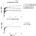
免疫不全マウスにおけるMDA-MB-436ヒト乳癌異種移植片腫瘍を、[18F]12を用いてPETにより可視化した。これらの腫瘍は、トレーサー注射の60分後においてバックグランドから識別可能な取込みの増加を実証した(図3)。トレーサー注射20分前にオラパリブ(50 mg/kg i.p.)または12(1 mg/kg i.p.)で処置した同じマウスでは、腫瘍での[18F]12取込みがバックグランドの組織活性レベルに減少した(図3)。microPET研究の0〜60分における腫瘍での[18F]12の時間-放射能曲線は、microPETイメージの視覚的評価を裏付け(図4)、オラパリブまたは12いずれかでの処置の結果として[18F]12取込みが有意に減少することを実証した。
MDA-MB-436 human breast cancer xenograft tumors in immunodeficient mice were visualized by PET using [ 18 F] 12. These tumors demonstrated an increase in uptake that was discernable from the
MDA-MB-436は、本質的に高いレベルのPARP-1活性を有するヒト乳癌細胞株であり(参考文献19)、microPETでのPARP-1活性をイメージングするための18F-標識オラパリブ誘導体の有効性を評価するためにマウスモデルにおいて用いられている。[18F]12は、microPETの取得1時間の間に腫瘍に漸進的に蓄積され、[18F]12取込みは、オラパリブまたは12いずれかで前処理された動物において遮断された。オラパリブおよび12の両方は、高い阻害能(それぞれIC50 = 5 nM(参考文献31)および6.3 nM)を有する競合的PARP-1阻害剤である。したがって、我々のデータは、マウスモデルにおける[18F]12取込みが、PARP-1発現によるものであり、[18F]12が、特異的にPARP-1発現をインビボでイメージングするための有効なPETトレーサーであることを示す。 MDA-MB-436 is a human breast cancer cell line with essentially high levels of PARP-1 activity (Ref. 19), an 18 F-labeled olaparib derivative for imaging PARP-1 activity in microPET. Used in mouse models to assess efficacy. [ 18 F] 12 was progressively accumulated in the tumor during the 1 hour of microPET acquisition, and [ 18 F] 12 uptake was blocked in animals pretreated with either olaparib or 12. Both olaparib and 12 are competitive PARP-1 inhibitors with high inhibitory potency (IC 50 = 5 nM (ref. 31) and 6.3 nM, respectively). Thus, our data indicate that [ 18 F] 12 uptake in mouse models is due to PARP-1 expression, and [ 18 F] 12 is effective for specifically imaging PARP-1 expression in vivo. Indicates a PET tracer.
実施例17
この実施例は、PETを用いたマウスにおける腫瘍組織の可視化を例示する。
Example 17
This example illustrates the visualization of tumor tissue in mice using PET.
MDA-MB-436およびMDA-MB-231ヒト乳癌細胞を、2 mM L-グルタミン、1 mMピルビン酸ナトリウム、0.1 mM非必須アミノ酸、2%MEMビタミンおよび5%ウシ胎児血清を添加したイーグル最少必須培地(アール平衡塩溶液および2 mM L-グルタミンを含む)を用いて5%CO2雰囲気の標準条件下で細胞培地中にて維持した。指数増殖中の細胞を、腫瘍移植のためにトリプシン処理し、採取した。計数後、細胞を、氷冷した1:1のMatrigelとPBSに再懸濁して、所望の濃度を得て、氷上で保持した。 MDA-MB-436 and MDA-MB-231 human breast cancer cells are eagle minimum essential with 2 mM L-glutamine, 1 mM sodium pyruvate, 0.1 mM non-essential amino acids, 2% MEM vitamins and 5% fetal calf serum The medium (containing Earl balanced salt solution and 2 mM L-glutamine) was used and maintained in cell medium under standard conditions of 5% CO 2 atmosphere. Exponentially growing cells were trypsinized and harvested for tumor transplantation. After counting, cells were resuspended in ice-cold 1: 1 Matrigel and PBS to obtain the desired concentration and kept on ice.
Charles River Laboratoriesからの成熟した雌の無胸腺nu/nuマウスを、これら一連のイメージング研究のための腫瘍移植の前に少なくとも1週間AALAC認証飼育設備において順応させた。雌のnu/nuマウスの乳腺脂肪体(腋窩リンパ節近傍)に約100μLの1:1 Matrigel:PBS中の約8 × 10⁶のMDA-MB-436乳癌細胞を移植した。イメージング研究を、移植2〜3週間後に実施した。 Mature female athymic nu / nu mice from Charles River Laboratories were acclimated in an AALAC certified housing facility for at least one week prior to tumor transplantation for these series of imaging studies. Approximately 100 μL of about 8 × 10 6 MDA-MB-436 breast cancer cells in 1: 1 Matrigel: PBS were transplanted into the mammary fat pad (near the axillary lymph node) of female nu / nu mice. Imaging studies were performed 2-3 weeks after transplantation.
腫瘍を有するマウスを、約2%イソフルラン/酸素を含む導入チャンバーに置き、その後、尾静脈カテーテルの設置のためカスタムダブルベッドに固定し;動的イメージング法のためにノーズコーンを介して約1%イソフルラン/酸素で麻酔を維持した。マウスに150〜200μCiの[18F]12を注射し、Focus 220およびInveon PET/CTスキャナーを用いて0〜60分間スキャンした。腫瘍および周囲の「バックグランド」組織について手動で描いた対象の領域から標準取込値(SUV)を作成した。特異的な取込みの可視化を、遮断薬オラパリブ(50 mg/kg、IP)または12(1 mg/kg、IP)での前処理の20分後、トレーサー注射によって獲得したイメージとベースラインスキャンの比較により測定した。
Tumor-bearing mice are placed in an introduction chamber containing about 2% isoflurane / oxygen and then fixed to a custom double bed for placement of the tail vein catheter; about 1% via the nose cone for dynamic imaging Anesthesia was maintained with isoflurane / oxygen. Mice were injected with 150-200 μCi [ 18 F] 12 and scanned for 0-60 minutes using a Focus 220 and Inveon PET / CT scanner. Standard uptake values (SUVs) were generated from the area of interest manually drawn for the tumor and surrounding "background" tissue. Visualize specific uptake by comparing baseline scans with images acquired by
実施例18
この実施例は、細胞培養アッセイにおける[18F]WC-4-138(化合物12)の取込みを例示する。
Example 18
This example illustrates the uptake of [ 18 F] WC-4-138 (Compound 12) in a cell culture assay.
これらの実験において、頭頸部扁平上皮細胞癌腫株SCC1、SCC15およびSCC25(ATCC)を、10%ウシ胎児血清(FBS、Gibco)、1%ペニシリン-ストレプトマイシン(P/S、Gibco)および100 ng/mlヒドロコルチゾン(Sigma-Aldrich)を添加したダルベッコ改変イーグル培地(DMEM、Gibco)中において増殖させた。小細胞肺癌株NCI-h69およびNCI-h82(ATCC)を、10%FBSおよび1%P/Sを添加したRPMI Medium(Gibco)中において増殖させた。ヒト乳癌細胞株MDA-MB-231(ATCC)を、5% FBS、2% MEMビタミン(Gibco)、1%200 mM L-グルタミン(Gibco)、1%10 mM非必須アミノ酸(NEAA、Gibco)を添加したイーグル最少必須培地(Gibco)中において増殖させた。 In these experiments, head and neck squamous cell carcinoma lines SCC1, SCC15 and SCC25 (ATCC) were treated with 10% fetal bovine serum (FBS, Gibco), 1% penicillin-streptomycin (P / S, Gibco) and 100 ng / ml. Growth was in Dulbecco's modified Eagle medium (DMEM, Gibco) supplemented with hydrocortisone (Sigma-Aldrich). Small cell lung cancer lines NCI-h69 and NCI-h82 (ATCC) were grown in RPMI Medium (Gibco) supplemented with 10% FBS and 1% P / S. Human breast cancer cell line MDA-MB-231 (ATCC), 5% FBS, 2% MEM vitamin (Gibco), 1% 200 mM L-glutamine (Gibco), 1% 10 mM non-essential amino acids (NEAA, Gibco) Grow in added Eagle's minimum essential medium (Gibco).
各実験の再現のために、約1μCiの[18F]WC-4-138を細胞培地1 mlで希釈し、106細胞に加えた。5、30、または60分後、培地を集め、細胞をリン酸緩衝生理食塩水(PBS、Gibco)0.7 mlで2回洗浄した。細胞培養ディッシュをこすり、microfugeチューブに移すことにより付着細胞を集めた。採取した培地、PBSまたは細胞ペレットにおいて放射能を測定した。細胞ペレットからのタンパク質を、標準の化学発光PARP ELISAキット(Trevigen #4520-096-K)を用いて定量した。すべてのデータを減衰補正し、細胞ペレット中の総タンパク質量に対して標準化した。薬物処置の検討のために、細胞を、[18F]WC-4-138とのインキュベーションの20時間前に、10μMオラパリブまたはイニパリブとインキュベートした。
For reproduction of each experiment, approximately 1 μCi of [ 18 F] WC-4-138 was diluted with 1 ml of cell culture medium and added to 10 6 cells. After 5, 30, or 60 minutes, the medium was collected and the cells were washed twice with 0.7 ml of phosphate buffered saline (PBS, Gibco). Adherent cells were collected by rubbing the cell culture dish and transferring to a microfuge tube. Radioactivity was measured in collected media, PBS or cell pellets. Protein from the cell pellet was quantified using a standard chemiluminescent PARP ELISA kit (Trevigen # 4520-096-K). All data were attenuation corrected and normalized to the total protein content in the cell pellet. For drug treatment studies, cells were incubated with 10 μM olaparib or
頭頸部細胞株SCC1およびSCC25は、構造的に少量のトレーサーを取込み、細胞ペレット中0.001〜0.002μCi/μgと記録される(図5A)。肺細胞株NCI-h69およびNCI-h82は、[18F]WC-4-138を取込み、細胞ペレット中0.004〜0.006μCi/μgと記録される(図5A)。5、30または60分間[18F]WC-4-138とインキュベーション後、頭頸部扁平上皮細胞癌(HNSCC)株(SCC1およびSCC25)または小細胞肺癌(SCLC)株(NCI-h69およびNCI-h82)において放射能を測定した(各時点n=3)。これらの図は、SCC細胞について1〜2ユニットのPARP活性、およびNCI細胞について3.5ユニットのParp活性を表す(図5B)。PARP活性をHNSCCまたは肺SCLC細胞において測定した(図5C〜D)。[18F]WC-4-138取込みをオラパリブまたはイニパリブで処置したHNSCC細胞株において測定した。データは、トレーサーとのインキュベーションの30または60分後の取込みについての平均±SDである(*p<0.05)。すべてのデータを、他に断らない限り、平均±SD、n=3として示す。 Head and neck cell lines SCC1 and SCC25 take up structurally small amounts of tracer and are recorded as 0.001 to 0.002 μCi / μg in the cell pellet (FIG. 5A). Lung cell lines NCI-h69 and NCI-h82 take up [ 18 F] WC-4-138 and are recorded as 0.004-0.006 μCi / μg in the cell pellet (FIG. 5A). After incubation with [ 18 F] WC-4-138 for 5, 30 or 60 minutes, head and neck squamous cell carcinoma (HNSCC) strains (SCC1 and SCC25) or small cell lung cancer (SCLC) strains (NCI-h69 and NCI-h82) ) Was measured (n = 3 at each time point). These figures represent 1-2 units of PARP activity for SCC cells and 3.5 units of Parp activity for NCI cells (FIG. 5B). PARP activity was measured in HNSCC or lung SCLC cells (FIGS. 5C-D). [ 18 F] WC-4-138 uptake was measured in HNSCC cell lines treated with olaparib or iniparib. Data are mean ± SD for uptake after 30 or 60 minutes of incubation with tracer (* p <0.05). All data are shown as mean ± SD, n = 3 unless otherwise stated.
この活性は、オラパリブにより消失するが(図5C)、イニパリブによっては消失しない(図5D)。 This activity is lost by olaparib (FIG. 5C) but not by iniparib (FIG. 5D).
実施例19
この実施例は、化合物12([18F]WC-4-138)の代謝安定性を例示する。
Example 19
This example illustrates the metabolic stability of Compound 12 ([ 18 F] WC-4-138).
これらの実験において、[18F]-WC-4-138の代謝安定性を、成体雄のC57BL/6Jマウス(Jackson Laboratory)の尾静脈に400μCiを注射した後、評価した。マウスを、注射の5または30分後、頸椎脱臼により屠殺した。下大静脈に傷をつけ、血液を腹腔から採取した。血漿を、14000 rpmでの遠心分離によって赤血球から分離した。血漿(100μl)をアセトニトリルと1 : 1.5の比で混合し、14000 rpmで遠心分離した。赤血球ペレット、全血漿、ならびにアセトニトリル可溶性および非可溶性フラクションに関連する放射能を測定した。各動物の肝臓をまた、所与の時間において採取し、ドライアイスで凍結した。肝臓を2 mlのアセトニトリル中でホモジナイズし、ホモジナイズされた肝臓1 mlを、14000 rpmで遠心分離した。上清およびペレットに関連する放射能を測定した。
In these experiments, the metabolic stability of [ 18 F] -WC-4-138 was assessed after injection of 400 μCi into the tail vein of adult male C57BL / 6J mice (Jackson Laboratory). Mice were sacrificed by
アセトニトリル可溶性の血漿または肝臓の上清(100μl)を水と1:1の比で混合し、逆相HPLCにより分離した。親化合物をまた、参照としてHPLCにより分離した。各HPLCフラクションに関連する放射能を測定した。各検体の親化合物のパーセントを、親化合物を含むと推測されるHPLCフラクションに関連する放射能の部分として計算した。 Acetonitrile soluble plasma or liver supernatant (100 μl) was mixed with water in a 1: 1 ratio and separated by reverse phase HPLC. The parent compound was also separated by HPLC as a reference. The radioactivity associated with each HPLC fraction was measured. The percentage of parent compound in each specimen was calculated as the fraction of radioactivity associated with the HPLC fraction suspected of containing the parent compound.
注射の5分後に存在する参照化合物と比較して50%の化合物12([18F]WC-4-138)が血液において速やかに代謝され、そして注射の30分後では、参照化合物と比較して当該化合物の約10%のみが観察された(図6)。対照的に、当該化合物は、肝臓において非常に長い半減期を有し、注射の5分後に参照化合物と比較して約80%、注射の30分後に参照化合物と比較して約70%を示した(図6)。
50% of compound 12 ([ 18 F] WC-4-138) is rapidly metabolized in the blood compared to the reference compound present 5 minutes after injection, and compared to the
実施例20
この実施例は、マウスにおける化合物12([18F]WC-4-138)の生体分布を例示する。
Example 20
This example illustrates the biodistribution of compound 12 ([ 18 F] WC-4-138) in mice.
8週齢の雌の無胸腺ヌードマウス(Harlan)に、[18F]WC-4-138を尾静脈から注射した。30μCiの[18F]WC-4-138のIVトレーサー注射の5もしくは30分後、45μCiの注射の1もしくは2時間後、または60μCiの注射の4時間後、マウスを頸椎脱臼により屠殺した。血漿または肝臓のアセトニトリル可溶性フラクションおよびコントロールの親化合物を逆相高速液体クロマトグラフィー(HPLC)により分離し、各フラクションの放射能を定量した。各検体の親化合物のパーセントを、親化合物を含むHPLCフラクションに関連する放射能の部分として計算した。データを、平均±SD、n=3として示す。
Eight week old female athymic nude mice (Harlan) were injected with [ 18 F] WC-4-138 via the tail vein. Mice were sacrificed by
4匹のマウスを、2時間の時点を除いて各時点で屠殺した(n=3)。血液、心臓、肺、筋肉、肝臓、脾臓、脂肪、副腎、腎臓、子宮、卵巣、骨、骨髄、膵臓、胃、小腸および大腸を各動物から採取した。すべての器官を、過剰な血液を取り除くためにふき取り、重さを量り、Beckmann 6000ガンマカウンターでカウントした。組織1グラム当たりの注射量のパーセント(%ID/g)を各器官について測定した。結果は、組織1グラム当たりの注射量の0.5〜15%で変化し、表1および2に示す。 Four mice were sacrificed at each time point except the 2 hour time point (n = 3). Blood, heart, lung, muscle, liver, spleen, fat, adrenal gland, kidney, uterus, ovary, bone, bone marrow, pancreas, stomach, small intestine and large intestine were collected from each animal. All organs were wiped to remove excess blood, weighed, and counted on a Beckmann 6000 gamma counter. The percentage of injection per gram of tissue (% ID / g) was measured for each organ. Results vary from 0.5 to 15% of the injection volume per gram of tissue and are shown in Tables 1 and 2.
表1 無胸腺ヌードマウスにおける[18F]WC-4-138の器官生体内分布
すべてのデータは平均±SDである。120分におけるn=3を除き、すべての群についてn=4。
Table 1. Organ biodistribution of [ 18 F] WC-4-138 in athymic nude mice
表2 マウスの生体内分布調査からのヒト線量測定の推測値
実施例21
この実施例は、マウスにおける化合物12([18F]WC-4-138)の取込みを例示する。
Example 21
This example illustrates the uptake of compound 12 ([ 18 F] WC-4-138) in mice.
成体の雌の無胸腺ヌードマウス(Charles River Laboratories)の乳腺脂肪体へ、106のSCC1細胞または107のMDA-MB-231細胞を移植した。SCC1腫瘍を有するマウスを5週目においてイメージングし、そしてMDA-MB-231細胞を有するマウスを移植2.5週間後イメージングした。マウスに2%イソフルラン/酸素で麻酔をかけ、イメージング法の間じゅうノーズコーンを介して1%イソフルラン/酸素で維持した。動物全体のmicroCTイメージをInveon PET/CTスキャナーで取得した。マウスに11.36±0.5 MBq(307±13μCi)の[18F]WC(4)-138を尾静脈から注射し、Focus 220またはInveon PET/CTスキャナーを用いて60分間動的スキャンを実施した。microPETおよびmicroCTイメージを、Integrated Research Workflowソフトウェア(Siemens)を用いてコレジスタ(coregister)した。時間活性曲線を決定するために対象の領域を腫瘍上に描いた。薬物処置の検討のために、イメージングの30分前に、動物に50 mg/kgのオラパリブまたはイニパリブの腹腔内注射をした。マウスを、ベースライン時、またはオラパリブもしくはイニパリブのIP注射の30分後にイメージングした。MDA-MB-231腫瘍を有するマウスの横断図を図7に示す。矢印は、CT上で同定される腫瘍の位置を指す。トレーサーは、ベースラインにおける腫瘍およびマウスの他のセクション中に十分に取り込まれるが(図7、各チャネルビューの上段、矢印)、[18F]12の腫瘍取込みがオラパリブでの処置により消失するが(図7、各チャネルパネルの下段左、矢印)、トレーサーは、まだ当該セクションの他のエリアに存在する。対照的に、イニパリブ処置は、腫瘍細胞中における[18F]WC(4)-138の取込みに影響を及ぼさないが、他のタイプの細胞において該取込みは消失する(図7、各チャネルビューの下段右、矢印)。
10 6 SCC1 cells or 10 7 MDA-MB-231 cells were transplanted into mammary fat pads of adult female athymic nude mice (Charles River Laboratories). Mice with SCC1 tumors were imaged at 5 weeks and mice with MDA-MB-231 cells were imaged 2.5 weeks after transplantation. Mice were anesthetized with 2% isoflurane / oxygen and maintained at 1% isoflurane / oxygen through the nose cone throughout the imaging procedure. MicroCT images of the whole animal were acquired with an Inveon PET / CT scanner. Mice were injected with 11.36 ± 0.5 MBq (307 ± 13 μCi) [ 18 F] WC (4) -138 through the tail vein and a dynamic scan was performed using a Focus 220 or Inveon PET / CT scanner for 60 minutes. microPET and microCT images were coregistered using Integrated Research Workflow software (Siemens). A region of interest was drawn on the tumor to determine a time activity curve. For drug treatment studies, animals were given an intraperitoneal injection of 50 mg / kg olaparib or
実施例22
この実施例は、MDA-MB-231腫瘍(図8A)およびSCC腫瘍(図8B)の両方についてベースラインと薬物処置との間で1 ml当たりの注射量の有意な減少を示す定量分析を例示する。図8において、マウスを、ベースライン時、またはオラパリブもしくはイニパリブのIP注射の30分後にイメージングした(A)。各MDA/MB-231腫瘍についてベースライン時または薬物処置後に1 ml当たりの注射量のパーセントを計算した。トレーサー取込みは、オラパリブ処置マウスにおいて有意に減少した(対応のあるt検定により決定した、*p=0.0038、n=6腫瘍)。トレーサー取込みは、イニパリブ処置マウスにおいて変化しなかった(対応のあるt検定により決定した、p=0.1098、n=6腫瘍)(B)。各SCC1腫瘍についてベースライン時または薬物処置後に1 ml当たりの注射量のパーセントを計算した。トレーサー取込みは、オラパリブ処置マウスにおいて有意に減少した(対応のあるt検定により決定した、*p=0.001、n=8腫瘍)。トレーサー取込みはイニパリブ処置マウスにおいて変化しなかった(対応のあるt検定により決定した、p=0.7216、n=5腫瘍)。
Example 22
This example illustrates a quantitative analysis showing a significant decrease in injection volume per ml between baseline and drug treatment for both MDA-MB-231 tumors (Figure 8A) and SCC tumors (Figure 8B) To do. In FIG. 8, mice were imaged at baseline or 30 minutes after IP injection of olaparib or iniparib (A). The percent of injection volume per ml was calculated for each MDA / MB-231 tumor at baseline or after drug treatment. Tracer uptake was significantly reduced in olaparib treated mice (determined by paired t test, * p = 0.0038, n = 6 tumors). Tracer uptake was unchanged in iniparib-treated mice (determined by paired t-test, p = 0.1098, n = 6 tumors) (B). The percent of injection volume per ml was calculated for each SCC1 tumor at baseline or after drug treatment. Tracer uptake was significantly reduced in olaparib treated mice (determined by paired t test, * p = 0.001, n = 8 tumors). Tracer uptake was unchanged in iniparib treated mice (determined by paired t test, p = 0.7216, n = 5 tumors).
実施例23
対象体は潜在的なPARP-1関連乳癌の症状を示す。医師がPETスキャンを指示し、そして技師が有効量の[18F]WC(4)-138を投与し、PETスキャンを実施する。腫瘍は、対象体の周囲組織と比較して大量のトレーサー取込みを示す。医師は、PARP-1関連癌と診断し、処置計画の一部としてPARP-1阻害剤を処方する。
Example 23
The subject exhibits potential symptoms of PARP-1-related breast cancer. The doctor directs a PET scan, and the technician administers an effective amount of [ 18 F] WC (4) -138 and performs the PET scan. Tumors show a large amount of tracer uptake compared to the surrounding tissue of the subject. Doctors diagnose PARP-1-related cancer and prescribe PARP-1 inhibitors as part of the treatment plan.
実施例24
対象体は肺の異常な炎症を示す。医師がPETスキャンを指示し、そして技師が有効量の[18F]WC(4)-138を投与し、PETスキャンを実施する。肺組織は、コントロールの肺組織と比較して大量のトレーサー取込みを示し、そして医師は、PARP-1関連炎症と診断し、処置計画の一部としてPARP-1阻害剤を処方する。
Example 24
The subject exhibits abnormal inflammation of the lungs. The doctor directs a PET scan, and the technician administers an effective amount of [ 18 F] WC (4) -138 and performs the PET scan. Lung tissue shows large amounts of tracer uptake compared to control lung tissue, and the physician diagnoses PARP-1-related inflammation and prescribes a PARP-1 inhibitor as part of the treatment plan.
参考文献
1. Hassa, P. O.; Hottiger, M. O. Frontiers in Bioscience : a Journal and Virtual Library 2008, 13, 3046.
2. d'Adda di Fagagna, F.; Hande, M. P.; Tong, W. M.; Lansdorp, P. M.; Wang, Z. Q.; Jackson, S. P. Nature Genetics 1999, 23, 76.
3. Jagtap, P.; Szabo, C. Nature Reviews. Drug Discovery 2005, 4, 421.
4. Schreiber, V.; Dantzer, F.; Ame, J. C.; de Murcia, G. Nature Reviews. Molecular Cell Biology 2006, 7, 517.
5. Virag, L.; Szabo, C. Pharmacological Reviews 2002, 54, 375.
6. Gradwohl, G.; Menissier de Murcia, J. M.; Molinete, M.; Simonin, F.; Koken, M.; Hoeijmakers, J. H.; de Murcia, G. Proceedings of the National Academy of Sciences of the United States of America 1990, 87, 2990.
7. Kameshita, I.; Matsuda, Z.; Taniguchi, T.; Shizuta, Y. The Journal of Biological Chemistry 1984, 259, 4770.
8. Ferraris, D. V. Journal of Medicinal Chemistry 2010, 53, 4561.
9. Basu, B.; Sandhu, S. K.; de Bono, J. S. Drugs 2012, 72, 1579.
10. Sandhu, S. K.; Schelman, W. R.; Wilding, G.; Moreno, V.; Baird, R. D.; Miranda, S.; Hylands, L.; Riisnaes, R.; Forster, M.; Omlin, A.; Kreischer, N.; Thway, K.; Gevensleben, H.; Sun, L.; Loughney, J.; Chatterjee, M.; Toniatti, C.; Carpenter, C. L.; Iannone, R.; Kaye, S. B.; de Bono, J. S.; Wenham, R. M. The Lancet Oncology 2013, 14, 882.
11. Plummer, R.; Lorigan, P.; Steven, N.; Scott, L.; Middleton, M. R.; Wilson, R. H.; Mulligan, E.; Curtin, N.; Wang, D.; Dewji, R.; Abbattista, A.; Gallo, J.; Calvert, H. Cancer Chemotherapy and Pharmacology 2013, 71, 1191.
12. Gelmon, K. A.; Tischkowitz, M.; Mackay, H.; Swenerton, K.; Robidoux, A.; Tonkin, K.; Hirte, H.; Huntsman, D.; Clemons, M.; Gilks, B.; Yerushalmi, R.; Macpherson, E.; Carmichael, J.; Oza, A. The Lancet Oncology 2011, 12, 852.
13. Kummar, S.; Chen, A.; Ji, J.; Zhang, Y.; Reid, J. M.; Ames, M.; Jia, L.; Weil, M.; Speranza, G.; Murgo, A. J.; Kinders, R.; Wang, L.; Parchment, R. E.; Carter, J.; Stotler, H.; Rubinstein, L.; Hollingshead, M.; Melillo, G.; Pommier, Y.; Bonner, W.; Tomaszewski, J. E.; Doroshow, J. H. Cancer Research 2011, 71, 5626.
14. Javle, M.; Curtin, N. J. Therapeutic Advances in Medical Oncology 2011, 3, 257.
15. Wahlberg, E.; Karlberg, T.; Kouznetsova, E.; Markova, N.; Macchiarulo, A.; Thorsell, A. G.; Pol, E.; Frostell, A.; Ekblad, T.; Oncu, D.; Kull, B.; Robertson, G. M.; Pellicciari, R.; Schuler, H.; Weigelt, J. Nature Biotechnology 2012, 30, 283.
16. Liu, X.; Shi, Y.; Maag, D. X.; Palma, J. P.; Patterson, M. J.; Ellis, P. A.; Surber, B. W.; Ready, D. B.; Soni, N. B.; Ladror, U. S.; Xu, A. J.; Iyer, R.; Harlan, J. E.; Solomon, L. R.; Donawho, C. K.; Penning, T. D.; Johnson, E. F.; Shoemaker, A. R. Clinical Cancer Research : an Official Journal of the American Association for Cancer Research 2012, 18, 510.
17. Tu, Z.; Chu, W.; Zhang, J.; Dence, C. S.; Welch, M. J.; Mach, R. H. Nuclear Medicine and Biology 2005, 32, 437.
18. Keliher, E. J.; Reiner, T.; Turetsky, A.; Hilderbrand, S. A.; Weissleder, R. ChemMedChem 2011, 6, 424.
19. Reiner, T.; Keliher, E. J.; Earley, S.; Marinelli, B.; Weissleder, R. Angew Chem Int Ed Engl 2011, 50, 1922.
20. Reiner, T.; Lacy, J.; Keliher, E. J.; Yang, K. S.; Ullal, A.; Kohler, R. H.; Vinegoni, C.; Weissleder, R. Neoplasia 2012, 14, 169.
21. Delaney, C. A.; Wang, L. Z.; Kyle, S.; White, A. W.; Calvert, A. H.; Curtin, N. J.; Durkacz, B. W.; Hostomsky, Z.; Newell, D. R. Clinical cancer research : an official journal of the American Association for Cancer Research 2000, 6, 2860.
22. Skalitzky, D. J.; Marakovits, J. T.; Maegley, K. A.; Ekker, A.; Yu, X. H.; Hostomsky, Z.; Webber, S. E.; Eastman, B. W.; Almassy, R.; Li, J.; Curtin, N. J.; Newell, D. R.; Calvert, A. H.; Griffin, R. J.; Golding, B. T. Journal of Medicinal Chemistry 2003, 46, 210.
23. Putt, K. S.; Hergenrother, P. J. Analytical Biochemistry 2004, 326, 78.
24. Jagtap, P.; Soriano, F. G.; Virag, L.; Liaudet, L.; Mabley, J.; Szabo, E.; Hasko, G.; Marton, A.; Lorigados, C. B.; Gallyas, F., Jr.; Sumegi, B.; Hoyt, D. G.; Baloglu, E.; VanDuzer, J.; Salzman, A. L.; Southan, G. J.; Szabo, C. Critical Care Medicine 2002, 30, 1071.
25. Ruf, A.; de Murcia, G.; Schulz, G. E. Biochemistry 1998, 37, 3893.
26. Kinoshita, T.; Nakanishi, I.; Warizaya, M.; Iwashita, A.; Kido, Y.; Hattori, K.; Fujii, T. FEBS letters 2004, 556, 43.
27. White, A. W.; Almassy, R.; Calvert, A. H.; Curtin, N. J.; Griffin, R. J.; Hostomsky, Z.; Maegley, K.; Newell, D. R.; Srinivasan, S.; Golding, B. T. Journal of Medicinal Chemistry 2000, 43, 4084.
28. Lasne, M. C.; Perrio, C.; Rouden, J.; Barre, L.; Roeda, D.; Dolle, F.; Crouzel, C. Top Curr Chem 2002, 222, 201.
29. Glaser, M.; Arstad, E. Bioconjugate chemistry 2007, 18, 989.
30. Zhou, D.; Chu, W.; Dence, C. S.; Mach, R. H.; Welch, M. J. Nuclear Medicine and Biology 2012.
31. Menear, K. A.; Adcock, C.; Boulter, R.; Cockcroft, X. L.; Copsey, L.; Cranston, A.; Dillon, K. J.; Drzewiecki, J.; Garman, S.; Gomez, S.; Javaid, H.; Kerrigan, F.; Knights, C.; Lau, A.; Loh, V. M., Jr.; Matthews, I. T.; Moore, S.; O'Connor, M. J.; Smith, G. C.; Martin, N. M. Journal of Medicinal Chemistry 2008, 51, 6581.
References
1.Hassa, PO; Hottiger, MO Frontiers in Bioscience: a Journal and Virtual Library 2008, 13, 3046.
2.d'Adda di Fagagna, F .; Hande, MP; Tong, WM; Lansdorp, PM; Wang, ZQ; Jackson, SP Nature Genetics 1999, 23, 76.
3. Jagtap, P .; Szabo, C. Nature Reviews. Drug Discovery 2005, 4, 421.
4. Schreiber, V .; Dantzer, F .; Ame, JC; de Murcia, G. Nature Reviews. Molecular Cell Biology 2006, 7, 517.
5. Virag, L .; Szabo, C. Pharmacological Reviews 2002, 54, 375.
6. Gradwohl, G .; Menissier de Murcia, JM; Molinete, M .; Simonin, F .; Koken, M .; Hoeijmakers, JH; de Murcia, G. Proceedings of the National Academy of Sciences of the United States of America 1990, 87, 2990.
7. Kameshita, I .; Matsuda, Z .; Taniguchi, T .; Shizuta, Y. The Journal of Biological Chemistry 1984, 259, 4770.
8. Ferraris, DV Journal of Medicinal Chemistry 2010, 53, 4561.
9. Basu, B .; Sandhu, SK; de Bono, JS Drugs 2012, 72, 1579.
10. Sandhu, SK; Schelman, WR; Wilding, G .; Moreno, V .; Baird, RD; Miranda, S .; Hylands, L .; Riisnaes, R .; Forster, M .; Omlin, A .; Kreischer , N .; Thway, K .; Gevensleben, H .; Sun, L .; Loughney, J .; Chatterjee, M .; Toniatti, C .; Carpenter, CL; Iannone, R .; Kaye, SB; de Bono, JS; Wenham, RM The Lancet Oncology 2013, 14, 882.
11. Plummer, R .; Lorigan, P .; Steven, N .; Scott, L .; Middleton, MR; Wilson, RH; Mulligan, E .; Curtin, N .; Wang, D .; Dewji, R .; Abbattista, A .; Gallo, J .; Calvert, H. Cancer Chemotherapy and Pharmacology 2013, 71, 1191.
12. Gelmon, KA; Tischkowitz, M .; Mackay, H .; Swenerton, K .; Robidoux, A .; Tonkin, K .; Hirte, H .; Huntsman, D .; Clemons, M .; Gilks, B. ; Yerushalmi, R .; Macpherson, E .; Carmichael, J .; Oza, A. The Lancet Oncology 2011, 12, 852.
13. Kummar, S .; Chen, A .; Ji, J .; Zhang, Y .; Reid, JM; Ames, M .; Jia, L .; Weil, M .; Speranza, G .; Murgo, AJ; Kinders, R .; Wang, L .; Parchment, RE; Carter, J .; Stotler, H .; Rubinstein, L .; Hollingshead, M .; Melillo, G .; Pommier, Y .; Bonner, W .; Tomaszewski , JE; Doroshow, JH Cancer Research 2011, 71, 5626.
14. Javle, M .; Curtin, NJ Therapeutic Advances in
15. Wahlberg, E .; Karlberg, T .; Kouznetsova, E .; Markova, N .; Macchiarulo, A .; Thorsell, AG; Pol, E .; Frostell, A .; Ekblad, T .; Oncu, D. ; Kull, B .; Robertson, GM; Pellicciari, R .; Schuler, H .; Weigelt,
16. Liu, X .; Shi, Y .; Maag, DX; Palma, JP; Patterson, MJ; Ellis, PA; Surber, BW; Ready, DB; Soni, NB; Ladror, US; Xu, AJ; Iyer, R .; Harlan, JE; Solomon, LR; Donawho, CK; Penning, TD; Johnson, EF; Shoemaker, AR Clinical Cancer Research: an Official Journal of the American Association for Cancer Research 2012, 18, 510.
17. Tu, Z .; Chu, W .; Zhang, J .; Dence, CS; Welch, MJ; Mach, RH Nuclear Medicine and Biology 2005, 32, 437.
18. Keliher, EJ; Reiner, T .; Turetsky, A .; Hilderbrand, SA; Weissleder,
19. Reiner, T .; Keliher, EJ; Earley, S .; Marinelli, B .; Weissleder, R. Angew Chem
20. Reiner, T .; Lacy, J .; Keliher, EJ; Yang, KS; Ullal, A .; Kohler, RH; Vinegoni, C .; Weissleder, R. Neoplasia 2012, 14, 169.
21. Delaney, CA; Wang, LZ; Kyle, S .; White, AW; Calvert, AH; Curtin, NJ; Durkacz, BW; Hostomsky, Z .; Newell, DR Clinical cancer research: an official journal of the American Association for
22. Skalitzky, DJ; Marakovits, JT; Maegley, KA; Ekker, A .; Yu, XH; Hostomsky, Z .; Webber, SE; Eastman, BW; Almassy, R .; Li, J .; Curtin, NJ; Newell, DR; Calvert, AH; Griffin, RJ; Golding, BT Journal of Medicinal Chemistry 2003, 46, 210.
23.Putt, KS; Hergenrother, PJ Analytical Biochemistry 2004, 326, 78.
24. Jagtap, P .; Soriano, FG; Virag, L .; Liaudet, L .; Mabley, J .; Szabo, E .; Hasko, G .; Marton, A .; Lorigados, CB; Gallyas, F., Jr .; Sumegi, B .; Hoyt, DG; Baloglu, E .; VanDuzer, J .; Salzman, AL; Southan, GJ; Szabo, C.
25. Ruf, A .; de Murcia, G .; Schulz, GE Biochemistry 1998, 37, 3893.
26. Kinoshita, T .; Nakanishi, I .; Warizaya, M .; Iwashita, A .; Kido, Y .; Hattori, K .; Fujii, T. FEBS letters 2004, 556, 43.
27. White, AW; Almassy, R .; Calvert, AH; Curtin, NJ; Griffin, RJ; Hostomsky, Z .; Maegley, K .; Newell, DR; Srinivasan, S .; Golding, BT Journal of Medicinal Chemistry 2000 , 43, 4084.
28. Lasne, MC; Perrio, C .; Rouden, J .; Barre, L .; Roeda, D .; Dolle, F .; Crouzel, C. Top Curr Chem 2002, 222, 201.
29. Glaser, M .; Arstad, E. Bioconjugate chemistry 2007, 18, 989.
30. Zhou, D .; Chu, W .; Dence, CS; Mach, RH; Welch, MJ Nuclear Medicine and Biology 2012.
31. Menear, KA; Adcock, C .; Boulter, R .; Cockcroft, XL; Copsey, L .; Cranston, A .; Dillon, KJ; Drzewiecki, J .; Garman, S .; Gomez, S .; Javaid , H .; Kerrigan, F .; Knights, C .; Lau, A .; Loh, VM, Jr .; Matthews, IT; Moore, S .; O'Connor, MJ; Smith, GC; Martin, NM Journal of Medicinal Chemistry 2008, 51, 6581.
本明細書で引用したすべての刊行物は、出典明示により各々その全体として本明細書の一部とする。本明細書で用いる単数形「a」、「an」および「the」は、他に断らない限り、複数形も含むことを意図する。 All publications cited herein are hereby incorporated by reference in their entirety. As used herein, the singular forms “a”, “an”, and “the” are intended to include the plural forms as well, unless expressly stated otherwise.
Claims (15)
で示される、化合物またはその医薬的に許容される塩。 Structural formula
Or a pharmaceutically acceptable salt thereof.
で示される、化合物またはその医薬的に許容される塩。 Structural formula
Or a pharmaceutically acceptable salt thereof.
Applications Claiming Priority (3)
| Application Number | Priority Date | Filing Date | Title |
|---|---|---|---|
| US201461923759P | 2014-01-05 | 2014-01-05 | |
| US61/923,759 | 2014-01-05 | ||
| PCT/US2015/010129 WO2015103526A1 (en) | 2014-01-05 | 2015-01-05 | Radiolabeled tracers for poly (adp-ribose) polymerase-1 (parp-1), methods and uses therefor |
Related Child Applications (1)
| Application Number | Title | Priority Date | Filing Date |
|---|---|---|---|
| JP2018173457A Division JP2019001820A (en) | 2014-01-05 | 2018-09-18 | Radiolabeled tracers for poly (adp-ribose) polymerase-1 (parp-1), methods and uses therefor |
Publications (3)
| Publication Number | Publication Date |
|---|---|
| JP2017505336A JP2017505336A (en) | 2017-02-16 |
| JP2017505336A5 JP2017505336A5 (en) | 2018-02-08 |
| JP6407303B2 true JP6407303B2 (en) | 2018-10-17 |
Family
ID=53494075
Family Applications (2)
| Application Number | Title | Priority Date | Filing Date |
|---|---|---|---|
| JP2016562470A Active JP6407303B2 (en) | 2014-01-05 | 2015-01-05 | Radiolabeled tracer for poly (ADP-ribose) polymerase-1 (PARP-1), method and use thereof |
| JP2018173457A Withdrawn JP2019001820A (en) | 2014-01-05 | 2018-09-18 | Radiolabeled tracers for poly (adp-ribose) polymerase-1 (parp-1), methods and uses therefor |
Family Applications After (1)
| Application Number | Title | Priority Date | Filing Date |
|---|---|---|---|
| JP2018173457A Withdrawn JP2019001820A (en) | 2014-01-05 | 2018-09-18 | Radiolabeled tracers for poly (adp-ribose) polymerase-1 (parp-1), methods and uses therefor |
Country Status (7)
| Country | Link |
|---|---|
| US (2) | US9993570B2 (en) |
| EP (2) | EP3089965B1 (en) |
| JP (2) | JP6407303B2 (en) |
| CN (1) | CN106458935B (en) |
| CA (1) | CA2935857C (en) |
| ES (1) | ES2700348T3 (en) |
| WO (1) | WO2015103526A1 (en) |
Families Citing this family (3)
| Publication number | Priority date | Publication date | Assignee | Title |
|---|---|---|---|---|
| EA201992177A1 (en) | 2017-03-27 | 2020-02-25 | Тесаро, Инк. | COMPOSITIONS BASED ON NIRAPARIB |
| WO2018218025A1 (en) * | 2017-05-24 | 2018-11-29 | The Trustees Of The University Of Pennsylvania | Radiolabeled and fluorescent parp inhibitors for imaging and radiotherapy |
| US11730725B2 (en) * | 2017-09-26 | 2023-08-22 | Tesaro, Inc. | Niraparib formulations |
Family Cites Families (9)
| Publication number | Priority date | Publication date | Assignee | Title |
|---|---|---|---|---|
| US7781596B1 (en) * | 1998-11-03 | 2010-08-24 | Abbott Laboratories | Substituted 2-phenylbenzimidazoles, the production thereof and their use |
| EP1140936B1 (en) | 1999-01-11 | 2004-03-17 | Agouron Pharmaceuticals, Inc. | Tricyclic inhibitors of poly(adp-ribose) polymerases |
| ECSP003637A (en) * | 1999-08-31 | 2002-03-25 | Agouron Pharma | TRICYCLE POLY INHIBITORS (ADP-RIBOSA) POLYMERASES |
| HU0301154D0 (en) * | 2003-04-28 | 2003-07-28 | Hideg Kalman Dr | Pharmaceutical composition |
| GB0428012D0 (en) * | 2004-12-22 | 2005-01-26 | Hammersmith Imanet Ltd | Radiolabelling methods |
| US8329686B2 (en) * | 2006-08-29 | 2012-12-11 | Washington University | Isatin analogues and uses therefor |
| DK2061765T3 (en) | 2006-09-01 | 2015-01-26 | Senhwa Biosciences Inc | Serine-threonine protein kinase AND PARP-MODULATOR |
| US8506927B2 (en) * | 2011-01-05 | 2013-08-13 | Washington University | Pegylated fluorobenzamide analogues, their synthesis and use in diagnostic imaging |
| CA2883574A1 (en) * | 2012-09-05 | 2014-03-13 | Bayer Cropscience Ag | Use of substituted 2-amidobenzimidazoles, 2-amidobenzoxazoles and 2-amidobenzothiazoles or salts thereof as active substances against abiotic plant stress |
-
2015
- 2015-01-05 WO PCT/US2015/010129 patent/WO2015103526A1/en active Application Filing
- 2015-01-05 CA CA2935857A patent/CA2935857C/en active Active
- 2015-01-05 EP EP15733272.7A patent/EP3089965B1/en active Active
- 2015-01-05 EP EP18190718.9A patent/EP3424909A1/en not_active Withdrawn
- 2015-01-05 ES ES15733272T patent/ES2700348T3/en active Active
- 2015-01-05 CN CN201580011735.7A patent/CN106458935B/en active Active
- 2015-01-05 JP JP2016562470A patent/JP6407303B2/en active Active
-
2016
- 2016-07-05 US US15/201,765 patent/US9993570B2/en active Active
-
2018
- 2018-05-14 US US15/978,374 patent/US20180326100A1/en not_active Abandoned
- 2018-09-18 JP JP2018173457A patent/JP2019001820A/en not_active Withdrawn
Also Published As
| Publication number | Publication date |
|---|---|
| JP2017505336A (en) | 2017-02-16 |
| US20160339124A1 (en) | 2016-11-24 |
| CN106458935A (en) | 2017-02-22 |
| ES2700348T3 (en) | 2019-02-15 |
| EP3424909A1 (en) | 2019-01-09 |
| CN106458935B (en) | 2019-05-28 |
| CA2935857A1 (en) | 2015-07-09 |
| US9993570B2 (en) | 2018-06-12 |
| EP3089965B1 (en) | 2018-08-29 |
| US20180326100A1 (en) | 2018-11-15 |
| EP3089965A4 (en) | 2017-06-21 |
| CA2935857C (en) | 2020-12-15 |
| EP3089965A1 (en) | 2016-11-09 |
| WO2015103526A1 (en) | 2015-07-09 |
| JP2019001820A (en) | 2019-01-10 |
Similar Documents
| Publication | Publication Date | Title |
|---|---|---|
| Zhou et al. | Synthesis,[18F] radiolabeling, and evaluation of poly (ADP-ribose) polymerase-1 (PARP-1) inhibitors for in vivo imaging of PARP-1 using positron emission tomography | |
| JP2019001820A (en) | Radiolabeled tracers for poly (adp-ribose) polymerase-1 (parp-1), methods and uses therefor | |
| US20220194904A1 (en) | 18f-labeled triazole containing psma inhibitors | |
| Zhou et al. | Preliminary evaluation of a novel 18F-labeled PARP-1 ligand for PET imaging of PARP-1 expression in prostate cancer | |
| EP3389727B1 (en) | Radiolabelled mglur2/3 pet ligands | |
| Kawamura et al. | Synthesis and evaluation of [11 C] XR9576 to assess the function of drug efflux transporters using PET | |
| US20050107398A1 (en) | Sigma-2 receptor radiotracers for imaging the proliferative status of solid tumors | |
| JP6840666B2 (en) | New anthranilic acid derivative | |
| Tran et al. | Synthesis and evaluation of novel potent TSPO PET ligands with 2-phenylpyrazolo [1, 5-a] pyrimidin-3-yl acetamide | |
| JP5085824B2 (en) | Production of compounds useful for hypoxia detection | |
| US10716869B2 (en) | Radiolabeled macrocyclic EGFR inhibitor | |
| Thomae et al. | Synthesis and preclinical evaluation of an 18F labeled PDE7 inhibitor for PET neuroimaging | |
| JP2007505101A (en) | Radiolabeled anilinoquinazoline type compounds and their use in radioimaging and radiotherapy | |
| Selivanova et al. | Synthesis and pharmacological evaluation of 11C-labeled piperazine derivative as a PET probe for sigma-2 receptor imaging | |
| US20170174632A1 (en) | 4-oxo-1, 4-dihydroquinoline-3-carboxamide as selective ligand for cannabinoid receptor 2 for diagnosis and therapy | |
| Gonciarz et al. | Elevated labile iron in castration–resistant prostate cancer is targetable with ferrous iron–activatable antiandrogen therapy | |
| KR20040007425A (en) | Novel uses of non-peptide bombesin receptor antagonists for treating anxiety and panic disorders | |
| WO2022192645A1 (en) | Compounds for brain imaging | |
| ヌルマヤ,エフェンディ | Development of Radiolabeled Compounds Directed Against Platelet-Derived Growth Factor Receptor Beta (PDGFRβ) for Tumor Imaging | |
| JP2019064986A (en) | MOLECULE PROBE FOR Bcr-Abl PROTEIN IMAGING | |
| WO2015194954A1 (en) | 6,7-dioxyalkyltetrahydroisoquinoline compounds |
Legal Events
| Date | Code | Title | Description |
|---|---|---|---|
| A521 | Request for written amendment filed |
Free format text: JAPANESE INTERMEDIATE CODE: A523 Effective date: 20171222 |
|
| A621 | Written request for application examination |
Free format text: JAPANESE INTERMEDIATE CODE: A621 Effective date: 20171222 |
|
| TRDD | Decision of grant or rejection written | ||
| A977 | Report on retrieval |
Free format text: JAPANESE INTERMEDIATE CODE: A971007 Effective date: 20180816 |
|
| A01 | Written decision to grant a patent or to grant a registration (utility model) |
Free format text: JAPANESE INTERMEDIATE CODE: A01 Effective date: 20180821 |
|
| A61 | First payment of annual fees (during grant procedure) |
Free format text: JAPANESE INTERMEDIATE CODE: A61 Effective date: 20180918 |
|
| R150 | Certificate of patent or registration of utility model |
Ref document number: 6407303 Country of ref document: JP Free format text: JAPANESE INTERMEDIATE CODE: R150 |
|
| R250 | Receipt of annual fees |
Free format text: JAPANESE INTERMEDIATE CODE: R250 |
|
| R250 | Receipt of annual fees |
Free format text: JAPANESE INTERMEDIATE CODE: R250 |
|
| R250 | Receipt of annual fees |
Free format text: JAPANESE INTERMEDIATE CODE: R250 |