JP6357418B2 - System and method for fracture of an eye lens - Google Patents
System and method for fracture of an eye lens Download PDFInfo
- Publication number
- JP6357418B2 JP6357418B2 JP2014534637A JP2014534637A JP6357418B2 JP 6357418 B2 JP6357418 B2 JP 6357418B2 JP 2014534637 A JP2014534637 A JP 2014534637A JP 2014534637 A JP2014534637 A JP 2014534637A JP 6357418 B2 JP6357418 B2 JP 6357418B2
- Authority
- JP
- Japan
- Prior art keywords
- lens
- eye
- laser
- visible light
- light pattern
- Prior art date
- Legal status (The legal status is an assumption and is not a legal conclusion. Google has not performed a legal analysis and makes no representation as to the accuracy of the status listed.)
- Expired - Fee Related
Links
Images


Classifications
-
- A—HUMAN NECESSITIES
- A61—MEDICAL OR VETERINARY SCIENCE; HYGIENE
- A61F—FILTERS IMPLANTABLE INTO BLOOD VESSELS; PROSTHESES; DEVICES PROVIDING PATENCY TO, OR PREVENTING COLLAPSING OF, TUBULAR STRUCTURES OF THE BODY, e.g. STENTS; ORTHOPAEDIC, NURSING OR CONTRACEPTIVE DEVICES; FOMENTATION; TREATMENT OR PROTECTION OF EYES OR EARS; BANDAGES, DRESSINGS OR ABSORBENT PADS; FIRST-AID KITS
- A61F9/00—Methods or devices for treatment of the eyes; Devices for putting-in contact lenses; Devices to correct squinting; Apparatus to guide the blind; Protective devices for the eyes, carried on the body or in the hand
- A61F9/007—Methods or devices for eye surgery
- A61F9/008—Methods or devices for eye surgery using laser
- A61F9/00802—Methods or devices for eye surgery using laser for photoablation
-
- A—HUMAN NECESSITIES
- A61—MEDICAL OR VETERINARY SCIENCE; HYGIENE
- A61B—DIAGNOSIS; SURGERY; IDENTIFICATION
- A61B90/00—Instruments, implements or accessories specially adapted for surgery or diagnosis and not covered by any of the groups A61B1/00 - A61B50/00, e.g. for luxation treatment or for protecting wound edges
- A61B90/30—Devices for illuminating a surgical field, the devices having an interrelation with other surgical devices or with a surgical procedure
-
- A—HUMAN NECESSITIES
- A61—MEDICAL OR VETERINARY SCIENCE; HYGIENE
- A61F—FILTERS IMPLANTABLE INTO BLOOD VESSELS; PROSTHESES; DEVICES PROVIDING PATENCY TO, OR PREVENTING COLLAPSING OF, TUBULAR STRUCTURES OF THE BODY, e.g. STENTS; ORTHOPAEDIC, NURSING OR CONTRACEPTIVE DEVICES; FOMENTATION; TREATMENT OR PROTECTION OF EYES OR EARS; BANDAGES, DRESSINGS OR ABSORBENT PADS; FIRST-AID KITS
- A61F9/00—Methods or devices for treatment of the eyes; Devices for putting-in contact lenses; Devices to correct squinting; Apparatus to guide the blind; Protective devices for the eyes, carried on the body or in the hand
- A61F9/007—Methods or devices for eye surgery
- A61F9/00736—Instruments for removal of intra-ocular material or intra-ocular injection, e.g. cataract instruments
- A61F9/00745—Instruments for removal of intra-ocular material or intra-ocular injection, e.g. cataract instruments using mechanical vibrations, e.g. ultrasonic
-
- A—HUMAN NECESSITIES
- A61—MEDICAL OR VETERINARY SCIENCE; HYGIENE
- A61F—FILTERS IMPLANTABLE INTO BLOOD VESSELS; PROSTHESES; DEVICES PROVIDING PATENCY TO, OR PREVENTING COLLAPSING OF, TUBULAR STRUCTURES OF THE BODY, e.g. STENTS; ORTHOPAEDIC, NURSING OR CONTRACEPTIVE DEVICES; FOMENTATION; TREATMENT OR PROTECTION OF EYES OR EARS; BANDAGES, DRESSINGS OR ABSORBENT PADS; FIRST-AID KITS
- A61F9/00—Methods or devices for treatment of the eyes; Devices for putting-in contact lenses; Devices to correct squinting; Apparatus to guide the blind; Protective devices for the eyes, carried on the body or in the hand
- A61F9/007—Methods or devices for eye surgery
- A61F9/008—Methods or devices for eye surgery using laser
- A61F9/00802—Methods or devices for eye surgery using laser for photoablation
- A61F9/00814—Laser features or special beam parameters therefor
-
- A—HUMAN NECESSITIES
- A61—MEDICAL OR VETERINARY SCIENCE; HYGIENE
- A61F—FILTERS IMPLANTABLE INTO BLOOD VESSELS; PROSTHESES; DEVICES PROVIDING PATENCY TO, OR PREVENTING COLLAPSING OF, TUBULAR STRUCTURES OF THE BODY, e.g. STENTS; ORTHOPAEDIC, NURSING OR CONTRACEPTIVE DEVICES; FOMENTATION; TREATMENT OR PROTECTION OF EYES OR EARS; BANDAGES, DRESSINGS OR ABSORBENT PADS; FIRST-AID KITS
- A61F9/00—Methods or devices for treatment of the eyes; Devices for putting-in contact lenses; Devices to correct squinting; Apparatus to guide the blind; Protective devices for the eyes, carried on the body or in the hand
- A61F9/007—Methods or devices for eye surgery
- A61F9/008—Methods or devices for eye surgery using laser
- A61F9/00825—Methods or devices for eye surgery using laser for photodisruption
-
- A—HUMAN NECESSITIES
- A61—MEDICAL OR VETERINARY SCIENCE; HYGIENE
- A61F—FILTERS IMPLANTABLE INTO BLOOD VESSELS; PROSTHESES; DEVICES PROVIDING PATENCY TO, OR PREVENTING COLLAPSING OF, TUBULAR STRUCTURES OF THE BODY, e.g. STENTS; ORTHOPAEDIC, NURSING OR CONTRACEPTIVE DEVICES; FOMENTATION; TREATMENT OR PROTECTION OF EYES OR EARS; BANDAGES, DRESSINGS OR ABSORBENT PADS; FIRST-AID KITS
- A61F9/00—Methods or devices for treatment of the eyes; Devices for putting-in contact lenses; Devices to correct squinting; Apparatus to guide the blind; Protective devices for the eyes, carried on the body or in the hand
- A61F9/007—Methods or devices for eye surgery
- A61F9/008—Methods or devices for eye surgery using laser
- A61F2009/00861—Methods or devices for eye surgery using laser adapted for treatment at a particular location
- A61F2009/0087—Lens
-
- A—HUMAN NECESSITIES
- A61—MEDICAL OR VETERINARY SCIENCE; HYGIENE
- A61F—FILTERS IMPLANTABLE INTO BLOOD VESSELS; PROSTHESES; DEVICES PROVIDING PATENCY TO, OR PREVENTING COLLAPSING OF, TUBULAR STRUCTURES OF THE BODY, e.g. STENTS; ORTHOPAEDIC, NURSING OR CONTRACEPTIVE DEVICES; FOMENTATION; TREATMENT OR PROTECTION OF EYES OR EARS; BANDAGES, DRESSINGS OR ABSORBENT PADS; FIRST-AID KITS
- A61F9/00—Methods or devices for treatment of the eyes; Devices for putting-in contact lenses; Devices to correct squinting; Apparatus to guide the blind; Protective devices for the eyes, carried on the body or in the hand
- A61F9/007—Methods or devices for eye surgery
- A61F9/008—Methods or devices for eye surgery using laser
- A61F2009/00885—Methods or devices for eye surgery using laser for treating a particular disease
- A61F2009/00887—Cataract
- A61F2009/00889—Capsulotomy
-
- A—HUMAN NECESSITIES
- A61—MEDICAL OR VETERINARY SCIENCE; HYGIENE
- A61N—ELECTROTHERAPY; MAGNETOTHERAPY; RADIATION THERAPY; ULTRASOUND THERAPY
- A61N5/00—Radiation therapy
- A61N5/06—Radiation therapy using light
- A61N5/0613—Apparatus adapted for a specific treatment
- A61N5/062—Photodynamic therapy, i.e. excitation of an agent
Landscapes
- Health & Medical Sciences (AREA)
- Ophthalmology & Optometry (AREA)
- Life Sciences & Earth Sciences (AREA)
- Surgery (AREA)
- General Health & Medical Sciences (AREA)
- Public Health (AREA)
- Heart & Thoracic Surgery (AREA)
- Veterinary Medicine (AREA)
- Engineering & Computer Science (AREA)
- Animal Behavior & Ethology (AREA)
- Nuclear Medicine, Radiotherapy & Molecular Imaging (AREA)
- Biomedical Technology (AREA)
- Vascular Medicine (AREA)
- Physics & Mathematics (AREA)
- Optics & Photonics (AREA)
- Oral & Maxillofacial Surgery (AREA)
- Pathology (AREA)
- Medical Informatics (AREA)
- Molecular Biology (AREA)
- Laser Surgery Devices (AREA)
Description
〔関連出願への相互参照〕
本出願は、2011年10月3日出願の「水晶体破砕システム」という名称の米国特許仮出願第61/542,702号と2011年10月26日出願の「水晶体破砕システム」という名称の米国特許仮出願第61/551,826号との優先権を主張する2012年10月2日出願の「水晶体の破砕のためのシステム及び方法」という名称の米国特許出願第13/633,505号の優先権を主張するものであり、これらの全ては、本明細書にその全内容が引用により組み込まれている。
[Cross-reference to related applications]
This application is based on US Patent Provisional Application No. 61 / 542,702, filed Oct. 3, 2011, entitled “Lens Crushing System”, and US Patent, entitled “Lens Crushing System”, filed Oct. 26, 2011. Priority of US patent application Ser. No. 13 / 633,505, filed Oct. 2, 2012, claiming priority from provisional application 61 / 551,826, entitled “System and Method for Lens Crushing” All of which are hereby incorporated by reference in their entirety.
本明細書に説明する技術は、一般的に眼治療に関し、より具体的には、眼組織切除に関する。 The techniques described herein generally relate to ocular treatment, and more specifically to ocular tissue resection.
白内障は、眼の水晶体に又はその包膜(水晶体嚢)に発症する曇りであり、その程度は、僅かから光の通過を遮断する完全な不透明度まで変化する。混濁部は、部分的又は完全、停在又は進行性、又は硬性又は軟性の場合がある。一般的に、白内障が進行する時に混濁部の硬度又は靭性が増加する。白内障は、時には、外科用メスを使用して罹患した水晶体を切断し、水晶体を交換する前に水晶体を眼から除去することによって治療される。このような治療には、眼に対する大きい切開部が必要である場合があり、特に混濁部が硬化してより切断し難くなる時には、敏感な水晶体以外の組織が危険に曝されることがある。 Cataracts are cloudiness that develops in the lens of the eye or in its envelope (the capsular bag), the extent of which varies from a slight amount to complete opacity that blocks the passage of light. The turbidity may be partial or complete, stagnant or progressive, or rigid or soft. In general, when the cataract progresses, the hardness or toughness of the cloudy part increases. Cataracts are sometimes treated by cutting the affected lens using a scalpel and removing the lens from the eye before replacing the lens. Such treatment may require a large incision in the eye, and tissues other than the sensitive lens may be at risk, especially when the turbid area hardens and becomes more difficult to cut.
眼の水晶体を切除するためのシステム及び方法の例を提供する。接近切開部が、水晶体に接近するために外側眼組織を通して作られる。レーザツールが、接近切開部を通して挿入される。挿入されたレーザツールを使用して、水晶体の一部分を切除するために電磁エネルギが集束され、この切除は、眼からの除去に向けて水晶体を複数の部分に破砕する。 Examples of systems and methods for resecting an eye lens are provided. An access incision is made through the outer eye tissue to access the lens. A laser tool is inserted through the access incision. Using the inserted laser tool, electromagnetic energy is focused to ablate a portion of the lens, which ablates the lens into portions for removal from the eye.
別の例では、眼の水晶体を切除するためのシステムは、切除パターンを眼の水晶体上に投影するように構成された可視光パターン投影器を含む。レーザツールは、眼の接近切開部を通して挿入されるように構成される。レーザツールは、噴霧を通じて水を水晶体に導入するように構成された潅注ポートと、水と反応して水晶体を複数の部分に切除する電磁エネルギを可視光パターンに従って集束させるように構成された可撓性先端と、水晶体の切除後に吸引を通じて水晶体の複数の部分を除去するように構成された吸入ポートとを含む。 In another example, a system for ablating an eye lens includes a visible light pattern projector configured to project an ablation pattern onto the eye lens. The laser tool is configured to be inserted through an eye incision. The laser tool is an irrigation port configured to introduce water into the lens through spraying, and a flexible configured to focus electromagnetic energy that reacts with the water and excises the lens into portions according to a visible light pattern. And a suction port configured to remove portions of the lens through suction after resection of the lens.
図1は、眼の水晶体の切除を示す図である。眼102が示されており、眼102は、眼102内に位置する水晶体104を含む。水晶体104の少なくとも一部分は、瞳孔106を通じて眼の外側から見える。水晶体104の一部分は、虹彩108により隠されて見えない場合がある。眼処置中に、瞳孔は、水晶体104へのより良好な接近が得られ、かつ水晶体104がより良く見えるように拡張することができる。 FIG. 1 is a diagram showing excision of an eye lens. An eye 102 is shown, which includes a crystalline lens 104 located within the eye 102. At least a portion of the lens 104 is visible from outside the eye through the pupil 106. A portion of the lens 104 may be hidden by the iris 108 and not visible. During eye treatment, the pupil can be expanded so that better access to the lens 104 is obtained and the lens 104 looks better.
ある一定の処置では、眼102内のある一定の組織を除去、破壊、又は破砕することが望ましい。例えば、白内障治療手順では、白内障に罹患した水晶体を除去し、除去した水晶体104を交換水晶体(例えば、合成交換水晶体)と交換することが望ましい場合がある。水晶体104の除去は、水晶体104が近見に対して曲率を変えることができなくなる老眼のような他の症状の治療に向けて望ましい場合がある。 In certain procedures, it is desirable to remove, destroy, or disrupt certain tissue within the eye 102. For example, in a cataract treatment procedure, it may be desirable to remove a lens affected by cataract and replace the removed lens 104 with a replacement lens (eg, a synthetic replacement lens). Removal of the lens 104 may be desirable for the treatment of other conditions, such as presbyopia, where the lens 104 cannot change curvature for near vision.
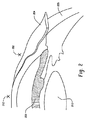
眼における大きい切開部は、視力に影響を与え、痛みを伴い、回復が遅く、かつ感染しやすいことがある。従って、水晶体104の除去を必要とする眼治療を実施する時に、水晶体104の幅全体にわたる切開部を作るのは、最善ではない場合がある。図1は、より小さい接近切開部を通して実施することができると考えられる除去に向けて眼の水晶体を切除する方法を示している。図1で「x」により識別される接近切開部110が、水晶体104に接近するために外側眼組織を通して作られる。レーザツール112又はその一部分が、接近切開部110を通して挿入される。挿入されたレーザツール112を使用して、水晶体104の一部分を切除するために電磁エネルギが集束され、その切除は、眼102からの除去に向けて水晶体を複数の部分に破砕する。図1の例では、電磁エネルギは、水晶体を複数のパイ(pie)形状部分114に破壊するために点線に沿って集束される。これらのパイ形状部分114は、水晶体104全体よりも小さいので、接近切開部110を通して抽出することができる。複数の部分114は、ピンセット又は吸引を通じてなどの様々な方法で除去することができる。 Large incisions in the eye can affect visual acuity, can be painful, slow recovery, and can be susceptible to infection. Thus, when performing eye treatments that require removal of the lens 104, it may not be best to make an incision across the width of the lens 104. FIG. 1 illustrates a method of resecting an eye lens for removal that could be performed through a smaller access incision. An access incision 110, identified by “x” in FIG. 1, is made through the outer eye tissue to access the lens 104. Laser tool 112 or a portion thereof is inserted through access incision 110. Using the inserted laser tool 112, electromagnetic energy is focused to ablate a portion of the lens 104, which ablate the lens into portions for removal from the eye 102. In the example of FIG. 1, the electromagnetic energy is focused along a dotted line to break the lens into a plurality of pie-shaped portions 114. Since these pie-shaped portions 114 are smaller than the entire lens 104, they can be extracted through the access incision 110. The plurality of portions 114 can be removed in various ways, such as through tweezers or suction.
図2は、接近切開部を通じた眼の内部組織への接近を示す眼の側面図である。接近切開部202が、結膜204及び強膜206のような外側眼組織のある一定の層を通して作られる。接近切開部202は、水晶体210のような内部構造への接近を求める際に、小柱網208のようなある一定の内部眼構造を横断する場合もある。水晶体抽出処置では、水晶体210又は水晶体210を取り囲む組織は、抽出に向けて接近切開部202を通して接近される。電磁エネルギが、水晶体210又は水晶体210を取り囲む組織上に接近切開部202を通して集束され、水晶体210を眼から分離し、抽出に向けて接近切開部202を通して水晶体を破砕する。一例では、レーザツール(例えば、可撓性レーザ先端)が、接近切開部202を通して挿入され、かつ電磁エネルギを水晶体210上に集束させて水晶体210を所定のパターンに従って抽出に向けて複数の部分に切除するように操作される。
FIG. 2 is a side view of the eye showing access to the internal tissue of the eye through the access incision. An
図2は、水晶体210の半径の外側から作られた接近切開部202を通じた水晶体への接近を示すが、水晶体210及び他の内部構造には、212に示すように、水晶体の前又は水晶体の上方から作られた接近切開部のような他のタイプの接近切開部、並びに他のタイプの切開部を通して接近可能である。図3は、水晶体の前又は水晶体の上方からのこのような接近切開部の上面図を示している。図3では、水晶体302は、眼304の中心に示されており、水晶体302は、虹彩308の中心にある瞳孔306越しに見える。処置中、瞳孔306を拡張することができ、接近切開部310を水晶体302の前に又は水晶体302の上に作り、瞳孔306を通して水晶体302に接近可能である。
FIG. 2 illustrates access to the lens through an
図4は、水晶体の半径の外側の外側眼組織を通る切開部を通じた水晶体への接近を示す図である。図4では、水晶体402は、眼404の中心に示されている。水晶体402は、虹彩408によって取り囲まれた眼の瞳孔406越しに見られる。虹彩408は、強膜及び結膜又は眼の白色部分のような眼組織のある一定の外側層によって取り囲まれている。図4の例では、水晶体は、水晶体402の半径の近くであるがその外側の外側眼組織410を通して(例えば、角強膜の縁を通して)作られた接近切開部412(例えば、3mmの切開部)を通して接近される。切開部412は、外科用メス又はレーザのような切断ツールを使用して作ることができる。同じか又は異なるタイプの同じ切断ツール414又は別の切断ツールが、水晶体402のような内眼組織に接近するために接近切開部412を通して挿入される。例えば、可撓性又は非可撓性先端のようなレーザツールを使用して接近切開部412を作ることができ、レーザツールは、眼404からの水晶体の除去を容易にするために、電磁エネルギを水晶体402上に集束させて複数の部分に水晶体を切除するように接近切開部412を通して挿入される。レーザツール(例えば、2,750から3,000nmの間の波長を有する中赤外域レーザ)は、切断又は切除される組織のタイプに基づく異なる設定(例えば、5から100Hzでの0.05から3ワットのエネルギレベル)で作動することができる。例えば、20Hzでの1.25ワットの設定及び短いパルス持続時間は、結膜を通じた切断に使用することができ、一方、30Hzでの0.25ワットの設定は、水晶体切除に使用することができ、より高い設定(例えば、0.75ワット)は、より硬い混濁部に利用される。
FIG. 4 illustrates the access to the lens through an incision through the outer ocular tissue outside the lens radius. In FIG. 4, the
切除は、いくつかの機構を使用して行うことができる。例えば、切除は、切除治療を行う外科医又は他の技術者により操作されるハンドレーザツールを使用して行うことができる。別の例では、切除は、予め定義された又は予めプログラムされたパターンに従って切除のパターンを作る処理において一度に1つ又はそれよりも多くの点で切除を行うように構成されたコンピュータ制御式のスキャナを使用して行うことができる。このような切除手順は、眼組織を損傷することがある超音波又は他の振動手順を使用しない抽出に向けて、複数の部分への水晶体の分割を可能にすることができる。 Ablation can be performed using several mechanisms. For example, ablation can be performed using a hand laser tool operated by a surgeon or other technician performing the ablation treatment. In another example, ablation is a computer controlled computer configured to perform ablation at one or more points at a time in the process of creating a pattern of ablation according to a pre-defined or pre-programmed pattern. Can be done using a scanner. Such an ablation procedure can allow for the division of the lens into multiple portions for extraction without the use of ultrasound or other vibration procedures that can damage the ocular tissue.
接近切開部を半径の外側に作ることは、いくつかの利益を有することができる。例えば、治療を実施する時に、水晶体の半径の外側に作られた接近切開部は、ある一定の敏感な眼構造の意図しない損傷を回避することができる。例えば、瞳孔を通して前から又は水晶体の上方から水晶体に接近する時に、瞳孔の一体性は、虹彩の潜在的な損傷により危うくなる。側面から水晶体に接近することにより、このような危険性を緩和することができる。 Making the access incision outside the radius can have several benefits. For example, when performing treatment, an access incision made outside the radius of the lens can avoid unintentional damage to certain sensitive eye structures. For example, when approaching the lens from the front through the pupil or from above the lens, pupil integrity is compromised by potential damage to the iris. Such danger can be mitigated by approaching the lens from the side.
図5は、結膜の回転を通じて達成することができる水晶体の半径の外側からの切開部の別の潜在的な利益を示している。結膜及び強膜502は、眼の白色部分を形成する眼組織の外側層であり、結膜は、強膜上にあり、強膜に対して摺動又は回転させることができる。図5の例では、結膜層は、強膜及び水晶体508のような下にある他の眼組織に対して静止位置504から回転した位置506まで回転される。結膜は、指又はクランプを使用して回転した位置506に保持することができる。506の接近切開部は、結膜が回転した位置506にある状態で、506で水晶体の外側の近くに作られる。接近切開部は、水晶体508のような内眼組織に接近するために結膜及び強膜を横断する。結膜は、回転した位置506に保たれ、治療は、506に示されている回転した位置で結膜を破る接近切開部を通して内眼組織及びその点より下の他の眼組織に適用される。
FIG. 5 illustrates another potential benefit of an incision from outside the lens radius that can be achieved through conjunctival rotation. The conjunctiva and
治療が完了して治療ツールが接近切開部から除去される時に、結膜は、静止位置504に戻ることが許容される。結膜が弛緩後の位置504まで弛緩すると、結膜を通る接近切開部の部分は、静止位置504に移動し、強膜のような眼組織の下側層を通る接近切開部の部分は、水晶体508の近くに残る。このようにして、結膜及び強膜及び他の内部眼組織の損傷した部分は千鳥配置される。この千鳥配置により、損傷した強膜及びより下の位置にある組織の上に結膜の無傷の部分ができ、これは、感染の防止を補助することができる。更に、眼組織の損傷した層の千鳥配置により、個々に損傷した層への血流が良くなり、治療時間が短縮される。
The conjunctiva is allowed to return to the
ある一定の実施では、ターゲット眼組織を所定の又は予め定義されたパターンに従って切除することが望ましい場合がある。例えば、眼の水晶体を除去する処置中に、水晶体は、水晶体の完全な直径よりも小さい切開部を通じた除去に向けてパターンに従って複数の部分に切除される場合がある。図6は、切除パターンを眼の水晶体上に投影する可視光パターン投影器を示す図である。可視光パターン投影器602は、可視光のパターンを眼604の一部分上に投影する。図6の例では、可視光パターン投影器602は、外科用メス、レーザ、又は他の切断ツールを眼604からの水晶体606の抽出に使用して水晶体606を切断する際に従うべきパターンを識別するパターンを眼604の水晶体606上に伝達する。図6のパターンは、実質的に円形の第1の部分608、及び実質的に円形の第1の部分608内の複数のクロスカットで構成された第2の部分610から構成されたパイ形のパターンである。可視光パターンは、マスクを通して投影される低電力レーザ又は可視光のような様々な機構を使用して投影することができる。可視光パターンは、3次元(3D)を含む様々な方法で投影することができる。例えば、3Dパターンは、ある一定の切除又は切断の深さの案内の補助に利用することができる。
In certain implementations, it may be desirable to excise the target eye tissue according to a predetermined or predefined pattern. For example, during a procedure that removes the lens of the eye, the lens may be excised into portions according to a pattern for removal through an incision that is smaller than the full diameter of the lens. FIG. 6 is a diagram showing a visible light pattern projector that projects an ablation pattern onto the eye lens. The visible
水晶体606への接近は、612に示すように水晶体606の前又は水晶体の上方から、又は614に示すように水晶体606の半径の外側から作ることができる接近切開部を通して達成される。水晶体606への接近時に、水晶体606は、可視光パターン投影器602により水晶体606上へ投影されたパターン608、610の方向に沿って又はそれを通して切断又は切除を作ることにより、複数の部分に分割される。例えば、電磁エネルギは、水晶体606を投影されたパターンの実質的に円形の第1の部分608に沿って、かつその後に可視光パターン608、610の第2の部分610の内部クロスカットに沿って切除するために、接近切開部614で挿入された可撓性レーザ先端を通して集束させることができる。別の例では、第2の部分610の内部クロスカットは、パターンの第1の部分608の実質的に円形の切断部を作る前に作ることができる。その後に、レーザ融除後に残る水晶体606の個々の部分は、接近切開部614を通して眼604から除去することができ、水晶体606の除去に必要とされる接近切開部614のサイズの最小化が可能である。
Access to the
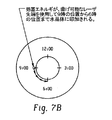
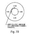
図7は、可撓性レーザ先端レーザツールを使用する投影されたパターンの各部分の例示的な切除を示す図である。図7Aでは、可撓性先端は、水晶体の9時の位置の近くの接近切開部を通して挿入される。図7Bでは、電磁エネルギは、水晶体上へ投影された切除パターンの実質的に円形の一部分に従うように9時の位置から6時の位置に向けて水晶体の周囲に沿って集束され、水晶体の外縁を切除する。一部の実施では、電磁エネルギは、可撓性レーザ先端の操作を通じて3時の位置まで全体を通して集束させることができる。図7Cでは、可撓性先端は、9時の位置に引っ込められ、図7Dでは、電磁エネルギは、他方の方向に12時の位置に至るまで水晶体の縁部に沿って集束され、水晶体の外縁の切除を完了するために引き続き3時の位置まで継続される。図7Eでは、可撓性レーザ先端は、9時の位置に引っ込められ、接近切開部を通して取り出される。 FIG. 7 is a diagram illustrating an exemplary ablation of portions of a projected pattern using a flexible laser tip laser tool. In FIG. 7A, the flexible tip is inserted through an access incision near the 9 o'clock position of the lens. In FIG. 7B, the electromagnetic energy is focused along the periphery of the lens from the 9 o'clock position to the 6 o'clock position so as to follow a substantially circular portion of the ablation pattern projected onto the lens. Excise. In some implementations, the electromagnetic energy can be focused throughout to the 3 o'clock position through manipulation of the flexible laser tip. In FIG. 7C, the flexible tip is retracted to the 9 o'clock position, and in FIG. 7D, the electromagnetic energy is focused along the edge of the lens until it reaches the 12 o'clock position in the other direction. Continue to the 3 o'clock position to complete the resection. In FIG. 7E, the flexible laser tip is retracted to the 9 o'clock position and removed through the access incision.
可視光パターン投影器により投影されたパターンは、様々な形態を取ることができる。図8は、薄切りパターンを水晶体上に投影する可視光パターン投影器を示す図である。可視光パターン投影器802は、水晶体組織804の切断又は切除を案内するためなどに可視光パターンを眼806の水晶体804上に投影する。図8で投影されたパターンは、第1の実質的に円形の部分808と平行クロスカット810とで構成される。実際は、切断ツールは、812、814に示されている切開部の一方のような接近切開部を通して挿入することができる。切断ツールは、可視光パターン投影器802により水晶体804上へ投影された複数の平行クロスカット線810に沿って水晶体804を切断又は切除するのに使用することができる。その後に、切断ツールは、投影されたパターンの実質的に円形の808の部分に沿って水晶体804を切断又は切除するのに使用することができる。投影されたパターンに沿った又はこれを通る切断部の順番は、変えることができることに注意されたい。水晶体の複数の得られたストリップは、次に、利用した接近切開部812、814を通して抽出することができる。 The pattern projected by the visible light pattern projector can take various forms. FIG. 8 is a diagram showing a visible light pattern projector that projects a sliced pattern onto a crystalline lens. The visible light pattern projector 802 projects a visible light pattern onto the lens 804 of the eye 806, such as to guide cutting or excision of the lens tissue 804. The pattern projected in FIG. 8 is composed of a first substantially circular portion 808 and a parallel crosscut 810. In practice, the cutting tool can be inserted through an access incision, such as one of the incisions shown at 812,814. The cutting tool can be used to cut or ablate the lens 804 along a plurality of parallel crosscut lines 810 projected onto the lens 804 by the visible light pattern projector 802. Thereafter, the cutting tool can be used to cut or ablate the lens 804 along a substantially circular 808 portion of the projected pattern. Note that the order of cuts along or through the projected pattern can be varied. Multiple resulting strips of the lens can then be extracted through the utilized access incisions 812, 814.
図9は、同心円パターンを水晶体上に投影する可視光パターン投影器を示す図である。可視光パターン投影器902は、水晶体組織904の切断又は切除を案内するためなどに可視光パターンを眼906の水晶体904上に投影する。図9で投影されたパターンは、第1の実質的に円形の部分908と同心円の内側切断部910とで構成される。実際は、切断ツールは、912、914に示されている切開部の一方のような接近切開部を通して挿入することができる。切断ツールは、可視光パターン投影器902又はLensXのような光学系を有するコンピュータスキャナを使用することによって水晶体904上へ投影された複数の同心円方向910に沿って水晶体904を切断、切除、又は破壊するのに使用することができる。切断ツールは、次に、投影されたパターンの実質的に円形の908の部分に沿って水晶体904を切断又は切除するのに使用することができる。水晶体の複数の得られたストリップは、除去前に水晶体の円形の部分を折り畳むことによるなどで、利用した接近切開部912、914(例えば、弓形エキシマ切開部)を通して抽出することができ、硬い切断されていない水晶体のこのような折り畳みは不可能である。
FIG. 9 is a diagram showing a visible light pattern projector that projects a concentric pattern onto a crystalline lens. The visible
図10は、例示的なレーザツールを示す図である。レーザエネルギのような電磁エネルギが、第1の端部1002でレーザツールに供給される。電磁エネルギは、レーザツールの長さに沿って伝播され、電磁エネルギがレーザ先端1004を通じて、レーザ先端1004の少なくとも端部1006から放出される。図10に示すレーザツールは、潅注ポート1008を更に含むことができ、これは、水を潅注ポート1008からレーザ先端1004の端部1006の近くの治療部位に噴霧することにより、水を眼の水晶体1010のような治療部位に導入するように構成される。潅注ポート1008によって供給された水は、様々な目的に機能することができる。例えば、潅注ポートは、水を水晶体1010治療部位に供給して水晶体1010の断片をほぐし、吸入ポート1012からの吸引を通じた断片の抽出を容易にすることができる。
FIG. 10 is a diagram illustrating an exemplary laser tool. Electromagnetic energy, such as laser energy, is supplied to the laser tool at the
図11は、潅注ポートからの水の潜在的な利益を示す図である。図11の例では、電磁エネルギは、2.78nmEr:YSGGレーザによって供給される。切除及び切断治療を行う際のこのようなレーザ1102の効果は、そのレーザ1102が、水晶体治療部位1106のような治療部位に存在する水1104と相互作用する時に高められる。実際に、このようなレーザ1102は、電磁エネルギ1108が水1104の存在しない場所に集束された時には切断又は切除を行うことができない。しかし、このようなレーザ1102が、潅注ポート1110から噴霧された水1104のような水1104が存在する治療部位1106に集束された時には、水晶体1106のような治療部位での組織は、1112に示すように切断又は切除される(例えば、水の微小膨張及び微小切除を通じて)。1つのこのようなレーザは、米国特許第8,033,825号明細書に説明されており、この特許の開示内容は、引用により本明細書に組み込まれている。潅注ポート1110からの水を使用する切除に続いて、水晶体1106の複数の部分は、支援としての潅注ポート1110からの追加の水1104の有無に関わらず、吸入ポート1114を通して抽出することができる。
FIG. 11 illustrates the potential benefits of water from the irrigation port. In the example of FIG. 11, electromagnetic energy is provided by a 2.78 nm Er: YSGG laser. The effectiveness of such a laser 1102 in performing an ablation and amputation treatment is enhanced when the laser 1102 interacts with
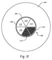
水晶体を複数の部分に分割するための水晶体の各部分の切除に続いて、水晶体のこの部分は、外科手術用ピンセット又はその他を使用して、図10及び図11に示す吸入ポートを通してなどで眼から除去される。図12は、切除された水晶体のある一定の部分の除去を示す図である。図示の眼1202は、パイ形状のパターン1206で切除された水晶体1204を含む。点線は、切除されて水晶体部分の一部としてもはや存在しない水晶体組織を表している。切除に続いて、水晶体1204は、6つのパイ形状部分に分割される。これらの部分の各々は、全体として水晶体1204より小さく、従って1208に示す切開部のような、より小さい切開部を通して除去することができる。これらの部分の各々は、別々に除去される。図12の例では、部分1210のうち2つが除去され、一方、パイ形状部分1212のうちの4つは、まだ接近切開部1206を通して除去されていない。
Following excision of each part of the lens to divide the lens into a plurality of parts, this part of the lens can be viewed by using surgical tweezers or the like, such as through the inhalation port shown in FIGS. Removed from. FIG. 12 illustrates the removal of certain portions of the excised lens. The illustrated
上述したように、水晶体及び他の眼組織切除は、異なるタイプ及びサイズの様々な異なるレーザ先端を使用して行うことができる。図13は、可撓性先端を含む例示的なレーザツールを示している。レーザツールは、Alcon Laboratories,Inc.からのLensSx(登録商標)レーザ、エキシマレーザ、又は他のタイプのレーザを利用することができる。可撓性先端1304により、水晶体又は眼の他の組織の各部分の切除が可能である。可撓性先端1304は、端部発射平坦、尖形、又は湾曲先端、又は側面発射先端、又は放射状発射先端のような様々な形態を取ることができる。レーザ先端1304は、様々な形態を取ることができる。例えば、レーザ先端1304は、米国特許第8,221,117号明細書又は米国特許第7,702,196号明細書に開示されたもののような側面発射レーザ先端とすることができ、これらの両方の開示内容は、引用により本明細書に組み込まれている。ある一定の例示的なレーザ先端は、図17に示されている。例示的なレーザ先端1304は、BioLase,Inc.によるPerio 300先端、部品番号740020を更に含むことができる。この先端は、ツイスト−オン式の簡単さを有し、時間を消費する剥離及び劈開を排除している。この先端は、眼の全ての区域への接近に向けて曲げ可能であり、かつ使い捨ての先端として使用することができる。Perio 300先端は、1.1mmの直径と7mm又は9mmのファイバ長さとを有し、チューブ外寸長さは15mmである。
As mentioned above, the lens and other ocular tissue resections can be performed using a variety of different laser tips of different types and sizes. FIG. 13 illustrates an exemplary laser tool that includes a flexible tip. Laser tools are available from Alcon Laboratories, Inc. A LensSx® laser, an excimer laser, or other type of laser from can be used. A
眼の水晶体の全て又は殆どの除去後に、水晶体のある一定の望ましくない残り又は他の不要な組織が眼の中に残る場合がある。例えば、混濁部除去処置中に、発芽細胞が、水晶体切除後に眼の中に残る場合がある。時間と共に、これらの発芽細胞は、交換水晶体上での新しい混濁部の生成の開始点になって白内障処置の利益を限定することがある。従って、いかなる発芽細胞の位置も、水晶体除去後及び処置終了前に識別して切除することが望ましいと考えられる。発芽細胞は、水晶体除去後に顕微鏡又は裸眼を使用して見ることができる。図14は、発芽細胞が存在する水晶体が除去された眼を示す図である。図14は、上述の処置の1つを使用するなどで水晶体1404が除去された眼1402を示している。水晶体1404除去後に、発芽細胞1406は、除去した水晶体の部位又はその近くに存在する。混濁部の再成長を防止するために、これらの発芽細胞1406の位置が識別された状態で、電磁エネルギが、発芽細胞1406を切除するために発芽細胞1406上に集束される。
After removal of all or most of the eye lens, certain undesirable remnants of the lens or other unwanted tissue may remain in the eye. For example, during opacity procedures, sprouting cells may remain in the eye after lens excision. Over time, these sprouting cells may limit the benefit of cataract treatment by becoming the starting point for the creation of new turbidity on the exchange lens. Therefore, it may be desirable to identify and excise any sprouting cell location after lens removal and before the end of treatment. Sprouting cells can be viewed using a microscope or the naked eye after lens removal. FIG. 14 is a diagram showing an eye from which a lens in which sprouting cells are present has been removed. FIG. 14 shows the
眼の水晶体除去後に、交換眼内水晶体を本来の水晶体が以前に占有していた水晶体嚢に位置決めすることができる。図15は、折り畳み水晶体技術を使用する交換水晶体の挿入を示す図である。折り畳まれてない水晶体が、1502に示されている。その折り畳まれてない水晶体1502は、折り畳み水晶体1504の幅を接近切開部1506を通して収まることができる幅に低減するために、図示のように1回又はそれよりも多くの回数で折り畳むことができる。処置中、折り畳み水晶体1504は、本来の水晶体が以前に占有していた膜ポケットに挿入される。水晶体は、眼の内側にある状態で、広げられて正しく位置決めされる。図16は、交換水晶体が従来の水晶体の除去後に位置決めされた眼を示す図である。眼1602は、本来の以前に位置決めされていた水晶体の位置に位置決めされた太線によって示された交換水晶体1604を含む。処置完了後に、結膜が接近切開部を作る前に回転されていた場合には、その回転を解除することができ、結膜切開部は、1606に示すように処置部位から離れ、それによって結膜及び下に重なる処置部位の両方の治癒が潜在的に早まる。
After removal of the lens of the eye, the replacement intraocular lens can be positioned in the capsular bag previously occupied by the original lens. FIG. 15 is a diagram illustrating the insertion of a replacement lens using a folded lens technique. An unfolded lens is shown at 1502. The unfolded
本出願は、本発明を例示するために実施例を使用した。本発明の特許請求可能な範囲は、他の実施例を含むことができる。 This application used examples to illustrate the present invention. The claimable scope of the invention may include other embodiments.
102 眼
104 水晶体
110 接近切開部
112 レーザツール
114 パイ形状部分
102 eye 104 crystalline lens 110 approaching incision part 112 laser tool 114 pie-shaped part
Claims (8)
前記水晶体の外縁に沿った周経路に従う可視光パターンを前記眼の前記水晶体上に投影するように構成され及び配置された可視光パターン投影器と、
先端を備えたレーザハンドツールであって、該レーザハンドツールは、隣接する複数のポートを備え、該複数のポートは、
前記先端を通して前記先端から放出されたレーザエネルギのビームと交わるように水を差し向けるように構成され及び配置された潅注ポート及び吸入ポートを含み、
前記レーザエネルギは前記水と反応して前記水の微小膨張及び微小切除を生じさせ、前記周経路に沿って前記水晶体を複数の部分に切除し、
前記隣接する複数のポートのうち少なくとも一つが、前記レーザエネルギのビームに向けて角度がつけられている、
ことを特徴とする装置。 A device for resecting the lens of the eye,
A visible light pattern projector configured and arranged to project a visible light pattern following a circumferential path along an outer edge of the lens onto the lens of the eye;
A laser hand tool with a tip, the laser hand tool comprising a plurality of adjacent ports, the plurality of ports comprising:
An irrigation port and an inhalation port configured and arranged to direct water through the tip to intersect the beam of laser energy emitted from the tip;
The laser energy causes a small expansion and microdissection of the water reacts with the water, the lens was cut into a plurality of portions along the circumferential path,
At least one of the plurality of adjacent ports is angled toward the beam of laser energy;
A device characterized by that.
請求項1に記載の装置。 The visible light pattern projector is configured and arranged to project a first portion of the visible light pattern that is substantially circular in shape and located on or near the outer edge of the lens. ,
The apparatus of claim 1.
前記可視光パターンの前記第2の部分は前記水晶体を複数の部分に切除するように構成され及び配置されている、
請求項2に記載の装置。 The laser hand tool is configured and arranged to project a second portion of the visible light pattern into the substantially circular first portion;
The second portion of the visible light pattern is constructed and arranged to excise the lens into a plurality of portions;
The apparatus of claim 2.
請求項1に記載の装置。 The access incision includes an incision made through the conjunctiva of the eye outside the radius of the lens;
The apparatus of claim 1.
請求項1に記載の装置。 The tip includes a flexible laser chip protruding outward from the tool;
The apparatus of claim 1.
請求項1に記載の装置。 The laser hand tool further comprises a suction generator for removing the plurality of portions of the lens by suction following the excision of the lens.
The apparatus of claim 1.
請求項1に記載の装置。 Following the excision of the lens, the laser hand tool is constructed and arranged to excise the germinated cells by focusing the laser energy on the water directed near the germinating cells remaining in the eye. ing,
The apparatus of claim 1.
請求項1に記載の装置。The apparatus of claim 1.
Applications Claiming Priority (7)
| Application Number | Priority Date | Filing Date | Title |
|---|---|---|---|
| US201161542702P | 2011-10-03 | 2011-10-03 | |
| US61/542,702 | 2011-10-03 | ||
| US201161551826P | 2011-10-26 | 2011-10-26 | |
| US61/551,826 | 2011-10-26 | ||
| PCT/US2012/058455 WO2013052481A1 (en) | 2011-10-03 | 2012-10-02 | Systems and methods for disruption of an eye lens |
| US13/633,505 US9060845B2 (en) | 2011-10-03 | 2012-10-02 | Systems and methods for disruption of an eye lens |
| US13/633,505 | 2012-10-02 |
Related Child Applications (1)
| Application Number | Title | Priority Date | Filing Date |
|---|---|---|---|
| JP2018115506A Division JP2018187402A (en) | 2011-10-03 | 2018-06-18 | Systems and methods for disruption of eye lens |
Publications (3)
| Publication Number | Publication Date |
|---|---|
| JP2014531289A JP2014531289A (en) | 2014-11-27 |
| JP2014531289A5 JP2014531289A5 (en) | 2015-11-19 |
| JP6357418B2 true JP6357418B2 (en) | 2018-07-11 |
Family
ID=47993297
Family Applications (2)
| Application Number | Title | Priority Date | Filing Date |
|---|---|---|---|
| JP2014534637A Expired - Fee Related JP6357418B2 (en) | 2011-10-03 | 2012-10-02 | System and method for fracture of an eye lens |
| JP2018115506A Pending JP2018187402A (en) | 2011-10-03 | 2018-06-18 | Systems and methods for disruption of eye lens |
Family Applications After (1)
| Application Number | Title | Priority Date | Filing Date |
|---|---|---|---|
| JP2018115506A Pending JP2018187402A (en) | 2011-10-03 | 2018-06-18 | Systems and methods for disruption of eye lens |
Country Status (6)
| Country | Link |
|---|---|
| US (2) | US9060845B2 (en) |
| EP (1) | EP2763636B1 (en) |
| JP (2) | JP6357418B2 (en) |
| CA (1) | CA2850494C (en) |
| ES (1) | ES2658360T3 (en) |
| WO (1) | WO2013052481A1 (en) |
Families Citing this family (19)
| Publication number | Priority date | Publication date | Assignee | Title |
|---|---|---|---|---|
| WO2007124038A2 (en) | 2006-04-20 | 2007-11-01 | Dentatek Corporation | Apparatus and methods for treating root canals of teeth |
| US7980854B2 (en) | 2006-08-24 | 2011-07-19 | Medical Dental Advanced Technologies Group, L.L.C. | Dental and medical treatments and procedures |
| US9232959B2 (en) | 2007-01-02 | 2016-01-12 | Aquabeam, Llc | Multi fluid tissue resection methods and devices |
| WO2009111736A1 (en) | 2008-03-06 | 2009-09-11 | Aquabeam Llc | Tissue ablation and cautery with optical energy carried in fluid stream |
| CN104203078B (en) | 2012-02-29 | 2018-04-20 | 普罗赛普特生物机器人公司 | The cutting tissue of automated image guiding and processing |
| EP2956070A4 (en) | 2013-02-14 | 2016-12-07 | Procept Biorobotics Corp | Aquablation aquabeam eye surgery methods and apparatus |
| US9877801B2 (en) | 2013-06-26 | 2018-01-30 | Sonendo, Inc. | Apparatus and methods for filling teeth and root canals |
| EP2910194A1 (en) * | 2014-02-21 | 2015-08-26 | 3DIntegrated ApS | Surgical instrument |
| US10624785B2 (en) | 2016-01-30 | 2020-04-21 | Carl Zeiss Meditec Cataract Technology Inc. | Devices and methods for ocular surgery |
| EP3576695B1 (en) | 2017-01-31 | 2023-02-15 | AMO Development, LLC | Systems for laser ophthalmic surgery that provide for iris exposures below a predetermined exposure limit |
| ES2914400T3 (en) | 2017-05-04 | 2022-06-10 | Zeiss Carl Meditec Inc | eye surgery devices |
| US11110004B2 (en) | 2017-09-18 | 2021-09-07 | William F. WILEY | Flexible/expandable phacoemulsification tip |
| WO2019067435A1 (en) | 2017-09-26 | 2019-04-04 | Wiley William J | Self-contained ocular surgery instrument |
| CN111093573B (en) * | 2017-09-27 | 2022-05-17 | 光物质相互作用有限公司 | Surgical instrument for minimally invasive aspiration of tissue |
| CN112702982B (en) | 2018-06-05 | 2023-12-19 | 卡尔蔡司白内障医疗技术公司 | Ophthalmic microsurgical tools, systems, and methods of use |
| JP7434340B2 (en) | 2019-02-01 | 2024-02-20 | カール・ツァイス・メディテック・キャタラクト・テクノロジー・インコーポレイテッド | Ophthalmic cutting instrument with integrated suction pump |
| CN114096221A (en) | 2019-05-17 | 2022-02-25 | 卡尔蔡司白内障医疗技术公司 | Ophthalmic cutting instrument with integrated suction pump |
| US11801163B2 (en) | 2019-06-07 | 2023-10-31 | Carl Zeiss Meditec Cataract Technology Inc. | Multi-stage trigger for ophthalmology cutting tool |
| USD997355S1 (en) | 2020-10-07 | 2023-08-29 | Sonendo, Inc. | Dental treatment instrument |
Family Cites Families (42)
| Publication number | Priority date | Publication date | Assignee | Title |
|---|---|---|---|---|
| US4744360A (en) * | 1986-12-18 | 1988-05-17 | Bath Patricia E | Apparatus for ablating and removing cataract lenses |
| AU3221589A (en) * | 1988-03-31 | 1989-10-05 | Site Microsurgical Systems, Inc. | Means for tissue removal using an erbium host laser |
| US5222952A (en) | 1988-10-28 | 1993-06-29 | Hanspeter Loertscher | Method for laser sclerostomy |
| US5722970A (en) * | 1991-04-04 | 1998-03-03 | Premier Laser Systems, Inc. | Laser surgical method using transparent probe |
| JP3197327B2 (en) * | 1991-05-11 | 2001-08-13 | 株式会社ニデック | Ophthalmic laser device |
| US5439462A (en) * | 1992-02-25 | 1995-08-08 | Intelligent Surgical Lasers | Apparatus for removing cataractous material |
| US5643250A (en) | 1992-08-07 | 1997-07-01 | O'donnell, Jr.; Francis E. | Laser probe hand piece |
| JP2670420B2 (en) * | 1993-11-18 | 1997-10-29 | 株式会社吉田製作所 | Laser cutting equipment |
| US5549598A (en) | 1995-05-22 | 1996-08-27 | O'donnell, Jr.; Francis E. | Glaucoma laser trabeculodissection |
| US6567582B1 (en) | 1995-08-31 | 2003-05-20 | Biolase Tech Inc | Fiber tip fluid output device |
| US20060240381A1 (en) | 1995-08-31 | 2006-10-26 | Biolase Technology, Inc. | Fluid conditioning system |
| US6389193B1 (en) | 1998-12-22 | 2002-05-14 | Biolase Technology, Inc. | Rotating handpiece |
| US7620290B2 (en) | 1995-08-31 | 2009-11-17 | Biolase Technology, Inc. | Modified-output fiber optic tips |
| DE19705815C2 (en) | 1997-02-15 | 1999-02-11 | Heidelberg Engineering Optisch | Medical device for microsurgery on the eye |
| DE19718139A1 (en) * | 1997-04-30 | 1998-11-05 | Aesculap Meditec Gmbh | Phaco-emulsification method for intra=ocular tissue removal |
| US20090298004A1 (en) | 1997-11-06 | 2009-12-03 | Rizoiu Ioana M | Tunnelling probe |
| DE19852574A1 (en) * | 1998-11-06 | 2000-05-11 | Aesculap Meditec Gmbh | Medical instrument for phacoemulsification |
| US20050113911A1 (en) * | 2002-10-17 | 2005-05-26 | Peyman Gholam A. | Adjustable intraocular lens for insertion into the capsular bag |
| US20070031473A1 (en) * | 2005-08-05 | 2007-02-08 | Peyman Gholam A | Drug delivery system and method |
| US6984230B2 (en) * | 2000-04-07 | 2006-01-10 | Synergetics, Inc. | Directional laser probe |
| US6520955B2 (en) * | 2000-12-28 | 2003-02-18 | Michael Reynard | Phacophotolysis method and apparatus |
| JP4194842B2 (en) | 2001-01-18 | 2008-12-10 | ザ リージェンツ オブ ザ ユニバーシティ オブ カリフォルニア | Minimally invasive glaucoma surgical instruments and methods |
| US6743221B1 (en) * | 2001-03-13 | 2004-06-01 | James L. Hobart | Laser system and method for treatment of biological tissues |
| US20070042315A1 (en) | 2005-06-24 | 2007-02-22 | Biolase Technology, Inc. | Visual feedback implements for electromagnetic energy output devices |
| US6855164B2 (en) | 2001-06-11 | 2005-02-15 | Vision Solutions Technologies, Llc | Multi-focal intraocular lens, and methods for making and using same |
| US7384419B2 (en) | 2002-08-26 | 2008-06-10 | Biolase Technology, Inc. | Tapered fused waveguide for delivering treatment electromagnetic radiation toward a target surfaced |
| US20080269731A1 (en) | 2003-11-19 | 2008-10-30 | Casimir Andrew Swinger | Method and apparatus applying patient-verified prescription of high order aberrations |
| DE102004021754A1 (en) * | 2004-04-30 | 2005-11-24 | Reinhardt Thyzel | Device for removing epithelial cells from a lens capsular bag of a human or animal eye |
| US8394084B2 (en) | 2005-01-10 | 2013-03-12 | Optimedica Corporation | Apparatus for patterned plasma-mediated laser trephination of the lens capsule and three dimensional phaco-segmentation |
| US7421186B2 (en) | 2005-01-10 | 2008-09-02 | Biolase Technology, Inc. | Modified-output fiber optic tips |
| WO2006089288A2 (en) * | 2005-02-19 | 2006-08-24 | Lenticular Research Group Llc | Apparatus and processes for preventing or delaying onset or progression of age-related cataract |
| CN101203190A (en) | 2005-04-22 | 2008-06-18 | 生物雷射科技股份有限公司 | Methods for treating hyperopia and presbyopia via laser perforating |
| KR100976281B1 (en) * | 2005-04-26 | 2010-08-16 | 바이오레이즈 테크놀로지, 인크. | Device for treating an eye |
| AU2006249760C1 (en) | 2005-05-25 | 2010-11-04 | Biolase, Inc. | Device having activated textured surfaces for treating oral tissue |
| BRPI0611186A2 (en) | 2005-06-03 | 2010-08-24 | Biolase Tech Inc | Tissue treatment device and method |
| US7492987B2 (en) * | 2005-12-19 | 2009-02-17 | Trimedyne, Inc. | Fiber optic laser energy delivery devices |
| US8544473B2 (en) | 2006-04-26 | 2013-10-01 | Biolase, Inc. | Methods for treating eye conditions with low-level light therapy |
| US20120135368A1 (en) | 2007-01-26 | 2012-05-31 | Rizoiu Ioana M | Modified-ouput fiber optic tips |
| US8568393B2 (en) * | 2007-03-13 | 2013-10-29 | Topcon Medical Laser Systems, Inc. | Computer guided patterned laser trabeculoplasty |
| CN101631522B (en) * | 2007-03-13 | 2014-11-05 | 眼科医疗公司 | Apparatus for creating ocular surgical and relaxing incisions |
| EP2323579A4 (en) | 2008-08-13 | 2014-05-14 | Biolase Inc | Methods and devices for treating presbyopia |
| US20120089134A1 (en) * | 2010-10-11 | 2012-04-12 | Christopher Horvath | Contactless Photodisruptive Laser assisted Cataract Surgery |
-
2012
- 2012-10-02 EP EP12838722.2A patent/EP2763636B1/en not_active Not-in-force
- 2012-10-02 JP JP2014534637A patent/JP6357418B2/en not_active Expired - Fee Related
- 2012-10-02 WO PCT/US2012/058455 patent/WO2013052481A1/en active Application Filing
- 2012-10-02 US US13/633,505 patent/US9060845B2/en not_active Expired - Fee Related
- 2012-10-02 CA CA2850494A patent/CA2850494C/en not_active Expired - Fee Related
- 2012-10-02 ES ES12838722.2T patent/ES2658360T3/en active Active
-
2015
- 2015-06-22 US US14/746,794 patent/US20150359672A1/en not_active Abandoned
-
2018
- 2018-06-18 JP JP2018115506A patent/JP2018187402A/en active Pending
Also Published As
| Publication number | Publication date |
|---|---|
| US20130085482A1 (en) | 2013-04-04 |
| CA2850494A1 (en) | 2013-04-11 |
| EP2763636B1 (en) | 2017-12-06 |
| US9060845B2 (en) | 2015-06-23 |
| ES2658360T3 (en) | 2018-03-09 |
| JP2018187402A (en) | 2018-11-29 |
| JP2014531289A (en) | 2014-11-27 |
| CA2850494C (en) | 2017-06-06 |
| US20150359672A1 (en) | 2015-12-17 |
| EP2763636A1 (en) | 2014-08-13 |
| EP2763636A4 (en) | 2015-06-17 |
| WO2013052481A1 (en) | 2013-04-11 |
Similar Documents
| Publication | Publication Date | Title |
|---|---|---|
| JP6357418B2 (en) | System and method for fracture of an eye lens | |
| JP4762252B2 (en) | Apparatus and method for separating multiple material layers having different removal thresholds | |
| AU2006238845B2 (en) | Methods for treating hyperopia and presbyopia via laser tunneling | |
| US20030014042A1 (en) | Method of creating stromal pockets for corneal implants | |
| EP2059202B1 (en) | System for marking corneal tissue in a transplant procedure | |
| US8256431B2 (en) | Methods for treating hyperopia and presbyopia via laser tunneling | |
| JP2002500522A (en) | Methods of corneal laser surgery | |
| AU2007292492A1 (en) | System and method for resecting corneal tissue using non-continuous initial incisions | |
| WO2001067968A1 (en) | A method and apparatus to correct vision disorders | |
| US20070244472A1 (en) | System and method for creating suture channels | |
| JP2009531079A (en) | Laser mask for creating corneal pockets | |
| JP2002541878A (en) | Keratotomy device | |
| RU150549U1 (en) | TOOL FOR DESTRUCTION OF THE EXTERNAL WALL OF THE SHLEMMOV CHANNEL | |
| RU2722813C1 (en) | Surgical treatment method of dacryocystitis | |
| RU2629719C1 (en) | Method of reoperation of laser specialized keratomileusis |
Legal Events
| Date | Code | Title | Description |
|---|---|---|---|
| A521 | Request for written amendment filed |
Free format text: JAPANESE INTERMEDIATE CODE: A523 Effective date: 20151001 |
|
| A621 | Written request for application examination |
Free format text: JAPANESE INTERMEDIATE CODE: A621 Effective date: 20151001 |
|
| A131 | Notification of reasons for refusal |
Free format text: JAPANESE INTERMEDIATE CODE: A131 Effective date: 20160725 |
|
| A977 | Report on retrieval |
Free format text: JAPANESE INTERMEDIATE CODE: A971007 Effective date: 20160722 |
|
| A601 | Written request for extension of time |
Free format text: JAPANESE INTERMEDIATE CODE: A601 Effective date: 20161025 |
|
| A601 | Written request for extension of time |
Free format text: JAPANESE INTERMEDIATE CODE: A601 Effective date: 20161226 |
|
| A521 | Request for written amendment filed |
Free format text: JAPANESE INTERMEDIATE CODE: A523 Effective date: 20170125 |
|
| RD04 | Notification of resignation of power of attorney |
Free format text: JAPANESE INTERMEDIATE CODE: A7424 Effective date: 20170413 |
|
| A131 | Notification of reasons for refusal |
Free format text: JAPANESE INTERMEDIATE CODE: A131 Effective date: 20170703 |
|
| A601 | Written request for extension of time |
Free format text: JAPANESE INTERMEDIATE CODE: A601 Effective date: 20171003 |
|
| A601 | Written request for extension of time |
Free format text: JAPANESE INTERMEDIATE CODE: A601 Effective date: 20171129 |
|
| A521 | Request for written amendment filed |
Free format text: JAPANESE INTERMEDIATE CODE: A523 Effective date: 20180104 |
|
| TRDD | Decision of grant or rejection written | ||
| A01 | Written decision to grant a patent or to grant a registration (utility model) |
Free format text: JAPANESE INTERMEDIATE CODE: A01 Effective date: 20180418 |
|
| A601 | Written request for extension of time |
Free format text: JAPANESE INTERMEDIATE CODE: A601 Effective date: 20180518 |
|
| A61 | First payment of annual fees (during grant procedure) |
Free format text: JAPANESE INTERMEDIATE CODE: A61 Effective date: 20180618 |
|
| R150 | Certificate of patent or registration of utility model |
Ref document number: 6357418 Country of ref document: JP Free format text: JAPANESE INTERMEDIATE CODE: R150 |
|
| LAPS | Cancellation because of no payment of annual fees |