JP6285972B2 - Use of ACY-1 as a marker for ischemia / reperfusion, delayed graft function and graft viability and methods thereof - Google Patents
Use of ACY-1 as a marker for ischemia / reperfusion, delayed graft function and graft viability and methods thereof Download PDFInfo
- Publication number
- JP6285972B2 JP6285972B2 JP2015562318A JP2015562318A JP6285972B2 JP 6285972 B2 JP6285972 B2 JP 6285972B2 JP 2015562318 A JP2015562318 A JP 2015562318A JP 2015562318 A JP2015562318 A JP 2015562318A JP 6285972 B2 JP6285972 B2 JP 6285972B2
- Authority
- JP
- Japan
- Prior art keywords
- acy
- patient
- dgf
- sample
- transplantation
- Prior art date
- Legal status (The legal status is an assumption and is not a legal conclusion. Google has not performed a legal analysis and makes no representation as to the accuracy of the status listed.)
- Active
Links
Images


Classifications
-
- G—PHYSICS
- G01—MEASURING; TESTING
- G01N—INVESTIGATING OR ANALYSING MATERIALS BY DETERMINING THEIR CHEMICAL OR PHYSICAL PROPERTIES
- G01N33/00—Investigating or analysing materials by specific methods not covered by groups G01N1/00 - G01N31/00
- G01N33/48—Biological material, e.g. blood, urine; Haemocytometers
- G01N33/50—Chemical analysis of biological material, e.g. blood, urine; Testing involving biospecific ligand binding methods; Immunological testing
- G01N33/53—Immunoassay; Biospecific binding assay; Materials therefor
- G01N33/573—Immunoassay; Biospecific binding assay; Materials therefor for enzymes or isoenzymes
-
- C—CHEMISTRY; METALLURGY
- C12—BIOCHEMISTRY; BEER; SPIRITS; WINE; VINEGAR; MICROBIOLOGY; ENZYMOLOGY; MUTATION OR GENETIC ENGINEERING
- C12N—MICROORGANISMS OR ENZYMES; COMPOSITIONS THEREOF; PROPAGATING, PRESERVING, OR MAINTAINING MICROORGANISMS; MUTATION OR GENETIC ENGINEERING; CULTURE MEDIA
- C12N9/00—Enzymes; Proenzymes; Compositions thereof; Processes for preparing, activating, inhibiting, separating or purifying enzymes
- C12N9/14—Hydrolases (3)
- C12N9/78—Hydrolases (3) acting on carbon to nitrogen bonds other than peptide bonds (3.5)
- C12N9/80—Hydrolases (3) acting on carbon to nitrogen bonds other than peptide bonds (3.5) acting on amide bonds in linear amides (3.5.1)
-
- C—CHEMISTRY; METALLURGY
- C12—BIOCHEMISTRY; BEER; SPIRITS; WINE; VINEGAR; MICROBIOLOGY; ENZYMOLOGY; MUTATION OR GENETIC ENGINEERING
- C12Q—MEASURING OR TESTING PROCESSES INVOLVING ENZYMES, NUCLEIC ACIDS OR MICROORGANISMS; COMPOSITIONS OR TEST PAPERS THEREFOR; PROCESSES OF PREPARING SUCH COMPOSITIONS; CONDITION-RESPONSIVE CONTROL IN MICROBIOLOGICAL OR ENZYMOLOGICAL PROCESSES
- C12Q1/00—Measuring or testing processes involving enzymes, nucleic acids or microorganisms; Compositions therefor; Processes of preparing such compositions
- C12Q1/34—Measuring or testing processes involving enzymes, nucleic acids or microorganisms; Compositions therefor; Processes of preparing such compositions involving hydrolase
-
- C—CHEMISTRY; METALLURGY
- C12—BIOCHEMISTRY; BEER; SPIRITS; WINE; VINEGAR; MICROBIOLOGY; ENZYMOLOGY; MUTATION OR GENETIC ENGINEERING
- C12Q—MEASURING OR TESTING PROCESSES INVOLVING ENZYMES, NUCLEIC ACIDS OR MICROORGANISMS; COMPOSITIONS OR TEST PAPERS THEREFOR; PROCESSES OF PREPARING SUCH COMPOSITIONS; CONDITION-RESPONSIVE CONTROL IN MICROBIOLOGICAL OR ENZYMOLOGICAL PROCESSES
- C12Q1/00—Measuring or testing processes involving enzymes, nucleic acids or microorganisms; Compositions therefor; Processes of preparing such compositions
- C12Q1/68—Measuring or testing processes involving enzymes, nucleic acids or microorganisms; Compositions therefor; Processes of preparing such compositions involving nucleic acids
- C12Q1/6876—Nucleic acid products used in the analysis of nucleic acids, e.g. primers or probes
- C12Q1/6883—Nucleic acid products used in the analysis of nucleic acids, e.g. primers or probes for diseases caused by alterations of genetic material
-
- G—PHYSICS
- G01—MEASURING; TESTING
- G01N—INVESTIGATING OR ANALYSING MATERIALS BY DETERMINING THEIR CHEMICAL OR PHYSICAL PROPERTIES
- G01N33/00—Investigating or analysing materials by specific methods not covered by groups G01N1/00 - G01N31/00
- G01N33/48—Biological material, e.g. blood, urine; Haemocytometers
- G01N33/50—Chemical analysis of biological material, e.g. blood, urine; Testing involving biospecific ligand binding methods; Immunological testing
- G01N33/68—Chemical analysis of biological material, e.g. blood, urine; Testing involving biospecific ligand binding methods; Immunological testing involving proteins, peptides or amino acids
- G01N33/6893—Chemical analysis of biological material, e.g. blood, urine; Testing involving biospecific ligand binding methods; Immunological testing involving proteins, peptides or amino acids related to diseases not provided for elsewhere
-
- C—CHEMISTRY; METALLURGY
- C12—BIOCHEMISTRY; BEER; SPIRITS; WINE; VINEGAR; MICROBIOLOGY; ENZYMOLOGY; MUTATION OR GENETIC ENGINEERING
- C12Q—MEASURING OR TESTING PROCESSES INVOLVING ENZYMES, NUCLEIC ACIDS OR MICROORGANISMS; COMPOSITIONS OR TEST PAPERS THEREFOR; PROCESSES OF PREPARING SUCH COMPOSITIONS; CONDITION-RESPONSIVE CONTROL IN MICROBIOLOGICAL OR ENZYMOLOGICAL PROCESSES
- C12Q2600/00—Oligonucleotides characterized by their use
- C12Q2600/118—Prognosis of disease development
-
- C—CHEMISTRY; METALLURGY
- C12—BIOCHEMISTRY; BEER; SPIRITS; WINE; VINEGAR; MICROBIOLOGY; ENZYMOLOGY; MUTATION OR GENETIC ENGINEERING
- C12Q—MEASURING OR TESTING PROCESSES INVOLVING ENZYMES, NUCLEIC ACIDS OR MICROORGANISMS; COMPOSITIONS OR TEST PAPERS THEREFOR; PROCESSES OF PREPARING SUCH COMPOSITIONS; CONDITION-RESPONSIVE CONTROL IN MICROBIOLOGICAL OR ENZYMOLOGICAL PROCESSES
- C12Q2600/00—Oligonucleotides characterized by their use
- C12Q2600/158—Expression markers
-
- G—PHYSICS
- G01—MEASURING; TESTING
- G01N—INVESTIGATING OR ANALYSING MATERIALS BY DETERMINING THEIR CHEMICAL OR PHYSICAL PROPERTIES
- G01N2333/00—Assays involving biological materials from specific organisms or of a specific nature
- G01N2333/90—Enzymes; Proenzymes
- G01N2333/914—Hydrolases (3)
- G01N2333/978—Hydrolases (3) acting on carbon to nitrogen bonds other than peptide bonds (3.5)
- G01N2333/98—Hydrolases (3) acting on carbon to nitrogen bonds other than peptide bonds (3.5) acting on amide bonds in linear amides (3.5.1)
-
- G—PHYSICS
- G01—MEASURING; TESTING
- G01N—INVESTIGATING OR ANALYSING MATERIALS BY DETERMINING THEIR CHEMICAL OR PHYSICAL PROPERTIES
- G01N2800/00—Detection or diagnosis of diseases
- G01N2800/24—Immunology or allergic disorders
- G01N2800/245—Transplantation related diseases, e.g. graft versus host disease
-
- G—PHYSICS
- G01—MEASURING; TESTING
- G01N—INVESTIGATING OR ANALYSING MATERIALS BY DETERMINING THEIR CHEMICAL OR PHYSICAL PROPERTIES
- G01N2800/00—Detection or diagnosis of diseases
- G01N2800/32—Cardiovascular disorders
- G01N2800/324—Coronary artery diseases, e.g. angina pectoris, myocardial infarction
-
- G—PHYSICS
- G01—MEASURING; TESTING
- G01N—INVESTIGATING OR ANALYSING MATERIALS BY DETERMINING THEIR CHEMICAL OR PHYSICAL PROPERTIES
- G01N2800/00—Detection or diagnosis of diseases
- G01N2800/70—Mechanisms involved in disease identification
- G01N2800/7019—Ischaemia
Landscapes
- Life Sciences & Earth Sciences (AREA)
- Health & Medical Sciences (AREA)
- Chemical & Material Sciences (AREA)
- Engineering & Computer Science (AREA)
- Organic Chemistry (AREA)
- Immunology (AREA)
- Molecular Biology (AREA)
- Proteomics, Peptides & Aminoacids (AREA)
- Wood Science & Technology (AREA)
- Zoology (AREA)
- Biomedical Technology (AREA)
- Analytical Chemistry (AREA)
- Biotechnology (AREA)
- Microbiology (AREA)
- Biochemistry (AREA)
- General Health & Medical Sciences (AREA)
- Genetics & Genomics (AREA)
- Urology & Nephrology (AREA)
- Hematology (AREA)
- Physics & Mathematics (AREA)
- Bioinformatics & Cheminformatics (AREA)
- Pathology (AREA)
- Medicinal Chemistry (AREA)
- General Engineering & Computer Science (AREA)
- General Physics & Mathematics (AREA)
- Food Science & Technology (AREA)
- Cell Biology (AREA)
- Biophysics (AREA)
- Investigating Or Analysing Biological Materials (AREA)
- Measuring Or Testing Involving Enzymes Or Micro-Organisms (AREA)
Description
本発明は、虚血再灌流障害又は移植片機能発現遅延のためのバイオマーカーに係る。具体的には、本発明は、アミノアシラーゼ−1及びアミノアシラーゼ−1を使用する診断及び予後の方法に係る。 The present invention relates to a biomarker for ischemia-reperfusion injury or graft function delay. Specifically, the present invention relates to diagnostic and prognostic methods using aminoacylase-1 and aminoacylase-1.
腎臓移植は、末期腎疾患の患者にとっては明確なメリットを提供し1,2、透析と比べて顕著な費用の節約を提供する3,4。2010年において、米国では、16,151件の腎移植が行われ(http://optn.transplant.hrsa.gov)、これに対して、英国での対応する数値は2,687件である(http://www.uktransplant.org.uk)。しかし、患者の一部は、早期に、移植片機能発現遅延(DGF)5のような、臨床転帰及び健康上の医療経済学的成果に顕著に影響を及ぼす合併症を経験している。 Kidney transplants, for patients with end-stage renal disease and provide clear benefits 1,2, compared with dialysis provides savings significant cost 3,4. In 2010, 16,151 kidney transplants were performed in the United States (http://optn.transplant.hrsa.gov), while the corresponding figure in the UK is 2,687 ( http://www.uktransplant.org.uk). However, some patients are experiencing early complications that significantly affect clinical outcomes and health-health economic outcomes, such as delayed graft function onset (DGF) 5 .
DGFについて多数の定義が提案されている6-8が、一般的に使用されている1つは、単独の高カリウム血症のため以外に、腎移植後第1週において透析が必要とされることである。他の臨床状況において見られる急性腎障害との類似性があるが、DGFの根本的病理は、ドナーの年齢及び虚血の期間のようなドナーに由来の要因、及び再灌流障害、免疫応答、及び免疫抑制剤9のようなレシピエントの要因の関与と複雑に絡み合っている。虚血再灌流障害(IRI)に続く急性細尿管壊死(ATN)は、DGFの患者における主な組織所見であるが、しかし、急性の細胞性又は体液性の拒絶反応が同時に生ずることがあり、組織学的には、他の病理、例えば、カルシニューリン阻害剤毒性が、しばしば、認められる。循環死後に提供された(DCD)臓器及び境界領域ドナーから提供された臓器の使用が増大しているが10、これは、DGFの発生の増大(米国において、最近では、移植レシピエントの〜20%に及んでいる)に一致している。DGFは、移植片生着不全、患者の死亡、死打ち切り移植片生着不全のリスクを2〜3倍増大させ11,12、乏しい1年時移植機能、動脈性高血圧、及び急性の拒絶反応のような、移植片の長期生着を低減させる多数の合併症と関連する13。総合的には、DGFは、正に3年経過時の移植片機能損失のリスクを41%増大させることと関連している14。 Many definitions for DGF have been proposed 6-8, but one commonly used is that dialysis is required in the first week after kidney transplantation, in addition to single hyperkalemia That is. Although similar to the acute kidney injury found in other clinical situations, the underlying pathology of DGF is due to donor-derived factors such as donor age and duration of ischemia, and reperfusion injury, immune response, And intricately intertwined with the involvement of recipient factors such as immunosuppressant 9 . Acute tubule necrosis (ATN) following ischemia-reperfusion injury (IRI) is a major histological finding in patients with DGF, but acute cellular or humoral rejection may occur simultaneously Histologically, other pathologies such as calcineurin inhibitor toxicity are often observed. Although the use of donated (DCD) organs after circulation death and organs donated from borderline donors is increasing 10 , this is an increase in the incidence of DGF (in the United States, recently, 20 %). DGF increases the risk of graft survival failure, patient death, death censored graft survival failure 2-3 times 11,12 , poor one-year transplant function, arterial hypertension, and acute rejection It is associated with a number of complications that reduce long-term graft survival, such as 13 . Overall, DGF is associated with a 41% increase in the risk of loss of graft function at exactly 3 years 14 .
DGFの早期の識別及び基礎病理学の理解の増大は、体液量状態の最適化、時宜を得た適切な透析、及び不必要な検査及び治療の回避を可能にする即時の患者管理を改善する顕著な可能性を有する15。患者を階級化する機会及び初期時点での目的に合わせた特別な治療介入は、改善された長期転帰を生ずる。プロテオミック技術の開発とともに、臨床的影響を有する新規のバイオマーカーを同定すること(ゲノムに基づく研究から出現する有望なマーカーを補完する)における臨床プロテオミクスの可能性に関する興奮が増大している16。 Early identification of DGF and increased understanding of basic pathology improves immediate patient management that allows optimization of fluid volume status, timely and proper dialysis, and avoidance of unnecessary testing and treatment 15 with remarkable potential. Special treatment interventions tailored to the opportunity to classify patients and their objectives at an early point in time produce improved long-term outcomes. With the development of proteomic technology, there is increasing excitement regarding the potential for clinical proteomics in identifying new biomarkers with clinical impact (complementing promising markers emerging from genome-based studies) 16 .
移植後の透析及び移植片回復に関する要求の潜在的な予測因子として現在調査中の尿中のマーカーとしては、インターロイキン18(IL−18)及び好中球ゼラチナーゼリポカリン(NGAL)が含まれ17、組織関連マーカーとしては、ICAM−1及びVCAMが含まれている15,18。残念ながら、DGFのケースの多くでは、尿が生成されないか、又は多くの結果の解析を混乱させる残留する本来の腎性アウトプットと混合され、DGFが一旦発症すると、生検組織は、しばしば、比較的遅い時期にのみ入手可能である。NGAL及びIL−18は、血清中において分析される際、確約を示してはいないが19、血液由来のバイオマーカーは、病院検査室での利便性及び日常的な使用のため、移植後を監視する理想的な方法であろう。しかし、血清又は血漿によるバイオマーカーの発見は、総患者数の〜99%からなるわずか22人の患者によって行われており、タンパク質存在量の広いダイナミックレンジは>10桁に及ぶ20。 Urinary markers currently under investigation as potential predictors of post-transplant dialysis and graft recovery requirements include interleukin 18 (IL-18) and neutrophil gelatinase lipocalin (NGAL) 17 , Tissue-related markers include ICAM-1 and VCAM 15,18 . Unfortunately, in many cases of DGF, urine is not produced or mixed with residual natural renal output that disrupts the analysis of many results, and once DGF develops, biopsy tissue often becomes It is only available at a relatively late time. NGAL and IL-18 are not assured when analyzed in serum 19 , but blood-derived biomarkers are monitored after transplantation for convenience and routine use in hospital laboratories Would be an ideal way to do. However, the discovery of biomarkers by serum or plasma is carried out by only 22 patients consisting of 99% of the total number of patients, a wide dynamic range protein abundance spans> 10 digits 20.
上述のとおり、NGAL、IL−18、KIM−1、L−FABP、ネトリン−1、及びケラチノサイト由来ケモカインのような多数の有望なバイオマーカーが腎虚血再灌流障害と関連しているが、これらの多くは、尿中、及びDGFの状況における移植後よりもむしろ本来の急性腎障害(AKI)において研究されている。DGFにおいて尿中のNGAL及びIL−18が検査され、移植後18時間において、AUCs0.78及び0.77であった。これらの結果を通して奨励されるようになっているが、それらは、DGFにおいて、尿排出量が移植後の全ての時点で生ずるかどうか、及びこのような尿排出量が、本来の腎臓から生成される現在の尿によって、どこで混同されるかについての障害の対象である。さらに、尿中NGALは、AKIにおいて、内因性腎不全から腎前性腎不全を区別するためのマーカーとして提案されており、移植患者における同様の使用も想像されるが、その真の識別能力は疑わしい。NGAL及びIL−18は、DGF及びSGFにおいて血清中で研究されているが、群間の識別は認められておらず、一方、シスタチンC(血清クレアチニンよりも優れている)は識別した。ドナーの尿中NGAL(血清NGALではない)は、長期のDGFと関連している。IRIにおけるバイオマーカーについての多数の候補及び現在までのNGAL及びIL−18のような有望なマーカーの調査にもかかわらず、明確な臨床上の有益性を有する血清中のマーカーの候補は、今のところ出現していない。 As noted above, a number of promising biomarkers such as NGAL, IL-18, KIM-1, L-FABP, netrin-1, and keratinocyte-derived chemokines are associated with renal ischemia-reperfusion injury, Many have been studied in natural acute kidney injury (AKI) rather than after transplantation in the urine and DGF situations. Urinary NGAL and IL-18 were examined in DGF and were AUCs 0.78 and 0.77 18 hours after transplantation. While these results have been encouraged, they have been shown in DGF whether urine output occurs at all time points after transplantation, and such urine output is generated from the original kidney. The subject of the disorder about where it is confused by the current urine. Furthermore, urinary NGAL has been proposed as a marker in AKI to distinguish prerenal renal failure from endogenous renal failure, and similar use in transplant patients is envisioned, but its true discriminating ability is I doubt it. NGAL and IL-18 have been studied in serum in DGF and SGF, but no discrimination between groups has been observed, while cystatin C (which is superior to serum creatinine) has been identified. Donor urinary NGAL (not serum NGAL) is associated with long-term DGF. Despite the search for a number of candidates for biomarkers in IRI and promising markers such as NGAL and IL-18 to date, candidates for markers in serum with clear clinical benefit are now However, it has not appeared.
移植片機能発現遅延は、患者の入院の長さ及びこれに関連する費用についての影響の理由だけでなく、患者の体液状態、及び最少の腎臓機能を示している「ウエットの」移植後患者からの過剰な体液除去により起こり得る可及的な更なる臨床上の合併症を回避するための時宜を得た適切な透析の使用の管理における困難性の理由のため、腎移植においては、なお、重大な臨床的事象である。さらに、DGFを経験した患者に対する公知のマイナスの長期間の影響は、これが腎移植を行う臨床医にとって極めて重要であることを意味する。 Delayed onset of graft function is not only because of the impact on the length of hospitalization and associated costs of the patient, but also from the “wet” post-transplant patient showing the patient's fluid status and minimal kidney function. Due to the difficulty in managing the use of timely and appropriate dialysis to avoid the possible further clinical complications that can occur due to excessive fluid removal of It is a serious clinical event. Furthermore, the known negative long-term effects on patients experiencing DGF mean that this is extremely important for clinicians performing kidney transplants.
従って、DGFのような虚血再灌流障害を予測する血液由来のバイオマーカーに関する要求がなお存在している。 Thus, there is still a need for blood-derived biomarkers that predict ischemia-reperfusion injury such as DGF.
本発明によれば、ACY−1の虚血再灌流障害のためのバイオマーカーとしての使用が提供される。好ましくは、前記虚血再灌流障害は、脳、心臓、腎臓、肺及び肝臓から選ばれる少なくとも1つの組織におけるものである。 According to the present invention, the use of ACY-1 as a biomarker for ischemia-reperfusion injury is provided. Preferably, the ischemia-reperfusion injury is in at least one tissue selected from brain, heart, kidney, lung and liver.
好ましくは、前記虚血再灌流障害は、心筋梗塞、卒中、手術、損傷又は臓器移植によって生じたものである。 Preferably, the ischemia-reperfusion injury is caused by myocardial infarction, stroke, surgery, injury or organ transplantation.
好ましくは、前記虚血再灌流障害は、移植手術後の患者において、移植片機能発現遅延に至る。好ましくは、前記移植手術後の患者は、腎移植手術後の患者である。 Preferably, the ischemia-reperfusion injury leads to delayed graft function in patients after transplantation surgery. Preferably, the patient after the transplant operation is a patient after the kidney transplant operation.
他の態様では、本発明は、ACY−1の移植片機能発現遅延のためのバイオマーカーとしての使用を提供する。 In another aspect, the present invention provides the use of ACY-1 as a biomarker for delayed onset of graft function.
さらに他の態様では、本発明は、患者における虚血再灌流障害を診断する方法を提供し、該方法は、i)患者から採取したサンプルにおけるACY−1のレベルを測定し;ii)患者のサンプルにおけるACY−1のレベルを、コントロールサンプルにおけるACY−1のレベル又は予め決定したACY−1についての対照レベルと対比することを含んでなり、コントロールサンプルと対比して又は予め決定した対照レベルと対比して、患者サンプルにおけるACY−1のレベルが増大している場合に、患者が虚血再灌流障害を有していると認定する。 In yet another aspect, the present invention provides a method of diagnosing ischemia-reperfusion injury in a patient, the method measuring i) the level of ACY-1 in a sample taken from the patient; ii) in the patient Comparing the level of ACY-1 in the sample with the level of ACY-1 in the control sample or a control level for the predetermined ACY-1 and contrasting with the control sample or with the predetermined control level In contrast, a patient is identified as having ischemia-reperfusion injury if the level of ACY-1 in the patient sample is increased.
好ましくは、前記虚血再灌流障害は、脳、心臓、腎臓、肺及び肝臓から選ばれる少なくとも1の組織におけるものである。好ましくは、前記虚血再灌流障害は、心筋梗塞、卒中、又は臓器移植によって生じたものである。 Preferably, the ischemia-reperfusion injury is in at least one tissue selected from brain, heart, kidney, lung and liver. Preferably, the ischemia-reperfusion injury is caused by myocardial infarction, stroke, or organ transplantation.
好ましくは、前記患者は腎移植手術後の患者である。 Preferably, the patient is a patient after a kidney transplant operation.
更なる態様では、本発明は、患者における移植片機能発現遅延を診断する方法を提供し、該方法は、i)患者から採取したサンプルにおけるACY−1のレベルを測定し;ii)患者のサンプルにおけるACY−1のレベルを、コントロールサンプルにおけるACY−1のレベル又は予め決定したACY−1についての対照レベルと対比することを含んでなり、コントロールサンプルと対比して又は予め決定した対照レベルと対比して、患者サンプルにおけるACY−1のレベルが増大している場合に、患者が移植片機能発現遅延を有していると認定する。 In a further aspect, the present invention provides a method of diagnosing graft function delay in a patient, the method measuring i) the level of ACY-1 in a sample taken from the patient; ii) a sample of the patient Comparing the level of ACY-1 in the control sample with the level of ACY-1 in the control sample or a predetermined control level for ACY-1 and contrasting with the control sample or with the predetermined control level Thus, if the level of ACY-1 in the patient sample is increased, the patient is identified as having a delayed onset of graft function.
好ましくは、前記患者は腎移植手術後の患者である。 Preferably, the patient is a patient after a kidney transplant operation.
さらに他の態様では、本発明は、術後の腎移植患者について透析管理戦略を決定する方法を提供し、該方法は、i)患者から採取したサンプルにおけるACY−1のレベルを測定し;ii)患者のサンプルにおけるACY−1のレベルを、コントロールサンプルにおけるACY−1のレベル又は予め決定したACY−1についての対照レベルと対比することを含んでなり、コントロールサンプルと対比して又は予め決定した対照レベルと対比して、患者サンプルにおけるACY−1のレベルが増大している場合に、患者が移植後1〜7日内に透析を必要としていると認定する、又はコントロールサンプルと対比して又は予め決定した対照レベルと対比して、患者サンプルにおけるACY−1のレベルが低下している場合に、患者が移植後1〜7日内には透析を必要としないと認定する。 In yet another aspect, the present invention provides a method for determining a dialysis management strategy for a post-operative kidney transplant patient, the method measuring i) the level of ACY-1 in a sample taken from the patient; ii ) Comparing the level of ACY-1 in the patient sample with the level of ACY-1 in the control sample or the control level for the predetermined ACY-1 and contrasting or predetermined with the control sample When the level of ACY-1 in the patient sample is increased compared to the control level, the patient qualifies as requiring dialysis within 1-7 days after transplantation, or compared to the control sample or in advance If the level of ACY-1 in the patient sample is reduced compared to the determined control level, the patient is Certified and do not require dialysis in the day.
さらに他の態様では、本発明は、術後の腎移植患者について体液管理戦略を決定する方法を提供し、該方法は、i)患者から単離したサンプルにおけるACY−1のレベルを測定し;ii)患者のサンプルにおけるACY−1のレベルを、コントロールサンプルにおけるACY−1のレベル又は予め決定したACY−1に関する対照レベルと対比することを含んでなり、コントロールサンプルと対比して又は予め決定した対照レベルと対比して、患者サンプルにおけるACY−1のレベルが増大している場合に、患者が、腎移植手術後の患者に通常投与される液体の量と比べて低減された量の液体を必要としていると認定する、又はコントロールサンプルと対比して又は予め決定した対照レベルと対比して、患者サンプルにおけるACY−1のレベルが低下している場合に、透析することが余り必要ではないため、患者にとっては、腎臓移植手術後の患者に通常投与される液体の量と比べて増大された量の液体がためになるであろうと認定する。 In yet another aspect, the invention provides a method of determining a fluid management strategy for a post-operative renal transplant patient, the method i) measuring the level of ACY-1 in a sample isolated from the patient; ii) Comparing the level of ACY-1 in the patient sample with the level of ACY-1 in the control sample or a predetermined control level for ACY-1 and contrasting or predetermined with the control sample When the level of ACY-1 in the patient sample is increased compared to the control level, the patient has a reduced amount of fluid compared to the amount of fluid normally administered to the patient after kidney transplant surgery. ACY-1 in a patient sample as identified as needed or compared to a control sample or compared to a predetermined control level When the level is low, it is less necessary to dialyze, so the patient will have an increased amount of fluid compared to the amount of fluid normally administered to the patient after kidney transplant surgery Authorize it.
さらに他の態様では、本発明は、患者における腎移植の術後の臨床転帰を予測する方法を提供し、該方法は、i)患者から採取したサンプルにおけるACY−1のレベルを測定し;ii)患者のサンプルにおけるACY−1のレベルを、コントロールサンプルにおけるACY−1のレベル又は予め決定したACY−1についての対照レベルと対比することを含んでなり、コントロールサンプルと対比して又は予め決定した対照レベルと対比して増大している患者サンプルにおけるACY−1のレベルは、移植後1、2、3、4又は5年の時点における前記患者の死亡フリー及び/又は透析フリー生存率の増大を予知するものであるとする。 In yet another aspect, the present invention provides a method for predicting postoperative clinical outcome of renal transplantation in a patient, the method measuring i) the level of ACY-1 in a sample taken from the patient; ii ) Comparing the level of ACY-1 in the patient sample with the level of ACY-1 in the control sample or the control level for the predetermined ACY-1 and contrasting or predetermined with the control sample The level of ACY-1 in a patient sample that is increased compared to the control level is an increase in the death-free and / or dialysis-free survival rate of the patient at 1, 2, 3, 4 or 5 years after transplantation. Suppose that it is something to be predicted.
好ましくは、サンプルは血液サンプル、さらに好ましくは血清サンプルである。 Preferably, the sample is a blood sample, more preferably a serum sample.
好ましくは、前記サンプルが、移植後の患者から得たものである。 Preferably, the sample is obtained from a patient after transplantation.
好ましくは、前記患者から、移植後第1、2、3又は4日の時点で得たものである。 Preferably, it was obtained from the patient on the first, second, third or fourth day after transplantation.
好ましくは、前記患者はヒトである。 Preferably, the patient is a human.
好ましくは、前記ACY−1はヒトACY−1である。好ましくは、前記ACY−1はポリペプチドである。好ましくは、前記ポリペプチドは、配列番号2のアミノ酸配列を有する。或いは、前記ACY−1は、mRNAのような核酸分子である。好ましくは、前記核酸分子は、配列番号1のヌクレオチド配列を有する。 Preferably, said ACY-1 is human ACY-1. Preferably, said ACY-1 is a polypeptide. Preferably, the polypeptide has the amino acid sequence of SEQ ID NO: 2. Alternatively, the ACY-1 is a nucleic acid molecule such as mRNA. Preferably, the nucleic acid molecule has the nucleotide sequence of SEQ ID NO: 1.
好ましくは、前記予め決定された対照レベルは、コントロール患者におけるACY−1の平均レベルである。 Preferably, the predetermined control level is the average level of ACY-1 in the control patient.
好ましくは、本発明の方法は、さらに、i)患者から単離したサンプルにおける、NAGL、KIM−1、IL−18、RBP、FABP4、シスタチンC及びクレアチニンから選ばれる少なく1個のバイオマーカーのレベルを測定し;ii)患者のサンプルにおけるNAGL、KIM−1、IL−18、RBP、FABP4、シスタチンC又はクレアチニンのレベルを、コントロールサンプルにおけるNAGL、KIM−1、IL−18、RBP、FABP4、シスタチンC又はクレアチニンのレベル又は予め決定したNAGL、KIM−1、IL−18、RBP、FABP4、シスタチンC又はクレアチニンに関する予め決定した対照レベルと対比することを含んでなる。 Preferably, the method of the invention further comprises i) the level of at least one biomarker selected from NAGL, KIM-1, IL-18, RBP, FABP4, cystatin C and creatinine in a sample isolated from a patient. Ii) levels of NAGL, KIM-1, IL-18, RBP, FABP4, cystatin C or creatinine in patient samples and NAGL, KIM-1, IL-18, RBP, FABP4, cystatin in control samples Contrast with a level of C or creatinine or a predetermined control level for a predetermined NAGL, KIM-1, IL-18, RBP, FABP4, cystatin C or creatinine.
さらに他の態様では、本発明は、患者における虚血再灌流障害を診断するためのキットを提供し、該キットは、i)ACY−1ポリペプチドに特異的に結合する検出可能にラベル化された試剤、又はACY−1核酸に特異的に結合する検出可能にラベル化された試剤;及びii)診断アッセイを実施するための試薬を含んでなる。 In yet another aspect, the invention provides a kit for diagnosing ischemia-reperfusion injury in a patient, the kit being i) detectably labeled that specifically binds to an ACY-1 polypeptide. Or a detectably labeled reagent that specifically binds to an ACY-1 nucleic acid; and ii) a reagent for performing a diagnostic assay.
さらに他の態様では、本発明は、患者における移植片機能発現遅延を診断するためのキットを提供し、該キットは、i)ACY−1ポリペプチドに特異的に結合する検出可能にラベル化された試剤、又はACY−1核酸に特異的に結合する検出可能にラベル化された試剤;及びii)診断アッセイを実施するための試薬を含んでなる。 In yet another aspect, the invention provides a kit for diagnosing delayed onset of graft function in a patient, the kit being i) detectably labeled that specifically binds to an ACY-1 polypeptide. Or a detectably labeled reagent that specifically binds to an ACY-1 nucleic acid; and ii) a reagent for performing a diagnostic assay.
さらに他の態様では、本発明は、ACY−1ポリペプチドを検出できる化合物又は試剤を含んでなるアッセイ装置を提供する。 In yet another aspect, the present invention provides an assay device comprising a compound or reagent capable of detecting an ACY-1 polypeptide.
さらに他の態様では、本発明は、患者における虚血再灌流障害を診断するため、又は移植片機能発現遅延を診断するため、又は患者における腎障害、腎疾患又は腎不全を診断するためのキットを提供し、該キットは、ACY−1ポリペプチドに特異的に結合する第1の抗体を含んでなるアッセイ装置;及び(2)ACY−1ポリペプチド又は第1の抗−ACY−1抗体に特異的に結合し及び検出可能な試剤に結合される第2の異なる抗体を含んでなる。 In yet another aspect, the invention provides a kit for diagnosing ischemia-reperfusion injury in a patient, or for diagnosing delayed onset of graft function, or for diagnosing renal injury, kidney disease or renal failure in a patient. Wherein the kit comprises an assay device comprising a first antibody that specifically binds to an ACY-1 polypeptide; and (2) an ACY-1 polypeptide or a first anti-ACY-1 antibody. A second different antibody that specifically binds and is bound to a detectable agent.
以下に、添付図面を参照して、本発明の具体例を例示する。 Hereinafter, specific examples of the present invention will be illustrated with reference to the accompanying drawings.
発明者らは、良好な再現性を有するプロテオミック適用範囲の中等度の深度を作成するために既に示された、高質量精度TLQ−Orbitrap Velosラベルフリー分析と組み合わせた最も豊富なタンパク質の14個の免疫除去を使用して21、DGFを有する又はDGFを有していない腎移植を受けた患者から術前及び術後に採取した血清タンパク質を比較した。発明者らは、驚くべきことには、アミノアシラーゼ−1(ACY−1)を重要な候補として同定した。アッセイに続いて、初めて血清中のACY−1の測定を可能にする開発、早くも移植後第1日のサンプルにおけるIRI及びDGFのためのバイオマーカーとしてのACY−1の有望な臨床上の利用性を確認する患者のより大きいコホートにおける検証、及び重要なことには、有意な転帰との関連が認められた。 The inventors have found 14 of the most abundant proteins in combination with high mass accuracy TLQ-Orbitrap Velos label-free analysis already shown to create a moderate depth of proteomic coverage with good reproducibility. of were compared to 21, DGF preoperative from patients received a kidney transplant which does not have a having or DGF a and serum proteins were collected after surgery using immunomagnetic removal. The inventors have surprisingly identified aminoacylase-1 (ACY-1) as an important candidate. Development that allows for the first measurement of serum ACY-1 following the assay, promising clinical use of ACY-1 as a biomarker for IRI and DGF in samples as early as 1 day post-transplant Validation in a larger cohort of patients confirming gender and, importantly, was associated with significant outcomes.
さらに、血清系バイオマーカーの同定及び血清の使用は、IRI、特にDGFとの関連では明白な利点を有する。DGF患者は尿排出量が全くない(これは、尿マーカーの有用性に対して劇的に影響を及ぼす)。余剰の本来の尿排出量は、さらに、この臨床状況において、尿マーカーのアセスメント及び使用を複雑にする。 Furthermore, the identification of serum-based biomarkers and the use of serum has obvious advantages in the context of IRI, particularly DGF. DGF patients have no urine output (this dramatically affects the usefulness of urine markers). Excessive original urine output further complicates the assessment and use of urine markers in this clinical setting.
血清のプロテオミック分析から、ACY−1をDGF用のバイオマーカーと特定した。バイオマーカーは、臨床上有用な様式で、腎移植後の患者群の早期の描写を提供し、しっかりと開発されたプロテオミック法、候補の選択及び臨床テスト開発システムを開発を通して、注意深く集めた及び注釈が付けられた臨床サンプルセットにより、臨床での使用の瀬戸際で、明確に定義された臨床上の疑問を持つ可能性を証明している。 Proteomic analysis of serum identified ACY-1 as a biomarker for DGF. The biomarkers have been carefully collected throughout the development of well-developed proteomic methods, candidate selection and clinical test development systems, providing an early depiction of the patient population after renal transplantation in a clinically useful manner and An annotated set of clinical samples demonstrates the potential for having well-defined clinical questions on the brink of clinical use.
発明者らは、1)再現性の免疫除去と、Orbitrap Velos質量分析計を使用する高質量精度タンデム質量分析との組み合わせにおける必要な方法論21;及び2)バイオマーカーパイプラインの発見及び初期検証段階、及び付随する臨床データとともに、将来を見越して集められたサンプルの高品質バンクに至る臨床医及び科学者の一体的なチームの使用の両方の開発を通して、血清のプロテオミック分析から、潜在的に臨床上の有用性を有するマーカーを誘導することができた。 The inventors have 1) the required methodology 21 in combination with reproducible immunoremoval and high mass accuracy tandem mass spectrometry using an Orbitrap Velos mass spectrometer; and 2) the discovery and initial validation phase of the biomarker pipeline From the proteomic analysis of serum, potentially through the development of both the use of an integrated team of clinicians and scientists leading to a high-quality bank of samples collected prospectively, along with accompanying clinical data Markers with clinical utility could be induced.
アミノアシラーゼ−1(ACY−1)
アミノアシラーゼ−1(ACY−1)(N−アシル−L−アミノ酸アミドヒドロラーゼとも呼ばれる)は、ペプチダーゼM20Aファミリーに属し、N−アシル化又はN−アセチル化アミノ酸(L−アスパラギン酸を除く)の加水分解に関与し、カルボン酸エステル及びL−アミノ酸を生成すると考えられる。ACY−1は、予測された単量体質量45.3kDaを有する亜鉛結合ホモ二量体細胞質タンパク質である22。真核性細胞では、全細胞タンパク質の50〜80%がN末端でアシル化されている(タンパク質の機能及び安定性に影響を及ぼし得る翻訳時又は翻訳後の修飾である)。ACY−1は、N−アシル化ペプチド、特にN−アセチル化された中性脂肪族アミノ酸の加水分解において触媒作用を発揮して、タンパク質合成のための遊離のアミノ酸を放出する23。発現及び活性の最も高いレベルは、腎臓において見られ、続いて、肝臓、脳、骨格筋及び膵臓であり、いくつかの他の臓器において低いレベルの発現が認められる22, 24-27。パン細管ACY−1発現は、アミノ酸回収における提案された役割28を有するブタ腎臓において示されたが26、ヒト腎臓では、主に近位細管発現である26。可変性の神経学的特徴を有する代謝の先天異常は、ACY−1の突然変異と関連し(突然変異に応じて、ACY−1の発現及び活性に対する影響を有する)、尿性のN−アセチル化アミノ酸を増大させる25。染色体3p21.1上に配置された場合、腎臓ガン及び肺ガンの両方において、ガン抑制遺伝子としての役割が提案されている22, 30, 31。
Aminoacylase-1 (ACY-1)
Aminoacylase-1 (ACY-1) (also called N-acyl-L-amino acid amide hydrolase) belongs to the peptidase M20A family and is a hydrolyzed N-acylated or N-acetylated amino acid (excluding L-aspartic acid). It is thought that it participates in decomposition and produces carboxylic acid esters and L-amino acids. ACY-1 is a zinc-binding homodimeric cytoplasmic protein with a predicted monomer mass of 45.3 kDa 22 . In eukaryotic cells, 50-80% of total cellular proteins are acylated at the N-terminus (translational or post-translational modifications that can affect protein function and stability). ACY-1 catalyzes the hydrolysis of N-acylated peptides, particularly N-acetylated neutral aliphatic amino acids, releasing free amino acids for protein synthesis 23 . The highest levels of expression and activity are found in the kidney, followed by the liver, brain, skeletal muscle and pancreas, with low levels of expression observed in several other organs 22, 24-27 . Pan tubule ACY-1 expression was demonstrated in porcine kidney with a proposed role 28 in the amino acid recovery 26, the human kidney is predominantly the proximal tubules expression 26. Inborn errors of metabolism with variable neurological characteristics are associated with mutations in ACY-1 (with effects on ACY-1 expression and activity, depending on the mutation), and urinary N-acetyl Increases amino acids 25 . When placed on chromosome 3p21.1, a role as a tumor suppressor gene has been proposed in both kidney and lung cancers 22, 30, 31 .
移植前の低い又は検出不能な血清ACY−1濃度及び肝臓ドナーにおける感染による又は手術後の各種増大の不在は、DGFにおけるACY−1の移植後の上昇が、単に、障害性の腎クリアランス又は炎症によるものではないことを示唆している。DGFは、プレ−獲得から手術後の期間を通して、複数のリスク要因及び細管を含む各種の細胞及び脈環構造に関わる根底にある機構と複合している9, 32。腎細尿管源を仮定すると、DGFの多くのケースにおける増大した血清ACY−1は、細管のダメージの程度を反映するものであり、これは、ATN(生検の数は少ないが)との連合及び血清ACY−1濃度の、緩慢な移植片機能発現による非併合性の移植からDGFまでの傾向によって支持される。逆に、より高いACY−1とDGFに続くより良好な転帰との間の関係は、潜在的にタンパク質合成についてのアミノ酸の利用性に対する影響を介して、増大された合成及び修復プロセスにおける役割を表示できる。 The low or undetectable serum ACY-1 concentration prior to transplantation and the absence of various increases due to infection in liver donors or after surgery indicates that post-transplant elevation of ACY-1 in DGF is simply impaired renal clearance or inflammation. It is suggested that it is not due to. DGF is complexed with a number of risk factors and the underlying mechanisms involved in various cells and vasculature, including tubules, from pre-acquisition to post-surgery 9, 32 . Assuming a renal tubule source, the increased serum ACY-1 in many cases of DGF reflects the extent of tubule damage, which is ATN (although the number of biopsies is small) The association and serum ACY-1 concentrations are supported by the trend from uncombined transplant to DGF due to slow graft function development. Conversely, the relationship between higher ACY-1 and better outcome following DGF potentially plays a role in the increased synthesis and repair process through its impact on amino acid availability for protein synthesis. Can be displayed.
ACY−1は、シクロスポリンで処置したラット又はシロリムス(ただし、タクロリムスではない)で処置したラット33では、顕著に変化する75個の尿タンパク質の1つであり、ACY−1は、臓器毒性に関与すると提唱されるミコフェノール酸モフェチル(MMF)から腎臓において産生された生物学的に活性な代謝物と付加物を形成する34。しかし、発明者らは、CNI毒性又はMMFとのリンクを見出していない。興味深いことには、AKI患者22人からの血清サンプルの分析(その内の10は、診断3日以内である)は、血清ACY−1濃度が全て<60ng/mLであること示し(データは示されていない)、これは、恐らく、損傷又は修復又はどんな根本的なIRIを反映するかどうかは、特定の移植状況において主として遭遇した及び/又はその際に、より極端且つ広範囲の虚血/病低酸素状態下においてのみ生ずる病理過程に非常に特異的であることを示している。これは、DBD、DCD及びLD移植の間の血清ACY−1における著しい差異によっても支持される。 ACY-1 is one of 75 urinary proteins that changes markedly in rats treated with cyclosporine or 33 treated with sirolimus (but not tacrolimus), and ACY-1 is involved in organ toxicity It forms adducts with biologically active metabolites produced in the kidney from the proposed mycophenolate mofetil (MMF) 34 . However, the inventors have not found a link with CNI toxicity or MMF. Interestingly, analysis of serum samples from 22 AKI patients (of which 10 are within 3 days of diagnosis) showed that all serum ACY-1 concentrations were <60 ng / mL (data shown) Not likely to reflect damage or repair or any fundamental IRI, which is mainly encountered in certain transplant situations and / or in that case more extreme and widespread ischemia / disease It is very specific for pathological processes that occur only under hypoxic conditions. This is also supported by significant differences in serum ACY-1 between DBD, DCD and LD transplantation.
DGFのためのマーカーに関して、死亡した又は生存するドナーからの移植片から0時間の生検における遺伝子の発現(特に、ケモカインCCL19及び21及びプロテアソームサブユニットPSMB8及び10において)を、関連する臨床因子と組み合わせた結果、AUC値は、DGFについて0.74及び急性拒絶反応について0.93であった35。DGF患者からの生検におけるRANTES及びCCR1/CCR2の発現は、移植後1年の時点及びそれ以降の移植片の機能と関連することも報告されている36。AKIにおける使用について、いくつかの既存のマーカーも検討されている37,38が、例えば、組織KIM−1はDGFとは相関せず39、一方、尿中NGAL及びIL−18は、DGFについて、いくらかの的中率を有する17,40が、長期間の機能との相関はない41。第1日の血清NGAL濃度は、DGFについての的中率を有していないが、発明者らの研究と同様に、血清シスタチンCはAUC0.83を有する。DGFに関連するACY−1のついての発明者らの特異度(88.5%)及び陰性的中率(81.7%)は非常に高く、潜在的にバイオマーカーパネルに貢献できる。直接的に腎機能を反映する血清シスタチンCは、各種の臨床現場においてAKIを予測することにおける使用のメタ分析でみられる0.96と同様のAUCにより、DGFを早期に予測することにおいては、ACY−1よりも優れている42。しかし、ACY−1及びシスタチンCを併用することは、第1日では、より高い特異度を有するACY−1より、わずかに良好であり、より高感度のACY−1アッセイが開発された場合には、ACY−1による更なる識別化が可能であろう。
With regard to markers for DGF, the expression of genes in biopsies at 0 hours from grafts from dead or surviving donors (especially in chemokines CCL 19 and 21 and proteasome subunits PSMB 8 and 10) and related clinical factors When combined, the AUC value was 0.74 for DGF and 0.93 for acute rejection 35 . Expression of RANTES and CCR1 / CCR2 in biopsies from DGF patients have also been reported to be associated with the function of time and
IRIのマウスモデルにおける遺伝子発現研究から、更なる推測機構が明らかであろう43。スフィンゴシンキナーゼ−1(SphK1)は、スフィンゴシン−1−ホスフェート(腎臓IRIの保護/修復に関与する)の形成において触媒として機能し44-46、機能によって分類される移植片の間で異なり47、ACY−1と相互作用することが報告されている30,48。ドナーの生検における炎症及び免疫−応答遺伝子は、圧倒的に、IRI及びDGFと関連付けられており、更なる見識を提供するために、総合システムによる生物学的分析アプローチが提案されている15。最近のDGF及び非DGF移植前生検の比較では、DGF群が、第1年の間に、腎機能に基づいてサブ分類されるまで、何らの顕著な経路のクラスタリングが見出されておらず、遺伝子がT細胞の活性化に関与する際、抗原提示及び細胞接着が、その後の乏しい機能と関連付けられている47。DGF患者のサブ分類は、当該の研究(この研究では、移植後の血清ACY−1における明確な上昇が、DGFの患者の約2/3においてのみ認められる)におけるACY−1による状況と類似している。これは、異なる根本の病態生理学的サブグループを含むことができ、また、DGF患者群内におけるACY−1の予後の値を証明する。 From gene expression studies in mouse models of IRI, further speculated mechanism will be apparent 43. Sphingosine kinase-1 (SphK1) functions as a catalyst in the formation of sphingosine-1-phosphate (involved in the protection / repair of kidney IRI) 44-46 , differing between grafts classified by function 47 , ACY Have been reported to interact with -1,30,48 . Inflammation and immune-response genes in donor biopsies are predominantly associated with IRI and DGF, and an integrated systems-based biological analysis approach has been proposed to provide further insight 15 . In a recent comparison of DGF and non-DGF pre-transplant biopsies, no significant pathway clustering was found until the DGF group was subclassified during the first year based on renal function. When the gene is involved in T cell activation, antigen presentation and cell adhesion are associated with subsequent poor function 47 . The subclassification of DGF patients is similar to the situation with ACY-1 in this study, where a clear increase in serum ACY-1 after transplantation is only seen in about 2/3 of DGF patients ing. This can include different underlying pathophysiological subgroups and also demonstrate prognostic values for ACY-1 within the DGF patient population.
尿細管細胞のアポトーシス及び再生は、腎移植における腎移植片によって経験される虚血再灌流障害(IRI)の基本的な部分であるため、及び上述のようにACY−1の管内局在化があるとすれば、移植片機能発現遅延(DGF)のような顕著なIRIに至るプロセスに関連するACY−1の可能性は、生物学的には真実味がある。Wisterラットに関する単独の研究において、ACY−1に対するカルシニューリン阻害剤及びシロリムスの影響が認められている一方、腎移植の上でのACY−1のバイオロジーの確固とした理解は欠けている。 Since apoptosis and regeneration of tubule cells is a fundamental part of ischemia-reperfusion injury (IRI) experienced by kidney grafts in kidney transplantation, and as described above, intravascular localization of ACY-1 is If so, the potential of ACY-1 associated with processes leading to significant IRI, such as graft function delay (DGF), is biologically meaningful. In a single study on Wister rats, the effects of calcineurin inhibitors and sirolimus on ACY-1 have been observed, while a solid understanding of the biology of ACY-1 on renal transplantation is lacking.
ここで使用するように、用語「ACY−1」は、アミノアシラーゼ−1のポリペプチド及び核酸分子の両方を意味するものとして使用する。 As used herein, the term “ACY-1” is used to mean both an aminoacylase-1 polypeptide and a nucleic acid molecule.
好ましくは、ACY−1は、ヒトACY−1ポリペプチド又は核酸分子である。 Preferably, ACY-1 is a human ACY-1 polypeptide or nucleic acid molecule.
ここで使用するように、用語「核酸分子」は、DNA分子(例えば、cDNA又はゲノムDNA)及びRNA分子(例えば、mRNA)及び例えば、ヌクレオチドアナログの使用によって産生されたDNA又はRNAのアナログを含む。核酸分子は一本鎖又は二本鎖であるが、好ましくは二本鎖DNAである。 As used herein, the term “nucleic acid molecule” includes DNA molecules (eg, cDNA or genomic DNA) and RNA molecules (eg, mRNA) and analogs of DNA or RNA produced, for example, by the use of nucleotide analogs. . The nucleic acid molecule is single-stranded or double-stranded, but preferably is double-stranded DNA.
1具体例では、ACY−1核酸分子は、配列番号1において示されるヌクレオチド配列、又はこのヌクレオチド配列のいずれかのフラグメントを含んでなる。1具体例では、核酸分子は、配列番号1のヌクレオチド配列又はこのヌクレオチド配列のいずれかのフラグメントからなる。 In one embodiment, the ACY-1 nucleic acid molecule comprises the nucleotide sequence shown in SEQ ID NO: 1, or a fragment of any of this nucleotide sequence. In one embodiment, the nucleic acid molecule consists of the nucleotide sequence of SEQ ID NO: 1 or a fragment of either of this nucleotide sequence.
他の具体例では、単離されたACY−1核酸分子は、配列番号1に示されるヌクレオチド配列、又はこのヌクレオチド配列のいずれかのフラグメントの補体である核酸分子である。他の具体例では、本発明の核酸分子は、ハイブリダイゼーション条件下において、配列番号1に示されるヌクレオチド配列にハイブリダイズできるように、配列番号1に示されるヌクレオチド配列に対して十分に補足性であり、これにより、安定したディープレックスを形成できる。好ましくは、ハイブリダイゼーション条件は、高ストリンジェンシーハイブリダイゼーション条件である。 In other embodiments, the isolated ACY-1 nucleic acid molecule is a nucleic acid molecule that is the complement of the nucleotide sequence set forth in SEQ ID NO: 1, or any fragment of this nucleotide sequence. In other embodiments, the nucleic acid molecule of the invention is sufficiently complementary to the nucleotide sequence set forth in SEQ ID NO: 1 so that it can hybridize under hybridization conditions to the nucleotide sequence set forth in SEQ ID NO: 1. With this, a stable deplex can be formed. Preferably, the hybridization conditions are high stringency hybridization conditions.
ここで使用するように、用語「高ストリンジェンシーハイブリダイゼーション条件」は、ハイブリダイゼーション及び洗浄のための条件を記述する。ストリンジェントな条件とは、当業者には公知であり、入手できる文献(例えば、「分子生物学における最新のプロトコル」,John Wiley & Sons,ニューヨーク,1989,6.3.1−6.3.6)に見られる。ストリンジェントなハイブリダイゼーション条件の好適な例は、6×塩化ナトリウム/クエン酸ナトリウム(SSC)中、約45℃におけるハイブリダイゼーション、続く、0.2×SSC、0.1%(w/v)SDS中、約50℃における1回又はそれ以上の洗浄である。ストリンジェントなハイブリダイゼーション条件の好適な他の例は、6×SSC中、約45℃におけるハイブリダイゼーション、続く、0.2×SSC、0.1%(w/v)SDS中、約55℃における1回又はそれ以上の洗浄である。ストリンジェントなハイブリダイゼーション条件のさらに他の例は、6×SSC中、約45℃におけるハイブリダイゼーション、続く、0.2×SSC、0.1%(w/v)SDS中、約60℃における1回又はそれ以上の洗浄である。好ましくは、ストリンジェントなハイブリダイゼーション条件は、6×SSC中、約45℃におけるハイブリダイゼーション、続く、0.2×SSC、0.1%(w/v)SDS中、約65℃における1回又はそれ以上の洗浄である。特に好ましいストリンジェンシー条件(及び実務家が、分子が本発明のハイブリダイゼーションの制限内にあるかどうかを決定するために、いかなる条件を適用すべきか確信できない場合に使用されるべき条件)は、0.5モルリン酸ナトリウム、7%(w/v)SDS中、65℃、続く、0.2×SSC、1%(w/v)SDS中、約65℃における1回又はそれ以上の洗浄である。 As used herein, the term “high stringency hybridization conditions” describes conditions for hybridization and washing. Stringent conditions are known to those skilled in the art and are available from the literature (eg, “Latest Protocols in Molecular Biology”, John Wiley & Sons, New York, 1989, 6.3.1-6.3. 6). A suitable example of stringent hybridization conditions is hybridization at about 45 ° C. in 6 × sodium chloride / sodium citrate (SSC) followed by 0.2 × SSC, 0.1% (w / v) SDS. Medium, one or more washes at about 50 ° C. Another suitable example of stringent hybridization conditions is hybridization at about 45 ° C. in 6 × SSC, followed by 0.2 × SSC, 0.1% (w / v) SDS at about 55 ° C. One or more washes. Yet another example of stringent hybridization conditions is hybridization at about 45 ° C. in 6 × SSC followed by 0.2 × SSC, 0.1% (w / v) SDS at about 60 ° C. One or more washings. Preferably, stringent hybridization conditions include hybridization at about 45 ° C. in 6 × SSC, followed by once in 0.2 × SSC, 0.1% (w / v) SDS at about 65 ° C. More cleaning. Particularly preferred stringency conditions (and conditions to be used when practitioners are not sure what conditions to apply to determine whether a molecule is within the limits of hybridization of the invention) are 0 .5 mol sodium phosphate, 7% (w / v) SDS at 65 ° C., followed by one or more washes at 0.2 × SSC, 1% (w / v) SDS at about 65 ° C. .
1具体例では、本発明のACY−1核酸分子は、配列番号1に示されるヌクレオチド配列の全長と、少なくとも約60%、65%、70%、75%、80%、85%、90%、91%、92%、93%、94%、95%、96%、97%、98%、99%又はそれ以上同一であるヌクレオチド配列を含んでなるか又はからなる。 In one embodiment, the ACY-1 nucleic acid molecule of the invention has at least about 60%, 65%, 70%, 75%, 80%, 85%, 90% of the full length of the nucleotide sequence set forth in SEQ ID NO: 1. It comprises or consists of nucleotide sequences that are 91%, 92%, 93%, 94%, 95%, 96%, 97%, 98%, 99% or more identical.
ここで使用するように、用語「相同性」及び「同一性」は、相互に交換可能なものとして使用される。配列間の配列の相同性又は同一性の算定は、下記のようにして行われる。 As used herein, the terms “homology” and “identity” are used interchangeably. Calculation of sequence homology or identity between sequences is performed as follows.
2つのアミノ酸配列、又は2つの核酸配列の同一性(%)を決定するために、最適な対比を目的として、配列を整列する(例えば、最適な整列のために、第1及び第2のアミノ酸又は核酸配列の一方又は両方にギャップを導入でき、比較のため、非相同配列を無視できる)。好適な具体例では、比較のために整列した対照配列の長さは、対照配列の長さの少なくとも30%、好ましくは少なくとも40%、さらに好ましくは少なくとも50%、さらに好ましくは少なくとも60%、さらに好ましくは少なくとも70%、75%、80%、82%、84%、85%、86%、87%、88%、89%、90%、91%、92%、93%、94%、95%、96%、97%、98%、99%又は100%である。ついで、対応するアミノ酸の位置又はヌクレオチドの位置におけるアミノ酸残基又はヌクレオチドを比較する。第1の配列における位置が、第2の配列における対応する位置と同じアミノ酸残基又はヌクレオチドによって占有されていれば、配列はその位置において同一である(ここで使用するように、アミノ酸又は核酸の「同一性」は、アミノ酸又は核酸の「相同性」と等しい)。2つの配列間の同一性(%)は、ギャップの数及び各ギャップの長さ(2つの配列の最適な整列のために導入することが要求される)を考慮して、配列によって分けられた同一の位置の数の関数である。 To determine the identity (%) of two amino acid sequences or two nucleic acid sequences, the sequences are aligned for the purpose of optimal contrast (eg, for optimal alignment, the first and second amino acids Or gaps can be introduced in one or both of the nucleic acid sequences, and for comparison, heterologous sequences can be ignored). In a preferred embodiment, the length of the control sequence aligned for comparison is at least 30%, preferably at least 40%, more preferably at least 50%, more preferably at least 60% of the length of the control sequence, Preferably at least 70%, 75%, 80%, 82%, 84%, 85%, 86%, 87%, 88%, 89%, 90%, 91%, 92%, 93%, 94%, 95% 96%, 97%, 98%, 99% or 100%. The amino acid residues or nucleotides at corresponding amino acid positions or nucleotide positions are then compared. If a position in the first sequence is occupied by the same amino acid residue or nucleotide as the corresponding position in the second sequence, then the sequence is identical at that position (as used herein, the amino acid or nucleic acid “Identity” is equivalent to “homology” of amino acids or nucleic acids). The percent identity between the two sequences was divided by sequence, taking into account the number of gaps and the length of each gap (required to be introduced for optimal alignment of the two sequences). It is a function of the number of identical positions.
配列の対比及び2つの配列間の同一性(%)の決定は、数学的アルゴリズムを使用して行われる。好適な具体例では、2つのアミノ酸配列の間の同一性(%)は、GCGソフトウエアパッケージ(http://www.gcg.comにて入手可能)におけるGAPプログラムを組み込んだNeedlemanらアルゴリズム(J. Mol. Biol.(1970),48:444−453)(BLOSUM62マトリックス又はPAM250マトリックスのいずれか及びギャップウエイト16、14、12、10、8、6、又は4及び長さウエイト1、2、3、4、5、又は6を使用する)を使用して決定される。さらに他の好適な具体例では、2つのアミノ酸配列の間の同一性(%)は、GCGソフトウエアパッケージ(http://www.gcg.comにて入手可能)におけるGAPプログラム(NWSgapdna.CMPマトリックス及びギャップウエイト40、50、60、70、又は80及び長さウエイト1、2、3、4、5、又は6を使用する)を使用して決定される。特に好適な因子のセット(及び実務家が、分子が本発明の配列同一性及び配列相同性の制限内にあるかどうかを決定するために、いかなる因子を適用すべきか確信できない場合に使用されるべきもの)は、ギャップペナルティー12、ギャップエクステンションペナルティー4及びフレームシフトギャップペナルティー5を有するBLOSUM62スコアマトリックスである。
Determination of sequence contrast and percent identity between two sequences is done using a mathematical algorithm. In a preferred embodiment, the percent identity between two amino acid sequences is determined by the Needleman et al. Algorithm (J) incorporating the GAP program in the GCG software package (available at http://www.gcg.com). Mol. Biol. (1970), 48: 444-453) (either BLOSUM62 matrix or PAM250 matrix and
或いは、2つのアミノ酸又はヌクレオチドの配列間の同一性(%)は、ALIGNプログラム(バージョン2.0)に組み込まれたMeyersらのアルゴリズム((1989)、CABIOS 4:11−17)(PMA120加重残余表、ギャップレングスペナルティー12及びギャップペナルティー4を使用する)を使用して決定される。 Alternatively, the percent identity between two amino acid or nucleotide sequences is calculated using the Meyers et al. Algorithm ((1989), CABIOS 4: 11-17) (PMA120 weighted residue) incorporated into the ALIGN program (version 2.0). Table, gap length penalty 12 and gap penalty 4).
1具体例では、核酸フラグメントは、ACY−1のドメイン、領域、又は機能的部位に相当する配列を含んでなるか又はからなる。或いは、ACY−1の核酸フラグメントは、ACY−1ポリペプチドのエピトープ保有領域をコードする。 In one embodiment, the nucleic acid fragment comprises or consists of a sequence corresponding to a domain, region, or functional site of ACY-1. Alternatively, the ACY-1 nucleic acid fragment encodes an epitope-bearing region of an ACY-1 polypeptide.
好適な具体例では、核酸フラグメントは、配列番号1の300、400、500、600、700、800、900、1000、1100、1200、1300、1400、1500、1600個又はそれ以上の連続するヌクレオチドを含んでなるか又はからなる。 In preferred embodiments, the nucleic acid fragment comprises 300, 400, 500, 600, 700, 800, 900, 1000, 1100, 1200, 1300, 1400, 1500, 1600 or more contiguous nucleotides of SEQ ID NO: 1. Comprising or consisting of.
1具体例では、ACY−1核酸分子は、ACY−1ポリペプチド、好ましくは、配列番号2のアミノ酸配列を含んでなる又はからなるACY−1ポリペプチド、好ましくは機能的対立遺伝子変異体をコードする核酸配列を含んでなるか又はからなる。或いは、ACY−1核酸分子は、ACY−1ポリペプチドの対立遺伝子変異体、好ましくは機能的対立遺伝子変異体をコードする核酸配列を含んでなるか又はからなる。 In one embodiment, the ACY-1 nucleic acid molecule encodes an ACY-1 polypeptide, preferably an ACY-1 polypeptide comprising, or consisting of, the amino acid sequence of SEQ ID NO: 2, preferably a functional allelic variant Comprising or consisting of a nucleic acid sequence. Alternatively, the ACY-1 nucleic acid molecule comprises or consists of a nucleic acid sequence encoding an allelic variant, preferably a functional allelic variant of an ACY-1 polypeptide.
ここで使用するように、用語「ACY−1ポリペプチドの対立遺伝子変異体」は、機能タンパク質及び非−機能タンパク質の両方を含む。機能的対立遺伝子変異体は、ACY−1活性を維持する、集団内のACY−1ポリペプチドの天然のアミノ酸配列変異体である。機能的対立遺伝子変異体は、一般的には、配列番号2の1又はそれ以上のアミノ酸の保存的な置換、又はポリペプチドの重大でない領域における重大でない残基の置換、欠失又は挿入のみを含有する。 As used herein, the term “allelic variant of an ACY-1 polypeptide” includes both functional and non-functional proteins. Functional allelic variants are natural amino acid sequence variants of ACY-1 polypeptides within a population that maintain ACY-1 activity. Functional allelic variants generally comprise only conservative substitutions of one or more amino acids of SEQ ID NO: 2, or substitutions, deletions or insertions of non-critical residues in non-critical regions of the polypeptide. contains.
「重大でない」アミノ酸残基は、ACY−1の生物活性を無効にする又は実質的に変更することなく、ACY−1の野生型の配列(例えば、配列番号2の配列)から変更される残基であり、一方、「必須の」アミノ酸残基は、このような変更を生ずる。 “Non-critical” amino acid residues are residues that are altered from the wild-type sequence of ACY-1 (eg, the sequence of SEQ ID NO: 2) without abolishing or substantially altering the biological activity of ACY-1. Whereas “essential” amino acid residues result in such changes.
「保存的な置換」は、アミノ酸残基が、同様の側鎖を有するアミノ酸残基で置き換えられる置換である。同様の側鎖を有するアミノ酸残基のファミリーは当分野において定義されている。これらのファミリーは、塩基性の側鎖を有するアミノ酸(例えば、リシン、アルギニン、ヒスチジン)、酸性の側鎖を有するアミノ酸(例えば、アスパラギン酸、、グルタミン酸)、不変の極性の側鎖を有するアミノ酸(例えば、グリシン、アスパラギン、グルタミン、セリン、トレオニン、チロシン、システイン)、非極性の側鎖を有するアミノ酸(例えば、アラニン、バリン、ロイシン、イソロイシン、プロリン、フェニルアラニン、メチオニン、トリプトファン)、β−分枝状の側鎖を有するアミノ酸(例えば、トレオニン、バリン、イソロイシン)及び芳香族の側鎖を有するアミノ酸(例えば、チロシン、フェニルアラニン、トリプトファン、ヒスチジン)を含む。 A “conservative substitution” is one in which the amino acid residue is replaced with an amino acid residue having a similar side chain. Families of amino acid residues having similar side chains have been defined in the art. These families include amino acids with basic side chains (eg lysine, arginine, histidine), amino acids with acidic side chains (eg aspartic acid, glutamic acid), amino acids with invariant polar side chains ( For example, glycine, asparagine, glutamine, serine, threonine, tyrosine, cysteine), amino acids having non-polar side chains (eg, alanine, valine, leucine, isoleucine, proline, phenylalanine, methionine, tryptophan), β-branched Amino acids having side chains (for example, threonine, valine, isoleucine) and amino acids having aromatic side chains (for example, tyrosine, phenylalanine, tryptophan, histidine).
1具体例では、ACY−1ポリペプチド分子は、配列番号2において示されるアミノ酸配列、又はこのアミノ酸配列のいずれかのフラグメントを含んでなる。1具体例では、ポリペプチド分子は、配列番号2のアミノ酸配列、又はこのアミノ酸配列のいずれかのフラグメントからなる。 In one embodiment, the ACY-1 polypeptide molecule comprises the amino acid sequence set forth in SEQ ID NO: 2, or any fragment of this amino acid sequence. In one embodiment, the polypeptide molecule consists of the amino acid sequence of SEQ ID NO: 2, or a fragment of either of this amino acid sequence.
1具体例では、ACY−1ポリペプチドは、配列番号2と少なくとも60%、65%、70%、75%、80%、85%、90%、95%、98%又はそれ以上同一であるアミノ酸配列を含んでなるか又はからなる。好ましくは、このようなポリペプチドは、ACY−1生物活性を保持する。 In one embodiment, the ACY-1 polypeptide is an amino acid that is at least 60%, 65%, 70%, 75%, 80%, 85%, 90%, 95%, 98% or more identical to SEQ ID NO: 2. It comprises or consists of a sequence. Preferably, such polypeptides retain ACY-1 biological activity.
1具体例では、ACY−1ポリペプチドは、配列番号2のポリペプチドの対立遺伝子変異体、又はこのアミノ酸配列のフラグメントである。 In one embodiment, the ACY-1 polypeptide is an allelic variant of the polypeptide of SEQ ID NO: 2, or a fragment of this amino acid sequence.
1具体例では、ペプチドフラグメントは、ACY−1ポリペプチド、例えば、配列番号2のポリペプチドのエピトープ保有領域をコードする。 In one embodiment, the peptide fragment encodes an epitope bearing region of an ACY-1 polypeptide, eg, the polypeptide of SEQ ID NO: 2.
1具体例では、ACY−1フラグメントは、配列番号2のポリペプチド生物学的に活性なフラグメントを含んでなる又はからなる。 In one embodiment, the ACY-1 fragment comprises or consists of the polypeptide biologically active fragment of SEQ ID NO: 2.
ここで使用するように、ACY−1ポリペプチドの「生物学的に活性なフラグメント」は、ACY−1ポリペプチドのアミノ酸配列(例えば、配列番号2において示されるアミノ酸配列)と十分に相同性であるか又は前記アミノ酸配列から誘導されたアミノ酸配列を含んでなるペプチド(全長ACY−1ポリペプチドよりも少ないアミノ酸を含み、ACY−1ポリペプチドの少なくとも1つの活性を発揮する)を含む。ACY−1ポリペプチドの生物学的に活性なフラグメントは、配列番号2の10、25、50、100、200個又は以上の連続するアミノ酸を含んでなるか又はからなる。 As used herein, a “biologically active fragment” of an ACY-1 polypeptide is sufficiently homologous to the amino acid sequence of the ACY-1 polypeptide (eg, the amino acid sequence shown in SEQ ID NO: 2). Or a peptide comprising an amino acid sequence derived from the amino acid sequence (which comprises fewer amino acids than the full-length ACY-1 polypeptide and exerts at least one activity of the ACY-1 polypeptide). A biologically active fragment of an ACY-1 polypeptide comprises or consists of 10, 25, 50, 100, 200 or more consecutive amino acids of SEQ ID NO: 2.
好ましくは、ACY−1ポリペプチドは、配列番号2のポリペプチドの対立遺伝子変異体である。 Preferably, the ACY-1 polypeptide is an allelic variant of the polypeptide of SEQ ID NO: 2.
ここで使用するように、用語「ACY−1の活性」及び「ACY−1の生物活性」は、ACY−1の活性に関する。このような活性としては、アシル化L−アミノ酸のL−アミノ酸及びアシル基への加水分解に触媒作用を発揮する能力が含まれる。他の活性としては、亜鉛を結合する能力が含まれる。 As used herein, the terms “activity of ACY-1” and “biological activity of ACY-1” relate to the activity of ACY-1. Such activity includes the ability to catalyze the hydrolysis of acylated L-amino acids to L-amino acids and acyl groups. Other activities include the ability to bind zinc.
診断及び予後アッセイ
発明者らは、ACY−1が、全ての患者において、移植前では、低いか又は検知不能であり、生体ドナーにおける濃度が、検知不能であるか又は一貫して非常に低いままであることを確認した。さらに、ACY−1の濃度は、CRPの変化と並行しては変化しなかった。これらの知見は、ACY−1が腎機能の尺度として単純に機能しないし(すなわち、移植前の末期腎疾患患者ではACY−1は産生されない)、炎症又は術後のマーカーとしても機能しないことを示唆している。むしろ、主に、移植片が術後に機能する速度に影響を及ぼすに十分な程度の虚血再灌流障害又は他の損傷/修復プロセスの面では、ピークとなるように思われる(SGF及びDGFの両方において無併発性の移植片と比べて明らかに上昇されている)。ACY−1は尿細管に位置していることと調和して、唯1人の患者において、十分なタクロリムス毒性が生ずる場合(それ自体、尿細管の損傷の原因となる)、発明者らは、並行しては、ACY−1における比較的小さい増大を観察した。しかし、ACY−1は、臨床上見られるタクロリムスレベルにおけるより小さい、より一般的な変動によっては影響されないし、陽性の中流尿培養によっては影響されない(これらが、高CRP濃度及び入院患者の静脈内抗生物質治療の必要性によって証明された臨床上の顕著な病態生理学と関連する場合でさえも)。
Diagnosis and Prognostic Assays We have found that ACY-1 is low or undetectable in all patients prior to transplantation and the concentration in living donors is undetectable or consistently very low. I confirmed that there was. Furthermore, the concentration of ACY-1 did not change in parallel with the change in CRP. These findings indicate that ACY-1 simply does not function as a measure of renal function (ie, ACY-1 is not produced in patients with end-stage renal disease prior to transplantation), nor does it function as an inflammation or postoperative marker. Suggests. Rather, it appears to peak primarily in terms of ischemia-reperfusion injury or other damage / repair processes sufficient to affect the rate at which the graft functions post-operatively (SGF and DGF). Both are clearly elevated compared to uncomplicated grafts). In harmony with the location of ACY-1 in the tubules, if sufficient tacrolimus toxicity occurs in only one patient (which itself causes tubule damage), the inventors In parallel, we observed a relatively small increase in ACY-1. However, ACY-1 is not affected by smaller, more general variations in clinically seen tacrolimus levels, and is not affected by positive midstream urine cultures (these are high CRP concentrations and intravenous in hospitalized patients). Even when associated with clinically significant pathophysiology evidenced by the need for antibiotic treatment).
第1日のACY−1血清濃度は顕著にDGFと関連したが、多変量分析では、統計学的有意性には達し得なかった。マーカーが移植後第1日のような早期に臨床的対応を知らせる可能性は、明らかに有利である。DGFに関連するACY−1についての特異度及び陰性的中率は非常に高く、臨床的に有用であることは注目される(感度及び陽性的中率は、一方では、コホート1においては適正であるのに、コホート2では非常に高いのに反して、低いACY−1濃度は、透析を必要とするようにはならない患者群を、より正確に限定できることを意味する)。それにもかかわらず、ROC分析で見られるAUCsも十分に高く、臨床的に有用である(コホート1及び2において、それぞれ、0.74及び0.77)。有望な新規のバイオマーカーを、血清クレアチニンのような不完全なゴールドスタンダードと比べることの困難性にもかかわらず、現在の移植後の臨床環境におけるACY−1の有用性は、シスタチンCとの組合せにおけるAUCs(コホート1及び2において0.94及び0.93)から見られる。
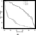
発明者らは、ACY−1濃度と、DGFの期間(血清クレアチニンのプラトーに対する日によって定義される)の又は1年、3年又は5年の時点での、より長い期間の腎機能因子(クレアチニン、eGFR、uPCR)との相関関係は観察されなかった。しかし、発明者らは、驚くべきことには、DGF患者の中でも、より高いACY−1濃度の患者は、カプラン・マイヤー分析によって、群として、5年の時点で、より高い透析フリーの生存率を有することが認められた。これは、移植後のIRIの間における高ACY−1濃度は、実際に、移植片が、IRIが不利益を被らない程度までIRIを改善するためには保護性である(必要でさえある)ことを示唆しており、これは、更なる調査のための創造力に富む領域を提供する。 The inventors have determined that ACY-1 concentrations and longer duration renal function factors (creatinine) at the time of DGF (defined by the day to serum creatinine plateau) or at 1 year, 3 years or 5 years. , EGFR, uPCR) was not observed. However, the inventors surprisingly found that among the DGF patients, those with higher ACY-1 concentrations were grouped by Kaplan-Meier analysis as a group at a higher dialysis-free survival rate at 5 years. It was found to have This is because the high ACY-1 concentration during IRI after transplantation is actually protective (even necessary) to improve the IRI to the extent that the IRI does not suffer from the disadvantages. This provides a creative area for further investigation.
従って、発明者らは、ACY−1を、予測医学(例えば、診断アッセイ及び予後アッセイ)における特別な用途のバイオマーカーとして確認した。 The inventors have therefore identified ACY-1 as a biomarker of particular use in predictive medicine (eg, diagnostic and prognostic assays).
発明者らによって提供されるデータは、ACY−1が、虚血再灌流障害についてのバイオマーカーとして有用であることを確認する(ACY−1のレベルが、虚血再灌流障害に罹っている又は発症する危険がある患者を特定できる)。 The data provided by the inventors confirms that ACY-1 is useful as a biomarker for ischemia-reperfusion injury (the level of ACY-1 is suffering from ischemia-reperfusion injury or Identify patients at risk of developing).
ここで使用するように、「虚血再灌流障害」は、虚血又は酸素不足の状態の後、組織へ血液が供給される際に生ずる組織の損傷を言う。虚血期間における血液からの酸素及び栄養の欠如は、血行の回復により、酸化ストレスの誘導を介して、炎症及び酸化的損傷を生ずる状態を創成する。いずれの組織も虚血再灌流障害を受け得る。特に、感受性の組織としては、脳、心臓、腎臓、肺、肝臓及び骨格筋が含まれる。 As used herein, “ischemia reperfusion injury” refers to tissue damage that occurs when blood is supplied to the tissue after ischemia or hypoxia. The lack of oxygen and nutrients from the blood during the ischemic period creates conditions that cause inflammation and oxidative damage through induction of oxidative stress by restoration of blood circulation. Any tissue can undergo ischemia-reperfusion injury. In particular, sensitive tissues include brain, heart, kidney, lung, liver and skeletal muscle.
虚血及びそれによる虚血再灌流障害は、心筋梗塞、敗血症、卒中及び臓器及び組織の獲得及び移植において生ずることがある。移植の際に虚血及び続く虚血再灌流障害に至り得る臓器としては、心臓、腎臓、肝臓、肺、膵臓、腸、及び胸腺が含まれる。 Ischemia and thereby ischemia-reperfusion injury can occur in myocardial infarction, sepsis, stroke and organ and tissue acquisition and transplantation. Organs that can lead to ischemia and subsequent ischemia-reperfusion injury upon transplantation include heart, kidney, liver, lung, pancreas, intestine, and thymus.
虚血再灌流障害は、移植を受けた患者において、移植片機能発現遅延を生ずることが知られている。ここで使用するように、「移植片機能発現遅延」は、移植後における透析の必要性(いくつかの理由による)又は血清クレアチニン(又はいくつかの他の腎臓機能のマーカー(クレアチニンクリアランスのような算定因子を含む))の期待したような改善の達成不能を含む(ただし、これらに限定されない)いくつかの臨床的定義によって決定されるように、腎移植片の術後の適切な機能発現の不能/遅延を言う。例えば、移植片機能発現遅延は、a)腎移植後第1週における透析の要求;b)単独の高カリウム血症のため以外に、腎移植後第1週における透析の必要性;c)移植後第1週における、増大、現状維持、連続する3日間における1日当たり10%未満の減少のいずれかの術後血清クレアチニンレベルの観察;及びd)Cockcroft算定クレアチニンクリアランス(cCCr)<10ml/分(DGFは、ついで、この値が>10ml/分になった際に終わったと言われる)によって決定される。 Ischemic reperfusion injury is known to cause delayed graft function in patients who have undergone transplantation. As used herein, “graft function delay” refers to the need for dialysis after transplantation (for several reasons) or serum creatinine (or some other marker of kidney function (such as creatinine clearance). Of the appropriate functioning of the kidney graft after surgery, as determined by several clinical definitions, including but not limited to the inability to achieve improvement as expected) Say impossibility / delay. For example, delayed graft function may include: a) dialysis requirement in the first week after kidney transplantation; b) need for dialysis in the first week after kidney transplantation, in addition to single hyperkalemia; c) transplantation Observation of post-operative serum creatinine levels, either increase, maintenance status, decrease of less than 10% per day for 3 consecutive days in the first week after; and d) Cockcroft calculated creatinine clearance (cCCr) <10 ml / min ( DGF is then determined to be finished when this value is> 10 ml / min).
ここに提供されるデータは、ACY−1が、移植片機能発現遅延についてのバイオマーカーとして有用であることを確認する(ACY−1のレベルが、移植片機能発現遅延に罹っている又は発症する危険がある患者を特定できる)。 The data provided here confirms that ACY-1 is useful as a biomarker for delayed graft function expression (ACY-1 levels are suffering from or developing graft function delay) Identify patients at risk).
ここに記載する診断法は、虚血再灌流障害又は移植片機能発現遅延に罹っている又は発症する危険がある患者を特定できる。 The diagnostic methods described herein can identify patients suffering from or at risk of developing ischemia-reperfusion injury or graft function delay.
1態様では、本発明は、患者における虚血再灌流障害を診断する方法を提供し、該方法は、i)患者から採取したサンプルにおけるACY−1レベルを測定し;及びii)患者サンプルにおけるACY−1レベルを、コントロールサンプルにおけるACY−1レベル又はACY−1について予め決定した対照レベルと対比することを含んでなり、コントロールサンプルと対比して又は予め決定した対照レベルと対比して患者サンプルにおけるACY−1のレベルが増大している場合に、患者が虚血再灌流障害を有していると認定する。 In one aspect, the invention provides a method of diagnosing ischemia-reperfusion injury in a patient, the method measuring i) ACY-1 levels in a sample taken from the patient; and ii) ACY in the patient sample. -1 level comprising comparing the ACY-1 level in a control sample or a control level predetermined for ACY-1 in a patient sample relative to a control sample or relative to a predetermined control level A patient is identified as having ischemia-reperfusion injury if the level of ACY-1 is increased.
更なる態様では、本発明は、移植患者における移植片機能発現遅延を診断する方法を提供し、該方法は、i)患者から採取したサンプルにおけるACY−1のレベルを測定し;ii)患者のサンプルにおけるACY−1のレベルを、コントロールサンプルにおけるACY−1のレベル又は予め決定したACY−1についての対照レベルと対比することを含んでなり、コントロールサンプルと対比して又は予め決定した対照レベルと対比して、患者サンプルにおけるACY−1のレベルが増大している場合に、患者が移植片機能発現遅延を有していると認定する。本発明は、また、移植患者における移植片機能発現遅延を診断する方法を提供し、該方法は、i)移植前の腎移植組織から採取したサンプル又は腎移植組織の潅流後の潅流体液のサンプルにおけるACY−1のレベルを測定し;ii)腎移植組織から採取したサンプルにおけるACY−1のレベルを、コントロールサンプルにおけるACY−1のレベル又は予め決定したACY−1についての対照レベルと対比することを含んでなり、コントロールサンプルと対比して又は予め決定した対照レベルと対比して、患者サンプルにおけるACY−1のレベルが増大している場合に、患者が移植片機能発現遅延を有していると認定する。 In a further aspect, the present invention provides a method of diagnosing graft function delay in a transplant patient, said method comprising: i) measuring the level of ACY-1 in a sample taken from the patient; Comparing the level of ACY-1 in the sample with the level of ACY-1 in the control sample or a control level for the predetermined ACY-1 and contrasting with the control sample or with the predetermined control level In contrast, a patient is identified as having a delayed onset of graft function when the level of ACY-1 in the patient sample is increased. The present invention also provides a method of diagnosing graft function delay in a transplant patient, the method comprising: i) a sample taken from a kidney transplant tissue prior to transplantation or a sample of perfusate after perfusion of the kidney transplant tissue Measuring the level of ACY-1 in the kidney; ii) contrasting the level of ACY-1 in the sample taken from the kidney transplant with the level of ACY-1 in the control sample or a predetermined control level for ACY-1 The patient has a delayed onset of graft function when the level of ACY-1 in the patient sample is increased relative to a control sample or relative to a predetermined control level Certify.
実施例に記載するように、発明者らは、虚血再灌流障害を有する患者、及び特に移植片機能発現遅延を有する患者におけるACY−1レベルは、虚血再灌流障害を有していない患者と比較して増大することを確認した。とりわけ、発明者らは、ACY−1レベルの増大が、移植後第1日、2日、3日及び4日において、術後の腎移植患者における移植片機能発現遅延の診断を可能にすることを確認した。
As described in the Examples, the inventors have shown that ACY-1 levels in patients with ischemia-reperfusion injury, and especially patients with delayed onset of graft function, do not have ischemia-reperfusion injury It was confirmed that it increased in comparison with. In particular, the inventors have found that increased ACY-1 levels allow for the diagnosis of delayed graft function in post-transplanted kidney transplant patients on
ここに記載する予後アッセイは、患者のサンプルにおけるACY−1レベルに基づき、術後の腎移植患者についての透析管理戦略のような好適な体液管理戦略を決定するために使用される。 The prognostic assays described herein are used to determine a suitable fluid management strategy, such as a dialysis management strategy for post-operative renal transplant patients, based on ACY-1 levels in patient samples.
1態様では、本発明は、術後の腎移植患者について体液管理戦略を決定する方法を提供し、該方法は、i)患者から採取したサンプルにおけるACY−1のレベルを測定し;ii)患者のサンプルにおけるACY−1のレベルを、コントロールサンプルにおけるACY−1のレベル又は予め決定したACY−1についての対照レベルと対比することを含んでなり、コントロールサンプルと対比して又は予め決定した対照レベルと対比して、患者サンプルにおけるACY−1のレベルが増大している場合に、患者が、術後の腎移植患者に通常投与される液体の量と比べて低減された量の液体を必要としていると認定する、又はコントロールサンプルと対比して又は予め決定した対照レベルと対比して、患者サンプルにおけるACY−1のレベルが低下している場合に、透析することが余り必要ではないため、患者にとっては、術後の腎移植患者に通常投与される液体の量と比べて増大された量の液体がためになるであろうと認定する。 In one aspect, the invention provides a method for determining a fluid management strategy for a post-operative renal transplant patient, the method i) measures the level of ACY-1 in a sample taken from the patient; ii) the patient Contrasting the level of ACY-1 in a sample of A with the level of ACY-1 in a control sample or a control level for a predetermined ACY-I, relative to a control sample or a predetermined control level In contrast, when the level of ACY-1 in the patient sample is increased, the patient needs a reduced amount of fluid compared to the amount of fluid normally administered to post-operative renal transplant patients. Level of ACY-1 in a patient sample as compared to a control sample or as compared to a predetermined control level When it is low, it is less necessary to dialyze, so the patient will rely on an increased amount of fluid compared to the amount of fluid normally administered to postoperative kidney transplant patients. Authorize deafness.
ここで使用するように、「術後の腎移植患者についての体液管理戦略」は、患者の臨床状態/要求によって決定されるように、患者への経口又は静脈内へのいずれか又は両方による投与又は投与を控えることを言う。例えば、DGF患者では、尿排出量は低減するか、しばしば、0となる。このような患者(排出手段を持たないもの)に多量の液体を投与することは、浮腫(肺浮腫による呼吸器合併症を含む)として証明されるように、水分過負荷を(最終的に)生ずるであろう。よって、DGFでは、低減された液体の投与(機能している移植片を有する患者について要求されるものと比べて)が好ましい。 As used herein, “fluid management strategy for post-operative renal transplant patients” refers to either oral or intravenous administration to a patient, or both, as determined by the patient's clinical status / demand. Or say to refrain from administration. For example, in DGF patients, urine output is reduced or often zero. Administering large amounts of fluid to such patients (those without drainage means) will eventually (through) water overload, as evidenced by edema (including respiratory complications due to lung edema) Will occur. Thus, with DGF, reduced fluid administration (as compared to that required for patients with functioning grafts) is preferred.
さらに、透析患者は、しばしば、わずかな量の尿を排出するか、全く排出しない。移植後では、患者がDGFを有していなければ、このような患者は多量の尿を排出する。このような状況では、前記患者に少なくとも同等(しばしば、より多く)の液体を投与することが必要である。これに関連して、十分に液体を投与することができなければ、患者は脱水状態になり、移植片の機能に影響を及ぼす。 In addition, dialysis patients often drain a small amount of urine or not at all. After transplantation, if the patient does not have DGF, such patient will drain a large amount of urine. In such situations, it is necessary to administer at least an equivalent (and often more) fluid to the patient. In this connection, if sufficient fluid cannot be administered, the patient becomes dehydrated and affects the function of the graft.
移植後の患者にどれだけの液体を投与するかを決定する基準法は存在していない。個人的に、患者の尿排出量、及び臨床状態(例えば、それらを調査する肺浮腫の兆候がある)を検査することによって行われている。しかし、良好に機能している移植片では、十分な量の液体を投与することが要求される。DGFを有する移植片では、同様の量の液体を投与することは有害となりがちである。 There is no standard method for determining how much fluid to administer to a patient after transplantation. Personally, it is done by examining the patient's urine output and clinical status (eg, there are signs of pulmonary edema to study them). However, a well-functioning implant requires that a sufficient amount of liquid be administered. For grafts with DGF, it is likely to be harmful to administer a similar amount of fluid.
更なる態様では、本発明は、術後の腎移植患者について透析管理戦略を決定する方法を提供し、該方法は、i)患者から採取したサンプルにおけるACY−1のレベルを測定し;ii)患者のサンプルにおけるACY−1のレベルを、コントロールサンプルにおけるACY−1のレベル又は予め決定したACY−1についての対照レベルと対比することを含んでなり、コントロールサンプルと対比して又は予め決定した対照レベルと対比して、患者サンプルにおけるACY−1のレベルが増大している場合に、患者が移植後1〜7日内に透析を必要としていると認定する、又はコントロールサンプルと対比して又は予め決定した対照レベルと対比して、患者サンプルにおけるACY−1のレベルが低下している場合に、患者が移植後1〜7日内には透析を必要としないと認定する。 In a further aspect, the present invention provides a method for determining a dialysis management strategy for a post-operative renal transplant patient, wherein the method measures i) the level of ACY-1 in a sample taken from the patient; ii) Contrasting the level of ACY-1 in the patient sample with the level of ACY-1 in the control sample or the control level for the predetermined ACY-1 in contrast to the control sample or the predetermined control If the level of ACY-1 in the patient sample is increased compared to the level, the patient qualifies as requiring dialysis within 1-7 days after transplant, or compared to or pre-determined with the control sample 1-7 days after transplantation when the level of ACY-1 in the patient sample is reduced compared to the control level Certified and do not require dialysis to.
ここで使用するように、「術後の腎移植患者についての透析管理戦略」とは、術後の腎移植患者が透析を必要としているかどうかの決定を言う。このような戦略は、腎移植患者において、術後毎日、更新及び評価され、体液状態、及び患者の臨床状態の他の態様とともに、高カリウム血症、血清クレアチニン及び血清尿素のような生物化学的因子に基づく。時には、透析の決定は、例えば、DGFを有する患者が過剰の液体を摂取し、肺水腫により呼吸器の合併症を発症した場合(緊急の透析が必要となりがちである)のように、迅速に行われなければならない。 As used herein, “dialysis management strategy for post-operative renal transplant patients” refers to the determination of whether post-operative renal transplant patients require dialysis. Such strategies are updated and evaluated daily after surgery in kidney transplant patients, as well as biochemical conditions such as hyperkalemia, serum creatinine and serum urea, as well as other aspects of fluid status and patient clinical status. Based on factors. Sometimes dialysis decisions are made quickly, for example, if a patient with DGF consumes excess fluid and develops respiratory complications due to pulmonary edema (which tends to require urgent dialysis). Must be done.
本発明は、また、患者における腎移植片の術後の臨床転帰を予測する方法を提供し、該方法は、i)患者から採取したサンプルにおけるACY−1のレベルを測定し;ii)患者のサンプルにおけるACY−1のレベルを、コントロールサンプルにおけるACY−1のレベル又は予め決定したACY−1についての対照レベルと対比することを含んでなり、コントロールサンプルと対比して又は予め決定した対照レベルと対比して増大している患者サンプルにおけるACY−1のレベルは、前記患者の死亡フリー生存率及び/又は透析フリー生存率の増大を予知するものであるとする。 The present invention also provides a method for predicting the postoperative clinical outcome of a renal graft in a patient, the method measuring i) the level of ACY-1 in a sample taken from the patient; ii) the patient's Comparing the level of ACY-1 in the sample with the level of ACY-1 in the control sample or a control level for the predetermined ACY-1 and contrasting with the control sample or with the predetermined control level It is assumed that the level of ACY-1 in a patient sample that is increasing in contrast is predictive of an increase in the patient's death-free survival and / or dialysis-free survival.
ここで使用するように、「腎移植の術後の臨床転帰」とは、移植患者が、移植後、生存する又は透析フリーでいられる見込みを言う。転帰は、血清クレアチニンレベルを尺度としてアッセイされるが、eGFRレベル、尿タンパクのレベル、高血圧症の有無、生存率又は透析フリー生存率の全てが、転帰の尺度として使用される。術後の臨床転帰は、移植後3か月、6か月、12か月又は1年、2年、3年、4年又は5年の時点における転帰であってもよい。 As used herein, “postoperative clinical outcome of kidney transplant” refers to the likelihood that the transplant patient will be alive or dialysis-free after transplant. Outcomes are assayed on the basis of serum creatinine levels, but eGFR levels, urine protein levels, presence or absence of hypertension, survival or dialysis-free survival are all used as a measure of outcome. The post-operative clinical outcome may be an outcome at 3 months, 6 months, 12 months or 1 year, 2 years, 3 years, 4 years or 5 years after transplantation.
生物学的サンプルにおけるACY−1ポリペプチド又は核酸分子の存在、レベル又は不存在は、患者から生物学的サンプルを採取し、該生物学的サンプルをACY−1ポリペプチド又は核酸分子を検出することができる化合物又は試剤と接触させることによって測定される。 The presence, level or absence of an ACY-1 polypeptide or nucleic acid molecule in a biological sample is obtained by taking a biological sample from a patient and detecting the ACY-1 polypeptide or nucleic acid molecule in the biological sample. Is measured by contacting with a compound or reagent capable of
ここで使用するように、用語「生物学的サンプル」及び「患者から採取したサンプル」は、患者内に存在する組織、細胞及び体液と同様に、患者から採取した組織、細胞及び体液を示すものとして交換可能で使用される。サンプルは、尿サンプル、血液サンプル、血清サンプル、痰サンプル、糞便サンプル、体組織の生検、例えば、移植した腎組織の生検、脳脊髄液サンプル、精液サンプル、又はスメアサンプルである。好適なサンプルは血清又は血漿である。或いは、サンプルは潅流液サンプルである。1具体例では、サンプルは移植前又は移植後に採取される。 As used herein, the terms “biological sample” and “sample taken from a patient” refer to tissue, cells and body fluids collected from a patient as well as tissues, cells and body fluids present in the patient. Used as interchangeable. The sample is a urine sample, blood sample, serum sample, sputum sample, stool sample, biopsy of body tissue, eg, biopsy of transplanted kidney tissue, cerebrospinal fluid sample, semen sample, or smear sample. A preferred sample is serum or plasma. Alternatively, the sample is a perfusate sample. In one embodiment, the sample is taken before or after transplantation.
ここで使用するように、「患者」とは、個人、例えば、移植片機能発現遅延のような虚血再灌流障害を有する又は有する危険があるヒトを言う。1具体例では、患者は、心筋梗塞、卒中に罹っている又は罹ったことがある、或いは、臓器移植を受けたことがある個人である。1具体例では、患者は、腎臓、心臓、肺又は肝臓の移植患者のような、術後の臓器移植患者である。 As used herein, “patient” refers to an individual, eg, a human having or at risk of having ischemia-reperfusion injury such as delayed onset of graft function. In one embodiment, the patient is an individual who has or has had a myocardial infarction, stroke, or who has undergone an organ transplant. In one embodiment, the patient is a post-operative organ transplant patient, such as a kidney, heart, lung or liver transplant patient.
ここで使用するように、「術後の臓器移植患者」とは、ドナー臓器を受容した患者を言う。 As used herein, “post-operative organ transplant patient” refers to a patient who has received a donor organ.
サンプルが術後の臓器移植患者からのものである場合、サンプルは、移植後第1日、2日、3日、4日、5日、6日又は7日の時点で患者から得られたものである。 If the sample is from a post-operative organ transplant patient, the sample was obtained from the patient at the 1st, 2nd, 3rd, 4th, 5th, 6th or 7th day after transplantation It is.
ここで使用するように、「DCD」は、用語「循環死後に提供された」と交換可能で使用される。 As used herein, “DCD” is used interchangeably with the term “provided after circulatory death”.
ここで使用するように、「DBD」は、用語「脳死後に提供された」と交換可能で使用される。 As used herein, “DBD” is used interchangeably with the term “provided after brain death”.
ACY−1核酸分子の発現レベルは、ACY−1核酸分子によってコードされるmRNAを測定すること;ACY−1核酸分子によってコードされるポリペプチドの量を測定すること;又はACY−1核酸分子によってコードされるポリペプチドの活性を測定することを含む多数の方法で測定される。 The expression level of an ACY-1 nucleic acid molecule is determined by measuring mRNA encoded by the ACY-1 nucleic acid molecule; measuring the amount of polypeptide encoded by the ACY-1 nucleic acid molecule; or by the ACY-1 nucleic acid molecule It is measured in a number of ways, including measuring the activity of the encoded polypeptide.
サンプルにおけるACY−1mRNAのレベルを測定するために、各種の公知のmRNA検出法が使用される。 Various known mRNA detection methods are used to measure the level of ACY-1 mRNA in a sample.
例えば、サンプルにおけるACY−1核酸分子に相当するmRNAのレベルは、インサイチュ方式及びインビトロ方式の両方で検出される。ACY−1mRNAは、サウザン又はノーザンブロット分析、ポリメラーゼ連鎖反応又はプローブアレイを使用して検出される。1具体例では、サンプルを、ACY−1核酸分子によってコードされるmRNAとハイブリダイズできる核酸分子(すなわち、ラベル化プローブのようなプローブ)と接触させる。例えば、プローブは、配列番号1の核酸分子のような全長ACY−1核酸分子、又は少なくとも10個、15個、30個、50個、100個、250個又は500個ヌクレオチドの長さの核酸分子のような、配列番号1の核酸分子の部分(ストリンジェントな条件下でACY−1核酸分子とハイブリダイズする)でもよい。 For example, the level of mRNA corresponding to an ACY-1 nucleic acid molecule in a sample is detected both in situ and in vitro. ACY-1 mRNA is detected using Southern or Northern blot analysis, polymerase chain reaction or probe arrays. In one embodiment, the sample is contacted with a nucleic acid molecule that can hybridize to the mRNA encoded by the ACY-1 nucleic acid molecule (ie, a probe such as a labeled probe). For example, the probe can be a full-length ACY-1 nucleic acid molecule, such as the nucleic acid molecule of SEQ ID NO: 1, or a nucleic acid molecule at least 10, 15, 30, 50, 100, 250, or 500 nucleotides in length. Or a portion of the nucleic acid molecule of SEQ ID NO: 1 (which hybridizes with the ACY-1 nucleic acid molecule under stringent conditions).
或いは、サンプルにおけるACY−1mRNAのレベルは、核酸増幅によって、例えば、rtPCR、リガーゼ連鎖反応、自家持続配列複製、転写増幅、又は他の各種の核酸増幅法及び続いて、当分野において知られた技術を使用して、増幅された分子を検出することによって評価される。 Alternatively, the level of ACY-1 mRNA in a sample is determined by nucleic acid amplification, eg, rtPCR, ligase chain reaction, autologous sequence replication, transcription amplification, or any other nucleic acid amplification method followed by techniques known in the art. Is used to evaluate the amplified molecules.
サンプルにおけるACY−1ポリペプチドのレベルを検出するために、各種の公知のタンパク質検出法が使用される。 Various known protein detection methods are used to detect the level of ACY-1 polypeptide in a sample.
一般に、タンパク質検出法は、ACY−1ポリペプチドに選択的に結合する試剤、例えば、抗−ACY−1抗体を患者のサンプルと接触させて、サンプルにおけるACY−1ポリペプチドのレベルを測定することを含んでなる。好ましくは、試剤又は抗体は、例えば、検出可能なラベルにてラベル化される。好適な抗−ACY−1抗体は、ポリクローナル又はモノクローナルである。Fab又はF(ab’)2のような抗体フラグメントを使用できる。 In general, protein detection methods involve contacting an agent that selectively binds to an ACY-1 polypeptide, eg, an anti-ACY-1 antibody, with a patient sample and measuring the level of the ACY-1 polypeptide in the sample. Comprising. Preferably, the reagent or antibody is labeled with a detectable label, for example. Suitable anti-ACY-1 antibodies are polyclonal or monoclonal. Antibody fragments such as Fab or F (ab ′) 2 can be used.
ここで使用するように、用語「ラベル化された」とは、検出可能な物質との反応性によるプローブ又は抗体の間接的ラベル化とともに、検出可能な物質のプローブ又は抗体へのカップリング(すなわち、物理的結合)によるプローブ又は抗体の直接的ラベル化を言う。 As used herein, the term “labeled” refers to the coupling of a detectable substance to a probe or antibody (ie, indirect labeling of the probe or antibody by reactivity with the detectable substance (ie, Direct labeling of the probe or antibody by physical binding).
サンプルにおけるACY−1ポリペプチドのレベルは、酵素結合免疫吸着アッセイ(ELISAs)、免疫沈降、免疫蛍光、酵素免疫アッセイ(EIA)、ラジオイムノアッセイ(RIA)、及びウエスタンブロットアッセイのような当分野において知られた技術によって測定される。ACY−1ポリペプチドのインビボ検出について、ラベル化抗−ACY−1抗体を患者に導入できる。このような抗体は、対象における存在及び位置が、標準的な画像技術によって検出される放射性マーカーによってラベル化される。 The level of ACY-1 polypeptide in a sample is known in the art such as enzyme linked immunosorbent assays (ELISAs), immunoprecipitation, immunofluorescence, enzyme immunoassay (EIA), radioimmunoassay (RIA), and Western blot assays. Measured by selected techniques. For in vivo detection of ACY-1 polypeptide, a labeled anti-ACY-1 antibody can be introduced into the patient. Such antibodies are labeled with radioactive markers whose presence and location in the subject is detected by standard imaging techniques.
サンプルにおけるACY−1ポリペプチドのレベルは、サンプルにおけるACY−1ポリペプチドの活性レベル測定することによっても検出される。 The level of ACY-1 polypeptide in the sample is also detected by measuring the activity level of ACY-1 polypeptide in the sample.
本発明の方法は、さらに、患者のサンプルにおけるACY−1の活性のレベルを、コントロールサンプルにおけるACY−1の活性レベル又はACY−1について予め定めた対照レベルと比較することを含んでなる。 The method of the invention further comprises comparing the level of ACY-1 activity in the patient sample to the activity level of ACY-1 in the control sample or a control level predetermined for ACY-1.
1具体例では、本発明の方法は、コントロールサンプルを、mRNA又はゲノムDNAのようなACY−1核酸分子を検出できる化合物又は医薬と接触させること、及びコントロールサンプルにおけるACY−1核酸分子のレベルを、患者サンプルにおけるACY−1核酸分子のレベルと比較することを含んでなる。 In one embodiment, the method of the invention comprises contacting a control sample with a compound or drug capable of detecting an ACY-1 nucleic acid molecule, such as mRNA or genomic DNA, and determining the level of ACY-1 nucleic acid molecule in the control sample. Comparing to the level of the ACY-1 nucleic acid molecule in the patient sample.
他の具体例では、さらに、本発明の方法は、コントロールサンプルを、ACY−1ポリペプチドを検出できる化合物又は試剤と接触させること、及びコントロールサンプルにおけるACY−1タンパク質のレベルを、テストサンプルにおけるACY−1タンパク質の存在と比較することを含んでなる。 In another embodiment, the method of the invention further comprises contacting the control sample with a compound or agent capable of detecting ACY-1 polypeptide, and determining the level of ACY-1 protein in the control sample by comparing the level of ACY-1 in the test sample. Comparing to the presence of -1 protein.
ここで使用するように、「対照レベル」又は「コントロール」とは、ACY−1発現の正常なレベルを有するサンプル、例えば、虚血再灌流障害を有していない又は有することが疑われない健常な対象からのサンプル、或いは、生物学的テストサンプルが得られた同じ対象からのサンプル、例えば、臓器移植前に得られたサンプルを言う。或いは、対照レベルは、予め定められたカットオフ値、すなわち、症状又は病気又はその欠乏を統計学的に予測する診断スコアを創成するために使用される対照データベースからのACY−1発現レベルを含んでなるか、又は標準の母集団サンプルに基づく予め定められた対照レベル、或いは、対象の発現のベースラインレベル、すなわち、臓器移植前の対象の発現レベルに基づく予め定められた対照レベルである。 As used herein, a “control level” or “control” is a sample that has a normal level of ACY-1 expression, eg, healthy that does not have or is not suspected to have ischemia-reperfusion injury A sample from a different subject or a sample from the same subject from which a biological test sample was obtained, eg, a sample obtained prior to organ transplantation. Alternatively, the control level comprises a predetermined cut-off value, i.e., the level of ACY-1 expression from a control database used to create a diagnostic score that statistically predicts a symptom or disease or its deficiency. Or a predetermined control level based on a standard population sample, or a baseline level of expression of the subject, ie a predetermined control level based on the expression level of the subject prior to organ transplantation.
或いは、予測は、ACY−1の正規化された発現レベルに基づくものでもよい。発現レベルは、サンプルにおけるACY−1の絶対発現レベルを、その発現を、恒常的に発現されるmRNAのようなマーカー(例えば、mRNA)ではない対照核酸の発現と比較することにより補正することによって正規化される。この正規化により、1サンプルにおける発現レベルを他のサンプルと比較すること又は異なった源からのサンプル間で比較することが可能になる。この正規化された発現は、ついで、任意に、対照レベル又はコントロールと比較される。 Alternatively, the prediction may be based on the normalized expression level of ACY-1. Expression level is corrected by comparing the absolute expression level of ACY-1 in the sample by comparing its expression to the expression of a control nucleic acid that is not a marker (eg, mRNA) such as constitutively expressed mRNA. Normalized. This normalization allows expression levels in one sample to be compared with other samples or between samples from different sources. This normalized expression is then optionally compared to a control level or control.
例えば、尿におけるバイオマーカーを測定する際、バイオマーカーは、絶対濃度として発現されるか、或いは、尿クレアチニンレベル又は尿タンパク質レベルのような公知の尿バイオマーカーに対して正規化される。 For example, when measuring a biomarker in urine, the biomarker is expressed as an absolute concentration or normalized to a known urine biomarker such as urine creatinine level or urine protein level.
1具体例では、診断又は予後方法は、サンプルにおけるACY−1のレベルを測定すること及び少なくとも1の更なるバイオマーカー、例えば、虚血再灌流障害又は移植片機能発現遅延を予測又は表示するバイオマーカーのレベルを測定することを含む。好ましくは、少なくとも1の更なるバイオマーカーは、NGAL、KIM−1、IL−18、RBP、FABP4、シスタチンC及びクレアチニンから選ばれ、方法は、さらに、生物学的テストサンプルにおけるNGAL、KIM−1、IL−18、RBP、FABP4、シスタチンC及びクレアチニン核酸分子、又はポリペプチドのレベルを検出することを含んでなる。好ましくは、少なくとも1の更なるバイオマーカーのレベルは、上述の方法のいずれか1つを使用して測定される。 In one embodiment, the diagnostic or prognostic method comprises measuring a level of ACY-1 in a sample and predicting or displaying at least one additional biomarker, such as ischemia reperfusion injury or delayed graft function expression. Including measuring the level of the marker. Preferably, the at least one further biomarker is selected from NGAL, KIM-1, IL-18, RBP, FABP4, cystatin C and creatinine, the method further comprising NGAL, KIM-1 in a biological test sample , Detecting the level of IL-18, RBP, FABP4, cystatin C and creatinine nucleic acid molecule, or polypeptide. Preferably, the level of at least one additional biomarker is measured using any one of the methods described above.
少なくとも1の更なるバイオマーカーのレベルは、同じ生物学的サンプル又は異なる生物学的サンプルにおけるACY−1のレベルについて測定される。 The level of at least one additional biomarker is measured for the level of ACY-1 in the same biological sample or different biological samples.
或いは、サンプルにおけるACY−1のレベルは、質量分析を使用して検出及び定量される。 Alternatively, the level of ACY-1 in the sample is detected and quantified using mass spectrometry.
1態様では、本発明は、アッセイ装置、例えは、チップアレイのような、その表面にACY−1ポリペプチド又は核酸を検出できる化合物又は試剤が付着した固体支持体を含む。好ましくは、ACY−1ポリペプチドを検出できる化合物又は試剤は、抗−ACY−1抗体、さらに好ましくは、ACY−1捕捉抗体である。1具体例では、アッセイ装置は、さらに、更なるバイオマーカー、好ましくは、腎臓の損傷、疾患又は障害のためのバイオマーカーを検出するための少なくとも1の追加の化合物又は試剤を含んでなる。 In one aspect, the invention includes an assay device, eg, a solid support, such as a chip array, having attached to its surface a compound or reagent capable of detecting ACY-1 polypeptide or nucleic acid. Preferably, the compound or reagent capable of detecting ACY-1 polypeptide is an anti-ACY-1 antibody, more preferably an ACY-1 capture antibody. In one embodiment, the assay device further comprises at least one additional compound or agent for detecting additional biomarkers, preferably biomarkers for kidney injury, disease or disorder.
発明者らは、ACY−1を、虚血再灌流障害又はDGFのマーカーとして同定した。本発明の方法は、当業者がACY−1レベルに基づき知らされた治療を決定することを可能にする。例えば、ここに記載の診断及び予後方法は、さらに、診断及び予後に基づき患者又は臓器を治療する工程を含んでなる。患者又は臓器を治療する工程は、i)移植後、患者を透析処置する、例えば、腎移植後第1週に患者を透析処置する工程;ii)患者に静脈注射を投与する、例えば、投与される静脈注射を調整する(すなわち、患者に投与されるIVI流体の速度及び容量を増減する)工程;iii)患者に免疫抑制剤(例えば、カルシニューリン阻害剤又はシロリムスのようなTOR阻害剤)を投与する、例えば、ACY−1レベルに基づいて免疫抑制剤を増減する又は免疫抑制治療の種類を変更する工程から選ばれる(これらは例示を目的とするものである)。 The inventors have identified ACY-1 as a marker for ischemia-reperfusion injury or DGF. The method of the present invention allows one of ordinary skill in the art to determine a known treatment based on ACY-1 levels. For example, the diagnostic and prognostic methods described herein further comprise treating a patient or organ based on the diagnosis and prognosis. The step of treating the patient or organ comprises i) dialysis of the patient after transplantation, eg, dialysis of the patient in the first week after kidney transplantation; ii) administering intravenous injection to the patient, eg, administered Adjusting the intravenous injection (ie, increasing or decreasing the rate and volume of the IVI fluid administered to the patient); iii) administering the patient an immunosuppressive agent (eg, a calcineurin inhibitor or a TOR inhibitor such as sirolimus) For example, selected from the steps of increasing or decreasing immunosuppressive agents or changing the type of immunosuppressive treatment based on ACY-1 levels (these are for illustrative purposes).
加えて、腎移植後の初期期間におけるACY−1の測定は、DGFを寛解させるように各種の可能性のある介入をガイドするために使用される。 In addition, measurement of ACY-1 in the initial period after renal transplantation is used to guide various potential interventions to ameliorate DGF.
キット
本発明は、生物学的サンプルにおけるACY−1の存在を検出するためのキットも含む。例えば、キットは、生物学的サンプルにおけるACY−1ポリペプチド又は核酸を検出できる化合物又は試剤を含む。化合物又は試剤は、好適な容器に梱包される。キットは、さらに、ACY−1タンパク質又は核酸分子を検出するためにキットを使用するための説明書を含んでなる。
Kits The present invention also includes kits for detecting the presence of ACY-1 in a biological sample. For example, the kit includes a compound or reagent that can detect an ACY-1 polypeptide or nucleic acid in a biological sample. The compound or reagent is packaged in a suitable container. The kit further comprises instructions for using the kit to detect ACY-1 protein or nucleic acid molecule.
1態様では、本発明は、患者における虚血再灌流障害を診断する又は移植片機能発現遅延を診断するためのキットを提供し、該キットは、ACY−1ポリペプチドに特異的に結合する検出可能にラベル化された試剤又はACY−1核酸に特異的に結合する検出可能にラベル化された試剤;及びii)診断を実施するための試薬を含んでなる。 In one aspect, the invention provides a kit for diagnosing ischemia-reperfusion injury or diagnosing delayed onset of graft function in a patient, the kit detecting specifically binding to an ACY-1 polypeptide. A possibly labeled reagent or a detectably labeled reagent that specifically binds to an ACY-1 nucleic acid; and ii) a reagent for performing a diagnosis.
試剤は抗体又は核酸分子である。 The reagent is an antibody or a nucleic acid molecule.
抗体系キットについて、該キットは、(1)本発明のマーカーに相当するACY−1ポリペプチドに特異的に結合する第1の抗体(例えば、固体支持体に付着している);及び任意に、(2)ACY−1ポリペプチド又は第1の抗−ACY−1抗体に結合し、検出可能な試剤に共役した第2の異なる抗体を含むことができる。 For antibody-based kits, the kit comprises (1) a first antibody that specifically binds to an ACY-1 polypeptide corresponding to a marker of the invention (eg, attached to a solid support); and optionally , (2) a second different antibody that binds to the ACY-1 polypeptide or first anti-ACY-1 antibody and is conjugated to a detectable agent.
オリゴヌクレオチド系キットについては、該キットは、(1)ACY−1核酸分子にハイブリダイズするヌクレオチドプローブ、例えば、検出可能にラベル化されたプライマー又は(2)ACY−1核酸分子を増幅するためのプライマー対を含むことができる。 For oligonucleotide-based kits, the kit includes (1) a nucleotide probe that hybridizes to an ACY-1 nucleic acid molecule, eg, a detectably labeled primer or (2) for amplifying an ACY-1 nucleic acid molecule Primer pairs can be included.
1態様では、本発明は、患者における虚血再灌流障害又は移植片機能発現遅延を診断するためのキットを提供し、該キットは、本発明のマーカーに相当するACY−1ポリペプチドに特異的に結合する第1の抗体(例えば、固体支持体に付着されている);及び(2)ACY−1ポリペプチド又は第1の抗−ACY−1抗体に結合し、検出可能な試剤に共役した第2の異なる抗体を含んでなるアッセイ装置を含んでなる。 In one aspect, the invention provides a kit for diagnosing ischemia-reperfusion injury or graft function delay in a patient, wherein the kit is specific for an ACY-1 polypeptide corresponding to a marker of the invention A first antibody that binds to (eg, attached to a solid support); and (2) binds to an ACY-1 polypeptide or first anti-ACY-1 antibody and is conjugated to a detectable agent. An assay device comprising a second different antibody.
キットは、検出可能な試剤を検出するために必要な成分(例えば、酵素又は基質)も含むことができる。キットは、アッセイされ、収容されたテストサンプルと比較されるコントロールサンプル又は一連のコントロールサンプルを含有することもできる。 The kit can also include components necessary for detecting a detectable agent (eg, an enzyme or a substrate). The kit can also contain a control sample or a series of control samples that are assayed and compared to the stored test sample.
この明細書の記載及び請求の範囲を通して、用語「含んでなる」及び「含有する」及びこれらの変形は、「含むが、これらに限定されない」を意味するものであり、他の部分、添加剤、成分、整数又は工程を除外することを意図するものではない(意図しない)。この明細書の記載及び請求の範囲を通して、単数形は、他に前後関係が要求しない限り、複数形を含むものである。特に、不定冠詞が使用される場合、明細書は、他に前後関係が要求しない限り、単数形とともに、複数形を含むものとして理解されなければならない。 Throughout the description and claims, the terms “comprising” and “containing” and variations thereof mean “including but not limited to” and other parts, additives It is not intended (or not intended) to exclude ingredients, integers or steps. Throughout this description and the claims, the singular includes the plural unless the context demands otherwise. In particular, where indefinite articles are used, the specification should be understood to include the plural as well as the singular unless the context requires otherwise.
本発明の特別な態様、具体例又は実施例に関連して記載する特徴、整数、特性、化合物、化学部分又は基は、矛盾しない限り、ここに記載のいかなる他の態様、具体例又は実施例にも適用可能であると理解されなければならない。この明細書(添付する請求の範囲、要約及び図面を含む)に開示する特徴の全て、及び/又は開示するいかなる方法又はプロセスの工程の全ては、このような特徴及び/又は工程の少なくともいくつかが相互に排他的である組み合わせを除き、いかなる組み合わせとしても組み合わされる。本発明は、いかなる上述の具体例の詳細にも限定されない。本発明は、この明細書(添付の請求の範囲、要約及び図面)に開示する特徴のいずれかの新規な1つ、又はいずれかの新規な組み合わせに、又はここに開示されたいかなる方法又はプロセスの工程のいずれかの新規な1つ、又はいずれかの新規な組み合わせに拡張される。 Features, integers, properties, compounds, chemical moieties or groups described in connection with a particular aspect, embodiment or example of the invention, unless otherwise inconsistent, any other embodiment, embodiment or example described herein. Must be understood to be applicable. All of the features disclosed in this specification (including the appended claims, abstract and drawings) and / or all of the steps of any method or process disclosed are at least some of such features and / or steps. Can be combined in any combination, except combinations where are mutually exclusive. The present invention is not limited to the details of any of the above-described embodiments. The present invention is directed to any one or any novel combination of features disclosed in this specification (attached claims, abstract and drawings), or any method or process disclosed herein. To any new one or any new combination.
読者は、本願に関連してこの明細書と同時に又は以前に出願され、この明細書とともに公開された全ての論文及び文献の全てに注目されたい。このような全ての論文及び文献の内容を、参照して、ここに組み込む。 The reader should be aware of all of the articles and literature that were filed concurrently or previously with this specification and published with this specification. The contents of all such articles and literature are incorporated herein by reference.
[実施例]
1. 方法
1.1 患者群及び研究デザイン
腎移植を受ける前に、患者の承諾を得た(図1)。将来を見越して移植前に、及び移植後に、長期的に1週当たり少なくとも3回、静脈血を採取した(平均14サンプル/患者)。Z/Serum Clot Activator Tubes(Greiner)(25℃において2000gで10分間遠心分離する前に、室温において45分から2時間凝固させ、血清を分取し、−80℃で保存することを可能にする)を使用して、血液を集めた。患者コホート1は、2008年9月〜2011年7月の期間に集めた患者55人を含み(サンプル665個;腎移植患者47人及び生体ドナー8人)、そのうちの15人はDGFを有し(単独の高カリウム血症のため以外に、腎移植後第1週において透析を必要とするとの定義に基づく)、及びコホート2は、2003年12月〜2006年3月の期間に集めた患者194人(第1日のサンプル138個及び第3日のサンプル177個)を含み、その内の55人はDGFを有していた。できる限り臨床的にマッチする、コホート1におけるDGFを有する患者5人及び合併症のない患者5人からの血清サンプル、移植前及び移植後第2日の時点で採取した比較用サンプル(サンプル計20個)を使用して初期バイオマーカーの発見を実施した。ついで、初めにコホート1からのサンプル、続いてコホート2からのサンプルを使用して(数は統計的検出力に基づき決定される)、初期知見の検証に着手した。例えば、DGF患者10人及び非DGF患者25人の第1/2日におけるACY−1測定は、Bonferroni法が、2群比較について、5%有意性検定を補正する際にAUC>0.8を検出するために、検定力80%を与える。
[Example]
1. Methods 1.1 Patient group and study design Prior to receiving a kidney transplant, patient consent was obtained (Figure 1). In anticipation of the future, venous blood was collected before transplantation and after transplantation at least 3 times per week (average 14 samples / patient). Z / Serum Clot Activator Tubes (Greiner) (allows clotting for 45 to 2 hours at room temperature before centrifugation at 2000 g for 10 minutes at 25 ° C., allowing serum to be collected and stored at −80 ° C.) Was used to collect blood.
1.2 血清の免疫除去及びサンプルの調製
発見セットにおける血清サンプル20個を、既に記載されたように21、Multi Affinity Regent System 14 カラム(Agilent)を使用して、免疫除去に供した。免疫除去されたフラクションを脱塩し、得られた物質を、フィルター−補助サンプル調製法(FASP)9の変法21を使用して、トリプシンにて消化した。
1.2 Serum immunodepletion and sample preparation Twenty serum samples in the discovery set were subjected to immunodepletion using a Multi Affinity Reagent System 14 column (Agilent) as previously described 21 . The immunodepleted fraction was desalted and the resulting material was digested with trypsin using variant 21 of filter-auxiliary sample preparation (FASP) 9 .
1.3 ラベルフリー質量分析
0.1%TFAの最終濃度への酸性化の後、ペプチドサンプルを患者によってブロックランダム化し、Proxeonナノエレクトロスプレイイオン源を備えたLTQ Orbitrap Velos質量分析計に接続したDionex UltiMate 3000 RSLCnanoシステムを使用して分析した(サンプル当たり3×2μg注入)。3.5μm Kromasil C18媒体が充填されたインハウス25mmキャピラリーエミッターカラムに、サンプルを直接注入した。総捕捉時間は300分であり、勾配の大部分は、流速0.4μl/分において、0.1%ギ酸中ACN3〜25%であった。サーベイMSスキャンは、60,000にセットされた解像度のOrbitrap内で獲得された。スキャン当たり最強イオン20個以下を断片化し、リニアトラップにおいて分析した。
1.3 Label-free mass spectrometry After acidification to a final concentration of 0.1% TFA, the peptide samples were block randomized by the patient and connected to an LTQ Orbitrap Velos mass spectrometer equipped with a Proxeon nanoelectrospray ion source. Analyzed using the UltimateMate 3000 RSLC nano system (3 × 2 μg injection per sample). Samples were injected directly into an in-house 25 mm capillary emitter column packed with 3.5 μm Kromasil C18 media. The total capture time was 300 minutes and the majority of the gradient was 3-25% ACN in 0.1% formic acid at a flow rate of 0.4 μl / min. Survey MS scans were acquired in an Orbitrap with a resolution set to 60,000. Less than 20 strongest ions per scan were fragmented and analyzed in a linear trap.
1.4 データの解析及び統計学的方法
MaxQuant(v1.1.1.25)51を使用して、ラベルフリー質量分析データの解析を行った。Andromeda52及び≧2ペプチドの判定基準を有するIPIヒトデータベース(v3.75,19/08/2010)を使用してタンパク質を同定し、及びDecoyデータベースから同定したタンパク質及び公知の混入物を除去した。LFQ強度に関するノンパラメトリック(Wilcoxon)有意性検定を使用して、患者群間及び患者群内の示差的発現をアッセイした。q値法53を使用して偽発見率を推定した。
1.4 Data analysis and statistical methods Label-free mass spectrometry data was analyzed using MaxQuant (v1.1.1.25) 51 . Proteins were identified using the IPI human database (v3.75, 19/08/2010) with criteria of Andromeda 52 and ≧ 2 peptides, and the identified proteins and known contaminants were removed from the Decoy database. Differential expression between and within patient groups was assayed using a non-parametric (Wilcoxon) significance test for LFQ intensity. The q-value method 53 was used to estimate the false discovery rate.
検証テストでは、血清ACY−1及び他のマーカー54の予知能力をアッセイするために、受信者動作特性(ROC)曲線を作成した。受信者動作特性曲線下の面積(AUC)を見積もり、95%CIsがブートストラップリサンプルから推定された。ブートストラップ有意性検定を使用して、AUCを、標準の近似を使用してアッセイしたブートストラップAUC間の差異の有意性を比較した。突出した予測変数を含有するモデルにおけるDGFに関するACY−1の非依存性予知能力をアッセイするために、多変量ロジスティクス回帰分析を使用した。Cox比例ハザード回帰、カプラン・マイヤー生存時間関数及びロングランク検定を使用して、ACY−1及び他のマーカーの濃度と透析フリー生存率との関係を査定した。全ての統計学的検定は両側性であり、全ての分析を、統計学的計算のためのR環境で行った(R Development Core Team,ウィーン、オーストリア)。 In the validation test, receiver operating characteristic (ROC) curves were generated to assay the predictive ability of serum ACY-1 and other markers 54 . The area under the receiver operating characteristic curve (AUC) was estimated and 95% CIs was estimated from the bootstrap resample. A bootstrap significance test was used to compare the significance of differences between bootstrap AUCs that were AUC assayed using a standard approximation. Multivariate logistics regression analysis was used to assay ACY-1 independent predictive ability for DGF in models containing prominent predictors. Cox proportional hazards regression, Kaplan-Meier survival function and long rank test were used to assess the relationship between the concentrations of ACY-1 and other markers and dialysis-free survival. All statistical tests were bilateral and all analyzes were performed in the R environment for statistical calculations (R Development Core Team, Vienna, Austria).
1.5 他の分析物との比較
血清クレアチニン、CPR、タクロリムス、及び尿タンパク質/クレアチニン比を、臨床プロトコル/指示通りに測定し、さらに、血清クレアチニン及びシスタチンCを、研究で使用した同じ時点で、コホート1において測定した。
1.5 Comparison with other analytes Serum creatinine, CPR, tacrolimus, and urine protein / creatinine ratios were measured as per clinical protocol / instruction, and serum creatinine and cystatin C were also used at the same time used in the study. , Measured in
1.6 ELISA展開
両コホートにおける血清サンプル980個について、サンプルをブロックランダム化し、インハウス開発したサンドイッチELISAを使用して、ACY−1濃度を測定した。手短に言えば、96穴Nunc MaxiSorpプレート(Nalge Nunc International;ロチェスター,ニューヨーク州)を、ラット抗−ヒトACY−1モノクローナル抗体(クローン475626;R&D Systems;ミネアポリス)及びACY−1標準(0.31−15.0ng/mL,Hisタグ組み換えACY−1−R&D Systems)にて被覆し、希釈したサンプルを塗布した。2時間のインキュベートの後、ヤギ抗−ACY−1抗体(R&D Systems)、続いて、連続して、ビオチン化ウサギ抗−ヤギIgG抗体(Sigma−Aldrich;プール,英国)、ストレプトアビジン−HRP及びテトラメチルベンズアジン(Sigma−Aldrich)を使用して、結合したACY−1を検出した。2N硫酸(Sigma−Aldrich)を添加した後、450nm(570nmにて補正)において吸光度を測定し、ACY−1濃度を算定した。
1.6 ELISA Development For 980 serum samples in both cohorts, the samples were block randomized and ACY-1 concentrations were measured using an in-house developed sandwich ELISA. Briefly, 96-well Nunc MaxiSorp plates (Nalge Nunc International; Rochester, NY) were added to rat anti-human ACY-1 monoclonal antibody (clone 475626; R & D Systems; Minneapolis) and ACY-1 standard (0.31- 15.0 ng / mL, His-tagged recombinant ACY-1-R & D Systems) and diluted samples were applied. After 2 hours of incubation, goat anti-ACY-1 antibody (R & D Systems) followed by biotinylated rabbit anti-goat IgG antibody (Sigma-Aldrich; Poole, UK), streptavidin-HRP and tetra Bound ACY-1 was detected using methylbenzazine (Sigma-Aldrich). After adding 2N sulfuric acid (Sigma-Aldrich), the absorbance was measured at 450 nm (corrected at 570 nm), and the ACY-1 concentration was calculated.
2. 結果
2.1 患者群
患者群の検査(表1)は、DCD移植片の割合及び誘導レジメン(診療の変更を招く)を除き、コホート1(発見及び初期検証群)及び2(長期間転帰データを有するより大きい検証群)の同様の特性を示す。コホート1における患者の31.9%及びコホート2における患者の28.4%においてDGFと診断された。平均年齢及び冷虚血時間(CIT)は、各コホートにおいてDGF群において有意に高く、温虚血時間(WIT)はコホート2において同様であった。初期プロテオミック発見のための使用したDGF患者5人及び非DGF患者5人は、CNI毒性又は急性拒絶反応の証拠を有しておらず、平均年齢、民族性ミックス、CIT、WIT、A、B及びDR坐位における平均HLAミスマッチ、及び免疫抑制レジメン、ドナータイプにおけるわずかな相違(非DGF群においてDCD3人/DBD2人と比べて、DGF群ではDCD5人)の点では、極めて密接にマッチしていた。初期発見セットにおいて使用した患者10人をコホート1から抽出した。DGF5人及び非DGF5人は、平均年齢、民族性ミックス、CIT、WIT、A、B及びDR坐位における平均HLAミスマッチ、誘導及び維持免疫抑制レジメンの点では同様であり、CNI毒性又は急性拒絶反応の証拠を有している患者はいない。
2. Results 2.1 Patient groups Examination of patient groups (Table 1), except for the proportion of DCD grafts and induction regimens (resulting in changes in practice), cohorts 1 (discovery and initial validation group) and 2 (long-term outcome data) Similar characteristics of the larger verification group with DGF was diagnosed in 31.9% of patients in
2.2 質量分析及び候補バイオマーカーの選択
初期質量分析スクリーンのために使用したサンプル20個(患者10人、2つの時点)の中で、少なくとも2つのペプチド(その内の少なくとも1つは独特である)を有する患者553人が特定され、相対量を測定した。統計学的有意性(p<0.05)に基づき、34個の候補(原理証明としてシスタチンCを含む)が、術前、術後又は変化パターンによって、DGF群及び非DGF群の間を区別した。アミノアシラーゼ−1は、全ての患者において術前では検出不能であるが、術後では、特にDGF群において顕著に増加したため(図2a)、優先的に更なる調査に供した。
2.2 Mass Spectrometry and Selection of Candidate Biomarkers Of the 20 samples (10 patients, 2 time points) used for the initial mass spectrometry screen, at least two peptides (of which at least one is unique) 553 patients were identified and relative amounts were measured. Based on statistical significance (p <0.05), 34 candidates (including cystatin C as proof-of-principle) distinguish between DGF and non-DGF groups by preoperative, postoperative or change patterns did. Aminoacylase-1 was undetectable preoperatively in all patients, but postoperatively increased significantly, especially in the DGF group (FIG. 2a), so it was preferentially subjected to further investigation.
2.3 コホート1におけるACY−1の血清濃度及び臨床事象との関係
発明者らが新たに開発し、検証したインハウスELISAを使用することにより、血清ACY−1に関するデータは、発見サンプルに関する質量分析データを広く支持した(図2)。ACY−1は、このセットにおける移植前のサンプル全てにおいて検出不能であり(<15.6ng/mL)、全体としてコホート1では、患者47人中の43人が術前で検出不能のACY−1を有し、残りは<50ng/mLであり、移植前におけるDGF群及び非DGF群の間の有意差はなかった。分析した長期的サンプル636個の内、ACY−1は、230個のサンプル、主に、移植後第5日のサンプルにおいてのみ検出された。全体的には、DGF患者における長期的サンプル207個の内の138個が検出のACY−1を有しており、これらの大部分は移植前及び移植後第5日より前のものである。非DGF群では、長期的サンプル429個の内の268個がACY−1の検出不能の濃度を有していた。同様に、肝臓ドナーは、術後では、ACY−1濃度>50ng/mLを示すものはおらず、術後サンプルの大部分(10/12)は検出不能のACY−1濃度を有していた。
2.3 Relationship between Serum Concentration of ACY-1 and Clinical Events in
異なった臨床経過を有する血清ACY−1プロフィールの例を図3に示す。DGFを有する患者15人の内の10人において、移植後第4日以降の時点で、ACY−1濃度>200ng/mLにおけるピークが観察されたが、非DGF移植片では6/32(18.8%)のみであり、この内の3人は移植直後に、無併発性の臨床経過を有していた。ACY−1濃度の長期プロフィールは、血清クレアチニン、シスタチンC、CRP、タクロリムス又は尿PCRのいずれかにおいて見られる傾向に従っておらず、ただし、非常に高いタクロリムス濃度(トラフレベル33ng/mL、意図する範囲9〜14ng/mL)によるタクロリムス毒性に罹った患者1人においてACY−1のピークが認められた。重要なことに、生検前4日間ACY−1濃度が260ng/mLでビークを示したが、生検の時点では検出不能となった患者1人を除き、陽性の中流尿培養(n=19;ACY−1<15.6〜88.3ng/mL)、術後透析のエピソードの時点、又は生検によって証明された拒絶反応の時点では、患者における血清ACY−1における明瞭なピークは存在しなかった。
An example of a serum ACY-1 profile with a different clinical course is shown in FIG. In 10 of the 15 patients with DGF, a peak at ACY-1 concentrations> 200 ng / mL was observed after
2.4 コホート1と比較したコホート2における移植後のACY−1の血清濃度
コホート2(より大きい最終検証セット)では、第1日サンプル138個の内、患者30人が検出不能のACY−1濃度を有し(DGF患者4/42(9.5%)、非DGF26/96(27.1%))、第3日の値について同様のパターンが認められた(それぞれ、18.8%対42.6%)。両コホートにおいて、第1日の濃度は、オーバーラップするものの、DGF群及び非DGF群の間で顕著に異なっていた(図4a、b)。より大きいコホート2からの非DGF患者を、クレアチニン減少比(CRR=(0日のクレアチニン−第7日のクレアチニン)/0日のクレアチニン)に基づいてカテゴリーに分類する場合、緩慢な移植片機能発現(SGF;CRR<0.7)として分類される患者及び早期移植片機能発現(IGF;CRR≧0.7)として分類される患者の間における第1日の血清ACY−1における顕著な差異(図4c)は、IGFからSGF、DGFへの増大傾向とともに認められ、これは移植片機能の改善率及びACY−1の関係を証明している。第3日の血清ACY−1濃度についても、同様に結果が認められた。
2.4 Serum concentrations of ACY-1 after transplantation in
2.5 血清ACY−1の関連性及びDGFに関する予知の有用性
コホート1における予知の有用性の初期探索検査のために、第1日又は第2日のACY−1濃度及びシスタチンC濃度を、第3日又は第4日のもののように、1つの時点として組み合わせた(それぞれについてn=35、患者24人は両時点におけるサンプルを有する)。コホート1では、DGFを予測する第1/2日のACY−1についての受信者動作特性曲線下の面積(AUC)は0.74であり、これは、それぞれ、0.79及び0.92のクレアチニン及びシスタチンCについての数値に相当する。組み合わされたACY−1及びシスタチンCは、AUCを0.94にわずかに改善した(図5a)。コホート2では、第1日のACY−1についてのAUCは0.77であり、それぞれ、0.75及び0.9のクレアチニン及びシスタチンCについての数値に相当し、後者は、ACY−1と組み合わされる場合、0.93に改善した(図5b)。クレアチニンについてのAUCは、より遅い時点では、より高いが(図5c、d)、コホート1及び2における第3/4日及び第3日の値については、同様の結果が見られた。コホート2(表2)における第1日の血清ACY−1及びシスタチンCについてのデータ誘導最適カットポイントは、それぞれ、高い特異性及び感度を示し、ロジスティクス回帰を介する両方の組み合わせからの最適カットポイントは、増大したYoudenインデックス56を介して明らかなように、より高い感度及び同様の特異性を提供する(0.71対0.43及び0.64)(表2)。
2.5 Serum ACY-1 relevance and usefulness of prognosis for DGF For initial exploratory testing of prognostic utility in
より大きい最終検証コホート2を使用することによって、血清ACY−1濃度と、生検によって証明された急性尿細管壊死(ATN)、移植片のタイプ、移植時の年齢、CIT、総WIT、第1日の血清クレアチニン及びシスタチンCとの顕著な関連性が観察された(表3)。単変量解析では、第1日及び第3日の血清ACY−1濃度、シスタチンC濃度及びクレアチニン濃度、移植片のタイプ、WIT及び総HLAミスマッチは、全て、DGFの発生と顕著に関連していた(表4)。DGFの予知に関する多変量ロジスティクス回帰モデルが、第1日の血清ACY−1濃度、クレアチニン濃度及びシスタチンC濃度、患者の年齢及び性別、移植片のタイプ(DBD,DCD)、HLAミスマッチ、CIT、WIT及び初期ステロイドの使用を組み込んで開発された。第1日の血清ACY−1は、尤度比検定(LRT p=0.013;別表4)を使用する際に重要であり、減少する場合、単変量解析と比べて高いオッズ比を維持した。シスタチンCは、このモデルにおけるDGFの唯一の他の独立した予測因子である(p<0.0001)。
By using a larger
この完全な多変量モデル(新しいモデル)及びACY−1なしのモデル(対照モデル)に関するリスクアセスメントプロットを、DGFを有する又はDGFなしの患者について、図8に示す。モデルは適合度の証拠を示さないし(別表1)、ブートストラップCIs(p=0.345)から予想されるように、AUCは顕著には異ならない。DGFを有していな患者(患者の47.4%(95%CI 24.5〜70.2%)が新しいモデルに従って低減されたリスクを有する)にとっては、最大の予測的利益である(表2における高NPVと一致する)。DGFを有する患者にとってもリスクの低減があり、この結果、0を含むCIを有する全体の総再分類改善度(NRI)が生じた。DGFを有していない患者に関する統合判別改善度(IDI)も0.0196(95%CI 0.004〜0.036)に低減し、さらに、これはACY−1の陰性的中率の増大を証明する。 Risk assessment plots for this complete multivariate model (new model) and the model without ACY-1 (control model) are shown in FIG. 8 for patients with or without DGF. The model shows no evidence of goodness of fit (Appendix 1), and the AUC is not significantly different as expected from bootstrap CIs (p = 0.345). For patients who do not have DGF (47.4% of patients (95% CI 24.5-70.2%) have a reduced risk according to the new model), it is the greatest predictive benefit (Table Consistent with high NPV in 2). There was also a reduction in risk for patients with DGF, resulting in an overall total reclassification improvement (NRI) with a CI including zero. The integrated discriminant improvement (IDI) for patients without DGF also decreased to 0.0196 (95% CI 0.004-0.036), which further increased the negative predictive value of ACY-1. Prove it.
2.6 血清ACY−1及び転帰
移植後第1日の血清ACY−1濃度は、DGFの長さ、血清クレアチニン、eGFR(修飾MDRD)、及び1年時点におけるuPCR(コホート1及び2)との相関を示さなかった。コホート2における長期間の追跡データの同様の解析は、移植後第1日のACY−1濃度と全体の又は透析フリー生存率との間の有意な関連性を示さなかった。
2.6 Serum ACY-1 and Outcome Serum ACY-1 concentration on
DGFとのACY−1の有意な関連性及びDGFを有するDBD及びDCD移植片タイプの両方において見られる血清濃度の有意な増大(図9)を考えて、DGF群のみを使用する(n=54)サブグループ解析を行ったところ、DGF患者において、第1/3日(第1日の値が利用できできなければ第3日)の血清ACY−1と透析フリー生存率との関連性を示した(第1日のHR=0.993,95%CI(0.988、0.999),p=0.0174)。HRs<1を反映させると、生存曲線は、DGFを有する患者において第1/3日の血清ACY−1と、移植から5年内に透析に戻る危険(非DGF群では見られなかった)との間に顕著な負の関連性(p<0.001)を示した(図10a,b)。移植後第1日の血清ACY−1濃度に基づき、DGFを有する患者を2つの群にサブ分割して、より小さいサイズの群を作成し、事象の数は小さい(n=10透析に戻る)が、差異は著しいままであった。DGFに至る患者における血清シスタチンC又はクレアチニンについて、転帰との同様の関連性は観察されなかった。正式の多変量解析は、少ない事象の数及び変量の数を考えると、統計学的には有効ではなかったが、臨床データの広範囲な検討では、DGF患者の高及び低ACY−1サブグループの間で異なる因子(可及的にドナータイプを除く転帰における差異にリンクする)が検出されなかった。DGF患者のみの中では、9/10の事象を説明するDBDタイプの移植片とともに、透析フリー生存率における明白な差異が見られ(図10c、d)、DGF群におけるDBDタイプ移植患者内では、移植後第1/3日のACY−1血清濃度に基づく転帰における明瞭な差異が見られた(図10e)。
Considering the significant association of ACY-1 with DGF and the significant increase in serum concentration seen in both DBD and DCD graft types with DGF (Figure 9), only the DGF group is used (n = 54 ) Subgroup analysis showed an association between serum ACY-1 and dialysis-free survival on
コホート1におけるDGFを有する患者15人の遡及的解析も、転帰とのACY−1の関連性を示唆した。移植失敗及び透析への戻りは何ら生じなかったが、3人の患者が、eGFR10.08、16.1及び13.6で、移植片が不全へ進行しつつあることが明らかになった。これらを転帰不良群と定義し、転帰及びACY−1によって患者を分類する分割表を作成する場合(別表2)、有意の関連性(p=0.056)の証拠は見られなかった。しかし、ACY−1≧200については、転帰不良のオッズは1:8であり、一方、ACY−1<200の場合は、転帰不良のオッズは2:3である。条件的なMLEは0.215と算定され(95%0.003〜5.545)(これは、コホート2において見られるものと類似する転帰との関連性を示唆する)及び有意のテスト結果の欠落は、恐らく、小さいサンプルにおける低い能力及びより限られた追跡時間の量の組み合わせによるものである。
A retrospective analysis of 15 patients with DGF in
2.7 総括論議
いくつかの研究では、DCDドナータイプのDGFのより高い頻度が報告されているが、ドナータイプの重要な調査では、長期間の温虚血にもかかわらず、DCD移植片レシピエントにおいては、転帰に対するDGFの影響はない57-59が、DBD患者においては、転帰に対してDGFの有害な影響があることが確認されている59,60。動物モデルからは、カテコールアミン分泌及び低血圧のような脳死の大きいシステム効果、及び続く、ドナー臓器における炎症誘発メディエーター/サイトカインの活性化(ついで、移植に至る更なる宿主応答を誘発する)が提起された61。異なったドナータイプを有するDGFの要因である根底にある異なった病理が、既存条件又は前終端条件(DCDドナーにおいて遭遇するATN(温虚血に至る)よりも、DBD臓器によるより少ない可逆変化を生ずる)の可能性とともに考察された。これは、移植直後のより低いACY−1濃度を有するDBDドナータイプ移植患者のサブグループにおいて見られるように、ACY−1は、本質的に、乏しい転帰を有するより低い応答を示す患者による損傷の修復又は応答のマーカーであるとの当該知見と合致する。
2.7 General Discussion Some studies have reported a higher frequency of DCF donor type DGF, but an important study of donor type has shown that DCD graft recipes despite long-term warm ischemia In Dent , 57-59 has no effect of DGF on outcome, but in DBD patients, it has been confirmed that DGF has an adverse effect on outcome 59,60 . Animal models have raised systemic effects of brain death such as catecholamine secretion and hypotension, and subsequent activation of pro-inflammatory mediators / cytokines in donor organs, which in turn induce further host responses leading to transplantation. 61 The different pathology underlying the DGF with different donor types results in less reversible changes by the DBD organ than the existing or pre-terminal conditions (ATN (which leads to warm ischemia) encountered in DCD donors) It was considered together with the possibility of This is seen in a subgroup of DBD donor-type transplant patients with lower ACY-1 concentrations immediately after transplant, as ACY-1 essentially reduces damage by patients with a lower response with poor outcome. Consistent with the finding that it is a marker of repair or response.
腎移植を必要とする末期の腎臓疾患を有する患者の数が増加するにつれて、循環死後に提供された(DCD)臓器及び境界領域ドナーからの臓器の使用が拡大している。しかし、これは、臨床転帰及び医療経済学的転帰に重大な影響を有する初期非機能性又は移植片機能発現遅延のような合併症のより高い発生率と関連する。このようなドナーからの著しい数の移植片が、質が不十分であるとの認定のために、移植以前に廃棄(回収に至る)される。廃棄についての判断は、多くは、主観的評価及び臨床経験に基づくものであり、より客観的な尺度が必要である。減少するドナー(脳死ドナー(DBD)及びDCDの両方)からの腎臓は、摘出前に、その場で、冷たい保全溶液にてフラッシュされ、保存容器内において氷上に保存される。4〜12時間の腎動脈を通る保存液の再循環を伴う体温低下装置による潅流は、ますます使用されるようになっており、証拠は相克的であるが、良好な転帰を生ずることが報告されている。流れ抵抗のような潅流動力学及びグルタチオンS−トランスフェラーゼ、乳酸デヒドロゲナーゼ、及び心臓由来脂肪酸結合タンパク質のような因子の潅流液濃度の尺度が、移植片のバイアビリティーの強力な指標として検討されているが、研究は少なく、このようなマーカーは転帰の信頼できる予知であることは明らかになってはいない。 As the number of patients with end-stage renal disease in need of kidney transplantation has increased, the use of organs provided after circulating death (DCD) organs and borderline donors has expanded. However, this is associated with a higher incidence of complications such as early non-functionality or delayed graft function onset that have a significant impact on clinical and medical economic outcomes. A significant number of grafts from such donors are discarded (resulting in recovery) prior to transplantation to qualify for poor quality. Judgment on disposal is often based on subjective assessment and clinical experience and requires a more objective measure. Kidneys from declining donors (both brain death donor (DBD) and DCD) are flushed in situ with cold preservation solution and stored on ice in a storage container prior to excision. Perfusion with hypothermic devices with recirculation of preservative fluid through the renal artery for 4-12 hours has become increasingly used, and the evidence is competing but reports good outcomes Has been. Perfusion dynamics such as flow resistance and measures of perfusate concentrations of factors such as glutathione S-transferase, lactate dehydrogenase, and heart-derived fatty acid binding protein are being investigated as powerful indicators of graft viability. There is little research, and it is not clear that such markers are reliable predictors of outcome.
予備調査結果は、ACY−1が、移植後の血清において検出可能であり及び特にDGFを有する幾人かの患者に関連することに加えて、移植以前に、腎臓からの機械的潅流液において測定されることを示している。予備調査結果は、血清における濃度と同様の濃度を示し、DBD移植片よりもDCD移植片において潜在的に高いレベルであることを示している。従って、これは、移植における臓器の使用前に、潅流液においてACY−1を測定できる可能性を提供し、他のマーカーと一緒に、移植片の質及び/又は移植の転帰を測定することにおいて重要である損傷/修復プロセスのいくつかの表示を提供できる。 Preliminary findings show that ACY-1 is detectable in post-transplantation serum and in particular in some patients with DGF, as well as measured in mechanical perfusate from the kidney prior to transplantation It is shown that. Preliminary findings indicate a concentration similar to that in serum, indicating a potentially higher level in the DCD graft than in the DBD graft. This therefore provides the possibility of measuring ACY-1 in the perfusate prior to the use of the organ in transplantation, in conjunction with other markers in measuring graft quality and / or transplant outcome. It can provide some indication of the damage / repair process that is important.
ACY−1に加えて、ACY−2及びACY−3に由来する、トリプシン消化物潅流液の質量分析におけるペプチドの知見は、これらACY−2及びACY−3も潜在的なバイオマーカーとして価値があることを意味する。 In addition to ACY-1, peptide knowledge in the mass spectrometry of tryptic digest perfusate derived from ACY-2 and ACY-3 is also valuable as a potential biomarker for these ACY-2 and ACY-3 Means that.

3. 心臓サンプル及び肺サンプルにおける血清ACY−1レベル
心臓又は肺移植に至る患者において、血清アミノアシラーゼ−1が、術後の各種の時間間隔で、多くの患者(ただし、全てではない)において上昇すること(少なくとも3人の患者では、レベルは、非常に顕著に上昇している)が観察された(表5及び6)。これは、転帰に関する有意性はこれまで明白でなく、更なる長期間の研究を必要とするが、このような患者における有意な虚血時間により、移植組織において、虚血再灌流障害のような特定の損傷を表示できる。
3. Serum ACY-1 levels in heart and lung samples Serum aminoacylase-1 is elevated in many (but not all) patients at various post-operative time intervals in patients leading to heart or lung transplantation (In at least 3 patients, the level is very significantly elevated) was observed (Tables 5 and 6). This has not been evident in terms of outcome so far and requires further long-term studies, but due to significant ischemic time in such patients, such as ischemia-reperfusion injury in transplanted tissues Can display specific damage.
参考文献
1.Port,F.K.,Wolfe,R.A.,Mauger,E.A.,Berling,D.P.&Jiang,K,「透析患者対死体腎移植の被移植者の生存確率の比較」 JAMA.270,1339−1343(1993)
2.Wolfe,R.A.ら,「全透析患者における、移植待機透析患者と初回死体腎移植の被移植者との生存確率の比較」 N.Engl.J.Med.341,1725−1730(1999)
3.de Wit,G.A.,Ramsteijn,P.G.およびde Charro, F.T.,「末期腎疾患治療の経済的評価」 Health Policy 44,215−232(1998)
4.Kontodimopoulos,N.及びNiakas,D.,「患者の平均余命に基づく移植治療における生涯コストとQALYsの見積もり」 Health Policy 86,85−96
(2008)
5.Tapiawala,S.N.ら,「移植片機能発現遅延及び移植腎が機能したままの死亡のリスク」 J.Am.Soc.Nephrol. 21,153−161(2010)
6.Akkina,S.K.ら,「様々な速度の移植片機能発現遅延を伴う同様の成績が、センター・パフォーマンスを反映する、センター・パフォーマンスを反映しない」 Am.J.Transplant.9,1460−1466(2009)
7.Roels,L.ら,「ヒスチジン−トリプトファン−ケトグルタル酸溶液中に24時間以上保存した死体腎の成績不良」 ルーベン学際的移植グループ Transplantation 66,1660−1664(1998)
8.Chang,S.H.,Russ,G.R.,Chadban,S.J.,Campbell,S.B.及びMcDonald,S.P,「オーストラリア及びニュージーランドにおける腎移植の動向1993−2004」 Transplantation 84,611−618(2007)
9.Siedlecki,A.,Irish,W.&Brennan,D.C.,「腎臓移植における移植片機能発現遅延」 Am.J.Transplant.11,2279−2296(2011)。
10.Keitel,E.ら,「拡大された死体ドナー基準を用いた腎移植」 Ann.Transplant.9,23−24(2004)
11.Halloran,P.F.ら,「移植片生着の主要な相関として早期機能発現 抗リンパ球グロブリン及びシクロスポリンを組み込んだプロトコルで処理された200の死体腎移植患者の多変量解析」 Transplantation 46,223−228(1988)
12.Snyder,J.J.,Kasiske,B.L.,Gilbertson,D.T.& Collins,A.J.,「腹膜及び血液透析患者における移植成績の比較」 Kidney Int.62,1423−1430(2002)
13.Sola,R.,Alarcon,A.,Jimenez,C.及びOsuna,A.,「移植片機能発現遅延の影響」 Nephrol.Dial.Transplant.19 Suppl3,iii32−37(2004)
14.Yarlagadda,S.G.,Coca,S.G.,Formica,R.N.,Jr.,Poggio,E.D.及びParikh,C.R.,「移植片機能発現遅延及び同種移植及び患者生存率の関連づけ:系統的レビューとメタアナリシス」 Nephrol.Dial.Transplant. 24,1039−1047(2009)
15.Muhlberger,I.,Perco,P.,Fechete,R.,Mayer,B.及びOberbauer,R.,「腎移植の虚血再灌流障害におけるバイオマーカー」 Transplantation 88,S14−19(2009)
16.Smith,M.P.,Banks,R.E.,Wood,S.L.,Lewington,A.J.及びSelby,P.J.,「腎疾患の研究へのプロテオーム解析の応用」 Nat.Rev.Nephrol.5,701−712(2009)。
17.Hall,I.E.ら,「IL−18及び尿中NGALは、透析及び腎移植後の移植片の回復を予測する」 J.Am.Soc.Nephrol. 21,189−197(2010)
18.Schwarz,Cら,「早期同種移植臓器機能低下に対するドナー腎生検における接着分子発現の寄与」 Transplantation 71,1666−1670(2001)
19.Hall,I.E.,Doshi,M.D.,Poggio,E.D.及びParikh,C.R,「腎移植後の同種移植片の機能を予測するためのクレアチニンと代替血清バイオマーカーとの比較」 Transplantation 91、48−56(2011)
20.Anderson,N.L.及びAnderson,N.G.,「ヒト血漿プロテオーム:歴史、性質、及び診断の展望」 Mol.Cell.Proteomics 1,845−867(2002)。
21.Smith,M.P.ら,「トップダウンプロテオーム解析及びボトムアッププロテオーム解析におけるカバレッジの深さ及び他の重要な側面の観点からの、血清免疫除去の程度を増加させる効果の体系的な分析」 Proteomics Clin.Appl. 5,561(2011)
22.Cook,R.M.,Burke,B.J.,Buchhagen,D.L.,Minna,J.D.及びMiller,Y.E.,「ヒトアミノアシラーゼ1 小細胞肺癌において不活性化された染色体3p21遺伝子のクローニング、配列、及び発現解析」 J.Biol.Chem.268,17010−17017(1993)
23.Perrier,J.,Durand,A.,Giardina,T.及びPuigserver,A.,「細胞内N末端アセチル化タンパク質の異化:アシルペプチド加水分解酵素及びアシラーゼの関与」 Biochimie 87,673−685(2005)
24.Lindner,H.ら,「哺乳動物種におけるアミノアシラーゼIの分布及びブタ腎臓における当該酵素の局在化」 Biochimie 82,129−137(2000)
25.Sass,J.O.ら,「アミノアシラーゼ1をコードする遺伝子、ACY1の変異は、代謝の新規先天異常を引き起こす」 Am.J.Hum.Genet.78,401−409(2006)
26.Yamauchi,A.ら,「N-アセチル-L-システインの脱アセチル化における、組織分布及び種の相違並びにアシラーゼ1の霊長類の腎臓における免疫組織化学的局在化」 J.Pharmacy Pharmacol. 54,205−212(2002)
27.Miller,Y.E.及びKao,B,「ヒトアミノアシラーゼ1のためのモノクローナル抗体に基づくイムノアッセイ」 J.Immunoassay 10,129−152(1989)
28.Lindner,H.A.,Tafler−Naumann,M.及びRohm,K.H,「腎臓アミノアシラーゼによるN−アセチルアミノ酸の利用」 Biochimie 90,773−780(2008)
29.Sommer,A.ら,「アミノアシラーゼ1欠損症の分子学的基礎」 Biochim.Biophys.Acta 1812,685−690(2011)
30.Zhong,Y.ら,「ゲノムワイドな解析によって、鉄により誘発されたラット腎細胞癌におけるアミノアシラーゼ1の腫瘍抑制遺伝子の役割を同定する」 Carcinogenesis 30,158−164(2009)
31.Miller,Y.E.,Minna,J.D.及びGazdar,A.F.,「肺小細胞癌におけるアミノアシラーゼ1の発現の欠損 クロモソーム3pによってコードされる遺伝子の不活性化の証拠」 J.Clin.Invest. 83,2120−2124(1989)
32.Abu Jawdeh,B.G.及びRabb,H.,「同種移植片機能発現遅延−急性腎臓損傷に関して何を知らせているか?」 Contrib.Nephrol. 174,173−181(2011)
33.Klawitter,J.ら,「尿代謝物パターンの変化とラットの腎臓において免疫抑制剤により誘導されるタンパク質の変化との関連:プロテオメタボローム研究」 J.Proteome Res. 9,865−875(2010)
34.Asif,A.R.ら,「ミコフェノール酸モフェチルを投与したラットからの腎臓組織におけるミコフェノール酸のアシルグルクロニド代謝産物の標的として同定されたタンパク質」 Biochimie 89,393−402(2007)
35.Kotsch,K.ら,「ゼロ時間腎生検における新規マーカーは、移植片の質と臨床転帰を示す」 Transplantation 90,958−965(2010)
36.Dikow R,Becker LE,Schaier Mら,「移植片機能発現遅延の腎移植患者において、ケモカイン及びケモカイン受容体の発現は、長期的な同種移植片機能発現を予測する」 Transplantation 2010;90:771−776
37.Halawa,A.,「急性腎移植片機能不全の早期診断:私たちが直面する課題 新規なバイオマーカーの役割」 Ann.Transplant. 16,90−98(2011)
38.Malyszko,J.,「様々な臨床設定における急性腎障害のバイオマーカー:パラダイムを変更する時が来た?」 Kidney and Blood Pressure Research 33,368−382(2010)
39.Schroppel,B.ら,「KIM−1の管状の発現は、移植後機能発現遅延を予測するものではない」 J.Am.Soc.Nephrol.21,536−542(2010)
40.Parikh,C.R.ら,「尿NGAL及びIL−18は、腎臓移植後の移植片機能発現遅延の予測バイオマーカーである」 Am.J.Transplant.6,1639−1645(2006)
41.Hollmen,M.E.,Kyllonen,L.E.,Inkinen,K.A.,Lalla,M.L.及びSalmela,K.T,「尿中好中球ゼラチナーゼ関連リポカリンは、腎移植後の移植片回復のマーカーである」 Kidney Int.79,89−98(2011)
42.Zhang,Z.,Lu,B.,Sheng,X.及びJin,N.,「急性腎障害の予測におけるシスタチンC:体系的レビューとメタ分析」 Am.J.Kidney Dis. 58,356−365(2011)
43.Supavekin,S.ら,「早期腎虚血/再灌流後の差次遺伝子発現」 Kidney Int. 63,1714−1724(2003)
44.Park SW,Kim M,Kim Mら,「スフィンゴシンキナーゼ1は、スフィンゴシン−1−リン酸1受容体の活性化によって、マウスの腎虚血/再灌流傷害から保護する」 Kidney Int 2011;2011:1315−1327
45.Bajwa A,Jo SK,Ye Hら,「近位尿細管におけるスフィンゴシン−1−リン酸1受容体の活性化が虚血/再灌流傷害から保護する」 J.Am.Soc.Nephrol 2010;21:955−965
46.Sola A,Weigert A,Jung Mら,「スフィンゴシン−1−リン酸のシグナル伝達は、腎臓の再生を促進するマクロファージによるLCN−2の産生を誘導する」 Journal of Pathology 2011;225:597−608
47.Mas,V.R.ら,「移植前トランスクリプトームプロファイルが、移植片機能発現遅延を起こした腎臓の中で品質が低下した転帰不良の腎臓を識別する」 Mol.Med.17,1311−1322(2011)
48.Maceyka,M.,Nava,V.E.,Milstien,S.及びSpiegel,S.,「アミノアシラーゼ1はスフィンゴシンキナーゼ1相互作用タンパク質である」 EBS Lett. 568,30−34(2004)
49.Ziolkowski,P.,Gamian,E.,Osiecka,B.,Zougman,A.及びWisniewski,J.R.,「核ユビキタスカゼイン及びサイクリン依存性キナーゼ基質の、乳房の浸潤性乳管癌における免疫組織化学的及びプロテオミクス評価」 J.Biomed.Biotechnol. 2009,919645(2009)
50.Zougman,A.,Ziolkowski,P.,Mann,M.及びWisniewski,J.R.,「ヒトにおける挿入RNAの編集の証拠」 Curr.Biol. 18,1760−1765(2008)。
51.Cox,J.ら,「SILACに基づく定量プロテオミクス用MaxQuant計算プラットフォームへの実用的なガイド」 Nat.Protoc. 4,698−705(2009)
52.Cox,J.ら,「Andromeda:MaxQuant環境に統合されたペプチド検索エンジン」 J.Proteome Res. 10,1794−1805(2011)
53.Storey,J.D.,「偽発見率に対する直接的アプローチ」 J.Royal Stats.Soc.Series B−Stat.Methodol.64,479−498(2002)
54.Robin,X.ら,「pROC:ROC曲線を分析し、比較する、RとS+のためのオープンソースパッケージ」 BMC Bioinformatics 12,77(2011)
55.Johnston,O.ら,「低減された移植片機能発現遅延(透析の有無にかかわらず)vs即時の移植片機能発現−長期間同種移植片生着の比較」 Nephrol.Dial.Transplant. 21,2270−2274(2006)
56.Youden,W.J.,「評価診断テストのための指針」 Cancer 3,32−35(1950)
57.Le Dinh H,Weekers L,Bonvoisin Cら,「移植片機能発現遅延は、腎臓移植における心臓死後の臓器提供を害するものではない」 Transplantation Proceedings 2012;44:2795−2802
58.Nagaraja P,Roberts GW,Stephens Mら「心臓死後のドナーからの腎臓移植後の転帰における移植片機能発現遅延及び急性拒絶反応の影響」 Transplantation 2012;94:1218−1223
59.Singh RP,Farney AC,Rogers Jら,「心臓死後の臓器提供からの腎臓移植:移植後の転帰における移植片機能発現遅延の影響の欠如」 Clinical Transplantation 2011;25:255−264
60.Brook NR,White SA,Waller JRら,「移植片機能発現遅延を伴う心停止ドナーの腎臓は、移植片機能発現遅延を発現する従来の生体ドナーの腎臓と比較して優れた移植片生着率を有する」 Am.J.Transplant 2003;3:614−618
61.Kusaka M,Pratschke J,Wilhelm MJら,「ドナー脳死によるラット腎臓の同系移植片における炎症性メディエーターの活性化」 Transplantation 2000;69:405−410
2. Wolfe, R.A. A. Et al., “Comparison of survival probabilities between patients undergoing transplant dialysis and recipients of initial cadaveric kidney transplantation in all dialysis patients”. Engl. J. et al. Med. 341, 1725-1730 (1999)
3. de Wit, G.M. A. Ramsteijn, P .; G. And de Charro, F.A. T.A. , "Economic evaluation of end-stage renal disease treatment" Health Policy 44, 215-232 (1998)
4). Kontodipoulos, N.M. And Niakas, D .; , "Estimation of lifetime costs and QARYs in transplantation treatment based on life expectancy of patients" Health Policy 86, 85-96
(2008)
5). Tapiawala, S .; N. Et al., “Delay of Graft Function and Risk of Death While Transplanted Kidney Functioned”. Am. Soc. Nephrol. 21, 153-161 (2010)
6). Akkina, S .; K. Et al., “Similar results with delayed onset of graft function at various rates reflect center performance, not center performance.” Am. J. et al. Transplant. 9, 1460-1466 (2009)
7). Roels, L. Et al., “Bad performance of cadaveric kidney preserved in histidine-tryptophan-ketoglutarate solution for more than 24 hours” Reuben Interdisciplinary Transplantation Group Transplantation 66, 1660-1664 (1998)
8). Chang, S.A. H. Russ, G .; R. Chadban, S .; J. et al. , Campbell, S .; B. And McDonald, S .; P, “Trends in renal transplantation in Australia and New Zealand 1993-2004” Transplantation 84, 611-618 (2007)
9. Siedlecki, A .; , Irish, W .; & Brennan, D.M. C. , "Delay of graft function expression in kidney transplantation" Am. J. et al. Transplant. 11, 2792-2296 (2011).
Ten. Keitel, E. et al., “Renal transplantation using expanded cadaver donor criteria” Ann. Transplant. 9, 23-24 (2004)
11. Halloran, P.M. F. Et al., “Multivariate analysis of 200 cadaveric kidney transplant patients treated with a protocol incorporating anti-lymphocyte globulin and cyclosporine as the primary correlation for graft survival.” Transplantation 46, 223-228 (1988)
12. Snyder, J .; J. et al. Kaiske, B .; L. Gilbertson, D .; T.A. & Collins, A.M. J. et al. , "Comparison of transplantation results in peritoneal and hemodialysis patients" Kidney Int. 62, 1423-1430 (2002)
13. Sola, R.A. Alarcon, A .; Jimenez, C .; And Osuna, A .; , “Effects of delayed onset of graft function” Nephrol. Dial. Transplant. 19 Suppl3, iii32-37 (2004)
14. Yarlagadda, S .; G. Coca, S .; G. , Formica, R .; N. , Jr. , Poggio, E .; D. And Parikh, C .; R. , “Association of delayed graft function and allograft and patient survival: systematic review and meta-analysis” Nephrol. Dial. Transplant. 24, 1039-1047 (2009)
15. Muhlberger, I.M. Perco, P .; , Fechete, R .; , Mayer, B .; And Oberbauer, R .; , “Biomarkers in ischemia-reperfusion injury of renal transplantation” Transplantation 88, S14-19 (2009)
16. Smith, M.M. P. , Banks, R .; E. , Wood, S .; L. Lewington, A .; J. et al. And Selby, P .; J. et al. , "Application of proteome analysis to the study of kidney disease" Nat. Rev. Nephrol. 5,701-712 (2009).
17. Hall, I. et al. E. Et al., "IL-18 and urinary NGAL predict graft recovery after dialysis and kidney transplantation." Am. Soc. Nephrol. 21, 189-197 (2010)
18. Schwarz, C et al., “Contribution of adhesion molecule expression in donor kidney biopsy to early allograft organ function decline” Transplantation 71, 1666-1670 (2001).
19. Hall, I. et al. E. Doshi, M .; D. , Poggio, E .; D. And Parikh, C .; R, “Comparison between creatinine and alternative serum biomarkers to predict allograft function after kidney transplantation” Transplantation 91, 48-56 (2011)
20. Anderson, N.M. L. And Anderson, N .; G. , “Human Plasma Proteome: History, Properties, and Diagnosis Perspectives” Mol. Cell. Proteomics 1,845-867 (2002).
twenty one. Smith, M.M. P. "Systematic analysis of the effect of increasing the degree of serum immunity removal in terms of depth of coverage and other important aspects in top-down and bottom-up proteome analysis" Proteomics Clin. Appl. 5,561 (2011)
twenty two. Cook, R.A. M.M. Burke, B .; J. et al. Buchhagen, D .; L. Minna, J .; D. And Miller, Y .; E. , "Cloning, sequence and expression analysis of chromosome 3p21 gene inactivated in
twenty three. Perrier, J. et al. Durand, A .; Giardina, T .; And Puigserver, A .; , "Intracellular N-terminal acetylated protein catabolism: involvement of acyl peptide hydrolase and acylase" Biochimie 87, 673-685 (2005)
twenty four. Lindner, H .; Et al., "Distribution of aminoacylase I in mammalian species and localization of the enzyme in porcine kidney" Biochimie 82, 129-137 (2000).
twenty five. Sass, J.M. O. “A mutation of ACY1, a
26. Yamauchi, A .; Et al., "Tissue distribution and species differences in deacetylation of N-acetyl-L-cysteine and immunohistochemical localization of
27. Miller, Y.M. E. And Kao, B, “Monoclonal antibody-based immunoassay for
28. Lindner, H .; A. Tafler-Naumann, M .; And Rohm, K .; H, “Utilization of N-acetylamino acid by kidney aminoacylase”
29. Somer, A .; Et al., “Molecular basis of
30. Zhong, Y. et al. Et al., "Genome-wide analysis identifies the role of
31. Miller, Y.M. E. Minna, J .; D. And Gazdar, A .; F., “Deficiency in the expression of
32. Abu Jawdeh, B.A. G. And Rabb, H .; , "Allograft function delay-What do you know about acute kidney injury?" Contrib. Nephrol. 174, 173-181 (2011)
33. Klawitter, J.A. Et al., "Relationship between changes in urinary metabolite patterns and protein changes induced by immunosuppressants in rat kidney: a proteometabolomic study." Proteome Res. 9,865-875 (2010)
34. Asif, A .; R. Et al., "Proteins identified as targets of acyl glucuronide metabolites of mycophenolic acid in kidney tissue from rats administered with mycophenolate mofetil," Biochimie 89, 393-402 (2007).
35. Kotsch, K.M. Et al., “Novel Markers in Zero-Time Kidney Biopsy Show Graft Quality and Clinical Outcome”
36. Dikow R, Becker LE, Scheier M, et al., "Expression of chemokines and chemokine receptors predicts long-term allograft function expression in renal transplant patients with delayed graft function expression" Transplantation 2010; 90: 771- 776
37. Halawa, A .; , "Early diagnosis of acute renal graft dysfunction: challenges faced by us. Role of novel biomarkers" Ann. Transplant. 16, 90-98 (2011)
38. Maryszko, J .; , "Biomarkers of acute kidney injury in various clinical settings: The time has come to change the paradigm?" Kidney and Blood Pressure Research 33, 368-382 (2010)
39. Schroppel, B.M. Et al., “Tubular expression of KIM-1 does not predict post-transplant function delay”. Am. Soc. Nephrol. 21, 536-542 (2010)
40. Parikh, C.I. R. Et al., "Urine NGAL and IL-18 are predictive biomarkers of delayed graft function expression after kidney transplantation." Am. J. et al. Transplant. 6, 1639-1645 (2006)
41. Hollmen, M .; E. Kyllonen, L .; E. Inkinen, K .; A. Lalla, M .; L. And Salmela, K .; T, “Urine neutrophil gelatinase-related lipocalin is a marker of graft recovery after kidney transplantation” Kidney Int. 79, 89-98 (2011)
42. Zhang, Z .; Lu, B .; , Sheng, X. And Jin, N .; "Cystatin C in the prediction of acute kidney injury: a systematic review and meta-analysis" Am. J. et al. Kidney Dis. 58, 356-365 (2011)
43. Supavekin, S .; Et al., “Differential gene expression after early renal ischemia / reperfusion” Kidney Int. 63, 1714-1724 (2003)
44. Park SW, Kim M, Kim M et al., “
45. Bajwa A, Jo SK, Ye H et al., “Activation of sphingosine-1-
46. Sola A, Weigert A, Jung M et al., "Sphingosine-1-phosphate signaling induces the production of LCN-2 by macrophages that promote renal regeneration." Journal of Pathology 2011; 225: 597-608.
47. Mas, V .; R. Et al., “Pretransplant Transcriptome Profile identifies kidneys with poor outcome among those with delayed graft function,” Mol. Med. 17, 1311-1322 (2011)
48. Maceyka, M .; , Nava, V .; E. Milstien, S .; And Spiegel, S .; , “
49. Ziolkowski, P.M. Gaian, E .; Osiecka, B .; Zougman, A .; And Wisniewski, J. et al. R. , “Immunohistochemical and proteomic evaluation of nuclear ubiquitous casein and cyclin-dependent kinase substrates in invasive ductal carcinoma of the breast”. Biomed. Biotechnol. 2009, 919645 (2009)
50. Zougman, A .; , Ziolkowski, P .; Mann, M .; And Wisniewski, J. et al. R. , "Evidence for editing of inserted RNA in humans" Curr. Biol. 18, 1760-1765 (2008).
51. Cox, J .; Et al., "A practical guide to the MaxQuant computing platform for quantitative proteomics based on SILAC" Nat. Protoc. 4, 698-705 (2009)
52. Cox, J .; Et al., “Andromeda: Peptide Search Engine Integrated into MaxQuant Environment”. Proteome Res. 10, 1794-1805 (2011)
53. Story, J. et al. D. , “Direct approach to false discovery rate” Royal Stats. Soc. Series B-Stat. Methodol. 64, 479-498 (2002)
54. Robin, X .; Et al., “PROC: Open Source Package for R and S + to Analyze and Compare ROC Curves” BMC Bioinformatics 12, 77 (2011)
55. Johnston, O.M. Et al., “Reduced Delay in Graft Function (with or without Dialysis) vs. Immediate Graft Function Expression—Comparison of Long-Term Allograft Survival” Nephrol. Dial. Transplant. 21, 270-2274 (2006)
56. Youden, W.M. J. et al. , "Guidelines for evaluation diagnostic tests" Cancer 3, 32-35 (1950)
57. Le Dinh H, Weekers L, Bonvoyin C et al., “Delayed graft function does not impair organ donation after cardiac death in kidney transplantation” Transplantation Processings 2012; 44: 2795-2802.
58. Nagaraja P, Roberts GW, Stephens M, et al. "Effects of delayed graft function expression and acute rejection on outcome after kidney transplantation from donors after cardiac death" Transplantation 2012; 94: 1218-1223.
59. Singh RP, Farney AC, Rogers J, et al. “Kidney transplantation from organ donation after heart death: Lack of effect of delayed graft function on outcome after transplantation” Clinical Transplantation 2011; 25: 255-264
60. Brook NR, White SA, Waller JR, et al., “The kidney of a cardiac arrest donor with delayed graft function expression is superior to that of a conventional living donor that develops delayed graft function expression. Am. "Am. J. et al. Transplant 2003; 3: 614-618
61. Kusaka M, Pratschke J, Wilhelm MJ, et al., “Activation of inflammatory mediators in syngeneic grafts of rat kidneys due to donor brain death”
Claims (10)
i)ACY−1ポリペプチドに特異的に結合する検出可能にラベル化された試薬、又はACY−1核酸に特異的に結合する検出可能にラベル化された試薬;及びi) a detectably labeled reagent that specifically binds to an ACY-1 polypeptide, or a detectably labeled reagent that specifically binds to an ACY-1 nucleic acid; and
ii)診断アッセイを実施するための試薬ii) Reagents for performing diagnostic assays
を含んでなる、キット。Comprising a kit.
Applications Claiming Priority (3)
| Application Number | Priority Date | Filing Date | Title |
|---|---|---|---|
| GBGB1304460.7A GB201304460D0 (en) | 2013-03-13 | 2013-03-13 | Biomarker |
| GB1304460.7 | 2013-03-13 | ||
| PCT/GB2014/050768 WO2014140595A1 (en) | 2013-03-13 | 2014-03-13 | Use of acy-1 as a marker of ischaemia/reperfusion, delayed graft function and graft viability as well as method thereof |
Publications (3)
| Publication Number | Publication Date |
|---|---|
| JP2016512325A JP2016512325A (en) | 2016-04-25 |
| JP2016512325A5 JP2016512325A5 (en) | 2017-04-20 |
| JP6285972B2 true JP6285972B2 (en) | 2018-02-28 |
Family
ID=48189800
Family Applications (1)
| Application Number | Title | Priority Date | Filing Date |
|---|---|---|---|
| JP2015562318A Active JP6285972B2 (en) | 2013-03-13 | 2014-03-13 | Use of ACY-1 as a marker for ischemia / reperfusion, delayed graft function and graft viability and methods thereof |
Country Status (6)
| Country | Link |
|---|---|
| US (1) | US10184941B2 (en) |
| EP (1) | EP2972384B1 (en) |
| JP (1) | JP6285972B2 (en) |
| CN (1) | CN105051543A (en) |
| GB (1) | GB201304460D0 (en) |
| WO (1) | WO2014140595A1 (en) |
Families Citing this family (2)
| Publication number | Priority date | Publication date | Assignee | Title |
|---|---|---|---|---|
| CN111187784B (en) * | 2020-02-12 | 2022-03-08 | 青岛农业大学 | Use of aminoacylase-1 |
| WO2024076485A1 (en) * | 2022-10-06 | 2024-04-11 | Natera, Inc. | Nucleic acid analysis of perfusion or flush fluids |
Family Cites Families (8)
| Publication number | Priority date | Publication date | Assignee | Title |
|---|---|---|---|---|
| JP2892171B2 (en) * | 1991-03-22 | 1999-05-17 | 寳酒造株式会社 | Polypeptide |
| US7262017B2 (en) | 2001-09-14 | 2007-08-28 | Torrey Pines Institute For Molecular Studies | Diagnostic markers for ischemia |
| CA2516013C (en) * | 2003-02-14 | 2014-05-13 | Beth Israel Deaconess Medical Center, Inc. | Predicting graft rejection |
| JPWO2008032868A1 (en) | 2006-09-15 | 2010-01-28 | 株式会社島津製作所 | Tumor marker for renal cancer and method for identifying morbidity of renal cancer |
| EP2131200A1 (en) | 2008-06-04 | 2009-12-09 | BRAHMS Aktiengesellschaft | A marker for graft failure and mortality |
| FR2933773B1 (en) * | 2008-07-10 | 2013-02-15 | Biomerieux Sa | METHOD FOR DETERMINING THE ISOMERASE DISULFIDE PROTEIN FOR IN VITRO DIAGNOSIS OF COLORECTAL CANCER |
| ES2492498T3 (en) * | 2009-01-15 | 2014-09-09 | The Board Of Trustees Of The Leland Stanford Junior University | Biomarker panel for the diagnosis and prediction of graft rejection |
| ES2356217B1 (en) | 2009-08-04 | 2012-02-13 | Universidad De Salamanca | METHOD FOR DETECTION OF RENAL DAMAGE. |
-
2013
- 2013-03-13 GB GBGB1304460.7A patent/GB201304460D0/en not_active Ceased
-
2014
- 2014-03-13 CN CN201480015268.0A patent/CN105051543A/en active Pending
- 2014-03-13 WO PCT/GB2014/050768 patent/WO2014140595A1/en active Application Filing
- 2014-03-13 US US14/775,219 patent/US10184941B2/en active Active
- 2014-03-13 EP EP14713890.3A patent/EP2972384B1/en active Active
- 2014-03-13 JP JP2015562318A patent/JP6285972B2/en active Active
Also Published As
| Publication number | Publication date |
|---|---|
| JP2016512325A (en) | 2016-04-25 |
| EP2972384A1 (en) | 2016-01-20 |
| US10184941B2 (en) | 2019-01-22 |
| EP2972384B1 (en) | 2017-07-26 |
| GB201304460D0 (en) | 2013-04-24 |
| WO2014140595A1 (en) | 2014-09-18 |
| US20160033507A1 (en) | 2016-02-04 |
| CN105051543A (en) | 2015-11-11 |
Similar Documents
| Publication | Publication Date | Title |
|---|---|---|
| US8394601B2 (en) | Peptide biomarkers predictive of renal function decline and kidney disease | |
| ES2891347T3 (en) | Methods for diagnosing and evaluating nonalcoholic steatohepatitis | |
| US9638701B2 (en) | Method to determine treatment of acute heart failure | |
| Smith et al. | Serum aminoacylase-1 is a novel biomarker with potential prognostic utility for long-term outcome in patients with delayed graft function following renal transplantation | |
| TR201807542T4 (en) | Methods and compositions for the diagnosis and prognosis of renal injury and renal failure. | |
| JP2015519564A (en) | Methods and compositions for providing pre-eclampsia assessment | |
| US20160245826A1 (en) | Ltbp2 as a biomarker for renal dysfunction | |
| EP2506016A2 (en) | Diagnostic method for detecting acute kidney injury using heat shock protein 72 as a sensitive biomarker | |
| Fabris et al. | Proteomic-based research strategy identified laminin subunit alpha 2 as a potential urinary-specific biomarker for the medullary sponge kidney disease | |
| TWI429752B (en) | Detection of unhealthy cell and uses thereof | |
| JP2014525593A (en) | Diagnostic analysis to predict cardiovascular risk | |
| Perez et al. | Plasma proteomics for the assessment of acute renal transplant rejection | |
| Guo et al. | Glomerular complement factor H–related protein 5 is associated with histologic injury in immunoglobulin A nephropathy | |
| Abdallah et al. | Novel troponin-like biomarkers of acute kidney injury | |
| Roehm et al. | Novel biomarkers of kidney disease in advanced heart failure: beyond GFR and proteinuria | |
| JP6285972B2 (en) | Use of ACY-1 as a marker for ischemia / reperfusion, delayed graft function and graft viability and methods thereof | |
| CN110678757A (en) | Methods of diagnosing or monitoring renal function or diagnosing renal dysfunction | |
| Kielar et al. | Gala-Bładzi nska | |
| US8728984B2 (en) | Acute kidney injury risk testing | |
| US20200064344A1 (en) | Use of serum 2-cysteine peroxiredoxins (2-cys-prdx) as biomarkers of chronic kidney diseases | |
| EP2882767B1 (en) | Evaluating renal injury using hyaluronic acid | |
| US20150064715A1 (en) | Urinary biomarkers of renal and mitochondrial dysfunction |
Legal Events
| Date | Code | Title | Description |
|---|---|---|---|
| A521 | Request for written amendment filed |
Free format text: JAPANESE INTERMEDIATE CODE: A523 Effective date: 20170313 |
|
| A621 | Written request for application examination |
Free format text: JAPANESE INTERMEDIATE CODE: A621 Effective date: 20170313 |
|
| A977 | Report on retrieval |
Free format text: JAPANESE INTERMEDIATE CODE: A971007 Effective date: 20171213 |
|
| TRDD | Decision of grant or rejection written | ||
| A01 | Written decision to grant a patent or to grant a registration (utility model) |
Free format text: JAPANESE INTERMEDIATE CODE: A01 Effective date: 20180109 |
|
| A61 | First payment of annual fees (during grant procedure) |
Free format text: JAPANESE INTERMEDIATE CODE: A61 Effective date: 20180202 |
|
| R150 | Certificate of patent or registration of utility model |
Ref document number: 6285972 Country of ref document: JP Free format text: JAPANESE INTERMEDIATE CODE: R150 |
|
| R250 | Receipt of annual fees |
Free format text: JAPANESE INTERMEDIATE CODE: R250 |
|
| R250 | Receipt of annual fees |
Free format text: JAPANESE INTERMEDIATE CODE: R250 |
|
| R250 | Receipt of annual fees |
Free format text: JAPANESE INTERMEDIATE CODE: R250 |