JP6152391B2 - Polymer nanoparticles useful in theranostics - Google Patents
Polymer nanoparticles useful in theranostics Download PDFInfo
- Publication number
- JP6152391B2 JP6152391B2 JP2014559046A JP2014559046A JP6152391B2 JP 6152391 B2 JP6152391 B2 JP 6152391B2 JP 2014559046 A JP2014559046 A JP 2014559046A JP 2014559046 A JP2014559046 A JP 2014559046A JP 6152391 B2 JP6152391 B2 JP 6152391B2
- Authority
- JP
- Japan
- Prior art keywords
- polymer
- nanoparticles
- starch
- appendix
- pmaa
- Prior art date
- Legal status (The legal status is an assumption and is not a legal conclusion. Google has not performed a legal analysis and makes no representation as to the accuracy of the status listed.)
- Active
Links
Images


Classifications
-
- C—CHEMISTRY; METALLURGY
- C08—ORGANIC MACROMOLECULAR COMPOUNDS; THEIR PREPARATION OR CHEMICAL WORKING-UP; COMPOSITIONS BASED THEREON
- C08F—MACROMOLECULAR COMPOUNDS OBTAINED BY REACTIONS ONLY INVOLVING CARBON-TO-CARBON UNSATURATED BONDS
- C08F251/00—Macromolecular compounds obtained by polymerising monomers on to polysaccharides or derivatives thereof
-
- A—HUMAN NECESSITIES
- A61—MEDICAL OR VETERINARY SCIENCE; HYGIENE
- A61K—PREPARATIONS FOR MEDICAL, DENTAL OR TOILETRY PURPOSES
- A61K31/00—Medicinal preparations containing organic active ingredients
- A61K31/185—Acids; Anhydrides, halides or salts thereof, e.g. sulfur acids, imidic, hydrazonic or hydroximic acids
- A61K31/19—Carboxylic acids, e.g. valproic acid
- A61K31/195—Carboxylic acids, e.g. valproic acid having an amino group
- A61K31/197—Carboxylic acids, e.g. valproic acid having an amino group the amino and the carboxyl groups being attached to the same acyclic carbon chain, e.g. gamma-aminobutyric acid [GABA], beta-alanine, epsilon-aminocaproic acid, pantothenic acid
-
- A—HUMAN NECESSITIES
- A61—MEDICAL OR VETERINARY SCIENCE; HYGIENE
- A61K—PREPARATIONS FOR MEDICAL, DENTAL OR TOILETRY PURPOSES
- A61K31/00—Medicinal preparations containing organic active ingredients
- A61K31/70—Carbohydrates; Sugars; Derivatives thereof
- A61K31/7028—Compounds having saccharide radicals attached to non-saccharide compounds by glycosidic linkages
- A61K31/7034—Compounds having saccharide radicals attached to non-saccharide compounds by glycosidic linkages attached to a carbocyclic compound, e.g. phloridzin
- A61K31/704—Compounds having saccharide radicals attached to non-saccharide compounds by glycosidic linkages attached to a carbocyclic compound, e.g. phloridzin attached to a condensed carbocyclic ring system, e.g. sennosides, thiocolchicosides, escin, daunorubicin
-
- A—HUMAN NECESSITIES
- A61—MEDICAL OR VETERINARY SCIENCE; HYGIENE
- A61K—PREPARATIONS FOR MEDICAL, DENTAL OR TOILETRY PURPOSES
- A61K47/00—Medicinal preparations characterised by the non-active ingredients used, e.g. carriers or inert additives; Targeting or modifying agents chemically bound to the active ingredient
- A61K47/30—Macromolecular organic or inorganic compounds, e.g. inorganic polyphosphates
- A61K47/32—Macromolecular compounds obtained by reactions only involving carbon-to-carbon unsaturated bonds, e.g. carbomers, poly(meth)acrylates, or polyvinyl pyrrolidone
-
- A—HUMAN NECESSITIES
- A61—MEDICAL OR VETERINARY SCIENCE; HYGIENE
- A61K—PREPARATIONS FOR MEDICAL, DENTAL OR TOILETRY PURPOSES
- A61K47/00—Medicinal preparations characterised by the non-active ingredients used, e.g. carriers or inert additives; Targeting or modifying agents chemically bound to the active ingredient
- A61K47/30—Macromolecular organic or inorganic compounds, e.g. inorganic polyphosphates
- A61K47/36—Polysaccharides; Derivatives thereof, e.g. gums, starch, alginate, dextrin, hyaluronic acid, chitosan, inulin, agar or pectin
-
- A—HUMAN NECESSITIES
- A61—MEDICAL OR VETERINARY SCIENCE; HYGIENE
- A61K—PREPARATIONS FOR MEDICAL, DENTAL OR TOILETRY PURPOSES
- A61K49/00—Preparations for testing in vivo
- A61K49/001—Preparation for luminescence or biological staining
- A61K49/0063—Preparation for luminescence or biological staining characterised by a special physical or galenical form, e.g. emulsions, microspheres
- A61K49/0069—Preparation for luminescence or biological staining characterised by a special physical or galenical form, e.g. emulsions, microspheres the agent being in a particular physical galenical form
- A61K49/0089—Particulate, powder, adsorbate, bead, sphere
- A61K49/0091—Microparticle, microcapsule, microbubble, microsphere, microbead, i.e. having a size or diameter higher or equal to 1 micrometer
- A61K49/0093—Nanoparticle, nanocapsule, nanobubble, nanosphere, nanobead, i.e. having a size or diameter smaller than 1 micrometer, e.g. polymeric nanoparticle
-
- A—HUMAN NECESSITIES
- A61—MEDICAL OR VETERINARY SCIENCE; HYGIENE
- A61K—PREPARATIONS FOR MEDICAL, DENTAL OR TOILETRY PURPOSES
- A61K49/00—Preparations for testing in vivo
- A61K49/06—Nuclear magnetic resonance [NMR] contrast preparations; Magnetic resonance imaging [MRI] contrast preparations
- A61K49/18—Nuclear magnetic resonance [NMR] contrast preparations; Magnetic resonance imaging [MRI] contrast preparations characterised by a special physical form, e.g. emulsions, microcapsules, liposomes
- A61K49/1818—Nuclear magnetic resonance [NMR] contrast preparations; Magnetic resonance imaging [MRI] contrast preparations characterised by a special physical form, e.g. emulsions, microcapsules, liposomes particles, e.g. uncoated or non-functionalised microparticles or nanoparticles
- A61K49/1821—Nuclear magnetic resonance [NMR] contrast preparations; Magnetic resonance imaging [MRI] contrast preparations characterised by a special physical form, e.g. emulsions, microcapsules, liposomes particles, e.g. uncoated or non-functionalised microparticles or nanoparticles coated or functionalised microparticles or nanoparticles
- A61K49/1824—Nuclear magnetic resonance [NMR] contrast preparations; Magnetic resonance imaging [MRI] contrast preparations characterised by a special physical form, e.g. emulsions, microcapsules, liposomes particles, e.g. uncoated or non-functionalised microparticles or nanoparticles coated or functionalised microparticles or nanoparticles coated or functionalised nanoparticles
- A61K49/1878—Nuclear magnetic resonance [NMR] contrast preparations; Magnetic resonance imaging [MRI] contrast preparations characterised by a special physical form, e.g. emulsions, microcapsules, liposomes particles, e.g. uncoated or non-functionalised microparticles or nanoparticles coated or functionalised microparticles or nanoparticles coated or functionalised nanoparticles the nanoparticle having a magnetically inert core and a (super)(para)magnetic coating
- A61K49/1881—Nuclear magnetic resonance [NMR] contrast preparations; Magnetic resonance imaging [MRI] contrast preparations characterised by a special physical form, e.g. emulsions, microcapsules, liposomes particles, e.g. uncoated or non-functionalised microparticles or nanoparticles coated or functionalised microparticles or nanoparticles coated or functionalised nanoparticles the nanoparticle having a magnetically inert core and a (super)(para)magnetic coating wherein the coating consists of chelates, i.e. chelating group complexing a (super)(para)magnetic ion, bound to the surface
-
- A—HUMAN NECESSITIES
- A61—MEDICAL OR VETERINARY SCIENCE; HYGIENE
- A61K—PREPARATIONS FOR MEDICAL, DENTAL OR TOILETRY PURPOSES
- A61K9/00—Medicinal preparations characterised by special physical form
- A61K9/14—Particulate form, e.g. powders, Processes for size reducing of pure drugs or the resulting products, Pure drug nanoparticles
-
- A—HUMAN NECESSITIES
- A61—MEDICAL OR VETERINARY SCIENCE; HYGIENE
- A61K—PREPARATIONS FOR MEDICAL, DENTAL OR TOILETRY PURPOSES
- A61K9/00—Medicinal preparations characterised by special physical form
- A61K9/48—Preparations in capsules, e.g. of gelatin, of chocolate
- A61K9/50—Microcapsules having a gas, liquid or semi-solid filling; Solid microparticles or pellets surrounded by a distinct coating layer, e.g. coated microspheres, coated drug crystals
- A61K9/51—Nanocapsules; Nanoparticles
- A61K9/5107—Excipients; Inactive ingredients
- A61K9/513—Organic macromolecular compounds; Dendrimers
- A61K9/5161—Polysaccharides, e.g. alginate, chitosan, cellulose derivatives; Cyclodextrin
-
- A—HUMAN NECESSITIES
- A61—MEDICAL OR VETERINARY SCIENCE; HYGIENE
- A61K—PREPARATIONS FOR MEDICAL, DENTAL OR TOILETRY PURPOSES
- A61K9/00—Medicinal preparations characterised by special physical form
- A61K9/48—Preparations in capsules, e.g. of gelatin, of chocolate
- A61K9/50—Microcapsules having a gas, liquid or semi-solid filling; Solid microparticles or pellets surrounded by a distinct coating layer, e.g. coated microspheres, coated drug crystals
- A61K9/51—Nanocapsules; Nanoparticles
- A61K9/5192—Processes
-
- C—CHEMISTRY; METALLURGY
- C08—ORGANIC MACROMOLECULAR COMPOUNDS; THEIR PREPARATION OR CHEMICAL WORKING-UP; COMPOSITIONS BASED THEREON
- C08F—MACROMOLECULAR COMPOUNDS OBTAINED BY REACTIONS ONLY INVOLVING CARBON-TO-CARBON UNSATURATED BONDS
- C08F8/00—Chemical modification by after-treatment
-
- C—CHEMISTRY; METALLURGY
- C08—ORGANIC MACROMOLECULAR COMPOUNDS; THEIR PREPARATION OR CHEMICAL WORKING-UP; COMPOSITIONS BASED THEREON
- C08J—WORKING-UP; GENERAL PROCESSES OF COMPOUNDING; AFTER-TREATMENT NOT COVERED BY SUBCLASSES C08B, C08C, C08F, C08G or C08H
- C08J3/00—Processes of treating or compounding macromolecular substances
- C08J3/12—Powdering or granulating
- C08J3/14—Powdering or granulating by precipitation from solutions
-
- B—PERFORMING OPERATIONS; TRANSPORTING
- B82—NANOTECHNOLOGY
- B82Y—SPECIFIC USES OR APPLICATIONS OF NANOSTRUCTURES; MEASUREMENT OR ANALYSIS OF NANOSTRUCTURES; MANUFACTURE OR TREATMENT OF NANOSTRUCTURES
- B82Y40/00—Manufacture or treatment of nanostructures
Landscapes
- Health & Medical Sciences (AREA)
- Chemical & Material Sciences (AREA)
- Engineering & Computer Science (AREA)
- Life Sciences & Earth Sciences (AREA)
- Medicinal Chemistry (AREA)
- Veterinary Medicine (AREA)
- Animal Behavior & Ethology (AREA)
- General Health & Medical Sciences (AREA)
- Public Health (AREA)
- Epidemiology (AREA)
- Nanotechnology (AREA)
- Pharmacology & Pharmacy (AREA)
- Bioinformatics & Cheminformatics (AREA)
- Chemical Kinetics & Catalysis (AREA)
- Organic Chemistry (AREA)
- Polymers & Plastics (AREA)
- Biomedical Technology (AREA)
- Radiology & Medical Imaging (AREA)
- Nuclear Medicine, Radiotherapy & Molecular Imaging (AREA)
- Physics & Mathematics (AREA)
- Optics & Photonics (AREA)
- Inorganic Chemistry (AREA)
- Molecular Biology (AREA)
- General Chemical & Material Sciences (AREA)
- Medicinal Preparation (AREA)
- Pharmaceuticals Containing Other Organic And Inorganic Compounds (AREA)
- Graft Or Block Polymers (AREA)
- Gastroenterology & Hepatology (AREA)
- Immunology (AREA)
- Proteomics, Peptides & Aminoacids (AREA)
- Addition Polymer Or Copolymer, Post-Treatments, Or Chemical Modifications (AREA)
- Compositions Of Macromolecular Compounds (AREA)
- Other Resins Obtained By Reactions Not Involving Carbon-To-Carbon Unsaturated Bonds (AREA)
- Medicines That Contain Protein Lipid Enzymes And Other Medicines (AREA)
- Medicines Containing Antibodies Or Antigens For Use As Internal Diagnostic Agents (AREA)
Description
本発明は、治療剤及び/又は診断剤の送達のために有用なポリマーナノ粒子に関する。 The present invention relates to polymeric nanoparticles useful for the delivery of therapeutic and / or diagnostic agents.
多くの天然の多糖類、例えばデンプン及びアルギネートなどが食品中に見出されており又は食品成分として使用されている。デンプンは、自然界に生ずる最も豊富な多糖類の1つである。このバイオポリマーは、(C6H10O5)nの分子式を有し、nは、300から1000までの範囲である[1]。デンプンは、アミロース及びアミロペクチンと呼ばれる2つのポリマーの混合物から成る[1、2]。アミロース分子は、α-1,4-アセタール結合によって連結されたα-D-グルコピラノース単位から成る。アミロペクチン分子は、はるかに大きく高度に枝分かれしている。この分子は、α-1,4線状結合を含み、α-1,6結合を介して分岐している[1、2]。産業界で使用される殆どのデンプンは、通常は、20%と30%の間でアミロースを含み、残りはアミロペクチン(70〜80%)及び脂質及びタンパク質等の微量成分(1%未満)である[3]。 Many natural polysaccharides such as starch and alginate have been found in foods or used as food ingredients. Starch is one of the most abundant polysaccharides that occur in nature. This biopolymer has a molecular formula of (C 6 H 10 O 5 ) n , where n ranges from 300 to 1000 [1]. Starch consists of a mixture of two polymers called amylose and amylopectin [1, 2]. The amylose molecule consists of α-D-glucopyranose units linked by α-1,4-acetal bonds. Amylopectin molecules are much larger and highly branched. This molecule contains α-1,4 linear bonds and is branched through α-1,6 bonds [1, 2]. Most starches used in the industry usually contain between 20% and 30% amylose, the rest being amylopectin (70-80%) and minor components (less than 1%) such as lipids and proteins [3].
デンプンには明白な利点がある。デンプンは、比較的安全であり、インビボの適用に対してとても適した生体適合性及び生分解性プロファイルを有する。コロイド系との関連で、デンプンは、それを生体分子開発のための有用な候補にする安定化特性を有する。デンプンは、アルコールに特徴的なさまざまな化学反応を受けることができる多数のヒドロキシル基を含有している。これは、さまざまな薬剤、標的成分、金属キレート剤、蛍光プローブなどが、デンプンベースの材料に抱合されることを可能にする。デンプンベースの材料はまた、かなり費用効果的でもあり得る。これらの利点にもかかわらず、デンプンは、生体材料として及び薬剤送達の適用において限定的な利用がなされてきた。天然デンプンは、その乏しい機械的及び化学的性質のために、限定された用途を有するが、さまざまな変性をデンプンの特性を改善するため及びその応用を広げるために行うことができる。最も一般的な化学変性は、グラフト、酸化、エステル化、エーテル化、及び加水分解である。アクリルベースのモノマーによるデンプンのグラフトにより、アクリルポリマーのpH応答性とデンプンの生分解性及び安定化特性との組合せのために薬物送達及び生物医学的応用の可能性を有する材料を生成することができる。 Starch has obvious advantages. Starch is relatively safe and has a biocompatible and biodegradable profile that is very suitable for in vivo applications. In the context of colloidal systems, starch has stabilizing properties that make it a useful candidate for biomolecule development. Starch contains numerous hydroxyl groups that can undergo various chemical reactions characteristic of alcohol. This allows various drugs, target components, metal chelators, fluorescent probes, etc. to be conjugated to the starch-based material. Starch based materials can also be quite cost effective. Despite these advantages, starch has had limited use as a biomaterial and in drug delivery applications. Natural starch has limited use because of its poor mechanical and chemical properties, but various modifications can be made to improve the properties of the starch and to broaden its application. The most common chemical modifications are grafting, oxidation, esterification, etherification, and hydrolysis. Grafting of starch with acrylic-based monomers can produce materials with potential for drug delivery and biomedical applications due to the combination of acrylic polymer pH-responsiveness and starch biodegradability and stabilization properties. it can.
制御された薬物送達のために、デンプン-キサンタンガムヒドロゲルが、デンプンとキサンタンガムをトリメタリン酸ナトリウムにより架橋することによって合成されている[4]。デンプンは、放射線、光分解、又は触媒、及び金属イオン、過酸化物若しくは過硫酸塩などの開始剤を用いて、さまざまなビニルモノマーのグラフト重合[5]によって変性されている[5〜12]。デンプンへのビニルモノマーのグラフトは、フリーラジカル開始反応によって一般に達成される。デンプンのグラフト共重合体は、ヒドロゲル、凝集剤、イオン交換体、高吸収性樹脂などとして使用されてきている[13〜18]。 For controlled drug delivery, starch-xanthan gum hydrogels have been synthesized by cross-linking starch and xanthan gum with sodium trimetaphosphate [4]. Starch has been modified by graft polymerization of various vinyl monomers [5-12] using radiation, photolysis, or catalysts and initiators such as metal ions, peroxides or persulfates [5-12] . Grafting of vinyl monomers onto starch is generally accomplished by a free radical initiation reaction. Starch graft copolymers have been used as hydrogels, flocculants, ion exchangers, superabsorbent resins and the like [13-18].
親水性アクリルモノマーは、調節可能な膨潤動態を有するヒドロゲルを形成することができ、薬物送達及び骨芽細胞接着性の改良などのその他の生物医学的応用のために利用されている[19〜21]。デンプンの生分解性特性とアクリル系ポリマーのpH応答性との組合せは、生物医学及び薬剤送達において可能性を有する興味深いヒドロゲルにつなげることができる。既刊の研究は、過硫酸カリウムがメタクリル酸のデンプンへのグラフトを開始することができることを示しているが、相当量のホモポリマーが形成される[22]。過硫酸カリウム/チオ硫酸ナトリウムレドックス開始システムを使用することによって、Hebeishらは、ホモポリマーの形成を最小限にしながらポリメタクリル酸をデンプンに効率よくグラフトすることができた[6、7]。 Hydrophilic acrylic monomers can form hydrogels with tunable swelling kinetics and are utilized for other biomedical applications such as drug delivery and improved osteoblast adhesion [19-21 ]. The combination of starch biodegradable properties and the pH responsiveness of acrylic polymers can lead to interesting hydrogels with potential in biomedical and drug delivery. Previous studies have shown that potassium persulfate can initiate the grafting of methacrylic acid onto starch, but a considerable amount of homopolymer is formed [22]. By using a potassium persulfate / sodium thiosulfate redox initiation system, Hebeish et al. Were able to efficiently graft polymethacrylic acid onto starch with minimal homopolymer formation [6, 7].
多くの応用において、pHのような環境刺激に応答する速い相転移が望ましい。しかしながら、大きい規模のバルクヒドロゲルは、ポリマーネットワーク中の立体構造変化並びにそのネットワーク中の溶質及び水の拡散に時間がかかるために、寸法変化が通常は遅くなる。その応答時間は拡散距離の二乗に比例するので、その相移転速度はヒドロゲルの寸法を調節することによって制御することができる[23]。一般に、ナノサイズのポリマーは、マイクロ秒のオーダーでの膨潤平衡及び相転移を受ける。それ故に、刺激応答性のナノ粒子は、刺激応答性の薬剤送達において潜在的に有用であり、それらの刺激に対する著しく速い応答のためにセンサー又はマイクロスイッチとして役立つことができる。 In many applications, a fast phase transition in response to environmental stimuli such as pH is desirable. However, large scale bulk hydrogels usually slow in dimensional changes due to the conformational changes in the polymer network and the diffusion of solutes and water in the network. Since its response time is proportional to the square of the diffusion distance, its phase transfer rate can be controlled by adjusting the size of the hydrogel [23]. In general, nanosized polymers undergo swelling equilibrium and phase transitions on the order of microseconds. Therefore, stimulus-responsive nanoparticles are potentially useful in stimulus-responsive drug delivery and can serve as sensors or microswitches due to the significantly faster response to those stimuli.
ビニルモノマーのグラフト重合については多数の出版物があるにも関わらず、ナノサイズのデンプンに基づくpHに敏感な粒子の開発及び特徴づけに関する既報データは非常に限られている。Saboktakinらは、バルクポリマーを産生するためのポリメタクリル酸のカルボキシメチルデンプンへのグラフトについて最近記載している[24]。その著者らは、続いて凍結乾燥法を使用してナノ粒子を産生したが、彼らの方法では、水性媒体中におけるナノ粒子の安定なコロイド分散が得られない。Saboktakinらは、パクリタキセルの送達システムとしてのキトサンナノ粒子上へのポリメタクリル酸のグラフトについても記載している[24(a)]。Hirosueらは、国際特許公開WO 2011/084060において、2-ブロモイソブチリルブロミドのようなリンカーによる主鎖のヒドロキシル基の変性の後にメチルアクリレートモノマーが原子移動ラジカル重合(ATRP)によってグラフトされるデンプンの主鎖を有するポリマーについて記載している。ナノ粒子は、エマルジョン拡散法によってデンプンポリマーにより形成された。 Despite numerous publications on vinyl monomer graft polymerization, the published data on the development and characterization of pH-sensitive particles based on nano-sized starch are very limited. Saboktakin et al. Recently described grafting polymethacrylic acid onto carboxymethyl starch to produce bulk polymers [24]. The authors subsequently used lyophilization methods to produce nanoparticles, but their method does not provide a stable colloidal dispersion of nanoparticles in an aqueous medium. Saboktakin et al. Also describe the grafting of polymethacrylic acid onto chitosan nanoparticles as a delivery system for paclitaxel [24 (a)]. Hirosue et al., In International Patent Publication WO 2011/084060, starch in which methyl acrylate monomer is grafted by atom transfer radical polymerization (ATRP) after modification of the hydroxyl group of the main chain with a linker such as 2-bromoisobutyryl bromide. A polymer having a main chain of is described. The nanoparticles were formed with starch polymer by the emulsion diffusion method.
本明細書に記載されている発明は、ナノ粒子の合成のための方法を含む。 The invention described herein includes a method for the synthesis of nanoparticles.
本発明のナノ粒子は、カルボキシル基又はアミノ基を含むグラフトされたポリマー鎖を有するポリマー主鎖を含む。ナノ粒子の一部として共有結合しているのは、ナノ粒子の外面に存在するポリエトキシ化部分である。 The nanoparticles of the present invention comprise a polymer backbone with grafted polymer chains containing carboxyl groups or amino groups. Covalently bonded as part of the nanoparticle is a polyethoxylated moiety present on the outer surface of the nanoparticle.
本発明のナノ粒子は、例えば、治療及び/又はシグナル分子のためのキャリアとして特に有用である。 The nanoparticles of the present invention are particularly useful as carriers for, for example, therapeutic and / or signaling molecules.
好ましくは、本発明のナノ粒子は、モノマーが主鎖ポリマーにグラフト重合し、ポリエトキシ化部分がその重合に加わってそのナノ粒子の一部として共有結合的に組み込まれるようになる「ワンポット」合成法において水溶液中で形成される。 Preferably, the nanoparticle of the present invention is a “one-pot” synthesis method in which the monomer is grafted onto the main chain polymer and the polyethoxylated moiety is added to the polymerization and covalently incorporated as part of the nanoparticle. Formed in an aqueous solution.
開示された実施形態において、ポリマー主鎖は、デンプンによって提供され、モノマーは、メタクリル酸(MAA)、ジエチルアミノエチルメタクリレート(DEAEM)であり、該ポリエトキシ化部分は、ポリソルベート80(Tween(登録商標)80)よって提供される。 In the disclosed embodiment, the polymer backbone is provided by starch, the monomers are methacrylic acid (MAA), diethylaminoethyl methacrylate (DEAEM), and the polyethoxylated moiety is polysorbate 80 (Tween® 80 ).
本発明のナノ粒子は、キャリアナノ粒子として特に有用である。そのキャリアのカーゴ又はペイロードは、薬物等の治療剤、フルオロフォア、例えばフルオレセインアミン、ガドリニウム等のシグナル分子であり得る。 The nanoparticles of the present invention are particularly useful as carrier nanoparticles. The carrier's cargo or payload can be a therapeutic agent such as a drug, a fluorophore, eg a signal molecule such as fluoresceinamine, gadolinium.
ナノ粒子の形成後に、ナノ粒子に治療物質が充填され得る。別法では、特にその粒子カーゴが、患者の中に存在する間にそのキャリア粒子から取り外すことが意図されていないシグナル分子又はその他の分子を含む場合、そのポリマーにはナノ粒子の製造前にそこへの共有結合によってその分子により機能性を付与することができる。開示されている例においては、有機キレート剤、ジエチレントリアミンペンタ酢酸二無水物(DTPAビス無水物)を、ナノ粒子に共有結合させた。1つの例において、DTPAは、ナノ粒子の製造前に多糖ポリマーの主鎖に共有結合され、別の例では、DTPAは、既に形成されているナノ粒子に結合された。Gd+3は、ナノ粒子の一部として予め組み込まれているキレート剤に充填された。 After formation of the nanoparticles, the nanoparticles can be loaded with a therapeutic agent. Alternatively, especially if the particle cargo contains a signal molecule or other molecule that is not intended to be removed from the carrier particle while present in the patient, the polymer may be removed prior to nanoparticle production. Functionality can be imparted by the molecule through covalent bonding to the molecule. In the disclosed example, an organic chelator, diethylenetriaminepentaacetic dianhydride (DTPA bisanhydride) was covalently bound to the nanoparticles. In one example, DTPA was covalently attached to the backbone of the polysaccharide polymer prior to nanoparticle production, and in another example, DTPA was attached to already formed nanoparticles. Gd +3 was loaded into a chelator previously incorporated as part of the nanoparticles.
別の例においては、水溶性であることが知られている薬剤の塩酸ドキソルビシンが、本発明のナノ粒子に充填され、インビボ挙動が特徴づけされた。 In another example, a drug known to be water soluble, doxorubicin hydrochloride, was loaded into nanoparticles of the invention and characterized in vivo behavior.
比較的低い多分散指数(PDI)を有する本発明のナノ粒子を得ることが可能である。単分散型の例、即ち、ナノ粒子が0.12未満のPDIを有する組成物が提供される。 It is possible to obtain nanoparticles of the invention having a relatively low polydispersity index (PDI). Monodispersed examples are provided, ie compositions having nanoparticles with a PDI of less than 0.12.
複数のカルボキシル基又はアミノ基が存在する本発明のナノ粒子は、pHに敏感であり、ほぼ数ミリ秒程度で相転移を示す例が提供される。1つの態様において、さまざまな処理パラメーター及びpHによって決まる典型的に示された粒子の大きさが調べられている。平均直径が100nmから300nmを超えるまでの範囲であるナノ粒子組成物が本明細書には例示されている。 The nanoparticle of the present invention having a plurality of carboxyl groups or amino groups is sensitive to pH and provides an example showing a phase transition in about several milliseconds. In one embodiment, the typically indicated particle size, as determined by various processing parameters and pH, is examined. Nanoparticle compositions with average diameters ranging from 100 nm to greater than 300 nm are exemplified herein.
抗癌剤のドキソルビシンの場合、1つの例は、薬剤抵抗性株化細胞中のIC50の減少を提供し、最大19倍までの減少が観察される薬物充填ナノ粒子を示す。従って、キャリアナノ粒子の可能性のある用途は、薬物抵抗性の乳癌の治療のためのドキソルビシンの制御された送達におけるものである。 In the case of the anticancer drug doxorubicin, one example provides drug-loaded nanoparticles that provide a decrease in IC50 in drug-resistant cell lines and a reduction of up to 19-fold is observed. Thus, a potential use of carrier nanoparticles is in the controlled delivery of doxorubicin for the treatment of drug resistant breast cancer.
Gd+3充填ナノ粒子は、核磁気共鳴映像法(MRI)造影剤において使用することができ、この用途は本明細書に例示されている。 Gd +3 filled nanoparticles can be used in nuclear magnetic resonance imaging (MRI) contrast agents, and this application is exemplified herein.
粒子に共有結合された有機蛍光プローブを有するナノ粒子の使用もまたフルオレセインアミン異性体Iによって例示されている。 The use of nanoparticles with organic fluorescent probes covalently bound to the particles is also exemplified by fluoresceinamine isomer I.
本発明のナノ粒子は、インビトロ及びインビボの応用を有する。本明細書に記載されているインビトロの研究は、例えば、癌細胞による粒子の細胞取り込み及び肝細胞に対する最小の細胞毒性を示し、薬物送達及び診断への応用に有用であることを示唆している。 The nanoparticles of the present invention have in vitro and in vivo applications. In vitro studies described herein, for example, show cellular uptake of particles by cancer cells and minimal cytotoxicity to hepatocytes, suggesting that they are useful for drug delivery and diagnostic applications .
典型的に示される粒子のNMR調査は、ポリソルベート80(PS80)が重合してポリメタクリル酸がグラフトしたデンプンのナノ粒子中に入り、その粒子の表面に存在することを示している。界面活性剤の性質を示すことが知られているポリエトキシ化ポリソルベートをナノ粒子に共有結合させることにより、生体系中のキャリアに安定性が付与される。その上、PS80は、血液中で低密度リポタンパク質(LDL)に結合し、LDL受容体の媒介による経細胞輸送によってナノ粒子が血液脳関門を通過することを促進することが知られている。この共有結合したPS80は、そのような粒子に脳を標的とするのに有利な電位を与えることができる。本明細書に記載されている我々の生体外の研究に加えて、画像データは、ナノ粒子の血液脳関門を通過する能力についての証拠を提供している。 An NMR study of the particles typically shown shows that polysorbate 80 (PS80) is polymerized into the grafted starch nanoparticles and is present on the surface of the particles. By covalently bonding to the nanoparticles a polyethoxylated polysorbate known to exhibit surfactant properties, stability is imparted to the carrier in the biological system. In addition, PS80 is known to bind to low density lipoprotein (LDL) in the blood and promote the passage of nanoparticles through the blood brain barrier by LDL receptor-mediated transcellular transport. This covalently bound PS80 can give such particles an advantageous potential for targeting the brain. In addition to our in vitro studies described herein, the imaging data provides evidence for the ability of nanoparticles to cross the blood brain barrier.
従って、本発明の実施形態は、ナノ粒子を製造する方法であって、
(a) 溶液中にポリマーを可溶化するステップと、
(b) カルボン酸側基を含む重合可能なモノマーを供給するステップと、
(c) モノマーをグラフト重合して可溶化したポリマー上にポリマー鎖を形成するステップと、
(d) 形成している鎖と反応する官能基を有するエトキシ化分子を供給するステップと、
を含み、
ステップ(c)がエトキシ化分子の存在下で行われて、エトキシ化分子をポリマー鎖に共有結合させる、方法である。
Accordingly, an embodiment of the present invention is a method for producing nanoparticles comprising:
(a) solubilizing the polymer in solution;
(b) supplying a polymerizable monomer containing a carboxylic acid side group;
(c) forming a polymer chain on the polymer solubilized by graft polymerization of the monomer;
(d) providing an ethoxylated molecule having a functional group that reacts with the forming chain;
Including
A method wherein step (c) is performed in the presence of an ethoxylated molecule to covalently bond the ethoxylated molecule to the polymer chain.
溶液としては、ヒドロキシル溶媒、例えば、水、1種以上のアルコール、特にエタノール、又は水とアルコール(1種以上)の、特に水とエタノールの混合物を含むことができる。 The solution may comprise a hydroxyl solvent, for example water, one or more alcohols, in particular ethanol, or a mixture of water and alcohol (one or more), in particular water and ethanol.
ポリマーは、ポリマーの構成単位当たり0.05と3の間(又は0.5と3の間、又は1と3の間、又は2と3の間)の置換度を有するポリヒドロキシルポリマーであり得、モノマーは、アルケニル基を含むことができ、グラフト重合ステップは、架橋剤の存在下で行うことができる。 The polymer can be a polyhydroxyl polymer having a degree of substitution between 0.05 and 3 (or between 0.5 and 3, or between 1 and 3 or between 2 and 3) per unit of polymer; Alkenyl groups can be included and the graft polymerization step can be carried out in the presence of a crosslinker.
1つの実施形態によれば、ステップ(c)のモノマーは、架橋剤の量の1倍と20倍(モル/モル)の間の量で存在する。 According to one embodiment, the monomer of step (c) is present in an amount between 1 and 20 times (mol / mol) the amount of crosslinker.
重合ステップは、フリーラジカル開始剤の存在下で行われるフリーラジカルグラフト重合法であり得る。開始剤は、遷移金属を実質的に含まないものであり得る。特定の開始剤は、過硫酸塩(persulfate)又はその機能的同等物である。 The polymerization step can be a free radical graft polymerization process performed in the presence of a free radical initiator. The initiator can be substantially free of transition metals. A specific initiator is persulfate or a functional equivalent thereof.
エトキシ化分子のエトキシ基(ethoxylate group)は、遊離のヒドロキシル基で終端することができる。エトキシ化分子は、界面活性剤分子をポリマー鎖に共有結合させるように化学反応するアルケニル基を含むことができる。特定の実施形態において、エトキシ化分子は、R(C9-C31)-C(O)O-基を有するポリエトキシ化ソルビタンであり、ソルビタンは、重合ステップの間にR(C9-C31)-C(O)O-基のC-C共有結合を介して第二のポリマーに結合される。R(C9-C31)-C(O)O-基は、少なくとも1つのC-C不飽和を含むことができ、それは重合ステップにおいてC-C共有結合を形成するように反応する。 The ethoxylate group of the ethoxylated molecule can be terminated with a free hydroxyl group. The ethoxylated molecule can include an alkenyl group that reacts chemically to covalently attach the surfactant molecule to the polymer chain. In certain embodiments, the ethoxylated molecule is a polyethoxylated sorbitan having an R (C9-C31) -C (O) O- group, and the sorbitan is R (C9-C31) -C ( O) It is attached to the second polymer via a CC covalent bond of the O-group. The R (C9-C31) —C (O) O— group can contain at least one C—C unsaturation, which reacts to form a C—C covalent bond in the polymerization step.
ステップ(C)のポリマーの量及びモノマーの量は、ポリマーのモノマー単位に対するポリマー鎖中のモノマー単位のモル比が0.1と10の間であるナノ粒子を製造するように選択することができる。 The amount of polymer and monomer in step (C) can be selected to produce nanoparticles in which the molar ratio of monomer units in the polymer chain to monomer units of the polymer is between 0.1 and 10.
重合ステップは、界面活性剤、多くの場合、アニオン界面活性剤の存在下で行うことができる。 The polymerization step can be performed in the presence of a surfactant, often an anionic surfactant.
実施形態は、生物学的薬剤の送達のためのナノ粒子を製造するステップを含み、薬剤は、ステップ(a)のポリマーに共有結合している。ポリマーは、ポリヒドロキシル化ポリマーであり得、薬剤は、そのヒドロキシル水素原子の置換によってポリマーに共有結合している。薬剤は、金属を錯体化することができる有機部分(organic moiety)であり得、方法は、金属-有機部分錯体を形成するステップを含むことができる。金属(1種以上)は、核磁気共鳴映像法において信号を提供するように選択することができ、例えば、Gd+3であり得る。 Embodiments include producing nanoparticles for delivery of a biological agent, the agent being covalently bound to the polymer of step (a). The polymer can be a polyhydroxylated polymer and the drug is covalently attached to the polymer by substitution of its hydroxyl hydrogen atoms. The agent can be an organic moiety capable of complexing a metal and the method can include forming a metal-organic partial complex. The metal (s) can be selected to provide a signal in nuclear magnetic resonance imaging, for example Gd +3 .
ステップ(c)のモノマーの量は、製造されるナノ粒子の水溶液の、pH7.4及び10mMのイオン強度で測定される、ゼータ電位の絶対値が少なくとも15mVとなるように十分に高く選択され得る。 The amount of monomer in step (c) can be chosen sufficiently high so that the absolute value of the zeta potential, measured at pH 7.4 and ionic strength of 10 mM, of the aqueous solution of nanoparticles produced is at least 15 mV. .
生物学的薬剤の送達のためのナノ粒子は、薬剤及び本発明の方法によって得られたナノ粒子を、液体媒体中で分散させて薬剤をナノ粒子中に取り込むことによって製造され得る。 Nanoparticles for the delivery of biological agents can be produced by dispersing the drug and the nanoparticles obtained by the method of the present invention in a liquid medium and incorporating the drug into the nanoparticle.
生物学的薬剤の送達のためのナノ粒子は、本発明の方法によって製造されたナノ粒子に、薬剤をナノ粒子のポリマー鎖のカルボン酸基に、共有結合させることによって製造され得る。 Nanoparticles for the delivery of biological agents can be produced by covalently attaching the agent to the carboxylic acid groups of the polymer chain of the nanoparticles to the nanoparticles produced by the method of the present invention.
1つの実施形態によれば、本発明は、キャリアナノ粒子を製造する方法であって、
(i) 水溶液中にポリマーを可溶化するステップと、
(ii) カルボン酸側基を含む重合可能なモノマーを供給するステップと、
(iii) モノマーをグラフト重合して可溶化したポリマー上にポリマー鎖を形成するステップと、
(iv) 形成している鎖と反応する官能基を有するポリエトキシ化分子を供給するステップと、
を含み、
重合ステップ(iii)がポリエトキシ化分子の存在下で行われ、ポリエトキシ化分子を形成している鎖に共有結合させ、重合生成物がナノ粒子の外側にポリエトキシ化部分を有するナノ粒子を形成する方法である。
According to one embodiment, the present invention is a method for producing carrier nanoparticles comprising:
(i) solubilizing the polymer in an aqueous solution;
(ii) providing a polymerizable monomer containing a carboxylic acid side group;
(iii) forming a polymer chain on the polymer solubilized by graft polymerization of the monomer;
(iv) providing a polyethoxylated molecule having a functional group that reacts with the forming chain;
Including
A method in which the polymerization step (iii) is carried out in the presence of a polyethoxylated molecule, covalently bonded to the chain forming the polyethoxylated molecule, and the polymerization product forms a nanoparticle having a polyethoxylated moiety outside the nanoparticle It is.
本発明は、(a)第一のポリマー、(b)第一のポリマーにグラフトした第二のポリマー、及び(c)第二のポリマーに共有結合したポリエトキシ化部分を含むナノ粒子を含む。 The invention includes nanoparticles comprising (a) a first polymer, (b) a second polymer grafted to the first polymer, and (c) a polyethoxylated moiety covalently bonded to the second polymer.
1つの実施形態において、ナノ粒子の第二のポリマーは、第二のポリマーの主鎖の2つの炭素当たり約1個のカルボキシル基を有する重合したビニル基を含むことができる。第二のポリマーは、ポリアルケニルポリマーであり得る。ポリアルケニルポリマーは、ポリアクリル酸であり得る。特定の実施形態によれば、ポリアクリル酸は、ポリ(メタクリル酸)である。 In one embodiment, the second polymer of nanoparticles can include polymerized vinyl groups having about 1 carboxyl group per two carbons of the second polymer backbone. The second polymer can be a polyalkenyl polymer. The polyalkenyl polymer can be polyacrylic acid. According to a particular embodiment, the polyacrylic acid is poly (methacrylic acid).
ポリエトキシ化部分は、R(C9-C31)-C(O)O-基を有するソルビタンであり得、ソルビタンは、R-基のC-C共有結合を介して第二のポリマーに結合している。 The polyethoxylated moiety can be sorbitan with an R (C9-C31) -C (O) O- group, which is attached to the second polymer via the C-C covalent bond of the R-group.
ナノ粒子の第一のポリマーは、ポリヒドロキシルポリマーを含むことができる。 The first polymer of nanoparticles can comprise a polyhydroxyl polymer.
第二のポリマーは、架橋されていてもよい。 The second polymer may be cross-linked.
実施形態は、複数のナノ粒子を含む組成物を含み、組成物は、薬学的に活性な薬剤を含むことができる。そのような薬剤は、例えばナノ粒子に吸着され得る。 Embodiments include a composition comprising a plurality of nanoparticles, and the composition can include a pharmaceutically active agent. Such agents can be adsorbed, for example, on nanoparticles.
組成物は、ナノ粒子及びシグナル分子を含むことができる。シグナル分子は、有機部分によりキレート化された金属であり得、部分はナノ粒子に共有結合されている。有機部分は、第一のポリマーに共有結合させることができる。 The composition can include nanoparticles and signal molecules. The signal molecule can be a metal chelated by an organic moiety, the moiety being covalently bound to the nanoparticle. The organic moiety can be covalently bound to the first polymer.
シグナル分子は、ナノ粒子に共有結合させることができ、好ましくはカルボン酸側基に共有結合する。シグナル分子の例は、フルオロフォアである。 The signal molecule can be covalently attached to the nanoparticle, preferably covalently attached to the carboxylic acid side group. An example of a signal molecule is a fluorophore.
ナノ粒子を含む組成物は、薬学的に活性な薬剤をさらに含むことができる。 The composition comprising nanoparticles can further comprise a pharmaceutically active agent.
1つの実施形態は、(I)多糖を含む第一のポリマー、(II)第一のポリマーにグラフトしたポリ(メタクリル酸)を含む第二の架橋したポリマー、及び(III)第二のポリマーの炭素主鎖とR基との間のC-C結合によって第二のポリマーに共有結合している(C9-C31)R-C(O)O-基を含む、ポリソルベートを含むナノ粒子を含む。 One embodiment comprises (I) a first polymer comprising a polysaccharide, (II) a second crosslinked polymer comprising poly (methacrylic acid) grafted to the first polymer, and (III ) a second polymer. Include nanoparticles comprising a polysorbate comprising (C9-C31) RC (O) O— groups covalently bonded to a second polymer by CC bonds between the carbon backbone and R groups.
ナノ粒子の(C9-C31)R-C(O)O-基は、-(C17)R-C(O)O-であり得、式中、Rは、直鎖のアルキルである。ポリソルベートは、基-O(CH2CH2O)w-C(O)(C17)R、HO(CH2CH2O)x-、-HO(CH2CH2O)y-、-HO(CH2CH2O)z-を含むことができ、但し、w+x+y+z=20である。多糖の分子量は、約2,600Daと約4,500Daの間であり得る。 The (C9-C31) RC (O) O— group of the nanoparticles can be — (C17) RC (O) O—, wherein R is a linear alkyl. Polysorbate has the groups --O (CH 2 CH 2 O) w --C (O) (C17) R, HO (CH 2 CH 2 O) x- , --HO (CH 2 CH 2 O) y- , --HO ( CH 2 CH 2 O) z − can be included, where w + x + y + z = 20. The molecular weight of the polysaccharide can be between about 2,600 Da and about 4,500 Da.
ポリ(メタクリル酸)のメタクリレートモノマー単位に対する多糖のモノマー単位のモル比は、0.2と8.0の間であり得る。 The molar ratio of polysaccharide monomer units to poly (methacrylic acid) methacrylate monomer units can be between 0.2 and 8.0.
ポリ(メタクリル酸)のメタクリレートモノマー単位に対するポリソルベートのモル比は、0.002と0.03の間であり得る。 The molar ratio of polysorbate to methacrylate monomer units of poly (methacrylic acid) can be between 0.002 and 0.03.
本発明の1つの実施形態は、ナノ粒子を製造する方法であって、
(a) 溶液中でポリマーを可溶化するステップ、
(b) アルキルアミノアルキルエステル側基を含む重合可能なモノマーを供給するステップ、
(c) 架橋剤を供給するステップ、及び
(d) モノマーをグラフト重合して可溶化したポリマー上にポリマー鎖を形成して、ナノ粒子を形成するステップ
を含む方法である。
One embodiment of the present invention is a method of producing nanoparticles comprising:
(a) solubilizing the polymer in solution;
(b) providing a polymerizable monomer comprising an alkylaminoalkyl ester side group;
(c) supplying a cross-linking agent; and
(d) A method comprising a step of forming a polymer chain on a polymer solubilized by graft polymerization of a monomer to form nanoparticles.
ポリマーは、ポリマーの構成単位当たり0.05と3の間の置換度を有するポリヒドロキシルポリマーであり得、モノマーは、アルケニル基を含むことができ、グラフト重合ステップは、架橋剤の存在下で行うことができる。ポリヒドロキシルポリマーは、ポリマーの構成単位当たり1と3の間の置換度を有することができる。 The polymer can be a polyhydroxyl polymer having a degree of substitution between 0.05 and 3 per polymer building unit, the monomer can include alkenyl groups, and the graft polymerization step can be performed in the presence of a crosslinker. it can. The polyhydroxyl polymer can have a degree of substitution between 1 and 3 per polymer building unit.
重合可能なモノマーは、メタクリル酸のアルキルアミノアルキルエステル、例えばジエチルアミノエチルメタクリレートであり得る。 Polymerizable monomers are alkylaminoalkyl esters of methacrylic acid may be, for example, diethylaminoethyl methacrylate rates.
架橋剤は、エチレングリコールジメタクリレートであり得る。 The cross-linking agent can be ethylene glycol dimethacrylate.
ステップ(d)のモノマーは、架橋剤の量の1倍と200倍の間の量(モル/モル)で存在することができる。 The monomer of step (d) can be present in an amount (mol / mol) between 1 and 200 times the amount of crosslinker.
ステップ(c)のポリマーの量及びモノマーの量は、ポリマーのモノマー単位に対するポリマー鎖中のモノマー単位のモル比が0.05と20の間、例えば2と4の間であるナノ粒子を製造するように選択することができる。 The amount of polymer and the amount of monomer in step (c) is such that the nanoparticles ratio of the monomer units in the polymer chain to the monomer units of the polymer is between 0.05 and 20, for example between 2 and 4, to produce nanoparticles. You can choose.
ポリソルベートは、ステップ(d)において存在することができる。 Polysorbate can be present in step (d).
重合可能なモノマーは、ポリソルベートの量の5倍と50倍の間の量(モル/モル)、又は約10倍と40倍の間、又は約15倍と35倍の間、又は約20倍と30倍の間の量(モル/モル)で存在することができる。 The polymerizable monomer may be an amount between 5 and 50 times the amount of polysorbate (mol / mol), or between about 10 and 40 times, or between about 15 and 35 times, or about 20 times. It can be present in an amount between 30 times (mol / mol).
非イオン性安定剤、例えばポリビニルピロリドンをステップ(d)において存在させることができる。 Nonionic stabilizers such as polyvinylpyrrolidone can be present in step (d).
本発明は、(i)多糖を含む第一のポリマー、及び(ii)第一のポリマーにグラフトしたメタクリル酸のアルキルアミノアルキルエステルを含む第二の架橋ポリマーであって、架橋している第二のポリマーを含むナノ粒子を含む。 The present invention provides a second crosslinked polymer comprising (i) a first polymer comprising a polysaccharide, and (ii) an alkylaminoalkyl ester of methacrylic acid grafted to the first polymer, wherein the second polymer is crosslinked. Including nanoparticles of the polymer.
第二のポリマーは、重合したジエチルアミノエチルメタクリル酸であり得る。 The second polymer can be polymerized diethylaminoethyl methacrylic acid.
第二のポリマーは、第二のポリマーの主鎖の2つの炭素当たり約1個のカルボキシル基を有する重合したビニル基を含むことができる。多糖は、デンプンであり得る。ナノ粒子は、それらの周囲の溶液のpHを約4から約7.4まで変化させたとき、500倍と1500倍の間で、或いは、それらの周囲の溶液のpHを約4から約7.4まで変化させたとき、約600倍と1400倍の間、又は約700倍と約1300倍の間又は約500倍と1300倍の間又は約400倍と1100倍の間、又は約700倍と1100倍の間、又は約800倍、又は約900倍、又は約1100倍の増加した体積変化を示すように製造することができる。 The second polymer can include polymerized vinyl groups having about one carboxyl group per two carbons of the second polymer backbone. The polysaccharide can be starch. Nanoparticles change the pH of their surrounding solution from about 4 to about 7.4, or between 500 and 1500 times, or from about 4 to about 7.4. Between about 600 times and 1400 times, between about 700 times and about 1300 times, between about 500 times and 1300 times, between about 400 times and 1100 times, or between about 700 times and 1100 times Or about 800 times, or about 900 times, or about 1100 times an increased volume change.
表の簡単な説明
表1は、ナノ粒子調製レシピ及びポリマー組成を示す。反応収率は、フィード中のMAA、PS80、及びデンプンの全重量に対する精製されたターポリマーの割合と定義された。
BRIEF DESCRIPTION OF THE TABLES Table 1 shows the nanoparticle preparation recipe and polymer composition. Reaction yield was defined as the ratio of purified terpolymer to the total weight of MAA, PS80, and starch in the feed.
表2は、薬物充填ナノ粒子の特徴づけを示す。粒径及び表面電荷に対する薬物充填の影響が示される。粒径は、5分間で平均された測定値の数加重直径を指す。充填効率は、NPs中に組み込まれた最初に加えられた薬物の割合であり、一方、薬物充填含量は、ナノ粒子の全重量に対する薬物の重量のパーセントである。全ての値は、3つの独立した試験の平均±標準偏差として記録されている。 Table 2 shows the characterization of the drug loaded nanoparticles. The effect of drug loading on particle size and surface charge is shown. Particle size refers to the number weighted diameter of measurements averaged over 5 minutes. Filling efficiency is the percentage of drug added initially in NPs, while drug loading content is the percentage of drug weight relative to the total weight of the nanoparticles. All values are recorded as the mean ± standard deviation of three independent tests.
表3は、さまざまなpHの0.15MのPBS中のさまざまなMAA/Stの供給モル比を有するナノ粒子の強度加重流体力学直径を示す。イオン強度はNaClを用いて一定に保たれた。全ての値は、3つの独立した試験の平均±標準偏差として記載されている。 Table 3 shows the strength-weighted hydrodynamic diameter of nanoparticles with different MAA / St feed molar ratios in 0.15M PBS at different pHs. The ionic strength was kept constant using NaCl. All values are listed as the mean ± standard deviation of three independent tests.
表4は、pH4及びpH7.4及びイオン強度10mMの緩衝液におけるゼータ電位と共に滴定データから計算されたフィード及び生成物のMAA含量を示す。全ての値は、3つの独立した試験の平均±標準偏差として記載されている。
Table 4 shows the MAA content of the feed and product calculated from the titration data along with the zeta potential in buffers of
表5Aは、St-DTPA-g-PMAA-P80のGd+3含量及びインビトロの緩和能を示す。緩和能は、0.9%NaCl中3T及び7Tで測定された。オムニスキャン(Omniscan)が、比較のために含まれた。 Table 5A shows Gd +3 content and in vitro mitigation ability of St-DTPA-g-PMAA-P80. Relaxation capacity was measured at 3T and 7T in 0.9% NaCl. Omniscan was included for comparison.
表5Bは、Omniscan(登録商標)、PolyGd、及びPolyGd-Doxに対するGd+3含量、Dox含量、分子量、粒径、及びr1を示す。r1は、生理食塩水中3T及び7Tで測定された。3つの独立した実験の平均及び標準偏差が示されている。Omniscan(登録商標)の分子量は、その分子式に基づいて計算された。 Table 5B shows Gd +3 content, Dox content, molecular weight, particle size, and r 1 for Omniscan®, PolyGd, and PolyGd-Dox. r 1 was measured at 3T and 7T in saline. The mean and standard deviation of three independent experiments are shown. The molecular weight of Omniscan® was calculated based on its molecular formula.
表6は、Omniscan(登録商標)、PolyGd、及びPolyGd-Doxに対するGd+3含量、Dox含量、分子量、粒径、及びr1を示す。r1は、生理食塩水中3T及び7Tで測定された。3つの独立した実験の平均及び標準偏差が示されている。Omniscan(登録商標)の分子量は、その分子式に基づいて計算された。 Table 6 shows Gd +3 content, Dox content, molecular weight, particle size, and r 1 for Omniscan®, PolyGd, and PolyGd-Dox. r 1 was measured at 3T and 7T in saline. The mean and standard deviation of three independent experiments are shown. The molecular weight of Omniscan® was calculated based on its molecular formula.
表7は、さまざまなPDEAEM-g-Stバッチのフィードの組成を示す。 Table 7 shows the feed composition of various PDEAEM-g-St batches.
[実施例]
化学物質及び試薬
可溶性デンプン(MW2,600-4,500Da)、メタクリル酸(MAA)、N,N'-メチレンビスアクリルアミド(MBA)、チオ硫酸ナトリウム(STS)、過硫酸カリウム(KPS)、ポリソルベート80(PS80)、及びドデシル硫酸ナトリウム(SDS)は、Sigma-Aldrich Canada(カナダ、オンタリオ州オークビル)から購入した。MAA阻害剤は、使用前に真空蒸留によって除去された。全てのその他の化学物質は試薬グレードで、入荷されたままで使用された。
[Example]
Chemicals and reagents Soluble starch (MW 2,600-4,500 Da), methacrylic acid (MAA), N, N'-methylenebisacrylamide (MBA), sodium thiosulfate (STS), potassium persulfate (KPS), polysorbate 80 ( PS80) and sodium dodecyl sulfate (SDS) were purchased from Sigma-Aldrich Canada (Oakville, Ontario, Canada). MAA inhibitors were removed by vacuum distillation before use. All other chemicals were reagent grade and used as received.
株化細胞及びメンテナンス
ネズミ乳癌株化細胞EMT6/WTは、Dr. Ian Tannock (オンタリオ州癌協会(Ontario Cancer Institute)、カナダ、オンタリオ州トロント)により最初に提供され、現在我々の研究室で維持されている。細胞の単層が、5%CO2/95%空気の加湿した培養器中37℃の75cm2ポリスチレン組織培養瓶で培養された。癌細胞は、10%ウシ胎仔血清(Cansera Inc.、カナダ、オンタリオ州エトビコーク)を補って、α-最小必須培地中で維持された(オンタリオ州癌協会メディアラボラトリー(Ontario Cancer Institute Media Laboratory)、カナダ、オンタリオ州トロント)。コンフルエンスまで成長した細胞は、0.05%トリプシン-EDTA(Invitrogen Inc.、カナダ、オンタリオ州バーリントン)によりトリプシン処理され、新鮮な成長培地中に希釈され(1/10)、再び蒔かれた。
Cell lines and maintenance The murine breast cancer cell line EMT6 / WT was first provided by Dr. Ian Tannock (Ontario Cancer Institute, Toronto, Ontario, Canada) and is currently maintained in our laboratory. ing. Cell monolayers were cultured in 75 cm 2 polystyrene tissue culture bottles at 37 ° C. in a humidified incubator with 5% CO 2 /95% air. Cancer cells were maintained in α-minimum essential medium (Ontario Cancer Institute Media Laboratory, Canada) supplemented with 10% fetal calf serum (Cansera Inc., Etobicoke, Ontario, Canada). , Toronto, Ontario). Cells that grew to confluence were trypsinized with 0.05% trypsin-EDTA (Invitrogen Inc., Burlington, Ontario, Canada), diluted (1/10) in fresh growth medium, and seeded again.
実験動物及び同所性乳腺腫瘍の誘発
全ての動物研究は、大学健康ネットワーク(University Health Network)における動物実験委員会(animal care committee)によって認可されており、全ての実験は、動物ケアに関するカナダ評議会(Canadian Council on Animal Care)によって出された全ての指針及び規則に従って実施された。生まれて8週間経った雌のBalb/cマウス(Jackson laboratory、米国メイン州)が使用された。その動物は、研究の期間中、食物及び水への自由な接近を許容された。腫瘍の調査については、100万のネズミのEMT6乳癌細胞が、左脇腹に皮下注射された。腫瘍は成長について観察され、MRI調査は、腫瘍の平均直径が5mmのところで開始された。
Induction of laboratory animals and orthotopic mammary tumors All animal studies are approved by the animal care committee in the University Health Network, and all experiments are approved by the Canadian Council on Animal Care. Implemented in accordance with all guidelines and regulations issued by the Canadian Council on Animal Care. Female Balb / c mice (Jackson laboratory, Maine, USA) 8 weeks old were used. The animals were allowed free access to food and water for the duration of the study. For tumor investigation, 1 million murine EMT6 breast cancer cells were injected subcutaneously into the left flank. Tumors were observed for growth and MRI investigations were initiated at an average tumor diameter of 5 mm.
ナノ粒子の調製及び特性づけ
[実施例1]
ポリメタクリル酸-グラフト-デンプン(PMMA-g-St)ナノ粒子の合成
PMMA-PS-80-g-Stナノ粒子の合成
フリーラジカル分散重合法が、過硫酸カリウム/チオ硫酸ナトリウム開始(KPS/STS)システムを用いてPMMA-g-Stナノ粒子をワンポットで調製するために、使用された。一連の予備調査が、安定な粒子を得るために必要となる適当な界面活性剤タイプ及び濃度並びにモノマー濃度を特定するために実施された。
Preparation and characterization of nanoparticles
[Example 1]
Synthesis of polymethacrylic acid-graft-starch (PMMA-g-St) nanoparticles
Synthesis of PMMA-PS-80-g-St nanoparticles Free radical dispersion polymerization method to prepare PMMA-g-St nanoparticles in one pot using potassium persulfate / sodium thiosulfate initiation (KPS / STS) system Used. A series of preliminary studies were conducted to identify the appropriate surfactant type and concentration and monomer concentration needed to obtain stable particles.
重合は、ウォーターバス中に浸された、窒素導入口、凝縮器、温度計、及び電磁攪拌機を取り付けた500mlの三つ口フラスコ中で行われた。所望量のデンプンが、蒸留水中に95℃で30分間加熱することによって溶解され、70℃まで冷却され、溶解している酸素を除去するためにN2で30分間パージした。その後所要量のSDS、PS80、KPS及びSTSがそのデンプン溶液に撹拌下で加えられた。15分後、所要量の窒素でパージしたMAA及びMBAをその溶液に加えることによって反応が開始された。オパール色が5分後に現れ、反応は、完全な転化を確保するために70℃で8時間続けられた。その生成物は温水によって徹底的に2回洗浄され、メタノールによって抽出され、その後未反応物質及びホモポリマーを除去するために超遠心分離にかけられた。精製された粒子は、凍結乾燥され、その後の使用のためにデシケーター中で保存された。 The polymerization was carried out in a 500 ml three-necked flask fitted with a nitrogen inlet, a condenser, a thermometer, and a magnetic stirrer immersed in a water bath. The desired amount of starch was dissolved in distilled water by heating at 95 ° C. for 30 minutes, cooled to 70 ° C. and purged with N 2 for 30 minutes to remove dissolved oxygen. The required amounts of SDS, PS80, KPS and STS were then added to the starch solution with stirring. After 15 minutes, the reaction was initiated by adding MAA and MBA purged with the required amount of nitrogen to the solution. An opal color appeared after 5 minutes and the reaction was continued for 8 hours at 70 ° C. to ensure complete conversion. The product was washed thoroughly twice with warm water, extracted with methanol, and then ultracentrifuged to remove unreacted material and homopolymer. The purified particles were lyophilized and stored in a desiccator for subsequent use.
グラフト収率パーセント(GY%)は、方程式1:

(式中、WIは、精製産物の重量であり、WTは、フィード中のモノマーの全重量である)
を用いて計算された。
Where W I is the weight of the purified product and W T is the total weight of monomers in the feed.
Was calculated using.
別の例において、重合は、ウォーターバスに浸されており、窒素導入口、凝縮器、温度計、及び電磁攪拌機を備えている500mlの三つ口フラスコ中で行われた。最初に所望量のデンプン(図1)が170mlの蒸留した脱イオン水(DDIW)に溶解され、95℃で30分間加熱された。その溶液は、70℃に冷却され、溶解している酸素を除去するためにN2により30分間パージされた。次に、5mlのDDIWに溶解した0.45mmolのKPS及び1.36mmolのSTSが加えられた。10分後、10mlのDDIWに溶解した所望量のSDS及びPS80(図1)を撹拌しながら反応混合物に加えた。15分後、反応は、窒素によりパージされた10mlの水中の既知量のMAA及びMBA(表1)を添加することによって開始され、最終溶液体積はDDIWを添加することによって200mlに調整された(表1)。5分後にオパール色が現れ、反応は完全な転化を確保するために70℃で8時間継続された。その生成物は温水及びメタノールによって徹底的に洗浄され、その後未反応物質及びホモポリマーを除去するために超遠心分離法(96,000g)にかけられた。精製された粒子は、凍結乾燥され、その後の使用のためにデシケーター中で保存された。
In another example, the polymerization was conducted in a 500 ml three-necked flask immersed in a water bath and equipped with a nitrogen inlet, a condenser, a thermometer, and a magnetic stirrer. Initially the desired amount of starch (FIG. 1) was dissolved in 170 ml of distilled deionized water (DDIW) and heated at 95 ° C. for 30 minutes. The solution was cooled to 70 ° C. and purged with
その反応収率パーセント(RY%)は、次の方程式:
![]()
![]()
(式中、WIは、精製産物の重量であり、WTは、フィード中のMAA、デンプン、及びPS80の合計重量である)
を用いて計算された。
(Where W I is the weight of the purified product and W T is the total weight of MAA, starch, and PS80 in the feed)
Was calculated using.
フーリエ変換赤外分光(FTIR)によるグラフトの確認
FTIRスペクトルが、パーキンエルマースペクトル1000シリーズ分光計(米国マサチューセッツ州)に基づいて記録された。スペクトルは、4cm-1の解像度により取られ、32スキャンの中で平均化された。試料は完全に乾燥したKBrと共に十分に粉砕され、ペレットが真空下での圧縮によって作製された。
Graft confirmation by Fourier transform infrared spectroscopy (FTIR)
FTIR spectra were recorded based on a
プロトン核磁気共鳴分光法(1H NMR)
1H NMR測定結果は、Varian Mercury 400 MHz(米国カリフォルニア州)を使用して得られた。PMAA-g-St(架橋なし)の試料が、0.01MのNaODに溶解されて、15mg/mlの溶液濃度を得た。スペクトルは、25°のパルス角度、10秒の遅延時間、及び2秒の捕捉時間で得られた。全ての化学シフトは水ピークを基準として100万分の1(ppm)で記録されている。
Proton nuclear magnetic resonance spectroscopy (1 H NMR)
1 H NMR measurement results were obtained using a
透過電子顕微鏡法(TEM)によるナノ粒子の検査
透過電子顕微鏡法(TEM)が、ナノ粒子の形及び形態を検査するために使用された。PBS(pH=7.4)中のナノ粒子懸濁液がモリブデン酸アンモニウムにより染色され、炭素で被覆されたグリッド上に置かれた。その試料は、ろ紙により吸い取られ、そのまま乾燥された。透過電子顕微鏡写真が、100kVの加速電圧によるHitachi H7000電子顕微鏡(Hitachi Canada, Ltd.、カナダオンタリオ州、ミシサーガ)によって取得された。
Inspection of nanoparticles by transmission electron microscopy (TEM) Transmission electron microscopy (TEM) was used to inspect the shape and morphology of the nanoparticles. A nanoparticle suspension in PBS (pH = 7.4) was stained with ammonium molybdate and placed on a carbon-coated grid. The sample was blotted with filter paper and dried as it was. Transmission electron micrographs were acquired with a Hitachi H7000 electron microscope (Hitachi Canada, Ltd., Mississauga, Ontario, Canada) with an acceleration voltage of 100 kV.
動的光散乱による粒径の測定
1つの例において、粒径は、NICOMP(商標)380ZLS(PSSNICOMP、米国カリフォルニア州、サンタバーバラ)装置を用いる動的光散乱(DLS)によって測定された。粒径は、HeNeレーザー光線により90°の検出角度で、37℃で測定された。精製したラテックスが蒸留水中に分散され、Hielscher UP100Hプローブウルトラソニケーター(Hielscher USA, Inc.、米国ニュージャージー州、リングウッド)を、80%のピーク振幅及び溶液中5mmのプローブ深さで5分間用いて、5mg/mlのストックのラテックス懸濁液が調製された。ストック懸濁液は、さまざまなpH及び0.15Mの一定のイオン強度の水性緩衝溶液により10倍に希釈された。得られた希釈ラテックス懸濁液のpHは、pHメーターにより確認された。各試料についての粒径は、3回測定され、その3つのものの平均が記録された。強度加重平均直径(intensity-weighted mean diameter)が、それはオリジナルデータから直接計算され、体積加重及び数加重平均直径よりもより再現性があるので、流体力学的大きさとして使用された。粒径分布は、多分散指数(PdI)を用いて評価された。一般に0.12より小さいPdI値を有する粒子は、単分散とみなされる。
Particle size measurement by dynamic light scattering.
In one example, particle size was measured by dynamic light scattering (DLS) using a NICOMP ™ 380ZLS (PSSNICOMP, Santa Barbara, Calif., USA) instrument. The particle size was measured at 37 ° C. with a HeNe laser beam at a detection angle of 90 °. The purified latex is dispersed in distilled water using a Hielscher UP100H probe ultrasonicator (Hielscher USA, Inc., Ringwood, NJ, USA) for 5 minutes with 80% peak amplitude and 5 mm probe depth in solution. A stock latex suspension of 5 mg / ml was prepared. The stock suspension was diluted 10-fold with aqueous buffer solutions of various pH and constant ionic strength of 0.15M. The pH of the resulting diluted latex suspension was confirmed with a pH meter. The particle size for each sample was measured three times and the average of the three was recorded. Intensity-weighted mean diameter was used as the hydrodynamic size because it is calculated directly from the original data and is more reproducible than volume weighted and number weighted mean diameter. The particle size distribution was evaluated using the polydispersity index (PdI). In general, particles having a PdI value of less than 0.12 are considered monodisperse.
ゼータ電位測定
表面電荷に対する粒子組成及びpHの影響を調査するために、粒子のゼータ電位が電気泳動移動度を用いて測定された。ストックのラテックス懸濁液が、異なるpH及び10mMの一定のイオン強度の緩衝溶液により希釈された。ゼータ電位は、Malvern zeta sizer Nano-ZS(Malvern、英国ウースターシャー)を用いてその後測定された。
Zeta potential measurement To investigate the effect of particle composition and pH on surface charge, the zeta potential of the particles was measured using electrophoretic mobility. Stock latex suspensions were diluted with buffer solutions of different pH and constant ionic strength of 10 mM. The zeta potential was subsequently measured using a Malvern zeta sizer Nano-ZS (Malvern, Worcestershire, UK).
ナノ粒子の電気泳動移動度の値を測定するため、ストックのラテックス懸濁液が、異なるpH及び10mMの一定のイオン強度の緩衝溶液により希釈された。測定された電気泳動移動度(μ)は、次の式[25]:
![]()
![]()
(式中、Rは、粒子半径であり、ηは溶液粘度であり、κは逆デバイ長であり、ε0は真空の誘電率であり、εrは媒体の誘電定数であり、そしてf(κR)は1:1の電解液に対するヘンリー関数である)
を用いてゼータ電位(ξ)と関連付けられる。
(Where R is the particle radius, η is the solution viscosity, κ is the inverse Debye length, ε 0 is the vacuum dielectric constant, ε r is the dielectric constant of the medium, and f ( (κR) is Henry's function for 1: 1 electrolyte)
Is associated with the zeta potential (ξ).
滴定調査
電位差滴定が、Fisher Scientific Accumet AB15 pHメーター(Fisher Scientific、カナダ、オンタリオ州トロント)により行われた。試料は、0.050gの精製した粒子を50mLの0.05MのNaCl中に懸濁することによって用意された。滴定は、pH電極(Fisher Scientific)、及び窒素ラインを取り付けた、十分にきれいにした温度制御された(25℃)100mLのビーカー中で行われた。ポリマー懸濁液は、電磁攪拌機を用いて連続的に撹拌された。HCl及びNaOHの0.1Mの体積標準溶液(Fisher Scientific、カナダ、オンタリオ州トロント)が滴定剤として使用された。そのラテックスのpHは、3.0まで下げられ、溶解している二酸化炭素をその系から除去するために滴定の前に20分間窒素がそのラテックス中にバブリングされた。不活性な雰囲気を維持するために滴定中に窒素が試料の上に穏やかに吹付けられた。特に断りのない限り、全てのデータは、正(酸中への塩基)滴定を用いて取得された。懸濁液は、平衡を確保するために各滴定剤添加の間において5分間安定化させた。最初の滴定データは、遊離のH+及びOH-の寄与を考慮することによって補正され、終点をよりはっきりとさせた。その補正は、方程式[26、27]:
![]()
![]()
(式中、[VNaOH]は分散液に加えられるNaOHの体積であり、
![]()
![]()
は、分散液中と同じpHのブランクの溶液に加えられるHCl及びNaOHの体積である)
に従って実施される。この補正によって、分散液中及びブランクの溶液中のH+及びOH-に対する同じ活量係数を仮定すれば、[V]pHの値は、当量点において一定であるはずである。
Is the volume of HCl and NaOH added to the blank solution at the same pH as in the dispersion)
Is carried out according to With this correction, assuming the same activity coefficients for H + and OH − in the dispersion and in the blank solution, the value of [V] pH should be constant at the equivalence point.
PMAA-g-Stナノ粒子合成
最大7.2%までの固形分を有する安定なPMAA-g-Stラテックスが上記の方法を用いて調製された。グラフトは、ワンポット合成工程で同時に起こるグラフト及びナノ粒子形成を可能にする修正された水分散液重合法を用いて実施された。その方法は、油及び有機溶媒の使用を必要としないことが見出された。最初に、モノマー、界面活性剤、及び開始剤が全て水に溶解する。
PMAA-g-St Nanoparticle Synthesis Stable PMAA-g-St latexes with solids up to 7.2% were prepared using the method described above. Grafting was performed using a modified aqueous dispersion polymerization method that allowed for simultaneous grafting and nanoparticle formation in a one-pot synthesis process. The method has been found not to require the use of oils and organic solvents. Initially, the monomer, surfactant, and initiator are all dissolved in water.
表1に描かれているように、反応収率(RY%)は、フィード中のMAA濃度の増加に伴って増加する。この結果は、より高いMAA濃度におけるデンプンとPS80の付近におけるMAA分子のより大きな利用可能性によって説明され得る。デンプンマクロラジカルは、MAAより動きが少なく、従って、それらのMAA分子との反応は近接するモノマー分子の利用可能性に基本的に依存する。 As depicted in Table 1, the reaction yield (RY%) increases with increasing MAA concentration in the feed. This result can be explained by the greater availability of MAA molecules in the vicinity of starch and PS80 at higher MAA concentrations. Starch macroradicals move less than MAA, and therefore their reaction with MAA molecules is essentially dependent on the availability of neighboring monomer molecules.
どんな理論にも制約されないが、開始剤が高温で分解するとき、発生した遊離ラジカルは、デンプン上で、溶質のモノマーと反応してオリゴマーラジカルを形成すると考えられる。成長するオリゴマー鎖は、それらの分子量及び濃度が上昇するにつれて次第に互いに連結する。臨界鎖長において、その形成されたグラフトポリマーは低いpHの媒体(カルボン酸基のプロトン化、及び開始剤からの硫酸イオンの生成による)に不溶性となり、安定剤を吸着して安定な粒子核を形成する。一旦粒子が形成されると、それらは連続相からモノマーを吸収する。この段階から、重合は、主としてモノマーで膨張した粒子中で起こる。 Without being bound by any theory, it is believed that when the initiator decomposes at high temperatures, the generated free radicals react with solute monomers to form oligomeric radicals on the starch. Growing oligomer chains are gradually linked together as their molecular weight and concentration increase. At the critical chain length, the formed graft polymer becomes insoluble in low pH media (due to protonation of carboxylic acid groups and production of sulfate ions from the initiator) and adsorbs the stabilizer to form stable particle nuclei. Form. Once the particles are formed, they absorb monomer from the continuous phase. From this stage, the polymerization takes place mainly in the particles swollen with monomers.
表1は、選択されたナノ粒子バッチのレシピ及びそれらのそれぞれのグラフト収率(GY%)をまとめている。MAA濃度の増加は、グラフト収率の増加を伴った。これは、より高いMAA濃度におけるデンプンに近接しているモノマー分子のより大きな利用可能性の点から説明され得る。デンプンのマクロラジカルは、比較的に動かない。その結果、これらのマクロ分子のモノマーとの反応は基本的にデンプンに近接しているMAA分子の利用可能性に依存する。 Table 1 summarizes the recipes for the selected nanoparticle batches and their respective graft yields (GY%). An increase in MAA concentration was accompanied by an increase in graft yield. This can be explained in terms of the greater availability of monomer molecules in close proximity to starch at higher MAA concentrations. Starch macroradicals are relatively immobile. As a result, the reaction of these macromolecules with monomers basically depends on the availability of MAA molecules in close proximity to starch.
新しい分散重合法によるターポリマーナノ粒子の好結果の合成は、以下のように説明され得る。最初に、全ての反応物は水に溶解する。重合が進行するにつれて、形成されたターポリマーは、臨界鎖長において、カルボン酸基のプロトン化及びPS80疎水性側鎖の存在によって、低いpHの重合媒体中に不溶となる。その上、PMAAは、50℃のより低い臨界溶解温度(LCST)を示すことが知られており、それは70℃の重合温度で水溶液から沈殿することができることを意味する[28〜30]。PMAAのこのLCST特性は、ナノ粒子形成に寄与することもできる。ポリマーの「ナノ沈殿物」は、安定剤を吸着して粒子核を形成することができる。その後それらは、連続相からモノマー又は低分子量ラジカルを吸収してより大きく成長することができる。界面活性剤の助けにより、そのより大きなナノ粒子は安定化される。重合条件下で形成されたターポリマーの相転移特性に基づいて、我々は、ワンポット合成プロセスの中で同時のグラフト及びナノ粒子形成を可能にするこの新たな水分散重合法を開発した。この方法は、油及び有機溶媒の使用を必要とせず、従って、逆マイクロエマルジョン重合法以上に有利である。 The successful synthesis of terpolymer nanoparticles by a new dispersion polymerization method can be explained as follows. Initially, all reactants are dissolved in water. As the polymerization proceeds, the terpolymer formed becomes insoluble in the low pH polymerization medium due to protonation of carboxylic acid groups and the presence of PS80 hydrophobic side chains at the critical chain length. Moreover, PMAA is known to exhibit a lower critical solution temperature (LCST) of 50 ° C., which means it can precipitate from aqueous solutions at a polymerization temperature of 70 ° C. [28-30]. This LCST property of PMAA can also contribute to nanoparticle formation. Polymer “nanoprecipitates” can adsorb stabilizers to form particle nuclei. They can then grow larger by absorbing monomers or low molecular weight radicals from the continuous phase. With the help of a surfactant, the larger nanoparticles are stabilized. Based on the phase transition properties of terpolymers formed under polymerization conditions, we have developed this new aqueous dispersion polymerization method that allows simultaneous grafting and nanoparticle formation within a one-pot synthesis process. This method does not require the use of oils and organic solvents and is therefore advantageous over the reverse microemulsion polymerization method.
グラフトの機構及びポリマー組成
FTIR及びNMR調査を用いて、グラフトを確認し、ポリマー組成及びグラフトの機構を検討した。デンプン、PMAA及びグラフトしたデンプンのFTIRスペクトルが、図2に示されている。天然デンプンのスペクトルと比較して、大きな変化は、1738cm-1におけるカルボニルC=O吸収周波数の存在である。天然デンプンにおける1166cm-1、1090cm-1、1020cm-1、及び954cm-1のピークは、CO結合伸縮に起因している。1090cm-1及び1020cm-1のピークは、無水グルコース環CO/CC伸縮の特徴を示す。1645cm-1の特性ピークは、デンプン中の結合した水の存在に起因する。水素結合したヒドロキシル基(O-H)によるブロードバンドが3450cm-1に現れており、分子間及び分子内結合した遊離のヒドロキシル基に関連した複雑な振動性伸縮によるものである。2940cm-1のバンドは、C-H伸縮の特徴を示す。天然デンプンにおける3450cm-1の強いOH伸縮バンドは、グラフト反応の後に強度が低下しており、デンプンのOH基を介したMAAとのデンプンの反応を暗示している。また、グラフトポリマーは、520〜920cm-1のピラノース環振動及びさらにPMAAのデンプンへのグラフトを確認するMAAホモポリマーにはない1032cm-1のCO/CC環伸縮の特性ピークも示す。
Graft mechanism and polymer composition
Grafting was confirmed using FTIR and NMR studies, and the polymer composition and grafting mechanism were studied. The FTIR spectra of starch, PMAA and grafted starch are shown in FIG. Compared to the natural starch spectrum, the major change is the presence of the carbonyl C = O absorption frequency at 1738 cm- 1 . 1166cm in the native starch -1, 1090 cm -1, the peak of the of 1020 cm -1, and 954cm -1 is due to CO binding stretching. Peak of 1090 cm -1 and of 1020 cm -1 indicates the character of the anhydroglucose ring CO / CC stretching. The characteristic peak at 1645 cm −1 is due to the presence of bound water in the starch. Broadband due to hydrogen-bonded hydroxyl groups (OH) appears at 3450 cm −1 due to the complex oscillatory stretching associated with free intermolecular and intramolecularly bonded hydroxyl groups. The 2940 cm -1 band shows the characteristics of CH stretching. The strong OH stretch band of 3450 cm -1 in native starch is reduced in strength after the grafting reaction, implying the reaction of starch with MAA via the starch's OH groups. The graft polymer also exhibits a characteristic peak of CO / CC ring stretching of 1032 cm −1 which is not found in MAA homopolymers confirming the pyranose ring vibration of 520-920 cm −1 and further the grafting of PMAA to starch.
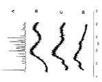
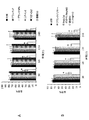
図3は、a)PS80、b)可溶性デンプン、c)PMAA及びd)PMAA-g-Stの1H-NMRスペクトルを示す。0.86ppmにおけるピークは、PS80の脂肪族CH3プロトンに相当する。1.27におけるピークは、界面活性剤の脂肪族CH2領域に由来している。3.62ppmにおける大きいピークは、PS80のポリエチレンオキシド領域のCH2プロトンからである。5.33における小さいピークは、脂肪酸鎖(二重結合)中のCH基からである。脂肪族領域中のより小さいピークは、脂肪酸尾部のさまざまな部分に属する。デンプンのスペクトルは、3.5ppmにおいて、C6炭素に結合したデンプン単位のCH2に帰せられる特性ピークを示す。3.8ppmにおけるピークは、C1〜C5炭素に加えられるCH単位に結合された水素に帰せられる。5.1ppmにおけるピークは、R-OHヒドロキシル基の水素に帰せられる。興味深いことに、ホモポリマーPMAAのスペクトルは、PMAA及びPS80の両方に特徴的なピークを示す。0.94ppm及び1.66ppmにおけるピークは、それぞれPMAAのCH3及びCH2に由来する。5.33ppmにおけるCHのピークは、PS80-PMAAコポリマーにはなく、PS80が、MAAモノマーとその二重結合を介して反応することを示している。PS80は、主要な疎水性置換基として、モノ-、ジ-、及びトリ-不飽和脂肪酸エステルを含有しており、この界面活性剤に対する重合経路の可能性を引き上げる。さらに、酸化反応に加わるPS80の能力はこれまでに文献によく解説されている[31〜33]。実際に、PS80は、薬物安定性の評価のための酸化剤として使用されてきた[34]。開始剤の存在下で、界面活性剤上のアルキルフリーラジカルは、水素引き抜き反応によって形成され得る。これは、熱又は光化学のRH結合の均一開裂を含む、さまざまなプロセスによって起こり得る。その後、フリーラジカルは、モノマーの二重結合を攻撃することによってグラフト重合反応に加わることができる。 FIG. 3 shows 1 H-NMR spectra of a) PS80, b) soluble starch, c) PMAA and d) PMAA-g-St. The peak at 0.86 ppm corresponds to the aliphatic CH 3 proton of PS80. The peak at 1.27 is derived from the aliphatic CH 2 region of the surfactant. The large peak at 3.62 ppm is from the CH 2 proton in the polyethylene oxide region of PS80. The small peak at 5.33 is from the CH group in the fatty acid chain (double bond). Smaller peaks in the aliphatic region belong to different parts of the fatty acid tail. The starch spectrum shows a characteristic peak attributed to CH 2 of starch units bound to the C6 carbon at 3.5 ppm. The peak at 3.8 ppm is attributed to hydrogen bonded to CH units added to the C1-C5 carbon. The peak at 5.1 ppm is attributed to the hydrogen of the R-OH hydroxyl group. Interestingly, the spectrum of homopolymer PMAA shows characteristic peaks for both PMAA and PS80. Peaks at 0.94ppm and 1.66ppm, respectively derived from CH 3 and CH 2 of PMAA. The CH peak at 5.33 ppm is absent from the PS80-PMAA copolymer, indicating that PS80 reacts with the MAA monomer via its double bond. PS80 contains mono-, di-, and tri-unsaturated fatty acid esters as the main hydrophobic substituents, raising the possibility of polymerization pathways for this surfactant. Furthermore, the ability of PS80 to participate in oxidation reactions has been well documented so far [31-33]. In fact, PS80 has been used as an oxidant for the evaluation of drug stability [34]. In the presence of an initiator, alkyl free radicals on the surfactant can be formed by a hydrogen abstraction reaction. This can occur by various processes, including the uniform cleavage of thermal or photochemical RH bonds. The free radical can then participate in the graft polymerization reaction by attacking the double bond of the monomer.
PMAA-g-Stポリマーの1H NMRスペクトルは、デンプン、MAA、及びPS80に特有のピークを示す。グラフトによってもたらされた化学環境の改変により、0.94、1.29、1.66、3.5のピークにおける小さいシフト並びに3.5ppmのピークにおける形状のわずかな変化が存在する。また、5.1におけるピークの相対強度の減少が存在し、デンプンのヒドロキシル基がグラフト反応に関与していることを示している。このピークは、試料中に存在する無水グルコース単位の量に直線的に依存している。3.52、3.70、及び1.66のピークの下の面積は、最終生成物におけるデンプン、MMA、及びPS80のモル比を計算するために使用され、表1に示された。加えて、滴定調査からの当量点データを用いて、我々は異なる供給モノマー比で調製されたナノ粒子中のMAA含量を決定した。これらのデータもまた、表2に示されている。フィード及び生成物中のMAAとデンプンのモル比の間には比較的良好な関係が存在する。しかしながら、フィード中のPS80の小さい画分のみがその最終生成物中には組み込まれる。最終生成物中のその界面活性剤の相対的モル比は、フィード中のMAAの量が減ると減少し、おそらく、PS80は、主としてMAAモノマーとの共重合を通してグラフトポリマー中に組み込まれることを暗示している。 The 1 H NMR spectrum of PMAA-g-St polymer shows peaks characteristic of starch, MAA, and PS80. There is a small shift in the 0.94, 1.29, 1.66, and 3.5 peaks as well as a slight change in shape at the 3.5 ppm peak due to the chemical environment modifications brought about by the grafting. There is also a decrease in the relative intensity of the peak at 5.1, indicating that the starch hydroxyl groups are involved in the grafting reaction. This peak is linearly dependent on the amount of anhydroglucose units present in the sample. The areas under the 3.52, 3.70, and 1.66 peaks were used to calculate the molar ratio of starch, MMA, and PS80 in the final product and are shown in Table 1. In addition, using equivalence point data from titration studies, we determined the MAA content in nanoparticles prepared with different feed monomer ratios. These data are also shown in Table 2. There is a relatively good relationship between the molar ratio of MAA to starch in the feed and product. However, only a small fraction of PS80 in the feed is incorporated into the final product. The relative molar ratio of that surfactant in the final product decreases as the amount of MAA in the feed decreases, possibly implying that PS80 is incorporated into the graft polymer primarily through copolymerization with the MAA monomer. doing.
さらなるFTIR及びH1 NMRスペクトルが図4に示されている。 Further FTIR and H 1 NMR spectra are shown in FIG.
デンプンへのPMAAのグラフトのための開始プロセス及びフリーラジカル形成は、次の反応スキームによって説明され得る[35、36]。
反応(3)、(5)、及び(6)は、さまざまなフリーラジカル種の連続形成に有利であるが、反応(2)及び(4)は、フリーラジカルの消滅をもたらす。チオ硫酸塩(thiosulfate)が存在すると、さまざまなフリーラジカル:硫酸ラジカル、チオ硫酸ラジカル、及びヒドロキシルラジカルが存在し、それらはデンプンを攻撃してデンプン分子上で水素引き抜き及びフリーラジカルの形成をもたらすことができるものと考えられる。ヒドロキシルラジカル又はデンプンラジカルは、MAAの二重結合を攻撃し、MAAのデンプンへのグラフトを引き起こすことができる。従って、開始された鎖へのMAA分子のその後の添加は、図5によるグラフト鎖を増殖させる。最後に、成長しているグラフト鎖は、連結又は不均化によって停止される(図5)。MAAの同時の単独重合がMAAモノマー上のフリーラジカルの開始作用によってある程度さらに起こることに注意すべきである。 Reactions (3), (5), and (6) are advantageous for the continuous formation of various free radical species, while reactions (2) and (4) result in the disappearance of free radicals. In the presence of thiosulfate, there are a variety of free radicals: sulfate radicals, thiosulfate radicals, and hydroxyl radicals that attack starch and cause hydrogen abstraction and free radical formation on the starch molecule. Can be considered. Hydroxyl radicals or starch radicals can attack the double bond of MAA and cause the grafting of MAA onto starch. Thus, subsequent addition of MAA molecules to the initiated chain will grow the grafted chain according to FIG. Finally, growing graft chains are stopped by ligation or disproportionation (FIG. 5). It should be noted that the simultaneous homopolymerization of MAA occurs to some extent by the free radical initiating action on the MAA monomer.
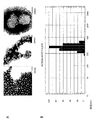
ナノ粒子の形態及び粒径
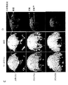
分析された全てのナノ粒子は、およそ100〜200nmの粒径及びどちらかといえば球状の非常に均一な形態を示した(図6A)。このナノ粒子は、多孔質の綿ボール様の表面形態を有する。多孔質構造を有することは、pH等の環境刺激に応じたより速い相転移とともにより高い薬物充填を促進することに関して有利であり得る。ある程度の粒子の凝集及び融合の存在もあるが、これはTEM試料調製の性質によるものであり得る。TEMグリッド上に近接近して置かれる粒子は、乾燥及び電子線の影響によって部分的に融合し得る。そのような挙動は一般的であり、ポリ(2-ヒドロキシエチルメタクリレート)粒子などの他のポリマーナノ粒子についても頻繁に報告されている[37]。
Nanoparticle Morphology and Particle Size All the nanoparticles analyzed exhibited a particle size of approximately 100-200 nm and a rather uniform morphology that was rather spherical (FIG. 6A). The nanoparticles have a porous cotton ball-like surface morphology. Having a porous structure can be advantageous with respect to promoting higher drug loading with faster phase transitions in response to environmental stimuli such as pH. There is also some degree of particle aggregation and fusion, which may be due to the nature of the TEM sample preparation. Particles placed in close proximity on the TEM grid can be partially fused due to drying and electron beam effects. Such behavior is common and has been frequently reported for other polymer nanoparticles such as poly (2-hydroxyethyl methacrylate) particles [37].
表3に示されているように、ナノ粒子の粒径は、ターポリマーPMAA-PS80-g-St-3については、ポリマー組成及びpHによって、70nmから310nmまでの範囲であった。DLS測定からの典型的な粒径分布プロットは、図6Bに示されている。一般に、粒径分布は、多分散指数PdIがおよそ0.09〜0.14で、PMAAナノ粒子(PdI=0.26)を除いて比較的狭い。粒径は、薬物応用におけるナノ粒子の性能を決定する重要なパラメーターである。それは、反応速度(pH等の刺激に対する)、薬の放出、細胞取り込み、並びに癌細胞を効果的に殺す粒子の能力などの特性に影響を及ぼす。加えて、粒径は、インビボの薬物動態及び生体内分布に、すなわち封入された薬物の治療効果に大きな影響を及ぼす。PMAA-g-Stナノ粒子の粒径は、粒子が細胞取り込み機構を受け易くするとともに、ナノ粒子に対して受動的な腫瘍標的特性を与える、血管透過性・滞留性亢進(EPR)効果の影響を粒子が受け易くする[38]。腫瘍血管系の空隙率(最も末梢のヒト腫瘍の有効な平均細孔径は約300nmである)及びリンパ排液の欠如のために、適切な大きさのコロイドナノ粒子は、EPRによって腫瘍中に選択的に分配される。 As shown in Table 3, the particle size of the nanoparticles ranged from 70 nm to 310 nm for the terpolymer PMAA-PS80-g-St-3, depending on the polymer composition and pH. A typical particle size distribution plot from a DLS measurement is shown in FIG. 6B. In general, the particle size distribution has a polydispersity index PdI of approximately 0.09 to 0.14 and is relatively narrow except for PMAA nanoparticles (PdI = 0.26). Particle size is an important parameter that determines the performance of nanoparticles in drug applications. It affects properties such as kinetics of response (to stimuli such as pH), drug release, cellular uptake, and the ability of particles to effectively kill cancer cells. In addition, the particle size has a great influence on the in vivo pharmacokinetics and biodistribution, ie the therapeutic effect of the encapsulated drug. The size of PMAA-g-St nanoparticles is influenced by the enhanced vascular permeability and retention (EPR) effect that makes the particles more susceptible to cellular uptake mechanisms and gives them passive tumor targeting properties Makes the particles more susceptible to [38]. Appropriately sized colloidal nanoparticles are selected in the tumor by EPR because of the porosity of the tumor vasculature (the effective average pore size of most peripheral human tumors is about 300 nm) and the lack of lymphatic drainage Distributed.
典型的な試料のTEM写真(図6)は、ナノ粒子が、ほぼ100〜200nmの粒径、球に近い形、及び多孔性の綿ボール様の形態を有することを示している。多孔質構造は、pHのような環境刺激に応じたより速い相転移とともにより高い薬物充填を促進することに関して、密度の高い構造よりも有利であり得る。図7は、PDEAEM-g-St-2ナノ粒子のTEM画像を示している。 A TEM picture of a typical sample (FIG. 6) shows that the nanoparticles have a particle size of approximately 100-200 nm, a shape close to a sphere, and a porous cotton ball-like morphology. A porous structure may be advantageous over a dense structure with respect to promoting higher drug loading with faster phase transitions in response to environmental stimuli such as pH. FIG. 7 shows a TEM image of PDEAEM-g-St-2 nanoparticles.
均一な粒径もまた、体内のナノ粒子の分布及びそれらの生体細胞との相互作用がその粒径によって大きく影響されるために、薬物送達の適用に重要である。一般に、単分散粒子は、より均一な物理的及び化学的特性を示し、より洗練された理にかなった薬物送達システムを考案することを容易にする。 Uniform particle size is also important for drug delivery applications because the distribution of nanoparticles in the body and their interaction with living cells is greatly influenced by the particle size. In general, monodisperse particles exhibit more uniform physical and chemical properties, making it easier to devise more sophisticated and reasonable drug delivery systems.
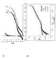
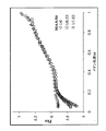
生理的pH範囲でpH応答性の膨れを示すPMAA-g-Stナノ粒子
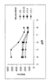
表3及び図8Aにおける結果は、pHを5から7.4まで増すと共に粒径が増大し、その粒径のpHに依存する変化は、MAA/Stのモル比に関連することを実証している。図8は、粒径がpHを4から7.4まで上げると共に増加し、その増加の規模はMAA含量によって決定されることを示している。例えば、PMAAナノ粒子の平均直径は、pH4における70.5nmからpH7.4における152nmまで2.2倍増加し、一方、PMAA-g-St-4は1.2倍に増加するのみであり、言い換えると、PMAAについては10.1倍の、PMAA-g-St-4については1.5倍の体積比(V7.4/V4)になる。一般に、デンプン含量の増加は、pH感度における減少をもたらす。これは、より低いMAA含量はイオン化されたカルボン酸基のより低い含量に起因するものとされるより小さい静電反発力をもたらし、それゆえ膨れを低下させるという事実のせいにされ得る。異なるpH感度は、異なる量のイオン化可能な及び/又はイオン化されたカルボン酸基を意味する。より低いMAA含量は、より小さい静電反発力及びpH7.4におけるより低い膨れをもたらし、より小さいV7.4/V4値につながる。
PMAA-g-St nanoparticles exhibiting pH-responsive swelling in the physiological pH range The results in Table 3 and Figure 8A show that the particle size increases with increasing pH from 5 to 7.4, depending on the pH of the particle size The change demonstrates that it is related to the molar ratio of MAA / St. FIG. 8 shows that the particle size increases with increasing pH from 4 to 7.4, the magnitude of the increase being determined by the MAA content. For example, the average diameter of PMAA nanoparticles increases 2.2-fold from 70.5 nm at
図8Aは、また、粒径の劇的な増加がpH5とpH6の間で起こることを示しており、PMAA含有ナノ粒子の体積相転移pHと合致する、この領域におけるイオン化遷移を示している[39]。PMAAのpKaより低いpH値において、プロトン化したカルボン酸基は、崩壊構造につながる広範な水素結合を形成する。より高いpH値においてカルボン酸基の増加したイオン化は、ポリマー鎖の間に高い静電反発力をもたらし、それゆえ粒子を大きくした。
FIG. 8A also shows that a dramatic increase in particle size occurs between
粒子の表面電荷に対するpH及びPMAA含量の影響
ナノ粒子のゼータ電位が、表4及び図8Bにまとめられている。このデータは、全てのナノ粒子が、そのナノ粒子と結合しているカルボン酸基のイオン化によって、2から7までの媒体のpHの増大と共に増加する負の表面電荷を有することを示している。ゼータ電位は、液体のイオンによって作り出される電気二重層における電荷であり、それぞれの粒子の周りに存在する。水溶液中に分散されたナノ粒子は、静電的安定化(表面電荷)によって又は立体安定化(粒子表面における界面活性剤又はその他の分子)によって、或いは両方の組合せによって安定化され得る。一般に、+/-20mVを超えるゼータ電位値が安定なコロイド分散の特徴と考えられる。DLVO理論によれば、凝集は、粒子間の引きつけるファンデルワールス力が、静電気反発力を圧するときに起こる。表4及び図8Bに示されているように、殆どのナノ粒子生成物は、調査されたさまざまなpH値において-20mVに近いかそれを超えるゼータ電位を有し、それ故コロイド的に安定であることが期待される。ナノ粒子中のPMAA含量が減少するとゼータ電位も低下する。pH4において、PMAA-g-St-4ナノ粒子は-2.7mVのゼータ電位を有し、それは、それらがいくらかのコロイド安定性の問題を有するであろうことを意味する。これらのデータは、高いpHにおけるより大きい負電荷と共に、PMAAがナノ粒子の表面電荷に大きく寄与することを示唆する。
Effect of pH and PMAA content on the surface charge of the particles The zeta potential of the nanoparticles is summarized in Table 4 and FIG. 8B. This data shows that all nanoparticles have a negative surface charge that increases with increasing pH of the medium from 2 to 7, due to ionization of the carboxylic acid groups attached to the nanoparticles. The zeta potential is the charge in the electric double layer created by the liquid ions and exists around each particle. Nanoparticles dispersed in an aqueous solution can be stabilized by electrostatic stabilization (surface charge) or by steric stabilization (surfactants or other molecules on the particle surface) or by a combination of both. In general, a zeta potential value exceeding +/− 20 mV is considered as a characteristic of stable colloidal dispersion. According to DLVO theory, agglomeration occurs when van der Waals forces attracting particles presses electrostatic repulsion. As shown in Table 4 and FIG. It is expected to be. As the PMAA content in the nanoparticles decreases, the zeta potential also decreases. At
デンプンに基づくナノ粒子におけるカルボン酸基の特性付け
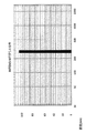
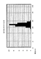
図9は、Cs=0.05N NaClでのPMAA-g-St-2ラテックス分散液の電位差滴定の例を示す。特別な定めのない限り、5分の安定化時間が各滴定剤の添加の間で設けられた。これは、平衡を得るために要する緩和時間が対応する低分子量の弱酸と比較して普通は非常に長いので、高分子電解質ラテックス粒子の滴定においては常法である。オリジナルの滴定データは、遊離のH+及びOH-の寄与を考慮に入れて補正され、終点をより明瞭にした。その補正は、方程式2[26、27]:
![]()
![]()
に従って実施される。上式中、[VNaOH]は分散液に加えられるNaOHの体積であり、
![]()
![]()
は、その分散液中と同じpHのブランクの溶液に加えられるHCl及びNaOHの体積である。この補正により、分散液中及びブランクの溶液中のH+及びOH-に対して同じ活量係数を仮定すると、[V]pHの値は、当量点で定数であるはずである。我々は、上記の手順を使って矢印で示されている2.39mlの当量点を決めた。この値は、単純なスプレッドシートプログラミングを用いる滴定曲線の変曲点から決定したものと十分一致している。 Is the volume of HCl and NaOH added to the blank solution at the same pH as in the dispersion. With this correction, assuming the same activity coefficient for H + and OH − in the dispersion and in the blank solution, the value of [V] pH should be constant at the equivalence point. We used the above procedure to determine the equivalence point of 2.39 ml indicated by the arrow. This value is in good agreement with that determined from the inflection point of the titration curve using simple spreadsheet programming.
滴定調査からの当量点データを用いて、我々は異なる供給モノマー比を有するさまざまなPMAA-g-Stバッチに対するMAA含量を確定した。これらのデータは、それらの対応する当量点データと共に表4に示されている。 Using the equivalence point data from the titration study, we determined the MAA content for various PMAA-g-St batches with different feed monomer ratios. These data are shown in Table 4 along with their corresponding equivalence point data.
さまざまな組成のナノ粒子のpKa値がαに対して描かれた。pK0値は、pKa値をα=0に外挿することによって求められた。そのpK0値は、ナノ粒子中のデンプン含量に依存することが見出された。それはPMAA-PS80ナノ粒子に対する4.9からPMAA-PS80-g-St-4ナノ粒子に対する5.8まで殆ど1単位増加した。PMAA-PS80-g-St-1粒子、PMAA-PS80-g-St-2粒子、及びPMAA-PS80-g-St-3粒子の固有のイオン化定数は、それぞれ5.0、5.1、及び5.5であった(図9)。デンプン含量を増加することによるpK0の増大は、デンプンのヒドロキシル基とターポリマーの酸基との間のより高い度合いの水素結合の観点から説明され得る。ターポリマーのイオン化挙動における変化(pKaの動きの変化によって証明される)は、粒子のより低い電荷密度、水に対するより高い親和性を促進することができるデンプンの親水性、並びにイオン性基のより拡大された立体配座及びより高い空間的隔離をもたらすであろうデンプンのより堅い鎖構造による可能性がある。 PK a value of nanoparticles of various compositions were plotted against alpha. The pK 0 value was determined by extrapolating the pK a value to α = 0. Its pK 0 value was found to depend on the starch content in the nanoparticles. It increased almost 1 unit from 4.9 for PMAA-PS80 nanoparticles to 5.8 for PMAA-PS80-g-St-4 nanoparticles. The intrinsic ionization constants of PMAA-PS80-g-St-1 particles, PMAA-PS80-g-St-2 particles, and PMAA-PS80-g-St-3 particles were 5.0, 5.1, and 5.5, respectively. (Figure 9). The increase in pK 0 by increasing the starch content can be explained in terms of a higher degree of hydrogen bonding between the starch hydroxyl groups and the terpolymer acid groups. Changes in the ionization behavior of terpolymer (as evidenced by a change in the movement of the pK a), a hydrophilic starch that can promote higher affinity for lower charge density, water particles and the ionic groups It may be due to the stiffer chain structure of starch that would result in a more expanded conformation and higher spatial isolation.
電位差滴定法もPMAA-g-Stナノ粒子中のカルボン酸官能基の分布を調べるために使用された。ヒドロゲルナノ粒子のゲル相は、イオンに対して透過性である。それ故、滴定剤イオンは、水性のバルク相には制限されず、ゲル相中に拡散してそのゲル相中に存在する官能基を中和することができる。バルク相とゲル相の間の安定化時間は、滴定される官能基の分布に大きく依存する。表面に接近可能な基は、より短い平衡時間を必要とし、一方表面下に埋もれている滴定可能な基は、平衡に達するまでにより長い時間を要する。 Potentiometric titration was also used to investigate the distribution of carboxylic acid functional groups in PMAA-g-St nanoparticles. The gel phase of the hydrogel nanoparticles is permeable to ions. Therefore, the titrant ions are not limited to the aqueous bulk phase and can diffuse into the gel phase and neutralize the functional groups present in the gel phase. The stabilization time between the bulk phase and the gel phase is highly dependent on the distribution of functional groups to be titrated. Groups accessible to the surface require a shorter equilibration time, while titratable groups buried below the surface require a longer time to reach equilibrium.
各添加の間の安定化時間をたった30秒として、速い逆滴定(塩基中への酸)が後に続く正滴定(酸中への塩基)の調査が、ナノ粒子中の酸基の分布の見識を得るために使用された。もし水相及びゲル相が滴定剤の次の量の添加前に完全に平衡に達している場合は、その正滴定及び逆滴定は完全にオーバーラップするはずであるが、もし平衡になっていない場合、その2つの滴定の間にはいくらかの時間のずれが観察されるはずである。図10は、PMAA及びMAA:Stのさまざまな供給比によるPMAA-g-Stの2つの異なるバッチに対する正滴定及び逆滴定の曲線を示している。PMAA粒子(図10A)及び高いMAA含量を有するPMAA-g-St粒子(図10B)は、両方とも正滴定と逆滴定の間で良好なオーバーラップを示しており、一方で、高いデンプン含量を有するPMAA-g-St粒子は、滴定の開始及び終わり近くのみで良好なオーバーラップを示した(図10C)。酸性基の滴定が発生するpH領域に大きなずれがあった。PMAA粒子及び低いデンプン含量を有するPMAA-g-St粒子についての速い安定化時間は、H+イオンの拡散がゲル構造によって妨害されないことを示唆している。これは粒子表面近くにカルボン酸基が存在するためである可能性がある。対照的に、高いデンプン含量を有するPMAA-g-StゲルにおけるH+付加の速度は、ゲル中の滴定可能な基に向けたH+拡散の速度より速いようであり、物質移動過程がゲル構造によって一部妨害されることを示唆している。これらの結果に基づき、高レベルのデンプン含量を有するPMAA-g-Stナノ粒子中のカルボン酸基は、PMAA及びより低いデンプン含量のPMAA-g-St粒子におけるものよりも粒子表面に接近できにくいと結論づけることが妥当である。 An investigation of forward titration (base into acid) followed by fast back titration (acid into base) with a stabilization time of only 30 seconds between each addition provides insight into the distribution of acid groups in the nanoparticles. Used to get. If the aqueous and gel phases are fully equilibrated before the next addition of titrant, the forward and back titrations should overlap completely, but are not equilibrated If so, some time lag should be observed between the two titrations. FIG. 10 shows forward and back titration curves for two different batches of PMAA-g-St with various feed ratios of PMAA and MAA: St. PMAA particles (Figure 10A) and PMAA-g-St particles with high MAA content (Figure 10B) both show good overlap between forward and back titrations, while high starch content. The PMAA-g-St particles with had a good overlap only near the beginning and end of the titration (FIG. 10C). There was a large shift in the pH region where acid group titration occurred. The fast stabilization time for PMAA particles and PMAA-g-St particles with low starch content suggests that the diffusion of H + ions is not hindered by the gel structure. This may be due to the presence of carboxylic acid groups near the particle surface. In contrast, the rate of H + addition in PMAA-g-St gels with high starch content appears to be faster than the rate of H + diffusion towards titratable groups in the gel, and the mass transfer process is gel structure It is suggested that it is partially disturbed by Based on these results, carboxylic acid groups in PMAA-g-St nanoparticles with high levels of starch content are less accessible to the particle surface than in PMAA and PMAA-g-St particles with lower starch content It is reasonable to conclude that
ポリ酸の酸強度又はイオン化の容易さは、各逐次電荷が、クーロン場がポリマーコイルの周りに積み重なるにつれて除去するのがより困難になるという点で、単一酸のそれとは異なる。ポリ酸の強度は、「見掛けの」pKa値によって表され、方程式3:
(式中、pK0は、固有の解離定数の負対数であり、Rは、気体定数であり、Tは、ケルビン温度であり、そしてΔGelは、静電気的相互作用期間である)
によるイオン化度(α)と関係している[40、41]。
(Where pK 0 is the negative logarithm of the intrinsic dissociation constant, R is the gas constant, T is the Kelvin temperature, and ΔG el is the electrostatic interaction period)
Is related to the degree of ionization (α) by [40, 41].
そのイオン化度(α)は、
(式中、[Veq]は、当量点の体積である)
によって計算される。
( Where [V eq ] is the volume of the equivalence point)
Is calculated by
方程式3は、非静電気期間(pK0)と静電気的相互作用期間(ΔGel)の和の観点から、pKaを説明している。図11においては、さまざまな組成物のナノ粒子のpKa値がαに対してプロットされている。pK0値は、pKa値をα=0に外挿することによって求められた。PMAAナノ粒子のpK0は、4.9であることが見出された。この値は、デンプン含量を増加することによって殆ど1単位増大した。実際に、5.8のpK0が、PMAA-g-St-4ナノ粒子について計算された。PMAA-g-St-1粒子、PMAA-g-St-2粒子、及びPMAA-g-St-3粒子の固有のイオン化定数は、それぞれ5.0、5.1、及び5.5であった。PMAAについてのpK0の実験値は、線状PMAA及びまたイソ酪酸のそれとよく一致しており、恐らくラテックス表面のカルボン酸基の大部分がバルク水中におけるものと同じ環境を有していることを示している。上記のように、より低いPMAA含量のナノ粒子は、表面下に埋もれたそれらのCOOH基のいくらかを有する。これにより、それらのカルボン酸基の付近における誘電率を低下してpK0値を増すことができる。
デンプン含量の増加は、ナノ粒子のイオン化挙動を変化させた(図11)。高いMAA含量を有するPMAA及びPMAA-g-StのpKa値は、イオン化度の増加と共に最初は急激に増大した。pKa値は、25%のイオン化付近でプラトーになり、40%のイオン化後に増大し始める。この挙動は、他の研究者によって同様に文書化されており、PMAAのコンパクトな/広がったコイル変形に起因する。一般に、その低いイオン化度でのコンパクト構造と一体となったPMAA粒子の高い電荷密度は、イオン化の範囲でpKa値における急激な初期の増加をもたらす。強い静電気反発力を最小限にするために、ポリマーは、より伸長されたコイル配列への立体構造変化を起こす。より高いデンプン含量を有するPMAA-g-St粒子は、異なるpKaプロフィールを示した。このpKaは、低いイオン化度で著しく高く、しかしながら、最高50%までの見掛けの解離定数において増加は殆どない。酸基は、このイオン化領域において単離された単位として挙動するようであり、小さい傾斜を有するpKa曲線を生ずる。これは、粒子のより低い電荷密度とともに、イオン化が可能な基のより伸張性の立体配座及びより高い空間的隔離をもたらす溶媒(水)とのより高度な適合性を促進するデンプンの親水性と関連付けて説明され得る。しかしながら、このpKa値は、より高い静電反発力によって50%のイオン化を超えて増加し始める。 Increasing the starch content changed the ionization behavior of the nanoparticles (FIG. 11). PK a value of PMAA and PMAA-g-St with high MAA content initially sharply increases with increasing degree of ionization. pK a values are to plateau at around 25% ionization, it starts to increase after the 40% ionization. This behavior is similarly documented by other researchers and is attributed to the compact / expanded coil deformation of PMAA. In general, high charge density PMAA particles compact was structure and integral at its lower ionization degree, resulting in rapid increase in initial in pK a value in the range of ionization. In order to minimize strong electrostatic repulsion, the polymer undergoes a conformational change to a more elongated coil arrangement. PMAA-g-St particles having a higher starch content showed different pK a profile. The pK a is lower significantly higher ionization degree, however, it increases little in apparent dissociation constant of up to 50%. The acid group appears to behave as an isolated unit in this ionization region, producing a pKa curve with a small slope. This is due to the lower charge density of the particles, as well as the hydrophilic nature of the starch, which promotes a higher degree of compatibility with the solvent (water) resulting in a more extensible conformation of the ionizable group and higher spatial sequestration. Can be described in association with However, the pK a value begins to increase more than 50% ionized by the higher electrostatic repulsion.
粒径及びpH感度に対する処理パラメーターの影響
図12Aは、粒径及び相転移に対するSDS濃度の影響を示す。PMAA-g-St粒子の直径は、界面活性剤含量が増加すると減少した。SDS界面活性剤の両親媒性構造は、水溶液中のポリマー粒子を安定させる助けとなり、それゆえポリマー鎖がより小さい粒子を形成することを可能にする。粒径に対する界面活性剤量の影響は顕著であった。例えば、SDSが0.17mmolから0.69mmolまで変化すると、粒径は591nmから298nmまで変化した。0.17mmol(0.05g)より下のSDSレベルは粒子の凝集をもたらした(矢印により注釈をつけられている)。PMAA-g-StコポリマーはCOOH基のイオン化のために負に帯電され、SDSはアニオン界面活性剤であるために、粒子表面に吸着されたSDSは粒子の凝集を防ぎながら、粒子が水中に分散することを助ける。pH=4における粒径に対するpH=7.4における粒径の比(D7.4/D4)が、pH応答性を測定するために使用された。その比は、低いSDSレベルにおいて減少したが、しかしながら、これは粒子の凝集及びDLSによる誤解させるような高い読みをもたらす低いpH値での減少された量のSDSによる粒子の貧弱な安定性のためである。
Effect of processing parameters on particle size and pH sensitivity FIG. 12A shows the effect of SDS concentration on particle size and phase transition. The diameter of PMAA-g-St particles decreased with increasing surfactant content. The amphiphilic structure of the SDS surfactant helps stabilize the polymer particles in the aqueous solution, thus allowing the polymer chains to form smaller particles. The effect of the amount of surfactant on the particle size was significant. For example, as the SDS changed from 0.17 mmol to 0.69 mmol, the particle size changed from 591 nm to 298 nm. SDS levels below 0.17 mmol (0.05 g) resulted in particle aggregation (annotated by arrows). Since PMAA-g-St copolymer is negatively charged due to ionization of COOH groups, and SDS is an anionic surfactant, the particles adsorbed on the particle surface are dispersed in water while preventing aggregation of the particles. To help. The ratio of the particle size at pH = 7.4 to the particle size at pH = 4 (D 7.4 / D 4 ) was used to measure pH responsiveness. The ratio decreased at low SDS levels, however, this was due to poor stability of particles with reduced amounts of SDS at low pH values resulting in particle agglomeration and misleading high readings by DLS. It is.
PS80濃度の増加の粒径及びpH応答性に対する影響は、図12Bに示されている。PS80量の増加は、粒径を若干増加させた。平均粒径は、0.13mmolのPS80での238nmから0.27mmolのPS80での300nmまで増加している。PS80レベルのさらなる増加は粒径に影響を及ぼさなかった。PS80濃度の増加は、また、粒子のpH感度の縮小をもたらした。上述のように、PS80は、フリーラジカルの形成並びにその二重結合によって重合反応に加わることができ、それ故、それはポリマー鎖の架橋を潜在的に推進することができる。多分、PS80濃度を増加することによって粒子の内部及び相互架橋が増加されて、ナノ粒子のpH感度の減少をもたらすことになる。 The effect of increasing PS80 concentration on particle size and pH responsiveness is shown in FIG. 12B. Increasing the amount of PS80 slightly increased the particle size. The average particle size increases from 238 nm with 0.13 mmol PS80 to 300 nm with 0.27 mmol PS80. Further increase in PS80 level did not affect the particle size. The increase in PS80 concentration also resulted in a decrease in the pH sensitivity of the particles. As mentioned above, PS80 can participate in the polymerization reaction through the formation of free radicals as well as its double bonds, and therefore it can potentially drive cross-linking of polymer chains. Perhaps increasing the PS80 concentration will increase the internal and cross-linking of the particles, leading to a decrease in the pH sensitivity of the nanoparticles.
全体のモノマー濃度が0.156mol/lから0.41mol/lに増加されると、平均粒径は、298nmから788nmまで増加した(図12C)。モノマー濃度が0.61mol/Lまでさらに増加されたとき、幅広の粒径分布が得られた(データは示されていない)。モノマー濃度のさらなる増加は、大規模な粒子の凝集をもたらした。モノマー濃度の増加は、それが核生成過程にさまざまに影響を及ぼすので、一般的に最終の粒径の増大を引き起こす。第一に、グラフトしたポリマー鎖は、連続相中に、この相の増大した溶解力によって、より長く留まることができる。その結果、そのオリゴマーは、沈殿前により長い鎖長に成長する。第二に、そのオリゴマー鎖の成長反応速度は増加する。また、安定剤の吸着速度は、その媒体の溶解力の変化のため、減少する。これらの効果の全てが、粒子核の平均サイズの増加に寄与する。興味深いことに、pH感度は、フィード中のモノマー濃度の変化の結果として著しく影響されることはなかった。 As the overall monomer concentration was increased from 0.156 mol / l to 0.41 mol / l, the average particle size increased from 298 nm to 788 nm (FIG. 12C). When the monomer concentration was further increased to 0.61 mol / L, a broad particle size distribution was obtained (data not shown). Further increases in monomer concentration resulted in large-scale particle agglomeration. Increasing monomer concentration generally causes an increase in final particle size as it affects the nucleation process in various ways. First, the grafted polymer chains can remain longer in the continuous phase due to the increased dissolving power of this phase. As a result, the oligomer grows to a longer chain length before precipitation. Second, the growth rate of the oligomer chain is increased. Also, the adsorption rate of the stabilizer decreases due to the change in the dissolving power of the medium. All of these effects contribute to an increase in the average size of the particle nuclei. Interestingly, pH sensitivity was not significantly affected as a result of changes in monomer concentration in the feed.
図12Dは、粒径及びpH感度に対する架橋剤モル比、X、の影響を示す。架橋剤モル比は、次の方程式:
を用いて計算された。粒径は、架橋剤モル比(X)の増加の結果増加した。より高い架橋レベルでは、より多くのポリマー鎖が架橋されてより大きい粒子をもたらすことができることが仮定され得る。また、個々のより小さい粒子の間で架橋及び粒子拡散してより大きい粒子を形成するという、高い可能性が存在する。体積相転移の大きさは、架橋レベルを下げることによって減少された。架橋剤の含量は、架橋密度及びヒドロゲルの網目サイズに対して直接の影響を有しており[4]、従って、架橋剤含量は、ヒドロゲルの膨潤挙動に対して大きな影響を有する。架橋剤レベルの増加と共に架橋間のポリマー鎖長は減少し、その結果、ゲルの膨潤を制限する弾力的に伸縮する力が劇的に増加した。これはより高い架橋レベルにおける粒子のpH応答性の減少を説明する。 Was calculated using. The particle size increased as a result of increasing the crosslinker molar ratio (X). It can be assumed that at higher cross-linking levels, more polymer chains can be cross-linked resulting in larger particles. There is also a high possibility of crosslinking and particle diffusion between individual smaller particles to form larger particles. The magnitude of the volume phase transition was reduced by lowering the level of crosslinking. The content of the crosslinker has a direct effect on the crosslink density and the hydrogel network size [4], therefore the crosslinker content has a great influence on the swelling behavior of the hydrogel. With increasing crosslinker level, the polymer chain length between crosslinks decreased, resulting in a dramatic increase in the elastic stretching force limiting gel swelling. This explains the decrease in particle pH responsiveness at higher crosslinking levels.
PMAA-g-Stナノ粒子を合成するワンポット水性分散液重合法が、かくして例示されている。粒径及びナノ粒子のpH応答性の膨潤は、合成パラメーター、例えば、MAA/St比、界面活性剤濃度、架橋剤濃度、及び全体のモノマー濃度に依存する。これらのパラメーターの調節は、さまざまな粒径及びpH応答性を有するPMAA-g-Stナノ粒子の生成を示す。PS80は、生成物をターポリマーにする重合に参加することが見出された。ポリソルベートは、また、安定なナノ粒子の形成においても役割を果たす。重合中のデンプンの存在も、より均一な粒径分布を与えるようである。MAA/St比によって、そのナノ粒子は、媒体のpHが7.4と4.0の間で変化するとき、最高10倍の体積変化を起こすことができる。 One-pot aqueous dispersion polymerization methods for synthesizing PMAA-g-St nanoparticles are thus illustrated. The particle size and pH responsive swelling of the nanoparticles depend on the synthesis parameters such as MAA / St ratio, surfactant concentration, crosslinker concentration, and total monomer concentration. Adjustment of these parameters indicates the generation of PMAA-g-St nanoparticles with various particle sizes and pH responsiveness. PS80 was found to participate in the polymerization of the product as a terpolymer. Polysorbate also plays a role in the formation of stable nanoparticles. The presence of starch during polymerization also appears to give a more uniform particle size distribution. Depending on the MAA / St ratio, the nanoparticles can undergo a volume change of up to 10 times when the pH of the medium changes between 7.4 and 4.0.
上述の例は、特にナノ粒子の合成及び特徴づけに関する本発明の実施形態を説明している。 The above examples illustrate embodiments of the invention, particularly with respect to nanoparticle synthesis and characterization.
ナノ粒子の製造は、ポリマー主鎖を可溶化することを含む。実施例において、主鎖は、約2,600Daから約4,500Daの範囲の分子量を有するデンプンによって提供される。上記のように、デンプンは、天然に存在する生体適合性、生分解性の毒性のないポリマーである。デンプンは、グリコシド結合によって連結されたグルコール単位から構成されている。天然デンプンの主成分は、アミロース及びアミロペクチンである。好ましい実施形態において、ポリマー主鎖をつくり上げているモノマー単位は、0.5と3の間の置換度を有するヒドロキシル基を有する。これは、平均して主鎖中のモノマー単位は、それらがポリマー中に現れるとき、平均して0.5から3個のヒドロキシル基を有することを意味する。従って、例えば、線状のグルコースポリマーであるアミロースは、約3の置換度を有する。この置換度は、約1から約3まで、又は2から約3までの範囲であり得、或いはそれは約1、約2、又は約3であり得る。従って、このポリマー主鎖は、複数のヒドロキシル基を有しており、そのためポリヒドロキシル化されていると言われる。モノマー単位は、例えば、1つ以上のペントース又はヘキソース(例えばグルコース)であり得、そのためそれは主鎖のモノマー単位当たり5個又は6個の炭素を有することができる。好ましくは、主鎖は、モノマー当たり3、4、5、6又は7個、より好ましくは、5又は6個、最も好ましくは6個の炭素を有する。比較的高分子量の多糖類(例えば、二糖又は三糖とは対照的に)の例としては、カロース、ラミナリン、クリソラミナリン、キシラン、アラビノキシラン、マンナン、フコイダン及びガラクトマンナンが挙げられる。アミロースのような体内で容易に分解され得る多糖類は、それらのインビボの挙動を活用するために使用され得るが、セルロースのような消化されにくい多糖類も、本発明のナノ粒子を形成することができる。天然に存在するデンプンとしては、トウモロコシデンプン、ジャガイモデンプン、サツマイモデンプン、コムギデンプン、サゴヤシデンプン、タピオカデンプン、コメデンプン、ダイズデンプン、アロールートデンプン、アミオカデンプン、ワラビデンプン、ハスデンプン、ワキシトウモロコシデンプン(waxy maize starch)、及び高アミローストウモロコシデンプンが挙げられる。 Nanoparticle production involves solubilizing the polymer backbone. In the examples, the backbone is provided by starch having a molecular weight in the range of about 2,600 Da to about 4,500 Da. As mentioned above, starch is a naturally occurring biocompatible, biodegradable non-toxic polymer. Starch is composed of glycol units linked by glycosidic bonds. The main components of natural starch are amylose and amylopectin. In a preferred embodiment, the monomer units making up the polymer backbone have hydroxyl groups with a degree of substitution between 0.5 and 3. This means that on average monomer units in the main chain have an average of 0.5 to 3 hydroxyl groups when they appear in the polymer. Thus, for example, amylose, a linear glucose polymer, has a degree of substitution of about 3. This degree of substitution can range from about 1 to about 3, or from 2 to about 3, or it can be about 1, about 2, or about 3. The polymer backbone therefore has a plurality of hydroxyl groups and is therefore said to be polyhydroxylated. The monomer unit can be, for example, one or more pentoses or hexoses (eg glucose) so that it can have 5 or 6 carbons per main chain monomer unit. Preferably, the backbone has 3, 4, 5, 6 or 7 per monomer, more preferably 5 or 6 and most preferably 6 carbons. Examples of relatively high molecular weight polysaccharides (eg, in contrast to disaccharides or trisaccharides) include callose, laminarin, chrysolaminarin, xylan, arabinoxylan, mannan, fucoidan and galactomannan. Polysaccharides that can be easily degraded in the body, such as amylose, can be used to take advantage of their in vivo behavior, but polysaccharides that are difficult to digest, such as cellulose, also form the nanoparticles of the present invention. Can do. Naturally occurring starch includes corn starch, potato starch, sweet potato starch, wheat starch, sago palm starch, tapioca starch, rice starch, soybean starch, arrow root starch, amio starch, bracken starch, lotus starch, waxy corn starch ( waxy maize starch), and high amylose corn starch.
本発明のナノ粒子の製造は、モノマーをポリマーにグラフト重合することを含む。実施例において、メタクリル酸がデンプンにグラフトされた。メタクリル酸は、α,β-不飽和カルボン酸であり、該実施例の重合製造プロセスは、フリーラジカルグラフト重合として知られている。そのような重合プロセスは、フリーラジカル開始剤の存在下で一般的には行われる。グラフト重合プロセスが進行すると、モノマー分子は、C-C結合が炭素をベースにした分子鎖とその分子鎖の側基からのカルボキシル基を形成している分子鎖に成長する。カルボキシル基は、その環境によってプロトン(H+)を失うことができるブレンステッド酸であり、まさにカルボキシラート基(CO2 -)として存在する。それ故、分子鎖上のカルボキシル基の数について言及されるとき、カルボキシル基の形は、それがCO2H又はCO2 -であるかは考慮に入れられない。ナノ粒子におけるカルボキシル基の酸挙動は、ナノ粒子の特性、特に異なるpHにおけるその挙動に寄与し、これについては他の場所で議論される。モノマーがα,β-不飽和カルボン酸であるとき、ナノ粒子の一部として形成される分子鎖は、交互に並ぶ炭素上にカルボキシル基を有する炭素をベースにした分子鎖を含有する。 The production of the nanoparticles of the present invention involves graft polymerization of a monomer to a polymer. In the examples, methacrylic acid was grafted onto starch. Methacrylic acid is an α, β-unsaturated carboxylic acid, and the polymerization production process of this example is known as free radical graft polymerization. Such polymerization processes are generally performed in the presence of a free radical initiator. As the graft polymerization process proceeds, monomer molecules grow into molecular chains in which CC bonds form carbon-based molecular chains and carboxyl groups from the side groups of the molecular chains. The carboxyl group is a Bronsted acid that can lose protons (H + ) depending on its environment, and is exactly present as a carboxylate group (CO 2 − ). Therefore, when reference is made to the number of carboxyl groups on the molecular chain, the form of a carboxyl group, it CO 2 H or CO 2 - in which one can not be taken into account. The acid behavior of the carboxyl group in the nanoparticle contributes to the properties of the nanoparticle, particularly its behavior at different pHs, which is discussed elsewhere. When the monomer is an α, β-unsaturated carboxylic acid, the molecular chain formed as part of the nanoparticle contains a carbon-based molecular chain with carboxyl groups on alternating carbon.
本発明の好ましい「ワンポット」合成において、その重合反応は、重合反応に加わる、即ち形成されつつある側鎖と共有結合を形成するエトキシ化分子の存在下で行われる。実施例において、エトキシ化分子は、Tween(登録商標)80として市販されているポリソルベート80である。単語のポリソルベートは、構造:
を有する化合物の群を表現する。与えられたポリソルベートについて、w+x+y+zは与えられた数「n」に等しく、「R」は1つ以上の脂肪ヒドロカルビル基である。基-O(O)C-Rは、一般的には天然に存在する脂肪酸に相当する。ポリソルベート80の場合、n=20であり、R基は、オレイン酸:
におけるR基と同じである。実は、ポリソルベート80に対する別の名前は、ソルビタン核、オキシエチレン基(-CH2-CH2-O-)及びそのソルビタンにエステル結合によって結合されたオレイン酸の存在を反映しており、そしてnの値を示している、ポリオキシエチレン(20)ソルビタンモノオレエートである。
Same as R group in In fact, another for
実施例で記載されているように、ポリソルベート80のオレイン酸R基は、モノマーのグラフト重合中に成長している側鎖に共有結合し、ポリエトキシ化部分はナノ粒子の外側表面に存在することになる。本発明は、ナノ粒子の合成中にこの挙動を見せるエトキシ化分子を含み、その重合ステップは、エトキシ化分子をポリマー鎖に共有結合させるエトキシ化分子の存在下で行われる。好ましくは、重合ステップは、ポリエトキシ化分子の存在下で行われ、ポリエトキシ化分子を形成しつつある分子鎖に共有結合させ、その重合した生成物がナノ粒子の外側にポリエトキシ化部分を有するナノ粒子を形成する。
As described in the examples, the oleic acid R group of
他の場所で示されているように、ポリエトキシ化部分は、本発明のナノ粒子に有用な実用特性を与える。ナノ粒子中に組み込まれるエトキシ化分子中のオキシエチレン単位又は基の数は、10、11、12、13、14、15、16、17、18、19、20、21、22、23、24、25、26、27、28、29、30、31、32、33、34、35、36、37、38、39又は40であり得る。与えられたポリソルベートは、しばしば分子の混合物であり、それ故これらの数は平均を指していることに注意すべきである。好ましくは、このポリエトキシ化分子は、ソルビタンをベースとした分子である。より好ましくは、それはポリソルベートである。 As indicated elsewhere, the polyethoxylated moiety provides useful practical properties for the nanoparticles of the present invention. The number of oxyethylene units or groups in the ethoxylated molecule incorporated into the nanoparticles is 10, 11, 12, 13, 14, 15, 16, 17, 18, 19, 20, 21, 22, 23, 24, 25, 26, 27, 28, 29, 30, 31, 32, 33, 34, 35, 36, 37, 38, 39 or 40. It should be noted that a given polysorbate is often a mixture of molecules, so these numbers refer to the average. Preferably, the polyethoxylated molecule is a sorbitan based molecule. More preferably it is a polysorbate.
本発明のナノ粒子に対して特性化されたように、ポリソルベートのR基のC=C(不飽和)は、ナノ粒子合成の重合に関与する。従って、このポリエトキシ化部分は、合成中に形成するポリマー鎖に共有結合され、且つ形成されたナノ粒子の鎖に共有結合されることになる。R(C9〜C31)-C(O)O-基を有するポリエトキシ化ソルビタンであって、グラフト重合の間にソルビタンがそのR(C9〜C31)-C(O)O-基のC-C共有結合を介して第二のポリマーに結合するポリエトキシ化ソルビタンは、本発明の実施形態である。好ましくは、ソルビタンは、オキシエチレン単位の総数が少なくとも10であるポリソルベートである。R(C9〜C31)-C(O)O-基が、重合の段階においてC-C共有結合を形成する反応をする少なくとも1つのC-C不飽和を含むことも好ましい。そのR(C9〜C31)-C(O)O-基は、C9、C10、C11、C12、C13、C14、C15、C16、C17、C18、C19、C20、C21、C22、C23、C24、C25、C26、C27、C28、C29、C30、C31のいずれかであり得る。偶数である炭素の総数を有する脂肪酸(R-CO2H)はより一般的あり、このことは、この状況において奇数を有するR基がより一般的で、それが好ましいものをもたらすことができることを意味する。 As characterized for the nanoparticles of the present invention, the C = C (unsaturation) of the R group of the polysorbate is involved in the polymerization of nanoparticle synthesis. Thus, this polyethoxylated moiety will be covalently bonded to the polymer chain formed during synthesis and to the formed nanoparticle chain. A polyethoxylated sorbitan having an R (C9-C31) -C (O) O- group, wherein the sorbitan bonds the CC covalent bond of its R (C9-C31) -C (O) O- group during graft polymerization. Polyethoxylated sorbitan that is attached to the second polymer via is an embodiment of the present invention. Preferably, the sorbitan is a polysorbate having a total number of oxyethylene units of at least 10. It is also preferred that the R (C9-C31) -C (O) O- group contains at least one CC unsaturation that reacts to form a CC covalent bond in the polymerization stage. The R (C9-C31) -C (O) O- group is C9, C10, C11, C12, C13, C14, C15, C16, C17, C18, C19, C20, C21, C22, C23, C24, C25. , C26, C27, C28, C29, C30, C31. Fatty acids with a total number of carbons that are even (R-CO 2 H) are more common, which means that R groups with odd numbers are more common in this situation and can lead to what is preferred. means.
不飽和を含むポリソルベート80のオレイン酸成分以外の脂肪酸としては、例えば、リノール酸、アラキドン酸、ミリストレイン酸、パルミトレイン酸、サピエン酸、エライジン酸、バクセン酸、リノエライジン酸、α-リノレン酸、エイコサペンタエン酸、エルカ酸などがある。ナノ粒子に対するさまざまな状況の下で、1つ以上のこれらの脂肪酸に基づく1つ以上のポリソルベートをその中に組み込むことが有利であることが見出され得る。
Examples of fatty acids other than the oleic acid component of
実施例で説明されているように、グラフト重合段階のポリマー及びモノマーの相対量は、異なるポリマー/モノマー比を有するナノ粒子を得るように変化させることができる。ナノ粒子に関して、モノマー分子は、重合中に形成される分子鎖の一部であり、それ故モノマー単位と呼ばれることもできる。実施例が、ポリマー主鎖のモノマー単位に対する重合モノマーのモノマー単位のモル比(即ちMMA/St)が0.6から4.7であるナノ粒子を示す。約0.1と約10、又は0.2と9.0、又は0.2と8、又は0.2と8.0、又は0.2と7.0、又は0.3と7.0、又は0.3と6.0、又は0.4と6.0、又は0.4と5.0、又は0.4と4.0、又は0.4と3.0、又は0.5と6.0、又は0.6と6.0、又は0.7と6.0、又は0.8と6.0の間、又は1と5.0の間のその他の比を得ること、或いは、約0.1、約0.2、約0.3、約0.4、約0.5、約0.6、約0.7、約0.8、約0.9、約1、約1.5、約2、約2.5、約3、約3.5、約4、約4.5、約5、約5.5、約6、約6.5、約7、約7.5、約8、約8.5、約9、約9.5又は約10の比を得ることが可能である。 As illustrated in the examples, the relative amounts of polymer and monomer in the graft polymerization stage can be varied to obtain nanoparticles with different polymer / monomer ratios. With respect to nanoparticles, monomer molecules are part of the molecular chain formed during polymerization and can therefore also be referred to as monomer units. The examples show nanoparticles with a molar ratio of monomer units of polymerized monomer to monomer units of the polymer backbone (ie MMA / St) of 0.6 to 4.7. About 0.1 and about 10, or 0.2 and 9.0, or 0.2 and 8, or 0.2 and 8.0, or 0.2 and 7.0, or 0.3 and 7.0, or 0.3 and 6.0, or 0.4 and 6.0, or 0.4 and 5.0, or 0.4 and 4.0 Or other ratios between 0.4 and 3.0, or 0.5 and 6.0, or 0.6 and 6.0, or 0.7 and 6.0, or between 0.8 and 6.0, or between 1 and 5.0, or about 0.1, about 0.2, About 0.3, about 0.4, about 0.5, about 0.6, about 0.7, about 0.8, about 0.9, about 1, about 1.5, about 2, about 2.5, about 3, about 3.5, about 4, about 4.5, about 5, about 5.5 , About 6, about 6.5, about 7, about 7.5, about 8, about 8.5, about 9, about 9.5 or about 10 ratios can be obtained.
実施例に同様に記載されているのは、ポリエトキシ分子及び重合したモノマーのモノマー単位の相対量が、ナノ粒子合成中にポリソルベート80及びMAAの量を変化させることによって、変えられたナノ粒子である。それらの粒子成分に対しては、約0.003から約0.01までの重合した分子鎖のモノマー単位に対するエトキシ化分子のモル比が示された。その他の比、即ち、約0.0005と1の間、約0.0006と0.1の間、約0.001と0.1の間、約0.001と0.05の間、約0.001と0.04の間、約0.002と0.04の間、約0.002と0.03の間、約0.002と0.02の間又は約0.003と0.01の間の比を得ること、或いは、例えば、約0.0005、約0.0007、約0.0009、約1、約0.9、約0.8、約0.7、約0.6、約0.5、約0.4、約0.3、約0.2、約0.1、約0.005、約0.006、約0.007、約0.008、又は約0.009の比を得ることが可能である。
Also described in the examples are the nanoparticles in which the relative amounts of polyethoxy molecules and polymerized monomer units are altered by changing the amount of
実施例のグラフト重合においてN,N'-メチレンビスアクリルアミドが使用された。実施例において、重合モノマー、MAA、及び架橋剤、MBA、のモル比は、約3:1から約7:1までの範囲であった。重合段階で使用されるモノマーの量は、モル基準で架橋剤の量の1倍と20倍の間であり得、又はそれは、1倍と15倍の間、又は1倍と10倍の間、又は2倍と10倍の間、又は2倍と9倍の間、又は2倍と8倍の間、2倍と7倍の間、又は2倍と6倍の間であり得、或いはモノマーの量は、モル基準で架橋剤の量の3倍と6倍の間であり得る。 N, N′-methylenebisacrylamide was used in the graft polymerization of the examples. In the examples, the molar ratio of polymerized monomer, MAA, and crosslinker, MBA ranged from about 3: 1 to about 7: 1. The amount of monomer used in the polymerization stage can be between 1 and 20 times the amount of crosslinker on a molar basis, or it can be between 1 and 15 times, or between 1 and 10 times, Or between 2 and 10 times, or between 2 and 9 times, or between 2 and 8 times, between 2 and 7 times, or between 2 and 6 times, or The amount can be between 3 and 6 times the amount of crosslinker on a molar basis.
ナノ粒子の応用及び使用
本発明のナノ粒子のさまざまな改変により、特に医療に関連する多数の応用においてそれらの有用性が実証された。ナノ粒子の改変バージョンは、そのナノ粒子の合成中、又はそれらの作製後の両方で得ることが可能である。
Applications and Uses of Nanoparticles Various modifications of the nanoparticles of the present invention have demonstrated their utility, especially in a number of applications related to medicine. Modified versions of the nanoparticles can be obtained both during the synthesis of the nanoparticles or after their production.
以下により詳細に記載されている1つの応用において、薬物送達剤としての本発明のナノ粒子の有用性が薬物ドキソルビシンを利用して実証されている。アントラサイクリン環抗生物質の仲間の一つであるドキソルビシンは、さまざまなヒト及び動物の固形腫瘍において広い抗腫瘍活性を有する周知の抗癌剤である[42、43]。しかしながら、この薬物は非常に狭い治療指数を有しており、その臨床用途は、心臓毒性及び骨髄機能抑制などのいくつかの望ましくない副作用によって妨げられている[44〜46]。もう一つの制限は、この薬物がよく知られたp-糖タンパク質(P-gp)基質であることである。P-gpは、多くの抗癌剤の細胞内蓄積を妨げ、それ故、主に能動的取り込みを妨げ、プラスに帯電した両親媒性の薬物の細胞流出をATPに依存する形式で増加させることによって、それらの細胞毒性活性の低下を引き起こす。P-gpの過剰発現は、癌細胞における多剤耐性の主要なメカニズムの1つであると思われる[47〜50]。 In one application described in more detail below, the utility of the nanoparticles of the present invention as a drug delivery agent has been demonstrated utilizing the drug doxorubicin. Doxorubicin, one of the anthracycline ring antibiotics, is a well-known anticancer agent with broad antitumor activity in a variety of human and animal solid tumors [42, 43]. However, this drug has a very narrow therapeutic index and its clinical use is hampered by several undesirable side effects such as cardiotoxicity and suppression of myeloid function [44-46]. Another limitation is that this drug is a well-known p-glycoprotein (P-gp) substrate. P-gp prevents intracellular accumulation of many anticancer drugs, and therefore primarily prevents active uptake, and increases the cellular efflux of positively charged amphipathic drugs in an ATP-dependent manner, It causes a decrease in their cytotoxic activity. Overexpression of P-gp appears to be one of the major mechanisms of multidrug resistance in cancer cells [47-50].
ドキソルビシンの適切なナノ粒子系との結合体は、ドキソルビシン化学療法と関連するいくつかの制限に立ち向かうことができる[51〜55]。適切なナノキャリアシステム中に薬物を組み込むことによるそれらの標的化された送達により、インビボにおける薬物の生体内分布及び薬物動態が改変する[56]。原則として、薬物を充填したナノ粒子の腫瘍中の蓄積は、血管透過性・滞留性亢進(EPR)効果として知られる非特異性標的プロセスによって達成され得る[38、57]。腫瘍に特有であり正常組織にはない、漏出性血管系及び限定されたリンパ排液は、多種多様の腫瘍の間質腔内における高分子薬物搬送システムの集積をもたらす。ナノ粒子などのコロイド状キャリアとのドキソルビシンの結合体は、多剤耐性(MDR)を克服する可能性があり得る。P-gpは、薬物が、それが細胞膜内に存在するときにのみ腫瘍細胞から流出され、それがそのエンドサイトーシス後に細胞質又はリソソーム中にある場合には流失されないことを認識することが仮説として取り上げられている[53、54、58、59]。 Conjugates of doxorubicin with appropriate nanoparticle systems can counter several limitations associated with doxorubicin chemotherapy [51-55]. Their targeted delivery by incorporating drugs in appropriate nanocarrier systems alters the biodistribution and pharmacokinetics of drugs in vivo [56]. In principle, accumulation of drug-loaded nanoparticles in tumors can be achieved by a non-specific targeting process known as the vascular permeability and retention (EPR) effect [38, 57]. Leaky vasculature and limited lymphatic drainage that are unique to tumors and not in normal tissues result in the accumulation of macromolecular drug delivery systems within the interstitial space of a wide variety of tumors. Conjugates of doxorubicin with colloidal carriers such as nanoparticles may have the potential to overcome multidrug resistance (MDR). It is hypothesized that P-gp recognizes that a drug is shed from a tumor cell only when it is present in the cell membrane and is not washed away if it is in the cytoplasm or lysosome after its endocytosis. [53, 54, 58, 59].
血液脳関門(BBB)と称される、脳毛細血管内皮を裏打ちしている上皮様の密着帯(tight junctions)の存在によって、全ての新たな可能性のある脳薬剤のうち98%を超えるものが、それらがBBBを超えることができないために、無効になる。薬剤の脳送達の分野においては、このBBBを克服するための多数の試み、例えば、密着帯の浸透圧開口、プロドラッグの使用、並びに標的抗体、リポソーム、及びナノ粒子のようなキャリアシステムがある[60〜63]。ほぼ10年の間、界面活性剤を被覆したナノ粒子が該BBBを超えて薬物を首尾よく輸送することが報告されている。ナノ粒子に媒介される薬物輸送は、特にポリソルベート、とりわけポリソルベート80(Tween80)による粒子の被覆に部分的に依存する[63〜67]。これらの材料による上塗りは、血漿からのアポリポタンパク質E(ApoE)のナノ粒子表面への吸着をもたらす。そして、その粒子は低密度リポタンパク質(LDL)粒子に似ているように見え、LDL受容体と相互作用してBBBを裏打ちしている内皮細胞によるそれらのファゴサイトーシスをもたらす。ナノ粒子中に封入された薬物又はイメージングプローブは、受容体に媒介されるトランスサイトーシスによってこれらの細胞中にその後移送され得る[63、68、69]。加えて、密着結合の調節又はP-糖タンパク流出系の抑制等のプロセスも起こり、その結果ナノ粒子の脳への取り込みをもたらものと思われている。今日まで、多くのさまざまな界面活性剤が評価されている。ポリソルベート80の上塗りのみが静脈内投与後に脳標的効果を生じることが明らかにされており、脳標的におけるポリソルベート80についての特異的役割を示唆している[63]。ポリソルベート80を上塗りすることの効果は、ナノ粒子の表面に吸収された界面活性剤が、粒子表面に対する高い親和性を有する血液成分による置き換えによりインビボで脱着され得るという事実のために限定される。
More than 98% of all new potential brain drugs due to the presence of epithelial tight junctions lining the brain capillary endothelium, called the blood-brain barrier (BBB) Are invalidated because they cannot exceed the BBB. In the field of drug brain delivery, there are numerous attempts to overcome this BBB, including osmotic opening of the cohesive zone, the use of prodrugs, and carrier systems such as target antibodies, liposomes, and nanoparticles. [60-63]. For nearly 10 years, surfactant-coated nanoparticles have been reported to successfully transport drugs across the BBB. Nanoparticle-mediated drug transport depends in part on the coating of the particles, in particular with polysorbate, in particular polysorbate 80 (Tween 80) [63-67]. Overcoating with these materials results in adsorption of apolipoprotein E (ApoE) from plasma to the nanoparticle surface. The particles then appear to resemble low density lipoprotein (LDL) particles, interacting with the LDL receptor, leading to their phagocytosis by the endothelial cells lining the BBB. Drugs or imaging probes encapsulated in nanoparticles can then be transported into these cells by receptor-mediated transcytosis [63, 68, 69]. In addition, processes such as regulation of tight junctions or suppression of the P-glycoprotein efflux system also occur, which is thought to result in the incorporation of nanoparticles into the brain. To date, many different surfactants have been evaluated. Only an overcoat of
別の応用において、本発明のナノ粒子の有用性は、医療診断の領域で実証されており、以下に記載されている例は、核磁気共鳴映像法(MRI)、及び蛍光プローブの領域における使用を示している。 In another application, the usefulness of the nanoparticles of the present invention has been demonstrated in the area of medical diagnosis, and the examples described below are for use in the field of nuclear magnetic resonance imaging (MRI) and fluorescent probes. Is shown.
核磁気共鳴映像法は、診断及び分析に用いる既知の強力なモダリティーであり、優れた軟組織コントラストを有する任意の平面内の生体構造の非侵襲性三次元視覚化を提供し、定量的な生体指標を用いる脈管及び組織の生理機能及び病状の調査を可能にする[70、71]。MR画像における軟組織のコントラストは、画像検査法、プロトコール及び組織の緩和時間定数(例えば、T1、T2)に応じて多因子性である。外因性の常磁性造影剤、例えば、Gd3+、Fe3+、及びMn2+錯体が、一般に使用され、それは特定の適用において、周囲の水プロトンの緩和率を変えて血管及び軟組織コントラストを際立たせる[72]。 Nuclear magnetic resonance imaging is a known powerful modality used for diagnosis and analysis, providing non-invasive three-dimensional visualization of anatomy in any plane with excellent soft tissue contrast and quantitative biological indices Allows investigation of vascular and tissue physiology and pathology [70, 71]. The soft tissue contrast in MR images is multifactorial depending on imaging methods, protocols, and tissue relaxation time constants (eg, T 1 , T 2 ). Exogenous paramagnetic contrast agents, such as Gd 3+ , Fe 3+ , and Mn 2+ complexes, are commonly used, which change the relaxation rate of surrounding water protons in certain applications to increase vascular and soft tissue contrast. Make it stand out [72].
ガドリニウム(Gd3+)は、高い緩和効率及び磁気モーメントの故にMRIに使用される主要な常磁性分子である[73〜75]。しかしながら、ガドリニウムはその遊離の形において、生体系に対して毒性が強く、それ故、Gd3+造影剤がそれらの臨床安全性プロフィールを改善するために安定な水溶性キレートとして組み立てられる[76〜78]。Gd3+をベースとする造影剤の「緩和能」と呼ばれるコントラストを高める能力は、Gd3+イオンの内部配位圏内の交換可能な水分子の数に正比例する[72〜74]。あいにく、有機キレート剤によるGd3+の錯体形成は、内圏の水分子の数を減少する。それ故、Gd3+をベースとするMRI造影剤のデザインにおける主要課題の1つは、それらの中毒性副作用を最小限にしながらいかにしてそれらの緩和能を増すかということである。 Gadolinium (Gd 3+ ) is the main paramagnetic molecule used in MRI because of its high relaxation efficiency and magnetic moment [73-75]. However, gadolinium is highly toxic to biological systems in its free form, and therefore Gd 3+ contrast agents are assembled as stable water-soluble chelates to improve their clinical safety profile [76- 78]. The ability to increase contrast, called “relaxation”, of Gd 3 + -based contrast agents is directly proportional to the number of exchangeable water molecules in the internal coordination zone of Gd 3+ ions [72-74]. Unfortunately, complexation of Gd 3+ with organic chelators reduces the number of water molecules in the inner sphere. Therefore, one of the major challenges in the design of Gd 3+ based MRI contrast agents is how to increase their mitigation capacity while minimizing their toxic side effects.
臨床上用いられるGd3+造影剤、例えば、ジエチレントリアミン五酢酸ガドリニウム(マグネビスト(登録商標))及びジエチレントリアミン五酢酸ビスメチルアミドガドリニウム(Omniscan(登録商標))などは、毒性がなく、それにもかかわらず、比較的低いT1緩和能、細胞外間隙への素早い血管の血管外漏出、全身への非特異性分布、及び速い腎クリアランスを示す。その結果、臨床診療において、Gd3+造影剤の頻回注射又は導入がただ一つの診断のために必要となる[79]。対照的に、毒性のない高分子のMRI造影剤、例えば、PEG、ポリ(L-リジン)、ポリ(グルタミン酸)、デンドリマー、デキストラン、及びリポソーム、ミセル、及びその他のそのような系を含めた超分子系などは、血流中でより高いT1緩和能及びより長い収容期間を示す[80〜90]。これらの高分子及び超分子系もまた、血管透過性・滞留性亢進(EPR)効果として知られる漏れやすい血管系及び発達中の周囲のリンパ管による腫瘍の消極的な標的指向を有効にする[57、91]。 Clinically used Gd 3+ contrast agents, such as diethylenetriaminepentaacetic acid gadolinium (Magnevist®) and diethylenetriaminepentaacetic acid bismethylamidogadolinium (Omniscan®), are not toxic and nevertheless It exhibits relatively low T 1 relaxation, rapid extravasation of blood vessels into the extracellular space, nonspecific distribution throughout the body, and rapid renal clearance. As a result, in clinical practice, frequent injection or introduction of Gd 3+ contrast is required for a single diagnosis [79]. In contrast, non-toxic macromolecular MRI contrast agents such as PEG, poly (L-lysine), poly (glutamic acid), dendrimers, dextran, and ultrasonologies including liposomes, micelles, and other such systems. Molecular systems and the like exhibit higher T 1 relaxation capacity and longer containment periods in the bloodstream [80-90]. These macromolecules and supramolecular systems also enable passive targeting of tumors by leaky vasculature known as vascular permeability and retention (EPR) effects and the developing peripheral lymphatic vessels [ 57, 91].
デンプンをベースにした誘導体、例えば、カルボキシメチルデンプンなどは、血漿増補液としてヒトで既に使用されており、それはデキストランと比較するとそのより低い免疫の可能性のために一般に耐容性良好である。アルブミンとは違って、カルボキシメチルデンプンは、免疫学的に活性であり得るペプチド成分を含んでおらず、抗体産生を誘発することができる。[92]
本明細書で例示されているように、ポリメタクリル酸-グラフト-デンプン-DTPA(PMAA-g-St-DTPA)を含有するデンプンをベースにしたナノ粒子は、水中の単一のワンポット合成方法で合成され得る。そのポリマーは高い親和性によりガドリニウムに結合することができる。この合成方法は、生理的pHにおいて溶解する適切な分子量のポリマーを得るように調整することができる。
Derivatives based on starch, such as carboxymethyl starch, have already been used in humans as plasma augmentation solutions and are generally well tolerated due to their lower immunity potential compared to dextran. Unlike albumin, carboxymethyl starch does not contain peptide components that may be immunologically active and can induce antibody production. [92]
As illustrated herein, starch-based nanoparticles containing polymethacrylic acid-graft-starch-DTPA (PMAA-g-St-DTPA) are produced in a single one-pot synthesis method in water. Can be synthesized. The polymer can bind to gadolinium with high affinity. This synthetic method can be tailored to obtain polymers of appropriate molecular weight that dissolve at physiological pH.
この系のデンプン及びメタクリル酸成分による豊富なヒドロキシル及びカルボン酸基のお蔭で、高範囲の薬物、標的指向化部分、及び蛍光プローブがポリマーに結合され得る。 Thanks to the rich hydroxyl and carboxylic acid groups due to the starch and methacrylic acid components of this system, a wide range of drugs, targeting moieties, and fluorescent probes can be attached to the polymer.
[実施例2]
抗癌剤ドキソルビシンの送達のためのSt-g-PMAA-P80ナノ粒子
ナノ粒子のドキソルビシン(カチオン性、Mw=579.98g/mol)を取り込む能力が薬物取り込み調査を用いて評価された。15ミリグラムの凍結乾燥されたナノ粒子が10mlの蒸留した脱イオン水(DDIW)中に懸濁される。薬物は、0.1〜5mg/mlの濃度でその懸濁液に加えられ、24時間そのナノ粒子に吸着させられる。その粒子は、次に30000rpmで30分間の超遠心分離にかけられ、上澄み液中の薬物の量が紫外分光光度計を用いて分析される。その後、粒子中に取り込まれた薬物の量が、充填用溶液中の初期の薬物濃度から最後の薬物濃度を差し引くことによって計算される。薬物充填量及び封入効率は、その時次の方程式を用いて計算される:
St-g-PMAA-P80 nanoparticles for delivery of the anticancer drug doxorubicin The ability of nanoparticles to incorporate doxorubicin (cationic, Mw = 579.98 g / mol) was evaluated using a drug uptake study. 15 milligrams of lyophilized nanoparticles are suspended in 10 ml of distilled deionized water (DDIW). Drug is added to the suspension at a concentration of 0.1-5 mg / ml and allowed to adsorb to the nanoparticles for 24 hours. The particles are then subjected to ultracentrifugation at 30000 rpm for 30 minutes and the amount of drug in the supernatant is analyzed using an ultraviolet spectrophotometer. Thereafter, the amount of drug incorporated in the particles is calculated by subtracting the final drug concentration from the initial drug concentration in the filling solution. Drug loading and encapsulation efficiency is then calculated using the following equation:
PolyGd-Doxナノ粒子の粒径及び表面電荷は、DLS及び電気泳動移動度測定によって測定された。そのナノ粒子分散液は、pH=7.4及び150mM(寸法測定)又は10mM(ζ-電位測定)のイオン強度を有するPBSを用いて0.5mg/mlに希釈された。すべての寸法及びζ-電位測定は、Malvern Zetasizer Nano ZS(ウースターシャー、英国)を用いて実施された。各測定は3重に行われ、平均±標準偏差が報告されている(表5A及び5B)。 The particle size and surface charge of PolyGd-Dox nanoparticles were measured by DLS and electrophoretic mobility measurement. The nanoparticle dispersion was diluted to 0.5 mg / ml with PBS having pH = 7.4 and ionic strength of 150 mM (dimension measurement) or 10 mM (ζ-potential measurement). All dimension and ζ-potential measurements were performed using a Malvern Zetasizer Nano ZS (Worcestershire, UK). Each measurement is performed in triplicate and the mean ± standard deviation is reported (Tables 5A and 5B).
そこにあるデータは、PMAA-PS 80-g-Stナノ粒子が、それらのコロイド安定性を失うことなく、相当量のDoxを充填することができることを示している(図13)。図14に示されているのは、A)天然型のドキソルビシン、B)PMAA-PS 80-g-Stナノ粒子、C)ドキソルビシンを充填したナノ粒子(LC=50%)、D)室温で6カ月保存した後のドキソルビシンを充填したナノ粒子(LC=50%)のXRDスペクトルである。ドキソルビシンについては、天然型においては結晶相の存在を示すはっきりしたピークがディフラクトグラム中に見られるが、一方ナノ粒子は、典型的な非結晶質のパターンを示している。ドキソルビシンが充填されたナノ粒子のディフラクトグラム中にピークがないことは、結晶質ドキソルビシンの非結晶質のドキソルビシンへの相変態を示唆している。 The data there shows that PMAA-PS 80-g-St nanoparticles can be loaded with a significant amount of Dox without losing their colloidal stability (FIG. 13). FIG. 14 shows A) natural doxorubicin, B) PMAA-PS 80-g-St nanoparticles, C) doxorubicin filled nanoparticles (LC = 50%), D) 6 at room temperature. It is an XRD spectrum of nanoparticles (LC = 50%) filled with doxorubicin after storage for months. For doxorubicin, the natural form shows a distinct peak in the diffractogram indicating the presence of a crystalline phase, while the nanoparticles exhibit a typical amorphous pattern. The absence of peaks in the diffractogram of nanoparticles filled with doxorubicin suggests a phase transformation of crystalline doxorubicin to amorphous doxorubicin.
ナノ粒子薬物送達システムの頻繁に起こる制限は、運ばれることができる薬物の量である。例えば、ポリアルキルシアノアクリレート(PACA)ナノ粒子は、ドキソルビシンに対して74%の充填効率で3.7%の充填含量を示した[93]。ドキソルビシンの充填含量及び充填効率は、ポリ(乳酸-co-グリコール酸)(PLGA)ナノ粒子においてそれぞれ5%及び47%であった[94]。表2は、さまざまな充填含量の粒子に対する充填効率、大きさ及びゼータ電位を示している。本発明のSt-g-PMAA-P80ナノ粒子は、高い充填能力を有しており、その充填効率は、最も高い充填含量においてさえも事実上変化がなく100%近くに留まる。それ故、さまざまな薬物充填を有するナノ粒子が、充填効率と粒子の大きさ及び安定性に著しい妥協をすることなく、ナノ粒子対薬物比を変化させることによって容易に調製され得る。この高い充填効率は、高価な薬物の浪費を減少する。高い充填含量の送達システムを有することは、繰り返される注射に対しては望ましい、より少量のキャリア材料を使用することを可能にする。その上、高い充填含量の粒子を有することは、少量の薬物充填粒子がそれらの作用点に到達するとしても、標的臓器及び組織中の望ましい薬物レベルが達成され得るように治効を改善する可能性があり得る。 A frequent limitation of nanoparticle drug delivery systems is the amount of drug that can be carried. For example, polyalkyl cyanoacrylate (PACA) nanoparticles exhibited a packing content of 3.7% with a packing efficiency of 74% relative to doxorubicin [93]. The loading content and loading efficiency of doxorubicin were 5% and 47% in poly (lactic acid-co-glycolic acid) (PLGA) nanoparticles, respectively [94]. Table 2 shows the packing efficiency, size and zeta potential for particles of various packing contents. The St-g-PMAA-P80 nanoparticles of the present invention have a high packing capacity and their packing efficiency remains virtually 100% with virtually no change even at the highest packing content. Therefore, nanoparticles with various drug loadings can be easily prepared by changing the nanoparticle to drug ratio without significantly compromising packing efficiency and particle size and stability. This high filling efficiency reduces the waste of expensive drugs. Having a high fill content delivery system allows the use of smaller amounts of carrier material, which is desirable for repeated injections. Moreover, having a high loading content of particles can improve the efficacy so that the desired drug level in the target organ and tissue can be achieved even if a small amount of drug loading particles reach their point of action. There can be sex.
ドキソルビシンのナノ粒子との相互作用の見識を得るために、ITC及びFTIRが使用された。FTIRデータは、ナノ粒子のカルボキシル基とDox(ドキソルビシン)のアミン基との間の強い静電相互作用に対する証拠を提供した。また、デンプンのOH基とDoxのOH及びNH2基との間における可能性のある水素結合に対するいくつかの証拠も存在する。ITCの結果は、Doxとナノ粒子のカルボン酸基との間に非常に強い相互作用があることを1の最大の化学量論比により示した。その相互作用の大きさは、媒体のpH及びイオン強度に大きく依存する(図1及び15)。 ITC and FTIR were used to gain insight into the interaction of doxorubicin with nanoparticles. FTIR data provided evidence for a strong electrostatic interaction between the carboxyl group of the nanoparticles and the amine group of Dox (doxorubicin). There is also some evidence for possible hydrogen bonding between the OH group of starch and the OH and NH 2 groups of Dox. ITC results showed a very strong interaction between Dox and the carboxylic acid groups of the nanoparticles with a maximum stoichiometric ratio of 1. The magnitude of the interaction is highly dependent on the pH and ionic strength of the medium (FIGS. 1 and 15).
粒子からのdoxのインビトロでの放出に対するpHの影響が、透析法を用いて調査された(図16)。粒子は、pHに依存するdoxの徐放を示した。ナノ粒子は、pH5と比較してpH7.4及び6において著しく遅い薬物放出を示す。pH7.4において、バースト放出はほとんどなく、放出される薬物は90時間後で20%未満である。pH6で放出速度は徐々に増加し、90時間後に充填された薬物の35%近くが放出される。pHが5まで下がると、薬物放出速度に著しい増加がある。これは、初めの8時間内の20%の初期バースト、それに続く90時間後で90%を超える薬物が放出されることをもたらす比較的遅い放出速度を特徴とする。St-g-PMAA-P80ナノ粒子からのpH依存性の薬物放出は、薬物-ポリマー相互作用の程度並びに粒子のpH依存性の体積変化と関連付けて説明され得る。酸性の環境中で著しく速い薬物放出を示すナノ粒子の薬物送達システムを有することは、腫瘍部位において薬物レベルを増しながら健常な組織への薬物暴露を最小限にする手段として寄与する。ヒトの腫瘍は、5.7から7までの酸性のpH状態を見せることが示されている。急速に成長する腫瘍細胞は、グルコース取り込みの割合は高められるが、低下された酸化的リン酸化の割合を有しており、乳酸の蓄積及びその後の酸性の腫瘍微小環境をもたらす。このワールブルク効果と称される酸素の存在中の腫瘍による高い乳酸産生の持続は、インビボの腫瘍細胞に対する成長有利性を提供する。加えて、殆どの腫瘍に典型的な不十分な血液供給及び乏しいリンパ排液もまた腫瘍微小環境の酸性度に寄与する。St-g-PMAA-P80ナノ粒子は、このpHの傾きを活用して高い局部的薬剤濃度を獲得し、全般的な全身暴露を最小限にすることができる可能性がある。
The effect of pH on the in vitro release of dox from the particles was investigated using a dialysis method (Figure 16). The particles showed a sustained release of dox depending on the pH. The nanoparticles show a significantly slower drug release at pH 7.4 and 6 compared to
[実施例3]
カスパーゼ阻害剤ペプチドの送達のためのPMAA-PS 80-g-Stナノ粒子
N-ベンジルオキシカルボニル-Asp(OMe)-Glu(OMe)-Val-Asp(OMe)-フルオロメチルケトン(Z-DEVD-FMK)などのカスパーゼ阻害剤ペプチドは神経細胞死を減少させることが実証されているが[95〜97]、血液脳関門(BBB)を横断することはできない
水溶性が乏しい薬物、例えば、カスパーゼ阻害剤ペプチドなどを効果的に充填するために、ポリマーに脂質鎖が導入された。典型的な反応において、ミリスチン酸(10mg、0.043mmol)が、5mlのDMSO中でEDC(34mg、0.175mmol)及びNHS(40mg、0.350mmol)と共に室温で1時間インキュベートされ、5mlのDMSO/H2O混合物中に溶解したPMAA-PS80-g-St(200mg)が加えられた。その混合物は、室温で24時間撹拌され、次いで透析物を12時間毎に変える48時間にわたるDDIW(2L、MWCO=12000kDa)に対する透析によって精製された。固体の脂質ポリマーは凍結乾燥によって得られた。Z-DEVD-FMKがモデルのカスパーゼ阻害剤ペプチドとして使用された。自己集合したナノ粒子を調製するために、10mgの脂質-ポリマーが2.0mlのDDI水中に溶解された。その脂質ポリマー溶液は、次に氷浴に入れられ、Hielscher UP100Hプローブウルトラソニケーター(Hielscher USA, Inc.、ニュージャージー州リングウッド、米国)を用いて超音波処理をしながら、200μL(5×40μL)のペプチド溶液(CH3CN/H2O(2/8、v/v)中1mg/ml)が10秒毎に脂質ポリマー溶液に少しずつ増やしながら加えられた。ペプチドを充填したナノ粒子(NPs)の粒径は、ゼータサイザーナノシステムを使用することによって37nmであると測定された。その充填効率は、91%より高いことが見出された。
[Example 3]
PMAA-PS 80-g-St nanoparticles for caspase inhibitor peptide delivery
Caspase inhibitor peptides such as N-benzyloxycarbonyl-Asp (OMe) -Glu (OMe) -Val-Asp (OMe) -fluoromethylketone (Z-DEVD-FMK) have been demonstrated to reduce neuronal cell death. [95-97], but cannot cross the blood-brain barrier (BBB) Lipid chains are introduced into polymers to effectively fill poorly water-soluble drugs, such as caspase inhibitor peptides. It was. In a typical reaction, myristic acid (10 mg, 0.043 mmol) was incubated with EDC (34 mg, 0.175 mmol) and NHS (40 mg, 0.350 mmol) in 5 ml DMSO for 1 hour at room temperature and 5 ml DMSO / H 2 PMAA-PS80-g-St (200 mg) dissolved in the O mixture was added. The mixture was stirred at room temperature for 24 hours and then purified by dialysis against DDIW (2 L, MWCO = 12000 kDa) over 48 hours, changing the dialysate every 12 hours. Solid lipid polymer was obtained by lyophilization. Z-DEVD-FMK was used as a model caspase inhibitor peptide. To prepare self-assembled nanoparticles, 10 mg lipid-polymer was dissolved in 2.0 ml DDI water. The lipid polymer solution is then placed in an ice bath and 200 μL (5 × 40 μL) while sonicating with a Hielscher UP100H probe ultrasonicator (Hielscher USA, Inc., Ringwood, NJ, USA). Peptide solution (1 mg / ml in CH 3 CN / H 2 O (2/8, v / v)) was added to the lipid polymer solution in small increments every 10 seconds. The particle size of nanoparticles loaded with peptides (NPs) was determined to be 37 nm by using a Zetasizer nanosystem. Its filling efficiency was found to be higher than 91%.
[実施例4]
MDA-MB435/LCC6ヒト乳癌細胞による粒子の取り込み
MDA-MB435/LCC6細胞によるフルオレスカミンで標識化されたナノ粒子の取り込みが、蛍光顕微鏡検査法及びフローサイトメトリーを用いて調査された。フルオレセインアミン(FA)がNPsに共有結合された。簡潔に言うと、200mgのNPsが20mlのDDIW中に分散され、その後50mgのNHS及びEDCが添加された。45分後、10mgのFAを加えることによって反応が開始された。その混合物は光から保護され、室温で24時間撹拌された。そしてpHが7.4に調整され、粒子は次に3回洗浄され、続いて未反応の染料を除去するために遠心分離にかけられた。
[Example 4]
Particle uptake by MDA-MB435 / LCC6 human breast cancer cells
The uptake of fluorescamine labeled nanoparticles by MDA-MB435 / LCC6 cells was investigated using fluorescence microscopy and flow cytometry. Fluoresceinamine (FA) was covalently bound to NPs. Briefly, 200 mg NPs were dispersed in 20 ml DDIW, after which 50 mg NHS and EDC were added. After 45 minutes, the reaction was initiated by adding 10 mg FA. The mixture was protected from light and stirred at room temperature for 24 hours. The pH was then adjusted to 7.4 and the particles were then washed 3 times and subsequently centrifuged to remove unreacted dye.
フルオレスカミンが粒子に共有結合され、それ故粒子からの染料の漏れはなかった。インキュベーション後、細胞表面に緩く結合した粒子が洗い落とされることを確保するために、細胞は冷たいPBSにより徹底的に洗浄された。それぞれ厚さが1μmの細胞の連続z-切片は、細胞の表面から3μmと15μmの間の全ての切片において蛍光活性を示し、ナノ粒子は細胞表面に結合されるほか細胞によって内部移行もされることを示している(データは示されていない)。ナノ粒子と一緒の細胞のインキュベーションの前の核の染色(DAPI)及び細胞膜の染色(Vybrant(登録商標)Dil)は、膜結合型の小胞経路による粒子取り込み(即ちエンドサイトーシス)、又は細胞膜の侵入による粒子取り込みの識別を可能にする。もし粒子が受動拡散によって細胞に入るならば、それらの周囲に膜結合小胞はないことが予想され、結果としてナノ粒子からの蛍光シグナルは細胞内小胞からのシグナル(Vybrant(登録商標)Dil)と共に局在化することは予想されない。しかしながら、もしナノ粒子がエンドサイトーシス式のメカニズムにより細胞中に取り込まれるならば、膜結合小胞は粒子を取り囲み、標識化したナノ粒子及び着色した膜小胞からのシグナルの共局在化が予想される。図17Aは、野生型(MDA MB435-WT)及び多剤耐性(MDA MBA435-MDR1)細胞による粒子取り込みを示している。ナノ粒子の細胞とのインキュベーションがない場合は、FITCフィルターを用いているシグナルはなく、膜結合小胞を表している内在化されたVybrant(登録商標)Dilで着色された細胞膜の、限定された数の小さい細胞内の蛍光フォーカス(野生型及びMDR細胞の両方に対するコントロール)が存在した。しかしながら、ナノ粒子との4時間にわたるインキュベーションの後には、より多数のより大きい蛍光フォーカスが両方の株化細胞中に観察された。着色された細胞膜(Vybrant(登録商標)Dil)から形成された膜結合小胞を表すことができるこれらの赤い蛍光フォーカスは、ナノ粒子シグナル(FITC)から観察される蛍光グリーンフォーカスと同調した。図17Bに示されているように、Gd3+が充填されているナノ粒子は、電子密度の高い沈着物のように見える。これは、St-g-PMAA-P80ナノ粒子が膜結合小胞を介して細胞によって取り込まれ、野生型及び耐性ヒト乳房腫瘍細胞の両方において細胞質ゾルの核周辺領域を往復することを示唆している。
Fluorescamine was covalently bound to the particles, so there was no dye leakage from the particles. After incubation, the cells were thoroughly washed with cold PBS to ensure that loosely bound particles on the cell surface were washed away. Serial z-sections of
フローサイトメトリーが、MDA453株化細胞中(WT及びMDR1の両方)のSt-g-PMAA-P80ナノ粒子の細胞取り込みを測定するために使用された。図18に示されているように、ナノ粒子の細胞レベルは、WT(A、B)及びMDR1(C、D)細胞の両方において37℃でのインキュベーションにより徐々に増大し、24時間のインキュベーションまでに飽和には達しなかった。興味深いことに、抵抗性株化細胞は、野生型と比較してより速くより高い程度の粒子の取り込みを示した。WT及びMDR1細胞に対する平均の蛍光強度は、1時間後にそれぞれ896及び1897であった。これらの値は、24時間後には11510及び18574に増大した。このデータは、ナノ粒子取り込みが、我々が1時間以内に大きな取り込みを見ることができたように、37℃で比較的速いことを示している。その代わりに、細胞が4℃でインキュベートされたとき、粒子の細胞取り込みにおいて著しい減少があった。4℃でインキュベートされた細胞は、バックグラウンドと比較してほんのわずかに高い蛍光強度値を示し、これは細胞表面が粒子に内在化されないで結合したことを表している可能性がある。低温での粒子の細胞取り込みにおける顕著な減少、一般的な代謝阻害薬は、ナノ粒子の細胞取り込みがエネルギーに依存するプロセスであることを示している。エンドサイトーシスは活発なプロセスであるために、このメカニズムによる取り込みは低温では減速し、ここで見られたようなナノ粒子取り込みにおける著しい減少は、細胞膜を横断する拡散よりはむしろエンドサイトーシスによる内部移行と合致する。 Flow cytometry was used to measure cellular uptake of St-g-PMAA-P80 nanoparticles in the MDA453 cell line (both WT and MDR1). As shown in FIG. 18, the cellular level of nanoparticles gradually increases with incubation at 37 ° C. in both WT (A, B) and MDR1 (C, D) cells, up to 24 hours of incubation. Saturation was not reached. Interestingly, resistant cell lines showed faster and higher degree of particle uptake compared to wild type. The average fluorescence intensity for WT and MDR1 cells was 896 and 1897, respectively, after 1 hour. These values increased to 11510 and 18574 after 24 hours. This data indicates that nanoparticle uptake is relatively fast at 37 ° C., as we were able to see large uptake within an hour. Instead, when the cells were incubated at 4 ° C., there was a significant decrease in the cellular uptake of the particles. Cells incubated at 4 ° C. show only slightly higher fluorescence intensity values compared to the background, which may indicate that the cell surface has bound to the particles without being internalized. A significant decrease in cellular uptake of particles at low temperatures, common metabolic inhibitors have shown that cellular uptake of nanoparticles is an energy-dependent process. Because endocytosis is an active process, uptake by this mechanism slows down at low temperatures, and the significant reduction in nanoparticle uptake as seen here is internal to endocytosis rather than diffusion across the cell membrane. Consistent with migration.
[実施例5]
野生型及び多剤耐性ヒト乳癌株化細胞に対するNPs抗腫瘍効果のインビトロ評価
遊離のDox及びDoxを充填したナノ粒子のMDA-MB435/LCC6乳癌株化細胞に対する有効性が、野生型及び耐性細胞の両方において評価された(図19)。細胞は増大する濃度のドキソルビシン又はドキソルビシンを充填したナノ粒子により24時間及び48時間処理され、細胞の生存がMTTアッセイを用いて測定された。遊離のDox及びDox充填ナノ粒子の両方とも野生型株化細胞に対して同様に有効であった(図19A、B)。全ての処理が、野生型細胞に対して細胞毒性を用量依存的形で顕在化させた。24時間処理に対するIC50値は、遊離のDox及び薬物充填ナノ粒子に対してそれぞれ0.34μg/ml及び0.32μg/mlであった。これらの値は、処理の長さを48時間に増すことによって0.07μg/ml(遊離のDox)及び0.06μg/ml(ナノ粒子)に減少した。このデータは、ドキソルビシンのナノ粒子中への充填が薬物活性の損失をもたらさないことを示している。
[Example 5]
In vitro evaluation of NPs anti-tumor effects on wild-type and multi-drug resistant human breast cancer cell lines Both were evaluated (Figure 19). Cells were treated with increasing concentrations of doxorubicin or nanoparticles loaded with doxorubicin for 24 and 48 hours and cell viability was measured using the MTT assay. Both free Dox and Dox-loaded nanoparticles were equally effective against wild type cell lines (FIGS. 19A, B). All treatments revealed cytotoxicity in a dose-dependent manner relative to wild type cells. The IC50 values for the 24 hour treatment were 0.34 μg / ml and 0.32 μg / ml for free Dox and drug loaded nanoparticles, respectively. These values decreased to 0.07 μg / ml (free Dox) and 0.06 μg / ml (nanoparticles) by increasing the treatment length to 48 hours. This data shows that loading doxorubicin into nanoparticles does not result in loss of drug activity.
遊離の薬物及び薬物を充填したナノ粒子の細胞毒性もまたMDR1株化細胞中で評価された。MDR細胞に対してDox濃度を増すことは、それが野生型に対してしたのと同じ規模では細胞の生存を減少させなかった。この耐性細胞における遊離のDoxのIC50は、24時間及び48時間の処理に対してそれぞれ57.01μg/ml及び7.69μg/mlであった(図19C、D)。Dox充填ナノ粒子は、24時間及び48時間のインキュベーション時間に対して2.99μg/ml及び0.62μg/mlのIC50値で、遊離の薬物と比較して著しく良好に機能した。処理時間を増すとより高い死滅をもたらすようである。さらに重要なことには、ドキソルビシンのナノ粒子中への充填は耐性細胞におけるIC50で最大19倍の減少をもたらす。 The cytotoxicity of free drug and drug-loaded nanoparticles was also evaluated in the MDR1 cell line. Increasing the Dox concentration on MDR cells did not reduce cell survival at the same scale as it did for the wild type. The IC50 of free Dox in this resistant cell was 57.01 μg / ml and 7.69 μg / ml for the 24 and 48 hour treatments, respectively (FIGS. 19C, D). Dox loaded nanoparticles performed significantly better compared to the free drug with IC50 values of 2.99 μg / ml and 0.62 μg / ml for incubation times of 24 and 48 hours. Increasing processing time seems to result in higher death. More importantly, loading doxorubicin into nanoparticles results in up to a 19-fold decrease in IC50 in resistant cells.
[実施例6]
新規なDTPA含有St-g-PMAA-P80ポリマー及びナノ粒子の合成
デュアルモードナノ粒子の調製
PF-NPs及びSA-NPsの概略構造とそれらの調製手順が図20に示されており以下に示すように調製される。
[Example 6]
Synthesis of novel DTPA-containing St-g-PMAA-P80 polymer and nanoparticles Preparation of dual-mode nanoparticles
The schematic structures of PF-NPs and SA-NPs and their preparation procedures are shown in FIG. 20 and are prepared as shown below.
Gd3+充填PMAA-g-St-DTPAポリマー(PF-NPs)の調製
PMAA-g-St-DTPAは、最初にジエチレントリアミンペンタ酢酸二無水物(DTPAビス無水物)をデンプンに結合させ、その後のMAAのグラフト重合によって合成された。
Preparation of Gd 3+ filled PMAA-g-St-DTPA polymers (PF-NPs)
PMAA-g-St-DTPA was synthesized by first coupling diethylenetriaminepentaacetic dianhydride (DTPA bisanhydride) to starch and subsequent graft polymerization of MAA.
DTPA-ビス-無水物の合成:DTPA-ビス-無水物は、Andersenらによって記載されている方法[98]に従って合成された。DTPA(5g、0.0125モル)、無水酢酸(3.62ml)、及びピリジン(4.61ml)が、温度計、機械攪拌機、及び冷水で冷却される還流冷却器を取り付けた50mlの三つ口の平底の反応器中で混合された。その混合物はオイルバス中で朝まで撹拌と共に60℃に加熱された。そのフラスコはイソプロピルアルコール(IPA)ですすがれ、内容物はブフナー漏斗によって濾過され、アセトニトリルで2回洗浄され、真空下で朝まで乾燥された。 Synthesis of DTPA-bis-anhydride: DTPA-bis-anhydride was synthesized according to the method [98] described by Andersen et al. DTPA (5 g, 0.0125 mol), acetic anhydride (3.62 ml), and pyridine (4.61 ml), a 50 ml three-neck flat bottom reaction equipped with a thermometer, mechanical stirrer, and reflux condenser cooled with cold water. Mixed in the vessel. The mixture was heated to 60 ° C. with stirring in the oil bath until morning. The flask was rinsed with isopropyl alcohol (IPA) and the contents were filtered through a Buchner funnel, washed twice with acetonitrile, and dried under vacuum until morning.
St-DTPAの合成:可溶性デンプン(3g)が、50mlの乾燥DMSOに溶解され、続いて1.5gのDTPA-ビス-無水物が加えられた。その溶液は、室温で24時間撹拌され、DMSOに対して24時間、その後水に対してさらに24時間透析された。その生成物(St-DTPA)は次いで50℃のオーブン中で一晩乾燥された。 Synthesis of St-DTPA: Soluble starch (3 g) was dissolved in 50 ml dry DMSO followed by addition of 1.5 g DTPA-bis-anhydride. The solution was stirred at room temperature for 24 hours and dialyzed against DMSO for 24 hours and then against water for an additional 24 hours. The product (St-DTPA) was then dried in an oven at 50 ° C. overnight.
PMAA-g-St-DTPAポリマーの合成:PMAA-g-St-DTPAポリマーは、以前に我々の研究室で開発されたワンポット分散液重合法[99]の改良されたものを用いて合成された。簡潔に言えば、1.55gのSt-DTPAが150mlの蒸留水中に70℃で30分間加熱することによって溶解された。その溶液は、溶解している酸素を除去するためにN2により30分間パージされた。その後、0.25gのSDS、1.5gのPS80、0.18gのKPS及び0.25gのSTSが、撹拌下でそのSt-DTPA溶液に加えられた。10分後、反応が2gの窒素をパージしたMAAの添加によって開始された。5分後にオパール色が現れ、その反応は、完全なグラフトを確保するために70℃で8時間続けられた。その生成物は、温水により2回しっかり洗浄され、メタノールにより抽出され、続いて未反応物質及びホモポリマーを除去するために透析にかけられた。その精製された粒子は、次に真空オーブン中で24時間乾燥され、その後の使用のためにデシケーター中で保存された。
Synthesis of PMAA-g-St-DTPA polymer: PMAA-g-St-DTPA polymer was synthesized using a modified one-pot dispersion polymerization method [99] previously developed in our laboratory . Briefly, 1.55 g St-DTPA was dissolved in 150 ml distilled water by heating at 70 ° C. for 30 minutes. The solution was purged with
PMAA-g-St-DTPAポリマーへのGd3+の充填:PMAA-g-St-DTPAポリマー(0.5g)が、10mlのDDIW水中に分散された。そのpHは、0.1NのNaOHを用いて6.5に調整された。次に、10mlの塩化ガドリニウム六水和物の水溶液(10mg/ml)が撹拌しながら滴下方式で加えられ、その反応のpHは、NaOHによりその実験を通して6.5に保たれた。撹拌はその後1時間継続され、その生成物は、キシレノールオレンジテスト[100]を用いて、洗浄媒体中に遊離のGd3+が検出されなくなるまで、0.9%NaClに対して徹底的に透析された。次に、生成物は凍結乾燥され、その後の使用のために保存された。生成物中のGd3+含量は、誘導結合されたプラズマ原子発光分光法(ICP-AES、Optima7300、PerkinElmer、シェルトン、米国)を用いて測定された。 Filling PMAA-g-St-DTPA polymer with Gd 3+ : PMAA-g-St-DTPA polymer (0.5 g) was dispersed in 10 ml of DDIW water. The pH was adjusted to 6.5 using 0.1N NaOH. Next, 10 ml of an aqueous solution of gadolinium chloride hexahydrate (10 mg / ml) was added dropwise with stirring, and the pH of the reaction was maintained at 6.5 throughout the experiment with NaOH. Stirring was then continued for 1 hour and the product was dialyzed extensively against 0.9% NaCl using the xylenol orange test [100] until no free Gd 3+ was detected in the wash medium. . The product was then lyophilized and stored for subsequent use. The Gd 3+ content in the product was measured using inductively coupled plasma atomic emission spectroscopy (ICP-AES, Optima 7300, PerkinElmer, Shelton, USA).
DTPA二無水物(DTPA-A)が、周囲温度の乾燥DMSO中の膨潤したSt-g-PMAA-P80の懸濁液に加えられた。その懸濁液は、周囲温度で24時間かき混ぜられ、次いで氷水浴により冷却される。蒸留水(100ml)が加えられ、その懸濁液は室温で1時間かき混ぜられる。ポリマーが、水に対して48時間透析され、30分間の30000rpmでの超遠心分離法によって集められる。別法では、DTPAは、上で概要を述べた手順に従って最初にデンプンに共有結合させることができ、得られたデンプン-DTPAは、以前に記載されている方法を用いてSt-g-PMAA-P80を合成するために使用することができる。得られたDTPAを含むSt-g-PMAA-P80は、次に効果的にGd3+を取り込むことができる。簡潔に言えば、0.5グラムのポリマーが10mlのDDIW水中に分散される。そのpHは、0.1NのNaOHを用いて6.5に調整される。次に、12.5mlの塩化ガドリニウム六水和物の水溶液(10mg/ml)が撹拌しながら滴下方式で加えられ、反応のpHは、水酸化ナトリウムによりその実験を通して6.5に保たれる。撹拌はその後24時間継続され、得られた生成物は、キシレノールオレンジテストを用いて洗浄媒体中に遊離のGd3+が検出されなくなるまで、0.9%NaClに対する透析により精製される。次に、生成物は凍結乾燥され、その後の使用のために保存される。その後、ガドリニウム含量は、誘導結合されたプラズマ原子発光分光法(ICP-AES)を用いて測定される。 DTPA dianhydride (DTPA-A) was added to a suspension of swollen St-g-PMAA-P80 in dry DMSO at ambient temperature. The suspension is stirred for 24 hours at ambient temperature and then cooled with an ice-water bath. Distilled water (100 ml) is added and the suspension is stirred for 1 hour at room temperature. The polymer is dialyzed against water for 48 hours and collected by ultracentrifugation at 30000 rpm for 30 minutes. Alternatively, DTPA can be first covalently attached to starch according to the procedure outlined above, and the resulting starch-DTPA can be obtained using the previously described method using St-g-PMAA- Can be used to synthesize P80. The resulting St-g-PMAA-P80 containing DTPA can then effectively incorporate Gd 3+ . Briefly, 0.5 grams of polymer is dispersed in 10 ml of DDIW water. The pH is adjusted to 6.5 using 0.1N NaOH. Next, 12.5 ml of an aqueous solution of gadolinium chloride hexahydrate (10 mg / ml) is added dropwise with stirring, and the pH of the reaction is maintained at 6.5 throughout the experiment with sodium hydroxide. Stirring is then continued for 24 hours and the resulting product is purified by dialysis against 0.9% NaCl until no free Gd 3+ is detected in the wash medium using the xylenol orange test. The product is then lyophilized and stored for subsequent use. The gadolinium content is then measured using inductively coupled plasma atomic emission spectroscopy (ICP-AES).
生物環境内での得られたランタニド錯体の安定性は、遊離のGd3+の毒性が強いので考慮すべき重要事項である。DTPAは、ガドリニウムの強いキレート化剤である。DTPA-ガドリニウム錯体は、生体系において安定であることが知られている。ナノ粒子の大きさは、モノマー濃度、界面活性剤レベルなどの反応パラメーターを変更することによって調節することができる。別法では、DTPAは、予め形成されたSt-g-PMAA-P80ナノ粒子中にデンプンのヒドロキシル基との反応を通して組み込むことができる。我々のデータは、DTPAを含むナノ粒子はガドリニウムと安定な錯体を形成することができることを示している(logKd=17.5)。 The stability of the resulting lanthanide complex in the biological environment is an important consideration because of the high toxicity of free Gd 3+ . DTPA is a strong gadolinium chelator. DTPA-gadolinium complexes are known to be stable in biological systems. Nanoparticle size can be adjusted by changing reaction parameters such as monomer concentration, surfactant level, and the like. Alternatively, DTPA can be incorporated into preformed St-g-PMAA-P80 nanoparticles through reaction with starch hydroxyl groups. Our data show that nanoparticles containing DTPA can form stable complexes with gadolinium (logK d = 17.5).
Gd+3を充填したナノ粒子のMRI造影剤としての性能についての定量的情報を得るために、我々は3.0T及び7.0Tにおける緩和能を測定した。ナノ粒子のさまざまな濃度のT1値がインビボで測定され、1/T1と濃度との間の直線近似(linear fit)が緩和能を得るために実行された。その緩和能の値は、表5A及び5Bに記載されている。臨床的に利用可能なMRI造影剤である、オムニスキャンに対する緩和能が、比較のために挙げられている。Gdを充填したSt-g-PMAA-P80は、オムニスキャンと比較して著しく高い緩和能の値を示した。この緩和能は、高分子造影剤に対して期待される磁場強度に依存することが見出される。 In order to obtain quantitative information about the performance of nanoparticles filled with Gd +3 as MRI contrast agents, we measured relaxation ability at 3.0T and 7.0T. T1 values at various concentrations of nanoparticles were measured in vivo, and a linear fit between 1 / T1 and the concentration was performed to obtain relaxation. The relaxivity values are listed in Tables 5A and 5B. The ability to alleviate Omniscan, a clinically available MRI contrast agent, is listed for comparison. St-g-PMAA-P80 filled with Gd showed remarkably high relaxation ability values compared to Omniscan. This relaxation capability is found to depend on the magnetic field strength expected for the polymeric contrast agent.
1つの例において、ナノ粒子はDoxが充填された。簡潔には、50mgの凍結乾燥されたナノ粒子が10mlのDDIW中に懸濁された。Doxが、2.5mg/mlの濃度でその懸濁液に加えられ、そのナノ粒子と共に48時間インキュベートされた。その粒子は、次に96,000gで30分間超遠心分離にかけられ、DDIWで2回洗浄された。次いで、PF-NPsは凍結乾燥され、その後の使用のために4℃で保存された。 In one example, the nanoparticles were filled with Dox. Briefly, 50 mg of lyophilized nanoparticles were suspended in 10 ml DDIW. Dox was added to the suspension at a concentration of 2.5 mg / ml and incubated with the nanoparticles for 48 hours. The particles were then ultracentrifuged at 96,000g for 30 minutes and washed twice with DDIW. The PF-NPs were then lyophilized and stored at 4 ° C. for subsequent use.
自己集合ナノ粒子(SA-NPs)の調製
PMAA-PS80-g-St可溶性ポリマーは、ほんのわずかな修正を加えた上記の方法を用いて合成された。架橋剤(MBA)はなしで、PS80の量は1グラムに増やされた。
Preparation of self-assembled nanoparticles (SA-NPs)
PMAA-PS80-g-St soluble polymer was synthesized using the method described above with only minor modifications. Without cross-linking agent (MBA), the amount of PS80 was increased to 1 gram.
次にHilyte Fluor(商標)750が、上記の方法を用いてポリマーに共有結合された。最後に、自己集合ナノ粒子(SA-NPs)を調製するために、8mgのポリマーが1.8mlの無菌の5%ブドウ糖中に溶解された。ポリマー溶液は、次に氷浴中に入れられ、Hielscher UP100Hプローブウルトラソニケーター(Hielscher USA, Inc.、米国ニュージャージー州、リングウッド)を用いた超音波処理の下、ドキソルビシン溶液(5%ブドウ糖中12mg/ml)の170μl(5×34μl)が、30秒毎に少しずつポリマー溶液に加えられた。その超音波処理は、さらに10分間続いた。そのDoxの添加は、ナノ粒子の自発的な形成をもたらした。そのSA-NPsは、次に、結合されていないDoxを除去するために、イオン交換樹脂、Sephadex G50 fine(GE Healthcare、ニュージャージー州ピスカワウェイ、米国)中を通過させた。
図20中の概略図に示されているように、PF-NPsの場合、NIR蛍光プローブは、共有結合されており、薬物は事前形成された架橋PMAA-Ps80-g-Stナノ粒子中に充填されていた。そのSA-NPsは、Doxの可溶性PMAA-PS80-g-Stポリマーへの付加が後に続く染料の共有結合により水性媒体中で自然発生的に形成された。PMAA-PS80-g-Stポリマーのカルボン酸基に対するHiLyte Fluor(商標)750の結合、及びドキソルビシンのイオン錯体生成は、全体の疎水性を増し、PS80の存在によって安定化される密度の高いナノ構造物の形成及びマイナスに帯電したカルボン酸基の存在による粒子表面上におけるイオン反発力を結果的にもたらすことが考えられる。
As shown in the schematic diagram in FIG. 20, in the case of PF-NPs, the NIR fluorescent probe is covalently bonded and the drug is packed into preformed crosslinked PMAA-Ps80-g-St nanoparticles. It had been. The SA-NPs were spontaneously formed in aqueous media by covalent attachment of the dye followed by addition of Dox to the soluble PMAA-PS80-g-St polymer. Binding of
SA-NPs及びPF-NPsは、それぞれ62±5nm(PdI=0.12)及び137±3nm(PdI=0.07)の粒径を示す(図23D-E)。TEMの写真は、粒径がDLSデータと良好な一致をしていることを示し、SA-NPsとPF-NPsとの間の異なる形態を示唆している。PF-NPsは、多孔質の綿ボール構造を有するほぼ球状であり、一方SA-NPsは、球状から離れ、よりコンパクトな総合的な形態を示す。これらナノ粒子の表面電荷は、マイナスあり、-38±1(SA-NPs)及び-35±5(PF-NPs)のξ-電位値を有した。 SA-NPs and PF-NPs show particle sizes of 62 ± 5 nm (PdI = 0.12) and 137 ± 3 nm (PdI = 0.07), respectively (FIGS. 23D-E). TEM pictures show that the particle size is in good agreement with the DLS data, suggesting a different morphology between SA-NPs and PF-NPs. PF-NPs are almost spherical with a porous cotton ball structure, while SA-NPs are far from spherical and show a more compact overall morphology. The surface charge of these nanoparticles was negative and had ξ-potential values of -38 ± 1 (SA-NPs) and -35 ± 5 (PF-NPs).
[実施例7]
St-DTPA-g-PMAA-P80キレートGd+3対遊離のGd+3の細胞生存率についての細胞毒性
インビボでの使用に対するナノ粒子の安全性の初期評価として、それらの毒性が単離したラット肝臓細胞を用いて遊離のガドリニウム溶液と対照して評価された。このモデルは素早い毒性スクリーニングのために使用されており、インビトロ-インビボ毒性外挿を実証している。肝細胞生存率が細胞膜崩壊を測定するトリパンブルー(0.1%w/v)排除試験によって顕微鏡で評価された。肝細胞の生存率が3時間のインキュベーション中30分毎に測定され、その細胞は使用前に少なくとも80%が生存可能であった。800μlの各試料がその肝細胞に加えられた。対照のブランクと、Gd+3を充填したポリマー及びナノ粒子の間で統計的に有意な違いはなかった。遊離のGd+3は、著しい肝細胞毒性を示し、1.5mg/mlのガドリニウム溶液にさらしたとき、240分後にそれぞれ肝細胞の生存が15%未満であった(図24)。ポリマー及びナノ粒子中に充填した同じ用量のGd+3への240分にわたる肝細胞の暴露は、ポリマー及びナノ粒子に対してそれぞれ65%及び68%の生存をもたらした。このデータは、Gd+3をSt-DTPA-PMAA-P80ポリマー/ナノ粒子に充填することが、ガドリニウムの毒性を著しく低下することを示している。
[Example 7]
Cytotoxicity of St-DTPA-g-PMAA-P80 chelate Gd +3 versus free Gd +3 on cell viability Rats in which their toxicity was isolated as an initial assessment of the safety of nanoparticles for in vivo use Liver cells were evaluated against free gadolinium solution. This model has been used for rapid toxicity screening, demonstrating in vitro-in vivo toxicity extrapolation. Hepatocyte viability was assessed microscopically by trypan blue (0.1% w / v) exclusion test which measures cell membrane disruption. Liver cell viability was measured every 30 minutes during a 3 hour incubation and the cells were viable at least 80% prior to use. 800 μl of each sample was added to the hepatocytes. There was no statistically significant difference between the control blank and the Gd +3 loaded polymer and nanoparticles. Free Gd +3 showed significant hepatotoxicity, and when exposed to a 1.5 mg / ml gadolinium solution, each hepatocyte survival was less than 15% after 240 minutes (FIG. 24). Exposure of hepatocytes over 240 minutes to the same dose of Gd +3 loaded in polymer and nanoparticles resulted in 65% and 68% survival for the polymer and nanoparticles, respectively. This data shows that loading Gd +3 into St-DTPA-PMAA-P80 polymer / nanoparticles significantly reduces gadolinium toxicity.
細胞生存率
インビボでの使用に対するナノ粒子の安全性の初期評価として、それらの毒性が単離したラット肝臓細胞を用いて遊離のガドリニウム溶液と対照して評価された。このモデルは素早い毒性スクリーニングのために使用されており、インビトロ-インビボ毒性外挿を実証している。肝細胞生存率が細胞膜崩壊を測定するトリパンブルー(0.1%w/v)排除試験によって顕微鏡で評価された。肝細胞の生存率が3時間のインキュベーション中30分毎に測定され、その細胞は使用前に少なくとも80%が生存可能であった。800μlの各試料がその肝細胞に加えられた。対照のブランクと、Gd+3を充填したポリマーの間で統計的に有意な違いはなかった。遊離のGd+3は、著しい肝細胞毒性を示し、1.5mg/mlのガドリニウム溶液にさらしたとき、240分後にそれぞれ肝細胞の生存が15%未満であった(図25)。ポリマー及びナノ粒子中に充填した同じ用量のGd+3への240分にわたる肝細胞の暴露は、ポリマー及びナノ粒子に対してそれぞれ65%及び68%の生存をもたらした。このデータは、Gd+3をPMAA-PS80-g-St-DTPAポリマーに充填することが、ガドリニウムの毒性を著しく低下することを示している。
Cell viability As an initial assessment of the safety of nanoparticles for in vivo use, their toxicity was assessed against isolated gadolinium solutions using isolated rat liver cells. This model has been used for rapid toxicity screening, demonstrating in vitro-in vivo toxicity extrapolation. Hepatocyte viability was assessed microscopically by trypan blue (0.1% w / v) exclusion test which measures cell membrane disruption. Liver cell viability was measured every 30 minutes during a 3 hour incubation and the cells were viable at least 80% prior to use. 800 μl of each sample was added to the hepatocytes. There was no statistically significant difference between the control blank and the polymer loaded with Gd + 3 . Free Gd +3 showed significant hepatotoxicity, with less than 15% hepatocyte survival after 240 minutes when exposed to 1.5 mg / ml gadolinium solution (FIG. 25). Exposure of hepatocytes over 240 minutes to the same dose of Gd +3 loaded in polymer and nanoparticles resulted in 65% and 68% survival for the polymer and nanoparticles, respectively. This data shows that loading Gd +3 into PMAA-PS80-g-St-DTPA polymer significantly reduces gadolinium toxicity.
[実施例8]
近赤外色素(HiLyte Fluor750)のSt-g-PMAA-P80ポリマー及びナノ粒子への結合
近赤外色素(HiLyte Fluor750)は、St-g-PMAA-P80にカルボジイミド化学反応(図43A)を用いることで共有結合される。簡潔には、20mgのポリマー/ナノ粒子が、DDIW水中に分散され、続いてEDC(48mg)及びNHS(45mg)が加えられ、90分間撹拌されて、St-g-PMAA-P80上のカルボン酸基を活性化させる。図43Bは、ナノ粒子とのNIR共役の視覚効果を示している。その後、200μlのHilyte Fluor750ヒドラジド(1mg/ml)が添加され、その混合物は暗所で一晩撹拌される。その生成物は、次に水に向けた透析により精製され、続いて凍結乾燥され、その後の使用のために-20℃で保存される。NIR染料で標識化されたSt-g-PMAA-P80は、820nmで蛍光放射を示す(図43C)。
[Example 8]
Binding of near-infrared dye (HiLyte Fluor750) to St-g-PMAA-P80 polymer and nanoparticles Near-infrared dye (HiLyte Fluor750) uses carbodiimide chemistry (Figure 43A) for St-g-PMAA-P80 It is covalently connected. Briefly, 20 mg of polymer / nanoparticles are dispersed in DDIW water followed by addition of EDC (48 mg) and NHS (45 mg) and stirred for 90 minutes to give the carboxylic acid on St-g-PMAA-P80. Activate the group. FIG. 43B shows the visual effect of NIR conjugation with nanoparticles. 200
[実施例9]
St-g-PMAA-P80の生体内分布及び標的能力
インビボ全身蛍光画像
NIR染料、HiLyte Fluor750、及びドキソルビシンが共充填されたナノ粒子が調製された。上で説明したようにして合成され、凍結乾燥されてデシケーター中で保存されたSt-g-PMAAナノ粒子が使用された。100mgのナノ粒子が、2mlのDDIW中に分散され、30mgのEDC/NHSが加えられた。30分後、0.2mlのHiLyte Fluor750ヒドラジド(1.25mg/ml)が撹拌下で加えられた。その混合物は光から保護され、室温で24時間撹拌された。最後に、その生成物は、pH7.4に中和され、連続する水による洗浄及び遠心分離によって精製された。ナノ粒子は次に上で説明した方法を用いてDoxを充填された。
[Example 9]
Biodistribution and targeting ability of St-g-PMAA-P80 in vivo whole body fluorescence imaging
Nanoparticles co-filled with NIR dye, HiLyte Fluor750, and doxorubicin were prepared. St-g-PMAA nanoparticles synthesized as described above, lyophilized and stored in a desiccator were used. 100 mg of nanoparticles were dispersed in 2 ml of DDIW and 30 mg of EDC / NHS was added. After 30 minutes, 0.2
ナノ粒子の粒径は、137±3nmであり、0.12の多分散指数を有することが見出された。従って、これらのナノ粒子の直径は、多くの固形腫瘍中に見出される透過性の脈管構造の細孔の大きさより小さく、ナノ粒子は固形腫瘍中に血管透過性・滞留性亢進(EPR)によって選択的に蓄積することができるはずであることを示唆している。その粒子は、球形であり、多孔質の綿ボール構造を示した。 The particle size of the nanoparticles was found to be 137 ± 3 nm with a polydispersity index of 0.12. Therefore, the diameter of these nanoparticles is smaller than the size of the permeable vasculature pores found in many solid tumors, and the nanoparticles can be increased in the solid tumors by increased vascular permeability and retention (EPR). It suggests that it should be able to accumulate selectively. The particles were spherical and exhibited a porous cotton ball structure.
ナノ粒子の全般的な表面電荷は、マイナスで、-35±5のゼータ電位値であることが見出された。このマイナスの表面電荷は、粒子の表面のカルボン酸基の存在及び少量の残存しているアニオン界面活性剤、SDS、のせいであり得る。このナノ粒子の正味の表面電荷は、それらの安定性並びに体循環におけるさまざまな生理的リポタンパク質の吸着に対して顕著な効果を有しており、インビボでのナノ粒子のクリアランスにおいて重要な役割を果たすことができる。HiLyte Fluor750含量は、3.9±0.02%であった。 The overall surface charge of the nanoparticles was found to be negative, with a zeta potential value of -35 ± 5. This negative surface charge can be attributed to the presence of carboxylic acid groups on the surface of the particles and a small amount of remaining anionic surfactant, SDS. The net surface charge of these nanoparticles has a significant effect on their stability as well as the adsorption of various physiological lipoproteins in the systemic circulation and plays an important role in the clearance of nanoparticles in vivo. Can fulfill. The HiLyte Fluor750 content was 3.9 ± 0.02%.
図26は、仰向けに横たわっているマウスのインビボの蛍光画像を示す。製剤及び近赤外放射波長の非常に高い蛍光シグナル強度によって、低いバックグラウンド干渉による良好な深い組織浸透性が可能である。高レベルの蛍光が注射1時間後に体全体で検出され、膀胱からくる最高の信号レベルが腎臓経路によるポリマーの排出を示した。早い時点での体全体のシグナルの検出は、多分、血液中にある粒子と皮膚及び皮下の脂肪部分における粒子堆積の組合せのためである。高度に灌流した臓器、例えば、肝臓及び心臓などから記録された高い蛍光は、臓器を通過する循環血液と、肝臓によって作られる細網内皮系(RES)の細胞による粒子取り込みによるものとが組み合わされた活性として説明され得る。肝臓における高められた取り込みは、血液中に循環しているポリマーをとらえることに関与する、これらの組織中に存在しているマクロファージが大いな要因となっているものと考えられる。前記のことは文献で議論されている大部分の製剤と比較して、肝臓の取り込みは、比較的低いように思われる。この製剤は、蛍光レベルが9日後にはベースラインに殆ど戻るので、時間が経つと体から除去されるようである。9日後に肝臓で検出されるシグナルはごくわずかであり、ポリマーは14日後には体から完全に排除されるようである。
FIG. 26 shows an in vivo fluorescence image of a mouse lying on its back. The very high fluorescence signal intensity of the formulation and near infrared emission wavelength allows for good deep tissue penetration with low background interference. A high level of fluorescence was detected throughout the
非侵襲性リアルタイムの蛍光画像が、同所性マウスEMT6乳房腫瘍を持つBalb/cマウスにおけるナノ粒子の生体内分布及び腫瘍蓄積を追跡するために利用された。HiLyte Fluor(商標)750の近NIR放射(λex=754nm、λem=778nm)及びこれらのナノ粒子の高い蛍光強度のお蔭で、自己蛍光のバックグラウンドレベルが確かに排除され得るように検出限界を設定することが可能である(図26A)。 Non-invasive real-time fluorescence images were used to track nanoparticle biodistribution and tumor accumulation in Balb / c mice with orthotopic mouse EMT6 breast tumors. Thanks to the near-NIR emission of HiLyte Fluor (TM) 750 (λ ex = 754nm, λ em = 778nm) and the high fluorescence intensity of these nanoparticles, the limit of detection is ensured that the background level of autofluorescence can certainly be eliminated Can be set (FIG. 26A).
図26Aは、時間ゼロ(ベースライン)及び尾の側部静脈中への各製剤の注射後のさまざまな時間におけるマウスの全身画像を示している。注射後1時間で、はっきりとした蛍光シグナルが、血液、皮膚、臓器及び皮下脂肪中へのナノ粒子の分布によって全身で検出可能である。図26Aに描かれているように、SA-NPs及びPF-NPsは、確かな生体内分布プロファイルを示した。SA-NPsの注射の1時間後には、強い蛍光シグナルが膀胱内で観察され(図26A、上パネル)、腎臓経路による排出を示していた。膀胱のシグナルは注射6時間後には著しく減少したが、強い蛍光を体全体で依然として検出することができ、SA-NPsの排除に向けた速い成分と遅い成分の両方を示唆した。重要なことに、SA-NPsを投与されたマウスは、腫瘍組織中に強い蛍光シグナルを示し、その他の灌流された組織、例えば肝臓などは、実質的により低いレベルのナノ粒子の蓄積を示した。これと合致して、接種された腫瘍は、周囲の組織学的に正常な組織から描き出されて、腫瘍中にSA-NPsの比較的大きい蓄積を示唆することができた。重要なのは、そのような蛍光サインが、1週間を超えて持続したことである。 FIG. 26A shows whole body images of mice at time zero (baseline) and at various times after injection of each formulation into the tail lateral vein. One hour after injection, a clear fluorescent signal can be detected systemically by the distribution of nanoparticles in blood, skin, organs and subcutaneous fat. As depicted in FIG. 26A, SA-NPs and PF-NPs showed reliable biodistribution profiles. One hour after injection of SA-NPs, a strong fluorescent signal was observed in the bladder (FIG. 26A, upper panel), indicating excretion by the renal route. Although the bladder signal was significantly reduced 6 hours after injection, strong fluorescence could still be detected throughout the body, suggesting both a fast and a slow component towards the elimination of SA-NPs. Importantly, mice given SA-NPs showed a strong fluorescent signal in the tumor tissue, and other perfused tissues such as the liver showed substantially lower levels of nanoparticle accumulation. . Consistent with this, the inoculated tumor was delineated from surrounding histologically normal tissue and could suggest a relatively large accumulation of SA-NPs in the tumor. Importantly, such a fluorescent signature persisted for over a week.
生体内分布の異なるパターンが、PF-NPsに対しては観察された。かなりの量の蓄積が肝臓及び脾臓中で示され、これは注射1時間後に観察された強い蛍光によって明らかにされる。さらに、これらの粒子は、膀胱中で最初の6時間の間有意な蛍光蓄積は検出されなかったので、腎臓経路により効率よく排出されることはなかった。ここでは系統的に検討はされなかったが、糞便中でより高いレベルの蛍光がPF-NPsを注射されたそれらのマウスにおいて観察されており、これら粒子は肝胆道経路により大部分撤去されることを示唆していることに言及する価値がある(個人的な見解)。注射1時間後に、腫瘍は周辺組織から区別され得るが、腫瘍中のPF-NPs蓄積の度合いは、SA-NPsより相当低いようにみえる。 Different patterns of biodistribution were observed for PF-NPs. A significant amount of accumulation is shown in the liver and spleen, as evidenced by the strong fluorescence observed 1 hour after injection. In addition, these particles were not efficiently excreted by the renal route because no significant fluorescence accumulation was detected in the bladder for the first 6 hours. Although not systematically examined here, higher levels of fluorescence in feces are observed in those mice injected with PF-NPs, and these particles are largely cleared by the hepatobiliary route It is worth mentioning that it suggests (personal view). One hour after injection, the tumor can be distinguished from surrounding tissues, but the degree of PF-NPs accumulation in the tumor appears to be significantly lower than SA-NPs.
SA-NPs及びPF-NPsの時間依存性の排出プロファイルは、Xenogen IVISシステムを使用してさらに定量化され、図26Bにプロットされた。SA-NPsは、最初に、多分、尿中排泄を通した除去による迅速除去相を示し、遅れた時点におけるよりゆっくりの除去が続いた。SA-NPsの注射15分後における平均全身NIR蛍光強度は、1.5±0.2×10⁸(p/s/cm2/sr)/(μW/cm2)であった。それは6時間で0.87±0.05×108(p/s/cm2/sr)/(μW/cm2)まで急速に減少した。体の蛍光は、24時間及び72時間の時点に対してそれぞれ0.63±0.09×108(p/s/cm2/sr)/(μW/cm2)及び0.50±0.05×108(p/s/cm2/sr)/(μW/cm2)であると測定された。これらの値は、注射14日(336時間)後にベースラインまで戻った。しかしながら、PF-NPsの全身蛍光強度は、全身においてはるかに遅い速度で減少した。体の蛍光は、粒子投与15分後に1.28±0.26×108(p/s/cm2/sr)/(μW/cm2)で測定され、24時間及び72時間でそれぞれ0.63±0.09×108(p/s/cm2/sr)/(μW/cm2)及び0.50±0.05×108(p/s/cm2/sr)/(μW/cm2)までゆっくり減少した。 The time-dependent emission profiles of SA-NPs and PF-NPs were further quantified using the Xenogen IVIS system and plotted in FIG. 26B. SA-NPs initially exhibited a rapid removal phase, possibly due to removal through urinary excretion, followed by slower removal at a later time point. The average whole-body NIR fluorescence intensity at 15 minutes after the injection of SA-NPs was 1.5 ± 0.2 × 10 8 (p / s / cm 2 / sr) / (μW / cm 2 ). It decreased rapidly to 0.87 ± 0.05 × 10 8 (p / s / cm 2 / sr) / (μW / cm 2 ) in 6 hours. The body fluorescence was 0.63 ± 0.09 × 10 8 (p / s / cm 2 / sr) / (μW / cm 2 ) and 0.50 ± 0.05 × 10 8 (p / s) for the 24 and 72 hour time points, respectively. / cm 2 / sr) / (μW / cm 2 ). These values returned to baseline 14 days after injection (336 hours). However, the whole body fluorescence intensity of PF-NPs decreased at a much slower rate throughout the body. Body fluorescence was measured at 1.28 ± 0.26 × 10 8 (p / s / cm 2 / sr) / (μW / cm 2 ) 15 minutes after particle administration, and 0.63 ± 0.09 × 10 8 at 24 and 72 hours, respectively. It decreased slowly to (p / s / cm 2 / sr) / (μW / cm 2 ) and 0.50 ± 0.05 × 10 8 (p / s / cm 2 / sr) / (μW / cm 2 ).
その結果は、SA-NPsは腎臓経路による排泄による速い最初の排出を受け、その後より遅い排出相が続き、粒子は14日の期間内に体から取り除かれることを示す。PF-NPsは、より大きい粒径及びそれらのより高度に架橋されている性質により、腎臓経路によっては取り除かれず、実質的により遅い速度でおそらく肝胆輸送メカニズムによって除去される。 The results show that SA-NPs receive a fast initial excretion due to excretion by the renal route, followed by a slower excretion phase, and the particles are cleared from the body within a 14 day period. PF-NPs are not removed by the renal route because of their larger particle size and their more highly cross-linked nature, and are probably removed by a hepatobiliary transport mechanism at a substantially slower rate.
腫瘍組織中のナノ粒子のリアルタイムの薬物動態
腫瘍中のナノ粒子に対する薬物動態パラメーターが、図26B中のデータから抽出され表6でまとめられた。腫瘍中のナノ粒子の蓄積、保持、及び排出と関連するこれらのパラメーターは、ナノスケールの薬物送達システムの治療効果を大まかに予測する。一般に、SA-NPsは、PF-NPsについて観察されたものと比較して腫瘍中により高い程度まで蓄積したが、より速い速度で消えた。腫瘍中の蛍光強度のピークは、SA-NPsについては6.15±1.04×10⁸(p/s/cm2/sr)/(μW/cm2)であり、PF-NPsについては0.15±0.25×108(p/s/cm2/sr)/(μW/cm2)であると測定された。SA-NPsについての腫瘍AUCは、PF-NPsよりも3.5倍大きく、腫瘍中におけるPF-NPsよりもSA-NPsのより大規模な蓄積を反映していた。SA-NPsのより大きい腫瘍蓄積は、PF-NPsと比較してそれらのより長い血液循環及び大きさがより小さいことに支えられている。しかしながら、PF-NPsは、277±33hrsの大きいt1/2の値及び0.0025±0.0003hrs-1のより小さいkelに反映される、より遅い腫瘍クリアランスを示し、一方、SA-NPsについてのt1/2及びkelは、92±12hrs及び0.0075±0.0006hrs-1であった。
Real-Time Pharmacokinetics of Nanoparticles in Tumor Tissue Pharmacokinetic parameters for nanoparticles in tumors were extracted from the data in FIG. 26B and summarized in Table 6. These parameters associated with the accumulation, retention and excretion of nanoparticles in the tumor roughly predict the therapeutic effect of the nanoscale drug delivery system. In general, SA-NPs accumulated to a higher degree in the tumor compared to that observed for PF-NPs, but disappeared at a faster rate. The peak of fluorescence intensity in the tumor is 6.15 ± 1.04 × 10 8 (p / s / cm 2 / sr) / (μW / cm 2 ) for SA-NPs and 0.15 ± 0.25 × 10 6 for PF-NPs. It was measured to be 8 (p / s / cm 2 / sr) / (μW / cm 2 ). Tumor AUC for SA-NPs was 3.5 times greater than PF-NPs, reflecting a larger accumulation of SA-NPs than PF-NPs in tumors. The greater tumor accumulation of SA-NPs is supported by their longer blood circulation and smaller size compared to PF-NPs. However, PF-NPs show slower tumor clearance reflected in a large t 1/2 value of 277 ± 33 hrs and a smaller k el of 0.0025 ± 0.0003 hrs −1 , whereas t for SA-NPs 1/2 and k el were 92 ± 12 hrs and 0.0075 ± 0.0006 hrs −1 .
生体外(ex vivo)イメージングにより測定されたナノ粒子の血液循環及び臓器分布
全体の動物のイメージングが、ナノ粒子の組織分布を測定するために実施された(図27A)。組織分布及び腫瘍蓄積も、集められた血液並びにさまざまな時点で切開された腫瘍及び肝臓、肺、腎臓、脾臓、皮膚、腸及び心臓を含む臓器の生体外のNIR蛍光分析から評価された。そのデータは、組織の平均ベースライン値に対して標準化された蛍光強度の割合として示されている(図27B、C)。SA-NPsは、PF-NPsと比較して血液中でより高い濃度で及びより長い時間にわたって検出された。注射後30分近くで、血液蛍光強度の割合は、SA-NPs及びPF-NPsに対して、それぞれ150.0±12.5及び70.0±6.0であると測定された。注射後4時間でこれらの値は、それぞれ47±8及び4.1±0.5に減少した。これらのデータに基づいて、PF-NPs及びSA-NPsに対する63分及び230分の血液循環半減期が計算された。PF-NPsは、肝臓、脾臓、肺、心臓、及び腸において相当に高レベルの蓄積を示し、その一方で、かなり高レベルのSA-NPsを、腎臓及び腫瘍中で検出することができた。PF-NPsで処理したマウスの腸における高レベルの蛍光は、肝臓に蓄積されている粒子が、糞便により排出されることを示唆している。この排出様式は尿による排出より一般に遅い。このため、肝臓からの蛍光は長期間にわたって非常に強いままである。これらの結果は、生きている動物の全身のイメージングと十分一致している。
Nanoparticle blood circulation and organ distribution measured by ex vivo imaging Whole animal imaging was performed to determine nanoparticle tissue distribution (FIG. 27A). Tissue distribution and tumor accumulation were also evaluated from collected blood and in vitro NIR fluorescence analysis of tumors dissected at various time points and organs including liver, lung, kidney, spleen, skin, intestine and heart. The data is shown as a percentage of the fluorescence intensity normalized to the average baseline value of the tissue (FIGS. 27B, C). SA-NPs were detected in blood at higher concentrations and over a longer time compared to PF-NPs. Nearly 30 minutes after injection, the proportion of blood fluorescence intensity was measured to be 150.0 ± 12.5 and 70.0 ± 6.0 for SA-NPs and PF-NPs, respectively. At 4 hours after injection, these values decreased to 47 ± 8 and 4.1 ± 0.5, respectively. Based on these data, 63 and 230 minute blood circulation half-lives for PF-NPs and SA-NPs were calculated. PF-NPs showed a fairly high level of accumulation in the liver, spleen, lung, heart, and intestine, while fairly high levels of SA-NPs could be detected in the kidney and tumor. The high level of fluorescence in the intestine of mice treated with PF-NPs suggests that particles accumulated in the liver are excreted by feces. This discharge mode is generally slower than urine discharge. For this reason, the fluorescence from the liver remains very strong over a long period of time. These results are in good agreement with whole body imaging of living animals.
腫瘍中のナノ粒子の血管外漏出を証明する腫瘍組織の顕微鏡イメージング
光顕レベルでのSA-NPs及びPF-NPsの腫瘍分布が、蛍光顕微鏡検査法を用いて調べられた。媒体のみ(5%ブドウ糖)が注入された腫瘍の組織切片が、FITC(励起:460〜490nm、発光500〜535nm)発光ウインドウに画像化され、自己蛍光の相対的水準が測定された。図28に示されているように、低い水準の自己蛍光が媒体処理組織のいくつかの部位で観察された。このスペクトル放射の性質のより精密な検査は、FITCで標識化されたナノ粒子のよりはっきりとした放射ピークに対比して、幅広い波長分布を示した。そのような自己蛍光は、リポフチン、コラーゲン、及びヘムのような広げられたπ軌道システムを含む組織中に生じる。したがって、FITC発光ウインドウ内の自己蛍光に対するナノ粒子に媒介された蛍光をはっきりと区別するため、組織部位は、FITC及びTRITC(励起540〜565nm、発光575〜620nm)発光ウインドウの両方の面に画像化された。これらの結果の組合せは、図28に示されているように、自己蛍光(黄緑)に対するナノ粒子(緑)に媒介された蛍光の明確な描写を可能にした。図に示されているように、投与後4時間で、SA-NPsは、PF-NPsにおいて見られたものと比較して、マウスの腫瘍組織中でより大きい範囲まで蓄積し、腫瘍の微小血管系から腫瘍を持っている組織中にずっと大きい範囲まで浸出した。対照的に、PF-NPsは、バルクの腫瘍実質内でより限定された分布を示した。PF-NPsは、腫瘍血管のより大きいエレメントに大部分とどめられ、血管周囲の領域に結びつけられた。
Microscopic imaging of tumor tissue demonstrating extravasation of nanoparticles in tumor Tumor distribution of SA-NPs and PF-NPs at the light microscopic level was examined using fluorescence microscopy. Tissue sections of tumors injected with vehicle alone (5% glucose) were imaged in a FITC (excitation: 460-490 nm, emission 500-535 nm) emission window to determine the relative level of autofluorescence. As shown in FIG. 28, low levels of autofluorescence were observed at several sites in the media treated tissue. A closer examination of the nature of this spectral emission showed a broad wavelength distribution compared to the sharper emission peak of nanoparticles labeled with FITC. Such autofluorescence occurs in tissues that contain extended π orbital systems such as lipofutin, collagen, and heme. Therefore, to clearly distinguish nanoparticle-mediated fluorescence versus autofluorescence within the FITC emission window, tissue sites are imaged on both sides of the FITC and TRITC (excitation 540-565 nm, emission 575-620 nm) emission windows. It became. The combination of these results allowed a clear depiction of nanoparticle (green) mediated fluorescence versus autofluorescence (yellowish green), as shown in FIG. As shown in the figure, at 4 hours after administration, SA-NPs accumulate to a greater extent in the tumor tissue of mice compared to that seen in PF-NPs, and tumor microvessels A much larger area leached from the system into the tumor-bearing tissue. In contrast, PF-NPs showed a more limited distribution within the bulk tumor parenchyma. PF-NPs were largely confined to larger elements of tumor blood vessels and tied to the area surrounding the blood vessels.
[実施例10]
インビボのMRI解析
Gd+3-充填PMAA-g-St-PS80(PolyGd)及びドキソルビシンを充填したGd+3-充填PMAA-g-St-PS80(PolyGd-Dox)のインビボのさまざまな臓器中の正のコントラストの向上を生ずる能力が市販されているOmniscan(登録商標)と比較された(図21)。主要な臓器、例えば、肝臓、脾臓、心臓、腸、及び膀胱などが、スライスAに示されており、一方スライスBは、脳、大動脈、及び腎臓についての詳細を提供している。0.05mmol/Kg Gd+3の臨床用量で、Omniscan(登録商標)は、肝臓及び心臓血管系においてごく小さいコントラストの向上を示した。それは脈管構造から急速に浸出させられ、コントラストの向上は注射5分後においてさえも乏しかった。その製剤は、強い膀胱シグナルによって明らかなように、腎臓経路を通って体から急速に除去される。Omniscan(登録商標)用量の半分(0.025mmol/Kg Gd+3)で、PMAA-g-St-P80製剤は、肝臓及び心臓血管系において著しく高いコントラストを生じた。60分後でさえ持続する強い血液プール効果が存在し、この製剤のより長い血液循環半減期に対する証拠を提供している。その造影剤は、腎臓及び次に膀胱において観察される強いコントラストの高まりによって明らかなように、腎臓経路によって排除されるように思われる。その膀胱の信号は時間を超えて急速に増加し続け、腎臓経路を通過するポリマーの排出に対する証拠を提供する。腎臓経路を通って消える製剤を有することは、この排出経路が肝胆道経路よりもずっと速いので臨床的に望ましい。
[Example 10]
In vivo MRI analysis
Gd +3 -filled PMAA-g-St-PS80 (PolyGd) and doxorubicin filled Gd +3 -filled PMAA-g-St-PS80 (PolyGd-Dox) improve positive contrast in various organs in vivo Was compared with the commercially available Omniscan® (FIG. 21). Major organs such as the liver, spleen, heart, intestine, and bladder are shown in slice A, while slice B provides details about the brain, aorta, and kidney. At a clinical dose of 0.05 mmol / Kg Gd +3 Omniscan® showed very little contrast enhancement in the liver and cardiovascular system. It was leached rapidly from the vasculature and contrast enhancement was poor even after 5 minutes of injection. The formulation is rapidly cleared from the body through the renal pathway, as evidenced by a strong bladder signal. At half the Omniscan® dose (0.025 mmol / Kg Gd +3 ), the PMAA-g-St-P80 formulation produced significantly higher contrast in the liver and cardiovascular system. There is a strong blood pool effect that persists even after 60 minutes, providing evidence for the longer blood circulation half-life of this formulation. The contrast agent appears to be excluded by the renal pathway, as evidenced by the strong contrast enhancement observed in the kidney and then the bladder. The bladder signal continues to increase rapidly over time, providing evidence for polymer excretion through the renal pathway. Having a formulation that disappears through the renal route is clinically desirable because this excretory route is much faster than the hepatobiliary route.
PMAA-g-St-PS80製剤のより長い血液循環、その優れたコントラストの向上及び血液プール効果によって、心筋生存度及びアテローム性動脈硬化の診断及び特性解析のための心臓及び全身のMRIスキャンを提供するために、それらはより低い総量のGd+3及び最高3回の注射の代わりに単回投与で使用されることができた。加えて、血管系を詳細に検出するために必要な高い分解能及び低い投与量により、それは、高度に灌流適用された臓器、例えば肺、肝臓、腎臓及び脳中の微小出血などにおける病原性状態の早期発見を提供できそうである。 PMAA-g-St-PS80 formulation provides longer blood circulation, better contrast and blood pool effects, providing cardiac and whole body MRI scans for diagnosis and characterization of myocardial viability and atherosclerosis To do so, they could be used in a single dose instead of a lower total amount of Gd +3 and up to 3 injections. In addition, due to the high resolution and low dose required to detect the vasculature in detail, it is a pathogenic condition in highly perfused organs such as microhemorrhages in the lung, liver, kidney and brain. It seems to be able to provide early detection.
T1強調画像は、主としてRF伝達及び受信コイルの感度プロファイルのために、造影剤分布の定性的な反映のみを提供する。対照的に、R1マップは、全身における造影剤分布のより良好な定量的測度を提供する。 T 1 -weighted images, because the sensitivity profile of the RF transmission and reception coils mainly provide only qualitative reflection of the contrast agent distribution. In contrast, the R 1 map provides a better quantitative measure of contrast agent distribution throughout the body.
その造影剤の全身分布の一時的な挙動は、注射後の複数の時点で動物を動かさないでR1マップを構成することによって定量化された(図27A)。次に、臓器特異性の濃度プロファイルは、手作業で分割した対象領域におけるR1測定結果から方程式1(前述の)に従って直接定量化された(図27B、C)。 The temporal behavior of the systemic distribution of the contrast agent was quantified by constructing an R 1 map without moving the animals at multiple time points after injection (FIG. 27A). Next, the organ-specific concentration profile was directly quantified according to Equation 1 (described above) from the R 1 measurement results in the target region divided manually (FIGS. 27B, C).
肝臓のR1値は、1.0±0.1s-1のそのベースライン値から注射後60分で2.1±0.3s-1まで増加し(ΔR1約1.1s-1)、腎臓のR1値は、0.6±0.1s-1から注射後5分で1.4±0.3s-1まで増加した(ΔR1約0.8s-1)。膀胱のR1は、ベースラインで0.4±0.1s-1と測定され、造影剤投与後は急激に増加して、注射5分後及び60分後にはそれぞれ1.1±0.1s-1(ΔR1約0.7s-1)及び2.3±0.4s-1(ΔR1約1.9)と測定された。300分までに、心臓及び腎臓のR1値においては著しい低下があり、一方肝臓及び膀胱のR1値は高いままであった。同様の傾向がPolyGd-Doxについても観察された。誘導結合プラズマ原子発光分光法(ICP-AES)を用いて臓器のGd3+含量を測定することによってそのデータはさらに検証され(図29)、R1値と臓器のGd3+含量との間の良好な一致が観察された。
The liver R 1 value increases from its baseline value of 1.0 ± 0.1 s −1 to 2.1 ± 0.3 s −1 60 minutes after injection (ΔR 1 approximately 1.1 s −1 ), and the renal R 1 value is It increased from 0.6 ± 0.1 s −1 to 1.4 ± 0.3
左心室(LV)血液のR1は、全身画像の血管造影剤強化の有用な指標を提供する。Omniscan(登録商標)について、LVのR1は、0.7±0.2s-1のベースライン値から注射後2分の0.8±0.1s-1まで(ΔR1約0.1s-1)わずかに増加した。この増加は統計的に重要ではなく、LVのR1値は、その後の全ての時点でベースライン(約0.7s-1)に接近したままであった。PolyGd注射に続いてLVのR1は、そのベースライン値の0.62±0.04s-1から注射後5分で1.5±0.2s-1まで増加し(ΔR1約0.9s-1)、180分後でさえも1.0±0.1s-1で上昇したままであった。
R 1 of left ventricular (LV) blood provides a useful indicator of whole body image angiography enhancement. For Omniscan®, R 1 of LV increased slightly from baseline value of 0.7 ± 0.2 s −1 to 0.8 ± 0.1
[実施例11]
PMAA-g-St-PS80の脳標的能力
特定のナノ粒子のPS80コーティングは、血液から粒子表面へのApo-Eの高められた吸着を引き起こすことが示唆されている(図30)。その後、ナノ粒子表面のApo-Eの存在は、これらのナノ粒子の脳毛細血管内皮細胞内の内部移行を、これらの細胞によって発現されるLDL受容体によって促進する。LDL受容体によるSt-g-PMAA-P80の微細血管中への取り込みの概略図は、図30に示されている。TOF-SIMSは、表面組成及び表面化学の分析を可能にする感受性の高い表面特定技術である。感度は、ppmのオーダーであり、さらに表面選択性は、サンプリングが試料の表面の1〜2nm以内で行われる、即ち、表面のわずかの原子層のみがサンプリングされるほどのものである。TOF-SIMSは、負のイオンモードにおける255、265、及び283m/zの特性ピークによって明らかにされているようにナノ粒子の表面のPS80の存在をはっきりと示している。これらのピークは一連のオレイン酸及びステアリン酸を示しており、ソルビタン分子の側鎖であり、対照試料(PS80無し)には存在しなかった。St-g-PMAA-P80ナノ粒子表面にポリソルベート80の発現を示しているTof-シムスのデータは図31に示されている。
[Example 11]
Brain targeting ability of PMAA-g-St-PS80 Specific nanoparticle PS80 coatings have been suggested to cause enhanced adsorption of Apo-E from blood to the particle surface (Figure 30). The presence of Apo-E on the nanoparticle surface then promotes the internalization of these nanoparticles in brain capillary endothelial cells by the LDL receptor expressed by these cells. A schematic diagram of the uptake of St-g-PMAA-P80 into microvessels by the LDL receptor is shown in FIG. TOF-SIMS is a sensitive surface identification technique that allows analysis of surface composition and surface chemistry. Sensitivity is on the order of ppm, and surface selectivity is such that sampling is done within 1-2 nm of the surface of the sample, i.e. only a small atomic layer of the surface is sampled. TOF-SIMS clearly shows the presence of PS80 on the surface of the nanoparticles as evidenced by characteristic peaks at 255, 265, and 283 m / z in negative ion mode. These peaks represent a series of oleic and stearic acids, which are side chains of sorbitan molecules and were not present in the control sample (no PS80). The Tof-Sims
PMAA-g-St-PS80の血液脳関門を横切る能力を調査するために、脳のMRIスライスが採取された。図32は、さまざまな脳スライスのベースライン及びポリマー注射後20分におけるさまざまな脳スライスのR1マップを示している。脳血管中の著しい増強が観察された。また、脳の特定領域、例えば、矢状静脈洞、脳室、皮質及び脳梁などにおけるr1値の著しい増加が存在した。脳室における際立った増強は、脳脊髄液(CSF)中のポリマーの存在に対する証拠を提供する。ナノ粒子の注射前と後におけるさまざまな脳スライスのR1マップの調査は、矢状静脈洞、脳室等の特定の脳領域において、そしてそれほどではないにせよ皮質及び皮質下領域において増強を明らかにした。注射後30分において、R1値は、皮質、皮質下、脳室、及び矢状静脈洞に対して、それぞれ、1.1±0.1s-1、1.1±0.1s-1、1.6±0.2s-1、及び1.7±0.1s-1と測定された。これらの値は、注射後180分において、1±0.1s-1、0.9±0.1s-1、1.5±0.3s-1、及び1.1±0.1s-1まで減少した(図32C)。 To investigate PMAA-g-St-PS80's ability to cross the blood-brain barrier, brain MRI slices were taken. FIG. 32 shows various brain slice baselines and R 1 maps of various brain slices 20 minutes after polymer injection. A significant enhancement in the cerebral blood vessels was observed. There was also a significant increase in r 1 values in certain areas of the brain, such as the sagittal sinus, ventricles, cortex and corpus callosum. The remarkable enhancement in the ventricle provides evidence for the presence of the polymer in the cerebrospinal fluid (CSF). Injection before and R 1 map survey of various brain slices after the nanoparticles, sagittal sinus, in specific brain regions of the brain room, etc., and revealed enhanced in cortical and subcortical regions to a lesser extent I made it. At 30 minutes after injection, R 1 values are 1.1 ± 0.1 s −1 , 1.1 ± 0.1 s −1 , 1.6 ± 0.2 s −1 for the cortex, subcortex, ventricle, and sagittal sinus, respectively. , And 1.7 ± 0.1 s −1 . These values decreased to 1 ± 0.1 s −1 , 0.9 ± 0.1 s −1 , 1.5 ± 0.3 s −1 , and 1.1 ± 0.1 s −1 at 180 minutes after injection (FIG. 32C).
[実施例12]
生体外調査
生体外調査が、PMAA-g-St-PS80の脳標的能力をさらに確認するために行われた。製剤が尾静脈を通して注射され、一定の時点でその動物は犠牲にされ、脳が取り出され、洗浄され、蛍光技術を用いてポリマー含量が測定され、製剤の血中含量も測定された(図22)。血液の蛍光がベースラインに戻ったときの24時間における脳内の蛍光の存在は、製剤の臓器レベルでの脳堆積を示す。さらに、PMAA-g-St-PS80投与後の灌流された脳スライス中の蛍光顕微鏡検査法が、細胞段階における脳中へのポリマー堆積を調査するために使用された(図4)。我々のデータは、PMAA-g-St-PS80ナノ粒子が脳毛細血管から浸出し(内皮の堅固な結合を浸透)、脳の血管周囲の領域内に堆積されることを確認している。
[Example 12]
In vitro investigation An in vitro investigation was conducted to further confirm the brain targeting ability of PMAA-g-St-PS80. The formulation was injected through the tail vein and at certain times the animal was sacrificed, the brain was removed, washed, the polymer content was measured using fluorescence techniques, and the blood content of the formulation was also measured (Figure 22). ). The presence of fluorescence in the brain at 24 hours when blood fluorescence returns to baseline indicates brain deposition at the organ level of the formulation. In addition, fluorescence microscopy in perfused brain slices after administration of PMAA-g-St-PS80 was used to investigate polymer deposition in the brain at the cellular stage (FIG. 4). Our data confirm that PMAA-g-St-PS80 nanoparticles leach out of the brain capillaries (penetrating the tight junctions of the endothelium) and are deposited in the area surrounding the blood vessels in the brain.
PS80を含む製剤及びPS80のない製剤に対する灌流された脳組織の蛍光顕微鏡調査が実施された。図4Bに示されているように、PMAA-g-St(SP80無し)製剤を受けている灌流された脳試料(中央のパネル)中にはポリマーと関連付けられる蛍光は検出されなかった。一方、PMAA-PS80-g-Stの試料は、微小血管内皮細胞内並びに脳毛細血管の血管周囲領域内における粒子に結合した蛍光の局在性のはっきりとした証拠を提供している(一番右のパネル)。ニューロンによる取り込みの証拠は、ここで調査した時点(注射後45分)では存在しなかった。 Fluorescence microscopy of perfused brain tissue for formulations containing PS80 and formulations without PS80 was performed. As shown in FIG. 4B, no fluorescence associated with the polymer was detected in the perfused brain sample (middle panel) receiving the PMAA-g-St (no SP80) formulation. The PMAA-PS80-g-St sample, on the other hand, provides clear evidence for the localization of particle-bound fluorescence in microvascular endothelial cells and in the perivascular region of brain capillaries (mostly (Right panel). There was no evidence of uptake by neurons at the time point examined here (45 minutes after injection).
[実施例13]
PMAA-g-St-PS80の腫瘍標的の可能性
線状ポリマーの腫瘍標的能力が、ネズミ乳癌腫瘍モデル(同所性ネズミEMT6乳房の腫瘍を持っているBalb/cマウス)を用いて調査された。その腫瘍蓄積は、インビボの蛍光画像及びMRIの両方を用いて調査された(図44)。ポリマーは優れた腫瘍蓄積を示した。腫瘍容積が、処理後のさまざまな時点で算定された。
[Example 13]
Tumor targeting potential of PMAA-g-St-PS80 The tumor targeting ability of linear polymers was investigated using a murine breast cancer tumor model (Balb / c mice bearing orthotopic murine EMT6 breast tumors) . Its tumor accumulation was investigated using both in vivo fluorescence images and MRI (FIG. 44). The polymer showed excellent tumor accumulation. Tumor volume was calculated at various times after treatment.
MIP血管造影図の定性分析は、造影剤投与後の動脈及び静脈の長期間及び持続する可視化を示唆した(図36A)。個々のT1強調画像の定量分析は、信号強度の標準偏差によって割られた下大静脈信号強度の比として定義される信号対雑音比(SNR)、及び雑音の信号強度の標準偏差によって割られた下大静脈信号強度とバックグラウンドの軟組織の信号強度の間の差として定義されるコントラスト対ノイズ比(CNR)において、ポリマーの造影剤の投与後に有意な(P<0.05)上昇を実証し、それは注射2時間後でさえも高められたままであった(図36B)。 Qualitative analysis of MIP angiograms suggested long-term and persistent visualization of arteries and veins after contrast agent administration (FIG. 36A). Quantitative analysis of individual T 1 weighted images, divided by the standard deviation of the signal to noise ratio (SNR), and the noise of the signal strength is defined as the ratio of the inferior vena cava signal intensity divided by the standard deviation of the signal strength Demonstrated a significant (P <0.05) increase in the contrast-to-noise ratio (CNR), defined as the difference between the inferior vena cava signal intensity and the background soft tissue signal intensity, after administration of the polymeric contrast agent, It remained elevated even 2 hours after injection (Figure 36B).
図34Aは、EMT6WT乳癌異種移植片を持っているBalb/cマウスの注射前とPolyGd及びPolyGd-Doxの0.025mmol/kg Gd3+の用量での注射後の複数の時点でのT1強調MR画像及び対応するR1マップを示す。図34Bは、ベースラインに対する一時的な定量的なR1変化(ΔR1)を表示している。高分子の造影剤の遅い腫瘤蓄積が観察され、それは腫瘍周辺への注射後2時間の0.66±0.13s-1(PolyGd)及び3時間の0.31±0.06s-1(PolyGd-Dox)のピークへのΔR1の漸増とその後の注射後48時間の0.34±0.04s-1(PolyGd)及び0.13±0.02s-1(PolyGd-Dox)における持続したΔR1の上昇によって反映された。PolyGdとPolyGd-DoxのピークΔR1の間には統計的に有意な違い(p<0.05)が存在した。高分子造影剤の腫瘍中で蓄積する能力は、血管透過性・滞留性亢進(EPR)と調和し、腫瘍血管系の漏れやすい性質、及び乏しい腫瘍が関連するリンパ排液と相まってPolyGd及びPolyGd-Doxの長期にわたる血液循環を示す。 Figure 34A is prior to injection of Balb / c mice that have EMT6WT breast cancer xenografts and PolyGd and PolyGd-Dox of 0.025 mmol / kg Gd 3+ of T 1 weighted MR at multiple time points after injection at a dose An image and corresponding R 1 map are shown. FIG. 34B displays the temporary quantitative R 1 change (ΔR 1 ) relative to the baseline. A slow mass accumulation of a polymeric contrast agent is observed, which peaks to 0.66 ± 0.13s -1 (PolyGd) 2 hours after injection around the tumor and 0.31 ± 0.06s -1 (PolyGd-Dox) peaks 3 hours after injection Was increased by a gradual increase in ΔR 1 followed by a sustained increase in ΔR 1 at 0.34 ± 0.04 s −1 (PolyGd) and 0.13 ± 0.02 s −1 (PolyGd-Dox) 48 hours after injection. There was a statistically significant difference (p <0.05) between the peak ΔR 1 of PolyGd and PolyGd-Dox. The ability of macromolecular contrast agents to accumulate in tumors is consistent with increased vascular permeability and retention (EPR), coupled with the leaky nature of tumor vasculature and lymph drainage associated with poor tumors, PolyGd and PolyGd- Shows the long-term blood circulation of Dox.
図35は、EMT6/WT腫瘍を持つマウスにおけるデンプンベースのナノ粒子の抗腫瘍活性を示す。腫瘍細胞は、その日に同所性に移植され、マウスはその後、8日目及び15日目にDoxと等しい2×10mg/kgの用量で、(A)5%ブドウ糖(n=2×4)、(B)遊離のDox(n=8)、PF-NPs(n=2×4)、(C)PF-NPs、又は(D)SA-NPs(n=2×3)により処理された。腫瘍容積は、最高62日目まで測定された。5%ブドウ糖、遊離のDox、PF-NPs、及びSA-NPsに対するカプランマイヤー生存曲線も用意された(図35E)。生存曲線における傾向は、さまざまな処理にわたって著しく異なった(p=0.0033、Mantel Cox)。動物の体重の長手記録が、安楽死を求める毒性限界として測定される全体の初期体重の20%の損失をもって、動物の生存能力の一般的な尺度として使用された。図35Fは、図2において投与された処置を受ける前と後のマウスについての体重のプロファイルを示している。どの場合においても、マウスは腫瘍が600mm3の切断サイズに達する前に死亡したりそれらの体重の20%を失ったりすることはなかった。Doxの群は全て、処置の開始後最初の20日間にわたって体重ロスのいくつかの証拠を示した。概して、遊離のDox、SA-NPs、及びPF-NPsによる処置は、その処置開始の30日後にほんの少数のマウスにおいてのみ、1g(ほぼ5%)を超える体重ロスをもたらした。食べること、飲むこと、みづくろい動作、探索行動、活気、又はその他の身体的特性の変化はどの処置グループにおいても記録されず、投与された用量でのナノ粒子の要因を背景とした一般毒性がないことを示唆した。
FIG. 35 shows the antitumor activity of starch-based nanoparticles in mice with EMT6 / WT tumors. Tumor cells were transplanted orthotopically that day and the mice were then (A) 5% glucose (n = 2 × 4) at a dose of 2 × 10 mg / kg equal to Dox on
腫瘍蛍光シグナルは、注射後4日後でさえも比較的強いままであった(図44)。蛍光画像の高感度により、腫瘍は数日にわたって観測され得る。MRIは、より低感度ではあるが、腫瘍の形態、構造、及び腫瘍中のポリマー分布についてのより詳細を提供する。近赤外蛍光プローブ及びMRI造影剤の両方に適合するPMAA-g-St-PS80プラットホームにより、これら2つの技術は、腫瘍の大きさ(例えば治療への対応)、形態、及び腫瘍中の時間をかけ過ぎたポリマー蓄積及び分布に関するより詳細な情報を得るために組み合わせることができる可能性を有する。 Tumor fluorescence signal remained relatively strong even 4 days after injection (Figure 44). Due to the high sensitivity of the fluorescence image, the tumor can be observed over several days. MRI, although less sensitive, provides more details about tumor morphology, structure, and polymer distribution in the tumor. With a PMAA-g-St-PS80 platform that is compatible with both near-infrared fluorescent probes and MRI contrast agents, these two techniques allow for tumor size (e.g., treatment response), morphology, and time in the tumor. It has the potential to be combined to obtain more detailed information about overlying polymer accumulation and distribution.
本発明の一部としてのドキソルビシンの使用は、先行の実施例に開示されている。その他の薬物又は治療物質が本発明のナノ粒子のカーゴとして充填され得る。これらとしては、例えば、アミホスチン、アポミン、三酸化ヒ素、ベツリン酸、ブレオマイシン、ボルテゾミブ、ボセンタン、カルムスチン、セレコクシブ、シスプラチン、シクロホスファミド、シタラビン、4-S-システアミニルカテコール、4-S-システアミニルフェノール、ダカルバジン、ドセタキセル、エベロリムス、レナリドミド、パクリタキセル、カルボプラチン、ダカルバジン、フルオロウラシル、フルタミド、メシル酸イマチニブ、メルカプトプリン、メトトレキサート、マイトマイシン、オキサリプラチン、パクリタキセル、プレドニゾン、リツキシマブ、ソラフェニブ、タモキシフェン、テモゾロマイド、サリドマイド、チオグアニン、トラスツズマブ、バルプロ酸、ビンブラスチン、ビンブラスチンなどが挙げられる。 The use of doxorubicin as part of the present invention is disclosed in the preceding examples. Other drugs or therapeutic substances may be loaded as the nanoparticle cargo of the present invention. These include, for example, amifostine, apomin, arsenic trioxide, betulinic acid, bleomycin, bortezomib, bosentan, carmustine, celecoxib, cisplatin, cyclophosphamide, cytarabine, 4-S-cystaminylcatechol, 4-S-systeme Aminylphenol, dacarbazine, docetaxel, everolimus, lenalidomide, paclitaxel, carboplatin, dacarbazine, fluorouracil, flutamide, imatinib mesylate, mercaptopurine, methotrexate, mitomycin, oxaliplatin, paclitaxel, prednisone, rituximab, rituximab, rituximab Examples include thioguanine, trastuzumab, valproic acid, vinblastine, and vinblastine.
血液脳関門を横切ることにおける本発明のナノ粒子の有効性が実証されており、本発明は、本発明のナノ粒子の使用を含み、以下:神経変性障害;精神神経疾患;脳腫瘍、てんかん、片頭痛、ナルコレプシー、不眠症、慢性疲労症候群、高山病、脳炎、髄膜炎、及びエイズによる認知症からなる群から選択される中枢神経系(CNS)障害;血管新生と関連する障害;炎症性又は自己免疫疾患;加齢黄斑変性症;又はリソソーム蓄積症、のいずれかの治療のためのナノ粒子の充填されたカーゴとしての治療物質を送達することが本発明の範囲内である。これは、ナノ粒子の平均サイズが約100nm以下である場合に特に当てはまる。 The effectiveness of the nanoparticles of the present invention in crossing the blood-brain barrier has been demonstrated, including the use of the nanoparticles of the present invention, the following: neurodegenerative disorders; neuropsychiatric disorders; brain tumors, epilepsy, fragments Central nervous system (CNS) disorders selected from the group consisting of headache, narcolepsy, insomnia, chronic fatigue syndrome, altitude sickness, encephalitis, meningitis, and AIDS dementia; disorders associated with angiogenesis; inflammatory or It is within the scope of the present invention to deliver the therapeutic agent as a nanoparticle-loaded cargo for the treatment of either autoimmune disease; age-related macular degeneration; or lysosomal storage disease. This is especially true when the average size of the nanoparticles is about 100 nm or less.
[実施例14]
結合したGdを有するPMAA-g-St-DTPAのpH依存性の緩和能及びMRコントラスト
腫瘍は代謝作用からの乳酸の多量の産生により局所pHの低下を引き起こすことが知られている。ここで我々は、PMAA-g-St-DTPA-Gdナノ粒子が異なるpH値で異なる緩和能を提供することができ、MRイメージングによって、腫瘍組織又は感染病巣中の正常な生理的pH7.4からのpH偏向の検出においてそれらの使用の可能性を示唆することを立証している(表6)。
[Example 14]
It is known that PMAA-g-St-DTPA with bound Gd has pH-dependent relaxation ability and MR contrast. Tumors cause local pH reduction due to the production of large amounts of lactic acid from metabolic action. Here we show that PMAA-g-St-DTPA-Gd nanoparticles can provide different mitigation capabilities at different pH values, and by MR imaging, from normal physiological pH 7.4 in tumor tissue or infected foci It has been demonstrated to suggest their potential use in the detection of pH deviations (Table 6).
[実施例15]
ポリ(ジエチルアミノエチルメタクリレート)-グラフトデンプン(PDEAEM-g-St)及びPDEAEM-g-St-DTPAナノ粒子の合成及び特性化
フリーラジカル分散重合法が、粒子を調製するために使用された。その重合は、窒素下で、α,α'-アゾジイソブチルアミジン二塩酸塩(V-50)を開始剤として、エチレングリコールジメタクリレート(EGDM)を形成されたナノ粒子のための架橋剤として、そしてポリビニルピロリドン(PVP)を非イオン性安定剤として使用して実施された。その重合は、水-エタノール混合物(9:1)を用いて、70℃で6時間行われた。得られた粒子は、水とエタノールを使用して洗浄及び精製された。PDEAEM-g-Stの線状ポリマーも、架橋剤の使用を除いた同じ合成方法を用いて調製された。
[Example 15]
Synthesis and characterization of poly (diethylaminoethyl methacrylate) -grafted starch (PDEAEM-g-St) and PDEAEM-g-St-DTPA nanoparticles A free radical dispersion polymerization method was used to prepare the particles. The polymerization is carried out under nitrogen, with α, α'-azodiisobutylamidine dihydrochloride (V-50) as an initiator, ethylene glycol dimethacrylate (EGDM) as a crosslinker for nanoparticles formed, and This was carried out using polyvinylpyrrolidone (PVP) as a nonionic stabilizer. The polymerization was carried out with a water-ethanol mixture (9: 1) at 70 ° C. for 6 hours. The resulting particles were washed and purified using water and ethanol. A linear polymer of PDEAEM-g-St was also prepared using the same synthetic method except for the use of a crosslinker.
PDEAEMのデンプンへのグラフトは、FTIR及び1H NMR分光法によって確認される。TEMが粒子の形態を調べるために使用される。DLSは、大きさに対するpH及び粒子組成物の影響を調査するために使用される。滴定調査は、DEAEMの内容の見解、そしてまたグラフトコポリマーのpKaを得るために使用される。 The grafting of PDEAEM to starch is confirmed by FTIR and 1 H NMR spectroscopy. TEM is used to examine particle morphology. DLS is used to investigate the effect of pH and particle composition on size. The titration study is used to obtain a view of the DEAEM content and also the pKa of the graft copolymer.
PDEAEM-g-St-DTPAナノ粒子を合成するために、2gの乾燥したデンプン-DTPAが清浄な2口の反応フラスコ中の40mLの脱イオン水に加えられ、均一になるまでそのまま撹拌された。その溶液のpHは、0.1NのNaOH溶液を用いてほぼ8に調整され、95mLの全容積となるように水が加えられた。その溶液は、撹拌プレート上の加熱されたウォータージャケット中に入れられ、窒素ガスをパージしながら15分間70℃で加熱及び撹拌されるままにされた。開始剤、V-50、が、次にその反応フラスコに加えられた。別のシンチレーションバイアル中で0.2gのPVPと水が、0.3〜0.5gのTween80と共に又は無しで混合され、ボルテックス装置上に均一になるまで置かれ、次いで反応フラスコに加えられた。別のシンチレーションバイアル中で、2.0gのモノマーDEAEM、75μLの架橋剤EGDM、5mLのエタノール及び5mLの脱イオン水が一緒に加えられ、ボルテックス装置上に均一になるまで置かれた。次に、それらは反応フラスコ中に加えられた。次に、その反応系は、ゆるい通気孔を有するフィルムでシールされ、その後70℃で8時間そのまま置かれ、そしてそれはまた一晩撹拌したままにされた。窒素パージはその分散液から除去され、代わりにその容器は通気された。次に、その分散液は、12,000〜14,000 MWCO Spectra/Por(登録商標)透析膜に移され、24時間、媒体が最低でも3回、2時間毎にリフレッシュされて、濾水中で透析された。次いで、洗浄された試料は、わずかな熱の中であと24時間、又は結晶生じるまで乾燥された。PDEAEM-g-St-DTPAナノ粒子及び線状ポリマーは、DTPAをPDEAEM-g-Stに結合させることによっても調製され得る。
To synthesize PDEAEM-g-St-DTPA nanoparticles, 2 g of dry starch-DTPA was added to 40 mL of deionized water in a clean two-neck reaction flask and stirred as such until homogeneous. The pH of the solution was adjusted to approximately 8 using 0.1N NaOH solution and water was added to a total volume of 95 mL. The solution was placed in a heated water jacket on a stir plate and left to heat and stir at 70 ° C. for 15 minutes while purging with nitrogen gas. Initiator, V-50, was then added to the reaction flask. In a separate scintillation vial, 0.2 g of PVP and water were mixed with or without 0.3-0.5 g of
最大で4.5%の固形分含量を有する安定なPDEAEM-g-Stラテックスが、記載されている方法を用いて首尾よく調製された。使用された分散重合法は、極めて容易であり、油及び有機溶媒の使用を必要とせず、この方法を逆マイクロエマルジョン重合法よりも有利にしている。最初に、全てのモノマーは、水に溶解し、重合が進行するにつれて形成されたグラフトポリマーは高いpHで不溶性となり、粒子の形で沈殿し、その後これらの粒子は界面活性剤の助けにより安定化される。表7は、4つの異なるバッチのフィード組成物をまとめている。グラフト収率はDEAEM濃度に依存した。DEAEM濃度を増すことはグラフト収率の増加とともに起こった。これは、より高いDEAEM濃度でデンプンに接近しているモノマー分子のより大きい利用の可能性と関連付けて説明され得よう。デンプンのマクロラジカルは、比較的固定されている。その結果、これらの巨大分子のモノマーとの反応はDEAEMモノマーのデンプン近接に対する可能性に基本的に依存する。 Stable PDEAEM-g-St latexes with a solids content of up to 4.5% were successfully prepared using the method described. The dispersion polymerization method used is very easy and does not require the use of oils and organic solvents, making it advantageous over the reverse microemulsion polymerization method. Initially, all monomers dissolve in water, and as the polymerization proceeds, the graft polymer formed becomes insoluble at high pH and precipitates in the form of particles, which are then stabilized with the aid of a surfactant. Is done. Table 7 summarizes four different batches of feed composition. Graft yield was dependent on DEAEM concentration. Increasing DEAEM concentration occurred with increasing graft yield. This could be explained in connection with the possibility of greater utilization of monomer molecules approaching starch at higher DEAEM concentrations. The macro radicals of starch are relatively fixed. As a result, the reaction of these macromolecules with monomers is fundamentally dependent on the possibility of DEAEM monomers for starch proximity.
分析された全てのナノ粒子は、比較的均一な粒径分布及びどちらかと言えば球状を有する非常に均一な形態を示した。これらのナノ粒子は、平坦な表面形態を有する。軽度の粒子凝集及び融合の存在があるが、これはTEM試料調製の性質のためであり得る。 All the nanoparticles analyzed showed a very uniform morphology with a relatively uniform particle size distribution and rather a spherical shape. These nanoparticles have a flat surface morphology. There is mild particle aggregation and the presence of fusion, which may be due to the nature of TEM sample preparation.
FTIR及び1H NMRによるグラフトの立体配座
デンプン、PDEAEM及びPDEAEM-g-StのFTIRスペクトルが図37に示されている。天然デンプンのスペクトルと比較すると、大きな変化は、1724cm-1におけるカルボニルC=O吸収周波数及び2962〜2964cm-1における第三級アミン基のストレッチ振動の存在である。天然デンプン中の1166、1090、1020、及び954cm-1におけるピークは、CO結合伸縮に起因している。1090及び1020cm-1におけるピークは、無水のグルコース環CO/CC伸縮の特徴を示している。1645cm-1に発生している特性ピークは、デンプン中に結合した水の存在に起因している。水素結合したヒドロキシル基(O-H)による広帯域は3450cm-1に現れ、原因は、分子間及び分子内結合したヒドロキシル基と関係する複雑な振動伸縮に帰せられる。3450cm-1におけるOH伸縮バンドは、PDEAEMホモポリマーには存在しない。2916〜2920cm-1のバンドは、C-H伸縮の特徴を示している。天然デンプン中の3450cm-1における強いOH伸縮バンドは、グラフト反応の後は強度が減少し、デンプンのOH基を介したデンプンのDEAEMとの反応を暗に示唆した。また、グラフトポリマーは、PDEAEMホモポリマーにはなかった520〜920cm-1におけるピラノース環振動の特性ピークを示し、PDEAEMのデンプンへのグラフトを立証した。
Graft Conformation by FTIR and 1 H NMR FTIR spectra of starch, PDEAEM and PDEAEM-g-St are shown in FIG. As compared with the spectrum of the native starch, significant change is the presence of a stretch vibration of a tertiary amine group in the carbonyl C = O absorption frequency and 2962~2964Cm -1 in 1724 cm -1. The peaks at 1166, 1090, 1020, and 954 cm −1 in natural starch are due to CO bond stretching. The peaks at 1090 and 1020 cm −1 are characteristic of anhydroglucose ring CO / CC stretching. The characteristic peak occurring at 1645 cm −1 is due to the presence of water bound in the starch. A broad band due to hydrogen-bonded hydroxyl groups (OH) appears at 3450 cm −1 , which is attributed to the complex vibration stretching associated with intermolecular and intramolecularly bonded hydroxyl groups. The OH stretch band at 3450 cm -1 is not present in PDEAEM homopolymer. The band of 2916-2920 cm-1 shows the characteristics of CH expansion and contraction. The strong OH stretch band at 3450 cm -1 in native starch decreased in strength after the grafting reaction, implying a reaction of starch with DEAEM via the OH group of starch. The graft polymer also showed a characteristic peak of pyranose ring vibration at 520 to 920 cm -1 that was not found in the PDEAEM homopolymer, demonstrating the grafting of PDEAEM to starch.
図38は、a)可溶性デンプン、c)PDEAEM、及びd)PDEAEM-g-Stの1H-NMRスペクトルを示している。デンプンのスペクトルは、C6炭素に結合しているデンプン単位のCH2に帰せられる3.51ppmの特性ピークを示している。3.82ppmのピークは、C1〜C5炭素に加えられるCH単位に結合している水素に帰せられる。5.24ppmのピークは、R-OHヒドロキシル基の水素に帰せられる。PDEAEM-g-StポリマーのNMRスペクトルはデンプンとPDEAEMの両方の特徴的なピークを示している。3.34、3.59、及び5.24のピークには小さなシフトが、並びに3.82ppmのピークには形状の小変化がグラフトによって引き起こされた化学環境の変化によって存在する。また、5.43のピークの相対強度における減少があり、デンプンのヒドロキシル基がグラフト反応に関与していることを示している。 FIG. 38 shows 1 H-NMR spectra of a) soluble starch, c) PDEAEM, and d) PDEAEM-g-St. The starch spectrum shows a characteristic peak of 3.51 ppm attributed to the CH 2 of starch units bound to the C6 carbon. The 3.82 ppm peak is attributed to hydrogen bonded to CH units added to the C1-C5 carbon. The 5.24 ppm peak is attributed to the hydrogen of the R-OH hydroxyl group. The NMR spectrum of PDEAEM-g-St polymer shows characteristic peaks for both starch and PDEAEM. There is a small shift in the peaks at 3.34, 3.59, and 5.24, and a small change in shape at the 3.82 ppm peak due to changes in the chemical environment caused by the graft. There is also a decrease in the relative intensity of the peak at 5.43, indicating that the starch hydroxyl groups are involved in the grafting reaction.
粒径に対するpH及びデンプン含量の影響
動的光散乱が、異なるpH及び一定のイオン強度の緩衝液中でのさまざまなデンプン:DEAEM比を有するナノ粒子の大きさを測定するために使用された。pH4及び7.4のPBS中のPDEAEM-g-St粒子の強度重量分布を示している典型的な動的レーザー光散乱データが、図39及び40に示されている。その粒子は単分散のものであり、ガウス分布に従った。図41における結果は、粒径がpH及びデンプン含量の関数として変化することを明らかにしている。PDEAEMナノ粒子は、pH4で521nmの平均直径を有した。その大きさはpH7.4でナノ粒子と結合している第三級アミン基の脱プロトン化によって221.8nmに減少した。この平均直径における変化は、pHによる体積における913倍の変化になる。グラフトは場合によっては粒径を2倍を超えて実際に減少することが観察された。DEAEMとデンプンの1:1.4のモル比を有するナノ粒子は、低いpHで250nmの平均直径を有し、7.4のpHでは129nmに縮小した。一般に、デンプン含量の増加は、pH感度における減少をもたらした。しかしながら、デンプンを含む粒子は、生理的pH範囲近くで比較すると、DEAEMナノ粒子と比較してより鋭い相転移を有するようであった。
Effect of pH and starch content on particle size Dynamic light scattering was used to measure the size of nanoparticles with various starch: DEAEM ratios in buffers of different pH and constant ionic strength. Typical dynamic laser light scattering data showing the intensity weight distribution of PDEAEM-g-St particles in
充填したGdを有するPDEAEM-g-St-DTPAのpH依存性の粒径
典型的な重合において、以下のレシピが使用され、Gd3+が上に記載されている方法を用いてPMAA-g-St-DTPA-PS80ナノ粒子に結合された。さまざまなpHの緩衝液中の粒径が、DLSによって測定された。図42中の結果は、pHが低下すると粒径が増大することを示している。粒子容積が増加すると、粒子の水和が増加し、それはMRの下でのより高い緩和能を生むことができ、従って、腫瘍又は感染病巣と健常組織との間のpHの違いを画像化する可能性を有する。レシピ:マルトデキストリン-DTPA(1.4g);水(163.5mL);添加されるNaOH(pHを調整するため(23.5mLでpH8.0);エタノール(5mL);V-50(0.2g);PVP(0.2g);Tween80(0.5g);DEAEM(1.8g);及びEGDM(65μL)。
PH-Dependent Particle Size of PDEAEM-g-St-DTPA with Filled Gd In a typical polymerization, the following recipe is used and GAA 3+ is used with the method described above using PMAA-g- Bound to St-DTPA-PS80 nanoparticles. The particle size in various pH buffers was measured by DLS. The results in FIG. 42 show that the particle size increases as the pH decreases. As the particle volume increases, the hydration of the particles increases, which can produce a higher mitigating ability under MR, thus imaging the difference in pH between the tumor or infected lesion and healthy tissue Have potential. Recipe: Maltodextrin-DTPA (1.4g); Water (163.5mL); Added NaOH (To adjust pH (pH 8.0 at 23.5mL); Ethanol (5mL); V-50 (0.2g); PVP (0.2 g); Tween 80 (0.5 g); DEAEM (1.8 g); and EGDM (65 μL).
[実施例16]
ドキソルビシンの脳腫瘍への送達
化学療法薬(例えば、ドキソルビシン)及びモノクローナル抗体を含めた多数の治療剤が、BBBを横切ることができず、それため、脳腫瘍の効果的な治療を提供することができない。脳腫瘍への治療剤の送達の1つの例として、乳癌の脳転移が使用された。1万のMDA-MB231-luc 3N2細胞が頭蓋内に注射された。
[Example 16]
Delivery of doxorubicin to brain tumors A number of therapeutic agents, including chemotherapeutic agents (eg, doxorubicin) and monoclonal antibodies, cannot cross the BBB and therefore cannot provide effective treatment of brain tumors. One example of delivery of a therapeutic agent to a brain tumor was brain metastasis of breast cancer. Ten thousand MDA-MB231-luc 3N2 cells were injected intracranially.
図33Aは、生物発光画像法により得られた脳腫瘍の典型的な画像(左)及び腫瘍の頭蓋内接種の1週間後に注射したFH750で標識したナノ粒子の分布(右)を示す。その結果は、ナノ粒子の脳腫瘍中における蓄積を強く示唆している。それらの画像は、ナノ粒子の尾静脈注射6時間後にとられた。固定されそしてスライスされた脳腫瘍組織がAMG EVOSf1蛍光顕微鏡(Advanced Microscopy Group、ワシントン州ボセル)を用いる蛍光顕微鏡により観察され、その画像は、Hoescht33342及びNIR HF750で標識化したナノ粒子を可視化するために、DAPI及びRFPフィルターセットを用いて取得された。図33Bは、ナノ粒子及びナノ粒子から解放されたDoxをはっきりと示している。 FIG. 33A shows a typical image of a brain tumor obtained by bioluminescence imaging (left) and the distribution of nanoparticles labeled with FH750 injected one week after intracranial inoculation of the tumor (right). The results strongly suggest the accumulation of nanoparticles in brain tumors. These images were taken 6 hours after tail vein injection of nanoparticles. Fixed and sliced brain tumor tissue was observed with a fluorescence microscope using an AMG EVOSf1 fluorescence microscope (Advanced Microscopy Group, Bothell, WA), the image of which was visualized to visualize nanoparticles labeled with Hoescht33342 and NIR HF750. Acquired using DAPI and RFP filter sets. FIG. 33B clearly shows the nanoparticles and Dox released from the nanoparticles.
本発明の一部としてのドキソルビシンの使用が先の実施例中に開示されている。その他の薬物又は治療物質が本発明のナノ粒子のカーゴとして充填され得る。これらとしては、例えば、アミホスチン、アポミン、三酸化ヒ素、ベツリン酸、ブレオマイシン、ボルテゾミブ、ボセンタン、カルムスチン、セレコクシブ、シスプラチン、シクロホスファミド、シタラビン、4-S-システアミニルカテコール、4-S-システアミニルフェノール、ダカルバジン、ドセタキセル、エベロリムス、レナリドミド、パクリタキセル、カルボプラチン、ダカルバジン、フルオロウラシル、フルタミド、メシル酸イマチニブ、メルカプトプリン、メトトレキサート、マイトマイシン、オキサリプラチン、パクリタキセル、プレドニゾン、リツキシマブ、ソラフェニブ、タモキシフェン、テモゾロマイド、サリドマイド、チオグアニン、トラスツズマブ、バルプロ酸、ビンブラスチン、ビンブラスチンなどが挙げられる。 The use of doxorubicin as part of the present invention is disclosed in the previous examples. Other drugs or therapeutic substances may be loaded as the nanoparticle cargo of the present invention. These include, for example, amifostine, apomin, arsenic trioxide, betulinic acid, bleomycin, bortezomib, bosentan, carmustine, celecoxib, cisplatin, cyclophosphamide, cytarabine, 4-S-cystaminylcatechol, 4-S-systeme Aminylphenol, dacarbazine, docetaxel, everolimus, lenalidomide, paclitaxel, carboplatin, dacarbazine, fluorouracil, flutamide, imatinib mesylate, mercaptopurine, methotrexate, mitomycin, oxaliplatin, paclitaxel, prednisone, rituximab, rituximab, rituximab Examples include thioguanine, trastuzumab, valproic acid, vinblastine, and vinblastine.
血液脳関門を横切ることにおける本発明のナノ粒子の有効性が実証されており、本発明は、本発明のナノ粒子の使用を含み、以下:神経変性障害;精神神経疾患;脳腫瘍、てんかん、片頭痛、ナルコレプシー、不眠症、慢性疲労症候群、高山病、脳炎、髄膜炎、及びエイズによる認知症からなる群から選択される中枢神経系(CNS)障害;血管新生と関連する障害;炎症性又は自己免疫疾患;加齢黄斑変性症;又はリソソーム蓄積症、のいずれかの治療のためのナノ粒子の充填されたカーゴとしての治療物質を送達することが本発明の範囲内である。これは、ナノ粒子の平均サイズが約100nm以下である場合に特に当てはまる。 The effectiveness of the nanoparticles of the present invention in crossing the blood-brain barrier has been demonstrated, including the use of the nanoparticles of the present invention, the following: neurodegenerative disorders; neuropsychiatric disorders; brain tumors, epilepsy, fragments Central nervous system (CNS) disorders selected from the group consisting of headache, narcolepsy, insomnia, chronic fatigue syndrome, altitude sickness, encephalitis, meningitis, and AIDS dementia; disorders associated with angiogenesis; inflammatory or It is within the scope of the present invention to deliver the therapeutic agent as a nanoparticle-loaded cargo for the treatment of either autoimmune disease; age-related macular degeneration; or lysosomal storage disease. This is especially true when the average size of the nanoparticles is about 100 nm or less.
当然のことながら、端点による数値域の列挙は、その範囲内に組み込まれた全てのものを含む。また、異なる列挙された範囲内に端点を有する列挙された範囲は、そのような列挙された範囲の端点を有する任意のその他の範囲の開示である。例えば、範囲20から350及び10から300の列挙は、範囲20から300及び10から350の開示である。
(付記)
(付記1)
ナノ粒子を製造する方法であって、
(a)溶液中にポリマーを可溶化するステップと、
(b)カルボン酸側基を含む重合可能なモノマーを供給するステップと、
(c)モノマーをグラフト重合して可溶化したポリマー上にポリマー鎖を形成するステップと、
(d)形成している鎖と反応する官能基を有するエトキシ化分子を供給するステップと、を含み、
ステップ(c)がエトキシ化分子の存在下で行われ、エトキシ化分子をポリマー鎖に共有結合させる、方法。
(付記2)
溶液が、ヒドロキシル溶媒を含む請求項1に記載の方法。
(付記3)
ヒドロキシル溶媒が水を含む請求項2に記載の方法。
(付記4)
ポリマーが、ポリマーの構成単位当たり0.05と3の間の置換度を有するポリヒドロキシルポリマーであり、モノマーがアルケニル基を含み、グラフト重合ステップが架橋剤の存在下で行われる付記1、2又は3に記載の方法。
(付記5)
ポリヒドロキシルポリマーが、0.5と3の間の置換度を有する付記4に記載の方法。
(付記6)
ポリヒドロキシルポリマーが、2と3の間の置換度を有する付記4に記載の方法。
(付記7)
ポリヒドロキシルポリマーが、0.5と3の間の置換度を有する多糖を含む付記4に記載の方法。
(付記8)
ポリヒドロキシルポリマーが、グルコースを含む付記7に記載の方法。
(付記9)
多糖が、デンプンを含む付記7に記載の方法。
(付記10)
ステップ(c)のモノマーが、架橋剤の量の1倍と20倍(モル/モル)の間の量で存在する付記4、5又は6に記載の方法。
(付記11)
ステップ(c)のモノマーが、架橋剤の量の3倍と6倍の間の量で存在する付記10に記載の方法。
(付記12)
重合ステップが、フリーラジカル開始剤の存在下で行われるフリーラジカルグラフト重合である付記4から11のいずれかに記載の方法。
(付記13)
開始剤が、遷移金属を実質的に含まない付記4から12のいずれかに記載の方法。
(付記14)
開始剤が、過硫酸塩又はその機能的同等物である付記4から12のいずれかに記載の方法。
(付記15)
エトキシ化分子のエトキシ基が、遊離のヒドロキシル基で終端している付記1から14のいずれかに記載の方法。
(付記16)
エトキシ化分子が、化学反応して界面活性剤分子をポリマー鎖に共有結合させるアルケニル基を含む付記1から15のいずれかに記載の方法。
(付記17)
エトキシ化分子が、R(C9-C31)-C(O)O-基を有するポリエトキシ化ソルビタンであり、ソルビタンが、重合ステップの間にR(C9-C31)-C(O)O-基のC-C共有結合を介して第二のポリマーに結合される付記16に記載の方法。
(付記18)
ソルビタンが、オキシエチレン単位の総数が少なくとも10であるポリソルベートである付記17に記載の方法。
(付記19)
オキシエチレン単位の総数が、10、12、14、16、18、20、22、24、26、28、30又は32である付記15に記載の方法。
(付記20)
前記R(C9-C31)-C(O)O-基が、重合ステップにおいて反応してC-C共有結合を形成する少なくとも1つのC-C不飽和を含む、付記17、18又は19に記載の方法。
(付記21)
ポリエトキシ化分子が、ポリソルベート80である付記20に記載の方法。
(付記22)
ステップ(C)のポリマーの量及びモノマーの量が、ポリマーのモノマー単位に対するポリマー鎖中のモノマー単位のモル比が0.1と10の間であるナノ粒子を製造するように選択される付記1から21のいずれかに記載の方法。
(付記23)
ステップ(C)のポリマーの量及びモノマーの量が、ポリマーのモノマー単位に対するポリマー鎖中のモノマー単位のモル比が0.3と7.0の間であるナノ粒子を製造するように選択される付記22に記載の方法。
(付記24)
ステップ(C)のポリマーの量及びモノマーの量が、ポリマーのモノマー単位に対するポリマー鎖中のモノマー単位のモル比が1と5.0の間であるナノ粒子を製造するように選択される付記22に記載の方法。
(付記25)
ステップ(c)におけるエトキシ化分子の量及びモノマー量が、モノマー単位に対するエトキシ化分子のモル比が0.0005と1の間であるナノ粒子を製造するように選択される付記1から24のいずれかに記載の方法。
(付記26)
ステップ(c)におけるエトキシ化分子の量及びモノマー量が、モノマー単位に対するエトキシ化分子のモル比が0.003と0.01の間であるナノ粒子を製造するように選択される付記25に記載の方法。
(付記27)
重合ステップが、界面活性剤の存在下で行われる付記1から26のいずれかに記載の方法。
(付記28)
界面活性剤が、アニオン界面活性剤である付記27に記載の方法。
(付記29)
生物学的薬剤の送達のためのナノ粒子を製造する方法であって、付記1から28のいずれかに記載の方法を含み、薬剤が、ステップ(a)のポリマーに共有結合している方法。
(付記30)
ポリマーが、ポリヒドロキシル化ポリマーであり、薬剤が、そのヒドロキシル水素原子の置換によってポリマーに共有結合している付記29に記載の方法。
(付記31)
薬剤が、金属を錯体化することができる有機部分を含む付記29に記載の方法。
(付記32)
金属-有機部分錯体を形成するステップをさらに含む付記31に記載の方法。
(付記33)
金属が、核磁気共鳴映像法において信号を提供するように選択される付記31又は32に記載の方法。
(付記34)
金属が、Gd +3 である付記33に記載の方法。
(付記35)
ステップ(c)のモノマーの量が十分に高く、製造されるナノ粒子の水溶液の、pH7.4及び10mMのイオン強度で測定される、ゼータ電位の絶対値が、少なくとも15mVである付記1から34のいずれかに記載の方法。
(付記36)
生物学的薬剤の送達のためのナノ粒子を製造する方法であって、薬剤及び付記1から35のいずれかに記載の方法によって得られたナノ粒子を液体媒体中で分散させて薬剤をナノ粒子中に取り込むステップを含む方法。
(付記37)
生物学的薬剤の送達のためのナノ粒子を製造する方法であって、付記1から35のいずれかに記載の方法によって製造されたナノ粒子に、薬剤をナノ粒子のポリマー鎖の前記カルボン酸基に、共有結合させることを含む方法。
(付記38)
キャリアナノ粒子を製造する方法であって、
(i)水溶液中にポリマーを可溶化するステップと、
(ii)カルボン酸側基を含む重合可能なモノマーを供給するステップと、
(iii)モノマーをグラフト重合して可溶化したポリマー上にポリマー鎖を形成するステップと、
(iv)形成している鎖と反応する官能基を有するポリエトキシ化分子を供給するステップと、
を含み、
重合ステップ(ii)がポリエトキシ化分子の存在下で行われ、ポリエトキシ化分子を形成している鎖に共有結合させ、重合生成物がナノ粒子の外側にポリエトキシ化部分を有するナノ粒子を形成する方法。
(付記39)
第一のポリマー、
第一のポリマーにグラフトした第二のポリマー、及び
第二のポリマーに共有結合したポリエトキシ化部分
を含むナノ粒子。
(付記40)
第二のポリマーが、第二のポリマーの主鎖の2つの炭素当たり約1個のカルボキシル基を有する重合したビニル基を含む付記39に記載のナノ粒子。
(付記41)
第二のポリマーが、ポリアルケニルポリマーである付記39に記載のナノ粒子。
(付記42)
ポリアルケニルポリマーが、ポリアクリル酸である付記41に記載のナノ粒子。
(付記43)
ポリアクリル酸が、ポリ(メタクリル酸)である付記42に記載のナノ粒子。
(付記44)
ポリエトキシ化部分が、R(C9-C31)-C(O)O-基を有するソルビタンであり、ソルビタンが、R-基のC-C共有結合を介して第二のポリマーに結合している付記39から43のいずれかに記載のナノ粒子。
(付記45)
第一のポリマーが、ポリヒドロキシルポリマーを含む付記39から44のいずれかに記載のナノ粒子。
(付記46)
ポリヒドロキシルポリマーが、0.05と3の間の置換度を有する多糖を含む付記45に記載のナノ粒子。
(付記47)
多糖が、デンプンを含む付記46に記載のナノ粒子。
(付記48)
第二のポリマーが、架橋されている付記47に記載のナノ粒子。
(付記49)
付記39から48のいずれかに記載の複数のナノ粒子を含む組成物であって、薬学的に活性な薬剤をさらに含む組成物。
(付記50)
薬剤が、ナノ粒子に吸着されている付記49に記載の組成物。
(付記51)
付記39から48のいずれかに記載の複数のナノ粒子を含む組成物であって、さらにシグナル分子を含む組成物。
(付記52)
シグナル分子が、有機部分によりキレート化された金属であり、部分がナノ粒子に共有結合されている付記51に記載の組成物。
(付記53)
有機部分が、第一のポリマーに共有結合されている付記52に記載の組成物。
(付記54)
シグナル分子が、ナノ粒子に共有結合されており、好ましくはカルボン酸側基に共有結合している付記51に記載の組成物。
(付記55)
シグナル分子が、フルオロフォアである付記51又は54の組成物。
(付記56)
薬学的に活性な薬剤をさらに含む付記51から54のいずれかに記載の組成物。
(付記57)
多糖を含む第一のポリマー、
第一のポリマーにグラフトしたポリ(メタクリル酸)を含む第二の架橋したポリマー、及び
(C9-C31)R-C(O)O-基を含み、第二のポリマーの炭素主鎖とR基との間のC-C結合によって第二のポリマーに共有結合しているポリソルベート
を含むナノ粒子。
(付記58)
(C9-C31)R-C(O)O-基が、Rが直鎖のアルキルである-(C17)R-C(O)O-である付記57に記載のナノ粒子。
(付記59)
ポリソルベートが、基-O(CH 2 CH 2 O) w -C(O)(C17)R、HO(CH 2 CH 2 O) x -、-HO(CH 2 CH 2 O) y -、-HO(CH 2 CH 2 O) z -を含み、但し、w+x+y+z=20である付記58に記載のナノ粒子。
(付記60)
多糖の分子量が、2,600Daと4,500Daの間である付記57、58又は59に記載のナノ粒子。
(付記61)
第一のポリマーがデンプンを含む付記57、58又は59に記載のナノ粒子。
(付記62)
ポリ(メタクリル酸)のメタクリレートモノマー単位に対する多糖のモノマー単位のモル比が、0.2と8.0の間である付記57から61のいずれかに記載のナノ粒子。
(付記63)
ポリソルベート及びポリ(メタクリル酸)のメタクリレートモノマー単位のモル比が、0.002と0.03の間である付記57から62のいずれかに記載のナノ粒子。
(付記64)
ナノ粒子を製造する方法であって、
(a)溶液中でポリマーを可溶化するステップ、
(b)アルキルアミノアルキルエステル側基を含む重合可能なモノマーを供給するステップ、
(c)架橋剤を供給するステップ、及び
(d)モノマーをグラフト重合して可溶化したポリマー上にポリマー鎖を形成して、ナノ粒子を形成するステップ
を含む方法。
(付記65)
ポリマーが、ポリマーの構成単位当たり0.05と3の間の置換度を有するポリヒドロキシルポリマーであり、モノマーが、アルケニル基を含み、グラフト重合ステップが、架橋剤の存在下で行われる付記64に記載の方法。
(付記66)
ポリヒドロキシルポリマーが、ポリマーの構成単位当たり1と3の間の置換度を有する付記65に記載の方法。
(付記67)
ポリヒドロキシルポリマーが、0.5と3の間の置換度を有する多糖を含む付記65に記載の方法。
(付記68)
ポリヒドロキシルポリマーが、グルコースを含む付記67に記載の方法。
(付記69)
多糖が、デンプンを含む付記67に記載の方法。
(付記70)
溶液が、ヒドロキシル溶媒を含む付記64から69のいずれかに記載の方法。
(付記71)
ヒドロキシル溶媒が、水及び/又はアルコールを含む付記70に記載の方法。
(付記72)
ヒドロキシル溶媒が、水及びエタノールを含む付記71に記載の方法。
(付記73)
重合可能なモノマーが、メタクリル酸のアルキルアミノアルキルエステルである付記64から72のいずれかに記載の方法。
(付記74)
モノマーが、ジエチルアミノエチルメタクリル酸である付記73に記載の方法。
(付記75)
重合ステップが、フリーラジカル開始剤の存在下で行われるフリーラジカルグラフト重合である付記64から74のいずれかに記載の方法。
(付記76)
開始剤が、遷移金属を実質的に含まない付記75に記載の方法。
(付記77)
開始剤が、過硫酸塩又はその機能的同等物である付記75又は76に記載の方法。
(付記78)
架橋剤が、エチレングリコールジメタクリレートである付記64から77のいずれかに記載の方法。
(付記79)
ステップ(d)のモノマーが、架橋剤の量の1倍と200倍の間の量(モル/モル)で存在する付記64から78のいずれかに記載の方法。
(付記80)
ステップ(d)のモノマーが、架橋剤の量の3倍と50倍の間の量(モル/モル)で存在する付記79に記載の方法。
(付記81)
ステップ(c)のポリマーの量及びモノマーの量が、ポリマーのモノマー単位に対するポリマー鎖中のモノマー単位のモル比が0.05と20の間であるナノ粒子を製造するように選択される付記64から80のいずれかに記載の方法。
(付記82)
ステップ(c)のポリマーの量及びモノマーの量が、ポリマーのモノマー単位に対するポリマー鎖中のモノマー単位のモル比が2と4の間であるナノ粒子を製造するように選択される付記81に記載の方法。
(付記83)
ポリソルベートが、ステップ(d)において存在する付記64から82のいずれかに記載の方法。
(付記84)
ポリソルベートのR基が、不飽和を含むヒドロカルビル基である付記83に記載の方法。
(付記85)
ポリソルベートが、ポリソルベート80である付記84に記載の方法。
(付記86)
重合可能なモノマーが、ポリソルベートの量の5倍と50倍の間の量(モル/モル)で存在する付記83、84又は85に記載の方法。
(付記87)
重合可能なモノマーが、ポリソルベートの量の20倍と30倍の間の量(モル/モル)で存在する付記86に記載の方法。
(付記88)
非イオン性安定剤がステップ(d)において存在する付記64から87のいずれかに記載の方法。
(付記89)
安定剤が、ポリビニルピロリドンである付記88に記載の方法。
(付記90)
生物学的薬剤の送達のためのナノ粒子を製造する方法であって、付記64から89のいずれかに記載の方法を含み、薬剤がステップ(a)のポリマーに共有結合している方法。
(付記91)
薬剤が、金属を錯体化することができる有機部分を含む付記90に記載の方法。
(付記92)
金属-有機部分錯体を形成するステップをさらに含む付記91に記載の方法。
(付記93)
有機部分が、DTPAである付記92に記載の方法。
(付記94)
金属が、核磁気共鳴映像法において信号を提供するように選択される付記92又は93に記載の方法。
(付記95)
金属が、Gd +3 である付記94に記載の方法。
(付記96)
生物学的薬剤の送達のためのナノ粒子を製造する方法であって、薬剤及び付記64から95のいずれかに記載の方法によって得られたナノ粒子を液体媒体中で分散させて薬剤をナノ粒子中に取り込むステップを含む方法。
(付記97)
多糖を含む第一のポリマー、及び
第一のポリマーにグラフトしたメタクリル酸のアルキルアミノアルキルエステルを含む第二の架橋ポリマーであって、架橋している第二のポリマー
を含むナノ粒子。
(付記98)
第二のポリマーが、重合したジエチルアミノエチルメタクリル酸である付記97に記載のナノ粒子。
(付記99)
第二のポリマーが、第二のポリマーの主鎖の2つの炭素当たり約1個のカルボキシル基を有する重合したビニル基を含む付記97に記載のナノ粒子。
(付記100)
多糖が、デンプンである付記97、98又は99に記載のナノ粒子。
(付記101)
付記97から100のいずれかに記載のナノ粒子の溶液を含む組成物であって、ナノ粒子が、それらの周囲の溶液のpHを約4から約7.4まで変化させたとき、500倍と1500倍の間で増加した体積変化を示す組成物。
(付記102)
ナノ粒子が、それらの周囲の溶液のpHを約4から約7.4まで変化させたとき、700倍と1100倍の間で増加した体積変化を示す付記101に記載の組成物。
(付記103)
付記97から100のいずれかに記載の複数のナノ粒子を含む組成物であって、シグナル分子をさらに含む組成物。
(付記104)
シグナル分子をさらに含む付記101又は102に記載の組成物。
(付記105)
シグナル分子が、有機部分によりキレート化された金属であり、部分がナノ粒子に共有結合されている付記103又は104に記載の組成物。
(付記106)
有機部分が、第一のポリマーに共有結合している付記105に記載の組成物。
(付記107)
金属が、核磁気共鳴映像法において信号を提供するように選択され、多糖のモノマー単位に対するメタクリル酸のアルキルアミノアルキルエステルのモル比が0.5と5の間である付記106に記載の組成物。
(付記108)
多糖のモノマー単位に対するメタクリル酸のアルキルアミノアルキルエステルのモル比が0.5と2の間である付記107に記載の組成物。
(付記109)
付記97から100のいずれかに記載の複数のナノ粒子を含む組成物であって、薬学的に活性な薬剤をさらに含む組成物。
(付記110)
薬剤が、ナノ粒子に吸着されている付記109に記載の組成物。
Of course, the recitation of numerical ranges by endpoints includes all those incorporated within that range. Also, an enumerated range having endpoints within different enumerated ranges is the disclosure of any other range having endpoints of such enumerated ranges. For example, listing the
(Appendix)
(Appendix 1)
A method for producing nanoparticles comprising:
(a) solubilizing the polymer in solution;
(b) supplying a polymerizable monomer containing a carboxylic acid side group;
(c) forming a polymer chain on the polymer solubilized by graft polymerization of the monomer;
(d) providing an ethoxylated molecule having a functional group that reacts with the forming chain;
A method wherein step (c) is performed in the presence of an ethoxylated molecule to covalently bond the ethoxylated molecule to the polymer chain.
(Appendix 2)
The method of
(Appendix 3)
The method of
(Appendix 4)
(Appendix 5)
The method of
(Appendix 6)
The method of
(Appendix 7)
The method of
(Appendix 8)
(Appendix 9)
(Appendix 10)
Method according to
(Appendix 11)
The method of
(Appendix 12)
12. The method according to any one of
(Appendix 13)
13. The method according to any one of
(Appendix 14)
13. The method according to any one of
(Appendix 15)
15. The method according to any one of
(Appendix 16)
16. The method according to any one of
(Appendix 17)
The ethoxylated molecule is a polyethoxylated sorbitan having an R (C9-C31) -C (O) O- group, and the sorbitan has an R (C9-C31) -C (O) O- group during the polymerization step. Item 17. The method of
(Appendix 18)
18. The method of appendix 17, wherein the sorbitan is a polysorbate having a total number of oxyethylene units of at least 10.
(Appendix 19)
The method according to
(Appendix 20)
20. The method of appendix 17, 18 or 19, wherein the R (C9-C31) -C (O) O- group comprises at least one CC unsaturation that reacts in a polymerization step to form a CC covalent bond.
(Appendix 21)
21. The method of
(Appendix 22)
(Appendix 23)
Appendix 22 wherein the amount of polymer and monomer in step (C) are selected to produce nanoparticles having a molar ratio of monomer units in the polymer chain to monomer units of the polymer between 0.3 and 7.0. the method of.
(Appendix 24)
Appendix 22 wherein the amount of polymer and monomer in step (C) is selected to produce nanoparticles having a molar ratio of monomer units in the polymer chain to monomer units of the polymer between 1 and 5.0. the method of.
(Appendix 25)
(Appendix 26)
26. The method of
(Appendix 27)
27. The method according to any one of
(Appendix 28)
28. The method according to appendix 27, wherein the surfactant is an anionic surfactant.
(Appendix 29)
29. A method of producing nanoparticles for delivery of a biological agent comprising the method of any one of
(Appendix 30)
30. The method of appendix 29, wherein the polymer is a polyhydroxylated polymer and the agent is covalently bonded to the polymer by substitution of its hydroxyl hydrogen atom.
(Appendix 31)
30. The method of appendix 29, wherein the agent comprises an organic moiety capable of complexing a metal.
(Appendix 32)
32. The method of appendix 31, further comprising the step of forming a metal-organic partial complex.
(Appendix 33)
33. The method of appendix 31 or 32, wherein the metal is selected to provide a signal in nuclear magnetic resonance imaging.
(Appendix 34)
34. The method according to appendix 33, wherein the metal is Gd +3 .
(Appendix 35)
(Appendix 36)
A method of producing nanoparticles for delivery of a biological agent, wherein the agent and nanoparticles obtained by the method of any one of
(Appendix 37)
36. A method of producing nanoparticles for delivery of a biological agent, the nanoparticles produced by the method according to any one of
(Appendix 38)
A method for producing carrier nanoparticles comprising:
(i) solubilizing the polymer in an aqueous solution;
(ii) providing a polymerizable monomer containing a carboxylic acid side group;
(iii) forming a polymer chain on the polymer solubilized by graft polymerization of the monomer;
(iv) providing a polyethoxylated molecule having a functional group that reacts with the forming chain;
Including
A method in which the polymerization step (ii) is carried out in the presence of a polyethoxylated molecule, covalently bonded to the chain forming the polyethoxylated molecule, and the polymerization product forms a nanoparticle having a polyethoxylated moiety outside the nanoparticle .
(Appendix 39)
The first polymer,
A second polymer grafted to the first polymer, and
A polyethoxylated moiety covalently bonded to a second polymer
Including nanoparticles.
(Appendix 40)
40. The nanoparticle of claim 39, wherein the second polymer comprises polymerized vinyl groups having about 1 carboxyl group per two carbons of the second polymer backbone.
(Appendix 41)
40. The nanoparticle according to appendix 39, wherein the second polymer is a polyalkenyl polymer.
(Appendix 42)
42. The nanoparticle according to appendix 41, wherein the polyalkenyl polymer is polyacrylic acid.
(Appendix 43)
43. The nanoparticle according to appendix 42, wherein the polyacrylic acid is poly (methacrylic acid).
(Appendix 44)
From appendix 39 wherein the polyethoxylated moiety is sorbitan having an R (C9-C31) -C (O) O- group, wherein the sorbitan is attached to the second polymer via the CC covalent bond of the R-group. 44. The nanoparticle according to any one of 43.
(Appendix 45)
45. The nanoparticle according to any one of appendices 39 to 44, wherein the first polymer comprises a polyhydroxyl polymer.
(Appendix 46)
46. The nanoparticle of claim 45, wherein the polyhydroxyl polymer comprises a polysaccharide having a degree of substitution between 0.05 and 3.
(Appendix 47)
47. The nanoparticle according to appendix 46, wherein the polysaccharide comprises starch.
(Appendix 48)
48. The nanoparticle according to appendix 47, wherein the second polymer is crosslinked.
(Appendix 49)
49. A composition comprising a plurality of nanoparticles according to any one of appendices 39 to 48, further comprising a pharmaceutically active agent.
(Appendix 50)
50. The composition according to appendix 49, wherein the drug is adsorbed to the nanoparticles.
(Appendix 51)
49. A composition comprising a plurality of nanoparticles according to any one of appendices 39 to 48, further comprising a signal molecule.
(Appendix 52)
52. The composition according to appendix 51, wherein the signal molecule is a metal chelated by an organic moiety, and the moiety is covalently bonded to the nanoparticle.
(Appendix 53)
53. The composition according to appendix 52, wherein the organic moiety is covalently bonded to the first polymer.
(Appendix 54)
52. The composition of appendix 51, wherein the signal molecule is covalently bound to the nanoparticle, preferably covalently bound to a carboxylic acid side group.
(Appendix 55)
55. The composition of appendix 51 or 54, wherein the signal molecule is a fluorophore.
(Appendix 56)
55. The composition according to any one of appendices 51 to 54, further comprising a pharmaceutically active agent.
(Appendix 57)
A first polymer comprising a polysaccharide,
A second crosslinked polymer comprising poly (methacrylic acid) grafted to the first polymer, and
A polysorbate containing a (C9-C31) RC (O) O- group and covalently bonded to the second polymer by a CC bond between the carbon backbone of the second polymer and the R group
Including nanoparticles.
(Appendix 58)
58. The nanoparticle according to appendix 57, wherein the (C9-C31) RC (O) O— group is — (C17) RC (O) O—, wherein R is linear alkyl.
(Appendix 59)
Polysorbate is a group --O (CH 2 CH 2 O) w --C (O) (C17) R, HO (CH 2 CH 2 O) x- , --HO (CH 2 CH 2 O) y- , --HO ( 59. The nanoparticle according to supplement 58, comprising CH 2 CH 2 O) z −, wherein w + x + y + z = 20.
(Appendix 60)
The nanoparticle according to appendix 57, 58 or 59, wherein the molecular weight of the polysaccharide is between 2,600 Da and 4,500 Da.
(Appendix 61)
60. The nanoparticle according to appendix 57, 58 or 59, wherein the first polymer comprises starch.
(Appendix 62)
62. The nanoparticle according to any one of appendices 57 to 61, wherein the molar ratio of the monomer unit of polysaccharide to the methacrylate monomer unit of poly (methacrylic acid) is between 0.2 and 8.0.
(Appendix 63)
63. The nanoparticle according to any one of appendices 57 to 62, wherein the molar ratio of polysorbate and poly (methacrylic acid) methacrylate monomer units is between 0.002 and 0.03.
(Appendix 64)
A method for producing nanoparticles comprising:
(a) solubilizing the polymer in solution;
(b) providing a polymerizable monomer comprising an alkylaminoalkyl ester side group;
(c) supplying a cross-linking agent; and
(d) A step of forming nanoparticles by forming polymer chains on a polymer solubilized by graft polymerization of monomers.
Including methods.
(Appendix 65)
The polymer of claim 64, wherein the polymer is a polyhydroxyl polymer having a degree of substitution between 0.05 and 3 per structural unit of the polymer, the monomer comprises an alkenyl group, and the graft polymerization step is performed in the presence of a crosslinking agent. Method.
(Appendix 66)
68. The method of appendix 65, wherein the polyhydroxyl polymer has a degree of substitution between 1 and 3 per polymer building unit.
(Appendix 67)
68. The method of appendix 65, wherein the polyhydroxyl polymer comprises a polysaccharide having a degree of substitution between 0.5 and 3.
(Appendix 68)
68. The method of appendix 67, wherein the polyhydroxyl polymer comprises glucose.
(Appendix 69)
68. The method of appendix 67, wherein the polysaccharide comprises starch.
(Appendix 70)
The method according to any one of appendices 64-69, wherein the solution comprises a hydroxyl solvent.
(Appendix 71)
The method according to
(Appendix 72)
72. The method according to appendix 71, wherein the hydroxyl solvent comprises water and ethanol.
(Appendix 73)
73. The method according to any one of appendices 64 to 72, wherein the polymerizable monomer is an alkylaminoalkyl ester of methacrylic acid.
(Appendix 74)
74. The method according to appendix 73, wherein the monomer is diethylaminoethyl methacrylic acid.
(Appendix 75)
75. The method according to any one of appendices 64 to 74, wherein the polymerization step is free radical graft polymerization performed in the presence of a free radical initiator.
(Appendix 76)
The method according to appendix 75, wherein the initiator is substantially free of a transition metal.
(Appendix 77)
77. The method according to appendix 75 or 76, wherein the initiator is persulfate or a functional equivalent thereof.
(Appendix 78)
78. The method according to any one of appendices 64 to 77, wherein the crosslinking agent is ethylene glycol dimethacrylate.
(Appendix 79)
79. The method according to any one of appendixes 64 to 78, wherein the monomer of step (d) is present in an amount (mol / mol) between 1 and 200 times the amount of crosslinker.
(Appendix 80)
80. The method of appendix 79, wherein the monomer of step (d) is present in an amount (mol / mol) between 3 and 50 times the amount of crosslinker.
(Appendix 81)
Supplements 64 to 80 wherein the amount of polymer and monomer in step (c) are selected to produce nanoparticles having a molar ratio of monomer units in the polymer chain to monomer units of the polymer between 0.05 and 20. The method in any one of.
(Appendix 82)
Item 82. The amount of polymer in step (c) and the amount of monomer are selected to produce nanoparticles having a molar ratio of monomer units in the polymer chain to monomer units of the polymer between 2 and 4. the method of.
(Appendix 83)
83. The method according to any one of appendices 64-82, wherein the polysorbate is present in step (d).
(Appendix 84)
84. The method according to appendix 83, wherein the R group of the polysorbate is a hydrocarbyl group containing unsaturation.
(Appendix 85)
85. The method according to appendix 84, wherein the polysorbate is
(Appendix 86)
86. The method of appendix 83, 84 or 85, wherein the polymerizable monomer is present in an amount (mol / mol) between 5 and 50 times the amount of polysorbate.
(Appendix 87)
90. The method according to appendix 86, wherein the polymerizable monomer is present in an amount (mol / mol) between 20 and 30 times the amount of polysorbate.
(Appendix 88)
90. The method of any one of appendixes 64 to 87, wherein a nonionic stabilizer is present in step (d).
(Appendix 89)
90. The method of appendix 88, wherein the stabilizer is polyvinyl pyrrolidone.
(Appendix 90)
90. A method of producing nanoparticles for delivery of a biological agent, comprising the method of any one of appendices 64-89, wherein the agent is covalently bound to the polymer of step (a).
(Appendix 91)
90. The method of
(Appendix 92)
92. The method of appendix 91, further comprising the step of forming a metal-organic partial complex.
(Appendix 93)
93. The method of appendix 92, wherein the organic moiety is DTPA.
(Appendix 94)
94. The method of appendix 92 or 93, wherein the metal is selected to provide a signal in nuclear magnetic resonance imaging.
(Appendix 95)
95. The method of appendix 94, wherein the metal is Gd + 3 .
(Appendix 96)
A method of producing nanoparticles for delivery of a biological agent, wherein the agent is dispersed in a liquid medium by dispersing the agent and the nanoparticle obtained by any of the methods according to any one of appendices 64-95. A method comprising the step of capturing in.
(Appendix 97)
A first polymer comprising a polysaccharide, and
A second crosslinked polymer comprising an alkylaminoalkyl ester of methacrylic acid grafted to the first polymer, the second polymer being crosslinked
Including nanoparticles.
(Appendix 98)
98. The nanoparticle according to appendix 97, wherein the second polymer is polymerized diethylaminoethyl methacrylic acid.
(Appendix 99)
98. The nanoparticle of clause 97, wherein the second polymer comprises polymerized vinyl groups having about one carboxyl group per two carbons of the second polymer backbone.
(Appendix 100)
The nanoparticle according to appendix 97, 98 or 99, wherein the polysaccharide is starch.
(Appendix 101)
A composition comprising a solution of the nanoparticles according to any one of appendix 97 to 100, wherein the nanoparticles are 500 times and 1500 times when the pH of the surrounding solution is changed from about 4 to about 7.4. A composition showing an increased volume change between.
(Appendix 102)
102. The composition of appendix 101, wherein the nanoparticles exhibit a volume change increased between 700 and 1100 when the pH of their surrounding solution is changed from about 4 to about 7.4.
(Appendix 103)
101. A composition comprising a plurality of nanoparticles according to any one of appendix 97 to 100, further comprising a signal molecule.
(Appendix 104)
103. The composition according to
(Appendix 105)
105. The composition of
(Appendix 106)
106. The composition of appendix 105, wherein the organic moiety is covalently bonded to the first polymer.
(Appendix 107)
107. The composition of
(Appendix 108)
108. The composition according to appendix 107, wherein the molar ratio of alkylaminoalkyl ester of methacrylic acid to polysaccharide monomer units is between 0.5 and 2.
(Appendix 109)
101. A composition comprising a plurality of nanoparticles according to any one of appendix 97 to 100, further comprising a pharmaceutically active agent.
(Appendix 110)
110. The composition according to appendix 109, wherein the drug is adsorbed to the nanoparticles.
本明細書に挙げられている全ての文書の内容、及びこの出願が優先権を主張している米国特許仮出願第61/605,995号の内容は、参照により本明細書に組み込まれている。
文献
1. Kerr, .W., Chemistry and industry of starch. Chemistry and industry of starch., 1950(Ed. 2).
2. Ellis, H. and S. Ring, A study of some factors influencing amylose gelation. Carbohydr. Polym., 1985. 5(3): p. 201-213.
3. Cui, S.W., Food carbohydrates: chemistry, physical properties, and applications. 2005, New York: Taylor & Francis.
4. Shalviri, A., Q. Liu, M.J. Abdekhodaie, and X.Y. Wu, Novel modified starch-xanthan gum hydrogels for controlled drug delivery: Synthesis and characterization. Carbohydr. Polym., 2010. 79(4): p. 898-907.
5. Celik, M. and M. Sacak, Synthesis and characterization of starch poly (methyl methacrylate) graft copolymers. J. Appl. Polym. Sci., 2002. 86(1): p. 53-57.
6. Beliakova, M.K., A.A. Aly, and F.A. Abdel Mohdy, Grafting of poly (methacrylic acid) on starch and poly (vinyl alcohol). Starch Starke, 2004. 56(9): p. 407-412.
7. Hebeish, A., M. Beliakova, and A. Bayazeed, Improved synthesis of poly (MAA)-starch graft copolymers. J. Appl. Polym. Sci., 1998. 68(10): p. 1709-1715.
8. Liu, M., R. Cheng, J. Wu, and C. Ma, Graft copolymerization of methyl acrylate onto potato starch initiated by eerie ammonium nitrate. J. Polym. Sci., Part A: Polym. Chem., 1993.
31(13): p. 3181-3186.
9. Mino, G. and S. Kaizerman, A new method for the preparation of graft copolymers. Polymerization initiated by eerie ion redox systems. J. Polym. Sci., 1958. 31(122): p. 242-243.
10. Sangramsingh, N., B. Patra, B. Singh, and C. Patra, Graft copolymerization of methyl methacrylate onto starch using a Ce (IV)-glucose initiator system. J. Appl. Polym. Sci., 2004. 91(2): p. 981-990.
11. Trimnell, D. and E. Stout, Grafting acrylic acid onto starch and poly (vinyl alcohol) by photolysis. J. Appl. Polym. Sci., 1980. 25(10): p. 2431-2434.
12. Zhang, L.M. and D.Q. Chen, Grafting of 2 (Dimethylamino) ethyl Methacrylate onto Potato Starch Using Potassium Permanganate/Sulf uric Acid Initiation System. Starch Starke, 2001. 53(7): p. 311-316.
13. Chan, W.C. and C.Y. Chiang, Flocculation of clay suspensions with water insoluble starch grafting acrylamide/sodium allylsulfonated copolymer powder. Journal of Applied Polymer Science, 1995. 58(10): p. 1721-1726.
14. Elvira, C, J. Mano, J. San Roman, and R. Reis, Starch-based biodegradable hydrogels with potential biomedical applications as drug delivery systems. Biomaterials, 2002. 23(9): p. 1955-1966.
15. Khalil, M., S. Farag, and S. Fattach, Hydrolysis of poly (acrylamide)-starch graft copolymer. Journal of Applied Polymer Science, 1995. 57(3): p. 335-342.
16. Rath, S. and R. Singh, Flocculation characteristics of grafted and ungrafted starch, amylose, and amylopectin. Journal of Applied Polymer Science, 1997. 66(9): p. 1721-1729.
17. Shah, S., B. Patel, C. Patel, and H. Trivedi, Saponification of graft copolymers of sodium salt of partially carboxymethylated amylose and its water absorbency. Starch Starke, 1992. 44(3): p.108-110.
18. Singh, R.P., G. Karmakar, S. Rath, N. Karmakar, S. Pandey, T. Tripathy, J. Panda, K. Kanan, S. Jain, and N. Lan, Biodegradable drag reducing agents and flocculants based on polysaccharides: materials and applications. Polymer Engineering & Science, 2000. 40(1): p. 46-60.
19. Aoki, T., M. Kawashima, H. Katono, K. Sanui, N. Ogata, T. Okano, and Y. Sakurai, Temperature-responsive interpenetrating polymer networks constructed with poly (acrylic acid) and poly (N, N-dimethylacrylamide). Macromolecules, 1994.27(4): p. 947-952.
20. Bearinger, J., D. Castner, and K. Healy, Biomolecular modification of p (AAm-co-EG/AA) IPNs supports osteoblast adhesion and phenotypic expression. J. Biomater. Sci., Polym. Ed., 1998. 9(7): p. 629-652.
21. Vernon, B., S.W. Kim, and Y.H. Bae, Insulin release from islets of Langerhans entrapped in a poly(N-isopropylacrylamide-co-acrylic acid) polymer gel. J Biomater Sci Polym Ed, 1999. 10(2): p. 183-98.
22. Bayazeed, A., M. Elzairy, and A. Hebeish, Synthesis and Application of New Thickerners Part I: Preparation of Poly (Acrylic Acid) Starch Graft Copolymer. Starch Starke, 1989.41(6): p. 233- 236.
23. Hirose, Y., T. Amiya, Y. Hirokawa, and T. Tanaka, Phase transition of submicron gel beads. Macromolecules, 1987.20(6): p. 1342-1344.
24. Saboktakin, M.R., A. Maharramov, and M.A. Ramazanov, pH-sensitive starch hydrogels via free radical graft copolymerization, synthesis and properties. Carbohydr. Polym., 2009.77(3): p. 634-638.
25. Wiersema, P., A. Loeb, and J.T.G. Overbeek, Calculation of the electrophoretic mobility of a spherical colloid particle. J. Colloid Interface Sci., 1966.22(1): p. 78-99.
26. Kawaguchi, S., Y. Nishikawa, T. Kitano, K. Ito, and A. Minakata, Dissociation behavior of poly (itaconic acid) by potentiometric titration and intrinsic viscosity. Macromolecules, 1990. 23(10): p. 2710-2714.
27. Kitano, T., S. Kawaguchi, K. Ito, and A. Minakata, Dissociation behavior of poly (fumaric acid) and poly (maleic acid). 1. Potentiometric titration and intrinsic viscosity. Macromolecules,1987.20(7): p. 1598-1606.
28. Wu, X.Y., Q. Zhang, and R. Arshady, Stimuli sensetive hydrogels: response and release modulation, in Introduction to polymeric biomaterials, R. Arshady, Editor. 2004, Citus Books: London.
29. Zeman, L. and D. Patterson, Pressure effects in polymer solution phase equilibriums. II. Systems showing upper and low critical solution temperatures. J. Phys. Chem. , 1972.76(8): p. 1214-1219.
30. Aggarwal, A., R. Saxena, B. Wang, and G.T. Caneba, Studies of the polymerization of methacrylic acid via free-radical retrograde precipitation polymerization process. J. Appl. Polym. Sci., 1996.62(12): p. 2039-2051.
31. Donbrow, M., E. Azaz, and A. Pillersdorf, Autoxidation of polysorbates. J. Pharm. Sci., 1978. 67(12): p. 1676-1681.
32. Ha, E., W. Wang, and Y.J. Wang, Peroxide formation in polysorbate 80 and protein stability, i. Pharm. Sci., 2002.91(10): p. 2252-2264.
33. Donbrow, M., Nonionic surfactants: physical chemistry. 1987, New York: Marcel Dekker.
34. Nema, S., R. Washkuhn, and R. Brendel, Excipients and their use in injectable products. J. Pharm. Sci. Technol., 1997.51(4): p. 166.
35. Ghosh, P., S.C. Chadha, A.R. Mukherjee, and S.R. Palit, Endgroup studies in persulfate initiated vinyl polymer by dye techniques. Part I. Initiation by persulfate alone. Journal of Polymer Science Part A: General Papers, 1964.2(10): p. 4433-4440.
36. Hohenstein, W. and H. Mark, Polymerization of olefins and diolefins in suspension and emulsion. Part II. Journal of Polymer Science, 1946.1(6): p. 549-580.
37. Tauer, K., A.M .I. Ali, and M. Sedlak, On the preparation of stable poly (2-hydroxyethyl methacrylate) nanoparticles. Colloid Polym. Sci., 2005.283(4): p. 351-358.
38. Maeda, H., J. Wu, T. Sawa, Y. Matsumura, and K. Hori, Tumor vascular permeability and the EPR effect in macromolecular therapeutics: a review. J Controlled Release, 2000. 65(1-2): p. 271-284.
39. Wu, X.Y. and P.I. Lee, Preparation and characterization of thermal-and pH-sensitive nanospheres. Pharm. Res., 1993. 10(10): p. 1544-1547.
40. Hoare, T. and R. Pelton, Functional group distributions in carboxylic acid containing poly (N- isopropylacrylamide) microgels. Langmuir, 2004. 20(6): p. 2123-2133.
41. Kawaguchi, S., A. Yekta, and M.A. Winnik, Surface characterization and dissociation properties of carboxylic acid core-shell latex particle by potentiometric and conductometric titration. J Colloid Interface Sci, 1995. 176(2): p. 362-369.
42. Bouma, J., J.H. Beijnen, A. Bult, and W.J.M. Underberg, Anthracycline antitumour agents. Pharm. World Sci., 1986. 8(2): p. 109-133.
43. Adriamycin monograph, in Compendium of pharmaceuticals and specialities, L. Welbanks, Editor. 2000, Canadian pharmacists association: Ottawa, ON. p. 31-32.
44. Alvarez, R.H., Present and future evolution of advanced breast cancer therapy. Breast Cancer Res, 2010. 12(Suppl 2): p. SI.
45. Teraoka, K., M. Hirano, K. Yamaguchi, and A. Yamashina, Progressive cardiac dysfunction in adriamycin-induced cardiomyopathy rats. Eur. J. Heart Failure, 2000. 2(4): p. 373-378.
46. Yeh, E.T.H., A . Tong, D.J. Lenihan, S.W. Yusuf, J. Swafford, C. Champion, J.-B. Durand, H. Gibbs, A.A. Zafarmand, and M.S. Ewer, Cardiovascular Complications of Cancer Therapy. Circulation, 2004. 109(25): p. 3122-3131.
47. Arceci, R.J., Can multidrug resistance mechanisms be modified? Br J Haematol., 2000. 110(2): p. 285-291.
48. Higgins, C.F., Multiple molecular mechanisms for multidrug resistance transporters. Nature, 2007. 446(7137): p. 749-757.
49. Wong, H.L., X.Y. Wu, and R. Bendayan, Multidrug resistance in solid tumors and its reversal., in Pharmaceutical perspectives of cancer therapeutics., Y. LU and R.I. Mahato, Editors. 2007, Springer: New York, NY. p. 121-148.
50. Glavinas, H., P. Krajcsi, J. Cserepes, and B. Sarkadi, The role of ABC transporters in drug resistance, metabolism and toxicity. Curr. Drug Delivery, 2004. 1(1): p. 27-42.
51. Bennis, S., C. Chapey, J. Robert, and P. Couvreur, Enhanced cytotoxicity of doxorubicin encapsulated in polyisohexylcyanoacrylate nanospheres against multidrug-resistant tumour cells in culture. Eur. J. Cancer, 1994. 30(1): p. 89-93.
52. Couvreur, P., L. Grislain, V. Lenaerts, F. Brasseur, P. Guiot, and A. Biernacki, Biodegradable polymeric nanoparticles as drug carrier for antitumor agents, in Polymeric Nanoparticles and Microspheres, P. Guiot and P. Couvreur, Editors. 1986, CRC Press: Boca Raton, FL. p. 27-93.
53. Shuhendler, A.J., R.Y. Cheung, J. Manias, A. Connor, A.M. Rauth, and X.Y. Wu, A novel doxorubicin-mitomycin C co-encapsulated nanoparticle formulation exhibits anti-cancer synergy in multidrug resistant human breast cancer cells. Breast Cancer Res Treat., 2010. 119(2): p. 255-269.
54. Prasad, P., J. Cheng, A. Shuhendler, A.M. Rauth, and X.Y. Wu, A novel nanoparticle formulation overcomes multiple types of membrane efflux pumps in human breast cancer cells. Drug Delivery Transl Res., 2012. 2(2): p. 1-11.
55. Wong, H.L., A.M. Rauth, R. Bendayan, J.L. Manias, M. Ramaswamy, Z. Liu, S.Z. Erhan, and X.Y. Wu, A new polymer-lipid hybrid nanoparticle system increases cytotoxicity of doxorubicin against multidrug-resistant human breast cancer cells. Pharm. Res., 2006. 23(7): p. 1574- 1585.
56. Torchilin, V.P., Passive and active drug targeting: drug delivery to tumors as an example, in Handb Exp Pharmacol., M. schafer-korting, Editor. 2010, Springer: New York, NY. p. 3-53.
57. Maeda, H., The enhanced permeability and retention (EPR) effect in tumor vasculature: the key role of tumor-selective macromolecular drug targeting. Adv Enzyme egul, 2001.41(1): p. 189-207.
58. Brigger, I., C. Dubernet, and P. Couvreur, Nanoparticles in cancer therapy and diagnosis. Adv Drug Delivery Rev, 2002.54(5): p. 631-651.
59. Panyam, J. and V. Labhasetwar, Biodegradable nanoparticles for drug and gene delivery to cells and tissue. Adv Drug Delivery Rev, 2003.55(3): p. 329-347.
60. Abbott, N.J. and I.A. Romero, Transporting therapeutics across the blood-brain barrier. Mol. Med. Today, 1996.2(3): p. 106-113.
61. Chauhan, N.B., Trafficking of intracerebroventricularly injected antisense oligonucleotides in the mouse brain. Antisense Nucleic Acid Drug Dev., 2002.12(5): p. 353-357.
62. Neuwelt, E., K. Maravilla, E. Frenkel, S. Rapaport, S. Hill, and P. Barnett, Osmotic blood-brain barrier disruption. Computerized tomographic monitoring of chemotherapeutic agent delivery. J. Clin. Invest., 1979.64(2): p. 684-688.
63. Wohlfart, S., S. Gelperina, and J. Kreuter, Transport of drugs across the blood-brain barrier by nanoparticles. J. Controlled Release, 2011.161(2): p. 264-273.
64. Alyautdin, R.N., V.E. Petrov, K. Langer, A. Berthold, D.A. Kharkevich, and J. Kreuter, Delivery of loperamide across the blood-brain barrier with polysorbate 80-coated polybutylcyanoacrylate nanoparticles. Pharm. Res., 1997.14(3): p. 325-328.
65. Kreuter, J., T. Hekmatara, S. Dreis, T. Vogel, S. Gelperina, and K. Langer, Covalent attachment of apolipoprotein Al and apolipoprotein B-100 to albumin nanoparticles enables drug transport into the brain. J. Controlled Release, 2007.118(1): p. 54-58.
66. Tosi, G., L. Costantino, F. Rivasi, B. Ruozi, E. Leo, A. Vergoni, R. Tacchi, A. Bertolini, M. Vandelli, and F. Forni, Targeting the central nervous system: in vivo experiments with peptide-derivatized nanoparticles loaded with Loperamide and Rhodamine-123. J. Controlled Release, 2007.122(1): p. 1-9.
67. Kurakhmaeva, K.B., I.A. Djindjikhashvili, V.E. Petrov, V.U. Balabanyan, T.A. Voronina, S.S. Trofimov, J. Kreuter, S. Gelperina, D. Begley, and R.N. Alyautdin, Brain targeting of nerve growth factor using poly (butyl cyanoacrylate) nanoparticles. J. Drug Targeting, 2009.17(8): p. 564-574.
68. Zensi, A., D. Begley, C. Pontikis, C. Legros, L. Mihoreanu, S. Wagner, C. Bikhel, H. von Briesen, and J. Kreuter, Albumin nanoparticles targeted with Apo E enter the CNS by transcytosis and are delivered to neurones. J. of Controlled Release, 2009.137(1): p. 78-86.
69. Michaelis, K., M.M. Hoffmann, S. Dreis, E. Herbert, R.N. Alyautdin, M. Michaelis, J. Kreuter, and K. Langer, Covalent linkage of apolipoprotein E to albumin nanoparticles strongly enhances drug transport into the brain. J. Pharmacol. Exp. Ther., 2006.317(3): p. 1246-1253.
70. Lauterbur, P.C., Image formation by induced local interactions: examples employing nuclear magnetic resonance. Nature, 1973.242(5394): p. 190-191.
71. Yan, G.P., L. Robinson, and P. Hogg, Magnetic resonance imaging contrast agents: overview and perspectives. Radiography, 2007.13(suppl 1): p. e5-el9.
72. Lauffer, R.B., Paramagnetic metal complexes as water proton relaxation agents for NMR imaging: theory and design. Chem. Rev., 1987.87(5): p. 901-927.
73. Aime, S., M. Fasano, and E. Terreno, Lanthanide (III) chelates for NMR biomedical applications. Chem. Soc. Rev., 1998.27(1): p. 19-29.
74. Caravan, P., J.J. Ellison, T.J. McMurry, and R.B. Lauffer, Gadolinium (III) chelates as MRI contrast agents: structure, dynamics, and applications. Chem. Rev., 1999. 99(9): p. 2293- 2352.
75. Weinmann, H.J., R.C. Brasch, W.R. Press, and G.E. Wesbey, Characteristics of gadolinium- DTPA complex: a potential NMR contrast agent. Am. J. Roentgenol., 1984. 142(3): p. 619- 624.
76. Haley, T., K. Raymond, N. Komesu, and H. Upham, Toxicological and pharmacological effects of gadolinium and samarium chlorides. Br. J. Pharmacol. Chemother., 1961. 17(3): p. 526- 532.
77. Spencer, A.J., S.A. Wilson, J. Batchelor, A. Reid, J. Pees, and E. Harpur, Gadolinium chloride toxicity in the rat. Toxicol. Pathol., 1997. 25(3): p. 245-255.
78. Graca, J., F. Davison, and J. Feavel, Comparative toxicity of stable rare earth compounds. III. Acute toxicity of intravenous injections of chlorides and chelates in dogs. AMA Arch. Ind. Health, 1964. 8: p. 555-564.
79. Lu, Z.R., A.M. Mohs, Y. Zong, and Y. Feng, Polydisulfide Gd (III) chelates as biodegradable macromolecular magnetic resonance imaging contrast agents. Int. J. Nanomed., 2006. 1(1): p. 31-40.
80. Bogdanov Jr, A., S. Wright, E. Marecos, A. Bogdanova, C. Martin, P. Petherick, and R. Weissleder, A long-circulating co-polymer in "passive targeting" to solid tumors. J. Drug Targeting, 1997. 4(5): p. 321-330.
81. Cai, J., E.M. Shapiro, and A.D. Hamilton, Self-assembling DNA quadruplex conjugated to MRI contrast agents. Bioconjugate Chem., 2009. 20(2): p. 205-208.
82. Doble, D.M.J., M. Botta, J. Wang, S. Aime, A. Barge, and K.N. Raymond, Optimization of the relaxivity of MRI contrast agents: Effect of poly (ethylene glycol) chains on the water- exchange rates of Gd III complexes. J. Am. Chem. Soc, 2001. 123(43): p. 10758-10759.
83. Kobayashi, H., S. Kawamoto, S.K. Jo, H.L. Bryant Jr, M.W. Brechbiel, and A. Robert, Macromolecular MRI contrast agents with small dendrimers: pharmacokinetic differences between sizes and cores. Bioconjugate Chem., 2003. 14(2): p. 388-394.
84. Langereis, S., Q.G. De Lussanet, M.H.P. Van Genderen, W.H. Backes, and E. Meijer, Multivalent contrast agents based on gadolinium-diethylenetriaminepentaacetic acid- terminated poly (propylene imine) dendrimers for magnetic resonance imaging. Macromolecules, 2004. 37(9): p. 3084-3091.
85. Mulder, W.J.M., G.J. Strijkers, A.W. Griffioen, L. van Bloois, G. Molema, G. Storm, G.A. Koning, and K. Nicolay, A liposomal system for contrast-enhanced magnetic resonance imaging of molecular targets. Bioconjugate Chem., 2004. 15(4): p. 799-806.
86. Rebizak, R., M. Schaefer, and E. Dellacherie, Polymeric conjugates of Gd3+- diethylenetriaminepentaacetic acid and dextran. 1. Synthesis, characterization, and paramagnetic properties. Bioconjugate Chem., 1997. 8(4): p. 605-610.
87. Steve, R., M.O. Guler, R.E. Bras, T.J. Meade, and S.I. Stupp, Self-assembled peptide amphiphile nanofibers conjugated to MRI contrast agents. Nano Lett., 2005. 5(1): p. 1-4.
88. Vexler, V.S., O. Clement, H. Schmitt Willich, and R.C. Brasch, Effect of varying the molecular weight of the MR contrast agent Gd-DTPA-polylysine on blood pharmacokinetics and enhancement patterns. J Magn Reson Imaging, 1994. 4(3): p. 381-388.
89. Wen, X., E.F. Jackson, R.E. Price, E.E. Kim, Q. Wu, S. Wallace, C. Charnsangavej, J.G. Gelovani, and C. Li, Synthesis and characterization of poly (L-glutamic acid) gadolinium chelate: a new biodegradable MRI contrast agent. Bioconjugate Chem., 2004. 15(6): p. 1408-1415.
90. Zhang, G., R. Zhang, X. Wen, L Li, and C. Li, Micelles based on biodegradable poly (l-glutamic acid)-b-polylactide with paramagnetic Gd ions chelated to the shell layer as a potential nanoscale MRI- visible delivery system. Biomacromolecules, 2007.9(1): p. 36-42.
91. Maeda, H., J. Wu, T. Sawa, Y. Matsumura, and K. Hori, Tumor vascular permeability and the EPR effect in macromolecular therapeutics: a review. Journal of Controlled Release, 2000. 65(1-2): p. 271-284.
92. Peters, T., The plasma proteins. 1975, New York: Academic Press.
93. Alhareth, K., C. Vauthier, C. Gueutin, G. Ponchel, and F. Moussa, Doxorubicin loading and in vitro release from poly(alkylcyanoacrylate) nanoparticles produced by redox radical emulsion polymerization. Journal of Applied Polymer Science, 2011.119(2): p. 816-822.
94. Park, J., P.M. Fong, J. Lu, K.S. Russell, C.J. Booth, W.M. Saltzman, and T.M. Fahmy, PEGylated PLGA nanoparticles for the improved delivery of doxorubicin. Nanomedicine: Nanotechnology, Biology and Medicine, 2009.5(4): p. 410-418.
95. Hara, H., R.M. Friedlander, V. Gagliardini, C. Ayata, K. Fink, Z. Huang, M. Shimizu-Sasamata, J. Yuan, and M.A. Moskowitz, Inhibition of interleukin 16 converting enzyme family proteases reduces ischemic and excitotoxic neuronal damage. Proceedings of the National Academy of Sciences, 1997.94(5): p. 2007-2012.
96. Thornberry, N.A. and Y. Lazebnik, Caspases: enemies within. Science, 1998.281(5381): p. 1312-1316.
97. Robertson, G.S., S.J. Crocker, D.W. Nicholson, and J .B. Schulz, Neuroprotection by the inhibition of apoptosis. Brain Pathology, 2000.10(2): p. 283-292.
98. Andersen, E.R., L.T. Holmaas, and V. Olaisen, Process for the production of DTPA-bis anhydride. 2008, EP Patent 1,711,479.
99. Shalviri, A., H.K. Chan, G. Raval, M.J. Abdekhodaie, Q. Liu, H. Heerklotz, and X.Y. Wu, Design of pH-responsive Nanoparticles of Terpolymer of Poly (methacrylic acid), Polysorbate 80 and Starch for Delivery of Doxorubicin. Colloids and Surfaces B: Biointerfaces, 2013.101: p. 405- 413.
100. Barge, A., G. Cravotto, E. Gianolio, and F. Fedeli, How to determine free Gd and free ligand in solution of Gd chelates. A technical note. Contrast Media Mol. Imaging, 2006.1(5): p. 184- 188.
Literature
1. Kerr, .W., Chemistry and industry of starch.Chemistry and industry of starch., 1950 (Ed. 2).
2. Ellis, H. and S. Ring, A study of some factors influencing amylose gelation. Carbohydr. Polym., 1985. 5 (3): p. 201-213.
3. Cui, SW, Food carbohydrates: chemistry, physical properties, and applications. 2005, New York: Taylor & Francis.
4. Shalviri, A., Q. Liu, MJ Abdekhodaie, and XY Wu, Novel modified starch-xanthan gum hydrogels for controlled drug delivery: Synthesis and characterization. Carbohydr. Polym., 2010. 79 (4): p. 898- 907.
5. Celik, M. and M. Sacak, Synthesis and characterization of starch poly (methyl methacrylate) graft copolymers. J. Appl. Polym. Sci., 2002. 86 (1): p. 53-57.
6. Beliakova, MK, AA Aly, and FA Abdel Mohdy, Grafting of poly (methacrylic acid) on starch and poly (vinyl alcohol). Starch Starke, 2004. 56 (9): p. 407-412.
7. Hebeish, A., M. Beliakova, and A. Bayazeed, Improved synthesis of poly (MAA) -starch graft copolymers. J. Appl. Polym. Sci., 1998. 68 (10): p. 1709-1715.
8. Liu, M., R. Cheng, J. Wu, and C. Ma, Graft copolymerization of methyl acrylate onto potato starch initiated by eerie ammonium nitrate. J. Polym. Sci., Part A: Polym. Chem., 1993 .
31 (13): p. 3181-3186.
9. Mino, G. and S. Kaizerman, A new method for the preparation of graft copolymers. Polymerization initiated by eerie ion redox systems. J. Polym. Sci., 1958. 31 (122): p. 242-243.
10. Sangramsingh, N., B. Patra, B. Singh, and C. Patra, Graft copolymerization of methyl methacrylate onto starch using a Ce (IV) -glucose initiator system. J. Appl. Polym. Sci., 2004. 91 (2): p. 981-990.
11. Trimnell, D. and E. Stout, Grafting acrylic acid onto starch and poly (vinyl alcohol) by photolysis. J. Appl. Polym. Sci., 1980. 25 (10): p. 2431-2434.
12. Zhang, LM and DQ Chen, Grafting of 2 (Dimethylamino) ethyl Methacrylate onto Potato Starch Using Potassium Permanganate / Sulf uric Acid Initiation System.Starch Starke, 2001. 53 (7): p. 311-316.
13. Chan, WC and CY Chiang, Flocculation of clay suspensions with water insoluble starch grafting acrylamide / sodium allylsulfonated copolymer powder. Journal of Applied Polymer Science, 1995. 58 (10): p. 1721-1726.
14. Elvira, C, J. Mano, J. San Roman, and R. Reis, Starch-based biodegradable hydrogels with potential biomedical applications as drug delivery systems.Biomaterials, 2002. 23 (9): p. 1955-1966.
15. Khalil, M., S. Farag, and S. Fattach, Hydrolysis of poly (acrylamide) -starch graft copolymer. Journal of Applied Polymer Science, 1995. 57 (3): p. 335-342.
16. Rath, S. and R. Singh, Flocculation characteristics of grafted and ungrafted starch, amylose, and amylopectin. Journal of Applied Polymer Science, 1997. 66 (9): p. 1721-1729.
17. Shah, S., B. Patel, C. Patel, and H. Trivedi, Saponification of graft copolymers of sodium salt of partially carboxymethylated amylose and its water absorbency. Starch Starke, 1992. 44 (3): p.108- 110.
18. Singh, RP, G. Karmakar, S. Rath, N. Karmakar, S. Pandey, T. Tripathy, J. Panda, K. Kanan, S. Jain, and N. Lan, Biodegradable drag reducing agents and flocculants based on polysaccharides: materials and applications. Polymer Engineering & Science, 2000. 40 (1): p. 46-60.
19. Aoki, T., M. Kawashima, H. Katono, K. Sanui, N. Ogata, T. Okano, and Y. Sakurai, Temperature-responsive interpenetrating polymer networks constructed with poly (acrylic acid) and poly (N, N-dimethylacrylamide). Macromolecules, 1994.27 (4): p. 947-952.
20. Bearinger, J., D. Castner, and K. Healy, Biomolecular modification of p (AAm-co-EG / AA) IPNs supports osteoblast adhesion and phenotypic expression. J. Biomater. Sci., Polym. Ed., 1998 9 (7): p. 629-652.
21. Vernon, B., SW Kim, and YH Bae, Insulin release from islets of Langerhans entrapped in a poly (N-isopropylacrylamide-co-acrylic acid) polymer gel.J Biomater Sci Polym Ed, 1999. 10 (2): p. 183-98.
22. Bayazeed, A., M. Elzairy, and A. Hebeish, Synthesis and Application of New Thickerners Part I: Preparation of Poly (Acrylic Acid) Starch Graft Copolymer. Starch Starke, 1989.41 (6): p. 233-236.
23. Hirose, Y., T. Amiya, Y. Hirokawa, and T. Tanaka, Phase transition of submicron gel beads. Macromolecules, 1987.20 (6): p. 1342-1344.
24. Saboktakin, MR, A. Maharramov, and MA Ramazanov, pH-sensitive starch hydrogels via free radical graft copolymerization, synthesis and properties. Carbohydr. Polym., 2009.77 (3): p. 634-638.
25. Wiersema, P., A. Loeb, and JTG Overbeek, Calculation of the electrophoretic mobility of a spherical colloid particle.J. Colloid Interface Sci., 1966.22 (1): p. 78-99.
26. Kawaguchi, S., Y. Nishikawa, T. Kitano, K. Ito, and A. Minakata, Dissociation behavior of poly (itaconic acid) by potentiometric titration and intrinsic viscosity. Macromolecules, 1990. 23 (10): p. 2710-2714.
27. Kitano, T., S. Kawaguchi, K. Ito, and A. Minakata, Dissociation behavior of poly (fumaric acid) and poly (maleic acid). 1. Potentiometric titration and intrinsic viscosity. Macromolecules, 1987.20 (7): p. 1598-1606.
28. Wu, XY, Q. Zhang, and R. Arshady, Stimuli sensetive hydrogels: response and release modulation, in Introduction to polymeric biomaterials, R. Arshady, Editor. 2004, Citus Books: London.
29. Zeman, L. and D. Patterson, Pressure effects in polymer solution phase equilibriums.II. Systems showing upper and low critical solution temperatures.J. Phys. Chem., 1972.76 (8): p. 1214-1219.
30. Aggarwal, A., R. Saxena, B. Wang, and GT Caneba, Studies of the polymerization of methacrylic acid via free-radical retrograde precipitation polymerization process.J. Appl. Polym. Sci., 1996.62 (12): p 2039-2051.
31. Donbrow, M., E. Azaz, and A. Pillersdorf, Autoxidation of polysorbates. J. Pharm. Sci., 1978. 67 (12): p. 1676-1681.
32. Ha, E., W. Wang, and YJ Wang, Peroxide formation in
33. Donbrow, M., Nonionic surfactants: physical chemistry. 1987, New York: Marcel Dekker.
34. Nema, S., R. Washkuhn, and R. Brendel, Excipients and their use in injectable products. J. Pharm. Sci. Technol., 1997.51 (4): p. 166.
35. Ghosh, P., SC Chadha, AR Mukherjee, and SR Palit, Endgroup studies in persulfate initiated vinyl polymer by dye techniques.Part I.Initiation by persulfate alone.Journal of Polymer Science Part A: General Papers, 1964.2 (10) : p. 4433-4440.
36. Hohenstein, W. and H. Mark, Polymerization of olefins and diolefins in suspension and emulsion. Part II. Journal of Polymer Science, 1946.1 (6): p. 549-580.
37. Tauer, K., AM .I. Ali, and M. Sedlak, On the preparation of stable poly (2-hydroxyethyl methacrylate) nanoparticles. Colloid Polym. Sci., 2005.283 (4): p. 351-358.
38. Maeda, H., J. Wu, T. Sawa, Y. Matsumura, and K. Hori, Tumor vascular permeability and the EPR effect in macromolecular therapeutics: a review. J Controlled Release, 2000. 65 (1-2) : p. 271-284.
39. Wu, XY and PI Lee, Preparation and characterization of thermal-and pH-sensitive nanospheres. Pharm. Res., 1993. 10 (10): p. 1544-1547.
40. Hoare, T. and R. Pelton, Functional group distributions in carboxylic acid containing poly (N-isopropylacrylamide) microgels.Langmuir, 2004. 20 (6): p. 2123-2133.
41. Kawaguchi, S., A. Yekta, and MA Winnik, Surface characterization and dissociation properties of carboxylic acid core-shell latex particle by potentiometric and conductometric titration.J Colloid Interface Sci, 1995. 176 (2): p. 362- 369.
42. Bouma, J., JH Beijnen, A. Bult, and WJM Underberg, Anthracycline antitumour agents. Pharm. World Sci., 1986. 8 (2): p. 109-133.
43. Adriamycin monograph, in Compendium of pharmaceuticals and specialities, L. Welbanks, Editor. 2000, Canadian pharmacists association: Ottawa, ON. P. 31-32.
44. Alvarez, RH, Present and future evolution of advanced breast cancer therapy. Breast Cancer Res, 2010. 12 (Suppl 2): p. SI.
45. Teraoka, K., M. Hirano, K. Yamaguchi, and A. Yamashina, Progressive cardiac dysfunction in adriamycin-induced cardiomyopathy rats. Eur. J. Heart Failure, 2000. 2 (4): p. 373-378.
46. Yeh, ETH, A. Tong, DJ Lenihan, SW Yusuf, J. Swafford, C. Champion, J.-B. Durand, H. Gibbs, AA Zafarmand, and MS Ewer, Cardiovascular Complications of Cancer Therapy. Circulation, 2004. 109 (25): p. 3122-3131.
47. Arceci, RJ, Can multidrug resistance mechanisms be modified? Br J Haematol., 2000. 110 (2): p. 285-291.
48. Higgins, CF, Multiple molecular mechanisms for multidrug resistance transporters. Nature, 2007. 446 (7137): p. 749-757.
49. Wong, HL, XY Wu, and R. Bendayan, Multidrug resistance in solid tumors and its reversal., In Pharmaceutical perspectives of cancer therapeutics., Y. LU and RI Mahato, Editors. 2007, Springer: New York, NY. p. 121-148.
50. Glavinas, H., P. Krajcsi, J. Cserepes, and B. Sarkadi, The role of ABC transporters in drug resistance, metabolism and toxicity. Curr. Drug Delivery, 2004. 1 (1): p. 27-42 .
51. Bennis, S., C. Chapey, J. Robert, and P. Couvreur, Enhanced cytotoxicity of doxorubicin encapsulated in polyisohexylcyanoacrylate nanospheres against multidrug-resistant tumour cells in culture. Eur. J. Cancer, 1994. 30 (1): p. 89-93.
52. Couvreur, P., L. Grislain, V. Lenaerts, F. Brasseur, P. Guiot, and A. Biernacki, Biodegradable polymeric nanoparticles as drug carrier for antitumor agents, in Polymeric Nanoparticles and Microspheres, P. Guiot and P. Couvreur, Editors. 1986, CRC Press: Boca Raton, FL. P. 27-93.
53. Shuhendler, AJ, RY Cheung, J. Manias, A. Connor, AM Rauth, and XY Wu, A novel doxorubicin-mitomycin C co-encapsulated nanoparticle formulation exhibits anti-cancer synergy in multidrug resistant human breast cancer cells. Res Treat., 2010. 119 (2): p. 255-269.
54. Prasad, P., J. Cheng, A. Shuhendler, AM Rauth, and XY Wu, A novel nanoparticle formulation overcomes multiple types of membrane efflux pumps in human breast cancer cells.Drug Delivery Transl Res., 2012. 2 (2 ): p. 1-11.
55. Wong, HL, AM Rauth, R. Bendayan, JL Manias, M. Ramaswamy, Z. Liu, SZ Erhan, and XY Wu, A new polymer-lipid hybrid nanoparticle system increases cytotoxicity of doxorubicin against multidrug-resistant human breast cancer Cells. Pharm. Res., 2006. 23 (7): p. 1574-1585.
56. Torchilin, VP, Passive and active drug targeting: drug delivery to tumors as an example, in Handb Exp Pharmacol., M. schafer-korting, Editor. 2010, Springer: New York, NY. P. 3-53.
57. Maeda, H., The enhanced permeability and retention (EPR) effect in tumor vasculature: the key role of tumor-selective macromolecular drug targeting. Adv Enzyme egul, 2001.41 (1): p. 189-207.
58. Brigger, I., C. Dubernet, and P. Couvreur, Nanoparticles in cancer therapy and diagnosis.Adv Drug Delivery Rev, 2002.54 (5): p. 631-651.
59. Panyam, J. and V. Labhasetwar, Biodegradable nanoparticles for drug and gene delivery to cells and tissue.Adv Drug Delivery Rev, 2003.55 (3): p. 329-347.
60. Abbott, NJ and IA Romero, Transporting therapeutics across the blood-brain barrier. Mol. Med. Today, 1996.2 (3): p. 106-113.
61. Chauhan, NB, Trafficking of intracerebroventricularly injected antisense oligonucleotides in the mouse brain.Antisense Nucleic Acid Drug Dev., 2002.12 (5): p. 353-357.
62. Neuwelt, E., K. Maravilla, E. Frenkel, S. Rapaport, S. Hill, and P. Barnett, Osmotic blood-brain barrier disruption. Computerized tomographic monitoring of chemotherapeutic agent delivery. J. Clin. Invest., 1979.64 (2): p. 684-688.
63. Wohlfart, S., S. Gelperina, and J. Kreuter, Transport of drugs across the blood-brain barrier by nanoparticles. J. Controlled Release, 2011.161 (2): p. 264-273.
64. Alyautdin, RN, VE Petrov, K. Langer, A. Berthold, DA Kharkevich, and J. Kreuter, Delivery of loperamide across the blood-brain barrier with polysorbate 80-coated polybutylcyanoacrylate nanoparticles. Pharm. Res., 1997.14 (3 ): p. 325-328.
65. Kreuter, J., T. Hekmatara, S. Dreis, T. Vogel, S. Gelperina, and K. Langer, Covalent attachment of apolipoprotein Al and apolipoprotein B-100 to albumin nanoparticles enables drug transport into the brain. Controlled Release, 2007.118 (1): p. 54-58.
66. Tosi, G., L. Costantino, F. Rivasi, B. Ruozi, E. Leo, A. Vergoni, R. Tacchi, A. Bertolini, M. Vandelli, and F. Forni, Targeting the central nervous system: in vivo experiments with peptide-derivatized nanoparticles loaded with Loperamide and Rhodamine-123. J. Controlled Release, 2007.122 (1): p. 1-9.
67. Kurakhmaeva, KB, IA Djindjikhashvili, VE Petrov, VU Balabanyan, TA Voronina, SS Trofimov, J. Kreuter, S. Gelperina, D. Begley, and RN Alyautdin, Brain targeting of nerve growth factor using poly (butyl cyanoacrylate) nanoparticles J. Drug Targeting, 2009.17 (8): p. 564-574.
68. Zensi, A., D. Begley, C. Pontikis, C. Legros, L. Mihoreanu, S. Wagner, C. Bikhel, H. von Briesen, and J. Kreuter, Albumin nanoparticles targeted with Apo E enter the CNS by transcytosis and are delivered to neurones. J. of Controlled Release, 2009.137 (1): p. 78-86.
69. Michaelis, K., MM Hoffmann, S. Dreis, E. Herbert, RN Alyautdin, M. Michaelis, J. Kreuter, and K. Langer, Covalent linkage of apolipoprotein E to albumin nanoparticles strongly enhances drug transport into the brain. J. Pharmacol. Exp. Ther., 2006.317 (3): p. 1246-1253.
70. Lauterbur, PC, Image formation by induced local interactions: examples involving nuclear magnetic resonance.Nature, 1973.242 (5394): p. 190-191.
71. Yan, GP, L. Robinson, and P. Hogg, Magnetic resonance imaging contrast agents: overview and perspectives.Radography, 2007.13 (suppl 1): p.e5-el9.
72. Lauffer, RB, Paramagnetic metal complexes as water proton relaxation agents for NMR imaging: theory and design.Chem. Rev., 1987.87 (5): p. 901-927.
73. Aime, S., M. Fasano, and E. Terreno, Lanthanide (III) chelates for NMR biomedical applications. Chem. Soc. Rev., 1998.27 (1): p. 19-29.
74. Caravan, P., JJ Ellison, TJ McMurry, and RB Lauffer, Gadolinium (III) chelates as MRI contrast agents: structure, dynamics, and applications. Chem. Rev., 1999. 99 (9): p. 2293- 2352.
75. Weinmann, HJ, RC Brasch, WR Press, and GE Wesbey, Characteristics of gadolinium- DTPA complex: a potential NMR contrast agent. Am. J. Roentgenol., 1984. 142 (3): p. 619- 624.
76. Haley, T., K. Raymond, N. Komesu, and H. Upham, Toxicological and pharmacological effects of gadolinium and samarium chlorides. Br. J. Pharmacol. Chemother., 1961. 17 (3): p. 526- 532.
77. Spencer, AJ, SA Wilson, J. Batchelor, A. Reid, J. Pees, and E. Harpur, Gadolinium chloride toxicity in the rat. Toxicol. Pathol., 1997. 25 (3): p. 245-255 .
78. Graca, J., F. Davison, and J. Feavel, Comparative toxicity of stable rare earth compounds.III. Acute toxicity of intravenous injections of chlorides and chelates in dogs.AMA Arch. Ind. Health, 1964. 8: p 555-564.
79. Lu, ZR, AM Mohs, Y. Zong, and Y. Feng, Polydisulfide Gd (III) chelates as biodegradable macromolecular magnetic resonance imaging contrast agents. Int. J. Nanomed., 2006. 1 (1): p. 31 -40.
80. Bogdanov Jr, A., S. Wright, E. Marecos, A. Bogdanova, C. Martin, P. Petherick, and R. Weissleder, A long-circulating co-polymer in "passive targeting" to solid tumors. Drug Targeting, 1997. 4 (5): p. 321-330.
81. Cai, J., EM Shapiro, and AD Hamilton, Self-assembling DNA quadruplex conjugated to MRI contrast agents. Bioconjugate Chem., 2009. 20 (2): p. 205-208.
82. Doble, DMJ, M. Botta, J. Wang, S. Aime, A. Barge, and KN Raymond, Optimization of the relaxivity of MRI contrast agents: Effect of poly (ethylene glycol) chains on the water- exchange rates of Gd III complexes. J. Am. Chem. Soc, 2001. 123 (43): p. 10758-10759.
83. Kobayashi, H., S. Kawamoto, SK Jo, HL Bryant Jr, MW Brechbiel, and A. Robert, Macromolecular MRI contrast agents with small dendrimers: pharmacokinetic differences between sizes and cores. Bioconjugate Chem., 2003. 14 (2 ): p. 388-394.
84. Langereis, S., QG De Lussanet, MHP Van Genderen, WH Backes, and E. Meijer, Multivalent contrast agents based on gadolinium-diethylenetriaminepentaacetic acid- terminated poly (propylene imine) dendrimers for magnetic resonance imaging. Macromolecules, 2004. 37 (9): p. 3084-3091.
85. Mulder, WJM, GJ Strijkers, AW Griffioen, L. van Bloois, G. Molema, G. Storm, GA Koning, and K. Nicolay, A liposomal system for contrast-enhanced magnetic resonance imaging of molecular targets. Bioconjugate Chem. , 2004. 15 (4): p. 799-806.
86. Rebizak, R., M. Schaefer, and E. Dellacherie, Polymeric conjugates of Gd 3+ -diethylenetriaminepentaacetic acid and dextran. 1. Synthesis, characterization, and paramagnetic properties. Bioconjugate Chem., 1997. 8 (4): p 605-610.
87. Steve, R., MO Guler, RE Bras, TJ Meade, and SI Stupp, Self-assembled peptide amphiphile nanofibers conjugated to MRI contrast agents. Nano Lett., 2005. 5 (1): p. 1-4.
88. Vexler, VS, O. Clement, H. Schmitt Willich, and RC Brasch, Effect of varying the molecular weight of the MR contrast agent Gd-DTPA-polylysine on blood pharmacokinetics and enhancement patterns. J Magn Reson Imaging, 1994. 4 (3): p. 381-388.
89. Wen, X., EF Jackson, RE Price, EE Kim, Q. Wu, S. Wallace, C. Charnsangavej, JG Gelovani, and C. Li, Synthesis and characterization of poly (L-glutamic acid) gadolinium chelate: a new biodegradable MRI contrast agent. Bioconjugate Chem., 2004. 15 (6): p. 1408-1415.
90. Zhang, G., R. Zhang, X. Wen, L Li, and C. Li, Micelles based on biodegradable poly (l-glutamic acid) -b-polylactide with paramagnetic Gd ions chelated to the shell layer as a potential nanoscale MRI-visible delivery system. Biomacromolecules, 2007.9 (1): p. 36-42.
91. Maeda, H., J. Wu, T. Sawa, Y. Matsumura, and K. Hori, Tumor vascular permeability and the EPR effect in macromolecular therapeutics: a review. Journal of Controlled Release, 2000. 65 (1-2 ): p. 271-284.
92. Peters, T., The plasma proteins. 1975, New York: Academic Press.
93. Alhareth, K., C. Vauthier, C. Gueutin, G. Ponchel, and F. Moussa, Doxorubicin loading and in vitro release from poly (alkylcyanoacrylate) nanoparticles produced by redox radical emulsion polymerization. Journal of Applied Polymer Science, 2011.119 (2): p. 816-822.
94. Park, J., PM Fong, J. Lu, KS Russell, CJ Booth, WM Saltzman, and TM Fahmy, PEGylated PLGA nanoparticles for the improved delivery of doxorubicin.Nanomedicine: Nanotechnology, Biology and Medicine, 2009.5 (4): p. 410-418.
95. Hara, H., RM Friedlander, V. Gagliardini, C. Ayata, K. Fink, Z. Huang, M. Shimizu-Sasamata, J. Yuan, and MA Moskowitz, Inhibition of
96. Thornberry, NA and Y. Lazebnik, Caspases: enemies within.Science, 1998.281 (5381): p. 1312-1316.
97. Robertson, GS, SJ Crocker, DW Nicholson, and J. B. Schulz, Neuroprotection by the inhibition of apoptosis. Brain Pathology, 2000.10 (2): p. 283-292.
98. Andersen, ER, LT Holmaas, and V. Olaisen, Process for the production of DTPA-bis anhydride. 2008, EP Patent 1,711,479.
99. Shalviri, A., HK Chan, G. Raval, MJ Abdekhodaie, Q. Liu, H. Heerklotz, and XY Wu, Design of pH-responsive Nanoparticles of Terpolymer of Poly (methacrylic acid),
100. Barge, A., G. Cravotto, E. Gianolio, and F. Fedeli, How to determine free Gd and free ligand in solution of Gd chelates.A technical note.Contrast Media Mol. Imaging, 2006.1 (5): p 184- 188.
Claims (27)
(a)溶液中にポリマーを可溶化するステップと、
(b)カルボン酸側基又はアルキルアミノアルキルエステル側基を含む重合可能なモノマーを供給するステップと、
(c)モノマーをグラフト重合して可溶化したポリマー上にポリマー鎖を形成するステップと、
(d)形成している鎖と反応する官能基を有するポリエトキシ化ソルビタンをベースとした分子を供給するステップと、を含み、
ステップ(c)がポリエトキシ化ソルビタンをベースとした分子の存在下で行われ、ポリエトキシ化ソルビタンをベースとした分子をポリマー鎖に共有結合させる、方法。 A method for producing nanoparticles comprising:
(a) solubilizing the polymer in solution;
(b) providing a polymerizable monomer comprising a carboxylic acid side group or an alkylaminoalkyl ester side group;
(c) forming a polymer chain on the polymer solubilized by graft polymerization of the monomer;
(d) a step of supplying the molecules based on polyethoxylated sorbitan having a functional group which reacts with and are chains formed,
Step (c) is carried out in the presence of a molecule which is based on polyethoxylated sorbitan, a molecule based on polyethoxylated sorbitan covalently bonded to the polymer chain, the method.
第一のポリマーにグラフトした第二のポリマー、及び
第二のポリマーに共有結合したポリエトキシ化ソルビタンをベースとした部分
を含むナノ粒子。 The first polymer,
A nanoparticle comprising a second polymer grafted to a first polymer, and a polyethoxylated sorbitan-based moiety covalently bonded to the second polymer.
第一のポリマーにグラフトしたポリ(メタクリル酸)を含む第二の架橋したポリマー、及び
第二のポリマーの炭素主鎖とR基との間のC-C結合によって第二のポリマーに共有結合している(C9-C31)R-C(O)O-基を含む、ポリソルベート
を含むナノ粒子。 A first polymer comprising a polysaccharide,
A second crosslinked polymer comprising poly (methacrylic acid) grafted to the first polymer, and
A nanoparticle comprising a polysorbate comprising a (C9-C31) RC (O) O- group covalently bonded to the second polymer by a CC bond between the carbon backbone of the second polymer and the R group.
(a)溶液中でポリマーを可溶化するステップ、
(b)アルキルアミノアルキルエステル側基を含む重合可能なモノマー又はビニル基及び1個のカルボキシル基を含むモノマーを供給するステップ、
(c)架橋剤を供給するステップ、
(d)モノマーをグラフト重合して可溶化したポリマー上にポリマー鎖を形成して、ナノ粒子を形成するステップ、及び
(e) モノマーのグラフト重合の間にポリエトキシ化ソルビタンをベースとした分子をポリマー鎖に共有結合させるステップ
を含む方法。 A method for producing nanoparticles comprising:
(a) solubilizing the polymer in solution;
(b) providing a polymerizable monomer containing an alkylaminoalkyl ester side group or a monomer containing a vinyl group and one carboxyl group ;
(c) supplying a cross-linking agent ;
(d) forming a polymer chain on the polymer solubilized by graft polymerization of the monomer to form nanoparticles , and
(e) covalently bonding a polyethoxylated sorbitan-based molecule to the polymer chain during monomer graft polymerization .
第一のポリマーにグラフトした、メタクリル酸のアルキルアミノアルキルエステル、又はビニル基及び1個のカルボキシル基を含むモノマーを含む第二の架橋ポリマーであって、架橋している第二のポリマー、及び
第二のポリマーに共有結合したポリエトキシ化ソルビタンをベースとした部分
を含むナノ粒子。 A first polymer comprising a polysaccharide ,
A second crosslinked polymer comprising an alkylaminoalkyl ester of methacrylic acid or a monomer comprising a vinyl group and one carboxyl group grafted to the first polymer , wherein the second polymer is crosslinked ; and
A nanoparticle comprising a polyethoxylated sorbitan-based moiety covalently bonded to a second polymer .
Applications Claiming Priority (3)
| Application Number | Priority Date | Filing Date | Title |
|---|---|---|---|
| US201261605995P | 2012-03-02 | 2012-03-02 | |
| US61/605,995 | 2012-03-02 | ||
| PCT/CA2013/000203 WO2013127004A1 (en) | 2012-03-02 | 2013-03-04 | Polymeric nanoparticles useful in theranostics |
Publications (3)
| Publication Number | Publication Date |
|---|---|
| JP2015512978A JP2015512978A (en) | 2015-04-30 |
| JP2015512978A5 JP2015512978A5 (en) | 2016-04-21 |
| JP6152391B2 true JP6152391B2 (en) | 2017-06-21 |
Family
ID=49081494
Family Applications (1)
| Application Number | Title | Priority Date | Filing Date |
|---|---|---|---|
| JP2014559046A Active JP6152391B2 (en) | 2012-03-02 | 2013-03-04 | Polymer nanoparticles useful in theranostics |
Country Status (7)
| Country | Link |
|---|---|
| US (2) | US10233277B2 (en) |
| EP (1) | EP2820070B1 (en) |
| JP (1) | JP6152391B2 (en) |
| CN (1) | CN104470975B (en) |
| CA (1) | CA2865925C (en) |
| SG (1) | SG11201405407WA (en) |
| WO (1) | WO2013127004A1 (en) |
Families Citing this family (9)
| Publication number | Priority date | Publication date | Assignee | Title |
|---|---|---|---|---|
| WO2015149188A1 (en) | 2014-04-03 | 2015-10-08 | The Governing Council Of The University Of Toronto | Multifunctional nanoparticle compositions and uses thereof |
| US20180021265A1 (en) * | 2015-02-18 | 2018-01-25 | Memorial Sloan Kettering Cancer Center | Compositions and methods for nanoparticle-based drug delivery and imaging |
| WO2017024032A2 (en) | 2015-08-03 | 2017-02-09 | Enb Therapeutics, Llc | Compositions and methods for treating cancers associated with etbr activation |
| CN105732901B (en) * | 2016-04-22 | 2018-02-02 | 广西大学 | A kind of manioc waste magnetic microsphere and preparation method thereof |
| CN108003286A (en) * | 2017-12-15 | 2018-05-08 | 北京思如诺科技有限公司 | A kind of copolymer for tumour radiotherapy diagnosis and treatment and its preparation method and application |
| KR20200119246A (en) | 2018-01-12 | 2020-10-19 | 이엔비 테라퓨틱스, 인크. | Deuterated compounds, compositions, and methods for treating cancer associated with ETBR activation |
| CN110960508B (en) * | 2019-11-13 | 2021-11-23 | 湖北大学 | Starch nano-particle with protein adsorption resistance and targeting capability and preparation method thereof |
| US20210361570A1 (en) * | 2020-05-19 | 2021-11-25 | Mcmaster University | In situ gelling polysaccharide-based nanoparticle hydrogel compositions, and methods of use thereof |
| WO2022024754A1 (en) * | 2020-07-27 | 2022-02-03 | 国立大学法人大阪大学 | Fine particle preparation method |
Family Cites Families (12)
| Publication number | Priority date | Publication date | Assignee | Title |
|---|---|---|---|---|
| JPS57138411A (en) | 1981-02-16 | 1982-08-26 | Nippon Denso Co Ltd | Air intake controller for car air conditioner |
| US4690996A (en) | 1985-08-28 | 1987-09-01 | National Starch And Chemical Corporation | Inverse emulsions |
| GB9411080D0 (en) | 1994-06-02 | 1994-07-20 | Unilever Plc | Treatment |
| JP2002537343A (en) | 1999-02-23 | 2002-11-05 | アイシス・ファーマシューティカルス・インコーポレーテッド | Multi-particle formulation |
| CN1388169A (en) * | 2002-07-26 | 2003-01-01 | 张夫道 | Production process and application of nano-level coagulated material of waste plastic and starch |
| NO20035745D0 (en) | 2003-12-19 | 2003-12-19 | Amersham Health As | Process |
| CN1709935A (en) * | 2005-06-17 | 2005-12-21 | 天津大学 | Preparation for starch grafted polyacrylamide and its derivative reversed-phase emulsion |
| US9468607B2 (en) * | 2007-10-09 | 2016-10-18 | Washington University | Ligand directed toroidal nanoparticles for therapy and diagnostic imaging |
| WO2009123734A1 (en) * | 2008-04-04 | 2009-10-08 | The Regents Of The University Of California | Functionalized magnetic nanoparticles and methods of use thereof |
| WO2010015715A2 (en) * | 2008-08-07 | 2010-02-11 | Bioscreen E.K. | Enzymatic processes for coupling and cross-linking natural and artificial fiber materials, plastics or other monomer to polymer materials |
| WO2010084060A1 (en) | 2009-01-21 | 2010-07-29 | Basf Se | Starch copolymers and nanoparticles thereof for drug delivery systems |
| WO2011041897A1 (en) * | 2009-10-06 | 2011-04-14 | Angiochem Inc. | Compositions and methods for the transport of therapeutic agents |
-
2013
- 2013-03-04 SG SG11201405407WA patent/SG11201405407WA/en unknown
- 2013-03-04 JP JP2014559046A patent/JP6152391B2/en active Active
- 2013-03-04 CA CA2865925A patent/CA2865925C/en active Active
- 2013-03-04 CN CN201380023187.0A patent/CN104470975B/en active Active
- 2013-03-04 EP EP13754319.5A patent/EP2820070B1/en active Active
- 2013-03-04 WO PCT/CA2013/000203 patent/WO2013127004A1/en active Application Filing
- 2013-03-04 US US14/382,495 patent/US10233277B2/en active Active
-
2018
- 2018-12-21 US US16/229,763 patent/US11155664B2/en active Active
Also Published As
| Publication number | Publication date |
|---|---|
| US11155664B2 (en) | 2021-10-26 |
| SG11201405407WA (en) | 2014-10-30 |
| EP2820070B1 (en) | 2023-07-26 |
| CA2865925A1 (en) | 2013-09-06 |
| US20150132384A1 (en) | 2015-05-14 |
| CN104470975A (en) | 2015-03-25 |
| WO2013127004A1 (en) | 2013-09-06 |
| EP2820070A1 (en) | 2015-01-07 |
| US10233277B2 (en) | 2019-03-19 |
| EP2820070A4 (en) | 2016-03-09 |
| CA2865925C (en) | 2021-06-01 |
| US20190233567A1 (en) | 2019-08-01 |
| CN104470975B (en) | 2018-02-27 |
| JP2015512978A (en) | 2015-04-30 |
Similar Documents
| Publication | Publication Date | Title |
|---|---|---|
| US11155664B2 (en) | Polymeric nanoparticles useful in theranostics | |
| Liu et al. | Internal stimuli-responsive nanocarriers for drug delivery: Design strategies and applications | |
| Martínez-Jothar et al. | Insights into maleimide-thiol conjugation chemistry: Conditions for efficient surface functionalization of nanoparticles for receptor targeting | |
| US9901616B2 (en) | Apoptosis-targeting nanoparticles | |
| Shen et al. | Dendrimer-based organic/inorganic hybrid nanoparticles in biomedical applications | |
| Zhou et al. | Synthesis and characterization of PEGylated polyethylenimine-entrapped gold nanoparticles for blood pool and tumor CT imaging | |
| Tran et al. | Design of iron oxide nanoparticles decorated oleic acid and bovine serum albumin for drug delivery | |
| Hwang et al. | Tumor targetability and antitumor effect of docetaxel-loaded hydrophobically modified glycol chitosan nanoparticles | |
| Díaz-López et al. | The performance of PEGylated nanocapsules of perfluorooctyl bromide as an ultrasound contrast agent | |
| Zhuang et al. | Laponite-polyethylenimine based theranostic nanoplatform for tumor-targeting CT imaging and chemotherapy | |
| Agarwal et al. | Cationized albumin conjugated solid lipid nanoparticles as vectors for brain delivery of an anti-cancer drug | |
| Srikar et al. | Polymeric nanoparticles for molecular imaging | |
| US20100168044A1 (en) | Superparamagnetic nanoparticle encapsulated with stimuli responsive polymer for drug delivery | |
| Sun et al. | Macrophages-targeting mannosylated nanoparticles based on inulin for the treatment of inflammatory bowel disease (IBD) | |
| Pei et al. | Design of Janus-like PMMA-PEG-FA grafted fluorescent carbon dots and their nanoassemblies for leakage-free tumor theranostic application | |
| Huang et al. | Design and application of dextran carrier | |
| Gardikis et al. | Dendrimers and the development of new complex nanomaterials for biomedical applications | |
| Shutava et al. | Synergetic effect of polyethylene glycol-grafted chitosan and bovine serum albumin on colloidal stability of polyelectrolyte nanocapsules | |
| Girija | Medical applications of polymer/functionalized nanoparticle systems | |
| Khatik et al. | Integrin αvβ3 receptor overexpressing on tumor-targeted positive MRI-guided chemotherapy | |
| Lim et al. | Preparation of pyrenyl-based multifunctional nanocomposites for biomedical applications | |
| Chen et al. | A dual-targeting nanocarrier based on modified chitosan micelles for tumor imaging and therapy | |
| Narmani et al. | Smart chitosan-PLGA nanocarriers functionalized with surface folic acid ligands against lung cancer cells | |
| Tao et al. | Fe3O4 Nanoparticles Embedded in Pectin–Doxorubicin Composites as pH-Responsive Nanoplatforms for Tumor Diagnosis and Therapy by T 1-Weighted Magnetic Imaging | |
| Shi et al. | Amphiphilic copolymer and TPGS mixed magnetic hybrid micelles for stepwise targeted co-delivery of DOX/TPP–DOX and image-guided chemotherapy with enhanced antitumor activity in liver cancer |
Legal Events
| Date | Code | Title | Description |
|---|---|---|---|
| RD01 | Notification of change of attorney |
Free format text: JAPANESE INTERMEDIATE CODE: A7426 Effective date: 20150320 |
|
| A521 | Request for written amendment filed |
Free format text: JAPANESE INTERMEDIATE CODE: A821 Effective date: 20150320 |
|
| A521 | Request for written amendment filed |
Free format text: JAPANESE INTERMEDIATE CODE: A523 Effective date: 20160302 |
|
| A621 | Written request for application examination |
Free format text: JAPANESE INTERMEDIATE CODE: A621 Effective date: 20160302 |
|
| A977 | Report on retrieval |
Free format text: JAPANESE INTERMEDIATE CODE: A971007 Effective date: 20170120 |
|
| A131 | Notification of reasons for refusal |
Free format text: JAPANESE INTERMEDIATE CODE: A131 Effective date: 20170131 |
|
| A521 | Request for written amendment filed |
Free format text: JAPANESE INTERMEDIATE CODE: A523 Effective date: 20170426 |
|
| TRDD | Decision of grant or rejection written | ||
| A01 | Written decision to grant a patent or to grant a registration (utility model) |
Free format text: JAPANESE INTERMEDIATE CODE: A01 Effective date: 20170516 |
|
| A61 | First payment of annual fees (during grant procedure) |
Free format text: JAPANESE INTERMEDIATE CODE: A61 Effective date: 20170529 |
|
| R150 | Certificate of patent or registration of utility model |
Ref document number: 6152391 Country of ref document: JP Free format text: JAPANESE INTERMEDIATE CODE: R150 |
|
| R250 | Receipt of annual fees |
Free format text: JAPANESE INTERMEDIATE CODE: R250 |
|
| R250 | Receipt of annual fees |
Free format text: JAPANESE INTERMEDIATE CODE: R250 |
|
| R250 | Receipt of annual fees |
Free format text: JAPANESE INTERMEDIATE CODE: R250 |
|
| R250 | Receipt of annual fees |
Free format text: JAPANESE INTERMEDIATE CODE: R250 |