JP6133926B2 - Dynamic imaging using variable contrast - Google Patents
Dynamic imaging using variable contrast Download PDFInfo
- Publication number
- JP6133926B2 JP6133926B2 JP2015077704A JP2015077704A JP6133926B2 JP 6133926 B2 JP6133926 B2 JP 6133926B2 JP 2015077704 A JP2015077704 A JP 2015077704A JP 2015077704 A JP2015077704 A JP 2015077704A JP 6133926 B2 JP6133926 B2 JP 6133926B2
- Authority
- JP
- Japan
- Prior art keywords
- motion
- image
- period
- images
- periods
- Prior art date
- Legal status (The legal status is an assumption and is not a legal conclusion. Google has not performed a legal analysis and makes no representation as to the accuracy of the status listed.)
- Active
Links
- 238000003384 imaging method Methods 0.000 title claims description 22
- 230000033001 locomotion Effects 0.000 claims description 182
- 238000000034 method Methods 0.000 claims description 36
- 230000000737 periodic effect Effects 0.000 claims description 30
- 230000005415 magnetization Effects 0.000 claims description 28
- 230000000747 cardiac effect Effects 0.000 claims description 21
- 238000007689 inspection Methods 0.000 claims description 16
- 238000013459 approach Methods 0.000 claims description 8
- 210000000056 organ Anatomy 0.000 claims description 7
- 238000012360 testing method Methods 0.000 claims description 7
- 238000012937 correction Methods 0.000 claims description 6
- 238000001514 detection method Methods 0.000 claims description 4
- 230000001133 acceleration Effects 0.000 claims description 2
- 239000002872 contrast media Substances 0.000 description 5
- 239000011159 matrix material Substances 0.000 description 4
- 230000029058 respiratory gaseous exchange Effects 0.000 description 4
- 238000012512 characterization method Methods 0.000 description 3
- 230000000694 effects Effects 0.000 description 2
- 230000002107 myocardial effect Effects 0.000 description 2
- 210000004165 myocardium Anatomy 0.000 description 2
- 230000010287 polarization Effects 0.000 description 2
- 238000012545 processing Methods 0.000 description 2
- 231100000241 scar Toxicity 0.000 description 2
- 230000002123 temporal effect Effects 0.000 description 2
- 206010006322 Breath holding Diseases 0.000 description 1
- 210000004369 blood Anatomy 0.000 description 1
- 239000008280 blood Substances 0.000 description 1
- 230000006835 compression Effects 0.000 description 1
- 238000007906 compression Methods 0.000 description 1
- 230000003111 delayed effect Effects 0.000 description 1
- 230000001419 dependent effect Effects 0.000 description 1
- 230000012447 hatching Effects 0.000 description 1
- 210000004185 liver Anatomy 0.000 description 1
- 238000005259 measurement Methods 0.000 description 1
- 238000005457 optimization Methods 0.000 description 1
- 230000003252 repetitive effect Effects 0.000 description 1
- 230000000241 respiratory effect Effects 0.000 description 1
- 229920006395 saturated elastomer Polymers 0.000 description 1
- 230000003068 static effect Effects 0.000 description 1
Images


Classifications
-
- A—HUMAN NECESSITIES
- A61—MEDICAL OR VETERINARY SCIENCE; HYGIENE
- A61B—DIAGNOSIS; SURGERY; IDENTIFICATION
- A61B5/00—Measuring for diagnostic purposes; Identification of persons
- A61B5/72—Signal processing specially adapted for physiological signals or for diagnostic purposes
- A61B5/7203—Signal processing specially adapted for physiological signals or for diagnostic purposes for noise prevention, reduction or removal
- A61B5/7207—Signal processing specially adapted for physiological signals or for diagnostic purposes for noise prevention, reduction or removal of noise induced by motion artifacts
- A61B5/7214—Signal processing specially adapted for physiological signals or for diagnostic purposes for noise prevention, reduction or removal of noise induced by motion artifacts using signal cancellation, e.g. based on input of two identical physiological sensors spaced apart, or based on two signals derived from the same sensor, for different optical wavelengths
-
- A—HUMAN NECESSITIES
- A61—MEDICAL OR VETERINARY SCIENCE; HYGIENE
- A61B—DIAGNOSIS; SURGERY; IDENTIFICATION
- A61B5/00—Measuring for diagnostic purposes; Identification of persons
- A61B5/0033—Features or image-related aspects of imaging apparatus classified in A61B5/00, e.g. for MRI, optical tomography or impedance tomography apparatus; arrangements of imaging apparatus in a room
- A61B5/004—Features or image-related aspects of imaging apparatus classified in A61B5/00, e.g. for MRI, optical tomography or impedance tomography apparatus; arrangements of imaging apparatus in a room adapted for image acquisition of a particular organ or body part
- A61B5/0044—Features or image-related aspects of imaging apparatus classified in A61B5/00, e.g. for MRI, optical tomography or impedance tomography apparatus; arrangements of imaging apparatus in a room adapted for image acquisition of a particular organ or body part for the heart
-
- A—HUMAN NECESSITIES
- A61—MEDICAL OR VETERINARY SCIENCE; HYGIENE
- A61B—DIAGNOSIS; SURGERY; IDENTIFICATION
- A61B5/00—Measuring for diagnostic purposes; Identification of persons
- A61B5/05—Detecting, measuring or recording for diagnosis by means of electric currents or magnetic fields; Measuring using microwaves or radio waves
- A61B5/055—Detecting, measuring or recording for diagnosis by means of electric currents or magnetic fields; Measuring using microwaves or radio waves involving electronic [EMR] or nuclear [NMR] magnetic resonance, e.g. magnetic resonance imaging
-
- A—HUMAN NECESSITIES
- A61—MEDICAL OR VETERINARY SCIENCE; HYGIENE
- A61B—DIAGNOSIS; SURGERY; IDENTIFICATION
- A61B5/00—Measuring for diagnostic purposes; Identification of persons
- A61B5/74—Details of notification to user or communication with user or patient ; user input means
- A61B5/742—Details of notification to user or communication with user or patient ; user input means using visual displays
-
- G—PHYSICS
- G01—MEASURING; TESTING
- G01R—MEASURING ELECTRIC VARIABLES; MEASURING MAGNETIC VARIABLES
- G01R33/00—Arrangements or instruments for measuring magnetic variables
- G01R33/20—Arrangements or instruments for measuring magnetic variables involving magnetic resonance
- G01R33/44—Arrangements or instruments for measuring magnetic variables involving magnetic resonance using nuclear magnetic resonance [NMR]
- G01R33/48—NMR imaging systems
- G01R33/50—NMR imaging systems based on the determination of relaxation times, e.g. T1 measurement by IR sequences; T2 measurement by multiple-echo sequences
-
- G—PHYSICS
- G01—MEASURING; TESTING
- G01R—MEASURING ELECTRIC VARIABLES; MEASURING MAGNETIC VARIABLES
- G01R33/00—Arrangements or instruments for measuring magnetic variables
- G01R33/20—Arrangements or instruments for measuring magnetic variables involving magnetic resonance
- G01R33/44—Arrangements or instruments for measuring magnetic variables involving magnetic resonance using nuclear magnetic resonance [NMR]
- G01R33/48—NMR imaging systems
- G01R33/54—Signal processing systems, e.g. using pulse sequences ; Generation or control of pulse sequences; Operator console
- G01R33/56—Image enhancement or correction, e.g. subtraction or averaging techniques, e.g. improvement of signal-to-noise ratio and resolution
- G01R33/563—Image enhancement or correction, e.g. subtraction or averaging techniques, e.g. improvement of signal-to-noise ratio and resolution of moving material, e.g. flow contrast angiography
- G01R33/56308—Characterization of motion or flow; Dynamic imaging
- G01R33/56325—Cine imaging
-
- G—PHYSICS
- G01—MEASURING; TESTING
- G01R—MEASURING ELECTRIC VARIABLES; MEASURING MAGNETIC VARIABLES
- G01R33/00—Arrangements or instruments for measuring magnetic variables
- G01R33/20—Arrangements or instruments for measuring magnetic variables involving magnetic resonance
- G01R33/44—Arrangements or instruments for measuring magnetic variables involving magnetic resonance using nuclear magnetic resonance [NMR]
- G01R33/48—NMR imaging systems
- G01R33/54—Signal processing systems, e.g. using pulse sequences ; Generation or control of pulse sequences; Operator console
- G01R33/56—Image enhancement or correction, e.g. subtraction or averaging techniques, e.g. improvement of signal-to-noise ratio and resolution
- G01R33/565—Correction of image distortions, e.g. due to magnetic field inhomogeneities
- G01R33/56509—Correction of image distortions, e.g. due to magnetic field inhomogeneities due to motion, displacement or flow, e.g. gradient moment nulling
-
- A—HUMAN NECESSITIES
- A61—MEDICAL OR VETERINARY SCIENCE; HYGIENE
- A61B—DIAGNOSIS; SURGERY; IDENTIFICATION
- A61B2576/00—Medical imaging apparatus involving image processing or analysis
Description
本発明は、周期的に運動する検査対象のMR画像を計算する方法およびこのためのMR装置に関する。 The present invention relates to a method for calculating an MR image of a test object that moves periodically and an MR apparatus therefor.
運動器官のMR画像を取得する際に、その器官、例えば心臓の固有運動を考慮しなければならず、また場合によっては周囲運動に基づく器官全体の運動を考慮しなければならない。呼吸時の心臓又は肝臓の運動の場合がそのケースであり、ほぼ周期的な繰り返し運動としてのこの第2の運動が前提とされる。運動する対象を画像化する第1の可能性は、1つのRFパルスの照射に基づいて生データ空間を完全に読み出す所謂シングルショット法であり、このシングルショット法では動き止めのためにデータ取得が十分に高速にて行われる。第2の撮像可能性は、一画像のためのデータ取得を複数の運動周期に分割し、MRデータを類似の運動時相でのみ取得するセグメント記録技術(segmented recording technique)である。心臓イメージングの場合には、呼吸および心臓運動を考慮しなければならない。呼吸による運動は息止め法により最小化することができ、又はナビゲータゲーティング(navigator gating)法によって動きを止めることができる。第1の可能性は測定時間を制限し、第2の可能性は効率を制限するとともに、より複雑化する。 When acquiring an MR image of a moving organ, the intrinsic motion of that organ, for example the heart, must be taken into account, and in some cases the movement of the entire organ based on the surrounding motion must be taken into account. The case of heart or liver movement during breathing is the case, and this second movement is assumed as a repetitive, almost periodic movement. The first possibility to image a moving object is the so-called single shot method, which reads out the raw data space completely based on the irradiation of one RF pulse. In this single shot method, data acquisition is performed to stop motion. It is fast enough. The second imaging possibility is a segmented recording technique in which data acquisition for one image is divided into a plurality of motion periods and MR data is acquired only at similar motion time phases. In the case of cardiac imaging, respiration and cardiac motion must be considered. Movement due to breathing can be minimized by the breath-holding method or stopped by the navigator gating method. The first possibility limits the measurement time and the second possibility limits efficiency and becomes more complex.
データ取得の1つの可能性は、心筋運動を測定するための所謂シネ(CINE)データ取得にある。このシネデータ取得の場合には、1心周期当たり複数のMR画像が取得され、心筋と血液との間の一定の良好なコントラストが必要とされる。これは良好なT2/T1コントラストを有するシーケンスの使用を意味する。画像取得の他の可能性は、1心拍動当たり1つの画像を測定することにより組織特性を決定する静的な組織特性を評価することにある。この場合、一般的に、飽和パルス、すなわち反転パルス、又は任意選択的な待機時間および後続の画像取得によるT2プリパレーションのような、プリパレーションブロック(preparation block)が実行される。そのプリパレーションブロックによって、特性評価のために必要とされるコントラストが生成される。 One possibility for data acquisition is in so-called cine data acquisition for measuring myocardial motion. In this cine data acquisition, a plurality of MR images are acquired per cardiac cycle, and a certain good contrast between the myocardium and blood is required. This means the use of sequences with good T2 / T1 contrast. Another possibility for image acquisition is to evaluate static tissue properties that determine tissue properties by measuring one image per heartbeat. In this case, a preparation block is generally performed, such as a T2 preparation with a saturation pulse, i.e. an inversion pulse, or an optional waiting time and subsequent image acquisition. The preparation block generates the contrast required for characterization.
さらに、造影剤の使用が公知であり、組織特性評価を造影剤使用の有無により行うことができる。臨床上、特に造影剤投与前後のT1コントラストが重要である。心臓において重要な組織特性評価は所謂遅延増強効果法による瘢痕表示であり、この遅延増強効果法の場合、造影剤を投与して5〜10分後に、正常心筋はもはや信号を発しないが、瘢痕は明るい信号をもたらすように、T1強調画像が取得される。 Furthermore, the use of contrast agents is known, and tissue characteristics can be evaluated based on the presence or absence of the use of contrast agents. Clinically, T1 contrast before and after administration of a contrast agent is particularly important. An important tissue characterization in the heart is the so-called delayed enhancement effect scar display, in which the normal myocardium no longer emits a signal 5-10 minutes after administration of the contrast agent, but the scar A T1-weighted image is acquired so as to produce a bright signal.
運動する器官の運動表示および組織特性評価を、必要とされるコントラストと結合することができる撮像方法を得ることが望まれている。 It would be desirable to have an imaging method that can combine motion representation and tissue characterization of moving organs with the required contrast.
この課題は、独立の請求項の特徴事項によって解決される。他の実施形態は、従属の請求項に記載されている。 This problem is solved by the features of the independent claims. Other embodiments are set forth in the dependent claims.
本発明の第1の観点によれば、周期的に運動する検査対象のMR画像を計算する方法が提供される。この方法においては、周期運動の少なくとも2つの周期にわたって検査対象のMR画像を取得するためのMR信号が検出され、少なくとも2つの周期のそれぞれにおいて複数のMR画像が取得される。この場合に、それらのMR画像に影響を及ぼす検査対象の磁化が、少なくとも2つの周期にわたって平衡状態に近づき、かつ少なくとも2つの周期の第2周期においては少なくとも2つの周期の第1周期におけるよりも平衡状態に近い。さらに、異なる運動時相ごとに検査対象の運動情報が決定されるように、第2周期からの複数のMR画像を用いて検査対象の周期運動の異なる運動時相ごとに運動情報が決定される。次に、第2周期において決定された運動情報を用いて、第1周期のMR画像において周期運動の異なる運動時相ごとに検査対象の運動補正が行われ、しかも、その運動補正は、第1周期の異なるMR画像ついて周期運動の異なる運動時相ごとに運動補正されたMR画像が計算されるように行われる。 According to a first aspect of the present invention, a method is provided for calculating an MR image of a test object that moves periodically. In this method, an MR signal for acquiring an MR image to be examined is detected over at least two periods of periodic motion, and a plurality of MR images are acquired in each of at least two periods. In this case, the magnetization of the object to be examined that affects those MR images approaches an equilibrium state over at least two periods, and in the second period of at least two periods than in the first period of at least two periods. Near equilibrium. Furthermore, exercise information is determined for each motion time phase with different periodic motions of the test object using a plurality of MR images from the second period so that the motion information of the test subject is determined for each different motion time phase. . Next, using the motion information determined in the second period, the motion correction of the inspection object is performed for each motion time phase having different periodic motions in the MR image of the first cycle, and the motion correction is performed in the first period. For the MR images having different periods, the motion-corrected MR images are calculated for each motion time phase having different periodic motions.
磁化が平衡状態に近い第2周期内のMR画像を用いることによって、高い信頼性で運動情報を決定することができる。というのは、第2周期内のMR画像系列ではコントラスト変化が少ないからである。従って、第2周期内のMR画像を用いて高い信頼性で、例えば変形情報である運動情報を計算することができる。この運動情報は、磁化が平衡状態から遠く離れている第1周期内のMR画像に伝達される。このことは、第1周期内の個々のMR画像のコントラストが大きく異なっていることを意味する。ここで、運動情報の助けにより周期運動の異なる位相ごとに運動補正されたMR画像が計算されるので、周期運動の異なる運動時相ごとに、かつ異なるコントラストごとに、MR画像が得られる。さらに、どのコントラストに関心があるのか、又は周期運動のどの時相に関心があるのかをもはや画像取得前に規定する必要がない。というのは、異なるコントラストごとにMR画像を計算したことから、そのような選択はMR画像の取得後に遡及的に決定できるからである。 By using the MR image in the second period whose magnetization is close to the equilibrium state, the motion information can be determined with high reliability. This is because the MR image series in the second period has a small contrast change. Therefore, it is possible to calculate, for example, motion information that is deformation information with high reliability using the MR image in the second period. This motion information is transmitted to the MR image in the first period in which the magnetization is far from the equilibrium state. This means that the contrast of individual MR images within the first period is greatly different. Here, since the MR image corrected for each different phase of the periodic motion is calculated with the help of the motion information, the MR image is obtained for each different motion time phase of the periodic motion and for each different contrast. Moreover, it is no longer necessary to define which contrast is of interest or which phase of periodic motion is of interest prior to image acquisition. This is because, since MR images were calculated for each different contrast, such selection can be determined retrospectively after acquisition of MR images.
この場合に第2周期からの運動情報の適用は、それより時間的に早い唯一の第1周期に対する適用に限定されない。第2周期からの運動情報は、この第2周期の前にある複数の周期のMR画像にも適用することができる。換言するならば、第2周期の運動情報は、時間的にその前にある少なくとも1つの周期に適用することができる。さらに、不完全な周期を使用してもよい。即ち、1周期の全てのMR画像を使用しなければならないというわけではない。さらに、運動決定のための周期の開始点もしくは周期内の開始点は自由に選択することができる。 In this case, the application of the motion information from the second period is not limited to the application to the only first period earlier in time. The motion information from the second period can be applied to MR images of a plurality of periods before the second period. In other words, the motion information of the second period can be applied to at least one period that precedes in time. Furthermore, an incomplete period may be used. That is, it is not necessary to use all MR images in one period. Furthermore, the start point of the cycle for determining the motion or the start point within the cycle can be freely selected.
計算に関しては、第2周期内の検査対象の異なる各運動時相について第2周期内の他の各運動時相に対する相対的な運動変化を決定することができる。運動補正は、第1周期のMR画像において次のように行われる。即ち、第1周期において、しかも第1周期の各MR画像について、周期運動の運動時相ごとに運動補正されたMR画像が決定されるように行われる。各運動時相の他の全ての運動時相に対するこの運動情報によって、当該運動情報と、第1周期において取得されたMR画像とから、第1周期における異なる運動時相ごとに、異なるコントラストも有するMR画像を計算することができる。 Regarding the calculation, it is possible to determine a relative motion change with respect to each other motion time phase in the second cycle for each motion time phase having a different inspection target in the second cycle. The motion correction is performed as follows in the MR image of the first period. That is, in the first period, for each MR image in the first period, an MR image that has been corrected for each movement time phase of the periodic movement is determined. Due to this motion information for all other motion time phases for each motion time phase, the motion information and the MR image acquired in the first period also have different contrasts for different motion time phases in the first period. MR images can be calculated.
この場合に、各MR画像に1つのコントラスト値が割り当てられ、当該コントラスト値で検査対象が付属のMR画像に表示されているとよい。第1周期の時間的に隣り合うMR画像間のコントラスト変化は、第2周期の時間的に隣り合うMR画像の場合よりも大きい。第2周期において、MR画像を取得する際の磁化は、第1周期においてよりも平行状態に近いので、第1周期における個々のMR画像間のコントラスト差は第2周期におけるよりも大きい。ここで、第1周期において取得された各MR画像が1つの異なるコントラスト値を有し、第1周期の取得されたMR画像をそれぞれ1つの運動時相に割り当てることができるもとすると、第1周期の異なるコントラスト値ごとに、当該コントラスト値でMR画像として取得された少なくとも1つのMR初期画像がもたらされる。第1周期の異なるコントラスト値ごとに、当該コントラスト値で取得されかつ運動時相の1つにおいて取得されたMR画像と、異なる運動時相についての運動情報とを用いて、他の欠けている運動時相ついて運動補正されたMR画像が計算される。これらは、付属の初期画像と同じコントラスト値を持つ。 In this case, one contrast value is assigned to each MR image, and the inspection object is preferably displayed on the attached MR image with the contrast value. The contrast change between temporally adjacent MR images in the first period is larger than in the case of MR images temporally adjacent in the second period. In the second period, the magnetization when acquiring the MR image is closer to the parallel state than in the first period, so the contrast difference between the individual MR images in the first period is larger than in the second period. Here, it is assumed that each MR image acquired in the first period has one different contrast value, and each acquired MR image in the first period can be assigned to one motion time phase. For each contrast value with a different period, at least one MR initial image obtained as an MR image with that contrast value is provided. For each different contrast value in the first period, other missing motions using MR images acquired at that contrast value and acquired in one of the motion time phases and motion information about the different motion time phases A motion-corrected MR image is calculated over time. These have the same contrast value as the attached initial image.
第1周期において取得されたMR画像はそれぞれ1つの異なるコントラスト値を持つ。1つの存在するコントラスト値について、第2周期において得られた運動情報と、第1周期において取得されたMR画像、即ち初期画像とにより、他の各運動時相のための運動補正されたMR画像を計算することができる。従って、当該コントラスト値について一連のMR画像が存在し、これらの一連のMR画像は検査対象の運動を同じコントラストで示す。これが第1周期の画像の他のコントラスト値について繰り返されるならば、異なるコントラスト値について画像シリーズが得られるので、任意に、異なるコントラスト値について当該画像シリーズから所謂可動撮像又はシネ撮像を作成することができる。 Each MR image acquired in the first period has one different contrast value. For one existing contrast value, the motion information obtained in the second period and the MR image acquired in the first period, that is, the initial image, the motion-corrected MR image for each other motion phase. Can be calculated. Therefore, there are a series of MR images for the contrast value, and these series of MR images show the motion of the examination object with the same contrast. If this is repeated for other contrast values in the first period image, an image series is obtained for different contrast values, so that so-called movable or cine imaging can optionally be created from the image series for different contrast values. it can.
運動補正されたMR画像の計算後に、第1周期の全てのコントラスト値と周期運動の全ての運動時相とについて取得(撮像)されたMR画像か計算されたMR画像かのいずれかのMR画像が存在する。従って、撮像後に任意のコントラスト値について周期運動のMR画像を観察することができる。 After the calculation of the motion-corrected MR image, either an MR image acquired (captured) or a calculated MR image for all contrast values of the first period and all motion phases of the periodic motion Exists. Therefore, it is possible to observe an MR image of periodic motion for an arbitrary contrast value after imaging.
一実施形態では、MR信号の検出前にプリパレーションパルスの照射によって検査対象の磁化が準備され、その磁化は少なくとも2つの周期にわたって平衡状態に近づく。傾斜磁場パルスおよび高周波(RF)パルスを投入することによって本来のイメージングを開始する前に、例えば反転パルスにより、磁化が180°だけ反転されることによってプリパレーション(準備)が行われる。その後に高速の傾斜磁場エコーシーケンス、例えばbSSFPシーケンス(balanced steady-state free precession)により画像取得を行う際に、個々のMR画像におけるコントラスト値は、最初は、即ち照射直後の最初の周期ではMR画像ごとに強く変化するのに対して、第2周期では僅かしか変化しない。 In one embodiment, the magnetization to be examined is prepared by irradiation of a preparation pulse prior to detection of the MR signal, and the magnetization approaches an equilibrium state for at least two periods. Before starting actual imaging by applying a gradient magnetic field pulse and a radio frequency (RF) pulse, preparation is performed by reversing the magnetization by 180 °, for example, by a reversal pulse. Subsequently, when performing image acquisition with a high-speed gradient magnetic field echo sequence, for example, a bSSFP sequence (balanced steady-state free precession), the contrast value in each MR image is initially the MR image in the first period immediately after irradiation. While it changes strongly every time, it changes only slightly in the second period.
反転パルスによる準備の代わりに飽和パルスが照射されてもよく、その飽和パルスで磁化がMR信号の取得前に飽和させられ、そのあと再び平行状態になる。しかし、反転パルス又は飽和パルスの照射はどうしても必要というわけではない。他の実施形態では、例えば傾斜磁場エコーシーケンスを時間T1よりも短い繰り返し時間TRで何度も繰り返して使用するならば、この種の磁化経過を達成することができる。この場合にも、しばらく時間が経過した後で、磁化が平行状態になる。 Instead of the preparation by the inversion pulse, a saturation pulse may be irradiated. With the saturation pulse, the magnetization is saturated before the acquisition of the MR signal, and then becomes parallel again. However, irradiation with an inversion pulse or a saturation pulse is not absolutely necessary. In other embodiments, this type of magnetization can be achieved, for example, if a gradient echo sequence is used over and over again with a repetition time TR shorter than time T1. In this case as well, the magnetization becomes parallel after a while.
第1周期が、周期運動において検査対象のMR信号が取得される時間的に最初の周期であり、第2周期が、検査対象のMR信号が取得される最後の周期であるとよい。最後の周期では磁化が平衡状態に非常に近いのに対して、時間的に最初の周期では磁化が依然として最も強く変化する。 The first period may be the first period in time when the MR signal to be inspected is acquired in the periodic motion, and the second period may be the last period in which the MR signal to be inspected is acquired. In the last period, the magnetization is very close to equilibrium, whereas in the first period in time, the magnetization still changes most strongly.
検査対象が例えば心臓である場合に、MR信号は複数の心周期、例えば3〜6、好ましくは4又は5の心周期にわたって取得するとよい。1心周期が約1秒持続するので、MR画像の取得がおよそ3から6秒の範囲内で可能である。この時間間隔の間、健康でない被検者も息を止めることができる。適用分野に応じて6よりも多い周期も使用できる。2以上の周期数だけが必要である。 When the examination object is, for example, the heart, the MR signal may be acquired over a plurality of cardiac cycles, for example, 3 to 6, preferably 4 or 5. Since one cardiac cycle lasts about 1 second, MR image acquisition is possible in the range of approximately 3 to 6 seconds. During this time interval, unhealthy subjects can also hold their breath. More than 6 cycles can be used depending on the field of application. Only a period number of 2 or more is necessary.
1つの心周期内で多数のMR画像を取得することは、加速法、例えば圧縮センシング技術の助けによりMR画像を取得することができる。さらに、この技術は、公知のごとく、実際に取得されるMR生データ点の個数を低減するべく、取得されるMRデータのある特定の条件を利用することによってエコー時間を短縮する。 Acquiring multiple MR images within one cardiac cycle can acquire MR images with the aid of acceleration methods, eg, compression sensing techniques. Furthermore, as is well known, this technique shortens the echo time by utilizing certain conditions of the acquired MR data in order to reduce the number of MR raw data points that are actually acquired.
さらに、異なるコントラスト値で計算および取得されるMR画像から、検査対象の所謂空間分解されたT1およびT2緩和マップを作成することができる。例えば反転パルスを使用した場合には、個々のMR画像における強度経過により、時間T1もしくはT2を推定することができる。これは、異なる運動時相について、ピクセルごとに計算して表示することができる。 Furthermore, so-called spatially resolved T1 and T2 relaxation maps of the inspection object can be created from MR images calculated and acquired with different contrast values. For example, when an inversion pulse is used, the time T1 or T2 can be estimated from the intensity course in each MR image. This can be calculated and displayed for each pixel for different motion phases.
本発明は、上述のごとく周期運動の少なくとも2つの周期にわたってMR信号を取得してMR画像を生成するための撮像ユニットと、既に詳述したように周期運動の異なる時相について運動情報を第2周期からのMR画像の助けにより計算して第1周期におけるMR画像に適用することにより、第1周期における異なるコントラスト値を持つMR画像ついて周期運動の異なる運動時相ごとにMR画像を計算する計算ユニットとを有する、MR画像を計算するMR装置にも関する。 As described above, the present invention provides the imaging unit for acquiring the MR signal over at least two periods of the periodic motion to generate the MR image, and the second motion information for the different phases of the periodic motion as already described in detail. Calculation to calculate MR images for different motion time phases of periodic motion for MR images with different contrast values in the first cycle by calculating with the help of MR images from the cycle and applying to MR images in the first cycle The invention also relates to an MR device for calculating MR images having a unit.
以下において述べる実施例の説明により、本発明の他の利点および実施形態を明らかにする。 Other advantages and embodiments of the present invention will become apparent from the description of the examples set forth below.
図1は、周期的に運動する器官の如き検査対象のMR画像を、本発明に従って異なるコントラストで生成することを可能にする磁気共鳴装置を概略的に示す。この磁気共鳴装置は、分極磁場B0を発生するための磁石10を有し、寝台11上に載置された被検者12がその磁石の中心部に移動され、そこで空間エンコードされた磁気共鳴信号が検査対象から取得される。高周波パルス列の照射と傾斜磁場の切替によって、分極磁場B0により生成された磁化が平衡位置から偏向させられ、その結果生じる磁化が図示されていない受信コイルにより磁気共鳴信号として検出される。さまざまなイメージングシーケンスを用いて、磁気共鳴信号を生成するための一般的な動作態様は当業者に知られているので、詳細な説明は省略する。
FIG. 1 schematically shows a magnetic resonance apparatus which makes it possible to generate MR images of an examination object, such as a periodically moving organ, with different contrasts according to the invention. This magnetic resonance apparatus has a
さらに、磁気共鳴装置(MR装置)はそのMR装置を制御するための中央制御ユニット13を有する。この中央制御ユニット13は、傾斜磁場を制御して切り替えるための傾斜磁場制御部14を有する。磁化を偏向するための高周波(RF)パルスを制御して照射するべく高周波制御部15が設けられている。記憶ユニット16には、例えばMR画像を取得するために必要なイメージングシーケンスと、装置の動作に必要なプログラムとを記憶させることができる。撮像ユニット17は画像取得を制御し、しかも選択されたイメージングシーケンスに依存して制御する。そのシーケンスの手順に従って、傾斜磁場およびRFパルスが照射される。それゆえ、撮像ユニット17は傾斜磁場制御部14およびRF制御部15も制御する。計算ユニット20において計算されたMR画像は表示部18に表示することができ、操作者は入力ユニット19を介してMR装置を操作することができる。
Furthermore, the magnetic resonance apparatus (MR apparatus) has a
図2には、そのイメージングシーケンスの一部と後続処理ステップとが概略的に示されている。その後続処理ステップで造影剤投与に基づく心臓撮像、例えば所謂遅延増強効果による検査を行うことができる。ECGトリガリング22によるECGトリガによりRFパルス21の照射、ここでは反転パルスの照射が行われ、その照射後に、バー29により概略的に示す時間間隔の期間中に画像取得が行われる。この画像取得法は、例えばk−t正則化を用いた圧縮センシング技術を使用するbSSFPシーケンスであるとよい。例えば、心電図(ECG)のR波の直後に照射される反転パルス21に基づいて、2D−MRデータが取得される。図示のケースでは、4つの心周期、即ち心周期23、心周期24、心周期25および心周期26にわたり画像取得が行われる。異なる心周期の期間中に取得されるMR画像の時間分解能は、1心周期当たり複数のMR画像が取得されるように30〜40msの範囲内にあるとよい。
FIG. 2 schematically shows part of the imaging sequence and subsequent processing steps. In the subsequent processing step, cardiac imaging based on contrast agent administration, for example, a so-called delay enhancement effect examination can be performed. The
さらに、図2には、個々の心周期において個々のMR画像が有するコントラストが概略的に示されている。第1の心周期23における個々のMR画像23a〜23g間のコントラストは、直前に照射された反転パルスに基づいて非常に強く変化する。磁化は撮像時間を通じて平衡状態に近づくので、周期26では個々のMR画像間の磁化の差が僅かしかない。
Further, FIG. 2 schematically shows the contrast of each MR image in each cardiac cycle. The contrast between the
撮像(画像取得)は、少なくとも2つの周期を通じて行われ、第1周期、即ち周期23においては、MR画像ごとの磁化変化が、第2周期における磁化変化よりも、即ち図示では周期26における磁化変化よりも大きい。ここでは、運動する心臓の運動情報、例えば変形情報を計算するために、周期26のMR画像26a〜26gが使用される。個々のMR画像26a〜26gが僅かなコントラスト差を有することから、これらのMR画像により心臓運動を良好に決定することができる。というのは、個々のMR画像間に、組織に基づくコントラスト差が生じないからである。異なる心時相における個々のMR画像を相互にレジストレーション(位置合わせ)をし、それにより個々の心時相における心臓の変形を表す個々の変形画像を計算する方法は、当業者に知られており、ここでは詳しくは説明しない。実現可能な運動情報計算法が、「Christoph Guetter, Hui Xue, Christophe, Chefd'hotel, Jens Guehring共著の論文“EFFICIENT SYMMETRIC AND INVERSE-CONSISTENT DEFORMABLE REGISTRATION THROUGH INTERLEAVED OPTIMIZATION”, Biomedical Imaging:From Nano to Macro, 2011 IEEE International Symposium, Page:590-593, ISSN:1945-7928」 に記載されている。図2には、第2周期、即ちここでは最後の周期26のMR画像から得られたこれらの変形画像が概略的にブロック27において示されている。それにより心臓運動を識別し、その識別した心臓運動を、第1の心周期で取得されたMR画像23a〜23gに適用する(当てはめる)ことができる。後でさらに図3および図4を参照して詳細に説明するように、第1周期の個々のコントラストについての変形情報から、周期運動の異なる時相についてのMR画像を、マトリックス28によって概略的に示されているようにそれぞれ決定することができる。
Imaging (image acquisition) is performed through at least two periods. In the first period, that is, the
上述の変形情報は、心臓の固有運動に基づいている。完全に止められていない呼吸に起因して、なおも残留運動、即ち周囲運動による運動が残る場合に、この残留運動をなおも同様に補正することができる。両周期23および26の間に、なおも1つの軽い運動、例えば僅かな呼吸活動による運動が存在する場合に、その運動は、運動情報の決定前に、異なる周期のMR画像を相互にレジストレーション(位置合わせ)することによって補償することができる。この場合に、例えば、第1周期の最後の画像、即ち画像23gが、第2周期の最後の画像、即ち画像26gに対してレジストレーションされるとよい。ここで生じる第2の運動情報が、第2周期の全体のMR画像に適用される。一般に、両周期からの同じ運動時相のMR画像を互いに比較することにより、第2の運動情報を計算することができる。
The deformation information described above is based on the intrinsic motion of the heart. If there is still residual motion, ie motion due to ambient motion, due to breathing that is not completely stopped, this residual motion can still be corrected as well. If there is still one light movement between both
図3に基づいて、どの運動情報もしくは変形情報が決定されるかを先ず説明する。図3には、このために1つの心周期の異なる運動時相が概略的に示されており、図示のケースでは、4つの運動時相が示されている。もちろん、周期運動を、それより多くの又はそれより少ない異なる運動時相に区分することができる。個々のMR画像26a〜26gが個々の運動時相に割り当てられ、又は各画像が運動時相を表し、各運動時相について少なくとも1つのMR画像が存在する。図2に示す例では、1周期当たり8個の画像が取得された。しかし、この個数は可変であり図解のために使用したにすぎない。第1の運動時相の1つ又は複数のMR画像を第2の運動時相のMR画像と比較することにより、第2の運動時相に対して相対的に第1の運動時相によって生じた運動変化を決定することができる。図3に、これが矢印1−2により概略的に示されている。同様に、第3の運動時相に対する相対的な第1の運動時相の運動変化又は変形変化が決定され、これが図に1−3により示され、さらに第4の運動時相に対する相対的な第1の運動時相の運動変化が1−4により示されている。さらに、第1の運動時相に対する相対的な第2の運動時相の運動変化もしくは第3又は第4の運動時相に対する相対的な第2の運動時相の運動変化が計算される。従って、各運動時相の他の各運動時相に対する運動変化が計算される。1つの周期内において、運動をn個の異なる運動時相に区分する場合には、n(n−1)個の運動情報又は変形情報がもたらされる。この運動情報又は変形情報は、並進成分および/又は回転成分を含み得る。このように決定された運動情報により、第1周期のMR画像について、異なる運動時相のMR画像を決定することができる。
Based on FIG. 3, which exercise information or deformation information is determined will be described first. For this purpose, FIG. 3 schematically shows different movement time phases of one cardiac cycle, and in the illustrated case, four movement time phases are shown. Of course, the periodic motion can be divided into more or fewer different motion phases.
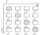
このことが図4に詳細に説明されており、図4には図2のマトリックス28がもう一度詳細に示されている。図4において斜線で示したMR画像は、各周期においてMR装置によって取得(撮像)されたMR画像であり、図示の例では41−1、42−2、43−3および44−4である。これらの4つのMR画像は、例えば図2のMR画像23a〜23gのどれかであり得る。取得されたMR画像41−1は、例えば反転パルスの照射直後に取得されたために、第1のコントラストを有する。図3で説明したように計算された変形画像により、今やMR画像41−2、41−3および41−4を計算することができる。取得されたMR画像41−1に基づいて画像41−2から41−4を計算するために、図3の例を参照するならば、変形情報1−2、1−3および1−4が使用されることになる。それゆえ、第1のコントラストについては、例えば、その第1のコントラストで運動する心臓のシネ(CINE)表示に使用できるMR画像シリーズが提供される。類似の計算がMR画像42について可能である。その取得されたMR画像と、運動情報、即ちここでは運動情報2−1、2−3および2−4とに基づいて、MR画像42−1、42−3および42−4が計算され、従って、さらに別の異なるコントラストについてMR画像シリーズが計算されたことになる。同じやり方で、MR画像43−1〜43−4と、44−1〜44−3とを計算することができる。それによって、図4において概略的に認識できるように、異なるコントラスト値ごとに、それぞれ1つの、異なる運動時相を含んだMR画像シリーズが得られる。医師は、異なるコントラスト値において心筋の運動に関する情報を得るべく、これらのMR画像シリーズを観察することができる。
This is illustrated in detail in FIG. 4, which again shows the
さらに磁化現象から、図2に示されているように、例えば生成された画像に基づいてボクセルを基礎にして計算される3パラメータFit法によって、T1値およびT2値を計算することができる。 Further, from the magnetization phenomenon, as shown in FIG. 2, the T1 value and the T2 value can be calculated by, for example, a three-parameter Fit method calculated based on voxels based on the generated image.
図5において、個々のステップが概略的に要約されている。ステップ51においてMR画像の取得が行われ、その際に、図2に示したように、MR画像の取得前に反転パルス又はプリパレーションパルスが投入されるとよい。しかし、冒頭に述べたように、反転パルス又はプリパレーションパルスは、平衡状態に近づく磁化経過を発生させるために絶対に必要というわけではない。次に、ステップ52において、少なくとも2つの周期のうちの第2周期のMR画像から運動情報が決定され、これは図4の例では4番目の周期、つまりMR画像の取得期間内の最後の周期である。ステップ53において、この運動情報が第1周期のMR画像に適用され、それにより、図4に示したように周期運動の異なる運動時相に対する異なるコントラスト値におけるMR画像が計算される。次に、画像取得後にステップ54において使用者によって、使用者が表示を望むコントラストが決定される。このことは、任意のコントラストの選択がステップ51におけるMR画像の取得前には行われず、後になってはじめて行われたことを意味する。ステップ55において、その選択されたコントラストにより所望の画像シリーズ、特にシネ表示用の画像シリーズを表示することができる。任意選択的に、ステップ56における磁化経過から、図2に示したように、個々のピクセルのためにT1値およびT2値を曲線当てはめ法によって計算することができる。
In FIG. 5, the individual steps are summarized schematically. In
上述の実施形態では、最後の周期、即ち第2周期の運動情報が第1周期のMR画像に対してのみ適用された。しかし、図2を参照すれば、最後の周期の運動情報を、最初の周期23におけるコントラスト値とは異なるコントラスト値が生じる第2周期のMR画像、即ち周期24のMR画像にも適用することもできる。周期24におけるコントラスト値はもはや周期23におけるコントラスト値と同じではないが、しかし運動情報の使用は1つの第1周期23のMR画像に限定されない。複数の第1周期23,24への適用が同様に考えられ得る。
In the above-described embodiment, the motion information of the last period, that is, the second period is applied only to the MR image of the first period. However, referring to FIG. 2, the motion information of the last period may be applied to the MR image of the second period in which the contrast value different from the contrast value in the
以上のように、上述の本発明は種々のコントラストにおいて十分な空間的および時間的な分解能を有するMR画像の生成を可能にする。 As described above, the present invention described above enables the generation of MR images having sufficient spatial and temporal resolution at various contrasts.
10 磁石
11 寝台
12 被検者
13 制御ユニット
14 傾斜磁場制御部
15 RF制御部
16 記憶ユニット
17 撮像ユニット
18 表示部
19 入力ユニット
20 計算ユニット
21 RFパルス
22 ECGトリガ
23〜26 心周期
23a〜23g MR画像
26a〜26g MR画像
27 フィールド
28 マトリックス
29 バー
DESCRIPTION OF
Claims (15)
前記周期運動の少なくとも2つの周期(23〜26)にわたって前記検査対象のMR画像を取得するためのMR信号を検出するステップであって、前記少なくとも2つの周期のそれぞれにおける複数のMR画像が取得され、前記MR画像に影響を及ぼす検査対象の磁化が、前記少なくとも2つの周期(23〜26)にわたって平衡状態に近づき、かつ前記少なくとも2つの周期のうちの第2周期(26)において、前記少なくとも2つの周期のうちの第1周期(23)よりも平衡状態に近づくステップと、
前記検査対象の運動情報が異なる運動時相ごとに決定されるように、前記第2周期(26)からの複数のMR画像(26a〜26g)を用いて前記検査対象の周期運動の異なる運動時相ごとに前記運動情報を決定するステップと、
前記第1周期の異なるMR画像について前記周期運動の異なる運動時相ごとに運動補正されたMR画像が計算されるように、前記第2周期において決定された運動情報を用いて、前記第1周期(23)のMR画像(23a−23g)における前記周期運動の異なる運動時相ごとに前記検査対象の運動補正を行うステップと、
を有する方法。 A method for calculating an MR image of a subject to be inspected with periodic motion,
Detecting MR signals for acquiring the MR image of the inspection object over at least two periods (23 to 26) of the periodic movement, wherein a plurality of MR images in each of the at least two periods are acquired; , The magnetization of the inspection object affecting the MR image approaches an equilibrium state over the at least two periods (23 to 26), and in the second period (26) of the at least two periods, the at least 2 Step closer to equilibrium than the first of the two periods (23);
When the movement information of the examination object is different using the plurality of MR images (26a to 26g) from the second period (26) so that the movement information of the examination object is determined for each different movement time phase. Determining the motion information for each phase;
The motion information determined in the second period is used to calculate the motion-corrected MR image for each motion time phase of the periodic motion with respect to the MR images having different first periods. Performing motion correction of the inspection object for each motion time phase with different periodic motion in the MR image (23a-23g) of (23);
Having a method.
前記第1周期の取得された各MR画像(23a−23g)が1つの運動時相に割り当てられることにより、前記第1周期の異なるコントラスト値ごとに当該コントラスト値でMR画像として取得された少なくとも1つのMR初期画像が生成され、
前記第1周期の異なるコントラスト値ごとに、当該コントラスト値で取得されかつ運動時相の1つにおいて取得されたMR初期画像と、様々な運動時相についての運動情報とを用いて、関連するMR初期画像が有するのと同じコントラスト値を有する他の欠けている運動時相について運動補正されたMR画像が計算されることを特徴とする請求項1から3の1つに記載の方法。 Each MR image (23a-23g) acquired in the first period has a different contrast value,
By assigning each MR image (23a-23g) acquired in the first period to one motion time phase, at least one acquired as an MR image with the contrast value for each different contrast value in the first period Two MR initial images are generated,
For each different contrast value in the first period, using the MR initial image acquired at that contrast value and acquired in one of the motion time phases and the motion information for the various motion time phases, the associated MR 4. Method according to one of claims 1 to 3, characterized in that motion-corrected MR images are calculated for other missing motion phases having the same contrast value as the initial image has.
前記周期運動の少なくとも2つの周期にわたって前記検査対象のMR画像を取得するためのMR信号を検出してMR画像を再構成するように構成されている撮像ユニット(17)であって、前記少なくとも2つの周期のそれぞれにおける複数のMR画像を取得し、前記MR画像に影響を及ぼす前記検査対象の磁化が、前記少なくとも2つの周期にわたって平衡状態に近づき、かつ前記少なくとも2つの周期のうちの第2周期(26)において、前記少なくとも2つの周期のうちの第1周期(23)おけるよりも平衡状態に近づくように構成された撮像ユニット(17)と、
前記検査対象の運動情報が異なる運動時相ごとに決定されるよう前記第2周期からの複数のMR画像(26a〜26g)を用いて前記検査対象の周期運動の異なる運動時相ごとに運動情報を決定するように構成されている計算ユニット(20)であって、前記第1周期の異なるMR画像について周期運動の異なる運動時相ごとに運動補正されたMR画像が計算するように、前記第2周期において決定された運動情報を用いて、前記第1周期のMR画像において周期運動の異なる運動時相ごとに検査対象の運動補正を行うように構成された計算ユニット(20)と、
を有するMR装置。 An MR apparatus for calculating MR images of a subject to be inspected with periodic motion,
An imaging unit (17) configured to detect an MR signal for acquiring an MR image of the inspection object over at least two periods of the periodic motion and to reconstruct the MR image, the imaging unit (17) Acquiring a plurality of MR images in each of the two periods, the magnetization of the inspection object affecting the MR image approaches an equilibrium state over the at least two periods, and a second period of the at least two periods (26) an imaging unit (17) configured to be closer to an equilibrium state than in the first period (23) of the at least two periods;
Movement information for each movement time phase with different periodic movements of the inspection object using a plurality of MR images (26a to 26g) from the second period so that the movement information of the inspection object is determined for each movement time phase different. A calculation unit (20) configured to determine a motion-corrected MR image for each motion time phase having a different periodic motion for the MR image having a different first motion. A calculation unit (20) configured to perform motion correction of a test object for each motion time phase having different periodic motions in the MR image of the first cycle, using motion information determined in two cycles;
MR apparatus having
Applications Claiming Priority (2)
| Application Number | Priority Date | Filing Date | Title |
|---|---|---|---|
| DE102014206724.3A DE102014206724B4 (en) | 2014-04-08 | 2014-04-08 | Dynamic imaging with variable contrast |
| DE102014206724.3 | 2014-04-08 |
Publications (3)
| Publication Number | Publication Date |
|---|---|
| JP2015198938A JP2015198938A (en) | 2015-11-12 |
| JP2015198938A5 JP2015198938A5 (en) | 2016-06-09 |
| JP6133926B2 true JP6133926B2 (en) | 2017-05-24 |
Family
ID=52823458
Family Applications (1)
| Application Number | Title | Priority Date | Filing Date |
|---|---|---|---|
| JP2015077704A Active JP6133926B2 (en) | 2014-04-08 | 2015-04-06 | Dynamic imaging using variable contrast |
Country Status (6)
| Country | Link |
|---|---|
| US (1) | US10420512B2 (en) |
| EP (1) | EP2930525B1 (en) |
| JP (1) | JP6133926B2 (en) |
| KR (1) | KR101631026B1 (en) |
| CN (1) | CN104970794B (en) |
| DE (1) | DE102014206724B4 (en) |
Families Citing this family (4)
| Publication number | Priority date | Publication date | Assignee | Title |
|---|---|---|---|---|
| CN104156975B (en) * | 2013-05-13 | 2018-04-24 | 东芝医疗系统株式会社 | Medical image analysis apparatus and method and medical imaging devices |
| DE102014225282B4 (en) | 2014-12-09 | 2016-07-21 | Siemens Healthcare Gmbh | Deformation calculation with cyclical movement of a test object |
| DE102015224162B4 (en) | 2015-12-03 | 2017-11-30 | Siemens Healthcare Gmbh | Method for determining a movement information and a magnetic resonance device describing a movement in an at least partially moved examination area |
| CN110177499A (en) * | 2016-11-21 | 2019-08-27 | 西门子医疗有限公司 | The method of the diagnostic measurement data on head is recorded by magnetic resonance equipment |
Family Cites Families (19)
| Publication number | Priority date | Publication date | Assignee | Title |
|---|---|---|---|---|
| WO2003096047A1 (en) * | 2002-05-13 | 2003-11-20 | Koninklijke Philips Electronics N.V. | Prior-information-enhanced dynamic magnetic resonance imaging |
| US6922580B2 (en) * | 2002-06-04 | 2005-07-26 | Koninklijke Philips Electronics N.V. | Blood flow gated MRI |
| JP2005278919A (en) * | 2004-03-30 | 2005-10-13 | Hitachi Medical Corp | Magnetic resonance imaging apparatus |
| US7809426B2 (en) | 2004-04-29 | 2010-10-05 | The Cleveland Clinic Foundation | Acquiring contrast-enhanced, T1 weighted, cine magnetic resonance images |
| DE102005000714A1 (en) | 2005-01-03 | 2006-07-20 | Siemens Ag | Medical imaging method for imaging a periodically moving object area, e.g. heart or lungs, by recording of general object area image data, addition of markers, subsequent periodic imaging and then interpolation of marker positions |
| JP4639136B2 (en) * | 2005-10-19 | 2011-02-23 | ジーイー・メディカル・システムズ・グローバル・テクノロジー・カンパニー・エルエルシー | Magnetic resonance imaging system |
| US20080150532A1 (en) * | 2006-12-21 | 2008-06-26 | General Electric Company | Method and apparatus for measuring t1 relaxation |
| JP2008212634A (en) * | 2007-02-06 | 2008-09-18 | Toshiba Corp | Magnetic resonance imaging apparatus and image analysis method and image analysis program therefor |
| EP1956383B1 (en) * | 2007-02-06 | 2010-10-20 | Kabushiki Kaisha Toshiba | MRI involving a cine prescan for motion analysis |
| DE102007018089B4 (en) | 2007-04-02 | 2010-10-14 | Siemens Ag | Heart imaging using MRI with adaptive inversion time |
| US9008753B2 (en) * | 2009-11-10 | 2015-04-14 | Deutsches Herzzentrum Berlin | Look-locker IR-SSFP for cardiac MR imaging with simultaneous generation of cardiac T1 maps, cine images and IR-prepared images |
| DE102010003895B4 (en) | 2010-04-13 | 2019-07-04 | Siemens Healthcare Gmbh | Method for generating angiographic magnetic resonance images |
| US8848990B2 (en) * | 2010-09-28 | 2014-09-30 | Siemens Aktiengesellschaft | Automatic registration of image series with varying contrast based on synthetic images derived from intensity behavior model |
| US20120133747A1 (en) | 2010-11-29 | 2012-05-31 | Sony Corporation | Image processing apparatus, display apparatus, image processing method and image processing program |
| JP2012115319A (en) * | 2010-11-29 | 2012-06-21 | R Tech:Kk | Organ motion analyzer and organ motion analysis program |
| JP6076677B2 (en) * | 2011-11-25 | 2017-02-08 | 東芝メディカルシステムズ株式会社 | Magnetic resonance imaging system |
| WO2013140356A1 (en) * | 2012-03-21 | 2013-09-26 | Koninklijke Philips Electronics N.V. | System and method for differentiation of normal myocardium from diffuse disease using t1 mapping in non-ischemic cardiomyopathies and others |
| US9129424B2 (en) * | 2012-04-17 | 2015-09-08 | Siemens Aktiengesellschaft | Phase sensitive T1 mapping in magnetic resonance imaging |
| DE102012206585B4 (en) * | 2012-04-20 | 2013-12-12 | Siemens Aktiengesellschaft | Method for the rapid spatially resolved determination of a magnetic resonance relaxation parameter in a study area |
-
2014
- 2014-04-08 DE DE102014206724.3A patent/DE102014206724B4/en not_active Expired - Fee Related
-
2015
- 2015-03-24 EP EP15160572.2A patent/EP2930525B1/en active Active
- 2015-04-06 JP JP2015077704A patent/JP6133926B2/en active Active
- 2015-04-07 KR KR1020150049011A patent/KR101631026B1/en active IP Right Grant
- 2015-04-08 CN CN201510162338.1A patent/CN104970794B/en active Active
- 2015-04-08 US US14/681,454 patent/US10420512B2/en active Active
Also Published As
| Publication number | Publication date |
|---|---|
| US10420512B2 (en) | 2019-09-24 |
| CN104970794A (en) | 2015-10-14 |
| EP2930525A1 (en) | 2015-10-14 |
| CN104970794B (en) | 2018-02-16 |
| DE102014206724B4 (en) | 2015-11-12 |
| KR20150116795A (en) | 2015-10-16 |
| EP2930525B1 (en) | 2019-12-04 |
| DE102014206724A1 (en) | 2015-10-08 |
| KR101631026B1 (en) | 2016-06-15 |
| US20150282764A1 (en) | 2015-10-08 |
| JP2015198938A (en) | 2015-11-12 |
Similar Documents
| Publication | Publication Date | Title |
|---|---|---|
| KR101596549B1 (en) | A method for a rapid determination of spatially resolved magnetic resonance relaxation parameters in an area of examination | |
| JP3865133B2 (en) | Self-adaptive tracking and phase encoding during data acquisition for contrast-enhanced MRA and dynamic contrast agent uptake studies | |
| KR101664138B1 (en) | System and method for tissue characterization using multi-slice magnetic resonance imaging | |
| US10371779B2 (en) | Apparatus and method for magnetic resonance imaging with high spatial temporal resolutions | |
| US20120123248A1 (en) | Dark blood delayed enhancement magnetic resonance viability imaging techniques for assessing subendocardial infarcts | |
| KR101809213B1 (en) | Method and apparatus for multi-slice imaging of t2-relaxation time | |
| US10451696B2 (en) | Magnetic resonance imaging apparatus and method of obtaining magnetic resonance image | |
| JP2009279202A (en) | Nuclear magnetic resonance imaging apparatus | |
| US6889071B2 (en) | Acquisition of high-temporal free-breathing MR images | |
| JP6133926B2 (en) | Dynamic imaging using variable contrast | |
| JP2014030723A (en) | Magnetic resonance imaging coordinated with physiological cycle | |
| JP4133348B2 (en) | Inspection device using nuclear magnetic resonance | |
| US20170131377A1 (en) | Magnetic resonance imaging apparatus and method | |
| JP2005040416A (en) | Magnetic resonance imaging apparatus | |
| JP4230875B2 (en) | Magnetic resonance imaging system | |
| JP4349647B2 (en) | Magnetic resonance imaging system | |
| US11500052B2 (en) | System and method for producing temporally resolved images depicting late-gadolinium enhancement with magnetic resonance imaging | |
| US20150094562A1 (en) | Magnetic resonance imaging with dynamic inversion preparation | |
| US20140303482A1 (en) | Magnetic resonance imaging method for imaging components with short transverse relaxation times (t2) in a human or an animal heart | |
| JP2015002834A (en) | Magnetic resonance imaging apparatus and magnetic resonance imaging method | |
| Coristine et al. | Improved respiratory self‐navigation for 3D radial acquisitions through the use of a pencil‐beam 2D‐T2‐prep for free‐breathing, whole‐heart coronary MRA | |
| US10495710B2 (en) | Time-resolved MR images during a cyclical movement | |
| JP2023008743A (en) | Magnetic resonance imaging apparatus | |
| Nothnagel | Development of novel quantitative medical imaging techniques for MPI and MRI |
Legal Events
| Date | Code | Title | Description |
|---|---|---|---|
| A521 | Request for written amendment filed |
Free format text: JAPANESE INTERMEDIATE CODE: A523 Effective date: 20160414 |
|
| A621 | Written request for application examination |
Free format text: JAPANESE INTERMEDIATE CODE: A621 Effective date: 20160414 |
|
| A977 | Report on retrieval |
Free format text: JAPANESE INTERMEDIATE CODE: A971007 Effective date: 20170309 |
|
| TRDD | Decision of grant or rejection written | ||
| A01 | Written decision to grant a patent or to grant a registration (utility model) |
Free format text: JAPANESE INTERMEDIATE CODE: A01 Effective date: 20170321 |
|
| A61 | First payment of annual fees (during grant procedure) |
Free format text: JAPANESE INTERMEDIATE CODE: A61 Effective date: 20170420 |
|
| R150 | Certificate of patent or registration of utility model |
Ref document number: 6133926 Country of ref document: JP Free format text: JAPANESE INTERMEDIATE CODE: R150 |
|
| R250 | Receipt of annual fees |
Free format text: JAPANESE INTERMEDIATE CODE: R250 |
|
| R250 | Receipt of annual fees |
Free format text: JAPANESE INTERMEDIATE CODE: R250 |
|
| S111 | Request for change of ownership or part of ownership |
Free format text: JAPANESE INTERMEDIATE CODE: R313113 |
|
| R360 | Written notification for declining of transfer of rights |
Free format text: JAPANESE INTERMEDIATE CODE: R360 |
|
| R360 | Written notification for declining of transfer of rights |
Free format text: JAPANESE INTERMEDIATE CODE: R360 |
|
| R371 | Transfer withdrawn |
Free format text: JAPANESE INTERMEDIATE CODE: R371 |
|
| S111 | Request for change of ownership or part of ownership |
Free format text: JAPANESE INTERMEDIATE CODE: R313113 |
|
| R350 | Written notification of registration of transfer |
Free format text: JAPANESE INTERMEDIATE CODE: R350 |
|
| R250 | Receipt of annual fees |
Free format text: JAPANESE INTERMEDIATE CODE: R250 |
|
| R250 | Receipt of annual fees |
Free format text: JAPANESE INTERMEDIATE CODE: R250 |