JP6124041B2 - Cell scaffold structure - Google Patents
Cell scaffold structure Download PDFInfo
- Publication number
- JP6124041B2 JP6124041B2 JP2011534906A JP2011534906A JP6124041B2 JP 6124041 B2 JP6124041 B2 JP 6124041B2 JP 2011534906 A JP2011534906 A JP 2011534906A JP 2011534906 A JP2011534906 A JP 2011534906A JP 6124041 B2 JP6124041 B2 JP 6124041B2
- Authority
- JP
- Japan
- Prior art keywords
- cells
- bladder
- scaffold
- tissue
- cell
- Prior art date
- Legal status (The legal status is an assumption and is not a legal conclusion. Google has not performed a legal analysis and makes no representation as to the accuracy of the status listed.)
- Active
Links
- 210000004027 cell Anatomy 0.000 claims description 471
- 210000000329 smooth muscle myocyte Anatomy 0.000 claims description 339
- 210000001519 tissue Anatomy 0.000 claims description 250
- 238000000034 method Methods 0.000 claims description 224
- 239000011159 matrix material Substances 0.000 claims description 213
- 210000000056 organ Anatomy 0.000 claims description 164
- 229920000642 polymer Polymers 0.000 claims description 159
- 238000002054 transplantation Methods 0.000 claims description 117
- 210000000626 ureter Anatomy 0.000 claims description 111
- 230000014509 gene expression Effects 0.000 claims description 102
- 210000002700 urine Anatomy 0.000 claims description 99
- 238000011282 treatment Methods 0.000 claims description 89
- 210000000577 adipose tissue Anatomy 0.000 claims description 81
- 210000002460 smooth muscle Anatomy 0.000 claims description 67
- 239000011886 peripheral blood Substances 0.000 claims description 57
- 210000005259 peripheral blood Anatomy 0.000 claims description 56
- 238000002513 implantation Methods 0.000 claims description 54
- 238000000151 deposition Methods 0.000 claims description 46
- 210000003815 abdominal wall Anatomy 0.000 claims description 43
- 238000001356 surgical procedure Methods 0.000 claims description 42
- 210000002901 mesenchymal stem cell Anatomy 0.000 claims description 41
- 210000004877 mucosa Anatomy 0.000 claims description 34
- 230000002950 deficient Effects 0.000 claims description 22
- 210000000981 epithelium Anatomy 0.000 claims description 17
- 230000003872 anastomosis Effects 0.000 claims description 10
- 230000004069 differentiation Effects 0.000 claims description 10
- 239000002771 cell marker Substances 0.000 claims description 9
- 230000002500 effect on skin Effects 0.000 claims description 7
- 238000000338 in vitro Methods 0.000 claims description 7
- 230000001720 vestibular Effects 0.000 claims description 7
- 238000004519 manufacturing process Methods 0.000 claims description 6
- 229920000249 biocompatible polymer Polymers 0.000 claims description 4
- 210000002919 epithelial cell Anatomy 0.000 claims description 4
- 241001465754 Metazoa Species 0.000 description 357
- 238000012360 testing method Methods 0.000 description 344
- 210000003932 urinary bladder Anatomy 0.000 description 295
- 230000002485 urinary effect Effects 0.000 description 124
- 210000001635 urinary tract Anatomy 0.000 description 90
- 210000004369 blood Anatomy 0.000 description 83
- 239000008280 blood Substances 0.000 description 83
- 230000008929 regeneration Effects 0.000 description 65
- 238000011069 regeneration method Methods 0.000 description 65
- 239000000463 material Substances 0.000 description 64
- 102100021943 C-C motif chemokine 2 Human genes 0.000 description 60
- 101710155857 C-C motif chemokine 2 Proteins 0.000 description 60
- 210000002837 heart atrium Anatomy 0.000 description 56
- 210000003491 skin Anatomy 0.000 description 54
- 108090000623 proteins and genes Proteins 0.000 description 53
- 239000000523 sample Substances 0.000 description 45
- 210000004303 peritoneum Anatomy 0.000 description 44
- 238000001574 biopsy Methods 0.000 description 40
- 230000003187 abdominal effect Effects 0.000 description 39
- 230000015572 biosynthetic process Effects 0.000 description 38
- 102000004169 proteins and genes Human genes 0.000 description 35
- 238000011887 Necropsy Methods 0.000 description 34
- 210000003205 muscle Anatomy 0.000 description 34
- 230000008021 deposition Effects 0.000 description 32
- 210000003734 kidney Anatomy 0.000 description 31
- 235000018102 proteins Nutrition 0.000 description 31
- 108010035532 Collagen Proteins 0.000 description 30
- 102000008186 Collagen Human genes 0.000 description 30
- 229920001436 collagen Polymers 0.000 description 30
- 238000012258 culturing Methods 0.000 description 27
- 239000002609 medium Substances 0.000 description 27
- 108010081823 Myocardin Proteins 0.000 description 25
- 102100030217 Myocardin Human genes 0.000 description 25
- 230000008828 contractile function Effects 0.000 description 25
- 239000003550 marker Substances 0.000 description 25
- 229920000954 Polyglycolide Polymers 0.000 description 24
- 230000001965 increasing effect Effects 0.000 description 24
- 239000011148 porous material Substances 0.000 description 24
- 230000008569 process Effects 0.000 description 24
- 210000000683 abdominal cavity Anatomy 0.000 description 23
- 238000010899 nucleation Methods 0.000 description 23
- 210000003741 urothelium Anatomy 0.000 description 23
- 230000003890 fistula Effects 0.000 description 22
- 230000006870 function Effects 0.000 description 22
- 239000004633 polyglycolic acid Substances 0.000 description 22
- 239000000306 component Substances 0.000 description 21
- 230000008602 contraction Effects 0.000 description 21
- 238000011161 development Methods 0.000 description 21
- 230000018109 developmental process Effects 0.000 description 21
- 230000008439 repair process Effects 0.000 description 21
- 206010016717 Fistula Diseases 0.000 description 20
- 238000002574 cystoscopy Methods 0.000 description 20
- 238000011156 evaluation Methods 0.000 description 20
- 206010061218 Inflammation Diseases 0.000 description 19
- 230000034994 death Effects 0.000 description 19
- 231100000517 death Toxicity 0.000 description 19
- 230000004054 inflammatory process Effects 0.000 description 19
- 239000010410 layer Substances 0.000 description 19
- 229920005615 natural polymer Polymers 0.000 description 19
- 230000001172 regenerating effect Effects 0.000 description 19
- 230000017423 tissue regeneration Effects 0.000 description 19
- OYPRJOBELJOOCE-UHFFFAOYSA-N Calcium Chemical compound [Ca] OYPRJOBELJOOCE-UHFFFAOYSA-N 0.000 description 18
- 102000004127 Cytokines Human genes 0.000 description 18
- 108090000695 Cytokines Proteins 0.000 description 18
- 241001673669 Porcine circovirus 2 Species 0.000 description 18
- 239000011575 calcium Substances 0.000 description 18
- 229910052791 calcium Inorganic materials 0.000 description 18
- 239000003102 growth factor Substances 0.000 description 18
- 210000004072 lung Anatomy 0.000 description 18
- 229920001184 polypeptide Polymers 0.000 description 18
- 102000004196 processed proteins & peptides Human genes 0.000 description 18
- 108090000765 processed proteins & peptides Proteins 0.000 description 18
- 229920001059 synthetic polymer Polymers 0.000 description 18
- 108091003079 Bovine Serum Albumin Proteins 0.000 description 17
- 239000011248 coating agent Substances 0.000 description 17
- 238000000576 coating method Methods 0.000 description 17
- 230000035876 healing Effects 0.000 description 17
- 210000000244 kidney pelvis Anatomy 0.000 description 17
- 210000001015 abdomen Anatomy 0.000 description 16
- 238000004458 analytical method Methods 0.000 description 16
- 238000010171 animal model Methods 0.000 description 16
- 239000012091 fetal bovine serum Substances 0.000 description 16
- 230000012010 growth Effects 0.000 description 16
- 230000002980 postoperative effect Effects 0.000 description 16
- 238000004393 prognosis Methods 0.000 description 16
- 210000000130 stem cell Anatomy 0.000 description 16
- 208000015181 infectious disease Diseases 0.000 description 15
- 230000003902 lesion Effects 0.000 description 15
- 238000002604 ultrasonography Methods 0.000 description 15
- 230000002792 vascular Effects 0.000 description 15
- 241000282887 Suidae Species 0.000 description 14
- 206010046543 Urinary incontinence Diseases 0.000 description 14
- 229920001577 copolymer Polymers 0.000 description 14
- 230000001419 dependent effect Effects 0.000 description 14
- 210000002540 macrophage Anatomy 0.000 description 14
- 238000000926 separation method Methods 0.000 description 14
- 239000000243 solution Substances 0.000 description 14
- 230000004083 survival effect Effects 0.000 description 14
- 101000652736 Homo sapiens Transgelin Proteins 0.000 description 13
- 102100031013 Transgelin Human genes 0.000 description 13
- 210000001185 bone marrow Anatomy 0.000 description 13
- 108010086826 calponin Proteins 0.000 description 13
- 102000006783 calponin Human genes 0.000 description 13
- 238000004113 cell culture Methods 0.000 description 13
- 210000004748 cultured cell Anatomy 0.000 description 13
- 208000037265 diseases, disorders, signs and symptoms Diseases 0.000 description 13
- 239000000835 fiber Substances 0.000 description 13
- 239000002953 phosphate buffered saline Substances 0.000 description 13
- 230000009385 viral infection Effects 0.000 description 13
- 102000029816 Collagenase Human genes 0.000 description 12
- 108060005980 Collagenase Proteins 0.000 description 12
- WZUVPPKBWHMQCE-UHFFFAOYSA-N Haematoxylin Chemical compound C12=CC(O)=C(O)C=C2CC2(O)C1C1=CC=C(O)C(O)=C1OC2 WZUVPPKBWHMQCE-UHFFFAOYSA-N 0.000 description 12
- 208000036142 Viral infection Diseases 0.000 description 12
- 229960002424 collagenase Drugs 0.000 description 12
- 238000003384 imaging method Methods 0.000 description 12
- 238000002955 isolation Methods 0.000 description 12
- 239000000203 mixture Substances 0.000 description 12
- 230000036961 partial effect Effects 0.000 description 12
- 210000002966 serum Anatomy 0.000 description 12
- 210000003708 urethra Anatomy 0.000 description 12
- 206010020524 Hydronephrosis Diseases 0.000 description 11
- 238000011888 autopsy Methods 0.000 description 11
- 210000002808 connective tissue Anatomy 0.000 description 11
- LOKCTEFSRHRXRJ-UHFFFAOYSA-I dipotassium trisodium dihydrogen phosphate hydrogen phosphate dichloride Chemical compound P(=O)(O)(O)[O-].[K+].P(=O)(O)([O-])[O-].[Na+].[Na+].[Cl-].[K+].[Cl-].[Na+] LOKCTEFSRHRXRJ-UHFFFAOYSA-I 0.000 description 11
- 230000000694 effects Effects 0.000 description 11
- 210000002950 fibroblast Anatomy 0.000 description 11
- 210000001165 lymph node Anatomy 0.000 description 11
- 239000000047 product Substances 0.000 description 11
- 208000024891 symptom Diseases 0.000 description 11
- 230000007704 transition Effects 0.000 description 11
- RKDVKSZUMVYZHH-UHFFFAOYSA-N 1,4-dioxane-2,5-dione Chemical compound O=C1COC(=O)CO1 RKDVKSZUMVYZHH-UHFFFAOYSA-N 0.000 description 10
- 102000007469 Actins Human genes 0.000 description 10
- 108010085238 Actins Proteins 0.000 description 10
- WSFSSNUMVMOOMR-UHFFFAOYSA-N Formaldehyde Chemical compound O=C WSFSSNUMVMOOMR-UHFFFAOYSA-N 0.000 description 10
- 239000006143 cell culture medium Substances 0.000 description 10
- 230000001413 cellular effect Effects 0.000 description 10
- 230000006378 damage Effects 0.000 description 10
- 239000003814 drug Substances 0.000 description 10
- 239000012530 fluid Substances 0.000 description 10
- JJTUDXZGHPGLLC-UHFFFAOYSA-N lactide Chemical compound CC1OC(=O)C(C)OC1=O JJTUDXZGHPGLLC-UHFFFAOYSA-N 0.000 description 10
- 238000007726 management method Methods 0.000 description 10
- 210000005087 mononuclear cell Anatomy 0.000 description 10
- -1 polytetrafluoroethylene Polymers 0.000 description 10
- XLYOFNOQVPJJNP-UHFFFAOYSA-N water Chemical compound O XLYOFNOQVPJJNP-UHFFFAOYSA-N 0.000 description 10
- YMWUJEATGCHHMB-UHFFFAOYSA-N Dichloromethane Chemical compound ClCCl YMWUJEATGCHHMB-UHFFFAOYSA-N 0.000 description 9
- 101000817629 Homo sapiens Dymeclin Proteins 0.000 description 9
- 108010084498 Myosin Heavy Chains Proteins 0.000 description 9
- 102000005604 Myosin Heavy Chains Human genes 0.000 description 9
- 210000001789 adipocyte Anatomy 0.000 description 9
- 239000000560 biocompatible material Substances 0.000 description 9
- 238000005119 centrifugation Methods 0.000 description 9
- 238000004891 communication Methods 0.000 description 9
- 239000013068 control sample Substances 0.000 description 9
- 239000003599 detergent Substances 0.000 description 9
- 210000003414 extremity Anatomy 0.000 description 9
- 210000001508 eye Anatomy 0.000 description 9
- 238000002347 injection Methods 0.000 description 9
- 239000007924 injection Substances 0.000 description 9
- 210000000265 leukocyte Anatomy 0.000 description 9
- 210000004400 mucous membrane Anatomy 0.000 description 9
- 210000000663 muscle cell Anatomy 0.000 description 9
- 210000000651 myofibroblast Anatomy 0.000 description 9
- 210000000440 neutrophil Anatomy 0.000 description 9
- 238000011160 research Methods 0.000 description 9
- 108010049974 Bone Morphogenetic Protein 6 Proteins 0.000 description 8
- 102100022525 Bone morphogenetic protein 6 Human genes 0.000 description 8
- 108010025714 CD146 Antigen Proteins 0.000 description 8
- 102100023126 Cell surface glycoprotein MUC18 Human genes 0.000 description 8
- 108010090254 Growth Differentiation Factor 5 Proteins 0.000 description 8
- 102100035379 Growth/differentiation factor 5 Human genes 0.000 description 8
- 102000003745 Hepatocyte Growth Factor Human genes 0.000 description 8
- 108090000100 Hepatocyte Growth Factor Proteins 0.000 description 8
- 206010021639 Incontinence Diseases 0.000 description 8
- 102000004058 Leukemia inhibitory factor Human genes 0.000 description 8
- 108090000581 Leukemia inhibitory factor Proteins 0.000 description 8
- 101150044441 PECAM1 gene Proteins 0.000 description 8
- 102100025368 Runt-related transcription factor 2 Human genes 0.000 description 8
- FAPWRFPIFSIZLT-UHFFFAOYSA-M Sodium chloride Chemical compound [Na+].[Cl-] FAPWRFPIFSIZLT-UHFFFAOYSA-M 0.000 description 8
- 241000282898 Sus scrofa Species 0.000 description 8
- 208000007097 Urinary Bladder Neoplasms Diseases 0.000 description 8
- 108010000134 Vascular Cell Adhesion Molecule-1 Proteins 0.000 description 8
- 102100023543 Vascular cell adhesion protein 1 Human genes 0.000 description 8
- 230000008859 change Effects 0.000 description 8
- DDRJAANPRJIHGJ-UHFFFAOYSA-N creatinine Chemical compound CN1CC(=O)NC1=N DDRJAANPRJIHGJ-UHFFFAOYSA-N 0.000 description 8
- 210000003743 erythrocyte Anatomy 0.000 description 8
- 239000007943 implant Substances 0.000 description 8
- 238000001727 in vivo Methods 0.000 description 8
- 210000000936 intestine Anatomy 0.000 description 8
- 238000002357 laparoscopic surgery Methods 0.000 description 8
- 238000005259 measurement Methods 0.000 description 8
- 244000309715 mini pig Species 0.000 description 8
- 238000012544 monitoring process Methods 0.000 description 8
- 210000005170 neoplastic cell Anatomy 0.000 description 8
- 239000013610 patient sample Substances 0.000 description 8
- 229920001606 poly(lactic acid-co-glycolic acid) Polymers 0.000 description 8
- 239000011780 sodium chloride Substances 0.000 description 8
- 210000005070 sphincter Anatomy 0.000 description 8
- 210000004876 tela submucosa Anatomy 0.000 description 8
- 238000002562 urinalysis Methods 0.000 description 8
- 108010047303 von Willebrand Factor Proteins 0.000 description 8
- 102100036537 von Willebrand factor Human genes 0.000 description 8
- 229960001134 von willebrand factor Drugs 0.000 description 8
- 108091032973 (ribonucleotides)n+m Proteins 0.000 description 7
- 102100032912 CD44 antigen Human genes 0.000 description 7
- 241000282472 Canis lupus familiaris Species 0.000 description 7
- 108010024682 Core Binding Factor Alpha 1 Subunit Proteins 0.000 description 7
- WQZGKKKJIJFFOK-GASJEMHNSA-N Glucose Chemical compound OC[C@H]1OC(O)[C@H](O)[C@@H](O)[C@@H]1O WQZGKKKJIJFFOK-GASJEMHNSA-N 0.000 description 7
- 101000868273 Homo sapiens CD44 antigen Proteins 0.000 description 7
- 101001033249 Homo sapiens Interleukin-1 beta Proteins 0.000 description 7
- 102100039065 Interleukin-1 beta Human genes 0.000 description 7
- 102000004264 Osteopontin Human genes 0.000 description 7
- 108010081689 Osteopontin Proteins 0.000 description 7
- 206010037596 Pyelonephritis Diseases 0.000 description 7
- 206010042674 Swelling Diseases 0.000 description 7
- 210000003484 anatomy Anatomy 0.000 description 7
- 239000012620 biological material Substances 0.000 description 7
- 210000004204 blood vessel Anatomy 0.000 description 7
- 230000037396 body weight Effects 0.000 description 7
- 238000012735 histological processing Methods 0.000 description 7
- 230000003284 homeostatic effect Effects 0.000 description 7
- 230000001976 improved effect Effects 0.000 description 7
- 230000006698 induction Effects 0.000 description 7
- 238000003780 insertion Methods 0.000 description 7
- 230000037431 insertion Effects 0.000 description 7
- 210000004731 jugular vein Anatomy 0.000 description 7
- 239000007788 liquid Substances 0.000 description 7
- 210000004698 lymphocyte Anatomy 0.000 description 7
- 230000004044 response Effects 0.000 description 7
- 238000010186 staining Methods 0.000 description 7
- 230000008961 swelling Effects 0.000 description 7
- 230000003442 weekly effect Effects 0.000 description 7
- 208000035143 Bacterial infection Diseases 0.000 description 6
- 206010005003 Bladder cancer Diseases 0.000 description 6
- 241000282465 Canis Species 0.000 description 6
- 206010028980 Neoplasm Diseases 0.000 description 6
- 108010053099 Vascular Endothelial Growth Factor Receptor-2 Proteins 0.000 description 6
- 102000016549 Vascular Endothelial Growth Factor Receptor-2 Human genes 0.000 description 6
- 230000002159 abnormal effect Effects 0.000 description 6
- 239000000654 additive Substances 0.000 description 6
- 230000033115 angiogenesis Effects 0.000 description 6
- 208000022362 bacterial infectious disease Diseases 0.000 description 6
- 230000008901 benefit Effects 0.000 description 6
- 239000012472 biological sample Substances 0.000 description 6
- 210000005068 bladder tissue Anatomy 0.000 description 6
- 238000002659 cell therapy Methods 0.000 description 6
- HVYWMOMLDIMFJA-DPAQBDIFSA-N cholesterol Chemical compound C1C=C2C[C@@H](O)CC[C@]2(C)[C@@H]2[C@@H]1[C@@H]1CC[C@H]([C@H](C)CCCC(C)C)[C@@]1(C)CC2 HVYWMOMLDIMFJA-DPAQBDIFSA-N 0.000 description 6
- 238000010276 construction Methods 0.000 description 6
- 238000009799 cystectomy Methods 0.000 description 6
- 230000001086 cytosolic effect Effects 0.000 description 6
- 238000001804 debridement Methods 0.000 description 6
- 230000007547 defect Effects 0.000 description 6
- 201000010099 disease Diseases 0.000 description 6
- 208000035475 disorder Diseases 0.000 description 6
- 229940079593 drug Drugs 0.000 description 6
- YQGOJNYOYNNSMM-UHFFFAOYSA-N eosin Chemical compound [Na+].OC(=O)C1=CC=CC=C1C1=C2C=C(Br)C(=O)C(Br)=C2OC2=C(Br)C(O)=C(Br)C=C21 YQGOJNYOYNNSMM-UHFFFAOYSA-N 0.000 description 6
- 210000003195 fascia Anatomy 0.000 description 6
- 238000011049 filling Methods 0.000 description 6
- 238000002594 fluoroscopy Methods 0.000 description 6
- 210000003128 head Anatomy 0.000 description 6
- 230000002147 killing effect Effects 0.000 description 6
- 108020004999 messenger RNA Proteins 0.000 description 6
- 230000004048 modification Effects 0.000 description 6
- 238000012986 modification Methods 0.000 description 6
- 230000023185 monocyte chemotactic protein-1 production Effects 0.000 description 6
- 102000039446 nucleic acids Human genes 0.000 description 6
- 108020004707 nucleic acids Proteins 0.000 description 6
- 150000007523 nucleic acids Chemical class 0.000 description 6
- 230000001575 pathological effect Effects 0.000 description 6
- 230000037361 pathway Effects 0.000 description 6
- 238000002360 preparation method Methods 0.000 description 6
- 230000002062 proliferating effect Effects 0.000 description 6
- 230000001737 promoting effect Effects 0.000 description 6
- 230000002829 reductive effect Effects 0.000 description 6
- 239000007787 solid Substances 0.000 description 6
- 201000005112 urinary bladder cancer Diseases 0.000 description 6
- 201000002327 urinary tract obstruction Diseases 0.000 description 6
- 210000005167 vascular cell Anatomy 0.000 description 6
- 210000001835 viscera Anatomy 0.000 description 6
- LCSKNASZPVZHEG-UHFFFAOYSA-N 3,6-dimethyl-1,4-dioxane-2,5-dione;1,4-dioxane-2,5-dione Chemical compound O=C1COC(=O)CO1.CC1OC(=O)C(C)OC1=O LCSKNASZPVZHEG-UHFFFAOYSA-N 0.000 description 5
- 108020004414 DNA Proteins 0.000 description 5
- 206010018367 Glomerulonephritis chronic Diseases 0.000 description 5
- HTTJABKRGRZYRN-UHFFFAOYSA-N Heparin Chemical compound OC1C(NC(=O)C)C(O)OC(COS(O)(=O)=O)C1OC1C(OS(O)(=O)=O)C(O)C(OC2C(C(OS(O)(=O)=O)C(OC3C(C(O)C(O)C(O3)C(O)=O)OS(O)(=O)=O)C(CO)O2)NS(O)(=O)=O)C(C(O)=O)O1 HTTJABKRGRZYRN-UHFFFAOYSA-N 0.000 description 5
- 241000282412 Homo Species 0.000 description 5
- 101000738771 Homo sapiens Receptor-type tyrosine-protein phosphatase C Proteins 0.000 description 5
- 206010058467 Lung neoplasm malignant Diseases 0.000 description 5
- 238000010240 RT-PCR analysis Methods 0.000 description 5
- 102100037422 Receptor-type tyrosine-protein phosphatase C Human genes 0.000 description 5
- 239000000427 antigen Substances 0.000 description 5
- 108091007433 antigens Proteins 0.000 description 5
- 102000036639 antigens Human genes 0.000 description 5
- 230000001580 bacterial effect Effects 0.000 description 5
- 238000006065 biodegradation reaction Methods 0.000 description 5
- 238000005266 casting Methods 0.000 description 5
- 230000024245 cell differentiation Effects 0.000 description 5
- 210000000170 cell membrane Anatomy 0.000 description 5
- 230000001684 chronic effect Effects 0.000 description 5
- 230000030944 contact inhibition Effects 0.000 description 5
- 230000001351 cycling effect Effects 0.000 description 5
- 230000007423 decrease Effects 0.000 description 5
- 230000003247 decreasing effect Effects 0.000 description 5
- 230000007812 deficiency Effects 0.000 description 5
- 239000007789 gas Substances 0.000 description 5
- 239000008103 glucose Substances 0.000 description 5
- 239000001963 growth medium Substances 0.000 description 5
- 230000002489 hematologic effect Effects 0.000 description 5
- 229920000669 heparin Polymers 0.000 description 5
- 229960002897 heparin Drugs 0.000 description 5
- 201000005202 lung cancer Diseases 0.000 description 5
- 208000020816 lung neoplasm Diseases 0.000 description 5
- 230000007246 mechanism Effects 0.000 description 5
- 210000000713 mesentery Anatomy 0.000 description 5
- 230000005012 migration Effects 0.000 description 5
- 238000013508 migration Methods 0.000 description 5
- 230000000877 morphologic effect Effects 0.000 description 5
- 230000007935 neutral effect Effects 0.000 description 5
- 239000008188 pellet Substances 0.000 description 5
- 230000035755 proliferation Effects 0.000 description 5
- 238000003860 storage Methods 0.000 description 5
- 230000009772 tissue formation Effects 0.000 description 5
- 210000002417 xiphoid bone Anatomy 0.000 description 5
- 206010000050 Abdominal adhesions Diseases 0.000 description 4
- 108010088751 Albumins Proteins 0.000 description 4
- 102000009027 Albumins Human genes 0.000 description 4
- 102000002260 Alkaline Phosphatase Human genes 0.000 description 4
- 108020004774 Alkaline Phosphatase Proteins 0.000 description 4
- VHUUQVKOLVNVRT-UHFFFAOYSA-N Ammonium hydroxide Chemical compound [NH4+].[OH-] VHUUQVKOLVNVRT-UHFFFAOYSA-N 0.000 description 4
- 102100027951 Brain and acute leukemia cytoplasmic protein Human genes 0.000 description 4
- 102000019034 Chemokines Human genes 0.000 description 4
- 108010012236 Chemokines Proteins 0.000 description 4
- 102100036912 Desmin Human genes 0.000 description 4
- 108010044052 Desmin Proteins 0.000 description 4
- 206010013554 Diverticulum Diseases 0.000 description 4
- 239000006144 Dulbecco’s modified Eagle's medium Substances 0.000 description 4
- KCXVZYZYPLLWCC-UHFFFAOYSA-N EDTA Chemical group OC(=O)CN(CC(O)=O)CCN(CC(O)=O)CC(O)=O KCXVZYZYPLLWCC-UHFFFAOYSA-N 0.000 description 4
- 206010015548 Euthanasia Diseases 0.000 description 4
- 102000010834 Extracellular Matrix Proteins Human genes 0.000 description 4
- 108010037362 Extracellular Matrix Proteins Proteins 0.000 description 4
- 206010016654 Fibrosis Diseases 0.000 description 4
- 206010018364 Glomerulonephritis Diseases 0.000 description 4
- 101000697853 Homo sapiens Brain and acute leukemia cytoplasmic protein Proteins 0.000 description 4
- 102000011782 Keratins Human genes 0.000 description 4
- 108010076876 Keratins Proteins 0.000 description 4
- 241000699670 Mus sp. Species 0.000 description 4
- 102000003505 Myosin Human genes 0.000 description 4
- 108060008487 Myosin Proteins 0.000 description 4
- ZLMJMSJWJFRBEC-UHFFFAOYSA-N Potassium Chemical compound [K] ZLMJMSJWJFRBEC-UHFFFAOYSA-N 0.000 description 4
- 239000004792 Prolene Substances 0.000 description 4
- 206010066218 Stress Urinary Incontinence Diseases 0.000 description 4
- 206010047115 Vasculitis Diseases 0.000 description 4
- 239000002251 absorbable suture material Substances 0.000 description 4
- 239000000908 ammonium hydroxide Substances 0.000 description 4
- 239000003242 anti bacterial agent Substances 0.000 description 4
- 230000002146 bilateral effect Effects 0.000 description 4
- 239000000090 biomarker Substances 0.000 description 4
- 238000004820 blood count Methods 0.000 description 4
- 210000001124 body fluid Anatomy 0.000 description 4
- 239000010839 body fluid Substances 0.000 description 4
- 201000011510 cancer Diseases 0.000 description 4
- 239000002775 capsule Substances 0.000 description 4
- 230000021164 cell adhesion Effects 0.000 description 4
- 229940109239 creatinine Drugs 0.000 description 4
- 210000005045 desmin Anatomy 0.000 description 4
- 238000001514 detection method Methods 0.000 description 4
- 230000029087 digestion Effects 0.000 description 4
- 238000009826 distribution Methods 0.000 description 4
- 238000001839 endoscopy Methods 0.000 description 4
- 210000002889 endothelial cell Anatomy 0.000 description 4
- 230000003511 endothelial effect Effects 0.000 description 4
- 210000002744 extracellular matrix Anatomy 0.000 description 4
- 238000000605 extraction Methods 0.000 description 4
- 208000030533 eye disease Diseases 0.000 description 4
- 230000004761 fibrosis Effects 0.000 description 4
- 238000005194 fractionation Methods 0.000 description 4
- 210000001035 gastrointestinal tract Anatomy 0.000 description 4
- 230000036541 health Effects 0.000 description 4
- 238000007489 histopathology method Methods 0.000 description 4
- 230000008595 infiltration Effects 0.000 description 4
- 238000001764 infiltration Methods 0.000 description 4
- 230000000968 intestinal effect Effects 0.000 description 4
- 230000001788 irregular Effects 0.000 description 4
- 210000002429 large intestine Anatomy 0.000 description 4
- 210000000565 lesser omentum Anatomy 0.000 description 4
- 230000007774 longterm Effects 0.000 description 4
- 239000013642 negative control Substances 0.000 description 4
- 229920000747 poly(lactic acid) Polymers 0.000 description 4
- 239000013641 positive control Substances 0.000 description 4
- 239000011591 potassium Substances 0.000 description 4
- 229910052700 potassium Inorganic materials 0.000 description 4
- 208000023504 respiratory system disease Diseases 0.000 description 4
- 239000011734 sodium Substances 0.000 description 4
- 230000001629 suppression Effects 0.000 description 4
- DSNBHJFQCNUKMA-SCKDECHMSA-N thromboxane A2 Chemical compound OC(=O)CCC\C=C/C[C@@H]1[C@@H](/C=C/[C@@H](O)CCCCC)O[C@@H]2O[C@H]1C2 DSNBHJFQCNUKMA-SCKDECHMSA-N 0.000 description 4
- 238000009966 trimming Methods 0.000 description 4
- GPRLSGONYQIRFK-MNYXATJNSA-N triton Chemical class [3H+] GPRLSGONYQIRFK-MNYXATJNSA-N 0.000 description 4
- 210000001113 umbilicus Anatomy 0.000 description 4
- 210000004291 uterus Anatomy 0.000 description 4
- 108030001720 Bontoxilysin Proteins 0.000 description 3
- 102100031151 C-C chemokine receptor type 2 Human genes 0.000 description 3
- 101710149815 C-C chemokine receptor type 2 Proteins 0.000 description 3
- CURLTUGMZLYLDI-UHFFFAOYSA-N Carbon dioxide Chemical compound O=C=O CURLTUGMZLYLDI-UHFFFAOYSA-N 0.000 description 3
- 102000002585 Contractile Proteins Human genes 0.000 description 3
- 108010068426 Contractile Proteins Proteins 0.000 description 3
- 238000002965 ELISA Methods 0.000 description 3
- 108010049003 Fibrinogen Proteins 0.000 description 3
- 102000008946 Fibrinogen Human genes 0.000 description 3
- 102000006395 Globulins Human genes 0.000 description 3
- 108010044091 Globulins Proteins 0.000 description 3
- 102000001554 Hemoglobins Human genes 0.000 description 3
- 108010054147 Hemoglobins Proteins 0.000 description 3
- 206010020853 Hypertonic bladder Diseases 0.000 description 3
- DGAQECJNVWCQMB-PUAWFVPOSA-M Ilexoside XXIX Chemical compound C[C@@H]1CC[C@@]2(CC[C@@]3(C(=CC[C@H]4[C@]3(CC[C@@H]5[C@@]4(CC[C@@H](C5(C)C)OS(=O)(=O)[O-])C)C)[C@@H]2[C@]1(C)O)C)C(=O)O[C@H]6[C@@H]([C@H]([C@@H]([C@H](O6)CO)O)O)O.[Na+] DGAQECJNVWCQMB-PUAWFVPOSA-M 0.000 description 3
- 241000124008 Mammalia Species 0.000 description 3
- 208000009722 Overactive Urinary Bladder Diseases 0.000 description 3
- 206010038540 Renal tubular necrosis Diseases 0.000 description 3
- 208000000921 Urge Urinary Incontinence Diseases 0.000 description 3
- 108010073929 Vascular Endothelial Growth Factor A Proteins 0.000 description 3
- 102000005789 Vascular Endothelial Growth Factors Human genes 0.000 description 3
- 108010019530 Vascular Endothelial Growth Factors Proteins 0.000 description 3
- 208000027418 Wounds and injury Diseases 0.000 description 3
- 230000005856 abnormality Effects 0.000 description 3
- 230000009471 action Effects 0.000 description 3
- 230000003044 adaptive effect Effects 0.000 description 3
- 210000004504 adult stem cell Anatomy 0.000 description 3
- 230000002776 aggregation Effects 0.000 description 3
- 238000004220 aggregation Methods 0.000 description 3
- 239000012670 alkaline solution Substances 0.000 description 3
- 238000013459 approach Methods 0.000 description 3
- 230000005540 biological transmission Effects 0.000 description 3
- 229940053031 botulinum toxin Drugs 0.000 description 3
- 239000000872 buffer Substances 0.000 description 3
- 235000011089 carbon dioxide Nutrition 0.000 description 3
- 230000010261 cell growth Effects 0.000 description 3
- 239000002738 chelating agent Substances 0.000 description 3
- 235000012000 cholesterol Nutrition 0.000 description 3
- 238000005345 coagulation Methods 0.000 description 3
- 230000015271 coagulation Effects 0.000 description 3
- 238000007906 compression Methods 0.000 description 3
- 230000006835 compression Effects 0.000 description 3
- 238000000748 compression moulding Methods 0.000 description 3
- 210000000555 contractile cell Anatomy 0.000 description 3
- 238000005138 cryopreservation Methods 0.000 description 3
- 230000000254 damaging effect Effects 0.000 description 3
- 230000004064 dysfunction Effects 0.000 description 3
- 210000003038 endothelium Anatomy 0.000 description 3
- 230000002708 enhancing effect Effects 0.000 description 3
- 150000002148 esters Chemical class 0.000 description 3
- 230000029142 excretion Effects 0.000 description 3
- 229940012952 fibrinogen Drugs 0.000 description 3
- 230000001497 fibrovascular Effects 0.000 description 3
- 238000009472 formulation Methods 0.000 description 3
- 239000000499 gel Substances 0.000 description 3
- 230000009036 growth inhibition Effects 0.000 description 3
- 238000005534 hematocrit Methods 0.000 description 3
- 230000006872 improvement Effects 0.000 description 3
- 210000004969 inflammatory cell Anatomy 0.000 description 3
- 208000014674 injury Diseases 0.000 description 3
- 230000003993 interaction Effects 0.000 description 3
- 230000003834 intracellular effect Effects 0.000 description 3
- 150000002632 lipids Chemical class 0.000 description 3
- 238000007443 liposuction Methods 0.000 description 3
- 230000004060 metabolic process Effects 0.000 description 3
- 210000002433 mononuclear leukocyte Anatomy 0.000 description 3
- 210000000107 myocyte Anatomy 0.000 description 3
- 230000017074 necrotic cell death Effects 0.000 description 3
- 239000002736 nonionic surfactant Substances 0.000 description 3
- 235000015097 nutrients Nutrition 0.000 description 3
- 210000001672 ovary Anatomy 0.000 description 3
- 208000020629 overactive bladder Diseases 0.000 description 3
- 230000002018 overexpression Effects 0.000 description 3
- 239000004417 polycarbonate Substances 0.000 description 3
- 229920000515 polycarbonate Polymers 0.000 description 3
- 230000005855 radiation Effects 0.000 description 3
- 238000011472 radical prostatectomy Methods 0.000 description 3
- 238000007634 remodeling Methods 0.000 description 3
- 210000002345 respiratory system Anatomy 0.000 description 3
- 231100000241 scar Toxicity 0.000 description 3
- 230000028327 secretion Effects 0.000 description 3
- 229910052708 sodium Inorganic materials 0.000 description 3
- 238000005063 solubilization Methods 0.000 description 3
- 230000007928 solubilization Effects 0.000 description 3
- 239000002904 solvent Substances 0.000 description 3
- 238000007447 staining method Methods 0.000 description 3
- 210000002536 stromal cell Anatomy 0.000 description 3
- 210000004003 subcutaneous fat Anatomy 0.000 description 3
- 239000000126 substance Substances 0.000 description 3
- 239000000758 substrate Substances 0.000 description 3
- 230000008093 supporting effect Effects 0.000 description 3
- 210000004509 vascular smooth muscle cell Anatomy 0.000 description 3
- 210000003462 vein Anatomy 0.000 description 3
- 238000005406 washing Methods 0.000 description 3
- PGOHTUIFYSHAQG-LJSDBVFPSA-N (2S)-6-amino-2-[[(2S)-5-amino-2-[[(2S)-2-[[(2S)-2-[[(2S)-2-[[(2S)-4-amino-2-[[(2S)-2-[[(2S)-2-[[(2S)-2-[[(2S)-2-[[(2S)-5-amino-2-[[(2S)-5-amino-2-[[(2S)-2-[[(2S)-2-[[(2S)-2-[[(2S,3R)-2-[[(2S)-5-amino-2-[[(2S)-2-[[(2S)-2-[[(2S,3R)-2-[[(2S)-2-[[(2S)-2-[[(2S)-2-[[(2S)-2-[[(2S)-5-amino-2-[[(2S)-1-[(2S,3R)-2-[[(2S)-2-[[(2S)-2-[[(2R)-2-[[(2S)-2-[[(2S)-2-[[2-[[(2S)-2-[[(2S)-2-[[(2S)-2-[[(2S)-1-[(2S)-2-[[(2S)-2-[[(2S)-2-[[(2S)-2-amino-4-methylsulfanylbutanoyl]amino]-3-(1H-indol-3-yl)propanoyl]amino]-5-carbamimidamidopentanoyl]amino]propanoyl]pyrrolidine-2-carbonyl]amino]-3-methylbutanoyl]amino]-4-methylpentanoyl]amino]-4-methylpentanoyl]amino]acetyl]amino]-3-hydroxypropanoyl]amino]-4-methylpentanoyl]amino]-3-sulfanylpropanoyl]amino]-4-methylsulfanylbutanoyl]amino]-5-carbamimidamidopentanoyl]amino]-3-hydroxybutanoyl]pyrrolidine-2-carbonyl]amino]-5-oxopentanoyl]amino]-3-hydroxypropanoyl]amino]-3-hydroxypropanoyl]amino]-3-(1H-imidazol-5-yl)propanoyl]amino]-4-methylpentanoyl]amino]-3-hydroxybutanoyl]amino]-3-(1H-indol-3-yl)propanoyl]amino]-5-carbamimidamidopentanoyl]amino]-5-oxopentanoyl]amino]-3-hydroxybutanoyl]amino]-3-hydroxypropanoyl]amino]-3-carboxypropanoyl]amino]-3-hydroxypropanoyl]amino]-5-oxopentanoyl]amino]-5-oxopentanoyl]amino]-3-phenylpropanoyl]amino]-5-carbamimidamidopentanoyl]amino]-3-methylbutanoyl]amino]-4-methylpentanoyl]amino]-4-oxobutanoyl]amino]-5-carbamimidamidopentanoyl]amino]-3-(1H-indol-3-yl)propanoyl]amino]-4-carboxybutanoyl]amino]-5-oxopentanoyl]amino]hexanoic acid Chemical compound CSCC[C@H](N)C(=O)N[C@@H](Cc1c[nH]c2ccccc12)C(=O)N[C@@H](CCCNC(N)=N)C(=O)N[C@@H](C)C(=O)N1CCC[C@H]1C(=O)N[C@@H](C(C)C)C(=O)N[C@@H](CC(C)C)C(=O)N[C@@H](CC(C)C)C(=O)NCC(=O)N[C@@H](CO)C(=O)N[C@@H](CC(C)C)C(=O)N[C@@H](CS)C(=O)N[C@@H](CCSC)C(=O)N[C@@H](CCCNC(N)=N)C(=O)N[C@@H]([C@@H](C)O)C(=O)N1CCC[C@H]1C(=O)N[C@@H](CCC(N)=O)C(=O)N[C@@H](CO)C(=O)N[C@@H](CO)C(=O)N[C@@H](Cc1cnc[nH]1)C(=O)N[C@@H](CC(C)C)C(=O)N[C@@H]([C@@H](C)O)C(=O)N[C@@H](Cc1c[nH]c2ccccc12)C(=O)N[C@@H](CCCNC(N)=N)C(=O)N[C@@H](CCC(N)=O)C(=O)N[C@@H]([C@@H](C)O)C(=O)N[C@@H](CO)C(=O)N[C@@H](CC(O)=O)C(=O)N[C@@H](CO)C(=O)N[C@@H](CCC(N)=O)C(=O)N[C@@H](CCC(N)=O)C(=O)N[C@@H](Cc1ccccc1)C(=O)N[C@@H](CCCNC(N)=N)C(=O)N[C@@H](C(C)C)C(=O)N[C@@H](CC(C)C)C(=O)N[C@@H](CC(N)=O)C(=O)N[C@@H](CCCNC(N)=N)C(=O)N[C@@H](Cc1c[nH]c2ccccc12)C(=O)N[C@@H](CCC(O)=O)C(=O)N[C@@H](CCC(N)=O)C(=O)N[C@@H](CCCCN)C(O)=O PGOHTUIFYSHAQG-LJSDBVFPSA-N 0.000 description 2
- HVCOBJNICQPDBP-UHFFFAOYSA-N 3-[3-[3,5-dihydroxy-6-methyl-4-(3,4,5-trihydroxy-6-methyloxan-2-yl)oxyoxan-2-yl]oxydecanoyloxy]decanoic acid;hydrate Chemical compound O.OC1C(OC(CC(=O)OC(CCCCCCC)CC(O)=O)CCCCCCC)OC(C)C(O)C1OC1C(O)C(O)C(O)C(C)O1 HVCOBJNICQPDBP-UHFFFAOYSA-N 0.000 description 2
- 102100036475 Alanine aminotransferase 1 Human genes 0.000 description 2
- 108010082126 Alanine transaminase Proteins 0.000 description 2
- 108010003415 Aspartate Aminotransferases Proteins 0.000 description 2
- 102000004625 Aspartate Aminotransferases Human genes 0.000 description 2
- 241000894006 Bacteria Species 0.000 description 2
- 208000034309 Bacterial disease carrier Diseases 0.000 description 2
- BPYKTIZUTYGOLE-IFADSCNNSA-N Bilirubin Chemical compound N1C(=O)C(C)=C(C=C)\C1=C\C1=C(C)C(CCC(O)=O)=C(CC2=C(C(C)=C(\C=C/3C(=C(C=C)C(=O)N\3)C)N2)CCC(O)=O)N1 BPYKTIZUTYGOLE-IFADSCNNSA-N 0.000 description 2
- 206010061698 Bladder injury Diseases 0.000 description 2
- 241000283690 Bos taurus Species 0.000 description 2
- 101710092112 Calponin-1 Proteins 0.000 description 2
- 108010022452 Collagen Type I Proteins 0.000 description 2
- 102000012422 Collagen Type I Human genes 0.000 description 2
- RTZKZFJDLAIYFH-UHFFFAOYSA-N Diethyl ether Chemical compound CCOCC RTZKZFJDLAIYFH-UHFFFAOYSA-N 0.000 description 2
- 102100037241 Endoglin Human genes 0.000 description 2
- LFQSCWFLJHTTHZ-UHFFFAOYSA-N Ethanol Chemical compound CCO LFQSCWFLJHTTHZ-UHFFFAOYSA-N 0.000 description 2
- IAYPIBMASNFSPL-UHFFFAOYSA-N Ethylene oxide Chemical compound C1CO1 IAYPIBMASNFSPL-UHFFFAOYSA-N 0.000 description 2
- 102000018233 Fibroblast Growth Factor Human genes 0.000 description 2
- 108050007372 Fibroblast Growth Factor Proteins 0.000 description 2
- 241000192125 Firmicutes Species 0.000 description 2
- 108020004206 Gamma-glutamyltransferase Proteins 0.000 description 2
- 229930186217 Glycolipid Natural products 0.000 description 2
- 101000881679 Homo sapiens Endoglin Proteins 0.000 description 2
- 101000599852 Homo sapiens Intercellular adhesion molecule 1 Proteins 0.000 description 2
- 101000581981 Homo sapiens Neural cell adhesion molecule 1 Proteins 0.000 description 2
- 101000800116 Homo sapiens Thy-1 membrane glycoprotein Proteins 0.000 description 2
- 102100037877 Intercellular adhesion molecule 1 Human genes 0.000 description 2
- 208000008081 Intestinal Fistula Diseases 0.000 description 2
- ONIBWKKTOPOVIA-BYPYZUCNSA-N L-Proline Chemical compound OC(=O)[C@@H]1CCCN1 ONIBWKKTOPOVIA-BYPYZUCNSA-N 0.000 description 2
- 102100027347 Neural cell adhesion molecule 1 Human genes 0.000 description 2
- OAICVXFJPJFONN-UHFFFAOYSA-N Phosphorus Chemical compound [P] OAICVXFJPJFONN-UHFFFAOYSA-N 0.000 description 2
- 102100024616 Platelet endothelial cell adhesion molecule Human genes 0.000 description 2
- 229920002732 Polyanhydride Polymers 0.000 description 2
- 239000004698 Polyethylene Substances 0.000 description 2
- 229920001710 Polyorthoester Polymers 0.000 description 2
- 239000004721 Polyphenylene oxide Substances 0.000 description 2
- 208000035965 Postoperative Complications Diseases 0.000 description 2
- ONIBWKKTOPOVIA-UHFFFAOYSA-N Proline Natural products OC(=O)C1CCCN1 ONIBWKKTOPOVIA-UHFFFAOYSA-N 0.000 description 2
- 108010029485 Protein Isoforms Proteins 0.000 description 2
- 102000001708 Protein Isoforms Human genes 0.000 description 2
- 206010072170 Skin wound Diseases 0.000 description 2
- 108010000499 Thromboplastin Proteins 0.000 description 2
- 102100033523 Thy-1 membrane glycoprotein Human genes 0.000 description 2
- 102100030859 Tissue factor Human genes 0.000 description 2
- 238000008050 Total Bilirubin Reagent Methods 0.000 description 2
- 239000013504 Triton X-100 Substances 0.000 description 2
- 229920004890 Triton X-100 Polymers 0.000 description 2
- 241001227561 Valgus Species 0.000 description 2
- 208000024248 Vascular System injury Diseases 0.000 description 2
- 208000012339 Vascular injury Diseases 0.000 description 2
- 230000001464 adherent effect Effects 0.000 description 2
- 230000002411 adverse Effects 0.000 description 2
- 230000001078 anti-cholinergic effect Effects 0.000 description 2
- 229940088710 antibiotic agent Drugs 0.000 description 2
- 210000002565 arteriole Anatomy 0.000 description 2
- 210000001367 artery Anatomy 0.000 description 2
- 239000005667 attractant Substances 0.000 description 2
- 229920002988 biodegradable polymer Polymers 0.000 description 2
- 239000004621 biodegradable polymer Substances 0.000 description 2
- 238000004159 blood analysis Methods 0.000 description 2
- 239000007975 buffered saline Substances 0.000 description 2
- 210000001736 capillary Anatomy 0.000 description 2
- 150000001720 carbohydrates Chemical class 0.000 description 2
- 210000001715 carotid artery Anatomy 0.000 description 2
- 230000015556 catabolic process Effects 0.000 description 2
- 230000004663 cell proliferation Effects 0.000 description 2
- 239000006285 cell suspension Substances 0.000 description 2
- 239000001913 cellulose Substances 0.000 description 2
- 229920002678 cellulose Polymers 0.000 description 2
- 230000031902 chemoattractant activity Effects 0.000 description 2
- 210000000349 chromosome Anatomy 0.000 description 2
- 208000037976 chronic inflammation Diseases 0.000 description 2
- 230000006020 chronic inflammation Effects 0.000 description 2
- 230000004186 co-expression Effects 0.000 description 2
- 239000000512 collagen gel Substances 0.000 description 2
- 210000001072 colon Anatomy 0.000 description 2
- 239000002131 composite material Substances 0.000 description 2
- 239000003636 conditioned culture medium Substances 0.000 description 2
- 239000002872 contrast media Substances 0.000 description 2
- 210000000805 cytoplasm Anatomy 0.000 description 2
- 230000003436 cytoskeletal effect Effects 0.000 description 2
- 238000006731 degradation reaction Methods 0.000 description 2
- 238000013461 design Methods 0.000 description 2
- 230000010339 dilation Effects 0.000 description 2
- 239000012153 distilled water Substances 0.000 description 2
- 229940126534 drug product Drugs 0.000 description 2
- 239000003995 emulsifying agent Substances 0.000 description 2
- 230000006862 enzymatic digestion Effects 0.000 description 2
- 230000002255 enzymatic effect Effects 0.000 description 2
- 238000013401 experimental design Methods 0.000 description 2
- 210000000416 exudates and transudate Anatomy 0.000 description 2
- 229940126864 fibroblast growth factor Drugs 0.000 description 2
- 239000000796 flavoring agent Substances 0.000 description 2
- 102000006640 gamma-Glutamyltransferase Human genes 0.000 description 2
- 238000004868 gas analysis Methods 0.000 description 2
- 230000002496 gastric effect Effects 0.000 description 2
- 238000005227 gel permeation chromatography Methods 0.000 description 2
- 238000003306 harvesting Methods 0.000 description 2
- 239000000017 hydrogel Substances 0.000 description 2
- 230000007062 hydrolysis Effects 0.000 description 2
- 238000006460 hydrolysis reaction Methods 0.000 description 2
- 238000010166 immunofluorescence Methods 0.000 description 2
- 238000012744 immunostaining Methods 0.000 description 2
- 238000011534 incubation Methods 0.000 description 2
- 230000001939 inductive effect Effects 0.000 description 2
- 230000002757 inflammatory effect Effects 0.000 description 2
- NOESYZHRGYRDHS-UHFFFAOYSA-N insulin Chemical compound N1C(=O)C(NC(=O)C(CCC(N)=O)NC(=O)C(CCC(O)=O)NC(=O)C(C(C)C)NC(=O)C(NC(=O)CN)C(C)CC)CSSCC(C(NC(CO)C(=O)NC(CC(C)C)C(=O)NC(CC=2C=CC(O)=CC=2)C(=O)NC(CCC(N)=O)C(=O)NC(CC(C)C)C(=O)NC(CCC(O)=O)C(=O)NC(CC(N)=O)C(=O)NC(CC=2C=CC(O)=CC=2)C(=O)NC(CSSCC(NC(=O)C(C(C)C)NC(=O)C(CC(C)C)NC(=O)C(CC=2C=CC(O)=CC=2)NC(=O)C(CC(C)C)NC(=O)C(C)NC(=O)C(CCC(O)=O)NC(=O)C(C(C)C)NC(=O)C(CC(C)C)NC(=O)C(CC=2NC=NC=2)NC(=O)C(CO)NC(=O)CNC2=O)C(=O)NCC(=O)NC(CCC(O)=O)C(=O)NC(CCCNC(N)=N)C(=O)NCC(=O)NC(CC=3C=CC=CC=3)C(=O)NC(CC=3C=CC=CC=3)C(=O)NC(CC=3C=CC(O)=CC=3)C(=O)NC(C(C)O)C(=O)N3C(CCC3)C(=O)NC(CCCCN)C(=O)NC(C)C(O)=O)C(=O)NC(CC(N)=O)C(O)=O)=O)NC(=O)C(C(C)CC)NC(=O)C(CO)NC(=O)C(C(C)O)NC(=O)C1CSSCC2NC(=O)C(CC(C)C)NC(=O)C(NC(=O)C(CCC(N)=O)NC(=O)C(CC(N)=O)NC(=O)C(NC(=O)C(N)CC=1C=CC=CC=1)C(C)C)CC1=CN=CN1 NOESYZHRGYRDHS-UHFFFAOYSA-N 0.000 description 2
- 230000003907 kidney function Effects 0.000 description 2
- 238000002386 leaching Methods 0.000 description 2
- 230000036244 malformation Effects 0.000 description 2
- 244000005700 microbiome Species 0.000 description 2
- 238000001000 micrograph Methods 0.000 description 2
- 239000003226 mitogen Substances 0.000 description 2
- 239000003147 molecular marker Substances 0.000 description 2
- 210000001616 monocyte Anatomy 0.000 description 2
- 210000005036 nerve Anatomy 0.000 description 2
- 230000008520 organization Effects 0.000 description 2
- 230000000399 orthopedic effect Effects 0.000 description 2
- 206010033675 panniculitis Diseases 0.000 description 2
- 230000007170 pathology Effects 0.000 description 2
- 210000004197 pelvis Anatomy 0.000 description 2
- 239000000825 pharmaceutical preparation Substances 0.000 description 2
- 229910052698 phosphorus Inorganic materials 0.000 description 2
- 239000011574 phosphorus Substances 0.000 description 2
- 230000035479 physiological effects, processes and functions Effects 0.000 description 2
- 210000004180 plasmocyte Anatomy 0.000 description 2
- 229920003023 plastic Polymers 0.000 description 2
- 239000004033 plastic Substances 0.000 description 2
- 239000002745 poly(ortho ester) Substances 0.000 description 2
- 229920000728 polyester Polymers 0.000 description 2
- 229920000573 polyethylene Polymers 0.000 description 2
- 239000004626 polylactic acid Substances 0.000 description 2
- 229920000136 polysorbate Polymers 0.000 description 2
- 229920001343 polytetrafluoroethylene Polymers 0.000 description 2
- 239000004810 polytetrafluoroethylene Substances 0.000 description 2
- 210000002307 prostate Anatomy 0.000 description 2
- 238000004451 qualitative analysis Methods 0.000 description 2
- 238000009801 radical cystectomy Methods 0.000 description 2
- 230000002787 reinforcement Effects 0.000 description 2
- 108091008146 restriction endonucleases Proteins 0.000 description 2
- 210000002163 scaffold cell Anatomy 0.000 description 2
- 230000011664 signaling Effects 0.000 description 2
- 239000010703 silicon Substances 0.000 description 2
- 229910052710 silicon Inorganic materials 0.000 description 2
- 210000002027 skeletal muscle Anatomy 0.000 description 2
- 239000001509 sodium citrate Substances 0.000 description 2
- NLJMYIDDQXHKNR-UHFFFAOYSA-K sodium citrate Chemical compound O.O.[Na+].[Na+].[Na+].[O-]C(=O)CC(O)(CC([O-])=O)C([O-])=O NLJMYIDDQXHKNR-UHFFFAOYSA-K 0.000 description 2
- 238000000807 solvent casting Methods 0.000 description 2
- 230000007480 spreading Effects 0.000 description 2
- 238000003892 spreading Methods 0.000 description 2
- 238000010561 standard procedure Methods 0.000 description 2
- 210000002784 stomach Anatomy 0.000 description 2
- 238000007920 subcutaneous administration Methods 0.000 description 2
- 239000006228 supernatant Substances 0.000 description 2
- 230000000153 supplemental effect Effects 0.000 description 2
- 208000011580 syndromic disease Diseases 0.000 description 2
- 238000003786 synthesis reaction Methods 0.000 description 2
- 238000002560 therapeutic procedure Methods 0.000 description 2
- 239000010409 thin film Substances 0.000 description 2
- 239000003106 tissue adhesive Substances 0.000 description 2
- 239000003104 tissue culture media Substances 0.000 description 2
- 206010044412 transitional cell carcinoma Diseases 0.000 description 2
- UFTFJSFQGQCHQW-UHFFFAOYSA-N triformin Chemical compound O=COCC(OC=O)COC=O UFTFJSFQGQCHQW-UHFFFAOYSA-N 0.000 description 2
- 210000004926 tubular epithelial cell Anatomy 0.000 description 2
- 206010046494 urge incontinence Diseases 0.000 description 2
- OBHRVMZSZIDDEK-UHFFFAOYSA-N urobilinogen Chemical compound CCC1=C(C)C(=O)NC1CC1=C(C)C(CCC(O)=O)=C(CC2=C(C(C)=C(CC3C(=C(CC)C(=O)N3)C)N2)CCC(O)=O)N1 OBHRVMZSZIDDEK-UHFFFAOYSA-N 0.000 description 2
- 108700026220 vif Genes Proteins 0.000 description 2
- 230000003612 virological effect Effects 0.000 description 2
- 230000009278 visceral effect Effects 0.000 description 2
- 230000002747 voluntary effect Effects 0.000 description 2
- 239000002699 waste material Substances 0.000 description 2
- 238000009941 weaving Methods 0.000 description 2
- 230000029663 wound healing Effects 0.000 description 2
- PAPBSGBWRJIAAV-UHFFFAOYSA-N ε-Caprolactone Chemical compound O=C1CCCCCO1 PAPBSGBWRJIAAV-UHFFFAOYSA-N 0.000 description 2
- NGZXDRGWBULKFA-NSOVKSMOSA-N (+)-Bebeerine Chemical compound C([C@@H]1N(C)CCC=2C=C(C(=C(OC3=CC=C(C=C3)C[C@H]3C=4C=C(C(=CC=4CCN3C)OC)O3)C=21)O)OC)C1=CC=C(O)C3=C1 NGZXDRGWBULKFA-NSOVKSMOSA-N 0.000 description 1
- HEGSGKPQLMEBJL-RQICVUQASA-N (2r,3s,4s,5r)-2-(hydroxymethyl)-6-octoxyoxane-3,4,5-triol Chemical compound CCCCCCCCOC1O[C@H](CO)[C@@H](O)[C@H](O)[C@H]1O HEGSGKPQLMEBJL-RQICVUQASA-N 0.000 description 1
- ZOBPZXTWZATXDG-UHFFFAOYSA-N 1,3-thiazolidine-2,4-dione Chemical compound O=C1CSC(=O)N1 ZOBPZXTWZATXDG-UHFFFAOYSA-N 0.000 description 1
- CMCBDXRRFKYBDG-UHFFFAOYSA-N 1-dodecoxydodecane Chemical compound CCCCCCCCCCCCOCCCCCCCCCCCC CMCBDXRRFKYBDG-UHFFFAOYSA-N 0.000 description 1
- IDOQDZANRZQBTP-UHFFFAOYSA-N 2-[2-(2,4,4-trimethylpentan-2-yl)phenoxy]ethanol Chemical compound CC(C)(C)CC(C)(C)C1=CC=CC=C1OCCO IDOQDZANRZQBTP-UHFFFAOYSA-N 0.000 description 1
- JKMHFZQWWAIEOD-UHFFFAOYSA-N 2-[4-(2-hydroxyethyl)piperazin-1-yl]ethanesulfonic acid Chemical compound OCC[NH+]1CCN(CCS([O-])(=O)=O)CC1 JKMHFZQWWAIEOD-UHFFFAOYSA-N 0.000 description 1
- APIXJSLKIYYUKG-UHFFFAOYSA-N 3 Isobutyl 1 methylxanthine Chemical compound O=C1N(C)C(=O)N(CC(C)C)C2=C1N=CN2 APIXJSLKIYYUKG-UHFFFAOYSA-N 0.000 description 1
- DVLFYONBTKHTER-UHFFFAOYSA-N 3-(N-morpholino)propanesulfonic acid Chemical compound OS(=O)(=O)CCCN1CCOCC1 DVLFYONBTKHTER-UHFFFAOYSA-N 0.000 description 1
- UMCMPZBLKLEWAF-BCTGSCMUSA-N 3-[(3-cholamidopropyl)dimethylammonio]propane-1-sulfonate Chemical compound C([C@H]1C[C@H]2O)[C@H](O)CC[C@]1(C)[C@@H]1[C@@H]2[C@@H]2CC[C@H]([C@@H](CCC(=O)NCCC[N+](C)(C)CCCS([O-])(=O)=O)C)[C@@]2(C)[C@@H](O)C1 UMCMPZBLKLEWAF-BCTGSCMUSA-N 0.000 description 1
- WSSSPWUEQFSQQG-UHFFFAOYSA-N 4-methyl-1-pentene Chemical compound CC(C)CC=C WSSSPWUEQFSQQG-UHFFFAOYSA-N 0.000 description 1
- 102100022464 5'-nucleotidase Human genes 0.000 description 1
- 102100031786 Adiponectin Human genes 0.000 description 1
- 108010076365 Adiponectin Proteins 0.000 description 1
- 238000010953 Ames test Methods 0.000 description 1
- 231100000039 Ames test Toxicity 0.000 description 1
- QGZKDVFQNNGYKY-UHFFFAOYSA-O Ammonium Chemical compound [NH4+] QGZKDVFQNNGYKY-UHFFFAOYSA-O 0.000 description 1
- USFZMSVCRYTOJT-UHFFFAOYSA-N Ammonium acetate Chemical compound N.CC(O)=O USFZMSVCRYTOJT-UHFFFAOYSA-N 0.000 description 1
- 239000005695 Ammonium acetate Substances 0.000 description 1
- 206010002091 Anaesthesia Diseases 0.000 description 1
- 101100243025 Arabidopsis thaliana PCO2 gene Proteins 0.000 description 1
- 206010003445 Ascites Diseases 0.000 description 1
- 208000025978 Athletic injury Diseases 0.000 description 1
- BVKZGUZCCUSVTD-UHFFFAOYSA-M Bicarbonate Chemical compound OC([O-])=O BVKZGUZCCUSVTD-UHFFFAOYSA-M 0.000 description 1
- 208000010392 Bone Fractures Diseases 0.000 description 1
- 102000004497 CCR2 Receptors Human genes 0.000 description 1
- 108010017312 CCR2 Receptors Proteins 0.000 description 1
- 102100024210 CD166 antigen Human genes 0.000 description 1
- 101150093802 CXCL1 gene Proteins 0.000 description 1
- 102100029761 Cadherin-5 Human genes 0.000 description 1
- BVKZGUZCCUSVTD-UHFFFAOYSA-L Carbonate Chemical compound [O-]C([O-])=O BVKZGUZCCUSVTD-UHFFFAOYSA-L 0.000 description 1
- 229920013683 Celanese Polymers 0.000 description 1
- VEXZGXHMUGYJMC-UHFFFAOYSA-M Chloride anion Chemical compound [Cl-] VEXZGXHMUGYJMC-UHFFFAOYSA-M 0.000 description 1
- 208000006545 Chronic Obstructive Pulmonary Disease Diseases 0.000 description 1
- 208000035473 Communicable disease Diseases 0.000 description 1
- 206010010356 Congenital anomaly Diseases 0.000 description 1
- 229920000742 Cotton Polymers 0.000 description 1
- 206010011224 Cough Diseases 0.000 description 1
- 201000004624 Dermatitis Diseases 0.000 description 1
- 206010012735 Diarrhoea Diseases 0.000 description 1
- XIQVNETUBQGFHX-UHFFFAOYSA-N Ditropan Chemical compound C=1C=CC=CC=1C(O)(C(=O)OCC#CCN(CC)CC)C1CCCCC1 XIQVNETUBQGFHX-UHFFFAOYSA-N 0.000 description 1
- 206010013530 Diverticula Diseases 0.000 description 1
- 208000030814 Eating disease Diseases 0.000 description 1
- 206010014172 Ectopic ureter Diseases 0.000 description 1
- 102000016942 Elastin Human genes 0.000 description 1
- 108010014258 Elastin Proteins 0.000 description 1
- 206010014561 Emphysema Diseases 0.000 description 1
- 102000004190 Enzymes Human genes 0.000 description 1
- 108090000790 Enzymes Proteins 0.000 description 1
- 241000283086 Equidae Species 0.000 description 1
- 208000010201 Exanthema Diseases 0.000 description 1
- 208000019454 Feeding and Eating disease Diseases 0.000 description 1
- 241000282326 Felis catus Species 0.000 description 1
- 102000009123 Fibrin Human genes 0.000 description 1
- 108010073385 Fibrin Proteins 0.000 description 1
- BWGVNKXGVNDBDI-UHFFFAOYSA-N Fibrin monomer Chemical compound CNC(=O)CNC(=O)CN BWGVNKXGVNDBDI-UHFFFAOYSA-N 0.000 description 1
- 108090000379 Fibroblast growth factor 2 Proteins 0.000 description 1
- 102000003974 Fibroblast growth factor 2 Human genes 0.000 description 1
- 102100037362 Fibronectin Human genes 0.000 description 1
- 108010067306 Fibronectins Proteins 0.000 description 1
- 206010017076 Fracture Diseases 0.000 description 1
- 238000003794 Gram staining Methods 0.000 description 1
- 102100034221 Growth-regulated alpha protein Human genes 0.000 description 1
- 239000007995 HEPES buffer Substances 0.000 description 1
- 239000012981 Hank's balanced salt solution Substances 0.000 description 1
- 208000032843 Hemorrhage Diseases 0.000 description 1
- 101000678236 Homo sapiens 5'-nucleotidase Proteins 0.000 description 1
- 101000980840 Homo sapiens CD166 antigen Proteins 0.000 description 1
- 101001008874 Homo sapiens Mast/stem cell growth factor receptor Kit Proteins 0.000 description 1
- 101000851018 Homo sapiens Vascular endothelial growth factor receptor 1 Proteins 0.000 description 1
- 206010020675 Hypermetropia Diseases 0.000 description 1
- 206010020751 Hypersensitivity Diseases 0.000 description 1
- 238000004566 IR spectroscopy Methods 0.000 description 1
- 102000004877 Insulin Human genes 0.000 description 1
- 108090001061 Insulin Proteins 0.000 description 1
- 108090001007 Interleukin-8 Proteins 0.000 description 1
- 102000004890 Interleukin-8 Human genes 0.000 description 1
- DHUZAAUGHUHIDS-ONEGZZNKSA-N Isomyristicin Chemical compound COC1=CC(\C=C\C)=CC2=C1OCO2 DHUZAAUGHUHIDS-ONEGZZNKSA-N 0.000 description 1
- WHUUTDBJXJRKMK-VKHMYHEASA-N L-glutamic acid Chemical compound OC(=O)[C@@H](N)CCC(O)=O WHUUTDBJXJRKMK-VKHMYHEASA-N 0.000 description 1
- 206010024264 Lethargy Diseases 0.000 description 1
- 239000007993 MOPS buffer Substances 0.000 description 1
- 102100027754 Mast/stem cell growth factor receptor Kit Human genes 0.000 description 1
- 208000010495 Meningocele Diseases 0.000 description 1
- 241001529936 Murinae Species 0.000 description 1
- 241000699666 Mus <mouse, genus> Species 0.000 description 1
- 229940121948 Muscarinic receptor antagonist Drugs 0.000 description 1
- 206010029113 Neovascularisation Diseases 0.000 description 1
- 101710138657 Neurotoxin Proteins 0.000 description 1
- IOVCWXUNBOPUCH-UHFFFAOYSA-M Nitrite anion Chemical compound [O-]N=O IOVCWXUNBOPUCH-UHFFFAOYSA-M 0.000 description 1
- 108091005461 Nucleic proteins Proteins 0.000 description 1
- 102100027439 Nucleobindin-1 Human genes 0.000 description 1
- 102000004140 Oncostatin M Human genes 0.000 description 1
- 108090000630 Oncostatin M Proteins 0.000 description 1
- 241000283973 Oryctolagus cuniculus Species 0.000 description 1
- 239000002033 PVDF binder Substances 0.000 description 1
- 241001494479 Pecora Species 0.000 description 1
- 206010034238 Pelvic adhesions Diseases 0.000 description 1
- 229920003171 Poly (ethylene oxide) Polymers 0.000 description 1
- 239000004696 Poly ether ether ketone Substances 0.000 description 1
- 239000004952 Polyamide Substances 0.000 description 1
- 239000004962 Polyamide-imide Substances 0.000 description 1
- 239000004695 Polyether sulfone Substances 0.000 description 1
- 239000004697 Polyetherimide Substances 0.000 description 1
- 239000004642 Polyimide Substances 0.000 description 1
- 239000004734 Polyphenylene sulfide Substances 0.000 description 1
- 239000004743 Polypropylene Substances 0.000 description 1
- 229920001213 Polysorbate 20 Polymers 0.000 description 1
- 239000004793 Polystyrene Substances 0.000 description 1
- 208000004210 Pressure Ulcer Diseases 0.000 description 1
- 102100027378 Prothrombin Human genes 0.000 description 1
- 108010094028 Prothrombin Proteins 0.000 description 1
- 239000012980 RPMI-1640 medium Substances 0.000 description 1
- 208000019155 Radiation injury Diseases 0.000 description 1
- 208000001647 Renal Insufficiency Diseases 0.000 description 1
- 206010038481 Renal necrosis Diseases 0.000 description 1
- 241000219061 Rheum Species 0.000 description 1
- 206010039203 Road traffic accident Diseases 0.000 description 1
- 101710102802 Runt-related transcription factor 2 Proteins 0.000 description 1
- 206010070834 Sensitisation Diseases 0.000 description 1
- 206010040047 Sepsis Diseases 0.000 description 1
- XUIMIQQOPSSXEZ-UHFFFAOYSA-N Silicon Chemical compound [Si] XUIMIQQOPSSXEZ-UHFFFAOYSA-N 0.000 description 1
- 206010040829 Skin discolouration Diseases 0.000 description 1
- 201000010829 Spina bifida Diseases 0.000 description 1
- 208000002847 Surgical Wound Diseases 0.000 description 1
- 206010043275 Teratogenicity Diseases 0.000 description 1
- 229940123464 Thiazolidinedione Drugs 0.000 description 1
- RTAQQCXQSZGOHL-UHFFFAOYSA-N Titanium Chemical compound [Ti] RTAQQCXQSZGOHL-UHFFFAOYSA-N 0.000 description 1
- 229920004929 Triton X-114 Polymers 0.000 description 1
- 229920004896 Triton X-405 Polymers 0.000 description 1
- 229920004898 Triton X-705 Polymers 0.000 description 1
- 108010057266 Type A Botulinum Toxins Proteins 0.000 description 1
- 229920001807 Urea-formaldehyde Polymers 0.000 description 1
- 208000028484 Urethral disease Diseases 0.000 description 1
- 206010046479 Urethral valves Diseases 0.000 description 1
- 102100033178 Vascular endothelial growth factor receptor 1 Human genes 0.000 description 1
- 241000251539 Vertebrata <Metazoa> Species 0.000 description 1
- 241000700605 Viruses Species 0.000 description 1
- 206010052428 Wound Diseases 0.000 description 1
- PNNCWTXUWKENPE-UHFFFAOYSA-N [N].NC(N)=O Chemical compound [N].NC(N)=O PNNCWTXUWKENPE-UHFFFAOYSA-N 0.000 description 1
- 206010000269 abscess Diseases 0.000 description 1
- 238000010521 absorption reaction Methods 0.000 description 1
- 230000004308 accommodation Effects 0.000 description 1
- 238000009825 accumulation Methods 0.000 description 1
- 239000002253 acid Substances 0.000 description 1
- WNLRTRBMVRJNCN-UHFFFAOYSA-L adipate(2-) Chemical compound [O-]C(=O)CCCCC([O-])=O WNLRTRBMVRJNCN-UHFFFAOYSA-L 0.000 description 1
- 230000011759 adipose tissue development Effects 0.000 description 1
- 239000000443 aerosol Substances 0.000 description 1
- 238000013019 agitation Methods 0.000 description 1
- 210000005091 airway smooth muscle Anatomy 0.000 description 1
- 229940043376 ammonium acetate Drugs 0.000 description 1
- 235000019257 ammonium acetate Nutrition 0.000 description 1
- 235000011114 ammonium hydroxide Nutrition 0.000 description 1
- 150000003863 ammonium salts Chemical class 0.000 description 1
- BFNBIHQBYMNNAN-UHFFFAOYSA-N ammonium sulfate Chemical compound N.N.OS(O)(=O)=O BFNBIHQBYMNNAN-UHFFFAOYSA-N 0.000 description 1
- 229910052921 ammonium sulfate Inorganic materials 0.000 description 1
- 235000011130 ammonium sulphate Nutrition 0.000 description 1
- 210000004381 amniotic fluid Anatomy 0.000 description 1
- 238000002266 amputation Methods 0.000 description 1
- 230000037005 anaesthesia Effects 0.000 description 1
- 229940035676 analgesics Drugs 0.000 description 1
- 230000002491 angiogenic effect Effects 0.000 description 1
- 239000000730 antalgic agent Substances 0.000 description 1
- 210000002159 anterior chamber Anatomy 0.000 description 1
- 239000003146 anticoagulant agent Substances 0.000 description 1
- 229940127219 anticoagulant drug Drugs 0.000 description 1
- 239000003963 antioxidant agent Substances 0.000 description 1
- 210000003433 aortic smooth muscle cell Anatomy 0.000 description 1
- 230000004596 appetite loss Effects 0.000 description 1
- 210000001742 aqueous humor Anatomy 0.000 description 1
- 206010003246 arthritis Diseases 0.000 description 1
- 230000001174 ascending effect Effects 0.000 description 1
- 208000006673 asthma Diseases 0.000 description 1
- 230000003190 augmentative effect Effects 0.000 description 1
- 230000009286 beneficial effect Effects 0.000 description 1
- WQZGKKKJIJFFOK-VFUOTHLCSA-N beta-D-glucose Chemical compound OC[C@H]1O[C@@H](O)[C@H](O)[C@@H](O)[C@@H]1O WQZGKKKJIJFFOK-VFUOTHLCSA-N 0.000 description 1
- 230000027455 binding Effects 0.000 description 1
- 230000000975 bioactive effect Effects 0.000 description 1
- 239000013060 biological fluid Substances 0.000 description 1
- 230000008827 biological function Effects 0.000 description 1
- 230000031018 biological processes and functions Effects 0.000 description 1
- 230000033228 biological regulation Effects 0.000 description 1
- 210000003443 bladder cell Anatomy 0.000 description 1
- 210000003969 blast cell Anatomy 0.000 description 1
- 239000012503 blood component Substances 0.000 description 1
- 238000009640 blood culture Methods 0.000 description 1
- 230000036770 blood supply Effects 0.000 description 1
- 238000009835 boiling Methods 0.000 description 1
- 229940089093 botox Drugs 0.000 description 1
- 210000005252 bulbus oculi Anatomy 0.000 description 1
- 108010018828 cadherin 5 Proteins 0.000 description 1
- 239000003715 calcium chelating agent Substances 0.000 description 1
- CJZGTCYPCWQAJB-UHFFFAOYSA-L calcium stearate Chemical compound [Ca+2].CCCCCCCCCCCCCCCCCC([O-])=O.CCCCCCCCCCCCCCCCCC([O-])=O CJZGTCYPCWQAJB-UHFFFAOYSA-L 0.000 description 1
- 235000013539 calcium stearate Nutrition 0.000 description 1
- 239000008116 calcium stearate Substances 0.000 description 1
- 239000012534 cell culture medium component Substances 0.000 description 1
- 230000011712 cell development Effects 0.000 description 1
- 239000013592 cell lysate Substances 0.000 description 1
- 230000003833 cell viability Effects 0.000 description 1
- 230000030570 cellular localization Effects 0.000 description 1
- 229920003086 cellulose ether Polymers 0.000 description 1
- 210000001175 cerebrospinal fluid Anatomy 0.000 description 1
- 238000012512 characterization method Methods 0.000 description 1
- 238000006243 chemical reaction Methods 0.000 description 1
- 239000003153 chemical reaction reagent Substances 0.000 description 1
- 239000003795 chemical substances by application Substances 0.000 description 1
- 239000000812 cholinergic antagonist Substances 0.000 description 1
- 229940127243 cholinergic drug Drugs 0.000 description 1
- 210000001612 chondrocyte Anatomy 0.000 description 1
- 208000037516 chromosome inversion disease Diseases 0.000 description 1
- 230000001886 ciliary effect Effects 0.000 description 1
- 210000000078 claw Anatomy 0.000 description 1
- 229940096422 collagen type i Drugs 0.000 description 1
- 239000003086 colorant Substances 0.000 description 1
- 238000010835 comparative analysis Methods 0.000 description 1
- 150000001875 compounds Chemical class 0.000 description 1
- 230000003750 conditioning effect Effects 0.000 description 1
- 238000011109 contamination Methods 0.000 description 1
- 208000006111 contracture Diseases 0.000 description 1
- 239000008358 core component Substances 0.000 description 1
- 238000012937 correction Methods 0.000 description 1
- 230000008878 coupling Effects 0.000 description 1
- 238000010168 coupling process Methods 0.000 description 1
- 238000005859 coupling reaction Methods 0.000 description 1
- 230000002380 cytological effect Effects 0.000 description 1
- 210000003104 cytoplasmic structure Anatomy 0.000 description 1
- KXGVEGMKQFWNSR-LLQZFEROSA-M deoxycholate Chemical compound C([C@H]1CC2)[C@H](O)CC[C@]1(C)[C@@H]1[C@@H]2[C@@H]2CC[C@H]([C@@H](CCC([O-])=O)C)[C@@]2(C)[C@@H](O)C1 KXGVEGMKQFWNSR-LLQZFEROSA-M 0.000 description 1
- 229940009976 deoxycholate Drugs 0.000 description 1
- 239000000645 desinfectant Substances 0.000 description 1
- 230000001627 detrimental effect Effects 0.000 description 1
- 230000009547 development abnormality Effects 0.000 description 1
- UREBDLICKHMUKA-CXSFZGCWSA-N dexamethasone Chemical compound C1CC2=CC(=O)C=C[C@]2(C)[C@]2(F)[C@@H]1[C@@H]1C[C@@H](C)[C@@](C(=O)CO)(O)[C@@]1(C)C[C@@H]2O UREBDLICKHMUKA-CXSFZGCWSA-N 0.000 description 1
- 229960003957 dexamethasone Drugs 0.000 description 1
- 238000003745 diagnosis Methods 0.000 description 1
- CURUTKGFNZGFSE-UHFFFAOYSA-N dicyclomine Chemical compound C1CCCCC1C1(C(=O)OCCN(CC)CC)CCCCC1 CURUTKGFNZGFSE-UHFFFAOYSA-N 0.000 description 1
- 229960002777 dicycloverine Drugs 0.000 description 1
- 238000000113 differential scanning calorimetry Methods 0.000 description 1
- 235000014632 disordered eating Nutrition 0.000 description 1
- 238000012137 double-staining Methods 0.000 description 1
- 230000003828 downregulation Effects 0.000 description 1
- 238000001035 drying Methods 0.000 description 1
- 229920002549 elastin Polymers 0.000 description 1
- 238000001523 electrospinning Methods 0.000 description 1
- 229940088598 enzyme Drugs 0.000 description 1
- 210000002615 epidermis Anatomy 0.000 description 1
- 230000008472 epithelial growth Effects 0.000 description 1
- 201000005884 exanthem Diseases 0.000 description 1
- 230000001747 exhibiting effect Effects 0.000 description 1
- 210000003722 extracellular fluid Anatomy 0.000 description 1
- 208000024519 eye neoplasm Diseases 0.000 description 1
- 210000001105 femoral artery Anatomy 0.000 description 1
- 210000003191 femoral vein Anatomy 0.000 description 1
- 239000012894 fetal calf serum Substances 0.000 description 1
- 229950003499 fibrin Drugs 0.000 description 1
- 230000003328 fibroblastic effect Effects 0.000 description 1
- 238000005429 filling process Methods 0.000 description 1
- 230000004992 fission Effects 0.000 description 1
- 239000000834 fixative Substances 0.000 description 1
- 238000007667 floating Methods 0.000 description 1
- 238000011010 flushing procedure Methods 0.000 description 1
- 239000012737 fresh medium Substances 0.000 description 1
- 230000004927 fusion Effects 0.000 description 1
- 230000002068 genetic effect Effects 0.000 description 1
- 210000004392 genitalia Anatomy 0.000 description 1
- 239000011521 glass Substances 0.000 description 1
- PCHJSUWPFVWCPO-UHFFFAOYSA-N gold Chemical compound [Au] PCHJSUWPFVWCPO-UHFFFAOYSA-N 0.000 description 1
- 229910052737 gold Inorganic materials 0.000 description 1
- 239000010931 gold Substances 0.000 description 1
- 239000008187 granular material Substances 0.000 description 1
- 230000005484 gravity Effects 0.000 description 1
- 238000009499 grossing Methods 0.000 description 1
- 244000144993 groups of animals Species 0.000 description 1
- 230000009422 growth inhibiting effect Effects 0.000 description 1
- 238000010562 histological examination Methods 0.000 description 1
- 210000005119 human aortic smooth muscle cell Anatomy 0.000 description 1
- 210000005260 human cell Anatomy 0.000 description 1
- 229910052588 hydroxylapatite Inorganic materials 0.000 description 1
- 201000006318 hyperopia Diseases 0.000 description 1
- 230000004305 hyperopia Effects 0.000 description 1
- 206010020718 hyperplasia Diseases 0.000 description 1
- 206010020745 hyperreflexia Diseases 0.000 description 1
- 230000001969 hypertrophic effect Effects 0.000 description 1
- 210000003405 ileum Anatomy 0.000 description 1
- 238000010185 immunofluorescence analysis Methods 0.000 description 1
- 238000003125 immunofluorescent labeling Methods 0.000 description 1
- 230000005847 immunogenicity Effects 0.000 description 1
- 238000003364 immunohistochemistry Methods 0.000 description 1
- 230000028709 inflammatory response Effects 0.000 description 1
- 238000001802 infusion Methods 0.000 description 1
- 230000005764 inhibitory process Effects 0.000 description 1
- 238000013101 initial test Methods 0.000 description 1
- 238000007689 inspection Methods 0.000 description 1
- 229940125396 insulin Drugs 0.000 description 1
- 230000002452 interceptive effect Effects 0.000 description 1
- 210000001596 intra-abdominal fat Anatomy 0.000 description 1
- 238000001361 intraarterial administration Methods 0.000 description 1
- 238000007912 intraperitoneal administration Methods 0.000 description 1
- 238000001990 intravenous administration Methods 0.000 description 1
- 230000036724 intravesical pressure Effects 0.000 description 1
- 230000009545 invasion Effects 0.000 description 1
- 230000006799 invasive growth in response to glucose limitation Effects 0.000 description 1
- 238000012977 invasive surgical procedure Methods 0.000 description 1
- 239000000644 isotonic solution Substances 0.000 description 1
- 210000001630 jejunum Anatomy 0.000 description 1
- 238000005304 joining Methods 0.000 description 1
- 150000002576 ketones Chemical class 0.000 description 1
- 208000017169 kidney disease Diseases 0.000 description 1
- 201000006370 kidney failure Diseases 0.000 description 1
- 206010024378 leukocytosis Diseases 0.000 description 1
- 239000003446 ligand Substances 0.000 description 1
- 230000000670 limiting effect Effects 0.000 description 1
- 230000004132 lipogenesis Effects 0.000 description 1
- 238000011068 loading method Methods 0.000 description 1
- 230000033001 locomotion Effects 0.000 description 1
- 208000019017 loss of appetite Diseases 0.000 description 1
- 235000021266 loss of appetite Nutrition 0.000 description 1
- 238000000464 low-speed centrifugation Methods 0.000 description 1
- 230000002934 lysing effect Effects 0.000 description 1
- 238000003754 machining Methods 0.000 description 1
- 210000005075 mammary gland Anatomy 0.000 description 1
- 108010082117 matrigel Proteins 0.000 description 1
- 230000035800 maturation Effects 0.000 description 1
- 238000002483 medication Methods 0.000 description 1
- 239000012528 membrane Substances 0.000 description 1
- 208000030159 metabolic disease Diseases 0.000 description 1
- 238000007431 microscopic evaluation Methods 0.000 description 1
- 210000004088 microvessel Anatomy 0.000 description 1
- 230000027939 micturition Effects 0.000 description 1
- 230000001483 mobilizing effect Effects 0.000 description 1
- 238000000465 moulding Methods 0.000 description 1
- 210000003097 mucus Anatomy 0.000 description 1
- 210000003130 muscle precursor cell Anatomy 0.000 description 1
- 230000009756 muscle regeneration Effects 0.000 description 1
- 230000003387 muscular Effects 0.000 description 1
- 210000003365 myofibril Anatomy 0.000 description 1
- 230000001338 necrotic effect Effects 0.000 description 1
- 230000009826 neoplastic cell growth Effects 0.000 description 1
- 201000008383 nephritis Diseases 0.000 description 1
- 210000004126 nerve fiber Anatomy 0.000 description 1
- 230000004031 neuronal differentiation Effects 0.000 description 1
- 239000002581 neurotoxin Substances 0.000 description 1
- 231100000618 neurotoxin Toxicity 0.000 description 1
- 239000002858 neurotransmitter agent Substances 0.000 description 1
- 230000006911 nucleation Effects 0.000 description 1
- 201000008106 ocular cancer Diseases 0.000 description 1
- 239000013307 optical fiber Substances 0.000 description 1
- 230000033667 organ regeneration Effects 0.000 description 1
- 210000000963 osteoblast Anatomy 0.000 description 1
- 229960005434 oxybutynin Drugs 0.000 description 1
- 239000012188 paraffin wax Substances 0.000 description 1
- 210000000505 parietal peritoneum Anatomy 0.000 description 1
- 210000003049 pelvic bone Anatomy 0.000 description 1
- 230000000149 penetrating effect Effects 0.000 description 1
- XYJRXVWERLGGKC-UHFFFAOYSA-D pentacalcium;hydroxide;triphosphate Chemical compound [OH-].[Ca+2].[Ca+2].[Ca+2].[Ca+2].[Ca+2].[O-]P([O-])([O-])=O.[O-]P([O-])([O-])=O.[O-]P([O-])([O-])=O XYJRXVWERLGGKC-UHFFFAOYSA-D 0.000 description 1
- 210000003668 pericyte Anatomy 0.000 description 1
- 210000004976 peripheral blood cell Anatomy 0.000 description 1
- 210000003819 peripheral blood mononuclear cell Anatomy 0.000 description 1
- 230000002093 peripheral effect Effects 0.000 description 1
- 210000003200 peritoneal cavity Anatomy 0.000 description 1
- 206010034674 peritonitis Diseases 0.000 description 1
- 230000026731 phosphorylation Effects 0.000 description 1
- 238000006366 phosphorylation reaction Methods 0.000 description 1
- 230000037081 physical activity Effects 0.000 description 1
- 230000035790 physiological processes and functions Effects 0.000 description 1
- 239000002504 physiological saline solution Substances 0.000 description 1
- 238000013310 pig model Methods 0.000 description 1
- 238000007747 plating Methods 0.000 description 1
- 229920001643 poly(ether ketone) Polymers 0.000 description 1
- 229920002492 poly(sulfone) Polymers 0.000 description 1
- 229920000058 polyacrylate Polymers 0.000 description 1
- 229920002239 polyacrylonitrile Polymers 0.000 description 1
- 229920002647 polyamide Polymers 0.000 description 1
- 229920002312 polyamide-imide Polymers 0.000 description 1
- 229920002577 polybenzoxazole Polymers 0.000 description 1
- 229920000570 polyether Polymers 0.000 description 1
- 229920006393 polyether sulfone Polymers 0.000 description 1
- 229920002530 polyetherether ketone Polymers 0.000 description 1
- 229920001601 polyetherimide Polymers 0.000 description 1
- 229920001721 polyimide Polymers 0.000 description 1
- 239000002861 polymer material Substances 0.000 description 1
- ODGAOXROABLFNM-UHFFFAOYSA-N polynoxylin Chemical compound O=C.NC(N)=O ODGAOXROABLFNM-UHFFFAOYSA-N 0.000 description 1
- 102000040430 polynucleotide Human genes 0.000 description 1
- 108091033319 polynucleotide Proteins 0.000 description 1
- 239000002157 polynucleotide Substances 0.000 description 1
- 229920000098 polyolefin Polymers 0.000 description 1
- 229920000056 polyoxyethylene ether Polymers 0.000 description 1
- 229940051841 polyoxyethylene ether Drugs 0.000 description 1
- 239000000256 polyoxyethylene sorbitan monolaurate Substances 0.000 description 1
- 235000010486 polyoxyethylene sorbitan monolaurate Nutrition 0.000 description 1
- 235000010482 polyoxyethylene sorbitan monooleate Nutrition 0.000 description 1
- 229920006380 polyphenylene oxide Polymers 0.000 description 1
- 229920000069 polyphenylene sulfide Polymers 0.000 description 1
- 229920001155 polypropylene Polymers 0.000 description 1
- 229920001296 polysiloxane Polymers 0.000 description 1
- 229920000053 polysorbate 80 Polymers 0.000 description 1
- 229920002223 polystyrene Polymers 0.000 description 1
- 239000005077 polysulfide Substances 0.000 description 1
- 229920001021 polysulfide Polymers 0.000 description 1
- 150000008117 polysulfides Polymers 0.000 description 1
- 229920006295 polythiol Polymers 0.000 description 1
- 229920002635 polyurethane Polymers 0.000 description 1
- 239000004814 polyurethane Substances 0.000 description 1
- 229920002981 polyvinylidene fluoride Polymers 0.000 description 1
- 201000010041 presbyopia Diseases 0.000 description 1
- 239000003755 preservative agent Substances 0.000 description 1
- 238000002203 pretreatment Methods 0.000 description 1
- 229960005439 propantheline bromide Drugs 0.000 description 1
- 229940039716 prothrombin Drugs 0.000 description 1
- 210000003689 pubic bone Anatomy 0.000 description 1
- 230000002685 pulmonary effect Effects 0.000 description 1
- 238000000746 purification Methods 0.000 description 1
- 238000011002 quantification Methods 0.000 description 1
- 238000004445 quantitative analysis Methods 0.000 description 1
- 239000001397 quillaja saponaria molina bark Substances 0.000 description 1
- 238000011470 radical surgery Methods 0.000 description 1
- 230000002285 radioactive effect Effects 0.000 description 1
- 206010037844 rash Diseases 0.000 description 1
- 102000005962 receptors Human genes 0.000 description 1
- 108020003175 receptors Proteins 0.000 description 1
- 230000001105 regulatory effect Effects 0.000 description 1
- 239000012779 reinforcing material Substances 0.000 description 1
- 230000002040 relaxant effect Effects 0.000 description 1
- 210000002254 renal artery Anatomy 0.000 description 1
- 230000003252 repetitive effect Effects 0.000 description 1
- 230000001850 reproductive effect Effects 0.000 description 1
- 238000002271 resection Methods 0.000 description 1
- 230000000241 respiratory effect Effects 0.000 description 1
- 210000001995 reticulocyte Anatomy 0.000 description 1
- 238000003757 reverse transcription PCR Methods 0.000 description 1
- 229930182490 saponin Natural products 0.000 description 1
- 150000007949 saponins Chemical class 0.000 description 1
- 210000000518 sarcolemma Anatomy 0.000 description 1
- 239000012047 saturated solution Substances 0.000 description 1
- 238000004626 scanning electron microscopy Methods 0.000 description 1
- 238000007790 scraping Methods 0.000 description 1
- 238000012216 screening Methods 0.000 description 1
- 229940125723 sedative agent Drugs 0.000 description 1
- 239000000932 sedative agent Substances 0.000 description 1
- 210000000582 semen Anatomy 0.000 description 1
- 230000008313 sensitization Effects 0.000 description 1
- 230000001953 sensory effect Effects 0.000 description 1
- 210000000697 sensory organ Anatomy 0.000 description 1
- 238000007493 shaping process Methods 0.000 description 1
- 229920000260 silastic Polymers 0.000 description 1
- 230000037370 skin discoloration Effects 0.000 description 1
- 206010040882 skin lesion Diseases 0.000 description 1
- 231100000444 skin lesion Toxicity 0.000 description 1
- 210000000813 small intestine Anatomy 0.000 description 1
- 239000000050 smooth muscle relaxant Substances 0.000 description 1
- 206010041232 sneezing Diseases 0.000 description 1
- NRHMKIHPTBHXPF-TUJRSCDTSA-M sodium cholate Chemical compound [Na+].C([C@H]1C[C@H]2O)[C@H](O)CC[C@]1(C)[C@@H]1[C@@H]2[C@@H]2CC[C@H]([C@@H](CCC([O-])=O)C)[C@@]2(C)[C@@H](O)C1 NRHMKIHPTBHXPF-TUJRSCDTSA-M 0.000 description 1
- 238000000638 solvent extraction Methods 0.000 description 1
- 238000009331 sowing Methods 0.000 description 1
- 229910001220 stainless steel Inorganic materials 0.000 description 1
- 239000010935 stainless steel Substances 0.000 description 1
- 238000011301 standard therapy Methods 0.000 description 1
- 238000011272 standard treatment Methods 0.000 description 1
- 230000001954 sterilising effect Effects 0.000 description 1
- 238000004659 sterilization and disinfection Methods 0.000 description 1
- 230000000638 stimulation Effects 0.000 description 1
- 238000003756 stirring Methods 0.000 description 1
- 230000009469 supplementation Effects 0.000 description 1
- 239000003356 suture material Substances 0.000 description 1
- 229920002994 synthetic fiber Polymers 0.000 description 1
- 230000002123 temporal effect Effects 0.000 description 1
- 231100000211 teratogenicity Toxicity 0.000 description 1
- 238000010257 thawing Methods 0.000 description 1
- 230000008719 thickening Effects 0.000 description 1
- 230000007838 tissue remodeling Effects 0.000 description 1
- 229910052719 titanium Inorganic materials 0.000 description 1
- 239000010936 titanium Substances 0.000 description 1
- 231100000027 toxicology Toxicity 0.000 description 1
- 239000003053 toxin Substances 0.000 description 1
- 231100000765 toxin Toxicity 0.000 description 1
- 230000002103 transcriptional effect Effects 0.000 description 1
- 108091008023 transcriptional regulators Proteins 0.000 description 1
- 230000008733 trauma Effects 0.000 description 1
- 210000005239 tubule Anatomy 0.000 description 1
- 238000012285 ultrasound imaging Methods 0.000 description 1
- 230000003827 upregulation Effects 0.000 description 1
- 230000036325 urinary excretion Effects 0.000 description 1
- 208000019206 urinary tract infection Diseases 0.000 description 1
- 238000005353 urine analysis Methods 0.000 description 1
- VBEQCZHXXJYVRD-GACYYNSASA-N uroanthelone Chemical compound C([C@@H](C(=O)N[C@H](C(=O)N[C@@H](CS)C(=O)N[C@@H](CC(N)=O)C(=O)N[C@@H](CS)C(=O)N[C@H](C(=O)N[C@@H]([C@@H](C)CC)C(=O)NCC(=O)N[C@@H](CC=1C=CC(O)=CC=1)C(=O)N[C@@H](CO)C(=O)NCC(=O)N[C@@H](CC(O)=O)C(=O)N[C@@H](CCCNC(N)=N)C(=O)N[C@@H](CS)C(=O)N[C@@H](CCC(N)=O)C(=O)N[C@@H]([C@@H](C)O)C(=O)N[C@@H](CCCNC(N)=N)C(=O)N[C@@H](CC(O)=O)C(=O)N[C@@H](CC(C)C)C(=O)N[C@@H](CCCNC(N)=N)C(=O)N[C@@H](CC=1C2=CC=CC=C2NC=1)C(=O)N[C@@H](CC=1C2=CC=CC=C2NC=1)C(=O)N[C@@H](CCC(O)=O)C(=O)N[C@@H](CC(C)C)C(=O)N[C@@H](CCCNC(N)=N)C(O)=O)C(C)C)[C@@H](C)O)NC(=O)[C@H](CO)NC(=O)[C@H](CC(O)=O)NC(=O)[C@H](CC(C)C)NC(=O)[C@H](CO)NC(=O)[C@H](CCC(O)=O)NC(=O)[C@@H](NC(=O)[C@H](CC=1NC=NC=1)NC(=O)[C@H](CCSC)NC(=O)[C@H](CS)NC(=O)[C@@H](NC(=O)CNC(=O)CNC(=O)[C@H](CC(N)=O)NC(=O)[C@H](CC(C)C)NC(=O)[C@H](CS)NC(=O)[C@H](CC=1C=CC(O)=CC=1)NC(=O)CNC(=O)[C@H](CC(O)=O)NC(=O)[C@H](CC=1C=CC(O)=CC=1)NC(=O)[C@H](CO)NC(=O)[C@H](CO)NC(=O)[C@H]1N(CCC1)C(=O)[C@H](CS)NC(=O)CNC(=O)[C@H]1N(CCC1)C(=O)[C@H](CC=1C=CC(O)=CC=1)NC(=O)[C@H](CO)NC(=O)[C@@H](N)CC(N)=O)C(C)C)[C@@H](C)CC)C1=CC=C(O)C=C1 VBEQCZHXXJYVRD-GACYYNSASA-N 0.000 description 1
- 230000003202 urodynamic effect Effects 0.000 description 1
- 210000002229 urogenital system Anatomy 0.000 description 1
- 210000003934 vacuole Anatomy 0.000 description 1
- 230000002477 vacuolizing effect Effects 0.000 description 1
- 210000000264 venule Anatomy 0.000 description 1
- 230000035899 viability Effects 0.000 description 1
- 229920002554 vinyl polymer Polymers 0.000 description 1
- 238000012800 visualization Methods 0.000 description 1
- 230000004580 weight loss Effects 0.000 description 1
Images


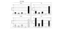


Classifications
-
- A—HUMAN NECESSITIES
- A61—MEDICAL OR VETERINARY SCIENCE; HYGIENE
- A61L—METHODS OR APPARATUS FOR STERILISING MATERIALS OR OBJECTS IN GENERAL; DISINFECTION, STERILISATION OR DEODORISATION OF AIR; CHEMICAL ASPECTS OF BANDAGES, DRESSINGS, ABSORBENT PADS OR SURGICAL ARTICLES; MATERIALS FOR BANDAGES, DRESSINGS, ABSORBENT PADS OR SURGICAL ARTICLES
- A61L27/00—Materials for grafts or prostheses or for coating grafts or prostheses
- A61L27/14—Macromolecular materials
- A61L27/18—Macromolecular materials obtained otherwise than by reactions only involving carbon-to-carbon unsaturated bonds
-
- A—HUMAN NECESSITIES
- A61—MEDICAL OR VETERINARY SCIENCE; HYGIENE
- A61F—FILTERS IMPLANTABLE INTO BLOOD VESSELS; PROSTHESES; DEVICES PROVIDING PATENCY TO, OR PREVENTING COLLAPSING OF, TUBULAR STRUCTURES OF THE BODY, e.g. STENTS; ORTHOPAEDIC, NURSING OR CONTRACEPTIVE DEVICES; FOMENTATION; TREATMENT OR PROTECTION OF EYES OR EARS; BANDAGES, DRESSINGS OR ABSORBENT PADS; FIRST-AID KITS
- A61F2/00—Filters implantable into blood vessels; Prostheses, i.e. artificial substitutes or replacements for parts of the body; Appliances for connecting them with the body; Devices providing patency to, or preventing collapsing of, tubular structures of the body, e.g. stents
- A61F2/02—Prostheses implantable into the body
- A61F2/04—Hollow or tubular parts of organs, e.g. bladders, tracheae, bronchi or bile ducts
- A61F2/042—Urinary bladders
-
- A—HUMAN NECESSITIES
- A61—MEDICAL OR VETERINARY SCIENCE; HYGIENE
- A61F—FILTERS IMPLANTABLE INTO BLOOD VESSELS; PROSTHESES; DEVICES PROVIDING PATENCY TO, OR PREVENTING COLLAPSING OF, TUBULAR STRUCTURES OF THE BODY, e.g. STENTS; ORTHOPAEDIC, NURSING OR CONTRACEPTIVE DEVICES; FOMENTATION; TREATMENT OR PROTECTION OF EYES OR EARS; BANDAGES, DRESSINGS OR ABSORBENT PADS; FIRST-AID KITS
- A61F2/00—Filters implantable into blood vessels; Prostheses, i.e. artificial substitutes or replacements for parts of the body; Appliances for connecting them with the body; Devices providing patency to, or preventing collapsing of, tubular structures of the body, e.g. stents
- A61F2/02—Prostheses implantable into the body
-
- A—HUMAN NECESSITIES
- A61—MEDICAL OR VETERINARY SCIENCE; HYGIENE
- A61L—METHODS OR APPARATUS FOR STERILISING MATERIALS OR OBJECTS IN GENERAL; DISINFECTION, STERILISATION OR DEODORISATION OF AIR; CHEMICAL ASPECTS OF BANDAGES, DRESSINGS, ABSORBENT PADS OR SURGICAL ARTICLES; MATERIALS FOR BANDAGES, DRESSINGS, ABSORBENT PADS OR SURGICAL ARTICLES
- A61L27/00—Materials for grafts or prostheses or for coating grafts or prostheses
- A61L27/36—Materials for grafts or prostheses or for coating grafts or prostheses containing ingredients of undetermined constitution or reaction products thereof, e.g. transplant tissue, natural bone, extracellular matrix
- A61L27/38—Materials for grafts or prostheses or for coating grafts or prostheses containing ingredients of undetermined constitution or reaction products thereof, e.g. transplant tissue, natural bone, extracellular matrix containing added animal cells
- A61L27/3804—Materials for grafts or prostheses or for coating grafts or prostheses containing ingredients of undetermined constitution or reaction products thereof, e.g. transplant tissue, natural bone, extracellular matrix containing added animal cells characterised by specific cells or progenitors thereof, e.g. fibroblasts, connective tissue cells, kidney cells
- A61L27/3826—Muscle cells, e.g. smooth muscle cells
-
- A—HUMAN NECESSITIES
- A61—MEDICAL OR VETERINARY SCIENCE; HYGIENE
- A61L—METHODS OR APPARATUS FOR STERILISING MATERIALS OR OBJECTS IN GENERAL; DISINFECTION, STERILISATION OR DEODORISATION OF AIR; CHEMICAL ASPECTS OF BANDAGES, DRESSINGS, ABSORBENT PADS OR SURGICAL ARTICLES; MATERIALS FOR BANDAGES, DRESSINGS, ABSORBENT PADS OR SURGICAL ARTICLES
- A61L27/00—Materials for grafts or prostheses or for coating grafts or prostheses
- A61L27/36—Materials for grafts or prostheses or for coating grafts or prostheses containing ingredients of undetermined constitution or reaction products thereof, e.g. transplant tissue, natural bone, extracellular matrix
- A61L27/38—Materials for grafts or prostheses or for coating grafts or prostheses containing ingredients of undetermined constitution or reaction products thereof, e.g. transplant tissue, natural bone, extracellular matrix containing added animal cells
- A61L27/3839—Materials for grafts or prostheses or for coating grafts or prostheses containing ingredients of undetermined constitution or reaction products thereof, e.g. transplant tissue, natural bone, extracellular matrix containing added animal cells characterised by the site of application in the body
- A61L27/3882—Hollow organs, e.g. bladder, esophagus, urether, uterus
-
- A—HUMAN NECESSITIES
- A61—MEDICAL OR VETERINARY SCIENCE; HYGIENE
- A61L—METHODS OR APPARATUS FOR STERILISING MATERIALS OR OBJECTS IN GENERAL; DISINFECTION, STERILISATION OR DEODORISATION OF AIR; CHEMICAL ASPECTS OF BANDAGES, DRESSINGS, ABSORBENT PADS OR SURGICAL ARTICLES; MATERIALS FOR BANDAGES, DRESSINGS, ABSORBENT PADS OR SURGICAL ARTICLES
- A61L27/00—Materials for grafts or prostheses or for coating grafts or prostheses
- A61L27/40—Composite materials, i.e. containing one material dispersed in a matrix of the same or different material
-
- C—CHEMISTRY; METALLURGY
- C08—ORGANIC MACROMOLECULAR COMPOUNDS; THEIR PREPARATION OR CHEMICAL WORKING-UP; COMPOSITIONS BASED THEREON
- C08L—COMPOSITIONS OF MACROMOLECULAR COMPOUNDS
- C08L67/00—Compositions of polyesters obtained by reactions forming a carboxylic ester link in the main chain; Compositions of derivatives of such polymers
- C08L67/04—Polyesters derived from hydroxycarboxylic acids, e.g. lactones
-
- A—HUMAN NECESSITIES
- A61—MEDICAL OR VETERINARY SCIENCE; HYGIENE
- A61L—METHODS OR APPARATUS FOR STERILISING MATERIALS OR OBJECTS IN GENERAL; DISINFECTION, STERILISATION OR DEODORISATION OF AIR; CHEMICAL ASPECTS OF BANDAGES, DRESSINGS, ABSORBENT PADS OR SURGICAL ARTICLES; MATERIALS FOR BANDAGES, DRESSINGS, ABSORBENT PADS OR SURGICAL ARTICLES
- A61L2430/00—Materials or treatment for tissue regeneration
- A61L2430/22—Materials or treatment for tissue regeneration for reconstruction of hollow organs, e.g. bladder, esophagus, urether, uterus
Landscapes
- Health & Medical Sciences (AREA)
- Life Sciences & Earth Sciences (AREA)
- Chemical & Material Sciences (AREA)
- Engineering & Computer Science (AREA)
- Biomedical Technology (AREA)
- Veterinary Medicine (AREA)
- Public Health (AREA)
- Oral & Maxillofacial Surgery (AREA)
- Transplantation (AREA)
- Animal Behavior & Ethology (AREA)
- General Health & Medical Sciences (AREA)
- Medicinal Chemistry (AREA)
- Dermatology (AREA)
- Epidemiology (AREA)
- Chemical Kinetics & Catalysis (AREA)
- Cell Biology (AREA)
- Urology & Nephrology (AREA)
- Botany (AREA)
- Zoology (AREA)
- Vascular Medicine (AREA)
- Reproductive Health (AREA)
- Heart & Thoracic Surgery (AREA)
- Cardiology (AREA)
- Composite Materials (AREA)
- Materials Engineering (AREA)
- Gastroenterology & Hepatology (AREA)
- Pulmonology (AREA)
- Polymers & Plastics (AREA)
- Organic Chemistry (AREA)
- Materials For Medical Uses (AREA)
- Prostheses (AREA)
- Micro-Organisms Or Cultivation Processes Thereof (AREA)
- Medicines Containing Material From Animals Or Micro-Organisms (AREA)
Description
本発明は自己の細胞で移植した足場を利用した薄層状構造の管腔臓器および組織構造の再生、再構築、修復、拡大または置換に関係する。 The present invention relates to the regeneration, reconstruction, repair, expansion or replacement of luminal organs and tissue structures of lamellar structure using scaffolds transplanted with autologous cells.
発明の背景
いくつかの奇形は、膀胱の異常発達をもたらし、外科的な補強が必要になることがある。後部尿道弁や両側性異所性尿管、膀胱外反症、排せつ膣外反症脊椎破裂(脊髄髄膜瘤など)などの症状によって膀胱のコンプライアンスがなく、低容量となり高圧を生成する恐れがある。臨床上、これは患者が失禁に苦しむ原因となり、同時に泌尿生殖器系における高圧に起因して腎不全のリスクを高める。小児患者向けの現在における標準治療は腸膀胱形成術 (enterocystoplasty) による膀胱拡大である (Lewis 他 Br. J. Urol. (1990); 65: 488-491)。膀胱拡大はコンプライアンスや容量を向上させ、圧力を低下させるために、患者から大腸組織の一部を切除し、患者が、その組織を既存の膀胱に繋げられることを含む。手術は比較的複雑で、高額となる。技術的結果のよい患者においてさえ、この手順にはさまざまな直接リスクや慢性合併症が付随する。こうした手術の侵襲性、コストおよび合併症が、最も深刻な膀胱の欠陥に対してのみの使用に限定される。同様の外科手術は膀胱の置換が必要な成人でも行われており、その多くは膀胱癌による。成人の場合、膀胱全体が切除され、大腸で代用されることになる。米国では、副作用のリスクにもかかわらず年間約10,000件の手術が行われており、その約10%が先天性奇形の小児、90%が膀胱癌などの疾患を発症した成人である。現在の標準治療に伴う副作用を排除または少なくとも大幅に低減する改善アプローチに対して切実な医療ニーズがあることは明確である。
BACKGROUND OF THE INVENTION Some malformations result in abnormal development of the bladder and may require surgical reinforcement. Symptoms such as posterior urethral valves, bilateral ectopic ureters, bladder valgus, excretory vaginal valgus spine rupture (such as spinal meningocele) may lead to low bladder capacity and high pressure is there. Clinically, this causes patients to suffer incontinence and at the same time increases the risk of renal failure due to high pressure in the genitourinary system. The current standard treatment for pediatric patients is bladder enlargement with enterocystoplasty (Lewis et al. Br. J. Urol. (1990); 65: 488-491). Bladder enlargement involves excising a portion of the colon tissue from a patient to improve compliance and volume and reduce pressure, and the patient can connect the tissue to an existing bladder. Surgery is relatively complex and expensive. Even in patients with good technical results, this procedure is associated with various direct risks and chronic complications. The invasiveness, cost and complications of such surgery are limited to use only for the most serious bladder defects. Similar surgeries are performed in adults who need bladder replacement, most of which are due to bladder cancer. For adults, the entire bladder is removed and replaced by the large intestine. In the United States, about 10,000 operations are performed annually despite the risk of side effects, of which about 10% are children with congenital malformations and 90% are adults who develop diseases such as bladder cancer. Clearly, there is an urgent medical need for improved approaches that eliminate or at least significantly reduce the side effects associated with current standard therapies.
ヒトの膀胱は尿の貯留場所としての役割を持つ骨盤腔の前部に位置する筋結織膜嚢であり、尿は尿管から入り、尿道を通って排出される。ヒトの膀胱は骨盤内で、骨盤骨(恥骨結合)の後ろにあり、尿道と呼ばれる、体外に出る排出管の上に位置し、それに後方が繋がっている。膀胱は患者の膀胱損傷の原因となるさまざまな疾患や損傷を受けやすい。たとえば、感染病や腫瘍、発育異常などから膀胱損傷が起こり得る。さらに交通事故やスポーツ傷害などの外傷が原因で膀胱損傷が起こる場合もある。膀胱癌の患者では尿路変更術が必要になる場合が多くみられる。米国では毎年54,000人以上の新しい膀胱癌の症例がある。ほとんどの膀胱癌は上皮性起源によるもので、世界的にみると年間約336,000人の尿路上皮癌 (移行上皮癌 (TCC)) の新しい症例がある (Kakizoe (2006) Cancer Sci. 97(9) 821)。 The human urinary bladder is a sarcolemma sac located in the front of the pelvic cavity that serves as a urine storage location, and urine enters through the ureter and drains through the urethra. The human urinary bladder is located in the pelvis, behind the pelvic bone (pubic bone joint), on the drainage duct that goes outside the body, called the urethra, and the back is connected to it. The bladder is susceptible to various diseases and injuries that cause the patient's bladder damage. For example, bladder injury can occur due to infectious diseases, tumors, developmental abnormalities and the like. In addition, bladder injury may occur due to trauma such as traffic accidents or sports injuries. Urinary diversion is often required in patients with bladder cancer. There are more than 54,000 new cases of bladder cancer each year in the United States. Most bladder cancers are of epithelial origin and there are approximately 336,000 new cases of urothelial cancer (transitional cell carcinoma (TCC)) annually (Kakizoe (2006) Cancer Sci. 97 (9 821).
尿路変更術とは、泌尿系の損傷や不全によって排尿できない患者の身体から尿の経路を作って排せつする方法である。一般的に、尿の流れを遮断する症状および尿管や腎臓の圧力が増大する症状において尿路変更術が必要となり得る。一般的な尿路変更術の適応症には嚢胞切除の必要な膀胱癌や腎機能に影響を及ぼす過敏膀胱、膀胱への放射線障害、女性に見られる難治性失禁、慢性骨盤通症候群が含まれる。一般に、尿路変更術には、人工膀胱造設術と禁制型変更術の方法が存在する。人工膀胱造設術では回腸、結腸または空腸などの小腸粘膜下組織 (SI) の短いセグメントなどの体内の導管へ接続される腹部ストーマを作ることが含まれる。この手術では、短い SI の一方の端が、通常腎臓から膀胱に尿を運搬する尿管に接続される。尿は尿管から短い SI へ流れ、ストーマから外部の収集容器へと出る。この手順の代替として尿管を直接ストーマに取り付ける方法があり、尿管瘻術とも呼ばれる。禁制型変更術には、胃または小腸や大腸の一部から体内に袋や容器を作ることが含まれ、ストーマの使用は必要な場合とそうでない場合がある。たとえば、大腸の一部を採取し、より球状に修正して禁制型皮膚性容器を作るなどがある。修正部分の一方の端は尿管に接続し、もう一方の端は外部の収集容器に繋がるストーマに接続される。最後に、成形しなおしたセグメントを、一方の端を尿管に、もう一方の端を尿道に接続して、もとの膀胱のかわりに設置することにより、同所変更手段を作り出すことができ、それによって、患者はストーマを介するのではなく、尿道から排尿することができるようになる。 Urinary diversion is a method of excretion by creating a urinary pathway from the body of a patient who cannot urinate due to urinary damage or failure. In general, urinary diversion may be necessary for symptoms that block urine flow and symptoms that increase ureteral or renal pressure. Common urinary diversion indications include bladder cancer requiring cystectomy, hypersensitive bladder affecting kidney function, radiation damage to the bladder, refractory incontinence seen in women, and chronic pelvic syndrome . In general, urinary tract modification includes artificial bladder construction and forbidden modification. Artificial cystoplasty involves creating an abdominal stoma that connects to a body conduit such as a short segment of small intestinal submucosa (SI) such as the ileum, colon, or jejunum. In this surgery, one end of a short SI is connected to a ureter that normally carries urine from the kidneys to the bladder. Urine flows from the ureter to a short SI and out of the stoma to an external collection container. An alternative to this procedure is to attach the ureter directly to the stoma, also called ureterostomy. Forbidden modification involves making a bag or container in the body from the stomach or part of the small or large intestine, and the use of a stoma may or may not be necessary. For example, a part of the large intestine is collected and modified into a more spherical shape to make a forbidden skin container. One end of the modified portion is connected to the ureter and the other end is connected to a stoma leading to an external collection container. Finally, the reshaped segment can be placed in place of the original bladder by connecting one end to the ureter and the other end to the urethra, creating a place-of-place change. , Thereby allowing the patient to urinate from the urethra rather than through the stoma.
小腸粘膜下組織 (SI) が尿路変更術に使用されるとはいえ、粘膜や粘膜下組織の切除は腸部分の収縮に繋がるおそれがあると報告されている (例Atala、A、J. Urol. 156: 338 (1996) 参照)。結石や粘膜産生の増加、新生組織形成、感染症、代謝障害、長期的拘縮および吸収を含む、膀胱の手術に特定の胃腸部を使用することに伴う他の問題が報告されている。尿路変更術で天然材料を使用しても、膀胱組織は、特定の筋肉弾性特性と尿路上皮の不浸透性機能を有し、容易に置き換えられない。また、患者自身の大腸のセグメントを尿路変更術に使用することは、まずセグメント切除の手術、次に尿路変更を施す手術の少なくとも2つの異なる手術が必要とされる。複数の手術が必要になると、手術の総コストや患者や患者の総合的な快適さへのリスクを上げることになる。 Although small intestinal submucosa (SI) is used for urinary diversion, it has been reported that excision of mucosa and submucosa can lead to contraction of the intestine (e.g. Atala, A, J. Urol. 156: 338 (1996)). Other problems with the use of specific gastrointestinal parts for bladder surgery have been reported, including increased stones and mucosal production, neoplasia, infections, metabolic disorders, long-term contractures and absorption. Even when natural materials are used in urinary diversion, bladder tissue has certain muscle elastic properties and impermeable function of the urothelium and is not easily replaced. Also, using a patient's own large intestine segment for urinary diversion requires at least two different operations: segment excision surgery and then urinary diversion surgery. The need for multiple surgeries increases the total cost of surgery and the risk to the patient and the patient's overall comfort.
よって、尿路変更術に胃腸部を使用することに関連する複数の合併症や複数回の手術が必要になることにともない、尿路変更システムを、それを必要な患者に提供する方法と手段の必要性が存在する。 Thus, a method and means for providing a urinary diversion system to a patient in need thereof due to the necessity of multiple complications and multiple operations associated with the use of the gastrointestinal tract for urinary diversion There is a need for.
尿失禁は一般社会ならびに医療環境の両方において、すべての年齢および健康レベルの人々に影響する一般的な疾患である。医学的にみて尿失禁によって患者は尿路感染症や床ずれ、会陰発疹、尿路性敗血症にかかりやすくなる。社会的および心理学的にみると尿失禁は恥ずかしさや社会的な非難、うつに関連し、特に老人にとっては施設に入れられるリスクが高まることになる (Herzo 他、Ann. Rev. Gerontol. Geriatrics、9: 74 (1989))。経済的にみると、そのコストは驚異的で、米国だけでも毎年100億ドル以上が失禁のコントロールに費やされている。 Urinary incontinence is a common disease affecting people of all ages and health levels, both in the general public and in the medical environment. Medically, urinary incontinence makes patients more susceptible to urinary tract infections, bedsores, perineal rashes, and urinary sepsis. Socially and psychologically, urinary incontinence is associated with embarrassment, social criticism, and depression, especially for older people (Herzo et al., Ann. Rev. Gerontol. Geriatrics, 9:74 (1989)). Economically, the cost is staggering, with more than $ 10 billion spent each year in the US alone to control incontinence.
失禁は真性ストレス尿失禁(膀胱および尿道の過可動)や固有括約筋欠損症(「ISD」)、またはその両方に原因がある場合がある。これは特に女性に多く見られ、子供にはより少ない程度の失禁が見られ(特に ISD)、男性では前立腺全摘出術の後に見られる。 Incontinence may be due to genuine stress urinary incontinence (bladder and urethral hypermobility), intrinsic sphincter deficiency (“ISD”), or both. This is especially common in women, to a lesser extent incontinence in children (especially ISD), and in men after radical prostatectomy.
ストレス尿失禁はせきやくしゃみをしたり、笑ったり、エクササイズをしている際など腹内圧力が上昇する身体活動時に不本意に失禁することである。患者は一方または両方の失禁タイプを患う場合があり、両方の疾患を患う場合は、混合型尿失禁と呼ばれる。尿失禁に関するあらゆる知識にもかかわらず、急迫性尿失禁のほとんどのケースは特発性であり、これは特定の原因が追及できないことを意味する。急迫性尿失禁はあらゆる年齢の誰にでも起こる可能性があるが、女性や年配者に特によく見られる。 Stress urinary incontinence is involuntary incontinence during physical activity that increases abdominal pressure, such as when coughing, sneezing, laughing, or exercising. Patients may suffer from one or both incontinence types, and when they suffer from both diseases, it is called mixed urinary incontinence. Despite all the knowledge about urinary incontinence, most cases of urge incontinence are idiopathic, meaning that a specific cause cannot be pursued. Urgent urinary incontinence can occur in anyone of any age but is particularly common in women and the elderly.
排尿筋は、収縮して尿を膀胱から排出する膀胱壁の筋肉である。反射亢進などの排尿筋の機能不全の結果として、膀胱コンプライアンスの低下、膀胱内圧の上昇、膀胱容量の低下が含まれ、そのすべては上部尿路の損傷につながる恐れがある。 The detrusor muscle is the bladder wall muscle that contracts and drains urine from the bladder. Detrusor dysfunction, such as increased reflexes, includes decreased bladder compliance, increased intravesical pressure, decreased bladder capacity, all of which can lead to upper urinary tract damage.
急迫性尿失禁に対する現在の治療法の1つにBotox(登録商標)などのポツリヌス毒素のような神経毒の注射が挙げられる。ポツリヌス毒素は、膀胱壁の排尿筋を麻痺させたり、膀胱の求心路に影響を与えて尿路上皮下神経の感覚器受容体を低下させることで過敏な膀胱に効果を発揮すると考えられている。サイズが大きいボツリヌス毒素分子は拡散能力が限定されるため、求心性および遠心性神経線維のどちらにも届かない。結果として、過活動膀胱 (OAB) に対する現在の投与方法は、例えば、ボツリヌス毒素を膀胱筋壁に何度も注射すること(通常20〜50回)が必要となり、通院回数や関連治療費も増加することとなる。その上、膀胱から出る感覚神経伝達物質の抑制の慢性的な長期的影響の安全性はまだ究明されていない。 One current treatment for urge urinary incontinence includes injection of a neurotoxin such as a botulinum toxin such as Botox®. Potulinus toxin is thought to exert an effect on the sensitive bladder by paralyzing the detrusor muscle of the bladder wall or by affecting the afferent tract of the bladder to lower the sensory organ receptors of the urinary subcutaneous nerve. Large botulinum toxin molecules have limited diffusivity and do not reach both afferent and efferent nerve fibers. As a result, current methods of administration for overactive bladder (OAB), for example, require multiple injections of botulinum toxin into the bladder muscle wall (usually 20-50), increasing the number of visits and associated treatment costs Will be. Moreover, the safety of chronic long-term effects of suppression of sensory neurotransmitters exiting the bladder has not yet been investigated.
尿失禁治療に対する更なるアプローチには、膀胱弛緩効果のある薬品を、そうした薬品の中軸に相当する抗コリン剤とともに投与することが含まれる。たとえば臭化プロパンテリンなどの抗コリン作用薬と、ラセミ性オキシブチニンやジサイクロミンなどの平滑筋弛緩剤/抗コリン作用薬の混合薬が急迫性尿失禁に使用されている (例 A. J. Wein、Urol. Clin. N. Am、22: 557 (1995) 参照)。ただし多くの場合、そのような薬物療法ですべてのクラスの尿失禁患者を完治することはなく、患者は深刻な副作用を経験することがよくみられる。 Further approaches to the treatment of urinary incontinence include the administration of drugs that have a bladder relaxing effect, together with anticholinergic agents that represent the core of such drugs. For example, anti-cholinergic drugs such as propantheline bromide and smooth muscle relaxant / anticholinergic drugs such as racemic oxybutynin and dicyclomine are used for urinary incontinence (eg AJ Wein, Urol. Clin N. Am, 22: 557 (1995)). However, in many cases, such medications do not completely cure all classes of urinary incontinence patients, and patients often experience serious side effects.
本発明の前に、熟練医師が使用した薬物療法以外の他の療法には、人工的括約筋の使用 (Lima S. V. C. 他、J. Urology、156: 622-624 (1996)、Levesque P. E. 他、J. Urology、156: 625-628 (1996)) や膀胱頸部支持人工器官 (Kondo A. 他、J. Urology、157: 824-827 (1996))、架橋コラーゲンの注射 (Berman C. J. 他、J. Urology、157: 122-124 (1997)、Perez L. M. 他、J. Urology、156: 633-636 (1996);Leonard M. P. 他、J. Urology、156: 637-640 (1996))、そしてポリテトラフルオロエチレンの注射 (Perez L. M. 他、J. Urology、156: 633-636 (1996)) が含まれる。 Prior to the present invention, other therapies than those used by skilled physicians include the use of artificial sphincters (Lima SVC et al., J. Urology, 156: 622-624 (1996), Levesque PE et al., J. Urology, 156: 625-628 (1996)) and bladder neck support prosthesis (Kondo A. et al., J. Urology, 157: 824-827 (1996)), cross-linked collagen injection (Berman CJ et al., J. Urology 157: 122-124 (1997), Perez LM et al., J. Urology, 156: 633-636 (1996); Leonard MP et al., J. Urology, 156: 637-640 (1996)), and polytetrafluoroethylene. Injections (Perez LM et al., J. Urology, 156: 633-636 (1996)).
ISD が関連する尿失禁治療における近年よく使用される方法に尿道周囲の内視鏡コラーゲン注射を患者に受けさせることがある。これは膀胱の漏れやストレス尿失禁の可能性を低減させる試みにおいて、膀胱筋を補強する。 One recently used method in the treatment of urinary incontinence associated with ISD is to allow patients to receive periurethral endoscopic collagen injection. This reinforces the bladder muscles in an attempt to reduce the possibility of bladder leakage and stress urinary incontinence.
尿失禁を回避する既存の解決法には既知の欠点がある。膀胱頸部周辺に内視鏡でコラーゲン注射を行うことは、深刻な疾病率がなく括約筋不全において極めて高い成功率を収めているが、コラーゲンの使用は平均して2年後に不具合が起こる場合があり、そのコスト効率性に対しても考慮が必要となる (Khullar V. 他、British J. Obstetrics & Gynecology、104: 96-99 (1996))。さらに、おそらくは移動現象による患者の随意調節の低下 (Perez L. M. 他) によって、随意調節を復活させるために繰り返し注射をする必要がある場合がある (Herschorn S. 他、J. Urology、156: 1305-1309 (1996))。 Existing solutions to avoid urinary incontinence have known drawbacks. Endoscopic collagen injection around the bladder neck has a very high success rate in sphincter failure without a serious morbidity, but the use of collagen can fail on average after two years. There is also a need to consider its cost efficiency (Khullar V. et al., British J. Obstetrics & Gynecology, 104: 96-99 (1996)). In addition, it may be necessary to repeat injections to restore voluntary control (Herschorn S. et al., J. Urology, 156: 1305-), possibly due to reduced voluntary control of patients (Perez LM et al.) 1309 (1996)).
ストレス尿失禁の治療として前立腺全摘出術後にコラーゲンを使用することも一般的には不本意な結果となっている (Klutke C. G. 他、J. Urology、156: 1703-1706 (1996))。その上、ある研究ではウシ真皮コラーゲンの注射で IgG および IgA クラスの特定抗体が産生された証拠が提供されている (McCell および M. および Delustro、F. 、J. Urology 155, 2068-2073 (1996))。よって長期的に見て患者のコラーゲンに対する鋭敏化が予測され得る。 The use of collagen after radical prostatectomy as a treatment for stress urinary incontinence has also generally been disappointing (Klutke C. G. et al., J. Urology, 156: 1703-1706 (1996)). In addition, one study provides evidence that injection of bovine dermal collagen produced specific antibodies of the IgG and IgA class (McCell and M. and Delustro, F., J. Urology 155, 2068-2073 (1996). )). Therefore, in the long term, sensitization of the patient to collagen can be predicted.
他に適切な代替法がないために、経尿道のコラーゲン注射療法は成功率が限定的であるにもかかわらず固有括約筋欠損症の治療として今なお受け入れられている。 Due to lack of other suitable alternatives, transurethral collagen injection therapy is still accepted as a treatment for intrinsic sphincter deficiency despite limited success rates.
現在、過活動膀胱疾患や急迫性尿失禁を患う患者はまず、非侵襲性薬剤医療品による治療を受けることになる。ただしそうした非侵襲性薬剤医療品が効かない場合、医師はより侵襲性のある治療法を提案する。 Currently, patients suffering from overactive bladder disease or urge incontinence will first be treated with a non-invasive drug product. However, if such non-invasive drug products do not work, doctors suggest more invasive treatments.
よって膀胱など既存の薄層状構造の管腔臓器および組織構造を拡大する侵襲の少ない方法に対する必要性がある。 Therefore there is a need for a minimally invasive method for enlarging the luminal organ and tissue structure of the existing laminar structures such as the bladder.
再生医療の原則が適用され、膀胱や膀胱の一部、膀胱の構成要素などの薄層状構造の管腔臓器および組織構造の再構築、修復、拡大または置換における使用のために、移植可能な細胞マトリックスが成功裏に提供されている。Atala 米国特許 6,576,019 で説明されるように、細胞は患者自身の膀胱や尿道、尿管、およびその他の泌尿生殖器組織を含む組織から得ることができる。しかし、新しく健康な培養組織を作るための基本ユニットとして主要臓器サイトから細胞培養系を発現および管理することに依存することに伴う課題がある。たとえば、欠陥のある膀胱の治療には、欠陥のある膀胱から膀胱細胞を培養することは培養された細胞にもまた問題があるというもっともな理由から、細胞調達に関する特定の課題がもたらされる。そのような細胞は移植可能な新しい膀胱足場やマトリックスを培養するのに最適な選択ではない。よって移植できる新しい臓器/組織構造足場やマトリックスの移植に適した細胞の代替源が必要となる。 Regenerative medicine principles apply and transplantable cells for use in the reconstruction, repair, enlargement or replacement of luminal organs and tissue structures in lamellar structures such as the bladder, part of the bladder, and components of the bladder The matrix has been successfully provided. As described in Atala US Pat. No. 6,576,019, cells can be obtained from tissues including the patient's own bladder, urethra, ureter, and other urogenital tissues. However, there are challenges associated with relying on expression and management of cell culture systems from major organ sites as a basic unit for creating new healthy cultures. For example, the treatment of bladder defective, a good reason that culturing bladder cells from the bladder including a defect is also problematic in cells cultured, resulting in a particular problem for cell procurement. Such cells are not the best choice for cultivating new implantable bladder scaffolds or matrices. Therefore, new organ / tissue structure scaffolds that can be transplanted and alternative sources of cells suitable for matrix transplantation are needed.
ヒトの脂肪組織が成人幹細胞の豊富な源である考えを支持する文献は豊富にある (Devlin 他 (2004)、Cytotherapy 6: 7-14;Awad、他 (2003)、Tissue Engineering 9: 1301-12;Erickson 他 (2002)、Biochemical and Biophysical Research Communications 290: 763-769;Gronthos 他 (2001)、Journal of Cellular Physiology 189: 54-63;Halvorsen 他 (2001);Metabolism 50: 407-413;Halvorsen 他 (2001)、Tissue Eng. 6: 729-41;Harp 他 (2001)、Biochemical and Biophysical Research Communication 281: 907-912;Hicok 他 (2004)、Tissue Engineering 10: 371-380;Safford 他 (2002)、Jun 7, 294(2): 371-9;Safford 他、(2004)、Experimental Neurology 187: 319-28;Sen 他 (2004)、Journal of Cellular Biochemistry 81: 312-319;Sigal 他 (1994)、Hepatology 19: 999-1006;Wickham 他 (2003)、Clinical Orthopedics and Related Research、412: 196-212;Ashijian 他 (2003)、Plast Reconstr Surg. 111:1922-31;De Ugarte 他 (2003)、Cells Tissues Organs. 174: 101-9;Mizumo 他 (2002)、Plast Reconstr Surg. 109: 199-209;Morizono 他 (2003)、Hum Gene Ther. 14: 59-66;Winter 他 (2003)、Arthritis Rheum. 48: 418-29;Zuk 他 (2001)、Tissue Eng 7: 211-2
28;Zuk 他 (2002)、Mol Biol 13: 4279−4295、reviewed in Gimble 他 (2003)、Cytotherapy 5: 362-369)。脂肪由来成人幹 (ADAS) 細胞と呼ばれる、こうした細胞は、MSCに匹敵する免疫表現型および分化能を示す (Gronthos 他 (2001)、Journal of Cellular Physiology 189: 54-63;Safford 他 (2002)、Biochem Biophys Res Commun. 294(2): 371-9;Zuk 他 (2002)、Mol Biol Cell 13: 4279-4295)。
There is a wealth of literature supporting the idea that human adipose tissue is a rich source of adult stem cells (Devlin et al. (2004), Cytotherapy 6: 7-14; Awad et al. (2003), Tissue Engineering 9: 1301-12 Erickson et al. (2002), Biochemical and Biophysical Research Communications 290: 763-769; Gronthos et al. (2001), Journal of Cellular Physiology 189: 54-63; Halvorsen et al. (2001); Metabolism 50: 407-413; Halvorsen et al. ( 2001), Tissue Eng. 6: 729-41; Harp et al. (2001), Biochemical and Biophysical Research Communication 281: 907-912; Hicok et al. (2004), Tissue Engineering 10: 371-380; Safford et al. (2002), Jun. 7, 294 (2): 371-9; Safford et al. (2004), Experimental Neurology 187: 319-28; Sen et al. (2004), Journal of Cellular Biochemistry 81: 312-319; Sigal et al. (1994), Hepatology 19 : 999-1006; Wickham et al. (2003), Clinical Orthopedics and Related Research, 412: 196-212; Ashijian et al. (2003), Plast Reconstr Surg. 111: 1922-31; De Ugarte et al. (2003), Cells Tissues Organs. 174: 101-9; Mi zumo et al. (2002), Plast Reconstr Surg. 109: 199-209; Morizono et al. (2003), Hum Gene Ther. 14: 59-66; Winter et al. (2003), Arthritis Rheum. 48: 418-29; Zuk et al. 2001), Tissue Eng 7: 211-2
28; Zuk et al. (2002), Mol Biol 13: 4279-4295, reviewed in Gimble et al. (2003), Cytotherapy 5: 362-369). These cells, called adipose-derived adult stem (ADAS) cells, exhibit immune phenotypes and differentiation potential comparable to MSCs (Gronthos et al. (2001), Journal of Cellular Physiology 189: 54-63; Safford et al. (2002), Biochem Biophys Res Commun. 294 (2): 371-9; Zuk et al. (2002), Mol Biol Cell 13: 4279-4295).
ヒトの脂肪吸引試料から成人幹細胞を単離する再現可能で効果的な方法が自由に使用できる状態で存在する (Aust 他 (2004)、Cytotherapy 6: 7-14;Halvorsen 他 (2001)、Metabolism 50: 407-413)。手順には組織のコラゲナーゼ消化、遠心分画、および培養の増殖が含まれる。1 グラムの組織からは培養開始 24 時間以内に50,000〜100,000の間質細胞が得られる ( Sen 他 (2001)、J Cellular Biochemistry 81: 312-319)。20名のドナーから得た試料の分析では、脂肪吸引廃液 1mlから生存率 94% で平均401,000の細胞が一貫して回収された。こうした細胞の増殖では、2週間以内に標準的な吸引脂肪から500,000万以上の細胞母集団を産出し得る。 A reproducible and effective method for isolating adult stem cells from human liposuction samples exists in a freely available manner (Aust et al. (2004), Cytotherapy 6: 7-14; Halvorsen et al. (2001), Metabolism 50 : 407-413). Procedures include tissue collagenase digestion, centrifugation, and culture growth. One gram of tissue yields 50,000-100,000 stromal cells within 24 hours of culturing (Sen et al. (2001) J Cellular Biochemistry 81: 312-319). Analysis of samples from 20 donors consistently recovered an average of 401,000 cells from 1 ml of liposuction waste with a 94% survival rate. Such cell growth can produce over 500 million cell populations from standard aspirated fat within 2 weeks.
デキサメタゾン、インスリン、イソブチルメチルキサンチン、チアゾリジンジオンの存在下、ADAS 細胞は脂質生成する (Sen 他 (2001) Journal of Cellular Biochemistry 81: 312-319)。ADAS 細胞の分化能は脂肪細胞系列だけにとどまらない。軟骨細胞および骨芽細胞経路に沿った ADAS 細胞分化を向上させる症状が報告されている (Awad、他 (2003)、Tissue Engineering 9: 1301-12;Erickson 他 (2002)、Biochemical and Biophysical Research Communications 290: 763-769;Halvorson 他 (2001)、Metabolism 50: 407-413;Hicok 他 (2004)、Tissue Engineering 10: 371-380;Wickham 他 (2003)、Clinic Orthopedics and Related Research 412: 196-212)。ヒドロキシアパタイト生体材料と結合した生体内のヒト ADAS 細胞は免疫不全のマウスの皮下に移植される場合、類骨マトリックスを合成する (Hikok 他 (2004)、Tissue Engineering 10: 371-380)。抗酸化物質およびその他のメディエーターの存在下で培養されたネズミおよびヒトの脂肪由来成人幹細胞 (それぞれ muADAS および huADAS) は、神経分化と一致する形態学的および表現型の変化を経ることを示すデータが多く存在する(De Ugarte (2003) Cells Tissues Organs. 174: 101-9;Safford 他 (2002)、294(2): 371-9;Safford 他 (2004)、Experimental Neurology 187: 319-28)。 In the presence of dexamethasone, insulin, isobutylmethylxanthine, and thiazolidinedione, ADAS cells adipate (Sen et al. (2001) Journal of Cellular Biochemistry 81: 312-319). The ability of ADAS cells to differentiate is not limited to the adipocyte lineage. Symptoms that improve ADAS cell differentiation along the chondrocyte and osteoblast pathways have been reported (Awad, et al. (2003), Tissue Engineering 9: 1301-12; Erickson et al. (2002), Biochemical and Biophysical Research Communications 290 : 763-769; Halvorson et al. (2001), Metabolism 50: 407-413; Hicok et al. (2004), Tissue Engineering 10: 371-380; Wickham et al. (2003), Clinic Orthopedics and Related Research 412: 196-212). In vivo human ADAS cells combined with hydroxyapatite biomaterials synthesize osteoid matrix when implanted subcutaneously in immunodeficient mice (Hikok et al. (2004), Tissue Engineering 10: 371-380). Data indicate that murine and human adipose-derived adult stem cells (muADAS and huADAS, respectively) cultured in the presence of antioxidants and other mediators undergo morphological and phenotypic changes consistent with neuronal differentiation. There are many (De Ugarte (2003) Cells Tissues Organs. 174: 101-9; Safford et al. (2002), 294 (2): 371-9; Safford et al. (2004), Experimental Neurology 187: 319-28).
Jayo 他 Regen. Med. (2008) 3(5)、671-682 (以下「Jayo I」とする) で説明されるように、臓器や組織の修復を試みることは、高い頻度でコラーゲン堆積が原因、また中には瘢痕組織形成が原因で組織の置換が完全にできないという特徴がある。Jayo 他はまた、より望ましい再生医療の結果は組織構造や臓器のもとの構造および機能の再生であると述べた。Jayo 他、J. Urol. (2008) 180;392-397 (以下「Jayo II」とする) も参照。ある種の分子が生体内における再生過程と関連していると考えられている。たとえばケモカイン MCP-1 は単核細胞を動員する能力があることで最もよく知られている。ただし、血管平滑筋細胞を増殖させるマイトジェンである可能性も考えられている。MCP-1 は循環単核白血球を血管損傷部分に動員する。循環単核白血球は次いで、典型的にはマクロファージに変換され、サイトカインや成長因子の貯蔵庫としての機能を果たすことができる。またマクロファージはコレストロールを処理し、脂質を酸化させる。マクロファージおよび筋前駆体細胞はともに MCP-1 のシグナル伝達の標的であると考えられている。CCR-2 レセプターは MCP-1 (CCL2) のリガンドであり、CCR-2 が欠乏しているマウスには亢進した脂質生成/線維症をともなう再生異常が見られる。骨格筋再生が困難である場合の CCR-2 が欠乏しているマウスの切片は通常のマウスと比較して、より多くの間質腔、高い炎症細胞数、大きく丸く膨張した筋原線維、間質腔におけるより多くの線維芽細胞の蓄積、脂肪堆積周辺におけるコラーゲン分配をともなう脂肪浸潤、カルシウム堆積に付随して起こる線維症を示した (Warren 他 (2005)、FASEB J.19: 413-415;Selzman 他 (2002)、Am J Physiol Heart Circ Physiol. 283(4);H1455-H1461;Shannon 他 (2007)、Am. J. Cell Physiol. 292: C953-C967;Shireman 他 (2006)、J. Surg. Res. 134(1): 145-57. Epub 2006 Feb 20;Amann 他 (1998)、Brit. J. Urol. 82: 118-121;Schecter 他 (2004)、J. Leukocyte Biol.75: 1079-1085;Deonarine 他、(2007)、Transl Med. 5: 11;Lumeng 他 (2007)、J Clin. Invest. 117(1): 175−184)。
As attempted by Jayo et al. Regen. Med. (2008) 3 (5), 671-682 (hereinafter “Jayo I”), attempts to repair organs and tissues are frequently caused by collagen deposition. Also, there is a feature that the tissue cannot be completely replaced due to scar tissue formation. Jayo et al. Also stated that the more desirable outcome of regenerative medicine is the regeneration of tissue structure and organ structure and function. See also Jayo et al., J. Urol. (2008) 180; 392-397 (hereinafter “Jayo II”). Certain molecules are thought to be associated with regeneration processes in vivo. For example, the chemokine MCP-1 is best known for its ability to mobilize mononuclear cells. However, the possibility of being a mitogen that proliferates vascular smooth muscle cells is also considered. MCP-1 recruits circulating mononuclear leukocytes to vascular injury. Circulating mononuclear leukocytes are then typically converted to macrophages and can serve as reservoirs for cytokines and growth factors. Macrophages also process cholesterol and oxidize lipids. Both macrophages and muscle progenitor cells are thought to be targets for MCP-1 signaling. The CCR-2 receptor is a ligand for MCP-1 (CCL2), and mice deficient in CCR-2 show regenerative abnormalities with enhanced lipogenesis / fibrosis. Sections of mice deficient in CCR-2 when skeletal muscle regeneration is difficult have more interstitial spaces, higher inflammatory cell counts, large, rounded and expanded myofibrils, compared to normal mice Accumulation of more fibroblasts in the interstitial space, fat infiltration with collagen partitioning around the fat deposit, and fibrosis associated with calcium deposition (Warren et al. (2005), FASEB J.19: 413-415 Selzman et al. (2002), Am J Physiol Heart Circ Physiol. 283 (4); H1455-H1461; Shannon et al. (2007), Am. J. Cell Physiol. 292: C953-C967; Shireman et al. (2006), J .; Res. 134 (1): 145-57. Epub 2006
本発明は、本明細書でで説明される再生、再構築、修復、拡大または置換の対象となる臓器や組織構造とは別の自己由来の細胞集団、そうした細胞やその細胞で移植された新しい臓器/組織構造足場またはマトリックス(構造体)を単離する方法、そしてそれを作る方法、ならびに新しい臓器/組織構造構造体を使用し、必要としている患者を治療する方法に関する。 The present invention provides a self-derived cell population separate from the organ or tissue structure to be regenerated, reconstructed, repaired, expanded or replaced as described herein, such cells or new transplanted with such cells. It relates to a method of isolating an organ / tissue structure scaffold or matrix (structure), a method of making it, and a method of treating a patient in need using the new organ / tissue structure structure.
本発明は、対象の自己細胞で移植した足場を利用した薄層状構造の管腔臓器および組織構造の再生、再構築、修復、拡大または置換に関係する。 The present invention, reproduction of the tubular organ and tissue structure of the laminar structure using scaffolds were implanted with autologous cells of the subject, reconstruction, repair, related to the expansion or replacement.
一態様において本発明は、尿路変更構造体と、その製作および使用方法を提供する。一実施形態では、尿路変更手段は対象の欠陥のある膀胱のためであり、(a) 腹壁部に接続するように構成された第一の端部と、第二の閉じた端部と、第一の尿管と接続するように構成された少なくとも1つの第一の開いた側部を有する管状の足場を含む、移植可能な、生体適合性の第一の構造体と、(b) 足場表面内外に置かれ、欠陥のある膀胱に由来しない自己細胞集団を含む。他の実施形態では、尿路変更手段は対象の欠陥のある膀胱のためであり、(a) 腹壁の開口部に接続するように構成された第一の端部と、第二の閉じた端部と、第一の尿管に接続され、第一の尿管から管状の足場への尿の排出を可能にするように適合された、少なくとも1つの第一の開いた側部を有する、尿の一時的な保管と排出のために適合された移植可能な生体適合性の菅状の足場と、(b) 足場表面内外に置かれ、欠陥のある膀胱に由来しない自己細胞集団を含む。 In one aspect, the present invention provides a urinary tract modification structure and methods for making and using the same. In one embodiment, the urinary tract changing means is for a defective bladder of the subject, (a) a first end configured to connect to the abdominal wall, a second closed end, An implantable, biocompatible first structure comprising a tubular scaffold having at least one first open side configured to connect with a first ureter; and (b) a scaffold Includes autologous cell populations placed inside and outside the surface and not derived from a defective bladder . In other embodiments, the urinary diversion means is for a defective bladder of the subject, (a) a first end configured to connect to an opening in the abdominal wall and a second closed end And a urine having at least one first open side connected to the first ureter and adapted to allow drainage of urine from the first ureter to the tubular scaffold An implantable biocompatible saddle-like scaffold adapted for temporary storage and excretion of, and (b) an autologous cell population that is placed inside and outside the scaffold surface and not derived from a defective bladder .
一実施形態では、本発明は、欠陥のある膀胱のための尿路変更構造体を、それを必要とする患者において準備する方法を提供し、手順には a) 腹壁の開口部に接するように構成された第一の端部と、第二の閉じた端部と、第一の尿管に接続され、第一の尿管から管状の足場への尿の通過を可能にするように適合された、少なくとも1つの第一の開いた側部を有する、管状の足場からなる、第一の移植可能な生体適合性のある足場を提供すること、b) 欠陥のある膀胱に由来しない自己細胞集団を足場の第一部位の内外に堆積させて尿路交換構造体を形成することが含まれる。他の実施形態では、方法の手順には a) 腹壁の開口部に接続するように構成された第一の端部と、第二の閉じた端部と、第一の尿管に接続され、第一の尿管から管状の足場への尿の通過を可能にするように適合された、少なくとも1つの第一の開いた側部からなる、尿の一時的な保管と排出のために適合された移植可能な生体適合性の菅状の足場を提供することと、b) 欠陥のある膀胱に由来しない自己細胞集団を足場表面内外に堆積し、尿路変更構造体を形成することが含まれる。 As in one embodiment, the present invention provides a urinary diversion structure for bladder defective, provides a method of preparing in a patient in need thereof, the steps to contact the opening of a) the abdominal wall Connected to the first ureter and adapted to allow passage of urine from the first ureter to the tubular scaffold Providing a first implantable biocompatible scaffold consisting of a tubular scaffold having at least one first open side, b) autologous cells not derived from a defective bladder It includes depositing the population in and out of the first part of the scaffold to form a urinary tract exchange structure. In another embodiment, the method procedure includes: a) a first end configured to connect to an opening in the abdominal wall, a second closed end, and a first ureter; Adapted for temporary storage and drainage of urine, consisting of at least one first open side, adapted to allow passage of urine from the first ureter into the tubular scaffold Providing an implantable biocompatible saddle-shaped scaffold, and b) depositing self-cell populations not derived from a defective bladder inside and outside the scaffold surface to form a urinary diversion structure .
他の一実施形態では、本発明は、欠陥のある膀胱のための尿路変更手段を、それを必要とする患者において提供する方法を提供し、方法の手順には a) 腹壁の開口部に接続するように構成された第一の端部と、第二の閉じた端部と、第一の尿管に接続され、第一の尿管から管状の足場への尿の通過を可能にするように適合された、少なくとも1つの第一の開いた側部を有する、管状の足場からなる、第一の移植可能な生体適合性のある足場を提供することと、b) 欠陥のある膀胱に由来しない自己細胞集団を足場表面内外に堆積して、尿路変更構造体を形成することと、c) 尿路変更を形成するために構造体を対象に移植することが含まれる。他の実施形態では、手順には a) 腹壁の開口部に接続するように構成された第一の端部と、第二の閉じた端部と、第一の尿管に接続され、第一の尿管から管状の足場への尿の通過を可能にするように適合された、少なくとも1つの第一の開いた側部からなる、尿の一時的な保管と排出のために適合された移植可能な生体適合性の菅状の足場を提供することと、b) 欠陥のある膀胱に由来しない自己細胞集団を足場の第一部位の内外に堆積させて尿路置換構造体を形成することと、 c) 尿路変更を形成するために構造体を対象に移植することが含まれる。他の一実施形態では、方法には(a) 腹壁の開口部に接するように構成された第一の端部と、第二の閉じた端部と、第一の尿管に接続され、第一の尿管から管状の足場への尿の通過を可能にするように適合された、少なくとも1つの第一の開いた側部を有する、管状の足場と (b) 尿路変更形成のため、足場表面内外に堆積された欠陥のある膀胱に由来しない自己細胞集団、で構成される尿路変更構造体を対象に移植する手順が含まれる。 In another embodiment, the present invention provides a method of providing urinary diversion means for a defective bladder in a patient in need thereof, the method procedure comprising: a) opening an abdominal wall Connected to a first end configured to connect, a second closed end, and a first ureter to allow passage of urine from the first ureter to a tubular scaffold Providing a first implantable biocompatible scaffold comprising a tubular scaffold having at least one first open side adapted to b) to a defective bladder It includes depositing non-derived self-cell populations inside and outside the scaffold surface to form a urinary diversion structure, and c) transplanting the structure into a subject to form the urinary diversion. In other embodiments, the procedure includes: a) a first end configured to connect to an opening in the abdominal wall, a second closed end, and a first ureter connected to the first end; Implant adapted for temporary storage and drainage of urine, comprising at least one first open side, adapted to allow passage of urine from the urinary tract into a tubular scaffold Providing a possible biocompatible saddle-shaped scaffold, and b) depositing self-cell populations not derived from a defective bladder inside or outside the first site of the scaffold to form a urinary tract replacement structure; C) transplanting the structure to the subject to form a urinary diversion. In another embodiment, the method a first end configured so that the Sessu the opening of (a) the abdominal wall, and a second closed end, is connected to the first ureter A tubular scaffold having at least one first open side adapted to allow passage of urine from the first ureter to the tubular scaffold, and (b) of urinary diversion formation Therefore, a procedure for transplanting a urinary tract structure composed of a self-cell population not derived from a defective bladder deposited inside or outside the scaffold surface to a target is included.
すべての実施形態において、尿路変更足場はさらに第二尿管に接続するように構成された第二の開いた側部を備える場合がある。すべての実施形態において、第一の端部は腹壁にぴったり重なって位置するように構成される場合がある。すべての実施形態において、第一の端部が対象の皮膚に縫合されるように構成される場合がある。すべての実施形態において、第一の端部はストーマを形成するように構成される場合がある。すべての実施形態において、ストーマはさらにストーマボタンを含む場合がある。すべての実施形態において、足場はさらにストーマを形成するためにワッシャーリングを含む。すべての実施形態において、生体適合性を有する足場は生物分解性である。すべての実施形態において、足場はポリグリコール酸と、ポリ乳酸と、ポリグリコール酸およびポリ乳酸の共重合体で構成される群から選択された材料から作られ得る。すべての実施形態において、細胞集団は平滑筋細胞集団である。すべての実施形態において、尿路変更手段は欠陥のある膀胱の代わりとなり得る。すべての実施形態において、尿路変更は一時的なものであり得る。すべての実施形態において、尿路変更は永久的なものであり得る。すべての実施形態において、菅状足場は長方形の断面または三角形の断面、もしくは円形の断面で作られ得る。すべての実施形態において、尿路変更手段は尿路上皮細胞を含まないようになり得る。すべての実施形態において、本発明の方法は泌尿器様の組織再生を特徴とする新しい尿路の導管を提供し得る。すべての実施形態において、再生された組織は尿路上皮、固有層、および平滑筋束のうち1つ以上の存在によって特徴付けられ得る。すべての実施形態において、再生された組織は、尿管導管接合部 (UCJ)、導管の頭部、および導管の中間アトリウム部のうち1つ以上で確認できる。すべての実施形態において、再生された組織は粘膜、粘膜下層、および線維血管間質がみられる平滑筋のうち1つ以上の存在によって特徴付けられる場合がある。すべての実施形態では、再生された組織は平滑筋が下にある途切れのない尿路上皮である。すべての実施形態では、尿路の導管は移植にともない上皮化した粘膜を形成する。 In all embodiments, the urinary diversion scaffold may further comprise a second open side configured to connect to the second ureter. In all embodiments, the first end may be configured to lie directly over the abdominal wall. In all embodiments, the first end may be configured to be sutured to the subject's skin. In all embodiments, the first end may be configured to form a stoma. In all embodiments, the stoma may further include a stoma button. In all embodiments, the scaffold further includes a washer ring to form a stoma. In all embodiments, the biocompatible scaffold is biodegradable. In all embodiments, the scaffold may be made from a material selected from the group consisting of polyglycolic acid, polylactic acid, and a copolymer of polyglycolic acid and polylactic acid. In all embodiments, the cell population is a smooth muscle cell population. In all embodiments, the urinary tract changing means can replace a defective bladder . In all embodiments, the urinary tract change may be temporary. In all embodiments, the urinary tract change can be permanent. In all embodiments, the saddle-like scaffold can be made of a rectangular cross section or a triangular cross section or a circular cross section. In all embodiments, the urinary tract changing means may be free of urothelial cells. In all embodiments, the methods of the present invention may provide new urinary tract conduits characterized by urinary tissue regeneration. In all embodiments, the regenerated tissue can be characterized by the presence of one or more of urothelium, lamina propria, and smooth muscle bundles. In all embodiments, it regenerated tissue, urinary conduit junction (UCJ), can be confirmed by the conduit of the head, and one or more of the intermediate atrium portion of the conduit. In all embodiments, the regenerated tissue may be characterized by the presence of one or more of smooth muscle with mucosa, submucosa, and fibrovascular stroma. In all embodiments, the regenerated tissue is an unbroken urothelium with smooth muscle underneath. In all embodiments, the urinary tract conduits form an epithelial mucosa upon implantation.
一態様において、本発明は分離した平滑筋細胞集団に関する。一実施形態において、細胞集団は末梢血から得られ、平滑筋細胞マーカーに関して陽性の収縮機能を有する1つ以上の細胞が含まれる。他の実施形態では、細胞集団は脂肪組織から得られ、平滑筋細胞マーカーに関して陽性の収縮機能を有する1つ以上の細胞を含む。 In one aspect, the invention relates to an isolated smooth muscle cell population. In one embodiment, the cell population is obtained from peripheral blood and includes one or more cells that have a positive contractile function with respect to smooth muscle cell markers. In other embodiments, the cell population is derived from adipose tissue and comprises one or more cells that have a positive contractile function with respect to smooth muscle cell markers.
すべての実施形態において、細胞集団は、ミオカルディン、アルファ平滑筋作用、カルポニン、ミオシン重鎖、BAALC、デスミン、筋線維芽細胞抗原および SM22 から選択される 1 つ以上の平滑筋細胞マーカーで特徴付けられ得る。すべての実施形態において、細胞集団はミオカルディン (MYOCD) を発現し得る。すべての実施形態において、「MYOCD」は MYOCD ポリペプチドおよび MYOCD ポリペプチドをコード化する核酸を含む。 In all embodiments, the cell population is characterized by one or more smooth muscle cell markers selected from myocardin, alpha smooth muscle action, calponin, myosin heavy chain, BAALC, desmin, myofibroblast antigen and SM22. obtain. In all embodiments, the cell population can express myocardin (MYOCD). In all embodiments, “MYOCD” includes a MYOCD polypeptide and a nucleic acid encoding a MYOCD polypeptide.
すべての実施形態において、細胞集団の収縮機能はカルシウム依存性であり得る。 In all embodiments, the contractile function of the cell population can be calcium dependent.
他の態様においては、本発明ではヒトの脂肪組織から直接得られた平滑筋細胞 (SMC) 集団を提供する。一実施形態では、ヒトの骨髄由来の MSC における発現レベルと比較して、Oct4B、オステオポンチン、BMP6、CD44、IL-1B、GDF5、HGF、LIF、MCAM、RUNX2、VCAM1、PECAM、vWF、および Flk-1 で構成されるグループから選択された少なくとも1つのバイオマーカーが SMC 集団では異なった形で現れる。他の実施形態では、MSC における発現レベルと比較して、少なくとも GDF5、HGF、LIF、MCAM、RUNX2、VCAM1、PECAM、vWF、および Flk-1 のうち 1 つは SMC 集団における発現が不十分である。さらに他の実施形態では、ヒトの骨髄由来の MSC における発現レベルと比較して、 Oct4B、オステオポンチン、BMP6、CD44、および IL-1B のうち少なくとも 1 つは SMC 集団においての発現が過剰である。 In another aspect, the present invention provides a smooth muscle cell (SMC) population obtained directly from human adipose tissue. In one embodiment, compared to expression levels in MSCs derived from human bone marrow, Oct4B, osteopontin, BMP6, CD44, IL-1B, GDF5, HGF, LIF, MCAM, RUNX2, VCAM1, PECAM, vWF, and Flk- At least one biomarker selected from the group consisting of 1 appears differently in the SMC population. In other embodiments, at least one of GDF5, HGF, LIF, MCAM, RUNX2, VCAM1, PECAM, vWF, and Flk-1 is poorly expressed in the SMC population as compared to the expression level in MSC . In still other embodiments, at least one of Oct4B, osteopontin, BMP6, CD44, and IL-1B is overexpressed in the SMC population as compared to the expression level in MSCs derived from human bone marrow.
更に他の実施形態では、本発明はヒトの骨髄由来の MSC における発現レベルと比較して、 (a) GDF5、HGF、LIF、MCAM、RUNX2、VCAM1、PECAM、vWF、および Flk-1 のうち少なくとも1つの発現が不十分であること、および (b) Oct4B、オステオポンチン、BMP6、CD44、および IL-1B のうち少なくとも1つの発現が過剰であることによって特徴付けられるヒトの脂肪組織から直接得られた平滑筋細胞集団を提供する。また更に他の実施形態では、SMC 集団はヒトの骨髄由来の MSC における発現レベルと比較して、 (a) GDF5、HGF、LIF、MCAM、RUNX2、VCAM1、PECAM、vWF、および Flk-1 すべての発現が不十分であること、および (b) Oct4B、オステオポンチン、BMP6、CD44、および IL-1B すべての発現が過剰であることによって特徴付けられる。 In yet another embodiment, the present invention relates to the level of expression in MSCs derived from human bone marrow: (a) at least of GDF5, HGF, LIF, MCAM, RUNX2, VCAM1, PECAM, vWF, and Flk-1. Obtained directly from human adipose tissue characterized by insufficient expression of one and (b) overexpression of at least one of Oct4B, osteopontin, BMP6, CD44, and IL-1B A smooth muscle cell population is provided. In yet another embodiment, the SMC population is compared to the expression level in MSCs derived from human bone marrow: (a) all of GDF5, HGF, LIF, MCAM, RUNX2, VCAM1, PECAM, vWF, and Flk-1 Characterized by insufficient expression and (b) overexpression of all of Oct4B, osteopontin, BMP6, CD44, and IL-1B.
更に他の実施形態では、本発明は CD45+ である細胞1つ以上および/または CD45+ である細胞1つ以上で構成される脂肪組織から直接得られた平滑筋細胞集団を提供する。 In yet another embodiment, the present invention provides a smooth muscle cell population obtained directly from adipose tissue composed of one or more cells that are CD45 + and / or one or more cells that are CD45 +.
他の実施形態では、本発明はヒトの骨髄由来の MSC よりも増殖寿命の短いヒトの脂肪組織から直接得られた平滑筋細胞集団を提供する。 In another embodiment, the invention provides a smooth muscle cell population obtained directly from human adipose tissue that has a shorter proliferative life than MSCs derived from human bone marrow.
さらに他の実施形態では、本発明は培養液中において接触依存性の増殖抑制を示す脂肪組織から直接得られた平滑筋細胞集団を提供する。 In yet another embodiment, the present invention provides a smooth muscle cell population obtained directly from adipose tissue that exhibits contact-dependent growth inhibition in culture.
また更に他の実施形態では、本発明はトロンボキサン A2 模倣剤に応答して少なくとも1つの平滑筋細胞 (SMC) マーカーの抑制が見られる脂肪組織から直接得られた平滑筋細胞集団を提供する。一実施形態では、SMC マーカーはミオカルディン、ミオシン重鎖 − 平滑筋アイソフォーム (SMMHC) からなるグループから選択される。他の実施形態では、ミオカルディンおよび SMMHC はトロンボキサン A2 模倣剤に応答して抑制される。 In yet another embodiment, the present invention provides a smooth muscle cell population obtained directly from adipose tissue in which suppression of at least one smooth muscle cell (SMC) marker is seen in response to a thromboxane A2 mimetic. In one embodiment, the SMC marker is selected from the group consisting of myocardin, myosin heavy chain-smooth muscle isoform (SMMHC). In other embodiments, myocardin and SMMHC are suppressed in response to a thromboxane A2 mimetic.
一実施形態では、本明細書で説明される平滑筋細胞集団は精製された細胞集団である。 In one embodiment, the smooth muscle cell population described herein is a purified cell population.
一態様において本発明は脂肪組織から得られた細胞調製物または細胞集団を提供する。他の実施形態では、集団は脂肪組織の SVF から得られる。他の実施形態では、SVF は異性分から成る細胞集団を含む。更に他の実施形態では、細胞集団は完全に分化した平滑筋細胞で構成される。また更に他の実施形態では、本発明はヒトの骨髄由来の間充織幹細胞 (MSC) 集団とは異なるヒトの脂肪由来の平滑筋細胞集団を提供する。一実施形態では、その区別はヒトの骨髄に由来する MSC 集団と比較して、ヒトの脂肪に由来する SMC 集団において異なるトランスクリプトミクス、プロテオミクス、および機能的属性に基づく。一実施形態では、細胞集団は自己原から得られた脂肪組織に由来する。 In one aspect, the invention provides a cell preparation or cell population obtained from adipose tissue. In other embodiments, the population is obtained from SVF of adipose tissue. In other embodiments, the SVF comprises a cell population consisting of isomeric components. In yet other embodiments, the cell population is composed of fully differentiated smooth muscle cells. In yet another embodiment, the present invention provides a human adipose-derived smooth muscle cell population that is distinct from a human bone marrow-derived mesenchymal stem cell (MSC) population. In one embodiment, the distinction is based on transcriptomics, proteomics, and functional attributes that are different in SMC populations derived from human fat compared to MSC populations derived from human bone marrow. In one embodiment, the cell population is derived from adipose tissue obtained from autologous sources.
他の態様では、本発明は自己末梢血または脂肪源から平滑筋細胞集団を単離する方法を提供する。 In another aspect, the present invention provides a method of isolating a smooth muscle cell population from autologous peripheral blood or a fat source.
一実施形態では、方法には (a) 末梢血液サンプルを密度勾配材料と接触させること、(b) サンプルを遠心分離機にかけて単核細胞の画分から成る密度勾配を画定すること、(c) 密度勾配から単核細胞を抽出すること、の手順が含まれ、その画分は、平滑筋細胞マーカーで陽性の収縮機能を持つ平滑筋細胞が1つ以上存在する細胞集団を含む。他の実施形態では、方法はさらに (d) 細胞集団を培養する手順を含む。一実施形態では、培養した細胞集団は培養液中に平滑筋細胞コロニーを形成する。さらに他の実施形態では、コロニーは培養後約5〜10日で形成する。さらなる実施形態では、細胞集団は内皮コロニーを形成しない。他の実施形態では、方法はさらに (d) の細胞集団を拡大する手順を含む。他の実施形態では、拡大された細胞集団とは精製された細胞集団である。 In one embodiment, the method comprises (a) contacting a peripheral blood sample with a density gradient material, (b) centrifuging the sample to define a density gradient consisting of a fraction of mononuclear cells, and (c) density. Extracting mononuclear cells from the gradient includes a fraction of a cell population in which one or more smooth muscle cells having a positive contractile function with a smooth muscle cell marker are present. In other embodiments, the method further comprises (d) culturing the cell population. In one embodiment, the cultured cell population forms smooth muscle cell colonies in the culture medium. In yet other embodiments, colonies form about 5-10 days after culture. In a further embodiment, the cell population does not form an endothelial colony. In another embodiment, the method further comprises the step of expanding (d) the cell population. In other embodiments, the expanded cell population is a purified cell population.
他の実施形態では、方法は (a) コラゲナーゼで脂肪組織サンプルを消化すること、(b) サンプルを遠心分離機にかけて間質血管画分 (SVF) を画定すること、(c) サンプルから SVF を抽出することの手順を含み、その画分は、 平滑筋細胞マーカーで陽性の収縮機能を持つ平滑筋細胞が1つ以上存在する細胞集団を含む。他の実施形態では、方法はさらに (d) 細胞集団を培養する手順を含む。他の実施形態では、方法はさらに(d) からの細胞集団を拡大する手順を含む。また他の実施形態では、拡大された細胞集団は精製された細胞集団である。 In other embodiments, the method comprises (a) digesting the adipose tissue sample with collagenase, (b) centrifuging the sample to define a stromal vascular fraction (SVF), (c) removing SVF from the sample. The fraction of which comprises a cell population in which one or more smooth muscle cells having a positive contractile function in a smooth muscle cell marker are present. In other embodiments, the method further comprises (d) culturing the cell population. In other embodiments, the method further comprises expanding the cell population from (d). In yet other embodiments, the expanded cell population is a purified cell population.
更なる実施形態では、本発明は a) 平滑筋細胞分化誘導媒体を使用しないでヒトの脂肪 SVF から得られた外来平滑筋細胞調製物を培養すること、および b) 完全に分化した平滑筋細胞集団を培養した細胞調製物から単離する手順が含まれる分離平滑筋細胞集団を提供する方法を提供する。また他の実施形態では、培養手順の前に酵素的に脂肪組織を消化する手順が行われる。他の実施形態では、培養手順の前に消化した脂肪組織を遠心分離して SVF を提供する手順が行われる。他の実施形態では、培養手順の前に SVF の洗浄および植え付け手順が行われる。他の実施形態では、培養手順は細胞培養支持体に付着する細胞を選択することを含む。さらに他の実施形態では、培養手順は平滑筋細胞分化を誘導するための成分を含む媒体の使用を含まない。また他の実施形態では、培養手順においていくつかの外因性成長因子を含むウシ胎仔血清 (FBS) などの血清を含む細胞培養培地を使用する。他の実施形態では、培養手順において特定の外因性成長因子を選択し、細胞培養培地に追加することはしない。一般的に「外因性」成長因子とは FBS からなど、媒体の血清成分によって通常はすでに提供されている外因性成長因子のほかに、細胞培養培地に選択され追加される成長因子である。外因性成長因子は組み換え型成長因子である場合がある。一実施形態では、培養手順において外因性成長因子を含む細胞培養培地は使用しない。他の実施形態では、培養手順において組み換え型成長因子を含む細胞培養培地は使用しない。 In a further embodiment, the present invention provides a) culturing an exogenous smooth muscle cell preparation obtained from human adipose SVF without using a smooth muscle cell differentiation inducing medium, and b) a fully differentiated smooth muscle cell. A method is provided for providing an isolated smooth muscle cell population comprising a procedure for isolating the population from a cultured cell preparation. In other embodiments, the procedure of enzymatically digesting adipose tissue is performed prior to the culturing procedure. In other embodiments, a procedure is performed to centrifuge digested adipose tissue prior to the culturing procedure to provide SVF. In other embodiments, SVF washing and planting procedures are performed prior to the culture procedure. In other embodiments, the culture procedure comprises selecting cells that adhere to the cell culture support. In still other embodiments, the culturing procedure does not include the use of a medium that includes components to induce smooth muscle cell differentiation. In yet another embodiment, a cell culture medium containing serum such as fetal bovine serum (FBS) containing some exogenous growth factors is used in the culture procedure. In other embodiments, certain exogenous growth factors are selected in the culture procedure and not added to the cell culture medium. In general, an “exogenous” growth factor is a growth factor that is selected and added to the cell culture medium in addition to the exogenous growth factor normally provided by the serum component of the medium, such as from FBS. The exogenous growth factor may be a recombinant growth factor. In one embodiment, cell culture media containing exogenous growth factors is not used in the culture procedure. In other embodiments, cell culture media containing recombinant growth factors is not used in the culture procedure.
すべての実施形態において、平滑筋細胞集団は脂肪由来の幹細胞集団および/または間葉幹細胞集団ではない。 In all embodiments, the smooth muscle cell population is not an adipose-derived stem cell population and / or a mesenchymal stem cell population.
また他の態様では、本発明は新しい薄層状構造の管腔臓器および組織構造を提供する構造体をそれを必要とする対象に提供する。一実施形態では、構造体は (a) ポリマーマトリクスまたは足場からなる移植可能な構造体、および (b) 新しい臓器または組織構造に相当する生来の臓器または組織由来ではないポリマーマトリクスの表面内外に堆積された自己細胞集団を含む。 In yet another aspect, the present invention provides a structure providing a new lamellar structured luminal organ and tissue structure to a subject in need thereof. In one embodiment, the structure is deposited on or in the surface of (a) an implantable structure consisting of a polymer matrix or scaffold, and (b) a polymer matrix not derived from a native organ or tissue corresponding to a new organ or tissue structure. Autologous self-cell population.
他の態様では、本発明は新しい膀胱やその一部を提供する構造体を必要とする対象に提供する。一実施形態では、構造体は (a) ポリマーマトリクスまたは足場からなる移植可能な構造体、および (b) ポリマーマトリクスの表面内外に堆積された対象の膀胱由来ではない自己細胞集団を含む。 In another aspect, the present invention provides a subject in need of a structure that provides a new bladder or part thereof. In one embodiment, the structure comprises (a) an implantable structure consisting of a polymer matrix or scaffold, and (b) an autologous cell population not derived from the subject's bladder deposited inside or outside the surface of the polymer matrix.
特定の実施形態では、成形されたポリマーマトリクスの構造体は楕円形をしている。いくつかの実施形態では、成形されたポリマーマトリクスの構造体は移植時に折りたたみ構造に形成される。一実施形態では、成形されたポリマーマトリクスの構造体は、成形されたポリマーマトリクスの構造体の柔軟性が移植時により柔軟になるように移植する前に処理される。他の実施形態では、成形されたポリマーマトリクスの構造体は最長約10cm である。一実施形態では、成形されたポリマーマトリクスの構造体は最長約4 cm である。他の実施形態では、成形されたポリマーマトリクスの構造体は最長約3 cm である。さらに他の実施形態では、成形されたポリマーマトリクスの構造体は最長幅約4 cm である。一実施形態では、成形されたポリマーマトリクスの構造体には約 30 cm2の 2D表面積がある。他の実施形態では、成形されたポリマーマトリクスの構造体には約 25 cm2の 2D 表面積がある。
In certain embodiments, the shaped polymer matrix structure is elliptical. In some embodiments, the shaped polymer matrix structure is formed into a folded structure upon implantation. In one embodiment, the shaped polymer matrix structure is treated prior to implantation such that the flexibility of the shaped polymer matrix structure becomes more flexible upon implantation. In other embodiments, the shaped polymer matrix structure is up to about 10
他の実施形態では、構造体には平滑筋細胞マーカーについて陽性の収縮機能を持つ末梢血由来の平滑筋細胞が1つ以上存在する細胞集団、または平滑筋細胞マーカーについて陽性の収縮機能を持つ脂肪組織由来の平滑筋細胞が1つ以上存在する細胞集団が含まれる。一実施形態では、構造体の細胞集団はカルシウム依存性の収縮機能を有する。 In other embodiments, the structure includes a population of cells having one or more peripheral blood-derived smooth muscle cells that have a positive contractile function for smooth muscle cell markers, or fat that has a positive contractile function for smooth muscle cell markers. A cell population in which one or more tissue-derived smooth muscle cells are present is included. In one embodiment, the cell population of the structure has a calcium-dependent contractile function.
すべての実施形態において、構造体にはその他の細胞集団がない場合がある。すべての実施形態において、構造体には尿路上皮細胞がない場合がある。すべての実施形態において、マトリクスに堆積した自己細胞集団は本明細書でで説明されるヒトの脂肪由来の平滑筋細胞集団である。すべての実施形態において、ヒトの脂肪由来の SMC 集団は SVF から直接得られ、完全に分化されている場合がある。すべての実施形態において、マトリクスに播種されたヒトの脂肪由来の SMC 集団は対象における必要とされる場所に構造体を移植する際に MCP-1 を産生する能力を有し得る。一実施形態では、MCP-1 は生来の間葉幹細胞に対する移植場所への誘引物質である。 In all embodiments, the structure may be free of other cell populations. In all embodiments, the structure may be free of urothelial cells. In all embodiments, the autologous cell population deposited on the matrix is a human adipose-derived smooth muscle cell population as described herein. In all embodiments, the human fat-derived SMC population is obtained directly from SVF and may be fully differentiated. In all embodiments, a human fat-derived SMC population seeded in a matrix may have the ability to produce MCP-1 upon transplantation of the structure to the required location in a subject. In one embodiment, MCP-1 is a transplant site attractant for native mesenchymal stem cells.
他の態様では、本発明は移植が必要な対象への移植に適した新しい臓器や組織構造の構造体を準備する方法を提供する。一実施形態では、方法は a) ヒトの脂肪組織サンプルを入手すること、b) サンプルから完全に分化した平滑筋細胞集団を単離すること、c) 細胞集団を培養すること、d) 細胞集団と成形したポリマーマトリクス細胞構造体を接触させる手順が含まれ、手順 a)、b)、c) および d) は約 45 日以内に実施される。すべての実施形態において、ヒトの脂肪組織サンプルは自己源から入手する。また他の実施形態では、方法にはさらに平滑筋細胞マーカーの発現を検知する手順が含まれる。他の実施形態では、発現は mRNA 発現である。さらに他の実施形態では、発現はポリペプチド発現である。一実施形態では、ポリペプチド発現は細胞内の免疫蛍光によって検知される。 In another aspect, the present invention provides a method for preparing a new organ or tissue structure suitable for transplantation into a subject in need of transplantation. In one embodiment, the method comprises: a) obtaining a human adipose tissue sample, b) isolating a fully differentiated smooth muscle cell population from the sample, c) culturing the cell population, d) the cell population And steps of a), b), c) and d) are performed within about 45 days. In all embodiments, the human adipose tissue sample is obtained from an autologous source. In yet another embodiment, the method further includes detecting a smooth muscle cell marker expression. In other embodiments, the expression is mRNA expression. In yet other embodiments, the expression is polypeptide expression. In one embodiment, polypeptide expression is detected by intracellular immunofluorescence.
また他の態様では、本発明は新しい薄層状構造の管腔臓器および組織構造をそれを必要とする対象に提供する方法を提供する。一実施形態では、方法は a) 治療が必要な臓器や組織構造の少なくとも一部に適合するように成形された生体適合性のある合成または天然のポリマーマトリクスを提供すること、b) 新しい臓器や組織構造に対応する元のの臓器や組織構造から由来しない自己細胞集団をポリマーマトリクスの第一部分の内外に堆積させること、および c) 薄層状構造の管腔臓器および組織構造を形成するために成形したポリマーマトリクス細胞構造体を対象に移植する手順を含む。また他の態様では、本発明は新しい膀胱やその一部をそれが必要な対象に提供する方法を提供する。一実施形態では、方法は a) 膀胱やその一部に適合するように成形された生体適合性のある合成または天然のポリマーマトリクスを提供すること、b) 対象の膀胱由来ではない自己細胞集団をポリマーマトリクスの第一部分の内外に堆積させること、および c) 新しい膀胱やその一部を形成するために加工したポリマーマトリクス細胞構造体を対象に移植する手順を含含む。他の実施形態では、本明細書でで説明される方法の手順 b) の細胞集団は平滑筋細胞マーカーについて陽性の収縮機能を持つ末梢血由来の平滑筋細胞を1つ以上含むか、または手順 b) の細胞集団は平滑筋細胞マーカーについて陽性の収縮機能を持つ脂肪組織由来の平滑筋細胞を1つ以上含む。また他の実施形態では、細胞集団の収縮機能はカルシウム依存性である。すべての実施形態において、マトリクスに堆積した自己細胞集団は本明細書でで説明されるヒトの脂肪由来の平滑筋細胞集団である。すべての実施形態において、マトリクスに播種されたヒトの脂肪由来の SMC 集団は対象における必要とされる場所に構造体を移植する際に MCP-1 を産生する能力を有する。一実施形態では、MCP-1 は生来の間葉幹細胞に対する移植場所への誘引物質である。 In yet another aspect, the present invention provides a method for providing a new lamellar structured luminal organ and tissue structure to a subject in need thereof. In one embodiment, the method includes: a) providing a biocompatible synthetic or natural polymer matrix that is shaped to fit at least a portion of the organ or tissue structure in need of treatment; b) a new organ or depositing a self-population of cells not derived from the original of organs and tissue structures that correspond to tissue structures inside and outside of the first portion of the polymer matrix, and c) shaped to form a hollow organ and tissue structure of the laminar structure A procedure for transplanting the polymer matrix cell structure to the subject. In yet another aspect, the present invention provides a method of providing a new bladder or part thereof to a subject in need thereof. In one embodiment, the method includes: a) providing a biocompatible synthetic or natural polymer matrix that is shaped to fit into the bladder or a portion thereof, b) an autologous cell population not derived from the subject's bladder. Including depositing inside or outside the first portion of the polymer matrix, and c) transplanting the polymer matrix cell structure that has been processed to form a new bladder or part thereof into the subject. In other embodiments, the cell population of step b) of the method described herein comprises one or more peripheral blood-derived smooth muscle cells that have a positive contractile function for smooth muscle cell markers or The cell population of b) contains one or more adipose tissue-derived smooth muscle cells that have a positive contractile function for smooth muscle cell markers. In yet other embodiments, the contractile function of the cell population is calcium dependent. In all embodiments, the autologous cell population deposited on the matrix is a human adipose-derived smooth muscle cell population as described herein. In all embodiments, the human fat-derived SMC population seeded in the matrix has the ability to produce MCP-1 upon transplantation of the structure to the required location in the subject. In one embodiment, MCP-1 is a transplant site attractant for native mesenchymal stem cells.
一実施形態では、本発明の方法はさらに脈管化を可能にするために、移植した導管構造体を対象の網、筋膜および/または腹膜で包む手順が含まれる。 In one embodiment, the method of the present invention further includes a procedure of wrapping the transplanted conduit structure with the subject's network, fascia and / or peritoneum to allow vascularization.
本発明はさらに対象の自己細胞で播種した足場を利用した薄層状構造の管腔臓器および組織構造の拡大に関する。一態様では、本発明は既存の薄層状構造の管腔臓器および組織構造を拡大することを、その治療が必要な対象において行う方法を、治療が必要な臓器や組織構造の少なくとも一部に適合し、腹腔鏡手術で移植するのに十分な大きさに成形したポリマーマトリクスや足場を提供すること、臓器や組織構造由来ではない自己細胞集団をポリマーマトリクスの第一部分内外に堆積させること、そして既存の薄層状構造の管腔臓器および組織構造が拡大するように、成形したポリマーマトリクスの構造体を腹腔鏡手術で対象の治療箇所に移植することにより提供する。特定の実施形態では、管腔臓器または組織構造とは膀胱や膀胱の一部である。 The present invention further relates to the expansion of the luminal organ and tissue structure of a thin layer structure using a scaffold seeded with the subject's own cells. In one aspect, the present invention is adapted to expand a luminal organ and tissue structure of the existing laminar structure, a method of performing in the subject in need, at least a portion of the treatment required organs and tissue structures Providing polymer matrices and scaffolds that are large enough to be transplanted by laparoscopic surgery, depositing self-cell populations not derived from organs or tissue structures inside or outside the first part of the polymer matrix, and existing In order to enlarge the luminal organ and tissue structure of the lamellar structure , the molded polymer matrix structure is provided by being transplanted to a treatment site of a subject by laparoscopic surgery. In certain embodiments, the luminal organ or tissue structure is the bladder or part of the bladder.
他の態様では、本発明は膀胱の容積を増大させることを、そのような治療を必要とする対象において行う方法を、治療が必要な膀胱の少なくとも一部に適合し、腹腔鏡手術で移植するのに十分な大きさに成形した生体適合性のある合成または天然のポリマーマトリクスを提供すること、対象の膀胱由来ではない自己細胞集団をポリマーマトリクスの第一部分内外に堆積させること、および膀胱容積が増大するように、成形したポリマーマトリクスを腹腔鏡手術で対象の治療箇所に移植することにより提供する。一実施形態では、膀胱容積は約 50ml 増加する。一実施形態では、膀胱容積は約 100ml 増加する。 In another aspect, the present invention provides a method for increasing bladder volume in a subject in need of such treatment, adapted to at least a portion of the bladder in need of treatment, and implanted laparoscopically Providing a biocompatible synthetic or natural polymer matrix shaped to be large enough to deposit self-cell populations not derived from the subject's bladder in and out of the first portion of the polymer matrix, and To increase, a shaped polymer matrix is provided by laparoscopic surgery at the target treatment site. In one embodiment, the bladder volume is increased by about 50 ml. In one embodiment, the bladder volume is increased by about 100 ml.
さらに他の態様では、本発明は膀胱における膀胱切開部を拡大することを、そのような治療が必要な対象において行う方法を、治療が必要な膀胱の少なくとも一部に適合し、腹腔鏡手術で移植するのに十分な大きさに成形した生体適合性のある合成または天然のポリマーマトリクスを提供すること、膀胱由来ではない自己細胞集団をポリマーマトリクスの第一部分内外に堆積させること、および膀胱切開部が拡大するように、成形したポリマーマトリクスを腹腔鏡手術で対象の治療箇所に移植することにより提供する。 In yet another aspect, the invention provides a method for enlarging a cystotomy in a bladder in a subject in need of such treatment, adapted to at least a portion of the bladder in need of treatment, and laparoscopically. Providing a biocompatible synthetic or natural polymer matrix shaped to be large enough to be implanted, depositing self-cell populations not derived from the bladder inside or outside the first portion of the polymer matrix, and cystotomy Is provided by implanting the molded polymer matrix into the treatment site of the subject by laparoscopic surgery.
また他の態様では、本発明は尿失禁の治療を、そのような治療が必要な対象において行う方法を、治療が必要な対象の膀胱の少なくとも一部に適合し、腹腔鏡手術で移植するのに十分な大きの生体適合性のある合成または天然のポリマーマトリクスを提供すること、膀胱由来ではない自己細胞集団をポリマーマトリクスの第一部分内外に堆積させること、および膀胱容積が増大するように、成形したポリマーマトリクスを腹腔鏡手術で対象の治療箇所に移植することにより提供する。 In yet another aspect, the invention provides a method for treating urinary incontinence in a subject in need of such treatment, adapted to at least a portion of the bladder of the subject in need of treatment and transplanted laparoscopically. Providing a sufficiently large biocompatible synthetic or natural polymer matrix, depositing self-cell populations not derived from the bladder in and out of the first portion of the polymer matrix, and molding to increase bladder volume The polymer matrix is provided by being transplanted to the treatment site of the subject by laparoscopic surgery.
本発明はさらに発明のポリマーマトリクスまたは足場と使用説明書で構成されるキットを提供する。 The present invention further provides kits comprising the polymer matrix or scaffold of the invention and instructions for use.
その他の態様では、本発明は移植後における対象の新しい臓器や組織構造の再生をモニタリングする予後診断方法を提供する。一実施形態では、方法には対象から取得した試験サンプルおよび対照サンプルにおける MCP-1 発現レベルを検知する手順が含まれ、対照サンプルと比べて試験サンプルの MCP-1 発現レベルが高いのは対象における再生の予後である。他の実施形態では、新しい臓器や組織構造が本明細書で説明される自己平滑筋細胞集団から得られる。また他の実施形態では、MCP-1 ポリペプチド発現が検知される。他の実施形態では、MCP-1 ポリペプチド発現は抗 MCP-1 因子を使用して検知する。また他の実施形態では抗 MCP-1 因子は抗体である。他の実施形態では、MCP-1 ポリペプチド発現は免疫組織化学を使用して検知する。一実施形態では、検知手順の前に対象から試験サンプルを取得する手順を行う。他の実施形態では、試験サンプルは血液である。 In another aspect, the present invention provides a prognostic method for monitoring the regeneration of a new organ or tissue structure of a subject after transplantation. In one embodiment, the method includes a procedure for detecting MCP-1 expression levels in a test sample and a control sample obtained from a subject, wherein the test sample has a higher MCP-1 expression level in the subject compared to the control sample. The prognosis of regeneration. In other embodiments, new organs and tissue structures are obtained from the autologous smooth muscle cell population described herein. In yet other embodiments, MCP-1 polypeptide expression is detected. In other embodiments, MCP-1 polypeptide expression is detected using an anti-MCP-1 agent. In another embodiment, the anti-MCP-1 factor is an antibody. In other embodiments, MCP-1 polypeptide expression is detected using immunohistochemistry. In one embodiment, a procedure for obtaining a test sample from a subject is performed prior to the detection procedure. In other embodiments, the test sample is blood.
発明の詳細な説明
本発明は、本明細書でで説明される再構築、修復、拡大または置換の対象となる臓器や組織構造とは別の源から得られた細胞集団、そうした細胞単離させる方法、その細胞で播種された新しい臓器/組織構造足場またはマトリクス(構造体)、そしてそれを作製する方法とそのような新しい臓器/組織構造構造体を使用して、必要としている患者を治療する方法に関連する。
1. 定義
Detailed Description of the Invention The present invention provides a population of cells obtained from a source other than the organ or tissue structure to be reconstructed, repaired, expanded or replaced as described herein, and to isolate such cells. Methods, new organ / tissue structure scaffolds or matrices seeded with the cells, and methods of making them and using such new organ / tissue structures, treat patients in need Related to the method.
1. Definition
別に定義されない限り、本明細書で使用される技術的および科学的用語は本発明の属する業界の当業者によって一般的に解釈されるものと同じの意味を有する。Principles of Tissue Engineering、第三版 (R Lanza、R Langer、& J Vacanti編)、2007 では、本願において使用される多くの用語に対する一般的な指針を当業者に提供する。 Unless defined otherwise, technical and scientific terms used herein have the same meaning as commonly understood by one of ordinary skill in the art to which this invention belongs. The Principles of Tissue Engineering, 3rd edition (edited by R Lanza, R Langer, & J Vacanti), 2007, provides those skilled in the art with general guidance for many terms used in this application.
当業者は、本明細書に説明されるものに相似の、または同等の多くの方法や材料が、本発明の実践において使用され得ることを認識するであろう。実際、本発明は説明される方法や材料に限定されるものではない。本発明の目的では、次の用語は以下のように定義される。 Those skilled in the art will recognize that many methods and materials similar or equivalent to those described herein can be used in the practice of the present invention. Indeed, the present invention is not limited to the methods and materials described. For purposes of the present invention, the following terms are defined as follows:
本明細書で使用される「平滑筋細胞」または「SMC」という用語は、本明細書でで説明される再構築、修復、拡大または置換の構造体および方法の対象となる生来の臓器や組織とは異なる源から得られた収縮性細胞を指す。SMC は末梢血または脂肪組織に由来し得る。脂肪組織の場合、SMC はSVFを含む 血管組織から得ることができる。よって SMC は毛細血管や細動脈、脂肪由来血管床の小静脈、または周皮細胞を含む血管周辺の割れ目から得ることができる。本発明で提供される平滑筋細胞は、本明細書でで説明される足場やマトリクスで播種および培養されると、空洞臓器 (膀胱、腹腔、胃腸管など) の壁で見られる無紋筋を形成できるようになり、収縮や弛緩できるという特性を持つ。当業者は平滑筋細胞のその他の特性を正しく理解するであろう。 As used herein, the term “smooth muscle cell” or “SMC” refers to a native organ or tissue that is the subject of the reconstruction, repair, expansion, or replacement structures and methods described herein. Refers to contractile cells obtained from different sources. SMC can be derived from peripheral blood or adipose tissue. In the case of adipose tissue, SMC can be obtained from vascular tissue including SVF. Thus, SMCs can be obtained from capillaries and arterioles, venules in adipose-derived vascular beds, or fissures around blood vessels containing pericytes. The smooth muscle cells provided by the present invention, when seeded and cultured on the scaffolds and matrices described herein, form unspiked muscles found on the walls of hollow organs (bladder, abdominal cavity, gastrointestinal tract, etc.) It has the property that it can be contracted and relaxed. Those skilled in the art will appreciate other characteristics of smooth muscle cells.
本明細書で使用される「細胞集団」は、適切な哺乳類の組織源から直接単離し、インビトロで培養することによって得られる多くの細胞を指す。当業者は本発明で使用する細胞集団の単離と培養のさまざまな方法や本発明における使用に適した細胞集団内のさまざまな細胞数を正しく理解するであろう。細胞集団は含脂肪細胞や非接着性脂肪細胞が実質上ない、脂肪由来の平滑筋細胞集団 (SMC) であり得る。SMC 集団は平滑筋細胞に関連するマーカー発現によって特徴付けられ得る。また、SMC 集団は精製された細胞集団であり得る。SMC 集団は自己源から由来し得る。 As used herein, “cell population” refers to a number of cells obtained by isolation directly from a suitable mammalian tissue source and culturing in vitro . Those skilled in the art will appreciate the various methods of isolation and culture of the cell population used in the present invention and the various cell numbers within the cell population suitable for use in the present invention. The cell population can be an adipose-derived smooth muscle cell population (SMC) that is substantially free of adipocytes and non-adherent adipocytes. SMC populations can be characterized by marker expression associated with smooth muscle cells. The SMC population can also be a purified cell population. SMC populations can come from self sources.
用語「自己」は本人の身体から得た、または移されたことを指す。自己の平滑筋細胞集団は本明細書でで説明される移植可能な構造体の移植者である対象から得られる。 The term “self” refers to having been obtained or transferred from the person's body. Autologous smooth muscle cell populations are obtained from subjects who are transplanters of the implantable structures described herein.
用語「マーカー」や「バイオマーカー」は一般的に DNA、RNA、タンパク質、炭水化物、または糖脂質ベースの分子マーカーを指し、培養細胞集団におけるその発現、または存在は、標準方法 (またはここに記載される方法) によって検出することができ、細胞の培養細胞集団における1つ以上の細胞が特定のタイプであることと一致する。一般的に細胞「マーカー」や「バイオマーカー」という用語は、本明細書でで説明される細胞集団で発現した分子を指し、それは通常生来の細胞によって発現する。マーカーは細胞よって発現するポリペプチド、または遺伝子等の染色体における識別可能な物理的位置、制限エンドヌクレアーゼ認識部位、または生来細胞によって発現したポリペプチド (mRNA など) をコード化する核酸であり得る。マーカーは「遺伝子発現マーカー」と呼ばれる遺伝子の発現部位、または既知のコード化機能を持たない DNA のセグメントであり得る。 The terms “marker” or “biomarker” generally refer to a DNA, RNA, protein, carbohydrate, or glycolipid-based molecular marker whose expression or presence in a cultured cell population is determined by standard methods (or as described herein). And is consistent with the fact that one or more cells in the cultured cell population of cells are of a particular type. In general, the term cell “marker” or “biomarker” refers to a molecule expressed in the cell population described herein, which is usually expressed by the native cell. A marker can be a polypeptide expressed by a cell or a nucleic acid encoding a distinguishable physical location in a chromosome, such as a gene, a restriction endonuclease recognition site, or a polypeptide (such as an mRNA) expressed by a native cell. A marker can be the expression site of a gene, called a “gene expression marker”, or a segment of DNA that does not have a known coding function.
「平滑筋細胞マーカー」という用語は一般的に DNA、RNA、タンパク質、炭水化物、または糖脂質ベースの分子マーカーを指し、培養細胞集団におけるその発現、または存在は、標準方法 (またはここに記載される方法) によって検出することができ、培養細胞集団における1つ以上の細胞が平滑筋細胞であることと一致する。一般的に平滑筋細胞 (SMC) 「マーカー」または「バイオマーカー」は通常生来の平滑筋細胞によって発現した分子を指す。マーカーは細胞によって発現するポリペプチド、または遺伝子等の染色体における識別可能な物理的位置、制限エンドヌクレアーゼ認識部位、またはSMC によって発現したポリペプチドをコード化する核酸であり得る。マーカーは「遺伝子発現マーカー」といわれる遺伝子の発現部位または既知のコード化機能を持たない DNA のセグメントであり得る。本発明で意図されるそのようなマーカーにはミオカルディン、アルファ平滑筋作用、カルポニン、ミオシン重鎖、BAALC、デスミン、筋線維芽細胞抗原、SM22の1つ以上、およびそれらの任意の組み合わせが含まれるが、限定されない。 The term “smooth muscle cell marker” generally refers to a DNA, RNA, protein, carbohydrate, or glycolipid-based molecular marker whose expression or presence in a cultured cell population is determined by standard methods (or described herein). Method), consistent with one or more cells in the cultured cell population being smooth muscle cells. Generally, smooth muscle cell (SMC) "marker" or "biomarker" refers to a molecule normally expressed by native smooth muscle cells. A marker can be a polypeptide expressed by a cell or a nucleic acid encoding a polypeptide that is identifiable in a chromosome, such as a gene, a restriction endonuclease recognition site, or a polypeptide expressed by an SMC. The marker can be a gene expression site called a “gene expression marker” or a segment of DNA that does not have a known coding function. Such markers contemplated by the present invention include myocardin, alpha smooth muscle action, calponin, myosin heavy chain, BAALC, desmin, myofibroblast antigen, one or more of SM22, and any combination thereof. However, it is not limited.
「異なって発現された遺伝子」、「識別的遺伝子発現」という用語、およびその同義語は、互換可能に使用されるが、第二の細胞や細胞集団における発現に比較して第一の細胞や細胞集団において高レベルまたは低レベルで発現が活性化する遺伝子を指す。またこれらの用語は、培養中の第一または第二細胞の継代中において時間経過と共に別の段階で高レベルまたは低レベルで発現が活性化する遺伝子も含む。さらに、異なって発現された遺伝子は核酸レベルまたはタンパク質レベルで活性されるか、抑制され得、代替スプライシングの対象となり、別のポリペプチド産物となり得る。こうした違いは mRNA レベル、表面発現、分泌、もしくはその他のポリペプチドなどの分割における変更によって明らかとなり得る。識別的遺伝子発現は2つ以上の遺伝子や遺伝子産物間の発現の比較や、2つ以上の遺伝子や遺伝子産物間の発現率の比較、さらには第一細胞および第二細胞間で異なる、同一遺伝子の別様に処理された産物2つの比較を含み得る。識別的発現は、例えば第一細胞や第二細胞間における、遺伝子や発現産物の一時的もしくは細胞的発現パターンにおける定性的および定量的な違いの両方を含む。本発明の目的では、第一細胞および第二細胞の所与の遺伝子の発現や、培養中の細胞の継代中における時間経過に伴うさまざまな段階で少なくとも約 1 倍、約 1.5 倍、約 2 倍、約 2.5 倍、約 3 倍、約 3.5 倍、約 4 倍、約 4.5 倍、約 5 倍、約 5.5 倍、約 6 倍、約 6.5 倍、約 7 倍、約 7.5 倍、約 8 倍、約 8.5 倍、約 9 倍、約 9.5 倍、約 10 倍、約 10.5 倍、約 11 倍、約 11.5 倍、約 12 倍、約 12.5 倍、約 13 倍、約 13.5 倍、約 14 倍、約 14.5 倍、または約 15 倍の違いが見られる場合に「識別的遺伝子発現」が存在するとみなされる。マーカーの異なった発現は間葉幹細胞または MSC (第二細胞) における発現と比べ、脂肪由来の細胞 (第一細胞) において見られる場合がある。 The terms “differently expressed gene”, “discriminative gene expression”, and their synonyms are used interchangeably, but the first cell or the expression relative to the expression in the second cell or cell population. A gene whose expression is activated at high or low levels in a cell population. These terms also include genes whose expression is activated at high or low levels at different stages over time during passage of the first or second cells in culture. In addition, differentially expressed genes can be activated or repressed at the nucleic acid or protein level, subject to alternative splicing, and can be another polypeptide product. These differences can be manifested by changes in the division of mRNA levels, surface expression, secretion, or other polypeptides. Discriminatory gene expression is a comparison of the expression between two or more genes or gene products, a comparison of the expression rate between two or more genes or gene products, and the same gene that differs between the first and second cells A comparison of two otherwise processed products may be included. Differential expression includes both qualitative and quantitative differences in temporal or cellular expression patterns of genes and expression products, eg, between first and second cells. For the purposes of the present invention, expression of a given gene in the first and second cells and at various stages over time during passage of cells in culture at least about 1 fold, about 1.5 fold, about 2 fold Times, about 2.5 times, about 3 times, about 3.5 times, about 4 times, about 4.5 times, about 5 times, about 5.5 times, about 6 times, about 6.5 times, about 7 times, about 7.5 times, about 8 times, Approximately 8.5 times, approximately 9 times, approximately 9.5 times, approximately 10 times, approximately 10.5 times, approximately 11 times, approximately 11.5 times, approximately 12 times, approximately 12.5 times, approximately 13 times, approximately 13.5 times, approximately 14 times, approximately 14.5 “Distinct gene expression” is considered to be present when there is a fold or approximately 15-fold difference. Different expression of the marker may be seen in adipose-derived cells (first cells) compared to expression in mesenchymal stem cells or MSCs (second cells).
「抑制」、「下方制御」、「下方発現」、および「減少」という用語は互換可能に使用され、遺伝子の発現または 1つ以上のタンパク質やサブユニットタンパク質をコード化する RNA 分子やRNA 分子に相当するもの、もしくは1つ以上のタンパク質やサブユニットタンパク質の活性が1つ以上の陽性対照および/または陰性対照などの1つ以上の対照と比較して低減するという意味である。下方発現は MSC における発現と比較して脂肪由来の細胞で見られ得る。 The terms “suppression”, “down-regulation”, “down-expression”, and “decrease” are used interchangeably and refer to the expression of a gene or an RNA molecule or RNA molecule that encodes one or more proteins or subunit proteins. Corresponding or means that the activity of one or more proteins or subunit proteins is reduced compared to one or more controls, such as one or more positive and / or negative controls. Down-expression can be seen in adipose-derived cells compared to expression in MSC.
「上方制御」または「上方発現」という用語は、遺伝子の発現または 1つ以上のタンパク質やサブユニットタンパク質をコード化するRNA 分子や RNA 分子に相当するもの、もしくは1つ以上のタンパク質やサブユニットタンパク質の活性が1つ以上の陽性対照および/または陰性対照などの1つ以上の対照と比較して上昇するという意味である。上方発現は MSC における発現と比較して脂肪由来の細胞で見られ得る。 The terms “up-regulation” or “up-expression” refer to the expression of a gene or the RNA molecule or RNA molecule that encodes one or more proteins or subunit proteins, or one or more proteins or subunit proteins. Means that the activity of is increased compared to one or more controls, such as one or more positive and / or negative controls. Up-expression can be seen in adipose-derived cells compared to expression in MSC.
「収縮機能」という用語は滑動するアクチンおよびミオシンのフィラメントの相互作用が関与する平滑筋収縮機能を指し、ミオシンのカルシウム依存性リン酸化反応によって開始し、それによって細胞内のカルシウムレベルに依存して収縮を起こす。 The term “contractive function” refers to smooth muscle contractile function involving the interaction of sliding actin and myosin filaments, initiated by myosin calcium-dependent phosphorylation and thereby depending on intracellular calcium levels Causes contraction.
用語「接触依存性増殖抑制」は2つ以上の細胞が互いに接触する場合に細胞増殖を停止することを指す。この特性の欠如は、それは変形細胞培養の病巣形成に類似する、接触によって成長が抑制されない細胞が互いの上に重なり合うことが観察できる細胞培養物において観察することができる。間葉幹細胞はこの特性を示さない。対照的に、接触依存性増殖抑制作用を有する細胞は培養物内でお互いの上に重なり合うことが観察されることはない。 The term “contact-dependent growth inhibition” refers to stopping cell growth when two or more cells come into contact with each other. This lack of properties can be observed in cell cultures where it can be observed that cells whose growth is not inhibited by contact overlap on top of each other, similar to the foci formation of deformed cell cultures. Mesenchymal stem cells do not exhibit this property. In contrast, cells with contact-dependent growth-inhibiting effects are not observed to overlap each other in the culture.
「末梢血」という用語は通常身体中を循環する血液を指す。 The term “peripheral blood” refers to blood that normally circulates throughout the body.
「脂肪組織」または「脂肪」という用語は通常、主に含脂肪細胞から成る疎性結合組織を指す。脂肪組織は皮膚の下 (皮下脂肪) および内臓の周り (内臓脂肪) を含むがそれに限定されない身体のさまざまな部位から得られる。 The term “adipose tissue” or “fat” usually refers to loose connective tissue consisting primarily of adipocytes. Adipose tissue is obtained from various parts of the body including but not limited to under the skin (subcutaneous fat) and around the viscera (visceral fat).
用語「構造体」は1つ以上の合成または天然の生体適合性材料で作られた足場やマトリクスの表面内外に堆積した1つ以上の細胞集団を指す。細胞集団は生体外もしくは生体内で足場やマトリクスと組み合わせることができる。 The term “structure” refers to one or more cell populations deposited on or off the surface of a scaffold or matrix made of one or more synthetic or natural biocompatible materials. Cell populations can be combined with scaffolds or matrices in vitro or in vivo.
用語「サンプル」、「患者サンプル」または「生体サンプル」は通常、個人や体液、体内組織、細胞系、組織培養またはその他の源から得られる生体サンプルを指す。この用語は末梢血や静脈血などの血液、尿および吸引脂肪などの生体起源のその他の液体サンプルなどの体液、および生検標本などの固形組織内バイオプシー (脂肪組織生検など)、もしくは組織培養やそこから由来する細胞、およびその子孫細胞を含む。またこの定義には、試薬による処理や可溶化、タンパク質やポリヌクレオチドなどの特定成分で濃縮など供給源から取得後に何らかの形で処理されているサンプルも含まれる。さらにこの定義には臨床サンプル、培養中細胞、細胞上清、細胞溶解物、血清、血漿、生体液および組織サンプルが含まれる。サンプルの供給源は、新鮮臓器や冷凍/保存臓器または組織サンプルやバイオプシー、吸引物などからの固形組織、血液または血液成分、脳脊髄液や羊水、腹水、間質液などの体液、対象の成長におけるいずれかの時期からの細胞であり得る。生体サンプルには防腐剤や抗凝血剤、バッファ、固定剤、栄養素、抗生物質など、本来は組織に存在しない成分が含まれる場合がある。サンプルは診断分析や監視分析に使用することができる。哺乳類からサンプルを取得する方法は当業者に周知である。用語「サンプル」が単独で使用される場合、「サンプル」は「生体サンプル」または「患者サンプル」などの意味で使用されるものとし、すなわち、これらの用語は互換可能に使用される。またサンプルとは試験サンプルであり得る。 The terms “sample”, “patient sample” or “biological sample” generally refer to a biological sample obtained from an individual or body fluid, body tissue, cell line, tissue culture or other source. This term refers to blood such as peripheral blood and venous blood, body fluids such as other liquid samples of biological origin such as urine and aspirated fat, and solid tissue biopsies such as biopsy specimens (such as adipose tissue biopsy), or tissue culture And cells derived therefrom and their progeny. This definition also includes samples that have been processed in some way after acquisition from a source, such as treatment with reagents, solubilization, concentration with specific components such as proteins and polynucleotides. This definition further includes clinical samples, cells in culture, cell supernatants, cell lysates, serum, plasma, biological fluids and tissue samples. Sample sources include fresh or frozen / preserved organs or tissue samples, biopsies, solid tissues from aspirates, blood or blood components, body fluids such as cerebrospinal fluid, amniotic fluid, ascites, interstitial fluid, growth of the subject Cells from any period of time. Biological samples may contain components that are not originally present in tissues, such as preservatives, anticoagulants, buffers, fixatives, nutrients, and antibiotics. Samples can be used for diagnostic and monitoring analysis. Methods for obtaining samples from mammals are well known to those skilled in the art. When the term “sample” is used alone, “sample” shall be used to mean “biological sample” or “patient sample”, ie, these terms are used interchangeably. The sample can also be a test sample.
「試験サンプル」という用語は本明細書でで説明される構造体移植後の対象からのサンプルを指す。試験サンプルは哺乳類対象における血液や血清、尿、精液、骨髄、粘膜、組織などを含むがそれに限定されないさまざまな供給源に由来し得る。 The term “test sample” refers to a sample from a subject after structure implantation as described herein. The test sample can be derived from a variety of sources including but not limited to blood and serum, urine, semen, bone marrow, mucosa, tissue, etc. in a mammalian subject.
用語「対照」または「対照サンプル」は陰性の結果が予測される陰性対照を指し、試験サンプルにおいて陽性の結果を相互に関連付けるのに役立つ。また、対照は陽性の結果が予想される陽性対照となる場合があり、試験サンプルにおける陰性結果を相互に関連付けるのに役立つ。本発明に適した対照には、通常レベルのサイトカインを持つことが知られているサンプル、本明細書でで説明される構造体を移植されていない哺乳類の対象から得たサンプル、および異常のない哺乳類対象から得たサンプルを含むがそれに限定されない。また対照は本明細書でで説明される構造体を移植する前に対象から得たサンプルであり得る。さらに、対照は試験サンプルで得られた細胞と同じ起源を持つ正常細胞を含むサンプルの場合がある。当業者は本発明における使用に適切なその他の対照を正しく理解するであろう。 The term “control” or “control sample” refers to a negative control in which a negative result is expected and serves to correlate positive results in a test sample. A control may also be a positive control where a positive result is expected and helps correlate negative results in the test sample. Suitable controls for the present invention include samples that are known to have normal levels of cytokines, samples obtained from mammalian subjects that have not been implanted with the structures described herein, and no abnormalities. Examples include, but are not limited to, samples obtained from mammalian subjects. A control can also be a sample obtained from a subject prior to implantation of the structure described herein. Further, the control may be a sample containing normal cells having the same origin as the cells obtained in the test sample. Those skilled in the art will appreciate other controls suitable for use in the present invention.
用語「患者」は治療が望まれる単一の動物、好ましくは哺乳類 (犬や猫、馬、ウサギ、動物園の動物、牛、豚、羊およびヒト以外の霊長類などのヒト以外の動物を含む) を指す。最も好ましくは、本明細書における患者はヒトである。 The term “patient” refers to a single animal that is desired to be treated, preferably a mammal (including non-human animals such as dogs, cats, horses, rabbits, zoo animals, cattle, pigs, sheep and non-human primates). Point to. Most preferably, the patient herein is a human.
「対象」という用語は1つ以上の兆候や症状、または不良な、損傷した、または機能しない泌尿系を含む不全の臓器機能または障害のその他の兆候を経験している、または経験したことのある、治療対象となりえる患者を含む単一のヒトを指す。そのような対象には、新たに診断された対象や、以前に診断された対象、現在再発している対象、または原因に関係なく、不全の臓器機能欠損や障害の疑いがある対象を含むがそれに限定されない。対象は不全の内蔵機能または障害に関連した症状の治療を以前に受けたことがある場合や、そのように治療されていない場合がある。嚢胞切除が必要な膀胱がんの対象、腎臓機能に影響を及ぼす過敏膀胱を持つ対象、膀胱に放射線傷害を受けた対象および難治性失禁を患う対象を含むがそれに限定されない対象が尿路変更術対象候補となり得る。対象は尿路変更術が必要であると新たにまたは過去に診断され、現在合併症を患っている場合、または原因に関係なく、泌尿系の欠損、損傷または非機能の恐れがある場合がある。対象は不全の、損傷したまたは機能しない泌尿系に関連した症状の治療を以前に受けたことがある場合や、そのように治療されていない場合がある。 The term “subject” has experienced or has experienced one or more signs or symptoms, or other signs of insufficiency of organ function or disorder, including a poor, damaged, or nonfunctional urinary system , Refers to a single person, including a patient who can be treated. Such subjects include newly diagnosed subjects, previously diagnosed subjects, subjects who have relapsed, or subjects suspected of failing organ function deficiency or failure regardless of cause. It is not limited to it. The subject may or may not have been previously treated for symptoms associated with a failing built-in function or disorder. Urinary diversion includes subjects with bladder cancer that need cystectomy, subjects with sensitive bladders that affect kidney function, subjects with radiation injury to the bladder, and subjects with refractory incontinence Can be a target candidate. Subject may be newly or previously diagnosed as requiring urinary diversion and currently suffering from complications or may be urinary deficient, damaged or nonfunctional regardless of cause . A subject may or may not have been previously treated for symptoms associated with a dysfunctional, damaged or nonfunctional urinary system.
用語「尿路変更手段」または「導管」は、対象と移植した尿路変更構造体や吻合尿管、隣接アトリウムの時間経過に伴う相互作用によってもたらされた臓器や組織構造を指す。アトリウムは尿が腹壁を通して排出できるようにする、前房で接続する室であり、構造体の尾方端を皮膚に接続する腹膜 (腹内腔にある) の最前にあるチューブ状部位によって成る場合がある。 The term “urinary diversion means” or “conduit” refers to an organ or tissue structure resulting from the interaction of a urinary diversion structure, anastomosis ureter, and adjacent atrium over time with a subject. An atrium is a chamber connected in the anterior chamber that allows urine to drain through the abdominal wall, and may consist of a tubular site in front of the peritoneum (in the abdominal lumen) that connects the caudal end of the structure to the skin. is there.
「尾部」および「頭部」という用語は尿産生およびその流れに関連する説明用語である。用語「尾部」は移植後にストーマに一番近い尿路変更構造体の端を指し、「頭部」は移植後に腎臓や尿管に一番近い尿路変更構造体の端を指す。 The terms “tail” and “head” are descriptive terms related to urine production and its flow. The term “tail” refers to the end of the urinary tract structure that is closest to the stoma after transplantation, and “head” refers to the end of the urinary tract structure that is closest to the kidney or ureter after transplant.
用語「有機堆積物」は尿路変更構造体の移植後に発生する、治癒プロセスおよび再生プロセスにおいて形成される破片を指す。破片は剥脱した組織細胞や、炎症性滲出液、足場の生分解で構成され得る。導管がそうした破片で遮断される (不適切な流出となる) 場合、沈滞する破片が導管の内腔内に有機堆積物や半固体の塊を形成する。 The term “organic deposit” refers to debris formed in the healing and regeneration process that occurs after implantation of a urinary diversion structure. Debris can consist of exfoliated tissue cells, inflammatory exudates, and scaffold biodegradation. When a conduit is blocked by such debris (inappropriate spillage), the stagnation debris forms organic deposits and semi-solid masses in the lumen of the conduit.
「創傷清拭」という用語は、感染や閉塞を防止して、治癒プロセスを促進するために異物や破裂組織、失活組織または死細胞を手術または手術以外の方法で導管から除去することを指す。創傷清拭には有機堆積物の除去も含まれる場合がある。 The term “debridement” refers to the removal of foreign bodies, ruptured tissue, inactivated tissue or dead cells from a conduit by surgery or non-surgical methods to prevent infection and occlusion and promote the healing process . Debridement may also include removal of organic deposits.
「ストーマ」という用語は尿路変更構造体の排出端から身体の外に尿を排出させるために使用する、外科的に形成された開口部を指す。尿は通常身体外の容器に収集される。 The term “stoma” refers to a surgically formed opening used to drain urine out of the body from the drain end of the urinary diversion structure. Urine is usually collected in a container outside the body.
用語「ストーマポート」または「ストーマボタン」は、ストーマのオープニングの一貫性を維持するために使用する器具などの手段を指す。 The term “stomach port” or “stoma button” refers to a means such as an instrument used to maintain the consistency of the opening of the stoma.
本明細書で使用される用語「拡大」または「拡張」は既存の薄層状構造の管腔臓器や組織構造の大きさを増大することを指す。たとえば、本発明の一態様では、既存の薄層状構造の管腔臓器や組織構造が10%、11%、12%、13%、14%、15%、16%、17%、18%、19%、20%、21%、22%、23%、24%、25%、26%、27%、28% または 29% に拡大され得る。本発明における他の態様では、既存の薄層状構造の管腔臓器や組織構造は既存の薄層状構造の管腔臓器や組織構造の容量を増加するなどのために拡大することができる。 As used herein, the term “expansion” or “expansion” refers to increasing the size of a luminal organ or tissue structure of an existing lamellar structure . For example, in one aspect of the present invention, the tubular organ or tissue structure is 10% of existing laminar structure, 11%, 12%, 13%, 14%, 15%, 16%, 17%, 18%, 19 Can be expanded to%, 20%, 21%, 22%, 23%, 24%, 25%, 26%, 27%, 28% or 29%. In another aspect of the present invention, the tubular organ or tissue structure of the existing laminar structure can be expanded, such as to increase the capacity of the hollow organ and tissue structures of existing laminar structure.
本明細書で使用される用語「容積」は画定された場所に入れることができる液体の容量を指す。 The term “volume” as used herein refers to the volume of liquid that can be placed in a defined location.
「再生予後診断」は一般に、本明細書で説明される構造体の移植の予測できる経過や結果の予想や予測を指す。本明細書で使用される再生予後診断は、膀胱交換または拡大後の機能する膀胱の進展や改善、導管移植後の機能する尿路変更手段の進展、改善した膀胱容量の進展、および改善した膀胱コンプライアンスの進展のうちの1つ以上の予想または予測を含む。本明細書で使用される「再生予後診断」は、新しい臓器や組織構造の経過や結果の予想または予測を提供することを指す。いくつかの実施形態では、「再生予後診断」は、膀胱交換または拡大後の機能する膀胱の進展や改善、導管移植後の機能する尿路変更手段の進展、改善した膀胱容量の進展、および膀胱コンプライアンスまたは改善した膀胱コンプライアンスの進展のうちの1つ以上の予想または予測 (予後診断) を提供することを含む。 “Regenerative prognosis” generally refers to the predictable course and outcome prediction or prediction of the transplantation of the structures described herein. As used herein, regenerative prognosis includes functional bladder development and improvement after bladder replacement or expansion, functional urinary diversion progress after conduit transplantation, improved bladder capacity development, and improved bladder Includes one or more predictions or predictions of compliance progress. As used herein, “regenerative prognosis” refers to providing a prediction or prediction of the course and outcome of a new organ or tissue structure. In some embodiments, “regenerative prognosis” comprises the development or improvement of a functioning bladder after bladder replacement or expansion, the development of a functioning urinary diversion means after conduit transplantation, the development of improved bladder capacity, and the bladder Including providing a prediction or prediction (prognosis) of one or more of compliance or improved progress in bladder compliance.
「再生組織」は本明細書で説明される構造体を移植した後に作られた新しい臓器や組織構造の組織を指す。臓器や組織構造は膀胱または膀胱の一部であり得る。再生組織は平滑筋が下にある途切れのない尿路上皮を含み得る。
2. 細胞集団
“Regenerative tissue” refers to tissue of new organs or tissue structures created after transplantation of the structures described herein. The organ or tissue structure can be the bladder or part of the bladder. Regenerative tissue can include an unbroken urothelium with smooth muscle underneath.
2. Cell population
本発明は薄層状構造の管腔臓器や組織構造の再構築、修復、拡大または置換において使用する平滑筋細胞集団を提供し、細胞集団は少なくとも1つの収縮機能有する細胞含み、1つ以上の平滑筋細胞マーカーについて陽性を示す。 The present invention provides a smooth muscle cell population for use in the reconstruction, repair, expansion or replacement of a lamellar structured luminal organ or tissue structure, the cell population comprising at least one cell with contractile function, and one or more smooth cells Positive for myocyte marker.
本明細書でで説明されるように、再生医療の原則が、通常尿路上皮や平滑筋層で作られる膀胱や膀胱の構成要素などの薄層状構造の管腔臓器および組織構造の再構築、修復、拡大または置換において使用するのための移植可能な細胞マトリクスを提供することにうまく適用されている(Becker 他 Eur. Urol. 51, 1217-1228 (2007);Frimberger 他 Regen. Med. 1, 425-435 (2006);Roth 他 Curr. Urol. Rep. 10, 119-125 (2009);Wood 他 Curr. Opin. Urol. 18, 564-569)。平滑筋細胞は膀胱や尿道、尿管、およびその他の泌尿生殖器組織を含む患者自身の組織から得ることができる。ただし、がん性の膀胱組織の治療時など、新しく健康な培養組織を作るための基本単位としての、一次臓器サイト由来の細胞培養系の作成および管理への依存に関連する課題がある。そのようながん性細胞は明らかに移植可能な新しい膀胱足場やマトリクスを培養するのに最適な選択ではない。 As described herein, the principle of regenerative medicine is the reconstruction of luminal organs and tissue structures in lamellar structures such as the bladder and bladder components that are usually made of urothelium and smooth muscle layers, Has been successfully applied to provide an implantable cell matrix for use in repair, expansion or replacement (Becker et al. Eur. Urol. 51, 1217-1228 (2007); Frimmberger et al. Regen. Med. 1, 425-435 (2006); Roth et al. Curr. Urol. Rep. 10, 119-125 (2009); Wood et al. Curin. Opin. Urol. 18, 564-569). Smooth muscle cells can be obtained from the patient's own tissues, including the bladder, urethra, ureters, and other urogenital tissues. However, such as during the treatment of cancer of the bladder tissue, as the basic unit for creating a new healthy culture organization, there is a problem related to the dependence on the creation and management cell culture system derived from a primary organ site. Such cancerous cells are clearly not the best choice for culturing new bladder scaffolds and matrices that can be transplanted.
本発明は再構築、修復、拡大または置換の対象となる臓器や組織構造とは異なる供給源から得られる細胞集団を提供する。一実施形態では、供給源は自己の供給源である。 The present invention provides a cell population obtained from a source different from the organ or tissue structure to be reconstructed, repaired, expanded or replaced. In one embodiment, the source is its own source.
他の態様では、細胞集団は平滑筋細胞集団と合致する、もしくは平滑筋細胞集団に典型的なマーカーを発現する。 In other embodiments, the cell population matches the smooth muscle cell population or expresses a marker typical of the smooth muscle cell population.
また他の態様では、本発明は再構築、修復、拡大または置換の対象となる管腔臓器や組織構造とは異なる供給源から単離された細胞集団を提供する。また好適な実施形態では、当該管腔臓器/組織構造は膀胱または膀胱の一部である。 In yet another aspect, the invention provides a cell population isolated from a source different from the luminal organ or tissue structure to be reconstructed, repaired, expanded or replaced. In a preferred embodiment, the luminal organ / tissue structure is the bladder or part of the bladder.
一態様では、供給源は末梢血である。一実施形態では、末梢血由来の平滑筋細胞集団は患者サンプルから得られる。患者サンプルは静脈血である場合がある。 In one aspect, the source is peripheral blood. In one embodiment, the peripheral blood-derived smooth muscle cell population is obtained from a patient sample. The patient sample may be venous blood.
一態様では、供給源は脂肪組織である。一実施形態では、脂肪組織由来の平滑筋細胞集団は患者サンプルから得られる。患者サンプルは腹腔形成術や脂肪吸引中に切除された脂肪組織である場合がある。好適な実施形態では患者サンプル In one aspect, the source is adipose tissue. In one embodiment, the adipose tissue-derived smooth muscle cell population is obtained from a patient sample. The patient sample may be adipose tissue removed during abdominoplasty or liposuction. In a preferred embodiment the patient sample
さらに他の実施形態では、本発明における単離された細胞集団は、培養されると、山と谷の形態、1つ以上の平滑筋細胞マーカー、収縮機能、フィラメント形成およびサイトカイン合成を含むがそれに限定されないさまざまな平滑筋細胞の特徴を発現させることができる。 In yet another embodiment, the isolated cell population of the present invention comprises, when cultured, a mountain and valley morphology, one or more smooth muscle cell markers, contractile function, filament formation and cytokine synthesis. A variety of smooth muscle cell characteristics can be expressed without limitation.
一態様では、培養した細胞集団はで山と谷形態の特徴を持つ。山と谷の形態をした細胞は、細長形、扁平で線維芽細胞状の継代、長く平行に整列、らせん状成長、およびそのあらゆる組み合わせを含むがそれに限定されない、さまざまな特徴を有し得る。一実施形態では、適切な培地で培養した細胞集団は培養平滑筋細胞特有の「山と谷形態」を発現する。 In one aspect, the cultured cell population is characterized by a mountain and valley morphology. Cells in the form of peaks and valleys can have a variety of characteristics including, but not limited to, elongated, flat, fibroblastic passages, long parallel alignments, helical growths, and any combination thereof. . In one embodiment, a cell population cultured in a suitable medium expresses the “mountain and valley morphology” characteristic of cultured smooth muscle cells.
他の態様では、培養した細胞集団には1つ以上の平滑筋細胞マーカーが見られる特徴がある。一実施形態では、細胞集団は、適切な培地で培養すると、ミオカルディン、アルファ平滑筋作用、カルポニン、ミオシン重鎖、BAALC、デスミン、筋線維芽細胞抗原、SM22のうちの 1つ以上、およびそのあらゆる組み合わせを含むがそれに限定されない検出可能な平滑筋細胞マーカーを発現する。 In other embodiments, the cultured cell population is characterized by one or more smooth muscle cell markers. In one embodiment, the cell population is cultured in an appropriate medium when the myocardin, alpha smooth muscle action, calponin, myosin heavy chain, BAALC, desmin, myofibroblast antigen, one or more of SM22, and any of them Expresses detectable smooth muscle cell markers including but not limited to combinations.
他の態様では、培養した細胞集団には1つ以上の細胞表面マーカーを発現する1つ以上の細胞が見られる特徴がある。一実施形態では、細胞集団は適切な培地で培養されると、CD73、CD90、CD105、CD166、CD31、CD54、CD56、CD117のうちの1つ以上、 およびそのあらゆる組み合わせを含むがそれに限定されない細胞表面マーカーで陽性を示す1つ以上の細胞が含まれる。他の実施形態では、細胞集団は適切な培地で培養されると、1つ以上のCD45+、CD31+、CD54+、CD56+、CD90+ および CD105+ である細胞を含む。 In other embodiments, the cultured cell population is characterized by one or more cells expressing one or more cell surface markers. In one embodiment, the cell population, when cultured in a suitable medium, includes cells including, but not limited to, one or more of CD73, CD90, CD105, CD166, CD31, CD54, CD56, CD117, and any combination thereof. One or more cells that are positive for a surface marker are included. In other embodiments, the cell population comprises one or more cells that are one or more of CD45 +, CD31 +, CD54 +, CD56 +, CD90 + and CD105 + when cultured in a suitable medium.
また他の態様では、培養した細胞集団には1つ以上の収縮機能を有する細胞が見られる特徴がある。一実施形態では、細胞集団は適切な培地で培養されると、収縮機能を発現する。また他の実施形態では、収縮機能はカルシウム依存性である。さらに他の実施形態では、カルシウム依存性収縮機能はカルシウムキレート剤による収縮阻止で証明される。他の実施形態では、カルシウムキレート剤は EDTA である。当業者は、当業者間で知られるほかのキレート剤が適している場合があることを正しく理解するであろう。 In another embodiment, the cultured cell population is characterized by the presence of one or more contractile cells. In one embodiment, the cell population expresses contractile function when cultured in a suitable medium. In yet other embodiments, the contractile function is calcium dependent. In yet another embodiment, the calcium-dependent contractile function is demonstrated by contraction inhibition by a calcium chelator. In other embodiments, the calcium chelator is EDTA. Those skilled in the art will appreciate that other chelating agents known among those skilled in the art may be suitable.
また他の態様では培養した細胞集団はフィラメント形成によって特徴付けられる。一実施形態では、細胞集団は適切な培地で培養されると、フィラメント形成する。 In yet other embodiments, the cultured cell population is characterized by filament formation. In one embodiment, the cell population forms a filament when cultured in a suitable medium.
一態様では、細胞集団には1つ以上のサイトカインを発現する少なくとも1つの細胞が含まれる。一実施形態では、サイトカインは MCP-1、オンコスタチン M、IL-8 および GRO で構成されるグループから選択される。 In one aspect, the cell population includes at least one cell that expresses one or more cytokines. In one embodiment, the cytokine is selected from the group consisting of MCP-1, Oncostatin M, IL-8 and GRO.
一態様では、本発明の細胞集団は単離後後培養液中において有限的な増殖寿命を持つ。他の実施形態では、細胞集団は約 1 継代、約 2 継代、約 3 継代、約 4 継代、約 5 継代、約 6 継代、約 7 継代、約 8 継代、約 9 継代、約 10 継代、約 11 継代、約 12 継代、約 13 継代、約 14 継代、約 15 継代、約 16 継代、約 17 継代または約 18 継代の寿命を持つ。好適な実施形態では、細胞集団は5 継代を越えることのない培養液中の寿命を有する。脂肪由来の SMC は通常、継代の間 3〜5 日で培養され、血液由来の SMC は通常最初の継代前 14 日、そして次の継代で 3〜5 日培養される (詳細は例 1 を参照)。 In one aspect, the cell population of the invention has a finite growth life in the post-isolation culture medium. In other embodiments, the cell population is about 1 passage, about 2 passages, about 3 passages, about 4 passages, about 5 passages, about 6 passages, about 7 passages, about 8 passages, about Life of 9 passages, about 10 passages, about 11 passages, about 12 passages, about 13 passages, about 14 passages, about 15 passages, about 16 passages, about 17 passages or about 18 passages have. In preferred embodiments, the cell population has a lifetime in culture that does not exceed 5 passages. Fat-derived SMCs are usually cultured 3 to 5 days during passage, blood-derived SMCs are usually cultured 14 days before the first passage, and 3 to 5 days at the next passage (see examples for details). 1).
一態様では、本発明は少なくとも1つ以上の新生細胞を含む新生細胞集団を提供し、本明細書でで説明される足場やマトリクスに堆積され、移植が必要な対象に移植される場合、ここで考えられる再構築や修復、拡大、または置換の対象となる臓器や組織構造に再生効果を提供する。新生細胞集団は移植が必要な患者へ移植されると薄層状構造の管腔臓器または組織構造の再生を促進したり、開始する能力がある。一般的に臓器や組織構造の再生は、細胞成分、組織構成および構造、機能、調節的発生によって特徴付けられる。また、新生細胞集団は播種細胞の管腔臓器または組織構造構造体の移植サイトに発生する傾向がある不全や疾患を最小限に抑える。移植サイトにおける無秩序さは、コラーゲン堆積の増加や瘢痕組織の形成として現れるが、それぞれは新生細胞集団を使用することで最小限に抑えることができる。また、特定の細胞的事象は再生プロセスを示している。本明細書で説明される細胞集団や足場を使って再生された膀胱や膀胱の一部の場合、再生臓器や組織構造は、管腔表面に向かって拡張する多数の微細血管の周りに放射状に広がる線維性血管性組織をともなう平滑筋柔組織と、粘膜面に整列する非常に発達した血管を有する基質成分で構成される (Jayo II supra を参照)。また再生される膀胱や膀胱の一部は Spindloid/間葉細胞および αSMA 陽性の筋前駆体細胞の存在によって特徴付けられる。一実施形態では、αSMA 陽性 Spindloid 細胞は新しい間質組織および複数の新しい血管 (細動脈) で確認される。 In one aspect, the invention provides a neoplastic cell population comprising at least one or more neoplastic cells, when deposited on a scaffold or matrix as described herein and transplanted into a subject in need of transplantation, wherein Provides a regenerative effect to the organs and tissue structures that are the subject of reconstruction, repair, expansion, or replacement. Or promote the regeneration of the tubular organ or tissue structure of the thin layer-like structure and neoplastic cell populations are transplanted transplanted to a patient in need, it is capable of starting. In general, regeneration of organs and tissue structures is characterized by cellular components, tissue organization and structure, function, and regulatory development. The neoplastic cell population also minimizes insufficiency and disease that tend to occur at the transplanted site of the seeded cell luminal organ or tissue structure. Disorders at the transplant site manifest as increased collagen deposition and formation of scar tissue, each of which can be minimized by using neoplastic cell populations. Certain cellular events also indicate the regeneration process. In the case of a bladder or part of a bladder that has been regenerated using the cell populations or scaffolds described herein, the regenerated organ or tissue structure is radially around a number of microvessels that expand toward the luminal surface. It consists of smooth muscle parenchyma with spreading fibrous vascular tissue and a matrix component with highly developed blood vessels aligned to the mucosal surface (see Jayo II supra). The regenerated bladder and part of the bladder are also characterized by the presence of spindloid / mesenchymal cells and αSMA positive muscle precursor cells. In one embodiment, αSMA positive Spindloid cells are identified in new stromal tissue and multiple new blood vessels (arterioles).
一実施形態では、本発明は細胞集団を提供し、本明細書でで説明される足場やマトリクスに堆積され、移植が必要な対象に移植される場合、ここで企図される再構築や修復、拡大、または置換の対象となる臓器や組織構造に修復効果を提供する。他の実施形態では、修復効果は瘢痕組織の形成やコラーゲンの堆積によって特徴付けられる。当業者は、当業者の間で知られている修復におけるその他の特性を正しく理解するであろう。 In one embodiment, the present invention provides a population of cells that are deposited on a scaffold or matrix as described herein, and when remodeled or repaired here when transplanted into a subject in need of transplantation, Provide a repair effect on the organ or tissue structure to be expanded or replaced. In other embodiments, the repair effect is characterized by scar tissue formation and collagen deposition. Those skilled in the art will appreciate other characteristics of repair known to those skilled in the art.
他の態様では、新生細胞集団は修復した薄層状構造の管腔臓器や組織構造のサイズにおける適応的調節によって特徴付けられる再生効果を提供する。一実施形態では、新生細胞集団の再生効果は新生細胞集団で播種した足場やマトリクスを移植する対象に特定した適応的調節を施すことである。一実施形態では、適応的調節は本明細書でで説明される構造体を使った対象における膀胱の置換や拡大であり、新しい膀胱は成長し、対象の身体サイズに釣り合うサイズになる。 In other embodiments, the neoplastic cell population provides a regenerative effect characterized by adaptive modulation in the size of the repaired lamellar structured luminal organ or tissue structure. In one embodiment, the regenerative effect of the neoplastic cell population is to apply adaptive regulation specific to the subject to be transplanted with a scaffold or matrix seeded with the neoplastic cell population. In one embodiment, adaptive accommodation is bladder replacement or enlargement in a subject using the structures described herein, and the new bladder grows and is sized to match the subject's body size.
一実施形態では、再生を促進できる細胞集団は MCP-1 を産生する細胞集団であり、ケモカイン生成 MCP-1 を発現する少なくとも1つの細胞が含まれる。MCP-1 の再生促進は移植サイトに特定の細胞タイプを動員することで特徴付けられる。一実施形態では、MCP-1 は筋肉前駆細胞を移植サイトに動員し、新しい膀胱内で増殖する。他の実施形態では、MCP-1 は移植サイトに単核白血球を動員し、単核白血球は、次いで、さまざまなサイトカインやケモカインを産生して再生プロセスを促進させる。他の実施形態では、MCP-1 は筋肉細胞に成長する大網細胞を誘導する。 In one embodiment, the cell population capable of promoting regeneration is a cell population that produces MCP-1, and includes at least one cell that expresses chemokine producing MCP-1. Enhanced regeneration of MCP-1 is characterized by mobilizing specific cell types to the transplant site. In one embodiment, MCP-1 recruits muscle progenitor cells to the transplant site and grows in a new bladder. In other embodiments, MCP-1 recruits mononuclear leukocytes to the transplant site, which in turn produces various cytokines and chemokines to facilitate the regeneration process. In other embodiments, MCP-1 induces omental cells that grow into muscle cells.
一態様では、本発明は MCP-1 などの特定サイトカインを、組織再生の代理マーカーとして使用する方法を提供する。そうしたマーカーは機能が再構成されたかどうかを基にした再生の評価とともに使用される。再生の時間的経過にわたり代理マーカーをモニタリングすることは、再生の予後指標としての役に立つ場合もある。 In one aspect, the invention provides a method of using a specific cytokine, such as MCP-1, as a surrogate marker for tissue regeneration. Such markers are used with an assessment of regeneration based on whether the function has been reconfigured. Monitoring surrogate markers over the time course of regeneration may serve as a prognostic indicator of regeneration.
他の実施形態では、細胞集団は精製された細胞集団である。本明細書でで説明される精製された細胞集団は1つ以上の形態、マーカーの発現および機能を基にした表現型によって特徴付けられる。表現型には、1つ以上の山と谷形態、1つ以上の平滑筋細胞マーカーの発現、サイトカインの発現、培養液中の有限的な増殖寿命、収縮機能およびフィラメント形成を誘発する能力を含まれるがそれに限定されない。表現型には本明細書でで説明されるその他の特徴や当業者の間において既知の特徴が含まれ得る。他の実施形態では、精製された細胞集団は本明細書で説明される平滑筋細胞集団に対して実質は均質である。実質上均質である精製集団は通常、1つ以上の形態、マーカーの発現および機能によって判断して、少なくとも 90% 均質である。他の実施形態では精製集団は少なくとも約 95% 均質であり、少なくとも約 98% 均質であり、または少なくとも約 99.5% 均質である。 In other embodiments, the cell population is a purified cell population. The purified cell populations described herein are characterized by a phenotype based on one or more forms, marker expression and function. Phenotypes include one or more mountain and valley morphology, one or more smooth muscle cell marker expression, cytokine expression, finite proliferative life in culture, ability to induce contractile function and filament formation It is not limited to that. A phenotype may include other features described herein and features known to those skilled in the art. In other embodiments, the purified cell population is substantially homogeneous to the smooth muscle cell population described herein. A purified population that is substantially homogeneous is usually at least 90% homogeneous, as judged by one or more forms, marker expression and function. In other embodiments, the purified population is at least about 95% homogeneous, at least about 98% homogeneous, or at least about 99.5% homogeneous.
他の実施形態では、平滑筋細胞集団はヒトの脂肪組織から直接得られ、ヒトの骨髄由来間充織幹細胞 (MSC) における発現レベルと比較して、オステオポンチン、Oct4B、成長分化因子 5 (GDF5)、肝細胞成長因子 (HGF)、白血病抑制因子 (LIF)、黒色腫細胞接着分子 (MCAM)、血管細胞接着分子 1 (VCAM1)、PECAM、vWF、Flk-1、ラント関連転写因子 2 (RUNX2)、骨形成タンパク質 6 (BMP6)、CD44、および IL-1B のうちの1つ以上の差次的発現によって特徴付けられる。また更に他の実施形態では、SMC 集団はヒトの骨髄由来の MSC における発現レベルと比較して、(a) GDF5、HGF、LIF、MCAM、RUNX2、VCAM1、PECAM、vWF、および Flk-1 のうちの1 つ以上の発現が不十分であり、(b) Oct4B、オステオポンチン、BMP6、CD44、および IL-1B のうちの1つ以上の発現が過剰である。また更に他の実施形態では、SMC 集団はヒトの骨髄由来の MSC における発現レベルと比較して、 (a) GDF5、HGF、LIF、MCAM、RUNX2、VCAM1、PECAM、vWF、および Flk-1 のすべての発現が不十分であり、(b) Oct4B、オステオポンチン、BMP6、CD44、および IL-1B のすべての発現が過剰である。 In other embodiments, the smooth muscle cell population is obtained directly from human adipose tissue and compared to the expression level in human bone marrow-derived mesenchymal stem cells (MSC), osteopontin, Oct4B, growth differentiation factor 5 (GDF5) , Hepatocyte growth factor (HGF), leukemia inhibitory factor (LIF), melanoma cell adhesion molecule (MCAM), vascular cell adhesion molecule 1 (VCAM1), PECAM, vWF, Flk-1, runt-related transcription factor 2 (RUNX2) , Characterized by differential expression of one or more of bone morphogenetic protein 6 (BMP6), CD44, and IL-1B. In yet another embodiment, the SMC population is (a) of GDF5, HGF, LIF, MCAM, RUNX2, VCAM1, PECAM, vWF, and Flk-1 compared to the expression level in MSCs derived from human bone marrow. And (b) one or more of Oct4B, osteopontin, BMP6, CD44, and IL-1B are overexpressed. In yet another embodiment, the SMC population is (a) all of GDF5, HGF, LIF, MCAM, RUNX2, VCAM1, PECAM, vWF, and Flk-1 compared to the expression level in MSCs derived from human bone marrow. (B) Overexpression of Oct4B, osteopontin, BMP6, CD44, and IL-1B.
他の実施形態では、平滑筋細胞集団は CD45+ である細胞1つ以上および/または CD45+ である細胞1つ以上で構成される脂肪組織から直接抽出される。 In other embodiments, the smooth muscle cell population is extracted directly from adipose tissue composed of one or more cells that are CD45 + and / or one or more cells that are CD45 +.
他の実施形態では、本発明はヒトの骨髄由来の MSC よりも増殖寿命の短いヒトの脂肪組織から直接得られた平滑筋細胞集団を提供する。他の実施形態では、SMC 集団は培養物において接触依存性の増殖抑制を示す。また更に他の実施形態では、SMC 集団はトロンボキサン A2 模倣剤に対して少なくとも1つの平滑筋細胞 (SMC) マーカーの抑制が見られる脂肪組織から直接得られる。他の実施形態では、SMC マーカーはミオカルディン、ミオシン重鎖 − 平滑筋アイソフォーム (SMMHC) で構成される群から選択される。他の実施形態では、ミオカルディンおよび SMMHC はトロンボキサン A2 模倣剤に応答して抑制される。 In another embodiment, the invention provides a smooth muscle cell population obtained directly from human adipose tissue that has a shorter proliferative life than MSCs derived from human bone marrow. In other embodiments, the SMC population exhibits contact-dependent growth inhibition in culture. In yet another embodiment, the SMC population is obtained directly from adipose tissue in which at least one smooth muscle cell (SMC) marker suppression is seen against the thromboxane A2 mimetic. In another embodiment, the SMC marker is selected from the group consisting of myocardin, myosin heavy chain-smooth muscle isoform (SMMHC). In other embodiments, myocardin and SMMHC are suppressed in response to a thromboxane A2 mimetic.
すべての実施形態において、SMC 集団は自己供給源から得られる。 In all embodiments, the SMC population is obtained from a self-source.
一態様では、本発明は本明細書で説明される平滑筋細胞集団を呼吸器疾患に適用することを意図する。気道平滑筋はほとんどの脊椎動物の気管支樹に存在する。呼吸器疾患は肺の筋肉機能不全のために不全の呼吸器系を持つ対象に見られる。特定の細胞集団は肺に施されると有益な効果を提供する場合があることが報告されている (例 Ohnishi 他 Int J Chron Obstruct Pulmon Dis. 2008 December;3(4): 509−514)。喘息や肺気腫、慢性閉塞性肺疾患 (COPD) などの呼吸器疾患を患う患者は SMC 集団から恩恵を受けることができる。また肺がんを患う患者にも役立つ。一実施形態では、自己 SMC 細胞集団は移植が必要な対象の脂肪組織または末梢血から単離することができる。細胞集団は対象の肺内のサイトで移植に適した足場に播種される。本発明における細胞集団の利点は、対象が肺がんなど呼吸器系に欠損がある場合に対象の肺から適切な SMC が利用できない場合にある。細胞集団は対象の肺の一部またはすべてが肺がんなどのために切除される場合に使用できる場合がある。対象の肺または肺の一部を切除した後、自己 SMC 集団はバイオプシーから単離して培養し、適切な足場に播種し、対象に移植して新しい肺や新しい肺組織構造を提供する。 In one aspect, the invention contemplates applying the smooth muscle cell population described herein to respiratory diseases. Airway smooth muscle is present in most vertebrate bronchial trees. Respiratory disease is found in subjects with a respiratory system that is failing due to pulmonary muscle dysfunction. It has been reported that certain cell populations may provide beneficial effects when applied to the lung (eg Ohnishi et al. Int J Chron Obstruct Pulmon Dis. 2008 December; 3 (4): 509-514). Patients with respiratory illnesses such as asthma, emphysema, and chronic obstructive pulmonary disease (COPD) can benefit from the SMC population. It is also useful for patients with lung cancer. In one embodiment, the autologous SMC cell population can be isolated from adipose tissue or peripheral blood of a subject in need of transplant. The cell population is seeded on a scaffold suitable for transplantation at a site in the subject's lungs. The advantage of the cell population in the present invention is that when the subject has a defect in the respiratory system, such as lung cancer, an appropriate SMC is not available from the subject's lungs. Cell populations may be used when some or all of the subject's lungs are excised, such as for lung cancer. Following excision of the subject's lung or part of the lung, the autologous SMC population is isolated from the biopsy and cultured, seeded on an appropriate scaffold, and transplanted into the subject to provide new lung or new lung tissue structure.
他の態様では、本発明は本明細書で説明される SMC 集団を眼疾患に適用することを意図する。眼疾患は目の筋肉機能不全のために欠損した目を持つ対象に見られる。平滑筋は毛様筋として眼に存在し、さまざまな距離から物体を見るために眼を調節し、シュレム管を通じた房水の流量を調節する。平滑筋は眼球の虹彩にも存在する。老眼や遠視などの眼疾患を患う患者はこうした SMC 集団から恩恵を受けることができる。また目のがんを患う患者にも役立つ。一実施形態では、自己 SMC 細胞集団は移植が必要な対象の脂肪組織または末梢血から単離することができる。細胞集団は対象の眼球内のサイトで移植に適した足場に播種される。本発明における細胞集団の利点は、対象の眼に欠損がある場合や眼組織の利用に制限がある場合に対象の眼から適切な SMC が利用できない場合にある。自己 SMC 集団はバイオプシーから単離され、培養して適切な足場に播種し、対象に移植して新しい眼組織を提供する。 In other embodiments, the present invention contemplates applying the SMC population described herein to eye diseases. Eye diseases are found in subjects with eyes that are deficient because of eye muscle dysfunction. Smooth muscle exists in the eye as a ciliary muscle that adjusts the eye to see objects from various distances and regulates the flow of aqueous humor through Schlemm's canal. Smooth muscle is also present in the iris of the eyeball. Patients with eye diseases such as presbyopia and hyperopia can benefit from these SMC populations. It is also useful for patients with eye cancer. In one embodiment, the autologous SMC cell population can be isolated from adipose tissue or peripheral blood of a subject in need of transplant. The cell population is seeded on a scaffold suitable for transplantation at a site within the subject's eye. The advantage of the cell population in the present invention is when an appropriate SMC cannot be used from the target eye when the target eye has a defect or when the use of eye tissue is limited. Autologous SMC populations are isolated from biopsies, cultured and seeded on appropriate scaffolds and transplanted into subjects to provide new ocular tissue.
他の実施形態では、本発明の平滑筋細胞集団は呼吸器疾患や眼疾患を患う対象に、足場を使わずに生着などによって施すことができる。当業者は生着の適切な方法を正しく理解するであろう。 In another embodiment, the smooth muscle cell population of the present invention can be applied to a subject suffering from respiratory disease or eye disease by engraftment or the like without using a scaffold. Those skilled in the art will appreciate the proper method of engraftment.
一実施形態では、自己 SMC 細胞集団は移植が必要な対象の脂肪組織または末梢血から単離することができる。細胞集団は対象の肺内のサイトで播種に適した足場に播種され得る。本発明における細胞集団の利点は、対象が肺がんなど呼吸器系に欠損がある場合に対象の肺から適切な SMC が利用できない場合にある。細胞集団は対象の肺の一部またはすべてが肺がんなどのために切除される場合に使用できる場合がある。対象の肺または肺の一部を切除した際に、自己 SMC 集団はバイオプシーから単離して培養し、適切な足場に播種し、対象に移植して新しい肺や肺組織構造を提供する。他の実施形態では、本発明の平滑筋細胞集団は呼吸器疾患を患う対象に、足場を使わずにエンフラグメントなどによって施す場合がある。当業者はエンフラグメントの適切な方法を正しく理解するであろう。
3. 細胞集団の単離方法
In one embodiment, the autologous SMC cell population can be isolated from adipose tissue or peripheral blood of a subject in need of transplant. The cell population can be seeded on a scaffold suitable for seeding at a site within the lungs of the subject. The advantage of the cell population in the present invention is that when the subject has a defect in the respiratory system, such as lung cancer, an appropriate SMC is not available from the subject's lungs. Cell populations may be used when some or all of the subject's lungs are excised, such as for lung cancer. When the subject's lung or lung part is excised, the autologous SMC population is isolated from the biopsy and cultured, seeded on an appropriate scaffold, and transplanted into the subject to provide new lung and lung tissue structure. In another embodiment, the smooth muscle cell population of the present invention may be administered to a subject suffering from a respiratory disease by enfragmentation or the like without using a scaffold. Those skilled in the art will appreciate the proper method of enfragmenting.
3. Cell population isolation method
自己細胞集団は、治療を必要とする対象から得られる。対象の由来組織は、一般に治療を必要とする患者の臓器/組織構造と同じではない。自己細胞集団は、脂肪組織や末梢血といった患者自身の組織から生成され得る。自己細胞は、生検で単離することが可能である。また、当該細胞は使用前に凍結または拡張することが可能である。 Autologous cell populations are obtained from subjects in need of treatment. The subject's source tissue is generally not the same as the organ / tissue structure of the patient in need of treatment. Autologous cell populations can be generated from the patient's own tissue, such as adipose tissue or peripheral blood. Autologous cells can be isolated by biopsy. The cells can be frozen or expanded before use.
細胞を播種する足場の構造体準備のため、対象から得られた平滑筋細胞を含む検体を適切な細胞懸濁液をへと分離する。細胞を単離および培養する方法については、明確に参照することにより本明細書に組み込まれる米国特許No. 5,567,612で考察されている。細胞を単一細胞期へと分離することは、一次培養の初期には必ずしも必要ではない。なぜなら、単一の細胞 懸濁液は1週間といった一定期間のin vitro培養の後に得られるからである。組織分離は、細胞外マトリックスと細胞同士を接着させる細胞間結合の機械的および酵素酵素的分断によって行われる。自己細胞は、足場に播種する細胞の数を増やすため、必要に応じてin vitro培養することが可能である。 To prepare a scaffold structure for seeding cells, a specimen containing smooth muscle cells obtained from a subject is separated into an appropriate cell suspension. Methods for isolating and culturing cells are discussed in US Pat. No. 5,567,612, which is hereby expressly incorporated by reference. It is not always necessary to separate the cells into the single cell stage early in primary culture. This is because a single cell suspension is obtained after a period of in vitro culture such as one week. Tissue separation is performed by mechanical and enzymatic enzymatic disruption of cell-cell junctions that adhere cells to the extracellular matrix. Autologous cells can be cultured in vitro as needed to increase the number of cells seeded on the scaffold.
細胞は播種前に、遺伝物質でトランスフェクトすることが可能である。平滑筋細胞はポリマー播種前に、特定の遺伝子でトランスフェクトすることが可能である。細胞ポリマー構造体には、ホストまたは再構築された新臓器の長期生存に必要な遺伝情報が含まれている場合がある。 Cells can be transfected with genetic material prior to seeding. Smooth muscle cells can be transfected with specific genes prior to polymer seeding. Cellular polymer structures may contain genetic information necessary for long-term survival of the host or reconstructed new organ.
細胞培養は、細胞分画法を用いてまたは用いないで準備することができる。細胞分画法は、当業者に知られている技術を用いて行われる。細胞分画法は、細胞の大きさ、DNA含量、細胞表面抗原、および生存能力に基づき行われ得る。例えば、平滑筋細胞は脂肪組織から濃縮され、内皮細胞や含脂肪細胞は平滑筋細胞の採取により減らすことが可能である。本発明の実施に際して、細胞分画法の利用は可能ではあるが、必須ではない。 Cell cultures can be prepared with or without cell fractionation. Cell fractionation is performed using techniques known to those skilled in the art. Cell fractionation methods can be performed based on cell size, DNA content, cell surface antigens, and viability. For example, smooth muscle cells can be concentrated from adipose tissue, and endothelial cells and adipocytes can be reduced by collecting smooth muscle cells. In practicing the present invention, the cell fractionation method can be used but is not essential.
本方法のもう1つのオプション手順として、低温保存が挙げられる。例えば、複数の侵襲的外科手順の必要性を低減できるといったように、低温保存は有用であるといえる。生検で得た細胞または対象から採取した検体は増幅され得、その増幅細胞の部分を使用して、その他の部分を低温保存することが可能である。細胞を増幅/保存する能力により、必要とされる外科手順の数を最低限に抑えることができる。低温保存の有用性のもう1つの例として、組織バンクが挙げられる。自己細胞は、例えばドナーの組織バンクに、保存可能である。新臓器または組織構造に細胞が必要となる際に、低温保存された細胞を必要に応じて利用することができる。疾患を持つまたは治療を行っている患者で既存の臓器/組織構造が危機に瀕している場合、1つ以上の生検を低温保存することが可能である。その後、患者自身の臓器/組織構造が機能不全となった場合、低温保存しておいた自己細胞を解凍して治療に用いることができる。例えば、治療後に新臓器/組織構造にガンが再発した場合、低温保存しておいた細胞を利用して追加の生検をすることなく臓器/組織構造を再構築することができる。 Another optional step of the method is cryopreservation. For example, cryopreservation can be useful, as it can reduce the need for multiple invasive surgical procedures. Cells obtained from a biopsy or a sample collected from a subject can be amplified, and portions of the amplified cells can be used to cryopreserve other portions. The ability to amplify / store cells can minimize the number of surgical procedures required. Another example of the usefulness of cryopreservation is a tissue bank. Autologous cells can be stored, for example, in a donor tissue bank. When cells are needed for a new organ or tissue structure, cryopreserved cells can be used as needed. One or more biopsies can be cryopreserved if the existing organ / tissue structure is at risk in a patient with disease or being treated. Thereafter, if the patient's own organ / tissue structure becomes dysfunctional, the autologous cells stored at low temperature can be thawed and used for treatment. For example, if cancer recurs in a new organ / tissue structure after treatment, the organ / tissue structure can be reconstructed without additional biopsy using cells that have been cryopreserved.
平滑筋細胞は、以下の一般的プロトコルに基づいて、脂肪または末梢血から単離させることができる。適切な重量(例、単位はg)および/または面積(例、cm2)の脂肪生検試料を採取可能である。新臓器/組織構造の構造体を計画移植する前に、適切な量(例、ml)の末梢血を採取可能である。 Smooth muscle cells can be isolated from fat or peripheral blood based on the following general protocol. A fat biopsy sample of appropriate weight (eg, in g) and / or area (eg, cm 2) can be taken. An appropriate volume (eg, ml) of peripheral blood can be collected prior to planned transplantation of a new organ / tissue structure.
以下は、平滑筋細胞を脂肪間質血管細胞群(SVF)から単離するのに適切なプロトコルの代表的な例である。SVFは、内皮/平滑筋細胞および国際細胞治療学会(ISCT)の基準(Domini 他 2006 Cytotherapy 8: 4, 315-317)により定義されるMSC様細胞を含む複数の細胞タイプからなる異種細胞集団である。生検により脂肪組織の適切なグラム重量(例、7〜25g)を採取し、PBSで洗浄し(例、3回)、メスおよびハサミで細分化し、50mLの円錐管に移し、コラゲナーゼ(例、0.1〜0.3%)(Worthington)と1% BSAを含むDMEM-HGの溶液内で37℃で60分間培養する。円錐管は消化を促進させるため、継続的に揺動させるか、一定期間ごとに振り動かす。SVFは、600gで10分遠心分離してペレット成形し、10% FBSを含むDMEM-HG内で再懸濁する。間質血管細胞群は、移植継代0まで利用可能である。
The following are representative examples of suitable protocols for isolating smooth muscle cells from adipose stromal vascular cell population (SVF). SVF is a heterogeneous cell population consisting of multiple cell types, including endothelial / smooth muscle cells and MSC-like cells as defined by the International Society for Cell Therapy (ISCT) criteria (Domini et al 2006 Cytotherapy 8: 4, 315-317). is there. Appropriate gram weight (eg, 7-25 g) of adipose tissue is collected by biopsy, washed with PBS (eg, 3 times), subdivided with a scalpel and scissors, transferred to a 50 mL conical tube, collagenase (eg, Incubate in a solution of DMEM-HG containing 0.1-0.3% (Worthington) and 1% BSA at 37 ° C for 60 minutes. In order to promote digestion, the conical tube is rocked continuously or shaken at regular intervals. SVF is pelleted by centrifugation at 600 g for 10 minutes and resuspended in DMEM-HG containing 10% FBS. The stromal vascular cell population is available until
以下は、末梢血からの平滑筋細胞の単離に適切なプロトコルの代表例である。適量の末梢血(例、25ml)をPBS内で1:1に希釈し、50mLの円錐管内で25mlのヒストパック1077(Sigma)を使って積層化され得る。遠心分離(例、800gで30 分)した後に、単核細胞群を採取し、PBSで1回洗浄し、α-MEM/10% FBS(Invitrogen)内で移植継代0まで再懸濁する。
The following are representative examples of protocols suitable for isolation of smooth muscle cells from peripheral blood. An appropriate amount of peripheral blood (eg, 25 ml) can be diluted 1: 1 in PBS and layered using 25 ml of Hispack 1077 (Sigma) in a 50 mL conical tube. After centrifugation (eg, 30 minutes at 800 g), the mononuclear cell group is collected, washed once with PBS, and resuspended until
当業者は、追加の平滑筋細胞単離方法を理解するであろう。 Those skilled in the art will appreciate additional smooth muscle cell isolation methods.
一態様において本発明は、平滑筋細胞の分化を誘導する条件を必要とせずに、単離平滑筋細胞集団をSVFから単離する方法を提供する。一実施形態において当該方法は、a)脂肪組織の採取、b)脂肪組織の消化、c)間質血管細胞群(SVF)を得るための消化脂肪組織の遠心分離、d)平滑筋細胞の分化誘導条件を必要としないSVFの培養、e)平滑筋細胞集団の脂肪組織由来SVFからの単離、を含む。一実施形態において当該培養工程は、SVFの洗浄、細胞培地でのSVFの再懸濁、再懸濁SVFの培養基による培養を含む。他の実施形態において当該培養工程は、プレート/容器といった細胞培養支持体に付着している細胞集団の形成を含む。他の実施形態において当該方法は、さらに、培養された細胞集団の拡張を含む。また他の実施形態において当該方法は、さらに、平滑筋細胞の特徴に関する平滑筋細胞集団の分析を含む。一実施形態では、当該脂肪組織は自己起源から得られる。 In one aspect, the invention provides a method of isolating an isolated smooth muscle cell population from SVF without the need for conditions that induce smooth muscle cell differentiation. In one embodiment, the method comprises: a) collection of adipose tissue, b) digestion of adipose tissue, c) centrifugation of digested adipose tissue to obtain stromal vascular cell population (SVF), d) differentiation of smooth muscle cells Incubation of SVF that does not require induction conditions, e) Isolation of a smooth muscle cell population from adipose tissue-derived SVF. In one embodiment, the culturing step comprises washing SVF, resuspending SVF in cell culture medium, culturing the resuspended SVF in culture medium. In other embodiments, the culturing step includes forming a cell population attached to a cell culture support, such as a plate / container. In other embodiments, the method further comprises expansion of the cultured cell population. In yet another embodiment, the method further comprises analysis of the smooth muscle cell population for smooth muscle cell characteristics. In one embodiment, the adipose tissue is obtained from autologous origin.
一実施形態では、脂肪組織SVF由来の細胞集団の平滑筋細胞への分化を誘導する細胞培養成分の使用を、当該培養条件は必要としない。Jack 他、J Biomaterials 30(2009)3529-3270は、幹細胞を平滑筋細胞へと分化させるため、SVF由来の未分化脂肪幹細胞をヘパリン含有誘導培地で6週間培養したと報告している(Rodriguez米国特許No. 7,531,355も参照)。Jack 他が報告した幹細胞は、本培養期間中の分割を必要としなかった。他の実施形態では、当該培養条件はヘパリン含有誘導培地を含む誘導培地の使用を必要としない。また他の実施形態において本発明の当該方法は、細胞集団を平滑筋細胞へと分化する/細胞集団を培養・拡張する外因性成長因子の使用を必要としない培養条件の使用を含む。 In one embodiment, the culture conditions do not require the use of cell culture components that induce differentiation of adipose tissue SVF-derived cell populations into smooth muscle cells. Jack et al., J Biomaterials 30 (2009) 3529-3270, reported that SVF-derived undifferentiated adipose stem cells were cultured in heparin-containing induction medium for 6 weeks to differentiate stem cells into smooth muscle cells (Rodriguez USA). See also Patent No. 7,531,355). The stem cells reported by Jack et al. Did not require division during the main culture period. In other embodiments, the culture conditions do not require the use of induction media including heparin-containing induction media. In yet another embodiment, the method of the invention includes the use of culture conditions that do not require the use of exogenous growth factors to differentiate / swell the cell population into smooth muscle cells.
本発明の当該方法がその他の報告されている方法より優れている点には、脂肪由来幹細胞を平滑筋細胞へと分化させる工程を省くことができ、それにより脂肪生検の採取とそこから平滑筋細胞集団を単離するまでの時間を短縮できることが含まれる。さらには、外因性成長因子といった分化を誘導するためのその他の細胞培地成分の必要がなくなり、コストの面からみても優れている。 In that the method of the present invention is superior to methods that are other reports, it is possible to omit the step of causing differentiation of adipose-derived stem cells into smooth muscle cells, thereby smoothing therefrom and collecting the fat biopsies It includes the ability to reduce the time to isolate a muscle cell population. Furthermore, there is no need for other cell culture medium components for inducing differentiation such as exogenous growth factors, which is excellent in terms of cost.
また一態様において本発明は、収縮機能があり1つ以上の平滑筋細胞マーカーに陽性を示す細胞を1つ以上含む平滑筋細胞集団の単離および培養の方法を提供する。一実施形態において当該方法には、薄層状構造の管腔臓器/組織構造の再構築、修復、拡大、置換を必要としており当該の再構築、修復、拡大、置換を必要とする管腔臓器/組織構造から検体を採取できない患者から検体を採取する工程が含まれる。他の実施形態では、平滑筋細胞は患者検体から生成される。また他の実施形態では、当該管腔臓器/組織構造は膀胱または膀胱の一部である。一実施形態では、当該検体は自己検体である。他の実施形態では、当該検体は末梢血検体である。また他の実施形態では、当該検体は脂肪組織検体である。当該脂肪組織は、腹腔形成術手順の結果として対象から摘出された組織である場合がある。 In one embodiment, the present invention also provides a method for isolating and culturing a smooth muscle cell population comprising one or more cells that have a contractile function and are positive for one or more smooth muscle cell markers. In one embodiment, the method involves reconstructing, repairing, expanding, and replacing a lamellar structured luminal organ / tissue structure and requiring the reconstructing, repairing, expanding, and replacing the luminal organ / A step of collecting a sample from a patient who cannot collect the sample from the tissue structure is included. In other embodiments, smooth muscle cells are generated from patient specimens. In yet other embodiments, the luminal organ / tissue structure is the bladder or part of the bladder. In one embodiment, the sample is a self sample. In other embodiments, the sample is a peripheral blood sample. In another embodiment, the specimen is an adipose tissue specimen. The adipose tissue may be tissue that has been removed from the subject as a result of an abdominoplasty procedure.
他の実施形態では、採取工程の後に分離工程を行う。 In another embodiment, the separation step is performed after the collection step.
末梢血検体については、検体の密度勾配物質への接触、単核細胞群を持つ密度勾配を画定するための検体の遠心分離、密度勾配からの単核細胞群の抽出が、当該分離工程に含まれる。当該分離工程の後に、抽出された細胞群からの細胞を培養する培養工程を行う場合がある。 For peripheral blood specimens, the separation process includes contact of the specimen with a density gradient material, centrifugation of the specimen to define a density gradient with mononuclear cells, and extraction of mononuclear cells from the density gradient. It is. After the separation step, a culture step for culturing cells from the extracted cell group may be performed.
脂肪組織検体については、検体のコラゲナーゼによる消化、消化済み検体の遠心分離、間質細胞を一次含脂肪細胞から分離するための遠心分離済み検体の攪拌、後の培養のために再懸濁可能な間質血管細胞群を得るための攪拌済み検体の遠心分離が、当該精製工程に含まれる。 For adipose tissue specimens, digestion of specimens with collagenase, centrifugation of digested specimens, agitation of centrifuged specimens to separate stromal cells from primary adipocytes, and resuspension for subsequent culture Centrifugation of the stirred specimen to obtain stromal vascular cell population is included in the purification step.
一態様において本発明は、分化を誘導する細胞培地の利用なしで単離平滑筋細胞(SMC)集団 を形成する方法を提供する。一実施形態において当該方法には、a)脂肪組織生検の採取、b)脂肪組織の酵素消化、c)異種細胞集団を含む間質血管細胞群(SVF)を得るための消化脂肪組織の遠心分離、d)異種細胞集団の洗浄および培養基による培養、e)平滑筋細胞の分化誘導培地を利用しない細胞集団の培養、f)完全に分化したSMC集団の培養細胞からの単離、が含まれる。また他の実施形態では当該培養工程e)は、細胞培養支持体に付着した細胞の選択を含む。他の実施形態では当該培養工程e)は、外因性成長因子を含む細胞培地の使用を含まない。一実施形態では当該培養工程は、当業者に知られている標準条件による、最小必須培地(例、DMEMまたはα-MEM)およびウシ胎仔血清(例、10% FBS)を含む細胞培地の使用を含む。別の実施形態では、平滑筋細胞集団は脂肪由来の幹細胞集団ではない。また他の実施形態では、当該平滑筋細胞集団は間葉幹細胞集団ではない。
4. 足場
In one aspect, the invention provides a method of forming an isolated smooth muscle cell (SMC) population without the use of a cell culture medium that induces differentiation. In one embodiment, the method includes a) collection of adipose tissue biopsy, b) enzymatic digestion of adipose tissue, c) centrifugation of digested adipose tissue to obtain a stromal vascular cell group (SVF) comprising a heterogeneous cell population. Separation, d) washing the heterogeneous cell population and culturing with the culture medium, e) culturing the cell population without using the smooth muscle cell differentiation induction medium, and f) isolating the fully differentiated SMC population from the cultured cells. . In another embodiment, the culturing step e) includes selection of cells attached to the cell culture support. In other embodiments, the culturing step e) does not include the use of a cell culture medium that includes exogenous growth factors. In one embodiment, the culturing step comprises the use of a cell medium containing minimal essential medium (eg, DMEM or α-MEM) and fetal calf serum (eg, 10% FBS) according to standard conditions known to those skilled in the art. Including. In another embodiment, the smooth muscle cell population is not an adipose-derived stem cell population. In other embodiments, the smooth muscle cell population is not a mesenchymal stem cell population.
4. Scaffolding
Atala U.S. 6576019(参照することにより全体が本明細書に組み込まれる)で説明されている通り、足場またはポリマーマトリックスはさまざまな材料から構成することが可能である。一般に、生体適合性材料や特に生分解可能な物質が、本明細書で説明する足場を構成する材料として好ましい。当該足場は、少なくとも2つ以上の別個の面を有する、移植可能な生体適合性がある合成または天然のポリマーマトリックスである。当該足場は、治療を要する管腔臓器/組織構造の少なくとも一部と適合するよう形成されている。生体適合性材料は生分解可能である。生体適合性とは、生物学的機能に有害作用または損傷作用を及ぼさない材料を指す。生分解可能な物質とは、患者の体内で吸収されるまたは分解される材料を指す。生分解可能な材料の例として、吸収性縫合糸などが挙げられる。足場形成用の代表的な材料として、加水分解により制御された速度で分解され再吸収される、コラーゲン、ポリ(アルファエステル)、ポリ(乳酸)、ポリ(グリコール酸)、ポリオルトエステル、ポリ無水物とその共重合体といった、天然または合成ポリマーが挙げられる。これらの材料は、分解性、管理容易性、大きさ、構成を最大限に制御することができる。推奨される生分解可能ポリマー材料には、吸収性の合成縫合糸の材料として開発されたポリグリコール酸やポリグラクチンが挙げられる。ポリグリコール酸やポリグラクチン縫合糸は、製造業者から購入して利用することができる。その他の足場材料には、セルロースエーテル、セルロース、セルロシックエステル、フッ化ポリエチレン、ポリ4メチルペンテン、ポリアクリロニトリル、ポリアミド、ポリアミドイミド、ポリアクリル酸塩、ポリベンゾオキサゾール、ポリカーボネート、ポリシアノアリールエーテル、ポリエステル、ポリエステルカーボネート、ポリエーテル、ポリエーテルエーテルケトン、ポリエーテルイミド、ポリエーテルケトン、ポリエーテルサルフォン、ポリエチレン、ポリフルオロオレフィン、ポリイミド、ポリオレフィン、ポリオキサジアゾール、ポリフェニレンオキサイド、ポリフェニレンサルファイド、ポリプロピレン、ポリスチレン、ポリスルファイド、ポリサルフォン、ポリテトラフルオルエチレン、ポリチオエーテル、ポリトリアゾール、ポリウレタン、ポリビニル、フッ化ポリビニリデン、再構築セルロース、シリコン、ユリアホルムアルデヒド、またはこれらの材料の共重合体/物理的融合物が挙げられる。当該材料には適切な抗菌剤を含ませ、外科手順の際に見やすくするよう着色料で色を付けされ得る。 As described in Atala U.S. 6576019 (incorporated herein by reference in its entirety), the scaffold or polymer matrix can be composed of a variety of materials. In general, biocompatible materials and particularly biodegradable materials are preferred as the materials that make up the scaffolds described herein. The scaffold is an implantable biocompatible synthetic or natural polymer matrix having at least two or more separate surfaces. The scaffold is configured to be compatible with at least a portion of the luminal organ / tissue structure that requires treatment. The biocompatible material is biodegradable. Biocompatible refers to a material that does not have a detrimental or damaging effect on biological function. A biodegradable substance refers to a material that is absorbed or degraded in a patient's body. Examples of biodegradable materials include absorbable sutures. Typical materials for forming scaffolds are collagen, poly (alpha ester), poly (lactic acid), poly (glycolic acid), polyorthoester, polyanhydride, which are degraded and resorbed at a controlled rate by hydrolysis. Natural or synthetic polymers such as products and copolymers thereof. These materials can be controlled to the maximum degree of degradability, manageability, size, and configuration. Recommended biodegradable polymer materials include polyglycolic acid and polyglactin, which were developed as materials for absorbable synthetic sutures. Polyglycolic acid and polyglactin sutures can be purchased from the manufacturer and used. Other scaffold materials include cellulose ether, cellulose, cellulosic ester, fluorinated polyethylene, poly-4-methylpentene, polyacrylonitrile, polyamide, polyamideimide, polyacrylate, polybenzoxazole, polycarbonate, polycyanoaryl ether, polyester , Polyester carbonate, polyether, polyether ether ketone, polyether imide, polyether ketone, polyether sulfone, polyethylene, polyfluoroolefin, polyimide, polyolefin, polyoxadiazole, polyphenylene oxide, polyphenylene sulfide, polypropylene, polystyrene, Polysulfide, polysulfone, polytetrafluoroethylene, polythioether, polytriazo Polyurethane, polyvinyl, polyvinylidene fluoride, reconstituted cellulose, silicon, urea formaldehyde, or copolymers / physical fusions of these materials. The material can include a suitable antibacterial agent and can be colored with a colorant to facilitate viewing during surgical procedures.
その他の生分解可能な足場材料には、MONOCRYL(TM)(グリコリドおよびイプシロンカプロラクトンの共重合体)、VICRYL(TM)またはポリグラクチン910(ポリグラクチン370およびステアリン酸カルシウムでコーティングされたラクチドおよびポリグラクチンの共重合体)、PANACRYL(TM)(カプロラクトンおよびグリコリドのポリマーでコーティングされたラクチドおよびグリコリドの共重合体)といったEthicon Co製の合成縫合糸材料(Ethicon Co、Somerville、N.J.)が挙げられる。(Craig P. H、Williams J. A、Davis K. W、他: A Biological Comparison of Polyglactin 910 and Polyglycolic Acid Synthetic Absorbable Sutures. Surg. 141;1010、(1975)) およびポリグリコール酸が含まれる。これらの材料は製造業者から供給されたままで使用することができる。 Other biodegradable scaffold materials include MONOCRRY (TM) (copolymer of glycolide and epsilon caprolactone), VICRYL (TM) or polyglactin 910 (polyglactin 370 and a copolymer of lactide and polyglactin coated with calcium stearate ), PANICRYL (TM) (a copolymer of lactide and glycolide coated with a polymer of caprolactone and glycolide), a synthetic suture material from Ethicon Co (Ethicon Co, Somerville, NJ). (Craig P. H, Williams J. A, Davis K. W, et al .: A Biological Comparison of Polyglactin 910 and Polyglycolic Acid Synthetic Absorbable Sutures. Surg. 141; 1010, (1975)) and polyglycolic acid. These materials can be used as supplied by the manufacturer.
また他の実施形態では、細胞を非集団化した天然の臓器の一部を利用して、マトリックスまたは足場を生成することが可能である。生物学的構造または臓器の一部は、細胞質および組織全体を臓器から取り除くことにより、細胞を非集団化することができる。細胞非集団化のプロセスは、一連の順次抽出を含む。この抽出プロセスの特徴は、生物学的構造の複雑なインフラストラクチャを妨害し破壊する可能性のある行き過ぎた抽出を避けることである。最初の工程には、細胞質細片の除去と細胞膜の可溶化が含まれる。その後、核原形質成分および核成分の可溶化を行う。 In yet other embodiments, a portion of a natural organ that has been unpopulated with cells can be used to generate a matrix or scaffold. A portion of a biological structure or organ can depopulate cells by removing the entire cytoplasm and tissue from the organ. The process of cell unpopulation involves a series of sequential extractions. A feature of this extraction process is to avoid over-extraction that can disrupt and destroy the complex infrastructure of biological structures. The first step involves removal of cytoplasmic debris and solubilization of the cell membrane. Thereafter, solubilization of the nucleoplasm component and the nuclear component is performed.
好適には、生物学的構造(例、臓器の一部)は、軽度の機械的破砕方法を用いて当該臓器の一部の周辺の細胞膜や細胞質細片を除去して細胞の非集団化を行う。軽度の機械的破砕方法は、細胞膜を壊すのに十分な程度でなければならない。しかし細胞の非集団化のプロセスでは、生物学的構造の複雑なインフラストラクチャの損傷または妨害を避けることが必要である。軽度の機械的破砕法には、臓器一部表面をかき取ること、臓器一部の揺動、適量の液体(例、蒸留水)内での臓器の攪拌などが挙げられる。好ましい一実施形態では、細胞膜が破壊され臓器から細胞質破片が除去されるまで適量蒸留水内で臓器一部を攪拌することが、軽度の機械的破砕方法に含まれる。 Preferably, biological structures (eg, part of an organ) are used to remove cell membranes and cytoplasmic debris around the part of the organ using a mild mechanical disruption method to depopulate cells. Do. Mild mechanical disruption methods must be sufficient to break the cell membrane. However, in the process of cell depopulation, it is necessary to avoid damaging or interfering with the complex infrastructure of biological structures. The mild mechanical crushing method includes scraping the surface of a part of the organ, shaking the part of the organ, stirring the organ in an appropriate amount of liquid (eg, distilled water), and the like. In a preferred embodiment, the mild mechanical disruption method includes agitating a portion of the organ in distilled water in an appropriate amount until the cell membrane is broken and cytoplasmic debris is removed from the organ.
細胞膜が除去された後、生物学的構造の核成分および原形質成分を除去する。これは、インフラストラクチャを妨害せずに、細胞質と核成分を溶解して行うことができる。核成分を溶解するため、非イオン洗剤または界面活性剤を使用する。非イオン洗剤または界面活性剤の例として、フィラデルフィアのRohm and Haasが販売するTritonシリーズ(Triton X-100、Triton N-101、Triton X-114、Triton X-405、Triton X-705、Triton DF-16など)(多くのベンダーから入手可能)、モノラウレート(Tween 20)、モノパルミテート(Tween 40)、モノオレエート(Tween 80)、ポリオキシエチレン23ラウリルエーテル(Brij.35)を含むTweenシリーズ、ポリオキシエチレンエーテルW-1(Polyox)、コール酸ナトリウム、デオキシコール酸塩、CHAPS、サポニン、nデシルD- グルコピラノシド、nヘプチルDグルコピラノシド、nオクチルDグルコピラノシド、ノニデットP-40などが挙げられるが、これらに限定されない。
After the cell membrane is removed, the nuclear and protoplasmic components of the biological structure are removed. This can be done by lysing cytoplasm and nuclear components without disturbing the infrastructure. Non-ionic detergents or surfactants are used to dissolve the core components. Examples of non-ionic detergents or surfactants include Triton series (Triton X-100, Triton N-101, Triton X-114, Triton X-405, Triton X-705, Triton DF, sold by Rohm and Haas, Philadelphia. -16) (available from many vendors), Tween series including monolaurate (Tween 20), monopalmitate (Tween 40), monooleate (Tween 80),
当業者は、前述の分類および業者に属する化合物の説明を理解し、また市販されるものを入手可能であり、「Chemical Classification、Emulsifiers and Detergents」、McCutcheonの「Emulsifiers and Detergents、1986、North American and International Editions」(McCutcheon Division、MC Publishing Co、Glen Rock、N.J、U.S.A.)、Judith Neugebauerの「A Guide to the Properties and Uses of Detergents in Biology and Biochemistry」(Calbiochem、R、Hoechst Celanese Corp、1987)で探すことが可能である。推奨される一実施形態では、非イオン界面活性剤はTritonシリーズ、できればTriton X-100と指定されている。 Those skilled in the art understand the aforementioned classifications and descriptions of the compounds belonging to the vendors and are commercially available, `` Chemical Classification, Emulsifiers and Detergents '', McCutcheon's `` Emulsifiers and Detergents, 1986, North American and Search for "International Editions" (McCutcheon Division, MC Publishing Co, Glen Rock, NJ, USA), "A Guide to the Properties and Uses of Detergents in Biology and Biochemistry" (Calbiochem, R, Hoechst Celanese Corp, 1987) by Judith Neugebauer It is possible. In one recommended embodiment, the nonionic surfactant is designated as the Triton series, preferably the Triton X-100.
当該非イオン洗剤の濃度は、細胞の非集団化を行う生物学的構造の種類により変更され得る。例えば、血管のような繊細な組織については、洗剤の濃度は低くするべきである。推奨される非イオン洗剤の濃度範囲は、約0.001〜2.0%(w/v)である。できれば、約0.05〜1.0%(w/v)が望ましい。約0.1%(w/v)〜0.8%(w/v)であればなお良い。推奨されるこれらの濃度範囲は約0.001〜0.2%(w/v)で、特に約0.05〜0.1%(w/v)が推奨される The concentration of the non-ionic detergent can vary depending on the type of biological structure that depopulates the cells. For example, for delicate tissues such as blood vessels, the detergent concentration should be low. The recommended nonionic detergent concentration range is about 0.001 to 2.0% (w / v). If possible, about 0.05 to 1.0% (w / v) is desirable. It is even better if it is about 0.1% (w / v) to 0.8% (w / v). The recommended concentration range is about 0.001 to 0.2% (w / v), especially about 0.05 to 0.1% (w / v).
細胞骨格成分には細胞質性の密な繊維状ネットワーク、細胞間複合体、頂端微小細胞構造が含まれており、水酸化アンモニウムといったアルカリ溶液を用いて溶解することができる。その他のアルカリ溶液にはアンモニウム塩があり、それらの誘導体も細胞骨格成分の可溶化に利用できる。その他の適したアンモニウム溶液として、硫酸アンモニウム、酢酸アンモニウム、水酸化アンモニウムが挙げられる。好ましい実施形態では、水酸化アンモニウムが使用されている。 Cytoskeletal components include dense cytoplasmic fibrous networks, intercellular complexes, and apical microcellular structures, which can be lysed using an alkaline solution such as ammonium hydroxide. Other alkaline solutions include ammonium salts, and their derivatives can also be used to solubilize cytoskeletal components. Other suitable ammonium solutions include ammonium sulfate, ammonium acetate, and ammonium hydroxide. In a preferred embodiment, ammonium hydroxide is used.
当該アルカリ溶液(例、水酸化アンモニウム)の濃度は細胞の非集団化を行う生物学的構造の種類により変更され得る。例えば、血管のような繊細な組織については、洗剤の濃度は低くするべきである。推奨される濃度範囲は、約0.001〜2.0%(w/v)である。できれば、約0.005〜0.1%(w/v)が望ましい。約0.01%(w/v)〜0.08%(w/v)であればなお良い。 The concentration of the alkaline solution (eg, ammonium hydroxide) can be varied depending on the type of biological structure that performs the depopulation of cells. For example, for delicate tissues such as blood vessels, the detergent concentration should be low. The recommended concentration range is about 0.001 to 2.0% (w / v). If possible, about 0.005 to 0.1% (w / v) is desirable. It is even better if it is about 0.01% (w / v) to 0.08% (w / v).
細胞が非集団化され凍結乾燥された構造は、使用が求められるまで適温で保管する。当該の細胞が非集団化された構造は使用前に、適切な等張緩衝液または細胞培地で平衡化する。適切な緩衝液には、リン酸緩衝生理食塩水(PBS)、生理食塩水、MOPS、HEPES、ハンクス液などがあるが、それらに限定されない。適切な細胞培地には、RPMI 1640、フィッシャー培地、イスコブ培地、マッコイ培地、ダルベッコ培地などがあるが、それらに限定されない。 Non-populated and lyophilized structures of cells are stored at a suitable temperature until required for use. The non-populated structure of the cells is equilibrated with an appropriate isotonic buffer or cell medium prior to use. Suitable buffers include but are not limited to phosphate buffered saline (PBS), saline, MOPS, HEPES, Hank's solution, and the like. Suitable cell culture media include, but are not limited to, RPMI 1640, Fisher media, Iscob media, McCoy media, Dulbecco media, and the like.
さらに、使用可能なその他の生体適合性材料としてステンレス鋼、チタン、シリコン、金、シラスチックが挙げられる。 In addition, other biocompatible materials that can be used include stainless steel, titanium, silicon, gold, and silastic.
ポリマーマトリックス/足場は強化可能である。例えば、合成マトリックス/足場の形成中に強化材料を追加する、または移植前に天然または合成マトリックスを接着することが可能である。強化形成用の代表的な材料として、加水分解により制御された速度で分解され再吸収される、コラーゲン、ポリ(アルファエステル)、ポリ(乳酸)、ポリ(グリコール酸)、ポリオルトエステル、ポリ無水物とその共重合体といった、天然または合成ポリマーが挙げられる。これらの材料は、分解性、管理容易性、大きさ、構成を最大限に制御することができる。 The polymer matrix / scaffold can be reinforced. For example, it is possible to add reinforcing materials during the formation of the synthetic matrix / scaffold or to adhere the natural or synthetic matrix prior to implantation. Typical materials for reinforcement formation are collagen, poly (alpha ester), poly (lactic acid), poly (glycolic acid), polyorthoester, polyanhydride, which are degraded and resorbed at a controlled rate by hydrolysis. Natural or synthetic polymers such as products and copolymers thereof. These materials can be controlled to the maximum degree of degradability, manageability, size, and configuration.
生分解可能なポリマーは、機械的性質に関して特徴付けることができる。それらの性質には、インストロン試験機を使用しての伸張強度、ゲル浸透クロマトグラフィー(GPC)によるポリマー分子量ついて、示差走査熱量測定法(DSC)によるガラス/遷移温度、赤外線(IR)分光学法による結合構造がある。また毒物学の点については、Ames 試験とin vitro奇形発生試験を伴う初期スクリーニング検査および免疫原性についての動物の移植検査よる炎症、開放、分解検査がある。in vitro細胞の接着および生存能力は、走査電子顕微鏡法、組織学、放射性同位体による定量的評価を利用して評価することができる。また生分解可能な材料は、患者に移植した際に材料が分解されるのにかかる期間に関して特徴付けることもできる。例えば厚みや網目サイズといった構造を変えることにより、生分解可能な材料は約2年〜2カ月の期間に実質的に生分解可能とでき、できれば約18カ月〜約4カ月の期間にし、さらには15カ月〜8カ月の期間にできればなお良い。最も好ましいのは約12カ月〜10カ月の期間である。必要であれば、生分解可能な材料は約3年、約4年、約5年の間、実質的に分解されないように構造体可能である。 Biodegradable polymers can be characterized with respect to mechanical properties. These properties include tensile strength using an Instron tester, polymer molecular weight by gel permeation chromatography (GPC), glass / transition temperature by differential scanning calorimetry (DSC), infrared (IR) spectroscopy. There is a bond structure by law. In terms of toxicology, there are initial screening tests with the Ames test and in vitro teratogenicity tests, and inflammation, release, and degradation tests by animal transplantation tests for immunogenicity. In vitro cell adhesion and viability can be assessed using scanning electron microscopy, histology, and quantitative assessment with radioisotopes. Biodegradable materials can also be characterized in terms of how long it takes for the material to degrade when implanted in a patient. By changing the structure such as thickness and mesh size, the biodegradable material can be substantially biodegradable in a period of about 2 years to 2 months, preferably in a period of about 18 months to about 4 months, and even more It is even better if you can do it in a period of 15 to 8 months. Most preferred is a period of about 12 to 10 months. If necessary, the biodegradable material can be structured such that it does not substantially degrade for about 3 years, about 4 years, or about 5 years.
ポリマーマトリックス/足場は、前述の通り、制御された細孔構造で組立てることが可能である。細孔サイズを使用して、細胞の分布を決めることができる。例えば、ポリマーマトリックス/足場上の細孔を大きくすると、細胞がある面から反対側の面へと移動できるようになる。あるいは細孔を小さくすると、ポリマーマトリックス/足場の両面の間で流体連結はあるが、細胞は通り抜けることができない。本目的を達成するために適切な細孔サイズは、直径約0.04ミクロン〜10ミクロンで、できれば直径約0.4ミクロン〜4ミクロンが望ましい。一部の実施形態では、ポリマーマトリックス/足場の面は、細胞集団が細胞集団を通り抜けて接着および移動するのに十分な大きさの細孔で構造体される。ポリマーマトリックス/足場の内部では細孔サイズを小さくして、ポリマーマトリックス/足場のある面から反対側の面に細胞が移動することを防ぐことができる。細孔サイズを小さくしたポリマーマトリックス/足場の一実施形態では、小さい細孔材料が2つの大きい細孔材料に挟まれた積層構造となっている。特にポリカーボネート膜が適している。なぜなら、約0.01ミクロン、約0.05ミクロン、約0.1ミクロン、約0.2ミクロン、約0.45ミクロン約0.6ミクロン、約1.0ミクロン、約2.0ミクロン、約4.0ミクロンといった非常に制御された細孔サイズで製造されているからである。1ミクロン未満のレベルでは、ポリマーマトリックス/足場は細菌、ウイルス、その他の微生物に対して不浸透性となり得る。 The polymer matrix / scaffold can be assembled with a controlled pore structure as described above. The pore size can be used to determine the distribution of cells. For example, increasing the pore size on the polymer matrix / scaffold allows the cells to move from one side to the other side. Or, if the pores are small, there is a fluid connection between both sides of the polymer matrix / scaffold, but the cells cannot pass through. A suitable pore size to achieve this objective is preferably about 0.04 to 10 microns in diameter, preferably about 0.4 to 4 microns in diameter. In some embodiments, the polymer matrix / scaffold surface is structured with pores large enough to allow the cell population to adhere and migrate through the cell population. Inside the polymer matrix / scaffold, the pore size can be reduced to prevent migration of cells from one side of the polymer matrix / scaffold to the opposite side. In one embodiment of a polymer matrix / scaffold with a reduced pore size, a small porous material is sandwiched between two large pore materials. A polycarbonate membrane is particularly suitable. Because it is manufactured with very controlled pore sizes of about 0.01 microns, about 0.05 microns, about 0.1 microns, about 0.2 microns, about 0.45 microns, about 0.6 microns, about 1.0 microns, about 2.0 microns, about 4.0 microns Because. At levels below 1 micron, the polymer matrix / scaffold can be impervious to bacteria, viruses, and other microorganisms.
各々別個のマトリックスまたはその一部の設計において、以下の特徴または基準を特に考慮に入れる:(i)形状、(ii)強度、(iii)剛性および固定度、(iv)縫合性(マトリックスまたはその一部を容易に縫合できる、または隣接組織に接着できる度合)。本明細書では、所与のマトリックス/足場の剛性は、足場を変形させようとする単位面積あたりの圧力とその結果としての歪み量の比率を表す係数である、弾性係数により定義される。(Andreas F. von Recum編「Handbook of Biomaterials evaluation、Scientific、Technical、and Clinical Testing of Implant Materials、2nd edition」(1999)、Ratner、他「Biomaterials Science: An Introduction to Materials in Medicine、Academic Press」(1996)などを参照)。足場の固定度とは、任意の足場が示す柔軟性(またはその欠如)の度合いを指す。 In the design of each separate matrix or part thereof, the following features or criteria are specifically taken into account: (i) shape, (ii) strength, (iii) stiffness and fixation, (iv) stitchability (matrix or its) The degree to which a part can be easily sutured or adhered to adjacent tissue). As used herein, the stiffness of a given matrix / scaffold is defined by the modulus of elasticity, which is a coefficient that represents the ratio of the pressure per unit area to deform the scaffold and the resulting amount of strain. (Andreas F. von Recum “Handbook of Biomaterials evaluation, Scientific, Technical, and Clinical Testing of Implant Materials, 2nd edition” (1999), Ratner, et al. “Biomaterials Science: An Introduction to Materials in Medicine, Academic Press” (1996) ) Etc.) The degree of fixation of the scaffold refers to the degree of flexibility (or lack thereof) exhibited by any scaffold.
これらの基準はそれぞれ(特に材料や製造プロセスの選択を通して)変更可能な変数であり、それによりマトリックスまたはその一部の最良な配置とそれが意図する医学的適応や生理学的機能に対処するための修正が可能となる。例えば、膀胱の置換、再構築、および/または拡大のためのマトリックス/足場を構成する材料は、断裂することなく縫合糸を支持するのに十分な強度がなければならず、また変動する尿の量に対応するのに十分な柔軟性がなければならない。 Each of these criteria is a variable that can be changed (especially through selection of materials and manufacturing processes), thereby addressing the best placement of the matrix or part thereof and the medical indications and physiological functions it is intended for. Can be modified. For example, the material that makes up the matrix / scaffold for bladder replacement, reconstruction, and / or expansion must be strong enough to support the suture without tearing, and the variable urine There must be enough flexibility to accommodate the quantity.
好ましくは、マトリックス/足場は、その生分解の後、その結果として再構築された膀胱は天然の膀胱と同様に空の時は折り畳まれるようになり、尿管は閉塞されることはなく、それと同時に新臓器/組織構造から、漏出点を残さずに導尿カテーテルが除去されるように、形成されるべきである。生物工学によって作られた膀胱構造体は、一体として生成されるか、または各部分を個々に生成可能であり、あるいは複数セクションの組合せを特定部分として生成可能である。特定のマトリックス/足場部分はそれぞれ、特定の機能を持つように生成され得る。または、特定部分は製造簡易化のために生成される。特定部分は特定の材料で構築され、特定の性質を提供するために設計され得る。特定部分の性質には、元の組織(例、尿管)と同様の0.5〜1.5 MPa.sup.2の伸張強度、および30〜100%の極限伸びが含まれる。または、伸張強度は0.5〜28 MPa.sup.2の範囲、極限伸びは10〜200%の範囲、圧縮強度は<12であり得る。 Preferably, the matrix / scaffold, after its biodegradation, will result in the reconstructed bladder to fold when empty as well as the natural bladder, and the ureter will not be occluded, and At the same time, it should be formed so that the urinary catheter is removed from the new organ / tissue structure without leaving a leak point. Bioengineered bladder structures can be produced as a single piece, each part can be produced individually, or a combination of sections can be produced as specific parts. Each specific matrix / scaffold portion can be generated to have a specific function. Or a specific part is produced | generated for manufacturing simplification. Specific parts can be constructed of specific materials and designed to provide specific properties. Specific part properties include 0.5-1.5 MPa.sup.2 stretch strength, similar to the original tissue (eg, ureter), and 30-100% ultimate elongation. Alternatively, the tensile strength can be in the range of 0.5-28 MPa.sup.2, the ultimate elongation can be in the range of 10-200%, and the compressive strength can be <12.
網目状構造は繊維質で形成され、円形、扇形、平板形、星形、孤立形、その他の繊維質との絡み合わせが好ましい。枝分かれする繊維質の利用は、容積増大に比例して表面積が増えるという問題を解決するため、自然界で行われているのと同じ原理に基づいている。多細胞性微生物はすべて、この反復型枝分かれ構造を利用している。枝分かれ体系は、臓器間の通信ネットワークおよび個々の臓器の機能ユニットを表している。細胞とともに本設定を播種・移植することにより、大量の細胞の移植が可能となる。各細胞はホストの環境へとさらされ、栄養素と老廃物の自由な交換が行なわれ新血管形成が達成される。ポリマーマトリックス/足場は、所望の最終形態、構造、機能次第で、柔軟にするまたは固定化することが可能である。 The network structure is formed of a fiber and is preferably intertwined with a circle, a sector, a flat plate, a star, an isolated, or other fibers. The use of branched fibers is based on the same principle as is done in nature to solve the problem of increasing surface area in proportion to volume increase. All multicellular microorganisms utilize this repetitive branching structure. The branching system represents a communication network between organs and functional units of individual organs. By seeding and transplanting this setting with cells, a large amount of cells can be transplanted. Each cell is exposed to the host's environment, where free exchange of nutrients and waste products takes place to achieve neovascularization. The polymer matrix / scaffold can be softened or immobilized depending on the desired final form, structure and function.
好ましい一実施形態では、ポリマーマトリックス/足場は繊維質平均直径15 .mu.mのポリグリコール酸で形成され、4-0ポリグラクチン910縫合糸を利用して膀胱型のモールドへと構成される。結果としてできる構造は、適正な機械的特性を得るためおよびその形状を設定するため、pol-DL-ラクチド-コ-グリコリド50: 50(80mg/ml塩化メチレン)などの液化共重合体でコーティングされる。 In a preferred embodiment, the polymer matrix / scaffold is formed of polyglycolic acid with an average fiber diameter of 15.mu.m and is constructed into a bladder-type mold utilizing 4-0 polyglactin 910 sutures. The resulting structure is coated with a liquefied copolymer such as pol-DL-lactide-co-glycolide 50: 50 (80 mg / ml methylene chloride) to obtain the proper mechanical properties and to set its shape. The
他の実施形態では、本発明の足場は、生体適合性があり生分解可能な形状設定材料でコーティングされている。一実施形態では、形状設定材料にポリ-ラクチド-コ-グリコリド共重合体が含まれている。他の実施形態では、形状設定材料は液化されている。 In another embodiment, the scaffold of the present invention is coated with a biocompatible and biodegradable shape setting material. In one embodiment, the shape-setting material includes a poly-lactide-co-glycolide copolymer. In other embodiments, the shape setting material is liquefied.
他の一態様において、本発明の足場は、例えば、移植後に新組織の再構築を促進するため、移植前に添加物または薬剤で処理される場合がある(ポリマーマトリックス/足場が細胞で播種される前または後)。したがって、増殖因子、サイトカイン、細胞外マトリックス/足場要素、その他の生体活性材料などをポリマーマトリックス/足場に添加して、移植の治癒および新組織の再構築を促進することができる。そのような添加物は一般的に、播種された臓器/組織内に適切な新組織が形成されるのを確実にするため、再構築・置換・拡大される組織/臓器に従って選択される(例えば骨折治癒に利用されるそのような添加物の例については、Kirker-Head、C. A. Vet. Surg. 24(5): 408-19(1995)を参照)。408-19 (1995)). 例えば、ポリマーマトリックス(随意に内皮細胞とともに播種)が血管組織を拡大するために使用される場合、血管内皮増殖因子(VEGF)(例として米国特許No. 5,654,273を参照)は新血管組織の再構築を促進するために利用可能である。増殖因子やその他の添加物(例、上皮増殖因子(EGF)、ヘパリン結合性上皮様増殖因子(HBGF)、線維芽細胞増殖因子(FGF)、サイトカイン、遺伝子、蛋白質、など)は、ポリマーマトリックスに播種された細胞により生成されたそのような増殖因子(もしあれば)の量を上回って添加することが可能である(添加細胞が利用されている場合)。好ましくはそのような添加物は、好ましくは、適切な種類の新組織の再構築を促進(例、ホスト細胞の移植片への浸潤を誘発/促進)するのに十分な量で、修復、置換、拡大が行われる組織/臓器に供給される。その他の有用な添加物として、抗生物質といった抗菌剤が挙げられる。 In another embodiment, the scaffolds of the invention may be treated with additives or drugs prior to transplantation (eg, polymer matrix / scaffold is seeded with cells, for example to facilitate remodeling of new tissue after transplantation). Before or after). Thus, growth factors, cytokines, extracellular matrix / scaffolding elements, other bioactive materials, etc. can be added to the polymer matrix / scaffold to promote graft healing and new tissue remodeling. Such additives are generally selected according to the tissue / organ that is reconstructed, replaced, or expanded to ensure that a suitable new tissue is formed within the seeded organ / tissue (e.g. (See Kirker-Head, CA Vet. Surg. 24 (5): 408-19 (1995) for examples of such additives utilized for fracture healing). 408-19 (1995)). For example, when a polymer matrix (optionally seeded with endothelial cells) is used to expand vascular tissue, see Vascular Endothelial Growth Factor (VEGF) (see, eg, US Pat. No. 5,654,273). ) Can be used to facilitate the remodeling of new vascular tissue. Growth factors and other additives (eg, epidermal growth factor (EGF), heparin-binding epithelial growth factor (HBGF), fibroblast growth factor (FGF), cytokines, genes, proteins, etc.) It is possible to add over the amount of such growth factor (if any) produced by the seeded cells (if added cells are utilized). Preferably, such additives are preferably repaired and replaced in an amount sufficient to promote remodeling of the appropriate type of new tissue (eg, induce / enhance host cell graft invasion). , Supplied to the tissue / organ to be expanded. Other useful additives include antibacterial agents such as antibiotics.
1つの好ましい支持体マトリックス/足場は交差する繊維質から成り、細胞支持体が移植されると短距離間で栄養素を伝播して細胞生存を可能にする。当該細胞支持体マトリックス/足場は、移植後の細胞集団の拡張に呼応して血管新生される。 One preferred support matrix / scaffold consists of intersecting fibers, and when the cell support is implanted, propagates nutrients over short distances to allow cell survival. The cell support matrix / scaffold is vascularized in response to expansion of the cell population after transplantation.
移植前に生体内で3D構造の構造体を作ることで、移植後の生体内細胞最終分化を促進し、マトリックスに対する炎症反応のリスクを最低限に抑えることができ、したがって移植片の拘縮や収縮を回避することができる。 By creating a 3D structure in vivo prior to transplantation, it can promote in vivo cell terminal differentiation after transplantation and minimize the risk of an inflammatory response to the matrix. Shrinkage can be avoided.
ポリマーマトリックス/足場は、使用前に既知の方法を利用して滅菌できる場合がある。当該方法が利用できるかどうかは、ポリマーマトリックスで使用されている材料によって決まる。滅菌方法の例として、蒸気、乾熱、放射線、ガス(エチレンオキシドガスなど)、煮沸が挙げられる。 The polymer matrix / scaffold may be sterilized using known methods prior to use. The availability of the method depends on the material used in the polymer matrix. Examples of sterilization methods include steam, dry heat, radiation, gas (such as ethylene oxide gas), and boiling.
足場を構成する合成材料は、溶剤キャスティング、圧縮成形、フィラメント延伸、メッシング、浸出、ウィービング、コーティングなどの方法を利用して形成可能である。溶剤キャスティングでは、1種類以上のポリマーを適切な溶剤(塩化メチレンなど)に溶かした溶液を、枝分かれパターンのレリーフ構造となるようキャスティングする。溶剤が蒸発した後に薄いフィルムができる。圧縮成形では、ポリマーを最大30,000ポンド/平方インチの圧力で適切なパターンへと圧縮する。フィラメント延伸では溶融ポリマーから延伸を行い、メッシングでは繊維質を圧縮してフェルト様材料を作ることで網目形成を行う。浸出では、2種類の材料を含む溶液を構造体の最終型に近い形に展開する。次に、溶剤を使用して1つの成分を溶かし、細孔を形成する。(参照することにより本明細書に組み込まれるMikos、米国特許No. 5,514,378を参照。)核生成では、足場の形をした薄いフィルムを放射性核分裂生成物にさらして、材料に放射線損傷の跡をつける。次に、酸または基剤でポリカーボネートシートにエッチングを施すと、放射線損傷を受けた材料の跡が細孔となる。最後に、レーザーを使用してさまざまな材料で個々の孔を形成し均一な細孔サイズを構成する。コーティングとは、ポリマー構造に液化共重合体(ポリ-DL-ラクチドコ-グリコリド 50: 50 80mg/ml 塩化メチレン)などの材料をコーティング/浸透させて機械的特性を変えることを指す。コーティングは一層を施す、または所望の機械的性質が得られるまで多層とすることが可能である。これらの形成技術は、組合わせて利用可能である。例えば、ポリマーマトリックス/足場をウィービング、圧縮成形して接着するといった具合である。また、異なるプロセスで形成された異なるポリマー材料を組合わせて複合形状を作ることもできる。複合形状は層構造である場合がある。例えば、ポリマーマトリックス/足場は1つ以上のポリマーマトリックスに接着して、複数層のポリマーマトリックス/足場構造を形づくる場合がある。当該接着は、液体ポリマーを用いた接着または縫合により行われる。またポリマーマトリックス/足場は堅いブロックとして形成し、レーザーまたはその他の標準的機械加工技術で所望の最終型へと形づくることも可能である。レーザー形成術とは、レーザーを用いて材料を除去するプロセスを指す。 The synthetic material constituting the scaffold can be formed using methods such as solvent casting, compression molding, filament drawing, meshing, leaching, weaving, and coating. In solvent casting, a solution in which one or more kinds of polymers are dissolved in an appropriate solvent (such as methylene chloride) is cast so as to form a relief structure having a branched pattern. A thin film is formed after the solvent has evaporated. In compression molding, the polymer is compressed into a suitable pattern at pressures up to 30,000 pounds per square inch. In filament drawing, stretching is performed from a molten polymer, and in meshing, a mesh is formed by compressing the fiber to produce a felt-like material. In leaching, a solution containing two materials is developed into a shape close to the final shape of the structure. Next, a solvent is used to dissolve one component to form pores. (See Mikos, US Pat. No. 5,514,378, incorporated herein by reference.) In nucleation, a thin film in the form of a scaffold is exposed to radioactive fission products to mark the material for radiation damage. . Next, when the polycarbonate sheet is etched with an acid or a base, traces of the radiation-damaged material become pores. Finally, a laser is used to form individual pores with various materials to form a uniform pore size. Coating refers to coating / penetrating a material such as a liquefied copolymer (poly-DL-lactideco-glycolide 50: 50 80 mg / ml methylene chloride) into the polymer structure to change the mechanical properties. The coating can be single layered or multilayered until the desired mechanical properties are obtained. These forming techniques can be used in combination. For example, weaving, compression molding and bonding the polymer matrix / scaffold. Also, composite shapes can be made by combining different polymer materials formed by different processes. The composite shape may be a layered structure. For example, a polymer matrix / scaffold may adhere to one or more polymer matrices to form a multi-layer polymer matrix / scaffold structure. The adhesion is performed by adhesion or suturing using a liquid polymer. The polymer matrix / scaffold can also be formed as a rigid block and shaped into the desired final mold by laser or other standard machining techniques. Laser shaping refers to the process of removing material using a laser.
好ましい実施形態において、当該足場は、不織のポリグリコール酸(PGA)フェルトおよびポリ(lactic-コ-グリコール酸)ポリマー(PLGA)から形成される。他の好ましい実施形態において、当該足場は尿路変更術足場である。 In a preferred embodiment, the scaffold is formed from a non-woven polyglycolic acid (PGA) felt and a poly (lactic-co-glycolic acid) polymer (PLGA). In another preferred embodiment, the scaffold is a urinary diversion scaffold.
Bertram 他米国公開出願20070276507(参照することにより全体が本明細書に組み込まれる)で説明されている通り、本発明のポリマーマトリックス/足場は、包括的体系、幾何配置、空間上の制約を満たすために必要な形態へといくつでも形成することが可能である。当該マトリックスは、薄層状構造の管腔臓器/組織構造の寸法および形に適合するよう形成された3Dマトリックスである。例えば、膀胱再構築のためにポリマーマトリックスを使用する場合、膀胱全部/一部の寸法および形に適合するよう形成された3Dマトリックスを使用することができる。必然的に、ポリマーマトリックスはサイズの異なる患者の膀胱に適合するよう、別のサイズおよび形に形成され得る。任意で、ポリマーマトリックスは、その生分解の後、再構築された膀胱が、天然の膀胱と同様に空の時は折り畳まれ得るように、成形されるべきである。ポリマーマトリックスは、患者の特殊なニーズに合わせて別の方法で成形され得る。例えば、負傷/身体不全を負った患者は腹腔の状態が異なり、膀胱置換足場、膀胱拡大足場、膀胱導管足場、排尿筋等価足場を調整して適合させなければならない場合がある。 As described in Bertram et al., US Published Application 20070276507 (incorporated herein by reference in its entirety), the polymer matrix / scaffold of the present invention is intended to meet comprehensive system, geometry, and spatial constraints. It is possible to form any number of shapes necessary for the process. The matrix is a 3D matrix formed to fit the dimensions and shape of the lamellar structured luminal organ / tissue structure. For example, when using a polymer matrix for bladder reconstruction, a 3D matrix shaped to fit all / part of the bladder in size and shape can be used. Naturally, the polymer matrix can be formed in other sizes and shapes to fit the bladders of patients of different sizes. Optionally, the polymer matrix should be shaped such that after its biodegradation, the reconstructed bladder can be folded when empty as well as the native bladder. The polymer matrix can be shaped in other ways to meet the specific needs of the patient. For example, patients with injuries / insufficiency may have different abdominal conditions and may need to be adjusted to accommodate bladder replacement scaffolds, bladder expansion scaffolds, bladder conduit scaffolds, and detrusor equivalent scaffolds.
一態様において本発明は、本明細書で述べる平滑筋細胞集団とともに利用するのに適した、追加の足場を企図する。例えば、肺への移植に適した足場が提供され得る。 In one aspect, the present invention contemplates additional scaffolds suitable for use with the smooth muscle cell populations described herein. For example, a scaffold suitable for implantation into the lung can be provided.
A. 拡大/置換足場
他の一態様において、ポリマーマトリックス/足場は膀胱の一部と適合するよう形成される。一実施形態では、形成されたマトリックスは、臓器被提供者の既存膀胱の少なくとも約50%、少なくとも約60%、少なくとも約70%、少なくとも約80%、少なくとも約90%、少なくとも約95%を置換する。他の一態様において、ポリマーマトリックス/足場は膀胱の100%または全部と適合するよう成形される。
A. In another embodiment of the expansion / replacement scaffold, the polymer matrix / scaffold is formed to be compatible with a portion of the bladder. In one embodiment, the formed matrix replaces at least about 50%, at least about 60%, at least about 70%, at least about 80%, at least about 90%, at least about 95% of the existing bladder of the organ donor. To do. In another embodiment, the polymer matrix / scaffold is shaped to fit 100% or all of the bladder.
一実施形態では、当該ポリマーマトリックスは、別個の面を2つ以上持ち移植可能な生体適合性のある第一の合成/天然ポリマーマトリックス/足場と、別個の面を2つ以上持ち移植可能な生体適合性のある第二の合成/天然ポリマーマトリックス/足場からなり、互いに結合するよう適応されており、結合した場合、治療を要する管腔臓器/組織構造の少なくとも一部と適合するよう成形されている。第一および第二のポリマーマトリックスは、2つ以上の別の部分に分割される1つの統合ユニットから形成する、または2つ以上の別の部分から形成して結合するよう適応させることが可能である。一部の実施形態では、ひとたび結合した第一および第二ポリマーマトリックスは、管腔臓器/組織構造の再構築、修復、拡大、置換に使用可能である。 In one embodiment, the polymer matrix comprises a biocompatible first synthetic / natural polymer matrix / scaffold having two or more distinct surfaces and an implantable living body having two or more distinct surfaces. Composed of a compatible second synthetic / natural polymer matrix / scaffold, adapted to bond to each other, and when bonded, shaped to fit at least part of a luminal organ / tissue structure that requires treatment Yes. The first and second polymer matrices can be adapted to be formed from one integrated unit that is divided into two or more separate parts, or formed and bonded from two or more separate parts. is there. In some embodiments, once bound, the first and second polymer matrices can be used for reconstruction, repair, expansion, replacement of luminal organ / tissue structures.
一部の実施形態では、第一および第二ポリマーマトリックスは対称形であり、また他の実施形態では、第一および第二ポリマーマトリックスは非対称形である。一実施形態では、第一ポリマーマトリックス/足場は球形または準半球形で閉じたドーム型端部と開いたエカトリアル境界があり、第二ポリマーマトリックス/足場は第一ポリマーマトリックスのエカトリアル境界と結合するよう適応された環である。他の実施形態では、第一および第二ポリマーマトリックスは各々半球形または準半球形で、閉じたドーム型端部と開いたエカトリアル境界がある。また他の実施形態では、第一および第二ポリマーマトリックスは各々円形または準円形基部で構造体され、各基部から2つ以上の弁が放射状に延びている。本実施形態では、第一および第二ポリマーマトリックスの基部と弁が形成された部分が結合して、空洞の球形または準球形マトリックス/足場が形成され、結合したポリマーマトリックスの1面に突縁の付いた縦方向の楕円形開口部が形成され、縦方向開口部の反対側に円形の開口部が形成される。他の実施形態では、第一および第二ポリマーマトリックスは上部、前部、側面部からなる3部構造体であり、結合するよう適応されている。本実施形態では、3つ以上(好ましくは4つ)の垂直方向のシームで3つの他の部分を結合させる。それにより、王冠型の新膀胱構造体が形成される。当該王冠型構造体は、好ましくは、管腔臓器の再構築、修復、拡大、置換のための手段として単独で使用される。一実施形態では、当該構造体は膀胱拡大足場である。膀胱拡大足場の一例を図1に示す。他の実施形態では、当該構造体は膀胱置換足場である。膀胱置換足場の一例を図2に示す。 In some embodiments, the first and second polymer matrices are symmetric, and in other embodiments, the first and second polymer matrices are asymmetric. In one embodiment, the first polymer matrix / scaffold is spherical or quasi-hemispherical with a closed dome-shaped end and an open equatorial boundary, and the second polymer matrix / scaffold is bonded to the equatorial boundary of the first polymer matrix. An adapted ring. In other embodiments, the first and second polymer matrices are each hemispherical or quasi-hemispherical with a closed dome-shaped end and an open equatorial boundary. In still other embodiments, the first and second polymer matrices are each structured with a circular or quasi-circular base, and two or more valves extend radially from each base. In this embodiment, the bases of the first and second polymer matrix and the valved portion are joined to form a hollow spherical or quasi-spherical matrix / scaffold, with a protruding edge on one side of the joined polymer matrix. A vertical oval opening is formed, and a circular opening is formed on the opposite side of the vertical opening. In other embodiments, the first and second polymer matrices are three-part structures consisting of an upper part, a front part, and a side part, and are adapted for bonding. In this embodiment, three or more (preferably four) vertical seams are joined to three other parts. Thereby, a crown-shaped new bladder structure is formed. The crown structure is preferably used alone as a means for reconstructing, repairing, expanding, and replacing a luminal organ. In one embodiment, the structure is a bladder expansion scaffold. An example of an enlarged bladder scaffold is shown in FIG. In other embodiments, the structure is a bladder replacement scaffold. An example of a bladder replacement scaffold is shown in FIG.
また、第一ポリマーマトリックス、第二ポリマーマトリックス、またはその両方には、当該構造体を元の血管/導管へ繋ぐ必要がある部分で、管状容器または挿入を受けるよう適応させた1つ以上の容器またはポートがある。容器/挿入は、それら自体が円筒状や管状などに形成されたポリマーマトリックスであり、各々が円筒状ポリマーの第一端部に1つ以上の突縁を持つ。容器/挿入は、前述の第一または第二ポリマーマトリックスのものと同じ生体適合性材料で構成されるのが好ましい。一部の実施形態では、円筒状または管状の容器/挿入ポリマーマトリックスの周辺に嵌めるよう適応されたウォッシャーも、当該容器/挿入に含まれる。例えば、当該ウォッシャーはヒドロゲルである。円筒状または管状の容器/挿入は、随意にウォッシャーを含む場合がある。当該ウォッシャーはヒドロゲルであり得る。また、当該円筒状または管状の挿入は自己安定式であり得る。 Also, the first polymer matrix, the second polymer matrix, or both include a tubular container or one or more containers adapted to receive an insertion where the structure needs to be connected to the original vessel / conduit. Or there is a port. A container / insert is a polymer matrix that is itself formed into a cylinder, tube, or the like, each having one or more protruding edges at the first end of the cylindrical polymer. The container / insert is preferably composed of the same biocompatible material as that of the first or second polymer matrix described above. In some embodiments, a washer adapted to fit around a cylindrical or tubular container / insert polymer matrix is also included in the container / insert. For example, the washer is a hydrogel. A cylindrical or tubular container / insert may optionally include a washer. The washer can be a hydrogel. Also, the cylindrical or tubular insert can be self-stabilizing.
他の実施形態では、当該足場/マトリックスを(細胞とともに播種した後に)元の血管/導管へ繋ぐ必要がある部分で、管状容器または挿入を受けるよう適応させた容器/ポートは、以下で考察するその他のマトリックスにも適用する。
B. 尿路変更手段
In other embodiments, tubular containers or containers / ports adapted to receive insertion are considered below where the scaffold / matrix needs to be connected to the original blood vessel / conduit (after seeding with the cells). Applies to other matrices.
B. Urinary tract change means
本発明は、細胞を播種でき、対象の尿路変更手段の構築においての胃腸組織を置換するものとして使用できる、新尿路変更手段または導管足場を提供する。例えば本明細書で説明する新尿路変更手段は、その他の方法としては回腸ループ造設術しかない患者の治療のために、根治的膀胱切除術の後に適用することが可能である。 The present invention provides a new urinary tract change means or conduit scaffold that can be seeded with cells and used as a replacement for gastrointestinal tissue in the construction of a urinary tract change means in a subject. For example, the new urinary tract changing means described herein can be applied after radical cystectomy for the treatment of patients who otherwise have only ileal loop construction.
一態様において本発明は、対象において尿路変更手段として利用するのに適しており、本明細書で述べる方法から形成される導管足場/マトリックスを検討する。当該導管足場の一方の端部は1つ以上の尿管に接続され、もう片方の端部は対象の体外にある尿貯留容器に接続される。一実施形態では、当該導管はストーマを経由して対象の体外に出される。他の実施形態では、当該ポリマーマトリックスは移植可能な生体適合性のある管状の第一合成ポリマーマトリックス/足場から成る。一部の実施形態では、当該管状の足場は対象の尿管に繋がるよう設定された第一端部を含む。他の実施形態では、当該第一足場はさらに、ストーマまたは対象の括約筋を形成するよう構成された第二端部を有する。他の実施形態では、当該第一足場はさらに、少なくとも1つの尿管に繋がるよ構成された少なくとも 1つの側方開口部を持つ。一部の実施形態では、当該第一足場は、第一尿管に繋がるよう構成された第一側方開口部と第二尿管に繋がるよう構成された第二側方開口部を有する。 In one aspect, the present invention contemplates a conduit scaffold / matrix that is suitable for use as a urinary diversion means in a subject and is formed from the methods described herein. One end of the conduit scaffold is connected to one or more ureters, and the other end is connected to a urine storage container outside the subject's body. In one embodiment, the conduit is exited from the subject's body via a stoma. In other embodiments, the polymer matrix comprises an implantable biocompatible tubular first synthetic polymer matrix / scaffold. In some embodiments, the tubular scaffold includes a first end configured to connect to a ureter of a subject. In other embodiments, the first scaffold further has a second end configured to form a stoma or sphincter of the subject. In other embodiments, the first scaffold further has at least one lateral opening configured to connect to at least one ureter. In some embodiments, the first scaffold comprises a second side opening configured to lead the first side opening configured to lead the first ureter to the second ureter.
他の実施形態では、当該管状構造は、縁が均一に揃えられている第一端部と縁が均一でなく不揃いである第二端部からなる。不揃いの縁は、複数の弁が放射状に延びている円形基部を有し得る。弁の数は1、2、3、4、5、または6であり得る。不揃いの縁は、図3に示されているもののような一連の弁を有し得る。一実施形態では、当該管状構造は、尿路変更系または患者の導管として使用するのに適した形状を有する。他の実施形態において当該系は、例えば尿管瘻術については、尿を1つ以上の尿管から腹壁部分へと方向転換させる。他の実施形態において当該系は、例えば膀胱瘻造設術については、尿を膀胱から腹壁部分へと方向転換させる。また他の実施形態では、当該系は膀胱を尿道へ接続する。さらに他の実施形態では、第一系は尿を1つ以上の尿管から腹壁部分へと方向転換し、第二系は尿を膀胱から腹壁部分へと方向転換する。すべての実施形態において、当該系は、例えばストーマ造設では、尿を1つ以上の尿管から腹壁部分へと方向転換する。 In another embodiment, the tubular structure comprises a first end having a uniform edge and a second end having a non-uniform edge. The irregular edge may have a circular base with a plurality of valves extending radially. The number of valves can be 1, 2, 3, 4, 5, or 6. The irregular edges may have a series of valves such as those shown in FIG. In one embodiment, the tubular structure has a shape suitable for use as a urinary diversion system or patient conduit. In other embodiments, the system redirects urine from one or more ureters to the abdominal wall portion, for example, for ureterostomy. In other embodiments, the system redirects urine from the bladder to the abdominal wall, for example, for cystostomy. In yet other embodiments, the system connects the bladder to the urethra. In yet another embodiment, the first system redirects urine from one or more ureters to the abdominal wall portion and the second system redirects urine from the bladder to the abdominal wall portion. In all embodiments, the system redirects urine from one or more ureters to the abdominal wall portion, for example in stoma construction.
他の実施形態では、管状マトリックス/足場は尿路変更手段または導管足場である。 In other embodiments, the tubular matrix / scaffold is a urinary diversion means or a conduit scaffold.
一実施形態では、尿路変更系の管状構造は、長方形、円形、三角形の断面積である。図3Aでは、本明細書で企図するいくつかの異なる断面形態のを図説している。 In one embodiment, the tubular structure of the urinary tract changing system has a rectangular, circular, or triangular cross-sectional area. FIG. 3A illustrates several different cross-sectional configurations contemplated herein.
別の実施形態では、管状構造が十分な剛性を維持して移植後の開存性を保持する。別の実施形態では、当該管状構造の内腔でカテーテルを使用してまたは使用せずにその剛性を保持している。カテーテルを使用する場合、当該管状構造の内腔空間に位置付けて追加の開存性を提供できる。 In another embodiment, the tubular structure maintains sufficient rigidity to maintain patency after implantation. In another embodiment, the lumen of the tubular structure retains its rigidity with or without the use of a catheter. When using a catheter, it can be positioned in the lumenal space of the tubular structure to provide additional patency.
別の一実施形態では当該導管足場は、さらに、第一足場の第一端部を尿管へと繋げるよう構成された円形または卵形のコネクタの形態の第二足場を含む。また別の実施形態では当該導管足場に、さらに、対象にストーマを造設するための第一管状の足場の第二端部を伴うストーマまたは括約筋を形成するよう構成された、ウォッシャー/リングの形態の第三足場が含まれる。図3Bは、さまざまな尿路変更手段の構造体を図説している(A − オープンクレーム卵形、B − オープンクレーム卵形容器、C − クローズド卵形容器および3つの導管)。 In another embodiment, the conduit scaffold further includes a second scaffold in the form of a circular or oval connector configured to connect the first end of the first scaffold to the ureter. In another embodiment, the conduit scaffold further comprises a washer / ring configuration configured to form a stoma or sphincter with a second end of a first tubular scaffold for constructing a stoma on a subject. The third scaffolding is included. FIG. 3B illustrates the structure of various urinary diversion means (A—open claim oval, B—open claim oval container, C—closed oval container and three conduits).
一部の実施形態では、自制型ストーマまたは括約筋を形成するため吻合術を達成するため、組織、臓器、身体部位接続用のウォッシャー構造が、当該管状構造に含まれる場合がある。別の実施形態では、当該ウォッシャーは約1mm未満、約1.5mm未満 、約2mm未満 、約2.5mm未満、約3mm未満、約3.5mm未満、約4mm未満、約4.5mm未満、約5mm未満の厚さで提供される。 In some embodiments, the tubular structure may include a washer structure for connecting tissue, organ, or body part to achieve anastomosis to form a self-limiting stoma or sphincter. In another embodiment, the washer is less than about 1 mm, less than about 1.5 mm, less than about 2 mm, less than about 2.5 mm, less than about 3 mm, less than about 3.5 mm, less than about 4 mm, less than about 4.5 mm, less than about 5 mm. Provided by.
一実施形態では、当該尿路変更手段または導管足場は図3で示す構成に成形される。 In one embodiment, the urinary tract changing means or conduit scaffold is molded into the configuration shown in FIG.
別の実施形態では、当該管状構造は、縁が均一に揃えられている第一端部と縁が均一でなく不揃いである第二端部からなる。不揃いの縁には、対象への外部ストーマの造設など、対象の外部領域に接続するために設定されている1つ以上の締め具が含まれる場合がある。一実施形態では、当該管状構造の第一および第二端部は図3に図説されている形態であり得る。当該締め具の数は、1つ、2つ、3つ、4つ、5つ、または6つであり得る。 In another embodiment, the tubular structure consists of a first end with uniform edges and a second end with non-uniform edges. The irregular edges may include one or more fasteners that are configured to connect to an external area of the object, such as creating an external stoma to the object. In one embodiment, the first and second ends of the tubular structure may be in the form illustrated in FIG. The number of fasteners can be one, two, three, four, five, or six.
別の実施形態では、当該管状の足場は図27で示す形態である。 In another embodiment, the tubular scaffold is in the form shown in FIG.
図4Aは、ヒト泌尿器系の通常の解剖学的構造の一部を示したものである。 FIG. 4A shows a portion of the normal anatomy of the human urinary system.
一実施形態では、当該管状構造は治療を要する患者の尿路変更手段または導管として使用するのに適した形態を有する。別の実施形態において当該導管は、例えば尿管瘻術については、1つ以上の尿管から腹壁部分へと尿路変更する(図4D)。別の実施形態において当該導管は、例えば膀胱瘻造設術については、膀胱から腹壁部分へと尿路変更する(図4B)。また別の実施形態では、当該導管は膀胱を尿道へ接続する(図4D)。さらに別の実施形態では、第一導管は1つ以上の尿管から腹壁部分へと尿路変更し、第二導管は膀胱から腹壁部分へと尿路変更する。すべての実施形態において、当該導管は1つ以上の尿管から腹壁部分へと尿路変更する(図4B)。すべての実施形態において、当該導管はストーマを造設するよう構成され得る。 In one embodiment, the tubular structure has a form suitable for use as a urinary diversion means or conduit for a patient in need of treatment. In another embodiment, the conduit diverts from one or more ureters to the abdominal wall portion, eg, for ureterostomy (FIG. 4D). In another embodiment, the conduit diverts from the bladder to the abdominal wall, for example, for cystostomy (FIG. 4B). In yet another embodiment, the conduit connects the bladder to the urethra (FIG. 4D). In yet another embodiment, the first conduit diverts from one or more ureters to the abdominal wall portion and the second conduit diverts from the bladder to the abdominal wall portion. In all embodiments, the conduit diverts from one or more ureters to the abdominal wall portion (FIG. 4B). In all embodiments, the conduit may be configured to create a stoma.
一実施形態では、尿路変更手段または導管足場の管状構造は、長方形、円形、三角形の横断面積である。別の実施形態では、当該管状構造が十分な剛性を維持して移植後の開存性を保持する。別の実施形態では、当該管状構造の内腔でカテーテルを使用してまたは使用せずにその剛性を保持している。一部の実施形態では尿路変更足場には、さらに、移植上の管状構造の内腔空間に位置付けるよう設定されたカテーテルが含まれる。一実施形態では、当該カテーテルはフォーリー様バルーンカテーテルである。カテーテルを使用する場合、当該管状構造の内腔空間に位置付けて追加の開存性を提供できる。当技術分野で既知のその他のカテーテルが本発明での利用に適しているという事実を、当業者は理解するであろう。 In one embodiment, the tubular structure of the urinary tract altering means or conduit scaffold is a rectangular, circular, triangular cross-sectional area. In another embodiment, the tubular structure maintains sufficient rigidity to maintain patency after implantation. In another embodiment, the lumen of the tubular structure retains its rigidity with or without the use of a catheter. In some embodiments, the urinary diversion scaffold further includes a catheter configured to be positioned in the lumenal space of the implanted tubular structure. In one embodiment, the catheter is a Foley-like balloon catheter. When using a catheter, it can be positioned in the lumenal space of the tubular structure to provide additional patency. Those skilled in the art will appreciate the fact that other catheters known in the art are suitable for use with the present invention.
別の実施形態では、当該足場の管状壁の厚さは約2mm未満、約2.5mm未満、約3.5未満、約4mm未満、約4.5mm未満、約5mm未満、約5.5mm未満、約6mmである。 In another embodiment, the tubular wall thickness of the scaffold is less than about 2 mm, less than about 2.5 mm, less than about 3.5, less than about 4 mm, less than about 4.5 mm, less than about 5 mm, less than about 5.5 mm, about 6 mm .
一部の実施形態では、当該足場は可変外径/内径を持つ。一実施形態では、当該足場の端部は張り出し、非張り出し、密閉、曲線処理されている。 In some embodiments, the scaffold has a variable outer diameter / inner diameter. In one embodiment, the end of the scaffold is overhanged, non-overhanged, sealed and curved.
別の実施形態では、当該足場は尿に対して浸透性である。一実施形態では、当該足場の細孔サイズは0ミクロンを超える値〜約500ミクロンである。別の実施形態では、当該細孔サイズは約100ミクロン〜約200ミクロンである。別の実施形態では、当該細孔サイズは約150ミクロン〜約200ミクロンである。別の実施形態では、当該細孔サイズは約100ミクロン、約110ミクロン、約120ミクロン、約130ミクロン、約140ミクロン、約150ミクロン、約160ミクロン、約170ミクロン、約180ミクロン、約190ミクロン、約200ミクロンである。一部の実施形態では、当該細孔サイズは約100ミクロン、約200ミクロン、約300ミクロン、約400ミクロン、約500ミクロン、約600ミクロンである。別の実施形態において当該足場には、単一の細孔サイズ分布、複数の細孔サイズ分布、または細孔勾配分布である細孔構造が含まれる。 In another embodiment, the scaffold is permeable to urine. In one embodiment, the pore size of the scaffold is from a value greater than 0 microns to about 500 microns. In another embodiment, the pore size is from about 100 microns to about 200 microns. In another embodiment, the pore size is from about 150 microns to about 200 microns. In another embodiment, the pore size is about 100 microns, about 110 microns, about 120 microns, about 130 microns, about 140 microns, about 150 microns, about 160 microns, about 170 microns, about 180 microns, about 190 microns. , About 200 microns. In some embodiments, the pore size is about 100 microns, about 200 microns, about 300 microns, about 400 microns, about 500 microns, about 600 microns. In another embodiment, the scaffold includes a pore structure that is a single pore size distribution, a plurality of pore size distributions, or a pore gradient distribution.
別の実施形態では、当該足場材料は縫合可能であり、漏出耐性のある組織との結合を構成し得る。 In another embodiment, the scaffold material can be sutured and can constitute a bond with leakage resistant tissue.
別の実施形態では、移植の利用期間中の開存性の保持、支持体細胞の接着とホスト組織の内方成長の支持、柔軟性を維持を行うべく、当該管状の足場材料を選択する。別の実施形態では、当該材料は生体内流体サイクルの間に受ける圧力を超える破裂強度を持つ。別の実施形態では、当該材料はホスト組織の内方成長に比例する分解時間を持つ。
C. 筋肉等価物
In another embodiment, the tubular scaffold material is selected to maintain patency during the period of use of the transplant, support support cell adhesion and host tissue ingrowth, and maintain flexibility. In another embodiment, the material has a burst strength that exceeds the pressure experienced during the in vivo fluid cycle. In another embodiment, the material has a degradation time proportional to the ingrowth of the host tissue.
C. Muscle equivalent
一態様において、本発明のポリマーマトリックス/足場は筋肉等価足場である。一実施形態では、当該筋肉等価足場は排尿筋等価足場である。別の実施形態では、当該足場は腹腔鏡下移植に適している。 In one embodiment, the polymer matrix / scaffold of the present invention is a muscle equivalent scaffold. In one embodiment, the muscle equivalent scaffold is a detrusor equivalent scaffold. In another embodiment, the scaffold is suitable for laparoscopic implantation.
一態様において当該ポリマーマトリックスは、治療を必要とする臓器/組織構造の少なくとも一部と適合するよう形成され、かつ腹腔鏡下で移植を行うのに十分な大きさであるポリマーマトリックス/足場から構成される。特定の実施形態では、本発明のポリマーマトリックス/足場の長さは約3〜20cmである。一実施形態では、当該ポリマーマトリックス/足場は最長約20cmである。別の実施形態では、当該ポリマーマトリックス/足場は最長約15cmである。別の実施形態では、当該ポリマーマトリックス/足場は最長約10cmである。別の実施形態では、当該ポリマーマトリックス/足場は最長約8cmである。別の実施形態では、当該ポリマーマトリックス/足場は最長約4cmである。また別の実施形態では、当該ポリマーマトリックス/足場は最長約3cmである。特定の実施形態では、本発明のポリマーマトリックス/足場の幅は約1〜8cmである。一部の実施形態では、当該ポリマーマトリックス/足場は最大幅約4cmである。別の実施形態では、当該ポリマーマトリックス/足場は最大幅約3 cmである。また別の実施形態では、当該ポリマーマトリックス/足場は最大幅約5 cmである。 In one embodiment, the polymer matrix is composed of a polymer matrix / scaffold that is formed to be compatible with at least a portion of the organ / tissue structure in need of treatment and is large enough to perform laparoscopic implantation. Is done. In certain embodiments, the length of the polymer matrix / scaffold of the present invention is about 3-20 cm. In one embodiment, the polymer matrix / scaffold is up to about 20 cm. In another embodiment, the polymer matrix / scaffold is up to about 15 cm. In another embodiment, the polymer matrix / scaffold is up to about 10 cm. In another embodiment, the polymer matrix / scaffold is up to about 8 cm. In another embodiment, the polymer matrix / scaffold is up to about 4 cm. In yet another embodiment, the polymer matrix / scaffold is up to about 3 cm. In certain embodiments, the width of the polymer matrix / scaffold of the present invention is about 1-8 cm. In some embodiments, the polymer matrix / scaffold is about 4 cm wide. In another embodiment, the polymer matrix / scaffold has a maximum width of about 3 cm. In yet another embodiment, the polymer matrix / scaffold has a maximum width of about 5 cm.
一実施形態では、当該ポリマーマトリックス/足場は三次元(3-D)形状である。別の実施形態では、当該ポリマーマトリックス/足場は平板形状である。一実施形態では、当該平板形状ポリマーマトリックス/足場は柔軟性を高めるための事前処理が施された面で構成される。特定の実施形態では、当該事前処理済み面はしわの寄りやすい箇所がコーティングされている。一実施形態において当該ポリマーマトリックス/足場は、腹腔鏡導管および/またはポートを通して巻き上げ、折り畳み、その他移植向けの形状を作るための十分な適応性がある。かかる実施形態において当該ポリマーマトリックス/足場は、腹腔鏡導管および/またはポートを通して展開し、折り開き、または挿入後に元の形状に戻すための十分な適応性がある。一実施形態において当該ポリマーマトリックス/足場は、腹腔鏡導管および/またはポートを通しての移植前に、2本、3本、4本、5本、6本、7本、8本、9本、10本の細長片に切り分ける。特定の実施形態では、2本、3本、4本、5本、6本、7本、8本、9本、10本の当該細長片を、腹腔鏡導管および/またはポートを通しての移植前に結合する。2本、3本、4本、5本、6本、7本、8本、9本、10本の当該細長片は、縫合糸または同業者に知られているその他の技術で結合可能である。かかる実施形態では、2本、3本、4本、5本、6本、7本、8本、9本、10本の結合済み細長片を、腹腔鏡導管および/またはポートを通るように折り畳む、および/または積み重ねる。かかる実施形態では、2本、3本、4本、5本、6本、7本、8本、9本、10本の結合済み細長片を、腹腔鏡導管および/またはポートを通した挿入後に折り開くおよび/または平積みする。一部の実施形態では、先に置かれた結合手段を、腹腔鏡導管および/またはポートを通した挿入後に必要に応じて締める。 In one embodiment, the polymer matrix / scaffold is a three-dimensional (3-D) shape. In another embodiment, the polymer matrix / scaffold is plate-shaped. In one embodiment, the flat polymer matrix / scaffold is composed of a pre-treated surface to increase flexibility. In certain embodiments, the pretreated surface is coated with areas that are prone to wrinkling. In one embodiment, the polymer matrix / scaffold is flexible enough to be rolled, folded, or otherwise shaped for implantation through a laparoscopic conduit and / or port. In such embodiments, the polymer matrix / scaffold is sufficiently flexible to deploy through laparoscopic conduits and / or ports, fold, or return to its original shape after insertion. In one embodiment, the polymer matrix / scaffold is 2, 3, 4, 5, 6, 7, 8, 9, 10 prior to implantation through a laparoscopic conduit and / or port. Cut into strips. In certain embodiments, two, three, four, five, six, seven, eight, nine, ten such strips are placed prior to implantation through a laparoscopic conduit and / or port. Join. Two, three, four, five, six, seven, eight, nine, ten such strips can be combined with sutures or other techniques known to those in the art . In such embodiments, two, three, four, five, six, seven, eight, nine, ten combined strips are folded through a laparoscopic conduit and / or port. , And / or stack. In such embodiments, two, three, four, five, six, seven, eight, nine, ten combined strips after insertion through a laparoscopic conduit and / or port Fold and / or flat stack. In some embodiments, the previously placed coupling means is tightened as necessary after insertion through the laparoscopic conduit and / or port.
一実施形態では、当該ポリマーマトリックスは、パッチの形状または細長片の形状で提供される移植可能で生体適合性のある第一の合成/天然ポリマーマトリックス/足場で構成される。一実施形態では、当該パッチは治療を必要とする患者の膀胱の排尿筋等価物として利用するのに適した形状である。別の実施形態では、当該パッチは治療を必要とする患者の既存の膀胱容積を増やすのに適した形状である。特定の実施形態では、当該パッチにより増やせる膀胱容積は約50mL〜500mLである。一部の実施形態では、当該パッチにより膀胱容積は50mLづつ増やせる。一部の実施形態では、当該パッチにより増やせる膀胱容積は約450mLである。一実施形態では、表面積を30cm2増大させると、200 mLの膀胱容積が250mLに増える。別の実施形態では、25cm2増大させると、350mLの膀胱容積が400mLに増える。一実施形態では、当該足場に約30cm2の二次元平面がある。別の実施形態では、当該足場に約25cm2の二次元平面がある。一実施形態では、当該パッチは細長片、円板、正方形、楕円形またはその他の適した形態の形状である。別の実施形態では、当該パッチはあらかじめ折り畳まれた形状で提供される(例、アコーディオン式)。
In one embodiment, the polymer matrix is comprised of an implantable and biocompatible first synthetic / natural polymer matrix / scaffold provided in the form of a patch or strip. In one embodiment, the patch is shaped to be utilized as a detrusor equivalent of the bladder of a patient in need of treatment. In another embodiment, the patch is shaped to increase the existing bladder volume of a patient in need of treatment. In certain embodiments, the bladder volume that can be increased by the patch is between about 50 mL and 500 mL. In some embodiments, the patch increases the bladder volume by 50 mL. In some embodiments, the bladder volume that can be increased by the patch is about 450 mL. In one embodiment, increasing the surface area by 30
図5A-Bは、筋肉等価足場またはポリマーマトリックスの例を示したものである。一実施形態では、ポリマーマトリックス/足場は二重ウェッジの形状である(例、図5Aに示されている形状)。別の実施形態では、当該ポリマーマトリックスは図6〜9に示されている形状のうちの1つに形成されている。 FIGS. 5A-B show examples of muscle equivalent scaffolds or polymer matrices. In one embodiment, the polymer matrix / scaffold is a double wedge shape (eg, the shape shown in FIG. 5A). In another embodiment, the polymer matrix is formed in one of the shapes shown in FIGS.
すべての実施形態において当該ポリマーマトリックス/足場は、膀胱とマトリックス/足場の両方にかかる負荷を最低限に抑えるよう成形されている。 In all embodiments, the polymer matrix / scaffold is shaped to minimize the load on both the bladder and the matrix / scaffold.
別の実施形態では、当該ポリマーマトリックスは、パッチの形状または細長片の形状で提供される移植可能で生体適合性のある第一の合成/天然ポリマーマトリックス/足場で構成される。一実施形態では、当該パッチは治療を要する患者の膀胱の排尿筋等価物として利用するのに適した形状である。別の実施形態では、当該パッチは治療を要する患者の既存の膀胱容積を増やすのに適した形状である。一部の実施形態では、当該パッチにより膀胱容積は50mLづつ増やせる。一実施形態では、当該パッチは細長片、円板、正方形、楕円形またはその他の適した形態の形状である。別の実施形態では、当該パッチはあらかじめ折り畳まれた形状で提供される(例、アコーディオン式)。 In another embodiment, the polymer matrix is comprised of an implantable and biocompatible first synthetic / natural polymer matrix / scaffold provided in the form of a patch or strip. In one embodiment, the patch is shaped to be utilized as a detrusor equivalent of the bladder of a patient in need of treatment. In another embodiment, the patch is shaped to increase the existing bladder volume of a patient in need of treatment. In some embodiments, the patch increases the bladder volume by 50 mL. In one embodiment, the patch is in the form of a strip, disk, square, ellipse or other suitable form. In another embodiment, the patch is provided in a pre-folded shape (eg, accordion style).
一実施形態では、当該ポリマーマトリックスは図1〜9または27に示されている形状のうちの1つに成形されている。 In one embodiment, the polymer matrix is molded into one of the shapes shown in FIGS. 1-9 or 27.
すべての実施形態において、これらのマトリックス/足場に使用される生体適合性材料は、例えば、生分解可能である。すべての実施形態において、当該生体適合性材料はポリグリコール酸であり得る。 In all embodiments, the biocompatible material used in these matrices / scaffolds is, for example, biodegradable. In all embodiments, the biocompatible material can be polyglycolic acid.
すべての実施形態において、当該ポリマーマトリックス/足場は生体適合性のある生分解可能な形成設定材料でコーティングされている。一実施形態では、当該形成設定材料は液体共重合体を包含し得る。別の実施形態では、当該液体共重合体は液化ラクチド/グリコリド共重合体を包含する場合がある。一実施形態では、当該液体共重合体はポリ-DL-ラクチド-コ-グリコリドを包含し得る。
5. 構造体
In all embodiments, the polymer matrix / scaffold is coated with a biocompatible, biodegradable formation setting material. In one embodiment, the formation setting material may include a liquid copolymer. In another embodiment, the liquid copolymer may include a liquefied lactide / glycolide copolymer. In one embodiment, the liquid copolymer can include poly-DL-lactide-co-glycolide.
5. Structure
一態様において、本発明は1つ以上の細胞集団で播種される1つ以上のポリマー足場またはマトリックスを提供する。細胞集団で播種されるかかる足場は、本明細書内では「構造体」と称する。一実施形態では、当該細胞播種済みポリマーマトリックス/マトリックスは、膀胱置換構造体、膀胱拡大構造体、膀胱導管構造体、排尿筋等価構造体からなる群から選択される新膀胱構造体を形成する。 In one aspect, the present invention provides one or more polymer scaffolds or matrices seeded with one or more cell populations. Such scaffolds seeded with cell populations are referred to herein as “structures”. In one embodiment, the cell-seeded polymer matrix / matrix forms a new bladder structure selected from the group consisting of a bladder replacement structure, a bladder expansion structure, a bladder conduit structure, and a detrusor equivalent structure.
本明細書で説明する細胞集団1つ以上の播種または堆積は当分野の既知の方法で達成可能であるという事実は、当業者により正当に理解されるであろう。例えば、バイオリアクター孵化および培養(Bertram 他 米国公開出願20070276507;McAllister 他 米国特許. No. 7,112,218;Auger 他 米国特許. No. 5,618,718;Niklason 他 米国特許. No. 6,537,567);圧力誘起移植(Torigoe 他(2007)Cell Transplant、16(7): 729-39;Wang 他(2006)Biomaterials. May;27(13): 2738-46);および静電移植(Bowlin 他 米国特許. No. 5,723,324)を使用可能である。また、エレクトロスピン法で得られる繊維質を細胞のエアロゾルで同時にコーティングする新技術が、移植または堆積に適し得る(Stankus 他(2007)Biomaterials、28: 2738-2746)。 The fact that seeding or deposition of one or more of the cell populations described herein can be accomplished with methods known in the art will be appreciated by those skilled in the art. For example, bioreactor incubation and culture (Bertram et al. US Published Application 20070276507; McAllister et al. US Patent. No. 7,112,218; Auger et al. US Patent No. 5,618,718; Niklason et al. US Patent No. 6,537,567); 2007) Cell Transplant, 16 (7): 729-39; Wang et al. (2006) Biomaterials. May; 27 (13): 2738-46); and electrostatic transplantation (Bowlin et al. US Patent No. 5,723,324) available. It is. Also, a new technique for simultaneously coating the fiber obtained by electrospinning with cellular aerosol may be suitable for implantation or deposition (Stankus et al. (2007) Biomaterials, 28: 2738-2746).
一実施形態における当該細胞堆積には、足場を細胞接着強化蛋白質と接触させる工程が含まれる。別の実施形態では、当該強化蛋白質は以下のうちの一つ以上である:フィブロネクチン、コラーゲン、MATRIGEL(TM)。別の実施形態では、当該足場は細胞接着強化蛋白質を含んでいない。別の実施形態における当該細胞堆積には、足場の細胞集団との接触後の培養工程が含まれる。また別の実施形態において当該培養には、パルスおよび/またはバイオリアクターの定常流による調整工程が含まれる。 The cell deposition in one embodiment includes contacting the scaffold with a cell adhesion enhancing protein. In another embodiment, the enhanced protein is one or more of the following: fibronectin, collagen, MATRIGEL (TM). In another embodiment, the scaffold does not include a cell adhesion enhancing protein. The cell deposition in another embodiment includes a culture step after contact with the scaffold cell population. In yet another embodiment, the culturing includes a conditioning step with pulse and / or steady flow of the bioreactor.
本明細書で述べる脂肪または末梢血から単離した平滑筋細胞集団は、その後、本明細書で述べる足場へ播種可能である。 Smooth muscle cell populations isolated from fat or peripheral blood as described herein can then be seeded onto the scaffolds described herein.
以下は、細胞を足場へと播種するプロトコルの代表例である。脂肪由来または末梢血由来の平滑筋細胞を最長7週間拡張して、足場の播種に必要な細胞量を生成する。足場の播種に適した細胞密度を、以下に示す。足場播種のための細胞の採集前に、脂肪由来の平滑筋細胞を継代を2回行って拡張する。末梢血由来の平滑筋細胞の培養は、足場播種のための採集の前にP3-4まで拡張することができる。細胞播種のための足場を準備するため、適した材料(例、PGAフェルト)を適切な大きさに切り、適切な形状に縫合し、材料(例、PLGA)でコーティングする。その後、当該足場を適切な方法(例、エチレンオキシド)を用いて滅菌する。細胞移植の前日に、滅菌済み足場を逐次60% エタノール/40% D-PBS、100% D-PBS、D-MEM/10% FBSまたはα-MEM/10% FBSの飽和液であらかじめ湿潤させ、その後、D-MEM/10% FBSまたはα-MEM/10% FBS内で室温で一晩培養する。そして、当該足場を脂肪由来または末梢血由来の平滑筋細胞で播種し、移植された構造体を加湿インキュベーター(37oC、5% CO2)で対象に移植する日まで(例、7日目まで)成長させる。当業者は、細胞播種のための足場準備、および細胞の足場への播種のためのさらなる方法を理解するであろう。 The following is a representative example of a protocol for seeding cells onto a scaffold. Adipose-derived or peripheral blood-derived smooth muscle cells are expanded for up to 7 weeks to produce the amount of cells needed to seed the scaffold. The cell density suitable for scaffold seeding is shown below. Prior to harvesting cells for scaffold seeding, fat-derived smooth muscle cells are expanded by two passages. Peripheral blood-derived smooth muscle cell culture can be expanded to P3-4 prior to harvesting for scaffold seeding. To prepare a scaffold for cell seeding, a suitable material (eg, PGA felt) is cut to an appropriate size, sutured to an appropriate shape, and coated with the material (eg, PLGA). The scaffold is then sterilized using an appropriate method (eg, ethylene oxide). The day before cell transplantation, the sterilized scaffold was sequentially pre-wetted with a saturated solution of 60% ethanol / 40% D-PBS, 100% D-PBS, D-MEM / 10% FBS or α-MEM / 10% FBS, Thereafter, the cells are cultured overnight in D-MEM / 10% FBS or α-MEM / 10% FBS at room temperature. Then, the scaffold is seeded with fat-derived or peripheral blood-derived smooth muscle cells, and the transplanted structure is grown by the humidified incubator (37oC, 5% CO2) until the day of transplantation (eg, until day 7). Let One skilled in the art will understand the preparation of the scaffold for cell seeding and additional methods for seeding the cell onto the scaffold.
一態様において、本発明は短縮された時間枠内で構造体を準備する方法を提供し、それにより構造体の移植を待っている対象に恩恵がもたらされる。これまで、SVFから生成された未分化脂肪幹細胞は、血管平滑筋細胞への分化の前に、誘導培地で6週間培養しなければならないと報告されている(上記Jack 他 2009). 一実施形態において当該方法は、a)ヒト脂肪組織検体の採取、b)完全に分化した平滑筋細胞集団の検体からの単離、c)細胞集団の培養、d)細胞集団の形成ポリマーマトリックス細胞構造体との接触の工程が含まれ、工程a)、b)、c)、d)は約45日以内で行われる。別の実施形態では、単離工程は細胞選択なしで行われる。別の実施形態では、当該単離工程b)は工程a)の採取後約72時間以内に行う。また別の実施形態では、当該培養工程c)は約4週間以内で行う。別の実施形態では、当該接触工程d)は約10日以内で行う。別の実施形態では、工程a)、b)、c)、d)は約28日以内で行う。また別の実施形態では、当該単離工程b)は工程a)の採取後約48時間以内に行う。一実施形態では、当該培養工程c)は約2週間以内で行う。別の実施形態では、当該接触工程d)は約5日以内で行う。すべての実施形態において、当該ヒト脂肪組織検体は自己起源から採取される。別の実施形態において当該方法には、さらに、平滑筋細胞マーカーの発現を検出する工程が含まれる。別の実施形態では、発現はmRNA発現である。さらに別の実施形態では、当該発現はポリペプチド発現である。一実施形態では、当該ポリペプチド発現は細胞内免疫蛍光測定法で検出される。 In one aspect, the present invention provides a method for preparing a structure within a shortened time frame, thereby benefiting a subject waiting for structure implantation. So far it has been reported that undifferentiated adipose stem cells generated from SVF must be cultured in induction medium for 6 weeks before differentiation into vascular smooth muscle cells (Jack et al. 2009). The method comprises: a) collection of a human adipose tissue specimen, b) isolation of a fully differentiated smooth muscle cell population from the specimen, c) culture of the cell population, d) formation of the cell population, polymer matrix cell structure and Steps a), b), c) and d) are carried out within about 45 days. In another embodiment, the isolation step is performed without cell selection. In another embodiment, the isolation step b) is performed within about 72 hours after the collection of step a). In another embodiment, the culturing step c) is performed within about 4 weeks. In another embodiment, the contacting step d) is performed within about 10 days. In another embodiment, steps a), b), c), d) are performed within about 28 days. In another embodiment, the isolation step b) is performed within about 48 hours after collection of step a). In one embodiment, the culturing step c) is performed within about 2 weeks. In another embodiment, the contacting step d) is performed within about 5 days. In all embodiments, the human adipose tissue specimen is taken from autologous origin. In another embodiment, the method further includes detecting expression of a smooth muscle cell marker. In another embodiment, the expression is mRNA expression. In yet another embodiment, the expression is polypeptide expression. In one embodiment, the polypeptide expression is detected by intracellular immunofluorescence.
一実施形態では、当該足場は本明細書で述べる細胞集団で構成される。別の実施形態では、当該足場は本質的に本明細書で述べる細胞集団からなる。また別の実施形態では、当該足場は本明細書で述べる細胞集団からなる。 In one embodiment, the scaffold is comprised of the cell populations described herein. In another embodiment, the scaffold consists essentially of the cell population described herein. In yet another embodiment, the scaffold consists of the cell population described herein.
第一ポリマーマトリックス、第二ポリマーマトリックス(ある場合)、または両方は、1つ以上の細胞集団が第一ポリマーマトリックスの第一表面、第二ポリマーマトリックスの第一表面、または両方に堆積され、マトリックス/足場の構造体および細胞を形成し、そこで1つ以上の細胞集団が実質的に筋肉細胞集団から成る。当該筋肉細胞集団は、例えば、平滑筋細胞集団である。好ましい実施形態では、当該第一表面および第二表面はそれぞれ、第一および第二ポリマーマトリックスの外部表面である。 The first polymer matrix, the second polymer matrix (if any), or both, wherein one or more cell populations are deposited on the first surface of the first polymer matrix, the first surface of the second polymer matrix, or both / Forms scaffold structures and cells, where one or more cell populations consist essentially of muscle cell populations. The muscle cell population is, for example, a smooth muscle cell population. In a preferred embodiment, the first surface and the second surface are the outer surfaces of the first and second polymer matrices, respectively.
別の実施形態では、マトリックスおよび細胞を含む当該構造体は他のいかなる細胞集団も含まない。好ましい実施形態では、当該構造体は尿路上皮細胞を含まない。 In another embodiment, the structure comprising matrix and cells does not include any other cell population. In a preferred embodiment, the structure does not contain urothelial cells.
これらの構造体は、膀胱、尿管、尿道などの泌尿生殖器といった内腔臓器/組織構造を、治療を必要とする対象に提供するために使用される。対象はかかる臓器/組織の再生、修復、拡大、置換が必要である場合がある。一実施形態では、当該内腔臓器/組織構造は膀胱またはその一部であり、当該ポリマーマトリックス/足場はマトリックス表面に堆積する平滑筋細胞を有する。当該構造体はまた、尿路変更手段/導管または排尿筋等価を提供するために使用される。 These structures are used to provide luminal organ / tissue structures such as urogenital organs such as bladder, ureter, urethra to subjects in need of treatment. Subjects may need to regenerate, repair, expand, or replace such organs / tissues. In one embodiment, the luminal organ / tissue structure is the bladder or part thereof, and the polymer matrix / scaffold has smooth muscle cells deposited on the matrix surface. The structure is also used to provide urinary diversion means / conduit or detrusor equivalent.
一態様において、本発明は本明細書で述べる細胞集団で播種される尿路変更手段または導管の足場/マトリックスを提供する。細胞集団で播種されたかかる足場は、本明細書内では「構造体」と称する。一実施形態において当該尿路変更手段/膀胱導管構造体は、1つ以上の本明細書で述べる足場および1つ以上の本明細書で述べる足場の1つ以上の表面に堆積する細胞集団から成る。 In one aspect, the invention provides a urinary diversion means or conduit scaffold / matrix seeded with the cell populations described herein. Such scaffolds seeded with cell populations are referred to herein as “structures”. In one embodiment, the urinary diversion means / bladder conduit structure consists of one or more scaffolds described herein and a population of cells deposited on one or more surfaces of one or more scaffolds described herein. .
一態様において本発明は、膀胱などの泌尿生殖器といった既存の内腔臓器/組織構造を強化するために利用可能な筋肉等価構造体を、治療を必要とする対象に提供する。当該対象は、かかる臓器/組織の拡張または治療が必要である場合がある。一実施形態では、当該内腔臓器/組織構造は膀胱またはその一部であり、当該ポリマーマトリックス/足場はマトリックス表面に堆積する平滑筋細胞を有する。一実施形態では、当該構造体を使用して排尿筋等価物を提供する。 In one aspect, the present invention provides a subject in need of treatment with a muscle equivalent structure that can be used to strengthen existing luminal organ / tissue structures such as the urogenital organs such as the bladder. The subject may require such organ / tissue expansion or treatment. In one embodiment, the luminal organ / tissue structure is the bladder or part thereof, and the polymer matrix / scaffold has smooth muscle cells deposited on the matrix surface. In one embodiment, the structure is used to provide a detrusor equivalent.
当業者は、マトリックス/足場に細胞集団を堆積するいくつかの適切な方法があると事を理解するであろう。 One skilled in the art will appreciate that there are several suitable ways to deposit cell populations in the matrix / scaffold.
一態様において当該構造体は、新臓器/組織構造を必要とする対象への移植に適している。一実施形態において当該構造体は、サイトカインMCP-1を生成する細胞集団を包含する。別の実施形態において当該MCP-1は、対象/受容者の体内間葉幹細胞の、移植を行う部位への移動を引き起こす。一実施形態では、受容者の体内間葉幹細胞の移動により、新臓器/組織構造の再生が支援される。 In one embodiment, the structure is suitable for transplantation into a subject in need of a new organ / tissue structure. In one embodiment, the structure includes a population of cells that produce the cytokine MCP-1. In another embodiment, the MCP-1 causes migration of the subject / recipient's body mesenchymal stem cells to the site of transplantation. In one embodiment, the migration of the recipient's body mesenchymal stem cells assists in the regeneration of the new organ / tissue structure.
別の一態様において本発明は、特定の細胞濃度で、細胞で播種された足場を提供する。一実施形態では、足場は平滑筋細胞集団で、約20x10 6 〜約30x10 6 の細胞濃度で移植される。別の実施形態では、当該細胞濃度は約1x10 6 〜約40x10 6 、約1x10 6 〜30x約10 6 、約1x10 6 〜約20x10 6 、約1x10 6 〜約10x10 6 、約1x10 6 〜約5x10 6 である。 In another aspect, the present invention provides a scaffold seeded with cells at a specific cell concentration. In one embodiment, the scaffold is a smooth muscle cell population that is transplanted at a cell concentration of about 20 × 10 6 to about 30 × 10 6 . In another embodiment, the cell concentration of about 1x10 6 ~ about 40 × 10 6, about 1x10 6 ~30X about 106, about 1x10 6 ~ about 20x10 6, about 1x10 6 ~ about 10x10 6, about 1x10 6 ~ about 5x10 6 It is.
さらなる実施形態では、当該濃度は約20x10 6 〜約98x10 6 細胞である。またさらなる実施形態では、当該濃度は約21x約10 6 〜97x約10 6 、約22x10 6 〜約95x10 6 、約23x10 6 〜約93x10 6 、約24x10 6 〜約91x10 6 、約25x10 6 〜約89x10 6 、約26x10 6 〜約87x10 6 、約28x10 6 〜約85x10 6 、約29x10 6 〜約83x10 6 、約30x10 6 〜約80x10 6 、約35x10 6 〜約75x10 6 、約40x10 6 〜約70x10 6 、約45x10 6 〜約65x10 6 、約50x10 6 〜約60x10 6 である。好ましい実施形態では、当該濃度は約24x10 6 〜約91x10 6 細胞である。 In a further embodiment, the concentration is from about 20 × 10 6 to about 98 × 10 6 cells. In a further embodiment, the concentration is about 21x to about 10 6 ~97X about 106, from about 22X10 6 ~ about 95X10 6, about 23X10 6 ~ about 93X10 6, about 24X10 6 ~ about 91X10 6, about 25 × 10 6 ~ about 89x10 6, about 26x10 6 ~ about 87x10 6, about 28x10 6 ~ about 85x10 6, about 29x10 6 ~ about 83x10 6, about 30x10 6 ~ about 80x10 6, about 35x10 6 ~ about 75x10 6, about 40x10 6 ~ about 70x10 6, About 45 × 10 6 to about 65 × 10 6 , about 50 × 10 6 to about 60 × 10 6 . In a preferred embodiment, the concentration is between about 24x10 6 ~ about 91X10 6 cells.
別の実施形態では、当該濃度は約2.5x10 6 〜約40x10 6 、約5x10 6 〜約40x10 6 、約7.5x10 6 〜約35x10 6 、約10x10 6 〜約30x10 6 、約15x10 6 〜約25x10 6 、約17.5x10 6 〜約22.5x10 6 である。別の実施形態では、当該細胞濃度は約1x10 6 、約2x10 6 、約3x10 6 、約4x10 6 、約5x10 6 、約6x10 6 、約7x10 6 、約8x10 6 、約9x10 6 、約10x10 6 、約11x10 6 、約12x10 6 、約13x10 6 、約14x10 6 、約15x10 6 、約16x10 6 、約17x10 6 、約18x10 6 、約19x10 6 、約20x10 6 、約21x10 6 、約22x10 6 、約23x10 6 、約24x10 6 、約25x10 6 、約26x10 6 、約27x10 6 、約28x10 6 、約29x10 6 、約30x10 6 、約31x10 6 、約32x10 6 、約33x10 6 、約34x10 6 、約35x10 6 、約36x10 6 、約37x10 6 、約38x10 6 、約39x10 6 、約40x10 6 、約41x10 6 、約42x10 6 、約43x10 6 、約44x10 6 、約45x10 6 、約46x10 6 、約47x10 6 、約48x10 6 、約49x10 6 、約50x10 6 、約51x10 6 、約52x106、約53x106、約54x10 6 、約55x10 6 、約56x10 6 、約57x10 6 、約58x10 6 、約59x10 6 、約60x10 6 、約61x10 6 、約62x10 6 、約63x10 6 、約64x10 6 、約65x10 6 、約66x10 6 、約67x10 6 、約68x10 6 、約69x10 6 、約70x10 6 、約71x10 6 、約72x10 6 、約73x10 6 、約74x10 6 、約75x10 6 、約76x10 6 、約77x10 6 、約78x10 6 、約79x10 6 、約80x10 6 、約81x10 6 、約82x10 6 、約83x10 6 、約84x10 6 、約85x10 6 、約86x10 6 、約87x10 6 、約88x10 6 、約89x10 6 、約90x10 6 、約91x10 6 、約92x10 6 、約93x10 6 、約94x10 6 、約95x10 6 、約96x10 6 、約97x10 6 、約98x10 6 、約99x10 6 である。 In another embodiment, the concentration is about 2.5x 10 6 ~ about 40x 10 6, about 5x 10 6 ~ about 40x 10 6, about 7.5x 10 6 ~ about 35x 10 6, about 10x 10 6 ~ about 30x 10 6, about 15x 10 6 ~ about 25x 10 6, is about 17.5x 10 6 ~ about 22.5x 10 6. In another embodiment, the cell concentration is about 1 × 10 6 , about 2 × 10 6 , about 3 × 10 6 , about 4 × 10 6 , about 5 × 10 6 , about 6 × 10 6 , about 7 × 10 6 , about 8 × 10 6 , about 9x 10 6, about 10x 10 6, about 11x 10 6, about 12x 10 6, about 13x 10 6, about 14x 10 6, about 15x 10 6, about 16x 10 6, about 17x 10 6, about 18x 10 6, about 19x 10 6, about 20x 10 6, about 21x 10 6, about 22x 10 6, about 23x 10 6, about 24x 10 6, about 25x 10 6, about 26x 10 6, about 27x 10 6, about 28x 10 6, about 29x 10 6, about 30x 10 6, about 31x 10 6, about 32x 10 6, about 33x 10 6, about 34x 10 6, about 35x 10 6, about 36x 10 6, about 3 x 10 6, about 38x 10 6, about 39x 10 6, about 40x 10 6, about 41x 10 6, about 42x 10 6, about 43x 10 6, about 44x 10 6, about 45x 10 6, about 46x 10 6, about 47x 10 6, about 48x 10 6, about 49x 10 6, about 50x 10 6, about 51x 10 6, about 52X106, about 53X106, about 54x 10 6, about 55x 10 6, about 56x 10 6, about 57x 10 6, about 58x 10 6, about 59x 10 6, about 60x 10 6, about 61x 10 6, about 62x 10 6, about 63x 10 6, about 64x 10 6, about 65x 10 6, about 66x 10 6, about 67x 10 6, about 68x 10 6, about 69x 10 6, about 70x 10 6, about 71x 10 6, about 72x 10 6, about 73x 10 6, about 74x 0 6, about 75x 10 6, about 76x 10 6, about 77x 10 6, about 78x 10 6, about 79X 10 6, about 80x 10 6, about 81x 10 6, about 82x 10 6, about 83x 10 6, about 84x 10 6, about 85x 10 6, about 86x 10 6, about 87x 10 6, about 88x 10 6, about 89X 10 6, about 90x 10 6, about 91x 10 6, about 92x 10 6, about 93X 10 6, about 94x 10 6 , about 95 × 10 6 , about 96 × 10 6 , about 97 × 10 6 , about 98 × 10 6 , and about 99 × 10 6 .
さらなる形態において本発明は、足場容積あたりの特定の細胞濃度で、細胞で播種された足場を提供する。一実施形態では、当該濃度は約3,000細胞/cm 2 〜15,000 細胞/cm 2 、約3,500細胞/cm 2 〜14,500細胞/cm 2 、約4,000細胞/cm 2 〜14,000細胞/cm 2 、約4,500細胞/cm 2 〜13,500細胞/cm 2 、約5,00細胞/cm 2 〜13,000細胞/cm 2 、約4,500細胞/cm 2 〜13,500細胞/cm 2 、約5,000細胞/cm 2 〜13,000細胞/cm 2 、約5,500細胞/cm 2 〜12,500細胞/cm 2 、約6,000細胞/cm 2 〜12,000細胞/cm 2 、約6,500細胞/cm 2 〜11,500細胞/cm 2 、約7,000細胞/cm 2 〜11,000細胞/cm 2 、約7,500細胞/cm 2 〜10,500細胞/cm 2 、約8,000細胞/cm 2 〜10,000細胞/cm 2 、約7,500細胞/cm 2 〜9,500細胞/cm 2 、約8,000細胞/cm2〜9,000細胞/cm 2 である。好ましい実施形態では、当該濃度は約3,000細胞/cm 2 〜7,000細胞/cm 2 、または約9,000細胞/cm 2 〜15,000細胞/cm 2 である。 In a further aspect, the present invention provides a scaffold seeded with cells at a specific cell concentration per scaffold volume. In one embodiment, the concentration is about 3,000 cells / cm 2 to 15,000 cells / cm 2 , about 3,500 cells / cm 2 to 14,500 cells / cm 2 , about 4,000 cells / cm 2. ˜14,000 cells / cm 2 , about 4,500 cells / cm 2 to 13,500 cells / cm 2 , about 5,000 cells / cm 2 to 13,000 cells / cm 2 , about 4,500 cells / cm 2 to 13,500 cells / cm 2 , about 5,000 cells / cm 2 to 13,000 cells / cm 2 , about 5,500 cells / cm 2 to 12,500 cells / cm 2 , about 6,000 cells / cm 2 to 12,000 cells / cm 2 , about 6,500 cells / cm 2 to 11,500 cells / cm 2 , about 7,000 cells / cm 2 to 11,000 cells / cm 2 , about 7,500 cells / Cm 2 ~10,500 cells / cm 2, about 8,000 cells / cm 2 to 10,000 cells / cm 2, from about 7,500 cells / cm 2 ~9,500 cells / cm 2, about 8,000 cells / cm 2 to 9,000 cells / cm 2 . In preferred embodiments, the concentration is about 3,000 cells / cm 2 to 7,000 cells / cm 2 , or about 9,000 cells / cm 2 to 15,000 cells / cm 2 .
一態様において本発明の構造体は、移植後に特定の特徴を対象に提供するよう適合される。一実施形態では、当該構造体は移植後に対象体内での再生を提供するよう適合される。別の実施形態では、当該構造体は移植部位において対象体内での再生を促進するよう適合される。例えば、移植後に、移植の部位において構造体自体から再生組織が形成され得る。別の実施形態では、当該構造体は移植後に機能的特性を対象に与え得る。例えば、尿路変更構造体は、対象の尿を第一尿管(例、第一側方開口部)から管状の足場内部へと通過させる事を可能にするように適合される。および/または対象の尿の一時的留置または排出(例、管状の足場)を提供するよう適合される。一実施形態において尿路変更構造体は、移植上で上皮化された粘膜を提供するよう適合される。別の実施形態において構造体は、対象の新臓器/組織構造の恒常性調節的発生を提供するよう適合される。
6. 使用方法
In one aspect, the structures of the present invention are adapted to provide specific features to a subject after implantation. In one embodiment, the structure is adapted to provide regeneration within the subject after implantation. In another embodiment, the structure is adapted to facilitate regeneration within the subject at the implantation site. For example, after transplantation, regenerated tissue can be formed from the structure itself at the site of transplantation. In another embodiment, the structure can impart functional properties to the subject after implantation. For example, the urinary tract modification structure is adapted to allow the subject's urine to pass from a first ureter (eg, a first lateral opening) into a tubular scaffold. And / or adapted to provide temporary placement or drainage (eg, tubular scaffold) of the subject's urine. In one embodiment, the urinary diversion structure is adapted to provide an epithelialized mucosa on the implant. In another embodiment, the structure is adapted to provide homeostatic development of the new organ / tissue structure of interest.
6. Usage
一態様において本発明は、薄層状内腔臓器/組織構造を、かかる治療を必要とする対象に提供するための方法を企図する。一実施形態では、当該対象は臓器/組織の再生、修復、拡大、置換を必要とし得る。一実施形態において当該方法は、臓器/組織構造を、必要とする臓器/組織構造の少なくとも一部と適合するよう成形された、生体適合性のある合成または天然のポリマーマトリックスを提供する工程を含む。当該提供工程の後に、再生、修復、拡大、置換を受ける対象の臓器/組織構造から得られたのではない1つ以上の細胞集団の堆積が行われ得る。当該堆積工程は、ポリマーマトリックス上での細胞集団の培養を含み得る。当該マトリックス上で細胞集団を堆積し構造体を形成した後に、内腔臓器/組織構造の形成のために患者の治療部位に移植することができる。一実施形態では、薄層構造である当該内腔臓器/組織構造は膀胱または膀胱の一部である。 In one aspect, the present invention contemplates a method for providing a lamellar lumen organ / tissue structure to a subject in need of such treatment. In one embodiment, the subject may require organ / tissue regeneration, repair, expansion, replacement. In one embodiment, the method includes providing a biocompatible synthetic or natural polymer matrix shaped to match the organ / tissue structure with at least a portion of the required organ / tissue structure. . The providing step may be followed by the deposition of one or more cell populations not derived from the organ / tissue structure of the subject undergoing regeneration, repair, expansion, replacement. The deposition step can include culturing a cell population on a polymer matrix. After the cell population is deposited on the matrix to form the structure, it can be implanted at the treatment site of the patient for the formation of a luminal organ / tissue structure. In one embodiment, the luminal organ / tissue structure that is a lamellar structure is the bladder or part of the bladder.
別の一態様において本発明は、薄層状内腔臓器/組織構造を、治療を必要とする対象に提供するための方法を提供する。一実施形態において当該方法には、a)前述治療を必要とする臓器/組織構造の少なくとも一部と適合するよう成形された、生体適合性のある合成または天然のポリマーマトリックスの提供、b)新臓器/ 組織構造に対応する体内臓器/組織から得られたのではない自己細胞集団の、ポリマーマトリックス第一面の上または内部への堆積、c)薄層状内腔臓器/組織構造の形成を目的とした、成形されたポリマーマトリックス細胞構造体の前述対象への移植、の工程を含む。別の一態様において本発明は、新膀胱またはその一部を、治療を必要とする対象に提供するための方法を提供する。一実施形態において当該方法は、a)膀胱またはその一部と適合するよう成形された、生体適合性のある合成または天然のポリマーマトリックスの提供、b)対象の膀胱から得られたのではない自己細胞集団の、ポリマーマトリックスの第一面の上または内部への堆積、c)新膀胱またはその一部の形成を目的とした、成形されたポリマーマトリックス細胞構造体の対象への移植、の工程が含まれる。別の実施形態では、本明細書で述べる工程b)の当該細胞集団は、収縮機能があり平滑筋細胞マーカーに陽性を示す1つ以上の末梢血由来の平滑筋細胞を含む。または工程b)の細胞集団は、収縮機能があり平滑筋細胞マーカーに陽性を示す1つ以上の脂肪組織由来の平滑筋細胞を含む。別の一実施形態では、当該細胞集団の収縮機能はカルシウム依存性である。 In another aspect, the present invention provides a method for providing a lamellar lumen organ / tissue structure to a subject in need of treatment. In one embodiment, the method includes a) providing a biocompatible synthetic or natural polymer matrix shaped to be compatible with at least a portion of the organ / tissue structure in need of the treatment, b) new Deposition of self-cell populations not derived from internal organs / tissues corresponding to the organ / tissue structure on or in the first surface of the polymer matrix, c) for the formation of lamellar lumen organs / tissue structures And a step of transplanting the molded polymer matrix cell structure to the aforementioned object. In another aspect, the present invention provides a method for providing a new bladder or part thereof to a subject in need of treatment. In one embodiment, the method comprises: a) providing a biocompatible synthetic or natural polymer matrix shaped to be compatible with the bladder or part thereof, b) self not obtained from the subject's bladder Depositing the cell population on or in the first side of the polymer matrix, c) transplanting the shaped polymer matrix cell structure to the object for the purpose of forming a new bladder or part thereof. included. In another embodiment, the cell population of step b) described herein comprises one or more peripheral blood derived smooth muscle cells that are contractile and positive for smooth muscle cell markers. Alternatively, the cell population of step b) comprises one or more adipose tissue-derived smooth muscle cells that have contractile function and are positive for smooth muscle cell markers. In another embodiment, the contractile function of the cell population is calcium dependent.
一実施形態において本発明の方法には、さらに、移植された導管構造体を対象の大小網、腸間膜、筋膜および/または腹膜で覆い、血管新生を可能とする工程が含まれる。 In one embodiment, the method of the present invention further includes the step of covering the transplanted conduit structure with the subject's large and small omentum, mesentery, fascia and / or peritoneum to allow angiogenesis.
別の一態様において本発明は、欠陥のある膀胱のための尿路変更手段または導管を、治療を必要とする対象に提供するための方法を提供する。一実施形態では、尿路変更手段を治療を必要とする対象に提供するための方法は、(a)生体適合性のある導管足場の提供、(b)第一細胞集団の前述足場の第一面の上または内部への堆積(前述第一細胞集団は実質的に筋肉細胞集団である)、(c)対象からの尿の排出を可能とする導管の形成を目的とした、工程(b)の足場の前述対象への移植、の工程を含む。別の実施形態では、当該生体適合性材料は生分解可能である。別の実施形態では、当該生体適合性材料はポリグリコール酸である。また別の実施形態では、当該第一細胞集団は実質的に平滑筋細胞集団である。 In another aspect, the present invention provides a method for providing a urinary diversion means or conduit for a defective bladder to a subject in need of treatment. In one embodiment, a method for providing urinary diversion means to a subject in need of treatment comprises: (a) providing a biocompatible conduit scaffold, (b) first of said scaffold of the first cell population. Deposition on or in a surface (wherein the first cell population is substantially a muscle cell population), (c) step (b) for the purpose of forming a conduit that allows drainage of urine from the subject Transplantation of the scaffold to the aforementioned subject. In another embodiment, the biocompatible material is biodegradable. In another embodiment, the biocompatible material is polyglycolic acid. In yet another embodiment, the first cell population is a substantially smooth muscle cell population.
一実施形態における当該方法には、本明細書で述べる尿路変更手段/導管足場を提供する工程を含む。別の追加実施形態では、当該尿路変更手段/導管足場は、本明細書で述べるように、第一、第二、第三足場といったように、複数部分で提供される。別の実施形態において当該方法は、さらに、欠陥のある膀胱から得られたのではない細胞集団を堆積して、尿路変更手段/導管構造体を形成する工程を含む。別の一実施形態では、当該堆積工程が細胞集団を足場上で培養することを含み得る。一部の実施形態において当該方法は、さらに、治療を必要とする患者に尿路変更構造体を移植する工程を含む。別の実施形態では、当該移植は欠陥のある膀胱部位で行われる。 The method in one embodiment includes providing a urinary diversion means / conduit scaffold as described herein. In another additional embodiment, the urinary diversion means / conduit scaffold is provided in multiple parts, such as a first, second, third scaffold, as described herein. In another embodiment, the method further comprises depositing a cell population that is not derived from a defective bladder to form a urinary diversion means / conduit structure. In another embodiment, the deposition step can include culturing the cell population on a scaffold. In some embodiments, the method further comprises the step of implanting the urinary diversion structure into a patient in need of treatment. In another embodiment, the transplant is performed at a defective bladder site.
一実施形態では、当該構造体の開口端部(例、腹壁に繋がるよう構成された第一端部)を腹壁または恥骨上壁を通して皮膚に吻合して(造孔術)、ストーマ/括約筋を形成する。別の実施形態では、カテーテルをストーマ開口部を通して、構造体内腔へと挿入して、尿の排出を提供する。 In one embodiment, the open end of the structure (eg, the first end configured to connect to the abdominal wall) is anastomosed to the skin through the abdominal wall or suprapubic wall (forostomy) to form the stoma / sphincter To do. In another embodiment, a catheter is inserted through the stoma opening and into the structural body lumen to provide urine drainage.
図10は、移植された導管構造体の構成を示す。 FIG. 10 shows the configuration of the implanted conduit structure.
別の実施形態において本発明の方法は、さらに、尿路変更構造体の移植後に、閉塞が発生していないか導管をモニタリングする工程を含む。当該閉塞は、デトリタスの堆積に起因し得る。さらに当該方法は、閉塞が検出された場合に導管内腔からデトリタスを除去する工程を含み得る。 In another embodiment, the method of the present invention further comprises monitoring the conduit for occlusion after implantation of the urinary diversion structure. The occlusion may be due to detritus deposition. Further, the method can include removing detritus from the conduit lumen when an occlusion is detected.
一態様において本発明は、治療を必要とする対象に尿路変更手段を一時的に提供する。一実施形態では、一時的尿路変更手段/導管構造体を対象へと移植してストーマ開口部を形成し、カテーテルまたは他の器具をストーマを通して導管構造体内腔へと一時的に挿入する。一時的導管は、尿を対象から排出でき、その一方で欠陥のある膀胱の恒久的な解決法を試行できるという利点がある。例えば、導管構造体の移植は、細胞集団が播種される新膀胱構造体の移植の前、後、または同時に行うことができる(例として上記Bertram 他 を参照)。図11に、移植された一時的尿路変更構造体の構成の例を示す。 In one aspect, the present invention temporarily provides urinary tract changing means to a subject in need of treatment. In one embodiment, a temporary urinary diversion means / conduit structure is implanted into the subject to form a stoma opening and a catheter or other instrument is temporarily inserted through the stoma into the conduit structure body lumen. Temporary conduits have the advantage that urine can be drained from the subject while a permanent solution to a defective bladder can be attempted. For example, transplantation of the conduit structure can be performed before, after, or at the same time as transplantation of the new bladder structure in which the cell population is seeded (see Bertram et al., For example). FIG. 11 shows an example of the configuration of the transplanted temporary urinary tract changing structure.
一実施形態において本発明の方法は、さらに、移植された尿路変更手段/導管構造体を対象の大小網、腸間膜、筋膜および/または腹膜で覆い、血管新生を可能とさせる工程が含まれる。 In one embodiment, the method of the present invention further comprises the step of covering the implanted urinary diversion means / conduit structure with the subject's large and small omentum, mesentery, fascia and / or peritoneum to allow angiogenesis. included.
一態様において本発明は、治療を必要とする対象に尿路変更手段を恒久的に提供する。図12に、移植された恒久的尿路変更構造体の構成の例を示す。 In one aspect, the present invention permanently provides urinary diversion means to a subject in need of treatment. FIG. 12 shows an example of the configuration of the transplanted permanent urinary tract changing structure.
一実施形態では、本明細書で述べる構造体は前立腺尿道置換および尿路変更術のために使用可能である。かかる手順は、前立腺尿道を除去する根治的前立腺切除術を必要とする対象にとって必須である。別の実施形態では、当該構造体を経皮的迂回導管のために使用して、弁膜様のねじれを持つ自制型導管を形成することが可能である。さらなる実施形態において当該構造体は、膀胱頚部手術および自制型導管やカテーテル用開口部のある尿排泄口で使用される、膀胱頚部スリングまたは被覆材料として使用可能である。かかる実施形態の例を、図13に示す。 In one embodiment, the structures described herein can be used for prostate urethral replacement and urinary diversion. Such a procedure is essential for subjects who require radical prostatectomy to remove the prostate urethra. In another embodiment, the structure can be used for a percutaneous bypass conduit to form a self-contained conduit with a valve-like twist. In a further embodiment, the structure can be used as a bladder neck sling or covering material for use in bladder neck surgery and urinary excretion with self-contained conduits and catheter openings. An example of such an embodiment is shown in FIG.
一態様において、本発明の尿路変更術構造体は上皮化粘膜を提供する。一実施形態では、当該構造体は移植の際に上皮化粘膜を形成するよう適合される。一実施形態では、当該上皮化粘膜は前庭領域および皮膚粘膜領域を成す。別の実施形態では、当該前庭領域は当該皮膚粘膜領域に隣接している。別の実施形態において当該皮膚粘膜領域は、対象の腹壁および皮膚に接着している構造体の間質端部に位置している。一般に、天然に存在する皮膚粘膜領域は粘膜および皮膚の存在により特徴付けられ、典型的には外表皮膚が終わり体内を覆う粘膜が始まる部位である体の開口部付近に存在している。本発明の構造体および方法により提供される上皮化粘膜は、対象への移植後に、尿路変更構造体の第一端部で発達する。さらなる実施形態において当該上皮化粘膜は、まず前庭領域に現れ徐々に皮膚粘膜領域を通って構造体のストーマ端部に向けて拡張/増大する上皮組織の存在により特徴付けられる。別の実施形態では、当該上皮組織は上皮細胞マーカーの発現により特徴付けられる。さらなる実施形態では、当該上皮細胞マーカーはサイトケラチンである。当該サイトケラチンは、サイトケラチン1〜19を含むがこれらに限定されない、当技術分野で知られている1つ以上のサイトケラチンであり得る。別の一実施形態では、当該サイトケラチンはAE-1/AE3抗体で検出可能である。 In one aspect, the urinary diversion structure of the present invention provides an epithelialized mucosa. In one embodiment, the structure is adapted to form an epithelial mucosa upon implantation. In one embodiment, the epithelialized mucosa comprises a vestibular region and a dermal mucosal region. In another embodiment, the vestibular region is adjacent to the skin mucosa region. In another embodiment, the mucocutaneous region is located at the stromal end of the structure that adheres to the abdominal wall and skin of the subject. In general, naturally occurring skin mucosal areas are characterized by the presence of mucous membranes and skin, typically present near the body opening, where the outer skin ends and the mucosa covering the body begins. The epithelialized mucosa provided by the structures and methods of the present invention develops at the first end of the urinary tract modification structure after implantation into a subject. In a further embodiment, the epithelialized mucosa is characterized by the presence of epithelial tissue that first appears in the vestibular region and gradually expands / expands through the skin mucosal region toward the stoma end of the structure. In another embodiment, the epithelial tissue is characterized by the expression of epithelial cell markers. In a further embodiment, the epithelial cell marker is cytokeratin. The cytokeratin can be one or more cytokeratins known in the art, including but not limited to cytokeratin 1-19. In another embodiment, the cytokeratin is detectable with an AE-1 / AE3 antibody.
拡大される臓器/組織への足場の移植は、当該例で説明されている方法に従って、または当該技術分野において認められている方法に従って行うことが可能である。当該マトリックス/足場は、移植材料の標的器官への縫合により、対象の臓器/組織へと移植可能である。 Transplantation of the scaffold to the organ / tissue to be expanded can be performed according to the methods described in the examples or according to methods recognized in the art. The matrix / scaffold can be implanted into the target organ / tissue by suturing the graft material to the target organ.
前述の技術は、かかる治療を必要とする患者の既存の薄層状内腔臓器/組織構造を拡張するために使用可能である。例えば、既存の薄層状内腔臓器/組織構造は、治療を要する臓器/組織構造の少なくとも一部と適合するよう成形されかつ腹腔鏡下で移植を行うのに十分な大きさであるポリマーマトリックス/足場の提供、当該臓器/組織構造から得られたのではない自己細胞集団の前述ポリマーマトリックスの第一面の上または内部への堆積、および前述患者の既存の薄層状内腔臓器/組織構造を拡張する前述治療部位への成形済みポリマーマトリックス構造体の腹腔鏡下での移植、により拡大可能である。 The foregoing techniques can be used to expand the existing lamellar lumen organ / tissue structure of a patient in need of such treatment. For example, an existing lamellar luminal organ / tissue structure may be a polymer matrix / structure that is shaped to be compatible with at least a portion of the organ / tissue structure that requires treatment and is large enough to be transplanted laparoscopically. Providing a scaffold, depositing an autologous cell population not derived from the organ / tissue structure on or within the first side of the polymer matrix, and the patient's existing lamellar lumen organ / tissue structure It can be expanded by laparoscopic implantation of a shaped polymer matrix structure to the treatment site to be expanded.
図7eは、本明細書で述べる筋肉等価足場の移植のための外科的方法を示したものである。図7fは、空のおよび満たされた膀胱上の移植部位を示したものである。図7gは、表面切開時に形成された楕円形状の外科的開口部を有する膀胱モデルを示したものである。制限された空間のモデルとして、折り畳まれた/巻き上げられた本発明のポリマーマトリックス/足場を通すための、プラスチックの導管を使用することができる。 FIG. 7e shows a surgical method for implantation of the muscle equivalent scaffold described herein. FIG. 7f shows the implantation site on the empty and filled bladder. FIG. 7g shows a bladder model with an elliptical surgical opening formed during a surface incision. As a limited space model, plastic conduits can be used to pass the folded / rolled polymer matrix / scaffold of the present invention.
前述の技術はまた、かかる治療を必要とする患者の膀胱の容積を増大させるために使用することができる。例えば、膀胱の容積は、前述治療を必要とする臓器/組織構造の少なくとも一部と適合するよう成形されかつ腹腔鏡下で移植を行うのに十分な大きさである生体適合性のある合成/天然のポリマーマトリックスの提供、当該臓器/組織構造から得られたのではない自己細胞集団の前述ポリマーマトリックスの第一面の上または内部への堆積、前述患者の膀胱容積を増大させる前述治療部位への成形されたポリマーマトリックス構造体の腹腔鏡下での移植、により増大可能である。一実施形態では、本発明のマトリックス/足場は膀胱容積を約50mL増大させるのに適している。別の実施形態では、本発明のマトリックス/足場は膀胱容積を約100mL増大させるのに適している。別の実施形態では、本発明のマトリックス/足場は膀胱容積を約60mL、約70mL、約80mL、約90mL増大させるのに適している。 The aforementioned techniques can also be used to increase the bladder volume of a patient in need of such treatment. For example, the volume of the bladder is a biocompatible synthetic / molded to be compatible with at least a portion of the organ / tissue structure in need of such treatment and large enough to perform a laparoscopic implant. Providing a natural polymer matrix, deposition of autologous cell populations not derived from the organ / tissue structure on or within the first surface of the polymer matrix, to the treatment site to increase the patient's bladder volume Can be augmented by laparoscopic implantation of a molded polymer matrix structure. In one embodiment, the matrix / scaffold of the present invention is suitable for increasing bladder volume by about 50 mL. In another embodiment, the matrix / scaffold of the present invention is suitable for increasing bladder volume by about 100 mL. In another embodiment, the matrix / scaffold of the present invention is suitable for increasing bladder volume by about 60 mL, about 70 mL, about 80 mL, about 90 mL.
前述の技術はさらに、膀胱の切開部位の拡張を、かかる治療を必要とする患者において行うために使用することができる。例えば、膀胱の切開部位は、前述治療を必要とする臓器/組織構造の少なくとも一部と適合するように成形されかつ腹腔鏡下で移植を行うのに十分な大きさである生体適合性のある合成/天然のポリマーマトリックスの提供、b)当該臓器/組織構造から得られたのではない自己細胞集団の前述ポリマーマトリックスの第一面の上または内部への堆積、c)前述患者の膀胱切開部位を拡張させる前述治療部位への成形されたポリマーマトリックス構造体の腹腔鏡下での移植、により拡張可能である。 The foregoing techniques can also be used to perform dilatation of the bladder incision site in patients in need of such treatment. For example, a bladder incision site is biocompatible that is shaped to be compatible with at least a portion of the organ / tissue structure in need of such treatment and is large enough to perform a laparoscopic implant Providing a synthetic / natural polymer matrix, b) depositing an autologous cell population not obtained from the organ / tissue structure on or within the first surface of the polymer matrix, c) a cystotomy site of the patient Can be expanded by laparoscopic implantation of the molded polymer matrix structure into the treatment site to expand the structure.
本発明の限定されない別の使用方法には、尿失禁治療を、かかる治療を必要とする患者において行うための方法が含まれる。例えば、尿失禁は、前述治療を必要とする臓器/組織構造の少なくとも一部と適合するように成形されかつ腹腔鏡下で移植を行うのに十分な大きさである生体適合性のある合成/天然のポリマーマトリックスの提供、当該臓器/組織構造から得られたのではない自己細胞集団の前述ポリマーマトリックスの第一面の上または内部への堆積、前述患者の膀胱容積を増大させる前述治療部位への成形されたポリマーマトリックス構造体の腹腔鏡下での移植、により治療可能である。 Another non-limiting method of use of the present invention includes a method for performing urinary incontinence treatment in a patient in need of such treatment. For example, urinary incontinence is a biocompatible synthetic / molded to be compatible with at least a portion of the organ / tissue structure in need of such treatment and large enough to perform a laparoscopic transplant. Providing a natural polymer matrix, deposition of autologous cell populations not derived from the organ / tissue structure on or within the first surface of the polymer matrix, to the treatment site to increase the patient's bladder volume Can be treated by laparoscopic implantation of the molded polymer matrix structure.
一実施形態では、本明細書で述べる足場、細胞集団、方法はさらに、本明細書で述べる障害の治療に役立つ薬剤の調製のために使用し得る。当該障害には、薄層状構造の管腔臓器/組織構造の再生、再構築、修復、拡大、置換を必要とする対象における、あらゆる症状が含まれる。別の実施形態では、当該臓器/組織構造は膀胱または膀胱の一部である。 In one embodiment, the scaffolds, cell populations, and methods described herein can be further used for the preparation of a medicament useful for the treatment of the disorders described herein. The disorder includes any symptom in a subject in need of regeneration, reconstruction, repair, enlargement, or replacement of a lamellar structured luminal organ / tissue structure. In another embodiment, the organ / tissue structure is the bladder or part of the bladder.
別の実施形態では、移植された構造体上に堆積される細胞によりMCP-1が生成され、移植部位で放出される。それにより、体内間葉幹細胞(MSC)が刺激され移植部位へと移動する。別の一実施形態では当該体内MSCにより、移植部位の移植された構造体の再生が促進および/または強化される。 In another embodiment, MCP-1 is produced by cells deposited on the transplanted structure and released at the site of implantation. As a result, body mesenchymal stem cells (MSC) are stimulated and migrate to the transplant site. In another embodiment, the in vivo MSC facilitates and / or enhances regeneration of the implanted structure at the implantation site.
一実施形態では、当該堆積細胞集団は、本明細書で述べる末梢血または脂肪組織から生成される平滑筋細胞(SMC)集団である。別の実施形態において当該SMC集団は、収縮機能があり平滑筋細胞マーカーに陽性を示す1つ以上の細胞を含む(例、ミオカルディン、アルファ平滑筋アクチン、カルポニン、ミオシン重鎖、BAALC、デスミン、筋線維芽細胞抗原、SM22、その組み合わせ)。別の実施形態において当該SMC集団は、ミオカルディン(MYOCD)発現を示す少なくとも1つの細胞 が含まれる。当該MYOCD発現は、MYCODポリペプチドをコード化する核酸またはMYOCDポリペプチドの発現である場合がある。別の実施形態では、当該SMCの収縮機能はカルシウム依存性である。一実施形態では、再構築、修復、拡大、置換の対象である薄層状構造の管腔臓器/組織構造は膀胱または膀胱の一部である。別の実施形態では、当該ポリマーマトリックスは尿路上皮細胞を含んでいない。 In one embodiment, the deposited cell population is a smooth muscle cell (SMC) population generated from peripheral blood or adipose tissue as described herein. In another embodiment, the SMC population comprises one or more cells that are contractile and positive for smooth muscle cell markers (eg, myocardin, alpha smooth muscle actin, calponin, myosin heavy chain, BAALC, desmin, muscle Fibroblast antigen, SM22, combinations thereof). In another embodiment, the SMC population includes at least one cell exhibiting myocardin (MYOCD) expression. The MYOCD expression may be the expression of a nucleic acid encoding a MYCOD polypeptide or a MYOCD polypeptide. In another embodiment, the contractile function of the SMC is calcium dependent. In one embodiment, the lamellar structured luminal organ / tissue structure that is to be reconstructed, repaired, expanded, or replaced is the bladder or part of the bladder. In another embodiment, the polymer matrix does not include urothelial cells.
すべての実施形態において、本発明の当該方法は、本明細書で述べる細胞集団とともに播種される膀胱置換足場、膀胱拡大足場、膀胱導管足場、または排尿筋等価足場に基づく移植用の構造体を利用する。 In all embodiments, the methods of the invention utilize a structure for transplantation based on a bladder replacement scaffold, bladder expansion scaffold, bladder conduit scaffold, or detrusor equivalent scaffold seeded with the cell populations described herein. To do.
別の実施形態おいて、本明細書で述べる薄層状構造の管腔臓器/組織構造の再生、再構築、修復、拡大、置換の方法は、a)前述治療を必要とする管腔臓器/組織構造の少なくとも一部と適合するように成形された、生体適合性のある合成または天然のポリマーマトリックスの提供、b)前述ポリマーマトリックスの第一面の上または内部への第一細胞集団の本明細書で述べる細胞濃度での堆積(前述第一細胞集団は実質的に筋肉細胞集団)、c)薄層状構造の管腔臓器/組織構造の形成を目的とした、成形されたポリマーマトリックス細胞構造体の前述患者の前述治療の部位への移植、の工程を含む。別の一実施形態では、生体内で形成される当該薄層状構造の管腔臓器/組織構造は、天然の膀胱組織のコンプライアンスを示す。 In another embodiment, the method of regenerating, reconstructing, repairing, expanding, or replacing a lamellar structured luminal organ / tissue structure described herein includes: a) a luminal organ / tissue that requires said treatment Providing a biocompatible synthetic or natural polymer matrix shaped to be compatible with at least a portion of the structure, b) a description of the first cell population on or in the first side of the polymer matrix. deposition at cell concentration described in writing (above first cell population substantially muscle cell population), c) the formation of hollow organ / tissue structure of the thin layer-like structure for the purpose, molded polymer matrix cell structure Implanting the patient at the site of treatment. In another embodiment, the tubular organ / tissue structure of the thin-layered structure formed in vivo, show the compliance of natural bladder tissue.
別の一態様において本発明は、生体力学的刺激または膨張および収縮の繰り返し(cycling)に基づく新膀胱の再生のための方法を、患者への移植後に提供する。一態様において当該方法は、膀胱または膀胱の一部の拡大/置換のために移植された、新膀胱構造体の再生促進に利用するのに適している。一実施形態では当該膀胱構造体は、新膀胱マトリックス/足場上の細胞の播種から形成される。別の実施形態では新膀胱足場は、膀胱置換足場、膀胱拡大足場、膀胱導管足場、または排尿筋肉等価足場である。 In another aspect, the present invention provides a method for regeneration of a new bladder based on biomechanical stimulation or cycling of expansion and contraction after implantation into a patient. In one embodiment, the method is suitable for use in promoting regeneration of a new bladder structure implanted for enlargement / replacement of the bladder or part of the bladder. In one embodiment, the bladder structure is formed from seeding cells on a new bladder matrix / scaffold. In another embodiment, the new bladder scaffold is a bladder replacement scaffold, a bladder expansion scaffold, a bladder conduit scaffold, or a detrusor muscle equivalent scaffold.
一態様において本発明の当該方法は、新膀胱足場を1つ以上の細胞集団で播種することから形成された、移植された膀胱構造体に適用される。一実施形態では当該の細胞播種されたポリマーマトリックスは、膀胱置換足場、膀胱拡大足場、膀胱導管足場、または排尿筋等価足場である。一実施形態では当該の1つ以上の細胞集団は、実質的に筋肉細胞集団からなる。別の実施形態では、当該筋肉細胞集団は平滑筋細胞集団であり得る。本明細書で述べるように、播種には異なる細胞濃度が適切である場合がある。 In one embodiment, the method of the invention is applied to an implanted bladder structure formed from seeding a new bladder scaffold with one or more cell populations. In one embodiment, the cell-seeded polymer matrix is a bladder replacement scaffold, a bladder expansion scaffold, a bladder conduit scaffold, or a detrusor equivalent scaffold. In one embodiment, the one or more cell populations consist essentially of muscle cell populations. In another embodiment, the muscle cell population can be a smooth muscle cell population. As described herein, different cell concentrations may be appropriate for seeding.
一態様において本発明の当該方法は、異なる時間および異なる期間で新膀胱の移植後に行われる。一実施形態では、当該膨張と収縮は一定期間にわたって毎日、一定期間にわたって毎週、または隔週に行われる。別の実施形態では、当該の毎日の膨張と収縮レジメンの期間は約2週間、約3週間、約4週間、約5週間、約6週間、約7週間、約8週間、約9週間、約10週間、約11週間、約12週間、約13週間、約14週間、または14週間以上である。 In one embodiment, the method of the invention is performed after transplantation of the new bladder at different times and for different time periods. In one embodiment, the expansion and contraction occurs daily for a period of time, weekly for a period of time, or biweekly. In another embodiment, the duration of the daily inflation and contraction regimen is about 2 weeks, about 3 weeks, about 4 weeks, about 5 weeks, about 6 weeks, about 7 weeks, about 8 weeks, about 9 weeks, about 9 weeks, 10 weeks, about 11 weeks, about 12 weeks, about 13 weeks, about 14 weeks, or more than 14 weeks.
一実施形態では、対象の毎日の膨張と収縮プロトコルには、約1時間の新膀胱の充填、約1時間の新膀胱からの排出、自由な(典型的には一晩の)新膀胱からの排出、の工程が含まれる。本プロトコルは、対象の膨張と収縮レジメンの1日目に行うことが可能である。この毎日のシーケンスは、1日目の後に数日連続して行うことができる。一実施形態において当該膨張と収縮プロトコルは、1日目の後に、充填工程が約2時間、約3時間、約4時間、4時間以上の期間に増やされた日に行われ得る。別の実施形態では当該の充填工程および排出工程は、新膀胱を自由に排出させる前に、1日1回以上繰り返され得る。
In one embodiment, the subject's daily inflation and deflation protocol includes about 1 hour new bladder filling, about 1 hour new bladder drainage, free (typically overnight) new bladder from The process of discharge is included. This protocol can be performed on
別の実施形態では、移植後に当該対象にカテーテルを装着し、カテーテルのクランプを締めたり緩めたりして膨張と収縮時間をコントロールする。 In another embodiment, a catheter is attached to the subject after implantation and the catheter clamp is tightened or loosened to control the inflation and deflation time.
当業者によれば、本細書にて追加の膨張と収縮レジメンが企図されていることが理解されるであろう。 One skilled in the art will appreciate that additional inflation and deflation regimes are contemplated herein.
膨張と収縮プロトコルの例は以下の通りである。本明細書で述べる通り、細胞で新膀胱マトリックス/足場を播種することにより形成される新膀胱構造体の移植後に、膨張と収縮を行う。当該膨張と収縮は、移植の約1ヵ月後に開始され 2週間ごと(14 ± 2日の間隔)で行い、およそ90日目まで続ける。膨張と収縮は、移植済み新膀胱のコンプライアンス測定といった特定の種類のアセスメントの後に、ただし、蛍光透視像法といったその他の種類のアセスメントの前に完了となる。膨張と収縮は、コンプライアンス測定を完了した後に(インキュベーターで温めた)無菌生理食塩水で膀胱を10〜25mL/分の率で再膨張させて行う。当該サイクルは少なくとも、5〜10回繰り返される。開始圧力として0〜10mmHgが得られ、開始時刻とともに記録される。カテーテル周辺での漏れが観察されたとき(漏出点としても知られている)、または供給した容積がコンプライアンス測定直後の値と同じになったときの、どちらかが先に起こったときに各サイクルについて時間、供給した等張液の容積、得られた圧力が記録される。 Examples of expansion and contraction protocols are as follows. As described herein, expansion and contraction occur after implantation of a new bladder structure formed by seeding a new bladder matrix / scaffold with cells. The expansion and contraction is started approximately one month after transplantation and is performed every 2 weeks (14 ± 2 days interval) and continues until approximately 90 days. Inflation and deflation are completed after certain types of assessments, such as compliance measurements of the transplanted new bladder, but before other types of assessments, such as fluoroscopy. Inflation and deflation are performed by reinflating the bladder at a rate of 10-25 mL / min with sterile saline (warmed in an incubator) after completing the compliance measurement. The cycle is repeated at least 5-10 times. A starting pressure of 0 to 10 mmHg is obtained and recorded with the starting time. Each cycle when leakage around the catheter is observed (also known as the leak point), or when the delivered volume is the same as the value immediately after the compliance measurement, whichever occurs first The time, the volume of isotonic solution supplied and the pressure obtained are recorded.
一実施形態において本発明は、対象に移植された新膀胱の再生を促進する方法を提供し、それには(a)移植された新膀胱の流体による充填、(b)工程(a)の充填された新膀胱からの排出、の工程が含まれる。別の実施形態では、当該方法には工程(a)と(b)を繰り返す工程(c)が含まれる。別の一実施形態では、当該方法を移植後の最初の2週間以内に始める。一実施形態では、工程(a)および(b)を1日に1回、1週間に1回、2週間に1回行う。一部の実施形態では、当該充填工程(a)を約1時間行い、当該排出工程(b)を約1時間行う。また別の実施形態では、工程a)およびb)を移植後少なくとも約6週間まで行う。別の一実施形態では、工程a)およびb)を移植後約10週間を超えては行わない。別の実施形態では、工程a)およびb)を移植後約10週間より長く行う。別の実施形態では、当該充填には新膀胱の拡張が包含される。別の実施形態において当該再生には、膨張と収縮を行っていない対象の新膀胱と比較しての当該新膀胱の容積増大包含される。別の一実施形態において当該再生には、膨張と収縮を行っていない対象の新膀胱と比較しての当該新膀胱のコンプライアンス増大が包含される。別の実施形態において当該再生には、膨張と収縮を行っていない対象の新膀胱と比較しての当該新膀胱の細胞外マトリックス発生の増大が包含される。一実施形態では、当該の細胞外マトリックス発生の増大にはエラスチン繊維の発生が包含される。 In one embodiment, the present invention provides a method for promoting regeneration of a new bladder implanted in a subject, comprising (a) filling the implanted new bladder with fluid, (b) filling in step (a). A process of draining from the new bladder. In another embodiment, the method includes a step (c) of repeating steps (a) and (b). In another embodiment, the method begins within the first 2 weeks after transplantation. In one embodiment, steps (a) and (b) are performed once a day, once a week, and once every two weeks. In some embodiments, the filling step (a) is performed for about 1 hour and the draining step (b) is performed for about 1 hour. In yet another embodiment, steps a) and b) are performed for at least about 6 weeks after transplantation. In another embodiment, steps a) and b) are not performed more than about 10 weeks after transplantation. In another embodiment, steps a) and b) are performed longer than about 10 weeks after transplantation. In another embodiment, the filling includes expansion of a new bladder. In another embodiment, the regeneration includes increasing the volume of the new bladder as compared to the new bladder of a subject that has not been inflated and deflated. In another embodiment, the regeneration includes an increase in compliance of the new bladder relative to a new bladder of a subject that has not been inflated and deflated. In another embodiment, the regeneration includes increased extracellular matrix development of the new bladder as compared to the subject's new bladder that has not been inflated and contracted. In one embodiment, the increased extracellular matrix development includes elastin fiber development.
別の一態様において本発明は、哺乳類における新膀胱の恒常性調節的発生を提供する方法に関係し、かかる移植済み膀胱は受容者のニーズに対応する。一実施形態では、当該移植された新膀胱は臓器被提供者に見合った大きさへと成長する。別の実施形態では、新膀胱の恒常性調節的発生を対象に提供するための方法は、(a)生体適合性のあるポリマー足場の提供、(b)第一細胞集団の前述足場の第一面の上または内部への堆積(前述第一細胞集団は実質的に筋肉細胞集団である)、(c)恒常性調節的発生を達成するための、工程(b)の足場の前述対象への移植、の工程が含まれる。別の一実施形態における当該恒常性調節的発生は、臓器の大きさおよび構造の復元w包含する。別の実施形態において恒常性調節的発生は、体重に比例した新膀胱容積を包含する。一実施形態では、当該の比例した新膀胱容積は移植後約4ヶ月で得られる。別の実施形態において新膀胱の恒常性調節的発生を対象に提供する方法は、恒常性調節的発生または移植済み新膀胱の状態をモニタリングする工程を含む。当該モニタリングは、移植された新膀胱の位置や形状を示すための膀胱X線造影手順、および/または尿流動態のコンプライアンスと容積の測定を含む。 In another aspect, the present invention relates to a method for providing homeoregulatory development of a new bladder in a mammal, wherein such transplanted bladder addresses the needs of the recipient. In one embodiment, the transplanted new bladder grows to a size commensurate with the organ donor. In another embodiment, a method for providing a subject with homeostatic development of a new bladder comprises: (a) providing a biocompatible polymer scaffold, (b) first of said scaffold of the first cell population. Deposition onto or into a surface (wherein said first cell population is substantially a muscle cell population), (c) to achieve said homeostatic development, the scaffold of step (b) to said subject A step of transplantation. In another embodiment, the homeostatic development includes the restoration of organ size and structure. In another embodiment, homeostatic development includes new bladder volume proportional to body weight. In one embodiment, the proportional new bladder volume is obtained about 4 months after transplantation. In another embodiment, a method of providing a subject with a homeostatic development of a new bladder comprises monitoring the homeostatic development or the status of a transplanted new bladder. Such monitoring may include a bladder x-ray imaging procedure to show the location and shape of the implanted new bladder, and / or urodynamic compliance and volume measurements.
別の態様において本発明は、新臓器/組織構造の移植後の、患者の予後を評価するための方法を提供する。一実施形態において当該方法は、前述対象から得られた試験検体におけるMCP-1発現量の検出、(b)対照検体に関連するMCP-1発現量(または対照参照値)に対しての当該試験検体における発現量の判定、(c)MCP-1発現量の測定に基づく、患者の再生予後の予測、の工程が含まれ、当該試験検体のMCP-1発現量が対照検体(または対照参照値)より高くなることは、対象の再生の予後予測である。 In another aspect, the present invention provides a method for assessing the prognosis of a patient after transplantation of a new organ / tissue structure. In one embodiment, the method comprises detecting the MCP-1 expression level in a test sample obtained from the subject, and (b) testing the MCP-1 expression level (or control reference value) associated with a control sample. It includes the steps of determining the expression level in the sample and (c) predicting the prognosis of the patient based on the measurement of the MCP-1 expression level. The MCP-1 expression level of the test sample is the control sample (or control reference value) ) Higher is a prognostic prediction of the target's regeneration.
別の態様において本発明は、患者への新臓器/組織構造の移植後の、患者の予後を評価するのための方法を提供し、当該方法は(a)患者の生物検体の採取、および(b)生物検体内のMCP-1発現の検出からなり、MCP-1発現は患者の再生に関する予後予測である。一部の実施形態では、対照検体(対照参照値)と比較しての患者の生物検体におけるMCP-1発現の増加は、対象における再生の予後予測である。一部の実施形態では、対照検体(対照参照値)と比較しての患者の生物検体におけるMCP-1発現の減少は、対象における再生の予後予測とならない。当該患者検体は、血液や尿といった体液からなる試験検体である。 In another aspect, the invention provides a method for assessing the prognosis of a patient after transplantation of a new organ / tissue structure into the patient, the method comprising: (a) collecting a biological specimen of the patient; b) Consists of detecting MCP-1 expression in biological specimens, which is a prognostic prediction for patient regeneration. In some embodiments, an increase in MCP-1 expression in a patient biological sample relative to a control sample (control reference value) is a prognostic prediction of regeneration in the subject. In some embodiments, a decrease in MCP-1 expression in a patient's biological sample relative to a control sample (control reference value) is not a prognostic prediction of regeneration in the subject. The patient sample is a test sample made of a body fluid such as blood or urine.
一部の実施形態における当該判定工程は、(i)試験検体および対照における差次的MCP-1発現量の測定、および/または(ii)試験検体および対照における差次的MCP-1発現量の測定により取得されたデータの分析の目的で、適切なプロセッサにより実行されるソフトウェアプログラムの使用を包含する。適切なソフトウェアおよびプロセッサは当技術分野で周知であり、市販されている。当該プログラムは、CD-ROM、フロッピーディスク、ハードドライブ、DVD、プロセッサ関連メモリといった有形的媒体に保存されているソフトウェア内で具現化可能であるが、当業者は、代わりになる方法として、当該プログラムの全体またはその一部をプロセッサ以外のデバイスにより実行可能である、および/または周知のやり方でファームウェアおよび/または専用ハードウェアで具現化可能であることを容易に理解するであろう。 In some embodiments, the determining step comprises (i) measuring differential MCP-1 expression levels in the test specimen and control, and / or (ii) measuring differential MCP-1 expression levels in the test specimen and control. Includes the use of software programs executed by a suitable processor for the purpose of analyzing the data obtained by measurement. Suitable software and processors are well known in the art and are commercially available. The program can be embodied in software stored in a tangible medium such as a CD-ROM, floppy disk, hard drive, DVD, or processor-related memory, but those skilled in the art can use the program as an alternative method. It will be readily appreciated that all or part thereof can be performed by devices other than processors and / or can be implemented in firmware and / or dedicated hardware in a well-known manner.
当該判定工程の後に、当該測定結果、所見、診断、予測および/または治療についての提案が記録されることが通常であり、例えば、技師、医師および/または患者に伝達される。特定の実施形態では、コンピュータを利用してかかる情報を患者および/または担当医といった利害関係者に伝達する。一部の実施形態では、当該検査/当該検査結果の分析は、当該結果または診断が伝達された国/管轄とは異なる国/管轄で行われる場合がある。 After the determination step, the measurement results, findings, diagnoses, predictions and / or treatment proposals are usually recorded and communicated, for example, to a technician, doctor and / or patient. In certain embodiments, computers are used to communicate such information to interested parties such as patients and / or attending physicians. In some embodiments, the test / analysis of the test result may be performed in a country / jurisdiction different from the country / jurisdiction in which the result or diagnosis was communicated.
好ましい実施形態において、差次的MCP-1発現量を持つ対象において測定されたMCP-1発現量に基づく予後診断、予測および/または治療についての提案は、検査完了後直ちに対象へと伝達され、予後診断および/または予測が作成される。当該結果および/または関連情報は、対象の担当医により対象に伝達され得る。あるいは当該結果は、電子メールや電話といった電子形態通信を含む任意の伝達手段により、対象に直接伝達され得る。伝達は、電子メール通信の場合のように、コンピュータにより支援される場合がある。特定の実施形態にでは、予後試験の結果および/または引き出された結論および/または当該試験に基づく治療についての提案を含む当該伝達は、テレコミュニケーション分野の技術者にとって周知のコンピュータハードウェアとソフトウェアの組合わせを利用して生成し、自動的に患者に送達することが可能である。保健医療向けの通信システムの例が、米国特許No. 6,283,761で説明されている。ただし本発明は、この特定の通信システムを利用する方法のみに限定されない。本発明の当該方法の特定の実施形態において、検体の検査、再生の予後診断および/または予測、検査結果または予後診断の伝達を含む当該方法工程のすべてまたは一部は、さまざまな管轄(例、外国)において行われる場合がある。 In a preferred embodiment, suggestions for prognosis, prediction and / or treatment based on MCP-1 expression levels measured in subjects with differential MCP-1 expression levels are communicated to the subjects immediately after completion of the test, Prognosis and / or prediction is created. The results and / or related information can be communicated to the subject by the subject physician. Alternatively, the result can be directly transmitted to the target by any transmission means including electronic form communication such as e-mail and telephone. Transmission may be assisted by a computer, such as in electronic mail communications. In certain embodiments, the communication, including prognostic test results and / or drawn conclusions and / or recommendations for treatment based on the test, may be performed by computer hardware and software well known to telecommunications technicians. It can be generated using a combination and automatically delivered to the patient. An example of a communication system for health care is described in US Pat. No. 6,283,761. However, the present invention is not limited to only a method using this specific communication system. In certain embodiments of the method of the invention, all or part of the method steps, including specimen testing, prognosis and / or prediction of regeneration, transmission of test results or prognosis, may be performed in various jurisdictions (eg, It may be done in a foreign country.
別の態様において本明細書で述べる当該予後診断方法は、移植の成功および再生のリハビリ/治療プロトコルに関する情報を利害関係者に提供する。一実施形態において当該方法は、前述対象から得られた試験検体におけるMCP-1発現量の検出、(b)対照検体に関連するMCP-1発現量(または対照参照値)に対しての当該試験検体における発現量の判定、(c)MCP-1発現量の測定に基づく患者の再生予後の予測、の工程を含み、対照検体(または対照参照値)と比較したより高いMCP-1発現量は、新臓器/組織構造の再生の状態を示唆するものである。 In another embodiment, the prognostic methods described herein provide interested parties with information regarding rehabilitation / treatment protocols for transplant success and regeneration. In one embodiment, the method comprises detecting the MCP-1 expression level in a test sample obtained from the subject, and (b) testing the MCP-1 expression level (or control reference value) associated with a control sample. Higher MCP-1 expression compared to the control sample (or control reference value), including the steps of determining the expression level in the sample and (c) predicting the patient's prognosis based on the measurement of MCP-1 expression level. , Suggests the state of regeneration of new organ / tissue structure.
一般的に、本明細書で用いる、再生予後診断には以下の1つ以上の見通しまたは予測が含まれる: 本明細書で述べる構造体の移植を通しての膀胱置換/拡大後の膀胱機能の進展または改善、本明細書で述べる構造体移植後の機能する尿路変更手段の進展、本明細書で述べる構造体移植後の膀胱容積/改善された膀胱容積の進展、または本明細書で述べる構造体移植後の膀胱コンプライアンス/改善された膀胱コンプライアンスの進展。 In general, as used herein, regenerative prognosis includes one or more of the following prospects or predictions: Progress of bladder function after bladder replacement / expansion through transplantation of structures described herein or Improvement, development of a functioning urinary tract change means after structure transplantation described herein, bladder volume / improved bladder volume development after structure transplantation described herein, or structure described herein Progression of bladder compliance / improved bladder compliance after transplantation.
すべての実施形態おいて、本明細書で述べる、薄層状構造の管腔臓器/組織構造をかかる治療を必要とする対象に提供する当該方法には、前述のように再生の予後評価の移植後工程が含まれる場合がある。 In all embodiments, the methods described herein for providing a lamellar structured luminal organ / tissue structure to a subject in need of such treatment include post-implantation prognostic evaluation of regeneration as described above. A process may be included.
すべての実施形態において本発明は、ある種の移植後モニタリング工程を含む、新臓器/組織構造を治療を必要とする対象に提供する方法に関する。一実施形態では、超音波画像や腎盂像、または移植後の異なる時点での尿および血液解析などを通して、移植された構造体の効果および性能をモニタリングする。当該モニタリング工程の詳細は、実施例3〜6で説明する。
7. キット
In all embodiments, the invention relates to a method of providing a new organ / tissue structure to a subject in need of treatment, including certain post-transplant monitoring steps. In one embodiment, the effectiveness and performance of the implanted structure is monitored, such as through ultrasound images, renal pelvis, or urine and blood analysis at different times after implantation. Details of the monitoring step will be described in Examples 3 to 6.
7. Kit
さらに本発明は、本発明のポリマーマトリックス/足場と関連材料、および/または細胞培地と使用説明書を包含するキットを含む。当該使用説明書には、細胞の培養または細胞および/または細胞産物の管理についての指示が記載され得る。当該使用説明書にはまた、腹腔鏡下移植用の本発明のポリマーマトリックス/足場の事前処理、折り畳み、またはその他の準備についての指示が記載されて得る。 The invention further includes kits comprising the polymer matrix / scaffold of the invention and related materials, and / or cell culture media and instructions for use. The instructions may include instructions for cell culture or cell and / or cell product management. The instructions may also include instructions for pre-treatment, folding, or other preparation of the polymer matrix / scaffold of the present invention for laparoscopic implantation.
一実施形態において本発明は、本明細書で述べる足場および使用説明書を包含するキットを提供する。別の実施形態では、当該キットの足場は以下のうちの1つ以上である: 膀胱拡大足場、膀胱置換足場、尿路導管足場、筋肉等価足場。
8. 報告
In one embodiment, the present invention provides a kit comprising a scaffold as described herein and instructions for use. In another embodiment, the kit's scaffold is one or more of the following: bladder expansion scaffold, bladder replacement scaffold, urinary tract conduit scaffold, muscle equivalent scaffold.
8. Report
本発明の当該方法では、商業目的で実施される場合には通常、再生予後報告または要旨が作成される。本発明の当該方法では、本明細書で述べる構造体を提供するための外科的処置の前および後に、考えられる再生の経過または転帰の予測を含む報告が作成される。当該報告は、予後に関する指標についての情報を含み得る。本発明の当該方法および報告は、さらに、データベースへの報告の保存を含み得る。あるいは当該方法は、さらに、データベース内に対象に関するレコードを作成して、データを当該レコードに投入する場合がある。一実施形態では当該報告は紙の報告であり、別の実施形態では当該報告は音声による報告であり、別の実施形態では当該報告は電子記録である。当該報告を医師および/または患者に提供することが企図される。当該報告の受領は、さらに、データと報告を包含するサーバーコンピュータへのネットワーク接続の確立、およびサーバーコンピュータからのデータと報告の要求を含む。また、本発明により提供される当該方法は、すべてまたは一部を自動化することが可能である。 In the method of the present invention, a reproduction prognosis report or summary is usually produced when implemented for commercial purposes. In the method of the present invention, a report is generated that includes a prediction of a possible regeneration course or outcome before and after a surgical procedure to provide the structures described herein. The report may include information about prognostic indicators. The methods and reports of the present invention can further include storing the report in a database. Alternatively, the method may further create a record relating to the object in the database and put the data into the record. In one embodiment, the report is a paper report, in another embodiment, the report is an audio report, and in another embodiment, the report is an electronic record. It is contemplated to provide such reports to physicians and / or patients. The receipt of the report further includes establishing a network connection to the server computer containing the data and report, and requesting the data and report from the server computer. In addition, the method provided by the present invention can be automated in whole or in part.
以下の実施例は例証のみを目的として提供されており、本発明の範囲を何ら制限する事を意図するものではない。 The following examples are provided for purposes of illustration only and are not intended to limit the scope of the invention in any way.
本明細書内で引用されるすべての特許、特許出願、参照文献は、参照することによりその全体が本明細書に組み込まれる。 All patents, patent applications, and references cited within this specification are hereby incorporated by reference in their entirety.
実施例1 − SMCの供給源としての末梢血および脂肪組織
血液由来細胞
平滑筋細胞は、イヌ、ブタ、ヒトの末梢血からの単離に成功した。簡潔に言えば、末梢血50mlをリン酸緩衝生理食塩水(PBS、最終容積100mL)と1:1で希釈調整し、密度勾配材料であるヒストパック(Histopaque)の上に重ね、1,354xgで20分間室温で遠心分離した。遠心分離後、当該密度勾配に4つの層が明確に画定される(上から順に、血清、軟膜、ヒストパック、赤血球)。当該軟膜に、乳白色/グレーの帯に見える単核細胞が認められる。当該軟膜を採取し、別の50mlの円錐管へ移す。PBSで50mLに希釈する。当該検体を711xgで10分間室温で遠心分離して、細胞をペレット成形する。再懸濁してペレット成形し、当該細胞を培養する。その後の細胞継代で適切な細胞数が得られたら、発現平滑筋細胞蛋白質の免疫検出、平滑筋細胞mRNA転写産物の核酸検出、細胞収縮、サイトカインおよび酵素の合成を含む終点分析のため、アリコートを準備・処理する。
結果
Example 1-Peripheral blood and adipose tissue as source of SMC Blood-derived cells Smooth muscle cells were successfully isolated from peripheral blood of dogs, pigs and humans. Briefly, 50 ml of peripheral blood was diluted 1: 1 with phosphate buffered saline (PBS, final volume of 100 mL) and layered on a density gradient material, Histopaque, 20 at 1,354 xg. Centrifuged at room temperature for minutes. After centrifugation, four layers are clearly defined in the density gradient (in order from the top: serum, buffy coat, histopack, erythrocytes). In the buffy coat, mononuclear cells appearing as milky white / gray bands are observed. The buffy coat is collected and transferred to another 50 ml conical tube. Dilute to 50 mL with PBS. The specimen is centrifuged at 711 xg for 10 minutes at room temperature to pellet the cells. Resuspend, pellet, and culture the cells. Once the appropriate number of cells is obtained in subsequent cell passages, aliquots for immunodetection of expressed smooth muscle cell proteins, nucleic acid detection of smooth muscle cell mRNA transcripts, cell contraction, cytokine and enzyme synthesis, and endpoint analysis Prepare and process.
result
培地の選択。単一のイヌ末梢血検体40〜50mLの単核細胞群を、6つの異なる培地処方で再懸濁し、6ウェルプライマリア(Primaria)またはコラーゲン被覆プレートに播種した。 Medium selection. Single canine peripheral blood specimens 40-50 mL mononuclear cell populations were resuspended in 6 different media formulations and seeded in 6-well Primaria or collagen-coated plates.
図14A-Eで示す通り、すべての条件において培養の1週間後に小規模な付着性コロニーおよび小規模な細胞集合体が観察された(DMEM培地分離は見られなかった)が、細胞の種類が何であるかは識別できなかった。すべての補足材料を加えたアルファ-MEM + 10% FBS、EGM-2の培地で培養した場合、選択された補足材料(マイナスVEGFおよびFGF2)(A、C、E)と組織培養プラスチックプレートを覆うコラーゲンタイプIを加えたEGM-2を同様の培地(B、D、F)で培養した場合、小規模なクラスターおよび細胞集合体がプライマリア培養皿上で観察された。DMEM処方で培養された末梢血培養でも、同様の結果が得られた(データ非表示)。 As shown in FIGS. 14A-E, small adherent colonies and small cell aggregates were observed after one week of culture in all conditions (no DMEM media separation was observed), but the cell type was I could not identify what it was. Covers selected supplemental materials (minus VEGF and FGF2) (A, C, E) and tissue culture plastic plate when cultured in medium with alpha-MEM + 10% FBS, EGM-2 plus all supplemental materials When EGM-2 supplemented with collagen type I was cultured in the same medium (B, D, F), small clusters and cell aggregates were observed on the primary culture dish. Similar results were obtained with peripheral blood cultures cultured with DMEM formulations (data not shown).
図15に示すように、培養の2週間後に増殖コロニーと小規模な単分子層が、プライマリア上のアルファMEM(左パネル)とコラーゲン被覆プレート(中パネル)で観察された。形態学的に、これらのコロニーは平滑筋(図15、上パネル)または内皮(図15、中パネル)のようであった。また、平滑筋(図15、上パネル)または内皮(図 15、中パネル)の増殖コロニーの形態は、その他の培地/基質条件(右パネル)でも形成された。一部のマクロファージは当初はアルファMEM内で維持された(図15、左下および中パネル)が、その後の継代へは引き継がれなかった。プライマリア プレート上の10% FBSでαMEMへと単離された細胞は、平滑筋(左上パネル)またはマクロファージ(左下パネル)の形態であった。内皮細胞はこれらの条件下では分離されなかった(左中パネル)。コラーゲンIプレート上の10% FBSでαMEMへと単離された細胞は、平滑筋(中上パネル)、内皮(中中パネル)、マクロファージ(中下パネル)の形態であった。EGM-2(右上パネル)および20% FBSで補足されたDMEM(右中パネル)といったその他の培地/基質処方も、間葉性および内皮様の細胞の増殖が見られた。 As shown in FIG. 15, after 2 weeks of culture, growing colonies and small monolayers were observed on alpha MEM (left panel) and collagen-coated plates (middle panel) on the primary. Morphologically, these colonies appeared to be smooth muscle (Figure 15, upper panel) or endothelium (Figure 15, middle panel). Also, smooth muscle (Figure 15, upper panel) or endothelium (Figure 15, middle panel) growth colony morphology was also formed in other media / substrate conditions (right panel). Some macrophages were initially maintained in alpha MEM (Figure 15, bottom left and middle panel) but were not passed on to subsequent passages. Cells isolated to αMEM with 10% FBS on the primary plate were in the form of smooth muscle (upper left panel) or macrophages (lower left panel). Endothelial cells were not separated under these conditions (left middle panel). Cells isolated to αMEM with 10% FBS on collagen I plates were in the form of smooth muscle (middle upper panel), endothelium (middle panel), macrophage (middle lower panel). Other media / substrate formulations such as EGM-2 (upper right panel) and DMEM supplemented with 20% FBS (right middle panel) also showed mesenchymal and endothelium-like cell proliferation.
12の培地/基質条件のうち、アルファ-MEM/10% FBSのプライマリアプレートでは、内皮細胞の増殖コロニーを伴わない、最も均質な平滑筋細胞の分離(図15、左上パネル)が得られた。プライマリアプレート上で単離しNunclon表面で拡張した(アルファMEM/10% FBS内)細胞は、平滑筋細胞(SMC)に典型的な標準的「山と谷」形態を提示した。そしてそれは、他の研究での記述と一致している(Kassis他(2006)、Koerner他(2006)、Simper他 (2002)、supra)。 Of the 12 media / substrate conditions, alpha-MEM / 10% FBS primary aplates yielded the most homogenous smooth muscle cell separation (Figure 15, upper left panel) without endothelial colonies. . Cells isolated on primary aplate and expanded on the Nunclon surface (within alpha MEM / 10% FBS) presented a standard “mountain and valley” morphology typical of smooth muscle cells (SMC). And it is consistent with descriptions in other studies (Kassis et al. (2006), Koerner et al. (2006), Simper et al. (2002), supra).
図16に示すように、これらの細胞はまた、複数継代に関するこの形態を維持した(図 16A-G)。ブタ頸動脈SMC(図 16H)およびイヌ膀胱SMC(図16I)の画像を、対照用に示す。後の継代で当該平滑筋細胞(図16F、G)は、より大きくなりより散開した。初期継代(A-E)は、ブタ頸動脈(H)およびイヌ膀胱(I)から単離された平滑筋細胞(SMC)に似ている。平滑筋細胞(F、G)の後の継代は、より大きくなりより散開したことから、平滑筋表現型が示唆される。 As shown in FIG. 16, these cells also maintained this morphology for multiple passages (FIGS. 16A-G). Images of porcine carotid artery SMC (FIG. 16H) and canine bladder SMC (FIG. 16I) are shown for control. At later passages, the smooth muscle cells (FIGS. 16F, G) became larger and more spread. Early passage (A-E) resembles smooth muscle cells (SMC) isolated from porcine carotid artery (H) and canine bladder (I). Subsequent passages of smooth muscle cells (F, G) were larger and more spread, suggesting a smooth muscle phenotype.
脂肪由来細胞
平滑筋細胞は、以下の手順によりブタ脂肪組織から分離されている。すべての手順は、バイオセーフティー施設内で行われる。
Adipose-derived cells Smooth muscle cells are separated from porcine adipose tissue by the following procedure. All procedures are performed within the biosafety facility.
脂肪検体の採取。室温または4℃でバイオセーフティー容器に最長24時間、使用前に保管する。 Collection of fat samples. Store in biosafety containers for up to 24 hours at room temperature or 4 ° C before use.
PBS 100mlにつきBSA 1gmおよびコラゲナーゼ 0.1〜0.3gmを加えて、コラゲナーゼ溶液を調製する。当該溶液を0.2μmのフィルターユニットを通してろ過する。37o Cに温める。 A collagenase solution is prepared by adding 1 gm BSA and 0.1-0.3 gm collagenase per 100 ml PBS. The solution is filtered through a 0.2 μm filter unit. Warm to 37oC.
コラゲナーゼ溶液の脂肪容積あたりの等価容積を、各遠心分離器に加える。1組織容積のコラゲナーゼ溶液が必要である(すなわち脂肪組織10mlあたりコラゲナーゼ溶液10ml)。
An equivalent volume per fat volume of collagenase solution is added to each centrifuge. One tissue volume of collagenase solution is required (
当該導管を消毒剤で拭き、パラフィルムで被覆し、揺動装置上の37℃のインキュベーター内に60分間留置する。あるいは、導管を37℃の水浴内に留置し、20分ごとに強く振り動かす。 The conduit is wiped with a disinfectant, covered with parafilm, and placed in a 37 ° C. incubator on a rocker for 60 minutes. Alternatively, place the conduit in a 37 ° C water bath and shake vigorously every 20 minutes.
300xgで5分間室温で遠心分離する。 Centrifuge at 300xg for 5 minutes at room temperature.
遠心分離器から当該導管を取り出し、10秒間強く振り動かして、細胞を十分に攪拌する。これで、間質細胞の含脂肪細胞からの分離が完了する。 Remove the conduit from the centrifuge and shake vigorously for 10 seconds to thoroughly agitate the cells. This completes the separation of stromal cells from adipocytes.
もう一度、300xgで5分間遠心分離する。表面の脂質、一次含脂肪細胞(浮遊細胞の黄色層)、コラゲナーゼ溶液を、注意深く吸引する。間質血管細胞群(底部の赤黒い細胞)がかき乱されないよう、ペレット上方に茶色のコラゲナーゼ溶液約10mlを残す。 Centrifuge again at 300xg for 5 minutes. Carefully aspirate surface lipids, primary adipocytes (yellow layer of floating cells), collagenase solution. Leave about 10 ml of brown collagenase solution above the pellet so that the stromal vascular cells (bottom red cells) are not disturbed.
当該細胞ペレットをPBS+1%BSA内で再懸濁し、ステリフリップ(Steri-Flip)を利用してろ過する。 The cell pellet is resuspended in PBS + 1% BSA and filtered using Steri-Flip.
当該細胞を300xgで5分間遠心分離し、残留コラゲナーゼ溶液を吸引する。吸引の際は、脂質ができるだけ入念に除去されるよう、ピペット端部を上から吸引するべきである。当該細胞ペレットは、底部に密集しているべきである。 The cells are centrifuged at 300 xg for 5 minutes and the residual collagenase solution is aspirated. During aspiration, the pipette end should be aspirated from above so that lipids are removed as carefully as possible. The cell pellet should be dense at the bottom.
組織培地10mlを各遠心分離導管に加え、当該細胞を再懸濁する。当該細胞を1つの導管に集め、もう一度遠心分離する。 Add 10 ml of tissue medium to each centrifuge tube and resuspend the cells. The cells are collected in one conduit and centrifuged again.
上清を吸引する。10 mlの培地内の細胞を懸濁する。 Aspirate the supernatant. Suspend cells in 10 ml medium.
当該細胞を、フラスコの数に合わせて等分に分割する。24〜72時間のプレーティング後に、培地をフラスコから吸引する。PBSで洗浄し、吸引する。 The cells are divided equally into the number of flasks. After 24-72 hours of plating, the medium is aspirated from the flask. Wash with PBS and aspirate.
フラスコあたりの当初容積の新鮮培地を加える。 Add the initial volume of fresh medium per flask.
細胞が合流点の80〜90%まで成長したら、継代または低温保存のいずれかを行う。 Once the cells have grown to 80-90% of the confluence, they are either passaged or cryopreserved.
その後の細胞継代で適切な細胞数が得られたら、発現平滑筋細胞蛋白質の免疫検出のため、アリコートを準備・処理する。 When an appropriate number of cells is obtained in subsequent cell passages, aliquots are prepared and processed for immunodetection of the expressed smooth muscle cell protein.
図17は、培養の形態学的評価に関連する。当該形態は、培養の3〜5日後に評価された。脂肪組織から生成されたヒト細胞およびブタ細胞は、平滑筋細胞の形態学的特徴を示す(図17)。当該細胞は「山と谷」形態を実証し、継代上での細長い、平板状、線維芽細胞様の形状、長い平行に並べられた列、培養の「渦巻状」の概観といった追加特徴を提示する。それらはすべて、培養された平滑筋細胞に典型的なものである。 FIG. 17 relates to morphological evaluation of the culture. The morphology was evaluated after 3-5 days of culture. Human cells and porcine cells generated from adipose tissue exhibit morphological characteristics of smooth muscle cells (FIG. 17). The cells demonstrated a “mountain and valley” morphology, with additional features such as elongated, flat, fibroblast-like shapes on passages, long parallel rows, and a “vortex” view of the culture. Present. They are all typical for cultured smooth muscle cells.
平滑筋マーカー。収縮性遺伝子の発現(およびそれらがコード化する蛋白質)の増加は、SMCの成熟に関連がある(Jeon他 J Cell Sci 119、4994-5005 (2006);Ross他 J Clin Inves. 116、3139-3149 (2006);Sinha他 Am J Physiol Cell Physiol 287、1560-1568 (2004))。ミオカルディンは、平滑筋収縮性蛋白質をコード化する遺伝子の転写制御因子であり、その中にはSM22、アルファ平滑筋アクチン、平滑筋 ミオシン重鎖、カルポニンなどが挙げられる(Qiu他 (2005) Circ Res 983-991;Wang他 (2003) Proc Natl Acad Sci 100: 7129-7134;Yoshida他 (2003) Circ Res 92: 856-864)。ミオカルディンは平滑筋の分化に必須であり、一部の細胞型における平滑筋遺伝子の発現を十分に促進する(Milyavsky他 (2007) Cancer Cell 11:133-146;van Tuyn他 (2005) supra;Wang他 (2003)、supra;Yoshida他 (2003)、supra)。我々は、血液組織または脂肪組織から単離された平滑筋細胞が、トータルRNAの分離および準定量的RT-PCRの実行により、平滑筋細胞マーカーミオカルディン、平滑筋アルファアクチン、SM22、ミオシン重鎖、カルポニンを発現したかどうかを判定した。 Smooth muscle marker. Increased expression of contractile genes (and the proteins they encode) is associated with SMC maturation (Jeon et al. J Cell Sci 119, 4994-5005 (2006); Ross et al. J Clin Inves. 116, 3139- 3149 (2006); Sinha et al. Am J Physiol Cell Physiol 287, 1560-1568 (2004)). Myocardin is a transcriptional regulator of genes encoding smooth muscle contractile proteins, including SM22, alpha smooth muscle actin, smooth muscle myosin heavy chain, and calponin (Qiu et al. (2005) Circ Res 983-991; Wang et al. (2003) Proc Natl Acad Sci 100: 7129-7134; Yoshida et al. (2003) Circ Res 92: 856-864). Myocardin is essential for smooth muscle differentiation and sufficiently promotes smooth muscle gene expression in some cell types (Milyavsky et al. (2007) Cancer Cell 11: 133-146; van Tuyn et al. (2005) supra; Wang Et al. (2003), supra; Yoshida et al. (2003), supra). We have isolated smooth muscle cells isolated from blood tissue or adipose tissue by separating total RNA and performing semi-quantitative RT-PCR, smooth muscle cell markers myocardin, smooth muscle alpha actin, SM22, myosin heavy chain Whether or not calponin was expressed was determined.
図18に示すように、これらの細胞は遺伝子レベルでこれらすべての平滑筋細胞マーカーを発現し、膀胱平滑筋細胞で見られる平滑筋細胞マーカーと一致していることが、当該結果により示される。これらのデータは、これらの末梢血組織または脂肪組織から単離された平滑筋細胞は平滑筋細胞の性質を有するという意見を支持する。 As shown in FIG. 18, the results show that these cells express all these smooth muscle cell markers at the gene level and are consistent with the smooth muscle cell markers found in bladder smooth muscle cells. These data support the opinion that smooth muscle cells isolated from these peripheral blood or adipose tissues have the properties of smooth muscle cells.
表現型の特徴。末梢血から単離された平滑筋細胞が、平滑筋遺伝子発現の転写制御因子に加え、特定の平滑筋収縮性蛋白質を発現するということは、既に述べた(図18)。図18は、SMCマーカーミオカルディン、平滑筋アルファアクチン、SM22、平滑筋ミオシン重鎖、カルポニンの遺伝子発現に関するRT-PCR分析を示したものである。検体は、ブタ脂肪、末梢血、膀胱(継代4)から単離された平滑筋細胞である。脂肪組織から単離されたSMCは各継代間で3〜5日間培養することが可能であり、血液から単離されたSMCは最初の継代の前に14日間、その後追加継代のために3〜5日間培養することが可能である。ベータアクチンの遺伝子発現は、当該ゲルの内部負荷を制御するために使用される。脂肪細胞および末梢血細胞の分離の発現プロフィールは、膀胱SMCの発現プロフィールと比較可能である。 Phenotypic characteristics. As already mentioned, smooth muscle cells isolated from peripheral blood express specific smooth muscle contractile proteins in addition to transcriptional regulators of smooth muscle gene expression (FIG. 18). FIG. 18 shows RT-PCR analysis for gene expression of SMC markers myocardin, smooth muscle alpha actin, SM22, smooth muscle myosin heavy chain, and calponin. The specimens are smooth muscle cells isolated from porcine fat, peripheral blood, and bladder (passage 4). SMCs isolated from adipose tissue can be cultured for 3-5 days between each passage, SMCs isolated from blood for 14 days before the first passage and then for additional passages It is possible to culture for 3 to 5 days. Beta actin gene expression is used to control the internal loading of the gel. The expression profile of the separation of adipocytes and peripheral blood cells is comparable to that of bladder SMC.
図19は、平滑筋細胞を発現した蛋白質マーカーに向けられたさまざまな抗体を利用して行われた、免疫蛍光検染色検査を示したものである。当該マーカーアルファ-アクチン、SM22、カルポニン、平滑筋ミオシン重鎖が、ブタ脂肪、末梢血、膀胱から単離された平滑筋細胞において検査された。これらの蛋白質はすべて、平滑筋細胞の収縮機能に関係する。複数継代での平滑筋細胞は、平滑筋アルファアクチン、SM22、カルポニン、平滑筋ミオシン重鎖について陽性染色された。これらの蛋白質の細胞内局在性は、平滑筋細胞において膀胱SMCと比較して実質的に同一であった。細胞の圧力繊維質におけるこれらの蛋白質の詳細にわたる染色を書きとめた。この染色パターンは典型的であり、平滑筋細胞についての予想されるものである。 FIG. 19 shows immunofluorescence staining tests performed using various antibodies directed to protein markers that expressed smooth muscle cells. The markers alpha-actin, SM22, calponin, smooth muscle myosin heavy chain were examined in smooth muscle cells isolated from porcine fat, peripheral blood, and bladder. All of these proteins are involved in the contractile function of smooth muscle cells. Smooth muscle cells at multiple passages stained positive for smooth muscle alpha actin, SM22, calponin, smooth muscle myosin heavy chain. The intracellular localization of these proteins was substantially the same in smooth muscle cells compared to bladder SMC. A detailed staining of these proteins in the cell pressure fiber was noted. This staining pattern is typical and is expected for smooth muscle cells.
図20は、ヒト末梢血から単離された平滑筋細胞の免疫染色法を示したものである(継代5)。平滑筋アルファアクチン、SM22、カルポニン用のプローブを使用した。平滑筋アルファアクチンおよびカルポニンの二重染色により(右上パネル)、これらの2つの蛋白質の同一細胞内での共発現が明らかとなった。この1つ以上の平滑筋細胞マーカーの単一細胞内同時発現により、これらの平滑筋細胞についての考えがさらに支持される。
収縮性。末梢血由来の平滑筋細胞が平滑筋収縮性蛋白質を発現させるため、我々は三次元ゲル収縮検査法を行って、それらの収縮機能を評価した。SMCは三次元ゲルに組み込まれる場合、コラーゲンマトリックスの収縮を自然に誘導することが証明されている(Travis他 (2001) Circ Res 88: 77-83)。脂肪組織由来の平滑筋細胞についてもまた、収縮性の試験を行った。
FIG. 20 shows immunostaining of smooth muscle cells isolated from human peripheral blood (passage 5). Probes for smooth muscle alpha actin, SM22, calponin were used. Double staining of smooth muscle alpha actin and calponin (upper right panel) revealed co-expression of these two proteins in the same cell. This single muscle co-expression of one or more smooth muscle cell markers further supports the idea of these smooth muscle cells.
Shrinkage. Since smooth muscle cells derived from peripheral blood express smooth muscle contractile proteins, we performed a three-dimensional gel contraction test to evaluate their contractile function. SMC has been shown to naturally induce contraction of the collagen matrix when incorporated into a three-dimensional gel (Travis et al. (2001) Circ Res 88: 77-83). Adipose tissue-derived smooth muscle cells were also tested for contractility.
図21は、ブタ血液由来の細胞(A)およびブタ脂肪組織由来の細胞(B)が膀胱平滑筋細胞(C)の収縮と匹敵する程度まで収縮することを示したものである。EDTAの混合体への添加により収縮が抑制され、したがって、当該収縮は平滑筋細胞のもう1つの特徴であるカルシウム依存性であるという考えが支持される。これらのデータにより、直径が収縮性細胞に依存して減少すること、および細胞がこの容量で機能することが示される。当該細胞は500,000細胞/mlで播種され、2日後にコラーゲンゲルの直径減少が実証されたため、収縮可能であることが確認された。ブタ膀胱平滑筋細胞は、陽性対照として使用された。本収縮のカルシウム依存性を実証するため、当該カルシウムキレート剤EDTAを添加して収縮を抑制しようとする検体を単離された。これらの結果、膀胱由来の平滑筋細胞と同様のカルシウム依存性の方法で、収縮を行う細胞の能力が確認された。 FIG. 21 shows that porcine blood-derived cells (A) and porcine adipose tissue-derived cells (B) contract to an extent comparable to that of bladder smooth muscle cells (C). Addition of EDTA to the mixture suppresses contraction, thus supporting the idea that the contraction is another feature of smooth muscle cells, calcium dependence. These data indicate that the diameter decreases depending on contractile cells and that the cells function at this volume. The cells were seeded at 500,000 cells / ml and, after 2 days, a decrease in the diameter of the collagen gel was demonstrated, confirming that they can contract. Porcine bladder smooth muscle cells were used as a positive control. In order to demonstrate the calcium dependence of this contraction, a sample was added to try to suppress the contraction by adding the calcium chelating agent EDTA. These results confirmed the ability of cells to contract in the same calcium-dependent manner as bladder-derived smooth muscle cells.
成長速度。細胞療法で平滑筋細胞を利用するには、必要とされる細胞数を容認可能な時間枠内に得ることができるかどうかを判定することが重要である。イヌおよびブタの検査の結果により、平滑筋コロニー(末梢血検体40mlから)は早ければ播種後7日目に観察できること、および14日以内に容易に合格となることが、示される(図14および15)。一検査において、培養18日目(継代2終了時)後に120万細胞が採取され、その時点で当該細胞は低温保存された。これらの特定細胞は、約50日後に解凍され、一般的に約80%融合性となるときに合格とし成長速度を判定した。解凍の6日後に、当該細胞集団は1670万細胞へと拡張した(継代3終了時)。培養のさらに7日後には、当該細胞集団は3170万細胞に達した(継代4終了時)。この初期検査により、約30日間の培養で3000万細胞が得られることが示される。 Growth speed. To utilize smooth muscle cells in cell therapy, it is important to determine whether the required number of cells can be obtained within an acceptable time frame. The results of dog and pig tests show that smooth muscle colonies (from 40 ml of peripheral blood specimens) can be observed as early as 7 days after seeding and pass easily within 14 days (Figure 14 and 15). In one test, 1.2 million cells were collected after 18 days of culture (at the end of passage 2), at which time the cells were cryopreserved. These specific cells were thawed after about 50 days and generally passed when they were about 80% confluent, and the growth rate was determined. Six days after thawing, the cell population expanded to 16.7 million cells (at the end of passage 3). After an additional 7 days of culture, the cell population reached 31.7 million cells (at the end of passage 4). This initial test shows that 30 million cells can be obtained in about 30 days of culture.
図22は、当該細胞の限定的増殖力に関連する。図22により、単位面積あたりの回収細胞数に応じて、ヒト脂肪組織から単離された平滑筋細胞が成長することが示される。これらのデータにより、継代4と5の間で回収される細胞数が減少し始めることが示され、これらの細胞が限定的かつ有限な増殖能を持つという主張が支持される。これは前駆細胞の特徴であるが、真性幹細胞には当てはまらない。
FIG. 22 relates to the limited proliferative power of the cells. FIG. 22 shows that smooth muscle cells isolated from human adipose tissue grow according to the number of recovered cells per unit area. These data show that the number of cells recovered between
図23により、継代あたりの回収細胞数に応じて、ブタ脂肪、末梢血、膀胱平滑筋から単離された平滑筋細胞が成長することが示される。図説の通り、継代2と3の間(2〜4週間の時間枠)に細胞数の劇的な拡張が得られ、数千万もの細胞を回収することができる。これにより、脂肪由来細胞の限定的/有限の増殖力が実証される。
FIG. 23 shows that smooth muscle cells isolated from porcine fat, peripheral blood, and bladder smooth muscle grow according to the number of recovered cells per passage. As illustrated, a dramatic expansion of the cell number is obtained between
増殖の接触阻止。末梢血および脂肪組織から単離された平滑筋細胞には、増殖の接触阻止が見られる。例えば、図14〜17で提供されるこれらの細胞の形態学的評価により、複数継代にわたり増殖の接触阻止が起こっていることが実証される。当該細胞は、互いに接触すると増殖し続けることがない。対照的にMSCには増殖の接触阻止は見られず、形質転換細胞培養における増殖巣形成と同様に、互いに重なり合っているのが観察される。例えば、Zhou他が、マウス骨髄の単核細胞群からのMSCの分離および培養についての報告し、3継代後に当該培養MSCで接触阻止が見られなくなることを観察した(10850ページおよび図1Aを参照)(Cancer Res. 2006;66(22): 10849-10854). Proliferation contact inhibition. Smooth muscle cells isolated from peripheral blood and adipose tissue show contact inhibition of proliferation. For example, the morphological evaluation of these cells provided in FIGS. 14-17 demonstrates that contact inhibition of proliferation has occurred over multiple passages. The cells do not continue to proliferate when in contact with each other. In contrast, MSCs do not show growth contact inhibition and are observed to overlap each other, similar to growth foci formation in transformed cell cultures. For example, Zhou et al. Reported on the isolation and culture of MSCs from mouse bone marrow mononuclear cell populations and observed that contact inhibition was no longer observed in the cultured MSCs after 3 passages (see page 10850 and Figure 1A). (See Cancer Res. 2006; 66 (22): 10849-10854).
サイトカインMCP-1産生。MCP-1は膀胱排尿細胞の正常産物である。大動脈平滑筋細胞で、MCP-1は再生における役割を果たす。MCP-1は、その単核細胞を補充する能力で最もよく知られている。ケモカイン以上である一方で、MCP-1は血管平滑筋細胞増殖の強力な分裂促進因子でもある。また、循環血液中の単球を血管損傷部位へと補充する。単球は典型的に、サイトカインと増殖因子の貯留層として機能するマクロファージへと変換される。マクロファージおよび筋前駆体細胞はともに MCP-1 のシグナル伝達の標的である。本サイトカインは体内の幹細胞および前駆細胞に関与しており、当該再生プロセスに貢献する可能性がある。 Cytokine MCP-1 production. MCP-1 is a normal product of bladder micturition cells. In aortic smooth muscle cells, MCP-1 plays a role in regeneration. MCP-1 is best known for its ability to recruit mononuclear cells. While more than a chemokine, MCP-1 is also a powerful mitogen for vascular smooth muscle cell proliferation. In addition, monocytes in the circulating blood are replenished to the site of vascular injury. Monocytes are typically converted into macrophages that function as a reservoir for cytokines and growth factors. Both macrophages and muscle progenitor cells are targets for MCP-1 signaling. This cytokine is involved in stem cells and progenitor cells in the body and may contribute to the regeneration process.
ヒト末梢血平滑筋細胞により産生されたMCP-1を定量化するため、R&DシステムからELISAベースの検査法システムを使用した。培地検体を二重検査し、標準曲線と比較して推定MCP-1量を出し、ug/24時間/100万細胞として報告した。ヒト膀胱平滑筋、脂肪、末梢血、膀胱尿路上皮(陰性対照)から単離された細胞の、サイトカインMCP-1の発現が判定された。 An ELISA-based test system from the R & D system was used to quantify MCP-1 produced by human peripheral blood smooth muscle cells. Media specimens were double tested and estimated MCP-1 levels were calculated relative to a standard curve and reported as ug / 24 hours / million cells. Cells isolated from human bladder smooth muscle, fat, peripheral blood, bladder urothelium (negative control) were determined for the expression of cytokine MCP-1.
図24は本分析からの結果を示したもので、ヒト末梢血由来およびヒト脂肪組織由来の平滑筋細胞は、ヒト膀胱平滑筋細胞のものと比較可能な量でMCP-1を産生することが示唆される。これらのデータにより、SMCと同様にMCP-1は脂肪および末梢血から単離された平滑筋細胞により発現される、という結果が支持される。さらに、筋肉前駆細胞の構造体内での動員/移動または増殖を直接的または間接的に引き起こすことにより、MCP-1産生は、再生における重大な役割を果たす可能性がある、という仮説をこれらのデータに基づき立てることができる。 FIG. 24 shows the results from this analysis. Human peripheral blood-derived and human adipose tissue-derived smooth muscle cells produce MCP-1 in an amount comparable to that of human bladder smooth muscle cells. It is suggested. These data support the result that, like SMC, MCP-1 is expressed by smooth muscle cells isolated from fat and peripheral blood. Furthermore, these data hypothesize that MCP-1 production may play a critical role in regeneration by directly or indirectly causing mobilization / migration or proliferation within the structure of muscle progenitor cells Can be based on
考察。脂肪から単離された平滑筋細胞は、いくつかの平滑筋細胞の特徴を示す。我々の研究により当該細胞は、標準的な酵素消化法および低速遠心分離プロトコルを利用して脂肪から容易に分離できることが示された。細胞は非常に速く拡張され、おそらく1カ月以内に約3000万細胞に達すると推測される。我々の研究により、さらに、早くは継代3で平滑筋マーカーが現れることから、これらの細胞は真性幹細胞集団というよりはむしろ平滑筋細胞集団に相当する場合があると実証された。SMCマーカーmRNAの発現はRTPCRにより実証される通り、早ければP0に観察できる。さらには単離された平滑筋細胞は、標準コラーゲンゲル収縮検査法で実証される通り、収縮機能がある。
Consideration. Smooth muscle cells isolated from fat exhibit some characteristics of smooth muscle cells. Our studies have shown that the cells can be easily separated from fat using standard enzymatic digestion and low speed centrifugation protocols. The cells expand very quickly and are estimated to reach approximately 30 million cells within a month. Our study further demonstrated that these cells may represent a smooth muscle cell population rather than a true stem cell population, as smooth muscle markers appear earlier at
平滑筋細胞の特徴付け。その後の継代で当該平滑筋細胞の細胞形態が維持されることは、既に本明細書で示した。また平滑筋マーカーの遺伝子と蛋白質量において、良い相関関係が見られる。 Characterization of smooth muscle cells. It has already been shown herein that the cell morphology of the smooth muscle cells is maintained in subsequent passages. There is also a good correlation between the smooth muscle marker gene and protein mass.
サイトカインの誘導。体内間葉幹細胞の構造体内での動員/移動または増殖を直接的または間接的に引き起こすことにより、MCP-1産生は、新臓器/組織構造再生における重大な役割を果たす可能性がある、という仮説を脂肪平滑筋細胞によるMCP-1の発現に基づき立てることができる。 Induction of cytokines. The hypothesis that MCP-1 production may play a critical role in the regeneration of new organ / tissue structures by directly or indirectly causing mobilization / migration or proliferation of mesenchymal stem cells in the body Can be established based on the expression of MCP-1 by fat smooth muscle cells.
実施例2MCP-1産生および細胞濃度
膀胱平滑筋細胞の培養からの調整培地を、市販のキットを利用してMCP-1を検出・定量化するために分析した。9つの構造体(3つの播種レベルから3つづつ)からの調整培地検体、および当該構造体の播種に利用された対のSMC細胞を検査してMCP-1量を調べた。当該結果を表2.1に示す。

構造体培地内のMCP-1を定量化するため、R&DシステムからイヌMCP-1用の特殊なELISAベース検査法システムを利用した。検体を二重検査し、標準曲線と比較して構造体培地内の推定MCP-1量を出した。図25に示すとおり、MCP-1産生と播種された細胞濃度との間に正の相関関係があることが、本分析の結果により示される。表2.2は、R&DシステムELISAにより判定された構造体培地のMCP-1定量化を示したものである。
表2.3は、各郡の平均MCP-1量の比較を示したものであり、当該結果の比率が播種濃度差異に平行していることが見られる。
結果により、細胞数と培地から検出されたMCP-1量との間に、正の相関関係があったことが示された。SMC外移植のために処理されたより多くの脂肪を含む再生イヌ膀胱(約900万細胞を播種)からの一部組織が、元のおよび再生イヌ組織において典型的に観察される、ということは既に述べた。外移植時の当該組織は非常に柔軟で、観察時の外移植は元の組織で観察されるものより多量の脂肪組織を包含していた。また、これらの外移植プレート上の培地表面に、脂肪組織が存在するときに典型的に観察される「光沢」が見られた。これらの観察所見により、脂肪堆積/再生膀胱組織の脂質生成におけるMCP-1/CCR-2相互作用の役割が示唆される。 The results showed that there was a positive correlation between the number of cells and the amount of MCP-1 detected from the medium. Already that some tissues from regenerated canine bladder (seeded with about 9 million cells) containing more fat processed for SMC explants are typically observed in the original and regenerated canine tissues Stated. The tissue at the time of explant was very flexible, and the explant at the time of observation included more adipose tissue than that observed in the original tissue. Also, the “gloss” typically observed when adipose tissue is present on the surface of the medium on these explant plates. These observations suggest a role for the MCP-1 / CCR-2 interaction in adipogenesis of fat deposition / regenerative bladder tissue.
実施例3 −新尿路導管構造体のブタへの移植
本研究は、新尿路導管の外科的移植および術後ケアの評価を調査することに加えて、新尿路導管導管(NUC)被験物移植後の尿路様組織の再生および血管供給と水密性を移植に供給するブタ腹膜の能力を評価することを目的とした。
Example 3-Transplantation of a new urinary tract conduit structure into pigs This study included a new urinary tract conduit (NUC) study in addition to investigating the evaluation of surgical transplantation and postoperative care of new urinary tract conduits. The purpose of this study was to evaluate the ability of pig peritoneum to regenerate urinary tract-like tissue after transplantation and to provide vascular supply and water tightness to the transplant.
新尿路導管(NUC)構造体被験物は、不織のポリグリコール酸(PGA)フェルトおよび自己平滑筋細胞(SMC)を伴うまたは伴わないポリ(乳-コ-グリコール酸)ポリマー(PLGA)からなる足場で構成される。事前に被験動物から除去された細胞を使用して、同一のブタに移植する構造体を生成した。構造体とは、不織のポリグリコール酸(PGA)フェルトおよび自己SMCを伴うポリ(乳-コ-グリコール酸)ポリマー(PLGA)からなる足場で構成される、無菌の導管状生体材料を指す。用語「足場のみ」とは、不織のポリグリコール酸(PGA)フェルトおよび細胞を伴なわないポリ(乳-コ-グリコール酸)ポリマー(PLGA)からなる足場で構成される、無菌の導管状生体材料を指す。 A new urinary tract conduit (NUC) structure test is from a poly (milk-co-glycolic acid) polymer (PLGA) with or without nonwoven polyglycolic acid (PGA) felt and autologous smooth muscle cells (SMC) Consists of scaffolding. Cells previously removed from the test animals were used to generate structures for transplantation into the same pig. A structure refers to a sterile conduit-like biomaterial composed of a scaffold consisting of a non-woven polyglycolic acid (PGA) felt and a poly (milk-co-glycolic acid) polymer (PLGA) with self-SMC. The term “scaffold only” refers to a sterile, conduit-like organism composed of a scaffold made of non-woven polyglycolic acid (PGA) felt and poly (milk-co-glycolic acid) polymer (PLGA) without cells. Refers to material.
本研究で使用した構造体は足場とSMCからなる被験物に対応しており、足場のみとはSMCを伴わない足場からなる被験物を指す。雌ゲッティンゲンミニブタ7頭を3つの群に分けた。1群(足場のみ)のN=1、2群(血液由来SMCを播種した足場)のN=3、3群(脂肪由来SMCを播種した足場)のN=3として、被験物を移植した。 The structure used in this study corresponds to a test object consisting of a scaffold and SMC, and the scaffold only refers to a test object consisting of a scaffold without SMC. Seven female Göttingen minipigs were divided into three groups. The test subjects were transplanted with N = 1 in group 1 (scaffold only), N = 3 in group 2 (scaffold seeded with blood-derived SMC), and N = 3 in group 3 (scaffold seeded with fat-derived SMC).
ブタとヒトでは腹部と上部尿路の解剖学的構造、外科的操作の方法、ストーマの造設と治癒、手術後のケア、腹膜の解剖学的構造に類似点があることを前提として、ブタを新尿路導管の評価に適した動物モデルであると見なした。ブタはまた、皮膚の創傷治癒の動物モデルとして良好に確立されており、ヒトにおける正常の治癒プロセスに限りなく近づけることができ、ストーマ治癒の評価を可能とする。ゲッティンゲンミニブタは、3ヵ月の研究期間の平均成長率が遅いことに基づき選択された。自己SMCは脂肪組織生検から採取し、静脈血検体は被験物移植の約10〜11週間前にすべての被験動物から採取した。特定の被験物は、0日目に各群に外科的に移植した。膀胱の外科的切除(膀胱全摘出術)後に、尿管にステントを配置し、吻合術のために被験物の流入方向(上方)端部へと結集した。壁側腹膜の腹壁からの分離を、正中線の白線から開始して左右両側の腹壁に向かって行った。当該腹膜の左側を横に切断し、血管供給源および水密性尿路を供給する正中線部分の右に向かって移植を被覆するのに使用し、(腹腔内窩にある)移植の尾方端と皮膚との間に管状接合(アトリウム)を形成した。当該移植の尾方端は、腹膜アトリウム内で皮膚ストーマから約5〜7cm離して終端とした。腹壁を横断し(正中線右側の)剣状突起付近の皮膚から抜け出る、上方腹膜の被覆を利用して当該アトリウムを拡張した。外部腹膜を皮膚に縫合して、腹膜と皮膚の接合部および腹膜で覆われたストーマ内腔を生成した。
Assuming similarities in the anatomy of the abdomen and upper urinary tract, methods of surgical operation, stoma construction and healing, postoperative care, and peritoneal anatomy in pigs and humans Was considered a suitable animal model for the evaluation of new urinary tract conduits. Pigs are also well-established as an animal model for skin wound healing and can approach the normal healing process in humans as much as possible, allowing assessment of stoma healing. Göttingen minipigs were selected based on their slow average growth rate over the three-month study period. Autologous SMCs were collected from adipose tissue biopsies and venous blood specimens were collected from all test animals approximately 10-11 weeks prior to subject transplantation. Certain subjects were surgically transplanted into each group on
尿管ステントへと繋げられた当該縫合糸は、後に除去できるようストーマを通して体外に露出させた。腹部切開を、非吸収性プロリン(Prolene)縫合糸で閉合した。当該皮膚は定常方法で閉合した。フォーリーカテーテルを当該ストーマへと挿入して、ストーマが治癒する間の尿の通過を可能にした。すべての被験動物に対して、同一の外科的処置を使用した。フォーリーカテーテルを除去した後、すべてのブタに尿排出を促進するTRACOE(TM)ストーマポートを装着した。被験動物によりストーマポートが外される場合があったので、8Frフォーリーカテーテルを使用して尿排出を補助した。アトリウムおよびストーマにデトリタスが堆積したため、より大きな直径に変更された拡張セット(研究特異的)を利用してストーマを管理した。 The suture connected to the ureteral stent was exposed outside the body through a stoma so that it could be removed later. The abdominal incision was closed with a non-absorbable Prolene suture. The skin was closed in a steady manner. A Foley catheter was inserted into the stoma to allow passage of urine during the healing of the stoma. The same surgical procedure was used for all test animals. After removing the Foley catheter, all pigs were fitted with a TROCOE (TM) stoma port that facilitates urine output. Since the stoma port was sometimes removed by the test animal, urine drainage was assisted using an 8Fr Foley catheter. As detritus was deposited in the atrium and stoma, the stoma was managed using an extended set (study specific) that was changed to a larger diameter.
ストーマの管理および置換を毎週予定に入れ、必要に応じて行った。ベースライン、移植後1週〜4週の毎週、8週、剖検時に、血液学的検査および血液生化学検査用の血液検体を採取・分析し当該結果を記録した。ベースラインおよび剖検時に、尿検査用の尿検体を採取・分析し当該結果を記録した。構造体、尿管、腎臓の画像化(蛍光透視法、超音波検査および/または内視鏡検査)を、研究中の2週、4週、8週、および剖検時に行った。また、有害な臨床徴候(例、尿流欠如が観察されるまたは瘻孔形成が疑われる場合)に対応するためにも、画像化を必要に応じて行った。瘻孔とは、膿瘍、窩洞、または空洞臓器と体表または別の空洞臓器(例えば、腸間、または腸と導管との間)とを接続する異常性導管または経路を指す。
Stoma management and replacement was scheduled on a weekly basis as needed. Blood samples for hematology and blood biochemistry were collected and analyzed at baseline, at
剖検時に腹腔を開き、導管をストーマ、腎臓、尿管とともに除去する前に、導管が見えるようにして写真撮影した。腎臓から皮膚ストーマ、局所リンパ節、その他肉眼的に明らかな病巣まで、全尿路の代表的な組織検体を採取した。すべての組織検体を、組織学的処理および評価のため、10%の中性緩衝ホルマリン(NBF)内に留置した。固定後に、組織を定常的にマイクロスライドへと加工し、ヘマトキシリンとエオシン(H&E)およびマッソンの三重染色法で染色した。スライドを顕微鏡下で評価した。 At the time of necropsy, the abdominal cavity was opened and photographed with the conduit visible before the conduit was removed with the stoma, kidney, and ureter. Tissue samples from the entire urinary tract were collected from the kidney to the skin stoma, regional lymph nodes, and other visible lesions. All tissue specimens were placed in 10% neutral buffered formalin (NBF) for histological processing and evaluation. After fixation, the tissue was routinely processed into microslides and stained with hematoxylin and eosin (H & E) and Masson's triple staining. Slides were evaluated under a microscope.
結果、移植(外科的手法)、すべての被験動物は問題なく移植手術から回復し、当該ストーマは尿排出のため可視化された。当該動物モデルは、NUC(新尿路導管)を移植する外科的処置の評価に適切であると見なした。 As a result, transplantation (surgical procedure), all animals were successfully recovered from the transplant operation and the stoma was visualized for urine excretion. The animal model was considered appropriate for the assessment of surgical procedures for transplanting NUC (Neo-Urine Conduit).
疾病率と死亡率 : 被験動物は28〜83日生存した。7頭中1頭は、予定のと殺日まで生存した(群2の動物4,83日間)。7頭中6頭は、予定日より前にと殺した、群3の被験動物5は、組織病理学的分析のため移植後38日目〜63日目に選択的に安楽死させた。また5頭は、臨床条件不良のため移植後28日目に安楽死させた。(群1の被験動物1、群2の被験動物2および3、群3の被験動物6および7)。これらの予定前の死はすべての治療群において起こり、原因はウイルス性感染および/または上部尿路への損傷を伴う閉塞関連病変であった。
Morbidity and mortality: Test animals survived 28-83 days. One of the seven survived until the scheduled date of death (
術後ケア、移植後1〜30日目、すべての被験動物は処置とは無関係に、術後のストーマ管理(例、毎週および必要に応じての流水洗浄と細片除去、および外された場合のカテーテル/ストーマポート置換)を必要とした。細片は、治癒および再生プロセス中に形成される。細片の原因として、剥離組織細胞、炎症性滲出液、足場の生分解が挙げられる。適切な流出がない場合(例、閉塞のため)、停滞した細片がデトリタスを形成する、導管内腔内の半固体塊。 Post-operative care, 1-30 days after transplantation, all animals are treated postoperatively, regardless of treatment (eg, weekly and if necessary, flushing and debris removal, and if removed) Catheter / stoma port replacement). Strips are formed during the healing and regeneration process. Causes of debris include exfoliated tissue cells, inflammatory exudate, and scaffold biodegradation. A semi-solid mass in a conduit lumen where stagnant debris forms detritus when there is no proper spillage (eg due to occlusion).
移植後31日目の剖検、すべての被験動物に、尿流出の一部/完全閉塞が見られた。細片堆積を伴うまたは伴わない、尿流閉塞が観察された。外科移植済み被験物の前腹部への位置付けが、四肢動物モデルにおける尿流の物理的閉塞の原因となった。つまり、のしかかる腹部臓器の重みが、導管の閉塞、癒着や瘻孔の形成、二次的上部尿路腎臓合併症(例、膨張、炎症、および/または尿管や腎臓の感染)を引き起こした。癒着とは、2つの組織表面が結合することを指す。腹腔内癒着および/または骨盤癒着が、術後合併症としてよく見られる。剖検で、導管または尿管の癒着が肉眼的かつX線写真上で観察され、顕微鏡的相関性を得るための試みを行った。 Necropsy on day 31 post-transplant, all animals showed partial / complete occlusion of urine outflow. Urine flow obstruction with or without debris deposition was observed. Positioning the surgically implanted subject in the anterior abdomen caused physical obstruction of urine flow in the limb animal model. That is, the weight of the abdominal organs that caused obstruction of the conduit, formation of adhesions and fistulas, secondary upper urinary tract renal complications (eg, swelling, inflammation, and / or ureteral and renal infection). Adhesion refers to the joining of two tissue surfaces. Intraperitoneal and / or pelvic adhesions are often seen as postoperative complications. At necropsy, adhesions of the duct or ureter were observed macroscopically and on radiographs and attempts were made to obtain microscopic correlations.
さらに、尿流閉塞は腹膜を使用してアトリウムを生成したことにより悪化し、一部/完全尿路閉塞および後のデトリタス堆積と細菌感染が引き起こされた。アトリウムとは、腹壁を通しての尿の通過を可能とした前部接続房を指す。本セグメントは、被験物の尾方端(皮膚から約5〜7cm離れた腹腔内窩にある)と皮膚を接続する、腹膜被覆のほとんどの前部導管様部分から作られた。 In addition, urinary flow obstruction was exacerbated by the use of the peritoneum to generate atrium, causing partial / complete urinary tract obstruction and subsequent detritus deposition and bacterial infection. An atrium refers to the anterior connection chamber that allows urine to pass through the abdominal wall. This segment was made from most anterior conduit-like parts of the peritoneal coating that connect the skin with the caudal end of the subject (in the abdominal cavity about 5-7 cm away from the skin).
被験物の外科的配置は、すべての被験動物において同様であった。したがって閉塞関連合併症は、すべての群において同様の病理学的メカニズムを有した(すなわち腹腔内容物圧力およびデトリタス堆積の原因となった腹膜アトリウム)。 The surgical placement of the subjects was similar in all animals. Thus, obstruction-related complications had similar pathological mechanisms in all groups (ie, peritoneal atrium that caused abdominal contents pressure and detritus deposition).
再生、尿路様組織の再生は早くは28日目から観察され、群3(脂肪由来SMC)の選択的に安楽死させた5匹の被験動物において、尿路上皮、固有層、尿管導管接合部(UCJ)の平滑筋束の存在が確認された。移植の尿管端部の再生プロセスは、構造体移植を受けた被験動物(群2および3)の間で比較可能な尿路様組織形成をもたらした。構造体群(群2および3)における尿路様組織再生は、被験動物の移植後の生存期間により影響を受けた。予定のと殺まで生存した1頭の被験動物(83日目の群2の被験動物4)には、ウイルス性感染が検出されたに関わらず、当該導管のUCJ、上方および中部に尿路上皮および平滑筋が見られた。しかしながら、腹膜のみのアトリウムは尿路様組織再生を支持するのに十分ではなかった。またアトリウム内で形成された組織は、尿路上皮粘膜を伴わない線維性結合組織で構成される壁を有していた。移植の尾方端が腹膜被覆内を自由に浮遊して、導管からアトリウムへの移行を剖検で定義することを困難にしたため、導管からアトリウムへの移行ポイントは被験動物間で異なる。尾方導管の典型的な組成(推定)は、有機コラーゲンと関連線維芽細胞および/または筋線維芽細胞であった。腹膜アトリウムは尿路様組織再生には不十分であることが明らかとなったが、当該腹膜はNUC移植への血管新生の供給源として機能する。
Regeneration, regeneration of urinary tract-like tissue was observed as early as day 28, and in urinary epithelium, lamina propria, ureteral ducts in 5 test animals selectively euthanized in group 3 (adipose-derived SMC) The presence of a smooth muscle bundle at the junction (UCJ) was confirmed. The regeneration process of the ureteral end of the transplant resulted in urinary tract-like tissue formation comparable between test animals (
結論、当該ブタ動物モデルは、新尿路導管の外科的応用の評価に適していることが、本パイロット研究において証明された。なぜなら、すべての被験動物が手術から回復し尿路変更が達成されたからである。さらにブタモデルは、尿流閉塞およびその上部尿路への影響についての術後ケアの評価に適していた。最後にブタモデルは、デトリタス堆積および細菌コロニー形成、ウイルス性 感染、腸の癒着および瘻孔を合併する環境においての被験物の尿路様組織再生能力を評価するのに適していた。尿路変更術の解剖学的配置部位が四足歩行動物の前腹底部であったことが尿流出の一部閉塞を引き起こしたが、当該外科的手法は成功であったと判定された。当該動物モデルを、新尿路導管の外科的応用、術後ケア、機能性の評価に適切であると見なした。 In conclusion, this pilot animal model proved to be suitable for the evaluation of surgical application of new urinary tract conduits. Because all test animals recovered from surgery and urinary diversion was achieved. In addition, the porcine model was suitable for the evaluation of postoperative care for urinary flow obstruction and its effect on the upper urinary tract. Finally, the porcine model was suitable for assessing the ability of subjects to regenerate urinary tract-like tissue in an environment involving detritus deposition and bacterial colonization, viral infection, intestinal adhesions and fistulas. Although the anatomical placement site for urinary diversion was the anterior abdominal floor of a quadruped walking animal caused partial blockage of urine outflow, the surgical procedure was determined to be successful. The animal model was considered suitable for surgical application, postoperative care, and functional evaluation of the new urinary tract conduit.
移植術後の最初の30日間の術後所見により、ブタにおける尿路変更手術後では珍しくない所見が明らかとなった。 Post-operative findings during the first 30 days after transplantation revealed findings that were not uncommon after urinary diversion surgery in pigs.
研究中に複数の交絡因子が起こったにもかかわらず(すなわち、前腹部底部への外科的配置、腹膜アトリウム、ウイルス性感染)、尿路様組織の再生は早くは28日目から観察され、選択的に安楽死させた被験動物(群3(脂肪由来SMC)の被験動物5)において、尿路上皮、固有層、尿管導管接合部の平滑筋束の存在が確認された。
Despite multiple confounding factors occurring during the study (ie, surgical placement at the base of the anterior abdomen, peritoneal atrium, viral infection), urinary tract regeneration was observed as early as day 28, In test animals that were selectively euthanized (
構造体群(群2および3)における尿路様組織再生の程度は、被験動物の移植後の生存期間により影響を受けた。予定のと殺まで生存した1頭の被験動物(83日目の群2の被験動物4)は、ウイルス性感染が検出されたにも関わらず、当該導管のUCJ、上方および中部に尿路上皮および平滑筋が見られた。
The extent of urinary tract-like tissue regeneration in the structure groups (
足場が血液由来または脂肪由来のSMCとともに播種された場合(順に群2および3)、再生プロセスにおける明白な差異は観察されなかった。これにより、再生促進におけるSMCの由来間の等価性が示唆される。
When the scaffolds were seeded with blood- or fat-derived SMC (
当該導管のアトリウムセグメント内の腹膜から形成された組織は、尿路上皮膜を伴わない線維性結合組織で構成される壁を有していた。 The tissue formed from the peritoneum in the atrium segment of the conduit had a wall composed of fibrous connective tissue without an urinary tract capsule.
実験デザイン Experimental design
概要、雌ゲッティンゲンミニブタ7頭を3つの群に分けた。1群(足場のみ)のN=1、2群(血液由来SMC)のN=3、3群(脂肪由来SMC)のN=3として、被験物を移植した。自己SMCは脂肪組織生検から採取し、静脈血検体は被験物の移植の約10〜11週間前にすべての被験動物から採取した。特定の被験物は、0日目に各群に外科的に移植した。膀胱の外科的切除(膀胱全摘出術)後に、尿管にステントを配置し、吻合術のために被験物の流入方向(上方)端部へと結集した。壁側腹膜の腹壁からの分離を、正中線の白線から開始して左右両側の腹壁に向かって行った。当該腹膜の左側を横に切断し、血管供給源および水密性尿路を供給する正中線の右に向かって移植を被覆するのに使用し、(腹腔内窩にある)移植の尾方端と皮膚との間に管状接合(アトリウム)を形成した。当該移植の尾方端は、腹膜アトリウム内で皮膚ストーマから約5〜7cm離して終端とした。腹壁を横断し(正中線右側の)剣状突起付近の皮膚から抜け出る、上方腹膜の被覆を利用して当該アトリウムを拡張した。外部腹膜を皮膚に縫合して、腹膜と皮膚の接合部および腹膜で覆われたストーマ内腔を生成した。尿管ステントへと繋げられた当該縫合糸は、後に除去できるようストーマを通して体外に露出させた。腹部切開を、非吸収性プロリン(Prolene)縫合糸で閉合した。当該皮膚は定常方法で閉合した。フォーリーカテーテルを当該ストーマへと挿入して、ストーマが治癒する間の尿の通過を可能にした。すべての被験動物に対して、同一の外科的処置を使用した。
Summary, 7 female Göttingen minipigs were divided into 3 groups. Subjects were transplanted with N = 1 in group 1 (scaffold only), N = 3 in group 2 (blood-derived SMC), and N = 3 in group 3 (adipose-derived SMC). Autologous SMCs were collected from adipose tissue biopsies, and venous blood specimens were collected from all test animals approximately 10-11 weeks prior to subject transplantation. Certain subjects were surgically transplanted into each group on
フォーリーカテーテルを除去した後、すべての被験動物に尿排出を促進するTRACOE(TM)ストーマポートを装着した。被験動物によりストーマポートが外される場合があったので、8Frフォーリーカテーテルを使用して尿排出を補助した。アトリウムおよびストーマにデトリタスが堆積したため、より大きな直径に変更された拡張セット(研究特異的)を利用してストーマを管理した。ストーマの管理およびポート/カテーテルの置換を毎週予定に入れ、必要に応じて行った。 After removing the Foley catheter, all test animals were fitted with a TROCOE (TM) stoma port that facilitates urine output. Since the stoma port was sometimes removed by the test animal, urine drainage was assisted using an 8Fr Foley catheter. As detritus was deposited in the atrium and stoma, the stoma was managed using an extended set (study specific) that was changed to a larger diameter. Stoma management and port / catheter replacement was scheduled weekly and performed as needed.
ベースライン、移植後1週〜4週の毎週、8週、剖検時に、血液学的検査および血液生化学検査用の血液検体を採取・分析し当該結果を記録した。ベースラインおよび剖検時に、尿検査用の尿検体を採取・分析し当該結果を記録した。構造体、尿管、腎臓の画像化(蛍光透視法、超音波検査および/または内視鏡検査)を、研究中の2週、4週、8週、および剖検時に行った。また、有害な臨床徴候(例、尿流欠如が観察されるまたは瘻孔形成が疑われる場合)に対応するためにも、画像化を必要に応じて行った。剖検時に腹腔を開き、導管をストーマ、腎臓、尿管とともに除去する前に、導管が見えるようにして写真撮影した。腎臓から皮膚ストーマ、局所リンパ節、その他肉眼的に明らかな病巣まで、全尿路の代表的な組織検体を採取した。すべての組織検体を、組織学的処理および評価のためにVet Path Services、Inc.宛に発送する前の24〜48時間、10%の中性緩衝ホルマリン(NBF)内に留置した。固定後に、組織を定常的にマイクロスライドへと加工し、ヘマトキシリンとエオシン(H&E)およびマッソンの三重染色法で染色した。スライドを顕微鏡下で評価した。当該病変報告は以下の実施例において示す。
Blood samples for hematology and blood biochemistry were collected and analyzed at baseline, at
以下の表 3.1は、研究デザインの概要を提供したものである。
ブタとヒトでは腹部と上部尿路の解剖学的構造、外科的操作の方法、ストーマの造設と治癒、手術後のケアに類似点があることを前提として、ブタを新尿路導管の評価に最適な動物モデルであると見なした。ブタは、皮膚の創傷治癒の動物モデルとして定評があり、ヒトにおける正常の治癒プロセスに限りなく近づけることができ、ストーマ治癒の評価を可能とする。当該大小網は、イヌにおける尿路組織再生のための血液供給および水密性表面を提供することが、前もって認められていた。現在の研究では、血管供給と水密性をNUCに供給するブタ腹膜の能力を評価した。またブタは、ヒトの壁側腹膜に似た当該臓器を有する唯一の大型被験動物である。ゲッティンゲンミニブタは、3ヵ月の研究期間の平均成長率が遅いことに基づき選択された。 Pigs are evaluated for new urinary ducts, assuming similarities in abdominal and upper urinary anatomy, surgical procedures, stoma construction and healing, and postoperative care in pigs and humans Was considered the best animal model. Pigs are well-established as an animal model for skin wound healing and can be approached indefinitely to the normal healing process in humans, enabling assessment of stoma healing. The large and small omentum was previously recognized to provide a blood supply and water tight surface for urinary tract tissue regeneration in dogs. The current study evaluated the ability of pig peritoneum to supply vascular supply and water tightness to NUC. The pig is also the only large test animal with the organ resembling the human parietal peritoneum. Göttingen minipigs were selected based on their slow average growth rate over the three-month study period.
材料および方法 Materials and methods
試験デバイス - 当該被験物は、i)自己脂肪由来のブタ平滑筋細胞を播種した合成ラクチド/グリコリドポリマーからなる導管状の足場(2.5 x 107細胞または2.5 x 10⁷ 細胞、ii)自己血液由来のブタ平滑筋細胞を播種した合成ラクチド/グリコリドポリマーからなる導管状の足場(2.5 x 107細胞または2.5 x 10⁷細胞)、およびiii)いかなる細胞も播種していない合成ラクチド/グリコリドポリマーからなる導管状の足場、であった。 Test Device-The test subject is: i) a conduit-like scaffold (2.5 x 10⁷ cells or 2.5 x 10⁷ cells, ii) autologous blood derived from a synthetic lactide / glycolide polymer seeded with autologous adipose-derived porcine smooth muscle cells Conduit scaffold made of synthetic lactide / glycolide polymer seeded with smooth muscle cells (2.5 x 10⁷ cells or 2.5 x 10⁷ cells), and iii) Conduit scaffold made of synthetic lactide / glycolide polymer without seeding any cells ,Met.
被験動物。合計7頭の被験動物に、当該被験物の移植を行った。被験物を移植した7頭の被験動物のうちの1頭(群 3)を、組織病理学的評価のため1カ月の時点(28日目)で、選択的に安楽死させた。その他のすべての被験動物を、研究過程で安楽死させた。 Test animal. A total of 7 test animals were transplanted with the test article. One of seven animals (group 3) transplanted with the subject was selectively euthanized at 1 month (day 28) for histopathological evaluation. All other test animals were euthanized during the course of the study.
外科的処置。 Surgical procedure.
生検/組織の採取。すべての被験動物について(群1〜 3)、肪組織生検および静脈血を0日目(移植手順)の10-11週間前に採取した。組織生検手順として、臍孔のすぐ尾方から始まる腹部を正中線切開した。柔軟皮下脂肪組織(結合組織なし)21〜34gの脂肪生検を、本正中線アクセス点から無菌採取した。採取した組織検体を、個別かつ無菌で組織培地とともに容器へと移した。当該腹部切開は、適切な大きさの吸収性縫合糸材料で層状に閉合した。当該皮膚は、適切サイズの吸収性縫合糸材料で表皮下の方法で閉合した。10-mlのアリコート約6つの静脈血を、ヘパリン化バキュテナー(vacutainer)で採取した。 Biopsy / tissue collection. For all test animals (groups 1-3), adipose tissue biopsy and venous blood were collected 10-11 weeks prior to day 0 (transplant procedure). As a tissue biopsy procedure, a midline incision was made in the abdomen starting just from the tail of the umbilicus. A 21-34 g fat biopsy of soft subcutaneous adipose tissue (no connective tissue) was collected aseptically from this midline access point. The collected tissue specimens were individually and aseptically transferred to the container along with the tissue culture medium. The abdominal incision was closed in layers with an appropriately sized absorbable suture material. The skin was closed in an epidermal manner with an appropriately sized absorbable suture material. Approximately 6 venous blood in 10-ml aliquots were collected with a heparinized vacutainer.
カニューレ挿入手順。被験物移植の12〜18日前に、血液採取を促進するため、留置カテーテルを各被験動物の頸静脈内に留置した。右頸静脈周辺部を剃髪し、上述の通りに準備した。すべての被験動物に無菌5.5-mm IDシリコンカテーテルをカニューレ挿入した。右外頸静脈に挿入し、ずれないよう縫合糸で固定した。特大のDaVINCIポートを、皮下ポケット内に取り付け・移植した。 Cannulation procedure. Indwelling catheters were placed in the jugular vein of each test animal to facilitate blood collection 12-18 days prior to test subject transplantation. The area around the right jugular vein was shaved and prepared as described above. All test animals were cannulated with sterile 5.5-mm ID silicone catheters. It was inserted into the right external jugular vein and fixed with a suture so as not to slip. An oversized DaVINCI port was installed and implanted in the subcutaneous pocket.
試験デバイスの移植。正中線腹部切開を臍孔の上方5cmで行い、約15cm尾方に拡張した。腹膜を特定し、その後、正中線の白線から開始して左右両側の腹壁に向かって分離した。当該組織を傷つけず血管新生が確保されるよう、注意を払った。その後、膀胱を露出させ慎重に尿を排出させて、腹腔内に尿が侵入しないよう確保した。膀胱へと供給を行う動脈および静脈を特定し、結紮した。尿管を特定し、ステント(14-cmの7Fr DaVINCI非吸収性尿管ステント2つ、上行法で挿入)を配置し、慎重に膀胱を横に切断した。切断された尿道を縫合した。その後、膀胱を切除した。左尿管を、周辺の後腹膜筋膜から慎重に分離させ、被験物の右側に達するのに十分な可動性が得られるまで頭側に伸張した。右尿管は、被験物の反対側に自由に到達できるよう分離した。尿管は、3-0 Vicrylを用いてシンプルな連続的パターンで、被験物上に縫合した。当該腹膜の左側を横に切断し、血管供給源および水密性尿路を供給する正中線部分の右に向かって移植を被覆するのに使用し、(腹腔窩にある)移植の尾方端と皮膚との間に管状接合(アトリウム)を形成した。当該移植の尾方端は、腹膜アトリウム内で皮膚ストーマから約5〜7cm離して終端とした。当該腹膜は、3-0 Vicrylで縫合した。腹壁を横断しかつ(正中線右側の)剣状突起付近の皮膚から抜け出る上方腹膜の被覆を利用して、当該アトリウムを拡張した。外部腹膜を皮膚に縫合して、腹膜と皮膚の接合部および腹膜で覆われたストーマ内腔を形成した。その後外科用接着剤を、腹膜が体壁から抜け出る縫合線に沿って留置した。尿管ステントへと繋げられた当該縫合糸は、後に除去できるようストーマを通して体外に露出させた。腹部切開を、非吸収性プロリン(Prolene)縫合糸で閉合した。当該皮膚は定常方法で閉合した。フォーリーカテーテルを当該ストーマへと挿入して、ストーマが治癒する間の尿の通過を可能にした。すべての被験動物に対して、同一の外科的処置を使用した。 Transplant of test device. A midline abdominal incision was made 5 cm above the umbilicus and expanded to approximately 15 cm caudal. The peritoneum was identified and then separated from the left and right abdominal walls starting from the white line of the midline. Care was taken to ensure angiogenesis without damaging the tissue. Thereafter, the bladder was exposed and urine was carefully drained to ensure that urine did not enter the abdominal cavity. The arteries and veins supplying the bladder were identified and ligated. The ureter was identified and stents (two 14-cm 7Fr DaVINCI non-absorbable ureteral stents inserted ascending) were placed and the bladder was carefully cut sideways. The cut urethra was sutured. Thereafter, the bladder was excised. The left ureter was carefully separated from the surrounding retroperitoneal fascia and stretched cranial until sufficient mobility was obtained to reach the right side of the test article. The right ureter was separated to allow free access to the other side of the test object. The ureter was sutured onto the test subject in a simple continuous pattern using 3-0 Vicryl. Cut the left side of the peritoneum laterally and use it to cover the graft toward the right of the midline that supplies the vascular source and watertight urinary tract, and the tail and skin of the graft (in the abdominal cavity) A tubular joint (atrium) was formed between the two. The tail end of the transplant was terminated about 5-7 cm away from the skin stoma in the peritoneal atrium. The peritoneum was sutured with 3-0 Vicryl. The atrium was expanded using a coating of the upper peritoneum that crosses the abdominal wall and exits the skin near the xiphoid process (on the right of the midline). The external peritoneum was sutured to the skin to form the peritoneum-skin junction and the stoma lumen covered by the peritoneum. Surgical adhesive was then placed along the suture line where the peritoneum escaped from the body wall. The suture connected to the ureteral stent was exposed outside the body through a stoma so that it could be removed later. The abdominal incision was closed with a non-absorbable Prolene suture. The skin was closed in a steady manner. A Foley catheter was inserted into the stoma to allow passage of urine during the healing of the stoma. The same surgical procedure was used for all test animals.
ストーマの管理。移植後2週間または切開部位が治癒するまで、当該手術部位について、離開、異常分泌物、異臭、炎症、その他の異変徴候がないか評価した。ストーマ部位および周辺組織を毎日2回洗浄し、尿排出のためストーマカテーテルを利用した。滴下がない場合、カテーテルを無菌生理食塩水で流水洗浄し、開存性を確認した。ストーマが閉塞した場合、生理食塩水で流水洗浄した後に、鉗子で綿状沈殿および詰まっている材料を除去した。これらの作業を行っても自由な尿流が回復されない場合、新しいカテーテルを設置して動かないようプロリン縫合糸で固定した。また、ストーマポートを設置して2-0プロリン縫合糸で固定した。 Stoma management. Two weeks after transplantation or until the incision was healed, the surgical site was evaluated for signs of dilation, abnormal secretions, off-flavors, inflammation, and other abnormal signs. The stoma site and surrounding tissue were washed twice daily and a stoma catheter was used to drain urine. When there was no dripping, the catheter was washed with running physiological saline to confirm patency. When the stoma was occluded, it was washed with running saline and then the cotton deposits and clogged material were removed with forceps. If free urine flow was not recovered after these operations, a new catheter was installed and fixed with a proline suture so as not to move. A stoma port was installed and fixed with 2-0 proline suture.
ステントの除去。移植術後2〜4週のさまざまな時点において、前述の通り被験動物に麻酔を実施して尿管ステントを除去した。 Stent removal. At various time points from 2 to 4 weeks after transplantation, the animals were anesthetized as described above to remove the ureteral stent.
頸静脈ポート。頸静脈ポートカテーテルを注入可能な生理食塩水で流水洗浄し、4週まで毎週およびその後は使用ごとにヘパリンロック(100 U/mL、〜2-3mL)し、開存性を確保した。
Jugular port. The jugular port catheter was flushed with injectable saline, and heparin-locked (100 U / mL, ˜2-3 mL) every week until
画像化。 Imaging.
超音波検査。当該導管および腎臓の超音波画像化を2週、4週、8週、および剖検前に実施した。 Ultrasonography. Ultrasound imaging of the conduit and kidney was performed at 2, 4, 8, and before necropsy.
膀胱鏡検査。4週に、膀胱スコープ(望遠鏡または顕微鏡のようなレンズ付き伸縮性光ファイバー)を挿入して膀胱鏡検査を実施し、導管内面を観察し、上記説明通り尿管ステントを除去した。本処置では被験動物に麻酔を投薬した。 Cystoscopy. At 4 weeks, a cystoscope was performed by inserting a cystoscope (telescope or telescopic optical fiber with a microscope-like lens), observing the inner surface of the conduit, and removing the ureteral stent as described above. In this treatment, the test animals were anesthetized.
体重。体重はベースライン、毎週、および剖検前に記録した。 body weight. Body weight was recorded at baseline, weekly, and before necropsy.
臨床病理学的検査。 Clinicopathological examination.
血液採取。血液学的分析(CBC)、凝析、血液生化学検査パラメータのための血液検体を、予定された時点(ベースライン、1週、2週、3週、4週、8週、および、剖検前)に、頸静脈の留置ポート経由で採取した。 Blood collection. Blood samples for hematological analysis (CBC), coagulation, blood biochemistry parameters, scheduled time points (baseline, 1 week, 2 weeks, 3 weeks, 4 weeks, 8 weeks, and before autopsy) ) Was collected via the indwelling port of the jugular vein.
血液学的検査。血液学的検体を2.0mlのEDTA導管内に採取し、Idexxへの(ウェットアイス)配送の前に冷蔵保管またはウェットアイス(2〜8°C)保管した。配送は、プロトコルで規定されている通り、分析促進のため採取から24時間以内に行った。以下の血液学パラメータについて検体を評価した、合計白血球数(WBC)、赤血球数(RBC)、ヘモグロビン濃度(HGB)、ヘマトクリット値(HCT)1、平均赤血球容積(MCV)、平均赤血球ヘモグロビン(MCH)1、平均赤血球ヘモグロビン濃度(MCHC)1、血小板数(PLT)、相対網状赤血球数(RTC)(1=計算値)。 Hematological examination. Hematological specimens were collected in 2.0 ml EDTA conduits and stored refrigerated or wet ice (2-8 ° C.) prior to delivery to Idexx (wet ice). Delivery was performed within 24 hours of collection to facilitate analysis as specified in the protocol. Samples were evaluated for the following hematology parameters: total white blood cell count (WBC), red blood cell count (RBC), hemoglobin concentration (HGB), hematocrit value (HCT) 1, mean red blood cell volume (MCV), mean red blood cell hemoglobin (MCH) 1. Mean red blood cell hemoglobin concentration (MCHC) 1, platelet count (PLT), relative reticulocyte count (RTC) (1 = calculated value).
凝析。凝析検体を1.8mlのクエン酸ナトリウム導管(0.2mL of 3.8%のクエン酸ナトリウム0.2mL)内へと採取し、8,000 RPMで10〜15分間遠心分離されるまでウェットアイス保管した。血漿を除去し、2つのラベリングされたバイアルへと分割し、その後-70℃で凍結した。1つのバイアルをドライアイス梱包し、分析のためIdexx宛に配送した。残りのバイアルは、研究結果が出るまで予備として保管した。以下のパラメータについて検体を評価した : プロトロンビン時間(PT)、活性化部分トロンボプラスチン時間(APTT)、フィブリノゲン(FIB)。 Aggregation. The coagulated specimen was collected into a 1.8 ml sodium citrate conduit (0.2 mL of 3.8% sodium citrate 0.2 mL) and stored in wet ice until centrifuged at 8,000 RPM for 10-15 minutes. Plasma was removed and divided into two labeled vials and then frozen at -70 ° C. One vial was packaged in dry ice and delivered to Idexx for analysis. The remaining vials were kept in reserve until study results were obtained. Samples were evaluated for the following parameters: prothrombin time (PT), activated partial thromboplastin time (APTT), fibrinogen (FIB).
血液生化学検査。血液生化学検査のための血液検体を、約4.0-mlの血清分離チューブ内に採取した。血液検体を10,000 RPMで10〜15分間遠心分離し、無菌技術を利用して血清を抽出した。血清を2つのラベリングされたバイアルへと分割し、-70°Cで凍結した。1つのバイアルをドライアイス梱包し、分析のためIdexx宛に配送した。残りのバイアルは、研究結果が出るまで予備として保管した。以下の血液生化学的パラメータについて検体を評価した、グルコース(GLU)、尿素窒素(BUN)、クレアチニン(CRE)、総蛋白質(TPR)、アルブミン(ALB)、グロブリン(GLOB)1、アルブミン/グロブリン比率(A/G)1、カルシウム(CAL)、リン(PHOS)、ナトリウム(NA)、カリウム(K)、塩化物(CL)、総コレステロール(CHOL)、総ビリルビン(TBIL)、トリグリセリド(TRG)、アラニンアミノトランスフェラーゼ(ALT)、アスパラギン酸アミノトランスフェラーゼ(AST)、アルカリホスファターゼ(ALK)、ガンマグルタミルトランスフェラーゼ(GGT)(1=計算値)。 Blood biochemistry test. Blood samples for blood biochemistry were collected in approximately 4.0-ml serum separation tubes. Blood samples were centrifuged at 10,000 RPM for 10-15 minutes and serum was extracted using aseptic technique. Serum was divided into two labeled vials and frozen at -70 ° C. One vial was packaged in dry ice and delivered to Idexx for analysis. The remaining vials were kept in reserve until study results were obtained. Samples were evaluated for the following blood biochemical parameters: glucose (GLU), urea nitrogen (BUN), creatinine (CRE), total protein (TPR), albumin (ALB), globulin (GLOB) 1, albumin / globulin ratio (A / G) 1, calcium (CAL), phosphorus (PHOS), sodium (NA), potassium (K), chloride (CL), total cholesterol (CHOL), total bilirubin (TBIL), triglyceride (TRG), Alanine aminotransferase (ALT), aspartate aminotransferase (AST), alkaline phosphatase (ALK), gamma glutamyltransferase (GGT) (1 = calculated value).
血液ガス。動脈内血液ガス検体をシリンジ(約1.0 mL)内に採取し、自家分析のためCG8+ i-STATカートリッジ内に血液を留置した。以下の血液ガスパラメータについて検体を評価した、ナトリウム(Na)(mmol/L)PCO2(mm Hg)、カリウム(K)(mmol/L)PO2(mm Hg)、イオン化カルシウム(iCa)(mmol/L)TCO2(mmol/L)、グルコース(Glu)(mg/dL)HCO3(mmol/L)、ヘマトクリット(Hct)(%)BEecf(mmol/L)、pH、SO2(%)。 Blood gas. An intra-arterial blood gas sample was collected in a syringe (approximately 1.0 mL), and the blood was placed in a CG8 + i-STAT cartridge for self-analysis. Samples were evaluated for the following blood gas parameters: sodium (Na) (mmol / L) PCO2 (mm Hg), potassium (K) (mmol / L) PO2 (mm Hg), ionized calcium (iCa) (mmol / L ) TCO2 (mmol / L), glucose (Glu) (mg / dL) HCO3 (mmol / L), hematocrit (Hct) (%) BEecf (mmol / L), pH, SO2 (%).
尿の採取。尿検体は、ベースラインおよび剖検前に採取した。約1.0mLおよび3.0mLの検体を、順に定性的分析および定量的分析のために、無菌容器に採取した。当該定性的分析は採取時に、MultistixR 10 SG試験紙を利用して実施した。定量的包括的尿検査用の検体は冷蔵し、採取から24時間以内にIDEXX Laboratories (マサチューセッツ州ノースグラフトン) 宛てに配送した。以下の定性的尿検査パラメータについて検体を評価した : グルコース、ビリルビン、血液、pH、蛋白質、ケトン、ウロビリノーゲン、比重、亜硝酸塩、白血球。また以下の定量的尿検査パラメータについて評価した、細菌培養、総細菌、グルコース、総蛋白質。
Urine collection. Urine samples were collected before baseline and necropsy. Approximately 1.0 mL and 3.0 mL specimens were sequentially collected in sterile containers for qualitative and quantitative analysis. The qualitative analysis was performed at the time of collection using
剖検。予定通りに(84±5日)安楽死させた被験動物は、特に腎臓、導管、尿管、尿管膀胱接合部、中導管、導管皮膚接合部、リンパ節(腰部および腸間膜)に重点を置いて剖検した。 autopsy. Test animals euthanized on schedule (84 ± 5 days), with particular emphasis on kidney, duct, ureter, ureter bladder junction, middle duct, duct skin junction, lymph nodes (lumbar and mesentery) An autopsy was performed.
組織の採取。安楽死および剖検において、新導管移植周辺のすべての組織を肉眼で観察し、自然位で写真撮影した。腎臓新、取り付けた尿管を伴う導管、リンパ節を採集した。腎臓を離体し写真撮影した。付属尿管を伴う新導管を評価し、10%の通常緩衝食塩水(NBF)で圧力かん流した。すべての採取された組織を、組織学的処理のため、10%の通常緩衝食塩水(NBF)内に保存した。 Tissue collection. At euthanasia and necropsy, all tissues around the new duct transplant were observed with the naked eye and photographed in the natural position. New kidneys, ducts with attached ureters, and lymph nodes were collected. The kidney was removed and photographed. New ducts with attached ureters were evaluated and pressure perfused with 10% normal buffered saline (NBF). All harvested tissues were stored in 10% normal buffered saline (NBF) for histological processing.
組織学的検査と組織病理学的検査。泌尿器のトリミング、観察、パラフィン包埋、切開を実施した。検体の切開スキームに関する特定の詳細については、病変報告(後述)に示す。スライドは、ヘマトキシリンとエオシン(H & E)およびマッソンの三重染色法で染色した。 Histological and histopathological examinations. Urinary trimming, observation, paraffin embedding, and incision were performed. Specific details regarding the specimen incision scheme are provided in the lesion report (discussed below). Slides were stained with hematoxylin and eosin (H & E) and Masson's triple stain.
結果 result
生検。脂肪組織生検(21-34g)および静脈血(ヘパリンチューブ内の6 x 10 mLアリコート)を、プロトコルに規定されている通りに採取した。脂肪生検検体の個々の重量を、別表 1(EE = 選択的安楽死、PCV-2=ブタサーコウイルス-2、SMC = 平滑筋細胞、S = 生存個体、UD = 臨床条件不良による予定外の死、X = 確認)に示す。 biopsy. Adipose tissue biopsy (21-34 g) and venous blood (6 x 10 mL aliquots in heparin tubes) were collected as specified in the protocol. Individual weights of fat biopsy specimens are shown in Appendix 1 (EE = selective euthanasia, PCV-2 = porcine circovirus-2, SMC = smooth muscle cells, S = surviving individuals, UD = unscheduled due to poor clinical conditions Death, X = confirmed).
移植(外科的手法)。すべての被験動物は問題なく移植手術から回復し、当該ストーマは尿排出のため可視化された。当該動物モデルを、新尿路導管の外科的応用、術後ケア、機能性の評価に適切であると見なした。 Transplant (surgical technique). All animals recovered without problems from the transplant operation and the stoma was visualized for urine output. The animal model was considered suitable for surgical application, postoperative care, and functional evaluation of the new urinary tract conduit.
図26は、本研究で利用した尿管ステントを示したものである。図27は当該新導管構造体を示したものである。図 28は、尿管に取り付けられた当該新導管構造体を示したものである。図29は、尿管に取り付けられた当該構造体の流入端部、および手術で造設されたストーマに向けられた流出端部を示したものである。図 30は、尿排出のための当該ストーマおよびカテーテルを示したものである。 FIG. 26 shows the ureteral stent used in this study. FIG. 27 shows the new conduit structure. FIG. 28 shows the new conduit structure attached to the ureter. FIG. 29 shows the inflow end of the structure attached to the ureter and the outflow end directed to the stoma constructed by surgery. FIG. 30 shows the stoma and catheter for urine drainage.
死亡率。被験動物7頭中1頭は、予定のと殺日まで生存した(群2の動物4、83日間)。7頭中6頭は、予定日より前にと殺した、群3の動物5は、組織病理学的分析のため移植後28日目に選択的に安楽死させた。また5頭は、臨床条件不良のため移植後38日目〜63日目に安楽死させた。(群1の動物1、群2の動物2および3、群3の動物6および7)。これらの予定前の死はすべての治療群において起こり、原因はウイルス性感染および/または上部尿路への損傷を伴う 閉塞関連病変であった。
Mortality rate. One of the seven test animals survived until the scheduled date of death (
被験動物7頭各々の最終的処遇および死亡所見を以下の表3.2に示す(EE = 選択的安楽死、PCV-2=ブタサーコウイルス-2、SMC = 平滑筋細胞、S = 生存個体、UD = 臨床条件不良による予定外の死、X = 確認)。
閉塞。導管およびストーマを通る尿流の閉塞が原因で、予定外のと殺を行った被験動物の疾病率は4/6に達した。これらには、47日目に安楽死させた群1の被験動物1、38日目と40日目に安楽死させた群2の被験動物2と3、39日目に安楽死させた群3の被験動物6が含まれる。閉塞は、被験物が四肢動物の腹腔前部に配置されていることにより促進されたと思われる。つまり、のしかかる腹部臓器の重みで、導管の閉塞、癒着や瘻孔の形成、腎臓合併症(例、膨張、炎症、および/または尿管や腎臓の感染)が引き起こされたと見なされる。閉塞は腹膜を使用してアトリウムを生成したことにより悪化し、一部/完全尿路閉塞および後のデトリタス堆積と細菌感染が引き起こされた。被験物の外科的配置は、すべての被験動物において同様であった。したがって閉塞関連合併症は、すべての群において同様の病理学的メカニズムを有した(すなわち四肢動物解剖学的構造が原因の腹部内臓の導管上への圧迫)
Occlusion. Due to obstruction of urine flow through conduits and stomas, the morbidity rate of unscheduled test animals reached 4/6. These include
30日超後の臨床健康観察および術後ケア。30日超で、試験されていた被験動物は6頭であった(群3の被験動物5は28日目に安楽死させることを決定した)。重要な臨床観察の1つとして、尿流出の間欠的閉塞が挙げられる。移植後の最初の30日間に足場の生分解が起こるため、閉塞は、被験物が四肢動物の腹腔前部に配置されていることにより促進されたと思われる。つまり、のしかかる腹部臓器の重みで、導管の閉塞、癒着や瘻孔の形成、腎臓合併症(例、膨張、炎症、および/または尿管や腎臓の感染)が引き起こされたと見なされる。閉塞は腹膜を使用してアトリウムを生成したことにより悪化し、一部/完全尿路閉塞および後のデトリタス堆積と細菌感染が引き起こされた。したがって、生存していた6頭の被験動物について、31日目から剖検までストーマの管理を続けた。これは、31日目から剖検までで最低4回、最高13回行われた。被験動物6頭のうち2頭は、30日後に摂食障害を起こした(群1の被験動物1、群3の被験動物7)。被験動物6頭のうち3頭は、30日後に嗜眠が見られた。被験動物2頭は最初の30日に不活発状態を呈し、30日後も不活発状態が継続した(群1の被験動物1、群2の被験動物4)。被験動物1頭は最初の30日に不活発状態を示さなかったが、30日後に不活発状態を呈した(群3の被験動物6)。以下の表3.4は、臨床健康観察および術後ケアを群別で(移植後30日未満)示したものである。
ステントの除去。移植後2週〜4週の間に、ステントテザーまたは超音波による視覚化や膀胱鏡による修正を利用して、麻酔をした被験動物からステントを除去した。被験動物1頭に設置したステント(群2の被験動3)は視覚化および除去ができなかったため、剖検までそのまま留置した。個々の尿管ステント除去データを収集した(データ未表示)。
Stent removal. Between 2 and 4 weeks after implantation, stents were removed from anesthetized animals using stent tether or ultrasound visualization and cystoscopic correction. Since the stent (
ストーマポート/カテーテル。個々のデータを収集した(データ未表示)。研究過程で変化を進展させ、ストーマの管理を最適化した。当初はフォーリーカテーテル を利用した。これはTRACOE(登録商標)8) ストーマポートに置換された。このデバイスは被験動物により外されることが相次ぎ実用的でないことが明らかとなったので、8Frフォーリー カテーテルを利用して尿排出を補助した。これは後に、閉塞を低減させるためのより大きな直径に変更された拡張セットチューブに置き換えられた。管理または置換を、毎週および必要に応じて行った。 Stoma port / catheter. Individual data was collected (data not shown). Developed changes in the research process and optimized stoma management. Initially, a Foley catheter was used. This was replaced by TROCOE® 8) stoma port. Since it became clear that this device was to be removed one after another by the test animal, an 8Fr Foley catheter was used to assist in urine drainage. This was later replaced by an expanded set tube that was changed to a larger diameter to reduce occlusion. Management or replacement was performed weekly and as needed.
体重。すべての被験動物は、移植後に体重が減少した。研究過程で、すべての被験動物の体重が変動した。被験動物1頭(群2の被験動物4)は予定のと殺まで生存し、2週〜剖検まで定常状態を維持した、または体重が増加した。個々の体重データは以下の表3.5に記載する。
臨床病理学的検査。個々の被験動物の臨床病理学的データを収集した。 Clinicopathological examination. Clinicopathological data of individual test animals were collected.
血液学的検査。血液学的データを収集した(データ未表示)。血液学的検査のための術後血液採取により、すべての群における白血球増加症の発生が明らかになった。すべての群の白血球数が変動したが、足場のみの被験動物(群1)の白血球数が剖検で最高値を示した。赤血球数(RBC)はすべての群で変動したが、研究過程で参照値域(8〜10MILL/uL)内に留まった。ヘマトクリット(%)は、研究過程ですべての群において変動した。 Hematological examination. Hematological data was collected (data not shown). Post-operative blood collection for hematology revealed the occurrence of leukocytosis in all groups. While the leukocyte counts of all groups varied, the leukocyte counts of the test animals (Group 1) with only the scaffold showed the highest values at necropsy. Red blood cell count (RBC) fluctuated in all groups, but remained within the reference range (8-10 MILL / uL) during the course of the study. Hematocrit (%) varied in all groups during the course of the study.
血液生化学検査。血液生化学検査データを収集した(データ未表示)。全体として、BUN、クレアチニン、総蛋白質、およびカリウムが、すべての被験動物の剖検で上昇した。アルブミンは剖検により一般に減少し、カリウムおよびナトリウムは変動した。すべての群において変動は明白であったが、剖検により足場のみの被験動物(群1)について最も有意な変動が明らかになった。 Blood biochemistry test. Blood biochemistry data were collected (data not shown). Overall, BUN, creatinine, total protein, and potassium were elevated at necropsy of all test animals. Albumin generally decreased at necropsy, and potassium and sodium varied. Although variation was evident in all groups, autopsy revealed the most significant variation for the scaffold-only test animals (Group 1).
凝析。凝析データを収集した(データ未表示)。活性化部分トロンボプラスチン時間が増加したが、増加を示し続けた被験動物5、7を除くすべての被験動物については、剖検によりベースライン付近に戻った。剖検時にフィブリノゲンがすべての群で増加し、足場のみの被験動物で最も値が高かった。
Aggregation. Aggregation data was collected (data not shown). Activated partial thromboplastin time increased, but all test animals except
尿検査。尿検査データを収集した(データ未表示)。尿検査データでは、ベースラインと剖検の間、すべての群で蛋白質、潜血、白血球数、細菌数の増加が見られた。
画像化。超音波データを以下の表3.6に記載する
Imaging. The ultrasound data is listed in Table 3.6 below.
図31〜43は、代表的な膀胱鏡検査画像を示したものである。図31は、4週の群1の被験動物1(細胞分離足場を移植)の膀胱鏡検査画像を示したものである。図32は、6週検査時、剖検2日前の同一被験動物を示したものである。
31 to 43 show typical cystoscopy images. FIG. 31 shows a cystoscopy image of test animal 1 (transplanted cell separation scaffold) in
図 33は、剖検前の群2の被験動物2(血液由来SMCを播種した足場)の膀胱鏡検査画像を示したものである。
FIG. 33 shows a cystoscopy image of
図34〜36は、4週、5週、および剖検前の群2の被験動物3(血液由来SMCを播種した足場)の膀胱鏡検査画像を示したものである。
FIGS. 34-36 show cystoscopy images of test animals 3 (scaffold seeded with blood-derived SMC) in
図 37は、剖検前の群2の被験動物4(血液由来SMCを播種した足場)の膀胱鏡検査画像を示したものである。
FIG. 37 shows a cystoscopy image of
図38は、3週の群3の被験動物5(脂肪由来SMCを播種した足場)の膀胱鏡検査画像を示したものである。当該画像は、白-黄褐色の不定形粒状概観の足場細片に覆われた粘膜を示したものである。図39は、3週の同一被験動物の画像を示したものである。尿管吻合付近または当該部位のピンク色のフォーカスは、ステント除去に伴い剥がされた上皮組織の場所を示しており、ピンク色の血管新生された果粒層となっている。
FIG. 38 shows a cystoscopy image of test animal 5 (scaffold seeded with fat-derived SMC) in
図40〜42は、3週、4週、および剖検前の群3の被験動物6(脂肪由来SMCを播種した足場)の膀胱鏡検査画像を示したものである。
FIGS. 40-42 show cystoscopy images of test animals 6 (scaffold seeded with adipose-derived SMC) in
図43は、4週の群3の被験動物7(脂肪由来SMCを播種した足場)の膀胱鏡検査画像を示したものである。当初は2つのステント(青の材料)が超音波画像で見られたが、後に細胞検査ガイダンスにより除去された。
FIG. 43 shows cystoscopy images of test animals 7 (scaffold seeded with adipose-derived SMC) in
超音波。個々および群の超音波検査データを上記の表に記載する。研究過程で行なった腎臓の超音波検査により、表面積の増大(長さx幅)が示され、すべての治療群における腎臓の変化(水腎)が示唆された。 Ultrasound. Individual and group ultrasound data are listed in the table above. Ultrasonography of the kidney performed during the course of the study showed an increase in surface area (length x width), suggesting changes in the kidney (hydrokidney) in all treatment groups.
移植壁厚の超音波検査により、すべての群における厚さ変動が認められた。以下の表3.8でこれを示す。
当該病変報告は以下の実施例4において示す。 The lesion report is shown in Example 4 below.
ブタサーコウイルス-2型(PCV-2)感染の徴候が、被験動物の3/7で観察された。これらには、38日目に安楽死させた群2の被験動物2、および63日目に安楽死させた群3の被験動物7が含まれる。PCV-2感染が認められた3番目の被験動物は群2の被験動物4で、予定のと殺まで生存した(83日間)。導管およびストーマを通る尿流の閉塞が原因で、予定外のと殺を行った被験動物の疾病率は4/6に達した。これらには、47日目に安楽死させた群1の被験動物1、38日目と40日目に安楽死させた群2の被験動物2と3、39日目に安楽死させた群3の被験動物6が含まれる。外科移植済み被験物の前腹部への位置付けが、四肢動物モデルにおける尿流の物理的閉塞の原因となった。つまり、のしかかる腹部臓器の重みが、導管の閉塞、癒着や瘻孔の形成、二次的上部尿路腎臓合併症(例、膨張、炎症、および/または尿管や腎臓の感染)の原因となった。さらに、尿流閉塞は腹膜を使用してアトリウムを生成したことにより悪化し、一部/完全尿路閉塞および後のデトリタス堆積と細菌感染が引き起こされた。被験物の外科的配置は、すべての被験動物において同様であった。したがって閉塞関連合併症は、すべての群において同様の病理学的メカニズムを有した。尿路様組織の再生は早くは28日目から観察され、選択的に安楽死させた被験動物(群3(脂肪由来SMC)の被験動物5)において、尿路上皮、固有層、尿管導管接合部(UCJ)の平滑筋束の存在が確認された。移植の尿管端部の再生プロセスは、構造体移植を受けた被験動物(群2および3)の間で比較可能な尿路様組織形成をもたらした。構造体群(群2および3)における尿路様組織再生は、被験動物の移植後の生存期間により影響を受けた。予定のと殺まで生存した1頭の被験動物(83日目の群2の被験動物4)には、ウイルス性感染が検出されたに関わらず、当該導管のUCJ、上方および中部に尿路上皮および平滑筋が見られた。しかしながら、腹膜のみのアトリウムは尿路様組織再生を支持するのに十分ではなかった。またアトリウム内で形成された組織は、尿路上皮粘膜を伴わない線維性結合組織で構成される壁を有していた。移植の尾方端が腹膜被覆内を自由に浮遊して、導管からアトリウムへの移行を剖検で定義することを困難にしたため、導管からアトリウムへの移行ポイントは被験動物間で異なる。尾方導管の典型的な組成(推定)は、線維芽細胞および/または筋線維芽細胞に関連する有機コラーゲンであった。腹膜アトリウムは尿路様組織再生には不十分であることが明らかとなったが、当該腹膜はNUC移植への血管新生の供給源として機能する。
Signs of porcine circovirus type-2 (PCV-2) infection were observed in 3/7 of the test animals. These include
結論。当該ブタ動物モデルは、新尿路導管の外科的応用の評価に適していることが、本研究において証明された。なぜなら、すべての被験動物が手術から回復し尿路変更術が達成されたからである。さらにブタモデルは、尿流閉塞およびその上部尿路への影響についての術後ケアの評価に適していた。最後にブタモデルは、デトリタス堆積および細菌コロニー形成、ウイルス性 感染、腸の癒着および瘻孔を合併する環境においての被験物の尿路様組織再生能力を評価するのに適していた。 Conclusion. The pig animal model proved to be suitable for evaluating the surgical application of the new urinary tract conduit. This is because all test animals recovered from surgery and urinary diversion was achieved. In addition, the porcine model was suitable for the evaluation of postoperative care for urinary flow obstruction and its effect on the upper urinary tract. Finally, the porcine model was suitable for assessing the ability of subjects to regenerate urinary tract-like tissue in an environment involving detritus deposition and bacterial colonization, viral infection, intestinal adhesions and fistulas.
尿路変更術の解剖学的配置部位が四足歩行動物の前腹底部であったことが尿流出の一部閉塞を引き起こしたが、当該外科的手法は成功であったと判定された。当該動物モデルを、新尿路導管の外科的応用、術後ケア、機能性の評価に適切であると見なした。 Although the anatomical placement site for urinary diversion was the anterior abdominal floor of a quadruped walking animal caused partial blockage of urine outflow, the surgical procedure was determined to be successful. The animal model was considered suitable for surgical application, postoperative care, and functional evaluation of the new urinary tract conduit.
移植術後の最初の30日間の術後所見により、ブタにおける尿路変更術後では珍しくない所見が明らかとなった。 Postoperative findings during the first 30 days after transplantation revealed findings that were not uncommon after urinary diversion in pigs.
実施例4 − 新導管構造体移植後の被験動物の病理学的検査
実施例3で述べた研究結論の時点で、被験動物の解剖病理学的検査を評価した。
Example 4-Pathological examination of test animals after transplantation of new conduit structure At the conclusion of the study described in Example 3, the anatomical pathology examination of the test animals was evaluated.
組織の採取。当該腹腔を開き、当該導管(すなわち、構造体または足場のみの被験物の移植の所産)を可視化して、被験動物施設で自然位でデジタル写真撮影した。当該導管を、腎臓、尿管とともに除去した。当該尿管を吻合から3〜4cm離して横断切開して、当該導管から単離された。腎臓、尿管、リンパ節、その他の病巣の代表的な部分を肉眼観察し、収集した。すべての組織検体を、組織学的処理および評価のためにVet Path Services、Inc.宛に発送する前の24〜48時間、10%の中性緩衝ホルマリン(NBF)内に留置した。 Tissue collection. The abdominal cavity was opened and the conduit (ie, the product of the transplant of the structure or scaffold only test) was visualized and digitally photographed in place at the test animal facility. The conduit was removed along with the kidney and ureter. The ureter was isolated from the conduit through a transverse incision 3-4 cm away from the anastomosis. Representative parts of the kidney, ureter, lymph nodes, and other lesions were macroscopically observed and collected. All tissue specimens were placed in 10% neutral buffered formalin (NBF) for 24-48 hours prior to shipping to Vet Path Services, Inc. for histological processing and evaluation.
組織学的処理。固定後に当該導管を縦方向に切開し(流出に対して平行)、図44で図説する通り背側半分と腹側半分に分割した。 Histological processing. After fixation, the conduit was incised longitudinally (parallel to the outflow) and divided into dorsal and ventral halves as illustrated in FIG.
背・腹側半分両方から、3つの横断切片をトリミングした(背側半分と腹側半分について上方、中部、尾方切片を取得)。背・腹側半分ともに1切片づつを、導管アトリウム接合部から採取した。2つの尿管導管接合部からそれぞれ、追加の切片を取得した。もう1つのスライドを利用して、皮膚表面のストーマおよび腹壁を通る隣接管を取得した。導管サイズが許容範囲内である場合、本スキームにより11のスライドが得られた。切片は各被験動物から採取された。さらに、以下の組織/臓器切片を採取して組織学的検査のために提出した : 左腎臓、右腎臓、左尿管、右尿管、腰リンパ節、腸間膜リンパ節、鼠径部リンパ節、あらゆる肉眼的病変。 Three transverse sections were trimmed from both the dorsal and ventral halves (upper, middle and caudal sections were obtained for the dorsal and ventral halves). One section of both dorsal and ventral halves was taken from the conduit atrium junction. Additional sections were obtained from each of the two ureter conduit junctions. Another slide was utilized to obtain a stoma on the skin surface and an adjacent tube through the abdominal wall. When the conduit size was within the acceptable range, 11 slides were obtained by this scheme. Sections were taken from each test animal. In addition, the following tissue / organ sections were collected and submitted for histological examination: left kidney, right kidney, left ureter, right ureter, lumbar lymph node, mesenteric lymph node, inguinal lymph node Any gross lesion.
VPSでの組織トリミングの間、図示目的のためデジタル写真撮影を行った。固定後に、組織を定常的にマイクロスライドへと加工し、ヘマトキシリンとエオシン(H&E)およびマッソンの三重染色法で染色した。また、腎臓用のスライドと5つの導管部位をブラウン‐ホップス法(グラム)で染色した。スライドを顕微鏡下で評価した。必要に応じて、個々の被験動物 データについての顕微鏡観察所見を取得し記録した。 During the tissue trimming with VPS, digital photographs were taken for illustration purposes. After fixation, the tissue was routinely processed into microslides and stained with hematoxylin and eosin (H & E) and Masson's triple staining. In addition, kidney slides and 5 duct sites were stained by the Brown-Hops method (grams). Slides were evaluated under a microscope. As required, microscopic observations on individual test animal data were obtained and recorded.
結果 result
死亡率。被験動物は28〜83日生存した。7頭中1頭は、予定のと殺日まで生存した(群2の被験動物1頭、83日間)。7頭中6頭は、予定日より前にと殺した、群3の被験動物1頭は、組織病理学的分析のため移植後28日目に選択的に安楽死させた。また5頭は、臨床条件不良のため移植後38日目〜63日目に安楽死させた(群1の被験動物1頭、群2の被験動物2頭、群3被験動物2頭)。これらの予定外の死はすべての治療群において起こり、原因はウイルス性感染および/または上部尿路への損傷を伴う閉塞関連病変であった。被験動物7頭各々の最終的処遇および死亡知見を、別表1「被験動物情報」に示した。処遇(死亡分類)については治療群ごとに、以下の表4.2に要約した(群1 = 足場のみ、群2 = 血液由来SMC構造体、群3 = 脂肪由来SMC構造体)。
閉塞。導管およびストーマを通る尿流の閉塞が原因で、予定外のと殺を行った被験動物の疾病率は4/6に達した。これらには、47日目に安楽死させた群1の被験動物1、38日目と40日目に安楽死させた群2の被験動物2と3、39日目に安楽死させた群3の被験動物6が含まれる。外科移植済み被験物の前腹部への位置付けが、四肢動物モデルにおける尿流の物理的閉塞の原因となった。つまり、のしかかる腹部臓器の重みが、導管の閉塞、癒着や瘻孔の形成、二次的上部尿路腎臓合併症(例、膨張、炎症、および/または尿管や腎臓の感染)の原因となった。さらに、尿流閉塞は腹膜を使用してアトリウムを生成したことにより悪化し、一部/完全尿路閉塞および後のデトリタス堆積と細菌感染が引き起こされた。被験物の外科的配置は、すべての被験動物において同様であった。したがって閉塞関連合併症は、すべての群において同様の病理学的メカニズムを有した。
Occlusion. Due to obstruction of urine flow through conduits and stomas, the morbidity rate of unscheduled test animals reached 4/6. These include
被験物の移植から形成された導管は、前腹部の後腹膜腔に位置するさまざまなサイズと形状の導管あった。当該尿管が、当該導管の上方端部(尿管導管接合部、UCJ、図44)で加わった。尿流を腹膜被覆移植およびアトリウムを通して誘導し、ストーマ部分で噴出させた。当該導管上方端部(尿管連結部)には頻繁に球状の両側性膨張が見られた。当該膨張は憩室と呼ばれ、再生プロセスの一部と考えられており、またストーマの間欠的閉塞や背圧を反映して、再生導管内の発生に対して膨張を引き起こしていると見なされた。 The conduits formed from the implantation of the test subjects were conduits of various sizes and shapes located in the retroperitoneal cavity of the anterior abdomen. The ureter was added at the upper end of the conduit (ureter conduit junction, UCJ, FIG. 44). Urine flow was guided through the peritoneal coating and the atrium, and was ejected at the stoma. Spherical bilateral expansion was frequently observed at the upper end of the conduit (ureter connection). The dilatation, called the diverticulum, is considered part of the regenerative process and was considered to cause dilatation to occur within the regenerative conduit, reflecting intermittent stoma obstruction and back pressure .
癒着および瘻孔。当該導管の腹側は筋膜および腹壁の骨格筋へと癒着しており、背側は腹膜で被覆されていた。剖検では、導管/尿管とその他の腹部臓器(例、胃腸管、大小網、その他の腹部臓器)との癒着が観察された。当該導管の内腔はデトリタスで満たされていた。さまざまな腹部臓器間の癒着を含む腹部癒着および導管と腹部臓器の癒着が、被験動物の7/7に認められた。 Adhesions and fistulas. The ventral side of the conduit was attached to the fascia and skeletal muscle of the abdominal wall, and the dorsal side was covered with the peritoneum. At necropsy, adhesions between the conduit / ureter and other abdominal organs (eg, gastrointestinal tract, large and small omentum, other abdominal organs) were observed. The lumen of the conduit was filled with detritus. Abdominal adhesions including adhesions between various abdominal organs and adhesions between conduits and abdominal organs were observed in 7/7 of the animals tested.
7頭中6頭には導管と腸との間の癒着が見られた(肉眼的および顕微鏡下で群1の被験動物1、群2の被験動物3および4、群3の被験動物6および7、顕微鏡下で群2の 被験動物2)。また7頭中1頭には、導管・子宮・尿管の子宮または卵巣への肉眼で見える癒着が認められた(群2の被験動物4)。被験動物1頭(群3の被験動物5)には、尿管と子宮/卵巣との癒着が見られた(肉眼的)。7頭中1頭には、尿管の子宮/卵巣への、および尿管の腸への肉眼で見える癒着が認められた(群3の被験動物6)。被験動物の2/7の導管と腸管との間に、肉眼的に瘻孔が観察され、顕微鏡下で瘻孔/好中球経路が認められた(群2の被験動物2および3)。被験動物2/7の導管と腸管との間に 、肉眼的に瘻孔が観察され、顕微鏡下で瘻孔/好中球経路が認められた(群2の被験動物2および3)。
Adhesion between the duct and the intestine was observed in 6 of 7 animals (
尿管および腎臓。顕微鏡的評価での根本的な生物学的プロセスにおいて、肥厚性尿管が肉眼的に観察された。尿管膨張(または水尿管症)は、正常な尿管壁構造を伴う拡張された内腔を特徴とした。尿管周辺の腸間膜がコラーゲンや線維芽細胞により(偶発的リンパ球およびマクロファージを伴う)拡張する場合に起こる、尿管周囲腸間膜の亜急性/慢性炎症により、尿管が肥厚性となることが時折見られた。移行細胞空胞化は、上皮組織内の円形透明な空胞を特徴とした。この炎症は通常、筋肉被膜または尿管の尿路上皮に影響を与えなかった。尿管周囲炎症は、尿管と腸管/生殖器の癒着に関連していた可能性があるが、癒着がなくとも発症していた可能性がある。血管壊死を伴う/伴わない血管炎が、尿管周囲炎症の部位内に観察された。これは、群2の被験動物2におけるPCV-2ウイルス性感染と関連がある可能性がある。顕微鏡下で水腎は、薄層化を伴う腎盂の膨張および腎臓皮質の慢性炎症(線維症、リンパ球、血漿細胞、偶発的マクロファージ)を特徴とした。水腎は、下部泌尿器系(尿管、導管、アトリウム/ストーマ)の全/一部閉塞の結果であると見なされた。水腎と時折関連が認められる慢性-活性腎盂腎炎は、好中球と細胞質細片の腎盂への浸潤(しばしば遠位髄質まで拡散)を特徴とした。腎盂腎炎は、腎盂へと上行する下部尿路の細菌感染の結果であった。慢性腎炎(水腎を伴わない)は、腎臓皮質または髄質における炎症細胞(リンパ球、マクロファージ、血漿細胞、偶発的好中球)の浸潤を伴う線維症を特徴とした。慢性腎炎を伴う腎臓皮質は、水腎/慢性腎炎を患う被験動物のものと同様の外観を持つが、慢性腎炎については腎盂拡張は見られなかった。慢性-活性腎炎は慢性腎炎と同様の外観を持つが、好中球の有意な浸潤が見られた。尿細管壊死/流体/キャスティング/糸球体腎炎は、好中球、糸球体内のリンパ球およびマクロファージ、個々の尿細管上皮細胞の壊死、蛋白性管状キャスティングおよび/または管状内腔内の出血を特徴とする一連の病変であった。尿細管壊死/流体/キャスティング/糸球体腎炎は、群2の被験動物2および4、群3の被験動物7で観察された。尿細管壊死/流体/キャスティング/糸球体腎炎を患う被験動物の腎臓において、血管炎/血管周囲炎もよく見られた。被膜/腹膜の慢性-活性炎症は、線維芽細胞、コラーゲン繊維質および/またはフィブリン、好中球、リンパ球、マクロファージによる腎臓被膜の肥厚化を特徴とし、腹膜炎を示唆した。
Ureters and kidneys. In a fundamental biological process with microscopic evaluation, hypertrophic ureters were observed macroscopically. Ureteral dilatation (or hydroureteropathy) was characterized by an expanded lumen with normal ureteral wall structure. Subacute / chronic inflammation of the periureter mesentery, which occurs when the mesentery around the ureter is dilated by collagen and fibroblasts (with incidental lymphocytes and macrophages), It was occasionally seen. Transitional cell vacuolation was characterized by circularly transparent vacuoles in epithelial tissue. This inflammation usually did not affect the muscular capsule or the urothelium of the ureter. Periurethral inflammation may have been associated with ureteral and intestinal / genital adhesions but may have developed without adhesions. Vasculitis with / without vascular necrosis was observed within the site of periurethral inflammation. This may be related to PCV-2 viral infection in
片側性水尿管症(被験動物の2/7)、群1の被験動物1頭(被験動物1)および群3の被験動物1頭(被験動物6)。両側性水尿管症(被験動物の2/7)、群2の被験動物1頭(被験動物2)および群3の被験動物1頭(被験動物5)。片側性水腎(被験動物の2/7)、群3の被験動物2頭(被験動物5および6)。両側性水腎(被験動物の2/7)、群1の被験動物1頭(被験動物1)および群2の被験動物1頭(被験動物2)。腎盂腎炎(両側性)は、7頭中1頭で観察された(群2、被験動物2)。予定のと殺まで生存した被験動物1頭(群2、被験動物4、83日)には、水尿管症、水腎、腎盂腎炎のいずれも見られなかった。
Unilateral hydroureteropathy (2/7 of test animals), 1 test animal in group 1 (test animal 1) and 1 test animal in group 3 (test animal 6). Bilateral hydroureteropathy (2/7 of test animals), 1 test animal in group 2 (test animal 2) and 1 test animal in group 3 (test animal 5). Unilateral hydronephrosis (2/7 of test animals), 2 test animals of group 3 (
尿路導管再生に関する所見。収穫された切片で観察された組織構成要素の詳細な所見を収集した(データ未表示)。 Observations on urinary tract conduit regeneration. Detailed findings of tissue components observed in harvested sections were collected (data not shown).
導管再生の所見の群間発生率を以下の表に示す。以下の表4.17に、群別所見のNUC概要を示す。
以下の表4.18に、群別所見のNUC概要を示す(非PCV-2被験動物)。
被験物の外科的移植後に発生した導管は、尿管(上方端部)から移植およびアトリウムを通って前腹部皮膚のストーマ開口部へと走行する中央内腔で構成された。構造体群(群2および3)における尿路様組織再生は、被験動物の移植後の生存期間により影響を受けた。移植後の様々な時点で被験動物のと殺を行ったため、観察された再生プロセスは異なるステージにあり、各群の尿路様組織の有無は移植後の時点およびSMCの有無に基づき群ごとに異なった。
The conduit that developed after surgical implantation of the subject consisted of a central lumen that traveled from the ureter (upper end) through the graft and atrium to the stoma opening in the anterior abdominal skin. Urinary tract-like tissue regeneration in the structure group (
尿管-導管接合部、導管上方部および中間部。尿管導管接合部(UCJ、図44のセクション14および16)を含む導管上方端部付近の組織の典型的な組成は、様々なサイズの粘膜下層に重なる尿路上皮、および分散型結合組織を伴う平滑筋繊維質の層(図27〜28)であった。
Ureteral-conduit junction, upper and middle conduits. The typical composition of the tissue near the upper end of the conduit, including the ureteral conduit junction (UCJ,
図45は、上部パネルの群3の被験動物6(脂肪由来SMC)および下部パネルの群1の被験動物1の準肉眼的写真を示したものである。
FIG. 45 shows quasi-macrophotographs of
図46は、群2の被験動物4(血液由来SMC)からの尿管-導管接合部付近の新尿路導管の顕微鏡写真(マッソンの三重染色法)を示したものである。当該尿路上皮は、薄い粘膜下層および平滑筋層上で確認された。
FIG. 46 shows a micrograph (Masson's triple staining method) of a new urinary tract conduit near the ureter-conduit junction from
図47は、群3の被験動物6(脂肪由来SMC)からの尿管-導管接合部付近の新尿路導管の顕微鏡写真(マッソンの三重染色法)を示したものである。当該尿路上皮は、薄い粘膜下層および平滑筋層上で確認された。
FIG. 47 shows a photomicrograph (Masson's triple staining method) of a new urinary tract conduit near the ureter-conduit junction from
図48は、被験動物1(群1)(左パネル)および被験動物3(群2)(右パネル)の中部導管壁の顕微鏡写真(マッソンの三重染色法)を示したものである。内腔に近い中部導管壁は多くの場合、慢性-活性炎症で覆われていた。足場のみ(群1)被験動物の壁は、主に青染色のコラーゲン(修復)からできており、右側の構造体の壁は、主に(再生)赤染色の紡錘細胞(おそらくミオサイト、線維芽細胞、筋線維芽細胞)でできていた。 FIG. 48 shows micrographs (Masson's triple staining method) of the middle conduit wall of test animal 1 (group 1) (left panel) and test animal 3 (group 2) (right panel). The middle conduit wall near the lumen was often covered with chronic-active inflammation. Scaffold only (Group 1) The test animal wall is mainly made of blue-stained collagen (repair), and the right-hand structure wall is mainly (regenerated) red-stained spindle cells (probably myocytes and fibers) Blast cells, myofibroblasts).
予定のと殺まで生存した被験動物の上方部位および中間導管は、同様の外観であった。尿路上皮および平滑筋層が導管内に存在する場合、典型的には導管壁は尿管より厚い(特に憩室内)にも関わらず、それらは尿管と形態学的に同様であった。導管の2層コンパートメント部が左および右尿管-導管接合部から尾方に突出するに従い、憩室が出現した。尿路上皮は典型的に、軽度に空胞化され変動する厚さを持つ外観であった。尿路上皮の厚さは(特に大型の憩室内で)最小限から中度漸先形とさまざまであり、また軽度の過形成が見られた。 The upper part and middle conduit of the test animals that survived the planned kill were similar in appearance. When urothelium and smooth muscle layers were present in the conduit, they were typically morphologically similar to the ureter, although the conduit wall was typically thicker than the ureter (especially the diverticulum). A diverticulum emerged as the two-layer compartment of the duct protruded caudally from the left and right ureter-conduit junctions. The urothelium was typically lightly vacuolated and had an appearance with varying thickness. Urinary epithelial thickness varied from minimal to moderately tapered (especially in large diverticula) with mild hyperplasia.
尿路上皮は、被験動物の5/7の尿管-導管接合部(UCJ、図44のセクション14および/または16)に見られた(表4.19)、群1の被験動物1頭(被験動物 1)、群2の被験動物1頭(被験動物 4)、群3の被験動物3頭(被験動物5、6、および7)。尿路上皮は被験動物の1/7において、上方部および中部に見られた(図44のセクション6および/または10、図44のセクション7および/または11)、群2の被験動物1頭(被験動物4)。平滑筋は、被験動物の4/7の尿管-導管接合部(UCJ、図44のセクション14および/または16)に見られた、群1の被験動物1頭(被験動物1)、群2の被験動物1頭(被験動物4)、群3の被験動物2頭(被験動物5、および6)。平滑筋は被験動物の1/7において、上方部および中部に見られた(図44のセクション6および/または10、図44のセクション7および/または11)、群2の被験動物1頭(被験動物4)。
Urinary epithelium was found in 5/7 ureter-conduit junctions (UCJ,
尿路様組織再生は、移植後の時間に依存していた。予定のと殺まで生存した被験動物1頭(群2、被験動物4、83日)については、尿路上皮および平滑筋がUCJ、導管の上方部および中部に見られた。
Urinary tract-like tissue regeneration was dependent on time after transplantation. For one test animal (
以下の表4.19に、UCJ、上方部導管、中部導管における尿路上皮および平滑筋の発生率を示した(群1 = 足場のみ、群2 = 血液由来SMC構造体、群3 = 脂肪由来SMC構造体)。
導管尾方部。移植の尾方端が腹膜被覆内を自由に浮遊して、導管からアトリウムへの移行を剖検で定義することを困難にしたため、導管からアトリウムへの移行ポイントは被験動物間で異なる。セクション8および12(尾方導管と推定、図44)の典型的な組成は、有機コラーゲンと関連線維芽細胞および/または筋線維芽細胞であった。コラーゲン様壁に対して内側でかつ内腔に最も近い層には 組成密度の低いコラーゲン、毛細血管、少数のリンパ球とマクロファージを伴う豊富な好中球からなる、慢性活性炎症が見られた。炎症に対して内側の内腔は多くの場合、退化または壊死した炎症細胞(主に好中球)および混合細菌コロニーを伴う細胞質細片からなるデトリタスで満たされていた。ブラウン‐ホップス・グラム染色により、細菌コロニーはグラム陽性およびグラム陰性の両方を示した。グラム陽性菌のほとんどは球菌であったが、球状および棒状両方のグラム陰性菌およびグラム陽性菌が観察された。足場のみの被験動物(群1)では、片側尿管付近での最小限の再生のみが見られた。残りの導管体については、ほとんどがコラーゲン繊維質と最小限の線維芽細胞でのみ構成されていた。構造体を受けた被験動物(群2および3)において、再生はより広域である傾向があった。また導管のその他の部分において、当該壁はコラーゲン、線維芽細胞、その他の紡錘細胞(筋線維芽細胞とミオサイトと推定)の混合から構成されていた。
Conduit tail. The transition point from the conduit to the atrium varies among the test animals, as the tail end of the transplant is free to float within the peritoneal coating, making it difficult to define the transition from the conduit to the atrium at necropsy. The typical composition of
導管のアトリウムおよびストーマ部分。導管のアトリウム-ストーマ端部(図44のセクション9、13、18)の領域において、当該ストーマ-アトリウム接合部は有機コラーゲンとストーマの真皮の付属器がアトリウム壁を並置する箇所で見ることができた。これらのセクションは、主に扁平上皮と慢性-活性炎症/デトリタスからなっていた。被験動物の3/7において(群2の被験動物3および4、群3の被験動物7)、皮膚の扁平上皮(表皮)がアトリウムを超える近距離の頭側に拡張していた。アトリウムの外部表面は、腹膜由来の密度の低い結合組織で構成されていた。この外部被覆は漿膜層と同等であり、神経、血管、脂肪組織、線維性結合組織の領域(コラーゲン繊維質および線維芽細胞)が含まれていた。導管のアトリウム-ストーマ端部での尿路様組織の再生は、いずれの被験動物においても確認されなかった(図44のセクション9、13、18)。
Atrium and stoma part of the conduit . In the region of the atrium- stoma end of the conduit (
その他の所見。被験動物の2/7において、当該導管壁内に足場材料が観察された。これは、群3の被験動物5(28日)の導管体のすべてのレベルにおいて観察され、群3の被験動物6(39日)のアトリウム導管接合部のみにおいて観察された。
Other findings. In 2/7 of the test animals, scaffold material was observed within the conduit wall. This was observed at all levels of the conduit of
考察。被験動物は28〜83日生存した。被験動物7頭中1頭は、予定のと殺日まで生存した(群2の動物4、83日間)。7頭中6頭は、予定日より前にと殺した、被験動物5(群3)は、組織病理学的分析のため移植後28日目に選択的に安楽死させた。また5頭は、臨床条件不良のため移植後38日目〜63日目に安楽死させた(群1の被験動物1、群2の被験動物2および3、群3の被験動物6および7)。
Consideration. The test animals survived 28-83 days. One of the seven test animals survived until the scheduled date of death (
導管およびストーマを通る尿流の閉塞が原因で、予定外のと殺を行った被験動物の疾病率は4/6に達した。これらには、47日目に安楽死させた群1の被験動物1、38日目と40日目に安楽死させた群2の被験動物2と3、39日目に安楽死させた群3の被験動物6が含まれる。腹部内蔵が移植を圧迫するブタの腹部底部への被験物の外科的配置、および外部環境および尿内粘液(ブタにおいて正常)によるデトリタスの堆積を生じた腹膜を使用しての導管-皮膚へと繋がるアトリウムセグメントの生成という複合的原因により、閉塞が引き起こされた。本四肢動物モデルに特有の関連術後合併症として以下が挙げられる:(i)潜在的瘻孔形成につながる腹部癒着、および(ii)腹部臓器との関連で配置する被験物の位置。癒着臓器が腸である場合、癒着の発生点はアトリウム壁であるため、そこで腸の癒着部分の筋層が縮小する、また侵食されることが多く見られた。被験物は腹膜に被覆された前腹部に留置され、尿管から皮膚表面への尿路導管を形成した。当該被験物は尿管に向けて上方端部を固定したが、尾方端部(腹膜が腹壁を通って皮膚表面に出る管を形成する場所)を被覆している腹膜内を自由に浮遊した。平滑筋および/または尿路上皮の形成は、被験動物の5/7の導管-尿管接合部付近、および予定のと殺まで生存した被験動物1頭(群2、被験動物4、83日)の導管の上方端部(移植済み被験物が腹膜被覆内にある場所)で起こった。導管の中部および尾方部は、尿路上皮被覆を伴わない線維性結合組織壁で構成されていた(群2の被験動物 4を除く。当該被験動物では導管中部において尿路上皮および平滑筋が観察された)。尿路様組織の再生は早くは28日目から観察され、選択的に安楽死させた被験動物5(群3、脂肪由来SMC)において、尿路上皮、固有層、尿管導管接合部(UCJ)の平滑筋束の存在が確認された。移植の尿管端部の再生プロセスは、構造体移植を受けた被験動物(群2および3)の間で比較可能な尿路様組織形成をもたらした。構造体群(群2および3)における尿路様組織再生は、被験動物の移植後の生存期間により影響を受けた。予定のと殺まで生存した1頭の被験動物(群2の被験動物4、83日)には、ウイルス性感染が検出されたにの関わらず、当該導管のUCJ、上方および中部に尿路上皮および平滑筋が見られた。しかしながら、腹膜のみのアトリウムは尿路様組織再生を支持するのに十分ではなかった。またアトリウム内で形成された組織は、尿路上皮粘膜を伴わない線維性結合組織で構成される壁を有していた。移植の尾方端が腹膜被覆内を自由に浮遊して、導管からアトリウムへの移行を剖検で定義することを困難にしたため、導管からアトリウムへの移行ポイントは被験動物間で異なる。セクション8および12(尾方導管と推定、図44)の典型的な組成は、有機コラーゲンと関連線維芽細胞および/または筋線維芽細胞であった。細胞質構造体を伴わない腹膜導管は尿路様組織再生には不十分であることが明らかとなったが、当該腹膜はNUC移植への血管新生の供給源として機能する。
Due to obstruction of urine flow through conduits and stomas, the morbidity rate of unscheduled test animals reached 4/6. These include
結論。 Conclusion.
尿路様組織の再生は早くは28日目から観察され、尿路上皮、固有層、尿管導管接合部の平滑筋束の存在が確認された(被験動物5、脂肪由来SMC)。
Regeneration of urinary tract-like tissue was observed as early as day 28, and the presence of smooth muscle bundles in the urothelium, lamina propria, and ureteral duct junction was confirmed (
ウイルス性感染および動物モデルによる合併症にも関わらず、構造体(血液または脂肪から生成されたSMCを播種した足場)の移植(順に群2および3)により、粘膜および平滑筋層からなる尿路様組織壁を持つ導管の形成が得られた。
Despite viral complications and complications from animal models, the urinary tract consists of mucous membranes and smooth muscle layers by transplantation of structures (scaffolds seeded with blood or fat-derived SMC) (
構造体群(群2および3)における尿路様組織再生の程度は、被験動物の移植後の生存期間により影響を受けた。予定のと殺まで生存しかつPCV-2に感染していた被験動物1頭(群2、被験動物4、83日)については、尿路上皮および平滑筋がUCJ、導管の上方部および中部に見られた。
The extent of urinary tract-like tissue regeneration in the structure groups (
足場が血液由来または脂肪由来のSMCとともに播種された場合(順に群2および3)、再生プロセスにおける明白な差異は観察されなかった。これにより、再生促進におけるSMCの由来間の等価性が示唆される。
When the scaffolds were seeded with blood- or fat-derived SMC (
動物モデルの解剖学的構造が原因で、合併症が認められる事態となった(すなわち、腹部における被験物の位置、腹膜を利用したアトリウム形成、癒着や瘻孔形成を伴う腹部内臓による被験物の圧迫、後の閉塞につながるデトリタス堆積)。 Complications were observed due to the anatomical structure of the animal model (ie, the location of the subject in the abdomen, atrium formation using the peritoneum, compression of the subject by the abdominal viscera with adhesions and fistula formation) Detritus deposition, which leads to later occlusion).
PCV-2感染は被験動物の3/7で確認された。PCV-2に感染した被験動物のうち2頭に対して、予定外の剖検を行った。外科的移植の部位および内腔を圧迫する腹部内臓が原因であった尿流の閉塞により、4/6の予定外のと殺が実施された。被験動物1頭(群3の被験動物5)は28日目に、組織病理学的評価のため選択的に安楽死させた。
PCV-2 infection was confirmed in 3/7 of the test animals. An unscheduled necropsy was performed on two of the test animals infected with PCV-2. 4/6 unscheduled killings were performed due to obstruction of the urine flow caused by the abdominal viscera compressing the site and lumen of the surgical implant. One test animal (
実施例5 − ブタモデルにおける移植された新尿路導管構造体の評価
本研究は、膀胱、脂肪、血液から生成された自己平滑筋細胞(SMC)で播種した新尿路導管の安全性と機能性の評価を目的とした。また、(SMCとともに播種しない)足場のみの治療についても評価した。
Example 5-Evaluation of transplanted new urinary tract conduit structure in a pig model The purpose was to evaluate sex. We also evaluated scaffold-only treatment (not seeded with SMC).
方法、新尿路導管(NUC)被験物は、不織のポリグリコール酸(PGA)フェルトおよび自己平滑筋細胞(SMC)を伴うまたは伴わないポリ(乳-コ-グリコール酸)ポリマー(PLGA)からなる足場で構成された。他に指定がない限り、本実施例で従うプロトコルは原則的に、実施例3で従ったプロトコルと同一である。 Methods, new urinary tract conduit (NUC) subjects from non-polyglycolic acid (PGA) felt and poly (milk-co-glycolic acid) polymer (PLGA) with or without autologous smooth muscle cells (SMC) It was made up of scaffolding. Unless otherwise specified, the protocol followed in this example is in principle the same as the protocol followed in Example 3.
本研究はゲッティンゲンミニブタ32頭(雌16頭、雄16頭)からなり、8頭づつの4 群へと分け(1群につき雄4頭と雌4頭)、4種類の被験物のうちの1つを播種するよう割当てた。膀胱、脂肪組織、静脈血検体の生検を、被験物移植の6〜10週間前に群1、2、3(構造体)の被験動物から採取した。膀胱、脂肪、血液から生成されたSMCを播種した被験物を、順に群1、2、3の被験動物に移植した。群4の被験動物には生検を実施せず、足場のみの被験物を移植した。群1〜3の被験動物は2つの外科的処置(生検と被験物移植)を受け、群 4の被験動物は1つの外科的処置(被験物移植)を受けた。被験物は0日目にすべての被験動物(群1〜4)に対して、膀胱の切除(根治的膀胱切除術)および被験物流入端部への尿管の転換により、外科的に移植した。被験物は、皮膚ストーマからの外気に直接暴露することのない腹内である腹腔の前底部に留置した。当該皮膚ストーマについては、正中線から剣状突起付近の右上腹部に向けて中を空にした。脈管元を提供し、液体が漏れないようにし、また流出皮膚ストーマに尿を導くよう、腹膜で被験物の周りを覆った。当該腹膜被覆を、被験物(腹腔内窩にある)の尾方端部を超えるよう約5〜7cm拡張して、腹壁を通って皮膚表面へと、尿流出のための管路であり本明細書内で「アトリウム」と呼ばれる構造を形成した。指定された時点で血液および尿検体を採取・分析し結果を記録した。移植、尿管、腎臓の画像化(蛍光透視法、超音波検査および/または内視鏡検査)を、研究過程の指定された時点で行った。また画像化は、観察される臨床的症候を受けて必要に応じて行った。
The study consisted of 32 Gottingen minipigs (16 females, 16 males), divided into 4 groups of 8 (4 males and 4 females), one of the 4 test subjects One was assigned to sow. Biopsies of bladder, adipose tissue, and venous blood specimens were taken from
当該腹腔を開き、当該導管(構造体または足場のみの被験物の移植の所産)を可視化して、被験動物施設で自然位でデジタル写真撮影した。当該導管を、皮膚ストーマ、腎臓、尿管とともに切除した。当該尿管を測定し、その後吻合から3〜4cm離して横断切開して、当該導管から分離した。腎臓から皮膚ストーマ、局所リンパ節、その他肉眼的に明らかな病変まで、全尿路の代表的な組織検体を採取した。すべての組織検体を24〜48時間、組織学的処理および評価のため、10%の中性緩衝ホルマリン(NBF)内に留置した。固定後に、組織を定常的にマイクロスライドへと加工し、ヘマトキシリンとエオシン(H&E)およびマッソンの三重染色法で染色した。スライドを顕微鏡下で評価した。 The abdominal cavity was opened and the conduit (product of transplantation of the structure or scaffold-only test article) was visualized and digitally photographed in a natural position at the test animal facility. The conduit was excised with a skin stoma, kidney and ureter. The ureter was measured and then separated from the conduit by making a transverse incision 3-4 cm away from the anastomosis. Tissue samples from the entire urinary tract were collected from the kidney to the skin stoma, regional lymph nodes, and other macroscopic lesions. All tissue specimens were placed in 10% neutral buffered formalin (NBF) for histological processing and evaluation for 24-48 hours. After fixation, the tissue was routinely processed into microslides and stained with hematoxylin and eosin (H & E) and Masson's triple staining. Slides were evaluated under a microscope.
結果 result
死亡率、根治手術中に、被験動物30(群4)の尿管に穿孔をきたした。したがって、被験物の移植を回避した。当該被験動物を0日目に安楽死させ補充を行わなかったため、被験物を移植した被験動物合計数N(構造体または足場のみ)は31に減少し、群4のNは7頭となった。その他のすべての被験動物への被験物の移植およびは術後の回復は順調であった。被験動物は移植後、6〜84日生存した。被験動物24頭に対して予定外のと殺を行い、被験動物7頭が予定のと殺まで生存した。
Mortality, Perforation occurred in the ureter of subject animal 30 (group 4) during radical surgery. Therefore, transplantation of the test article was avoided. Since the subject animal was euthanized on
構造体および足場のみの治療群の安全性に関連する所見、術後の臨床観察所見は、群を問わず同様であった。最もよく見られたものとして、ストーマ流出障害(31/31頭)、食欲不振(30/31頭)、軟便(23/31頭)、体重減少(21/31頭)が挙げられる。生存中の臨床観察中に、PCV 2感染と一致する皮膚病変が観察された。予定外にと殺された被験動物24頭中12頭に、1つ以上の病変が生じ、PCV-2感染と関連があるブタ皮膚炎ネフロパシー症候群(PDNS)に帰属する病理学的特徴と一致していた。PCV-2の症状は被験物とは無関係な疾病率の顕著な原因であると考えられるため、PCV-2に感染した被験動物については結果の節で議論しない。すべての被験動物の入手可能なデータを取得した(データ未表示)。以下のいずれかが認められた被験動物は「PCV-2感染」として分類した:1)臨床的に観察された紫色の皮膚変色、2)微視的な血管炎または腎臓、皮膚、または肺に影響を与える血管炎/血管周囲炎、3)尿細管壊死/流体/キャスティング/糸球体腎炎の腎臓所見、または尿細管上皮細胞のウイルス性封入体、4)1、2、または3が見られる場合の腰静脈リンパ節におけるリンパ球枯渇。
The findings related to the safety of the treatment group with structure and scaffold alone and the postoperative clinical observation were the same regardless of the group. The most common were stoma outflow disorder (31/31), loss of appetite (30/31), loose stool (23/31), weight loss (21/31). During a live clinical observation, skin lesions consistent with
これらの基準により、予定外の剖検を行った被験動物24頭中12頭のブタPCV-2感染を確認した、群1の5/8頭、群2の4/8頭、群3の2/8頭、群4の1/7頭。すべての被験動物における重要な臨床的観察として、アトリウムの、ストーマの、又はステント細片堆積を伴う/伴わない尿流出の間欠的閉塞が挙げられる。閉塞は、被験物が四肢動物の腹腔前部に配置されていること、および解剖学的関連により促進されたと思われる。つまり、のしかかる腹部臓器の重みで、導管の閉塞、癒着や瘻孔の形成、腎臓合併症(例、膨張、炎症、および/または尿管や腎臓の感染)が引き起こされたと見なされる。
Based on these criteria, 12 out of 24 test animals subjected to unscheduled necropsy confirmed porcine PCV-2 infection, 5/8 in
被験物の外科的配置は、すべての被験動物において同様であった。したがって閉塞関連合併症は、すべての群において同様の病理学的メカニズムを有した(すなわち、四肢動物解剖学的構造が原因の腹部内臓の導管上への圧迫、またはストーマ内のデトリタス堆積)。閉塞により、水尿管症、水腎、腎盂腎炎、癒着、瘻孔形成からなる重要な安全性についての所見が得られた。水尿管症および水腎は、尿流出の間欠的完全閉塞に関連していた。腎盂腎炎は、細片やデトリタスの堆積および便や皮膚によるストーマの細菌汚染の二次性病変であると見なされ、足場のみの群(群4)で最もよく見られた。被験物の外科的位置および群1〜3の被験動物への複数の手術(生検および被験物移植)により、腹部および骨盤内の癒着の形成が促進された。デブリードマンプロトコル(鎮静させた被験動物における鉗子の使用)、尿流閉塞、ウイルス性感染、腸管の導管への癒着が、腸-導管瘻孔の形成の原因となった。 The surgical placement of the subjects was similar in all animals. Thus, obstruction-related complications had similar pathological mechanisms in all groups (ie, compression on abdominal visceral ducts due to limb animal anatomy, or detritus deposition within the stoma). The obstruction provided important safety findings consisting of hydroureteropathy, hydronephrosis, pyelonephritis, adhesions, and fistula formation. Hydrouretery and hydronephrosis were associated with intermittent complete obstruction of urine outflow. Pyelonephritis was considered a secondary lesion of debris deposits and bacterial contamination of the stoma by stool and skin and was most commonly seen in the scaffold-only group (Group 4). Multiple surgical operations (biopsy and subject transplantation) on the subject's surgical location and group 1-3 subjects promoted the formation of adhesions in the abdomen and pelvis. The debridement protocol (use of forceps in sedated test animals), urinary obstruction, viral infection, adhesion to the intestinal ducts caused the formation of intestinal-conduit fistulas.
再生に関する所見、被験物の外科的移植後に発生した導管は、尿管(上方端部)から移植およびアトリウムを通って前腹部皮膚のストーマ開口部へと走行する中央内腔で構成される。当該導管壁の組織学的外観は、導管内の検体の位置および被験動物の生存期間により異なる。尿路様組織再生は、SMCの由来(すなわち膀胱、脂肪、血液)に関わらず、構造体(群1〜3)被験物の移植後に、線維血管間質を伴う粘膜、粘膜下層、平滑筋が観察されることを特徴とする。下層の平滑筋を伴う連続的尿路上皮からなる尿路組織部位が、構造体被験物を移植した被験動物のほとんどにおいて観察された。それとは対照的に、修復プロセスは、足場のみの被験物の移植後に観察され、わずかの平滑筋を伴う線維血管間質により支持される異常性粘膜を特徴とした。構造体群における尿路様組織再生は、被験動物の移植後の生存期間により影響を受けた。 Regenerative observations, the conduit that developed after surgical implantation of the subject consists of a central lumen that runs from the ureter (upper end) through the graft and atrium to the stoma opening in the abdominal skin. The histological appearance of the conduit wall depends on the location of the specimen within the conduit and the survival time of the test animal. Regardless of the origin of SMC (i.e., bladder, fat, blood), urinary tract-like tissue regeneration involves the mucosa, submucosa, and smooth muscle with fibrovascular stroma after transplantation of the structure (groups 1-3) test article. It is characterized by being observed. A urinary tract site consisting of continuous urothelium with underlying smooth muscle was observed in most of the test animals transplanted with structural subjects. In contrast, the repair process was observed after implantation of a scaffold-only subject and was characterized by an abnormal mucosa supported by fibrovascular stroma with a few smooth muscles. Urinary tract-like tissue regeneration in the structure group was affected by the survival time after transplantation of the test animals.
結論、被験動物31頭中7頭(23%)が本研究を完了した。PCV-2 ウイルス感染、および外科的移植の部位および内腔を圧迫する腹部内臓が原因であった尿流の一部〜完全な閉塞により、23/24の予定外のと殺が実施された。生存中外科的処置関連の合併症により、1/24の予定外のと殺が実施された。当該導管の間欠的閉塞と関連がある安全性についての所見は、水腎、水尿管症、腎盂腎炎、癒着、瘻孔であった。 In conclusion, 7 out of 31 test animals (23%) completed the study. Unplanned killings of 23/24 were performed due to partial to complete occlusion of the urine flow caused by PCV-2 virus infection and abdominal viscera compressing the site and lumen of the surgical implant. Survival-related surgical complications caused 1/24 unscheduled killings. Safety findings associated with intermittent blockage of the conduit were hydronephrosis, hydroureteropathy, pyelonephritis, adhesions, and fistulas.
あらゆる構造体被験物から生成された導管の複数部位において、治癒および再生が観察された。その一方で、足場のみの被験物から生成された導管において、治癒および修復が観察され、構造体移植により、粘膜および平滑筋層からなる尿路様組織壁を持つ導管の形成が得られることが実証された。 Healing and regeneration were observed at multiple sites in the conduits generated from all structural subjects. On the other hand, healing and repair are observed in conduits generated from scaffold-only subjects, and structure transplantation can result in the formation of conduits with urinary tract-like tissue walls consisting of mucosa and smooth muscle layers. Proven.
構造体被験物(再生)と足場のみの被験物(修復)との間の治癒の差異が、足場のみの被験物とともに観察された重要な腎臓所見の高い発生率の原因となり、足場のみの被験物はさらなる発生に不適当であったという判定につながった。 Differences in healing between structure test (regeneration) and scaffold-only test (repair) contributed to the high incidence of important kidney findings observed with the scaffold-only test, and scaffold-only test This led to a determination that the object was unsuitable for further development.
再生プロセスおよび構造体被験物間の転帰における差異は、観察されなかった。これにより、再生促進におけるSMCの由来間の等価性が示唆される。 No differences in the regeneration process and outcome between structural subjects were observed. This suggests equivalence between the origins of SMC in promoting regeneration.
本研究は、膀胱、脂肪、血液から生成された自己平滑筋細胞(SMC)とともに播種した新尿路導管の安全性と機能性の判定を目的とした。また、(SMCとともに播種しない)足場のみの治療についても評価した。膀胱の外科的切除(膀胱全摘出術)および尿管の被験物流入端部への再移植の後に、尿路様組織粘膜および壁からなる導管様構造を再生することを目標とした。本研究の生存フェーズは約5ヶ月持続した。 The purpose of this study was to determine the safety and functionality of new urinary ducts seeded with autologous smooth muscle cells (SMC) generated from bladder, fat, and blood. We also evaluated scaffold-only treatment (not seeded with SMC). After surgical excision of the urinary bladder (total cystectomy) and reimplantation of the ureter to the inflow end of the test object, the goal was to regenerate the duct-like structure consisting of the urinary tract-like mucosa and walls. The survival phase of this study lasted about 5 months.
実験デザイン。 Experimental design.
概要。ゲティンゲンミニブタ32頭を4群(4/性別/群)に分けた。群1〜3については被験物(構造体)の移植の6〜10週間前に外科的生検処置を行って隔離し、構造体構築の必要に応じてSMC の特徴付けおよび拡張を行った。構造体または足場のみの被験物(群1〜4)を0日目に外科的に移植した。膀胱切除(根治的切除)の外科的手術後に尿管は被験物の流入端部へと転換された。被験物は、皮膚ストーマからの外気に直接暴露することのない腹内である腹腔の前底部に留置した。当該皮膚ストーマについては、正中線から剣状突起付近の右上腹部に向けて中を空にした。脈管元を提供し、液体が漏れないようにし、また流出皮膚ストーマに尿を導くよう、腹膜で被験物の周りを覆った。当該腹膜被覆を、被験物(腹腔内窩にある)の尾方端部を超えるよう腹壁を通って皮膚表面へと拡張して、本明細書内で「アトリウム」と呼ばれる構造を形成した。この被験物は細長い筒状で、不織のポリグリコール酸(PGA)フェルトおよびポリ(乳-コ-グリコール酸)ポリマー(PLGA)で構成され、自己SMCを伴う(構造体)または伴わない(足場のみ)。群1には膀胱由来の自己組織SMCを播種した構造体を移植し、群2には脂肪由来の自己組織SMCを播種した構造体を移植し、群3には、血液由来の自己組織SMCを播種した構造体を移植し、群4には足場のみを移植した。表5.1は研究計画の概要である。
表5.2 は研究概要を表す(TA = 被験物、NA = 該当なし、M = 雄F = 雌 N = いいえ、Y = はい、a − 死体動物、b − 麻酔により死亡の動物、c − 手術後通るようになった尿管、動物の代替は不可、d- 研究における予備動物の利用不可)。
血液と尿の検体を指定された日時に採取、分析、記録した。 Blood and urine samples were collected, analyzed and recorded at the designated date and time.
移植物、尿管、腎臓の画像化(蛍光透視法、超音波検査および/または内視鏡検査)を、研究過程の指定された時点で行った。また画像化は、観察される臨床的症候を受けて必要に応じて行った。被験動物は、移植後6〜84日間生存した。剖検時に腹腔を開き、導管を腎臓および尿管とともに除去する前に、導管が見えるようにして写真を撮影した。当該尿管の長さと幅を測定し、その後吻合から3〜4cm離して横断切開して、当該導管から単離された。{腎臓、尿管、リンパ節、その他の病変の代表的な部分を肉眼観察し、収集した。すべての組織検体を、組織学的処理および評価のため、10%の中性緩衝ホルマリン(NBF)内に留置した。固定後に、組織を定常的にマイクロスライドへと加工し、ヘマトキシリンとエオシン(H&E)およびマッソンの三重染色法で染色した。当該スライドは、委員会認定の病理学者によって評価された。 Implant, ureter and kidney imaging (fluoroscopy, ultrasonography and / or endoscopy) was performed at designated points in the course of the study. Imaging was also performed as needed in response to observed clinical symptoms. The test animals survived for 6 to 84 days after transplantation. The abdominal cavity was opened at necropsy and photographs were taken with the conduit visible before the conduit was removed with the kidney and ureter. The length and width of the ureter were measured and then isolated from the conduit through a transverse incision 3-4 cm away from the anastomosis. {Representative parts of kidney, ureter, lymph nodes, and other lesions were macroscopically observed and collected. All tissue specimens were placed in 10% neutral buffered formalin (NBF) for histological processing and evaluation. After fixation, the tissue was routinely processed into microslides and stained with hematoxylin and eosin (H & E) and Masson's triple staining. The slides were evaluated by a board certified pathologist.
病理学報告例を下記に表示する。 Examples of pathology reports are displayed below.
材料および方法。 Materials and methods.
被験物 Test article
当該被験物は、i)自己膀胱由来のブタ平滑筋細胞を播種した合成ラクチド/グリコリドポリマーからなる導管状の足場(24.9 x 106細胞、40.7 x 10⁶細胞、29 x 106細胞、24.25 x 10⁶細胞、または35.7 x 10⁶細胞)、ii)自己血液由来のブタ平滑筋細胞を播種した合成ラクチド/グリコリドポリマーからなる導管状の足場 (40.7 x 10⁶細胞または29 x 106細胞)、iii)自己脂肪由来のブタ平滑筋細胞を播種した合成ラクチド/グリコリドポリマーからなる導管状の足場(24 x 106細胞、40 x 106細胞、33.1 x 10⁶細胞、または29 x 106細胞)、およびiii)いかなる細胞も播種していない合成ラクチド/グリコリドポリマーからなる導管状の足場であった。 The test subject is i) a conduit-like scaffold (24.9 x 10⁶ cells, 40.7 x 10⁶ cells, 29 x 106 cells, 24.25 x 10⁶ cells) made of synthetic lactide / glycolide polymer seeded with porcine smooth muscle cells derived from autologous bladder. Or 35.7 x 10⁶ cells), ii) ductile scaffolds made of synthetic lactide / glycolide polymer seeded with autologous porcine smooth muscle cells (40.7 x 10⁶ cells or 29 x 106 cells), iii) pigs derived from autologous fat Conduit scaffolds made of synthetic lactide / glycolide polymer seeded with smooth muscle cells (24 x 106 cells, 40 x 106 cells, 33.1 x 10⁶ cells, or 29 x 106 cells), and iii) no cells seeded It was a conduit-like scaffold made of synthetic lactide / glycolide polymer.
2008年5月13日にゲティンゲンミニブタ32頭(雌16頭、雄16頭)をマーシャルファーム(ニューヨーク州ノースローズ)から提供を受けた。これらの対象動物は生後7ヶ月であった。追加として3頭のミニブタ(雌2頭と雄1頭)を同ファームから提供を受けた。追加のブタは生後6ヶ月で、予備として取得した。受領に際して、傷病についての検査完了の承諾を得た。最初の検査後、最初の積荷で受領した動物は最初の受取り日から数え6日の検疫期間を置き、身体検査の後に研究用として解放された。予備の動物については、4日の検疫期間を置いたが、本研究のために使用しなかった。 On May 13, 2008, 32 Getingen minipigs (16 females and 16 males) were provided by Marshall Farm (North Rose, NY). These subjects were 7 months old. In addition, three minipigs (two females and one male) were provided by the farm. Additional pigs were obtained at 6 months of age as spares. Upon receipt, approval was obtained to complete the inspection for the wound. After the first examination, animals received on the first shipment were released for research after a physical examination with a quarantine period of six days from the date of first receipt. Spare animals had a 4-day quarantine period but were not used for this study.
最初の群割当ては、平均群重量に基づいて行った。群1〜3については割当を変更して、収集された組織と末梢血からの体外細胞拡大に基づいて行った。 Initial group assignments were made based on average group weight. Groups 1-3 were reassigned based on collected tissue and extracorporeal cell expansion from peripheral blood.
外科的処置。 Surgical procedure.
生検/組織収集、被験動物群1〜3(構造体被験物)については、膀胱、脂肪組織、および静脈血の生検を、0日目(移植手順)の6〜10週前に取得した。被験動物群4動物(足場のみの被験物)は、これらの処置を受けなかった。 組織生検手順として、臍孔のすぐ尾方から始まる腹部を正中線切開した。可能である場合、柔軟皮下脂肪組織(結合組織なし)20〜50gの脂肪生検を、本正中線アクセス点から採取した。膀胱生検(およそ2.5cmx 2.5cm)は、膀胱の先端ドームから収集した。採取した組織検体を、個別かつ無菌で組織培地とともに容器へと移した。膀胱の欠陥は、吸収性の縫合材料を使用して最低2層として閉合した。当該腹部切開は、適切なサイズの吸収性縫合糸材料で層状に閉合した。
For biopsy / tissue collection, groups of animals 1-3 (structured test subjects), biopsies of bladder, adipose tissue, and venous blood were obtained 6-10 weeks prior to day 0 (transplant procedure) .
静脈血の10mlのアリコートを各動物につき、およそ6つづつ、ヘパリン化ヴァキュテナーに収集した。次に、血液のバイアルを、氷嚢で冷した容器(〜8°C)に梱包した。 Aliquots of 10ml of venous blood for each animal, approximately 6 by one, was collected in heparinized Vakyutena. Then, the vials blood were packed in the container (to 8 ° C) was cooled in ice packs.
被験物の移植。正中線腹部切開を臍孔の上方5cmで行い、約15cm尾方に拡張した。腹膜を特定し、その後腹部スペースから慎重に分割した。組織が無傷であること、および血管新生を確実にするため、注意を払った。 Test subject transplantation. A midline abdominal incision was made 5 cm above the umbilicus and expanded to approximately 15 cm caudal. The peritoneum was identified and then carefully divided from the abdominal space. Care was taken to ensure that the tissue was intact and angiogenic.
その後膀胱を露出し、尿を慎重に排出させ、腹腔に尿が入ってないことを確認した。膀胱に分布する動脈および静脈を特定し結紮した。尿管を特定し、ステントを留置し、膀胱から慎重に横に切開した。切断された尿道を縫合した。その後、膀胱を切除した。左尿管を、周辺後腹膜筋膜から慎重に分離させ、被験物の右側に達するのに十分な可動性が得られるまで頭側に伸張した。右尿管を、被験物の流入(上方)端部に届くように切開して自由にした。尿管は、3-0 Vicrylを用いてシンプルな連続的パターンで、被験物上に縫合した。脈管元を提供し、液体が漏れないようにし、また流出皮膚ストーマに尿を導くよう、腹膜で被験物の周りを覆った。腹壁を通って皮膚表面へ達する尿の流出のための経路を形成するため、被験物の尾方端部(腹腔内窩にある)まで約5〜7cm拡張した。腹膜を3-0 Vicryl で縫合した。 After that, the bladder was exposed and the urine was carefully drained, and it was confirmed that there was no urine in the abdominal cavity. Arteries and veins distributed in the bladder were identified and ligated. The ureter was identified, a stent was placed, and a careful lateral incision was made from the bladder. The cut urethra was sutured. Thereafter, the bladder was excised. The left ureter was carefully separated from the surrounding retroperitoneal fascia and stretched cranial until sufficient mobility was obtained to reach the right side of the test article. The right ureter was dissected free to reach the inflow (upper) end of the test article. The ureter was sutured onto the test subject in a simple continuous pattern using 3-0 Vicryl. The subject was covered with the peritoneum to provide a vascular source, prevent fluid from leaking, and direct urine to the outflowing skin stoma. Approximately 5-7 cm was extended to the caudal end (in the abdominal cavity) of the test to form a pathway for urine outflow through the abdominal wall to the skin surface. The peritoneum was sutured with 3-0 Vicryl.
ストーマおよびストーマポートの処置。乳腺の横にある前腹側壁に、ストーマを形成した。構造体なしあるいは足場のみの腹部アトリウム部を体外に露出し、皮膚へと縫合した。外科用接着剤を縫合線に沿った腹膜が体壁から抜け出る部位に塗布した。ステントに接合した当該縫合糸は、後に除去できるようストーマを通して体外に露出させた。ストーマポート(TRACOER(登録商標))を設置した。移植後の約1〜3カ月後に、研究特有のストーマポートを残りの被験動物で使用した。両方の種類について、ストーマポートを固定した後、腹部切開を非吸収性Prolene縫合糸で閉じた。皮膚を通常通りの方法で縫合した。 Stoma and stoma port treatment. A stoma was formed in the anterior abdominal sidewall next to the mammary gland. The abdominal atrium with no structure or only the scaffold was exposed outside the body and sutured to the skin. Surgical adhesive was applied to the site along the suture line where the peritoneum escaped from the body wall. The suture bonded to the stent was exposed outside the body through a stoma so that it could be removed later. A stoma port (TRACOER®) was installed. Approximately 1-3 months after transplantation, study-specific stoma ports were used in the remaining test animals. For both types, after fixing the stoma port, the abdominal incision was closed with a non-absorbable Prolene suture. The skin was sutured as usual.
ステントの除去。移植後5〜14日からのさまざまな時点で、前述通りに被験動物に麻酔を実施し、尿管ステントを除去した。 Stent removal. At various time points from 5-14 days after transplantation, the animals were anesthetized as described above and the ureteral stent was removed.
術後管理。 Postoperative management.
ストーマポート。移植後の2週間、ストーマポートの開存性(尿ドレナージ)を毎日評価した。尿が検査により尿滴下が見られない場合、ストーマポートを生理食塩水で流水洗浄し開存性を評価した。 Stoma port. Stomaport patency (urine drainage) was assessed daily for 2 weeks after transplantation. When urine did not show urine dripping by examination, the stoma port was washed with running saline to evaluate patency.
切開部位。切開部位もまた、離開、異常分泌物、異臭、炎症、その他の異変について移植後2週間(または治癒するまで)毎日検査を行った。 Incision site. The incision site was also examined daily for 2 weeks after transplantation (or until healed) for amputation, abnormal secretions, off-flavors, inflammation, and other abnormalities.
デブリードマン、デトリタス/乾酪性物質が導管に堆積しており、尿の自由な放出が妨げられる可能性があったため、デブリードマン術を開始した。被験動物の検査期間は移植後35〜52日に達した。デブリードマンについては記述通りに、被験動物に鎮痛剤を投与した。デトリタスがストーマ開口部より大きかった場合、ストーマ上で小規模の切開を行いデブリードマンを促進した。デトリタスを視覚的に特定し、鉗子でつかみ緩やかに曳引した。目に見えるデトリタスをすべて除去してから、ストーマを生理食塩水で流水洗浄した。ストーマ切開を縫合して閉じ、新しい研究特有のストーマポートを被験動物に挿入し、固定した。被験動物は、プロトコルに従った回復が可能であった。 Debridement, a detritus / dry-drying substance, was deposited in the conduit, which could prevent free release of urine. The test period for the test animals reached 35-52 days after transplantation. For debridement, analgesics were administered to test animals as described. If the detritus was larger than the stoma opening, a small incision was made on the stoma to promote debridement. The detritus was visually identified and grasped with forceps and gently pulled. After all visible detritus was removed, the stoma was washed with running saline. The stoma incision was sutured closed and a new study-specific stoma port was inserted into the test animal and secured. The test animals were able to recover according to the protocol.
頚静脈ポート。頚静脈ポートカテーテルを注入用食塩水で流水洗浄し、開存性を確認するため4週まで毎週、へパリンロック(100のU/mL、〜2-3mL)した。 Jugular port. The jugular vein port catheter was flushed with infusion saline and heparin-locked (100 U / mL, ~ 2-3 mL) weekly for up to 4 weeks to confirm patency.
画像化 Imaging
静脈性腎盂撮影法。8週および剖検前に、蛍光透視法のガイダンスに従い下腎動脈に直接X線造影剤を噴射し、腎盂像を撮像した。処置のため被験動物に鎮静剤を投与し、大腿動脈あるいは末梢静脈から注入した。 Intravenous pyelography. At 8 weeks and before autopsy, X-ray contrast medium was injected directly into the inferior renal artery according to fluoroscopy guidance, and images of the renal pelvis were taken. For treatment, sedatives were administered to test animals and infused from the femoral artery or peripheral vein.
Loopography(逆行性腎盂撮影法)。被験動物5頭 [群2の被験動物9 (33日)および被験動物10(30日)、群3の被験動物17 (48日)、群4の被験動物27 (20日)および被験動物29 (41日)] において、瘻孔の有無を確認するため逆行性腎盂撮影を行った。これらの被験動物のストーマの腹部部分を洗浄、すすぎ、乾燥させ、透視画像の準備を行った。A 3:1 生理食塩水の混合物:造影剤をストーマを介して導管に注入した。その後、蛍光透視像を撮像した。
Loopography. Test animals 5 (
超音波検査。導管および腎臓の超音波診断を、2週、6週、10週、および剖検前に基線(腎臓のみ)で行なった。本手術のために被験動物に麻酔を施した。 Ultrasonography. Ultrasounds of the ducts and kidneys were performed at baseline, 2 weeks, 6 weeks, 10 weeks, and before baseline (kidney only). The animals were anesthetized for this surgery.
観察。 Observation.
体重。生検前(群1〜3)、移植手術前(群1〜4)、剖検前に(群1〜4)に体重を量り、記録した。 body weight. Weighed and recorded before biopsy (groups 1-3), before transplantation surgery (groups 1-4), and before autopsy (groups 1-4).
臨床病理学的検査。 Clinicopathological examination.
血液。血液学分析(CBC)凝析、血液生化学検査パラメーターの分析用血液検体を生検前(群1〜3)、移植手術前(群4)、頚静脈内の内在のポートを介した剖検前(群1〜4)に1週、2週、3週、4週および8週にそれぞれ収集した。血液ガス分析用血液検体は、生検(群1〜3)および移植前手術(群4)で頚静脈または大腿静脈から収集した。 blood. Blood samples for hematology analysis (CBC) coagulation, analysis of blood biochemistry parameters before biopsy (groups 1-3), before transplantation surgery (group 4), before autopsy via intrinsic port in the jugular vein (Groups 1-4) were collected at 1 week, 2 weeks, 3 weeks, 4 weeks and 8 weeks, respectively. Blood samples for blood gas analysis were collected from the jugular or femoral vein at biopsy (Groups 1-3) and pre-transplant surgery (Group 4).
実施例3での説明の通り、血液分析、凝析、血液生化学検査および血液ガス分析を実施した。 As described in Example 3, blood analysis, coagulation, blood biochemistry and blood gas analysis were performed.
追加分析。安楽死の直前に、ヘパリン添加血液および血清試料を、被験動物23、18、12、6、1、15から収集した。10-mLの全血試料をヘパリン・チューブに収集した。追加の血液を2本の4.0-mL血清分離チューブに収集した。また、上述通りに、血清を分離した。
Additional analysis. Just prior to euthanasia, heparinized blood and serum samples were collected from
尿。予定された時点(生検前 [群1〜3]、移植前手術 [群4]、および剖検前)において、尿検体収集のために2つの方法を使用した:カテーテル挿入または試験管キャッチ法。使用した方法を記録した。所望の検体サイズは少なくとも3mlとし、5mlの無菌チューブ内の3つのアリコートへと分割した。検体収集が困難であった場合、定量検体は前例を取った。実施例3での説明の通り、検体を定性および定量パラメータで検査した。 urine. At scheduled time points (pre-biopsy [groups 1-3], pre-transplant surgery [group 4], and pre-necropsy), two methods were used for urine specimen collection: catheter insertion or tube catch. The method used was recorded. The desired specimen size was at least 3 ml and was divided into 3 aliquots in 5 ml sterile tubes. Where sample collection was difficult, quantitative samples were preceded. As described in Example 3, specimens were examined with qualitative and quantitative parameters.
結果。 result.
生検。膀胱組織および血液検体を、群1、2、3および予備の全被験動物からプロトコルに従って採取した。すべての雄被験動物から採取した脂肪組織生検は、20グラムというプロトコルで定義する最小量未満であった。しかしながら、収集した組織から十分なSMCが拡張し、群2に割り当てた雄用構造体を産生した。3頭の被験動物3頭(被験動物22および23、ならびに予備の被験動物34)からは、脂肪組織を得ることができなかった。被験動物22および23を群3に割り当てた。脂肪の生検標本のそれぞれの重量を表5.2に示す。
biopsy. Bladder tissue and blood specimens were collected from
移植。2頭を除くすべての被験動物に移植を行った。被験動物30(雄、群4)は解剖学的に尿管が小規模で、被験物移植の際にステント設置を試みたところ穿孔をきたした。当該動物を安楽死させ、交換はしなかった。また被験動物29(雄、群4)についても、被験物移植において尿管に穿孔をきたした。当該被験動物の移植は成功し、外科的処置から生存した。 Transplant. All test animals except 2 were transplanted. Test animal 30 (male, group 4) had an anatomically small ureter and perforation occurred when attempting to place a stent during transplantation of the test object. The animals were euthanized and not exchanged. Test animal 29 (male, group 4) also perforated the ureter during test subject transplantation. The subject animal was successfully transplanted and survived the surgical procedure.
ステントの除去。各ステント除去データは表5.2に示されている。尿管ステントを移植後5〜14日目に除去した。 Stent removal. Each stent removal data is shown in Table 5.2. The ureteral stent was removed 5-14 days after implantation.
死亡率。被験物を移植した被験動物31頭のそれぞれの移植後からの生存日および最終的処理日は、表5.2 (上方) および表5.5 (下方) に表示されている。尿管穿孔のため、被験動物30(群4)には被験物を移植しなかった。当該被験動物を安楽死させ交換はしなかったため、被験動物の合計Nが31頭に減少し、群4のNは7頭となった。被験動物24頭に対して予定外のと殺を行い、被験動物7頭が予定のと殺まで生存した。24の予定外の死亡の素因 (死亡率分類) については、表5.3の治療群に要約される。
PCV-2関連の死亡率生存中の臨床観察中に、PCV 2感染と一致する皮膚病変が観察された。CV-2の症状は被験物とは無関係な疾病率の顕著な原因であると考えられるため、PCV-2に感染した被験動物については結果の節で議論しない。
During clinical observations during survival of PCV-2 related mortality, skin lesions consistent with
非PCV-2関連死亡率。予定外にと殺した合計12頭は、PCV-2ウイルス感染に関係はなかった(生存中手術の関係での死亡1頭も含む)。PCV-2感染に起因しない12の予定外の死亡は群1に1頭(被験動物8)、群2に3頭 (被験動物11、9および12)、群3に4頭 (被験動物24、21、22および17)、群4に4頭 (動物32、31、29および27) 含まれていた。被験動物の臨床的衰退における重要な事象は、ストーマによる、尿および細胞破片の流出が上手く行かなかったことにある。過度の腹部器官の重みが、導管閉鎖、付着および瘻孔形成、および腎の合併症(例、膨張、炎症、および/または尿管や腎臓の感染)を引き起こした前腹腔部への被験物の配置により、尿流出の異常が促進されたと見なされた。被験物の外科的配置は、すべての被験動物において同様であった。したがって閉塞関連合併症は、すべての群において同様の病理学的メカニズムを有した(すなわち四肢動物解剖学的構造が原因の腹部内臓の導管上への圧迫)。
Non-PCV-2 related mortality. A total of twelve killed unscheduled were not related to PCV-2 virus infection (including one death related to live surgery). Twelve unscheduled deaths not attributed to PCV-2 infection were 1 in group 1 (test animal 8), 3 in group 2 (test
研究結果に基づいた予定外の死亡の分布。24の予定外の死亡の根本的な研究結果の要約は、表5.4に示される。
被験物を移植した各被験動物の死亡カテゴリーを、表5.5に表示した(S = 予定のと殺、FD = 発見時死亡、E = 臨床症状不良による安楽死、A = 術後の処置関連合併症によるの研究過程での死、SMC = 平滑筋細胞、NA = 該当なし、F = 雌、M = 雄、P=PCV-2関連の死亡、Non-P = PCV-2非関連の死亡、SURV = 予定のと殺まで生存)。
安全性関係の所見。被験物を移植した被験動物31頭のすべての個体および群のデータを取得した(データ未表示)。予定された剖検まで生存した被験動物7頭および12頭のPCV-2非関連の予定外の死に焦点を当てて、考察を行う。データは以下のようにまとめられる、 Safety-related findings. Data were obtained for all individuals and groups of 31 test animals transplanted with the test article (data not shown). Discussion will focus on the 7 and 12 test animals that survived the scheduled necropsy, unplanned deaths unrelated to PCV-2. The data is summarized as follows:
生存被験動物7頭の臨床健康観察および術後ケア。移植後最初の30日間は、デブリードマンはどの被験動物にも実施しなかった。食欲不振(拒食)が認められ、ストーマポート管理を7頭の被験動物すべて対して行った。軟便(下痢))は7頭の動物のうちの3頭に認められた:群1の2/2(被験動物1)および群3の1/2(被験動物23)。侵襲性外科的処置後において、これらの観察結果は珍しくない(表5.6 - 7頭の生存被験動物の群別の適切な臨床健康観察および術後ケア(移植後30日未満))。
30日目以降(つまり31-84日±5/剖検)に、7頭の動物のうちの4頭の排出物/残骸を除去するためにデブリードマンを行った:群1の1/2(被験動物1)、群2の1/1(動物15)、および群3の2/2(被験動物23)。*足場が挿入された被験動物に(群4)デブリードマン術を行わなかった。*食欲不振(拒食)は7頭の被験動物のうちの4頭に認められた:群1の1/2(被験動物1)、群2の1/1(被験動物15)、群3の1/2(被験動物18)および、群4の1/2(被験動物26)。ストーマポート管理は7頭の被験動物すべてに行った。軟便(下痢)は7頭の被験動物のうちの5頭で観察された:群1の2/2(被験動物1および6)、群2の1/1(被験動物15)、群3の2/2(被験動物23および18)(表5.7 - 7頭の生存被験動物の群別の適切な臨床健康観察および術後ケア、(移植後30日超))を参照)。
PCV-2非関連の予定外の死12件の臨床健康観察および術後ケア。移植後最初の30日間は、デブリードマンはどの被験動物にも実施しなかった。食欲不振(拒食)は12頭の被験動物のうちの11頭に認められた:群1の1/1(被験動物番号8)、群2の3/3(被験動物番号9、11、12)、群3の4/4(被験動物番号17、21、24および22)、および群4の3/4(動物番号27、32および29)。ストーマポート管理は12頭の被験動物すべてに対して行った。軟便(下痢)は12頭の被験動物のうちの5頭で観察された:群1の1/1(動物番号8))、群2の1/3(動物番号11)、および群3の3/4(動物番号21、22、24)。侵襲性外科的処置後において、これらの観察結果は珍しくない(表5.8を参照)。
2 匹の被験動物 (グループ 1 の被験動物番号 8 (9日目)および グループ 4 の被験動物番号 27番 (20 日目))を、30 日間の前に安楽死させた。2 匹の被験動物、グループ4 の被験動物番号 32およびグループ2の被験動物番号9を、それぞれ31日目、33日目に安楽死させた。ここには30日以上実験を実施した被験動物が含まれる。30日後 (例、日 31-84±5/生検)、細片や排出物を除去するために、12匹中5匹の被験動物にデブリードマンが実施された。グループ 2 に属する3匹中2匹(被験動物番号12および11)、さらにグループ3の4匹中3匹 (被験動物番号21、22、24)が含まれていた。足場のみを移植した被験動物 (グループ 4)は安楽死させたか、デブリードマン処置の実施前に死亡が確認された。食欲不振(餌を食べない)の動物が、12匹中7匹観察された。そのなかでグループ2の動物は3匹中1匹 (被験動物番号11)、さらに 4 匹中4匹はグループ 3(被験動物番号17、21、22、24) さらにグループ4は4匹中2匹(被験動物番号29および31)であった。ストーマ ポートの維持が12匹中10匹に対して行われた。グループ 2 のうち 3匹中 3匹 (被験動物番号9、11、12)、グループ 3 のうち 4匹中 4 匹 (被験動物番号21、22、17、24)、グループ 4 のうち 4匹中 3 匹 (被験動物番号29、31、32)。12匹中8匹から軟便 (下痢)が確認された。グループ 2 のうち 3匹中 3匹 (被験動物番号9、11、12)、グループ 3 の 4匹中 4 匹 (被験動物番号17、21、22、24)、およびグループ 4 のうち 4匹中 1 匹 (被験動物番号:29)。健康有害に関する所見では、これは閉塞により引き起こされたと考えられた(表 5.9-非 PCV-2で12匹の被験動物グループに関連する臨床健康所見および術後処置「予定外の死亡 (移植後30日超)」)。
生存した 7 匹の被験動物の体重。実験中、7匹すべての被験動物の体重は変動した。これは術後の合併症 (例、閉塞、腹部癒着、瘻孔および腎臓の合併症を導く動物型合併症) が原因である (表 5.12)。各グループにおける被験動物の数は少ないものの、構造体を得たすべての被験動物 (グループ 1-3) が生検前から術前にかけて体重が増大した。グループ 3 を除くすべてのグループは、移植時から解剖を行うまでに体重が減少した。
非PCV-2 で予定外に死亡した12匹の被験動物の体重。実験中、12匹すべての被験動物の体重は変動した。これは術後の合併症 (例、閉塞、腹部癒着、瘻孔および腎臓の合併症を導く動物型合併症) が原因である (表 5.13)。構造体を受けたすべての被験動物 (グループ 1-3) が、生検前から術前にかけて体重が増大した。すべてのグループは、移植時から解剖を行うまでに体重が減少した。グループ 1 は体重の減少が最も大きく (1匹)、グループ 2 は体重の減少が最も少なかった。(表 5.13 − 非PCV-2 で予定外に死亡した 12 匹の被験動物の平均体重。NA=該当なし)。
臨床病理学。各動物の臨床病理学データ(血液学的検査 (CBC)、凝固検査、血清生化学検査、血液ガス検査、尿検査)が取得された。 Clinical pathology. Clinicopathological data (hematology (CBC), coagulation, serum biochemistry, blood gas, urinalysis) were obtained for each animal.
すべてのグループのデータにおいて増減が確認された。一般的に、構造体移植を行った動物 (グループ 1-3) のプロファイルは、大きさは類似していた。しかし足場のみのグループ (グループ 4) は、より深刻な腎臓変質を引き起こすほどの血清の変化が見られた。 Increases and decreases were confirmed in the data of all groups. In general, the profiles of the animals that received the structure transplant (Group 1-3) were similar in size. However, the scaffold-only group (Group 4) had serum changes that caused more severe renal alteration.
生存した 7 匹の被験動物および非 PCV-2 で予定外に「死亡」した 12 匹の被験動物の血液ガス データ19匹すべての被験動物の血液ガス データは、すべて正常の範囲値だった。 Blood gas data from 7 test animals that survived and 12 test animals that were unexpectedly “dead” with non-PCV-2. All 19 blood gas data were within normal range values.
「生存した 7 匹の被験動物および非 PCV-2 の予定外に死亡した 12 匹の被験動物の尿検査」。剖検予定のある7匹の生存した被験動物は、尿中タンパク質の平均値はすべてのグループで0-100mg/dLと通常値を超えなかった。しかし、足場のみのグループ (グループ 4)は剖検で最大数値を示した (表 5.87)。非 PCV-2 の予定外に死亡した12匹の被験動物に対するデータが不十分であったことから、比較を実施することはできなかった。表 5.87 では「生存した7匹の被験動物グループの尿中タンパク質の平均値」が示されている。
画像化。静脈性腎盂撮影 (IVP) およびloopograms(逆行性腎盂撮影法)が各被験動物に実施された。尿管および腎臓の一方または両方の静脈性腎盂撮影に不一致があった。いくつかのケースでは、腎臓、尿管、導管は目に見える状態であった (X線不透過性)。一方、別のケースではほとんど、あるいは全く見えない場合もあった。実験の性質上、希薄溶液を用いなければならず、そのため各部位の視覚化が制限された。 Imaging. Intravenous pyelography (IVP) and loopograms (retrograde pyelography) were performed on each test animal. There were discrepancies in venous pyelography of one or both of the ureter and kidney. In some cases, the kidneys, ureters, and ducts were visible (radiopaque). On the other hand, in other cases, there was little or no visibility. Due to the nature of the experiment, dilute solutions had to be used, which limited the visualization of each site.
7匹の生存した被験動物のIVP。7匹中6匹 (被験動物番号1、6、15、23、25、26)において、8週目での腎盂X線像が可能であった。8週目では、番号 18 の被験動物(グループ3)の腎盂X線像は (過失により) 得ることができなかった。しかしこのことにより番号18の被験動物(グループ3)の腎盂X線像が一枚失われることの影響は、それほど大きくはない。安楽死前の腎盂X線像が、7匹中7匹すべてで実施された。 IVP of 7 surviving test animals. In 6 out of 7 animals (subjects Nos. 1, 6, 15, 23, 25, and 26), renal pelvic X-ray images at 8 weeks were possible. At 8 weeks, no renal pelvic x-rays of No. 18 test animal (Group 3) could be obtained (due to negligence). However, the effect of losing one renal pelvis X-ray image of the test animal (Group 3) No. 18 is not so great. Pre-euthanasia renal pelvic x-rays were performed on all 7 of 7 animals.
代表的な腎盂X線像イメージが提供される。図 49 は、8週目 (A) および生検前 (B) のグループ 1 に属する 6 匹の被験動物に対する腎盂X線像を示している。図 50 は、8週目 (A) および生検前 (B) のグループ 2 に属する 15 匹の被験動物に対する腎盂X線像を示している。図 51 は5週目のグループ 2 に属する9匹の被験動物に対する腎盂X線像を示している(loopogram: 腸の図は瘻孔を示している)。図 52 は7週目のグループ 3 に属する17匹の被験動物に対する腎盂X線像を示している(loopogram: 腸の図は瘻孔を示している)。図 53 は8週目のグループ 3 に属する21匹の被験動物に対する腎盂X線像 (両腎臓、各尿管、導管を図示)を示している。図 54 は8週目のグループ 2 に属する24匹の被験動物に対する腎盂X線像 (一つの腎臓、尿管が確認可能) を示している図 55 は、グループ 2 に属する 23 匹の被験動物に対する8 週目(A)での腎盂X線像および解剖前のストーマ ポートの導管 (B) を示している。図 56 は、グループ 4 に属する 25 匹の被験動物に対する8 週目(A)および解剖前(B)の腎盂X線像を示している。図 57 は、グループ 4 に属する 26 匹の被験動物に対する8 週目(A)および解剖前(B)の腎盂X線像を示している。図 58 では4 週目のグループ 4 に属する 29 匹の被験動物に対する、目に見える尿管(A)と、同じく4週目のloopogramの腎盂X線像を示している。
A representative renal pelvic x-ray image is provided. FIG. 49 shows renal pelvic x-rays for six test animals belonging to
非PCV-2 で予定外に死亡した12匹の被験動物のIVP。これらの被験動物は死亡したため腎盂X線像を取得できなかった。いくつかの生検前の腎盂X線像は、健康障害、導管/腸の瘻孔や早期の安楽死により実行されなかった。 IVPs of 12 test animals that died unexpectedly with non-PCV-2. Since these test animals died, renal pelvic X-ray images could not be acquired. Some pre-biopsy renal fistulas were not performed due to health problems, duct / intestinal fistulas or early euthanasia.
生存した7匹の被験動物の代表的なLoopograms(逆行性腎盂像)は、上記に記されている。7匹の生存した被験動物のうち、1匹から腸導管の瘻孔形成が確認された。3週目に、被験動物番号25に対してLoopogramが実施された。導管に瘻孔がないことが確認された。被験動物はケージに戻され、実験終了まで引き続き利用された。
Representative Loopograms (retrograde pyeloma) of 7 surviving test animals are noted above. Of the seven surviving test animals, one confirmed fistula formation in the intestinal duct. At 3 weeks, a loopogram was performed on
非 PCV-2 の予定外に死亡した12匹の被験動物に対して実施されたLoopograms (逆行性腎盂像)。非 PCV-2 の予定外に死亡した12匹の被験動物のうち4匹から、腸導管の瘻孔が確認された。Loopograms (逆行性腎盂像)が異なった時点で実施され、グループ 2 の被験動物番号9 (33日目)、グループ 3 の被験動物番号17(48日目)、さらにグループ4の被験動物番号27 (20日目) および 29 (41日目) に対して瘻孔の有無が確認された。これらの動物に対しては、移植処置後 20 日から48日目のあいだに、安楽死または部検が実施された。
Loopograms (retrograde pyeloma) performed on 12 test animals that died unexpectedly non-PCV-2. Intestinal fistula was confirmed in 4 of 12 test animals that died unexpectedly non-PCV-2. Loopograms (retrograde pyelogram) were performed at different times,
「7匹の生存した被験動物に対する超音波検査」各被験動物およびグループ毎の超音波検査のデータが示された。実験期間中に行われた腎臓の超音波検査では、すべての処置グループに対して、表面積(長さ x 幅)が増大している、つまり腎臓の変化(水腎症)があることを知らせる結果が示された。グループの平均値は、右、左の腎臓共に各グループで類似していた。 "Ultrasonography on 7 surviving test animals" Ultrasound data for each test animal and group was shown. Results of renal ultrasonography performed during the experiment inform all treatment groups that the surface area (length x width) has increased, that is, there is a kidney change (hydronephrosis) It has been shown. Group mean values were similar in each group for both right and left kidneys.
移植への超音波検査は、すべての構造体 グループ(グループ 1-3)で導管壁の厚さが薄くなっていることを示した。足場のみを移植した被験動物 (グループ 4) は、導管壁の厚さがわずかに増加していることを示した (表 5.89 - 「生存した7匹の被験動物グループ (cm) の導管壁の厚さの平均値」)
実験期間中に行われた腎臓の超音波検査では、すべての処置グループにおいて、腎臓の変化(水腎症)を意味する表面積(長さX幅)の増大が示された。グループ平均値は、右左の腎臓で全グループで時間を通じて類似していた。 In the ultrasonic inspection of the kidneys that were made during the experimental period, Oite in all treatment groups, the increased surface area, which means a change in the kidney (hydronephrosis) (length X width) was shown. Group average value was similar over time in all groups in the right and left kidney.
移植への超音波検査は、すべての構造体 グループ(グループ 1-3)で導管壁の厚さが薄くなっていることを示した。足場のみを移植した被験動物 (グループ 4) では、厚さがわずかに増加していることを示した。しかし分析を実施するために使用できるデータには限りがあった (表 5.93 - 「生存した7匹の被験動物グループ (cm) の導管壁の厚さの平均値」)。
病理学。病理学報告は、下記の実施例で確認できる。 Pathology. The pathology report can be confirmed in the following examples.
ウイルス感染および閉塞を原因とする予定外の死亡により、実験中に生存した被験動物の数は、32匹から7匹に減少した。検死を行う対象となる被験動物 (7匹)すべてから癒着が確認され、さらに腸導管の瘻孔が7匹中4匹の被験動物から確認された(実例は以下)。水尿管症および水腎症(一方または両方)がすべての動物から確認され、また7匹中1匹からは腎盂腎炎が見られた。腎盂腎炎を持つ被験動物は、足場のみの処置グループ (グループ 4)であった。非 PCV-2 の予定外に死亡した被験動物 (12匹)のうち、すべての動物から癒着が確認された。また12匹中7匹から(腸導管の)瘻孔が12匹中1匹から (尿管から腸にかけて)瘻孔が確認された。水尿管症および水腎症(一方または両方)が被験動物12匹中8匹から確認され、また12匹中4匹からは腎盂腎炎が見られた。腎盂腎炎を持つ3匹の被験動物は、足場のみの処置グループ (グループ 4)であり、1匹の被験動物は自己血液由来の SMC 処置グループ (グループ 3)であった。予定外の死亡を引き起こした合併症にも関わらず、構造体の移植は、尿路上皮と平滑筋層で構成される組織の再生過程のためのテンプレートとして機能を果たした。再生は尿管端部の移植で最も顕著であった。尾部の移植および導管のアトリウムにある組織には、尿路上皮の内膜のない線維性結合組織で構成された壁があった。尿管端部の移植における再生過程により尿路組織が形成された。これは、構造体 グループ間で比較され、足場のみのグループで顕著にみられる組織回復治療とは異なるものであった。 Due to unscheduled deaths due to viral infection and occlusion, the number of test animals that survived during the experiment decreased from 32 to 7. Adhesion was confirmed in all of the test animals (7 animals) to be necropsied, and intestinal duct fistula was confirmed in 4 out of 7 test animals (examples below). Hydrouretery and hydronephrosis (one or both) were confirmed in all animals, and one out of seven had pyelonephritis. The animals with pyelonephritis were in a scaffold-only treatment group (Group 4). Of the 12 animals that died unexpectedly on non-PCV-2, adhesions were confirmed in all animals. In addition, a fistula (from the intestinal duct) was observed in 7 out of 12 animals, and a fistula was observed in 1 out of 12 animals (from the ureter to the intestine). Hydrouretery and hydronephrosis (one or both) were confirmed in 8 of 12 test animals, and pyelonephritis was seen in 4 of 12 animals. Three animals with pyelonephritis were the scaffold-only treatment group (Group 4), and one animal was the autologous blood-derived SMC treatment group (Group 3). Despite the complications that caused unscheduled deaths, the transplantation of the structure served as a template for the regeneration process of the tissue composed of the urothelium and the smooth muscle layer. Regeneration was most prominent with ureteral end transplantation. The tissue in the atrium of the transplant and the conduit of the tail, there is constructed wall without fibrous connective tissue lining of the urothelium. Urinary tract tissue was formed by the regeneration process in the transplantation of the ureteral end. This was different from the tissue recovery treatment compared between the structure groups and prominent in the scaffold-only group.
足場のみの被験物は、結果が通常の尿路組織と一致せず、さらに被験動物に高い割合で両側性の腎臓合併症が見られたことから、今後の開発には適していないことが確認された。構造体 グループで観察された結果は、細胞源を問わず、等しいものであった。 Confirmation that the scaffold-only test was not suitable for future development, as the results did not match the normal urinary tract tissue, and a higher percentage of bilateral renal complications were observed in the test animals. It was done. The results observed in the structure group were the same regardless of the cell source.
考察。手術後に回復した31匹の被験動物のうち、7匹 (23%、グループ 1、3、4 から各2匹およびグループ 2 から 1 匹) が実験完了まで生存することができた。すべてのグループで肉眼的病変が見られ、そのなかにはウイルス感染の兆候、尿流の間欠的閉塞、導管の破損、排出物の堆積、腹部および骨盤の癒着、瘻孔、水腎症、水尿管症、腎盂腎炎が含まれる。各供給源(グループ1-3、膀胱、脂肪、血液由来の SMC)からのSMCが播種されて行われた構造体移植から、尿路組織再生の証拠が異なる程度で確認された。一方で不完全な尿路組織の回復と、尿路上部の病理が、足場のみ移植されたグループ (グループ 4)で観察された。31 匹の被験動物のうち 12 匹 (39%)が、PCV-2ウイルスに感染した。ウイルス感染は生存した被験動物の数を減らしたが、しかし尿路組織の再生も構造体 グループ (グループ 1-3) で観察された。被験物の外科的処置による置換は、四肢動物が刺激性接触皮膚炎を避けるために最適な排尿を行い、各動物が保護されることを目的として設計された。しかし、この外科的処理により、被験物が腹部臓器を直接覆ってしまい、各臓器の重量と腹部臓器の圧力が導管の間欠的な閉塞を引き起こしてしまった。この四肢動物にのみ実施される外科的処置の置換により、観察された癒着、瘻孔、尿路上部の合併症(尿管または腎臓の膨張、炎症および/または感染症)が引き起こされた可能性がある。しかし、腸閉塞はこの実験では観察されなかった。一般には豚が、開発と術後の癒着防止に関する研究を行うための動物モデルとして使用される。そのため、構造体被験動物 (グループ 1-3) の生検を行う際に腹部を開腹することで、いくつかの癒着が発生することが予期されていた。グループ 1-3 の被験動物はまた、被験物の移植を行うために 2 度目の外科的処置を受けた。そのことで、実質的に癒着の発生リスクが、外科的処置を 1 度だけ受けた (被験物移植のみ)、足場のみの移植を行った被験動物に比較すると上がった。本実験で観察された癒着は、これらの外科的処置により引き起こされたものであると考えられた。
Consideration. Of the 31 test animals recovered after surgery, 7 (23%, 2 from each of
すべての有害所見が、尿路上部の問題 (水尿管症、水腎症、腎盂腎炎) は導管を経由した尿流が間欠的に閉塞されたことが原因であることを示した。本実験で使用した四肢動物の豚から観察された腎盂腎炎は、細胞質細片や排出物の堆積、および糞便や皮膚からのストーマに含まれるバクテリアにより続発すると考えられた。腎盂腎炎は、足場のみの動物 (グループ 4) から最も頻繁に観察された。すべての被験動物に対して外科的処置で実施された被験物の移植と、グループ 1-3 の動物に対して実施された複数の外科的処置 (生検および被験物移植) が、腹部および骨盤の癒着の形成を促進した。さらに、間欠的閉塞、ストーマの保持に使用されたデブリードマン処置 (平静にされた動物から堆積した排出物を取り除くために鉗子を使用)、ウイルス感染、さらに導管への腸管の癒着が、腸の導管における瘻孔形成と、腎臓上部の疾患を引き起こした。予定外の死亡を引き起こした合併症にも関わらず、構造体の移植 (グループ 1-3) は、尿路上皮と平滑筋層で構成される組織の再生過程のためのテンプレートとして機能を果たした。再生は尿管端部の移植で最も顕著であった。導管のアトリウムの組織 (構造体または足場のない腹膜組織のみ) には尿路上皮の内膜のない線維性結合組織で構成された壁があった。つまり腹膜単体では尿路組織の再生の支援は行われなかったことを示している。尿管端部の移植における再生過程により、尿路組織が形成された。これは、構造体 グループ間で比較され、足場のみのグループで顕著にみられる組織回復治療とは異なるものであった。足場のみの被験物は、結果が通常の尿路組織および、瘢痕および/または内腔狭窄に対して感染が起こりやすい線維性結合組織で構成された壁とは一致しなかった。足場のみを移植した動物に対する尿路組織の不完全な形成により、上記で議論された間欠的閉塞が発生し、尿路上部の所見に悪影響を与えた。グループ 4 の動物には、高い割合で両側性の腎臓合併症が見られた。構造体 グループで観察された結果は、細胞源を問わず、等しいものであった。
All adverse findings indicated that upper urinary tract problems (hydrouretery, hydronephrosis, pyelonephritis) were caused by intermittent blockage of urine flow through the conduit. The pyelonephritis observed in the limb animal pigs used in this experiment was thought to be secondary to the accumulation of cytoplasmic debris and effluent, and bacteria contained in the stoma from feces and skin. Pyelonephritis was most frequently observed from animals with scaffolds only (Group 4). Transplantation of subjects performed surgically on all test animals and multiple surgical procedures (biopsy and transplantation of subjects) performed on animals of Group 1-3 were performed on the abdomen and pelvis. Promoted the formation of adhesions. In addition, intermittent obstruction, debridement procedures used to maintain the stoma (using forceps to remove accumulated effluent from tranquilized animals), viral infections, and intestinal adhesions to the duct, Caused fistula formation in the ducts and upper kidney disease. Despite the complications that caused unscheduled death, the structure transplant (Group 1-3) served as a template for the regeneration process of tissue composed of the urothelium and smooth muscle layers . Regeneration was most prominent with ureteral end transplantation. The atrium tissue of the duct (only peritoneal tissue without structure or scaffold) had a wall composed of fibrous connective tissue without the lining of the urothelium. In other words, the peritoneum alone did not support the regeneration of urinary tract tissue. The urinary tract tissue was formed by the regeneration process in the transplantation of the ureteral end. This was different from the tissue recovery treatment compared between the structure groups and prominent in the scaffold-only group. Scaffold-only subjects did not agree with walls composed of normal urinary tract tissue and fibrous connective tissue prone to infection for scarring and / or lumen stenosis. Incomplete formation of urinary tract tissue for animals transplanted with scaffolds alone caused the intermittent obstruction discussed above, adversely affecting the findings of the upper urinary tract.
結論。本研究の目的は、自己血液由来、脂肪から抽出したあるいは血液由来の平滑筋細胞 (SMC) または足場だけ (SMCなしの足場) を播種することにより、新生尿管(NUC)の安全性と機能性を評価することにあった。機能評価の対象は、尿流および尿路組織の再生であった。31 匹中 7 匹 (23%) の動物が本研究で比較された。主要な安全性に関する評価において、31匹中24匹の予定外の死亡は、PCV-2ウイルス感染または導管の間欠的閉塞により、上部尿路に損傷 (水腎症、水尿管症、腎盂腎炎) が起こったことが原因であった。閉塞には、デブリードマンおよびストーマを生理食塩水で洗い流すことで対応した。粘膜、粘膜下層、および線維血管間質がみられる平滑筋によって特徴付けられる再生された尿路組織が、被験物移植の構造体後に SMC 源 (膀胱、脂肪、血液) に関わらず観察された。その一方で、限られた平滑筋の線維血管間質に由来する粘膜異常で特徴づけられる足場のみの被験物の修復過程が、移植後に確認された。癒着形成が発生しやすい豚の性質と、すべての構造体 グループに対して実施された組織生検および被験物の移植に関連した2度の外科的処置は、再生組織と腸の間に癒着を引き起こした。瘻孔形成は、頻繁なストーマの洗浄と、豚の四肢での立ち姿勢によりさらに悪化した。足場のみの被験物における SMC の欠如は、尿路組織の発達の不完全性および動物の死亡率や閉塞の増加を引き起こしたように見られた。またこれにより、足場のみの被験物は今後の開発には適さないという結論が導かれた。 Conclusion. The purpose of this study was to ensure the safety and function of the new urinary tract (NUC) by seeding only autologous, fat-derived or blood-derived smooth muscle cells (SMC) or scaffolds (scaffolds without SMC). There was to evaluate sex. The subject of functional evaluation was urinary flow and urinary tract tissue regeneration. Seven of 31 (23%) animals were compared in this study. In a major safety assessment, 24/31 unscheduled deaths caused damage to the upper urinary tract due to PCV-2 virus infection or intermittent blockage of the conduit (hydronephrosis, hydroureteropathy, pyelonephritis) ) Happened. The obstruction was addressed by flushing the debridement and stoma with saline. Regenerated urinary tissue, characterized by smooth muscle with mucosa, submucosa, and fibrovascular stroma, was observed regardless of the source of SMC (bladder, fat, blood) after the structure of the subject transplant. On the other hand, the repair process of the scaffold-only test characterized by mucosal abnormalities derived from limited smooth muscle fibrovascular stroma was confirmed after transplantation. The nature of pigs, which are prone to adhesion formation, and two surgical procedures related to tissue biopsy and subject transplantation performed on all structural groups have resulted in adhesions between the regenerated tissue and the intestine. Caused. Fistula formation was further exacerbated by frequent stoma cleaning and standing on the limbs of pigs. Lack of SMC in scaffold-only subjects appeared to have caused imperfect development of urinary tract tissue and increased animal mortality and obstruction. This also led to the conclusion that scaffold-only specimens were not suitable for future development.
実例 6 − 新生尿管構造体移植後の被験動物の病理
実例 5 に記述されている本研究の結論部分では、被験動物に対する解剖病理検査が評価された。
Example 6-Pathology of test animals after transplantation of new ureteral structures In the conclusion of the study described in Example 5, anatomical pathology tests on test animals were evaluated.
組織採取プロトコル毎に骨盤腔が開かれ、導管 (構造体または足場のみの被験物の移植の結果) が視覚化され、生体内原位置で撮影された。導管は腎臓及び尿管と共にまとめて除去された。尿管は測定が行われ、その後、横断切片により吻合から 3、4 センチほど放すことで、導管から単離された。腎臓、尿管、リンパ節、およびその他に観察された肉眼的病変を採取した。すべての組織サンプルは、組織学的過程および評価のために 10 %の中性緩衝ホルマリンと交換された。 For each tissue collection protocol, the pelvic cavity was opened and the conduits (results of transplantation of the structure or scaffold-only subject) were visualized and taken in situ. The conduit was removed together with the kidney and ureter. The ureter was measured and then isolated from the duct by releasing about 3 to 4 centimeters from the anastomosis by cross section. Gross lesions observed in the kidney, ureter, lymph nodes, and others were collected. All tissue samples were replaced with 10% neutral buffered formalin for histological processes and evaluation.
組織学的過程。トリミングされた下記の図に示されている通り、導管は固定後に縦方向に開かれ (流出方向と平行)、その後背面と腹部に分けられた。 Histological process. As shown in the trimmed figure below, the conduit was opened longitudinally after fixation (parallel to the outflow direction) and then divided into the back and abdomen.
図 59A-B は、DB-252 被験動物番号 18 の固定後の導管組織と、トリミングされた図を示している。図 59C は尿管、ストーマ、導管中部および憩室の位置を示している。
Figure 59A-B shows the conduit tissue after fixation of DB-252
3 つの横断面が各動物の半身図からトリミングされた (それぞれ背面部のスライド 5、6、7でキャプチャされた頭蓋、腹部、尾、および腹部のスライド 9、10、11)。各動物の半身図からの横断面の一つは、アトリウムから取得された (背面部のスライド 8および腹部のスライド 12)。その他の断面は 2 つの尿管導管接合部で撮影された (スライド 13 の左側、スライド 15 の右側)。一方の断面 (スライド 17) は皮膚表面と腹壁に隣接した管にあるストーマを記録するために撮影された。導管のサイズが認められた際、本スキームでは 11 のスライドを作成した。いくつかの導管は長さが足りず、スキームのトリミングを行えなかった。そのため、利用可能な導管は小さい断面に分けられた。各被験動物から採取された断面には、導管背面頭部、導管背面中部、導管背面尾部、背面ストーマ側導管結合部、導管腹側頭部、導管腹側中部、腹側導管ストーマ側結合部、左側尿管導管ストーマ側結合部、右側尿管導管結合部、および導管ストーマ−皮膚結合部が含まれる。さらに、以下の組織/臓器の断面が組織採取のために取得、提出された。左腎臓 (スライド 1)、右腎臓 (スライド 2)、左尿管 (スライド 14)、右尿管 (スライド 16)、腰リンパ節 (スライド 3)、腸間膜リンパ節 (スライド 4)、その他の肉眼的病変 (スライド 18、19など)。組織のトリミング中、図示を行うためにデジタル写真が撮影された。固定後、組織は定期的にマイクロスライドへ処理され、ヘマトキシリンとエオシンとマッソン・トリクローム(エラスチン)に染み付けられた。スライドは顕微鏡を使用して評価された。必要に応じて、各被験動物データの顕微鏡観察が行われ、採点が実施された。
Three cross sections were cropped from each animal's half-body view (cranium, abdomen, tail, and abdomen slides 9, 10, 11 captured on
結果 result
死亡率。根治手術中、被験物の移植を防ぐために動物番号 30 (グループ 4) の尿管に穴が開けられた。被験動物は安楽死され、代用は実施されなかった。そのため被験物(構造体または足場のみ)を移植された動物の全体数は31匹に減少し、またグループ4の動物数は7匹になった。24 匹の動物は予定外に死亡し、7 匹が予定された検死まで生存した。
Mortality rate. During definitive surgery, the ureter of animal number 30 (Group 4) was punctured to prevent transplantation of the test subject. The test animals were euthanized and no substitution was performed. As a result, the total number of animals transplanted with the test article (structure or scaffold only) was reduced to 31 and the number of animals in
以下のセクションでは、予定外の死亡に対して観察された発見について議論していく。 The following sections discuss the findings observed for unscheduled deaths.
ウイルスによる死亡率。存命中臨床観察中に、PCV 2感染と一致する皮膚病変が観察された。予定外の死亡した 12 匹からは一つまたは複数の病変が見られ、それらは豚の皮膚炎の原因となる病理学的特徴や、PCV-2感染に関連する腎症症候群(PDNS)と一致した。
Virus mortality. During life-long clinical observations, skin lesions consistent with
予定外に剖検が実施された24匹中12匹の動物から、豚のPCV-2 感染が特定された。そのうちの 5 匹はグループ 1 であった (被験動物番号5113342、7、2、4、3)。また 4 匹はグループ 2 であった (被験動物番号13、16、14、10)。さらに 2 匹はグループ 3 であった (動物番号20、19)。さらに 1 匹はグループ 4 であった (被験動物番号 28)。PCV-2条件が装置と無関係な罹患率の見逃すことのできない原因と考えられたとともに、PCV-2動物は結果の節では議論されない。
Of the 24 animals that had undergone an unscheduled autopsy, 12 pigs were identified for PCV-2 infection. Five of them were in Group 1 (
非PCV-2関連死亡率。予定外に死んだ合計12匹は、PCV-2ウイルス感染に関係はなかった(一匹の生存中手術の関係での死亡も含む)PCV-2感染に起因しない12の予定外の死亡はグループ1に1匹(被験動物番号8); グループ2に3匹(被験動物番号11、9および12)グループ3に4匹(被験動物番号24、21、22および17)グループ4に4匹(被験動物番号(32、31、29および27)含まれていた。被験動物の臨床的減少に関わる重要な点は、尿流の閉塞およびストーマから発生する排出物であった。閉塞は、腹部器官の重量が導管の閉合、癒着、瘻孔形成、および腎臓の合併症 (尿管または腎臓の膨張、炎症および/または感染症)を引き起こす可能性のある、腹部・骨盤腔に対する被験物の置換により促進されたと考えられた。外科的処置による被験物の置換は、すべての実験動物で同様に実施された。つまり、閉塞に関連する合併症は、すべてのグループで類似した病理学的メカニズムを有していたということになる(四肢動物生体構造が原因で腹部内臓が導管に負担をかけている等)。
Non-PCV-2 related mortality. A total of 12 unscheduled deaths were not associated with PCV-2 virus infection (including deaths in the context of one live surgery) 12 unscheduled deaths not attributed to PCV-2 infection were grouped 1 in 1 (subject number 8); 3 in group 2 (
安全性に関連する研究結果。すべての動物に対する肉眼的所見および顕微鏡による所見の総合リストが準備された (数値を示さない)。それぞれの微視的動物データが得られた (数値を示さない)。予定されていた検死まで生存していた7匹の動物と非PCV-2の予定外に死亡した12匹の動物(合計19匹)に対する被験物の解剖学的置換により促進された尿流の閉塞に関して、肉眼的所見および顕微鏡所見では焦点があてられている。被験物の移植により形成された導管のサイズは一定ではなく、成形管は腹部の腹膜腔に位置された。尿管は、導管の頭部末端に挿入される(尿管導管接合部 (UCJ)、図 59c)。尿流は、腹膜に包まれた移植物とアトリウムを通るように方向付けられ、ストーマに現れた。導管の頭部末端では、両側が球根状に頻繁に膨張した。これは憩室と呼ばれ、間欠的閉塞により発生すると考えられた。 Research results related to safety. A comprehensive list of gross and microscopic findings for all animals was prepared (not shown). Microscopic animal data were obtained for each (not shown). Urinary flow obstruction facilitated by anatomical replacement of the test subjects for 7 animals that survived to scheduled autopsy and 12 animals that died unexpectedly non-PCV-2 (19 total) In terms of macroscopic and microscopic findings. The size of the conduit formed by implantation of the test article was not constant, and the molded tube was located in the abdominal peritoneal cavity. Ureter is inserted into the head end of the conduit (urinary conduit junction (UCJ), Figure 59c). Urine flow was directed through the peritoneal implant and atrium, and appeared in the stoma. At the head end of the conduit, both sides frequently expanded in a bulbous shape. This was called a diverticulum and was thought to occur due to intermittent obstruction.
癒着および瘻孔。導管の腹部は筋膜および腹壁の骨格筋に癒着し、背面部は腹膜で覆われた。部検の際、著しい癒着(消化管への導管、網、尿管、精嚢、膵臓、脾臓、または生殖器)が原因となり、導管の視覚化が困難であることが時折あった。導管の内腔に排出物が堆積することがあった。導管と隣接する空洞器官(腸など)の間に瘻孔が観察された。特に、観察された19匹の被験動物の腸導管または尿管の導管で瘻孔が確認された。 Adhesions and fistulas. The abdomen of the conduit adhered to the fascia and the skeletal muscle of the abdominal wall, and the back part was covered with the peritoneum. Occasionally, at the time of the mass examination, it was difficult to visualize the conduit due to significant adhesions (conduit to the digestive tract, network, ureter, seminal vesicle, pancreas, spleen, or genitals). Exudates could accumulate in the lumen of the conduit. A fistula was observed between the duct and the adjacent hollow organ (such as the intestine). In particular, fistulas were observed in the intestinal or ureteral ducts of 19 observed animals.
7匹の生存した動物の癒着および瘻孔。予定された検死まで生存した7 匹すべての動物から癒着が確認された(表 6.4)。7匹の被験動物のうち4匹の導管または腸管のあいだで、瘻孔が観察された。2匹中2 匹はグループ 1 であった (被験動物番号1、6)。さらに 1 匹はグループ 2 であった (被験動物番号 15)。また2匹中1匹はグループ3であった(被験動物番号23)。 Adhesion and fistula of 7 surviving animals. Adhesion was confirmed in all 7 animals that survived to the scheduled autopsy (Table 6.4). A fistula was observed between 4 ducts or intestinal tracts out of 7 test animals. Two of the two were in Group 1 (Subjects Nos. 1 and 6). One more animal was in group 2 (test animal number 15). One out of two animals was group 3 (test animal number 23).
予定外に死亡した12匹の被験動物の癒着および瘻孔。早期に死亡した非PCV-2の12 匹すべての被験動物から癒着が確認された(表 6.5)。12匹の被験動物のうち7匹の導管または腸管のあいだで、瘻孔が観察された。そのうちの3匹中3匹がグループ 2 であった (被験動物番号11、12および9)。またグループ3からは4匹中3匹(被験動物番号21、22および17)グループ4からは4匹中1匹(被験動物番号29)から観察された。グループ 4 の動物から、腸および尿管の間から一つの瘻孔が観察された(被験動物番号27)。
Adhesion and fistula of 12 test animals that died unexpectedly. Adhesion was confirmed in all 12 non-PCV-2 test animals that died early (Table 6.5). A fistula was observed between 7 conduits or intestinal tracts out of 12 test animals. Of these, 3 out of 3 were in group 2 (
尿管および腎臓。顕微鏡で観察された肥厚した尿管は、微視的評価されたいくつかの現象が原因となった。尿管の膨張 (または水尿管症)は、正常な尿管壁構造の内腔の肥大に特徴づけられた。移行細胞過形成は、移行上皮の細胞の増加により特徴づけられた。また空胞形成は上皮の円形かつ傷の無い空砲に特徴づけられた。尿管を覆っている腸間隔はコラーゲン、線維芽細胞、時折リンパ球とマクロファージにより膨張した際、尿管-腸間隔に亜急性/慢性的炎症が発生した。通常、この炎症は尿管の尿路上皮へ影響しなかった。尿管炎症は、尿管、腸または生殖器間の癒着に関係があると考えられたが、一方で同炎症は癒着なしでも発生した。 Ureters and kidneys. The thickened ureters observed under a microscope were caused by several phenomena that were evaluated microscopically. Ureteral dilatation (or hydroureteropathy) was characterized by an enlargement of the lumen of the normal ureter wall structure. Transitional cell hyperplasia was characterized by an increase in cells in the transitional epithelium. Vacuole formation was characterized by an epithelial round and undamaged air cannon. Subacute / chronic inflammation occurred in the ureter-intestinal interval when the intestinal interval covering the ureter was swollen by collagen, fibroblasts, and occasionally lymphocytes and macrophages. Usually, this inflammation did not affect the urothelium of the ureter. Urinary inflammation was thought to be related to adhesions between the ureters, intestines or genitals, while the inflammation occurred without adhesions.
微視的に、水腎症は腎盂の膨張、細線化、および腎皮質の慢性的炎症 (線維症、リンパ球、形質細胞、またマクロファージ)で特徴づけられる。水腎症は、下部泌尿器系(尿管)の全てまたは一部が閉塞されたことで引き起こされたと考えられた。水腎症に関連していた慢性活動性腎盂腎炎は、好中球および細胞質細が腎盂へ侵入し、時折、抹消髄質にまで拡がっていくという特徴があった。 Microscopically, hydronephrosis is characterized by renal pelvic swelling, thinning, and chronic inflammation of the renal cortex (fibrosis, lymphocytes, plasma cells, and macrophages). Hydronephrosis was thought to be caused by occlusion of all or part of the lower urinary system (ureter). Chronic active pyelonephritis associated with hydronephrosis was characterized by the infiltration of neutrophils and cytoplasm into the renal pelvis and occasionally spreading to the peripheral medulla.
水腎症は、腎盂へ上行する下部尿路のバクテリア感染で引き起こされたと考えられた。慢性腎炎(水腎症なし)は、腎皮質または髄質の炎症細胞(リンパ球、マクロファージ、形質細胞、時々好中球も)の線維症により特徴付けられた。 Hydronephrosis was thought to have been caused by bacterial infection in the lower urinary tract up to the renal pelvis. Chronic nephritis (no hydronephrosis) was characterized by fibrosis of inflammatory cells (lymphocytes, macrophages, plasma cells, and sometimes neutrophils) in the renal cortex or medulla.
慢性腎炎のある腎皮質は、水腎症/慢性腎炎のある動物の腎皮質と類似していた。しかし慢性腎炎の場合、骨盤は膨張しなかった。慢性活動性腎炎は、慢性腎炎と外見が類似していたが、好中球の浸潤が深刻であった。腎尿細管壊死/円柱/糸球体腎炎は、好中球、リンパ球、そして糸球体のマクロファージ、各尿細管上皮細胞の壊死、タンパク性の管状円柱および/または尿細管内腔の出血といった一連の変化により引き起こされた。 The renal cortex with chronic nephritis was similar to that of animals with hydronephrosis / chronic nephritis. However, in the case of chronic nephritis, the pelvis did not swell. Chronic active nephritis was similar in appearance to chronic nephritis, but neutrophil infiltration was severe. Renal tubular necrosis / cylinder / glomerulonephritis consists of a series of neutrophils, lymphocytes, and glomerular macrophages, necrosis of each tubular epithelial cell, proteinaceous tubular cast and / or tubule lumen bleeding Caused by change.
生存した7匹の被験動物にみられる水尿管症と水腎症および/または腎盂腎炎生存した7匹の被験動物から、水尿管症と水腎症が顕微鏡で観察された(表 6.6)。片側性水尿管症 (7匹中2匹)。グループ3の被験動物2匹 (被験動物番号23および18)。両側性水尿管症 (7匹中5匹)。グループ1の被験動物2匹 (被験動物番号1および6)、グループ2の被験動物1匹 (被験動物番号 15)、グループ 4 の被験動物 2 匹(被験動物番号25および26)。片側性水腎症 (7匹中6匹)。グループ1の被験動物2匹 (被験動物番号1および6)、グループ2の被験動物1匹 (被験動物番号 15)、グループ 3 の被験動物 2 匹(被験動物番号18および23)、グループ4の被験動物1匹 (被験動物番号 26)。両側性水腎症(7匹中1匹)、グループ4 (被験動物番号 25)。腎盂腎炎(片側性)が、グループ4 の7匹中1匹の被験動物から確認された(被験動物番号 26)。
Hydrouretery and hydronephrosis and / or pyelonephritis found in 7 surviving animals. Hydrouretery and hydronephrosis were observed microscopically from 7 surviving animals (Table 6.6). . Unilateral hydroureteropathy (2 of 7). 2 test animals from group 3 (
予定外に死亡した12匹の被験動物にみられる水尿管症と水腎症および/または腎盂腎炎。非PCV-2に関連し、早期に死亡した12匹中8匹の被験動物から、水尿管症と水腎症が顕微鏡で観察された(表 6.7)。片側の水尿管症 (12匹中3匹)。グループ2の被験動物1匹 (被験動物番号12)、グループ3の被験動物1匹(被験動物番号 21)およびグループ4の被験動物1匹 (被験動物番号27)。両側の水尿管症 (12匹中5匹)。グループ3の被験動物2匹 (被験動物番号22および24)、グループ4の被験動物3匹 (被験動物番号32、31、および29)。片側性水腎症 (12匹中5匹)。グループ2の被験動物2匹 (被験動物番号9および12)、グループ3の被験動物1匹 (被験動物番号 21)、グループ 4 の被験動物 2 匹(被験動物番号32および27)。両側性水腎症 (12匹中3匹)。グループ3の被験動物1匹 (被験動物番号 24)、グループ 4 の被験動物 2 匹(被験動物番号31および29)。
Hydrouretery and hydronephrosis and / or pyelonephritis in 12 test animals that died unexpectedly. Hydroureteropathy and hydronephrosis were observed under a microscope from 8 of 12 test animals that died early in relation to non-PCV-2 (Table 6.7). Unilateral hydroureteropathy (3 of 12). One test animal from Group 2 (Test Animal No. 12), one test animal from Group 3 (Test Animal No. 21), and one test animal from Group 4 (Test Animal No. 27). Bilateral hydroureteropathy (5 of 12). Two test animals from group 3 (
片側性腎盂腎炎が、12匹中2匹の被験動物から確認された。グループ3の被験動物1匹 (被験動物番号 24)、グループ 4 の被験動物 1 匹(被験動物番号32)。両側性腎盂腎炎が、12匹中2匹の被験動物から確認された。グループ4の被験動物2匹 (被験動物番号31および29)。 Unilateral pyelonephritis was confirmed in 2 of 12 test animals. One test animal from group 3 (subject number 24) and one test animal from group 4 (subject number 32). Bilateral pyelonephritis was confirmed in 2 of 12 test animals. Two test animals from group 4 (subject numbers 31 and 29).
尿路導管の再生に関連する所見。各セクションの組織成分が観察された(数値を示さない)。下記の再生に関わる所見についての議論は、組織学的評価(図 59)のために予定されていた検死まで生存した7匹の動物、および予定外に死亡した12匹の動物から得たセクションに焦点があてられている。被験物の外科的移植後に発達した導管は、尿管(頭部末端)から、移植物とアトリウムを通じて、腹部前面の皮膚上にあるストーマに向かって辿る中央内腔で構成されていた。導管壁の組織学的外見は、導管内のサンプルの場所や動物の生存期間により異なった。 Findings related to regeneration of urinary tract conduits. The tissue component of each section was observed (not shown). The discussion of reproductive findings below is based on sections from 7 animals that survived to autopsy scheduled for histological evaluation (Figure 59) and 12 animals that died unexpectedly. Focus is on. Conduit developed after surgical implantation of analyte from the urinary tract (head end), through the implant and atrium, was composed of a central lumen traced towards the stoma in the abdominal skin front. The histological appearance of the conduit wall varied depending on the location of the sample in the conduit and the survival time of the animals.
尿管導管接合部および導管の頭部。尿管導管接合部(UCJ、図59の節13および15)と頭部(図59の節5と9)を含む導管頭部の組織の通常の構成は、散在している結合組織を有する平滑筋繊維の層を覆っている尿路上皮であった。UCJおよび導管頭部は、尿管と形態的に類似していた。しかし導管壁の厚さは一般的に尿管の壁よりも厚くなっていた(特に憩室内)。憩室は、左右の尿管導管接合部から尾方に向かって突き出ている両コンパートメント部として考えられた。尿路上皮の通常の外見は、僅かまたは中程度に空砲化していて、また厚さに関しては各自異なっていた。尿路上皮の厚さは僅かまたは中程度に細くなり(特に大腸憩室内)、さらに僅かまたは少しだけ肥厚があった。予定されていた検死まで生存した7匹の動物において、節5、9、13、15で観察された尿路上皮と平滑筋層の発現率は、すべての被験動物のなかで類似していた。表6.8:生存した7匹の被験動物における尿路上皮と導管頭部の平滑筋層の発現率
非PCV-2で予定外に死亡した12匹の動物に対して、節5、9、13、15で観察された尿路上皮と平滑筋層の発現率は、構造体移植を行ったグループ(グループ1-3)のすべての被験動物のなかで類似していた。足場のみ移植された動物(グループ4)に対する節 5、9、13、15の尿路上皮と、導管頭部の平滑筋層の発現率は、構造体移植された動物よりも低かった。表6.9:「予定外に死亡した12匹の被験動物」に対する尿路上皮と導管頭部の平滑筋層の発現率。
導管中部および導管尾部。19匹すべての動物に対して、導管中部の典型的な外見(図59の節6と10)と導管尾部(図59の節7および11)は、UCJおよび導管頭部と比較して異なっていた。導管からアトリウムへの遷移は、被験動物により異なった。原因は移植の尾方端が腹膜内で自由に浮き上がり、導管からアトリウムへの遷移を解剖時に定義することが困難であったためである。節 6、7、10、11の典型的な構成は、線維芽細胞のコラーゲンで組織されていた。コラーゲン壁内側および内腔に最も近いのは、大まかに配置されたコラーゲン、毛細血管、十分な好中球、リンパ球、そしてマクロファージで構成された慢性活動性炎症の層であった。炎症および内腔の内側は、変質組織または壊死した炎症細胞(好中球)および混合細菌集落の細胞破片で時折覆われた。
Middle conduit and conduit tail. For all 19 animals, the typical appearance in the middle of the conduit (
導管のアトリウム及びストーマ部。導管のアトリウム-ストーマ端部(図59の節 8、12、17)で、組織されたコラーゲンとストーマの真皮の付属器がコラーゲン壁(付属器なし)に並列されたストーマ-アトリウム結合部は、肉眼で見えた。19匹すべての動物において、それらの節は主に扁平上皮と慢性活動性炎症/排出物で構成されていた。通常、皮膚の扁平上皮(上皮)は、アトリウムに向かって頭側から短い範囲を膨張していった。アトリウムの外部表面は、腹膜起始の緩い結合組織で構成されていた。この外部被膜は、漿膜層、血管、脂肪組織、線維性結合組織のいくつかの部位(コラーゲン繊維と線維芽細胞)と同等であった。瘻孔のうち一つの場所で、導管の小さいセグメントが隣接する瘻孔を覆うための異所性腸粘膜が観察された。これは、グループ2に属する7匹の生存した被験動物のうち一匹から確認された(被験動物番号 15)。
Atrium and stoma section of the conduit . At the end of the conduit's atrium-stoma (
構造体または足場のみを移植された動物の回復の結果。粘膜、粘膜下層、および線維血管間質がみられる平滑筋によって特徴付けられる再生された尿路組織が、被験物移植の構造体後に SMC 源 (膀胱、脂肪、血液) に関わらず観察された。構造体被験物を移植した予定外に死亡した8匹の動物のうちの4匹と、生存した5匹の動物のうち4匹に対して、平滑筋が下にある途切れのない尿路上皮で構成された膀胱組織の部位が観察された。足場のみの被験動物の大部分は、導管組織の形態が、平滑筋が下にある尿管導管接合部から短い範囲(約1mm)膨張する尿路上皮に特徴づけられる再生過程と適合した。 Results of recovery of animals transplanted with structures or scaffolds only. Regenerated urinary tissue, characterized by smooth muscle with mucosa, submucosa, and fibrovascular stroma, was observed regardless of the source of SMC (bladder, fat, blood) after the structure of the subject transplant. For 4 of 8 animals that died unplanned to receive a structure test and 4 of 5 animals that survived, the undisrupted urothelium with smooth muscle underneath A site of constructed bladder tissue was observed. Most of the animals of the scaffold alone in the form of a conduit tissue smooth muscle was compatible with the short range (about 1mm) regeneration processes characterized urothelial that expands from urinary conduit junction below.
考察。31匹中7匹の被験動物が予定されていた検死まで生存し、31匹中24匹が予定外に安楽死されたか、死亡が確認された。ウイルス感染により24匹中12匹が予定外に死亡した。本実験中、PCV-2感染の臨床および病理学的サインがあった。肉眼で見える所見としては、皮膚の斑状出血(紫に変色)があり、また(予定外に死亡した24匹のうち11匹から)鼠径リンパ節の拡大が確認された。顕微鏡的所見には、腎尿細管壊死、円柱、糸球体腎炎、腎臓内の尿細管上皮細胞にある好酸性細胞内の含有物、腎臓および尿管の血管炎または血管周囲炎等による腎の変化、および肺の炎症(顕微鏡的所見では解剖時にPCV-2感染の動物にみられた。この感染は予定外に死亡した24匹の動物中12匹に影響を与えた)が含まれていた。予定外に死亡した24匹の動物のうち12匹が、非PCV-2関連であった。予定外に死亡した24匹のうち1匹の動物は手技上の合併症により、また予定外に死亡した24匹中11匹は、閉塞の影響が原因であった。閉塞の原因として、被験物の外科的処置による置換および、腹部内臓の重量が移植を圧迫した豚の腹部、また導管を皮膚に接続することで外部環境および尿(豚に正常)の粘液からの排出物の堆積を引き起こす、アトリウム・セグメントを形成する腹膜の使用があった。 Consideration. Seven of 31 test animals survived to the scheduled autopsy, and 24 of 31 were euthanized unscheduled or confirmed dead. Viral infection caused 12 of 24 animals to die unexpectedly. During this experiment, there were clinical and pathological signs of PCV-2 infection. Macroscopic findings include ecchymosis of the skin (discolored purple) and enlargement of inguinal lymph nodes (from 11 out of 24 unplanned deaths). Microscopic findings include renal tubular necrosis, casts, glomerulonephritis, eosinophilic inclusions in renal tubular epithelial cells, renal and ureteral vasculitis or perivascular inflammation, etc. And inflammation of the lungs (microscopic findings were seen in animals infected with PCV-2 at the time of dissection. This infection affected 12 of 24 animals that died unexpectedly). Of the 24 animals that died unexpectedly, 12 were non-PCV-2 related. One of the 24 animals that died unscheduled was due to procedural complications, and 11 of 24 animals that died unscheduled were due to obstruction. The cause of the obstruction is the replacement of the subject with surgical treatment and from the mucus of the external environment and urine (normal to pigs) by connecting the conduit to the skin of the abdomen of the pig whose abdominal viscera pressed the graft. There has been the use of peritoneum to form an atrium segment that causes effluent accumulation.
この四肢動物の動物モデルに関連する術後合併症には以下が含まれる。(i) 瘻孔形成の可能性を導く腹部癒着および (ii) 腹部器官に関連する被験物の置換場所。 Postoperative complications associated with this limb animal model include: (i) abdominal adhesions leading to the possibility of fistula formation, and (ii) the place of replacement of the test object related to the abdominal organs.
腸で癒着が起こった場合、腸内の癒着部位の筋層とアトリウム内壁が減少および腐食することがあった。被験物は腹部の腹腔内に、被験物の尾方端から腹壁を介して皮膚へ向かって拡張していく腹膜から抽出されたチューブと共に置かれた。被験物は尿管の頭部末端で固定されていたが、腹膜の最尾側が自由に浮いてしまった。尿管(頭部末端)から腹部前面の皮膚上にあるストーマ開口部に向かって辿る、中央内腔で構成されていた尿導管が治癒された。尿路上皮と積層化された平滑筋の形成は、移植された被験物が腹膜内に見られた導管の頭部末端で最も多く観察された。腹膜のみで形成されるアトリウムは、尿路上皮が覆われていない線維性結合組織壁で構成されていた。 When adhesions occurred in the intestine, the muscle layer and inner wall of the atrium at the adhesion site in the intestine could be reduced and corroded. The subject was placed in the abdominal abdominal cavity with a tube extracted from the peritoneum extending from the caudal end of the subject through the abdominal wall toward the skin. The test object was fixed at the head end of the ureter, but the caudal side of the peritoneum floated freely. The urinary conduit, which consisted of the central lumen, was traced from the ureter (end of the head) to the stoma opening on the skin in front of the abdomen. The formation of smooth muscle layered with the urothelium was most often observed at the head end of the conduit where the transplanted specimen was found in the peritoneum. An atrium formed only from the peritoneum was composed of a fibrous connective tissue wall in which the urothelium was not covered.
頭側導管(尿管付近)中における尿路上皮及び/又は平滑筋層の発生頻度は、足場だけのグループ(グループ4)よりも構造体グループ(グループ1、2、3)の方が高かった。移植の尾側端付近から取った断片に観察される再生の程度は変化しうるが、その理由は、解剖時に移植の尾側端とアトリウム頭側の境界を識別することが困難だからである。移植中央部にあると推定されるほとんどの断片(断片6及び10、図 59)に、尿路上皮は存在しなかった。表6.10は、新生導管に関するグループ別所見概要を示す。
表6.10
Table 6.10
表6.11は、腎臓、尿管、その他の組織のグループ別所見概要を示す。
尿路上皮再生は被験物中の細胞の有無に依存せず、導管管腔に面したアトリウムの表面に発生することは予想しなかった;そのため、泌尿器組織再生の評価は導管の頭側端に限定して行った (断片5、9、13及び15)。
Urinary epithelial regeneration did not depend on the presence or absence of cells in the test subject and was not expected to occur on the surface of the atrium facing the duct lumen; therefore, assessment of urinary tissue regeneration was at the head end of the duct Limited (
被験物の配置と覆い被さる腹腔内器官の重みが、全てのグループの管腔において排水不良とデトリタスの蓄積を引き起こした原因となった可能性がある。足場のみの移植を移植した動物の解剖では、顕著な所見があった。頭側導管中の憩室形成、尿管と腎臓障害の頻度によって証明されるように、尿路上部の病変は尿の背圧に起因する断続的又は完全な閉塞を伴なった。尿管又は導管の狭窄の形跡はどのグループにおいても観察されなかった。しかしグループ4では、グループ1、2及び3の動物と比べて水尿管症と水腎症の頻度が上昇した(各々合計100%及び69%)。足場だけの被験物中にSMCが存在しないことが、閉塞及び泌尿器組織の不完全な発達に起因する死亡率を増加させたように思われ、これによって足場のみの被験物が更なる発達に適さないと判断された。
The placement of the test article and the weight of the overlying abdominal organ may have caused poor drainage and detritus accumulation in all groups of lumens. There were significant findings in the anatomy of animals transplanted with scaffold-only transplants. Upper urinary tract lesions were accompanied by intermittent or complete obstruction due to urine back pressure, as evidenced by the frequency of diverticulum formation in the cranial duct, ureter and kidney damage. No evidence of stenosis of the ureter or duct was observed in any group. However, in
粘膜、粘膜下層及び線維血管間質を持つ平滑筋によって特徴付けられる泌尿器組織様再生が、SMC供給源の種類(即ち、膀胱、脂肪又は血液)に拘わらず、構造体被験物の移植後に観察された。対照的に、限られた平滑筋を持つ繊維血管間質で支持された異常粘膜によって特徴付けられる足場のみの被験物の移植後には、修復過程が観察された。構造体グループでの泌尿器様組織再生の程度は、移植後の動物生存期間によって影響を受けた。 Urinary tissue-like regeneration, characterized by smooth muscle with mucosa, submucosa and fibrovascular stroma, is observed after transplantation of a structural subject regardless of the type of SMC source (ie bladder, fat or blood). It was. In contrast, a repair process was observed following implantation of scaffold-only subjects characterized by abnormal mucosa supported by fibrovascular stroma with limited smooth muscle. The degree of urinary tissue regeneration in the structure group was affected by the survival of the animals after transplantation.
図60〜63は、新生尿管の顕微鏡写真を提示している。図60は、グループ2の雌の動物11に由来するNUC頭側部分の顕微鏡写真(マッソントリクローム染色)を示す。
60-63 present micrographs of the new ureter. FIG. 60 shows a photomicrograph (Masson trichrome staining) of the NUC cranial portion derived from
図61は、グループ1の雌の動物1に由来するNUC頭側部分の顕微鏡写真(マッソントリクローム染色)を示す。尿路上皮が平滑筋層上に存在する。
FIG. 61 shows a photomicrograph (Masson trichrome staining) of the NUC cranial portion derived from
図62は、グループ2の雄の動物15に由来する左尿管に近いNUCの顕微鏡写真(マッソントリクローム染色)を示す。尿路上皮が平滑筋層上に存在する。
FIG. 62 shows a photomicrograph of NUC close to the left ureter derived from
図63は、グループ1の雄の動物5に由来するNUC壁の顕微鏡写真(マッソントリクローム染色)を示す。このレベルで、導管の壁は繊維性結合組織から構成されている。管腔側はデトリタスで覆われている。この外観は、NUCの尾側部分で最も頻繁に観察された。
FIG. 63 shows a photomicrograph (Masson trichrome staining) of NUC walls derived from
結論 Conclusion
23/24の予定外の死亡は、PCV-2のウイルス感染や、外科移植と管腔を圧迫する腹部内容物に起因する部分的又は全体的な尿管閉塞が原因であった。管腔圧迫と尿管閉塞に関する所見は、癒着、瘻、水尿管症、水腎症、腎盂腎炎であった。1/24の予定外の死亡は、生存中の外科手術に起因する合併症が原因であった。 Unscheduled deaths on 23/24 were due to viral infection of PCV-2 and partial or total ureteral obstruction due to surgical implantation and abdominal contents compressing the lumen. Findings regarding lumen compression and ureteral obstruction were adhesions, sputum, hydroureteropathy, hydronephrosis, and pyelonephritis. The 1/24 unscheduled death was due to complications resulting from the surviving surgery.
足場だけの被験物に由来する導管で治癒と修復が観察された一方、全ての構造体被験物に由来する導管の各部位で治癒と再生が観察された。これは構造体の移植が、粘膜と平滑筋層から成る泌尿器様組織壁を持つ導管を形成したことを示している。 Healing and repair were observed in conduits derived from scaffold-only subjects, while healing and regeneration were observed at each site of conduits derived from all structural subjects. This indicates that the graft of the structure formed a conduit with a urinary-like tissue wall consisting of mucosa and smooth muscle layers.
構造体被験物(再生)と足場だけの被験物の治癒における差が、足場だけの被験物で観察された高頻度の腎臓所見の原因であり、これが足場だけの被験物が更なる発達に不適であると判定した理由である。 Differences in the healing between structural (regenerative) and scaffold-only subjects are the cause of the frequent kidney findings observed in scaffold-only subjects, which are inappropriate for further development. This is the reason why it was determined.
各構造体被験物の間で再生過程の差は観察されず、これは再生促進において各SMC供給源が同価値であることを示唆している。 No difference in the regeneration process was observed between each structural test, suggesting that each SMC source is equivalent in promoting regeneration.
実施例7 − SMCを播種した新生尿管足場のインビボ移植
人工足場(PGA)と細胞構造体の、尿路上部への損傷や代謝異常を起こさず尿を尿管経由で体外へ流せる新生尿管(NUC)の生成能力を評価するために、3ヶ月の前臨床研究を(実施例3の手順に従い)ブタモデルを使用して実施した。
Example 7-In vivo transplantation of new ureteral scaffolds seeded with SMC New ureters that allow urine to flow out of the body via the ureter without causing damage to the upper urinary tract or metabolic abnormalities of the artificial scaffold (PGA) and cell structures To assess the ability to produce (NUC), a three month preclinical study was performed using the porcine model (according to the procedure of Example 3).
SMCを播種したPLGA生分解性足場又は新生尿管(NUC)を使った失禁型尿路迂回路確立の実施可能性を評価した。足場のみのコントロール及び、血液、脂肪又は膀胱に由来する自己SMCを播種したNUCを、経皮迂回路ブタモデルを使って3ヶ月間評価した。導管管腔とストーマの術後管理を通じて尿排出を維持した。 We evaluated the feasibility of establishing an incontinence urinary tract detour using PLGA biodegradable scaffolds seeded with SMC or new ureters (NUC). NUCs seeded with scaffold-only controls and autologous SMCs derived from blood, fat or bladder were evaluated for 3 months using a percutaneous bypass pig model. Urinary excretion was maintained through postoperative management of duct lumen and stoma.
血液、脂肪又は膀胱に由来する平滑筋細胞(SMC)から成る構造体が、尿路上皮細胞ライニングと平滑筋層から成り、上部尿路に変質を起こさない開存性導管を再生した。クレアチニンの上昇、代謝異常又は血液学的パラメーター変化の形跡は認められなかった。 A structure consisting of smooth muscle cells (SMC) derived from blood, fat or bladder, reconstructed a patent duct composed of urothelial cell lining and smooth muscle layer, which does not alter the upper urinary tract. There was no evidence of increased creatinine, metabolic abnormalities or hematological parameter changes.
NUC迂回路は、細胞供給源に拘わらず再生泌尿器組織(上皮細胞ライニング及び血管平滑筋壁)から成る導管に発達した。上部尿路での顕著な変質、クレアチニン上昇、出血、血液学的異常、代謝異常は観察されなかった。対照的に、足場のみが移植された動物は、尿路上皮でライニングされ主として繊維性結合組織から成る開存性導管と、限られた平滑筋を発達させた。このグループはまた、高頻度の水尿管症と水腎症を示した。両方のグループにおいて、研究期間中の開存性を維持するために、導管管腔とストーマの早期術後管理が必要とされた。 The NUC detour developed into a conduit consisting of regenerative urinary tissue (epithelial cell lining and vascular smooth muscle wall) regardless of the cell source. No significant alteration in the upper urinary tract, increased creatinine, bleeding, hematologic abnormalities, or metabolic abnormalities were observed. In contrast, animals implanted with only the scaffold developed a patent duct lined with urothelium and mainly composed of fibrous connective tissue and limited smooth muscle. This group also showed a high frequency of hydroureterism and hydronephrosis. In both groups, early postoperative management of the conduit lumen and stoma was required to maintain patency during the study period.
図64は、新生膀胱導管を形成する再生泌尿器組織の組織学的特徴が、SMC供給源に拘わらず類似していたことを示している。48日目に仮分析、そして研究終了時に分析(3ヶ月後の最終屠殺)を実施した。
FIG. 64 shows that the histological features of the regenerative urinary tissue forming the new bladder duct were similar regardless of the SMC source. Preliminary analysis on
これらの研究結果は、様々な供給源(血液、脂肪又は膀胱)に由来する自己SMCを播種した人工新生尿管(NUC)が、膀胱切除後の尿排泄管理のための開存性失禁型尿路迂回路を確立できることを示している。NBCを移植した動物は、GI由来の尿路迂回路又は足場のみの移植に通常伴う後遺症を示さなかった。新生膀胱導管は、失禁型尿路迂回路を使った膀胱切除術後管理用GI組織の代替物となり得る。 The results of these studies show that artificial neo-ureters (NUC) seeded with autologous SMCs from various sources (blood, fat, or bladder) are patency incontinence urine for managing urinary excretion after cystectomy It shows that a route detour can be established. Animals transplanted with NBC showed no sequelae usually associated with GI-derived urinary diversion or scaffold-only transplantation. The neo-bladder conduit can replace GI tissue for post-cystectomy management using an incontinence urinary bypass.
Pirker他. (2007) J Urol. 4月;177(4):1546-51によると、尿管膀胱移行部(UVJ)の3個の筋肉構成物が、妊娠60日目のブタ胎児において容易に識別することができる。これには、1) 尿管 (U) 平滑筋、 2) 排尿 (D) 平滑筋、及び 3) 尿管周囲鞘平滑筋を含む。尿管平滑筋が多くの小さな筋束(長い矢印)によって特徴付けられる一方、排尿筋は多方向へ走る大きな筋束から構成され、尿管周囲鞘の筋繊維は全ての年齢グループにおいて、中間部の繊維サイズ、部位、壁内尿管に沿った方向によって識別可能である(短い矢印)。U = 尿管管腔、B = 膀胱管腔、α-SMA 染色、x40から縮小。これは、図65で示されている。 According to Pirker et al. (2007) J Urol. Apr; 177 (4): 1546-51, three muscle components of the urinary bladder junction (UVJ) are easily Can be identified. This includes 1) ureteral (U) smooth muscle, 2) micturition (D) smooth muscle, and 3) periurethral smooth muscle. While the ureteral smooth muscle is characterized by many small muscle bundles (long arrows), the detrusor muscle is composed of large muscle bundles that run in multiple directions, and the muscle fibers of the perureteral sheath are the middle part in all age groups Fiber size, location, and direction along the intramural ureter (short arrows). U = ureteral lumen, B = bladder lumen, α-SMA staining, reduced from x40. This is illustrated in FIG.
図66に示すように脂肪グループの動物では、82日後に、新しく形成された尿管-導管移行部(UCJ)とPirkerのUVJに関する記述の類似性が示された。UCJの断片は60〜90日目のブタ胎児の胚発生に対して基本的に組織学的な類似性を示した。マッソントリクローム染色を行った UCJの組織断片。オリジナル拡大率10x。U= 尿管管腔;C=導管管腔、矢印は、尿管及び尿管周囲との類似性を持つ筋繊維の混合物を示している。 As shown in FIG. 66, the fat group of animals showed similar similarity of the newly formed ureter-conduit junction (UCJ) and Pirker's UVJ after 82 days. The fragments of UCJ showed basically histological similarity to embryonic development of pig fetuses at 60-90 days. UCJ tissue fragment stained with Masson Trichrome. Original magnification 10x. U = ureter lumen; C = conduit lumen, arrows indicate a mixture of muscle fibers with similarity to the ureter and periureter.
図67は、移植した導管の組織像を示し、また、平滑筋細胞壁、腹壁、尿路上皮細胞蓋及び導管管腔の位置を示している。 FIG. 67 shows the histology of the transplanted conduit and also shows the location of the smooth muscle cell wall, abdominal wall, urothelial cell cap and conduit lumen.
実施例8−非膀胱細胞の供給源に由来する新規機能性新生尿管
我々はここで、表現型、機能性に関して膀胱由来の平滑筋細胞と識別できない、ブタ末梢血及び脂肪由来の平滑筋細胞の分離と特性評価について述べる。我々は、末梢血及び脂肪に由来する平滑筋細胞が、合成生分解性管状の足場構造に使用し得ること、そしてこれらの播種足場をブタ膀胱切除モデルにおいて移植すると、膀胱由来の平滑筋細胞を播種したものと機能的に識別できない新生尿管が順調に再生されることを示す。末梢血又は脂肪に由来する平滑筋細胞を播種した足場から泌尿器構造を新規に生成する能力は、泌尿器組織工学技術の臨床診療への転換を大幅に促進するだろう。
Example 8-Novel functional new ureters derived from a source of non-bladder cells We here do not distinguish from bladder-derived smooth muscle cells in terms of phenotype, functionality, porcine peripheral blood and fat-derived smooth muscle cells This paper describes the separation and characterization. We have found that smooth muscle cells derived from peripheral blood and fat can be used in synthetic biodegradable tubular scaffold structures, and when these seeded scaffolds are transplanted in a porcine cystectomy model, It shows that a new ureter that cannot be functionally distinguished from the seeded one is regenerated smoothly. The ability to generate new urinary structures from scaffolds seeded with smooth blood cells derived from peripheral blood or fat will greatly facilitate the transformation of urological tissue engineering technology into clinical practice.
平滑筋細胞は、骨格筋や綱などその他の組織供給源からも単離されているが(Wilschut 他. J Cell Biochem 105、1228-1239 (2008)、Hernando 他. Eur J Vasc Surg 8、531-536 (1994))、我々は末梢血と脂肪に由来する平滑筋細胞の回収に着目することを選択した。その理由は、これらの平滑筋細胞が、試料回収が容易だという良好な潜在力を持つ臨床的有用性の典型だからである。ブタ膀胱切除モデルを選択し、細胞/足場複合体に応用した場合における末梢血又は脂肪に由来する平滑筋細胞の性能を、膀胱由来の平滑筋細胞と比較して評価した(Baldwin 他. J. Endourol. 17、307-312 (2003)、Akbal 他. J Urol. 176、1706-1711 (2006))。
Smooth muscle cells have also been isolated from other tissue sources such as skeletal muscle and rope (Wilschut et al. J Cell Biochem 105, 1228-1239 (2008), Hernando et al. Eur
図68は、ブタ(A) 膀胱、(B) 脂肪、及び(C) 末梢血由来の平滑筋細胞を示している。これら3種類の組織に由来する平滑筋細胞は、完全に分化した平滑筋細胞の形態学的特徴を共有しており、これには扁平で紡錘状の繊維芽細胞の外観と、渦巻き状の「山と谷」構造を含む。 FIG. 68 shows porcine (A) bladder, (B) fat, and (C) smooth muscle cells derived from peripheral blood. Smooth muscle cells derived from these three types of tissues share the morphological characteristics of fully differentiated smooth muscle cells, including the appearance of a flat, spindle-shaped fibroblast and the spiral “ Includes “mountain and valley” structure.
図69は、ブタ膀胱、脂肪及び末梢血に由来する平滑筋細胞マーカーのRT-PCR分析を示している。平滑筋細胞マーカーSmαA、SM22、ミオカルジン、SMMHC及びカルポニンの発現は、組織供給源に拘わらず、全ての平滑筋細胞型において類似している。試料は、RNA量とβ-アクチンに基づいて正規化されている。数字は、一次細胞培養物が由来するブタ個体を指している。 FIG. 69 shows RT-PCR analysis of smooth muscle cell markers derived from porcine bladder, fat and peripheral blood. The expression of smooth muscle cell markers SmαA, SM22, myocardin, SMMHC and calponin is similar in all smooth muscle cell types regardless of tissue source. Samples are normalized based on RNA content and β-actin. Numbers refer to pig individuals from which primary cell cultures are derived.
図70は、ブタ膀胱、脂肪及び末梢血に由来する平滑筋細胞の免疫蛍光分析を示している。平滑筋細胞マーカーであるSmαA、SM22、SMMHC及びカルポニンの発現は、組織供給源に拘わらず、全ての平滑筋細胞型において類似しているが、例外として、脂肪由来SMC陰性コントロール(IgG アイソタイプ)におけるSM22が、検出可能な着色を示さなかった(データは表示されていない)。 FIG. 70 shows immunofluorescence analysis of smooth muscle cells derived from porcine bladder, fat and peripheral blood. Expression of smooth muscle cell markers SmαA, SM22, SMMHC and calponin is similar in all smooth muscle cell types regardless of tissue source, with the exception of adipose-derived SMC negative control (IgG isotype) SM22 showed no detectable coloration (data not shown).
ブタ末梢血由来の単核分画を直接プレーティングすると、典型的な平滑筋細胞の形態を持つコロニーが増殖した(図68)。スクリーニング(n=24)された全ての動物(100%)が平滑筋細胞コロニーを生成し、末梢血50mlの継代0において2.44 x 10³から2.37 x 10⁶個の平滑筋細胞を回収した。平滑筋細胞の回収は、培地組成、細胞密度又は表面コーティングを変更しても影響を受けなかった(データは示されていない)。同様の手法を用いて、皮下から又は脂肪吸引で得た脂肪の平滑筋細胞供給源としての潜在的有用性を調べた。脂肪の間質血管分画(SVF)は、限定された間葉潜在能力22を持ち、内皮細胞、平滑筋細胞及び前駆細胞を含んでいる異種細胞集団の典型である。我々は、ブタ脂肪由来の平滑筋細胞コロニー(単層へ増殖可能)を、効率100%(n=24)、1.37 x 10⁵〜4.36 x 10⁵ 細胞数/ 脂肪組織(g)の回収率(図68)で生成することができた。それに対し膀胱組織からは、1.29 x 10⁶〜9.3 x 10⁶ 細胞数/膀胱組織(g)の回収率で平滑筋細胞を分離することができた。末梢血又は脂肪由来の平滑筋細胞コロニーの増殖は、膀胱由来の培養平滑筋細胞の典型的な渦巻状「山と谷」構造の特徴を持つ細胞単層を形成した(図68)。
When a mononuclear fraction derived from porcine peripheral blood was directly plated, colonies with typical smooth muscle cell morphology grew (FIG. 68). All screened (n = 24) animals (100%) generated smooth muscle cell colonies and recovered 2.44 × 10³ to 2.37 × 10 6 smooth muscle cells at
平滑筋細胞の濃縮は、高い細胞密度と高グルコース培地の使用によって促進されたが、この条件は特に間葉系幹細胞の増殖に不利であることが示されている(Lund 他. 2009 上記参照)。このため系統的な比較分析が実施され、脂肪由来平滑筋細胞と間葉系幹細胞の表現型と機能特性に関する重要な差が示されている(以下の実施例を参照)。 Smooth muscle cell enrichment was facilitated by the use of high cell density and high glucose medium, but this condition has been shown to be particularly detrimental to the growth of mesenchymal stem cells (Lund et al. 2009 see above). . For this reason, a systematic comparative analysis has been carried out, showing important differences in the phenotype and functional properties of fat-derived smooth muscle cells and mesenchymal stem cells (see examples below).
末梢血又は脂肪に由来する平滑筋細胞の機能特性のインビトロ分析は、これらが膀胱由来の平滑筋細胞と識別できないことを示している。平滑筋の収縮能と関連するタンパク質の発現増加は、平滑筋細胞の分化及び成熟の特徴的な性質の1つである(Jeon 他、2006 上記参照、Ross 他、2006 上記参照、Sinha 他、2004 上記参照)。ミオカルジンは平滑筋細胞の分化にとって重要な転写因子の1つであり、収縮能にとって不可欠な平滑筋マーカーの発現を仲介する。これらのマーカーには、SM22、 α-平滑筋アクチン(SMαA)、平滑筋ミオシン重鎖 (SMMHC) 及びカルポニン (CNN)を含む。平滑筋マーカーであるSMMHC及びCNNの発現は、通常、成熟した平滑筋細胞の徴候と見なされる。(Qiu 他、2005 上記参照、Wang 他、2003 上記参照、Yoshida 他、2003 上記参照) 図69で示されるように、これらの重要な平滑筋細胞マーカー発現の半定量RT-PCR分析は、血液及び脂肪に由来する平滑筋細胞が、膀胱由来の平滑筋細胞に直接的に匹敵することを示している。 In vitro analysis of functional properties of smooth muscle cells derived from peripheral blood or fat shows that they cannot be distinguished from smooth muscle cells derived from the bladder. Increased protein expression associated with smooth muscle contractility is one of the distinctive properties of smooth muscle cell differentiation and maturation (Jeon et al., 2006 supra, Ross et al., 2006 supra, Sinha et al., 2004). See above). Myocardin is one of the transcription factors important for smooth muscle cell differentiation and mediates the expression of smooth muscle markers essential for contractility. These markers include SM22, α-smooth muscle actin (SMαA), smooth muscle myosin heavy chain (SMMHC) and calponin (CNN). Expression of the smooth muscle markers SMMHC and CNN is usually considered a sign of mature smooth muscle cells. (Qiu et al., 2005 supra, Wang et al., 2003 supra, Yoshida et al., 2003 supra) As shown in FIG. 69, semi-quantitative RT-PCR analysis of these important smooth muscle cell marker expression was performed in blood and It shows that smooth muscle cells derived from fat are directly comparable to smooth muscle cells derived from the bladder.
これらの結果は、平滑筋細胞特異タンパク質発現の免疫蛍光分析によって裏付けられた。末梢血と脂肪に由来し、膀胱由来の平滑筋細胞で観察される同じ局在パターンを持つ平滑筋細胞において、αSMA、SM22、CNN及びSMMHCが発現した(図70)。ストレスファイーバーへの局在化が観察されたが、これは膀胱由来の平滑筋細胞において典型的である。脂肪由来の平滑筋細胞では、SM22の弱い着色が観察された。 These results were supported by immunofluorescence analysis of smooth muscle cell specific protein expression. ΑSMA, SM22, CNN and SMMHC were expressed in smooth muscle cells derived from peripheral blood and fat and having the same localization pattern observed in bladder-derived smooth muscle cells (FIG. 70). Localization to stress fiber was observed, which is typical in bladder-derived smooth muscle cells. In fat-derived smooth muscle cells, weak staining of SM22 was observed.
図71は、ブタ(A)膀胱、(B)脂肪、及び(C)末梢血に由来する平滑筋細胞の収縮能を示している。3種類全ての組織供給源に由来する平滑筋細胞が、コラーゲンゲル基質においてCa2+ 依存性収縮能を示す。数字は、動物個体に由来する細胞株を指している。 FIG. 71 shows the contractility of smooth muscle cells derived from porcine (A) bladder, (B) fat, and (C) peripheral blood. Smooth muscle cells from all three tissue sources exhibit Ca2 + -dependent contractility in collagen gel matrix. Numbers refer to cell lines derived from animal individuals.
末梢血と脂肪に由来する平滑筋細胞の機能性を、三次元Ca2+依存性収縮能アッセイを用いて更に評価した。平滑筋細胞は3次元ゲルに埋め込まれると、コラーゲン基質の収縮をCa2+依存的かつ自発的に誘発する (Travis 他. 2001 上記参照)。図71に示すように、試料毎に変動が観察されるものの、末梢血と脂肪に由来する細胞は、膀胱由来の平滑筋細胞と同程度に収縮し、この収縮能はCa2+ キレート剤として知られるEDTAによって阻害される。 The functionality of smooth muscle cells derived from peripheral blood and fat was further evaluated using a three-dimensional Ca2 + -dependent contractility assay. When smooth muscle cells are embedded in 3D gels, they induce collagen matrix contraction in a Ca2 + -dependent and spontaneous manner (Travis et al. 2001, supra). As shown in FIG. 71, although fluctuation is observed for each sample, peripheral blood and fat-derived cells contract to the same extent as bladder-derived smooth muscle cells, and this contractility is known as a Ca2 + chelator. Inhibited by EDTA.
図72A-Cは、ブタの (A) 膀胱、(B) 脂肪、及び(C) 末梢血に由来する平滑筋細胞の成長機構を示している。番号は、動物個体由来の細胞株を示している。 72A-C show the smooth muscle cell growth mechanism derived from porcine (A) bladder, (B) fat, and (C) peripheral blood. The numbers indicate cell lines derived from animal individuals.
脂肪及び末梢血に由来する平滑筋細胞の泌尿器再生医学への応用は、充分な細胞数を許容時間枠内に確保できるか否かに左右される。この目的のため、平滑筋細胞コロニー(ブタ末梢血試料50ml又はブタ脂肪7〜25gに由来)が、播種後7日以内に識別可能となり、14日以内に継代しうることを観察した。図72A-Cで示されるように、一個の膀胱平滑筋試料が原因不明により増殖に失敗した例を除き、2〜3週間(n=24)以内に百万から数千万個の平滑筋細胞が、膀胱、末梢血又は脂肪から回収された。膀胱及び脂肪に由来する平滑筋細胞が、人工新生尿管足場へ播種するための細胞を採取する前に、2回継代された。末梢血由来の平滑筋細胞は3〜4回継代され、同数の細胞が生成された。平均すると30〜40 x 106個の平滑筋細胞が、新生尿管足場へ播種するために使用された。
The application of smooth muscle cells derived from fat and peripheral blood to urological regeneration medicine depends on whether a sufficient number of cells can be secured within an allowable time frame. For this purpose, it was observed that smooth muscle cell colonies (derived from porcine
膀胱由来の平滑筋細胞が人工生体高分子足場への播種に使用することができ、この足場が膀胱切除のインビボ臨床モデルへ移植されると完全に機能する新規膀胱増大の再生をもたらすことは既に示した (Jayo I 上記)。しかし、膀胱由来の平滑筋細胞は臨床的に望ましくないため、尿失禁の三ヶ月ブタ臨床モデルにおいて、末梢血及び脂肪に由来する平滑筋細胞のインビボ臨床効果の評価が進められた(Baldwin 他. 2003 上記;Akbal 他 2006 supra)膀胱、脂肪及び末梢血に由来する平滑筋細胞をPGA/PLGA足場へ播種して再生新規尿管を生成し、尿が尿管から体の外部表面へ直接流出させることが可能になった。血液又は膀胱を供給源として得られた平滑筋細胞から成る構造体が、尿路上皮細胞内膜と平滑筋層から成る開存性導管を再生し、上部尿路に変更が生じないことが認められた。クレアチニンの上昇、代謝異常、又は血液学的パラメーター変化の形跡は見られなかった。 It is already possible that bladder-derived smooth muscle cells can be used to seed artificial biopolymer scaffolds, and that this scaffold will result in a fully functioning new bladder growth regeneration when implanted into an in vivo clinical model of cystectomy Showed (Jayo I above). However, since bladder-derived smooth muscle cells are clinically undesirable, evaluation of in vivo clinical effects of peripheral blood and adipose-derived smooth muscle cells was advanced in a 3-month porcine clinical model of urinary incontinence (Baldwin et al. 2003 Above; Abbal et al. 2006 supra) Seed of smooth muscle cells derived from bladder, fat and peripheral blood on PGA / PLGA scaffolds to generate regenerated new ureters, allowing urine to drain directly from the ureters to the external surface of the body It became possible. A structure consisting of smooth muscle cells obtained from blood or bladder as a source regenerates a patent duct composed of the inner lining of the urothelial cell and the smooth muscle layer, and the upper urinary tract remains unchanged. It was. There was no evidence of creatinine elevation, metabolic abnormalities, or hematological parameter changes.
図73は、脂肪、末梢血及び膀胱に由来する平滑筋細胞を播種した人工足場を使ったブタ膀胱切除モデルにおける、新生尿管の再生を示している。脂肪、末梢血又は膀胱に由来する平滑筋細胞を播種した新生尿管合成物が、尿路上皮と平滑筋層を持つ泌尿器様組織の再生をもたらした。平滑筋細胞束が、尿管/足場接合部に隣接する、新生尿管の頭側、背側、腹側面で認められた。全てのグループで、繊維芽細胞と平滑筋細胞が、導管中間部から得られた代表的な節において認められた。足場だけのグループは、主として繊維性結合組織から成る新生尿管を発達させ、修復と関連する平滑筋束を抑制した。マッソン・トリクローム染色を使い、再生された平滑筋束と(赤)、コラーゲンが豊富な基質内における、繊維芽細胞と平滑筋細胞から成る初期細胞組織の存在を確認した(青)。核は暗褐色に染まった。全てのグループで、繊維芽細胞と平滑筋細胞が、導管中間部から得られた代表的な節において認められた。より高い濃度のコラーゲンが、膀胱と足場グループの各々で認められ、これはマッソン・トリクローム染色の青染色によって特徴付けられた。足場のみのグループは、主として繊維性結合組織から成る新生尿管を発達させ、平滑筋束を抑制した。 FIG. 73 shows the regeneration of new ureters in a porcine cystectomy model using an artificial scaffold seeded with fat, peripheral blood and bladder-derived smooth muscle cells. Neo-ureteral composites seeded with smooth muscle cells derived from fat, peripheral blood or bladder led to regeneration of urinary tissue with urothelium and smooth muscle layer. Smooth muscle cell bundles were found on the cranial, dorsal, and ventral sides of the new ureter adjacent to the ureter / scaffold junction. In all groups, fibroblasts and smooth muscle cells were found in representative nodes obtained from the middle of the conduit. The scaffold-only group developed a new ureter consisting mainly of fibrous connective tissue and suppressed smooth muscle bundles associated with repair. Masson trichrome staining was used to confirm the presence of regenerated smooth muscle bundles (red) and the initial cellular tissue composed of fibroblasts and smooth muscle cells in a collagen-rich matrix (blue). The nucleus was dyed dark brown. In all groups, fibroblasts and smooth muscle cells were found in representative nodes obtained from the middle of the conduit. Higher concentrations of collagen were found in each of the bladder and scaffold groups, which were characterized by blue staining with Masson Trichrome staining. The scaffold-only group developed a new ureter consisting mainly of fibrous connective tissue and suppressed smooth muscle bundles.
図73に示されるように、新生尿管を形成する再生泌尿器組織の組織学的特徴は、平滑筋細胞集団の出所にかかわらず、全般的に類似していた。対照的に、足場のみが移植された動物は、主として繊維性結合組織から成る、尿路上皮で裏打ちされた開存性導管を発達させ、平滑筋の発達は抑制した。両方のグループにおいて、研究期間中の開存性を維持するために、導管ルーメン及びストーマの初期術後管理が必要とされた。 As shown in FIG. 73, the histological features of the regenerating urinary tissue forming the new ureter were generally similar regardless of the origin of the smooth muscle cell population. In contrast, animals implanted with only the scaffold developed patency ducts lined with urothelium, consisting primarily of fibrous connective tissue, and suppressed smooth muscle development. In both groups, early postoperative management of conduit lumens and stoma was required to maintain patency during the study period.
本研究は、複数の有望な細胞供給源(血、脂肪又は膀胱)に由来する自己平滑筋細胞で播種された人工生分解性足場合成物が、前臨床ブタモデルにおける開存性新生尿管の再形成に使用しうることを実証している。末梢血又は脂肪に由来する平滑筋細胞を播種した人工足場から新規に泌尿器構造物を生成するこの技能が、泌尿器組織技術工学の臨床診療への移行を大幅に促進するだろう。 This study shows that artificial biodegradable adults seeded with autologous smooth muscle cells derived from multiple promising cell sources (blood, fat or bladder) have been used in preclinical porcine models. It has been demonstrated that it can be used for remodeling. This skill of generating new urinary structures from artificial scaffolds seeded with peripheral blood or fat-derived smooth muscle cells will greatly facilitate the transition of urological tissue engineering to clinical practice.
材料及び方法 Materials and methods
ブタの膀胱、脂肪及び末梢血からの平滑筋細胞の生成。自己新生尿管構造体生成のために、膀胱と脂肪の生体組織並びに採血した末梢血から、平滑筋細胞が単離された。膀胱の生検標本1cm2 、脂肪の生検標本2cm2、及び末梢血50mLが、計画されている最終的な新生尿管の移植の約8週間前に、24匹のゲッチンゲンブタの各々から得られた。
Generation of smooth muscle cells from porcine bladder, fat and peripheral blood. Smooth muscle cells were isolated from bladder and adipose tissue and peripheral blood collected for the generation of autologous ureteral structures. A
膀胱由来の平滑筋細胞を単離するために、尿路上皮細胞層が膀胱生検組織から切り離され、残りの平滑筋層は1mm2 の小片に切断されてから組織培養プレート表面に配置された。生検用の小片は、生物学的安全キャビネットの中で10〜30分間乾燥された。DMEM-HG (Gibco) + 10% FBSが生検試料に加えられた後、培養プレートは、加湿状態で37℃、CO2濃度5%に保持されたインキュベーターの中でインキュベートされた。
To isolate bladder-derived smooth muscle cells, the urothelial cell layer was cut from the bladder biopsy tissue, and the remaining smooth muscle layer was cut into 1
脂肪組織(7〜25g)がPBSで3回洗浄された後、メスとハサミで切り刻まれてから50mLの円錐管に移され、0.3%のコラゲナーゼ(Worthington)及びDMEM-HG中の1% BSAから成る溶解液の中で、37℃で60分間インキュベートされた。消化を促進するため、これらの円錐管を連続的に揺するか、又は周期的に振とうさせた。600Gで10分間、遠心分離機にかけて間質血管細胞分画をペレット化した後、DMEM-HG + 10% FBS中で再懸濁させた。次に、間質血管細胞分画を継代0への播種に使った。
Adipose tissue (7-25 g) was washed 3 times with PBS, then chopped with a scalpel and scissors and then transferred to a 50 mL conical tube from 0.3% collagenase (Worthington) and 1% BSA in DMEM-HG Incubated at 37 ° C. for 60 minutes in the resulting lysate. These conical tubes were shaken continuously or shaken periodically to facilitate digestion. The stromal vascular cell fraction was pelleted by centrifugation at 600G for 10 minutes and then resuspended in DMEM-HG + 10% FBS. The stromal vascular cell fraction was then used for seeding to
25mlのブタ末梢血がPBS中で1:1に希釈された後、50mL 円錐管の中で25ml のヒストパック-1077 (Sigma)を使って積層化された。遠心分離(800G、30分間)の後、単核分画が採取され、PBSで一度洗浄された後、α-MEM/10% FBS (Invitrogen)中で再懸濁され、継代0に播種された
After 25 ml of porcine peripheral blood was diluted 1: 1 in PBS, it was layered with 25 ml of Hispack-1077 (Sigma) in a 50 mL conical tube. After centrifugation (800G, 30 minutes), the mononuclear fraction was collected, washed once with PBS, resuspended in α-MEM / 10% FBS (Invitrogen) and seeded at
新生尿管細胞/足場合成物の集合体。膀胱、脂肪及び末梢血に由来する平滑筋細胞が、個別に7週間まで増殖され、NUC足場への播種に必要な107 個の細胞が生成された。足場へ播種して最終構造体を生成するための細胞を採取する前に、膀胱及び脂肪に由来する平滑筋細胞が2回継代された。末梢血に由来する平滑筋細胞の培養物が、足場への播種のための採取前に3〜4回継代された。NUC足場を作製するため、PGAフェルトがNUCの形に切断、縫合された後、PLGAでコーティングされた。次にこの構造体が、エチレンオキサイドを使って殺菌された。細胞播種の前日、NUC足場が、60% エタノール/40% D-PBS、100% D-PBS、D-MEM/10% FBS又はα-MEM/10% FBSで飽和させることで事前に加湿され、続いてD-MEM/10% FBS又はα-MEM/10% FBSの中で、一晩、室温でインキュベートされた。次に、NUC足場に膀胱、脂肪又は末梢血に由来する平滑筋細胞が播種され、播種された構造体を、加湿されて37℃、CO2濃度5%に保持されたインキュベーターの中で、自己宿主ブタへ移植する7日目まで成熟させた。 Neogenous ureteral cells / legged adult aggregates. Smooth muscle cells derived from bladder, fat and peripheral blood were individually grown for up to 7 weeks, producing 107 cells necessary for seeding on NUC scaffolds. Smooth muscle cells derived from the bladder and fat were passaged twice before harvesting cells for seeding the scaffold to produce the final structure. Cultures of smooth muscle cells derived from peripheral blood were passaged 3-4 times before harvesting for scaffold seeding. To make a NUC scaffold, PGA felt was cut and stitched into a NUC shape and then coated with PLGA. The structure was then sterilized using ethylene oxide. The day before cell seeding, the NUC scaffold is pre-humidified by saturating with 60% ethanol / 40% D-PBS, 100% D-PBS, D-MEM / 10% FBS or α-MEM / 10% FBS, Subsequently, the cells were incubated overnight at room temperature in D-MEM / 10% FBS or α-MEM / 10% FBS. Next, smooth muscle cells derived from bladder, fat or peripheral blood are seeded on the NUC scaffold, and the seeded structure is self-hosted in an incubator that is humidified and maintained at 37 ° C. and a CO2 concentration of 5%. Matured until day 7 when transplanted to pigs.
RNAの単離及び半定量RT-PCR分析ブタの膀胱、脂肪及び末梢血に由来する平滑筋細胞から、RNAがRNeasy Plus RNA Mini 単離キット(Qiagen)を使って単離された。Quantitect cDNA 合成キット (Invitrogen)を使い、各試料から1 μgのRNAが逆転写された。以下の平滑筋細胞特異プライマーが、RT-PCR反応(5’-3’)を引き起こすために使われた:β-actin (F:TTC TAC AAT GAG CTG CGT GTG、R:CGT TCA CAC TTC ATG ATG GAG T)、SM22 (transgelin) (F:GAT CCA ACT GGT TTA TGA AGA AAG C、R:TCT AAC TGA TGA TCT GCC GAG GTC)、SMαA (F:CCA GCA GAT GTG GAT CAG CA、R:AAG CAT TTG CGG TGG ACA AT)、SMMHC°(F:GCT CAG AAA GTT TGC CAC CTC、R:TCC TGC TCC AGG ATG AAC AT)、CNN (calponin) (F:CAT GTC CTC TGC TCA CTT CAA C、R:CCC CTC GAT CCA CTC TCT CA)、MYOCD°。(F:AAG AGC ACA GGG TCT CCT CA、R:ACT CCG AGT CAT TTG CTG CT)。サイクル条件:熱変性 95° (2分)、熱変性 95° (45秒)、アニーリング (45秒)、伸長反応72° (45秒)、最終伸長反応72° (5 分)。35 サイクル(ミオカルディン40 サイクル).アニーリング温度:β-actin=58°、SM22=56°、SMαA=55°、SMMHC=60°、CNN=51°、MYOCD=52°。PCR反応は GoTaq Green PCR ミックス (Promega) を使って行われ、iQcycler (Bio-Rad)上で繰り返された。
RNA isolation and semi-quantitative RT-PCR analysis RNA was isolated from smooth muscle cells derived from porcine bladder, fat and peripheral blood using the RNeasy Plus RNA Mini isolation kit (Qiagen). Using the Quantitect cDNA synthesis kit (Invitrogen), 1 μg of RNA was reverse transcribed from each sample. The following smooth muscle cell specific primers were used to trigger the RT-PCR reaction (5'-3 '): β-actin (F: TTC TAC AAT GAG CTG CGT GTG, R: CGT TCA CAC TTC ATG ATG GAG T), SM22 (transgelin) (F: GAT CCA ACT GGT TTA TGA AGA AAG C, R: TCT AAC TGA TGA TCT GCC GAG GTC), SMαA (F: CCA GCA GAT GTG GAT CAG CA, R: AAG CAT TTG CGG TGG ACA AT), SMMHC ° (F: GCT CAG AAA GTT TGC CAC CTC, R: TCC TGC TCC AGG ATG AAC AT), CNN (calponin) (F: CAT GTC CTC TGC TCA CTT CAA C, R: CCC CTC GAT CCA CTC TCT CA), MYOCD °. (F: AAG AGC ACA GGG TCT CCT CA, R: ACT CCG AGT CAT TTG CTG CT). Cycle conditions: heat denaturation 95 ° (2 minutes), heat denaturation 95 ° (45 seconds), annealing (45 seconds), extension reaction 72 ° (45 seconds), final extension reaction 72 ° (5 minutes). 35 cycles (
免疫蛍光分析:以下の抗体が免疫蛍光分析のために使われた:SMαA (Dako #M0851)、CNN (Dako #M3556)、SM-MHC (Sigma #M7786)、ミオカルディン (Santa Cruz #SC3428)、SM22 (Abcam #ab28811-100)、抗msIgG1/Alexafluor 488 (Invitrogen #A21121)、抗msIgG2a/Alexafluor 488 (Invitrogen #A21131)、抗gtIgG/Alexafuor 488 (Invitrogen #A11055)。SMMHCが濃度10 μg/mlで使われたことを除き、全ての一次抗体が最終濃度5 μg /mlにおいて使われた。 Immunofluorescence analysis: The following antibodies were used for immunofluorescence analysis: SMαA (Dako # M0851), CNN (Dako # M3556), SM-MHC (Sigma # M7786), Myocaldin (Santa Cruz # SC3428), SM22 (Abcam # ab28811-100), anti-msIgG1 / Alexafluor 488 (Invitrogen # A21121), anti-msIgG2a / Alexafluor 488 (Invitrogen # A21131), anti-gtIgG / Alexafuor 488 (Invitrogen # A11055). All primary antibodies were used at a final concentration of 5 μg / ml, except that SMMHC was used at a concentration of 10 μg / ml.
収縮アッセイ。収縮アッセイが、先に述べたように実施された(Travis 他、2001 supra)。 Contraction assay. Contraction assays were performed as previously described (Travis et al., 2001 supra).
成長機構。平滑筋細胞を、コンフルエンス≧70%の連続継代によって、組織単離から新生尿管足場への播種まで増殖する。 Growth mechanism. Smooth muscle cells are propagated from tissue isolation to seeding into new ureteral scaffolds by continuous passage at confluence ≧ 70%.
ブタ膀胱切除モデルにおける新規新生尿管形成のGLP前臨床分析。GLP前臨床分析において、膀胱を完全に切除した32匹のゲッチンゲンブタ(データ点毎に雄4匹、雌4匹、合計8匹)と失禁型尿管造瘻術を使い、膀胱、血又は脂肪組織に由来する自己平滑筋細胞を播種し、生体組織工学を用いて作製したNUC構造体の安全性と機能性を判定する。32匹のブタの内、最初のグループ(雄4匹、雌4匹)には、膀胱由来の平滑筋細胞を播種したNUCを移植する。2番目のグループには、脂肪由来のSMCを播種したNUC足場を移植し、3番目のグループには、血由来のSMCを播種したNUC足場を移植し、4番目のグループには播種されていないNUC足場のみを移植した。デバイスの効果と性能は、研究中の異なった時点において、超音波画像診断、腎盂造影並びに尿、血液の分析をすることでモニターした。回復期の完了(84日目±5日)に伴い全ての動物を安楽死させ、組織プレパラートと病理学検査用の解剖を実施し、腎臓、導管、随伴器官及び組織を採取した。
GLP preclinical analysis of new neoureter formation in a porcine cystectomy model. In GLP preclinical analysis, 32 Göttingen pigs (4 males, 4 females for a total of 8 per data point) with complete excision of the bladder and incontinence ureterostomy, bladder, blood or fat The self-smooth muscle cells derived from the tissue are seeded, and the safety and functionality of the NUC structure produced using biological tissue engineering are determined. Of the 32 pigs, the first group (4 males, 4 females) is transplanted with NUC seeded with bladder-derived smooth muscle cells. The second group is transplanted with NUC scaffolds seeded with fat-derived SMCs, the third group is transplanted with NUC scaffolds seeded with blood-derived SMCs, and the fourth group is not seeded Only the NUC scaffold was transplanted. The effectiveness and performance of the device was monitored at different time points during the study by performing ultrasound imaging, pyelography and urine and blood analysis. All animals were euthanized upon completion of convalescence (
実施例9−ブタにおける新生尿管の評価
本研究の目的は、膀胱、血液又は脂肪に由来する自己平滑筋細胞を播種したTengion 新生尿管 (NUC) 構造体の使用における安全性と機能性を判定することにあり、この構造体は、膀胱の外科的切除(根治的膀胱切除術)及び尿管のNUC構造体移植システム流入端部への迂回後の、導管移植と組織再生のために使われる構造体全体を覆うために腹膜を使用する。構造体の排出流出端部を、尿を通すために外科的に作られたストーマへ向けて取り付ける。本研究では、新生尿管構造体の性能、及び随伴器官と組織に対する効果を評価する。終点測定には、腎盂造影、超音波検査、血液分析及び病理組織診断を含む。
Example 9-Evaluation of new ureters in pigs This structure is to be used for conduit grafting and tissue regeneration after surgical excision of the bladder (radical cystectomy) and diversion of the ureter to the inflow end of the NUC structure implantation system. The peritoneum is used to cover the entire structure. The discharge end of the structure is attached to a surgically created stoma for urine passage. In this study, we evaluate the performance of neo-ureter structures and their effects on associated organs and tissues. Endpoint measurements include renal angiography, ultrasonography, blood analysis and histopathology.
本研究では20匹の動物(雌10匹、雄10匹)を使用する。一つのグループ(I)は、自己膀胱SMCを持つ4匹の動物(雌2匹、雄2匹)から構成される。二つのグループは、各々8匹(雌4匹、雄4匹)から構成される:自己脂肪SMC及び自己血液SMC。一番目のグループには、膀胱由来のSMCを播種したNUC足場を移植し、2番目のグループには、脂肪由来のSMCを播種したNUC足場を移植し、三番目のグループには血液由来のSMCを播種した新生尿管足場を移植する。デバイスの効果と性能は、研究中の異なった時点において、超音波画像診断、腎盂造影並びに尿、血液の分析をすることによってモニターする。回復期の完了(84日目±4〜5日)に伴い、動物を安楽死させ、組織プレパラートと病理学検査用の解剖を実施し、腎臓、導管、随伴器官及び組織を採取するものとする。20匹の動物の内、4匹が二つの主要な処置に付される。最初の処置は、膀胱の生検である。後日、2番目の外科的処置を実施し、自己膀胱SMCを播種した新生尿管構造体を移植する。残りの16匹を、一つの非主要な外科的処置に付し、腹部及び採血した静脈血から脂肪組織を採取する。この組織は、細胞を播種した新生尿管構造体の生成に使われる自己細胞を採取するために用いる。この処置は、構造体に必要な充分な数の自己組織試料を確保するために必要である。集団選択の最適な進行を実現するために、これら二種類の組織を16匹の動物から採取する。これらの同じ動物に、血液又は脂肪組織に由来する、自己SMCを播種した足場を移植する。導管移植のために腹膜を血管供給源として利用することも、評価の対象とされる。
In this study, 20 animals (10 females and 10 males) are used. One group (I) consists of 4 animals (2 females, 2 males) with autologous bladder SMC. The two groups are each composed of 8 (4 females, 4 males): self-fat SMC and autologous blood SMC. The first group was transplanted with NUC scaffolds seeded with bladder-derived SMC, the second group with NUC scaffolds seeded with fat-derived SMC, and the third group with blood-derived SMCs Transplant a new ureteral scaffold seeded with. The effectiveness and performance of the device is monitored at different time points during the study by performing ultrasound imaging, pyelography and urine and blood analysis. Upon completion of convalescence (
試験動物:俗名:ヨークシャー豚;種:Sus Scrofa;動物の数(性別)雌10匹と雄10匹;体重範囲:25±5Kg Test animals: Common name: Yorkshire pig; Species: Sus Scrofa; Number of animals (sex) 10 females and 10 males; Weight range: 25 ± 5Kg
処置グループ:本研究の目的は、雌と雄のヨークシャー豚に移植したTengion新生尿管構造体を、12週間に渡り評価することである。表9.1は、研究の五つの段階を示す。
表9.2は、研究デザインの概要を示している。生検前、術前、解剖前に体重を測定する。切開部の評価を、14日間にわたり毎日、又は治癒するまで実施する。ストーマボタンの維持管理を、14日間にわたり毎日、又は必要に応じて実施する。デブリードマンを、動物毎に必要に応じて実施する。
試験デバイス Test device
被験物グループ1:自己膀胱由来のSMCを持つPGA/PLGA新生尿管構造体。説明合成ラクチド−コ−グリコリド酸ポリマーと自己膀胱由来のSMCから構成される足場。 Test article group 1: PGA / PLGA neo-ureter structures with SMCs derived from autologous bladder. Description Scaffold composed of synthetic lactide-co-glycolide acid polymer and autologous bladder-derived SMC.
被験物グループ2:自己脂肪由来のSMCを持つPGA/PLGA新生尿管構造体。説明合成ラクチド−コ−グリコリド酸ポリマーと自己脂肪由来のSMCから構成される足場。 Test article group 2: PGA / PLGA neo-ureteral structures with SMCs derived from autologous fat. Description Scaffold composed of synthetic lactide-co-glycolide acid polymer and self-fat derived SMC.
被験物グループ3:自己血液由来のSMCを持つPGA/PLGA新生尿管構造体。説明合成ラクチド−コ−グリコリド酸ポリマーと自己血液由来のSMCから構成される足場。 Test article group 3: PGA / PLGA neo-ureter structure with SMCs derived from autologous blood. Description Scaffold composed of synthetic lactide-co-glycolide acid polymer and autologous SMC.
体重 Weight
手続きの説明:動物の体重を、調整済みの天秤ばかりで測定する。 Procedure description: Weigh the animals with a calibrated balance.
手続きの回数/持続期:生検前、術前、解剖前。獣医の裁量によって体重を追加的に測定する。 Procedure frequency / duration: Before biopsy, preoperative, predissection. Additional weight is measured at the discretion of the veterinarian.
処置の実施:体重は、訓練された技術者が測定する。 Treatment implementation: Body weight is measured by a trained technician.
鎮静及び麻酔: Sedation and anesthesia:
手続きの説明:生検及び試験デバイスの移植処置:各動物に鎮静剤が投与され、手術準備前に麻酔が施される。 Procedure Description: Biopsy and Test Device Implantation Procedure: Each animal is administered a sedative and anesthetized prior to surgical preparation.
手術の準備 Preparing for surgery
手続きの説明:全ての動物に対して(生検と移植の当日に)、剣状突起の3インチ上部から恥骨結合部までの毛を剃る。次にこの動物を背臥位で横たえる。その後、手術部位をポビドンヨードとアルコール70%の溶液を用いて3回の交互スクラブにより洗浄し、交互スクラブが完了次第、最後のポビドンヨード溶液を加えてから乾燥する。無菌手術を行うために手術部位をドレーピングする。 Procedure Description: For all animals (on the day of biopsy and transplantation), shave the hair from 3 inches above the xiphoid process to the pubic junction. Then lay this animal in a supine position. Thereafter, the surgical site is washed with 3 alternating scrubs using a solution of povidone iodine and 70% alcohol, and upon completion of the alternate scrubbing, the final povidone iodine solution is added and dried. Drap the surgical site to perform aseptic surgery.
手続きの回数/持続期:生検前及び手術前。各イベント毎に約30分の手術準備が必要となる。 Procedure frequency / duration: Before biopsy and surgery. Approximately 30 minutes of surgery preparation is required for each event.
尿試料の採取と分析: Collection and analysis of urine samples:
処置の説明:カテーテル又は試験管で受ける方法を用いて、二つの尿試料を生検前と解剖前に採取する。 Treatment Description: Two urine samples are taken before biopsy and dissection using the method received by a catheter or test tube.
約3.0mLの尿試料(-1mLは定量試験、2mLは定性試験用)を滅菌した容器に採取し、定性分析に使用する。 Collect approximately 3.0 mL of urine sample (-1 mL for quantitative test and 2 mL for qualitative test) in a sterilized container for qualitative analysis.
採取した尿の上澄みを5mLの滅菌チューブに移す。 Transfer the collected urine supernatant to a 5 mL sterile tube.
定性尿検査:0.5 mLの尿を採取当日に分析する。Multistixe 10 SG試験紙を用いて採取時に定性的測定を行う。
Qualitative urinalysis: 0.5 mL of urine is analyzed on the day of collection. Qualitative measurements are made at the time of
定量尿検査:1 mL以上の尿の上澄みを5 mLの滅菌チューブに移す。定性及び定量測定において着目するパラメーターは以下を含む:定性尿検査:以下の各種試験紙 − グルコース、ビリルビン、ケトン、比重、血液、pH、タンパク質、ウロビリノーゲン、亜硝酸塩、及び白血球。
Quantitative urinalysis:
定量尿検査:定性的な量のバクテリア、グルコース及び総タンパク質。 Quantitative urinalysis: Qualitative amounts of bacteria, glucose and total protein.
処置の期間と頻度:処置の所用時間は、動物毎に約15分間である。生検前と解剖前に採取を行う。 Duration and frequency of treatment: The duration of treatment is approximately 15 minutes per animal. Collect before biopsy and before dissection.
採血 Blood collection
血液学的検査、凝固検査、血清生化学検査、血液ガス検査 Hematology test, coagulation test, serum biochemistry test, blood gas test
血液学的検査:血液学的試料を2.0mLのEDTAチューブに採取し、湿氷の上に保管するか、又は2〜8 °Cで冷蔵する。試料を標識し、採取から24時間以内に分析を行う。血液試料の以下のパラメーターを測定する:白血球数 (WBC);赤血球数(RBC);ヘモグロビン濃度(HGB);ヘマトクリット値(HCT) 1;平均赤血球容積(MCV);平均赤血球ヘモグロビン(MCH) 1;平均赤血球ヘモグロビン濃度 (MCHC) 1;血小板数(PLT)、ここで1=計算値。 Hematology: Hematology samples are taken in 2.0 mL EDTA tubes and stored on wet ice or refrigerated at 2-8 ° C. Samples are labeled and analyzed within 24 hours of collection. The following parameters of the blood sample are measured: white blood cell count (WBC); red blood cell count (RBC); hemoglobin concentration (HGB); hematocrit value (HCT) 1; mean red blood cell volume (MCV); mean red blood cell hemoglobin (MCH) 1; Mean red blood cell hemoglobin concentration (MCHC) 1; platelet count (PLT), where 1 = calculated value.
凝固検査:合計1.8mLの血液を、1.8mLクエン酸ナトリウムチューブ(3.8%クエン酸ナトリウム0.2mL)で採取する。クエン酸血試料を氷上で保管し、準備ができ次第、1000〜1300Gで10〜15分間、遠心分離する。冷凍前に血漿を2分割してから、マイナス70°Cで冷凍する。クエン酸血漿試料は、マイナス70°Cで保管する必要がある。試料の以下のパラメーターを測定する:プロトロンビン時間 (PT);活性化部分トロンボプラスチン時間(APTT);及びフィブリノゲン(FIB)。 Coagulation test: Collect a total of 1.8 mL of blood in a 1.8 mL sodium citrate tube (3.8% sodium citrate 0.2 mL). Citrate blood samples are stored on ice and centrifuged at 1000-1300 G for 10-15 minutes when ready. Prior to freezing, divide the plasma into two and freeze at minus 70 ° C. Citrate plasma samples should be stored at minus 70 ° C. The following parameters of the sample are measured: prothrombin time (PT); activated partial thromboplastin time (APTT); and fibrinogen (FIB).
血清生化学検査:血清生化学検査の試料を、約4.0mLの血清分離用チューブに採取する。血液試料を遠心分離して(1300-1600Gで10〜15分間)、無菌操作によって血漿を抽出する。次に血漿はマイナス70°Cで冷凍される。血漿の以下の血清生化学パラメーターを測定する:グルコース (GLU);尿素窒素(BUN);クレアチニン(CRE);総タンパク質量 (TPR);アルブミン (ALB);グロブリン(GLOB) 1;アルブミン/グロブリン比 (A/G) 1;カルシウム (CAL);リン酸 (PHOS);ナトリウム(NA);カリウム (K);塩化物(CL);総コレステロール(CHOL);総ビリルビン(TBIL);トリグリセリド(TRG);アラニン・アミノトランスフェラーゼ(ALT);アスパラギン酸アミノトランスフェラーゼ(AST);アルカリ性ホスファターゼ(ALK);γ-グルタミン酸転移酵素(GGT);ここで 1=計算値。 Serum biochemistry: Serum biochemistry samples are collected in approximately 4.0 mL serum separation tubes. Blood samples are centrifuged (1300-1600 G for 10-15 minutes) and plasma is extracted by aseptic manipulation. The plasma is then frozen at minus 70 ° C. Measure the following serum biochemical parameters of plasma: glucose (GLU); urea nitrogen (BUN); creatinine (CRE); total protein (TPR); albumin (ALB); globulin (GLOB) 1; albumin / globulin ratio (A / G) 1; calcium (CAL); phosphoric acid (PHOS); sodium (NA); potassium (K); chloride (CL); total cholesterol (CHOL); total bilirubin (TBIL); triglyceride (TRG) Alanine aminotransferase (ALT); aspartate aminotransferase (AST); alkaline phosphatase (ALK); γ-glutamate transferase (GGT); where 1 = calculated value.
血液ガス検査:血液ガス/スパンヘマトクリット/総タンパク質量モニタリング:動脈血試料(-1.0 mL)を採取し、調整済みの血液分析器i-STAT と適切なカートリッジを使って分析する。試料の以下の血液ガスパラメーターを測定する:ナトリウム (Na) (mmol/L) PCO2 (mm Hg);カリウム (K) (mmol/L) PO2 (mm Hg);イオン化カルシウム(iCa) (mmol/L) TCO2 (mmol/L);グルコース (Glu) (mg/dL) HCO3 (mmol/L);ヘマトクリット (Hct) (%) BEecf (mmol/L);pH;及び SO2 (%)。処置の期間/頻度。CBC、臨床化学及び凝固検査用の血液試料採取を、生検前、4、8週間後、解剖前に行う。血液ガスを生検前に採取する。各採血処置の所要時間は、動物毎に約15分間である。 Blood gas test: Blood gas / span hematocrit / total protein monitoring: Arterial blood samples (-1.0 mL) are taken and analyzed using a calibrated blood analyzer i-STAT and an appropriate cartridge. The following blood gas parameters of the sample are measured: sodium (Na) (mmol / L) PCO2 (mm Hg); potassium (K) (mmol / L) PO2 (mm Hg); ionized calcium (iCa) (mmol / L ) TCO2 (mmol / L); glucose (Glu) (mg / dL) HCO3 (mmol / L); hematocrit (Hct) (%) BEecf (mmol / L); pH; and SO2 (%). Duration / frequency of treatment. Blood samples for CBC, clinical chemistry and coagulation tests will be taken before biopsy, after 4-8 weeks and before dissection. Blood gas is collected before biopsy. The time required for each blood collection procedure is approximately 15 minutes per animal.
細胞培養のための採血、脂肪組織採取、及び膀胱組織採取 Blood collection for cell culture, adipose tissue collection, and bladder tissue collection
処置の説明:採血(グループ2及び3):6匹の動物(雄8匹、雌8匹)から約8 x 10mLアリコートの静脈血を、ヘパリン入り真空採血管を使って採取する。脂肪組織採取(グループ2及び3):脂肪組織と膀胱を得るために、尾側から臍へかけて正中切開を行う。採血をした同じ16匹の動物の腹部から、約25〜50グラムの皮下脂肪組織を無菌状態で採取する。生検組織は、直ちに組織培地が入ったジャーに保存する。
Treatment Description: Blood collection (
膀胱組織の採取(グループ1):残った4匹の動物の膀胱を露出させ、尿を抜いて空にする。膀胱組織の先端ドーム片(〜2.5 X 2.5 cm)1個を、膀胱から摘出する。膀胱組織は、直ちに組織培地に無菌状態で保存する。膀胱の欠損は、吸収性縫合材と適切な技術を用いて閉じられる。
腹部の切開部を、適切な寸法の吸収性縫合材を用いて層状に閉じる。皮膚は、又に適当な寸法の吸収性の縫合材料を用いて、下位クチクラ・ファッションで合わせられる。
Bladder tissue collection (Group 1): Expose the remaining 4 animal bladders and drain and empty the urine. One tip dome piece (˜2.5 × 2.5 cm) of bladder tissue is removed from the bladder. Bladder tissue is immediately stored aseptically in tissue culture medium. The bladder defect is closed using absorbable sutures and appropriate techniques.
The abdominal incision is closed in layers with an appropriately sized absorbent suture. The skin is also fitted in a subcuticular fashion using an appropriately sized absorbent suture material.
膀胱切除に伴う、導管を経由した尿管移動: Ureteral movement via a conduit following cystectomy:
処置の説明概要:尿管の移動処置を、開腹術によって行う。腹部の正中切開を行い、頭側5cmから始まり臍まで、更に尾側へ約15cm伸展させて切開する。腹膜を確認し、その組織が新生尿管構造体を被覆し、腹壁を通じて抜け出ることが可能な導管を形成するに足る長さになるまで、注意深く腹部から分離する。構造体を覆い、腹壁から外へ伸びる導管を形成するために、腹膜を測定して切除する。腹膜を、3-0 Vicrylを用いて構造体周囲に縫合する。組織が損傷を受けず、血管が通じる状態が維持されるよう注意が払われる。次に膀胱を露出し、尿が腹腔に入らないよう注意しながら、尿を排出して空にする。膀胱へ血液を供給する動脈と静脈を確認して結紮する。尿管を確認し、2個の7Fr 14cm非吸収性尿管ステント(DaVinci 製)を上行挿入する。尿管を注意深く膀胱から切除する。切除の際に、尿管をかがり縫いする。次に膀胱を取り外す。左の尿管を、右側に充分に届くまで、頭側方向に伸びる周辺後腹膜筋膜から注意深く取り外す。右の尿管を、構造体端部に届くよう切除して取り外す。これらの尿管を、3-0 Vicrylを使い単純連続パターンで構造体に縫合する。乳腺の外側にある腹壁にストーマを設置する。腹膜導管を露出させ、皮膚に縫合する。外科用接着剤を、縫合線に沿った部分と腹膜が体壁から外へ出る部分に塗布する。ステントに結ばれた縫合糸ストランドを、将来の取り外しのために、ストーマを通じて体外に出す。適当な長さの、DaVINCI製のストーマボタン/カテーテルをストーマに挿入し、生存期間中の適切な排出を可能にする。これらが処置が完了次第、腹部切開部を非吸収性プロリン縫合糸で閉じる。皮膚は通常の方法で縫合する。次に動物をケージに戻す。注記:腹膜は、血管系内の血流を阻止しないよう、細心の注意を払って取り扱わねばならない。非分解性の尿管ステントは、診断的評価が時期を早めて取り除く必要性(例えば、腎臓閉塞)を示さない限り、約7日間その場所に残される。
Description of procedure: The ureteral transfer procedure is performed by laparotomy. A midline incision is made in the abdomen, extending from the
処置の期間/頻度:Day0において、動物毎に一回、約2〜4時間。
Duration / frequency of treatment: Once per day, approximately 2-4 hours on
ストーマボタンの手入れと維持管理及び切開部の評価。 Care and maintenance of the stoma button and evaluation of the incision.
処置の説明: Treatment description:
ストーマボタン/カテーテル(フォーリー又は同等物):根治手術の後、ストーマカテーテル(DaVINCIで作製したストーマボタン又は同等物:各時点で必要に応じて3〜10cm )を再挿入し、縫合糸で動物に固定する。研究期間中、ストーマボタンは所定の位置に保持する。滴が漏れていないカテーテルを滅菌した生理食塩水で洗い流し、開存性を確保する。注記:7日目から21日目の間、足場材料が分解作用を受け、(タンパク質付随の)細片が尿の中へ流れ出し始める。これがストーマボタンの閉塞と、構造体容量を超過する尿の滞留を引き起こす可能性がある。そのため、必要に応じてストーマと新生導管のデブリードマンを実施する。
Stoma button / catheter (foley or equivalent): After definitive surgery, reinsert the stoma catheter (stoma button or equivalent made with DaVINCI: 3-10 cm as needed at each time point) and suture the animal Fix it. During the study period, the stoma button is held in place. Rinse the catheter with no leaking drops with sterile saline to ensure patency. Note: Between
切開部の評価:最初の14日間又は治癒するまでの毎日、切開部の評価を行う。ストーマ部位と周辺組織を、1日に2回清掃する。ストーマの尿排出を観察し、切開部位の離開、異常排出、臭い、炎症、その他の異常を評価する。 Incision assessment: The incision is assessed for the first 14 days or daily until healing. Clean the stoma site and surrounding tissue twice a day. Observe stoma urinary excretion and evaluate incision site excretion, abnormal excretion, odor, inflammation, and other abnormalities.
滞留したストーマの組織デブリードマン処置:ストーマ/導管に組織が滞留した動物を、デブリードマン処置に付す。所定の手順に従って動物に鎮静剤を投与する。デブリードマン用の鉗子を挿入できるよう、ストーマ表面を小さく切開する。滞留した組織を視覚的に確認して鉗子でつかみ、ていねいに引っ張る。滞留した組織が除去されてから、ストーマ/導管を生理食塩水で洗い流す。切開部を縫合糸で閉じ、ストーマボタンを再挿入し、縫合糸で動物に固定する。動物を各自のケージに戻す。 Tissue debridement treatment of stagnant stoma: Animals with stagnant tissue in the stoma / conduit are subjected to debridement treatment. The animals are administered sedation according to a prescribed procedure. A small incision is made in the stoma surface to allow insertion of debridement forceps. Visually check the accumulated tissue, grasp it with forceps, and pull it carefully. After stagnant tissue is removed, the stoma / conduit is flushed with saline. The incision is closed with suture, the stoma button is reinserted and secured to the animal with suture. Return the animals to their cages.
処置の期間/頻度:以下の外科的処置に従う:ストーマボタン:毎日観察を行い、カテーテルから滴が漏れていない時、必要に応じて維持管理を行う。所要時間は約15分である。切開部の評価:最初の14日間又は治癒するまで毎日行う。 Duration / Frequency of treatment: Follow the following surgical procedures: Stoma button: Perform daily observation and maintain as needed when no drops are leaking from the catheter. The journey takes about 15 minutes. Incision assessment: performed for the first 14 days or daily until healing.
臨床観察 Clinical observation
処置の説明:回復:各手術の完了に続き、すぐに動物を麻酔から覚醒させ、各自のケージへ戻す。移植後4週間の臨床観察:移植後、4週間にわたり毎週5日(月曜〜金曜)、動物個体の食物摂取と糞尿排泄の観察を行う。実験施設の獣医及び/又は研究責任者の裁量によって観察期間を延長してもよい。生存:移植/再移植手術から回復した動物は、84±5日間生存する。 Treatment Description: Recovery: Following completion of each surgery, animals are immediately awakened from anesthesia and returned to their cages. Clinical observation for 4 weeks after transplantation: The animals are observed for food intake and excretion during 5 days (Monday to Friday) for 4 weeks after transplantation. The observation period may be extended at the discretion of the laboratory veterinarian and / or research director. Survival: Animals recovered from transplant / replant surgery survive for 84 ± 5 days.
処置の回数/頻度:回復:あらゆる外科処置の最後に、約一時間実施される。移植後或いは再移植外科手術後5日間の臨床観察:臨床観察は週に5日間、毎週行う。 Number / frequency of treatment : Recovery: At the end of every surgical procedure , performed for about one hour. After transplantation or reimplantation surgery after 5 days of clinical observation: clinical observations a week to five days, every week.
毎日の動物健康評価:隔離から剖検までの研究期間に、日に二回、約8時間隔で、約10分間で行われる。 Daily animal health assessment : During the study period from isolation to necropsy , performed twice a day, approximately 8 hours apart, for approximately 10 minutes .
腎盂X線像と超音波: Nephrostomy X-ray and ultrasound:
処置の説明: Treatment description:
腎盂X線像は、蛍光X線透視検査造影剤の腎動脈への直接注射により、末梢静脈を通して或いは大腿動脈のカテーテル法によって行われる。超音波は、一般的麻酔下で、腎臓及び新尿路導管に対し行われる。 Renal x-ray images are performed by direct injection of fluoroscopic fluoroscopy contrast media into the renal artery, through peripheral veins, or by femoral artery catheterization. Ultrasound is performed on the kidney and new urinary tract conduits under general anesthesia .
処置の回数/頻度: Number / frequency of treatment :
腎盂X線像:プレ剖検。処置は、動物につき約30分間続く。超音波:プレ生検(腎臓) 第4週、第8週とプレ剖検(腎臓と新尿路導管)。追加の超音波イメージは、研究ディレクターと主治の獣医の裁量で、動物の臨床健康を評価するために必要な他の時点で得られてもよい。
Nephrostomy X-ray: pre-necropsy. The treatment lasts about 30 minutes per animal. Ultrasound: pre-biopsy (kidney)
動物の犠牲と剖検。 Animal sacrifice and autopsy.
処置の説明: Treatment description:
臨時の剖検 ― 死亡、ひん死の状態、或は臨時の安楽死を受けることが分かった任意の動物は、限られた剖検に供される。診査の剖検は、死亡の潜在的な任意の原因又は安楽死につながった問題の決定を試みる。組織収集は、泌尿生殖器の組織に限られる。臨時と定期の安楽死―安楽死を引き起こすために、認可されたAVMA(American Veterinary Medical Association)ガイドラインに沿って、すべての動物にナトリウム・ペントバルビタールを注射する(150mg/IV)。定期の安楽死は、12週の移植後(84日目±5)に行われる。
Occasional Autopsy - death, any animal found to undergo moribund, or euthanasia extraordinary is subjected to a limited necropsy. Necropsy examination attempts to determine the problems that led to potential any cause or euthanasia death. Tissue collection is limited to urogenital tissue. Temporary and regular euthanasia-All animals are injected with sodium pentobarbital (150 mg / IV) in accordance with approved American Veterinary Medical Association (AVMA) guidelines to cause euthanasia. Regular euthanasia occurs after 12 weeks of transplantation (
身体検査―すべての動物は、安楽死の前に施設獣医に査定される。検査は動物の全身状態の記録を含む。直腸の体温、呼吸速度、心拍数、毛細血管再充満時間。剖検―すべての動物は剖検に当てる。腎臓、導管、尿管、尿管膀胱結合部、導管中間部、導管ー皮膚交差点、リンパ節(腰部と腸間膜)に特に注目する。腎臓、尿管、尿道(有れば)、導管、ストーマ、胸部、腹腔と骨盤腔、その臓器と組織について大まかな評価を行う。どれでも肉眼的病変、癒着と/或は臓器変化(生殖器を含み)は観察されば、組織病理学的な評価のために、査定、写真、収集にする。骨盤腔の主要の臓器は採取され、将来の顕微鏡の可能な分析のために保存される。主要の臓器は、肝臓、膵臓、脾臓、大腸(盲腸、結腸、直腸)、小腸(十二指腸、空腸、回腸)、胃(噴門、胃底、幽門)である。完全な新尿路導管は、視覚化され、生体内原位置で撮影される。付加的な写真と/或は肉眼的病変は、プロセクターの裁量で取られてもよい。導管の固定はフォナリンにより行われ、ストーマにフォナリンを注入し、導管及び尿管をふくらませることによる。これは、ストーマが圧力を保っておくために縛られている間に、フォーリーカテーテル(或は同等の)により行われる。一回の時間は、動物ごとに約1/2時間であり、84日目(±5日)に実施される。 Physical examination-All animals are assessed by a facility veterinarian prior to euthanasia. Inspection includes a record of the general condition of the animal. Rectal temperature, respiratory rate, heart rate, capillary refill time. Autopsy - all animals shed the autopsy. Particular attention is paid to the kidneys, ducts , ureters, ureter-bladder junctions, middle ducts , duct -skin intersections, and lymph nodes (lumbar and mesentery). Kidney, ureter, urethra (if any), the conduit, the stoma, thoracic, abdominal and pelvic cavity, a rough evaluated for their organs and tissue. Any gross lesions, adhesions and / or organ changes (including genital organs) should be assessed, photographed and collected for histopathological evaluation. The major organs of the pelvic cavity are collected and saved for possible future microscopic analysis. The major organs are the liver, pancreas, spleen, large intestine (cecum, colon, rectum), small intestine (duodenum, jejunum, ileum), stomach (cardia, fundus, pylorus). Completely new urinary duct is visualized, it is photographed in situ. Additional photographs and / or gross lesions may be taken at the discretion of the professional sector. Fixing of the conduit is performed by Fonarin, injecting Fonarin the stoma due to inflate conduit and urinary tract. This is because while the stoma is bound to keep maintaining the pressure, performed by Foley catheter (or equivalent). One time is about 1/2 hour per animal and is performed on day 84 (± 5 days).
組織学/病理学研究 Histology / Pathology research
処置の説明。固定した泌尿器官(移植された新尿路導管、腎臓、付属組織)は採取され、切られ、観察され、パラフィンの中に埋め込まれ、切片にする。切片は、ヘマトキシリンとエオシン(H&E)とマッソン・トリクローム(エラスチン)により染色される。 A description of the action . Fixed urinary officer (new urinary tract conduit that have been ported, kidney, included organization) is collected, cut, observed, embedded in paraffin, sectioned. Sections are stained with hematoxylin and eosin (H & E) and Masson's trichrome (elastin).
実施例10―上皮化された粘膜を含んでいる導管の形成
上記実施例9の実施計画に従って、脂肪組織―、末梢血―或は膀胱―由来の平滑筋細胞を蒔かれたNUC足場を動物に移植した。
Example 10-Formation of a conduit containing epithelialized mucosa
According to the implementation plan of Example 9 above , animals were transplanted with NUC scaffolds sprinkled with adipose tissue-, peripheral blood- or bladder-derived smooth muscle cells.
術後の臨床観察は、全ての三治療群にわたって類似した。全群の全動物は、術後処置に続いてすぐにストーマから流れる尿があった。全群の全動物は、一ヶ月まで臨床的に正常だった。全群の全動物は、尿がストーマから流れるのを維持するために、ストーマボタンメンテナンスと壊死組織除去術を必要とした。腎機能の血清マーカー(血中尿素窒素[BUN]と血清クレアチニン)は、全ての治療群にわたって基線で類似した。第4週に、全群について数値が増えた(データを示さない)。炎症及び/又は安全性に関する血液学的指標(全白血球とフィブリノゲン)は、全治療群にわたって基線で類似した。第4週に、全群について数値が増えた(データを示さない)。
Postoperative clinical observations were similar across all three treatment groups. All animals of all groups, there was a urine flowing from the stoma immediately following the post-operative treatment. All animals of all groups, was clinically normal up to one month. All animals of all groups, for urine to maintain the flow from the stoma, and required stoma button maintenance and debridement procedure. Serum markers of renal function (blood urea nitrogen [BUN] and serum creatinine) was similar at baseline for all treatment groups. In
剖検についで、固定した泌尿器官(移植された新尿路導管、腎臓、付属組織)は採取され、切られ、観察され、パラフィン中に埋め込まれ、切片にされた。切片は、ヘマトキシリンとエオシンとマッソン・トリクローム(エラスチン)により染色された。動物の中に形成された導管は調べられ、ストーマ端部で上皮化された粘膜により特徴付けられることが見つけ出された。上皮化された粘膜は、Fig.74に示されたように、ストーマ端部にある。(脂肪組織由来の平滑筋細胞がまかれた新尿路導管)AE-1/AE-3は、上皮の指標として、サイトケラチンマーカーの検出が用いられた。 Autopsy then on, fixed urinary officer (new urinary tract conduit that have been ported, kidney, included organization) is collected, cut, observed, embedded in paraffin, has been sectioned. Sections were stained with hematoxylin and eosin and Masson's trichrome (elastin). The conduits formed in the animals were examined and found to be characterized by mucosa epithelialized at the stoma end . The epithelial mucosa is shown in Fig. As shown at 74, at the end of the stoma . (Adipose tissue Shin urinary duct-derived smooth muscle cells were seeded) AE-1 / AE-3 as an indicator of epithelial, detection of cytokeratin markers were used.
実施例11―三角部温存の膀胱摘除後での新膀胱拡大構造体
その研究中の新膀胱構造体は、対象自身の器官に特有の細胞がその上に増殖した生物分解可能な足場より形成される。目的は、病的な膀胱に代わりに移植される可能性がある対象のために、新しい膀胱を構築し、「排せつ腔」の創設性の必要を緩和することである。この研究の目的は、膀胱拡大のために新膀胱構造体の同等物を決定することである。
New bladder structure of the new bladder expansion structure in its study after bladder resection of Example 11 trigone-sparing is formed from biodegradable scaffold grown thereon specific cell in the subject's own organs The The goal is to build a new bladder and alleviate the need for “excretion cavity” creation for subjects that may be transplanted instead of a diseased bladder. The purpose of this study is to determine the equivalents of a new bladder structure for bladder expansion.
試験設計。イヌ科動物の6組は、各々、3雌と3雄を含み、それらにおいて、異なる密度の尿路上皮細胞(UC)と平滑筋細胞(SMC)が試験される。膀胱拡大足場は、下記のように細胞播種される。
組織生検の処置。第1−4組には、膀胱生検から得れたSMCとUCが採用される。動物が膀胱生検処置のために麻酔をかけられる間に又はその日の前には、動物は、末梢血管から無菌血収集の準備をされ、約60ミリリットルの静脈血を無菌的に集められる(ナトリウムヘパリンと6本の10ml血液チューブ)。膀胱生検のための正中切開が腹部に用いられると、脂肪組織は露出され切除される。腹部から無菌的に集められた脂肪組織量は、約20-50グラムの組織に相当する。集められた組織量が足りないと決めたならば、切開は脂肪組織を得るために解剖された皮下組織と鼠径部脂肪パッドの上へ用いられる。膀胱には露出され、尿や存在しうるあらゆるものが出される。次いで、約2cm×1cmの膀胱の一片が膀胱の頂点から切除される。 Tissue biopsy procedure . The 1-4 pair, SM C and U C which is obtained from the bladder biopsies are employed. Before or the day between animal et anesthetized are for bladder biopsy procedure, animals are ready peripherally sterile blood collection is Me aseptically collecting venous blood of approximately 60 ml (Sodium heparin and six 10 ml blood tubes). When a midline incision for a bladder biopsy is used in the abdomen , the adipose tissue is exposed and excised. Adipose tissue mass of collected aseptically from the abdomen, equivalent to about 20-50 grams of tissue. If it is determined that the amount of tissue collected is insufficient , an incision is used over the dissected subcutaneous tissue and groin fat pad to obtain adipose tissue. The bladder is exposed to urine and everything that can be present. Then, a piece of bladder of approximately 2 cm × 1 cm are cut from the apex of the bladder.
外科処置(第1日)−三角部を温存する膀胱摘除は、新膀胱構成の移植につづいて行われる。術後処置―サイクリング、コンプライアンス測定、蛍光透視検査、全身健康評価、臨床治療は、必要ならば行われる。剖検時点―移植の後で約6ヶ月(移植から182±2日)。 Surgery (day 1) - Bladder resection to preserve trigone is performed following the implantation of a new bladder construction. Postoperative treatment - Cycling, compliance measurement, fluoroscopy, general health assessment, clinical treatment is carried out if necessary. Necropsy time—approximately 6 months after transplantation (182 ± 2 days after transplantation).
動物。Canis familiaris種(血統―雑種犬)が採用される。12頭の雄(代わりの1頭を加える)は採用される。未経産と非妊娠の12頭の雌(代わりの1頭を加える)は採用される。動物は、生検段階に若いおとなであり、体重は15−25kgである。 Animals . Canis familiaris species (pedigree-crossbreed dog) are adopted. Twelve males (add one instead) are employed. Twelve females who are nulliparous and non-pregnant (add one alternative) are employed. The animals are young adults at the biopsy stage and weigh 15-25 kg.
動物の準備。膀胱は、生検外科手術の時に、基線のコンプライアンス測定を得るために、二重ルーメン・カテーテルを用いてカテーテル処置される。カテーテルは、測定のあとで、動物から取り除かれる。膀胱は、決定的な手術のときに、フォーリーカテーテルを用いてカテーテル処置される。手術部位は、ポビドンヨード洗浄液を用いて重ねて徹底的に洗われ、70%イソプロピルアルコールに浸されたスポンジを用いて拭われ、次に乾かす。DuraPrep(TM)(或は同類の)液はこの部位に塗布され、また乾かす。次に、この部位は厳格な無菌手術のために適切に覆われる。 Animal preparation . The bladder is catheterized using a dual lumen catheter to obtain baseline compliance measurements during biopsy surgery. The catheter is removed from the animal after the measurement. The bladder is catheterized using a Foley catheter during critical surgery. The surgical site is thoroughly rinsed with povidone iodine wash, wiped with a sponge soaked in 70% isopropyl alcohol, and then dried . DuraPrep (TM) (or similar) solution is applied to this area and allowed to dry . This site is then properly covered for strict aseptic surgery.
外科処置 Surgical procedure
膀胱の生検。正中切開は、尾の近くから開始され臍までの腹部に行われる。膀胱は露出され、尿や存在しうるあらゆるものが出される。次いで、約2cm×1cmの膀胱の一片は、膀胱の頂点から切除される。膀胱組織は、組織培養メディア(DMEM或は同類)に保たれる。膀胱の欠陥は、吸収性の縫合材料(PDS或は同類)を用いて少なくとも2層で閉じられる。膀胱は、代わりに外科用ステープルを用いて閉じられ、縫合糸で更に縫われる。腹部切開部は、適当な寸法の吸収性の縫合材料で層を重ねて合わせられる。皮膚は、再度、適当な寸法の吸収性の縫合材料を用いて、表皮下筋膜で閉じられる。皮膚は、代わりにステープルを用いて閉じられてもよい。 Biopsy of the bladder . A midline incision starts near the tail and is made in the abdomen to the navel. The bladder is exposed, and urine and anything that can be present. A piece of bladder approximately 2 cm x 1 cm is then excised from the apex of the bladder. Bladder tissue is kept in tissue culture media (DMEM or the like). Bladder defects are closed in at least two layers using an absorbable suture material (PDS or the like) . The bladder is instead closed using surgical staples and further stitched with sutures. The abdominal incision is layered together with an appropriately sized absorbent suture material. The skin is again closed with the subepithelial fascia using an appropriately sized absorbable suture material . The skin may instead be closed using staples .
新膀胱拡大構造体の移植。新膀胱拡大足場は、移植のための新膀胱拡大構造体を形成するために、上記のとおりUC及び/或はSMCをまかれる。正中切開は、尾の近くから開始され臍までの腹部に行われる。腹部の収縮調整装置は、切り口を開くために置かれる。膀胱は露出され、尿や存在しうるあらゆるものが出される。膀胱三角の部位が見分けられて、尿管弁と膀胱三角を完全に残して、膀胱は全部が切り除かれる。カテーテルは、三角部位の粘膜下管或は漿膜下管を通して、構造体の管腔になるものの中に進める。次いで、このカテーテルは、適当な縫合材料で膀胱漿膜にしっかり留められて、腹筋、皮下組織、皮膚にトンネルを掘ることで動物の外部に至り、臍の近くで外にでる。 Transplantation of a new bladder expansion structure . The new bladder expansion scaffold is sprinkled with UC and / or SMC as described above to form a new bladder expansion structure for implantation. A midline incision starts near the tail and is made in the abdomen to the navel. An abdominal contraction adjustment device is placed to open the incision. The bladder is exposed , and urine and anything that can be present . The bladder triangles are identified and the entire bladder is cut away , leaving the ureteral valve and bladder triangle completely . The catheter is advanced through the triangular submucosal or subserosal tube into what becomes the lumen of the structure. The catheter is then secured to the bladder serosa with a suitable suture material and tunneled to the abdominal muscles, subcutaneous tissue, and skin to reach the outside of the animal and exit near the navel.
構造体は、ポリグラクチン910縫合材料を用いて外科手術の実時間に決められた縫合パターンで正常膀胱組織と吻合される。吻合部の外側の縁(右側と左側の両方)は、剖検の時に吻合線の同定を手伝うために非吸収性縫合糸で印を付ける。網は、膀胱構造体の上をおおって引っ張られ、外科用接着剤でしっかり留められる。 Structure is anastomosed with normal bladder tissue by suturing pattern determined for real-time surgery with polyglactin 910 suture material. Edges (both right and left) outer anastomosis, marked with non-absorbable sutures in order to help identify the anastomotic line when the autopsy. The net is pulled over the bladder structure and secured with a surgical adhesive.
腹部切開部、適当な寸法の吸収性の縫合材料で層を重ねて閉じられる。皮膚は、再度、適当な寸法の吸収性の縫合材料を用いて、表皮下筋膜で閉じられる。皮膚は、代わりにステープルを用いて閉じられてもよい。フォーリーカテーテルは、試験装置を用いた膀胱拡大処置後に術後採尿を容易にするために残される。 The abdominal incision is closed with layers of absorbable suture material of appropriate dimensions. The skin is again closed with the subepithelial fascia using an appropriately sized absorbable suture material . The skin may instead be closed using staples . The Foley catheter is left to facilitate post-operative urine collection after the bladder enlargement procedure using the test device.
切開部位の観察。外科的切開は、外科手術後の感染と炎症と全般的な保全の標示のために少なくとも14日で(治癒するまで)少なくとも日に1回に観察されて、査定される。皮膚ステープル(使用したならば)は、外科手術後7−21日の間に取り去れそうだ。適当な治療は、必要により開始される。 Observation of the incision site. Surgical incisions are observed and assessed at least once a day ( until healed ) at least 14 days for signs of post- surgical infection, inflammation and general integrity . Skin staples ( if used ) are likely to be removed 7-21 days after surgery. Appropriate treatment is initiated if necessary .
術後の採尿。根治手術後に、カテーテルが取り去られるまで、尿はカテーテルから集められる。 Postoperative urine collection. After radical surgery, until the catheter is Torisa, urine is collecting up from the catheters.
留置尿道カテーテル(例えば、フォーリー)は、置換の約7日以内に取り去られてもよい。恥骨上/漿膜/経皮カテーテルは、外科手術後の約14-21日で取り去られる。尿はフォーリーと恥骨上カテーテルが取り去れてしまったら、カテーテル法で或は「pan caught」法で集められてもよい。 Indwelling urinary catheters (e.g., Foley) may be away takes about 7 days in more than replacement. The suprapubic / serosa / percutaneous catheter is removed approximately 14-21 days after surgery. Urine Once you have gone Torisare a suprapubic catheter and Foley, it may be collected by catheterization or in "pan caught" method.
コンプライアンス測定。生検処置前に、移植後約30±3日に開始され、月一回、剖検の日に、コンプライアンス測定が得られる。膀胱は、二重ルーメン・カテーテルでカテーテル処置される。全ての残尿が取り除かれて、カテーテル寸法と配置の長さがフォローアップ処置の一貫性を確保するために書き留められる。一つの管腔が直接的な圧力ケーブルと結びつけられて、他の管腔が10−25mL/分の速度で無菌生理食塩水(定温器で温められた)を注ぐために用いられる。0−10mmHgの始動圧は成し遂げられて開始時刻に沿って書き留められる。時間、供給された量、得られた圧力は、漏出物がカテーテルの周り(a.k.a.漏出圧)に観察されるときに書き留められる。ついでに、注入された生理食塩水の総量が膀胱(完全にか又は部分的か、後に続くのがサイクリングか又は透視画像かによる)を空にするために吸い上げられて、回収された量が書き留められる。動物は、13.7項に概説したように鎮静化される。測定は、ゼロの漏出圧が得られれば、少なくとも1回、3回以下で繰り返される。 Compliance measurement . Prior to the biopsy procedure , compliance measurements are obtained approximately 30 ± 3 days after transplantation and once a month on the day of necropsy . The bladder is catheterized with a double lumen catheter. All residual urine is removed and the catheter dimensions and placement length are noted to ensure consistency of the follow-up procedure . One lumen is tied to a direct pressure cable and the other lumen is used to pour sterile saline (warmed with an incubator) at a rate of 10-25 mL / min . A starting pressure of 0-10 mmHg is achieved and is noted along the start time. The time, amount delivered, and pressure obtained are noted when leakage is observed around the catheter (aka leakage pressure ). Incidentally, the total amount of injected saline bladder is sucked up for emptying the (fully or partly or later Following depends on whether cycling or fluoroscopic images), the recovered amount of note It is done. Animals are sedated as outlined in Section 13.7 . Measurements, if that leakage pressure of zero is obtained, at least once, Ru repeated below 3 times.
サイクリング。サイクリングは、移植後の約1ヶ月で開始され、2週間ごと(14±2日の間隔)に行われ、約第90日目まで続く。サイクリングは、コンプライアンス測定の後、透視画像の前に終了する。サイクリングは、0−25mL/分の速度でコンプライアンス測定の終了後、無菌生理食塩水(定温器で温められた)を用いて膀胱を再度ふくらませることで行われる。サイクリングは、少なくとも5−10回繰り返される。0−10mmHgの始動圧は成し遂げられて、開始時刻に沿って書き留められる。時間、供給された等張液の量、得られた圧力は、漏出物がカテーテルの周り(a.k.a.漏出ポイント)に観察されるときに、或は供給された量が今しがた行われたコンプライアンス測定の量と相等しくする時に、どちらが先に起こっても、サイクリング毎に書き留められる。
Cycling . Cycling begins at about 1 month after transplantation, performed every two weeks (the
透視画像透視画像は、移植後約30±3日に開始され、月一回、剖検の日に行われる。透視画像は、膀胱に造影剤を注ぐこと及びこれを記録することで行われる。透視画像は、コンプライアンス測定又はサイクリングの終了後に(必要な時点で)行われる。注がれた全生理食塩水の約半分は吸い上げられ、膀胱の直近の漏出量まで膨らませるために、無菌生理食塩水と造影剤の50/50混合物に置き換えられる。透視画像は、50/50混合物の注入で行われる。膀胱造影図のために使用された量は、漏出時圧を得られた時の量である。 Fluoroscopic image The fluoroscopic image starts about 30 ± 3 days after transplantation and is performed once a month on the day of autopsy. Fluoroscopic image is done by that pouring a contrast agent into the bladder and record this. Fluoroscopic image, after compliance measurements or cycling the end (at time needed) are performed. About half of the total saline poured is aspirated and replaced with a 50/50 mixture of sterile saline and contrast media to inflate to the nearest leakage of the bladder. Fluoroscopic image is carried out in Note input of 50/50 mixture. Amounts used for bladder angiogram is the amount of time that the resulting leak point pressure.
存命中の観察と測定 Surviving observation and measurement
瀕死/死亡率の検査。瀕死/死亡率の検査は、毎日に2回(AMとPM)で行われる。全ての動物は、一般的な健康、瀕死、死亡のために検査される。 Drowning / mortality testing . The moribund / mortality test is performed twice daily (AM and PM). All animals are examined for general health, moribundity, and death.
臨床観察。臨床観察は生検方法を実行した後に、少なくとも週1回実行する。移植の後に、臨床観察は、最初の2週間は毎日少なくとも2回(少なくとも6時間間隔)、30日目まで、毎日実行する。その後、臨床観察は少なくとも毎週1回(7±1日)継続するすべての動物が観察され;観察は記録される。
Clinical observation. Clinical observation is performed at least once a week after performing the biopsy procedure. Following transplantation, clinical observations are performed daily for the first 2 weeks, at least twice daily (at least 6 hours apart), until
体重。体重は動物割当の前、生検方法前の5日以内、移植前の5日以内に記録され、その後、最初の3カ月(すなわち90日まで)は毎週(7±1日)記録され、次に、検死まで毎月ごとに(30±2日間の間隔) 記録される。 body weight. Body weight is recorded prior to animal assignment, within 5 days prior to biopsy procedure, within 5 days prior to transplantation, and then weekly (7 ± 1 days) for the first 3 months (ie up to 90 days), then And every month (30 ± 2 days interval) until autopsy.
身体検査。全身状態、直腸体温、呼吸数、心拍数、および毛細血管再充満時の記録を含む身体検査は、研究と剖検の前に、各動物に実行される。 Physical examination. Physical examination, including records of general condition, rectal temperature, respiratory rate, heart rate, and capillary refill, is performed on each animal prior to study and necropsy.
併用療法。受け入れられた獣医学診療に従って、動物の一般的な健康を維持するために、動物は併用療法(抗生物質や輸液療法など)を投与される可能性がある。併用療法は、必要に応じて投与される。 Combination therapy. In accordance with accepted veterinary practice, animals may be given combination therapy (such as antibiotics or infusion therapy) to maintain the general health of the animal. Combination therapy is administered as needed.
標本採集 Sampling
血液。血液は末梢血管から採取される。全血を表している血液量はおよその量である。血液標本採集は次の計画に従う。
尿。フォーリーカテーテルが取り除れた後、尿標本はカーテル法によるフォーリーカテーテル(それが留置されている間)、或いは「パンコート」法を通して採取する。収集後、処理と分析のために標本は適切な試験室に転送される。尿標本採取は次の計画に従う。
臨床病理学 Clinical pathology
血液学y 血液試料の以下のパラメーターを測定する:赤血球数;ヘモグロビン濃度;ヘマトクリット;平均赤血球容積;平均赤血球ヘモグロビン濃度;平均赤血球ヘモグロビン;網状赤血球数;赤血球細胞形態;血小板数;血小板形態;白血球数;好中球数;リンパ球数;単球数;好酸球数;好塩基球数;他の細胞(必要に応じて) Hematology y Measure the following parameters of a blood sample: red blood cell count; hemoglobin concentration; hematocrit; average red blood cell volume; average red blood cell hemoglobin concentration; average red blood cell hemoglobin; reticulocyte count; red blood cell morphology; platelet count; Neutrophil count; lymphocyte count; monocyte count; eosinophil count; basophil count; other cells (if necessary)
凝固血液標本は、遠心分離され、血漿を抽出し、そして血漿標本は以下のパラメータに従って評価される:凝固パラメータ;活性化部分トロンボプラスチン時間;プロトロンビン時間;フィブリノゲン。 Coagulated blood specimens are centrifuged, plasma is extracted, and plasma specimens are evaluated according to the following parameters: coagulation parameters; activated partial thromboplastin time; prothrombin time; fibrinogen.
血清生化学検査.血液標本は、遠心分離され、血漿を抽出し、そして血漿標本は以下のパラメータに従って評価される:血清化学パラメータ;アラニンアミノトランスフェラーゼ;アスパラギン酸アミノトランスフェラーゼ;アルカリホスファターゼ;γ-グルタミルトランスフェラーゼ;総ビリルビン;血中尿素窒素(BUN );クレアチニン;カルシウム;リン;総蛋白;アルブミン;グロブリン;アルブミン/グロブリン比;ブドウ糖;コレステロール;トリグリセリド;ナトリウム;カリウム;塩化。 Serum biochemistry. Blood specimens are centrifuged, plasma is extracted, and plasma specimens are evaluated according to the following parameters: serum chemistry parameters; alanine aminotransferase; aspartate aminotransferase; alkaline phosphatase; γ-glutamyltransferase Total bilirubin; blood urea nitrogen (BUN); creatinine; calcium; phosphorus; total protein; albumin; globulin; albumin / globulin ratio; glucose; cholesterol;
尿検査。尿標本は以下のパラメータに従って評価される:量;色;クラリティ;比重;尿沈渣顕微鏡評価;pH、蛋白質、ブドウ糖、ビリルビン、ケトン、血液、ウロビリノーゲン、亜硝酸塩、白血球を含む尿試験片分析。 Urinalysis. Urine specimens are evaluated according to the following parameters: quantity; color; clarity; specific gravity; urinary sediment microscopic evaluation; urine specimen analysis including pH, protein, glucose, bilirubin, ketone, blood, urobilinogen, nitrite, leukocytes.
安楽死。安楽死の日には、動物が最初に安定され、そしてコンプライアンス測定及びX線透視撮影が実行される。安楽死は、実行される(ペントバルビタールナトリウムの麻酔、35〜60 mg / kg、静脈内投与、効果的に、放血の後に)。 Euthanasia. On the day of euthanasia, the animals are first stabilized and compliance measurements and fluoroscopy are performed. Euthanasia is performed (after pentobarbital sodium anesthesia, 35-60 mg / kg, intravenous administration, effectively, after exsanguination).
総剖検。完全な総剖検は、全ての動物に実施される。剖検では死骸と筋骨格系の検査、すべての外部表面と穴、頭蓋腔、脳の外表面、およびそれに関連する臓器や組織がある全ての腹部、胸部、骨盤腔が含まれる。膀胱上に特定の巣があるであろう。 Total autopsy. A complete gross autopsy is performed on all animals. Necropsy includes examination of the carcass and musculoskeletal system, all external surfaces and holes, the cranial cavity, the external surface of the brain, and all abdominal, thoracic, and pelvic cavities with associated organs and tissues. There will be a specific nest on the bladder.
組織の収集と保存。腹腔が開かされ、拡張膀胱は可視化され、その位置で撮影される。そして完全な膀胱(三角、吻合部位、および新膀胱)を取り除く、尿道が定着され、そして尿道は圧力下で固定できるために適切なカテーテルされ、それはコンプライアンス測定中に生成されたものと同様に、(病理組織学的)法に従う適切な色留め剤(すなわち10%中性緩衝ホルマリン[NBF])を使う。21から48時間の10%NBFで21―48時間固定した後、組織は70%エタノールに移しされる。さらに、臓器(または臓器の標本)と下記の組織は、その場で検査され、検討され、解剖無しに、そして10%NBFまたは他の適切な色留め剤で固定される。
Collecting and storing the organization. The abdominal cavity is opened and the expanded bladder is visualized and imaged at that location. And remove the complete bladder (triangle, anastomosis site, and new bladder), the urethra is established, and the urethra is an appropriate catheter so that it can be fixed under pressure, similar to that produced during compliance measurements, Use an appropriate colorant (
以下の組織が収集される:副腎(両方);動物識別(IDを保持するために剖検で収集する);大動脈;骨髄;胸骨;脳(大脳、小脳、脳幹);頚;精巣上体(両方);食道;目(両方)(ディヴィッドソン氏の解決法で修正された);胆嚢;心臓;小腸;盲腸;小腸;大腸;小腸;十二指腸;小腸;回腸(パイアー斑がある);大腸;空腸;大腸;直腸;腎臓(両方);涙腺(両方);肝;肺;リンパ節;下顎;リンパ節;腸間膜;リンパ節;腸骨;乳腺;光ファイバ(両方)(ディヴィッドソン氏の解決法で修正された);神経;坐骨;卵巣(両方);すい臓;副甲状腺腺;脳下垂体腺;前立腺;唾液腺;顎(両方);骨格筋;皮膚;脊髄(頸部、胸部、腰椎);脾臓;胃(心臓、胃、幽門);精巣(両方(ディヴィッドソン氏の解決法で修正された);胸腺;甲状腺(両方);舌;気管;子宮;膣;総病変/質量(ディヴィッドソン氏の解決法で修正された)。 The following tissues are collected: adrenal gland (both); animal identification (collected at necropsy to preserve ID); aorta; bone marrow; sternum; brain (cerebrum, cerebellum, brainstem); neck; epididymis (both ); Esophagus; eyes (both) (corrected by Davidson's solution); gallbladder; heart; small intestine; cecum; small intestine; large intestine; small intestine; Colon; rectum; kidney (both); lacrimal gland (both); liver; lung; lymph node; mandible; lymph node; mesentery; lymph node; iliac bone; mammary gland; Nerve; sciatic bone; ovary (both); pancreas; parathyroid gland; pituitary gland; prostate; salivary gland; jaw (both); skeletal muscle; skin; spinal cord (cervical, thoracic, lumbar) Spleen; stomach (heart, stomach, pylorus); testes (both (by Davidson's solution) Tadashisa a); thymus; thyroid (both); tongue; trachea; uterus; vagina; Fixed in total lesion / mass (Mr Davidson solution).
組織学。固定尿新膀胱は背側半分と腹半分二つ半分に切られる(尿管は背面に膀胱を入れる)。膀胱は再び、膀胱組織の4象限を作成する頭蓋/尾線に沿って半分に切られる。組織の各象限から、最大0.5-cmワイドの3つの標本が収集される(完全な厚さ)。識別の補助として、標本は標本1(最も長い)から標本3(最も短い)の長さに縮小される。更に、組織の方向を支援するために、それぞれの標本の尾方端の漿膜/外膜の部分で刻み目の切込みをする。 Histology. The fixed new urinary bladder is cut into the dorsal half and the half abdomen (the ureter has the bladder on the back). The bladder is again cut in half along the skull / tail line creating the four quadrants of bladder tissue. From each quadrant of the tissue, three specimens up to 0.5-cm wide are collected (full thickness). As an aid to identification, the sample is reduced from sample 1 (longest) to sample 3 (shortest). In addition, incisions are made in the serosa / outer membrane portion at the caudal end of each specimen to assist in tissue orientation.
手術のインターフェイスが明らかにされている場合、各象限から、本来の膀胱と新膀胱の手術のインターフェイスにまたがる領域から2つの標本(すなわち、標本1および2)が収集される。これらの標本は、膀胱に尿管が挿入するすぐ上の領域から采集される。各象限から3番目の標本(すなわち、標本3)は尿の新膀胱の標本を表す手術インターフェイスへの頭蓋領域から収集され、そして手術のインターフェイスにかかわらない可能性がある。
If the surgical interface is identified, two specimens (ie,
手術インターフェイスが明らかにされていない場合、3つの標本は尾部(すなわち、三角)から頭蓋(すなわち、頂点)まで直線的に収集される。 If the surgical interface is not revealed, three specimens are collected linearly from the tail (ie, triangle) to the skull (ie, vertex).
合計12個の標本が各拡張膀胱から収集される。組織の標本は、パラフィン包埋され、組織学的評価のために薄片にされる。薄片方法に関係なく、各象限から3つの標本が、1膀胱あたりに4ブロックがあるはずであるように、1ブロックに埋め込まれる。組織標本は、組織標本が薄片にされてる時に薄片は組織の表面(皮膜だけなどの)ではなく、すべての層を通過するように埋め込まれる。 A total of 12 specimens are collected from each dilated bladder. Tissue specimens are embedded in paraffin and sliced for histological evaluation. Regardless of the flake method, three specimens from each quadrant are embedded in one block so that there should be 4 blocks per bladder. The tissue specimen is embedded so that when the tissue specimen is sliced, the slice passes through all layers, not the surface of the tissue (such as just the skin).
組織病理学のためのスライドをヘマトキシリンおよびエオシン(H&E)とマッソンの三重(エラスチン)で染色される。 Slides for histopathology are stained with hematoxylin and eosin (H & E) and Masson's triple (elastin).
また、尿管は、尿道、腎臓は、代表的な三角の標本、および局所リンパ節は整えられて、パラフィンに包埋され、そして区分されるで。スライドはH&Eで染色される。 Also, the ureter is the urethra, the kidney is a representative triangular specimen, and the regional lymph nodes are trimmed, embedded in paraffin, and sectioned. Slides are stained with H & E.
膀胱壁の厚さ。各節の肉厚量の定量的測定はグループ内および間の比較評価で実行される。膀胱壁の主な構成部、結合された尿路上皮/粘膜固有層(尿/ LP)と筋層(TM)の盲検は接眼マイクロメータを4×倍率にし、手動で実行され、網線として指示される。次に網線はミリメートルに変換され、そして尿/ LPは、TM、および総壁の平均厚さが計算される。組織学的グレーディング段階で近似解剖学的に常態(つまり、「解剖」)として解釈されたの節は、比較分析のために評価される。そして、これらの測定は、上記3つの部分、膀胱(すなわち、三角、中間、および頂部膀胱領域)の相対的な厚さの比較、そして総肉厚の総合的な一貫性、その構成部品の比率、および割合の算定に使用される。すべての値が平均値の式で表示される。 The thickness of the bladder wall. Quantitative measurement of the thickness of each section is performed by comparative evaluation within and between groups. The main component of the bladder wall, the combined urothelial / mucosal lamina (urine / LP) and muscle layer (TM) blinded, is performed manually with a 4x magnification of the eyepiece micrometer, as a mesh Instructed. The mesh is then converted to millimeters, and urine / LP is calculated for TM, and the average thickness of the total wall. Sections that have been interpreted as approximate anatomical normal (ie, “anatomy”) in the histological grading stage are evaluated for comparative analysis. And these measurements are a comparison of the relative thicknesses of the three parts, bladder (ie, triangle, middle, and top bladder regions), and the overall consistency of the total thickness, the proportion of its components , And used in percentage calculations. All values are displayed as an average formula.
統計分析。データは動物による個々の値と計算及び標準偏差による概要値として表示される。体重、血液学、凝固、および血液生化学検査の統計分析は実施される。適切な統計テストを判別するには、各データセットはSAS(登録商標)システムを使用して統計的決定ツリーを受けるべき。統計分析のために、間隔ごと、グループごと、性別ごとに少なくとも3匹の動物が必要となる。データは最初に、差異の均一性を測るためのレヴィンのテストを受け、そして常態を測るためにシャピロ-ウィルクテストを受ける。前提条件を拒否するいずれかのテストに有意水準A p ≦ 0.05は必要となる。両方の前提条件が満たされている場合は、単一の因子ANOVAは適用され、動物のグループを要素として、有意水準AP≦0.05を利用する。パラメトリックANOVAが、有意p≦0.05である場合は、ダネット検定が対照群と各テストの記事投与群間の有意差を識別するために使用される。パラメトリック前提条件のいずれも満たされてない場合は、クラスカル- ワリスのノンパラメトリックANOVA手順は、集団間の有意差(p≦0.05)を評価するために使用される。このANOVA差が大きい場合、ダンの多重比較試験が適用され、再びの有意水準p≦0.05利用する。 Statistical analysis. Data are displayed as individual values by animal and as summary values by calculation and standard deviation. Statistical analysis of body weight, hematology, coagulation, and blood biochemistry is performed. To determine the appropriate statistical test, each dataset should receive a statistical decision tree using the SAS® system. For statistical analysis, at least 3 animals per interval, group, and sex are required. The data is first subjected to Levin's test to measure the uniformity of the difference, and the Shapiro-Wilk test to measure normality. A significance level of A p ≤ 0.05 is required for any test that rejects the precondition. If both preconditions are met, a single factor ANOVA is applied, taking a group of animals as a factor and using a significance level of AP ≦ 0.05. If the parametric ANOVA is significant p ≦ 0.05, Dunnett's test is used to identify significant differences between the control group and the article administration group for each test. If none of the parametric assumptions are met, the Kruskal-Wallis nonparametric ANOVA procedure is used to assess significant differences between populations (p ≦ 0.05). If this ANOVA difference is large, Dunn's multiple comparison test is applied and the significance level p ≦ 0.05 is used again.
例12- 三角温存摘出後の新膀胱拡張構築の生体内移植
上記の例で説明されたイヌ科の対象に新膀胱拡張構造物の移植に続き、移植された構造物は蛍光透視像及び容量とコンプライアンスで検査される。図75は4カ月の移植された構造物の膀胱X線造影を示している。Aは膀胱由来のSMCsのある移植された構造物に対応する。Bは血液由来のSMCsのある移植された構造物に対応する;Cは脂肪組織由来のSMCsのある移植された構造物に対応するDは天然膀胱の基線に対応する。図76は移植された新膀胱構造物の(A)容量と(B)コンプライアンスを示している。すべての血液学と血液生化学検査は、すべての膀胱グループにおいて正常範囲内であることが分かった。
Example 12-In vivo transplantation of a new bladder expansion construct after triangle-preserving excision Following transplantation of a new bladder expansion structure to the canine subject described in the above example, the transplanted structure has a fluoroscopic image and volume. Inspected for compliance. FIG. 75 shows a cystogram of the 4 month implanted structure. A corresponds to an implanted structure with SMCs derived from the bladder. B corresponds to the implanted structure with blood-derived SMCs; C corresponds to the implanted structure with adipose tissue-derived SMCs; D corresponds to the baseline of the natural bladder. FIG. 76 shows (A) capacity and (B) compliance of the implanted new bladder structure. All hematology and blood biochemistry tests were found to be within normal limits in all bladder groups.
5ヶ月では、動物は、予想通りに体重が増え、また臨床的に順調に見え、そしてすべての血液学と血液生化学検査は、すべての膀胱グループにおいて正常範囲内であることが分かった。さらに、水尿管症/水腎症は全く観測されなかった。5か月に、脂肪由来平滑筋細胞のグループが、血液由来平滑筋細胞のグループに対して高い平均膀胱容量を持つことが見られ(i)、膀胱由来平滑筋細胞のグループ(10/10,0.01/20及び0/20 )の容量と近似(ii)していた。コンプライアンスは5ヶ月ですべてのグループの間に類似していた。図77は、動物の平均体重を示している。図78は、動物の平均血清クレアチニン値を示している。図79は、動物の平均BUNを示している。図80は、動物の平均アルカリホスファターゼ(ALP)を示している。図81は、動物の平均総タンパク質を示している。図82は、動物の平均白血球数(WBC)を示している。図83は、(血液)及び図84(脂肪)は移植された構造物の膀胱のX線造影を示している。表12.3は、天然膀胱対新膀胱足場構造の容量を示している。
例13-完全再生膀胱の特性
序論と目的:新膀胱の耐久性、機能性、および総数平滑筋細胞(SMC)を多形性低悪性腺癌ベースの生分解性高分子足場に移植する効果は、イヌ科の動物で根治的膀胱切除術および自己新膀胱置換(NBR)構造物の注入により評価された。
Example 13-Fully Regenerated Bladder Characteristics Introduction and Purpose: The Endurance, Functionality, and Total Number of Smooth Muscle Cells (SMC) of New Bladder are Transplanted into Polymorphic Low Malignant Adenocarcinoma Based Biodegradable Polymer Scaffold It was evaluated in canines by radical cystectomy and injection of autologous new bladder replacement (NBR) structures.
方法:NBRは3つの密度でSMCに移植された:25、12と4 x 10⁶個の細胞/構造物(n= 8/grp)。全く膀胱切除術された膀胱がすぐに再移植された(R)グループ(n= 8)は、グループは抑制として機能した。寿命診断(X線、尿検査、尿力学)は9カ月の研究期間で行われた。体外薬理学的および組織学的研究は、研究終了時に新膀胱組織で実施された。 Methods: NBR was implanted into SMC at 3 densities: 25, 12 and 4 x 10⁶ cells / structure (n = 8 / grp). The (R) group (n = 8), in which the totally cystectomy bladder was immediately reimplanted, the group served as a suppression. Lifetime diagnosis (X-rays, urinalysis, urine dynamics) was performed over a 9 month study period. In vitro pharmacological and histological studies were performed on new bladder tissue at the end of the study.
結果:動物は、臨床的に健康、自制であり、移植3週間後に排尿することができた。移植9カ月後では、すべての尿流動態コンプライアンス値と新膀胱組織(粘膜と漿膜内膜、排尿筋、血管系、神経構造を含む)のある機能的な膀胱グループ(NBRとR)が天然膀胱と一致することが示した。様々な濃度のカルバコール(Car)とフェニレフリン(PE)に対しての収縮応答は、すべてのグループにおいて類似していた。しかし、α-β- メチレン-ATP(AA)に対しての収縮応答は、RとNBRが25 x 106 SMCに移植するだけで明らかになる。電気刺激(EFS)を受けた膀胱組織片のロジスティック解析は、すべてのグループに同様のEC50と傾斜因子値を示した。 Results: The animals were clinically healthy and self-control and were able to urinate 3 weeks after transplantation. Nine months after transplantation, functional bladder groups (NBR and R) with all urodynamic compliance values and new bladder tissue (including mucosa and serosa, detrusor muscle, vasculature, and nerve structure) are native It was shown to be consistent with Contractile responses to various concentrations of carbachol (Car) and phenylephrine (PE) were similar in all groups. However, the contractile response to α-β-methylene-ATP (AA) becomes apparent when R and NBR are transplanted into 25 x 106 SMC. Logistic analysis of bladder tissue specimens that received electrical stimulation (EFS) showed similar EC50 and slope factor values for all groups.
結論:自己新生膀胱置換構造体は、生来の膀胱と類似して、組織のと尿動態のと薬理的な特徴である完全な器官として膀胱を再生させることができ、外科的な移植の後で9 moに至るまで恒久性である。病的な組織発生、免疫の反応、或は新生膀胱再生への全身的な反応は、形跡があらなかった。尿動態のと薬理的な反応は、自己新生膀胱置換構造体につき播種された自己のSMCsと、組織再生を達成している12 x106 SMCsと最終的な再生の結果、Rと類似して組織再生、尿動態のと薬理的な結果を成し遂げている25 x106 SMCsと尿動態の結果の間で、正相関を示唆する。 Conclusion: The autologous bladder replacement structure, similar to the native bladder, can regenerate the bladder as a complete organ that is histological and pharmacodynamic and pharmacological features, after surgical implantation Permanent up to 9 mo. There was no evidence of pathological histogenesis, immune response, or systemic response to neoplastic bladder regeneration. The urodynamic and pharmacological responses are similar to those of R, with autologous SMCs seeded per autologous bladder replacement structure and 12 x 106 SMCs achieving tissue regeneration, and final tissue regeneration. Suggesting a positive correlation between 25 x 106 SMCs and urodynamic results, achieving urinary and pharmacological results.
例14-再生膀胱サイズの適応制御
器官サイズと構造を維持する恒常性制御は、特定の臓器、組織、体重やサイズとの複雑な関係です。組織再生または器官復元の間、調節的発生か器官サイズの回復と細胞や組織の損失の後の構造か実質欠損を観測できる。再生式療法の目標は構造と機能の回復と受取人の適応的調節の両方が含まれている。細胞移植されたPLGAベースの足場注入された、膀胱切除術を受けた動物の適応的調節は二分脊椎神経因性膀胱を伴う子供の自己テンジョン新膀胱拡張(TM)(NBA)の第II相臨床試験の初期結果に基づく適応的調節と比べられる。
Example 14-Adaptive control of regenerating bladder size
Homeostasis control that maintains organ size and structure is a complex relationship with a particular organ, tissue, weight or size. During tissue regeneration or organ restoration, structural or parenchymal defects after regulatory development or restoration of organ size and loss of cells and tissues can be observed. Regenerative therapy goals include both structural and functional recovery and recipient adaptive adjustment. Adaptive modulation of cell transplanted PLGA-based scaffold-injected cystectomy animals is phase II clinical of self-tension neovesical dilatation (TM) (NBA) in children with spina neuropathic bladder Compared to adaptive adjustment based on initial results of the trial.
新膀胱容量と体重は、細胞移植されたPLGAベースの足場注入された、膀胱切除術を受けた動物の移植6ヶ月後で測定した(p.i.)。膀胱圧容量と排尿間隔(VI)は測定され、そして公式予測膀胱容量(FPBC)は基線で計算され、2つの年齢と体重にマッチした第II相NBA臨床試験の対象(PT1 と PT2)のNBA移植の1 2ヵ月後に行われる
New bladder capacity and body weight were measured 6 months after transplantation of cells receiving PLGA-based scaffold-injected cystectomy cells (p.i.). Bladder pressure capacity and micturition interval (VI) were measured, and official predicted bladder capacity (FPBC) was calculated at baseline, and the NBA of subjects in a phase II NBA clinical trial (PT1 and PT2) matched to two ages and
注入された動物は研究期間に健康と自制的のままにいて、そして新膀胱容量は、6ヶ月に早く体重との一貫性を実現した。新膀胱組織の組織学と免疫組織学は天然膀胱に似た構造と機能、膀胱の再生を示した。PT1の基線容量は33%のFPBCでした。p.i. の12カ月では、PT1容量は、基線から84%増加し、60%のFPBCを達成した。PT2のは、ベースラインでFPBCの100%の容量と12ヶ月のパイを持っていたVIは、両方のPT1、PT2の増加となった。 The injected animals remained healthy and self-control during the study period, and the new bladder capacity achieved consistency with body weight as early as 6 months. Histology and immunohistology of the new bladder tissue showed structure and function similar to natural bladder, and bladder regeneration. The baseline capacity of PT1 was 33% FPBC. In the 12 months of p.i., PT1 capacity increased 84% from baseline, achieving 60% FPBC. PT2 had 100% capacity of FPBC at baseline and 12 month pie VI, both PT1 and PT2 increased.
これらの結果は、動物と人間の受取人の体の大きさに自己新膀胱の再生成は適切に成長しているを示した。これらのデータは、テンジョン自己新膀胱拡張(TM)注入によって誘発された新膀胱は、受信者のニーズに生体反応しているという結論を支持する。 These results showed that the regeneration of autologous new bladders is growing properly on the body size of animals and human recipients. These data support the conclusion that the new bladder induced by Tension self-new bladder expansion (TM) infusion is bioreactive to the needs of the recipient.
実施例15新生膀胱再生における生体力学刺激法(膨張と収縮の繰り返し)の役割
生体力学刺激は、組織再生と最適治療を促進する方法として知られている。膀胱の再生を、膨張と収縮(充填、貯蔵、排尿)により生体力学的に刺激する。このサイクリングは子宮内の胎児期に始まり、幼児期早期の人体における機能的な膀胱の発達に貢献する。先天性(即ち、二分脊椎)又は後天性(即ち、脊髄損傷)の機能障害に起因する神経因性膀胱を持つ患者においてこのサイクリングが阻害されると、重大な機能的、構造的な変化が引き起こされる。
Example 15 Role of Biomechanical Stimulation Method (Repetition of Expansion and Contraction) in Neonatal Bladder Regeneration Biomechanical stimulation is known as a method for promoting tissue regeneration and optimal treatment. Bladder regeneration is biomechanically stimulated by expansion and contraction (filling, storage, urination). This cycling begins in the fetus in the womb and contributes to the development of a functional bladder in the human body in early childhood. Inhibition of this cycling in patients with neurogenic bladder due to congenital (i.e. spina bifida) or acquired (i.e. spinal cord injury) dysfunction causes significant functional and structural changes. It is.
膀胱を切除し、細胞を播種したPLGA足場を移植した動物での膀胱組織再生に対する膨張と収縮の効果を評価し、そこで得た知見を、二分脊椎に起因する神経因性膀胱を持つ患者におけるTengion 自己 NEO-BLADDER AUGMENT(TM) (NBA)の臨床試験(第2段階)結果に応用した。 To evaluate the effects of dilatation and contraction on bladder tissue regeneration in animals transplanted with PLGA scaffolds with resected bladders and seeded cells, and obtained findings from Tengion in patients with neurogenic bladder caused by spina bifida The results were applied to the results of a clinical trial (stage 2) of self NEO-BLADDER AUGMENT (TM) (NBA).
移植後の新生膀胱の膨張と収縮を、移植後2週間の動物において、1週当たり3日の頻度で開始した。3個の膨張と収縮パラメーターを収集した:総週数、時間/日、総時間数。平均パラメーターに基づいた尿流動態測定を三つの膨張と収縮群に対して実施した:高 (10 週、>3.75 時間/日、<60 時間)、低 (10 週、<2.25 時間/日、<25 時間)、及び 膨張と収縮なし。高群は、改善されたコンプライアンスと低群 (p < 0.0001)よりも平均して3倍高い容量を持つ新生膀胱を発達させた。HIGH群は、移植後6ヶ月までに自然ベースライン容量の90%を達成し、その一方、低群では40%を達成しただけだった。膨張と収縮させない動物(尿失禁)は、管状の尿路組織迂回路を発達させた。新生膀胱壁の組織学的検査により、膨張と収縮させた膀胱中には、より自然な組織構造と細胞外基質組成物(例えば、エラスチン)が確認された。NBAを研究した第2段階早期のデータによると、術後の膨張と収縮で問題(例えば、開いた膀胱頸部、低圧の重症逆流)がある患者の予後は、これらの問題のない患者と比較し、臨床、尿流動態的に劣っていた。
The expansion and contraction of the new bladder after transplantation started at a frequency of 3 days per week in
動物とヒトにおいて、自己細胞を播種したPLGA足場の移植後の再生治療を促進するためには、初期の移植後膨張と収縮が不可欠である。前臨床研究から得た知見は、NBA試験の第2段階から得た初期知見に一致しており、膀胱再生における膨張と収縮の重要性が確認される。 Early post-transplant expansion and contraction is essential to promote post-transplant regenerative treatment of PLGA scaffolds seeded with autologous cells in animals and humans. The findings from preclinical studies are consistent with the initial findings from the second stage of the NBA trial, confirming the importance of expansion and contraction in bladder regeneration.
図85は、ヒト膀胱の発達における膨張と収縮の役割を示している。コラーゲンの減少に伴い筋肉と弾性繊維の増加が認められた。出産が近づくと括約筋の緊張が高まり、膨張と収縮グ動態を促進する(Wahl 他.BJU Int、2003. 91:255)。図86は、加齢と尿排出量増加に伴う膀胱容量の増大を示している。尿産生の増加が膀胱容量の増大を促進する(Kim 他. J. Urol、1991;146:524)。 FIG. 85 shows the role of expansion and contraction in human bladder development. Increased muscle and elastic fibers were observed as collagen decreased. As childbirth approaches, sphincter tension increases and promotes dilatation and contraction dynamics (Wahl et al. BJU Int, 2003. 91: 255). FIG. 86 shows an increase in bladder capacity with increasing age and urine output. Increased urine production promotes increased bladder capacity (Kim et al. J. Urol, 1991; 146: 524).
図87A-Cは、膨張と収縮が再生結果に影響を与えることを示している。同じような大きさの新生膀胱足場を持つイヌの二つのグループを検査し、膨張と収縮数を増やすと膀胱容量が増大することが確認された。図87Aは、膨張と収縮させた移植新生膀胱と膨張と収縮させない移植新生膀胱の間の組織学的な比較を示している。膨張と収縮させた膀胱中にエラスチン繊維が観察された。図87Bは、2個の異なった膀胱間の、膨張と収縮時間に基づく容量の差を示している図87Cは、膨張と収縮させた膀胱と膨張と収縮させない膀胱の容量の対比を示している。 Figures 87A-C show that expansion and contraction affect the playback results. Two groups of dogs with similarly sized new bladder scaffolds were examined and found to increase bladder capacity with increasing numbers of expansions and contractions. FIG. 87A shows a histological comparison between an inflated and deflated transplanted new bladder and a non-inflated and deflated transplanted new bladder. Elastin fibers were observed in the inflated and deflated bladder. FIG. 87B shows the volume difference between two different bladders based on inflation and deflation time. FIG. 87C shows the contrast between the inflation and deflation bladder capacity and the inflation and non-deflation bladder capacity. .
図88は、膨張と収縮による再生増強効果の臨床成果への移転を示している。新生膀胱が移植されて膨張と収縮が可能なヒト患者の膀胱容量が、膨張と収縮が不可能な患者に比べて改善されていることが観察された。このことは、膨張と収縮又は生体力学刺激が再生を促進し、臨床成果の改善にとって重要であることを示唆している。 FIG. 88 shows the transfer of the regeneration enhancement effect due to expansion and contraction to the clinical outcome. It was observed that the bladder capacity of a human patient who can be expanded and contracted with a new bladder implanted is improved compared to a patient who cannot expand and contract. This suggests that expansion and contraction or biomechanical stimuli promote regeneration and are important for improving clinical outcomes.
実施例16 − 筋肉と同等な構造体
3次元(3-D)構造体をPGA/PLGAフェルト材から作製した。具体的には、PLGAで被覆したPGAフェルトで作られた多孔質の分解性足場を3次元の膀胱形に形成し、細胞を播種した。図9A-Eで、追加された構造体を示す。
Example 16-Structure equivalent to muscle
A three-dimensional (3-D) structure was fabricated from PGA / PLGA felt material. Specifically, a porous degradable scaffold made of PLGA-coated PGA felt was formed into a three-dimensional bladder shape and cells were seeded. Figures 9A-E show the added structure.
最適な足場構造を決定するため、7個の異なった足場構造体を事前に形成し、外科的に適合性試験を行った。各構造体に、10段階(1=適合性最低、10=適合性最高)の品質等級を付けた。7種類の足場構造体の概要を、以下の表16.1で示す。
外科的適合性試験の結果を、以下の表16.2で示す。
実施例17 − インビボ
研究スケジュール − インビボ:研究開始前の処置(生検):Day (-20) - (-30);研究開始 (Day 0):研究開始前の生検後、約 20 − 30 Days ;解剖開始:Day 30 ± 3d 及び Day 84 ± 3d;予備報告:病理報告の受領後2〜3週間後;最終報告提出:予備報告承認の2週間後;インビボ研究の完了: 84 日± 3日
Example 17-In Vivo Research Schedule-In Vivo: Treatment Before Study Start (Biopsy): Day (-20)-(-30); Study Start (Day 0): About 20-30 after biopsy before study start Days; start of dissection:
研究用動物:俗名:ヨークシャー豚;種:Sus Scrofa;動物の数(性別)雌12匹min、雄2匹min.;年齢範囲:記録済;体重範囲:>45 kg Animal for research: Proprietary name: Yorkshire pig; Species: Sus Scrofa; Number of animals (sex) 12 female min, 2 male min .; Age range: Recorded; Weight range:> 45 kg
研究デザイン:研究デザインを以下の表17.1で示す1 ((a): 組織ドナー及び研究用動物 (b):ポリ(ラクチド- co -グリコール酸)の足場メッシュ + 自己平滑筋細胞 (c) 尿のみ (d) 動物30匹の解剖前試料 (e) 平滑筋細胞 = SMC)。
試験デバイス:新生膀胱拡大構造体w/膀胱平滑筋細胞(SMC);説明:上記の実施例に記載されている足場No.1 (楕円形 長さ10cm×幅3.7cm (図5A最上部の構造を参照) ;2次元表面積29.1 cm2)は、合成ラクチド−コ−グリコリド酸ポリマーと自己膀胱平滑筋細胞から構成される。標識細胞数:構造体に播種されたSMCの数を分析証明書に表示する。保管温度:22°C ± 5.
Test device: Neoplastic bladder expansion structure w / bladder smooth muscle cell (SMC); Description: Scaffold No. 1 described in the above example (
技術的及び分析的処置 Technical and analytical measures
体重 body weight
手順の説明:動物の体重を、調整済みの天秤ばかりで測定した。 Procedure Description: The animals were weighed with a calibrated balance.
手続きの回数/持続期:ベースラインとして、移植手術前、一ヶ月目は毎週、その後は毎月一回(±2 日間)、及び解剖前。 Procedure frequency / duration: As baseline, before transplant surgery, weekly for the first month, then monthly (± 2 days) thereafter, and before dissection.
外科手術の準備 Preparing for surgery
手順の説明:動物を仰向けに横たえた。処置前に、滅菌尿カテーテルを注意深く膀胱へ挿入し、尿を抜いて膀胱を空にした。 Procedure description: The animal was laid on its back. Prior to treatment, a sterile urinary catheter was carefully inserted into the bladder and the urine was removed to empty the bladder.
手続きの回数/持続期:外科手術前における単独処置 Procedure frequency / duration: Single procedure before surgery
生検(供給源は雄ブタのみ):処置の説明:腹部に正中切開を行い、膀胱へ接近した。膀胱組織を採取する前に尿を抜いて膀胱を空にし、約2 cm x 2 cmの大きさの膀胱組織片を摘出した。 Biopsy (source is boar only): Treatment Description: A midline incision was made in the abdomen and the bladder was approached. Before collecting the bladder tissue, the urine was removed to empty the bladder, and a piece of bladder tissue having a size of about 2 cm x 2 cm was removed.
生検用の組織を、すぐに組織培地を入れたジャーに無菌状態で保存し、パッケージした。更に、35mL以上の静脈血又は動脈血を採取し、血液の採取量と同量の0.05%ヘパリンを加えた滅菌EtOプラスチックジャーに無菌状態で保存した。 The tissue for biopsy was immediately stored aseptically in a jar containing tissue culture medium and packaged. Furthermore, 35 mL or more of venous blood or arterial blood was collected and stored in a sterile state in a sterile EtO plastic jar to which 0.05% heparin was added in the same amount as the collected blood.
手続きの回数/持続期:供給源の動物当り1回、所要時間は1時間。 Number of procedures / duration: Once per source animal, duration is 1 hour.
カテーテルの移植(雌ブタのみ): Catheter implantation (sow only):
手順の説明:留置カテーテルを頸静脈及び膀胱内部に挿入し、各動物の採血と尿採取を容易にした。 Procedure Description: An indwelling catheter was inserted into the jugular vein and bladder to facilitate blood collection and urine collection for each animal.
膀胱へのカテーテル挿入:腹部に正中切開を行い、膀胱へ接近した。尿を抜いて膀胱を空にしてから、8-9.5Frオープンルーメンカテーテル1個を挿入し、移動を防ぐため膀胱の中へ縫合した。挿入地点は、腹側の拡大予定部位から離れた、膀胱の腹側とする。膀胱に固定された後、カテーテルを動物の脇腹に設置したポートから挿入し、皮下ポケットに移植した。 Catheter insertion into the bladder: A midline incision was made in the abdomen and approached to the bladder. After removing urine and emptying the bladder, one 8-9.5Fr open lumen catheter was inserted and sutured into the bladder to prevent migration. The insertion point is the ventral side of the bladder, away from the site to be enlarged on the ventral side. After being fixed to the urinary bladder, a catheter was inserted from a port placed on the flank of the animal and implanted into a subcutaneous pocket.
頸静脈へのカテーテル挿入:右又は左の頸静脈周辺部を剃毛し、無菌処理を行った。9.5Frシリコンカテーテルを静脈へ挿入し、移動を防ぐために縫合糸で固定した。固定された後、追加の大型DaVINCIポートを取り付け、皮下ポケットへ移植した。 Catheter insertion into the jugular vein: The right or left jugular vein periphery was shaved and sterilized. A 9.5 Fr silicone catheter was inserted into the vein and secured with a suture to prevent migration. After fixation, an additional large DaVINCI port was attached and implanted into the subcutaneous pocket.
手続きの回数/持続期:拡大手術の前に最低10日間実施する。動物当り1回実施し、所要時間は約1時間。 Procedure frequency / duration: At least 10 days prior to extended surgery. It is performed once per animal and takes about 1 hour.
尿試料の採取と分析(雌ブタのみ): Collection and analysis of urine samples (sow only):
手順の説明:2個の尿試料をカテーテル又は皿受けによって採取した。約1.0 mL と 3.0 mLの試料を滅菌容器に採取し、それぞれを定性分析と定量分析で使用した。採取後24時間以内に、定量分析用の尿の上澄みを5 mL滅菌チューブに移して冷蔵した。採取時に、Multistix(登録商標) 10 SG 試験紙を使って定性的測定を行った。定性及び定量測定において着目するパラメーターは以下を含む:グルコース、ビリルビン、pH、タンパク質、ケトン、ウロビリノーゲン、比重、亜硝酸塩、バクテリア1(定量的測定のみ)、白血球。 Procedure Description: Two urine samples were collected by catheter or dish tray. Approximately 1.0 mL and 3.0 mL samples were collected in sterile containers and used for qualitative analysis and quantitative analysis, respectively. Within 24 hours after collection, the urine supernatant for quantitative analysis was transferred to a 5 mL sterilized tube and refrigerated. At the time of collection, qualitative measurements were performed using Multistix® 10 SG test strips. Parameters of interest in qualitative and quantitative measurements include: glucose, bilirubin, pH, protein, ketone, urobilinogen, specific gravity, nitrite, bacteria 1 (quantitative measurement only), leukocytes.
手続きの回数/持続期:ベースラインとして、最初の48時間は定期的に、移植後から1、2、3、42週と8週、及び解剖前に行う。処置の所用時間は、動物毎に約15分間である。 Procedure frequency / duration: As a baseline, the first 48 hours are performed regularly, 1, 2, 3, 42 and 8 weeks after transplantation and before dissection. The time required for treatment is approximately 15 minutes per animal.
膀胱透視造影 Cystoscopy
手順の説明:各動物の膀胱に対して、透視造影用の下処理を以下のように実施した:滅菌したオープンルーメンFoleyカテーテルを膀胱の挿入部位に挿入する; 注射器を装着し、尿を抜いて膀胱を空にする;3:1に希釈した生理食塩水を注入する:オープンルーメンカテーテル経由で造影剤を注入し、膀胱を満たす;透視造影を実施する。 Procedure description: Each animal's bladder was treated with fluoroscopy as follows: a sterile open lumen Foley catheter was inserted into the bladder insertion site; a syringe was attached and the urine was drained Empty the bladder; inject 3: 1 diluted saline: inject contrast through an open lumen catheter to fill the bladder; perform fluoroscopy.
持続期/手順の回数:基線、術後、42、8週、プレ生検。手順は、毎頭の動物に約10-30分間を持続する。 Duration / number of procedures: baseline, postoperative, 42, 8 weeks, pre-biopsy. The procedure lasts about 10-30 minutes for each animal.
採血(雌豚だけ): Blood collection (only sows):
血液学、凝固、血清生化学、血中ガス血液学:血液学の標本は、2.0ml EDTA管で集められ、湿った氷の中に蓄えられた、或いは(2-8 °C)冷蔵された。標本は、氷中で分類され、包装された。分析が収集の24時間で行われた。血標本は、下記の具体的な限定要素のために評価された:白血球数(WBC);赤血球数(RBC);ヘモグロビン濃度(HGB);ヘマトクリット値(HCT)a;平均赤血球容積(MCV);平均赤血球ヘモグロビン量(MCH)a;平均赤血球ヘモグロビン 濃度(MCHC)a;血小板数(PLT);網状赤血球数(RTC);白血球百分率;a =計算された数値。 Hematology, coagulation, serum biochemistry, blood gas hematology: hematology specimens were collected in 2.0 ml EDTA tubes and stored in moist ice or refrigerated (2-8 ° C) . The specimens were classified and packaged in ice. Analysis was performed at 24 hours of collection. Blood specimens were evaluated for the following specific limiting factors: white blood cell count (WBC); red blood cell count (RBC); hemoglobin concentration (HGB); hematocrit value (HCT) a; mean red blood cell volume (MCV); Mean erythrocyte hemoglobin (MCH) a; mean erythrocyte hemoglobin concentration (MCHC) a; platelet count (PLT); reticulocyte count (RTC); white blood cell percentage; a = calculated number.
凝固1.8 mL血の全部は、1.8 mLのクエン酸ナトリウムチューブ(0.2 mLの 3.8%クエン酸ナトリウム)へ集まられた。クエン酸血標本は、15分間1、700 x gで遠心分離のために備えられるまで氷中で持たれた。凍っている前に、血漿は、半分に離れ、-70℃で凍結された。一つのバイアル瓶が選定された実験室へ送られた。外のバイアルが研究末のまで代替物として持たれた。クエン酸血漿標本は、-70℃で蓄えられた。 All of the coagulated 1.8 mL blood was collected into a 1.8 mL sodium citrate tube (0.2 mL 3.8% sodium citrate). Citrate blood specimens were held in ice for 15 minutes at 1,700 x g until ready for centrifugation. Prior to freezing, plasma was halved and frozen at -70 ° C. One vial was sent to the selected laboratory. An outer vial was held as an alternative until the end of the study. Citrate plasma specimens were stored at -70 ° C.
測られた凝固限定要素は、プロトロンビン時間(PT)、活性化部分トロンボプラスチン 時間(APTT)、繊維素原(FIB)を含んだ。 Coagulation limiting elements measured included prothrombin time (PT), activated partial thromboplastin time (APTT), and fibrinogen (FIB).
血清生化学:血清生化学の標本は、約4.0ml血清分離チューブに集められた。血標本は、(10 分間に10,000 RPM)遠心単離された。血清が無菌操作で析出された。血清は、平らに二つの別々のラベル瓶の間で単離された。血清が-70℃で凍結された。血清標本は、下記の限定要素のために評価された:グルコース(GLU)、血中尿素窒素(BUN)、クレアチニン(CRE)、総蛋白・血清蛋白(TPR)、アルブミン(ALB)、グロブリン(GLOB) 1、アルブミン/グロブリン割合(A/G) 1、カルシウム(CAL)、リン(PHOS)、電解質:ナトリウム(NA)、カリウム(K)、塩化物(CL);総コレステロール(CHOL);総ビリルビン(TBIL);トリグリセリド(TRG);アラニンアミノ基転移酵素(ALT);アスパラギン酸アミノ基転移酵素(AST);アルカリフォスファターゼ(ALK);ガンマグルタミルトランスフェラーゼ(GGT);1 =計算された数値。 Serum biochemistry: Serum biochemistry specimens were collected in approximately 4.0 ml serum separation tubes. Blood specimens were centrifuged (10,000 RPM in 10 minutes). Serum was deposited aseptically. Serum was isolated flat between two separate label bottles. Serum was frozen at -70 ° C. Serum specimens were evaluated for the following limiting factors: glucose (GLU), blood urea nitrogen (BUN), creatinine (CRE), total protein / serum protein (TPR), albumin (ALB), globulin (GLOB) 1) Albumin / globulin ratio (A / G) 1, calcium (CAL), phosphorus (PHOS), electrolyte: sodium (NA), potassium (K), chloride (CL); total cholesterol (CHOL); total bilirubin (TBIL); triglyceride (TRG); alanine aminotransferase (ALT); aspartate aminotransferase (AST); alkaline phosphatase (ALK); gamma glutamyltransferase (GGT); 1 = calculated value.
血中ガス:血中ガス/スパンヘマトクリット/タンパク質監視:動脈血標本(〜1.0 mL)は、目盛りをつけたi-STAT分析器と適当なカートリッジを用いて集められた。標本は、下記の血中ガスの限定要素のために評価された:ナトリウム(NA) (mmol/L);カリウム(K) (mmol/L);イオン化カルシウム(iCa) (mmol/L);ブドウ糖(Glu) (mg/dL);ヘマトクリット値(Hct) (%) ;pH、PCO2 (mm Hg);PO2 (mm Hg);TCO2 (mmol/L);HCO3 (mmol/L);BEecf (mmol/L);pH;and SO2 (%).
血標本の収集手順の持続期/回数は、下記のように行われた:移植の後でと部検の前に基線、1、2、3、4、8週(プレ生検の動物標本は、30日を持続)、手順は、毎頭の動物に約15分間を持続する。
Blood gas: blood gas / span hematocrit / protein monitoring: Arterial blood specimens (˜1.0 mL) were collected using a calibrated i-STAT analyzer and an appropriate cartridge. The specimens were evaluated for the following limiting components of blood gas: sodium (NA) (mmol / L); potassium (K) (mmol / L); ionized calcium (iCa) (mmol / L); glucose (Glu) (mg / dL); hematocrit (Hct) (%); pH, PCO2 (mm Hg); PO2 (mm Hg); TCO2 (mmol / L); HCO3 (mmol / L); BEecf (mmol / L); pH; and SO2 (%).
The duration / number of blood sample collection procedures was as follows: baseline, 1, 2, 3, 4, 8 weeks after transplantation and before the site examination (pre-biopsy animal specimens are , Last 30 days), the procedure lasts about 15 minutes for each animal.
腹腔鏡検査の膀胱の増大: Laparoscopic bladder enlargement:
処置の説明: Treatment description:
ポート配置と定位:4ポートの経腹膜技術は、腹腔内空間と膀胱へ腹腔鏡の通路を得るために採用された。四つの穴が作られた:一つの12-mmの原始的なポートが臍の上に〜1 cmで挿入された;二つの12-mmの派生的なポートが〜7−10cm側面まで臍の下に〜3 − 4 cmで挿入された;一つの5-mmの恥骨上のポートは、網、腹膜と新生膀胱の矯正の腹腔鏡検査の操作を容易にすると/或いはCO2を用いて気腹術を創立するために挿入された。臍の下の第5の腹部穴は、約15 mm Hgの圧力までにCO2を用いて気腹術を創立するためにベレシュ針で行われた。 Port placement and orientation: A 4-port transperitoneal technique was employed to obtain a laparoscopic passage to the intra-abdominal space and the bladder. Four holes were made: one 12-mm primitive port was inserted ~ 1 cm above the navel; two 12-mm derivative ports ~ 7-10 cm lateral to the umbilical Inserted at ~ 3-4 cm below; one 5-mm suprapubic port facilitates laparoscopic manipulation of correction of the retina, peritoneum and new bladder and / or pneumoperitoneum using CO2 Inserted to establish the art. A fifth abdominal hole under the navel was made with a Beresh needle to establish a pneumoperitoneum using CO2 up to a pressure of about 15 mm Hg.
膀胱増大:3種の膀胱増大方法は、利用されている脈管源に基づいて行われた。(例、大綱或は腹膜.) Bladder augmentation: The three bladder augmentation methods were based on the vascular source utilized. (E.g. general rules or peritoneum)
腹膜を使用した膀胱拡大:張力をかけない状態で 1次12-mmポートまでの距離を越える長さの腹膜断片を、体内操作により腹壁から注意深く剥離した。構造体よりも大きな腹膜断片を12-mmポートの1個を通じて体外へ取り出し、注意深く広げて構造体に供した。播種した構造体を培地から取り出し、構造体番号を動物の記録と照合して検証した。次に構造体を外科用接着剤又は縫合糸を使って腹膜へ固定し、腹膜を構造体全体に重ね合わせた。滅菌した注射器を使って滅菌pH生理食塩水を徐々に注入し、構造体の湿りを保持した。構造体が固定され次第、腹膜/構造体から成るユニットを、12-mmポートを経由して注意深く腹腔内スペースへ挿入し、膀胱の腹側(尿管口の反対側)にある尿道のすぐ上に位置する膀胱円蓋上に縦方向で置いた。構造体の片側を、適切な寸法(例えば横寸法0.45cm、縦寸法0.47cm)のステープルを使って膀胱に固定した.膀胱へ確実に固定され次第、構造体の位置に沿って膀胱を縦切開し、切除した膀胱組織を構造体の固定されていない側へステープルを使って固定した。切開した膀胱内に残留している尿の腹腔内への漏出量を抑えるため、予防措置を講じた。構造体に重ね合わせた腹膜を全て、外科用接着剤を使って膀胱へ固定した。 Bladder enlargement using the peritoneum: Peritoneal fragments longer than the distance to the primary 12-mm port without tension were carefully detached from the abdominal wall by internal manipulation. A peritoneal fragment larger than the structure was taken out of the body through one of the 12-mm ports and carefully spread for use in the structure. The seeded structures were removed from the medium and the structure number was verified against the animal records. The structure was then secured to the peritoneum using a surgical adhesive or suture and the peritoneum was overlaid on the entire structure. Sterile pH saline was slowly injected using a sterile syringe to keep the structure moist. As soon as the structure is secured, the peritoneal / structure unit is carefully inserted into the abdominal space via the 12-mm port and just above the urethra on the ventral side of the bladder (opposite the ureteral orifice). Was placed vertically on the bladder cap located in One side of the structure was secured to the bladder using staples of appropriate dimensions (eg 0.45cm lateral, 0.47cm longitudinal). Once in place, the bladder was longitudinally incised along the position of the structure The excised bladder tissue was then fixed with staples to the unfixed side of the structure. Precautions were taken to reduce leakage of urine remaining in the incised bladder into the abdominal cavity. All the peritoneum superimposed on the structure was secured to the bladder using surgical adhesive.
綱を使用した膀胱拡大:張力をかけない状態で膀胱全長に足る長さの末端大綱断片を内視鏡と鉗子を使って丁寧につかみ、体内操作によって注意深く腹腔から剥離した。綱断片を、膀胱の腹側(尿管口の反対側)にある尿道のすぐ上に位置する膀胱円蓋上に縦方向で据えた。綱血管の片側を、外科用接着剤を使って膀胱表面へ固定した。大綱断片が膀胱へ確実に固定され次第、播種した構造体を培地から取り出し、構造体番号を動物の記録と照合して検証した。構造体を12-mmポートを経由して注意深く腹腔内スペースへ挿入し、固定した大綱の形に沿って膀胱表面へ縦方向に据えた。構造体の、固定された大綱に近い側を、適切な寸法のステープルを使って膀胱表面へ縦方向に固定した。固定した構造体と同じ形に沿って、膀胱を縦切開した。摘出した膀胱組織を、構造体の固定されていない側へステープルを使って固定した。切開した膀胱内に残留している尿の腹腔内への漏出量を抑えるため、予防措置を講じた。構造体に重ね合わせた大綱を全て、外科用接着剤を使って膀胱へ固定した。ルーメンカテーテルを使い、拡大した膀胱の漏れをチェックし、適切な閉合と水密性を確保した。4個の腹腔鏡用ポートを取り外し、適切な寸法の吸収性縫合糸を使って腹腔穿刺を縫合した。適切な寸法の吸収性縫合材を使い、皮膚を皮下縫合で閉合じた。 Bladder enlargement using a rope: A terminal outline piece long enough to fill the entire length of the bladder without tension was grasped carefully using an endoscope and forceps and carefully detached from the abdominal cavity by internal manipulation. The leash piece was placed vertically on the bladder cap located just above the urethra on the ventral side of the bladder (opposite the ureteral opening). One side of the cord blood vessel was secured to the bladder surface using surgical adhesive. As soon as the habitat fragment was securely fixed to the bladder, the seeded structure was removed from the medium and the structure number was verified against the animal records. The structure was carefully inserted into the intraperitoneal space via a 12-mm port and placed vertically on the bladder surface along the shape of a fixed general rule. The side of the structure close to the fixed hawser was fixed longitudinally to the bladder surface using appropriately sized staples. A longitudinal incision was made in the bladder along the same shape as the fixed structure. The removed bladder tissue was fixed with staples to the unfixed side of the structure. Precautions were taken to reduce leakage of urine remaining in the incised bladder into the abdominal cavity. All hawsers overlaid on the structure were secured to the bladder using surgical adhesive. A lumen catheter was used to check for enlarged bladder leaks to ensure proper closure and water tightness. Four laparoscopic ports were removed and the abdominal puncture was sutured using an appropriately sized absorbable suture. Using appropriately sized absorbent sutures, the skin was closed with subcutaneous sutures.
手続きの回数/持続期:Day0において動物当り1回、所要時間は約5時間。
Number of procedures / Duration: Once per day on
大綱を使用した、開腹術による膀胱拡大:上述した大綱を使った膀胱拡大処置を、腹腔鏡方式から開腹方式へと変更した。比較のため、以下の表17.2に示すように、腹膜を使った膀胱拡大処置を開腹方式で2回行った。
表 17.2開腹術による研究デザイン
Table 17.2 Study design by laparotomy
簡潔には、尾側から臍へかけて正中切開を行った。綱と腹膜を確認し、組織が膀胱の拡大部分を覆うに足りる長さになるまで、注意深く腹腔内スペースから剥離した。組織内の血管が維持されるよう注意を払った。次に膀胱を露出させ、尿が腹腔内へ浸入しないよう注意しながら、慎重に尿を抜いて膀胱を空にした。構造体の膀胱への増大は、前述した同じ処置に従った。確認され次第、適切な寸法の吸収性縫合材を使い、腹部の切開部を重ね合わせて閉合した。皮膚は、適切な寸法の吸収性縫合材を使い、皮下縫合で閉合した。 Briefly, a midline incision was made from the caudal side to the navel. The leash and peritoneum were identified and carefully detached from the intraperitoneal space until the tissue was long enough to cover the enlarged portion of the bladder. Care was taken to maintain the blood vessels in the tissue. The bladder was then exposed and the bladder was emptied carefully, taking care not to allow urine to enter the abdominal cavity. The enlargement of the structure to the bladder followed the same procedure described above. Once confirmed, an abdominal incision was overlaid and closed using an appropriately sized absorbent suture. The skin was closed with subcutaneous sutures using appropriately sized absorbent sutures.
手続きの回数/持続期:動物当りの所用時間はDay0において最大5時間。
Number of procedures / duration: The maximum time per animal is 5 hours on
動物の犠牲と剖検。 Animal sacrifice and autopsy.
手続きの説明: Procedure description:
予定外及び予定された安楽死 − 全ての動物にペントバルビタールナトリウムを投与(150 mg/kg、IV)し、安楽死させた。予定された安楽死は、生検当日(雄ブタのみ)、拡張処置後30日目(Day30)及び84日目(Day84)に実施した。 Unscheduled and scheduled euthanasia-All animals were euthanized with sodium pentobarbital (150 mg / kg, IV). Scheduled euthanasia was performed on the day of biopsy (only boars), 30 days after expansion treatment (Day 30) and 84 days (Day 84).
解剖 − 全ての雌ブタを解剖し、特に膀胱に着目する。完全な膀胱(膀胱三角、吻合部位及び新生膀胱)を可視化し、原位置で撮影してからひとまとめに摘出し、10% NBFで固定した。 Dissection-Dissect all sows, especially the bladder. The complete bladder (bladder triangle, anastomosis site and new bladder) was visualized, taken in situ, then removed in bulk and fixed with 10% NBF.
手続きの回数/持続期:Day 30 及び 84 (±3 日)において、1つの処置の動物当り所用時間は約1/2時間。
Number of procedures / duration: On
組織学/病理学研究 Histology / Pathology research
手続きの説明:固定された膀胱(即ち、拡大された新生膀胱)を、正常な膀胱と構造体の間の境界面にまたがった独立部分を含むようにトリミングした。組織試料をトリミングしてパラフィン包埋してから、薄片にした。スライドをヘマトキシリン、エオシン(H & E)及びマッソントリクローム(エラスチン)で染色した。 Procedure Description: A fixed bladder (ie, an enlarged new bladder) was trimmed to include an independent portion that spanned the interface between the normal bladder and the structure. The tissue sample was trimmed and embedded in paraffin and then sliced. Slides were stained with hematoxylin, eosin (H & E) and Masson trichrome (elastin).
手続きの回数/持続期:組織学的検査と病理学的検査を、試料の受領から3ヶ月以内に実施した。 Procedure frequency / duration: Histological and pathological examinations were performed within 3 months of receiving the sample.
結果:以下の表17.3と17.4に示すように、移植は、体重で計測される動物の成長能力とそれに伴う膀胱容量の増加に対して影響を与えなかった。図8fは、本発明の移植パッチの4週間目における透視画像を示す。
図89は、足場の移植を示す。 FIG. 89 shows scaffold implantation.
図90は、移植したパッチ足場の4週間目の透視画像を示す。 FIG. 90 shows a fluoroscopic image of the transplanted patch scaffold at 4 weeks.
実施例 18脂肪由来の平滑筋細胞と間葉系幹細胞(MSCs)の比較
脂肪組織は、限定的な間葉系分化能を有する内皮細胞、脂肪細胞、平滑筋細胞、前駆細胞から成る異種起源細胞集団の典型である。我々は、定量RT-PCR、抗原発現、タンパク質フィンガープリンティング法、成長機構分析、機能分析を用いて、ヒト脂肪由来の付着間質血管分画(SVF)の細胞構成を評価した。我々は、平滑筋細胞コンパートメント用に脂肪SVFを濃縮する工程が培地組成に直接左右されることを明らかにしている。これらヒト脂肪由来の平滑筋細胞(Ad-SMC)は、ヒト膀胱由来の平滑筋細胞と機能的に区別がつかず、間葉系幹細胞(MSC)又は他の脂肪由来の前駆細胞集団とは表現型と機能面で異なっている。
Example 18 Comparison of fat-derived smooth muscle cells and mesenchymal stem cells (MSCs) Adipose tissue is a heterogeneous cell consisting of endothelial cells, adipocytes, smooth muscle cells, and progenitor cells with limited mesenchymal differentiation potential It is typical of the group. We evaluated the cellular composition of human adipose-derived adherent stromal vascular fraction (SVF) using quantitative RT-PCR, antigen expression, protein fingerprinting, growth mechanism analysis, and functional analysis. We have shown that the process of enriching fat SVF for smooth muscle cell compartments is directly dependent on the medium composition. These human adipose-derived smooth muscle cells (Ad-SMC) are functionally indistinguishable from human bladder-derived smooth muscle cells and are expressed as mesenchymal stem cells (MSC) or other adipose-derived progenitor cell populations. Different in type and function.
我々は、定量リアルタイムPCR法 (TaqMan)を使い、初期「継代0」付着ヒトSVF由来細胞集団の細胞構成を研究した。付着SVFに由来するこの開始細胞集団が、内皮、平滑筋及び脂肪と関連したマーカーを発現する細胞から構成されているにもかかわらず、我々は、MSCの増殖に不利に選択された特定の培地条件下においてSVF由来の細胞を増幅することにより、著しい特性を持つ細胞集団を特定し、培養することができたMSC (Gong 他. Tissue Eng Part A 2008;15:319-330;Lund 他. Cytotherapy 2009;11:189-197)。Cytotherapy 2009;11:189-197)。主要な核及び細胞表面マーカーの 発現をFACS並びにRT-PCR (逆転写PCR) で分析した結果、分化能における部分的な重複及びMSCと組織学的に関連するマーカーの発現にもかかわらず、この細胞集団はMSCと比べて、より顕著な平滑筋細胞表現型を明らかに有している。また、この細胞集団はMSCと比べて顕著に少ない内皮特異的遺伝子を発現する。平滑筋細胞表現型の発現は、継代数、脂肪供給源、又は遺伝子組換えサイトカインと増殖因子を伴う分化の必要条件とは無関係である。さらに、平滑筋細胞で濃縮したこの集団は顕著なプロテオミクシグネチャを持ち、このことがこの集団とMSCを明確に区別している。最後に我々は、この平滑筋細胞様集団及びMSCの、トロンボキサンA2類似体U46619に対する正反対の反応を活用し、これら2つの細胞型の間にある顕著な機能的差異を実証した。総合するとこれらのデータは、この細胞集団が、脂肪細胞、内皮細胞及びMSCを含む他の種類の脂肪由来細胞と比べ、脂肪由来の平滑筋細胞(Ad-SMC)としてより正確に特定されており、独特な特徴を持つ細胞種の典型の1つであるという結論を支持している。
We used quantitative real-time PCR (TaqMan) to study the cell organization of the initial “
方法及び材料 Methods and materials
脂肪組織の調製:ヒト脂肪の試料を皮下から又は脂肪吸引 (Zen-Bio、Research Triangle Park、NC)によって得た後、同量のPBS/ゲンタマイシン(Gibco) (5μg/ml)で3〜5回洗浄した。脂肪をフィルター滅菌したコラーゲナーゼ I (Worthington)(0.1%、DMEM-HG(Gibco)中の1% BSA)を使い37℃で1時間消化してから、50ml円錐チューブに入れ、300Gで5分間遠心単離された。間質血管分画をPBS/1% BSA中で再懸濁させてから、100 μmのSteriflip真空フィルターで濾過した。この細胞集団を再び300Gで5分間遠心分離してペレット化し、DMEM-HG + 10% FBS +ゲンタマイシン(5 μg/ml)の中で再懸濁させた。継代2最終段階の骨髄由来MSCをサプライヤー(Lonza)から入手した。平滑筋細胞マーカーの発現に対する培地タイプの影響を研究するため、SVF由来の細胞を α-MEM (Gibco) + 10% FBS、SMCM (ScienCell) 又は L15 (Sigma)の中で交互に懸濁させた。
Adipose tissue preparation: Human fat samples were obtained subcutaneously or by liposuction (Zen-Bio, Research Triangle Park, NC) and then 3-5 times with the same amount of PBS / gentamicin (Gibco) (5 μg / ml). Washed. Digest the fat with filter sterilized collagenase I (Worthington) (0.1%, 1% BSA in DMEM-HG (Gibco)) for 1 hour at 37 ° C , then place in a 50 ml conical tube and centrifuge at 300 G for 5 minutes. Isolated. The stromal vascular fraction was resuspended in PBS / 1% BSA and then filtered through a 100 μm Steriflip vacuum filter. This cell population was again pelleted by centrifugation at 300G for 5 minutes and resuspended in DMEM-HG + 10% FBS + gentamicin (5 μg / ml). Bone marrow-derived MSC at
Taq-Man qRT-PCR。RNAは、メーカーの指図によりRNeasy Plus Mini Kit (Qiagen)を用いてMSC とAd-SMCから精製された。cDNAは、メーカーの指図により2 SuperScript VILO cDNA Synthesis Kit (Invitrogen)を用いて2 μg of RNAから抽出された。cDNA合成に従って、各試料が1:10で希釈された。qRT-PCRは、ついで以下のように記録されたTaqManプライマーと探り針を用いて構成された。10μマスターミックス(2X)、1 μlプライマー/探り針、9 μl cDNA (1:10で希釈された)。 Taq-Man qRT-PCR. RNA was purified from MSC and Ad-SMC using the RNeasy Plus Mini Kit (Qiagen) according to the manufacturer's instructions. cDNA was extracted from 2 μg of RNA using 2 SuperScript VILO cDNA Synthesis Kit (Invitrogen) according to the manufacturer's instructions. Each sample was diluted 1:10 according to cDNA synthesis. qRT-PCR was then constructed using TaqMan primers and probe recorded as follows. 10μ master mix (2X), 1 μl primer / tip, 9 μl cDNA (diluted 1:10).
以下のTaqManプライマーは、平滑筋、内皮と脂質生成の遺伝子発現の評価のために使用された。SmαA(平滑筋αアクチン): Hs00909449_m1、SM22: Hs00162558_m1、ミオカルディン: Hs00538076_m1、SMMHC (平滑筋ミオシンサブユニット): Hs00224610_m1、カルポニン: Hs00154543_m1、アディポネクチン: Hs00605917_m1、FABP-4 (脂肪酸結合蛋白質#4): Hs1086177_m1、CDH5/VECAD (血管内皮カドヘリン): Hs00174344_m1、vWF (フォンヴィレブランド因子): Hs00169795_m1、PECAM1 (血小板内皮細胞接着分子#1): Hs00169777_m1、FLT1/VEGFR (VEGF受容体): Hs01052936_m1、KDR/FLK1 (胎児肝臓キナーゼ#1): Hs00176676_m1、TEK (チロシンキナーゼ、内皮): Hs00945155_m1. 18s rRNAが、内在性コントロールとして使われた。試料が膀胱平滑筋細胞cDNAと対照して調整済まれた。全てのプライマー/探り針をアプライドバイオシステムより手に入れる。全ての反応は、初期設定のサイクリングパラメーターで即時のサーマルサイクラーに行われた。PCRデータの分析は、比較のCtによって相対定量の方法で行われた。 The following TaqMan primers were used for evaluation of gene expression of smooth muscle, endothelium and adipogenesis. SmαA (smooth muscle α-actin): Hs00909449_m1, SM22: Hs00162558_m1, myocardin: Hs00538076_m1, SMMHC (smooth muscle myosin subunit): Hs00224610_m1, calponin: Hs00154543_m1, adiponectin: Hs00605917_m1 protein: Hs00605917_m1 CDH5 / VECAD (vascular endothelial cadherin): Hs00174344_m1, vWF (von Willebrand factor): Hs00169795_m1, PECAM1 (platelet endothelial cell adhesion molecule # 1): Hs00169777_m1, FLT1 / VEGFR (VEGF receptor): Hs01052936_m1, KDR / FLK1 (fetus) Liver kinase # 1): Hs00176676_m1, TEK (tyrosine kinase, endothelium): Hs00945155_m1. 18s rRNA was used as an endogenous control. Samples were prepared in contrast to bladder smooth muscle cell cDNA. Obtain all primers / probes from the Applied Biosystem. All reactions were performed on an immediate thermal cycler with default cycling parameters. Analysis of PCR data was performed by a method of relative quantification with comparative Ct.
配列-RT-PCR。qRT-PCR分析に基づいての即時の配列は、メーカーの指図により細胞表面マーカーPCR配列プラットフォームとSABiosciences MSC(PAHS-082A)を用いて、35回りで行われた。 Sequence-RT-PCR. Immediate sequencing based on qRT-PCR analysis was performed around 35 using the cell surface marker PCR sequencing platform and SABiosciences MSC (PAHS-082A) according to the manufacturer's instructions.
FACs分析。データ点での0.5 x 10⁶- 1 x 10⁶細胞は、非特異的結合を防止するために塞がれたFc受容体と2%パラホルムアルデヒドに固定された。細胞は、それから、メーカーの推薦として細胞表面マーカーCD31、CD45、CD54、CD56、CD73、CD90、CD105、CD117 or CD133 (BD Biosciences)のために直接的な標識抗体と培養された。洗濯(PBS、0.1% Triton X-100)、の終わる後で、抗原検出がBD FACS AriaとGuava EasyCyte Mini発現アッセイ系を役立たせて適当な蛍光チャネルで行われた。最少量の5000-10,000催し物は、各試料から捕らえられた。 FACs analysis. 0.5 x 10⁶-1 x 10⁶ cells at the data points were fixed in Fc receptor and 2% paraformaldehyde blocked to prevent non-specific binding. The cells were then cultured with directly labeled antibodies for cell surface markers CD31, CD45, CD54, CD56, CD73, CD90, CD105, CD117 or CD133 (BD Biosciences) as recommended by the manufacturer. After washing (PBS, 0.1% Triton X-100), antigen detection was performed on the appropriate fluorescent channel using the BD FACS Aria and Guava EasyCyte Mini expression assay systems. A minimum amount of 5000-10,000 entertainment was captured from each sample.
2Dプロテオミクスの分析。パッセージ制御骨髄由来( P2の端)のMSC とAd-SMCは、50mM Tris pH 8; 150mM NaCl; 0.5% NP40 とプロテアーゼ阻害剤の副作用、ロシュ溶菌緩衝液に溶解された。各細胞種類からの40μgタンパク質溶解液は、メーカーの指図によりpH 4.0-7.0 Zoom IEFストリップ(Invitrogen)の上で流れ出た。各ストリップは、4-12% Bis/Trisアクリルアミドゲルへ積まれ、2目の寸法の上で流れ出た ゲル剤は、メーカーの指図によりSYPROルビー染色(Invitrogen)としみをつけられた
Analysis of 2D proteomics. MSC and Ad-SMC derived from passage-controlled bone marrow (P2 end) were dissolved in 50
パッセージ制御骨髄由来( P2の端)のMSC とAd-SMCは、50mM Tris pH 8; 150mM NaCl; 0.5% NP40 とプロテアーゼ阻害剤の副作用、ロシュ溶菌緩衝液に溶解された。各細胞種類からの40μgタンパク質溶解液は、メーカーの指図によりpH 4.0-7.0 Zoom IEFストリップ(Invitrogen)の流れ出た。各ストリップは、4-12% Bis/Trisアクリルアミドゲルへ積まれ、2目の寸法の上で流れ出た。ゲル剤は、メーカーの指図によりSYPROルビー染色(Invitrogen)としみをつけられた
MSC and Ad-SMC derived from passage-controlled bone marrow (P2 end) were dissolved in 50
結果 result
Ad-SVF中の発現マーカー。定義済みの内皮と脂肪細胞と平滑筋細胞の特異的なTaqManプライマーのパネルを用いて、平板培養の後で最初の24-48以内で組織培養フラスコの上に粘着された脂肪組織の間質血管断片から得られた細胞集団の量的なTaqMan RT-PCR分析は、行われた。これは、平滑筋細胞特異的な遺伝子の表現の上に時間と継代と保存液製剤へ影響の分析のために基線を確立すると同じように、最初の粘着された細胞集団に表現目印を分析するのに役に立った。図表91Aのように、アディポネクチンとFABP-4のレベルは、低いけれども探知できるし、残留脂肪の現存と調和して24-48以内で粘着された細胞集団に観察された。同様に、VECAD、vWF、PECAM、FLT1、FLK と TEKの表現に限定された内皮細胞集団は、この時点に示された(図表91D-E)。SMαA、SM22、ミオカルディン 、SMMHC とカルポニンの表現に限定された平滑筋細胞集団は、最も早く粘着された細胞集団以内で観察された(図表91B-C)。三つの細胞集団は、平板養の24-48時間以内に類似のレベルで検出されてできられた。以下に検討されたように、平滑筋細胞は、細胞集団の混合物より取り分けられた。
Expression marker in Ad-SVF. Adipose tissue stromal vessels adhered onto tissue culture flasks within the first 24-48 after plating using a panel of specific TaqMan primers for pre-defined endothelium and adipocytes and smooth muscle cells Quantitative TaqMan RT-PCR analysis of the cell population obtained from the fragments was performed. It analyzes the expression landmarks in the first adherent cell population, just as it establishes a baseline for analysis of time and passage and effects on preservation solution formulations over the expression of smooth muscle cell specific genes It was useful to do. As in
平滑筋細胞マーカーの発現は、保存液の類型に依存する。脂肪が多様な細胞類型から構成された異種性の組織であるゆえに、内皮細胞の上を覆う平滑筋細胞の豊富が保存液製剤の影響を受けるとの予期は合理である。非識別されたMSCsが骨髄と脂肪から分離されることは、厳密に保存液製剤に基づく(Gong 他 2009 supra) 保存液での上昇されたぶどう糖レベル或いは高い密度に成長の存在は、MSC拡張に対する選択ために現われる(Lund 他 2009 supra ; Stolzing 他) Rejuv Res 2006;9:31-35)。保存液製剤の調整が他の細胞集団とMSCの拡張に平滑筋細胞のための豊富に対して有用だと推論された。図表92A-Bに示されたように(保存液類型によりSMC目印の表現のTaqman 分析)、脂肪-SVFから平滑筋細胞の富ませられた集団の拡張は、厳密にDMEM-HG保存液に成長に基づく。α-MEM、SMCM 或いは L15の成長は、SMαA、SM22、ミオカルディン、SMMHC とカルポニンの減少された表現により示されたように、著しく減らされた平滑筋細胞表現型と関連する。 The expression of the smooth muscle cell marker depends on the type of the preservation solution. Since fat is a heterogeneous tissue composed of diverse cell types, the expectation that the abundance of smooth muscle cells overlying endothelial cells is affected by the preservation solution formulation is reasonable. The separation of non-identified MSCs from bone marrow and fat is strictly based on a stock solution formulation (Gong et al 2009 supra). Appears to choose (Lund et al 2009 supra; Stolzing et al) Rejuv Res 2006; 9: 31-35). It was inferred that the preparation of the stock solution formulation was useful against other cell populations and abundance for smooth muscle cells to expand MSC. As shown in Figure 92A-B (Taqman analysis of SMC landmark representation by storage fluid type), expansion of a population enriched in smooth muscle cells from adipose-SVF grows strictly into DMEM-HG stock based on. Growth of α-MEM, SMCM or L15 is associated with a significantly reduced smooth muscle cell phenotype, as shown by the reduced expression of SMαA, SM22, myocardin, SMMHC and calponin.
Ad-SMCは、MSCより接近して平滑筋細胞に似ている。Ad-SMC と MSCの遺伝子の表現のシグナチュアと関連する平滑筋細胞を査定するためには、半定量的なRT-PCRを採用された。図表93に示されたように、重要な平滑筋目印のカルポニン、ミオカルディンとSMMHCの表現は、MSCより平滑筋細胞ともっと類似しているという仮定を裏付けて、MSCと比べてもっと目立ってAd-SMCに断言された。次いで、SMC特異的な目印の表現の安定性は、5個以上の培養中の継代と多様な頼らない脂肪組織標本(n=4)に渡して査定された。図表94に示されたように、(継代に渡してRT-PCR of Ads)、SMαA、SM22、SMMHC、ミオカルディンとカルポニンの表現は、平滑筋細胞表現型の表現が期間にわたって安定であることを証明しながら、著しく一定であり、提供者に頼らないである。これらの観察は、もっと完全に区別されて表現型的に安定な細胞集団となるAd-SMCと一致する。 Ad-SMC is more similar to smooth muscle cells than MSC. Semi-quantitative RT-PCR was employed to assess the smooth muscle cells associated with Ad-SMC and MSC gene expression signatures. As shown in Figure 93, the expression of the important smooth muscle landmarks calponin, myocardin and SMMHC is more conspicuous than MSC, supporting the assumption that it is more similar to smooth muscle cells than MSC. Affirmed by SMC. The stability of the expression of SMC-specific landmarks was then assessed over 5 passages in culture and various unreliable adipose tissue specimens (n = 4). As shown in Figure 94, RT-PCR of Ads (passed to passage), SMαA, SM22, SMMHC, myocardin and calponin expression indicate that the expression of the smooth muscle cell phenotype is stable over time. While proving, it is remarkably constant and does not rely on the provider. These observations are consistent with Ad-SMC, a more completely differentiated and phenotypically stable cell population.
配列根拠のRT-PCR分析は、Ad-SMC とMSCの間で主要な目印の遺伝子表現に重要な差異を示す。SABiosciences MSC目印配列パネルは、(P2) Ad-SMC と MSC制御継代の間で遺伝子表現に差異を系統的に見分けるために使われてしまった。このパネルは、MSC多分化能と自己再生に関係された84個の遺伝子の表現状態の輪郭を描く。Ad-SMC とMSCの間で明確なほど見分けられた主要な目印の梗概は、表18.1に示される。
表18.1
Table 18.1
著しい下向き調節(少なくとも十重)は、MSCと関係であるAd-SMCにGDF5、HGF、LIF、MCAM、RUNX2 と VCAM1のために観察された。著しい上向き調節(少なくとも十重)は、MSCと比べるAd-SMCにBMP6、CD44、と IL1βのために観察された。これらの主要な差別は、細胞集団或いは、遺伝子表現の中に、継代に依存しない一貫性を存続して観察された。(n=6、データが示されない)。 Significant down-regulation (at least tenfold) was observed for GDF5, HGF, LIF, MCAM, RUNX2 and VCAM1 in Ad-SMC associated with MSC. Significant up-regulation (at least tenfold) was observed for BMP6, CD44, and IL1β in Ad-SMC compared to MSC. These major discriminations were observed to persist in the cell population or gene expression consistently independent of passage. (n = 6, data not shown).
遺伝子表現分析は、SABiosciences表面印目配列を用いて続いた。主要な結果の概要は、表18.2に示された。この表には、Ad-SMCがP0 とP4に検査された。
表18.2
Table 18.2.
線維芽細胞の/間質印目ALCAM、COL1A1 と COL1A2の表現は、平滑筋細胞特異的な印目MYH10、MYH9 と MYOCDのように継代に渡して維持される。集団は、付着脂肪細胞での最小限の汚染であるということを示し、脂肪細胞印目RETNに対して役に立たない。重要的に、Ad-SMCが四つの継代以内でHLA MHC II負のステータスを捕らえるが、これらは、MHC II負であるMSCで主要な区別として、正のHLA MHC IIである。他の興味を起こさせる観察は、内皮印目下ENG、ICAM2、NOS3、PECAM1、SELP、TEK、VECAM とVWFの向き調節に一般的傾向により判断されたように、Ad-SMCが次第に継代でより少ない内皮となるということである。このデータは、図表95にRT-PCR分析に独立して確証される
The expression of fibroblast / stromal indicia ALCAM, COL1A1 and COL1A2 is maintained throughout passages as in smooth muscle cell-specific indicia MYH10, MYH9 and MYOCD. The population shows minimal contamination with adherent adipocytes and is useless for adipocyte imprint RETN. Importantly, Ad-SMC captures HLA MHC II negative status within 4 passages, but these are positive HLA MHC II as the main distinction in MSCs that are MHC II negative. Other intriguing observations are that Ad-SMC is progressively more subcultured as determined by general trends in the orientation of endothelium ENG, ICAM2, NOS3, PECAM1, SELP, TEK, VECAM and VWF. It means less endothelium. This data is confirmed in
脂肪由来の平滑筋細胞(Ad-SMC)と間葉系幹細胞(MSC)の遺伝子発現プロフィールを更に比較するため、ヒト間葉系幹細胞マーカー(SABiosciences; PCR アレイカタログ # PAHS-082A)のPCR遺伝子アレイ分析を実施した(データは示されていない)。その結果は、Ad-SMC、MSC、良く特徴づけられた非MSC細胞型、及びヒト大動脈皮内細胞(HuAEC)における、相同遺伝子の発現の程度を示している。分析した84個のヒトMSC遺伝子の内、初回分離においてヒトAd-SMCは、ヒトMSCと27%だけの相同性(遺伝子84個中、23個)を持っている(データは示されていない)。対照的に、良く特徴づけられた非MSC、HuAECは、MSCと49%の相同性(遺伝子84個中、41個)を持っている(データは示されていない)。これは、良く知られている非MSC細胞型であるHuAECに比べ、Ad-SMCのMSCに対する相同性が際だって低いという結論を支持している。このように、Ad-SMCはHuAECよりも更に非MSCs的であり、脂肪組織から単離されたAd-SMC細胞はAd-SMCであってMSCではないという結論を更に支持している。 PCR gene array of human mesenchymal stem cell marker (SABiosciences; PCR array catalog # PAHS-082A) to further compare gene expression profiles of adipose-derived smooth muscle cells (Ad-SMC) and mesenchymal stem cells (MSC) Analysis was performed (data not shown). The results show the degree of homologous gene expression in Ad-SMC, MSC, well-characterized non-MSC cell types, and human aortic intradermal cells (HuAEC). Of the 84 human MSC genes analyzed, human Ad-SMC has only 27% homology (23 of 84 genes) with human MSC in the initial isolation (data not shown) . In contrast, the well characterized non-MSC, HuAEC, has 49% homology (41 of 84 genes) with MSC (data not shown). This supports the conclusion that the homology of Ad-SMC to MSC is significantly lower than HuAEC, a well-known non-MSC cell type. Thus, Ad-SMC is more non-MSCs than HuAEC, further supporting the conclusion that Ad-SMC cells isolated from adipose tissue are Ad-SMC and not MSC.
Ad-SMCの細胞表面プロフィールは、MSCのそれとは著しく異なっている。我々は、通常MSCに関連する表面マーカーCD73、CD90、CD105 及び CD166の発現を、MSCとAd-SMCが共有していることを観察した(表18.1)。しかしながら以下で論じるように、これらのマーカーはMSCとの組織学的関連性以上の固有の生物学的意義を持っていない。細胞表面マーカーのRT-PCR分析による遺伝子発現は、図 96A-C (Ad-SMCs) 及び図 97A-B (MSCs)で示された比較FACs分析において全般的に反映されており、Ad-SMCがCD31+、CD45+、CD54+、CD56+、CD90+、CD105+であることを示している。重要なことには、Ad-SMCがCD45+ 及び CD117+であり、CD45- CD117-であるMSCとは明らかに異なった。CD73の発現は、既報の脂肪間質血管分画での発現と一致したが(da Silva Meirelles 他. J Cell Sci、119:2204 (2006))、既報の骨髄由来MSCでの発現とは異なる。我々は、小さな多機能細胞下部集団の存在を反映している可能性がある、小さいが特異的なCD133+ 細胞集団も観察できた。 The cell surface profile of Ad-SMC is significantly different from that of MSC. We observed that MSC and Ad-SMC share the expression of surface markers CD73, CD90, CD105 and CD166 that are normally associated with MSC (Table 18.1). However, as discussed below, these markers have no intrinsic biological significance beyond their histological association with MSC. Gene expression by RT-PCR analysis of cell surface markers is generally reflected in the comparative FACs analysis shown in Figure 96A-C (Ad-SMCs) and Figure 97A-B (MSCs). CD31 +, CD45 +, CD54 +, CD56 +, CD90 +, and CD105 + are shown. Importantly, Ad-SMC is CD45 + and CD117 +, clearly different from MSC, which is CD45-CD117-. CD73 expression was consistent with previously reported adipose stromal vascular fractions (da Silva Meirelles et al. J Cell Sci, 119: 2204 (2006)), but different from previously reported bone marrow-derived MSCs. We have also observed a small but specific CD133 + cell population that may reflect the presence of a small multifunctional subpopulation.
継代管理されたMSCとAd-SMCは、特有のプロテオームシグネチャを持つ。図98は、MSC、膀胱由来の SMC、Ad-SMC、及びヒト大動脈平滑筋細胞の全プロテオームシグネチャの比較分析を示している。最上部の2つの枠では、Ad-SMCsがMSCと異なり、脂肪細胞や他の種類の幹細胞、前駆細胞から単離されたMSCとも明らかに異なることを示している(Roche 他; Proteomics 2009;9:223-232; Noel 他. Exp Cell Res 2008;314:1575-1584)。両方のゲル上の矢印は、MSCとAdSMC間の1つの違いを強調しており、それはpH勾配と分子量範囲における異なった位置でのタンパク質濃度である。MSCではこのタンパク質濃度が、pH7.0に近く、分子量60,000に等しいかそれ以上に位置している。対照的に、AdSMCではこのタンパク質濃度がpH 4.0に近く、分子量が60,000以下に位置している。またAdSMCは、pH 7.0におけるゲルの右外縁に沿ったシミで示されるように、pH7以上のタンパク質をMSCより多く持っていた。膀胱平滑筋細胞をコントロールとして分析した。囲みは、全ての試料における類似領域を示している。AdSMCのタンパク質プロフィールが膀胱由来SMC(左下の枠)のプロフィールに最も類似し、MSCで観察されるパターンと異なっている。大動脈平滑筋細胞を追加の平滑筋細胞コントロールとして分析した(右下の枠)。大動脈平滑筋細胞と膀胱平滑筋細胞のプロテオームシグネチャは、ほぼ同一である。総合すると、AdSMC、膀胱及び大動脈平滑筋細胞のプロフィール(これらはMSCのプロフィールと著しく異なる)における高度の類似性が、SMC(MSCではない)が脂肪組織から分離されているという結論を支持している。全てのゲルをSYPRO Rubyで染色し、タンパク質パターンを可視化した。 Passage-managed MSCs and Ad-SMCs have a unique proteome signature. FIG. 98 shows a comparative analysis of all proteome signatures of MSC, bladder-derived SMC, Ad-SMC, and human aortic smooth muscle cells. The top two frames show that Ad-SMCs are distinct from MSCs and distinctly from adipocytes, other types of stem cells, and MSCs isolated from progenitor cells (Roche et al; Proteomics 2009; 9 : 223-232; Noel et al. Exp Cell Res 2008; 314: 1575-1584). The arrows on both gels highlight one difference between MSC and AdSMC, which is the protein concentration at different positions in the pH gradient and molecular weight range. In MSC, this protein concentration is close to pH 7.0 and is equal to or higher than the molecular weight of 60,000. In contrast, AdSMC has a protein concentration close to pH 4.0 and a molecular weight below 60,000. AdSMC also had more protein at pH 7 or higher than MSC, as shown by the stain along the right outer edge of the gel at pH 7.0. Bladder smooth muscle cells were analyzed as a control. Boxes indicate similar regions in all samples. The protein profile of AdSMC is most similar to that of bladder-derived SMC (bottom left frame) and is different from the pattern observed with MSC. Aortic smooth muscle cells were analyzed as an additional smooth muscle cell control (bottom right frame). The proteomic signatures of aortic smooth muscle cells and bladder smooth muscle cells are almost identical. Taken together, the high degree of similarity in AdSMC, bladder and aortic smooth muscle cell profiles (which are significantly different from MSC profiles) supports the conclusion that SMCs (not MSCs) are isolated from adipose tissue Yes. All gels were stained with SYPRO Ruby to visualize protein patterns.
Ad-SMCの成長機構はMSCと著しく異なる。Ad-SMCの増殖能は、継代40まで順調に増殖させたMSCと著しく異なっている(Bruder 他、J Cell Biochem、64:278-294 (1997))。図99に示したように、Ad-SMCは培養の4〜5日後に増殖能の顕著な低下を示している。我々は、MSCとは異なり、Ad-SMCが接触依存性増殖阻害を示すことも観察した。これらの観察結果は、Ad-SMCが自己再生能力を持たず、そのため当然ながら幹細胞でも前駆細胞でもないことを示している。MSCは接触依存性増殖阻害を示さず、互いに重なり合い、形質転換細胞培養物中の病巣形成に似ていることが観察された。これは、既報の観察結果と一致している(Zhou 他. 2006 supra) The growth mechanism of Ad-SMC is significantly different from that of MSC. The proliferative capacity of Ad-SMC is markedly different from that of MSC grown smoothly up to passage 40 (Bruder et al., J Cell Biochem, 64: 278-294 (1997)). As shown in FIG. 99, Ad-SMC shows a marked decrease in proliferation ability after 4-5 days of culture. We also observed that, unlike MSC, Ad-SMC shows contact-dependent growth inhibition. These observations indicate that Ad-SMC does not have the ability to self-renew and is therefore naturally neither a stem cell nor a progenitor cell. MSCs did not show contact-dependent growth inhibition and were observed to overlap each other and resemble lesion formation in transformed cell cultures. This is consistent with previously reported observations (Zhou et al. 2006 supra)
Ad-SMC と MSCは、U46619を使った処理に対して顕著な拮抗反応を示す。平滑筋細胞に係る分化経路の活性化に関与するシグナル伝達カスケードを対象とする小分子の効果を評価する取組みの一環として、我々はトロンボキサンA2類似体であるU46619に着目した。この類似体には、細胞内Ca2+レベルを増大させ、RhoA、CaM 及び MLC キナーゼシグナル伝達カスケードを活性化する効果がある。既報されているように (Kim 他. 2009、Stem Cells. 27(1):191 -199)、我々は、U46619 (1μM)を使った処理が、主要な平滑筋細胞マーカーミオカルディンとMSC中の SMMHC をアップレギュレートすることを確認した。しかしながら、図100で示されるように、Ad-SMCはこの同じ処理に対して、ミオカルディンとSMMHC発現の顕著なダウンレギュレーションを伴う反応を示した。これらの結果は、Ad-SMCと MSCの機能的差異を明白に証明している。 Ad-SMC and MSC show a significant antagonistic response to treatment with U46619. As part of an effort to evaluate the effects of small molecules targeting signaling cascades involved in the activation of differentiation pathways involving smooth muscle cells, we focused on U46619, a thromboxane A2 analog. This analog has the effect of increasing intracellular Ca2 + levels and activating RhoA, CaM and MLC kinase signaling cascades. As previously reported (Kim et al. 2009, Stem Cells. 27 (1): 191-199), we treated with U46619 (1 μM) in the major smooth muscle cell markers myocardin and MSC. It was confirmed that SMMHC was up-regulated. However, as shown in FIG. 100, Ad-SMC responded to this same treatment with significant down-regulation of myocardin and SMMHC expression. These results clearly demonstrate functional differences between Ad-SMC and MSC.
機能性マーカーの発現。図101は、中胚葉分化マーカーのRT-PCR分析結果を提示している。レーン内容、1:1: MSC コントロール、2:MSC 実験用、3: AdSMC コントロール、4: AdMSC 実験用、5: 末梢血コントロール、6: 末梢血実験用、及び 7: H2O. MSCとAdSMC中の中胚葉分化マーカーの発現が、脂肪生成分化をする。標準条件下(n=1)での増殖において、AdSMCはMSCと比べて著しく強いオステオポンチンを発現する。Oct4B(Oct4Aのスプライスバリアント)の発現(確立されている多能性マーカー(Kotoula 他、2008、Stem Cells 26(1): 290-1))は、MSCと比べ、脂肪由来細胞中で著しくアップレギュレートされている。MSCと脂肪由来細胞は、共に Oct4A.を発現しない。 Functional marker expression. FIG. 101 presents RT-PCR analysis results of mesoderm differentiation markers. Lane contents, 1: 1: MSC control, 2: MSC experiment, 3: AdSMC control, 4: AdMSC experiment, 5: Peripheral blood control, 6: Peripheral blood experiment, and 7: H2O. In MSC and AdSMC Expression of a mesoderm differentiation marker causes adipogenic differentiation. In growth under standard conditions (n = 1), AdSMC expresses significantly more osteopontin compared to MSC. Oct4B (Oct4A splice variant) expression (established pluripotency marker (Kotoula et al., 2008, Stem Cells 26 (1): 290-1)) is significantly up-regulated in adipose-derived cells compared to MSC Have been rate. Neither MSC nor adipose-derived cells express Oct4A.
図102は、MSC/AdSMC中のOct4A/Oct4B発現のRT-PCR分析結果を示している。レーン内容: 1:1: 膀胱 SMC、2: HFF-1 (ヒト繊維芽細胞)、3: MSC、4: AdSMC、5: 末梢血、6: H2O. 緊密に関連した転写アイソフォームであるOct4AとOct4Bの発現を、MSC、AdSMC、繊維芽細胞及びSMC レーンで評価した。全ての細胞株がOct4B (n=1)を発現したが、多能性マーカーOct4A(Gong 他. 2009 上記)の発現は観察できなかった。 FIG. 102 shows the results of RT-PCR analysis of Oct4A / Oct4B expression in MSC / AdSMC. Lane contents: 1: 1: bladder SMC, 2: HFF-1 (human fibroblasts), 3: MSC, 4: AdSMC, 5: peripheral blood, 6: H2O. With Oct4A, a closely related transcription isoform Oct4B expression was assessed in MSC, AdSMC, fibroblast and SMC lanes. Although all cell lines expressed Oct4B (n = 1), the expression of the pluripotency marker Oct4A (Gong et al. 2009 supra) could not be observed.
検討。本報において、我々は脂肪由来細胞集団のマーカー発現を評価した。脂肪細胞は異種細胞型混合物の典型であり、内皮細胞、周皮細胞、平滑筋細胞、脂肪細胞及びMSC (Lin 他、Stem Cells Dev 2008;17:1053-1063)を含んでいる。異なった条件下で単離され、異なった密度と培地組成で増殖された脂肪の間質血管分画から成る付着細胞はしばしばMSCとしてグループ化されるが、異なる条件下で細胞組成物を確立する系統的な手法はない(Rebelatto 他、2008 上記、Liu 他. 2007 上記、Jack 他. Biomaterials 2009;30:3259-3270)。同様に、骨髄の単核分画に由来する付着細胞は、通常まとめてMSCと称される。しかしながら複数の研究施設が、異なってはいるが部分的に重複する表現型と機能特性を持つ、骨髄由来の別な幹細胞又は前駆細胞集団を単離したと主張している。ただし、これらがインビボでユニークな細胞型を代表している否かはまだ明らかになっていない(Ulloa-Montoya 他、J Biosci Bioeng 2005;100:12-27; Ratajczak 他. Folia Histochem et Cyto 2004;42:139-146; Lodie 他. Tissue Eng 2002;8:739-751) 。同様に多くの研究において、脂肪とMSC由来の骨髄間の機能と表現型の相似性の程度に関し、相反する結論が報告されている(Roche 他. Proteomics 2009;9:223-232; Noel 他. Exp Cell Res 2008;314:1575-1584)。全ての非骨髄由来間質細胞のMSCとしての 同定に対し、インビボの異所性小骨形成アッセイの結果に基づいた疑義が提起されており、これによると骨髄由来の間質細胞だけがMSCとして分類され得る (Kalz 他. Stem Cells 2008;26:2419-24)。これらのデータにもかかわらず既刊文献の評価から判断すると、定量PCR系譜解析で評価されるように、脂肪由来MSCと骨髄由来MSCが、部分的に重複するが別個の分化能を共有していると合理的に結論してもよい(Roche 他. 2009 上記.、Noel 他. 2008 上記.、Rebelatto 他. 2008 上記、Liu 他. 2007上記)。
Consideration. In this report, we evaluated marker expression in adipose-derived cell populations. Adipocytes are typical of heterogeneous cell type mixtures and include endothelial cells, pericytes, smooth muscle cells, adipocytes and MSCs (Lin et al., Stem Cells Dev 2008; 17: 1053-1063). Adherent cells that consist of adipose stromal vascular fraction isolated under different conditions and grown at different densities and medium compositions are often grouped as MSCs, but establish cell compositions under different conditions There is no systematic approach (Rebelatto et al., 2008 supra, Liu et al. 2007 supra, Jack et al. Biomaterials 2009; 30: 3259-3270). Similarly, adherent cells derived from the mononuclear fraction of bone marrow are commonly referred to collectively as MSCs. However, several laboratories allege that they have isolated other stem or progenitor cell populations derived from bone marrow that have different but partially overlapping phenotypes and functional characteristics. However, it is not yet clear whether they represent unique cell types in vivo (Ulloa-Montoya et al., J Biosci Bioeng 2005; 100: 12-27; Ratajczak et al. Folia Histochem et Cyto 2004; 42: 139-146; Lodie et al.
我々はTaqMan Q-RTPCRを使って、初期の付着脂肪間質血管分画由来細胞の分析に着手した。図91と92で示すように、我々はSmαA、SM22、SMMHC、カルポニン及びミオカルディンの発現によって示される平滑筋細胞表現型を常に示す細胞集団を分離することができた。内皮細胞と脂肪細胞マーカーもプレーティング後最初の24〜48時間内に検出可能だが、この集団は継代と共に他の細胞特性の出現頻度を低下させながら平滑筋の特性は保持する(図 91、92、表 7)。脂肪生成マーカーの発現は急速に弱まることが観察された(表7)。脂肪由来MSCの分離とそれに続く組織工学的応用について述べている他の報告書とは異なり、平滑筋遺伝子発現シグネチャの分化を引き起こすために誘導サイトカイン及び追加の外因性増殖因子は必要なかった(Jack 他. 2009 上記) We set out to analyze the cells from the early adherent adipose stromal vascular fraction using TaqMan Q-RTPCR. As shown in FIGS. 91 and 92, we were able to isolate a cell population that always showed a smooth muscle cell phenotype as indicated by the expression of SmαA, SM22, SMMHC, calponin and myocardin. Endothelial cells and adipocyte markers can also be detected within the first 24-48 hours after plating, but this population retains smooth muscle properties while decreasing the frequency of other cellular properties with passage (Figure 91, 92, Table 7). It was observed that the expression of adipogenic markers declined rapidly (Table 7). Unlike other reports describing the isolation of adipose-derived MSCs and subsequent tissue engineering applications, inducible cytokines and additional exogenous growth factors were not required to cause differentiation of the smooth muscle gene expression signature (Jack Other. 2009 above)
International Society for Stem Cell Research、7th Annual Meeting 、7月 8〜11日、2009)。Ad-SMCの分離は、図91Bに示すように、特定の培地組成中の分離に直接依存し、平滑筋細胞マーカーの発現はDMEM-HG培地における増殖を条件とする。他の培地タイプでの増殖は平滑筋細胞特性を弱化させ、また間葉系前駆細胞集団を更に濃縮する可能性がある(Gong 他. 2009 supra) この結果までに、培地中の高グルコースの存在と高密度に拡張は、旺盛なMSC 分化能の表現に有害で示された。(Lund 他. 2009 supra) 追加研究によれば、高グルコース培地と比べ、低グルコース培地におけるMSCの骨形成分化能が増大することが示されている(Jager 他. Biomed Tech (Berl) 2003;48:241-244). グルコースや他の糖類と関連する糖化最終産物の存在が、MSCの分化能を低下させる可能性があることが示唆されている(Kume 他 J Bone Miner Res 2005;20:1647-1658). 総合するとこれらの観察結果は、高濃度、高グルコース条件下における脂肪SVF由来細胞の増殖が、平滑筋細胞表現型にとって有利な選択となり、MSC特性の獲得に不利になることを示している。 International Society for Stem Cell Research, 7th Annual Meeting, July 8-11, 2009). The separation of Ad-SMC is directly dependent on the separation in a specific medium composition, as shown in FIG. 91B, and the expression of smooth muscle cell markers is subject to growth in DMEM-HG medium. Growth in other media types can weaken smooth muscle cell properties and further enrich the mesenchymal progenitor population (Gong et al. 2009 supra) By this result, the presence of high glucose in the media And high density expansion was shown to be detrimental to the expression of vigorous MSC differentiation potential. (Lund et al. 2009 supra) Additional studies have shown that the osteogenic differentiation potential of MSCs in low glucose media is increased compared to high glucose media (Jager et al. Biomed Tech (Berl) 2003; 48 : 241-244). It has been suggested that the presence of glycation end products associated with glucose and other saccharides may reduce the differentiation potential of MSCs (Kume et al. J Bone Miner Res 2005; 20: 1647 Taken together, these observations indicate that the growth of adipose SVF-derived cells under high concentration and high glucose conditions is a favorable choice for the smooth muscle cell phenotype and disadvantageous for acquiring MSC properties. ing.
遺伝子発現の手法を継続し、我々は表18.1と18.2でアレイPCRデータパネルを使って、Ad-SMCとMSCを一貫して明確に識別するマーカーのコア集団を同定した。BMP6、CD44 及びIL-1βは、Ad-SMCにおいてMSCと比べ少なくとも30倍強い発現を示し、その一方、GDF5、HGF、LIF、MCAM、RUNX2 及びVCAM1は、MSCにおいてAd-SMCと比べ少なくとも30倍強い発現を示している。これらの結果は複数のドナー試料(n=3)において相互に一貫しており、我々の観察結果がドナーのばらつきや遺伝子発現レベルでの不規則変動の結果でないことを示唆している。BMP6は、MSC分化中の軟骨形成と骨形成の制御に関わるTGF-βスーパーファミリーの一員である。(Henning 他. J Cell Physiol 2007;211:682-291; Friedman 他. J Cell Biochem 2006;98:538-554)。我々のデータは、アップレギュレートされたBMP6発現がAd-SMCと脂肪由来MSCを明確に識別することを示しており、骨髄由来MSC に比べ、BMP6発現が下方調整されていることが観察された(Henning 他. 2007 上記) 興味深いことには、外因性BMP6を持つ脂肪由来MSCの誘発が、TGF-β1受容体の発現におけるアップレギュレーションを引き起こす(Henning 他. 2007 上記) TGF-βシグナル伝達経路は良く確立されており、平滑筋特異的発達経路の活性化において重要な役割を持っている(Owens 他. Acta Physiol Scand 1998;164:623-635)。CD44は良く知られたMSC様細胞マーカーであり、細胞増殖、マイグレーション及びホーミングと強く関連している(Khaldoyanidi S. Cell Stem Cell 2008;2:198-200)。脂肪由来及び骨髄由来MSCのCD44発現は、全体的な発現、スプライスバリアント及び遺伝子発現の全体的な安定性に関して類似していることが示されている(Peroni 他. Exp Cell Res 2008;314:603-615)。 Continuing gene expression techniques, we used the array PCR data panel in Tables 18.1 and 18.2 to identify a core population of markers that consistently and clearly distinguish between Ad-SMC and MSC. BMP6, CD44 and IL-1β are at least 30-fold more strongly expressed in Ad-SMC than MSC, while GDF5, HGF, LIF, MCAM, RUNX2 and VCAM1 are at least 30-fold higher in MSC than Ad-SMC. Strong expression is shown. These results are consistent with each other in multiple donor samples (n = 3), suggesting that our observations are not the result of irregularities in donor variability or gene expression levels. BMP6 is a member of the TGF-β superfamily involved in the control of chondrogenesis and bone formation during MSC differentiation. (Henning et al. J Cell Physiol 2007; 211: 682-291; Friedman et al. J Cell Biochem 2006; 98: 538-554). Our data show that up-regulated BMP6 expression clearly distinguishes Ad-SMC from adipose-derived MSC, and it was observed that BMP6 expression was down-regulated compared to bone marrow-derived MSC (Henning et al. 2007 supra) Interestingly, induction of adipose MSCs with exogenous BMP6 causes upregulation in the expression of TGF-β1 receptor (Henning et al. 2007 supra) The TGF-β signaling pathway is It is well established and has an important role in the activation of smooth muscle-specific developmental pathways (Owens et al. Acta Physiol Scand 1998; 164: 623-635). CD44 is a well-known MSC-like cell marker and is strongly associated with cell proliferation, migration and homing (Khaldoyanidi S. Cell Stem Cell 2008; 2: 198-200). CD44 expression in adipose- and bone marrow-derived MSCs has been shown to be similar in terms of overall expression, splice variants and overall stability of gene expression (Peroni et al. Exp Cell Res 2008; 314: 603 -615).
Ad-SMCではなくMSCで交流安定化されていた遺伝子は、MSCの軟骨と骨形成分化の制御に重要であることが示されているGDF5を含くむ。HGFおよびその同族体の受容体は骨髄由来MSCの運動と増殖の制御とで明らかにされていた。(ノイス他。幹細胞2004;22:405-414)。以前の報告と一致し、私たちは親炎症性サイトカインLIFの表現がMSCsとAd-SMCsを区別する主な特徴であることを観測した。(Majumdar 他. J Hematother Stem Cell Res 2000;9:841-8) MSCの前駆状態の主要なマーカーとしてのLIFの行為は、最大の分化能のプロキシとして提供している。(Whitney 他. Tissue Eng Part A 2009;15:1)。CD146の表現が密接にこの観測は、AD - SMCが平滑筋細胞集団を表していることを我々の解釈と一致している。MCAMは(CD146)脂肪(Zannettino 他 J Cell Physiol 2008;214:413-421)及び骨髄(Baksh 他 Stem Cells 2007;25:1384-92)の血管周囲のニッチから誘導したMSCと密接に関連している細胞表面マーカーである。(Zannettino 他 2008 supra; Baksh 他 2007 supra; Gronthos 他 J Cell Physiol 2001;189:54-63) CD146の表現は、脂肪や骨髄からMSCのような細胞集団の幹細胞の可能性と相関性があると表示する。(Zannettino 他 2008 supra; Baksh 他 2007 supra; Gronthos 他 J Cell Physiol 2001;189:54-63) RUNX2は、MSCの分化骨形成の調節に関与する転写因子である。(Isenmann 他、Stem Cells 2009) 細胞接着マーカーVCAM1(CD106)の表現も脂肪(Zannettino 他 2008 supra)や骨髄(Brooke 他 Stem Cells Dev 2008;17:929-40)から単離されたMSCの特徴である。最終的に、Ad-SMCは脂肪質または骨髄から分離されてるMSCと違って、MHCのII組の強発現を示す。(Niemeyer 他 Tissue Eng 2007;13:111-121)。
Genes that have been AC-stabilized by MSC but not Ad-SMC include GDF5, which has been shown to be important in the control of MSC cartilage and osteogenic differentiation. The receptors for HGF and its congeners have been demonstrated in the control of bone marrow-derived MSC motility and proliferation. (Neuss et al.
つまり、遺伝子発現データは、Ad-SMCがMSCのような細胞集団ではなく、完全に差別化されたSMC集団を表していることを示す。この解釈は、図39における、MSCとAd-SMCの2D全体のプロテオーム比較で確証されます。(図39はAd-SMCとMSCには特有の、そして、ユニークなプロテオミクス署名があるのを示します。) それと脂肪由来MSCおよび他のクラスの幹細胞や前駆細胞のプロファイルとAd-MSCのプロテオミクスプロファイルの追加比較の重要なオーバラップは見られなかった。(Noel 他. 2008 上記; Rebelatto 他. 2008 上記). 複数の成熟した平滑筋細胞マーカーと内皮細胞マーカーの損失、および機能収縮の関連表現が膀胱由来の平滑筋細胞(Basu 他、2009; 準備中; Basu 他. 幹細胞研究国際学会、第7回会議、7月8-11、2009)に匹敵するから見ると、これらのデータは、実際にMSCs ではなく、Ad-SMCが平滑筋細胞であると強く示唆した。 That is, gene expression data indicates that Ad-SMC represents a fully differentiated SMC population, not a cell population like MSC. This interpretation is confirmed by the proteome comparison of the entire MSC and Ad-SMC 2D in Figure 39. (Figure 39 shows that Ad-SMC and MSC have unique and unique proteomic signatures.) Profiles of adipose-derived MSCs and other classes of stem and progenitor cells and Ad-MSC proteomic profiles No significant overlap of additional comparisons was found. (Noel et al. 2008 supra; Rebelatto et al. 2008 supra). Loss of multiple mature smooth muscle and endothelial cell markers and associated expression of functional contraction are bladder-derived smooth muscle cells (Basu et al., 2009; in preparation) ; Basu et al. These data show that Ad-SMC is actually smooth muscle cells, not MSCs, because it is comparable to the International Society for Stem Cell Research, 7th Conference, July 8-11, 2009) Strongly suggested.
私たちは骨髄由来のMSCとFACSからのAd-SMC両方の主要なMSC関連細胞表面マーカーの発現を検討してきた。両方の細胞種類は、CD90+とCD105+に一貫して陽性であったが、CD73には陰性であった、MSCの確立したマーカーが標準のMSCマーカーの表現におけるかなりの不均一性の可能性を示した。(Chamberlain 他 幹細胞2007;25:2739-49). また、Ad-SMCはCD45+CD117+であることが観測され、脂肪質か骨髄由来のMSCとそれらを明白に区別している。(Lee 他 細胞生理生化2004;14:311-324) CD45+コンパートメントの識別は、MSCとは異なり、造血起源の亜母集団の存在を示唆している。これらの観測にもかかわらず、我々はMSCとCD73、CD90およびCD105の細胞表面マーカーとの識別はどの本質的な生物学的意義も持たず、その分野の歴史的の進行中に作成された成果物と見なすことができると信じている。(Dominici 他 2006 supra). AD-SMCはMSCS(例えば、CD90およびCD105)と典型的な細胞表面マーカーの一部を共有することがあるが、それらは明らかにMSCから他の確立したマーカー(CD34、CD45とCD117)の表現において明確に区別されている。私たちと他の研究者が、複数の完全に微分された細胞種類がMSCと一般的に関連している同じマーカーの多くを確実に表現していることを観察したとき、これらのマーカーには差別している値があると信じるのはさらに難しいものである。(Jones 他 リウマチ学2008;47:126-131) 現在の報告に、MSCとAd-SMCの転写学、プロテオーム及び機能分析の結合はMSCとAd-SMCが生物学的に異なる細胞集団を表すかどうかを評価する上で有用されていることを示した。(Lodie 他 2002 上記; Gong 他 2009 上記).
We have investigated the expression of major MSC-related cell surface markers for both bone marrow-derived MSCs and Ad-SMC from FACS. Both cell types were consistently positive for CD90 + and CD105 +, but negative for CD73, MSC established markers show considerable potential heterogeneity in the expression of standard MSC markers It was. (Chamberlain et al. Stem cells 2007; 25: 2739-49). Ad-SMC was also observed to be CD45 + CD117 +, clearly distinguishing them from fat or bone marrow derived MSCs. (Lee et al.
我々のAd-SMCとMSCの機能的な比較は、平滑筋細胞の特定の信号伝達経路を標的とする低分子医薬品の成長速度、平滑筋表現と応答の分析に焦点を当てた。幹細胞の重要な特徴は、自己複製能力である。MSCは、多系統分化の可能性を維持しながら、少なくとも25から40までの通路に広がる能力によって示したように、自己複製能力を持っていることを証明した。(Tintut 他 Circulation 2003;108:2505-2510; Reyes 他 Blood 2001;98:2615-2625; Bruder 他 J Cell Biochem 1997;64:278-294). 対照的に、図99に示したように、4-5日間以内の初期めっきに、末端分化平滑筋細胞の種類の識別と一致している、潜在成長力への急激な低下を示した。自己更新のための任意の能力の兆候は全くない。 Our functional comparison of Ad-SMC and MSC focused on analyzing the growth rate, smooth muscle expression and response of small molecule drugs that target specific signaling pathways in smooth muscle cells. An important feature of stem cells is the ability to self-renew. MSCs have demonstrated self-renewal ability, as demonstrated by the ability to spread through at least 25-40 passages, while maintaining the possibility of multilineage differentiation. (Tintut et al. Circulation 2003; 108: 2505-2510; Reyes et al. Blood 2001; 98: 2615-2625; Bruder et al. J Cell Biochem 1997; 64: 278-294). In contrast, as shown in FIG. The initial plating within -5 days showed a sharp decline in growth potential, consistent with the identification of terminally differentiated smooth muscle cell types. There is no indication of any ability for self-renewal.
幹細胞や前駆細胞集団のもう一つの特徴は、外因性成長因子、ECMと細胞外環境の他の制御コンポーネントの組み合わせを使用する明確な発生系譜に沿って指示された分化のための要件である。多くの報告が組織工学と再生医療における脂肪や骨髄からのMSCの調節分化に焦点を当てている。例えば、脂肪由来のMSCは、ネズミ膀胱切除モデルで機能性に関する証拠を示したポリマー膀胱ドーム状足場構造を移植するまでの6週間前に100U/mlヘパリンを含む誘導メディアを使用して平滑筋のような細胞に分化した。(Jack 他 2009 上記) また、スフィンゴシルホスホリルコリン、ブラジキニンとアンジオテンシンIIを含むTGF-βの信号伝達経路を標的するTGF-β及び小分子アゴニストは、脂肪や骨髄由来のMSCから平滑筋のような表現型への誘導に使用されている。(Gong 他 2009 supra; Kim他. Cell Signal 2008;20:1882-1889; Jeon 他 2006 supra; Kim 他 Int J Biochem Cell Biol 40;2482-2491). それほど標的ではないアプローチとしては、DNA脱メチル化剤5-azaCを利用するエピジェネティクス再プログラミング法は、心筋細胞のような表現型を直接骨髄由来のMSCに向けるのに使用された。(Xu 他 Exp Biol Med 2004;229:623-631). 脱分化脂肪細胞はまた、TGF-βを使用して平滑筋系統によって動かされ、そしてマウス膀胱損傷モデルにおける膀胱組織の再生に貢献することが報告された。(Sakuma 他 J Urol 2009 Jul;182(1):355-65. Epub 2009 May 20). 最後に、骨髄由来の細胞からの平滑筋細胞のTGF -β誘導分化するための方法が記載されている。(Kanematsu 他 Am J Pathol 2005;166:565-573; Becker 他 Int J Artif Organs 2008;31:951-9). 総合すれば、これらの報告は、通常、誘導性サイトカインまたは小分子アゴニストによる治療前にマーカー関連するどんな平滑筋細胞の表現もないMSCのような集団を提示していた。
Another feature of stem and progenitor cell populations is the requirement for directed differentiation along a well-developed lineage that uses a combination of exogenous growth factors, ECM and other regulatory components of the extracellular environment. Many reports focus on the regulatory differentiation of MSCs from fat and bone marrow in tissue engineering and regenerative medicine. For example, adipose-derived MSCs can be used to induce smooth muscle using induction media containing 100 U /
明白な対比には、平滑筋細胞集団を直接的に分離して、完全に発達した平滑筋細胞と典型的に関連されたそれらを含んでいる全ての主要的な平滑筋関連の目印を表現している脂肪から拡張することができた。(Owens 他.Physiol Rev 2004;84:767-801). これの観察は、Ad-SMCがすでに最初の分離ともっと完全的に区別された細胞集団を示すと、根本的にMSCと異なるということを強く暗示する。外因性増殖因子の付加なしに高密度に複数継代のあとでブタ骨髄由来MSCにより特徴のような平滑筋の獲得は、近ごろ現れた。(Shukla 他 World J Urol 2008;26:341-349)。しかしながら、この報告と対比して、我々は、複数の、依存しない組織標本に渡して最も初期の継代から明確的な平滑筋細胞表現型をつれて、Ad-SMCを分離できる。(図表94に論証されたとおりのn=174). 合流に長期増殖を通る「分化」のための必要条件があらない。
An obvious contrast is the direct separation of the smooth muscle cell population to represent all major smooth muscle-related landmarks that include those typically associated with fully developed smooth muscle cells. Could be extended from fat. (Owens et al.
異種独立キナーゼに渡してシグナル伝達収縮調節とともに複数の平滑筋細胞特異的な遺伝子経路の協調的調節、いわゆる励起転写カプリング (Wamhoff 他 Circ Res 2006;98:868-878)は、U46619、トロンボキサンの安定性のアナログA2 (TxA2)に調節される。U46619での治療は、脂肪由来MSCと平滑筋細胞特異的な印目SMαA、カルポニン、smoothelin と SMMHCの関連向き調節に増えられたSRFの表現とミオカルディンとなることを示された。(Kim 他 2009 supra) 励起転写カプリングにTxA2の効果は、源細胞にかかわらず、MSCのために機能指紋として仕える見える。我々は、骨髄由来MSCが脂肪由来MSCのために観察されたようにU46619とともに治療時で平滑筋印目の向き調節を再現することに気がついた。2009 上記) しかしながら、図100に示されたように、Ad-SMCは、主要的な機能的な平滑筋印目ミオカルディンとSMMHCの明瞭な向き調節を示して、まったく反対のふうにU46619に反応する。疑いもなく、励起転写カプリングに必要であるシグナル伝達の組織と規制は、MSCに観察されたように、Ad-SMCにまった異なる。この観察は、Ad-SMCが機能上脂肪或は骨髄由来MSCと性質が異なって、実に生物学上の独特な細胞集団を表現するという決定的な証拠を与える。 Coordinate regulation of multiple smooth muscle cell specific gene pathways along with signal transduction regulation across heterologous independent kinases, so-called excitation transcription coupling (Wamhoff et al Circ Res 2006; 98: 868-878) Adjusted to stability analog A2 (TxA2). Treatment with U46619 has been shown to be myocardin with increased SRF expression and adipose-derived MSC and smooth muscle cell-specific signature SMαA, calponin, increased regulation of the orientation of smoothin and SMMHC. (Kim et al 2009 supra) The effect of TxA2 on excitation transcription coupling appears to serve as a functional fingerprint for MSC, regardless of the source cell. We noticed that bone marrow-derived MSCs, along with U46619, reproduced the orientation of smooth muscle marks during treatment as observed for adipose-derived MSCs. 2009) However, as shown in Figure 100, Ad-SMC responds to U46619 in exactly the opposite manner, showing distinct orientation of the major functional smooth muscle imprint myocardin and SMMHC . Undoubtedly, the tissue and regulation of signal transduction required for excitation transcription coupling is very different from Ad-SMC, as observed by MSC. This observation provides definitive evidence that Ad-SMC is functionally different in nature from bone marrow-derived MSCs and indeed represents a unique biological cell population.
Ad-SMCsは、何処から源を発されるか、MSCとのかれらの関係が何であるか?脂肪が高密度の脈管Z組織であり、多数の研究は、平滑筋と同じようなMSCのと内皮細胞の潜在的な源として、血管周囲ニッチを関係させてしまった。(Caplan J Pathol 2009;217:318-324) MSC分化能でのペリ細胞は、多臓器システムからと同じようにうまく、血管から直接的に分離されてしまった。(da Silva Meirelles 他 2006 supra; da Silva Meirelles 他 Tissue Eng Part A 2009Feb;15(2):221-9; Tintut 他 2003 supra) しかしながら、SMαA+細胞を、全ての毛細血管、細動脈と脂肪由来血管床の細静脈に制限するが、STRO-1、主要なMSC-特異的な印目の表現は、しっかりと内皮に関連され、その上に血管のサブセット以内だけで見つけ出された。(Lin 他 Stem Cells Dev 2008;17:1053-1063). さらに、幹細胞-特異的な印目Oct4のとテロメラーゼの表現は、多能性前駆細胞が脂肪以内で珍しいことがわかって、めったに観察されなかった。(Lin 他 2008 supra) これらの観察は、そっくりそのままMSC、内皮と広い血管周囲ニッチ以内で明確な区域を占めている平滑筋を指す。それでも、可能性が開発の系譜に渡して相当な差し引きのために残る。たとえば、内皮細胞は、TGF-β或は血管新生因子の枯渇と内皮細胞間接触の損害に応じて、表現型のような平滑筋細胞へ切り替わる系譜の可能となる。Trends Cardiovasc Med 2008;18:312-323; Krenning 他 Biomaterials 2008;11:189-3711)。 Where do Ad-SMCs come from, and what are their relationships with MSCs? Fat is a dense vascular Z tissue and numerous studies have implicated perivascular niche as a potential source of MSCs and endothelial cells similar to smooth muscle. (Caplan J Pathol 2009; 217: 318-324) Pericy cells with MSC differentiation ability have been isolated directly from blood vessels just as well as from multi-organ systems. (da Silva Meirelles et al. 2006 supra; da Silva Meirelles et al. Tissue Eng Part A 2009Feb; 15 (2): 221-9; Tintut et al. 2003 supra) However, SMαA + cells are transferred to all capillaries, arterioles and adipose-derived vascular beds. Although restricted to the venules of STRO-1, the major MSC-specific signature expression was tightly associated with the endothelium and found only within a subset of blood vessels on it. (Lin et al. Stem Cells Dev 2008; 17: 1053-1063). In addition, stem cell-specific signatures Oct4 and telomerase expression are rarely observed as pluripotent progenitor cells are found to be unusual within fat. There wasn't. (Lin et al. 2008 supra) These observations point to MSC, smooth muscle that occupies a distinct area within the endothelium and wide perivascular niche. Still, the potential remains for substantial deductions in the development lineage. For example, endothelial cells are capable of a lineage that switches to smooth muscle cells such as phenotypes in response to depletion of TGF-β or angiogenic factors and loss of contact between endothelial cells. Trends Cardiovasc Med 2008; 18: 312-323; Krenning et al. Biomaterials 2008; 11: 189-3711).
最後に、内皮のと平滑筋の表現型での粘着細胞類型は、限られた間葉系分化能と同じようにうまく、おとなの末梢血を循環するために見かけられた(He 他 Stem Cells 2007;25:69-77)。こんな循環している平滑筋細胞は、有意義な数量でおとなの末梢血から精製されて不能であるが、脂肪由来平滑筋細胞の集団の役に立ちそうだ(我々の公にされない観察)。長い期間のMSCが分化経路のように平滑筋細胞に従うと仮定する(Dennis 他 Stem Cells 2002;20:205-214)と、我々の観察のように合わせて取られたの、公にされた資料は、増殖性のと分化能の可変程度につれて平滑筋、内皮とMSC細胞類型の広い連続体のための源として血管周囲ニッチと調和してあることが信じられる。直接的なMSCの分化によって、生じられたの或いは脂肪から単離された平滑筋細胞が間葉系分化柔軟性の証左を示して続くが(Kim 他 2009 supra; 我々の公にされない観察)、それにもかかわらず、明確的な区別は、骨髄或は脂肪SVF供給源のMSC と関連して明瞭な機能のと表現型の区別をもっている脂肪由来平滑筋細胞につれて、連続体の対立的な端の間で引かれそうだ。
Finally, adherent cell types in endothelial and smooth muscle phenotypes were found to circulate in adult peripheral blood as well as limited mesenchymal differentiation potential (He et al. Stem Cells 2007 ; 25: 69-77). Such circulating smooth muscle cells are incapable of being purified from adult peripheral blood in significant quantities, but are likely to serve a population of adipose-derived smooth muscle cells (our undisclosed observation). Assuming that long-term MSCs follow smooth muscle cells as in the differentiation pathway (Dennis et al.
終わりで、我々は、PO脂肪の付着間質血管分画からのAd-SMCsの分離がしっかりと保存液製剤に左右されてあるということを証明してしまった。平滑筋細胞印目の表現は、がっしりして、一致であり、脂肪供給源と無関係であり、継代に渡してある。我々は、Ad-SMCsが遺伝子の表現、表現型的にプロテオミクと表面印目分析により論証されたようにMSCと異なり、機能的にシグナリング、経路と関係づけされた平滑筋細胞に向けている薬理的な医薬品に対して彼らの反応により評価されたようにMSCと異なるを明らかにした。他の発表された報告に対比して、これらの平滑筋細胞の分離は、TGF-β或いは関係のある分子と直接的な区分を必要としない。Ad-SMCは、4-5継代以内で107 細胞に至るまで拡大され、印目と関係のある平滑筋の完全的な範囲を表現しそうで、vitro (Ca2+-依存性収縮) の中にも、vivo (豚の膀胱切除に新生尿管の再生)の中にも膀胱由来SMCに匹敵している。(Basu 他2009準備中; Basu 他 International Society for Stem Cell Research、7th 年会、July 8-11、2009). これらの資料は、これの集団が脂肪由来平滑筋細胞のようにもっと正確に述べられ、内皮細胞とMSCを含んでいる脂肪由来平細胞の他の種類に対比して異なると明瞭な集団を著わすという結論を支持する。 At the end, we have demonstrated that the separation of Ad-SMCs from the attached stromal vascular fraction of PO fat is firmly dependent on the stock solution formulation. The expression of smooth muscle cell imprints is solid and consistent, independent of fat source and passed to passage. We differ from MSCs in that Ad-SMCs are phenotypically demonstrated by gene expression, phenotypically by proteomics and surface signature analysis, and are pharmacologically directed to smooth muscle cells that are functionally related to signaling and pathways. Revealed different from MSC as assessed by their response to typical drugs. In contrast to other published reports, the separation of these smooth muscle cells does not require direct partitioning with TGF-β or related molecules. Ad-SMC expands to 107 cells within 4-5 passages and is likely to represent a complete range of smooth muscle that is related to indicia, even within the vitro (Ca2 + -dependent contraction) , In vivo (regeneration of the new ureter in swine cystectomy) is also comparable to bladder-derived SMC. (Basu et al 2009 in preparation; Basu et al International Society for Stem Cell Research, 7th Annual Meeting, July 8-11, 2009). These materials are more accurately described as a population of fat-derived smooth muscle cells. We support the conclusion that, unlike other types of adipose-derived flat cells containing endothelial cells and MSCs, distinct and distinct populations are evident.
Claims (53)
a)生体適合性のあるポリマーマトリックス、および
b)マトリックス表面の上または内部に堆積された平滑筋細胞(SMC)集団を含む移植可能な構造体であって、
該SMC集団は対象の自家細胞であり、拡大又は置換の標的である元の臓器または組織構造に由来せず、脂肪組織又は末梢血に由来するSMC集団である、移植可能な構造体。 An implantable structure for use in a method of enlarging or replacing a lamellar structured luminal organ or tissue structure in a subject comprising: a) a biocompatible polymer matrix; and b) a matrix An implantable structure comprising a smooth muscle cell (SMC) population deposited on or in a surface comprising:
The transplantable structure, wherein the SMC population is an autologous cell of interest, not from the original organ or tissue structure that is the target of expansion or replacement, but from an adipose tissue or peripheral blood.
方法。 26. A method of making an implantable structure according to any one of claims 1 to 25, comprising: a) providing a biocompatible tubular polymer matrix, and b) smooth muscle. Depositing a cell (SMC) population on or in the surface of the matrix, wherein the SMC population is an autologous cell of interest and not derived from the original organ or tissue structure that is the target of expansion or replacement An SMC population arising from adipose tissue or peripheral blood,
Method.
Applications Claiming Priority (17)
| Application Number | Priority Date | Filing Date | Title |
|---|---|---|---|
| US11124208P | 2008-11-04 | 2008-11-04 | |
| US61/111,242 | 2008-11-04 | ||
| US11354208P | 2008-11-11 | 2008-11-11 | |
| US61/113,542 | 2008-11-11 | ||
| US11402108P | 2008-11-12 | 2008-11-12 | |
| US61/114,021 | 2008-11-12 | ||
| US11438208P | 2008-11-13 | 2008-11-13 | |
| US11438808P | 2008-11-13 | 2008-11-13 | |
| US61/114,388 | 2008-11-13 | ||
| US61/114,382 | 2008-11-13 | ||
| US20155008P | 2008-12-10 | 2008-12-10 | |
| US20155508P | 2008-12-10 | 2008-12-10 | |
| US20155408P | 2008-12-10 | 2008-12-10 | |
| US61/201,550 | 2008-12-10 | ||
| US61/201,554 | 2008-12-10 | ||
| US61/201,555 | 2008-12-10 | ||
| PCT/US2009/063306 WO2010054013A2 (en) | 2008-11-04 | 2009-11-04 | Cell-scaffold constructs |
Related Child Applications (1)
| Application Number | Title | Priority Date | Filing Date |
|---|---|---|---|
| JP2014127197A Division JP6121948B2 (en) | 2008-11-04 | 2014-06-20 | Cell scaffold structure |
Publications (3)
| Publication Number | Publication Date |
|---|---|
| JP2012510302A JP2012510302A (en) | 2012-05-10 |
| JP2012510302A5 JP2012510302A5 (en) | 2014-08-14 |
| JP6124041B2 true JP6124041B2 (en) | 2017-05-10 |
Family
ID=41600400
Family Applications (4)
| Application Number | Title | Priority Date | Filing Date |
|---|---|---|---|
| JP2011534906A Active JP6124041B2 (en) | 2008-11-04 | 2009-11-04 | Cell scaffold structure |
| JP2014127197A Active JP6121948B2 (en) | 2008-11-04 | 2014-06-20 | Cell scaffold structure |
| JP2017065065A Pending JP2017159048A (en) | 2008-11-04 | 2017-03-29 | Cell-scaffold constructs |
| JP2018200327A Pending JP2019048094A (en) | 2008-11-04 | 2018-10-24 | Cell-scaffold constructs |
Family Applications After (3)
| Application Number | Title | Priority Date | Filing Date |
|---|---|---|---|
| JP2014127197A Active JP6121948B2 (en) | 2008-11-04 | 2014-06-20 | Cell scaffold structure |
| JP2017065065A Pending JP2017159048A (en) | 2008-11-04 | 2017-03-29 | Cell-scaffold constructs |
| JP2018200327A Pending JP2019048094A (en) | 2008-11-04 | 2018-10-24 | Cell-scaffold constructs |
Country Status (13)
| Country | Link |
|---|---|
| US (3) | US8337485B2 (en) |
| EP (2) | EP3461507A1 (en) |
| JP (4) | JP6124041B2 (en) |
| KR (5) | KR20180122484A (en) |
| CN (2) | CN102271722B (en) |
| AU (1) | AU2009313554B2 (en) |
| CA (1) | CA2742599A1 (en) |
| ES (1) | ES2689932T3 (en) |
| HK (1) | HK1205923A1 (en) |
| NZ (3) | NZ593208A (en) |
| PL (1) | PL2352529T3 (en) |
| PT (1) | PT2352529T (en) |
| WO (1) | WO2010054013A2 (en) |
Families Citing this family (49)
| Publication number | Priority date | Publication date | Assignee | Title |
|---|---|---|---|---|
| US20020045260A1 (en) * | 2000-10-17 | 2002-04-18 | Shih-Chieh Hung | Method of isolating mesenchymal stem cells |
| US9873001B2 (en) | 2008-01-07 | 2018-01-23 | Salutaris Medical Devices, Inc. | Methods and devices for minimally-invasive delivery of radiation to the eye |
| US8608632B1 (en) | 2009-07-03 | 2013-12-17 | Salutaris Medical Devices, Inc. | Methods and devices for minimally-invasive extraocular delivery of radiation and/or pharmaceutics to the posterior portion of the eye |
| US8602959B1 (en) | 2010-05-21 | 2013-12-10 | Robert Park | Methods and devices for delivery of radiation to the posterior portion of the eye |
| US20110207987A1 (en) * | 2009-11-02 | 2011-08-25 | Salutaris Medical Devices, Inc. | Methods And Devices For Delivering Appropriate Minimally-Invasive Extraocular Radiation |
| US10022558B1 (en) | 2008-01-07 | 2018-07-17 | Salutaris Medical Devices, Inc. | Methods and devices for minimally-invasive delivery of radiation to the eye |
| US9056201B1 (en) | 2008-01-07 | 2015-06-16 | Salutaris Medical Devices, Inc. | Methods and devices for minimally-invasive delivery of radiation to the eye |
| DK2227257T3 (en) | 2008-01-07 | 2013-09-30 | Salutaris Medical Devices Inc | DEVICES FOR MINIMUM-INVASIVE EXTRAOCULAR RADIATION TO THE POSTERIOR PART OF THE EYE |
| EP3461507A1 (en) * | 2008-11-04 | 2019-04-03 | inRegen | Cell-scaffold constructs |
| JP4944937B2 (en) | 2008-12-25 | 2012-06-06 | 株式会社大気社 | Building assembly structure and building assembly method using the assembly structure |
| USD691268S1 (en) | 2009-01-07 | 2013-10-08 | Salutaris Medical Devices, Inc. | Fixed-shape cannula for posterior delivery of radiation to eye |
| USD691269S1 (en) | 2009-01-07 | 2013-10-08 | Salutaris Medical Devices, Inc. | Fixed-shape cannula for posterior delivery of radiation to an eye |
| USD691270S1 (en) | 2009-01-07 | 2013-10-08 | Salutaris Medical Devices, Inc. | Fixed-shape cannula for posterior delivery of radiation to an eye |
| USD691267S1 (en) | 2009-01-07 | 2013-10-08 | Salutaris Medical Devices, Inc. | Fixed-shape cannula for posterior delivery of radiation to eye |
| JP2013521882A (en) * | 2010-03-09 | 2013-06-13 | テンジオン, インク. | Cell scaffolding construct |
| NZ630187A (en) | 2010-05-03 | 2016-04-29 | Regenmed Cayman Ltd | Smooth muscle cell constructs |
| KR102302069B1 (en) | 2010-11-10 | 2021-09-14 | 인리젠 | Injectable formulations for organ augmentation |
| AU2012204226B2 (en) | 2011-01-06 | 2015-05-28 | Humacyte | Tissue-engineered constructs |
| EP2809269B1 (en) | 2012-01-31 | 2020-05-13 | Wake Forest University Health Sciences | Innervation of engineered structures |
| US11311367B2 (en) | 2012-01-31 | 2022-04-26 | Wake Forest University Health Sciences | Tissue-engineered gut-sphincter complexes and methods of making the same |
| US20130317315A1 (en) * | 2012-05-22 | 2013-11-28 | Tony V. Lu | Method of age management |
| US20130317622A1 (en) * | 2012-05-25 | 2013-11-28 | Antonio Sambusseti | Surgical method for grafting an artificial implant in a bladder and/or in a urethral or ureteral segment |
| KR20240096798A (en) | 2012-10-24 | 2024-06-26 | 인리젠 | Renal cell populations and uses thereof |
| US10028857B2 (en) * | 2013-02-15 | 2018-07-24 | Convatec Technologies Inc | Methods and systems for ostomy care |
| CN103393482B (en) * | 2013-08-14 | 2016-04-06 | 北京瑞健高科生物科技有限公司 | A kind of mammary prostheses supporting device based on tissue matrix material and preparation method thereof |
| ITMI20131716A1 (en) | 2013-10-16 | 2015-04-17 | Antonio Sambusseti | ABSORBABLE TUBULAR TUBULAR BRACKET FOR TUBULAR PATCH INTENDED FOR TISSUE RECONSTRUCTION OF URETRAL AND / OR URETERAL SECTORS REMOVED |
| GB201322869D0 (en) * | 2013-12-23 | 2014-02-12 | Ucl Business Plc | Therapeutic agents and delivery with microspheres |
| US20160074186A1 (en) | 2014-09-15 | 2016-03-17 | Covidien Lp | Coupling a body conduit to tissue |
| EP3200837A1 (en) * | 2014-10-01 | 2017-08-09 | 3M Innovative Properties Company | Porous devices, kits, and methods for debridement |
| US11357651B2 (en) * | 2015-03-19 | 2022-06-14 | Nanyang Technological University | Stent assembly and method of preparing the stent assembly |
| US20170051016A1 (en) | 2015-08-17 | 2017-02-23 | Northwestern University | Anti-inflammatory nanofibers |
| EP3357996B1 (en) * | 2015-09-29 | 2021-12-29 | Kaneka Corporation | Biological tissue fabrication information generating device, biological tissue fabrication system, and medical expense calculation system |
| US20190015190A1 (en) | 2015-12-30 | 2019-01-17 | Wake Forest University Health Sciences | Tissue-engineered gut-sphincter complexes and methods of making the same |
| RU2633309C2 (en) * | 2016-02-25 | 2017-10-11 | Государственное бюджетное образовательное учреждение высшего профессионального образования "Тверской государственный медицинский университет" Министерства здравоохранения Российской Федерации (ГБОУ ВПО Тверской ГМУ Минздрава России) | Combined surgical drainage |
| USD815285S1 (en) | 2016-05-11 | 2018-04-10 | Salutaris Medical Devices, Inc. | Brachytherapy device |
| USD814638S1 (en) | 2016-05-11 | 2018-04-03 | Salutaris Medical Devices, Inc. | Brachytherapy device |
| USD814637S1 (en) | 2016-05-11 | 2018-04-03 | Salutaris Medical Devices, Inc. | Brachytherapy device |
| US10548977B1 (en) | 2016-05-20 | 2020-02-04 | Emery Korpas | Pain relief utilizing a combination of polymer based materials |
| US10702704B2 (en) | 2016-05-20 | 2020-07-07 | E.K. Licensing, Llc | Pain relief utilizing polymer based materials or a combination of LED bulbs, polymer based materials and a near field accelerator |
| USD808528S1 (en) | 2016-08-31 | 2018-01-23 | Salutaris Medical Devices, Inc. | Holder for a brachytherapy device |
| USD808529S1 (en) | 2016-08-31 | 2018-01-23 | Salutaris Medical Devices, Inc. | Holder for a brachytherapy device |
| WO2018213659A1 (en) * | 2017-05-19 | 2018-11-22 | Gettman Matthew T | Methods and materials for treating urinary calculi |
| US11443433B2 (en) * | 2018-02-10 | 2022-09-13 | The Trustees Of The University Of Pennsylvania | Quantification and staging of body-wide tissue composition and of abnormal states on medical images via automatic anatomy recognition |
| KR20210044210A (en) | 2018-07-06 | 2021-04-22 | 쿡 메디컬 테크놀러지스 엘엘씨 | Storage devices, loading devices, delivery systems, kits, and related methods |
| CN110448730A (en) * | 2019-08-29 | 2019-11-15 | 上海白衣缘生物工程有限公司 | A kind of biological sticking patch and preparation method thereof for eardrum reparation |
| CN113750297B (en) * | 2021-09-03 | 2022-04-15 | 东华大学 | Structurally and functionally bionic urethral stent and preparation method thereof |
| US20230132318A1 (en) * | 2021-10-25 | 2023-04-27 | Satellite Biosciences, Inc. | Methods of implanting engineered tissue constructs |
| CN114848898B (en) * | 2022-06-23 | 2023-07-21 | 点云生物(杭州)有限公司 | Artificial bone scaffold manufactured based on 3D printing technology and method |
| WO2024044371A2 (en) * | 2022-08-26 | 2024-02-29 | The Texas A&M University System | Tubular structures, methods, and apparatuses for making and using the same |
Family Cites Families (74)
| Publication number | Priority date | Publication date | Assignee | Title |
|---|---|---|---|---|
| US4182339A (en) * | 1978-05-17 | 1980-01-08 | Hardy Thomas G Jr | Anastomotic device and method |
| US4217664A (en) * | 1979-02-02 | 1980-08-19 | Faso Joseph M | Prosthesis and method for creating a stoma |
| US4520821A (en) * | 1982-04-30 | 1985-06-04 | The Regents Of The University Of California | Growing of long-term biological tissue correction structures in vivo |
| US5863531A (en) * | 1986-04-18 | 1999-01-26 | Advanced Tissue Sciences, Inc. | In vitro preparation of tubular tissue structures by stromal cell culture on a three-dimensional framework |
| CA1340581C (en) * | 1986-11-20 | 1999-06-08 | Joseph P. Vacanti | Chimeric neomorphogenesis of organs by controlled cellular implantation using artificial matrices |
| US5804178A (en) * | 1986-11-20 | 1998-09-08 | Massachusetts Institute Of Technology | Implantation of cell-matrix structure adjacent mesentery, omentum or peritoneum tissue |
| US5567612A (en) | 1986-11-20 | 1996-10-22 | Massachusetts Institute Of Technology | Genitourinary cell-matrix structure for implantation into a human and a method of making |
| AU2900792A (en) * | 1991-10-24 | 1993-05-21 | Children's Medical Center Corporation | Neomorphogenesis of urological structures in vivo from cell culture |
| US6283761B1 (en) | 1992-09-08 | 2001-09-04 | Raymond Anthony Joao | Apparatus and method for processing and/or for providing healthcare information and/or healthcare-related information |
| US5514378A (en) * | 1993-02-01 | 1996-05-07 | Massachusetts Institute Of Technology | Biocompatible polymer membranes and methods of preparation of three dimensional membrane structures |
| US5399352A (en) * | 1993-04-14 | 1995-03-21 | Emory University | Device for local drug delivery and methods for using the same |
| US6039749A (en) * | 1994-02-10 | 2000-03-21 | Endovascular Systems, Inc. | Method and apparatus for deploying non-circular stents and graftstent complexes |
| US20020055786A1 (en) * | 1994-08-16 | 2002-05-09 | Anthony Atala | Reconstruction of urological structures with polymeric matrices |
| US5654273A (en) * | 1994-09-22 | 1997-08-05 | Children's Medical Center Corporation | Synducin mediated modulation of tissue repair |
| US5618718A (en) | 1994-12-30 | 1997-04-08 | Universite Laval | Production of a contractile smooth muscle |
| US5586438A (en) * | 1995-03-27 | 1996-12-24 | Organ, Inc. | Portable device for preserving organs by static storage or perfusion |
| EP0820301B1 (en) * | 1995-04-07 | 2002-07-24 | Purdue Research Foundation | Tissue graft for urinary bladder reconstruction |
| US5855610A (en) * | 1995-05-19 | 1999-01-05 | Children's Medical Center Corporation | Engineering of strong, pliable tissues |
| US6090618A (en) * | 1996-10-07 | 2000-07-18 | Arch Development Corporation | DNA constructs and viral vectors comprising a smooth muscle promoter |
| US5714359A (en) | 1995-10-12 | 1998-02-03 | The University Of Akron | Apparatus and method for electrostatic endothelial cell seeding in a vascular prosthesis |
| US6171344B1 (en) * | 1996-08-16 | 2001-01-09 | Children's Medical Center Corporation | Bladder submucosa seeded with cells for tissue reconstruction |
| US5902336A (en) * | 1996-10-15 | 1999-05-11 | Mirimedical, Inc. | Implantable device and method for removing fluids from the blood of a patient method for implanting such a device and method for treating a patient experiencing renal failure |
| EP0961595B1 (en) * | 1996-12-10 | 2003-09-10 | Purdue Research Foundation | Tubular submucosal graft constructs |
| FR2759575B1 (en) | 1997-02-14 | 1999-04-30 | Assist Publ Hopitaux De Paris | ARTIFICIAL IMPLANT FOR THE REPLACEMENT OF THE URINARY EXCRETING APPARATUS IN HUMANS |
| WO1999001538A1 (en) | 1997-07-03 | 1999-01-14 | Massachusetts Institute Of Technology | Tissue-engineered constructs |
| JP2001503126A (en) * | 1997-08-26 | 2001-03-06 | ルーク ゲトリーベ―ジステーメ ゲゼルシャフト ミット ベシュレンクテル ハフツング | Fluid type torque converter |
| CA2307567C (en) | 1997-10-31 | 2009-01-20 | Children's Medical Center Corporation | Polymer matrix/cell construct for bladder reconstruction |
| WO1999025396A2 (en) | 1997-11-17 | 1999-05-27 | The Regents Of The University Of Michigan | Hybrid tissues for tissue engineering |
| US6371992B1 (en) * | 1997-12-19 | 2002-04-16 | The Regents Of The University Of California | Acellular matrix grafts: preparation and use |
| US20030082152A1 (en) * | 1999-03-10 | 2003-05-01 | Hedrick Marc H. | Adipose-derived stem cells and lattices |
| US7131996B2 (en) * | 1999-03-18 | 2006-11-07 | Helmut Wasserman | Artificial urinary diversion device |
| US6709465B2 (en) * | 1999-03-18 | 2004-03-23 | Fossa Medical, Inc. | Radially expanding ureteral device |
| DE19912472B4 (en) * | 1999-03-18 | 2011-03-17 | Wassermann, Helmut, Prof. | Artificial urinary drainage system |
| US6503273B1 (en) | 1999-11-22 | 2003-01-07 | Cyograft Tissue Engineering, Inc. | Tissue engineered blood vessels and methods and apparatus for their manufacture |
| US6432712B1 (en) * | 1999-11-22 | 2002-08-13 | Bioscience Consultants, Llc | Transplantable recellularized and reendothelialized vascular tissue graft |
| US6428802B1 (en) * | 1999-12-29 | 2002-08-06 | Children's Medical Center Corp. | Preparing artificial organs by forming polylayers of different cell populations on a substrate |
| US6376244B1 (en) * | 1999-12-29 | 2002-04-23 | Children's Medical Center Corporation | Methods and compositions for organ decellularization |
| US6866686B2 (en) * | 2000-01-28 | 2005-03-15 | Cryolife, Inc. | Tissue graft |
| AT411328B (en) * | 2000-07-12 | 2003-12-29 | Universitaetsklinikum Freiburg | TUBE SYSTEM FOR THE RECONSTRUCTION OF A URINE TUBE |
| KR20020094074A (en) * | 2001-06-07 | 2002-12-18 | (주)에이치비메디컬스 | Implantation tube in the body |
| CN1684589A (en) * | 2002-05-02 | 2005-10-19 | 普渡研究基金会 | Vascularization enhanced graft constructs |
| US7299805B2 (en) * | 2002-06-07 | 2007-11-27 | Marctec, Llc | Scaffold and method for implanting cells |
| US20040029266A1 (en) * | 2002-08-09 | 2004-02-12 | Emilio Barbera-Guillem | Cell and tissue culture device |
| CN1194653C (en) * | 2002-12-30 | 2005-03-30 | 中国中医研究院西苑医院 | Internal cavitary and vessel stent with medicinal coating against spinal canal restenosis and production thereof |
| US7182745B2 (en) * | 2003-03-25 | 2007-02-27 | Boston Scientific Scimed, Inc. | Retaining stent |
| GB0402103D0 (en) * | 2004-02-02 | 2004-03-03 | Hengelmolen Rudy | Tubular graft |
| US20050201990A1 (en) * | 2004-03-09 | 2005-09-15 | Suchitra Sumitran-Holgersson | Endothelial progenitor cells and methods of use thereof |
| WO2005086831A2 (en) * | 2004-03-10 | 2005-09-22 | Orbus Medical Technologies, Inc. | Endothelial ligand binding coated medical device |
| US8425539B2 (en) * | 2004-04-12 | 2013-04-23 | Xlumena, Inc. | Luminal structure anchoring devices and methods |
| US9801527B2 (en) * | 2004-04-19 | 2017-10-31 | Gearbox, Llc | Lumen-traveling biological interface device |
| US7935096B2 (en) * | 2004-09-06 | 2011-05-03 | Ostomycure As | Percutaneous implant |
| US20060199265A1 (en) * | 2005-03-02 | 2006-09-07 | Wolf Michael F | Seeding implantable medical devices with cells |
| US7531503B2 (en) * | 2005-03-11 | 2009-05-12 | Wake Forest University Health Sciences | Cell scaffold matrices with incorporated therapeutic agents |
| US20080109058A1 (en) * | 2005-06-01 | 2008-05-08 | Cook Incorporated | Intraoperative Anastomosis Method |
| WO2007001992A2 (en) * | 2005-06-21 | 2007-01-04 | Regents Of The University Of Minnesota | Ureteral stent with axial and radial variability |
| US7531355B2 (en) | 2005-07-29 | 2009-05-12 | The Regents Of The University Of California | Methods and compositions for smooth muscle reconstruction |
| FR2890977A1 (en) * | 2005-09-19 | 2007-03-23 | Assist Publ Hopitaux De Paris | METHOD FOR OBTAINING HUMAN SMOOTH MUSCLE CELLS AND THEIR APPLICATIONS |
| ITMI20051854A1 (en) * | 2005-10-03 | 2007-04-04 | Sambusseti Antonio | PATCH FOR THE REPLACEMENT OF A VESCICAL WALL PORTION FOLLOWED BY PARTIAL VESCITECTOMY |
| JP4644786B2 (en) * | 2005-10-18 | 2011-03-02 | 国立大学法人 東京医科歯科大学 | Microbead manufacturing method |
| US20070275363A1 (en) | 2006-02-10 | 2007-11-29 | Bertram Timothy A | Bioreactor for organ reconstruction and augmentation |
| CA2641733C (en) * | 2006-02-10 | 2015-03-31 | Tengion, Inc. | Scaffolds for organ reconstruction and augmentation |
| US20080081323A1 (en) * | 2006-09-29 | 2008-04-03 | Daniel Keeley | Regenerative Medicine Devices and Melt-Blown Methods of Manufacture |
| EA018285B1 (en) * | 2007-08-31 | 2013-06-28 | Китозим Са | Prosthesis for promoting the in vivo reconstruction of a hollow organ or a portion of a hollow organ |
| US8992409B2 (en) * | 2007-10-11 | 2015-03-31 | Peter Forsell | Method for controlling flow in a bodily organ |
| CN101147810B (en) * | 2007-11-01 | 2010-05-19 | 南昌大学第一附属医院 | Cell-biodegradable material compound and its preparation method and application |
| EP2210248B1 (en) * | 2007-11-13 | 2016-04-20 | Cook Medical Technologies LLC | Intraluminal bypass prosthesis |
| US20090254039A1 (en) * | 2007-12-21 | 2009-10-08 | O'brien Nicole Sams | Designer accessory for use with an intracorporeal medical device |
| EP3461507A1 (en) * | 2008-11-04 | 2019-04-03 | inRegen | Cell-scaffold constructs |
| US8192500B2 (en) * | 2008-12-12 | 2012-06-05 | Boston Scientific Scimed, Inc. | Ureteral stent |
| WO2010087769A1 (en) * | 2009-01-29 | 2010-08-05 | Milux Holding S.A | Implant comprising an expandable section |
| US8900116B2 (en) * | 2009-07-14 | 2014-12-02 | Stimatix Gi Ltd. | Inflatable stomal implant |
| NZ630187A (en) * | 2010-05-03 | 2016-04-29 | Regenmed Cayman Ltd | Smooth muscle cell constructs |
| EP2600809B1 (en) * | 2010-08-03 | 2015-02-25 | King Saud University | Stoma coat |
| ES2593753T3 (en) * | 2010-10-19 | 2016-12-13 | Apollo Endosurgery, Inc. | Duodenal sleeve with anchor without perforation |
-
2009
- 2009-11-04 EP EP18175242.9A patent/EP3461507A1/en not_active Withdrawn
- 2009-11-04 AU AU2009313554A patent/AU2009313554B2/en not_active Ceased
- 2009-11-04 NZ NZ59320809A patent/NZ593208A/en not_active IP Right Cessation
- 2009-11-04 PT PT09753285T patent/PT2352529T/en unknown
- 2009-11-04 WO PCT/US2009/063306 patent/WO2010054013A2/en active Application Filing
- 2009-11-04 CA CA2742599A patent/CA2742599A1/en not_active Abandoned
- 2009-11-04 KR KR1020187031781A patent/KR20180122484A/en not_active Application Discontinuation
- 2009-11-04 US US12/612,606 patent/US8337485B2/en not_active Expired - Fee Related
- 2009-11-04 NZ NZ626419A patent/NZ626419A/en not_active IP Right Cessation
- 2009-11-04 NZ NZ60429609A patent/NZ604296A/en not_active IP Right Cessation
- 2009-11-04 CN CN200980153634.8A patent/CN102271722B/en not_active Expired - Fee Related
- 2009-11-04 KR KR1020187008826A patent/KR20180034707A/en not_active Application Discontinuation
- 2009-11-04 JP JP2011534906A patent/JP6124041B2/en active Active
- 2009-11-04 KR KR1020197016239A patent/KR20190067267A/en not_active Application Discontinuation
- 2009-11-04 PL PL09753285T patent/PL2352529T3/en unknown
- 2009-11-04 KR KR1020117012781A patent/KR101724639B1/en active IP Right Grant
- 2009-11-04 EP EP09753285.7A patent/EP2352529B1/en active Active
- 2009-11-04 ES ES09753285T patent/ES2689932T3/en active Active
- 2009-11-04 KR KR1020177009069A patent/KR20170041279A/en active Application Filing
- 2009-11-04 CN CN201410270167.XA patent/CN104274256A/en active Pending
-
2012
- 2012-11-20 US US13/682,614 patent/US9271824B2/en not_active Expired - Fee Related
-
2014
- 2014-06-20 JP JP2014127197A patent/JP6121948B2/en active Active
-
2015
- 2015-07-14 HK HK15106716.3A patent/HK1205923A1/en unknown
-
2017
- 2017-03-29 JP JP2017065065A patent/JP2017159048A/en active Pending
- 2017-05-18 US US15/599,137 patent/US20180071074A1/en not_active Abandoned
-
2018
- 2018-10-24 JP JP2018200327A patent/JP2019048094A/en active Pending
Also Published As
Similar Documents
| Publication | Publication Date | Title |
|---|---|---|
| JP6121948B2 (en) | Cell scaffold structure | |
| JP2012510302A5 (en) | ||
| CA2797705C (en) | Smooth muscle cell constructs | |
| KR20130005285A (en) | Cell-scaffold constructs | |
| Rothberg et al. | History and development of regenerative medicine and tissue engineering in urology | |
| AU2019202548A1 (en) | Cell-scaffold constructs | |
| CA3037457A1 (en) | Cell-scaffold constructs |
Legal Events
| Date | Code | Title | Description |
|---|---|---|---|
| A621 | Written request for application examination |
Free format text: JAPANESE INTERMEDIATE CODE: A621 Effective date: 20121031 |
|
| A521 | Request for written amendment filed |
Free format text: JAPANESE INTERMEDIATE CODE: A523 Effective date: 20121120 |
|
| A131 | Notification of reasons for refusal |
Free format text: JAPANESE INTERMEDIATE CODE: A131 Effective date: 20140121 |
|
| A601 | Written request for extension of time |
Free format text: JAPANESE INTERMEDIATE CODE: A601 Effective date: 20140417 |
|
| A602 | Written permission of extension of time |
Free format text: JAPANESE INTERMEDIATE CODE: A602 Effective date: 20140424 |
|
| A601 | Written request for extension of time |
Free format text: JAPANESE INTERMEDIATE CODE: A601 Effective date: 20140519 |
|
| A602 | Written permission of extension of time |
Free format text: JAPANESE INTERMEDIATE CODE: A602 Effective date: 20140526 |
|
| A524 | Written submission of copy of amendment under article 19 pct |
Free format text: JAPANESE INTERMEDIATE CODE: A524 Effective date: 20140620 |
|
| A131 | Notification of reasons for refusal |
Free format text: JAPANESE INTERMEDIATE CODE: A131 Effective date: 20140924 |
|
| A521 | Request for written amendment filed |
Free format text: JAPANESE INTERMEDIATE CODE: A523 Effective date: 20141224 |
|
| A02 | Decision of refusal |
Free format text: JAPANESE INTERMEDIATE CODE: A02 Effective date: 20150512 |
|
| A711 | Notification of change in applicant |
Free format text: JAPANESE INTERMEDIATE CODE: A711 Effective date: 20151028 |
|
| A711 | Notification of change in applicant |
Free format text: JAPANESE INTERMEDIATE CODE: A712 Effective date: 20160711 |
|
| A61 | First payment of annual fees (during grant procedure) |
Free format text: JAPANESE INTERMEDIATE CODE: A61 Effective date: 20170322 |
|
| R150 | Certificate of patent or registration of utility model |
Ref document number: 6124041 Country of ref document: JP Free format text: JAPANESE INTERMEDIATE CODE: R150 |
|
| S533 | Written request for registration of change of name |
Free format text: JAPANESE INTERMEDIATE CODE: R313533 |
|
| R350 | Written notification of registration of transfer |
Free format text: JAPANESE INTERMEDIATE CODE: R350 |