JP6073616B2 - X-ray CT apparatus, image processing apparatus, and program - Google Patents
X-ray CT apparatus, image processing apparatus, and program Download PDFInfo
- Publication number
- JP6073616B2 JP6073616B2 JP2012206419A JP2012206419A JP6073616B2 JP 6073616 B2 JP6073616 B2 JP 6073616B2 JP 2012206419 A JP2012206419 A JP 2012206419A JP 2012206419 A JP2012206419 A JP 2012206419A JP 6073616 B2 JP6073616 B2 JP 6073616B2
- Authority
- JP
- Japan
- Prior art keywords
- substance
- image
- coordinates
- unit
- ray
- Prior art date
- Legal status (The legal status is an assumption and is not a legal conclusion. Google has not performed a legal analysis and makes no representation as to the accuracy of the status listed.)
- Active
Links
- 238000012545 processing Methods 0.000 title claims description 95
- 239000000126 substance Substances 0.000 claims description 192
- 238000009826 distribution Methods 0.000 claims description 128
- 239000013076 target substance Substances 0.000 claims description 109
- 239000013558 reference substance Substances 0.000 claims description 68
- 239000012925 reference material Substances 0.000 claims description 54
- XLYOFNOQVPJJNP-UHFFFAOYSA-N water Substances O XLYOFNOQVPJJNP-UHFFFAOYSA-N 0.000 claims description 39
- 238000006243 chemical reaction Methods 0.000 claims description 32
- 239000000463 material Substances 0.000 claims description 15
- 238000004364 calculation method Methods 0.000 claims description 13
- 238000002591 computed tomography Methods 0.000 description 106
- 238000000034 method Methods 0.000 description 90
- 239000002872 contrast media Substances 0.000 description 36
- 230000008569 process Effects 0.000 description 36
- VTYYLEPIZMXCLO-UHFFFAOYSA-L Calcium carbonate Chemical compound [Ca+2].[O-]C([O-])=O VTYYLEPIZMXCLO-UHFFFAOYSA-L 0.000 description 30
- LEHOTFFKMJEONL-UHFFFAOYSA-N Uric Acid Chemical compound N1C(=O)NC(=O)C2=C1NC(=O)N2 LEHOTFFKMJEONL-UHFFFAOYSA-N 0.000 description 20
- TVWHNULVHGKJHS-UHFFFAOYSA-N Uric acid Natural products N1C(=O)NC(=O)C2NC(=O)NC21 TVWHNULVHGKJHS-UHFFFAOYSA-N 0.000 description 20
- 229940116269 uric acid Drugs 0.000 description 20
- 238000007781 pre-processing Methods 0.000 description 17
- 229910000019 calcium carbonate Inorganic materials 0.000 description 15
- 230000009977 dual effect Effects 0.000 description 15
- 210000000845 cartilage Anatomy 0.000 description 12
- 210000004872 soft tissue Anatomy 0.000 description 12
- 239000000203 mixture Substances 0.000 description 10
- 230000000694 effects Effects 0.000 description 9
- 238000005259 measurement Methods 0.000 description 9
- 230000004048 modification Effects 0.000 description 8
- 238000012986 modification Methods 0.000 description 8
- 238000013459 approach Methods 0.000 description 7
- ZCYVEMRRCGMTRW-UHFFFAOYSA-N 7553-56-2 Chemical compound [I] ZCYVEMRRCGMTRW-UHFFFAOYSA-N 0.000 description 6
- 229910052740 iodine Inorganic materials 0.000 description 6
- 239000011630 iodine Substances 0.000 description 6
- 230000015572 biosynthetic process Effects 0.000 description 5
- 230000001629 suppression Effects 0.000 description 5
- 238000004891 communication Methods 0.000 description 4
- 238000012937 correction Methods 0.000 description 4
- 238000010586 diagram Methods 0.000 description 4
- 238000003384 imaging method Methods 0.000 description 4
- 230000033001 locomotion Effects 0.000 description 4
- 239000002904 solvent Substances 0.000 description 4
- JYYOBHFYCIDXHH-UHFFFAOYSA-N carbonic acid;hydrate Chemical compound O.OC(O)=O JYYOBHFYCIDXHH-UHFFFAOYSA-N 0.000 description 3
- 230000008859 change Effects 0.000 description 3
- 239000003925 fat Substances 0.000 description 3
- 230000007246 mechanism Effects 0.000 description 3
- 239000013077 target material Substances 0.000 description 3
- 238000002247 constant time method Methods 0.000 description 2
- 230000007423 decrease Effects 0.000 description 2
- 230000002093 peripheral effect Effects 0.000 description 2
- GDOPTJXRTPNYNR-UHFFFAOYSA-N CC1CCCC1 Chemical compound CC1CCCC1 GDOPTJXRTPNYNR-UHFFFAOYSA-N 0.000 description 1
- 0 CCCC(*)C1CCCC1 Chemical compound CCCC(*)C1CCCC1 0.000 description 1
- 238000010521 absorption reaction Methods 0.000 description 1
- 230000009471 action Effects 0.000 description 1
- 230000005540 biological transmission Effects 0.000 description 1
- 210000000988 bone and bone Anatomy 0.000 description 1
- 238000011109 contamination Methods 0.000 description 1
- 230000008878 coupling Effects 0.000 description 1
- 238000010168 coupling process Methods 0.000 description 1
- 238000005859 coupling reaction Methods 0.000 description 1
- 238000013480 data collection Methods 0.000 description 1
- 238000000354 decomposition reaction Methods 0.000 description 1
- 238000001514 detection method Methods 0.000 description 1
- 230000004069 differentiation Effects 0.000 description 1
- 239000000284 extract Substances 0.000 description 1
- 239000011159 matrix material Substances 0.000 description 1
Images
Classifications
-
- G—PHYSICS
- G06—COMPUTING; CALCULATING OR COUNTING
- G06T—IMAGE DATA PROCESSING OR GENERATION, IN GENERAL
- G06T11/00—2D [Two Dimensional] image generation
- G06T11/003—Reconstruction from projections, e.g. tomography
-
- G—PHYSICS
- G01—MEASURING; TESTING
- G01N—INVESTIGATING OR ANALYSING MATERIALS BY DETERMINING THEIR CHEMICAL OR PHYSICAL PROPERTIES
- G01N23/00—Investigating or analysing materials by the use of wave or particle radiation, e.g. X-rays or neutrons, not covered by groups G01N3/00 – G01N17/00, G01N21/00 or G01N22/00
- G01N23/02—Investigating or analysing materials by the use of wave or particle radiation, e.g. X-rays or neutrons, not covered by groups G01N3/00 – G01N17/00, G01N21/00 or G01N22/00 by transmitting the radiation through the material
- G01N23/04—Investigating or analysing materials by the use of wave or particle radiation, e.g. X-rays or neutrons, not covered by groups G01N3/00 – G01N17/00, G01N21/00 or G01N22/00 by transmitting the radiation through the material and forming images of the material
- G01N23/046—Investigating or analysing materials by the use of wave or particle radiation, e.g. X-rays or neutrons, not covered by groups G01N3/00 – G01N17/00, G01N21/00 or G01N22/00 by transmitting the radiation through the material and forming images of the material using tomography, e.g. computed tomography [CT]
-
- G—PHYSICS
- G06—COMPUTING; CALCULATING OR COUNTING
- G06T—IMAGE DATA PROCESSING OR GENERATION, IN GENERAL
- G06T11/00—2D [Two Dimensional] image generation
- G06T11/003—Reconstruction from projections, e.g. tomography
- G06T11/005—Specific pre-processing for tomographic reconstruction, e.g. calibration, source positioning, rebinning, scatter correction, retrospective gating
-
- A—HUMAN NECESSITIES
- A61—MEDICAL OR VETERINARY SCIENCE; HYGIENE
- A61B—DIAGNOSIS; SURGERY; IDENTIFICATION
- A61B6/00—Apparatus or devices for radiation diagnosis; Apparatus or devices for radiation diagnosis combined with radiation therapy equipment
- A61B6/02—Arrangements for diagnosis sequentially in different planes; Stereoscopic radiation diagnosis
- A61B6/03—Computed tomography [CT]
- A61B6/032—Transmission computed tomography [CT]
-
- A—HUMAN NECESSITIES
- A61—MEDICAL OR VETERINARY SCIENCE; HYGIENE
- A61B—DIAGNOSIS; SURGERY; IDENTIFICATION
- A61B6/00—Apparatus or devices for radiation diagnosis; Apparatus or devices for radiation diagnosis combined with radiation therapy equipment
- A61B6/48—Diagnostic techniques
- A61B6/482—Diagnostic techniques involving multiple energy imaging
-
- G—PHYSICS
- G01—MEASURING; TESTING
- G01N—INVESTIGATING OR ANALYSING MATERIALS BY DETERMINING THEIR CHEMICAL OR PHYSICAL PROPERTIES
- G01N2223/00—Investigating materials by wave or particle radiation
- G01N2223/20—Sources of radiation
- G01N2223/206—Sources of radiation sources operating at different energy levels
-
- G—PHYSICS
- G01—MEASURING; TESTING
- G01N—INVESTIGATING OR ANALYSING MATERIALS BY DETERMINING THEIR CHEMICAL OR PHYSICAL PROPERTIES
- G01N2223/00—Investigating materials by wave or particle radiation
- G01N2223/40—Imaging
- G01N2223/419—Imaging computed tomograph
-
- G—PHYSICS
- G01—MEASURING; TESTING
- G01N—INVESTIGATING OR ANALYSING MATERIALS BY DETERMINING THEIR CHEMICAL OR PHYSICAL PROPERTIES
- G01N2223/00—Investigating materials by wave or particle radiation
- G01N2223/60—Specific applications or type of materials
- G01N2223/612—Specific applications or type of materials biological material
- G01N2223/6126—Specific applications or type of materials biological material tissue
-
- G—PHYSICS
- G06—COMPUTING; CALCULATING OR COUNTING
- G06T—IMAGE DATA PROCESSING OR GENERATION, IN GENERAL
- G06T2211/00—Image generation
- G06T2211/40—Computed tomography
- G06T2211/408—Dual energy
Landscapes
- Theoretical Computer Science (AREA)
- Engineering & Computer Science (AREA)
- General Physics & Mathematics (AREA)
- Physics & Mathematics (AREA)
- Health & Medical Sciences (AREA)
- Radiology & Medical Imaging (AREA)
- Pulmonology (AREA)
- Life Sciences & Earth Sciences (AREA)
- Chemical & Material Sciences (AREA)
- Analytical Chemistry (AREA)
- Biochemistry (AREA)
- General Health & Medical Sciences (AREA)
- Nuclear Medicine, Radiotherapy & Molecular Imaging (AREA)
- Immunology (AREA)
- Pathology (AREA)
- Apparatus For Radiation Diagnosis (AREA)
Description
この発明の実施形態は、X線CT装置、画像処理装置及びプログラムに関する。 Embodiments described herein relate generally to an X-ray CT apparatus, an image processing apparatus, and a program.
X線コンピュータ断層撮影(Computed Tomography:CT)は、対象物をX線ビームでスキャンして得られた投影データを再構成することにより、対象物の情報を表す画像を形成する技術である。 X-ray computed tomography (CT) is a technique for forming an image representing information on an object by reconstructing projection data obtained by scanning the object with an X-ray beam.
X線CTの応用として、対象物に含まれる物質の種別を特定する技術がある。この技術においては、単一の管電圧によるX線ビームで得られた画像を基に物質を判別することは難しいため、異なる2つの管電圧によるエネルギーの異なる2つのX線ビームでそれぞれスキャンを行う手法が近年注目を集めている。この手法は「デュアル・エナジー・CT(Dual Energy CT)」と呼ばれる。 As an application of X-ray CT, there is a technique for identifying the type of a substance contained in an object. In this technique, since it is difficult to discriminate a substance based on an image obtained with an X-ray beam with a single tube voltage, scanning is performed with two X-ray beams having different energies with two different tube voltages. The method has attracted attention in recent years. This technique is called “Dual Energy CT”.
非特許文献1には、異なる2つの管電圧を適用して2つの画像を形成し、これら画像のCT値の比を用いて物質を特定する技術が記載されている。なお、非特許文献1に記載された全ての事項は、この明細書の一部を構成するものとして援用される。
Non-Patent
また、特許文献1には、異なる2つの管電圧を適用して得られた各投影データに示す線減弱係数を、2つの基準物質(たとえば水と骨)の線減弱係数の線形結合として表現することにより、各基準物質の分布を表す画像(基準物質画像)を形成する技術が記載されている。更に、特許文献1には、これら基準物質画像を組み合わせることにより、実効原子番号画像、密度画像及び単色X線画像を形成する手法も記載されている。なお、特許文献1に記載された全ての事項は、この明細書の一部を構成するものとして援用される。
In
しかしながら、CT値が管電圧に依存し、その結果CT値の比が管電圧の組み合わせに依存することを考慮すると、従来のデュアル・エナジー・CTにおいては、CT値比が近い物質を高い精度で判別することは困難であった。 However, considering that the CT value depends on the tube voltage and, as a result, the ratio of the CT value depends on the combination of tube voltages, in the conventional dual energy CT, a substance with a close CT value ratio can be obtained with high accuracy. It was difficult to distinguish.
また、従来のデュアル・エナジー・CTにおいては、基準物質画像に基づいてその基準物質の含有量が多いか少ないかを把握することは可能であるが、その物質の種別を特定することは困難であった。 In the conventional dual energy CT, it is possible to determine whether the content of the reference material is large or small based on the reference material image, but it is difficult to specify the type of the material. there were.
この発明が解決しようとする課題は、対象物に含まれる物質を高い精度で特定することが可能なX線CT装置、画像処理装置及びプログラムを提供することである。 The problem to be solved by the present invention is to provide an X-ray CT apparatus, an image processing apparatus, and a program capable of specifying a substance contained in an object with high accuracy.
実施形態のX線CT装置は、対象物をスキャンして得られた投影データに基づいて対象物内の画像を表示するものであり、生成部と、変換部と、画像形成部と、特定部とを有する。生成部は、エネルギーが異なるX線でそれぞれ対象物をスキャンして複数の投影データを生成する。変換部は、複数の投影データを、複数の基準物質に対応する複数の新たな投影データに変換する。画像形成部は、変換部で変換した複数の新たな投影データをそれぞれ再構成することで、複数の基準物質に対応する複数の基準物質画像を形成する。特定部は、複数の基準物質画像の画素の値の相関に基づいて、複数の基準物質の成分比を示す座標系における座標を特定し、当該座標系における当該座標の分布情報を取得する。 An X-ray CT apparatus according to an embodiment displays an image in an object based on projection data obtained by scanning the object, and includes a generation unit, a conversion unit, an image forming unit, and a specifying unit. And have. The generation unit generates a plurality of projection data by scanning the object with X-rays having different energies. The conversion unit converts the plurality of projection data into a plurality of new projection data corresponding to the plurality of reference substances. The image forming unit reconstructs a plurality of new projection data converted by the conversion unit, thereby forming a plurality of reference material images corresponding to the plurality of reference materials. The specifying unit specifies coordinates in the coordinate system indicating the component ratio of the plurality of reference substances based on the correlation of the pixel values of the plurality of reference substance images, and acquires distribution information of the coordinates in the coordinate system .
実施形態に係るX線CT装置、画像処理装置及びプログラムについて図面を参照しつつ説明する。以下の実施形態では被検体(患者)を対象物とするが、対象物はこれに限定されるものではない。 An X-ray CT apparatus, an image processing apparatus, and a program according to an embodiment will be described with reference to the drawings. In the following embodiments, a subject (patient) is an object, but the object is not limited to this.
〈第1の実施形態〉
[構成]
この実施形態のX線CT装置の概略構成を図1に示す。このX線CT装置は、ガントリ1と、前処理部2と、再構成処理部3と、データ処理部4とを有する。なお、図示は省略するが、この実施形態のX線CT装置には、一般的なものと同様に、寝台、コンソール、高電圧発生装置などが設けられている。
<First Embodiment>
[Constitution]
A schematic configuration of the X-ray CT apparatus of this embodiment is shown in FIG. The X-ray CT apparatus includes a
〔ガントリ1〕
ガントリ1は、被検体をX線でスキャンするために用いられる。ガントリ1には、一般的なものと同様に、互いに対向配置されたX線管及びX線検出器、これらを回転させる回転機構、スリップリング機構、傾斜(チルト)機構、データ収集部(Data Acqusition System:DAS)などが設けられている。ガントリ1は、X線管及びX線検出器を回転させつつ被検体をX線でスキャンする。X線検出器による検出データは、データ収集部によって収集され、前処理部2に送られる。
[Gantry 1]
The
ガントリ1は、特に、エネルギーの異なる2つのX線ビームでそれぞれスキャンを行う手法、つまりデュアル・エナジー・CTを実施することができる。X線のエネルギーは、高電圧発生装置がX線管に印加する電圧(管電圧)に依存する。デュアル・エナジー・CTの手法としては、Slow−kV Switching方式や、Dual Source方式や、Fast−kV Switching方式などがある。Slow−kV Switching方式とは、第1の管電圧でスキャンを行った後に、第2の管電圧でスキャンを行う方式(2回転方式)である。Dual Source方式とは、X線管を2つ備えたガントリを用い、これらX線管に異なる管電圧を印加してスキャンを行う方式(2管球方式)である。Fast−kV Switching方式とは、X線管及びX線検出器を回転させながらビューごとに管電圧を切り替える方式(高速スイッチ方式)である。
In particular, the
〔前処理部2〕
前処理部2は、ガントリ1から送られた検出データに対して所定の前処理(画像再構成処理の前に行われる処理)を施す。この前処理としては、データの対数を計算する処理、リファレンス補正、水補正、ビームハードニング補正、体動補正などがある。前処理部2により生成されるデータは投影データと呼ばれる。前処理部2により生成された投影データは、再構成処理部3やデータ処理部4に送られる。ガントリ1及び前処理部2は「生成部」の一例として機能する。
[Pre-processing unit 2]
The preprocessing
〔再構成処理部3〕
再構成処理部3は、前処理部2により生成された投影データに対して再構成処理を施すことにより、被検体の画像データを生成する。再構成処理は、投影データから被検体のX線吸収係数の分布を逆算する演算処理である。この演算処理としては、2次元フーリエ変換法、コンボリューション・バックプロジェクション法、ファンビーム・コンボリューション・バックプロジェクション法などがある。
[Reconstruction processing unit 3]
The
また、再構成処理部3は、データ処理部4により得られた投影データに再構成処理を施して画像データを生成するように構成されていてもよい。この処理については後述する。
The
〔データ処理部〕
データ処理部4は、前処理部2により生成された投影データに対して所定のデータ処理を施すことにより、被検体に含まれる物質の種別を特定する。
[Data processing section]
The data processing unit 4 performs predetermined data processing on the projection data generated by the
データ処理部4には、投影データ変換部41と、画像形成部42と、物質特定部43とが設けられている。なお、投影データ変換部41は「変換部」の一例として、画像形成部42は「画像形成部」の一例として、そして物質特定部43は「特定部」の一例として、それぞれ機能する。
The data processing unit 4 is provided with a projection
(投影データ変換部41)
投影データ変換部41は、デュアル・エナジー・CTの手法により得られた第1及び第2の投影データを、あらかじめ決められた2つの基準物質に対応する2つの投影データに変換する。この処理の一例として、投影データ変換部41は、特許文献1に記載された手法を用いて、第1及び第2の投影データのそれぞれを、2つの基準物質に対応するあらかじめ決められた2つの基準値と、当該2つの投影データとからなる線形結合として表現する。つまり、この線形結合における2つの係数が、目的の2つの投影データとなる。この処理例の詳細については後述する。
(Projection Data Conversion Unit 41)
The projection
(画像形成部42)
画像形成部42は、投影データ変換部41により得られた2つの投影データをそれぞれ再構成することにより、2つの基準物質に対応する2つの基準物質画像を形成する。基準物質画像は、対象物内における線減弱係数の分布を表す。線減弱係数とは、入射X線が単一厚さの物質を通過するときに減衰するエネルギーの割合を示す。この再構成処理により得られる情報は、2つの基準値の係数がと2つの基準物質画像とからなる線形結合画像である。つまり、この線形結合画像における2つの係数が、目的の2つの基準物質画像(再構成画像)となる。
(Image forming unit 42)
The
この再構成処理は、再構成処理部3と同様の要領で行われる。なお、画像形成部42の代わりに再構成処理部3が当該再構成処理を行うようにしてもよい。その場合、画像形成部42は不要である。画像形成部42又は再構成処理部3が実行する処理の例の詳細については後述する。
This reconfiguration processing is performed in the same manner as the
なお、2つの基準値は、投影データの線減弱係数を線形結合として表すために用いられる。2つの基準値は、2つの基準物質の線減弱係数であってよい。また、2つの基準値は任意の値であってもよい。 Note that the two reference values are used to represent the linear attenuation coefficient of the projection data as a linear combination. The two reference values may be linear attenuation coefficients of the two reference materials. The two reference values may be arbitrary values.
(処理例)
上記の処理例として、特許文献1に記載された手法を説明する。前処理部2により生成された第1の投影データ(高エネルギー側)をgHで表し、第2の投影データ(低エネルギー側)をgLで表す。投影データ変換部41は、これら投影データgH、gLに対して次式(1)に示す変換を施すことで、2つの投影データL1、L2を生成する。
(Example of processing)
As an example of the above processing, the method described in
ただし:
Dは、次式(2)の右辺の2×2行列の行列式;
〈μ〉1,2 H,Lは、特許文献1に記載されたエネルギー平均化線減弱係数;
をそれぞれ示す。
However:
D is a determinant of the 2 × 2 matrix on the right side of the following equation (2);
<Μ> 1 , 2 H, L are energy averaged line attenuation coefficients described in
Respectively.
画像形成部42は、式(2)に示す2つの投影データL1、L2をそれぞれ再構成することにより、2つの基準物質に対応する2つの基準物質画像(次式(3)のc1、c2)を形成する。任意の物質の線減弱係数μは、次式(3)に示すように、2つの基準値μ1、μ2と、2つの基準物質画像c1、c2とを用いた線形結合(線形結合画像)として表現される。
The
ただし:
Eは、X線のエネルギー;
μ1(E)は、エネルギーEにおける第1の基準物質の線減弱係数(基準値);
μ2(E)は、エネルギーEにおける第2の基準物質の線減弱係数(基準値);
c1(E,x,y)は、座標(x,y)に位置する画素における第1の基準物質の存在率;
c2(E,x,y)は、座標(x,y)に位置する画素における第2の基準物質の存在率;
をそれぞれ示す。
However:
E is the energy of X-rays;
μ 1 (E) is the linear attenuation coefficient (reference value) of the first reference material at energy E;
μ 2 (E) is the linear attenuation coefficient (reference value) of the second reference material at energy E;
c 1 (E, x, y) is the abundance of the first reference substance in the pixel located at the coordinates (x, y);
c 2 (E, x, y) is the abundance of the second reference material in the pixel located at the coordinates (x, y);
Respectively.
基準物質画像(存在率)c1及びc2は、任意の物質の線減弱係数μを2つの基準物質の線減弱係数μ1及びμ2の関数として表したときの係数であり、この任意の物質が各基準物質にどれだけ類似しているかを示す指標である。 The reference substance images (presence ratios) c 1 and c 2 are coefficients when the linear attenuation coefficient μ of an arbitrary substance is expressed as a function of the linear attenuation coefficients μ 1 and μ 2 of the two reference substances. It is an index showing how similar a substance is to each reference substance.
以下、第1の基準物質が造影剤(ヨウ素濃度50[mgI/ml])であり、第2の基準物質が水である場合を例として特に説明する。 Hereinafter, a case where the first reference substance is a contrast medium (iodine concentration 50 [mgI / ml]) and the second reference substance is water will be particularly described as an example.
(物質特定部43)
物質特定部43は、画像形成部42により形成された2つの基準物質画像の相関に基づいて対象物質の種別を特定する。なお、対象物質とは、この実施形態において種別の特定処理の対象とされる物質を示す。上記処理例を適用する場合、物質特定部43は、まず、所定の対象物質について、2つの基準値μ1及びμ2に基づきあらかじめ決められた座標系における、2つの基準物質画像c1及びc2に対応する座標を決定する。更に、物質特定部43は、所定の複数の物質についてあらかじめ得られた上記座標系における複数の座標と、前段の処理で決定された2つの基準物質画像c1及びc2に対応する座標とに基づいて、対象物質の種別を特定する。
(Substance identification part 43)
The
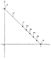
上記座標系については、任意にこれを設定することが可能である。その一例として、図2に示すような、2つの存在率c1及びc2を基底とする2次元座標系を適用できる。この座標系は、第1の基準物質としての造影剤の存在率c1を縦軸とし、第2の基準物質としての水の存在率c2を横軸としたものである。 This can be arbitrarily set for the coordinate system. As an example, a two-dimensional coordinate system based on two existence rates c 1 and c 2 as shown in FIG. 2 can be applied. This coordinate system, the existence ratio c 1 of the contrast medium as a first reference material on the vertical axis, in which the existence ratio c 2 of water as a second reference material was the horizontal axis.
この座標系における座標については、縦軸の座標、横軸の座標の順で記載する。すなわち、この座標系における座標は(c1,c2)と記載される。縦軸における基準の座標P(1,0)、つまり縦軸方向における基底として、造影剤の存在率c1が100%に相当するベクトルを適用する。また、横軸における基準の座標Q(0,1)、つまり横軸方向における基底として、水の存在率c2が100%に相当するベクトルを適用する。 The coordinates in this coordinate system are described in the order of the coordinate on the vertical axis and the coordinate on the horizontal axis. That is, the coordinates in this coordinate system are described as (c 1 , c 2 ). As a reference coordinate P (1, 0) on the vertical axis, that is, a base in the vertical axis direction, a vector corresponding to a contrast medium existence rate c 1 of 100% is applied. In addition, as a reference coordinate Q (0, 1) on the horizontal axis, that is, a base in the horizontal axis direction, a vector corresponding to 100% water abundance c 2 is applied.
物質特定部43は、画像形成部42により得られた線形結合画像における基準物質画像の組(c1,c2)を、この座標系の座標として扱う。
The
なお、本例では、基準物質画像の組と座標とが同じ表現となるように座標系(基底)を設定しているが、これに限定されるものではない。たとえば、c1やc2に係数を掛けることにより、座標軸が伸縮された座標系を適用することができる。また、本例では、縦軸にc1を割り当て、横軸にc2を割り当てているが、これを逆にしてもよい。更に、本例のような直交座標系に代えて、斜交座標系等の他の座標系を用いることも可能である。すなわち、この実施形態における座標系は、線形結合画像における2つの基準物質画像の相関を表すことができるものであれば十分であり、その具体的な態様は任意である。 In this example, the coordinate system (base) is set so that the set of the reference substance image and the coordinates are the same, but the present invention is not limited to this. For example, a coordinate system in which the coordinate axes are expanded and contracted can be applied by multiplying c 1 and c 2 by a coefficient. Further, in this example, assigned a c 1 on the vertical axis, but are assigned to c 2 on the horizontal axis may be the other way round. Furthermore, instead of the orthogonal coordinate system as in this example, other coordinate systems such as an oblique coordinate system can be used. That is, the coordinate system in this embodiment is sufficient as long as it can represent the correlation between two reference substance images in a linear combination image, and the specific mode is arbitrary.
物質特定部43による処理に供される上記「複数の物質」は、任意の物質でよい。また、その個数も任意である。図2に示す例では、造影剤と水が複数の物質に相当する。本例では、「複数の物質」と、基底の生成に用いられる物質とが同じであるが、これには限定されない。その一例として、造影剤と水の2つの物質を「基準物質」とし、造影剤、水、炭酸カルシウム、脂肪及び尿酸の5つの物質を「複数の物質」として適用する場合を後述する。
The “plurality of substances” used for processing by the
複数の物質に対応する座標の取得方法についても任意である。その例として、その物質の測定を実際に行って座標を求める方法や、他の物質の線減弱係数をも考慮して座標を算出する方法などがある。 A method for acquiring coordinates corresponding to a plurality of substances is also arbitrary. For example, there are a method of actually measuring the substance and obtaining coordinates, and a method of calculating coordinates taking into account the linear attenuation coefficient of other substances.
前者の方法は、たとえば、その物質をX線でスキャンして投影データを生成し、これに基づく画素毎の線減弱係数を線形結合として表現し、その係数の組に対応する座標を決定することにより行われる。 In the former method, for example, the material is scanned with X-rays to generate projection data, and a linear attenuation coefficient for each pixel based on the material is expressed as a linear combination, and coordinates corresponding to the set of coefficients are determined. Is done.
後者の方法について説明する。各物質の線減弱係数は既知であるから、異なる2つのエネルギーに対応する線減弱係数を上記の式(3)にそれぞれ代入することで、次の連立方程式(4a)、(4b)が得られる。 The latter method will be described. Since the linear attenuation coefficient of each substance is known, the following simultaneous equations (4a) and (4b) are obtained by substituting the linear attenuation coefficients corresponding to two different energies into the above equation (3), respectively. .
ただし:
ELow、EHighは、2種類のX線エネルギー;
μ(ELow)は、低い側のエネルギーELowにおける当該物質の線減弱係数;
μ(EHigh)は、高い側のエネルギーEHighにおける当該物質の線減弱係数;
をそれぞれ示す。
However:
E Low and E High are two types of X-ray energies;
μ (E Low ) is the linear attenuation coefficient of the material at lower energy E Low ;
μ (E High ) is the linear attenuation coefficient of the substance at the higher energy E High ;
Respectively.
この連立方程式(4a)、(4b)において未知数はc1とc2の2つであるから、これを解くことにより、係数の組c1、c2を算出できる。そして、この係数の組に基づいて目的の座標が得られる。 In the simultaneous equations (4a) and (4b), there are two unknowns c 1 and c 2 , and by solving this, a set of coefficients c 1 and c 2 can be calculated. The target coordinates are obtained based on the set of coefficients.
物質特定部43は、このようにして取得された複数の物質についての複数の座標と、対象物質について物質特定部43により決定された座標とに基づいて、この対象物質の種別を特定する。この処理の一例として、物質特定部43は、(1)当該複数の座標に基づく座標系内の領域を求め、(2)この領域に対する対象物質の座標の位置に基づいて、対象物質の種別を特定する。以下、座標系内の領域として、複数の座標を結ぶグラフを用いる場合を説明する。ここで、特に必要な場合を除き、(1)で得られる領域(グラフ等)を実際に表示させる必要はない。以下の説明では、実施形態の理解を支援するために、表示されたグラフを用いているに過ぎない。それ以降に説明される座標系内の領域についても同様である。なお、座標系内の領域は当該座標系における座標の集合に相当し、グラフはこの領域自体やその外周線に位置する座標の集合に相当する。
The
座標系内の領域としてのグラフを求める処理について説明する。図2に示す例では、上記複数の座標として、造影剤の座標と水の座標が適用されている。物質特定部43は、目的のグラフとして、これら座標を結ぶ線分L1を求める。
Processing for obtaining a graph as an area in the coordinate system will be described. In the example shown in FIG. 2, the coordinates of the contrast agent and the coordinates of water are applied as the plurality of coordinates. The
一般に、線分の式は、2つの座標に基づいてこれらを通る直線の式(つまり傾きと切片)を算出し、この直線のうち当該2つの座標を両端とする部分を抽出することにより得られる。3つ以上の座標を考慮して2つ以上の線分を求める場合には、任意の2つの座標の組み合わせに対して同様の演算を行えばよい。 In general, a line segment formula is obtained by calculating a formula of a straight line that passes through these two coordinates (that is, an inclination and an intercept), and extracting a portion of the straight line having the two coordinates at both ends. . When two or more line segments are obtained in consideration of three or more coordinates, the same calculation may be performed on any combination of two coordinates.
さて、線分L1上の座標は、造影剤のヨウ素濃度に対応している。たとえば、線分L1上の座標P1、P2、P3、P4、P5は、ヨウ素濃度25[mgI/ml]、20[mgI/ml]、15[mgI/ml]、10[mgI/ml]、5[mgI/ml]にそれぞれ対応している。また、前述のように、座標Pは50[mgI/ml]に対応し、座標Qは0[mgI/ml](単なる水)に対応している。つまり、線分L1上において座標Pに近づくとヨウ素濃度が増加し、座標Qに近づくとヨウ素濃度が減少する。 Now, the coordinates on the line segment L1 correspond to the iodine concentration of the contrast agent. For example, coordinates P1, P2, P3, P4, and P5 on the line segment L1 have iodine concentrations of 25 [mgI / ml], 20 [mgI / ml], 15 [mgI / ml], 10 [mgI / ml], 5 Each corresponds to [mgI / ml]. As described above, the coordinate P corresponds to 50 [mgI / ml], and the coordinate Q corresponds to 0 [mgI / ml] (simple water). That is, the iodine concentration increases as it approaches the coordinate P on the line segment L1, and the iodine concentration decreases as it approaches the coordinate Q.
このように、複数の物質の1つとして水を採用することで、物質の濃度をグラフとして表すことができる。たとえば造影剤、水、炭酸カルシウム、脂肪及び尿酸の5つの物質が採用される場合におけるグラフの例を図3に示す。 Thus, the density | concentration of a substance can be represented as a graph by employ | adopting water as one of several substances. For example, FIG. 3 shows an example of a graph in the case where five substances of contrast medium, water, calcium carbonate, fat and uric acid are employed.
図3には、造影剤の座標Pと、水の座標Qと、炭酸カルシウムの座標Rと、脂肪の座標Sと、尿酸の座標Tが示されている。また、座標Pと座標Qとを結ぶ線分L1は造影剤のヨウ素濃度に相当し、座標Rと座標Qとを結ぶ線分L2は炭酸カルシウムの濃度に相当し、座標Sと座標Qとを結ぶ線分L3は脂肪の濃度に相当し、座標Tと座標Qとを結ぶ線分L4は尿酸の濃度に相当する。各線分L1〜L4において、座標Qに近づくほど濃度が低くなり、座標Qから遠ざかるほど濃度が高くなる。 FIG. 3 shows coordinates P of the contrast agent, coordinates Q of water, coordinates R of calcium carbonate, coordinates S of fat, and coordinates T of uric acid. A line segment L1 connecting the coordinates P and Q corresponds to the iodine concentration of the contrast agent, and a line segment L2 connecting the coordinates R and Q corresponds to the calcium carbonate concentration. The connecting line segment L3 corresponds to the fat concentration, and the connecting line segment L4 connecting the coordinates T and Q corresponds to the uric acid concentration. In each of the line segments L1 to L4, the density decreases as it approaches the coordinate Q, and the density increases as the distance from the coordinate Q increases.
また、図3から分かるように、4つの線分L1〜L4は、互いに座標Qでのみ交差している。これは、濃度0(単なる水)に相当する座標Qを除いて、線分L1〜L4が互いに分離されていること、つまり4つの物質(造影剤、炭酸カルシウム、脂肪及び尿酸)に対応する座標が当該座標系において互いに分離されていることを示している。換言すると、当該座標系における線分の位置が異なれば、それに対応する物質も異なるということになる。 As can be seen from FIG. 3, the four line segments L1 to L4 intersect each other only at the coordinate Q. This is because the line segments L1 to L4 are separated from each other except the coordinate Q corresponding to the concentration 0 (simply water), that is, the coordinates corresponding to the four substances (contrast medium, calcium carbonate, fat and uric acid). Are separated from each other in the coordinate system. In other words, if the position of the line segment in the coordinate system is different, the corresponding substance is also different.
このようなグラフを利用することにより、物質特定部43は、物質特定部43により決定された対象物質の座標とグラフとの位置関係に基づいて、この対象物質の種別を特定する。
By using such a graph, the
図3に示す線分L1〜L4をグラフとして利用する場合を例として説明すると、物質特定部43は、まず、対象物質の座標がこれら線分L1〜L4のいずれかの上に位置するか判断する。
The case where the line segments L1 to L4 shown in FIG. 3 are used as a graph will be described as an example. First, the
対象物質の座標が線分Li(i=1〜4のいずれか)上に位置すると判断された場合、物質特定部43は、この線分Liにおける当該座標の位置に基づいて、対象物質の種別を特定する。この種別には、対象物質の物質名だけでなく、その濃度(つまり対象物質と水との成分比)も含まれる。
When it is determined that the coordinates of the target substance are located on the line segment Li (i = 1 to 4), the
なお、物質名だけを特定するように構成することも可能である。その場合には、対象物質の座標が位置している線分Liを特定するだけでよい。 It is also possible to configure to specify only the substance name. In that case, it is only necessary to specify the line segment Li where the coordinates of the target substance are located.
一方、対象物質の座標がどの線分L1〜L4上にも位置しないと判断された場合、物質特定部43は、この対象物質はこれら線分L1〜L4に相当する物質のいずれにも該当しないとの結果を得る。
On the other hand, when it is determined that the coordinates of the target substance are not located on any of the line segments L1 to L4, the
3つ以上の基準物質について座標が得られている場合、座標系内の領域として、これら座標を結んでなる多角形を形成することができる。この多角形により規定される領域は、これら基準物質の混合物に相当する。なお、この混合物とは、各基準物質の成分比を0〜100%とし、全基準物質の成分比の和を100%とした場合に定義される物質を意味する。よって、この混合物には、3つ以上の基準物質のうちの1つ又は2つのみを成分とするものも含まれる。1つの基準物質のみを成分とするものは多角形の頂点に相当し、2つの基準物質のみを成分とするものは多角形の辺に相当する。 When coordinates are obtained for three or more reference materials, a polygon formed by connecting these coordinates can be formed as a region in the coordinate system. The area defined by this polygon corresponds to a mixture of these reference substances. This mixture means a substance defined when the component ratio of each reference substance is 0 to 100% and the sum of the component ratios of all reference substances is 100%. Therefore, this mixture includes those containing only one or two of the three or more reference substances as components. A material having only one reference material as a component corresponds to a vertex of the polygon, and a material having only two reference materials as a component corresponds to a side of the polygon.
3つ以上の基準物質を用いる場合の例として、造影剤と水と炭酸カルシウムの3つを基準物質とする場合について説明する。この場合、図4に示すように、造影剤、水、炭酸カルシウムに相当する座標P、Q、Rを頂点とする多角形(三角形)Aが得られる。多角形Aの辺PQ(線分L1)及びQR(線分L2)に相当する領域は、それぞれ前述のように造影剤の濃度及び炭酸カルシウムの濃度を表す。また、辺PR(線分L5)に相当する領域は、造影剤と炭酸カルシウムとの混合物における造影剤と炭酸カルシウムとの成分比を表す。辺PRにおいて、頂点Pに近づくほど造影剤の成分比が増大し、頂点Rに近づくほど炭酸カルシウムの成分比が増大する。 As an example in the case of using three or more reference substances, a case where three reference materials, contrast medium, water, and calcium carbonate are used as reference substances will be described. In this case, as shown in FIG. 4, a polygon (triangle) A having coordinates P, Q, and R corresponding to the contrast medium, water, and calcium carbonate as vertices is obtained. The regions corresponding to the sides PQ (line segment L1) and QR (line segment L2) of the polygon A represent the contrast agent concentration and the calcium carbonate concentration, respectively, as described above. A region corresponding to the side PR (line segment L5) represents a component ratio of the contrast agent and calcium carbonate in the mixture of the contrast agent and calcium carbonate. In the side PR, the component ratio of the contrast agent increases as it approaches the apex P, and the component ratio of calcium carbonate increases as it approaches the apex R.
また、多角形Aの内部領域、つまり多角形A上の領域から線分L1、L2、L5を除いた領域は、これら3つの基準物質全ての混合物に相当する。この内部領域中の座標についても、頂点Pに近づくほど造影剤の成分比が増大し、頂点Rに近づくほど炭酸カルシウムの成分比が増大する。そして、水に相当する頂点Qに近づくほど当該混合物の濃度が減少する。 Further, the inner area of the polygon A, that is, the area obtained by removing the line segments L1, L2, and L5 from the area on the polygon A corresponds to a mixture of all these three reference substances. Regarding the coordinates in this internal region, the component ratio of the contrast agent increases as it approaches the apex P, and the component ratio of calcium carbonate increases as it approaches the apex R. And the density | concentration of the said mixture reduces, so that the vertex Q equivalent to water is approached.
物質特定部43は、決定された対象物質の座標が多角形上に位置するか否か判断する。当該座標が多角形の外部に位置すると判断された場合、物質特定部43は、この対象物質は当該多角形に対応する混合物ではないと判断する。
The
一方、当該座標が多角形上に位置すると判断された場合、物質特定部43は、この対象物質は当該混合物であると判断する。更に、物質特定部43は、この対象物質の座標の位置に基づいて、この対象物質を構成する3つ以上の基準物質の成分比を求める。
On the other hand, when it is determined that the coordinates are located on the polygon, the
なお、以上の例では、X線CT装置の測定誤差を考慮していないため、各物質に相当する座標が一意に決定されると仮定している。この測定誤差は、装置の機差(公差等)やノイズの混入などに起因する。以下、測定誤差を考慮する場合における処理について説明する。なお、測定誤差が十分に小さい場合などこれを許容できる場合には、上記の処理を適用すればよい。 In the above example, since the measurement error of the X-ray CT apparatus is not considered, it is assumed that the coordinates corresponding to each substance are uniquely determined. This measurement error is caused by machine differences (tolerance, etc.) of the apparatus, noise contamination, and the like. Hereinafter, processing in the case where measurement error is taken into account will be described. If this is acceptable, such as when the measurement error is sufficiently small, the above processing may be applied.
測定誤差を考慮する場合の一例について説明する。まず、様々な物質に対して繰り返し測定を行うことにより、各物質についての座標の分布を取得する。この分布情報は、たとえば、このX線CT装置により得られる投影データに混入するノイズの標準偏差情報である。この分布情報は物質特定部43に記憶される。
An example in which measurement error is taken into account will be described. First, the distribution of coordinates for each substance is obtained by repeatedly measuring various substances. This distribution information is, for example, standard deviation information of noise mixed in projection data obtained by this X-ray CT apparatus. This distribution information is stored in the
造影剤、水及び炭酸カルシウムのそれぞれについての座標の分布の例を図5に示す。造影剤についての座標の分布範囲をPa、水についての座標の分布範囲をQa、炭酸カルシウムについての座標の分布範囲をRaでそれぞれ示す。 An example of the distribution of coordinates for each of the contrast agent, water and calcium carbonate is shown in FIG. The coordinate distribution range for the contrast agent is denoted by Pa, the coordinate distribution range for water is denoted by Qa, and the coordinate distribution range for calcium carbonate is denoted by Ra.
物質特定部43は、標準偏差情報に基づいて、当該物質に対応するグラフを含む2次元領域を求める。この処理の一例を説明する。まず、物質特定部43は、分布範囲Paと分布範囲Qaとを結ぶ2本の線分L1a、L1bを求める。線分L1a、L1bとしては、たとえば、分布範囲Pa、Qaの双方に接し、かつ互いに交差しないものが用いられる。それにより、分布範囲Pa、Qa及び線分L1a、L1bで囲まれた領域Bが得られる。同様にして、分布範囲Ra、Qa及び線分L2a、L2bで囲まれた領域Cが得られる。
The
領域Bは、測定誤差を反映した造影剤の濃度の座標の分布範囲として用いられる。領域Bにおいて線分L1上にない座標には、線分L1に基づいて濃度の値が対応付けられる。この対応付けの例として、線分L1上の各位置において線分L1に直交する直線を求め、この直線上に位置する座標の濃度を等しく設定することができる。測定誤差を反映した炭酸カルシウムの濃度の分布範囲を示す領域Cについても同様に濃度が設定される。 Region B is used as a distribution range of contrast coordinates of the contrast agent reflecting the measurement error. In the region B, coordinates that are not on the line segment L1 are associated with density values based on the line segment L1. As an example of this association, a straight line orthogonal to the line segment L1 can be obtained at each position on the line segment L1, and the density of coordinates positioned on this line can be set equal. The concentration is similarly set for the region C indicating the distribution range of the calcium carbonate concentration reflecting the measurement error.
物質特定部43は、決定された対象物質の座標と、領域B、Cとの位置関係に基づいて、この対象物質の種別を特定する。たとえば、対象物質の座標が領域B上に位置する場合、物質特定部43は、この対象物質は造影剤であると特定し、更に、当該座標と、領域B上の座標に設定された濃度とに基づいて、この対象物質の濃度を特定する。
The
[動作]
この実施形態に係るX線CT装置の動作について説明する。このX線CT装置の動作例を図6に示す。なお、装置各部の動作の詳細については上述したので、ここでは簡単な説明にとどめる。
[Operation]
The operation of the X-ray CT apparatus according to this embodiment will be described. An example of the operation of this X-ray CT apparatus is shown in FIG. Since the details of the operation of each part of the apparatus have been described above, only a brief description will be given here.
(S1)
まず、ガントリ1でデュアル・エナジー・CTによる撮影を行う。前処理部2は、ガントリ1により収集されたデータを投影データに変換する。それにより、エネルギーが異なる第1及び第2の投影データが生成される。第1及び第2の投影データは、データ処理部4に送られる。
(S1)
First, photographing with dual energy CT is performed with the
(S2)
投影データ変換部41は、ステップ1で生成された各投影データに基づく画素毎の線減弱係数を2つの基準物質の線減弱係数の線形結合として表現することにより、2つの基準物質による各投影データの分解を行う。このステップ2により、第1及び第2の投影データが、2つの基準物質に対応する2つの投影データに変換される。
(S2)
The projection
(S3)
更に、画像形成部42は、ステップ2で得られた線形結合を再構成することにより線形結合画像を形成する。これにより、2つの基準物質画像が得られる。
(S3)
Further, the
(S4)
物質特定部43は、ステップ3で得られた2つの基準物質画像の相関を求める。上記の例では、2つの基準物質画像の各画素(x、y)について、所定の2次元座標系における対応する座標(c1(x、y)、c2(x、y))が得られる。
(S4)
The
(S5)
物質特定部43は、ステップ4で得られた相関に基づいて、対象物質を2つの基準物質に分解する。上記の例では、対象物質が造影剤と水などに分解される。
(S5)
The
(S6)
物質特定部43は、ステップ5での分解結果に基づいて対象物質の種別を特定する。この特定結果は、たとえば図示しないディスプレイに表示される。また、この特定結果は、X線CT装置の記憶デバイスやネットワーク上の記憶デバイスに記憶される。
(S6)
The
[作用・効果]
この実施形態に係るX線CT装置の作用と効果について説明する。
[Action / Effect]
The operation and effect of the X-ray CT apparatus according to this embodiment will be described.
このX線CT装置は、ガントリ1及び前処理部2により投影データを生成する。特に、デュアル・エナジー・CT、つまり、エネルギーが異なる第1及び第2のX線でそれぞれ対象物をスキャンすることで、第1及び第2の投影データが生成される。
In the X-ray CT apparatus, projection data is generated by the
更に、このX線CT装置は、投影データ変換部41と、画像形成部42と、物質特定部43とを有する。投影データ変換部41は、第1及び第2の投影データを、あらかじめ決められた2つの基準物質に対応する2つの投影データ(新たな投影データ)に変換する。画像形成部42は、2つの投影データをそれぞれ再構成することで、2つの基準物質に対応する2つの基準物質画像を形成する。物質特定部43は、2つの基準物質画像の相関に基づいて対象物質の種別を特定する。
The X-ray CT apparatus further includes a projection
投影データ変換部41が、上記の変換処理として、第1及び第2の投影データのそれぞれを、2つの基準物質に対応するあらかじめ決められた2つの基準値と、2つの投影データとからなる線形結合として表現するように構成されていてもよい。
As the conversion process, the projection
また、画像形成部42が、この線形結合を再構成することにより、2つの基準値と2つの基準物質画像とからなる線形結合画像を形成するように構成されていてもよい。
Further, the
また、物質特定部43が、対象物質について、2つの基準値に基づきあらかじめ決められた座標系における2つの基準物質画像に対応する座標を決定し、かつ、複数の物質についてあらかじめ得られた座標系における複数の座標と、決定された座標とに基づいて、この対象物質の種別を特定するように構成されていてもよい。
Further, the
なお、第1及び第2のX線のそれぞれのエネルギーについて、2つの基準値は、あらかじめ決められた2つの基準物質の線減弱係数であってよい。更に、座標系は、2つの基準物質の線減弱係数に基づく2つの基底により張られる2次元座標系であってよい。 In addition, about each energy of the 1st and 2nd X-ray | X_line, two reference values may be a linear attenuation coefficient of two reference substances determined beforehand. Further, the coordinate system may be a two-dimensional coordinate system stretched by two bases based on the linear attenuation coefficients of two reference materials.
また、物質特定部43が、複数の座標に基づく座標系内の領域に対する対象物質の座標の位置に基づいて対象物質の種別を特定するように構成されていてもよい。
Further, the
また、複数の物質が第1及び第2の物質を含む場合において、物質特定部43が、座標系における第1の物質の座標と第2の物質の座標とを結ぶ線分を当該座標系内の領域として求め、かつ、対象物質の座標がこの線分上に位置する場合、当該位置に基づいて第1の物質と第2の物質との成分比を求めるように構成されていてもよい。
In addition, when the plurality of substances include the first and second substances, the
また、複数の物質が3つ以上の物質を含む場合において、物質特定部43が、座標系における3つ以上の物質の座標を結ぶ多角形を上記座標系内の領域として求め、対象物質の座標がこの多角形上に位置する場合、当該位置に基づいて3つ以上の物質の成分比を求めるように構成されていてもよい。
In addition, when the plurality of substances include three or more substances, the
ここで、上記の例では、3つ以上の物質の座標を考慮する場合における上記座標系内の領域は、多角形には限定されない。この場合における上記座標系内の領域は、一般に、3つ以上の物質の座標に基づく図形、つまりこれら座標を考慮して形成された任意の図形でよい。たとえば、3つ以上の物質の座標を通過又は内包する図形を上記座標系内の領域として用いることができる。この例では、図形の外周線が直線である必要はなく、また物質の座標が当該図形の外周線上にある必要もない。対象物質の座標が図形上に位置する場合、当該位置に基づいて3つ以上の物質の成分比が算出される。 Here, in the above example, the region in the coordinate system when considering the coordinates of three or more substances is not limited to a polygon. In this case, the region in the coordinate system may generally be a figure based on the coordinates of three or more substances, that is, an arbitrary figure formed in consideration of these coordinates. For example, a figure that passes or includes the coordinates of three or more substances can be used as the region in the coordinate system. In this example, the outer peripheral line of the figure does not need to be a straight line, and the coordinates of the substance do not need to be on the outer peripheral line of the figure. When the coordinates of the target substance are located on the figure, the component ratio of three or more substances is calculated based on the position.
また、複数の物質のうちの1つが水である場合において、物質特定部43が、第1及び第2の対象物質について決定された第1及び第2の座標のそれぞれと、水の座標とを結ぶ2つの線分を座標系内の領域として求め、これら線分の位置関係に基づいて第1の対象物質と第2の対象物質とが同種であるか否か特定するように構成されていてもよい。
In addition, when one of the plurality of substances is water, the
また、物質特定部43が、投影データに混入するノイズの標準偏差情報をあらかじめ記憶し、この標準偏差情報に基づいて座標系内の領域を含む2次元領域を求め、この2次元領域に対する対象物質の座標の位置に基づいてこの対象物質の種別を特定するように構成されていてもよい。
In addition, the
このようなX線CT装置によれば、対象物質の特性を所定の座標系における座標として表現し、この座標と複数の物質についての複数の座標とに基づいて対象物質の種別を特定することができる。したがって、CT値比が近い物質であっても座標を参照することにより高い精度での識別が可能である。 According to such an X-ray CT apparatus, the characteristics of the target substance are expressed as coordinates in a predetermined coordinate system, and the type of the target substance can be specified based on the coordinates and a plurality of coordinates for the plurality of substances. it can. Therefore, even a substance with a close CT value ratio can be identified with high accuracy by referring to the coordinates.
また、上記座標系内の領域を用いる構成を適用することにより、対象物質を構成する物質の成分比を求めることができる。更に、上記座標系内の領域を参照することにより、2つ以上の対象物質がそれぞれ同種であるか異種であるかを判別することができる。 Moreover, the component ratio of the substance which comprises a target substance can be calculated | required by applying the structure which uses the area | region in the said coordinate system. Furthermore, it is possible to determine whether two or more target substances are the same or different from each other by referring to the region in the coordinate system.
また、ノイズの影響を考慮する構成を適用することにより、より高精度、高確度での物質の特定、成分比の特定、対象物質の分離などが可能となる。 In addition, by applying a configuration that takes into account the influence of noise, it is possible to specify a substance with higher accuracy and accuracy, specify a component ratio, separate a target substance, and the like.
〈第2の実施形態〉
この実施形態のX線CT装置の概略構成を図7に示す。このX線CT装置は、第1の実施形態で説明した構成を含んでいる。このX線CT装置は、ガントリ1と、前処理部2と、再構成処理部3と、データ処理部4と、制御部5と、表示部6と、操作部7とを有する。データ処理部4には、投影データ変換部41と、画像形成部42と、物質特定部43とが設けられている。第1の実施形態と同じ構成部分は、特に言及しない限り第1の実施形態と同様の機能を有する。
<Second Embodiment>
A schematic configuration of the X-ray CT apparatus of this embodiment is shown in FIG. This X-ray CT apparatus includes the configuration described in the first embodiment. The X-ray CT apparatus includes a
基準物質の1つは水であるとする。また、物質特定部43は、第1の実施形態で説明したように、対象物質の座標と水の座標とを結ぶ線分を求める(図2等を参照)。この線分は、対象物質と水との混合物における成分比、つまり対象物質の濃度を表すものである。
One of the reference substances is water. Moreover, the substance specific |
データ処理部4は、対象物(被検体等)の撮影領域の各位置について第1の実施形態の処理を適用することにより、撮影領域における対象物質の分布を求める。この分布情報は再構成処理部3に入力される。なお、分布情報には、撮影領域における対象物質の存在位置を示す情報と、濃度を示す情報とが含まれている。
The data processing unit 4 obtains the distribution of the target substance in the imaging region by applying the process of the first embodiment to each position of the imaging region of the object (subject, etc.). This distribution information is input to the
分布情報と投影データを受けた再構成処理部3は、投影データにおける対象物質の分布領域に対して再構成処理を施すことで、対象物質の分布(たとえば濃度の分布)を表す分布画像を形成する。この処理の一例として、再構成処理部3は、分布情報に示す対象物質の存在位置に基づいて、この存在位置に対応する画素を特定する。そして、再構成処理部3は、投影データに基づいて、特定された画素についてのみ画像を再構成する。他の処理として、再構成処理部3は、投影データに基づいて通常の再構成処理を行い、それにより得られた画像のうち上記存在位置に対応する画素のみを抽出する。これら手法により得られる再構成画像が上記の分布画像である。
Upon receiving the distribution information and the projection data, the
制御部5は、対象物質の濃度を表す線分上における位置の指定を受けて、この指定位置に応じた態様で分布画像を表示部6に表示させる。この処理について以下に説明する。
The
このX線CT装置には、対象物質の濃度を変更するためのユーザインターフェイスが設けられている。このユーザインターフェイスは、ソフトウェアに基づいて表示部6に表示されるものであってもよいし、ハードウェアであってもよい。ハードウェアとしてはダイヤルやスライドレバーなどがある。ソフトウェアを用いる場合、たとえば、ダイヤルやスライドレバーを模した画像を表示部6に表示させ、これを操作部7で操作するように構成できる。
The X-ray CT apparatus is provided with a user interface for changing the concentration of the target substance. This user interface may be displayed on the
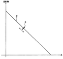
ソフトウェアを用いる場合の具体例として、制御部5は、図2に示すグラフに基づいて、図8に示す画像を表示部6に表示させる。この画像は、図2に示す座標系と、線分画像Dと、スライダEとを含む。座標系の座標軸には、物質名を示す文字列「造影剤」、「水」が付されている。線分画像Dは、線分L1を示している。スライダEは、線分画像D上を移動可能とされている(図中の両側矢印を参照)。スライダEは、たとえば操作部7のマウスによるドラッグ操作によって移動される。
As a specific example in the case of using software, the
ユーザは、スライダEを所望の位置に移動させることにより、造影剤の濃度の指定を行う。より具体的に説明すると、線分画像Dと線分L1とはあらかじめ対応付けられており、制御部5は、スライダEの位置に対応する線分L1の位置、つまり造影剤の濃度が指定される。制御部5は、指定された濃度に応じて分布画像の表示態様を変更する。表示態様の変更処理としては、画素値(輝度、色等)を変更するものや、模様を変更するものなどがある。
The user designates the contrast medium concentration by moving the slider E to a desired position. More specifically, the line segment image D and the line segment L1 are associated in advance, and the
分布画像が複数存在する場合には、各分布画像について濃度の指定を行うことが可能である。その場合、ユーザは、たとえばポインティングデバイスを用いて、所望の分布画像を指定する。制御部5は、指定された分布画像における所定の濃度に対応する位置にスライダEを表示させる。ユーザは、スライダEを所望の位置に移動させる。制御部5は、移動後のスライダEの位置に応じて当該分布画像の表示態様を変更する。
When there are a plurality of distribution images, it is possible to specify the density for each distribution image. In that case, the user designates a desired distribution image using, for example, a pointing device. The
濃度の変更処理として、数値を入力する方法を採用することも可能である。その場合、制御部5は、対象物質の濃度情報を入力するための入力領域を表示部6に表示させる。ユーザは、操作部7(たとえばキーボード)を用いて入力領域に所望の濃度の値を入力する。入力領域は、たとえばプルダウンメニューのように、濃度の複数の選択肢から所望のものを選択可能とするオブジェクトであってもよい。
It is also possible to employ a method of inputting a numerical value as the density changing process. In that case, the
初期値よりも濃度を高めた場合の分布画像を強調画像と呼ぶことがある。また、初期値よりも濃度を低下させた場合の分布画像を抑制画像と呼ぶことがある。 A distribution image in which the density is higher than the initial value may be referred to as an emphasized image. In addition, the distribution image when the density is lowered from the initial value may be referred to as a suppression image.
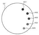
強調画像及び抑制画像の例を説明する。図9Aは、60keVの単色X線画像としての分布画像(原画像)G0を示す。なお、図9A(画像を示す以下の図も同様)においては、実際の画像と表示輝度が逆転されている。つまり、実際の画像では対象物質の濃度が高いほど表示輝度が高くなっているが、図9Aでは濃度が高いほど表示輝度が低くなっている。 An example of an enhanced image and a suppressed image will be described. FIG. 9A shows a distribution image (original image) G0 as a monochromatic X-ray image of 60 keV. In FIG. 9A (the same applies to the following drawings showing images), the actual image and the display luminance are reversed. That is, in the actual image, the higher the concentration of the target substance, the higher the display luminance. In FIG. 9A, the higher the concentration, the lower the display luminance.
単色X線画像は次式(5)で定義される。 A monochromatic X-ray image is defined by the following equation (5).
ただし:
CT numberは、CT値;
μは、式(5)に示す対象物質の線減弱係数;
μwaterは、水の線減弱係数;
をそれぞれ示す。
However:
CT number is the CT value;
μ is the linear attenuation coefficient of the target substance shown in Formula (5);
μ water is the linear attenuation coefficient of water;
Respectively.
原画像G0には、様々な濃度の造影剤の分布画像H01、H02、H03、H04、H05が描写されている。分布画像H01、H02、H03、H04、H05は、それぞれ、濃度25[mgI/ml]、20[mgI/ml]、15[mgI/ml]、10[mgI/ml]、5[mgI/ml]に相当する。 In the original image G0, distribution images H01, H02, H03, H04, and H05 of contrast agents having various concentrations are depicted. Distribution images H01, H02, H03, H04, and H05 have concentrations of 25 [mgI / ml], 20 [mgI / ml], 15 [mgI / ml], 10 [mgI / ml], and 5 [mgI / ml], respectively. It corresponds to.
図9Bは、原画像G0に基づく強調画像G1を示す。強調画像G1には、原画像G0中の分布画像H01、H02、H03、H04、H05にそれぞれ対応する分布画像H11、H12、H13、H14、H15が描写されている。各分布画像H1i(i=1〜5)は、分布画像H0iに示す濃度を高めた状態を示している。 FIG. 9B shows an enhanced image G1 based on the original image G0. In the emphasized image G1, distribution images H11, H12, H13, H14, and H15 respectively corresponding to the distribution images H01, H02, H03, H04, and H05 in the original image G0 are depicted. Each distribution image H1i (i = 1 to 5) shows a state in which the density shown in the distribution image H0i is increased.
図9Cは、原画像G0に基づく抑制画像G2を示す。抑制画像G2には、原画像G0中の分布画像H01、H02、H03、H04、H05にそれぞれ対応する分布画像H21、H22、H23、H24、H25が描写されている。各分布画像H2i(i=1〜5)は、分布画像H0iに示す濃度を低下させた状態を示している。 FIG. 9C shows a suppression image G2 based on the original image G0. In the suppression image G2, distribution images H21, H22, H23, H24, and H25 respectively corresponding to the distribution images H01, H02, H03, H04, and H05 in the original image G0 are depicted. Each distribution image H2i (i = 1 to 5) shows a state in which the density shown in the distribution image H0i is lowered.
このような強調画像G1及び抑制画像G2の生成方法を説明する。まず、制御部5は、造影剤の濃度を示す線分L1(図2参照)上に座標が位置する画素を特定する。次に、制御部5は、特定された各画素について、式(3)に示す造影剤の存在率c1を1000に設定し、水の存在率c2を0に設定し、これらを式(2a)、(2b)に代入することにより、強調画像G1を生成する。また、制御部5は、造影剤の存在率c1を0に設定し、水の存在率c2を1000に設定し、これらを式(2a)、(2b)に代入することにより、抑制画像G2を生成する。制御部5は、生成された強調画像G1や抑制画像G2を表示部6に表示させる。
A method for generating such an enhanced image G1 and a suppressed image G2 will be described. First, the
次に、濃度の算出方法の例を説明する。前述のように、座標系中の線分は、物質と水との混合物を表しており、線分上の位置が濃度に相当している。図2に示す場合において、造影剤の濃度を示す線分L1は、傾きが−1、切片が1の直線上にあるので、造影剤の濃度は次式で算出することができる。 Next, an example of a density calculation method will be described. As described above, the line segment in the coordinate system represents a mixture of a substance and water, and the position on the line segment corresponds to the concentration. In the case shown in FIG. 2, since the line segment L1 indicating the concentration of the contrast medium is on a straight line having an inclination of −1 and an intercept of 1, the contrast medium concentration can be calculated by the following equation.
図10は、造影剤の濃度を示す線分L1上に座標が位置する係数c1(x,y)、c2(x,y)を抽出し、式(6)を用いて算出された濃度をカラー表示させた場合の表示態様の一例を模式的に表している。分布画像Gには5つの分布画像H1、H2、H3、H4、H5が描写されている。各分布画像Hi(i=1〜5)は、その濃度に応じた色で表示される。濃度と表示色との関係はカラーバーJにより示されている。なお、複数の物質の濃度をカラー表示させる場合には、物質毎に異なる色で分布画像を表示させるように構成することも可能である。 FIG. 10 shows the density calculated using the equation (6) by extracting the coefficients c 1 (x, y) and c 2 (x, y) whose coordinates are located on the line segment L1 indicating the density of the contrast agent. An example of a display mode in the case of displaying in color is schematically shown. In the distribution image G, five distribution images H1, H2, H3, H4, and H5 are depicted. Each distribution image Hi (i = 1 to 5) is displayed in a color corresponding to its density. The relationship between the density and the display color is indicated by a color bar J. In addition, when displaying the density | concentration of a several substance in color, it is also possible to comprise so that a distribution image may be displayed with a different color for every substance.
この実施形態のX線CT装置の作用及び効果を説明する。 The operation and effect of the X-ray CT apparatus of this embodiment will be described.
この実施形態のX線CT装置は、第1の実施形態の作用に加えて、次に示す作用を有する。なお、物質の1つは溶剤(たとえば水)である。物質特定部43は、対象物質の座標と溶剤の座標とを結ぶ線分を求める。再構成処理部3は、投影データにおける対象物質の分布領域に対して再構成処理を施すことにより、対象物質の分布を表す分布画像を形成する。制御部5(表示画像生成部)は、線分上における位置の指定を受けて、当該指定位置に応じた態様の分布画像(表示画像)を生成して表示部6に表示させる。
The X-ray CT apparatus of this embodiment has the following operation in addition to the operation of the first embodiment. One of the substances is a solvent (for example, water). The
また、制御部5により線分を表す線分画像が表示部6に表示され、更に、この線分画像上の位置が操作部7を用いて指定されたことを受けて、制御部5が、当該指定位置に対応する線分上の位置を特定し、当該特定位置に応じた態様で分布画像を表示させるように構成することができる。
In addition, a line segment image representing a line segment is displayed on the
また、物質の濃度情報を入力するための入力領域が表示部6に表示され、更に、操作部7を用いて対象物質の濃度情報が入力領域に入力されたことを受けて、制御部5(濃度算出部、表示画像生成部)が、当該濃度情報に対応する線分上の位置を特定し、当該特定位置に応じた態様の分布画像を生成して表示させるように構成することができる。
In addition, an input area for inputting the concentration information of the substance is displayed on the
また、制御部5が、線分上の指定位置に応じて分布画像の表示色を変更するように構成することが可能である。また、制御部5が、物質特定部43により特定された対象物質の種別に応じて分布画像の表示色を変更するように構成することが可能である。更に、制御部5が、対象物質の種別と線分上の指定位置の双方に応じて分布画像の表示色を変更するように構成することも可能である。
Further, the
このようなX線CT装置によれば、第1の実施形態の効果に加えて、強調画像や抑制画像を形成し表示することができる。また、物質の濃度を視覚的に表現することができるので、濃度の直感的な理解が可能となる。 According to such an X-ray CT apparatus, in addition to the effects of the first embodiment, an enhanced image and a suppressed image can be formed and displayed. In addition, since the concentration of the substance can be expressed visually, an intuitive understanding of the concentration becomes possible.
なお、上記の例では、対象物質の座標と溶剤の座標とを結ぶ線分を求めて対象物質の分布画像を形成する場合について説明したが、溶剤以外の物質を適用して同様の処理を行うことも可能である。たとえば、前述の「複数の物質」のうちの一の物質の座標と対象物質の座標とを結ぶ線分を求め、更に、投影データにおける対象物質の分布領域に対して再構成処理を施すことにより分布画像を形成する。この分布画像は対象物質及び一の物質の分布を表すものである。つまり、2つの物質の分布を表現する単一の分布画像が得られる。 In the above example, a case has been described in which a line segment connecting the coordinates of the target substance and the coordinates of the solvent is obtained to form a distribution image of the target substance, but the same processing is performed by applying a substance other than the solvent. It is also possible. For example, a line segment connecting the coordinates of one substance of the “plural substances” and the coordinates of the target substance is obtained, and further, a reconstruction process is performed on the distribution area of the target substance in the projection data. A distribution image is formed. This distribution image represents the distribution of the target substance and one substance. That is, a single distribution image expressing the distribution of two substances is obtained.
〈第3の実施形態〉
第1の実施形態で説明した方法では、第1の物質の座標と水の座標とを結ぶ第1の線分と、第2の物質の座標と水の座標とを結ぶ第2の線分とが近い場合、これら物質を判別することは困難である。一方、非特許文献1に記載された方法では、第1の物質のCT値と第2の物質のCT値とが近い場合、これら物質を判別することは困難である。
<Third Embodiment>
In the method described in the first embodiment, the first line connecting the coordinates of the first substance and the coordinates of water, the second line connecting the coordinates of the second substance and the coordinates of water, When these are close, it is difficult to distinguish these substances. On the other hand, in the method described in
そこで、この実施形態では、これら2つの方法を併用することにより、より確実な物質の判別を可能とする技術を提供する。また、この実施形態では、特許文献1に記載された技術を利用して、実効原子番号画像、密度画像及び単色X線画像を形成する処理についても説明する。
Therefore, in this embodiment, a technique is provided that enables more reliable substance discrimination by using these two methods in combination. In this embodiment, processing for forming an effective atomic number image, a density image, and a monochromatic X-ray image using the technique described in
この実施形態のX線CT装置の概略構成を図11に示す。このX線CT装置は、第1の実施形態で説明した構成を含んでいる。また、図示は省略するが、このX線CT装置は、第2の実施形態で説明した構成を更に含んでいてもよい。 FIG. 11 shows a schematic configuration of the X-ray CT apparatus of this embodiment. This X-ray CT apparatus includes the configuration described in the first embodiment. Although not shown, the X-ray CT apparatus may further include the configuration described in the second embodiment.
このX線CT装置は、ガントリ1と、前処理部2と、再構成処理部3と、データ処理部4と、画素値比算出部8と、物質特定部9と、特定画像形成部10と、特定制御部11とを有する。データ処理部4には、投影データ変換部41と、画像形成部42と、物質特定部43とが設けられている。なお、物質特定部9と特定制御部11はそれぞれ「特定部」の一例として機能する。
The X-ray CT apparatus includes a
第1の実施形態と同じ構成部分は、特に言及しない限り第1の実施形態と同様の機能を有する。なお、第1の実施形態で説明した物質特定処理はデータ処理部4が実行し、非特許文献1に記載された物質特定処理は画素値比算出部8及び物質特定部9が実行する。
The same components as those in the first embodiment have the same functions as those in the first embodiment unless otherwise specified. The substance specifying process described in the first embodiment is executed by the data processing unit 4, and the substance specifying process described in
再構成処理部3は、デュアル・エナジー・CTにより得られた第1及び第2の投影データのそれぞれに対して再構成処理を施すことにより第1及び第2の画像を形成する。
The
画素値比算出部8は、非特許文献1に記載されているように、第1及び第2の画像における画素値(CT値)の比を算出する。この処理は、第1の画像と第2の画像との間で画素の対応付けを行い、対応付けられた2つの画素のCT値の比を算出するものである。
As described in
物質特定部9は、非特許文献1に記載された方法を用いることで、画素値比算出部8により算出されたCT値比に基づいて、対象物質の種別を特定する。
The
以下、具体例として、尿酸、軟骨及び軟組織が混在する状況において尿酸を特定する場合を説明する。これら物質について得られたCT値比の分布の例を図12に示す。本例では、管電圧80kV及び135kVでデュアル・エナジー・CTによる撮影を行うものとする。図12では、管電圧80kVでのCT値をx軸に、管電圧135kVでのCT値をy軸にとっている。したがって、原点と任意の座標とを通過する直線の傾きがCT値比に相当する。 Hereinafter, as a specific example, a case where uric acid is specified in a situation where uric acid, cartilage, and soft tissue coexist will be described. An example of the distribution of CT value ratios obtained for these substances is shown in FIG. In this example, it is assumed that imaging by dual energy CT is performed at tube voltages of 80 kV and 135 kV. In FIG. 12, the CT value at the tube voltage of 80 kV is on the x axis, and the CT value at the tube voltage of 135 kV is on the y axis. Therefore, the slope of a straight line passing through the origin and arbitrary coordinates corresponds to the CT value ratio.
分布領域K1、K2、K3は、それぞれ尿酸、軟骨、軟組織についての分布を示している。分布領域K1、K2、K3の位置関係から分かるように、尿酸と軟骨とはCT値比が異なっており互いの判別が可能であるが、尿酸と軟組織ではCT値比が近いため判別が困難である。なお、直線M1、M2は、それぞれ尿酸のCT値比の最大値と最小値を表している。 Distribution regions K1, K2, and K3 indicate distributions for uric acid, cartilage, and soft tissue, respectively. As can be seen from the positional relationship between the distribution regions K1, K2, and K3, uric acid and cartilage have different CT value ratios and can be distinguished from each other, but uric acid and soft tissue are difficult to distinguish because the CT value ratio is close. is there. The straight lines M1 and M2 represent the maximum value and the minimum value of the CT value ratio of uric acid, respectively.
一方、図13は、第1の実施形態で説明した方法で得られた、尿酸、軟骨及び軟組織の座標の分布の例を示している。なお、縦軸及び横軸は、それぞれ造影剤及び水に基づき設定されている。分布領域N1、N2、N3は、それぞれ尿酸、軟骨、軟組織についての分布を示している。直線L6は、尿酸の座標の分布状態を示す直線である。また、直線L6a、L6bは、それぞれ、尿酸の分布領域の広がりに応じた直線の分布範囲の上限及び下限を示している(第1の実施形態を参照)。軟骨及び軟組織については直線の図示を省略しているが、その形態から直線の向きは容易に把握できる。分布領域N1、N2、N3の位置から分かるように、本例では、尿酸と軟骨では近い直線が得られるので物質相互の判別が困難であり、尿酸と軟組織では直線の向きが十分異なるので判別が可能である。 On the other hand, FIG. 13 shows an example of the distribution of coordinates of uric acid, cartilage, and soft tissue obtained by the method described in the first embodiment. The vertical axis and the horizontal axis are set based on the contrast medium and water, respectively. Distribution regions N1, N2, and N3 indicate distributions for uric acid, cartilage, and soft tissue, respectively. The straight line L6 is a straight line showing the distribution state of the coordinates of uric acid. The straight lines L6a and L6b indicate the upper and lower limits of the linear distribution range corresponding to the spread of the uric acid distribution region, respectively (see the first embodiment). Although illustration of straight lines is omitted for cartilage and soft tissue, the direction of the straight lines can be easily grasped from the form. As can be seen from the positions of the distribution regions N1, N2, and N3, in this example, since a straight line is obtained with uric acid and cartilage, it is difficult to distinguish between substances, and the direction of the straight line is sufficiently different between uric acid and soft tissue. Is possible.
以上の考察の下、第1の実施形態で説明した方法と非特許文献1に記載された方法とを併用する処理の一例を説明する(図14を参照)。まず、第1の実施形態と同様に、デュアル・エナジー・CTにより投影データを取得する(S21)。
Under the above consideration, an example of a process that uses the method described in the first embodiment and the method described in
再構成処理部3,画素値比算出部8及び物質特定部9は、前述の要領で、図12に示すCT値比の分布情報、つまり分布領域K1、K2、K3を得る(S22)。また、データ処理部4は、前述の要領で、図13に示す座標の分布情報、つまり分布領域N1、N2、N3を得る(S23)。これら分布情報は特定制御部11に送られる。以下、CT値比の分布情報を第1の分布情報と呼び、座標の分布情報を第2の分布情報と呼ぶことがある。
The
特定制御部11は、第1及び第2の分布情報に基づいて3つの物質をそれぞれ特定する。その処理の例として、特定制御部11は、まず、図12に示す直線M1、M2と、図13に示す直線L6a、L6bを求める。なお、直線M1、M2や直線L6a、L6bは、今回の測定により得られたデータから作成したものであっても、事前に得られた測定データや規定の値(理論値や標準的な値等)であってもよい。また、特定制御部11は、各物質について、そのCT値比の分布領域に基づく直線や、その座標の分布領域に基づく直線を求めてもよい。このようにして、直線M1、M2により挟まれた特定領域と、直線L6a、L6bにより挟まれた特定領域とが得られる(S24、S25)。
The
続いて、特定制御部11は、軟骨及び軟組織のそれぞれのCT値比の分布領域K2、K3が、直線M1、M2により挟まれた特定領域に含まれているか判断する(S26)。この処理は、分布領域K2、K3自体が当該特定領域に含まれているか判断するものでもよいし、上記で直線を求めた場合にはその直線が当該特定領域に含まれているか判断するものでもよい。
Subsequently, the
なお、前者の場合、分布領域K2、K3の全体が当該特定領域に含まれているか判断するようにしてもよいし、分布領域K2、K3の所定割合が当該特定領域に含まれているか判断するようにしてもよい。 In the former case, it may be determined whether the entire distribution areas K2, K3 are included in the specific area, or whether a predetermined ratio of the distribution areas K2, K3 is included in the specific area. You may do it.
また、後者の場合、直線の長さによっては少しの傾きの違いでも直線が当該特定領域からはみ出すことが考えられる。その場合、直線の長さの範囲をあらかじめ決めておき、その範囲内において直線が当該特定領域に含まれているときに、この直線は当該特定領域に含まれていると判断するようにしてもよい。 In the latter case, depending on the length of the straight line, it is conceivable that the straight line protrudes from the specific area even with a slight difference in inclination. In that case, a range of the length of the straight line is determined in advance, and when the straight line is included in the specific region within the range, it may be determined that the straight line is included in the specific region. Good.
続いて、特定制御部11は、軟骨及び軟組織のそれぞれの座標の分布領域N2、N3が、直線L6a、L6bにより挟まれた特定領域に含まれているか判断する(S27)。この処理は、CT値比の場合と同様に実行できる。
Subsequently, the
次に、特定制御部11は、CT値比の分布領域K2、K3が特定領域に含まれているかの判断結果と、座標の分布領域N2、N3が特定領域に含まれているか判断結果とに基づいて、尿酸、軟骨及び軟組織の判別を行う。
Next, the
その具体例として、特定制御部11は、CT値比の分布領域が特定領域に含まれており(S28:Yes)、かつ、座標の分布領域が特定領域に含まれている(S29:Yes)と判断された場合、これら分布領域に対応する物質は尿酸であると判断する(S30)。
As a specific example, the
また、特定制御部11は、CT値比の分布領域が特定領域に含まれており(S28:Yes)、かつ、座標の分布領域が特定領域に含まれていない(S29:No)と判断された場合、これら分布領域に対応する物質は軟組織であると判断する(S31)。
Further, the
また、特定制御部11は、CT値比の分布領域が特定領域に含まれておらず(S28:No)、かつ、座標の分布領域が特定領域に含まれている(S32:Yes)と判断された場合、これら分布領域に対応する物質は軟骨であると判断する(S33)。
Further, the
また、特定制御部11は、CT値比の分布領域が特定領域に含まれておらず(S28:No)、かつ、座標の分布領域が特定領域に含まれていない(S32:No)と判断された場合、これら分布領域に対応する物質は尿酸でも軟骨でも軟組織でもないと判断する。以上で、この処理例の説明は終了となる。
Further, the
次に、特定画像形成部10について説明する。特定画像形成部10は、実効原子番号画像、密度画像、及び単色X線画像のうちの少なくとも1つ(特定画像と呼ぶことがある)を形成する。この処理は、第1の実施形態で説明した線形結合(式(3))における係数c1及びc2に基づいて形成される、各基準物質の分布を表す画像(基準物質画像)を、特許文献1に記載された方法で組み合わせることにより実行されるものである。
Next, the specific
なお、実効原子番号画像は、対象物における実効原子番号の分布を表す画像である。また、密度画像は、対象物質における物質の密度の分布を表す画像である。また、単色X線画像は、単一のエネルギーを有するX線で対象物質をスキャンした場合を模擬的に再現する画像である。これら画像を形成可能な特定画像形成部10は、「形成部」の一例として機能する。
The effective atomic number image is an image representing the distribution of effective atomic numbers in the object. The density image is an image representing the density distribution of the substance in the target substance. The monochromatic X-ray image is an image that simulates the case where the target substance is scanned with X-rays having a single energy. The specific
この実施形態の物質特定部43は、第1の実施形態で説明した処理で得られた複数の対象物質のそれぞれについての特定結果と、これら対象物質のそれぞれについて特定画像形成部10により形成された実効原子番号画像、密度画像、及び/又は単色X線画像に基づいて、これら対象物質の種別をそれぞれ特定することが可能である。なお、第1の実施形態で説明した処理は、複数の物質についてあらかじめ得られた座標系における複数の座標と、対象物質について物質特定部43により決定された座標とに基づいて、対象物質の種別を特定するものである。
The
この実施形態のX線CT装置によれば、第1の実施形態の効果を得ることができるとともに、第1の実施形態と同様の座標系における各物質の座標の分布状態と、各物質のCT値比の分布状態の双方に基づいて、物質の種別を特定することができる。したがって、対象物に含まれる物質の特定精度の更なる向上を図ることが可能である。 According to the X-ray CT apparatus of this embodiment, the effects of the first embodiment can be obtained, the distribution state of the coordinates of each substance in the coordinate system similar to the first embodiment, and the CT of each substance The type of the substance can be specified based on both the distribution state of the value ratio. Therefore, it is possible to further improve the identification accuracy of the substance contained in the object.
また、この実施形態のX線CT装置によれば、実効原子番号画像、密度画像、及び/又は単色X線画像を形成することができるので、物質の分布状態を視覚的に把握することが可能である。また、座標系を用いた物質特定処理とこれら画像を用いた物質特定処理を併用することにより、対象物に含まれる物質の特定精度の更なる向上を図ることが可能である。 Further, according to the X-ray CT apparatus of this embodiment, an effective atomic number image, a density image, and / or a monochromatic X-ray image can be formed, so that the distribution state of the substance can be visually grasped. It is. Further, by combining the substance specifying process using the coordinate system and the substance specifying process using these images, it is possible to further improve the specifying accuracy of the substance included in the object.
〈変形例〉
上記実施形態の変形例を説明する。
<Modification>
A modification of the above embodiment will be described.
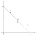
第1の変形例を説明する。対象物質に未知の成分が含まれている場合、図5に示す線分Liのような直線的な相関関係が得られず、領域B等を適当に決定し難いことがある。たとえば図15Aに示すように、3つの対象物質について、3つの座標V1、V2、V3が得られたものとする。これら座標V1、V2、V3は、同一の直線上に位置しない。その場合、この座標系と、座標V1、V2、V3を表示部に表示させる。ユーザは、表示された座標系及び座標V1、V2、V3を参照し、操作部を用いて当該座標系内に領域を任意に設定する。このとき、たとえば図15Bに示すように、座標V1、V2、V3を含むように領域Wが設定される。 A first modification will be described. When an unknown component is included in the target substance, a linear correlation such as the line segment Li shown in FIG. 5 cannot be obtained, and it may be difficult to appropriately determine the region B or the like. For example, as shown in FIG. 15A, it is assumed that three coordinates V1, V2, and V3 are obtained for three target substances. These coordinates V1, V2, and V3 are not located on the same straight line. In this case, this coordinate system and the coordinates V1, V2, and V3 are displayed on the display unit. The user refers to the displayed coordinate system and the coordinates V1, V2, and V3, and arbitrarily sets a region in the coordinate system using the operation unit. At this time, as shown in FIG. 15B, for example, the region W is set so as to include coordinates V1, V2, and V3.
領域Wは、対象物の未知の成分などに起因する誤差を反映した造影剤の濃度の座標の分布範囲として用いられる。領域Wにおいて座標(c1,c2)=(1,0)と座標(c1,c2)=(0,1)とを結ぶ線分(図5の線分L1に相当する)上にない座標に対しては、当該線分に基づいて濃度の値が対応付けられる。この対応付けは、たとえば図5に示す場合と同様にして行われる。物質特定部43は、たとえば図5に示す場合と同様の手法により、決定された対象物質の座標と、領域Wとの位置関係に基づいて、この対象物質の種別を特定する。
The region W is used as a distribution range of the coordinates of the contrast agent concentration reflecting an error caused by an unknown component of the object. On the line segment (corresponding to the line segment L1 in FIG. 5) connecting the coordinates (c 1 , c 2 ) = (1, 0) and the coordinates (c 1 , c 2 ) = (0, 1) in the region W A density value is associated with a non-coordinate based on the line segment. This association is performed, for example, in the same manner as shown in FIG. The
この変形例によれば、対象物質に未知の成分が含まれている等の誤差要因が介在する場合であっても、ユーザの判断によって物質の特定を高い精度で行うことができる。 According to this modification, even when an error factor such as an unknown component is included in the target substance, the substance can be specified with high accuracy based on the judgment of the user.
第2の変形例を説明する。上記実施形態では、エネルギーが異なる2つのX線を用いるデュアル・エナジー・CTの手法が適用されているが、実施形態はこれに限定されるものではない。 A second modification will be described. In the above embodiment, the dual energy CT method using two X-rays having different energies is applied, but the embodiment is not limited to this.
たとえば、エネルギーが異なる3以上のX線を用いてスキャンを行うことが可能なX線CT装置においては、生成部は、エネルギーが異なるN(3以上)のX線でそれぞれ対象物をスキャンしてN個の投影データを生成する。変換部は、これらN個の投影データを、N個の基準物質に対応するN個の新たな投影データに変換する。画像形成部は、変換部で変換したN個の新たな投影データをそれぞれ再構成することで、N個の基準物質に対応するN個の基準物質画像を形成する。特定部は、N個の基準物質画像の画素の値の相関に基づいて対象物質を特定する。 For example, in an X-ray CT apparatus capable of performing scanning using three or more X-rays having different energies, the generation unit scans the object with N (three or more) X-rays having different energies. N pieces of projection data are generated. The conversion unit converts the N projection data into N new projection data corresponding to the N reference substances. The image forming unit reconfigures each of the N new projection data converted by the conversion unit, thereby forming N reference material images corresponding to the N reference materials. The specifying unit specifies the target substance based on the correlation of the pixel values of the N reference substance images.
この場合に実行される処理においては、上記実施形態では2つである線形結合の基底の個数がN個となり、上記実施形態では2次元である座標系の次元がN次元となる。つまり、エネルギーが異なるX線の数が2である場合と3以上である場合との間に、実質的な相違はない。よって、上記実施形態は、エネルギーが異なる複数(2以上)のX線を用いる場合として、次のように一般化できる。 In the processing executed in this case, the number of bases of two linear combinations in the above embodiment is N, and the dimension of the two-dimensional coordinate system is N in the above embodiment. That is, there is no substantial difference between the case where the number of X-rays having different energies is 2 and the case where it is 3 or more. Therefore, the above embodiment can be generalized as follows when using a plurality of (two or more) X-rays having different energies.
実施形態に係るX線CT装置は、対象物をスキャンして得られた投影データに基づいて、対象物内の画像を表示する装置であって、生成部と、変換部と、画像形成部と、特定部とを有する。生成部は、エネルギーが異なるX線でそれぞれ対象物をスキャンして複数の投影データを生成する。変換部は、複数の投影データを、複数の基準物質に対応する複数の新たな投影データに変換する。画像形成部は、変換部で変換した複数の新たな投影データをそれぞれ再構成することで、複数の基準物質に対応する複数の基準物質画像を形成する。特定部は、複数の基準物質画像の画素の値の相関に基づいて対象物質を特定する。 An X-ray CT apparatus according to an embodiment is an apparatus that displays an image in an object based on projection data obtained by scanning the object, and includes a generation unit, a conversion unit, an image formation unit, And a specific part. The generation unit generates a plurality of projection data by scanning the object with X-rays having different energies. The conversion unit converts the plurality of projection data into a plurality of new projection data corresponding to the plurality of reference substances. The image forming unit reconstructs a plurality of new projection data converted by the conversion unit, thereby forming a plurality of reference material images corresponding to the plurality of reference materials. The specifying unit specifies the target substance based on the correlation of the pixel values of the plurality of reference substance images.
このようなX線CT装置によれば、上記実施形態と同様に、対象物に含まれる物質を高い精度で特定することが可能である。更に、エネルギーが異なる3以上のX線を用いることで、2つのX線を用いる上記実施形態よりも高い精度での物質特定が可能となる。 According to such an X-ray CT apparatus, it is possible to specify the substance contained in the target object with high accuracy as in the above embodiment. Furthermore, by using three or more X-rays having different energies, it becomes possible to specify a substance with higher accuracy than in the embodiment using two X-rays.
上記実施形態で説明した任意の構成や処理についても同様の一般化を行うことができる。その場合、上記実施形態と同様の効果が得られるとともに、物質特定の精度の更なる向上を図ることが可能となる。 The same generalization can be performed for any configuration and processing described in the above embodiment. In that case, it is possible to obtain the same effect as that of the above-described embodiment and to further improve the accuracy of substance identification.
〈物質特定方法〉
実施形態に係る物質特定方法は、たとえば上記実施形態に係るX線CT装置により実行される。実施形態の物質特定方法は、生成ステップと、変換ステップと、画像形成ステップと、特定ステップを含む。生成ステップでは、エネルギーが異なるX線でそれぞれ対象物をスキャンして複数の投影データを生成する。変換ステップでは、複数の投影データを、複数の基準物質に対応する複数の新たな投影データに変換する。画像形成ステップでは、変換ステップで変換した複数の新たな投影データをそれぞれ再構成することで、複数の基準物質に対応する複数の基準物質画像を形成する。特定ステップでは、複数の基準物質画像の画素の値の相関に基づいて対象物質を特定する。
<Method of substance identification>
The substance specifying method according to the embodiment is executed by, for example, the X-ray CT apparatus according to the above-described embodiment. The substance specifying method of the embodiment includes a generating step, a converting step, an image forming step, and a specifying step. In the generation step, a plurality of projection data is generated by scanning the object with X-rays having different energies. In the conversion step, the plurality of projection data is converted into a plurality of new projection data corresponding to the plurality of reference substances. In the image forming step, a plurality of reference material images corresponding to the plurality of reference materials are formed by reconstructing a plurality of new projection data converted in the conversion step. In the specifying step, the target substance is specified based on the correlation of the pixel values of the plurality of reference substance images.
特定ステップは、複数の基準物質画像における対応する画素の値が、予め設定された相関を有するかを判定することにより、対象物質を特定するものであってよい。 The specifying step may specify the target substance by determining whether the value of the corresponding pixel in the plurality of reference substance images has a preset correlation.
変換ステップは、基準物質に対応するX線減弱係数を含む計算式を用いて、複数の投影データを、複数の基準物質に対応する複数の新たな投影データに変換するものであってよい。 The conversion step may convert a plurality of projection data into a plurality of new projection data corresponding to a plurality of reference materials using a calculation formula including an X-ray attenuation coefficient corresponding to the reference material.
特定ステップにより特定された対象物質の領域を識別可能にした対象物内の画像を生成する表示画像生成ステップを更に含んでいてもよい。 It may further include a display image generation step for generating an image in the object in which the region of the target substance specified by the specifying step can be identified.
特定ステップにより特定された対象物質の領域の画素を、他の領域の画素に比べて、強調または抑制した表示画像を生成する表示画像生成ステップを更に含んでいてもよい。 It may further include a display image generation step of generating a display image in which the pixels in the target substance region specified in the specifying step are emphasized or suppressed compared to the pixels in other regions.
予め記憶された計算式に基づいて、複数の基準物質画像における対応する画素の値から、対象物質の濃度情報を求める濃度算出ステップを更に含んでいてもよい。 A concentration calculating step for obtaining concentration information of the target substance from the values of the corresponding pixels in the plurality of reference substance images may be further included based on a calculation formula stored in advance.
濃度情報を表した表示画像を生成する表示画像生成ステップを更に含んでいてもよい。 A display image generation step for generating a display image representing the density information may be further included.
画像形成ステップにおいて、複数の投影データを再構成して得られた複数のエネルギー画像を生成し、かつ、特定ステップにおいて、複数のエネルギー画像の複数の基準物質画像の画素の値の相関に基づいて対象物質を特定してもよい。 In the image forming step, a plurality of energy images obtained by reconstructing a plurality of projection data are generated, and in the specifying step, based on correlation of pixel values of a plurality of reference substance images of the plurality of energy images. The target substance may be specified.
基準物質画像に基づいて、対象物における実効原子番号の分布を表す実効原子番号画像、対象物質における物質の密度の分布を表す密度画像、及び、単一のエネルギーを有するX線で対象物質をスキャンした場合を模擬的に再現する単色X線画像のうちの少なくとも1つを形成する形成ステップを更に含み、かつ、特定ステップにおいて、形成ステップで形成された画像に基づいて、対象物質の特定を行ってもよい。 Based on the reference material image, the effective atomic number image representing the distribution of the effective atomic number in the target object, the density image representing the density distribution of the material in the target material, and scanning the target material with X-rays having a single energy The method further includes a forming step of forming at least one of monochromatic X-ray images that reproduces the simulated case, and in the specifying step, the target substance is specified based on the image formed in the forming step. May be.
このような物質特定方法によれば、第1の実施形態と同様に、CT値比が近い物質であっても座標を参照することにより高い精度での識別が可能である。また、対象物質を構成する物質の成分比を求めることや、2つ以上の対象物質がそれぞれ同種であるか異種であるかを判別することができる。また、第1の実施形態のようにノイズの影響を考慮した物質特定方法を適用することにより、より高精度、高確度での物質の特定、成分比の特定、対象物質の分離などが可能となる。 According to such a substance specifying method, as in the first embodiment, even a substance having a close CT value ratio can be identified with high accuracy by referring to the coordinates. Further, it is possible to obtain the component ratio of the substances constituting the target substance, and to determine whether two or more target substances are the same or different. In addition, by applying a substance identification method that takes into account the influence of noise as in the first embodiment, it is possible to identify substances with higher accuracy and accuracy, identify component ratios, separate target substances, and the like. Become.
また、実施形態に係る物質特定方法によれば、第2の実施形態と同様に、上記効果に加えて、強調画像や抑制画像を形成し表示することができる。また、物質の濃度を視覚的に表現することができるので、濃度の直感的な理解が可能となる。 Further, according to the substance specifying method according to the embodiment, in addition to the above effects, an enhanced image and a suppressed image can be formed and displayed, as in the second embodiment. In addition, since the concentration of the substance can be expressed visually, an intuitive understanding of the concentration becomes possible.
また、実施形態に係る物質特定方法によれば、第3の実施形態と同様に、上記効果に加えて、各物質の座標の分布状態と各物質のCT値比の分布状態の双方に基づいて、物質の種別を特定することにより、対象物に含まれる物質の特定精度の更なる向上を図ることが可能である。また、実効原子番号画像、密度画像、及び/又は単色X線画像を形成することができるので、物質の分布状態を視覚的に把握することが可能である。また、座標系を用いた物質特定処理とこれら画像を用いた物質特定処理を併用することにより、対象物に含まれる物質の特定精度の更なる向上を図ることが可能である。 Further, according to the substance specifying method according to the embodiment, as in the third embodiment, in addition to the above effects, based on both the distribution state of the coordinates of each substance and the distribution state of the CT value ratio of each substance. By specifying the type of substance, it is possible to further improve the identification accuracy of the substance contained in the object. In addition, since an effective atomic number image, a density image, and / or a monochromatic X-ray image can be formed, it is possible to visually grasp the distribution state of the substance. Further, by combining the substance specifying process using the coordinate system and the substance specifying process using these images, it is possible to further improve the specifying accuracy of the substance included in the object.
〈画像処理装置〉
実施形態に係る画像処理装置について説明する。実施形態に係る画像処理装置の構成例を図16に示す。画像処理装置100は、院内LAN等のネットワークを介して画像保管装置300に接続されている。画像保管装置300は、X線CT装置200により形成された画像を記憶している。
<Image processing device>
An image processing apparatus according to an embodiment will be described. A configuration example of the image processing apparatus according to the embodiment is shown in FIG. The
X線CT装置200は、たとえば第1の実施形態のX線CT装置(図1参照)から物質特定部43を除いた構成を有する。つまり、X線CT装置200は、対象物をスキャンして得られた投影データに基づいて、前記対象物内の画像を表示する装置であって、次の機能を有する:エネルギーが異なるX線でそれぞれ対象物をスキャンして複数の投影データを生成する生成機能;複数の投影データを、複数の基準物質に対応する複数の新たな投影データに変換する変換機能;変換機能で変換した複数の新たな投影データをそれぞれ再構成することで、複数の基準物質に対応する複数の基準物質画像を形成する画像形成機能。X線CT装置200は、形成された複数の基準物質画像を、院内LAN等のネットワークを介して画像保管装置300に送る。画像保管装置300は、たとえば、画像保存通信システム(Picture Archiving and Communication Systems;PACS)である。
The
画像処理装置100は、X線CT装置200により形成されて画像保管装置300に記憶された画像を処理する。なお、他のルートを経由して画像処理装置100に画像を入力することも可能である。他のルートの例として、画像が記憶されたDVD等の記録媒体がある。
The
画像処理装置100は、外部から画像を取得する処理を行う画像取得部101を有する。画像保管装置300に記憶された画像を取得する場合、画像取得部101は、ネットワークを介して画像保管装置300と通信する通信部を含む。更に、画像取得部101は、たとえば、取得する画像を選択するためのユーザインターフェイス(表示部、操作部等)と、選択された画像を取得するための信号を生成し、通信部を制御してこの信号を画像保管装置300に送信させる通信制御部を含む。記録媒体を介して画像を取得する場合、画像取得部101は、この記録媒体に記録された情報を読み取り可能な読み取り部(ドライブ装置等)を含む。
The
画像取得部101により取得された画像は、記憶部102に記憶される。記憶部102は、ハードディスクドライブ等の記憶装置を含んで構成される。この実施形態では、X線CT装置200が上記の要領で形成した画像が記憶部102に記憶される。この画像は、エネルギーが異なるX線でそれぞれ対象物をスキャンして生成された複数の投影データを、複数の基準物質に基づき変換して得られた複数の新たな投影データを、それぞれ再構成することで形成された、複数の基準物質に対応する複数の基準物質画像である。
The image acquired by the
物質特定部103は、記憶部102に記憶された複数の基準物質画像の画素の値の相関に基づいて対象物質を特定する。この処理は、たとえば第1の実施形態の物質特定部43と同様にして実行される。物質特定部103は「特定部」の一例として機能する。
The
画像処理装置100が表示部を有する場合、物質特定部103による処理結果が表示部に表示される。また、画像処理装置100がたとえばネットワーク上のサーバである場合、画像処理装置100は、物質特定部103による処理結果を、ネットワークを介して所定のユーザ端末に送信する機能(送信部)を有する。また、画像処理装置100は、物質特定部103による処理結果を記録媒体に記録する記録部(ドライブ装置等)を有していてもよい。
When the
このような画像処理装置100によれば、対象物に含まれる物質を高い精度で特定することが可能である。なお、画像処理装置100は、上記実施形態に係るX線CT装置の任意の機能を有していてもよい。以下のいずれかの構成が適用される場合、上記実施形態において説明した、当該構成に対応する作用及び効果が奏される。
According to such an
たとえば、物質特定部103は、複数の基準物質画像における対応する画素の値が、予め設定された相関を有するかを判定することにより、対象物質を特定するものであってよい。
For example, the
また、画像処理装置100は、物質特定部103により特定された対象物質の領域を識別可能にした対象物内の画像を生成する表示画像生成部を備えていてもよい。
In addition, the
また、画像処理装置100は、物質特定部103により特定された対象物質の領域の画素を、他の領域の画素に比べて、強調または抑制した表示画像を生成する表示画像生成部を備えていてもよい。
In addition, the
また、画像処理装置100は、予め記憶された計算式に基づいて、複数の基準物質画像における対応する画素の値から、対象物質の濃度情報を求める濃度算出部を備えていてもよい。これに加え、画像処理装置100は、濃度情報を表した表示画像を生成する表示画像生成部を備えていてもよい。
Further, the
また、物質特定部103は、複数のエネルギー画像の複数の基準物質画像の画素の値の相関に基づいて対象物質を特定するものであってよい。
The
また、画像処理装置100は、基準物質画像に基づいて、対象物における実効原子番号の分布を表す実効原子番号画像、対象物質における物質の密度の分布を表す密度画像、及び、単一のエネルギーを有するX線で対象物質をスキャンした場合を模擬的に再現する単色X線画像のうちの少なくとも1つを形成する形成部を更に備え、かつ、物質特定部103が、形成部により形成された画像に基づいて対象物質の特定を行うように構成されていてもよい。
Further, the
〈プログラム及び記憶媒体〉
上記実施形態で説明した処理を、X線CT装置又はこれに含まれるコンピュータに実行させるためのプログラムを構成することができる。また、上記実施形態で説明した処理を、画像処理装置(コンピュータ)に実行させるためのプログラムを、たとえばDVD等の記憶媒体に記憶させることができる。また、インターネットやLAN等のネットワークを介して、これらプログラムのいずれかを送信するシステムを構築することができる。
<Program and storage medium>
A program for causing the X-ray CT apparatus or a computer included therein to execute the processing described in the above embodiment can be configured. Further, a program for causing the image processing apparatus (computer) to execute the processing described in the above embodiment can be stored in a storage medium such as a DVD. In addition, a system for transmitting any of these programs via a network such as the Internet or a LAN can be constructed.
この発明の実施形態を説明したが、上記の実施形態は例として提示したものであり、発明の範囲を限定することを意図していない。これら新規な実施形態は、その他の様々な形態で実施されることが可能であり、発明の要旨を逸脱しない範囲で、種々の省略、置き換え、変更を行うことができる。これら実施形態やその変形は、発明の範囲や要旨に含まれるとともに、特許請求の範囲に記載された発明とその均等の範囲に含まれる。 Although the embodiment of the present invention has been described, the above-described embodiment has been presented as an example, and is not intended to limit the scope of the invention. These novel embodiments can be implemented in various other forms, and various omissions, replacements, and changes can be made without departing from the scope of the invention. These embodiments and modifications thereof are included in the scope and gist of the invention, and are included in the invention described in the claims and the equivalents thereof.
1 ガントリ
2 前処理部
3 再構成処理部
4 データ処理部
41 投影データ変換部
42 画像形成部
43 物質特定部
5 制御部
6 表示部
7 操作部
8 画素値比算出部
9 物質特定部
10 特定画像形成部
11 特定制御部
DESCRIPTION OF
Claims (10)
エネルギーが異なるX線でそれぞれ対象物をスキャンして複数の投影データを生成する生成部と、
前記複数の投影データを、複数の基準物質に対応する複数の新たな投影データに変換する変換部と、
前記変換部で変換した複数の新たな投影データをそれぞれ再構成することで、複数の基準物質に対応する複数の基準物質画像を形成する画像形成部と、
前記複数の基準物質画像の画素の値の相関に基づいて、前記複数の基準物質の成分比を示す座標系における座標を特定し、当該座標系における当該座標の分布情報を取得する特定部と、
を備えるX線CT装置。 In an X-ray CT apparatus for displaying an image in the object based on projection data obtained by scanning the object,
A generator that scans an object with X-rays having different energies to generate a plurality of projection data; and
A converter that converts the plurality of projection data into a plurality of new projection data corresponding to a plurality of reference substances;
An image forming unit that forms a plurality of reference material images corresponding to a plurality of reference materials by reconstructing a plurality of new projection data converted by the conversion unit, and
Based on the correlation of the pixel values of the plurality of reference substance images, identify coordinates in a coordinate system indicating the component ratio of the plurality of reference substances , and obtain a distribution information of the coordinates in the coordinate system;
An X-ray CT apparatus comprising:
前記特定部は、前記2又は3の基準物質画像の成分比を示す座標系における座標を特定する、The specifying unit specifies coordinates in a coordinate system indicating a component ratio of the reference material image of 2 or 3,
請求項1乃至7のいずれか一項に記載のX線CT装置。The X-ray CT apparatus according to claim 1.
前記記憶部に記憶された前記複数の基準物質画像の画素の値の相関に基づいて、前記複数の基準物質の成分比を示す座標系における座標を特定し、当該座標系における当該座標の分布情報を取得する特定部と、
を備える画像処理装置。 It is formed by reconstructing a plurality of new projection data obtained by converting a plurality of projection data generated by scanning an object with X-rays having different energies based on a plurality of reference materials, respectively. A storage unit for storing a plurality of reference substance images corresponding to the plurality of reference substances;
Based on the correlation of the pixel values of the plurality of reference substance images stored in the storage unit, the coordinates in the coordinate system indicating the component ratio of the plurality of reference substances are specified, and the distribution information of the coordinates in the coordinate system Specific part to obtain,
An image processing apparatus comprising:
前記記憶部に記憶された前記複数の基準物質画像の画素の値の相関に基づいて、前記複数の基準物質の成分比を示す座標系における座標を特定し、当該座標系における当該座標の分布情報を取得する特定部として機能させるプログラム。 It is formed by reconstructing a plurality of new projection data obtained by converting a plurality of projection data generated by scanning an object with X-rays having different energies based on a plurality of reference materials, respectively. A computer having a storage unit for storing a plurality of reference substance images corresponding to a plurality of reference substances,
Based on the correlation of the pixel values of the plurality of reference substance images stored in the storage unit, the coordinates in the coordinate system indicating the component ratio of the plurality of reference substances are specified, and the distribution information of the coordinates in the coordinate system A program that functions as a specific part that acquires a message.
Priority Applications (4)
| Application Number | Priority Date | Filing Date | Title |
|---|---|---|---|
| JP2012206419A JP6073616B2 (en) | 2011-09-28 | 2012-09-20 | X-ray CT apparatus, image processing apparatus, and program |
| CN201280006252.4A CN103327901B (en) | 2011-09-28 | 2012-09-24 | X ray CT device, material defining method and image processing apparatus |
| US13/994,346 US9418451B2 (en) | 2011-09-28 | 2012-09-24 | X-ray CT apparatus, substance identifying method, and image processing apparatus |
| PCT/JP2012/074329 WO2013047403A1 (en) | 2011-09-28 | 2012-09-24 | X-ray ct equipment, material-specifying method, and image processing equipment |
Applications Claiming Priority (3)
| Application Number | Priority Date | Filing Date | Title |
|---|---|---|---|
| JP2011213047 | 2011-09-28 | ||
| JP2011213047 | 2011-09-28 | ||
| JP2012206419A JP6073616B2 (en) | 2011-09-28 | 2012-09-20 | X-ray CT apparatus, image processing apparatus, and program |
Publications (3)
| Publication Number | Publication Date |
|---|---|
| JP2013081765A JP2013081765A (en) | 2013-05-09 |
| JP2013081765A5 JP2013081765A5 (en) | 2015-09-10 |
| JP6073616B2 true JP6073616B2 (en) | 2017-02-01 |
Family
ID=47995436
Family Applications (1)
| Application Number | Title | Priority Date | Filing Date |
|---|---|---|---|
| JP2012206419A Active JP6073616B2 (en) | 2011-09-28 | 2012-09-20 | X-ray CT apparatus, image processing apparatus, and program |
Country Status (4)
| Country | Link |
|---|---|
| US (1) | US9418451B2 (en) |
| JP (1) | JP6073616B2 (en) |
| CN (1) | CN103327901B (en) |
| WO (1) | WO2013047403A1 (en) |
Families Citing this family (28)
| Publication number | Priority date | Publication date | Assignee | Title |
|---|---|---|---|---|
| JP5798526B2 (en) * | 2012-07-18 | 2015-10-21 | 株式会社リガク | X-ray analyzer, X-ray analysis system, X-ray analysis method, and X-ray analysis program |
| JP6033421B2 (en) * | 2013-05-24 | 2016-11-30 | 株式会社日立製作所 | X-ray CT apparatus and processing method |
| US9978158B2 (en) * | 2013-08-30 | 2018-05-22 | Koninklijke Philips N.V. | Spectral projection data de-noising with anti-correlation filter |
| CA2926838C (en) | 2013-10-09 | 2022-04-26 | Voti Inc. | Techniques for imaging a scanned object |
| JP6482934B2 (en) * | 2014-06-03 | 2019-03-13 | キヤノンメディカルシステムズ株式会社 | Image processing apparatus, radiation detection apparatus, and image processing method |
| JP6351164B2 (en) * | 2014-06-12 | 2018-07-04 | 国立研究開発法人量子科学技術研究開発機構 | Beam irradiation object confirmation device, beam irradiation object confirmation program, and stopping power ratio calculation program |
| US9964499B2 (en) * | 2014-11-04 | 2018-05-08 | Toshiba Medical Systems Corporation | Method of, and apparatus for, material classification in multi-energy image data |
| CN104574292B (en) * | 2014-11-26 | 2018-06-26 | 沈阳东软医疗系统有限公司 | A kind of bearing calibration of CT images and device |
| KR101725099B1 (en) * | 2014-12-05 | 2017-04-26 | 삼성전자주식회사 | Computed tomography apparatus and control method for the same |
| US9913622B2 (en) | 2015-02-23 | 2018-03-13 | Toshiba Medical Systems Corporation | X-ray CT apparatus and image processing device |
| JP6412636B2 (en) * | 2015-03-30 | 2018-10-24 | 株式会社日立製作所 | Image generating apparatus, image generating method, and X-ray CT apparatus |
| WO2017038823A1 (en) * | 2015-08-31 | 2017-03-09 | 富士フイルム株式会社 | Liquid crystal cell and 3d-structured liquid crystal cell |
| WO2017073399A1 (en) * | 2015-10-27 | 2017-05-04 | 株式会社日立製作所 | X-ray ct data processing device and x-ray ct device comprising same |
| TW201736865A (en) * | 2016-04-13 | 2017-10-16 | Nihon Medi-Physics Co Ltd | Automatic removal of physiological accumulations from nuclear medicine image, and automatic segmentation of ct image |
| EP3441005B1 (en) * | 2017-08-11 | 2022-04-06 | Siemens Healthcare GmbH | Analysis of lesions with the aid of multi-energy ct imaging |
| EP3695251A1 (en) * | 2017-10-09 | 2020-08-19 | Koninklijke Philips N.V. | Material-selective adaptive blending of volumetric image data |
| WO2020003744A1 (en) * | 2018-06-27 | 2020-01-02 | キヤノン株式会社 | Radiographic imaging apparatus, radiographic imaging method, and program |
| WO2020082171A1 (en) | 2018-10-22 | 2020-04-30 | Voti Inc. | Tray insert for screening tray |
| US10718724B2 (en) * | 2018-11-06 | 2020-07-21 | Jon M Frenn | Systems and methods of comparative computed tomography (CT) for qualification of commercial grade items |
| JP2020185223A (en) * | 2019-05-15 | 2020-11-19 | 国立大学法人北海道大学 | Monochrome ct image creation method, monochrome ct image creation device and monochrome ct image creation program |
| US20220313178A1 (en) * | 2019-05-27 | 2022-10-06 | Diatrend Corporation | Data processing device and data processing method for processing x-ray detection data, and x-ray inspection apparatus provided with the device or method |
| WO2021035477A1 (en) * | 2019-08-26 | 2021-03-04 | Guangxi Liugong Machinery Co., Ltd. | Electric excavator |
| CN112730468B (en) * | 2019-10-28 | 2022-07-01 | 同方威视技术股份有限公司 | Article detection device and method for detecting article |
| CN113361632A (en) * | 2021-06-25 | 2021-09-07 | 西门子数字医疗科技(上海)有限公司 | Method, apparatus, computer device and medium for determining biological tissue class in image |
| US11885752B2 (en) | 2021-06-30 | 2024-01-30 | Rapiscan Holdings, Inc. | Calibration method and device therefor |
| US12019035B2 (en) | 2021-07-16 | 2024-06-25 | Rapiscan Holdings, Inc. | Material detection in x-ray security screening |
| CN114813798B (en) * | 2022-05-18 | 2023-07-07 | 中国工程物理研究院化工材料研究所 | CT detection device and imaging method for characterizing internal structure and composition of material |
| KR20240146169A (en) | 2023-03-28 | 2024-10-08 | 최정혜 | Walks for fish that can nurture vegetation |
Family Cites Families (7)
| Publication number | Priority date | Publication date | Assignee | Title |
|---|---|---|---|---|
| US6999549B2 (en) | 2002-11-27 | 2006-02-14 | Ge Medical Systems Global Technology, Llc | Method and apparatus for quantifying tissue fat content |
| DE102006009222B4 (en) | 2006-02-28 | 2008-02-28 | Siemens Ag | Method and device for determining the concentration of a substance in a body material by means of multi-energy computed tomography |
| JP5085310B2 (en) * | 2007-12-27 | 2012-11-28 | ジーイー・メディカル・システムズ・グローバル・テクノロジー・カンパニー・エルエルシー | Image processing apparatus, program, and X-ray CT apparatus |
| US8194961B2 (en) * | 2008-04-21 | 2012-06-05 | Kabushiki Kaisha Toshiba | Method, apparatus, and computer-readable medium for pre-reconstruction decomposition and calibration in dual energy computed tomography |
| JP5367443B2 (en) * | 2009-04-28 | 2013-12-11 | ジーイー・メディカル・システムズ・グローバル・テクノロジー・カンパニー・エルエルシー | X-ray CT system |
| US8705688B2 (en) * | 2010-10-13 | 2014-04-22 | Kabushiki Kaisha Toshiba | Medical image processing apparatus, X-ray computed tomography apparatus, and medical image processing method |
| US20140376686A1 (en) * | 2011-02-18 | 2014-12-25 | Smiths Heimann Gmbh | System and method for multi-scanner x-ray inspection |
-
2012
- 2012-09-20 JP JP2012206419A patent/JP6073616B2/en active Active
- 2012-09-24 WO PCT/JP2012/074329 patent/WO2013047403A1/en active Application Filing
- 2012-09-24 CN CN201280006252.4A patent/CN103327901B/en active Active
- 2012-09-24 US US13/994,346 patent/US9418451B2/en active Active
Also Published As
| Publication number | Publication date |
|---|---|
| US20130287260A1 (en) | 2013-10-31 |
| CN103327901B (en) | 2016-01-27 |
| CN103327901A (en) | 2013-09-25 |
| JP2013081765A (en) | 2013-05-09 |
| US9418451B2 (en) | 2016-08-16 |
| WO2013047403A1 (en) | 2013-04-04 |
Similar Documents
| Publication | Publication Date | Title |
|---|---|---|
| JP6073616B2 (en) | X-ray CT apparatus, image processing apparatus, and program | |
| JP5775244B2 (en) | System and method for 3D graphical prescription of medical imaging volume | |
| JP5038643B2 (en) | Image display device | |
| JP5274119B2 (en) | System for user interface of digital X-ray emission tomosynthesis | |
| JP5438267B2 (en) | Method and system for identifying regions in an image | |
| JP7387270B2 (en) | Medical image processing device, learning method, X-ray diagnostic device, medical image processing method, and program | |
| US8768030B2 (en) | CT measurement with multiple X-ray sources | |
| JP2007203046A (en) | Method and system for preparing image slice of object | |
| JP2009502403A (en) | Method and apparatus for generating a plurality of inspection results | |
| JP2015515296A (en) | Providing image information of objects | |
| US20100007663A1 (en) | Medical image display control device and method for operating medical image display control device | |
| JP2017529906A (en) | Visualization of spectral image data | |
| JP6201255B2 (en) | Medical image processing system and medical image processing program | |
| JP5614870B2 (en) | Rule-based volume drawing and exploration system and method | |
| RU2676001C2 (en) | Visualisation of reconstructed image data | |
| JP2009000167A (en) | Medical image diagnostic apparatus and medical image display device | |
| JP2006075216A (en) | Medical image processing system, program and method | |
| JP4745637B2 (en) | Image display device | |
| CN104586419A (en) | visualization of dual energy computed tomography airways data | |
| US10548570B2 (en) | Medical image navigation system | |
| JP6645904B2 (en) | Medical image display device and display program | |
| JP2011067279A (en) | Medical image processor and medical image processing program | |
| JP7301725B2 (en) | Shooting planning device and shooting planning method | |
| EP3809376A2 (en) | Systems and methods for visualizing anatomical structures | |
| JP4542256B2 (en) | X-ray CT system |
Legal Events
| Date | Code | Title | Description |
|---|---|---|---|
| A521 | Request for written amendment filed |
Free format text: JAPANESE INTERMEDIATE CODE: A523 Effective date: 20150717 |
|
| A621 | Written request for application examination |
Free format text: JAPANESE INTERMEDIATE CODE: A621 Effective date: 20150717 |
|
| A711 | Notification of change in applicant |
Free format text: JAPANESE INTERMEDIATE CODE: A711 Effective date: 20160527 |
|
| A131 | Notification of reasons for refusal |
Free format text: JAPANESE INTERMEDIATE CODE: A131 Effective date: 20160628 |
|
| A521 | Request for written amendment filed |
Free format text: JAPANESE INTERMEDIATE CODE: A523 Effective date: 20160829 |
|
| TRDD | Decision of grant or rejection written | ||
| A01 | Written decision to grant a patent or to grant a registration (utility model) |
Free format text: JAPANESE INTERMEDIATE CODE: A01 Effective date: 20161206 |
|
| A61 | First payment of annual fees (during grant procedure) |
Free format text: JAPANESE INTERMEDIATE CODE: A61 Effective date: 20170105 |
|
| R150 | Certificate of patent or registration of utility model |
Ref document number: 6073616 Country of ref document: JP Free format text: JAPANESE INTERMEDIATE CODE: R150 |
|
| S533 | Written request for registration of change of name |
Free format text: JAPANESE INTERMEDIATE CODE: R313533 |
|
| R350 | Written notification of registration of transfer |
Free format text: JAPANESE INTERMEDIATE CODE: R350 |


