JP6019040B2 - Tissue grafts modified with a cross-linking agent and methods of making and using the same - Google Patents
Tissue grafts modified with a cross-linking agent and methods of making and using the same Download PDFInfo
- Publication number
- JP6019040B2 JP6019040B2 JP2013553631A JP2013553631A JP6019040B2 JP 6019040 B2 JP6019040 B2 JP 6019040B2 JP 2013553631 A JP2013553631 A JP 2013553631A JP 2013553631 A JP2013553631 A JP 2013553631A JP 6019040 B2 JP6019040 B2 JP 6019040B2
- Authority
- JP
- Japan
- Prior art keywords
- tissue graft
- tissue
- graft
- amniotic membrane
- chorion
- Prior art date
- Legal status (The legal status is an assumption and is not a legal conclusion. Google has not performed a legal analysis and makes no representation as to the accuracy of the status listed.)
- Active
Links
Images


Classifications
-
- A—HUMAN NECESSITIES
- A61—MEDICAL OR VETERINARY SCIENCE; HYGIENE
- A61L—METHODS OR APPARATUS FOR STERILISING MATERIALS OR OBJECTS IN GENERAL; DISINFECTION, STERILISATION OR DEODORISATION OF AIR; CHEMICAL ASPECTS OF BANDAGES, DRESSINGS, ABSORBENT PADS OR SURGICAL ARTICLES; MATERIALS FOR BANDAGES, DRESSINGS, ABSORBENT PADS OR SURGICAL ARTICLES
- A61L27/00—Materials for grafts or prostheses or for coating grafts or prostheses
- A61L27/36—Materials for grafts or prostheses or for coating grafts or prostheses containing ingredients of undetermined constitution or reaction products thereof, e.g. transplant tissue, natural bone, extracellular matrix
- A61L27/38—Materials for grafts or prostheses or for coating grafts or prostheses containing ingredients of undetermined constitution or reaction products thereof, e.g. transplant tissue, natural bone, extracellular matrix containing added animal cells
-
- A—HUMAN NECESSITIES
- A61—MEDICAL OR VETERINARY SCIENCE; HYGIENE
- A61F—FILTERS IMPLANTABLE INTO BLOOD VESSELS; PROSTHESES; DEVICES PROVIDING PATENCY TO, OR PREVENTING COLLAPSING OF, TUBULAR STRUCTURES OF THE BODY, e.g. STENTS; ORTHOPAEDIC, NURSING OR CONTRACEPTIVE DEVICES; FOMENTATION; TREATMENT OR PROTECTION OF EYES OR EARS; BANDAGES, DRESSINGS OR ABSORBENT PADS; FIRST-AID KITS
- A61F2/00—Filters implantable into blood vessels; Prostheses, i.e. artificial substitutes or replacements for parts of the body; Appliances for connecting them with the body; Devices providing patency to, or preventing collapsing of, tubular structures of the body, e.g. stents
- A61F2/02—Prostheses implantable into the body
-
- A—HUMAN NECESSITIES
- A61—MEDICAL OR VETERINARY SCIENCE; HYGIENE
- A61K—PREPARATIONS FOR MEDICAL, DENTAL OR TOILETRY PURPOSES
- A61K35/00—Medicinal preparations containing materials or reaction products thereof with undetermined constitution
- A61K35/12—Materials from mammals; Compositions comprising non-specified tissues or cells; Compositions comprising non-embryonic stem cells; Genetically modified cells
- A61K35/48—Reproductive organs
- A61K35/50—Placenta; Placental stem cells; Amniotic fluid; Amnion; Amniotic stem cells
-
- A—HUMAN NECESSITIES
- A61—MEDICAL OR VETERINARY SCIENCE; HYGIENE
- A61L—METHODS OR APPARATUS FOR STERILISING MATERIALS OR OBJECTS IN GENERAL; DISINFECTION, STERILISATION OR DEODORISATION OF AIR; CHEMICAL ASPECTS OF BANDAGES, DRESSINGS, ABSORBENT PADS OR SURGICAL ARTICLES; MATERIALS FOR BANDAGES, DRESSINGS, ABSORBENT PADS OR SURGICAL ARTICLES
- A61L27/00—Materials for grafts or prostheses or for coating grafts or prostheses
- A61L27/36—Materials for grafts or prostheses or for coating grafts or prostheses containing ingredients of undetermined constitution or reaction products thereof, e.g. transplant tissue, natural bone, extracellular matrix
- A61L27/3604—Materials for grafts or prostheses or for coating grafts or prostheses containing ingredients of undetermined constitution or reaction products thereof, e.g. transplant tissue, natural bone, extracellular matrix characterised by the human or animal origin of the biological material, e.g. hair, fascia, fish scales, silk, shellac, pericardium, pleura, renal tissue, amniotic membrane, parenchymal tissue, fetal tissue, muscle tissue, fat tissue, enamel
-
- A—HUMAN NECESSITIES
- A61—MEDICAL OR VETERINARY SCIENCE; HYGIENE
- A61L—METHODS OR APPARATUS FOR STERILISING MATERIALS OR OBJECTS IN GENERAL; DISINFECTION, STERILISATION OR DEODORISATION OF AIR; CHEMICAL ASPECTS OF BANDAGES, DRESSINGS, ABSORBENT PADS OR SURGICAL ARTICLES; MATERIALS FOR BANDAGES, DRESSINGS, ABSORBENT PADS OR SURGICAL ARTICLES
- A61L27/00—Materials for grafts or prostheses or for coating grafts or prostheses
- A61L27/36—Materials for grafts or prostheses or for coating grafts or prostheses containing ingredients of undetermined constitution or reaction products thereof, e.g. transplant tissue, natural bone, extracellular matrix
- A61L27/3683—Materials for grafts or prostheses or for coating grafts or prostheses containing ingredients of undetermined constitution or reaction products thereof, e.g. transplant tissue, natural bone, extracellular matrix subjected to a specific treatment prior to implantation, e.g. decellularising, demineralising, grinding, cellular disruption/non-collagenous protein removal, anti-calcification, crosslinking, supercritical fluid extraction, enzyme treatment
- A61L27/3687—Materials for grafts or prostheses or for coating grafts or prostheses containing ingredients of undetermined constitution or reaction products thereof, e.g. transplant tissue, natural bone, extracellular matrix subjected to a specific treatment prior to implantation, e.g. decellularising, demineralising, grinding, cellular disruption/non-collagenous protein removal, anti-calcification, crosslinking, supercritical fluid extraction, enzyme treatment characterised by the use of chemical agents in the treatment, e.g. specific enzymes, detergents, capping agents, crosslinkers, anticalcification agents
-
- A—HUMAN NECESSITIES
- A61—MEDICAL OR VETERINARY SCIENCE; HYGIENE
- A61P—SPECIFIC THERAPEUTIC ACTIVITY OF CHEMICAL COMPOUNDS OR MEDICINAL PREPARATIONS
- A61P17/00—Drugs for dermatological disorders
- A61P17/02—Drugs for dermatological disorders for treating wounds, ulcers, burns, scars, keloids, or the like
-
- A—HUMAN NECESSITIES
- A61—MEDICAL OR VETERINARY SCIENCE; HYGIENE
- A61P—SPECIFIC THERAPEUTIC ACTIVITY OF CHEMICAL COMPOUNDS OR MEDICINAL PREPARATIONS
- A61P19/00—Drugs for skeletal disorders
-
- A—HUMAN NECESSITIES
- A61—MEDICAL OR VETERINARY SCIENCE; HYGIENE
- A61P—SPECIFIC THERAPEUTIC ACTIVITY OF CHEMICAL COMPOUNDS OR MEDICINAL PREPARATIONS
- A61P41/00—Drugs used in surgical methods, e.g. surgery adjuvants for preventing adhesion or for vitreum substitution
-
- A—HUMAN NECESSITIES
- A61—MEDICAL OR VETERINARY SCIENCE; HYGIENE
- A61L—METHODS OR APPARATUS FOR STERILISING MATERIALS OR OBJECTS IN GENERAL; DISINFECTION, STERILISATION OR DEODORISATION OF AIR; CHEMICAL ASPECTS OF BANDAGES, DRESSINGS, ABSORBENT PADS OR SURGICAL ARTICLES; MATERIALS FOR BANDAGES, DRESSINGS, ABSORBENT PADS OR SURGICAL ARTICLES
- A61L2430/00—Materials or treatment for tissue regeneration
- A61L2430/40—Preparation and treatment of biological tissue for implantation, e.g. decellularisation, cross-linking
Landscapes
- Health & Medical Sciences (AREA)
- Life Sciences & Earth Sciences (AREA)
- Chemical & Material Sciences (AREA)
- Engineering & Computer Science (AREA)
- Biomedical Technology (AREA)
- Animal Behavior & Ethology (AREA)
- General Health & Medical Sciences (AREA)
- Public Health (AREA)
- Veterinary Medicine (AREA)
- Medicinal Chemistry (AREA)
- Chemical Kinetics & Catalysis (AREA)
- Epidemiology (AREA)
- Transplantation (AREA)
- Oral & Maxillofacial Surgery (AREA)
- Dermatology (AREA)
- Cell Biology (AREA)
- Zoology (AREA)
- Botany (AREA)
- Pharmacology & Pharmacy (AREA)
- General Chemical & Material Sciences (AREA)
- Developmental Biology & Embryology (AREA)
- Molecular Biology (AREA)
- Bioinformatics & Cheminformatics (AREA)
- Nuclear Medicine, Radiotherapy & Molecular Imaging (AREA)
- Organic Chemistry (AREA)
- Urology & Nephrology (AREA)
- Reproductive Health (AREA)
- Pregnancy & Childbirth (AREA)
- Biotechnology (AREA)
- Immunology (AREA)
- Virology (AREA)
- Vascular Medicine (AREA)
- Surgery (AREA)
- Cardiology (AREA)
- Heart & Thoracic Surgery (AREA)
- Physical Education & Sports Medicine (AREA)
- Materials For Medical Uses (AREA)
- Medicines Containing Material From Animals Or Micro-Organisms (AREA)
- Neurology (AREA)
- Neurosurgery (AREA)
Description
関連出願の相互参照
本出願は、2011年2月14日出願の米国仮出願第61/442,348号の優先権を主張するものである。この出願は、その教示の全てに関してその全体で本明細書中に参照により援用される。
This application claims priority to US Provisional Application No. 61 / 442,348, filed February 14, 2011. This application is incorporated by reference herein in its entirety for all of its teachings.
ヒトの脊髄および脳は、最も外側の硬膜、くも膜、および最も内側の軟膜を含む、3つの重なる組織層からなる、髄膜で覆われている。髄膜は、中枢神経系の働きに不可欠であり、そして、事故または外科的処置によるそれらの崩壊は、修復されない限り、深刻な結果を引き起こし得る。硬膜裂傷は、脊椎手術での一般的な合併症であり、漏出しないピンホールからパッチでの組織再建が必要な大きい欠損までのサイズにわたり得る。持続的な裂傷は、深刻な頭痛、CSF瘻孔、偽性嚢胞の形成、神経根の捕捉および流体の収集を導き得る。大規模な遡及的シリーズ(retrospective series)は、頸部手術に関して1%(Hannallah D, Lee J, Khan M, Donaldson WF, Kang JD: Cerebrospinal fluid leaks following cervical spine surgery. J Bone Joint Surg Am 2008; 90(5):1101−1105)、および、初回腰椎手術および再腰椎手術に関してそれぞれ7.6%および15.9%(Khan MH, Rihn J, Steele G, et al: Postoperative management protocol for incidental dural tears during degenerative lumbar spine surgery: A review of 3,183 consecutive degenerative lumbar cases. Spine (Phila Pa 1976) 2006:31(22):2609−2613)の発生率を報告している。 The human spinal cord and brain are covered with a meninges, consisting of three overlapping layers of tissue, including the outermost dura mater, the arachnoid membrane, and the innermost buffy coat. The meninges are essential for the functioning of the central nervous system, and their disruption due to accidents or surgical procedures can cause serious consequences unless repaired. Dural lacerations are a common complication of spinal surgery and can range from pinholes that do not leak to large defects that require tissue reconstruction with patches. Persistent lacerations can lead to severe headaches, CSF fistulas, pseudocyst formation, nerve root capture and fluid collection. A large retrospective series is 1% on cervical surgery (Hannallah D, Lee J, Khan M, Donaldson WF, Kang JD: Cerebrospinful fluid leaks in the world. (5): 1101-1105), and 7.6% and 15.9% for initial lumbar and revertebral surgery, respectively (Khan MH, Rihn J, Steele G, et al: Postoperative management protocol. dege erative lumbar spine surgery:. A review of 3,183 consecutive degenerative lumbar cases Spine (Phila Pa 1976) 2006: 31 (22): 2609-2613) have reported the incidence of.
水密の(watertight)閉鎖は、裂傷の修復において重要な因子である。直接の縫合修復は、硬膜裂傷を修復するために日常的に用いられるが、縫合の穴を通る漏出が起こり得る。同一の切開から回収された自家の脂肪移植片が、漏出を防ぐための疎水性シールを形成するために用いられている。移植片は、硬膜を覆うために十分大きくあるべきであり、そして、欠損に隣接する硬膜に縫合される(Mayfield FH, Kurokawa K: Watertight closure of spinal dura mater: Technical note. J Neurosurg 1975;43(5):639−640)。筋肉および筋膜もまた、パッチとして用いられている。自家の組織の使用は、しかしながら、長引く手術時間、血液の喪失および別個の切開(separate incisions)をもたらし得る。加えて、自家移植片の量は、子どもでは不十分であり得る。異種移植およびクロスリンクの(cross−linked)動物由来のコラーゲンマトリックスは、硬膜パッチとして用いることができるが、疾患感染のリスクをもたらす。 Watertight closure is an important factor in laceration repair. Direct suture repair is routinely used to repair a dural laceration, but leakage through the suture hole can occur. Autologous fat grafts recovered from the same incision are used to form a hydrophobic seal to prevent leakage. The implant should be large enough to cover the dura mater and sutured to the dura mater adjacent to the defect (Mayfield FH, Kurokawa K: Watertight of spinal dura mater: Technical nour. J ur; 43 (5): 639-640). Muscle and fascia are also used as patches. The use of autologous tissue, however, can result in prolonged surgical time, blood loss and separate incisions. In addition, the amount of autograft may be insufficient in children. A collagen matrix from xenograft and cross-linked animals can be used as a dural patch, but carries the risk of disease infection.
硬膜裂傷のシーリングを助けるための補助的技術は、フィブリン糊およびヒドロゲルを含む。フィブリン糊は、プールされた血液から調製されて、疾患を感染させる可能性がある。現時点で、硬膜裂傷をシールするためのフィブリン糊の適用は、適応外使用(off label use)を構成する。DuraSeal Spine Sealant System(Confluent Surgical Inc., Waltham, MA)のような合成ヒドロゲルは、2つの構成要素(ポリエチレングリコールエステルおよびトリリジンアミン)、および、欠損部位で重合してシールを形成するデリバリーシステムからなる。ヒドロゲルは、重合中にサイズが最大で50%まで膨張するので、神経圧縮が起こり得る。 Auxiliary techniques to help seal dural laceration include fibrin glue and hydrogel. Fibrin glue can be prepared from pooled blood and infect diseases. At present, the application of fibrin glue to seal dural lacerations constitutes off label use. Synthetic hydrogels, such as the DuraSeal Spine Sealant System (Confluent Surgical Inc., Waltham, Mass.), Have two components (polyethylene glycol ester and trilysamine amine) and a delivery system that polymerizes at the defect site to form a seal. Become. As the hydrogel swells up to 50% in size during polymerization, nerve compression can occur.
瘢痕組織形成または術後癒着とも呼ばれる、術後の線維症は、様々な外科的処置に続く自然発生である。この自然な創傷治癒カスケードは、たいていの場合、軟組織の癒着の形成をもたらし、それは、外科的なアクセシビリティを制限し、減縮し、またはこれに影響を与える。線維症は、脊椎の外科的処置後において特に問題となる。例えば、硬膜外線維症は、手術中に露出される神経根および硬膜外嚢の線維芽細胞浸潤である。瘢痕組織は硬膜および神経根を包み込み得て、それは最終的に、手術前に経験されたものと同様の症状の再発をもたらし得る。このように、問題を対処するための、後に続く手術を行わなければならない可能性があり、さらなる不便、コスト、およびリスクを患者に与える。 Post-surgical fibrosis, also called scar tissue formation or post-surgical adhesions, is a natural occurrence following various surgical procedures. This natural wound healing cascade often results in the formation of soft tissue adhesions that limit, reduce or affect surgical accessibility. Fibrosis is particularly problematic after spinal surgical procedures. For example, epidural fibrosis is the fibroblast infiltration of nerve roots and epidural capsules exposed during surgery. Scar tissue can wrap around the dura mater and nerve roots, which can ultimately lead to recurrence of symptoms similar to those experienced before surgery. In this way, subsequent surgery may have to be performed to address the problem, giving the patient additional inconvenience, cost, and risk.
脊椎手術後の瘢痕形成は、脊椎に直接適用される膜および泡の使用により防ぐことができる。現在のところ、癒着予防のために用いられる膜は、合成のI、II、およびIII型コラーゲン、無細胞真皮マトリックスの同種移植片、HAセルロースフィルムまたはブタの腸粘膜下組織(SIS)由来である。これらの材料は、取り扱いしにくい特性、望ましくない再吸収特性、制限された固定能力、および制限された保管選択性を含む、様々な欠点を有する。このように、必要とされるのは、生体組織への良い接着性を示して創傷治癒を促進して、その上、市販の選択肢の欠点を有さない、移植片である。 Scar formation after spinal surgery can be prevented by the use of membranes and foam applied directly to the spine. Currently, the membranes used for adhesion prevention are derived from synthetic type I, II, and III collagen, acellular dermal matrix allografts, HA cellulose film or porcine intestinal submucosa (SIS) . These materials have various drawbacks, including unmanageable properties, undesired resorption properties, limited fixation capacity, and limited storage selectivity. Thus, what is needed is an implant that exhibits good adhesion to living tissue to promote wound healing and yet does not have the disadvantages of commercial options.
本明細書は、生物学的組織への高い接着性を有して創傷治癒の適用に有用な、胎盤由来の組織移植片を開示する。 The present specification discloses placenta-derived tissue grafts that have high adhesion to biological tissues and are useful for wound healing applications.
要約
一態様では、組織移植片は、以下を含む:(1)2またはそれ以上の層の羊膜、ここで、羊膜の少なくとも1つの層は架橋されている、(2)2またはそれ以上の層の絨毛膜、ここで、羊膜の少なくとも1つの層は架橋されている、または(3)1または複数の層の羊膜および絨毛膜、ここで羊膜および/または絨毛膜の少なくとも1つの層は架橋されている。別の態様では、移植片は、お互いに架橋された羊膜および絨毛膜からなる。さらなる態様では、移植片は、羊膜と絨毛膜との間に挟まれた、1または複数の層を有する。羊膜および/または絨毛膜は、移植片の形成の前に、架橋剤で処理される。また、移植片上に存在する架橋剤の存在は、興味の対象となる生物学的組織への接着性を高める。また、本明細書は、組織移植片を作成および使用するための方法を開示する。
Summary In one aspect, a tissue graft comprises: (1) two or more layers of amniotic membrane, wherein at least one layer of the amniotic membrane is cross-linked, (2) two or more layers Or at least one layer of amniotic membrane, or (3) one or more layers of amniotic membrane and chorion, wherein at least one layer of amniotic membrane and / or chorion is crosslinked ing. In another embodiment, the implant consists of amniotic membrane and chorion crosslinked to each other. In a further aspect, the implant has one or more layers sandwiched between the amniotic membrane and the chorion. The amniotic membrane and / or chorion is treated with a cross-linking agent prior to graft formation. Also, the presence of the cross-linking agent present on the graft increases adhesion to the biological tissue of interest. The present specification also discloses methods for making and using tissue grafts.
本発明の効果は、一部分は以下の記載中で説明され、一部分は記載から明白であるか、下記態様の実行により習得され得る。以下に示される効果は、添付の請求項で特に指摘の要素および組み合わせを用いて実現および到達するであろう。前述の一般的な記載および以下の詳細な記載の両者は、単に例示的かつ説明的であり、限定ではないことが理解されるべきである。 The advantages of the present invention will be explained in part in the following description, and in part will be apparent from the description, or may be learned by practice of the following embodiments. The effects set forth below will be realized and attained by means of the elements and combinations particularly pointed out in the appended claims. It is to be understood that both the foregoing general description and the following detailed description are exemplary and explanatory only and are not restrictive.
援用されて本明細書の一部分を成す添付の図面は、下記の様々な態様を説明する。 The accompanying drawings, which are incorporated in and constitute a part of this specification, illustrate various aspects described below.
本項目および方法を開示および記載する前に、下記の態様は、特定の化合物、合成方法、または使用に限定されず、したがってもちろん、変更され得ることが理解されるべきである。また、本明細書で使用される専門用語は、単に特定の態様を説明する目的であり、限定されることを意図しないことが理解されるべきである。 Before disclosing and describing this item and method, it is to be understood that the following embodiments are not limited to particular compounds, synthetic methods, or uses, and, of course, can be varied. It is also to be understood that the terminology used herein is for the purpose of describing particular aspects only and is not intended to be limiting.
本明細書および以下の請求項において、以下の意味を有すると定義されるべきいくつかの用語に対して、参照が作成される:
本明細書および添付の請求項において用いられる単数形「a」、「an」、および「the」は、文脈が明らかに別段の指示をしない限り、複数形の指示対象を含むことを留意しなければならない。したがって、例えば、「架橋剤(a cross−linking agent)」との言及は、2またはそれ以上のそのような架橋剤(agents)の混合物などを含む。
In this specification and in the claims that follow, reference will be made to a number of terms that shall be defined to have the following meanings:
It should be noted that the singular forms “a”, “an”, and “the” as used herein and in the appended claims include plural referents unless the context clearly dictates otherwise. I must. Thus, for example, reference to “a cross-linking agent” includes a mixture of two or more such cross-linking agents, and the like.
「随意的(optional)」または「場合により(optionally)」は、続けて記述される事象または状況が生じ得るまたは生じ得ないこと、および、その記載はその事象または状況が生じる事例および生じない事例を含むことを意味する。例えば、語句「場合により清浄化するステップ」は、清浄化するステップが行われ得るか行われ得ないことを意味する。 “Optional” or “optionally” means that the event or situation described subsequently may or may not occur, and that the description may or may not occur. Is included. For example, the phrase “optionally cleaning step” means that the cleaning step may or may not be performed.
本明細書において用いられる用語「対象」は、任意の脊椎生物である。 The term “subject” as used herein is any vertebrate organism.
本明細書において用いられる用語「羊膜」は、中間組織層が損傷されていない(intact)、または実質的に取り除かれた、羊膜を含む。 The term “amniotic membrane” as used herein includes amniotic membrane in which the intermediate tissue layer is intact or substantially removed.
タイトルまたはサブタイトルは、読者の便宜のために本明細書中において用いられてよく、本発明の範囲に影響を及ぼす意図ではない。さらに、本明細書中で用いられるいくつかの用語は、以下にさらに具体的に定義される。 A title or subtitle may be used herein for the convenience of the reader and is not intended to affect the scope of the invention. In addition, some terms used herein are more specifically defined below.
I.組織移植片、および、それを作成するための方法
本明細書は、生物学的組織への高い接着性を有して創傷治癒の適用に有用な、胎盤由来の組織移植片を開示する。一態様では、移植片は、お互いに架橋された羊膜および/または絨毛膜からなる。羊膜および/または絨毛膜は、移植片の形成の前に、架橋剤で処理される。
I. Tissue grafts and methods for making them The present specification discloses placenta-derived tissue grafts that have high adhesion to biological tissue and are useful for wound healing applications. In one aspect, the implant consists of amnion and / or chorion crosslinked to each other. The amniotic membrane and / or chorion is treated with a cross-linking agent prior to graft formation.
一態様では、羊膜の組織移植片を作成するための方法は、以下のステップを含む:
(a)第一表面と第二表面とを有する第一羊膜を準備するステップ;および
(b)第一表面と第二表面とを有する第二羊膜または絨毛膜を、前記第一羊膜の前記第二表面に積層するステップ、
ここで、前記第二羊膜または絨毛膜の前記第一表面が、前記第一羊膜の前記第二表面に積層されて、および、前記第一羊膜、第二羊膜、および/または前記絨毛膜は、積層の前に、架橋剤で処理される。
In one aspect, a method for making an amniotic tissue graft includes the following steps:
(A) providing a first amniotic membrane having a first surface and a second surface; and (b) a second amniotic membrane or chorion having a first surface and a second surface; Laminating on two surfaces,
Wherein the first surface of the second amniotic membrane or chorion is laminated to the second surface of the first amniotic membrane, and the first amniotic membrane, the second amniotic membrane, and / or the chorion Prior to lamination, it is treated with a crosslinking agent.
別の態様では、絨毛膜または絨毛膜/羊膜の組織移植片を作成するための方法は、以下のステップを含む:
(a)第一表面と第二表面とを有する第一絨毛膜を準備するステップ;および
(b)第一表面と第二表面とを有する第二絨毛膜を、前記第一絨毛膜の前記第二表面に積層するステップ、
ここで、前記第二絨毛膜の前記第一表面が、前記第一絨毛膜の前記第二表面に積層されて、および、前記第一絨毛膜および/または第二絨毛膜は、積層の前に、架橋剤で処理される。
In another aspect, a method for making a chorionic membrane or chorionic / amniotic tissue graft includes the following steps:
(A) providing a first chorion having a first surface and a second surface; and (b) a second chorion having a first surface and a second surface; Laminating on two surfaces,
Wherein the first surface of the second chorion is laminated to the second surface of the first chorion, and the first and / or second chorion is prior to lamination , Treated with a crosslinking agent.
別の態様では、組織移植片を作成するための方法は、以下のステップを含む:
(a)羊膜の基底膜を露出して、露出基底膜と第二表面とを有する第一膜を生産するために、場合により、実質的に全ての、羊膜の上皮細胞を除去するステップ;
(b)前記第一膜を、第一架橋剤で処理するステップ;および
(c)間質層と第二表面とを有する絨毛膜を、前記羊膜上に載せるステップ、
ここで、前記絨毛膜の前記間質層は、前記羊膜の前記第二表面に隣接していて、前記絨毛膜は、第二架橋剤で処理されて、
ここで、前記第一架橋剤および前記第二架橋剤は同一または異なる。
In another aspect, a method for making a tissue graft includes the following steps:
(A) optionally removing substantially all amniotic epithelial cells to expose the basement membrane of the amniotic membrane to produce a first membrane having an exposed basement membrane and a second surface;
(B) treating the first membrane with a first crosslinking agent; and (c) placing a chorionic membrane having a stroma layer and a second surface on the amniotic membrane;
Wherein the interstitial layer of the chorion is adjacent to the second surface of the amniotic membrane, and the chorion is treated with a second cross-linking agent;
Here, the first crosslinking agent and the second crosslinking agent are the same or different.
別の態様では、組織移植片は、以下のステップを含む方法により生産される:
(a)第一表面と第二表面とを有する第一羊膜を準備するステップ;および
(b)第一表面と第二表面とを有する第二羊膜または第一絨毛膜を、前記第一羊膜の前記第二表面に積層するステップ、
ここで、前記第二羊膜または第一絨毛膜の前記第一表面が、前記第一羊膜の前記第二表面に積層されて積層物が生産されて、および
(c)前記積層物を架橋剤に接触させるステップ。
In another aspect, the tissue graft is produced by a method comprising the following steps:
(A) providing a first amniotic membrane having a first surface and a second surface; and (b) a second amniotic membrane or first chorion having a first surface and a second surface; Laminating to the second surface;
Here, the first surface of the second amniotic membrane or the first chorion is laminated on the second surface of the first amniotic membrane to produce a laminate, and (c) the laminate as a cross-linking agent. Contacting.
別の態様では、組織移植片は、以下のステップを含む方法により生産される:
(a)第一表面と第二表面とを有する第一絨毛膜を準備するステップ;および
(b)第一表面と第二表面とを有する第二絨毛膜または第一羊膜を、前記第一絨毛膜の前記第二表面に積層するステップ、
ここで、前記第二絨毛膜または第一羊膜の前記第一表面が、前記第一絨毛膜の前記第二表面に積層されて積層物が生産されて、および
(c)前記積層物を架橋剤に接触させるステップ。
In another aspect, the tissue graft is produced by a method comprising the following steps:
(A) providing a first chorion having a first surface and a second surface; and (b) a second chorion or first amniotic membrane having a first surface and a second surface; Laminating to the second surface of the membrane;
Wherein the first surface of the second chorion or first amniotic membrane is laminated to the second surface of the first chorion to produce a laminate, and (c) the laminate as a cross-linking agent The step of contacting.
さらなる態様では、組織移植片は、以下のステップを含む方法により生産される:
(a)対象から胎盤を得るステップ、
ここで前記胎盤は羊膜および絨毛膜を含む;
(b)胎盤を清浄化するステップ;
(c)絨毛膜組織層を羊膜層から分離するステップ、
ここで前記羊膜は、基底膜に隣接する上皮細胞を含み;
(d)前記羊膜の前記基底膜を露出して第一膜を生産するために、場合により、実質的に全ての上皮細胞を除去するステップ:
(e)前記第一膜および絨毛膜を、1または複数の架橋剤で処理するステップ;
(f)前記第一膜を、乾燥用固定具の表面上に載せるステップ、
ここで、前記第一膜の前記基底膜は、前記乾燥用固定具の表面に隣接していて;
(g)組織移植片を生産するために、前記第一膜上に絨毛膜を載せるステップ;および
(h)前記乾燥用固定具上の前記組織移植片を脱水するステップ。
In a further aspect, the tissue graft is produced by a method comprising the following steps:
(A) obtaining a placenta from the subject;
Wherein said placenta comprises amniotic membrane and chorion;
(B) cleaning the placenta;
(C) separating the chorionic tissue layer from the amniotic layer;
Wherein the amniotic membrane comprises epithelial cells adjacent to the basement membrane;
(D) optionally removing substantially all epithelial cells to expose the basement membrane of the amniotic membrane and produce a first membrane:
(E) treating the first membrane and chorion with one or more cross-linking agents;
(F) placing the first membrane on the surface of the drying fixture;
Wherein the basement membrane of the first membrane is adjacent to the surface of the drying fixture;
(G) placing a chorion on the first membrane to produce a tissue graft; and (h) dehydrating the tissue graft on the drying fixture.
図1は、組織移植片として後に用いるための胎盤材料を摘出し、処理し、そして調製するためのステップの例示的な概略(100)と特定の態様を示す。それぞれの個々のステップに関するさらに詳細の説明および議論を続ける。はじめに、選択的帝王切開(ステップ110)に続き、胎盤組織が、承諾する患者から採取される。材料は保存されて、適切な処理場、または検査(check−in)と評価のための施設に、従来の組織保存方法で運ばれる(ステップ120)。それから、全体の処理、操作、および、羊膜と絨毛膜の分離が行われる(ステップ130)。それから、許容できる組織は、除染されて(ステップ140)、基底膜を露出させるために羊膜から上皮層を実質的に除去する随意的ステップが続けられる(ステップ145)。次に、羊膜および/または絨毛膜は、架橋剤の溶液で処理される(ステップ147)。それから、組織移植片が、羊膜および/または絨毛膜から調製されて、そして、移植片は続けて脱水されて(ステップ150)、切断および梱包がされて(ステップ160)、γ線または電子ビームの放射線を用いて滅菌されて(ステップ165)、そして、適切な外科的処置における創傷ケアのために、外科医および他の医療専門家による使用のために、市場にリリースされる(ステップ170)。各ステップを以下に詳細に記載する。 FIG. 1 shows an exemplary schematic (100) and specific embodiments of steps for extracting, processing, and preparing placental material for later use as a tissue graft. Continue to explain and discuss further details about each individual step. First, following selective cesarean section (step 110), placental tissue is taken from the patient to accept. The material is stored and transported by conventional tissue storage methods (step 120) to an appropriate processing site or facility for check-in and evaluation. Then, the entire process, manipulation and amnion and chorion separation are performed (step 130). The acceptable tissue is then decontaminated (step 140) followed by an optional step of substantially removing the epithelial layer from the amniotic membrane to expose the basement membrane (step 145). Next, the amniotic membrane and / or chorion is treated with a solution of a cross-linking agent (step 147). A tissue graft is then prepared from the amniotic membrane and / or chorion, and the graft is subsequently dehydrated (step 150), cut and packed (step 160), gamma ray or electron beam Sterilized with radiation (step 165) and released to the market for use by surgeons and other medical professionals for wound care in appropriate surgical procedures (step 170). Each step is described in detail below.
最初の組織採取(ステップ110)
組織移植片を生産するために用いられる構成要素は、胎盤に由来する。胎盤の起源は変更することができる。一態様では、胎盤は、ヒトのような哺乳類に由来し、制限されないが、ウシ、ブタなどを含む他の動物をここで用いることができる。ヒトの場合、胎盤の回収は、帝王切開の出産中にそれが採取される病院で行われる。出産間近の母親を指すドナーは、移植が可能な最も安全な組織を提供するために設計された総合的なスクリーニングプロセスを自主的に受ける。スクリーニングプロセスは、好ましくは、ヒト免疫不全ウイルス1型および2型に対する抗体(抗−HIV−1および抗−HIV−2)、B型肝炎ウイルス B型肝炎表面抗原(HBsAg)に対する抗体(抗−HBV)、C型肝炎ウイルスに対する抗体(抗−HCV)、ヒトT−リンパ向性ウイルスI型およびII型に対する抗体(抗−HTLV−I、抗−HTLV−II)、CMV、および梅毒に対する抗体、および、ヒト免疫不全ウイルス1型(HIV−1)およびC型肝炎ウイルス(HCV)を検査する核酸に関して、従来の血清検査を用いて検査する。上記の検査リストは単に例示的であり、当業者により理解されるように、より多くの、より少ない、または異なる検査が、経時的にまたは移植片の使用目的に基づいて、所望され、または、必要とされてよい。
Initial tissue collection (step 110)
The components used to produce tissue grafts are derived from the placenta. The origin of the placenta can be changed. In one aspect, the placenta is derived from a mammal such as a human and other animals can be used herein, including but not limited to cows, pigs and the like. In humans, the placenta is collected at the hospital where it is collected during cesarean delivery. Donors who point to mothers who are about to give birth voluntarily undergo a comprehensive screening process designed to provide the safest tissue that can be transplanted. The screening process preferably comprises antibodies against human immunodeficiency virus types 1 and 2 (anti-HIV-1 and anti-HIV-2), antibodies against hepatitis B virus hepatitis B surface antigen (HBsAg) (anti-HBV) ), Antibodies to hepatitis C virus (anti-HCV), antibodies to human T-lymphotropic viruses type I and II (anti-HTLV-I, anti-HTLV-II), antibodies to CMV, and syphilis, and Test for nucleic acids to test for human immunodeficiency virus type 1 (HIV-1) and hepatitis C virus (HCV) using conventional serum tests. The above test list is merely exemplary, and as will be appreciated by those skilled in the art, more, fewer, or different tests may be desired over time or based on the intended use of the implant, or May be needed.
ドナーの情報およびスクリーニング検査結果の検討に基づいて、ドナーは、許容できるとみなされるか、または許容できないとみなされるかの、いずれかである。加えて、細菌、例えば、クロストリジウムまたは連鎖球菌の存在を判定するために、分娩時(at the time of delivery)に培養が行われる。ドナーの情報、スクリーニング検査、および分娩培養(delivery culture)が全て満たしている場合(すなわち、いかなるリスクも示さず、または許容できるレベルのリスクを示す)、ドナーは医長に承認されて、組織標本は、さらなる処理および評価に当初ふさわしいと指定される。 Based on review of donor information and screening test results, the donor is either considered acceptable or unacceptable. In addition, culturing is performed at the time of delivery to determine the presence of bacteria, such as Clostridium or Streptococcus. If the donor information, screening test, and delivery culture are all satisfied (ie, do not show any risk or show an acceptable level of risk), the donor is approved by the physician and the tissue sample is , Designated as initially suitable for further processing and evaluation.
上記の選定基準に見合うヒト胎盤は、好ましくは、滅菌輸送バッグ中の生理食塩水中に入れられ、そして処理場またはさらなる処理のための実験室への輸送のために、ウェットアイスの容器中に保存される。 A human placenta meeting the above selection criteria is preferably placed in saline in a sterile transport bag and stored in a container of wet ice for transport to a processing site or laboratory for further processing. Is done.
スクリーニング検査と分娩培養からの結果の取得が完了する前に胎盤が採取される場合は、そのような組織はラベルされて検疫所に保管される。組織が操作および使用に安全であること示す必要なスクリーニング評価と分娩培養が満たされた後にのみ、胎盤は、さらなる処理に承認され、医長からの最終承認を得る。 If the placenta is collected before the results from screening tests and parturition culture are completed, such tissues are labeled and stored in a quarantine station. Only after the necessary screening assessments and delivery cultures that indicate that the tissue is safe for manipulation and use are satisfied, the placenta is approved for further processing and gains final approval from the physician.
材料の検査および評価(ステップ120)
処理センターまたは実験室への到着時に、輸送物は開封されて、滅菌輸送バッグ/容器が、なお密封されてかつ冷却されているか、適切なドナー書類が存在するか、そして書類上のドナー番号が、組織を含む滅菌輸送バッグ上の番号と一致するか、確認される。組織を含む滅菌輸送バッグはそれから、さらなる処理の準備が整うまで、冷蔵庫内で保管される。
Material inspection and evaluation (step 120)
Upon arrival at the processing center or laboratory, the package is opened and the sterile transport bag / container is still sealed and cooled, the appropriate donor document is present, and the donor number on the document is Verify that it matches the number on the sterile transport bag that contains the tissue. The sterile transport bag containing the tissue is then stored in the refrigerator until ready for further processing.
全体組織の処理(ステップ130)
組織をさらに処理する準備が整ったら、胎盤組織をさらに処理するために必要な滅菌用品が制御環境中のステージングエリアに集められて、制御環境の中へ導入するために準備がされる。一態様では、胎盤は室温で処理される。制御環境が製造フードならば、滅菌用品は開封されて従来の滅菌技術を用いてフード内へ置かれる。制御環境がクリーンルームならば、滅菌用品は開封されて滅菌ドレープで覆われたカート上に置かれる。作業表面全体は、従来の滅菌技術を用いて、一枚の滅菌ドレープで覆われており、滅菌用品および処理装置は、同様に従来の滅菌技術を用いて、滅菌ドレープ上に置かれる。
Whole organization processing (step 130)
When the tissue is ready for further processing, the sterilization items necessary for further processing of the placental tissue are collected in a staging area in the controlled environment and ready for introduction into the controlled environment. In one aspect, the placenta is treated at room temperature. If the controlled environment is a production hood, the sterilization article is opened and placed into the hood using conventional sterilization techniques. If the controlled environment is a clean room, the sterilized article is placed on a cart that is opened and covered with sterile drape. The entire work surface is covered with a single sterilization drape using conventional sterilization techniques, and the sterilization article and processing equipment are similarly placed on the sterilization drape using conventional sterilization techniques.
処理装置は、従来かつ産業上認可された除染手順により除染されて、それから制御環境内に導入される。装置は、装置が組織標本に近接する可能性を最小限にするために戦略的に制御環境内に置かれ、さもなければ不注意に組織標本で汚染される。 The processing equipment is decontaminated by conventional and industry-approved decontamination procedures and then introduced into the controlled environment. The device is strategically placed in a controlled environment to minimize the possibility of the device being in close proximity to the tissue specimen, or otherwise inadvertently contaminated with the tissue specimen.
つぎに、胎盤は滅菌輸送バッグから取り出されて、制御環境内の滅菌処理ボウルへと無菌的に移される。滅菌ボウルは、室温または室温付近の高張生理食塩水(例えば、18%NaCl)を含む。胎盤は、血塊を分離するのを助けるため、および、胎盤組織を室温に到達させるために、穏やかに揉まれて、それは、胎盤の構成要素がお互いから(例えば、羊膜および絨毛膜)、分離するのを促進する。周囲温度まで温められた後(例えば、約10〜30分後)、胎盤は滅菌処理ボウルから取り出されて、検査のために羊膜の膜層を下向きにして、処理トレイ上に平らに置かれる。 The placenta is then removed from the sterile transport bag and aseptically transferred to a sterilization bowl in a controlled environment. The sterile bowl contains hypertonic saline (eg, 18% NaCl) at or near room temperature. The placenta is gently swallowed to help separate blood clots and to allow the placental tissue to reach room temperature, which separates the components of the placenta from each other (eg, amniotic membrane and chorion) To promote After warming to ambient temperature (eg, after about 10-30 minutes), the placenta is removed from the sterilization bowl and placed flat on the processing tray with the amniotic membrane layer facing down for examination.
胎盤は、変色、破片または他の汚染、臭気、および損傷の兆候について調べられる。組織サイズも書き留める。この時点で、組織がさらなる処理にふさわしいかどうかについて決定がなされる。 The placenta is examined for discoloration, debris or other contamination, odor, and signs of damage. Write down the organization size. At this point, a determination is made as to whether the tissue is suitable for further processing.
つぎに、羊膜と絨毛膜が、注意深く分離される。一態様では、この処置に用いられる材料および装置は、処理トレイ、18%生理食塩水、滅菌4×4スポンジ、および2つの滅菌ナルジェンジャー(Nalgene jar)を含む。胎盤組織はそれから、羊膜が絨毛膜から分離されることができる領域(典型的には隅)を見つけるために精査される。羊膜は、絨毛膜上の薄い不透明の層として見られる。 Next, the amniotic membrane and chorion are carefully separated. In one aspect, the materials and devices used for this procedure include a processing tray, 18% saline, sterile 4x4 sponge, and two sterile Nalgene jars. The placental tissue is then scrutinized to find areas (typically corners) where the amniotic membrane can be separated from the chorion. The amniotic membrane is seen as a thin opaque layer on the chorion.
線維芽細胞層は、1枚の滅菌ガーゼまたは綿棒で羊膜の両側にやさしく触れることで確認される。線維芽細胞層は、試験材料に張り付く。羊膜は、処理トレイの中に、基底膜層を下にして置かれる。また、先の尖っていない道具、細胞スクレーパー、または滅菌ガーゼを用いて、任意の残りの血液が取り除かれる。このステップは十分注意して行われるべきであり、また、羊膜を引き裂かないようにしなければならない。羊膜の清浄化は、いったん羊膜が外見上滑らかで乳白色になると完了する。 The fibroblast layer is confirmed by gently touching both sides of the amniotic membrane with a piece of sterile gauze or cotton swab. The fibroblast layer sticks to the test material. The amniotic membrane is placed in the processing tray with the basement membrane layer down. Also, any remaining blood is removed using a pointed tool, cell scraper, or sterile gauze. This step should be done with great care and should not tear the amniotic membrane. The cleaning of the amniotic membrane is complete once the amniotic membrane is apparently smooth and milky white.
特定の態様では、線維芽細胞層を露出させるために、海綿層とも呼ばれる中間組織層は、羊膜から実質的に取り除かれる。取り除かれる中間組織層の量に関して、用語「実質的に取り除かれる」は、本明細書で、90%より多い、95%より多い、または99%より多い中間組織層を羊膜から取り除くことを定義する。これは、羊膜から中間組織層を剥がすことにより行うことができる。あるいは、中間組織層は、ガーゼまたは他の適切なワイプで中間組織層を拭き取ることで、羊膜から取り除くことができる。結果として生じる羊膜は、続いて、下記の方法を用いて除染することができる。 In certain embodiments, an intermediate tissue layer, also called a sponge layer, is substantially removed from the amniotic membrane to expose the fibroblast layer. With respect to the amount of intermediate tissue layer that is removed, the term “substantially removed” as used herein defines removing more than 90%, more than 95%, or more than 99% of the intermediate tissue layer from the amniotic membrane. . This can be done by peeling the intermediate tissue layer from the amniotic membrane. Alternatively, the intermediate tissue layer can be removed from the amniotic membrane by wiping the intermediate tissue layer with gauze or other suitable wipe. The resulting amniotic membrane can subsequently be decontaminated using the following method.
理論に束縛されるものではないが、中間層の除去は、特に、移植片を生産するために多数の羊膜が用いられる場合に、組織移植片の乾燥を加速することができる。中間層は、羊膜を架橋剤と接触させる前に羊膜から取り除くことができるか、または、選択的に、羊膜を架橋剤と接触させた後に取り除くことができる。 Without being bound by theory, the removal of the intermediate layer can accelerate the drying of the tissue graft, especially when multiple amniotic membranes are used to produce the graft. The intermediate layer can be removed from the amniotic membrane prior to contacting the amniotic membrane with the cross-linking agent, or alternatively, can be removed after contacting the amniotic membrane with the cross-linking agent.
化学的除染(ステップ140)
上記で単離された羊膜および絨毛膜は、下記の技術を用いて化学的に除染することができる。一態様では、羊膜および絨毛膜は室温で除染される。一態様では、ステップ130で生産される羊膜は、次のステップのために滅菌ナルジェンジャー中に置くことができる。一態様では、次の方法は、羊膜を清浄化するために用いることができる。ナルジェンジャーは無菌的に18%の高張生理食塩水で満たされて密封される(または蓋で封がされる)。ジャーはそれから、ロッカープラットフォーム上に置かれて、30〜90分間撹拌されて、羊膜から汚染物質をさらに取り除く。ロッカープラットフォームが臨界的(critical)環境(例えば、製造フード)内でなかった場合は、ナルジェンジャーは制御/滅菌環境へ戻されて開封される。滅菌鉗子を用いて、または無菌的に中身を静かに移して、羊膜は、18%高張生理食塩水を含むナルジェンジャーからやさしく取り出されて、空のナルジェンジャー中に置かれる。羊膜を有する、この空のナルジェンジャーは、それから、無菌的に、予め混合された抗生物質溶液で満たされる。一態様では、予め混合された抗生物質溶液は、硫酸ストレプトマイシンおよび硫酸ゲンタマイシンのような抗生物質の混合物からなる。ポリミキシンB硫酸塩およびバシトラシンのような他の抗生物質、または、現在利用可能または将来利用可能である同様の抗生物質もまた、適切である。加えて、抗生物質溶液は、羊膜の温度を変化させないように、添加時に室温であることが好ましく、さもなければ羊膜を傷つける。羊膜および抗生物質を含む、このジャーまたは容器はそれから、密封され、または密閉されて、ロッカープラットフォーム上に置かれて、好ましくは60〜90分間、撹拌される。抗生物質溶液内での羊膜のそのような振動または撹拌は、組織から汚染物質および細菌をさらに取り除く。場合により、羊膜は、洗浄剤で洗浄することができる。一態様では、羊膜は、0.1〜10%、0.1〜5%、0.1〜1%、または0.5%のTriton−X洗浄液で洗浄することができる。
Chemical decontamination (step 140)
The amnion and chorion isolated above can be chemically decontaminated using the following technique. In one aspect, the amniotic membrane and chorion are decontaminated at room temperature. In one aspect, the amniotic membrane produced in
ロッカープラットフォームが臨界的環境(例えば、製造フード)内でなかった場合は、羊膜および抗生物質を含むジャーまたは容器はそれから、臨界的/滅菌環境へ戻されて開封される。滅菌鉗子を用いて、羊膜はジャーまたは容器からやさしく取り出されて、滅菌水または標準の生理食塩水(0.9%生理食塩水)を含む滅菌ボウル中に置かれる。羊膜は、少なくとも10〜15分間、滅菌水/標準生理食塩水中に、適所に浸される。羊膜は、組織からの抗生物質溶液および任意の他の汚染物質の除去を促進するために、わずかに撹拌されてよい。少なくとも10〜15分後に、羊膜は、脱水およびさらなる処理がされる状態になる。 If the rocker platform was not in a critical environment (eg, a manufacturing hood), the jar or container containing the amniotic membrane and antibiotic is then returned to the critical / sterile environment and opened. Using sterile forceps, the amniotic membrane is gently removed from the jar or container and placed in a sterile bowl containing sterile water or standard saline (0.9% saline). The amniotic membrane is immersed in place in sterile water / standard saline for at least 10-15 minutes. The amniotic membrane may be agitated slightly to facilitate the removal of the antibiotic solution and any other contaminants from the tissue. After at least 10-15 minutes, the amniotic membrane is ready for dehydration and further processing.
絨毛膜の場合は、以下の例示的な方法を用いることができる。羊膜から絨毛膜を分離して線維層から凝固血液を除去した後、絨毛膜は18%生理食塩水中で15分〜60分間、すすぎがされる。第一のすすぎのサイクルの間、18%生理食塩水は、溶液温度がおよそ48℃であるように、実験用加熱板を用いて滅菌容器中で加熱される。溶液が静かに移され、絨毛膜組織は滅菌容器内に置かれて、静かに移された生理食塩水が容器に注がれる。容器は密封されて、ロッカープレート上に置かれて、15分〜60分間撹拌される。1時間の撹拌浴の後に、絨毛膜組織は取り出されて、さらに15分〜60分のすすぎのサイクルのために、第二の加熱撹拌浴中に置かれた。場合により、絨毛膜組織は、羊膜の除染のために、上記のように洗浄剤(例えば、Triton−X洗浄液)で洗浄することができる。容器は密封されて、15分〜120分間、加熱せずに撹拌される。絨毛膜組織はつぎに、脱イオン水で(250mlの脱イオン水×4回)、各すすぎで激しく動かして洗浄される。組織は取り出されて、1×PBS w/EDTA溶液の容器中に置かれる。容器は密封されて、8時間制御された温度下で、1時間撹拌される。絨毛膜組織は取り出されて、滅菌水を用いてすすぎがされる。目視検査は、任意の残っている変色した線維状血液材料を絨毛膜組織から除去するために行われた。絨毛膜組織は、褐色変色の兆候がない乳白色の外観を有するはずである。 In the case of a chorion, the following exemplary method can be used. After separating the chorion from the amniotic membrane and removing the coagulated blood from the fibrous layer, the chorion is rinsed in 18% saline for 15-60 minutes. During the first rinse cycle, 18% saline is heated in a sterile container using a laboratory heating plate such that the solution temperature is approximately 48 ° C. The solution is gently transferred, the chorionic tissue is placed in a sterile container, and the gently transferred saline is poured into the container. The container is sealed and placed on a rocker plate and stirred for 15-60 minutes. After the 1 hour stirring bath, the chorionic tissue was removed and placed in a second heated stirring bath for an additional 15 to 60 minute rinse cycle. In some cases, the chorionic tissue can be washed with a cleaning agent (eg, Triton-X cleaning solution) as described above for decontamination of the amniotic membrane. The container is sealed and stirred without heating for 15-120 minutes. The chorionic tissue is then washed with deionized water (250 ml deionized water × 4 times) with vigorous movement with each rinse. The tissue is removed and placed in a container of 1 × PBS w / EDTA solution. The vessel is sealed and stirred for 1 hour at a controlled temperature for 8 hours. Chorionic tissue is removed and rinsed with sterile water. Visual inspection was performed to remove any remaining discolored fibrous blood material from the chorionic tissue. The chorionic tissue should have a milky appearance with no signs of brown discoloration.
羊膜からの上皮層の随意的除去(ステップ145)
特定の態様では、羊膜上に存在する上皮層を除去することが望ましい。一態様では、羊膜上に存在する上皮層は、羊膜の基底層を露出するために実質的に取り除かれる。取り除かれる上皮の量に関して、用語「実質的に取り除かれる」は、本明細書で、90%より多い、95%より多い、または99%より多い上皮細胞を羊膜から取り除くことを定義する。羊膜層上に残っている上皮細胞の存在または不存在は、当技術分野で知られている技術を用いて評価することができる。例えば、上皮細胞層の除去後、処理ロットからの代表的な組織サンプルが、標準的な顕微鏡の検査スライド上に置かれる。組織サンプルはそれから、エオシンY染色を用いて染色されて、下記のように評価される。サンプルはそれからカバーされてスタンドに置かれる。いったん、染色するのに十分な時間が過ぎたら、拡大下で目視観測される。
Optional removal of the epithelial layer from the amniotic membrane (step 145)
In certain embodiments, it is desirable to remove the epithelial layer present on the amniotic membrane. In one aspect, the epithelial layer present on the amniotic membrane is substantially removed to expose the basal layer of the amniotic membrane. With respect to the amount of epithelium removed, the term “substantially removed” is defined herein to remove more than 90%, more than 95%, or more than 99% of epithelial cells from the amniotic membrane. The presence or absence of epithelial cells remaining on the amniotic layer can be assessed using techniques known in the art. For example, after removal of the epithelial cell layer, a representative tissue sample from the treatment lot is placed on a standard microscope examination slide. The tissue sample is then stained using eosin Y staining and evaluated as follows. The sample is then covered and placed on a stand. Once enough time has passed for staining, it is visually observed under magnification.
上皮層は、当技術分野で知られている技術により取り除くことができる。例えば、上皮層は、細胞スクレーパーを用いて、羊膜から削り取ることができる。他の技術は、限定されないが、膜の凍結、細胞スクレーパーを用いた物理的除去、または上皮細胞を非イオン洗剤、陰イオン洗剤、およびヌクレアーゼに曝すことを含む。上皮が除去された組織はそれから、基底膜が傷つけられておらず損傷されていないことを確認するために検査される。このステップは、処理ステップの完了後かつ次のセクションに記載のように組織が脱水される前に行われる。例えば、代表的なサンプル移植片が、顕微鏡を用いた分析のために取り出される。組織サンプルは標準的なスライド上に置かれて、エオシンYで染色されて、そして、顕微鏡下で観察される。上皮が存在すれば、玉石形の細胞として見られる。 The epithelial layer can be removed by techniques known in the art. For example, the epithelial layer can be scraped from the amniotic membrane using a cell scraper. Other techniques include, but are not limited to, freezing the membrane, physical removal using a cell scraper, or exposing epithelial cells to nonionic detergents, anionic detergents, and nucleases. The tissue from which the epithelium has been removed is then examined to ensure that the basement membrane is not damaged or damaged. This step is performed after completion of the processing step and before the tissue is dehydrated as described in the next section. For example, a representative sample graft is removed for analysis using a microscope. Tissue samples are placed on standard slides, stained with eosin Y, and viewed under a microscope. If the epithelium is present, it is seen as a cobblestone cell.
本明細書に記載の方法、特にステップ130および145は、羊膜中の全ての細胞構成要素を除去はしない。この技術は、当技術分野で「脱細胞化」と呼ばれる。脱細胞化は、一般に、上皮細胞および線維芽細胞を含む羊膜に存在する全ての細胞の物理的および/または化学的除去を含む。ステップ145は上皮細胞を除去するが、ステップ130で説明された中間層の除去後でさえ、羊膜間質細胞層に存在する線維芽細胞層は損傷されていない。
The methods described herein, particularly steps 130 and 145, do not remove all cellular components in the amniotic membrane. This technique is referred to in the art as “decellularization”. Decellularization generally involves the physical and / or chemical removal of all cells present in the amniotic membrane, including epithelial cells and fibroblasts. Step 145 removes epithelial cells, but even after removal of the intermediate layer described in
架橋剤での処理(ステップ147)
組織移植片の適用に応じて、羊膜および/または絨毛膜は、積層の前に、個別に架橋剤で処理される。別の態様では、(1)2またはそれ以上の層の羊膜、(2)2またはそれ以上の層の絨毛膜の、または(3)1または複数の層の羊膜および絨毛膜からなる積層は、積層後に、架橋剤で続いて処理することができる。
Treatment with a crosslinking agent (step 147)
Depending on the application of the tissue graft, the amniotic membrane and / or the chorion are individually treated with a cross-linking agent prior to lamination. In another aspect, (1) two or more layers of amniotic membrane, (2) two or more layers of chorion, or (3) one or more layers of amniotic membrane and chorion are laminated After lamination, it can be subsequently treated with a crosslinking agent.
一般に、架橋剤は非毒性および非免疫原性である。絨毛膜および羊膜が架橋剤で処理される場合、架橋剤は同一または異なってよい。一態様では、絨毛膜および羊膜は、架橋剤で別々に処理することができて、または、選択的に、絨毛膜および羊膜は同一の架橋剤で一緒に処理することができる。特定の態様では、羊膜または絨毛膜は、2またはそれ以上の異なる架橋剤で処理することができる。羊膜および/または絨毛膜を処理するための条件は、変更することができる。一態様では、羊膜または絨毛膜は、架橋剤の水溶液が入った容器中に置くことができる。一態様では、架橋剤の濃度は、0.1M〜5M、0.1M〜4M、0.1M〜3M、0.1M〜2M、または0.1M〜1Mである。別の態様では、羊膜または絨毛膜は、1〜2秒間、最大で60分まで、架橋剤で処理することができる。さらなる態様では、羊膜または絨毛膜は、最高で50℃までの室温で、架橋剤で処理される。 In general, crosslinkers are non-toxic and non-immunogenic. When the chorion and the amniotic membrane are treated with a crosslinking agent, the crosslinking agent may be the same or different. In one aspect, the chorion and the amniotic membrane can be treated separately with a cross-linking agent, or alternatively, the chorion and the amniotic membrane can be treated together with the same cross-linking agent. In certain embodiments, the amniotic membrane or chorion can be treated with two or more different crosslinkers. The conditions for treating the amniotic membrane and / or chorion can be varied. In one aspect, the amniotic membrane or chorion can be placed in a container containing an aqueous solution of a cross-linking agent. In one aspect, the concentration of the cross-linking agent is 0.1M-5M, 0.1M-4M, 0.1M-3M, 0.1M-2M, or 0.1M-1M. In another aspect, the amniotic membrane or chorion can be treated with a crosslinking agent for 1-2 seconds up to 60 minutes. In a further aspect, the amniotic membrane or chorion is treated with a crosslinking agent at room temperature up to 50 ° C.
架橋剤は通常、タンパク質と反応して共有結合を生じさせることが可能な、2またはそれ以上の官能基を有する。一態様では、架橋剤はタンパク質上に存在するアミノ基と反応することができる基を有する。そのような官能基の例は、限定されないが、ヒドロキシル基、置換または非置換アミノ基、カルボキシル基、およびアルデヒド基を含む。一態様では、架橋剤は、例えば、グルタルアルデヒドのようなジアルデヒド類であってよい。別の態様では、架橋剤は、例えば、(N−(3−ジメチルアミノプロピル)−N’−エチル−カルボジイミド(EDC)のようなカルボジイミドであってよい。別の態様では、架橋剤は、酸化デキストラン、p−アジドベンゾイルヒドラジド、N−[α−マレイミドアセトキシ]スクシンイミドエステル、p−アジドフェニルグリオキサル一水和物、ビス−[β−(4−アジドサリチルアミド)エチル]ジスルフィド、ビス−[スルホスクシンイミジル]スベレート、ジチオビス[スクシンイミジル]プロピオナート、ジスクシンイミジルスベレート、および1−エチル−3−[3−ジメチルアミノプロピル]カルボジイミドハイドロクロライド、二官能性オキシラン(OXR)、またはエチレングリコールジグリシジルエーテル(EGDE)であってよい。 Crosslinkers usually have two or more functional groups that can react with proteins to form covalent bonds. In one aspect, the cross-linking agent has a group that can react with an amino group present on the protein. Examples of such functional groups include, but are not limited to, hydroxyl groups, substituted or unsubstituted amino groups, carboxyl groups, and aldehyde groups. In one aspect, the cross-linking agent may be a dialdehyde such as, for example, glutaraldehyde. In another aspect, the crosslinker may be a carbodiimide such as, for example, (N- (3-dimethylaminopropyl) -N′-ethyl-carbodiimide (EDC). Dextran, p-azidobenzoylhydrazide, N- [α-maleimidoacetoxy] succinimide ester, p-azidophenylglyoxal monohydrate, bis- [β- (4-azidosalicylamido) ethyl] disulfide, bis- [ Sulfosuccinimidyl] suberate, dithiobis [succinimidyl] propionate, disuccinimidyl suberate, and 1-ethyl-3- [3-dimethylaminopropyl] carbodiimide hydrochloride, bifunctional oxirane (OXR), or ethylene glycol Diglycidyl ether (EGDE) It may be.
一態様では、糖が、羊膜および絨毛膜に存在するタンパク質と反応して共有結合を形成することができる場合、糖が架橋剤である。例えば、糖はメイラード反応によりタンパク質と反応することができ、それは糖類を還元することによりタンパク質上のアミノ基の非酵素的グリコシル化によって始まり、後に続く共有結合の形成をもたらす。架橋剤として有用な糖類の例は、限定されないが、D−リボース、グリセロ―ス、アルトロース、タロース、エルセオース、グルコース、リキソース、マンノース、キシロース、グロース、アラビノース、イドース、アロース、ガラクトース、マルトース、ラクトース、スクロース、セロビオース、ゲンチオビオース、メリビオース、ツラノース、トレハロース、イソマルトース、またはそれらの任意の組み合わせを含む。このように、一態様では、羊膜または絨毛膜は、膜に共有結合された少なくとも1つのクロスリンカーを含む。別の態様では、1つの組織移植片は、1つの羊膜と1つの絨毛膜の積層を含み、ここで、羊膜と絨毛膜は、1つのクロスリンカーを介してお互いに共有結合している。 In one aspect, the sugar is a cross-linking agent when the sugar can react with proteins present in the amniotic membrane and chorion to form a covalent bond. For example, a sugar can react with a protein by Maillard reaction, which begins by non-enzymatic glycosylation of an amino group on the protein by reducing the sugar, resulting in subsequent covalent bond formation. Examples of saccharides useful as crosslinking agents include, but are not limited to, D-ribose, glycerose, altrose, talose, elseose, glucose, lyxose, mannose, xylose, gulose, arabinose, idose, allose, galactose, maltose, lactose , Sucrose, cellobiose, gentiobiose, melibiose, turanose, trehalose, isomaltose, or any combination thereof. Thus, in one aspect, the amniotic membrane or chorion includes at least one crosslinker covalently bound to the membrane. In another aspect, a tissue graft includes a stack of one amniotic membrane and one chorion, wherein the amniotic membrane and the chorion are covalently bonded to one another via a single crosslinker.
以下の方法は、羊膜および絨毛膜を架橋剤で処理するための例示的な方法を提供する。清浄化されて除染された絨毛膜および羊膜は、製造フード中の滅菌領域上に置かれる。組織は、架橋剤、好ましくは0.05〜1MのD−リボース、好ましくは0.2M(3.01%)のD−リボースを含む、ナルジェンジャー(Nalgene jar)に、1〜60分間、好ましくは5分間、移される。組織は、別々の容器で、または一緒に同一の容器内でのいずれかで、架橋剤で処理してよい。インキュベーション後、組織は溶液から取り除かれて、場合により、乾燥させる。 The following method provides an exemplary method for treating amnion and chorion with a crosslinker. The cleaned and decontaminated chorion and amniotic membrane are placed on a sterile area in the production hood. The tissue is placed in a Nalgene jar containing a cross-linking agent, preferably 0.05-1M D-ribose, preferably 0.2M (3.01%) D-ribose for 1-60 minutes. Preferably transferred for 5 minutes. The tissue may be treated with the cross-linking agent either in separate containers or together in the same container. After incubation, the tissue is removed from the solution and optionally dried.
組織移植片の調製および脱水(ステップ150)
羊膜および/または絨毛膜が、前のセクションで述べたように個々にまたは積層として架橋剤で処理された後、羊膜および/または絨毛膜からなる組織移植片が生産されて、そして続いて脱水される。一態様では、組織移植片は、2またはそれ以上の羊膜、2またはそれ以上の絨毛膜、またはお互いの上部に層状にされた羊膜および絨毛膜からなる積層である。一態様では、組織移植片は、化学的脱水に続く凍結乾燥により脱水される。一態様では、化学的脱水ステップは、羊膜または絨毛膜に存在する残りの水を、実質的に(すなわち、90%より多く、95%より多く、または99%より多く)または完全に除去する(すなわち、組織を脱水する)ために、羊膜および絨毛膜を独立にまたは積層として、極性有機溶媒と十分な時間および量で接触させることにより行われる。溶媒はプロトン性または非プロトン性であってよい。本明細書で有用な極性有機溶媒の例は、限定されないが、アルコール類、ケトン類、エーテル類、アルデヒド類、またはそれらの任意の組み合わせを含む。具体的な非限定の例は、DMSO、アセトン、テトラヒドロフラン、エタノール、イソプロパノール、またはそれらの任意の組み合わせを含む。一態様では、胎盤組織は、室温で極性有機溶媒に接触させられる。さらなるステップは必要なく、組織は下記のようにして、直接に凍結乾燥することができる。
Tissue graft preparation and dehydration (step 150)
After the amniotic membrane and / or chorion is treated with the cross-linking agent individually or as a laminate as described in the previous section, a tissue graft consisting of the amniotic membrane and / or chorion is produced and subsequently dehydrated. The In one aspect, the tissue graft is a laminate composed of two or more amniotic membranes, two or more chorionic membranes, or an amnion and chorion layered on top of each other. In one aspect, the tissue graft is dehydrated by lyophilization following chemical dehydration. In one aspect, the chemical dehydration step removes substantially (ie, more than 90%, more than 95%, or more than 99%) or completely the remaining water present in the amniotic membrane or chorion ( That is, in order to dehydrate the tissue), the amnion and chorion are independently or as a laminate by contacting with a polar organic solvent for a sufficient time and amount. The solvent may be protic or aprotic. Examples of polar organic solvents useful herein include, but are not limited to, alcohols, ketones, ethers, aldehydes, or any combination thereof. Specific non-limiting examples include DMSO, acetone, tetrahydrofuran, ethanol, isopropanol, or any combination thereof. In one aspect, placental tissue is contacted with a polar organic solvent at room temperature. No further steps are necessary and the tissue can be directly lyophilized as follows.
化学的脱水後、任意の残りの水および極性有機溶媒を除去するために、組織移植片は凍結乾燥される。一態様では、羊膜および絨毛膜は、凍結乾燥の前に、適切な乾燥用固定具上に置くことができる。一態様では、架橋剤で処理された羊膜は、露出した基底(すなわち、上皮層が実質的に取り除かれた)または露出していない基底膜(すなわち、上皮層が取り除かれていない)を乾燥用固定具の表面に隣接して、適切な乾燥用固定具上に置かれる。 After chemical dehydration, the tissue graft is lyophilized to remove any remaining water and polar organic solvent. In one aspect, the amniotic membrane and chorion can be placed on a suitable drying fixture prior to lyophilization. In one aspect, the amniotic membrane treated with the cross-linking agent is used to dry the exposed base (ie, the epithelial layer is substantially removed) or the unexposed basement membrane (ie, the epithelial layer is not removed). Placed on a suitable drying fixture adjacent to the fixture surface.
一態様では、絨毛膜(架橋剤で処理された、または処理されない)を載せる前に、さらなる羊膜(単数または複数)(架橋剤で処理された、または処理されない)を、乾燥用固定具に取り付けられた第一羊膜に載せることができる。これらの態様では、宿主細胞に直接接触しないこれらの層は、上皮細胞を基底膜から除去する必要がない。本態様では、膜を一緒に接着するために線維芽細胞層が用いられるが、例えば、フィブリン糊、ゼラチン、光化学技術、および縫合のような他の技術および材料が、複数の積層された組織移植片を生産するために用いられることができる。層の実際の数は、手術の必要性および組織移植片を用いるように意図される処置に依存する。一般に、本明細書に記載の組織移植片は、創傷の形態に合うサイズに切断されて、創傷の上または中に置かれて、そして必要に応じて、架橋剤を増強するために縫合または外科用接着剤で所定の位置に保つことができる。 In one aspect, an additional amniotic membrane (s) (treated or not treated with a crosslinker) is attached to the drying fixture before placing the chorion (treated or not treated with a crosslinker) Can be placed on the first amnion. In these embodiments, those layers that do not directly contact the host cell need not remove epithelial cells from the basement membrane. In this embodiment, fibroblast layers are used to adhere the membranes together, but other techniques and materials such as fibrin glue, gelatin, photochemical techniques, and sutures can be used in multiple stacked tissue transplants. Can be used to produce pieces. The actual number of layers depends on the need for surgery and the procedure intended to use the tissue graft. In general, the tissue grafts described herein are cut to a size that fits the form of the wound, placed on or in the wound, and optionally sutured or surgically to augment the crosslinker. Can be kept in place with the adhesive.
他の態様では、羊膜は、露出した基底または露出していない基底膜が上を向くようにして、乾燥用固定具の表面上に置くことができる。特定の態様では、1または複数のさらなる膜を、羊膜と絨毛膜との間に積層することができる。本態様では、さらなる膜(単数または複数)は、場合により、架橋剤で処理することができる。さらなる膜の例は、限定されないが、心膜の同種移植片、無細胞(acelluar)真皮の同種移植片、羊膜、絨毛膜、ウォートン・ジェリー(Wharton’s jelly)、精製された1型コラーゲン異種移植片、バイオセルロースポリマーまたはコポリマー、生体適合性合成ポリマーまたはコポリマーフィルム、精製された小腸粘膜下組織、膀胱無細胞マトリックス、死体筋膜、またはそれらの任意の組み合わせを含み、ここで、膜のいずれかは、場合により、架橋剤で処理することができる。 In other embodiments, the amniotic membrane can be placed on the surface of the drying fixture with the exposed or unexposed basement membrane facing up. In certain embodiments, one or more additional membranes can be laminated between the amniotic membrane and the chorion. In this aspect, the additional membrane (s) can optionally be treated with a crosslinking agent. Examples of additional membranes include, but are not limited to, pericardial allografts, acellular dermal allografts, amnion, chorion, Wharton's jelly, purified type 1 collagen xenogeneic Including a graft, biocellulose polymer or copolymer, biocompatible synthetic polymer or copolymer film, purified small intestine submucosa, bladder acellular matrix, cadaver fascia, or any combination thereof, wherein any of the membranes This can optionally be treated with a crosslinking agent.
乾燥用固定具は、好ましくは、羊膜および絨毛膜を平たい形状で広げて置いて完全に受け入れるのに十分大きいサイズである。一態様では、乾燥用固定具は、デュポンが発明して販売するアセタール樹脂工学プラスチックのブランド名であるテフロンまたはデルリン製であり、そしてそれはまた、ジョージア州マリエッタのWerner Machine, Inc.から市販されている。熱耐性および切断耐性であり湿組織を受け入れるのに適切な形状に形成することが可能な任意の他の適切な材料もまた、乾燥用固定具に用いることができる。 The drying fixture is preferably of a size that is large enough to allow the amniotic membrane and chorion to lay out in a flat shape for complete reception. In one aspect, the drying fixture is made of Teflon or Delrin, the brand name of the acetal resin engineering plastics invented and marketed by DuPont, and it is also made by Werner Machine, Inc. of Marietta, Georgia. Commercially available. Any other suitable material that is heat and cut resistant and can be formed into a suitable shape for receiving wet tissue can also be used in the drying fixture.
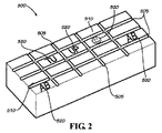
一態様では、図2に示されるのと同様に、乾燥用固定具500の受容面は、切断後の組織の所望の外輪郭であって、組織が用いられる適用可能な外科的処置に要求されるサイズと形である、製品スペース(product space)510を規定する溝505を有する。例えば、乾燥用固定具は、溝が格子配置であるようにして配列させることができる。一つの乾燥用固定具上の格子は、同一の均一なサイズであってよく、または異なる外科的適用のために設計された多様のサイズを含んでよい。それでもなお、当業者によって理解されるように、任意のサイズおよび形の配列を、乾燥用固定具に用いることができる。別の実施態様では、積空間を規定する溝を有する代わりに、乾燥用固定具は、隆起した突起部または刃を有する。
In one aspect, similar to that shown in FIG. 2, the receiving surface of the drying
溝または突起部の間の「空の」空間の中に、乾燥用固定具は、テキスト、ロゴ、名前、または同様のデザイン520の形状にわずかに隆起した、または、ギザギザした、表面模様(texture)を含み得る。この表面模様付けされた(textured)テキスト、ロゴ、名前、またはデザインは、カスタマイズされ得る。乾燥されると、基本的に組織自身の中にラベルをもたらしながら、組織は隆起した表面模様の周りに、またはギザギザした表面模様の中に、それ自身を形作る。好ましくは、乾燥および切断後に、乾燥組織のエンドユーザー(典型的には臨床医)が乾燥組織の基底側から間質側を区別することができるように、表面模様/ラベルは、組織移植片上に一方向にのみ読まれる、または見られることができる。これが望まれる理由は、外科的処置の間、羊膜基底側を下にして、または同種移植片を受ける患者の天然組織に隣接して、同種移植片を所定の位置に置くことが望ましいからである。図2は、乾燥用固定具500の空の空間510の中に含まれ得る様々なマーク、ロゴ、およびテキスト520を示す。典型的に、一つの乾燥用固定具は、全ての空の空間の中に同一のデザインまたはテキストを含む;しかしながら、図2は、説明の目的のために、それぞれの移植片をエンボス加工するためのそのような乾燥用固定具上に含まれ得る広く様々なデザインを示す。
In the “empty” space between the grooves or protrusions, the drying fixture is slightly raised or jagged textured in the shape of text, logo, name, or
いったん羊膜および/または絨毛膜からなる組織移植片が乾燥用固定具上に置かれると、乾燥用固定具は凍結乾燥機内に置かれる。組織移植片を脱水するための凍結乾燥機の使用は、加熱脱水のような他の技術に比べて、より効率的かつ完璧であり得る。一般に、胎盤組織移植片中の氷晶形成は、組織移植片中の細胞外マトリックスを傷つけ得るので避けることが望ましい。凍結乾燥前に羊膜および絨毛膜を化学的に脱水することで、この問題は回避することができる。 Once the tissue graft consisting of amniotic membrane and / or chorion is placed on the drying fixture, the drying fixture is placed in a freeze dryer. The use of a lyophilizer to dehydrate tissue grafts can be more efficient and complete compared to other techniques such as heat dehydration. In general, ice crystal formation in placental tissue grafts is desirable because it can damage the extracellular matrix in tissue grafts. This problem can be avoided by chemically dehydrating the amniotic membrane and chorion before lyophilization.
別の態様では、脱水ステップは、組織移植片を温めるステップを含む。一態様では、羊膜および/または絨毛膜は、適切な上述の乾燥用固定具上に置かれて、そして、乾燥用固定具は滅菌Tyvex(または同様の、通気性があり熱耐性で密封可能な材料)の脱水バッグ中に置かれて、密封される。通気性のある脱水バッグは、組織の乾燥が早すぎるのを防止する。複数の乾燥用固定具が同時に処理される場合は、それぞれの乾燥用固定具は、それ自身のTyvexバッグ中に置かれるか、または、複数の乾燥フレームをそれ上に保持するように設計された適切な取付フレーム中に置かれてそれからフレーム全体がより大きい一つの滅菌Tyvex脱水バッグ内に置かれて密封されるかの、いずれかである。 In another aspect, the dehydrating step includes warming the tissue graft. In one aspect, the amniotic membrane and / or chorion is placed on a suitable above-described drying fixture and the drying fixture is sterile Tyvex (or similar, breathable, heat resistant and sealable) Material) is placed in a dehydration bag and sealed. A breathable dehydration bag prevents the tissue from drying out too quickly. When multiple drying fixtures are processed simultaneously, each drying fixture is either placed in its own Tyvex bag or designed to hold multiple drying frames on it Either placed in a suitable mounting frame and then the entire frame is placed and sealed in one larger sterile Tyvex dewatering bag.
1つまたは複数の乾燥用固定具を含むTyvex脱水バッグはそれから、およそ35〜50℃に予熱された非真空のオーブンまたはインキュベーター内に置かれる。Tyvexバッグは、30〜120分間、オーブン中にそのままにされる。一態様では、加熱ステップは、組織移植片を過乾燥または燃焼はさせないが組織を十分に乾燥させるために、およそ45℃の温度で45分で行うことができる。任意の具体的なオーブンに関する具体的な温度および時間は、高度、オーブンの大きさ、オーブン温度の精度、乾燥用固定具に用いられる材料、同時に乾燥される乾燥用固定具の数、一つまたは複数の、乾燥用固定具のフレームが同時に乾燥されるのかどうか、などを含む他の因子に基づいて計算および調節される必要がある。 The Tyvex dewatering bag containing one or more drying fixtures is then placed in a non-vacuum oven or incubator preheated to approximately 35-50 ° C. The Tyvex bag is left in the oven for 30-120 minutes. In one aspect, the heating step can be performed at a temperature of approximately 45 ° C. for 45 minutes in order to allow the tissue graft to dry sufficiently without over-drying or burning the tissue graft. The specific temperature and time for any specific oven is: altitude, oven size, accuracy of oven temperature, materials used for drying fixtures, number of drying fixtures to be dried simultaneously, one or There are multiple factors that need to be calculated and adjusted based on other factors, such as whether the frame of the drying fixture is being dried at the same time.
一態様では、露出した基底(すなわち、上皮層が実質的に取り除かれた)または露出していない基底膜(すなわち、上皮層が取り除かれない)を有する羊膜の層が乾燥用固定具に載せられた後、絨毛膜(架橋剤で処理されない、または処理された)が羊膜に載せられる。一態様では、羊膜の、露出した基底層または露出していない基底層を乾燥用固定具に載せて、そして、絨毛膜(架橋剤で処理された、または処理されない)を、続いて、乾燥用固定具に取り付けられた羊膜に載せる。本態様は図3に示されていて、羊膜層800が乾燥用固定具600に載せられて、そして絨毛膜層810が羊膜層800に載せられる。他の態様では、いったん、羊膜(および、以下に記載の他の膜)および絨毛膜が乾燥用固定具に載せられたら、乾燥フレームを膜の上に載せることができる。この特徴は、乾燥ラック82が乾燥用固定具80の上部に置かれている図4に示されている。乾燥フレームは、膜を所定の位置に保つ。加えて、乾燥フレームは、組織移植片のシート全体を、持ち上げることなく完全に乾燥させて、それは収率の増加をもたらす。
In one aspect, a layer of amniotic membrane with an exposed base (ie, the epithelial layer is substantially removed) or an unexposed basement membrane (ie, the epithelial layer is not removed) is placed on the drying fixture. After that, the chorion (not treated or treated with a cross-linking agent) is placed on the amniotic membrane. In one aspect, the exposed or unexposed basal layer of amniotic membrane is placed on a drying fixture and the chorion (treated or not treated with a crosslinker) is subsequently dried. Place it on the amniotic membrane attached to the fixture. This embodiment is shown in FIG. 3, with the
特定の態様では、組織移植片は、最後の切断および梱包(ステップ160)を除いて、物理的に変更されない。完了すると、処理された組織移植片は白っぽい着色を有する半透明の見た目を有する。組織移植片は柔らかくて、それが乾燥されて水分を補給されていない状態での曲げ加工およびサイズ化に耐える。本明細書に記載の組織移植片は、長時間、室温で保管され得る。 In certain aspects, the tissue graft is not physically altered except for the last cut and package (step 160). When complete, the treated tissue graft has a translucent appearance with a whitish coloration. The tissue graft is soft and resists bending and sizing when it is dried and not hydrated. The tissue grafts described herein can be stored at room temperature for extended periods of time.
切断および梱包(ステップ160)
いったん移植片が十分に脱水されたら、組織移植片はそれから、特定の商品サイズに切断される準備がされて、保管、最終的な滅菌および後の外科的使用のために、適切に梱包される。一態様では、脱水された組織を含むTyvekバッグは、滅菌/制御環境中に戻して置かれる。生産されるべき移植片の数は、乾燥用固定具(単数または複数)上の組織のサイズおよび形状に基づいて見積もられる。また、それぞれの組織移植片に対して一つの、適切な数のポーチが、滅菌/制御環境の中に導入される。乾燥用固定具(単数または複数)はそれから、Tyvekバッグから取り出される。
Cutting and packing (step 160)
Once the graft is sufficiently dehydrated, the tissue graft is then ready to be cut to a specific commodity size and packaged appropriately for storage, final sterilization, and later surgical use . In one aspect, the Tyvek bag containing dehydrated tissue is placed back into a sterile / controlled environment. The number of implants to be produced is estimated based on the size and shape of the tissue on the drying fixture (s). Also, an appropriate number of pouches, one for each tissue graft, is introduced into the sterilization / control environment. The drying fixture (s) is then removed from the Tyvek bag.
乾燥用固定具が溝を有する場合は、以下の例示的な方法を、商品サイズに組織移植片を切断するために用いることができる。乾燥用固定具が格子状に構成される場合は、#20または同様のストレートまたはローリング刃を、それぞれの溝のラインに沿って平行に切断するために用いる。次に、垂直方向の全ラインが切断される。あるいは、乾燥用固定具が隆起した縁または刃を有する場合は、以下の方法を、組織移植片を商品サイズに切断するために用いることができる。滅菌ローラーを、乾燥用固定具を横切って転がるために用いる。脱水された組織移植片が、乾燥用固定具の全ての隆起した刃または縁に沿って切断されるように、十分な圧力が付与されるべきである。 If the drying fixture has a groove, the following exemplary method can be used to cut tissue grafts into product size. If the drying fixture is configured in a grid, # 20 or similar straight or rolling blade is used to cut parallel along the respective groove line. Next, all the vertical lines are cut. Alternatively, if the drying fixture has a raised edge or blade, the following method can be used to cut the tissue graft to commercial size. A sterilization roller is used to roll across the drying fixture. Sufficient pressure should be applied so that the dehydrated tissue graft is cut along all raised blades or edges of the drying fixture.
切断後、それぞれの組織移植片は、それぞれの「インナー」ポーチに置かれる。好ましくは透明側と不透明側を有するインナーポーチは、透明側を上に向けるべきである。組織移植片は、テキスト、ロゴ、名前、または同様のデザインの形状での表面模様がインナーポーチの透明側を介して外に向いてインナーポーチの外側に見えるように、「インナー」ポーチの中に置かれる。この方法は、それぞれの別個の組織移植片に関して繰り返される。 After cutting, each tissue graft is placed in a respective “inner” pouch. Preferably the inner pouch having a transparent side and an opaque side should face the transparent side up. The tissue graft is in the “inner” pouch so that the surface pattern in the shape of text, logo, name, or similar design faces outward through the transparent side of the inner pouch and is visible on the outside of the inner pouch Placed. This method is repeated for each separate tissue graft.
それぞれの組織移植片はそれから、裂け目または穴がないか、商品サイズ(切断物として)が、規定の長さおよび幅サイズのおよそ(+または−)1ミリメーター以内で、かつ、その特定の移植片に関しておよそ(+または−)250ミクロン以内の厚さであるか、組織移植片の著しい染みまたは変色がないか、および表面模様付けされたロゴまたはワードが、「インナー」ポーチを介して読めて、そして見えるかどうかを、確認するための最終的な検査がされる。 Each tissue graft is then free of crevices or holes, or the product size (as a cut) is within approximately (+ or-) 1 millimeter of the specified length and width size and that particular graft Read through the "inner" pouch that is about (+ or-) 250 microns thick with respect to the piece, no significant stain or discoloration of the tissue graft, and surface-patterned logo or word And a final test is done to see if it is visible.
可能な限り、酸素は、密封される前にインナーポーチから取り除かれる。インナーポーチは、任意の適切な方法で密封することができる;しかしながら、加熱密封が効果的であることが示されている。一態様では、梱包後、商品は、例えば17.5kGyの標的線量でのγ線または電子ビーム滅菌を用いた放射線により、最終的に滅菌される。次に、それぞれのインナーポーチは、さらなる保護、保管および輸送のために、「アウター」ポーチの中に、別々に梱包される。 Whenever possible, oxygen is removed from the inner pouch before it is sealed. The inner pouch can be sealed in any suitable manner; however, heat sealing has been shown to be effective. In one aspect, after packaging, the goods are finally sterilized, for example, with gamma radiation at a target dose of 17.5 kGy or radiation using electron beam sterilization. Each inner pouch is then separately packaged in an “outer” pouch for further protection, storage and transportation.
上述のステップのいずれも、不要な細胞を殺すため、組織移植片を除染するために、またはさもなければ組織移植片を保存するために、組織移植片を凍結させるステップを含まないことを留意すべきである。本明細書に記載の、脱水された組織移植片は、冷蔵または冷凍の必要なく室温または周囲温度で保管および輸送されるように作られる。 Note that none of the above steps include freezing the tissue graft to kill unwanted cells, decontaminate the tissue graft, or otherwise preserve the tissue graft. Should. The dehydrated tissue grafts described herein are made to be stored and transported at room or ambient temperature without the need for refrigeration or freezing.
商品のリリース(ステップ170)
組織移植片がエンドユーザーに輸送およびリリースする準備ができる前に、品質保証部および医長によって、製造、回収およびドナーの適格性に関する全ての証拠書類が見直されて許容できると見なされる。
Product release (step 170)
Before the tissue graft is ready to be transported and released to the end user, all documentation regarding manufacturing, retrieval and donor eligibility is reviewed and deemed acceptable by the Quality Assurance Department and the Chief Medical Officer.
適切なラベリングおよび生産加工流通過程の管理(chain of custody)が、承認された業界標準及び慣習に従って、上記の方法の全てを通して監視される。適切なクリーンルームおよび滅菌作業条件が、可能な限り、上記の方法を通してずっと、維持および使用される。 Appropriate labeling and production process management are monitored through all of the above methods in accordance with approved industry standards and practices. Appropriate clean room and sterilization operating conditions are maintained and used throughout the above methods whenever possible.
II.組織移植片の適用
本明細書に記載の組織移植片の強化された接着性の特質のために、移植片は、対象での創傷治癒を含む、非常に多くの医学的適用に用いることができる。理論に束縛されるものではないが、組織移植片に共有結合された架橋基は、例えば、コラーゲンのような移植片内のタンパク質、および生物学的組織中に存在する他のタンパク質の、非酵素的架橋を促進することができる。一態様では、本明細書に記載の組織移植片は、硬膜と架橋する(すなわち、共有結合を形成する)ことができる。他の態様では、本明細書に記載の組織移植片は、腱、靭帯、筋肉、および他の生体組織に接着することができる。本明細書に記載の組織移植片は、裂傷の補強およびシーリング、ならびに、他の手術後合併症に加えて手術後の瘢痕形成の予防または軽減に有用である。加えて、組織移植片の強化された接着性の特性のために、移植片は、縫合の必要なく、手術部位への適用のための準備ができている。
II. Application of tissue grafts Because of the enhanced adhesive nature of the tissue grafts described herein, the grafts can be used for a large number of medical applications, including wound healing in a subject. . Without being bound by theory, the bridging group covalently attached to the tissue graft is a non-enzyme of proteins within the graft, such as collagen, and other proteins present in biological tissue, for example. Cross-linking can be promoted. In one aspect, the tissue graft described herein can crosslink (ie, form a covalent bond) with the dura mater. In other aspects, the tissue grafts described herein can adhere to tendons, ligaments, muscles, and other biological tissues. The tissue grafts described herein are useful for reinforcing and sealing lacerations and for preventing or reducing post-surgical scar formation in addition to other post-operative complications. In addition, because of the enhanced adhesive properties of tissue grafts, the grafts are ready for application to the surgical site without the need for suturing.
一態様では、本明細書に記載の移植片は、創傷治癒を高める、または改善するのに有用である。日常ベースで医師を訪れる創傷のタイプはさまざまである。急性の創傷は、外科的処置、外傷および火傷に起因する。慢性の創傷は、他の点では健康な人において、治癒に比べて閉じるのが遅い創傷である。患者を悩ませる慢性の創傷のタイプの例は、糖尿病性足部潰瘍、静脈性下腿潰瘍、圧迫潰瘍、動脈性潰瘍、および感染する手術創傷を含む。 In one aspect, the implants described herein are useful for enhancing or improving wound healing. There are various types of wounds that visit physicians on a daily basis. Acute wounds result from surgical procedures, trauma and burns. A chronic wound is a wound that closes slowly compared to healing in an otherwise healthy person. Examples of types of chronic wounds that afflict patients include diabetic foot ulcers, venous leg ulcers, pressure ulcers, arterial ulcers, and infected surgical wounds.
外傷性創傷を治療する時の医師の目標は、最小限の瘢痕および感染で、創傷の領域における自然な機能を患者が保持するようにしながら創傷を治療することである。創傷が感染する場合、手足または生活の喪失を導き得る。ほとんどは、何事もなく医師はこれらの患者を治療する。しかしながら、慢性の創傷を扱う医師は、手足または生活の喪失を導き得る感染リスクを最小限化するために、できるだけ素早く創傷を閉じることを主に心掛ける。慢性の創傷は、治癒カスケードを悪化または遅らせる共存症を有する患者での創傷である。一態様では、本明細書に記載の組織片は、炎症を軽減して治癒を高めるのを助けるために、必須の創傷治癒因子、細胞外マトリックスタンパク質および炎症性メディエータを運搬して瘢痕組織形成を軽減する、組織再生テンプレートとして機能することができる。 The goal of a physician when treating a traumatic wound is to treat the wound while keeping the patient natural functioning in the area of the wound with minimal scarring and infection. If a wound is infected, it can lead to loss of limbs or life. In most cases, doctors treat these patients without incident. However, physicians who deal with chronic wounds are primarily concerned with closing the wound as quickly as possible to minimize the risk of infection that can lead to loss of limbs or life. Chronic wounds are wounds in patients with comorbidities that exacerbate or delay the healing cascade. In one aspect, the tissue pieces described herein carry essential wound healing factors, extracellular matrix proteins and inflammatory mediators to reduce scar tissue formation to help reduce inflammation and enhance healing. It can function as a tissue regeneration template to reduce.
別の態様では、本明細書に記載の組織移植片は、手術後に起こる脊椎および周辺領域への合併症を対処する、または軽減するのに有用である。急性および慢性の脊椎損傷および脊椎痛は、脊柱における外傷および/または退行性変化に起因し得る。退行性の患者に関しては、通常、患者の症状および病状に応じた可能な手術が施される。保存療法が失敗した場合の最初の外科的選択肢は、椎弓切除術またはマイクロ椎間板切除術(micro−discectomy)である。これらの低侵襲的処置は、痛みの発生または脊柱管狭窄を緩和することを目的とする。疾患が進行する場合、制限されないが、脊椎固定を含む他の手術が必要となり得る。脊椎固定は、前方(腹部を通って前から)、後方(背中から)、または側部(脇を通って)で、様々な方法を通して達成されてよい。それぞれの方法は、利点と欠点がある。目標は典型的に、椎間板を取り除き、板の高さを元に戻して、動きおよびさらなる劣化を制限するために2つの脊椎骨を一緒に固定させる(fuse)ことである。外科医および患者の外科的選択肢には、椎間板を人口板で置き換えることもある。脊椎外傷は典型的に、脊椎レベル(spine levels)を固定する(fusing)ことにより治療され、または、椎骨が破砕している場合は、外科医は椎体切除術(corpectomy)をして、影響を受けたレベルにわたって固定することを選択し得る。 In another aspect, the tissue grafts described herein are useful in addressing or reducing complications to the spine and surrounding areas that occur after surgery. Acute and chronic spinal cord injury and spinal pain can result from trauma and / or degenerative changes in the spinal column. For degenerative patients, usually possible surgery is performed depending on the patient's symptoms and condition. The first surgical option if conservative therapy fails is a laminectomy or micro-discectomy. These minimally invasive procedures aim to relieve pain development or spinal stenosis. If the disease progresses, other operations may be required including but not limited to spinal fixation. Spinal fixation may be accomplished through a variety of methods, either anterior (through the abdomen from the front), posterior (from the back), or side (through the side). Each method has advantages and disadvantages. The goal is typically to remove the intervertebral disc and restore the disc height to fuse the two vertebrae together to limit movement and further degradation. A surgeon and patient's surgical option may be to replace the intervertebral disc with an artificial disc. Spine trauma is typically treated by fixing spine levels, or if the vertebra is fractured, the surgeon performs a corpectomy to affect the spine. You may choose to fix over the level you receive.
一態様では、本明細書に記載の組織移植片は、脊椎上または脊椎付近の瘢痕形成を予防または軽減し、および硬膜裂傷をシーリングするのに有用である。手術後の脊椎上または脊椎付近の瘢痕形成は非常に衰弱性(debilitating)であり得て、そして場合により、上記の症状を対処するための手術をその後に必要とする。また、用語「抗−癒着」は、当技術分野において、脊椎上または脊椎付近の瘢痕組織の予防を指すためにも用いられる。他の態様では、本明細書に記載の組織移植片は、移植片が周囲の手術部位由来の手術後の外傷から脊髄硬膜を保護する、保護バリアとして用いることができる。例えば、移植片は、椎骨などの、切れたばかりの骨による鋭いエッジによって引き起こされる、脊髄硬膜への損傷を防ぐことができる。他の態様では、組織移植片は、前方腰椎椎体間固定術(anterior lumbar interbody fusion)、後方腰椎椎体間固定術(posterior lumbar interbody fusion)、経椎間孔腰椎椎体間固定術(trans−lumbar interbody fusion)、前方頸部椎間板切除術および固定術(anterior cervical discectomy and fusion)、マイクロ椎間板切除術、脊髄硬膜修復のため、および脳脊髄液漏(CSF leakage)を防ぐための硬膜シーラントとして、用いることができる。 In one aspect, the tissue grafts described herein are useful for preventing or reducing scar formation on or near the spine and sealing dural lacerations. Scar formation on or near the spine after surgery can be very debilitating and, in some cases, subsequently requires surgery to address the above symptoms. The term “anti-adhesion” is also used in the art to refer to the prevention of scar tissue on or near the spine. In other aspects, the tissue grafts described herein can be used as a protective barrier where the graft protects the spinal dura from post-surgical trauma from the surrounding surgical site. For example, the graft can prevent damage to the spinal dura caused by sharp edges, such as vertebrae, from freshly cut bone. In another aspect, the tissue graft is an anterior lumbar interbody fusion, a posterior lumbar interbody fusion, a transforaminal lumbar interbody fusion. -Lumbar interfusion body, anterior cervical discectomy and fusion, microdiscectomy, for spinal dura repair and to prevent cerebrospinal fluid leakage (CSF leakage) It can be used as a sealant.
外科的処置に応じて、組織移植片は、脊髄硬膜、神経根を含む脊椎の周辺領域、またはそれらの組み合わせに、直接適用することができる。椎骨の独特な構造のために、組織移植片は、対象における適切な位置に配置して付けることができるようにして、任意の形状または寸法に切断することができる。例えば、組織移植片が、両側の被覆に用いられる場合、長方形型の膜は、組織移植片を、後方脊椎突起(posterior spinal process)周辺に合わせさせて、横の動きを最小限にする。横の動きを最小限にすることに加えて、組織移植片はまた、影響を受けた領域への露出のために骨髄層が取り除かれている近位および遠位のバリアカバレッジを与えることができる。一態様では、適切な配置を確実にするために、移植片は、対象での移植片の適切な配置を確実にするために、移植片の露出した基底膜上をエンボス加工することができる。特に、適切な移植片の配置は、移植片の基底膜が脊髄硬膜または周辺領域に直接接触することを確実にする。例えば、適切な膜の配置および方向は、後方または前方アプローチが使用される脊椎の適用において材料を適用する場合に重要である。 Depending on the surgical procedure, the tissue graft can be applied directly to the spinal dura mater, the peripheral region of the spine including nerve roots, or a combination thereof. Because of the unique structure of the vertebrae, the tissue graft can be cut into any shape or size so that it can be placed and applied at the appropriate location in the subject. For example, if a tissue graft is used for bilateral coverage, the rectangular membrane allows the tissue graft to be aligned around the posterior spinal process to minimize lateral movement. In addition to minimizing lateral movement, tissue grafts can also provide proximal and distal barrier coverage where the bone marrow layer has been removed for exposure to the affected area . In one aspect, to ensure proper placement, the graft can be embossed on the exposed basement membrane of the graft to ensure proper placement of the graft in the subject. In particular, proper graft placement ensures that the basement membrane of the graft is in direct contact with the spinal dura or surrounding area. For example, proper membrane placement and orientation is important when applying materials in spinal applications where a posterior or anterior approach is used.
組織片は、脊椎と関連する様々な外科的処置に起因し得る瘢痕形成を予防または軽減するのに有用である。組織片は首、背中、または腰における任意の処置後に用いることができる。適用に応じて、羊膜の上皮は実質的に取り除かれてよい。例えば、椎弓切除術または椎間板切除術などの後処置では、上皮層は実質的に取り除かれる。上皮細胞層の除去は、羊膜の基底膜層を露出させて、細胞シグナル特性を増大させる。この上方制御の応答は、細胞の遊走および抗−炎症性タンパク質の発現を高めて、線維症を阻害する。脊髄硬膜は、後処置に続き典型的に保護されないままである。このように、本明細書に記載の移植片は、これらの処置において、満たされていない要求をもたらす。 The tissue pieces are useful for preventing or reducing scar formation that may result from various surgical procedures associated with the spine. The tissue piece can be used after any treatment on the neck, back, or waist. Depending on the application, the epithelium of the amniotic membrane may be substantially removed. For example, in post-treatments such as laminectomy or discectomy, the epithelial layer is substantially removed. Removal of the epithelial cell layer exposes the basement membrane layer of the amniotic membrane and increases cell signal properties. This up-regulated response increases cell migration and expression of anti-inflammatory proteins and inhibits fibrosis. The spinal dura mater typically remains unprotected following post treatment. Thus, the implants described herein provide unmet needs in these procedures.
他の態様では、上皮細胞層は取り除かれない。例えば、前方腰椎椎体間固定術(ALIF)や経椎間孔椎体間固定術(Transforaminal Interbody Fusion)(TLIF)のような前処置または改良された前処置では、羊膜の上皮層は取り除かれず、損傷されていない。これらの態様では、組織片は、腹膜、より大きな血管、および腹部の筋肉組織からの隔離を維持することにより脊椎の手術部位にさらなる保護を与える。膜は、癒着および瘢痕に対する、減少した摩擦解剖学的バリア(friction anatomical barrier)としての役割を果たす。例えば、組織片は、瘢痕組織が大きな血管を脊椎に結合させるのを防ぐことができる。これは、脊椎の手術後での一般的な問題であり、これを対処するための第二の外科的処置が必要である。 In other embodiments, the epithelial cell layer is not removed. For example, pre-treatments such as anterior lumbar interbody fusion (ALIF) or transforaminal interbody fusion (TLIF) do not remove the epithelial layer of the amniotic membrane. Not damaged. In these embodiments, the tissue pieces provide additional protection to the spinal surgical site by maintaining isolation from the peritoneum, larger blood vessels, and abdominal muscle tissue. The membrane serves as a reduced friction anatomical barrier to adhesions and scarring. For example, the tissue piece can prevent scar tissue from joining large blood vessels to the spine. This is a common problem after spinal surgery and requires a second surgical procedure to address this.
別の態様では、組織移植片は、歯科の適用に有用である。例えば、移植片は、歯科インプラントの周辺に、または、進行性歯肉退縮欠損の治療に用いることができる。別の態様では、移植片は、組織再生誘導法において用いることができる。 In another aspect, the tissue graft is useful for dental applications. For example, the implant can be used around dental implants or to treat progressive gingival regression defects. In another aspect, the graft can be used in a tissue regeneration induction method.
他の態様では、本発明に記載の移植片は、整形外科の適用(すなわち、スポーツ医学)で用いることができる。スポーツ医学は、活発な人およびスポーツ選手において、外傷に起因する関節中または関節周辺の様々な軟組織損傷、または運動の繰り返しによりもたらされる慢性状態の修復および再建を含む。例えば、スポーツ医学は、制限されないが、肩、肘、足、足首、手および手首と関連する、様々な異なる損傷の治療を含む。 In other aspects, the implants described in the present invention can be used in orthopedic applications (ie, sports medicine). Sports medicine includes repair and reconstruction of chronic conditions in active people and athletes, caused by various soft tissue injuries in or around the joints caused by trauma, or repeated exercise. For example, sports medicine includes, but is not limited to, treating a variety of different injuries associated with the shoulders, elbows, feet, ankles, hands and wrists.
損傷の主なタイプは、腱および靭帯の捻挫および様々な関節におけるヘルニアを含み、最も一般的には、膝のACLおよび肩の回旋腱板である。腱と靭帯でない処置は、裂けた膝半月板の修復および治療されないままにしておくと関節の変形性関節症をもたらし得る膝軟骨の修復を含む。また、非外科的選択肢は、炎症を起こした腱(例えば「テニス肘」)への抗−炎症性薬剤の注入、関節への潤滑剤の注入(例えばヒアルロン酸を膝へ)、ならびに理学療法およびブレーシング(bracing)を含む。 The main types of injury include tendon and ligament sprains and hernias in various joints, most commonly the knee ACL and the shoulder rotator cuff. Treatments that are not tendons and ligaments include repair of the torn knee meniscus and repair of knee cartilage that can result in joint osteoarthritis if left untreated. Non-surgical options also include injection of anti-inflammatory drugs into inflamed tendons (eg, “tennis elbow”), injection of lubricant into joints (eg, hyaluronic acid into the knee), and physical therapy and Includes bracing.
一態様では、本明細書に記載の組織移植片は、治癒している腱上での瘢痕形成を防ぐために、腱修復を包むために用いることができる。また、それらは、修復がうまく進行するための、保護的な閉鎖環境を提供することができる。組織移植片は、同種移植片を購入する必要性に代替するため、または、腱または靭帯の移行をするために、既製の(off−the−shelf)腱および靭帯として用いることができる。 In one aspect, the tissue graft described herein can be used to wrap a tendon repair to prevent scar formation on a healing tendon. They can also provide a protective closed environment for the repair to proceed successfully. Tissue grafts can be used as off-the-shelves tendons and ligaments to replace the need to purchase allografts or to make tendon or ligament transitions.
他の態様では、本明細書に記載の組織移植片は、回旋腱板の補強で用いることができる。いくつかの回旋腱板の裂傷は、生存可能な天然組織の欠如のため、修復を助ける補強マトリックスを必要とするほどに大きい。本明細書に記載の組織移植片は、修復を補強するためのマトリックスとして用いることができる。一態様では、本明細書に記載の組織移植片は、膝軟骨を修復するために用いることができる。例えば、組織移植片は、細胞培養された軟骨細胞または軟骨欠損の内側の他のプロ軟骨再生マトリックスを保持するためのバリアとして用いることができる。この態様では、組織移植片は、欠損を閉鎖してマトリックスを所定の位置に維持するためのフラップとして使用される。 In other aspects, the tissue grafts described herein can be used in rotator cuff reinforcement. Some rotator cuff tears are so large that they require a reinforcing matrix to aid in repair due to the lack of viable natural tissue. The tissue grafts described herein can be used as a matrix to reinforce a repair. In one aspect, the tissue grafts described herein can be used to repair knee cartilage. For example, tissue grafts can be used as a barrier to hold cell-cultured chondrocytes or other pro-cartilage regeneration matrices inside a cartilage defect. In this embodiment, the tissue graft is used as a flap to close the defect and maintain the matrix in place.
一態様では、組織移植片は、末梢神経を修復するために用いることができる。組織移植片は、治癒している神経の上に瘢痕が形成するのを防ぐために、神経修復上を包むものとして用いることができる。また、組織移植片は、修復がうまく進行するための、保護的な閉鎖環境を提供することができる。他の態様では、組織移植片は、神経末端が再接近し得ない保護的環境中に神経が伸びるのを導くための神経再生チューブ中に製造することができる。ここで、末端が他の軟組織に妨害されることなく自由に接することができる場合、神経は一定の距離次第で再度結合することができる。別の態様では、組織移植片は、前立腺切除術の処置後に神経束を束ねるために用いることができる。これらの神経は、勃起機能および排泄抑制能力を司る。組織移植片は、瘢痕および場合により神経を損傷することからそれらを守るために、神経上に置くことができる。 In one aspect, the tissue graft can be used to repair peripheral nerves. The tissue graft can be used as a wrap for nerve repair to prevent the formation of scars on the healing nerve. Tissue grafts can also provide a protective closed environment for successful repair. In other embodiments, the tissue graft can be manufactured in a nerve regeneration tube to guide nerve growth into a protective environment where nerve endings cannot reappear. Here, the nerves can reconnect again depending on a certain distance if the end can freely touch without being disturbed by other soft tissues. In another aspect, the tissue graft can be used to bundle a nerve bundle after a prostatectomy procedure. These nerves are responsible for erectile function and ability to suppress excretion. Tissue grafts can be placed on nerves to protect them from scarring and possibly damaging the nerves.
他の態様では、本明細書に記載の組織移植片は、骨膜の修復での補助;破裂性/損傷性嚢の修復の補助;骨修復中の空隙充填材料の固定の補助;のような他の整形外科の適用において、または対象の四肢に関する適用(例えば、小骨の固定のための抗−癒着バリア、金属めっきまたは金属製品が用いられる場合の抗−癒着バリア、または、破裂性/損傷性嚢の修復の補助)において、用いることができる。 In other embodiments, the tissue graft described herein may be used to assist in repair of periosteum; assist in repair of ruptured / damaged sac; assist in fixation of void filling material during bone repair; In orthopedic applications of the subject or for the limb of the subject (eg anti-adhesion barrier for fixation of small bones, anti-adhesion barrier when metal plating or metal products are used, or ruptured / damaging sac Can be used in the repair assistance).
別の態様では、組織移植片は、女性の受胎能に関与し得る疾患の治療、生殖器系に起因する痛みまたは生殖器系におけるガンに関する産婦人科(OB/GYN)の外科的処置に用いることができる。これらの処置は、子宮筋腫の除去(筋腫切除術)、卵巣嚢胞の除去、卵管結紮、子宮内膜症の治療、いくつかのガン性または非ガン性腫瘍の除去、および膣スリング(vaginal slings)を含む。これらの処置は、経膣の、腹部の、または腹腔鏡の手法により達成されてよい。 In another aspect, the tissue graft may be used for the treatment of diseases that may be involved in female fertility, for obstetrics and gynecology (OB / GYN) surgical treatment for pain caused by the reproductive system or cancer in the reproductive system. it can. These procedures include removal of uterine fibroids (myomactomy), removal of ovarian cysts, tubal ligation, treatment of endometriosis, removal of some cancerous or non-cancerous tumors, and vaginal slings. )including. These procedures may be accomplished by transvaginal, abdominal, or laparoscopic techniques.
組織移植片は、外科的処置後の生殖器系における瘢痕組織の量を軽減するためのパッチとして用いることができる。瘢痕組織は、線維組織の別形態であり、そしてまた、受胎能の問題に寄与し得る。卵巣上または卵管中の瘢痕の量を最小限に抑える能力は、手術後の受胎能および痛みさえにも役立ち得る。別の態様では、組織移植片は、重症の子宮内膜症の治療後に子宮壁を補強するために、および、患者の妊娠する能力を高めるために、用いることができる。さらなる態様では、組織移植片は、卵巣嚢胞の除去後の抗−癒着バリア、または膣壁びらんの修復の補助として用いることができる。 The tissue graft can be used as a patch to reduce the amount of scar tissue in the genital system after a surgical procedure. Scar tissue is another form of fibrous tissue and can also contribute to fertility problems. The ability to minimize the amount of scar on the ovary or in the fallopian tube can also help with fertility and even pain after surgery. In another aspect, the tissue graft can be used to reinforce the uterine wall after treatment of severe endometriosis and to increase the patient's ability to become pregnant. In a further aspect, the tissue graft can be used as an anti-adhesion barrier after removal of ovarian cysts or as an aid in repairing vaginal erosion.
他の態様では、組織移植片は心臓適用に用いることができる。狭心症は心筋の虚血(血液の不足、したがって、酸素供給の欠乏)による、一般に冠動脈(心臓の血管)の閉塞または攣縮による、重度の胸部痛である。狭心症の主な原因である冠動脈疾患は、心臓の動脈のアテローム性動脈硬化に起因する。アテローム硬化性の血塊を除去するための様々な開放性の心臓および血管の外科処置は、血管の修復、再建および閉鎖、および外科的欠損を閉鎖させてパッチするための再生組織パッチの補助を必要とする。また、心臓バイパス移植片および心臓欠陥の再建(心臓切開の外科的処置の一環として)は、パッチまたは移植片から、軟組織の弱さに対する支え(buttress)、適切な組織の欠損がある場合は組織置換、および心臓それ自身への癒着を軽減する可能性も与えるという、恩恵を受けることができる。本明細書に記載の組織移植片は、頸動脈修復、冠動脈バイパス移植術、先天性心疾患、心臓弁修復、および血管修復(すなわち末梢血管)などの手術および合併症に起因する血管および心臓の欠陥の修復を助けるためのパッチとして、用いることができる。 In other embodiments, the tissue graft can be used for cardiac applications. Angina is severe chest pain, usually due to occlusion or spasm of the coronary arteries (cardiac blood vessels) due to myocardial ischemia (lack of blood and thus lack of oxygen supply). Coronary artery disease, the main cause of angina, results from atherosclerosis of the heart arteries. Various open heart and vascular surgical procedures to remove atherosclerotic clots require vascular repair, reconstruction and closure, and the assistance of regenerative tissue patches to close and patch surgical defects And Also, reconstruction of cardiac bypass grafts and heart defects (as part of open heart surgical procedures) can be done from patches or grafts, but to soft tissue weakness, tissue if there is a lack of proper tissue It can benefit from replacement and also the possibility to reduce adhesions to the heart itself. The tissue grafts described herein are vascular and cardiac due to surgery and complications such as carotid artery repair, coronary artery bypass grafting, congenital heart disease, heart valve repair, and vascular repair (ie, peripheral blood vessels). It can be used as a patch to help repair defects.
本明細書に記載の組織移植片は、全身の外科処置で用いることができる。例えば、全身の外科的処置は、腹腔に関する処置を含む。これらは、腸、胃、大腸、肝臓、胆嚢、虫垂、胆管および甲状腺を含む。処置は、ヘルニア、ポリープ切除、ガン除去、結腸クローン病および潰瘍性大腸炎の外科的処置を含んでよい。これらの処置は、開腹または腹腔鏡下で行われてよい。他の態様では、組織移植片は、吻合の閉鎖、吻合のための抗−癒着バリア、またはヘルニア修復のための抗−癒着バリアを促進するために用いることができる。 The tissue grafts described herein can be used in systemic surgical procedures. For example, systemic surgical procedures include procedures involving the abdominal cavity. These include the intestine, stomach, large intestine, liver, gallbladder, appendix, bile duct and thyroid. Treatment may include surgical treatment of hernias, polypectomy, cancer removal, colonic Crohn's disease and ulcerative colitis. These procedures may be performed laparoscopically or laparoscopically. In other embodiments, the tissue graft can be used to promote an anastomosis closure, an anti-adhesion barrier for anastomosis, or an anti-adhesion barrier for hernia repair.
他の態様では、組織移植片は耳鼻咽喉科(ENT)処置で用いることができる。鼓室形成は、鼓膜(eardrum)(鼓膜(tympanic membrane))および/または中耳の小骨の再建のために行われる。穿孔された鼓膜の治療のための、様々な選択肢が存在する。穿孔が最近の外傷による場合は、耳、鼻および喉の多くの専門家は、自然に治癒するかどうか観察して見ることを選択するだろう。これが生じないか、または、頻回の再度の穿孔が同一エリアに生じる場合は、手術が検討され得る。鼓室形成は、外耳道を通して、または耳の後ろの切開により行うことができる。ここで、外科医は、耳周辺の皮膚の下の組織から移植片を摘出して、そして、鼓膜を再構築するためにそれを用いる。本明細書に記載の組織移植片は、患者自身の組織の摘出と関連するさらなる外傷を防ぐために用いることができ、そして手術時間を短縮する。他の態様では、組織移植片は、アデノイド切除後の創傷被覆、扁桃摘出後の創傷被覆、またはシュナイダー膜(Sniderian membrane)の促進修復として用いることができる。 In other embodiments, the tissue graft can be used in an ENT procedure. Tympanic chamber formation is performed for the reconstruction of the eardrum (the tympanic membrane) and / or the ossicles of the middle ear. There are various options for the treatment of perforated tympanic membranes. If perforations are due to recent trauma, many ear, nose and throat specialists will choose to observe and see if they heal naturally. If this does not occur or if frequent re-perforations occur in the same area, surgery can be considered. Tympanic chamber formation can be performed through the ear canal or by an incision behind the ear. Here, the surgeon removes the graft from the tissue under the skin around the ear and uses it to reconstruct the eardrum. The tissue grafts described herein can be used to prevent further trauma associated with the removal of the patient's own tissue and reduce surgical time. In other embodiments, the tissue graft can be used as a wound covering after adenoidectomy, a wound covering after tonsillectomy, or an accelerated repair of the Schneider membrane.
他の態様では、本明細書に記載の組織移植片は、形成外科処置で用いることができる。瘢痕形成術(scar revision)は、瘢痕の見た目を改善または軽減するための手術である。それはまた、機能を回復させ、および、損傷、創傷、または以前の手術に起因する皮膚変化(美観を損なうこと)をなおす。瘢痕組織は、損傷または手術後の皮膚治癒として形成する。瘢痕の量は、創傷の大きさ、深さ、および部位;人の年齢;遺伝;および皮膚の色(色素沈着)を含む皮膚特性により決定され得る。手術は、瘢痕の切除および欠損の注意深い閉鎖を含む。一態様では、本明細書に記載の組織移植片は、瘢痕の治癒および予防;および軟組織の縁の丁寧な近似(careful approximation of soft−tissue edges)が達成不可能であって瘢痕組織が生じ得るケロイドまたはガンの修復(revision)/除去に役立つパッチとして用いることができる。さらに、組織移植片の抗炎症性特性は、その上、治癒を高めることができる。 In other aspects, the tissue grafts described herein can be used in plastic surgery. Scar revision is a procedure for improving or reducing the appearance of scars. It also restores function and corrects skin changes (damaging aesthetics) resulting from injury, wounding, or previous surgery. Scar tissue forms as a skin healing after injury or surgery. The amount of scar can be determined by skin characteristics including wound size, depth, and site; human age; heredity; and skin color (pigmentation). Surgery involves removal of the scar and careful closure of the defect. In one aspect, the tissue grafts described herein may produce scar tissue where scar healing and prevention; and a careful approximation of soft-tissue edges is not achievable It can be used as a patch to aid in the revision / removal of keloids or cancers. Furthermore, the anti-inflammatory properties of tissue grafts can further enhance healing.
他の態様では、組織移植片は、眼科の適用(例えば、眼表面修復のオン−レイ(on−lay)移植片)または泌尿器科の適用(例えば、精管復元術の間の精管の閉鎖を促進する、または外傷により生じる精管の閉鎖を促進する)において用いることができる。 In other embodiments, the tissue graft is an ophthalmic application (eg, an on-lay implant for ocular surface repair) or a urological application (eg, closure of the vas deferens during vasectomy) Or promote closure of the vas deferens caused by trauma).
一態様では、組織移植片は、脳硬膜の修復および置換、小帯牽引の除去、失われた膝蓋骨組織の再生、空洞中のシュナイダー膜、歯科インプラント周辺の軟組織の修復、口腔前庭形成術、および組織再生誘導法において用いることができる。 In one aspect, the tissue graft comprises repair and replacement of the cerebral dura mater, removal of ligament traction, regeneration of lost patella tissue, Schneider membrane in the cavity, soft tissue repair around the dental implant, oral vestibuloplasty, And can be used in a tissue regeneration induction method.
移植片の適用に応じて、移植片は、自家血液の採取と分離を用いた産物または期限切れの保存血由来の血小板濃縮物のいずれかの、血小板濃縮物由来の天然起源の増殖因子;骨髄穿刺液;濃縮ヒト胎盤臍帯血幹細胞、濃縮羊水幹細胞またはバイオリアクター中で増殖された幹細胞由来の幹細胞;または抗生物質からなる溶液のような生物活性剤に浸すことができる。ここで、組織移植片の1つまたは複数の膜層は、生物活性剤を吸収する。生物活性剤を有する湿組織移植片を創傷に対して適用する際、生物活性剤は時間をかけて創傷に届けられる。 Depending on the application of the graft, the graft may be a natural growth factor derived from platelet concentrate, either a product using autologous blood collection and separation or a platelet concentrate derived from expired stored blood; Fluid; concentrated human placental umbilical cord blood stem cells, concentrated amniotic fluid stem cells or stem cells derived from stem cells grown in a bioreactor; or bioactive agents such as solutions consisting of antibiotics. Here, one or more membrane layers of the tissue graft absorb the bioactive agent. When a wet tissue graft with a bioactive agent is applied to a wound, the bioactive agent is delivered to the wound over time.
本明細書に記載の組織移植片は対象の組織に直接適用することができるが、それらはまた、続けて対象に適用することのできる創傷被覆材(wound dressing)に適用することができる。例えば、創傷包帯は、ガーゼ、絆創膏またはラップ、または対象へ直接適用することのできる組織移植片を含むことができる、または付けることができる任意の他の適切な物であり得る。 Although the tissue grafts described herein can be applied directly to the tissue of the subject, they can also be applied to wound dressing that can subsequently be applied to the subject. For example, the wound dressing can include gauze, bandages or wraps, or any other suitable item that can include or be applied to a tissue graft that can be applied directly to a subject.
様々な改良および変更を、本明細書に記載の化合物、組成物および方法に対して行うことができる。本明細書に記載の化合物、組成物および方法の他の態様は、明細書の検討、および、本明細書に開示された化合物、組成物および方法の実践から明らかであろう。明細書および実施例は例示的であると考えられることが意図される。 Various improvements and modifications can be made to the compounds, compositions and methods described herein. Other aspects of the compounds, compositions and methods described herein will be apparent from consideration of the specification and practice of the compounds, compositions and methods disclosed herein. It is intended that the specification and examples be considered exemplary.
Claims (35)
(1)2またはそれ以上の層の脱水された羊膜、
ここで、脱水された羊膜の少なくとも1つの層は架橋されている、
(2)2またはそれ以上の層の脱水された絨毛膜、
ここで、脱水された絨毛膜の少なくとも1つの層は架橋されている、または
(3)1または複数の層の脱水された羊膜および脱水された絨毛膜、
ここで、脱水された羊膜および/または脱水された絨毛膜の少なくとも1つの層は架橋されている、
を含み、
羊膜の少なくとも1つの前記層は、脱細胞化されておらず、かつ、繊維芽細胞および/または上皮細胞を含んでおり、または、絨毛膜の少なくとも1つの前記層は脱細胞化されていない、および
前記羊膜またはその一部は、損傷されていない羊膜から得られる、
組織移植片。 A dehydrated tissue graft,
(1) two or more layers of dehydrated amniotic membrane,
Wherein at least one layer of dehydrated amniotic membrane is crosslinked,
(2) two or more layers of dehydrated chorion,
Wherein at least one layer of dehydrated chorion is cross-linked, or (3) one or more layers of dehydrated amniotic membrane and dehydrated chorion,
Wherein at least one layer of the dehydrated amnion and / or dehydrated chorion are crosslinked,
Only including,
At least one said layer of amniotic membrane is not decellularized and contains fibroblasts and / or epithelial cells, or at least one said layer of chorion is not decellularized, and
The amniotic membrane or part thereof is obtained from undamaged amniotic membrane,
Tissue graft.
前記羊膜の前記第一表面は、露出した基底層または露出していない基底層を含むことを特徴とする、
移植片。 The implant of claim 1, comprising:
The first surface of the amniotic membrane comprises an exposed basal layer or an unexposed basal layer,
Graft.
前記架橋剤が、糖を含むことを特徴とする、
移植片。 The implant of claim 1, comprising:
The crosslinking agent contains a sugar,
Graft.
前記糖は、D−リボース、グリセロ―ス、アルトロース、タロース、エルセオース、グルコース、リキソース、マンノース、キシロース、グロース、アラビノース、イドース、アロース、ガラクトース、マルトース、ラクトース、スクロース、セロビオース、ゲンチオビース、メリビオース、ツラノース、トレハロース、イソマルトース、またはそれらの任意の組み合わせを含むことを特徴とする、
移植片。 The implant of claim 3 , comprising:
The sugar is D-ribose, glycerose, altrose, talose, elseose, glucose, lyxose, mannose, xylose, gulose, arabinose, idose, allose, galactose, maltose, lactose, sucrose, cellobiose, gentiobiose, melibiose, turanose , Trehalose, isomaltose, or any combination thereof,
Graft.
前記架橋剤は、ジアルデヒド、カルボジイミド、またはそれらの組み合わせを含むことを特徴とする、
移植片。 The implant of claim 1, comprising:
The cross-linking agent includes dialdehyde, carbodiimide, or a combination thereof,
Graft.
前記羊膜および絨毛膜は、クロスリンカーを介してお互いに共有結合しており、かつ、前記羊膜および/または前記絨毛膜は脱細胞化されてないことを特徴とする、
組織移植片。 A dehydrated tissue graft comprising a dehydrated amniotic membrane and a dehydrated chorion;
The amnion and chorion are covalently bonded to each other via a crosslinker , and the amnion and / or the chorion are not decellularized ,
Tissue graft.
請求項1に記載の組織移植片を含むことを特徴とする、
創傷被覆材。 A wound dressing,
Characterized in that it comprises a tissue graft according to claim 1,
Wound dressing.
前記膜に共有結合された少なくとも1つのクロスリンカーを含み、ここで、前記羊膜または前記絨毛膜は脱細胞化されていないことを特徴とする、
羊膜または絨毛膜。 Dehydrated amniotic membrane or dehydrated chorion,
Look containing at least one crosslinker is covalently bound to said membrane, wherein said amnion or the chorion is characterized by not being decellularized,
Amnion or chorion.
前記方法は、対象に、請求項1に記載の組織移植片を、対象の脊髄硬膜または脊椎付近の領域に直接適用するステップを含むことを特徴とする、
組織移植片。 A tissue graft according to claim 1 for use in a method of preventing or reducing scar formation on or near a spine after a surgical procedure comprising:
The method comprises applying to a subject a tissue graft according to claim 1 directly to the spinal dura mater or an area near the spine of the subject.
Tissue graft .
前記外科的処置は、後処置を含むことを特徴とする、
組織移植片。 A tissue graft according to claim 9 ,
The surgical procedure includes a post-treatment,
Tissue graft .
前記後処置は、椎弓切除術または椎間板切除術であることを特徴とする、
組織移植片。 The tissue graft of claim 10 , comprising:
The post-treatment is a laminectomy or a discectomy,
Tissue graft .
前記後処置は、前方腰椎椎体間固定術(ALIF)および経椎間孔椎体間固定術(TLIF)であることを特徴とする、
組織移植片。 The tissue graft of claim 10 , comprising:
The post-treatment is anterior lumbar interbody fusion (ALIF) and transforaminal interbody fusion (TLIF),
Tissue graft .
前記外科的処置は、前処置を含むことを特徴とする、
組織移植片。 A tissue graft according to claim 9 ,
The surgical procedure includes a pretreatment,
Tissue graft .
前記移植片中に存在する羊膜は、露出した基底層を含むことを特徴とする、
組織移植片。 A tissue graft according to claim 9 ,
The amniotic membrane present in the graft comprises an exposed basal layer,
Tissue graft .
前記方法は、縫合の使用を含まないことを特徴とする、
組織移植片。 A tissue graft according to claim 9 ,
The method does not include the use of sutures,
Tissue graft .
前記方法は、請求項1に記載の組織移植片を、硬膜裂傷に直接適用するステップを含むことを特徴とする、
組織移植片。 A tissue graft according to claim 1 for use in a method for treating a dural laceration in a subject comprising:
The method comprises the step of applying the tissue graft of claim 1 directly to a dural laceration,
Tissue graft .
前記移植片中に存在する羊膜は、露出した基底層を含むことを特徴とする、
組織移植片。 The tissue graft of claim 16 , comprising:
The amniotic membrane present in the graft comprises an exposed basal layer,
Tissue graft .
前記方法は、縫合の使用を含まないことを特徴とする、
組織移植片。 The tissue graft of claim 16 , comprising:
The method does not include the use of sutures,
Tissue graft .
前記方法は、請求項1に記載の移植片を創傷に適用するステップを含み、
ここで前記創傷は脳硬膜中にあり、小帯牽引の除去のため、失われた膝蓋骨組織の再生のため、洞腔中のシュナイダー膜または歯科インプラント周囲の軟組織の修復のための、歯周形成外科の(perioplastic)処置による創傷であることを特徴とする、
組織移植片。 A tissue graft according to claim 1 for use in a method for promoting wound healing in a subject comprising:
The method includes the step of applying the implant of claim 1 to a wound;
Where the wound is in the dura mater, periodontal for removal of ligament traction, regeneration of lost patella tissue, repair of Schneider's membrane in the sinus or soft tissue around the dental implant It is characterized by being a wound from a plastic surgery procedure,
Tissue graft .
前記方法は、創傷を、請求項1に記載の移植片と接触させるステップを含むことを特徴とする、
組織移植片。 A tissue graft according to claim 1 for use in a method for promoting wound healing associated with dental surgery.
The method includes the step of contacting a wound with the implant of claim 1.
Tissue graft .
前記移植片が、進行性歯肉退縮欠損の治療において、または組織再生誘導法において、
歯科インプラントとともに用いられることを特徴とする、
組織移植片。 A tissue graft according to claim 20 , comprising
In the treatment of progressive gingival recession defect or in tissue regeneration induction method,
Used with dental implants,
Tissue graft .
前記方法は、創傷を、請求項1に記載の移植片と接触させるステップを含むことを特徴とする、
組織移植片。 A tissue graft according to claim 1 for use in a method for promoting wound healing associated with orthopedic applications comprising:
The method includes the step of contacting a wound with the implant of claim 1.
Tissue graft .
前記移植片が、骨膜の修復に役立ち、破裂性/損傷性嚢を修復し、または骨修復中の空隙充填材料を固定して、腱修復に用いられることを特徴とする、
組織移植片。 A tissue graft according to claim 22 ,
The graft serves for repair of periosteum, repairs ruptured / damaged sac, or fixes void filling material during bone repair and is used for tendon repair,
Tissue graft .
前記方法は、創傷を、請求項1に記載の移植片と接触させるステップを含むことを特徴とする、
組織移植片。 A tissue graft according to claim 1 for use in a method for promoting wound healing associated with otolaryngological applications comprising:
The method includes the step of contacting a wound with the implant of claim 1.
Tissue graft .
前記耳鼻咽喉科の適用は、鼓室形成、アデノイド切除術による創傷、扁桃摘出による創傷、またはシュナイダー膜の修復であることを特徴とする、
組織移植片。 25. The tissue graft of claim 24 , wherein
The otolaryngology application is characterized by tympanoplasty, adenoidectomy wound, tonsillectomy wound, or Schneider's membrane repair,
Tissue graft .
前記方法は、創傷を、請求項1に記載の移植片と接触させるステップを含むことを特徴とする、
組織移植片。 A tissue graft according to claim 1 for use in a method for promoting wound healing on a subject's limb, comprising:
The method includes the step of contacting a wound with the implant of claim 1.
Tissue graft .
前記移植片は、小骨の固定のための抗−癒着バリア、金属めっきまたは金属製品が用いられる場合の抗−癒着バリアであり、または破裂性/損傷性嚢の修復を促進することを特徴とする、
組織移植片。 27. The tissue graft of claim 26 , wherein
Said implant is an anti-adhesion barrier for the fixation of small bones, an anti-adhesion barrier when metal plating or metal products are used, or promotes the repair of ruptured / damaging sac ,
Tissue graft .
前記方法は、創傷を、請求項1に記載の移植片と接触させるステップを含むことを特徴とする、
組織移植片。 A tissue graft according to claim 1 for use in a method for promoting wound healing associated with ophthalmic applications comprising:
The method includes the step of contacting a wound with the implant of claim 1.
Tissue graft .
前記移植片は、眼表面修復のためのオン−レイ移植片であることを特徴とする、
組織移植片。 30. The tissue graft of claim 28 , wherein
The implant is an on-lay implant for ocular surface repair,
Tissue graft .
前記方法は、創傷を、請求項1に記載の移植片と接触させるステップを含むことを特徴とする、
組織移植片。 A tissue graft according to claim 1 for use in a method for promoting wound healing associated with gynecological applications comprising:
The method includes the step of contacting a wound with the implant of claim 1.
Tissue graft .
前記移植片は、卵巣嚢胞の除去後の抗−癒着バリアであり、または膣壁びらんの修復に役立つことを特徴とする、
組織移植片。 A tissue graft according to claim 30 , comprising
The graft is an anti-adhesion barrier after removal of ovarian cysts or serves to repair vaginal erosion,
Tissue graft .
前記方法は、創傷を、請求項1に記載の移植片と接触させるステップを含むことを特徴とする、
組織移植片。 A tissue graft according to claim 1 for use in a method for promoting wound healing associated with urological applications comprising:
The method includes the step of contacting a wound with the implant of claim 1.
Tissue graft .
前記移植片は、精管復元術の間の精管の閉鎖を促進する、または外傷により生じる精管の閉鎖を促進することを特徴とする、
組織移植片。 A tissue graft according to claim 32 , comprising:
Said graft promotes vas deferment during vasectomy or promotes vagina closure caused by trauma,
Tissue graft .
前記方法は、創傷を、請求項1に記載の移植片と接触させるステップを含むことを特徴とする、
組織移植片。 The tissue graft of claim 1 for use in a method for promoting wound healing associated with systemic surgery comprising:
The method includes the step of contacting a wound with the implant of claim 1.
Tissue graft .
前記移植片は、吻合の閉鎖を促進して、吻合のための抗−癒着バリアであり、またはヘルニア修復のための抗−癒着バリアであることを特徴とする、
組織移植片。 35. The tissue graft of claim 34 , comprising:
The graft promotes anastomotic closure and is an anti-adhesion barrier for anastomosis or an anti-adhesion barrier for hernia repair,
Tissue graft .
Applications Claiming Priority (3)
| Application Number | Priority Date | Filing Date | Title |
|---|---|---|---|
| US201161442348P | 2011-02-14 | 2011-02-14 | |
| US61/442,348 | 2011-02-14 | ||
| PCT/US2012/024814 WO2012112417A2 (en) | 2011-02-14 | 2012-02-13 | Tissue grafts modified with a cross-linking agent and method of making and using the same |
Publications (2)
| Publication Number | Publication Date |
|---|---|
| JP2014505112A JP2014505112A (en) | 2014-02-27 |
| JP6019040B2 true JP6019040B2 (en) | 2016-11-02 |
Family
ID=46673100
Family Applications (1)
| Application Number | Title | Priority Date | Filing Date |
|---|---|---|---|
| JP2013553631A Active JP6019040B2 (en) | 2011-02-14 | 2012-02-13 | Tissue grafts modified with a cross-linking agent and methods of making and using the same |
Country Status (7)
| Country | Link |
|---|---|
| US (4) | US20140205646A1 (en) |
| EP (1) | EP2675465B1 (en) |
| JP (1) | JP6019040B2 (en) |
| KR (1) | KR102118457B1 (en) |
| AU (2) | AU2012217982B2 (en) |
| CA (1) | CA2826359A1 (en) |
| WO (1) | WO2012112417A2 (en) |
Families Citing this family (62)
| Publication number | Priority date | Publication date | Assignee | Title |
|---|---|---|---|---|
| ES2713062T3 (en) | 2005-09-27 | 2019-05-17 | Tissuetech Inc | Amniotic membrane preparations and purified compositions and methods of use |
| WO2009033160A1 (en) | 2007-09-07 | 2009-03-12 | Surgical Biologics | Placental tissue grafts and improved methods of preparing and using the same |
| US9480549B2 (en) | 2008-04-25 | 2016-11-01 | Allosource | Multi-layer tissue patches |
| US9358320B2 (en) | 2008-04-25 | 2016-06-07 | Allosource | Multi-layer tissue patches |
| KR102331661B1 (en) | 2011-02-14 | 2021-11-25 | 미메딕스 그룹 인크. | Micronized placental tissue compositions and methods for making and using the same |
| JP6019040B2 (en) | 2011-02-14 | 2016-11-02 | ミメドックス グループ インコーポレーティッドMimedx Group Inc. | Tissue grafts modified with a cross-linking agent and methods of making and using the same |
| US9585983B1 (en) | 2011-10-12 | 2017-03-07 | BioDlogics, LLC | Wound covering and method of preparation |
| US20130218274A1 (en) * | 2011-12-02 | 2013-08-22 | Mimedx Group, Inc. | Shredded tissue grafts and methods for making and using the same |
| US9162011B2 (en) | 2011-12-19 | 2015-10-20 | Allosource | Flowable matrix compositions and methods |
| WO2013095830A1 (en) | 2011-12-22 | 2013-06-27 | Mimedx Group Inc. | Cross-linked dehydrated placental tissue grafts and methods for making and using the same |
| WO2014028657A1 (en) | 2012-08-15 | 2014-02-20 | Mimedx Group, Inc | Reinforced placental tissue grafts and methods of making and using the same |
| US9943551B2 (en) | 2012-08-15 | 2018-04-17 | Mimedx Group, Inc. | Tissue grafts composed of micronized placental tissue and methods of making and using the same |
| US8904664B2 (en) | 2012-08-15 | 2014-12-09 | Mimedx Group, Inc. | Dehydration device and methods for drying biological materials |
| US11338063B2 (en) | 2012-08-15 | 2022-05-24 | Mimedx Group, Inc. | Placental tissue grafts modified with a cross-linking agent and methods of making and using the same |
| US20140106447A1 (en) | 2012-10-12 | 2014-04-17 | Mimedx Group, Inc. | Compositions and methods for recruiting stem cells |
| US9180145B2 (en) | 2012-10-12 | 2015-11-10 | Mimedx Group, Inc. | Compositions and methods for recruiting and localizing stem cells |
| US9155799B2 (en) | 2012-11-19 | 2015-10-13 | Mimedx Group, Inc. | Cross-linked collagen with at least one bound antimicrobial agent for in vivo release of the agent |
| US8946163B2 (en) | 2012-11-19 | 2015-02-03 | Mimedx Group, Inc. | Cross-linked collagen comprising metallic anticancer agents |
| US8940684B2 (en) | 2012-11-19 | 2015-01-27 | Mimedx Group, Inc. | Cross-linked collagen comprising an antifungal agent |
| US10517931B2 (en) | 2013-01-17 | 2019-12-31 | Mimedx Group, Inc. | Non-surgical, localized delivery of compositions for placental growth factors |
| US9827293B2 (en) | 2013-01-17 | 2017-11-28 | Mimedx Group, Inc. | Non-surgical, localized delivery of compositions for placental growth factors |
| US9655948B1 (en) | 2013-01-17 | 2017-05-23 | Mimedx Group, Inc. | Non-surgical, localized delivery of compositions for placental growth factors |
| WO2014113733A1 (en) | 2013-01-18 | 2014-07-24 | Mimedx Group, Inc. | Methods for treating cardiac conditions |
| US10206977B1 (en) | 2013-01-18 | 2019-02-19 | Mimedx Group, Inc. | Isolated placental stem cell recruiting factors |
| US10905800B1 (en) | 2013-01-29 | 2021-02-02 | BioDlogics, LLC | Ocular covering and method of use |
| US9498327B1 (en) | 2013-03-05 | 2016-11-22 | Biodlogics Llc | Repair of tympanic membrane using human birth tissue material |
| US9789138B1 (en) | 2013-03-06 | 2017-10-17 | BioDlogics, LLC | Neural repair construct and method of use |
| US9770472B1 (en) | 2013-03-08 | 2017-09-26 | Brahm Holdings, Llc | Organ jacket and methods of use |
| US9446077B2 (en) | 2013-03-13 | 2016-09-20 | Allosource | Fascia fibrous compositions and methods for their use and manufacture |
| US9855301B1 (en) | 2013-03-13 | 2018-01-02 | Biodlogics Llc | Human birth tissue laminate and methods of use |
| US10029030B2 (en) | 2013-03-15 | 2018-07-24 | Mimedx Group, Inc. | Molded placental tissue compositions and methods of making and using the same |
| KR102312720B1 (en) | 2013-03-15 | 2021-10-13 | 알로소스 | Cell repopulated collagen matrix for soft tissue repair and regeneration |
| US9795638B1 (en) | 2013-03-16 | 2017-10-24 | BioDlogics, LLC | Cardiothoracic construct and methods of use |
| US10335433B2 (en) | 2013-04-10 | 2019-07-02 | Mimedx Group, Inc. | NDGA polymers and metal complexes thereof |
| US9446142B2 (en) | 2013-05-28 | 2016-09-20 | Mimedx Group, Inc. | Polymer chelator conjugates |
| US10449220B2 (en) | 2013-08-30 | 2019-10-22 | Mimedx Group, Inc. | Micronized placental compositions comprising a chelator |
| US8916339B1 (en) * | 2013-10-31 | 2014-12-23 | Vivex Biomedical, Inc. | Spinal cord tissue dehydrated and micronized |
| AU2014360195B2 (en) | 2013-12-06 | 2018-02-08 | Allosource | Method of drying sheets of tissue |
| AU2015206236B2 (en) | 2014-01-17 | 2020-02-20 | Mimedx Group, Inc. | Method for inducing angiogenesis |
| WO2015190090A1 (en) * | 2014-06-09 | 2015-12-17 | 誠一 横尾 | Closed culture vessel for anchorage-dependent cells |
| US10617785B2 (en) | 2014-08-28 | 2020-04-14 | Mimedx Group, Inc. | Collagen reinforced tissue grafts |
| US10265438B1 (en) | 2014-11-03 | 2019-04-23 | BioDlogics, LLC | Methods and compositions for the repair and replacement of connective tissue |
| US10143777B2 (en) | 2014-11-17 | 2018-12-04 | Mimedx Group, Inc. | Fenestration kits for making fenestrated placental tissue allografts and methods of using the same |
| US11207355B2 (en) * | 2015-01-09 | 2021-12-28 | Amnio Technology Llc | Enriched multilayer amnion derived tissue graft |
| US10736990B2 (en) | 2015-02-11 | 2020-08-11 | Mimedx Group, Inc. | Collagen and micronized placental tissue compositions and methods of making and using the same |
| WO2016138025A2 (en) | 2015-02-23 | 2016-09-01 | Tissuetech, Inc. | Apparatuses and methods for treating ophthalmic diseases and disorders |
| AU2016353345B2 (en) | 2015-11-12 | 2021-12-23 | University Of Virginia Patent Foundation | Compositions and methods for vas-occlusive contraception and reversal thereof |
| TW201733600A (en) | 2016-01-29 | 2017-10-01 | 帝聖工業公司 | Fetal support tissue products and methods of use |
| US10271942B2 (en) | 2016-08-24 | 2019-04-30 | Arthrex, Inc. | Tissue use for repair of injury |
| CN108210995A (en) * | 2016-12-14 | 2018-06-29 | 成都青山利康药业有限公司 | A kind of novel composite biological tissues repair materials and its preparation method and application |
| EP3565484B1 (en) | 2017-01-05 | 2024-04-03 | Contraline, Inc. | Compositions for implanting and reversing stimuli-responsive implants |
| US10772986B2 (en) | 2017-01-26 | 2020-09-15 | Allosource | Fascia fibrous compositions and methods for their use and manufacture |
| CN106983918B (en) * | 2017-03-03 | 2020-05-05 | 北京博辉瑞进生物科技有限公司 | Biological anti-adhesion material, preparation method and application thereof |
| USD896994S1 (en) * | 2017-10-03 | 2020-09-22 | Alexander Lorenz | Concrete slab |
| USD962484S1 (en) * | 2017-10-03 | 2022-08-30 | Alexander Lorenz | Concrete slab |
| CA3117577A1 (en) | 2018-11-13 | 2020-05-22 | Contraline, Inc. | Systems and methods for delivering biomaterials |
| US11511017B2 (en) | 2019-03-12 | 2022-11-29 | Arthrex, Inc. | Ligament reconstruction |
| CN110772667A (en) * | 2019-11-15 | 2020-02-11 | 中山大学附属第一医院 | Amnion material and preparation method thereof |
| US20220331488A1 (en) * | 2021-04-18 | 2022-10-20 | TissX, Inc. | Prosthetic Tissue Treatment For Desirable Mechanical Properties |
| CN114028617B (en) * | 2021-10-25 | 2022-08-23 | 广东普洛宇飞生物科技有限公司 | Biological material and preparation method and application thereof |
| CN114028618A (en) * | 2021-10-25 | 2022-02-11 | 广东普洛宇飞生物科技有限公司 | Biological material based on amniotic membrane basement membrane and preparation method and application thereof |
| US11925532B2 (en) | 2021-12-10 | 2024-03-12 | Vivex Biologics Group, Inc. | Vented wound dressing barrier |
Family Cites Families (74)
| Publication number | Priority date | Publication date | Assignee | Title |
|---|---|---|---|---|
| US2694914A (en) | 1951-10-05 | 1954-11-23 | Jr William C Glover | Spotting table vacuum plate |
| US4361552A (en) | 1980-09-26 | 1982-11-30 | Board Of Regents, The University Of Texas System | Wound dressing |
| US4564368A (en) | 1983-04-11 | 1986-01-14 | Sawyer Willard C | Method and apparatus for use in drying biomass particles |
| US4745771A (en) | 1983-08-23 | 1988-05-24 | Board Of Regents, The University Of Texas System | Apparatus and method for cryopreparing biological tissue for ultrastructural analysis |
| US4968325A (en) | 1987-08-24 | 1990-11-06 | Centre Quebecois De Valorisation De La Biomasse | Fluidized bed gasifier |
| US4971954A (en) * | 1988-11-23 | 1990-11-20 | University Of Medicine And Dentistry Of New Jersey | Collagen-based matrices ribose cross-linked |
| JP2840081B2 (en) | 1989-06-09 | 1998-12-24 | 株式会社リコー | Semiconductor thin film manufacturing method |
| US5290558A (en) | 1989-09-21 | 1994-03-01 | Osteotech, Inc. | Flowable demineralized bone powder composition and its use in bone repair |
| JPH03176064A (en) | 1989-12-05 | 1991-07-31 | Hoechst Japan Ltd | Wound surface covering material |
| DE3942954A1 (en) | 1989-12-23 | 1991-06-27 | Hoechst Ag | PROCESS FOR THE PREPARATION OF ALDEHYDE |
| EP0614345A4 (en) | 1991-11-26 | 1994-12-07 | Res Dev Foundation | Fetal membrane tubes for nerve and vessel grafts. |
| JPH07213597A (en) * | 1994-02-03 | 1995-08-15 | Bio Eng Lab:Kk | Twisted yarn of purified collagen-like substance, formed body of twisted yarn and their manufacture |
| US7101857B2 (en) | 1996-11-05 | 2006-09-05 | Gp Medical, Inc. | Crosslinkable biological material and medical uses |
| US6030635A (en) | 1998-02-27 | 2000-02-29 | Musculoskeletal Transplant Foundation | Malleable paste for filling bone defects |
| AUPQ196799A0 (en) | 1999-08-02 | 1999-08-26 | Pinfold, Ann | Method of preservation of amnion and uses thereof |
| US6893462B2 (en) | 2000-01-11 | 2005-05-17 | Regeneration Technologies, Inc. | Soft and calcified tissue implants |
| US6682760B2 (en) | 2000-04-18 | 2004-01-27 | Colbar R&D Ltd. | Cross-linked collagen matrices and methods for their preparation |
| US20010053839A1 (en) | 2000-06-19 | 2001-12-20 | Koken Co. Ltd. | Biomedical material and process for making same |
| CA2430989A1 (en) | 2000-12-06 | 2002-06-13 | Robert J. Hariri | Method of collecting placental stem cells |
| US7311905B2 (en) | 2002-02-13 | 2007-12-25 | Anthrogenesis Corporation | Embryonic-like stem cells derived from post-partum mammalian placenta, and uses and methods of treatment using said cells |
| EP2316919B1 (en) | 2001-02-14 | 2015-10-07 | Anthrogenesis Corporation | Post-partum mammalian placenta, its use and placental stem cells therefrom |
| WO2002063962A1 (en) | 2001-02-14 | 2002-08-22 | Hariri Robert J | Renovation and repopulation of decellularized tissues and cadaveric organs by stem cells |
| JP4451658B2 (en) | 2001-10-18 | 2010-04-14 | ライフセル コーポレーション | Tissue and organ remodeling |
| US20030187515A1 (en) * | 2002-03-26 | 2003-10-02 | Hariri Robert J. | Collagen biofabric and methods of preparing and using the collagen biofabric |
| CA2493363A1 (en) | 2002-07-29 | 2004-02-05 | Asahi Kasei Kabushiki Kaisha | Stem cells for treating pancreatic damage |
| US20060252919A1 (en) | 2002-12-17 | 2006-11-09 | Technion Research & Development Foundation Ltd. | Vegf variant that lacks vegfr-1 binding activity and its use in promotion of re-endothelization and prevention of in-stent restenosis |
| EP1660642A4 (en) | 2003-06-16 | 2006-12-20 | Zephan Biopharmaceuticals Inc | Methods of preparing a transplantable product for treatment of skin defects |
| WO2005017165A1 (en) | 2003-08-11 | 2005-02-24 | University Of South Florida | Stem cell beacon |
| CA2540529C (en) | 2003-10-03 | 2012-03-13 | Vib Vzw | Means and methods for the recruitment and identification of stem cells |
| DE602005027463D1 (en) | 2004-02-13 | 2011-05-26 | Cognis France Sas | USE OF A COSMETIC COMPOSITION CONTAINING EXTRACT OF CASTANEA SATIVA LEAVES |
| WO2006026325A2 (en) | 2004-08-26 | 2006-03-09 | Pathak Chandrashekhar P | Implantable tissue compositions and method |
| JP2006230749A (en) * | 2005-02-25 | 2006-09-07 | Kaneka Corp | Material for fixing prosthesis to cartilage tissue |
| EP1919365A2 (en) * | 2005-07-13 | 2008-05-14 | Anthrogenesis Corporation | Ocular plug formed from placenta derived collagen biofabric |
| ES2713062T3 (en) | 2005-09-27 | 2019-05-17 | Tissuetech Inc | Amniotic membrane preparations and purified compositions and methods of use |
| US20070299043A1 (en) | 2005-10-03 | 2007-12-27 | Hunter William L | Anti-scarring drug combinations and use thereof |
| ES2628031T3 (en) | 2005-10-26 | 2017-08-01 | Genesis Technologies Limited | Bioabsorbable acellular tissue regeneration matrices produced by incubation of acellular blood products |
| US20070248575A1 (en) | 2006-04-19 | 2007-10-25 | Jerome Connor | Bone graft composition |
| TW200817019A (en) | 2006-07-10 | 2008-04-16 | Univ Columbia | De novo formation and regeneration of vascularized tissue from tissue progenitor cells and vascular progenitor cells |
| US8105634B2 (en) | 2006-08-15 | 2012-01-31 | Anthrogenesis Corporation | Umbilical cord biomaterial for medical use |
| US8372437B2 (en) * | 2006-08-17 | 2013-02-12 | Mimedx Group, Inc. | Placental tissue grafts |
| US20080050347A1 (en) | 2006-08-23 | 2008-02-28 | Ichim Thomas E | Stem cell therapy for cardiac valvular dysfunction |
| US8071135B2 (en) | 2006-10-04 | 2011-12-06 | Anthrogenesis Corporation | Placental tissue compositions |
| WO2008048663A2 (en) | 2006-10-18 | 2008-04-24 | Bacterin International, Inc. | Novel process for devitalized/acellular tissue for transplantation |
| US20080233552A1 (en) * | 2007-03-22 | 2008-09-25 | David Hui-Kang Ma | Method of cross-linking amnion to be an improved biomedical material |
| WO2008128075A1 (en) | 2007-04-12 | 2008-10-23 | Isto Technologies, Inc. | Compositions and methods for tissue repair |
| JP5538217B2 (en) | 2007-07-10 | 2014-07-02 | ライフセル コーポレーション | Cell-free tissue matrix construct for tissue repair |
| WO2009033160A1 (en) * | 2007-09-07 | 2009-03-12 | Surgical Biologics | Placental tissue grafts and improved methods of preparing and using the same |
| US20090090022A1 (en) | 2007-10-09 | 2009-04-09 | Hememics Biotechnologies, Inc. | Desiccation Chamber and Methods for Drying Biological Materials |
| EP2237798A2 (en) | 2007-12-12 | 2010-10-13 | Imperial Innovations Limited | Methods |
| US9358320B2 (en) * | 2008-04-25 | 2016-06-07 | Allosource | Multi-layer tissue patches |
| KR20110013419A (en) | 2008-04-25 | 2011-02-09 | 알로소스 | Anti-adhesion barrier wound dressing comprising processed amniotic tissue and method of use |
| GB0816496D0 (en) | 2008-09-10 | 2008-10-15 | Zhao Xiaobin | Hyaluronic acid cryogel |
| DK2375907T3 (en) | 2008-11-21 | 2019-06-11 | Celularity Inc | TREATMENT OF DISEASES, DISORDERS OR CONDITIONS IN THE LUNGERHOUSES USING PLACENTAL CELLS |
| EP2424970A4 (en) | 2009-04-28 | 2014-08-13 | Enwave Corp | Apparatus and method for dehydrating biological materials |
| KR101132625B1 (en) | 2010-02-03 | 2012-04-02 | 주식회사 바이오랜드 | Method for preparing contact lens-shaped amniotic dressing |
| US8840665B2 (en) | 2010-06-11 | 2014-09-23 | Liventa Bioscience, Inc. | Method of tendon repair with amnion and chorion constructs |
| WO2012003377A2 (en) | 2010-06-30 | 2012-01-05 | Tissuetech, Inc. | Methods of preparing chorion tissue and products derived therefrom |
| US20120010708A1 (en) | 2010-07-08 | 2012-01-12 | AFcell Medical | Amnion and chorion replacement cover and uses thereof in surgical repair of muscles |
| EP2458043A1 (en) | 2010-11-24 | 2012-05-30 | SpinPlant GmbH | Product of crosslinked material and method for producing the same |
| KR102331661B1 (en) | 2011-02-14 | 2021-11-25 | 미메딕스 그룹 인크. | Micronized placental tissue compositions and methods for making and using the same |
| AU2012217912B2 (en) | 2011-02-14 | 2016-02-11 | Mimedx Group Inc. | Laminated tissue grafts composed of Wharton's jelly and methods of making and using the same |
| AU2012217975B2 (en) | 2011-02-14 | 2015-11-19 | Mimedx Group Inc. | Micronized placental tissue compositions and methods for making and using the same |
| JP6019040B2 (en) | 2011-02-14 | 2016-11-02 | ミメドックス グループ インコーポレーティッドMimedx Group Inc. | Tissue grafts modified with a cross-linking agent and methods of making and using the same |
| US20140017280A1 (en) | 2011-10-06 | 2014-01-16 | Mimedx Group, Inc. | Micronized compositions composed of bone grafts and methods of making and using the same |
| US20130218274A1 (en) | 2011-12-02 | 2013-08-22 | Mimedx Group, Inc. | Shredded tissue grafts and methods for making and using the same |
| US9186382B2 (en) | 2011-12-02 | 2015-11-17 | Mimedx Group, Inc. | Placental tissue grafts produced by chemical dehydration/freeze-drying and methods for making and using the same |
| WO2013095830A1 (en) | 2011-12-22 | 2013-06-27 | Mimedx Group Inc. | Cross-linked dehydrated placental tissue grafts and methods for making and using the same |
| US11338063B2 (en) | 2012-08-15 | 2022-05-24 | Mimedx Group, Inc. | Placental tissue grafts modified with a cross-linking agent and methods of making and using the same |
| US9943551B2 (en) | 2012-08-15 | 2018-04-17 | Mimedx Group, Inc. | Tissue grafts composed of micronized placental tissue and methods of making and using the same |
| WO2014028657A1 (en) | 2012-08-15 | 2014-02-20 | Mimedx Group, Inc | Reinforced placental tissue grafts and methods of making and using the same |
| US8904664B2 (en) | 2012-08-15 | 2014-12-09 | Mimedx Group, Inc. | Dehydration device and methods for drying biological materials |
| US20140106447A1 (en) | 2012-10-12 | 2014-04-17 | Mimedx Group, Inc. | Compositions and methods for recruiting stem cells |
| US9180145B2 (en) | 2012-10-12 | 2015-11-10 | Mimedx Group, Inc. | Compositions and methods for recruiting and localizing stem cells |
| US10029030B2 (en) | 2013-03-15 | 2018-07-24 | Mimedx Group, Inc. | Molded placental tissue compositions and methods of making and using the same |
-
2012
- 2012-02-13 JP JP2013553631A patent/JP6019040B2/en active Active
- 2012-02-13 KR KR1020137023861A patent/KR102118457B1/en active IP Right Grant
- 2012-02-13 CA CA2826359A patent/CA2826359A1/en active Pending
- 2012-02-13 EP EP12746488.1A patent/EP2675465B1/en active Active
- 2012-02-13 AU AU2012217982A patent/AU2012217982B2/en active Active
- 2012-02-13 WO PCT/US2012/024814 patent/WO2012112417A2/en active Application Filing
- 2012-02-13 US US13/984,842 patent/US20140205646A1/en not_active Abandoned
-
2016
- 2016-03-18 US US15/074,949 patent/US10232085B2/en active Active
- 2016-08-12 AU AU2016213903A patent/AU2016213903B2/en active Active
-
2019
- 2019-01-29 US US16/260,738 patent/US10869952B2/en active Active
- 2019-01-29 US US16/260,733 patent/US10869951B2/en active Active
Also Published As
| Publication number | Publication date |
|---|---|
| EP2675465A4 (en) | 2015-11-04 |
| AU2016213903A1 (en) | 2016-09-01 |
| US20140205646A1 (en) | 2014-07-24 |
| US10232085B2 (en) | 2019-03-19 |
| US20190192736A1 (en) | 2019-06-27 |
| AU2012217982A1 (en) | 2013-09-05 |
| CA2826359A1 (en) | 2012-08-23 |
| US20190175789A1 (en) | 2019-06-13 |
| AU2016213903B2 (en) | 2017-02-02 |
| US10869952B2 (en) | 2020-12-22 |
| US10869951B2 (en) | 2020-12-22 |
| EP2675465B1 (en) | 2020-07-01 |
| KR20140023282A (en) | 2014-02-26 |
| WO2012112417A2 (en) | 2012-08-23 |
| US20160303290A1 (en) | 2016-10-20 |
| EP2675465A2 (en) | 2013-12-25 |
| KR102118457B1 (en) | 2020-06-03 |
| JP2014505112A (en) | 2014-02-27 |
| WO2012112417A3 (en) | 2012-10-11 |
| AU2012217982B2 (en) | 2016-05-12 |
Similar Documents
| Publication | Publication Date | Title |
|---|---|---|
| US10869952B2 (en) | Tissue grafts modified with a cross-linking agent and method of making and using the same | |
| US20210146011A1 (en) | Reinforced placental tissue grafts and methods of making and using the same | |
| JP6254094B2 (en) | Cross-linked dehydrated placental tissue graft and methods of making and using the same | |
| US10441680B2 (en) | Placental tissue grafts produced by chemical dehydration/freeze-drying and methods for making and using the same | |
| JP5940095B2 (en) | Laminated tissue graft composed of Wharton Jerry and method of making and using the same | |
| US11338063B2 (en) | Placental tissue grafts modified with a cross-linking agent and methods of making and using the same |
Legal Events
| Date | Code | Title | Description |
|---|---|---|---|
| A521 | Request for written amendment filed |
Free format text: JAPANESE INTERMEDIATE CODE: A523 Effective date: 20131003 |
|
| A621 | Written request for application examination |
Free format text: JAPANESE INTERMEDIATE CODE: A621 Effective date: 20150129 |
|
| A131 | Notification of reasons for refusal |
Free format text: JAPANESE INTERMEDIATE CODE: A131 Effective date: 20151221 |
|
| A601 | Written request for extension of time |
Free format text: JAPANESE INTERMEDIATE CODE: A601 Effective date: 20160318 |
|
| A521 | Request for written amendment filed |
Free format text: JAPANESE INTERMEDIATE CODE: A523 Effective date: 20160420 |
|
| TRDD | Decision of grant or rejection written | ||
| A01 | Written decision to grant a patent or to grant a registration (utility model) |
Free format text: JAPANESE INTERMEDIATE CODE: A01 Effective date: 20160905 |
|
| A61 | First payment of annual fees (during grant procedure) |
Free format text: JAPANESE INTERMEDIATE CODE: A61 Effective date: 20161003 |
|
| R150 | Certificate of patent or registration of utility model |
Ref document number: 6019040 Country of ref document: JP Free format text: JAPANESE INTERMEDIATE CODE: R150 |
|
| R250 | Receipt of annual fees |
Free format text: JAPANESE INTERMEDIATE CODE: R250 |
|
| R250 | Receipt of annual fees |
Free format text: JAPANESE INTERMEDIATE CODE: R250 |
|
| R250 | Receipt of annual fees |
Free format text: JAPANESE INTERMEDIATE CODE: R250 |
|
| R250 | Receipt of annual fees |
Free format text: JAPANESE INTERMEDIATE CODE: R250 |
|
| R250 | Receipt of annual fees |
Free format text: JAPANESE INTERMEDIATE CODE: R250 |