JP6000788B2 - Mobile X-ray equipment - Google Patents
Mobile X-ray equipment Download PDFInfo
- Publication number
- JP6000788B2 JP6000788B2 JP2012218461A JP2012218461A JP6000788B2 JP 6000788 B2 JP6000788 B2 JP 6000788B2 JP 2012218461 A JP2012218461 A JP 2012218461A JP 2012218461 A JP2012218461 A JP 2012218461A JP 6000788 B2 JP6000788 B2 JP 6000788B2
- Authority
- JP
- Japan
- Prior art keywords
- ray
- mobile
- generation unit
- imaging apparatus
- ray imaging
- Prior art date
- Legal status (The legal status is an assumption and is not a legal conclusion. Google has not performed a legal analysis and makes no representation as to the accuracy of the status listed.)
- Expired - Fee Related
Links
- 238000003384 imaging method Methods 0.000 claims description 91
- 238000003860 storage Methods 0.000 claims description 71
- 230000007246 mechanism Effects 0.000 claims description 20
- 230000008602 contraction Effects 0.000 claims description 8
- 238000010586 diagram Methods 0.000 description 8
- 238000009434 installation Methods 0.000 description 8
- 238000001514 detection method Methods 0.000 description 5
- 238000006073 displacement reaction Methods 0.000 description 3
- 230000000694 effects Effects 0.000 description 3
- 238000000034 method Methods 0.000 description 3
- 238000012546 transfer Methods 0.000 description 3
- 230000004308 accommodation Effects 0.000 description 2
- 230000009471 action Effects 0.000 description 2
- 230000006399 behavior Effects 0.000 description 2
- 230000003749 cleanliness Effects 0.000 description 2
- 230000006866 deterioration Effects 0.000 description 2
- 238000003745 diagnosis Methods 0.000 description 2
- 230000005284 excitation Effects 0.000 description 2
- 238000005304 joining Methods 0.000 description 2
- 238000003825 pressing Methods 0.000 description 2
- 230000005855 radiation Effects 0.000 description 2
- 238000013459 approach Methods 0.000 description 1
- 230000008859 change Effects 0.000 description 1
- 238000012423 maintenance Methods 0.000 description 1
- 230000008569 process Effects 0.000 description 1
- 238000012545 processing Methods 0.000 description 1
- 230000004044 response Effects 0.000 description 1
- 238000004904 shortening Methods 0.000 description 1
- 238000001356 surgical procedure Methods 0.000 description 1
Images
Classifications
-
- A—HUMAN NECESSITIES
- A61—MEDICAL OR VETERINARY SCIENCE; HYGIENE
- A61B—DIAGNOSIS; SURGERY; IDENTIFICATION
- A61B6/00—Apparatus or devices for radiation diagnosis; Apparatus or devices for radiation diagnosis combined with radiation therapy equipment
- A61B6/44—Constructional features of apparatus for radiation diagnosis
- A61B6/4405—Constructional features of apparatus for radiation diagnosis the apparatus being movable or portable, e.g. handheld or mounted on a trolley
-
- A—HUMAN NECESSITIES
- A61—MEDICAL OR VETERINARY SCIENCE; HYGIENE
- A61B—DIAGNOSIS; SURGERY; IDENTIFICATION
- A61B6/00—Apparatus or devices for radiation diagnosis; Apparatus or devices for radiation diagnosis combined with radiation therapy equipment
- A61B6/44—Constructional features of apparatus for radiation diagnosis
-
- A—HUMAN NECESSITIES
- A61—MEDICAL OR VETERINARY SCIENCE; HYGIENE
- A61B—DIAGNOSIS; SURGERY; IDENTIFICATION
- A61B6/00—Apparatus or devices for radiation diagnosis; Apparatus or devices for radiation diagnosis combined with radiation therapy equipment
- A61B6/46—Arrangements for interfacing with the operator or the patient
- A61B6/461—Displaying means of special interest
- A61B6/462—Displaying means of special interest characterised by constructional features of the display
Landscapes
- Life Sciences & Earth Sciences (AREA)
- Health & Medical Sciences (AREA)
- Engineering & Computer Science (AREA)
- Medical Informatics (AREA)
- Pathology (AREA)
- Heart & Thoracic Surgery (AREA)
- High Energy & Nuclear Physics (AREA)
- Physics & Mathematics (AREA)
- Nuclear Medicine, Radiotherapy & Molecular Imaging (AREA)
- Optics & Photonics (AREA)
- Veterinary Medicine (AREA)
- Radiology & Medical Imaging (AREA)
- Biomedical Technology (AREA)
- Biophysics (AREA)
- Molecular Biology (AREA)
- Surgery (AREA)
- Animal Behavior & Ethology (AREA)
- General Health & Medical Sciences (AREA)
- Public Health (AREA)
- Human Computer Interaction (AREA)
- Apparatus For Radiation Diagnosis (AREA)
Description
本発明は移動型X線診断装置に関する。 The present invention relates to a mobile X-ray diagnostic apparatus.
移動型X線診断装置は、病状等により病室より移動することが困難な患者、もしくは手術中、救急処置中の患者に対してX線撮影を行うため、患者のいる場所へ移動するための機能を有する。 The mobile X-ray diagnostic apparatus is a function for moving to a place where a patient is present in order to perform X-ray imaging for a patient who is difficult to move from a hospital room due to a medical condition or the like, or a patient during an operation or emergency treatment. Have
図1は、従来の移動型X線診断装置の構成を概略的に示している。移動型X線診断装置は、例えば特許文献1及び特許文献2のように、X線を放射するX線管球を含むX線発生部101と、X線発生部101を支持する支持部とを有する。支持部は、例えば、移動型X線診断装置の台車105に、垂直に設置される支柱103と、支柱に対して上下方向に移動可能なように設置された伸縮式アーム102とを有する。
FIG. 1 schematically shows the configuration of a conventional mobile X-ray diagnostic apparatus. The mobile X-ray diagnostic apparatus includes, for example, an
また、移動型X線診断装置は、例えば台車105内にバッテリとX線検出部を制御するための計算機を有する本体とを備え、本体上部には計算機のモニタ104を備える。モニタ104は、例えばタッチセンサ等による操作部を備える。また、計算機は、例えば、撮影条件の管理、撮影済み画像の濃度等の調整、及び撮影予定の患者リストと撮影部位の情報との少なくともいずれかの管理を行う。移動型X線診断装置は、例えば、操作者から見て、本体の前方に上述の支柱103を有する。
In addition, the mobile X-ray diagnostic apparatus includes, for example, a battery and a main body having a computer for controlling the X-ray detection unit in the
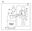
移動型X線診断装置は、撮影対象の患者の付近に配置される。そして、支柱103を軸に回転することで伸縮式アーム102の向きを調節し、伸縮式アーム102により支柱103からの距離を調節することにより、撮影したい箇所に応じた位置にX線管球が配置されるようにX線発生部101が配置される。このときの様子を図2に示す。図2は、病室200において、ベッド201に寝ている患者202のX線撮影を行う様子を概略的に示す図である。操作者203は、X線発生部101を撮影したい箇所に応じた位置に配置するため、伸縮式アーム102の伸縮の程度と、支柱103の回転量とを調節して、撮影を行う。このとき、例えば、X線検出部を制御するために、モニタ104を通じて計算機に命令を与える。このようにして、移動型X線診断装置を用いて、病室200などでのX線撮影を行うことができる。なお、撮影を行わないときは、図1のように、伸縮式アーム102を畳み、本体の側の所定の格納位置にX線発生部101が配置されるように支柱103を回転させることにより、容易に持ち運ぶことができる。
The mobile X-ray diagnostic apparatus is disposed in the vicinity of the patient to be imaged. Then, the direction of the
図1に示すように、撮影を行わない場合にX線発生部101を支柱より本体の側に配置すると、モニタ104の上部にX線発生部101が配置され、格納時における、操作者のモニタ104の参照可能範囲と入力可能範囲とを制約しうる。そのため、操作者は、撮影予定患者リスト等を参照したい場合などは、X線撮影を行わない移動時等であっても、X線発生部101を所定の格納位置から移動させることとなり、操作性が低下するという課題があった。
As shown in FIG. 1, when the
本発明は上記課題に鑑みなされたものであり、撮影を行わない場合にも操作を可能とする移動型X線診断装置を提供することを目的とする。 The present invention has been made in view of the above problems, and an object thereof is to provide a mobile X-ray diagnostic apparatus that can be operated even when imaging is not performed.
上記目的を達成するため、本発明による移動型X線撮影装置は、X線を発生させるX線発生部および前記X線発生部を支持すると共に当該X線発生部を所定の位置へ移動させる支持機構と、X線撮影に関する情報の表示と操作の受け付けを行う操作表示部とを有する移動型X線撮影装置であって、前記X線撮影を行わないときの前記X線発生部の格納位置と前記操作表示部の位置との、水平方向と鉛直方向との少なくともいずれかにおける距離が所定距離以上離れるように、前記格納位置が定められ、前記移動型X線撮影装置は、前記X線発生部が格納されているかを判定する判定手段をさらに有し、前記X線発生部が格納されていると判定した場合、前記操作表示部の表示を変更する、ことを特徴とする。
In order to achieve the above object, a mobile X-ray imaging apparatus according to the present invention supports an X-ray generation unit that generates X-rays and the X-ray generation unit and supports the X-ray generation unit to a predetermined position. A mobile X-ray imaging apparatus having a mechanism and an operation display unit for displaying information regarding X-ray imaging and receiving operations, and a storage position of the X-ray generation unit when the X-ray imaging is not performed between the position of the operation display unit, so that the distance in at least one of the horizontal and vertical direction are separated a predetermined distance or more, the storage position is defined et al is, the mobile X-ray imaging apparatus, the X-ray generation It further has a determination means for determining whether the part is stored, and when it is determined that the X-ray generation part is stored, the display of the operation display part is changed .
本発明によれば、移動型X線診断装置の操作性を向上させることができる。 According to the present invention, the operability of the mobile X-ray diagnostic apparatus can be improved.
以下、添付図面を参照して本発明の実施の形態を詳細に説明する。 Hereinafter, embodiments of the present invention will be described in detail with reference to the accompanying drawings.
<<実施形態1>>
図3は、本実施形態に係る移動型X線診断装置の構成を概略的に示す図である。本実施形態に係る移動型X線診断装置は、図1の移動型X線診断装置と同様に、X線発生部101、伸縮式アーム102、支柱103、モニタ104、台車105を有する。また、台車105には、それぞれ2つの車輪107及び108、すなわち4つの車輪が備えられ、これにより、移動型X線診断装置は病院施設内で移動することができる。なお、前輪108又は後輪107のいずれかは、例えば操作者がハンドル106に備えられた操作部を操作したことに応じて駆動する電動モータを備え、移動型X線診断装置の移動を補助してもよい。また、台車105は、例えば内部にバッテリーを備え、装置の移動、X線の曝射、デジタル式平面ディテクタ(X線検出部)、ディテクタの制御用計算機、及びモニタ104に電力を供給する。なお、モニタ104は、情報の表示機能だけでなく、計算機等の操作を受け付ける操作機能をも有する操作表示部である。
<<
FIG. 3 is a diagram schematically showing the configuration of the mobile X-ray diagnostic apparatus according to the present embodiment. The mobile X-ray diagnostic apparatus according to the present embodiment includes an
また、移動型X線診断装置では、図1及び図2の例で示すように、台車105の前方に第1の方向に延びる支柱103と、第1の方向に交差する第2の方向(垂直方向)に伸縮するように支柱に接合された伸縮式アーム102と、を有する。そして、移動型X線診断装置は、さらに、伸縮式アーム102の先端に取り付けられたX線発生部101を備える。X線発生部101は、X線管球を含むユニットである。伸縮式アーム102と支柱103は、X線発生部101を支持する支持機構であると共に、X線発生部101が所定の位置へ移動させる移動機構である。
In the mobile X-ray diagnostic apparatus, as shown in the examples of FIGS. 1 and 2, a
X線撮影時には、X線発生部101は、支柱103および伸縮式アーム102の可動範囲内でX線撮影に適した位置に移動させる。また、X線検出部は、X線焦点と撮影対象部位の延長上に設置され、保持される。そして、撮影対象となる患者の撮影部位に最適なX線管電圧、管電流、撮影時間等が設定され、撮影が実行される。X線検出部は、X線の曝射に同期してX線信号を読み取り、信号を計算機に転送する。そして、計算機は、転送された信号に基づいて画像処理を行い、信号を診断に有効な画像に変換する。変換された画像信号は、有線、無線を問わず、病院内に設置された画像サーバーに送られ、保存、診断用に閲覧される。そして、X線撮影が終了すると、X線発生部101は、移動を容易にするため、例えば支柱103の後方の所定の格納位置へ格納される。なお、格納位置は、例えば、伸縮式アーム102を所定長(例えば最短長)に縮め、移動型X線診断装置の進行方向の逆方向、すなわち、進行方向に向かって支柱の後方に伸縮式アームを配置したときのX線発生部101の位置である。これにより、前方の障害物に接触したとしてもX線発生部101が保護されるため、また、移動時の大きさが十分に小型になるため、移動型X線診断装置を容易に移動させることが可能となる。
During X-ray imaging, the
ここで、本実施形態に係る移動型X線診断装置では、図3に示すように、移動時などのX線撮影を実行しない場合に、X線発生部101を格納する格納位置が、操作者によるモニタ104の操作を妨げないようにしている。すなわち、X線発生部101の格納位置は、移動型X線診断装置の情報表示と操作とを行うための操作表示部を、格納されたX線発生部101が覆うことのない位置としてあらかじめ定められる。すなわち、X線発生部101の格納位置においてモニタ104に表示された情報の内容が視認可能となるように、格納位置における伸縮式アーム102の長さ、支柱103の回転量、及びモニタ104の位置を定める。図3の例では、X線発生部101が格納される水平位置と、モニタ104の水平位置とを所定距離以上離した例である。この場合、X線発生部101が格納される際には、各部の配置は、移動型X線診断装置の進行方向に向かって、モニタ104の前方にX線発生部101が配置され、X線発生部101の前方に支柱が配置されることとなる。このように、進行方向に向かって最後方にモニタ104が配置されるため、操作が容易になる。モニタ104がタッチパネル式の入力デバイスであるときに、このような配置にすると特に操作が容易になる。
Here, in the mobile X-ray diagnostic apparatus according to the present embodiment, as shown in FIG. 3, when X-ray imaging is not performed during movement, the storage position for storing the
なお、ここでの所定距離は、例えばモニタ104とX線発生部101の大きさに比例して決定してもよい。例えば、モニタ104の水平面における中心とX線発生部101の水平面における中心とを結ぶ距離が、移動型X線診断装置の前後方向におけるモニタ104及びX線発生部101の長さの和の2分の1以上となるように、所定距離を決定してもよい。それ以外にも、X線発生部101の格納時にモニタ104で情報の閲覧及び所定の操作が可能となるのであれば、どのような距離を所定距離としてもよい。
The predetermined distance here may be determined in proportion to the size of the
移動型X線診断装置を操作する操作者は、病棟でのX線撮影の指示があった場合に移動型X線診断装置の電源を投入し、X線検出部を制御する計算機に撮影対象の患者の情報を転送し、その内容を参照して患者の病室に移動型X線診断装置を移動させる。このとき、本実施形態に係る移動型X線診断装置であれば、モニタ104がX線発生部101によって全体を覆われることがないため、操作者は、モニタ104を通じて情報の参照および情報の入力等の操作が可能となる。したがって、X線発生部101が格納された状態にあるままに、操作者は、装置の電源投入、撮影患者リストの転送、撮影患者リストの参照、撮影済み画像の処理、画像情報転送、電源遮断等の操作を実行することができるようになる。
An operator operating the mobile X-ray diagnostic apparatus turns on the power of the mobile X-ray diagnostic apparatus when there is an instruction for X-ray imaging in the ward, and the computer that controls the X-ray detection unit The patient information is transferred, and the mobile X-ray diagnostic apparatus is moved to the patient's room with reference to the contents. At this time, in the mobile X-ray diagnostic apparatus according to the present embodiment, since the
なお、X線発生部101を格納した状態で、モニタ104の一部がX線発生部101により覆われる場合は、モニタ104の覆われない部分に操作機能又は情報が表示されるように、モニタ104を制御してもよい。具体的には、例えば、計算機は、X線発生部101が所定の格納位置に配置されたと判定すると、それに応じてモニタ104の表示を変更する。これにより、物理的な構造上、X線発生部101がモニタ104の表示の一部を遮ってしまう場合でも、操作や情報の参照が可能となる。
When the
<<実施形態2>>
実施形態1では、図3を用いて、格納時のX線発生部101とモニタ104との水平方向における距離を所定距離以上離した例を説明したが、これに限られない。以下の実施形態では、ほかの具体的な例を示す。
<<
In the first embodiment, the example in which the distance in the horizontal direction between the
図4は、移動型X線診断装置において、X線発生部101が格納される場合の鉛直方向での位置とモニタ104の鉛直方向での位置を所定距離以上離したものである。この結果、X線発生部101が格納される場合に、台車の進行方向から見て後方の所定の角度(図4では45度)から視認した際に、モニタ104がX線発生部101に覆われないこととなる。所定距離は、モニタ104の外縁上の所定の一点(例えば表示領域の前方側の辺の一点)とX線発生部101の外縁上の一点(例えば、X線発生部101の外縁の最後方の点)とを結ぶ直線と、水平面とのなす角が所定の角度以上となるように設定される。
FIG. 4 shows a mobile X-ray diagnostic apparatus in which the vertical position when the
なお、所定の角度は、例えば操作者の身長に応じて定まってもよく、所定の角度からのモニタ104の視認性を得るために、X線発生部101の格納位置を鉛直方向又は水平方向において設定してもよい。すなわち、移動型X線診断装置は、操作者の身長を例えばモニタ104を介して取得する機能を備え、その身長とモニタ104が配置される高さとに応じてX線発生部101の格納位置を制御してもよい。
The predetermined angle may be determined according to, for example, the height of the operator. In order to obtain the visibility of the
例えば、操作者の身長が高いほど、所定の角度を大きくしてもよく、この場合、X線発生部の鉛直方向の格納位置とモニタ104の鉛直方向の位置とを、より離して格納してもよい。また、同様の場合に、X線発生部の水平方向における格納位置とモニタ104の水平位置とを、より離してもよい。なお、X線発生部の水平方向における格納位置は、伸縮式アーム102によって定まることが考えられる。この場合は、例えばモニタ104の方を水平方向に移動させることで、水平方向における距離を確保してもよい。これにより、操作者の身長が高い場合でも高い操作性を実現することができる。
For example, the predetermined angle may be increased as the height of the operator increases. In this case, the vertical storage position of the X-ray generation unit and the vertical position of the
また、操作者の身長が低い場合は、所定の角度を小さくしてもよく、X線発生部の鉛直方向の格納位置とモニタ104の鉛直方向の位置とを、より接近させてもよい。同様に、X線発生部の水平方向の格納位置とモニタ104の水平位置とをより接近させてもよい。これにより、X線撮影を行わない場合に、移動型X線診断装置をより容易に移動することが可能となる。
Further, when the height of the operator is low, the predetermined angle may be reduced, and the vertical storage position of the X-ray generation unit and the vertical position of the
なお、図5に示すように、伸縮式アーム102の入れ子構造を4段以上の多段式とすることにより、X線発生部101の水平方向の格納位置とモニタ104の水平位置とを、3段式以下のアームを用いた場合より離してもよい。すなわち、支柱103に支持される第1のアームと、第1のアームに接続される3段以上の第2のアームとを有していてもよい。また、3段以上の第2のアームのうち、伸縮式アーム102の端部にあたる第2のアームがX線発生部101を支持する。さらに、3段以上の第2のアームは、それぞれが等しい可動範囲を有していてもよい。なお、ここでの「等しい可動範囲」とは、所定範囲(例えば数センチメートルなど)の差を含み、実質的に等しい可動範囲を有することを指すものである。すなわち、可動範囲が完全に一致することは必要としない。これにより、3段以上の第2のアームを用いることにより、広い可動範囲を確保しながら、アームの全体としての最短長が短くなり、よりコンパクトに移動型X線診断装置を移動させることが可能となる。また、例えばテレスコピックアームの場合、可動範囲を3段以上の第2のアームで等しくすることにより、効率よく広い範囲にX線発生部101を配置することが可能となる。このように、X線発生部101を格納する際にモニタ104に表示された情報の内容が視認可能となるように、格納位置におけるアームの長さを定めることで、移動型X線診断装置を移動させる際にモニタ104の表示情報へのアクセスと操作とを実行できる。
As shown in FIG. 5, the
また、特許文献2が示すように、伸縮式アーム102を伸展方向に対し支柱103の側面又は上部に接合することにより、X線発生部101を格納する場合のアームの長さを抑えることができる。したがって、伸縮式アーム102を伸展方向に対して支柱103の側面又は上部に接合することで、台車のスペースが小さい場合でも、格納する際のX線発生部101の水平方向の位置とモニタ104の水平位置とを、十分に離すことが可能となる。
Further, as shown in
<<実施形態3>>
本実施形態では、図6に示すように、X線発生部101の格納位置を、モニタ104の背面とする。図6の例は、モニタ104の設置角度を変更して、その表示及び操作を行う面の反対側、すなわちモニタ104の背面にX線発生部101が格納される例を示している。このときの設置角度は、例えば、水平面とモニタの操作及び表示を行う面とのなす角が所定の角度以上であるようにする。モニタ104は、基本位置として図6に示すような設定角度を有していてもよいし、X線発生部101の格納時に移動するようにしてもよい。モニタ104を移動させる場合は、X線発生部101の格納と連動してモニタ104を移動させてもよい。例えば、移動型X線診断装置は、モニタ104においてX線撮影終了を指示するボタンの押下を検出することにより、自動的に図6に示すような形態となるように、モニタ104と、伸縮式アーム102及び支柱103を制御してもよい。
<<
In the present embodiment, as shown in FIG. 6, the storage position of the
なお、モニタ104の設置角度ではなく、位置を移動させてもよい。例えば、図7に示すように、モニタ104の設置位置をX線発生部101の格納位置より上方に移動させてもよい。また、モニタ104の設置位置は、固定的に図7のようにX線発生部101の格納位置の上方であってもよい。また、この場合にもモニタ104の設置角度は可変であってもよく、例えば、X線発生部101を格納する際にはX線発生部101と衝突しないように水平方向から90度起こし、X線発生部101が格納位置に到達したときに角度を元に戻してもよい。また、X線発生部101を格納する場合の所定の角度を設定しておき、X線発生部101が格納位置に到達したときにその角度となるようにモニタ104の設置角度を制御してもよい。
Note that the position of the
<<実施形態4>>
上述の実施形態1〜3においては、X線発生部101と伸縮式アーム102との相対的な位置が固定の場合について説明した。本実施形態では、さらに、X線発生部101と伸縮式アーム102とが可動的に接合される例について説明する。図8は、X線発生部101を伸縮式アーム102の下方に移動させる移動型X線診断装置を概略的に示す図である。このように、X線発生部101を伸縮式アーム102の下部に移動させることにより、水平方向における、X線発生部101及び伸縮式アーム102の合計の長さが短くなる。したがって、X線発生部101の格納場所の水平位置とモニタ104の水平位置とを容易に所定距離以上離すことが可能となる。また、図9に示すように、X線発生部101を伸縮式アーム102の上方に移動させても同様の効果が得られる。
<<
In the first to third embodiments described above, the case where the relative position between the
なお、これまで説明したX線発生部101の格納方法については、適宜組み合わせることが可能である。すなわち、本実施形態のようにしてX線発生部101とモニタ104との水平方向の距離を所定距離以上離すのに加え、さらに、鉛直方向でも所定距離以上離すようにしてもよい。これらを組み合わせることにより、台車上のスペースが狭い場合であっても、移動型X線診断装置の操作性を向上させることができる。
The storage methods of the
<<実施形態5>>
上述の実施形態では、伸縮式アーム102を有する移動型X線診断装置について説明した。このような移動型X線診断装置では、操作者は、患者ベッドの一方に移動型X線診断装置を移動させ、X線発生部101の支持機構のロック機構を解除し、X線発生部101をベッド側に回転させた上でベッドの反体側へ回り、X線管球の位置合わせを行う。しかし、病室の環境等により、患者ベッドの反体側に回ることが容易でない場合もある。例えば、手術室でのX線撮影では、ベッドの反対側では医師が術式を行っており、操作者が回り込むことができない。さらにX線発生部101の位置調整を術野上部で行うことにより、術野の清潔が侵される虞がある。このように、ベッドの反対側へ回れない場合、伸縮式アーム102を有する移動型X線診断装置の位置調整が困難となり、操作性が低下するという課題があった。
<<
In the above-described embodiment, the mobile X-ray diagnostic apparatus having the
そこで、本実施形態では、ベッドの反対側へ回れない場合でも、X線発生部101の位置調整を容易にするために、伸縮式アーム102を多段式アームとし、そのアームの中間段に、支持機構のロック解除ボタンと、ハンドルとを設ける。そして、このハンドルにより、ハンドルが取り付けられたアームの中間段の伸縮操作が行われると、他の段の伸縮操作が連動して行われるようにする。これにより、伸縮式アーム102の伸縮操作を支柱103の付近で行うことができるようになるため、ベッドの反対側へ回る必要がなくなる。さらに、手術中などに、術野の真上で操作を行うことがなくなるため、清潔さを確保することができる。
Therefore, in the present embodiment, even when it is not possible to turn to the opposite side of the bed, in order to facilitate the position adjustment of the
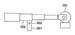
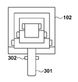
具体的には、例えば図10から図13のように、伸縮式アームを構成する。図10は、2段目下部にハンドルを設置した場合の多段伸縮式アームの側面図である。また、図11は、この場合の多段伸縮式アームの断面図である。同様に、図12は、2段目上部にハンドルを設置した場合の多段伸縮式アームの側面図である。また、図13は、この場合の多段伸縮式アームの断面図である。 Specifically, for example, as shown in FIGS. 10 to 13, an extendable arm is configured. FIG. 10 is a side view of the multi-stage telescopic arm when a handle is installed at the lower part of the second stage. FIG. 11 is a cross-sectional view of the multistage telescopic arm in this case. Similarly, FIG. 12 is a side view of a multi-stage telescopic arm when a handle is installed at the upper part of the second stage. FIG. 13 is a cross-sectional view of the multistage telescopic arm in this case.
いずれの場合も、ハンドル301に、支持機構のロック解除ボタン302が設けられ、ロック解除ボタン302を押してロックを解除した後に、アームの方向、上下位置、及び長さを調節することできる。なお、「支持機構」とは、支柱103と伸縮式アーム102のことであり、これらがロックされている場合は、支柱の回転、及び伸縮式アームの上下位置の変更と長さの調節を行うことはできない。したがって、本実施形態のような構成にすることにより、ハンドル301とそれに設置されたロック解除ボタン302とを操作するだけで、容易にX線発生部101を所定の位置へ移動させることが可能となる。
In any case, the
なお、上述の説明では、ハンドルを上部または下部に設置したが、側面や、他の位置に設置しても同様の効果を得ることができる。また、ハンドルは2段目に限らず、4段構成の3段目などに設置してもよい。 In the above description, the handle is installed at the upper part or the lower part. However, the same effect can be obtained by installing the handle at the side face or at another position. Further, the handle is not limited to the second stage, and may be installed at the third stage having a four-stage configuration.
<<実施形態6>>
実施形態1〜5においては、伸縮式アーム102のみが伸縮可能である場合について説明したが、本実施形態では、支柱103も伸縮可能とした場合について説明する。図14は、移動型X線診断装置において、凹部に形成されたX線発生部を格納するための格納部にX線発生部がある状態を示している。格納部を設けることにより、X線発生部が外部からの衝撃に対して影響を受けにくくなり、移動型X線診断装置の劣化を防ぐことが可能となる。
<<
In the first to fifth embodiments, the case where only the
図14において、X線診断装置は、例えば、X線管球1、コリメータ2、アーム3、支柱4、アーム支持部5、台車部6、移動機構7、支柱回転部8、モニタ9、格納棒受け部11を有する。上述の実施形態のX線発生部101は例えばX線管球1及びコリメータ2を含み、伸縮式アーム102はアーム3に対応し、支柱103は支柱4に対応し、モニタ104はモニタ9に対応するなど、基本的な構成は上述の実施形態と同様である。
In FIG. 14, the X-ray diagnostic apparatus includes, for example, an
X線管球1は、X線を照射する。コリメータ2は、X線管球1に設置されたX線照射範囲を制限する。アーム3は、X線管球1を支持し、少なくとも水平方向にX線管球1を移動させるための伸縮機能と伸縮位置固定機能とを有する。支柱4は、アーム3を支持する。アーム支持部5は、アーム3と支柱4を連結し、アーム3を支柱4に沿って移動可能な機能と任意の位置に固定可能な機能とを有する。台車部6は、支柱4を支持する。移動機構7は、台車部6を移動可能にするものであり、例えば、複数のタイヤ、又はキャスタを地面に設置した状態で回転することで台車部6を移動させる。支柱回転部8は、台車部6と支柱4とを連結し、ベアリングを構成することで、支柱4を台車部6上で地面と垂直な軸を中心に回転可能にする。また、支柱回転部8は、無励磁作動ブレーキが構成され、無励磁作動ブレーキの通電状態で支柱4の回転を任意の位置で止めることができる。モニタ9は、管球を格納するための格納部の底面側に格納時の管球と接触しない位置に設置される。そして、モニタ9は、回診時に撮影する患者情報や、患者の場所、検査情報リストを表示する。また、撮影条件の設定や撮影したX線画像を院内ネットワークに送信する操作も可能である。格納棒受け部11は、アーム下面10が接触又は近接したことを検知する接触センサを有する。
The
図14の例では、X線発生部の格納のために凹部を台車部6に形成し、X線撮影をしないときには、その凹部として形成された格納部にX線発生部が移動するようにする。そして、格納時に、格納棒受け部11とアーム下面10とが接触又は所定距離以内に近接するようにしている。なお、図14において、アーム下面10は、アーム3から突出する凸部が形成される必要はなく、台車部6に設けられた格納棒受け部11との間で、磁石と磁気センサとをそれぞれ有することで、アーム3やX線管球1が格納位置にあることを検知してもよい。また、図14のような装置では、管球取出し時に、アーム支持部5の支柱4に対する垂直移動と、支柱回転部8のブレーキ解除からの支柱の回転との2つの挙動のみを許可するように構成してもよい。また、図14に示す装置では、管球取出し時に、アーム支持部5の支柱4に対する垂直情報への移動のみの挙動を許可してもよい。アーム3の伸縮を停止させるには、アーム3の伸縮位置固定部を制御することにより行ってもよい。これらにより、さらにX線管球1がモニタ9に接触することがなくなる。
In the example of FIG. 14, a recess is formed in the
図14のように、支柱をも伸縮可能とすることで、X線発生部の格納時のサイズが小さくなる。また、図14のように凹部を設けてそこにX線発生部を格納することで、格納時のサイズがさらに小さくなり、これにより、X線診断装置の移動がさらに容易になる。 As shown in FIG. 14, the size of the X-ray generation unit when stored can be reduced by making the support column extendable and contractible. Further, as shown in FIG. 14, by providing a recess and storing the X-ray generation unit therein, the size at the time of storage is further reduced, and thereby the movement of the X-ray diagnostic apparatus is further facilitated.
なお、図14に示すX線診断装置は、X線管球1とコリメータ2とが露出した形状となっているが、格納部においてこれを一部覆う側壁部が形成されていてもよい。図15は、このような側壁部を格納部に設けたX線診断装置の例を示す図であり、X線管球1とコリメータ2の一部が覆われている。これにより、X線管球1とコリメータ2とを外部からの衝撃から守ることができ、X線診断装置の劣化を防ぐことができる。なお図14のように、側壁部を設けずに、凹部を形成してそこにX線発生部を格納する場合は、X線診断装置の進行方向に対して側面よりX線発生部にアクセスすることが可能となる。したがって、例えば、格納時に上から格納する必要性や、撮影時にまずアームを上に移動させる必要性がなくなり、アームの動作の制約が少なくなるほか、格納時にX線管球を交換するなど、メンテナンスを行うことが容易になる。
In addition, although the X-ray diagnostic apparatus shown in FIG. 14 has a shape in which the
図16は、同様に側壁部を格納部に設けたX線診断装置の例を示し、このX線診断装置においては、格納時のX線管球の周囲に被曝防護用の壁がモニタ9の縁部から突出した形状を有する。図16においては、この被曝防護用の壁は、格納棒受け部11と同様の高さの壁であり、少なくともコリメータ2を覆う高さを有する。これにより、格納時にコリメータ2が外部から衝撃を受ける可能性を低減し、装置の長寿命化に貢献する。制御用計算機は、さらにX線管球1と台車部6の被曝防護用の壁が接触しない回転角度テーブルを有することで、X線管球1が台車部6の被曝防護用の壁に接近していないかを判断することを追加してもよい。これらにより、X線管球1がモニタ9のみならず、台車部6の被曝防護用の壁にも接触することがなくなる。
FIG. 16 similarly shows an example of an X-ray diagnostic apparatus in which a side wall is provided in the storage unit. In this X-ray diagnostic apparatus, an exposure protection wall is provided around the X-ray tube during storage of the
なお、図16のX線診断装置では、モニタ9はその対向する2側面を不図示のモニタガイドレールにスライド可能に取り付けられてされており、スライドすることで出し入れが可能である。例えば、モニタ9は、図の矢印の方向にスライド可能に構成されている。したがって、移動時にも、モニタ9をスライドさせて取り出し、情報の取得や、X線診断装置の操作を行うことができる。
In the X-ray diagnostic apparatus of FIG. 16, the
一方、図17のX線診断装置は、2枚の被曝防護用の壁13が台車部6から突出してコリメータ2の側面を覆うと共に、X線管球1の少なくとも1部をも覆う位置まで延びている。これにより、収納時にコリメータ2及びX線管球1が外部から衝撃を受ける可能性を図16の場合と比べてさらに低減し、装置の長寿命化に貢献する。なお、図17のX線診断装置の格納部は、アーム3の幅に対応し、アーム3と嵌合する溝が形成されている。X線発生部の格納時に、まず、アーム3が縮められると共に、進行方向に向かって逆方向に回転させられ、その後アーム3の支柱4方向での位置(高さ)を下げることにより、X線発生部が格納される。そのとき、このような溝が形成されていると、アームをより低い位置へ格納することができるため、よりコンパクトな形状にすることができると共に、X線管球1やコリメータ2が外部に露出する箇所を極力減らすことが可能となる。また、X線発生部の格納時のアームの位置が下げられると、前方視認性もよくなり、移動型X線診断装置の移動が容易になる。
On the other hand, in the X-ray diagnostic apparatus of FIG. 17, two
なお、図17に示す例では、回診車本体あるいは台車部6に配置される第1のモニタ9に加えて、コリメータ2に付属する第2のモニタ12を備える。第1のモニタ9は、図16のX線診断装置と同様に、図の矢印の方向にスライド可能に構成されている。第2のモニタ12は、いわゆるバリアングルな表示部を構成している。例えば表示画面の1辺に沿ってコリメータ2に固定されており、その1辺を軸として第2のモニタ12の向きを変更できる。第2のモニタ12のコリメータ2に対する固定方法はこれに限定されず、例えば、第2のモニタ12をコリメータ2に対して任意の方向に所定角度の範囲で向きを変更可能に構成されていてもよい。また、格納時のX線管球1を取り付けるユニットの表面部分に設置されてもよい。
In addition, in the example shown in FIG. 17, the
図17の例では、X線管球1及びコリメータ2が収納状態にある場合に、制御用計算機の制御により第1のモニタ9の電源をオフにしながら第2のモニタ12の電源をON状態とし、第2のモニタ12により情報を表示し、操作を受け付けることが可能となる。また、これに代えて、またはこれに加えて、スライド可能な第1のモニタ9と第2のモニタ12にそれぞれ異なる情報を表示させ、表示領域を有効利用して操作者の利便性を高めることが可能となる。
In the example of FIG. 17, when the
図18は、支柱4が多段階に伸縮可能で、またアーム3が入れ子式あるいはテレスコピックな構成を有しており、それぞれ操作者の操作に応じて伸縮可能な場合に、これらを展開してX線撮影を行っている様子を示している。台車部6の上にX線高電圧発生装置やX線制御装置並びに制御盤を備えたシステム制御部18が搭載されている。システム制御部18は、X線をも制御する。このシステム制御部18には移動用のハンドルとX線照射情報の表示および照射指令を入力できるディスプレイとが配置されている。また台車部6の上部前方に第1の支柱4−1が台車部6に対して全方位に対して旋回16が可能に設置され、垂直に配置されている。そしてその旋回量を回動変位センサ14で確認している。この第1の支柱4−1には支柱に沿って上下方向(支柱の軸方向)へ移動可能な第2の支柱4−2が構成されている。また通常の病室専用で特に高い位置を必要としない場合は第1の支柱4−1と第2の支柱4−2は一体で構成した一本の支柱でもよい。さらに第2の支柱4−2に対し略直角である水平方向17に収縮可能なX線管球を支持するアーム3が設けられ、移動量を変位センサ15で確認している。このアーム3の先端へX線管球1を備えたユニットが取り付けられ、そのX線管球1の下部にコリメータ2が取り付けられている。そして病室に配置されているベッド19の上には被検者20と、その間に撮像用のフラットパネル21が配置されている。このように支柱及びアームを多段階構成とすることにより、収納時はコンパクトとなり、前方視認性を確保でき、また取り回しが容易になるという効果がある。
FIG. 18 shows a case where the
図19(a)は、モニタ9の表示に関する説明図である。図19(a)は、通常のモニタ表示状態を示し、本実施形態では、X線管球1が収納位置にある状態(装置の移動時)を考慮している。これら図19に示す情報は、X線診断装置に含まれる制御用計算機による制御に応じてモニタ9に表示される。
FIG. 19A is an explanatory diagram relating to the display on the
図19(a)の通常状態では、モニタ表示可能域の全面を表示使用領域1002としている。図19(a)では例えば直前の撮影により得られた放射線画像1002が表示される。また別の例では、次の撮影に向けて、X線センサが撮影可能なレディ状態であるか否か、散乱線除去グリッドがつけられているか否かが表示される。さらに、撮影対象の被検者(患者)情報(氏名、生年月日、年齢、患者ID、性別)、撮影対象の部位又は選択可能な撮影部位の一覧、撮影方向或いは選択可能な撮影方向の一覧等の撮影情報や患者情報等が表示されてもよい。
In the normal state of FIG. 19A, the entire monitor displayable area is a
図19(b)及び(c)は、モニタ9のその他の表示に関する説明図である。モニタ9には、図19(b)に示すように、例えば未撮影の被検者情報や撮影要件を表示させるリストを表示させることができる。かかるリストは、例えば、RIS(Radiology Information System)から無線を通じて取得した管理情報に基づくリストである。また、図19(c)に示すように、例えば、図19(b)のリストに、被験者ごとの撮影の可否の情報を含めてもよい。この撮影の可否の情報は、例えば被検者等の事情で撮影がキャンセルとなったか否か等の情報をRIS等から取得することで得られる情報である。
FIGS. 19B and 19C are explanatory diagrams regarding other displays of the
Claims (28)
前記X線撮影を行わないときの前記X線発生部の格納位置と前記操作表示部の位置との、水平方向と鉛直方向との少なくともいずれかにおける距離が所定距離以上離れるように、前記格納位置が定められ、
前記移動型X線撮影装置は、前記X線発生部が格納されているかを判定する判定手段をさらに有し、
前記X線発生部が格納されていると判定した場合、前記操作表示部の表示を変更する、
ことを特徴とする移動型X線撮影装置。 An X-ray generation unit that generates X-rays, a support mechanism that supports the X-ray generation unit and moves the X-ray generation unit to a predetermined position, and an operation display that displays information about X-ray imaging and accepts operations A mobile X-ray imaging apparatus having a
The storage position so that the distance in at least one of the horizontal direction and the vertical direction between the storage position of the X-ray generation unit and the position of the operation display unit when the X-ray imaging is not performed is a predetermined distance or more. is defined et al is,
The mobile X-ray imaging apparatus further includes a determination unit that determines whether the X-ray generation unit is stored,
When it is determined that the X-ray generation unit is stored, the display of the operation display unit is changed.
A mobile X-ray imaging apparatus characterized by the above.
ことを特徴とする請求項1に記載の移動型X線撮影装置。 The predetermined distance is set so that an angle formed between a straight line connecting a point on the outer edge of the X-ray generation unit and a point on the outer edge of the operation display unit and a horizontal plane is equal to or greater than a predetermined angle.
The mobile X-ray imaging apparatus according to claim 1.
前記所定距離は、前記操作者の身長に応じて定められる、
ことを特徴とする請求項1又は2に記載の移動型X線撮影装置。 Obtaining means for obtaining the height of an operator of the mobile X-ray imaging apparatus;
The predetermined distance is determined according to the height of the operator.
The mobile X-ray imaging apparatus according to claim 1, wherein the apparatus is a mobile X-ray imaging apparatus.
前記X線撮影を行わないときの前記X線発生部が、前記操作表示部の表示及び操作を行う面の背面に格納され、
前記移動型X線撮影装置は、前記X線発生部が格納されているかを判定する判定手段をさらに有し、
前記X線発生部が格納されていると判定した場合、前記操作表示部の表示を変更する、
ことを特徴とする移動型X線撮影装置。 An X-ray generation unit that generates X-rays, a support mechanism that supports the X-ray generation unit and moves the X-ray generation unit to a predetermined position, and an operation display that displays information about X-ray imaging and accepts operations A mobile X-ray imaging apparatus having a
The X-ray generation unit when the X-ray imaging is not performed is stored on the back of the surface on which the operation display unit performs display and operation ,
The mobile X-ray imaging apparatus further includes a determination unit that determines whether the X-ray generation unit is stored,
When it is determined that the X-ray generation unit is stored, the display of the operation display unit is changed.
A mobile X-ray imaging apparatus characterized by the above.
ことを特徴とする請求項4に記載の移動型X線撮影装置。 An angle formed between a surface on which the display and operation of the operation display unit are performed and a horizontal plane is a predetermined angle or more,
The mobile X-ray imaging apparatus according to claim 4.
ことを特徴とする請求項4又は5に記載の移動型X線撮影装置。 The operation display unit is located above the X-ray generation unit when the X-ray imaging is not performed.
The mobile X-ray imaging apparatus according to claim 4 or 5, wherein
ことを特徴とする請求項4から6のいずれか1項に記載の移動型X線撮影装置。 When the X-ray generation unit is stored, the operation display unit moves.
The mobile X-ray imaging apparatus according to any one of claims 4 to 6, wherein:
ことを特徴とする請求項1から7のいずれか1項に記載の移動型X線撮影装置。 The support mechanism has a multistage telescopic arm,
Mobile X-ray imaging apparatus according to any one of claims 1 to 7, characterized in.
前記伸縮式アームは、当該伸縮式アームの伸展方向に対して前記支柱の側面又は上部に接合される、
ことを特徴とする請求項1から8のいずれか1項に記載の移動型X線撮影装置。 The support mechanism includes a support and a telescopic arm that extends and contracts in a direction perpendicular to the support and is joined to the support.
The telescopic arm is joined to the side surface or upper part of the support column with respect to the extending direction of the telescopic arm.
Mobile X-ray imaging apparatus according to any one of claims 1 8, characterized in.
前記X線発生部の格納時に、当該X線発生部は、前記伸縮式アームの上方または下方に移動される、
ことを特徴とする請求項1から9のいずれか1項に記載の移動型X線撮影装置。 The support mechanism includes a column and a telescopic arm that expands and contracts in a direction perpendicular to the column and is joined to the column, and the X-ray generation unit is movably joined to the telescopic arm,
When the X-ray generator is stored, the X-ray generator is moved above or below the telescopic arm.
Mobile X-ray imaging apparatus according to any one of claims 1 9, characterized in.
前記多段伸縮式アームの中間段のいずれかに前記支持機構を操作するハンドルを有する、
ことを特徴とする請求項1から10のいずれか1項に記載の移動型X線撮影装置。 The support mechanism has a multistage telescopic arm,
A handle for operating the support mechanism at any one of the intermediate stages of the multistage telescopic arm;
The mobile X-ray imaging apparatus according to any one of claims 1 to 10 , wherein:
ことを特徴とする請求項11に記載の移動型X線撮影装置。 The handle has a button for releasing the lock of the mechanism by which the support mechanism moves the X-ray generation unit,
Mobile X-ray imaging apparatus according to claim 1 1, wherein the.
ことを特徴とする請求項11又は12に記載の移動型X線撮影装置。 When the multistage telescopic arm is expanded and contracted in the intermediate stage to which the handle is attached, the multistage telescopic arm is also operated in conjunction with the other stages.
The mobile X-ray imaging apparatus according to claim 11 or 12 , wherein the apparatus is a mobile X-ray imaging apparatus.
前記X線発生部によるX線撮影に関する情報を表示する表示部と、
第1の方向に延びる軸を有しその軸を中心に回転可能な支柱と、
前記表示部および前記支柱が設置される台車と、
前記X線発生部を支持し、前記第1の方向と交差する第2の方向に延びる伸縮可能なアームであって、前記X線発生部の格納時に前記表示部と前記支柱との間に当該X線発生部を格納することが可能な長さとなるアームと、
前記X線撮影を行わないときの前記X線発生部の格納位置に、前記X線発生部が格納されているかを判定する判定手段と、を有し、
前記X線発生部が前記格納位置に格納されていると判定した場合、前記表示部の表示を変更する、
ことを特徴とする移動型X線撮影装置。 An X-ray generator for generating X-rays;
A display unit for displaying information relating to X-ray imaging by the X-ray generation unit;
A column having an axis extending in the first direction and rotatable about the axis;
A carriage on which the display unit and the support are installed;
An extendable arm that supports the X-ray generation unit and extends in a second direction that intersects the first direction, and is disposed between the display unit and the support column when the X-ray generation unit is stored. An arm having a length capable of storing the X-ray generation unit;
Determination means for determining whether the X-ray generation unit is stored in a storage position of the X-ray generation unit when the X-ray imaging is not performed,
When it is determined that the X-ray generation unit is stored in the storage position, the display of the display unit is changed.
Mobile X-ray imaging apparatus characterized by.
ことを特徴とする請求項14に記載の移動型X線撮影装置。 The arm supports the X-ray generation unit at an end of the arm, and changes the position of the X-ray generation unit according to the expansion and contraction of the arm.
Mobile X-ray imaging apparatus according to claim 1 4, characterized in that.
前記3段以上の第2のアームのうち、前記アームにおける端に位置する第2のアームが前記X線発生部を支持する、
ことを特徴とする請求項14又は15に記載の移動型X線撮影装置。 The arm includes a first arm supported by the support column, and three or more stages of second arms that are movably connected to the first arm in the second direction;
Of the three or more stages of the second arm, the second arm located at the end of the arm supports the X-ray generator,
Mobile X-ray imaging apparatus according to claim 1 4 or 1 5, characterized in that.
ことを特徴とする請求項16に記載の移動型X線撮影装置。 The second arm having three or more stages has an equal movable range.
The mobile X-ray imaging apparatus according to claim 16 .
ことを特徴とする請求項14から17のいずれか1項に記載の移動型X線撮影装置。 When the arm is most contracted and disposed along the direction opposite to the traveling direction of the mobile X-ray imaging apparatus, the X-ray generation unit is disposed in front of the display unit in the traveling direction, and the front is Prop is placed,
Mobile X-ray imaging apparatus according to any one of claims 1 4 1 7, characterized in.
ことを特徴とする請求項14から18のいずれか1項に記載の移動型X線撮影装置。 When the X-ray generation unit is in the storage position, the length of the arm at the storage position is determined so that the information displayed on the display unit is visible.
Mobile X-ray imaging apparatus according to any one of claims 1 4 1 8, characterized in.
ことを特徴とする請求項19に記載の移動型X線撮影装置。 The storage position of the X-ray generation unit is a position of the X-ray generation unit when the arm is contracted to a predetermined length and the arm is disposed in a direction opposite to the traveling direction of the mobile X-ray imaging apparatus. Is,
The mobile X-ray imaging apparatus according to claim 19 .
前記アームは前記第2の支柱に支持される、
ことを特徴とする請求項14から20のいずれか1項に記載の移動型X線撮影装置。 The support column includes a first support column that is rotatably arranged with respect to the carriage, and a second support column that is arranged to be movable in the direction of the axis of the support column with respect to the first support column.
The arm is supported by the second column;
Mobile X-ray imaging apparatus according to any one of claims 1 to 4 2 0 characterized by.
ことを特徴とする請求項21に記載の移動型X線撮影装置。 The arm is arranged to be movable in the direction of the axis of the column with respect to the second column.
Mobile X-ray imaging apparatus according to claim 2 1, wherein the.
ことを特徴とする請求項14から22のいずれか1項に記載の移動型X線撮影装置。 The storage unit for storing the X-ray generation unit at the storage position is formed in the carriage.
Mobile X-ray imaging apparatus according to any one of claims 1 to 4 2 2 wherein.
ことを特徴とする請求項23に記載の移動型X線撮影装置。 The storage portion is a recess formed in the carriage;
Mobile X-ray imaging apparatus according to claim 2 3, characterized in that.
前記格納部は、少なくとも前記コリメータの側面を覆う側壁部を有する、
ことを特徴とする請求項23に記載の移動型X線撮影装置。 The X-ray generation unit includes a collimator,
The storage portion has a side wall portion that covers at least a side surface of the collimator,
Mobile X-ray imaging apparatus according to claim 2 3, characterized in that.
ことを特徴とする請求項23に記載の移動型X線撮影装置。 The storage portion is formed with a groove that corresponds to the width of the arm and fits with the arm at the storage position.
Mobile X-ray imaging apparatus according to claim 2 3, characterized in that.
前記表示部は、前記X線発生部が前記格納部に格納された場合に、前記コリメータの上方に配置される、
ことを特徴とする請求項23に記載の移動型X線撮影装置。 The X-ray generation unit includes a collimator,
The display unit is disposed above the collimator when the X-ray generation unit is stored in the storage unit.
Mobile X-ray imaging apparatus according to claim 2 3, characterized in that.
前記X線撮影を行わないときの前記X線発生部の格納位置に、前記X線発生部が格納されているかを判定する判定手段をさらに有し、前記X線発生部が前記格納位置に格納されていると判定した場合、前記操作表示部の表示を変更することを特徴とする移動型X線撮影装置。 The X-ray generator is further stored in the storage position when the X-ray imaging is not performed, and the X-ray generator is stored in the storage position. When the mobile X-ray imaging apparatus is determined to be operated, the display of the operation display unit is changed.
Priority Applications (2)
| Application Number | Priority Date | Filing Date | Title |
|---|---|---|---|
| JP2012218461A JP6000788B2 (en) | 2012-09-28 | 2012-09-28 | Mobile X-ray equipment |
| US14/036,156 US20140093045A1 (en) | 2012-09-28 | 2013-09-25 | Portable x-ray diagnostic apparatus |
Applications Claiming Priority (1)
| Application Number | Priority Date | Filing Date | Title |
|---|---|---|---|
| JP2012218461A JP6000788B2 (en) | 2012-09-28 | 2012-09-28 | Mobile X-ray equipment |
Publications (3)
| Publication Number | Publication Date |
|---|---|
| JP2014068891A JP2014068891A (en) | 2014-04-21 |
| JP2014068891A5 JP2014068891A5 (en) | 2015-10-29 |
| JP6000788B2 true JP6000788B2 (en) | 2016-10-05 |
Family
ID=50385210
Family Applications (1)
| Application Number | Title | Priority Date | Filing Date |
|---|---|---|---|
| JP2012218461A Expired - Fee Related JP6000788B2 (en) | 2012-09-28 | 2012-09-28 | Mobile X-ray equipment |
Country Status (2)
| Country | Link |
|---|---|
| US (1) | US20140093045A1 (en) |
| JP (1) | JP6000788B2 (en) |
Families Citing this family (7)
| Publication number | Priority date | Publication date | Assignee | Title |
|---|---|---|---|---|
| US9125611B2 (en) | 2010-12-13 | 2015-09-08 | Orthoscan, Inc. | Mobile fluoroscopic imaging system |
| JP6324009B2 (en) * | 2013-09-12 | 2018-05-16 | キヤノン株式会社 | Radiation generating apparatus and radiation imaging apparatus |
| US10151710B2 (en) | 2014-07-18 | 2018-12-11 | Peltec Services, Inc. | Portable industrial radiography apparatus |
| WO2016035146A1 (en) * | 2014-09-02 | 2016-03-10 | 株式会社島津製作所 | Mobile x-ray imaging apparatus |
| CN107847203A (en) * | 2015-07-21 | 2018-03-27 | 富士胶片株式会社 | Radiation device, the control method of radiation device and program |
| AU201515550S (en) * | 2015-10-20 | 2016-06-08 | Micro X Ltd | Mobile x-ray apparatus |
| KR20190042388A (en) * | 2017-10-16 | 2019-04-24 | 삼성전자주식회사 | Apparatus and method for controlling x-ray imaging |
Family Cites Families (16)
| Publication number | Priority date | Publication date | Assignee | Title |
|---|---|---|---|---|
| US3790805A (en) * | 1971-04-19 | 1974-02-05 | Picker Corp | Mobile x-ray unit |
| US4326131A (en) * | 1977-10-03 | 1982-04-20 | Siemens Aktiengesellschaft | Mobile x-ray apparatus |
| DE7730536U1 (en) * | 1977-10-03 | 1979-03-15 | Siemens Ag, 1000 Berlin Und 8000 Muenchen | Mobile X-ray machine |
| JP3888203B2 (en) * | 2002-04-02 | 2007-02-28 | 株式会社島津製作所 | Round-trip X-ray system |
| JP2003310595A (en) * | 2002-04-19 | 2003-11-05 | Shimadzu Corp | X-ray diagnostic system |
| JP4612832B2 (en) * | 2004-12-03 | 2011-01-12 | キヤノン株式会社 | Radiation imaging apparatus and control method thereof |
| JP4515921B2 (en) * | 2005-01-07 | 2010-08-04 | 株式会社日立メディコ | Mobile X-ray device |
| EP2248466B1 (en) * | 2008-02-22 | 2013-06-19 | Hitachi Medical Corporation | Mobile x-ray device |
| WO2010004855A1 (en) * | 2008-07-10 | 2010-01-14 | 株式会社 日立メディコ | Mobile x-ray apparatus |
| US8690425B2 (en) * | 2009-11-25 | 2014-04-08 | Carestream Health, Inc. | Retrofit of a mobile cart |
| US8672543B2 (en) * | 2010-04-13 | 2014-03-18 | Carestream Health, Inc. | Counterweight for mobile x-ray device |
| US8568028B2 (en) * | 2010-04-13 | 2013-10-29 | Carestream Health, Inc. | Mobile radiography unit having collapsible support column |
| US8961011B2 (en) * | 2010-04-13 | 2015-02-24 | Carestream Health, Inc. | Mobile radiography unit having multiple monitors |
| JP4931262B2 (en) * | 2010-06-18 | 2012-05-16 | キヤノン株式会社 | Radiation imaging apparatus and control method thereof |
| US8678649B2 (en) * | 2010-06-25 | 2014-03-25 | Varian Medical Systems, Inc. | Conversion of existing portable or mobile analog radiographic apparatus for enabling digital radiographic applications |
| US9414802B2 (en) * | 2011-09-12 | 2016-08-16 | Carestream Health, Inc. | Charger for electronic grid holders and detectors stored at mobile radiographic imaging apparatus and methods for using the same |
-
2012
- 2012-09-28 JP JP2012218461A patent/JP6000788B2/en not_active Expired - Fee Related
-
2013
- 2013-09-25 US US14/036,156 patent/US20140093045A1/en not_active Abandoned
Also Published As
| Publication number | Publication date |
|---|---|
| JP2014068891A (en) | 2014-04-21 |
| US20140093045A1 (en) | 2014-04-03 |
Similar Documents
| Publication | Publication Date | Title |
|---|---|---|
| JP6000788B2 (en) | Mobile X-ray equipment | |
| US9295438B2 (en) | Movable X-ray generation apparatus | |
| JP6238611B2 (en) | Mobile radiography apparatus, radiography system, and control method | |
| JP5731888B2 (en) | X-ray diagnostic imaging equipment | |
| JP6590946B2 (en) | Medical system | |
| JP2014533188A (en) | Apparatus, system and method for generating X-ray images | |
| US9084582B2 (en) | Radiation imaging apparatus and method of controlling radiation imaging apparatus | |
| CN109788930A (en) | For moving the docking observing system of x-ray system | |
| JP2015196073A (en) | X-ray imaging apparatus and operation method thereof, and program | |
| JP5511142B2 (en) | X-ray equipment | |
| JP2015073752A (en) | Mobile x-ray equipment, control method for the same, and program | |
| JP2018121925A (en) | X-ray computed tomography apparatus and subject carrying tool for x-ray computed tomography | |
| JPWO2017017949A1 (en) | Radiation irradiation equipment | |
| JP5553965B2 (en) | Radiation imaging system | |
| JP6540401B2 (en) | X-ray imaging system, X-ray imaging apparatus, and X-ray detector | |
| JP6342151B2 (en) | Mobile X-ray diagnostic device | |
| JP2015058117A (en) | Mobile x-ray diagnostic apparatus | |
| WO2015008504A1 (en) | X-ray imaging device | |
| JP5404508B2 (en) | X-ray diagnostic equipment | |
| JP7262960B2 (en) | Medical image diagnosis device and imaging planning device | |
| JP2009153579A (en) | X-ray ct system and medical imaging system | |
| JP5537718B1 (en) | Mobile X-ray diagnostic device | |
| JP2017221360A (en) | Radiological imaging system | |
| JP5433345B2 (en) | Medical device bed apparatus and medical device | |
| JP4482004B2 (en) | X-ray diagnostic equipment |
Legal Events
| Date | Code | Title | Description |
|---|---|---|---|
| A521 | Request for written amendment filed |
Free format text: JAPANESE INTERMEDIATE CODE: A523 Effective date: 20150903 |
|
| A621 | Written request for application examination |
Free format text: JAPANESE INTERMEDIATE CODE: A621 Effective date: 20150903 |
|
| A977 | Report on retrieval |
Free format text: JAPANESE INTERMEDIATE CODE: A971007 Effective date: 20160609 |
|
| A131 | Notification of reasons for refusal |
Free format text: JAPANESE INTERMEDIATE CODE: A131 Effective date: 20160627 |
|
| A521 | Request for written amendment filed |
Free format text: JAPANESE INTERMEDIATE CODE: A523 Effective date: 20160704 |
|
| TRDD | Decision of grant or rejection written | ||
| A01 | Written decision to grant a patent or to grant a registration (utility model) |
Free format text: JAPANESE INTERMEDIATE CODE: A01 Effective date: 20160801 |
|
| A61 | First payment of annual fees (during grant procedure) |
Free format text: JAPANESE INTERMEDIATE CODE: A61 Effective date: 20160831 |
|
| R151 | Written notification of patent or utility model registration |
Ref document number: 6000788 Country of ref document: JP Free format text: JAPANESE INTERMEDIATE CODE: R151 |
|
| LAPS | Cancellation because of no payment of annual fees |


