JP5964746B2 - DNA-cell conjugate - Google Patents
DNA-cell conjugate Download PDFInfo
- Publication number
- JP5964746B2 JP5964746B2 JP2012504866A JP2012504866A JP5964746B2 JP 5964746 B2 JP5964746 B2 JP 5964746B2 JP 2012504866 A JP2012504866 A JP 2012504866A JP 2012504866 A JP2012504866 A JP 2012504866A JP 5964746 B2 JP5964746 B2 JP 5964746B2
- Authority
- JP
- Japan
- Prior art keywords
- cell
- cells
- nucleic acid
- dna
- acid moiety
- Prior art date
- Legal status (The legal status is an assumption and is not a legal conclusion. Google has not performed a legal analysis and makes no representation as to the accuracy of the status listed.)
- Active
Links
Images


Classifications
-
- C—CHEMISTRY; METALLURGY
- C12—BIOCHEMISTRY; BEER; SPIRITS; WINE; VINEGAR; MICROBIOLOGY; ENZYMOLOGY; MUTATION OR GENETIC ENGINEERING
- C12N—MICROORGANISMS OR ENZYMES; COMPOSITIONS THEREOF; PROPAGATING, PRESERVING, OR MAINTAINING MICROORGANISMS; MUTATION OR GENETIC ENGINEERING; CULTURE MEDIA
- C12N5/00—Undifferentiated human, animal or plant cells, e.g. cell lines; Tissues; Cultivation or maintenance thereof; Culture media therefor
- C12N5/0006—Modification of the membrane of cells, e.g. cell decoration
-
- A—HUMAN NECESSITIES
- A61—MEDICAL OR VETERINARY SCIENCE; HYGIENE
- A61K—PREPARATIONS FOR MEDICAL, DENTAL OR TOILETRY PURPOSES
- A61K47/00—Medicinal preparations characterised by the non-active ingredients used, e.g. carriers or inert additives; Targeting or modifying agents chemically bound to the active ingredient
- A61K47/50—Medicinal preparations characterised by the non-active ingredients used, e.g. carriers or inert additives; Targeting or modifying agents chemically bound to the active ingredient the non-active ingredient being chemically bound to the active ingredient, e.g. polymer-drug conjugates
- A61K47/69—Medicinal preparations characterised by the non-active ingredients used, e.g. carriers or inert additives; Targeting or modifying agents chemically bound to the active ingredient the non-active ingredient being chemically bound to the active ingredient, e.g. polymer-drug conjugates the conjugate being characterised by physical or galenical forms, e.g. emulsion, particle, inclusion complex, stent or kit
- A61K47/6901—Conjugates being cells, cell fragments, viruses, ghosts, red blood cells or viral vectors
Description
関連出願への相互参照
本出願は、全ての目的のために全体として本明細書に援用される、2009年4月8日に出願された米国出願第61/167,748号、及び2009年9月16日に出願された米国出願第61/243,123号の優先権を主張する。
CROSS REFERENCE TO RELATED APPLICATIONS This application is a U.S. application 61 / 167,748 filed on April 8, 2009, and incorporated by reference herein in its entirety for all purposes. Claims priority of US application 61 / 243,123, filed on May 16th.
政府支援の研究又は開発の下でなされた発明への権利に関する陳述
本研究は、米国エネルギー省によって承認され、エネルギー省、及び科学局、基礎エネルギー科学局のナノスケールサイエンスエンジニアリング及びテクノロジー(NSET)プログラムによって支援された助成金番号DE−AC02−05CH11231により、国立衛生研究所によって承認されたHG003329及びR01GM072700により、並びに国立衛生研究所の分子生物物理学訓練助成金T32GM08295により、政府支援に基づいてなされた。合衆国政府は本発明に一定の権利を有する。
Statement of rights to inventions made under government-sponsored research or development This research is approved by the US Department of Energy and is part of the Nanoscale Science Engineering and Technology (NSET) program of the Department of Energy and the Department of Science and Basic Energy Sciences. Granted under grant support by the National Institutes of Health, HG003329 and R01GM07700 approved by the National Institutes of Health, and by the National Institutes of Health Molecular Biophysics Training Grant T32GM08295, with grant number DE-AC02-05CH11231 supported by . The United States government has certain rights in this invention.
コンパクトディスクで提出された、「配列表」、表、又はコンピュータプログラム表付属物についての言及
該当なし
Reference to “sequence listing”, tables, or computer program listings submitted on a compact disc N / A
典型的には、ペプチドは、研究用の細胞を捕捉するための細胞に基づいたアレイにおいて使用されている。表面は、細胞表面上のインテグリンに結合するように設計された「RGD」ペプチドでプリントされるが、このシステムは、非付着性細胞(例えば、白血球、リンパ球)などのインテグリンを持たない細胞とは作用せず、同じプラットフォームにおいて一緒に異なる細胞型を用いた制御された所定のパターニングは不可能である。RGDシステムは、細胞分化を開始し、したがって、分析される前に細胞が変化するという大きな欠点を有する。これは、インテグリンが分化、また分化に関するインテグリン結合後に続く活性の制御に関与する受容体であるからである(Du,X.P.ら,Cell 1991,65,409−416;Xiong,J.P.ら,Science 2002,296,151−155参照)。 Typically, peptides are used in cell-based arrays to capture research cells. The surface is printed with “RGD” peptides designed to bind to integrins on the cell surface, but this system works with cells that do not have integrins, such as non-adherent cells (eg, leukocytes, lymphocytes). Does not work, and controlled and predetermined patterning using different cell types together on the same platform is not possible. The RGD system has the major drawback of initiating cell differentiation and thus changing cells before they are analyzed. This is because integrins are receptors involved in the regulation of differentiation and the subsequent activity following differentiation-related integrin binding (Du, XP et al., Cell 1991, 65, 409-416; Xiong, JP). Et al., Science 2002, 296, 151-155).
間接的な非共有結合は、DNAが抗体−リガンド相互作用を通じて非共有結合により細胞に間接的に結合するタンパク質−タンパク質結合システムを用いて示されている(Bailey RCら.J Am Chem Soc.2007 Feb 21;129(7):1959−67)。細胞上の標的リガンドに特異的な抗体はDNAにコンジュゲートされる。抗体とリガンドとの間の非共有連結は、タンパク質−タンパク質相互作用の典型である水素結合に基づく。細胞表面リガンドに特異的な抗体上の一本鎖DNA(ssDNA)オリゴマーは、それらのリガンドを有する細胞に結合し、順に、細胞は、細胞を捕捉するために細胞上のパートナー鎖に結合するssDNA相補的オリゴマーを有する表面に固定される。 Indirect non-covalent binding has been shown using a protein-protein binding system in which DNA is indirectly bound to cells by non-covalent bonds through antibody-ligand interactions (Bailey RC et al. J Am Chem Soc. 2007). Feb 21; 129 (7): 1959-67). Antibodies specific for the target ligand on the cell are conjugated to DNA. The non-covalent linkage between the antibody and the ligand is based on hydrogen bonds that are typical of protein-protein interactions. Single-stranded DNA (ssDNA) oligomers on antibodies specific for cell surface ligands bind to cells with those ligands, which in turn bind to partner strands on the cell to capture the cell Immobilized on a surface having a complementary oligomer.
生細胞の表面への合成一本鎖DNA(ssDNA)鎖の間接的共有結合は、最初に、Chandra,R.A.ら.Angew.Chem.−Int.Edit.2006,45,896−901による代謝性オリゴ糖操作を用いて示された。間接的な共有原子価は、DNAの導入前に、過アセチル化されたN−アジドアセチルマンノサミン(Ac4ManNAz)を用いた細胞処理後(3日要する)に得られた細胞表面のシアル酸に導入された特定の化学手(アジド)を介した。次に、ホスフィン−ssDNAコンジュゲートは、アジド糖とDNAとの間にアミド結合(Staudinger連結反応、E.Saxonら.Science 2000,287)を形成するアジド手に共有結合させた。合成アジド糖の代謝によって細胞表面の糖コンジュゲート内に組み込まれたアジドは、多数の安定な細胞−表面付加物を生成するために、ビオチン化されたトリアリールホスフィンと反応させることができる。しかしながら、共有代謝アプローチは、DNA結合のための細胞を調製するのに多くの日数を用紙、細胞表面糖を変更することは、それを分析する機会を得る前に、それを変化する細胞に対して代謝的影響を与える。表面上にシアル酸を所有するある種の哺乳動物細胞にさらに限定され、したがって、細菌、植物細胞、真菌、又は多くの他の動物細胞用に用いることができない。 Indirect covalent binding of synthetic single-stranded DNA (ssDNA) strands to the surface of living cells is first described in Chandra, R .; A. Et al. Angew. Chem. -Int. Edit. It was shown using metabolic oligosaccharide manipulation according to 2006, 45, 896-901. Indirect covalent valency is determined by cell surface sialic acid obtained after cell treatment with peracetylated N-azidoacetylmannosamine (Ac4ManNAz) (requires 3 days) prior to DNA introduction. Via a specific chemical hand (azide) introduced. The phosphine-ssDNA conjugate was then covalently attached to the azide hand forming an amide bond (Staudinger ligation reaction, E. Saxon et al. Science 2000, 287) between the azido sugar and DNA. Azides incorporated into cell surface sugar conjugates by metabolism of synthetic azido sugars can be reacted with biotinylated triarylphosphines to produce a large number of stable cell-surface adducts. However, the shared metabolic approach takes many days to prepare cells for DNA binding, and changing the cell surface sugars will give it a chance to analyze it before it has the opportunity to analyze it. Have metabolic effects. It is further limited to certain mammalian cells that possess sialic acid on their surface and therefore cannot be used for bacteria, plant cells, fungi, or many other animal cells.
また、細胞表面で抗体とリガンドを介した非共有結合は、細胞を活性化し、分析が開始され得る前に細胞を無秩序にさせ、共有結合と比較して、より弱く、「可逆的」となる傾向がある。さらに、抗体機構は細胞表面上にリガンドの普及率を必要とし、細胞表面上に十分なDNAを固定するために、リガンドに特異的な抗体を操作することが必要である。 Also, non-covalent binding via antibody and ligand on the cell surface activates the cell and makes the cell disordered before analysis can be initiated, making it weaker and “reversible” compared to covalent binding. Tend. Furthermore, the antibody mechanism requires a high penetration rate of the ligand on the cell surface, and it is necessary to manipulate an antibody specific for the ligand in order to immobilize sufficient DNA on the cell surface.
細胞表面での抗体を用いたリガンドの相互作用を介した非共有結合は形成され、この場合、抗体は、タンパク質結合DNAの鎖を担持する(Bailey RCら.J Am Chem Soc.2007 Feb 21;129(7):1959−67)。両方法は、研究のために捕捉しようとする細胞を活性化するという即座の不利益を有し、したがって、分析され得る前に、対象とするものを異なるものに形質転換する。細胞表面上のDNA結合のこれらの初期システムにおける欠点を克服することは、この正に記載された初期分野を変換し、従前は可能でなかった価値あるツール及び操作を提供することができるであろう。 A non-covalent bond is formed through ligand interaction with the antibody at the cell surface, in which case the antibody carries a chain of protein-bound DNA (Bailey RC et al. J Am Chem Soc. 2007 Feb 21; 129 (7): 1959-67). Both methods have the immediate disadvantage of activating the cells that are to be captured for study, and thus transforming the one of interest into a different one before it can be analyzed. Overcoming the shortcomings in these early systems of DNA binding on the cell surface can transform this positively described early field and provide valuable tools and operations that were not possible previously. Let's go.
一態様では、本発明は、細胞を有する組成物を提供し、ここで、細胞は、天然の官能基を含む表面を有し、該細胞は細胞壁を持たない。また、この組成物は、核酸部分を含み、該核酸部分は、天然の官能基に共有結合する。 In one aspect, the present invention provides a composition having cells, wherein the cells have a surface comprising natural functional groups and the cells do not have a cell wall. The composition also includes a nucleic acid moiety that is covalently linked to a natural functional group.
別の態様では、本発明は、細胞と活性化された核酸部分とを接触させることによって、細胞と核酸部分のコンジュゲートを調製する方法を提供し、ここで、細胞は天然の官能基を含む表面を有し、さらに細胞は細胞壁を持たず、それにより、核酸部分は天然の官能基に共有結合される。 In another aspect, the invention provides a method of preparing a conjugate of a cell and a nucleic acid moiety by contacting the cell with an activated nucleic acid moiety, wherein the cell comprises a natural functional group. It has a surface and the cells do not have a cell wall, whereby the nucleic acid moiety is covalently bound to a natural functional group.
別の態様では、本発明は、細胞を有する組成物を提供し、ここで、細胞は細胞壁及び核酸部分を有し、該核酸部分は細胞に共有結合される。 In another aspect, the present invention provides a composition comprising a cell, wherein the cell has a cell wall and a nucleic acid moiety, wherein the nucleic acid moiety is covalently bound to the cell.
別の態様では、本発明は、細胞と活性化された核酸部分とを接触させることによって、細胞と核酸部分のコンジュゲートを調製する方法を提供し、ここで、細胞は細胞壁を含み、それにより核酸部分は細胞に結合される。 In another aspect, the present invention provides a method of preparing a conjugate of a cell and a nucleic acid moiety by contacting the cell with an activated nucleic acid moiety, wherein the cell comprises a cell wall, thereby The nucleic acid moiety is bound to the cell.
別の態様では、本発明は、第1の核酸部分に共有結合された天然の官能基の細胞表面を有する細胞、第1の核酸部分に相補的である第2の核酸部分を有する基質表面を含むデバイスを提供し、それにより細胞は第1と第2の核酸部分の核酸二重鎖の形成を介して基質表面に結合される。 In another aspect, the invention provides a cell having a natural functional group cell surface covalently linked to a first nucleic acid moiety, a substrate surface having a second nucleic acid moiety that is complementary to the first nucleic acid moiety. A device is provided, whereby cells are bound to a substrate surface via formation of a nucleic acid duplex of first and second nucleic acid moieties.
発明の詳細な説明
I.一般
本発明は、代謝工学を必要とせずに、細胞表面上の天然の官能基を介して、DNAを細胞に共有的に連結させることによって調製される細胞−DNAコンジュゲートの最初の例を提供する。細胞壁を持たない哺乳動物細胞及び他の細胞について、DNAは、リジンアミンなどの、細胞表面のアミノ酸に直接共有結合される。細胞壁を持たない植物細胞及び他の細胞について、細胞の表面上の糖類は、アルデヒド又はケトンを形成するように、例えば酸化によって最初に修飾され、次にDNAで修飾される。
Detailed Description of the Invention General The present invention provides the first example of a cell-DNA conjugate prepared by covalently linking DNA to cells via natural functional groups on the cell surface without the need for metabolic engineering To do. For mammalian cells and other cells that do not have a cell wall, the DNA is covalently linked directly to cell surface amino acids, such as lysineamine. For plant cells and other cells that do not have a cell wall, the saccharide on the surface of the cell is first modified, for example by oxidation, and then with DNA to form aldehydes or ketones.
DNA−細胞コンジュゲートを形成させるための従来の方法は、代謝工学の使用、即ち、細胞によって代謝され、細胞表面に発現されるアジド官能化糖などの適切に修飾された糖を有する細胞を与えることを必要としていた。次に、DNAは、アジド修飾糖にコンジュゲートされた。この方法は、有用性及び応用範囲の両方を制限するいくつかの欠点を有している。例えば、代謝工学は、細胞表面上に十分量のアジド官能化糖を細胞に与えるのに数日を必要とする。また、修飾することができる細胞型は制限され、重要な診断的有用性を有する初代細胞を排除する。最後に、代謝工学は、必ず、細胞の構造及び特性を修飾する。 Conventional methods for forming DNA-cell conjugates provide the use of metabolic engineering, ie cells with appropriately modified sugars such as azide functionalized sugars that are metabolized by the cells and expressed on the cell surface I needed that. The DNA was then conjugated to an azide modified sugar. This method has several disadvantages that limit both utility and scope of application. For example, metabolic engineering requires several days to give a cell a sufficient amount of azide-functionalized sugar on the cell surface. Also, the cell types that can be modified are limited, eliminating primary cells that have significant diagnostic utility. Finally, metabolic engineering necessarily modifies the structure and properties of cells.
本発明は、細胞表面に既に存在する天然の官能基を用いることによって、アジド代謝工学プロセルに固有の困難性を克服する。NHS−エステルで修飾されたDNAなどの活性化DNA配列を用いることによって、DNAは、細胞壁を持たない細胞の細胞表面上にリジン基のアミンに共有結合可能である。このプロセスは最大数時間を要する。プロセスの容易さにより、初代細胞、並びに幹細胞の修飾も可能にする。細胞壁を有する細胞について、細胞表面上の天然の糖は、アルデヒド及びケトンを形成するために、例えば、酸化によって最初に修飾され、次に例えば、アミノオキシ基を用いて活性化DNA配列と反応させる。本発明のDNA−細胞コンジュゲート及び方法は、細胞検出及びアッセイ方法に向かって実質的な跳躍を表す。 The present invention overcomes the difficulties inherent in azide metabolic engineering processes by using natural functional groups already present on the cell surface. By using an activated DNA sequence such as DNA modified with NHS-esters, the DNA can be covalently bound to a lysine amine on the cell surface of cells that do not have a cell wall. This process takes up to several hours. The ease of the process allows modification of primary cells as well as stem cells. For cells with cell walls, the natural sugars on the cell surface are first modified, for example by oxidation, to form aldehydes and ketones and then reacted with activated DNA sequences using, for example, aminooxy groups . The DNA-cell conjugates and methods of the present invention represent a substantial leap towards cell detection and assay methods.
表面で細胞をパターニングする能力は、種々の研究及び応用のための新しいプラットフォームを提供し、例えば、細胞生物学の研究、幹細胞分化の制御、及び新しい組織の操作が挙げられる。典型的には、細胞に基づくアッセイは、細胞表面上のインテグリンに結合するように設計される「RGD」を用いて、対象の表面をプリントすることによって形成される。このアプローチは多数の細胞型の固定化に幅広く適合されている一方で、非付着細胞(例えば白血球)を捕捉し、又は独自のアレイ特徴に複数の細胞型を結合するために使用することができない。また、細胞分化又は行動において望ましくない変化を引き起こす可能性があり、これは、これらのプロセスの制御と関わる正に表面受容体を用いるためである。 The ability to pattern cells on the surface provides a new platform for various studies and applications, including cell biology studies, control of stem cell differentiation, and manipulation of new tissues. Typically, cell-based assays are formed by printing the surface of interest using “RGD” designed to bind to integrins on the cell surface. While this approach is widely adapted for the immobilization of many cell types, it cannot be used to capture non-adherent cells (eg, white blood cells) or bind multiple cell types to unique array features. . It can also cause undesired changes in cell differentiation or behavior, because it uses just surface receptors involved in the control of these processes.
これらの制限を回避するために、相補鎖でプリントされた表面に対して細胞膜に共有結合された合成DNA鎖のハイブリダイゼーションを介した生細胞の捕捉は、参照により本明細書中に援用されるChandra,R.A.;Douglas,E.S.;Mathies,R.A.;Bertozzi,C.R.;Francis,M.B.Angew.Chem.−Int.Edit.2006,45,896−901における発明者らの幾人かによって従来報告されている。複数の細胞型が単一の基質にパターニングされるようにすることに加えて、この方法は、基質再利用及び同調性の重要な利点を提供した。最も重要なことには、このアプローチは、接着細胞に加えて非接着細胞を捕捉するために用いられ、この受容体に依存しないプロセスを通じた固定化の結果として、細胞は行動の最小の変化を経験することが示されている。従来の報告では、この方法の有用性はまた、複雑な細胞パターンの形成について示されている。 To circumvent these limitations, capture of live cells via hybridization of synthetic DNA strands covalently bound to the cell membrane to surfaces printed with complementary strands is incorporated herein by reference. Chandra, R.A. A. Douglas, E .; S. Mathies, R .; A. Bertozzi, C .; R. Francis, M .; B. Angew. Chem. -Int. Edit. Previously reported by some of the inventors in 2006, 45, 896-901. In addition to allowing multiple cell types to be patterned into a single substrate, this method provided important advantages of substrate reuse and synchrony. Most importantly, this approach is used to capture non-adherent cells in addition to adherent cells, and as a result of immobilization through this receptor-independent process, cells undergo minimal changes in behavior. Shown to experience. In previous reports, the usefulness of this method has also been shown for the formation of complex cell patterns.
従来の研究で用いられたDNA鎖は、2工程プロセスを通じて細胞表面のグリカンに組み込まれた。最初に、細胞は、1〜3日間、アジド含有マンノース誘導体と一緒に与えられた。この糖は、その後代謝され、細胞表面のグリカンを含むシアル酸に組み込まれた。次に、DNAは、Staudinger連結を用いてアジド官能性に標的化された。効果的ではあるが、このプロトコールは、培養された哺乳動物細胞株について最も適切であり、これは、十分な数のアジド基を組み込むために数日の曝露が必要である。 The DNA strand used in previous studies was incorporated into cell surface glycans through a two-step process. Initially, cells were fed with azide-containing mannose derivatives for 1-3 days. This sugar was then metabolized and incorporated into sialic acid containing cell surface glycans. The DNA was then targeted to azide functionality using Staudinger ligation. Although effective, this protocol is most appropriate for cultured mammalian cell lines, which require several days of exposure to incorporate a sufficient number of azide groups.
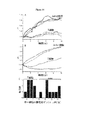
DNAに基づく接着法の一般性を拡張するために、本発明は、実質的に任意の細胞表面に核酸の直接的な組込みのための改善された方法を提供する。ここで図1に言及すると、一態様では、活性化された又は官能化された単一鎖の核酸は、最初に、緩衝溶液中で化学的リンカーと反応し、核酸−リンカーコンジュゲートを形成する。細胞又は細胞表面は、細胞表面への化学的リンカーを介して核酸を結合させるように進行する反応を可能にする特定の期間、緩衝溶液に曝露される。細胞への核酸の結合後、細胞を細胞培地に戻す。種々の濃度の核酸は、図1aにおいて記載及び定量されるように用いることができる。一態様では、オリゴヌクレオチドは、化学的リンカーにオリゴヌクレオチドをコンジュゲートするために、中性緩衝溶液中で化学的リンカーと反応させられ、次に、細胞は、30分間、オリゴヌクレオチド−リンカーコンジュゲートを含む緩衝溶液とともにインキュベートされ、細胞表面へのオリゴヌクレオチド−リンカーコンジュゲートの修飾及び結合を可能にする。 In order to extend the generality of DNA-based adhesion methods, the present invention provides improved methods for the direct integration of nucleic acids on virtually any cell surface. Referring now to FIG. 1, in one aspect, an activated or functionalized single-stranded nucleic acid is first reacted with a chemical linker in a buffer solution to form a nucleic acid-linker conjugate. . The cell or cell surface is exposed to a buffer solution for a specific period of time that allows the reaction to proceed to attach the nucleic acid via a chemical linker to the cell surface. After binding of the nucleic acid to the cells, the cells are returned to the cell culture medium. Various concentrations of nucleic acid can be used as described and quantified in FIG. In one aspect, the oligonucleotide is reacted with the chemical linker in a neutral buffer solution to conjugate the oligonucleotide to the chemical linker, and then the cells are allowed to react for 30 minutes with the oligonucleotide-linker conjugate. Incubate with a buffer solution containing to allow modification and attachment of the oligonucleotide-linker conjugate to the cell surface.
細胞修飾は、典型的には、リンカーと細胞表面上のアミノ酸との間の共有結合の形成を介して開始する。いくつかの態様では、細胞表面は、動物、植物、藻、又は細菌細胞を含む任意の起源由来の生細胞の細胞膜であってもよい。いくつかの態様では、共有結合はアミド結合又はエステル結合である。いくつかの態様では、オリゴヌクレオチドは単一鎖である。他の態様では、アミノ酸は、リジン、システイン、チロシン、セリン、アスパラギン酸、グルタミン酸及びトリプトファンから選択される。 Cell modification typically begins through the formation of a covalent bond between the linker and an amino acid on the cell surface. In some aspects, the cell surface may be the cell membrane of live cells from any source including animal, plant, algae, or bacterial cells. In some embodiments, the covalent bond is an amide bond or an ester bond. In some embodiments, the oligonucleotide is single stranded. In other embodiments, the amino acid is selected from lysine, cysteine, tyrosine, serine, aspartic acid, glutamic acid and tryptophan.
細胞壁を有する植物細胞などの細胞では、連結は、とりわけヒドラゾン、オキシム、又はアミンであってもよく、ここで、結合は、過ヨウ素酸塩酸化、続くヒドラゾン、オキシム又はアミン形成を介して生じる。一態様では、細胞上の糖質は酸化を受けアルデヒド官能基を生じさせ、次にアルデヒドは、植物細胞表面の直接細胞修飾のためのオリゴヌクレオチド−リンカーコンジュゲートと反応する。 In cells, such as plant cells with cell walls, the linkage may be a hydrazone, oxime, or amine, among others, where binding occurs via periodate oxidation followed by hydrazone, oxime, or amine formation. In one aspect, the carbohydrate on the cell is oxidized to produce an aldehyde functional group, which then reacts with an oligonucleotide-linker conjugate for direct cellular modification of the plant cell surface.
この手法は、いくつかの態様では1時間未満で行うことができ、対象の任意のオリゴヌクレオチド配列を用いて等レベルの細胞表面官能化をもたらす。本方法は、RT−PCR分析用の単一細胞の捕捉、力測定又は細胞パターニング技術用の固体基質への生細胞の結合を含む種々の態様に適用され得る。実施例では、本発明者らは、他の方法を用いたパターニングが困難であるあらゆる細胞型である、赤血球、初代T細胞、及び筋芽細胞を捕捉するためのこの新しい標識法の使用を示す。この新しい技術は、DNAに基づく接着ストラテジーの範囲を大いに拡張し、有機合成を専門にしない研究室において使用するには十分に容易である。 This approach can be performed in less than an hour in some embodiments and results in equal levels of cell surface functionalization with any oligonucleotide sequence of interest. The method can be applied to a variety of embodiments including single cell capture for RT-PCR analysis, force measurement or binding of living cells to a solid substrate for cell patterning techniques. In the examples, we demonstrate the use of this new labeling method to capture red blood cells, primary T cells, and myoblasts, which are all cell types that are difficult to pattern using other methods. . This new technology greatly expands the range of DNA-based adhesion strategies and is easy enough to use in laboratories that do not specialize in organic synthesis.
したがって、一態様では、本発明は、直接共有結合されたオリゴヌクレオチドを有する細胞膜を含む組成物である。細胞膜は全体として無傷な細胞であってもよく、それにより、組成物は直接共有結合されたオリゴヌクレオチドを有する全細胞を含む。多数のオリゴヌクレオチドは単一細胞に結合され得る。細胞は生細胞であってもよい。細胞は、動物細胞又は非動物細胞を含む真核細胞などの任意の細胞であってもよい。細胞又は細胞膜が「直接的に」修飾されるということは、細胞膜(細胞表面、細胞の外側)がオリゴヌクレオチドの結合前に修飾及び変化しないことを意味する。具体的には、結合は細胞表面上の構成に対するものであるため、直接的には、オリゴヌクレオチド結合が、オリゴヌクレオチドとの共有結合前に修飾されない構成を意味する。従来の方法は全て、オリゴヌクレオチドに結合する前に、最初に細胞表面上の部分を修飾する。 Thus, in one aspect, the invention is a composition comprising a cell membrane having an oligonucleotide directly covalently linked. The cell membrane may be an intact cell as a whole, whereby the composition comprises whole cells with directly covalently bound oligonucleotides. Multiple oligonucleotides can be attached to a single cell. The cell may be a living cell. The cell may be any cell such as a eukaryotic cell including an animal cell or a non-animal cell. That a cell or cell membrane is “directly” modified means that the cell membrane (cell surface, outside the cell) is not modified and altered prior to oligonucleotide binding. Specifically, since the binding is to a configuration on the cell surface, it directly refers to a configuration where the oligonucleotide binding is not modified prior to covalent binding to the oligonucleotide. All conventional methods first modify a portion on the cell surface before binding to the oligonucleotide.
対照的に、結合は、新しい共有結合が互いに接続する2つの分子間で形成されることを意味する。結合は、典型的にはアミド又はエステル結合であるが、オリゴヌクレオチドと細胞表面の部分(例えば、タンパク質、アミノ酸、若しくは糖質、又は他の細胞表面実体)との間の結合の目的を果たす任意の結合であってもよい。 In contrast, a bond means that a new covalent bond is formed between two molecules that connect to each other. The linkage is typically an amide or ester linkage, but any that serves the purpose of a linkage between the oligonucleotide and a cell surface moiety (eg, a protein, amino acid, or carbohydrate, or other cell surface entity). It may be a combination.
一態様は、迅速及び効率的プロセスにおいてNHS−DNAコンジュゲートを用いて細胞表面の直接修飾を促進し、1時間以内で相補的DNAを有する表面上で実質的に任意の哺乳動物細胞をパターニングすることができる。本明細書に記載される特定の技術は、赤血球細胞、初代T細胞、及び筋芽細胞を含む、従来のインテグリン標的技術とは一般的に不適合ないくつかの細胞型を用いる能力を示す。固定化手法は、従来報告されている抗体及びレクチンに基づく方法と比較して、初代T細胞を活性化しなかった。これらの研究では、筋芽細胞は、高効率でパターニングされ、表面結合後に分化しないままであった。分化培地を変えると、筋芽細胞は、優れた程度の端整列を有するパターン化された領域の中心に形成した。この新しいプロトコールの利用性は、一般的に、人工組織の生成及び生細胞のデバイス設定への組み込みのためのDNAに基づく結合ストラテジーの応用性を拡張する。 One aspect uses NHS-DNA conjugates in a rapid and efficient process to facilitate direct modification of the cell surface and pattern virtually any mammalian cell on the surface with complementary DNA within 1 hour be able to. The particular techniques described herein demonstrate the ability to use several cell types that are generally incompatible with conventional integrin targeting techniques, including red blood cells, primary T cells, and myoblasts. The immobilization technique did not activate primary T cells compared to previously reported antibody and lectin based methods. In these studies, myoblasts were patterned with high efficiency and remained undifferentiated after surface binding. When the differentiation medium was changed, myoblasts formed in the center of the patterned area with a good degree of edge alignment. The availability of this new protocol generally extends the applicability of DNA-based binding strategies for artificial tissue generation and incorporation into live cell device settings.
II.定義
「細胞」は、生命の基本的な機能的単位を意味し、原核細胞及び真核細胞の両方を含む。細胞は、核又は核様体を有する内部、及び細胞膜(細胞表面)によって特徴付けられる。細胞はまた細胞壁を有してもよい。細胞壁を持たない細胞は、真核細胞、哺乳動物細胞、及び幹細胞を含む。細胞壁を有する原核細胞及び植物細胞を含む。他の細胞は、本発明において有用である。
II. Definitions “Cell” means the basic functional unit of life and includes both prokaryotic and eukaryotic cells. Cells are characterized by an interior with a nucleus or nucleoid and a cell membrane (cell surface). The cell may also have a cell wall. Cells without cell walls include eukaryotic cells, mammalian cells, and stem cells. Includes prokaryotic cells and plant cells with cell walls. Other cells are useful in the present invention.
「天然の官能基」とは、細胞表面に由来する官能基を意味し、例えば、アミノ酸及び糖が挙げられ、本発明のコンジュゲートを形成するための核酸部分と反応する。例示的なアミノ酸には、リジン、システイン、チロシン、スレオニン、セリン、アスパラギン酸、グルタミン酸及びトリプトファンが含まれる。例えば、以下に記載のアミノ酸などの他のアミノ酸も有用である。また、糖は細胞表面に由来し、マンノース、ガラクトース及びシアル酸、並びに以下の記載の糖が含まれる。天然の官能基は、未修飾形態の核酸部分と反応することができ、又はそれらをより反応的にさせるように修飾され得る。 “Natural functional group” means a functional group derived from the cell surface, and includes, for example, amino acids and sugars, which react with a nucleic acid moiety to form a conjugate of the invention. Exemplary amino acids include lysine, cysteine, tyrosine, threonine, serine, aspartic acid, glutamic acid and tryptophan. For example, other amino acids such as those described below are also useful. Sugars are derived from the cell surface and include mannose, galactose and sialic acid, and the sugars described below. Natural functional groups can react with unmodified forms of nucleic acid moieties or can be modified to make them more reactive.
用語「アミノ酸」とは、天然に存在するアミノ酸及び合成アミノ酸、並びに天然に存在するアミノ酸に類似して機能するアミノ酸類似体及びアミノ酸模倣体を意味する。天然に存在するアミノ酸、遺伝子コードによってコードされたアミノ酸、後に修飾されるそれらのアミノ酸、例えば、ヒドロキシプロリン、γ−カルボキシグルタミン酸、及びO−ホスホセリンが挙げられる。 The term “amino acid” refers to naturally occurring and synthetic amino acids, as well as amino acid analogs and amino acid mimetics that function similarly to the naturally occurring amino acids. Examples include naturally occurring amino acids, amino acids encoded by the genetic code, those amino acids that are later modified, such as hydroxyproline, γ-carboxyglutamic acid, and O-phosphoserine.
「アミノ酸類似体」は、天然に存在するアミノ酸と同じ基本の化学構造、即ち、水素、カルボキシル基、アミノ基、及びR基に結合する炭素原子を有する化合物を意味し、例えば、モホセリン、ノルロイシン、メチオニンスルホキシド、メチオニンメチルスルホニウムが挙げられる。このような類似体は、修飾されたR基(例えば、ノルロイシン)又は修飾されたペプチド骨格を有するが、天然に存在するアミノ酸と同じ基本の化学構造を保持する。 “Amino acid analog” means a compound having the same basic chemical structure as a naturally occurring amino acid, ie, hydrogen, carboxyl group, amino group, and carbon atom bonded to the R group, for example, mofoserine, norleucine, Examples include methionine sulfoxide and methionine methylsulfonium. Such analogs have modified R groups (eg, norleucine) or modified peptide backbones, but retain the same basic chemical structure as a naturally occurring amino acid.
「非天然アミノ酸」は遺伝子によってコードされないが、必ずしも天然に存在するアミノ酸と同じ基本構造を持たない。非天然アミノ酸は、限定されるものではないが、アゼチジンカルボン酸、2−アミノアジピン酸、3−アミノアジピン酸、ベータアラニン,アミノプロピオン酸、2−アミノブチル酸、4−アミノブチル酸、6−アミノカプロン酸、2−アミノへプタン酸、2−アミノイソ酪酸、3−アミノイソ酪酸、2−アミノピメリン酸、tert−ブチルグリシン、2,4−ジアミノイソ酪酸、デスモシン、2,2’−ジアミノピメリン酸、2,3−ジアミノプロピオン酸、N−エチルグリシン、N−エチルアスパラギン、ホモプロリン、ヒドロキシリジン、アロ−ヒドロキシリジン、3−ヒドロキシプロリン、4−ヒドロキシプロリン、イソデスモシン、アロ−イソロイシン、N−メチルアラニン、N−メチルグリシン、N−メチルイソロイシン、N−メチルペンチルグリシン、N−メチルバリン、ナフタアラニン、ノルバリン、オルニチン、ペンチルグリシン、ピペコリン酸、チオプロリン、アミノフェニルアラニン、ヒドロキシチロシン、及びアミノチロシンを含む。 An “unnatural amino acid” is not encoded by a gene, but does not necessarily have the same basic structure as a naturally occurring amino acid. Non-natural amino acids include, but are not limited to, azetidine carboxylic acid, 2-aminoadipic acid, 3-aminoadipic acid, betaalanine, aminopropionic acid, 2-aminobutyric acid, 4-aminobutyric acid, 6 -Aminocaproic acid, 2-aminoheptanoic acid, 2-aminoisobutyric acid, 3-aminoisobutyric acid, 2-aminopimelic acid, tert-butylglycine, 2,4-diaminoisobutyric acid, desmosine, 2,2'-diaminopimelic acid, 2, 3-diaminopropionic acid, N-ethylglycine, N-ethylasparagine, homoproline, hydroxylysine, allo-hydroxylysine, 3-hydroxyproline, 4-hydroxyproline, isodesmosine, allo-isoleucine, N-methylalanine, N-methyl Glycine, N-methylisoleucine, N Methylpentyl glycine, N- methylvaline, naphthalate alanine, norvaline, ornithine, pentylglycine, pipecolic acid, thioproline, amino phenylalanine, hydroxy tyrosine, and amino-tyrosine.
「アミノ酸模倣体」とは、アミノ酸の一般的な化学構造とは異なるが、天然に存在するアミノ酸と類似したように機能する構造を有する化合物を意味する。 “Amino acid mimetics” refers to chemical compounds that have a structure that is different from the general chemical structure of an amino acid, but that functions in a manner similar to a naturally occurring amino acid.
アミノ酸は、本明細書において、IUPAC−IUB生化学命名委員会によって推奨される通常知られている3文字シンボル又は1文字シンボルによって示されてもよい。同様に、ヌクレオチドは、それらの通常許容される1文字コードによって示されてもよい。 Amino acids may be referred to herein by the commonly known three letter symbols or the one letter symbols recommended by the IUPAC-IUB Biochemical Nomenclature Committee. Similarly, nucleotides may be indicated by their normally accepted one letter code.
「保存的に修飾された改変体」とは、アミノ酸及び核酸配列の両方に適用する。特定の核酸配列に関して、「保存的に修飾された改変体」は、同一若しくは本質的に同一のアミノ酸配列をコードするそれらの核酸、又は核酸がアミノ酸配列をコードしない場合は本質的に同一の配列を指す。遺伝子コードの縮退のため、大多数の機能的に同一の核酸は任意の所定のタンパク質をコードする。例えば、コドンのGCA、GCC、GCG及びGCUは全てアミノ酸のアラニンをコードする。したがって、アラニンがコドンによって特定される全ての位置で、そのコドンは、コードされるポリペプチドを変えることなしに、記載の対応するコドンのいずれかに変更され得る。このような核酸改変体は「サイレント改変体」であり、保存的に修飾された改変体の一種である。ポリペプチドをコードする本明細書における全ての核酸配列はまた、核酸の全ての可能なサイレント改変体を記載する。当業者は、核酸における各コドン(通常はメチオニンの唯一のコドンであるAUG、通常はトリプトファンの唯一のコドンであるTGGを除く)が機能的に同一分子を生じさせるように改変可能である。したがって、ポリペプチドをコードする核酸の各サイレント改変体は、記載された各配列において潜在的である。 “Conservatively modified variants” applies to both amino acid and nucleic acid sequences. With respect to a particular nucleic acid sequence, “conservatively modified variants” are those nucleic acids that encode the same or essentially the same amino acid sequence, or sequences that are essentially identical if the nucleic acid does not encode an amino acid sequence. Point to. Due to the degeneracy of the genetic code, the majority of functionally identical nucleic acids encode any given protein. For example, the codons GCA, GCC, GCG and GCU all encode the amino acid alanine. Thus, at every position where an alanine is specified by a codon, the codon can be changed to any of the corresponding codons described without altering the encoded polypeptide. Such a nucleic acid variant is a “silent variant” and is a kind of variant conservatively modified. Every nucleic acid sequence herein which encodes a polypeptide also describes every possible silent variation of the nucleic acid. One of ordinary skill in the art can modify each codon in the nucleic acid (except AUG, which is usually the only codon for methionine, usually TGG, which is the only codon for tryptophan) to yield a functionally identical molecule. Thus, each silent variant of a nucleic acid that encodes a polypeptide is implicit in each described sequence.
アミノ酸配列について、当業者は、コードされた配列において単一のアミノ酸又は少ない割合のアミノ酸を置換、付加又は欠失する核酸、ペプチド、ポリペプチド又はタンパク質配列に対して個々の置換、欠失又は付加は、その変更が化学的に類似した(疎水性、親水性、正に帯電した、中性、負に帯電した)アミノ酸によるアミノ酸の置換に至る「保存的に修飾された改変体」であることを認識する。代表的な疎水性アミノ酸には、バリン、ロイシン、イソロイシン、メチオニン、フェニルアラニン、及びトリプトファンが含まれる。例示的な芳香族アミノ酸には、フェニルアラニン、チロシン及びトリプトファンが挙げられる。例示的な脂肪族アミノ酸にはセリン及びスレオニンが含まれる。例示的な塩基性アミノ酸にはリジン、アルギニン及びヒスチジンが含まれる。カルボン酸側鎖を有する例示的なアミノ酸にはアスパラギン酸及びグルタミン酸が挙げられる。カルボキサミド側鎖を有する例示的なアミノ酸にはアスパラギン及びグルタミンが含まれる。機能的に類似したアミノ酸を与える保存的置換表は当該技術分野において周知である。このような保存的に修飾された改変体は加えられるが、本発明の多型改変体、種間ホモログ、及び対立遺伝子を排除する。 For amino acid sequences, one skilled in the art will recognize individual substitutions, deletions or additions to a nucleic acid, peptide, polypeptide or protein sequence that substitutes, adds or deletes a single amino acid or a small percentage of amino acids in the encoded sequence Is a “conservatively modified variant” that results in the replacement of an amino acid with an amino acid whose change is chemically similar (hydrophobic, hydrophilic, positively charged, neutral, negatively charged) Recognize Exemplary hydrophobic amino acids include valine, leucine, isoleucine, methionine, phenylalanine, and tryptophan. Exemplary aromatic amino acids include phenylalanine, tyrosine and tryptophan. Exemplary aliphatic amino acids include serine and threonine. Exemplary basic amino acids include lysine, arginine and histidine. Exemplary amino acids having a carboxylic acid side chain include aspartic acid and glutamic acid. Exemplary amino acids having a carboxamide side chain include asparagine and glutamine. Conservative substitution tables giving functionally similar amino acids are well known in the art. Such conservatively modified variants are added but exclude polymorphic variants, interspecies homologs, and alleles of the invention.
以下の8つの群の各々は、互いに保存された置換であるアミノ酸を含む:
1)アラニン(A)、グリシン(G);
2)アスパラギン酸(D)、グルタミン酸(E);
3)アスパラギン(N)、グルタミン(Q);
4)アルギニン(R)、リジン(K);
5)イソロイシン(I)、ロイシン(L)、メチオニン(M)、バリン(V);
6)フェニルアラニン(F)、チロシン(Y)、トリプトファン(W);
7)セリン(S)、スレオニン(T);及び
8)システイン(C)、メチオニン(M)
(例えば、Creighton,Proteins(1984)参照)。
Each of the following eight groups includes amino acids that are conservative substitutions for each other:
1) Alanine (A), Glycine (G);
2) Aspartic acid (D), glutamic acid (E);
3) Asparagine (N), glutamine (Q);
4) Arginine (R), Lysine (K);
5) Isoleucine (I), leucine (L), methionine (M), valine (V);
6) phenylalanine (F), tyrosine (Y), tryptophan (W);
7) Serine (S), Threonine (T); and 8) Cysteine (C), Methionine (M)
(See, eg, Creighton, Proteins (1984)).
「糖」は、単糖、二糖、オリゴ糖又は多糖などの糖を意味する。単糖には、限定されないが、グルコース、リボース、フルクトース、シアル酸、マンノース及びガラクトースが含まれる。二糖には、限定されないが、スクロース及びラクトースが含まれる。多糖には、限定されないが、セルロース、ヘミセルロース及びリグノセルロース又はスターチが挙げられる。他の糖は、本発明において有用である。 “Sugar” means a sugar such as a monosaccharide, disaccharide, oligosaccharide or polysaccharide. Monosaccharides include but are not limited to glucose, ribose, fructose, sialic acid, mannose and galactose. Disaccharides include, but are not limited to sucrose and lactose. Polysaccharides include, but are not limited to, cellulose, hemicellulose and lignocellulose or starch. Other sugars are useful in the present invention.
本明細書で使用するとき、用語「接触すること」とは、少なくとも2つの区別される種が反応し得るようにそれらを接触させるプロセスを意味する。しかしながら、得られた反応生成物は、添加された試薬間での反応から直接的に、又は反応混合物において生成され得る添加された試薬の1以上からの中間体から生成され得ることを認識しなければならない。 As used herein, the term “contacting” means the process of contacting at least two distinct species so that they can react. However, it should be recognized that the resulting reaction product can be generated directly from the reaction between the added reagents or from intermediates from one or more of the added reagents that can be generated in the reaction mixture. I must.
「核酸部分」は、複数のヌクレオチド又は核酸からなる群を意味する。例示的な核酸部分には、限定されないが、オリゴヌクレオチド、デオキシリボ核酸(DNA)、リボ核酸(RNA)、ペプチド核酸(PNA)、モルホリノ及びロックト核酸(LNA)、グリコール核酸(GNA)、トレオース核酸(TNA)、一本鎖DNA(ssDNA)、2’−フルオロデオキシリボ核酸、アプタマー及びその他が挙げられる。 “Nucleic acid moiety” means a group of nucleotides or nucleic acids. Exemplary nucleic acid moieties include, but are not limited to, oligonucleotides, deoxyribonucleic acid (DNA), ribonucleic acid (RNA), peptide nucleic acid (PNA), morpholino and locked nucleic acid (LNA), glycol nucleic acid (GNA), threose nucleic acid ( TNA), single-stranded DNA (ssDNA), 2'-fluorodeoxyribonucleic acid, aptamer and others.
「活性化された核酸部分」は、1以上の天然の官能基との反応性が増加した基を有する核酸部分を意味する。例えば、天然の官能基がアミン又はリジンである場合、活性化基は、N−ヒドロキシスクシンイミドエステル(NHS−エステル)などの活性化基であり得る。活性化エステルは、活性化核酸部分の核酸部分に直接結合されるか、又はリンカーを介して結合され得る。天然の官能基がシステインである場合、活性化基はマレイミドであり得る。他の活性化エステル及び活性化基は、活性化核酸部分に有用である。チロシンは、ジアゾニウム塩、イミン、及びアリル−パラジウム種を用いて修飾され得る。トリプトファンはメタロカルベノイド及びイミンを用いて修飾されてもよい。N末端アミノ酸はアミノ転移を介して修飾されてもよく、N末端セリン及びスレオニンは酸化されて、過ヨウ素酸ナトリウムを用いてアルデヒドを生じさせてもよい。 “Activated nucleic acid moiety” means a nucleic acid moiety having a group that has increased reactivity with one or more natural functional groups. For example, when the natural functional group is an amine or lysine, the activating group can be an activating group such as N-hydroxysuccinimide ester (NHS-ester). The activated ester can be directly linked to the nucleic acid portion of the activated nucleic acid moiety or can be linked via a linker. When the natural functional group is cysteine, the activating group can be maleimide. Other activated esters and activating groups are useful for the activated nucleic acid moiety. Tyrosine can be modified with diazonium salts, imines, and allyl-palladium species. Tryptophan may be modified with metallocarbenoids and imines. The N-terminal amino acid may be modified via transamination and the N-terminal serine and threonine may be oxidized to form an aldehyde using sodium periodate.
「修飾された天然の官能基」は、細胞表面に核酸部分を結合するために修飾された天然の官能基を意味する。天然の官能基は、代謝工学によることを除いて、種々の方法において修飾され得る。例えば、天然の官能基が糖である場合、糖は、安定な修飾剤又は過ヨウ素酸ナトリウムなどの酸化剤を用いて酸化されてもよい。天然の官能基が糖であり、修飾剤が過ヨウ素酸ナトリウムなどの酸化剤である場合、修飾された天然の官能基はアルデヒド又はケトンであり得る。 “Modified natural functional group” means a natural functional group that has been modified to attach a nucleic acid moiety to a cell surface. Natural functional groups can be modified in a variety of ways except by metabolic engineering. For example, if the natural functional group is a sugar, the sugar may be oxidized using a stable modifier or an oxidizing agent such as sodium periodate. If the natural functional group is a sugar and the modifying agent is an oxidizing agent such as sodium periodate, the modified natural functional group can be an aldehyde or a ketone.
「基質表面」は、核酸部分を含むように誘導され得る任意の材料を指す。基質表面に対する材料の例には、限定されないが、ガラス(制御された細孔ガラスを含む)、ポリマー(例えば、ポリスチレン、ポリウレタン、ポリスチレン−ジビニルベンゼン共重合体)、シリコーンゴム、石英、ラテックス、誘導可能な遷移金属、磁気材料、二酸化ケイ素、窒化ケイ素、ガリウムヒ素、及びそれらの誘導体が挙げられる。表面上の反応部位を除いて、材料は、一般的に、それらが供されてもよい様々な化学反応条件に耐性である。 “Substrate surface” refers to any material that can be derivatized to include a nucleic acid moiety. Examples of materials for the substrate surface include, but are not limited to, glass (including controlled pore glass), polymer (eg, polystyrene, polyurethane, polystyrene-divinylbenzene copolymer), silicone rubber, quartz, latex, derivative Possible transition metals, magnetic materials, silicon dioxide, silicon nitride, gallium arsenide, and their derivatives. With the exception of reactive sites on the surface, materials are generally resistant to various chemical reaction conditions in which they may be subjected.
III.細胞−DNAコンジュゲート
本発明は、細胞−DNAコンジュゲート及びコンジュゲートを製造するための方法を提供する。
III. Cell-DNA conjugates The present invention provides cell-DNA conjugates and methods for producing the conjugates.
A.細胞壁を持たない細胞のコンジュゲート
本発明は、核酸部分と、細胞壁を持たない細胞とのコンジュゲートを提供する。コンジュゲートは、細胞表面上に天然の官能基を介して細胞表面に核酸部分を共有的に連結させることによって形成される。本発明のコンジュゲートに使用される細胞は任意の細胞であってもよく、核酸部分をコンジュゲートするために官能基を導入する代謝工学を必要としない。
A. Conjugates of cells without cell walls The present invention provides conjugates of nucleic acid moieties and cells without cell walls. Conjugates are formed by covalently linking nucleic acid moieties to the cell surface via natural functional groups on the cell surface. The cells used in the conjugates of the present invention may be any cells and do not require metabolic engineering to introduce functional groups to conjugate the nucleic acid moiety.
いくつかの態様では、本発明は細胞と核酸部分のコンジュゲートを提供し、ここで、細胞は天然の官能基を含む表面を有し、細胞は細胞壁を持たず、核酸部分は天然の官能基に共有結合される。 In some embodiments, the invention provides a conjugate of a cell and a nucleic acid moiety, wherein the cell has a surface that includes a natural functional group, the cell does not have a cell wall, and the nucleic acid moiety has a natural functional group. Covalently bound to
本発明に有用な細胞は、任意の細胞型を含む。いくつかの態様では、細胞は細胞壁を持たない細胞である。核酸部分結合のために用いることができる細胞(及び同使用のための細胞膜)は、全ての動物細胞、植物細胞、藻細胞、細菌細胞及び真菌細胞を含む任意の真核細胞であってもよい。以下は、本発明の組成物、デバイス及び方法において使用されてもよい細胞の限定的なリストである。このリストは、多数の細胞及び細胞型を含むことを意図し、限定されないが、本発明において使用されてもよい細胞の限定である。むしろこれらの細胞及び細胞型は代表的であり、例示的である。生細胞、死細胞、及び細胞株は、本発明において使用されてもよい。生細胞は大分の分析的及び有用な上方を提供する可能性があり、生細胞が細胞表面修飾を通じて活性化されることなしに本発明において使用可能であるため、それらは、本発明によってかつ本発明に固有して提供されるツールを用いた人工的なシステム及びデバイスにおいて、それらの天然又は天然に近い特性について研究され得る。 Cells useful in the present invention include any cell type. In some embodiments, the cell is a cell that does not have a cell wall. Cells that can be used for nucleic acid moiety binding (and cell membranes for the same use) may be any eukaryotic cell, including all animal cells, plant cells, algal cells, bacterial cells, and fungal cells. . The following is a limited list of cells that may be used in the compositions, devices and methods of the present invention. This list is intended to include a large number of cells and cell types, and is not limiting, but is a limitation of cells that may be used in the present invention. Rather, these cells and cell types are representative and exemplary. Live cells, dead cells, and cell lines may be used in the present invention. Because live cells can provide the most analytical and useful uplift and can be used in the present invention without being activated through cell surface modification, they are provided by the present invention and the present invention. In artificial systems and devices using tools provided specifically for the invention, their natural or near-natural properties can be studied.
以下は、細胞の非制限的リストである。血液及び免疫系由来のヒト細胞型には以下が含まれる:リンパ球:B細胞、T細胞(細胞傷害性T細胞、ナチュラルキラーT細胞、調節性T細胞、TヘルパーT細胞)、ナチュラルキラー骨髄細胞:顆粒細胞(好塩基性顆粒球、好酸性顆粒球、好中性顆粒球/過分葉好中球)、単球/マクロファージ、赤血球(網状赤血球)、マスト細胞、血小板/巨核球、樹状細胞。内分泌系には、甲状腺(甲状腺上皮細胞、傍濾胞細胞)、副甲状腺(副甲状腺主細胞、好酸性細胞)、副腎(クロム親和性細胞)、松果体(松果体細胞)の細胞が含まれる。神経系の細胞には、グリア細胞(星状細胞、小膠細胞)、大細胞性神経分泌細胞、星細胞、ベッチャー細胞、下垂体性、(性腺刺激細胞、皮質刺激細胞、甲状腺刺激細胞、ソマトトロピン産生細胞、プロラクチン産生細胞)が含まれる。呼吸系の細胞には、肺細胞(I型肺細胞、II型肺細胞)、クララ細胞、杯細胞、塵埃細胞が含まれる。消火器系の細胞には、胃(胃主細胞、壁細胞)、杯細胞、パネート細胞、G細胞、D細胞、ECL細胞、I細胞、K細胞、S細胞が含まれる。腸内分泌細胞、腸クロム親和性細胞、APUD細胞、肝臓(肝細胞、クッパー細胞)、軟骨/骨/筋肉。外皮系骨:骨芽細胞、骨細胞、破骨細胞、歯細胞(セメント芽細胞、エナメル芽細胞)軟骨:軟骨芽細胞、軟骨細胞、皮膚/毛髪:毛胞、ケラチン生成細胞、メラニン形成細胞(母斑細胞)、筋肉:筋細胞、その他:脂肪細胞、線維芽細胞、腱細胞。泌尿器系には、有足細胞、糸球体傍細胞、糸球体内メサンギウム細胞/糸球体外メサンギウム細胞、近位尿細管刷子縁細胞、緻密斑細胞が含まれる。生殖系には、雄(精子、セルトリ細胞、ライディッヒ細胞)、雌(卵子)が含まれる。角化上皮細胞には、表皮角化細胞(分化上皮細胞)、表皮基底細胞(幹細胞)、手足の爪の角化細胞、爪床基底細胞(幹細胞)、毛髄質細胞、毛皮質軸細胞、毛表皮軸細胞、毛根表皮鞘細胞、根鞘小皮細胞、ハクスリ層根鞘細胞、ヘンレ層根鞘細胞、外毛根鞘細胞、毛母体細胞(幹細胞)、湿潤層障壁上皮細胞、角膜、舌、口腔、食道、肛門管、遠位尿道及び膣腔の重層扁平上皮被蓋上皮細胞、角膜、舌、口腔、食道、肛門管、遠位尿道及び膣腔の上皮基底細胞(幹細胞)、尿管上皮細胞(膀胱および尿管の内膜)、外分泌上皮細胞、唾液腺粘液細胞(多糖類の豊富な分泌)、唾液腺漿液細胞(糖タンパク質酵素の豊富な分泌)、舌のフォンエブナー腺細胞(味蕾を洗う)、乳腺細胞(乳汁を分泌)、涙腺細胞(涙を分泌)、耳道腺細胞(耳垢を分泌)、エクリン汗腺暗細胞(糖タンパク質を分泌)、エクリン汗腺明細胞(小分子を分泌)が含まれる。アポクリン汗腺細胞(発香性物質を分泌、性ホルモン感受性)、まぶたのモル腺細胞(特化汗腺)、皮脂腺細胞(脂質に富む皮脂の分泌)、鼻のボーマン腺細胞(嗅上皮を洗う液の分泌)、十二指腸のブルンナー腺細胞(酵素を含むアルカリ性粘液を分泌)、精嚢細胞(精子のエネルギー源であるフルクトースを含む精液の成分を分泌)、前立腺細胞(精液の成分を分泌)、球尿道腺細胞(粘液を分泌)、バルトリン腺細胞(膣の潤滑液を分泌)、リトレ腺細胞(粘液を分泌)、子宮内膜細胞(糖質を分泌)、気道および消化管の単離された杯細胞(粘液を分泌)、胃の内層の粘液上皮細胞、(粘液を分泌)、胃腺の酵素源細胞(ペプシノーゲンを分泌)、胃腺の塩酸分泌細胞(塩酸の分泌)、膵臓の腺房細胞(消化酵素と重炭酸塩を分泌)、小腸のパネス細胞(リゾチームを分泌)、肺のII型肺胞細胞(界面活性物質を分泌)、肺のクララ細胞、ホルモン分泌細胞、下垂体前葉細胞、ソマトトロピン産生細胞、プロラクチン産生細胞、甲状腺刺激ホルモン産生細胞、性腺刺激ホルモン産生細胞、副腎皮質刺激ホルモン分泌細胞、下垂体中葉細胞(メラノサイト刺激ホルモンを分泌)、大細胞性神経分泌細胞(オキシトシン及びバソプレシンを分泌)、消化管及び気道の細胞(セロトニン、エンドルフィン、ソマトスタチン、ガストリン、セクレチン、コレシストキニン、インスリン、グルカゴン、ボンベシンを分泌)、甲状腺細胞、甲状腺上皮細胞、傍濾胞細胞、副甲状腺細胞、上皮小体主細胞、好酸性細胞、副腎細胞、クロム親和性細胞(ステロイドホルモン(鉱質コルチコイド、糖質コルチコイド)を分泌)、精巣ライディヒ細胞(テストステロンを分泌)、卵巣濾胞の内卵胞膜細胞(エストロゲンを分泌)、卵胞破裂部位黄体細胞(プロゲステロンを分泌)、顆粒膜黄体細胞、卵胞膜ルテイン細胞、腎臓の傍糸球体装置細胞(レニンを分泌)、腎臓の緻密斑細胞、代謝及び貯蔵細胞、障壁機能細胞(肺、消化管、外分泌腺及び尿生殖路)、腎臓、I型肺胞細胞(肺の気室内膜)、膵臓導管細胞(腺房中心細胞)、無筋導管細胞(汗腺、唾液腺、乳腺など)、導管細胞(精嚢、前立腺など)、閉鎖性内部体腔を有する上皮細胞、推進機能を有する絨毛細胞、細胞外マトリックス分泌細胞、収縮性細胞、骨格筋細胞、幹細胞、心筋細胞、血液及び免疫系細胞、赤血球、巨核球(血小板前駆細胞)、単球細胞、結合組織のマクロファージ(多種類)、表皮ランゲルハンス細胞、破骨細胞(骨)、樹状細胞(リンパ系組織)、ミクログリア細胞(中枢神経系)、好中球、好酸球、好塩基球、マスト細胞、ヘルパーT細胞、サプレッサーT細胞、細胞傷害性T細胞、ナチュラルキラーT細胞、B細胞、ナチュラルキラー細胞、網状赤血球、血液及び免疫系の幹細胞と方向づけられた前駆細胞(多種類)、多能性幹細胞、全能性幹細胞、誘導多能性幹細胞、成人幹細胞、神経系、感覚変換器細胞、自立性ニューロン細胞、感覚器官及び末梢ニューロン支持細胞、中枢神経系ニューロン及びグリア細胞、レンズ細胞、色素細胞、メラニン細胞、網膜色素上皮細胞、生殖細胞、卵原細胞/卵母細胞、精子細胞、精母細胞、精原細胞(精母細胞の幹細胞)、精子、ナース細胞、卵巣濾胞細胞、セルトリ細胞(精巣内)、胸腺上皮細胞、間質細胞、又は間質性角膜炎が挙げられる。 The following is a non-limiting list of cells. Human cell types derived from blood and the immune system include: lymphocytes: B cells, T cells (cytotoxic T cells, natural killer T cells, regulatory T cells, T helper T cells), natural killer bone marrow Cells: Granule cells (basophilic granulocytes, eosinophilic granulocytes, neutrophilic granulocytes / hypertrophic neutrophils), monocytes / macrophages, erythrocytes (reticulocytes), mast cells, platelets / megakaryocytes, dendrites cell. The endocrine system includes cells of the thyroid gland (thyroid epithelial cells, parafollicular cells), parathyroid glands (parathyroid main cells, eosinophilic cells), adrenal glands (chromophilic cells), and pineal gland (pine tree cells). It is. Neural cells include glial cells (astrocytes, microglia), large cell neurosecretory cells, stellate cells, Becher cells, pituitary, (gonad stimulating cells, cortical stimulating cells, thyroid stimulating cells, somatotropin Production cells, prolactin production cells). Respiratory cells include lung cells (type I lung cells, type II lung cells), Clara cells, goblet cells, and dust cells. The cells of the fire extinguisher system include stomach (gastric main cells, mural cells), goblet cells, panate cells, G cells, D cells, ECL cells, I cells, K cells, and S cells. Enteroendocrine cells, intestinal chromaffin cells, APUD cells, liver (hepatocytes, Kupffer cells), cartilage / bone / muscle. Integumental bone: osteoblast, bone cell, osteoclast, tooth cell (cement blast, enamel blast) cartilage: chondroblast, chondrocyte, skin / hair: hair follicle, keratinocyte, melanocyte ( Nevus cells), muscle: muscle cells, others: fat cells, fibroblasts, tendon cells. The urinary system includes podocytes, paraglomerular cells, intraglomerular mesangial cells / extraglomerular mesangial cells, proximal tubular brush border cells, dense plaque cells. The reproductive system includes males (sperm, Sertoli cells, Leydig cells) and females (egg). The keratinized epithelial cells include epidermal keratinocytes (differentiated epithelial cells), epidermal basal cells (stem cells), limb keratinocytes, nail bed basal cells (stem cells), medullary cells, fur axis cells, hair Epidermal axis cell, hair root epidermal sheath cell, root sheath dermal cell, Huxli layer root sheath cell, Henle layer root sheath cell, outer root sheath cell, hair matrix cell (stem cell), wet layer barrier epithelial cell, cornea, tongue, oral cavity , Esophageal, anal canal, distal urethra and vaginal cavity stratified squamous epithelial cells, cornea, tongue, oral cavity, esophagus, anal canal, distal urethra and vaginal cavity epithelial basal cells (stem cells), ureteral epithelial cells (Intima of bladder and ureter), exocrine epithelial cells, salivary mucus cells (polysaccharide rich secretion), salivary gland serous cells (glycoprotein enzyme rich secretion), tongue von Ebner gland cells (wash taste buds), Breast cells (secreting milk), lacrimal cells (secreting tears), ear canal cells (ears) Secretion), secreting eccrine sweat gland dark cell (glycoprotein) includes eccrine sweat gland clear cell (secreting small molecules). Apocrine sweat gland cells (secreting fragrant substances, sex hormone sensitivity), molal gland cells of the eyelid (specialized sweat glands), sebaceous gland cells (secretion of lipid-rich sebum), nasal Bowman gland cells (secretion of fluid to wash the olfactory epithelium) Brunner gland cells of the duodenum (secreting alkaline mucus containing enzymes), seminal vesicle cells (secreting components of semen containing fructose, the energy source of sperm), prostate cells (secreting components of semen), bulbourethral gland cells (mucus) ), Bartholin gland cells (secreting vaginal lubricating fluid), Littre's gland cells (secreting mucus), endometrial cells (secreting carbohydrates), isolated goblet cells of the respiratory tract and digestive tract (secreting mucus) , Mucous epithelial cells of the stomach lining (secreting mucus), gastric gland enzyme source cells (secreting pepsinogen), gastric gland hydrochloride secreting cells (secreting hydrochloric acid), pancreatic acinar cells (digesting enzymes and bicarbonate secretion) Panes cells of the small intestine (secreting lysozyme), type II alveolar cells of the lung (secreting surfactant), Clara cells of the lung, hormone secreting cells, anterior pituitary cells, somatotropin producing cells, prolactin producing cells, thyroid stimulating hormone Producing cells, Gonadotropin producing cells, Adrenocorticotropic hormone secreting cells, Pituitary mesenchymal cells (secreting melanocyte stimulating hormone), Large cell neurosecretory cells (secreting oxytocin and vasopressin), Gastrointestinal tract and airway cells (serotonin) Secreted endorphins, somatostatin, gastrin, secretin, cholecystokinin, insulin, glucagon, bombesin), thyroid cells, thyroid epithelial cells, parafollicular cells, parathyroid cells, parathyroid cells, eosinophilic cells, adrenal cells, Chromaffin cells (steroid hormones (mineral cortico And testicular Leydig cells (secreting testosterone), ovarian follicular inner follicular membrane cells (secreting estrogen), follicular rupture site luteal cells (secreting progesterone), granulosa luteal cells, follicular membrane Lutein cells, kidney paraglomerular device cells (secreting renin), kidney dense plaque cells, metabolic and storage cells, barrier function cells (lung, gastrointestinal tract, exocrine gland and urogenital tract), kidney, type I alveoli Cells (pulmonary ventricular membranes), pancreatic duct cells (acinar central cells), unmuscle duct cells (sweat glands, salivary glands, mammary glands, etc.), duct cells (seminal vesicles, prostate, etc.), epithelium with closed internal body cavities Cells, trophoblast cells with propellant function, extracellular matrix secreting cells, contractile cells, skeletal muscle cells, stem cells, cardiomyocytes, blood and immune system cells, erythrocytes, megakaryocytes (platelet progenitor cells), monocyte cells, connective tissue Macrophages (multiple types), epidermal Langerhans cells, osteoclasts (bone), dendritic cells (lymphoid tissue), microglia cells (central nervous system), neutrophils, eosinophils, basophils, mast cells, Helper T cells, suppressor T cells, cytotoxic T cells, natural killer T cells, B cells, natural killer cells, reticulocytes, blood and stem cells of the immune system (multiple types), pluripotent stem cells , Totipotent stem cells, induced pluripotent stem cells, adult stem cells, nervous system, sensory transducer cells, autonomous neuron cells, sensory organs and peripheral neuron support cells, central nervous system neurons and glial cells, lens cells, pigment cells, melanin Cell, retinal pigment epithelial cell, germ cell, oocyte / oocyte, sperm cell, spermatocyte, spermatogonia (sperm cell stem cell), sperm, nurse cell , Ovarian follicular cells, Sertoli cells (in testis), thymic epithelial cells, stromal cells, or stromal keratitis and the like.
細胞は健常細胞、又は疾患細胞であってもよい。例えば、細胞は癌状態由来であってもよく、例えば上皮癌又は癌腫が挙げられ、限定されないが、前立腺の癌腫、乳癌腫、結腸癌腫、膵臓癌腫、肺癌腫、皮膚癌腫(メラノーマ)、食道癌腫など、又は起源の推定細胞(肝細胞癌、腎細胞癌腫、及び小細胞肺癌種など)が含まれる。他の癌細胞には、筋上皮癌、肉腫、神経膠腫、リンパ腫、白血病、カルチノイド、及び任意の他の癌タイプが挙げられる。組織の他の状況又は状態における細胞が使用されてもよく、限定されないが、自己免疫状態、免疫関連状態(例えば、アレルギー、抗原投与に対する免疫応答など)、標準的処置に貢献するか又はそれに対する耐性を示す状態を代表する細胞、状態に対する感受性又は素因(例えば、糖尿病、甲状腺状態、脳卒中、心血管状態、又は肝臓の質、機能、及び変性など)が挙げられる。 The cell may be a healthy cell or a diseased cell. For example, the cells may be derived from a cancerous state, including but not limited to epithelial cancer or carcinoma, including but not limited to prostate carcinoma, breast cancer, colon carcinoma, pancreatic carcinoma, lung carcinoma, skin carcinoma (melanoma), esophageal carcinoma. Or putative cells of origin (such as hepatocellular carcinoma, renal cell carcinoma, and small cell lung cancer types). Other cancer cells include myoepithelial cancer, sarcoma, glioma, lymphoma, leukemia, carcinoid, and any other cancer type. Cells in other situations or conditions of the tissue may be used, including but not limited to autoimmune conditions, immune related conditions (eg, allergies, immune responses to antigen administration, etc.), contributing to or against standard treatment Cells representative of a condition that exhibits resistance, sensitivity or predisposition to the condition (eg, diabetes, thyroid condition, stroke, cardiovascular condition, or liver quality, function, and degeneration, etc.).
いくつかの態様では、細胞は初代細胞である。他の態様では、細胞は哺乳動物細胞である。いくつかの他の態様では、細胞は幹細胞である。 In some embodiments, the cell is a primary cell. In other embodiments, the cell is a mammalian cell. In some other embodiments, the cell is a stem cell.
細胞表面は、任意の適した天然の官能基、例えば、アミノ酸及び糖を含むことができる。いくつかの態様では、天然の官能基は、アミノ酸であってもよく、例えば、リジン、システイン、チロシン、スレオニン、セリン、アスパラギン酸、グルタミン酸又はトリプトファンが含まれる。他の態様では、天然の官能基はリジンである。いくつかの他の態様では、天然の官能基はN末端セリン又はスレオニンであってもよい。 The cell surface can include any suitable natural functional group, such as amino acids and sugars. In some embodiments, the natural functional group may be an amino acid, including, for example, lysine, cysteine, tyrosine, threonine, serine, aspartic acid, glutamic acid, or tryptophan. In other embodiments, the natural functional group is lysine. In some other embodiments, the natural functional group may be an N-terminal serine or threonine.
核酸部分は、核酸又はヌクレオチドを有する任意の適した核酸部分であってもよい。例示的な核酸部分には、限定されないが、オリゴヌクレオチド、デオキシリボ核酸(DNA)、リボ核酸(RNA)、ペプチド核酸(PNA)、モルホリノ及びロックト核酸(LNA)、グリコール核酸(GNA)、トレオース核酸(TNA)、一本鎖DNA(ssDNA)、アプタマー及びその他が挙げられる。他の核酸部分はフッ素化された核酸が挙げられる。 The nucleic acid moiety may be any suitable nucleic acid moiety having a nucleic acid or nucleotide. Exemplary nucleic acid moieties include, but are not limited to, oligonucleotides, deoxyribonucleic acid (DNA), ribonucleic acid (RNA), peptide nucleic acid (PNA), morpholino and locked nucleic acid (LNA), glycol nucleic acid (GNA), threose nucleic acid ( TNA), single stranded DNA (ssDNA), aptamer and others. Other nucleic acid moieties include fluorinated nucleic acids.
別の態様では、本発明の核酸部分は核酸であり、またヌクレオチドのポリマーでもある。用語「核酸」又は「オリゴヌクレオチド」は相互交換的に用いることができ、DNA又はRNAヌクレオチドの生体高分子を含むことを意味し、一本鎖、二本鎖、三本鎖、分岐若しくは未分岐、又は部分的にハイブリダイズする短いオリゴヌクレオチド、二次構造を有する核酸、及び/又は天然に存在する若しくは天然に存在しないヌクレオチドである核酸を含む。 In another aspect, the nucleic acid moiety of the invention is a nucleic acid and is also a polymer of nucleotides. The term “nucleic acid” or “oligonucleotide” can be used interchangeably and is meant to include biopolymers of DNA or RNA nucleotides, single stranded, double stranded, triple stranded, branched or unbranched Or partially hybridizing short oligonucleotides, nucleic acids having secondary structure, and / or nucleic acids that are naturally occurring or non-naturally occurring nucleotides.
一態様では、一本鎖オリゴヌクレオチドは、ハイブリダイゼーションによって、別の細胞、基質表面、又はデバイス上のその相補鎖へ細胞を結合させる機会を提供する。 In one aspect, a single stranded oligonucleotide provides an opportunity to bind a cell to another cell, substrate surface, or its complementary strand on a device by hybridization.
細胞表面上への結合に使用される一本鎖オリゴヌクレオチドの長さは、約4ヌクレオチド〜約200ヌクレオチドの範囲であり得る。一般に、約12ヌクレオチド〜40ヌクレオチドの長さはハイブリダイゼーションに最適である。約20〜約25ヌクレオチドの鎖は、多くの場合、ハイブリダイゼーションの目的に用いられる。 The length of the single stranded oligonucleotide used for binding on the cell surface can range from about 4 nucleotides to about 200 nucleotides. In general, a length of about 12-40 nucleotides is optimal for hybridization. A strand of about 20 to about 25 nucleotides is often used for hybridization purposes.
細胞表面に結合される核酸部分の数は、細胞あたり約100,000以上であり得る。いくつかの態様では、核酸部分の数は、約10,000、又は約30,000、又は約50,000に対して1つの核酸部分であり得る。必要とされる核酸部分の数は、用途及び/又は細胞型などの因子に基づいて変化し得る。図1bでは、最大120,000個のDNA鎖が各細胞上に導入されたことを示す。 The number of nucleic acid moieties attached to the cell surface can be about 100,000 or more per cell. In some embodiments, the number of nucleic acid moieties can be about 10,000, or about 30,000, or about 50,000 for one nucleic acid moiety. The number of nucleic acid moieties required can vary based on factors such as application and / or cell type. FIG. 1b shows that up to 120,000 DNA strands have been introduced on each cell.
一態様では、核酸部分はアプタマーである。アプタマーは、三次元構造を適合し、特異的な標的分子を結合し得るオリゴ核酸分子である。アプタマーは、通常、大きなランダム配列プールからそれらを選択することによって作成されるが、天然のアプタマーはまたリボスイッチに存在する。アプタマーは、基礎研究及び高分子薬物などの臨床目的に用いることができる。アプタマーは、それらの標的分子の存在において自己開裂するようにリボザイムと組み合わせることができる。これらの化合物分子は、さらなる研究、産業上及び臨床上の用途を有する。DNA又はRNAアプタマーは、核酸部分の短鎖である。アプタマーは、小分子、タンパク質、核酸、並びにさらには細胞、組織及び臓器などの種々の分子標的に結合するように、インビトロ選択又は同等にはSELEX(指数的濃縮によるリガンドの系統的進化)の繰り返しラウンドを通じて操作された核酸種である。アプタマーは、バイオテクノロジー及び治療的用途において有用であり、これは、それらが一般に使用される生体分子、抗体と競う分子認識特性を提供するためである。それらの識別認識に加えて、アプタマーは、試験管において完全に操作され得るため、抗体と比較して利点を提供し、それらは、化学合成によって容易に生成され、所望の貯蔵特性を有し、及び治療用途において免疫原性が少ないか又はそれがない。アプタマー選択は、人工的に作製されたアプタマーに類似した分子認識性を有するリボスイッチと呼ばれる核酸に基づく遺伝子調節要素を含む。 In one aspect, the nucleic acid moiety is an aptamer. Aptamers are oligonucleic acid molecules that can conform to a three-dimensional structure and bind a specific target molecule. Aptamers are usually made by selecting them from a large random sequence pool, but natural aptamers are also present in riboswitches. Aptamers can be used for basic research and clinical purposes such as macromolecular drugs. Aptamers can be combined with ribozymes to self-cleavage in the presence of their target molecules. These compound molecules have further research, industrial and clinical applications. A DNA or RNA aptamer is a short chain of a nucleic acid moiety. Aptamers are repetitive in vitro selections or equivalently SELEX (systematic evolution of ligands by exponential enrichment) to bind to small molecules, proteins, nucleic acids, and even various molecular targets such as cells, tissues and organs. A nucleic acid species that has been manipulated through rounds. Aptamers are useful in biotechnology and therapeutic applications because they provide molecular recognition properties that compete with commonly used biomolecules, antibodies. In addition to their identification and recognition, aptamers offer advantages compared to antibodies because they can be fully manipulated in vitro, they are easily generated by chemical synthesis and have the desired storage properties, And less or less immunogenic in therapeutic applications. Aptamer selection involves gene regulatory elements based on nucleic acids called riboswitches with molecular recognition similar to artificially produced aptamers.
スマートアプタマーの概念、及びスマートリガンドは、アプタマー−標的相互作用の所定の平衡(Kd)、速度(koff、kon)定数、及び熱力学的(ΔH、ΔS)パラメータを有するアプタマーを発見する。動的キャピラリー電気泳動はアプタマーを選択する。非修飾アプタマーは、数分から数時間の半減期で血流から急速に浄化され、それは主に、アプタマーの固有の低分子量の結果として、ヌクレアーゼ分解及び腎臓による生体からの浄化による。未修飾アプタマーの用途は、現在、血液凝固などの一時的な状態の治療、又は局所投与が可能な場合には眼などの臓器の治療に焦点が当てられている。この急速な浄化は、インビボの診断画像などの用途に利点であり得る。例は、癌画像のための開発中のテニシン結合アプタマーである。いくつかの修飾、例えば、2’−フッ素置換されたピリミジン、ポリエチレングリコール(PEG)連結など(それらはともにMacugen、FDA適合アプタマーに使用される)は、当日又はさらには週の時間スケールまで容易にアプタマーの半減期を増大させる科学者に利用可能である。 The smart aptamer concept, and smart ligands, find aptamers with predetermined equilibrium (Kd), rate (koff, kon) constants, and thermodynamic (ΔH, ΔS) parameters for aptamer-target interactions. Dynamic capillary electrophoresis selects aptamers. Unmodified aptamers are rapidly cleared from the bloodstream with a half-life of minutes to hours, mainly due to nuclease degradation and clearance from the body by the kidneys as a result of the inherent low molecular weight of the aptamer. The use of unmodified aptamers is currently focused on the treatment of transient conditions such as blood clotting, or the treatment of organs such as the eye when local administration is possible. This rapid cleanup can be advantageous for applications such as in vivo diagnostic imaging. An example is a developing tenisine-binding aptamer for cancer imaging. Some modifications, such as 2'-fluorinated pyrimidines, polyethylene glycol (PEG) linkages, etc. (both used together for Macugen, FDA compatible aptamers) can be easily performed to the day or even the weekly timescale Available to scientists to increase the half-life of aptamers.
アプタマーに基づく治療の開発に加えて、多くの研究者は、プロテオミクスと呼ばれる全細胞タンパク質プロファイリングのため、及び疾患対健康状態の区別のための医学的診断のための診断技術を開発してきた。全てのインビトロの選択についての資源として、アプタマーデータベースは全ての公開された実験を分類する。これは、aptamer.icmb.utexas.edu/でオンラインに見出される。AptaBiD又はアプタマー−促進バイオマーカーディスカバリーはバイオマーカーディスカバリーのための技術である。AptaBiDは、バイオマーカーの指数関数的な検出を促進する細胞上の異なる分子標的のためのアプタマーの複数ラウンドの生成又はそのプールに基づく。それは3つの主要な段階を伴う:(i)標的細胞のバイオマーカー用のアプタマーの異なる複数ラウンドの選択;(ii)標的細胞のバイオマーカー用のアプタマーに基づく単離;及び(iii)バイオマーカーの質量分析同定。AptaBiD技術の重要な特徴は、同時にバイオマーカーディスカバリーを用いて合成のアフィニティープローブ(アプタマー)を生成することである。AptaBiDでは、アプタマーは、それらの自然な状態及び立体構造における細胞表面バイオマーカーについて開発されている。バイオマーカーの同定の促進に加えて、このようなアプタマーは、細胞単離、細胞視覚化、及びインビボでの細胞追跡に直接使用され得る。また、それらは、細胞受容体の活性を調節し、異なる薬物(例えば、siRNA及び薬剤剤)を細胞に送達するために用いることができる。 In addition to developing aptamer-based therapies, many researchers have developed diagnostic techniques for medical diagnosis for whole-cell protein profiling called proteomics and for disease vs. health status differentiation. As a resource for all in vitro selections, the aptamer database classifies all published experiments. This can be found online at aptamer.icmb.utexas.edu/. AptaBiD or aptamer-promoted biomarker discovery is a technique for biomarker discovery. AptaBiD is based on the generation or pool of multiple rounds of aptamers for different molecular targets on cells that facilitate the exponential detection of biomarkers. It involves three main steps: (i) different rounds of selection of aptamers for target cell biomarkers; (ii) aptamer-based isolation for target cell biomarkers; and (iii) biomarkers Mass spectrometric identification. An important feature of AptaBiD technology is the simultaneous generation of synthetic affinity probes (aptamers) using biomarker discovery. In AptaBiD, aptamers have been developed for cell surface biomarkers in their natural state and conformation. In addition to facilitating biomarker identification, such aptamers can be used directly for cell isolation, cell visualization, and cell tracking in vivo. They can also be used to modulate the activity of cellular receptors and deliver different drugs (eg, siRNA and drug agents) to cells.
アプタマーの親和性及び選択性は、抗体のそれと匹敵し得る。本発明では、アプタマーは、特定のレポーター、リンカー、又は他の部分を組み込むために、化学的又は酵素的合成中に容易に部位特異的に修飾され得る。また、アプタマーの二次構造は、分析物に依存した立体構造変化を受けるように操作され得て、それは、化学的薬物を具体的に認識する能力と協力して、種々の可能なシグナル伝達スキーマを可能にし、検出様式が光学的、電気化学的、又は質量に基づくものであるかどうかは関係がない。 Aptamer affinity and selectivity can be comparable to that of antibodies. In the present invention, aptamers can be easily site-specifically modified during chemical or enzymatic synthesis to incorporate specific reporters, linkers, or other moieties. In addition, the secondary structure of aptamers can be manipulated to undergo an analyte-dependent conformational change, which in conjunction with the ability to specifically recognize chemical drugs, a variety of possible signaling schemes. It is irrelevant whether the detection mode is optical, electrochemical, or mass based.
別の態様では、核酸部分は、同定の配列、バーコード配列、プローブ、ハイブリダイゼーション用の捕捉配列、認識配列、遺伝子発現調節配列、遺伝子配列、エンハンサー、及び/又は天然に存在する酵素、タンパク質を組み込む又は由来の配列、あるいはその他の配列として用いることができるオリゴ核酸配列である。 In another aspect, the nucleic acid portion comprises an identifying sequence, a barcode sequence, a probe, a capture sequence for hybridization, a recognition sequence, a gene expression regulatory sequence, a gene sequence, an enhancer, and / or a naturally occurring enzyme, protein. An oligonucleic acid sequence that can be used as an incorporated or derived sequence or other sequence.
一態様では、細胞に結合された核酸部分配列(複数)は同じである。別の態様では、細胞に結合された核酸部分の配列は異なってもよい。これは、複数の使用のための核酸部分の結合を可能にするであろう。例えば、特定の配置での細胞を捕捉するための捕捉核酸部分、特定の活性又は有用性を達成するためのハイブリダイゼーション又は活性化された配列である。 In one aspect, the nucleic acid subsequences attached to the cell are the same. In another aspect, the sequence of the nucleic acid moiety attached to the cell may be different. This will allow binding of nucleic acid moieties for multiple uses. For example, a capture nucleic acid moiety for capturing cells in a particular configuration, a hybridized or activated sequence to achieve a specific activity or utility.
いくつかの態様では、核酸部分は、オリゴヌクレオチド、DNA、RNA、PNA又はアプタマーであってもよい。他の態様では、核酸部分は、一本鎖DNA(ssDNA)であってもよい。いくつかの他の態様では、核酸部分は、約10〜約100個の核酸であってもよい。なお他の態様では、核酸部分はアプタマーであってもよい。 In some aspects, the nucleic acid moiety may be an oligonucleotide, DNA, RNA, PNA or aptamer. In other embodiments, the nucleic acid portion may be single stranded DNA (ssDNA). In some other embodiments, the nucleic acid moiety may be about 10 to about 100 nucleic acids. In still other embodiments, the nucleic acid moiety may be an aptamer.
また、本発明の核酸部分にはリンカーが含まれてもよい。別の態様では、細胞結合システム用の化学的リンカーを用いてオリゴヌクレオチドを細胞表面に連結する。ここで図1Aによると、リンカーは、アミノ酸、糖質、又は他の細胞表面部分などの細胞表面上の細胞部分への結合を促進する。一態様では、リンカーは、アミノ酸(又は細胞表面上の糖質若しくは他の部分)を最初に修飾せずに、細胞上のアミノ酸に直接結合させることができる部分である。化学的リンカーは、結合されるべきオリゴヌクレオチドの一端に配置される。一態様では、化学的リンカーによって、細胞表面タンパク質のアミノ酸を用いた結合の形成はその特徴を変更し、共有結合はアミノ酸と核酸オリゴヌクレオチドとの間に化学的リンカーを介して形成される。いくつかの態様では、形成された結合はアミド結合又はエステル結合である。このようにして、アミノ酸に結合するプロセスにおいて、化学的リンカーは、典型的には、アミド、エステル、又は他の結合を形成するその特徴を変化させ、また、細胞表面部分は共有結合の部分となることが確認される。 Moreover, a linker may be contained in the nucleic acid part of the present invention. In another embodiment, the oligonucleotide is linked to the cell surface using a chemical linker for cell binding systems. Referring now to FIG. 1A, the linker facilitates binding to cell portions on the cell surface, such as amino acids, carbohydrates, or other cell surface portions. In one aspect, a linker is a moiety that can be directly attached to an amino acid on a cell without first modifying the amino acid (or carbohydrate or other moiety on the cell surface). A chemical linker is placed at one end of the oligonucleotide to be bound. In one aspect, the formation of a bond with an amino acid of a cell surface protein by a chemical linker alters its characteristics, and a covalent bond is formed between the amino acid and the nucleic acid oligonucleotide via the chemical linker. In some embodiments, the bond formed is an amide bond or an ester bond. In this way, in the process of attaching to an amino acid, a chemical linker typically changes its characteristics to form an amide, ester, or other bond, and the cell surface moiety is replaced with a covalent bond moiety. It is confirmed that
1つの特定の態様では、N−ヒドロスクシンイミド(NHS)エステルは、カルボジイミドの存在下において、カルボン酸塩とNHSとの反応によって形成される1つのこのような可能性のある化学的リンカーである。NHS又はスルホ−NHSエステル含有試薬は、NHS又はスルホ−NHS脱離基の放出を伴う求核試薬と反応し、アシル化生成物を形成する。このようなエステルとスルフヒドリル基又はヒドロキシル基との反応は、エステル連結又はスルフヒドリルエステル連結を形成する。これらの結合の両方は、潜在的には、水性環境において加水分解され、又は隣接するアミンと交換し、アミド結合及びNHS脱離基を形成する。 In one particular embodiment, N-hydrosuccinimide (NHS) ester is one such potential chemical linker formed by the reaction of carboxylate with NHS in the presence of carbodiimide. NHS or sulfo-NHS ester-containing reagents react with nucleophiles with release of NHS or sulfo-NHS leaving groups to form acylated products. Reaction of such esters with sulfhydryl groups or hydroxyl groups forms ester linkages or sulfhydryl ester linkages. Both of these bonds are potentially hydrolyzed in an aqueous environment or exchanged with adjacent amines to form amide bonds and NHS leaving groups.
別の態様では、化学的リンカーはヘテロ二官能性架橋剤である。一態様では、ヘテロ二官能性架橋剤はNHS−PEOn−マレイミドである。NHS−PEOn−マレイミド試薬は、アミンとスルフヒドリルを含有する分子の共有コンジュゲートを可能にする、N−ヒドロスクシンイミド(NHS)エステルとマレイミド基を有するヘテロ二官能性架橋剤である。 In another aspect, the chemical linker is a heterobifunctional crosslinker. In one aspect, the heterobifunctional crosslinker is NHS-PEOn-maleimide. The NHS-PEOn-maleimide reagent is a heterobifunctional crosslinker with an N-hydrosuccinimide (NHS) ester and a maleimide group that allows for covalent conjugation of molecules containing amines and sulfhydryls.
別の態様では、ポリエチレングリコール(PEG)を有する架橋剤は、ポリエチレンオキシド(PEO)とも呼ばれ、スペーサーは、純粋に炭化水素スペーサーアームを有する試薬に対する共有的代替物である。PEGスペーサーは、試薬とコンジュゲートの水溶性を改良し、コンジュゲートの凝集の可能性を減少させ、架橋の柔軟性を増大させる。その結果、スペーサー自体の免疫原的応答を減少させる。異なるPEG鎖長の異種混合物を含む典型的なPEG試薬と比較して、これらのPEO試薬は、所定の重量及びスペーサーアーム長の同質化合物であり、架橋応用の最適化及び特徴付けにおいてより大きな精密性を提供する。例えば、スクシンイミジル−[(N−マレイミドプロピオンアミド)−ヘキサエチレングリコール]エステルは、5mgのNHS−PEO6−マレイミド(Pierce Biotechnology,Inc.Rockford,IL 61105)を溶解させることによってストック溶液を作製するために実施例において用いられた。 In another aspect, a crosslinker with polyethylene glycol (PEG) is also referred to as polyethylene oxide (PEO), and the spacer is a covalent alternative to a reagent having a purely hydrocarbon spacer arm. PEG spacers improve the water solubility of reagents and conjugates, reduce the likelihood of conjugate aggregation, and increase the flexibility of crosslinking. As a result, the immunogenic response of the spacer itself is reduced. Compared to typical PEG reagents containing heterogeneous mixtures of different PEG chain lengths, these PEO reagents are homogenous compounds of a given weight and spacer arm length, with greater precision in optimization and characterization of crosslinking applications Provide sex. For example, succinimidyl-[(N-maleimidopropionamide) -hexaethylene glycol] ester is used to make a stock solution by dissolving 5 mg NHS-PEO 6 -maleimide (Pierce Biotechnology, Inc. Rockford, IL 61105). Were used in the examples.
別の態様では、シアル酸及びEDC又は1−エチル−3−(3−ジメチルアミノプロピル)カルボジイミド塩酸塩(カルボン酸塩及びアミンを含む生物学的基質をコンジュゲートするために使用されるカルボジイミド)の存在下、NHS−マレイミドは、スルホヒドリル−オリゴヌクレオチドとコンジュゲートし、アミノ酸と反応して、細胞表面でエステル結合を形成することができる。 In another aspect, sialic acid and EDC or 1-ethyl-3- (3-dimethylaminopropyl) carbodiimide hydrochloride (carbodiimide used to conjugate biological substrates including carboxylates and amines). In the presence, NHS-maleimide can be conjugated with sulfohydryl-oligonucleotide and react with amino acids to form ester bonds at the cell surface.
オリゴヌクレオチド上のリンカーとのアミド、エステル、又は他の結合を形成する可能性のあるアミノ酸は、リジン、システイン、アスパラギン酸、グルタミン酸、チロシン、トリプトファン及びセリンが挙げられる。一般に、リジン、システイン、アスパラギン酸、グルタミン酸、及びチロシンは、NHS−オリゴヌクレオチドとアミド結合を形成し、セリンは、NHS−オリゴヌクレオチドとエステル結合を形成する。他のリンカーは異なる結合を形成する場合がある。例えば、マレイミド、ジスルフィドを含む試薬、アシル化のプロセスを用いて、細胞表面タンパク質上のシステインと直接共有結合を形成することができる。アミドカップリングは、アスパラギン酸及びグルタミン酸で使用され、アミド結合を形成することができる。ジアゾニウムカップリング、アシル化、及びアルキル化は、細胞表面上のチロシンで使用され、アミド結合連結を形成することができる。アミノ酸(20個のアミノ酸又は任意の非天然アミノ酸)のいずれかを用いて、細胞表面とのオリゴヌクレオチドの結合である直接共有結合を形成することができる。20個のアミノ酸は、イソロイシン、ロイシン、リジン、メチオニン、フェニルアラニン、スレオニン、トリプトファン、及びバリン(必須アミノ酸)、並びにアラニン、アスパラギン、アスパラギン酸、システイン、グルタミン酸、グルタミン、グリシン、プロリン、セリン、及びチロシン、非必須アミノ酸、さらにアルギニン及びヒスチジンである。 Amino acids that may form amides, esters, or other bonds with linkers on oligonucleotides include lysine, cysteine, aspartic acid, glutamic acid, tyrosine, tryptophan and serine. Generally, lysine, cysteine, aspartic acid, glutamic acid, and tyrosine form an amide bond with NHS-oligonucleotide, and serine forms an ester bond with NHS-oligonucleotide. Other linkers may form different bonds. For example, maleimides, reagents containing disulfides, and acylation processes can be used to form covalent bonds directly with cysteines on cell surface proteins. Amide coupling can be used with aspartic acid and glutamic acid to form amide bonds. Diazonium coupling, acylation, and alkylation can be used with tyrosine on the cell surface to form amide bond linkages. Any of the amino acids (20 amino acids or any unnatural amino acid) can be used to form a direct covalent bond, which is the binding of the oligonucleotide to the cell surface. The 20 amino acids are isoleucine, leucine, lysine, methionine, phenylalanine, threonine, tryptophan, and valine (essential amino acids) and alanine, asparagine, aspartic acid, cysteine, glutamic acid, glutamine, glycine, proline, serine, and tyrosine, Non-essential amino acids, as well as arginine and histidine.
一般に、従来技術において有用な任意の親和性分子は、検出可能な基質の特異的認識を与える既知のリガンドと共に、本発明の核酸基の結合に有用性を見出す。次にこれらの官能基に結合し得るこのような生物学的分子の例には、既知の結合パートナーを有するリンカー分子が含まれ、又は親和性分子は、限定されないが、多糖、レクチン、セレクチン、核酸(単量体及びオリゴ体の両方)、タンパク質、酵素、脂質、抗体、及び小分子、例えば、糖、ペプチド、アプタマー、薬物、及びリガンドが挙げられる。 In general, any affinity molecule useful in the prior art finds utility in binding nucleic acid groups of the invention, with known ligands that provide specific recognition of the detectable substrate. Examples of such biological molecules that can then bind to these functional groups include linker molecules with known binding partners, or affinity molecules include, but are not limited to, polysaccharides, lectins, selectins, Nucleic acids (both monomers and oligobodies), proteins, enzymes, lipids, antibodies, and small molecules such as sugars, peptides, aptamers, drugs, and ligands.
別の態様では、結合は共有結合性である。本発明に有用な二官能性架橋剤は、ペプチド、タンパク質、巨大分子、半導体ナノ結晶、又は基質などの2種の官能性標的にカップリングすることができる2種の反応基を含む。2種の反応基は同じであってもよく又は異なっていてもよい。限定されないが、このような官能基、例えば、チオール、カルボン酸塩、アミン、ヒドロキシル、アルデヒド、ケトン、活性水素、エステル、スルフヒドリル又は光反応部分が挙げられる。例えば、一態様では、架橋剤は、官能末端上に1つのアミン反応基及びチオール反応基を有することができる。本発明における連結剤として用いられてもよいヘテロ二官能性架橋剤の例には、限定されないが、以下が含まれる:
−アミン反応性+スルフヒドリル反応性架橋剤。
−カルボニル反応性+スルフヒドリル反応性架橋剤。
−アミン反応性+光反応性架橋剤。
−スルフヒドリル反応性+光反応性架橋剤。
−カルボニル反応性+光反応性架橋剤。
−カルボン酸塩反応性+光反応性架橋剤。
−アルギニン反応性+光反応性架橋剤。
In another aspect, the linkage is covalent. Bifunctional crosslinkers useful in the present invention include two reactive groups that can be coupled to two functional targets such as peptides, proteins, macromolecules, semiconductor nanocrystals, or substrates. The two reactive groups may be the same or different. Such functional groups include, but are not limited to, thiol, carboxylate, amine, hydroxyl, aldehyde, ketone, active hydrogen, ester, sulfhydryl or photoreactive moiety. For example, in one aspect, the cross-linking agent can have one amine reactive group and a thiol reactive group on the functional end. Examples of heterobifunctional crosslinkers that may be used as linking agents in the present invention include, but are not limited to:
-Amine reactivity + sulfhydryl reactive crosslinking agent.
-Carbonyl reactive + sulfhydryl reactive crosslinker.
-Amine reactivity + photoreactive crosslinker.
-Sulfhydryl reactivity + photoreactive crosslinking agent.
-Carbonyl reactivity + photoreactive crosslinker.
-Carboxylate reactivity + photoreactive crosslinker.
Arginine reactivity + photoreactive crosslinker.
以下は、一般に、架橋剤が適合する分類のリストである。このリストは例示的であり、本発明に有用であり得る架橋剤のタイプの包括的なものである考えるべきではない。各分類について、即ち、これらの化学的標的がどの官能基であるかについて、いくつかのサブ分類があり、それは、1つの反応基がいくつかの官能基と反応し得るためである。 The following is a list of classifications generally compatible with crosslinkers. This list is exemplary and should not be considered as an exhaustive list of types of crosslinkers that may be useful in the present invention. There are several sub-classifications for each class, ie which functional groups these chemical targets are for, because one reactive group can react with several functional groups.
反応基を有する大部分の架橋剤は以下の分類に広く分類され得る:
1.アミノ反応性:アミン(NH2)含有分子への架橋剤カップリング。
2.チオール反応性:スルフヒドリル(SH)含有分子への架橋剤カップリング。
3.カルボン酸塩反応性:カルボン酸(COOH)含有分子への架橋剤カップリング。
4.ヒドロキシル反応性:ヒドロキシル(−OH)含有分子への架橋剤カップリング。
5.アルデヒド及びケトン反応性:アルデヒド(−CHO)又はケトン(R2CO)含有分子への架橋剤カップリング。
6.活性水素反応性。
7.光反応性。
Most crosslinkers with reactive groups can be broadly classified into the following categories:
1. Amino reactivity: crosslinker coupling to amine (NH2) containing molecules.
2. Thiol reactivity: Crosslinker coupling to sulfhydryl (SH) containing molecules.
3. Carboxylate reactivity: Crosslinker coupling to carboxylic acid (COOH) containing molecules.
4). Hydroxyl reactivity: Crosslinker coupling to hydroxyl (-OH) containing molecules.
5. Aldehyde and ketone reactivity: Crosslinker coupling to aldehyde (—CHO) or ketone (R 2 CO) containing molecules.
6). Active hydrogen reactivity.
7). Photoreactive.
より具体的には、これらの分類に入る化学物質は、限定されないが、以下を含むものが挙げられる:
1.イソチオシアネート、イソシアネート、アシルアジド、NHSエステル、スルホニルクロリド、アルデヒド及びグリオキサル、エポキシド及びオキシラン、カーボネート、アリール化剤、イミドエステル、カルボジイミド、無水物、アルキン。
2.ハロアセチル及びハロゲン化アルキル誘導体、マレイミド、アジリジン、アクリロイル誘導体、アリール化剤、チオールジスルフィド交換試薬。
3.ジアゾアルカン及びジアゾアセチル化合物、例えば、カルボニルジイミダゾール及びカルボジイミド。
4.エポキシド及びオキシラン、カルボニルジイミダゾール、過ヨウ素酸塩を用いた酸化、N,N’−ジスクシンイミジルカーボネート又はN−ヒドロキシスクシンイミドクロロホルメート、酵素的酸化、アルキルハロゲン、イソシアネート。
5.シッフ塩基形成又は還元アミノ化のためのヒドラジン誘導体。
6.マンニッヒ縮合及びヨウ素化反応のためのジアゾニウム誘導体。
7.アリールアジド及びハロゲン化アリールアジド、ベンゾフェノン、ジアゾ化合物、ジアジリン誘導体。
More specifically, chemicals that fall into these categories include, but are not limited to, those that include:
1. Isothiocyanate, isocyanate, acyl azide, NHS ester, sulfonyl chloride, aldehyde and glyoxal, epoxide and oxirane, carbonate, arylating agent, imide ester, carbodiimide, anhydride, alkyne.
2. Haloacetyl and alkyl halide derivatives, maleimides, aziridines, acryloyl derivatives, arylating agents, thiol disulfide exchange reagents.
3. Diazoalkanes and diazoacetyl compounds such as carbonyldiimidazole and carbodiimide.
4). Epoxide and oxirane, carbonyldiimidazole, oxidation with periodate, N, N′-disuccinimidyl carbonate or N-hydroxysuccinimide chloroformate, enzymatic oxidation, alkyl halogen, isocyanate.
5. Hydrazine derivatives for Schiff base formation or reductive amination.
6). Diazonium derivatives for Mannich condensation and iodination reactions.
7). Arylazides and halogenated arylazides, benzophenones, diazo compounds, diazirine derivatives.
これらのサブ分類の各々については、化学物質の多くの例がある。全てのこれらの化学物質及びサブ分類の上記リストは、従来技術に記載されているが、多くは、参照により本明細書に援用される、”Bioconjugate Techniques”(Greg T Hermansonによる)Academic Press,San Diego,1996に見出すことができる。 For each of these subclasses, there are many examples of chemicals. The above list of all these chemicals and subclasses has been described in the prior art, but many are "Bioconjugate Techniques" (by Greg T Hermanson) Academic Press, San, which is incorporated herein by reference. Diego, 1996.
細胞へのコンジュゲーション及び結合が実施される緩衝溶液の選択は、化学的リンカー又は架橋剤の選択、及び細胞の増殖状態の維持(即ち、細胞溶解を妨げること)に依存する。好ましい態様では、緩衝溶液の範囲はpH6〜8であり、一本鎖核酸と反応する化学的リンカーに使用される同一の官能基を含んではならない。pH7.2は、中央値pHであるが、pHは中性でないが、化学反応と細胞状態の適合性に依存している。 The choice of buffer solution in which conjugation and binding to the cells is performed depends on the choice of chemical linker or cross-linking agent and maintaining the proliferative state of the cells (ie, preventing cell lysis). In a preferred embodiment, the buffer solution range is pH 6-8 and should not contain the same functional groups used for chemical linkers that react with single stranded nucleic acids. pH 7.2 is the median pH, but the pH is not neutral, but depends on the compatibility of the chemical reaction and the cellular state.
一態様では、緩衝溶液は、N−ヒドロスクシンイミド(NHS)エステル(例えば、NHS−PEO−マレイミド)が化学的リンカーとして用いられてもよいように中性pHのリン酸緩衝溶液である。反応は、一般に、化学的リンカーと核酸のコンジュゲート、及び細胞又は細胞表面へのその後の結合を可能にする条件下で実施される。NHSエステル架橋剤とリン酸緩衝溶液を用いるいくつかの態様では、反応は、中性pH(例えば、pH7.2)、室温にて、所定の期間(例えば、1、3、5、10、20、30、45、60分又はそれを超える)実施される。 In one aspect, the buffer solution is a neutral pH phosphate buffer solution such that N-hydrosuccinimide (NHS) ester (eg, NHS-PEO-maleimide) may be used as a chemical linker. The reaction is generally carried out under conditions that allow for the conjugation of the chemical linker to the nucleic acid and subsequent attachment to the cell or cell surface. In some embodiments using an NHS ester crosslinker and a phosphate buffer solution, the reaction is carried out at a neutral pH (eg, pH 7.2) at room temperature for a predetermined period (eg, 1, 3, 5, 10, 20 , 30, 45, 60 minutes or more).
いくつかの態様では、コンジュゲートは、細胞表面にリジンを有する哺乳動物細胞、及びアミドを介してリジンに共有結合される一本鎖DNAを含む。 In some embodiments, the conjugate comprises a mammalian cell having lysine on the cell surface and single stranded DNA covalently attached to lysine via an amide.
本発明のコンジュゲートは、当該技術分野において既知の任意の適した手段によって調製可能である。一般に、この方法は、細胞が未修飾である細胞の天然の官能基にDNAを連結されるが、コンジュゲーション工程に干渉してもよい他の生物学的材料から細胞を単離することを伴う。任意のバイオコンジュゲート技術、例えば、Hermansonにおいて上述される技術を用いることができる。 The conjugates of the invention can be prepared by any suitable means known in the art. In general, this method involves isolating cells from other biological materials that link the DNA to the cell's natural functional groups, which are unmodified, but may interfere with the conjugation process. . Any bioconjugate technique can be used, such as those described above in Hermanson.
いくつかの態様では、本発明は、細胞と活性化された核酸部分との接触によって、細胞と核酸部分とのコンジュゲートを調製する方法を含み、ここで、細胞は、天然の官能基を含む表面を有し、細胞は細胞壁を持たず、それにより、核酸部分が天然の官能基に共有結合する。 In some aspects, the invention includes a method of preparing a conjugate of a cell and a nucleic acid moiety by contacting the cell with an activated nucleic acid moiety, wherein the cell comprises a natural functional group. Having a surface, the cell does not have a cell wall, whereby the nucleic acid moiety is covalently bound to a natural functional group.
活性化された核酸部分には、上記されるリンカーが含まれる。いくつかの態様では、活性化された核酸部分は活性化エステルを含む。他の態様では、コンジュゲートを作製する方法は、哺乳動物細胞と活性化された核酸部分を接触させることを含み、ここで、天然の官能基はリジンを含み、活性化された核酸部分はNHS−エステルを含み、それにより、核酸部分はアミド結合形成によって天然の官能基に共有結合される。 The activated nucleic acid moiety includes the linker described above. In some embodiments, the activated nucleic acid moiety comprises an activated ester. In other embodiments, the method of making the conjugate comprises contacting the activated nucleic acid moiety with a mammalian cell, wherein the natural functional group comprises lysine and the activated nucleic acid moiety is NHS. Including esters, whereby the nucleic acid moiety is covalently linked to the natural functional group by amide bond formation.
B.細胞と細胞壁とのコンジュゲート
また、本発明は、核酸部分と細胞壁を有する細胞とのコンジュゲート、及びそれを製造する方法を提供する。いくつかの態様では、本発明は、細胞壁を有する細胞と核酸部分とのコンジュゲートを提供し、ここで、核酸部分は細胞に共有結合される。他の態様では、核酸部分は、細胞表面に共有結合される。
B. Conjugate of cell and cell wall The present invention also provides a conjugate of a nucleic acid moiety and a cell having a cell wall, and a method for producing the same. In some aspects, the invention provides a conjugate of a cell having a cell wall and a nucleic acid moiety, wherein the nucleic acid moiety is covalently bound to the cell. In other embodiments, the nucleic acid moiety is covalently bound to the cell surface.
天然の官能基は、任意の適した天然の官能基、例えば、アミノ酸又は糖であってもよい。核酸部分の連結に有用な糖は、限定されないが、グルコース、リボース、フルクトース、シアル酸、マンノース、ガラクトース、スクロース、ラクトース、及びその他が挙げられる。他の糖には、限定されないが、単糖、二糖及び多糖が含まれる。 The natural functional group may be any suitable natural functional group such as an amino acid or a sugar. Sugars useful for linking nucleic acid moieties include, but are not limited to, glucose, ribose, fructose, sialic acid, mannose, galactose, sucrose, lactose, and others. Other sugars include, but are not limited to monosaccharides, disaccharides and polysaccharides.
別の態様では、細胞表面へのオリゴヌクレオチドの結合は、植物細胞、細菌細胞、真菌、酵母、藻、及び古細菌を用いて達成することができる。この場合の結合は、最初に、細胞表面上で糖質分子の修飾を必要とし、次に、修飾された部分へのオリゴヌクレオチドリンカーの結合が必要である。動物細胞と植物細胞との主な相違は、植物細胞は細胞壁を有することであり、したがって、オリゴヌクレオチドの結合前に修飾を必要とする。一態様では、オリゴヌクレオチドの結合は、過ヨウ素酸塩酸化、次にヒドラゾン形成によって達成され得る。一態様では、植物細胞上の糖質は、哺乳動物細胞又は動物細胞を用いた他の態様におけるようにタンパク質を修飾する代わりに修飾されてもよい。本実施例では、糖質の糖は、機能的アルデヒド(又はケトン)を生成するために酸化される。次に、アルデヒド又はケトンは、合成のヒドリジド−DNAと反応され、ヒドラゾンと呼ばれる共有結合を形成する。 In another aspect, oligonucleotide binding to the cell surface can be achieved using plant cells, bacterial cells, fungi, yeasts, algae, and archaea. The conjugation in this case first requires modification of the carbohydrate molecule on the cell surface and then the conjugation of the oligonucleotide linker to the modified moiety. The main difference between animal cells and plant cells is that plant cells have a cell wall and therefore require modification prior to oligonucleotide attachment. In one aspect, oligonucleotide attachment can be achieved by periodate oxidation followed by hydrazone formation. In one aspect, carbohydrates on plant cells may be modified instead of modifying proteins as in mammalian cells or other embodiments using animal cells. In this example, the sugar sugar is oxidized to produce a functional aldehyde (or ketone). The aldehyde or ketone is then reacted with synthetic hydride-DNA to form a covalent bond called a hydrazone.
より一般的には、組成物は、化学的リンカーにコンジュゲートされたオリゴヌクレオチドを含み、そのリンカーは細胞の外表面に共有的に結合されている。結合又は連結は、一般に、ヒドラゾン、オキシム、又はアミンである。オリゴヌクレオチドと細胞表面の糖質との間の結合を形成する方法は、非動物細胞表面の糖質を修飾して、アルデヒド又はケトンを形成することを含む。この形成のプロセスは、過ヨウ素酸化を含むことができる。次に、アルデヒド又はケトンは、共有結合を形成するために反応する官能基を有するオリゴヌクレオチドと接触される。共有結合は、オリゴヌクレオチド上のリンカーと細胞表面上の糖質との間のヒドラゾン、オキシム、又はアミン連結であってもよい。一般に、これらの結合を有する連結は、細胞表面上の糖質での連結である。この反応は、非動物細胞又は動物細胞、好ましくは非動物細胞、例えば、植物、酵母、細菌及び藻、並びに他の単細胞生物を用いて行うことができる。いくつかの態様では、細胞は植物細胞である。 More generally, the composition comprises an oligonucleotide conjugated to a chemical linker, which is covalently attached to the outer surface of the cell. The bond or linkage is generally a hydrazone, oxime, or amine. A method of forming a bond between an oligonucleotide and a cell surface carbohydrate includes modifying a non-animal cell surface carbohydrate to form an aldehyde or ketone. This process of formation can include periodate oxidation. The aldehyde or ketone is then contacted with an oligonucleotide having a functional group that reacts to form a covalent bond. The covalent bond may be a hydrazone, oxime, or amine linkage between a linker on the oligonucleotide and a carbohydrate on the cell surface. In general, the linkage having these bonds is a linkage at the carbohydrate on the cell surface. This reaction can be performed using non-animal cells or animal cells, preferably non-animal cells such as plants, yeast, bacteria and algae, and other unicellular organisms. In some embodiments, the cell is a plant cell.
他の態様では、天然の官能基には、修飾された天然の官能基が含まれる。上記される通り、天然の官能基は、酸化してアルデヒド又はケトンを形成する1,2−ジオール基を有する糖であってもよい。いくつかの他の態様では、修飾された天然の官能基は酸化された糖が含まれる。さらに他の態様では、修飾された天然の官能基は、シアル酸、マンノース、グルコース、ガラクトース、N−アセチルグルコサミン又はN−アセチルマンノサミンが含まれる。 In other embodiments, the natural functional group includes a modified natural functional group. As noted above, the natural functional group may be a sugar having a 1,2-diol group that oxidizes to form an aldehyde or ketone. In some other embodiments, the modified natural functional group includes an oxidized sugar. In yet another aspect, the modified natural functional group includes sialic acid, mannose, glucose, galactose, N-acetylglucosamine or N-acetylmannosamine.
また、本発明は、核酸部分と細胞的を持つ細胞とのコンジュゲートを作製する方法を提供する。上記したように、本方法は、最初に細胞壁を有する細胞の天然の官能基を修飾し、次に修飾された天然の官能基に核酸部分をコンジュゲートするという2段階プロセスを含むことができる。 The present invention also provides a method of making a conjugate of a nucleic acid moiety and a cell with cellularity. As described above, the method can include a two-step process of first modifying the natural functional group of a cell having a cell wall and then conjugating the nucleic acid moiety to the modified natural functional group.
いくつかの態様では、本発明は、細胞と核酸部分のコンジュゲートを調製する方法を提供し、これは、細胞と活性化された核酸部分を接触させることを含み、ここで、細胞は細胞壁を有し、それにより核酸部分は細胞に連結される。活性化された核酸部分は、上記されるように核酸部分の細胞へのコンジュゲートを可能にする任意の適切な活性化基を含むことができる。いくつかの態様では、活性化された核酸部分には、アミノオキシ、ヒドラジド、ヒドラジン、セミカルバジド、チオセミカルバジド又はアミンが含まれる。あるいは、活性化された核酸部分は、チアゾリジンを形成させるためのシステイン、又はオキサゾリジンを形成させるためのセリンを含むことができる。核酸部分と細胞をコンジュゲートさせる他の方法は、Hermanson(上記参照)に報告されている。 In some aspects, the invention provides a method of preparing a conjugate of a cell and a nucleic acid moiety, comprising contacting the cell with an activated nucleic acid moiety, wherein the cell has a cell wall. So that the nucleic acid moiety is linked to the cell. The activated nucleic acid moiety can include any suitable activating group that allows conjugation of the nucleic acid moiety to a cell as described above. In some embodiments, the activated nucleic acid moiety includes aminooxy, hydrazide, hydrazine, semicarbazide, thiosemicarbazide, or amine. Alternatively, the activated nucleic acid moiety can include cysteine to form thiazolidine, or serine to form oxazolidine. Other methods for conjugating nucleic acid moieties and cells have been reported to Hermanson (see above).
また、細胞壁を有する細胞を有するコンジュゲートを調製する方法は、天然の官能基を修飾する段階を含む。いくつかの態様では、核酸部分と細胞壁を持つ細胞とのコンジュゲートを調製する方法は、天然の官能基と修飾剤を接触させて、修飾された天然の官能基を調製することを含み、それにより核酸部分は修飾された天然の官能基に共有結合される。 The method for preparing a conjugate having a cell with a cell wall also includes a step of modifying a natural functional group. In some embodiments, a method of preparing a conjugate of a nucleic acid moiety and a cell with a cell wall comprises contacting a natural functional group with a modifying agent to prepare a modified natural functional group, which Thus, the nucleic acid moiety is covalently linked to the modified natural functional group.
修飾剤は、修飾された官能基を調節するための任意の適した薬剤であってもよい。例えば、修飾剤には、限定されないが、酸化剤が挙げられる。いくつかの態様では、修飾剤には酸化剤が含まれる。適切な酸化剤には、限定されないが、過ヨウ素酸ナトリウムが挙げられる。他の態様では、酸化剤は過ヨウ素酸ナトリウムを含む。いくつかの他の態様では、修飾された天然の官能基は酸化糖を含む。なお他の態様では、修飾された天然の官能基は酸化されたシアル酸を含む。やはり他の態様では、修飾された天然の官能基はアルデヒド基を含む。 The modifying agent may be any suitable agent for modulating the modified functional group. For example, the modifying agent includes, but is not limited to, an oxidizing agent. In some embodiments, the modifying agent includes an oxidizing agent. Suitable oxidizing agents include but are not limited to sodium periodate. In other embodiments, the oxidizing agent comprises sodium periodate. In some other embodiments, the modified natural functional group comprises an oxidized sugar. In yet other embodiments, the modified natural functional group comprises oxidized sialic acid. In yet other embodiments, the modified natural functional group comprises an aldehyde group.
IV.デバイス
また、本発明は、本発明のコンジュゲートを含む基質表面及びデバイスを提供する。いくつかの態様では、本発明は、第1の核酸部分に共有結合される天然の官能基を含む細胞表面を有する細胞を含むデバイスを提供する。また、このデバイスは、第1の核酸部分に相補的な第2の核酸部分を含む基質表面を含み、それにより、細胞は、第1及び第2の核酸部分の核酸二重鎖の形成を介して基質表面に結合される。
IV. Devices The present invention also provides substrate surfaces and devices comprising the conjugates of the invention. In some aspects, the present invention provides a device comprising a cell having a cell surface comprising a natural functional group covalently attached to a first nucleic acid moiety. The device also includes a substrate surface that includes a second nucleic acid moiety that is complementary to the first nucleic acid moiety, whereby the cell is mediated by formation of a nucleic acid duplex of the first and second nucleic acid moieties. Bound to the substrate surface.
いくつかの態様では、細胞は動物細胞である。 In some embodiments, the cell is an animal cell.
基質表面は任意の適した材料の表面であってもよい。適切な材料の例には、限定されないが、ガラス(制御された細孔ガラスを含む)、ポリマー(例えば、ポリスチレン、ポリウレタン、ポリスチレン−ジビニルベンゼン共重合体)、シリコーンゴム、石英、ラテックス、誘導可能な遷移金属、磁気材料、二酸化ケイ素、窒化ケイ素、ガリウムヒ素、及びそれらの誘導体が挙げられる。また、基質表面は、限定されないが、平面、局面及び球面を含む任意の適切な表面形状を有することができる。 The substrate surface may be any suitable material surface. Examples of suitable materials include, but are not limited to, glass (including controlled pore glass), polymers (eg, polystyrene, polyurethane, polystyrene-divinylbenzene copolymer), silicone rubber, quartz, latex, inducible Transition metals, magnetic materials, silicon dioxide, silicon nitride, gallium arsenide, and derivatives thereof. Also, the substrate surface can have any suitable surface shape including, but not limited to, flat, aspect and spherical.
いくつかの態様では、基質表面は平面である。他の態様では、基質表面は球面である。 In some embodiments, the substrate surface is planar. In other embodiments, the substrate surface is spherical.
いくつかの態様では、デバイスはまた流体のためのチャネルを含む。他の態様では、チャネルはマイクロチャネルである。いくつかの態様では、チャネルはナノチャネルである。 In some aspects, the device also includes a channel for the fluid. In other embodiments, the channel is a microchannel. In some embodiments, the channel is a nanochannel.
いくつかの態様では、デバイスはセンサーを含む。他の態様では、センサーはナノセンサーを含む。いくつかの態様では、センサーは電極を含む。さらに他の態様では、センサーは圧電センサーを含む。やはり他の態様では、デバイスは原子間力顕微鏡に適合される。 In some embodiments, the device includes a sensor. In other embodiments, the sensor comprises a nanosensor. In some embodiments, the sensor includes an electrode. In yet another aspect, the sensor includes a piezoelectric sensor. In yet another aspect, the device is adapted to an atomic force microscope.
いくつかの態様では、デバイスは、細胞の生化学的又は電気化学的分析に適合される。他の態様では、生化学的分析にはゲノム分析を含む。いくつかの態様では、デバイスはまた、微細加工ヒーター、温度センサー、ポリメラーゼ連鎖反応(PCR)チャンバー又はキャピラリー電気泳動分離チャネルから選択されるコンポーネントを含む。なお他の態様では、デバイスは、細胞の転写プロフィールを生じさせることができる。 In some aspects, the device is adapted for biochemical or electrochemical analysis of cells. In other embodiments, the biochemical analysis includes genomic analysis. In some aspects, the device also includes a component selected from a microfabricated heater, a temperature sensor, a polymerase chain reaction (PCR) chamber, or a capillary electrophoresis separation channel. In yet other embodiments, the device can generate a transcriptional profile of the cell.
いくつかの態様では、デバイスはまたバイオリアクターを含む。 In some embodiments, the device also includes a bioreactor.
別の態様では、本発明は、創傷治癒、組織再生、感染、試験薬物に対する反応性又は応答性、一般的な薬物スクリーニング、及び刺激に応答した遺伝子発現などのオリゴヌクレオチド結合による細胞捕捉を介して研究することができるプロセスを観察し、スクリーニングするための使用に関するプラットフォームを提供する。また、本発明は、オリゴヌクレオチド結合による細胞捕捉を通じて研究又は検出されるように種々のヒト状態及び状況(例えば、疾患及び正常)を可能にする方法及び合成系を提供する。例えば、細胞へのオリゴヌクレオチド結合はまた、捕捉オリゴヌクレオチドへの細胞表面マーカーのハイブリダイゼーションを検出することによって、疾患の細胞表面マーカーの検出による疾患の診断の実施に提供されてもよい。 In another aspect, the present invention provides for cell capture through oligonucleotide binding such as wound healing, tissue regeneration, infection, reactivity or responsiveness to test drugs, general drug screening, and gene expression in response to stimuli. Provides a platform for use to observe and screen processes that can be studied. The present invention also provides methods and synthetic systems that allow various human conditions and situations (eg, disease and normal) to be studied or detected through cell capture by oligonucleotide binding. For example, oligonucleotide binding to a cell may also be provided in performing a diagnosis of a disease by detecting a cell surface marker of the disease by detecting hybridization of the cell surface marker to the capture oligonucleotide.
細胞表面に結合されたオリゴヌクレオチド鎖を有する細胞は、流体又はゲルなどの3次元で互いにハイブリダイズするか又はアニーリングされてもよい。この技術の多数の応用が可能である。3次元足場又はメッシュは、平面上に提示され得るよりも多くの細胞をパターン化するために使用することができる。このようにして、一態様では、メッシュ、孔質バルク材、又は足場は、表面に結合されたオリゴヌクレオチドを提示するように修飾され得て、それにより、本方法を用いて修飾された細胞は表面上に表示又は捕捉することができる。このような表面は、遺伝学、加齢、及び薬物応答の研究に、又は代謝工学、細胞副産物の生成及び回収に用いることができる。 Cells having oligonucleotide chains attached to the cell surface may hybridize or be annealed to each other in three dimensions, such as fluid or gel. Many applications of this technology are possible. A three-dimensional scaffold or mesh can be used to pattern more cells than can be presented on a plane. Thus, in one aspect, the mesh, porous bulk material, or scaffold can be modified to present oligonucleotides attached to the surface, such that cells modified using the method are It can be displayed or captured on the surface. Such surfaces can be used for genetics, aging, and drug response studies, or for metabolic engineering, cell by-product generation and recovery.
例えば、細胞は、心筋組織再生などの組織再生に用いることができ、ここで、臨界数の細胞は、十分な数の細胞の蓄積に応じて拍動単位を形成する。任意の組織は、相互作用する細胞のネットワークを生じさせるために、播種された細胞結合マトリックスを用いて潜在的には「成長」させ得る。このような細胞凝集体及び刺激された組織の動力学及び生成物が研究され、制御され得る。例えば、幹細胞の分化及び貯蔵は、細胞結合システムを用いて促進することができる。例えば、成人幹細胞及び誘導された多能性幹細胞は、分化を避け、所望の細胞型として分化状態に導かれ得る。人工組織は、ヒトを含む動物において、喪失した、損傷を受けた、又は疾患した組織を置換するために生成され得る。神経及び脊髄組織再生は、インビトロ又はインビボの適切な環境において実行可能である。このようなシステムに細胞を導き又は研究するために、センサーは、細胞内で、又は細胞と接触した表面上で配置されてもよい。 For example, cells can be used for tissue regeneration, such as myocardial tissue regeneration, where a critical number of cells form a pulsatile unit in response to the accumulation of a sufficient number of cells. Any tissue can potentially be “grown” with the seeded cell binding matrix to generate a network of interacting cells. The dynamics and products of such cell aggregates and stimulated tissue can be studied and controlled. For example, stem cell differentiation and storage can be facilitated using a cell binding system. For example, adult stem cells and induced pluripotent stem cells can avoid differentiation and be directed to a differentiated state as the desired cell type. Artificial tissue can be generated to replace lost, damaged, or diseased tissue in animals, including humans. Nerve and spinal cord tissue regeneration can be performed in a suitable environment in vitro or in vivo. In order to direct or study cells into such a system, the sensor may be placed within the cell or on a surface in contact with the cell.
細胞結合システムは、アプタマー、遺伝子制御分子、遺伝子発現調節分子、遺伝子発現を調節するための遺伝子などの他の細胞表面分子を送達する結合されたオリゴヌクレオチドを有する細胞を用いて、送達目的に用いることができる。また、細胞結合システムは、結合されたオリゴヌクレオチドを有する細胞の内容物を別の細胞に送達するために用いることもできる。例えば、小分子、ペプチド、ペプチド模倣体、遺伝子発現用のDNAを有するベクター、低分子阻害因子RNA又は短鎖ヘアピンRNAなどの干渉RNA分子、及び他の送達可能な分子が挙げられる。細胞表面上のアプタマーは、内在する受容体に結合することができ、細胞は、その内容物と共に内在化される。一般に、タンパク質を結合するように適合されたアプタマーである。オリゴヌクレオチドを表面に有する細胞は、別の生細胞に又はその中に送達機構を与える。アプタマーそれ自体は、細胞において発現するためのオリゴヌクレオチド、干渉RNA(例えば、siRNA、shRNA、又はミクロRNA)などの細胞内に作用するように適合された核酸、又は細胞において特定配列を結合するように適合されたレベルを有する核酸などの分子指標を含んでもよく、その配列又はその配列によって示される遺伝子の光学的検出を提供してもよい。核酸は、いくつかある遺伝的修飾活性のうち、遺伝子発現を増大し、遺伝子発現を調節し、遺伝子発現をブロックし、又は遺伝子発現を修飾することができる。 Cell binding systems are used for delivery purposes using cells with attached oligonucleotides that deliver aptamers, gene regulatory molecules, gene expression regulatory molecules, other cell surface molecules such as genes to regulate gene expression be able to. A cell binding system can also be used to deliver the contents of a cell having bound oligonucleotides to another cell. For example, small molecules, peptides, peptidomimetics, vectors with DNA for gene expression, interfering RNA molecules such as small inhibitor RNA or short hairpin RNA, and other deliverable molecules. Aptamers on the cell surface can bind to endogenous receptors, and the cells are internalized with their contents. In general, aptamers are adapted to bind proteins. Cells having oligonucleotides on their surface provide a delivery mechanism to or in another living cell. The aptamer itself binds a specific sequence in the cell, or a nucleic acid adapted to act in the cell, such as an oligonucleotide for expression in the cell, an interfering RNA (eg, siRNA, shRNA, or microRNA). May include a molecular indicator such as a nucleic acid having a level adapted to, and may provide optical detection of that sequence or the gene represented by that sequence. Nucleic acids can increase gene expression, regulate gene expression, block gene expression, or modify gene expression, among several genetic modification activities.
別の態様では、本発明は、基質上の配列特異的なパターニング又は細胞の捕捉のための方法、及びスクリーニング方法を提供する。例えば、細胞又は細胞試料は、実施例に記載されるようにpH7.2のPBSリン酸緩衝液及びリンカーと共に数分から数時間インキュベートされ、細胞表面に一本鎖オリゴヌクレオチドを結合させ、それにより、その表面上でオリゴヌクレオチドを用いて細胞は修飾される。基質表面に結合された配列(例えば、ssDNA捕捉配列)を有する基質表面が調製され得る。一態様では、細胞に結合された一本鎖オリゴヌクレオチドは、基質に結合されたssDNA捕捉配列に相補的であり、それにより、細胞は基質と接触することができる場合に、2つの配列がハイブリダイズし、よって、細胞が基質に固定される。 In another aspect, the present invention provides methods for sequence-specific patterning or cell capture on a substrate and screening methods. For example, a cell or cell sample is incubated for several minutes to several hours with PBS phosphate buffer and linker at pH 7.2 as described in the Examples to bind single stranded oligonucleotides to the cell surface, thereby Cells are modified with oligonucleotides on the surface. A substrate surface can be prepared having a sequence (eg, ssDNA capture sequence) attached to the substrate surface. In one aspect, the single stranded oligonucleotide bound to the cell is complementary to the ssDNA capture sequence bound to the substrate, thereby allowing the two sequences to hybridize when the cell can contact the substrate. Soy, and thus the cells are fixed to the substrate.
したがって、別の態様では、本発明は、さらに、細胞修飾の本方法を用いて、表面に固定された細胞を特徴とするデバイスを提供する。一態様では、本発明のデバイスは、マイクロアレイ又はマイクロアレイ様パターンにおいてパターン化されたセンサーであってもよい。別の態様では、結合されたオリゴヌクレオチドを有する細胞又は細胞膜は、細胞の分析又は他の目的のためのデバイスにおいて用いることができる。いくつかの態様では、デバイスは、本方法を用いて細胞に結合されたオリゴヌクレオチドにハイブリダイズするための、その表面上に相補的オリゴヌクレオチドを有する。デバイスは、任意の有用な形状、例えば、平面又は球面であってもよい。それは、流体のためのチャネル(即ち、マイクロチャネル又はナノチャネル)を有することができる。デバイスは、ナノセンサー、電極、又は圧電極などのセンサーを含んでもよい。デバイスは、原子間力顕微鏡、細胞の生化学的分析、又はゲノム分析用に適合されてもよい。デバイスは、可能なプロセスのうち特に、追加のコンポーネント、例えば、ヒーター、温度センサー、及び細胞の転写プロフィールを生成するためのPCRチャンバー及びキャピラリー電気泳動分離チャネルなどのコンポーネントを有してもよい。デバイスはさらにバイオリアクターを含むことができる。デバイスは、微細加工されたヒーター、温度センサー、ポリメラーゼ連鎖反応(PCR)チャンバー及びキャピラリー電気泳動分離チャネルから選択されるコンポーネントを含むことができる。 Accordingly, in another aspect, the present invention further provides a device featuring cells immobilized on a surface using this method of cell modification. In one aspect, the device of the present invention may be a sensor patterned in a microarray or microarray-like pattern. In another aspect, cells or cell membranes with bound oligonucleotides can be used in devices for cell analysis or other purposes. In some embodiments, the device has a complementary oligonucleotide on its surface for hybridizing to the oligonucleotide bound to the cell using the method. The device may be any useful shape, such as a plane or a spherical surface. It can have channels for fluid (ie, microchannels or nanochannels). The device may include a sensor such as a nanosensor, an electrode, or a pressure electrode. The device may be adapted for atomic force microscopy, biochemical analysis of cells, or genomic analysis. The device may have, among other possible processes, additional components, such as heaters, temperature sensors, and components such as PCR chambers and capillary electrophoresis separation channels for generating cellular transcription profiles. The device can further include a bioreactor. The device can include components selected from microfabricated heaters, temperature sensors, polymerase chain reaction (PCR) chambers, and capillary electrophoresis separation channels.
一態様では、ハイブリダイゼーションイベントの検出のため、細胞に結合されたオリゴヌクレオチド又はデバイス上の相補的オリゴヌクレオチド若しくは捕捉オリゴヌクレオチドは、デバイスに依存して標識される。例えば、標識が増幅混合物と共に添加される場合、cDNAは鋳型鎖上にあり、一方、プローブはセンス鎖上にある(それらは負の対照でない場合)。標識は、典型的には蛍光であるが、時により放射線標識等が使用される。標識は直接的又は間接的であってもよい。間接標識は、ハイブリダイゼーション前又は後で生じ得るカップリング段階を必要とする。標識がハイブリダイゼーション前になされる場合、アミノアリル−UTP及びNHSアミノ反応色素(シアニン色素など)などの色素で標識されたハイブリダイゼーション(例えば、2チャネルアレイにおいて)を使用してもよい。アミノアリル基は、典型的には、反応色素と反応する核酸塩基に結合した長いリンカー上のアミノ基である。いくつかの態様では、修飾されたヌクレオチド(典型的には、1aaUTP:4TTP混合物)は、通常のヌクレオチドと比較して酵素的には低い割合で添加され、結果として、典型的には、分光光度計で測定すると60塩基ごとに1つである。次に、aaDNAは、例えば、アミン基を含むTrisリン酸緩衝液を含有する溶液を用いて、カラムで精製される。 In one aspect, for detection of hybridization events, oligonucleotides bound to cells or complementary or capture oligonucleotides on the device are labeled depending on the device. For example, if a label is added with the amplification mixture, the cDNA is on the template strand, while the probes are on the sense strand (if they are not a negative control). The label is typically fluorescent, but sometimes a radiolabel or the like is used. The label may be direct or indirect. Indirect labeling requires a coupling step that can occur before or after hybridization. Where labeling is done prior to hybridization, hybridization labeled with dyes such as aminoallyl-UTP and NHS amino reactive dyes (such as cyanine dyes) (eg, in a two channel array) may be used. An aminoallyl group is typically an amino group on a long linker attached to a nucleobase that reacts with a reactive dye. In some embodiments, modified nucleotides (typically 1aaUTP: 4TTP mixture) are added enzymatically at a lower rate compared to normal nucleotides, resulting in typically spectrophotometry. It is one for every 60 bases when measured in total. Next, aaDNA is purified on a column using, for example, a solution containing a Tris phosphate buffer containing an amine group.
マイクロアレイは、様々な技術を用いて加工することができ、スライドガラスへの微細なポイントピンによるプリンティング、予め作製されたマスクを用いるフォトリソグラフィー、ダイナミックマイクロミラー装置を用いるフォトリソグラフィー、インクジェットプリンティング、又は微小電極アレイ上の電気化学的方法が挙げられる。スポットされたマイクロアレイでは、プローブはオリゴヌクレオチド、cDNA又はmRNAに対応するPCR産物の小断片である。プローブは、アレイ表面上に配置する前に合成され、次にガラスに「スポット」される。共通のアプローチは、DNAプローブを含み、アレイ表面上の設計された位置で各プローブを配置しているウェルに浸されているロボットアームによって制御された微細ピン又は針のアレイを利用する。得られたプローブの「グリッド」は、調製されたプローブの核酸プロフィールを表し、実験的又は臨床的試料から誘導された相補的cDNA又はcRNA「標的」を受けるようになっている。 Microarrays can be processed using a variety of techniques, including printing with fine point pins on glass slides, photolithography using prefabricated masks, photolithography using dynamic micromirror devices, inkjet printing, or micro Electrochemical methods on the electrode array can be mentioned. In a spotted microarray, the probe is a small fragment of a PCR product corresponding to an oligonucleotide, cDNA or mRNA. The probes are synthesized before being placed on the array surface and then "spotted" on the glass. A common approach utilizes an array of fine pins or needles that include DNA probes and are controlled by a robotic arm that is immersed in a well that places each probe at a designed location on the array surface. The resulting “grid” of probes represents the nucleic acid profile of the prepared probe and is adapted to receive complementary cDNA or cRNA “targets” derived from experimental or clinical samples.
一態様では、これらのオリゴヌクレオチドマイクロアレイにおけるオリゴヌクレオチドプローブは、既知の又は予測されたオープン・リーディング・フレームの配列の一部と適合するように設計された短い配列である。このようなアレイにおいて、基質上に整列されたオリゴヌクレオチドは、無傷の配列を配置する代りに、直接アレイ表面上にこの配列を合成することによって、単一の遺伝子又は遺伝子サイレンス改変体のファミリーを表すように設計された短いオリゴヌクレオチド配列をプリントすることによって生成することができる。配列は、所定の目的に応じて、長い(Agilentデザインなどの60マープローブ)又は短い(Affymetrixによって生成された25マープローブ)のものであってもよい;長いプローブは、個々の標的遺伝子により特異的であり、短いプローブはアレイ全体に高密度でスポットされ、製造業者により安価である。オリゴヌクレオチドアレイを製造するために使用される1つの技術は、シリカ基質上のフォトリソグラフィー合成(Agilent及びAffymetrix)を含み、そこでは、光及び光感受性マスキング剤を使用して、全アレイ全域で一度に1ヌクレオチドあたり配列を「構築」する。利用可能なプローブの各々は、単一のヌクレオチドの溶液中にアレイを水に浸す前に選択的に「マスクが取られ」、次に、マスキング反応が起こり、次のセットのプローブは、異なるヌクレオチド曝露のための準備においてマスクが取られる。多数の繰り返しの後、各プローブの配列は、完全に構築されるようになる。より最近には、NimbleGenシステムのMaskless Array Synthesisは、多数のプローブとの柔軟性を組み合わせている。 In one aspect, the oligonucleotide probes in these oligonucleotide microarrays are short sequences designed to match a portion of the sequence of known or predicted open reading frames. In such an array, oligonucleotides aligned on a substrate can be combined into a single gene or family of gene silence variants by synthesizing this sequence directly on the surface of the array instead of placing the intact sequence. It can be generated by printing a short oligonucleotide sequence designed to represent. The sequence may be long (60-mer probe such as Agilent design) or short (25-mer probe generated by Affymetrix), depending on the intended purpose; long probes are more specific to individual target genes Short probes are spotted densely throughout the array and are less expensive by the manufacturer. One technique used to fabricate oligonucleotide arrays involves photolithographic synthesis (Agilent and Affymetrix) on a silica substrate, where light and light sensitive masking agents are used once across the entire array. “Construct” sequences per nucleotide. Each of the available probes is selectively “masked” before soaking the array in water in a solution of a single nucleotide, then a masking reaction occurs, and the next set of probes A mask is taken in preparation for exposure. After multiple iterations, the sequence of each probe becomes fully constructed. More recently, the Maskless Array Synthesis of the NimbleGen system combines flexibility with multiple probes.
2色マイクロアレイ又は2チャンネルマイクロアレイは、典型的には、比較されるべき2つの試料(例えば、疾患組織対健常組織、又は疾患細胞対健常細胞)から調製されたcDNAとハイブリダイズし、2種のフルオロフォアで標識される。cDNA標識に使用される蛍光色素には、蛍光の発光波長が570nm(光スペクトルの緑色部分に対応する)であるCy3、蛍光の発光波長が670nm(光スペクトルの赤色部分に対応する)であるCy5が含まれる。2つのCy標識されたcDNA試料を混合し、単一のマイクロアレイにハイブリダイズさせ、次に、マイクロアレイスキャナーでスキャンし、所定波長のレーザービームを用いて発光後に2つのフルオロフォアの蛍光を視覚化する。その後、各フルオロフォアの相対強度は、比率に基づく分析に用いられて、上方制御及び下方制御された遺伝子を特定してもよい。 Two-color microarrays or two-channel microarrays typically hybridize with cDNA prepared from two samples to be compared (eg, disease tissue vs. healthy tissue, or disease cells vs. healthy cells) Labeled with a fluorophore. Fluorescent dyes used for cDNA labeling include Cy3 having a fluorescence emission wavelength of 570 nm (corresponding to the green portion of the light spectrum) and Cy5 having a fluorescence emission wavelength of 670 nm (corresponding to the red portion of the light spectrum). Is included. Two Cy-labeled cDNA samples are mixed and hybridized to a single microarray, then scanned with a microarray scanner and visualized using a laser beam of a given wavelength to visualize the fluorescence of the two fluorophores after emission . The relative intensity of each fluorophore may then be used in a ratio-based analysis to identify up-regulated and down-regulated genes.
本細胞修飾は、細胞になされてもよく、微小流体応用、方法及びデバイスに用いられてもよい。細胞表面修飾の本方法は、表面に単一の細胞を捕捉及び固定することができ、それによって、種々の方法で細胞に作用するようにする。例えば、特定の態様では、実施例4に記載されたデバイス、及び、参照により本明細書に援用されるTorielloら,Integrated microfluidic bioprocessor for single−cell gene expression analysis,Proc.Natl.Acad.Sci.U.S.A.,105,20173−20178,2008 Dec 23;105(51):20173−8に報告されたデバイスなどの組み込まれた微小流体デバイスを作製することができる。この組み込まれた微小流体は、単一の細胞において遺伝子発現の分析用に開発された。単一細胞の逆転写PCR増幅は、この組み込まれた微小デバイスの表面上で単一細胞を捕捉及び固定することによって可能となる。 This cell modification may be made to cells and used in microfluidic applications, methods and devices. This method of cell surface modification can capture and immobilize a single cell on the surface, thereby acting on the cell in a variety of ways. For example, in certain embodiments, the device described in Example 4 and the Toriello et al., Integrated microfluidic bioprocessing for single-gene gene expression analysis, Proc. Natl. Acad. Sci. U. S. A. , 105, 2017-17-20178, 2008 Dec 23; 105 (51): 20173-8 can be fabricated. This integrated microfluidic was developed for analysis of gene expression in a single cell. Single cell reverse transcription PCR amplification is made possible by capturing and fixing single cells on the surface of this integrated microdevice.
このようにして、一態様では、本方法は、この直接的な細胞修飾を用いた固定細胞を特徴とする微小流体構造を可能にする。このような微小流体構造には、微小空気式システム、即ち、オフチップ流体の操作用のマイクロシステム(液体ポンプ、ガスバルブなど)、及び例えば、酵素的分析(例えば、グルコース及び乳酸塩アッセイ)、DNA分析(例えば、ポリメラーゼ連鎖反応及びハイスループットアッセイ)、及びプロテオミクス用などの種々の分子生物学手法で使用するためのナノ及びピコリットル体積のオンチップ操作用の微小流体構造が含まれる。 Thus, in one aspect, the method allows for a microfluidic structure featuring fixed cells using this direct cell modification. Such microfluidic structures include micropneumatic systems, i.e. microsystems for manipulation of off-chip fluids (liquid pumps, gas valves, etc.) and, for example, enzymatic analysis (e.g. glucose and lactate assays), DNA Included are microfluidic structures for on-chip manipulation in nano and picoliter volumes for use in various molecular biology techniques such as for analysis (eg, polymerase chain reaction and high throughput assays), and proteomics.
このような微小流体デバイスは、検出などのアッセイ操作、並びに1つのチップ上での試料の前処理及び試料の調製を統合することが意図される。バイオチップ用の新たな応用分野は臨床病理学であり、特に疾患の即時のポイントオブケア診断である。さらに、生化学的毒素及び他の危険な病原体についての空気/水試料の連続サンプリング及びリアルタイム試験を可能にする微小流体に基づくデバイスは、早期警告のためのバイオセンサー(例えば、「バイオ煙探知器」)として役立つ可能性がある。 Such microfluidic devices are intended to integrate assay operations such as detection, as well as sample preparation and sample preparation on a single chip. A new field of application for biochips is clinical pathology, especially the immediate point-of-care diagnosis of disease. In addition, microfluidic-based devices that allow continuous sampling and real-time testing of air / water samples for biochemical toxins and other dangerous pathogens can be used as biosensors for early warning (eg, “biosmoke detectors”). )).
別の態様では、本方法によって到達されるオリゴヌクレオチドを有する細胞を用いた微小流体は、微細加工されたチャネルを通じた連続液体流の操作に基づく連続流技術を含む。液体流の作動は、外部の圧源、外部の機械的ポンプ、組み込まれた機械的微小ポンプ、又は動電学的な機構のいずれかによって実施される。連続流の微小流体操作は、実施することは容易であり、タンパク質の汚染問題に感受性であることは低いため、主流アプローチである。連続流デバイスは、多数の十分に定義された試料の生化学的応用のため、及び化学的分離などのある種の作業のために適切であるが、それらは、高い程度の柔軟性又は複雑な流体操作を必要とする作業にはあまり適していない。連続流システムにおけるプロセスモニタリング能力は、ナノリットルレンジに下げた解像度を提供するMEMS技術に基づく高い感受性の微小流体のフローセンサーを用いて達成され得る。 In another aspect, microfluidics using cells with oligonucleotides reached by the method include continuous flow techniques based on the manipulation of continuous liquid flow through microfabricated channels. The operation of the liquid flow is performed either by an external pressure source, an external mechanical pump, an integrated mechanical micropump, or an electrokinetic mechanism. Continuous flow microfluidic manipulation is the mainstream approach because it is easy to implement and is less sensitive to protein contamination problems. Continuous flow devices are suitable for biochemical applications of a large number of well-defined samples and for certain tasks such as chemical separations, but they have a high degree of flexibility or complexity. It is not well suited for work that requires fluid manipulation. Process monitoring capabilities in continuous flow systems can be achieved using highly sensitive microfluidic flow sensors based on MEMS technology that provide resolution down to the nanoliter range.
他の態様には、限定されないが、新規な解放構造を含む上記の閉鎖チャネルの連続流システムに対するデジタル(液滴系)微小流体代替物が含まれ、この場合、分離した、独立に生業可能な液滴が基質上で作用される。 Other embodiments include, but are not limited to, digital (droplet-based) microfluidic alternatives to the closed channel continuous flow system described above, including a novel release structure, in which case a separate, independently viable A droplet is acted on the substrate.
マイクロアレイに加えて、別の態様は、二次元電気泳動、トランスクリプトーム分析、PCR増幅などのために設計されたバイオチップを包含する。他の応用には、タンパク質及びDNA用の種々の電気泳動及び液体クロマトグラフィー、細胞分離、特に血液細胞分離、タンパク質分析、細胞生存性分析及び微生物捕捉を含む細胞操作及び分析が挙げられる。 In addition to microarrays, other embodiments include biochips designed for two-dimensional electrophoresis, transcriptome analysis, PCR amplification, and the like. Other applications include various electrophoresis and liquid chromatography for proteins and DNA, cell separation, particularly blood cell separation, protein analysis, cell viability analysis and cell manipulation and analysis including microbial capture.
別の態様では、この直接的な細胞修飾法は、単一細胞の画像化、操作及びパターニングのためのデバイスを可能にする。例えば、細胞は、原子間力顕微鏡(AFM)又は走査型力顕微鏡(SFM)を用いて、分析又はパターン化することができる。このようにして、一態様では、参照により本明細書に援用されるHsiao SCら,DNA−coated AFM cantilevers for the investigation of cell adhesion and the patterning of live cells,Angew Chem Int Ed Engl.2008;47(44):8473−7,16 Sep 2008 Epubに報告されている、さらに実施例8に記載されているもいるAFMカンチレバーなどのデバイスが挙げられる。本発明に包含される技術がAFMデバイスにもたらす利点は、分析及び操作のためのAFMカンチレバーを用いて細胞を正確に配置する能力である。AFM及びSFMはともに、非常に高解像型の走査型プローブ顕微鏡であり、実証された解像度はナノメートルの画分であり、光回折限界よりも1000倍良好である。 In another aspect, this direct cell modification method enables a device for single cell imaging, manipulation and patterning. For example, cells can be analyzed or patterned using atomic force microscopy (AFM) or scanning force microscopy (SFM). Thus, in one aspect, Hsiao SC, et al., DNA-coated AFM cantilevers for the incubation of the living of cells, AngInt Chemt. 2008; 47 (44): 8473-7, 16 Sep 2008 Epub, and further described in Example 8, such as the AFM cantilever. An advantage that the techniques encompassed by the present invention provide to AFM devices is the ability to accurately place cells using AFM cantilevers for analysis and manipulation. Both AFM and SFM are very high resolution scanning probe microscopes, and the demonstrated resolution is a fraction of a nanometer, 1000 times better than the optical diffraction limit.
本発明のオリゴヌクレオチド結合システムを用いてAFMに生細胞を供する能力は、複数の細胞型を同時に特異的に分析する能力を提供し、実際に、安全に保持され、オリゴヌクレオチドアンカーから調節されるため、より高い忠実性で単一細胞を分析する機会を与える。例えば、AFMの別の主要な応用は、力分光法であり、力−距離曲線を測定する。この方法について、AFMは、表面に伸長し、表面から縮まり、それは、カンチレバーの電界偏向が圧電移動の関数としてモニタリングされる。これらの測定は、名のスケール接触、原子結合、ファンデルワールス力、及びカシミール力、液体及び単一の分子ストレッチング中の解離力、及び破壊力を測定するために用いられている。ここで、数ピコニュートンのオーダーの力は、通常、0.1ナノメートルよりも良好な垂直距離解像度で測定され得る。 The ability to subject live cells to AFM using the oligonucleotide binding system of the present invention provides the ability to specifically analyze multiple cell types simultaneously and in fact is kept safe and regulated from oligonucleotide anchors. Therefore, give you the opportunity to analyze single cells with higher fidelity. For example, another major application of AFM is force spectroscopy, which measures force-distance curves. For this method, the AFM extends to and retracts from the surface, which can monitor the electric field deflection of the cantilever as a function of piezoelectric movement. These measurements have been used to measure nominal scale contacts, atomic bonds, van der Waals forces, and kashmir forces, dissociation forces in liquids and single molecule stretching, and breaking forces. Here, forces on the order of a few piconewtons can usually be measured with a vertical distance resolution better than 0.1 nanometer.
マイクロ及びナノ技術は、化学分析、環境モニタリング、医療診断、並びに医薬のためのセロミクス及びマイクロリアクターに適用されている。チップ上での研究(LOC)システムは、同様に、ナノ技術を用いることによって、流体走査構造のダウンスケールに対して広がると期待される。サブマイクロメートル及びナノサイズチャネル、DNAラビリンス、分析あたり単一の細胞検出、及びナノセンサーは、生物学的種及び巨大分子との相互作用の新しい方法を可能にすることを実施可能にされてもよい。本方法は、全細胞にこれらの細胞を可能することに貢献する。LOSシステムは、リアルタイムPCRを達成させ、生化学的アッセイ、免疫アッセイを促進し、細菌、ウイルス及び癌を抗原−抗体反応に基づいて検出し、癌細胞及び細菌を検出する誘電泳動、血液試料調製は、チャネルスクリーニング上でDNAを抽出し、細胞分析のために細胞を壊す可能性がある。 Micro and nano technology has been applied to chemical analysis, environmental monitoring, medical diagnostics, and cellomics and microreactors for medicine. On-chip research (LOC) systems are also expected to expand against the downscale of fluid scanning structures by using nanotechnology. Sub-micrometer and nano-sized channels, DNA labyrinth, single cell detection per analysis, and nanosensors can be enabled to enable new methods of interaction with biological species and macromolecules. Good. This method contributes to enabling these cells in whole cells. LOS system achieves real-time PCR, facilitates biochemical assays, immunoassays, detects bacteria, viruses and cancer based on antigen-antibody reaction, dielectrophoresis to detect cancer cells and bacteria, blood sample preparation May extract DNA on channel screening and break cells for cell analysis.
チップ上での研究技術は、すぐに、特にポイントオブケア試験デバイスの開発を通じて、健康全体を改善するために、重要な努力部分となっている場合がある。医療財源の少ない国々では、先進国において治療可能である感染疾患は、多くの場合、致命的である。いくつかの場合、乏しい医療機関は、ある種の病気を治療するための薬物を有するが、薬物を受け入れるべき患者を同定するための診断ツールを欠如する。多くの研究者は、LOC技術は、強力な新しい診断装置への鍵となり得る。これらの研究者の目標は、研究支持なしの免疫アッセイ及び核酸アッセイなどの診断試験を行うための乏しい設備の機関において医療プロバイダーを可能にする微小流体チップを作製することである。逆転写(RT)ポリメラーゼ連鎖反応(PCR)用の画期的なポリマーのチップ上の研究(LOC)が設計され、加工させ、ポイントオブケア試験(POCT)の臨床診断用に特徴付けられている。さらに、RT−PCRプロセス用の非接触赤外線(IR)に基づく温度調節システム、及びオンチップ検出用の光学検出システムからなる携帯用分析器はまた開発されつつあり、RT−PCR LOCをモニターするために開発されている。 Research technology on the chip can quickly become an important effort to improve overall health, especially through the development of point-of-care testing devices. In countries with limited medical resources, infectious diseases that can be treated in developed countries are often fatal. In some cases, poor medical institutions have drugs to treat certain illnesses but lack diagnostic tools to identify patients who should receive the drug. For many researchers, LOC technology can be the key to a powerful new diagnostic device. The goal of these researchers is to create a microfluidic chip that enables medical providers in poorly equipped institutions for performing diagnostic tests such as research-supported immunoassays and nucleic acid assays. A breakthrough polymer on-chip study (LOC) for reverse transcription (RT) polymerase chain reaction (PCR) has been designed, processed, and characterized for point-of-care trial (POCT) clinical diagnostics . In addition, a portable analyzer consisting of a non-contact infrared (IR) based temperature control system for the RT-PCR process and an optical detection system for on-chip detection is also being developed to monitor the RT-PCR LOC. Has been developed.
別の態様では、キャピラリー電気泳動分離チャネルに直接連結された微細加工されたヒーター、温度センサー、及びPCRチャンバーを含む完全に統合されたゲノム分析マイクロシステムが構築されている。実施例6では、単一の細胞遺伝子発現分析用の統合された微小流体バイオプロセッサーが記載され、さらに、参照により本明細書に援用される、Torielloら,“Integrated microfluidic bioprocessor for single−cell gene expression analysis”,PNAS,December 23,2008 vol.105 no.51 20173−20178,online on 9/16/08に報告されている。デバイスは、科学捜査、及びポイントオブケア分子医療診断において使用するための微細加工されたゲノムマイクロプロセッサーに向けた重要な工程である。細胞がデバイス上に表示されるか、又は互いに接触されるかどうかであろうと、遺伝子発現の分析は、マイクロアレイ、発現されたcDNA配列タグ(EST)配列決定、遺伝子発現の連続分析(SAGE)タグ配列決定、大規模な並列シグネチャ配列決定(MPSS)、又は多重インサイチュハイブリダイゼーションの種々の応用を含む複合的技術を用いてmRNAレベルを測定することによって決定され得る。これらの技術の全ては、極めてノイズな傾向にあり、及び/又は生物学的測定におけるバイアスに影響を受け、計算生物学における主要な研究分野は、ハイスループット遺伝子発現試験におけるノイズからシグナルを分離するための統計手段の開発を含む。このような研究は、多くの場合、障害に関わる遺伝子を決定するために使用される:癌性上皮細胞からのマイクロアレイデータと非癌性細胞からのデータを比較し、癌細胞の特定集団において上方制御及び下方制御される転写物を決定してもよい。 In another aspect, a fully integrated genomic analysis microsystem is constructed that includes a microfabricated heater, a temperature sensor, and a PCR chamber directly connected to a capillary electrophoresis separation channel. Example 6 describes an integrated microfluidic bioprocessor for single cellular gene expression analysis and is further described by Toriello et al., “Integrated microfluidic for single-cell gene expression”, incorporated herein by reference. analysis ", PNAS, December 23, 2008 vol. 105 no. 51 2013-3-20178, online on 9/16/08. Devices are an important step towards microfabricated genomic microprocessors for use in forensics and point-of-care molecular medical diagnostics. Whether cells are displayed on the device or contacted with each other, analysis of gene expression can include microarray, expressed cDNA sequence tag (EST) sequencing, continuous analysis of gene expression (SAGE) tags It can be determined by measuring mRNA levels using multiple techniques including sequencing, massively parallel signature sequencing (MPSS), or various applications of multiplex in situ hybridization. All of these techniques tend to be extremely noisy and / or affected by bias in biological measurements, and major research areas in computational biology separate signals from noise in high-throughput gene expression tests Including the development of statistical tools for. Such studies are often used to determine the genes involved in the disorder: comparing microarray data from cancerous epithelial cells with data from noncancerous cells and up in a specific population of cancer cells The transcript to be controlled and down-regulated may be determined.
制御の分析は、同様に、これらのオリゴヌクレオチドを連結した細胞において生じることができる:制御は、ホルモンなどの細胞外シグナルで開始し、1以上のタンパク質の活性化の増加又は減少をもたらすイベントの複雑な編成である。バイオインフォマティックス技術は、このプロセスにおける種々の工程を調査するために適用されている。例えば、プロモーター分析は、遺伝子のコード領域を囲むDNAにおける配列モチーフの同定及び研究を伴う。これらのモチーフは、その領域がmRNAに転写される程度に影響を及ぼす。発現データは遺伝子調節を推測するために使用され得る:多種多様の状態の生物からのマイクロアレイデータを比較して、各状態に関係する遺伝子について仮説を立ててもよい。単一の細胞生物では、細胞周期の段階、及び種々のストレス状態(熱ショック、飢餓など)を比較してもよい。次に、どの遺伝子が同時発現されるかを決定するために、クラスタリングアルゴリズムをその発現データに適用してもよい。例えば、同時発現した遺伝子の上流領域(プロモーター)は、過多の調節要素について調査されてもよい。 Analysis of regulation can likewise occur in cells linked to these oligonucleotides: regulation begins with extracellular signals such as hormones and leads to an increase or decrease in the activation of one or more proteins. It is a complicated organization. Bioinformatics technology has been applied to investigate the various steps in this process. For example, promoter analysis involves the identification and study of sequence motifs in the DNA surrounding the coding region of a gene. These motifs affect the extent to which the region is transcribed into mRNA. Expression data can be used to infer gene regulation: microarray data from a wide variety of states of organisms may be compared to make hypotheses about the genes associated with each state. In a single cell organism, the stages of the cell cycle and various stress states (heat shock, starvation, etc.) may be compared. A clustering algorithm may then be applied to the expression data to determine which genes are co-expressed. For example, the upstream region (promoter) of a co-expressed gene may be investigated for an excess of regulatory elements.
別の態様では、結合されたオリゴヌクレオチドを用いて現に修飾された細胞は、物理量を測定し、観察者又は装置によって読むことができるシグナルにそれを変換するセンサーデバイスにおいて、又はそれに組み込んで用いられる。例えば、タンパク質発現の分析はまた、これらの組成及びシステムを用いて達成可能である。タンパク質マイクロアレイ及びハイスループット(HT)質量分析(MS)は、細胞に存在するタンパク質のスナップショットを提供することができる。バイオインフォマティックスは、タンパク質マイクロアレイ及びHT MSデータの意味をなすことに非常に関わる;前者のアプローチは、mRNAを標的としてマイクロアレイと類似した問題に直面し、後者は、タンパク質配列データベースから予測された量に対する大量の質量データを適合させる問題、及び各タンパク質の複数であるが、不完全なペプチドが検出される試料の複雑な統計分析を伴う。 In another aspect, a cell that is currently modified with a bound oligonucleotide is used in or incorporated into a sensor device that measures a physical quantity and converts it into a signal that can be read by an observer or device. . For example, analysis of protein expression can also be achieved using these compositions and systems. Protein microarrays and high throughput (HT) mass spectrometry (MS) can provide snapshots of proteins present in cells. Bioinformatics is very involved in making sense of protein microarrays and HT MS data; the former approach faces problems similar to microarrays targeting mRNA, the latter predicted from protein sequence databases It involves the problem of fitting a large amount of mass data to the amount, and complex statistical analysis of multiple, but incomplete peptides of each protein detected in each sample.
癌、又は任意の他の疾患若しくは条件における突然変異の分析は、オリゴヌクレオチドを捕捉した細胞を用いてこれらのシステムによって達成可能である。癌においては、影響を受けた細胞のゲノムは、複雑に又はさらには予測できないように再配列される。大規模な配列決定の努力により、癌における様々な遺伝子の従来未知の点突然変異を同定した。バイオインフォマティックスは、莫大な量の生じた配列データを管理するための特殊化した自動システムを生み続け、それらは、配列決定している結果と、ヒトゲノム配列及び生殖細胞系列の多型の成長する回収とを比較するための新しいアルゴリズムとソフトウェアを作製する。新しい物理的検出技術、例えば、染色体の増加及び喪失(比較ゲノムハイブリダイゼーションと呼ばれる)を同定するためのオリゴヌクレオチドマイクロアレイ、及び既知の点突然変異を検出するための単一ヌクレオチド多型を用いる。これらの検出法は、ゲノム中の数十万の部位を同時に測定し、数千の試料を測定するためにハイスループットで用いられる場合、1実験あたりテラバイトのデータを生じる。大量であり、新しいタイプのデータは、バイオインフォマティシャンのために新しい機会を生じさせる。データは、多くの場合、相当な変動性、又はノイズを含むことが分かり、したがって、Hidden Markovモデル及び変化点分析法は、実際のコピー数変化を暗示するように開発されつつある。バイオインフォマティックスの構造的部門では、相同性は、タンパク質のどの部分が構造形成に、及び他のタンパク質との相互作用に重要であるかを決定するために用いられる。相同性モデリングと呼ばれる技術では、相同なタンパク質の構造が一度知られると、このインフォメーションを用いてタンパク質の構造を予測する。これは、現在、タンパク質の構造を確実に予測する唯一の方法のままである。 Analysis of mutations in cancer, or any other disease or condition, can be achieved with these systems using cells that have captured oligonucleotides. In cancer, the genome of the affected cell is rearranged in a complex or even unpredictable way. Large scale sequencing efforts have identified previously unknown point mutations of various genes in cancer. Bioinformatics continues to produce specialized automated systems for managing vast amounts of generated sequence data, including the results of sequencing and the growth of human genome sequences and germline polymorphisms Create new algorithms and software to compare recovery with New physical detection techniques such as oligonucleotide microarrays to identify chromosomal gains and losses (called comparative genomic hybridization) and single nucleotide polymorphisms to detect known point mutations. These detection methods simultaneously measure hundreds of thousands of sites in the genome and produce terabytes of data per experiment when used at high throughput to measure thousands of samples. Large amounts and new types of data create new opportunities for bioinformatics. The data is often found to contain considerable variability, or noise, and therefore Hidden Markov models and change point analysis methods are being developed to imply actual copy number changes. In the structural sector of bioinformatics, homology is used to determine which parts of a protein are important for structure formation and interaction with other proteins. In a technique called homology modeling, once a homologous protein structure is known, this information is used to predict the protein structure. This currently remains the only way to reliably predict protein structure.
比較ゲノミクスは、本発明のシステムを用いて行うことができる。比較ゲノム分析のコアは、異なる生物における遺伝子(オルソロジー分析)又は他のゲノム特徴間の対応の確率である。2つのゲノムの相違に関係する進化プロセスを追跡することを可能にするのはこれらのマップである。種々の構成レベルで作用する多数の進化イベントは、ゲノム進化を形成する。最低レベルでは、点突然変異は個々のヌクレオチドに影響を及ぼす。より高いレベルでは、大きな染色体部分は、二量化、水平伝達、逆転、転位、欠失及び挿入を受ける。最終的に、全ゲノムはハイブリダイゼーション、倍数化及び内部共生のプロセスに関与し、多くの場合、急速特殊化に至る。ゲノム進化の複雑性は、数学的モデル及びアルゴリズムの開発者に多くの刺激的なチャレンジを課し、開発者は、アルゴリズム、統計及び数学的技術のスペクトルを頼り、最節約モデルに基づく課題のための正確な固定パラメータ及び近似パラメータから、確率的モデルにも基づく課題のBayesian分析のためのMarkov Chain Monte Carloアルゴリズムの範囲である。 Comparative genomics can be performed using the system of the present invention. The core of comparative genomic analysis is the probability of correspondence between genes (orthology analysis) or other genomic features in different organisms. It is these maps that make it possible to follow the evolutionary processes related to the differences between the two genomes. Numerous evolutionary events that operate at various constitutive levels form the genome evolution. At the lowest level, point mutations affect individual nucleotides. At higher levels, large chromosomal segments undergo dimerization, horizontal transmission, inversion, translocation, deletion and insertion. Ultimately, the entire genome is involved in the process of hybridization, ploidy and endosymbiosis, often leading to rapid specialization. The complexity of genome evolution poses many exciting challenges for mathematical model and algorithm developers who rely on algorithms, statistics, and the spectrum of mathematical techniques for tasks based on the most saving model. The range of the Markov Chain Monte Carlo algorithm for Bayesian analysis of tasks based also on probabilistic models from accurate fixed and approximate parameters.
細胞及びシステムは、患者の予後及び診断を行うために用いることができる。個々のゲノムは、直接的な細胞修飾によって実施されるデバイスを用いて遺伝子型を決定し、分析され得る。例えば、遺伝子型決定段階では、単一ヌクレオチド多型(SNP)チップ(典型的にはゲノムの0.02%)を含む多種の実験的アプローチ、又は部分的若しくは完全なゲノム配列決定を有することができる。遺伝子型が知られると、個々のゲノムと比較し、遺伝子及び遺伝子座の疾患関連を見つけることができる多くのバイオインフォマティックス分析ツールがある。 The cells and systems can be used to perform patient prognosis and diagnosis. Individual genomes can be genotyped and analyzed using devices performed by direct cell modification. For example, the genotyping stage may have a variety of experimental approaches, including single nucleotide polymorphism (SNP) chips (typically 0.02% of the genome), or partial or complete genomic sequencing. it can. Once the genotype is known, there are many bioinformatics analysis tools that can find disease associations of genes and loci compared to individual genomes.
別の態様では、現に修飾された細胞は、細胞において、又はいくつかの分子間の生物活性を促進するバイオリアクターデバイス若しくはシステムにおいて使用され、一般的には、最終生成物、又はいくつかの他の所望の結果、例えば、生物活性(例えば、代謝、エネルギー生産、電気信号、異化、アポトーシス、成長、分化、増殖など)をもたらす。例えば、細胞の代謝はこれらのシステムにおいて研究され得る。バイオリアクターは、生物学的に活性な環境を支持する任意のデバイス又はシステムを指してもよい。 In another aspect, the presently modified cell is used in a cell or in a bioreactor device or system that promotes biological activity between several molecules, typically the end product, or some other The desired result of, for example, biological activity (eg, metabolism, energy production, electrical signals, catabolism, apoptosis, growth, differentiation, proliferation, etc.). For example, cellular metabolism can be studied in these systems. A bioreactor may refer to any device or system that supports a biologically active environment.
生体材料、幹細胞、成長及び分化因子における化学的利点は、操作された細胞外マトリックス(「足場(scaffold)」)、細胞、及び生物学的に活性な分子の組み合わせから実験室において組織を組み立てるための固有の機会を与える。別の態様では、本細胞修飾法は、細胞培養という文脈において、細胞又は組織を成長させることが意図された他の表面に細胞を結合又は配置されることが可能である。例えば、細胞表面に共有結合によって結合された一本鎖オリゴヌクレオチドを有する細胞は、隣接細胞又は表面の相補鎖にハイブリダイズすることができ、したがって、細胞同士を連結させ、及び/又は細胞、組織及び臓器の発生及び研究のための表面に細胞を連結させる手段を提供する。組織工学の学際的分野における力強い開発は、新規な一連の組織置換部分、本方法が使用を見出す実施戦略を生み出している。一態様では、結合したオリゴヌクレオチドを用いて修飾された細胞は、3次元組織形成を支持することができる人工構造に移植されるか又は「播種」され得る。バルク材料、ステント、足場は、エクスビボ及びインビボの両方で、インビボ環境を繰り返し、細胞にそれらの有する微小環境に影響を及ぼすようにするには、多くの場合、不可欠であり、本方法を用いて細胞接着を可能にするように修飾されてもよい。 The chemical benefits of biomaterials, stem cells, growth and differentiation factors are to assemble tissue in the laboratory from a combination of engineered extracellular matrix (“scaffold”), cells, and biologically active molecules. Give you a unique opportunity. In another aspect, the cell modification method can bind or place cells on other surfaces intended to grow cells or tissues in the context of cell culture. For example, cells having single stranded oligonucleotides covalently attached to the cell surface can hybridize to adjacent cells or complementary strands on the surface, thus linking cells together and / or cells, tissues And a means of linking cells to a surface for organ development and research. Strong developments in the interdisciplinary field of tissue engineering have created a new series of tissue replacement parts, implementation strategies where this method finds use. In one aspect, cells modified with conjugated oligonucleotides can be implanted or “seeded” into an artificial structure that can support three-dimensional tissue formation. Bulk materials, stents, and scaffolds are often indispensable to repeat the in vivo environment both on ex vivo and in vivo and to affect the microenvironment that they have in their cells. It may be modified to allow cell adhesion.
一態様では、光合成単細胞生物は、本方法を用いて修飾され、オリゴヌクレオチドが結合される。提案される生物は、限定されないが、以下を含む:光合成:C.reinhardtii(藻類)、Synechocystis PCC 6803(シアノバクテリア)、R.rubrum(グラム陰性菌);従属栄養生物:C.pasteurianum(窒素固定細菌)、Azotobacter sp.(任意の生物の最高呼吸速度を有するように提案される)(参照により本明細書に援用されるMelisら参照)。 In one aspect, a photosynthetic unicellular organism is modified using this method and an oligonucleotide is attached. Proposed organisms include, but are not limited to: Photosynthesis: C.I. reinhardtii (algae), Synechocystis PCC 6803 (Cyanobacteria), R.E. rubrum (gram-negative bacteria); heterotrophic organism: C.I. pasteurianum (nitrogen-fixing bacteria), Azotobacter sp. (Suggested to have the highest respiration rate of any organism) (see Melis et al., Incorporated herein by reference).
一態様では、藻類は修飾され、オリゴヌクレオチドが結合される。藻類は、増殖が速く、高い割合で脂質、又は油成分を含むことができるため、本質的には適している場合がある。それらは、一日に数倍で重量を倍増させることができ、菜種、パーム、大豆、又はジャトロファ(jatropha)などの代替よりも、1エーカーあたり少なくとも15倍多く生成される。奇麗な水の必要性の欠如により、藻類農業はまた、多くの他の穀物よりも費用効率が高く、新鮮な水源に対する圧力をほとんど生じさせない。また、食用作物を移すことなしに成長可能である。オリゴヌクレオチド結合システムは、表面上に、例えば、太陽光への曝露を最大にするように角度調節された表面上に、植物、細菌、又は藻類細胞の配置の調節を可能にする。 In one aspect, the algae is modified and an oligonucleotide is attached. Algae may be inherently suitable because they grow fast and can contain a high proportion of lipid or oil components. They can double the weight several times a day and are produced at least 15 times more per acre than alternatives such as rapeseed, palm, soy, or jatropha. Due to the lack of clean water needs, algal farming is also more cost effective than many other cereals and creates little pressure on fresh water sources. It can also grow without transferring food crops. Oligonucleotide binding systems allow for the adjustment of the placement of plant, bacterial, or algal cells on a surface, for example, on an angle-adjusted surface to maximize exposure to sunlight.
別の態様では、結合されたオリゴヌクレオチドを用いて現に修飾された細胞は、燃料電池、又は他の電気化学的変換デバイスに用いることができる。それは、電極の存在下で反応する(陽極側で)燃料から電気、及び(陰極側で)酸化物を生成する。反応物は細胞に流れ込み、反応生成物はそこから流れ出すが、一方、電極はその中に維持する。燃料電池は、必要な流れが維持される限り、実質的に連続して操作可能である。燃料電池は、典型的には熱力学的な開放系である、補充されなければならない外部電源からの反応物を消費する点において、電気化学的電池バッテリーとは異なる。対照的に、バッテリーが電気エネルギーを化学的に貯蔵し、したがって、熱力学的に閉鎖系を示す。燃料及び酸化物の多くの組み合わせが可能である。水素電池は、燃料として水素、及び酸化物として酸素(通常は空気)を使用する。他の燃料には炭化水素及びアルコールが挙げられる。他の酸化物には塩素及び二酸化塩素が挙げられる。 In another aspect, cells that are currently modified with conjugated oligonucleotides can be used in fuel cells, or other electrochemical conversion devices. It produces electricity from the fuel (on the anode side) and oxide (on the cathode side) that reacts in the presence of the electrode. The reactant flows into the cell and the reaction product flows out of it, while the electrode remains in it. The fuel cell can be operated substantially continuously as long as the required flow is maintained. Fuel cells differ from electrochemical cell batteries in that they consume reactants from an external power source that must be replenished, which is typically a thermodynamic open system. In contrast, the battery stores electrical energy chemically and therefore thermodynamically exhibits a closed system. Many combinations of fuel and oxide are possible. A hydrogen battery uses hydrogen as fuel and oxygen (usually air) as an oxide. Other fuels include hydrocarbons and alcohols. Other oxides include chlorine and chlorine dioxide.
生物太陽電池は、Rosenbaum,Appl.Microbiol.Biotechnol.(2005)68:753−756に報告されるような微生物電気発生用の緑色藻類Chlamydomonas reinhardtiiを用いて生成される。2つの部屋のある微生物燃料電池は、Wangら,Electrochimica Acta 54(2009)1109−1114に報告されている。自律した光栄養微生物燃料電池(MFC)はHeら,Environ.Sci.Technol.2009,43,1648−1654に報告され、そこでは、自律した堆積光栄養MFCからの電気発生は、光合成微生物及び従属栄養細菌の混合した微生物群を用いて達成された。電気は、これらのMFCから一定に発生し、有機化合物及び栄養素を添加することはなかった。Kjeangら(Journal of Power Sources 158(2006)1−12)は、個々の代謝回転率に関連して、デバイス中でパターン化された分離した酵素を用いて、区分化されていない燃料電池アセンブリにおいて、組み合わせた燃料及び酸化物チャネルを最適化するために、微生物燃料電池について戦略的な酵素パターニングを報告する。Ringeisenら.J.of Power Sources 165(2007)591−597は、嫌気性及び好気性環境において活性を維持するShewanella(DSP10)oneidensisを用いて構築された小型MFCを報告する。従来の研究は、この細菌からの電子が酸素の存在下で金属を減少させるために使用されていたことを示した。細菌は、陽極チャンバーにおける活性化された電気化学的種としてデバイスに使用された。この論文は、水面下モニタリングシステム用のセンサーが、好気性環境(即ち、水表面に近い水柱)で機能し得る小さく(密かに、電力供給するデバイスと比較して、大きさにおいて重要でない)理想的な電源として要求し、連続出力(再充電及び寿命問題がない)を提供し、高レベルの太陽放射を必要としない(したがって、表面下及び夜間等に機能する)。このグループによって研究用に選択される細菌は、センサーネットワークのためのRFコミュニケーションの機会を与え、これは、陽極用の嫌気性環境になくてもよく、したがって、水面で光環境において機能し得るためである。また、現在の設計における微生物燃料電池は、太陽エネルギーによって出力される場合、より頑丈に作られ、このようなシステムに可能な電源は、窒素供給源及び光利用性の性質に依存する。注意深く設計されたシステムでは、窒素(例えば、堆積物、廃水、又は農業廃棄物)の組み合わせを有するシステムは、太陽エネルギーを用いて出力され、システムの窒素を有する電池(Choら,J.Applied Microbiology 104(2008)640参照)を出力し、結果はより多くの電気を生じ、システムの寿命を長くする。 Biological solar cells are described in Rosenbau, Appl. Microbiol. Biotechnol. (2005) 68: 753-756, produced using the green alga Chlamydomonas reinhardtii for microbial electricity generation. A two-chamber microbial fuel cell is reported in Wang et al., Electrochimica Acta 54 (2009) 1109-1114. An autonomous phototrophic microbial fuel cell (MFC) is described by He et al., Environ. Sci. Technol. 2009, 43, 1648-1654, where electricity generation from autonomous sedimentary phototrophic MFC was achieved using a mixed microbial community of photosynthetic and heterotrophic bacteria. Electricity was constantly generated from these MFCs and no organic compounds and nutrients were added. Kjeang et al. (Journal of Power Sources 158 (2006) 1-12) in a non-partitioned fuel cell assembly using isolated enzymes patterned in the device in relation to individual turnover rates. We report strategic enzyme patterning for microbial fuel cells to optimize combined fuel and oxide channels. Ringeisen et al. J. et al. of Power Sources 165 (2007) 591-597 reports a small MFC constructed using Shewanella (DSP10) oneidensis that maintains activity in anaerobic and aerobic environments. Previous studies have shown that electrons from this bacterium have been used to deplete metals in the presence of oxygen. Bacteria were used in the device as an activated electrochemical species in the anode chamber. This paper shows that sensors for subsurface monitoring systems can function in aerobic environments (ie, water columns close to the water surface) and are small (secretly unimportant in size compared to powering devices) Demands as a common power source, provides continuous power (no recharging and lifetime issues) and does not require high levels of solar radiation (and thus works subsurface and at night, etc.). Bacteria selected for research by this group provide an opportunity for RF communication for the sensor network, which may not be in an anaerobic environment for the anode and therefore can function in the light environment at the surface of the water It is. Also, microbial fuel cells in current designs are made more robust when output by solar energy, and the power source possible for such systems depends on the nature of the nitrogen source and light availability. In a carefully designed system, a system with a combination of nitrogen (e.g., sediment, wastewater, or agricultural waste) is output using solar energy and a battery with nitrogen in the system (Cho et al., J. Applied Microbiology). 104 (2008) 640), which results in more electricity and extends the life of the system.
このようにして、本発明のオリゴヌクレオチド結合システムを用いたデバイスが製造され、例えば、本発明のオリゴヌクレオチド修飾された非動物細胞、及び本発明と一致した他の原理を用いた燃料電池デバイスが挙げられる。例えば、水素生成燃料電池は、修飾された植物細胞を用いて製造することができる。典型的には、植物細胞はパターン化され、このようにして、水素生産者及び酸素捕捉者が、エネルギー生産用の最適配置において組み合わせられる。デバイスは平面若しくは球面であってもよく、又は太陽エネルギーの捕捉及び処理に最適な任意の表面を提供し、センサーを含んでもよい。また、デバイスは、エネルギー目的のために、植物、細菌又は藻類細胞を研究又は使用するために作製され得る。このデバイスは、さらに、エネルギーを貯蔵するために用いられてもよい。デバイスはセンサーを含むことができ、又はバイオリアクターであってもよい。藻類は、緑色藻類による光合成H2生成を用いた維持でき、商業的に実施可能に統合された生物学的水素生成プロセスのための基礎となり得て、太陽のスペクトルの視覚領域は、太陽光の近赤外領域を利用する酸素非発生型の光合成細菌によるH2生成と共役する。2つの生物による光合成の経過におけるバイオマス蓄積は、次に、さらなるH2生成のための遮光嫌気的発酵に利用される。遮光嫌気的発酵の副産物としての小さい有機酸蓄積、及びこれらは、緑色藻類及び光合成細菌によるさらなる水素生成を支持する基質として利用され得る。このような統合されたH2生成の基礎は、単細胞の緑色藻類(例えば、Chlamydomonas reinhardtii)の酸素発生型光合成であり、プロセスは、水、二酸化炭素及び無機栄養素を生命の基本の構成要素に変換するために太陽光のエネルギーを用いる(A.Melis,及びM.R.Melnicki,International J.of Hydrogen Energy 31(2006)1563−1573を参照)。 In this way, a device using the oligonucleotide binding system of the present invention is manufactured, for example, a non-animal cell modified with the oligonucleotide of the present invention, and a fuel cell device using other principles consistent with the present invention. Can be mentioned. For example, a hydrogen producing fuel cell can be manufactured using modified plant cells. Typically, plant cells are patterned, thus combining hydrogen producers and oxygen scavengers in an optimal arrangement for energy production. The device may be planar or spherical, or may provide any surface that is optimal for solar energy capture and processing, and may include sensors. Devices can also be made to study or use plant, bacteria or algae cells for energy purposes. This device may also be used to store energy. The device can include a sensor or can be a bioreactor. Algae can be maintained using photosynthetic H 2 production by green algae and can be the basis for a commercially viable integrated biological hydrogen production process, where the visual region of the solar spectrum It is coupled with H 2 production by non-oxygen-generating photosynthetic bacteria using the near infrared region. Biomass accumulation in the course of photosynthesis by the two organisms is then utilized for shaded anaerobic fermentation for further H 2 production. Small organic acid accumulation as a byproduct of light-shielded anaerobic fermentation, and these can be utilized as substrates to support further hydrogen production by green algae and photosynthetic bacteria. The basis for such integrated H2 production is oxygen-generating photosynthesis of unicellular green algae (eg, Chlamydomonas reinhardtii), and the process converts water, carbon dioxide and inorganic nutrients into the basic building blocks of life. For this purpose (see A. Melis, and MR Melnicki, International J. of Hydrogen Energy 31 (2006) 1563-1573).
本発明のツールが適用され得る他の光合成の光起電性非動物細胞に基づくシステムは、シリカゾル−ゲルを形成させるためのSynechocystis sp.PCC 6803の使用を報告しているDicksonら.International J.Hydrogen Energy,34(2009)204−215を含む。このような技術は、小型の電子デバイスに直接適用可能であり得る。Suiら.J.of Microelectromechanical systems vol.17,no.65,Dec 2008は、包埋された微細加工されたマイクロ支柱電極を有するポリジメチル−シロキサン(PDMS)の微生物燃料電池(MFC)を報告している。このMFCは、移植されたbioMEWSデバイス用の潜在的な電源として生体移植に適した柔軟性があり、生体適合性構造によって特徴付けられる。Songらは、bioMEMSデバイス用のポリジメチルシロキサン(PDMS)を用いた微小流体ポリマー電解質メンブレン(PEM)燃料電池を報告し、それは、携帯用の電子デバイス及び他の小型化したMEMSデバイスを電力供給するために用いることができる。Songらによって報告された操作は、薄いNafionシートを挟む代わりに、燃料電池電極間でイオン選択メンブレンを生成するために可逆的に結合したPDMS微小チャネルを用いたガラス基質上で1ミクロン未満の薄いNafionメンブレンをパターン化する。PDMS材料の柔軟性により、Nafionメンブレンは、PDMSチップとガラス基質との間で、酸素プラズマ結合により密閉され得る。表面パターニングは、標準的な微細加工プロセスフローに途切れなく統合され得て、効果のない取り付けにより、厄介である層の同時締めつけ及び漏れの固有のリスクなしにPEM微小流体燃料電池を形成することができる。最低限でも、このデバイスは、微小流体燃料電池の大規模な並列アレイの構築を可能にする。細胞のパターニングは、本発明の細胞結合法を用いて促進可能である。 Other photosynthetic photovoltaic non-animal cell based systems to which the tools of the invention can be applied are Synechocystis sp. Dickson et al. Reporting the use of PCC 6803. International J. Org. Hydrogen Energy, 34 (2009) 204-215. Such a technique may be directly applicable to small electronic devices. Sui et al. J. et al. of Microelectromechanical systems vol. 17, no. 65, Dec 2008 report a polydimethyl-siloxane (PDMS) microbial fuel cell (MFC) with embedded microfabricated micropost electrodes. This MFC is flexible and suitable for biological implantation as a potential power source for implanted bioMEWS devices and is characterized by a biocompatible structure. Song et al. Report a microfluidic polymer electrolyte membrane (PEM) fuel cell using polydimethylsiloxane (PDMS) for bioMEMS devices, which powers portable electronic devices and other miniaturized MEMS devices. Can be used for The procedure reported by Song et al. Is less than 1 micron thin on a glass substrate using PDMS microchannels reversibly coupled to produce an ion selective membrane between fuel cell electrodes instead of sandwiching a thin Nafion sheet. Pattern the Nafion membrane. Due to the flexibility of the PDMS material, the Nafion membrane can be sealed by oxygen plasma bonding between the PDMS chip and the glass substrate. Surface patterning can be seamlessly integrated into standard microfabrication process flows, and ineffective attachment can form a PEM microfluidic fuel cell without the inherent risk of cumbersome layer tightening and leakage. it can. At a minimum, this device allows for the construction of large parallel arrays of microfluidic fuel cells. Cell patterning can be facilitated using the cell binding methods of the present invention.
別の態様では、本修飾技術を用いて修飾される細胞表面は、ウイルスの外被タンパク質、ファージ及び他の自己集合した生体分子表面又は構造などの細胞膜様表面である。実施例9は、DNAアプタマーを表面に結合させる本細胞修飾法を用いて修飾されたウイルス又はファージが多価細胞標的ビヒクルの作製を可能にすることを示す。このようにして、この直接的な細胞修飾法は、核酸又はオリゴヌクレオチドの直接結合を要求する任意の数の応用のために、多くの単量体又は生体分子表面の修飾に用いることができる。 In another aspect, the cell surface that is modified using the present modification techniques is a cell membrane-like surface, such as a viral coat protein, phage and other self-assembled biomolecule surfaces or structures. Example 9 shows that a virus or phage modified using this cell modification method that binds DNA aptamers to the surface allows for the generation of multivalent cell target vehicles. Thus, this direct cell modification method can be used to modify the surface of many monomers or biomolecules for any number of applications requiring direct binding of nucleic acids or oligonucleotides.
V.キット
本発明はまた、本発明の核酸−細胞コンジュゲートの形成に適した活性化された核酸部分、及び相補的核酸部分を有する基質表面を有するキットを提供する。いくつかの態様では、本発明は、細胞表面の天然の官能基への供給結合に適した活性化された核酸部分、及び該活性化された核酸部分に相補的な核酸部分を含む基質表面を提供する。活性化された核酸部分は粉末であってもよく、又は溶液中にあってもよい。また、キットは当業者に知られている緩衝溶液、及び核酸−細胞コンジュゲートを形成し、次に、該核酸−細胞コンジュゲートを、基質表面上の核酸部分を介して基質表面に結合させるための他の溶液を含んでもよい。
V. Kits The present invention also provides kits having an activated nucleic acid moiety suitable for formation of the nucleic acid-cell conjugates of the present invention and a substrate surface having a complementary nucleic acid moiety. In some aspects, the present invention provides a substrate surface comprising an activated nucleic acid moiety suitable for supply binding to a natural functional group on a cell surface, and a nucleic acid moiety complementary to the activated nucleic acid moiety. provide. The activated nucleic acid moiety may be a powder or in solution. The kit also forms buffer solutions and nucleic acid-cell conjugates known to those skilled in the art, and then binds the nucleic acid-cell conjugate to the substrate surface via a nucleic acid moiety on the substrate surface. Other solutions may also be included.
キットの成分は、上記で詳述されている。基質表面は任意の適した材料を含み、限定されないが、顕微鏡用スライドガラス、カバーシリップ、組織培養プレート、又はウェルプレートが挙げられる。基質表面は、特定の位置における核酸部分を用いて予めプリントされてもよい。キットには、他の成分が含まれてもよく、例えば、細胞の固定液、検出剤の溶液、細胞染色剤の溶液、緩衝溶液などである。他の溶液は、核酸部分それ自体、並びに細胞に核酸部分を結合させるための溶液及び試薬を含んでもよい。 The components of the kit are detailed above. The substrate surface includes any suitable material, including but not limited to a microscope slide, a cover slip, a tissue culture plate, or a well plate. The substrate surface may be pre-printed with the nucleic acid moiety at a specific location. The kit may contain other components, such as a cell fixing solution, a detection agent solution, a cell staining solution, and a buffer solution. Other solutions may include the nucleic acid moiety itself, as well as solutions and reagents for binding the nucleic acid moiety to cells.
VI.実施例
実施例1.リジン天然官能基を介した細胞壁を持たない細胞へのDNAのコンジュゲーション
全ての細胞培養試薬は、特記がなければ、Gibco/Invitrogen Corp(Carlsbad,CA)から購入した。細胞培養は、標準的な技術を用いて行われた。Jurkat細胞は、10%(v/v)牛胎仔血清(FBS,Hyclone)及び1%ペニシリン/ストレプトマイシン(P/S,Sigma)を捕捉したRPMI培地1640中でT−25培養フラスコ(Corning,米国)において増殖させた。MCF−7細胞は、1%非必須アミノ酸及び10%牛胎仔血清+1%ペニシリン/ストレプトマイシンを捕捉したDMEM中で増殖させた。MDA−MB−231細胞は、MCF−7細胞と同じ条件であるが、非必須アミノ酸なしで増殖させた。
VI. Example
Example 1. Conjugation of DNA to cells without cell walls via lysine natural functional groups All cell culture reagents were purchased from Gibco / Invitrogen Corp (Carlsbad, Calif.) Unless otherwise specified. Cell culture was performed using standard techniques. Jurkat cells were T-25 culture flasks (Corning, USA) in RPMI medium 1640 capturing 10% (v / v) fetal calf serum (FBS, Hyclone) and 1% penicillin / streptomycin (P / S, Sigma). Grown in MCF-7 cells were grown in DMEM captured 1% non-essential amino acids and 10% fetal calf serum + 1% penicillin / streptomycin. MDA-MB-231 cells were grown under the same conditions as MCF-7 cells, but without nonessential amino acids.
蛍光顕微鏡写真は、DAPI/Hoechst、フルオロセイン/フルオロ−3、及びローダミン用の蛍光フィルターセットを備えたAxiovert 200M倒立顕微鏡(ZEISS)を用いて撮像された。種々のオリゴヌクレオチドの紫外線吸収は、UVIKON933二重ビームUV/vis分光光度計(Kontron Instruments,英国)で260nmで測定された。 Fluorescence micrographs were imaged using an Axiovert 200M inverted microscope (ZEISS) equipped with fluorescent filter sets for DAPI / Hoechst, fluorescein / fluoro-3, and rhodamine. The UV absorption of various oligonucleotides was measured at 260 nm with a UVIKON 933 dual beam UV / vis spectrophotometer (Kontron Instruments, UK).
NHS−DNAコンジュゲートの合成。細胞接着研究について、3つの相補的オリゴヌクレオチド対を設計し、全体組成において一致し、配列のみ異なっていた。各配列対はまた、同程度の融点(55℃)及び最小二次構造を有するように計算された。 Synthesis of NHS-DNA conjugates. For cell adhesion studies, three complementary oligonucleotide pairs were designed and matched in overall composition and differed only in sequence. Each sequence pair was also calculated to have a comparable melting point (55 ° C.) and minimal secondary structure.
配列同一性は以下の通りであった:
C1:5’−GTA ACG ATC CAG CTG TCA CT−3’(配列番号1)
M1:5’−AGT GAC AGC TGG ATC GTT AC−3’(配列番号2)
C2:5’−TC A TAC GAC TCA CTC TAG GG−3’(配列番号3)
M2:5’−CCC TAG AGT GAG TCG TAT GA−3’(配列番号4)
C3:5’−ACT GAC TGA CTG ACT GAC TG−3’(配列番号5)
M3:5’−CAG TCA GTC AGT CAG TCA GT−3’(配列番号6)
The sequence identity was as follows:
C1: 5′-GTA ACG ATC CAG CTG TCA CT-3 ′ (SEQ ID NO: 1)
M1: 5′-AGT GAC AGC TGG ATC GTT AC-3 ′ (SEQ ID NO: 2)
C2: 5′-TC A TAC GAC TCA CTC TAG GG-3 ′ (SEQ ID NO: 3)
M2: 5′-CCC TAG AGT GAG TCG TAT GA-3 ′ (SEQ ID NO: 4)
C3: 5′-ACT GAC TGA CTG ACT GAC TG-3 ′ (SEQ ID NO: 5)
M3: 5′-CAG TCA GTC AGT CAG TCA GT-3 ′ (SEQ ID NO: 6)
オリゴヌクレオチドは、5’末端に組み込まれたチオール基を用いて、Integrated DNA Technologies(Coralville,IA)から購入した。試料(80μl中の2mg)は、320μlの10mMのTris(2−カルボキシエチル)ホスフィン(TCEP)及び400μlの1×TE緩衝液(1mMのEDTAを含む10mMのTris、HClでpHを7.5にする)と合わせ、使用するまで−20℃で凍結保存した。NHS−PSO6−マレイミド(スクシンイミジル−[(N−マレイミドプロピオンアミド)−ヘキサエチレングリコール]エステル)をPierceから購入した。ストック溶液は、1mLのDMSO(Sigma)中に5mgのNHS−PSO6−マレイミドを溶解させることによって調製した。この溶液のアリコート(各々20μL)を使用するまで−20℃で保存した。
Oligonucleotides were purchased from Integrated DNA Technologies (Coralville, IA) using a thiol group incorporated at the 5 ′ end. Samples (2 mg in 80 μl) were loaded with 320
DNA修飾は、NAP−5サイズ排除カラム(GE Healthcare)に5’−チオールssDNA(30μl、0.39mM)の解凍溶液を通過させることによって達成された。次に、溶出物は、室温で10分間、20μLのNHS−PSO6−マレイミド溶液に曝露された。その後、反応物は、PBS溶液(pH7.2)を用いて予め平衡にされた二次NAP−5カラムを通過させることによって精製された。カラム溶出中のDNA濃度は、UV−vis分光法を用いて確認された。次に、得られた溶液を生細胞の試料に適用された。 DNA modification was achieved by passing a thawing solution of 5′-thiol ssDNA (30 μl, 0.39 mM) through a NAP-5 size exclusion column (GE Healthcare). The eluate was then exposed to 20 μL NHS-PSO 6 -maleimide solution for 10 minutes at room temperature. The reaction was then purified by passing through a secondary NAP-5 column pre-equilibrated with PBS solution (pH 7.2). The DNA concentration during column elution was confirmed using UV-vis spectroscopy. The resulting solution was then applied to a sample of living cells.
修飾化学の性質を確認するために、オリゴヌクレオチドコンジュゲートのモデルを調製し、特徴付けた。これをするために、0.5mLのDMFを6−アミノ−N−(4−アミノフェニルエチル)ヘキサミドにより飽和し、NAP−5精製後に得られた1mLの反応溶液を添加した。室温にて30分のインキュベーション後、オリゴヌクレオチドコンジュゲートは、MALDI−TOF MSを用いて分析された。観察された質量は、期待値の0.090%以内であった。 To confirm the nature of the modification chemistry, a model of the oligonucleotide conjugate was prepared and characterized. To do this, 0.5 mL of DMF was saturated with 6-amino-N- (4-aminophenylethyl) hexamide and 1 mL of the reaction solution obtained after NAP-5 purification was added. After 30 minutes incubation at room temperature, the oligonucleotide conjugates were analyzed using MALDI-TOF MS. The observed mass was within 0.090% of the expected value.
生細胞の修飾及び合致したDNA分子の定量は、以下の通りに達成された:修飾の直前に、5×10⁶個のJurkat細胞の試料をPBS緩衝液で3回洗浄し、培地からいずれのタンパク質を除去した。次に、細胞は、30分間室温にて、NHS−DNAの溶液(C3鎖、3μM〜54μMの最終濃度)に曝露された。遠心分離により分離後、細胞を培地に戻すか、又は後述するように蛍光DNA相補体を用いて標識された。 Modification of live cells and quantification of matched DNA molecules was accomplished as follows: immediately prior to modification, a sample of 5 × 10 6 Jurkat cells was washed 3 times with PBS buffer and any Protein was removed. The cells were then exposed to a solution of NHS-DNA (C3 chain, 3 μM to 54 μM final concentration) for 30 minutes at room temperature. After separation by centrifugation, the cells were returned to the medium or labeled with a fluorescent DNA complement as described below.
表面DNA分子の数を定量するために、修飾された細胞の一部がFITC標識された粗相補的DNA鎖の10μL容器と共に0℃にて30分間インキュベートされた。次に、細胞をPBS溶液で洗浄し、分析前に1%FBS含有PBS中に再懸濁させた。その後、細胞をフローサイトメーターにより分析した。蛍光測定は、既知のフルオロフォア密度の蛍光ビーズを用いて較正された。蛍光測定は、DNA修飾のない対照細胞とミスマッチDNA配列と反応させた細胞に対応する蛍光測定と比較された。 To quantitate the number of surface DNA molecules, a portion of the modified cells were incubated with a 10 μL container of FITC-labeled crude complementary DNA strand for 30 minutes at 0 ° C. Cells were then washed with PBS solution and resuspended in PBS containing 1% FBS prior to analysis. Subsequently, the cells were analyzed with a flow cytometer. The fluorescence measurements were calibrated using fluorescent beads of known fluorophore density. Fluorescence measurements were compared to fluorescence measurements corresponding to control cells without DNA modification and cells reacted with mismatched DNA sequences.
図31は、上記方法によって調製したDNA−細胞コンジュゲートを示し、相補的DNA鎖を用いて修飾された基質表面に結合し、これはDNA−細胞コンジュゲートの形成を実証している。 FIG. 31 shows a DNA-cell conjugate prepared by the above method, which binds to a modified substrate surface using a complementary DNA strand, demonstrating the formation of a DNA-cell conjugate.
実施例2.アスパラギン酸又はグルタミン酸の天然の官能基を介した細胞壁を持たない細胞へのDNAのコンジュゲーション
DNAは、以下の手法によって、EDCカップリング化学を用いて細胞にコンジュゲートされた:
1.5ミクロンの細胞を採取し、10mLのPBSで2回洗浄する。
2.0.366gのEDCを19.06mLのPBSに溶解させる。
3.0.452gのNHSをEDC溶液に添加する。
4.pHが7近辺であることを確認する(通常はそうである)。
5.このEDC/NHS溶液を1mL採取し、200μLの80μMアミン−ssDNA(1×SSC中)を添加する。
6.室温で30分〜1時間インキュベートする。
7.10mLの1%FBS/PBSで3回洗浄する。
8.50〜100μLの1%FBS/PBSに再懸濁させ、スライドに載せるために準備をする。
Example 2 Conjugation of DNA to cells without cell walls via the natural functional groups of aspartic acid or glutamic acid DNA was conjugated to cells using EDC coupling chemistry by the following procedure:
Harvest 1.5 micron cells and wash twice with 10 mL PBS.
2. Dissolve 0.366 g EDC in 19.06 mL PBS.
3. Add 0.452 g NHS to the EDC solution.
4). Make sure that the pH is around 7 (usually).
5. Take 1 mL of this EDC / NHS solution and add 200 μL of 80 μM amine-ssDNA (in 1 × SSC).
6). Incubate at room temperature for 30 minutes to 1 hour.
7.
8. Resuspend in 50-100 μL of 1% FBS / PBS and prepare to load on slide.
実施例3.酸化糖の天然の官能基へのDNAのコンジュゲーション
細胞壁を持つ細胞上での表面曝露されたジオール類(リポ多糖、リポタイコ酸など)の酸化。1mLのダルベッコリン酸緩衝生理食塩水(DPBS)中の細胞(105〜107個)を遠心分離を用いてリンスする。DPBS中の0.5〜5mMのNaIO4の2mLに細胞を懸濁し、20分間、軌道振とうしながら37℃に置いた。低濃度の過ヨウ素酸塩は、藻類及び酵母などの原核生物には必要である。DPBS中の1mLの0.1Mグルコースを用いて過ヨウ素酸塩を中和する。細胞を遠心分離して沈殿させ、2×1mLのDPBS、1×1mL pH6のMOPS緩衝液で洗浄する(下記参照)。
Example 3 Conjugation of DNA to natural functional groups of oxidized sugars Oxidation of surface exposed diols (lipopolysaccharide, lipoteichoic acid, etc.) on cells with cell walls. Rinse cells (10 5 to 10 7 cells) in 1 mL Dulbecco's phosphate buffered saline (DPBS) using centrifugation. Cells were suspended in 2 mL of 0.5-5 mM NaIO 4 in DPBS and placed at 37 ° C. with orbital shaking for 20 minutes. Low concentrations of periodate are necessary for prokaryotes such as algae and yeast. Periodate is neutralized with 1 mL 0.1 M glucose in DPBS. Cells are spun down and washed with 2 × 1 mL DPBS, 1 × 1
酸化された表面曝露されたジオール類へのハイブリダイズ修飾されたssDNAの結合。800μLのpH6 MPOS緩衝液(0.5MのNaCl、0.1Mの3−(N−モルホリノ)プロパンスルホン酸、18.2MΩのH2O、pHを6.0に調整)+10mMアニリン+約35μMヒドラジンssDNA(Integrated DNA Technologiesから購入)中に細胞を再懸濁させた。穏やかに振とうさせながら、表面ジオールの濃度に依存して、室温にて1〜18時間、細胞を反応させる。細胞をスピンダウンさせ、DPBSでリンスする。
Binding of hybridized modified ssDNA to oxidized surface exposed diols. 800
図32は、相補的DNA鎖を用いて修飾された基質表面に結合する、上記方法によって調製されたDNA−細胞コンジュゲートを示し、DNA−細胞コンジュゲートの形成を実証している。 FIG. 32 shows a DNA-cell conjugate prepared by the above method that binds to a modified substrate surface using a complementary DNA strand, demonstrating the formation of a DNA-cell conjugate.
実施例4.デバイス
DNA鎖の細胞への結合、及びDNAをプリントした表面へのそれらの固定のための一般的なプロトコールは以下の通りであった:修飾の直前に、5×10⁶個のJurkat細胞の試料は、PBSを用いて3回洗浄し、培地からいずれのタンパク質を除去した。最終リンス後、追加のPSを添加し、体積を5mLにした(1×10⁶細胞/mL)。次に、細胞懸濁物は、30μLの5’−チオールssDNA(C2配列)から合成及び精製された1mLのNHS−DNA(11.7μM)溶液と反応した。混合物は、室温にて30分間反応させ、次に1%FBS含有PBSを用いて3回洗浄した。その後、細胞は、1%FBS含有PBSの0.5mLに再懸濁された。
Example 4 The general protocol for binding of the device DNA strands to the cells and their fixation to the DNA printed surface was as follows: a sample of 5 × 10 6 Jurkat cells immediately prior to modification Was washed 3 times with PBS to remove any protein from the medium. After the final rinse, additional PS was added to bring the volume to 5 mL (1 × 10 6 cells / mL). The cell suspension was then reacted with 1 mL NHS-DNA (11.7 μM) solution synthesized and purified from 30 μL of 5′-thiol ssDNA (C2 sequence). The mixture was reacted for 30 minutes at room temperature and then washed 3 times with PBS containing 1% FBS. The cells were then resuspended in 0.5 mL of PBS containing 1% FBS.
ガラス表面をプリントするために、3×クエン酸ナトリウム緩衝生理食塩水(SSC:45mMクエン酸ナトリウム、450mMのNaCl、pH7.0)中の5’アミン官能化されたssDNAの20μM溶液を試料調製に用いた。DNA溶液は、手動のピペッティングにより、又はUC Berkeley Functional Genomic Laboratoryでロボットマイクロアレイプリントシステムを用いることにより、アルデヒド官能化されたスライドガラス(SCHOTT Nexterion,Louisville,KY)に置いた。スポットされたDNAを固定し、製造業者のプロトコールに従ってスライドを不活性化した。プリント後、スライドをN2気流下で乾燥させ、乾燥雰囲気下、暗所に保存した。パターン化されたスライドは、典型的には、1カ月以内に使用した。 To print the glass surface, a 20 μM solution of 5 ′ amine functionalized ssDNA in 3 × sodium citrate buffered saline (SSC: 45 mM sodium citrate, 450 mM NaCl, pH 7.0) was used for sample preparation. Using. The DNA solution was placed on aldehyde-functionalized glass slides (SCHOTT Nexterion, Louisville, KY) by manual pipetting or by using a robotic microarray printing system at the UC Berkeley Functional Genomic Laboratory. Spotted DNA was fixed and slides were inactivated according to the manufacturer's protocol. After printing, the slides were dried under a N 2 stream and stored in a dark place under a dry atmosphere. Patterned slides were typically used within a month.
スライドガラスの微細パターニングは、アルミニウムリフトオフ技術と組み合わせたフォトリソグラフィーを用いて達成され、全詳細にはいずれかに記載されている。ある細胞型を用いて研究について、全細胞はC2配列を用いて標識された。スライドは、他に特記がなければ、相補的DNA配列であるM2を用いてパターン化された。DNA修飾された細胞の溶液は、各表面に導入され、撹拌せずに3〜5分間インキュベートされた。次に、デバイスは、1%FBS含有PBSを用いて洗浄された。反復データセットは、洗浄前に無作為に3つのデバイス領域を選択することによって回収した。各位置が撮影され、洗浄され、次に再び視覚化された。 Fine patterning of glass slides is achieved using photolithography combined with aluminum lift-off technology and is described in any detail. For studies using certain cell types, all cells were labeled with the C2 sequence. Slides were patterned with the complementary DNA sequence M2, unless otherwise noted. A solution of DNA modified cells was introduced to each surface and incubated for 3-5 minutes without agitation. The device was then washed with PBS containing 1% FBS. Repeated data sets were collected by randomly selecting three device areas before washing. Each location was photographed, washed and then visualized again.
細胞生存性の評価は以下の通りに行われた:C2鎖で被覆されたJurkat細胞は、通常の増殖培地を含む1mLのペトリ皿に播種され、M2鎖DNAは、2μM濃度になるように溶液に添加された。未修飾のJurkat細胞は、対照として同一条件下で培養された。血球計算盤を用いて24、48、及び72時間で4つの試料の各々において細胞をカウントした。細胞生存性は、トリパンブルーの添加によりモニタリングされた。 Evaluation of cell viability was performed as follows: Jurkat cells coated with C2 strands were seeded in 1 mL Petri dishes containing normal growth medium, and M2 strand DNA was in solution at a concentration of 2 μM. Added to. Unmodified Jurkat cells were cultured under the same conditions as a control. Cells were counted in each of the four samples at 24, 48, and 72 hours using a hemocytometer. Cell viability was monitored by the addition of trypan blue.
表面に結合した細胞のアポトーシスの程度は、アネキシンV/ヨウ化プロピジウム染色(BD Biosciences)により決定された。DNAによるスライド上への固定後、細胞を通常の培地中で37℃にて24時間インキュベートした。未結合のJurkat細胞(表面DNA鎖がない)の試料は、対照と同じ条件下でインキュベートされた。900μLの1×結合緩衝液、30μLのアネキシンV−FITCストック溶液、及び30μLのPIストック溶液からなる溶液を調製した。24時間後、この溶液の100μLは、15分間室温にてスライドに適用された。細胞は、蛍光顕微鏡により画像化され、非生存細胞の数を1時間後にカウントした。
The extent of apoptosis of cells bound to the surface was determined by Annexin V / propidium iodide staining (BD Biosciences). After fixation on the slide with DNA, the cells were incubated for 24 hours at 37 ° C. in normal medium. Samples of unbound Jurkat cells (no surface DNA strands) were incubated under the same conditions as the controls. A solution was prepared consisting of 900
パターン化された表面上への接着細胞株の固定は以下の通りに達成された:2つの乳癌細胞株であるMCF−7及びMDA−MB−231をATCCから購入した。細胞は、いずれのトリプシンなしで1mMのEDTAを用いて培養プレートから脱着させ、細胞溶液をPBSで3回洗浄した。細胞溶液(1×10⁶細胞/mL)の5mL部分は、上記される30μLの5’−チオールssDNA(C2配列)から合成した1mLnoNHS−DNA溶液と反応させた。混合物は、室温にて30分間反応させ、1%FBS含有PBSを用いて3回洗浄された。次に、細胞は、1%FBS含有PBSの0.5mL中に再懸濁された。細胞溶液は、相補的DNA配列M2を用いてパターン化されたスライドガラスに導入され、試料を5分間インキュベートした。次に、スライドは、1%FBS含有PBSで2回洗浄された。DNAハイブリダイゼーションを介したスライドへの固定後、細胞は、通常の培地中でインキュベートされ、36時間観察された。反復データセットは、12時間の間隔で、3つの異なる表面領域を撮影することによって回収された。 Immobilization of adherent cell lines on the patterned surface was accomplished as follows: Two breast cancer cell lines, MCF-7 and MDA-MB-231, were purchased from ATCC. The cells were detached from the culture plate using 1 mM EDTA without any trypsin and the cell solution was washed 3 times with PBS. A 5 mL portion of the cell solution (1 × 10 6 cells / mL) was reacted with a 1 mL noNHS-DNA solution synthesized from 30 μL of 5′-thiol ssDNA (C2 sequence) described above. The mixture was reacted at room temperature for 30 minutes and washed 3 times with PBS containing 1% FBS. The cells were then resuspended in 0.5 mL of PBS containing 1% FBS. The cell solution was introduced into a glass slide patterned with the complementary DNA sequence M2, and the sample was incubated for 5 minutes. The slide was then washed twice with PBS containing 1% FBS. After fixation to the slide via DNA hybridization, the cells were incubated in normal medium and observed for 36 hours. Repeated data sets were collected by imaging three different surface areas at 12 hour intervals.
細胞固定の配列特異性の確認を以下の通り行った:DNA修飾されたJurkat細胞及びMDA細胞は、上記されるように、30分間、PBS中のNHS−DNA(それぞれ配列C2又はC1)とともに各細胞群をインキュベートすることによって調製された。細胞の視覚差別化を促進するために、各群の細胞質ゾルは、CellTracker BlueTM又はCellTracker GreenTM生細胞染色を用いて標識された。リンス後、等量の各群を混合し、配列M2又はM1(上記されるように構築された)を有する微細スポットされたDNAマイクロアレイに導入し、5分間インキュベートした。次に、マイクロアレイは、1%FBS含有PBSで2回洗浄され、蛍光顕微鏡下で観察された。 Confirmation of cell-fixing sequence specificity was performed as follows: DNA-modified Jurkat and MDA cells were treated with NHS-DNA in PBS (sequence C2 or C1, respectively) for 30 minutes as described above. Prepared by incubating cell populations. To promote visual differentiation of cells, each group of cytosol was labeled using CellTracker Blue ™ or CellTracker Green ™ live cell staining. After rinsing, equal volumes of each group were mixed and introduced into a microspotted DNA microarray having the sequence M2 or M1 (constructed as described above) and incubated for 5 minutes. The microarray was then washed twice with PBS containing 1% FBS and observed under a fluorescence microscope.
ヒト赤血球細胞の固体は以下の通りに達成された:赤血球の新鮮な試料は、健常なヒトの血液試料から得て、室温にて、1%クエン酸溶液中に保存された。1時間以内に細胞を用いた。細胞溶液をPBSで3回洗浄し、次に、細胞表面の修飾を可能にするため、30分間、NHS−ssDNA溶液中でインキュベートした。その後、細胞懸濁物は、1%FBS/PBS溶液で3回洗浄され、次に、相補的ssDNA鎖を有するスライドガラスに適用された。細胞接着後、スライドガラスを1%FBS/PBSで洗浄し、いずれの未結合細胞も除去し、光学顕微鏡下で視察した。固定後、1%FBS/PBS中で細胞をインキュベートし、それらの生存率は、3時間後に、トリパンブルー染色を用いて調べた。 Human red blood cell solids were achieved as follows: A fresh sample of red blood cells was obtained from a healthy human blood sample and stored in a 1% citrate solution at room temperature. Cells were used within 1 hour. The cell solution was washed 3 times with PBS and then incubated in NHS-ssDNA solution for 30 minutes to allow cell surface modification. The cell suspension was then washed 3 times with 1% FBS / PBS solution and then applied to a glass slide with a complementary ssDNA strand. After cell adhesion, the slide glass was washed with 1% FBS / PBS to remove any unbound cells, and observed under an optical microscope. After fixation, the cells were incubated in 1% FBS / PBS and their viability was examined after 3 hours using trypan blue staining.
初代CD4+T細胞のパターニング及びIL−2産生アッセイは以下の通りに行った:初代CD4+T細胞(Jay T.Groves Lab,UC Berkeleyとのコラボレーションにより得た)をマウスから集め、使用前に報告されている条件下で増殖させた。次に、初代T細胞は、NHS−DNAプロトコールを用いて修飾され、上記されるように、スポッティングにより、又はフォトリソグラフィーの使用によりプリントされた異なるDNAパターンに曝露された。DNA固体化された細胞を有するスライドガラスを1%FBS/PBSで洗浄し、いずれの未結合細胞を除去し、顕微鏡下で視察した。 Primary CD4 + T cell patterning and IL-2 production assays were performed as follows: Primary CD4 + T cells (obtained in collaboration with Jay T. Groves Lab, UC Berkeley) were collected from mice and reported prior to use. Grown under conditions. The primary T cells were then modified using the NHS-DNA protocol and exposed to different DNA patterns printed by spotting or using photolithography as described above. Glass slides with DNA solidified cells were washed with 1% FBS / PBS to remove any unbound cells and viewed under a microscope.
DNA二重鎖で固定された初代T細胞のIL−2産生は、ELISAを用いて試験された。2×10⁵個の初代T細胞の集団は、DNA鎖で修飾され、相補配列を有する一連のスライド(1cm2)に固定された。次に、これらの試料を3つの部分に分けた。第1の試料は、いずれの追加試薬を含まない正常なT細胞増殖培地中でインキュベートされた。第2の試料は、PHA(1μg/mL)及びPMA(50ng/mL)で処理された。第3の試料は、ConA(1μg/mL)、PMA(50ng/mL)及びCSA(μg/mL)で処理された。表面DNAを含まない遊離T細胞の類似試料を対照として調製した。全ての細胞試料を37℃にて20時間インキュベートし、次に遠心分離した。培地部分(1mL)は細胞の各集団から採取し、マウスインターロイキン−2 ELISAテストキット(Thermo Scientific)を用いてIL−2産生について試験した。 IL-2 production of primary T cells fixed with DNA duplex was tested using ELISA. A population of 2 × 10 5 primary T cells was modified with a DNA strand and fixed on a series of slides (1 cm 2 ) with complementary sequences. These samples were then divided into three parts. The first sample was incubated in normal T cell growth medium without any additional reagents. The second sample was treated with PHA (1 μg / mL) and PMA (50 ng / mL). The third sample was treated with ConA (1 μg / mL), PMA (50 ng / mL) and CSA (μg / mL). A similar sample of free T cells without surface DNA was prepared as a control. All cell samples were incubated for 20 hours at 37 ° C. and then centrifuged. Media portions (1 mL) were taken from each population of cells and tested for IL-2 production using a mouse interleukin-2 ELISA test kit (Thermo Scientific).
初代筋芽細胞のパターニングは以下の通りに達成された:初代筋芽細胞(Randall Lee Lab,UCSFとのコラボレーションにより得た)は、参照により本明細書中に援用されるHuang,N.F.;Patel,S.;Thakar,R.G.;Wu,J.;Hsiao,B.S.;Chu,B.;Lee,R.J.;Li,S.Nano Lett.2006,6,537−542に報告されるような公知のプロトコールに従ってマウスから回収し、精製した。要約すると、正常な細胞増殖は、10%(v/v)牛胎仔血清(FBS,HyClone)、1%bGF(Invitrogen)、及び1%ペニシリン/ストレプトマイシン(P/S,Sigma)を含むHam’s F−10培地(Invitrogen)中で達成された。表面修飾の直前に、細胞は、いずれのトリプシンなしで1mMのEDTAを用いて脱着させた。得られた細胞懸濁物をPBSで3回洗浄した。細胞溶液(1×10⁶細胞/mL)の5mL部分は、1mLのNHS−DNA溶液と反応された。混合物を室温にて30分間反応させ、次に、細胞は、1%FBS含有PBSで3回洗浄された。表面は、スポッティング又はフォトリソグラフィーを介して相補的DNA配列でパターン化し、1%FBS含有PBSと共に室温にて1時間インキュベートされた。細胞溶液をスライドに導入し、5分間インキュベートした。次に、デバイスを1%FBS含有PBSを用いて3回洗浄した。固定後、細胞は、融合培地(5%ウマ血清、及び1%ペニシリン/ストレプトマイシン(P/S,Sigma)を含むDMEM(Invitrogen))中で37℃にて14日間インキュベートされた。未結合の筋芽細胞は、対照として同一条件下で培養された。全ての細胞試料の画像及び動画は24時間ごとに記録された。 Patterning of primary myoblasts was achieved as follows: Primary myoblasts (obtained in collaboration with Randall Lee Lab, UCSF) were obtained from Huang, N., et al., Incorporated herein by reference. F. Patel, S .; Takar, R .; G. Wu, J .; Hsiao, B .; S. Chu, B .; Lee, R .; J. et al. Li, S .; Nano Lett. Harvested from mice and purified according to known protocols as reported in 2006, 6, 537-542. In summary, normal cell growth is Ham's with 10% (v / v) fetal calf serum (FBS, HyClone), 1% bGF (Invitrogen), and 1% penicillin / streptomycin (P / S, Sigma). Achieved in F-10 medium (Invitrogen). Immediately prior to surface modification, cells were detached with 1 mM EDTA without any trypsin. The resulting cell suspension was washed 3 times with PBS. A 5 mL portion of the cell solution (1 × 10 6 cells / mL) was reacted with 1 mL NHS-DNA solution. The mixture was allowed to react for 30 minutes at room temperature, then the cells were washed 3 times with PBS containing 1% FBS. The surface was patterned with complementary DNA sequences via spotting or photolithography and incubated for 1 hour at room temperature with PBS containing 1% FBS. Cell solution was introduced onto the slide and incubated for 5 minutes. Next, the device was washed 3 times with PBS containing 1% FBS. After fixation, cells were incubated for 14 days at 37 ° C. in fusion medium (DMEM (Invitrogen) with 5% horse serum and 1% penicillin / streptomycin (P / S, Sigma)). Unbound myoblasts were cultured under the same conditions as a control. Images and movies of all cell samples were recorded every 24 hours.
実施例5:DNAバーコード
微小デバイスは、代謝分析のためおpH感受性微小電極のアレイ上で単一細胞のDNAバーコード指向された捕捉用に開発された。細胞は、膜結合の一本鎖DNAを用いて修飾され、特定の単一細胞捕捉は、微小流体チャネル内の酸化イリジウムpH微小電極のセンサー領域に結合された捕捉鎖によって指向される。この二機能性微小電極アレイは、pHモニタリング、並びに初代T細胞及びJurkatTリンパ球の分化について実証される。単一のJurkat細胞は、11ミリpH/分の細胞外酸性速度を示し、一方、初代T細胞はたった2ミリpH/分を示した。このシステムを用いて、特異的に非接着細胞を捕捉し、代謝特性の変化に基づく異種案産物における視覚的に同じ健常な細胞と癌性細胞との間を区別することができる。本明細書に示されている二機能性微小電極は、本発明の組成を用いて作製されたデバイスが、選択的に細胞を捕捉し、それらの電気的及び代謝的活性を測定する能力をどのようにして持つのかを示す。DNA−バーコード捕捉を用いて、接着細胞と自然な非接着細胞とが同じデバイス上で研究され得る。さらに、このように捕捉された細胞は活性化されない。アレイフォーマットは、混合物から細胞間を直接区別でき、単一細胞特性における変化、及びその細胞が全体にどのように寄与するかを示す。制御された単一細胞の電気化学的測定は、細胞活性の多重な細胞内分析を可能にする名のスケールの細胞接触面を用いることを指摘する。
Example 5: A DNA barcode microdevice was developed for DNA barcode-directed capture of single cells on an array of pH sensitive microelectrodes for metabolic analysis. Cells are modified with membrane-bound single stranded DNA, and specific single cell capture is directed by capture strands bound to the sensor region of the iridium oxide pH microelectrode within the microfluidic channel. This bifunctional microelectrode array is demonstrated for pH monitoring and differentiation of primary T cells and Jurkat T lymphocytes. Single Jurkat cells exhibited an extracellular acid rate of 11 millipH / min, while primary T cells exhibited only 2 millipH / min. This system can be used to specifically capture non-adherent cells and discriminate between visually identical healthy and cancerous cells in heterogeneous products based on changes in metabolic properties. The bifunctional microelectrodes shown herein allow devices made using the composition of the present invention to selectively capture cells and measure their electrical and metabolic activity. Show how you have it. Using DNA-barcode capture, adherent cells and natural non-adherent cells can be studied on the same device. Furthermore, the cells thus captured are not activated. The array format can distinguish cells directly from the mixture, showing changes in single cell properties and how the cells contribute to the whole. It is pointed out that controlled single cell electrochemical measurements use a name scale cell contact surface that allows multiple intracellular analyzes of cellular activity.
微小流体デバイスにおける単一細胞の制御された捕捉は、単一細胞分析の統合された微小流体の開発には本質的である。個々の細胞に匹敵する大きさと体積スケールを用いて、微小流体デバイスは、細胞の微小環境を制御するための力強いツールを与える。操作された細胞表面DNA(細胞接着バーコード)の使用は、細胞捕捉を可能にし、従前の細胞のDNA捕捉は、微小流体チップ内の単一細胞の遺伝子発現分析を行うために用いることができ、不活性化された細胞を代表するその遺伝子分析を有する。本発明のデバイスは、不活性化された(天然の)細胞を用いてpH感受性微小電極のアレイを装着するためのDNAバーコード細胞捕捉を用いることができる。システムは、pHセンサー表面上で、非接着の単一細胞(従前は不可能である)、並びに接着細胞の迅速、選択的及び不可逆的捕捉を可能にする。この二機能性システムは、細胞該酸性化が全エネルギー使用に比例するという原理に基づく単一細胞の代謝の正確なリアルタイムモニタリングを可能にする。これらの実験では、この技術は高い代謝活性を有する癌細胞を特定することができることを実証し、このデバイスは癌治療用の潜在的な診断及び予測ツールとなる。さらに、システムは、研究される細胞について非常に選択的及び特異的であるように適合することができ、これらの細胞はデバイス表面上の正確な位置で置くことができるため、システムは、患者の特定の分析、及び当然に癌を含む任意の疾患の治療として知られている用途にとって理想的である。 Controlled capture of single cells in microfluidic devices is essential for the development of integrated microfluidics for single cell analysis. Using a size and volume scale comparable to individual cells, microfluidic devices provide a powerful tool for controlling the microenvironment of cells. The use of engineered cell surface DNA (cell adhesion barcode) allows cell capture, and previous cell DNA capture can be used to perform gene expression analysis of single cells in a microfluidic chip. Having its genetic analysis representative of inactivated cells. The device of the present invention can use DNA barcode cell capture to mount an array of pH sensitive microelectrodes using inactivated (native) cells. The system allows for rapid, selective and irreversible capture of non-adherent single cells (previously impossible) and adherent cells on the pH sensor surface. This bifunctional system allows for accurate real-time monitoring of single cell metabolism based on the principle that cell acidification is proportional to total energy use. These experiments demonstrate that this technology can identify cancer cells with high metabolic activity, making this device a potential diagnostic and predictive tool for cancer therapy. In addition, the system can be adapted to be very selective and specific for the cells being studied, and these cells can be placed at the exact location on the device surface so that the system Ideal for specific analysis and applications known as treatment of any disease, including of course cancer.
従来の作業は、微小流体システムにおいて、単一細胞の捕捉及びpHモニタリングの個々の側面を示していた。整列された単一細胞の捕捉のための様々な方法が示され、物理的及びエネルギートラップ、並びに生化学的接着が含まれる。この従前の作業における細胞は、DNAを用いて捕捉されなかったが、むしろデバイス環境における障壁によって物理的に拘束された。単一の限定捕捉壁又は微小流体トラップは、センサー全体で細胞を単離するために用いることができたが、一方、新鮮な培地へのアクセス、及び廃棄物を浄化する能力が正常な細胞機能に重要であることが示されている。非常に正確な細胞配置はまた、細胞内スケールの電極が使用される場合、活性をモニターするために重要でもある。本発明のオリゴヌクレオチド結合システムは、デバイス上の物的障壁に関わらない捕捉、並びにプロセスにおいてそれらを喪失危険性がない、細胞の供給及び洗浄を可能にする。 Previous work has shown individual aspects of single cell capture and pH monitoring in microfluidic systems. Various methods for capture of aligned single cells are shown, including physical and energy traps, and biochemical adhesion. Cells in this previous work were not captured using DNA, but rather were physically constrained by barriers in the device environment. A single limited capture wall or microfluidic trap could be used to isolate cells across the sensor, while normal cell function with access to fresh media and ability to purify waste Are shown to be important. Very accurate cell placement is also important for monitoring activity when intracellular scale electrodes are used. The oligonucleotide binding system of the present invention allows capture regardless of physical barriers on the device, as well as supply and wash cells without risk of losing them in the process.
細胞外酸性化の使用は、細胞活性化の定量的分析における有益なツールである。主要な例は、Cytosensor Microphysiometerであり、これは、代謝を定量する手段として、バルクな細胞集団(104〜106細胞/3μl試料)からの酸性化を測定するために幅広く用いられている。このシステムは、多数の応用に用いられている。例えば、Gタンパク質共役(ケモカイン)受容体活性化、ニューロトロフィン活性、リガンド開閉イオンチャネル、及びチロシンキナーゼ受容体へのリガンドの結合が挙げられる。また、オーファン受容体のためのリガンドを同定するために用いられている。他のデバイスはまた、単一細胞レベルまで下げる細胞活性を測定するためにpH電極を使用している。最近、Gesら.Biomed.Microdevices,2008,10,347−354に報告された作業は、物理的限定を用いた単一心筋細胞からの酸性化速度のオンチップ測定のためのデバイスを示した。Gesシステムでは、単一の筋細胞は、PDMSチャネルの末端を物理的に閉鎖して摘むことによって検知溶液において単離された。システムは単一細胞モニタリングにおける重要な段階を表す一方、細胞単離技術は、センサー電極上に制御された捕捉を可能にせず、この電極は、同じ制御環境における単一細胞から同時のマルチ分析物モニタリングに必要である。本発明は、これらの重要な利点を提供する。 The use of extracellular acidification is a valuable tool in the quantitative analysis of cell activation. A major example is the Cytosensor Microphysimeter, which is widely used to measure acidification from bulk cell populations (10 4 to 10 6 cells / 3 μl sample) as a means of quantifying metabolism. This system is used in many applications. Examples include G protein-coupled (chemokine) receptor activation, neurotrophin activity, ligand-gated ion channels, and ligand binding to tyrosine kinase receptors. It has also been used to identify ligands for orphan receptors. Other devices also use pH electrodes to measure cellular activity down to the single cell level. Recently, Ges et al. Biomed. Work reported in Microdevices, 2008, 10, 347-354 showed a device for on-chip measurement of acidification rates from single cardiomyocytes using physical limitations. In the Ges system, single myocytes were isolated in the sensing solution by physically closing the PDMS channel ends and pinching. While the system represents an important step in single cell monitoring, cell isolation techniques do not allow controlled capture on the sensor electrode, which can be used for simultaneous multi-analyte from single cells in the same controlled environment. Necessary for monitoring. The present invention provides these important advantages.
本明細書に記載されているデバイスは、個々の細胞の同サイズスケールであるセンサーを用いて、オリゴヌクレオチドに基づく細胞捕捉技術の直接的な統合を提供し、したがって、二機能性電極システムにおいて、これまでは達成できなかった細胞分析を可能にする。リソグラフィー的にパターン化された酸化イリジウムpH微小電極のアレイは、微小流体チャネル内に囲まれる。単一の標準DNAは、シランリンカーを用いて酸化イリジウム表面に結合され、検出感受性を保持しながら、相補的DNAを持つ細胞を捕捉する能力をセンサーに与える。ここで、本発明者らは、このシステムを用いて、非接着T細胞の代謝から得られる細胞外酸性化を測定し、pH感受性は、健常な初代T細胞と、高い代謝を有する癌性Jurkat細胞との間を区別するには十分である。これらの結果は、同基本型の個々の健常及び形質転換された細胞の微分可能な代謝活性を示し、それは、異種試料内の循環腫瘍細胞(CTC)の同定を可能にする。二機能性電極上でDNA指向されて細胞捕捉と電気化学的モニタリングの新規な組み合わせは、単一細胞分析について新しいプラットフォームを提供する。 The devices described herein provide direct integration of oligonucleotide-based cell capture technology using sensors that are the same size scale of individual cells, and thus in bifunctional electrode systems, Enables cell analysis not previously achieved. An array of lithographically patterned iridium oxide pH microelectrodes is enclosed within a microfluidic channel. A single standard DNA is attached to the iridium oxide surface using a silane linker, giving the sensor the ability to capture cells with complementary DNA while retaining detection sensitivity. Here, we use this system to measure extracellular acidification resulting from metabolism of non-adherent T cells, and the pH sensitivity is that of healthy primary T cells and cancerous Jurkat with high metabolism. It is enough to distinguish between cells. These results show the differentiable metabolic activity of the same basic type of individual healthy and transformed cells, which allows the identification of circulating tumor cells (CTCs) in heterogeneous samples. The novel combination of cell capture and electrochemical monitoring that is DNA-directed on a bifunctional electrode provides a new platform for single cell analysis.
電極センサー製作は以下の通りに達成される:電極(40nm厚、Au、20nmのCr接着層を有する)は、上記される標準的なフォトリソグラフィーリフトオフを用いて、1.1mm厚のボロフロートガラスウエハース上にパターン化された。7μ厚層のパリレン−cは、Specialty Coating Systems Labcoter 2パリレン配置システムを用いてウエハース上に配置し、AlphaStep IQ表面形状測定装置を用いて測定された(図11)。100nm層のアルミニウムをデバイス上に蒸着し、次に、30秒間60℃で界面活性剤を用いてAir Productアルミニウムエッチング液によりリソグラフィー的にエッチングした(図11A、11B)。アルミニウム層のためのエッチマスクは、フォトリソグラフィー的にパターン化された1μm厚フィルムのShipley1818フォトレジストであった。次に、アルミニウム層をマスクとして用い、酸素プラズマにより下層のパリレンをエッチングした(60seem02,100W,60分)。
Electrode sensor fabrication is accomplished as follows: The electrode (40 nm thick, Au, with 20 nm Cr adhesion layer) is 1.1 mm thick borofloat glass using the standard photolithography lift-off described above. Patterned on wafer. A 7μ thick layer of parylene-c was placed on the wafer using a Specialty
センサー領域からパリレン絶縁を除去後、センサーは、Yamanakaらのプロトコールに従って、酸化イリジウム層を用いて電着された。要約すると、イリジウム沈着溶液は次にように調製された。37.5gのOfIrCl4を75mLの脱イオンすりに添加し、90分間撹拌した。次に、125mgのシュウ酸を添加し、この溶液を3時間撹拌した。最後に、溶液のpHをK2CO3を用いて11に調整した。溶液は、最初は薄黄色であり、薄青色に変化し、最後には数週間経過して暗青色に変わった。沈着溶液は、調製後に少なくとも6カ月間安定であった。酸化イリジウム沈着は、電圧循環モードでCHI660ポテンシオスタットを用いて行われた。飽和した塩化第一水銀リファレンスを用いた3つの電極配置及びプラチナ対電極に240サイクロ(登録商標)の+0.7V(0.25s)及び−0.5V(0.25s)を用いた。 After removing the parylene insulation from the sensor area, the sensor was electrodeposited with an iridium oxide layer according to the protocol of Yamanaka et al. In summary, the iridium deposition solution was prepared as follows. 37.5 g OfIrCl 4 was added to 75 mL deionized scour and stirred for 90 minutes. Then 125 mg oxalic acid was added and the solution was stirred for 3 hours. Finally, the pH of the solution was adjusted to 11 using K 2 CO 3 . The solution was initially pale yellow, turned light blue and finally turned dark blue after several weeks. The deposition solution was stable for at least 6 months after preparation. Iridium oxide deposition was performed using a CHI 660 potentiostat in voltage cycling mode. Three electrode configurations using saturated mercuric chloride reference and 240 Cyclo® +0.7 V (0.25 s) and −0.5 V (0.25 s) were used for the platinum counter electrode.
沈着後、装置は、1分間、プラズマ洗浄され、60℃にて60分間の蒸着によって、トリメトキシシリルプロパナールを用いて修飾された。次に、アミン修飾されたssDNA(リン酸緩衝生理食塩水中の80μM)をデバイスに蒸着し、上記した還元アミノ化を用いて結合させた(図11C)。DNA沈着後、保護アルミニウム層は、20分間撹拌しながら、室温にて0.1MのNaOHを用いた処理によって溶解し、センサー表面上にのみ捕捉DNAを放置した(図11D)。 After deposition, the device was plasma cleaned for 1 minute and modified with trimethoxysilylpropanal by vapor deposition at 60 ° C. for 60 minutes. Next, amine-modified ssDNA (80 μM in phosphate buffered saline) was evaporated onto the device and bound using reductive amination as described above (FIG. 11C). After DNA deposition, the protective aluminum layer was dissolved by treatment with 0.1 M NaOH at room temperature with stirring for 20 minutes, leaving the captured DNA only on the sensor surface (FIG. 11D).
微小流体デバイス調製は以下のようにして達成される:Dow Corning Sylgard 184を用いて、SU−8又はポリスチレン鋳型によりポリ(ジメチルシロキサン)(PDMS)チャネルを調製した。チャネルは幅5mm、長さ15mm、及び高さ600μmであった。20ゲージのテフロン(登録商標)チューブと適合する流体入口は、18ゲージの鈍い先端の針を用いて穴をあけ、5mm径の出口リザーバーは他の末端で穴をあけた。PDMSチャネルは、10分間、UV/オゾン径で洗浄され、デバイスに適用された。チャネルはDI水で1時間満たされ、酸化イリジウム層を水和させ、次に、電極のpH応答は、標準のpH4、5、7及び10の緩衝液を用いて較正された。チャネルは、MinCOポリイミドヒーターとCole−Parmer DigiSense PID温度コントローラーを備えた加熱されたアルミニウムステージを用いて37℃に維持された。
Microfluidic device preparation is accomplished as follows: Poly (dimethylsiloxane) (PDMS) channels were prepared with SU-8 or polystyrene templates using Dow Corning Sylgard 184. The channel was 5 mm wide, 15 mm long, and 600 μm high. A fluid inlet compatible with a 20 gauge Teflon tube was pierced with an 18 gauge blunt tip needle and a 5 mm diameter outlet reservoir was pierced at the other end. The PDMS channel was cleaned with UV / ozone diameter for 10 minutes and applied to the device. The channel was filled with DI water for 1 hour to hydrate the iridium oxide layer and then the pH response of the electrode was calibrated with
細胞調製及び標識は以下のようにして達成された:Jurkat細胞は、10%牛胎仔血清(FBS)及び1%ペニシリン−ストレプトマイシン溶液を含むPRMI−1640中に培養された。培養細胞は、37℃にて5%CO2において維持され、2〜3日ごとに1:10に分割された。細胞酸性化実験は、25mMのD−グルコース、5.3mMのKCl、及び110.34mMのNaCl+1%FBS及びペニシリン/ストレプトマイシンを含むダルベッコ変法イーグル培地のカスタム低緩衝化培地に基づいて行われた。最後に、培地は、0.1MのNaOHを用いてpHを7.45に調整された。細胞は、従来報告されているようにマウスから単離され、調製された(A.L.DeMond,K.D.Mossman,T.Starr,M.L.Dustin and J.T.Groves,Biophys.J.,2008,94,3286−3292参照)。 Cell preparation and labeling was accomplished as follows: Jurkat cells were cultured in RPMI-1640 containing 10% fetal bovine serum (FBS) and 1% penicillin-streptomycin solution. Cultured cells were maintained at 37 ° C. in 5% CO 2 and split 1:10 every 2-3 days. Cell acidification experiments were performed on custom low buffered medium in Dulbecco's modified Eagle's medium containing 25 mM D-glucose, 5.3 mM KCl, and 110.34 mM NaCl + 1% FBS and penicillin / streptomycin. Finally, the medium was adjusted to pH 7.45 with 0.1M NaOH. Cells were isolated and prepared from mice as previously reported (AL DeMond, KD Mossman, T. Starr, ML Dustin and JT Groves, Biophys. J., 2008, 94, 3286-3292).
ssDNAによる細胞表面標識は、上記されるように、細胞表面上の1級アミンを共有結合的に修飾するNHS−DNAコンジュゲートを用いて達成される。要約すると、細胞は、室温にて30分間、PBS中の120μMのNHS−DNA溶液においてインキュベートされ、次に、3回洗浄して、いずれの未結合DNAを除去した。バーコード特異的細胞捕捉は、従来(Douglasら.Chip,2007,7,1442−1448参照)報告されているように、スポットされたDNAマイクロアレイスライドを用いて試験された。 Cell surface labeling with ssDNA is accomplished using NHS-DNA conjugates that covalently modify primary amines on the cell surface, as described above. In summary, cells were incubated in 120 μM NHS-DNA solution in PBS for 30 minutes at room temperature and then washed 3 times to remove any unbound DNA. Barcode specific cell capture was tested using spotted DNA microarray slides as previously reported (see Douglas et al., Chip, 2007, 7, 1442-1448).
代謝モニタリングは以下のようにして達成された:細胞は、106細胞/mlの濃度で懸濁され、懸濁液を微小流体デバイスに流し入れられた。細胞懸濁液は、テフロン(登録商標)チューブを用いて1mLシリンジによりチャネルに流し入れられた。Jurkat細胞及び初代T細胞を同時にモニタリングする場合、それらは、上記されるように、それぞれCellTracker Green及びRed色素で標識され、等比で混合された。5分のインキュベーションによりDNAに基づく細胞捕捉を可能にした後、未結合細胞は、低緩衝液培地でリンスして除去された(5μl/分、3分間)。リンス後、pH応答は、電気化学的に10分間モニタリングされた。これを記録した後、細胞は、デバイスを55℃に加熱し、低緩衝液培地で強力にリンス(200μL/分)を適用することによって電極から離された。一度リンスし、37℃に戻し、デバイスに細胞を再充填することができた。これにより、複数測定が単一細胞調製で採用されることができた。 Metabolic monitoring was achieved as follows: cells were suspended at a concentration of 10 6 cells / ml and the suspension was poured into a microfluidic device. The cell suspension was poured into the channel with a 1 mL syringe using a Teflon tube. When simultaneously monitoring Jurkat cells and primary T cells, they were labeled with CellTracker Green and Red dye, respectively, and mixed in equal proportions as described above. After allowing DNA-based cell capture by 5 min incubation, unbound cells were removed by rinsing with low buffer medium (5 μl / min, 3 min). After rinsing, the pH response was monitored electrochemically for 10 minutes. After recording this, the cells were detached from the electrodes by heating the device to 55 ° C. and applying a strong rinse (200 μL / min) with low buffer medium. Once rinsed and returned to 37 ° C., the device could be refilled with cells. This allowed multiple measurements to be employed in single cell preparation.
電圧測定は、酸化イリジウム電極と遠隔のFLEXREF Ag/AgCl参照電極(World Precision Instruments)との間に記録された。細胞領域外の同一酸化イリジウム電極は、任意のセンサードリフトを相殺するために用いられた。センサー電極は、16ビットのアナログ−デジタル変換を用いるNational Insturments PCI−603 IEデータ収集カードに接続された。デジタル化シグナルは、カスタムLabview VIを用いてモニタリングされ、3hzで多重にサンプリングされた。電圧シグナルは、Peak Fitソフトウェアを用いて、1%Loessフィルターにより処理され、ノイズを減少させた。 Voltage measurements were recorded between an iridium oxide electrode and a remote FLEXREF Ag / AgCl reference electrode (World Precision Instruments). The same iridium oxide electrode outside the cell region was used to offset any sensor drift. The sensor electrode was connected to a National Instruments PCI-603 IE data collection card using 16-bit analog-to-digital conversion. The digitized signal was monitored using a custom Labview VI and sampled multiple times at 3hz. The voltage signal was processed with a 1% Loess filter using Peak Fit software to reduce noise.
代謝解析前、電極は、標準のpH緩衝液を用いて特徴付けられた(図12)。これらのDNA修飾された電極は、それらのpH感受性を維持することが見出され、性能は未修飾の酸化イリジウムセンサーに匹敵していた。電極応答は、安定かつ迅速であり、500ms未満で1pH単位変化に応答した。電極のpH応答は、典型的には、−68.6mV/pH単位であり、pH4〜10の範囲全体で線状応答であった。細胞酸性化測定についての典型的な範囲は、約6.5〜7.5であり、それにより、このセンサーは測定に十分に適している。観察された応答の大きさは、従来実証されている他の水和した酸化イリジウムセンサーの−60〜−80mV/pHレンジと一致して反応する。pH感受性を提供する電極での反応は、Olthuisらによって報告されている。−60〜−80mV/pH感受性レンジは、種々の電気化学的技術により示されているように、酸化イリジウムフィルムの酸化段階に依存する。感受性を与える電極での反応は、Olthuisによって与えられた。 Prior to metabolic analysis, the electrodes were characterized using a standard pH buffer (Figure 12). These DNA modified electrodes were found to maintain their pH sensitivity and the performance was comparable to the unmodified iridium oxide sensor. The electrode response was stable and rapid, responding to 1 pH unit change in less than 500 ms. The pH response of the electrode was typically -68.6 mV / pH unit and was a linear response over the range of pH 4-10. A typical range for cell acidification measurements is about 6.5-7.5, so that this sensor is well suited for measurement. The magnitude of the observed response is consistent with the -60 to -80 mV / pH range of other hydrated iridium oxide sensors previously demonstrated. Reactions at electrodes that provide pH sensitivity have been reported by Olthuis et al. The −60 to −80 mV / pH sensitive range depends on the oxidation stage of the iridium oxide film, as shown by various electrochemical techniques. The response at the sensitive electrode was given by Olthuis.
2Ir(OH)2O-+H2O===Ir2O(OH)3O3-+3H++2e- 2Ir (OH) 2 O − + H 2 O === Ir 2 O (OH) 3 O 3 + + 3H + + 2e −
二機能性微小電極アレイチップ上のpH微小電極を用いたアフィニティー捕捉DNAプローブの組み込みは、通常には非接着性の細胞については、細胞外酸性化の直接モニタリング用のプラットフォームを与える。また、システムは、細胞が接着性であるか又は非接着性であるかに関わらずに、オリゴヌクレオチド結合システムによって達成されない細胞のモニタリングを与える。図13に示されるように、大きさを制限する二機能性微小電極は、センサーに直接単一細胞捕捉を可能にする。二機能性微小電極アレイは、Jurkat細胞及び初代T細胞の細胞外酸性化を測定することによって試験された。第1に、Jurkat細胞及び初代T細胞は、センサー官能性、及び2種細胞型間での単一細胞酸性化における相違を達成するためのアレイ上で別々に捕捉され、モニタリングされた。図14Aは、10分の期間での単一細胞の酸性化データを示す。Jurkat細胞は、11.5±3.3ミリpH/分の細胞外酸性化速度を示し、一方、初代T細胞は、1.61±1.5ミリpH/分を示した(s.d.,それぞれn=9)。また、この相違は、バルク細胞集団の酸性化測定(低緩衝液培地中の約106/mlの細胞、37℃)を用いて確認された。 Incorporation of affinity capture DNA probes using pH microelectrodes on a bifunctional microelectrode array chip provides a platform for direct monitoring of extracellular acidification for normally non-adherent cells. The system also provides cell monitoring that is not achieved by the oligonucleotide binding system, regardless of whether the cells are adherent or non-adherent. As shown in FIG. 13, a bifunctional microelectrode that limits size allows single cell capture directly to the sensor. Bifunctional microelectrode arrays were tested by measuring extracellular acidification of Jurkat cells and primary T cells. First, Jurkat cells and primary T cells were captured and monitored separately on the array to achieve differences in sensor functionality and single cell acidification between the two cell types. FIG. 14A shows single cell acidification data over a 10 minute period. Jurkat cells showed an extracellular acidification rate of 11.5 ± 3.3 millipH / min, whereas primary T cells showed 1.61 ± 1.5 millipH / min (sd. , N = 9 respectively. This difference was also confirmed using acidification measurements of the bulk cell population (approximately 10 6 / ml cells in low buffer medium, 37 ° C.).
混合した集団において異なる細胞を区別する能力を実証するために、同じ細胞接着バーコードを有するJurkat細胞と初代T細胞の混合物からの単一細胞が同時にアレイ上でモニタリングされた。図14Bは、10分間のアレイ上の混合細胞からの酸性化データを示す。測定された酸性化速度の相違は、別々の試料と同じ動向を追い、2つの視覚的に類似した細胞間での区別を可能にした(図14B)。Jurkat細胞は、10.1±2.3ミリ−pH/分の酸性化速度を有し、健常なT細胞は、2.41±2.54ミリ−pH/分を有した(s.d.、それぞれn=5)。 To demonstrate the ability to distinguish different cells in a mixed population, single cells from a mixture of Jurkat and primary T cells with the same cell adhesion barcode were simultaneously monitored on the array. FIG. 14B shows acidification data from mixed cells on the array for 10 minutes. The difference in the measured acidification rates followed the same trend as the separate samples, allowing for a distinction between two visually similar cells (FIG. 14B). Jurkat cells had an acidification rate of 10.1 ± 2.3 milli-pH / min, and healthy T cells had 2.41 ± 2.54 milli-pH / min (s.d. , N = 5 respectively.
図14Cは、アレイ上の既知の細胞集団を用いたいくつかの試験全体での酸性化速度の棒グラフを示す。Jurkat細胞について、平均酸性化速度は11.5±3.2ミリ−pH/分であり、一方、初代T細胞は1.62±1.31ミリ−Ph/分の速度を示した。この相違は明確に有意であり、T−検定の値はP<0.0002であった。一方、Jurkat細胞は、初代T細胞よりも僅かに大きく(典型的には、12μm対10μm径)、大きさの相違は、酸性化の相違に占めるほど十分に大きくはない。 FIG. 14C shows a bar graph of acidification rates across several tests with known cell populations on the array. For Jurkat cells, the average acidification rate was 11.5 ± 3.2 milli-pH / min, while primary T cells exhibited a rate of 1.62 ± 1.31 milli-Ph / min. This difference was clearly significant and the T-test value was P <0.0002. On the other hand, Jurkat cells are slightly larger than primary T cells (typically 12 μm vs. 10 μm in diameter), and the difference in size is not large enough to account for the difference in acidification.
外因性刺激に対する単一細胞の応答を測定する能力を実証するために、Jurkat細胞はロテノンで処理される一方で、二機能性微小電極アレイ上で捕捉された(図15)。ロテノンとのインキュベーションは、ミトコンドリアの電子伝達鎖との干渉に曝露され、解糖回路を完成するための細胞を乳酸発酵にシフトさせた。次に、乳酸の得られた排出物は、細胞環境において酸性化速度を増加させなければならない。実験では、捕捉された細胞は、最初に、好気的代謝下での酸性化の基準速度(約8.8ミリ−pH/分)を確立するために、正常な条件下でインキュベートされた。13分後、10μMロテノンがチャネルに添加され、その結果、1分以内に酸性化速度(−27.7ミリ−pHh/分)を3倍に増大させた。Jurkat細胞が低緩衝培地中の1μMロテノンで処理された(約106細胞/mL、37℃)バルク細胞対照は、同一の未処理細胞と比較して、60分間で2倍超の酸性化を常に示した。この代謝シフトの観察は、単一シグナル細胞レベルで、受容体−リガンド結合などの、外因性薬物に対する応答をモニタリングするこの技術の能力の重要な実証を与える。 To demonstrate the ability to measure single cell responses to exogenous stimuli, Jurkat cells were treated with rotenone while captured on a bifunctional microelectrode array (FIG. 15). Incubation with rotenone was exposed to interference with the mitochondrial electron transport chain, shifting the cells to complete the glycolytic cycle to lactic acid fermentation. Second, the resulting effluent of lactic acid must increase the acidification rate in the cellular environment. In the experiment, the captured cells were first incubated under normal conditions to establish a baseline rate of acidification under aerobic metabolism (about 8.8 milli-pH / min). After 13 minutes, 10 μM rotenone was added to the channel, resulting in a 3-fold increase in acidification rate (−27.7 milli-pHh / min) within 1 minute. Bulk cell controls in which Jurkat cells were treated with 1 μM rotenone in low buffer medium (approximately 10 6 cells / mL, 37 ° C.) produced more than 2-fold acidification in 60 minutes compared to identical untreated cells. Always indicated. This observation of metabolic shift provides an important demonstration of the ability of this technique to monitor responses to exogenous drugs, such as receptor-ligand binding, at the single signal cell level.
本明細書において開発された二機能性微小電極アレイは、アレイフォーマット上で選択的細胞捕捉及び単一細胞の代謝的モニタリングという2つの重要な機能を含む。初期の研究では、Castellarnauらは、ISFET pHセンサー近くの最近の高濃度懸濁物を局在化する二電気泳動を用い、グルコース存在下で細胞の酸性化を測定した。この技術はバルク応答の測定に十分に適している一方で、単一細胞の固有の活性を解決する能力を欠如している。Gesらの単一の癌性細胞pHシステムは、大きな接着細胞をモニタリングする能力を提供するが、チャネルを密封することによって生じる体積変位は細胞接着を指向することを困難にする。DNA−バーコード捕捉は、接着細胞、並びにT細胞及びB細胞など自然には非接着性細胞の指向された捕捉の利点、さらに、オリゴヌクレオチド結合方法によって達成されない細胞の分析の更なる利点を提供する。この制御された捕捉は、空間分解の電子的及び/又は光学プロービング用のプラットフォーム、及び細胞表面での活性の測定を与える。 The bifunctional microelectrode array developed herein includes two important functions: selective cell capture and single cell metabolic monitoring on an array format. In earlier studies, Castellellanau et al. Measured cell acidification in the presence of glucose using dual electrophoresis to localize a recent high-concentration suspension near the ISFET pH sensor. While this technique is well suited for measuring bulk responses, it lacks the ability to resolve the intrinsic activity of single cells. Although the single cancerous cell pH system of Ges et al. Provides the ability to monitor large adherent cells, the volume displacement caused by sealing the channel makes it difficult to direct cell adhesion. DNA-barcode capture offers the advantage of directed capture of adherent cells, and naturally non-adherent cells such as T and B cells, as well as additional advantages of cell analysis not achieved by oligonucleotide binding methods To do. This controlled capture provides a spatially resolved electronic and / or optical probing platform and a measurement of activity at the cell surface.
酸性化データは、単一の非接着細胞が捕捉DNAを用いて処置、及び電極への結合後に正常に継続して挙動することを示す。任意の捕捉技術は細胞に対するいくつかの効果を有する可能性がある一方で、細胞接着バーコードは、インテグリン又は抗体に基づく捕捉用に多くの場合に使用される天然の細胞表面受容体をバイパスし、このようして、それらの既知のシグナル伝達経路の活性化を避けるべきである。Jurkat細胞と初代T細胞について、測定される細胞外酸性化速度は、Gesらによって報告されている単一細胞酸性化速度と匹敵するが、本発明者らの機能化微小電極技術の増大した感受性は、2つの細胞型間の区別を可能にする。 Acidification data show that single non-adherent cells behave normally and continue after treatment with capture DNA and binding to the electrode. While any capture technique may have some effect on cells, cell adhesion barcodes bypass the natural cell surface receptors that are often used for integrin or antibody-based capture. Thus, activation of those known signaling pathways should be avoided. For Jurkat cells and primary T cells, the measured extracellular acidification rate is comparable to the single cell acidification rate reported by Ges et al., But the increased sensitivity of our functionalized microelectrode technology. Allows a distinction between two cell types.
本発明者らの単一細胞結果は、初代非形質転換細胞と不死化された癌性T細胞の代謝活性化における相違が、単一細胞レベルで検出され得ることを示す。本発明者らは、この代謝相違を用いて、同じ基本型からの視覚的に類似した単一細胞間を電気化学的に区別する能力を示している。この方法論は、それらの典型的な代謝活性によって個々の循環腫瘍細胞を同定するために使用することができ、単一の抗体に基づく捕捉を超える。また、異なる転移能の癌性細胞間を差別化するために使用され得る。このような混合内で単一細胞モニタリングは、癌の進行又は起源の細胞状態に基づいた薬物応答における相違を検出することを可能にする。 Our single cell results indicate that differences in metabolic activation between primary non-transformed cells and immortalized cancerous T cells can be detected at the single cell level. We have shown the ability to use this metabolic difference to electrochemically distinguish between visually similar single cells from the same basic type. This methodology can be used to identify individual circulating tumor cells by their typical metabolic activity and goes beyond single antibody based capture. It can also be used to differentiate between cancerous cells of different metastatic potential. Within such a mixture, single cell monitoring makes it possible to detect differences in drug response based on the progression of cancer or the cellular state of origin.
より多くの要素を含むための明確な拡張を伴うアレイフォーマットは、アンサンブルバリエーションを特徴付けるのに十分な力を用いて、同条件下で多数の細胞の個々の活性を直接比較することができる。ナノ加工された電極アレイの構築は、高い空間分解を有する細胞表面の電気化学的分析マップを生じさせることができる。停止細胞表面プロフィーリングは、従来は走査型電気化学顕微鏡を用いて実証されてきたが、ナノ電極アレイは、一連のプロセスから並行プロセスにこれを形質転換し、正細胞と同様に時間的分解を与えることができる。 An array format with a clear extension to include more elements can directly compare the individual activities of multiple cells under the same conditions, using sufficient force to characterize ensemble variations. Construction of nanofabricated electrode arrays can give rise to electrochemical analysis maps of cell surfaces with high spatial resolution. Arrested cell surface profiling has been demonstrated previously using scanning electrochemical microscopy, but nanoelectrode arrays transform this from a series of processes to a parallel process, with temporal degradation similar to that of positive cells. Can be given.
このシステムは、単一細胞からの検出された分析物の数を増加させることができ、一般に、分析システムの増加した複雑性についての能力を有する。従来言及された、バルク細胞のモニタリング用のCytosensor Microphysiometerシステムは、標準pH測定能力に加えて、グルコース、乳酸塩及び酸素レベルを同時に測定するように改変された。マイクロ又はナノ加工された分析物−選択的センサーはまた、単一細胞上の複数分析物の検知を含む、更なる分析的深さのためのシステムに加えることができた。カルシウム感受性のフルオロフォアと電気制御との組み合わせは、Thayerらによって報告されているパッチクランプ中に単一ニューロンにおいてカルシウム流入をモニタリングするために使用されている。PDMS/ガラス多層デバイスは、同時蛍光及び電気的測定を可能にするように容易に修飾され得る。蛍光プローブは、多くの場合、光退色を被る一方で、本明細書において具体化された技術は、数時間又は数日、単一細胞の代謝活性を追跡するために用いることができ、そのライフサイクル全体で細胞進行としていずれかの変化を表す。 This system can increase the number of detected analytes from a single cell and generally has the capacity for the increased complexity of the analytical system. The previously mentioned Cytosensor Microphysimeter system for bulk cell monitoring has been modified to simultaneously measure glucose, lactate and oxygen levels in addition to standard pH measurement capabilities. Micro- or nanofabricated analyte-selective sensors could also be added to the system for further analytical depth, including the detection of multiple analytes on a single cell. A combination of calcium sensitive fluorophores and electrical control has been used to monitor calcium influx in single neurons during the patch clamp reported by Thayer et al. PDMS / glass multilayer devices can be easily modified to allow simultaneous fluorescence and electrical measurements. While fluorescent probes often suffer from photobleaching, the techniques embodied herein can be used to track the metabolic activity of a single cell for hours or days and its life Any change represents cell progression throughout the cycle.
DNAバーコードに基づく細胞捕捉は、電極上で、離散したマルチタイプの細胞システムの構築及び分析を可能にする個々の細胞間での結合を操作する能力を与える。例えば、単一のニューロンは、単一細胞の神経筋シナプス形成及び操作の分析を可能にする単一の筋細胞にDNAを用いて連結させることができる。さらに、人工的な組織は、オリゴヌクレオチドハイブリダイゼーションを用いた、細胞−細胞結合により操作可能である。センサーは、システム内又はその近傍に配置され、システムにおける代謝、電気的又は他の変化を記録することができる。 Cell capture based on DNA barcodes provides the ability to manipulate the bonds between individual cells that allow the construction and analysis of discrete multi-type cell systems on the electrodes. For example, a single neuron can be linked using DNA to a single myocyte that allows analysis of neuromuscular synapse formation and manipulation of a single cell. Furthermore, artificial tissue can be manipulated by cell-cell binding using oligonucleotide hybridization. Sensors can be placed in or near the system to record metabolic, electrical or other changes in the system.
実施例6:微小流体バイオプロセッサー
単一細胞分析は、同系細胞集団内で遺伝子発現の変化を理解するための強力なアプローチである。マイクロアレイ及び遺伝子発現の連続分析などの伝統的な遺伝子発現分析は、単一細胞レベルでの変化、及び大多数の細胞のアンサンブル平均挙動に関する単なる記録を分析するには十分感受性でない。最近、多種の高感度であり、特殊化された技術が、単一細胞における遺伝子発現を探査するために開発されている。これらのアプローチの多くはリアルタイムモニタリングの利点を提供するが、必要されるプロトコールは困難であり、多くの場合、細胞操作を必要とし、限定された多重化能力を有する。
Example 6: Microfluidic bioprocessor single cell analysis is a powerful approach to understanding gene expression changes within syngeneic cell populations. Traditional gene expression analysis, such as microarray and continuous analysis of gene expression, is not sensitive enough to analyze changes at the single cell level and mere records of the ensemble average behavior of the majority of cells. Recently, a variety of sensitive and specialized techniques have been developed to explore gene expression in single cells. Many of these approaches provide the benefits of real-time monitoring, but the required protocols are difficult, often require cell manipulation, and have limited multiplexing capabilities.
新しく開発された微小流体技術及び方法は、大多数の細胞に縮尺を合わせることができるフォーマットにおいて単一細胞分析を可能にする。微小流体デバイスは単一細胞を探査するための力強いプラットフォームを示し、これは、固有の長さ(1〜100μm)及び体積スケール(ピコリットル〜ナノリットル)が単一細胞の大きさ及び体積(約1pL)に近いためである。最大利点の微小流体は、全てのプロセッシング工程を単一デバイスに組み込む能力であり、感受性、再現性、及び定量的な単一細胞分析を妨げるであろう試料汚染及び生成物喪失を排除する。 Newly developed microfluidic techniques and methods allow single cell analysis in a format that can be scaled to the majority of cells. Microfluidic devices represent a powerful platform for exploring single cells, which has a unique length (1-100 μm) and volume scale (picoliters to nanoliters) of single cell size and volume (approximately This is because it is close to 1 pL). The greatest advantage of microfluidics is the ability to incorporate all processing steps into a single device, eliminating sample contamination and product loss that would interfere with sensitivity, reproducibility, and quantitative single cell analysis.
単一細胞遺伝子発現分析を行うために微小デバイスに組み込まなければならない3工程は、細胞選択及び局在化、酵素的反応、及び対象分析物の定量的検出である。多数の微小流体システムは、これらのエレメントの1又は2を実証しているが、3つ全ての組み込みの成功は非常に挑戦的である。初期の微小流体システムは、キャピラリー電気泳動(CE)分離チャネルにPCRチャンバーを首尾よくカップリングさせた。最近の統合された微小システムは、検出感受性の有意な増加、粗試料の処理、及び超並列処理を実証している。これらの利点にもかかわらず、統合された微小流体デバイスは、単一細胞から直接、遺伝子発現の変化を測定する単一プラットフォームに3工程全てを首尾よく結合させている。根本的なハードルは、各ナノリットル加工工程間の分析物の有効な移動である。 The three steps that must be incorporated into a microdevice to perform single cell gene expression analysis are cell selection and localization, enzymatic reactions, and quantitative detection of the analyte of interest. A number of microfluidic systems have demonstrated one or two of these elements, but the success of all three integrations is very challenging. Early microfluidic systems successfully coupled the PCR chamber to a capillary electrophoresis (CE) separation channel. Recent integrated microsystems have demonstrated significant increases in detection sensitivity, coarse sample processing, and massively parallel processing. Despite these advantages, the integrated microfluidic device has successfully combined all three steps directly from a single cell into a single platform that measures changes in gene expression. The fundamental hurdle is the effective transfer of analyte between each nanoliter processing step.
この挑戦に加えて、本発明者らは、単一細胞遺伝子発現プロフィーリングのために必要なエレメントの全てを備えた、統合された微小流体デバイスを開発し、単一細胞の遺伝子サイレンシングの研究を行うためにそれを使用した(図20)。細胞は、DNAハイブリダイゼーションによって金パッド上に捕捉できる細胞表面上の20塩基のオリゴヌクレオチドを用いて官能化される。GAPDHのmRNAと対照18S rRNAの多重遺伝子発現分析は、2細胞集団で行われる。第1の細胞集団は、両方の標的遺伝子の均一な高発現を示す通常条件下で増殖させた未処理Jurkat Tリンパ球からなる(図20A)。第2の細胞集団は、GAPDHのmRNAで指向されたsiRNAで処理される(図20B)。GAPDHのmRNAは個々の細胞において沈黙される程度が、18S rRNA対照と比較して精査される。 In addition to this challenge, we have developed an integrated microfluidic device with all of the elements necessary for single cell gene expression profiling to study single cell gene silencing. It was used to do (Figure 20). Cells are functionalized with 20 base oligonucleotides on the cell surface that can be captured on a gold pad by DNA hybridization. Multigene expression analysis of GAPDH mRNA and control 18S rRNA is performed on two cell populations. The first cell population consists of untreated Jurkat T lymphocytes grown under normal conditions showing a uniform high expression of both target genes (FIG. 20A). The second cell population is treated with siRNA directed at GAPDH mRNA (FIG. 20B). The extent to which GAPDH mRNA is silenced in individual cells is probed compared to the 18S rRNA control.
遺伝子発現微小デバイスは、100mm径のガラスウエハース上で4つの独立したアドレス可能な整列された分析システムを含む(図21)。同一の微小システムの各々は、プロセッシング工程間で最大移動効率を可能にするように統合される4つの異なる領域を含む。第1の領域は、リアクター領域を通じて、試料入口から材料を移動するための3バルブポンプである。リアクター領域では、単一細胞が捕捉、溶解され、対照のmRNAが逆転写され、RT−PCRによって増幅される。アフィニティー捕捉領域は、アンプリコンが、アフィニティー捕捉ゲルマトリックスにおいて固定化され、精製され、及び濃縮される、リザーバー及び捕捉チャンバーとして作用するホールドチャンバーを含む。最後に、システムは、生成物のサイズに基づく分離と定量のためのCE分離チャネルを含む。 The gene expression microdevice includes four independently addressable aligned analytical systems on a 100 mm diameter glass wafer (FIG. 21). Each of the same microsystems includes four different regions that are integrated to allow maximum transfer efficiency between processing steps. The first region is a three valve pump for moving material from the sample inlet through the reactor region. In the reactor region, single cells are captured and lysed, and control mRNA is reverse transcribed and amplified by RT-PCR. The affinity capture region includes a reservoir and hold chamber that acts as a reservoir and capture chamber where the amplicon is immobilized, purified, and concentrated in the affinity capture gel matrix. Finally, the system includes a CE separation channel for separation and quantification based on product size.
CE分離及び検出による単一細胞システムからの完全な分析は、図22Aに概要されるように、75分未満で行われる。第1に、Jurkat細胞は、20塩基のオリゴヌクレオチドで官能化される(図22E)。Jurkat細胞は、細胞によって代謝され、細胞表面上にアジド基の提示をもたらす合成の過アセチル化N−アジドアセチルマンノサミン(Ac4ManNAz)とともに増殖される。リン酸緩衝化ssDNAは、Staudinger連結によってアジド基と反応し、細胞あたり約270,000のssDNA分子を用いて官能化された細胞集団を生じさせる。 A complete analysis from a single cell system with CE separation and detection is performed in less than 75 minutes, as outlined in FIG. 22A. First, Jurkat cells are functionalized with a 20 base oligonucleotide (FIG. 22E). Jurkat cells are metabolized by the cells and grown with synthetic peracetylated N-azidoacetyl mannosamine (Ac 4 ManNAz) that results in the presentation of the azide group on the cell surface. Phosphate buffered ssDNA reacts with azide groups via Staudinger ligation, yielding a cell population functionalized with about 270,000 ssDNA molecules per cell.
別の態様では、ssDNA分子は、以下のように、実施例1に記載される通り、本方法を用いてJurkat細胞に結合可能である:修飾直前に、5×10⁶個のJurkat細胞の試料をPBS緩衝液で3回洗浄し、培地からいずれのタンパク質も除去する。最後のリンス後、追加のPBSを添加して、5mL体積にする(1×10⁶細胞/mL)。次に、細胞懸濁液は、合成された1mLのNHS−DNA(11.7μM)溶液と反応させ、30μLの5’−チオールssDNAから精製される。混合物は、30分間室温にて反応させ、次に1%FBS含有PBSで3回洗浄した。その後、細胞を0.5mLの1%FBS含有PBSに再懸濁される。 In another embodiment, ssDNA molecules can be bound to Jurkat cells using this method as described in Example 1 as follows: a sample of 5 × 10 6 Jurkat cells immediately prior to modification. Is washed 3 times with PBS buffer to remove any protein from the medium. After the final rinse, additional PBS is added to bring the volume to 5 mL (1 × 10 6 cells / mL). The cell suspension is then reacted with the synthesized 1 mL NHS-DNA (11.7 μM) solution and purified from 30 μL of 5′-thiol ssDNA. The mixture was reacted for 30 minutes at room temperature and then washed 3 times with PBS containing 1% FBS. The cells are then resuspended in 0.5 mL PBS containing 1% FBS.
リアクター内では、チオール修飾された捕捉DNAの相補的20塩基鎖は、金−チオール連結を有する、フォトリソグラフィー的に画定された25×25μm2サイズ制限の金パッド上に固相化される。細胞をリアクター内に流入させ、DNAハイブリダイゼーションを介して金パッドに固相化される。パッドのサイズは、たった1個の細胞が結合することを確実にする。従来の報告では、本発明者らは、代謝工学とDNAに基づく結合の組み合わせが、抗体又はレクチン系の方法よりも有意に少ない細胞活性化をもたらすことを示している。したがって、任意の結合方法が細胞挙動に対して何らかの影響を与えることが期待されるが、DNAに基づく方法はこれらの効果を最小にする。 Within the reactor, the complementary 20 base strands of thiol-modified capture DNA are immobilized on a photolithographically defined 25 × 25 μm 2 size limited gold pad with gold-thiol linkages. Cells are flowed into the reactor and immobilized on a gold pad via DNA hybridization. The size of the pad ensures that only one cell is attached. In previous reports, the inventors have shown that the combination of metabolic engineering and DNA-based binding results in significantly less cellular activation than antibody or lectin-based methods. Thus, although any binding method is expected to have some effect on cell behavior, DNA-based methods minimize these effects.
リアクターから外に出た残りの捕捉されていない細胞を洗浄後、捕捉された細胞を分析のために調製する(図22C)。急速な30秒の凍結融解による溶解後、標的mRNAは、42℃にて15分のインキュベーション中に安定なcDNA標準に逆転写される。次に、30サイクルのPCR増幅は、25分以内に同じ200nLのリアクターにおいて完了される。次に、全てのRT−PCRは、サイズ分析用の分離チャネルにリアクターから定量的に移される。増幅された断片と未反応のRT−PCRの混合物は、リアクターからホールドチャンバーにポンプで送られ、廃棄から陰極リザーバーに電気泳動的に駆動される。アフィニティー捕捉プローブに相補性を有する対象の談判は、定量的に濃縮され、捕捉チャンバーの入口に固相化され、精製された捕捉プラグが作製される。アフィニティー捕捉マトリックスは、対象の断片(20μM)に相補的な2つの20塩基オリゴヌクレオチド捕捉プローブと共重合された直鎖状ポリアクリルアミド(LPA)ゲルを含む。この捕捉プロセスは、dsDNAアンプリコンを固相化するために、配列特異的なヘリックス侵入を用いる。捕捉プローブは、GAPDH及び18S rRNAアンプリコンの末端からのそれぞれ配列23及び47塩基に相補的である。プライミング部位内にプローブを置くことによって、捕捉ゲルはまた、未反応の高いモル濃度のFAM標識されたプライマーを除くために精製マトリックスとして作用する。最後に、精製され、濃縮された生成物は、アフィニティー捕捉ゲルから80℃で熱的に放出され、電気泳動的に分離され、共焦点傾向検出によって定量される(データ示さず)。 After washing the remaining uncaptured cells that have exited the reactor, the captured cells are prepared for analysis (FIG. 22C). After lysis by rapid 30 second freeze-thaw, the target mRNA is reverse transcribed to a stable cDNA standard during a 15 minute incubation at 42 ° C. Next, 30 cycles of PCR amplification are completed in the same 200 nL reactor within 25 minutes. All RT-PCR is then quantitatively transferred from the reactor to a separation channel for size analysis. The amplified fragment and unreacted RT-PCR mixture is pumped from the reactor to the hold chamber and electrophoretically driven from waste to the cathode reservoir. Interrogations of interest that are complementary to the affinity capture probe are quantitatively concentrated and immobilized at the entrance of the capture chamber to produce a purified capture plug. The affinity capture matrix comprises a linear polyacrylamide (LPA) gel copolymerized with two 20 base oligonucleotide capture probes complementary to the fragment of interest (20 μM). This capture process uses sequence specific helix invasion to immobilize dsDNA amplicons. The capture probe is complementary to sequences 23 and 47 bases from the ends of the GAPDH and 18S rRNA amplicons, respectively. By placing the probe within the priming site, the capture gel also acts as a purification matrix to remove unreacted high molar FAM labeled primers. Finally, the purified and concentrated product is thermally released from the affinity capture gel at 80 ° C., electrophoretically separated, and quantified by confocal trend detection (data not shown).
統合された微小流体遺伝子発現システムは、個々の細胞の遺伝子サイレンシングに関する定量的データを生じさせる。期待通り、未処理のJurkat細胞は、GAPDH mRNA及び18S rRNAの正常な発現を示す(図23A)。代表的な電気泳動図は、200bpのGAPDH及び247bpの18S rRNA標的のそれぞれ160s及び185sを移動する2つの強力なピークを示す(図23)。さらに、対象の断片を固相化するために捕捉マトリックスの使用は、分離から全ての未処理プライマーを除去し、干渉なしに小さな及び大きなアンプリコンの両方を検出することができる。GAPDH mRNAに指向されるsiRNAを用いて電気泳動された単一のJurkat細胞は、18S rRNAの唯一の単一ピークを185sに生じさせる。8個の個々の細胞からの単一細胞実験は、未処理Jurkat細胞の0、5、50、1、48、0、5、及び0%でGAPDH mRNAの発現を示す。この分析は、中程度(約50%)又は完全な(約0%)のサイレンシングを伴う集団に分類する。これらの単一細胞測定は、同条件下の50個のJurkat細胞において行われたバルク測定とは基本的に異なり、この場合、GAPDHの発現は元の値の21±4%(n=4)まで減少される。このようにして、遺伝子サイレンシングについて測定されたアンサンブル平均は、個々の細胞応答の確率的多様正をマスクする。パッド上に細胞が捕捉されていない対照アッセイは生成物を示さず、これは、このシステムにおいて余分な汚染がないことを証明している。同様に、逆転写なしのPCR対照は増幅を示さず、これは、増幅鋳型がRNAであり、DNAでないことを確認する。 An integrated microfluidic gene expression system generates quantitative data regarding gene silencing of individual cells. As expected, untreated Jurkat cells show normal expression of GAPDH mRNA and 18S rRNA (FIG. 23A). The representative electropherogram shows two strong peaks moving 160s and 185s of the 200bp GAPDH and 247bp 18S rRNA targets, respectively (Figure 23). Furthermore, the use of a capture matrix to immobilize the fragment of interest can remove all untreated primers from the separation and detect both small and large amplicons without interference. A single Jurkat cell electrophoresed with siRNA directed against GAPDH mRNA gives a unique single peak of 18S rRNA at 185s. Single cell experiments from 8 individual cells show GAPDH mRNA expression in 0, 5, 50, 1, 48, 0, 5, and 0% of untreated Jurkat cells. This analysis classifies populations with moderate (about 50%) or complete (about 0%) silencing. These single cell measurements are fundamentally different from the bulk measurements performed in 50 Jurkat cells under the same conditions, where GAPDH expression is 21 ± 4% of the original value (n = 4) Is reduced to. Thus, the ensemble average measured for gene silencing masks the stochastic diversity of individual cell responses. A control assay with no cells trapped on the pad shows no product, demonstrating that there is no extra contamination in this system. Similarly, PCR controls without reverse transcription show no amplification, confirming that the amplification template is RNA and not DNA.
サイレンシング挙動の変化がエレクトロポレーションプロセス中に導入されてsiRNAの量の単純な関数でないことを確認するために、細胞は、蛍光標識されたsiRNAで処理された。正常な条件下で増殖させた細胞は、17±2の相対蛍光単位(rfu)のCy3標識されたsiRNAの平均取り込みを示し(表S1)、30個のうち4個の細胞は、22rfuの僅かなレベル上昇を示した。エレクトロポレーションの変化が2個の細胞集団の原因である場合、本発明者らは、13%の細胞が、完全にサイレントされたとして特徴付けられ、87%が中程度にサイレントされたとして特徴付けられると期待した。微小流体デバイスにおいて行われた単一細胞遺伝子発現分析は、反対の蛍光を示す(図23B)。 To confirm that changes in silencing behavior were introduced during the electroporation process and not a simple function of the amount of siRNA, cells were treated with fluorescently labeled siRNA. Cells grown under normal conditions showed an average uptake of 17 ± 2 relative fluorescence units (rfu) of Cy3-labeled siRNA (Table S1), 4 out of 30 cells being as little as 22 rfu Showed a significant level increase. When electroporation changes are responsible for two cell populations, we have characterized 13% of cells as fully silenced and 87% as moderately silenced. Expected to be attached. Single cell gene expression analysis performed in the microfluidic device shows opposite fluorescence (FIG. 23B).

さらに、GAPDH遺伝子は、中程度のサイレンシングを経験している細胞集団がsiRNA結合ドメインにおけるヘテロ接合多形から生じているどうかを調べるために配列決定された。配列には突然変位がないことが分かった。GAPDH siRNA結合配列決定の配列決定の結果は、期待される配列(5’−AAA GTT GTC ATG GAT GAC C−3’(配列番号11))が、Jurkat細胞に見出され、2つの細胞集団が結合ドメインにおける多形の結果でないことを示唆した。したがって、本明細書に示されている2つの細胞集団は、siRNA送達の些細な結果、遺伝子変化、及び細胞生存性ではない。
In addition, the GAPDH gene was sequenced to investigate whether a cell population experiencing moderate silencing arises from heterozygous polymorphisms in the siRNA binding domain. It was found that there was no sudden displacement in the array. The GAPDH siRNA binding sequencing results showed that the expected sequence (5′-AAA GTT GTC ATG GAT GAC C-3 ′ (SEQ ID NO: 11) ) was found in Jurkat cells and two cell populations. This suggests that it is not the result of polymorphism in the binding domain. Thus, the two cell populations presented here are not trivial consequences of siRNA delivery, genetic changes, and cell viability.
異なるレベルの遺伝子サイレンシングを有する2つの異なる細胞集団の出現は、Liu YP,Dambaeva SV,Dovzhenko OV,Garthwaite MA,Golos TG.Stable plasmid−based siRNA silencing of gene expression in human embryonic stem cells.Stem Cells Dev.2005;14:487−492によって検出されている。ヒト胚幹細胞(hESC)における増強した緑色蛍光タンパク質生産の測定は、siRNA処理に対する全か無の応答を示しているが、根底のメカニズムは特徴付けられていなかった。本発明者らのシステムは、定量的な転写分析を行う独特な能力を実証し、80%の細胞が期待される完全な阻害を示すが、20%は50%阻害を示したことを表す。これは、siRNAの分解を調節し、その標的結合をブロックし、又は転写分解を阻害する遺伝的又は表現的な双安定性又はスイッチの存在を示唆する。これらのうち、後者のほんの2つのメカニズムは、50%阻害レベルについて重要な説明を与える。現在、より網羅的な研究は、本明細書において観察された二相発現蛍光を確認し、その機械的起源を探査することが要求されている。非常に並行した構造の作製は、本発明者らのアプローチの腫瘍な利点の1つであるため、最大96個にスケールアップしたアナライザーは、この種の研究のために必要なスループットを提供することになる。 The appearance of two different cell populations with different levels of gene silencing is described in Liu YP, Dambaeva SV, Dovzhenko OV, Garthwaite MA, Golos TG. Stable plasmid-based siRNA silencing of gene expression in human embryonic stem cells. Stem Cells Dev. 2005; 14: 487-492. Measurement of enhanced green fluorescent protein production in human embryonic stem cells (hESC) has shown an all-or-nothing response to siRNA treatment, but the underlying mechanism has not been characterized. Our system demonstrates the unique ability to perform quantitative transcriptional analysis, with 80% of cells showing the expected complete inhibition, while 20% represents 50% inhibition. This suggests the existence of genetic or phenotypic bistableness or switches that modulate the degradation of siRNA, block its target binding, or inhibit transcription degradation. Of these, only the latter two mechanisms provide an important explanation for the 50% inhibition level. Currently, more exhaustive studies are required to confirm the biphasic expression fluorescence observed herein and explore its mechanical origin. Because the creation of highly parallel structures is one of the oncological advantages of our approach, analyzers scaled up to 96 can provide the throughput needed for this type of study. become.
単一リアクターにおけるたった30サイクルのPCRを用いた、本発明者らの発現微小デバイスにおける1工程のRT−PCR増幅を行う本システムは、有効な統合の直接的な結果である。第1に、多孔質重合材料というよりはむしろガラスの使用は生成物吸着を妨げる。第2に、ガラスの高い熱伝導性は、迅速な熱サイクリングを可能にし、反応効率を増加させる。第3に、空気弁及びポンプの使用は、ナノリットルのボーラスな材料の効率的移動を可能にする。最後に、アフィニティー捕捉、精製、及び濃縮プロセスは、全ての生じた生成物の定量的分析、伝統的なクロスインジェクター又は動圧インジェクターの使用と比較した劇的な改善を可能にし、これは、分析されるべきごく一部(1%未満)の生成物だけを可能にする。この統合されたデバイスのこれらの有意な特性は、単一細胞の特性及び挙動に関する多種多様の生物分析的研究への方向性を示す。 The present system, which performs a one-step RT-PCR amplification in our expression microdevice using only 30 cycles of PCR in a single reactor, is a direct result of effective integration. First, the use of glass rather than porous polymeric material prevents product adsorption. Secondly, the high thermal conductivity of the glass allows for rapid thermal cycling and increases reaction efficiency. Third, the use of air valves and pumps allows efficient transfer of nanoliter bolus materials. Finally, the affinity capture, purification, and enrichment process allows for a dramatic improvement compared to the quantitative analysis of all resulting products, the use of traditional cross-injectors or dynamic pressure injectors, which Only a fraction (less than 1%) of the product to be made is possible. These significant properties of this integrated device indicate the direction to a wide variety of bioanalytical studies on single cell properties and behavior.
ここで、本発明者らは、siRNA処理の結果としてノックダウンされたmRNAの変化に関する単一細胞測定を行っている。このアッセイは、バルク測定によってマスクされた個々の細胞において、固有の二相遺伝子ノックダウン効率を示唆する。分析工程はサイズに基づく分離を利用するため、多重能力は、生成及び分析され得る生成物数によって決定され、これは、5〜10の標的の発現が並行して研究され得ることを示唆している。この微小デバイスをレーザー捕捉顕微解剖と結合させることによって、腫瘍の異種性が単一細胞レベルで調査され得る。また、Oct4 mRNA標的を使用して、トロポブラスト様細胞に分化を誘導する場合、hESCにおける発現に対するsiRNA処理の効果の定量的単一細胞分析を行うことができるはずである。さらに、リアクターあたり11個未満のmRNA分子の本発明者らの従前の検出に基づいて、本微小流体でデバイスは、改善された生成物捕捉、精製及び注入プロセスが十分に可能とされ、統合されると、単一転写レベルで個々の細胞からの発現の研究を最終的に可能とする。全体として、本アプローチは、アンサンブル平均の根拠をなす遺伝子発現における確率的変動を示すための多くの興味深い可能性を提供する。 Here, the present inventors are performing single cell measurements regarding changes in mRNA knocked down as a result of siRNA treatment. This assay suggests unique biphasic gene knockdown efficiency in individual cells masked by bulk measurements. Since the analysis process utilizes size-based separation, the multiplex capability is determined by the number of products that can be generated and analyzed, suggesting that the expression of 5-10 targets can be studied in parallel. Yes. By combining this microdevice with laser capture microdissection, tumor heterogeneity can be investigated at the single cell level. Also, when using Oct4 mRNA targets to induce differentiation in tropoblast-like cells, it should be possible to perform a quantitative single cell analysis of the effect of siRNA treatment on expression in hESCs. In addition, based on our previous detection of less than 11 mRNA molecules per reactor, the microfluidic device allows fully improved product capture, purification and injection processes and is integrated. This ultimately allows the study of expression from individual cells at a single transcription level. Overall, this approach offers many interesting possibilities for showing the stochastic variation in gene expression that underlies the ensemble average.
材料及び方法。バイオセンサー製造。製造プロトコールは、参照により本明細書に援用されるToriello NM,Liu CN,Mathies RA.Multichannel reverse transcription−polymerase chain reaction microdevice for rapid gene expression and biomarker analysis.Anal Chem.2006;78:7997−8003に記載の従来の核酸増幅微小デバイスに使用されるものに類似している。要約すると、空気マニホールドウエハースを形成するために、バルブシートと作動チャネルはフォトリソグラフィー的に画定され、0.5mm厚のボロフロートの100mmガラスウエハース上に38μmの深さでエッチングした。バルブ作動アクセスホールにドリルで穴をあけ、マニホールドは再利用可能な9mm×6cmストリップに切られた。取り外し可能なポリジメチルシロキサン(PDMS)エラストマーバルブは、PDMS−ガラス結合を改善するために、1.5分間、UVオゾンクリーナーを用いて254μmのPDMSメンブレンの両側を活性化し、次に、マニホールドと結合されたチャネルウエハースとの間のメンブレンをサンドイッチにすることによって形成された。
Materials and methods. Biosensor manufacturing. The manufacturing protocol is described in Toriello NM, Liu CN, Mathies RA, incorporated herein by reference. Multichannel reverse transcription-polymerase chain reaction microdevice for rapid gene expression and biomarker analysis. Anal Chem. 2006; 78: 7997-8003, similar to those used in conventional nucleic acid amplification microdevices. In summary, to form an air manifold wafer, the valve seat and working channel were photolithographically defined and etched at a depth of 38 μm onto a 0.5 mm
リアクター/チャネルウエハースは、0.5mm厚のボロフロートガラスウェア上に作製された。ポンプするための流体チャネルは、前面側にフォトリソグラフィー的に画定され、深さ38μmにエッチングされた。反応、ホール後、及び捕捉チャンバー、並びに分離チャネルは、裏側にフォトリソグラフィー的に画定され、深さ20μmの深さにエッチングされた。電気泳動リザーバー、抵抗温度検出(RTD)アクセルホール、及びホールを介したバルブはダイヤモンドドリルで穴があけられた。RTDウエハースを形成するために、200ÅのTi及び2,000Åのpt(Ti/Pt;UHVスパッタリング)で配置された0.5mm厚のボロフロートガラスウェアは、フォトリソグラフィー的にパターン化され、30μm幅のRTDエレメント及び300μm幅のリードを形成するために、90℃の王水でエッチングされた。ドリルで穴があけられたリアクター/チャネルウエハースは整列させられ、665℃で6時間、プログラム可能な真空炉を用いることによって、RTDウエハースに熱的に結合された。 Reactor / channel wafers were made on 0.5 mm thick borofloat glassware. The fluid channel for pumping was photolithographically defined on the front side and etched to a depth of 38 μm. The reaction, post-hole, and capture chambers, and the separation channel were photolithographically defined on the back side and etched to a depth of 20 μm. The electrophoresis reservoir, resistance temperature detection (RTD) accelerator hole, and the valve through the hole were drilled with a diamond drill. To form an RTD wafer, a 0.5 mm thick borofloat glassware placed with 200 Ti Ti and 2,000 p pt (Ti / Pt; UHV sputtering) is photolithographically patterned and 30 μm wide. In order to form an RTD element and a lead having a width of 300 μm, etching was performed with aqua regia at 90 ° C. The drilled reactor / channel wafer was aligned and thermally bonded to the RTD wafer by using a programmable vacuum furnace at 665 ° C. for 6 hours.
取り外し可能なモジュラーヒーターを形成するために、0.5mm厚のボロフロートガラスウェアは、2,200ÅのTi/Ptを用いてスパッター配置された。ヒーターリーダーは、リソグラフィー的に画定された領域に6μmの金を電気めっきすることによって形成された。金リードに接続しているTi/Ptは、ヘビ型抵抗ヒーターエレメントは、イオンミルにおいてフォトリソグラフィー的に曝露されたTi/Ptを異方的にエッチングすることによって形成された。 To form a removable modular heater, 0.5 mm thick borofloat glassware was sputtered using 2,200 liters of Ti / Pt. The heater leader was formed by electroplating 6 μm gold onto a lithographically defined area. Ti / Pt connected to gold leads was formed by anisotropically etching Ti / Pt exposed photolithographically in an ion mill with a snake resistance heater element.
Jurkat細胞調製。Tリンパ球Jurkat細胞は、1%ペニシリン/ストレプトマイシン(1%P/S;Invetrogen)及び25μMのAc4ManNAz含有の10mLの培地(RPMI培地1640;Invetrogen)中に50mLフラスコ(Nalge−Nunc International)において48時間培養された。増殖の最初の24時間、細胞は10%FBS(JR Scientific)を用いて培養された。Jurkat細胞を洗浄し、25μMのAc4ManNAz含有の血清枯渇培地中でさらに24時間、同期のためにインキュベートした。新鮮なDNA官能化されたJurkat細胞は、分析の1時間前に調製された。1%FBS含有PBS(Ambion)5mLで細胞を2回洗浄し、1%FBS/PBS中の125μMのリン酸修飾したssDNA(5’−phos−GTA ACG ATC CAG CTG TCA CT−3’(配列番号12))と1時間37℃で反応した。次に、微小流体デバイスに導入前に、5mLの1%FBS/PBSで細胞を3回リンスした。
Jurkat cell preparation. T lymphocyte Jurkat cells were cultured in 50 mL flasks (Nalge-Nunc International) in 10 mL medium (RPMI medium 1640; Invertogen) containing 1% penicillin / streptomycin (1% P / S; Invitrogen) and 25 μM Ac4ManNAz. Incubated. During the first 24 hours of growth, cells were cultured with 10% FBS (JR Scientific). Jurkat cells were washed and incubated for another 24 hours in serum-depleted medium containing 25 μM Ac4ManNAz for synchronization. Fresh DNA functionalized Jurkat cells were prepared 1 hour prior to analysis. Cells were washed twice with 5 mL of 1% FBS-containing PBS (Ambion) and 125 μM phosphate-modified ssDNA (5′-phos-GTA ACG ATC CAG CTG TCA CT-3 ′ (SEQ ID NO : 1) in 1% FBS / PBS . 12) ) and 1 hour at 37 ° C. The cells were then rinsed 3 times with 5 mL of 1% FBS / PBS before introduction into the microfluidic device.
siRNA処理。遺伝子サイレンシング研究について、150,000個のJurkat細胞は、二重標準GAPDH siRNA(センス、5’−GGU CAU CCA UGA CAA CUU UdTdT−3’(配列番号13);Ambion)の2.5μgを用いてエレクトロポレートされた。細胞は、75μLのsiPORTエレクトロポレーション緩衝液(AM1629;Ambion)中に懸濁され、単一パルスは、250μs間250Vで1mmキューベット(Bio−Rad)において行われる。次に細胞を増殖させ、上記のJurkat細胞の調製のセクションに記載されたのと同じようにして調製した。負の対照研究について、細胞は、mRNAに結合しないCy3標識されたsiRNAの150pmolを用いてエレクトロポレートされた。
siRNA treatment. For gene silencing studies, 150,000 Jurkat cells use 2.5 μg of dual standard GAPDH siRNA (Sense, 5′-GGU CAU CCA UGA CAA CUU UdTdT-3 ′ (SEQ ID NO: 13) ; Ambion) Electroporated. Cells are suspended in 75 μL of siPORT electroporation buffer (AM1629; Ambion) and a single pulse is performed in a 1 mm cuvette (Bio-Rad) at 250 V for 250 μs. Cells were then grown and prepared as described in the Jurkat cell preparation section above. For negative control studies, cells were electroporated with 150 pmol of Cy3-labeled siRNA that did not bind to mRNA.
RT−PCR混合物。多重RNA RT−PCRは、Jurkat細胞から直接得たGAPDH及び18S rRNAについて行われた。25μLのRT反応混合物は、Cell−to−cDNA IIキット[4単位のMoloneyマウス白血病ウイルス(Mo−MLV)逆転写酵素、0.4単位のRNase阻害剤、0.1μMのdNTP、1×RT緩衝液(Ambion)]、0.08単位のプラチナTaqポリメラーゼ(Invetrogen)、並びにGAPDH遺伝子に対する800nMのフォワードとリバースプライマー、及び18S rRNA標的に対する20nMのフォワードとリバースプライマーを含む。GAPDHフォワード(5’−AGG GCT GCT TTT AAC TCT GG−3’(配列番号14))及びリバース(5’−FAM−TTG ATT TTG GAG GGA TCT CG−3’(配列番号15))プライマーは、200bpアンプリコンを生じさせる。18S rRNAフォワード(5’−CGG CTA CCA CAT CCA AGG AAG−3’(配列番号16))及びリバース(5’−FAM−CGC TCC CAA GAT CCA ACT AC−3’(配列番号17))プライマーは247bpアンプリコンを生じさせる。RTなしの対照、及び鋳型なしの対照は、反応混合物からそれぞれMo−MLV RT及びJurkat細胞を除くことによって微小デバイスで行われた。
RT-PCR mixture. Multiplex RNA RT-PCR was performed on GAPDH and 18S rRNA obtained directly from Jurkat cells. 25 μL of RT reaction mixture was prepared using Cell-to-cDNA II kit [4 units of Moloney murine leukemia virus (Mo-MLV) reverse transcriptase, 0.4 units of RNase inhibitor, 0.1 μM dNTP, 1 × RT buffer. Ambion], 0.08 units of Platinum Taq polymerase (Invetrogen), and 800 nM forward and reverse primers for the GAPDH gene and 20 nM forward and reverse primers for the 18S rRNA target. GAPDH forward (5′-AGG GCT GCT TTT AAC TCT GG-3 ′ (SEQ ID NO: 14) ) and reverse (5′-FAM-TTG ATT TTG GAG GGA TCT CG-3 ′ (SEQ ID NO: 15) ) primers are 200 bp Give rise to an amplicon. 18S rRNA forward (5′-CGG CTA CCA CAT CCA AGG AAG-3 ′ (SEQ ID NO: 16) ) and reverse (5′-FAM-CGC TCC CAA GAT CCA ACT AC-3 ′ (SEQ ID NO: 17) ) primer is 247 bp Give rise to an amplicon. Control without RT and control without template were performed on the microdevice by excluding Mo-MLV RT and Jurkat cells from the reaction mixture, respectively.
マトリックス合成。DNAアフィニティー捕捉ゲルは、LPAと2つの5’−アクリダイト修飾された捕捉オリゴヌクレオチドとを共重合させることによって合成される。アフィニティー捕捉マトリックスは、6%wt/volのアクリルアミド、1×TTE、及び40nmolの2アクリダイト修飾されたオリゴヌクレオチド(IDT)をアルゴンで2時間散布し、次に0.015%wt/volの過硫酸アンモニウム(APS;Fisher Scientific)及びテトラメチルエチレンジアミン(TEMED;Fisher Scientific)を添加することによって4℃にて合成される。アフィニティー捕捉マトリックスは、GAPDH(5’−Acry−ATC CCA TCA CCA TCT TCC AG−3’(配列番号18)、TM=54.2;50mMの一価塩、20μM)及び18S rRNA(5’−Acry−GCA GCC GCG GTA ATT CCA GC−3’(配列番号19)、TM=61.9;50mM一価塩、20μM)に対する捕捉プローブを含む。GAPDH捕捉オリゴヌクレオチドは、200bpアンプリコン中の20塩基配列に相補的であり、5’FAN標識された末端から23塩基である。18S rRNA捕捉オリゴヌクレオチドは、247bpアンプリコン中の20塩基配列に相補的であり、5’FAM標識された末端から60塩基である。
Matrix synthesis. DNA affinity capture gels are synthesized by copolymerizing LPA and two 5'-acridite modified capture oligonucleotides. The affinity capture matrix was sparged with 6% wt / vol acrylamide, 1 × TTE, and 40
リアクター製造。ガラス表面は、非特異的細胞接着を妨げるために、改変されたHjerten被覆プロトコールを用いることによって、ポリジメチルアクリルアミド(PDMA)を用いて誘導化される。最初に、リアクターのガラス表面は、1MのNaOHで1時間インキュベートすることによって脱プロトン化される。NaOH溶液を3.5pH H2O中の0.6%(vol/vol)(γ−メタクリオキシプロピル)トリメトキシシラン溶液(γ、Sigma)に置換する。二機能性γ−溶液は、アクリルアミドポリマー核生成のためにガラス表面を調製する。γ−溶液のインキュベーション中、250μLのジメチルアクリルアミドは、4.75mLのH2Oに溶解され、Arを用いて1時間散布される。Ar散布後、100μLの磯ピルアルコール(IPA)、20μLのTEMED、及び25μLの10%(vol/vol)APSを連続して、アクリルアミド溶液に添加し、直鎖状PDMAを形成する。γ−溶液をチャネルから取り出し、PDMA溶液をチャネル中で1時間インキュベートする。次に、チャネルをリンスし、アセトニトリルで乾燥させる。
Reactor manufacturing. The glass surface is derivatized with polydimethylacrylamide (PDMA) by using a modified Hjerten coating protocol to prevent non-specific cell adhesion. Initially, the glass surface of the reactor is deprotonated by incubating with 1M NaOH for 1 hour. The NaOH solution is replaced with a 0.6% (vol / vol) (γ-methacryloxypropyl) trimethoxysilane solution (γ, Sigma) in 3.5 pH H 2 O. The bifunctional γ-solution prepares the glass surface for acrylamide polymer nucleation. During the γ-solution incubation, 250 μL of dimethylacrylamide is dissolved in 4.75 mL of H 2 O and sparged with Ar for 1 hour. After Ar spraying, 100 [mu] L 磯 pill alcohol (IPA), 20 [mu] L TEMED, and 25 [mu]
次に、反応チャンバーの中心においてリソグラフィー的に画定された25μm×25μmの金パッドは、Tris(2−カルボキシエチル)ホスフィン(TCEP、200μM;Invetrogen)脱保護されたチオール−DNA(5’−チオール−AGT GAC AGC TGG ATC GTT AC−3’(配列番号20)、20μM)とともに1時間インキュベートすることによってssDNAで官能化される。その後、チャンバーはリンスされ、乾燥して未結合DNAを取り除く。
Next, a 25 [mu] m x 25 [mu] m gold pad defined lithographically in the center of the reaction chamber was added to a Tris (2-carboxyethyl) phosphine (TCEP, 200 [mu] M; Invitrogen) deprotected thiol-DNA (5'-thiol- Functionalized with ssDNA by incubating with AGT GAC AGC TGG ATC GTT AC-3 ′ (SEQ ID NO: 20) , 20 μM) for 1 hour. The chamber is then rinsed and dried to remove unbound DNA.
ゲル泳動配列。デバイスは、電気浸透流を抑制するために分離チャネル、ホールドチャンバー、及び捕捉チャンバーをメタノールに希釈された動的被覆を用いて1分間処理することによって、アフィニティー捕捉及び分離について分離される。多重アフィニティー捕捉マトリックス(20μM、6%、黄色)は、室温にて分離チャネル交差まで各陰極(C)リザーバーから泳動される。次に、分離マトリックス(赤色)は中央陽極(A)から捕捉チャンバーを過ぎて試料泳動クロスに泳動される。全ての開放されたバルブを用いて、システムの残りは、試料部分で3μLのRNase不含水を添加し、廃棄(W)リザーバーで真空にすることによって水和させる。その後、微小デバイスは、44℃の温度制御段階に配置される。追加の2μLの水が、試料泳動黒数から熱的に拡張したゲルを取り除くためにシステムを通じて流される。 Gel migration sequence. Devices are separated for affinity capture and separation by treating the separation channel, hold chamber, and capture chamber for 1 minute with a dynamic coating diluted in methanol to suppress electroosmotic flow. Multiple affinity capture matrices (20 μM, 6%, yellow) are run from each cathode (C) reservoir until separation channel crossing at room temperature. The separation matrix (red) is then migrated from the central anode (A) past the capture chamber to the sample migration cloth. With all open valves, the rest of the system is hydrated by adding 3 μL RNase-free water in the sample portion and evacuating in the waste (W) reservoir. The microdevice is then placed in a temperature control phase of 44 ° C. An additional 2 μL of water is flowed through the system to remove the thermally expanded gel from the sample run black count.
バイオプロセッサー操作。図22に示されるように、微小デバイス操作は、細胞捕捉用の反応チャンバーを調製することによって開始する。それらの表面にssDNAを含むように修飾された細胞は、反応混合物に懸濁され、真空によって反応に導かれる。細胞が金細胞捕捉パッド全体に流れる場合、DNAハイブリダイゼーションは、細胞表面上のssDNAと金パッドに固相化された捕捉的ssDNAとの間で生じる。捕捉効率を最大にするため、DNA媒介した細胞捕捉は、15分間行われる。捕捉されていない細胞をシステムから洗い出し、廃棄部分で除去され、バルブは熱サイクリングのために閉じられた。この研究中に、4つ全てのリアクターを並行して用いた。これは、4個の個々の細胞から同時分析を可能にした。捕捉後、各細胞の形態学は、視覚可能な状態にしていることを確認するために記録される。単一細胞研究のための全データ回収時間は1カ月であった。 Bioprocessor operation. As shown in FIG. 22, microdevice operation begins by preparing a reaction chamber for cell capture. Cells modified to contain ssDNA on their surface are suspended in the reaction mixture and guided to the reaction by vacuum. When cells flow across the gold cell capture pad, DNA hybridization occurs between the ssDNA on the cell surface and the capture ssDNA immobilized on the gold pad. In order to maximize capture efficiency, DNA-mediated cell capture is performed for 15 minutes. Uncaptured cells were washed out of the system, removed at the waste site, and the valve closed for thermal cycling. During this study, all four reactors were used in parallel. This allowed simultaneous analysis from 4 individual cells. After capture, the morphology of each cell is recorded to confirm that it is in a visible state. Total data collection time for single cell studies was 1 month.
凍結融解の溶解及び1工程のRT−PCR熱サイクリングは、単一の200nLリアクター内で行われた。ドライアイスの破片は、捕捉されたJurkat細胞を凍結融解によって溶解するために、30秒間、4つ全てのリアクターの反応チャンバーに置かれた。凍結融解の溶解は、この1工程RT−PCRにおいて用いることにより、Hot Start Tagポリメラーゼの初期に望ましくない活性化を妨げ、逆転写酵素の変性を妨げ、RNaseによるRNA分解を最小にした。次に、細胞のRNAから線状の15分のcDNA合成が、対象のRNA転写物(GAPDH及び18S rRNA)に相補的なプライマーを用いることによって、42℃で行われた。cDNA合成後、Mo−MLV RTを変性し、プラチナTaqポリメラーゼを95℃にて60秒間活性化し、その後、95℃5秒、47℃20秒、及び72℃25秒の30サイクルのPCRを行った。急速加熱及び冷却速度(>15℃s-1)のため、PCRの各サイクルは50秒で完了し、全反応時間は46分である。 Freeze-thaw lysis and one-step RT-PCR thermal cycling were performed in a single 200 nL reactor. The pieces of dry ice were placed in the reaction chambers of all four reactors for 30 seconds to lyse the captured Jurkat cells by freeze thawing. Freeze-thaw lysis was used in this one-step RT-PCR to prevent early activation of Hot Start Tag polymerase, prevent denaturation of reverse transcriptase, and minimize RNA degradation by RNase. Next, linear 15-minute cDNA synthesis from cellular RNA was performed at 42 ° C. by using primers complementary to the RNA transcripts of interest (GAPDH and 18S rRNA). After cDNA synthesis, Mo-MLV RT was denatured and platinum Taq polymerase was activated at 95 ° C for 60 seconds, followed by 30 cycles of PCR at 95 ° C for 5 seconds, 47 ° C for 20 seconds, and 72 ° C for 25 seconds. . Due to the rapid heating and cooling rates (> 15 ° C. s −1 ), each cycle of PCR is completed in 50 seconds and the total reaction time is 46 minutes.
熱サイクリング後、対象生成物のアフィニティー捕捉、精製及び濃縮を行った。リアクター内容物は、5段階のポンプサイクルによってホールドチャンバーにポンプで入れられた。350ms作動が各工程と共に使用され、30nLの1回拍出量に至った。23秒遅延は、各ポンプサイクル間で使用され、分析物を捕捉領域に移動させ、ホールドチャンバーに分析物蓄積を妨げるのに十分な時間とした。廃棄(W)と陰極(C)リザーバーとの間の一定の100V/cm領域は、捕捉チャンバーに分析物を駆動する。捕捉チャンバーに相補的な分析物は、捕捉チャンバーの入口でハイブリダイズされ、試料プラグを作製した。廃棄と陰極との間の電場は、残りのPCR反応物(過剰のプライマー、塩、及び緩衝液)が陰極リザーバーに線状されるまで維持され、結果として、精製されたアンプリコン試料プラグをもたらす。第3のポンプサイクルを用いて、全体として12.2分の捕捉及び洗浄時間であった。捕捉プロセスが完了後、全デバイスの温度を80℃に上昇させ、アフィニティー捕捉ゲルから熱的に捕捉されたDNA断片を熱的に放出し、試料は、陰極と陽極との間の150V/cmの領域で分離された。4つ全てのレーンから電気泳動的分別されたFAM標識された生成物は、Berkeleyロータリー共焦点スキャナーを備えたレーザー誘導蛍光(Shi YNら.Radial capillary array electrophoresis microplate and scanner for high−performance nucleic acid analysis.Anal Chem.1999;71:5354−5361)を用いることによって検出された。全捕捉及び放出プロセスは、熱勾配を防ぐために、スキャナー上の温度制御段階で行われた。 After thermal cycling, the target product was affinity captured, purified and concentrated. The reactor contents were pumped into the hold chamber by a five stage pump cycle. A 350 ms run was used with each step, resulting in a stroke volume of 30 nL. A 23 second delay was used between each pump cycle to allow sufficient time to move the analyte to the capture region and prevent analyte accumulation in the hold chamber. A constant 100 V / cm region between the waste (W) and cathode (C) reservoir drives the analyte into the capture chamber. Analytes complementary to the capture chamber were hybridized at the entrance of the capture chamber to create a sample plug. The electric field between the waste and the cathode is maintained until the remaining PCR reaction (excess primer, salt, and buffer) is linearized to the cathode reservoir, resulting in a purified amplicon sample plug. . Using the third pump cycle, the overall capture and wash time was 12.2 minutes. After the capture process is complete, the temperature of the entire device is raised to 80 ° C. to thermally release thermally captured DNA fragments from the affinity capture gel and the sample is 150 V / cm between the cathode and anode. Separated by area. FAM-labeled products electrophoretically separated from all four lanes were obtained using laser-induced fluorescence (Shi YN et al. Anal Chem. 1999; 71: 5354-5361). The entire capture and release process was performed at a temperature control stage on the scanner to prevent thermal gradients.
実施例7:バイオリアクター又は燃料電池のための単一藻類細胞又は細菌細胞のパターニング
糖質上のアルデヒド又はケトンを介して非動物細胞へのオリゴヌクレオチド結合に適用される本発明の原理を用いて、水素生成効率を研究及び最適化することができる。デバイスに対するオリゴヌクレオチドの捕捉及び結合により、チップ上でパターン化された細胞及び酵素の作製を可能にする。システムにおいて、電気へん水素ガスの変換は、水素生成を測定する。電流生成を介した直接的な水素生成の測定は、従来の測定と比較して、水素生成に対する潜在的に高い感受性のために有利である(培養の上部空間のGS表示を参照)。このような研究及び作動に対する本発明の利点には、デバイス上の細胞の正確な配置を伴う結合及び制御インプリシットを含み、次に、燃料電池は再利用可能であり、また燃料及びカーボンエネルギーの利用において高効率である。例えば、自立型燃料電池デバイスは、異なる光合成電池のパターン化された層を用いて構築することができ、この場合、電池は、太陽スペクトルの重ならない部分において太陽光を吸収し、さらに、隣接層の光合成電池によって作られたバイオマスを供給し、利用する窒素固定生物の代替層(単数又は複数)を組み込む。消耗した場合、オリゴヌクレオチドハイブリダイゼーションによって結合された細胞を有する燃料電池は加熱され、オリゴヌクレオチドは脱ハイブリダイズし、死細胞は洗い出される。表面は、デバイス表面上の相補的オリゴヌクレオチドに再度結合する新鮮な修飾された細胞を用いて全体がリンスされる。しかしながら、ゾルゲル包埋された細胞を有するゾルゲルデバイスは廃棄され、同じように再使用することはできないが、この代謝回転はそれらの標的とされた応答に適している場合がある。
Example 7: Patterning of single algal cells or bacterial cells for bioreactors or fuel cells Using the principles of the present invention applied to oligonucleotide binding to non-animal cells via aldehydes or ketones on carbohydrates The hydrogen production efficiency can be studied and optimized. Oligonucleotide capture and binding to the device allows for the creation of patterned cells and enzymes on the chip. In the system, the conversion of electrical hydrogen gas measures hydrogen production. The measurement of direct hydrogen production via current generation is advantageous because of its potentially higher sensitivity to hydrogen production compared to conventional measurements (see GS display of culture headspace). Advantages of the present invention over such research and operation include coupling and control implications with precise placement of cells on the device, then the fuel cell is reusable, and fuel and carbon energy High efficiency in use. For example, a free-standing fuel cell device can be constructed using patterned layers of different photosynthetic cells, in which case the cell absorbs sunlight in non-overlapping parts of the solar spectrum and further Incorporates alternative layer (s) of nitrogen-fixing organisms to supply and utilize biomass produced by the photosynthetic cell. When depleted, the fuel cell with cells bound by oligonucleotide hybridization is heated, the oligonucleotide is dehybridized, and dead cells are washed out. The surface is rinsed entirely with freshly modified cells that bind again to complementary oligonucleotides on the device surface. However, although sol-gel devices with sol-gel embedded cells are discarded and cannot be reused in the same way, this turnover may be suitable for their targeted response.
最適化された細菌及び藻類細胞パターニングは、DPBS中の5mMのNaIO4において、20分間37℃にて懸濁された細胞を用いて達成される。DPBSでリンス後、細胞は、pH6.0のMOPS緩衝液中の10mMアニリン、及び35μMのIリンカー−DNA(IDTから購入される、Iリンカーはヒドラジドリンカーである)において2時間再懸濁される。細胞をリンスし、インキュベートし、数分間、相補的パターン化されたDNAについて遠心分離(1%FBS)した。さらに、既に記載の他の利点のために、既に記載した他の利点に加えて、オリゴヌクレオチド捕捉された細胞を使用し、バイオリアクター、生物活性、代謝、及び燃料電池電気生成、及びこのような類似した他の使用のためのエネルギー供給源として太陽光に曝露した場合、本発明は、単層又は多層に細胞を配置する機会を与え、各細胞は、太陽燃料供給源に対して触接アクセスを有する。順に、これは、低強度光を使用する機会を与える。 Optimized bacterial and algal cell patterning is achieved using cells suspended in 5 mM NaIO 4 in DPBS for 20 minutes at 37 ° C. After rinsing with DPBS, the cells are resuspended in 10 mM aniline in pH 6.0 MOPS buffer and 35 μM I linker-DNA (purchased from IDT, I linker is a hydrazide linker) for 2 hours. The cells were rinsed, incubated and centrifuged (1% FBS) for complementary patterned DNA for several minutes. Furthermore, for other advantages already described, in addition to the other advantages already described, oligonucleotide-captured cells are used to produce bioreactors, bioactivity, metabolism, and fuel cell electricity generation, and such When exposed to sunlight as an energy source for other similar uses, the present invention provides the opportunity to place cells in a single layer or multiple layers, where each cell has contact access to a solar fuel source. Have In turn, this gives the opportunity to use low intensity light.
本システムの他の実験及び使用は、チップ上のプロトン交換膜水素燃料電池のための設計を最適化することである。さらに、多孔性ポリマーは、窒素塩基及び細胞廃棄による白金/パラジウムの毒性作用を妨げるために使用されてもよい。このようなシステムの置換における水素出力効率を決定するために、単位時間に発生した電流が測定される。 Another experiment and use of the system is to optimize the design for a proton exchange membrane hydrogen fuel cell on a chip. Furthermore, porous polymers may be used to prevent the toxic effects of platinum / palladium due to nitrogen bases and cell waste. In order to determine the hydrogen output efficiency in such a system replacement, the current generated per unit time is measured.
異なる光合成及び非光合成微生物の組み合わせの水素出力効率は、システム効率の増加に向けた経路について研究され得る。デバイス上の生物のパターン、露光量、パターンにある生物の特定の組み合わせなどのパラメータは、効率を最適化するために変化させることができる。例えば、Melis研究室によって提案された3つの生物水素生成システム(太陽放射の異なる領域を吸収する2つの光合成独立栄養生物、及び光合成によって作製されるバイオマスを採用し、光合成が独立栄養成長に必要とする低分子有機酸に変換する1つの従属栄養生物)が適合され得る。提案された生物には、限定されないが、光合成:C.reinhardtii(藻類)、Synechocystis PCC 6803(シアノバクテリア)、R.rubrum(グラム陰性嫌気性生物);従属栄養生物:C.pasteurianum(窒素固定細菌)、Azotobacter sp.(任意の生物の最大呼吸速度を有することが提案される)が挙げられる(Melisら参照、本明細書のいずれかに引用されている)。 The hydrogen output efficiency of a combination of different photosynthetic and non-photosynthetic microorganisms can be studied for a route towards increasing system efficiency. Parameters such as the pattern of organisms on the device, exposure dose, specific combinations of organisms in the pattern, etc. can be varied to optimize efficiency. For example, the three biohydrogen generation systems proposed by the Melis laboratory (using two photosynthetic autotrophs that absorb different areas of solar radiation, and biomass produced by photosynthesis, photosynthesis is required for autotrophic growth. One heterotrophic organism that converts to a low molecular organic acid). Proposed organisms include, but are not limited to, photosynthesis: C.I. reinhardtii (algae), Synechocystis PCC 6803 (Cyanobacteria), R.E. rubrum (gram negative anaerobe); heterotrophic organism: C.I. pasteurianum (nitrogen-fixing bacteria), Azotobacter sp. (Suggested to have the maximum respiration rate of any organism) (see Melis et al., Cited elsewhere herein).
別の態様では、燃料電池は、陰極での溶液系還元の代りに開放空気陰極、及び陽極上に直接的に細菌をパターン化された陽極を含む。これは、LPA(直鎖状ポリアクリルアミド)修飾されたストレプトアビジン(Invitrogenから利用可能である)を用いて陽極を被覆することによって行われる。被覆はまた、細胞からの窒素廃棄による毒性からPtを保護する作用がある。5’−ビオチン修飾されたDNAは、アルミニウムリフトオフのリソグラフィーパターニングを用いて、LPA被覆された電極にパターン化される。そこで、ガラス電極基部は、電極にパターン化され、最終的には細胞は電極上にパターン化され、PDMS上部が結合される。水素生成は長期成長中、及び栄養素がプラトーになるときに最大となるため、細胞への非常に遅い培地の流れが一定に存在する。デバイスが照らされ、光合成による水素生成は、例えば、細胞に配置されたある種のバイアスを用いてリザーバー全体での電圧降下によって測定され得る。 In another aspect, the fuel cell includes an open air cathode instead of solution-based reduction at the cathode, and an anode patterned with bacteria directly on the anode. This is done by coating the anode with LPA (linear polyacrylamide) modified streptavidin (available from Invitrogen). The coating also serves to protect Pt from toxicity due to nitrogen waste from the cells. The 5'-biotin modified DNA is patterned onto LPA coated electrodes using aluminum lift-off lithographic patterning. Therefore, the glass electrode base is patterned on the electrode, and finally the cells are patterned on the electrode, and the upper part of PDMS is joined. Since hydrogen production is maximized during long-term growth and when the nutrients plateau, there is a constant slow flow of medium to the cells. The device is illuminated and hydrogen production by photosynthesis can be measured, for example, by the voltage drop across the reservoir using some type of bias placed on the cell.
燃料電池デバイスでは、水素ガスはPt電極上で核となり、大部分の燃料電池設計と同様に、電子が除かれ、直ぐに、細胞によって生成された水素を電気に変換する。 In fuel cell devices, hydrogen gas nucleates on the Pt electrode and, like most fuel cell designs, removes electrons and immediately converts the hydrogen produced by the cells into electricity.
パターニングの別の関心のある局面は、燃料電池デバイスに使用するために、互いに上部に細胞をパターン化するためにDNAを用いている。ここで図30を参照すると、複数の細胞は、DNAマイクロアレイ上に互いの上部にパターン化され得る。各細胞は、シアノバクテリアであるSynechocystisを除き、赤色の自己蛍光を発する異なるフルオロフォアで標識される。細胞及びそれらが標識されるフルオロフォアが何かを示す概略図を図30に示す。利点は、光合成細胞間に挟まれた酸素吸収細胞(A.vinelandii)の空間配向が、光合成細胞の局所的近辺において酸素濃度を低く維持するのに潜在的に有用であるということである。光合成は副産物として酸素(少なくとも藻類及びシアノバクテリアの場合)を生成し、酸素を生成する酵素は酸素によって阻害されるので、信じられないほどに高い代謝を有する細胞に拘束された光合成細胞を有することは、局所的酸素濃度がいつも低く、酸素生成が維持されることを意味する。現在、Synechocystisは、ヒドロゲナーゼ酵素の酸素阻害により、長期のH2生成を上手く行わない。現に予期された実験は、細胞を一緒にして培養することを行うことができ(例えば、A.vinelandiiとSynechocystisとの同時培養、A.vinelandiiとR rubrumとの同時培養、又は3つ全てが一緒に培養される)、これらの培養は、好感的な水素放出を与え、細胞が単独で培養された場合よりも存在する酸素の低同を有することを示すべきである。 Another interesting aspect of patterning is using DNA to pattern cells on top of each other for use in fuel cell devices. Referring now to FIG. 30, a plurality of cells can be patterned on top of each other on a DNA microarray. Each cell is labeled with a different fluorophore that emits red autofluorescence, with the exception of the cyanobacterium Synechocystis. A schematic showing what the cells and the fluorophores they are labeled with is shown in FIG. The advantage is that the spatial orientation of oxygen-absorbing cells (A. vinelandii) sandwiched between photosynthetic cells is potentially useful to keep the oxygen concentration low in the local vicinity of photosynthetic cells. Photosynthesis produces oxygen (as at least in the case of algae and cyanobacteria) as a by-product, and the enzymes that produce oxygen are inhibited by oxygen, so it has photosynthetic cells that are bound to cells with an incredibly high metabolism Means that the local oxygen concentration is always low and oxygen production is maintained. Currently, Synechocystis is by oxygen inhibition of the hydrogenase enzyme does not perform well long-term H 2 generation. Currently anticipated experiments can be performed by culturing cells together (eg, co-culture of A. vinelandii and Synechocystis, co-culture of A. vinelandii and R rubrum, or all three together. These cultures should give a favorable hydrogen release and show that the cells have less oxygen present than when cultured alone.
O2及びH2ガス濃度はガスクロマトグラフィーを用いて測定することができる。デバイスの構築におり、1つの細胞型(例えば、R.rubrum)を用いて試験され、水素燃料電池として機能し得ることを示す。最後の実験は、二次元及び三次元パターニングが、2以上の細胞型の組み合わせを用いた場合、細胞の電気出力(H2酸性と同義)にどのようにして影響を及ぼすかを調べている。 The O 2 and H 2 gas concentrations can be measured using gas chromatography. In the construction of the device, it has been tested with one cell type (eg R. rubrum) and shows that it can function as a hydrogen fuel cell. The last experiment, two and three dimensional patterning, the case of using a combination of two or more cell types, are investigating how to affect the electrical output of the cell (H 2 acidic synonymous).
各細胞を修飾するために使用された鎖は、細胞が正確にパターン化されることを確実にするために、他細胞又はガラス上の任意の他の鎖を補完しない。上記実施例において使用された同オリゴヌクレオチド(M1/C1、M2/C2、M3/C3)及び他の2つの相補的配列Z2(5’CACACACACACACACACACA3’(配列番号21))及びzc2(5’TGTGTGTGTGTGTGTGTGTG3’(配列番号22))は、ガラス基質上でこれらの細胞をパターン化するために使用することができる。細胞は、一度に1層でパターン化される。細胞は、5mM過ヨウ素酸ナトリウムで酸化され、次に、MOPS(N−モルフィリノプロパンスルホン酸)緩衝液中、pH6で約35μMヒドラジン−DNAにおいてインキュベートされる(MOPSは通常の緩衝化範囲外でこの実験に使用されることに留意すべきである)。細胞を洗浄後、予めDNAで修飾されたガラス基質上でPDMSウェルに可逆的に結合される。ウェルを1つの細胞溶液で満たし、3000rpmで5分間遠心分離し、PBS中でリンスし、次に、2又は3種の細胞型で連続的に繰り返す。DNAは、これらの条件下では脱ハイブリダイズせず、細胞は、図30に示されるようにパターン化される。選択される細胞型は、それらに限定されないが、多層のパターン化された細胞が本方法を用いて添加することができる。例えば、図30を参照すると、ガラスは、M2オリゴヌクレオチドで修飾され得る。Synechocystis細胞の層は、C1オリゴヌクレオチドを介してガラス基質に結合される。次に、このSynechocystis層は、C2オリゴヌクレオチドを用いて修飾される。A.vinelandii細胞は、M2オリゴヌクレオチドで修飾され、MS/C2ハイブリダイゼーションを介して、第1のSynechocystis層に結合される。A.vinelandii細胞の第2の層は、M2とC3で修飾され、R.rubrum細胞の第3の層は、Meで修飾され、次に、M2オリゴヌクレオチドにハイブリダイズし、第3の層を第2の層の細胞に連結させる。
The strands used to modify each cell do not complement other cells or any other strand on the glass to ensure that the cells are correctly patterned. The same oligonucleotide (M1 / C1, M2 / C2, M3 / C3) and the other two complementary sequences Z2 (5′CACACACACACACACACACACA3 ′ (SEQ ID NO: 21) ) and zc2 (5′TGTGTGTGTGTGTGTGTGTG3 ′) used in the above examples (SEQ ID NO: 22) can be used to pattern these cells on a glass substrate. Cells are patterned in one layer at a time. Cells are oxidized with 5 mM sodium periodate and then incubated in MOPS (N-morphino propane sulfonic acid) buffer at
パターニングは無菌条件下で行うべきであり、次に、全ての細胞が増殖することができる培地、及び少量の頭部空間を有するバイアルにおいてパターンを配置すべきである。H2生成は、この頭部空間から試料を経時的に採取し、活性をモニタリングすることによってモニタリングすることができた。システムは、全3若しくは2の細胞型、又はしかしながら多数の細胞型がパターン化される媒体、光源を必要とし、例えば、太陽光又は蛍光又はタングステンバルブが十分である。デバイスでは、培地の遅い流れがチャンバーの内外に与えられ、そこでは、細胞は電極上にパターン化される。システムを維持するために気体は必要でない。しかしながら、窒素ガスは、嫌気的環境を作るために燃料電池を開始した場合、システムを分離するために使用される。 Patterning should be performed under aseptic conditions and then the pattern should be placed in a vial with a medium that allows all cells to grow and a small amount of head space. H 2 production could be monitored by taking samples from this head space over time and monitoring activity. The system requires a medium, a light source, in which all three or two cell types, or many cell types, are patterned, for example sunlight or fluorescent or tungsten bulbs are sufficient. In the device, a slow flow of media is applied in and out of the chamber where cells are patterned on the electrodes. No gas is required to maintain the system. However, nitrogen gas is used to isolate the system when starting the fuel cell to create an anaerobic environment.
各細胞の個々の培地であるが、1つの培地の体積で必要とされる同じ栄養素濃度を含む任意の培地が使用とされ得る。例えば、Synechocystisは、BG11と呼ばれる培地を用い、A.vinelandiiは、Burk培地で生存し、そのため、1LのBG11と1LのBurk培地において全ての栄養素成分を含む共培養の培地が、単一体積(1L)で作製され、それにより、個々の培地と比較して、1リットルあたりの栄養素の高い濃度を与える。R.rubrumは、ORMEROD培地を用い、その場合、3つ全ての生物用の培地は、1つの高い栄養培地に3つの培地を作る1Lの共培養培地における各培養の1Lについての全ての成分(緩衝化させるためのリン酸塩を引く)を有する。Burk培地からのリン酸緩衝液調製法を用いて、リン酸塩濃度を最適化することができる。 Any medium containing the same nutrient concentration required in one medium volume can be used, although each medium is an individual medium. For example, Synechocystis uses a medium called BG11. Vinelandii survives in Burk medium, so a co-culture medium containing all nutrient components in 1 L BG11 and 1 L Burk medium is made in a single volume (1 L), thereby comparing to individual media Giving a high concentration of nutrients per liter. R. rubrum uses ORMEROD medium, in which case all three organism media are all ingredients for 1 L of each culture (buffered) in 1 L co-culture medium making 3 medium in 1 high nutrient medium. To pull phosphate for Phosphate buffer preparation from Burk media can be used to optimize phosphate concentration.
デバイスは、絶えず外部にある物に電力を供給するために働く。電池が消耗した場合、デバイスは加熱し、DNAは脱ハイブリダイズし、死細胞が洗い出され、新鮮な細胞は古い脂肪と置換して洗浄される。また、デバイスは、バッテリーとして、又はパワーグリッドに対する付加として作用し得る。理論的には、デバイスの多くは、連続して媒体に潜在的に電力を与えるように配線され、同意語としてSiソーラーパネルを用いて作用し得る。 The device constantly works to supply power to something outside. When the battery is depleted, the device heats up, the DNA is dehybridized, dead cells are washed out, and fresh cells are replaced with old fat and washed. The device can also act as a battery or as an addition to the power grid. Theoretically, many of the devices can be wired continuously to potentially power the media and work with Si solar panels as synonyms.
実施例8:AFMパターニング
力支配の細胞−細胞間接着は、多くの生物学的プロセスに極めて重要であり、細胞分化、組織成長、腫瘍形成、及び脊椎動物の免疫応答の適切な機能化が挙げられる。これらの相互作用の強度は、典型的には、吸入マイクロピペットなどの力測定を可能にするプローブへの単一の生細胞の結合を介して特徴付けられる。ごく細菌、光学ピンセットが単一細胞の捕捉、及び高精度でこれらの力の測定に適用されているが、この技術は、ピコニュートンレンジでの力を適用することに限定される。原子間力顕微鏡(AFM)は、これらの方法に魅力的に代わるものを提供し、それは、ピコニュートンからナノニュートンで力を定量することができるためであり、この技術は、単一の生細胞の機械的特性を測定し、単一細胞レベルでの接着力を研究するために実際に使用されている。いくつかの基本的な接着測定は、細胞表面上で糖質部分に結合するフィブロネクチン又はレクチンを用いてAFMカンチレバーを被覆することによって達成されているが、特に、後者の場合には、細胞結合分子それ自体が、評価される細胞統制に影響を及ぼし得るある程度の細胞毒性を有することが報告されている。このようにして、これらの研究は、細胞受容体−リガンド相互作用の測定についてAFMの用途を強調するが、拡大された一連のカンチレバー結合法は、幅広く変化する時間スケールに対して、細胞−細胞間相互作用の研究に必要とされる。
Example 8: AFM patterning force-dominated cell-cell adhesion is critical to many biological processes, including cell differentiation, tissue growth, tumor formation, and proper functionalization of vertebrate immune responses It is done. The strength of these interactions is typically characterized through the binding of a single live cell to a probe that allows force measurements such as an inhalation micropipette. Although very bacteria, optical tweezers have been applied to capture these forces with high accuracy and single cell capture, this technique is limited to applying forces in the piconewton range. Atomic force microscopy (AFM) offers an attractive alternative to these methods, because it can quantify force from piconewtons to nanonewtons, a technique that enables single live cells to be quantified. It is actually used to measure the mechanical properties of and to study the adhesion at the single cell level. Some basic adhesion measurements have been achieved by coating AFM cantilevers with fibronectin or lectin that binds to carbohydrate moieties on the cell surface, especially in the latter case, cell binding molecules As such, it has been reported to have some degree of cytotoxicity that can affect the cellular control being assessed. Thus, although these studies highlight the use of AFM for the measurement of cell receptor-ligand interactions, an expanded series of cantilever binding methods has been developed for cell-cells over a wide range of time scales. Necessary for studying interactions.
この必要性に対処するために、本発明者らは、細胞生存率、接着強度、各技術が達成し得るプローブ再使用に重点を置いて、AFMカンチレバーに生細胞を結合するための3つの生体分子を媒介した方法を比較した。これらの研究は、相補的DNA鎖の使用を介した細胞結合が生存性に少なくとも影響を与え、細胞シグナル伝達経路を活性化しそうにないことを指示した。また、この方法は、全体の優れた接着強度を提供するが、このパラメータは、1つの表面から別の表面に細胞を移動することができるように減衰させることができる。本発明者らは、遊離細胞を拾い上げ、より長い相補領域を有するDNA鎖を持つ基質上に正確な位置でそれらを配置することによってこの概念を実証することができた。この「つけペン(dip−pen)」生細胞パターニングは、DNA媒介の細胞接着法の再利用性を実証し、十分に定義された空間関係を用いて複雑な細胞混合物の構築に有用であることを明らかにすることができた。 In order to address this need, we have identified three living organisms for binding live cells to AFM cantilevers, with emphasis on cell viability, adhesion strength, and probe reuse that each technique can achieve. Molecular mediated methods were compared. These studies indicated that cell binding through the use of complementary DNA strands at least affects survival and is unlikely to activate cell signaling pathways. This method also provides excellent overall adhesion strength, but this parameter can be attenuated so that cells can be moved from one surface to another. We have been able to demonstrate this concept by picking up free cells and placing them in precise positions on a substrate with a DNA strand having a longer complementary region. This “dip-pen” live cell patterning demonstrates the reusability of DNA-mediated cell adhesion methods and is useful for constructing complex cell mixtures using well-defined spatial relationships Was able to clarify.
いくつかの結合ストラテジーの比較を可能にするために、3つの異なる生体分子(DNA、コンカナバリンA(ConA)、及び抗体)が、細胞アンカリング用に窒化ケイ素AFMカンチレバーに結合された。全ての結合法について、作業表面上の酸化ケイ素の薄層は、図16aに概要されるようにアルデヒド基で変換された。これらの工程を用いた製造された表面は、接触角測定によって特徴付けられた。 To allow comparison of several binding strategies, three different biomolecules (DNA, concanavalin A (ConA), and antibody) were bound to silicon nitride AFM cantilevers for cell anchoring. For all bonding methods, a thin layer of silicon oxide on the work surface was converted with aldehyde groups as outlined in FIG. 16a. Surfaces produced using these processes were characterized by contact angle measurements.
アミン官能化されたDNAは、還元アミノ化を介してアルデヒド基に結合された(図16b)。第1に、アルデヒド被覆されたカンチレバーは、アミン官能化された一本鎖DNA(ssDNA)溶液に浸され、次に、イミン形成を促進するために加熱された。室温まで冷却後、水素化ホウ素ナトリウム水溶液を用いて、イミンを非加水分解性のアミン連結に還元された。また、この工程は、任意の未反応アルデヒド官能基をアルコールに還元するために利用された。3’末端でフルオロセインイソチオシアネート(FITC)を有する5’−アミン−官能化されたDNA鎖をカップリングさせることによって、この鎖の存在が蛍光画像により確認することができた。 Amine functionalized DNA was coupled to aldehyde groups via reductive amination (FIG. 16b). First, aldehyde-coated cantilevers were immersed in an amine-functionalized single-stranded DNA (ssDNA) solution and then heated to promote imine formation. After cooling to room temperature, the imine was reduced to a non-hydrolyzable amine linkage using an aqueous sodium borohydride solution. This process was also utilized to reduce any unreacted aldehyde functionality to alcohol. By coupling a 5'-amine-functionalized DNA strand with fluorescein isothiocyanate (FITC) at the 3 'end, the presence of this strand could be confirmed by fluorescence imaging.
これまで取り組みにおいて、タンパク質は、非特異的な吸着を介して、及びチップ表面に導入されたアミン基へのグルタルアルデヒド架橋を介して、AFMチップに結合された。より十分に画定された連結を提供する(したがって、より均一な細胞結合を実現する)ために、本発明者らは、代わりに、アミノ−DNA鎖に使用される単一の還元アミノ化ストラテジーを用いることを選択する。ConA及び抗ヒトCD抗体(抗CD3)上の表面リジン残基は、カンチレバー表面上のアルデヒド官能基(図16b)と反応させたが、低濃度の還元剤(66μM)は、タンパク質の三次構造を維持するために必要とされるジスルフィド結合の減少を最小にするために使用された。この反応に使用されるタンパク質濃度(20μMのConA及び6μMの抗CD)は、市販の試料を用いて容易に達成される。DNAについて上述したように、FITC標識されたConA及び抗CD3は、蛍光顕微鏡を用いて生体分子結合を検証するために、ある実験において使用された。同様のレベルの蛍光が各々について検出された。 In previous efforts, proteins were bound to the AFM tip via non-specific adsorption and via glutaraldehyde cross-linking to amine groups introduced on the tip surface. In order to provide a more well-defined linkage (and thus achieve a more uniform cell binding), we instead have a single reductive amination strategy used for amino-DNA strands. Choose to use. Surface lysine residues on ConA and anti-human CD antibody (anti-CD3) were reacted with aldehyde functional groups on the cantilever surface (FIG. 16b), while low concentrations of reducing agent (66 μM) reduced the tertiary structure of the protein. Used to minimize disulfide bond reduction required to maintain. The protein concentration used in this reaction (20 μM ConA and 6 μM anti-CD) is easily achieved using commercially available samples. As described above for DNA, FITC-labeled ConA and anti-CD3 were used in certain experiments to verify biomolecule binding using a fluorescence microscope. Similar levels of fluorescence were detected for each.
DNA、レクチン、及び抗体のカンチレバーへの共有結合について、以下の方法を用いた。DNAを媒介にした細胞接着研究について、相補的オリゴヌクレオチド配列対(A/A’)を設計した。配列同一性は以下の通りであった:
A:5’−TCA TAC GAC TCA CTC TAG GG−3’(配列番号23)
A:5’−CCC TAG AGT GAG TCG TAT GA−3’(配列番号24)
The following method was used for covalent binding of DNA, lectin, and antibody to the cantilever. For DNA-mediated cell adhesion studies, complementary oligonucleotide sequence pairs (A / A ′) were designed. The sequence identity was as follows:
A: 5′-TCA TAC GAC TCA CTC TAG GG-3 ′ (SEQ ID NO: 23)
A: 5′-CCC TAG AGT GAG TCG TAT GA-3 ′ (SEQ ID NO: 24)
アルデヒド被覆されたカンチレバー(MLCT−NONM)は、3×生理食塩水/クエン酸ナトリウム緩衝液(45mMクエン酸ナトリウム、450mMのNaCl、pH7.0)中の5’−アミン官能化されたssDNAの20μMの溶液中に15分間浸され、オーブン中に100℃にて30分間加熱され、次に、0.2%SDS溶液及び蒸留水(各1分)で洗浄された。得られたカンチレバーは、10mLのエタノール及び30mLのPBS中の0.1gのNaBH4の新鮮な溶液中で15分間浸され、次に0.2%SDS溶液及び水(各1分)で洗浄された。カンチレバーをN2下で乾燥し、使用するまで低湿度環境で保存した。ssDNAで被覆されたカンチレバーは、3’−FITC標識された5’−アミノssDNA(A鎖)をアルデヒド被覆されたカンチレバー表面にカップリングさせ、次に蛍光顕微鏡を用いて画像化することによって特徴付けられた。 Aldehyde coated cantilever (MLCT-NONM) is a 20 μM solution of 5′-amine functionalized ssDNA in 3 × saline / sodium citrate buffer (45 mM sodium citrate, 450 mM NaCl, pH 7.0). For 15 minutes, heated in an oven at 100 ° C. for 30 minutes, and then washed with 0.2% SDS solution and distilled water (1 minute each). The resulting cantilevers were soaked in a fresh solution of 0.1 g NaBH 4 in 10 mL ethanol and 30 mL PBS for 15 minutes, then washed with 0.2% SDS solution and water (1 minute each). It was. Cantilevers were dried under N 2 and stored in a low humidity environment until use. Cantilever coated with ssDNA is characterized by coupling 3'-FITC labeled 5'-amino ssDNA (A-strand) to the aldehyde-coated cantilever surface and then imaging using a fluorescence microscope. It was.
また、コンカナバリンA及び抗CD3 IgGモノクローナル抗体は、還元アミノ化手法によって、アルデヒド被覆されたカンチレバー(MLCT−AUNM)表面にカップリングされた。アルデヒド被覆されたカンチレバーは、高湿度チャンバー中で2時間、66μMのNaBH4を含むpH7.0のPBS緩衝溶液中のタンパク質の20μM(ConA)又は1ng/mL(抗CD3)溶液に曝露された。次に、カンチレバーは、過剰のPBS及び水で洗浄され、使用するまで純粋なPBS溶液中で4℃にて保存された。 Concanavalin A and anti-CD3 IgG monoclonal antibody were also coupled to the aldehyde-coated cantilever (MLCT-AUNM) surface by the reductive amination procedure. Aldehyde-coated cantilevers were exposed to a 20 μM (ConA) or 1 ng / mL (anti-CD3) solution of protein in a pH 7.0 PBS buffer solution containing 66 μM NaBH 4 in a high humidity chamber for 2 hours. The cantilevers were then washed with excess PBS and water and stored at 4 ° C. in pure PBS solution until use.
生細胞の表面にssDNAを有する生細胞を調製するために、本発明者らは、最初に、従来報告されている通り[Zabzdyr JL,Lillard SJ.Measurement of single−cell gene expression using capillary electrophoresis.Anal Chem.2001;73:5771−5775]、原形質膜に包埋された糖タンパク質にアジド官能基を導入した。過アセチル化N−α−アジドアセチルマンノサミン(Ac4ManNAz)を細胞に添加し、次に代謝され、細胞の表面にアジドを提示した(図16c)。トリアリールホスフィン修飾されたssDNAは、5’−アミン修飾されたssDNAとホスフィンペンタフルオロフェニル(PFP)エステルとの反応を通じて調製された。その後、この試薬は、Staudinger連結を介して細胞表面のアジド基を標識するために使用され、安定なアミド連結を生じさせた。フローサイトメトリー実験は、アジド修飾された細胞表面への連結を受けるホスフィン−DNAコンジュゲートの能力を以前に検証している。多数の細胞型はこのシステムと適合されることが期待される(以前にはDNAに基づく接着法を用いて探査されている)が、非接着性のJurkat細胞は、それらが自ら細胞外マトリックスを分泌しないため、これらの研究のために選ばれなかった。したがって、全ての細胞接着イベントは、それらの表面上の生体分子からのみ生じる。 In order to prepare live cells with ssDNA on the surface of the live cells, the inventors firstly [Zabzdyr JL, Lillard SJ. Measurement of single-cell gene expression using capillary electrophoresis. Anal Chem. 2001; 73: 5771-5775], azide functional groups were introduced into glycoproteins embedded in the plasma membrane. Peracetylated N-α-azidoacetyl mannosamine (Ac 4 ManNAz) was added to the cells and then metabolized to present azide on the cell surface (FIG. 16c). Triarylphosphine modified ssDNA was prepared through reaction of 5′-amine modified ssDNA with phosphine pentafluorophenyl (PFP) ester. This reagent was then used to label the cell surface azide group via a Staudinger linkage, resulting in a stable amide linkage. Flow cytometry experiments have previously verified the ability of phosphine-DNA conjugates to undergo ligation to azide-modified cell surfaces. Many cell types are expected to be compatible with this system (previously explored using DNA-based adhesion methods), but non-adherent Jurkat cells have their own extracellular matrix. Not selected for these studies because it does not secrete. Thus, all cell adhesion events arise only from biomolecules on their surface.
他の研究では、生細胞の表面にssDNAを有する生細胞を調製するために、DNA標準を細胞に結合させるための一般的なプロトコールを実施することができる。修飾直前に、5×10⁶個のJurkat細胞の試料はPBS緩衝液で3回洗浄され、培地からいずれのタンパク質も除去した。最後のリンス後、追加のPBSを添加し、体積を5mL(1×10⁶細胞/mL)にした。次に、細胞懸濁液を合成された1mLのNHS−DNA(11.7μM)溶液と反応させ、30μLの5’−チオールssDNA(C2配列)から精製した。混合物を室温にて30分間反応させ、次に1%FBS含有PBSで3回洗浄する。その後、0.5mLの1%FBS含有PBSに細胞を再懸濁させる。 In other studies, general protocols for binding DNA standards to cells can be performed to prepare live cells with ssDNA on the surface of live cells. Immediately prior to modification, 5 × 10 6 Jurkat cell samples were washed three times with PBS buffer to remove any protein from the medium. After the final rinse, additional PBS was added to bring the volume to 5 mL (1 × 10 6 cells / mL). The cell suspension was then reacted with 1 mL of the synthesized NHS-DNA (11.7 μM) solution and purified from 30 μL of 5′-thiol ssDNA (C2 sequence). The mixture is allowed to react at room temperature for 30 minutes and then washed 3 times with PBS containing 1% FBS. The cells are then resuspended in 0.5 mL of 1% FBS-containing PBS.
細胞の生存率に対する接着分子の効果は、2つの異なる方法を用いて評価された。最初に、未修飾のJurkat細胞の懸濁液は、ConA又は抗CD3抗体とともに懸濁され、DNA被覆された細胞の溶液は相補配列とともに懸濁された。図17aは、3日間で、得られた細胞の増殖曲線を示す。DNA修飾された細胞の増殖は、未修飾細胞と同じであったが、抗CD3処理された細胞は増殖が遅延することが示された。ConA被覆された細胞は凝集し、12時間後、もはや生存していなかった。細胞形態変化は、可溶性生体分子によって誘導された。Jurkat細胞は、通常培地(対照)で増殖し、DNA修飾されたJurkat細胞は2μMのDNAの存在下で増殖し、未修飾のJurkat細胞は、2μMのConA又は0.1mg/mLの抗CD3の存在下で増殖された。得られた細胞は、12時間の期間後、光学顕微鏡で調べられた。細胞の見掛けは、DNA存在下で大きくは変化しなかったが、ConA及び抗CD3の存在下で増殖させた細胞は、凝集及び他の形態変化を示した。 The effect of adhesion molecules on cell viability was evaluated using two different methods. Initially, a suspension of unmodified Jurkat cells was suspended with ConA or anti-CD3 antibody, and a solution of DNA-coated cells was suspended with complementary sequences. FIG. 17a shows the growth curve of the cells obtained at 3 days. Growth of DNA modified cells was the same as that of unmodified cells, but cells treated with anti-CD3 were shown to be delayed in growth. ConA-coated cells aggregated and were no longer alive after 12 hours. Cell shape changes were induced by soluble biomolecules. Jurkat cells grow in normal medium (control), DNA-modified Jurkat cells grow in the presence of 2 μM DNA, and unmodified Jurkat cells grow in 2 μM ConA or 0.1 mg / mL anti-CD3. Grown in the presence. The resulting cells were examined with a light microscope after a period of 12 hours. The appearance of the cells did not change significantly in the presence of DNA, but cells grown in the presence of ConA and anti-CD3 showed aggregation and other morphological changes.
第2の比較法として、3つの細胞接着分子は、上記で概要されたのと同じ還元アミノ化法を用いて、市販のアルデヒド被覆されたスライドガラスに被覆された。目視検査によって、3つ全ての表面は、効果的な細胞結合を達成することができた(図17b)が、DNAコンジュゲートされた細胞だけは、48時間後に形態変化を示していないようであった。ConA及び抗CD3固相化された細胞は、この期間中に有意な変化を示し、それらの表面受容体の架橋による可能性があった。表面固相化された細胞の生存率は、アネキシンV及びヨウ化プロピジウム(PI)染色を用いて24及び48時間後に決定された。DNA固相化された細胞について、低い割合のアポトーシス及びネクローシス細胞は、未修飾細胞と同じであった(図17c)。しかしながら、ConA及び抗CD3固相化された細胞は、対照試料と比較して、アポトーシス細胞の数が有意に高いことが示された。このようにして、DNA分子は、それらの相補パートナーとハイブリダイズするだけのようであり、力測定実験において細胞の全生理学を阻害する可能性は非常に小さかったはずである。 As a second comparative method, three cell adhesion molecules were coated onto a commercially available aldehyde-coated glass slide using the same reductive amination method outlined above. By visual inspection, all three surfaces were able to achieve effective cell binding (FIG. 17b), but only DNA-conjugated cells did not appear to show morphological changes after 48 hours. It was. ConA and anti-CD3 immobilized cells showed significant changes during this period, possibly due to cross-linking of their surface receptors. The viability of the surface-immobilized cells was determined after 24 and 48 hours using Annexin V and propidium iodide (PI) staining. For the DNA-immobilized cells, the low percentage of apoptotic and necrotic cells was the same as unmodified cells (FIG. 17c). However, ConA and anti-CD3 immobilized cells were shown to have significantly higher numbers of apoptotic cells compared to control samples. In this way, the DNA molecules seem to only hybridize with their complementary partners, and there should have been very little chance of inhibiting the overall physiology of the cells in force measurement experiments.
生細胞は、3つ全ての生体分子を有するAFMチップによって容易に捕捉された。この捕捉は、細胞膜をカンチレバーで触れるだけで達成され、5秒程度の短い接触時間で細胞のAFMチップへの移動がもたらされた。適切な生体分子を欠如しているチップによっては細胞は捕捉されなかった。 Viable cells were easily captured by an AFM chip with all three biomolecules. This capture was achieved by simply touching the cell membrane with a cantilever, resulting in the cell moving to the AFM tip with a contact time as short as 5 seconds. Cells were not captured by the chip lacking the appropriate biomolecule.
カンチレバー付着の強度を決定するための本アッセイは、細胞−カンチレバー接着が、細胞と相補的に官能化されたスライドガラスとの間のDNAに基づく接着よりも、総合して小さく、したがって、全体で弱くなるように設計された。この配列により、細胞−カンチレバー相互作用は、最初に破壊されることが期待され、相対的に低い濃度の生体分子が達成され得る相互作用の強度を生じさせた。細胞−表面相互作用前の細胞−カンチレバー相互作用の破壊は、実験中に視覚的観察によって検証された。脱着力は、2つの異なる収縮速度と2つの異なる接触力を用いて、各付着法のために測定された(図18a)。測定された脱着力は、Bellモデルによって予測されるように、全ての付着法を全体で接触力を及び修飾速度とともに増大した。ConA結合法は、脱着測定の12%でゼロ力結合イベントを生じさせた。このようなイベントは、DNA及び抗体の場合には観察されなかった。 This assay for determining the strength of cantilever attachment is generally less cell-cantilever adhesion than DNA-based adhesion between cells and a complementary functionalized glass slide, and therefore overall Designed to be weak. With this arrangement, the cell-cantilever interaction was expected to be destroyed first, resulting in a strength of interaction that a relatively low concentration of biomolecule could be achieved. The disruption of cell-cantilever interaction prior to cell-surface interaction was verified by visual observation during the experiment. Desorption force was measured for each deposition method using two different shrinkage rates and two different contact forces (FIG. 18a). The measured desorption force increased with all attachment methods overall contact force and modification rate, as predicted by the Bell model. The ConA binding method produced a zero force binding event at 12% of the desorption measurements. Such an event was not observed in the case of DNA and antibodies.
有意な力の広がりは、3つ全ての付着法について観察された;しかしながら、全ての実験パラメータでは、DNA法が最も強い平均接着を示し、続いて抗体結合、次にConAであった(図18b)。対照実験として、本発明者らは、ConA及び抗CD3の捕捉効率は、細胞表面に導入されたDNA差の存在によって影響を及ぼされない。各結合ストラテジーについて決定された全脱着力は、連結の数と収縮速度に依存し、したがって、個々の生体分子相互作用の絶対的力を反映していない。比較のために、典型的な20bpのDNA二重鎖を分離するために必要とされる力は、38〜50pNであることが以前に決定され、本実験では、相互作用力が単純に付加的であると想定される場合、およそ20〜25の個々の連結は、細胞とカンチレバーとの間でなされるが、複数の並列結合はより複雑なスケーリング挙動を示し得る。同様の理由は、約10個のConA−マンノース相互作用(各47pN)及び12個の抗体−抗原相互作用(各49pN)が関与することを示唆した。各接着イベントに関わる連結の数を決定するための実験は、これらの効果をより正確に決定するために進行中である。それにも関わらず、この現在の結果は、各個々の連結の力が他の生体分子よりも小さい可能性があるとしても、DNAハイブリダイゼーション法は、典型的な調製条件下で最も強固な接着をもたらすことを示す。 Significant force spread was observed for all three attachment methods; however, for all experimental parameters, the DNA method showed the strongest average adhesion followed by antibody binding followed by ConA (FIG. 18b). ). As a control experiment, we find that the capture efficiency of ConA and anti-CD3 is not affected by the presence of DNA differences introduced on the cell surface. The total desorption force determined for each binding strategy depends on the number of linkages and the rate of contraction and therefore does not reflect the absolute force of individual biomolecular interactions. For comparison, the force required to separate a typical 20 bp DNA duplex was previously determined to be 38-50 pN, and in this experiment the interaction force is simply additional Although approximately 20-25 individual connections are made between the cell and the cantilever, multiple parallel connections may exhibit more complex scaling behavior. Similar reasons suggested that about 10 ConA-mannose interactions (47 pN each) and 12 antibody-antigen interactions (49 pN each) were involved. Experiments to determine the number of connections involved in each adhesion event are underway to more accurately determine these effects. Nevertheless, this current result shows that even though each individual linking force may be less than other biomolecules, DNA hybridization methods provide the strongest adhesion under typical preparation conditions. Show that it brings.
細胞−カンチレバー相互作用の強さは、相互作用する鎖の数、及び相補的領域の長さを変化させることによって調整することができ、DNAハイブリダイゼーションの可逆性はまたチップが何回も使用されることを可能にする。これらの利点の両方によって、細胞を1つずつパターンに配列するためのAFMチップを使用できるようになる。最近の報告では、個々のDNA鎖は、プリントされた基質にある位置から別の位置に移動可能であり、類似のやり方で低分子色素をプリントすることができる。 The strength of the cell-cantilever interaction can be tuned by changing the number of interacting strands and the length of the complementary region, and the reversibility of DNA hybridization can also be used multiple times on the chip. Make it possible. Both of these advantages make it possible to use AFM tips for arranging cells one by one in a pattern. In recent reports, individual DNA strands can be moved from one position on the printed substrate to another, and small molecule dyes can be printed in a similar manner.
これを行うために、短いDNA鎖(13塩基)の5μM溶液をカンチレバーに適用し、長鎖(20塩基)の80μM溶液をスライドガラスにカップリングさせた。DNA被覆されたJurkat細胞はCO2独立培地中でインキュベートされ、AFM装置下でスライドガラスの未被覆側に適用された。次に、カンチレバーは、400pNの接触力で10秒間細胞と接触するように下げられる。その後、カンチレバーを引っ込めて、カンチレバーへの細胞付着を視覚的に確認した。次に、結合した細胞は、1mm/sの最大速度でDNA被覆側に移動された。カンチレバーをスライドに接触するように下げ、細胞は、400pNの接触力で10秒間基質と接触するように下げられた。次に、カンチレバーを引っ込めて、すると、細胞はスライドガラスに接触したままとなる。このプリント方法を適用することによって、細胞は、2D基質に正確にそれらを配置するための(x,y)座標を与えられ得る。細胞は、パターニング後に視覚された状態で見出された。 To do this, a 5 μM solution of short DNA strands (13 bases) was applied to the cantilever and an 80 μM solution of long strands (20 bases) was coupled to the slide glass. DNA coated Jurkat cells were incubated in CO 2 independent medium and applied to the uncoated side of the glass slide under an AFM apparatus. The cantilever is then lowered to contact the cell for 10 seconds with a contact force of 400 pN. Thereafter, the cantilever was retracted, and cell attachment to the cantilever was visually confirmed. Next, the bound cells were moved to the DNA coating side at a maximum speed of 1 mm / s. The cantilever was lowered to contact the slide and the cells were lowered to contact the substrate for 10 seconds with a contact force of 400 pN. The cantilever is then retracted and the cells remain in contact with the glass slide. By applying this printing method, the cells can be given (x, y) coordinates to accurately place them on the 2D substrate. The cells were found as visualized after patterning.
要約すると、本発明者らは、AFMによる細胞−細胞相互作用の研究のための多目的なDNAに基づく接着法の開発を記載してきた。このプラットフォームの主要な利点は、チップの再利用性、相互作用力の調整可能性、及び十分に画定された化学的連結の使用を含む。使用された3つの生体分子に基づく結合ストラテジーのうち、DNA法は、結合後の細胞生存率の観点から優れていることが分かった。個々の細胞の正確かつプログラム可能なパターンを形成するためのAFMの使用は、細胞分化及び制御に関する隣接相互作用の影響を理解するための有用なツールを提供する。従来の報告では、本発明者らは、複雑なパターンは、相補的オリゴヌクレオチドを用いてプリントされた表面上でDNA被覆された細胞の自己アセンブリーを介して調製され得ることを示した。本明細書に記載されるAFMつけペン法は、複数の細胞型からなるクラスターを作製し、調べるために必要とされる高解像度を達成することができるこの技術に対する有用な補完体を提供する。本発明者らは、現在、癌の転移、免疫シナプス形成、及び細胞−細胞コミュニケーションに関与する基本的な接着メカニズムを解明するためにこの方法を用いている。 In summary, the inventors have described the development of a versatile DNA-based adhesion method for studying cell-cell interactions by AFM. The main advantages of this platform include chip reusability, tunability of interaction forces, and the use of well-defined chemical linkages. Of the binding strategies based on the three biomolecules used, the DNA method was found to be superior in terms of cell viability after binding. The use of AFM to form an accurate and programmable pattern of individual cells provides a useful tool for understanding the effects of adjacent interactions on cell differentiation and control. In previous reports, the inventors have shown that complex patterns can be prepared via self-assembly of cells coated with DNA on a surface printed with complementary oligonucleotides. The AFM pen method described herein provides a useful complement to this technique that can achieve the high resolution required to create and examine clusters of multiple cell types. We are currently using this method to elucidate the basic adhesion mechanisms involved in cancer metastasis, immune synapse formation, and cell-cell communication.
実施例9:ウイルスキャプシド
多価の標的送達ビヒクルのためのウイルスキャプシドの修飾。多価であり、標的とされた送達ビヒクルは、薬物投与及び診断画像のために大きな保証を与える。多数のコア足場、例えば、ポリマー、デンドリマー、無機ナノ粒子、及びリポソームは、これらの応用における顕著な成功をもって使用されてきた。生体分子に基づくベクターに関して、操作された熱ショックケージ及びウイルスキャプシドはまた、それらの内側に薬物分子を収納するように開発されてきた。これらの担体タイプの各々について、非常に重要な検討事項は、担体と標的とされた組織タイプの選択的関連性を可能にする受容体結合グループの設置である。この目的のための最も共通した分子ストラテジーは、葉酸、コバラミン、糖質、ペプチド及び抗体、並びに核酸アプタマーを伴っている。これらの分子の豊富な化学的多様性は、組成物を変化させるための足場に各々の複数コピーを付着させる希望に加えて、見事に官能化された基の寛容、及び生理学的条件下で進められる化学的反応を要求する。
Example 9: Modification of a viral capsid for a viral multivalent targeted delivery vehicle. Multivalent and targeted delivery vehicles provide great assurance for drug administration and diagnostic imaging. A number of core scaffolds such as polymers, dendrimers, inorganic nanoparticles, and liposomes have been used with significant success in these applications. For biomolecule-based vectors, engineered heat shock cages and virus capsids have also been developed to enclose drug molecules inside them. For each of these carrier types, a very important consideration is the establishment of a receptor binding group that allows the selective association of the carrier with the targeted tissue type. The most common molecular strategies for this purpose involve folic acid, cobalamin, carbohydrates, peptides and antibodies, and nucleic acid aptamers. The abundant chemical diversity of these molecules proceeds under superb functional group tolerance and physiological conditions, in addition to the desire to attach multiple copies of each to a scaffold to alter the composition. Require chemical reaction to be performed.
化学選択的カップリング反応の増加数は、フルサイズの生体分子の標識に有利である。報告された方法の全ては、特定の強さ及び理想的用法を有糸、各技術の追加を伴って、新しい可能性は、複数の生体分子成分を含む複雑な構造の精製のために生じている。このリストに加えて、本発明者らは、過ヨウ素酸ナトリウム水溶液の存在下でアニリンとフェニレンジアミンとの間で生じる非常に効果的な酸化的カップリング反応について報告している。この反応は、今までの優れた化学選択性を示し、μM濃度及び中性pHで迅速に進行する。この報告では、本発明者らは、ゲノムを含まないウイルスキャプシドの表面への20〜60コピーのDNAアプタマーを結合させるためにこの方法を適用する。得られた多価アセンブリーは、Jurkat細胞の表面にチロシンキナーゼ受容体に結合し、容易にエンドサイトーシスされる。最後に、本発明者らは、この化学は、担体内に機能的薬物分子を組み込むことができた他のバイオコンジュゲートと組み合わせることができることを示す。これらのヘテロ生体分子構造を調製する酸化的カップリングストラテジーの能力は、多数の異なるタイプの送達ビヒクルの調製においてその使用について十分な前兆となる。 The increased number of chemoselective coupling reactions is advantageous for labeling full size biomolecules. All of the reported methods have specific strengths and ideal usage, with the addition of each technology, new possibilities arise for the purification of complex structures containing multiple biomolecular components. Yes. In addition to this list, we report a very effective oxidative coupling reaction that occurs between aniline and phenylenediamine in the presence of aqueous sodium periodate. This reaction exhibits excellent chemoselectivity to date and proceeds rapidly at μM concentrations and neutral pH. In this report, we apply this method to bind 20-60 copies of a DNA aptamer to the surface of a viral capsid that does not contain a genome. The resulting multivalent assembly binds to the tyrosine kinase receptor on the surface of Jurkat cells and is easily endocytosed. Finally, we show that this chemistry can be combined with other bioconjugates that could incorporate functional drug molecules within the carrier. The ability of oxidative coupling strategies to prepare these heterobiomolecular structures provides a good precursor for their use in the preparation of many different types of delivery vehicles.
バクテリオファージMS2は、標的とされた送達薬物の構築のために容易に利用可能な足場を提供する。このウイルスのタンパク質コートは、中空球形構造に配置されている180の配列同一な単量体からなる。コートタンパク質単量体は、大腸菌において発現され、容易に自己集合され得て、ゲノムを含まない頑丈であり、無毒な生分解性構造を生じる。参照により本明細書中に援用されるCarrico,Z.M.;Romanini,D.W.;Mehl,R.A.;Francis,M.B.Chem.Commun.2008,1205−1207を参照されたい。MS2キャプシドは、キャプシドの内部にアクセスを可能にする32個の孔を有するので、選択的修飾は、直交バイオコンジュゲート反応を用いて、内面及び外面の両方に達成され得る。(Hooker,J.M.;Kovacs,E.W.;Francis,M.B.Journal of the American Chemical Society.2004,126,3718−3719;Kovacs,E.W.;Hooker,J.M.;Romanini,D.W.;Holder,P.G.;Berry,K.E.;Francis,M.B.Bioconjugate Chemistry.2007,18,1140−1147)。 従来の報告では、本発明者らは、チロシンに基づく化学が、画像化応用に使用できるようにするために、F−18 PETトレーサー(Hooker,J.M.;O’Neil,J.P.;Romanini,D.W.;Taylor,S.E.;Francis,M.Molecular Imaging and Biology.2008,10,182−191)及びGdに基づくMRIコントラスト促進剤(Hooker,J.M.;Datta,A.;Botta,M.;Raymond,K.N.;Francis,M.B.Nano Letters.2007,7,2207−2210;Datta,A.;Hooker,J.M.;Botta,M.;Francis,M.B.;Aime,S.;Raymond,K.N.Journal of the American Chemical Society.2008,130,2546−2552)を組み込むために使用され得ることを示している。 Bacteriophage MS2 provides a readily available scaffold for the construction of targeted delivery drugs. The protein coat of this virus consists of 180 sequence-identical monomers arranged in a hollow sphere structure. Coat protein monomers are expressed in E. coli and can be easily self-assembled to yield robust, non-toxic, biodegradable structures that do not contain the genome. Carrico, Z., which is incorporated herein by reference. M.M. Romanini, D .; W. Mehl, R .; A. Francis, M .; B. Chem. Commun. 2008, 1205-1207. Since the MS2 capsid has 32 pores that allow access to the interior of the capsid, selective modification can be achieved on both the inner and outer surfaces using orthogonal bioconjugate reactions. Kovacs, EW; Francis, MB Journal of the American Chemical Society. 2004, 126, 3718-3719; Kovacs, EW; Romani, DW; Holder, PG; Berry, KE; Francis, MB Bioconjugate Chemistry. 2007, 18, 1140-1147). In previous reports, the inventors have determined that F-18 PET tracer (Hooker, JM; O'Neil, JP, P.A. Taylor, S. E .; Francis, M. Molecular Imaging and Biology. Botota, M .; Raymond, KN; Francis, M. B. Nano Letters. 2007, 7, 2207-2210; Datta, A .; Hooker, J. M .; Aim, S .; Raymond, K.N. Journal of the American Chemical Society. 2008, 130, 2546-2552).
特定の標的能力を有するキャプシドに与えるために、本発明者らは、核酸アプタマーをそれらの表面に接触させるのに有効な合成法を開発してきた。SELEX(Systematic Evolution of Ligands by Exponential Enrichment)プロセスを用いて、アプタマー配列は、実質的に任意の標的に結合するように進展させることができ(Tuerk,C;Gold,L.Science.1990,249,505−510;Ellington,A.D.;Szostak,J.W.Nature.1990,346,818−822)、一度、組成が同定されると、それらは自動化された固相合成技術を用いて容易に得ることができる。また、それらの合成は、安定性を改善するか又は新規な機能性を授けることができる修飾された骨格の導入に影響を受けやすい。さらに、アプタマーは、多くの場合、抗体の特異性及びアフィニティーと合致するか又は抑制することさえも可能であり、より小さなサイズの付加された利便性を有する。これらの質は、標的とされた治療の開発、及び幅広く変化に富む標的能力を有するプラットフォームを画像化するための魅力的なツールとする。 In order to confer capsids with specific targeting capabilities, the inventors have developed effective synthetic methods for contacting nucleic acid aptamers with their surfaces. Using the SELEX (Systematic Evolution of Ligand by Exponential Enrichment) process, aptamer sequences can be evolved to bind to virtually any target (Tuerk, C; Gold, L. Science. 1990, 249, 249). Ellington, AD; Szostak, JW Nature. 1990, 346, 818-822), once the composition has been identified, they can be easily used using automated solid-phase synthesis techniques. Can get to. Their synthesis is also susceptible to the introduction of modified backbones that can improve stability or confer new functionality. In addition, aptamers often have the added convenience of a smaller size that can match or even suppress the specificity and affinity of the antibody. These qualities make it an attractive tool for the development of targeted therapies and imaging platforms with broad and varied targeting capabilities.
MS2キャプシドの表面にオリゴヌクレオチドアプタマーを組み込むために、本発明者らは、従来報告されているNaIO4媒介性酸化剤カップリングストラテジーを選択する。この方法は、NaIO4の存在下でアニリンへのN,N−ジエチル−N’−アシルフェニレンジアミン部分の化学選択的カップリングを必要とする。従来の研究では、過ヨウ素酸ナトリウムを使用して、RNAの3’末端に1,2−ジオールを酸化し、アルデヒド官能性を導入していたが、RNA鎖の分解は観察されなかった。この観察は、RNAに基づくアプタマーが本方法を用いて使用可能であったことを示唆するが、本発明者らは、単離及び操作工程中の加水分解に向けた安定性の増大に起因して、DNAを進めることを選択する。アニリンカップリングパートナーは、アンバーストップコドン抑制システムを用いて、MS2コートタンパク質の19位に、直接的な化学修飾、又は非天然アミノ酸であるp−アミノフェニルアラニン(paF)の導入を通じてMS2キャプシドの外面に導入され得る(Carrico,Z.M.;Romanini,D.W.;Mehl,R.A.;Francis,M.B.Chem.Commun.2008,1205−1207)。後者の経路を用いて、本発明者らは、酸化カップリングストラテジーが、フェニレンジアミン基を有するペプチド鎖の100を超えるコピーを導入することができることを以前に報告している。この報告について、本発明者らは、再度、T19paF突然変位(MS2−paF19)を有するキャプシドを生成するために抑制法を用い、したがって、外面へのカップリングのために180個のアニリン基を提供した。しかしながら、この場合、本発明者らは、再度、カード導入のために内面に180個のスルフィドリル基を提供するためにN87C突然変位を導入した。得られたMS2−paF19−N87C二重突然変位体は、10mg/Lの培養物として得られた。 In order to incorporate oligonucleotide aptamers on the surface of the MS2 capsid, we select a previously reported NaIO 4 mediated oxidant coupling strategy. This method requires chemoselective coupling of the N, N-diethyl-N′-acylphenylenediamine moiety to aniline in the presence of NaIO 4 . In previous studies, sodium periodate was used to oxidize 1,2-diol at the 3 ′ end of RNA and introduce aldehyde functionality, but RNA strand degradation was not observed. Although this observation suggests that RNA-based aptamers could be used with this method, we attributed the increased stability towards hydrolysis during isolation and manipulation steps. Select to advance the DNA. An aniline coupling partner is placed on the outer surface of the MS2 capsid at the 19 position of the MS2 coat protein using amber stop codon suppression system, through direct chemical modification or introduction of the unnatural amino acid p-aminophenylalanine (paF). (Carrico, Z.M .; Romanini, D.W .; Mehl, R.A .; Francis, M.B. Chem. Commun. 2008, 1205-1207). Using the latter route, we have previously reported that oxidative coupling strategies can introduce over 100 copies of peptide chains with phenylenediamine groups. For this report, we once again used the inhibition method to generate a capsid with a T19paF sudden displacement (MS2-paF19), thus providing 180 aniline groups for coupling to the outer surface. did. In this case, however, we again introduced a N87C abrupt displacement to provide 180 sulfhydryl groups on the inner surface for card introduction. The resulting MS2-paF19-N87C double abrupt mutant was obtained as a 10 mg / L culture.
反応条件を開発するために、20塩基のDNA配列、A鎖を最初に選択した。この配列のアミン末端バージョンから開始して、N,N−ジエチル−N−アシルフェニレンジアミン部分は、NHS−エステル含有前駆体を用いたアシル化を通じて容易に導入された。次に、反応銃剣は、反応性DNA、過ヨウ素酸ナトリウムの濃度、及び反応時間を変更することによってスクリーニングされた(図25)。最適DNAカップリングは、MS2−paF19コートタンパク質(20μM、単量体に基づく)と比較して10〜20当量(200〜400μM)のDNAコンジュゲート、及び250当量の過ヨウ素酸塩(5mM)を用いて、室温にて1時間で達成された。カップリング効率の更なる増加は、反応が高濃度の塩化ナトリウムで行う場合に観察され、それは、より多くのDNAが結合されるため、キャプシド表面上の負の電荷密度の蓄積を保護するための高いイオン強度の能力による可能性がある。A鎖について、SDS−PAGE及びクーマシー染色、続く光学密度測定により32%のキャプシド単量体がDNAの一本鎖で修飾されていることが示され、これは、各々の無傷なキャプシド上の55本の鎖に対応する。長い配列は、僅かに低い変換を示し、これは、立体効果及び静電反発力に起因している可能性が高い。反応後、修飾されたキャプシドは、100kDaの分子量カットオフを有するサイズ排除クロマトグラフィー又は遠心分離濃縮器を用いて過剰なDNAから分離され得た。技術的ノートとして、全てのグリセロール、エチレングリコール又は他の隣接ジオールを試料から除くには注意が必要であり、それにより、過ヨウ素酸塩との反応からそれを妨げる。 To develop the reaction conditions, a 20 base DNA sequence, A chain, was first selected. Starting from an amine-terminated version of this sequence, the N, N-diethyl-N-acylphenylenediamine moiety was easily introduced through acylation with NHS-ester containing precursors. The reaction bayonet was then screened by changing the reactive DNA, sodium periodate concentration, and reaction time (FIG. 25). Optimal DNA coupling involves 10-20 equivalents (200-400 μM) DNA conjugate and 250 equivalents periodate (5 mM) compared to MS2-paF19 coat protein (20 μM, monomer based). And achieved in 1 hour at room temperature. A further increase in coupling efficiency is observed when the reaction is carried out with high concentrations of sodium chloride, which protects the accumulation of negative charge density on the capsid surface as more DNA is bound. It may be due to the ability of high ionic strength. For the A chain, SDS-PAGE and Coomassie staining, followed by optical density measurements, show that 32% of the capsid monomer is modified with a single strand of DNA, which is 55% on each intact capsid. Corresponds to the book chain. Long arrays show slightly lower conversion, which is likely due to steric effects and electrostatic repulsion. After the reaction, the modified capsid could be separated from excess DNA using size exclusion chromatography or a centrifugal concentrator with a molecular weight cut-off of 100 kDa. As a technical note, care must be taken to remove all glycerol, ethylene glycol or other adjacent diols from the sample, thereby preventing it from reacting with periodate.
図25cとdに示されるように、得れたキャプシドは、透過型電子顕微鏡(TEM)及び動的光散乱(DLS)によって無傷のままであった。DLSは、A鎖のキャプシドへのコンジュゲートに応じて、流体力学直径において10.5±0.7nmの増加を示した。相補鎖が導入された場合、直径はさらに3.9±1.0nm増加し、これは、DNAが、キャプシドの外部にコンジュゲートした場合に、ワトソン−クリック−フランクリン塩基対をなおも可能にすることを示唆した。塩基対の能力は、DNA鎖が酸化カップリング条件を通じて安定であるという良好な証拠を提供する。変性したキャプシド単量体を用いて、コンジュゲートしたDNAの塩基対は、さらに、ゲルシフトアッセイを用いてSDS−PAGEによって確認された(図25b)。 The resulting capsid remained intact by transmission electron microscopy (TEM) and dynamic light scattering (DLS), as shown in FIGS. 25c and d. DLS showed a 10.5 ± 0.7 nm increase in hydrodynamic diameter in response to conjugation of the A chain to the capsid. When the complementary strand is introduced, the diameter further increases by 3.9 ± 1.0 nm, which still allows Watson-Crick-Franklin base pairs when the DNA is conjugated to the exterior of the capsid. I suggested that. The ability of base pairing provides good evidence that the DNA strand is stable through oxidative coupling conditions. Using the denatured capsid monomer, the base pair of the conjugated DNA was further confirmed by SDS-PAGE using a gel shift assay (FIG. 25b).
MS2の内部表面の官能化のため、標準的なシステインバイオコンジュゲートを選択した。MS2は、正常なマレイミドバイオコンジュゲート条件下でアクセスできないことが分かっている2つの天然のシステインを含む。したがって、二重突然変異(MS2−paF19−N87C)を発現させ、内部表面にシステインを導入した。システイン突然変異の反応性を示し、この場合、MS2は、Alexa Fluor 488マレイミド(AF488)の存在下で蛍光標識されるようになる。さらに、MALDI−TOF MSは、単一修飾された生成物への定量に近い変換を示す。システイン突然変異のないMS2−paF19キャプシドは、同一条件下で色素導入を示さなかった。 A standard cysteine bioconjugate was selected for functionalization of the internal surface of MS2. MS2 contains two natural cysteines that have been found inaccessible under normal maleimide bioconjugate conditions. Therefore, a double mutation (MS2-paF19-N87C) was expressed and cysteine was introduced on the internal surface. Shows reactivity of cysteine mutations, in which case MS2 becomes fluorescently labeled in the presence of Alexa Fluor 488 maleimide (AF488). Furthermore, MALDI-TOF MS shows a conversion close to quantification to a single modified product. The MS2-paF19 capsid without the cysteine mutation did not show dye introduction under the same conditions.
図24に示される送達ビヒクルの合成を終了させるために、Jurkat細胞上に特異的な細胞表面マーカーを標的とする41ヌクレオチドのDNAアプタマー(B鎖)を外部標的基として選択した。B鎖は、従前、sgc8cとして報告され、細胞−SELEXプロセスを用いて単離され、その結合パートナーは、タンパク質チロシンキナーゼ7(PTK7)であることが決定された。PTK7は、Jurkat細胞の白血病細胞、並びに多数の他の白血病細胞株の表面に存在する膜貫通型タンパク質であり、T細胞の急性リンパ芽球性白血病用の潜在的なバイオマーカーとして提案されている。酸化カップリングストラテジーを用いて、20〜40コピーのジエチルフェニレンジアミン標識されたB鎖が各キャプシドに結合され、SDS−PAGE及びデンシトメトリー分析によって決定された(図25a、レーン8)。細胞結合アッセイにおけるキャプシドを検出するために、内部は、DNA結合前に、上記したAF488発色団で修飾された。 To complete the synthesis of the delivery vehicle shown in FIG. 24, a 41 nucleotide DNA aptamer (B chain) targeting a specific cell surface marker on Jurkat cells was selected as the external targeting group. The B chain was previously reported as sgc8c and was isolated using the cell-SELEX process and its binding partner was determined to be protein tyrosine kinase 7 (PTK7). PTK7 is a transmembrane protein present on the surface of Jurkat leukemia cells, as well as many other leukemia cell lines, and has been proposed as a potential biomarker for acute lymphoblastic leukemia of T cells . Using an oxidative coupling strategy, 20-40 copies of diethylphenylenediamine labeled B chain were attached to each capsid and determined by SDS-PAGE and densitometry analysis (Figure 25a, lane 8). To detect capsids in cell binding assays, the interior was modified with the AF488 chromophore described above prior to DNA binding.
本発明者らは、Jurkat細胞とともに37℃にて30〜60分間、培地中にてこれらのキャプシドをインキュベートすることによって、キャプシドの標的特異性を試験した。フローサイトメトリーを用いたその後の分析は、B鎖(キャプシド中に11nM)を有するMSキャプシドに曝露された細胞試料が、バックグラウンドの細胞の自己蛍光と比較して、平均蛍光強度の有意な増加を示したことを表した(図26a)。負の対照について、本発明者らは、外部修飾のないAF488修飾キャプシド(AF488−MS2)、及びランダム配列の41nt鎖で修飾されたキャプシド(C)を合成した。両方の対照キャプシドは、平均蛍光強度の増加を生じさせず、細胞結合における特異的アプタマー配列の役割を確認した。 We tested the capsid target specificity by incubating these capsids in media with Jurkat cells for 30-60 minutes at 37 ° C. Subsequent analysis using flow cytometry showed that cell samples exposed to the MS capsid with the B chain (11 nM in the capsid) significantly increased the mean fluorescence intensity compared to background cell autofluorescence. (FIG. 26a). For the negative control, we synthesized an AF488 modified capsid without external modification (AF488-MS2) and a capsid modified with a random sequence 41 nt chain (C). Both control capsids did not cause an increase in mean fluorescence intensity, confirming the role of specific aptamer sequences in cell binding.
フローサイトメトリー実験におけるB鎖の標的を検証すると、本発明者らは、共焦点顕微鏡を用いて、修飾キャプシドの細胞内在化を調べた。Jurkat細胞と共に30〜60分間、37℃にてインキュベーション後、B鎖で標識されたキャプシドの存在は、細胞内の明るい蛍光ドットとして検出され得た(図3b)。蛍光エンドサイトーシスマーカーを用いて同時染色実験は、B標識キャプシドは低密度リポタンパク質(LDL)粒子(トランスフェリンでない)を用いて同時局在したことを示した。トランスフェリン及びLDLは既知のエンドサイトーシスマーカーである一方で、それらは、細胞内に一度入ると異なる経路を介して移動する。トランスフェリンは、リサイクリング経路を介して表面に戻るように指向されるエンドソームを指示することが示され、一方、LDLと関連した小胞は、最終的にはリソゾームに移動する。α−PTK7アプタマーの標的特異性と組み合わせて、Bキャプシドのリソゾーム運命は、リソゾーム酸性化に応じて優先的に放出される酸に不安定なプロドラッグの標的薬物送達について促される。 Having verified the target of the B chain in a flow cytometry experiment, we examined the internalization of the modified capsid using confocal microscopy. After incubation with Jurkat cells for 30-60 minutes at 37 ° C., the presence of capsid labeled with B chain could be detected as bright fluorescent dots inside the cells (FIG. 3b). Co-staining experiments with fluorescent endocytosis markers showed that B-labeled capsids were co-localized with low density lipoprotein (LDL) particles (not transferrin). While transferrin and LDL are known endocytosis markers, they migrate through different pathways once they enter the cell. Transferrin has been shown to direct endosomes that are directed back to the surface through a recycling pathway, while vesicles associated with LDL eventually migrate to lysosomes. In combination with the target specificity of the α-PTK7 aptamer, the lysosomal fate of B capsid is facilitated for targeted drug delivery of an acid labile prodrug that is preferentially released in response to lysosomal acidification.
任意の送達ビヒクル組成について、保護されていない生体分子標的試薬の結合は、組織特異性を達成するために主に重要なものになる可能性がある。この報告書は、この目的のための化学選択的酸化カップリング反応の利用性を実証する。具体的には、本明細書に記載されているMS2に基づくビヒクルは、ここで、結合アプタマーが決定されている任意の受容体に標的とされ得る。診断画像の目的のため、キャプシドが内在化される必要はない可能性がある;しかしながら、観察された取り込みは、薬物送達応用に非常に有益であるように描かれる。この実験では、本発明者らは、これらの担体に抗癌剤、並びにインビボで細胞マーカーの局在を決定するために用いることができる放射性核種及び造影剤を添加している。 For any delivery vehicle composition, unprotected biomolecular targeting reagent binding can be of primary importance to achieve tissue specificity. This report demonstrates the utility of a chemoselective oxidative coupling reaction for this purpose. Specifically, the MS2-based vehicle described herein can now be targeted to any receptor for which a binding aptamer has been determined. For diagnostic imaging purposes, the capsid may not need to be internalized; however, the observed uptake is drawn to be very beneficial for drug delivery applications. In this experiment, we have added anticancer agents to these carriers, as well as radionuclides and contrast agents that can be used to determine the localization of cellular markers in vivo.
一般的手法及び材料。特に記述がなければ、全ての化学物質及び溶媒は、分析グレードのものであり、市販供給源から入手されるものを用いた。分析用の薄層クロマトグラフィー(TLC)は、EM試薬0.25mmのシリカゲル60−F254プレート上で行われ、254nmでの紫外線(UV)照射又は過マンガン酸カリウム染色によって視覚化した。全ての有機溶媒は、ロータリーエバポレーターを用いて減圧したで除去された。ジクロロメタン(CH2Cl2)は、窒素雰囲気下で水素化カルシウムから蒸留された。生物学的手法に使用されるか又は反応溶媒としての水(dd−H2O)は、NANOpure精製システム(Barnstead,USA)を用いて脱イオン化された。4−(4−ジエチルアミノ−フェニルカルバモイル)−酪酸スクシンイミジルエステルは、以前に報告された方法を用いて調製された。全てのオリゴヌクレオチドは、Integrated DNA Technologies(Coralville,IA)から入手された。試料は、逆相HPLC又はNAP−5ゲル濾過カラム(GE Helthcare)によって精製された。LAB CONCO Freezone 4.5(Lab Conco)を用いて試料を凍結乾燥させた。凍結乾燥されたオリゴヌクレオチドは適切な緩衝液中で再懸濁され、濃度は260nmの吸光度を測定することによって決定された。全ての細胞培養試薬は、他に記述がなければ、Gibco/Invitrogen Corp(Carlsbad,CA)から入手された。細胞培養は標準的な技術を用いて行われた。Jurkat細胞は、10%(v/v)ウシ胎仔血清(FBS、HyClone)及び1%ペニシリン/ストレプトマイシン(P/S、Sigma)が補足されたRPMI培地1640中でT−25培養フラスコ(Corning、USA)において増殖された。 General techniques and materials. Unless otherwise noted, all chemicals and solvents were analytical grade and were obtained from commercial sources. Analytical thin layer chromatography (TLC) was performed on 0.25 mm EM reagent silica gel 60-F254 plates and visualized by ultraviolet (UV) irradiation at 254 nm or potassium permanganate staining. All organic solvents were removed under reduced pressure using a rotary evaporator. Dichloromethane (CH2Cl2) was distilled from calcium hydride under a nitrogen atmosphere. Water (dd-H2O) used in biological procedures or as a reaction solvent was deionized using a NANOpure purification system (Barnstead, USA). 4- (4-Diethylamino-phenylcarbamoyl) -butyric acid succinimidyl ester was prepared using a previously reported method. All oligonucleotides were obtained from Integrated DNA Technologies (Coralville, IA). Samples were purified by reverse phase HPLC or a NAP-5 gel filtration column (GE Healthcare). Samples were lyophilized using LAB CONCO Freezone 4.5 (Lab Conco). The lyophilized oligonucleotide was resuspended in an appropriate buffer and the concentration was determined by measuring the absorbance at 260 nm. All cell culture reagents were obtained from Gibco / Invitrogen Corp (Carlsbad, Calif.) Unless otherwise noted. Cell culture was performed using standard techniques. Jurkat cells were cultured in T-25 culture flasks (Corning, USA) in RPMI medium 1640 supplemented with 10% (v / v) fetal calf serum (FBS, HyClone) and 1% penicillin / streptomycin (P / S, Sigma). ).
器具及び試料分析NMR。1H及び13Cスペクトルは、Bruker AVQ−400(400MHz)分光計を用いて測定された。化学シフトは、クロロホルム−d(δ7.26,s)と比較して、100万分の1(ppm)でδとして報告された。多重度は以下のように報告される:s(一重項)、d(二重項)、t(三重項)、q(四重項)、dd(二重項の二重項)、p(五重項)、m(多重項)、br(ブロード)、又はapp(見掛け)。カップリング定数は、J値(ヘルツ(Hz))として報告される。所定の共鳴のプロトン数(n)は指示されたnHであり、スペクトル統合値に基づく。 Instrument and sample analysis NMR. 1H and 13C spectra were measured using a Bruker AVQ-400 (400 MHz) spectrometer. The chemical shift was reported as δ in parts per million (ppm) compared to chloroform-d (δ 7.26, s). Multiplicity is reported as follows: s (singlet), d (doublet), t (triplet), q (quartet), dd (doublet of doublet), p ( Quintet), m (multiplet), br (broad), or app (apparent). Coupling constants are reported as J values (Hertz (Hz)). The proton number (n) for a given resonance is the indicated nH and is based on the spectral integration value.
質量分析。マトリックス支援レーザー吸着イオン化飛行時間型質量分析(MALDI−TOF MS)は、Voyager−DETMシステム(PerSeptive Biosystems,USA)で行われた。MALDI−TOF MS分析前、試料は、C18 ZipTip(登録商標)ピペットチップ(Millipore,USA)を用いて脱塩された。オリゴヌクレオチド試料は、3−ヒドロキシピコロン酸:クエン酸アンモニウム溶液(4.5:5.5のMeCN:ddH2O中の45mg/mL:5mg/mL)を用いて同時結晶化された。電気スプレーイオン化質量分析(ESI−MS)オリゴヌクレオチドコンジュゲートは、Ion Max電気スプレーイオン化(ESI)源を曽田得たLTQ Orbitrap XL質量分析器(Thermo Fisher Scientific,Waltham,MA)を用いて分析された。試料溶液は、シリンジポンプを用いて、流速5μL/分にてESIプローブに注入された。イオン光学に適用される電圧は、Tune Plusソフトウェア(バージョン2.4,Thermo)を用いて、対象のイオンの最適な脱溶媒和及び透過のために自動的に調節された。マススペクトルは、2分間のm/z=500〜2000の範囲全体で負のイオンモードにおいて記録された。マススペクトルは、Xcaliburソフトウェア(バージョン4.1,Thermo)を用いて処理され、測定された変化状態の分布は、ProMassソフトウェア(バージョン2.5SR−1、Novatia,Monmouth Junction,NJ)を用いて解析された。ESI−MS分析前に、オリゴヌクレオチドは以前に記載の通りに調製された。オリゴヌクレオチド及びタンパク質試料についての全てのMSデータは、0.1%の正確な値の範囲内にあることが見出された。 Mass spectrometry. Matrix-assisted laser adsorption ionization time-of-flight mass spectrometry (MALDI-TOF MS) was performed on a Voyager-DETM system (PerSeptive Biosystems, USA). Prior to MALDI-TOF MS analysis, samples were desalted using a C18 ZipTip® pipette tip (Millipore, USA). Oligonucleotide samples were co-crystallized using 3-hydroxypycolic acid: ammonium citrate solution (45 mg / mL in 4.5: 5.5 MeCN: ddH 2 O: 5 mg / mL). Electrospray ionization mass spectrometry (ESI-MS) oligonucleotide conjugates were analyzed using an LTQ Orbitrap XL mass spectrometer (Thermo Fisher Scientific, Waltham, Mass.) Obtained from Ida Max electrospray ionization (ESI) source. . The sample solution was injected into the ESI probe using a syringe pump at a flow rate of 5 μL / min. The voltage applied to the ion optics was automatically adjusted for optimal desolvation and permeation of the ions of interest using Tune Plus software (version 2.4, Thermo). Mass spectra were recorded in negative ion mode over a 2 minute range of m / z = 500-2000. Mass spectra were processed using Xcalibur software (version 4.1, Thermo) and the distribution of measured changes was analyzed using ProMass software (version 2.5SR-1, Novatia, Monday Junction, NJ). It was done. Prior to ESI-MS analysis, oligonucleotides were prepared as previously described. All MS data for the oligonucleotide and protein samples was found to be within an accurate value range of 0.1%.
高速液体クロマトグラフィー(HPLC)。HPLCは、アジレント1100シリーズのHPLCシステム(Agilent Technologies,USA)で行われた。全てのHPLC実験についての試料分析は、インラインのダイオードアレイ検出器(DAD)で達成された。オリゴヌクレオチドの分析用及び調製用の逆相HPLCは、C18静止期、及びMeCN/100mM酢酸トリエチルアンモニウム(TAA,pH=7.0)を用いて達成された。 High performance liquid chromatography (HPLC). HPLC was performed on an Agilent 1100 series HPLC system (Agilent Technologies, USA). Sample analysis for all HPLC experiments was accomplished with an in-line diode array detector (DAD). Analytical and preparative reverse phase HPLC of the oligonucleotide was achieved using C18 stationary phase and MeCN / 100 mM triethylammonium acetate (TAA, pH = 7.0).
ゲル分析。タンパク質分析について、ドデシル硫酸ナトリウム−ポリアクリルアミドゲル電気泳動(SDS−PAGE)は、Laemmliのプロトコールに従って、Bio−Rad(Hercules,CA)のMini−Protean装置で実施された。全てのタンパク質の電気泳動試料は、1,4−ジチオトレイトール(DTT)の存在下で10分間100℃にて加熱され、いずれのジスルフィド結合の還元が確認された。5分間、30Vで70〜90分にて120Vでゲルを泳動し、バンドの良好な分離を可能にした。市販のマーカー(Bio−Rad)は、見掛けの分子量の帰属のために各ゲルの少なくとも1つのレーンに適用された。タンパク質バンドの視覚は、クーマシーブリリアントブルー(Bio−Rad)を用いた染色によって達成された。蛍光タンパク質コンジュゲートについて、視覚化はUVバックライトで達成された。ゲルイメージ化は、EpiChem3ダークルームシステム(UVP,USA)にて行われた。
Gel analysis. For protein analysis, sodium dodecyl sulfate-polyacrylamide gel electrophoresis (SDS-PAGE) was performed on a Mini-Protean instrument at Bio-Rad (Hercules, CA) according to Laemmli protocol. Electrophoretic samples of all proteins were heated at 100 ° C. for 10 minutes in the presence of 1,4-dithiothreitol (DTT), confirming any disulfide bond reduction. The gel was run at 120V for 5 minutes at 30V and 70-90 minutes, allowing good separation of the bands. A commercially available marker (Bio-Rad) was applied to at least one lane of each gel for apparent molecular weight assignment. Visualization of the protein band was achieved by staining with Coomassie Brilliant Blue (Bio-Rad). For fluorescent protein conjugates, visualization was achieved with UV backlight. Gel imaging was performed with an
動的光散乱。Malvern Instruments Zetasizer Nano ZS usage courtesy of Jean Frechetを用いて、DLS測定値を得た。データプロット及び標準偏差は、3点想定の平均から計算され、各々は各45秒の15ランからなっている。測定データは強度プロットを示し、小さい径よりも多い106の因子によって大きな径を計測する。試料は、10mM(pH7.0)リン酸緩衝液に採取された。 Dynamic light scattering. DLS measurements were obtained using a Malvern Instruments Zetasizer Nano ZS usage culture of Jean Frechet. Data plots and standard deviations were calculated from an average of 3 points, each consisting of 15 runs of 45 seconds each. The measurement data shows an intensity plot, where a large diameter is measured by 106 factors more than a small diameter. Samples were taken in 10 mM (pH 7.0) phosphate buffer.
透過型電子顕微鏡(TEM)。TEM画像は、100kV加速電圧を有するFEI Tecnai 12透過型電子顕微鏡を用いて、UCBerkeley Electron Microscope Labで得られた。TEMグリッドは、Cressington 108 Auto Sputter Coaterにおいて、アルゴンプラズマ(0.1mbarで30秒間、40mA)でカーボン被覆されたフォームバー指示された銅メッシュグリッドをチャージすることによって調製された。タンパク質試料は、5μLの試料をこれらのグリッド上でピペッティングし、3分間、平衡にすることによってTEM分析のために調製された。次に、試料は、フィルターペーパーで水分を吸い上げ、ddH2Oでリンスした。次に、グリッドは、負の染色として、90秒間、酢酸ウラニルの1%(w/v)水溶液5μLに曝露された。過剰染色を除去後、グリッドを空気中で乾燥させた。
Transmission electron microscope (TEM). TEM images were obtained on a UC Berkeley Electron Microscope Lab using a
オリゴヌクレオチドにフェニルジアミンを添加するための実験の一般的手法。5’末端に一級アミンを含むDNAオリゴヌクレオチドを購入した。典型的な反応は以下の通りである:300μM濃度のDNAは、DMFと50mMのリン酸緩衝液(pH8.0)の1:1溶液中で、4−(4−ジエチルアミノ−フェニルカルバモイル)−酪酸スクシンイミジルエステル(60〜120当量)と反応させる。反応混合物は、簡単にボルテックスし、次に、室温にて2時間反応させた。RPHPLC又は市販のゲル濾過カラムを用いて、市販のプロトコールに従って、DNAから低分子を精製することができる。精製後、DNAを凍結乾燥させ、次に所望の緩衝液に再懸濁させる。260nmの吸光度を測定することによって濃度を決定する。A、B、及びCの配列識別は以下の通りである:
A:5’−TCATACGACTCACTCTAGGGA−3’(配列番号7)
B:5’−ATCTAACTGCTGCGCCGCCGGGAAAATACTGTACGGTTAGA−3’(配列番号8)
C:5’−CCCTAGAGTGAGTCGTATGACCCTAGAGTGAGTCGTATGAA−3’(配列番号9)。
General procedure for experiments to add phenyldiamine to oligonucleotides. A DNA oligonucleotide containing a primary amine at the 5 ′ end was purchased. A typical reaction is as follows: DNA at 300 μM concentration is 4- (4-diethylamino-phenylcarbamoyl) -butyric acid in a 1: 1 solution of DMF and 50 mM phosphate buffer (pH 8.0). React with succinimidyl ester (60-120 equivalents). The reaction mixture was vortexed briefly and then allowed to react for 2 hours at room temperature. Small molecules can be purified from DNA according to commercial protocols using RPHPLC or commercially available gel filtration columns. After purification, the DNA is lyophilized and then resuspended in the desired buffer. The concentration is determined by measuring the absorbance at 260 nm. The sequence identification of A, B, and C is as follows:
A: 5'-TCATACGACTCACTCTAGGGGA-3 ' (SEQ ID NO: 7)
B: 5'-ATCTACTACTGCTGGCCGCCGGGAAAATACTGTTACGGTTAGA-3 ' (SEQ ID NO: 8)
C: 5′-CCCTAGAGTGAGTCGTATGACCCTAGAGTGAGTCGTATGAA-3 ′ (SEQ ID NO: 9) .
MS2へのDNAコンジュゲートのための一般的手法。エッペンドルフチューブは、MS2−paF19又はMS2−paF19−N87C(20μM)、フェニレンジアミン被覆オリゴヌクレオチド(200〜400μM)、及びNaIO4(5mM)で満たされる。反応は、150mMのNaClを含む50mMのリン酸緩衝液(pH7.0)において行われる。反応は、簡単にボルテックスし、室温にて1時間反応させる。1時間後、50μLの反応物について、反応は、500mMのトリス(2−カルボキシエチル)ホスフィン塩酸塩(TCEP)の50μLを添加することによってクエンチされる。精製について、試料を最初に、ゲル濾過(NAP−5)によって所望の緩衝液にバッファー交換する。次に、過剰のDNAは、100k分子量カットオフ(Millipore)を用いて連続遠心分離濾過によって除去される。 General procedure for DNA conjugation to MS2. Eppendorf tubes are filled with MS2-paF19 or MS2-paF19-N87C (20 μM), phenylenediamine-coated oligonucleotides (200-400 μM), and NaIO4 (5 mM). The reaction is carried out in 50 mM phosphate buffer (pH 7.0) containing 150 mM NaCl. The reaction is simply vortexed and allowed to react for 1 hour at room temperature. After 1 hour, for 50 μL reaction, the reaction is quenched by adding 50 μL of 500 mM Tris (2-carboxyethyl) phosphine hydrochloride (TCEP). For purification, the sample is first buffer exchanged to the desired buffer by gel filtration (NAP-5). The excess DNA is then removed by continuous centrifugal filtration using a 100 k molecular weight cutoff (Millipore).
MS2変位体のクローニング及び発現。pBAD−MS2−paF19プラスミド生成及び増殖は従来報告されている。本発明者らは、p−アミノフェニルアラニン(paF)組み込みのために必要なtRNA及びtRNA−シンターゼをコードするプラスミドについて、Peter Schultz lab(Scripps Research Institute,LaJolla,CA)を考慮したい。87位は、以下のフォワード及びリバースプライマーを用いてシステインに突然変位された:フォワード:5’−AGCCGCATGGCGTTCGTACTTATGTATGGAACTAACCATTC−3’(配列番号25)、リバース:5’−GAATGGTTAGTTCCATACATAAGTACGAACGCCATGCGGCT−3’(配列番号26)。MS2−paF19−N87Cの増殖及び精製は、MS2−paF19と同じであったが、MS2−paF19−N87C(約1〜10mg/L、比較するとMS2−paF19については約20mg/mLである)については低収率で得られた。
Cloning and expression of MS2 displacement. pBAD-MS2-paF19 plasmid production and propagation has been reported previously. We would like to consider Peter Schultz lab (Scrips Research Institute, LaJolla, Calif.) For plasmids encoding tRNA and tRNA-synthase required for p-aminophenylalanine (paF) incorporation. Position 87 was suddenly displaced to cysteine using the following forward and reverse primers: forward: 5′-AGCCCGCATGGCGTTCGTACTTATGTATGGAACTAACCATTTC-3 ′ (SEQ ID NO : 25) , reverse: 5′-GAATGGTTTAGTCCATACATAAGTACGAACGCCATGCGGCT-3 (SEQ ID NO : 25 ) . The growth and purification of MS2-paF19-N87C was the same as MS2-paF19, but for MS2-paF19-N87C (about 1-10 mg / L, compared to about 20 mg / mL for MS2-paF19) Obtained in low yield.
MS2−paF19−N87Cの二重表面修飾。MS2−paF19−N87Cは最初に、内部システインにおいて修飾される。システインアルキル化反応について、Alexa Fluor 488マレイミド(Invetrogen)(DMSO中の19mL溶液15μL)をMS2−paF19−N87C(10mMリン酸緩衝液(pH7.2)中の100μM溶液285μL)に添加する。反応は、簡単にボルテックスし、室温にて1時間反応させる。過剰の低分子をゲル濾過(NAP−5)によって除去し、残りのタンパク質は遠心分離濾過を用いて濾過された。遠心分離フィルターは使用前に予めリンスされることに気付くことは重要である。それは、本発明者らは、これは酸化カップリング工程での問題を避けることを見出したからである。外部修飾は、上記されるように行われた。
Double surface modification of MS2-paF19-N87C. MS2-paF19-N87C is first modified at an internal cysteine. For the cysteine alkylation reaction, Alexa Fluor 488 maleimide (Invetrogen) (19 μL solution in
蛍光キャプシドのフローサイトメトリー。フローサイトメトリー分析は、FACSCaaliburフローサイトメーター(BD Biosciences,USA)上で標準の488Arレーザーを用いて獲得された。データは、全ての実験について、少なくとも10,000個の生細胞ついて獲得された。Jurkat細胞(250μL中に1×10⁶細胞)は培地中の蛍光キャプシド(2μM)で処理され、氷上又は38℃にて30〜60分間インキュベートされた。インキュベーション後、細胞は、1mLの新鮮な培地で2回洗浄され、次にフローサイトメーターで分析された。 Flow cytometry of fluorescent capsids. Flow cytometric analysis was acquired using a standard 488Ar laser on a FACSCalibur flow cytometer (BD Biosciences, USA). Data was acquired for at least 10,000 live cells for all experiments. Jurkat cells (1 × 10 6 cells in 250 μL) were treated with fluorescent capsid (2 μM) in media and incubated on ice or at 38 ° C. for 30-60 minutes. After incubation, the cells were washed twice with 1 mL fresh medium and then analyzed on a flow cytometer.
共焦点顕微鏡。共焦点顕微鏡画像化は、64×Achroplan IR油浸対物レンズを用いて、Zeiss LSM510 META/NLO Axioimager上で行われた。Jurkat細胞の二重標識は、修飾されてMS2(20μM)及びDiILDL(16μg/mL、Invetrogen)又はAlexa Fluor 594−トランスフェリン(25μg/mL、Invetrogen)と共に30〜60分間、37℃にて細胞をインキュベートすることによって行われた。修飾されたMS2(Alexa Fluor 488で蛍光標識されている)は、488nmで励起され、発光は495〜530nmの間で回収された。DiI−LDL及びAF594−トランスフェリンは543nmラインで励起され、発光は、それぞれ590〜625nmの間、及び590〜655nmの間で回収された。 Confocal microscope. Confocal microscopy imaging was performed on a Zeiss LSM510 META / NLO Axioimager using a 64 × Acroplan IR oil immersion objective. Double labeling of Jurkat cells is modified to incubate cells with MS2 (20 μM) and DiILDL (16 μg / mL, Invitrogen) or Alexa Fluor 594-transferrin (25 μg / mL, Invitrogen) for 30-60 minutes at 37 ° C. Was done by Modified MS2 (fluorescently labeled with Alexa Fluor 488) was excited at 488 nm and emission was collected between 495-530 nm. DiI-LDL and AF594-transferrin were excited at the 543 nm line and emission was collected between 590 and 625 nm and between 590 and 655 nm, respectively.
前述の発明は、理解の明確性のために説明及び例示によって幾分詳細に記載したが、当業者は、添付の特許請求の範囲内である種の変更及び改変を行い得ることを承認するであろう。さらに、本明細書に挙げられた各参考文献は、各参考文献が個別に参照によって援用されているかのように、ある程度まで全体として参照により援用される。本出願と本明細書に挙げられた参考文献との間に矛盾がある場合、本出願が優先されなければならない。
配列
配列番号1
GTA ACG ATC CAG CTG TCA CT
配列番号2
AGT GAC AGC TGG ATC GTT AC
配列番号3
TCA TAC GAC TCA CTC TAG GG
配列番号4
CCC TAG AGT GAG TCG TAT GA
配列番号5
ACT GAC TGA CTG ACT GAC TG
配列番号6
CAG TCA GTC AGT CAG TCA GT
配列番号7
TCATACGACTCACTCTAGGGA
配列番号8
ATCTAACTGCTGCGCCGCCGGGAAAATACTGTACGGTTAGA
配列番号9
CCCTAGAGTGAGTCGTATGACCCTAGAGTGAGTCGTATGAA
Although the foregoing invention has been described in some detail by way of illustration and example for purposes of clarity of understanding, those skilled in the art will recognize that certain changes and modifications may be made within the scope of the appended claims. I will. In addition, each reference cited herein is incorporated by reference in its entirety to some extent as if each reference was individually incorporated by reference. In case of a conflict between the present application and the references listed herein, the present application shall take precedence.
GTA ACG ATC CAG CTG TCA CT
SEQ ID NO: 2
AGT GAC AGC TGG ATC GTT AC
SEQ ID NO: 3
TCA TAC GAC TCA CTC TAG GG
SEQ ID NO: 4
CCC TAG AGT GAG TCG TAT GA
SEQ ID NO: 5
ACT GAC TGA CTG ACT GAC TG
SEQ ID NO: 6
CAG TCA GTC AGT CAG TCA GT
SEQ ID NO: 7
TCATACGAACTCACTCTAGGGGA
SEQ ID NO: 8
ATCATAACTGCTGGCCGCCCGGAAAATAACTGTTACGGTTAGA
SEQ ID NO: 9
CCCTAGAGTGAGTCGTATGACCCTAGAGTGGAGTCGTATGAA
Claims (17)
該天然の官能基に共有結合される核酸部分
を含む組成物。 A cell having a surface comprising a natural functional group that is not a product of metabolic engineering having an azide-functionalized sugar, the cell having no cell wall; and a composition comprising a nucleic acid moiety covalently linked to the natural functional group .
アミドを介してリジンに共有結合される一本鎖デオキシ核酸
を含む、請求項1に記載の組成物。 2. The composition of claim 1, comprising a mammalian cell comprising lysine on the cell surface; and a single stranded deoxynucleic acid covalently linked to lysine via an amide.
活性化された核酸部分に相補的な核酸部分を含む基質表面であって、該天然の官能基がアジド官能化糖を有する代謝工学の生成物ではない基質表面
を含むキット。 An activated nucleic acid moiety suitable for covalent attachment to a natural functional group on a cell surface; and a substrate surface comprising a nucleic acid moiety complementary to the activated nucleic acid moiety, wherein the natural functional group is azide functional A kit comprising a substrate surface that is not a product of metabolic engineering having a fossilized sugar.
細胞が第1及び第2の核酸部分の核酸二重鎖の形成を介して基質表面に結合するように、第1の核酸部分に相補的な第2の核酸部分を含む基質表面
を含むデバイスであって、ここで、細胞が細胞壁を持たず、該天然の官能基がアジド官能化糖を有する代謝工学の生成物ではないデバイス。 A cell having a cell surface derived from the cell surface and comprising a natural functional group covalently linked to the first nucleic acid moiety; and the cell through the formation of a nucleic acid duplex of the first and second nucleic acid moieties A device comprising a substrate surface comprising a second nucleic acid moiety complementary to a first nucleic acid moiety so as to bind to the surface, wherein the cell does not have a cell wall and the natural functional group is azide functional A device that is not a product of metabolic engineering with fossilized sugar.
Applications Claiming Priority (5)
| Application Number | Priority Date | Filing Date | Title |
|---|---|---|---|
| US16774809P | 2009-04-08 | 2009-04-08 | |
| US61/167,748 | 2009-04-08 | ||
| US24312309P | 2009-09-16 | 2009-09-16 | |
| US61/243,123 | 2009-09-16 | ||
| PCT/US2010/030397 WO2010118235A1 (en) | 2009-04-08 | 2010-04-08 | Dna-cell conjugates |
Related Child Applications (1)
| Application Number | Title | Priority Date | Filing Date |
|---|---|---|---|
| JP2016130419A Division JP2016168062A (en) | 2009-04-08 | 2016-06-30 | Dna-cell conjugates |
Publications (3)
| Publication Number | Publication Date |
|---|---|
| JP2012523569A JP2012523569A (en) | 2012-10-04 |
| JP2012523569A5 JP2012523569A5 (en) | 2013-05-30 |
| JP5964746B2 true JP5964746B2 (en) | 2016-08-03 |
Family
ID=42936579
Family Applications (2)
| Application Number | Title | Priority Date | Filing Date |
|---|---|---|---|
| JP2012504866A Active JP5964746B2 (en) | 2009-04-08 | 2010-04-08 | DNA-cell conjugate |
| JP2016130419A Pending JP2016168062A (en) | 2009-04-08 | 2016-06-30 | Dna-cell conjugates |
Family Applications After (1)
| Application Number | Title | Priority Date | Filing Date |
|---|---|---|---|
| JP2016130419A Pending JP2016168062A (en) | 2009-04-08 | 2016-06-30 | Dna-cell conjugates |
Country Status (7)
| Country | Link |
|---|---|
| US (2) | US9327036B2 (en) |
| EP (1) | EP2417249B1 (en) |
| JP (2) | JP5964746B2 (en) |
| CN (3) | CN107326002A (en) |
| CA (1) | CA2757884C (en) |
| ES (1) | ES2924287T3 (en) |
| WO (1) | WO2010118235A1 (en) |
Families Citing this family (37)
| Publication number | Priority date | Publication date | Assignee | Title |
|---|---|---|---|---|
| WO2013019714A1 (en) | 2011-07-29 | 2013-02-07 | The Trustees Of Columbia University In The City Of New York | Mems affinity sensor for continuous monitoring of analytes |
| US9671364B2 (en) | 2011-12-22 | 2017-06-06 | Becton, Dickinson And Company | Methods and apparatus for rapid detection of infectious microorganisms |
| US9160024B1 (en) * | 2012-06-22 | 2015-10-13 | The United States Of America, As Represented By The Secretary Of The Navy | Grafting of biomolecules onto microbial fuel cells |
| WO2014160363A1 (en) * | 2013-03-14 | 2014-10-02 | Baylor Research Institute | Surface modification of porcine islets |
| US10732183B2 (en) * | 2013-03-15 | 2020-08-04 | The Trustees Of Columbia University In The City Of New York | Method for detecting multiple predetermined compounds in a sample |
| EP2799872B1 (en) * | 2013-05-01 | 2017-10-18 | F. Hoffmann-La Roche AG | Selective release of sub-group of biological units |
| CN104212758A (en) * | 2013-05-31 | 2014-12-17 | 复旦大学 | Method for constructing genetic character chimeric multi-cellar structure |
| US11952622B2 (en) | 2013-07-18 | 2024-04-09 | The Johns Hopkins University | Analysis of DNA-containing samples and resolution of mixed contributor DNA samples |
| US11378539B2 (en) | 2013-09-20 | 2022-07-05 | Board Of Regents, The University Of Texas System | Fabrication of iridium oxide pH sensors and sensory arrays |
| US10744207B2 (en) * | 2014-05-02 | 2020-08-18 | Acepodia, Inc. | Biological complexes and methods for using same |
| WO2016022696A1 (en) | 2014-08-05 | 2016-02-11 | The Trustees Of Columbia University In The City Of New York | Method of isolating aptamers for minimal residual disease detection |
| CA2993401C (en) * | 2014-10-28 | 2018-09-18 | The Governing Council Of The University Of Toronto | Electrochemical metabolic activity detecting device |
| US10280390B2 (en) | 2014-12-22 | 2019-05-07 | Saint-Gobain Performance Plastics Corporation | System for culture of cells in a controlled environment |
| US10655097B2 (en) | 2014-12-22 | 2020-05-19 | Saint-Gobain Performance Plastics Corporation | T-cell culture double bag assembly |
| US9926524B2 (en) | 2014-12-22 | 2018-03-27 | Saint-Gobain Performance Plastics Corporation | Gas permeable material |
| WO2017161325A1 (en) * | 2016-03-17 | 2017-09-21 | Massachusetts Institute Of Technology | Methods for identifying and modulating co-occurant cellular phenotypes |
| US11874268B2 (en) * | 2016-06-12 | 2024-01-16 | Nanohesgarsazan Salamat Arya Ncubation Center For Equipment And Devices | Method and system for metastasis diagnosis and prognosis |
| WO2018170515A1 (en) * | 2017-03-17 | 2018-09-20 | The Broad Institute, Inc. | Methods for identifying and modulating co-occurant cellular phenotypes |
| CN107037218B (en) * | 2017-05-04 | 2019-02-15 | 中南大学 | A kind of memebrane protein detection preparation, non-marked tumour nano-probe and application method |
| JP7456637B2 (en) * | 2018-07-06 | 2024-03-27 | ザ リージェンツ オブ ザ ユニバーシティ オブ カリフォルニア | Lipid-modified oligonucleotides and methods of use thereof |
| US11208648B2 (en) * | 2018-11-16 | 2021-12-28 | International Business Machines Corporation | Determining position and transcriptomes of biological cells |
| BR102019006678A2 (en) * | 2019-04-02 | 2020-10-06 | Universidade Federal de Uberlândia | PROCESS OF MODIFICATION OF THE SURFACE OF ELECTRODES FOR THE CONSTRUCTION OF ELECTROCHEMICAL BIOSSENSORS |
| CN110205291A (en) * | 2019-04-28 | 2019-09-06 | 蚌埠医学院第一附属医院 | The preparation method and pharmaceutical composition of a kind of film surface engineering NK cell and application |
| CN112014361B (en) * | 2019-05-30 | 2023-09-15 | 华东理工大学 | Cell membrane sialic acid rapid imaging marking method based on polyamino fluorescent nanoclusters |
| WO2021091858A1 (en) * | 2019-11-04 | 2021-05-14 | Rarecyte, Inc. | Reference slide |
| CN114630906A (en) * | 2019-11-04 | 2022-06-14 | 生物辐射实验室股份有限公司 | Cell barcoding for single cell sequencing |
| CN111534475B (en) * | 2019-11-28 | 2022-03-15 | 厦门诺康得生物科技有限公司 | Method for coupling antibody on cell surface and application thereof |
| CN112280829B (en) * | 2020-04-07 | 2023-12-26 | 南方科技大学 | Kit, sample marking method and single-cell sequencing method |
| WO2021226113A1 (en) * | 2020-05-04 | 2021-11-11 | The Regents Of The University Of California | Electrochemical cellular circuits |
| US20240002908A1 (en) * | 2020-11-09 | 2024-01-04 | The General Hospital Corporation | Methods of determining surface glycan density |
| CN114574496B (en) * | 2020-12-01 | 2024-04-09 | 上海交通大学医学院附属仁济医院 | Nucleoside derivative modified aptamer sgc8 |
| JPWO2022190733A1 (en) * | 2021-03-10 | 2022-09-15 | ||
| CN113083383B (en) * | 2021-03-18 | 2022-10-25 | 华中农业大学 | Microfluidic chip device, preparation method and soil microbial community culture method |
| CN113092425B (en) * | 2021-03-29 | 2022-09-27 | 上海交通大学 | Method for establishing programmable intercellular communication by DNA nano machine based on pH response |
| WO2022241227A1 (en) | 2021-05-14 | 2022-11-17 | Akoya Biosciences, Inc. | Amplification of rna detection signals in biological samples |
| US20230130371A1 (en) | 2021-08-03 | 2023-04-27 | Akoya Biosciences, Inc. | RNA Detection by Selective Labeling and Amplification |
| EP4272764A1 (en) * | 2022-05-03 | 2023-11-08 | Scipio Bioscience | Method of complexing biological units with particles |
Family Cites Families (8)
| Publication number | Priority date | Publication date | Assignee | Title |
|---|---|---|---|---|
| US7067308B1 (en) * | 2000-03-28 | 2006-06-27 | Bioagri Corporation | Vector for genetically modifying non-human animals |
| DE10019154A1 (en) * | 2000-04-18 | 2001-10-31 | Univ Eberhard Karls | Cell-binding nucleic acid molecules (aptamers) |
| CN1764721A (en) * | 2003-01-31 | 2006-04-26 | 普罗美加公司 | Covalent tethering of functional groups to proteins |
| US20060263336A1 (en) | 2003-03-24 | 2006-11-23 | Caplan Arnold I | Cell targeting methods and compositions |
| US20070087400A1 (en) * | 2004-07-30 | 2007-04-19 | Aldis Darzins | Covalent tethering of functional groups to proteins and substrates therefor |
| DE102006026191A1 (en) * | 2006-05-26 | 2007-11-29 | Eberhard-Karls-Universität Tübingen Universitätsklinikum | Device useful for isolating mesenchymal stem cells from biological tissues or fluids is coated with an aptamer that mediates binding of such cells |
| JP2007319153A (en) * | 2006-06-02 | 2007-12-13 | Takashi Otsu | Combination preparation of nucleic acid ligand and neutralizer |
| JP2009545747A (en) * | 2006-08-02 | 2009-12-24 | カリフォルニア インスティチュート オブ テクノロジー | Method and system for detecting and / or sorting targets |
-
2010
- 2010-04-08 CN CN201710671296.3A patent/CN107326002A/en active Pending
- 2010-04-08 JP JP2012504866A patent/JP5964746B2/en active Active
- 2010-04-08 CA CA2757884A patent/CA2757884C/en active Active
- 2010-04-08 CN CN202110460486.7A patent/CN113186149A/en active Pending
- 2010-04-08 ES ES10762440T patent/ES2924287T3/en active Active
- 2010-04-08 EP EP10762440.5A patent/EP2417249B1/en active Active
- 2010-04-08 US US13/263,129 patent/US9327036B2/en active Active
- 2010-04-08 WO PCT/US2010/030397 patent/WO2010118235A1/en active Application Filing
- 2010-04-08 CN CN2010800213506A patent/CN102575228A/en active Pending
-
2016
- 2016-03-01 US US15/057,604 patent/US9969970B2/en active Active
- 2016-06-30 JP JP2016130419A patent/JP2016168062A/en active Pending
Also Published As
| Publication number | Publication date |
|---|---|
| EP2417249B1 (en) | 2022-06-08 |
| JP2016168062A (en) | 2016-09-23 |
| CA2757884C (en) | 2019-10-22 |
| EP2417249A1 (en) | 2012-02-15 |
| US9327036B2 (en) | 2016-05-03 |
| EP2417249A4 (en) | 2013-06-26 |
| CN102575228A (en) | 2012-07-11 |
| CA2757884A1 (en) | 2010-10-14 |
| CN107326002A (en) | 2017-11-07 |
| US20160319238A1 (en) | 2016-11-03 |
| US9969970B2 (en) | 2018-05-15 |
| JP2012523569A (en) | 2012-10-04 |
| CN113186149A (en) | 2021-07-30 |
| US20120142088A1 (en) | 2012-06-07 |
| ES2924287T3 (en) | 2022-10-05 |
| WO2010118235A1 (en) | 2010-10-14 |
Similar Documents
| Publication | Publication Date | Title |
|---|---|---|
| JP5964746B2 (en) | DNA-cell conjugate | |
| Kim et al. | Reversible regulation of enzyme activity by pH-responsive encapsulation in DNA nanocages | |
| US10946383B2 (en) | Oligonucleotide encoded chemical libraries | |
| Lee et al. | Nonendocytic delivery of functional engineered nanoparticles into the cytoplasm of live cells using a novel, high-throughput microfluidic device | |
| EP3844306A2 (en) | Increasing spatial array resolution | |
| DK2494075T3 (en) | TABLE-MANAGED NANOCONJUGATES | |
| JP6453300B2 (en) | Nanopipette apparatus and method for intracellular analysis | |
| US10538757B2 (en) | Nanozymes, methods of making nanozymes, and methods of using nanozymes | |
| Chan et al. | Nanoneedle‐assisted delivery of site‐selective peptide‐functionalized DNA nanocages for targeting mitochondria and nuclei | |
| WO2011068909A2 (en) | Aptamer cell compositions | |
| JPWO2015045666A1 (en) | Fluid device, exosome analysis method, biomolecule analysis method, and biomolecule detection method | |
| Hashkavayi et al. | Dual rolling circle amplification-enabled ultrasensitive multiplex detection of exosome biomarkers using electrochemical aptasensors | |
| Abune et al. | Aptamer‐functionalized hydrogels: An emerging class of biomaterials for protein delivery, cell capture, regenerative medicine, and molecular biosensing | |
| US20230059921A1 (en) | Methods and compositions for diagnostically-responsive ligand-targeted delivery of therapeutic agents | |
| Mercurio et al. | Switchable protecting strategy for solid phase synthesis of DNA and RNA interacting nucleopeptides | |
| Wang et al. | Smart magnetic nanosensors synthesized through layer-by-layer deposition of molecular beacons for noninvasive and longitudinal monitoring of cellular mRNA | |
| Jin et al. | Spacer matters: All-peptide-based ligand for promoting interfacial proteolysis and plasmonic coupling | |
| CN103505739A (en) | Method for preparing antitumor drug carrier by using rolling circle amplification technology | |
| Nwokwu et al. | ExoPRIME: Solid-phase immunoisolation and OMICS analysis of surface-marker-specific exosomal subpopulations | |
| Roco et al. | Applications: Nanobiosystems, medicine, and health | |
| Karaca et al. | Catalytic Au/PEDOT/Pt micromotors for cancer biomarker detection and potential breast cancer treatment | |
| Enomoto et al. | Catch-and-release of target cells using aptamer-conjugated electroactive zwitterionic oligopeptide SAM | |
| Pokhrel et al. | Mechanical Stability of DNA Corona Phase on Gold Nanospheres | |
| TW201643249A (en) | Dna aptamer capable of binding to non-small cell lung cancer cell (h1975) | |
| KR101737450B1 (en) | Method for high resolution visualization of microrna in single cell |
Legal Events
| Date | Code | Title | Description |
|---|---|---|---|
| A521 | Request for written amendment filed |
Free format text: JAPANESE INTERMEDIATE CODE: A523 Effective date: 20130403 |
|
| A621 | Written request for application examination |
Free format text: JAPANESE INTERMEDIATE CODE: A621 Effective date: 20130403 |
|
| A131 | Notification of reasons for refusal |
Free format text: JAPANESE INTERMEDIATE CODE: A131 Effective date: 20131210 |
|
| A977 | Report on retrieval |
Free format text: JAPANESE INTERMEDIATE CODE: A971007 Effective date: 20131211 |
|
| A601 | Written request for extension of time |
Free format text: JAPANESE INTERMEDIATE CODE: A601 Effective date: 20140305 |
|
| A602 | Written permission of extension of time |
Free format text: JAPANESE INTERMEDIATE CODE: A602 Effective date: 20140312 |
|
| A131 | Notification of reasons for refusal |
Free format text: JAPANESE INTERMEDIATE CODE: A131 Effective date: 20150113 |
|
| A601 | Written request for extension of time |
Free format text: JAPANESE INTERMEDIATE CODE: A601 Effective date: 20150402 |
|
| A521 | Request for written amendment filed |
Free format text: JAPANESE INTERMEDIATE CODE: A523 Effective date: 20150713 |
|
| A131 | Notification of reasons for refusal |
Free format text: JAPANESE INTERMEDIATE CODE: A131 Effective date: 20160209 |
|
| A521 | Request for written amendment filed |
Free format text: JAPANESE INTERMEDIATE CODE: A523 Effective date: 20160509 |
|
| TRDD | Decision of grant or rejection written | ||
| A01 | Written decision to grant a patent or to grant a registration (utility model) |
Free format text: JAPANESE INTERMEDIATE CODE: A01 Effective date: 20160531 |
|
| A61 | First payment of annual fees (during grant procedure) |
Free format text: JAPANESE INTERMEDIATE CODE: A61 Effective date: 20160630 |
|
| R150 | Certificate of patent or registration of utility model |
Ref document number: 5964746 Country of ref document: JP Free format text: JAPANESE INTERMEDIATE CODE: R150 |
|
| R250 | Receipt of annual fees |
Free format text: JAPANESE INTERMEDIATE CODE: R250 |
|
| R250 | Receipt of annual fees |
Free format text: JAPANESE INTERMEDIATE CODE: R250 |
|
| R250 | Receipt of annual fees |
Free format text: JAPANESE INTERMEDIATE CODE: R250 |
|
| R250 | Receipt of annual fees |
Free format text: JAPANESE INTERMEDIATE CODE: R250 |
|
| R250 | Receipt of annual fees |
Free format text: JAPANESE INTERMEDIATE CODE: R250 |